JP2018502575A - Evaluation method of retinal pigment epithelial cell population - Google Patents
Evaluation method of retinal pigment epithelial cell population Download PDFInfo
- Publication number
- JP2018502575A JP2018502575A JP2017535832A JP2017535832A JP2018502575A JP 2018502575 A JP2018502575 A JP 2018502575A JP 2017535832 A JP2017535832 A JP 2017535832A JP 2017535832 A JP2017535832 A JP 2017535832A JP 2018502575 A JP2018502575 A JP 2018502575A
- Authority
- JP
- Japan
- Prior art keywords
- cells
- rpe
- cell
- differentiation
- tgfβ
- Prior art date
- Legal status (The legal status is an assumption and is not a legal conclusion. Google has not performed a legal analysis and makes no representation as to the accuracy of the status listed.)
- Pending
Links
Images


Classifications
-
- C—CHEMISTRY; METALLURGY
- C12—BIOCHEMISTRY; BEER; SPIRITS; WINE; VINEGAR; MICROBIOLOGY; ENZYMOLOGY; MUTATION OR GENETIC ENGINEERING
- C12N—MICROORGANISMS OR ENZYMES; COMPOSITIONS THEREOF; PROPAGATING, PRESERVING, OR MAINTAINING MICROORGANISMS; MUTATION OR GENETIC ENGINEERING; CULTURE MEDIA
- C12N5/00—Undifferentiated human, animal or plant cells, e.g. cell lines; Tissues; Cultivation or maintenance thereof; Culture media therefor
- C12N5/06—Animal cells or tissues; Human cells or tissues
- C12N5/0602—Vertebrate cells
- C12N5/0618—Cells of the nervous system
- C12N5/0621—Eye cells, e.g. cornea, iris pigmented cells
-
- G—PHYSICS
- G01—MEASURING; TESTING
- G01N—INVESTIGATING OR ANALYSING MATERIALS BY DETERMINING THEIR CHEMICAL OR PHYSICAL PROPERTIES
- G01N33/00—Investigating or analysing materials by specific methods not covered by groups G01N1/00 - G01N31/00
- G01N33/48—Biological material, e.g. blood, urine; Haemocytometers
- G01N33/50—Chemical analysis of biological material, e.g. blood, urine; Testing involving biospecific ligand binding methods; Immunological testing
- G01N33/53—Immunoassay; Biospecific binding assay; Materials therefor
- G01N33/574—Immunoassay; Biospecific binding assay; Materials therefor for cancer
- G01N33/57407—Specifically defined cancers
-
- A—HUMAN NECESSITIES
- A61—MEDICAL OR VETERINARY SCIENCE; HYGIENE
- A61K—PREPARATIONS FOR MEDICAL, DENTAL OR TOILETRY PURPOSES
- A61K35/00—Medicinal preparations containing materials or reaction products thereof with undetermined constitution
- A61K35/12—Materials from mammals; Compositions comprising non-specified tissues or cells; Compositions comprising non-embryonic stem cells; Genetically modified cells
- A61K35/30—Nerves; Brain; Eyes; Corneal cells; Cerebrospinal fluid; Neuronal stem cells; Neuronal precursor cells; Glial cells; Oligodendrocytes; Schwann cells; Astroglia; Astrocytes; Choroid plexus; Spinal cord tissue
-
- A—HUMAN NECESSITIES
- A61—MEDICAL OR VETERINARY SCIENCE; HYGIENE
- A61K—PREPARATIONS FOR MEDICAL, DENTAL OR TOILETRY PURPOSES
- A61K35/00—Medicinal preparations containing materials or reaction products thereof with undetermined constitution
- A61K35/12—Materials from mammals; Compositions comprising non-specified tissues or cells; Compositions comprising non-embryonic stem cells; Genetically modified cells
- A61K35/48—Reproductive organs
- A61K35/54—Ovaries; Ova; Ovules; Embryos; Foetal cells; Germ cells
- A61K35/545—Embryonic stem cells; Pluripotent stem cells; Induced pluripotent stem cells; Uncharacterised stem cells
-
- C—CHEMISTRY; METALLURGY
- C12—BIOCHEMISTRY; BEER; SPIRITS; WINE; VINEGAR; MICROBIOLOGY; ENZYMOLOGY; MUTATION OR GENETIC ENGINEERING
- C12N—MICROORGANISMS OR ENZYMES; COMPOSITIONS THEREOF; PROPAGATING, PRESERVING, OR MAINTAINING MICROORGANISMS; MUTATION OR GENETIC ENGINEERING; CULTURE MEDIA
- C12N2500/00—Specific components of cell culture medium
- C12N2500/02—Atmosphere, e.g. low oxygen conditions
-
- C—CHEMISTRY; METALLURGY
- C12—BIOCHEMISTRY; BEER; SPIRITS; WINE; VINEGAR; MICROBIOLOGY; ENZYMOLOGY; MUTATION OR GENETIC ENGINEERING
- C12N—MICROORGANISMS OR ENZYMES; COMPOSITIONS THEREOF; PROPAGATING, PRESERVING, OR MAINTAINING MICROORGANISMS; MUTATION OR GENETIC ENGINEERING; CULTURE MEDIA
- C12N2500/00—Specific components of cell culture medium
- C12N2500/30—Organic components
- C12N2500/38—Vitamins
-
- C—CHEMISTRY; METALLURGY
- C12—BIOCHEMISTRY; BEER; SPIRITS; WINE; VINEGAR; MICROBIOLOGY; ENZYMOLOGY; MUTATION OR GENETIC ENGINEERING
- C12N—MICROORGANISMS OR ENZYMES; COMPOSITIONS THEREOF; PROPAGATING, PRESERVING, OR MAINTAINING MICROORGANISMS; MUTATION OR GENETIC ENGINEERING; CULTURE MEDIA
- C12N2501/00—Active agents used in cell culture processes, e.g. differentation
- C12N2501/10—Growth factors
- C12N2501/115—Basic fibroblast growth factor (bFGF, FGF-2)
-
- C—CHEMISTRY; METALLURGY
- C12—BIOCHEMISTRY; BEER; SPIRITS; WINE; VINEGAR; MICROBIOLOGY; ENZYMOLOGY; MUTATION OR GENETIC ENGINEERING
- C12N—MICROORGANISMS OR ENZYMES; COMPOSITIONS THEREOF; PROPAGATING, PRESERVING, OR MAINTAINING MICROORGANISMS; MUTATION OR GENETIC ENGINEERING; CULTURE MEDIA
- C12N2501/00—Active agents used in cell culture processes, e.g. differentation
- C12N2501/10—Growth factors
- C12N2501/15—Transforming growth factor beta (TGF-β)
-
- C—CHEMISTRY; METALLURGY
- C12—BIOCHEMISTRY; BEER; SPIRITS; WINE; VINEGAR; MICROBIOLOGY; ENZYMOLOGY; MUTATION OR GENETIC ENGINEERING
- C12N—MICROORGANISMS OR ENZYMES; COMPOSITIONS THEREOF; PROPAGATING, PRESERVING, OR MAINTAINING MICROORGANISMS; MUTATION OR GENETIC ENGINEERING; CULTURE MEDIA
- C12N2501/00—Active agents used in cell culture processes, e.g. differentation
- C12N2501/10—Growth factors
- C12N2501/16—Activin; Inhibin; Mullerian inhibiting substance
-
- C—CHEMISTRY; METALLURGY
- C12—BIOCHEMISTRY; BEER; SPIRITS; WINE; VINEGAR; MICROBIOLOGY; ENZYMOLOGY; MUTATION OR GENETIC ENGINEERING
- C12N—MICROORGANISMS OR ENZYMES; COMPOSITIONS THEREOF; PROPAGATING, PRESERVING, OR MAINTAINING MICROORGANISMS; MUTATION OR GENETIC ENGINEERING; CULTURE MEDIA
- C12N2501/00—Active agents used in cell culture processes, e.g. differentation
- C12N2501/999—Small molecules not provided for elsewhere
-
- C—CHEMISTRY; METALLURGY
- C12—BIOCHEMISTRY; BEER; SPIRITS; WINE; VINEGAR; MICROBIOLOGY; ENZYMOLOGY; MUTATION OR GENETIC ENGINEERING
- C12N—MICROORGANISMS OR ENZYMES; COMPOSITIONS THEREOF; PROPAGATING, PRESERVING, OR MAINTAINING MICROORGANISMS; MUTATION OR GENETIC ENGINEERING; CULTURE MEDIA
- C12N2502/00—Coculture with; Conditioned medium produced by
- C12N2502/13—Coculture with; Conditioned medium produced by connective tissue cells; generic mesenchyme cells, e.g. so-called "embryonic fibroblasts"
- C12N2502/1323—Adult fibroblasts
-
- C—CHEMISTRY; METALLURGY
- C12—BIOCHEMISTRY; BEER; SPIRITS; WINE; VINEGAR; MICROBIOLOGY; ENZYMOLOGY; MUTATION OR GENETIC ENGINEERING
- C12N—MICROORGANISMS OR ENZYMES; COMPOSITIONS THEREOF; PROPAGATING, PRESERVING, OR MAINTAINING MICROORGANISMS; MUTATION OR GENETIC ENGINEERING; CULTURE MEDIA
- C12N2506/00—Differentiation of animal cells from one lineage to another; Differentiation of pluripotent cells
- C12N2506/02—Differentiation of animal cells from one lineage to another; Differentiation of pluripotent cells from embryonic cells
-
- C—CHEMISTRY; METALLURGY
- C12—BIOCHEMISTRY; BEER; SPIRITS; WINE; VINEGAR; MICROBIOLOGY; ENZYMOLOGY; MUTATION OR GENETIC ENGINEERING
- C12N—MICROORGANISMS OR ENZYMES; COMPOSITIONS THEREOF; PROPAGATING, PRESERVING, OR MAINTAINING MICROORGANISMS; MUTATION OR GENETIC ENGINEERING; CULTURE MEDIA
- C12N2509/00—Methods for the dissociation of cells, e.g. specific use of enzymes
-
- C—CHEMISTRY; METALLURGY
- C12—BIOCHEMISTRY; BEER; SPIRITS; WINE; VINEGAR; MICROBIOLOGY; ENZYMOLOGY; MUTATION OR GENETIC ENGINEERING
- C12N—MICROORGANISMS OR ENZYMES; COMPOSITIONS THEREOF; PROPAGATING, PRESERVING, OR MAINTAINING MICROORGANISMS; MUTATION OR GENETIC ENGINEERING; CULTURE MEDIA
- C12N2533/00—Supports or coatings for cell culture, characterised by material
- C12N2533/50—Proteins
- C12N2533/54—Collagen; Gelatin
-
- G—PHYSICS
- G01—MEASURING; TESTING
- G01N—INVESTIGATING OR ANALYSING MATERIALS BY DETERMINING THEIR CHEMICAL OR PHYSICAL PROPERTIES
- G01N2333/00—Assays involving biological materials from specific organisms or of a specific nature
- G01N2333/435—Assays involving biological materials from specific organisms or of a specific nature from animals; from humans
- G01N2333/46—Assays involving biological materials from specific organisms or of a specific nature from animals; from humans from vertebrates
- G01N2333/47—Assays involving proteins of known structure or function as defined in the subgroups
- G01N2333/4701—Details
- G01N2333/4703—Regulators; Modulating activity
-
- G—PHYSICS
- G01—MEASURING; TESTING
- G01N—INVESTIGATING OR ANALYSING MATERIALS BY DETERMINING THEIR CHEMICAL OR PHYSICAL PROPERTIES
- G01N2500/00—Screening for compounds of potential therapeutic value
- G01N2500/10—Screening for compounds of potential therapeutic value involving cells
-
- G—PHYSICS
- G01—MEASURING; TESTING
- G01N—INVESTIGATING OR ANALYSING MATERIALS BY DETERMINING THEIR CHEMICAL OR PHYSICAL PROPERTIES
- G01N33/00—Investigating or analysing materials by specific methods not covered by groups G01N1/00 - G01N31/00
- G01N33/48—Biological material, e.g. blood, urine; Haemocytometers
- G01N33/50—Chemical analysis of biological material, e.g. blood, urine; Testing involving biospecific ligand binding methods; Immunological testing
- G01N33/53—Immunoassay; Biospecific binding assay; Materials therefor
- G01N33/569—Immunoassay; Biospecific binding assay; Materials therefor for microorganisms, e.g. protozoa, bacteria, viruses
- G01N33/56966—Animal cells
Abstract
細胞集団が眼異常を処置するための適切な治療用物質(therapeutic)であるかどうか適格性を判定する方法が開示される。本方法は、細胞の該集団における、プレメラノソームタンパク質(PMEL17)と、細胞レチンアルデヒド結合タンパク質(CRALBP)、レシチンレチノールアシルトランスフェラーゼ(LRAT)、および性決定領域Y-ボックス9(SOX9)からなる群より選択される少なくとも1つのポリペプチドの同時発現を分析する段階を含む。Disclosed is a method for determining the eligibility of a cell population as being an appropriate therapeutic for treating an eye abnormality. The method comprises, in the population of cells, a premelanosome protein (PMEL17), a cellular retinaldehyde binding protein (CRALBP), a lecithin retinol acyltransferase (LRAT), and a sex determining region Y-box 9 (SOX9). Analyzing the co-expression of at least one selected polypeptide.
Description
本発明は、その一部の態様では、網膜色素上皮細胞に関し、さらに詳細には、このような細胞を治療用物質(therapeutic)として評価することに関するが、これに限定されない。本発明はまた、胚性幹細胞からの網膜色素上皮細胞の作製に関する。 The present invention, in some aspects thereof, relates to retinal pigment epithelial cells, and more particularly, but not exclusively, to assessing such cells as therapeutic substances. The present invention also relates to the production of retinal pigment epithelial cells from embryonic stem cells.
網膜色素上皮(RPE)は、神経網膜と脈絡毛細管板との間にある色素細胞の単層である。RPE細胞は、網膜およびその光受容体の維持および機能において重大な役割を果たしている。これらの維持および機能には、血液網膜関門の形成、迷光の吸収、神経網膜への栄養分の供給、視物質の再生、ならびに脱落した光受容体外節の取り込みおよび再利用が含まれる。 The retinal pigment epithelium (RPE) is a monolayer of pigment cells that lies between the neural retina and the choriocapillaris plate. RPE cells play a critical role in the maintenance and function of the retina and its photoreceptors. These maintenance and functions include the formation of the blood-retinal barrier, the absorption of stray light, the supply of nutrients to the neural retina, the regeneration of visual materials, and the uptake and reuse of missing photoreceptor outer segments.
網膜組織は多くの理由で変成することがある。理由の中には、動脈または静脈の閉塞、糖尿病性網膜症、および未熟児網膜症があり、通常、これらは遺伝性である。網膜色素変性症、網膜分離、網膜格子状変性、ベスト病、および加齢黄斑変性(AMD)など疾患は進行型の網膜変性を特徴とする。 Retinal tissue can be altered for a number of reasons. Among the reasons are arterial or venous occlusion, diabetic retinopathy, and retinopathy of prematurity, which are usually hereditary. Diseases such as retinitis pigmentosa, retinal segregation, retinal lattice degeneration, best disease, and age-related macular degeneration (AMD) are characterized by progressive retinal degeneration.
RPE細胞は、潜在的に、上述した網膜疾患における変成RPEの細胞補充療法(cell replacement therapy)に用いられる可能性がある。それはまた、網膜変性疾患を処置するために遺伝子を導入するためのビヒクルとして用いられる可能性もある。これらの細胞はまた、網膜変性疾患のインビトロモデルとして、低分子に治療効果があるかどうかハイスループットスクリーニングするためのツールとして、ならびに網膜変性疾患の新薬を発見および試験するために役立つ可能性がある。RPE細胞はまた、RPEの発生、成熟、特徴、特性、代謝、免疫原性、機能、および他の細胞タイプとの相互作用の基礎研究に使用できる可能性がある。 RPE cells could potentially be used for cell replacement therapy of degenerative RPE in retinal diseases described above. It may also be used as a vehicle for introducing genes to treat retinal degenerative diseases. These cells may also serve as in vitro models of retinal degenerative diseases, as tools for high-throughput screening of small molecules for therapeutic efficacy, and for discovering and testing new drugs for retinal degenerative diseases . RPE cells may also be used for basic studies of RPE development, maturation, characteristics, properties, metabolism, immunogenicity, function, and interactions with other cell types.
ヒト胎児RPEおよび成人RPEは同種移植のための代替ドナー供給源として用いられてきた。しかしながら、十分な組織供給を得る際の実施上の問題と、中絶胎児からの組織の使用に関する倫理上の問題があるために、これらのドナー供給源の普及は限られている。成人RPE移植片および胎児RPE移植片の供給における、これらの制約を考えて、代替ドナー供給源の可能性が研究されてきた。移植用のRPE細胞供給源としてヒト多能性幹細胞には大きな利点がある。ヒト多能性幹細胞は多能性発生能があるために、本物の機能的RPE細胞に分化できる可能性があり、無限の自己複製能があることを考えると、無制限のRPE細胞ドナー供給源として役立つ可能性がある。実際に、ヒト胚性幹細胞(hESC)およびヒト人工多能性幹細胞(iPS)はインビトロでRPE細胞に分化し、RPE機能不全によって引き起こされるRoyal College of Surgeons(RCS)ラット網膜変性モデルに網膜下移植された後には網膜変性を弱め、視覚機能を保つことが証明されている。従って、多能性幹細胞はRPE細胞を生産するための無制限の供給源となる可能性がある。 Human fetal RPE and adult RPE have been used as alternative donor sources for allogeneic transplantation. However, the prevalence of these donor sources is limited due to practical issues in obtaining sufficient tissue supply and ethical issues regarding the use of tissue from aborted fetuses. Given these limitations in the supply of adult and fetal RPE grafts, the potential of alternative donor sources has been studied. Human pluripotent stem cells have significant advantages as a source of RPE cells for transplantation. Given that human pluripotent stem cells are capable of differentiating into genuine functional RPE cells due to their pluripotent development potential, given their unlimited self-renewal potential, they can serve as an unlimited RPE cell donor source. May be helpful. In fact, human embryonic stem cells (hESC) and human induced pluripotent stem cells (iPS) differentiate into RPE cells in vitro and subretinal transplantation into the Royal College of Surgeons (RCS) rat retinal degeneration model caused by RPE dysfunction It has been proven that it weakens retinal degeneration and preserves visual function. Thus, pluripotent stem cells can be an unlimited source for producing RPE cells.
多能性幹細胞からRPE細胞を得るための現行のプロトコールでは、色素細胞と非色素細胞の混合集団が生じる。しかしながら、基礎研究、創薬、および細胞療法におけるRPE細胞の使用には色素細胞の純粋な集団が望ましい。 Current protocols for obtaining RPE cells from pluripotent stem cells produce a mixed population of pigmented and non-pigmented cells. However, pure populations of pigment cells are desirable for the use of RPE cells in basic research, drug discovery, and cell therapy.
背景技術には、WO2013/114360(特許文献1)、WO2008/129554(特許文献2)、およびWO2013/184809(特許文献3)が含まれる。 Background art includes WO2013 / 114360 (Patent Document 1), WO2008 / 129554 (Patent Document 2), and WO2013 / 184809 (Patent Document 3).
本発明の一部の態様の一局面によれば、ヒト多角RPE細胞の集団であって、それらの細胞の少なくとも95%がプレメラノソームタンパク質(PMEL17)と細胞レチンアルデヒド結合タンパク質(CRALBP)とを同時発現し、細胞の集団の経上皮電気抵抗が100オームより大きい、ヒト多角RPE細胞の集団が提供される。 According to one aspect of some embodiments of the present invention, there is a population of human multifaceted RPE cells, wherein at least 95% of those cells simultaneously contain premelanosome protein (PMEL17) and cellular retinaldehyde binding protein (CRALBP). A population of human multilateral RPE cells is provided that is expressed and has a transepithelial electrical resistance of the population of cells greater than 100 ohms.
本発明の一部の態様の一局面によれば、ヒトRPE細胞の集団であって、それらの細胞の少なくとも80%がプレメラノソームタンパク質(PMEL17)と細胞レチンアルデヒド結合タンパク質(CRALBP)とを同時発現し、かつ集団内の細胞が、アンジオゲニン、組織メタロプロテアーゼ阻害物質2(TIMP2)、可溶性糖タンパク質130(sgp130)、および可溶型腫瘍壊死因子α偏在性膜受容体1(sTNF-R1)のそれぞれを分泌する、ヒトRPE細胞の集団が提供される。 According to one aspect of some embodiments of the present invention, a population of human RPE cells, wherein at least 80% of those cells co-express premelanosome protein (PMEL17) and cellular retinaldehyde binding protein (CRALBP) And cells in the population are each of angiogenin, tissue metalloprotease inhibitor 2 (TIMP2), soluble glycoprotein 130 (sgp130), and soluble tumor necrosis factor alpha ubiquitous membrane receptor 1 (sTNF-R1) A population of human RPE cells that secrete is provided.
本発明の態様によれば、前記集団内の細胞は、アンジオゲニン、組織メタロプロテアーゼ阻害物質2(TIMP2)、可溶性糖タンパク質130(sgp130)、および可溶型腫瘍壊死因子α偏在性膜受容体1(sTNF-R1)のそれぞれを分泌する。 According to an embodiment of the present invention, the cells in the population include angiogenin, tissue metalloprotease inhibitor 2 (TIMP2), soluble glycoprotein 130 (sgp130), and soluble tumor necrosis factor α ubiquitous membrane receptor 1 ( secretes each of sTNF-R1).
本発明の態様によれば、前記細胞は、アンジオゲニン、TIMP2、sgp130、またはsTNF-R1を極性化様式で分泌する。 According to an embodiment of the invention, the cells secrete angiogenin, TIMP2, sgp130, or sTNF-R1 in a polarized manner.
本発明の態様によれば、前記細胞は、アンジオゲニン、TIMP2、sgp130、およびsTNF-R1のそれぞれを極性化様式で分泌する。 According to an embodiment of the invention, the cells secrete each of angiogenin, TIMP2, sgp130, and sTNF-R1 in a polarized manner.
本発明の態様によれば、sgp130の基底側分泌に対するsgp130の頂端側分泌の比は1より大きい。 According to embodiments of the invention, the ratio of apical secretion of sgp130 to basal secretion of sgp130 is greater than 1.
本発明の態様によれば、sTNF-R1の基底側分泌に対するsTNF-R1の頂端側分泌の比は1より大きい。 According to embodiments of the invention, the ratio of apical secretion of sTNF-R1 to basal secretion of sTNF-R1 is greater than 1.
本発明の態様によれば、アンジオゲニンの頂端側分泌に対するアンジオゲニンの基底側分泌の比は1より大きい。 According to an embodiment of the invention, the ratio of angiogenin basal secretion to angiogenin apical secretion is greater than 1.
本発明の態様によれば、TIMP2の基底側分泌に対するTIMP2の頂端側分泌の比は1より大きい。 According to embodiments of the invention, the ratio of apical secretion of TIMP2 to basal secretion of TIMP2 is greater than 1.
本発明の態様によれば、集団中のOct4+TRA-1-60+細胞の数は1:250,000を下回る。 According to an embodiment of the invention, the number of Oct4 + TRA-1-60 + cells in the population is below 1: 250,000.
本発明の態様によれば、免疫染色によって測定された場合に、前記細胞の少なくとも80%がベストロフィン 1(Bestrophin 1)を発現する。 According to an embodiment of the invention, at least 80% of the cells express Bestrophin 1 as measured by immunostaining.
本発明の態様によれば、免疫染色によって測定された場合に、前記細胞の少なくとも80%は小眼球症関連転写因子(MITF)を発現する。 According to embodiments of the invention, at least 80% of the cells express microphthalmia-related transcription factor (MITF) as measured by immunostaining.
本発明の態様によれば、FACSによって測定された場合に、前記細胞の50%超がペアードボックス遺伝子6(PAX-6)を発現する。 According to embodiments of the invention, more than 50% of the cells express the paired box gene 6 (PAX-6) as measured by FACS.
本発明の態様によれば、前記細胞は1日につき1mlあたり750ng超の色素上皮由来因子(PEDF)を分泌する。 According to an embodiment of the invention, the cells secrete more than 750 ng pigment epithelium-derived factor (PEDF) per ml per day.
本発明の態様によれば、前記細胞はPEDFおよび血管内皮増殖因子(VEGF)を極性化様式で分泌する。 According to an embodiment of the invention, the cells secrete PEDF and vascular endothelial growth factor (VEGF) in a polarized manner.
本発明の態様によれば、PEDFの基底側分泌に対するPEDFの頂端側分泌の比は1より大きい。 According to embodiments of the invention, the ratio of PEDF apical secretion to PEDF basal secretion is greater than one.
本発明の態様によれば、2〜8℃における8時間のインキュベーション後に、比は依然として1より大きい。 According to an embodiment of the invention, the ratio is still greater than 1 after 8 hours incubation at 2-8 ° C.
本発明の態様によれば、細胞の集団の経上皮電気抵抗は100オームより大きい。 According to embodiments of the invention, the transepithelial electrical resistance of the population of cells is greater than 100 ohms.
本発明の態様によれば、2〜8℃における8時間のインキュベーション後に、前記細胞の経上皮電気抵抗は依然として100オームより大きい。 According to embodiments of the invention, after 8 hours incubation at 2-8 ° C., the transepithelial electrical resistance of the cells is still greater than 100 ohms.
本発明の態様によれば、VEGFの頂端側分泌に対するVEGFの基底側分泌の比は1より大きい。 According to embodiments of the invention, the ratio of basal secretion of VEGF to apical secretion of VEGF is greater than 1.
本発明の態様によれば、2〜8℃における8時間のインキュベーション後に、比は依然として1より大きい。 According to an embodiment of the invention, the ratio is still greater than 1 after 8 hours incubation at 2-8 ° C.
本発明の態様によれば、細胞集団は網膜下投与後にRCSラットにおける視力をレスキューすることができる。 According to embodiments of the invention, the cell population can rescue visual acuity in RCS rats after subretinal administration.
本発明の態様によれば、細胞集団は、RCSラットにおいて網膜下投与後少なくとも180日間にわたって光受容体をレスキューすることができる。 According to embodiments of the invention, the cell population can rescue photoreceptors in RCS rats for at least 180 days after subretinal administration.
本発明の態様によれば、細胞集団はヒト胚性幹細胞のエクスビボ分化によって作製される。 According to aspects of the invention, the cell population is generated by ex vivo differentiation of human embryonic stem cells.
本発明の態様によれば、細胞集団は、
(a)分化細胞を生成するように、ヒト胚性幹細胞を、ニコチンアミドを含みアクチビンAを欠如している培地中で培養する段階;
(b)RPE系列にさらに分化している細胞を生成するように、分化細胞を、ニコチンアミドおよびアクチビンAを含む培地中で培養する段階;ならびに
(c)RPE系列にさらに分化している細胞を、ニコチンアミドを含みアクチビンAを欠如している培地中で培養する段階
によって作製される。
According to an aspect of the invention, the cell population is
(a) culturing human embryonic stem cells in a medium containing nicotinamide and lacking activin A to generate differentiated cells;
(b) culturing the differentiated cells in a medium comprising nicotinamide and activin A to produce cells that are further differentiated into the RPE lineage; and
(c) Produced by culturing cells that are further differentiated into the RPE lineage in a medium that contains nicotinamide and lacks activin A.
本発明の態様によれば、胚性幹細胞は、bFGFおよびTGFβを含む培地中で増殖される。 According to an embodiment of the invention, embryonic stem cells are grown in a medium containing bFGF and TGFβ.
本発明の態様によれば、胚性幹細胞はヒト帯(cord)線維芽細胞上で培養される。 According to embodiments of the invention, embryonic stem cells are cultured on human cord fibroblasts.
本発明の態様によれば、段階(a)〜(c)は、大気酸素レベルが約10%未満の条件下において行われる。 According to embodiments of the present invention, steps (a)-(c) are performed under conditions where the atmospheric oxygen level is less than about 10%.
本発明の態様によれば、前記方法は、段階(c)の後に、分化した細胞を、ニコチンアミドの存在下で大気酸素レベルが約10%を超える条件下において培地中で培養する段階をさらに含む。 According to an embodiment of the present invention, the method further comprises, after step (c), culturing the differentiated cells in a medium in the presence of nicotinamide under atmospheric oxygen levels greater than about 10%. Including.
本発明の一部の態様の一局面によれば、活性物質としての本明細書に記載の細胞集団と、薬学的に許容される担体とを含む、薬学的組成物が提供される。 According to one aspect of some embodiments of the present invention, there is provided a pharmaceutical composition comprising a cell population described herein as an active substance and a pharmaceutically acceptable carrier.
本発明の一部の態様の一局面によれば、網膜変性を処置するための、本明細書に記載の細胞集団の使用が提供される。 According to one aspect of some embodiments of the present invention there is provided the use of a cell population described herein for treating retinal degeneration.
本発明の一部の態様の一局面によれば、
(a)分化細胞を生成するように、多能性幹細胞を、分化物質を含みトランスフォーミング成長因子β(TGFβ)スーパーファミリーメンバーを欠如している培地中で培養する段階;
(b)RPE系列にさらに分化している細胞を生成するように、分化細胞を、トランスフォーミング成長因子β(TGFβ)スーパーファミリーのメンバーおよび分化物質を含む培地中で培養する段階;
(c)RPE細胞を生成するように、RPE系列にさらに分化している細胞を、分化物質を含みトランスフォーミング成長因子β(TGFβ)スーパーファミリーのメンバーを欠如している培地中で培養する段階
を含む、RPE細胞を作製する方法であって、段階(a)〜(c)が、大気酸素レベルが約10%未満の条件下において行われる、方法が提供される。
According to one aspect of some embodiments of the present invention,
(a) culturing pluripotent stem cells in a medium that contains a differentiation agent and lacks transforming growth factor beta (TGFβ) superfamily members to generate differentiated cells;
(b) culturing the differentiated cells in a medium comprising a transforming growth factor beta (TGFβ) superfamily member and a differentiation agent to produce cells that are further differentiated into the RPE lineage;
(c) culturing cells further differentiated into the RPE lineage in a medium that contains a differentiation agent and lacks a member of the transforming growth factor β (TGFβ) superfamily so as to generate RPE cells. A method of producing RPE cells is provided, wherein steps (a)-(c) are performed under conditions where atmospheric oxygen levels are less than about 10%.
本発明の態様によれば、段階(a)は非付着条件下において行われる。 According to an embodiment of the invention, step (a) is performed under non-adhering conditions.
本発明の態様によれば、非付着条件は非付着性培養プレートを含む。 According to an aspect of the invention, the non-adherent conditions include a non-adherent culture plate.
本発明の態様によれば、段階(a)は、
(i)分化細胞を含む細胞のクラスターを生成するように、培養されるヒト多能性幹細胞の集団を、ニコチンアミドを含む培地中で、アクチビンAの非存在下で非付着条件下において培養する段階、および、その後に、
(ii)(i)の分化細胞を、ニコチンアミドを含む培地中で、アクチビンAの非存在下で付着条件下において培養する段階
を含む。
According to an embodiment of the invention, step (a) comprises
(i) A cultured population of human pluripotent stem cells is cultured in a medium containing nicotinamide in the absence of activin A under non-adherent conditions so as to generate a cluster of cells containing differentiated cells Stages, and then
(ii) culturing the differentiated cells of (i) in a medium containing nicotinamide in the absence of activin A under adherent conditions.
本発明の態様によれば、前記方法は、段階(ii)の前に細胞のクラスターを解離して、細胞の凝集塊または細胞の単一細胞懸濁液を作製する段階をさらに含む。 According to an embodiment of the invention, the method further comprises the step of dissociating the cluster of cells prior to step (ii) to produce a clump of cells or a single cell suspension of cells.
本発明の態様によれば、前記方法は、段階(c)の後に、分化した細胞を、分化物質の存在下で大気酸素レベルが約10%を超える条件下において培地中で培養する段階をさらに含む。 According to an embodiment of the present invention, the method further comprises, after step (c), culturing the differentiated cells in a medium in the presence of a differentiation substance under atmospheric oxygen levels greater than about 10%. Including.
本発明の態様によれば、トランスフォーミング成長因子β(TGFβ)スーパーファミリーのメンバーは、TGFβ1、TGFβ3、およびアクチビンAからなる群より選択される。 According to an embodiment of the invention, the transforming growth factor β (TGFβ) superfamily member is selected from the group consisting of TGFβ1, TGFβ3, and activin A.
本発明の態様によれば、段階(a)の分化物質および段階(c)の分化物質は同一である。 According to an embodiment of the invention, the differentiation substance of step (a) and the differentiation substance of step (c) are the same.
本発明の態様によれば、段階(a)の分化物質はニコチンアミド(NA)または3-アミノベンズアミドである。 According to an embodiment of the invention, the differentiation substance of step (a) is nicotinamide (NA) or 3-aminobenzamide.
本発明の態様によれば、前記方法は、段階(c)の後に、多角細胞(polygonal cell)を選択する段階をさらに含む。 According to an embodiment of the present invention, the method further comprises the step of selecting a polygonal cell after step (c).
本発明の態様によれば、前記方法は、多角細胞を増殖させる段階をさらに含む。 According to an aspect of the present invention, the method further comprises the step of growing the polygonal cells.
本発明の態様によれば、増殖させる段階は付着性表面上または細胞外マトリックス上で行われる。 According to an embodiment of the invention, the growing step is performed on an adherent surface or on an extracellular matrix.
本発明の態様によれば、多能性幹細胞は胚性幹細胞を含む。 According to an aspect of the invention, the pluripotent stem cell comprises an embryonic stem cell.
本発明の態様によれば、胚性幹細胞は、bFGFおよびTGFβを含む培地中で増殖される。 According to an embodiment of the invention, embryonic stem cells are grown in a medium containing bFGF and TGFβ.
本発明の態様によれば、胚性幹細胞はヒト帯線維芽細胞上で培養される。 According to aspects of the invention, embryonic stem cells are cultured on human band fibroblasts.
特に定義のない限り、本明細書で使用する技術用語および科学用語は全て、本発明が属する当業者に一般的に理解されているものと同じ意味を有する。本明細書に記載のものと同様のまたは等価な方法および材料を本発明の態様の実施または試験において使用することができるが、例示的な方法および/または材料を下記で説明する。矛盾する場合は、定義を含む本明細書が優先される。さらに、材料、方法、および実施例は例示にすぎず、必ず限定することが意図されない。 Unless defined otherwise, all technical and scientific terms used herein have the same meaning as commonly understood by one of ordinary skill in the art to which this invention belongs. Although methods and materials similar or equivalent to those described herein can be used in the practice or testing of embodiments of the present invention, exemplary methods and / or materials are described below. In case of conflict, the present specification, including definitions, will control. In addition, the materials, methods, and examples are illustrative only and not intended to be limiting.
本発明の一部の態様が、一例にすぎないが、添付の図面に関連して本明細書において説明される。今から、図面について具体的に詳述するが、示された事項は一例であり、本発明の態様を例示的に議論するためのものであることが強調される。この点に関して、図面と共に採用された説明が、本発明の態様がどのように実施され得るかを当業者に明らかにする。
本発明の特定の態様の説明
本発明は、その一部の態様では、網膜色素上皮細胞に関し、さらに詳細には、治療用物質として、このような細胞を評価することに関するが、これに限定されない。本発明はまた、ヒト胚性幹細胞からの網膜色素上皮細胞の作製に関する。
DESCRIPTION OF SPECIFIC EMBODIMENTS OF THE INVENTION The present invention relates in some aspects to retinal pigment epithelial cells, and more particularly, but not exclusively, to assessing such cells as therapeutic agents. . The invention also relates to the production of retinal pigment epithelial cells from human embryonic stem cells.
本発明の少なくとも1つの態様を詳細に説明する前に、本願において、本発明は、必ずしも、以下の説明において示される、または実施例によって例示される詳細に限定されるわけではないことを理解しなければならない。本発明は他の態様が可能であるか、または様々なやり方で実施もしくは実行することができる。 Before describing at least one embodiment of the present invention in detail, it is understood in this application that the present invention is not necessarily limited to the details set forth in the following description or illustrated by the examples. There must be. The invention is capable of other aspects or of being practiced or carried out in various ways.
神経網膜は視覚を開始し、下にある網膜色素上皮(RPE)によって支持されている。RPE細胞の機能不全、変性、および消失は、ベスト病、網膜色素変性症(RP)のサブタイプ、および西側世界での視覚障害の第1位の原因である加齢黄斑変性(AMD)の目立った特徴である。これらの状況では、徐々に視覚が失われ、失明することが多い。 The neural retina initiates vision and is supported by the underlying retinal pigment epithelium (RPE). RPE cell dysfunction, degeneration, and loss are prominent in age-related macular degeneration (AMD), the best cause of retinitis pigmentosa (RP), and the leading cause of visual impairment in the western world It is a characteristic. In these situations, vision is gradually lost and often blinded.
網膜および隣接するRPEは両方とも神経外胚葉から生じる。下等な種では、RPEは網膜を再生するが、哺乳動物ではRPEを介した再生は阻害されており、再生は、末梢網膜周縁部に位置する幹細胞を介して、非常に限られた程度で起こる。 Both the retina and adjacent RPE arise from the neuroectoderm. In lower species, RPE regenerates the retina, but in mammals, RPE-mediated regeneration is inhibited, and regeneration is to a very limited extent via stem cells located in the periphery of the peripheral retina. Occur.
ヒト胚性幹細胞(hESC)は移植用の無制限のRPE細胞ドナー供給源として役立つ可能性がある。マウスESC、霊長類ESC、およびヒトESCがRPE様細胞に分化する能力、網膜下移植後に網膜変性を弱める能力および視覚機能を保つ能力が証明されている。 Human embryonic stem cells (hESC) may serve as an unlimited RPE cell donor source for transplantation. The ability of mouse ESCs, primate ESCs, and human ESCs to differentiate into RPE-like cells, the ability to attenuate retinal degeneration after subretinal transplantation, and the ability to preserve visual function have been demonstrated.
ヒト胚性幹細胞をRPE細胞に分化させるための様々なプロトコールが開発されてきた(例えば、WO2008/129554を参照されたい)。 Various protocols for differentiating human embryonic stem cells into RPE cells have been developed (see, eg, WO2008 / 129554).
本発明者らは今や、特定のポリペプチドの発現に基づいて、RPE細胞に首尾良く分化した細胞集団について適格性を判定するユニークかつ簡単な手法を発見している。本発明者らは、これらの分化した細胞の表面に発現している無数の潜在的なポリペプチドの中から2種類の特定のマーカーの組み合わせを用いて、成功した分化を実証できることを発見した。 The present inventors have now discovered a unique and simple technique for determining eligibility for a cell population that has successfully differentiated into RPE cells based on the expression of a particular polypeptide. The inventors have discovered that successful differentiation can be demonstrated using a combination of two specific markers among the myriad of potential polypeptides expressed on the surface of these differentiated cells.
本発明者らはまた、色素上皮由来因子(PEDF)の分泌が、RPE分化プロセスの初期段階を実証するためのマーカーとして用いられる可能性があることも発見した(表4を参照されたい)。 We also discovered that pigment epithelium-derived factor (PEDF) secretion may be used as a marker to demonstrate the early stages of the RPE differentiation process (see Table 4).
実施するために本発明をさらに縮小する一方で、本発明者らは、一部の態様においてRPE細胞を規定するシグネチャーとして用いられる可能性がある、RPE細胞によって分泌される、さらなるタンパク質を同定した。 While further reducing the invention to perform, we have identified additional proteins secreted by RPE cells that may be used in some embodiments as signatures defining RPE cells. .
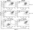
従って、本発明の一局面によれば、細胞集団が眼異常を処置するための適切な治療用物質であるかどうか適格性を判定する方法であって、該方法が、細胞の集団における、プレメラノソームタンパク質(PMEL17)と、細胞レチンアルデヒド結合タンパク質(CRALBP)、レシチンレチノールアシルトランスフェラーゼ(LRAT)、および性決定領域Y-ボックス9(SOX9)からなる群より選択される少なくとも1つのポリペプチドの同時発現を分析する段階を含み、PMEL17と少なくとも1つのポリペプチドとを同時発現する細胞の数が所定のレベルを上回る場合に、細胞集団が、網膜障害を処置するための適切な治療用物質として適格であると判定される、方法が提供される。 Thus, according to one aspect of the present invention, a method for determining eligibility of a cell population as an appropriate therapeutic agent for treating an eye abnormality comprising: Co-expression of melanosome protein (PMEL17) and at least one polypeptide selected from the group consisting of cellular retinaldehyde binding protein (CRALBP), lecithin retinol acyltransferase (LRAT), and sex determining region Y-box 9 (SOX9) The cell population is qualified as an appropriate therapeutic agent for treating retinal disorders when the number of cells co-expressing PMEL17 and at least one polypeptide exceeds a predetermined level. A method is provided that is determined to be.
別の局面によれば、細胞集団が眼異常を処置するための適切な治療用物質であるかどうか適格性を判定する方法であって、該方法が、細胞の集団における、細胞レチンアルデヒド結合タンパク質(CRALBP)と、プレメラノソームタンパク質(PMEL17)、レシチンレチノールアシルトランスフェラーゼ(LRAT)、および性決定領域Y-ボックス9(SOX9)からなる群より選択される少なくとも1つのポリペプチドの同時発現を分析する段階を含み、CRALBPと少なくとも1つのポリペプチドとを同時発現する細胞の数が所定のレベルを上回る場合に、細胞集団が、眼異常を処置するための適切な治療用物質として適格であると判定される、方法が提供される。 According to another aspect, a method of determining eligibility of a cell population as an appropriate therapeutic agent for treating an eye abnormality comprising: a cell retinaldehyde binding protein in a population of cells. Analyzing the co-expression of (CRALBP) and at least one polypeptide selected from the group consisting of premelanosome protein (PMEL17), lecithin retinol acyltransferase (LRAT), and sex-determining region Y-box 9 (SOX9) A cell population is determined to be eligible as an appropriate therapeutic agent for treating an ocular abnormality if the number of cells co-expressing CRALBP and at least one polypeptide exceeds a predetermined level A method is provided.
本明細書で使用する「適切な治療用物質」という句は、細胞集団が眼異常を処置するのに適していることを指す。治療用物質である細胞は、複数の機構のいずれか1つによって効果を発揮することができる。例示的な機構の1つは、網膜内にある変成中の光受容体または他の細胞の生存を促す支持的な栄養作用(trophic effect)である。治療用RPE細胞はまた、機能不全中および/または変成中の宿主RPE細胞を補充する再生機構を介して効果を発揮してもよい。一態様によれば、RPE細胞は成熟しており、ロドプシンを含む光受容体の脱落した外節を貪食する機能的能力を有する。別の態様によれば、RPE細胞は完全に成熟していない。 As used herein, the phrase “appropriate therapeutic agent” refers to the cell population being suitable for treating an ocular abnormality. Cells that are therapeutic substances can exert their effects by any one of a plurality of mechanisms. One exemplary mechanism is the supportive trophic effect that promotes the survival of metamorphic photoreceptors or other cells in the retina. Therapeutic RPE cells may also exert their effects through a regenerative mechanism that recruits dysfunctional and / or metamorphic host RPE cells. According to one aspect, the RPE cells are mature and have a functional ability to phagocytose the outer segment of photoreceptors including rhodopsin. According to another embodiment, the RPE cells are not fully mature.
前記細胞集団が治療用物質として役立つ眼異常には、一般的に網膜機能不全、網膜損傷、および/または網膜色素上皮の消失に関連する網膜疾患または網膜障害が含まれるが、これに限定されない。本発明に従って処置され得る異常の非限定的なリストは、網膜色素変性症、レーバー先天性黒内障、遺伝性または後天性の黄斑変性、加齢黄斑変性(AMD)、ベスト病、網膜剥離、脳回転状萎縮、コロイデレミア、パターンジストロフィならびにRPEの他のジストロフィー、シュタルガルト病、光傷害、レーザー傷害、炎症性傷害、感染性傷害、放射線傷害、新血管性傷害、または外傷性傷害のいずれか1つによって引き起こされる損傷が原因のRPE損傷および網膜損傷を含む。 Ocular abnormalities for which the cell population is useful as a therapeutic agent generally include, but are not limited to, retinal disorders or retinal disorders associated with retinal dysfunction, retinal damage, and / or loss of retinal pigment epithelium. A non-limiting list of abnormalities that can be treated according to the present invention includes retinitis pigmentosa, Labor congenital cataract, hereditary or acquired macular degeneration, age-related macular degeneration (AMD), best disease, retinal detachment, brain rotation Caused by atrophy, colloideremia, pattern dystrophies and other RPE dystrophies, Stargardt disease, photoinjury, laser injury, inflammatory injury, infectious injury, radiation injury, neovascular injury, or traumatic injury RPE damage and retinal damage caused by damage.
言及したように、本発明のこの局面の方法は、プレメラノソームタンパク質(PMEL17; SwissProt No. P40967)、ならびに細胞レチンアルデヒド結合タンパク質(CRALBP; SwissProt No. P12271)、レシチンレチノールアシルトランスフェラーゼ(LRAT; SwissProt No. 095327)、および性決定領域Y-ボックス9(SOX9; P48436)からなる群より選択される少なくとも1つのポリペプチドを発現する量(例えば、パーセント細胞)を測定することによって行われる。 As mentioned, the method of this aspect of the invention includes premelanosome protein (PMEL17; SwissProt No. P40967), as well as cellular retinaldehyde binding protein (CRALBP; SwissProt No. P12271), lecithin retinol acyltransferase (LRAT; SwissProt No. 095327), and the amount (eg, percent cells) that expresses at least one polypeptide selected from the group consisting of sex determining region Y-box 9 (SOX9; P48436).
または、この局面の方法は、CRALBP(CRALBP; SwissProt No. P12271)、ならびにレシチンレチノールアシルトランスフェラーゼ(LRAT; SwissProt No. 095327)、性決定領域Y-ボックス9(SOX 9; P48436)、およびPMEL17(SwissProt No. P40967)からなる群より選択される少なくとも1つのポリペプチドを測定することによって行われる。
Alternatively, methods of this aspect include CRALBP (CRALBP; SwissProt No. P12271), and lecithin retinol acyltransferase (LRAT; SwissProt No. 095327), sex determining region Y-box 9 (
従って、例えば、CRALBPおよびPMEL17が測定されてもよいか、PMEL17およびLRATが測定されてもよいか、または、PMEL17およびSOX9が測定されてもよい。または、CRALBPおよびLRATが測定されてもよいか、または、CRALBPおよびSOX9が測定されてもよい。 Thus, for example, CRALBP and PMEL17 may be measured, PMEL17 and LRAT may be measured, or PMEL17 and SOX9 may be measured. Alternatively, CRALBP and LRAT may be measured, or CRALBP and SOX9 may be measured.
本明細書において言及されたポリペプチドのうち2つ超を、例えば、上述したポリペプチドのうちの3つを、さらには、上述したポリペプチドのうちの4つ全てを測定できることが理解されるだろう。 It will be understood that more than two of the polypeptides referred to herein can be measured, for example, three of the polypeptides described above, or even all four of the polypeptides described above. Let's go.
上述したポリペプチドの発現を分析するための方法は、典型的には、抗原を特異的に認識する抗体の使用を伴う。CRALBPを認識する市販抗体には、例えば、Abcamによって製造された抗体(例えば、ab15051およびab189329、クローンB2)が含まれる。PMEL17を認識する市販抗体には、例えば、Abcamによって製造された抗体(例えば、ab137062およびab189330、クローンEPR4864)が含まれる。LRATを認識する市販抗体には、例えば、Milliporeによって製造された抗体(例えば、MABN644)が含まれる。SOX9を認識する市販抗体には、例えば、Abcamによって製造された抗体(例えば、ab185230)が含まれる。分析は、フローサイトメトリー、ウエスタンブロット、免疫細胞化学、ラジオイムノアッセイ法、PCRなどを含む当技術分野において公知の任意の方法を用いて行うことができる。 The methods for analyzing the expression of a polypeptide as described above typically involve the use of an antibody that specifically recognizes the antigen. Commercially available antibodies that recognize CRALBP include, for example, antibodies produced by Abcam (eg, ab15051 and ab189329, clone B2). Commercial antibodies that recognize PMEL17 include, for example, antibodies produced by Abcam (eg, ab137062 and ab189330, clone EPR4864). Commercial antibodies that recognize LRAT include, for example, antibodies produced by Millipore (eg, MABN644). Commercially available antibodies that recognize SOX9 include, for example, antibodies produced by Abcam (eg, ab185230). Analysis can be performed using any method known in the art, including flow cytometry, Western blot, immunocytochemistry, radioimmunoassay, PCR, and the like.
フローサイトメトリーの場合、抗体は蛍光部分に取り付けられ、蛍光励起セルソーター(FACS)を用いて分析されてもよい。または、蛍光部分を有する二次抗体の使用も想定される。 For flow cytometry, the antibody may be attached to a fluorescent moiety and analyzed using a fluorescence excitation cell sorter (FACS). Alternatively, the use of a secondary antibody having a fluorescent moiety is also envisaged.
分析されるポリペプチドが細胞内ポリペプチドであるので、典型的には、抗体が標的に結合できるように、細胞は透過処理されることが理解されるだろう。最初に、可溶性抗原または半減期が短い抗原の安定性を確かなものにするために、細胞は固定されてもよい。これにより、標的タンパク質は元々の細胞位置に保たれるべきである。細胞の透過性が保たれることを確かなものにするために、抗体は透過処理用緩衝液中で調製されてもよい。細胞集団をゲーティングする時に、透過処理後および固定後に、フローサイトメーターにおける細胞の光散乱プロファイルは大幅に変わることが理解されるだろう。 It will be appreciated that since the polypeptide to be analyzed is an intracellular polypeptide, typically the cells are permeabilized so that the antibody can bind to the target. Initially, cells may be fixed to ensure the stability of soluble antigens or antigens with a short half-life. This should keep the target protein in its original cellular location. To ensure that the permeability of the cells is maintained, the antibody may be prepared in a permeabilization buffer. It will be appreciated that when gating a cell population, the light scattering profile of the cells in the flow cytometer changes significantly after permeabilization and after fixation.
細胞膜を透過処理する方法は、当技術分野において公知であり、例えば、
1.ホルムアルデヒド、続いて、界面活性剤: ホルムアルデヒドによる固定(例えば、4.5%以下で10〜15分間(これによりタンパク質が安定化される)、続いて、界面活性剤、例えば、TritonまたはNP-40(PBS中で0.1〜1%)、Tween20(PBS中で0.1〜1%)、サポニン、ジギトニン、およびLeucoperm(例えば、PBS中で0.5%v/v)による膜の破壊;
2.ホルムアルデヒド(例えば、4.5%以下)、続いてメタノール;
3.メタノール、続いて、界面活性剤(例えば、80%メタノール、次いで、0.1%Tween20);
4.アセトン固定および透過処理
を含む。
Methods for permeabilizing cell membranes are known in the art, for example
1. Formaldehyde, followed by detergent: fixation with formaldehyde (eg, 4.5% or less for 10-15 minutes (this stabilizes the protein), followed by detergent, eg, Triton or NP-40 Disruption of the membrane by (0.1-1% in PBS), Tween20 (0.1-1% in PBS), saponin, digitonin, and Leucoperm (eg 0.5% v / v in PBS);
2. Formaldehyde (eg 4.5% or less), followed by methanol;
3. Methanol, followed by a surfactant (eg 80% methanol, then 0.1% Tween 20);
4. Includes acetone fixation and permeabilization.
本明細書で使用する「フローサイトメトリー」という用語は、試料中の材料(例えば、特定のマーカーを含むRPE細胞)の割合が、(例えば、標識された抗体と材料を結合することによって)材料を標識し、材料を含有する流体の流れが光線を通過するようにし、試料から発せられた光を、一連のフィルターおよびミラーによって構成波長に分離し、光を検出することによって決定されるアッセイ法を指す。 As used herein, the term `` flow cytometry '' refers to the proportion of material (e.g., RPE cells containing a particular marker) in a sample (e.g., by binding the material with a labeled antibody). An assay that is determined by allowing the fluid stream containing the material to pass through the light beam, separating the light emitted from the sample into a constituent wavelength by a series of filters and mirrors, and detecting the light Point to.
例えば、Becton Dickinson FACScan、Navios Flow Cytometer(Beckman Coulterシリアル番号AT15119 RHE9266、およびFACScalibur(BD Biosciences, Mountain View, CA)を含む数多くのフローサイトメーターが市販されている。FACS分析に用いられ得る抗体はSchlossman S, Boumell L, et al.,[Leucocyte Typing V. New York: Oxford University Press; 1995]に開示されており、広く市販されている。 A number of flow cytometers are commercially available including, for example, Becton Dickinson FACScan, Navios Flow Cytometer (Beckman Coulter serial number AT15119 RHE9266, and FACScalibur (BD Biosciences, Mountain View, Calif.) Antibodies that can be used for FACS analysis are Schlossman. S, Boumell L, et al., [Leucocyte Typing V. New York: Oxford University Press; 1995] and is widely available commercially.
上述したポリペプチドの発現レベルはRNAレベルならびにタンパク質レベルで行われてもよいことが理解されるだろう。RNAレベルに基づいてポリペプチドの発現を確認するための例示的な方法には、PCR、RT-PCR、ノザンブロットなどが含まれるが、これに限定されない。 It will be appreciated that the expression levels of the polypeptides described above may be performed at the RNA level as well as the protein level. Exemplary methods for confirming polypeptide expression based on RNA levels include, but are not limited to, PCR, RT-PCR, Northern blot, and the like.
前記細胞が治療用物質として有用であるという適格性を判定するためには、細胞において同時発現しているポリペプチドのうち少なくとも2つの量は、非RPE細胞(例えば、未分化の胚性幹細胞)と比較した場合に、統計的に有意なレベルを上回って増加していなければならない。 To determine eligibility that the cell is useful as a therapeutic agent, at least two of the polypeptides co-expressed in the cell are non-RPE cells (e.g., undifferentiated embryonic stem cells). Must increase above a statistically significant level.
特定の態様によれば、前記細胞が治療用物質として有用であるという適格性を判定するためには、当業者に公知の方法(例えば、FACS)によってアッセイした場合に、前記集団内の細胞の少なくとも80%は、検出可能なレベルのPMEL17および上述したポリペプチドの1つ(例えば、CRALBP)を発現しなければならない、より好ましくは、前記集団内の細胞の少なくとも85%は、検出可能なレベルのPMEL17および上述したポリペプチドの1つ(例えば、CRALBP)を発現しなければならない、より好ましくは、前記集団内の細胞の少なくとも90%は、検出可能なレベルのPMEL17および上述したポリペプチドの1つ(例えば、CRALBP)を発現しなければならない、より好ましくは、前記集団内の細胞の少なくとも95%は、検出可能なレベルのPMEL17および上述したポリペプチドの1つ(例えば、CRALBP)を発現しなければならない。より好ましくは、前記集団内の細胞の少なくとも100%は、検出可能なレベルのPMEL17および上述したポリペプチドの1つ(例えば、CRALBP)を発現しなければならない。 According to certain embodiments, to determine the eligibility of the cells as being useful as a therapeutic agent, when assayed by methods known to those skilled in the art (e.g., FACS), the cells in the population At least 80% must express a detectable level of PMEL17 and one of the polypeptides described above (eg, CRALBP), more preferably, at least 85% of the cells in the population are at a detectable level PMEL17 and one of the polypeptides described above (e.g., CRALBP), more preferably, at least 90% of the cells in the population have detectable levels of PMEL17 and one of the polypeptides described above. One (e.g., CRALBP), more preferably, at least 95% of the cells in the population have detectable levels of PMEL17 and one of the polypeptides described above (e.g. For example, CRALBP) must be expressed. More preferably, at least 100% of the cells in the population must express a detectable level of PMEL17 and one of the polypeptides described above (eg, CRALBP).
別の態様によれば、前記細胞が治療用物質として有用であるという適格性を判定するためには、CRALBPと上述したポリペプチドの1つ(例えば、PMEL17)の同時発現のレベルは(例えば、平均蛍光強度によって測定された場合)、未分化ESCと比較して少なくとも2倍、より好ましくは少なくとも3倍、より好ましくは少なくとも4倍、さらにより好ましくは少なくとも5倍、少なくとも10倍、少なくとも20倍、少なくとも30倍、少なくとも40倍、少なくとも50倍増加していなければならない。 According to another aspect, to determine eligibility that the cell is useful as a therapeutic agent, the level of co-expression of CRALBP and one of the aforementioned polypeptides (e.g., PMEL17) is (e.g., (As measured by mean fluorescence intensity), at least 2-fold, more preferably at least 3-fold, more preferably at least 4-fold, even more preferably at least 5-fold, at least 10-fold, at least 20-fold compared to undifferentiated ESC Must be increased by at least 30 times, at least 40 times, at least 50 times.
特定の態様によれば、前記細胞が治療用物質として有用であるという適格性を判定するためには、当業者に公知の方法(例えば、FACS)によってアッセイした場合に、前記集団内の細胞の少なくとも80%は、検出可能なレベルのCRALBPおよび上述したポリペプチドの1つ(例えば、PMEL17)を発現しなければならない、より好ましくは、前記集団内の細胞の少なくとも85%は、検出可能なレベルのCRALBPおよび上述したポリペプチドの1つ(例えば、PMEL17)を発現しなければならない、より好ましくは、前記集団内の細胞の少なくとも90%は、検出可能なレベルのCRALBPおよび上述したポリペプチドの1つ(例えば、PMEL17)を発現しなければならない、より好ましくは、前記集団内の細胞の少なくとも95%は、検出可能なレベルのCRALBPおよび上述したポリペプチドの1つ(例えば、PMEL17)を発現しなければならない、より好ましくは、前記集団内の細胞の100%は、検出可能なレベルのCRALBPおよび上述したポリペプチドの1つ(例えば、PMEL17)を発現しなければならない。 According to certain embodiments, to determine the eligibility of the cells as being useful as a therapeutic agent, when assayed by methods known to those skilled in the art (e.g., FACS), the cells in the population At least 80% must express a detectable level of CRALBP and one of the polypeptides described above (eg, PMEL17), more preferably at least 85% of the cells in the population are at a detectable level Of CRALBP and one of the polypeptides described above (e.g., PMEL17), more preferably, at least 90% of the cells in the population have detectable levels of CRALBP and one of the polypeptides described above. (E.g., PMEL17) must be expressed, more preferably at least 95% of the cells in the population have detectable levels of CRALBP and one of the polypeptides described above (e.g. For example, PMEL17) must be expressed, more preferably 100% of the cells in the population must express a detectable level of CRALBP and one of the above-described polypeptides (eg, PMEL17). .
さらに、インビボで動物モデルにおいて前記細胞は適格性を判定されてもよい。このようなモデルの1つは、Royal College of Surgeons(RCS)ラットモデルである。移植後に、眼底画像化、視運動追跡閾値(optokinetic tracking threshold)(OKT)、網膜電図(ERG)、組織学、錐体計数、およびロドプシン摂取を含む方法を用いて、細胞の治療効果が分析されてもよい。これらの方法は、本明細書中の以下の実施例5においてさらに説明される。 Furthermore, the cells may be qualified in an animal model in vivo. One such model is the Royal College of Surgeons (RCS) rat model. Post-transplant analysis of cell therapeutic effects using methods including fundus imaging, optokinetic tracking threshold (OKT), electroretinogram (ERG), histology, cone counting, and rhodopsin uptake May be. These methods are further described in Example 5 herein below.
前記細胞は、例えば、核型分析、形態、細胞数、ならびに生存率、有効性(バリア機能ならびにPEDFおよびVEGFの極性分泌)、残存hESCのレベル、グラム染色、ならびに無菌性を含むさらなる手法で、適格性を判定されてもよいか、または特徴決定されてもよい。実施され得る例示的なアッセイ法は実施例4において説明される。 The cells may be further processed, including, for example, karyotype analysis, morphology, cell number, and viability, efficacy (barrier function and PEDF and VEGF polar secretion), residual hESC levels, Gram staining, and sterility. Eligibility may be determined or characterized. Exemplary assays that can be performed are described in Example 4.
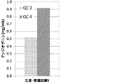
さらに、前記細胞は、バリア機能、ならびに増殖因子(例えば、色素上皮由来因子(PEDF)もしくはVEGF、サイトカイン、インターロイキン、および/またはケモカイン)の極性化様式での分泌のレベルについて分析されてもよい。 In addition, the cells may be analyzed for barrier function and levels of secretion in a polarized manner of growth factors such as pigment epithelium-derived factor (PEDF) or VEGF, cytokines, interleukins, and / or chemokines. .
分泌されたPEDFを分析するために、細胞培養物から上清が収集され、細胞は回収および計数される。細胞培養上清中のPEDF量は、PEDF ELISAアッセイ法(例えば、ELISAquant(商標)PEDF Sandwich ELISA Antigen Detection Kit, BioProductsMD, PED613)を用いて、製造業者のプロトコールに従って定量されてもよい。 To analyze the secreted PEDF, the supernatant is collected from the cell culture and the cells are collected and counted. The amount of PEDF in the cell culture supernatant may be quantified using a PEDF ELISA assay (eg, ELISAquant ™ PEDF Sandwich ELISA Antigen Detection Kit, BioProducts MD, PED613) according to the manufacturer's protocol.
さらに、前記細胞におけるPEDFおよびVEGFの分泌の方向が分析されてもよい。これは、図28に示したようにトランズウェルアッセイ法を用いて行われてもよい。適格性判定の前または後に、前記細胞は当技術分野において公知の方法に従って保存(例えば、凍結または凍結保存)されてもよく、対象に直接投与されてもよい。 In addition, the direction of PEDF and VEGF secretion in the cells may be analyzed. This may be done using a Transwell assay as shown in FIG. Prior to or after qualification, the cells may be stored (eg, frozen or cryopreserved) according to methods known in the art and administered directly to the subject.
本発明は、任意の供給源に由来する網膜色素上皮(RPE)細胞を含む細胞集団を分析することを意図する。従って、この細胞集団は、ドナーから得られたRPE細胞(すなわち、網膜の色素層の天然RPE細胞)を含んでもよく、幹細胞の集団からエクスビボで分化したRPE細胞(hSC由来RPE細胞、例えば、多能性幹細胞、例えば、ヒト胚性幹細胞)を含んでもよい。別の態様によれば、RPE細胞は分化転換によって得られる。例えば、Zhang et al., Protein Cell 2014, 5(1): 48-58を参照されたい。この内容は参照により本明細書に組み入れられる。 The present invention contemplates analyzing cell populations containing retinal pigment epithelium (RPE) cells from any source. Thus, this cell population may include RPE cells obtained from a donor (i.e., native RPE cells in the retinal pigment layer), and RPE cells differentiated ex vivo from a population of stem cells (hSC-derived RPE cells, e.g., multiple Capable stem cells, eg, human embryonic stem cells). According to another embodiment, RPE cells are obtained by transdifferentiation. See, for example, Zhang et al., Protein Cell 2014, 5 (1): 48-58. This content is incorporated herein by reference.
一態様によれば、分析されるRPE細胞はPax6を発現しない。 According to one aspect, the RPE cells to be analyzed do not express Pax6.
別の態様によれば、分析されるRPE細胞はPax6を発現する。 According to another embodiment, the RPE cells to be analyzed express Pax6.
「網膜色素上皮細胞」、「RPE細胞」、「RPE」は、文脈が許す時には同義に用いられることがあり、網膜の色素上皮細胞層を形成する天然RPE細胞と機能上類似する細胞タイプの細胞を指す(例えば、眼内に移植されると、天然RPE細胞に似た機能活動を示す)。 “Retinal pigment epithelial cells”, “RPE cells”, and “RPE” are sometimes used interchangeably when the context allows, and cell types that are functionally similar to natural RPE cells that form the pigment epithelial cell layer of the retina (Eg, shows functional activity similar to native RPE cells when implanted in the eye).
一態様によれば、RPE細胞は、少なくとも1種類、2種類、3種類、4種類、または5種類の成熟RPE細胞マーカーを発現する。このようなマーカーには、CARLBP、RPE65、PEDF、PMEL17、ベストロフィン、およびチロシナーゼが含まれるが、これに限定されない。任意で、RPE細胞はまた、RPE前駆細胞マーカー、例えば、MITFも発現してもよい。別の態様において、RPE細胞はPAX-6を発現する。別の態様において、RPE細胞は、OTX2、SIX3、SIX6、およびLHX2を含むが、これに限定されない少なくとも1種類の網膜前駆細胞マーカーを発現する。 According to one aspect, RPE cells express at least one, two, three, four, or five mature RPE cell markers. Such markers include, but are not limited to CARLBP, RPE65, PEDF, PMEL17, bestrophin, and tyrosinase. Optionally, the RPE cells may also express an RPE progenitor cell marker, such as MITF. In another embodiment, the RPE cells express PAX-6. In another embodiment, RPE cells express at least one retinal progenitor cell marker, including but not limited to OTX2, SIX3, SIX6, and LHX2.
さらに別の態様によれば、RPE細胞は、本明細書の下記の実施例セクションに記載の方法に従って胚性幹細胞から分化されたRPE細胞である。実施例の内容は、本明細書そのものに含まれるような内容である。 According to yet another aspect, RPE cells are RPE cells differentiated from embryonic stem cells according to the methods described in the Examples section herein below. The contents of the examples are the contents as included in the present specification itself.
本明細書で使用する「成熟RPE細胞マーカー」という句は、成熟RPE細胞において、非RPE細胞または未熟RPE細胞に対して(例えば、少なくとも2倍、少なくとも5倍、少なくとも10倍)上昇している抗原(例えば、タンパク質)を指す。 As used herein, the phrase `` mature RPE cell marker '' is elevated in mature RPE cells relative to non-RPE cells or immature RPE cells (e.g., at least 2-fold, at least 5-fold, at least 10-fold). Refers to an antigen (eg, protein).
本明細書で使用する「RPE前駆細胞マーカー」という句は、RPE前駆細胞において、非RPE細胞に対して(例えば、少なくとも2倍、少なくとも5倍、少なくとも10倍)上昇している抗原(例えば、タンパク質)を指す。 As used herein, the phrase `` RPE progenitor cell marker '' refers to an antigen that is elevated (e.g., at least 2-fold, at least 5-fold, at least 10-fold) in non-RPE cells in RPE progenitor cells (e.g., Protein).
別の態様によれば、RPE細胞は、網膜の色素上皮細胞層を形成する天然RPE細胞に似た形態を有する、すなわち、色素細胞である、および/または特徴的な多角形状を有する。 According to another embodiment, the RPE cells have a morphology resembling that of native RPE cells that forms the retinal pigment epithelial cell layer, ie, are pigment cells, and / or have a characteristic polygonal shape.
さらに別の態様によれば、RPE細胞は黄斑変性などの疾患を処置することができる。 According to yet another aspect, RPE cells can treat diseases such as macular degeneration.
さらに別の態様によれば、RPE細胞は、本明細書において上記で列挙した要件の少なくとも1つ、2つ、3つ、4つ、または全てを満たす。 According to yet another aspect, the RPE cells meet at least one, two, three, four, or all of the requirements listed herein above.
「hSC由来RPE細胞」という用語は、hSCからの定方向分化(directed differentiation)によって得られたRPE細胞を指すために本明細書において用いられる。好ましい態様によれば、hSC由来RPE細胞は、下記で定義されるパラメータによって示されるような機能的RPE細胞である。「定方向分化」という用語は、「RPE誘導性分化」という用語と同義に用いられ、RPE細胞タイプへの分化を誘導/促進する培養条件下においてhSCを操作するプロセスを意味すると理解しなければならない。 The term “hSC-derived RPE cells” is used herein to refer to RPE cells obtained by directed differentiation from hSC. According to a preferred embodiment, the hSC-derived RPE cells are functional RPE cells as indicated by the parameters defined below. The term “directed differentiation” is used synonymously with the term “RPE-induced differentiation” and must be understood to mean the process of manipulating hSC under culture conditions that induce / promote differentiation into RPE cell types. Don't be.
特定の態様によれば、RPE細胞は、TGFβスーパーファミリーの1つまたは複数のメンバーの存在下でのhSCの定方向分化によって得られ、以下の特徴の少なくとも1つを示す:
-分化の間に、培養細胞はTGFβシグナル伝達に応答すること;
-RPE細胞は、最終分化を示すマーカー、例えば、ベストロフィン1、CRALBP、および/またはRPE65を発現すること;
-移植後に(すなわち、インサイチューで)、RPE細胞は、RPE細胞に隣接する光受容体を支持する栄養作用を示すこと;
-さらに、インサイチューで、RPE細胞は、これらの光受容体の正常な再生プロセスの一環として、脱落した光受容体外節の食作用を伴って機能することができること;
-さらに、インサイチューで、RPE細胞は網膜関門を作製し、視サイクルにおいて機能することができること。
According to a particular embodiment, RPE cells are obtained by directed differentiation of hSC in the presence of one or more members of the TGFβ superfamily and exhibit at least one of the following characteristics:
-During differentiation, cultured cells respond to TGFβ signaling;
-RPE cells express a marker indicating terminal differentiation, e.g.,
-After transplantation (i.e., in situ), RPE cells exhibit trophic support that supports photoreceptors adjacent to RPE cells;
-Furthermore, in situ, RPE cells can function with the phagocytosis of shed photoreceptor outer segments as part of the normal regeneration process of these photoreceptors;
-Furthermore, in situ, RPE cells can create the retinal barrier and function in the visual cycle.
本明細書で使用する「幹細胞」という句は、特定の特殊化された機能を有する他の細胞タイプ(例えば、完全に分化した細胞)に分化するように誘導されるまで、培養中に長期間にわたって未分化状態にとどまることができる細胞(例えば、多能性または多分化能性の幹細胞)を指す。好ましくは、「幹細胞」という句は、胚性幹細胞(ESC)、人工多能性幹細胞(iPS)、成人幹細胞、間葉系幹細胞、および造血幹細胞を包含する。 As used herein, the phrase `` stem cell '' is used for a long period of time in culture until induced to differentiate into other cell types with specific specialized functions (e.g., fully differentiated cells). Refers to cells that can remain undifferentiated over time (eg, pluripotent or multipotent stem cells). Preferably, the phrase “stem cell” encompasses embryonic stem cells (ESC), induced pluripotent stem cells (iPS), adult stem cells, mesenchymal stem cells, and hematopoietic stem cells.
特定の態様によれば、RPE細胞は、ヒト胚性幹細胞または人工多能性幹細胞を含む多能性幹細胞に由来する。 According to certain embodiments, RPE cells are derived from pluripotent stem cells, including human embryonic stem cells or induced pluripotent stem cells.
「胚性幹細胞」という句は、3つ全ての胚葉(すなわち、内胚葉、外胚葉、および中胚葉)に分化することができるか、または未分化な状態に留まることができる胚細胞を指す。「胚性幹細胞」という句は、胚着床前の、妊娠後に形成された胚組織(例えば、胚盤胞)から得られた細胞(すなわち、着床前胚盤胞)、着床後/原腸形成前の段階の胚盤胞から得られた拡張胚盤胞細胞(extended blastocyst cell)(EBC)(WO2006/040763を参照されたい)、および妊娠中のいつでも、好ましくは、妊娠10週前に胎児の生殖組織から得られた胚性生殖(EG)細胞を含んでもよい。本発明の一部の態様の胚性幹細胞は周知の細胞培養方法を用いて得ることができる。例えば、ヒト胚性幹細胞はヒト胚盤胞から単離することができる。ヒト胚盤胞は、典型的には、ヒトのインビボ着床前胚から得られるか、または体外受精(IVF)胚から得られる。または、単一細胞ヒト胚を胚盤胞段階まで拡張することができる。ヒトES細胞を単離するために、胚盤胞から透明帯が除去され、外科手術によって内部細胞塊(ICM)が単離される。この場合、栄養外胚葉細胞が溶解され、穏やかにピペッティングすることによってインタクトなICMから除去される。次いで、ICMは、その増生(outgrowth)を可能にする適切な培地を含有する組織培養フラスコ中にプレーティングされる。9〜15日後に、ICM由来増殖は機械的解離または酵素的分解によって凝集塊に解離され、次いで、細胞は新鮮な組織培養培地上に再プレーティングされる。未分化形態を示すコロニーが1つ1つ、マイクロピペット/幹細胞ツールによって選択され、断片/凝集塊に機械的に切開され、再プレーティングされる。次いで、結果として生じたES細胞は定期的に4〜7日ごとにスプリットされる。ヒトES細胞を調製する方法に関する、さらなる詳細については、Reubinoff et al., Nat Biotechnol 2000, May: 18(5): 559; Thomson et al.,[米国特許第5,843,780号; Science 282: 1145, 1998; Curr. Top. Dev. Biol. 38: 133, 1998; Proc. Natl. Acad. Sci. USA 92: 7844, 1995]; Bongso et al., [Hum Reprod 4: 706, 1989];およびGardner et al., [Fertil. Steril. 69: 84, 1998]を参照されたい。 The phrase “embryonic stem cells” refers to embryonic cells that can differentiate into all three germ layers (ie, endoderm, ectoderm, and mesoderm) or remain in an undifferentiated state. The phrase `` embryonic stem cells '' refers to cells obtained from embryonic tissue formed after pregnancy (e.g., blastocysts) before embryo implantation (i.e., preimplantation blastocysts), post-implantation / original Extended blastocyst cells (EBC) (see WO2006 / 040763) obtained from pre-intestinal blastocysts, and anytime during pregnancy, preferably 10 weeks before pregnancy It may also include embryonic germ (EG) cells obtained from fetal reproductive tissue. Embryonic stem cells of some aspects of the invention can be obtained using well-known cell culture methods. For example, human embryonic stem cells can be isolated from human blastocysts. Human blastocysts are typically obtained from human in vivo preimplantation embryos or from in vitro fertilized (IVF) embryos. Alternatively, single cell human embryos can be expanded to the blastocyst stage. To isolate human ES cells, the zona pellucida is removed from the blastocyst and the inner cell mass (ICM) is isolated by surgery. In this case, trophectoderm cells are lysed and removed from intact ICM by gentle pipetting. The ICM is then plated in a tissue culture flask containing an appropriate medium that allows its outgrowth. After 9-15 days, ICM-derived growth is dissociated into clumps by mechanical dissociation or enzymatic degradation, and the cells are then re-plated onto fresh tissue culture medium. Individual colonies showing undifferentiated morphology are selected by a micropipette / stem cell tool, mechanically dissected into fragments / agglomerates and re-plated. The resulting ES cells are then periodically split every 4-7 days. For further details regarding methods of preparing human ES cells, see Reubinoff et al., Nat Biotechnol 2000, May: 18 (5): 559; Thomson et al., [US Pat. No. 5,843,780; Science 282: 1145, 1998. Curr. Top. Dev. Biol. 38: 133, 1998; Proc. Natl. Acad. Sci. USA 92: 7844, 1995]; Bongso et al., [Hum Reprod 4: 706, 1989]; and Gardner et al. ., [Fertil. Steril. 69: 84, 1998].
本発明の一部の態様によれば、市販の幹細胞も使用できることが理解されるだろう。ヒトES細胞は、NIHヒト胚性幹細胞登録所(NIH human embryonic stem cells registry)[Hypertext Transfer Protocol://grants(dot)nih(dot)gov/stem_cells/registry/current(dot)htm]および他の欧州登録所から購入することができる。市販の胚性幹細胞株の非限定的な例は、HAD-C 102、ESI、BG01、BG02、BG03、BG04、CY12、CY30、CY92、CY10、TE03、TE32、CHB-4、CHB-5、CHB-6、CHB-8、CHB-9、CHB-10、CHB-11、CHB-12、HUES1、HUES2、HUES3、HUES4、HUES5、HUES6、HUES7、HUES8、HUES9、HUES10、HUES11、HUES12、HUES13、HUES14、HUES15、HUES16、HUES17、HUES18、HUES19、HUES20、HUES21、HUES22、HUES23、HUES24、HUES25、HUES26、HUES27、HUES28、CyT49、RUES3、WA01、UCSF4、NYUES1、NYUES2、NYUES3、NYUES4、NYUES5、NYUES6、NYUES7、UCLA1、UCLA2、UCLA3、WA077(H7)、WA09(H9)、WA13(H13)、WA14(H14)、HUES62、HUES63、HUES64、CT1、CT2、CT3、CT4、MA135、Eneavour-2、WIBR1、WIBR2、WIBR3、WIBR4、WIBR5、WIBR6、HUES45、Shef3、Shef6、BJNhem19、BJNhem20、SA001、SA001である。
It will be appreciated that according to some aspects of the present invention, commercially available stem cells can also be used. Human ES cells are registered in the NIH human embryonic stem cells registry [Hypertext Transfer Protocol: // grants (dot) nih (dot) gov / stem_cells / registry / current (dot) htm] and other It can be purchased from the European Registry. Non-limiting examples of commercially available embryonic stem cell lines include HAD-
さらに、ES細胞は、マウス(Mills and Bradley, 2001)、ゴールデンハムスター[Doetschman et al., 1988, Dev Biol. 127: 224-7]、ラット[Iannaccone et al., 1994, Dev Biol. 163: 288-92]、ウサギ[Giles et al. 1993, Mol Reprod Dev. 36: 130-8; Graves & Moreadith, 1993, Mol Reprod Dev. 1993, 36: 424-33]、いくつかの家畜種[Notarianni et al., 1991, J Reprod Fertil Suppl. 43: 255-60; Wheeler 1994, Reprod Fertil Dev. 6: 563-8; Mitalipova et al., 2001, Cloning. 3: 59-67]、ならびに非ヒト霊長類種(アカゲザルおよびマーモセット)[Thomson et al., 1995, Proc Natl Acad Sci U S A. 92: 7844-8; Thomson et al., 1996, Biol Reprod. 55: 254-9]を含む他の種からも得ることができる。 Furthermore, ES cells were obtained from mice (Mills and Bradley, 2001), golden hamsters [Doetschman et al., 1988, Dev Biol. 127: 224-7], rats [Iannaccone et al., 1994, Dev Biol. 163: 288. -92], rabbits [Giles et al. 1993, Mol Reprod Dev. 36: 130-8; Graves & Moreadith, 1993, Mol Reprod Dev. 1993, 36: 424-33], some livestock species [Notarianni et al ., 1991, J Reprod Fertil Suppl. 43: 255-60; Wheeler 1994, Reprod Fertil Dev. 6: 563-8; Mitalipova et al., 2001, Cloning. 3: 59-67], and non-human primate species (Rhesus monkey and marmoset) [Thomson et al., 1995, Proc Natl Acad Sci US A. 92: 7844-8; Thomson et al., 1996, Biol Reprod. 55: 254-9] be able to.
拡張胚盤胞細胞(EBC)は、原腸形成前の段階である、受精して少なくとも9日後の胚盤胞から得ることができる。胚盤胞を培養する前に、内部細胞塊を露出するように、透明帯が[例えば、タイロード酸性溶液(Sigma Aldrich、St Louis、MO 、USA)によって]消化される。次いで、胚盤胞は、標準的な胚性幹細胞培養法を用いて、インビトロで全胚として、受精後、少なくとも9日間かつ14日以下で(すなわち、原腸形成事象の前に)培養される。 Extended blastocyst cells (EBC) can be obtained from blastocysts at least 9 days after fertilization, which is the stage before gastrulation. Prior to culturing the blastocyst, the zona pellucida is digested [eg, by Tyrode acid solution (Sigma Aldrich, St Louis, MO, USA)] to expose the inner cell mass. The blastocysts are then cultured in vitro as whole embryos using standard embryonic stem cell culture methods for at least 9 days and no more than 14 days after fertilization (ie, before gastrulation events) .
ES細胞を調製するための別の方法は、Chung et al., Cell Stem Cell, Volume 2, Issue 2, 113-117, 7 February 2008に記載されている。この方法は、体外受精プロセスの間に胚から単一細胞を取り出す段階を含む。このプロセスにおいて胚は破壊されない。
Another method for preparing ES cells is described in Chung et al., Cell Stem Cell,
ES細胞を調製するためのさらに別の方法は単為生殖によるものである。このプロセスでも胚は破壊されない。 Yet another method for preparing ES cells is by parthenogenesis. This process does not destroy the embryo.
現在行われているES培養法は、主に、幹細胞増殖に必要な因子を分泌するが、同時に幹細胞分化を阻害するフィーダー細胞層の使用に基づいている。例示的なフィーダー層には、ヒト胚線維芽細胞、成人ファロピウス管上皮細胞、初代マウス胚線維芽細胞(PMEF)、マウス胚線維芽細胞(MEF)、マウス胎仔線維芽細胞(MFF)、ヒト胚線維芽細胞(HEF)、ヒト胚性幹細胞の分化から得られたヒト線維芽細胞、ヒト胎児筋肉細胞(HFM)、ヒト胎児皮膚細胞(HFS)、ヒト成人皮膚細胞、ヒト包皮線維芽細胞(HFF)、ヒト臍帯線維芽細胞、臍帯または胎盤から得られたヒト細胞、およびヒト骨髄ストローマ細胞(hMSC)が含まれる。ESCを未分化状態で維持するために、培地に増殖因子が添加されてもよい。このような増殖因子にはbFGFおよび/またはTGFβが含まれる。別の態様において、hESCをナイーヴな未分化状態で維持するために、培地に作用物質が添加されてもよい。例えば、Kalkan et al., 2014, Phil. Trans. R. Soc. B, 369: 20130540を参照されたい。 Current ES culture methods are based primarily on the use of feeder cell layers that secrete factors necessary for stem cell proliferation but at the same time inhibit stem cell differentiation. Exemplary feeder layers include human embryonic fibroblasts, adult fallopian tube epithelial cells, primary mouse embryonic fibroblasts (PMEF), mouse embryonic fibroblasts (MEF), mouse embryonic fibroblasts (MFF), human embryos Fibroblasts (HEF), human fibroblasts obtained from human embryonic stem cell differentiation, human fetal muscle cells (HFM), human fetal skin cells (HFS), human adult skin cells, human foreskin fibroblasts (HFF) ), Human umbilical fibroblasts, human cells obtained from umbilical cord or placenta, and human bone marrow stromal cells (hMSC). In order to maintain the ESC in an undifferentiated state, a growth factor may be added to the medium. Such growth factors include bFGF and / or TGFβ. In another embodiment, an agent may be added to the medium to maintain hESCs in a naïve undifferentiated state. See, for example, Kalkan et al., 2014, Phil. Trans. R. Soc. B, 369: 20130540.
ES細胞培養では無フィーダー細胞系も用いられてきた。このような系では、フィーダー細胞層の代用品として、血清代用品、サイトカイン、および増殖因子(IL6および可溶性IL6受容体キメラを含む)が加えられたマトリックスが用いられる。幹細胞は、細胞外マトリックス(例えば、Matrigel(登録商標)またはラミニン)などの固体表面上で、培養培地、例えば、LonzaL7系、mTeSR、StemPro、XFKSR、E8の存在下で増殖することができる。フィーダー細胞と幹細胞が同時に増殖されることを必要とし、混合細胞集団が生じる可能性があるフィーダーに基づく培養とは異なり、無フィーダー系上で増殖させた幹細胞は表面から容易に分離される。幹細胞を増殖させるのに用いられる培養培地は、分化を効果的に阻害し、幹細胞の増殖を促進する因子、例えば、MEF条件培地およびbFGFを含有する。しかしながら、一般的に用いられる無フィーダー培養系では、マウス血清もしくはウシ血清またはMEF条件培地が加えられた、動物に基づくマトリックス(例えば、Matrigel(登録商標))が用いられる[Xu C, et al. (2001). Feeder-free growth of undifferentiated human embryonic stem cells. Nat Biotechnol. 19: 971-4]。動物に基づくマトリックスは、動物病原体がヒトES細胞に交差移動(cross-transfer)する危険をもたらし、従って、将来の臨床応用を危うくする。 Feeder-free cell lines have also been used in ES cell culture. In such systems, a matrix supplemented with serum substitutes, cytokines, and growth factors (including IL6 and soluble IL6 receptor chimeras) is used as a substitute for the feeder cell layer. Stem cells can be grown on a solid surface such as an extracellular matrix (eg, Matrigel® or laminin) in the presence of a culture medium, eg, Lonza L7 system, mTeSR, StemPro, XFKSR, E8. Unlike feeder-based cultures where feeder cells and stem cells need to be grown simultaneously, which can result in mixed cell populations, stem cells grown on feeder-free systems are easily separated from the surface. The culture medium used to grow stem cells contains factors that effectively inhibit differentiation and promote stem cell growth, such as MEF conditioned medium and bFGF. However, commonly used feeder-free culture systems use an animal-based matrix (e.g., Matrigel®) supplemented with mouse serum or bovine serum or MEF conditioned medium [Xu C, et al. (2001). Feeder-free growth of undifferentiated human embryonic stem cells. Nat Biotechnol. 19: 971-4]. Animal-based matrices pose the risk of animal pathogens cross-transfer to human ES cells, thus jeopardizing future clinical applications.
ESCをRPE系列に分化させるための非常に多くの方法が公知であり、定方向分化プロトコール、例えば、WO2008/129554、2013/184809に記載の定方向分化プロトコールと、自然分化プロトコール、例えば、米国特許第8,268,303号および米国特許出願第20130196369号に記載の自然分化プロトコールを両方とも含む。それぞれの内容は参照により組み入れられる。 Numerous methods are known for differentiating ESCs into RPE series, including directed differentiation protocols, such as the directed differentiation protocol described in WO2008 / 129554, 2013/184809, and natural differentiation protocols, such as US patents. Both natural differentiation protocols described in US 8,268,303 and US Patent Application 20130196369 are included. The contents of each are incorporated by reference.
特定の態様によれば、RPE細胞は、定方向分化プロトコールを用いて、例えば、実施例セクションに開示されたプロトコールに従ってESC細胞から作製される。 According to certain embodiments, RPE cells are generated from ESC cells using a directed differentiation protocol, for example according to the protocol disclosed in the Examples section.
ある例示的な分化プロトコールでは、胚性幹細胞は、第1の分化物質を用いてRPE細胞系列に分化され、次いで、トランスフォーミング成長因子-β(TGFβ)スーパーファミリーのメンバー(例えば、TGFβ1、TGFβ2、およびTGFβ3サブタイプ、ならびにアクチビン(例えば、アクチビンA、アクチビンB、およびアクチビンAB)、nodal、抗ミュラー管ホルモン(AMH)、いくつかの骨形成タンパク質(BMP)、例えば、BMP2、BMP3、BMP4、BMP5、BMP6、およびBMP7、ならびに増殖分化因子(GDF)を含む相同リガンド)を用いて、RPE細胞にさらに分化される。 In one exemplary differentiation protocol, embryonic stem cells are differentiated into an RPE cell line using a first differentiation agent and then transformed into a transforming growth factor-β (TGFβ) superfamily member (e.g., TGFβ1, TGFβ2, And TGFβ3 subtypes, and activins (e.g., activin A, activin B, and activin AB), nodal, anti-Muellerian hormone (AMH), some bone morphogenetic proteins (BMP), e.g., BMP2, BMP3, BMP4, BMP5 , BMP6, and BMP7, and homologous ligands including growth differentiation factors (GDF)) are further differentiated into RPE cells.
特定の態様によれば、TGFβスーパーファミリーメンバーは、TGFβ1、アクチビンA、およびTGFβ3からなる群より選択される。 According to a particular embodiment, the TGFβ superfamily member is selected from the group consisting of TGFβ1, activin A, and TGFβ3.
特定の態様によれば、トランスフォーミング成長因子-β(TGFβ)スーパーファミリーのメンバーは、アクチビンA、例えば、20〜200ng/ml、例えば、100〜180ng/mlのアクチビンAである。 According to a particular embodiment, the transforming growth factor-β (TGFβ) superfamily member is activin A, such as 20-200 ng / ml, such as 100-180 ng / ml activin A.
第1の分化物質はRPE系列への分化を促進する。例えば、第1の分化物質は多能性幹細胞から神経前駆体への分化を促進する可能性がある。このような細胞はPAX6などの神経前駆体マーカーを発現してもよい。 The first differentiation substance promotes differentiation into the RPE series. For example, the first differentiation substance may promote differentiation from pluripotent stem cells to neural precursors. Such cells may express neural precursor markers such as PAX6.
特定の態様によれば、第1の分化物質は、ニコチンアミド(NA)、例えば、1〜100mM、5〜50mM、5〜20mM、例えば、10mMのニコチンアミド(NA)である。 According to a particular embodiment, the first differentiation agent is nicotinamide (NA), eg 1-100 mM, 5-50 mM, 5-20 mM, eg 10 mM nicotinamide (NA).
NAは「ナイアシンアミド」としても公知であり、β細胞機能を保護および改善すると考えられているアミド誘導体型のビタミンB3(ナイアシン)である。NAの化学式はC6H6N2Oである。NAは、成長と、食物からエネルギーへの変換に不可欠であり、関節炎の治療ならびに糖尿病の治療および予防において用いられてきた。

特定の態様によれば、ニコチンアミドはニコチンアミド誘導体またはニコチンアミド模倣物である。本明細書で使用する「ニコチンアミド(NA)誘導体」という用語は、天然NAの化学修飾誘導体である化合物を指す。一態様において、化学修飾は、アミド部分の窒素原子または酸素原子を介した、(環の炭素メンバーまたは窒素メンバーを介した)基本NA構造のピリジン環の置換でもよい。置換時には、1つもしくは複数の水素原子が置換基によって置き換えられてもよく、および/または置換基がN原子に取り付けられて四価の正荷電窒素を形成してもよい。従って、本発明のニコチンアミドには置換ニコチンアミドまたは非置換ニコチンアミドが含まれる。別の態様において、化学修飾は、例えば、NAのチオベンズアミド類似体を形成するような、単一の基の欠失または交換でもよい。これらは全て、有機化学に精通した人により理解される通りである。本発明の文脈における誘導体はまたNAのヌクレオシド誘導体(例えば、ニコチンアミドアデニン)も含む。 According to a particular embodiment, the nicotinamide is a nicotinamide derivative or a nicotinamide mimetic. As used herein, the term “nicotinamide (NA) derivative” refers to a compound that is a chemically modified derivative of natural NA. In one embodiment, the chemical modification may be a substitution of the pyridine ring of the basic NA structure (via a ring carbon or nitrogen member) via a nitrogen or oxygen atom of the amide moiety. Upon substitution, one or more hydrogen atoms may be replaced by a substituent and / or the substituent may be attached to an N atom to form a tetravalent positively charged nitrogen. Accordingly, the nicotinamides of the present invention include substituted nicotinamides or unsubstituted nicotinamides. In another embodiment, the chemical modification may be a single group deletion or exchange, eg, to form a thiobenzamide analog of NA. All of these are as understood by those familiar with organic chemistry. Derivatives in the context of the present invention also include nucleoside derivatives of NA (eg nicotinamide adenine).
様々なNA誘導体が説明されており、一部は、PDE4酵素の阻害活性とも関連して説明されている(WO03/068233;WO02/060875;GB2327675A)、またはVEGF-受容体型チロシンキナーゼ阻害剤(WO01/55114)として説明されている。例えば、4-アリール-ニコチンアミド誘導体を調製するプロセス(WO05/014549)。他の例示的なニコチンアミド誘導体はWO01/55114およびEP2128244に開示される。 Various NA derivatives have been described, some of which have been described in connection with the inhibitory activity of the PDE4 enzyme (WO03 / 068233; WO02 / 060875; GB2327675A), or VEGF-receptor tyrosine kinase inhibitors (WO01 / 55114). For example, a process for preparing 4-aryl-nicotinamide derivatives (WO05 / 014549). Other exemplary nicotinamide derivatives are disclosed in WO01 / 55114 and EP2128244.
ニコチンアミド模倣物には、多能性細胞からのRPE細胞の分化および成熟においてニコチンアミドの作用を再現する、修飾型ニコチンアミドおよびニコチンアミドの化学類似体が含まれる。例示的なニコチンアミド模倣物には、安息香酸、3-アミノ安息香酸、および6-アミノニコチンアミドが含まれる。ニコチンアミド模倣物として作用し得る別のクラスの化合物はポリ(ADP-リボース)ポリメラーゼ(PARP)阻害剤である。例示的なPARP阻害剤には、3-アミノベンズアミド、イニパリブ(Iniparib)(BSI201)、オラバリブ(Olaparib)(AZD-2281)、ルカパリブ(Rucaparib)(AG014699、PF-01367338)、ベリパリブ(Veliparib)(ABT-888)、CEP9722、MK4827、およびBMN-673が含まれる。 Nicotinamide mimetics include modified nicotinamide and chemical analogs of nicotinamide that reproduce the action of nicotinamide in the differentiation and maturation of RPE cells from pluripotent cells. Exemplary nicotinamide mimetics include benzoic acid, 3-aminobenzoic acid, and 6-aminonicotinamide. Another class of compounds that can act as nicotinamide mimetics are poly (ADP-ribose) polymerase (PARP) inhibitors. Exemplary PARP inhibitors include 3-aminobenzamide, Iniparib (BSI201), Olaparib (AZD-2281), Rucaparib (AG014699, PF-01367338), Veliparib (ABT -888), CEP9722, MK4827, and BMN-673.
特定の態様によれば、分化は以下の通りに行われる:
(a)ESCを第1の分化物質(例えば、ニコチンアミド)を含む培地中で培養する段階;ならびに
(b)段階(a)から得られた細胞を、TGFβスーパーファミリーのメンバー(例えば、アクチビンA)および第1の分化物質(例えば、ニコチンアミド)を含む培地中で培養する段階。
According to a particular embodiment, differentiation is performed as follows:
(a) culturing the ESC in a medium containing a first differentiation agent (e.g., nicotinamide); and
(b) culturing the cells obtained from step (a) in a medium comprising a member of the TGFβ superfamily (eg, activin A) and a first differentiation agent (eg, nicotinamide).
好ましくは、段階(a)はTGFβスーパーファミリーのメンバーの非存在下で行われる。 Preferably, step (a) is performed in the absence of a member of the TGFβ superfamily.
前記のプロトコールは、段階(b)において得られた細胞を、第1の分化物質(例えば、ニコチンアミド)を含むが、TGFβスーパーファミリーのメンバー(例えば、アクチビンA)が無い培地中で培養することによって継続されてもよい。この段階は本明細書では段階(c)と呼ばれる。 The protocol includes culturing the cells obtained in step (b) in a medium containing a first differentiation agent (e.g., nicotinamide) but lacking a TGFβ superfamily member (e.g., activin A). May be continued. This stage is referred to herein as stage (c).
今から、前記のプロトコールを、さらなる態様を用いて、さらに詳細に説明する。 The protocol will now be described in further detail using further aspects.
十分な量のESCが得られたら、分化プロセスを開始する。ESCは、典型的には、(例えば、コラゲナーゼA、ディスパーゼ、TrypLE select、EDTAを用いることによって)付着細胞培養物から取り出され、非付着性基材(例えば、Hydrocell非付着性細胞培養プレート)上に、ニコチンアミドの存在下で(およびアクチビンAの非存在下で)プレーティングされる。例示的なニコチンアミド濃度は、1〜100mM、5〜50mM、5〜20mM、例えば、10mMである。細胞が非付着性基材上にプレーティングされたら、細胞培養物は、細胞懸濁液、好ましくは、懸濁培養物中の浮遊クラスター、すなわち、ヒト胚性幹細胞(hESC)に由来する細胞の凝集物と呼ばれることがある。この細胞クラスターは、どんな基材(例えば、培養プレート、担体)にも付着しない。浮遊幹細胞の供給源は、その全体が参照により本明細書に組み入れられる、WO06/070370において以前に述べられた。この段階は、最低1日、より好ましくは、2日間、3日間、1週間、さらには10日間、行われてもよい。好ましくは、前記細胞は、懸濁液中で、ニコチンアミドと一緒に(およびTGFβスーパーファミリーメンバー、例えば、アクチビンAの非存在下で)2週間を超えては長く培養されない。 Once a sufficient amount of ESC is obtained, the differentiation process begins. ESCs are typically removed from adherent cell cultures (e.g., by using collagenase A, dispase, TrypLE select, EDTA) and on non-adherent substrates (e.g., Hydrocell non-adherent cell culture plates). Are plated in the presence of nicotinamide (and in the absence of activin A). Exemplary nicotinamide concentrations are 1-100 mM, 5-50 mM, 5-20 mM, such as 10 mM. Once the cells have been plated on a non-adherent substrate, the cell culture is a cell suspension, preferably a suspension cluster in the suspension culture, i.e. of cells derived from human embryonic stem cells (hESC). Sometimes called agglomerates. This cell cluster does not adhere to any substrate (eg, culture plate, carrier). The source of floating stem cells was previously described in WO06 / 070370, which is incorporated herein by reference in its entirety. This stage may be performed for a minimum of 1 day, more preferably 2 days, 3 days, 1 week, or even 10 days. Preferably, the cells are not cultured for longer than 2 weeks in suspension with nicotinamide (and in the absence of TGFβ superfamily members such as activin A).
好ましい態様によれば、細胞が非付着性基材上で培養される時、大気酸素条件は、パーセントが約20%、15%、10%であるか、または約20%、15%、10%未満であるように、より好ましくは、約9%未満、約8%未満、約7%未満、約6%未満になるように、より好ましくは約5%(例えば、1%〜20%、1%〜10%、または0〜5%)になるように操作される。 According to a preferred embodiment, when the cells are cultured on a non-adherent substrate, the atmospheric oxygen conditions are about 20%, 15%, 10% or about 20%, 15%, 10% Less, more preferably less than about 9%, less than about 8%, less than about 7%, less than about 6%, more preferably about 5% (e.g. 1% -20%, 1 % To 10%, or 0 to 5%).
非付着性細胞培養プレートの例には、Hydrocell(例えば、カタログ番号174912)、Nuncなどによって製造される非付着性細胞培養プレートが含まれる。 Examples of non-adherent cell culture plates include non-adherent cell culture plates manufactured by Hydrocell (eg, catalog number 174912), Nunc, etc.
典型的に、前記クラスターは、少なくとも50〜500,000個、50〜100,000個、50〜50,000個、50〜10,000個、50〜5000個、50〜1000個の細胞を含む。一態様によれば、クラスター中の細胞は層へと組織化されず、不規則な形を形成する。一態様において、クラスターには多能性胚性幹細胞が無い。別の態様において、クラスターは少量の多能性胚性幹細胞(例えば、タンパク質レベルでOCT4とTRA1-60とを同時発現する、5%以下または3%以下(例えば、0.01〜2.7%)の細胞)を含む。典型的に、クラスターは、ニコチンアミドの影響を受けて部分的に分化された細胞を含む。このような細胞はPAX6などの神経前駆体マーカーを発現してもよい。前記細胞はまた、例えば、α-フェトプロテイン、MIXL1、およびブラキウリ(Brachyuri)などの他の系列の前駆体マーカーも発現してもよい。 Typically, the cluster comprises at least 50-500,000, 50-100,000, 50-50,000, 50-10,000, 50-5000, 50-1000 cells. According to one aspect, the cells in the cluster are not organized into layers and form irregular shapes. In one embodiment, the cluster is free of pluripotent embryonic stem cells. In another embodiment, the cluster is a small amount of pluripotent embryonic stem cells (eg, no more than 5% or no more than 3% (eg, 0.01-2.7%) cells that co-express OCT4 and TRA1-60 at the protein level). including. Typically, clusters contain cells that are partially differentiated under the influence of nicotinamide. Such cells may express neural precursor markers such as PAX6. The cells may also express other series of precursor markers such as, for example, α-fetoprotein, MIXL1, and Brachyuri.
前記クラスターは、当技術分野において公知の酵素的方法または非酵素的方法(例えば、機械的方法)を用いて解離されてもよい。一態様によれば、前記細胞は、もはや、クラスター、例えば、2〜100,000個の細胞、2〜50,000個の細胞、2〜10,000個の細胞、2〜5000個の細胞、2〜1000個の細胞、2〜500個の細胞、2〜100個の細胞、2〜50個の細胞の凝集物または凝集塊の形をとらないように解離される。特定の態様によれば、前記細胞は単一細胞懸濁液中に存在する。 The clusters may be dissociated using enzymatic or non-enzymatic methods (eg, mechanical methods) known in the art. According to one embodiment, said cells are no longer clusters, e.g. 2-100,000 cells, 2-50,000 cells, 2-10,000 cells, 2-5000 cells, 2-1000 cells. , 2 to 500 cells, 2 to 100 cells, 2 to 50 cells aggregates or aggregates that do not take the form of aggregates. According to a particular embodiment, the cells are present in a single cell suspension.
次いで、前記細胞(例えば、解離した細胞)は付着性基材上にプレーティングされ、ニコチンアミド、例えば、1〜100mM、5〜50mM、5〜20mM、例えば、10mMのニコチンアミドの存在下で(およびアクチビンAの非存在下で)培養される。この段階は、最低1日、より好ましくは、2日間、3日間、1週間、さらには14日間行われてもよい。好ましくは、前記細胞は付着細胞培養においてニコチンアミドの存在下で(およびアクチビンの非存在下で)1週間を超えては培養されない。 The cells (e.g. dissociated cells) are then plated on an adherent substrate and in the presence of nicotinamide, e.g. 1-100 mM, 5-50 mM, 5-20 mM, e.g. 10 mM nicotinamide ( And in the absence of activin A). This step may be performed for a minimum of 1 day, more preferably 2 days, 3 days, 1 week, or even 14 days. Preferably, the cells are not cultured in adherent cell culture in the presence of nicotinamide (and in the absence of activin) for more than 1 week.
概していえば、前記細胞は、典型的には、第2の分化因子(例えば、アクチビンA)が添加される前に約2〜3週間、好ましくは4週間以下でニコチンアミド(1〜100mM、5〜50mM、5〜20mM、例えば、10mMの濃度)に曝露される。 Generally speaking, the cells typically have nicotinamide (1-100 mM, 5 mg) in about 2-3 weeks, preferably up to 4 weeks, before the second differentiation factor (e.g., activin A) is added. -50 mM, 5-20 mM, eg 10 mM concentration).
付着性基材の例には、コラーゲン、フィブロネクチン、ラミニン、(例えば、ラミニン521)が含まれるが、これに限定されない。 Examples of adherent substrates include, but are not limited to, collagen, fibronectin, laminin (eg, laminin 521).
定方向分化の第1の段階(すなわち、ニコチンアミド(例えば、10mM)の存在下、非付着培養条件下、低酸素大気条件下において培養した後に、付着性基材上でニコチンアミドの存在下、低酸素大気条件下において培養する)の後に、次いで、半分化した(semi-differentiated)細胞は、付着性基材上でのさらなる分化段階、すなわち、ニコチンアミド(例えば、10mM)およびアクチビンA(例えば、20〜200ng/ml、100〜200ng/ml、例えば、140ng/ml、150ng/ml、160ng/ml、または180ng/ml)の存在下で培養される。この段階は、1日〜10週間、3日〜10週間、1週間〜10週間、1週間〜8週間、1週間〜4週間、例えば、少なくとも1週間、少なくとも2週間、少なくとも3週間、少なくとも4週間、少なくとも5週間、少なくとも6週間、少なくとも7週間、さらには8週間行われてもよい。好ましくは、この段階は約2週間行われる。一態様によれば、この分化段階はまた、低大気酸素条件、すなわち、約20%、15%、10%未満、より好ましくは、約9%未満、約8%未満、約7%未満、約6%未満、より好ましくは、約5%(例えば、1%〜20%、1%〜10%、または0〜5%)でも行われる。 The first stage of directed differentiation (i.e., in the presence of nicotinamide (e.g., 10 mM), under non-adherent culture conditions, under hypoxic atmospheric conditions, and then in the presence of nicotinamide on the adherent substrate, After culturing under hypoxic atmospheric conditions) the semi-differentiated cells then undergo further differentiation stages on the adherent substrate, i.e. nicotinamide (e.g. 10 mM) and activin A (e.g. 20-200 ng / ml, 100-200 ng / ml, such as 140 ng / ml, 150 ng / ml, 160 ng / ml, or 180 ng / ml). This phase can be 1 day to 10 weeks, 3 days to 10 weeks, 1 week to 10 weeks, 1 week to 8 weeks, 1 week to 4 weeks, e.g. at least 1 week, at least 2 weeks, at least 3 weeks, at least 4 weeks. It may be performed weekly, at least 5 weeks, at least 6 weeks, at least 7 weeks, or even 8 weeks. Preferably, this stage is performed for about 2 weeks. According to one aspect, this stage of differentiation also includes low atmospheric oxygen conditions, i.e., less than about 20%, 15%, less than 10%, more preferably less than about 9%, less than about 8%, less than about 7%, about Less than 6%, more preferably about 5% (eg, 1% -20%, 1% -10%, or 0-5%) is also performed.
定方向分化の第2の段階(すなわち、付着性基材上でニコチンアミドおよびアクチビンAの存在下で培養する)の後に、さらに分化した細胞は、任意で、付着性基材上で、その後の分化段階、すなわち、ニコチンアミド(例えば、1〜100mM、5〜50mM、5〜20mM、例えば、10mM)の存在下、アクチビンAの非存在下で培養されてもよい。この段階は、少なくとも1日、2日間、3日間、1週間、少なくとも2週間、少なくとも3週間、さらには4週間行われてもよい。好ましくは、この段階は約1週間行われる。この分化段階は、低大気酸素条件(すなわち、約20%、15%、10%未満、より好ましくは、約9%未満、約8%未満、約7%未満、約6%未満、より好ましくは、約5%(例えば、1%〜20%、1%〜10%、もしくは0〜5%)または正常大気酸素条件、あるいは両方の組み合わせ(すなわち、最初は低大気酸素条件、その後に、わずかに色素沈着のある細胞が観察された場合に正常大気酸素条件)で行われてもよい。 After the second stage of directed differentiation (i.e., culturing in the presence of nicotinamide and activin A on the adherent substrate), the further differentiated cells are optionally further treated on the adherent substrate. The cells may be cultured in the differentiation stage, ie, in the presence of nicotinamide (eg, 1-100 mM, 5-50 mM, 5-20 mM, eg, 10 mM) in the absence of activin A. This stage may be performed for at least 1 day, 2 days, 3 days, 1 week, at least 2 weeks, at least 3 weeks, or even 4 weeks. Preferably, this stage is performed for about a week. This differentiation stage may be performed at low atmospheric oxygen conditions (i.e., less than about 20%, 15%, less than 10%, more preferably less than about 9%, less than about 8%, less than about 7%, less than about 6%, more preferably Approximately 5% (eg, 1% -20%, 1% -10%, or 0-5%) or normal atmospheric oxygen conditions, or a combination of both (ie, initially low atmospheric oxygen conditions, then slightly It may be performed under normal atmospheric oxygen conditions when pigmented cells are observed.
特定の態様によれば、大気酸素条件が正常大気条件に戻された場合に、細胞は、少なくとも、もう1日(例えば、2週間まで)、ニコチンアミド(例えば、10mM)の存在下およびアクチビンAの非存在下で培養される。 According to certain embodiments, when atmospheric oxygen conditions are returned to normal atmospheric conditions, the cells are at least another day (e.g., up to 2 weeks) in the presence of nicotinamide (e.g., 10 mM) and activin A In the absence of.
本発明による基本培地は、インビトロで細胞増殖を支持するための当技術分野において公知の任意の公知の細胞培養培地、典型的には、培養中の細胞を生存可能な状態で維持するのに必要な、塩、糖、アミノ酸、および他の任意の栄養分を含む、規定された基本溶液を含む培地である。本発明に従って用いられ得る市販の基本培地の非限定的な例は、Nuristem(ESC分化の場合はbFGFおよびTGFβを含まず、ESC増加の場合はbFGFおよびTGFβを含む)、Neurobasal(商標)、KO-DMEM、DMEM、DMEM/F12、LonzaL7系、mTeSR、StemPro、XF KSR、E8、Cellgro(商標) Stem Cell Growth Medium、またはX-Vivo(商標)を含む。基本培地には、細胞培養を扱う当技術分野において公知の様々な作用物質が加えられてもよい。以下は、本開示に従って用いられる培養系に含まれ得る様々なサプリメントについての非限定的な言及である。
-ノックアウト血清代用品(knock out serum replacement)(KOSR)、Nutridoma-CS、TCH(商標)、N2、N2誘導体、もしくはB27、または組み合わせなどがあるが、これに限定されない、血清または血清代用品を含有する培地。
-フィブロネクチン、ラミニン、コラーゲン、およびゼラチンなどがあるが、これに限定されない、細胞外マトリックス(ECM)成分。ECMは、増殖因子の1つまたは複数のTGFβスーパーファミリーのメンバーを運ぶのに用いられてもよい。
-ペニシリンおよびストレプトマイシンなどがあるが、これに限定されない、抗菌剤。
-非必須アミノ酸(NEAA)。BDNF、NT3、NT4などがあるが、これに限定されない、培養中のSCの生存の促進において役割を果たすことが公知であるニューロトロフィン。
The basal medium according to the present invention may be any known cell culture medium known in the art for supporting cell growth in vitro, typically necessary to maintain cells in culture viable. A medium containing a defined base solution containing salt, sugar, amino acids, and any other nutrients. Non-limiting examples of commercially available basal media that can be used in accordance with the present invention include Nuristem (not including bFGF and TGFβ for ESC differentiation and including bFGF and TGFβ for increasing ESC), NeurobasalTM, KO -Includes DMEM, DMEM, DMEM / F12, LonzaL7, mTeSR, StemPro, XF KSR, E8, Cellgro (TM) Stem Cell Growth Medium, or X-Vivo (TM). Various agents known in the art dealing with cell culture may be added to the basal medium. The following are non-limiting references to various supplements that can be included in the culture system used in accordance with the present disclosure.
-Serum or serum substitutes, including but not limited to knock out serum replacement (KOSR), Nutridoma-CS, TCHTM, N2, N2 derivatives, or B27, or combinations Medium containing.
-An extracellular matrix (ECM) component, including but not limited to fibronectin, laminin, collagen, and gelatin. ECM may be used to carry one or more members of the TGFβ superfamily of growth factors.
-Antibacterial agents, including but not limited to penicillin and streptomycin.
-Non-essential amino acid (NEAA). Neurotrophins known to play a role in promoting survival of SC in culture, including but not limited to BDNF, NT3, NT4 and the like.
好ましい態様によれば、ESCを分化させるのに用いられる培地は、Nuristem培地(Biological Industries、05-102-1Aまたは05-100-1A)である。 According to a preferred embodiment, the medium used to differentiate ESCs is Nuristem medium (Biological Industries, 05-102-1A or 05-100-1A).
特定の態様によれば、ESCの分化はゼノフリー条件下において行われる。 According to a particular embodiment, ESC differentiation is performed under xenofree conditions.
一態様によれば、増殖/成長培地には異種の汚染物質が無い、すなわち、血清、動物由来増殖因子、およびアルブミンなどの動物由来成分が無い。従って、この態様によれば、培養する段階は異種の汚染物質の非存在下で行われる。 According to one aspect, the growth / growth medium is free of foreign contaminants, ie, free of animal-derived components such as serum, animal-derived growth factors, and albumin. Thus, according to this embodiment, the culturing step is performed in the absence of foreign contaminants.
ESCをゼノフリー条件下において培養するための他の方法は、その内容が全体として組み入れられる米国特許出願公開第20130196369号に示される。 Another method for culturing ESCs under xenofree conditions is shown in US Patent Application Publication No. 20130196369, the contents of which are incorporated in their entirety.
分化段階の間に、胚性幹細胞は分化状態についてモニタリングされてもよい。分化を示すことが分かっている細胞特異的マーカーまたは組織特異的マーカーを調べると、細胞分化を決定することができる。 During the differentiation stage, embryonic stem cells may be monitored for differentiation status. Cell differentiation can be determined by examining cell-specific or tissue-specific markers that are known to indicate differentiation.
組織/細胞特異的マーカーは、当技術分野において周知の免疫学的技法を用いて検出することができる[Thomson JA et al.,(1998). Science 282: 1145-7]。例には、膜結合マーカーまたは細胞内マーカーの場合はフローサイトメトリー、細胞外マーカーおよび細胞内マーカーの場合は免疫組織化学、ならびに分泌分子マーカー(例えば、PEDF)の場合は酵素イムノアッセイ法が含まれるが、これに限定されない。 Tissue / cell specific markers can be detected using immunological techniques well known in the art [Thomson JA et al., (1998). Science 282: 1145-7]. Examples include flow cytometry for membrane-bound or intracellular markers, immunohistochemistry for extracellular and intracellular markers, and enzyme immunoassay methods for secreted molecular markers (e.g., PEDF) However, it is not limited to this.
従って、本発明の別の局面によれば、
(a)分化細胞を生成するように、多能性幹細胞を、分化物質を含みトランスフォーミング成長因子β(TGFβ)スーパーファミリーのメンバーを欠如している培地中で培養する段階;
(b)RPE系列にさらに分化している細胞を生成するように、分化細胞を、トランスフォーミング成長因子β(TGFβ)スーパーファミリーのメンバーおよび分化物質を含む培地中で培養する段階;
(c)RPE系列にさらに分化している細胞からの色素上皮由来因子(PEDF)の分泌を分析する段階;ならびに
(d)RPE細胞を生成するように、RPE系列にさらに分化している細胞を、分化物質を含みトランスフォーミング成長因子β(TGFβ)スーパーファミリーのメンバーを欠如している培地中で培養する段階
を含み、
PEDF量が所定のレベルを上回る場合に段階(d)が行われる、
網膜上皮細胞を作製する方法が提供される。
Thus, according to another aspect of the invention,
(a) culturing pluripotent stem cells in a medium that contains a differentiation agent and lacks a member of the transforming growth factor β (TGFβ) superfamily to generate differentiated cells;
(b) culturing the differentiated cells in a medium comprising a transforming growth factor beta (TGFβ) superfamily member and a differentiation agent to produce cells that are further differentiated into the RPE lineage;
(c) analyzing the secretion of pigment epithelium-derived factor (PEDF) from cells further differentiated into the RPE lineage; and
(d) culturing cells further differentiated into the RPE lineage in a medium that contains a differentiation agent and lacks a member of the transforming growth factor β (TGFβ) superfamily so as to generate RPE cells. Including
Step (d) is performed when the amount of PEDF exceeds a predetermined level,
A method for producing retinal epithelial cells is provided.
好ましくは、段階(d)は、PEDFのレベルが100ng/ml/日、200ng/ml/日、300ng/ml/日、400ng/ml/日、または500ng/ml/日を上回る場合に行われる。 Preferably, step (d) is performed when the level of PEDF exceeds 100 ng / ml / day, 200 ng / ml / day, 300 ng / ml / day, 400 ng / ml / day, or 500 ng / ml / day.
分化プロセスの間または分化プロセスの後に細胞の有効性を判定するための別の方法は、本明細書の下記の実施例4において示したように、バリア機能ならびにPEDFおよびVEGFの極性分泌を分析することによる方法である。 Another method for determining cell efficacy during or after the differentiation process is to analyze barrier function and polar secretion of PEDF and VEGF, as shown in Example 4 herein below. It is a method.
前記細胞はRPE細胞へと促されたら選択/または増加されてもよい。 The cells may be selected / or increased once prompted to RPE cells.
特定の態様によれば、選択は、ネガティブ選択に、すなわち、非RPE細胞を除去することに基づいている。これは、非色素細胞を除去することによって、または非多角細胞を除去することによって、または表面マーカーを使用することによって機械的に行われてもよい。 According to a particular embodiment, the selection is based on negative selection, ie removing non-RPE cells. This may be done mechanically by removing non-pigmented cells, or by removing non-polygonal cells, or by using surface markers.
別の態様によれば、選択は、ポジティブ選択、すなわち、形態(例えば、色素細胞および/または多角細胞)に基づいて選択することに基づいている。これは目視による分析によって行われてもよく、表面マーカーを使用することによって行われてもよい。 According to another aspect, the selection is based on positive selection, ie selection based on morphology (eg pigment cells and / or multi-cells). This may be done by visual analysis or by using surface markers.
さらに別の態様によれば、選択は、最初に、ネガティブ選択に基づき、次いで、ポジティブ選択に基づいている。 According to yet another aspect, the selection is based first on a negative selection and then on a positive selection.
RPE細胞の増加は、細胞外マトリックス上、例えば、ゼラチン、コラーゲン、またはポリ-D-リジン、およびラミニン上で行われてもよい。増加のために、前記細胞は、無血清KOM、血清を含む培地(例えば、DMEM+20%)、またはNuristem培地(06-5102-01-1A Biological Industries)中で培養されてもよい。任意で、前記細胞は、増加期の間に、濃度1〜100mM、5〜50mM、5〜20mM、例えば、10mMのニコチンアミドに曝露されてもよい。これらの培養条件下では、色素細胞は色素沈着を減らし、子宮筋腫に似た形態を獲得する。さらに長期間培養し、高密度培養物まで増殖させた後に、前記細胞は特徴的な多角形の形態を再獲得し、好ましくは、RPE細胞の色素沈着も再獲得する。 RPE cell expansion may be performed on the extracellular matrix, eg, gelatin, collagen, or poly-D-lysine, and laminin. For the increase, the cells may be cultured in serum-free KOM, serum-containing medium (eg, DMEM + 20%), or Nuristem medium (06-5102-01-1A Biological Industries). Optionally, the cells may be exposed to nicotinamide at a concentration of 1-100 mM, 5-50 mM, 5-20 mM, eg, 10 mM during the augmentation phase. Under these culture conditions, pigment cells reduce pigmentation and acquire a morphology resembling uterine fibroids. After further long-term culture and growth to a high density culture, the cells reacquire characteristic polygonal morphology, and preferably reacquire RPE cell pigmentation.
RPE細胞は、懸濁液中で増加されてもよいか、または単層の状態で増加されてもよい。単層培養でのRPE細胞の増加は、当業者に周知の方法によるバイオリアクター内での大規模増加に変更されてもよい。 RPE cells may be increased in suspension or may be increased in a monolayer. The increase in RPE cells in monolayer culture may be altered to a large scale increase in the bioreactor by methods well known to those skilled in the art.
本明細書に記載の方法に従って作製されたRPE細胞の集団は多数の異なるパラメータに従って特徴決定されてもよい。 A population of RPE cells generated according to the methods described herein may be characterized according to a number of different parameters.
従って、例えば、得られたRPE細胞は形状が多角形であり、色素沈着している。 Thus, for example, the resulting RPE cells are polygonal in shape and pigmented.
一態様によれば、得られたRPE細胞集団内の細胞の少なくとも70%、75%、80%、85%、90%、95%、少なくとも96%、少なくとも97%、少なくとも98%、少なくとも99%、さらには100%は、プレメラノソームタンパク質(PMEL17)と細胞レチンアルデヒド結合タンパク質(CRALBP)とを同時発現する。 According to one aspect, at least 70%, 75%, 80%, 85%, 90%, 95%, at least 96%, at least 97%, at least 98%, at least 99% of the cells in the resulting RPE cell population And even 100% co-express premelanosome protein (PMEL17) and cellular retinaldehyde binding protein (CRALBP).
投与後に、本明細書に記載の細胞は(図27Cに示したように)単層を形成することができる。 Following administration, the cells described herein can form a monolayer (as shown in FIG. 27C).
一態様によれば、単層中の細胞の経上皮電気抵抗は100オームより大きい。 According to one aspect, the transepithelial electrical resistance of the cells in the monolayer is greater than 100 ohms.
好ましくは、前記細胞の経上皮電気抵抗は、150オーム、200オーム、250オーム、300オーム、300オーム、400オーム、500オーム、600オーム、700オーム、800オームより大きいか、さらには900オームより大きい。 Preferably, the transepithelial electrical resistance of the cell is greater than 150 Ohm, 200 Ohm, 250 Ohm, 300 Ohm, 300 Ohm, 400 Ohm, 500 Ohm, 600 Ohm, 700 Ohm, 800 Ohm, or even more than 900 Ohm large.
特定の態様によれば、TEERは、100〜1000オーム、より好ましくは、100〜900オーム、例えば、200〜900オーム、300〜800オーム、300〜700オーム、400〜800オーム、または400〜700オームである。 According to certain embodiments, the TEER is 100-1000 ohms, more preferably 100-900 ohms, e.g. 200-900 ohms, 300-800 ohms, 300-700 ohms, 400-800 ohms, or 400-700. Ohm.
経上皮電気抵抗(TEER)を測定するための装置は当技術分野において公知である。TEERを測定するための例示的な構成を図28に示した。 Devices for measuring transepithelial electrical resistance (TEER) are known in the art. An exemplary configuration for measuring TEER is shown in FIG.
本明細書において開示された細胞集団には未分化なヒト胚性幹細胞が無いことが理解されるだろう。一態様によれば、例えば、FACSによって測定された場合に、1:250,000より少ない細胞がOct4+TRA-1-60+である。前記細胞はまた、PCRによって測定された場合に、hESCに関連するGDF3もTDGFも発現しないか、hESCに関連するGDF3またはTDGFを下方制御する。 It will be appreciated that the cell populations disclosed herein are free of undifferentiated human embryonic stem cells. According to one aspect, for example, fewer than 1: 250,000 cells are Oct4 + TRA-1-60 + as measured by FACS. The cells also do not express hESC-related GDF3 or TDGF or down-regulate hESC-related GDF3 or TDGF as measured by PCR.
本明細書において開示された細胞集団を特徴決定する別の手法はマーカー発現による手法である。従って、例えば、免疫染色によって測定された場合に、前記細胞の少なくとも80%、85%、または90%はベストロフィン1を発現する。一態様によれば、前記細胞の90〜95%はベストロフィンを発現する。
Another technique for characterizing the cell population disclosed herein is by marker expression. Thus, for example, at least 80%, 85%, or 90% of the cells express
別の態様によれば、免疫染色によって測定された場合に、前記細胞の少なくとも80%、85%、87%、89%、または90%は小眼球症関連転写因子(MITF)を発現する。例えば、前記細胞の85〜95%はMITFを発現する。 According to another embodiment, at least 80%, 85%, 87%, 89%, or 90% of the cells express microphthalmia-related transcription factor (MITF) as measured by immunostaining. For example, 85-95% of the cells express MITF.
別の態様によれば、FACSによって測定された場合に、前記細胞の少なくとも50%、55%、60%、70%、75%、80%、85%、87%、89%、または90%はペアードボックス遺伝子6(PAX-6)を発現する。 According to another embodiment, at least 50%, 55%, 60%, 70%, 75%, 80%, 85%, 87%, 89%, or 90% of the cells as measured by FACS It expresses paired box gene 6 (PAX-6).
本明細書に記載の細胞はまた、分泌する因子の量および/またはタイプに従って特徴決定されることもできる。従って、一態様によれば、前記細胞は、好ましくは、ELISAによって測定された場合に、(例えば、培養して14日後に)1日につき1mlあたり500、750、1000ng、さらには2000ng超の色素上皮由来因子(PEDF)を分泌する。 The cells described herein can also be characterized according to the amount and / or type of factor secreted. Thus, according to one aspect, the cells preferably have 500, 750, 1000 ng, or even more than 2000 ng of dye per ml per day (e.g. after 14 days in culture) as measured by ELISA. Secretes epithelial derived factor (PEDF).
本明細書において作製したRPE細胞はPEDFおよび血管内皮増殖因子(VEGF)を極性化様式で分泌することが理解されるだろう。特定の態様によれば、PEDFの基底側分泌に対するPEDFの頂端側分泌の比は1より大きい。特定の態様によれば、PEDFの基底側分泌に対するPEDFの頂端側分泌の比は2より大きい。特定の態様によれば、PEDFの基底側分泌に対するPEDFの頂端側分泌の比は3より大きい。さらに、VEGFの頂端側分泌に対するVEGFの基底側分泌の比は1より大きい。特定の態様によれば、VEGFの頂端側分泌に対するVEGFの基底側分泌の比は、1.5、2、または2.5より大きい。 It will be appreciated that the RPE cells generated herein secrete PEDF and vascular endothelial growth factor (VEGF) in a polarized manner. According to a particular embodiment, the ratio of PEDF apical secretion to PEDF basal secretion is greater than 1. According to a particular embodiment, the ratio of PEDF apical secretion to PEDF basal secretion is greater than 2. According to a particular embodiment, the ratio of PEDF apical secretion to PEDF basal secretion is greater than 3. Furthermore, the ratio of VEGF basal secretion to VEGF apical secretion is greater than 1. According to particular embodiments, the ratio of basal secretion of VEGF to apical secretion of VEGF is greater than 1.5, 2, or 2.5.
本発明の細胞は、例えば、アンジオゲニン、免疫調節性因子IL-6、sgp130、MIF、sTNF-R1、sTRAIL-R3、MCP-1およびオステオプロテジェリン、細胞外マトリックス制御因子TIMP-1およびTIMP-2、ならびにタンパク質Ax1を含む、さらなる因子を分泌する。 The cells of the present invention include, for example, angiogenin, immunoregulatory factors IL-6, sgp130, MIF, sTNF-R1, sTRAIL-R3, MCP-1 and osteoprotegerin, extracellular matrix regulators TIMP-1 and TIMP-2 As well as additional factors including the protein Ax1.
別の局面によれば、前記細胞集団内の細胞の少なくとも80%はプレメラノソームタンパク質(PMEL17)と細胞レチンアルデヒド結合タンパク質(CRALBP)とを同時発現し、さらに、前記細胞の一部(少なくとも10%、20%、30%、40%、50%、60%、70%、80%、90%、95%)は、アンジオゲニン、組織メタロプロテアーゼ阻害物質2(TIMP2)、可溶性糖タンパク質130(sgp130)、および可溶型腫瘍壊死因子α偏在性膜受容体1(sTNF-R1)のそれぞれを分泌/放出する。 According to another aspect, at least 80% of the cells in the cell population co-express premelanosome protein (PMEL17) and cellular retinaldehyde binding protein (CRALBP), and further, a portion of the cells (at least 10% 20%, 30%, 40%, 50%, 60%, 70%, 80%, 90%, 95%) angiogenin, tissue metalloprotease inhibitor 2 (TIMP2), soluble glycoprotein 130 (sgp130), And secrete / release each of the soluble tumor necrosis factor α ubiquitous membrane receptor 1 (sTNF-R1).
場合によっては、プレメラノソームタンパク質(PMEL17)と細胞レチンアルデヒド結合タンパク質(CRALBP)とを同時発現する細胞は全て、アンジオゲニン、組織メタロプロテアーゼ阻害物質2(TIMP2)、可溶性糖タンパク質130(sgp130)、および可溶型腫瘍壊死因子α偏在性膜受容体1(sTNF-R1)も分泌/放出することが理解されるだろう。 In some cases, all cells that co-express the premelanosome protein (PMEL17) and the cellular retinaldehyde-binding protein (CRALBP) all have angiogenin, tissue metalloprotease inhibitor 2 (TIMP2), soluble glycoprotein 130 (sgp130), and It will be understood that soluble tumor necrosis factor alpha ubiquitous membrane receptor 1 (sTNF-R1) is also secreted / released.
他の場合、プレメラノソームタンパク質(PMEL17)と細胞レチンアルデヒド結合タンパク質(CRALBP)とを同時発現する細胞の大多数(50%、60%、70%、80、90%超)は、アンジオゲニン、組織メタロプロテアーゼ阻害物質2(TIMP2)、可溶性糖タンパク質130(sgp130)、および可溶型腫瘍壊死因子α偏在性膜受容体1(sTNF-R1)も分泌/放出する。 In other cases, the majority of cells co-expressing premelanosome protein (PMEL17) and cellular retinaldehyde binding protein (CRALBP) (over 50%, 60%, 70%, 80, 90%) are angiogenin, tissue metallo Protease inhibitor 2 (TIMP2), soluble glycoprotein 130 (sgp130), and soluble tumor necrosis factor alpha ubiquitous membrane receptor 1 (sTNF-R1) are also secreted / released.
本明細書において作製したRPE細胞は、好ましくは、アンジオゲニン、TIMP2、sgp130、およびsTNF-R1を極性化様式で分泌する。 The RPE cells produced herein preferably secrete angiogenin, TIMP2, sgp130, and sTNF-R1 in a polarized manner.
特定の態様によれば、sgp130の基底側分泌に対するsgp130の頂端側分泌の比は1より大きい。特定の態様によれば、sgp130の基底側分泌に対するsgp130の頂端側分泌の比は2より大きい。特定の態様によれば、sgp130の基底側分泌に対するsgp130の頂端側分泌の比は3より大きい。 According to a particular embodiment, the ratio of apical secretion of sgp130 to basal secretion of sgp130 is greater than 1. According to a particular embodiment, the ratio of apical secretion of sgp130 to basal secretion of sgp130 is greater than 2. According to a particular embodiment, the ratio of apical secretion of sgp130 to basal secretion of sgp130 is greater than 3.
さらに、基底側sTNF-R1に対する頂端側sTNF-R1の比は1より大きい。特定の態様によれば、基底側sTNF-R1に対する頂端側sTNF-R1の比は2より大きい。特定の態様によれば、基底側sTNF-R1に対する頂端側sTNF-R1の比は3より大きい。 Furthermore, the ratio of apical sTNF-R1 to basal sTNF-R1 is greater than 1. According to a particular embodiment, the ratio of apical sTNF-R1 to basal sTNF-R1 is greater than 2. According to a particular embodiment, the ratio of apical sTNF-R1 to basal sTNF-R1 is greater than 3.
さらに、アンジオゲニンの基底側分泌:アンジオゲニンの頂端側分泌の比は1より大きい。特定の態様によれば、アンジオゲニンの基底側分泌:アンジオゲニンの頂端側分泌の比は1.5、2、2.5、または3より大きい。 Furthermore, the ratio of basal side secretion of angiogenin: apical side secretion of angiogenin is greater than 1. According to particular embodiments, the ratio of basal secretion of angiogenin: apical secretion of angiogenin is greater than 1.5, 2, 2.5, or 3.
さらに、TIMP2の基底側分泌に対するTIMP2の頂端側分泌の比は1より大きい。特定の態様によれば、TIMP2の基底側分泌に対するTIMP2の頂端側分泌の比は2より大きい。特定の態様によれば、TIMP2の基底側分泌に対するTIMP2の頂端側分泌の比は3より大きい。 Furthermore, the ratio of TIMP2 apical secretion to TIMP2 basal secretion is greater than 1. According to a particular embodiment, the ratio of TIMP2 apical secretion to TIMP2 basal secretion is greater than 2. According to a particular embodiment, the ratio of TIMP2 apical secretion to TIMP2 basal secretion is greater than 3.
前記細胞の安定性は別の特徴である。従って、例えば、PEDF分泌の量は、2〜8℃における6時間、8時間、10時間、12時間、さらには24時間のインキュベーション後でも細胞内で依然として安定している。さらに、PEDFおよびVEGFの極性分泌は、2〜8℃における6時間、8時間、10時間、12時間、さらには24時間のインキュベーション後でも依然として安定している。さらに、前記細胞のTEERは、2〜8℃における6時間、8時間、10時間、12時間、さらには24時間のインキュベーション後でも細胞内で依然として安定している。 The stability of the cells is another feature. Thus, for example, the amount of PEDF secretion is still stable in the cells after 6 hours, 8 hours, 10 hours, 12 hours and even 24 hours incubation at 2-8 ° C. Moreover, the polar secretion of PEDF and VEGF is still stable after 6 hours, 8 hours, 10 hours, 12 hours and even 24 hours incubation at 2-8 ° C. Furthermore, the TEER of the cells is still stable in the cells after 6 hours, 8 hours, 10 hours, 12 hours and even 24 hours incubation at 2-8 ° C.
別の態様において、前記細胞は治療効果があることを特徴とする。従って、例えば、本発明者らは、細胞集団が網膜下投与後にRCSラットにおける視力をレスキューすることができることを示している。さらに、細胞集団はRCSラットにおいて網膜下投与後180日までにわたって(一部の態様では少なくとも180日にわたって)、光受容体(例えば、錐体光受容体)をレスキューすることができる。 In another embodiment, the cell has a therapeutic effect. Thus, for example, the inventors have shown that cell populations can rescue visual acuity in RCS rats after subretinal administration. In addition, the cell population can rescue photoreceptors (eg, cone photoreceptors) in RCS rats for up to 180 days after subretinal administration (in some embodiments, over at least 180 days).
RPE細胞を得ることが大いに有益なことは当業者によく理解されるだろう。RPE細胞は、RPE細胞の生存、再生、および機能を促す新薬を開発するためのインビトロモデルとして用いられる可能性がある。RPE細胞は、RPE細胞に対して毒性作用または再生作用のある化合物のハイスループットスクリーニングに役立つ可能性がある。RPE細胞は、光受容体細胞の発生、分化、維持、生存、および機能に重要な機構、新たな遺伝子、可溶性因子または膜結合因子を明らかにするために用いられる可能性がある。 Those skilled in the art will appreciate that it is highly beneficial to obtain RPE cells. RPE cells may be used as an in vitro model to develop new drugs that promote the survival, regeneration and function of RPE cells. RPE cells may be useful for high-throughput screening of compounds that have toxic or regenerative effects on RPE cells. RPE cells may be used to reveal mechanisms, new genes, soluble factors, or membrane-bound factors that are important for photoreceptor cell development, differentiation, maintenance, survival, and function.
RPE細胞はまた、網膜変性における機能不全の、または変成したRPE細胞を移植、補充、および支持するための無制限のRPE細胞供給源としても役立つ可能性がある。さらに、遺伝子組換えされたRPE細胞は、移植後に眼および網膜に遺伝子を運び、発現するためのベクターとして役立つ可能性がある。 RPE cells may also serve as an unrestricted source of RPE cells for transplanting, recruiting, and supporting dysfunctional or transformed RPE cells in retinal degeneration. Furthermore, genetically engineered RPE cells may serve as vectors for carrying and expressing genes in the eye and retina after transplantation.
本開示の方法によって生産されるRPE細胞は、このような細胞の大規模な、および/または長期の培養に用いられる可能性がある。この目的のために、本発明の方法は、細胞の大規模生産に適したバイオリアクターおよびまたは細胞培養系において実施しなければならず、この場合、本発明に従って未分化hSCを培養しなければならない。バイオリアクターおよびまたは細胞培養系において細胞を培養するための一般要件は当業者に周知である。 RPE cells produced by the disclosed methods may be used for large-scale and / or long-term culture of such cells. For this purpose, the method of the invention must be carried out in a bioreactor and / or cell culture system suitable for large-scale production of cells, in which case undifferentiated hSCs must be cultured according to the invention . The general requirements for culturing cells in bioreactors and / or cell culture systems are well known to those skilled in the art.
前記細胞の収集は当技術分野において公知の様々な方法によって行うことができる。非限定的な例には、機械的切開、およびパパインまたはトリプシン(例えば、TrypLE select)を用いた解離が含まれる。当技術分野において公知の他の方法も適用可能である。 The collection of the cells can be performed by various methods known in the art. Non-limiting examples include mechanical incision and dissociation using papain or trypsin (eg, TrypLE select). Other methods known in the art are also applicable.
本明細書に記載のように作製したRPE細胞は対象の眼内の様々な標的部位に移植することができる。一態様によれば、RPE細胞の移植は、RPEの正常な解剖学的位置である(光受容体外節と脈絡膜との間にある)眼の網膜下空間への移植である。さらに、前記細胞の遊走能力および/または正のパラクライン作用に応じて、内側網膜または外側網膜、網膜周辺部、および脈絡膜内を含む、さらなる眼区画への移植を考慮することができる。 RPE cells generated as described herein can be transplanted to various target sites within the subject's eye. According to one aspect, transplantation of RPE cells is transplantation into the subretinal space of the eye (between the photoreceptor outer segment and the choroid), which is the normal anatomical location of RPE. In addition, depending on the cell's ability to migrate and / or positive paracrine action, transplantation into further ocular compartments can be considered, including the inner or outer retina, the periphery of the retina, and within the choroid.
本明細書に記載のRPE細胞を用いて処置され得る網膜疾患には、網膜色素変性症、網膜分離、網膜格子状変性、ベスト病、および加齢黄斑変性(AMD)が含まれるが、これに限定されない。 Retinal diseases that can be treated using the RPE cells described herein include retinitis pigmentosa, retinal separation, retinal lattice degeneration, best disease, and age-related macular degeneration (AMD). It is not limited.
さらに、移植は、当技術分野において公知の様々な技法によって行うことができる。RPE移植を行うための方法は、例えば、米国特許第5,962,027号、同第6,045,791号、および同第5,941,250号、ならびにEye Graefes Arch Clin Exp Opthalmol March 1997; 235(3): 149-58; Biochem Biophys Res Commun Feb. 24, 2000; 268(3): 842-6; Opthalmic Surg February 1991; 22(2): 102-8に記載されている。角膜移植を行うための方法は、例えば、米国特許第5,755,785号、ならびにEye 1995; 9 (Pt 6 Su): 6-12; Curr Opin Opthalmol August 1992; 3 (4): 473-81; Ophthalmic Surg Lasers April 1998; 29 (4): 305-8; Ophthalmology April 2000; 107 (4): 719-24;およびJpn J Ophthalmol November-December 1999; 43(6): 502-8に記載されている。主パラクライン作用(mainly paracrine effect)が用いられるのであれば、細胞はまた、半透性の容器に入れられた眼に送達され、この中で維持される可能性もあり、これはまた宿主免疫系への細胞の曝露も減少させる(Neurotech USA CNTF delivery system; PNAS March 7, 2006 vol. 103(10) 3896-3901)。
Further, the implantation can be performed by various techniques known in the art. Methods for performing RPE transplants are described, for example, in US Pat. Nos. 5,962,027, 6,045,791, and 5,941,250, and Eye Graefes Arch Clin Exp Opthalmol March 1997; 235 (3): 149-58; Biochem Biophys Res Commun Feb. 24, 2000; 268 (3): 842-6; Opthalmic Surg February 1991; 22 (2): 102-8. Methods for performing corneal transplants are described, for example, in US Pat. No. 5,755,785 and Eye 1995; 9 (
一態様によれば、移植は、経扁平部硝子体切除術の後に、細胞を小さな網膜開口部に通して網膜下空間内に送達することによって、または直接注射することによって行われる。または、細胞は経強膜、経脈絡叢アプローチを介して網膜下空間に送達されてもよい。さらに、硝子体空間への直接的な経強膜注射または毛様体の近くにある前側網膜周辺部への送達を行うことができる。 According to one aspect, transplantation is performed after transplanar vitrectomy by delivering cells through a small retinal opening into the subretinal space or by direct injection. Alternatively, the cells may be delivered to the subretinal space via a transscleral, transchoroidal approach. Furthermore, direct transscleral injection into the vitreous space or delivery to the periphery of the anterior retina near the ciliary body can be performed.
RPE細胞は様々な形で移植することができる。例えば、RPE細胞は細胞懸濁液の形で標的部位に導入されてもよく、マトリックス上、細胞外マトリックス上、もしくは基材上、例えば、生分解性ポリマー上、または組み合わせ上に付着されてもよい。RPE細胞はまた、他の網膜細胞と、例えば、光受容体と一緒に移植(同時移植)されてもよい。 RPE cells can be transplanted in various ways. For example, RPE cells may be introduced into the target site in the form of a cell suspension, attached to a matrix, an extracellular matrix, or a substrate, such as a biodegradable polymer, or a combination. Good. RPE cells may also be transplanted (co-implanted) with other retinal cells, eg, with photoreceptors.
従って、本発明はまた、本明細書に記載のRPE細胞の薬学的組成物に関する。この組成物は、好ましくは、眼に移植するのに適している。従って、例えば、RPE細胞はBSS plus(商標)などの眼内洗浄液中に製剤化されてもよい。 Accordingly, the present invention also relates to pharmaceutical compositions of RPE cells as described herein. This composition is preferably suitable for implantation into the eye. Thus, for example, RPE cells may be formulated in an intraocular wash such as BSS plus ™.
本願から成立しつつある特許の期間中に、RPE細胞を作製するための多くの関連する技術が開発されることが予想される。RPE細胞という用語は、このような全ての新技術をアプリオリに含むことが意図される。 It is anticipated that many related techniques for producing RPE cells will be developed during the period of patents being granted from this application. The term RPE cell is intended to include all such new technologies a priori.
本明細書で使用する「約」という用語は±10%を指す。 As used herein, the term “about” refers to ± 10%.
「含む(comprises)」、「含む(comprising)」、「含む(includes)」、「含む(including)」、「有する(having)」という用語、およびこれらの活用形は、「〜を含むが〜に限定されない(including but not limited to)」を意味する。 The terms `` comprises '', `` comprising '', `` includes '', `` including '', `` having '', and their conjugations, include `` including, but ... Means but not limited to.
「〜からなる(consisting of)」という用語は、「〜を含みかつ〜に限定される(including and limited to)」を意味する。 The term “consisting of” means “including and limited to”.
「〜から本質的になる(consisting essentially of)」という用語は、さらなる成分、段階、および/または部分が、請求された組成物、方法、および構造の基本的かつ新規の特徴を実質的に変えない限り、前記の組成物、方法、または構造が、さらなる成分、段階、および/または部分を含んでもよいことを意味する。 The term “consisting essentially of” means that additional components, steps, and / or parts substantially alter the basic and novel characteristics of the claimed composition, method, and structure. Unless otherwise indicated, it means that the composition, method, or structure may include additional components, steps, and / or moieties.
本明細書で使用する単数形「1つの(a)」、「1つの(an)」、および「その(the)」は、特に文脈によってはっきりと規定されていない限り複数の指示物を含む。例えば、「1つの化合物」または「少なくとも1つの化合物」という用語は、その混合物を含む複数の化合物を含んでもよい。 As used herein, the singular forms “a”, “an”, and “the” include plural referents unless the context clearly dictates otherwise. For example, the term “a compound” or “at least one compound” may include a plurality of compounds, including mixtures thereof.
本願全体を通じて、本発明の様々な態様を範囲の形で提示することができる。範囲の形での説明は単なる便宜および簡略のためのものであり、本発明の範囲に対する融通の利かない限定として解釈してはならないと理解されるべきである。従って、範囲の説明は、可能性のある全ての部分範囲(subrange)ならびにその範囲内にある個々の数値を具体的に開示したとみなされるべきである。例えば、1〜6などの範囲の説明は、1〜3、1〜4、1〜5、2〜4、2〜6、3〜6などの部分範囲、ならびにその範囲内にある個々の数値、例えば、1、2、3、4、5、および6を具体的に開示したとみなされるべきである。これは範囲の幅に関係なく適用される。 Throughout this application, various aspects of this invention can be presented in a range format. It should be understood that the description in range is merely for convenience and brevity and should not be construed as an inflexible limitation on the scope of the invention. Accordingly, the description of a range should be considered to have specifically disclosed all the possible subranges as well as individual numerical values within that range. For example, a description of a range such as 1-6 includes subranges such as 1-3, 1-4, 1-5, 2-4, 2-6, 3-6, as well as individual numerical values within that range, For example, 1, 2, 3, 4, 5, and 6 should be considered specifically disclosed. This applies regardless of the width of the range.
本明細書で使用する「方法」という用語は、化学、薬理学、生物学、生化学、なおよび医学の当業者に公知の手法、手段、技法、および手順、または化学、薬理学、生物学、生化学、および医学の当業者によって、公知の手法、手段、技法、および手順から容易に開発される手法、手段、技法、および手順を含むが、これに限定されない、所与のタスクを達成するための手法、手段、技法、および手順を指す。 As used herein, the term “method” refers to methods, means, techniques and procedures known to those skilled in the art of chemistry, pharmacology, biology, biochemistry, and medicine, or chemistry, pharmacology, biology. Accomplish a given task, including, but not limited to, methods, means, techniques, and procedures that are readily developed from known methods, means, techniques, and procedures by those skilled in the art, biochemistry, and medicine Refers to techniques, means, techniques, and procedures for doing so.
本明細書で使用する「処置する」という用語は、異常の進行を妨げる、実質的に阻害する、遅延する、もしくは逆転させるか、異常に関する臨床症状もしくは審美的症状を実質的に寛解させるか、または異常に関する臨床症状もしくは審美的症状の出現を実質的に阻止することを含む。 As used herein, the term `` treating '' refers to preventing, substantially inhibiting, delaying, or reversing the progression of an abnormality, substantially ameliorating clinical or aesthetic symptoms associated with the abnormality, Or substantially preventing the appearance of clinical or aesthetic symptoms related to the abnormality.
理解しやすくするために別々の態様の文脈において説明された本発明のある特定の特徴は、1つの態様において組み合わされて提供されてもよいことが理解される。逆に、簡潔にするために1つの態様の文脈において説明された本発明の様々な特徴は、別々に、または任意の適切なサブコンビネーションで、または本発明の他の任意の説明された態様において適切なように提供されてもよい。様々な態様の状況において説明された、ある特定の特徴は、これらの要素がなければ態様が機能しない場合を除いて、これらの態様の必須の特徴とみなしてはならない。 It is understood that certain features of the invention described in the context of separate embodiments for ease of understanding may be provided in combination in one embodiment. Conversely, various features of the invention described in the context of one embodiment for the sake of brevity are separately or in any suitable subcombination or in any other described embodiment of the invention. It may be provided as appropriate. Certain features described in the context of various aspects should not be considered essential features of these aspects, unless the aspect is not functional without these elements.
前記で説明され、下記の特許請求の範囲のセクションにおいて請求される本発明の様々な態様および局面は以下の実施例において実験的に裏付けられる。 Various embodiments and aspects of the invention described above and claimed in the claims section below are experimentally supported in the following examples.
ここで、以下の実施例について言及する。以下の実施例は、前記の説明と共に本発明の一部の態様を非限定的に例示する。 Reference is now made to the following examples. The following examples, together with the above description, illustrate some aspects of the invention in a non-limiting manner.
一般的に、本明細書において用いられる命名法および本発明において用いられる実験手順には、分子的技法、生化学的技法、微生物学的技法、および組換えDNA技法が含まれる。このような技法は文献において細かな点に至るまで説明されている。例えば、「Molecular Cloning: A laboratory Manual」 Sambrook et al., (1989); 「Current Protocols in Molecular Biology」 Volumes I-III Ausubel, R. M., ed. (1994); Ausubel et al., 「Current Protocols in Molecular Biology」, John Wiley and Sons, Baltimore, Maryland (1989); Perbal, 「A Practical Guide to Molecular Cloning」, John Wiley & Sons, New York (1988); Watson et al., 「Recombinant DNA」, Scientific American Books, New York; Birren et al. (eds) 「Genome Analysis: A Laboratory Manual Series」, Vol. 1-4, Cold Spring Harbor Laboratory Press, New York (1998); 米国特許第4,666,828号; 同第4,683,202号; 同第4,801,531号; 同第5,192,659号、および同第5,272,057号に示された方法; 「Cell Biology: A Laboratory Handbook」, Volume I-III Cellis, J. E., ed. (1994); Freshneyによる「Culture of Animal Cells - A Manual of Basic Technique」, Wiley-Liss, N. Y. (1994), Third Edition; 「Current Protocols in Immunology」 Volume I-III Coligan J. E., ed. (1994); Stites et al. (eds), 「Basic and Clinical Immunology」 (8th Edition), Appleton & Lange, Norwalk, CT (1994); Mishell and Shiigi (eds), 「Selected Methods in Cellular Immunology」, W. H. Freeman and Co., New York (1980)を参照されたい。特許および科学文献において、使用可能なイムノアッセイ法が広範囲にわたって説明されている。例えば、米国特許第3,791,932号; 同第3,839,153号; 同第3,850,752号; 同第3,850,578号; 同第3,853,987号; 同第3,867,517号; 同第3,879,262号; 同第3,901,654号; 同第3,935,074号; 同第3,984,533号; 同第3,996,345号; 同第4,034,074号; 同第4,098,876号; 同第4,879,219号; 同第5,011,771号、および同第5,281,521号を参照されたい。「Oligonucleotide Synthesis」 Gait, M. J., ed. (1984); 「Nucleic Acid Hybridization」 Hames, B. D., and Higgins S. J., eds. (1985); 「Transcription and Translation」 Hames, B. D., and Higgins S. J., eds. (1984); 「Animal Cell Culture」 Freshney, R. I., ed. (1986); 「Immobilized Cells and Enzymes」 IRL Press, (1986); 「A Practical Guide to Molecular Cloning」 Perbal, B., (1984)、および「Methods in Enzymology」 Vol. 1-317, Academic Press; 「PCR Protocols: A Guide To Methods And Applications」, Academic Press, San Diego, CA (1990); Marshak et al., 「Strategies for Protein Purification and Characterization - A Laboratory Course Manual」 CSHL Press (1996)を参照されたい。これらは全て、本明細書において完全に示されたように参照により組み入れられる。本文書全体を通して他の一般的な参考文献が提供される。その中にある手順は当技術分野において周知であると考えられ、読者の便宜のために提供される。その中に収められている情報は全て参照により本明細書に組み入れられる。 In general, the nomenclature used herein and the experimental procedures used in the present invention include molecular techniques, biochemical techniques, microbiological techniques, and recombinant DNA techniques. Such techniques are explained in detail in the literature. For example, “Molecular Cloning: A laboratory Manual” Sambrook et al., (1989); “Current Protocols in Molecular Biology” Volumes I-III Ausubel, RM, ed. (1994); Ausubel et al., “Current Protocols in Molecular Biology, John Wiley and Sons, Baltimore, Maryland (1989); Perbal, A Practical Guide to Molecular Cloning, John Wiley & Sons, New York (1988); Watson et al., Recombinant DNA, Scientific American Books , New York; Birren et al. (Eds) "Genome Analysis: A Laboratory Manual Series", Vol. 1-4, Cold Spring Harbor Laboratory Press, New York (1998); U.S. Pat.Nos. 4,666,828; 4,683,202; 4,801,531; 5,192,659 and 5,272,057; Cell Biology: A Laboratory Handbook, Volume I-III Cellis, JE, ed. (1994); Freshney's Culture of Animal Cells-A Manual of Basic Technique '', Wiley-Liss, NY (1994), Third Edition; `` Current Protocols in Immunology '' Volume I-III Coligan JE, ed. (1994); Stites et al. (Eds), `` Basic and Clinical Immunology '' (8th Edition), Appleton & Lange, Norwalk, CT (1994); Mishell and Shiigi (eds), `` Selected Methods in Cellular Immunology '', WH Freeman and Co. , New York (1980). In the patent and scientific literature, available immunoassay methods are extensively described. For example, U.S. Pat.Nos. 3,791,932; 3,839,153; 3,850,752; 3,850,578; 3,853,987; 3,867,517; 3,879,262; 3,901,654; 3,935,074; No. 3,984,533; No. 3,996,345; No. 4,034,074; No. 4,098,876; No. 4,879,219; No. 5,011,771 and No. 5,281,521. "Oligonucleotide Synthesis" Gait, MJ, ed. (1984); "Nucleic Acid Hybridization" Hames, BD, and Higgins SJ, eds. (1985); "Transcription and Translation" Hames, BD, and Higgins SJ, eds. (1984) ); Animal Cell Culture, Freshney, RI, ed. (1986); Immobilized Cells and Enzymes, IRL Press, (1986); A Practical Guide to Molecular Cloning, Perbal, B., (1984), and Methods. in Enzymology '' Vol. 1-317, Academic Press; `` PCR Protocols: A Guide To Methods And Applications '', Academic Press, San Diego, CA (1990); Marshak et al., `` Strategies for Protein Purification and Characterization-A Laboratory See Course Manual, CSHL Press (1996). All of which are incorporated by reference as fully set forth herein. Other general references are provided throughout this document. The procedures therein are considered well known in the art and are provided for the convenience of the reader. All the information contained therein is incorporated herein by reference.
実施例1
CRALBP/PMEL17二重染色FACS法の適格判定
この研究の目的は、少なくとも3日の試験日にわたる少なくとも6回の独立したスパイキング(spiking)アッセイ法において、CRALBP/PMEL17二重染色FACS法の正確度および精度を証明することによって、CRALBP/PMEL17二重染色FACS法について適格性を判定することであった。陽性対照細胞としてOpRegen(登録商標)バッチ5Cを使用し、陰性対照細胞としてHAD-C 102-hESCを使用して、このアッセイ法の適格性判定を行った。異なるスパイキングポイントの正確度および精度を試験するために、hESCにスパイクした既知量のRPE(OpRegen(登録商標)5C)の較正曲線を使用した。予想された正確度および精度は、全ポイントで25%までであった。
Example 1
Qualification of CRALBP / PMEL17 double-stained FACS method The purpose of this study was to determine the accuracy of the CRALBP / PMEL17 double-stained FACS method in at least six independent spiking assays over a test day of at least 3 days. And to prove eligibility for the CRALBP / PMEL17 double staining FACS method by demonstrating accuracy. The assay was qualified using
染色プロトコール:スポンサープロトコールに従って、凍結保存したhESCバンク(HAD-C 102 p48 4.5.2014)から取得した陰性対照hESC細胞をNutristem(HSAを含有する)に入れて融解した。陽性対照RPE細胞ストック:スポンサープロトコールに従って、OpRegen(登録商標)バッチ5C細胞(参照株)を20%HS-DMEMに入れて融解した。融解したOpRegen(登録商標)5CおよびHAD-C 102 hESCを遠心沈殿し、1mlのPBS(-)に再懸濁し、35μMセルストレーナーで濾過し、トリパンブルーを用いて計数した。細胞濃度をPBS(-)中で1mlにつき0.73×10⁶〜106個の細胞まで調整した。1μl/mlのFVS450を各細胞懸濁液に添加した後に、ボルテックスし、37℃で6分間インキュベートした。FVS450を0.1%BSAで洗浄し、0.1%BSA-Fc-blockに再懸濁して(RTで5分)、細胞上の全Fcエピトープをブロックした。次いで、細胞をPBS(-)で洗浄し、80%メタノールで固定した(4℃で5分)。固定した細胞をPBS(-)で1回、0.1%PBS-Tで1回、洗浄し、0.1%PBS-Tで透過処理した(RTで20分)。透過処理溶液を10%正常ヤギ血清(NGS)ブロッキング溶液(200,000細胞/50μl)とRTで少なくとも30分間(最大1時間)交換した。インキュベーション時間の間に品質試料チューブ(QS)を調製し、ブロッキングの終了時に細胞を分け、免疫染色した。細胞を一次抗体と30分間インキュベートし、その後に、0.1%PBS-Tで3回洗浄し、二次抗体と30分間インキュベートし、0.1%PBS-Tで3回洗浄した。
Staining protocol: Negative control hESC cells obtained from cryopreserved hESC banks (HAD-
陰性対照細胞および陽性対照細胞をviability stain FVS450で染色し、固定し、ブロックし、透過処理した。次いで、各集団のトリパンブルー生存率による細胞数に基づいて、4種類の濃度(hESC中25%、50%、75%、および95%のRPE)での陰性対照hESC中の既知量の陽性対照RPE(OpRegen(登録商標)5C)細胞の較正曲線を作製した。陰性対照細胞および陽性対照細胞ならびに混合集団を、RPEマーカーであるCRALBPおよびPMEL17に特異的な一次モノクローナル抗体で免疫染色し、その後に、適合する二次抗体(それぞれ、抗マウス-FITCおよび抗ウサギ-Alexa Fluor647)で染色した。染色細胞をFACS分析して、パーセント単一生細胞ゲーティングCRALBP+PMEL17+細胞を測定した。
Negative and positive control cells were stained with viability stain FVS450, fixed, blocked and permeabilized. Then, based on the number of cells by trypan blue viability for each population, negative control at 4 different concentrations (25%, 50%, 75%, and 95% RPE in hESC) and a known amount of positive control in hESC A calibration curve of RPE (
結果
正確度:アッセイ法の正確度を4つのレベルのスパイク済RPE(25%、50%、75%、および95%)の試験結果から求めた。潜在的に100%のRPE細胞であることに対して、RPEストック(OpRegen(登録商標)5C)の正確度を求めた。それぞれのレベル値を6回の独立したラン/測定によって分析した。
Results Accuracy: The accuracy of the assay was determined from the test results of four levels of spiked RPE (25%, 50%, 75%, and 95%). The accuracy of the RPE stock (
50%濃度レベルが、25%までの期待された正確度で定量する下限であるとみなされた(50%レベルは-8.41〜20.14であった;75%および99.5%レベルは-5.32〜6.88であった)。 The 50% concentration level was considered to be the lower limit quantifying with the expected accuracy up to 25% (50% levels were -8.41 to 20.14; 75% and 99.5% levels were -5.32 to 6.88 there were).
これらの結果は25%までの期待された相対バイアス(relative bias)アウトカムを満たし、このアッセイ法が50〜99.5%の濃度のCRALBP+PMEL17+二重陽性細胞を決定するために正確度があることを示す。OpRegen(登録商標)5Cは99.5%のCRALBP+PMEL17+二重陽性RPE細胞を生じるので、99.5%より大きい結果については25%未満の相対バイアスを保証することができない。
These results meet the expected relative bias outcome of up to 25% and show that this assay is accurate to determine CRALBP + PMEL17 + double positive cells at concentrations of 50-99.5% Show.
(表1)
室内再現精度(Intermediate Precision):1人のオペレーターが行った6回のアッセイ法の結果からアッセイ法の室内再現精度を求めた。アッセイごとにパーセント単一生RPEを求め、これから%CYを計算した。表2は試験結果をまとめたものである。示したように、全濃度レベルについて%CYは20%より小さく、十分な精度で測定することができる。濃度レベル25%、50%、75%、95%、および100%RPEの%CYは、それぞれ、16.14%、10.61%、5.10%、1.17%、および0.34%であった。これらの結果は精度の期待値を満たしている。全濃度でパーセントRPEの測定値は期待値の20%以内である。これらの結果は、このアッセイ法が25〜99.5%の濃度のRPEを測定するために精度があることを示す。 Indoor precision: The indoor reproducibility of the assay was determined from the results of six assays performed by one operator. Percent single raw RPE was determined for each assay, and% CY was calculated from this. Table 2 summarizes the test results. As shown,% CY is less than 20% for all concentration levels and can be measured with sufficient accuracy. The% CY for the concentration levels of 25%, 50%, 75%, 95%, and 100% RPE were 16.14%, 10.61%, 5.10%, 1.17%, and 0.34%, respectively. These results meet the expected accuracy. The measured percentage RPE at all concentrations is within 20% of the expected value. These results indicate that this assay is accurate for measuring concentrations of RPE of 25-99.5%.
(表2)
併行精度(Repeatability):試料の併行精度を3回のラン(#2、#3、および#4)で試験し、3回のラン(#2、#3、および#4)では2つの同じOpRegen(登録商標)SC試料を染色し、並行して取得した。結果から、実験内で得られた試料同一性が試料全体にわたって再現可能であり、首尾一貫していることが確認された。 Repeatability: The sample's repeatability was tested in 3 runs (# 2, # 3, and # 4) and 2 identical OpRegens in 3 runs (# 2, # 3, and # 4) ® SC samples were stained and acquired in parallel. The results confirmed that the sample identity obtained within the experiment was reproducible and consistent throughout the sample.
直線性/範囲:図1に示したように、正確度および精度があることが見出されたデータを用いて直線性を測定した。目標値(スパイク済)と、試験されたアッセイ範囲(50%〜100%)にわたる測定結果との回帰係数は0.99であることが見出された。従って、許容される正確度および精度ならびに直線性を証明した、この方法の範囲は、試験試料の期待された範囲をカバーする、50%〜99.5%の範囲のRPE細胞である。 Linearity / Range: As shown in FIG. 1, linearity was measured using data found to be accurate and accurate. The regression coefficient between the target value (spiked) and the measurement results over the tested assay range (50% to 100%) was found to be 0.99. Thus, the range of this method, which demonstrated acceptable accuracy and precision and linearity, is in the range of 50% -99.5% RPE cells covering the expected range of test samples.
陽性対照細胞:CRALBP/PMEL17二重陽性細胞の暫定レベルを95%以上に設定した。 Positive control cells: The provisional level of CRALBP / PMEL17 double positive cells was set to 95% or higher.
陰性対照細胞:hESCに対するCRALBP/PMEL17二重陽性細胞の暫定レベルを2%またはそれ未満に設定した。 Negative control cells: The provisional level of CRALBP / PMEL17 double positive cells for hESC was set to 2% or less.
安定性:結果から、染色された試料は1日後および4日後でも4℃で安定しており、正確度は、期待された許容基準内に保たれ、従って、試料を調製して96時間以内にデータを取得できることが分かる。 Stability: The results show that the stained samples are stable at 4 ° C even after 1 and 4 days, and the accuracy remains within the expected acceptance criteria, so within 96 hours of preparing the samples It turns out that data can be acquired.
結論
本明細書に示した結果から、開示された方法は適格であると判定され、OpRegen(登録商標)最終産物におけるRPEの純度およびOpRegen(登録商標)の生産工程に沿った異なる段階でのRPEの純度をインビトロで求める所期の用途に適していることが分かる。50%〜99.5%の範囲のRPE細胞において、相対バイアスの正確度は<25%、%CVの精度は<20%であった。
CONCLUSION From the results presented herein, the disclosed method was determined to be qualified and the RPE purity in the OpRegen® end product and the RPE at different stages along the OpRegen® production process. It can be seen that it is suitable for the intended use for determining the purity of In RPE cells ranging from 50% to 99.5%, relative bias accuracy was <25% and% CV accuracy was <20%.
実施例2
OpRegen(登録商標)純度のレベルの評価
ヒト網膜色素上皮細胞(RPE)の純度ならびにRPE細胞中の非RPE細胞不純物のレベルを評価するためのFACSに基づく方法を開発した。視サイクル成分の1つである細胞レチンアルデヒド結合タンパク質(CRALBP)が、成熟RPE細胞のユニークなマーカーとしてバイオインフォマティクスを用いて同定された。CRALBP特異的モノクローナル抗体を用いた予備研究から、本明細書に記載の方法に従って作製されたRPE細胞において98%を超える純度が示されている。さらに、これらの結果は、RPEに認められるメラノソームマーカーであるPMEL17の免疫染色によって裏付けられた。さらに、いくつかのRPE特異的マーカーとは異なり、CRALBPは、可能性のある神経冠細胞汚染物質であるメラノサイトにおいて発現していない。
Example 2
Evaluation of OpRegen® Purity Level A FACS-based method was developed to assess the purity of human retinal pigment epithelial cells (RPE) as well as the level of non-RPE cell impurities in RPE cells. A cellular retinaldehyde-binding protein (CRALBP), one of the visual cycle components, has been identified using bioinformatics as a unique marker of mature RPE cells. Preliminary studies using CRALBP-specific monoclonal antibodies have shown purity greater than 98% in RPE cells made according to the methods described herein. Furthermore, these results were supported by immunostaining of PMEL17, a melanosome marker found in RPE. Furthermore, unlike some RPE-specific markers, CRALBP is not expressed in melanocytes, a potential neural crest cell contaminant.
試験試料および対照:ヒト初代メラノサイト(ATCC、PCS-200-013)をCRALBPの場合は陰性対照細胞として使用し、メラノソーム(メラニン顆粒)に豊富にあるI型膜貫通糖タンパク質であるPMEL17の場合は陽性対照細胞として使用した。P29におけるHADC102-hESC(OpRegen(登録商標)親株)をCRALBPおよびPMEL17両方の陰性対照細胞として使用した。臨床グレードOpRegen(登録商標)細胞(バッチ2A)および研究グレードOpRegen(登録商標)(GMPに似た模擬試験生産で生産し; 模擬試験IV D16)を試験試料として使用した。これらの細胞を実施例3に記載のように作製した。
Test samples and controls: Primary human melanocytes (ATCC, PCS-200-013) are used as negative control cells in the case of CRALBP, and in the case of PMEL17, a type I transmembrane glycoprotein abundant in melanosomes (melanin granules) Used as a positive control cell. HADC102-hESC (OpRegen® parent strain) in P29 was used as both CRALBP and PMEL17 negative control cells. Clinical grade OpRegen® cells (
免疫染色およびFACS分析:細胞を融解し、Fixable Viability Stain(FVS450)(BD 562247)を用いて染色し、80%メタノールで固定し、一次マウス抗CRALBP(クローンB2、Abcam ab15051)、またはそのマウスIgG2aに対するアイソタイプ対照(Abcam ab170191)およびウサギ抗ヒトPMEL17(クローンEPR4864、Abcam ab137062)で免疫染色し、その後に、それぞれ、二次抗体ヤギ抗マウス(Dako F0479)およびヤギ抗ウサギ(Jackson 111-606-144)で免疫染色した。 Immunostaining and FACS analysis: Cells were thawed, stained with Fixable Viability Stain (FVS450) (BD 562247), fixed with 80% methanol, primary mouse anti-CRALBP (clone B2, Abcam ab15051), or its mouse IgG2a Were immunostained with isotype control (Abcam ab170191) and rabbit anti-human PMEL17 (clone EPR4864, Abcam ab137062) followed by secondary antibody goat anti-mouse (Dako F0479) and goat anti-rabbit (Jackson 111-606-144, respectively) ).
FACSデータの取得は、検証済みのNaviosフローサイトメーター(Beckman Coulter)を用いて行い、分析はFlowJo7.6を用いて行った。 Acquisition of FACS data was performed using a verified Navios flow cytometer (Beckman Coulter), and analysis was performed using FlowJo7.6.
結果
抗CRALBPモノクローナル抗体を用いた初期FACSデータから、OpRegen(登録商標)の純度レベルは98%を上回ることが分かった。可能性のある神経冠細胞汚染物質であるメラノサイトは、ユニークなRPE特異的マーカーであるCRALBPが陰性であることが見出された。(1.7%)。親株HADC102-hESCは予想した通りCRALBP陰性であった(0.2%)。
Results Initial FACS data using anti-CRALBP monoclonal antibody showed that the OpRegen® purity level was above 98%. Melanocytes, a potential neural crest cell contaminant, were found to be negative for CRALBP, a unique RPE-specific marker. (1.7%). The parent strain HADC102-hESC was negative for CRALBP as expected (0.2%).
OpRegen(登録商標)の純度レベルはCRALBPおよびPMEL17による二重染色後でも98%超にとどまった(図10)。メラノサイトは予想した通りPMEL17陽性染色を示したが、二重に印がつけられた集団については陰性であった(約1%)。HADC102-hESCはCRALBPおよびPMEL17について陰性染色を示した(0.07%)。 The purity level of OpRegen® remained above 98% even after double staining with CRALBP and PMEL17 (FIG. 10). Melanocytes showed positive PMEL17 staining as expected, but were negative for the double-marked population (approximately 1%). HADC102-hESC showed negative staining for CRALBP and PMEL17 (0.07%).
実施例3
製造工程および工程管理の説明
OpRegen(登録商標)は、放射線照射された、ゼノフリーGMPグレードのヒト臍帯線維芽細胞フィーダー上で増殖させた、ゼノフリーGMPグレードのHAD-C 102 hESC株から製造される。臨床グレードヒト線維芽細胞フィーダー細胞株(CRD008; MCB)およびワーキング細胞バンク(WCB)を医薬品及び医薬部外品の製造管理及び品質管理規則(Good Manufacturing Practice)(GMP)の下で、かつゼノフリー条件下において生産し、適切に試験し、特徴決定し、バンク化(bank)した。次いで、GMPの下で、かつゼノフリー条件下において、余ったヒト胚盤胞から臨床グレードのhESC HAD-C 102株を得る際に、これらを使用した。
Example 3
Explanation of manufacturing process and process control
OpRegen® is produced from a xenofree GMP grade HAD-
生産の初期段階では、放射線照射済みフィーダー上でhESCをコロニーとして増加させる。次いで、分化を定方向で開始するために、hESCを懸濁培養に移す。球状体(SB)を形成し、次いで、神経運命に向かい、その後に、RPE細胞に向かう継続した定方向分化条件下において付着性培養物としてプレーティングする。分化段階の終了時に、非色素区域を物理的に切除し、色素細胞を酵素により収集し、播種し、増加させる。精製されたhESC由来RPE細胞(DS)を継代2代目で収集し、DPにすぐに加工する。製造工程の期間はhESCの増殖速度に左右され(融解から約2ヶ月)、通常、合計で4〜5ヶ月以上にわたる。
In the early stages of production, hESCs are increased as colonies on the irradiated feeder. The hESC are then transferred to suspension culture to initiate differentiation in a directed direction. Spheroids (SBs) are formed and then plated as adherent cultures under continuous directed differentiation conditions towards neural fate and then towards RPE cells. At the end of the differentiation phase, non-pigmented areas are physically excised and pigmented cells are collected enzymatically, seeded and expanded. Purified hESC-derived RPE cells (DS) are collected at
工程内品質管理(QC)試験を含む、製造工程の各段階を以下で手短に説明する。 Each stage of the manufacturing process, including in-process quality control (QC) testing, is briefly described below.
段階1〜3:ヒト帯線維芽細胞フィーダーワーキング細胞バンク(WCB)の作製。継代3および4代目のヒト帯フィーダーマスター細胞バンク(MCB)(CRD008-MCB)のバイアルを融解し、20%ヒト血清(14-498E、Lonza)を加えたダルベッコ変法イーグル培地(DMEM、SH30081.01、Hyclone)中で増加させ、放射線照射し(Gamma cell、220 Exel、MDS Nordion 3,500ラド)、ワーキング細胞バンク(WCB)を作製するために継代7および8代目で凍結保存した。凍結保存の前に、フィーダー細胞培養からの試料を、無菌性、マイコプラズマ、およびリムルスアメーボサイトライセート(Limulus Amebocyte Lysate)(LAL)、形態、核型、細胞数、ならびに生存率について試験した。さらに、融解後に、MCBとの同一性、増殖できないこと、および未分化HAD-C 102-hESC増殖を支持する能力を確認した。WCBが全てのQC試験に合格した場合に、hESCを増加させるために該バンクをリリースした。
Stages 1-3: Creation of human band fibroblast feeder working cell bank (WCB). Dulbecco's modified Eagle's medium (DMEM, SH30081) with thawed vials of human band feeder master cell bank (MCB) (CRD008-MCB) at
生産段階1〜3を図12に図示した。 Production stages 1-3 are illustrated in FIG.
段階4および5:hECSの増加。ヒト帯線維芽細胞WCB(CRD008-WCB8またはCRD008-WCB9)の1個のバイアルを融解し、組換えヒトゼラチン(RhG 100-001、Fibrogen)で覆ったセンターウェルプレートに、20%ヒト血清(14-498E、Lonza)を加えたDMEM(SH30081.01、Hyclone)中70,000〜100,000細胞/ml/プレートの濃度でプレーティングした。線維芽細胞を付着させるために、細胞を37℃、5%CO2で一晩インキュベートした。1〜4日後に、HAD-C 102-hESC MCBからの試料を融解し、増殖因子bFGFおよびTGF-β(05-102-1A、Biological Industries、Israel)を含有するNutristem「Plus」培地(GMPグレードであり、かつゼノフリー)中のフィーダー細胞の上に置いて37℃、5%CO2で6〜7日間プレーティングした。6日目〜7日目に、hESC培養物を機械的に破壊し(滅菌したチップ、または使い捨ての滅菌した幹細胞用ツールを用いた; 14602 Swemed)、フィーダー細胞を含有する、さらなる新鮮に調製されたプレートに70,000〜100,000細胞/プレートの濃度で継代した。分化を開始するのに必要なhESC量に達するように、これを数回の継代の間毎週繰り返した(図13、段階4および5)。増加させたHAD-C 102-hESCを使用する前に、無菌性、マイコプラズマ、LAL、核型、およびMCBとの同一性について試験した。さらに、多能性の形態学的な外観ならびに多能性マーカー(TRA-1-60、Oct4、およびアルカリホスファターゼ)の一体化した発現を確認した(図2、段階5)。生産段階4および5を図13に図示した。
段階6〜13:RPE細胞への分化。増加させたHAD-C 102-hESCを、6cm細胞培養プレート中でさらに増加させるために、コラゲナーゼ(4152、Worthington)で酵素処理した(図14、段階6)。次いで、OpRegen(登録商標)DSを得る際に、増加させたHAD-C 102-hESCを使用した。 Stages 6-13: Differentiation into RPE cells. Increased HAD-C 102-hESC was enzymatically treated with collagenase (4152, Worthington) to further increase in 6 cm cell culture plates (FIG. 14, step 6). The increased HAD-C 102-hESC was then used in obtaining OpRegen® DS.
段階6の培養物からコラゲナーゼAによって収集したHAD-C 102-hESCクラスターを、10mMニコチンアミド(N-5535、Sigma)を加えたNutristem「Minus」培地(増殖因子bFGFおよびTGF-βを含有しない; 06-5102-01-1A Biological Industries、特別注文)の存在下でフィーダー無しの非付着性の6cm Hydrocell培養ディッシュの機械的に移し替えによって、それぞれのOpRegen(登録商標)のバッチの分化を開始した(図14、段階7)。次いで、球状体が生じるように、プレートを低酸素雰囲気(5%)条件(37℃、5%CO2)下で1週間まで培養した。次いで、ある週齢の球状体を懸濁液中に収集し、ピペッティングによって穏やかに解離し、低酸素雰囲気(5%)下、10mMニコチンアミドを加えたNutristem「Minus」培地の存在下で、さらに1週間増殖させるために、ヒトラミニン(511、Biolamina)でコーティングした6ウェルプレートに移し替えた(図14、段階8)。細胞は、低酸素(5%)雰囲気下で、さらに4週間まで; 10mMニコチンアミドおよび140ng/mlアクチビンA(G-120-14E、Peprotech)を加えたNutristem「Minus」培地の存在下で2週間(図14、段階9)、それに続いて、10mMニコチンアミドしか加えていないNutristem「Minus」培地の存在下で2週間まで(図14、段階10)増殖し続けた。多角細胞パッチにおいて薄い色素沈着の区域が目ではっきりと見えた場合に、プレートを正常酸素(20%)雰囲気(37℃、5%CO2)に戻し、10mMニコチンアミドを含むNutristem「Minus」培地の存在下で2週間まで増殖させた(図14、段階11)。2週間後までに、非色素細胞の区域の中に、特有の色素沈着のある拡大した多角パッチが目ではっきりと見えた(図14、段階12)。残りの色素細胞をはがし、TrypLE Select(12563-011、Invitrogen)で37℃で15分間処理した後に手作業で収集した(図14、段階13)。生産段階6〜13を図14に図示した。
HAD-C 102-hESC cluster collected by collagenase A from
段階14〜17:OpRegen(登録商標)細胞の増加。次いで、20%ヒト血清(14-498E、Lonza)を加えたDMEM(SH30081.01、Hyclone)の存在下で2〜3日間、増殖させるために、色素細胞を6ウェルゼラチンコーティングプレート(0.5〜1×10⁶細胞/プレート; P0)に移し替えた(図15、段階14)。次いで、DMEMをNutristem「Minus」培地と交換し、プレートがわずかに色素沈着のある多角細胞で覆われるまで2〜3週間にわたって細胞を増殖させた(図15、段階14)。次いで、これらのP0細胞を、ゼラチンで覆ったフラスコに入れてさらに2回の継代(P1、P2)の間増加させた。TrypLE Selectで37℃で処理した後にP0およびP1における細胞を収集し、洗浄し、ゼラチンコーティングフラスコ上に置いて、20%ヒト血清を加えたDMEMの存在下で2〜3日間培養した。DMEMをNutristem「Minus」培地と交換し、プレートが薄い色素沈着のある多角細胞で覆われるまで2〜3週間にわたって細胞を増殖させた(図15、段階15〜16)。次いで、T175フラスコ中で増殖させたP2における細胞を37℃でTrypLE Selectで処理した後に収集し、20%ヒト血清を加えたDMEMに再懸濁し、プールし、計数した。 Steps 14-17: Increase in OpRegen® cells. The pigment cells are then allowed to grow for 2-3 days in the presence of DMEM (SH30081.01, Hyclone) supplemented with 20% human serum (14-498E, Lonza) in 6-well gelatin-coated plates (0.5-1 × 10 6 cells / plate; transferred to P0) (FIG. 15, step 14). DMEM was then replaced with Nutristem “Minus” medium and the cells were allowed to grow for 2-3 weeks until the plates were covered with slightly pigmented polygonal cells (FIG. 15, step 14). These P0 cells were then placed in a gelatin-covered flask and expanded for two additional passages (P1, P2). Cells at P0 and P1 after treatment with TrypLE Select at 37 ° C. were collected, washed, placed on gelatin coated flasks and cultured for 2-3 days in the presence of DMEM with 20% human serum. DMEM was replaced with Nutristem “Minus” medium and the cells were allowed to grow for 2-3 weeks until the plates were covered with thin pigmented polygonal cells (FIG. 15, steps 15-16). Cells in P2 grown in T175 flasks were then harvested after treatment with TrypLE Select at 37 ° C., resuspended in DMEM with 20% human serum, pooled and counted.
無菌性、マイコプラズマ、およびLALの試験のために、各バッチから増殖培地試料を採取した。細胞形態を観察し、記録に残した(図15、段階17)。生産段階14〜17を図15に図示した。 Growth medium samples were taken from each batch for sterility, mycoplasma, and LAL testing. Cell morphology was observed and recorded (FIG. 15, step 17). Production stages 14-17 are illustrated in FIG.
実施例4
工程管理ポイント
IPCポイントを図16に図示した。生産工程に沿ってhESC不純物およびRPE純度を評価するために選択したサンプリングポイントを以下で説明する。
Example 4
Process control point
The IPC points are illustrated in FIG. The sampling points selected to evaluate hESC impurities and RPE purity along the production process are described below.
IPCポイント1:分化前の、正常核型を有する、機械的に増加させたHAD-C 102 hESC。これは、最高レベルのhESCが予想される出発物質である。このポイントは、分化前の最大hESCレベルを評価する目的で加えた。
IPC point 1: mechanically increased HAD-
IPCポイント2:分化前の、コラゲナーゼによって増加させたHAD-C 102 hESC。この段階では、ある程度の分化が予想され、それによって、Oct4およびTRA-1-60を発現する細胞のレベルの低下ならびにGDF3およびTDGFの発現レベルの低下が予想される。このポイントは、非定方向分化段階の間にhESC不純物を評価する目的で加えた。
IPC point 2: HAD-
IPCポイント3:無フィーダー条件下においてニコチンアミドの存在下でhESC分化を誘導して1週間後に生産した球状体。この分化初期段階では、分化中のhESC不純物は最大レベルにあることが予想され、それによって、この評価は最高レベルの安全問題を示すと予想される。 IPC point 3: spheroids produced one week after induction of hESC differentiation in the presence of nicotinamide under feeder-free conditions. At this early stage of differentiation, the differentiating hESC impurities are expected to be at a maximum level, so that this assessment is expected to represent the highest level of safety issues.
IPCポイント4:アクチビンA処理の終了時の細胞。アクチビンAは分化をRPE細胞に向ける。このポイントでは、hESC不純物の大きな減少と、RPEマーカー発現の大幅な増大が予想される。このポイントは、RPEへのhESC分化をモニタリングする目的で加えた。 IPC point 4: Cells at the end of activin A treatment. Activin A directs differentiation to RPE cells. At this point, a large decrease in hESC impurities and a significant increase in RPE marker expression are expected. This point was added to monitor hESC differentiation to RPE.
IPCポイント5〜7:非色素区域(IPCポイント6)を色素区域(IPCポイント7)から分離する前および分離した後の、分化プロセスの終了時の細胞。IPCポイント5および6は細胞不純物を含有するのに対して、試料7は、増加前の、分化プロセスの終了時の製品に相当する。試料6に認められる細胞汚染物質は試料7中では少量であり、製品ではさらに少量になる。
IPC points 5-7: Cells at the end of the differentiation process before and after separating the non-pigmented area (IPC point 6) from the pigmented area (IPC point 7). IPC points 5 and 6 contain cellular impurities, whereas
IPCポイント8:P0における色素細胞。2〜3週間増加された、分化プロセスの終了時の色素細胞。これらの細胞は、生産工程が終了する2段階前の製品に相当する。 IPC point 8: Pigment cell at P0. Pigment cells at the end of the differentiation process, increased by 2-3 weeks. These cells correspond to the product two stages before the production process is completed.
IPCポイント9:P1における色素細胞。2〜3週間増加されたP0細胞。これらの細胞は、生産工程が終了する1段階前の製品に相当する。 IPC point 9: Pigment cell at P1. P0 cells increased for 2-3 weeks. These cells correspond to the product one stage before the production process is completed.
IPCポイント10:凍結保存前のP2における色素細胞。2〜3週間増加されたP1細胞を収集およびプールする。これらの細胞は、凍結保存前の原体(DS)に相当する。 IPC point 10: Pigment cells in P2 before cryopreservation. Collect and pool the P1 cells increased for 2-3 weeks. These cells correspond to the drug substance (DS) before cryopreservation.
IPCポイント11:凍結保存したP2における色素細胞。これらの細胞は製剤(DP)に相当する。生産全体を通して、全サンプリングポイントで、RPE細胞から分泌されることが公知である色素上皮由来因子(PEDF)の分泌を評価するために細胞培養培地を収集した。 IPC point 11: Chromosome cells in P2 cryopreserved. These cells correspond to the preparation (DP). Throughout production, cell culture medium was collected at all sampling points to assess the secretion of pigment epithelium-derived factor (PEDF), which is known to be secreted from RPE cells.
結果
TRA-1-60+Oct4+hESCの定量:生産工程に沿って収集した様々な試料中のhESCのレベルを、高感度のロバストなOct4/TRA-1-60二重染色FACS法を用いて決定した。初期の神経/眼野(eye field)分化を支持する増殖条件下において、多能性細胞増殖を支持するフィーダーならびに増殖因子(TGFβおよびbFGF)を除去して1週間後に、TRA-1-60+Oct4+細胞は0.0106〜2.7%しかなかった(IPCポイント3、球状体)。RPE分化を促進するアクチビンAを添加した後に、TRA-1-60+Oct4+細胞のレベルは0.00048〜0.0168%までさらに減少した(IPCポイント4、アクチビンの終了)。分化終了時に、非色素細胞を切除した後に、TRA-1-60+Oct4+細胞のレベルは0.00033〜0.03754%であった(IPCポイント7、色素細胞)。生産工程が終了する2段階前であるP0では、0.00009〜0.00108%(LODを下回る-LLOQに近い)のレベルのTRA-1-60+Oct4+細胞が検出された(IPCポイント8)。P1(IPCポイント9)、凍結保存前のP2(原体;IPCポイント10)、および凍結保存後のP2(DP;IPCポイント11)におけるTRA-1-60+Oct4+細胞のレベルはアッセイLLOQを下回る(すなわち、それぞれ、0.00004〜0.00047%、0.00000〜0.00016%、および0.00000〜0.00020%)。
result
Quantification of TRA-1-60 + Oct4 + hESC: Determine the level of hESC in various samples collected along the production process using a sensitive and robust Oct4 / TRA-1-60 double-staining FACS method did. One week after removal of feeders and growth factors (TGFβ and bFGF) that support pluripotent cell growth under growth conditions that support early neural / eye field differentiation, TRA-1-60 + Oct4 + cells were only 0.0106-2.7% (
多能性hESCマーカーGDF3およびTDGFの相対発現:生産工程に沿った様々なIPCポイントで、多能性遺伝子GDF3およびTDGFの相対発現を分析した。GDF3およびTDGFの発現レベルは徐々に低下した。このことは、分化プロセスに沿ってTRA-1-60+Oct4+細胞の数が徐々に低下したことと相関関係にあった。生産工程が終了する2段階前であるP0、P1、ならびに凍結保存前のP2(原体)および凍結保存後のP2(製剤)の終了時に、GDF3およびTDGFの発現レベルは、陰性対照OpRegen(登録商標)5C細胞において認められる発現レベルと同様であった。 Relative expression of pluripotent hESC markers GDF3 and TDGF: The relative expression of pluripotency genes GDF3 and TDGF was analyzed at various IPC points along the production process. The expression level of GDF3 and TDGF gradually decreased. This correlated with a gradual decline in the number of TRA-1-60 + Oct4 + cells along the differentiation process. The expression levels of GDF3 and TDGF are negative control OpRegen (registered) at the end of P0, P1, two stages before the end of the production process, and P2 (original) before cryopreservation and P2 (formulation) after cryopreservation. It was similar to the expression level observed in 5C cells.
CRALBP+PMEL17+細胞の定量:P0およびP2で分化段階の終了時に(それぞれ、IPCポイント8および11)、RPE純度を測定するためにCRALBP+PMEL17+細胞を評価した。表3および図17から分かるように、生産工程が終了する2段階前であるP0(IPCポイント8)でのCRALBP+PMEL17+RPE純度のレベルは98.53〜98.83%の範囲であった。凍結保存後のP2では、同様のRPE純度レベルが検出された(99.61〜99.76%; IPCポイント11)(表3)。 CRALBP + PMEL17 + cells quantification: At the end of the differentiation phase at P0 and P2 (IPC points 8 and 11, respectively), CRALBP + PMEL17 + cells were evaluated to measure RPE purity. As can be seen from Table 3 and FIG. 17, the level of CRALBP + PMEL17 + RPE purity at P0 (IPC point 8), which is two stages before the end of the production process, was in the range of 98.53 to 98.83%. Similar levels of RPE purity were detected in P2 after cryopreservation (99.61-99.76%; IPC point 11) (Table 3).
(表3)
模擬試験生産ラン4および5に沿った、ベストロフィン1、MITF、およびCRALBPで免疫染色した細胞の共焦点画像化:分化段階の終了時に(IPCポイント7)、増加段階の終了時に(IPCポイント10、DS)、および凍結保存後に(IPCポイント11、DP)、RPEマーカーであるベストロフィン1、MITF、ZO-1、およびCRALBPについて細胞を免疫染色した。免疫染色のために、手作業で単離した非色素細胞(IPCポイント6)をプレーティングしたが、固定中にプレートからはがれ、そのために染色することができなかった。12日間(模擬試験5のみ。進行中の生産からのP0における細胞と並行して)および28日間プレーティングした選択された色素細胞(IPCポイント7)は、試験された全RPEマーカーに対して陽性に染色され、ベストロフィン1を発現する細胞およびMITFを発現する細胞のパーセントは、それぞれ、93%および93.3〜96.5%であった。同様のレベルのベストロフィン1陽性細胞およびMITF陽性細胞が、P0(それぞれ、94.9%および95.9%;模擬試験4のみ試験した)、凍結保存前のP2、原体(それぞれ、92.2〜92.75%および93.7〜95.5%)、ならびに凍結保存後のP2、製剤(それぞれ、91.1〜95.7%および83.8〜94.9%;模擬試験5のMITF免疫染色の減少から、分析のために無作為に選択された区域のアウトライアーが証明された)において検出された。全てのIPC7、10、および11試料においてCRALBP(ならびにZO-1)発現が検出された(図18)。
Confocal imaging of cells immunostained with
模擬試験生産2、4、および5に沿ったRPEマーカーであるベストロフィン1、CRALBP、およびRPE65の相対発現:生産工程に沿った様々なIPCポイントで、RPE遺伝子であるベストロフィン1、CRALBPおよびRPE65の相対発現を測定した。生産工程に沿って、ベストロフィン1、CRALBP、およびRPE65の相対発現レベルは徐々に増加した。分化をRPE細胞に向けるアクチビンA処理の終了時に(IPCポイント4)、ベストロフィン1、CRALBP、およびRPE65の相対レベルは、それぞれ、分化前の機械的に継代したhESC(IPCポイント1;模擬試験4)におけるそれぞれの相対レベルの685倍、36倍、および325倍であった。ベストロフィン1、CRALBP、およびRPE65の相対発現レベルは分化段階の終わり(IPCポイント5)からP1段階(IPCポイント9)にかけてピークに達した。これらの段階において、それぞれの発現レベルは、分化前の機械的に継代したhESC(IPCポイント1)におけるレベルの5,838〜11,841倍、211〜299倍、および5,708〜8,687倍であった。
Relative expression of RRP markers bestrophin 1, CRALBP, and RPE65 along
模擬試験生産4および5に沿った形態評価:分化段階の終了時に(IPCポイント5)、色素細胞の相対面積を見積もるために、および増加期P0〜P2に(IPCポイント8〜10)、コンフルエントな多角形態を検証するために細胞形態を分析した。分化段階の終了時に(IPCポイント5)、非色素区域を切除する前に色素細胞の相対面積を見積もった。色素細胞の相対面積は、模擬試験4では32.5%±13.5%(平均±SD、6ウェルプレートのn=7ウェル)、模擬試験5では60%±13%であった(平均±SD、6ウェルプレートのうちのn=7ウェル)(図11における代表的な画像を参照されたい)。色素細胞区域を選択し、拡大させた。増加期P0(IPCポイント8)、P1(IPCポイント9)、およびP2(IPCポイント10)の終了時の形態から、典型的な多角形の上皮単層形態のある高密度に詰め込まれた培養物が証明された(図11)。
Morphological assessment along
模擬試験生産4および5に沿ったPEDF分泌および有効性測定:模擬試験生産4および5に沿った様々なIPCポイントで、RPE細胞から分泌することが公知である色素上皮由来因子(PEDF)を細胞培養培地中で測定した。表4から分かるように、4〜79ng/mL/日の範囲の非常に低いレベルのPEDFがhESC(IPCポイント1および2)ならびに球状体(IPCポイント3;ニコチンアミドを用いた最初の週の終わり)によって分泌された。分化をRPE細胞に向けるアクチビンA処理の終了時に(IPCポイント4)、分泌されたPEDFのレベルは682〜1,038ng/mL/日の範囲であり、球状体によって分泌されたレベルと比較して31〜37倍多かった。正常酸素条件の細胞をニコチンアミドとインキュベートした後(IPCポイント5)、PEDF分泌は1,482〜4,746ng/mL/日にさらに増大(2.2〜4.6倍)したことが観察された。増加段階の間(それぞれ、P0〜P2、IPC8〜10)、PEDF分泌レベルは2,187〜8,681ng/mL/日の範囲であり、P0〜P1でピークに達した。
PEDF secretion and efficacy measurements along
(表4)模擬試験生産4および5に沿ったPEDF分泌
RPE細胞間に生じた密着結合は、血液網膜関門と、PEDFおよびVEGFの極性分泌とを生じさせることを可能にする。PEDFは頂端側に分泌され、頂端側で抗血管新生性かつ神経向性の増殖因子として作用する。VEGFは主に基底側に分泌され、基底側で、脈絡叢内皮に対して血管新生促進性の増殖因子として作用する。P0の終了時(IPCポイント8)、凍結保存前のP2の終了時(IPCポイント10)、および凍結保存後のP2の終了時(IPCポイント11)に、RPE極性化(バリア機能ならびにPEDFおよびVEGFの極性分泌)をトランズウェルシステムにおいて測定した。表5から分かるように、全てのIPCポイントで、バリア機能/経上皮電気抵抗(TEER)ならびにPEDFおよびVEGFの極性分泌が証明された。 Tight junctions created between RPE cells make it possible to produce a blood-retinal barrier and polar secretion of PEDF and VEGF. PEDF is secreted to the apical side and acts as an anti-angiogenic and neurotropic growth factor on the apical side. VEGF is secreted mainly to the basal side and acts as a pro-angiogenic growth factor on the choroid plexus endothelium on the basal side. RPE polarization (barrier function and PEDF and VEGF) at the end of P0 (IPC point 8), at the end of P2 before cryopreservation (IPC point 10), and at the end of P2 after cryopreservation (IPC point 11) Polar secretion) was measured in the Transwell system. As can be seen from Table 5, barrier function / transepithelial electrical resistance (TEER) and PEDF and VEGF polar secretion were demonstrated at all IPC points.
(表5)
模擬試験ラン4および5において生産されたRPE細胞のバッチリリース試験:模擬試験ラン4および5において生産されたOpRegen(登録商標)がGMP生産OpRegen(登録商標)と同等なことを検証するために、凍結保存前のP2の終了時での形態試験(IPCポイント10、DS)、ならびに凍結保存後のP2の終了時での生存率、総細胞数/クライオバイアル(cryovial)、同一性(ベストロフィン1およびMITFの発現)、hESC不純物、ならびに核型分析(IPCポイント11、DP)を含む、簡略化されたOpRegen(登録商標)バッチリリース試験を行った。模擬試験ラン4および5において生産されたOpRegen(登録商標)はバッチリリース基準に合格した。模擬試験ラン2において生産されたOpRegen(登録商標)は凍結保存されず、そのために試験することができなかった。
Batch release test of RPE cells produced in mock test runs 4 and 5: To verify that OpRegen® produced in mock test runs 4 and 5 is equivalent to GMP-produced OpRegen® Morphological study at the end of P2 before cryopreservation (
結論
3つの模擬試験生産ラン(模擬試験ラン2、4、および5)を、臨床バッチのGMP生産で使用した同じGMP生産方法、ゼノフリーGMPグレード細胞(放射線照射済みCRD008フィーダー上で増殖させたHAD-C 102 hESC)、ゼノフリーGMPグレード試薬、およびGMPグレード実験器具を用いて研究グレード条件下において行った。模擬試験生産2、4、および5は、生産に沿ってhESC不純物のレベルを評価することを目的とし、模擬試験生産4および5はまた、工程品質管理において重要なものを同定することも目的とした。
Conclusion
Three mock trial production runs (mock trial runs 2, 4, and 5) were used in the same GMP production method used for GMP production of clinical batches, Xenofree GMP grade cells (HAD-C grown on irradiated CRD008 feeders) 102 hESC), xenofree GMP grade reagents, and GMP grade laboratory instruments, and were performed under research grade conditions.
適格であると判定されたTRA-1-60/Oct4二重染色FACS法(LOD 0.0004%、1/250,000、およびLLOQ 0.001%、1/100,000)ならびに適格であると判定されたフローサイトメーターを用いて、模擬試験5生産工程が終了する3段階前である分化段階の終了時に、負に選択された色素細胞中にアッセイLODを下回るレベルのhESC不純物が観察された。アッセイ適格性判定の前に中心施設のフローサイトメーターを用いて行った模擬試験ラン2および4では、hESC不純物のレベルは生産工程が終了する2段階前にはアッセイLODを下回った。このデータと一致して、定量RT-PCR分析から、生産工程が終了する2段階前に、多能性hESC遺伝子であるGDF3およびTDGFの発現が陰性対照(OpRegen(登録商標)5C細胞)と同様のレベルまで下方制御されたことが証明された。
Using a qualified TRA-1-60 / Oct4 double-stained FACS method (LOD 0.0004%, 1 / 250,000, and LLOQ 0.001%, 1 / 100,000) and a qualified flow cytometer Thus, at the end of the differentiation stage, three stages before the completion of the
生産が終了する3段階前(色素細胞の単離)に行った同一性試験から、ベストロフィン1およびMITFはそれぞれ免疫染色細胞の93%および96.5%まで発現しており、CRALBPおよびZO-1が発現している(定量せず)ことが証明された。1段階過ぎて(すなわち、生産工程が終了する2段階前であるP0)、負に選択された色素細胞の増加サイクルが1回終わった後に行ったRPE純度試験から、細胞の>98.5%がFACSによってCRALBP+PMEL17+二重陽性であることが分かった。製剤において同様のレベルのRPE純度(すなわち、>99.6%)も検出された。これらの結果は、典型的な多角形上皮単層形態を証明した形態試験と、RPE遺伝子であるベストロフィン1、CRALBP、およびRPE65の発現が陽性対照(OpRegen(登録商標)5C細胞)と同様のレベルまで上方制御されたことを証明した定量RT-PCR分析によって裏付けられた。
From the identity test conducted three stages before the end of production (pigment cell isolation),
模擬試験ラン4および5の生産工程に沿って様々な段階で、RPE細胞から分泌することが公知であるPEDFを細胞培養培地中で測定した。分化をRPE細胞に向けることがIdelsonら,2009によって以前に示されていたアクチビンA処理の終了時に(IPCポイント4)、分泌されたPEDFのレベルは以前の生産段階(球状体の誘導)と比べて大きく増加した(模擬試験4では31倍、模擬試験5では37倍)。PEDF分泌レベルは増加し続け、P0〜P1でピークに達した(アクチビンA後のレベルと比べて1.7〜5.8倍増加)。分化プロセスの終了時(IPCポイント5)の色素細胞の相対面積の評価は、RPE分化を評価するための別の重要な品質管理尺度であると確認された。この尺度を用いて、模擬試験4および5ランにおいて色素細胞の収量に2倍の差があることが観察された(模擬試験4では32.5%、模擬試験5では60%)。これは、この段階のPEDF分泌において認められた同様の差と相関関係にあった(模擬試験4では1,482ng/ml/日、模擬試験5では4,746ng/ml/日)。
PEDF, which is known to be secreted from RPE cells, was measured in cell culture medium at various stages along the production process of mock test runs 4 and 5. At the end of activin A treatment previously shown by Idelson et al., 2009 to direct differentiation to RPE cells (IPC point 4), the level of secreted PEDF is compared to the previous production stage (induction of spheroids) (
結論として、生産工程が終了する3段階と早い段階で、TRA-1-60+Oct4+hESC不純物は観察されなかった。このことは、GDF3およびTDGFの低い発現レベル、ベストロフィン1、CRALBP、およびRPE65の高い発現レベル、ならびに高レベルのベストロフィン1一重陽性細胞およびMITF一重陽性細胞、ならびに多量のCRALBP+PMEL17+二重陽性細胞(1段階過ぎて試験された)と相関関係にあった。重要な安全性IPCおよび有効度IPCが重大な生産段階において同定された。 In conclusion, TRA-1-60 + Oct4 + hESC impurities were not observed in the 3 stages and early stages of the production process. This means low expression levels of GDF3 and TDGF, high expression levels of bestrophin-1, CRALBP, and RPE65, as well as high levels of besttrophin-1 single positive cells and MITF single positive cells, and high amounts of CRALBP + PMEL17 + double Correlation with positive cells (tested after one step). Significant safety IPC and efficacy IPC were identified in critical production stages.
実施例5
有効度評価
実験の構成:本発明者らは、実施例4に記載のように作製したRPE細胞の網膜下移植によって、Royal College of Surgeons(RCS)ラットモデルにおけるRDD進行を遅らせることができるかどうか調べた。
Example 5
Efficacy assessment Experimental setup: Whether we can delay RDD progression in the Royal College of Surgeons (RCS) rat model by subretinal transplantation of RPE cells generated as described in Example 4 Examined.
生後(P)21〜23日目(光受容体死が開始する前)に、25,000個、100,000個、または200,000個のRPE細胞をRCSラットの一方の眼の網膜下空間に移植した。BSS+(Alcon)で処置した動物およびナイーヴな無処置動物が対照として役立った。群を4つの生存齢:生後P60日目、P100日目、P150日目、およびP200日目に分けた。眼底写真撮影を用いて小疱形成を同定し、注射の質をモニタリングした。 At postnatal day (P) 21-23 (before photoreceptor death began), 25,000, 100,000, or 200,000 RPE cells were transplanted into the subretinal space of one eye of RCS rats. Animals treated with BSS + (Alcon) and naïve untreated animals served as controls. Groups were divided into four survival ages: postnatal day P60, day P100, day P150, and day P200. Fundus photography was used to identify blebbing and to monitor injection quality.
眼底検査もP60日目、P100日目、P150日目、およびP200日目に行った。視運動追跡を用いて、全時点(P60、P100、P150、P200)で全動物の視力を測定した。 Fundus examinations were also performed on days P60, P100, P150, and P200. Visual acuity tracking was used to measure the visual acuity of all animals at all time points (P60, P100, P150, P200).
P60およびP100における全研究群において局所ERGおよび全視野ERGを評価した。各動物の割り当てられた屠殺日に両眼を取り出し、4%パラホルムアルデヒドで固定し、凍結保存し、Optimum Cutting Temperature compound(OCT)に包埋し、凍結切片にした。クレシルバイオレット染色を用いて、光受容体の構造レスキューを同定し、数え上げた。免疫蛍光染色(IF)を用いて移植細胞を同定し、その運命、増殖状態、光受容体外節を貪食する能力を評価した。さらに、宿主錐体のレスキューを測定する際に免疫蛍光を使用した。 Local ERG and full-field ERG were evaluated in all study groups at P60 and P100. Both eyes were removed on the assigned sacrifice date of each animal, fixed with 4% paraformaldehyde, cryopreserved, embedded in Optimum Cutting Temperature compound (OCT), and frozen sections. Cresyl violet staining was used to identify and enumerate photoreceptor rescue. Immunofluorescent staining (IF) was used to identify transplanted cells and evaluate their fate, growth status, and ability to phagocytose photoreceptor outer segments. In addition, immunofluorescence was used in measuring host cone rescue.
研究デザインを本明細書の以下の表6にまとめた。 The study design is summarized in Table 6 below.
(表6)
材料および方法
細胞数:細胞を計数した後に、適切な投与濃度にアリコートした。全注射時点について注射前の細胞生存率の平均は94.0%±0.03であった。注射後の細胞生存率の平均は92.4%±0.02であった。
Materials and Methods Cell number: Cells were counted and then aliquoted to the appropriate dose concentration. The average pre-injection cell viability for all injection time points was 94.0% ± 0.03. The average cell viability after injection was 92.4% ± 0.02.
外科手術:ゲージが徐々に小さくなる針:18、22、25、および30を用いて、結膜および強膜を貫通する小さな切開部を作製した。注射した細胞が出てくるのを少なくするために、角膜の外側周辺部分への穿刺を用いて眼内圧を下げた。次いで、ガラスピペットを網膜下空間に挿入し、2μlの懸濁液を注射した。次いで、強膜切開を縫合糸により閉じた。細胞または緩衝液のみ(BSS+)の注射の成功は、最初に、網膜下小疱を手作業で視覚化し、その後に、眼底カメラ(Micron III)を用いて写真撮影することによって確認された。
Surgery: Small incisions through the conjunctiva and sclera were made using
視運動追跡閾値:視運動追跡閾値を盲検方式で測定および記録した。反復測定ANOVAまたは一元配置ANOVAとFisherのLSD事後解析を用いて、OKTデータを分析した。 Visual movement tracking threshold: The visual movement tracking threshold was measured and recorded in a blinded manner. OKT data were analyzed using repeated measures ANOVA or one-way ANOVA and Fisher's LSD post hoc analysis.
網膜電図(Electroretinagram)(ERG):2種類のERG:小さな光の点を用いて網膜の局部を刺激する診査的な形式の局所ERGと、視野全体を刺激する標準的な方式の全視野ERGを測定した。 Electroretinagram (ERG): Two types of ERG: a diagnostic form of local ERG that stimulates the local area of the retina using a small spot of light, and a standardized full-field ERG that stimulates the entire field of view Was measured.
組織学および免疫組織化学:各動物から両眼を収集し、固定し、凍結保護し、包埋し、凍結した。凍結ブロックを12μmの凍結切片にした。1枚のスライドにつき4枚の切片を含む約60枚のスライドを入手した。 Histology and immunohistochemistry: Both eyes from each animal were collected, fixed, cryoprotected, embedded and frozen. The frozen block was made into 12 μm frozen sections. Approximately 60 slides were obtained containing 4 sections per slide.
クレシルバイオレット:(1)注射部位および縫合糸、(2)光受容体レスキューの証拠、(3)移植細胞の証拠、(4)不適当な病態について、クレシルバイオレット染色切片を調べた。各スライドについて、レスキューを定量するために外側顆粒層の最大厚も記録した。 Cresyl violet: (1) Injection site and suture, (2) Evidence of photoreceptor rescue, (3) Evidence of transplanted cells, (4) Inappropriate pathology, cresyl violet stained sections were examined. For each slide, the maximum thickness of the outer granule layer was also recorded to quantify the rescue.
免疫蛍光(IF):IF用に選択する、RPE細胞で処置した眼のスライドを、移植ヒト細胞のサイズおよび形態と一致する細胞を網膜下空間に含むクレシルバイオレット染色切片から選択した。さらに、宿主ONLの保護を二次的な判断基準として使用した。全IF染色を二重染色として行った。DAPIはバックグラウンド核染色として役立った。細胞で処置した全ての動物から少なくとも1枚のスライドを各ランのために使用した。 Immunofluorescence (IF): Eye slides treated with RPE cells, selected for IF, were selected from cresyl violet stained sections containing cells in the subretinal space consistent with the size and morphology of transplanted human cells. In addition, host ONL protection was used as a secondary criterion. All IF staining was performed as double staining. DAPI served as background nuclear staining. At least one slide from all cells treated with cells was used for each run.
ラン#1は、ヒトRPE細胞および非RPE細胞を検出するために、マウスモノクローナル抗核マーカー(HuNu、クローン3E1.3、Millipore 、カタログ番号MAB4383)と一緒に同時染色するウサギモノクローナル抗メラノーマgp100(PMEL17、クローンEPR4864; ヒト特異的、Abcamカタログ番号ab137062)を用いて行った。
ラン#2は、ヒト増殖細胞を検出するために、ウサギモノクローナル抗Ki67(Ki67; クローンEPR3610、ヒト特異的、Abcam 、カタログ番号ab92742)および抗核マーカーを用いて行った。
ラン#3は、切片の錐体計数を評価するために、ウサギポリクローナル抗ラット錐体アレスチン(Milliporeカタログ番号ab15282)を用いて行った(セクション6.8.3を参照されたい)。さらに、食細胞活性の尺度として宿主ロドプシン/外節を含有する移植ヒト細胞を同定するために、選択されたスライドを、マウスモノクローナル抗ロドプシン(クローンRho1D4、Millipore 、MAB5356)とPMEL17の組み合わせを用いて染色した。
錐体計数:細胞を移植した眼の全てから入手した、および食塩水を注射した同齢の対照から入手した網膜の切片から、共焦点z-スタック(z-stack)画像を取得した。以前に評価されたクレシルバイオレット染色切片を用いて明らかにされた光受容体レスキュー区域において、細胞を注射した眼からの切片を選択した。3人の観察者が盲検方式で錐体を計数した。次いで、3回の計数値を平均し、投与群と齢との間で計数値を比較した。 Cone count: Confocal z-stack images were obtained from retinal sections obtained from all of the eyes implanted with cells and from age-matched controls injected with saline. Sections from eyes injected with cells were selected in the photoreceptor rescue area revealed using previously evaluated cresyl violet stained sections. Three observers counted the cones in a blinded fashion. Then, the three count values were averaged, and the count values were compared between the administration group and the age.
ロドプシン摂取:移植細胞によって用いられる潜在的なレスキュー機構は、光受容体外節および脱落した細片を摂取する機構である。細片区画の除去は光受容体に及ぼす毒性ストレスを小さくし、従って、光受容体の生存を持続するのに役立つ。ここでは、本発明者らは、細胞生存率および光受容体保護指数に基づいてRPE細胞によるロドプシン摂取を評価するために特定の動物を選択した。免疫蛍光を用いて、この評価を行った。 Rhodopsin uptake: A potential rescue mechanism used by transplanted cells is the mechanism of ingesting photoreceptor outer segments and shed pieces. Removal of the debris compartment reduces toxic stress on the photoreceptor and thus helps to sustain photoreceptor survival. Here, we selected specific animals to assess rhodopsin uptake by RPE cells based on cell viability and photoreceptor protection index. This evaluation was performed using immunofluorescence.
結果
眼底画像化:細胞で処置した眼の剖検時に収集した眼底画像から、外科手術中に網膜下小疱が形成された場所; 細胞が網膜下空間に沈着した場所に対応する網膜の色素過剰区域および色素不足区域が明らかになった(図19A〜C)。これらのまだら模様の区域は、BSS+を注射した眼でも注射をしていない眼でも、はっきりと分からなかった。
Results Fundus imaging: from the fundus images collected at the time of necropsy of cells treated with cells, where subretinal blebs were formed during surgery; retinal hyperpigmented areas corresponding to where cells were deposited in the subretinal space And a pigment-deficient area was revealed (FIGS. 19A-C). These mottled areas were not clearly seen in eyes injected with BSS + or not.
視運動追跡閾値:OKT閾値は、全齢で、全ての細胞処置群においてレスキューされた(図20)。細胞処置群は、全齢で、手術していないか、または食塩水を注射した眼より成績が優れていた。低用量(25K)と、さらに多い2種類の用量(100K(p<0.0001)および200K(p<0.0001))との間には、特に、高齢になってから有意な用量依存的効果があったが、中用量を(100K)過ぎて高用量(200K)からはOKTに対する明らかな利益は観察されなかった(p=0.5646)。OKT閾値は全ての細胞処置群においてレスキューされたのに対して、視力絶対値は時間と共にゆっくりと低下した。無処置の動物および食塩水を注射した動物のOKT閾値は研究の間に低下し続けた。BSS+を注射した眼はナイーヴな無処置群(p=0.6068)および無処置の他眼と差がなかった。 Optomotor tracking threshold: OKT threshold was rescued in all cell treatment groups at all ages (FIG. 20). The cell-treated group was superior in performance at all ages to eyes that had not been operated on or injected with saline. There was a significant dose-dependent effect between the low dose (25K) and the two higher doses (100K (p <0.0001) and 200K (p <0.0001)), especially after aging However, no obvious benefit to OKT was observed from the high dose (200K) past the medium dose (100K) (p = 0.5646). The OKT threshold was rescued in all cell treatment groups, whereas the absolute visual acuity slowly decreased with time. The OKT threshold of untreated animals and animals injected with saline continued to decline during the study. Eyes injected with BSS + were not different from naïve untreated group (p = 0.6068) and untreated other eyes.
局所ERG:局所ERGを、約P60における全ての実験ラット(n=252)において測定した。図21Aに示したように、RPE細胞で処置した個々の動物はうまく働き、対照より成績が大きく優れていた。 Local ERG: Local ERG was measured in all experimental rats (n = 252) at approximately P60. As shown in FIG. 21A, individual animals treated with RPE cells worked well and performed significantly better than controls.
全視野ERG:全視野ERGを、P60における125匹のRCSラットと、P100における63匹のRCSラットから測定した。図21Bに示したように、RPE細胞で処置した個々の動物はうまく働き、対照より成績が大きく優れていた。 Full-field ERG: Full-field ERG was measured from 125 RCS rats at P60 and 63 RCS rats at P100. As shown in FIG. 21B, individual animals treated with RPE cells worked well and performed significantly better than controls.
クレシルバイオレット染色:クレシルバイオレット染色切片の例示的な合成写真を図22Aに示した。BSS+を注射した眼からの代表的な画像と、細胞で処置した眼からの代表的な画像(複数の群からの画像)を図22Bに示した。 Cresyl violet staining: An exemplary composite photograph of a cresyl violet stained section is shown in FIG. 22A. A representative image from an eye injected with BSS + and a representative image from cells treated with cells (images from multiple groups) are shown in FIG. 22B.
外側顆粒層の厚さ(ONL)を光受容体レスキューの一次指標として測定した。データを、齢全体にわたる各用量群に存在する光受容体核の最大数として記録した(図23)。P60、P100、およびP150における細胞処置群のONLの厚さはBSS+で処置した眼より有意に厚かった(全てp<0.0001)。光受容体レスキューの証拠がある動物のパーセントの点では、P60では動物の76〜92%、P100では80〜90%、P150では72〜86%、P200では0〜18%に光受容体の証拠があった。 The outer granular layer thickness (ONL) was measured as the primary indicator of photoreceptor rescue. Data was recorded as the maximum number of photoreceptor nuclei present in each dose group across age (FIG. 23). The ONL thickness of the cell treatment group at P60, P100, and P150 was significantly thicker than the eyes treated with BSS + (all p <0.0001). The percentage of animals with evidence of photoreceptor rescue is 76-92% of animals in P60, 80-90% in P100, 72-86% in P150, and 0-18% in P200 was there.
免疫蛍光:移植RPE細胞は、それぞれの生存齢の動物において免疫蛍光によって陽性と同定された(図24)。しかしながら、齢が進むにつれて、同定された細胞のある動物の数は減少した。最初は移植細胞を示さなかった動物のさらなるスライドを繰り返し染色すると、さらなる動物において陽性細胞があると同定されたが、全ての場合でそうではなかった。 Immunofluorescence: Transplanted RPE cells were identified as positive by immunofluorescence in each surviving animal (FIG. 24). However, as age progressed, the number of animals with identified cells decreased. Repeated staining of additional slides of animals that initially did not show transplanted cells identified positive cells in additional animals, but not in all cases.
IF分析によって、移植細胞は全ての動物において発見されていないが、ONLの厚さの測定結果から、細胞で処置した動物の70〜90%には、OKTレスキューによって確認された大幅な光受容体レスキューがあった。このことから、ほとんどの処置眼には、ある時点で移植細胞があったことが示唆された。増殖マーカーKi67を用いて、増殖中のヒト細胞を同定した。Ki67陽性ヒト細胞は観察されなかった(図24)。 Transplanted cells have not been found in all animals by IF analysis, but ONL thickness measurements indicate that 70-90% of cells treated with cells have significant photoreceptors confirmed by OKT rescue. There was a rescue. This suggested that most treated eyes had transplanted cells at some point. Proliferation marker Ki67 was used to identify proliferating human cells. Ki67 positive human cells were not observed (FIG. 24).
錐体計数:細胞移植を受けた動物中の錐体の数は対照眼より有意に多かった(図25;それぞれの比較についてp=<0.0001)。一般的に、低投与量、中投与量、および高投与量の細胞全体にわたって錐体数には差がなかった。各齢からの代表的な画像を図24に示した。 Cone counting: The number of cones in animals receiving cell transplants was significantly higher than in control eyes (FIG. 25; p = <0.0001 for each comparison). In general, there was no difference in the number of cones across the low, medium and high dose cells. Representative images from each age are shown in FIG.
ロドプシン摂取:試験したそれぞれの場合において(n=6)、移植RPE細胞中に蛍光標識ロドプシンが観察された(図26A〜J)。これにより、移植後に移植細胞は外節細片を摂取することが確認された。 Rhodopsin uptake: In each case tested (n = 6), fluorescently labeled rhodopsin was observed in the transplanted RPE cells (FIGS. 26A-J). This confirmed that the transplanted cells ingested outer segment strips after transplantation.
結論
RCSラットの網膜下空間に移植した場合に、RPE細胞は、試験された全齢で、RCSラットの視力を対照の視力を上回ってレスキューした。移植片が、評価するのに十分に大きいか、または評価のために到達可能な網膜区域にあった場合に、ERG応答は保護された。移植して180日後までに移植区域において桿体光受容体および錐体光受容体はレスキューされた。まとめると、このデータから、OpRegen(登録商標)は宿主網膜の機能完全性および構造完全性を長期間にわたって維持することが証明される。従って、OpRegen(登録商標)は、RPおよびAMDなどのヒトRPE細胞障害の処置に大きな可能性を秘めている。
Conclusion
When transplanted into the subretinal space of RCS rats, RPE cells rescued the visual acuity of RCS rats above that of controls at all ages tested. The ERG response was protected when the graft was large enough to be evaluated or in a retinal area that was reachable for evaluation. By 180 days after transplantation, rod photoreceptors and cone photoreceptors were rescued in the transplant area. Taken together, this data demonstrates that OpRegen® maintains the functional and structural integrity of the host retina over time. Therefore, OpRegen® has great potential for the treatment of human RPE cell disorders such as RP and AMD.
実施例6
RPE細胞の安定性
短期安定性
BSS plus中の製剤化されたRPE細胞(実施例4に記載のように作製した)をバイアル1個あたり600〜1000μlの最終体積で調製した。短期安定性を0時間、4時間、8時間、および24時間の時点で試験した。細胞は全ての時点で安定なことが見出された。
Example 6
RPE cell stability Short-term stability
Formulated RPE cells in BSS plus (made as described in Example 4) were prepared in a final volume of 600-1000 μl per vial. Short term stability was tested at 0, 4, 8, and 24 hours. Cells were found to be stable at all time points.
RPE細胞の生存率および細胞濃度は、全ての用量製剤について8時間インキュベートした時点で安定していた;以下の濃度についてのパーセント平均生存率(±SD)。
・低濃度(BSS plus 100μlにつき70×103個)は時点0時間の93%±5から時点8時間の91%±1に変化し、有意な減少はなかった。
・高濃度(BSS plus 100μlにつき70×103個)は時点0時間の92%±3から時点8時間の91%±2に変化し、有意な減少はなかった。
RPE cell viability and cell concentrations were stable at 8 hours incubation for all dose formulations; percent mean viability (± SD) for the following concentrations.
• The low concentration (70 × 10 3 per 100 μl of BSS plus) changed from 93% ± 5 at
• The high concentration (70 × 10 3 cells per 100 μl of BSS plus) changed from 92% ± 3 at
試験した中濃度(BSS plus 100μlにつき250×103個)について、時点全体を通して有意な変化はなかった。 There was no significant change throughout the time point for the medium concentrations tested (250 × 10 3 per 100 μl BSS plus).
全ての時点および処方された用量について全体の範囲は時点0時間〜8時間まで88%〜97%であった。時点0時間(93%±3)および時点8時間(91%±1)の全ての結果を平均した場合に、2%の減少が見出された。
The overall range for all time points and prescribed doses was 88% to 97% from
時点でも処方された用量でも、細胞濃度の有意な変化は観察されなかった。細胞濃度は、わずかな減少(2%)が高用量の1つのバッチで見られた以外は3つ全ての研究において変化しなかった。 No significant change in cell concentration was observed at either the time point or the prescribed dose. Cell concentrations were unchanged in all three studies except that a slight decrease (2%) was seen in one batch of high dose.
様々な投与製剤の外観は、試験された時点全体を通して変化しなかった。細胞懸濁液は、外来粒子および非解離凝集物を含まない。 The appearance of the various dose formulations did not change throughout the time points tested. The cell suspension is free of foreign particles and non-dissociated aggregates.
試験された全ての時点で、それぞれの処方されたRPE細胞用量の同一性および純度は24時間まで安定しており、バッチリリース基準内にあった。8時間で(全ての処方されたRPE細胞用量について)、MITF陽性細胞およびベストロフィン陽性細胞のレベルはそれぞれ86〜97%および90〜94%であり、CRALBP+PMEL17+二重陽性細胞のレベルは98.35〜99.64%の範囲であった。 At all time points tested, the identity and purity of each formulated RPE cell dose was stable for up to 24 hours and was within batch release criteria. At 8 hours (for all prescribed RPE cell doses), the levels of MITF positive cells and bestrophin positive cells are 86-97% and 90-94%, respectively, and the levels of CRALBP + PMEL17 + double positive cells are It was in the range of 98.35 to 99.64%.
処方されたRPE細胞用量は、全ての試験された時点(4時間、8時間、24時間)において有効性を維持し、両方とも高レベルのPEDFを分泌し、PEDFの主に頂端側への、およびVEGFの基底側への極性分泌による極性化RPE単層を形成した。試験された8時間の時点の結果:TEERは376〜724オームの範囲、基底側に対するPEDFの頂端側比は2.77〜5.70の範囲、頂端側に対するVEGFの基底側比は2.04〜3.88の範囲であった。 The prescribed RPE cell dose remains effective at all tested time points (4 hours, 8 hours, 24 hours), both secrete high levels of PEDF, mainly to the apical side of PEDF, And a polarized RPE monolayer by polar secretion of VEGF to the basal side was formed. Results at 8 hours tested: TEER ranged from 376 to 724 ohms, PEDF apical ratio to the basal side ranged from 2.77 to 5.70, and basal ratio of VEGF to apical side ranged from 2.04 to 3.88 It was.
無菌性は、全ての細胞用量製剤について全てのインキュベーション時点において保たれた。 Sterility was maintained at all incubation points for all cell dose formulations.
これらの結果から、2〜8℃に保たれた場合に全臨床用量の最終製剤におけるOpRegen(登録商標)細胞安定性は少なくとも8時間あることが裏付けられる。収集した部分的なデータ(同一性、無菌性、および中用量有効性)に基づいて、24時間までの安全マージンが存在する。 These results confirm that the OpRegen® cell stability in the final formulation at all clinical doses is at least 8 hours when kept at 2-8 ° C. There is a safety margin of up to 24 hours based on the partial data collected (identity, sterility, and medium dose efficacy).
短期安定性アッセイ法の結果を以下の表7にまとめた。 The results of the short-term stability assay are summarized in Table 7 below.
(表7)
長期安定性:3つのRPE細胞バッチを気相液体窒素に入れて凍結した。凍結保存中の長期安定性の試験は凍結日の後に開始した。得られた結果は、凍結して3年後のものである。以下のパラメータ:生存率、細胞数、RPE同一性(%ベストロフィン1陽性細胞および%MITF陽性細胞)、RPE純度(FACS%CRALBP+PMEL17+RPE細胞)、有効性(極性化およびPEDF分泌)、核型分析、ならびに無菌性を試験している。各時点において、必要な数のバイアルを融解し、本明細書に記載のようにアッセイ法のために細胞を調製する。
Long-term stability: Three RPE cell batches were frozen in gas phase liquid nitrogen. Long-term stability studies during cryopreservation began after the freezing date. The results obtained are three years after freezing. The following parameters: viability, cell number, RPE identity (
長期安定性アッセイ法の結果を以下の表8にまとめた。 The results of the long-term stability assay are summarized in Table 8 below.
(表8)
結果
生存率、総細胞数/バイアル、およびRPE同一性は3年間にわたってずっと維持された。さらに、示したように、データから、保存前に収集したものと同様のレベルで有効性および純度が証明された。
Results Viability, total cell count / vial, and RPE identity were maintained throughout the three years. Furthermore, as shown, the data demonstrated efficacy and purity at a level similar to that collected before storage.
凍結保存して4年後に正常核型が観察された。このことから、気相中での長期保管は、これまでのところ、RPEゲノム安定性に有害な影響を及ぼさなかったことが分かる。 Normal karyotype was observed 4 years after cryopreservation. This indicates that long-term storage in the gas phase has so far not had a detrimental effect on RPE genome stability.
試料無菌性は、3ヶ月で全臨床バッチにおいて細菌/真菌増殖が存在しないかどうか試験することで証明された。別のバッチが、凍結保存して4年後に陰性の試験結果を示した。これまでのところ3年間の安定性試験に及ぶ、これらの一様に許容される安定性結果に基づいて、RPE細胞産物は液体窒素の気相中で-180℃以下の温度で保管された場合に少なくとも3年間にわたって安定していると結論づけられる。 Sample sterility was demonstrated by testing for bacterial / fungal growth in all clinical batches at 3 months. Another batch showed negative test results after 4 years of cryopreservation. Based on these uniformly acceptable stability results, which have so far spanned a three-year stability study, RPE cell products have been stored in liquid nitrogen gas phases at temperatures below -180 ° C. It is concluded that it is stable for at least 3 years.
実施例7
安全性および生体内分布
本研究の目的は、雄NOD-SCIDマウスおよび雌NOD-SCIDマウスに6ヶ月の研究期間にわたって網膜下投与した後に、(実施例4に記載のように作製した)RPE細胞の生存、生体内分布、および安全性を評価することであった。
Example 7
Safety and biodistribution The purpose of this study was to develop RPE cells (made as described in Example 4) after subretinal administration over 6 months of study to male and female NOD-SCID mice. Was to assess survival, biodistribution, and safety.
注射時には5〜6週齢のNOD-SCIDマウス(NOD.CB17-Prkdcscid)に、BSS Plus(ビヒクル対照)、または1μLのBSS Plusに懸濁した2種類の用量のRPE細胞:50×103個の細胞もしくは100×103個の細胞(実行可能な最大用量)を注射した。RPEを、33G Hamilton針を用いて経硝子体経路(提案された臨床投与経路)を介して網膜下投与した。単一用量の50×103個の細胞または100×103個の細胞を一方の眼に注射したのに対して、他眼は内部標準として役立った。各投薬作業には、各群からのマウス(雄および雌)が入っていた。予備試験後に研究に入れられたマウスを様々な試験群に無作為に割り当てた。2回の無作為化を行った。ビヒクル/試験物品を投与する前に処置群に配置するために、重量による測定値無作為化手順を用いた。投与後に、最終処置群に配置するために連続無作為化を用いて、研究での使用に適した動物を目標の研究に移した。予備試験で眼の異常性、異常な臨床観察、または16グラム未満の体重を有するマウス、および網膜下RPE注射がうまく行かなかったマウスを研究から除外した。 Two doses of RPE cells suspended in 5-6 week old NOD-SCID mice (NOD.CB17-Prkdcscid), BSS Plus (vehicle control), or 1 μL BSS Plus at injection: 50 × 10 3 Cells or 100 × 10 3 cells (maximum feasible dose). RPE was administered subretinal via the transvitreal route (proposed clinical route of administration) using a 33G Hamilton needle. A single dose of 50 × 10 3 cells or 100 × 10 3 cells was injected into one eye, while the other eye served as an internal standard. Each dosing task included mice (male and female) from each group. Mice enrolled in the study after the preliminary study were randomly assigned to various study groups. Two randomizations were performed. A randomized measurement procedure by weight was used to place the vehicle / test article in the treatment group prior to administration. Following administration, animals suitable for use in the study were transferred to the target study using continuous randomization for placement in the final treatment group. Mice with ocular abnormalities, abnormal clinical observations, or body weights of less than 16 grams in pre-tests, and mice that failed subretinal RPE injections were excluded from the study.
研究測定:この研究におけるRPE安全性の評価は、動物死亡率、臨床観察、体重、眼科検査、臨床病理学(血液学および血液化学)、肉眼的病理学による肉眼的評価、臓器重量(絶対的ならびに体重および脳の重量に対して相対的)、眼および様々な臓器の病理組織学的評価に基づいた。RPEの生存率および生体内分布の評価は、眼および様々な臓器の病理組織学的評価および蛍光免疫染色評価ならびにqPCR分析によって行った。以下の測定を行った:
・臨床観察;
・体重;
・眼科検査(肉眼検査および生体顕微鏡検査(biomicroscopic examination)を含む) ;
・LEICA M80 Stereo顕微鏡を用いた、網膜下注射の質の外科的顕微鏡検査(眼底検査);
・全血球数および血液化学;
・剖検および肉眼的病理学;
・臓器重量(絶対的ならびに体重および脳の重量に対して相対的);
・視神経を含む対側性の処置および無処置の眼の収集、固定、およびパラフィンブロッキング;
・眼ならびに組織(骨髄のある胸骨、脳、心臓、腎臓、肝臓、肺、下顎リンパ節、脊髄、脾臓、胸腺、腫瘤、および肉眼的病変)の盲検式ヘマトキシリン-エオジン組織病理学;
・ヘマトキシリン-エオジン染色スライドにおける色素細胞の盲検式半定量;
・ヒトマーカー(ヒト核)+RPEマーカー(ヒトPMEL17)を対象にした、眼における色素細胞移植片を証明する、代表的なヘマトキシリン-エオジンスライドに隣接する選択されたスライドの盲検式免疫染色、ならびにヒトRPE細胞および非RPE細胞、ヒトマーカー(ヒト核)+増殖マーカー(ヒトKi67)の評価、ならびにヒト増殖細胞および非ヒト増殖細胞、RPEマーカー(RPE65)+増殖マーカー(ヒトKi67)の評価、ならびにRPEヒト増殖細胞および非RPEヒト増殖細胞の評価;
・ヒト起源を排除するためのヒトマーカー(ヒト核)を対象にした、テラトーマ、腫瘍、異常細胞、および病変部を証明する、代表的なヘマトキシリン-エオジンスライドに隣接する選択されたスライドの盲検式免疫染色;
・血液、骨髄(大腿から収集した)、脳、視神経のある左眼および右眼、心臓、左腎臓および右腎臓、肝臓、肺、下顎リンパ節、卵巣、骨格大腿二頭筋、脊髄、脾臓、精巣、ならびに胸腺からのゲノムDNAの収集および抽出、ならびにヒトβグロビンのqPCR分析;
・同じ群および時点からの動物においてヒトβグロビン陽性であることが見出された(上記以外の)組織に対するヘマトキシリン-エオジン組織病理学。
Study measurements: RPE safety assessments in this study included animal mortality, clinical observation, body weight, ophthalmic examination, clinical pathology (hematology and blood chemistry), macroscopic assessment by gross pathology, organ weight (absolute And relative to body weight and brain weight), based on histopathological evaluation of the eyes and various organs. Evaluation of RPE survival and biodistribution was performed by histopathological and fluorescent immunostaining evaluation of eyes and various organs and qPCR analysis. The following measurements were made:
Clinical observation;
·body weight;
Ophthalmic examination (including macroscopic and biomicroscopic examination);
-Surgical microscopy (fundus examination) of the quality of subretinal injection using LEICA M80 Stereo microscope;
Whole blood count and blood chemistry;
Autopsy and gross pathology;
Organ weight (absolute and relative to body weight and brain weight);
Collection, fixation, and paraffin blocking of contralateral and untreated eyes including the optic nerve;
Blind hematoxylin-eosin histopathology of the eyes and tissues (bone marrow sternum, brain, heart, kidney, liver, lung, mandibular lymph node, spinal cord, spleen, thymus, mass, and gross lesions);
• Blind semi-quantification of pigment cells on hematoxylin-eosin stained slides;
Blind immunostaining of selected slides adjacent to a representative hematoxylin-eosin slide, demonstrating pigment cell grafts in the eye, targeting human markers (human nuclei) + RPE markers (human PMEL17), And evaluation of human RPE and non-RPE cells, human marker (human nucleus) + proliferation marker (human Ki67), and evaluation of human and non-human proliferation cells, RPE marker (RPE65) + proliferation marker (human Ki67), And evaluation of RPE human proliferating cells and non-RPE human proliferating cells;
Blind selected slides adjacent to a representative hematoxylin-eosin slide that demonstrates teratomas, tumors, abnormal cells, and lesions targeting human markers (human nuclei) to eliminate human origin Formula immunostaining;
Blood, bone marrow (collected from the thigh), brain, left and right eyes with optic nerve, heart, left kidney and right kidney, liver, lung, mandibular lymph node, ovary, skeletal femoral muscle, spinal cord, spleen, Collection and extraction of genomic DNA from the testis and thymus, and qPCR analysis of human β-globin;
Hematoxylin-eosin histopathology for tissues found to be positive for human β-globin in animals from the same group and time point (other than above).
結果
詳細な臨床観察、体重、眼科検査、ならびに血液学および血清臨床化学で構成される臨床病理学を含む生存中の検査において、RPEに関連する毒物学的所見はなかった。詳細な臨床観察および眼科検査において、両用量レベルのRPE色素細胞で処置したマウスでは、アルビノバックグラウンドを有する左眼に「眼が黒っぽく変色した」と観察されたことが見出された。生き残っている動物の眼科検査から、この観察は、硝子体中央の(mid-vitreal)黒っぽい色素沈着のある病巣(foci)で構成されることが分かった。色素沈着のある病巣は、側頭部後側(temporal posterior)水晶体包から鼻網膜表面に及ぶ線に沿ってランダムに分布していた。これらの病巣は、注射中に見られる硝子体の逆流により裏付けられるように、注射後に眼から注射カニューレを取り外す際に注射カニューレから漏れたRPE細胞、または網膜下移植後に硝子体液内に漏れたRPE細胞であると解釈された。
Results There were no toxicological findings associated with RPE in detailed clinical observations, body weights, ophthalmic examinations, and in-vivo examinations including clinical pathology composed of hematology and serum clinical chemistry. In detailed clinical observations and ophthalmic examinations, it was found that in mice treated with both dose levels of RPE pigment cells, the left eye with an albino background was observed as “eye turned dark”. Ophthalmic examination of surviving animals revealed that this observation consisted of a mid-vitreal dark pigmented foci. Pigmented lesions were randomly distributed along a line extending from the temporal posterior lens capsule to the nasal retina surface. These lesions are supported by the vitreous reflux seen during injection, RPE cells leaking from the injection cannula when removing the injection cannula from the eye after injection, or RPE leaking into the vitreous humor after subretinal implantation It was interpreted as a cell.
この研究において観察された眼病変部は全て、麻酔、外科的注射手順に二次的に生じたもの、または偶発的には加齢性変化として生じたものであると考えられた。硝子体液内に、色素沈着のある複数の病巣が発見されたことから、RPE細胞は硝子体内で生存できる可能性があることが示唆される。RPEで処置した動物の一部で硝子体内に色素細胞が存在することが顕微鏡的レベルで確認された。 All ocular lesions observed in this study were considered to have occurred secondary to anesthesia, surgical injection procedures, or incidentally as age-related changes. The discovery of multiple lesions with pigmentation in the vitreous humor suggests that RPE cells may survive in the vitreous. The presence of pigment cells in the vitreous in some of the animals treated with RPE was confirmed on a microscopic level.
2週間、2ヶ月、および6ヶ月の間隔で一組のヒトβグロビン遺伝子プローブ/プライマーを用いたqPCRによって評価した場合の生体内分布の点では、100×103個のOpRegen(登録商標)細胞で処置した左眼は、8/12匹、11/12匹、および16/16匹の動物においてRPE DNA陽性であり、群平均レベルは、それぞれ、眼の総DNA 1μgあたり38コピー、47コピー、および249コピーであった。このことは、時間と共に増加する傾向があることを示している。雄と雌との間に有意差はなかった。これらの動物では、1匹の2週間雄動物からの脊髄(27コピー/μg DNA)と、1匹の2週間雌動物からの骨格筋(16コピー/μg DNA)および脊髄(適格性のレベルを下回る)(おそらく、これらの組織からDNAを抽出している間に外因性ヒトDNAによって不注意に汚染されたため)を除いて、無処置の右眼と、血液、大腿骨髄、脳、心臓、腎臓、肝臓、肺、下顎リンパ節、卵巣、骨格大腿二頭筋、脊髄、脾臓、精巣、および胸腺を含む、眼でない全ての組織において、RPE DNAは検出されなかった。 100 × 10 3 OpRegen® cells in terms of biodistribution as assessed by qPCR using a set of human β-globin gene probes / primers at 2-week, 2-month, and 6-month intervals Left eyes treated with RPE DNA positive in 8/12, 11/12, and 16/16 animals, and group mean levels were 38 copies, 47 copies per μg total eye DNA, respectively. And 249 copies. This indicates a tendency to increase with time. There was no significant difference between males and females. In these animals, the spinal cord from one 2 week male animal (27 copies / μg DNA) and the skeletal muscle from one 2 week female animal (16 copies / μg DNA) and spinal cord (level of eligibility) With the exception of the untreated right eye and blood, femur bone marrow, brain, heart, and kidney, except probably because it was inadvertently contaminated by exogenous human DNA while extracting DNA from these tissues No RPE DNA was detected in any tissue other than the eye, including liver, lung, mandibular lymph node, ovary, skeletal biceps, spinal cord, spleen, testis, and thymus.
RPEに関連する肉眼的変化は、2ヶ月および6ヶ月の間隔では、数匹の動物の左眼における黒色の変色または黒色の病巣に限定され、これは、生存中の臨床観察および/または眼科検査と一致した。これらの変化は、色素細胞と相関関係にあり、高用量群において生き残っている動物と、両用量群において死の間ぎわに安楽死させられた動物および死体となって発見された動物との顕微鏡検査によって判定された場合に有害なものとみなされなかった。色素細胞は、高用量群の各時点で検査された生き残っているマウスのほぼ全てにおいて(2週間、2ヶ月、および6ヶ月の間隔で、11/12、12/12、および16/16の網膜下空間に)、ならびに低用量群および高用量群において死の間ぎわに安楽死させられた動物または死体となって発見された動物において、処置された左眼に存在した。ヒト細胞特異的バイオマーカーおよびRPE特異的バイオマーカーの免疫染色によって確認された場合に、色素細胞の最もよく見られる場所は網膜下空間および硝子体であった。網膜下空間では、色素細胞は早い時点では注射部位に限定される傾向があったのに対して、後の時点では、注射部位から離れた場所に存在した。このことから、細胞は局所的に広がったことが示唆される。雄において、6ヶ月時点での1眼当たりの色素細胞の総数の平均は2週間または2ヶ月の時点と比較してわずかに増加した。このヒト由来色素細胞数の増加はqPCR分析によって裏付けられた。 Gross changes associated with RPE are limited to black discoloration or black lesions in the left eye of several animals at intervals of 2 and 6 months, which is a surviving clinical observation and / or ophthalmic examination. Matched. These changes correlate with pigmented cells, and microscopic examination of animals surviving in the high dose group and animals euthanized and dead in both dose groups Was not considered harmful when judged by. Pigment cells were found in almost all surviving mice examined at each time point in the high dose group (11/12, 12/12, and 16/16 retinas at 2-week, 2-month, and 6-month intervals). In the lower space), and in animals that were euthanized between deaths in the low-dose and high-dose groups or animals that were found corpse, were present in the treated left eye. The most common places of pigment cells were the subretinal space and the vitreous, as confirmed by immunostaining of human cell specific biomarkers and RPE specific biomarkers. In the subretinal space, pigment cells tended to be confined to the injection site at an early time point, whereas at later time points, they were located away from the injection site. This suggests that the cells have spread locally. In males, the average total number of pigment cells per eye at 6 months was slightly increased compared to 2 weeks or 2 months. This increase in the number of human pigment cells was confirmed by qPCR analysis.
RPE細胞の長期生着を図27Aに示した。移植して9ヶ月後に、NOD-SCID網膜下空間にある色素細胞はヒト核およびPMEL17が陽性に染色された。 Long-term engraftment of RPE cells is shown in FIG. 27A. Nine months after transplantation, pigment cells in the NOD-SCID subretinal space stained positive for human nuclei and PMEL17.
図27Bは、注射後に小疱の場所で密集している細胞を示した写真である。図27Cは、注射後、その後に細胞が広がって単層になったのを示した写真である。 FIG. 27B is a photograph showing cells confluent at the location of the bleb after injection. FIG. 27C is a photograph showing that after injection, the cells spread and became a monolayer.
RPEはどんな臓器重量変化とも関連しなかった。無処置の右眼と、脳、心臓、腎臓、肝臓、肺、下顎リンパ節、脊髄、脾臓、および胸腺を含む、この研究で検査された、眼でない臓器には肉眼的変化および顕微鏡的変化がなかった。高用量群の検査された動物において、抗ヒト核バイオマーカー抗体染色(ヒト核)が、2週間の時点では試験された左眼の64%、2ヶ月の時点では36%、および6ヶ月の時点では73%において検出された。 RPE was not associated with any organ weight change. Organs examined in this study, including the intact right eye and brain, heart, kidney, liver, lung, mandibular lymph node, spinal cord, spleen, and thymus, have gross and microscopic changes. There wasn't. In the high-dose group tested animals, anti-human nuclear biomarker antibody staining (human nucleus) showed 64% of the left eye tested at 2 weeks, 36% at 2 months, and 6 months In 73%.
ヒト核の最も高い検出レベルが網膜下空間内の色素細胞集団に認められ、硝子体がこれに続いた。抗ヒトRPE特異的バイオマーカーであるPMEL17染色が、試験された動物のほとんどで観察されたのに対して、別のRPE特異的バイオマーカーであるRPE65は様々な時点で様々な検出レベルが認められた。これらのRPE特異的バイオマーカーは主に網膜下空間で検出され、硝子体ではそれより少なく検出された。ヒト細胞増殖バイオマーカーであるKi67は、少数の動物において、ほんのわずかな細胞にしか検出されず、主に硝子体内の色素細胞、それより少なく網膜下空間内の色素細胞において検出された。Ki67陽性の発生率は時間と共に減少し、6ヶ月では1匹の動物だけだった。Ki67陽性細胞は、どんな異常形態とも関連しなかった。 The highest level of detection of human nuclei was found in the pigment cell population in the subretinal space, followed by the vitreous. PMEL17 staining, an anti-human RPE-specific biomarker, was observed in most of the animals tested, whereas another RPE-specific biomarker, RPE65, had different detection levels at different time points. It was. These RPE-specific biomarkers were detected mainly in the subretinal space and less in the vitreous. Ki67, a human cell proliferation biomarker, was detected in only a few cells in a small number of animals, mainly in pigment cells in the vitreous and fewer pigment cells in the subretinal space. The incidence of Ki67 positivity decreased with time, with only one animal at 6 months. Ki67 positive cells were not associated with any abnormal morphology.
いくつかの顕微鏡的変化が全ての時点および全ての研究群にまたがって注射部位に認められ、外科的注射手順に関連すると考えられた。これらの変化の一部は、6ヶ月で高用量群において検査された動物においてわずかによく見られた。例えば、網膜剥離が1匹の動物において認められ、網膜変性/萎縮または線維増殖の発生率または重篤度がビヒクル対照群と比較してわずかに増大した。 Several microscopic changes were noted at the injection site across all time points and all study groups and were considered related to the surgical injection procedure. Some of these changes were slightly more common in animals examined in the high dose group at 6 months. For example, retinal detachment was observed in one animal, and the incidence or severity of retinal degeneration / atrophy or fibroproliferation was slightly increased compared to the vehicle control group.
動物の死亡率および生存率に対してRPE依存的作用は無かった。 There was no RPE-dependent effect on animal mortality and survival.
結論
RPEを100,000細胞/μl/眼までの用量レベルで1回注射した後、6ヶ月の研究期間の間に、NOD/SCID動物モデルにおいて局所または全身の毒物学的作用、致死作用、または腫瘍形成作用は観察されなかった。RPE細胞の生体内分布は、処置された左眼に限定され、網膜下細胞は時間の関数として網膜下注射部位から局所的に広がった。2週間、2ヶ月、および6ヶ月の間隔で、高用量群において検査された動物のほとんどにおいてRPE細胞は主に網膜下空間に存在し、硝子体がこれに続き、ヒト核特異的バイオマーカーおよび/またはヒトRPE特異的バイオマーカーに対する抗体による免疫染色の陽性は一定でなかった。眼におけるRPE細胞の持続性は少なくとも6ヶ月と見積もられ、細胞増殖は非常に限られていた。限られた増殖は主に硝子体で起こり、有害な作用を及ぼさなかった。処置された眼においてRPE細胞数が経時的に増加するという証拠があったが、これには、検査された網膜下集団における増殖発生の低下が伴った。RPE特異的マーカーであるRPE65およびPMEL17は、硝子体内とは対照的に、主に網膜下空間内のRPE細胞で発現した。硝子体内にKi67陽性細胞発生の大部分が見出された。後者は、RPE細胞の経時的な増加が硝子体空間に限定され、特異的なRPE65およびPMEL17 RPEマーカーの発現が微小環境によって調節される可能性があることを示唆している。結論として、上記で示されたデータに基づいて、ビヒクル対照群と比較した場合に、本明細書で説明されたRPE細胞の注射に関連する重大な安全問題はなかった。
Conclusion
After a single injection of RPE at dose levels up to 100,000 cells / μl / eye, during a 6-month study period, local or systemic toxicological, lethal, or tumorigenic effects in NOD / SCID animal models Was not observed. The biodistribution of RPE cells was limited to the treated left eye, and subretinal cells spread locally from the subretinal injection site as a function of time. At 2 weeks, 2 months, and 6 months, RPE cells are predominantly present in the subretinal space in most animals examined in the high dose group, followed by vitreous, followed by human nuclear specific biomarkers and Positive immunostaining with antibodies to / or human RPE specific biomarkers was not constant. The persistence of RPE cells in the eye was estimated to be at least 6 months and cell proliferation was very limited. Limited proliferation occurred primarily in the vitreous and had no adverse effects. There was evidence that the number of RPE cells increased over time in the treated eye, but this was accompanied by a decrease in the development of proliferation in the subretinal population examined. RPE65 and PMEL17, RPE-specific markers, were expressed primarily in RPE cells in the subretinal space, as opposed to in the vitreous. Most of the Ki67 positive cell development was found in the vitreous. The latter suggests that the increase in RPE cells over time is confined to the vitreous space and the expression of specific RPE65 and PMEL17 RPE markers may be regulated by the microenvironment. In conclusion, based on the data presented above, there were no significant safety issues associated with the RPE cell injection described herein when compared to the vehicle control group.
実施例8
RPE細胞におけるPax-6発現
目的:ヒト網膜色素上皮(RPE)細胞においてPAX-6レベルを評価するためのFACSに基づく方法の開発。
Example 8
Pax-6 expression in RPE cells Objective: Development of a FACS-based method to assess PAX-6 levels in human retinal pigment epithelium (RPE) cells.
材料および方法
(実施例4に記載のように作製した)凍結RPE細胞を融解し、遠心沈殿し、1mlのPBS minusに再懸濁し、35μMセルストレーナーで濾過し、NC-200細胞カウンターを用いて計数した。細胞濃度を、PBS minus中1mlにつき約1×10⁶個の細胞まで調整した。細胞懸濁液1mlにつき1μl/mlのFVS450を添加した後に、ボルテックスし、37℃で6分間インキュベートした。FVS450を0.1%BSA(-Ig)-PBS minusでクエンチし、0.1%BSA(-Ig)-Fc-block(RTで5分間)に再懸濁して、細胞上の全Fcエピトープをブロックした。次いで、細胞を固定し、抗Pax-6抗体(AF647カタログ番号562249)で染色した。
Materials and methods
Frozen RPE cells (made as described in Example 4) were thawed, spun down, resuspended in 1 ml PBS minus, filtered through a 35 μM cell strainer and counted using an NC-200 cell counter. The cell concentration was adjusted to approximately 1 × 10 6 cells per ml in PBS minus. After addition of 1 μl / ml FVS450 per ml of cell suspension, vortexed and incubated at 37 ° C. for 6 minutes. FVS450 was quenched with 0.1% BSA (-Ig) -PBS minus and resuspended in 0.1% BSA (-Ig) -Fc-block (5 min at RT) to block all Fc epitopes on the cells. Cells were then fixed and stained with anti-Pax-6 antibody (AF647 catalog number 562249).
結果
図29から分かるように、P0およびP2における細胞はPAX6陽性である(P0では81.5%〜82.5%、P2では91.3%〜96.1%)。P2は生産工程の終了時に継代したものであり、P0は2増加段階前のものである。図29および図30に示したように、データはバッチ全体にわたって首尾一貫していることが示された。さらに、本発明者らは、FACS分析によって、RPE細胞がPAX-6およびCRALBPに対して二重染色されることを示した(図31)。
Results As can be seen from FIG. 29, the cells in P0 and P2 are PAX6 positive (81.5% -82.5% in P0, 91.3% -96.1% in P2). P2 is the one that was passaged at the end of the production process, and P0 is the one before the 2 increase stage. As shown in FIGS. 29 and 30, the data was shown to be consistent throughout the batch. Furthermore, we showed by FACS analysis that RPE cells were double stained for PAX-6 and CRALBP (FIG. 31).
実施例9
RPE細胞によって分泌されたタンパク質の同定
目的:バッチリリース有効性アッセイ法ならびに工程管理アッセイ法として使用することができる、OpRegen(登録商標)(RPE細胞)によって分泌されたタンパク質(既知および新規)のシグネチャーを同定すること。
Example 9
Identification of proteins secreted by RPE cells Objective: Signature of proteins (known and novel) secreted by OpRegen® (RPE cells) that can be used as a batch release efficacy assay as well as a process control assay To identify.
以下に示した様々な培養条件下において培養した(実施例3に記載のように作製した)RPE細胞から上清を収集した。次いで、G6およびG7 RayBiotechアレイを用いて製造業者の説明書に従って、関連するアレイと上清を一晩スクリーニングした後に、上清をスクリーニングした。
1. 12ウェルプレート上で4日間および14日間培養した、融解後のRPE製剤細胞(継代3代目で0.5×10⁶細胞/ウェル)(本明細書中ではOpRegen(登録商標)と呼ぶ)。
2. 12ウェルプレート上で14日間培養し、次いで、(AM-RPE-15に従って)トランズウェル上で3週間培養し、500Ωを超えるTEERを示した、融解後のRPE製剤細胞。頂端側チャンバーおよび基底側チャンバーから上清を採取した。
3. アクチビンA処理前(QC3)およびアクチビンA処理後(QC4)の、実施例3に記載のプロトコールに従って作製した細胞。
4. TGFβおよびFGFが添加されていないNutristem培地(Nut-)。
Supernatants were collected from RPE cells cultured as described below (made as described in Example 3) under various culture conditions. The supernatants were then screened after overnight screening of the relevant arrays and supernatants using G6 and G7 RayBiotech arrays according to the manufacturer's instructions.
1. RPE-formulated cells after thawing (0.5 × 10 6 cells / well at passage 3) cultured on 12-well plates for 4 and 14 days (referred to herein as OpRegen®).
2. RPE formulated cells after thawing, cultured for 14 days on 12-well plates and then for 3 weeks on Transwell (according to AM-RPE-15) and showed a TEER greater than 500Ω. Supernatants were collected from the apical and basal chambers.
3. Cells prepared according to the protocol described in Example 3 before activin A treatment (QC3) and after activin A treatment (QC4).
4. Nutristem medium (Nut-) without TGFβ and FGF.
上清を以下の細胞培養物からも収集し、ELISAによって試験した。
1. それぞれ、12ウェルプレート上で14日間培養し、次いで、(AM-RPE-15に従って)トランズウェル上で3週間培養し、それぞれ355Ωおよび505ΩのTEERを示した、融解後のOpRegen(登録商標)製剤細胞。上清を、14日目(継代3代目)から、ならびに頂端側チャンバーおよび基底側チャンバーから採取した。
2. 12ウェルプレート上で14日間培養した、融解後のRPE7細胞(継代3代目で0.5×10⁶細胞/ウェル)。
3. (実施例3に記載のように)酵素または機械によって単離した後にラミニン521上で増殖させた、生産工程の継代1代目の終了時の模擬試験VI細胞。これらの細胞をAM-RPE-15に従って有効性について試験し、12ウェルプレート上で14日目の細胞(継代2代目)から、ならびにトランズウェル上で3週間後の頂端側チャンバーおよび基底側チャンバーから採取した細胞から、上清を収集した。
4. 4日目および14日目の継代3代目の胎児HuRPE細胞(0.5×10⁶細胞/ウェル)。
Supernatants were also collected from the following cell cultures and tested by ELISA.
1. Thawed OpRegen (R), each cultured for 14 days on 12-well plates and then for 3 weeks on Transwell (according to AM-RPE-15), showing 355Ω and 505Ω TEER respectively ) Formulation cells. Supernatants were collected from day 14 (passage 3) and from the apical and basal chambers.
2. Thawed RPE7 cells (0.5 × 10 6 cells / well at passage 3) cultured for 14 days on 12-well plates.
3. Simulated test VI cells at the end of the first passage of the production process, grown on laminin 521 after isolation by enzyme or machine (as described in Example 3). These cells were tested for efficacy according to AM-RPE-15, from cells on day 14 (passage 2) on 12-well plates, and apical and basal chambers after 3 weeks on Transwell The supernatant was collected from the cells collected from.
4.
それぞれのELISAキットに関連する製造業者の説明書に従って、ELISA試験の検証を行った。各プロトコールにおいて上清とのインキュベーションは一晩であった。 Validation of the ELISA test was performed according to the manufacturer's instructions associated with each ELISA kit. Incubation with the supernatant was overnight in each protocol.
研究デザイン:様々な培養条件下において培養した細胞から上清を収集し、-80℃に保った。タンパク質アレイ分析後に、ヒットの検証をELISAによって測定した。 Study design: Supernatants were collected from cells cultured under various culture conditions and kept at -80 ° C. After protein array analysis, hit validation was measured by ELISA.
結果
G7アレイ結果を本明細書中の以下の表9に示した。
result
The G7 array results are shown in Table 9 herein below.
(表9)
G6アレイ結果を本明細書中の以下の表10に示した。 The G6 array results are shown in Table 10 herein below.
(表10)
RPE分泌タンパク質は、3つの機能グループ:(1)血管形成タンパク質、例えば、VEGFおよびアンジオゲニン、(2)細胞外マトリックス制御因子、例えば、TIMP-1およびTIMP-2、ならびに(3)免疫調節性タンパク質、例えば、IL-6、MIF、sgp130、sTNF-R1、sTRAIL-R3、MCP-1、およびオステオプロテジェリンに分けることができる。受容体型チロシンキナーゼAx1もRPE細胞によって分泌されることが見出された。ELISAによって検証するために、高レベルの分泌を示した、および/または極性分泌(頂端側/基底側)パターンを示した6種類のタンパク質(アンジオゲニン、TIMP-2、MIF、sgp130、sTNF-R1、およびsTRAIL-R3)を選択した。アレイデータからも、極性化アッセイ法において見られたようなVEGF分泌が証明された。 RPE secreted proteins are divided into three functional groups: (1) angiogenic proteins such as VEGF and angiogenin, (2) extracellular matrix regulators such as TIMP-1 and TIMP-2, and (3) immunoregulatory proteins. For example, IL-6, MIF, sgp130, sTNF-R1, sTRAIL-R3, MCP-1, and osteoprotegerin. The receptor tyrosine kinase Ax1 was also found to be secreted by RPE cells. Six proteins (Angiogenin, TIMP-2, MIF, sgp130, sTNF-R1, which showed high levels of secretion and / or showed a polar secretion (apical / basal) pattern for verification by ELISA And sTRAIL-R3) were selected. Array data also demonstrated VEGF secretion as seen in the polarization assay.
アンジオゲニン:タンパク質アレイデータから、生産工程に沿ってアンジオゲニン分泌が増大することが証明された(表9および表10)。これらの結果はELISAによって確認され、このELISAから、アクチビンAを添加する前にニコチンアミドで処理した分化細胞が分泌したアンジオゲニンのレベルは0.52ng/mLであったのに対して、ニコチンアミドおよびアクチビンAで2週間処理した後のアンジオゲニン(agiogenin)分泌レベルは0.91ng/mLまで増加したことが証明された(図32A)。融解後に、12ウェルプレートに入れて2週間培養したRPE細胞(0.5×106細胞/ウェル; 継代3代目)はアンジオゲニンを分泌した(図32B)。極性化RPE細胞(トランズウェル上で3週目; TEER>350Ω、PEDF頂端側/基底側およびVEGF基底側/頂端側の比>1)はアンジオゲニンを基底側に極性化様式で分泌し、頂端側への分泌は少ないか、または無かった(基底側のアンジオゲニンレベルは0.1〜0.25ng/mLの範囲であり、頂端側のアンジオゲニンレベルは0.05〜0.12ng/mLの範囲であった; 図32B)。Idelsonら、2009に従って作製したRPE7細胞はトランズウェルシステムにおいてバリア機能を生じることができなかったが(TEERは100Ωより小さい)、VEGFおよびPEDFを分泌することができた。12ウェルプレート中で14日間プレーティングした場合に、RPE7細胞がアンジオゲニンを分泌する能力を試験した。培養して14日目に、RPE7は、本明細書に記載のように作製したRPE細胞の範囲内のレベルでアンジオゲニンを分泌した(図32C)。
Angiogenin: Protein array data demonstrated that angiogenin secretion increased along the production process (Table 9 and Table 10). These results were confirmed by ELISA, which showed that the level of angiogenin secreted by differentiated cells treated with nicotinamide before addition of activin A was 0.52 ng / mL, compared to nicotinamide and activin. It was demonstrated that the level of angiogenin secretion after treatment with A for 2 weeks increased to 0.91 ng / mL (FIG. 32A). After thawing, RPE cells (0.5 × 10 6 cells / well; passage 3) cultured in a 12-well plate for 2 weeks secreted angiogenin (FIG. 32B). Polarized RPE cells (3 weeks on Transwell; TEER> 350Ω, PEDF apical / basal and VEGF basal / apical ratio> 1) secrete angiogenin to the basal side in a polarized manner, apical Secretion was low or absent (basal angiogenin levels ranged from 0.1 to 0.25 ng / mL and apical angiogenin levels ranged from 0.05 to 0.12 ng / mL; FIG. ). RPE7 cells made according to Idelson et al., 2009 were unable to produce barrier function in the Transwell system (TEER is less than 100Ω) but were able to secrete VEGF and PEDF. The ability of RPE7 cells to secrete angiogenin was tested when plated in 12-well plates for 14 days. On
TIMP-1およびTIMP-2の分泌:タンパク質アレイスクリーニングから、極性化RPE細胞および非極性化RPE細胞からTIMP-1およびTIMP-2が分泌されることが証明された(図33A〜E)。興味深いことに、アレイデータから、TIMP-2が頂端側に、TIMP-1の基底側への極性分泌が示された(図33A)。ELISAデータから、今までに試験された全RPEバッチによって、TIMP-2は主に頂端側に分泌されることが確認された(図33C〜D、69.9〜113.3ng/mLの頂端側の範囲および11.9〜43.7ng/mLの基底側の範囲)。TIMP-2はまた、正常ヒト胎児RPE細胞(HuRPE、ScienCell)によって分泌されたレベルと同様のレベルで非極性化OpRegen(登録商標)細胞によっても分泌された(図33C〜E)。RPE7細胞も、OpRegen(登録商標)細胞と同様のレベルでTIMP-2を分泌した(図33C〜E)。興味深いことに、生産工程に沿ってQC3およびQC4チェックポイントで非常に低いレベルのTIMP-2が検出された(図33B)。 Secretion of TIMP-1 and TIMP-2: Protein array screening demonstrated that TIMP-1 and TIMP-2 were secreted from polarized and nonpolarized RPE cells (FIGS. 33A-E). Interestingly, array data showed polar secretion of TIMP-2 on the apical side and basal side of TIMP-1 (FIG. 33A). ELISA data confirmed that TIMP-2 was secreted mainly apically by all RPE batches tested to date (Figure 33C-D, apical range of 69.9-113.3 ng / mL and Basal range of 11.9-43.7 ng / mL). TIMP-2 was also secreted by nonpolarized OpRegen® cells at levels similar to those secreted by normal human fetal RPE cells (HuRPE, ScienCell) (FIGS. 33C-E). RPE7 cells also secreted TIMP-2 at levels similar to OpRegen® cells (FIGS. 33C-E). Interestingly, very low levels of TIMP-2 were detected at the QC3 and QC4 checkpoints along the production process (FIG. 33B).
OpRegen(登録商標)細胞によるsgp130分泌:タンパク質アレイデータから、IPC/QCチェックポイント3および4で見られたように、OpRegen(登録商標)生産工程に沿ってsgp130分泌が増大することも証明された(表9および表10)。ELISAデータから、ニコチンアミドで処理した後、アクチビンAを添加する前に細胞によって分泌されたレベル(IPC/QC3; 0.68ng/mL)と比較して、アクチビンAで2週間処理した後に高レベルのsgp130分泌が確認された(IPC/QC4; 1.64ng/mL)(図34A)。融解後に、12ウェルプレートに入れて2週間培養したOpRegen(登録商標)細胞(0.5×106細胞/ウェル; 継代3代目)はsgp130を分泌した(図34B〜C)。同様の条件下において培養したRPE7細胞は、OpRegen(登録商標)細胞の範囲内のレベルのsgp130を分泌した(14日目に1.0ng/mL;図34D)。胎児HuRPE細胞は4日目および14日目に低いsgp130レベルを分泌した。
Sgp130 secretion by OpRegen® cells: Protein array data also demonstrated increased sgp130 secretion along the OpRegen® production process, as seen at IPC /
極性化OpRegen(登録商標)細胞はsgp130を頂端側に極性化様式で分泌し、基底側には少量分泌したか、または分泌しなかった(頂端側のsgp130分泌レベルは0.93〜2.06ng/mLであり、基底側のsgp130レベルは0〜0.2ng/mLの範囲であった;図34B〜C)。 Polarized OpRegen® cells secreted sgp130 to the apical side in a polarized manner and secreted a small amount to the basal side or not (the apical sgp130 secretion level was 0.93-2.06 ng / mL And basal sgp130 levels ranged from 0 to 0.2 ng / mL; FIGS. 34B-C).
放出されたsTNF-R1:分化細胞の上清中に、非常に低いレベルの放出されたsTNF-R1が、ニコチンアミドおよびアクチビンAで2週間処理する前(IPC/QC3 0.01ng/mL)および2週間処理した後(IPC/QC4 0.02ng/mL)にELISAによって検出された(図35A)。融解後に、12ウェルプレートに入れて2週間培養したOpRegen(登録商標)細胞(0.5×106細胞/ウェル;継代3代目)は培養14日目の上清中にsTNF-R1を含有した(図35B〜C)。同様の条件下において培養したHuRPE細胞の培養上清中には同様のレベルのsTNF-R1があったのに対して、RPE7細胞は比較的低いsTNF-R1レベルを示した(図35D)。
Released sTNF-R1: In the supernatant of differentiated cells, very low levels of released sTNF-R1 were treated with nicotinamide and activin A for 2 weeks (IPC / QC3 0.01 ng / mL) and 2 After weekly treatment (IPC / QC4 0.02 ng / mL), it was detected by ELISA (Figure 35A). After thawing, OpRegen® cells (0.5 × 10 6 cells / well; passage 3) cultured in a 12-well plate for 2 weeks contained sTNF-R1 in the supernatant on
極性化OpRegen(登録商標)細胞は高レベルの放出されたsTNF-R1を頂端側に分泌した(頂端側sTNF-R1レベルは0.22〜1.83ng/mLの範囲であり、基底側のsTNF-R1レベルは0.01〜0.11ng/mLの範囲であった;図35C〜D)。 Polarized OpRegen® cells secreted high levels of released sTNF-R1 apically (apical sTNF-R1 levels ranged from 0.22 to 1.83 ng / mL, basal sTNF-R1 levels Ranged from 0.01 to 0.11 ng / mL; FIGS. 35C-D).
sTRAIL-R3:タンパク質アレイデータによって、sTRAIL-R3がOpRegen(登録商標)細胞の上清中に検出された(表9および表10)。ELISAから、OpRegen(登録商標)生産工程に沿ってsTRAIL-R3が存在することが確認された(QC3では493pg/mL、QC4では238pg/mL)。胎児HuRPE培養物中にはsTRAIL-R3が無く、RPE7培養中には非常に低いレベルのsTRAIL-R3(4pg/mL)があった。 sTRAIL-R3: Protein array data detected sTRAIL-R3 in the supernatant of OpRegen® cells (Table 9 and Table 10). ELISA confirmed the presence of sTRAIL-R3 along the OpRegen® production process (493 pg / mL for QC3, 238 pg / mL for QC4). There was no sTRAIL-R3 in fetal HuRPE cultures, and very low levels of sTRAIL-R3 (4 pg / mL) in RPE7 cultures.
MIFの検出:タンパク質アレイデータによって、MIFがOpRegen(登録商標)細胞の上清中に検出された(表9および表10)。ELISAによって、OpRegen(登録商標)生産工程に沿ってMIFが存在することが確認された(QC3では100.3ng/mL、QC4では44.7ng/mL)。極性化OpRegen(登録商標)細胞から、頂端側に高レベルのMIFがあることが証明された(頂端側のMIFレベルは26.6〜138.3ng/mLの範囲であり、基底側のMIFレベルは1.9〜30.5ng/mLの範囲であった)。 Detection of MIF: Protein array data detected MIF in the supernatant of OpRegen® cells (Table 9 and Table 10). ELISA confirmed the presence of MIF along the OpRegen® production process (100.3 ng / mL for QC3, 44.7 ng / mL for QC4). Polarized OpRegen® cells demonstrated high levels of MIF on the apical side (apical MIF levels ranged from 26.6 to 138.3 ng / mL and basal MIF levels ranged from 1.9 to 30.5 ng / mL range).
実施例10
OpRegen(登録商標)とRPE1およびRPE7との比較
目的:OpRegen(登録商標)(RPE細胞)と、Idelsonら、2009のプロトコールに従って作製したRPE細胞を比較すること。
Example 10
Comparison of OpRegen® with RPE1 and RPE7 Objective: To compare OpRegen® (RPE cells) with RPE cells made according to the protocol of Idelson et al., 2009.
材料および方法
OpRegen(登録商標)(RPE細胞)を実施例3に記載のように作製した。
Materials and methods
OpRegen® (RPE cells) was made as described in Example 3.
RPE細胞を、Idelsonら、2009のプロトコールに従って作製し、RPE1およびRPE7と名付けた。 RPE cells were generated according to the protocol of Idelson et al., 2009 and named RPE1 and RPE7.
トランズウェルシステム(図28に示した通り)を用いて、安定したバリア特性とPEDFおよびVEGFの極性分泌による極性化RPE単層を開発することができた。経上皮電気抵抗(TEER)測定を用いてRPE単層のバリア機能を評価し、酵素結合免疫測定法(ELISA)を用いてPEDFおよびVEGFの極性分泌を評価した。細胞を融解し、ニコチンアミドの存在下で14日間培養した。7日目および14日目にPEDF分泌を試験した。次いで、さらに4週間わたって細胞をトランズウェル(Costar 3460、0.4μm)に移し替えた。この間、TEERを測定し、(サイトカイン分泌を評価するために)上および下のトランズウェルチャンバーから培地を4週間まで毎週収集した。細胞が極性化している場合には、TEERは100Ωを上回るべきであり、頂端側PEDF分泌と基底側PEDF分泌の比および基底側VEGF分泌と頂端側VEGF分泌の比は1を上回るべきである。
Using the Transwell system (as shown in FIG. 28), we were able to develop a stable RPE monolayer with stable barrier properties and polar secretion of PEDF and VEGF. Transepithelial electrical resistance (TEER) measurements were used to assess the barrier function of RPE monolayers, and enzyme-linked immunoassay (ELISA) was used to assess the polar secretion of PEDF and VEGF. Cells were thawed and cultured for 14 days in the presence of nicotinamide. PEDF secretion was tested on
試験したOpRegen(登録商標)バッチは全て、バリア機能を生じる能力(TEERは368〜688Ωの範囲)と、PEDFおよびVEGFを極性化様式で分泌する能力(頂端側PEDF/基底側PEDFの比は3.47〜8.75であり、基底側VEGF/頂端側VEGFの比は1.39〜2.74であった)を示した(表11を参照されたい)。 All OpRegen® batches tested have the ability to produce barrier function (TEER ranges from 368 to 688Ω) and the ability to secrete PEDF and VEGF in a polarized manner (apical PEDF / basal PEDF ratio is 3.47). (Basal VEGF / apical VEGF ratio was 1.39 to 2.74) (see Table 11).
(表11)
Idelsonら(2009)に従ってGMP条件下において生産されたRPE1およびRP7は3つの独立した研究においてバリア機能を生じることができなかった(TEER<100Ω)。トランズウェル上に播種した細胞は均質な閉じた多角単層を生じることができず、大きな穴が認められた(図36)。これらの細胞はバリア機能を生じることができなかったが、RPE1およびRPE7はPEDF(表11を参照されたい)およびVEGF(示さず)をOpRegen(登録商標)と同様のレベルで分泌することができた。これらのCRALBP+PMEL17+純度のレベルはそれぞれ99.91%および96.29%であり、OpRegen(登録商標)と同様であった(図37)。 RPE1 and RP7 produced under GMP conditions according to Idelson et al. (2009) failed to produce a barrier function in three independent studies (TEER <100Ω). Cells seeded on Transwell could not produce a homogeneous closed polygonal monolayer and large holes were observed (FIG. 36). Although these cells were unable to produce barrier function, RPE1 and RPE7 could secrete PEDF (see Table 11) and VEGF (not shown) at levels similar to OpRegen®. It was. These CRALBP + PMEL17 + purity levels were 99.91% and 96.29%, respectively, similar to OpRegen® (FIG. 37).
これらのデータに基づいて、RPE1およびRPE7は密着結合を生じる能力に欠陥があると結論づけられうる。 Based on these data, it can be concluded that RPE1 and RPE7 are defective in their ability to produce tight junctions.
本発明は本発明の特定の態様と共に説明されたが、多くの選択肢、変更、およびバリエーションが当業者に明らかなことは明白である。従って、添付の特許請求の範囲の精神内および広い範囲内のこのような全ての選択肢、変更、およびバリエーションを含むことが意図される。 While the invention has been described in conjunction with specific embodiments of the invention, it will be apparent that many alternatives, modifications and variations will be apparent to those skilled in the art. Accordingly, it is intended to embrace all such alternatives, modifications and variations that fall within the spirit and broad scope of the appended claims.
本明細書において述べられた刊行物、特許、および特許出願は全て、それらの全体が、それぞれの個々の刊行物、特許、または特許出願が本明細書に参照により組み入れられるように詳細かつ個別に示されるのと同じ程度に本明細書に参照により組み入れられる。さらに、本願における任意の参考文献の引用または特定は、このような参考文献が本発明の先行技術として利用できることが認められると解釈されないものとする。セクションの見出しが用いられる範囲内で、これらは必ず限定するものであると解釈してはならない。 All publications, patents, and patent applications mentioned in this specification are both in detail and individually so that each individual publication, patent, or patent application is incorporated herein by reference. Incorporated herein by reference to the same extent as shown. Furthermore, citation or identification of any reference in this application shall not be construed as an admission that such reference is available as prior art to the present invention. To the extent that section headings are used, they should not be construed as limiting.
Claims (25)
該方法が、細胞の該集団における、プレメラノソームタンパク質(PMEL17)と、細胞レチンアルデヒド結合タンパク質(CRALBP)、レシチンレチノールアシルトランスフェラーゼ(LRAT)、および性決定領域Y-ボックス9(SOX9)からなる群より選択される少なくとも1つのポリペプチドの同時発現を分析する段階を含み、該PMEL17と該少なくとも1つのポリペプチドとを同時発現する細胞の数が所定のレベルを上回る場合に、該細胞集団が、眼異常を処置するための適切な治療用物質として適格であるとして判定される、方法。 A method of determining the eligibility of a cell population as an appropriate therapeutic substance for treating an eye abnormality comprising:
The method comprises, in the population of cells, a group consisting of a premelanosome protein (PMEL17), a cellular retinaldehyde binding protein (CRALBP), a lecithin retinol acyltransferase (LRAT), and a sex determining region Y-box 9 (SOX9). Analyzing the co-expression of at least one selected polypeptide, wherein the cell population is ocular when the number of cells co-expressing the PMEL17 and the at least one polypeptide exceeds a predetermined level. A method that is determined to be eligible as an appropriate therapeutic substance for treating an abnormality.
(a)分化細胞を生成するように、胚性幹細胞を、分化物質を含む培地中で培養すること;および
(b)該分化細胞を、トランスフォーミング成長因子β(TGFβ)スーパーファミリーのメンバーを含む培地中で培養すること
によって行われる、請求項5に記載の方法。 Differentiating ex vivo,
(a) culturing embryonic stem cells in a medium containing a differentiation agent so as to generate differentiated cells; and
6. The method according to claim 5, wherein (b) the differentiated cell is cultured in a medium containing a member of the transforming growth factor β (TGFβ) superfamily.
をさらに含む、請求項11に記載の方法。 (c) further comprising culturing the cell after step (b) in a medium comprising nicotinamide (NA) and lacking at least one member of the TGFβ superfamily. The method described.
(b)RPE系列にさらに分化している細胞を生成するように、該分化細胞を、該トランスフォーミング成長因子β(TGFβ)スーパーファミリーのメンバーおよび該分化物質を含む培地中で培養する段階;
(c)該RPE系列にさらに分化している細胞からの色素上皮由来因子(PEDF)の分泌を分析する段階;ならびに
(d)RPE細胞を生成するように、該RPE系列にさらに分化している細胞を、分化物質を含みトランスフォーミング成長因子β(TGFβ)スーパーファミリーのメンバーを欠如している培地中で培養する段階
を含み、
該PEDFの量が所定のレベルを上回る場合に段階(d)が行われる、
網膜上皮細胞を作製する方法。 (a) culturing pluripotent stem cells in a medium that contains a differentiation agent and lacks a member of the transforming growth factor β (TGFβ) superfamily to generate differentiated cells;
(b) culturing the differentiated cells in a medium comprising a member of the transforming growth factor β (TGFβ) superfamily and the differentiating agent to produce cells that are further differentiated into the RPE lineage;
(c) analyzing the secretion of pigment epithelium-derived factor (PEDF) from cells further differentiated into the RPE lineage; and
(d) culturing cells further differentiated into the RPE lineage in a medium that contains a differentiation agent and lacks a member of the transforming growth factor β (TGFβ) superfamily so as to generate RPE cells. Including
Step (d) is performed when the amount of PEDF exceeds a predetermined level;
A method of producing retinal epithelial cells.
Applications Claiming Priority (7)
| Application Number | Priority Date | Filing Date | Title |
|---|---|---|---|
| US201462097753P | 2014-12-30 | 2014-12-30 | |
| US62/097,753 | 2014-12-30 | ||
| US201562116980P | 2015-02-17 | 2015-02-17 | |
| US62/116,980 | 2015-02-17 | ||
| US201562195304P | 2015-07-22 | 2015-07-22 | |
| US62/195,304 | 2015-07-22 | ||
| PCT/IL2015/051270 WO2016108240A1 (en) | 2014-12-30 | 2015-12-30 | Assessing retinal pigment epithelial cell populations |
Related Child Applications (2)
| Application Number | Title | Priority Date | Filing Date |
|---|---|---|---|
| JP2020085170A Division JP2020146044A (en) | 2014-12-30 | 2020-05-14 | Evaluation method of retinal pigment epithelial cell population |
| JP2021020986A Division JP2021078513A (en) | 2014-12-30 | 2021-02-12 | Method of assessing retinal pigment epithelial cell populations |
Publications (2)
| Publication Number | Publication Date |
|---|---|
| JP2018502575A true JP2018502575A (en) | 2018-02-01 |
| JP2018502575A5 JP2018502575A5 (en) | 2019-02-14 |
Family
ID=55305031
Family Applications (4)
| Application Number | Title | Priority Date | Filing Date |
|---|---|---|---|
| JP2017535832A Pending JP2018502575A (en) | 2014-12-30 | 2015-12-30 | Evaluation method of retinal pigment epithelial cell population |
| JP2020085170A Pending JP2020146044A (en) | 2014-12-30 | 2020-05-14 | Evaluation method of retinal pigment epithelial cell population |
| JP2021020986A Pending JP2021078513A (en) | 2014-12-30 | 2021-02-12 | Method of assessing retinal pigment epithelial cell populations |
| JP2023038688A Pending JP2023078266A (en) | 2014-12-30 | 2023-03-13 | Method of assessing retinal pigment epithelial cell populations |
Family Applications After (3)
| Application Number | Title | Priority Date | Filing Date |
|---|---|---|---|
| JP2020085170A Pending JP2020146044A (en) | 2014-12-30 | 2020-05-14 | Evaluation method of retinal pigment epithelial cell population |
| JP2021020986A Pending JP2021078513A (en) | 2014-12-30 | 2021-02-12 | Method of assessing retinal pigment epithelial cell populations |
| JP2023038688A Pending JP2023078266A (en) | 2014-12-30 | 2023-03-13 | Method of assessing retinal pigment epithelial cell populations |
Country Status (8)
| Country | Link |
|---|---|
| US (2) | US20180011092A1 (en) |
| EP (2) | EP3916085A1 (en) |
| JP (4) | JP2018502575A (en) |
| AU (2) | AU2015373051B2 (en) |
| CA (1) | CA2972580A1 (en) |
| ES (1) | ES2880346T3 (en) |
| IL (1) | IL253242A0 (en) |
| WO (1) | WO2016108240A1 (en) |
Cited By (1)
| Publication number | Priority date | Publication date | Assignee | Title |
|---|---|---|---|---|
| JP2018525986A (en) * | 2015-07-29 | 2018-09-13 | ハダシット メディカル リサーチ サービシーズ アンド ディベロップメント リミテッド | Large-scale production of retinal pigment epithelial cells |
Families Citing this family (6)
| Publication number | Priority date | Publication date | Assignee | Title |
|---|---|---|---|---|
| US11891622B2 (en) | 2014-12-30 | 2024-02-06 | Cell Cure Neurosciences Ltd. | RPE cell populations and methods of generating same |
| US20180008458A1 (en) | 2014-12-30 | 2018-01-11 | Cell Cure Neurosciences Ltd. | Methods of treating retinal diseases |
| WO2017021973A1 (en) | 2015-08-05 | 2017-02-09 | Cell Cure Neurosciences Ltd. | Preparation of retinal pigment epithelium cells |
| CN108138145A (en) * | 2015-08-05 | 2018-06-08 | 细胞治疗神经科学有限公司 | For treating the preparation of the photoreceptor of retinal disease |
| EP3643781A4 (en) | 2017-06-23 | 2021-07-14 | Zhuhai Essex Bio-pharmaceutical Co., Ltd. | Method for producing soluble recombinant human-basic fibroblast growth factor (rh-bfgf) |
| CA3225834A1 (en) * | 2021-07-28 | 2023-02-02 | Lineage Cell Therapeutics, Inc. | Expansion of retinal pigment epithelium cells |
Citations (3)
| Publication number | Priority date | Publication date | Assignee | Title |
|---|---|---|---|---|
| JP2008017840A (en) * | 2006-06-26 | 2008-01-31 | Millipore Corp | Growth of embryonic stem cell |
| JP2010524457A (en) * | 2007-04-18 | 2010-07-22 | ハダシット メディカル リサーチ サーヴィシーズ アンド ディヴェロップメント リミテッド | Retinal pigment epithelial cells derived from stem cells |
| US20130196369A1 (en) * | 2012-02-01 | 2013-08-01 | University Of Southern California | Methods of Culturing Retinal Pigmented Epithelium Cells, Including Xeno-Free Production, RPE Enrichment, and Cryopreservation |
Family Cites Families (47)
| Publication number | Priority date | Publication date | Assignee | Title |
|---|---|---|---|---|
| NL154600B (en) | 1971-02-10 | 1977-09-15 | Organon Nv | METHOD FOR THE DETERMINATION AND DETERMINATION OF SPECIFIC BINDING PROTEINS AND THEIR CORRESPONDING BINDABLE SUBSTANCES. |
| NL154598B (en) | 1970-11-10 | 1977-09-15 | Organon Nv | PROCEDURE FOR DETERMINING AND DETERMINING LOW MOLECULAR COMPOUNDS AND PROTEINS THAT CAN SPECIFICALLY BIND THESE COMPOUNDS AND TEST PACKAGING. |
| NL154599B (en) | 1970-12-28 | 1977-09-15 | Organon Nv | PROCEDURE FOR DETERMINING AND DETERMINING SPECIFIC BINDING PROTEINS AND THEIR CORRESPONDING BINDABLE SUBSTANCES, AND TEST PACKAGING. |
| US3901654A (en) | 1971-06-21 | 1975-08-26 | Biological Developments | Receptor assays of biologically active compounds employing biologically specific receptors |
| US3853987A (en) | 1971-09-01 | 1974-12-10 | W Dreyer | Immunological reagent and radioimmuno assay |
| US3867517A (en) | 1971-12-21 | 1975-02-18 | Abbott Lab | Direct radioimmunoassay for antigens and their antibodies |
| NL171930C (en) | 1972-05-11 | 1983-06-01 | Akzo Nv | METHOD FOR DETERMINING AND DETERMINING BITES AND TEST PACKAGING. |
| US3850578A (en) | 1973-03-12 | 1974-11-26 | H Mcconnell | Process for assaying for biologically active molecules |
| US3935074A (en) | 1973-12-17 | 1976-01-27 | Syva Company | Antibody steric hindrance immunoassay with two antibodies |
| US3996345A (en) | 1974-08-12 | 1976-12-07 | Syva Company | Fluorescence quenching with immunological pairs in immunoassays |
| US4034074A (en) | 1974-09-19 | 1977-07-05 | The Board Of Trustees Of Leland Stanford Junior University | Universal reagent 2-site immunoradiometric assay using labelled anti (IgG) |
| US3984533A (en) | 1975-11-13 | 1976-10-05 | General Electric Company | Electrophoretic method of detecting antigen-antibody reaction |
| US4098876A (en) | 1976-10-26 | 1978-07-04 | Corning Glass Works | Reverse sandwich immunoassay |
| US4879219A (en) | 1980-09-19 | 1989-11-07 | General Hospital Corporation | Immunoassay utilizing monoclonal high affinity IgM antibodies |
| US5011771A (en) | 1984-04-12 | 1991-04-30 | The General Hospital Corporation | Multiepitopic immunometric assay |
| US4666828A (en) | 1984-08-15 | 1987-05-19 | The General Hospital Corporation | Test for Huntington's disease |
| US4683202A (en) | 1985-03-28 | 1987-07-28 | Cetus Corporation | Process for amplifying nucleic acid sequences |
| US4801531A (en) | 1985-04-17 | 1989-01-31 | Biotechnology Research Partners, Ltd. | Apo AI/CIII genomic polymorphisms predictive of atherosclerosis |
| US5272057A (en) | 1988-10-14 | 1993-12-21 | Georgetown University | Method of detecting a predisposition to cancer by the use of restriction fragment length polymorphism of the gene for human poly (ADP-ribose) polymerase |
| SG49267A1 (en) | 1989-08-14 | 1998-05-18 | Photogenesis Inc | Surgical instrument and cell isolation and transplantation |
| US5192659A (en) | 1989-08-25 | 1993-03-09 | Genetype Ag | Intron sequence analysis method for detection of adjacent and remote locus alleles as haplotypes |
| US6045791A (en) | 1992-03-06 | 2000-04-04 | Photogenesis, Inc. | Retinal pigment epithelium transplantation |
| US5281521A (en) | 1992-07-20 | 1994-01-25 | The Trustees Of The University Of Pennsylvania | Modified avidin-biotin technique |
| US5755785A (en) | 1994-08-12 | 1998-05-26 | The University Of South Florida | Sutureless corneal transplantation method |
| US5843780A (en) | 1995-01-20 | 1998-12-01 | Wisconsin Alumni Research Foundation | Primate embryonic stem cells |
| US5941250A (en) | 1996-11-21 | 1999-08-24 | University Of Louisville Research Foundation Inc. | Retinal tissue implantation method |
| GB9715584D0 (en) | 1997-07-23 | 1997-10-01 | Eisai Co Ltd | Compounds |
| GB0001930D0 (en) | 2000-01-27 | 2000-03-22 | Novartis Ag | Organic compounds |
| IL156413A0 (en) | 2001-01-31 | 2004-01-04 | Pfizer Prod Inc | Nicotinamide biaryl derivatives useful as inhibitors of pde4 isozymes |
| AU2003201745A1 (en) | 2002-02-11 | 2003-09-04 | Pfizer Limited | Nicotinamide derivatives and a tiotropium salt in combination for the treatment of e.g. inflammatory, allergic and respiratory diseases |
| TWI280239B (en) | 2003-07-15 | 2007-05-01 | Hoffmann La Roche | Process for preparation of pyridine derivatives |
| US7794704B2 (en) | 2004-01-23 | 2010-09-14 | Advanced Cell Technology, Inc. | Methods for producing enriched populations of human retinal pigment epithelium cells for treatment of retinal degeneration |
| EP1799810A2 (en) | 2004-10-12 | 2007-06-27 | Technion Research & Development Foundation Ltd. | Isolated primate embryonic cells and methods of generating and using same |
| EP1844136B1 (en) | 2004-12-29 | 2014-08-27 | Hadasit Medical Research Services And Development Ltd. | Stem cells culture systems |
| JP5441099B2 (en) | 2007-01-18 | 2014-03-12 | 独立行政法人理化学研究所 | Method for inducing differentiation into photoreceptor cells |
| US20090226955A1 (en) * | 2007-12-21 | 2009-09-10 | University Of Miami | Immortalized retinal pigmented epithelial cells |
| KR102073730B1 (en) | 2009-11-17 | 2020-02-05 | 아스텔라스 인스티튜트 포 리제너러티브 메디슨 | Methods of producing human rpe cells and pharmaceutical preparations of human rpe cells |
| US20120258451A1 (en) | 2011-04-08 | 2012-10-11 | Advanced Cell Technology, Inc. | Laser isolation of viable cells |
| KR102054904B1 (en) | 2011-11-14 | 2019-12-11 | 아스텔라스 인스티튜트 포 리제너러티브 메디슨 | Pharmaceutical preparations of human RPE cells and uses thereof |
| US9658216B2 (en) | 2012-01-31 | 2017-05-23 | Cell Cure Neurosciences Ltd. | Methods of selecting retinal pigmented epithelial cells |
| CN102618497B (en) * | 2012-03-15 | 2013-10-02 | 中国人民解放军第三军医大学第一附属医院 | Method for preparing retinal pigment epithelia by utilizing human marrow mesenchymal stem cells |
| ES2688019T3 (en) | 2012-06-05 | 2018-10-30 | The Regents Of The University Of California | Methods and compositions for accelerated production of retinal pigmented epithelial cells from pluripotent cells |
| JP6426110B2 (en) | 2013-02-01 | 2018-11-21 | ザ ユナイテッド ステイツ オブ アメリカ, アズ リプレゼンテッド バイ ザ セクレタリー, デパートメント オブ ヘルス アンド ヒューマン サービシーズ | Methods for generating retinal pigment epithelial (RPE) cells derived from population pluripotent stem cells (IPSCs) |
| CN105814194A (en) * | 2013-12-11 | 2016-07-27 | 辉瑞有限公司 | Method for producing retinal pigment epithelial cells |
| WO2015175504A1 (en) | 2014-05-12 | 2015-11-19 | The Johns Hopkins University | Differentiation of human pluripotent stem cells into retinal pigment epithelium using hif1 inhibitors |
| US20180008458A1 (en) | 2014-12-30 | 2018-01-11 | Cell Cure Neurosciences Ltd. | Methods of treating retinal diseases |
| US11891622B2 (en) | 2014-12-30 | 2024-02-06 | Cell Cure Neurosciences Ltd. | RPE cell populations and methods of generating same |
-
2015
- 2015-12-30 AU AU2015373051A patent/AU2015373051B2/en active Active
- 2015-12-30 EP EP21170654.4A patent/EP3916085A1/en active Pending
- 2015-12-30 ES ES15832698T patent/ES2880346T3/en active Active
- 2015-12-30 US US15/539,514 patent/US20180011092A1/en not_active Abandoned
- 2015-12-30 EP EP15832698.3A patent/EP3240890B1/en active Active
- 2015-12-30 JP JP2017535832A patent/JP2018502575A/en active Pending
- 2015-12-30 CA CA2972580A patent/CA2972580A1/en active Pending
- 2015-12-30 WO PCT/IL2015/051270 patent/WO2016108240A1/en active Application Filing
-
2017
- 2017-06-29 IL IL253242A patent/IL253242A0/en unknown
-
2020
- 2020-05-14 JP JP2020085170A patent/JP2020146044A/en active Pending
-
2021
- 2021-02-12 JP JP2021020986A patent/JP2021078513A/en active Pending
-
2022
- 2022-08-08 US US17/883,263 patent/US20230051803A1/en active Pending
-
2023
- 2023-01-04 AU AU2023200036A patent/AU2023200036A1/en active Pending
- 2023-03-13 JP JP2023038688A patent/JP2023078266A/en active Pending
Patent Citations (3)
| Publication number | Priority date | Publication date | Assignee | Title |
|---|---|---|---|---|
| JP2008017840A (en) * | 2006-06-26 | 2008-01-31 | Millipore Corp | Growth of embryonic stem cell |
| JP2010524457A (en) * | 2007-04-18 | 2010-07-22 | ハダシット メディカル リサーチ サーヴィシーズ アンド ディヴェロップメント リミテッド | Retinal pigment epithelial cells derived from stem cells |
| US20130196369A1 (en) * | 2012-02-01 | 2013-08-01 | University Of Southern California | Methods of Culturing Retinal Pigmented Epithelium Cells, Including Xeno-Free Production, RPE Enrichment, and Cryopreservation |
Non-Patent Citations (4)
| Title |
|---|
| CELL STEM CELL (2009) VOL.5, PP.396-408, JPN5017010931, ISSN: 0004366029 * |
| JOURNAL OF BIOLOGICAL CHEMISTRY (2010) VOL.285, NO.35, PP.26933-26944, JPN6019044063, ISSN: 0004366027 * |
| MOLECULAR VISION (2011) VOL.17, PP.558-575, JPN6019044061, ISSN: 0004366026 * |
| PLOS ONE (2012) VOL.7, NO.6, E35325, PP.1-16, JPN6019044065, ISSN: 0004366028 * |
Cited By (3)
| Publication number | Priority date | Publication date | Assignee | Title |
|---|---|---|---|---|
| JP2018525986A (en) * | 2015-07-29 | 2018-09-13 | ハダシット メディカル リサーチ サービシーズ アンド ディベロップメント リミテッド | Large-scale production of retinal pigment epithelial cells |
| US11230696B2 (en) | 2015-07-29 | 2022-01-25 | Hadasit Medical Research Services And Development Ltd. | Large scale production of retinal pigment epithelial cells |
| JP7055095B2 (en) | 2015-07-29 | 2022-04-15 | ハダシット メディカル リサーチ サービシーズ アンド ディベロップメント リミテッド | Large-scale production of retinal pigment epithelial cells |
Also Published As
| Publication number | Publication date |
|---|---|
| WO2016108240A1 (en) | 2016-07-07 |
| AU2015373051A1 (en) | 2017-07-13 |
| JP2020146044A (en) | 2020-09-17 |
| AU2023200036A1 (en) | 2023-02-09 |
| JP2021078513A (en) | 2021-05-27 |
| EP3240890A1 (en) | 2017-11-08 |
| US20230051803A1 (en) | 2023-02-16 |
| AU2015373051B2 (en) | 2022-10-06 |
| CA2972580A1 (en) | 2016-07-07 |
| EP3240890B1 (en) | 2021-06-16 |
| EP3916085A1 (en) | 2021-12-01 |
| ES2880346T3 (en) | 2021-11-24 |
| IL253242A0 (en) | 2017-08-31 |
| JP2023078266A (en) | 2023-06-06 |
| US20180011092A1 (en) | 2018-01-11 |
Similar Documents
| Publication | Publication Date | Title |
|---|---|---|
| US20220169981A1 (en) | Rpe cell populations and methods of generating same | |
| US20230028133A1 (en) | Methods of treating retinal diseases | |
| US20230051803A1 (en) | Assessing retinal pigment epithelial cell populations | |
| JP6987769B2 (en) | Preparation method of retinal pigment epithelial cells | |
| JP2023500830A (en) | Methods for generating retinal pigment epithelial cells | |
| NZ733006A (en) | Rpe cell populations and methods of generating same |
Legal Events
| Date | Code | Title | Description |
|---|---|---|---|
| A521 | Request for written amendment filed |
Free format text: JAPANESE INTERMEDIATE CODE: A523 Effective date: 20171004 |
|
| A521 | Request for written amendment filed |
Free format text: JAPANESE INTERMEDIATE CODE: A523 Effective date: 20190104 |
|
| A621 | Written request for application examination |
Free format text: JAPANESE INTERMEDIATE CODE: A621 Effective date: 20190104 |
|
| A977 | Report on retrieval |
Free format text: JAPANESE INTERMEDIATE CODE: A971007 Effective date: 20191031 |
|
| A131 | Notification of reasons for refusal |
Free format text: JAPANESE INTERMEDIATE CODE: A131 Effective date: 20191114 |
|
| A601 | Written request for extension of time |
Free format text: JAPANESE INTERMEDIATE CODE: A601 Effective date: 20200212 |
|
| A521 | Request for written amendment filed |
Free format text: JAPANESE INTERMEDIATE CODE: A523 Effective date: 20200514 |
|
| A02 | Decision of refusal |
Free format text: JAPANESE INTERMEDIATE CODE: A02 Effective date: 20201014 |