KR20210155390A - Minimally Invasive Cell Transplantation Procedure to Induce Development of Organogenesis in Vivo - Google Patents
Minimally Invasive Cell Transplantation Procedure to Induce Development of Organogenesis in Vivo Download PDFInfo
- Publication number
- KR20210155390A KR20210155390A KR1020217036664A KR20217036664A KR20210155390A KR 20210155390 A KR20210155390 A KR 20210155390A KR 1020217036664 A KR1020217036664 A KR 1020217036664A KR 20217036664 A KR20217036664 A KR 20217036664A KR 20210155390 A KR20210155390 A KR 20210155390A
- Authority
- KR
- South Korea
- Prior art keywords
- million
- cells
- days
- lymph node
- subject
- Prior art date
Links
Images


Classifications
-
- A—HUMAN NECESSITIES
- A61—MEDICAL OR VETERINARY SCIENCE; HYGIENE
- A61K—PREPARATIONS FOR MEDICAL, DENTAL OR TOILETRY PURPOSES
- A61K35/00—Medicinal preparations containing materials or reaction products thereof with undetermined constitution
- A61K35/12—Materials from mammals; Compositions comprising non-specified tissues or cells; Compositions comprising non-embryonic stem cells; Genetically modified cells
- A61K35/37—Digestive system
- A61K35/407—Liver; Hepatocytes
-
- A—HUMAN NECESSITIES
- A61—MEDICAL OR VETERINARY SCIENCE; HYGIENE
- A61B—DIAGNOSIS; SURGERY; IDENTIFICATION
- A61B17/00—Surgical instruments, devices or methods, e.g. tourniquets
- A61B17/34—Trocars; Puncturing needles
- A61B17/3468—Trocars; Puncturing needles for implanting or removing devices, e.g. prostheses, implants, seeds, wires
-
- A—HUMAN NECESSITIES
- A61—MEDICAL OR VETERINARY SCIENCE; HYGIENE
- A61B—DIAGNOSIS; SURGERY; IDENTIFICATION
- A61B17/00—Surgical instruments, devices or methods, e.g. tourniquets
- A61B17/00234—Surgical instruments, devices or methods, e.g. tourniquets for minimally invasive surgery
-
- A—HUMAN NECESSITIES
- A61—MEDICAL OR VETERINARY SCIENCE; HYGIENE
- A61B—DIAGNOSIS; SURGERY; IDENTIFICATION
- A61B17/00—Surgical instruments, devices or methods, e.g. tourniquets
- A61B17/34—Trocars; Puncturing needles
- A61B17/3403—Needle locating or guiding means
-
- A—HUMAN NECESSITIES
- A61—MEDICAL OR VETERINARY SCIENCE; HYGIENE
- A61B—DIAGNOSIS; SURGERY; IDENTIFICATION
- A61B17/00—Surgical instruments, devices or methods, e.g. tourniquets
- A61B17/34—Trocars; Puncturing needles
- A61B17/3472—Trocars; Puncturing needles for bones, e.g. intraosseus injections
-
- A—HUMAN NECESSITIES
- A61—MEDICAL OR VETERINARY SCIENCE; HYGIENE
- A61B—DIAGNOSIS; SURGERY; IDENTIFICATION
- A61B8/00—Diagnosis using ultrasonic, sonic or infrasonic waves
- A61B8/12—Diagnosis using ultrasonic, sonic or infrasonic waves in body cavities or body tracts, e.g. by using catheters
-
- A—HUMAN NECESSITIES
- A61—MEDICAL OR VETERINARY SCIENCE; HYGIENE
- A61F—FILTERS IMPLANTABLE INTO BLOOD VESSELS; PROSTHESES; DEVICES PROVIDING PATENCY TO, OR PREVENTING COLLAPSING OF, TUBULAR STRUCTURES OF THE BODY, e.g. STENTS; ORTHOPAEDIC, NURSING OR CONTRACEPTIVE DEVICES; FOMENTATION; TREATMENT OR PROTECTION OF EYES OR EARS; BANDAGES, DRESSINGS OR ABSORBENT PADS; FIRST-AID KITS
- A61F2/00—Filters implantable into blood vessels; Prostheses, i.e. artificial substitutes or replacements for parts of the body; Appliances for connecting them with the body; Devices providing patency to, or preventing collapsing of, tubular structures of the body, e.g. stents
- A61F2/02—Prostheses implantable into the body
- A61F2/022—Artificial gland structures using bioreactors
-
- A—HUMAN NECESSITIES
- A61—MEDICAL OR VETERINARY SCIENCE; HYGIENE
- A61K—PREPARATIONS FOR MEDICAL, DENTAL OR TOILETRY PURPOSES
- A61K35/00—Medicinal preparations containing materials or reaction products thereof with undetermined constitution
- A61K35/12—Materials from mammals; Compositions comprising non-specified tissues or cells; Compositions comprising non-embryonic stem cells; Genetically modified cells
- A61K35/22—Urine; Urinary tract, e.g. kidney or bladder; Intraglomerular mesangial cells; Renal mesenchymal cells; Adrenal gland
-
- A—HUMAN NECESSITIES
- A61—MEDICAL OR VETERINARY SCIENCE; HYGIENE
- A61K—PREPARATIONS FOR MEDICAL, DENTAL OR TOILETRY PURPOSES
- A61K35/00—Medicinal preparations containing materials or reaction products thereof with undetermined constitution
- A61K35/12—Materials from mammals; Compositions comprising non-specified tissues or cells; Compositions comprising non-embryonic stem cells; Genetically modified cells
- A61K35/26—Lymph; Lymph nodes; Thymus; Spleen; Splenocytes; Thymocytes
-
- A—HUMAN NECESSITIES
- A61—MEDICAL OR VETERINARY SCIENCE; HYGIENE
- A61K—PREPARATIONS FOR MEDICAL, DENTAL OR TOILETRY PURPOSES
- A61K35/00—Medicinal preparations containing materials or reaction products thereof with undetermined constitution
- A61K35/12—Materials from mammals; Compositions comprising non-specified tissues or cells; Compositions comprising non-embryonic stem cells; Genetically modified cells
- A61K35/37—Digestive system
- A61K35/39—Pancreas; Islets of Langerhans
-
- A—HUMAN NECESSITIES
- A61—MEDICAL OR VETERINARY SCIENCE; HYGIENE
- A61K—PREPARATIONS FOR MEDICAL, DENTAL OR TOILETRY PURPOSES
- A61K35/00—Medicinal preparations containing materials or reaction products thereof with undetermined constitution
- A61K35/12—Materials from mammals; Compositions comprising non-specified tissues or cells; Compositions comprising non-embryonic stem cells; Genetically modified cells
- A61K35/42—Respiratory system, e.g. lungs, bronchi or lung cells
-
- A—HUMAN NECESSITIES
- A61—MEDICAL OR VETERINARY SCIENCE; HYGIENE
- A61L—METHODS OR APPARATUS FOR STERILISING MATERIALS OR OBJECTS IN GENERAL; DISINFECTION, STERILISATION OR DEODORISATION OF AIR; CHEMICAL ASPECTS OF BANDAGES, DRESSINGS, ABSORBENT PADS OR SURGICAL ARTICLES; MATERIALS FOR BANDAGES, DRESSINGS, ABSORBENT PADS OR SURGICAL ARTICLES
- A61L27/00—Materials for grafts or prostheses or for coating grafts or prostheses
- A61L27/36—Materials for grafts or prostheses or for coating grafts or prostheses containing ingredients of undetermined constitution or reaction products thereof, e.g. transplant tissue, natural bone, extracellular matrix
- A61L27/38—Materials for grafts or prostheses or for coating grafts or prostheses containing ingredients of undetermined constitution or reaction products thereof, e.g. transplant tissue, natural bone, extracellular matrix containing added animal cells
- A61L27/3804—Materials for grafts or prostheses or for coating grafts or prostheses containing ingredients of undetermined constitution or reaction products thereof, e.g. transplant tissue, natural bone, extracellular matrix containing added animal cells characterised by specific cells or progenitors thereof, e.g. fibroblasts, connective tissue cells, kidney cells
-
- A—HUMAN NECESSITIES
- A61—MEDICAL OR VETERINARY SCIENCE; HYGIENE
- A61L—METHODS OR APPARATUS FOR STERILISING MATERIALS OR OBJECTS IN GENERAL; DISINFECTION, STERILISATION OR DEODORISATION OF AIR; CHEMICAL ASPECTS OF BANDAGES, DRESSINGS, ABSORBENT PADS OR SURGICAL ARTICLES; MATERIALS FOR BANDAGES, DRESSINGS, ABSORBENT PADS OR SURGICAL ARTICLES
- A61L27/00—Materials for grafts or prostheses or for coating grafts or prostheses
- A61L27/36—Materials for grafts or prostheses or for coating grafts or prostheses containing ingredients of undetermined constitution or reaction products thereof, e.g. transplant tissue, natural bone, extracellular matrix
- A61L27/38—Materials for grafts or prostheses or for coating grafts or prostheses containing ingredients of undetermined constitution or reaction products thereof, e.g. transplant tissue, natural bone, extracellular matrix containing added animal cells
- A61L27/3839—Materials for grafts or prostheses or for coating grafts or prostheses containing ingredients of undetermined constitution or reaction products thereof, e.g. transplant tissue, natural bone, extracellular matrix containing added animal cells characterised by the site of application in the body
-
- A—HUMAN NECESSITIES
- A61—MEDICAL OR VETERINARY SCIENCE; HYGIENE
- A61P—SPECIFIC THERAPEUTIC ACTIVITY OF CHEMICAL COMPOUNDS OR MEDICINAL PREPARATIONS
- A61P1/00—Drugs for disorders of the alimentary tract or the digestive system
- A61P1/16—Drugs for disorders of the alimentary tract or the digestive system for liver or gallbladder disorders, e.g. hepatoprotective agents, cholagogues, litholytics
-
- A—HUMAN NECESSITIES
- A61—MEDICAL OR VETERINARY SCIENCE; HYGIENE
- A61P—SPECIFIC THERAPEUTIC ACTIVITY OF CHEMICAL COMPOUNDS OR MEDICINAL PREPARATIONS
- A61P11/00—Drugs for disorders of the respiratory system
-
- A—HUMAN NECESSITIES
- A61—MEDICAL OR VETERINARY SCIENCE; HYGIENE
- A61P—SPECIFIC THERAPEUTIC ACTIVITY OF CHEMICAL COMPOUNDS OR MEDICINAL PREPARATIONS
- A61P13/00—Drugs for disorders of the urinary system
- A61P13/12—Drugs for disorders of the urinary system of the kidneys
-
- A—HUMAN NECESSITIES
- A61—MEDICAL OR VETERINARY SCIENCE; HYGIENE
- A61P—SPECIFIC THERAPEUTIC ACTIVITY OF CHEMICAL COMPOUNDS OR MEDICINAL PREPARATIONS
- A61P3/00—Drugs for disorders of the metabolism
- A61P3/08—Drugs for disorders of the metabolism for glucose homeostasis
- A61P3/10—Drugs for disorders of the metabolism for glucose homeostasis for hyperglycaemia, e.g. antidiabetics
-
- A—HUMAN NECESSITIES
- A61—MEDICAL OR VETERINARY SCIENCE; HYGIENE
- A61P—SPECIFIC THERAPEUTIC ACTIVITY OF CHEMICAL COMPOUNDS OR MEDICINAL PREPARATIONS
- A61P41/00—Drugs used in surgical methods, e.g. surgery adjuvants for preventing adhesion or for vitreum substitution
-
- A—HUMAN NECESSITIES
- A61—MEDICAL OR VETERINARY SCIENCE; HYGIENE
- A61B—DIAGNOSIS; SURGERY; IDENTIFICATION
- A61B17/00—Surgical instruments, devices or methods, e.g. tourniquets
- A61B2017/00969—Surgical instruments, devices or methods, e.g. tourniquets used for transplantation
-
- A—HUMAN NECESSITIES
- A61—MEDICAL OR VETERINARY SCIENCE; HYGIENE
- A61B—DIAGNOSIS; SURGERY; IDENTIFICATION
- A61B17/00—Surgical instruments, devices or methods, e.g. tourniquets
- A61B17/34—Trocars; Puncturing needles
- A61B17/3403—Needle locating or guiding means
- A61B2017/3413—Needle locating or guiding means guided by ultrasound
-
- A—HUMAN NECESSITIES
- A61—MEDICAL OR VETERINARY SCIENCE; HYGIENE
- A61L—METHODS OR APPARATUS FOR STERILISING MATERIALS OR OBJECTS IN GENERAL; DISINFECTION, STERILISATION OR DEODORISATION OF AIR; CHEMICAL ASPECTS OF BANDAGES, DRESSINGS, ABSORBENT PADS OR SURGICAL ARTICLES; MATERIALS FOR BANDAGES, DRESSINGS, ABSORBENT PADS OR SURGICAL ARTICLES
- A61L2400/00—Materials characterised by their function or physical properties
- A61L2400/06—Flowable or injectable implant compositions
-
- A—HUMAN NECESSITIES
- A61—MEDICAL OR VETERINARY SCIENCE; HYGIENE
- A61L—METHODS OR APPARATUS FOR STERILISING MATERIALS OR OBJECTS IN GENERAL; DISINFECTION, STERILISATION OR DEODORISATION OF AIR; CHEMICAL ASPECTS OF BANDAGES, DRESSINGS, ABSORBENT PADS OR SURGICAL ARTICLES; MATERIALS FOR BANDAGES, DRESSINGS, ABSORBENT PADS OR SURGICAL ARTICLES
- A61L2430/00—Materials or treatment for tissue regeneration
- A61L2430/26—Materials or treatment for tissue regeneration for kidney reconstruction
-
- A—HUMAN NECESSITIES
- A61—MEDICAL OR VETERINARY SCIENCE; HYGIENE
- A61L—METHODS OR APPARATUS FOR STERILISING MATERIALS OR OBJECTS IN GENERAL; DISINFECTION, STERILISATION OR DEODORISATION OF AIR; CHEMICAL ASPECTS OF BANDAGES, DRESSINGS, ABSORBENT PADS OR SURGICAL ARTICLES; MATERIALS FOR BANDAGES, DRESSINGS, ABSORBENT PADS OR SURGICAL ARTICLES
- A61L2430/00—Materials or treatment for tissue regeneration
- A61L2430/28—Materials or treatment for tissue regeneration for liver reconstruction
-
- Y—GENERAL TAGGING OF NEW TECHNOLOGICAL DEVELOPMENTS; GENERAL TAGGING OF CROSS-SECTIONAL TECHNOLOGIES SPANNING OVER SEVERAL SECTIONS OF THE IPC; TECHNICAL SUBJECTS COVERED BY FORMER USPC CROSS-REFERENCE ART COLLECTIONS [XRACs] AND DIGESTS
- Y02—TECHNOLOGIES OR APPLICATIONS FOR MITIGATION OR ADAPTATION AGAINST CLIMATE CHANGE
- Y02A—TECHNOLOGIES FOR ADAPTATION TO CLIMATE CHANGE
- Y02A50/00—TECHNOLOGIES FOR ADAPTATION TO CLIMATE CHANGE in human health protection, e.g. against extreme weather
- Y02A50/30—Against vector-borne diseases, e.g. mosquito-borne, fly-borne, tick-borne or waterborne diseases whose impact is exacerbated by climate change
Abstract
본원에는 대상체의 림프절에 세포를 이식하고 이소성 조직을 성장시키는 방법 및 시스템이 제공된다. 특정 구현예에서, 본원에 제공된 방법 및 시스템은 최소 침습적 세포 이식을 필요로 하는 환자를 치료하기 위한 최소 침습적 세포 이식을 가능하게 한다. 특정 구현예에서, 본원에 제공된 방법 및 시스템은 초음파 내시경술의 사용을 포함한다.Provided herein are methods and systems for transplanting cells into a lymph node of a subject and growing ectopic tissue. In certain embodiments, the methods and systems provided herein enable minimally invasive cell transplantation for treating a patient in need thereof. In certain embodiments, the methods and systems provided herein include the use of ultrasound endoscopy.
Description
우선권 주장 claim priority
본 출원은 2019년 4월 11일에 출원된 미국 가출원 제62/832,492호를 우선권으로 주장하며, 이의 내용은 전문이 본원에 참고로 포함된다.This application claims priority to U.S. Provisional Application No. 62/832,492, filed on April 11, 2019, the contents of which are incorporated herein by reference in their entirety.
도입부intro
본 개시는 기능적 이소성 조직 및 장기를 생성하기 위해 대상체의 림프절 내에 세포를 이식하기 위한 최소 침습적 방법에 관한 것이다.The present disclosure relates to a minimally invasive method for transplanting cells into a lymph node of a subject to create functional ectopic tissues and organs.
장기 이식 및/또는 재생에 대한 수요가 많아지고 있다. 그러나, 말기 질환 환자에게 이식하기 위해 입수 가능한 장기의 부족은 전세계적으로 주요 의학적, 사회적 및 경제적 문제를 나타낸다. 또한, 전체 장기 이식은 수혜자의 건강 상태에 대한 엄격한 요건을 가질 수 있다. 예를 들어, 현재 미국에서는 연간 표준 간 이식의 자격을 얻지 못하는 대략 30,000명의 말기 간 질환(ESLD)을 갖는 환자가 존재한다.There is a growing demand for organ transplantation and/or regeneration. However, the lack of available organs for transplantation into terminally ill patients represents a major medical, social and economic problem worldwide. In addition, whole organ transplantation may have stringent requirements on the health status of the recipient. For example, there currently exist approximately 30,000 patients with end-stage liver disease (ESLD) in the United States who do not qualify for an annual standard liver transplant.
전체 장기 이식에 대한 대안적인 방법은 손상된 장기를 재생하기 위한 세포 이식을 포함할 수 있다. 예를 들어, 간세포 이식(hepatocyte transplantation; HT)은 표준 간 이식에 적합하지 않은 것으로 간주되고 추가 치료 옵션이 없는 ESLD를 갖는 환자의 삶을 연장하고 환자의 삶의 질을 향상시킬 수 있다. 그러나, 병에 걸린 장기에 대한 동소 세포(orthotopic cell)-기반 치료법은 말기 질환 동안 간경변 및 섬유증 간에서 적절한 환경이 없을 가능성부터 완전 디조지 증후군(complete DiGeorge syndrome)에서 흉선이 없는 경우까지 여러 이유로 실현 가능하지 않을 수 있다.An alternative method to whole organ transplantation may include cell transplantation to regenerate damaged organs. For example, hepatocyte transplantation (HT) may prolong the life and improve the quality of life of patients with ESLD, which are considered unsuitable for standard liver transplantation and have no additional treatment options. However, orthotopic cell-based therapies for diseased organs are realized for a number of reasons, ranging from the possibility of lack of an appropriate environment in cirrhotic and fibrotic livers during end-stage disease to thymus absence in complete DiGeorge syndrome. It may not be possible.
ESLD를 앓고 있는 환자에 대해, 중요한 문제가 존재할 수 있는데, 대부분의 세포 치료법은 본래 병에 걸린 간 내로의 세포 생착을 증진시키는 것에 관한 것이다. 이식된 간 세포는 일반적으로, 비장(환자에서 비장 동맥 또는 설치류에서 비장 실질(비장 실질조직)) 내로 또는 문맥 정맥을 통해 간내로 주사된다. 비장 동맥에 이식된 간 세포는 초기 비장 주사 후에 능동적으로 또는 수동적으로 병에 걸린 간으로 빠르게 이동할 수 있으며, 여기서, 이식된 간세포에 의한 간 재생이 일어날 것으로 예상된다. 그러나, 이러한 방법은 이식의 해부학적 부위로 인해 상당히 제한이 있다. ESLD을 갖는 이러한 환자들 대부분[Takahashi et al., 2014]은 비장 비대 및 비장 비대증을 가지며, 여기서, 비장 동맥 순환을 통해 비장 실질조직으로 들어가는 모든 세포(예를 들어, 적혈구, 백혈구 및 혈소판)와 관련하여 비장에서 공격적 세포 포획 이후 식균작용 및 세포 파괴가 일어날 수 있다. 또한, 이식된 간세포는 문맥 정맥 공급의 주요 성분으로서 비장 정맥을 통해 간 실질조직으로 간을 추가로 유도될 수 있다. 이러한 세포는 문맥삼분지(portal triad)에서 간 동모양 혈관으로 순환할 수 있으며, 여기서, 간세포에 의한 작은 문맥 정맥 분기 및 간 동모양 혈관의 부분 폐색이 일시적 문맥고혈압, 초기 허혈 및 다수의 이식된 세포의 사멸을 초래한다(da Fonseca et al., 2008). 이식된 간세포는 동모양 혈관 내피 세포 장벽을 극복하고 간 실질조직 내에 생착시키는 매우 제한된 능력을 가질 수 있다. 또한, ESLD를 갖는 환자는 상당한 정도의 간 섬유증 및 간경변을 가질 수 있으며, 이는 이미 진행성 세포구조 혼란에 의해 제한된 간소엽 내에서 후속 세포 성장에 대한 주요 제한 인자일 수 있다.For patients suffering from ESLD, an important problem may exist, with most cell therapies primarily directed to enhancing cell engraftment into the diseased liver. Transplanted liver cells are usually injected into the spleen (the splenic artery in patients or the splenic parenchyma (splenic parenchyma) in rodents) or into the liver via a portal vein. Hepatocytes transplanted into the splenic artery can rapidly migrate, either actively or passively, to the diseased liver after the initial spleen injection, where regeneration of the liver by the transplanted hepatocytes is expected to occur. However, this method is quite limited due to the anatomical site of the transplantation. Most of these patients with ESLD [Takahashi et al. , 2014] have splenomegaly and splenomegaly, in which phagocytosis and cells following aggressive cell entrapment in the spleen with respect to all cells (e.g., red blood cells, leukocytes and platelets) that enter the splenic parenchyma via the splenic arterial circulation. destruction can occur. In addition, transplanted hepatocytes can further induce the liver into the hepatic parenchyma via the splenic vein as a major component of portal vein supply. These cells can circulate from the portal triad to the hepatic sinus vessels, where small portal venous bifurcations by hepatocytes and partial occlusion of the hepatic sinus vessels result in transient portal hypertension, early ischemia and multiple grafts. resulting in cell death (da Fonseca et al. , 2008). Transplanted hepatocytes may have a very limited ability to overcome the sinusoidal vascular endothelial cell barrier and engraft within the liver parenchyma. In addition, patients with ESLD may have a significant degree of liver fibrosis and cirrhosis, which may be a major limiting factor for subsequent cell growth within the liver lobules already limited by progressive cytoarchitectural disruption.
이에 따라, 적절한 해부학적 특징을 갖는 기능적 장기를 생성시킬 수 있는 신규한 세포-기반 장기 재생 방법이 요구되고 있다. 또한, ESLD, 대사성 간 질환 및 급성 간 부전에 대한 신규한 치료법이 요구되고 있다.Accordingly, there is a need for a novel cell-based organ regeneration method capable of generating functional organs with appropriate anatomical features. There is also a need for novel therapies for ESLD, metabolic liver disease and acute liver failure.
일 양태에서, 본 개시는 대상체에서 하나 이상의 세포를 이식하고 이소성 조직을 성장시키는 최소 침습적 방법을 제공한다. 특정 구현예에서, 방법은 내시경을 엔도루미날 방법을 통해, 예를 들어, 대상체의 위장관, 기도, 또는 요로 내로 진행시키는 단계, 트랜스루미날 방법을 이용하여 내시경에 부착된 바늘을 내장 벽을 통해 대상체의 림프절 내로 삽입하는 단계, 및 하나 이상의 세포를 바늘을 통해 림프절 내로 전달하여, 림프절에서 하나 이상의 세포를 생착시키고 이소성 조직을 생성시키는 단계를 포함한다.In one aspect, the present disclosure provides a minimally invasive method of transplanting one or more cells and growing ectopic tissue in a subject. In certain embodiments, the method comprises advancing an endoscope through an endoluminal method, e.g., into the gastrointestinal tract, airway, or urinary tract of a subject, using a transluminal method to pass a needle attached to the endoscope through the visceral wall. inserting into a lymph node of the subject, and delivering the one or more cells through the needle into the lymph node, engrafting the one or more cells in the lymph node and creating ectopic tissue.
특정 구현예에서, 내시경을 진행시키는 단계, 바늘을 삽입하는 단계, 또는 둘 모두는 방사선학적 영상화 또는 초음파 영상화의 도움으로 수행된다. 특정 구현예에서, 방사선학적 영상화는 동적 방사선학적 영상화, 컴퓨터 단층촬영(CT), 자기 공명 영상화(MRI) 또는 모두를 포함한다. 특정 구현예에서, 림프절은 대상체의 복강 또는 흉강에 있다. 특정 구현예에서, 림프절은 대상체의 종격동 또는 복막후 영역에 있다.In certain embodiments, advancing the endoscope, inserting a needle, or both are performed with the aid of radiographic imaging or ultrasound imaging. In certain embodiments, radiographic imaging includes dynamic radiographic imaging, computed tomography (CT), magnetic resonance imaging (MRI), or both. In certain embodiments, the lymph nodes are in the abdominal or chest cavity of the subject. In certain embodiments, the lymph nodes are in the mediastinum or retroperitoneal region of the subject.
특정 구현예에서, 본 개시의 대상체에서 하나 이상의 세포를 이식하고 이소성 조직을 성장시키는 최소 침습적 방법은 초음파 또는 방사선학적 영상화의 도움으로 바늘을 대상체의 복강 또는 흉강의 림프절(들) 내로 삽입하는 단계, 및 하나 이상의 세포를 바늘을 통해 림프절 내로 전달하여, 하나 이상의 세포를 림프절에서 생착시키고, 확장시키고, 이소성 조직으로 분화시키는 단계를 포함한다. 특정 구현예에서, 방법은 내시경을 대상체의 위장관, 기도, 또는 요로를 통해 진행시키는 단계, 및 트랜스루미날 방법을 이용하여 대상체의 림프절에 도달하도록 바늘을 내장 벽을 통해 삽입하는 단계를 추가로 포함하며, 바늘은 내시경에 부착된다. 특정 구현예에서, 내시경을 진행시키는 단계, 바늘을 삽입하는 단계, 또는 둘 모두는 림프절의 초음파 영상화의 도움으로 수행된다. 특정 구현예에서, 방사선학적 영상화는 동적 방사선학적 영상화, 컴퓨터 단층촬영(CT), 자기 공명 영상화(MRI) 또는 모두를 포함한다. 특정 구현예에서, 내시경은 림프절을 탐지하도록 구성된 초음파 프로브를 포함한다.In certain embodiments, a minimally invasive method of transplanting one or more cells and growing ectopic tissue in a subject of the present disclosure comprises inserting a needle into a lymph node(s) of an abdominal or thoracic cavity of a subject with the aid of ultrasound or radiographic imaging; and delivering the one or more cells through the needle into the lymph node to engraft, expand, and differentiate the one or more cells into the ectopic tissue in the lymph node. In certain embodiments, the method further comprises advancing the endoscope through the gastrointestinal tract, airway, or urinary tract of the subject, and inserting a needle through the visceral wall to reach a lymph node of the subject using the transluminal method. and the needle is attached to the endoscope. In certain embodiments, advancing the endoscope, inserting a needle, or both, is performed with the aid of ultrasound imaging of the lymph nodes. In certain embodiments, radiographic imaging includes dynamic radiographic imaging, computed tomography (CT), magnetic resonance imaging (MRI), or both. In certain embodiments, the endoscope comprises an ultrasound probe configured to detect lymph nodes.
특정 구현예에서, 하나 이상의 세포는 간세포, 췌장 세포 또는 췌도, 신장 세포 또는 단편, 흉선 세포 또는 단편, 또는 폐 세포 또는 단편을 포함한다. 특정 구현예에서, 하나 이상의 세포는 대상체에 대해 자가, 동종, 또는 이종이다. 특정 구현예에서, 하나 이상의 세포는 대상체에 대해 동계이다.In certain embodiments, the one or more cells comprise hepatocytes, pancreatic cells or islets, kidney cells or fragments, thymocytes or fragments, or lung cells or fragments. In certain embodiments, the one or more cells are autologous, allogeneic, or heterologous to the subject. In certain embodiments, one or more cells are syngeneic to the subject.
특정 구현예에서, 본원에 개시된 방법은 살아있는 도너 조직으로부터 하나 이상의 세포를 단리시키는 것을 추가로 포함한다. 특정 구현예에서, 본원에 개시된 방법은 전달 전에 냉동보존으로부터 하나 이상의 세포를 회복시키는 것을 추가로 포함한다. 특정 구현예에서, 방법은 하나 이상의 세포의 면역 거부를 감소시키기 위해 대상체에 면역억제제를 투여하는 것을 추가로 포함한다.In certain embodiments, the methods disclosed herein further comprise isolating one or more cells from a living donor tissue. In certain embodiments, the methods disclosed herein further comprise recovering one or more cells from cryopreservation prior to delivery. In certain embodiments, the method further comprises administering to the subject an immunosuppressant to reduce immune rejection of one or more cells.
특정 구현예에서, 방법은 하나 이상의 세포를 복강 또는 흉강의 적어도 2, 3, 4, 5, 6, 7, 8, 9, 10개 이상의 림프절 내로 전달하는 것을 포함한다.In certain embodiments, the method comprises delivering one or more cells into at least 2, 3, 4, 5, 6, 7, 8, 9, 10 or more lymph nodes of the abdominal cavity or thoracic cavity.
특정 구현예에서, 하나 이상의 세포는 약 20 ㎛의 평균 직경의 세포를 포함한다. 특정 구현예에서, 하나 이상의 세포는 하나의 단일 림프절 내로의 전달 당 적어도 약 1천만, 2천만, 3천만, 4천만, 5천만, 6천만, 7천만, 8천만, 9천만, 또는 1억개의 세포를 포함한다. 특정 구현예에서, 바늘은 1 mL 당 적어도 약 1천만, 2천만, 2천5백만, 3천만, 4천만, 4천5백만, 5천만, 5천5백만, 6천만, 7천만, 8천만, 9천만, 또는 1억개의 세포를 갖는 현탁 용액 중의 하나 이상의 세포를 전달한다. 특정 구현예에서, 바늘은 1 mL 당 적어도 약 1천만, 2천만, 3천만, 4천만, 또는 5천만개의 생존 세포를 갖는 현탁 용액 중의 하나 이상의 세포를 전달한다. 특정 구현예에서, 세포의 집단에서 하나 이상의 세포는 림프절 내로 전달되며, 세포의 집단은 적어도 약 50%, 55%, 60%, 65%, 66%, 67%, 68%, 69%, 70%, 71%, 72%, 73%, 74%, 75%, 80%, 85%, 90%, 95%, 또는 약 100%의 생존 세포를 갖는다. 특정 구현예에서, 세포의 집단에서 하나 이상의 세포는 림프절 내로 전달되며, 전달은 하나 이상의 세포가 바늘을 통과할 때, 세포의 집단의 약 20%, 15%, 10%, 9%, 8.5%, 8%, 7.5%, 7%, 6.5%, 6%, 5.5%, 5%, 4.5%, 4%, 3.5%, 3%, 2.5%, 2%, 1.5%, 1%, 또는 0.5% 미만의 세포 생존율 감소를 유발한다.In certain embodiments, the one or more cells comprise cells with an average diameter of about 20 μm. In certain embodiments, the one or more cells are at least about 10 million, 20 million, 30 million, 40 million, 50 million, 60 million, 70 million, 80 million, 90 million, or 100 million cells per delivery into one single lymph node. contains cells. In certain embodiments, the needle is at least about 10 million, 20 million, 25 million, 30 million, 40 million, 45 million, 50 million, 55 million, 60 million, 70 million, 80 million per mL , transfer one or more cells in suspension solution having 90 million, or 100 million cells. In certain embodiments, the needle delivers one or more cells in suspension solution having at least about 10 million, 20 million, 30 million, 40 million, or 50 million viable cells per mL. In certain embodiments, one or more cells from the population of cells are delivered into a lymph node, wherein the population of cells is at least about 50%, 55%, 60%, 65%, 66%, 67%, 68%, 69%, 70% , 71%, 72%, 73%, 74%, 75%, 80%, 85%, 90%, 95%, or about 100% viable cells. In certain embodiments, the one or more cells in the population of cells are delivered into the lymph node, and wherein the delivery is when the one or more cells pass through the needle, about 20%, 15%, 10%, 9%, 8.5% of the population of cells, less than 8%, 7.5%, 7%, 6.5%, 6%, 5.5%, 5%, 4.5%, 4%, 3.5%, 3%, 2.5%, 2%, 1.5%, 1%, or 0.5% causes a decrease in cell viability.
특정 구현예에서, 바늘의 내경은 최대 약 700 ㎛, 600 ㎛, 500 ㎛, 450 ㎛, 400 ㎛, 300 ㎛, 260 ㎛, 250 ㎛, 또는 200 ㎛이다. 특정 구현예에서, 바늘의 내경은 최대 약 260 ㎛이다. 특정 구현예에서, 바늘의 외경은 최대 약 1 mm, 900 ㎛, 800 ㎛, 750 ㎛, 700 ㎛, 650 ㎛, 600 ㎛, 550 ㎛, 520 ㎛, 510 ㎛, 500 ㎛, 480 ㎛, 450 ㎛, 또는 400 ㎛이다. 특정 구현예에서, 바늘의 외경은 최대 약 510 ㎛이다. 특정 구현예에서, 바늘은 최대 약 19, 19.5, 20, 20.5, 21, 21.5, 22, 22.5, 23.5, 24, 24.5, 25, 25.5, 26, 26.5, 또는 27 게이지이다. 특정 구현예에서, 바늘은 최대 약 25 게이지이다. 특정 구현예에서, 바늘은 최대 약 25 게이지이며, 바늘은 1 mL 당 적어도 약 5천만개의 생존 세포를 갖는 현탁 용액 중의 하나 이상의 세포를 전달하며, 하나 이상의 세포는 적어도 약 65%의 생존 세포를 포함한다.In certain embodiments, the inner diameter of the needle is at most about 700 μm, 600 μm, 500 μm, 450 μm, 400 μm, 300 μm, 260 μm, 250 μm, or 200 μm. In certain embodiments, the inner diameter of the needle is at most about 260 μm. In certain embodiments, the outer diameter of the needle is at most about 1 mm, 900 μm, 800 μm, 750 μm, 700 μm, 650 μm, 600 μm, 550 μm, 520 μm, 510 μm, 500 μm, 480 μm, 450 μm, or 400 μm. In certain embodiments, the outer diameter of the needle is at most about 510 μm. In certain embodiments, the needle is at most about 19, 19.5, 20, 20.5, 21, 21.5, 22, 22.5, 23.5, 24, 24.5, 25, 25.5, 26, 26.5, or 27 gauge. In certain embodiments, the needle is up to about 25 gauge. In certain embodiments, the needle is at most about 25 gauge, the needle delivers one or more cells in a suspension solution having at least about 50 million viable cells per mL, and the one or more cells comprise at least about 65% viable cells do.
특정 구현예에서, 하나 이상의 세포는 간세포를 포함하며, 이소성 조직은 이소성 간 조직이다. 특정 구현예에서, 방법은 대상체에서 간 질환 또는 병태를 치료한다. 특정 구현예에서, 간 질환 또는 병태는 알코올 소비와 관련된 말기 간 질환 또는 간 섬유증, A, B, C 또는 D형 간염 감염증, 비알코올성 지방간 질환, 자가면역 간염, 원발성 담즙 간경변, 원발성 경화 쓸개관염, 담도 폐쇄증, 낭성 섬유증, 알라질 증후군, 매독, 브루셀라병, 기생충 감염증, 화학적 노출, 만성 담도 질환, 버드-키아리 증후군, 오슬러병, 또는 우심부전이다. 특정 구현예에서, 간 질환 또는 병태는 티로신혈증, 단풍시럽뇨병, 페닐케톤뇨증, 크리글러-나자르 증후군, 옥살산증, 고옥살산뇨증, 혈색소침착증, 알파-1 항트립신 결핍, 윌슨병, 가족성 간내 담즙정체 증후군, 갈락토오스혈증, 글리코겐 축적병, 또는 가족성 아밀로이드 다발신경병증을 포함하는 대사 장애와 관련된다. 특정 구현예에서, 대상체는 대상체의 간에 혈액 공급을 감소시키고 대상체에서 간세포 기능장애를 유도하는 문맥대정맥 문합술 수술 절차 또는 경정맥경유 간내문맥 전신순환 문합술(TIPS)을 받았으며, 문맥대정맥 문합술 수술 절차는 단측 문맥대정맥 문합술, 측측 문맥대정맥 문합술, 삽입 H-이식 또는 C-이식을 포함하는 대정맥간막 문합술, 또는 중심 또는 원위 비정맥신정맥 문합술을 포함한다. 특정 구현예에서, 방법은 대상체에서 말기 간 질환을 치료한다.In certain embodiments, the one or more cells comprise hepatocytes and the ectopic tissue is ectopic liver tissue. In certain embodiments, the method treats a liver disease or condition in a subject. In certain embodiments, the liver disease or condition is end-stage liver disease or liver fibrosis associated with alcohol consumption, hepatitis A, B, C or D infection, nonalcoholic fatty liver disease, autoimmune hepatitis, primary biliary cirrhosis, primary cirrhosis of the biliary tract. , biliary atresia, cystic fibrosis, alagil syndrome, syphilis, brucellosis, parasitic infections, chemical exposure, chronic biliary tract disease, Budd-Chiari syndrome, Osler disease, or right heart failure. In certain embodiments, the liver disease or condition is tyrosinemia, maple syrupuria, phenylketonuria, Krigler-Nazar syndrome, oxalic acidosis, hyperoxaluria, hemochromatosis, alpha-1 antitrypsin deficiency, Wilson disease, familial intrahepatic bile Metabolic disorders including stasis syndrome, galactosemia, glycogen storage disease, or familial amyloid polyneuropathy. In certain embodiments, the subject has undergone a portal vein anastomosis surgical procedure or transjugular intrahepatic portal vein systemic anastomosis (TIPS) that reduces blood supply to the liver of the subject and induces hepatocyte dysfunction in the subject, wherein the portal vein anastomosis surgical procedure is a unilateral portal vein anastomosis. vena cava anastomosis, lateral portal vena cava anastomosis, vena cava anastomosis with insertion H-graft or C-graft, or central or distal non-venous renal vein anastomosis. In certain embodiments, the method treats end-stage liver disease in a subject.
특정 구현예에서, 하나 이상의 세포는 신장 세포 또는 신장 단편을 포함하며, 이소성 조직은 이소성 신장 조직이다. 특정 구현예에서, 방법은 대상체에서 신장 질환 또는 병태를 치료한다. 특정 구현예에서, 신장 질환 또는 병태는 말기 신장 질환이다.In certain embodiments, the one or more cells comprise kidney cells or kidney fragments, and the ectopic tissue is ectopic kidney tissue. In certain embodiments, the method treats a kidney disease or condition in a subject. In certain embodiments, the kidney disease or condition is end-stage renal disease.
특정 구현예에서, 하나 이상의 세포는 췌장 세포 또는 췌도를 포함하며, 이소성 조직은 이소성 췌장 조직이다. 특정 구현예에서, 방법은 대상체에서 인슐린 분비의 감소 또는 결여를 유발하는 내분비 췌장 질환 또는 병태를 치료한다. 특정 구현예에서, 췌장 질환 또는 병태는 대상체에서 인슐린 분비의 감소를 유발하는 제I형 당뇨병, 제II형 당뇨병, 또는 만성 췌장염이다.In certain embodiments, the one or more cells comprise pancreatic cells or islets, and the ectopic tissue is ectopic pancreatic tissue. In certain embodiments, the method treats an endocrine pancreatic disease or condition that results in a decrease or lack of insulin secretion in a subject. In certain embodiments, the pancreatic disease or condition is type I diabetes, type II diabetes, or chronic pancreatitis that results in a decrease in insulin secretion in the subject.
특정 구현예에서, 하나 이상의 세포는 폐 세포 또는 폐 단편을 포함하며, 이소성 조직은 이소성 폐 조직이다. 특정 구현예에서, 방법은 대상체에서 폐 질환 또는 병태를 치료한다. 특정 구현예에서, 폐 질환 또는 병태는 만성 폐색성 폐 질환(COPD)이다. 특정 구현예에서, COPD는 담배 연기, 오염물질 및 매연, 알파-1-항트립신, 낭성 섬유증, 만성 천식, 기종, 만성 기관지염, 또는 특발성 폐섬유증에 의해 유발된다.In certain embodiments, the one or more cells comprise lung cells or lung fragments, and the ectopic tissue is an ectopic lung tissue. In certain embodiments, the method treats a lung disease or condition in a subject. In certain embodiments, the lung disease or condition is chronic obstructive pulmonary disease (COPD). In certain embodiments, COPD is caused by cigarette smoke, pollutants and soot, alpha-1-antitrypsin, cystic fibrosis, chronic asthma, emphysema, chronic bronchitis, or idiopathic pulmonary fibrosis.
특정 구현예에서, 하나 이상의 세포는 흉선 세포 또는 흉선 단편을 포함하며, 이소성 조직은 이소성 흉선 조직이다. 특정 구현예에서, 흉선 세포 또는 단편은 도너 대상체로부터 얻어지며, 이소성 흉선 조직은 도너 대상체로부터의 세포의 이식에 대한 대상체의 도너 특이적 내성을 유도한다. 특정 구현예에서, 질환 또는 병태는 연령 관련 면역계 기능장애이며, 이소성 흉선 조직은 대상체의 면역 기능을 조절한다.In certain embodiments, the one or more cells comprise thymocytes or thymic fragments, and the ectopic tissue is ectopic thymic tissue. In certain embodiments, the thymic cells or fragments are obtained from a donor subject, and the ectopic thymic tissue induces a donor-specific resistance in the subject to transplantation of cells from the donor subject. In certain embodiments, the disease or condition is an age-related immune system dysfunction, and the ectopic thymus tissue modulates immune function in the subject.
특정 구현예에서, 림프절은 위장관에 대해 근위에 있으며, 내시경은 위장관을 따라 진행된다. 특정 구현예에서, 림프절은 십이지장 주위 림프절, 위 주위 림프절, 췌장 주위 림프절, 장간막 림프절, 회결장 림프절, 결장간막 림프절, 위 림프절, 간 비장 림프절, 비장 폐문 림프절, 식도 주위 림프절, 심장 주위 림프절, 대동맥 주위 림프절, 대동맥후 림프절, 측대동맥 림프절, 전대동맥 림프절, 소만곡 림프절, 총간 림프절, 비장 동맥 림프절, 복강 축 림프절, 장골 림프절, 또는 복막후 림프절 중 하나 이상을 포함한다. 특정 구현예에서, 림프절은 십이지장 주위 림프절을 포함한다. 특정 구현예에서, 림프절은 기도에 대해 근위에 있으며, 내시경은 기도를 따라 진행된다. 특정 구현예에서, 림프절은 종격동 영역의 하나 이상의 림프절을 포함한다. 특정 구현예에서, 림프절은 복장옆 림프절, 늑간 림프절, 상횡경막 림프절, 상기관기관지 림프절, 하기관기관지 림프절, 기관지폐 림프절, 기관옆 림프절, 또는 폐내 림프절 중 하나 이상을 포함한다. 특정 구현예에서, 림프절은 요로에 대해 근위에 있으며, 내시경은 요로를 따라 진행된다. 특정 구현예에서, 림프절은 복막후 영역의 하나 이상의 림프절을 포함한다. 특정 구현예에서, 림프절은 외부 장골 림프절, 내부 장골 림프절, 대정맥 요추 림프절, 대동맥 요추 림프절, 표재 서혜부 림프절, 깊은 서혜부 림프절, 대동정맥사이 방광 주위 림프절, 폐색 방광 주위 림프절, 또는 천골전 방광 주위 림프절 중 하나 이상을 포함한다.In certain embodiments, the lymph nodes are proximal to the gastrointestinal tract and the endoscope is advanced along the gastrointestinal tract. In certain embodiments, the lymph node is a paraduodenal lymph node, a paragastric lymph node, a parapancreatic lymph node, a mesenteric lymph node, an ileal colonic lymph node, a colonic mesenteric lymph node, a gastric lymph node, a hepatic splenic lymph node, a splenic hilar lymph node, a paraesophageal lymph node, a paracardiac lymph node, aorta peripheral lymph nodes, post-aortic lymph nodes, collateral aortic lymph nodes, anterior aortic lymph nodes, small curved lymph nodes, common hepatic lymph nodes, splenic artery lymph nodes, celiac axial lymph nodes, iliac lymph nodes, or retroperitoneal lymph nodes. In certain embodiments, the lymph nodes include paraduodenal lymph nodes. In certain embodiments, the lymph nodes are proximal to the airway and the endoscope is advanced along the airway. In certain embodiments, the lymph node comprises one or more lymph nodes of the mediastinum region. In certain embodiments, the lymph node comprises one or more of a parasternal lymph node, an intercostal lymph node, an upper diaphragmatic lymph node, an upper bronchial lymph node, an bronchial bronchial lymph node, a bronchopulmonary lymph node, a paratracheal lymph node, or an intrapulmonary lymph node. In certain embodiments, the lymph nodes are proximal to the urinary tract and the endoscope is advanced along the urinary tract. In certain embodiments, the lymph node comprises one or more lymph nodes of the retroperitoneal region. In certain embodiments, the lymph node is one of an external iliac lymph node, an internal iliac lymph node, a vena cava lumbar lymph node, aortic lumbar lymph node, a superficial inguinal lymph node, a deep inguinal lymph node, an interaortic perivesicular lymph node, an obstructed peribladder lymph node, or an anterior sacral peribladder lymph node. include more than one.
특정 구현예에서, 대상체는 인간이다. 특정 구현예에서, 대상체는 비-인간 동물이다.In certain embodiments, the subject is a human. In certain embodiments, the subject is a non-human animal.
특정 구현예에서, 하나 이상의 세포를 림프절 내에 전달한 후 이소성 조직은 약 5일, 10일, 15일, 20일, 25일, 30일, 35일, 40일, 45일, 50일, 55일, 60일, 65일, 70일, 75일, 80일, 85일, 90일, 95일, 100일, 110일, 120일, 130일, 140일, 150일, 160일, 170일, 180일, 190일, 또는 약 200일 내에 형성된다.In certain embodiments, after delivery of the one or more cells into the lymph node, the ectopic tissue is at about 5 days, 10 days, 15 days, 20 days, 25 days, 30 days, 35 days, 40 days, 45 days, 50 days, 55 days, 60 days, 65 days, 70 days, 75 days, 80 days, 85 days, 90 days, 95 days, 100 days, 110 days, 120 days, 130 days, 140 days, 150 days, 160 days, 170 days, 180 days , 190 days, or about 200 days.
본 개시는 또한, 간 질환의 치료를 필요로 하는 대상체에서 간 질환을 치료하는 방법을 제공한다. 특정 구현예에서, 방법은 내시경을 엔도루미날 방법을 통해 대상체의 위장관, 기도, 또는 요로 내로 진행시키는 단계, 트랜스루미날 방법을 이용하여 내시경에 부착된 바늘을 초음파 영상화의 도움으로 내장 벽을 통해 대상체의 림프절 내로 삽입하는 단계, 및 하나 이상의 세포를 바늘을 통해 림프절 내로 전달하여, 림프절에서 하나 이상의 세포를 생착시키고 이소성 간 조직을 생성시키는 단계를 포함한다. 비제한적인 구현예에서, 림프절 내에 하나 이상의 세포를 전달한 후 이소성 조직은 약 5일, 10일, 15일, 20일, 25일, 30일, 35일, 40일, 45일, 50일, 55일, 60일, 65일, 70일, 75일, 80일, 85일, 90일, 95일, 100일, 110일, 120일, 130일, 140일, 150일, 160일, 170일, 180일, 190일, 또는 약 200일 내에 형성된다.The present disclosure also provides a method of treating a liver disease in a subject in need thereof. In certain embodiments, the method comprises advancing an endoscope through an endoluminal method into the gastrointestinal tract, airway, or urinary tract of a subject, using a transluminal method to pass a needle attached to the endoscope through the visceral wall with the aid of ultrasound imaging inserting into a lymph node of the subject, and delivering the one or more cells through the needle into the lymph node to engraft the one or more cells in the lymph node and create ectopic liver tissue. In a non-limiting embodiment, after delivery of the one or more cells within the lymph node, the ectopic tissue is at about 5 days, 10 days, 15 days, 20 days, 25 days, 30 days, 35 days, 40 days, 45 days, 50 days, 55 days. days, 60 days, 65 days, 70 days, 75 days, 80 days, 85 days, 90 days, 95 days, 100 days, 110 days, 120 days, 130 days, 140 days, 150 days, 160 days, 170 days, formed within 180 days, 190 days, or about 200 days.
본 개시는 대상체에서 간세포를 이식하고 이소성 간을 성장시키기 위한 시스템으로서, 내시경, 및 바늘 및 그 안에 포함된 현탁 용액 중 세포의 집단을 갖는 주사기를 포함하며, 현탁 용액은 1 mL 당 약 2천5백만 내지 약 1억개의 생존 간세포를 가지며, 바늘은 최대 약 25 게이지인, 시스템을 제공한다. 특정 구현예에서, 내시경 및 바늘은 대상체의 위장관, 기도, 또는 요로를 따라 함께 진행하도록 구성된다. 특정 구현예에서, 주사기는 바늘을 통해 하나 이상의 세포를 전달하도록 구성된다. 특정 구현예에서, 내시경은 초음파 프로브를 포함한다. 특정 구현예에서, 초음파 프로브는 대상체의 복강에서 림프절을 탐지하도록 구성된다. 특정 구현예에서, 현탁 용액은 1 mL 당 적어도 약 3천만, 4천만, 4천5백만, 5천만, 5천5백만, 6천만, 7천만, 8천만, 9천만, 또는 1억개의 세포를 갖는다. 특정 구현예에서, 주사기의 세포의 집단은 적어도 약 50%, 55%, 60%, 65%, 66%, 67%, 68%, 69%, 70%, 71%, 72%, 73%, 74%, 75%, 80%, 85%, 90%, 95%, 또는 약 100%의 생존 세포를 갖는다. 특정 구현예에서, 주사기의 세포의 집단은 적어도 약 65%의 생존 세포를 갖는다. 특정 구현예에서, 주사기의 세포의 집단은 적어도 약 1천만, 2천만, 3천만, 4천만, 5천만, 6천만, 7천만, 8천만, 9천만, 또는 1억개의 세포를 포함한다. 특정 구현예에서, 주사기의 세포의 집단은 1 mL 당 적어도 약 5천만개의 생존 세포를 포함하며, 세포의 집단은 적어도 약 65%의 생존 세포를 포함한다.The present disclosure provides a system for transplanting hepatocytes and growing an ectopic liver in a subject, comprising an endoscope, and a syringe having a needle and a population of cells in a suspension solution contained therein, wherein the suspension solution is about 2,500 per mL having from one million to about 100 million viable stem cells, and needles up to about 25 gauge. In certain embodiments, the endoscope and needle are configured to advance together along the gastrointestinal tract, airway, or urinary tract of a subject. In certain embodiments, the syringe is configured to deliver one or more cells through a needle. In certain embodiments, the endoscope comprises an ultrasound probe. In certain embodiments, the ultrasound probe is configured to detect lymph nodes in the abdominal cavity of a subject. In certain embodiments, the suspension solution contains at least about 30 million, 40 million, 45 million, 50 million, 55 million, 60 million, 70 million, 80 million, 90 million, or 100 million cells per mL. have In certain embodiments, the population of cells in the syringe is at least about 50%, 55%, 60%, 65%, 66%, 67%, 68%, 69%, 70%, 71%, 72%, 73%, 74 %, 75%, 80%, 85%, 90%, 95%, or about 100% viable cells. In certain embodiments, the population of cells of the syringe has at least about 65% viable cells. In certain embodiments, the population of cells of the syringe comprises at least about 10 million, 20 million, 30 million, 40 million, 50 million, 60 million, 70 million, 80 million, 90 million, or 100 million cells. In certain embodiments, the population of cells of the syringe comprises at least about 50 million viable cells per mL, and the population of cells comprises at least about 65% viable cells.
참조문헌 포함References included
본 명세서에 언급된 모든 간행물, 특허, 및 특허 출원은, 각 개별 간행물, 특허, 또는 특허 출원이 참조문헌에 포함되는 것으로 구체적으로 및 개별적으로 명시되는 것과 동일한 정도로 참조문헌으로 본원에 포함된다.All publications, patents, and patent applications mentioned in this specification are herein incorporated by reference to the same extent as if each individual publication, patent, or patent application was specifically and individually indicated to be incorporated by reference.
본 개시의 신규한 특징은 첨부된 청구범위에서 구체적으로 기술된다. 본 개시의 특징 및 장점의 더 나은 이해는 본 개시의 원리가 사용되는 예시적인 구현예를 기술하는 하기 상세한 설명, 및 첨부된 도면을 참조로 하여 얻어질 것이다.
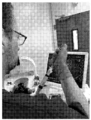
도 1a는 림프절 내로의 EUS-유도 세포 전달을 위한 실험 동물에 대해 초음파 내시경술을 수행하는 내시경 의학자의 사진이다.
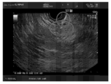
도 1b는 근처 림프절(LN)에 도달하는 세침 흡인 바늘의 초음파 이미지를 도시한 초음파 검사의 사진이다.
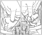
도 2는 수술 외과의에 의해 수행된 실험 동물의 십이지장 주위 림프절 내로의 간세포의 직접 주사의 사진을 도시한 것이다.
도 3은 도너 동물로부터 단리되고 상이한 게이지의 바늘을 통과한 상이한 배치의 간세포의 세포 생존율을 요약한 막대 그래프이다.
도 4는 문맥대정맥 문합술 절차 전 및 후 실험 동물의 열린 복강의 사진뿐만 아니라 문맥대정맥 문합술 절차의 다이아그램(최우측)을 도시한 것이다.
도 5는 본 개시에 따른 전임상 연구의 실험 설계를 도시한 다이아그램이다.
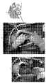
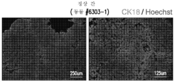
도 6a는 정상 간 조직에서 간세포의 마커인 CK-18의 양성 염색을 도시한 것이다.
도 6b는 전임상 연구에서 간세포의 내시경 초음파(EUS) 주사를 수용한 후 6일에 림프절에서의 CK-18 면역염색 신호의 존재를 도시한 것이다.
도 6c는 전임상 연구에서 간세포의 직접 주사를 수용한 후 6일에 림프절에서의 CK-18 면역염색 신호의 존재를 도시한 것이다.
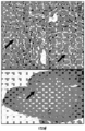
도 7a는 내시경 초음파(EUS) 주사에 의한 림프절 내로의 자가 간세포의 이식 후 약 90일에 림프절에 간 조직의 존재를 도시한 것이다.
도 7b는 내시경 초음파(EUS) 주사에 의한 림프절 내로의 동종 간세포의 이식 후 약 90일에 림프절에 간 조직의 존재를 도시한 것이다.
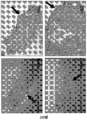
도 7c는 내시경 초음파(EUS) 주사에 의한 림프절 내로의 자가 간세포의 이식 후 약 60일에 림프절에 간 조직 및 푸마릴아세토아세테이트-하이드롤라아제(FAH) 양성 간세포의 존재를 도시한 것이다.
도 7d는 내시경 초음파(EUS) 주사에 의한 림프절 내로의 동종 간세포의 이식 후 약 150일에 림프절에 간조직 및 FAH 양성 간세포의 존재를 도시한 것이다.The novel features of the present disclosure are specifically set forth in the appended claims. A better understanding of the features and advantages of the present disclosure will be obtained by reference to the following detailed description, which sets forth exemplary embodiments in which the principles of the present disclosure are employed, and the accompanying drawings.
1A is a photograph of an endoscopist performing ultrasound endoscopy on an experimental animal for EUS-induced cell delivery into lymph nodes.
1B is a photograph of an ultrasound scan showing an ultrasound image of a fine needle aspiration needle reaching a nearby lymph node (LN).
2 shows photographs of direct injections of hepatocytes into the paraduodenal lymph nodes of an experimental animal performed by a surgical surgeon.
3 is a bar graph summarizing cell viability of different batches of hepatocytes isolated from donor animals and passed through needles of different gauges.
4 shows a diagram (rightmost) of the portal vein anastomosis procedure as well as photographs of the open abdominal cavity of the experimental animal before and after the portal vein anastomosis procedure.
5 is a diagram illustrating the experimental design of a preclinical study according to the present disclosure.
6A shows positive staining of CK-18, a marker of hepatocytes, in normal liver tissue.
6B depicts the presence of CK-18 immunostaining signal in lymph nodes at day 6 after receiving endoscopic ultrasound (EUS) injection of hepatocytes in a preclinical study.
6C depicts the presence of CK-18 immunostaining signal in lymph nodes 6 days after receiving direct injection of hepatocytes in a preclinical study.
7A depicts the presence of liver tissue in the lymph nodes approximately 90 days after transplantation of autologous hepatocytes into the lymph nodes by endoscopic ultrasound (EUS) injection.
FIG. 7B depicts the presence of liver tissue in the lymph nodes approximately 90 days after transplantation of allogeneic hepatocytes into the lymph nodes by endoscopic ultrasound (EUS) injection.
FIG. 7C depicts the presence of liver tissue and fumarylacetoacetate-hydrolase (FAH) positive hepatocytes in lymph nodes approximately 60 days after transplantation of autologous hepatocytes into lymph nodes by endoscopic ultrasound (EUS) injection.
FIG. 7D depicts the presence of liver tissue and FAH positive hepatocytes in lymph nodes approximately 150 days after transplantation of allogeneic hepatocytes into lymph nodes by endoscopic ultrasound (EUS) injection.
명확성을 위해, 그러나, 제한 없이, 본 발명에 개시된 주제의 상세한 설명은 하기 하위섹션으로 나뉘어진다:For clarity, however, and without limitation, the detailed description of the subject matter disclosed herein is divided into the following subsections:
1. 개론;1. Introduction;
2. 정의:2. Definition:
3. 세포 전달 절차; 3. Cell transfer procedure;
4. 이식된 세포로부터 이소성 조직의 생성;4. Generation of ectopic tissue from transplanted cells;
5. 질환 및 병태;5. diseases and conditions;
6. 대상체;6. subject;
7. 시스템; 및7. system; and
8. 키트.8. Kit.
1. One. 개론Introduction
개론으로서, 본 개시는 생체내 장기형성의 발달을 유도하기 위한 최소 침습적 절차를 위한 방법 및 시스템에 관한 것이다. 본원에는 대상체의 림프절 내로 세포를 전달함으로써 대상체에 세포를 이식하고 이소성 조직을 성장시키기 위한 방법 및 시스템이 제공된다. 적절한 해부학적 및 기능적 특징을 갖는 림프절 내측에 전달된 세포 또는 조직 단편으로부터의 이소성 조직을 성장시키는 공정은 생체내 기관 형성으로 지칭된다. 본 개시에 의해 가능한 최소 침습적 절차는 대상체에 대한 하나 이상의 장기의 정상 기능을 보안하거나 증대시킬 수 있는 이소성 조직(들)의 생성을 유발할 수 있다.As an overview, the present disclosure relates to methods and systems for minimally invasive procedures for inducing the development of organogenesis in vivo. Provided herein are methods and systems for transplanting cells and growing ectopic tissue in a subject by delivering the cells into a lymph node of a subject. The process of growing ectopic tissue from cells or tissue fragments delivered inside a lymph node with appropriate anatomical and functional characteristics is referred to as organogenesis in vivo. The minimally invasive procedures possible by the present disclosure may result in the creation of ectopic tissue(s) that may secure or augment the normal function of one or more organs for a subject.
상기에서 논의된 바와 같이, 현재의 이식 치료법, 특히 동소 장기 이식과 관련된 하나의 문제는 말기 간 또는 신장 질환과 같은 말기 질환을 갖는 여러 환자가 동소 장기 이식 또는 다른 타입의 세포 이식을 위해 필요로 할 수 있는 대수술에 더 이상 적합하지 않을 수 있다는 것이다. 대수술과 관련하여 상당한 위험이 있을 수 있으며, 이러한 환자의 예후는 이의 악화된 건강 상태를 고려할 때 나쁠 수 있다. 일 양태에서, 본 개시는 이러한 문제를 해결하기 위한 최소 침습적 절차(예를 들어, 내시경 초음파(EUS))를 제공한다. 방법 및 시스템은 자연적 생물반응기로서 림프절을 사용하여 이소성 부위에서 해부학적으로 온전하고 완전히 또는 적어도 부분적으로 기능하는 장기를 생성할 수 있다. 본원에 제공된 방법 및 시스템은 종종 추가 수술 절차와 관련될 수 있는 말기 장기 질환을 갖는 환자에 대한 질병률 및 사망률을 최소화할 수 있다. 본 개시는 또한, 본원에 기술된 수술 절차들 중 적어도 일부를 외래 환자 기준으로 수행할 수 있고, 이에 따라, 초기 절차 비용을 감소시킬 수 있다.As discussed above, one problem with current transplantation therapies, particularly orthotopic organ transplantation, is that many patients with end-stage disease, such as end-stage liver or kidney disease, will need an orthotopic organ transplant or other types of cell transplantation. It may no longer be suitable for major surgery. There can be significant risks associated with major surgery, and the prognosis for these patients can be poor given their worsening health. In one aspect, the present disclosure provides a minimally invasive procedure (eg, endoscopic ultrasound (EUS)) to address this problem. Methods and systems can use lymph nodes as natural bioreactors to generate anatomically intact and fully or at least partially functioning organs at ectopic sites. The methods and systems provided herein can minimize morbidity and mortality for patients with end-organ disease, which can often be associated with additional surgical procedures. The present disclosure may also perform at least some of the surgical procedures described herein on an outpatient basis, thereby reducing initial procedure costs.
본원에서 사용되는 용어 "이소성 조직"은 신체의 이소성(비-본래) 위치에 존재하고 일반적으로, 신체의 본래 위치에서 발견될 수 있는 건강한 기관 또는 조직과 유사하거나 동일한 하나 이상의 형태학적 및/또는 기능적 특성을 갖는 조직을 지칭할 수 있다. 용어 "기능성 이소성 조직"은 신체의 본래 위치에서 일반적으로 발견될 수 있는 건강한 기관 또는 조직의 기능들 중 하나 이상을 갖는 이소성 조직을 지칭할 수 있다. 용어 "이소성 위치"는 대상체의 본래 위치에 상대적인 위치를 기술하기 위해 이용될 수 있으며, 예를 들어, 이소성 위치는 대상체의 신체에서 본래 위치와는 상이한 위치를 지칭할 수 있다. 예를 들어, 림프절에서 간은 이소성이고, 즉, 간 조직이 통상적으로 림프절에서 발견되지 않기 때문에 건강한 정상 포유류 신체에서 이소성 간이다. 본 개시의 일부 양태에 따르면, 이소성 조직은 형태학적으로 간세포와 닮은 세포의 집단을 포함하는 림프절에서 성장할 수 있고, 총괄적으로, 건강한 고유 간이 수행할 수 있는 하나 이상의 기능을 수행할 수 있다.As used herein, the term "ectopic tissue" is one or more morphological and/or functional similar to or identical to a healthy organ or tissue that is present in an ectopic (non-native) location of the body and is generally found in an in situ location of the body. It can refer to an organization with characteristics. The term “functional ectopic tissue” may refer to ectopic tissue having one or more of the functions of a healthy organ or tissue that can normally be found in situ in the body. The term “ectopic location” may be used to describe a location relative to a subject's native location, eg, an ectopic location may refer to a location on the subject's body that is different from the original location. For example, the liver in lymph nodes is ectopic, ie, ectopic liver in a healthy normal mammalian body because liver tissue is not normally found in lymph nodes. According to some aspects of the present disclosure, the ectopic tissue may grow in a lymph node comprising a population of cells that morphologically resemble hepatocytes, and collectively, perform one or more functions that a healthy native liver may perform.
특정 구현예에서, 방법은 내시경을 대상체의 체강 또는 폐쇄된 체강 내로 진행시키는 것을 포함한다. 특정 구현예에서, 방법은 내시경을 대상체의 위장(GI)관, 기도, 또는 요로 내로 진행시키는 것을 포함한다.In certain embodiments, the method comprises advancing an endoscope into a body cavity or an occluded body cavity of a subject. In certain embodiments, the method comprises passing an endoscope into the gastrointestinal (GI) tract, airway, or urinary tract of the subject.
특정 구현예에서, 내시경은 엔도루미날 방법을 통해 대상체의 체강 내로 진행된다. 본원에서 사용되는 바와 같이, 본원에서 사용되는 용어 "엔도루미날 방법"은 임의의 방법, 예를 들어, 대상체의 체강 내로 내시경을 삽입하기 위한 당해 분야에 공지된 방법 및 디바이스를 지칭한다. 예를 들어, 그러나, 제한을 두지 않고, 방법은 내시경을 엔도루미날 방법을 통해 대상체의 위장(GI)관, 기도, 또는 요로 내로 진행시키는 것을 포함한다.In certain embodiments, the endoscope is advanced into a body cavity of a subject via an endoluminal method. As used herein, the term “endoluminal method” as used herein refers to any method, eg, methods and devices known in the art for inserting an endoscope into a body cavity of a subject. For example, but not by way of limitation, the method includes passing an endoscope through an endoluminal method into the gastrointestinal (GI) tract, airway, or urinary tract of a subject.
특정 구현예에서, 방법은 또한, 바늘을 대상체의 림프절 내로 삽입하는 것을 포함할 수 있으며, 여기서, 바늘은 내시경에 부착된다. 특정 구현예에서, 내시경에 부착된 바늘은 내장 벽을 통해 림프절 내로 삽입된다. 내장 벽의 비제한적인 예는 관(tract), 예를 들어, 위, 십이지장, 기관, 기관지, 또는 방광을 따라 속이 빈 내장 및/또는 기관을 둘러싸는 해부학적 구조를 포함한다. 특정 구현예에서, 방법은 또한, 바늘을 트랜스루미날 방법을 통해 삽입하는 것을 포함한다. 본원에서 사용되는 용어 "트랜스루미날 방법"은 임의의 방법, 예를 들어, 내시경을 사용을 통해 강(lumen)을 가로질러 작업 기구(예를 들어, 바늘)를 삽입하기 위한 당해 분야에 공지된, 방법 및 디바이스를 지칭한다.In certain embodiments, the method can also include inserting a needle into a lymph node of a subject, wherein the needle is attached to an endoscope. In certain embodiments, a needle attached to an endoscope is inserted through the visceral wall and into a lymph node. Non-limiting examples of visceral walls include anatomical structures surrounding hollow intestines and/or organs along a tract, eg, stomach, duodenum, trachea, bronchus, or bladder. In certain embodiments, the method also includes inserting the needle via a transluminal method. As used herein, the term “transluminal method” refers to any method known in the art for inserting a working instrument (eg, a needle) across a lumen via use, eg, an endoscope. , methods and devices.
특정 구현예에서, 방법은 또한, 하나 이상의 세포를 바늘을 통해 림프절 내로 전달하는 것을 포함한다. 특정 구현예에서, 방법은 하나의 단일 세포를 림프절 내로 전달하는 것을 포함한다. 특정 구현예에서, 방법은 세포의 집단을 림프절 내로 전달하는 것을 포함한다. 특정 구현예에서, 세포의 집단은 하나의 세포 타입을 포함한다. 특정 구현예에서, 세포의 집단은 적어도 2개의 세포 타입을 포함한다. 특정 구현예에서, 림프절 내로 전달된 하나 이상의 세포는 림프절에서 생착하고, 이소성 조직을 생성할 수 있다.In certain embodiments, the method also includes delivering the one or more cells through the needle into the lymph node. In certain embodiments, the method comprises delivering one single cell into a lymph node. In certain embodiments, the method comprises delivering a population of cells into a lymph node. In certain embodiments, the population of cells comprises one cell type. In certain embodiments, the population of cells comprises at least two cell types. In certain embodiments, one or more cells delivered into a lymph node are capable of engrafting in the lymph node and generating ectopic tissue.
특정 구현예에서, 내시경의 진행, 바늘의 삽입, 또는 둘 모두는 최소 침습적 또는 비-침습적 방법에 의해 수행된다. 예를 들어, 초음파-매개 이미지화 또는 다른 탐지 방법은 타겟 림프절의 위치를 위해 본원에 제공된 방법에서 적용될 수 있다. 과정 동안에, 초음파 영상화 또는 다른 방법, 예를 들어, 최소 침습적 또는 비침습적 탐지 방법은 내시경의 진행, 적합한 타겟 림프절의 위치화, 또는 내장 벽을 통해 또는 타겟 림프절 내로의 바늘 삽입의 모니터링 중 어느 하나에 대해 적용될 수 있다.In certain embodiments, the procedure of the endoscope, insertion of a needle, or both, is performed by minimally invasive or non-invasive methods. For example, ultrasound-mediated imaging or other detection methods can be applied in the methods provided herein for the location of target lymph nodes. During the procedure, ultrasound imaging or other methods, e.g., minimally invasive or non-invasive detection methods, include either monitoring of the progress of the endoscope, localization of a suitable target lymph node, or insertion of a needle through the visceral wall or into the target lymph node. can be applied for
다른 양태에서, 본 개시는 초음파의 도움으로 대상체의 복부, 골반 또는 흉강에서의 림프절 내로 바늘을 삽입하는 것을 포함하는 방법을 제공한다. 방법은 또한, 바늘을 통해 림프절 내로 하나 이상의 세포를 전달하는 것을 포함할 수 있다. 림프절의 초음파-유도 위치는 이용 가능한 임의의 기술에 의해 수행될 수 있다. 예를 들어, 그러나, 제한을 두지 않고, 세포의 주사를 위해 적합한 림프절을 동정하고 위치시키기 위해 초음파 영상화가 이용될 수 있다. 특정 구현예에서, 초음파 분광법은 림프절을 탐지하기 위해 이용될 수 있다. 특정 구현예에서, 초음파 영상화 또는 분광법은 림프절의 국소화를 위한 다른 탐지 방법과 함께 이용될 수 있다.In another aspect, the present disclosure provides a method comprising inserting a needle into a lymph node in a subject's abdomen, pelvis, or chest cavity with the aid of ultrasound. The method may also include delivering the one or more cells through the needle into the lymph node. Ultrasound-guided positioning of lymph nodes may be performed by any technique available. For example, but without limitation, ultrasound imaging can be used to identify and locate lymph nodes suitable for injection of cells. In certain embodiments, ultrasound spectroscopy may be used to detect lymph nodes. In certain embodiments, ultrasound imaging or spectroscopy may be used in conjunction with other detection methods for localization of lymph nodes.
특정 구현예에서, 방법은 대상체의 림프절 내로 세포의 집단을 전달하는 것을 포함하며, 여기서, 간세포의 집단은 림프절에서 생착시키고 이소성 간을 생성시킨다. 특정 구현예에서, 방법은 또한, 대상체의 간으로 혈액 공급을 감소시키는 것을 포함한다. 본 개시에 의해, 대상체의 간으로의 혈액 공급의 감소가 림프절에서 이소성 간의 성장에 유익하다는 것이 발견되었다.In certain embodiments, the method comprises delivering a population of cells into a lymph node of a subject, wherein the population of hepatocytes engrafts in the lymph node and produces an ectopic liver. In certain embodiments, the method also includes reducing blood supply to the liver of the subject. It has been discovered by the present disclosure that a reduction in blood supply to the liver of a subject is beneficial for the growth of ectopic liver in the lymph nodes.
2. 2. 정의Justice
본 출원에서, 단수 형태의 사용은 달리 구체적으로 기술하지 않는 한 복수 형태를 포함한다. 본 명세서에서 사용되는, 단수 형태("a," "an" 및 "the")가 문맥이 달리 명확하게 명시하지 않는 한, 복수 지시대상을 포함한다는 것이 주지되어야 한다.In this application, use of the singular form includes the plural form unless specifically stated otherwise. It should be noted that, as used herein, the singular forms "a," "an" and "the" include plural referents unless the context clearly dictates otherwise.
본 출원에서, "또는"의 사용은 달리 기술하지 않는 한, "및/또는"을 의미한다. 본원에서 사용되는 용어 "및/또는" 및 "이들의 임의의 조합" 및 문법적으로 이에 대응하는 용어는 상호교환적으로 사용될 수 있다. 이러한 용어들은 임의의 조합이 구체적으로 고려됨을 전달할 수 있다. 오로지 예시 목적을 위해서만, 하기 구 "A, B, 및/또는 C" 또는 "A, B, C, 또는 이들의 임의의 조합"은 "개별적으로, A; 개별적으로, B; 개별적으로, C; A 및 B; B 및 C; A 및 C; 및 A, B, 및 C"를 의미한다. 용어 "또는"은 문맥이 구체적으로 분리 사용(disjunctive use)을 지칭하지 않는 한, 결합하여 또는 분리하여 사용될 수 있다.In this application, the use of "or" means "and/or" unless stated otherwise. As used herein, the terms “and/or” and “any combination thereof” and grammatically corresponding terms may be used interchangeably. These terms may convey that any combination is specifically contemplated. For illustrative purposes only, the following phrases “A, B, and/or C” or “A, B, C, or any combination thereof” refer to “individually, A; individually, B; individually, C; A and B; B and C; A and C; and A, B, and C". The term “or” may be used in combination or separately, unless the context specifically refers to a disjunctive use.
또한, 용어 "포함하는(including)"뿐만 아니라, "포함하다(include, includes)" 및 "포함된(included)"과 같은 다른 형태의 사용은 제한적이지 않다.Also, other forms of use of the term “including,” as well as “include, includes,” and “included,” are not limiting.
본 명세서에서 "일부 구현예," "특정 구현예," "구현예," "하나의 구현예," 또는 "다른 구현예"는 구현예와 관련하여 기술된 특정의 특성, 구조 또는 특징이 적어도 일부 구현예에 포함되지만, 반드시 본 개시의 모든 구현예는 아님을 의미한다.As used herein, “some embodiments,” “specific embodiments,” “embodiments,” “one embodiment,” or “other embodiments” refer to at least a particular characteristic, structure, or characteristic described in connection with the embodiment. included in some embodiments, but not necessarily all embodiments of the present disclosure.
본 명세서 및 청구항(들)에서 사용되는 단어 "포함하는(comprising)"(및 "포함하다(comprise, comprises)와 같은 포함하는의 임의의 형태), "갖는(having)"(및 "가지다(have, has)"와 같은 갖는의 임의의 형태), "포함하는(including)"(및 "포함하다(includes, include)"와 같은 포함하는의 임의의 형태) 또는 "함유하는(containing)"(및 "함유하다(contains, contain)"와 같은 함유하는의 임의의 형태)은 포괄적이거나 개방형이고, 추가적인, 인용되지 않은 요소 또는 방법 단계를 배제하지 않는다. 본 명세서에 논의된 임의의 구현예가 본 개시의 임의의 방법 또는 조성물과 관련하여 구현될 수 있으며, 그 반대도 마찬가지라는 것이 고려된다. 또한, 본 개시의 조성물은 본 개시의 방법을 달성하기 위해 사용될 수 있다.As used herein and in the claim(s), the word "comprising" (and any form of including, such as "comprise, comprises"), "having" (and "have") , has any form of having), "including" (and any form of including, such as "includes, include") or "containing" (and Any form of containing, such as "contains, contain") is inclusive or open-ended and does not exclude additional, unrecited elements or method steps. It is contemplated that any method or composition can be implemented and vice versa.In addition, the composition of the present disclosure can be used to achieve the method of the present disclosure.
본원에서 사용되는 기준 수치 및 문법적으로 이에 대응하는 것과 관련하여 용어 "약"은 수치 자체 및 그러한 수치값에서 플러스 또는 마이너스의 값의 범위를 포함할 수 있다.As used herein, the term "about," in reference to a reference numerical value and grammatically corresponding thereto, may include the numerical value itself and ranges of values positive or negative in that numerical value.
용어 "약" 또는 "대략"은 당업자에 의해 측정하는 경우 특정 값에 대한 허용 가능한 오차 범위 내임을 의미하며, 이는 부분적으로, 값이 어떻게 측정되거나 결정되는 지, 즉, 측정 시스템의 한계에 의존적일 것이다. 예를 들어, "약"은 당분야의 관행에 따라 1 또는 1 초과의 표준 편차 내를 의미할 수 있다. 대안적으로, "약"은 제공된 값의 최대 20%, 최대 10%, 최대 5%, 또는 최대 1%의 범위를 의미할 수 있다. 다른 예에서, "약 10"의 양은 10 및 9 내지 11의 임의의 양을 포함한다. 또 다른 예에서, 참조 수치 값과 관련하여 용어 "약"은 또한, 값 플러스(plus) 또는 마이너스(minus) 그러한 값으로부터의 10%, 9%, 8%, 7%, 6%, 5%, 4%, 3%, 2%, 또는 1%의 범위를 포함할 수 있다. 대안적으로, 특히 생물학적 시스템 또는 공정과 관련하여, 용어 "약"은 값의 10배(an order of magnitude) 내, 바람직하게는, 5배 내, 및 더욱 바람직하게는, 2배 내임을 의미할 수 있다. 특정 값이 출원 및 청구범위에 기술된 경우에, 달리 기술하지 않는 한, 특정 값에 대한 허용 가능한 오차 범위 내를 의미하는 용어 "약"이 가정되어야 한다.The terms “about” or “approximately” mean within an acceptable error range for a particular value as measured by one of ordinary skill in the art, which will depend, in part, on how the value is measured or determined, i.e., the limits of the measurement system. will be. For example, "about" can mean within one or more than one standard deviation according to the practice in the art. Alternatively, “about” can mean a range of at most 20%, at most 10%, at most 5%, or at most 1% of a given value. In other examples, an amount of “about 10” includes 10 and any amount from 9 to 11. In another example, the term "about" with respect to a reference numerical value also means a value plus or minus 10%, 9%, 8%, 7%, 6%, 5%, from such value; 4%, 3%, 2%, or 1%. Alternatively, particularly in the context of a biological system or process, the term “about” shall mean within an order of magnitude, preferably within 5 times, and more preferably within 2 times a value. can Where specific values are recited in the application and claims, unless otherwise stated, the term "about", meaning within an acceptable margin of error for the specific value, should be assumed.
3. 3. 세포 전달 절차Cell transfer procedure
특정 구현예에서, 대상체의 림프절 내로 세포의 트랜스루미날 전달을 위해 내시경을 이용한다. 특정 구현예에서, 림프절은 내시경의 이용을 통해 도달될 수 있는 신체 내강 또는 폐쇄 체강에 대해 근위에 또는 신체 내강 또는 폐쇄 체강 내에 위치되어 있다. 특정 구현예에서, 림프절은 대상체의 복부, 골반 또는 흉강에 위치되어 있다.In certain embodiments, an endoscope is used for transluminal delivery of cells into a lymph node of a subject. In certain embodiments, the lymph nodes are located proximal to or within a body lumen or occluded body cavity that can be reached through the use of an endoscope. In certain embodiments, the lymph nodes are located in the subject's abdomen, pelvis, or chest cavity.
본원에서 사용되는 용어 "내시경"은 대상체, 예를 들어, 인간 대상체 또는 비-인간 포유류 대상체, 예를 들어, 개, 돼지, 말, 당나귀, 토끼, 소, 마우스, 또는 래트의 신체 내에 도입될 수 있고 대상체의 내부 부분의 관찰을 제공할 수 있는 임의의 기구를 지칭할 수 있다. 때때로, 관찰(view)은, 예를 들어, 내시경에 조명 광학이 장착되거나 달리 조명이 보조될 때 육안 검사를 제공한다. 특정 구현예에서, 본 개시의 내시경은 하나 이상의 탐지 프로브를 포함하거나, 하나 이상의 탐지 프로브에 부착되거나 결합되고, 대상체의 내부 부분의 검사를 위한 상이한 탐지 모드를 제공한다. 예를 들어, 내시경은 육안 검사를 위한 조명 광학, 초음파-매개 탐지(예를 들어, 초음파 영상화)를 위한 초음파 프로브, 또는 방사선, 적외선 신호, 고주파 신호, 또는 형광 신호를 위한 탐지기를 가질 수 있다.As used herein, the term “endoscope” can be introduced into the body of a subject, e.g., a human subject or a non-human mammalian subject, e.g., a dog, pig, horse, donkey, rabbit, cow, mouse, or rat. and may refer to any instrument capable of providing viewing of an internal portion of a subject. Sometimes, the view provides a visual inspection, for example when the endoscope is equipped with illumination optics or otherwise assisted with illumination. In certain embodiments, an endoscope of the present disclosure includes, is attached to, or is coupled to, one or more detection probes, and provides different detection modes for examination of an internal portion of a subject. For example, an endoscope may have illumination optics for visual inspection, ultrasound probes for ultrasound-mediated detection (eg, ultrasound imaging), or detectors for radiation, infrared signals, radio frequency signals, or fluorescent signals.
본원에 기술된 내시경은 하기 부분들 중 하나 이상을 포함할 수 있다: 대상체의 루미날 관(luminal tract)(예를 들어, GI관, 기도, 또는 요로)을 따라 이동시키기 위한 강성 또는 가요성 튜브; 검사 시에 기관 또는 물체를 조명하기 위한 광 전달 시스템; 이미지를 대물 렌즈에서 뷰어(viewer), 접안 렌즈 또는 바디의 내측의 내시경에 의해 포착된 이미지를 나타내는 비디오 시스템으로 전송하기 위한 렌즈 시스템; 의료 기구 또는 조작기의 진입을 허용하는 하나 이상의 채널. 광원은 바디의 외측에 또는 미니 사이즈로 존재할 수 있고, 내시경의 내측에 장착될 수 있다. 광원이 바디 외측에 있을 때, 광은 광섬유 시스템을 통해 전송될 수 있다. 바디 내측에 포착된 광 이미지를 전송하기 위한 광학 시스템, 예를 들어, 광섬유가 존재할 수 있다. 추가적으로 또는 대안적으로, 본원에 제공된 내시경에는 상이한 목적을 위한 다른 탐지 기구가 장착될 수 있다(예를 들어, 이를 부분으로서 포함하거나, 여기에 부착될 수 있다).An endoscope described herein may include one or more of the following parts: A rigid or flexible tube for movement along a luminal tract (eg, GI tract, airway, or urinary tract) of a subject. ; a light delivery system for illuminating an organ or object upon examination; a lens system for transmitting an image from the objective lens to a video system representing the image captured by a viewer, an eyepiece, or an endoscope inside the body; One or more channels allowing entry of a medical instrument or manipulator. The light source may exist on the outside of the body or in a mini size, and may be mounted on the inside of the endoscope. When the light source is outside the body, the light can be transmitted through the fiber optic system. There may be an optical system inside the body for transmitting the captured optical image, for example an optical fiber. Additionally or alternatively, the endoscope provided herein may be equipped with (eg, include as part or attached to) other detection instruments for different purposes.
본원에 제공된 방법은 대상체의 신체 내강 또는 폐쇄 체강에 내시경을 진행시키는 것을 포함할 수 있다. 신체 내강의 비제한적인 예는 위장(GI)관(예를 들어, 식도, 위, 십이지장, 소장, 대장, 결장, 담관, 직장, 항문), 기도(예를 들어, 코, 하부 기도), 귀, 요로, 자궁경부, 자궁, 및 나팔관을 포함한다. 폐쇄 체강의 비제한적인 예는 복강, 및 골반강을 포함한다. 특정 구현예에서, 본원에 개시된 방법은 대상체의 위장관, 기도, 또는 요로에 내시경을 진행시키는 것을 포함한다.The methods provided herein can include advancing an endoscope into a body lumen or closed body cavity of a subject. Non-limiting examples of body lumens include the gastrointestinal (GI) tract (eg, esophagus, stomach, duodenum, small intestine, large intestine, colon, bile duct, rectum, anus), airway (eg, nose, lower airway), ear , urinary tract, cervix, uterus, and fallopian tubes. Non-limiting examples of closed body cavities include the abdominal cavity, and the pelvic cavity. In certain embodiments, the methods disclosed herein comprise conducting an endoscope into the gastrointestinal tract, airway, or urinary tract of a subject.
특정 구현예에서, 본원에 개시된 방법은 신체 내강 또는 폐쇄 체강에 대해 근위에 또는 신체 내강 또는 폐쇄 체강에 위치된 적어도 하나의 림프절 내로 세포를 전달하기 위해 대상체의 신체 내강을 따라 또는 폐쇄 체강에 내시경을 진행시키는 것을 포함한다. 특정 구현예에서, 림프절의 위치는 세포 이식의 목적을 기반으로 하여 선택된다. 특정 구현예에서, 내강 및/또는 폐쇄 체강은 세포 이식에 적합한 림프절을 기반으로 하여 선택된다.In certain embodiments, the methods disclosed herein use an endoscope along or into a body lumen of a subject to deliver cells into at least one lymph node located proximally to or in a body lumen or body cavity obstructed. includes proceeding. In certain embodiments, the location of the lymph node is selected based on the purpose of the cell transplant. In certain embodiments, the lumen and/or occluded body cavity is selected based on lymph nodes suitable for cell transplantation.
특정 구현예에서, 본원에 개시된 방법은 GI관에 대해 근위에 있는 하나 이상의 림프절 내로 세포를 전달하기 위해 GI관을 따라 내시경을 진행시키는 것을 포함한다. GI관의 상부 부분 또는 하부 부분에 대해 근위에 있는 림프절 내로 세포를 전달하기 위해 내시경(예를 들어, 식도경 또는 위내시경)이 이용될 수 있다. 예를 들어, 식도에 가까운 림프절 내로 세포를 전달하기 위해 식도경이 이용될 수 있으며, 위 주위 및/또는 십이지장 주위 림프절 내로 세포를 전달하기 위해 위내시경이 이용될 수 있다. 본원에 제공된 방법을 이용하여 세포가 전달될 수 있는 GI관에 대해 근위에 있는 림프절의 비제한적인 예는 십이지장 주위 림프절, 위 주위 림프절, 췌장 주위 림프절, 장간막 림프절, 회결장 림프절, 결장간막 림프절, 위 림프절, 간 비장 림프절, 비장 폐문 림프절, 식도 주위 림프절, 심장 주위 림프절, 대동맥 주위 림프절, 대동맥후 림프절, 측대동맥 림프절, 전대동맥 림프절, 소만곡 림프절, 총간 림프절, 비장 동맥 림프절, 복강 축 림프절, 장골 림프절, 및 복막후 림프절을 포함한다. 내시경은 대상체의 입으로부터, 또는 특정 구현예에서, 대상체의 코로부터 도입될 수 있다. 대안적으로, 내시경은 대상체의 항문으로부터 도입될 수 있으며, 예를 들어, 직장경, S자 결장경, 또는 결장경은 직장 또는 결장에 가까운 림프절 내로 세포를 전달하기 위해 이용될 수 있다. 특정 구현예에서, 내시경은 위장관을 따라 유연하게 이동할 수 있으며, 세포 이식의 목적을 위해 적합한 림프절을 위치시키기 위해 조사 검사가 수행된다.In certain embodiments, the methods disclosed herein comprise advancing an endoscope along the GI tract to deliver cells into one or more lymph nodes proximal to the GI tract. An endoscope (eg, esophagoscopy or gastroscopy) may be used to deliver cells into lymph nodes proximal to the upper or lower portion of the GI tract. For example, an esophagoscope can be used to deliver cells into lymph nodes proximal to the esophagus, and gastroscopy can be used to deliver cells into paragastric and/or paraduodenal lymph nodes. Non-limiting examples of lymph nodes proximal to the GI tract to which cells may be delivered using the methods provided herein include, but are not limited to, paraduodenal lymph nodes, paragastric lymph nodes, parapancreatic lymph nodes, mesenteric lymph nodes, ileocecal lymph nodes, colonic mesenteric lymph nodes, Gastric lymph nodes, hepatic splenic lymph nodes, splenic hilar lymph nodes, paraesophageal lymph nodes, paracardiac lymph nodes, para-aortic lymph nodes, post-aortic lymph nodes, collateral aortic lymph nodes, anterior aortic lymph nodes, small curvature lymph nodes, common hepatic lymph nodes, splenic artery lymph nodes, celiac axial lymph nodes, iliac lymph nodes, and retroperitoneal lymph nodes. The endoscope can be introduced from the subject's mouth, or, in certain embodiments, from the subject's nose. Alternatively, an endoscope may be introduced from the subject's anus, eg, a rectoscope, sigmoidoscopy, or colonoscope may be used to deliver cells into the rectum or lymph nodes proximal to the colon. In certain embodiments, the endoscope can be moved flexibly along the gastrointestinal tract, and an investigational examination is performed to locate a suitable lymph node for the purpose of cell transplantation.
특정 구현예에서, 본원에 개시된 방법은 기도에 대해 근위에 있는 하나 이상의 림프절 내로 세포를 전달하기 위해 대상체의 기도를 따라 내시경을 진행시키는 것을 포함한다. 예를 들어, 그러나, 제한을 두지 않고, 대상체의 기관 또는 폐의 기관지, 또는 후두에 가까운 림프절을 타겟으로 하기 위해 기관지경 또는 후두경이 사용된다. 본원에 제공된 방법을 이용하여 세포가 전달될 수 있는 기도에 대해 근위에 있는 림프절의 비제한적인 예는 복장옆 림프절, 늑간 림프절, 상횡경막 림프절, 상기관기관지 림프절, 하기관기관지 림프절, 기관지폐 림프절, 기관옆 림프절, 및 폐내 림프절을 포함한다. 기관지경 또는 후두경은 대상체의 입으로부터, 또는 특정 구현예에서, 대상체의 코로부터 도입될 수 있다.In certain embodiments, the methods disclosed herein comprise advancing an endoscope along an airway of a subject to deliver cells into one or more lymph nodes proximal to the airway. For example, but without limitation, a bronchoscope or laryngoscope is used to target a subject's trachea or bronchi of the lungs, or lymph nodes proximal to the larynx. Non-limiting examples of lymph nodes proximal to the airways to which cells may be delivered using the methods provided herein include parasternal lymph nodes, intercostal lymph nodes, superior diaphragmatic lymph nodes, upper bronchial lymph nodes, lower bronchial lymph nodes, bronchopulmonary lymph nodes. , paratracheal lymph nodes, and intrapulmonary lymph nodes. The bronchoscope or laryngoscope can be introduced from the subject's mouth, or in certain embodiments, from the subject's nose.
특정 구현예에서, 본원에 개시된 방법은 요로에 대해 근위에 있는 하나 이상의 림프절 내로 세포를 전달하기 위해 대상체의 요로를 따라 내시경을 진행시키는 것을 포함한다. 예를 들어, 그러나, 제한을 두지 않고, 요도 또는 방광에 가까운 림프절을 타겟으로 하기 위해 방광경이 이용된다. 방광경은 대상체의 요도로부터 도입될 수 있다. 본원에 제공된 방법을 이용하여 세포가 전달될 수 있는 요로에 대해 근위에 있는 림프절의 비제한적인 예는 외부 장골 림프절, 내부 장골 림프절, 대정맥 요추 림프절, 대동맥 요추 림프절, 표재 서혜부 림프절, 깊은 서혜부 림프절, 대동정맥사이 방광 주위 림프절, 폐색 방광 주위 림프절, 또는 천골전 방광 주위 림프절 중 하나 이상을 포함한다.In certain embodiments, the methods disclosed herein comprise running an endoscope along a urinary tract of a subject to deliver cells into one or more lymph nodes proximal to the urinary tract. For example, but without limitation, a cystoscope is used to target lymph nodes proximal to the urethra or bladder. A cystoscope may be introduced from the subject's urethra. Non-limiting examples of lymph nodes proximal to the urinary tract to which cells may be delivered using the methods provided herein include external iliac lymph nodes, internal iliac lymph nodes, vena cava lumbar lymph nodes, aortic lumbar lymph nodes, superficial inguinal lymph nodes, deep inguinal lymph nodes, including one or more of interaortic paravestic lymph nodes, obstructive paravestic lymph nodes, or presacral paravestic lymph nodes.
특정 구현예에서, 본원에 개시된 방법은 세포를 폐쇄된 체강 내측 또는 부근의 적어도 하나의 림프절 내로 전달하기 위해 내시경을 폐쇄된 체강(예를 들어, 복강, 골반강) 안으로 진행시키는 것을 포함한다. 특정 구현예에서, 내시경은 최소 절개를 통해, 예를 들어, 복부의 표면 상의 최소 절개를 통해 폐쇄된 체강 내로 도입된다. 특정 구현예에서, 폐쇄된 체강은 복강 또는 골반강이다. 복강 또는 골반강 내측 또는 부근의 림프절의 비제한적인 예는 비장 림프절, 간 림프절, 낭포 림프절, 추간공 림프절, 우/좌측 위 림프절, 유문 림프절, 상유문 림프절,, 하유문 림프절, 레트로 유문 림프절, 상췌장 림프절, 하위 림프절, 상/하위 췌십이지장 림프절, 하위 장간막 림프절, S상 림프절, 상직장 림프절, 결장간막 림프절, 좌결장 림프절, 우결장 림프절, 중간 결장 림프절, 충수 림프절, 회결장 림프절, 막창자뒤 림프절, 덮개앞 림프절, 상장간막 림프절, 좌측 요추 림프절, 측대동맥 림프절, 및 전대동맥 림프절을 포함한다.In certain embodiments, the methods disclosed herein comprise advancing an endoscope into an obstructed body cavity (eg, an abdominal cavity, a pelvic cavity) to deliver cells into at least one lymph node in or near the obstructed body cavity. In certain embodiments, the endoscope is introduced into an occluded body cavity through a minimal incision, eg, through a minimal incision on the surface of the abdomen. In certain embodiments, the occluded body cavity is an abdominal cavity or a pelvic cavity. Non-limiting examples of lymph nodes in or near the abdominal or pelvic cavity include splenic lymph nodes, hepatic lymph nodes, cystic lymph nodes, intervertebral foramen lymph nodes, right/left gastric lymph nodes, pyloric lymph nodes, superior pyloric lymph nodes, inferior pyloric lymph nodes, retro pyloric lymph nodes, superior pancreas. Lymph node, inferior lymph node, superior/inferior pancreaticoduodenal lymph node, inferior mesenteric lymph node, sigmoid lymph node, superior rectal lymph node, mesenteric lymph node, left colon lymph node, right colon lymph node, middle colon lymph node, appendix lymph node, ileal colon lymph node, retro-intestinal lymph node , anterior chamber lymph nodes, superior mesenteric lymph nodes, left lumbar lymph nodes, collateral aortic lymph nodes, and anterior aortic lymph nodes.
특정 구현예에서, 본원에 개시된 방법 및 시스템은 세포가 이식되는 림프절을 위치시키기 위해 및/또는 체강 또는 폐쇄된 체강을 통해 내시경을 진행시키는 단계, 바늘을 삽입시키는 단계 또는 이들의 임의의 조합을 유도하기 위해 초음파를 이용한다. 초음파는 인간 가청의 상한선보다 높은 주파수를 갖는 음파이며, 이는 약 20 kHz 초과 내지 수 기가헤르츠일 수 있다. 초음파 영상화(또는 초음파 검사)는 본원에 제공된 방법 및 시스템을 적용하는 의도된 목적에 따라 다양한 방식으로 수행될 수 있다. 초음파 검사의 비제한적인 예는 도플러 초음파 검사, 콘트라스트 초음파 검사, 분자 초음파 검사, 탄성 초음파, 중재적 초음파 검사, 및 압박 초음파 검사를 포함할 수 있다. 특정 구현예에서, 본원에서 사용되는 초음파 영상화 기술은 국제특허공개 WO2018222724A1호 및 WO2018134729A1호에 기술된 것과 같은 기술을 이용하여, 주사를 위해 적합한 림프절을 위치시킬 목적을 위해 높은 공간적 및/또는 시간적 해상도를 가지며, 이러한 문헌 각각은 본원에 참고로 포함된다.In certain embodiments, the methods and systems disclosed herein induce the step of advancing an endoscope through a body cavity or an occluded body cavity, inserting a needle, or any combination thereof to locate a lymph node into which cells are transplanted and/or Ultrasound is used to Ultrasound is a sound wave with a frequency higher than the upper limit of human hearing, which can be greater than about 20 kHz to several gigahertz. Ultrasound imaging (or sonography) can be performed in a variety of ways depending on the intended purpose of applying the methods and systems provided herein. Non-limiting examples of ultrasound examinations may include Doppler ultrasound examination, contrast ultrasound examination, molecular ultrasound examination, elastic ultrasound examination, interventional ultrasound examination, and compression ultrasound examination. In certain embodiments, the ultrasound imaging technique used herein utilizes techniques such as those described in WO2018222724A1 and WO2018134729A1 to achieve high spatial and/or temporal resolution for the purpose of locating suitable lymph nodes for injection. and each of these documents is incorporated herein by reference.
본 개시의 특정 구현예에서, 본원에 개시된 방법은 초음파 영상화와 조합하여 내시경을 이용하며, 여기서, 내시경은 초음파 프로브에 (예를 들어, 내시경의 일부로서 또는 별도의 조각으로서) 부착된다. 본원에서 상호 교환 가능하게 사용되는, 용어 "초음파 내시경술," "내시경 초음파," 또는 "EUS"는 내시경이 흉부, 복부 및 골반, 또는 신체의 다른 내부 구조에서의 내부 장기를 탐지하고/하거나(예를 들어, 이의 이미지를 얻고/얻거나) 이를 조작하기 위해 초음파와 결합되는 의학적 절차를 지칭할 수 있다. 본원에 제공된 방법 및 시스템에서 이용되는 EUS 기구 및 기술은 현재 상업적으로 입수 가능한 것, 및/또는 미국특허공개 US20060106306A1호 및 US20070237373A1호, 및 국제특허공개 WO1998009247A1호에 기술된 것일 수 있으며, 이러한 문헌들 각각은 전문이 본원에 참고로 포함된다.In certain embodiments of the present disclosure, the methods disclosed herein utilize an endoscope in combination with ultrasound imaging, wherein the endoscope is attached to an ultrasound probe (eg, as part of an endoscope or as a separate piece). As used interchangeably herein, the terms “ultrasound endoscopy,” “endoscopic ultrasound,” or “EUS” refer to an endoscope being used to detect internal organs in the chest, abdomen and pelvis, or other internal structures of the body and/or ( For example, it may refer to a medical procedure that is combined with ultrasound to obtain an image thereof and/or manipulate it. EUS instruments and techniques utilized in the methods and systems provided herein may be those currently commercially available and/or described in US Patent Publications US20060106306A1 and US20070237373A1, and International Patent Publication WO1998009247A1, each of which is incorporated herein by reference in its entirety.
특정 구현예에서, 타겟 림프절(예를 들어, 하나 이상의 세포가 전달되는 림프절)의 초음파 영상화는 방사선학적 영상화와 같은 다른 이미지화 기술의 도움으로 수행된다. 방사선학적 영상화는 동적 방사선학적 영상화(형광투시법), 컴퓨터 단층촬영(CT), 또는 자기 공명 영상화(MRI)를 포함할 수 있다. 예를 들어, 그러나, 제한을 두지 않고, 컴퓨터 단층촬영(CT)은 대상체의 전체 신체 또는 일부 국소 장기(들) 또는 조직(들)의 해부학적 정보를 얻기 위해 수행된다. 특정 구현예에서, 자기 공명 또는 이용 가능한 임의의 다른 의료 기술은 대상체의 복부, 골반 또는 흉강에 타겟 림프절을 위치시키기 위한 초음파 영상화와 함께 또는 대신에 이용된다.In certain embodiments, ultrasound imaging of a target lymph node (eg, a lymph node that has delivered one or more cells) is performed with the aid of other imaging techniques, such as radiographic imaging. Radiographic imaging may include dynamic radiographic imaging (fluoroscopy), computed tomography (CT), or magnetic resonance imaging (MRI). For example, but without limitation, computed tomography (CT) is performed to obtain anatomical information of the subject's entire body or some local organ(s) or tissue(s). In certain embodiments, magnetic resonance or any other medical technique available is used in conjunction with or instead of ultrasound imaging to position a target lymph node in a subject's abdomen, pelvis, or chest cavity.
본원에 제공된 방법 및 시스템은 하나 이상의 세포를 림프절 내로 전달하기 위한 바늘을 이용하는 것을 포함할 수 있다. 바늘은 주사기의 일부일 수 있으며, 이는 전달되는 세포를 수용하고 전달을 위해 세포를 바늘을 통해 밖으로 밀어내도록 구성될 수 있다. 본원에 제공된 바늘은 특정 정도의 첨예도 및 강성도를 갖도록 구성될 수 있다. 예를 들어, 바늘은 림프절 내로 삽입되어야 한다. 특정 구현예에서, 바늘은 또한, GI관(예를 들어, 식도, 위, 장, 또는 결장의 벽), 기도, 요로(예를 들어, 요도 또는 방광의 벽)의 벽을 침투하도록 구성되며, 이에 따라, 바늘은 관 외측으로 림프절에 도달할 수 있다. 당해 분야에 공지된 임의의 적합한 바늘은 본원에 개시된 방법과 함께 이용될 수 있다.The methods and systems provided herein can include using a needle to deliver one or more cells into a lymph node. The needle may be part of a syringe, which may be configured to receive a delivered cell and push the cell out through the needle for delivery. The needles provided herein can be configured to have a certain degree of sharpness and stiffness. For example, a needle must be inserted into a lymph node. In certain embodiments, the needle is also configured to penetrate the walls of the GI tract (e.g., the walls of the esophagus, stomach, intestine, or colon), airways, urinary tract (e.g., the walls of the urethra or bladder), Accordingly, the needle may reach the lymph node out of the duct. Any suitable needle known in the art may be used with the methods disclosed herein.
특정 구현예에서, 바늘은 내시경에, 예를 들어, 내시경의 일부로서 또는 별도의 기구로서 부착된다. 바늘은 미세 바늘 흡입(FNA) 절차에서 사용되는 것일 수 있다. FNA 바늘은 본 개시의 교시를 적용할 목적으로 상업적으로 입수 가능하거나 구체적으로 설계될 수 있다. FNA 바늘은 통상적으로 생검 목적을 위해 이용되어, 예를 들어, 진단 목적을 위해 조직으로 절단하고 조직 단편을 흡인한다. 본원에 제공된 방법 및 시스템에서, FNA 바늘은 대신에 세포 전달 목적을 위해 이용될 수 있다. 특정 구현예에서, FNA 바늘은 선형 어레이 내시경 초음파 EUS/FNA 장치를 통해 작동된다. 예시적인 EUS/FNA 장치는 반강성 보호 외피 내에 FNA 바늘(예를 들어, 단단한 탈착식 스타일렛을 갖는 중공 바늘)의 제어되고 측정된 진행을 위해 구성될 수 있다. EUS/FNA 장치는 또한, 스타일렛 삽입 또는 회수를 위한 및 살아있는 세포가 삽입될 수 있는 시린지의 부착을 위한 포트를 구비한 핸들을 가질 수 있다. 특정 구현예에서, 바늘은 보호 외피 내에서, 내시경 초음파 장치로부터 EUS 프로브로 음향적으로 동정된 림프절 내로 삽입된다. 이후에, 바늘은 외피 밖으로 진행되고, 직접 초음파 유도 하에서 타겟 림프절 내로 트랜스루미날로 삽입될 수 있다. 시린지의 첨단에 부착된 스톱콕은 바늘 바디 내에 진공을 생성시키고 유지시키는 데 도움을 줄 수 있다. 바늘 첨단이 타겟 림프절에 있고 스타일렛이 제거된 직후에, 배양 배지 중에 살아있는 세포를 포함하는 시린지는 바늘 핸들에 연결될 수 있다. 스톱콕이 개방된 직후에, 림프절 실질조직 내에 배치된 바늘은 세포를 즉시 및 직접 관찰 하에서 주입할 수 있게 한다. 바늘은 핸들의 원위 부분에 조정 가능한 스페이서/슬라이더를 가질 수 있어 내시경에 존재하는 외피의 길이의 변형할 수 있으며, 이는 근처 림프절에 주사할 때 추가 수준의 안전성 및 정확성을 보장할 수 있다.In certain embodiments, the needle is attached to the endoscope, eg, as part of the endoscope or as a separate instrument. The needle may be one used in a fine needle aspiration (FNA) procedure. FNA needles are commercially available or may be specifically designed for the purpose of applying the teachings of this disclosure. FNA needles are commonly used for biopsy purposes, eg, to cut into tissue and aspirate tissue fragments for diagnostic purposes. In the methods and systems provided herein, FNA needles may instead be used for cell delivery purposes. In certain embodiments, the FNA needle is actuated via a linear array endoscopic ultrasound EUS/FNA device. The exemplary EUS/FNA device may be configured for controlled and measured propagation of an FNA needle (eg, a hollow needle with a rigid removable stylet) within a semi-rigid protective sheath. The EUS/FNA device may also have a handle with a port for insertion or retrieval of a stylet and for attachment of a syringe into which live cells may be inserted. In certain embodiments, the needle is inserted, within a protective sheath, from an endoscopic ultrasound device into an acoustically identified lymph node with an EUS probe. The needle may then be advanced out of the integument and transluminal inserted into the target lymph node under direct ultrasound guidance. A stopcock attached to the tip of the syringe can help create and maintain a vacuum within the needle body. Immediately after the needle tip is in the target lymph node and the stylet is removed, a syringe containing live cells in culture medium may be connected to the needle handle. Immediately after the stopcock is opened, a needle placed within the lymph node parenchyma allows the cells to be injected immediately and under direct observation. The needle may have an adjustable spacer/slider on the distal portion of the handle to allow for variations in the length of the sheath present on the endoscope, which may ensure an additional level of safety and accuracy when injecting into nearby lymph nodes.
특정 구현예에서, 바늘, 예를 들어, 바늘의 크기는 림프절 내로 세포를 용이하게 주입하도록 구성된다. 특정 구현예에서, 바늘의 크기(예를 들어, 게이지)는 타겟 림프절의 위치, 전달되는 세포의 타입, 및 전달되는 세포의 양을 기초로 하여 선택된다. 특정 구현예에서, 바늘이 작을수록, 타겟 림프절에 도달하고 삽입하는 데 더 유연하다. 특정 구현예에서, 비교적 큰 바늘 둘레에 벌려 둘 수 있는, 작은 타겟 림프절 내에 비교적 작은 바늘이 더 용이하게 삽입된다. 특정 구현예에서, 바늘의 크기, 예를 들어, 바늘의 내경은 바늘을 막지 않으면서 세포를 밀어낼 수 있도록 구성된다. 본원에서 사용되는, 바늘의 크기는 바늘이 주사기의 다른 부분과 결합하는 바늘의 허브가 아닌, 바늘의 첨단의 크기를 지칭한다. 본원에 개시된, 바늘의 외경은 첨단으로부터 바늘의 벽의 외측의 제1의 전체 직경을 지칭하며, 바늘의 내경은 첨단으로부터 바늘의 벽의 내측의 제1의 전체 직경을 지칭한다.In certain embodiments, the needle, eg, the size of the needle, is configured to facilitate injection of cells into a lymph node. In certain embodiments, the size (eg, gauge) of the needle is selected based on the location of the target lymph node, the type of cells being delivered, and the amount of cells being delivered. In certain embodiments, the smaller the needle, the more flexible it is to reach and insert the target lymph node. In certain embodiments, a relatively small needle is more easily inserted into a small target lymph node, which can be spread around a relatively large needle. In certain embodiments, the size of the needle, eg, the inner diameter of the needle, is configured to repel cells without blocking the needle. As used herein, the size of the needle refers to the size of the tip of the needle, not the hub of the needle at which the needle engages other parts of the syringe. As disclosed herein, the outer diameter of the needle refers to the first overall diameter of the outside of the wall of the needle from the tip, and the inner diameter of the needle refers to the first overall diameter of the inside of the wall of the needle from the tip.
특정 구현예에서, 바늘은 최대 약 700 ㎛, 600 ㎛, 500 ㎛, 450 ㎛, 400 ㎛, 300 ㎛, 260 ㎛, 250 ㎛ 또는 200 ㎛의 내경을 갖는다. 특정 구현예에서, 바늘은 최대 약 260 ㎛의 내경을 갖는다. 특정 구현예에서, 바늘의 내경은 약 700 ㎛, 600 ㎛, 500 ㎛, 450 ㎛, 400 ㎛, 300 ㎛, 260 ㎛, 250 ㎛, 또는 200 ㎛일 수 있다. 특정 구현예에서, 바늘의 내경은 약 260 ㎛이다. 바늘은 최대 약 1 mm, 900 ㎛, 800 ㎛, 750 ㎛, 700 ㎛, 650 ㎛, 600 ㎛, 550 ㎛, 520 ㎛, 510 ㎛, 500 ㎛, 480 ㎛, 450 ㎛, 또는 400 ㎛의 외경을 가질 수 있다. 특정 구현예에서, 바늘의 외경은 최대 약 510 ㎛이다. 특정 구현예에서, 바늘의 외경은 약 1 mm, 900 ㎛, 800 ㎛, 750 ㎛, 700 ㎛, 650 ㎛, 600 ㎛, 550 ㎛, 520 ㎛, 510 ㎛, 500 ㎛, 480 ㎛, 450 ㎛, 또는 400 ㎛이다. 특정 구현예에서, 바늘의 외경은 약 510 ㎛이다. 특정 구현예에서, 바늘은 ISO 7864:2016에 따라 규정된 바와 같이, 특정 게이지를 갖는다. 예를 들어, 그러나, 제한을 두지 않고, 바늘은 약 19, 19.5, 20, 20.5, 21, 21.5, 22, 22.5, 23.5, 24, 24.5, 25, 25.5, 26, 26.5, 또는 27 게이지(ga)이다. 특정 구현예에서, 바늘은 최대 약 19, 19.5, 20, 20.5, 21, 21.5, 22, 22.5, 23.5, 24, 24.5, 25, 25.5, 26, 26.5, 또는 27 ga이다. 바늘은 최대 약 25 ga일 수 있다. 본원에 기술된 바와 같이, 특정 게이지 크기와 비교하여 바늘의 크기가 비교될 때, ISO 7864:2016에 의해 규정된 바와 같이 그러한 특정 게이지의 표준화된 피하 주사기 바늘의 외경과 바늘의 외경을 비교한다. 특정의 다른 구현예에서, 바늘은 비교적 작은 외경 및 비교적 큰 내경을 갖는다. 특정 구현예에서, 바늘은 비-표준 크기를 가지고, 예를 들어, 큰 내경 및 작은 외경을 유지하면서 얇은 벽을 갖는다.In certain embodiments, the needle has an inner diameter of at most about 700 μm, 600 μm, 500 μm, 450 μm, 400 μm, 300 μm, 260 μm, 250 μm, or 200 μm. In certain embodiments, the needle has an inner diameter of at most about 260 μm. In certain embodiments, the inner diameter of the needle may be about 700 μm, 600 μm, 500 μm, 450 μm, 400 μm, 300 μm, 260 μm, 250 μm, or 200 μm. In certain embodiments, the inner diameter of the needle is about 260 μm. The needle may have an outer diameter of at most about 1 mm, 900 μm, 800 μm, 750 μm, 700 μm, 650 μm, 600 μm, 550 μm, 520 μm, 510 μm, 500 μm, 480 μm, 450 μm, or 400 μm. can In certain embodiments, the outer diameter of the needle is at most about 510 μm. In certain embodiments, the outer diameter of the needle is about 1 mm, 900 μm, 800 μm, 750 μm, 700 μm, 650 μm, 600 μm, 550 μm, 520 μm, 510 μm, 500 μm, 480 μm, 450 μm, or 400 μm. In certain embodiments, the outer diameter of the needle is about 510 μm. In a specific embodiment, the needle has a specific gauge, as defined according to ISO 7864:2016. For example, but without limitation, the needle may be about 19, 19.5, 20, 20.5, 21, 21.5, 22, 22.5, 23.5, 24, 24.5, 25, 25.5, 26, 26.5, or 27 gauge (ga). to be. In certain embodiments, the needle is at most about 19, 19.5, 20, 20.5, 21, 21.5, 22, 22.5, 23.5, 24, 24.5, 25, 25.5, 26, 26.5, or 27 ga. The needle may be up to about 25 ga. As described herein, when the size of a needle is compared to a particular gauge size, the outer diameter of the needle is compared to that of a standardized hypodermic syringe needle of that particular gauge, as defined by ISO 7864:2016. In certain other embodiments, the needle has a relatively small outer diameter and a relatively large inner diameter. In certain embodiments, the needle has a non-standard size, eg, has a thin wall while maintaining a large inner diameter and a small outer diameter.
바늘의 주사기 내측의 세포의 총수 및 세포 농도는 여러 인자, 예를 들어, 주사되는 세포의 크기, 생성되는 이소성 조직의 타입, 세포의 증식 능력, 이소성 조직의 타겟 질량, 주사용 바늘의 크기, 및 타겟 림프절의 크기를 기초로 하여 선택된다.The total number and cell concentration of cells inside the syringe of the needle depends on several factors, such as the size of the cells injected, the type of ectopic tissue produced, the cell's proliferative capacity, the target mass of the ectopic tissue, the size of the injection needle, and It is selected based on the size of the target lymph node.
특정 구현예에서, 본원에 기술된 바와 같은 주사기는 주사를 위한 적어도 약 1천만, 2천만, 3천만, 4천만, 5천만, 6천만, 7천만, 8천만, 9천만, 1억, 2억, 3억, 4억, 5억, 6억, 7억, 8억, 9억, 10억, 30억, 50억, 80억, 100억, 200억, 500억, 또는 1000억개의 세포를 수용한다. 특정 구현예에서, 본원에 기술된 바와 같은 주사기는 주사를 위한 약 1천만, 2천만, 3천만, 4천만, 5천만, 6천만, 7천만, 8천만, 9천만, 1억, 2억, 3억, 4억, 5억, 6억, 7억, 8억, 9억, 10억, 30억, 50억, 80억, 100억, 200억, 500억, 또는 1000억개의 세포를 갖는다. 특정 구현예에서, 본원에 기술된 바와 같은 주사기는 주사를 위한 최대 약 1천만, 2천만, 3천만, 4천만, 5천만, 6천만, 7천만, 8천만, 9천만, 1억, 2억, 3억, 4억, 5억, 6억, 7억, 8억, 9억, 10억, 30억, 50억, 80억, 100억, 200억, 500억, 또는 1000억개의 세포를 갖는다. 특정 구현예에서, 주사기는 주사를 위한 약 5천만 내지 약 2억개의 세포를 수용한다.In certain embodiments, a syringe as described herein is for injection at least about 10 million, 20 million, 30 million, 40 million, 50 million, 60 million, 70 million, 80 million, 90 million, 100 million, 200 million million , 300 million, 400 million, 500 million, 600 million, 700 million, 800 million, 900 million, 1 billion, 3 billion, 5 billion, 8 billion, 10 billion, 20 billion, 50 billion, or 100 billion cells do. In certain embodiments, a syringe as described herein contains about 10 million, 20 million, 30 million, 40 million, 50 million, 60 million, 70 million, 80 million, 90 million, 100 million, 200 million, 300 million, 400 million, 500 million, 600 million, 700 million, 800 million, 900 million, 1 billion, 3 billion, 5 billion, 8 billion, 10 billion, 20 billion, 50 billion, or 100 billion cells. In certain embodiments, a syringe as described herein for injection up to about 10 million, 20 million, 30 million, 40 million, 50 million, 60 million, 70 million, 80 million, 90 million, 100 million, 200 million , have 300 million, 400 million, 500 million, 600 million, 700 million, 800 million, 900 million, 1 billion, 3 billion, 5 billion, 8 billion, 10 billion, 20 billion, 50 billion, or 100 billion cells . In certain embodiments, the syringe holds from about 50 million to about 200 million cells for injection.
특정 구현예에서, 본원에 기술된 바와 같은 주사기는 이소성 간의 생성을 위한, 적어도 약 1천만, 2천만, 3천만, 4천만, 5천만, 6천만, 7천만, 8천만, 9천만, 또는 1억개의 간세포를 수용한다. 특정 구현예에서, 본원에 기술된 바와 같은 주사기는 이소성 간의 생성을 위한 약 1천만, 2천만, 3천만, 4천만, 5천만, 6천만, 7천만, 8천만, 9천만, 또는 1억개의 간세포를 갖는다. 특정 구현예에서, 본원에 기술된 바와 같은 주사기는 이소성 간의 생성을 위한, 최대 약 1천만, 2천만, 3천만, 4천만, 5천만, 6천만, 7천만, 8천만, 9천만, 또는 1억개의 간세포를 갖는다. 특정 구현예에서, 주사기는 이소성 간의 생성을 위한 약 5천만 내지 약 2억개의 세포를 수용한다.In certain embodiments, a syringe as described herein is for the production of an ectopic liver, at least about 10 million, 20 million, 30 million, 40 million, 50 million, 60 million, 70 million, 80 million, 90 million, or 1 million Holds billions of hepatocytes. In certain embodiments, a syringe as described herein contains about 10 million, 20 million, 30 million, 40 million, 50 million, 60 million, 70 million, 80 million, 90 million, or 100 million doses for the production of an ectopic liver. have hepatocytes. In certain embodiments, a syringe as described herein is for production of an ectopic liver at up to about 10 million, 20 million, 30 million, 40 million, 50 million, 60 million, 70 million, 80 million, 90 million, or 1 million It has billions of hepatocytes. In certain embodiments, the syringe holds from about 50 million to about 200 million cells for the production of an ectopic liver.
특정 구현예에서, 본원에 기술된 바와 같은 주사기는 1 mL 당 적어도 약 3천만, 4천만, 4천5백만, 5천만, 5천5백만, 6천만, 7천만, 8천만, 9천만, 1억, 2억, 3억, 4억, 5억, 6억, 7억, 8억, 9억, 10억, 30억, 50억, 80억, 또는 100억개의 세포를 갖는 세포의 현탁 용액을 갖는다. 특정 구현예에서, 본원에 기술된 바와 같은 주사기는 1 mL 당 약 3천만, 4천만, 4천5백만, 5천만, 5천5백만, 6천만, 7천만, 8천만, 9천만, 1억, 2억, 3억, 4억, 5억, 6억, 7억, 8억, 9억, 10억, 30억, 50억, 80억, 또는 100억개의 세포를 갖는 세포의 현탁 용액을 갖는다. 특정 구현예에서, 본원에 기술된 바와 같은 주사기는 1 mL 당 최대 약 3천만, 4천만, 4천5백만, 5천만, 5천5백만, 6천만, 7천만, 8천만, 9천만, 1억, 2억, 3억, 4억, 5억, 6억, 7억, 8억, 9억, 10억, 30억, 50억, 80억, 또는 100억개의 세포를 갖는 세포의 현탁 용액을 갖는다.In certain embodiments, a syringe as described herein contains at least about 30 million, 40 million, 45 million, 50 million, 55 million, 60 million, 70 million, 80 million, 90 million, 1 per mL A suspension solution of cells with billions, 200 million, 300 million, 400 million, 500 million, 600 million, 700 million, 800 million, 900 million, 1 billion, 3 billion, 5 billion, 8 billion, or 10 billion cells have In certain embodiments, a syringe as described herein contains about 30 million, 40 million, 45 million, 50 million, 55 million, 60 million, 70 million, 80 million, 90 million, 100 million per mL , has a suspension solution of cells with 200 million, 300 million, 400 million, 500 million, 600 million, 700 million, 800 million, 900 million, 1 billion, 3 billion, 5 billion, 8 billion, or 10 billion cells . In certain embodiments, syringes as described herein contain up to about 30 million, 40 million, 45 million, 50 million, 55 million, 60 million, 70 million, 80 million, 90 million, 1 per mL A suspension solution of cells with billions, 200 million, 300 million, 400 million, 500 million, 600 million, 700 million, 800 million, 900 million, 1 billion, 3 billion, 5 billion, 8 billion, or 10 billion cells have
특정 구현예에서, 본원에 기술된 바와 같은 주사기는 이소성 간의 생성을 위한, 1 mL 당 적어도 약 3천만, 4천만, 4천5백만, 5천만, 5천5백만, 6천만, 7천만, 8천만, 9천만, 또는 1억개의 세포를 갖는 간세포의 현탁 용액을 수용한다. 특정 구현예에서, 본원에 기술된 바와 같은 주사기는 이소성 간의 생성을 위한, 1 mL 당 약 3천만, 4천만, 4천5백만, 5천만, 5천5백만, 6천만, 7천만, 8천만, 9천만, 또는 1억개의 세포를 갖는 간세포의 현탁 용액을 수용한다. 특정 구현예에서, 본원에 기술된 바와 같은 주사기는 이소성 간의 생성을 위한, 1 mL 당 최대 약 3천만, 4천만, 4천5백만, 5천만, 5천5백만, 6천만, 7천만, 8천만, 9천만, 또는 1억개의 세포를 갖는 간세포의 현탁 용액을 수용한다.In certain embodiments, a syringe as described herein is for the production of ectopic liver, at least about 30 million, 40 million, 45 million, 50 million, 55 million, 60 million, 70 million, 8 million per mL Holds a suspension solution of hepatocytes with 10 million, 90 million, or 100 million cells. In certain embodiments, a syringe as described herein is for the production of ectopic liver, about 30 million, 40 million, 45 million, 50 million, 55 million, 60 million, 70 million, 80 million per mL , contain a suspension solution of hepatocytes with 90 million, or 100 million cells. In certain embodiments, a syringe as described herein is used for production of ectopic liver at up to about 30 million, 40 million, 45 million, 50 million, 55 million, 60 million, 70 million, 8 million per mL Holds a suspension solution of hepatocytes with 10 million, 90 million, or 100 million cells.
특정 구현예에서, 간세포의 주사를 위한 바늘은 최대 약 25 게이지인 것으로 선택되며, 바늘은 1 mL 당 적어도 약 5천만개의 생존 세포를 갖는 현탁 용액 중에서 간세포를 전달한다. 특정 구현예에서, 주사기 내측의 세포의 집단은 적어도 약 65%의 생존 세포를 갖는다.In certain embodiments, the needle for injection of hepatocytes is selected to be at most about 25 gauge, and the needle delivers hepatocytes in a suspension solution having at least about 50 million viable cells per mL. In certain embodiments, the population of cells inside the syringe has at least about 65% viable cells.
특정 이론으로 제한하고자 하는 것은 아니지만, 림프절 내로 전달된 세포의 생존력은 이소성 조직의 형성 및 기능에 대해 중요할 수 있다. 특정 구현예에서, 세포의 집단에서 생존력 백분율은 제어되어야 한다. 주사기, 예를 들어, 바늘은 전달 과정 동안 세포의 생존력을 유지하도록 구성될 수 있다. 예를 들어, 바늘 크기 및 현탁 용액의 세포 농도는, 세포가 세포 전달 과정 동안 바늘을 통과할 때 생존력 백분율이 크게 감소하지 않도록 구성될 수 있다. 특정 구현예에서, 세포 전달은 세포가 바늘을 통과할 때 세포의 집단에서 세포 생존율의 약 20%, 15%, 10%, 9%, 8.5%, 8%, 7.5%, 7%, 6.5%, 6%, 5.5%, 5%, 4.5%, 4%, 3.5%, 3%, 2.5%, 2%, 1.5%, 1%, 또는 0.5% 미만의 감소를 초래한다. 특정 구현예에서, 세포 생존율의 감소는 세포가 세포 전달 동안 바늘을 통과할 때(예를 들어, 주사 전 세포의 생존력과 비교하여) 약 10% 미만이다.Without wishing to be bound by any particular theory, the viability of cells delivered into lymph nodes may be important for the formation and function of ectopic tissue. In certain embodiments, the percentage of viability in a population of cells should be controlled. A syringe, eg, a needle, may be configured to maintain viability of the cells during the delivery process. For example, the needle size and cell concentration of the suspension solution can be configured such that the percentage of viability does not significantly decrease as the cells pass through the needle during the cell delivery process. In certain embodiments, cell delivery is about 20%, 15%, 10%, 9%, 8.5%, 8%, 7.5%, 7%, 6.5% of cell viability in a population of cells when the cells pass through the needle, 6%, 5.5%, 5%, 4.5%, 4%, 3.5%, 3%, 2.5%, 2%, 1.5%, 1%, or less than 0.5%. In certain embodiments, the decrease in cell viability is less than about 10% when the cells pass through a needle during cell delivery (eg, compared to the viability of the cells prior to injection).
특정 구현예에서, 본원에 제공된 방법은 대상체의 간에 대한 혈액 공급을 감소시키고, 대상체의 림프절 내로 하나 이상의 간세포를 전달하는 것을 포함한다. 대상체의 간에 대한 혈액 공급의 감소는 간세포 기능장애, 예를 들어, 적어도 일부의 간세포의 기능장애 또는 사멸, 및 결과적으로, 간 기능의 감소 또는 상실을 유도할 수 있다. 특정 이론으로 제한하고자 하는 것은 아니지만, 간으로의 혈액 공급의 감소 후에 대상체에서 간 기능의 감소 또는 상실은 림프절 내로의 간세포 이식 후 림프절에서 이소성 간의 성장에 보완적 영향을 미칠 수 있다. 다시 말해서, 간으로부터의 혈액 공급을 감소시키는 단계는, 특정 구현예에서, 림프절에서 이소성 간의 형성을 촉진시키고/시키거나, 특정 구현예에서, 림프절에서 이소성 간의 기능적 성능을 개선시킨다. 방법의 특정 구현예에서, 세포 전달은 간으로의 혈액 공급을 감소시키는 단계와 조합하여 수행될 때, 간세포의 직접 주사(예를 들어, 대수술 또는 경피 수술을 통해) 또는 간세포의 EUS-유도 주사에 의해 수행된다.In certain embodiments, the methods provided herein include reducing blood supply to the liver of a subject and delivering one or more hepatocytes into a lymph node of the subject. A decrease in blood supply to the liver of a subject can lead to hepatocyte dysfunction, eg, dysfunction or death of at least some hepatocytes, and consequently, a decrease or loss of liver function. While not wishing to be bound by a particular theory, a decrease or loss of liver function in a subject after a decrease in blood supply to the liver may have a complementary effect on the growth of ectopic liver in a lymph node following transplantation of hepatocytes into the lymph node. In other words, reducing the blood supply from the liver promotes, in certain embodiments, the formation of an ectopic liver in a lymph node, and/or, in certain embodiments, improves functional performance of the ectopic liver in a lymph node. In certain embodiments of the method, when the cell delivery is performed in combination with a step of reducing blood supply to the liver, direct injection of hepatocytes (eg, via major surgery or percutaneous surgery) or EUS-guided injection of hepatocytes performed by
문맥 정맥으로부터 혈액 유입을 재유도하는 것을 포함할 수 있는, 간으로의 혈액 공급을 감소시키기 위해 수술 방법이 이용될 수 있다. 예를 들어, 경정맥경유 간내문맥 전신순환 문합술(TIPS) 절차는 유입 문맥정맥과 유출 간 정맥 간에 직접 소통을 확립시키기 위해 수행되며, 이에 따라, 간을 통한 혈류가 감소될 수 있다. TIPS는 문맥 정맥과 간 정맥 간에 소통을 확립시키는 간 내의 인공 채널을 지칭한다. TIPS는 말기 간 질환으로 인한 진행성 문맥고혈압의 해로운 내장 및 전신 혈액역학 영향을 완화시키기 위해 단독 요법으로서 이용되었다. 특정 구현예에서, 본원에 개시된 방법은 림프절에서 이소성 간을 생성하기 위해 림프절에서 간세포 이식과 함께 TIPS를 이용하는 것을 포함한다.Surgical methods may be used to reduce blood supply to the liver, which may include re-directing blood flow from the portal vein. For example, a transjugular intrahepatic portal vein systemic anastomosis (TIPS) procedure is performed to establish direct communication between the influent portal vein and the outflow hepatic vein, thereby reducing blood flow through the liver. TIPS refers to an artificial channel in the liver that establishes communication between the portal vein and the hepatic vein. TIPS was used as monotherapy to alleviate the deleterious visceral and systemic hemodynamic effects of progressive portal hypertension due to end-stage liver disease. In certain embodiments, the methods disclosed herein comprise using TIPS in conjunction with hepatocyte transplantation in a lymph node to generate an ectopic liver in the lymph node.
경정맥경유 간내 문맥전신 문합술은 복강을 개방하는 것을 포함하는 대수술 또는 최소 침습적 절차를 통해 간에서 외과적으로 배치될 수 있다. 특정 구현예에서, 경정맥경유 간내 문맥전신 문합술은 형광 유도 하에서 배치된다. 간으로의 접근은 목의 내부 경정맥을 통해 얻어질 수 있다. 경정맥으로의 접근이 확인된 직후에, 가이드와이어 및 유도관(introducer sheath)은 문합술의 배치를 용이하게 하기 위해 배치될 수 있다. 이러한 단계는 상대 정맥을 하대 정맥으로 및 마지막으로 간 정맥으로 이동시킴으로써 환자의 간 정맥으로의 접근을 가능하게 할 수 있다. 카테터가 간 정맥에 있으면, 간에서 압력 구배를 계산하기 위해 웨지 압력이 얻어질 수 있다. 이 후에, 문맥 정맥을 위치시키기 위해 이산화탄소가 주입될 수 있다. 이후에, Colapinto로서 공지된 특수 바늘은 간의 중심 부근에서, 큰 문맥 정맥에 간 정맥을 연결하기 위해, 간 실질조직을 통해 진행될 수 있다. 문합술을 위한 채널은 다음으로 바늘에 의해 생성된 관을 따라 간 내에 혈관성형 벌룬을 팽창시킴으로써 생성될 수 있다. 고압 문맥 정맥과 저압 간 정맥 사이에 관을 유지시키기 위해 스텐트 또는 내부 이식편으로서 공지된 특수 메시 튜브를 배치시킴으로써 문합술이 완료될 수 있다. 절차 후에, 배치를 나타내기 위해 형광투시 이미지가 만들어질 수 있다.Transjugular intrahepatic portal vein anastomosis can be placed surgically in the liver either through major surgery involving opening the abdominal cavity or through a minimally invasive procedure. In certain embodiments, the transjugular intrahepatic portal vein anastomosis is deployed under fluorescence induction. Access to the liver may be obtained through the internal jugular vein of the neck. Immediately after access to the jugular vein is confirmed, a guidewire and introducer sheath may be deployed to facilitate placement of the anastomosis. This step may allow access to the hepatic vein of the patient by moving the superior vena cava to the inferior vena cava and finally to the hepatic vein. When the catheter is in the hepatic vein, wedge pressure can be obtained to calculate the pressure gradient in the liver. After this, carbon dioxide may be injected to position the portal vein. A special needle known as Colapinto can then be advanced through the liver parenchyma to connect the hepatic vein to the large portal vein, near the center of the liver. A channel for anastomosis can then be created by inflating an angioplasty balloon within the liver along a tube created by the needle. Anastomosis can be completed by placing a special mesh tube known as a stent or endograft to hold the tube between the high pressure portal vein and the low pressure hepatic vein. After the procedure, fluoroscopy images can be made to show the placement.
특정의 다른 구현예에서, 문맥대정맥 문합술 절차는 간에 대한 혈액 공급을 감소시키기 위해, 문맥 정맥과 하대 정맥 간에 소통을 확립하기 위해 수행된다. 문맥대정맥 문합술은 비제한적으로, 실시예 2에 기술된 절차와 같은 외과적 방법에 의해 배치될 수 있다. 사용될 수 있는 다른 수술 절차는 단측 또는 측측 문맥대정맥 문합술(SSPCS), 삽입 H- 또는 C-이식을 포함하는 대정맥간막 문합술(MCS), 및 비정맥신정맥 문합술(SRS)(중심 또는 원위 중 어느 하나)을 포함할 수 있다.In certain other embodiments, the portal vena cava anastomosis procedure is performed to establish communication between the portal vein and inferior vena cava to reduce blood supply to the liver. Portal vena cava anastomosis can be deployed by a surgical method, such as, but not limited to, the procedure described in Example 2. Other surgical procedures that may be used include unilateral or lateral portal vein anastomosis (SSPCS), vena cava anastomosis (MCS) with insertion H- or C-graft, and nonvenous renal vein anastomosis (SRS) (either central or distal). may include
특정 구현예에서, 간세포는 간에 대한 혈액 공급이 감소된 후에 림프절로 전달된다. 특정 구현예에서, 간세포 이식은 간에 대한 혈액 공급을 감소시키는 단계 직후에 수행된다. 특정 구현예에서, 간세포 이식은 간에 대한 혈액 공급을 감소시키는 단계 후 소정 기간에, 예를 들어, 약 1시간, 2시간, 6시간, 12시간, 18시간, 24시간, 36시간, 2일, 3일, 4일, 5일, 7일, 10일, 2주, 3주, 또는 4주에 수행된다. 특정 구현예에서, 이러한 간격은 생명을 위협할 수 있는 완전한 간 부전을 피하기 위해, 특정 시간보다 더 길 수 없다. 예를 들어, 그러나, 제한을 두지 않고, 간격은 약 12시간, 18시간, 24시간, 36시간, 2일, 3일, 4일, 5일, 7일, 10일, 2주, 3주, 또는 4주보다 더 짧다. 대안적으로, 간세포 이식은 또한, 간에 대한 혈액 공급을 감소시키는 단계 직전에 수행될 수 있다. 특정 구현예에서, 간격은 약 1시간, 2시간, 6시간, 12시간, 18시간, 24시간, 36시간, 2일, 3일, 4일, 5일, 7일, 10일, 2주, 또는 3주보다 짧을 수 있다. 2개의 단계 간의 간격은 간으로의 혈액 공급의 감소 정도 및 절차를 받는 대상체의 건강 상태에 따라 달라질 수 있다. 사례별 의사의 의학적 평가는 본원에 기술된 바와 같은 2개의 단계 간의 간격을 결정하는 데 포함될 수 있다.In certain embodiments, the hepatocytes are delivered to the lymph nodes after the blood supply to the liver is reduced. In certain embodiments, hepatocyte transplantation is performed immediately after reducing the blood supply to the liver. In certain embodiments, hepatocyte transplantation is performed at a predetermined period after the step of reducing blood supply to the liver, e.g., about 1 hour, 2 hours, 6 hours, 12 hours, 18 hours, 24 hours, 36 hours, 2 days, 3 days, 4 days, 5 days, 7 days, 10 days, 2 weeks, 3 weeks, or 4 weeks. In certain embodiments, this interval cannot be longer than a certain amount of time to avoid complete liver failure, which can be life threatening. For example, but without limitation, the interval may be about 12 hours, 18 hours, 24 hours, 36 hours, 2 days, 3 days, 4 days, 5 days, 7 days, 10 days, 2 weeks, 3 weeks, or shorter than 4 weeks. Alternatively, hepatocyte transplantation may also be performed immediately prior to the step of reducing the blood supply to the liver. In certain embodiments, the interval is about 1 hour, 2 hours, 6 hours, 12 hours, 18 hours, 24 hours, 36 hours, 2 days, 3 days, 4 days, 5 days, 7 days, 10 days, 2 weeks, or shorter than 3 weeks. The interval between the two steps may vary depending on the degree of decrease in blood supply to the liver and the health status of the subject undergoing the procedure. A medical evaluation of the physician on a case-by-case basis may be included in determining the interval between the two phases as described herein.
이식 거부는 비-자가 장기 또는 세포의 이식체를 포함하는 임의의 이식 절차와 관련된 문제일 수 있다. 면역억제는 본 개시의 방법에 따라 절차에 의해 유도된 이식 거부를 예방하거나 개선시키기 위해 적용될 수 있다. 예를 들어, 면역억제제약물은 대상체에, 세포 이식이 완료되기 직전 또는 직후에, 또는 절차 후 약 1시간, 2시간, 6시간, 12시간, 18시간, 24시간, 36시간, 2일, 3일, 4일, 5일, 7일, 10일, 2주, 4주, 6주, 2개월, 3개월, 4개월, 5개월, 6개월, 9개월, 12개월, 18개월, 2년, 3년, 4년, 또는 심지어 더 긴 시간에 투여될 수 있다.Transplant rejection can be a problem associated with any transplant procedure involving transplantation of non-autologous organs or cells. Immunosuppression can be applied to prevent or ameliorate transplant rejection induced by procedures according to the methods of the present disclosure. For example, the immunosuppressive drug is administered to the subject immediately before or immediately after completion of cell transplantation, or at about 1 hour, 2 hours, 6 hours, 12 hours, 18 hours, 24 hours, 36 hours, 2 days, 3 hours after the procedure. Days, 4 days, 5 days, 7 days, 10 days, 2 weeks, 4 weeks, 6 weeks, 2 months, 3 months, 4 months, 5 months, 6 months, 9 months, 12 months, 18 months, 2 years, It may be administered for 3 years, 4 years, or even longer.
본원에는 상이한 면역억제의 방법이 적용될 수 있다. 예를 들어, 그러나, 제한을 두지 않고, 대상체에는 면역억제의 유도를 위한 면역억제제가 투여되며, 이는 급성 거부 반응을 예방할 목적으로 이식 직후에 가중된 용량(intensified dose)으로 제공되는 모든 약제를 포함할 수 있다. 관련된 약제의 비제한적인 예는 메틸프레드니솔론, 아트감, 티모글로불린, OKT3, 바실릭시맙, 술루메드롤, 및 다클리주맙을 포함할 수 있다. 대상체에는 또한, 면역억제의 유지를 위한 면역억제제가 투여될 수 있으며, 이는 장기간 유지시키기 위한 의도로 이식 전, 동안, 또는 후에 제공되는 모든 면역억제 약제를 포함할 수 있다. 예를 들어, 그러나, 제한을 두지 않고, 본원에 기술된 바와 같은 면역억제 유지를 위해 프레드니손, 사이클로스포린, 타크롤리무스, 미코페놀레이트, 모페틸, 아자티오프린, 프로그라프, 또는 라파마이신이 사용될 수 있다. 특정 구현예에서, 대상체에는 또한, 항-거부 면역억제를 위한 면역억제제가 투여될 수 있으며, 이는 초기 이식후 기간 동안 또는 특정 추적 기간 동안, 예를 들어, 급성 거부 반응의 진단 후 최대 30일 동안 급성 거부 에피소드를 치료할 목적으로 제공되는 모든 면역억제 약제를 포함할 수 있다. 관련된 약제의 비제한적인 예는 메틸프레드니솔론, 아트감, OKT3, 티모글로불린, 바실릭시맙, 또는 다클리주맙을 포함할 수 있다. 본 발명의 방법에서 사용될 수 있는 면역억제제의 다른 예는 또한, 다른 스테로이드(예를 들어, 코르티코스테로이드, 덱사메타손, 및 프레드니손), Cox-1 및 Cox-2 억제제, 마크롤라이드 항생제(예를 들어, 파라마이신 및 타크롤리무스), 및 B-세포, T-세포, 및/또는 다른 선천성 면역 활성을 제한하거나, 감소시키거나, 억제하는 다른 물질을 포함할 수 있다.Different methods of immunosuppression can be applied herein. For example, but without limitation, the subject is administered an immunosuppressive agent for induction of immunosuppression, which includes any agent given in an intensified dose immediately after transplantation for the purpose of preventing acute rejection. can do. Non-limiting examples of related agents may include methylprednisolone, atgam, thymoglobulin, OKT3, basiliximab, sulumedrol, and daclizumab. The subject may also be administered an immunosuppressive agent for maintenance of immunosuppression, which may include any immunosuppressive agent given before, during, or after transplantation with the intention of long-term maintenance. For example, but without limitation, prednisone, cyclosporine, tacrolimus, mycophenolate, mofetil, azathioprine, prograf, or rapamycin can be used to maintain immunosuppression as described herein. have. In certain embodiments, the subject may also be administered an immunosuppressive agent for anti-rejection immunosuppression, which may occur during the initial post-transplant period or during certain follow-up periods, e.g., for up to 30 days after diagnosis of acute rejection. Any immunosuppressive agent given for the purpose of treating an episode of acute rejection may be included. Non-limiting examples of related agents may include methylprednisolone, atgam, OKT3, thymoglobulin, basiliximab, or daclizumab. Other examples of immunosuppressive agents that may be used in the methods of the present invention include other steroids (eg, corticosteroids, dexamethasone, and prednisone), Cox-1 and Cox-2 inhibitors, macrolide antibiotics (eg, paramycin and tacrolimus), and other substances that limit, decrease, or inhibit B-cell, T-cell, and/or other innate immune activity.
4. 4. 이식된 세포로부터의 이소성 조직의 생성Generation of ectopic tissue from transplanted cells
본원에 제공된 방법 및 시스템은 간세포, 신장 세포 또는 신장 조직 단편, 췌장 세포 또는 췌도, 흉선 세포 또는 흉선 단편, 또는 폐 세포 또는 폐 조직 단편을 포함하지만, 이로 제한되지 않는 다양한 타입의 세포를 이식하기 위해 적용될 수 있다. 림프절 내에 이식된 세포는 생착시키고, 대상체의 정상 장기의 하나 이상의 기능을 보완하거나 증가시킬 수 있는 이소성 조직을 형성할 수 있다.The methods and systems provided herein are for transplanting various types of cells including, but not limited to, hepatocytes, kidney cells or kidney tissue fragments, pancreatic cells or islets, thymocytes or thymic fragments, or lung cells or lung tissue fragments. can be applied. Cells transplanted within the lymph nodes can engraft and form ectopic tissue that can complement or augment one or more functions of a subject's normal organs.
림프절 내로 이식되는 세포는 세포 이식의 목적에 따라, 동종 세포 집단 또는 이종 세포 집단일 수 있다. 예를 들어, 특정 구현예에서, 동종 간세포의 집단은 이소성 간을 생성하기 위해 림프절 내로 전달된다. 특정 구현예에서, 이소성 간을 성장시킬 때, 이종 세포의 집단, 예를 들어, 추가의 다른 간 실질조직 세포를 갖는 간세포는 림프절 내로 전달된다. 특정 구현예에서, 이종 신장 세포의 집단 또는 신장 단편은 림프절에서 이소성 신장을 생성시킬 목적으로 림프절 내로 전달된다. 특정 구현예에서, 시험관내 분화 방법(예를 들어, 국제특허공개 WO2014182885A2호, WO2019006127A1호, 및 WO2018227101A1호에 기술된 것, 이러한 문헌 각각은 본원에 참고로 포함됨)에 의해 생성된 도너 대상체로부터의 배아 신장 또는 신장 오가노이드가 얻어지고, 작은 단편으로 처리되고(예를 들어, 다져지거나 그라인딩되고), 림프절로 전달되는 용액을 형성하기 위해 액체 중에 재현탁된다. 용액은 배아 신장 또는 신장 오가노이드를 구성하는 상이한 타입의 세포를 포함한다. 특정 구현예에서, 이종 췌장 세포의 집단은 이소성 췌장을 발생시키기 위해 림프절 내로 전달된다.The cells to be transplanted into the lymph node may be either an allogeneic cell population or a heterogeneous cell population, depending on the purpose of the cell transplant. For example, in certain embodiments, a population of allogeneic hepatocytes is delivered into a lymph node to generate an ectopic liver. In certain embodiments, when growing an ectopic liver, a heterogeneous population of cells, eg, hepatocytes with additional other liver parenchymal cells, are delivered into the lymph nodes. In certain embodiments, a population of heterogeneous kidney cells or a kidney fragment is delivered into a lymph node for the purpose of generating an ectopic kidney in the lymph node. In certain embodiments, embryos from a donor subject produced by an in vitro differentiation method (eg, those described in WO2014182885A2, WO2019006127A1, and WO2018227101A1, each of which is incorporated herein by reference). Kidney or renal organoids are obtained, processed into small pieces (eg, minced or ground), and resuspended in liquid to form a solution that is delivered to the lymph nodes. The solution contains the different types of cells that make up the embryonic kidney or kidney organoids. In certain embodiments, a population of heterogeneous pancreatic cells is delivered into a lymph node to develop an ectopic pancreas.
본원에 제공된 방법 및 시스템은 대상체의 림프절 내로 치료학적 유효량의 세포를 전달하는 것을 포함할 수 있다. 본원에서 사용되는 용어 "치료학적 유효량"은 이식되거나 전달되는 세포의 집단을 지칭할 때, 임의의 의학적 치료에 적용 가능한 합리적인 이익/위험 비율로 림프절에 세포 이식을 받은 대상체에서 원하는 치료 효과를 형성하는 데 효과적인, 세포의 집단에서의 관련 세포, 예를 들어, 이식되는 세포, 또는 이식되는 세포를 포함하는 조성물의 양을 의미할 수 있다. 예를 들어, 그러나, 제한을 두지 않고, 대상체 내에 이식된 세포의 집단의 양은 예를 들어, 말기 간 질환 또는 신장 질환을 치료하기 위해 이식이 의도되는 질환 또는 병태의 하나 이상의 증상의 통계학적으로 유의미한, 측정 가능한 변화를 생성하기에 충분하다. 치료학적 유효량의 결정은 본 발명의 방법 또는 시스템을 적용하는 의도된 의학적 목적에 따른다. 치료학적 유효량은 대상체의 병력, 연령, 상태, 성별뿐만 아니라 대상체에서의 의학적 병태의 중증도 및 타입, 및 다른 약제학적 활성제의 투여에 따라 달라질 수 있다.The methods and systems provided herein can include delivering a therapeutically effective amount of cells into a lymph node of a subject. As used herein, the term "therapeutically effective amount", when referring to a population of cells transplanted or delivered, produces a desired therapeutic effect in a subject receiving cell transplantation into a lymph node at a reasonable benefit/risk ratio applicable to any medical treatment. It can refer to the amount of a relevant cell in a population of cells, eg, a cell to be transplanted, or a composition comprising the cell to be transplanted, that is effective to For example, but not by way of limitation, the amount of a population of cells transplanted in a subject is statistically significant, for example, one or more symptoms of a disease or condition for which the transplant is intended to treat end-stage liver disease or kidney disease. , is sufficient to produce a measurable change. Determination of a therapeutically effective amount depends on the intended medical purpose for which the method or system of the present invention is applied. A therapeutically effective amount may vary depending on the medical history, age, condition, sex of the subject, as well as the severity and type of medical condition in the subject, and administration of other pharmaceutically active agents.
본원에 제공된 방법 및 시스템을 적용할 의도된 목적에 따라, 상이한 양의 세포는 이소성 조직을 성장시키기 위해 림프절 내로 전달될 수 있다. 림프절 당 적어도 약 1백만, 2백만, 3백만, 4백만, 5백만, 7백만, 9백만, 1천만, 1천5백만, 2천만, 3천만, 4천만, 4천5백만, 5천만, 5천5백만, 6천만, 7천만, 8천만, 9천만, 1억, 1억5천만, 2억, 3억, 5억, 7억5천만, 8억, 9억, 10억, 50억, 100억, 200억, 300억, 500억, 또는 심지어 그 이상의 세포가 전달될 수 있다. 특정 구현예에서, 약 1천만, 1천5백만, 2천만, 3천만, 4천만, 4천5백만, 5천만, 5천5백만, 6천만, 7천만, 8천만, 9천만, 또는 1억개의 세포는 단일 림프절 내로 전달될 수 있다. 특정 구현예에서, 약 5천만 내지 약 2억개의 세포는 단일 림프절에서 전달될 수 있다.Depending on the intended purpose of applying the methods and systems provided herein, different amounts of cells may be delivered into lymph nodes to grow ectopic tissue. at least about 1 million, 2 million, 3 million, 4 million, 5 million, 7 million, 9 million, 10 million, 15 million, 20 million, 30 million, 40 million, 45 million, 50 million, per lymph node; 55 million, 60 million, 70 million, 80 million, 90 million, 100 million, 150 million, 200 million, 300 million, 500 million, 750 million, 800 million, 900 million, 1 billion, 5 billion , 10 billion, 20 billion, 30 billion, 50 billion, or even more cells can be delivered. In certain embodiments, about 10 million, 15 million, 20 million, 30 million, 40 million, 45 million, 50 million, 55 million, 60 million, 70 million, 80 million, 90 million, or 1 million Billions of cells can be delivered into a single lymph node. In certain embodiments, from about 50 million to about 200 million cells can be delivered in a single lymph node.
전달되는 세포의 집단은 적어도 약 40%, 45%, 50%, 55%, 60%, 65%, 66%, 67%, 68%, 69%, 70%, 71%, 72%, 73%, 74%, 75%, 80%, 85%, 90%, 95%, 또는 약 100%의 생존 세포를 가질 수 있다. 세포 생존력을 증진시키기 위해 이용 가능한 수단이 전달되는 세포의 집단에서 세포 생존력 수준을 유지하기 위해 적용될 수 있다. 특정 구현예에서, 세포 전달 전에, 세포 집단의 생존력 상태(예를 들어, 생존력 백분율 또는 다른 세포 생존력 파라미터)는 생존 세포 집단을 전달하기 위해 측정될 수 있으며, 결과적으로, 기능성 이소성 조직이 림프절에서 생성될 수 있다.The population of cells transferred is at least about 40%, 45%, 50%, 55%, 60%, 65%, 66%, 67%, 68%, 69%, 70%, 71%, 72%, 73%, 74%, 75%, 80%, 85%, 90%, 95%, or about 100% viable cells. Means available for enhancing cell viability can be applied to maintain cell viability levels in the population of cells being delivered. In certain embodiments, prior to cell delivery, the viability status of the cell population (eg, percentage viability or other cell viability parameter) can be measured to deliver the viable cell population, resulting in functional ectopic tissue being generated in the lymph node. can be
비제한적인 구현예에서, 이소성 조직은 림프절 내로 개시된 다양한 타입의 세포의 이식 후 적어도 약 5일, 10일, 15일, 20일, 25일, 30일, 35일, 40일, 45일, 50일, 55일, 60일, 65일, 70일, 75일, 80일, 85일, 90일, 95일, 100일, 110일, 120일, 130일, 140일, 150일, 160일, 170일, 180일, 190일, 또는 약 200일에 형성될 수 있다. 예를 들어, 그러나, 제한을 두지 않고, 간세포는 EUS를 통해 림프절 내로 전달될 수 있으며, 생착된 간세포는 간세포의 이식 후 약 60, 약 90, 또는 약 150일에 간 조직을 형성할 수 있다. 비제한적인 구현예에서, 이소성 조직은 림프절 내로 개시된 다양한 타입의 세포의 이식 후 약 5일, 10일, 15일, 20일, 25일, 30일, 35일, 40일, 45일, 50일, 55일, 60일, 65일, 70일, 75일, 80일, 85일, 90일, 95일, 100일, 110일, 120일, 130일, 140일, 150일, 160일, 170일, 180일, 190일, 또는 약 200일 내에 형성될 수 있다. 예를 들어, 그러나, 제한을 두지 않고, 간세포는 EUS를 통해 림프절 내로 전달될 수 있으며, 생착된 간세포는 간세포의 이식 후 약 60, 약 90, 또는 약 150일 내에 간 조직을 형성하기 시작할 수 있다.In a non-limiting embodiment, the ectopic tissue is at least about 5 days, 10 days, 15 days, 20 days, 25 days, 30 days, 35 days, 40 days, 45 days, 50 days after transplantation of the disclosed various types of cells into the lymph nodes. days, 55 days, 60 days, 65 days, 70 days, 75 days, 80 days, 85 days, 90 days, 95 days, 100 days, 110 days, 120 days, 130 days, 140 days, 150 days, 160 days, 170 days, 180 days, 190 days, or about 200 days. For example, but without limitation, hepatocytes may be delivered into lymph nodes via EUS, and the engrafted hepatocytes may form liver tissue at about 60, about 90, or about 150 days after transplantation of the hepatocytes. In a non-limiting embodiment, the ectopic tissue is about 5 days, 10 days, 15 days, 20 days, 25 days, 30 days, 35 days, 40 days, 45 days, 50 days after transplantation of the various types of cells initiated into the lymph nodes. , 55 days, 60 days, 65 days, 70 days, 75 days, 80 days, 85 days, 90 days, 95 days, 100 days, 110 days, 120 days, 130 days, 140 days, 150 days, 160 days, 170 days days, 180 days, 190 days, or about 200 days. For example, but without limitation, hepatocytes can be delivered into lymph nodes via EUS, and the engrafted hepatocytes can begin to form liver tissue within about 60, about 90, or about 150 days after transplantation of the hepatocytes. .
전달되는 세포는 상이한 공급처로부터 및 여러 상이한 방법을 통해 얻어질 수 있다. 세포는 대상체에 대해 동종, 이종, 또는 자가일 수 있다. 세포는 살아있는 도너 조직으로부터 직접적으로 얻어질 수 있다. 예를 들어, 간세포는 미국특허 제9125891B2호 및 제6610288B1호, 미국특허공개 제20120045764A1호 및 제20040110289A1호에 기술된 방법에 따라 이식을 위해 단리되고 제조되며, 이러한 문헌 각각은 본원에 참고로 포함된다. 특정 구현예에서, 전달되는 세포는 시험관내 공급원으로부터 얻어진다. 줄기 세포, 예를 들어, 배아 줄기 세포, 유도된 다능성 줄기 세포, 영양막 줄기 세포, 또는 임의의 다른 타입의 다능성 또는 만능성 줄기 세포는 시험관내에서 배양되고 본원에 제공된 방법 및 시스템을 적용할 목적을 위해 적합한 특정 타입의 세포 또는 세포 집단으로 분화될 수 있다. 예를 들어, 상기에 기술된 바와 같이, 신장 오가노이드 또는 췌장 섬은 시험관내 분화 방법을 이용하여 얻어지며, 이러한 것은 본원에 기술된 이식 절차를 위해 제조된다. 전달되는 세포는 또한, 이식 이전에, 저장되고, 예를 들어, 저온보존될 수 있다. 세포 생존력 수준이 림프절에서 후속 세포 전달 및 이소성 조직 형성에 적합한 한, 표준 냉동보존 및 회복 프로토콜이 이용될 수 있다. 특정 구현예에서, 전달되는 세포는 세포의 하나 이상의 유전자가 변형되거나 세포가 하나 이상의 외인성 유전자를 발현시키기 위해 변형되도록 유전적으로 변형된다. 임의의 입수 가능한 유전자 편집 방법, 예를 들어, 상동 재조합, 트랜스포사아제/트랜스포손, 아연 핑거 뉴클레아제, TALEN, 및 CRIPSR, 예를 들어, CRISPR-Cas9 기술은 전달되는 세포의 유전자 변형을 위해 적용될 수 있다.The delivered cells can be obtained from different sources and through several different methods. Cells may be allogeneic, heterologous, or autologous to a subject. Cells may be obtained directly from living donor tissue. For example, hepatocytes are isolated and prepared for transplantation according to the methods described in US Pat. . In certain embodiments, the cells to be delivered are obtained from an in vitro source. Stem cells, e.g., embryonic stem cells, induced pluripotent stem cells, trophoblast stem cells, or any other type of pluripotent or pluripotent stem cell can be cultured in vitro and subjected to the methods and systems provided herein. It can be differentiated into a particular type of cell or cell population suitable for a purpose. For example, as described above, renal organoids or pancreatic islets are obtained using in vitro differentiation methods, which are prepared for the transplantation procedure described herein. The delivered cells may also be stored, eg, cryopreserved, prior to transplantation. Standard cryopreservation and recovery protocols may be employed as long as the cell viability levels are suitable for subsequent cell delivery and ectopic tissue formation in the lymph node. In certain embodiments, the delivered cell is genetically modified such that one or more genes of the cell are modified or the cell is modified to express one or more exogenous genes. Any available gene editing method, eg, homologous recombination, transposase/transposon, zinc finger nuclease, TALEN, and CRIPSR, eg, CRISPR-Cas9 technology, can be used for genetic modification of the transferred cells. can be applied.
세포는 이식을 위한 용액 중에서 제조되고 현탁될 수 있다. 특정 구현예에서, 세포 집단을 함유한 현탁 용액은 약제학적으로 허용되는 부형제, 희석제, 또는 담체를 추가로 포함할 수 있다. 본원에서 사용되는 용어 "약제학적으로 허용되는"은 건전한 의학적 판단의 범위 내에서, 과도한 독성, 자극, 알레르기 반응, 또는 다른 문제 또는 합병증 없이 대상체(예를 들어, 인간 대상체 또는 비-인간 동물)의 조직과 접촉하여 사용하기에 적합하고 합리적인 이익/위험 비율과 잘 맞는 그러한 화합물, 물질, 조성물, 및/또는 투약 형태를 지칭할 수 있다.Cells can be prepared and suspended in solution for transplantation. In certain embodiments, the suspension solution containing the cell population may further comprise a pharmaceutically acceptable excipient, diluent, or carrier. As used herein, the term "pharmaceutically acceptable" means, within the scope of sound medical judgment, that of a subject (e.g., a human subject or a non-human animal) without undue toxicity, irritation, allergic reaction, or other problems or complications. may refer to such compounds, substances, compositions, and/or dosage forms suitable for use in contact with tissue and well matched to a reasonable benefit/risk ratio.
본원에서 사용되는 용어 "용액"은 본 발명의 세포가 생존 가능한 상태로 유지되는 약제학적으로 허용되는 담체 또는 희석제를 포함할 수 있다. 약제학적으로 허용되는 담체 및 희석제는 염수, 수성 완충 용액, 용매, 및/또는 분산 매질을 포함한다. 이러한 담체 및 희석제의 사용은 당해 분야에 널리 공지되어 있다. 용액은 바람직하게는 멸균되고 주사가능성의 용이성이 존재하는 정도까지 유동성이다. 용액은 제조 및 저장 조건 하에서 안정하고, 예를 들어, 파라벤, 클로로부탄올, 페놀, 아스코르브산, 티메로살 등의 사용을 통해 박테리아 및 진균과 같은 미생물의 오염 작용에 대해 보존될 수 있다. 본 개시의 용액은 약제학적으로 허용되는 담체 또는 희석제 및 필요한 경우, 상기에 나열된 다른 구성성분들에 본원에 기술된 바와 같은 생존 가능한 기능성 세포를 도입하고 이후에 여과 멸균함으로써 제조될 수 있다.As used herein, the term "solution" may include a pharmaceutically acceptable carrier or diluent in which the cells of the present invention are maintained in a viable state. Pharmaceutically acceptable carriers and diluents include saline, aqueous buffered solutions, solvents, and/or dispersion media. The use of such carriers and diluents is well known in the art. The solution is preferably sterile and fluid to the extent that ease of injectability exists. The solution is stable under the conditions of manufacture and storage and can be preserved against the contaminating action of microorganisms such as bacteria and fungi, for example, through the use of parabens, chlorobutanol, phenol, ascorbic acid, thimerosal, and the like. Solutions of the present disclosure can be prepared by introducing viable functional cells as described herein into a pharmaceutically acceptable carrier or diluent and, if necessary, other ingredients listed above, followed by filter sterilization.
본 개시의 목적을 위해 약제학적으로 허용되는 부형제로서 역할을 할 수 있는 물질 또는 재료의 비제한적인 예는 당, 예를 들어, 락토오스, 글루코오스 및 수크로오스; 전분, 예를 들어, 옥수수 전분 및 감자 전분; 셀룰로오스, 및 이의 유도체, 예를 들어, 나트륨 카복시메틸 셀룰로오스, 메틸셀룰로오스, 에틸 셀룰로오스, 미정질 셀룰로오스 및 셀룰로오스 아세테이트; 분말화된 트래거캔스; 맥아; 젤라틴; 윤활제, 예를 들어, 마그네슘 스테아레이트, 나트륨 라우릴 설페이트 및 탈크; 코코아 버터 및 좌제 왁스; 오일, 예를 들어, 땅콩유, 면실유, 홍화유, 참기름, 올리브유, 옥수수유 및 대두유; 글리콜, 예를 들어, 프로필렌 글리콜; 폴리올, 예를 들어, 글리세린, 소르비톨, 만니톨 및 폴리에틸렌 글리콜(PEG); 에스테르, 예를 들어, 에틸 올레에이트 및 에틸 라우레이트; 아가; 완충제, 예를 들어, 마그네슘 하이드록사이드 및 알루미늄 하이드록사이드; 알긴산; 무발열원 수(pyrogen free water); 등장성 염수; 링거액; 에틸 알코올; pH 완충된 용액; 폴리에스테르, 폴리카보네이트 및/또는 폴리언하이드라이드; 벌크화제, 예를 들어, 폴리펩타이드 및 아미노산; 혈청 성분, 예를 들어, 혈청 알부민, HDL 및 LDL; C2-C12 알코올, 예를 들어, 에탄올; 및 약제학적 제형에서 사용되는 다른 비독성 혼화 가능한 물질을 포함할 수 있다. 습윤화제, 착색제, 이형제, 코팅제, 감미제, 착향제, 퍼퓸제, 보존제 및 항산화제가 또한, 제형에 존재할 수 있다. "부형제", "담체", "약제학적으로 허용되는 담체" 등은 본원에서 상호교환 가능하게 사용된다. 본원에서 사용되는 용어 "약제학적으로 허용되는 부형제"는 약제학적으로 허용되는 물질, 조성물 또는 비히클, 예를 들어, 액체 또는 고체 충전제, 희석제, 담체, 제조 보조제(예를 들어, 윤활제, 탈크 마그네슘, 칼슘 또는 아연 스테아레이트, 또는 스테아르산), 또는 신체의 장기 또는 부분으로 본 발명의 화합물, 물질, 또는 세포를 전달하거나 이동시키는 것에 관여된 용매 캡슐화 물질을 지칭할 수 있다. 각 부형제는 제형의 다른 구성성분, 예를 들어, 이식될 세포와 양립 가능하고 대상체에 해를 끼치지 않는다는 의미에서 "허용 가능"해야 한다.Non-limiting examples of substances or materials that can serve as pharmaceutically acceptable excipients for the purposes of this disclosure include sugars such as lactose, glucose and sucrose; starches such as corn starch and potato starch; cellulose, and derivatives thereof, such as sodium carboxymethyl cellulose, methylcellulose, ethyl cellulose, microcrystalline cellulose and cellulose acetate; powdered tragacanth; malt; gelatin; lubricants such as magnesium stearate, sodium lauryl sulfate and talc; cocoa butter and suppository wax; oils such as peanut oil, cottonseed oil, safflower oil, sesame oil, olive oil, corn oil and soybean oil; glycols such as propylene glycol; polyols such as glycerin, sorbitol, mannitol and polyethylene glycol (PEG); esters such as ethyl oleate and ethyl laurate; baby; buffers such as magnesium hydroxide and aluminum hydroxide; alginic acid; pyrogen free water; isotonic saline; Ringer's solution; ethyl alcohol; pH buffered solution; polyester, polycarbonate and/or polyanhydride; bulking agents such as polypeptides and amino acids; serum components such as serum albumin, HDL and LDL; C 2 -C 12 alcohols such as ethanol; and other non-toxic compatible substances used in pharmaceutical formulations. Wetting agents, colorants, mold release agents, coating agents, sweetening agents, flavoring agents, perfuming agents, preservatives and antioxidants may also be present in the formulation. "Excipient", "carrier", "pharmaceutically acceptable carrier" and the like are used interchangeably herein. As used herein, the term "pharmaceutically acceptable excipient" refers to a pharmaceutically acceptable substance, composition or vehicle, such as a liquid or solid filler, diluent, carrier, manufacturing aid (e.g., lubricant, magnesium talc, calcium or zinc stearate, or stearic acid), or a solvent encapsulating material involved in the delivery or transport of a compound, substance, or cell of the present invention to an organ or part of the body. Each excipient must be "acceptable" in the sense of being compatible with the other components of the formulation, eg, the cells to be transplanted, and not harming the subject.
림프절 내로 전달된 후에, 본 개시의 세포는 림프절에서 생착하고, 증식하고, 이소성 조직을 생성할 수 있다. 본원에서 사용되는 용어 "생착시키다" 또는 문법적으로 이에 대응하는 용어는 하나 이상의 세포가 타겟 림프절에 이식되고 림프절 내에서 생존하고 생물학적으로 활성화되는(예를 들어, 세포 기능을 수행하는) 과정을 지칭할 수 있다. 본원에서 사용되는 용어 "증식하다" 또는 문법적으로 이에 대응하는 용어는 세포가 하나 이상의 일련의 유사분열을 경험하고 다수의 자손 세포를 생성하는 과정을 지칭할 수 있으며, 증식 과정은 세포 수의 기하급수적 증가를 초래할 수 있다. 특정 구현예에서, 세포는 높은 수준으로 증식하며, 이에 따라, 최종적으로 생성되는 이소성 조직의 질량은 림프절로 전달된 세포의 본래 질량보다 현저하게 더 높다. 특정 구현예에서, 세포 증식은 중간 수준이다. 특정 구현예에서, 질량 증가는 예를 들어, 원래 이식된 세포의 질량과 비교하여, 적어도 약 1.2배, 1.5배, 2배, 2.5배, 3배, 3.5배, 4배, 4.5배, 5배, 6배, 7배, 7.5배, 8배, 9배, 10배, 12배, 15배, 17.5배, 20배, 30배, 40배, 50배, 60배, 70배, 80배, 90배, 100배, 120배, 150배, 200배, 300배, 400배, 500배, 또는 1000배이다. 특정 구현예에서, 세포의 수는 예를 들어, 원래 이식된 세포의 수와 비교하여 적어도 약 1.2배, 1.5배, 2배, 2.5배, 3배, 3.5배, 4배, 4.5배, 5배, 6배, 7배, 7.5배, 8배, 9배, 10배, 12배, 15배, 17.5배, 20배, 30배, 40배, 50배, 60배, 70배, 80배, 90배, 100배, 120배, 150배, 200배, 300배, 400배, 500배, 또는 1000배 만큼 증가한다.After delivery into the lymph node, the cells of the disclosure can engraft, proliferate, and generate ectopic tissue in the lymph node. As used herein, the term “engraft” or its grammatical equivalent shall refer to a process in which one or more cells are transplanted into a target lymph node and survive and become biologically active (eg, performing a cellular function) within the lymph node. can As used herein, the term "proliferate" or a grammatically equivalent term may refer to a process by which a cell undergoes one or more series of mitosis and produces a large number of progeny cells, wherein the proliferative process is an exponential in the number of cells. may cause an increase. In certain embodiments, the cells proliferate to a high level, and thus the final mass of ectopic tissue is significantly higher than the original mass of cells delivered to the lymph nodes. In certain embodiments, cell proliferation is at an intermediate level. In certain embodiments, the increase in mass is at least about 1.2-fold, 1.5-fold, 2-fold, 2.5-fold, 3-fold, 3.5-fold, 4-fold, 4.5-fold, 5-fold, e.g., compared to the mass of cells originally transplanted. , 6x, 7x, 7.5x, 8x, 9x, 10x, 12x, 15x, 17.5x, 20x, 30x, 40x, 50x, 60x, 70x, 80x, 90x times, 100 times, 120 times, 150 times, 200 times, 300 times, 400 times, 500 times, or 1000 times. In certain embodiments, the number of cells is, e.g., at least about 1.2-fold, 1.5-fold, 2-fold, 2.5-fold, 3-fold, 3.5-fold, 4-fold, 4.5-fold, 5-fold compared to the number of cells originally transplanted. , 6x, 7x, 7.5x, 8x, 9x, 10x, 12x, 15x, 17.5x, 20x, 30x, 40x, 50x, 60x, 70x, 80x, 90x It increases by a factor of 2x, 100x, 120x, 150x, 200x, 300x, 400x, 500x, or 1000x.
특정 구현예에서, 혈관형성은 세포 이식을 받은 림프절에서 일어날 수 있으며, 예를 들어, 림프절 내에, 특정 구현예에서, 이소성 조직 내에 혈관 네트워크를 침투시키고 형성하는 혈관이 존재할 수 있다. 특정 구현예에서, 침투하는 혈관 네트워크는 이소성 조직에 대한 혈액 공급의 근거를 형성할 수 있다. 특정 구현예에서, 침투하는 혈관 네트워크는 대사 산물 또는 물질을 이소성 조직으로 및/또는 이소성 조직으로부터 운반하는 데 도움을 줄 수 있다. 예를 들어, 그러나, 제한을 두지 않고, 이소성 간은 글리코겐, 글루코오스, 및/또는 담즙을 생성할 수 있는데, 이는 이식 부위(림프절) 내측에 형성된 혈관 네트워크에 의해 주요 혈류 내로 운반될 수 있다. 특정 구현예에서, 이소성 췌장은 인슐린, 글루카곤, 및/또는 소마토스타틴을 생성하고 분비할 수 있으며, 이는 또한, 림프절 내측에 형성된 혈관 네트워크를 통해 대상체의 신체의 주요 혈류 내로 도입될 수 있다. 특정 구현예에서, 림프 순환계는 또한, 이소성 조직에 의해 생성된 물질에 대한 운반 채널로서 역할을 한다. 이에 따라, 림프계는 특정 구현예에서, 신체의 다른 부분에서 이의 누화(crosstalk)를 통해 혈액순환계로 이러한 물질의 공급을 제공할 수 있다.In certain embodiments, angiogenesis may occur in a lymph node that has undergone cell transplantation, eg, within the lymph node, and in certain embodiments, within the ectopic tissue, there may be blood vessels that penetrate and form a vascular network. In certain embodiments, infiltrating vascular networks may form the basis of blood supply to ectopic tissue. In certain embodiments, infiltrating vascular networks can assist in transporting metabolites or substances to and/or from ectopic tissue. For example, but without limitation, the ectopic liver may produce glycogen, glucose, and/or bile, which may be transported into the main bloodstream by a network of blood vessels formed inside the site of transplantation (lymph nodes). In certain embodiments, the ectopic pancreas is capable of producing and secreting insulin, glucagon, and/or somatostatin, which can also be introduced into the main bloodstream of the subject's body through a network of blood vessels formed inside the lymph nodes. In certain embodiments, the lymphatic circulation also serves as a transport channel for substances produced by ectopic tissue. As such, the lymphatic system may, in certain embodiments, provide a supply of these substances to the blood circulation through their crosstalk in other parts of the body.
5. 5. 질환 및 병태diseases and conditions
방법 및 시스템은 대상체에서 질환 또는 병태를 치료하기 위해 적용될 수 있다. 특정 구현예에서, 방법 및 시스템은 림프절에서 하나 이상의 이소성 조직을 성장시키는 데 사용되며, 이는 대상체에 대한 장기의 기능을 보완하거나 증가시키고, 결과적으로, 대상체가 겪고 있는 질환 또는 병태의 하나 이상의 증상을 개선시키거나 이러한 질환 또는 병태를 치료할 수 있다.The methods and systems can be applied to treat a disease or condition in a subject. In certain embodiments, the methods and systems are used to grow one or more ectopic tissue in a lymph node, which complements or increases the function of the organ for the subject and, consequently, reduces one or more symptoms of the disease or condition the subject is suffering from. ameliorate or treat such a disease or condition.
A. 간 질환 및 병태A. Liver Diseases and Conditions
특정 구현예에서, 방법 및 시스템은 대상체의 림프절 내로 간세포를 전달하여 간세포를 생착시키고, 림프절에서 이소성 간을 생성시키기 위해 이용될 수 있다. 이소성 간은 비제한적으로, 소화 동안 소장에서 노폐물을 제거하고 지방을 분해하는 데 도움을 줄 수 있는, 담즙의 생성; 혈장 단백질, 예를 들어, 알부민의 생성; 신체를 통해 지방을 운반하는 데 도움을 주기 위한 콜레스테롤, 인지질, 및 지질단백질의 생성; 과잉 글루코오스의 저장을 위한 글리코겐으로의 전환(당생성) 및 글루코오스가 필요할 때 글리코겐의 해중합(당분해)에 의한 혈당 조절; 과잉 탄수화물 및 단백질의 지방산 및 트리글리세라이드로의 전환; 아미노산의 탈아미노화 및 아미노전이; 아미노산의 비-질소 부분의 글루코오스 또는 지질로의 전환; 에너지를 생산하기 위한 트리글리세라이드의 산화; 이의 철분 함량(간은 철분을 저장함)의 사용을 위한 헤모글로빈의 처리; 독성 암모니아의 우레아로의 전환; 특정 약물 및 다른 독성 물질을 제거하기 위한 혈액 투석; 혈액 응고를 위해 필요한 응혈 인자의 합성; 면역 인자의 생성 및 혈류로부터 박테리아의 제거에 의한 감염증에 대한 저항; 적혈구로부터 빌리루빈의 제거와 같은, 정상의 건강한 간 장기가 수행할 수 있는 하나 이상의 기능을 가질 수 있다.In certain embodiments, the methods and systems can be used to deliver hepatocytes into a lymph node of a subject to engraft the hepatocytes, and to generate an ectopic liver in the lymph node. Ectopic liver includes, but is not limited to, the production of bile, which can help break down fat and remove waste products from the small intestine during digestion; production of plasma proteins such as albumin; production of cholesterol, phospholipids, and lipoproteins to help transport fat through the body; glycemic control by conversion of excess glucose to glycogen for storage (glucose production) and depolymerization of glycogen when glucose is needed (glycolysis); conversion of excess carbohydrates and proteins to fatty acids and triglycerides; deamination and transamino acids of amino acids; conversion of the non-nitrogen portion of an amino acid to glucose or lipid; oxidation of triglycerides to produce energy; treatment of hemoglobin for use of its iron content (the liver stores iron); conversion of toxic ammonia to urea; hemodialysis to remove certain drugs and other toxic substances; synthesis of clotting factors necessary for blood clotting; resistance to infection by the production of immune factors and the removal of bacteria from the bloodstream; It may have one or more functions that a normal healthy liver organ can perform, such as the removal of bilirubin from red blood cells.
특정 구현예에서, 방법 및 시스템은 여러 상이한 간 질환 또는 병태를 치료하기 위해 사용될 수 있다. 이러한 간 질환 또는 병태는 간부전 또는 간 기능들 중 하나 이상의 감소를 수반할 수 있다. 본 개시의 방법에 의해 치료될 수 있는 간 질환 및/또는 장애의 비제한적인 예는 급성 간부전, 간경변, 간암, 간염, 지방간 질환 및 비알코올성 지방간 질환을 포함한다. 간 질환은 티로신혈증, 단풍시럽뇨병, 페닐케톤뇨증, 크리글러-나자르 증후군, 옥살산증, 고옥살산뇨증, 혈색소침착증, 알파-1 항트립신 결핍, 윌슨병, 가족성 간내 담즙정체 증후군, 및 가족성 아밀로이드 다발신경병증을 포함하지만, 이로 제한되지 않는, 대사 장애(예를 들어, 소아 대사 장애)와 관련될 수 있다. 본 방법 및 시스템은 특히, 말기 간 질환 및/또는 간 섬유증, 예를 들어, B형 간염 감염증, C형 간염 감염증, 알코올 소비, 간경변, 비알코올성 지방간염, 또는 혈색소침착증에 의해 유발된 질환을 치료하는 데 사용될 수 있다.In certain embodiments, the methods and systems can be used to treat several different liver diseases or conditions. Such liver disease or condition may involve liver failure or a decrease in one or more of liver functions. Non-limiting examples of liver diseases and/or disorders that can be treated by the methods of the present disclosure include acute liver failure, cirrhosis, liver cancer, hepatitis, fatty liver disease and nonalcoholic fatty liver disease. Liver diseases include tyrosinemia, maple syrupuria, phenylketonuria, Krigler-Nazar syndrome, oxalic acidosis, hyperoxaluria, hemochromatosis, alpha-1 antitrypsin deficiency, Wilson disease, familial intrahepatic cholestasis syndrome, and familial amyloid. metabolic disorders (eg, pediatric metabolic disorders), including but not limited to polyneuropathy. The methods and systems treat, inter alia, end-stage liver disease and/or liver fibrosis, eg, hepatitis B infection, hepatitis C infection, disease caused by alcohol consumption, cirrhosis, nonalcoholic steatohepatitis, or hemochromatosis. can be used to
특정 구현예에서, 본원에 제공된 방법 및 시스템은 이를 필요로 하는 대상체에 적용할 때, 간 질환 또는 병태와 관련된 하나 이상의 증상, 예를 들어, 하나 이상의 간 기능의 회복을 개선시키고/시키거나 특정 구현예에서, 세포 이식 전 생명을 위협하는 간 질환 또는 병태를 경험한 대상체의 생존 기간을 연장시킨다. 예를 들어, 본 개시에 따른 간세포 이식을 받은 대상체는 알라닌 아미노전이효소(ALT) 검사, 아스파르테이트 아미노전이효소(AST) 검사, 알칼리성 포스파타아제(ALP) 검사, 알부민 검사, 빌리루빈 검사와 같은 이의 간 기능 검사의 결과를 개선시킬 수 있다. 이러한 개선은 다른 건강한 대상체와 비교하여 또는 간 질환 또는 병태를 갖기 전 동일한 대상체의 검사 결과와 비교하여 적어도 약 10%, 20%, 30%, 40%, 50%, 60%, 70%, 80%, 90%, 또는 약 100% 회복일 수 있다. 특정 구현예에서, 간세포 이식을 받은 대상체의 수명은 적어도 약 6개월, 9개월, 1년, 2년, 3년, 4년, 5년, 6년, 7년, 8년, 9년, 10년, 12년, 15년, 18년, 20년, 25년, 30년, 40년, 50년, 60년, 또는 심지어 그 이상 동안 연장된다.In certain embodiments, the methods and systems provided herein, when applied to a subject in need thereof, improve one or more symptoms associated with a liver disease or condition, e.g., recovery of one or more liver functions, and/or in certain embodiments In an example, prolong the survival of a subject who has experienced a life-threatening liver disease or condition prior to cell transplantation. For example, a subject receiving hepatocyte transplantation according to the present disclosure may be subjected to an alanine aminotransferase (ALT) test, an aspartate aminotransferase (AST) test, an alkaline phosphatase (ALP) test, an albumin test, or a bilirubin test. It can improve the result of its liver function test. Such improvement is at least about 10%, 20%, 30%, 40%, 50%, 60%, 70%, 80% compared to another healthy subject or compared to a test result of the same subject prior to having the liver disease or condition. , 90%, or about 100% recovery. In certain embodiments, the lifespan of a subject receiving hepatocyte transplantation is at least about 6 months, 9 months, 1 year, 2 years, 3 years, 4 years, 5 years, 6 years, 7 years, 8 years, 9 years, 10 years , 12 years, 15 years, 18 years, 20 years, 25 years, 30 years, 40 years, 50 years, 60 years, or even more.
B. 신장 질환 및 병태B. Kidney Diseases and Conditions
특정 구현예에서, 방법 및 시스템은 대상체의 림프절 내로 신장 세포 또는 신장 조직 단편을 전달하여, 신장 세포를 생착시키고, 림프절에서 이소성 신장을 생성시키기 위해 이용될 수 있다. 이소성 신장은 정상의 건강한 신장 장기가 수행할 수 있는 하나 이상의 기능을 가질 수 있다. 예를 들어, 이소성 신장은 하기 주요 신장 기능을 어느 정도까지 또는 완전한 정도까지 수행할 수 있다: 혈액 중에 세포 및 큰 단백질을 보유하면서 결국 소변이 되는 한외여과액을 생성하기 위해 혈액으로부터 작은 분자량의 물질(예를 들어, 대사 폐기물)의 여과를 포함할 수 있는 소변의 생성; 및 한외여과액으로부터 세뇨관 주위 모세관으로의 특정 물질(예를 들어, 이온, 글루코오스)의 재흡수. 특정 구현예에서, 이소성 신장은 또한, 비제한적으로, 산-염기 균형, 전해질 농도, 세포외 체액 용량, 및 혈압과 같은, 정상 신장이 수행할 때 전신 항상성의 유지에 참여할 수 있다.In certain embodiments, the methods and systems can be used to deliver kidney cells or pieces of kidney tissue into a lymph node of a subject to engraft the kidney cells and generate an ectopic kidney in the lymph node. An ectopic kidney may have one or more functions that a normal healthy kidney organ can perform. For example, ectopic kidney can perform, to some extent or complete, the following major renal functions: Small molecular weight substances from the blood to produce an ultrafiltrate that retains cells and large proteins in the blood and eventually becomes urine. production of urine, which may include filtration of (eg, metabolic wastes); and reabsorption of certain substances (eg ions, glucose) from the ultrafiltrate into the peritubular capillaries. In certain embodiments, ectopic kidney may also participate in maintenance of systemic homeostasis as normal kidney does, such as, but not limited to, acid-base balance, electrolyte concentration, extracellular fluid capacity, and blood pressure.
특정 구현예에서, 방법 및 시스템은 여러 상이한 신장 질환 또는 병태를 치료하는 데 사용된다. 이러한 신장 질환 또는 병태는 신부전 또는 신장 기능들 중 하나 이상의 감소를 수반할 수 있다. 본 개시의 방법에 의해 치료될 수 있는 신장 질환 및/또는 장애의 비제한적인 예는 급성 신부전, 만성 신장 질환, 사구체 신염, 루푸스, 다낭성 신장 질환, 신증, 신장증, 신장 기형 및 신장암을 포함한다. 본 방법 및 시스템은 특히, 말기 신장 질환(신부전), 예를 들어, 당뇨병, 자가면역 질환(예를 들어, 루푸스 및 IgA 신증), 유전 질환(예를 들어, 다낭성 신장 질환), 신증후군, 및 요로 문제에 의해 유발된 질환을 치료하는 데 사용될 수 있다.In certain embodiments, the methods and systems are used to treat several different kidney diseases or conditions. Such kidney disease or condition may involve renal failure or a decrease in one or more of renal functions. Non-limiting examples of kidney diseases and/or disorders that can be treated by the methods of the present disclosure include acute renal failure, chronic kidney disease, glomerulonephritis, lupus, polycystic kidney disease, nephropathy, nephropathy, renal malformations and renal cancer. . The methods and systems are particularly useful for end-stage renal disease (kidney failure), such as diabetes, autoimmune diseases (eg, lupus and IgA nephropathy), genetic diseases (eg, polycystic kidney disease), nephrotic syndrome, and It can be used to treat diseases caused by urinary tract problems.
특정 구현예에서, 본원에 제공된 방법 및 시스템은 이를 필요로 하는 대상체에 적용될 때, 신장 질환 또는 병태와 관련된 하나 이상의 증상, 예를 들어, 하나 이상의 신장 기능의 회복을 개선시키고/시키거나, 특정 구현예에서, 세포 이식 전 생명을 위협하는 신장 질환 또는 병태를 경험한 대상체의 생존 기간을 연장한다. 예를 들어, 그러나, 제한을 두지 않고, 본 개시에 따른 신장 세포 또는 단편의 이식을 받은 대상체는 이의 신장 기능 검사, 예를 들어, 혈청 크레아티닌, 사구체 여과율(GFR), 혈중 요소 질소(BUN)에 대한 혈액 검사의 결과를 개선시킨다. 이러한 개선은 다른 건강한 대상체와 비교하여 또는 신장 질환 또는 병태를 갖기 전 동일한 대상체의 검사 결과와 비교하여 적어도 약 10%, 20%, 30%, 40%, 50%, 60%, 70%, 80%, 90%, 또는 약 100% 회복일 수 있다. 특정 구현예에서, 신장 세포 또는 단편의 이식을 받은 대상체의 수명은 적어도 약 6개월, 9개월, 1년, 2년, 3년, 4년, 5년, 6년, 7년, 8년, 9년, 10년, 12년, 15년, 18년, 20년, 25년, 30년, 40년, 50년, 60년, 또는 심지어 그 이상 동안 연장될 수 있다.In certain embodiments, the methods and systems provided herein, when applied to a subject in need thereof, improve one or more symptoms associated with a kidney disease or condition, e.g., recovery of one or more kidney function, and/or in certain embodiments In an example, prolong the survival of a subject who has experienced a life-threatening kidney disease or condition prior to cell transplantation. For example, but without limitation, a subject receiving a transplant of kidney cells or fragments according to the present disclosure may be tested for its renal function, such as serum creatinine, glomerular filtration rate (GFR), blood urea nitrogen (BUN). improve blood test results. Such improvement is at least about 10%, 20%, 30%, 40%, 50%, 60%, 70%, 80% compared to another healthy subject or compared to a test result of the same subject prior to having the kidney disease or condition. , 90%, or about 100% recovery. In certain embodiments, the lifespan of a subject receiving a transplant of kidney cells or fragments is at least about 6 months, 9 months, 1 year, 2 years, 3 years, 4 years, 5 years, 6 years, 7 years, 8 years, 9 years It can be extended for years, 10 years, 12 years, 15 years, 18 years, 20 years, 25 years, 30 years, 40 years, 50 years, 60 years, or even more.
C. 췌장 질환 및 병태C. Pancreatic Diseases and Conditions
특정 구현예에서, 방법 및 시스템은 대상체의 림프절 내로 췌장 세포(예를 들어, 췌장 β, α, δ, ζ, 또는 γ 세포) 또는 췌도를 전달하여 췌장 세포 또는 췌도를 생착시키고, 림프절에서 이소성 췌장을 생성시키기 위해 이용된다. 이소성 췌장은 정상의 건강한 췌장 장기가 수행할 수 있는 하나 이상의 기능을 가질 수 있다. 예를 들어, 그러나, 제한을 두지 않고, 이소성 췌장은 인슐린, 글루카곤, 소마토스타틴 또는 췌장 폴리펩타이드 중 하나 이상을 분비한다. 특정 구현예에서, 이소성 췌장은 생리학적 자극에 반응하여 하나 이상의 내분비 호르몬을 적절하게 분비한다. 특정 구현예에서, 이소성 췌장은 혈당 농도의 증가에 반응하여 인슐린을 분비하고/하거나 혈당 농도의 감소에 반응하여 글루카곤을 분비하는데, 이는 혈당 수준의 항상성을 유지하는 데 도움을 줄 수 있다. 이러한 분비 반응은 생리학적 자극의 변화에 비례할 수 있다. 예를 들어, 혈당의 증가가 클수록, 이소성 췌장에 의해 더 많은 인슐린이 분비되며, 이의 비율은 다른 건강한 대상체에서의 정상의 건강한 췌장 또는 췌장 질환을 겪기 전의 동일한 대상체의 췌장과 적어도 부분적으로 동일하거나 이와 유사할 수 있다.In certain embodiments, the methods and systems deliver pancreatic cells (eg, pancreatic β, α, δ, ζ, or γ cells) or islets into a lymph node of a subject to engraft the pancreatic cells or islets, and ectopic pancreas in the lymph node is used to create An ectopic pancreas can have one or more functions that a normal healthy pancreatic organ can perform. For example, but without limitation, the ectopic pancreas secretes one or more of insulin, glucagon, somatostatin, or a pancreatic polypeptide. In certain embodiments, the ectopic pancreas adequately secretes one or more endocrine hormones in response to physiological stimuli. In certain embodiments, the ectopic pancreas secretes insulin in response to an increase in blood glucose concentration and/or glucagon in response to a decrease in blood glucose concentration, which may help maintain homeostasis of blood glucose levels. These secretory responses may be proportional to changes in physiological stimuli. For example, the greater the increase in blood sugar, the more insulin is secreted by the ectopic pancreas, the proportion of which is at least partially equal to or equal to a normal healthy pancreas in another healthy subject or the pancreas of the same subject before suffering pancreatic disease. may be similar.
특정 구현예에서, 방법 및 시스템은 여러 상이한 췌장 질환 또는 병태를 치료하는 데 사용된다. 이러한 췌장 질환 또는 병태는 췌장 부전 또는 췌장 기능 중 하나 이상의 감소를 수반할 수 있다. 본원에 제공된 방법 및 시스템에 의해 치료되는 췌장 질환 또는 병태는 내분비 췌장 질환 또는 병태일 수 있다. 본 개시의 방법에 의해 치료될 수 있는 췌장 질환 및/또는 장애의 비제한적인 예는 급성 췌장염, 만성 췌장염, 유전성 췌장염, 및 췌장암을 포함한다. 본 방법 및 시스템은 특히, 췌장 부전 또는 췌장 이식이 필요한 다른 상황을 치료하는 데 사용될 수 있다.In certain embodiments, the methods and systems are used to treat several different pancreatic diseases or conditions. Such pancreatic diseases or conditions may involve a decrease in one or more of pancreatic insufficiency or pancreatic function. The pancreatic disease or condition treated by the methods and systems provided herein may be an endocrine pancreatic disease or condition. Non-limiting examples of pancreatic diseases and/or disorders that can be treated by the methods of the present disclosure include acute pancreatitis, chronic pancreatitis, hereditary pancreatitis, and pancreatic cancer. The methods and systems may be used, inter alia, to treat pancreatic insufficiency or other situations requiring pancreatic transplantation.
특정 구현예에서, 본원에 제공되는 방법 및 시스템은 이를 필요로 하는 대상체에 적용될 때, 췌장 질환 또는 병태와 관련된 하나 이상의 증상, 예를 들어, 하나 이상의 췌장 기능의 회복을 개선시키고/시키거나, 특정 구현예에서, 세포 이식 전 생명을 위협하는 췌장 질환 또는 병태를 경험한 대상체의 생존 기간을 연장한다. 예를 들어, 그러나, 제한을 두지 않고, 본 개시에 따른 췌장 세포 또는 췌도의 이식을 받은 대상체는 혈당 조절을 개선시킬 수 있다. 이러한 개선은 다른 건강한 대상체와 비교하여 또는 췌장 질환 또는 병태를 갖기 전 동일한 대상체의 검사 결과와 비교하여 적어도 약 10%, 20%, 30%, 40%, 50%, 60%, 70%, 80%, 90%, 또는 약 100% 회복일 수 있다. 특정 구현예에서, 췌장 세포 또는 췌도의 이식을 받은 대상체의 수명은 적어도 약 6개월, 9개월, 1년, 2년, 3년, 4년, 5년, 6년, 7년, 8년, 9년, 10년, 12년, 15년, 18년, 20년, 25년, 30년, 40년, 50년, 60년, 또는 심지어 그 이상 동안 연장될 수 있다.In certain embodiments, the methods and systems provided herein, when applied to a subject in need thereof, improve one or more symptoms associated with a pancreatic disease or condition, e.g., recovery of one or more pancreatic functions, and/or In an embodiment, the survival of a subject who has experienced a life-threatening pancreatic disease or condition prior to cell transplantation is prolonged. For example, but without limitation, a subject receiving a transplantation of pancreatic cells or islets according to the present disclosure may have improved glycemic control. Such improvement is at least about 10%, 20%, 30%, 40%, 50%, 60%, 70%, 80% compared to another healthy subject or compared to a test result of the same subject prior to having the pancreatic disease or condition. , 90%, or about 100% recovery. In certain embodiments, the lifespan of a subject receiving a transplant of pancreatic cells or islets is at least about 6 months, 9 months, 1 year, 2 years, 3 years, 4 years, 5 years, 6 years, 7 years, 8 years, 9 years It can be extended for years, 10 years, 12 years, 15 years, 18 years, 20 years, 25 years, 30 years, 40 years, 50 years, 60 years, or even more.
D. 면역계 기능 장애/면역결핍 장애 및 병태D. Immune System Dysfunction/Immune Deficiency Disorders and Conditions
특정 구현예에서, 방법 및 시스템은 대상체의 림프절 내로 흉선 세포 또는 흉선 단편을 전달하여 흉성 세포 또는 단편을 생착시키고, 림프절에서 이소성 흉선을 생성하기 위해 이용된다. 특정 구현예에서, 이소성 흉선은 정상의 건강한 흉선 장기가 수행할 수 있는 하나 이상의 기능을 보완하거나 강화한다. 예를 들어, 그러나, 제한을 두지 않고, 이소성 흉선은 T 세포 성장, 발달, 성숙, 및 선택에 참여하기 위해 신체의 면역조절에 참여할 수 있다. 본 개시에 따른 이소성 흉선의 생성은 면역계 기능 장애를 갖는 대상체, 예를 들어, 노년층 또는 고령의 대상체(예를 들어, 50, 55, 60, 65, 70, 75, 80, 또는 85세 이상), 또는 면역결핍 장애 또는 병태, 예를 들어, X-연관 무감마글로불린혈증(XLA), 공통 가변성 면역결핍(CVID), 중증 복합형 면역결핍(SCID), 중증 화상, 화학요법, 방사선, 당뇨병, 영양실조, 후천성 면역결핍 증후군(AIDS), 백혈병, 중증 바이러스 감염증, 및 다발성 골수종을 갖는 대상체에서 면역계 기능을 증가시키거나 조절하는 데 사용할 수 있다.In certain embodiments, the methods and systems are used to deliver thymic cells or thymic fragments into a lymph node of a subject to engraft the thymic cells or fragments, and to generate ectopic thymus in the lymph node. In certain embodiments, the ectopic thymus complements or enhances one or more functions that a normal healthy thymic organ may perform. For example, but without limitation, the ectopic thymus may participate in the body's immunomodulation to participate in T cell growth, development, maturation, and selection. The generation of an ectopic thymus according to the present disclosure is a subject having an immune system dysfunction, e.g., an elderly or elderly subject (e.g., 50, 55, 60, 65, 70, 75, 80, or 85 years of age or older), or immunodeficiency disorders or conditions such as X-linked agammaglobulinemia (XLA), common variable immunodeficiency (CVID), severe combined immunodeficiency (SCID), severe burns, chemotherapy, radiation, diabetes, nutrition It may be used to increase or modulate immune system function in subjects with ataxia, acquired immunodeficiency syndrome (AIDS), leukemia, severe viral infections, and multiple myeloma.
특정 구현예에서, 도너 대상체로부터의 흉선 세포 또는 단편은 동일한 도너 대상체로부터의 장기 또는 다른 세포 이식 전에 수용자 대상체의 림프절 내에 도입된다. 림프절에서 이소성 흉선의 생성은 도너 대상체에 대한 수용자 대상체의 내성을 유도할 수 있으며, 이는 후속 장기 또는 세포 이식에 유리할 수 있다. 흉선 세포 이식은, 일부 구현예에서, 장기 또는 다른 세포 이식 전 적어도 약 5일, 1주, 2주, 3주, 4주, 1개월, 2개월, 3개월, 4개월, 6개월, 9개월, 또는 1년에 수행될 수 있으며, 이에 따라, 도너 대상체에 대한 적절한 면역 내성이 수용자에서 확립될 수 있다. 본원에 기술될 수 있는 흉선 세포 이식은 임의의 타입의 장기 이식 또는 임의의 다른 세포 타입의 이식과 함께 적용될 수 있고, 본 개시에 따른 흉성 세포 이식 없는 것을 제외하고 그밖에 동일한 장기 또는 세포 이식을 받은 수용자에서 나타나는 이식 거부 반응을 감소시킬 수 있다.In certain embodiments, thymocytes or fragments from a donor subject are introduced into the lymph nodes of a recipient subject prior to transplantation of organs or other cells from the same donor subject. The generation of ectopic thymus in the lymph nodes may induce resistance of the recipient subject to the donor subject, which may be beneficial for subsequent organ or cell transplantation. Thymic cell transplantation, in some embodiments, is at least about 5 days, 1 week, 2 weeks, 3 weeks, 4 weeks, 1 month, 2 months, 3 months, 4 months, 6 months, 9 months prior to organ or other cell transplantation. , or 1 year, so that adequate immune tolerance to the donor subject can be established in the recipient. A thymocyte transplant that may be described herein may be applied in conjunction with any type of organ transplant or transplant of any other cell type, and a recipient who has otherwise received the same organ or cell transplant except without a thymic cell transplant according to the present disclosure. may reduce transplant rejection.
본 개시에 따른 세포의 이식을 받은 대상체의 수명은 적어도 약 6개월, 9개월, 1년, 2년, 3년, 4년, 5년, 6년, 7년, 8년, 9년, 10년, 12년, 15년, 18년, 20년, 25년, 30년, 40년, 50년, 60년, 또는 심지어 그 이상 동안 연장될 수 있다.The lifespan of a subject receiving transplantation of cells according to the present disclosure is at least about 6 months, 9 months, 1 year, 2 years, 3 years, 4 years, 5 years, 6 years, 7 years, 8 years, 9 years, 10 years , 12 years, 15 years, 18 years, 20 years, 25 years, 30 years, 40 years, 50 years, 60 years, or even more.
E. 폐 질환 및 병태E. Lung Diseases and Conditions
특정 구현예에서, 방법 및 시스템은 대상체의 림프절 내로 폐 세포 또는 폐 조직 단편을 전달하여 폐 세포 또는 단편을 생착시키고 림프절에서 이소성 폐를 생성시키기 위해 이용된다. 이소성 폐는 정상의 건강한 췌장 폐가 수행할 수 있는 하나 이상의 기능을 가질 수 있다. 예를 들어, 이소성 폐는 만성 폐색성 폐 질환(COPD)을 갖는 환자일 수 있다. 이소성 폐는 COPD를 갖는 환자에서 진행성 섬유증에 의해 심각하게 감소될 수 있는 폐 기능 질량을 증가시킬 수 있다. 특정 구현예에서, 표준 폐 이식을 위한 후보가 아닌 폐 축소 절차를 경험한 환자는 기관지 주위 림프절 내에 폐 세포의 이식을 위해 적합할 수 있다.In certain embodiments, the methods and systems are used to deliver lung cells or lung tissue fragments into a lymph node of a subject to engraft the lung cells or fragments and generate an ectopic lung in the lymph node. An ectopic lung may have one or more functions that a normal healthy pancreatic lung can perform. For example, the ectopic lung may be a patient with chronic obstructive pulmonary disease (COPD). Ectopic lungs can increase lung functional mass, which can be severely reduced by progressive fibrosis in patients with COPD. In certain embodiments, patients who have undergone a lung reduction procedure that are not candidates for standard lung transplantation may be suitable for transplantation of lung cells into parabronchial lymph nodes.
특정 구현예에서, 방법 및 시스템은 여러 상이한 폐 질환 또는 병태를 치료하는 데 사용된다. 이러한 폐 질환 또는 병태는 폐부전 또는 폐 기능들 중 하나 이상의 감소를 수반할 수 있다. 본 개시의 방법에 의해 치료될 수 있는 폐 질환 및/또는 장애의 비제한적인 예는 만성 폐색성 폐 질환(COPD)을 포함한다. COPD는 담배 연기 및 추가 담배 사용, 공해(화학물질, 먼지 또는 독성 물질) 및 흄(fume), 유전적 장애(예를 들어, 알파-1-항트립신, 낭성 섬유증), 만성 천식, 기종, 만성 기관지염, 또는 특발성 폐섬유증에 의해 유발될 수 있다.In certain embodiments, the methods and systems are used to treat several different lung diseases or conditions. Such lung disease or condition may involve lung failure or a decrease in one or more of lung functions. Non-limiting examples of lung diseases and/or disorders that can be treated by the methods of the present disclosure include chronic obstructive pulmonary disease (COPD). COPD is caused by tobacco smoke and additional tobacco use, pollution (chemicals, dust, or toxic substances) and fumes, genetic disorders (eg, alpha-1-antitrypsin, cystic fibrosis), chronic asthma, emphysema, chronic It can be caused by bronchitis, or idiopathic pulmonary fibrosis.
특정 구현예에서, 본원에 제공된 방법 및 시스템은 폐 질환 또는 병태와 관련된 하나 이상의 증상을 개선시킬 수 있고, 예를 들어, 하나 이상의 폐 기능의 회복을 개선시킬 수 있고/있거나, 특정 구현예에서, 대상체의 생존의 연장은 세포 이식 전에 생명을 위협하는 폐 질환 또는 병태를 경험한다.In certain embodiments, the methods and systems provided herein can ameliorate one or more symptoms associated with a lung disease or condition, e.g., improve recovery of one or more lung function, and/or, in certain embodiments, Prolonging the survival of the subject experiences a life-threatening lung disease or condition prior to cell transplantation.
6. 6. 대상체object
본 개시에 따른 세포 이식을 받을 수 있는 대상체는 임의의 인간 환자, 예를 들어, ESLD 환자, 신부전을 갖는 환자, 제I형 당뇨병을 갖는 환자, 또는 장기 이식을 기다리고 있는 환자일 수 있다. 특정 구현예에서, 대상체는 의학적 치료의 특정 단계에 있다.A subject eligible for a cell transplant according to the present disclosure may be any human patient, eg, an ESLD patient, a patient with renal failure, a patient with type I diabetes, or a patient awaiting organ transplantation. In certain embodiments, the subject is at a certain stage of medical treatment.
본 개시에 따른 세포 이식을 받고 있는 대상체는 임의의 연령일 수 있고, 성인, 신생아, 유아 또는 어린이일 수 있다. 특정 구현예에서, 대상체는 0, 1, 2, 3, 4, 5, 6, 7, 8, 9, 10, 11, 12, 13, 14, 15, 16, 17, 18, 19, 20, 21, 22, 23, 24, 25, 26, 27, 28, 29, 30, 31, 32, 33, 34, 35, 36, 37, 38, 39, 40, 41, 42, 43, 44, 45, 46, 47, 48, 49, 50, 51, 52, 53, 54, 55, 56, 57, 58, 59, 60, 61, 62, 63, 64, 65, 66, 67, 68, 69, 70, 71, 72, 73, 74, 75, 76, 77, 78, 79, 80, 81, 82, 83, 84, 85, 86, 87, 88, 89, 90, 91, 92, 93, 94, 95, 96, 97, 98, 또는 99세이거나, 그 안의 범위 내에 있다(예를 들어, 2 내지 20세, 20 내지 40세, 또는 40 내지 90세). 또한, 대상체는 남성 또는 여성일 수 있다.A subject undergoing cell transplantation according to the present disclosure may be of any age, and may be an adult, newborn, infant or child. In certain embodiments, the subject is 0, 1, 2, 3, 4, 5, 6, 7, 8, 9, 10, 11, 12, 13, 14, 15, 16, 17, 18, 19, 20, 21 , 22, 23, 24, 25, 26, 27, 28, 29, 30, 31, 32, 33, 34, 35, 36, 37, 38, 39, 40, 41, 42, 43, 44, 45, 46 , 47, 48, 49, 50, 51, 52, 53, 54, 55, 56, 57, 58, 59, 60, 61, 62, 63, 64, 65, 66, 67, 68, 69, 70, 71 , 72, 73, 74, 75, 76, 77, 78, 79, 80, 81, 82, 83, 84, 85, 86, 87, 88, 89, 90, 91, 92, 93, 94, 95, 96 , 97, 98, or 99 years of age (eg, 2-20 years old, 20-40 years old, or 40-90 years old). Also, the subject may be male or female.
본 개시에 따른 세포 이식을 받는 것에 대한 대상체의 적합성은 공인된 의사에 의해 결정될 수 있다. 특정 구현예에서, 대상체의 건강 상태의 종합적인 평가는 세포 이식을 수행하기 전에 필요할 수 있다. 특정 구현예에서, 대상체의 건강 상태에 대한 요건은 다른 장기 또는 세포 이식 절차에 비해 현저히 낮을 수 있다. 특정 이론으로 제한하고자 하는 것은 아니지만, 본 개시에 따른 최소 침습적 절차는 장기 이식을 위해 통상적으로 수행되는 주요 수술에 비해 수술 절차의 위험성을 현저히 낮출 수 있다. 또한, 세포가 병에 걸린 장기보다는 하나 이상의 림프절 내에 이식되는 것을 고려할 때, 요건은 병에 걸린 장기에 대한 전제 조건이 없을 수 있기 때문에 현저히 더 낮을 수 있다.A subject's suitability for receiving a cell transplant according to the present disclosure can be determined by a licensed physician. In certain embodiments, a comprehensive assessment of the subject's health status may be required prior to performing cell transplantation. In certain embodiments, the requirements for a subject's health status may be significantly lower compared to other organ or cell transplantation procedures. Without wishing to be bound by any particular theory, the minimally invasive procedure according to the present disclosure can significantly lower the risk of surgical procedures compared to major surgeries typically performed for organ transplantation. Also, given that cells are to be transplanted into one or more lymph nodes rather than the diseased organ, the requirement may be significantly lower as there may be no prerequisites for the diseased organ.
본원에 개시된 임의의 방법 및 시스템은 또한, 비-인간 대상체, 예를 들어, 연구 목적 또는 수의학 목적을 위한 실험실 또는 농장 동물에서 사용될 수 있다. 비-인간 대상체의 비제한적인 예는 개, 염소, 기니 피그, 햄스터, 마우스, 돼지, 비-인간 영장류(예를 들어, 고릴라, 유인원, 오랑우탄, 여유원숭이, 또는 개코원숭이), 래트, 양, 또는 소를 포함한다.Any of the methods and systems disclosed herein can also be used in non-human subjects, eg, laboratory or farm animals for research purposes or veterinary purposes. Non-limiting examples of non-human subjects include dogs, goats, guinea pigs, hamsters, mice, pigs, non-human primates (eg, gorillas, apes, orangutans, macaques, or baboons), rats, sheep, or cattle.
7. 7. 시스템system
본 개시는 또한 림프절에서 세포를 이식하고 기능성 이소성 조직을 성장시키기 위한 시스템을 제공한다. 시스템은 바늘 및 그 안에 포함된 현탁 용액 중 하나 이상의 세포를 갖는 주사기 및 내시경을 포함할 수 있다. 내시경 및 바늘은 대상체의 체강(예를 들어, GI관, 기도, 또는 요로) 또는 폐쇄된 체강(예를 들어, 복강, 골반강, 또는 흉강)과 함께 진행하도록 구성될 수 있으며, 주사기는 바늘을 통해 하나 이상의 세포를 전달하도록 구성될 수 있다.The present disclosure also provides systems for transplanting cells and growing functional ectopic tissue in lymph nodes. The system may include a syringe and an endoscope having a needle and one or more cells in a suspension solution contained therein. The endoscope and needle may be configured to advance with a body cavity (eg, the GI tract, airway, or urinary tract) or an occluded body cavity (eg, abdominal cavity, pelvic cavity, or chest cavity) of a subject, and the syringe may be routed through the needle may be configured to deliver one or more cells.
상기에 논의된 바와 같이, 특정 구현예에서, 현탁 용액은 1 mL 당 적어도 약 3천만, 4천만, 4천5백만, 5천만, 5천5백만, 6천만, 7천만, 8천만, 9천만, 1억, 2억, 3억, 4억, 5억, 6억, 7억, 8억, 9억, 10억, 30억, 50억, 80억, 또는 100억개의 세포를 가질 수 있다. 특정 구현예에서, 본원에 기술된 바와 같은 주사기는 1 mL 당 약 3천만, 4천만, 4천5백만, 5천만, 5천5백만, 6천만, 7천만, 8천만, 9천만, 1억, 2억, 3억, 4억, 5억, 6억, 7억, 8억, 9억, 10억, 30억, 50억, 80억, 또는 100억개의 세포를 갖는 세포의 현탁 용액을 갖는다. 특정 구현예에서, 본원에 기술된 바와 같은 주사기는 1 mL 당 최대 약 3천만, 4천만, 4천5백만, 5천만, 5천5백만, 6천만, 7천만, 8천만, 9천만, 1억, 2억, 3억, 4억, 5억, 6억, 7억, 8억, 9억, 10억, 30억, 50억, 80억, 또는 100억개의 세포를 갖는 세포의 현탁 용액을 갖는다.As discussed above, in certain embodiments, the suspension solution is at least about 30 million, 40 million, 45 million, 50 million, 55 million, 60 million, 70 million, 80 million, 90 million per mL , 100 million, 200 million, 300 million, 400 million, 500 million, 600 million, 700 million, 800 million, 900 million, 1 billion, 3 billion, 5 billion, 8 billion, or 10 billion cells. In certain embodiments, a syringe as described herein contains about 30 million, 40 million, 45 million, 50 million, 55 million, 60 million, 70 million, 80 million, 90 million, 100 million per mL , has a suspension solution of cells with 200 million, 300 million, 400 million, 500 million, 600 million, 700 million, 800 million, 900 million, 1 billion, 3 billion, 5 billion, 8 billion, or 10 billion cells . In certain embodiments, syringes as described herein contain up to about 30 million, 40 million, 45 million, 50 million, 55 million, 60 million, 70 million, 80 million, 90 million, 1 per mL A suspension solution of cells with billions, 200 million, 300 million, 400 million, 500 million, 600 million, 700 million, 800 million, 900 million, 1 billion, 3 billion, 5 billion, 8 billion, or 10 billion cells have
특정 구현예에서, 본원에 기술된 바와 같은 주사기는 이소성 간의 생성을 위한 1 mL 당 적어도 약 3천만, 4천만, 4천5백만, 5천만, 5천5백만, 6천만, 7천만, 8천만, 9천만, 또는 1억개의 세포를 갖는 간세포의 현탁 용액을 갖는다. 특정 구현예에서, 본원에 기술된 바와 같은 주사기는 이소상 간의 생성을 위한 1 mL 당 약 3천만, 4천만, 4천5백만, 5천만, 5천5백만, 6천만, 7천만, 8천만, 9천만, 또는 1억개의 세포를 갖는 간세포의 현탁 용액을 갖는다. 특정 구현예에서, 본원에 기술된 바와 같은 주사기는 이소상 간의 생성을 위한 1 mL 당 최대 약 3천만, 4천만, 4천5백만, 5천만, 5천5백만, 6천만, 7천만, 8천만, 9천만, 또는 1억개의 세포를 갖는 간세포의 현탁 용액을 갖는다.In certain embodiments, a syringe as described herein contains at least about 30 million, 40 million, 45 million, 50 million, 55 million, 60 million, 70 million, 80 million million per mL for production of ectopic liver. , having a suspension solution of hepatocytes with 90 million, or 100 million cells. In certain embodiments, a syringe as described herein is about 30 million, 40 million, 45 million, 50 million, 55 million, 60 million, 70 million, 80 million per mL for the production of isophase liver. , having a suspension solution of hepatocytes with 90 million, or 100 million cells. In certain embodiments, a syringe as described herein can contain up to about 30 million, 40 million, 45 million, 50 million, 55 million, 60 million, 70 million, 8 million per mL for the production of isophase liver. Have a suspension solution of hepatocytes with 10 million, 90 million, or 100 million cells.
상기에 논의된 바와 같이, 특정 구현예에서, 바늘은 최대 약 700 ㎛, 600 ㎛, 500 ㎛, 450 ㎛, 400 ㎛, 300 ㎛, 260 ㎛, 250 ㎛, 또는 200 ㎛의 내경을 가질 수 있다. 특정 구현예에서, 바늘은 최대 약 260 ㎛의 내경을 갖는다. 특정 구현예에서, 바늘의 내경은 약 700 ㎛, 600 ㎛, 500 ㎛, 450 ㎛, 400 ㎛, 300 ㎛, 260 ㎛, 250 ㎛, 또는 200 ㎛이다. 특정 구현예에서, 바늘의 내경은 약 260 ㎛이다. 바늘은 최대 약 1 mm, 900 ㎛, 800 ㎛, 750 ㎛, 700 ㎛, 650 ㎛, 600 ㎛, 550 ㎛, 520 ㎛, 510 ㎛, 500 ㎛, 480 ㎛, 450 ㎛, 또는 400 ㎛의 외경을 가질 수 있다. 특정 구현예에서, 바늘의 외경은 최대 약 510 ㎛이다. 특정 구현예에서, 바늘의 외경은 약 1 mm, 900 ㎛, 800 ㎛, 750 ㎛, 700 ㎛, 650 ㎛, 600 ㎛, 550 ㎛, 520 ㎛, 510 ㎛, 500 ㎛, 480 ㎛, 450 ㎛, 또는 400 ㎛이다. 특정 구현예에서, 바늘의 외경은 약 510 ㎛이다. 특정 구현예에서, 바늘은 ISO 7864:2016에 따라 규정된 바와 같은 특정의 게이지를 갖는다. 예를 들어, 그러나, 제한을 두지 않고, 바늘은 약 19, 19.5, 20, 20.5, 21, 21.5, 22, 22.5, 23.5, 24, 24.5, 25, 25.5, 26, 26.5, 또는 27 게이지(ga)이다. 특정 구현예에서, 바늘은 최대 약 19, 19.5, 20, 20.5, 21, 21.5, 22, 22.5, 23.5, 24, 24.5, 25, 25.5, 26, 26.5, 또는 27 ga이다. 예를 들어, 그러나, 제한을 두지 않고, 시스템의 바늘은 최대 약 25 ga일 수 있다. 특정 구현예에서, 바늘은 예를 들어, 큰 내경 및 작은 외경을 유지하면서 얇은 벽을 갖는, 비-표준화된 크기를 갖는다.As discussed above, in certain embodiments, the needle may have an inner diameter of up to about 700 μm, 600 μm, 500 μm, 450 μm, 400 μm, 300 μm, 260 μm, 250 μm, or 200 μm. In certain embodiments, the needle has an inner diameter of at most about 260 μm. In certain embodiments, the inner diameter of the needle is about 700 μm, 600 μm, 500 μm, 450 μm, 400 μm, 300 μm, 260 μm, 250 μm, or 200 μm. In certain embodiments, the inner diameter of the needle is about 260 μm. The needle may have an outer diameter of at most about 1 mm, 900 μm, 800 μm, 750 μm, 700 μm, 650 μm, 600 μm, 550 μm, 520 μm, 510 μm, 500 μm, 480 μm, 450 μm, or 400 μm. can In certain embodiments, the outer diameter of the needle is at most about 510 μm. In certain embodiments, the outer diameter of the needle is about 1 mm, 900 μm, 800 μm, 750 μm, 700 μm, 650 μm, 600 μm, 550 μm, 520 μm, 510 μm, 500 μm, 480 μm, 450 μm, or 400 μm. In certain embodiments, the outer diameter of the needle is about 510 μm. In a specific embodiment, the needle has a specific gauge as defined according to ISO 7864:2016. For example, but without limitation, the needle may be about 19, 19.5, 20, 20.5, 21, 21.5, 22, 22.5, 23.5, 24, 24.5, 25, 25.5, 26, 26.5, or 27 gauge (ga). to be. In certain embodiments, the needle is at most about 19, 19.5, 20, 20.5, 21, 21.5, 22, 22.5, 23.5, 24, 24.5, 25, 25.5, 26, 26.5, or 27 ga. For example, but without limitation, the needle of the system may be up to about 25 ga. In certain embodiments, the needle has a non-standardized size, for example, with a thin wall while maintaining a large inner diameter and a small outer diameter.
본원에 제공된 시스템 및 방법은 약 20 ㎛의 평균 직경의 세포, 예를 들어, 간세포에 특히 유용할 수 있다. 특정 이론에 의해 제한하고자 하는 것은 아니지만, 전달될 세포의 크기는 현탁 용액 중 작업 세포 농도, 세포 전달을 위해 사용되는 바늘의 내경, 및 세포 전달 전 및 세포 전달 후 주사기에서의 세포 생존력 수준을 결정하기 위한 기여 인자일 수 있다. 20 ㎛ 미만의 평균 직경의 세포에 대하여, 바늘 크기는 25 ga보다 더 작을 수 있으며, 현탁 용액 중 세포 농도 및 총 세포수는 약 20 ㎛의 평균 직경, 예를 들어, 상기에 논의된 바와 같은 파라미터의 세포와 비교하여 더 높을 수 있다. 다른 한편으로, 20 ㎛ 초과의 평균 직경의 세포에 대하여, 바늘 크기는 예를 들어, 25 ga보다 더 클 수 있으며, 현탁액 중 세포 농도 및 총 세포수는 약 20 ㎛의 평균 직경, 예를 들어, 상기에 논의된 바와 같은 파라미터의 세포와 비교하여 더 낮을 수 있다.The systems and methods provided herein may be particularly useful for cells with an average diameter of about 20 μm, eg, hepatocytes. While not wishing to be bound by any particular theory, the size of the cells to be delivered depends on the working cell concentration in the suspension solution, the inner diameter of the needle used for cell delivery, and the level of cell viability in the syringe before and after cell delivery. may be a contributing factor for For cells with an average diameter of less than 20 μm, the needle size may be less than 25 ga, and the cell concentration and total number of cells in the suspension solution have an average diameter of about 20 μm, e.g., parameters as discussed above. may be higher compared to the cells of On the other hand, for cells with an average diameter greater than 20 μm, the needle size may be, for example, greater than 25 ga, and the cell concentration and total number of cells in suspension may have an average diameter of about 20 μm, for example, The parameters as discussed above may be lower compared to cells.
8. 8. 키트kit
본 개시는 또한, 본원에 개시된 임의의 방법을 수행하는 데 유용한 물질을 포함하는 키트를 제공한다. 특정 구현예에서, 키트는 본원에 개시된 하나 이상의 세포를 포함하는 용기를 포함한다. 특정 구현예에서, 세포는 냉동 용기에 제공될 수 있다. 특정 구현예에서, 세포는 용액, 예를 들어, 매질 중에 제공될 수 있다. 특정 구현예에서, 용기는 약 1천만 내지 약 5억개의 세포를 포함할 수 있다. 특정 구현예에서, 용기는 균질한 세포 집단을 포함할 수 있다. 대안적으로, 용기는 불균질한 세포 집단을 포함할 수 있다. 용기에 제공될 수 있는 세포의 비제한적인 예는 간세포, 신장 세포 또는 신장 조직 단편, 췌장 세포 또는 췌도, 흉선 세포 또는 흉선 조직 단편, 또는 폐 세포 또는 폐 조직 단편을 포함한다.The present disclosure also provides kits comprising materials useful for performing any of the methods disclosed herein. In certain embodiments, a kit comprises a container comprising one or more cells disclosed herein. In certain embodiments, the cells may be provided in a freezing container. In certain embodiments, cells may be provided in solution, eg, a medium. In certain embodiments, the vessel may contain from about 10 million to about 500 million cells. In certain embodiments, the container may contain a homogeneous cell population. Alternatively, the vessel may contain a heterogeneous cell population. Non-limiting examples of cells that may be provided in a container include hepatocytes, kidney cells or kidney tissue fragments, pancreatic cells or islets, thymocytes or thymic tissue fragments, or lung cells or lung tissue fragments.
특정 구현예에서, 키트는 본원에 개시된 바와 같이, 세포를 림프절에 도입하기 위한 용액을 포함하는, 제2 용기를 추가로 포함할 수 있다.In certain embodiments, the kit can further comprise a second container comprising a solution for introducing cells into a lymph node, as disclosed herein.
특정 구현예에서, 키트는 본원에 개시된 바와 같은 림프절에 세포를 전달하기 위한 디바이스(또는 시스템)를 추가로 포함할 수 있다. 특정 구현예에서, 키트는 본원에 개시된 바와 같은 내시경을 추가로 포함할 수 있다. 특정 구현예에서, 내시경은 바늘에 결합된다. 예를 들어, 그러나, 제한을 두지 않고, 키트는 본원에 개시된 바와 같은 림프절에 세포를 전달하기 위한 바늘 및/또는 내시경을 포함할 수 있다. 이러한 전달 디바이스의 비제한적인 예는 상기 시스템 섹션에 개시되어 있다.In certain embodiments, the kit may further comprise a device (or system) for delivering cells to a lymph node as disclosed herein. In certain embodiments, the kit may further comprise an endoscope as disclosed herein. In certain embodiments, the endoscope is coupled to a needle. For example, but without limitation, the kit may include a needle and/or an endoscope for delivering cells to a lymph node as disclosed herein. A non-limiting example of such a delivery device is disclosed in the Systems section above.
실시예Example
하기 실시예는 본 발명의 일부 구현예를 추가로 예시하기 위해 제공되지만, 이는 본 발명의 범위를 제한하는 것으로 의도되지 않는다. 이의 예시적인 특성에 의해 당업자에게 공지된 다른 절차, 방법론, 또는 기술이 대안적으로 이용될 수 있다는 것이 이해될 것이다.The following examples are provided to further illustrate some embodiments of the invention, but are not intended to limit the scope of the invention. It will be understood that other procedures, methodologies, or techniques known to those skilled in the art by virtue of their exemplary nature may alternatively be utilized.
실시예 1. 이식 및 생존력 검사를 위한 간세포 단리Example 1. Isolation of Hepatocytes for Transplantation and Viability Testing
본 실시예는 세포 현탁액이 본 개시에 따라 특정 크기의 바늘을 통해 전달될 때 단리된 간세포의 생존력이 유의미하게 영향을 받지 않음을 나타낸다. 본 실시예는 본 개시의 하나의 예시적인 방법에 따라 단리되고 전달된 간세포가 이식을 위해 적합한 생존력을 가질 수 있음을 나타낸다.This example shows that the viability of isolated hepatocytes is not significantly affected when the cell suspension is delivered through a needle of a specific size according to the present disclosure. This example shows that hepatocytes isolated and delivered according to one exemplary method of the present disclosure may have suitable viability for transplantation.
간 세포를 하기 프로토콜에 따라 단리하였다. 첫째로, 좌측 간엽을 도너 개로부터 외과적으로 얻었고, 추가 처리를 위해 얼음이 포함된 이중 백 용기에서 저온 저장 기술(4℃에서 Belzer 용액으로 정적 보존) 하에서 이동시켰다. 간 세포를 문헌[Seglen PO, Preparation of isolated rat lover cells. Methods Cell Biol, 1976; 13:29 63]에 기술된 2-단계 콜라게나아제 관류 방법을 이용하여 단리하였다. 간단히 말해서, 문맥 캐뉼라 또는 임의의 큰 용기에서의 캐뉼라를 배치하였다. 배양 접시에 37℃에서 10분 동안 분당 50 mL의 유량으로 EDTA 용액(0.02%)을 간에 관류시켰다. 후속하여, 콜라게나아제 용액(37℃)을 동일한 유량으로 간 샘플을 통해 재순환시켰다. 10분 후에, 간 캡슐을 파쇄하고, 소화된 간 실질조직을 칼슘 글루코네이트 및 인간 혈청 알부민을 함유한 얼음-냉각 Hanks 용액 또는 Plasma-Lyte A 용액 중에 현탁시켰다. 얻어진 간 세포 현탁액을 3회 여과 및 세척하였다.Liver cells were isolated according to the following protocol. First, the left liver lobe was surgically obtained from the donor dog and transferred under cold storage technique (static preservation with Belzer solution at 4°C) in double bag containers with ice for further processing. Liver cells were prepared as described in Seglen PO, Preparation of isolated rat lover cells. Methods Cell Biol, 1976; 13:29 63]. Briefly, the portal cannula or cannula in any large vessel was placed. The culture dish was perfused with EDTA solution (0.02%) of the liver at a flow rate of 50 mL/min for 10 min at 37°C. Subsequently, the collagenase solution (37° C.) was recycled through the liver sample at the same flow rate. After 10 minutes, the liver capsules were disrupted and the digested liver parenchyma was suspended in ice-cold Hanks' solution or Plasma-Lyte A solution containing calcium gluconate and human serum albumin. The resulting liver cell suspension was filtered and washed three times.
하기에 기술되는 실험 전반에 걸쳐 상이한 단계에서 현탁 용액에서의 단리된 간세포의 생존력을 확인하기 위해 트리판 블루 배제 검사를 이용하였다. 이러한 실험에서, 상이한 세포 농도를 갖는 현탁 용액의 상이한 배치를 상이한 게이지의 EUS FNA 바늘을 통과하기 전 및 후에 세포 생존력에 대해 검사하였다. 도 3은 세포의 2개의 배치의 실험으로부터 얻어진 세포 생존력 결과를 요약한 플롯이다. 도면에 도시된 바와 같이, 모든 검사된 용액은 아니더라도, 대부분의 검사된 용액은 임의의 검사된 EUS 바늘을 통과하기 전 및 후에, 유사한 세포 생존력 수준을 갖는다.A trypan blue exclusion test was used to confirm the viability of isolated hepatocytes in suspension solution at different stages throughout the experiments described below. In these experiments, different batches of suspension solutions with different cell concentrations were tested for cell viability before and after passing through EUS FNA needles of different gauges. 3 is a plot summarizing cell viability results obtained from experiments with two batches of cells. As shown in the figure, most, if not all, tested solutions have similar cell viability levels before and after passing through any tested EUS needle.
실험에서 사용되는 바늘:Needles used in the experiment:
19 게이지(19 ga) 바늘(Covidien: Ref# DSN-19-01): 대략 불용 체적은 1.2 mL임.19 Gauge (19 ga) Needle (Covidien: Ref# DSN-19-01): Approximate insoluble volume is 1.2 mL.
22 게이지(22 ga) 바늘(Covidien: Ref# N22-05): 대략 불용 체적은 0.5 mL임.22 Gauge Needle (Covidien: Ref# N22-05): Approximate insoluble volume is 0.5 mL.
25 게이지(25 ga) 바늘(Covidien: Ref# DSN-25-01): 대략 불용 체적은 1.1 mL임.25 gauge (25 ga) needle (Covidien: Ref# DSN-25-01): Approximate insoluble volume is 1.1 mL.
배치 #1:Batch #1:
도너 ID#: 6303.Donor ID#: 6303.
간 소화 및 단일 세포 간세포 단리 후 초기 생존력: 59.5%. 총 42억4천만개의 살아있는 간세포에 대해(총 800 mL 용액을 갖는 경우 5.3 M/mL).Initial viability after liver digestion and single cell hepatocyte isolation: 59.5%. For a total of 4.24 billion live hepatocytes (5.3 M/mL with a total of 800 mL solution).
3회 세척 사이클 후 및 이식을 위해 준비된 세포 생존력: 72.04%. 총 4억2천9백만개의 살아있는 간세포에 대해(800 mL 중 총 150 mL 용액이 사용하는 경우 2.86 M/mL).Cell viability after 3 wash cycles and ready for transplantation: 72.04%. For a total of 429 million live hepatocytes (2.86 M/mL if a total 150 mL solution in 800 mL is used).
4억2천9백만개의 살아있는 간세포를 대략 20 ml 중에 재현탁시키고, 19, 22 및 25 ga EUS 바늘을 이용하여 생존력에 대해 검사하였다. 표 1은 이러한 실험에서 검사된 상이한 그룹에서 얻어진 생존력 스코어를 요약한 것이다. 대조군은 어떠한 EUS 바늘을 통해 밀어내지 않고 검사후 생존력 평가 전에 그밖에 동일하게 처리한 용액을 나타낸다.429 million live hepatocytes were resuspended in approximately 20 ml and tested for viability using 19, 22 and 25 ga EUS needles. Table 1 summarizes the viability scores obtained in the different groups tested in these experiments. Controls represent solutions that have not been pushed through any EUS needle and have otherwise been treated identically before the assessment of post-test viability.
표 1. 배치 #1의 생존력 결과Table 1. Viability Results for

배치 #2:Batch #2:
도너 ID#: 6302.Donor ID#: 6302.
간 소화 및 단일 세포 간세포 단리 후 초기 생존력: 67.5%. 총 약 50억개의 살아있는 단리된 간세포에 대해(총 900 mL 용액을 갖는 5.5 M/mL), 실험을 위해 600 ml 용액(약 33억개의 세포)을 사용하였다.Initial viability after liver digestion and single cell hepatocyte isolation: 67.5%. For a total of about 5 billion live isolated hepatocytes (5.5 M/mL with a total of 900 mL solution), 600 ml solution (about 3.3 billion cells) was used for the experiment.
3회 세척 사이클 후 및 이식을 위해 준비된 세포 생존력:Cell viability after 3 wash cycles and ready for transplantation:
총 6억2천4백만개의 살아있는 간세포에 대한 71.7% 생존력(150 mL 용액 중 4.16 M/mL).71.7% viability (4.16 M/mL in 150 mL solution) for a total of 624 million live hepatocytes.
다음날 이식을 위해 UW 중에서 밤새 저장하였다.It was stored overnight in UW for transplantation the next day.
총 5억6천7백만개의 살아있는 간세포에 대한 76.3% 생존력(70 ml 용액 중 8.1 M/ml).76.3% viability (8.1 M/ml in 70 ml solution) for a total of 567 million live hepatocytes.
107개의 간세포/ml의 14개의 튜브, 저온보존된 튜브 당 2 ml.14 tubes of 107 hepatocytes/ml, 2 ml per tube cryopreserved.
총 13억개의 세포에 대한 69.7% 생존력(150 ml 용액 중 9.1 M/ml).69.7% viability for a total of 1.3 billion cells (9.1 M/ml in 150 ml solution).
간세포를 25 M/ml 및 50 M/ml로 재현탁시키고, 19, 22 및 25 ga EUS 바늘을 이용하여 생존력에 대해 검사하였다. 표 2 및 표 3은 이러한 실험에서 검사된 상이한 그룹에서 얻어진 생존력 스코어를 요약한 것이다. 대조군은 어떠한 EUS 바늘을 통해 밀어내지 않고 검사후 생존력 평가 전에 그밖에 동일하게 처리한 용액을 나타낸다.Hepatocytes were resuspended at 25 M/ml and 50 M/ml and tested for viability using 19, 22 and 25 ga EUS needles. Tables 2 and 3 summarize the viability scores obtained in the different groups tested in these experiments. Controls represent solutions that have not been pushed through any EUS needle and have otherwise been treated identically before the assessment of post-test viability.
표 2. 25 M/mL 용액을 갖는 배치 #2의 생존력 결과Table 2. Viability results for

표 3. 50 M/mL M/mL 용액을 갖는 실험 #2의 생존력 결과Table 3. Viability Results for

실시예 2. 간 부전의 모델로서 개에서의 문맥대정맥 문합술Example 2. Portal vena cava anastomosis in dogs as a model of liver failure
본 실시예는 간 부전의 치료를 위한 재생 의학을 연구하기 위한 동물 모델을 생성하는 예시적인 수술 절차를 기술한다.This example describes an exemplary surgical procedure for generating an animal model to study regenerative medicine for the treatment of liver failure.
도 4에 예시된 바와 같이, 완전한 문맥대정맥 문합술은 간 내로 주요 혈류를 차단하기 위해 수행될 수 있으며, 이는 간 손상을 유발시킬 수 있다. 적절한 수술용 스테이플러를 사용하면 이러한 절차를 거의 출혈 없이 수행할 수 있을 것이다. 간문 내 주요 문맥 정맥(PV)의 동원 이후에, 총 담관(CBD) 및 주요 간 동맥(HA)을 단리시켰다. 상부 장간막 정맥(SMV)과 비장 정맥(SV) 간의 컨플루언스에 대한 주요 PV의 근위부 박리를 또한 수행하였다. 신장 정맥을 향한 원위 절개와 함께 하부-간 하대 정맥(IVC)의 추가 동원을 수행하였다. 분기 전(우측 및 좌측 분지) 주요 PV의 완전한 결찰 및 횡단을 IVC를 향해 미골부로 PV의 동원 전에 관절형 혈관내 스테이플로 수행하였다. IVC를 향한 PV의 완전한 미동부 동원(caudal mobilization)을 수행하고, PV와 IVC 사이의 종단간 문합(6-0 프롤렌의 연속 봉합사)을 수행하였다. 이러한 단계는 IVC의 부분 클램핑 및 근위 PV의 총 클램핑을 포함한다. 완전 지혈이 달성된 후에, 복강을 닫았다. 전신 마취로부터 완전히 회복한 후 동물을 OR에서 발관화하였다. As illustrated in FIG. 4 , a complete portal vein anastomosis can be performed to block the main blood flow into the liver, which can lead to liver damage. With a suitable surgical stapler, this procedure can be performed with little or no bleeding. Following recruitment of the major portal vein (PV) in the portal vein, the common bile duct (CBD) and the major hepatic artery (HA) were isolated. Proximal dissection of the major PV to confluence between the superior mesenteric vein (SMV) and splenic vein (SV) was also performed. Further mobilization of the inferior-hepatic inferior vena cava (IVC) was performed with a distal incision towards the renal vein. Complete ligation and transversal of the major PV prior to bifurcation (right and left bifurcation) was performed with articulating endovascular staples prior to recruitment of the PV to the coccyx towards the IVC. Complete caudal mobilization of the PV towards the IVC was performed, and an end-to-end anastomosis between the PV and IVC (continuous suture of 6-0 prolene) was performed. These steps include partial clamping of the IVC and total clamping of the proximal PV. After complete hemostasis was achieved, the abdominal cavity was closed. After complete recovery from general anesthesia, animals were extubated in the OR.
실시예 3. 림프절에서 장기형성을 위한 간세포의 EUS-유도 전달의 전임상 연구Example 3. Preclinical Study of EUS-Directed Delivery of Hepatocytes for Organogenesis in Lymph Nodes
본 실시예는 십이지장 주위 림프절(LN) 내로 간세포의 EUS-유도 전달에 의한 큰 동물(개) 모델에서 간 부전의 치료를 예시한 것이다.This example illustrates the treatment of liver failure in a large animal (canine) model by EUS-induced delivery of hepatocytes into the paraduodenal lymph nodes (LN).
본원에 기술된 큰 동물 모델(개)에서 전임상 실험은 USDA 지침에 따라 IACUC 프로토콜에 의해 승인받았고, Allegheny Health Network(Highmark, Pittsburgh, PA 소재)의 서비스 실험실에서 유료로 수행되었다. 이러한 실험에서, 간세포 이식을, 완전한 문맥대정맥 문합술(PCS)이 외과적으로 발달된 후에 간 부전이 유도된 개 모델에서 수행하였다.Preclinical trials in the large animal model (dog) described herein were approved by the IACUC protocol in accordance with USDA guidelines and were performed at a fee in service laboratories of the Allegheny Health Network (Highmark, Pittsburgh, PA). In these experiments, hepatocyte transplantation was performed in a dog model in which liver failure was induced after complete portal vein anastomosis (PCS) was surgically developed.
간단하게, 십이지장 주위 림프절(PDLN) 내로의 간세포 이식이 정상 간 세포 구조 및 전체 기능성과 함께 새로운 이소성 간 돌기(bud)의 생성(장기형성)을 유도함을 확인하기 위해 2 동물 그룹을 검사하였다. 대조군은 완전한 외과적 PCS를 받고, 이후에, LN 내에 일반 염수의 모의 주입이 이루어졌다. 연구 그룹은 EUS 방법을 통해 PDLN 내로의 자가 또는 동종 간세포 이식 전에 동일한 초기 PCS 절차를 받았다. 두 그룹 모두는 타크롤리무스 및 프레드니손으로 이루어진 비-특이적 면역억제 요법을 받았다. 두 그룹 모두는 초기 절차 후 6개월 동안 추적하였다. 대조군 동물은 본 연구의 기간에 걸쳐 30% 사망률과 함께 진행성 간 부전을 경험하였다. 연구 그룹 동물은 간 부전의 장기간 징후를 나타내지 않고 사망률을 나타내지 않는다. 이러한 동물들은 간세포가 초기에 이식되는 PDLN 부위의 확대된 징후를 나타낸다. 연구 부검의 종료 동안에, 이러한 동물은 정상 해부학적 및 조직학적 특징을 갖는 이러한 PDLN에서 이소성 간 조직의 발달을 나타낸다. PDLN에서 이소성 간은 본 연구 기간 동안 지속 가능한 간 기능의 임상적 및 실험실 증거를 나타낸다.Briefly, two groups of animals were examined to confirm that hepatocyte transplantation into the paraduodenal lymph node (PDLN) induces the generation of new ectopic hepatic buds (organization) with normal hepatocyte structure and overall functionality. The control group underwent complete surgical PCS followed by a simulated injection of normal saline into the LN. The study group underwent the same initial PCS procedure prior to autologous or allogeneic hepatocyte transplantation into PDLN via EUS method. Both groups received a non-specific immunosuppressive regimen consisting of tacrolimus and prednisone. Both groups were followed for 6 months after the initial procedure. Control animals experienced progressive liver failure with 30% mortality over the duration of the study. Study group animals show no long-term signs of liver failure and no mortality. These animals show enlarged signs of PDLN sites where hepatocytes are initially transplanted. During the end of the study necropsy, these animals show development of ectopic liver tissue in these PDLNs with normal anatomical and histological features. Ectopic liver in PDLN represents clinical and laboratory evidence of sustainable liver function for the duration of this study.
도 5의 연구 다이아그램에 도시된 바와 같이, 그룹 1은 그룹 4 내지 그룹 7에 대한 도너 그룹으로서 역할을 하며, 이는 간세포의 동종 이식편을 수용한 것이다. 그룹 2는 이의 LN 내로 플라시보 주입(염수)과 함께 PCS 절차를 받았다. 그룹 3은 자가 대조군이다. 그룹 4 및 그룹 5는 저용량 수준(75 M HT; 25 M 간세포/mL, 3개의 LN 각각 내에 1 mL의 용액이 주사됨) 또는 중간 용량 수준(150 M HT; 50 M 간세포/mL, 3개의 LN 각각 내에 1 mL의 용액이 주사됨)으로 LN 내에 HT의 직접 주사(도 2에 도시된 바와 같음)를 수용하였다. 그룹 6 및 그룹 7은 최소 침습적 내시경 초음파(EUS)(방법)(도 1a 및 도 1b에 도시된 바와 같음)를 통해 이의 HT 이식을 받았으며, 상기에 개략된 저용량 및 중간 용량 둘 모두를 갖는다. As shown in the study diagram of FIG. 5 ,
1차 종점은 90일에 이루어졌으며(그룹 2 내지 7 각각으로부터 N=2 희생), 91일 내지 180일에 추가 장기 안전성 추적을 수행하였으며, 120일(그룹 2 내지 7 각각으로부터 N=1), 150일(그룹 2 내지 7 각각으로부터 N=1) 및 180일(그룹 2 내지 7 각각으로부터 N=1)의 연구 종료에 연구 동물을 중간에 희생시켰다.The primary endpoint was at day 90 (N=2 sacrifices from each of groups 2-7), an additional long-term safety follow-up was performed on days 91-180, and 120 days (N=1 from each of groups 2-7), Study animals were sacrificed interim at the end of the study at 150 days (N=1 from each of groups 2-7) and 180 days (N=1 from each of groups 2-7).
림프절을 각 시점에 희생된 동물로부터 수확하고, 파라포름알데하이드에 고정시키고, 최적의 절단 온도 화합물 또는 파라핀에 임베딩하였다. 섹션을 제조하고, 헤마톡실린 및 에오신(H&E) 또는 항-푸마릴아세토아세테이트-하이드롤라아제(FAH) 면역염색과 함께 헤마톡실린으로 염색하였다. FAH는 간세포에서 고도로 발현되고, 이에 따라, 간세포를 동정하기 위해 사용될 수 있다. 섹션을 이미지화하고, 생착된 간세포의 존재 및 이소성 간 조직의 형성에 대해 조직학적으로 평가하였다.Lymph nodes were harvested from sacrificed animals at each time point, fixed in paraformaldehyde, and embedded in optimal cleavage temperature compound or paraffin. Sections were prepared and stained with hematoxylin along with hematoxylin and eosin (H&E) or anti-fumarylacetoacetate-hydrolase (FAH) immunostaining. FAH is highly expressed in hepatocytes and thus can be used to identify hepatocytes. Sections were imaged and histologically assessed for the presence of engrafted hepatocytes and the formation of ectopic liver tissue.
통계 분석statistical analysis
1차 종점은 90일에 새로이 생착된 간세포를 갖는 림프절의 크기 및 중량(g)이다. 중대형견(23.7 kg)에 대한 전체 크기의 간은 대략 767 ± 48 g으로 계량되는데, 이는 또한, 완전히 생착된 이소성 간의 중량에 대한 추정치 및 치료 그룹 내의 간 중량의 표준 편차로서 역할을 한다. 6개의 처리 그룹 중 4개는 이의 림프절 내로의 직접 주사 또는 내시경 주입에 의한 75 M(저용량) 또는 150 M(중간 용량)의 용량의 동종 간세포를 수용하였다. 주요 효과의 종점 차이 및 용량 수준과 주입 타입의 상호작용을 검사하기 위해 동물의 총 중량(kg)을 조정하는, 4개의 동종 그룹의 공분산의 이원 분석(ANCOVA)을 이용하였다. 치료 그룹 당 5마리의 개는 α = 0.05(양측)를 갖는 F 검사를 이용하여, 각 주요 효과의 2개의 수준 간에 종점의 77 g 차이(0.8 효과 크기)를 탐지하는 데 92% 검정력을, 및 효과 크기가 0.6일 때 주입 타입과 개 수준 간의 상호작용을 탐지하는 데 75% 검정력을 제공한다. 특정 이론으로 제한하고자 하는 것은 아니지만, 간 중량과 이전에 보고된 결과(Kam et al., 1987, Sohlenius-Sternbeck, 2006)를 기초로 한 총 중량 간에 기본적인 선형 관계뿐만 아니라 유의 상관 관계가 존재할 수 있다. 이는 공변량 조정을 고려하지 않기 때문에, 전체 검정력의 보수적인 추정치이다.The primary endpoint is the size and weight (g) of lymph nodes with newly engrafted hepatocytes at 90 days. A full size liver for a medium to large dog (23.7 kg) weighed approximately 767 ± 48 g, which also serves as an estimate for the weight of a fully engrafted ectopic liver and as the standard deviation of liver weight within the treatment group. Four of the six treatment groups received allogeneic hepatocytes at a dose of 75 M (low dose) or 150 M (medium dose) by endoscopic infusion or direct injection into their lymph nodes. Two-way analysis of covariance (ANCOVA) of four homogeneous groups, adjusting for total weight (kg) of animals, was used to examine the endpoint differences of main effects and the interaction of dose level and infusion type. 5 dogs per treatment group had 92% power to detect a 77 g difference in endpoints (0.8 effect size) between the two levels of each main effect, using the F test with α = 0.05 (two-tailed), and An effect size of 0.6 provides 75% power to detect an interaction between injection type and canine level. Without wishing to be bound by any particular theory, there may be a significant as well as a basic linear relationship between liver weight and total weight based on previously reported results (Kam et al., 1987; Sohlenius-Sternbeck, 2006). . This is a conservative estimate of overall power because it does not account for covariate adjustments.
6개의 그룹 간의 관련된 쌍별 비교뿐만 아니라 치료 그룹 차이에 대한 전체 F 검사를 수행하기 위해 또한, 동일한 중점 및 공변량을 갖는 일원 ANOVA를 이용하였다. 6개의 그룹은 상술된 4개의 동종 그룹 및 내시경 주입을 통한 75 M 자가 간세포를 수용한 하나의 그룹, 및 염수 용액의 내시경 주입을 수용한 하나의 대조군을 포함한다. 그룹 당 5마리의 개는 α=0.05(양측)을 갖는 전체 F 검사를 이용한 동일 평균의 널(null) 가설에 대해 154 g(20% 생착)의 적어도 하나의 차이를 탐지하는 데 100% 검정력을 제공할 수 있다. 이러한 샘플 크기는 0.05 유의 수준을 유지하기 위해 Tukey Kramer(쌍별) 다중 비교 절차를 이용하여 모든 스텝다운 검사에 대해 적어도 230 g(30% 생착)의 차이를 탐지하는 데 90% 검정력을 달성할 수 있다. 이러한 검정력 계산을 PASS 12(Hintze, 2013)를 통해 유도하였다.One-way ANOVA with equal weights and covariates was also used to perform a full F test for treatment group differences as well as relevant pairwise comparisons between the six groups. The six groups included the four allogeneic groups described above and one group that received 75 M autologous hepatocytes via endoscopic infusion, and one control group that received endoscopic infusion of saline solution. Five dogs per group had 100% power to detect at least one difference of 154 g (20% engraftment) for the null hypothesis of equal means using a full F test with α=0.05 (two-tailed). can provide This sample size can achieve 90% power to detect differences of at least 230 g (30% engraftment) for all step-down tests using the Tukey Kramer (pairwise) multiple comparison procedure to maintain a significance level of 0.05. . This power calculation was derived through PASS 12 (Hintze, 2013).
수술 방법 및 실험 방법Surgical methods and experimental methods
(I) 라인 배치 및 동물 지원(I) Line placement and animal support
우측 경정맥에 PE 카테터를 삽입하고, 0.9% NaCl IV 지원 유체를 공급하였다. 마비제를 절차의 기간 전반에 걸쳐 시린지 펌프 또는 볼루스 요법을 통해 투여하고 유지하였다. 말초 동맥에 PE 카테터를 삽입하였다. 이중 내강, 장기 중심 정맥 접근(상대 정맥)을 우측 경부절제술 및 외부 우측 경정맥으로의 직접 접근을 통해 추가로 배치시켰다. 영구 중심선 접근을 삽입하고, 복부 절차의 완료 후 후방 경부 영역에서 삽입하고 외부화하였다. 동물의 신체 표면 상에 배치된 전극을 통해 심전 레지스터를 모니터링하였다. 직장 온도를 연속적으로 모니터링하였다. 동물 지원은 SOP ANI-017(USDA 규제 종에서 생존 수술을 수행하기 위한 지침) 및 ANI-032(마취된 환자의 열 규정)에 따른다.A PE catheter was inserted into the right jugular vein and 0.9% NaCl IV support fluid was supplied. Nursing agents were administered and maintained via syringe pump or bolus therapy throughout the duration of the procedure. A PE catheter was inserted into the peripheral artery. A double lumen, long central venous access (superior vein) was further deployed via right cervical resection and direct access to the external right jugular vein. A permanent midline access was inserted, inserted and externalized in the posterior cervical region after completion of the abdominal procedure. The ECG resistor was monitored via electrodes placed on the animal's body surface. Rectal temperature was continuously monitored. Animal support is in accordance with SOP ANI-017 ( Guidelines for Performing Survival Surgery in USDA Regulated Species ) and ANI-032 ( Heat Regulations for Anesthetized Patients ).
(II) 후속 간세포 단리를 위한 좌측 간엽 섹션 절제술(II) Left mesenchymal section resection for subsequent hepatocyte isolation
동물을 전신 마취 하에서 안정하게 한 후에, 멸균 방식으로 수술 준비하고 드레이핑하였다. 정중 절개를 통해 복강으로 들어갔다. 간문을 초기에 해부하고 좌측 측면 섹션(LLS)을 단리하였다. 좌측 문맥 정맥(PV) 및 좌측 간 동맥(HA)을 단리하고, 혈관 테이프로 둘러쌓았다. LLS를 관절형 혈관내 스테이플의 이용과 함께 제어된 실질조직 절단을 통해 절제하였다. 좌측 측면 섹션절제술은 전체 간 용적의 대략 20% 제거를 초래한다. 간 좌측 측면 섹션이 수술 부위로부터 제거된 직후에, 시편을 멸균 조건 하에서 백 테이블(BT) 절차로 처리하였다. BT는 모든 혈액을 제거하기 위해 좌측 PV 및 좌측 HA를 차가운 락테이트 링거(LR)로 플러싱하고(플러싱 단계), 이후에, 후속 간 보존을 위해 Belzer 용액을 주입하는 것을 포함한다. 간 섹션을 이중 플라스틱 백에 패킹하고, (간세포를 얻기 위해) 후속 세포 단리가 수행되는 실험실로의 후속 이동을 위해 얼음 냉각기 상에 배치시켰다.After the animals were stabilized under general anesthesia, they were prepared for surgery and draped in a sterile manner. It was introduced into the abdominal cavity through a median incision. The liver was initially dissected and the left lateral section (LLS) was isolated. The left portal vein (PV) and left hepatic artery (HA) were isolated and surrounded with vascular tape. LLS was resected through controlled parenchymal excision with the use of articular endovascular staples. Left lateral section resection results in the removal of approximately 20% of the total liver volume. Immediately after the left lateral section of the liver was removed from the surgical site, the specimens were subjected to the bag table (BT) procedure under sterile conditions. BT involves flushing the left PV and left HA with cold lactate Ringer (LR) to remove all blood (flushing step), followed by injection of Belzer's solution for subsequent liver preservation. Liver sections were packed in double plastic bags and placed on an ice cooler for subsequent transfer to a laboratory where subsequent cell isolation (to obtain hepatocytes) was performed.
(III) 문맥대정맥 문합술(III) Portal vena cava anastomosis
좌측 측면 섹션 절개술의 완료 후 간문의 확장된 외과적 절개를 수행하였다. 완전한 문맥대정맥 문합술을 실시예 2에 기술된 바와 같은 절차에 따라 수행하였다.An expanded surgical incision of the liver was performed after completion of the left lateral section tomy. A complete portal vena cava anastomosis was performed according to the procedure as described in Example 2.
(IV) 이식을 위한 간세포 단리(IV) hepatocyte isolation for transplantation
간 세포를 실시예 1에 기술된 절차에 따라 단리하였다. 얻어진 간 세포 현탁액을 여과하고, 세척하였다. 단리된 간세포의 생존력을 확인하기 위해 트리판 블루 배제 검사를 이용하였다. 동물이 간세포 자체를 수용한 자가-이식 그룹에서, 동물은 추가 5시간 동안 전신 마취를 한 상태에서 유지시키면서 세포를 단리시키고, 후속 주입을 위해 준비하였다.Liver cells were isolated according to the procedure described in Example 1. The resulting liver cell suspension was filtered and washed. A trypan blue exclusion test was used to confirm the viability of isolated hepatocytes. In the autograft group where the animals received the hepatocytes themselves, the animals were kept under general anesthesia for an additional 5 hours while the cells were isolated and ready for subsequent injection.
(V) 직접 외과적 주입을 통한 간세포 이식(V) hepatocyte transplantation via direct surgical injection
품질 관리(세포 생존력, 세포수, 배양, 및 민감도)를 위한 샘플을 획득한 후 수술 외과의에 의해 간세포 이식을 수행하였다. 장간막 및 십이지장 주위 림프절을 할당된 실험 그룹에 의해 결정된 바와 같이, 직접 외과적 방법을 통해 또는 내시경 주사(하기에 기술됨)를 통해 직접 실질조직 세포내 주입(세포는 주입 매질 중 작은 용량(1 내지 3 ml) 내에서 25×106 내지 50×106개의 생존 세포/ml을 수득함)을 받았다. 림프절 내로의 이소성 간세포 주입 후 적절한 지혈이 완전히 달성되었다. 세심한 지혈을 달성한 후 복강에 항생제 및 항진균제(네오마이신 500mg/L, 폴리믹신 15,000 단위/kg/L, 바시트라신 1000 단위/kg/L, 및 암포테리신 B 4 mg/kg/L) 용액을 충분히 관주하였다. 복부 벽을 3층 방식으로 봉합하고, 배출관을 이용하지 않았다. 봉합 수술 드레싱을 피부 봉합 위에 배치시켰다. 동물은 전신 마취 절차로부터 적절히 회복되었으며, 수술실에서 추가로 발관되었다. 동물을 후속하여, 이의 수술후 관리를 받기 위해 적절하게 구비된 동물 시설로 옮겼다.After obtaining samples for quality control (cell viability, cell count, culture, and sensitivity), hepatocyte transplantation was performed by the surgical surgeon. Direct parenchymal intracellular infusion of the mesenteric and paraduodenal lymph nodes via direct surgical methods or via endoscopic injection (described below), as determined by the assigned experimental group (cells are administered in small doses (1 to 1) of the infusion medium. 3 ml) yielded 25×10 6 to 50×10 6 viable cells/ml). Adequate hemostasis was fully achieved after ectopic hepatocyte infusion into the lymph nodes. Solutions of antibiotics and antifungals (neomycin 500 mg/L, polymyxin 15,000 units/kg/L, bacitracin 1000 units/kg/L, and amphotericin B 4 mg/kg/L) intraperitoneally after meticulous hemostasis has been achieved was sufficiently irrigated. The abdominal wall was sutured in a three-layer manner, and no drainage tube was used. A suture surgical dressing was placed over the skin sutures. Animals recovered adequately from the general anesthesia procedure and were further extubated in the operating room. Animals were subsequently transferred to an appropriately equipped animal facility for their post-operative care.
(VI) 관내 내시경 방법을 통한 간세포 이식(VI) Hepatocyte transplantation via endoscopic endoscopic method
PCS의 완료 후 전신 마취 하에 동물에 내시경 주사를 수행하였다. EUS에서 풍부한 경험을 갖는 공인된 담도-췌장 내시경 의학자는 이러한 절차를 수행하였다. 선형 내시경 초음파(Olympus GF-UCT 180)를 이러한 실험을 위해 이용하였다. 내시경을 동물의 입을 통해 도입하고, 위 및 십이지장으로 진행시켰다. 내시경 초음파(EUS) 프로브는 십이지장 주위 림프절(LN)의 위치화를 보조하고 유도하였다. LN을 경위장관 및/또는 경십이지장 방법을 통해 직접적으로 도달시켰다. LN을 미세 바늘 흡인(FNA) 바늘(Boston scientific, 19 내지 25 G 범위 바늘)로 적절히 진입시킨 후에, 이러한 EUS 유도 절차를 통해 LN 내로 성공적인 바늘 삽입 후 간세포 이식을 수행하기 위해 수술 외과의는 내시경 의학자를 지정하였다. 이전에 단리된 간세포를 25×106 또는 50×106개의 세포/mL를 함유한 용액 중에서 유지시켰다. 용액 중 간세포를 미세 바늘 흡인(FNA) 바늘(Boston scientific, 19 내지 25 G 범위 바늘)을 이용하여 십이지장 주위 LN 내로 직접적으로 이식하였다. 수술 절차가 완료된 직후에, 동물을 전신 마취 절차로부터 적절하게 회복시키고, 수술실에서 추가로 발관하였다. 동물을 후속하여 이의 수술후 관리를 받기 위해 적절히 갖춘 동물 시설로 옮겼다.After completion of PCS, animals were subjected to endoscopic injections under general anesthesia. A certified biliary-pancreatic endoscopist with extensive experience in EUS performed these procedures. A linear endoscopic ultrasound (Olympus GF-UCT 180) was used for these experiments. The endoscope was introduced through the animal's mouth and advanced into the stomach and duodenum. An endoscopic ultrasound (EUS) probe aided and guided the localization of the paraduodenal lymph nodes (LN). LN was reached directly via the transgastrointestinal and/or transduodenal method. After adequate entry of the LN with a fine needle aspiration (FNA) needle (Boston scientific, 19 to 25 G range needle), the surgical surgeon requires an endoscopist to perform hepatocyte transplantation after successful needle insertion into the LN via this EUS-guided procedure. designated. Previously isolated hepatocytes were maintained in solutions containing 25×10 6 or 50×10 6 cells/mL. Hepatocytes in solution were directly implanted into the paraduodenal LN using a fine needle aspiration (FNA) needle (Boston scientific, 19-25 G range needle). Immediately after the surgical procedure was completed, the animals were adequately recovered from the general anesthesia procedure and further extubated in the operating room. Animals were subsequently transferred to an appropriately equipped animal facility for their post-operative care.
(VII) 회복 및 수술후 관리(VII) Recovery and postoperative care
동물은 자발적 환기를 재개하면서 적절한 모니터링을 통해 마취로부터 회복되었다. 동물을 훈련된 전임상 시설 스탭에 의해 수술후 첫 2 내지 3일 동안(명시된 경우 더 긴 시간 동안) 하루에 24시간 모니터링하고, 이후에, 연구 기간 동안 적어도 매일 모니터링하였다. 동물의 수술후 관리는 수의사와 상의하여 연구 책임자의 지시 하에서 이루어졌다.Animals recovered from anesthesia with appropriate monitoring with resumption of spontaneous ventilation. Animals were monitored 24 hours a day for the first 2-3 days post-surgery (and longer if specified) by trained preclinical facility staff, and thereafter at least daily throughout the study period. Postoperative care of the animals was done under the direction of the study director in consultation with the veterinarian.
동물을 면밀하게, 자주 모니터링하였다. 각 동물을 통증의 평가 및 완화를 위한 하기 종-특이적 기준을 기초로 하여 평가하였다: 발성, 우울, 호흡 > 50% 증가(20/분을 기준으로, 개에서 평균 호흡률). 또한, 심박수를 모니터링하였다. 온도를 또한 모니터링하였다.Animals were closely and frequently monitored. Each animal was evaluated based on the following species-specific criteria for assessment and relief of pain: vocalization, depression, >50% increase in respiration (mean respiratory rate in dogs, based on 20/min). In addition, heart rate was monitored. Temperature was also monitored.
통증 관리 및 모니터링Pain management and monitoring
동물을 초기에, 전반적인 활동 수준, 수술 상처, 식욕, 및 식이에 대한 태도를 포함하지만, 이로 제한되지 않는 통증 스코어링 시스템을 이용하여, 수술후 통증 및 고통의 다음 지표에 대해 시간별로 모니터링하였다.Animals were initially monitored over time for the following indicators of postoperative pain and distress using a pain scoring system including, but not limited to, overall activity level, surgical wounds, appetite, and attitudes to diet.
수술후 통증을 하기 지표를 기반으로 기술된 진통제 투여로 치료하였다: 약 10 내지 15%의 심박수 증가; 및 약 40%의 호흡률 증가. 통증을, 부프레노르핀 또는 부토르파놀 및 케토프로펜, 또는 수의사와 상의하여 결정된 다른 보조 진통제의 예정된 투여로 관리하였다.Postoperative pain was treated with analgesic administration described based on the following indicators: an increase in heart rate of about 10-15%; and an increase in respiratory rate of about 40%. Pain was managed with scheduled administration of buprenorphine or butorphanol and ketoprofen, or other adjuvant analgesics as determined in consultation with the veterinarian.
모든 동물은 수술후 부프레노르핀(0.01 mg/kg IV q6-8h) 진통제를 수용하였다. 여기에 추가 또는 대안적인 진통제가 필요한 경우에 케토프로펜(1 내지 2 mg/kg IV) 및 부토르파놀(0.1 mg/kg IV q6h)을 보충하였다.All animals received postoperative buprenorphine (0.01 mg/kg IV q6-8 h) analgesic. They were supplemented with ketoprofen (1 to 2 mg/kg IV) and butorphanol (0.1 mg/kg IV q6h) when additional or alternative analgesics were required.
추가 지원 및 예방적 조치Additional support and precautions
동물은 1주 동안 매일 IV 항생제, 및 수술후 기간 동안 추가 약제를 수용하였다. 수술후 동물은 임상 및 실험실 파라미터에 따라 조정된 유지 IV 유체를 수용하였다.Animals received IV antibiotics daily for 1 week, and additional medications for the postoperative period. Post-surgery animals received maintenance IV fluids adjusted according to clinical and laboratory parameters.
또한, 수술후 기간 전반에 걸쳐, 위 운동성(장폐색 등)을 모니터링하였다.In addition, gastric motility (such as intestinal obstruction) was monitored throughout the postoperative period.
중심선을 수술 절차의 마지막에 배치시켰다. 감염의 징후, 동물에 대한 고통 또는 개에게 가해진 외상이 없는 경우에, 본 연구 기간 동안 이러한 IV 접근을 제거하고 유지하였다.A centerline was placed at the end of the surgical procedure. This IV access was removed and maintained for the duration of the study if there were no signs of infection, pain to the animal, or trauma to the dog.
면역억제(IS) 요법:Immunosuppressive (IS) therapy:
동물은 간세포 이식 전에 수술실에서 솔루메드롤(1 g IV)을 투여받았다. 수술 후 IS 요법은 하기와 같다:Animals received solumedrol (1 g IV) in the operating room prior to hepatocyte transplantation. Postoperative IS therapy is as follows:
본 연구의 기간 동안 Prograf(대략 0.3 mg/kg) Po q 12시간 모니터링하고, 이에 따라 독성을 예방하도록 조정하였다.Prograf (approximately 0.3 mg/kg) Po q was monitored for 12 h during the duration of the study, and adjusted accordingly to prevent toxicity.
프레드니손 20 mg po qd x 1주Prednisone 20 mg po qd x 1 week
프레드니손 10 mg po qd x 1주prednisone 10 mg po qd x 1 week
프레드니손 5 mg po qd, 본 연구의 기간 동안.Prednisone 5 mg po qd, for the duration of the study.
실시예 4. 림프절에서 장기형성을 위한 간세포의 EUS-유도 전달의 전임상 연구Example 4. Preclinical Study of EUS-Directed Delivery of Hepatocytes for Organogenesis in Lymph Nodes
본 실시예는 십이지장 주위 림프절(LN) 내로의 간세포의 EUS-유도 전달에 의해 큰 동물(개) 모델에서 간 부전의 치료를 예시한 것이다.This example illustrates the treatment of liver failure in a large animal (dog) model by EUS-guided delivery of hepatocytes into the paraduodenal lymph nodes (LN).
본원에 기술된 큰 동물 모델(개)에서의 전임상 실험은 USDA 지침에 따라 IACUC 프로토콜에 의해 승인되었고, Allegheny Health Network(Highmark, Pittsburgh, PA 소재)에서 서비스 실험실에서 유료로 수행되었다. 이러한 실험에서, 간세포 이식을, 완전한 문맥대정맥 문합술(PCS)이 외과적으로 발달된 후에 간 부전이 유도된 개 모델에서 수행하였다.Preclinical trials in large animal models (dogs) described herein were approved by the IACUC protocol in accordance with USDA guidelines and were performed at a fee in service laboratories at the Allegheny Health Network (Highmark, Pittsburgh, PA). In these experiments, hepatocyte transplantation was performed in a dog model in which liver failure was induced after complete portal vein anastomosis (PCS) was surgically developed.
간단하게, 십이지장 주위 림프절(PDLN) 내로의 간세포 이식이 정상 간 세포 구조 및 전체 기능성과 함께 새로운 이소성 간 돌기의 생성(장기형성)을 유도함을 확인하기 위해 동물 그룹을 검사하였다. 연구 그룹은 EUS 방법을 통해 또는 직접 주사를 통해 PDLN 내로의 자가 또는 동종 간세포 이식 전에 동일한 초기 PCS 절차를 받았다. 모든 그룹은 타크롤리무스 및 프레드니손으로 이루어진 비-특이적 면역억제 요법을 받았다. 그룹을 초기 절차 후 150일 동안 추적하였다.Briefly, groups of animals were examined to confirm that hepatocyte transplantation into the paraduodenal lymph node (PDLN) induces the generation of new ectopic hepatic projections (organization) with normal hepatocyte structure and overall functionality. The study group underwent the same initial PCS procedure prior to autologous or allogeneic hepatocyte transplantation into the PDLN either via the EUS method or via direct injection. All groups received a non-specific immunosuppressive regimen consisting of tacrolimus and prednisone. The group was followed for 150 days after the initial procedure.
동물은 (도 2에 도시된 바와 같이) HT를 LN 내로 직접 주사를 통해 또는 (도 1a 및 도 1b에 도시된 바와 같이) 최소 침습적 내시경 초음파(EUS)를 통해 수용하였다. 동물은 7천5백만개의 HT(25 M 간세포/mL, 1 mL의 용액이 3개의 LN 각각 내에 주사됨), 또는 1억5천만개의 HT(50 M 간세포/mL, 1 mL의 용액이 3개의 LN 각각 내에 주사됨)를 수용하였다.Animals received HT via direct injection into the LN (as shown in FIG. 2 ) or via minimally invasive endoscopic ultrasound (EUS) (as shown in FIGS . 1A and 1B ). Animals received either 75 million HT (25 M hepatocytes/mL, 1 mL of solution injected into each of the three LNs), or 150 million HT (50 M hepatocytes/mL, 1 mL of solution was injected into three injected into each LN).
동물을 6일, 60일, 90일, 또는 150일에 희생시켰다.Animals were sacrificed on days 6, 60, 90, or 150.
도 6a는 정상 간 조직에서 간세포의 마커인 CK-18의 양성 염색을 도시한 것이며, 도 6b 및 도 6c는 각각 EUS 및 직접 주사에 의한 이식 후 6일에 림프절에 CK-18 면역염색된 간세포의 존재를 도시한 것이다. 이러한 초기 결과는 직접 주사 및 EUS 주사 그룹 둘 모두에서 림프절에 간세포의 존재를 나타낸다. EUS를 통해 림프절 내로 전달된 1차 간세포는 성공적으로 간 조직을 형성하였다. Figure 6a shows the positive staining of CK-18, a marker of hepatocytes, in normal liver tissue, and Figures 6b and 6c are CK-18 immunostained hepatocytes in lymph nodes 6 days after transplantation by EUS and direct injection, respectively. showing existence. These initial results indicate the presence of hepatocytes in the lymph nodes in both the direct injection and EUS injection groups. Primary hepatocytes delivered into lymph nodes via EUS successfully formed liver tissue.
도 7a 내지 도 7d는 EUS에 의해 간세포의 이식 후 간 조직의 형성을 도시한 것이다. 림프절을 각 시점에 희생된 동물로부터 수확하고, 파라포름알데하이드에서 고정시키고, 파라핀에 임베딩하였다. 섹션을 제조하고, 헤마톡실린 및 에오신(H&E) 또는 항-푸마릴아세토아세테이트-하이드롤라아제(FAH) 면역염색을 갖는 헤마톡실린으로 염색하였다. FAH는 간세포에서 고도로 발현되고, 이에 따라, 간세포를 동정하기 위해 사용될 수 있다. 섹션을 이미지화하였고, 생착된 간세포의 존재 및 이소성 간 조직의 형성에 대해 조직학적으로 평가하였다. 7A to 7D show the formation of liver tissue after transplantation of hepatocytes by EUS. Lymph nodes were harvested from sacrificed animals at each time point, fixed in paraformaldehyde, and embedded in paraffin. Sections were prepared and stained with hematoxylin and eosin (H&E) or hematoxylin with anti-fumarylacetoacetate-hydrolase (FAH) immunostaining. FAH is highly expressed in hepatocytes and thus can be used to identify hepatocytes. Sections were imaged and evaluated histologically for the presence of engrafted hepatocytes and the formation of ectopic liver tissue.
도 7a는 EUS에 의한 자가 간세포의 이식 후 림프절의 조직학 이미지를 도시한 것이다. 림프절 당 약 25×106개의 자가 간세포를 EUS에 의해 이식하고, 림프절을 이식 후 약 90일 동안 수집하였다. 림프절의 섹션을 헤마톡실린 및 에오신(H&E)으로 염색하였다. 더 높은 배율의 H&E 염색(우측)은 간 조직(화살표)이 생착된 간세포로부터 형성되었음을 나타낸다. 7A shows histological images of lymph nodes after transplantation of autologous hepatocytes by EUS. About 25×10 6 autologous hepatocytes per lymph node were transplanted by EUS, and lymph nodes were collected for about 90 days after transplantation. Sections of lymph nodes were stained with hematoxylin and eosin (H&E). Higher magnification H&E staining (right) indicates that liver tissue (arrow) was formed from engrafted hepatocytes.
도 7b는 EUS에 의한 동종 간세포의 이식 후 림프절의 조직학 이미지를 도시한 것이다. 림프절 당 약 50×106개의 동종 간세포를 EUS에 의해 이식하고, 림프절을 이식 후 약 90일 동안 수집하였다. 림프절의 섹션을 H&E로 염색하였다. 간 조직(화살표)은 림프절의 다양한 부분에 형성되었다. Figure 7b shows histological images of lymph nodes after transplantation of allogeneic hepatocytes by EUS. About 50×10 6 allogeneic hepatocytes per lymph node were transplanted by EUS, and lymph nodes were collected for about 90 days after transplantation. Sections of lymph nodes were stained with H&E. Liver tissue (arrows) was formed in various parts of the lymph nodes.
도 7c는 EUS에 의한 자가 간세포의 이식 후 림프절의 조직학 이미지를 도시한 것이다. 림프절 당 약 50×106개의 자가 간세포를 EUS에 의해 이식하고, 림프절을 이식 후 약 60일 동안 수집하였다. 림프절의 섹션을 H&E(우측 패널) 또는 항-푸마릴아세토아세테이트-하이드롤라아제(FAH) 면역염색을 갖는 헤마톡실린(좌측 패널)으로 염색하였다. 간 조직(화살표)은 림프절의 다양한 부분에 형성되었고, FAH-양성 간세포를 포함하였다. Figure 7c shows histological images of lymph nodes after transplantation of autologous hepatocytes by EUS. About 50×10 6 autologous hepatocytes per lymph node were transplanted by EUS, and lymph nodes were collected for about 60 days after transplantation. Sections of lymph nodes were stained with H&E (right panel) or hematoxylin with anti-fumarylacetoacetate-hydrolase (FAH) immunostaining (left panel). Liver tissue (arrows) formed in various parts of the lymph nodes and contained FAH-positive hepatocytes.
도 7d는 EUS에 의한 동종 간세포의 이식 후 림프절의 조직학 이미지를 도시한 것이다. 림프절 당 약 25×106개의 동종 간세포를 EUS에 의해 이식하고, 림프절을 이식 후 약 150일 동안 수집하였다. 림프절의 섹션을 H&E(우측 패널) 또는 항-푸마릴아세토아세테이트-하이드롤라아제(FAH) 면역염색을 갖는 헤마톡실린(좌측 패널)으로 염색하였다. 간 조직(화살표)은 림프절의 다양한 부분에 형성되었고, FAH-양성 간세포를 포함하였다. 7D shows histological images of lymph nodes after transplantation of allogeneic hepatocytes by EUS. About 25×10 6 allogeneic hepatocytes per lymph node were transplanted by EUS, and lymph nodes were collected for about 150 days after transplantation. Sections of lymph nodes were stained with H&E (right panel) or hematoxylin with anti-fumarylacetoacetate-hydrolase (FAH) immunostaining (left panel). Liver tissue (arrows) formed in various parts of the lymph nodes and contained FAH-positive hepatocytes.
연구 그룹 동물은 간 부전의 장기간 징후를 나타내지 않고 사망률을 나타내지 않는다. 이러한 동물들은 간세포가 초기에 이식되는 PDLN 부위의 확대된 징후를 나타낸다. 연구 부검의 종료 동안에, 이러한 동물은 정상 해부학적 및 조직학적 특징을 갖는 이러한 PDLN에서 이소성 간 조직의 발달을 나탄내다. PDLN에서 이소성 간은 본 연구 기간 동안 지속 가능한 간 기능의 임상적 및 실험실 증거를 나타낸다.Study group animals show no long-term signs of liver failure and no mortality. These animals show enlarged signs of PDLN sites where hepatocytes are initially transplanted. During the end of the study necropsy, these animals exhibited the development of ectopic liver tissue in these PDLNs with normal anatomical and histological features. Ectopic liver in PDLN represents clinical and laboratory evidence of sustainable liver function for the duration of this study.
수술 방법 및 실험 방법Surgical methods and experimental methods
(I) 라인 배치 및 동물 지원(I) Line placement and animal support
우측 경정맥에 PE 카테터를 삽입하고, 0.9% NaCl IV 지원 유체를 공급하였다. 마비제를 절차의 기간 전반에 걸쳐 시린지 펌프 또는 볼루스 요법을 통해 투여하고 유지하였다. 말초 동맥을에PE 카테터를 삽입하였다. 이중 내강, 장기 중심 정맥 접근(상대 정맥)을 우측 경부절제술 및 외부 우측 경정맥으로의 직접 접근을 통해 추가로 배치시켰다. 영구 중심선 접근을 삽입하고, 복부 절차의 완료 후 후방 경부 영역에서 삽입하고 외부화하였다. 동물의 신체 표면 상에 배치된 전극을 통해 심전 레지스터를 모니터링하였다. 직장 온도를 연속적으로 모니터링하였다. 동물 지원은 SOP ANI-017(USDA 규제 종에서 생존 수술을 수행하기 위한 지침) 및 ANI-032(마취된 환자의 열 규정)에 따른다.A PE catheter was inserted into the right jugular vein and 0.9% NaCl IV support fluid was supplied. Nursing agents were administered and maintained via syringe pump or bolus therapy throughout the duration of the procedure. A PE catheter was inserted into the peripheral artery. A double lumen, long central venous access (superior vein) was further deployed via right cervical resection and direct access to the external right jugular vein. A permanent midline access was inserted, inserted and externalized in the posterior cervical region after completion of the abdominal procedure. The ECG resistor was monitored via electrodes placed on the animal's body surface. Rectal temperature was continuously monitored. Animal support is in accordance with SOP ANI-017 ( Guidelines for Performing Survival Surgery in USDA Regulated Species ) and ANI-032 ( Heat Regulations for Anesthetized Patients ).
(II) 후속 간세포 단리를 위한 좌측 간엽 섹션 절제술(II) Left mesenchymal section resection for subsequent hepatocyte isolation
동물을 전신 마취 하에서 안정하게 한 후에, 멸균 방식으로 수술 준비하고 드레이핑하였다. 정중 절개를 통해 복강으로 들어갔다. 간문을 초기에 해부하고 좌측 측면 섹션(LLS)을 단리하였다. 좌측 문맥 정맥(PV) 및 좌측 간 동맥(HA)을 단리하고, 혈관 테이프로 둘러쌓았다. LLS를 관절형 혈관내 스테이플의 이용과 함께 제어된 실질조직 절단을 통해 절제하였다. 좌측 측면 섹션절제술은 전체 간 용적의 대략 20% 제거를 초래한다. 간 좌측 측면 섹션이 수술 부위로부터 제거된 직후에, 시편을 멸균 조건 하에서 백 테이블(BT) 절차로 처리하였다. BT는, 모든 혈액을 제거하기 위해 좌측 PV 및 좌측 HA를 차가운 락테이트 링거(LR)로 플러싱하고(플러싱 단계), 이후에, 후속 간 보존을 위해 Belzer 용액을 주입하는 것을 포함한다. 간 섹션을 이중 플라스틱 백에 패킹하고, (간세포를 얻기 위해) 후속 세포 단리가 수행되는 실험실로의 후속 이동을 위해 얼음 냉각기 상에 배치시켰다.After the animals were stabilized under general anesthesia, they were prepared for surgery and draped in a sterile manner. It was introduced into the abdominal cavity through a median incision. The liver was initially dissected and the left lateral section (LLS) was isolated. The left portal vein (PV) and left hepatic artery (HA) were isolated and surrounded with vascular tape. LLS was resected through controlled parenchymal excision with the use of articular endovascular staples. Left lateral section resection results in the removal of approximately 20% of the total liver volume. Immediately after the left lateral section of the liver was removed from the surgical site, the specimens were subjected to bag table (BT) procedures under sterile conditions. BT involves flushing the left PV and left HA with cold lactate Ringer (LR) to remove all blood (flushing step), followed by injection of Belzer's solution for subsequent liver preservation. Liver sections were packed in double plastic bags and placed on an ice cooler for subsequent transfer to a laboratory where subsequent cell isolation (to obtain hepatocytes) was performed.
(III) 문맥대정맥 문합술(III) Portal vena cava anastomosis
좌측 측면 섹션 절개술의 완료 후 간문의 확장된 외과적 절개를 수행하였다. 완전한 문맥대정맥 문합술을 실시예 2에 기술된 절차에 따라 수행하였다.An expanded surgical incision of the liver was performed after completion of the left lateral section tomy. A complete portal vena cava anastomosis was performed according to the procedure described in Example 2.
(IV) 이식을 위한 간세포 단리(IV) hepatocyte isolation for transplantation
간 세포를 실시예 1에 기술된 절차에 따라 단리하였다. 얻어진 간 세포 현탁액을 여과하고, 세척하였다. 단리된 간세포의 생존력을 확인하기 위해 트리판 블루 배제 검사를 이용하였다. 동물이 간세포 자체를 수용한 자가-이식 그룹에서, 동물은 추가 5시간 동안 전신 마취를 한 상태에서 유지시키면서 세포를 단리시키고, 후속 주입을 위해 준비하였다.Liver cells were isolated according to the procedure described in Example 1. The resulting liver cell suspension was filtered and washed. A trypan blue exclusion test was used to confirm the viability of isolated hepatocytes. In the autograft group where the animals received the hepatocytes themselves, the animals were kept under general anesthesia for an additional 5 hours while the cells were isolated and ready for subsequent injection.
(V) 직접 외과적 주입을 통한 간세포 이식(V) hepatocyte transplantation via direct surgical injection
품질 관리(세포 생존력, 세포수, 배양, 및 민감도)를 위한 샘플을 획득한 후 수술 외과의에 의해 간세포 이식을 수행하였다. 장간막 및 십이지장 주위 림프절은, 할당된 실험 그룹에 의해 결정된 바와 같이, 직접 외과적 방법을 통해 또는 내시경 주사(하기에 기술됨)를 통해 직접 실질조직 세포내 주입(세포는 주입 매질 중 작은 용량(1 내지 3 ml) 내에서 25×106 내지 50×106개의 생존 세포/ml을 수득함)을 받았다. 림프절 내로의 이소성 간세포 주입 후 적절한 지혈이 완전히 달성되었다. 세심한 지혈을 달성한 후 복강에 항생제 및 항진균제(네오마이신 500mg/L, 폴리믹신 15,000 단위/kg/L, 바시트라신 1000 단위/kg/L, 및 암포테리신 B 4 mg/kg/L) 용액을 충분히 관주하였다. 복부 벽을 3층 방식으로 봉합하고, 배출관을 이용하지 않았다. 봉합 수술 드레싱을 피부 봉합 위에 배치시켰다. 동물은 전신 마취 절차로부터 적절히 회복되었으며, 수술실에서 추가로 발관되었다. 동물을 후속하여, 이의 수술후 관리를 받기 위해 적절하게 구비된 동물 시설로 옮겼다.After obtaining samples for quality control (cell viability, cell count, culture, and sensitivity), hepatocyte transplantation was performed by the surgical surgeon. The mesenteric and paraduodenal lymph nodes were injected directly into the parenchymal tissue via direct surgical methods or via endoscopic injection (described below), as determined by the assigned experimental group (cells were administered in small doses (1) of the infusion medium). to 3 ml) yielding 25×10 6 to 50×10 6 viable cells/ml). Adequate hemostasis was fully achieved after ectopic hepatocyte infusion into the lymph nodes. Solutions of antibiotics and antifungals (neomycin 500 mg/L, polymyxin 15,000 units/kg/L, bacitracin 1000 units/kg/L, and amphotericin B 4 mg/kg/L) intraperitoneally after meticulous hemostasis has been achieved was sufficiently irrigated. The abdominal wall was sutured in a three-layer manner, and no drainage tube was used. A suture surgical dressing was placed over the skin sutures. Animals recovered adequately from the general anesthesia procedure and were further extubated in the operating room. Animals were subsequently transferred to an appropriately equipped animal facility for their post-operative care.
(VI) 관내 내시경 방법(EUS)을 통한 간세포 이식(VI) Hepatocyte transplantation via endoscopic endoscopic method (EUS)
PCS의 완료 후 전신 마취 하에 동물에 내시경 주사를 수행하였다. EUS에서 풍부한 경험을 갖는 공인된 담도-췌장 내시경 의학자는 이러한 절차를 수행하였다. 선형 내시경 초음파(Olympus GF-UCT 180)를 이러한 실험을 위해 이용하였다. 내시경을 동물의 입을 통해 도입하고, 위 및 십이지장으로 진행시켰다. 내시경 초음파 프로브(EUS) 프로브는 십이지장 주위 림프절(LN)의 위치화를 보조하고 유도하였다. LN을 경위장관 및/또는 경십이지장 방법을 통해 직접적으로 도달시켰다. LN을 미세 바늘 흡인(FNA) 바늘(Boston scientific, 19 내지 25 G 범위 바늘)로 적절히 진입시킨 후에, 이러한 EUS 유도 절차를 통해 LN 내로 성공적인 바늘 삽입 후 간세포 이식을 수행하기 위해 수술 외과의는 내시경 의학자를 보조하였다. 이전에 단리된 간세포를 25×106 또는 50×106개의 세포/mL를 함유한 용액 중에서 유지시켰다. 용액 중 간세포를 미세 바늘 흡인(FNA) 바늘(Boston scientific, 19 내지 25 G 범위 바늘)을 이용하여 십이지장 주위 LN 내로 직접적으로 이식하였다. 수술 절차가 완료된 직후에, 동물을 전신 마취 절차로부터 적절하게 회복시키고, 수술실에서 추가로 발관하였다. 동물을 후속하여 이의 수술후 관리를 받기 위해 적절히 갖춘 동물 시설로 옮겼다.After completion of PCS, animals were subjected to endoscopic injections under general anesthesia. A certified biliary-pancreatic endoscopist with extensive experience in EUS performed these procedures. A linear endoscopic ultrasound (Olympus GF-UCT 180) was used for these experiments. The endoscope was introduced through the animal's mouth and advanced into the stomach and duodenum. An endoscopic ultrasound probe (EUS) probe aided and guided the localization of the paraduodenal lymph nodes (LN). LN was reached directly via the transgastrointestinal and/or transduodenal method. After adequate entry of the LN with a fine needle aspiration (FNA) needle (Boston scientific, 19 to 25 G range needle), the surgical surgeon requires an endoscopist to perform hepatocyte transplantation after successful needle insertion into the LN via this EUS-guided procedure. Assisted. Previously isolated hepatocytes were maintained in solutions containing 25×10 6 or 50×10 6 cells/mL. Hepatocytes in solution were directly implanted into the paraduodenal LN using a fine needle aspiration (FNA) needle (Boston scientific, 19-25 G range needle). Immediately after the surgical procedure was completed, the animals were adequately recovered from the general anesthesia procedure and further extubated in the operating room. Animals were subsequently transferred to an appropriately equipped animal facility for their post-operative care.
(VII) 회복 및 수술후 관리(VII) Recovery and postoperative care
동물은 자발적 환기를 재개하면서 적절한 모니터링을 통해 마취로부터 회복되었다. 동물을 훈련된 전임상 시설 스탭에 의해 수술후 첫 2 내지 3일 동안(명시된 경우 더 긴 시간 동안) 하루에 24시간 모니터링하고, 이후에, 연구 기간 동안 적어도 매일 모니터링하였다. 동물의 수술후 관리는 수의사와 상의하여 연구 책임자의 지시 하에서 이루어졌다.Animals recovered from anesthesia with appropriate monitoring with resumption of spontaneous ventilation. Animals were monitored by trained preclinical facility staff 24 hours a day for the first 2-3 days post-surgery (for longer times if specified) and thereafter at least daily during the study period. Postoperative care of the animals was done under the direction of the study director in consultation with the veterinarian.
동물을 면밀하게, 자주 모니터링하였다. 각 동물을 통증의 평가 및 완화를 위한 하기 종-특이적 기준을 기초로 하여 평가하였다: 발성, 우울, 호흡 > 50% 증가(20/분을 기준으로, 개에서 평균 호흡률). 또한, 심박수를 모니터링하였다. 온도를 또한 모니터링하였다.Animals were closely and frequently monitored. Each animal was evaluated based on the following species-specific criteria for assessment and relief of pain: vocalization, depression, >50% increase in respiration (mean respiratory rate in dogs, based on 20/min). In addition, heart rate was monitored. Temperature was also monitored.
통증 관리 및 모니터링Pain management and monitoring
동물을 초기에, 전반적인 활동 수준, 수술 상처, 식욕, 및 식이에 대한 태도를 포함하지만, 이로 제한되지 않는 통증 스코어링 시스템을 이용하여, 수술후 통증 및 고통의 다음 지표에 대해 시간별로 모니터링하였다.Animals were initially monitored over time for the following indicators of postoperative pain and distress using a pain scoring system including, but not limited to, overall activity level, surgical wounds, appetite, and attitudes to diet.
수술후 통증을 하기 지표를 기반으로 기술된 진통제 투여로 치료하였다: 약 10 내지 15%의 심박수 증가; 및 약 40%의 호흡률 증가. 통증을, 부프레노르핀 또는 부토르파놀 및 케토프로펜, 또는 수의사와 상의하여 결정된 다른 보조 진통제의 예정된 투여로 관리하였다.Postoperative pain was treated with analgesic administration described based on the following indicators: an increase in heart rate of about 10-15%; and an increase in respiratory rate of about 40%. Pain was managed with scheduled administration of buprenorphine or butorphanol and ketoprofen, or other adjuvant analgesics as determined in consultation with the veterinarian.
모든 동물은 수술후 부프레노르핀(0.01 mg/kg IV q6-8h) 진통제를 수용하였다. 여기에 추가 또는 대안적인 진통제가 필요한 경우에 케토프로펜(1 내지 2 mg/kg IV) 및 부토르파놀(0.1 mg/kg IV q6h)을 보충하였다.All animals received postoperative buprenorphine (0.01 mg/kg IV q6-8 h) analgesic. They were supplemented with ketoprofen (1 to 2 mg/kg IV) and butorphanol (0.1 mg/kg IV q6h) when additional or alternative analgesics were required.
추가 지원 및 예방적 조치Additional support and precautions
동물은 1주 동안 매일 IV 항생제, 및 수술후 기간 동안 추가 약제를 수용하였다. 수술후 동물은 임상 및 실험실 파라미터에 따라 조정된 유지 IV 유체를 수용하였다.Animals received IV antibiotics daily for 1 week, and additional medications for the postoperative period. Postoperative animals received maintenance IV fluids adjusted according to clinical and laboratory parameters.
또한, 수술후 기간 전반에 걸쳐, 위 운동성(장폐색 등)을 모니터링하였다.In addition, gastric motility (such as intestinal obstruction) was monitored throughout the postoperative period.
중심선을 수술 절차의 마지막에 배치시켰다. 감염의 징후, 동물에 대한 고통 또는 개에게 가해진 외상이 없는 경우에, 본 연구 기간 동안 이러한 IV 접근을 제거하고 유지하였다.A centerline was placed at the end of the surgical procedure. This IV access was removed and maintained for the duration of the study if there were no signs of infection, pain to the animal, or trauma to the dog.
면역억제(IS) 요법:Immunosuppressive (IS) therapy:
동물은 간세포 이식 전에 수술실에서 솔루메드롤(1 g IV)을 투여받았다. 수술 후 IS 요법은 하기와 같다:Animals received solumedrol (1 g IV) in the operating room prior to hepatocyte transplantation. Postoperative IS therapy is as follows:
본 연구의 기간 동안 Prograf(대략 0.3 mg/kg) Po q 12시간 모니터링하고, 이에 따라 독성을 예방하도록 조정하였다.Prograf (approximately 0.3 mg/kg) Po q was monitored for 12 h during the duration of the study, and adjusted accordingly to prevent toxicity.
프레드니손 20 mg po qd x 1주Prednisone 20 mg po qd x 1 week
프레드니손 10 mg po qd x 1주prednisone 10 mg po qd x 1 week
프레드니손 5 mg po qd, 이러한 연구 기간 동안.Prednisone 5 mg po qd, during these studies.
본 개시의 바람직한 구현예가 본원에 나타나고 기술되었지만, 당업자에게, 이러한 구현예가 단지 일 예로서 제공된다는 것이 명백할 것이다. 수많은 변형, 변경, 및 치환은 본 개시를 벗어나지 않고 당업자에게 일어날 것이다. 본 개시의 구현예에 대한 다양한 대안이 본 개시를 실시하는 데 이용될 수 있는 것으로 이해되어야 한다. 하기 청구범위가 본 개시의 범위를 규정하며, 이러한 청구범위 내의 방법 및 구조 및 이의 등가물이 이에 의해 포함되는 것으로 의도된다.While preferred embodiments of the present disclosure have been shown and described herein, it will be apparent to those skilled in the art that such embodiments are provided by way of example only. Numerous modifications, changes, and substitutions will occur to those skilled in the art without departing from this disclosure. It should be understood that various alternatives to the embodiments of the present disclosure may be utilized in practicing the present disclosure. The following claims define the scope of this disclosure, and it is intended that methods and structures within such claims and equivalents thereof be covered thereby.
다양한 특허, 특허 출원, 간행물, 제품 설명, 프로토콜, 및 서열 수탁 번호는 본 출원 전반에 걸쳐 인용되며, 이의 발명은 전문이 모든 목적을 위해 본원에 참고로 포함된다.Various patents, patent applications, publications, product descriptions, protocols, and sequence accession numbers are incorporated throughout this application, the inventions of which are incorporated herein by reference in their entirety for all purposes.
Claims (74)
(a) 내시경을 엔도루미날 방법(endoluminal approach)을 통해 대상체의 위장관, 기도, 또는 요로 내로 진행시키는 단계,
(b) 트랜스루미날 방법(transluminal approach)을 이용하여 내시경에 부착된 바늘을 내장 벽을 통해 대상체의 림프절 내로 삽입하는 단계, 및
(c) 바늘을 통해 하나 이상의 세포를 림프절 내로 전달하여, 림프절에서 하나 이상의 세포를 생착시키고 이소성 조직을 생성시키는 단계
를 포함하는 방법.A minimally invasive method of transplanting one or more cells into a subject and growing ectopic tissue, the method comprising:
(a) advancing an endoscope through an endoluminal approach into the gastrointestinal tract, airway, or urinary tract of a subject;
(b) using a transluminal approach to insert a needle attached to the endoscope through the visceral wall into a lymph node of the subject; and
(c) delivering the one or more cells through the needle into the lymph node to engraft the one or more cells in the lymph node and create ectopic tissue;
How to include.
(a) 초음파 영상화 또는 방사선학적 영상화의 도움으로 대상체의 복강 또는 흉강의 림프절에 바늘을 삽입하는 단계, 및
(b) 바늘을 통해 하나 이상의 세포를 림프절 내로 전달하여, 림프절에서 하나 이상의 세포를 생착시키고 이소성 조직을 생성시키는 단계
를 포함하는 방법.A minimally invasive method for transplanting one or more cells into a subject and growing ectopic tissue, the method comprising:
(a) inserting a needle into a lymph node in the abdominal or thoracic cavity of the subject with the aid of ultrasound imaging or radiographic imaging, and
(b) delivering the one or more cells through the needle into the lymph node to engraft the one or more cells in the lymph node and create ectopic tissue;
How to include.
(a) 내시경을 엔도루미날 방법을 통해 대상체의 위장관, 기도, 또는 요로 내로 진행시키는 단계,
(b) 트랜스루미날 방법을 이용하여 내시경에 부착된 바늘을 내장 벽을 통해 초음파 영상화의 도움으로 대상체의 림프절 내에 삽입하는 단계, 및
(c) 하나 이상의 세포를 바늘을 통해 림프절 내로 전달하여, 림프절에서 하나 이상의 세포를 생착시키고 이소성 간 조직을 생성시키는 단계
를 포함하는 방법.A method of treating liver disease in a subject in need thereof, comprising:
(a) passing the endoscope through the endoluminal method into the gastrointestinal tract, airway, or urinary tract of a subject;
(b) using the transluminal method to insert a needle attached to the endoscope through the visceral wall into a lymph node of the subject with the aid of ultrasound imaging, and
(c) delivering the one or more cells through the needle into the lymph node to engraft the one or more cells in the lymph node and create ectopic liver tissue.
How to include.
Applications Claiming Priority (3)
| Application Number | Priority Date | Filing Date | Title |
|---|---|---|---|
| US201962832492P | 2019-04-11 | 2019-04-11 | |
| US62/832,492 | 2019-04-11 | ||
| PCT/US2020/027783 WO2020210710A1 (en) | 2019-04-11 | 2020-04-10 | Minimally invasive cell transplant procedure to induce the development of in vivo organogenesis |
Publications (1)
| Publication Number | Publication Date |
|---|---|
| KR20210155390A true KR20210155390A (en) | 2021-12-22 |
Family
ID=72752119
Family Applications (1)
| Application Number | Title | Priority Date | Filing Date |
|---|---|---|---|
| KR1020217036664A KR20210155390A (en) | 2019-04-11 | 2020-04-10 | Minimally Invasive Cell Transplantation Procedure to Induce Development of Organogenesis in Vivo |
Country Status (12)
| Country | Link |
|---|---|
| US (1) | US20220096126A1 (en) |
| EP (1) | EP3952895A4 (en) |
| JP (1) | JP2022528921A (en) |
| KR (1) | KR20210155390A (en) |
| CN (1) | CN114340646A (en) |
| AU (1) | AU2020271121B2 (en) |
| BR (1) | BR112021020451A2 (en) |
| CA (1) | CA3136471A1 (en) |
| GB (1) | GB2600256B (en) |
| IL (1) | IL287125A (en) |
| MX (1) | MX2021012314A (en) |
| WO (1) | WO2020210710A1 (en) |
Families Citing this family (1)
| Publication number | Priority date | Publication date | Assignee | Title |
|---|---|---|---|---|
| WO2023023353A1 (en) * | 2021-08-19 | 2023-02-23 | Kassab Ghassan S | Intranodal needle and method of using the same |
Family Cites Families (19)
| Publication number | Priority date | Publication date | Assignee | Title |
|---|---|---|---|---|
| AU5874896A (en) | 1995-05-26 | 1996-12-11 | Diacrin, Inc. | Porcine hepatocytes for use in treatment of disorders characterized by insufficient liver function |
| US6154560A (en) | 1996-08-30 | 2000-11-28 | The Cleveland Clinic Foundation | System and method for staging regional lymph nodes using quantitative analysis of endoscopic ultrasound images |
| WO2004009766A2 (en) | 2002-07-19 | 2004-01-29 | Vesta Therapeutics, Inc. | Method of obtaining viable human liver cells, including hepatic stem/progenitor cells |
| US7653427B2 (en) | 2004-11-12 | 2010-01-26 | Intra-Medical Imaging LLC | Method and instrument for minimally invasive sentinel lymph node location and biopsy |
| US7804990B2 (en) | 2006-01-25 | 2010-09-28 | Siemens Medical Solutions Usa, Inc. | System and method for labeling and identifying lymph nodes in medical images |
| US11191785B2 (en) * | 2008-03-07 | 2021-12-07 | University Of Pittsburgh-Of The Commonwealth System Of Higher Education | Lymph node as a site for transplantation, organogenesis and function for multiple tissues and organs |
| WO2014138486A1 (en) * | 2013-03-06 | 2014-09-12 | University Of Pittsburgh - Of The Commonwealth System Of Higher Education | Lymph node as a site for transplantation, organogenesis and function for multiple tissues and organs |
| US9125891B2 (en) | 2008-03-07 | 2015-09-08 | University Of Pittsburgh-Of The Commonwealth System Of Higher Education | Lymph nodes as a site for regeneration |
| WO2010127275A1 (en) | 2009-05-01 | 2010-11-04 | Oregon Health & Scince University | Method of expanding human hepatocytes in vivo |
| WO2013069143A1 (en) * | 2011-11-10 | 2013-05-16 | 株式会社高研 | Training model for ultrasonic bronchoscopy |
| US9533013B2 (en) * | 2013-03-13 | 2017-01-03 | University Of North Carolina At Chapel Hill | Method of treating pancreatic and liver conditions by endoscopic-mediated (or laparoscopic-mediated) transplantation of stem cells into/onto bile duct walls of particular regions of the biliary tree |
| KR102618557B1 (en) | 2013-05-08 | 2023-12-28 | 인리젠 | Organoids comprising isolated renal cells and uses thereof |
| CA3148687A1 (en) * | 2014-11-26 | 2016-06-02 | Accelerated Biosciences Corp. | Induced hepatocytes and uses thereof |
| JP6594133B2 (en) * | 2015-09-16 | 2019-10-23 | 富士フイルム株式会社 | Endoscope position specifying device, operation method of endoscope position specifying device, and endoscope position specifying program |
| WO2018134729A1 (en) | 2017-01-18 | 2018-07-26 | Technion Research & Development Foundation Ltd. | Sparsity-based ultrasound super-resolution imaging |
| JP2020508298A (en) * | 2017-02-17 | 2020-03-19 | ユニバーシティ オブ ピッツバーグ −オブ ザ コモンウェルス システム オブ ハイヤー エデュケイション | Fat-associated lymphocyte cluster as a site for transplantation, tissue regeneration, organogenesis and function for multiple tissues |
| EP3629938A1 (en) | 2017-05-31 | 2020-04-08 | Mayo Foundation for Medical Education and Research | Methods for super-resolution ultrasound imaging of microvessels |
| WO2018227101A1 (en) | 2017-06-09 | 2018-12-13 | President And Fellows Of Harvard College | Methods of enhancing development of renal organoids and methods of using the same |
| WO2019006127A1 (en) | 2017-06-28 | 2019-01-03 | Rutgers, The State University Of New Jersey | Single kidney cell-derived organoids |
-
2020
- 2020-04-10 GB GB2115372.1A patent/GB2600256B/en active Active
- 2020-04-10 CA CA3136471A patent/CA3136471A1/en active Pending
- 2020-04-10 EP EP20788083.2A patent/EP3952895A4/en active Pending
- 2020-04-10 CN CN202080043054.XA patent/CN114340646A/en active Pending
- 2020-04-10 KR KR1020217036664A patent/KR20210155390A/en active Search and Examination
- 2020-04-10 BR BR112021020451A patent/BR112021020451A2/en unknown
- 2020-04-10 JP JP2021559803A patent/JP2022528921A/en active Pending
- 2020-04-10 WO PCT/US2020/027783 patent/WO2020210710A1/en active Search and Examination
- 2020-04-10 MX MX2021012314A patent/MX2021012314A/en unknown
- 2020-04-10 AU AU2020271121A patent/AU2020271121B2/en active Active
-
2021
- 2021-10-10 IL IL287125A patent/IL287125A/en unknown
- 2021-10-11 US US17/498,168 patent/US20220096126A1/en active Pending
Also Published As
| Publication number | Publication date |
|---|---|
| MX2021012314A (en) | 2021-11-12 |
| CN114340646A (en) | 2022-04-12 |
| US20220096126A1 (en) | 2022-03-31 |
| CA3136471A1 (en) | 2020-10-15 |
| AU2020271121A1 (en) | 2021-11-04 |
| IL287125A (en) | 2021-12-01 |
| GB202115372D0 (en) | 2021-12-08 |
| GB2600256B (en) | 2024-02-21 |
| EP3952895A1 (en) | 2022-02-16 |
| JP2022528921A (en) | 2022-06-16 |
| AU2020271121B2 (en) | 2024-04-11 |
| BR112021020451A2 (en) | 2022-03-08 |
| WO2020210710A1 (en) | 2020-10-15 |
| EP3952895A4 (en) | 2022-12-28 |
| GB2600256A (en) | 2022-04-27 |
Similar Documents
| Publication | Publication Date | Title |
|---|---|---|
| Mertsching et al. | Engineering of a vascularized scaffold for artificial tissue and organ generation | |
| Martinod et al. | In vivo tissue engineering of human airways | |
| CN102271722B (en) | Cell-scaffold constructs | |
| JP6289858B2 (en) | Use of adipose tissue-derived stromal stem cells in the treatment of hemorrhoids | |
| CN101940801B (en) | Methods and compositions for enhancing vascular access | |
| KR102129562B1 (en) | Method and composition for treating inflammatory bowel disease without colectomy | |
| US20210093751A1 (en) | Method and composition for treating inflammatory bowel disease | |
| Puppi et al. | Human hepatocyte transplantation overview | |
| Schwarze et al. | Mixed hematopoietic chimerism induces long-term tolerance to cardiac allografts in miniature swine | |
| Ladd et al. | The development of newborn porcine models for evaluation of tissue-engineered small intestine | |
| US20220096126A1 (en) | Minimally invasive cell transplant procedure to induce the development of in vivo organogenesis | |
| Sundaram et al. | Esophageal regeneration following surgical implantation of a tissue engineered esophageal implant in a pediatric model | |
| Levin et al. | A “living bioreactor” for the production of tissue-engineered small intestine | |
| CN110547250A (en) | Construction method and application of synovial sarcoma xenograft mouse model with healthy immunity | |
| Zhang et al. | Autologous adipose tissue–derived mesenchymal stem cells introduced by biliary stents or local immersion in porcine bile duct anastomoses | |
| CN101076360A (en) | Materials and methods for minimally-invasive administration of a cell-containing flowable composition | |
| Kim et al. | Tracheal replacement with fresh and cryopreserved aortic allograft in adult dog | |
| Fabre et al. | Composite cervical skin and cartilage flap provides a novel large airway substitute after long-segment tracheal resection | |
| Reinfeldt Engberg et al. | Transplantation of autologous minced bladder mucosa for a one-step reconstruction of a tissue engineered bladder conduit | |
| Tsou et al. | Application of aortic allograft in trachea transplantation | |
| De Vleeschauwer et al. | The need for a new animal model for chronic rejection after lung transplantation | |
| Matthias et al. | Skeletal muscle perfusion and stem cell delivery in muscle disorders using intra-femoral artery canulation in mice | |
| Vons et al. | First description of the surgical anatomy of the cynomolgus monkey liver | |
| He et al. | Strategies for extended lifetime of implantable intraperitoneal insulin catheters | |
| JP2001518934A (en) | Bone marrow as a transplant site |
Legal Events
| Date | Code | Title | Description |
|---|---|---|---|
| A201 | Request for examination |