KR20210119210A - Organoid clearing kit, method of organoid clearing and immunostaining for 3-dimensional imaging using thereof - Google Patents
Organoid clearing kit, method of organoid clearing and immunostaining for 3-dimensional imaging using thereof Download PDFInfo
- Publication number
- KR20210119210A KR20210119210A KR1020200035842A KR20200035842A KR20210119210A KR 20210119210 A KR20210119210 A KR 20210119210A KR 1020200035842 A KR1020200035842 A KR 1020200035842A KR 20200035842 A KR20200035842 A KR 20200035842A KR 20210119210 A KR20210119210 A KR 20210119210A
- Authority
- KR
- South Korea
- Prior art keywords
- solution
- organoid
- clearing
- organoids
- sample
- Prior art date
Links
- 210000002220 organoid Anatomy 0.000 title claims abstract description 116
- 238000000034 method Methods 0.000 title claims abstract description 35
- 238000012744 immunostaining Methods 0.000 title claims abstract description 16
- 238000003384 imaging method Methods 0.000 title claims abstract description 11
- 210000001519 tissue Anatomy 0.000 claims abstract description 52
- FAPWRFPIFSIZLT-UHFFFAOYSA-M Sodium chloride Chemical compound [Na+].[Cl-] FAPWRFPIFSIZLT-UHFFFAOYSA-M 0.000 claims description 59
- UMCMPZBLKLEWAF-BCTGSCMUSA-N 3-[(3-cholamidopropyl)dimethylammonio]propane-1-sulfonate Chemical compound C([C@H]1C[C@H]2O)[C@H](O)CC[C@]1(C)[C@@H]1[C@@H]2[C@@H]2CC[C@H]([C@@H](CCC(=O)NCCC[N+](C)(C)CCCS([O-])(=O)=O)C)[C@@]2(C)[C@@H](O)C1 UMCMPZBLKLEWAF-BCTGSCMUSA-N 0.000 claims description 31
- 239000011780 sodium chloride Substances 0.000 claims description 31
- 238000005406 washing Methods 0.000 claims description 28
- XSQUKJJJFZCRTK-UHFFFAOYSA-N Urea Chemical compound NC(N)=O XSQUKJJJFZCRTK-UHFFFAOYSA-N 0.000 claims description 19
- 239000004202 carbamide Substances 0.000 claims description 19
- PXIPVTKHYLBLMZ-UHFFFAOYSA-N Sodium azide Chemical compound [Na+].[N-]=[N+]=[N-] PXIPVTKHYLBLMZ-UHFFFAOYSA-N 0.000 claims description 18
- CZMRCDWAGMRECN-UGDNZRGBSA-N Sucrose Chemical compound O[C@H]1[C@H](O)[C@@H](CO)O[C@@]1(CO)O[C@@H]1[C@H](O)[C@@H](O)[C@H](O)[C@@H](CO)O1 CZMRCDWAGMRECN-UGDNZRGBSA-N 0.000 claims description 10
- 229930006000 Sucrose Natural products 0.000 claims description 10
- 239000005720 sucrose Substances 0.000 claims description 10
- 210000001671 embryonic stem cell Anatomy 0.000 claims description 9
- 239000000463 material Substances 0.000 claims description 8
- 239000005416 organic matter Substances 0.000 claims description 8
- 210000004556 brain Anatomy 0.000 claims description 6
- 210000004263 induced pluripotent stem cell Anatomy 0.000 claims description 4
- 230000000747 cardiac effect Effects 0.000 claims description 3
- 210000001072 colon Anatomy 0.000 claims description 3
- 230000003511 endothelial effect Effects 0.000 claims description 3
- 210000004185 liver Anatomy 0.000 claims description 3
- 210000004072 lung Anatomy 0.000 claims description 3
- 239000008363 phosphate buffer Substances 0.000 claims description 3
- 210000000813 small intestine Anatomy 0.000 claims description 3
- 230000002792 vascular Effects 0.000 claims description 3
- 210000000056 organ Anatomy 0.000 abstract description 10
- 201000010099 disease Diseases 0.000 abstract description 8
- 208000037265 diseases, disorders, signs and symptoms Diseases 0.000 abstract description 8
- 238000011160 research Methods 0.000 abstract description 6
- 238000003745 diagnosis Methods 0.000 abstract description 4
- 238000009509 drug development Methods 0.000 abstract description 4
- 239000002547 new drug Substances 0.000 abstract description 3
- 239000000243 solution Substances 0.000 description 96
- IAZDPXIOMUYVGZ-UHFFFAOYSA-N Dimethylsulphoxide Chemical compound CS(C)=O IAZDPXIOMUYVGZ-UHFFFAOYSA-N 0.000 description 9
- 238000005516 engineering process Methods 0.000 description 9
- 210000004027 cell Anatomy 0.000 description 8
- 230000008569 process Effects 0.000 description 8
- 241000283707 Capra Species 0.000 description 7
- 239000012099 Alexa Fluor family Substances 0.000 description 6
- 108090000623 proteins and genes Proteins 0.000 description 6
- 239000000427 antigen Substances 0.000 description 5
- 108091007433 antigens Proteins 0.000 description 5
- 102000036639 antigens Human genes 0.000 description 5
- 102000004169 proteins and genes Human genes 0.000 description 5
- QURLONWWPWCPIC-UHFFFAOYSA-N 2-(2-aminoethoxy)ethanol;3,6-dichloro-2-methoxybenzoic acid Chemical compound NCCOCCO.COC1=C(Cl)C=CC(Cl)=C1C(O)=O QURLONWWPWCPIC-UHFFFAOYSA-N 0.000 description 4
- 241000699666 Mus <mouse, genus> Species 0.000 description 4
- 241000283973 Oryctolagus cuniculus Species 0.000 description 4
- 229930040373 Paraformaldehyde Natural products 0.000 description 4
- 102000004243 Tubulin Human genes 0.000 description 4
- 108090000704 Tubulin Proteins 0.000 description 4
- 238000002073 fluorescence micrograph Methods 0.000 description 4
- 229920002866 paraformaldehyde Polymers 0.000 description 4
- 101001092197 Homo sapiens RNA binding protein fox-1 homolog 3 Proteins 0.000 description 3
- 102100035530 RNA binding protein fox-1 homolog 3 Human genes 0.000 description 3
- 238000012258 culturing Methods 0.000 description 3
- 239000003814 drug Substances 0.000 description 3
- 239000000017 hydrogel Substances 0.000 description 3
- 238000003364 immunohistochemistry Methods 0.000 description 3
- 239000000203 mixture Substances 0.000 description 3
- 239000000126 substance Substances 0.000 description 3
- ROSDSFDQCJNGOL-UHFFFAOYSA-N Dimethylamine Chemical compound CNC ROSDSFDQCJNGOL-UHFFFAOYSA-N 0.000 description 2
- 208000030453 Drug-Related Side Effects and Adverse reaction Diseases 0.000 description 2
- 241000282412 Homo Species 0.000 description 2
- 108700005091 Immunoglobulin Genes Proteins 0.000 description 2
- 241001465754 Metazoa Species 0.000 description 2
- 229920001213 Polysorbate 20 Polymers 0.000 description 2
- 206010070863 Toxicity to various agents Diseases 0.000 description 2
- 238000010171 animal model Methods 0.000 description 2
- 239000000090 biomarker Substances 0.000 description 2
- 238000012136 culture method Methods 0.000 description 2
- 229940079593 drug Drugs 0.000 description 2
- 238000011156 evaluation Methods 0.000 description 2
- 239000007850 fluorescent dye Substances 0.000 description 2
- 239000012634 fragment Substances 0.000 description 2
- 238000001727 in vivo Methods 0.000 description 2
- 238000011534 incubation Methods 0.000 description 2
- 210000003734 kidney Anatomy 0.000 description 2
- 150000002632 lipids Chemical class 0.000 description 2
- 230000007246 mechanism Effects 0.000 description 2
- 230000003278 mimic effect Effects 0.000 description 2
- 210000002569 neuron Anatomy 0.000 description 2
- 229920000642 polymer Polymers 0.000 description 2
- 239000000256 polyoxyethylene sorbitan monolaurate Substances 0.000 description 2
- 235000010486 polyoxyethylene sorbitan monolaurate Nutrition 0.000 description 2
- FWBHETKCLVMNFS-UHFFFAOYSA-N 4',6-Diamino-2-phenylindol Chemical compound C1=CC(C(=N)N)=CC=C1C1=CC2=CC=C(C(N)=N)C=C2N1 FWBHETKCLVMNFS-UHFFFAOYSA-N 0.000 description 1
- 208000031873 Animal Disease Models Diseases 0.000 description 1
- 241000282832 Camelidae Species 0.000 description 1
- 241000699800 Cricetinae Species 0.000 description 1
- 108010058683 Immobilized Proteins Proteins 0.000 description 1
- 108060003951 Immunoglobulin Proteins 0.000 description 1
- 108010021625 Immunoglobulin Fragments Proteins 0.000 description 1
- 108010067060 Immunoglobulin Variable Region Proteins 0.000 description 1
- 241000699670 Mus sp. Species 0.000 description 1
- 241000700159 Rattus Species 0.000 description 1
- 239000013504 Triton X-100 Substances 0.000 description 1
- 229920004890 Triton X-100 Polymers 0.000 description 1
- 210000004504 adult stem cell Anatomy 0.000 description 1
- 238000004458 analytical method Methods 0.000 description 1
- 239000012620 biological material Substances 0.000 description 1
- 238000001574 biopsy Methods 0.000 description 1
- 230000010261 cell growth Effects 0.000 description 1
- 230000001413 cellular effect Effects 0.000 description 1
- 210000001638 cerebellum Anatomy 0.000 description 1
- 210000004720 cerebrum Anatomy 0.000 description 1
- 239000003153 chemical reaction reagent Substances 0.000 description 1
- 230000008045 co-localization Effects 0.000 description 1
- 239000003086 colorant Substances 0.000 description 1
- 238000012790 confirmation Methods 0.000 description 1
- 238000004925 denaturation Methods 0.000 description 1
- 230000036425 denaturation Effects 0.000 description 1
- 230000008021 deposition Effects 0.000 description 1
- 238000011161 development Methods 0.000 description 1
- 230000018109 developmental process Effects 0.000 description 1
- 230000004069 differentiation Effects 0.000 description 1
- 230000000694 effects Effects 0.000 description 1
- 238000001962 electrophoresis Methods 0.000 description 1
- 230000013020 embryo development Effects 0.000 description 1
- 238000002474 experimental method Methods 0.000 description 1
- 239000000834 fixative Substances 0.000 description 1
- 238000000799 fluorescence microscopy Methods 0.000 description 1
- 102000034287 fluorescent proteins Human genes 0.000 description 1
- 108091006047 fluorescent proteins Proteins 0.000 description 1
- 102000018358 immunoglobulin Human genes 0.000 description 1
- 238000011532 immunohistochemical staining Methods 0.000 description 1
- 230000006872 improvement Effects 0.000 description 1
- 238000010874 in vitro model Methods 0.000 description 1
- 239000004615 ingredient Substances 0.000 description 1
- 238000002347 injection Methods 0.000 description 1
- 239000007924 injection Substances 0.000 description 1
- 230000003834 intracellular effect Effects 0.000 description 1
- 150000002500 ions Chemical class 0.000 description 1
- 230000004807 localization Effects 0.000 description 1
- 210000001259 mesencephalon Anatomy 0.000 description 1
- 238000002156 mixing Methods 0.000 description 1
- 230000003287 optical effect Effects 0.000 description 1
- 210000004789 organ system Anatomy 0.000 description 1
- 239000003960 organic solvent Substances 0.000 description 1
- 230000008520 organization Effects 0.000 description 1
- 230000003204 osmotic effect Effects 0.000 description 1
- 239000012188 paraffin wax Substances 0.000 description 1
- 239000002245 particle Substances 0.000 description 1
- 230000010412 perfusion Effects 0.000 description 1
- 230000008823 permeabilization Effects 0.000 description 1
- 229920001184 polypeptide Polymers 0.000 description 1
- 238000002360 preparation method Methods 0.000 description 1
- 238000004321 preservation Methods 0.000 description 1
- 108090000765 processed proteins & peptides Proteins 0.000 description 1
- 102000004196 processed proteins & peptides Human genes 0.000 description 1
- 230000035945 sensitivity Effects 0.000 description 1
- 230000000087 stabilizing effect Effects 0.000 description 1
- 238000010186 staining Methods 0.000 description 1
- 238000007447 staining method Methods 0.000 description 1
- 230000003068 static effect Effects 0.000 description 1
- 229940124597 therapeutic agent Drugs 0.000 description 1
- 230000000451 tissue damage Effects 0.000 description 1
- 231100000827 tissue damage Toxicity 0.000 description 1
- 230000017423 tissue regeneration Effects 0.000 description 1
Images

Classifications
-
- G—PHYSICS
- G01—MEASURING; TESTING
- G01N—INVESTIGATING OR ANALYSING MATERIALS BY DETERMINING THEIR CHEMICAL OR PHYSICAL PROPERTIES
- G01N1/00—Sampling; Preparing specimens for investigation
- G01N1/28—Preparing specimens for investigation including physical details of (bio-)chemical methods covered elsewhere, e.g. G01N33/50, C12Q
- G01N1/30—Staining; Impregnating ; Fixation; Dehydration; Multistep processes for preparing samples of tissue, cell or nucleic acid material and the like for analysis
-
- G—PHYSICS
- G01—MEASURING; TESTING
- G01N—INVESTIGATING OR ANALYSING MATERIALS BY DETERMINING THEIR CHEMICAL OR PHYSICAL PROPERTIES
- G01N1/00—Sampling; Preparing specimens for investigation
- G01N1/28—Preparing specimens for investigation including physical details of (bio-)chemical methods covered elsewhere, e.g. G01N33/50, C12Q
- G01N1/34—Purifying; Cleaning
-
- G—PHYSICS
- G01—MEASURING; TESTING
- G01N—INVESTIGATING OR ANALYSING MATERIALS BY DETERMINING THEIR CHEMICAL OR PHYSICAL PROPERTIES
- G01N1/00—Sampling; Preparing specimens for investigation
- G01N1/28—Preparing specimens for investigation including physical details of (bio-)chemical methods covered elsewhere, e.g. G01N33/50, C12Q
- G01N1/30—Staining; Impregnating ; Fixation; Dehydration; Multistep processes for preparing samples of tissue, cell or nucleic acid material and the like for analysis
- G01N2001/302—Stain compositions
Abstract
Description
본 발명은 오가노이드 투명화 키트, 이를 이용한 오가노이드 투명화 방법 및 3차원 이미지화를 위한 면역염색 방법에 관한 것이다.The present invention relates to an organoid clearing kit, an organoid clearing method using the same, and an immunostaining method for three-dimensional imaging.
현재 질병의 진단은 2차원 스캐닝을 통한 3차원으로의 재구성 기술에 의해 보다 정교하게 진단할 수 있는 기술로 발전하여 왔다. 그러나, 현재 개발된 기술은 밀리미터 수준으로 세포 수준에서의 분석이 가능한 정도의 기술은 매우 미흡한 실정이다.Currently, the diagnosis of disease has been developed as a technology capable of more precisely diagnosing the disease by reconstructing it into three dimensions through two-dimensional scanning. However, the currently developed technology is very insufficient for the degree of analysis at the cellular level at the millimeter level.
즉, 생검 등의 생체조직을 고정한 후 파라핀 등으로 포매하여 나노미터 두께의 슬라이드를 만들어 광학이나 형광 등으로 미세 구조를 분석한다. 이러한 미세구조에 대한 이미지 기술을 이용하여 3차원 이미지를 구성하기 위해서는 콘포칼과 같은 현미경을 이용해야 하며 이러한 경우 수십 마이크로 미터 수준 두께의 정보를 획득할 수 있다. 그러나 이 모든 것은 광원이 침투할 수 있는 깊이에 의해 제한된다. 따라서, 보다 두꺼운 조직의 3차원 이미지를 획득하기 위해서는 조직을 연속적으로 절편하여 현미경으로 이미징 한 후 다시 재구성하는 과정이 필요하다.That is, after fixing biological tissue such as a biopsy, it is embedded in paraffin, etc. to make a nanometer-thick slide, and the microstructure is analyzed by optical or fluorescence. In order to construct a three-dimensional image using the image technology for such a microstructure, a microscope such as a confocal must be used, and in this case, information with a thickness of several tens of micrometers can be obtained. But all of this is limited by the depth the light source can penetrate. Therefore, in order to obtain a three-dimensional image of a thicker tissue, it is necessary to continuously section the tissue, image it under a microscope, and then reconstruct it.
조직 투명화 기술은 조직의 손상 없이 구조 및 단백질 발현 등을 확인 할 수 있으므로 최근에 매우 다양한 방법으로 조직을 투명화 할 수 있는 기술이 개발되었다. 기존의 조직 투명화 기술은 유기용매를 이용한 조직 투명화 방법인 Spatleholz, BABB, Scale S, iDISCO법과, 폴리머 주입법인 ACT(active CLARITY technology)법에 의해 처리된 조직의 항원 보존성이 보고된 바 있다. ACT를 제외한 다른 방법의 경우 형광과 항원의 보존성이 감소하는 문제를 가지고 있다. ACT의 경우 90% 이상의 항원 보존성을 가지며, 이는 클라리티(CLARITY)와 같이 고정된 단백질에 추가로 하이드로젤 폴리머와의 결합을 필요로 하는 방법에 비하면 보다 높은 보존성을 보인다. 그러나 강한 조직 고정 과정은 항원성의 손실을 유발하여, 사용할 수 있는 항체가 감소하는 등의 문제점을 고려해야 하므로, 여러 가지 기술의 개선이 필요하다.Tissue clearing technology can check the structure and protein expression without tissue damage, so recently, a technology that can clear tissues in a variety of ways has been developed. Conventional tissue clearing technology has been reported to preserve antigens in tissues treated by Spatleholz, BABB, Scale S, iDISCO methods, which are tissue clearing methods using organic solvents, and ACT (active CLARITY technology), a polymer injection method. In the case of methods other than ACT, there is a problem in that the preservation of fluorescence and antigen is reduced. In the case of ACT, it has more than 90% antigen conservation, which shows higher conservation than a method that requires binding to a hydrogel polymer in addition to an immobilized protein such as CLARITY. However, since the strong tissue fixation process causes a loss of antigenicity, it is necessary to consider problems such as a decrease in usable antibodies, and improvement of various techniques is required.
최근에 개발된 클라리티(CLARITY)법은 조직 내 hydrogel을 넣어서 DNA나 단백질 등 진단에 중요한 material 등을 붙잡아 주는 일종의 그물망 지지체를 만든 뒤 지질만을 선택적으로 제거하는 방법을 이용한다. 클라리티(CLARITY) 및 관류 도움 시약 방출방법(PARS)과 같이 광학적으로 투명하고 고분자 투과가 가능한 이미지를 창출할 수 있는 조직투명성 기술은 매우 향상된 기관계 이미징에 있어서 주요한 진전을 제공하였다. The recently developed CLARITY method uses a method of selectively removing only lipids after making a kind of mesh support that holds materials important for diagnosis such as DNA or protein by inserting a hydrogel in the tissue. Tissue translucency technologies, such as CLARITY and Perfusion Assisted Reagent Release (PARS), which can create optically clear and polymer-permeable images, have provided major advances in greatly improved imaging of organ systems.
그러나, 상기 클라리티 방법은 hydrogel 농도가 높아지면 단백질과의 결합도가 많아 더 조직이 단단해져서 지질의 제거가 어려워 지기 때문에 투명화하는데 시간이 오래 걸리는 단점이 있다. 이는 조직 표면에 공기 및 검은 입자의 침착을 야기한다. 또한, 기존의 클라리티 법은 과정이 복잡하고 부가적인 장비가 많이 필요하다. 뿐만 아니라 한번에 한 조직만 투명화 할 수 있어 경제적, 시간적 손실을 유발하며 항체를 이용한 염색이 불안정한 문제가 있었다.However, the Clarity method has a disadvantage in that it takes a long time to clear because the higher the concentration of the hydrogel is, the higher the degree of binding to the protein is, the harder the tissue becomes, and the removal of the lipid becomes difficult. This causes the deposition of air and black particles on the tissue surface. In addition, the existing clarity method is complicated and requires a lot of additional equipment. In addition, since only one tissue can be transparent at a time, it causes economic and time loss, and there is a problem in that staining using an antibody is unstable.
한편, 오가노이드는 생체내 생물학적 물질 및 과정의 3D 시험관내 모델이며, 배아 발생, 조직 재생 및 조직 기능, 약물 독성 및 약물 감수성, 질환 모델링, 성인 줄기 세포 생물학과 관련된 다양한 연구 문제를 해결하기 위한 새로운 플랫폼으로서 작용한다. 이러한 오가노이드는 기관 또는 조직을 구성하는 여러 특이적 세포 집단들을 포함하고 있고, 실제 조직 또는 기관과 유사한 형태 및 구조적 조직화가 이루어져 있으며, 각 기관이 가지는 특수한 기능을 재현할 수 있다.On the other hand, organoids are 3D in vitro models of biological materials and processes in vivo, and novel platforms to solve various research problems related to embryonic development, tissue regeneration and tissue function, drug toxicity and drug sensitivity, disease modeling, and adult stem cell biology. acts as These organoids contain various specific cell groups constituting an organ or tissue, have a shape and structural organization similar to that of an actual tissue or organ, and can reproduce the specific function of each organ.
실험동물 질환 모델은 인간과 여러 가지 면에서 차이가 존재하기 때문에, 이를 이용한 질환 기전 연구나 신약 개발에는 명확한 한계가 존재하는데, 세계적으로 실험동물을 이용한 연구에 제한이 있어, 최근에는 인간 장기를 모사하는 오가노이드가 대안으로 제시되고 있다. 현재 다양한 인체 장기를 모사하는 오가노이드가 개발되고 있으며, 다른 동물 모델이나 배양법으로는 인간의 뇌를 모사하기가 불가능한 반면, 인간의 대뇌, 중뇌, 소뇌를 모사하는 오가노이드가 개발된 바 있다. 오가노이드는 기존의 2차원 또는 3차원 배양법보다 훨씬 생체와 유사하여 약물을 평가함에 있어 정확한 결과를 도출할 수 있으면서 동물을 이용한 평가법보다 훨씬 적은 비용과 시간이 소요되어 차세대 약물 독성 및 유효성 평가에 널리 사용될 것으로 기대되고 있다. Because experimental animal disease models differ from humans in many ways, there are clear limitations in disease mechanism research or drug development using them. Organoids that do this have been proposed as an alternative. Organoids that mimic various human organs are currently being developed, and while it is impossible to simulate the human brain using other animal models or culture methods, organoids that mimic the human cerebrum, midbrain, and cerebellum have been developed. Organoids are much more similar to the living body than the existing 2D or 3D culture method, so accurate results can be drawn in evaluating drugs, and it takes much less cost and time than the evaluation method using animals, so it is widely used for next-generation drug toxicity and efficacy evaluation. expected to be used.
이에, 본 발명자들은 고가의 전기영동 장치를 필요로 하지 않고 다양한 조직의 손상 없이 쉽게 투명화할 수 있는 최적의 용액 조성의 조합 및 이를 이용한 투명화 방법을 실제 조직 또는 기관과 유사한 형태인 오가노이드에 적용하여, 면역염색 시 고해상도의 3차원 형광 이미지 결과를 제공하고, 다양한 질병을 정확하게 진단 또는 치료하기 위한 연구, 및 신약 개발 연구 등에 활용하고자 하였다.Accordingly, the present inventors applied a combination of an optimal solution composition that can be easily transparent without requiring an expensive electrophoresis device and easily transparent without damaging various tissues, and a clearing method using the same, to an organoid having a shape similar to an actual tissue or organ. , to provide high-resolution three-dimensional fluorescence image results during immunostaining, and to use it in research for accurately diagnosing or treating various diseases, and research on new drug development.
본 발명자들은 고가의 장치 없이 손쉽게 실제 조직 또는 기관과 유사한 형태인 오가노이드를 투명화 하여 고해상도의 3차원 면역염색 이미지를 제공할 수 있는 오가노이드 투명화 용액 키트를 발명하였는 바, 이에 기초하여 본 발명을 완성하였다.The present inventors have invented an organoid clearing solution kit that can provide a high-resolution three-dimensional immunostaining image by easily clearing an organoid in a shape similar to an actual tissue or organ without an expensive device. Based on this, the present invention was completed. did.
이에, 본 발명의 목적은 오가노이드 투명화 용액 키트, 이를 이용한 오가노이드 투명화 방법 및 3차원 이미지화를 위한 면역염색 방법을 제공하는 것이다.Accordingly, an object of the present invention is to provide an organoid clearing solution kit, an organoid clearing method using the same, and an immunostaining method for three-dimensional imaging.
그러나, 본 발명이 이루고자 하는 기술적 과제는 이상에서 언급한 과제에 제한되지 않으며, 언급되지 않은 또 다른 과제들은 아래의 기재로부터 본 발명이 속하는 기술 분야의 통상의 지식을 가진 자에게 명확하게 이해될 수 있을 것이다.However, the technical task to be achieved by the present invention is not limited to the tasks mentioned above, and other tasks not mentioned can be clearly understood by those of ordinary skill in the art to which the present invention belongs from the following description. There will be.
상기와 같은 목적을 달성하기 위해 본 발명은 수크로오스(sucrose)를 포함하는 고정 용액(fixing solution);In order to achieve the above object, the present invention provides a fixing solution containing sucrose;
3-[(3-콜아미도프로필)디메틸암모니오]-1-프로판설포네이트(3-[(3-Cholamidopropyl)dimethylammonio]-1-propanesulfonate, CHAPS), 우레아(urea), 및 염화나트륨(NaCl)으로 이루어진 군으로부터 선택된 하나 이상을 포함하는 조직 클리어링 용액(tissue clearing solution);3-[(3-Cholamidopropyl)dimethylammonio]-1-propanesulfonate (3-[(3-Cholamidopropyl)dimethylammonio]-1-propanesulfonate, CHAPS), urea, and sodium chloride (NaCl) Tissue clearing solution comprising one or more selected from the group consisting of (tissue clearing solution);
인산완충식염수(phosphate buffer saline; PBS) 및 아지드화나트륨(sodium azide)으로 이루어진 군으로부터 선택된 하나 이상을 포함하는 세척 용액(washing solution); 및a washing solution comprising at least one selected from the group consisting of phosphate buffer saline (PBS) and sodium azide; and
3-[(3-콜아미도프로필)디메틸암모니오]-1-프로판설포네이트(3-[(3-Cholamidopropyl)dimethylammonio]-1-propanesulfonate, CHAPS), 우레아(urea), 및 염화나트륨(NaCl)으로 이루어진 군으로부터 선택된 하나 이상을 포함하는 마운팅 용액(mounting solution)을 포함하는 것을 특징으로 하는, 오가노이드 투명화 용액 키트를 제공한다.3-[(3-Cholamidopropyl)dimethylammonio]-1-propanesulfonate (3-[(3-Cholamidopropyl)dimethylammonio]-1-propanesulfonate, CHAPS), urea, and sodium chloride (NaCl) It provides an organoid clearing solution kit, characterized in that it comprises a mounting solution containing one or more selected from the group consisting of.
또한, 본 발명은 하기 단계를 포함하는, 오가노이드 투명화 방법을 제공한다:The present invention also provides a method for clearing organoids comprising the steps of:
(a) 고정 용액으로 오가노이드 시료를 고정시키는 단계;(a) fixing the organoid sample with a fixing solution;
(b) 조직 클리어링 용액으로 상기 고정된 오가노이드 시료를 투명화 시키는 단계;(b) clearing the fixed organoid sample with a tissue clearing solution;
(c) 세척 용액으로 상기 투명화한 시료에 부착되어 있는 유기물을 씻어내는 단계; 및(c) washing the organic matter adhering to the transparent sample with a washing solution; and
(d) 마운팅 용액으로 상기 세척된 시료를 고정시키는 단계.(d) fixing the washed sample with a mounting solution.
또한, 본 발명은 하기 단계를 포함하는, 오가노이드의 3차원 이미지화를 위한 면역염색 방법을 제공한다:The present invention also provides an immunostaining method for three-dimensional imaging of organoids, comprising the steps of:
(a) 고정 용액으로 오가노이드 시료를 고정시키는 단계;(a) fixing the organoid sample with a fixing solution;
(b) 조직 클리어링 용액으로 상기 고정된 오가노이드 시료를 투명화 시키는 단계;(b) clearing the fixed organoid sample with a tissue clearing solution;
(c) 세척 용액으로 상기 투명화한 시료에 부착되어 있는 유기물을 씻어내는 단계; (c) washing the organic matter adhering to the transparent sample with a washing solution;
(d) 상기 세척된 시료에 형광물질이 부착된 항체를 처리하는 단계; 및(d) treating the washed sample with an antibody to which a fluorescent material is attached; and
(e) 마운팅 용액으로 상기 세척된 시료를 고정시키는 단계.(e) fixing the washed sample with a mounting solution.
본 발명의 일 구현예로서, 상기 고정 용액에서 수크로오스는 농도가 20%(w/v) 내지 50%(w/v)일 수 있으나, 이에 제한되지 않는다.In one embodiment of the present invention, the concentration of sucrose in the fixing solution may be 20% (w/v) to 50% (w/v), but is not limited thereto.
본 발명의 다른 구현예로서, 상기 조직 클리어링 용액에서 CHAPS는 농도가 10%(w/v) 내지 40%(w/v)이고, 우레아는 농도가 30%(w/v) 내지 70%(w/v)이고, 염화나트륨은 농도가 0.001%(w/v) 내지 1%(w/v)일 수 있으나, 이에 제한되지 않는다.In another embodiment of the present invention, in the tissue clearing solution, CHAPS has a concentration of 10% (w/v) to 40% (w/v), and urea has a concentration of 30% (w/v) to 70% (w) /v), and sodium chloride may have a concentration of 0.001% (w/v) to 1% (w/v), but is not limited thereto.
본 발명의 또 다른 구현예로서, 상기 세척 용액에서 아지드화나트륨은 농도가 0.001%(w/v) 내지 1%(w/v)일 수 있으나, 이에 제한되지 않는다.As another embodiment of the present invention, the concentration of sodium azide in the washing solution may be 0.001% (w/v) to 1% (w/v), but is not limited thereto.
본 발명의 또 다른 구현예로서, 상기 마운팅 용액에서 CHAPS는 농도가 20%(w/v) 내지 60%(w/v)이고, 우레아는 농도가 20%(w/v) 내지 60%(w/v)이고, 염화나트륨은 농도가 0.001%(w/v) 내지 1%(w/v)일 수 있으나, 이에 제한되지 않는다.As another embodiment of the present invention, in the mounting solution, CHAPS has a concentration of 20% (w/v) to 60% (w/v), and urea has a concentration of 20% (w/v) to 60% (w) /v), and sodium chloride may have a concentration of 0.001% (w/v) to 1% (w/v), but is not limited thereto.
본 발명의 또 다른 구현예로서, 상기 오가노이드는 뇌 오가노이드, 간 오가노이드, 결장 오가노이드, 소장 오가노이드, 폐 오가노이드, 내피 오가노이드, 심장 오가노이드, 신장 오가노이드, 혈관 오가노이드, 및 상피 오가노이드로 이루어진 군으로부터 선택되는 하나 이상일 수 있으나, 이에 제한되지 않는다.In another embodiment of the present invention, the organoids are brain organoids, liver organoids, colon organoids, small intestine organoids, lung organoids, endothelial organoids, cardiac organoids, kidney organoids, vascular organoids, and It may be one or more selected from the group consisting of epithelial organoids, but is not limited thereto.
본 발명의 또 다른 구현예로서, 상기 오가노이드는 배아줄기세포 또는 유도만능줄기세포 유래일 수 있으나, 이에 제한되지 않는다.In another embodiment of the present invention, the organoid may be derived from embryonic stem cells or induced pluripotent stem cells, but is not limited thereto.
본 발명에 따른 투명화 용액 키트는 높은 투명도로 오가노이드를 투명화하고 면역염색 시 선명한 3차원 형광 이미지를 제공하는 효과가 있다. 따라서, 본 발명에 따른 투명화 용액 키트는 실제 조직 또는 기관과 유사한 형태인 오가노이드를 투명화함으로써 다양한 질환의 정확한 진단 또는 치료를 위한 연구 및 신약 개발 분야에서 유용하게 이용될 것으로 기대된다.The clearing solution kit according to the present invention has the effect of clearing organoids with high transparency and providing a clear three-dimensional fluorescence image during immunostaining. Therefore, the clearing solution kit according to the present invention is expected to be usefully used in research and new drug development for accurate diagnosis or treatment of various diseases by clearing organoids in a shape similar to an actual tissue or organ.
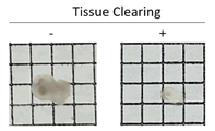
도 1은 본 발명의 일 구현예에 따른 투명화 용액을 이용한 오가노이드의 투명화 결과를 나타낸 도면이다.
도 2는 본 발명의 일 구현예에 따른 투명화 용액을 이용한 오가노이드의 면역염색 결과를 나타낸 도면이다.1 is a view showing the results of clearing organoids using a clearing solution according to an embodiment of the present invention.
2 is a view showing the results of immunostaining of organoids using a clearing solution according to an embodiment of the present invention.
본 발명은 수크로오스(sucrose)를 포함하는 고정 용액(fixing solution);The present invention is a fixing solution containing sucrose (sucrose);
3-[(3-콜아미도프로필)디메틸암모니오]-1-프로판설포네이트(3-[(3-Cholamidopropyl)dimethylammonio]-1-propanesulfonate, CHAPS), 우레아(urea), 및 염화나트륨(NaCl)으로 이루어진 군으로부터 선택된 하나 이상을 포함하는 조직 클리어링 용액(tissue clearing solution);3-[(3-Cholamidopropyl)dimethylammonio]-1-propanesulfonate (3-[(3-Cholamidopropyl)dimethylammonio]-1-propanesulfonate, CHAPS), urea, and sodium chloride (NaCl) Tissue clearing solution comprising one or more selected from the group consisting of (tissue clearing solution);
인산완충식염수(phosphate buffer saline; PBS) 및 아지드화나트륨(sodium azide)으로 이루어진 군으로부터 선택된 하나 이상을 포함하는 세척 용액(washing solution); 및a washing solution comprising at least one selected from the group consisting of phosphate buffer saline (PBS) and sodium azide; and
3-[(3-콜아미도프로필)디메틸암모니오]-1-프로판설포네이트(3-[(3-Cholamidopropyl)dimethylammonio]-1-propanesulfonate, CHAPS), 우레아(urea), 및 염화나트륨(NaCl)으로 이루어진 군으로부터 선택된 하나 이상을 포함하는 마운팅 용액(mounting solution)을 포함하는 것을 특징으로 하는, 오가노이드 투명화 용액 키트를 제공한다.3-[(3-Cholamidopropyl)dimethylammonio]-1-propanesulfonate (3-[(3-Cholamidopropyl)dimethylammonio]-1-propanesulfonate, CHAPS), urea, and sodium chloride (NaCl) It provides an organoid clearing solution kit, characterized in that it comprises a mounting solution containing one or more selected from the group consisting of.
또한, 본 발명은 하기 단계를 포함하는, 오가노이드 투명화 방법을 제공한다:The present invention also provides a method for clearing organoids comprising the steps of:
(a) 고정 용액으로 오가노이드 시료를 고정시키는 단계;(a) fixing the organoid sample with a fixing solution;
(b) 조직 클리어링 용액으로 상기 고정된 오가노이드 시료를 투명화 시키는 단계;(b) clearing the fixed organoid sample with a tissue clearing solution;
(c) 세척 용액으로 상기 투명화한 시료에 부착되어 있는 유기물을 씻어내는 단계; 및(c) washing the organic matter adhering to the transparent sample with a washing solution; and
(d) 마운팅 용액으로 상기 세척된 시료를 고정시키는 단계.(d) fixing the washed sample with a mounting solution.
또한, 본 발명은 하기 단계를 포함하는, 오가노이드의 3차원 이미지화를 위한 면역염색 방법을 제공한다:The present invention also provides an immunostaining method for three-dimensional imaging of organoids, comprising the steps of:
(a) 고정 용액으로 오가노이드 시료를 고정시키는 단계;(a) fixing the organoid sample with a fixing solution;
(b) 조직 클리어링 용액으로 상기 고정된 오가노이드 시료를 투명화 시키는 단계;(b) clearing the fixed organoid sample with a tissue clearing solution;
(c) 세척 용액으로 상기 투명화한 시료에 부착되어 있는 유기물을 씻어내는 단계; (c) washing the organic matter adhering to the transparent sample with a washing solution;
(d) 상기 세척된 시료에 형광물질이 부착된 항체를 처리하는 단계; 및(d) treating the washed sample with an antibody to which a fluorescent material is attached; and
(e) 마운팅 용액으로 상기 세척된 시료를 고정시키는 단계.(e) fixing the washed sample with a mounting solution.
본 발명에 있어서, "오가노이드(organoid)"는 3D 입체구조를 가지는 세포덩어리를 의미하며, 동물 등에서 수집, 취득하지 않은 인공적인 배양 과정을 통하여 제조한 축소되고 단순화된 버전의 기관을 의미한다. 이를 구성하는 세포의 유래는 제한되지 않으며, 예컨대 오가노이드는 조직, 배아줄기세포 또는 유도만능줄기세포에서 파생될 수 있고, 자가재생 및 분화능력으로 인해 3차원으로 배양될 수 있다. 상기 오가노이드는 세포의 성장 과정에서 주변 환경과 상호 작용하도록 허용되는 환경을 가질 수 있다. 이에 따라 본 발명에서 3D 오가노이드는 실제로 생체 내에서 상호 작용을 하고 있는 장기를 거의 완벽히 모사하여, 질병의 치료제 개발 및 기전 등을 관찰할 수 있는 훌륭한 모델이 될 수 있다.In the present invention, "organoid" refers to a cell mass having a 3D three-dimensional structure, and refers to a reduced and simplified version of an organ prepared through an artificial culture process that is not collected or acquired from animals. The origin of the cells constituting it is not limited, for example, organoids may be derived from tissues, embryonic stem cells or induced pluripotent stem cells, and may be cultured in three dimensions due to their self-renewal and differentiation ability. The organoid may have an environment that is allowed to interact with the surrounding environment during the cell growth process. Accordingly, in the present invention, the 3D organoid can be an excellent model for observing the development and mechanism of therapeutic agents for diseases by simulating almost completely the organs that actually interact in vivo.
본 발명에 있어서, 상기 오가노이드는 뇌 오가노이드, 간 오가노이드, 결장 오가노이드, 소장 오가노이드, 폐 오가노이드, 내피 오가노이드, 심장 오가노이드, 신장 오가노이드, 혈관 오가노이드, 및 상피 오가노이드로 이루어진 군으로부터 선택되는 하나 이상일 수 있으며, 본 발명의 일 실시예에 따르면 뇌 오가노이드일 수 있으나, 이에 제한되지 않는다.In the present invention, the organoids include brain organoids, liver organoids, colon organoids, small intestine organoids, lung organoids, endothelial organoids, cardiac organoids, kidney organoids, vascular organoids, and epithelial organoids. It may be one or more selected from the group consisting of, but may be a brain organoid according to an embodiment of the present invention, but is not limited thereto.
본 발명에 있어서, 상기 오가노이드는 배아줄기세포 또는 유도만능줄기세포 유래일 수 있으며, 본 발명의 일 실시예에 따르면 배아줄기세포 유래일 수 있으나, 이에 제한되지 않는다. 이 때, 상기 배아줄기세포는 예컨대 인간 배아줄기세포 또는 마우스 배아줄기세포일 수 있으나, 이의 종류에 제한은 없다. 본 발명에 있어서, 상기 세포를 배양하는 방법은 특별히 제한되지 아니하며, 당해 기술분야에서 통상적으로 사용하는 방법을 통해 배양될 수 있다. In the present invention, the organoid may be derived from embryonic stem cells or induced pluripotent stem cells, and according to an embodiment of the present invention may be derived from embryonic stem cells, but is not limited thereto. In this case, the embryonic stem cells may be, for example, human embryonic stem cells or mouse embryonic stem cells, but the type thereof is not limited. In the present invention, the method for culturing the cells is not particularly limited, and may be cultured by a method commonly used in the art.
본 발명에 있어서, "면역조직화학법" 또는 "면역염색"은 세포 또는 조직 등에서 특정한 단백질의 국재를 연구하는 방법으로 개발한 항체를 사용하는 염색법을 말한다. 1차 항체에 형광색소 등의 형광물질을 부가한 2차 항체를 작용시켜 콘포칼로 관찰한다. 세포 또는 조직을 고정하여 관찰하기 때문에 통상 얻게 되는 정보는 정적인 것이지만 형광색이 다른 2차 항체를 사용하면, 복수인 단백질의 공동국재도 연구할 수 있는 점에서 분자세포생물학에 있어서도 매우 중요한 기술로 인식되고 있다. 본 발명에 있어서, 상기 면역조직화학법에 사용되는 항체의 종류에는 제한이 없으며, 예컨대 본 발명의 일 실시예에 따르면 1차 항체로 뉴런의 핵 바이오마커인 NeuN에 대한 항체(항-NeuN 항체) 및 베타-III 튜불린(beta-III tubulin)에 대한 항체(항-Tuj1 항체)를 이용하고, 2차 항체로 Goat anti rabbit(alexa fluor® 647) 및 Goat anti mouse(alexa fluor® 488)을 이용할 수 있다. In the present invention, "immunohistochemistry" or "immunostaining" refers to a staining method using an antibody developed as a method for studying the localization of a specific protein in cells or tissues. Confocal observation is made by applying a secondary antibody to which a fluorescent substance such as a fluorescent dye is added to the primary antibody. Since cells or tissues are fixed and observed, the information usually obtained is static, but using secondary antibodies with different fluorescence colors can also study the colocalization of multiple proteins, which is recognized as a very important technology in molecular cell biology. is becoming In the present invention, the type of antibody used in the immunohistochemistry is not limited, for example, according to an embodiment of the present invention, as a primary antibody, an antibody against NeuN, a nuclear biomarker of neurons (anti-NeuN antibody) and beta-III tubulin (beta-III tubulin) using an antibody (anti-Tuj1 antibody), and Goat anti rabbit (alexa fluor ® 647) and Goat anti mouse (alexa fluor ® 488) as secondary antibodies. can
본 발명에 있어서, “항체”는 항원에 특이적으로 결합하여 인식하는 면역글로불린 유전자 또는 이의 단편에서 유래하는 프레임워크 영역을 포함하는 폴리펩티드를 의미한다. 인식된 면역글로불린 유전자는 카파, 람다, 알파, 감마, 델타, 엡실론, 및 뮤 불변 영역 유전자를 비롯하여, 무수한 면역글로불린 가변 영역 유전자를 포함한다. 경쇄는 카파 또는 람다로 분류되며, 중쇄는 감마, 뮤, 알파, 델타, 또는 엡실론으로 분류되고, 결과적으로 각각 면역글로불린 부류 IgG, IgM, IgA, IgD 및 IgE를 한정한다. 전형적으로, 항체의 항원-결합 영역은 결합 특이성 및 친화성에서 가장 핵심적이게 된다. 일부 실시형태에서, 항체 또는 항체의 단편은 인간, 마우스, 래트, 염소, 토끼, 햄스터, 낙타 등을 포함한, 상이한 유기체에서 유래될 수 있다.In the present invention, "antibody" refers to a polypeptide comprising a framework region derived from an immunoglobulin gene or fragment thereof that specifically binds to and recognizes an antigen. Recognized immunoglobulin genes include numerous immunoglobulin variable region genes, including kappa, lambda, alpha, gamma, delta, epsilon, and mu constant region genes. Light chains are classified as kappa or lambda, and heavy chains are classified as gamma, mu, alpha, delta, or epsilon, consequently defining the immunoglobulin classes IgG, IgM, IgA, IgD and IgE, respectively. Typically, the antigen-binding region of an antibody becomes most critical in binding specificity and affinity. In some embodiments, antibodies or fragments of antibodies may be from different organisms, including humans, mice, rats, goats, rabbits, hamsters, camels, and the like.
본 발명에 있어서, "형광물질"은 특정한 파장의 빛을 흡수하여 검출 가능한 영역의 빛을 방출하는 물질을 의미하며, 예컨대 형광 단백질, 형광 화합물 등이 포함되고, 상기 검출 가능한 영역이라 함은 육안 또는 다양한 계측기기를 사용하여 방출되는 빛의 수준을 정량할 수 있는 영역을 의미한다. 본 발명에 있어서 상기 형광물질의 종류에는 제한이 없다.In the present invention, "fluorescent material" refers to a material that absorbs light of a specific wavelength and emits light in a detectable region, for example, includes a fluorescent protein, a fluorescent compound, and the like, and the detectable region means the human eye or It refers to an area where the level of emitted light can be quantified using various measuring instruments. In the present invention, the type of the fluorescent material is not limited.
본 발명에 있어서, 상기 고정 용액에서 수크로오스(sucrose)는 농도가 20%(w/v) 내지 50%(w/v), 20%(w/v) 내지 40%(w/v), 20%(w/v) 내지 35%(w/v), 35%(w/v) 내지 40%(w/v), 또는 30%(w/v) 내지 50%(w/v)일 수 있다. 본 발명의 구체적인 실시예에 따르면 수크로오스의 농도를 35%(w/v)로 하여 시료를 탈수시키고 PFA(paraformaldehyde)로 유기물간 공유결합된 시료를 좀 더 강하게 고정할 수 있으나, 상기 농도에 한정되는 것은 아니다. In the present invention, the concentration of sucrose in the fixing solution is 20% (w/v) to 50% (w/v), 20% (w/v) to 40% (w/v), 20% (w/v) to 35% (w/v), 35% (w/v) to 40% (w/v), or 30% (w/v) to 50% (w/v). According to a specific embodiment of the present invention, the concentration of sucrose is 35% (w/v), the sample is dehydrated, and the sample covalently bonded between organic substances can be fixed more strongly with PFA (paraformaldehyde), but the concentration is limited it is not
본 발명에 있어서, 상기 조직 클리어링 용액에서 CHAPS는 농도가 10%(w/v) 내지 40%(w/v) 또는 10%(w/v) 내지 30%(w/v)일 수 있고, 우레아는 농도가 30%(w/v) 내지 70%(w/v) 또는 40%(w/v) 내지 60%(w/v)일 수 있고, 염화나트륨은 농도가 0.001%(w/v) 내지 1%(w/v), 0.01%(w/v) 내지 1%(w/v), 또는 0.01%(w/v) 내지 0.5%(w/v)일 수 있다. 본 발명의 구체적인 실시예에 따르면 상기 조직 클리어링 용액은 20%(w/v)의 CHAPS, 50%(w/v)의 우레아를 포함할 수 있고, 0.1%(w/v)의 염화나트륨을 포함하여 삼투압에 의한 조직의 변형과 ion strength를 안정화시켜 시료에 있는 형광 물질의 변성을 최소화 할 수 있으나, 상기 농도에 한정되는 것은 아니다.In the present invention, the concentration of CHAPS in the tissue clearing solution may be 10% (w/v) to 40% (w/v) or 10% (w/v) to 30% (w/v), urea may have a concentration of 30% (w/v) to 70% (w/v) or 40% (w/v) to 60% (w/v), and sodium chloride has a concentration of 0.001% (w/v) to 1% (w/v), 0.01% (w/v) to 1% (w/v), or 0.01% (w/v) to 0.5% (w/v). According to a specific embodiment of the present invention, the tissue clearing solution may contain 20% (w/v) of CHAPS, 50% (w/v) of urea, and 0.1% (w/v) of sodium chloride. It is possible to minimize the denaturation of the fluorescent material in the sample by stabilizing the tissue deformation and ion strength by osmotic pressure, but is not limited to the above concentration.
본 발명에 있어서, 상기 세척 용액에서 아지드화나트륨(sodium azide)은 농도가 0.001%(w/v) 내지 1%(w/v), 0.01%(w/v) 내지 1%(w/v), 또는 0.01%(w/v) 내지 0.5%(w/v)일 수 있다. 본 발명의 구체적인 실시예에 따르면, 상기 세척 용액은 0.1%(w/v)의 아지드화나트륨(sodium azide)을 포함하여 투명화 후 탈수된 오가노이드 시료에 최대 30%까지 함수율을 증가시킨 후 15%를 탈수하여 조직에 붙어있는 이미징에 방해되는 유기물을 세척할 수 있으나, 상기 농도에 한정되는 것은 아니다.In the present invention, the concentration of sodium azide in the washing solution is 0.001% (w/v) to 1% (w/v), 0.01% (w/v) to 1% (w/v) ), or 0.01% (w/v) to 0.5% (w/v). According to a specific embodiment of the present invention, the washing solution contains 0.1% (w/v) of sodium azide after increasing the moisture content to up to 30% in the dehydrated organoid sample after clearing 15 % may be dehydrated to wash organic matter adhering to the tissue that interferes with imaging, but is not limited to the above concentration.
본 발명에 있어서, 상기 마운팅 용액에서 CHAPS는 농도가 20%(w/v) 내지 60%(w/v) 또는 30%(w/v) 내지 50%(w/v)일 수 있고, 우레아(urea)는 농도가 10%(w/v) 내지 50%(w/v) 또는 20%(w/v) 내지 50%(w/v)일 수 있고, 염화나트륨(NaCl)은 농도가 0.001%(w/v) 내지 1%(w/v), 0.01%(w/v) 내지 1%(w/v), 또는 0.01%(w/v) 내지 0.5%(w/v)일 수 있다. 본 발명의 구체적인 실시예에 따르면 상기 마운팅 용액의 굴절률을 1.45로 맞추기 위해 각각 40%(w/v), 40%(w/v)의 CHAPS 및 우레아를 포함할 수 있으며, 0.1%(w/v)의 염화나트륨(NaCl)을 포함할 수 있으나, 상기 농도에 제한되는 것은 아니다. In the present invention, the concentration of CHAPS in the mounting solution may be 20% (w/v) to 60% (w/v) or 30% (w/v) to 50% (w/v), and urea ( urea) may have a concentration of 10% (w/v) to 50% (w/v) or 20% (w/v) to 50% (w/v), and sodium chloride (NaCl) may have a concentration of 0.001% ( w/v) to 1% (w/v), 0.01% (w/v) to 1% (w/v), or 0.01% (w/v) to 0.5% (w/v). According to a specific embodiment of the present invention, in order to adjust the refractive index of the mounting solution to 1.45, 40% (w/v) and 40% (w/v) of CHAPS and urea may be included, respectively, and 0.1% (w/v) ) may include sodium chloride (NaCl), but is not limited to the above concentration.
본 발명의 일 실시예에서는, 오가노이드의 투명화를 위한 고정 용액, 조직 클리어링 용액, 세척 용액, 및 마운팅 용액을 포함하는 오가노이드의 투명화 용액 키트를 제조하였으며(실시예 1 및 표 1 참조), 이를 이용하여 오가노이드를 투명화 할 경우 투명도가 높은 것을 확인함으로써 상기 제조한 투명화 용액 키트의 조성이 오가노이드 투명화 및 3차원 형광 이미지화를 위한 최적의 조건임을 확인하였다 (실시예 2 참조).In one embodiment of the present invention, an organoid clearing solution kit including a fixation solution, a tissue clearing solution, a washing solution, and a mounting solution for clearing the organoid was prepared (see Example 1 and Table 1). It was confirmed that the composition of the prepared clearing solution kit was the optimal condition for organoid clearing and three-dimensional fluorescence imaging by confirming that the transparency was high when organoids were made transparent by using the same (see Example 2).
본 발명의 다른 실시예에서는, 상기 투명화 용액 키트를 이용하여 오가노이드를 투명화하고 면역염색을 실시한 결과, 높은 해상도의 3차원 면역염색 이미지를 획득할 수 있음을 확인하였다(실시예 3 참조). In another embodiment of the present invention, it was confirmed that a high-resolution three-dimensional immunostaining image could be obtained as a result of immunostaining and transparent organoids using the clearing solution kit (see Example 3).
본원 명세서 전체에서 "포함하는" 이라는 용어가 사용될 때, 이는 특별히 반대되는 기재가 없는 한 다른 구성 요소를 제외하는 것이 아니라 다른 구성 요소를 더 포함할 수 있는 것을 의미한다. 본원 명세서 전체에서 사용되는 정도의 용어 “~(하는) 단계” 또는 “~의 단계”는 “~ 를 위한 단계”를 의미하지 않는다.When the term “comprising” is used throughout this specification, it means that other components may be further included, rather than excluding other components, unless otherwise stated. As used throughout this specification, the term “step of (to)” or “step of” does not mean “step for”.
또한, 본 발명에서 "~으로 이루어진" 이라는 용어는 "포함하는" 이라는 용어의 바람직한 실시양태인 것으로 간주된다.Also, in the present invention, the term “consisting of” is considered to be a preferred embodiment of the term “comprising”.
이하, 본 발명의 이해를 돕기 위하여 바람직한 실시예를 제시한다. 그러나 하기의 실시예는 본 발명을 보다 쉽게 이해하기 위하여 제공되는 것일 뿐, 하기 실시예에 의해 본 발명의 내용이 한정되는 것은 아니다.Hereinafter, preferred examples are presented to help the understanding of the present invention. However, the following examples are only provided for easier understanding of the present invention, and the contents of the present invention are not limited by the following examples.
실시예 1. 고정 용액, 조직 클리어링 용액, 세척 용액, 및 마운팅 용액의 제조Example 1. Preparation of Fixation Solution, Tissue Clearing Solution, Washing Solution, and Mounting Solution
오가노이드의 투명화를 위해 필요한 고정 용액(fixing solution), 조직 클리어링 용액(tissue clearing solution), 세척 용액(washing solution), 및 마운팅 용액(mounting solution)을 제조하여 이들을 포함하는 오가노이드 투명화 용액 키트를 제조하였으며, 각 용액의 구성 성분은 하기 표 1에 나타내었다.Prepare an organoid clearing solution kit including a fixing solution, a tissue clearing solution, a washing solution, and a mounting solution necessary for clearing the organoid and the components of each solution are shown in Table 1 below.
구체적으로, 오가노이드 시료를 PFA(paraformaldehyde)로 고정시키고 유기물간 공유결합된 시료를 좀 더 강하게 고정시키기 위해, 농도가 35%(w/v)인 수크로오스(sucrose)를 구성성분으로 하는 고정 용액(fixing solution)을 제조하였다. Specifically, in order to fix the organoid sample with PFA (paraformaldehyde) and to fix the sample covalently bonded between organic substances more strongly, a fixing solution containing sucrose with a concentration of 35% (w/v) as a component ( fixing solution) was prepared.
또한, 삼투압에 의한 조직의 변형과 ion strength를 안정화시켜 오가노이드 시료에 있는 형광 물질의 변성을 최소화 하기 위해, 20%(w/v) CHAPS(3-[(3-콜아미도프로필)디메틸암모니오]-1-프로판설포네이트) 및 50%(w/v) 우레아(urea)에 0.1%(w/v) 농도의 염화나트륨(NaCl)을 추가하여, 본 발명의 조직 클리어링 용액(tissue clearing solution)을 제조하였다. In addition, 20% (w/v) CHAPS (3-[(3-cholamidopropyl) dimethylammonium o]-1-propanesulfonate) and 50% (w/v) urea by adding 0.1% (w/v) sodium chloride (NaCl) to the tissue clearing solution of the present invention was prepared.
이어서, 투명화 후 탈수된 오가노이드 시료에 최대 30%까지 함수율을 증가시킨 후 15%를 탈수하여 오가노이드 시료에 붙어있는 이미징에 방해되는 유기물을 세척시킬 수 있도록 인산완충식염수(phosphate buffer saline; PBS)에 0.1%(w/v)의 아지드화나트륨(sodium azide)을 첨가하여 세척 용액(washing solution)을 제조하였다. Then, after clearing, increase the moisture content to up to 30% in the dehydrated organoid sample, and then dehydrate 15% to wash the organic matter adhering to the organoid sample that interferes with imaging. A washing solution was prepared by adding 0.1% (w/v) sodium azide to it.
또한, 40%(w/v)의 CHAPS(3-[(3-콜아미도프로필)디메틸암모니오]-1-프로판설포네이트), 40%(w/v) 우레아(urea), 및 0.1%(w/v) 염화나트륨(NaCl)으로 마운팅 용액(mounting solution)을 제조하여 굴절률을 1.45로 맞추었다.Also, 40% (w/v) CHAPS (3-[(3-cholamidopropyl)dimethylammonio]-1-propanesulfonate), 40% (w/v) urea, and 0.1% A mounting solution was prepared with (w/v) sodium chloride (NaCl) and the refractive index was adjusted to 1.45.
실시예 2. 오가노이드 투명화 및 우수한 3차원 이미지 획득을 위한 용액의 최적의 배합 조건 확인Example 2. Confirmation of optimal mixing conditions of solution for organoid transparency and excellent three-dimensional image acquisition
상기 실시예 1에서 제조한 고정 용액, 조직 클리어링 용액, 세척 용액, 및 마운팅 용액을 각각 이용하여 오가노이드를 투명화하였다.Organoids were made transparent by using the fixative solution, tissue clearing solution, washing solution, and mounting solution prepared in Example 1, respectively.
구체적으로, 배아줄기세포 유래 세포를 170일 동안 배양하여 제조한 뇌 오가노이드를 이용하여 실험을 진행하였으며, 오가노이드 시료를 1 X PBS로 4℃에서 20분동안 3회에 걸쳐 세척한 다음, 고정 용액(fixing solution)에 넣고 4℃/50 rpm에서 침전될 때까지 밤새 진탕 배양(shaking incubation)하였다. 침전된 상기 오가노이드 시료를 조직 클리어링 용액(tissue clearing solution)에 넣고 37℃/50 rpm으로 1일 동안 진탕 배양하고 세척 용액으로(washing solution) 4℃에서 1시간씩 3회 세척하였다. 상기 세척이 끝난 오가노이드 시료는 마운팅 용액(mounting solution)에 넣고 37℃/50 rpm으로 최소 12시간 이상 진탕 배양하였다. Specifically, the experiment was conducted using brain organoids prepared by culturing embryonic stem cell-derived cells for 170 days, and the organoid samples were washed three times with 1 X PBS at 4°C for 20 minutes, and then fixed. It was placed in a fixing solution and incubated with shaking overnight until precipitated at 4°C/50 rpm. The precipitated organoid sample was placed in a tissue clearing solution, incubated with shaking at 37° C./50 rpm for 1 day, and washed three times with a washing solution at 4° C. for 1 hour each. The washed organoid samples were placed in a mounting solution and incubated with shaking at 37° C./50 rpm for at least 12 hours.
상기 과정을 통해 투명화한 오가노이드를 관찰한 결과, 도 1에 나타낸 바와 같이 상기 실시예 1에서 제조한 오가노이드 투명화 용액을 사용한 경우(우측 도면) 투명화 하기 전보다(좌측 도면) 높은 투명도로 오가노이드가 투명화된 것을 확인하여, 상기 표 1에 나타낸 용액의 조성이 오가노이드 투명화 및 3차원 형광 이미지 획득을 위한 최적의 조건임을 확인하였다.As a result of observing the organoids cleared through the above process, as shown in FIG. 1 , when the organoid clearing solution prepared in Example 1 was used (right drawing), the organoids with higher transparency than before transparent (left drawing) It was confirmed that the solution was transparent, and it was confirmed that the composition of the solution shown in Table 1 was the optimal condition for organoid transparency and three-dimensional fluorescence image acquisition.
실시예 3. 면역조직화학법에 의한 3차원 이미지 획득Example 3. 3D image acquisition by immunohistochemistry
1차 항체로 뉴런의 핵 바이오마커인 NeuN에 대한 항체(항-NeuN 항체) 및 베타-III 튜불린(beta-III tubulin)에 대한 항체(항-Tuj1 항체)를 이용하고, 2차 항체로 Goat anti rabbit(alexa fluor® 647)(Jackson, Fab) 및 Goat anti mouse(alexa fluor® 488)(Jackson, Fab)을 이용하여 면역조직화학적 염색을 실시하였다. An antibody against NeuN (anti-NeuN antibody) and beta-III tubulin (anti-Tuj1 antibody), which is a nuclear biomarker of neurons, were used as the primary antibody, and Goat as the secondary antibody Immunohistochemical staining was performed using anti rabbit (alexa fluor ® 647) (Jackson, Fab) and Goat anti mouse (alexa fluor ® 488) (Jackson, Fab).
구체적으로, 오가노이드 시료를 1 X PBS로 4℃에서 20분씩 3회에 걸쳐 세척한 다음, 상기 실시예 1에서 제조한 고정 용액(fixing solution)에 넣고 4℃/50 rpm에서 침전될 때까지 밤새 진탕 배양(shaking incubation)하였다. 침전된 상기 오가노이드 시료를 조직 클리어링 용액(tissue clearing solution)으로 42℃/50 rpm에서 3일 동안 배양한 다음, 세척 용액(washing solution)으로 4℃에서 1시간씩 3번 세척하고, 다시 한번 더 조직 클리어링 용액(tissue clearing solution)으로 42℃/50 rpm에서 3일 동안 반응시켰다. 그런 다음, 항체가 잘 침투할 수 있도록, 0.2% Triton X-100 및 20% DMSO가 포함된 1 X TBS로 permeabilization 과정을 37℃/50 rpm에서 3일 동안 진행하였다. 상기 과정을 마친 시료에 1차 항체로 0.2% Tween20 및 5% DMSO가 포함된 1 X TBS 용액에 1:100으로 희석시킨 항-NeuN 항체 + 항-Tuj1 항체를 처리하여 37℃/50 rpm에서 4일 동안 배양한 후, 1 X TBS 용액으로 4℃에서 20분씩 3회에 걸쳐 세척하였다. 그리고 나서, 2차 항체로 0.2% Tween20 및 5% DMSO가 포함된 1 X TBS 용액에 1:100으로 희석시킨 Goat anti rabbit(alexa fluor® 647) 및 Goat anti mouse(alexa fluor® 488)을 처리하고 37℃/50 rpm에서 4일 동안 배양한 후, 다시 1 X TBS 용액으로 4℃에서 20분씩 3회에 걸쳐 세척하였다. 세척 후 세포 내 핵은 1:50으로 희석된 DAPI를 4℃에서 1시간 동안 처리하여 염색하였고, 또 다시 1 X TBS 용액으로 4℃에서 20분씩 3회에 걸쳐 세척하였다. 상기 과정을 마친 시료에 마운팅 용액을 처리하고 37℃에서 3일 동안 배양하여 고정시킨 다음, 공초점 현미경(confocal microscope) 촬영을 통해 면역염색 결과를 관찰하였다.Specifically, the organoid sample was washed three times with 1 X PBS at 4° C. for 20 minutes each, and then placed in the fixing solution prepared in Example 1, and overnight until precipitated at 4° C./50 rpm. Shaking incubation was performed. The precipitated organoid sample was incubated with a tissue clearing solution at 42° C./50 rpm for 3 days, then washed 3 times with a washing solution at 4° C. for 1 hour each, and once again It was reacted with a tissue clearing solution at 42° C./50 rpm for 3 days. Then, to allow the antibody to penetrate well, the permeabilization process was performed with 1 X TBS containing 0.2% Triton X-100 and 20% DMSO at 37° C./50 rpm for 3 days. The sample after the above process was treated with anti-NeuN antibody + anti-Tuj1 antibody diluted 1:100 in 1 X TBS solution containing 0.2% Tween20 and 5% DMSO as a primary antibody at 37°C/50 rpm. After incubation for one day, it was washed three times with 1 X TBS solution at 4° C. for 20 minutes each. Then, as a secondary antibody, Goat anti rabbit (alexa fluor ® 647) and Goat anti mouse (alexa fluor ® 488) diluted 1:100 in 1 X TBS solution containing 0.2% Tween20 and 5% DMSO were treated and After culturing for 4 days at 37° C./50 rpm, it was washed 3 times with 1 X TBS solution at 4° C. for 20 minutes each. After washing, the intracellular nuclei were stained by treatment with 1:50 diluted DAPI at 4°C for 1 hour, and again washed 3 times with 1 X TBS solution at 4°C for 20 minutes each. The sample after the above process was treated with a mounting solution, incubated at 37° C. for 3 days and fixed, and then the results of immunostaining were observed by imaging under a confocal microscope.
그 결과, 도 2에 나타낸 바와 같이, NeuN 및 β III tubulin이 각각 붉은색, 녹색 형광으로 선명하게 나타난 것을 확인하였다. As a result, as shown in FIG. 2 , it was confirmed that NeuN and β III tubulin were clearly displayed in red and green fluorescence, respectively.
이로부터 상기 표 1에 나타낸 본 발명의 투명화 용액을 오가노이드를 면역염색 하는데 사용할 수 있으며, 상기 용액을 이용하여 오가노이드를 면역염색 할 경우 선명한 3차원 형광 이미지를 획득할 수 있음을 알 수 있었다.From this, it can be seen that the clearing solution of the present invention shown in Table 1 can be used to immunostain organoids, and a clear three-dimensional fluorescence image can be obtained when the organoids are immunostained using the solution.
전술한 본 발명의 설명은 예시를 위한 것이며, 본 발명이 속하는 기술분야의 통상의 지식을 가진 자는 본 발명의 기술적 사상이나 필수적인 특징을 변경하지 않고서 다른 구체적인 형태로 쉽게 변형이 가능하다는 것을 이해할 수 있을 것이다. 그러므로 이상에서 기술한 실시예들은 모든 면에서 예시적인 것이며 한정적이 아닌 것으로 이해해야 한다.The above description of the present invention is for illustration, and those of ordinary skill in the art to which the present invention pertains can understand that it can be easily modified into other specific forms without changing the technical spirit or essential features of the present invention. will be. Therefore, it should be understood that the embodiments described above are illustrative in all respects and not restrictive.
Claims (9)
3-[(3-콜아미도프로필)디메틸암모니오]-1-프로판설포네이트(3-[(3-Cholamidopropyl)dimethylammonio]-1-propanesulfonate, CHAPS), 우레아(urea), 및 염화나트륨(NaCl)으로 이루어진 군으로부터 선택된 하나 이상을 포함하는 조직 클리어링 용액(tissue clearing solution);
인산완충식염수(phosphate buffer saline; PBS) 및 아지드화나트륨(sodium azide)으로 이루어진 군으로부터 선택된 하나 이상을 포함하는 세척 용액(washing solution); 및
3-[(3-콜아미도프로필)디메틸암모니오]-1-프로판설포네이트(3-[(3-Cholamidopropyl)dimethylammonio]-1-propanesulfonate, CHAPS), 우레아(urea), 및 염화나트륨(NaCl)으로 이루어진 군으로부터 선택된 하나 이상을 포함하는 마운팅 용액(mounting solution)을 포함하는 것을 특징으로 하는, 오가노이드 투명화 용액 키트.
a fixing solution containing sucrose;
3-[(3-Cholamidopropyl)dimethylammonio]-1-propanesulfonate (3-[(3-Cholamidopropyl)dimethylammonio]-1-propanesulfonate, CHAPS), urea, and sodium chloride (NaCl) Tissue clearing solution comprising one or more selected from the group consisting of (tissue clearing solution);
a washing solution comprising at least one selected from the group consisting of phosphate buffer saline (PBS) and sodium azide; and
3-[(3-Cholamidopropyl)dimethylammonio]-1-propanesulfonate (3-[(3-Cholamidopropyl)dimethylammonio]-1-propanesulfonate, CHAPS), urea, and sodium chloride (NaCl) Organoid clearing solution kit, characterized in that it comprises a mounting solution (mounting solution) comprising at least one selected from the group consisting of.
상기 고정 용액에서 수크로오스는 농도가 20%(w/v) 내지 50%(w/v)인 것을 특징으로 하는, 오가노이드 투명화 용액 키트.
According to claim 1,
Sucrose in the fixing solution is characterized in that the concentration of 20% (w / v) to 50% (w / v), organoid clearing solution kit.
상기 조직 클리어링 용액에서 CHAPS는 농도가 10%(w/v) 내지 40%(w/v)이고, 우레아는 농도가 30%(w/v) 내지 70%(w/v)이고, 염화나트륨은 농도가 0.001%(w/v) 내지 1%(w/v)인 것을 특징으로 하는, 오가노이드 투명화 용액 키트.
According to claim 1,
In the tissue clearing solution, the concentration of CHAPS is 10% (w/v) to 40% (w/v), the concentration of urea is 30% (w/v) to 70% (w/v), and the concentration of sodium chloride is is 0.001% (w / v) to 1% (w / v), organoid clearing solution kit.
상기 세척 용액에서 아지드화나트륨은 농도가 0.001%(w/v) 내지 1%(w/v)인 것을 특징으로 하는, 오가노이드 투명화 용액 키트.
According to claim 1,
Sodium azide in the washing solution is characterized in that the concentration of 0.001% (w / v) to 1% (w / v), organoid clearing solution kit.
상기 마운팅 용액에서 CHAPS는 농도가 20%(w/v) 내지 60%(w/v)이고, 우레아는 농도가 20%(w/v) 내지 60%(w/v)이고, 염화나트륨은 농도가 0.001%(w/v) 내지 1%(w/v)인 것을 특징으로 하는, 오가노이드 투명화 용액 키트.
According to claim 1,
In the mounting solution, CHAPS has a concentration of 20% (w/v) to 60% (w/v), urea has a concentration of 20% (w/v) to 60% (w/v), and sodium chloride has a concentration of 20% (w/v) to 60% (w/v) 0.001% (w / v) to 1% (w / v), characterized in that the organoid clearing solution kit.
상기 오가노이드는 뇌 오가노이드, 간 오가노이드, 결장 오가노이드, 소장 오가노이드, 폐 오가노이드, 내피 오가노이드, 심장 오가노이드, 신장 오가노이드, 혈관 오가노이드, 및 상피 오가노이드로 이루어진 군으로부터 선택되는 하나 이상인 것을 특징으로 하는, 오가노이드 투명화 용액 키트.
According to claim 1,
wherein said organoid is selected from the group consisting of brain organoids, liver organoids, colon organoids, small intestine organoids, lung organoids, endothelial organoids, cardiac organoids, renal organoids, vascular organoids, and epithelial organoids An organoid clearing solution kit, characterized in that at least one.
상기 오가노이드는 배아줄기세포 또는 유도만능줄기세포 유래인 것을 특징으로 하는, 오가노이드 투명화 용액 키트.
According to claim 1,
The organoid is an organoid clearing solution kit, characterized in that it is derived from embryonic stem cells or induced pluripotent stem cells.
(a) 고정 용액으로 오가노이드 시료를 고정시키는 단계;
(b) 조직 클리어링 용액으로 상기 고정된 오가노이드 시료를 투명화 시키는 단계;
(c) 세척 용액으로 상기 투명화한 시료에 부착되어 있는 유기물을 씻어내는 단계; 및
(d) 마운팅 용액으로 상기 세척된 시료를 고정시키는 단계.
A method for clearing an organoid comprising the steps of:
(a) fixing the organoid sample with a fixing solution;
(b) clearing the fixed organoid sample with a tissue clearing solution;
(c) washing the organic matter adhering to the transparent sample with a washing solution; and
(d) fixing the washed sample with a mounting solution.
(a) 고정 용액으로 오가노이드 시료를 고정시키는 단계;
(b) 조직 클리어링 용액으로 상기 고정된 오가노이드 시료를 투명화 시키는 단계;
(c) 세척 용액으로 상기 투명화한 시료에 부착되어 있는 유기물을 씻어내는 단계;
(d) 상기 세척된 시료에 형광물질이 부착된 항체를 처리하는 단계; 및
(e) 마운팅 용액으로 상기 세척된 시료를 고정시키는 단계.Immunostaining method for three-dimensional imaging of organoids, comprising the steps of:
(a) fixing the organoid sample with a fixing solution;
(b) clearing the fixed organoid sample with a tissue clearing solution;
(c) washing the organic matter adhering to the transparent sample with a washing solution;
(d) treating the washed sample with an antibody to which a fluorescent material is attached; and
(e) fixing the washed sample with a mounting solution.
Priority Applications (1)
| Application Number | Priority Date | Filing Date | Title |
|---|---|---|---|
| KR1020200035842A KR20210119210A (en) | 2020-03-24 | 2020-03-24 | Organoid clearing kit, method of organoid clearing and immunostaining for 3-dimensional imaging using thereof |
Applications Claiming Priority (1)
| Application Number | Priority Date | Filing Date | Title |
|---|---|---|---|
| KR1020200035842A KR20210119210A (en) | 2020-03-24 | 2020-03-24 | Organoid clearing kit, method of organoid clearing and immunostaining for 3-dimensional imaging using thereof |
Publications (1)
| Publication Number | Publication Date |
|---|---|
| KR20210119210A true KR20210119210A (en) | 2021-10-05 |
Family
ID=78077776
Family Applications (1)
| Application Number | Title | Priority Date | Filing Date |
|---|---|---|---|
| KR1020200035842A KR20210119210A (en) | 2020-03-24 | 2020-03-24 | Organoid clearing kit, method of organoid clearing and immunostaining for 3-dimensional imaging using thereof |
Country Status (1)
| Country | Link |
|---|---|
| KR (1) | KR20210119210A (en) |
Cited By (1)
| Publication number | Priority date | Publication date | Assignee | Title |
|---|---|---|---|---|
| KR102376008B1 (en) | 2021-10-22 | 2022-03-17 | 이한석 | Hot-air heater by induction heating |
Citations (1)
| Publication number | Priority date | Publication date | Assignee | Title |
|---|---|---|---|---|
| KR20200018505A (en) | 2017-05-29 | 2020-02-19 | 스템셀 테크놀러지스 캐나다 인크. | Compositions and Methods for Obtaining Organoids |
-
2020
- 2020-03-24 KR KR1020200035842A patent/KR20210119210A/en not_active Application Discontinuation
Patent Citations (1)
| Publication number | Priority date | Publication date | Assignee | Title |
|---|---|---|---|---|
| KR20200018505A (en) | 2017-05-29 | 2020-02-19 | 스템셀 테크놀러지스 캐나다 인크. | Compositions and Methods for Obtaining Organoids |
Cited By (1)
| Publication number | Priority date | Publication date | Assignee | Title |
|---|---|---|---|---|
| KR102376008B1 (en) | 2021-10-22 | 2022-03-17 | 이한석 | Hot-air heater by induction heating |
Similar Documents
| Publication | Publication Date | Title |
|---|---|---|
| Suleiman et al. | Injury-induced actin cytoskeleton reorganization in podocytes revealed by super-resolution microscopy | |
| CN107209093B (en) | Transparentizing reagent for biomaterial, system and use thereof | |
| EP3252452A1 (en) | Method for imaging and analysis of a biological specimen | |
| JP6433901B2 (en) | Composition for preparing biological material with excellent light transmittance and use thereof | |
| CN110139922B (en) | Compositions and methods for clarifying tissue | |
| KR101866249B1 (en) | Composition for clrearing of biotissue and clarity method for biotissue using thereof | |
| EP2547999A1 (en) | Clearing reagent for biological material, and use thereof | |
| US11022528B2 (en) | Composition for biological tissue transparency and method for biological tissue transparency using same | |
| KR102296381B1 (en) | Composition for clrearing of biotissue and clarity method for biotissue using thereof | |
| US11365213B2 (en) | Composition for clearing spheroids, method for clearing spheroids using same, and kit comprising same | |
| KR102085373B1 (en) | Biological tissue clearing kit for imaging 3-dimensional fluorescence photograph and the method of biological tissue clearing using thereof | |
| KR20210119210A (en) | Organoid clearing kit, method of organoid clearing and immunostaining for 3-dimensional imaging using thereof | |
| US11391653B2 (en) | Composition for immunostaining cleared large tissues and method for immunostaining cleared large biological tissues | |
| Reed et al. | Culture of murine aortic explants in 3-dimensional extracellular matrix: a novel, miniaturized assay of angiogenesis in vitro | |
| JP6276487B1 (en) | How to observe sweat gland dynamics | |
| Klouda et al. | From 2D to 3D: promising advances in imaging lung structure | |
| KR102477568B1 (en) | A method of pretreatment of transparency of a biological samples with a size of 1 mm or less and a method of transparency of the biological samples comprising the same | |
| Sun et al. | A simple optical tissue clearing pipeline for 3D vasculature imaging of the mediastinal organs in mice | |
| Epperlein et al. | Immunohistochemical demonstration of hyaluronan and its possible involvement in axolotl neural crest cell migration | |
| KR102591021B1 (en) | Composition for clrearing of organoids, clarity method for organoid using the same | |
| Radtke et al. | IBEX: an open and extensible method for high content multiplex imaging of diverse tissues | |
| Carroll Jr et al. | Hyalin is a cell adhesion molecule involved in mediating archenteron–blastocoel roof attachment | |
| US20240142352A1 (en) | Biological-specimen transparentizing agent, system, and use therefor | |
| Vache | Structure and Mechanics of Neuronal Model Systems: Insights from Atomic Force Microscopy and Micropipette Aspiration | |
| Rubi-Sans et al. | Quantification of extracellular matrix components in immunolabeled tissue samples |
Legal Events
| Date | Code | Title | Description |
|---|---|---|---|
| E601 | Decision to refuse application |