KR20210087938A - CD8 imaging constructs and methods of use thereof - Google Patents
CD8 imaging constructs and methods of use thereof Download PDFInfo
- Publication number
- KR20210087938A KR20210087938A KR1020217012365A KR20217012365A KR20210087938A KR 20210087938 A KR20210087938 A KR 20210087938A KR 1020217012365 A KR1020217012365 A KR 1020217012365A KR 20217012365 A KR20217012365 A KR 20217012365A KR 20210087938 A KR20210087938 A KR 20210087938A
- Authority
- KR
- South Korea
- Prior art keywords
- subject
- tumor
- binding construct
- patient
- image
- Prior art date
Links
- 238000000034 method Methods 0.000 title claims abstract description 531
- 238000003384 imaging method Methods 0.000 title claims description 38
- 238000009739 binding Methods 0.000 claims abstract description 558
- 230000027455 binding Effects 0.000 claims abstract description 556
- 108091007433 antigens Proteins 0.000 claims abstract description 515
- 102000036639 antigens Human genes 0.000 claims abstract description 515
- 239000000427 antigen Substances 0.000 claims abstract description 514
- 239000000203 mixture Substances 0.000 claims abstract description 52
- 238000009472 formulation Methods 0.000 claims abstract description 21
- 206010028980 Neoplasm Diseases 0.000 claims description 759
- 241000282414 Homo sapiens Species 0.000 claims description 455
- 238000009826 distribution Methods 0.000 claims description 259
- 210000004027 cell Anatomy 0.000 claims description 217
- 210000003171 tumor-infiltrating lymphocyte Anatomy 0.000 claims description 158
- 238000002560 therapeutic procedure Methods 0.000 claims description 144
- 201000011510 cancer Diseases 0.000 claims description 140
- 238000012544 monitoring process Methods 0.000 claims description 139
- 239000003814 drug Substances 0.000 claims description 102
- 238000011282 treatment Methods 0.000 claims description 100
- 229940124597 therapeutic agent Drugs 0.000 claims description 87
- 239000000700 radioactive tracer Substances 0.000 claims description 85
- 239000002955 immunomodulating agent Substances 0.000 claims description 83
- 208000035269 cancer or benign tumor Diseases 0.000 claims description 71
- 102100034922 T-cell surface glycoprotein CD8 alpha chain Human genes 0.000 claims description 65
- 210000001519 tissue Anatomy 0.000 claims description 59
- 230000001225 therapeutic effect Effects 0.000 claims description 58
- 239000003550 marker Substances 0.000 claims description 56
- 230000008595 infiltration Effects 0.000 claims description 51
- 238000001764 infiltration Methods 0.000 claims description 51
- 230000005855 radiation Effects 0.000 claims description 44
- 229940076838 Immune checkpoint inhibitor Drugs 0.000 claims description 36
- 239000012274 immune-checkpoint protein inhibitor Substances 0.000 claims description 36
- 230000004044 response Effects 0.000 claims description 36
- 210000004556 brain Anatomy 0.000 claims description 34
- -1 CA-327 Chemical compound 0.000 claims description 31
- 230000008859 change Effects 0.000 claims description 29
- 230000001965 increasing effect Effects 0.000 claims description 28
- 230000002829 reductive effect Effects 0.000 claims description 24
- 230000002285 radioactive effect Effects 0.000 claims description 18
- 125000003275 alpha amino acid group Chemical group 0.000 claims description 17
- 238000001514 detection method Methods 0.000 claims description 17
- 230000009545 invasion Effects 0.000 claims description 17
- 208000037265 diseases, disorders, signs and symptoms Diseases 0.000 claims description 16
- 102000037984 Inhibitory immune checkpoint proteins Human genes 0.000 claims description 13
- 108091008026 Inhibitory immune checkpoint proteins Proteins 0.000 claims description 13
- 239000002246 antineoplastic agent Substances 0.000 claims description 11
- 229940127089 cytotoxic agent Drugs 0.000 claims description 11
- 208000035475 disorder Diseases 0.000 claims description 10
- 230000028993 immune response Effects 0.000 claims description 10
- 230000003247 decreasing effect Effects 0.000 claims description 9
- 238000002347 injection Methods 0.000 claims description 9
- 239000007924 injection Substances 0.000 claims description 9
- 206010041823 squamous cell carcinoma Diseases 0.000 claims description 9
- 238000003364 immunohistochemistry Methods 0.000 claims description 8
- 208000014018 liver neoplasm Diseases 0.000 claims description 8
- 230000008569 process Effects 0.000 claims description 8
- 229960002621 pembrolizumab Drugs 0.000 claims description 7
- GNMUEVRJHCWKTO-FQEVSTJZSA-N 6h-thieno[3,2-f][1,2,4]triazolo[4,3-a][1,4]diazepine-6-acetamide, 4-(4-chlorophenyl)-n-(4-hydroxyphenyl)-2,3,9-trimethyl-, (6s)- Chemical compound C([C@@H]1N=C(C2=C(N3C(C)=NN=C31)SC(=C2C)C)C=1C=CC(Cl)=CC=1)C(=O)NC1=CC=C(O)C=C1 GNMUEVRJHCWKTO-FQEVSTJZSA-N 0.000 claims description 6
- 201000009030 Carcinoma Diseases 0.000 claims description 6
- 206010009944 Colon cancer Diseases 0.000 claims description 6
- 206010061309 Neoplasm progression Diseases 0.000 claims description 6
- FAPWRFPIFSIZLT-UHFFFAOYSA-M Sodium chloride Chemical compound [Na+].[Cl-] FAPWRFPIFSIZLT-UHFFFAOYSA-M 0.000 claims description 6
- 229950000080 birabresib Drugs 0.000 claims description 6
- 201000001441 melanoma Diseases 0.000 claims description 6
- 230000005751 tumor progression Effects 0.000 claims description 6
- 206010058467 Lung neoplasm malignant Diseases 0.000 claims description 5
- 206010025323 Lymphomas Diseases 0.000 claims description 5
- 208000005718 Stomach Neoplasms Diseases 0.000 claims description 5
- 201000000053 blastoma Diseases 0.000 claims description 5
- 201000008184 embryoma Diseases 0.000 claims description 5
- 208000005017 glioblastoma Diseases 0.000 claims description 5
- 208000032839 leukemia Diseases 0.000 claims description 5
- 201000005202 lung cancer Diseases 0.000 claims description 5
- 208000020816 lung neoplasm Diseases 0.000 claims description 5
- 230000036210 malignancy Effects 0.000 claims description 5
- 229960003301 nivolumab Drugs 0.000 claims description 5
- 238000001356 surgical procedure Methods 0.000 claims description 5
- 239000004475 Arginine Substances 0.000 claims description 4
- WHUUTDBJXJRKMK-UHFFFAOYSA-N Glutamic acid Natural products OC(=O)C(N)CCC(O)=O WHUUTDBJXJRKMK-UHFFFAOYSA-N 0.000 claims description 4
- 206010033128 Ovarian cancer Diseases 0.000 claims description 4
- 206010061535 Ovarian neoplasm Diseases 0.000 claims description 4
- 206010035226 Plasma cell myeloma Diseases 0.000 claims description 4
- 206010039491 Sarcoma Diseases 0.000 claims description 4
- 206010041067 Small cell lung cancer Diseases 0.000 claims description 4
- ODKSFYDXXFIFQN-UHFFFAOYSA-N arginine Natural products OC(=O)C(N)CCCNC(N)=N ODKSFYDXXFIFQN-UHFFFAOYSA-N 0.000 claims description 4
- 229950009791 durvalumab Drugs 0.000 claims description 4
- 235000013922 glutamic acid Nutrition 0.000 claims description 4
- 239000004220 glutamic acid Substances 0.000 claims description 4
- 208000002154 non-small cell lung carcinoma Diseases 0.000 claims description 4
- 208000000587 small cell lung carcinoma Diseases 0.000 claims description 4
- 239000007787 solid Substances 0.000 claims description 4
- 208000029729 tumor suppressor gene on chromosome 11 Diseases 0.000 claims description 4
- UWNXGZKSIKQKAH-SSEXGKCCSA-N (2R)-2-[[2-[(3-cyanophenyl)methoxy]-4-[[3-(2,3-dihydro-1,4-benzodioxin-6-yl)-2-methylphenyl]methoxy]-5-methylphenyl]methylamino]-3-hydroxypropanoic acid Chemical compound Cc1cc(CN[C@H](CO)C(O)=O)c(OCc2cccc(c2)C#N)cc1OCc1cccc(c1C)-c1ccc2OCCOc2c1 UWNXGZKSIKQKAH-SSEXGKCCSA-N 0.000 claims description 3
- YPBKTZBXSBLTDK-PKNBQFBNSA-N (3e)-3-[(3-bromo-4-fluoroanilino)-nitrosomethylidene]-4-[2-(sulfamoylamino)ethylamino]-1,2,5-oxadiazole Chemical compound NS(=O)(=O)NCCNC1=NON\C1=C(N=O)/NC1=CC=C(F)C(Br)=C1 YPBKTZBXSBLTDK-PKNBQFBNSA-N 0.000 claims description 3
- XUMALORDVCFWKV-IBGZPJMESA-N 2-amino-N-[(1S)-1-[8-[2-(1-methylpyrazol-4-yl)ethynyl]-1-oxo-2-phenylisoquinolin-3-yl]ethyl]pyrazolo[1,5-a]pyrimidine-3-carboxamide Chemical compound C[C@H](NC(=O)C1=C2N=CC=CN2N=C1N)C1=CC2=CC=CC(C#CC3=CN(C)N=C3)=C2C(=O)N1C1=CC=CC=C1 XUMALORDVCFWKV-IBGZPJMESA-N 0.000 claims description 3
- QSPOQCXMGPDIHI-UHFFFAOYSA-N 2-amino-n,n-dipropyl-8-[4-(pyrrolidine-1-carbonyl)phenyl]-3h-1-benzazepine-4-carboxamide Chemical compound C1=C2N=C(N)CC(C(=O)N(CCC)CCC)=CC2=CC=C1C(C=C1)=CC=C1C(=O)N1CCCC1 QSPOQCXMGPDIHI-UHFFFAOYSA-N 0.000 claims description 3
- KURQKNMKCGYWRJ-HNNXBMFYSA-N 7-(5-methylfuran-2-yl)-3-[[6-[[(3s)-oxolan-3-yl]oxymethyl]pyridin-2-yl]methyl]triazolo[4,5-d]pyrimidin-5-amine Chemical compound O1C(C)=CC=C1C1=NC(N)=NC2=C1N=NN2CC1=CC=CC(CO[C@@H]2COCC2)=N1 KURQKNMKCGYWRJ-HNNXBMFYSA-N 0.000 claims description 3
- 229940126253 ADU-S100 Drugs 0.000 claims description 3
- 206010061424 Anal cancer Diseases 0.000 claims description 3
- 208000003950 B-cell lymphoma Diseases 0.000 claims description 3
- 206010005003 Bladder cancer Diseases 0.000 claims description 3
- 206010006187 Breast cancer Diseases 0.000 claims description 3
- 208000026310 Breast neoplasm Diseases 0.000 claims description 3
- 206010008342 Cervix carcinoma Diseases 0.000 claims description 3
- 208000001333 Colorectal Neoplasms Diseases 0.000 claims description 3
- 206010061818 Disease progression Diseases 0.000 claims description 3
- 206010014733 Endometrial cancer Diseases 0.000 claims description 3
- 206010014759 Endometrial neoplasm Diseases 0.000 claims description 3
- 206010017993 Gastrointestinal neoplasms Diseases 0.000 claims description 3
- 206010019695 Hepatic neoplasm Diseases 0.000 claims description 3
- 208000008839 Kidney Neoplasms Diseases 0.000 claims description 3
- 239000002177 L01XE27 - Ibrutinib Substances 0.000 claims description 3
- 206010061902 Pancreatic neoplasm Diseases 0.000 claims description 3
- 206010060862 Prostate cancer Diseases 0.000 claims description 3
- 208000000236 Prostatic Neoplasms Diseases 0.000 claims description 3
- 208000015634 Rectal Neoplasms Diseases 0.000 claims description 3
- 206010038389 Renal cancer Diseases 0.000 claims description 3
- 206010061934 Salivary gland cancer Diseases 0.000 claims description 3
- 208000003837 Second Primary Neoplasms Diseases 0.000 claims description 3
- 229930006000 Sucrose Natural products 0.000 claims description 3
- CZMRCDWAGMRECN-UGDNZRGBSA-N Sucrose Chemical compound O[C@H]1[C@H](O)[C@@H](CO)O[C@@]1(CO)O[C@@H]1[C@H](O)[C@@H](O)[C@H](O)[C@@H](CO)O1 CZMRCDWAGMRECN-UGDNZRGBSA-N 0.000 claims description 3
- 208000024770 Thyroid neoplasm Diseases 0.000 claims description 3
- 208000007097 Urinary Bladder Neoplasms Diseases 0.000 claims description 3
- 208000006105 Uterine Cervical Neoplasms Diseases 0.000 claims description 3
- 206010047741 Vulval cancer Diseases 0.000 claims description 3
- 208000004354 Vulvar Neoplasms Diseases 0.000 claims description 3
- 201000007538 anal carcinoma Diseases 0.000 claims description 3
- 229960003852 atezolizumab Drugs 0.000 claims description 3
- 201000010881 cervical cancer Diseases 0.000 claims description 3
- 208000029742 colonic neoplasm Diseases 0.000 claims description 3
- 230000001419 dependent effect Effects 0.000 claims description 3
- 238000003745 diagnosis Methods 0.000 claims description 3
- 230000005750 disease progression Effects 0.000 claims description 3
- 201000003914 endometrial carcinoma Diseases 0.000 claims description 3
- 230000002357 endometrial effect Effects 0.000 claims description 3
- 229950006370 epacadostat Drugs 0.000 claims description 3
- 201000010536 head and neck cancer Diseases 0.000 claims description 3
- 208000014829 head and neck neoplasm Diseases 0.000 claims description 3
- 206010073071 hepatocellular carcinoma Diseases 0.000 claims description 3
- HNDVDQJCIGZPNO-UHFFFAOYSA-N histidine Natural products OC(=O)C(N)CC1=CN=CN1 HNDVDQJCIGZPNO-UHFFFAOYSA-N 0.000 claims description 3
- 229960001507 ibrutinib Drugs 0.000 claims description 3
- XYFPWWZEPKGCCK-GOSISDBHSA-N ibrutinib Chemical compound C1=2C(N)=NC=NC=2N([C@H]2CN(CCC2)C(=O)C=C)N=C1C(C=C1)=CC=C1OC1=CC=CC=C1 XYFPWWZEPKGCCK-GOSISDBHSA-N 0.000 claims description 3
- 229960002751 imiquimod Drugs 0.000 claims description 3
- DOUYETYNHWVLEO-UHFFFAOYSA-N imiquimod Chemical compound C1=CC=CC2=C3N(CC(C)C)C=NC3=C(N)N=C21 DOUYETYNHWVLEO-UHFFFAOYSA-N 0.000 claims description 3
- 229960005386 ipilimumab Drugs 0.000 claims description 3
- 210000003734 kidney Anatomy 0.000 claims description 3
- 201000010982 kidney cancer Diseases 0.000 claims description 3
- 201000007270 liver cancer Diseases 0.000 claims description 3
- 210000004072 lung Anatomy 0.000 claims description 3
- 201000005249 lung adenocarcinoma Diseases 0.000 claims description 3
- 230000000527 lymphocytic effect Effects 0.000 claims description 3
- 208000015486 malignant pancreatic neoplasm Diseases 0.000 claims description 3
- YZOQZEXYFLXNKA-UHFFFAOYSA-N n-[4-(4-amino-2-ethylimidazo[4,5-c]quinolin-1-yl)butyl]methanesulfonamide Chemical compound C1=CC=CC2=C(N(C(CC)=N3)CCCCNS(C)(=O)=O)C3=C(N)N=C21 YZOQZEXYFLXNKA-UHFFFAOYSA-N 0.000 claims description 3
- 201000002528 pancreatic cancer Diseases 0.000 claims description 3
- 208000008443 pancreatic carcinoma Diseases 0.000 claims description 3
- GCWIQUVXWZWCLE-INIZCTEOSA-N pelabresib Chemical compound N([C@@H](CC(N)=O)C=1ON=C(C=1C1=CC=CC=C11)C)=C1C1=CC=C(Cl)C=C1 GCWIQUVXWZWCLE-INIZCTEOSA-N 0.000 claims description 3
- 208000030940 penile carcinoma Diseases 0.000 claims description 3
- 201000008174 penis carcinoma Diseases 0.000 claims description 3
- 201000002628 peritoneum cancer Diseases 0.000 claims description 3
- 206010038038 rectal cancer Diseases 0.000 claims description 3
- 201000001275 rectum cancer Diseases 0.000 claims description 3
- 230000009467 reduction Effects 0.000 claims description 3
- 201000003804 salivary gland carcinoma Diseases 0.000 claims description 3
- 239000011780 sodium chloride Substances 0.000 claims description 3
- 210000002784 stomach Anatomy 0.000 claims description 3
- 201000000498 stomach carcinoma Diseases 0.000 claims description 3
- 239000005720 sucrose Substances 0.000 claims description 3
- 201000002510 thyroid cancer Diseases 0.000 claims description 3
- 229940125117 ulevostinag Drugs 0.000 claims description 3
- 201000005112 urinary bladder cancer Diseases 0.000 claims description 3
- 206010046766 uterine cancer Diseases 0.000 claims description 3
- 208000012991 uterine carcinoma Diseases 0.000 claims description 3
- 201000005102 vulva cancer Diseases 0.000 claims description 3
- 208000010667 Carcinoma of liver and intrahepatic biliary tract Diseases 0.000 claims description 2
- 206010073069 Hepatic cancer Diseases 0.000 claims description 2
- 101000831007 Homo sapiens T-cell immunoreceptor with Ig and ITIM domains Proteins 0.000 claims description 2
- 102000017578 LAG3 Human genes 0.000 claims description 2
- 101150030213 Lag3 gene Proteins 0.000 claims description 2
- 208000034578 Multiple myelomas Diseases 0.000 claims description 2
- 102100024834 T-cell immunoreceptor with Ig and ITIM domains Human genes 0.000 claims description 2
- 238000010521 absorption reaction Methods 0.000 claims description 2
- 238000002648 combination therapy Methods 0.000 claims description 2
- 229940124981 favezelimab Drugs 0.000 claims description 2
- 201000002250 liver carcinoma Diseases 0.000 claims description 2
- 206010034299 Penile cancer Diseases 0.000 claims 1
- 230000037396 body weight Effects 0.000 claims 1
- 238000002600 positron emission tomography Methods 0.000 description 223
- 108090000623 proteins and genes Proteins 0.000 description 59
- 102000004169 proteins and genes Human genes 0.000 description 58
- 235000018102 proteins Nutrition 0.000 description 55
- 150000007523 nucleic acids Chemical class 0.000 description 31
- 229940024606 amino acid Drugs 0.000 description 29
- 235000001014 amino acid Nutrition 0.000 description 29
- 150000001413 amino acids Chemical class 0.000 description 29
- 108090000765 processed proteins & peptides Proteins 0.000 description 27
- 102000004196 processed proteins & peptides Human genes 0.000 description 24
- 102100024952 Protein CBFA2T1 Human genes 0.000 description 23
- 102000039446 nucleic acids Human genes 0.000 description 23
- 108020004707 nucleic acids Proteins 0.000 description 23
- 210000001744 T-lymphocyte Anatomy 0.000 description 21
- 238000001727 in vivo Methods 0.000 description 20
- 229920001184 polypeptide Polymers 0.000 description 20
- 239000003795 chemical substances by application Substances 0.000 description 19
- 238000002405 diagnostic procedure Methods 0.000 description 18
- 238000004422 calculation algorithm Methods 0.000 description 15
- 238000009169 immunotherapy Methods 0.000 description 15
- 239000000126 substance Substances 0.000 description 14
- 238000002512 chemotherapy Methods 0.000 description 13
- 239000012634 fragment Substances 0.000 description 13
- 150000001875 compounds Chemical class 0.000 description 12
- 210000004698 lymphocyte Anatomy 0.000 description 12
- 230000000694 effects Effects 0.000 description 11
- 108020004705 Codon Proteins 0.000 description 10
- AOJJSUZBOXZQNB-TZSSRYMLSA-N Doxorubicin Chemical compound O([C@H]1C[C@@](O)(CC=2C(O)=C3C(=O)C=4C=CC=C(C=4C(=O)C3=C(O)C=21)OC)C(=O)CO)[C@H]1C[C@H](N)[C@H](O)[C@H](C)O1 AOJJSUZBOXZQNB-TZSSRYMLSA-N 0.000 description 10
- 230000007423 decrease Effects 0.000 description 10
- 239000002253 acid Substances 0.000 description 9
- 230000006870 function Effects 0.000 description 9
- 108020004414 DNA Proteins 0.000 description 8
- 102000053602 DNA Human genes 0.000 description 8
- 241000124008 Mammalia Species 0.000 description 8
- 238000010586 diagram Methods 0.000 description 8
- 229940079593 drug Drugs 0.000 description 8
- RCINICONZNJXQF-MZXODVADSA-N taxol Chemical compound O([C@@H]1[C@@]2(C[C@@H](C(C)=C(C2(C)C)[C@H](C([C@]2(C)[C@@H](O)C[C@H]3OC[C@]3([C@H]21)OC(C)=O)=O)OC(=O)C)OC(=O)[C@H](O)[C@@H](NC(=O)C=1C=CC=CC=1)C=1C=CC=CC=1)O)C(=O)C1=CC=CC=C1 RCINICONZNJXQF-MZXODVADSA-N 0.000 description 8
- 229960005486 vaccine Drugs 0.000 description 8
- ZCXUVYAZINUVJD-UKFBFLRUSA-N 2-deoxy-2-fluoro-alpha-D-glucose Chemical compound OC[C@H]1O[C@H](O)[C@H](F)[C@@H](O)[C@@H]1O ZCXUVYAZINUVJD-UKFBFLRUSA-N 0.000 description 7
- 238000002591 computed tomography Methods 0.000 description 7
- 238000006467 substitution reaction Methods 0.000 description 7
- 210000001266 CD8-positive T-lymphocyte Anatomy 0.000 description 6
- 108060003951 Immunoglobulin Proteins 0.000 description 6
- ZDZOTLJHXYCWBA-VCVYQWHSSA-N N-debenzoyl-N-(tert-butoxycarbonyl)-10-deacetyltaxol Chemical compound O([C@H]1[C@H]2[C@@](C([C@H](O)C3=C(C)[C@@H](OC(=O)[C@H](O)[C@@H](NC(=O)OC(C)(C)C)C=4C=CC=CC=4)C[C@]1(O)C3(C)C)=O)(C)[C@@H](O)C[C@H]1OC[C@]12OC(=O)C)C(=O)C1=CC=CC=C1 ZDZOTLJHXYCWBA-VCVYQWHSSA-N 0.000 description 6
- 108091028043 Nucleic acid sequence Proteins 0.000 description 6
- 229930012538 Paclitaxel Natural products 0.000 description 6
- 125000000539 amino acid group Chemical group 0.000 description 6
- 239000011230 binding agent Substances 0.000 description 6
- 238000001574 biopsy Methods 0.000 description 6
- 201000010099 disease Diseases 0.000 description 6
- 102000018358 immunoglobulin Human genes 0.000 description 6
- 239000003112 inhibitor Substances 0.000 description 6
- 230000009826 neoplastic cell growth Effects 0.000 description 6
- 229960001592 paclitaxel Drugs 0.000 description 6
- 238000001959 radiotherapy Methods 0.000 description 6
- 239000000523 sample Substances 0.000 description 6
- 238000012360 testing method Methods 0.000 description 6
- 239000003053 toxin Substances 0.000 description 6
- 231100000765 toxin Toxicity 0.000 description 6
- 108700012359 toxins Proteins 0.000 description 6
- 229960004528 vincristine Drugs 0.000 description 6
- OGWKCGZFUXNPDA-UHFFFAOYSA-N vincristine Natural products C1C(CC)(O)CC(CC2(C(=O)OC)C=3C(=CC4=C(C56C(C(C(OC(C)=O)C7(CC)C=CCN(C67)CC5)(O)C(=O)OC)N4C=O)C=3)OC)CN1CCC1=C2NC2=CC=CC=C12 OGWKCGZFUXNPDA-UHFFFAOYSA-N 0.000 description 6
- OGWKCGZFUXNPDA-XQKSVPLYSA-N vincristine Chemical compound C([N@]1C[C@@H](C[C@]2(C(=O)OC)C=3C(=CC4=C([C@]56[C@H]([C@@]([C@H](OC(C)=O)[C@]7(CC)C=CCN([C@H]67)CC5)(O)C(=O)OC)N4C=O)C=3)OC)C[C@@](C1)(O)CC)CC1=C2NC2=CC=CC=C12 OGWKCGZFUXNPDA-XQKSVPLYSA-N 0.000 description 6
- 108010021625 Immunoglobulin Fragments Proteins 0.000 description 5
- 102000008394 Immunoglobulin Fragments Human genes 0.000 description 5
- FBOZXECLQNJBKD-ZDUSSCGKSA-N L-methotrexate Chemical compound C=1N=C2N=C(N)N=C(N)C2=NC=1CN(C)C1=CC=C(C(=O)N[C@@H](CCC(O)=O)C(O)=O)C=C1 FBOZXECLQNJBKD-ZDUSSCGKSA-N 0.000 description 5
- NWIBSHFKIJFRCO-WUDYKRTCSA-N Mytomycin Chemical compound C1N2C(C(C(C)=C(N)C3=O)=O)=C3[C@@H](COC(N)=O)[C@@]2(OC)[C@@H]2[C@H]1N2 NWIBSHFKIJFRCO-WUDYKRTCSA-N 0.000 description 5
- 108091008874 T cell receptors Proteins 0.000 description 5
- 102000016266 T-Cell Antigen Receptors Human genes 0.000 description 5
- 238000003556 assay Methods 0.000 description 5
- 230000001186 cumulative effect Effects 0.000 description 5
- 238000011161 development Methods 0.000 description 5
- 230000012010 growth Effects 0.000 description 5
- 230000002401 inhibitory effect Effects 0.000 description 5
- 230000000977 initiatory effect Effects 0.000 description 5
- 238000004519 manufacturing process Methods 0.000 description 5
- 239000002609 medium Substances 0.000 description 5
- 229960000485 methotrexate Drugs 0.000 description 5
- 239000002773 nucleotide Substances 0.000 description 5
- 125000003729 nucleotide group Chemical group 0.000 description 5
- 229920000642 polymer Polymers 0.000 description 5
- 230000002062 proliferating effect Effects 0.000 description 5
- QTBSBXVTEAMEQO-UHFFFAOYSA-M Acetate Chemical compound CC([O-])=O QTBSBXVTEAMEQO-UHFFFAOYSA-M 0.000 description 4
- GHASVSINZRGABV-UHFFFAOYSA-N Fluorouracil Chemical compound FC1=CNC(=O)NC1=O GHASVSINZRGABV-UHFFFAOYSA-N 0.000 description 4
- FFEARJCKVFRZRR-BYPYZUCNSA-N L-methionine Chemical compound CSCC[C@H](N)C(O)=O FFEARJCKVFRZRR-BYPYZUCNSA-N 0.000 description 4
- 108091005461 Nucleic proteins Proteins 0.000 description 4
- 150000007513 acids Chemical class 0.000 description 4
- 238000007792 addition Methods 0.000 description 4
- 238000013459 approach Methods 0.000 description 4
- 229940121413 bempegaldesleukin Drugs 0.000 description 4
- 230000005540 biological transmission Effects 0.000 description 4
- 210000004369 blood Anatomy 0.000 description 4
- 239000008280 blood Substances 0.000 description 4
- 238000002619 cancer immunotherapy Methods 0.000 description 4
- 238000012512 characterization method Methods 0.000 description 4
- 208000016468 chronic pneumonitis of infancy Diseases 0.000 description 4
- 229920003211 cis-1,4-polyisoprene Polymers 0.000 description 4
- 230000000295 complement effect Effects 0.000 description 4
- 238000010494 dissociation reaction Methods 0.000 description 4
- 230000005593 dissociations Effects 0.000 description 4
- 229960003668 docetaxel Drugs 0.000 description 4
- 230000009977 dual effect Effects 0.000 description 4
- 229960002949 fluorouracil Drugs 0.000 description 4
- 238000003018 immunoassay Methods 0.000 description 4
- 230000001024 immunotherapeutic effect Effects 0.000 description 4
- UWKQSNNFCGGAFS-XIFFEERXSA-N irinotecan Chemical compound C1=C2C(CC)=C3CN(C(C4=C([C@@](C(=O)OC4)(O)CC)C=4)=O)C=4C3=NC2=CC=C1OC(=O)N(CC1)CCC1N1CCCCC1 UWKQSNNFCGGAFS-XIFFEERXSA-N 0.000 description 4
- 229930182817 methionine Natural products 0.000 description 4
- 230000004048 modification Effects 0.000 description 4
- 238000012986 modification Methods 0.000 description 4
- 210000000056 organ Anatomy 0.000 description 4
- 102000040430 polynucleotide Human genes 0.000 description 4
- 108091033319 polynucleotide Proteins 0.000 description 4
- 239000002157 polynucleotide Substances 0.000 description 4
- 208000024891 symptom Diseases 0.000 description 4
- 210000004881 tumor cell Anatomy 0.000 description 4
- JXLYSJRDGCGARV-CFWMRBGOSA-N vinblastine Chemical compound C([C@H](C[C@]1(C(=O)OC)C=2C(=CC3=C([C@]45[C@H]([C@@]([C@H](OC(C)=O)[C@]6(CC)C=CCN([C@H]56)CC4)(O)C(=O)OC)N3C)C=2)OC)C[C@@](C2)(O)CC)N2CCC2=C1NC1=CC=CC=C21 JXLYSJRDGCGARV-CFWMRBGOSA-N 0.000 description 4
- 230000000007 visual effect Effects 0.000 description 4
- STQGQHZAVUOBTE-UHFFFAOYSA-N 7-Cyan-hept-2t-en-4,6-diinsaeure Natural products C1=2C(O)=C3C(=O)C=4C(OC)=CC=CC=4C(=O)C3=C(O)C=2CC(O)(C(C)=O)CC1OC1CC(N)C(O)C(C)O1 STQGQHZAVUOBTE-UHFFFAOYSA-N 0.000 description 3
- 108010047041 Complementarity Determining Regions Proteins 0.000 description 3
- CMSMOCZEIVJLDB-UHFFFAOYSA-N Cyclophosphamide Chemical compound ClCCN(CCCl)P1(=O)NCCCO1 CMSMOCZEIVJLDB-UHFFFAOYSA-N 0.000 description 3
- UHDGCWIWMRVCDJ-CCXZUQQUSA-N Cytarabine Chemical compound O=C1N=C(N)C=CN1[C@H]1[C@@H](O)[C@H](O)[C@@H](CO)O1 UHDGCWIWMRVCDJ-CCXZUQQUSA-N 0.000 description 3
- 102000004190 Enzymes Human genes 0.000 description 3
- 108090000790 Enzymes Proteins 0.000 description 3
- 101100220044 Homo sapiens CD34 gene Proteins 0.000 description 3
- 108010002350 Interleukin-2 Proteins 0.000 description 3
- 102000000588 Interleukin-2 Human genes 0.000 description 3
- QNAYBMKLOCPYGJ-REOHCLBHSA-N L-alanine Chemical compound C[C@H](N)C(O)=O QNAYBMKLOCPYGJ-REOHCLBHSA-N 0.000 description 3
- 241000699670 Mus sp. Species 0.000 description 3
- 108700031757 NKTR-214 Proteins 0.000 description 3
- IIDJRNMFWXDHID-UHFFFAOYSA-N Risedronic acid Chemical compound OP(=O)(O)C(P(O)(O)=O)(O)CC1=CC=CN=C1 IIDJRNMFWXDHID-UHFFFAOYSA-N 0.000 description 3
- 230000018199 S phase Effects 0.000 description 3
- JXLYSJRDGCGARV-WWYNWVTFSA-N Vinblastine Natural products O=C(O[C@H]1[C@](O)(C(=O)OC)[C@@H]2N(C)c3c(cc(c(OC)c3)[C@]3(C(=O)OC)c4[nH]c5c(c4CCN4C[C@](O)(CC)C[C@H](C3)C4)cccc5)[C@@]32[C@H]2[C@@]1(CC)C=CCN2CC3)C JXLYSJRDGCGARV-WWYNWVTFSA-N 0.000 description 3
- 235000004279 alanine Nutrition 0.000 description 3
- 238000004458 analytical method Methods 0.000 description 3
- 239000003242 anti bacterial agent Substances 0.000 description 3
- 229940088710 antibiotic agent Drugs 0.000 description 3
- 235000009697 arginine Nutrition 0.000 description 3
- 229960003121 arginine Drugs 0.000 description 3
- 239000000090 biomarker Substances 0.000 description 3
- HXCHCVDVKSCDHU-LULTVBGHSA-N calicheamicin Chemical compound C1[C@H](OC)[C@@H](NCC)CO[C@H]1O[C@H]1[C@H](O[C@@H]2C\3=C(NC(=O)OC)C(=O)C[C@](C/3=C/CSSSC)(O)C#C\C=C/C#C2)O[C@H](C)[C@@H](NO[C@@H]2O[C@H](C)[C@@H](SC(=O)C=3C(=C(OC)C(O[C@H]4[C@@H]([C@H](OC)[C@@H](O)[C@H](C)O4)O)=C(I)C=3C)OC)[C@@H](O)C2)[C@@H]1O HXCHCVDVKSCDHU-LULTVBGHSA-N 0.000 description 3
- 229930195731 calicheamicin Natural products 0.000 description 3
- 230000010261 cell growth Effects 0.000 description 3
- 230000004663 cell proliferation Effects 0.000 description 3
- 238000002659 cell therapy Methods 0.000 description 3
- 239000002254 cytotoxic agent Substances 0.000 description 3
- 231100000599 cytotoxic agent Toxicity 0.000 description 3
- STQGQHZAVUOBTE-VGBVRHCVSA-N daunorubicin Chemical compound O([C@H]1C[C@@](O)(CC=2C(O)=C3C(=O)C=4C=CC=C(C=4C(=O)C3=C(O)C=21)OC)C(C)=O)[C@H]1C[C@H](N)[C@H](O)[C@H](C)O1 STQGQHZAVUOBTE-VGBVRHCVSA-N 0.000 description 3
- 238000012217 deletion Methods 0.000 description 3
- 230000037430 deletion Effects 0.000 description 3
- CYQFCXCEBYINGO-IAGOWNOFSA-N delta1-THC Chemical compound C1=C(C)CC[C@H]2C(C)(C)OC3=CC(CCCCC)=CC(O)=C3[C@@H]21 CYQFCXCEBYINGO-IAGOWNOFSA-N 0.000 description 3
- 239000000539 dimer Substances 0.000 description 3
- 229960004679 doxorubicin Drugs 0.000 description 3
- 238000005516 engineering process Methods 0.000 description 3
- 229940088598 enzyme Drugs 0.000 description 3
- VJJPUSNTGOMMGY-MRVIYFEKSA-N etoposide Chemical compound COC1=C(O)C(OC)=CC([C@@H]2C3=CC=4OCOC=4C=C3[C@@H](O[C@H]3[C@@H]([C@@H](O)[C@@H]4O[C@H](C)OC[C@H]4O3)O)[C@@H]3[C@@H]2C(OC3)=O)=C1 VJJPUSNTGOMMGY-MRVIYFEKSA-N 0.000 description 3
- SDUQYLNIPVEERB-QPPQHZFASA-N gemcitabine Chemical compound O=C1N=C(N)C=CN1[C@H]1C(F)(F)[C@H](O)[C@@H](CO)O1 SDUQYLNIPVEERB-QPPQHZFASA-N 0.000 description 3
- 229940088597 hormone Drugs 0.000 description 3
- 239000005556 hormone Substances 0.000 description 3
- 210000000987 immune system Anatomy 0.000 description 3
- 230000005847 immunogenicity Effects 0.000 description 3
- 239000003446 ligand Substances 0.000 description 3
- 239000011159 matrix material Substances 0.000 description 3
- 230000015654 memory Effects 0.000 description 3
- GLVAUDGFNGKCSF-UHFFFAOYSA-N mercaptopurine Chemical compound S=C1NC=NC2=C1NC=N2 GLVAUDGFNGKCSF-UHFFFAOYSA-N 0.000 description 3
- 229960004857 mitomycin Drugs 0.000 description 3
- 230000001613 neoplastic effect Effects 0.000 description 3
- 238000011275 oncology therapy Methods 0.000 description 3
- 210000003819 peripheral blood mononuclear cell Anatomy 0.000 description 3
- 239000012071 phase Substances 0.000 description 3
- 238000002360 preparation method Methods 0.000 description 3
- 239000000047 product Substances 0.000 description 3
- 238000002271 resection Methods 0.000 description 3
- 238000012216 screening Methods 0.000 description 3
- 150000003384 small molecules Chemical class 0.000 description 3
- 210000000130 stem cell Anatomy 0.000 description 3
- 238000002626 targeted therapy Methods 0.000 description 3
- 230000008685 targeting Effects 0.000 description 3
- 238000011287 therapeutic dose Methods 0.000 description 3
- 230000001988 toxicity Effects 0.000 description 3
- 231100000419 toxicity Toxicity 0.000 description 3
- 230000032258 transport Effects 0.000 description 3
- 229960003048 vinblastine Drugs 0.000 description 3
- IAKHMKGGTNLKSZ-INIZCTEOSA-N (S)-colchicine Chemical compound C1([C@@H](NC(C)=O)CC2)=CC(=O)C(OC)=CC=C1C1=C2C=C(OC)C(OC)=C1OC IAKHMKGGTNLKSZ-INIZCTEOSA-N 0.000 description 2
- ZCXUVYAZINUVJD-AHXZWLDOSA-N 2-deoxy-2-((18)F)fluoro-alpha-D-glucose Chemical compound OC[C@H]1O[C@H](O)[C@H]([18F])[C@@H](O)[C@@H]1O ZCXUVYAZINUVJD-AHXZWLDOSA-N 0.000 description 2
- AOJJSUZBOXZQNB-VTZDEGQISA-N 4'-epidoxorubicin Chemical compound O([C@H]1C[C@@](O)(CC=2C(O)=C3C(=O)C=4C=CC=C(C=4C(=O)C3=C(O)C=21)OC)C(=O)CO)[C@H]1C[C@H](N)[C@@H](O)[C@H](C)O1 AOJJSUZBOXZQNB-VTZDEGQISA-N 0.000 description 2
- 108700028369 Alleles Proteins 0.000 description 2
- IJGRMHOSHXDMSA-UHFFFAOYSA-N Atomic nitrogen Chemical compound N#N IJGRMHOSHXDMSA-UHFFFAOYSA-N 0.000 description 2
- 108010006654 Bleomycin Proteins 0.000 description 2
- 229940045513 CTLA4 antagonist Drugs 0.000 description 2
- 101100112922 Candida albicans CDR3 gene Proteins 0.000 description 2
- 102100039498 Cytotoxic T-lymphocyte protein 4 Human genes 0.000 description 2
- 102000001301 EGF receptor Human genes 0.000 description 2
- 108060006698 EGF receptor Proteins 0.000 description 2
- HTIJFSOGRVMCQR-UHFFFAOYSA-N Epirubicin Natural products COc1cccc2C(=O)c3c(O)c4CC(O)(CC(OC5CC(N)C(=O)C(C)O5)c4c(O)c3C(=O)c12)C(=O)CO HTIJFSOGRVMCQR-UHFFFAOYSA-N 0.000 description 2
- 238000012413 Fluorescence activated cell sorting analysis Methods 0.000 description 2
- 108700012941 GNRH1 Proteins 0.000 description 2
- 201000010915 Glioblastoma multiforme Diseases 0.000 description 2
- WQZGKKKJIJFFOK-GASJEMHNSA-N Glucose Natural products OC[C@H]1OC(O)[C@H](O)[C@@H](O)[C@@H]1O WQZGKKKJIJFFOK-GASJEMHNSA-N 0.000 description 2
- DHMQDGOQFOQNFH-UHFFFAOYSA-N Glycine Chemical compound NCC(O)=O DHMQDGOQFOQNFH-UHFFFAOYSA-N 0.000 description 2
- 239000000579 Gonadotropin-Releasing Hormone Substances 0.000 description 2
- 101001055145 Homo sapiens Interleukin-2 receptor subunit beta Proteins 0.000 description 2
- 102100026879 Interleukin-2 receptor subunit beta Human genes 0.000 description 2
- LRQKBLKVPFOOQJ-YFKPBYRVSA-N L-norleucine Chemical compound CCCC[C@H]([NH3+])C([O-])=O LRQKBLKVPFOOQJ-YFKPBYRVSA-N 0.000 description 2
- QIVBCDIJIAJPQS-VIFPVBQESA-N L-tryptophane Chemical compound C1=CC=C2C(C[C@H](N)C(O)=O)=CNC2=C1 QIVBCDIJIAJPQS-VIFPVBQESA-N 0.000 description 2
- GQYIWUVLTXOXAJ-UHFFFAOYSA-N Lomustine Chemical compound ClCCN(N=O)C(=O)NC1CCCCC1 GQYIWUVLTXOXAJ-UHFFFAOYSA-N 0.000 description 2
- 102000029749 Microtubule Human genes 0.000 description 2
- 108091022875 Microtubule Proteins 0.000 description 2
- 238000012879 PET imaging Methods 0.000 description 2
- 108020004511 Recombinant DNA Proteins 0.000 description 2
- 241000283984 Rodentia Species 0.000 description 2
- 108010003723 Single-Domain Antibodies Proteins 0.000 description 2
- 230000006052 T cell proliferation Effects 0.000 description 2
- CYQFCXCEBYINGO-UHFFFAOYSA-N THC Natural products C1=C(C)CCC2C(C)(C)OC3=CC(CCCCC)=CC(O)=C3C21 CYQFCXCEBYINGO-UHFFFAOYSA-N 0.000 description 2
- NKANXQFJJICGDU-QPLCGJKRSA-N Tamoxifen Chemical compound C=1C=CC=CC=1C(/CC)=C(C=1C=CC(OCCN(C)C)=CC=1)/C1=CC=CC=C1 NKANXQFJJICGDU-QPLCGJKRSA-N 0.000 description 2
- 229940123237 Taxane Drugs 0.000 description 2
- 241001116498 Taxus baccata Species 0.000 description 2
- FOCVUCIESVLUNU-UHFFFAOYSA-N Thiotepa Chemical compound C1CN1P(N1CC1)(=S)N1CC1 FOCVUCIESVLUNU-UHFFFAOYSA-N 0.000 description 2
- FYAMXEPQQLNQDM-UHFFFAOYSA-N Tris(1-aziridinyl)phosphine oxide Chemical compound C1CN1P(N1CC1)(=O)N1CC1 FYAMXEPQQLNQDM-UHFFFAOYSA-N 0.000 description 2
- QIVBCDIJIAJPQS-UHFFFAOYSA-N Tryptophan Natural products C1=CC=C2C(CC(N)C(O)=O)=CNC2=C1 QIVBCDIJIAJPQS-UHFFFAOYSA-N 0.000 description 2
- 230000002159 abnormal effect Effects 0.000 description 2
- 230000004913 activation Effects 0.000 description 2
- 239000013543 active substance Substances 0.000 description 2
- 229940009456 adriamycin Drugs 0.000 description 2
- YBBLVLTVTVSKRW-UHFFFAOYSA-N anastrozole Chemical compound N#CC(C)(C)C1=CC(C(C)(C#N)C)=CC(CN2N=CN=C2)=C1 YBBLVLTVTVSKRW-UHFFFAOYSA-N 0.000 description 2
- 239000013059 antihormonal agent Substances 0.000 description 2
- 229950002916 avelumab Drugs 0.000 description 2
- 229960000074 biopharmaceutical Drugs 0.000 description 2
- 229960001561 bleomycin Drugs 0.000 description 2
- OYVAGSVQBOHSSS-UAPAGMARSA-O bleomycin A2 Chemical compound N([C@H](C(=O)N[C@H](C)[C@@H](O)[C@H](C)C(=O)N[C@@H]([C@H](O)C)C(=O)NCCC=1SC=C(N=1)C=1SC=C(N=1)C(=O)NCCC[S+](C)C)[C@@H](O[C@H]1[C@H]([C@@H](O)[C@H](O)[C@H](CO)O1)O[C@@H]1[C@H]([C@@H](OC(N)=O)[C@H](O)[C@@H](CO)O1)O)C=1N=CNC=1)C(=O)C1=NC([C@H](CC(N)=O)NC[C@H](N)C(N)=O)=NC(N)=C1C OYVAGSVQBOHSSS-UAPAGMARSA-O 0.000 description 2
- 230000001413 cellular effect Effects 0.000 description 2
- DQLATGHUWYMOKM-UHFFFAOYSA-L cisplatin Chemical compound N[Pt](N)(Cl)Cl DQLATGHUWYMOKM-UHFFFAOYSA-L 0.000 description 2
- 229960004316 cisplatin Drugs 0.000 description 2
- HJKBJIYDJLVSAO-UHFFFAOYSA-L clodronic acid disodium salt Chemical compound [Na+].[Na+].OP([O-])(=O)C(Cl)(Cl)P(O)([O-])=O HJKBJIYDJLVSAO-UHFFFAOYSA-L 0.000 description 2
- 239000006071 cream Substances 0.000 description 2
- 229960004397 cyclophosphamide Drugs 0.000 description 2
- OPTASPLRGRRNAP-UHFFFAOYSA-N cytosine Chemical compound NC=1C=CNC(=O)N=1 OPTASPLRGRRNAP-UHFFFAOYSA-N 0.000 description 2
- 230000006378 damage Effects 0.000 description 2
- 229960000975 daunorubicin Drugs 0.000 description 2
- 238000007876 drug discovery Methods 0.000 description 2
- 229960001904 epirubicin Drugs 0.000 description 2
- 229960005420 etoposide Drugs 0.000 description 2
- 238000009093 first-line therapy Methods 0.000 description 2
- OVBPIULPVIDEAO-LBPRGKRZSA-N folic acid Chemical compound C=1N=C2NC(N)=NC(=O)C2=NC=1CNC1=CC=C(C(=O)N[C@@H](CCC(O)=O)C(O)=O)C=C1 OVBPIULPVIDEAO-LBPRGKRZSA-N 0.000 description 2
- CHPZKNULDCNCBW-UHFFFAOYSA-N gallium nitrate Chemical compound [Ga+3].[O-][N+]([O-])=O.[O-][N+]([O-])=O.[O-][N+]([O-])=O CHPZKNULDCNCBW-UHFFFAOYSA-N 0.000 description 2
- 206010017758 gastric cancer Diseases 0.000 description 2
- 229960005277 gemcitabine Drugs 0.000 description 2
- 230000014509 gene expression Effects 0.000 description 2
- 230000002068 genetic effect Effects 0.000 description 2
- 239000008103 glucose Substances 0.000 description 2
- 229910052739 hydrogen Inorganic materials 0.000 description 2
- 229960001101 ifosfamide Drugs 0.000 description 2
- HOMGKSMUEGBAAB-UHFFFAOYSA-N ifosfamide Chemical compound ClCCNP1(=O)OCCCN1CCCl HOMGKSMUEGBAAB-UHFFFAOYSA-N 0.000 description 2
- 239000012216 imaging agent Substances 0.000 description 2
- 230000001900 immune effect Effects 0.000 description 2
- 229940072221 immunoglobulins Drugs 0.000 description 2
- 230000008676 import Effects 0.000 description 2
- 230000001976 improved effect Effects 0.000 description 2
- 102000006639 indoleamine 2,3-dioxygenase Human genes 0.000 description 2
- 108020004201 indoleamine 2,3-dioxygenase Proteins 0.000 description 2
- 208000000509 infertility Diseases 0.000 description 2
- 230000036512 infertility Effects 0.000 description 2
- 231100000535 infertility Toxicity 0.000 description 2
- 208000021267 infertility disease Diseases 0.000 description 2
- 230000005764 inhibitory process Effects 0.000 description 2
- 238000007913 intrathecal administration Methods 0.000 description 2
- 229960004768 irinotecan Drugs 0.000 description 2
- 230000003902 lesion Effects 0.000 description 2
- HPJKCIUCZWXJDR-UHFFFAOYSA-N letrozole Chemical compound C1=CC(C#N)=CC=C1C(N1N=CN=C1)C1=CC=C(C#N)C=C1 HPJKCIUCZWXJDR-UHFFFAOYSA-N 0.000 description 2
- 229960002247 lomustine Drugs 0.000 description 2
- 230000005291 magnetic effect Effects 0.000 description 2
- 238000002595 magnetic resonance imaging Methods 0.000 description 2
- 239000000463 material Substances 0.000 description 2
- 229960001428 mercaptopurine Drugs 0.000 description 2
- 230000004060 metabolic process Effects 0.000 description 2
- 210000004688 microtubule Anatomy 0.000 description 2
- 229960001156 mitoxantrone Drugs 0.000 description 2
- KKZJGLLVHKMTCM-UHFFFAOYSA-N mitoxantrone Chemical compound O=C1C2=C(O)C=CC(O)=C2C(=O)C2=C1C(NCCNCCO)=CC=C2NCCNCCO KKZJGLLVHKMTCM-UHFFFAOYSA-N 0.000 description 2
- 239000000178 monomer Substances 0.000 description 2
- 230000035772 mutation Effects 0.000 description 2
- 201000000050 myeloid neoplasm Diseases 0.000 description 2
- 239000002674 ointment Substances 0.000 description 2
- 230000000174 oncolytic effect Effects 0.000 description 2
- 244000309459 oncolytic virus Species 0.000 description 2
- 230000003287 optical effect Effects 0.000 description 2
- WRUUGTRCQOWXEG-UHFFFAOYSA-N pamidronate Chemical compound NCCC(O)(P(O)(O)=O)P(O)(O)=O WRUUGTRCQOWXEG-UHFFFAOYSA-N 0.000 description 2
- 230000036961 partial effect Effects 0.000 description 2
- 239000013610 patient sample Substances 0.000 description 2
- 238000002823 phage display Methods 0.000 description 2
- BASFCYQUMIYNBI-UHFFFAOYSA-N platinum Chemical compound [Pt] BASFCYQUMIYNBI-UHFFFAOYSA-N 0.000 description 2
- CPTBDICYNRMXFX-UHFFFAOYSA-N procarbazine Chemical compound CNNCC1=CC=C(C(=O)NC(C)C)C=C1 CPTBDICYNRMXFX-UHFFFAOYSA-N 0.000 description 2
- 229960000624 procarbazine Drugs 0.000 description 2
- 238000012545 processing Methods 0.000 description 2
- RXWNCPJZOCPEPQ-NVWDDTSBSA-N puromycin Chemical compound C1=CC(OC)=CC=C1C[C@H](N)C(=O)N[C@H]1[C@@H](O)[C@H](N2C3=NC=NC(=C3N=C2)N(C)C)O[C@@H]1CO RXWNCPJZOCPEPQ-NVWDDTSBSA-N 0.000 description 2
- GZUITABIAKMVPG-UHFFFAOYSA-N raloxifene Chemical compound C1=CC(O)=CC=C1C1=C(C(=O)C=2C=CC(OCCN3CCCCC3)=CC=2)C2=CC=C(O)C=C2S1 GZUITABIAKMVPG-UHFFFAOYSA-N 0.000 description 2
- 238000012552 review Methods 0.000 description 2
- 229920002477 rna polymer Polymers 0.000 description 2
- 150000003839 salts Chemical class 0.000 description 2
- 239000000333 selective estrogen receptor modulator Substances 0.000 description 2
- 229940095743 selective estrogen receptor modulator Drugs 0.000 description 2
- 230000035945 sensitivity Effects 0.000 description 2
- 210000002966 serum Anatomy 0.000 description 2
- 239000008279 sol Substances 0.000 description 2
- 208000017572 squamous cell neoplasm Diseases 0.000 description 2
- 201000011549 stomach cancer Diseases 0.000 description 2
- 238000003860 storage Methods 0.000 description 2
- PVYJZLYGTZKPJE-UHFFFAOYSA-N streptonigrin Chemical compound C=1C=C2C(=O)C(OC)=C(N)C(=O)C2=NC=1C(C=1N)=NC(C(O)=O)=C(C)C=1C1=CC=C(OC)C(OC)=C1O PVYJZLYGTZKPJE-UHFFFAOYSA-N 0.000 description 2
- 239000013589 supplement Substances 0.000 description 2
- 230000009885 systemic effect Effects 0.000 description 2
- 229940063683 taxotere Drugs 0.000 description 2
- 229960001196 thiotepa Drugs 0.000 description 2
- WYWHKKSPHMUBEB-UHFFFAOYSA-N tioguanine Chemical compound N1C(N)=NC(=S)C2=C1N=CN2 WYWHKKSPHMUBEB-UHFFFAOYSA-N 0.000 description 2
- 230000000699 topical effect Effects 0.000 description 2
- UCFGDBYHRUNTLO-QHCPKHFHSA-N topotecan Chemical class C1=C(O)C(CN(C)C)=C2C=C(CN3C4=CC5=C(C3=O)COC(=O)[C@]5(O)CC)C4=NC2=C1 UCFGDBYHRUNTLO-QHCPKHFHSA-N 0.000 description 2
- 238000012384 transportation and delivery Methods 0.000 description 2
- YXTKHLHCVFUPPT-YYFJYKOTSA-N (2s)-2-[[4-[(2-amino-5-formyl-4-oxo-1,6,7,8-tetrahydropteridin-6-yl)methylamino]benzoyl]amino]pentanedioic acid;(1r,2r)-1,2-dimethanidylcyclohexane;5-fluoro-1h-pyrimidine-2,4-dione;oxalic acid;platinum(2+) Chemical compound [Pt+2].OC(=O)C(O)=O.[CH2-][C@@H]1CCCC[C@H]1[CH2-].FC1=CNC(=O)NC1=O.C1NC=2NC(N)=NC(=O)C=2N(C=O)C1CNC1=CC=C(C(=O)N[C@@H](CCC(O)=O)C(O)=O)C=C1 YXTKHLHCVFUPPT-YYFJYKOTSA-N 0.000 description 1
- FLWWDYNPWOSLEO-HQVZTVAUSA-N (2s)-2-[[4-[1-(2-amino-4-oxo-1h-pteridin-6-yl)ethyl-methylamino]benzoyl]amino]pentanedioic acid Chemical compound C=1N=C2NC(N)=NC(=O)C2=NC=1C(C)N(C)C1=CC=C(C(=O)N[C@@H](CCC(O)=O)C(O)=O)C=C1 FLWWDYNPWOSLEO-HQVZTVAUSA-N 0.000 description 1
- URLVCROWVOSNPT-XOTOMLERSA-N (2s)-4-[(13r)-13-hydroxy-13-[(2r,5r)-5-[(2r,5r)-5-[(1r)-1-hydroxyundecyl]oxolan-2-yl]oxolan-2-yl]tridecyl]-2-methyl-2h-furan-5-one Chemical compound O1[C@@H]([C@H](O)CCCCCCCCCC)CC[C@@H]1[C@@H]1O[C@@H]([C@H](O)CCCCCCCCCCCCC=2C(O[C@@H](C)C=2)=O)CC1 URLVCROWVOSNPT-XOTOMLERSA-N 0.000 description 1
- TVIRNGFXQVMMGB-OFWIHYRESA-N (3s,6r,10r,13e,16s)-16-[(2r,3r,4s)-4-chloro-3-hydroxy-4-phenylbutan-2-yl]-10-[(3-chloro-4-methoxyphenyl)methyl]-6-methyl-3-(2-methylpropyl)-1,4-dioxa-8,11-diazacyclohexadec-13-ene-2,5,9,12-tetrone Chemical compound C1=C(Cl)C(OC)=CC=C1C[C@@H]1C(=O)NC[C@@H](C)C(=O)O[C@@H](CC(C)C)C(=O)O[C@H]([C@H](C)[C@@H](O)[C@@H](Cl)C=2C=CC=CC=2)C/C=C/C(=O)N1 TVIRNGFXQVMMGB-OFWIHYRESA-N 0.000 description 1
- XRBSKUSTLXISAB-XVVDYKMHSA-N (5r,6r,7r,8r)-8-hydroxy-7-(hydroxymethyl)-5-(3,4,5-trimethoxyphenyl)-5,6,7,8-tetrahydrobenzo[f][1,3]benzodioxole-6-carboxylic acid Chemical compound COC1=C(OC)C(OC)=CC([C@@H]2C3=CC=4OCOC=4C=C3[C@H](O)[C@@H](CO)[C@@H]2C(O)=O)=C1 XRBSKUSTLXISAB-XVVDYKMHSA-N 0.000 description 1
- INAUWOVKEZHHDM-PEDBPRJASA-N (7s,9s)-6,9,11-trihydroxy-9-(2-hydroxyacetyl)-7-[(2r,4s,5s,6s)-5-hydroxy-6-methyl-4-morpholin-4-yloxan-2-yl]oxy-4-methoxy-8,10-dihydro-7h-tetracene-5,12-dione;hydrochloride Chemical compound Cl.N1([C@H]2C[C@@H](O[C@@H](C)[C@H]2O)O[C@H]2C[C@@](O)(CC=3C(O)=C4C(=O)C=5C=CC=C(C=5C(=O)C4=C(O)C=32)OC)C(=O)CO)CCOCC1 INAUWOVKEZHHDM-PEDBPRJASA-N 0.000 description 1
- RCFNNLSZHVHCEK-IMHLAKCZSA-N (7s,9s)-7-(4-amino-6-methyloxan-2-yl)oxy-6,9,11-trihydroxy-9-(2-hydroxyacetyl)-4-methoxy-8,10-dihydro-7h-tetracene-5,12-dione;hydrochloride Chemical compound [Cl-].O([C@H]1C[C@@](O)(CC=2C(O)=C3C(=O)C=4C=CC=C(C=4C(=O)C3=C(O)C=21)OC)C(=O)CO)C1CC([NH3+])CC(C)O1 RCFNNLSZHVHCEK-IMHLAKCZSA-N 0.000 description 1
- NOPNWHSMQOXAEI-PUCKCBAPSA-N (7s,9s)-7-[(2r,4s,5s,6s)-4-(2,3-dihydropyrrol-1-yl)-5-hydroxy-6-methyloxan-2-yl]oxy-6,9,11-trihydroxy-9-(2-hydroxyacetyl)-4-methoxy-8,10-dihydro-7h-tetracene-5,12-dione Chemical compound N1([C@H]2C[C@@H](O[C@@H](C)[C@H]2O)O[C@H]2C[C@@](O)(CC=3C(O)=C4C(=O)C=5C=CC=C(C=5C(=O)C4=C(O)C=32)OC)C(=O)CO)CCC=C1 NOPNWHSMQOXAEI-PUCKCBAPSA-N 0.000 description 1
- FPVKHBSQESCIEP-UHFFFAOYSA-N (8S)-3-(2-deoxy-beta-D-erythro-pentofuranosyl)-3,6,7,8-tetrahydroimidazo[4,5-d][1,3]diazepin-8-ol Natural products C1C(O)C(CO)OC1N1C(NC=NCC2O)=C2N=C1 FPVKHBSQESCIEP-UHFFFAOYSA-N 0.000 description 1
- IEXUMDBQLIVNHZ-YOUGDJEHSA-N (8s,11r,13r,14s,17s)-11-[4-(dimethylamino)phenyl]-17-hydroxy-17-(3-hydroxypropyl)-13-methyl-1,2,6,7,8,11,12,14,15,16-decahydrocyclopenta[a]phenanthren-3-one Chemical compound C1=CC(N(C)C)=CC=C1[C@@H]1C2=C3CCC(=O)C=C3CC[C@H]2[C@H](CC[C@]2(O)CCCO)[C@@]2(C)C1 IEXUMDBQLIVNHZ-YOUGDJEHSA-N 0.000 description 1
- FDKXTQMXEQVLRF-ZHACJKMWSA-N (E)-dacarbazine Chemical compound CN(C)\N=N\c1[nH]cnc1C(N)=O FDKXTQMXEQVLRF-ZHACJKMWSA-N 0.000 description 1
- LKJPYSCBVHEWIU-KRWDZBQOSA-N (R)-bicalutamide Chemical compound C([C@@](O)(C)C(=O)NC=1C=C(C(C#N)=CC=1)C(F)(F)F)S(=O)(=O)C1=CC=C(F)C=C1 LKJPYSCBVHEWIU-KRWDZBQOSA-N 0.000 description 1
- AGNGYMCLFWQVGX-AGFFZDDWSA-N (e)-1-[(2s)-2-amino-2-carboxyethoxy]-2-diazonioethenolate Chemical compound OC(=O)[C@@H](N)CO\C([O-])=C\[N+]#N AGNGYMCLFWQVGX-AGFFZDDWSA-N 0.000 description 1
- UKAUYVFTDYCKQA-UHFFFAOYSA-N -2-Amino-4-hydroxybutanoic acid Natural products OC(=O)C(N)CCO UKAUYVFTDYCKQA-UHFFFAOYSA-N 0.000 description 1
- WNXJIVFYUVYPPR-UHFFFAOYSA-N 1,3-dioxolane Chemical compound C1COCO1 WNXJIVFYUVYPPR-UHFFFAOYSA-N 0.000 description 1
- 238000010176 18-FDG-positron emission tomography Methods 0.000 description 1
- VGONTNSXDCQUGY-RRKCRQDMSA-N 2'-deoxyinosine Chemical group C1[C@H](O)[C@@H](CO)O[C@H]1N1C(N=CNC2=O)=C2N=C1 VGONTNSXDCQUGY-RRKCRQDMSA-N 0.000 description 1
- BTOTXLJHDSNXMW-POYBYMJQSA-N 2,3-dideoxyuridine Chemical compound O1[C@H](CO)CC[C@@H]1N1C(=O)NC(=O)C=C1 BTOTXLJHDSNXMW-POYBYMJQSA-N 0.000 description 1
- QCXJFISCRQIYID-IAEPZHFASA-N 2-amino-1-n-[(3s,6s,7r,10s,16s)-3-[(2s)-butan-2-yl]-7,11,14-trimethyl-2,5,9,12,15-pentaoxo-10-propan-2-yl-8-oxa-1,4,11,14-tetrazabicyclo[14.3.0]nonadecan-6-yl]-4,6-dimethyl-3-oxo-9-n-[(3s,6s,7r,10s,16s)-7,11,14-trimethyl-2,5,9,12,15-pentaoxo-3,10-di(propa Chemical compound C[C@H]1OC(=O)[C@H](C(C)C)N(C)C(=O)CN(C)C(=O)[C@@H]2CCCN2C(=O)[C@H](C(C)C)NC(=O)[C@H]1NC(=O)C1=C(N=C2C(C(=O)N[C@@H]3C(=O)N[C@H](C(N4CCC[C@H]4C(=O)N(C)CC(=O)N(C)[C@@H](C(C)C)C(=O)O[C@@H]3C)=O)[C@@H](C)CC)=C(N)C(=O)C(C)=C2O2)C2=C(C)C=C1 QCXJFISCRQIYID-IAEPZHFASA-N 0.000 description 1
- FDAYLTPAFBGXAB-UHFFFAOYSA-N 2-chloro-n,n-bis(2-chloroethyl)ethanamine Chemical compound ClCCN(CCCl)CCCl FDAYLTPAFBGXAB-UHFFFAOYSA-N 0.000 description 1
- YIMDLWDNDGKDTJ-QLKYHASDSA-N 3'-deamino-3'-(3-cyanomorpholin-4-yl)doxorubicin Chemical compound N1([C@H]2C[C@@H](O[C@@H](C)[C@H]2O)O[C@H]2C[C@@](O)(CC=3C(O)=C4C(=O)C=5C=CC=C(C=5C(=O)C4=C(O)C=32)OC)C(=O)CO)CCOCC1C#N YIMDLWDNDGKDTJ-QLKYHASDSA-N 0.000 description 1
- NDMPLJNOPCLANR-UHFFFAOYSA-N 3,4-dihydroxy-15-(4-hydroxy-18-methoxycarbonyl-5,18-seco-ibogamin-18-yl)-16-methoxy-1-methyl-6,7-didehydro-aspidospermidine-3-carboxylic acid methyl ester Natural products C1C(CC)(O)CC(CC2(C(=O)OC)C=3C(=CC4=C(C56C(C(C(O)C7(CC)C=CCN(C67)CC5)(O)C(=O)OC)N4C)C=3)OC)CN1CCC1=C2NC2=CC=CC=C12 NDMPLJNOPCLANR-UHFFFAOYSA-N 0.000 description 1
- PWMYMKOUNYTVQN-UHFFFAOYSA-N 3-(8,8-diethyl-2-aza-8-germaspiro[4.5]decan-2-yl)-n,n-dimethylpropan-1-amine Chemical compound C1C[Ge](CC)(CC)CCC11CN(CCCN(C)C)CC1 PWMYMKOUNYTVQN-UHFFFAOYSA-N 0.000 description 1
- HQSBCDPYXDGTCL-UHFFFAOYSA-N 3-[(4-amino-3-methylphenyl)methyl]-7-(furan-2-yl)triazolo[4,5-d]pyrimidin-5-amine Chemical compound C1=C(N)C(C)=CC(CN2C3=NC(N)=NC(=C3N=N2)C=2OC=CC=2)=C1 HQSBCDPYXDGTCL-UHFFFAOYSA-N 0.000 description 1
- IDPUKCWIGUEADI-UHFFFAOYSA-N 5-[bis(2-chloroethyl)amino]uracil Chemical compound ClCCN(CCCl)C1=CNC(=O)NC1=O IDPUKCWIGUEADI-UHFFFAOYSA-N 0.000 description 1
- NCWQLHHDGDXIJN-UHFFFAOYSA-N 6-(2-chloro-6-methylpyridin-4-yl)-5-(4-fluorophenyl)-1,2,4-triazin-3-amine Chemical compound ClC1=NC(C)=CC(C=2C(=NC(N)=NN=2)C=2C=CC(F)=CC=2)=C1 NCWQLHHDGDXIJN-UHFFFAOYSA-N 0.000 description 1
- WYXSYVWAUAUWLD-SHUUEZRQSA-N 6-azauridine Chemical compound O[C@@H]1[C@H](O)[C@@H](CO)O[C@H]1N1C(=O)NC(=O)C=N1 WYXSYVWAUAUWLD-SHUUEZRQSA-N 0.000 description 1
- 229960005538 6-diazo-5-oxo-L-norleucine Drugs 0.000 description 1
- YCWQAMGASJSUIP-YFKPBYRVSA-N 6-diazo-5-oxo-L-norleucine Chemical compound OC(=O)[C@@H](N)CCC(=O)C=[N+]=[N-] YCWQAMGASJSUIP-YFKPBYRVSA-N 0.000 description 1
- ZGXJTSGNIOSYLO-UHFFFAOYSA-N 88755TAZ87 Chemical compound NCC(=O)CCC(O)=O ZGXJTSGNIOSYLO-UHFFFAOYSA-N 0.000 description 1
- FUXVKZWTXQUGMW-FQEVSTJZSA-N 9-Aminocamptothecin Chemical compound C1=CC(N)=C2C=C(CN3C4=CC5=C(C3=O)COC(=O)[C@]5(O)CC)C4=NC2=C1 FUXVKZWTXQUGMW-FQEVSTJZSA-N 0.000 description 1
- HDZZVAMISRMYHH-UHFFFAOYSA-N 9beta-Ribofuranosyl-7-deazaadenin Natural products C1=CC=2C(N)=NC=NC=2N1C1OC(CO)C(O)C1O HDZZVAMISRMYHH-UHFFFAOYSA-N 0.000 description 1
- 108010012934 Albumin-Bound Paclitaxel Proteins 0.000 description 1
- OGSPWJRAVKPPFI-UHFFFAOYSA-N Alendronic Acid Chemical compound NCCCC(O)(P(O)(O)=O)P(O)(O)=O OGSPWJRAVKPPFI-UHFFFAOYSA-N 0.000 description 1
- 239000012103 Alexa Fluor 488 Substances 0.000 description 1
- CEIZFXOZIQNICU-UHFFFAOYSA-N Alternaria alternata Crofton-weed toxin Natural products CCC(C)C1NC(=O)C(C(C)=O)=C1O CEIZFXOZIQNICU-UHFFFAOYSA-N 0.000 description 1
- 108020000948 Antisense Oligonucleotides Proteins 0.000 description 1
- 102000004452 Arginase Human genes 0.000 description 1
- 108700024123 Arginases Proteins 0.000 description 1
- BFYIZQONLCFLEV-DAELLWKTSA-N Aromasine Chemical compound O=C1C=C[C@]2(C)[C@H]3CC[C@](C)(C(CC4)=O)[C@@H]4[C@@H]3CC(=C)C2=C1 BFYIZQONLCFLEV-DAELLWKTSA-N 0.000 description 1
- 108010078554 Aromatase Proteins 0.000 description 1
- DCXYFEDJOCDNAF-UHFFFAOYSA-N Asparagine Natural products OC(=O)C(N)CC(N)=O DCXYFEDJOCDNAF-UHFFFAOYSA-N 0.000 description 1
- 208000023275 Autoimmune disease Diseases 0.000 description 1
- NOWKCMXCCJGMRR-UHFFFAOYSA-N Aziridine Chemical compound C1CN1 NOWKCMXCCJGMRR-UHFFFAOYSA-N 0.000 description 1
- 108010074708 B7-H1 Antigen Proteins 0.000 description 1
- 241000894006 Bacteria Species 0.000 description 1
- 108700004676 Bence Jones Proteins 0.000 description 1
- VGGGPCQERPFHOB-MCIONIFRSA-N Bestatin Chemical compound CC(C)C[C@H](C(O)=O)NC(=O)[C@@H](O)[C@H](N)CC1=CC=CC=C1 VGGGPCQERPFHOB-MCIONIFRSA-N 0.000 description 1
- 229940122361 Bisphosphonate Drugs 0.000 description 1
- 241000283690 Bos taurus Species 0.000 description 1
- 208000003174 Brain Neoplasms Diseases 0.000 description 1
- MBABCNBNDNGODA-LTGLSHGVSA-N Bullatacin Natural products O=C1C(C[C@H](O)CCCCCCCCCC[C@@H](O)[C@@H]2O[C@@H]([C@@H]3O[C@H]([C@@H](O)CCCCCCCCCC)CC3)CC2)=C[C@H](C)O1 MBABCNBNDNGODA-LTGLSHGVSA-N 0.000 description 1
- KGGVWMAPBXIMEM-JQFCFGFHSA-N Bullatacinone Natural products O=C(C[C@H]1C(=O)O[C@H](CCCCCCCCCC[C@H](O)[C@@H]2O[C@@H]([C@@H]3O[C@@H]([C@@H](O)CCCCCCCCCC)CC3)CC2)C1)C KGGVWMAPBXIMEM-JQFCFGFHSA-N 0.000 description 1
- KGGVWMAPBXIMEM-ZRTAFWODSA-N Bullatacinone Chemical compound O1[C@@H]([C@@H](O)CCCCCCCCCC)CC[C@@H]1[C@@H]1O[C@@H]([C@H](O)CCCCCCCCCC[C@H]2OC(=O)[C@H](CC(C)=O)C2)CC1 KGGVWMAPBXIMEM-ZRTAFWODSA-N 0.000 description 1
- COVZYZSDYWQREU-UHFFFAOYSA-N Busulfan Chemical compound CS(=O)(=O)OCCCCOS(C)(=O)=O COVZYZSDYWQREU-UHFFFAOYSA-N 0.000 description 1
- 238000011357 CAR T-cell therapy Methods 0.000 description 1
- MEQYZLVMLXTKEV-UHFFFAOYSA-N CC(=O)NC1C(CN2CCC(Cl)C12)OC(=O)NCC1C2CN3CCC(O2)C13 Chemical compound CC(=O)NC1C(CN2CCC(Cl)C12)OC(=O)NCC1C2CN3CCC(O2)C13 MEQYZLVMLXTKEV-UHFFFAOYSA-N 0.000 description 1
- 108010021064 CTLA-4 Antigen Proteins 0.000 description 1
- 241000282836 Camelus dromedarius Species 0.000 description 1
- 241000282465 Canis Species 0.000 description 1
- GAGWJHPBXLXJQN-UORFTKCHSA-N Capecitabine Chemical compound C1=C(F)C(NC(=O)OCCCCC)=NC(=O)N1[C@H]1[C@H](O)[C@H](O)[C@@H](C)O1 GAGWJHPBXLXJQN-UORFTKCHSA-N 0.000 description 1
- GAGWJHPBXLXJQN-UHFFFAOYSA-N Capecitabine Natural products C1=C(F)C(NC(=O)OCCCCC)=NC(=O)N1C1C(O)C(O)C(C)O1 GAGWJHPBXLXJQN-UHFFFAOYSA-N 0.000 description 1
- AOCCBINRVIKJHY-UHFFFAOYSA-N Carmofur Chemical compound CCCCCCNC(=O)N1C=C(F)C(=O)NC1=O AOCCBINRVIKJHY-UHFFFAOYSA-N 0.000 description 1
- DLGOEMSEDOSKAD-UHFFFAOYSA-N Carmustine Chemical compound ClCCNC(=O)N(N=O)CCCl DLGOEMSEDOSKAD-UHFFFAOYSA-N 0.000 description 1
- JWBOIMRXGHLCPP-UHFFFAOYSA-N Chloditan Chemical compound C=1C=CC=C(Cl)C=1C(C(Cl)Cl)C1=CC=C(Cl)C=C1 JWBOIMRXGHLCPP-UHFFFAOYSA-N 0.000 description 1
- XCDXSSFOJZZGQC-UHFFFAOYSA-N Chlornaphazine Chemical compound C1=CC=CC2=CC(N(CCCl)CCCl)=CC=C21 XCDXSSFOJZZGQC-UHFFFAOYSA-N 0.000 description 1
- MKQWTWSXVILIKJ-LXGUWJNJSA-N Chlorozotocin Chemical compound OC[C@@H](O)[C@@H](O)[C@H](O)[C@H](C=O)NC(=O)N(N=O)CCCl MKQWTWSXVILIKJ-LXGUWJNJSA-N 0.000 description 1
- 241000699800 Cricetinae Species 0.000 description 1
- 229930188224 Cryptophycin Natural products 0.000 description 1
- 102000004127 Cytokines Human genes 0.000 description 1
- 108090000695 Cytokines Proteins 0.000 description 1
- 239000012624 DNA alkylating agent Substances 0.000 description 1
- 229940124087 DNA topoisomerase II inhibitor Drugs 0.000 description 1
- 108010092160 Dactinomycin Proteins 0.000 description 1
- WEAHRLBPCANXCN-UHFFFAOYSA-N Daunomycin Natural products CCC1(O)CC(OC2CC(N)C(O)C(C)O2)c3cc4C(=O)c5c(OC)cccc5C(=O)c4c(O)c3C1 WEAHRLBPCANXCN-UHFFFAOYSA-N 0.000 description 1
- XXGMIHXASFDFSM-UHFFFAOYSA-N Delta9-tetrahydrocannabinol Natural products CCCCCc1cc2OC(C)(C)C3CCC(=CC3c2c(O)c1O)C XXGMIHXASFDFSM-UHFFFAOYSA-N 0.000 description 1
- 108010002156 Depsipeptides Proteins 0.000 description 1
- AUGQEEXBDZWUJY-ZLJUKNTDSA-N Diacetoxyscirpenol Chemical compound C([C@]12[C@]3(C)[C@H](OC(C)=O)[C@@H](O)[C@H]1O[C@@H]1C=C(C)CC[C@@]13COC(=O)C)O2 AUGQEEXBDZWUJY-ZLJUKNTDSA-N 0.000 description 1
- AUGQEEXBDZWUJY-UHFFFAOYSA-N Diacetoxyscirpenol Natural products CC(=O)OCC12CCC(C)=CC1OC1C(O)C(OC(C)=O)C2(C)C11CO1 AUGQEEXBDZWUJY-UHFFFAOYSA-N 0.000 description 1
- BWGNESOTFCXPMA-UHFFFAOYSA-N Dihydrogen disulfide Chemical compound SS BWGNESOTFCXPMA-UHFFFAOYSA-N 0.000 description 1
- CYQFCXCEBYINGO-DLBZAZTESA-N Dronabinol Natural products C1=C(C)CC[C@H]2C(C)(C)OC3=CC(CCCCC)=CC(O)=C3[C@H]21 CYQFCXCEBYINGO-DLBZAZTESA-N 0.000 description 1
- 238000002965 ELISA Methods 0.000 description 1
- 101150029707 ERBB2 gene Proteins 0.000 description 1
- 241000196324 Embryophyta Species 0.000 description 1
- SAMRUMKYXPVKPA-VFKOLLTISA-N Enocitabine Chemical compound O=C1N=C(NC(=O)CCCCCCCCCCCCCCCCCCCCC)C=CN1[C@H]1[C@@H](O)[C@H](O)[C@@H](CO)O1 SAMRUMKYXPVKPA-VFKOLLTISA-N 0.000 description 1
- 241000283073 Equus caballus Species 0.000 description 1
- 229930189413 Esperamicin Natural products 0.000 description 1
- DBVJJBKOTRCVKF-UHFFFAOYSA-N Etidronic acid Chemical compound OP(=O)(O)C(O)(C)P(O)(O)=O DBVJJBKOTRCVKF-UHFFFAOYSA-N 0.000 description 1
- 241000282326 Felis catus Species 0.000 description 1
- BDAGIHXWWSANSR-UHFFFAOYSA-M Formate Chemical compound [O-]C=O BDAGIHXWWSANSR-UHFFFAOYSA-M 0.000 description 1
- 241000233866 Fungi Species 0.000 description 1
- 230000037057 G1 phase arrest Effects 0.000 description 1
- 239000004471 Glycine Substances 0.000 description 1
- 108010017213 Granulocyte-Macrophage Colony-Stimulating Factor Proteins 0.000 description 1
- 102100039620 Granulocyte-macrophage colony-stimulating factor Human genes 0.000 description 1
- 102100034458 Hepatitis A virus cellular receptor 2 Human genes 0.000 description 1
- 241000282412 Homo Species 0.000 description 1
- 101000889276 Homo sapiens Cytotoxic T-lymphocyte protein 4 Proteins 0.000 description 1
- 101001068133 Homo sapiens Hepatitis A virus cellular receptor 2 Proteins 0.000 description 1
- PMMYEEVYMWASQN-DMTCNVIQSA-N Hydroxyproline Chemical compound O[C@H]1CN[C@H](C(O)=O)C1 PMMYEEVYMWASQN-DMTCNVIQSA-N 0.000 description 1
- VSNHCAURESNICA-UHFFFAOYSA-N Hydroxyurea Chemical compound NC(=O)NO VSNHCAURESNICA-UHFFFAOYSA-N 0.000 description 1
- 206010020751 Hypersensitivity Diseases 0.000 description 1
- MPBVHIBUJCELCL-UHFFFAOYSA-N Ibandronate Chemical compound CCCCCN(C)CCC(O)(P(O)(O)=O)P(O)(O)=O MPBVHIBUJCELCL-UHFFFAOYSA-N 0.000 description 1
- XDXDZDZNSLXDNA-TZNDIEGXSA-N Idarubicin Chemical compound C1[C@H](N)[C@H](O)[C@H](C)O[C@H]1O[C@@H]1C2=C(O)C(C(=O)C3=CC=CC=C3C3=O)=C3C(O)=C2C[C@@](O)(C(C)=O)C1 XDXDZDZNSLXDNA-TZNDIEGXSA-N 0.000 description 1
- XDXDZDZNSLXDNA-UHFFFAOYSA-N Idarubicin Natural products C1C(N)C(O)C(C)OC1OC1C2=C(O)C(C(=O)C3=CC=CC=C3C3=O)=C3C(O)=C2CC(O)(C(C)=O)C1 XDXDZDZNSLXDNA-UHFFFAOYSA-N 0.000 description 1
- 102000037982 Immune checkpoint proteins Human genes 0.000 description 1
- 108091008036 Immune checkpoint proteins Proteins 0.000 description 1
- 108700005091 Immunoglobulin Genes Proteins 0.000 description 1
- 102000012745 Immunoglobulin Subunits Human genes 0.000 description 1
- 108010079585 Immunoglobulin Subunits Proteins 0.000 description 1
- 108010067060 Immunoglobulin Variable Region Proteins 0.000 description 1
- 102100040061 Indoleamine 2,3-dioxygenase 1 Human genes 0.000 description 1
- 101710120843 Indoleamine 2,3-dioxygenase 1 Proteins 0.000 description 1
- 102000014150 Interferons Human genes 0.000 description 1
- 108010050904 Interferons Proteins 0.000 description 1
- 102000013462 Interleukin-12 Human genes 0.000 description 1
- 108010065805 Interleukin-12 Proteins 0.000 description 1
- 102000003812 Interleukin-15 Human genes 0.000 description 1
- 108090000172 Interleukin-15 Proteins 0.000 description 1
- 102100030703 Interleukin-22 Human genes 0.000 description 1
- 108010002586 Interleukin-7 Proteins 0.000 description 1
- 102000000704 Interleukin-7 Human genes 0.000 description 1
- 102000015696 Interleukins Human genes 0.000 description 1
- 108010063738 Interleukins Proteins 0.000 description 1
- XUJNEKJLAYXESH-REOHCLBHSA-N L-Cysteine Chemical compound SC[C@H](N)C(O)=O XUJNEKJLAYXESH-REOHCLBHSA-N 0.000 description 1
- DCXYFEDJOCDNAF-REOHCLBHSA-N L-asparagine Chemical compound OC(=O)[C@@H](N)CC(N)=O DCXYFEDJOCDNAF-REOHCLBHSA-N 0.000 description 1
- CKLJMWTZIZZHCS-REOHCLBHSA-N L-aspartic acid Chemical compound OC(=O)[C@@H](N)CC(O)=O CKLJMWTZIZZHCS-REOHCLBHSA-N 0.000 description 1
- UKAUYVFTDYCKQA-VKHMYHEASA-N L-homoserine Chemical compound OC(=O)[C@@H](N)CCO UKAUYVFTDYCKQA-VKHMYHEASA-N 0.000 description 1
- AGPKZVBTJJNPAG-WHFBIAKZSA-N L-isoleucine Chemical compound CC[C@H](C)[C@H](N)C(O)=O AGPKZVBTJJNPAG-WHFBIAKZSA-N 0.000 description 1
- ROHFNLRQFUQHCH-YFKPBYRVSA-N L-leucine Chemical compound CC(C)C[C@H](N)C(O)=O ROHFNLRQFUQHCH-YFKPBYRVSA-N 0.000 description 1
- QEFRNWWLZKMPFJ-ZXPFJRLXSA-N L-methionine (R)-S-oxide Chemical compound C[S@@](=O)CC[C@H]([NH3+])C([O-])=O QEFRNWWLZKMPFJ-ZXPFJRLXSA-N 0.000 description 1
- QEFRNWWLZKMPFJ-UHFFFAOYSA-N L-methionine sulphoxide Natural products CS(=O)CCC(N)C(O)=O QEFRNWWLZKMPFJ-UHFFFAOYSA-N 0.000 description 1
- COLNVLDHVKWLRT-QMMMGPOBSA-N L-phenylalanine Chemical compound OC(=O)[C@@H](N)CC1=CC=CC=C1 COLNVLDHVKWLRT-QMMMGPOBSA-N 0.000 description 1
- OUYCCCASQSFEME-QMMMGPOBSA-N L-tyrosine Chemical compound OC(=O)[C@@H](N)CC1=CC=C(O)C=C1 OUYCCCASQSFEME-QMMMGPOBSA-N 0.000 description 1
- KZSNJWFQEVHDMF-BYPYZUCNSA-N L-valine Chemical compound CC(C)[C@H](N)C(O)=O KZSNJWFQEVHDMF-BYPYZUCNSA-N 0.000 description 1
- 239000005551 L01XE03 - Erlotinib Substances 0.000 description 1
- 239000002146 L01XE16 - Crizotinib Substances 0.000 description 1
- JLERVPBPJHKRBJ-UHFFFAOYSA-N LY 117018 Chemical compound C1=CC(O)=CC=C1C1=C(C(=O)C=2C=CC(OCCN3CCCC3)=CC=2)C2=CC=C(O)C=C2S1 JLERVPBPJHKRBJ-UHFFFAOYSA-N 0.000 description 1
- 229920001491 Lentinan Polymers 0.000 description 1
- ROHFNLRQFUQHCH-UHFFFAOYSA-N Leucine Natural products CC(C)CC(N)C(O)=O ROHFNLRQFUQHCH-UHFFFAOYSA-N 0.000 description 1
- 108010000817 Leuprolide Proteins 0.000 description 1
- KDXKERNSBIXSRK-UHFFFAOYSA-N Lysine Natural products NCCCCC(N)C(O)=O KDXKERNSBIXSRK-UHFFFAOYSA-N 0.000 description 1
- 239000004472 Lysine Substances 0.000 description 1
- 230000027311 M phase Effects 0.000 description 1
- 102000043129 MHC class I family Human genes 0.000 description 1
- 108091054437 MHC class I family Proteins 0.000 description 1
- 229930126263 Maytansine Natural products 0.000 description 1
- 206010027476 Metastases Diseases 0.000 description 1
- 241001465754 Metazoa Species 0.000 description 1
- VFKZTMPDYBFSTM-KVTDHHQDSA-N Mitobronitol Chemical compound BrC[C@@H](O)[C@@H](O)[C@H](O)[C@H](O)CBr VFKZTMPDYBFSTM-KVTDHHQDSA-N 0.000 description 1
- 229930192392 Mitomycin Natural products 0.000 description 1
- 241000699666 Mus <mouse, genus> Species 0.000 description 1
- 241000699660 Mus musculus Species 0.000 description 1
- FBKMWOJEPMPVTQ-UHFFFAOYSA-N N'-(3-bromo-4-fluorophenyl)-N-hydroxy-4-[2-(sulfamoylamino)ethylamino]-1,2,5-oxadiazole-3-carboximidamide Chemical compound NS(=O)(=O)NCCNC1=NON=C1C(=NO)NC1=CC=C(F)C(Br)=C1 FBKMWOJEPMPVTQ-UHFFFAOYSA-N 0.000 description 1
- OVBPIULPVIDEAO-UHFFFAOYSA-N N-Pteroyl-L-glutaminsaeure Natural products C=1N=C2NC(N)=NC(=O)C2=NC=1CNC1=CC=C(C(=O)NC(CCC(O)=O)C(O)=O)C=C1 OVBPIULPVIDEAO-UHFFFAOYSA-N 0.000 description 1
- SYNHCENRCUAUNM-UHFFFAOYSA-N Nitrogen mustard N-oxide hydrochloride Chemical compound Cl.ClCC[N+]([O-])(C)CCCl SYNHCENRCUAUNM-UHFFFAOYSA-N 0.000 description 1
- 101710163270 Nuclease Proteins 0.000 description 1
- 108700020796 Oncogene Proteins 0.000 description 1
- 102000043276 Oncogene Human genes 0.000 description 1
- 108700026244 Open Reading Frames Proteins 0.000 description 1
- 241000283973 Oryctolagus cuniculus Species 0.000 description 1
- 102100035593 POU domain, class 2, transcription factor 1 Human genes 0.000 description 1
- 101710084414 POU domain, class 2, transcription factor 1 Proteins 0.000 description 1
- 241001494479 Pecora Species 0.000 description 1
- 108010057150 Peplomycin Proteins 0.000 description 1
- 102000057297 Pepsin A Human genes 0.000 description 1
- 108090000284 Pepsin A Proteins 0.000 description 1
- 102000035195 Peptidases Human genes 0.000 description 1
- 108091005804 Peptidases Proteins 0.000 description 1
- 241000276498 Pollachius virens Species 0.000 description 1
- HRHKSTOGXBBQCB-UHFFFAOYSA-N Porfiromycine Chemical compound O=C1C(N)=C(C)C(=O)C2=C1C(COC(N)=O)C1(OC)C3N(C)C3CN12 HRHKSTOGXBBQCB-UHFFFAOYSA-N 0.000 description 1
- HFVNWDWLWUCIHC-GUPDPFMOSA-N Prednimustine Chemical compound O=C([C@@]1(O)CC[C@H]2[C@H]3[C@@H]([C@]4(C=CC(=O)C=C4CC3)C)[C@@H](O)C[C@@]21C)COC(=O)CCCC1=CC=C(N(CCCl)CCCl)C=C1 HFVNWDWLWUCIHC-GUPDPFMOSA-N 0.000 description 1
- 102100024216 Programmed cell death 1 ligand 1 Human genes 0.000 description 1
- XBDQKXXYIPTUBI-UHFFFAOYSA-M Propionate Chemical compound CCC([O-])=O XBDQKXXYIPTUBI-UHFFFAOYSA-M 0.000 description 1
- 102100024924 Protein kinase C alpha type Human genes 0.000 description 1
- 101710109947 Protein kinase C alpha type Proteins 0.000 description 1
- 102000004022 Protein-Tyrosine Kinases Human genes 0.000 description 1
- 108090000412 Protein-Tyrosine Kinases Proteins 0.000 description 1
- AHHFEZNOXOZZQA-ZEBDFXRSSA-N Ranimustine Chemical compound CO[C@H]1O[C@H](CNC(=O)N(CCCl)N=O)[C@@H](O)[C@H](O)[C@H]1O AHHFEZNOXOZZQA-ZEBDFXRSSA-N 0.000 description 1
- 241000700159 Rattus Species 0.000 description 1
- 235000002492 Rungia klossii Nutrition 0.000 description 1
- 244000117054 Rungia klossii Species 0.000 description 1
- 238000012300 Sequence Analysis Methods 0.000 description 1
- MTCFGRXMJLQNBG-UHFFFAOYSA-N Serine Natural products OCC(N)C(O)=O MTCFGRXMJLQNBG-UHFFFAOYSA-N 0.000 description 1
- 241000282898 Sus scrofa Species 0.000 description 1
- BXFOFFBJRFZBQZ-QYWOHJEZSA-N T-2 toxin Chemical compound C([C@@]12[C@]3(C)[C@H](OC(C)=O)[C@@H](O)[C@H]1O[C@H]1[C@]3(COC(C)=O)C[C@@H](C(=C1)C)OC(=O)CC(C)C)O2 BXFOFFBJRFZBQZ-QYWOHJEZSA-N 0.000 description 1
- 101710089528 T-cell surface glycoprotein CD8 alpha chain Proteins 0.000 description 1
- 102100034928 T-cell surface glycoprotein CD8 beta chain Human genes 0.000 description 1
- 101710204227 T-cell surface glycoprotein CD8 beta chain Proteins 0.000 description 1
- BPEGJWRSRHCHSN-UHFFFAOYSA-N Temozolomide Chemical compound O=C1N(C)N=NC2=C(C(N)=O)N=CN21 BPEGJWRSRHCHSN-UHFFFAOYSA-N 0.000 description 1
- AYFVYJQAPQTCCC-UHFFFAOYSA-N Threonine Natural products CC(O)C(N)C(O)=O AYFVYJQAPQTCCC-UHFFFAOYSA-N 0.000 description 1
- 239000004473 Threonine Substances 0.000 description 1
- DKJJVAGXPKPDRL-UHFFFAOYSA-N Tiludronic acid Chemical compound OP(O)(=O)C(P(O)(O)=O)SC1=CC=C(Cl)C=C1 DKJJVAGXPKPDRL-UHFFFAOYSA-N 0.000 description 1
- 102000008235 Toll-Like Receptor 9 Human genes 0.000 description 1
- 108010060818 Toll-Like Receptor 9 Proteins 0.000 description 1
- 239000000365 Topoisomerase I Inhibitor Substances 0.000 description 1
- 239000000317 Topoisomerase II Inhibitor Substances 0.000 description 1
- IWEQQRMGNVVKQW-OQKDUQJOSA-N Toremifene citrate Chemical compound OC(=O)CC(O)(C(O)=O)CC(O)=O.C1=CC(OCCN(C)C)=CC=C1C(\C=1C=CC=CC=1)=C(\CCCl)C1=CC=CC=C1 IWEQQRMGNVVKQW-OQKDUQJOSA-N 0.000 description 1
- 102100023935 Transmembrane glycoprotein NMB Human genes 0.000 description 1
- UMILHIMHKXVDGH-UHFFFAOYSA-N Triethylene glycol diglycidyl ether Chemical compound C1OC1COCCOCCOCCOCC1CO1 UMILHIMHKXVDGH-UHFFFAOYSA-N 0.000 description 1
- 108090000704 Tubulin Proteins 0.000 description 1
- 102000004243 Tubulin Human genes 0.000 description 1
- 206010046431 Urethral cancer Diseases 0.000 description 1
- 206010046458 Urethral neoplasms Diseases 0.000 description 1
- KZSNJWFQEVHDMF-UHFFFAOYSA-N Valine Natural products CC(C)C(N)C(O)=O KZSNJWFQEVHDMF-UHFFFAOYSA-N 0.000 description 1
- 241000251539 Vertebrata <Metazoa> Species 0.000 description 1
- 241000863480 Vinca Species 0.000 description 1
- 229940122803 Vinca alkaloid Drugs 0.000 description 1
- 101100514842 Xenopus laevis mtus1 gene Proteins 0.000 description 1
- PVNFMCBFDPTNQI-UIBOPQHZSA-N [(1S,2R,5S,6S,16E,18E,20R,21S)-11-chloro-21-hydroxy-12,20-dimethoxy-2,5,9,16-tetramethyl-8,23-dioxo-4,24-dioxa-9,22-diazatetracyclo[19.3.1.110,14.03,5]hexacosa-10,12,14(26),16,18-pentaen-6-yl] acetate [(1S,2R,5S,6S,16E,18E,20R,21S)-11-chloro-21-hydroxy-12,20-dimethoxy-2,5,9,16-tetramethyl-8,23-dioxo-4,24-dioxa-9,22-diazatetracyclo[19.3.1.110,14.03,5]hexacosa-10,12,14(26),16,18-pentaen-6-yl] 3-methylbutanoate [(1S,2R,5S,6S,16E,18E,20R,21S)-11-chloro-21-hydroxy-12,20-dimethoxy-2,5,9,16-tetramethyl-8,23-dioxo-4,24-dioxa-9,22-diazatetracyclo[19.3.1.110,14.03,5]hexacosa-10,12,14(26),16,18-pentaen-6-yl] 2-methylpropanoate [(1S,2R,5S,6S,16E,18E,20R,21S)-11-chloro-21-hydroxy-12,20-dimethoxy-2,5,9,16-tetramethyl-8,23-dioxo-4,24-dioxa-9,22-diazatetracyclo[19.3.1.110,14.03,5]hexacosa-10,12,14(26),16,18-pentaen-6-yl] propanoate Chemical compound CO[C@@H]1\C=C\C=C(C)\Cc2cc(OC)c(Cl)c(c2)N(C)C(=O)C[C@H](OC(C)=O)[C@]2(C)OC2[C@H](C)[C@@H]2C[C@@]1(O)NC(=O)O2.CCC(=O)O[C@H]1CC(=O)N(C)c2cc(C\C(C)=C\C=C\[C@@H](OC)[C@@]3(O)C[C@H](OC(=O)N3)[C@@H](C)C3O[C@@]13C)cc(OC)c2Cl.CO[C@@H]1\C=C\C=C(C)\Cc2cc(OC)c(Cl)c(c2)N(C)C(=O)C[C@H](OC(=O)C(C)C)[C@]2(C)OC2[C@H](C)[C@@H]2C[C@@]1(O)NC(=O)O2.CO[C@@H]1\C=C\C=C(C)\Cc2cc(OC)c(Cl)c(c2)N(C)C(=O)C[C@H](OC(=O)CC(C)C)[C@]2(C)OC2[C@H](C)[C@@H]2C[C@@]1(O)NC(=O)O2 PVNFMCBFDPTNQI-UIBOPQHZSA-N 0.000 description 1
- SPJCRMJCFSJKDE-ZWBUGVOYSA-N [(3s,8s,9s,10r,13r,14s,17r)-10,13-dimethyl-17-[(2r)-6-methylheptan-2-yl]-2,3,4,7,8,9,11,12,14,15,16,17-dodecahydro-1h-cyclopenta[a]phenanthren-3-yl] 2-[4-[bis(2-chloroethyl)amino]phenyl]acetate Chemical compound O([C@@H]1CC2=CC[C@H]3[C@@H]4CC[C@@H]([C@]4(CC[C@@H]3[C@@]2(C)CC1)C)[C@H](C)CCCC(C)C)C(=O)CC1=CC=C(N(CCCl)CCCl)C=C1 SPJCRMJCFSJKDE-ZWBUGVOYSA-N 0.000 description 1
- IHGLINDYFMDHJG-UHFFFAOYSA-N [2-(4-methoxyphenyl)-3,4-dihydronaphthalen-1-yl]-[4-(2-pyrrolidin-1-ylethoxy)phenyl]methanone Chemical compound C1=CC(OC)=CC=C1C(CCC1=CC=CC=C11)=C1C(=O)C(C=C1)=CC=C1OCCN1CCCC1 IHGLINDYFMDHJG-UHFFFAOYSA-N 0.000 description 1
- XZSRRNFBEIOBDA-CFNBKWCHSA-N [2-[(2s,4s)-4-[(2r,4s,5s,6s)-4-amino-5-hydroxy-6-methyloxan-2-yl]oxy-2,5,12-trihydroxy-7-methoxy-6,11-dioxo-3,4-dihydro-1h-tetracen-2-yl]-2-oxoethyl] 2,2-diethoxyacetate Chemical compound O([C@H]1C[C@](CC2=C(O)C=3C(=O)C4=CC=CC(OC)=C4C(=O)C=3C(O)=C21)(O)C(=O)COC(=O)C(OCC)OCC)[C@H]1C[C@H](N)[C@H](O)[C@H](C)O1 XZSRRNFBEIOBDA-CFNBKWCHSA-N 0.000 description 1
- 108010023617 abarelix Proteins 0.000 description 1
- AIWRTTMUVOZGPW-HSPKUQOVSA-N abarelix Chemical compound C([C@@H](C(=O)N[C@H](CC(N)=O)C(=O)N[C@@H](CC(C)C)C(=O)N[C@@H](CCCCNC(C)C)C(=O)N1[C@@H](CCC1)C(=O)N[C@H](C)C(N)=O)N(C)C(=O)[C@H](CO)NC(=O)[C@@H](CC=1C=NC=CC=1)NC(=O)[C@@H](CC=1C=CC(Cl)=CC=1)NC(=O)[C@@H](CC=1C=C2C=CC=CC2=CC=1)NC(C)=O)C1=CC=C(O)C=C1 AIWRTTMUVOZGPW-HSPKUQOVSA-N 0.000 description 1
- 229960002184 abarelix Drugs 0.000 description 1
- 230000001594 aberrant effect Effects 0.000 description 1
- 229940028652 abraxane Drugs 0.000 description 1
- QUHYUSAHBDACNG-UHFFFAOYSA-N acerogenin 3 Natural products C1=CC(O)=CC=C1CCCCC(=O)CCC1=CC=C(O)C=C1 QUHYUSAHBDACNG-UHFFFAOYSA-N 0.000 description 1
- 229930183665 actinomycin Natural products 0.000 description 1
- 229940037127 actonel Drugs 0.000 description 1
- 238000011226 adjuvant chemotherapy Methods 0.000 description 1
- 238000011467 adoptive cell therapy Methods 0.000 description 1
- 210000004100 adrenal gland Anatomy 0.000 description 1
- 239000000674 adrenergic antagonist Substances 0.000 description 1
- 230000002411 adverse Effects 0.000 description 1
- 239000000443 aerosol Substances 0.000 description 1
- 229960001686 afatinib Drugs 0.000 description 1
- ULXXDDBFHOBEHA-CWDCEQMOSA-N afatinib Chemical compound N1=CN=C2C=C(O[C@@H]3COCC3)C(NC(=O)/C=C/CN(C)C)=CC2=C1NC1=CC=C(F)C(Cl)=C1 ULXXDDBFHOBEHA-CWDCEQMOSA-N 0.000 description 1
- 239000000556 agonist Substances 0.000 description 1
- 108700025316 aldesleukin Proteins 0.000 description 1
- 229960005310 aldesleukin Drugs 0.000 description 1
- 229940045714 alkyl sulfonate alkylating agent Drugs 0.000 description 1
- 150000008052 alkyl sulfonates Chemical class 0.000 description 1
- 229940100198 alkylating agent Drugs 0.000 description 1
- 239000002168 alkylating agent Substances 0.000 description 1
- SHGAZHPCJJPHSC-YCNIQYBTSA-N all-trans-retinoic acid Chemical compound OC(=O)\C=C(/C)\C=C\C=C(/C)\C=C\C1=C(C)CCCC1(C)C SHGAZHPCJJPHSC-YCNIQYBTSA-N 0.000 description 1
- 208000030961 allergic reaction Diseases 0.000 description 1
- 230000004075 alteration Effects 0.000 description 1
- 229960000473 altretamine Drugs 0.000 description 1
- 125000003277 amino group Chemical group 0.000 description 1
- 229960003437 aminoglutethimide Drugs 0.000 description 1
- ROBVIMPUHSLWNV-UHFFFAOYSA-N aminoglutethimide Chemical compound C=1C=C(N)C=CC=1C1(CC)CCC(=O)NC1=O ROBVIMPUHSLWNV-UHFFFAOYSA-N 0.000 description 1
- 229960002749 aminolevulinic acid Drugs 0.000 description 1
- 229960001220 amsacrine Drugs 0.000 description 1
- XCPGHVQEEXUHNC-UHFFFAOYSA-N amsacrine Chemical compound COC1=CC(NS(C)(=O)=O)=CC=C1NC1=C(C=CC=C2)C2=NC2=CC=CC=C12 XCPGHVQEEXUHNC-UHFFFAOYSA-N 0.000 description 1
- 229960002932 anastrozole Drugs 0.000 description 1
- BBDAGFIXKZCXAH-CCXZUQQUSA-N ancitabine Chemical compound N=C1C=CN2[C@@H]3O[C@H](CO)[C@@H](O)[C@@H]3OC2=N1 BBDAGFIXKZCXAH-CCXZUQQUSA-N 0.000 description 1
- 229950000242 ancitabine Drugs 0.000 description 1
- 239000003098 androgen Substances 0.000 description 1
- 229940030486 androgens Drugs 0.000 description 1
- 239000005557 antagonist Substances 0.000 description 1
- 230000001466 anti-adreneric effect Effects 0.000 description 1
- 230000002280 anti-androgenic effect Effects 0.000 description 1
- 229940046836 anti-estrogen Drugs 0.000 description 1
- 230000001833 anti-estrogenic effect Effects 0.000 description 1
- 230000000340 anti-metabolite Effects 0.000 description 1
- 230000000259 anti-tumor effect Effects 0.000 description 1
- 239000000051 antiandrogen Substances 0.000 description 1
- 229940030495 antiandrogen sex hormone and modulator of the genital system Drugs 0.000 description 1
- 229940124691 antibody therapeutics Drugs 0.000 description 1
- 102000025171 antigen binding proteins Human genes 0.000 description 1
- 108091000831 antigen binding proteins Proteins 0.000 description 1
- 230000000890 antigenic effect Effects 0.000 description 1
- 229940100197 antimetabolite Drugs 0.000 description 1
- 239000002256 antimetabolite Substances 0.000 description 1
- 229940045687 antimetabolites folic acid analogs Drugs 0.000 description 1
- 229940045719 antineoplastic alkylating agent nitrosoureas Drugs 0.000 description 1
- 229940041181 antineoplastic drug Drugs 0.000 description 1
- 239000003418 antiprogestin Substances 0.000 description 1
- 239000000074 antisense oligonucleotide Substances 0.000 description 1
- 238000012230 antisense oligonucleotides Methods 0.000 description 1
- 210000000709 aorta Anatomy 0.000 description 1
- 239000007864 aqueous solution Substances 0.000 description 1
- 150000008209 arabinosides Chemical class 0.000 description 1
- 229940078010 arimidex Drugs 0.000 description 1
- 229940087620 aromasin Drugs 0.000 description 1
- 239000003886 aromatase inhibitor Substances 0.000 description 1
- 229940046844 aromatase inhibitors Drugs 0.000 description 1
- 238000003491 array Methods 0.000 description 1
- 235000009582 asparagine Nutrition 0.000 description 1
- 229960001230 asparagine Drugs 0.000 description 1
- 235000003704 aspartic acid Nutrition 0.000 description 1
- 208000037979 autoimmune inflammatory disease Diseases 0.000 description 1
- VSRXQHXAPYXROS-UHFFFAOYSA-N azanide;cyclobutane-1,1-dicarboxylic acid;platinum(2+) Chemical compound [NH2-].[NH2-].[Pt+2].OC(=O)C1(C(O)=O)CCC1 VSRXQHXAPYXROS-UHFFFAOYSA-N 0.000 description 1
- 229950011321 azaserine Drugs 0.000 description 1
- 150000001541 aziridines Chemical class 0.000 description 1
- 239000011324 bead Substances 0.000 description 1
- WQZGKKKJIJFFOK-VFUOTHLCSA-N beta-D-glucose Chemical compound OC[C@H]1O[C@@H](O)[C@H](O)[C@@H](O)[C@@H]1O WQZGKKKJIJFFOK-VFUOTHLCSA-N 0.000 description 1
- OQFSQFPPLPISGP-UHFFFAOYSA-N beta-carboxyaspartic acid Natural products OC(=O)C(N)C(C(O)=O)C(O)=O OQFSQFPPLPISGP-UHFFFAOYSA-N 0.000 description 1
- 229960000397 bevacizumab Drugs 0.000 description 1
- 229960000997 bicalutamide Drugs 0.000 description 1
- 230000001588 bifunctional effect Effects 0.000 description 1
- 230000003115 biocidal effect Effects 0.000 description 1
- 239000012472 biological sample Substances 0.000 description 1
- 238000001815 biotherapy Methods 0.000 description 1
- 229950008548 bisantrene Drugs 0.000 description 1
- 150000004663 bisphosphonates Chemical class 0.000 description 1
- MBABCNBNDNGODA-LUVUIASKSA-N bullatacin Chemical compound O1[C@@H]([C@@H](O)CCCCCCCCCC)CC[C@@H]1[C@@H]1O[C@@H]([C@H](O)CCCCCCCCCC[C@@H](O)CC=2C(O[C@@H](C)C=2)=O)CC1 MBABCNBNDNGODA-LUVUIASKSA-N 0.000 description 1
- 229960002092 busulfan Drugs 0.000 description 1
- 108700002839 cactinomycin Proteins 0.000 description 1
- 229950009908 cactinomycin Drugs 0.000 description 1
- IVFYLRMMHVYGJH-PVPPCFLZSA-N calusterone Chemical compound C1C[C@]2(C)[C@](O)(C)CC[C@H]2[C@@H]2[C@@H](C)CC3=CC(=O)CC[C@]3(C)[C@H]21 IVFYLRMMHVYGJH-PVPPCFLZSA-N 0.000 description 1
- 229950009823 calusterone Drugs 0.000 description 1
- 229940088954 camptosar Drugs 0.000 description 1
- 230000005907 cancer growth Effects 0.000 description 1
- 238000009566 cancer vaccine Methods 0.000 description 1
- 229940022399 cancer vaccine Drugs 0.000 description 1
- 229960004117 capecitabine Drugs 0.000 description 1
- 229910052799 carbon Inorganic materials 0.000 description 1
- 229960004562 carboplatin Drugs 0.000 description 1
- 125000003178 carboxy group Chemical group [H]OC(*)=O 0.000 description 1
- UHBYWPGGCSDKFX-UHFFFAOYSA-N carboxyglutamic acid Chemical compound OC(=O)C(N)CC(C(O)=O)C(O)=O UHBYWPGGCSDKFX-UHFFFAOYSA-N 0.000 description 1
- XREUEWVEMYWFFA-CSKJXFQVSA-N carminomycin Chemical compound C1[C@H](N)[C@H](O)[C@H](C)O[C@H]1O[C@@H]1C2=C(O)C(C(=O)C3=C(O)C=CC=C3C3=O)=C3C(O)=C2C[C@@](O)(C(C)=O)C1 XREUEWVEMYWFFA-CSKJXFQVSA-N 0.000 description 1
- 229960003261 carmofur Drugs 0.000 description 1
- 229960005243 carmustine Drugs 0.000 description 1
- 239000000969 carrier Substances 0.000 description 1
- 229950001725 carubicin Drugs 0.000 description 1
- 230000006369 cell cycle progression Effects 0.000 description 1
- 230000030833 cell death Effects 0.000 description 1
- 238000001516 cell proliferation assay Methods 0.000 description 1
- 229960001602 ceritinib Drugs 0.000 description 1
- VERWOWGGCGHDQE-UHFFFAOYSA-N ceritinib Chemical compound CC=1C=C(NC=2N=C(NC=3C(=CC=CC=3)S(=O)(=O)C(C)C)C(Cl)=CN=2)C(OC(C)C)=CC=1C1CCNCC1 VERWOWGGCGHDQE-UHFFFAOYSA-N 0.000 description 1
- 238000009614 chemical analysis method Methods 0.000 description 1
- 238000002144 chemical decomposition reaction Methods 0.000 description 1
- 238000006243 chemical reaction Methods 0.000 description 1
- 229960004630 chlorambucil Drugs 0.000 description 1
- JCKYGMPEJWAADB-UHFFFAOYSA-N chlorambucil Chemical compound OC(=O)CCCC1=CC=C(N(CCCl)CCCl)C=C1 JCKYGMPEJWAADB-UHFFFAOYSA-N 0.000 description 1
- 229950008249 chlornaphazine Drugs 0.000 description 1
- 229960001480 chlorozotocin Drugs 0.000 description 1
- 238000003776 cleavage reaction Methods 0.000 description 1
- 238000011260 co-administration Methods 0.000 description 1
- 229960001338 colchicine Drugs 0.000 description 1
- 239000002131 composite material Substances 0.000 description 1
- 238000011970 concomitant therapy Methods 0.000 description 1
- 229960005061 crizotinib Drugs 0.000 description 1
- KTEIFNKAUNYNJU-GFCCVEGCSA-N crizotinib Chemical compound O([C@H](C)C=1C(=C(F)C=CC=1Cl)Cl)C(C(=NC=1)N)=CC=1C(=C1)C=NN1C1CCNCC1 KTEIFNKAUNYNJU-GFCCVEGCSA-N 0.000 description 1
- 108010089438 cryptophycin 1 Proteins 0.000 description 1
- PSNOPSMXOBPNNV-VVCTWANISA-N cryptophycin 1 Chemical compound C1=C(Cl)C(OC)=CC=C1C[C@@H]1C(=O)NC[C@@H](C)C(=O)O[C@@H](CC(C)C)C(=O)O[C@H]([C@H](C)[C@@H]2[C@H](O2)C=2C=CC=CC=2)C/C=C/C(=O)N1 PSNOPSMXOBPNNV-VVCTWANISA-N 0.000 description 1
- 108010090203 cryptophycin 8 Proteins 0.000 description 1
- PSNOPSMXOBPNNV-UHFFFAOYSA-N cryptophycin-327 Natural products C1=C(Cl)C(OC)=CC=C1CC1C(=O)NCC(C)C(=O)OC(CC(C)C)C(=O)OC(C(C)C2C(O2)C=2C=CC=CC=2)CC=CC(=O)N1 PSNOPSMXOBPNNV-UHFFFAOYSA-N 0.000 description 1
- 208000035250 cutaneous malignant susceptibility to 1 melanoma Diseases 0.000 description 1
- 235000018417 cysteine Nutrition 0.000 description 1
- XUJNEKJLAYXESH-UHFFFAOYSA-N cysteine Natural products SCC(N)C(O)=O XUJNEKJLAYXESH-UHFFFAOYSA-N 0.000 description 1
- 229960000684 cytarabine Drugs 0.000 description 1
- 229940104302 cytosine Drugs 0.000 description 1
- 231100000433 cytotoxic Toxicity 0.000 description 1
- 210000001151 cytotoxic T lymphocyte Anatomy 0.000 description 1
- 230000001472 cytotoxic effect Effects 0.000 description 1
- 229960003901 dacarbazine Drugs 0.000 description 1
- URLVCROWVOSNPT-QTTMQESMSA-N desacetyluvaricin Natural products O=C1C(CCCCCCCCCCCC[C@@H](O)[C@H]2O[C@@H]([C@@H]3O[C@@H]([C@@H](O)CCCCCCCCCC)CC3)CC2)=C[C@H](C)O1 URLVCROWVOSNPT-QTTMQESMSA-N 0.000 description 1
- 238000013461 design Methods 0.000 description 1
- 229950003913 detorubicin Drugs 0.000 description 1
- 230000001627 detrimental effect Effects 0.000 description 1
- 238000002059 diagnostic imaging Methods 0.000 description 1
- 238000012631 diagnostic technique Methods 0.000 description 1
- 230000004069 differentiation Effects 0.000 description 1
- 230000029087 digestion Effects 0.000 description 1
- 239000003085 diluting agent Substances 0.000 description 1
- PMMYEEVYMWASQN-UHFFFAOYSA-N dl-hydroxyproline Natural products OC1C[NH2+]C(C([O-])=O)C1 PMMYEEVYMWASQN-UHFFFAOYSA-N 0.000 description 1
- AMRJKAQTDDKMCE-UHFFFAOYSA-N dolastatin Chemical compound CC(C)C(N(C)C)C(=O)NC(C(C)C)C(=O)N(C)C(C(C)C)C(OC)CC(=O)N1CCCC1C(OC)C(C)C(=O)NC(C=1SC=CN=1)CC1=CC=CC=C1 AMRJKAQTDDKMCE-UHFFFAOYSA-N 0.000 description 1
- 229930188854 dolastatin Natural products 0.000 description 1
- 238000004980 dosimetry Methods 0.000 description 1
- 229960004242 dronabinol Drugs 0.000 description 1
- 239000003937 drug carrier Substances 0.000 description 1
- 229960005501 duocarmycin Drugs 0.000 description 1
- VQNATVDKACXKTF-XELLLNAOSA-N duocarmycin Chemical compound COC1=C(OC)C(OC)=C2NC(C(=O)N3C4=CC(=O)C5=C([C@@]64C[C@@H]6C3)C=C(N5)C(=O)OC)=CC2=C1 VQNATVDKACXKTF-XELLLNAOSA-N 0.000 description 1
- 229930184221 duocarmycin Natural products 0.000 description 1
- FSIRXIHZBIXHKT-MHTVFEQDSA-N edatrexate Chemical compound C=1N=C2N=C(N)N=C(N)C2=NC=1CC(CC)C1=CC=C(C(=O)N[C@@H](CCC(O)=O)C(O)=O)C=C1 FSIRXIHZBIXHKT-MHTVFEQDSA-N 0.000 description 1
- 229950006700 edatrexate Drugs 0.000 description 1
- 239000012636 effector Substances 0.000 description 1
- VLCYCQAOQCDTCN-UHFFFAOYSA-N eflornithine Chemical compound NCCCC(N)(C(F)F)C(O)=O VLCYCQAOQCDTCN-UHFFFAOYSA-N 0.000 description 1
- 229950011487 enocitabine Drugs 0.000 description 1
- 230000002255 enzymatic effect Effects 0.000 description 1
- 102000052116 epidermal growth factor receptor activity proteins Human genes 0.000 description 1
- 108700015053 epidermal growth factor receptor activity proteins Proteins 0.000 description 1
- YJGVMLPVUAXIQN-UHFFFAOYSA-N epipodophyllotoxin Natural products COC1=C(OC)C(OC)=CC(C2C3=CC=4OCOC=4C=C3C(O)C3C2C(OC3)=O)=C1 YJGVMLPVUAXIQN-UHFFFAOYSA-N 0.000 description 1
- 229960001433 erlotinib Drugs 0.000 description 1
- AAKJLRGGTJKAMG-UHFFFAOYSA-N erlotinib Chemical compound C=12C=C(OCCOC)C(OCCOC)=CC2=NC=NC=1NC1=CC=CC(C#C)=C1 AAKJLRGGTJKAMG-UHFFFAOYSA-N 0.000 description 1
- 229950002017 esorubicin Drugs 0.000 description 1
- ITSGNOIFAJAQHJ-BMFNZSJVSA-N esorubicin Chemical compound O([C@H]1C[C@@](O)(CC=2C(O)=C3C(=O)C=4C=CC=C(C=4C(=O)C3=C(O)C=21)OC)C(=O)CO)[C@H]1C[C@H](N)C[C@H](C)O1 ITSGNOIFAJAQHJ-BMFNZSJVSA-N 0.000 description 1
- LJQQFQHBKUKHIS-WJHRIEJJSA-N esperamicin Chemical compound O1CC(NC(C)C)C(OC)CC1OC1C(O)C(NOC2OC(C)C(SC)C(O)C2)C(C)OC1OC1C(\C2=C/CSSSC)=C(NC(=O)OC)C(=O)C(OC3OC(C)C(O)C(OC(=O)C=4C(=CC(OC)=C(OC)C=4)NC(=O)C(=C)OC)C3)C2(O)C#C\C=C/C#C1 LJQQFQHBKUKHIS-WJHRIEJJSA-N 0.000 description 1
- 229960001842 estramustine Drugs 0.000 description 1
- FRPJXPJMRWBBIH-RBRWEJTLSA-N estramustine Chemical compound ClCCN(CCCl)C(=O)OC1=CC=C2[C@H]3CC[C@](C)([C@H](CC4)O)[C@@H]4[C@@H]3CCC2=C1 FRPJXPJMRWBBIH-RBRWEJTLSA-N 0.000 description 1
- 229940011871 estrogen Drugs 0.000 description 1
- 239000000262 estrogen Substances 0.000 description 1
- 239000000328 estrogen antagonist Substances 0.000 description 1
- 102000015694 estrogen receptors Human genes 0.000 description 1
- 108010038795 estrogen receptors Proteins 0.000 description 1
- 229940009626 etidronate Drugs 0.000 description 1
- 229960005237 etoglucid Drugs 0.000 description 1
- 238000011156 evaluation Methods 0.000 description 1
- 230000017188 evasion or tolerance of host immune response Effects 0.000 description 1
- 229940085363 evista Drugs 0.000 description 1
- 229940043168 fareston Drugs 0.000 description 1
- 229940087476 femara Drugs 0.000 description 1
- 239000000945 filler Substances 0.000 description 1
- 229960000390 fludarabine Drugs 0.000 description 1
- GIUYCYHIANZCFB-FJFJXFQQSA-N fludarabine phosphate Chemical compound C1=NC=2C(N)=NC(F)=NC=2N1[C@@H]1O[C@H](COP(O)(O)=O)[C@@H](O)[C@@H]1O GIUYCYHIANZCFB-FJFJXFQQSA-N 0.000 description 1
- 238000001943 fluorescence-activated cell sorting Methods 0.000 description 1
- 239000007850 fluorescent dye Substances 0.000 description 1
- 238000012632 fluorescent imaging Methods 0.000 description 1
- 229960002074 flutamide Drugs 0.000 description 1
- MKXKFYHWDHIYRV-UHFFFAOYSA-N flutamide Chemical compound CC(C)C(=O)NC1=CC=C([N+]([O-])=O)C(C(F)(F)F)=C1 MKXKFYHWDHIYRV-UHFFFAOYSA-N 0.000 description 1
- 229960000304 folic acid Drugs 0.000 description 1
- 235000019152 folic acid Nutrition 0.000 description 1
- 239000011724 folic acid Substances 0.000 description 1
- 150000002224 folic acids Chemical class 0.000 description 1
- 229940001490 fosamax Drugs 0.000 description 1
- 108020001507 fusion proteins Proteins 0.000 description 1
- 102000037865 fusion proteins Human genes 0.000 description 1
- 229940044658 gallium nitrate Drugs 0.000 description 1
- 230000002496 gastric effect Effects 0.000 description 1
- 229940020967 gemzar Drugs 0.000 description 1
- 230000004547 gene signature Effects 0.000 description 1
- 238000001415 gene therapy Methods 0.000 description 1
- ZDXPYRJPNDTMRX-UHFFFAOYSA-N glutamine Natural products OC(=O)C(N)CCC(N)=O ZDXPYRJPNDTMRX-UHFFFAOYSA-N 0.000 description 1
- 150000004676 glycans Chemical class 0.000 description 1
- 229930182470 glycoside Natural products 0.000 description 1
- 239000003102 growth factor Substances 0.000 description 1
- 231100000844 hepatocellular carcinoma Toxicity 0.000 description 1
- 239000000833 heterodimer Substances 0.000 description 1
- UUVWYPNAQBNQJQ-UHFFFAOYSA-N hexamethylmelamine Chemical compound CN(C)C1=NC(N(C)C)=NC(N(C)C)=N1 UUVWYPNAQBNQJQ-UHFFFAOYSA-N 0.000 description 1
- 238000004128 high performance liquid chromatography Methods 0.000 description 1
- 239000000710 homodimer Substances 0.000 description 1
- 230000005745 host immune response Effects 0.000 description 1
- 210000005260 human cell Anatomy 0.000 description 1
- 229940088013 hycamtin Drugs 0.000 description 1
- 239000001257 hydrogen Substances 0.000 description 1
- 125000004435 hydrogen atom Chemical group [H]* 0.000 description 1
- 229960001330 hydroxycarbamide Drugs 0.000 description 1
- 229960002591 hydroxyproline Drugs 0.000 description 1
- 229940015872 ibandronate Drugs 0.000 description 1
- 229960000908 idarubicin Drugs 0.000 description 1
- RAXXELZNTBOGNW-UHFFFAOYSA-N imidazole Substances C1=CNC=N1 RAXXELZNTBOGNW-UHFFFAOYSA-N 0.000 description 1
- 150000003949 imides Chemical class 0.000 description 1
- 210000002865 immune cell Anatomy 0.000 description 1
- 230000005746 immune checkpoint blockade Effects 0.000 description 1
- 230000036737 immune function Effects 0.000 description 1
- 238000013388 immunohistochemistry analysis Methods 0.000 description 1
- 229940121354 immunomodulator Drugs 0.000 description 1
- 230000003308 immunostimulating effect Effects 0.000 description 1
- DBIGHPPNXATHOF-UHFFFAOYSA-N improsulfan Chemical compound CS(=O)(=O)OCCCNCCCOS(C)(=O)=O DBIGHPPNXATHOF-UHFFFAOYSA-N 0.000 description 1
- 229950008097 improsulfan Drugs 0.000 description 1
- 230000006872 improvement Effects 0.000 description 1
- 238000000338 in vitro Methods 0.000 description 1
- 208000015181 infectious disease Diseases 0.000 description 1
- 238000001802 infusion Methods 0.000 description 1
- 230000003993 interaction Effects 0.000 description 1
- 238000009830 intercalation Methods 0.000 description 1
- 229940047124 interferons Drugs 0.000 description 1
- 108010074108 interleukin-21 Proteins 0.000 description 1
- 229940047122 interleukins Drugs 0.000 description 1
- 238000001361 intraarterial administration Methods 0.000 description 1
- 238000007918 intramuscular administration Methods 0.000 description 1
- 238000007912 intraperitoneal administration Methods 0.000 description 1
- 238000001990 intravenous administration Methods 0.000 description 1
- 238000011835 investigation Methods 0.000 description 1
- 230000007794 irritation Effects 0.000 description 1
- 229960000310 isoleucine Drugs 0.000 description 1
- AGPKZVBTJJNPAG-UHFFFAOYSA-N isoleucine Natural products CCC(C)C(N)C(O)=O AGPKZVBTJJNPAG-UHFFFAOYSA-N 0.000 description 1
- 230000002147 killing effect Effects 0.000 description 1
- 229940115286 lentinan Drugs 0.000 description 1
- 229960003881 letrozole Drugs 0.000 description 1
- RGLRXNKKBLIBQS-XNHQSDQCSA-N leuprolide acetate Chemical compound CC(O)=O.CCNC(=O)[C@@H]1CCCN1C(=O)[C@H](CCCNC(N)=N)NC(=O)[C@H](CC(C)C)NC(=O)[C@@H](CC(C)C)NC(=O)[C@@H](NC(=O)[C@H](CO)NC(=O)[C@H](CC=1C2=CC=CC=C2NC=1)NC(=O)[C@H](CC=1N=CNC=1)NC(=O)[C@H]1NC(=O)CC1)CC1=CC=C(O)C=C1 RGLRXNKKBLIBQS-XNHQSDQCSA-N 0.000 description 1
- 239000007788 liquid Substances 0.000 description 1
- 210000004185 liver Anatomy 0.000 description 1
- 244000144972 livestock Species 0.000 description 1
- 230000004807 localization Effects 0.000 description 1
- YROQEQPFUCPDCP-UHFFFAOYSA-N losoxantrone Chemical compound OCCNCCN1N=C2C3=CC=CC(O)=C3C(=O)C3=C2C1=CC=C3NCCNCCO YROQEQPFUCPDCP-UHFFFAOYSA-N 0.000 description 1
- 229950008745 losoxantrone Drugs 0.000 description 1
- 229940087857 lupron Drugs 0.000 description 1
- RVFGKBWWUQOIOU-NDEPHWFRSA-N lurtotecan Chemical compound O=C([C@]1(O)CC)OCC(C(N2CC3=4)=O)=C1C=C2C3=NC1=CC=2OCCOC=2C=C1C=4CN1CCN(C)CC1 RVFGKBWWUQOIOU-NDEPHWFRSA-N 0.000 description 1
- 229950002654 lurtotecan Drugs 0.000 description 1
- 230000003211 malignant effect Effects 0.000 description 1
- 229940099262 marinol Drugs 0.000 description 1
- WKPWGQKGSOKKOO-RSFHAFMBSA-N maytansine Chemical compound CO[C@@H]([C@@]1(O)C[C@](OC(=O)N1)([C@H]([C@@H]1O[C@@]1(C)[C@@H](OC(=O)[C@H](C)N(C)C(C)=O)CC(=O)N1C)C)[H])\C=C\C=C(C)\CC2=CC(OC)=C(Cl)C1=C2 WKPWGQKGSOKKOO-RSFHAFMBSA-N 0.000 description 1
- 238000005259 measurement Methods 0.000 description 1
- 238000000691 measurement method Methods 0.000 description 1
- 229960004961 mechlorethamine Drugs 0.000 description 1
- HAWPXGHAZFHHAD-UHFFFAOYSA-N mechlorethamine Chemical compound ClCCN(C)CCCl HAWPXGHAZFHHAD-UHFFFAOYSA-N 0.000 description 1
- 229960004296 megestrol acetate Drugs 0.000 description 1
- RQZAXGRLVPAYTJ-GQFGMJRRSA-N megestrol acetate Chemical compound C1=C(C)C2=CC(=O)CC[C@]2(C)[C@@H]2[C@@H]1[C@@H]1CC[C@@](C(C)=O)(OC(=O)C)[C@@]1(C)CC2 RQZAXGRLVPAYTJ-GQFGMJRRSA-N 0.000 description 1
- 230000002503 metabolic effect Effects 0.000 description 1
- LSDPWZHWYPCBBB-UHFFFAOYSA-O methylsulfide anion Chemical compound [SH2+]C LSDPWZHWYPCBBB-UHFFFAOYSA-O 0.000 description 1
- HPNSFSBZBAHARI-UHFFFAOYSA-N micophenolic acid Natural products OC1=C(CC=C(C)CCC(O)=O)C(OC)=C(C)C2=C1C(=O)OC2 HPNSFSBZBAHARI-UHFFFAOYSA-N 0.000 description 1
- 229960005485 mitobronitol Drugs 0.000 description 1
- 229960003539 mitoguazone Drugs 0.000 description 1
- MXWHMTNPTTVWDM-NXOFHUPFSA-N mitoguazone Chemical compound NC(N)=N\N=C(/C)\C=N\N=C(N)N MXWHMTNPTTVWDM-NXOFHUPFSA-N 0.000 description 1
- VFKZTMPDYBFSTM-GUCUJZIJSA-N mitolactol Chemical compound BrC[C@H](O)[C@@H](O)[C@@H](O)[C@H](O)CBr VFKZTMPDYBFSTM-GUCUJZIJSA-N 0.000 description 1
- 229950010913 mitolactol Drugs 0.000 description 1
- 230000011278 mitosis Effects 0.000 description 1
- 229960000350 mitotane Drugs 0.000 description 1
- ZAHQPTJLOCWVPG-UHFFFAOYSA-N mitoxantrone dihydrochloride Chemical compound Cl.Cl.O=C1C2=C(O)C=CC(O)=C2C(=O)C2=C1C(NCCNCCO)=CC=C2NCCNCCO ZAHQPTJLOCWVPG-UHFFFAOYSA-N 0.000 description 1
- 238000002156 mixing Methods 0.000 description 1
- 108091005601 modified peptides Proteins 0.000 description 1
- 231100000219 mutagenic Toxicity 0.000 description 1
- 230000003505 mutagenic effect Effects 0.000 description 1
- 229960000951 mycophenolic acid Drugs 0.000 description 1
- HPNSFSBZBAHARI-RUDMXATFSA-N mycophenolic acid Chemical compound OC1=C(C\C=C(/C)CCC(O)=O)C(OC)=C(C)C2=C1C(=O)OC2 HPNSFSBZBAHARI-RUDMXATFSA-N 0.000 description 1
- NJSMWLQOCQIOPE-OCHFTUDZSA-N n-[(e)-[10-[(e)-(4,5-dihydro-1h-imidazol-2-ylhydrazinylidene)methyl]anthracen-9-yl]methylideneamino]-4,5-dihydro-1h-imidazol-2-amine Chemical compound N1CCN=C1N\N=C\C(C1=CC=CC=C11)=C(C=CC=C2)C2=C1\C=N\NC1=NCCN1 NJSMWLQOCQIOPE-OCHFTUDZSA-N 0.000 description 1
- YOHYSYJDKVYCJI-UHFFFAOYSA-N n-[3-[[6-[3-(trifluoromethyl)anilino]pyrimidin-4-yl]amino]phenyl]cyclopropanecarboxamide Chemical compound FC(F)(F)C1=CC=CC(NC=2N=CN=C(NC=3C=C(NC(=O)C4CC4)C=CC=3)C=2)=C1 YOHYSYJDKVYCJI-UHFFFAOYSA-N 0.000 description 1
- 239000002105 nanoparticle Substances 0.000 description 1
- 229930014626 natural product Natural products 0.000 description 1
- 229940086322 navelbine Drugs 0.000 description 1
- QZGIWPZCWHMVQL-UIYAJPBUSA-N neocarzinostatin chromophore Chemical compound O1[C@H](C)[C@H](O)[C@H](O)[C@@H](NC)[C@H]1O[C@@H]1C/2=C/C#C[C@H]3O[C@@]3([C@@H]3OC(=O)OC3)C#CC\2=C[C@H]1OC(=O)C1=C(O)C=CC2=C(C)C=C(OC)C=C12 QZGIWPZCWHMVQL-UIYAJPBUSA-N 0.000 description 1
- 229960001420 nimustine Drugs 0.000 description 1
- VFEDRRNHLBGPNN-UHFFFAOYSA-N nimustine Chemical compound CC1=NC=C(CNC(=O)N(CCCl)N=O)C(N)=N1 VFEDRRNHLBGPNN-UHFFFAOYSA-N 0.000 description 1
- 229910052757 nitrogen Inorganic materials 0.000 description 1
- 229940085033 nolvadex Drugs 0.000 description 1
- 238000012633 nuclear imaging Methods 0.000 description 1
- 239000002777 nucleoside Substances 0.000 description 1
- 150000003833 nucleoside derivatives Chemical class 0.000 description 1
- 229950011093 onapristone Drugs 0.000 description 1
- 239000013307 optical fiber Substances 0.000 description 1
- 210000001672 ovary Anatomy 0.000 description 1
- 229960001756 oxaliplatin Drugs 0.000 description 1
- DWAFYCQODLXJNR-BNTLRKBRSA-L oxaliplatin Chemical compound O1C(=O)C(=O)O[Pt]11N[C@@H]2CCCC[C@H]2N1 DWAFYCQODLXJNR-BNTLRKBRSA-L 0.000 description 1
- 229940046231 pamidronate Drugs 0.000 description 1
- 229960005079 pemetrexed Drugs 0.000 description 1
- QOFFJEBXNKRSPX-ZDUSSCGKSA-N pemetrexed Chemical compound C1=N[C]2NC(N)=NC(=O)C2=C1CCC1=CC=C(C(=O)N[C@@H](CCC(O)=O)C(O)=O)C=C1 QOFFJEBXNKRSPX-ZDUSSCGKSA-N 0.000 description 1
- 230000035515 penetration Effects 0.000 description 1
- 229960002340 pentostatin Drugs 0.000 description 1
- FPVKHBSQESCIEP-JQCXWYLXSA-N pentostatin Chemical compound C1[C@H](O)[C@@H](CO)O[C@H]1N1C(N=CNC[C@H]2O)=C2N=C1 FPVKHBSQESCIEP-JQCXWYLXSA-N 0.000 description 1
- QIMGFXOHTOXMQP-GFAGFCTOSA-N peplomycin Chemical compound N([C@H](C(=O)N[C@H](C)[C@@H](O)[C@H](C)C(=O)N[C@@H]([C@H](O)C)C(=O)NCCC=1SC=C(N=1)C=1SC=C(N=1)C(=O)NCCCN[C@@H](C)C=1C=CC=CC=1)[C@@H](O[C@H]1[C@H]([C@@H](O)[C@H](O)[C@H](CO)O1)O[C@@H]1[C@H]([C@@H](OC(N)=O)[C@H](O)[C@@H](CO)O1)O)C=1NC=NC=1)C(=O)C1=NC([C@H](CC(N)=O)NC[C@H](N)C(N)=O)=NC(N)=C1C QIMGFXOHTOXMQP-GFAGFCTOSA-N 0.000 description 1
- 229950003180 peplomycin Drugs 0.000 description 1
- 229940111202 pepsin Drugs 0.000 description 1
- 239000008194 pharmaceutical composition Substances 0.000 description 1
- 239000000546 pharmaceutical excipient Substances 0.000 description 1
- 230000003285 pharmacodynamic effect Effects 0.000 description 1
- 230000000144 pharmacologic effect Effects 0.000 description 1
- COLNVLDHVKWLRT-UHFFFAOYSA-N phenylalanine Natural products OC(=O)C(N)CC1=CC=CC=C1 COLNVLDHVKWLRT-UHFFFAOYSA-N 0.000 description 1
- BZQFBWGGLXLEPQ-REOHCLBHSA-N phosphoserine Chemical compound OC(=O)[C@@H](N)COP(O)(O)=O BZQFBWGGLXLEPQ-REOHCLBHSA-N 0.000 description 1
- 230000004962 physiological condition Effects 0.000 description 1
- 230000035790 physiological processes and functions Effects 0.000 description 1
- 229950010773 pidilizumab Drugs 0.000 description 1
- NUKCGLDCWQXYOQ-UHFFFAOYSA-N piposulfan Chemical compound CS(=O)(=O)OCCC(=O)N1CCN(C(=O)CCOS(C)(=O)=O)CC1 NUKCGLDCWQXYOQ-UHFFFAOYSA-N 0.000 description 1
- 229950001100 piposulfan Drugs 0.000 description 1
- 229910052697 platinum Inorganic materials 0.000 description 1
- 150000003057 platinum Chemical class 0.000 description 1
- 229960001237 podophyllotoxin Drugs 0.000 description 1
- YJGVMLPVUAXIQN-XVVDYKMHSA-N podophyllotoxin Chemical compound COC1=C(OC)C(OC)=CC([C@@H]2C3=CC=4OCOC=4C=C3[C@H](O)[C@@H]3[C@@H]2C(OC3)=O)=C1 YJGVMLPVUAXIQN-XVVDYKMHSA-N 0.000 description 1
- YVCVYCSAAZQOJI-UHFFFAOYSA-N podophyllotoxin Natural products COC1=C(O)C(OC)=CC(C2C3=CC=4OCOC=4C=C3C(O)C3C2C(OC3)=O)=C1 YVCVYCSAAZQOJI-UHFFFAOYSA-N 0.000 description 1
- 238000002264 polyacrylamide gel electrophoresis Methods 0.000 description 1
- 229920001282 polysaccharide Polymers 0.000 description 1
- 239000005017 polysaccharide Substances 0.000 description 1
- 230000003389 potentiating effect Effects 0.000 description 1
- 238000012910 preclinical development Methods 0.000 description 1
- 229960004694 prednimustine Drugs 0.000 description 1
- DTYWJKSSUANMHD-UHFFFAOYSA-N preladenant Chemical compound C1=CC(OCCOC)=CC=C1N1CCN(CCN2C3=C(C4=NC(=NN4C(N)=N3)C=3OC=CC=3)C=N2)CC1 DTYWJKSSUANMHD-UHFFFAOYSA-N 0.000 description 1
- 229950008939 preladenant Drugs 0.000 description 1
- 230000002265 prevention Effects 0.000 description 1
- 125000002924 primary amino group Chemical group [H]N([H])* 0.000 description 1
- 238000003672 processing method Methods 0.000 description 1
- 229940002612 prodrug Drugs 0.000 description 1
- 239000000651 prodrug Substances 0.000 description 1
- 230000003623 progesteronic effect Effects 0.000 description 1
- 230000035755 proliferation Effects 0.000 description 1
- 235000019833 protease Nutrition 0.000 description 1
- WOLQREOUPKZMEX-UHFFFAOYSA-N pteroyltriglutamic acid Chemical compound C=1N=C2NC(N)=NC(=O)C2=NC=1CNC1=CC=C(C(=O)NC(CCC(=O)NC(CCC(=O)NC(CCC(O)=O)C(O)=O)C(O)=O)C(O)=O)C=C1 WOLQREOUPKZMEX-UHFFFAOYSA-N 0.000 description 1
- 150000003212 purines Chemical class 0.000 description 1
- 229950010131 puromycin Drugs 0.000 description 1
- 150000003230 pyrimidines Chemical class 0.000 description 1
- 238000011002 quantification Methods 0.000 description 1
- 238000000163 radioactive labelling Methods 0.000 description 1
- 229960004622 raloxifene Drugs 0.000 description 1
- 229960002185 ranimustine Drugs 0.000 description 1
- 230000022983 regulation of cell cycle Effects 0.000 description 1
- 229950010550 resiquimod Drugs 0.000 description 1
- BXNMTOQRYBFHNZ-UHFFFAOYSA-N resiquimod Chemical compound C1=CC=CC2=C(N(C(COCC)=N3)CC(C)(C)O)C3=C(N)N=C21 BXNMTOQRYBFHNZ-UHFFFAOYSA-N 0.000 description 1
- 230000004043 responsiveness Effects 0.000 description 1
- 229930002330 retinoic acid Natural products 0.000 description 1
- 229940089617 risedronate Drugs 0.000 description 1
- MBABCNBNDNGODA-WPZDJQSSSA-N rolliniastatin 1 Natural products O1[C@@H]([C@@H](O)CCCCCCCCCC)CC[C@H]1[C@H]1O[C@@H]([C@H](O)CCCCCCCCCC[C@@H](O)CC=2C(O[C@@H](C)C=2)=O)CC1 MBABCNBNDNGODA-WPZDJQSSSA-N 0.000 description 1
- VHXNKPBCCMUMSW-FQEVSTJZSA-N rubitecan Chemical compound C1=CC([N+]([O-])=O)=C2C=C(CN3C4=CC5=C(C3=O)COC(=O)[C@]5(O)CC)C4=NC2=C1 VHXNKPBCCMUMSW-FQEVSTJZSA-N 0.000 description 1
- 230000007017 scission Effects 0.000 description 1
- 238000002864 sequence alignment Methods 0.000 description 1
- 230000019491 signal transduction Effects 0.000 description 1
- 238000009097 single-agent therapy Methods 0.000 description 1
- 229940112726 skelid Drugs 0.000 description 1
- 239000007790 solid phase Substances 0.000 description 1
- 239000002904 solvent Substances 0.000 description 1
- 241000894007 species Species 0.000 description 1
- 230000009870 specific binding Effects 0.000 description 1
- 229950006315 spirogermanium Drugs 0.000 description 1
- ICXJVZHDZFXYQC-UHFFFAOYSA-N spongistatin 1 Natural products OC1C(O2)(O)CC(O)C(C)C2CCCC=CC(O2)CC(O)CC2(O2)CC(OC)CC2CC(=O)C(C)C(OC(C)=O)C(C)C(=C)CC(O2)CC(C)(O)CC2(O2)CC(OC(C)=O)CC2CC(=O)OC2C(O)C(CC(=C)CC(O)C=CC(Cl)=C)OC1C2C ICXJVZHDZFXYQC-UHFFFAOYSA-N 0.000 description 1
- 230000002269 spontaneous effect Effects 0.000 description 1
- 238000011272 standard treatment Methods 0.000 description 1
- 238000007619 statistical method Methods 0.000 description 1
- 229960001052 streptozocin Drugs 0.000 description 1
- ZSJLQEPLLKMAKR-GKHCUFPYSA-N streptozocin Chemical compound O=NN(C)C(=O)N[C@H]1[C@@H](O)O[C@H](CO)[C@@H](O)[C@@H]1O ZSJLQEPLLKMAKR-GKHCUFPYSA-N 0.000 description 1
- 238000007920 subcutaneous administration Methods 0.000 description 1
- 239000000758 substrate Substances 0.000 description 1
- 230000004083 survival effect Effects 0.000 description 1
- 238000009121 systemic therapy Methods 0.000 description 1
- 229960001603 tamoxifen Drugs 0.000 description 1
- 229940066453 tecentriq Drugs 0.000 description 1
- 229960004964 temozolomide Drugs 0.000 description 1
- NRUKOCRGYNPUPR-QBPJDGROSA-N teniposide Chemical compound COC1=C(O)C(OC)=CC([C@@H]2C3=CC=4OCOC=4C=C3[C@@H](O[C@H]3[C@@H]([C@@H](O)[C@@H]4O[C@@H](OC[C@H]4O3)C=3SC=CC=3)O)[C@@H]3[C@@H]2C(OC3)=O)=C1 NRUKOCRGYNPUPR-QBPJDGROSA-N 0.000 description 1
- 229960001278 teniposide Drugs 0.000 description 1
- 229960005353 testolactone Drugs 0.000 description 1
- BPEWUONYVDABNZ-DZBHQSCQSA-N testolactone Chemical compound O=C1C=C[C@]2(C)[C@H]3CC[C@](C)(OC(=O)CC4)[C@@H]4[C@@H]3CCC2=C1 BPEWUONYVDABNZ-DZBHQSCQSA-N 0.000 description 1
- YFTWHEBLORWGNI-UHFFFAOYSA-N tiamiprine Chemical compound CN1C=NC([N+]([O-])=O)=C1SC1=NC(N)=NC2=C1NC=N2 YFTWHEBLORWGNI-UHFFFAOYSA-N 0.000 description 1
- 229950011457 tiamiprine Drugs 0.000 description 1
- 229960003087 tioguanine Drugs 0.000 description 1
- 229960005026 toremifene Drugs 0.000 description 1
- XFCLJVABOIYOMF-QPLCGJKRSA-N toremifene Chemical compound C1=CC(OCCN(C)C)=CC=C1C(\C=1C=CC=CC=1)=C(\CCCl)C1=CC=CC=C1 XFCLJVABOIYOMF-QPLCGJKRSA-N 0.000 description 1
- FGMPLJWBKKVCDB-UHFFFAOYSA-N trans-L-hydroxy-proline Natural products ON1CCCC1C(O)=O FGMPLJWBKKVCDB-UHFFFAOYSA-N 0.000 description 1
- 238000011830 transgenic mouse model Methods 0.000 description 1
- 230000007704 transition Effects 0.000 description 1
- 108091007466 transmembrane glycoproteins Proteins 0.000 description 1
- 238000011269 treatment regimen Methods 0.000 description 1
- 229950007217 tremelimumab Drugs 0.000 description 1
- 229950001353 tretamine Drugs 0.000 description 1
- IUCJMVBFZDHPDX-UHFFFAOYSA-N tretamine Chemical compound C1CN1C1=NC(N2CC2)=NC(N2CC2)=N1 IUCJMVBFZDHPDX-UHFFFAOYSA-N 0.000 description 1
- 229960001727 tretinoin Drugs 0.000 description 1
- 229930013292 trichothecene Natural products 0.000 description 1
- 150000003327 trichothecene derivatives Chemical class 0.000 description 1
- 229950000212 trioxifene Drugs 0.000 description 1
- 229960000875 trofosfamide Drugs 0.000 description 1
- UMKFEPPTGMDVMI-UHFFFAOYSA-N trofosfamide Chemical compound ClCCN(CCCl)P1(=O)OCCCN1CCCl UMKFEPPTGMDVMI-UHFFFAOYSA-N 0.000 description 1
- HDZZVAMISRMYHH-KCGFPETGSA-N tubercidin Chemical compound C1=CC=2C(N)=NC=NC=2N1[C@@H]1O[C@H](CO)[C@@H](O)[C@H]1O HDZZVAMISRMYHH-KCGFPETGSA-N 0.000 description 1
- OUYCCCASQSFEME-UHFFFAOYSA-N tyrosine Natural products OC(=O)C(N)CC1=CC=C(O)C=C1 OUYCCCASQSFEME-UHFFFAOYSA-N 0.000 description 1
- 229950009811 ubenimex Drugs 0.000 description 1
- 229960001055 uracil mustard Drugs 0.000 description 1
- 150000003673 urethanes Chemical class 0.000 description 1
- 238000002255 vaccination Methods 0.000 description 1
- 239000004474 valine Substances 0.000 description 1
- 229910052720 vanadium Inorganic materials 0.000 description 1
- 239000013598 vector Substances 0.000 description 1
- 239000003981 vehicle Substances 0.000 description 1
- 229960004355 vindesine Drugs 0.000 description 1
- UGGWPQSBPIFKDZ-KOTLKJBCSA-N vindesine Chemical compound C([C@@H](C[C@]1(C(=O)OC)C=2C(=CC3=C([C@]45[C@H]([C@@]([C@H](O)[C@]6(CC)C=CCN([C@H]56)CC4)(O)C(N)=O)N3C)C=2)OC)C[C@@](C2)(O)CC)N2CCC2=C1N=C1[C]2C=CC=C1 UGGWPQSBPIFKDZ-KOTLKJBCSA-N 0.000 description 1
- GBABOYUKABKIAF-IELIFDKJSA-N vinorelbine Chemical compound C1N(CC=2C3=CC=CC=C3NC=22)CC(CC)=C[C@H]1C[C@]2(C(=O)OC)C1=CC([C@]23[C@H]([C@@]([C@H](OC(C)=O)[C@]4(CC)C=CCN([C@H]34)CC2)(O)C(=O)OC)N2C)=C2C=C1OC GBABOYUKABKIAF-IELIFDKJSA-N 0.000 description 1
- 229960002066 vinorelbine Drugs 0.000 description 1
- CILBMBUYJCWATM-PYGJLNRPSA-N vinorelbine ditartrate Chemical compound OC(=O)[C@H](O)[C@@H](O)C(O)=O.OC(=O)[C@H](O)[C@@H](O)C(O)=O.C1N(CC=2C3=CC=CC=C3NC=22)CC(CC)=C[C@H]1C[C@]2(C(=O)OC)C1=CC([C@]23[C@H]([C@@]([C@H](OC(C)=O)[C@]4(CC)C=CCN([C@H]34)CC2)(O)C(=O)OC)N2C)=C2C=C1OC CILBMBUYJCWATM-PYGJLNRPSA-N 0.000 description 1
- 229950003008 vipadenant Drugs 0.000 description 1
- 229940055760 yervoy Drugs 0.000 description 1
- XRASPMIURGNCCH-UHFFFAOYSA-N zoledronic acid Chemical compound OP(=O)(O)C(P(O)(O)=O)(O)CN1C=CN=C1 XRASPMIURGNCCH-UHFFFAOYSA-N 0.000 description 1
- 229940002005 zometa Drugs 0.000 description 1
- 229960000641 zorubicin Drugs 0.000 description 1
- FBTUMDXHSRTGRV-ALTNURHMSA-N zorubicin Chemical compound O([C@H]1C[C@@](O)(CC=2C(O)=C3C(=O)C=4C=CC=C(C=4C(=O)C3=C(O)C=21)OC)C(\C)=N\NC(=O)C=1C=CC=CC=1)[C@H]1C[C@H](N)[C@H](O)[C@H](C)O1 FBTUMDXHSRTGRV-ALTNURHMSA-N 0.000 description 1
Images


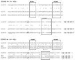
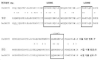
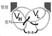


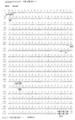


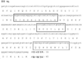


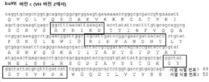


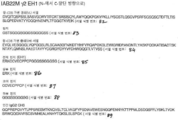


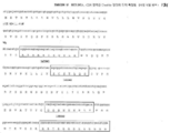
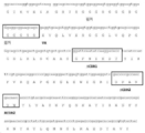


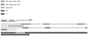

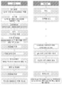

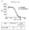


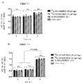
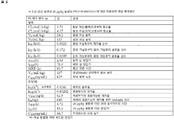
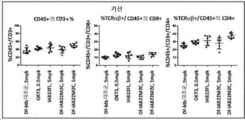

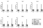


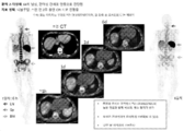
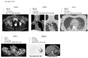

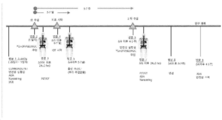

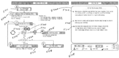
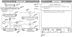
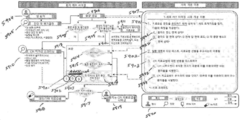


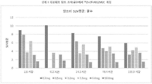


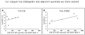
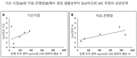


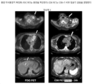

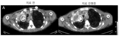


Classifications
-
- A—HUMAN NECESSITIES
- A61—MEDICAL OR VETERINARY SCIENCE; HYGIENE
- A61K—PREPARATIONS FOR MEDICAL, DENTAL OR TOILETRY PURPOSES
- A61K51/00—Preparations containing radioactive substances for use in therapy or testing in vivo
- A61K51/02—Preparations containing radioactive substances for use in therapy or testing in vivo characterised by the carrier, i.e. characterised by the agent or material covalently linked or complexing the radioactive nucleus
- A61K51/04—Organic compounds
- A61K51/08—Peptides, e.g. proteins, carriers being peptides, polyamino acids, proteins
- A61K51/10—Antibodies or immunoglobulins; Fragments thereof, the carrier being an antibody, an immunoglobulin or a fragment thereof, e.g. a camelised human single domain antibody or the Fc fragment of an antibody
- A61K51/1093—Antibodies or immunoglobulins; Fragments thereof, the carrier being an antibody, an immunoglobulin or a fragment thereof, e.g. a camelised human single domain antibody or the Fc fragment of an antibody conjugates with carriers being antibodies
-
- A—HUMAN NECESSITIES
- A61—MEDICAL OR VETERINARY SCIENCE; HYGIENE
- A61K—PREPARATIONS FOR MEDICAL, DENTAL OR TOILETRY PURPOSES
- A61K51/00—Preparations containing radioactive substances for use in therapy or testing in vivo
- A61K51/02—Preparations containing radioactive substances for use in therapy or testing in vivo characterised by the carrier, i.e. characterised by the agent or material covalently linked or complexing the radioactive nucleus
- A61K51/04—Organic compounds
- A61K51/08—Peptides, e.g. proteins, carriers being peptides, polyamino acids, proteins
- A61K51/10—Antibodies or immunoglobulins; Fragments thereof, the carrier being an antibody, an immunoglobulin or a fragment thereof, e.g. a camelised human single domain antibody or the Fc fragment of an antibody
- A61K51/1045—Antibodies or immunoglobulins; Fragments thereof, the carrier being an antibody, an immunoglobulin or a fragment thereof, e.g. a camelised human single domain antibody or the Fc fragment of an antibody against animal or human tumor cells or tumor cell determinants
- A61K51/1069—Antibodies or immunoglobulins; Fragments thereof, the carrier being an antibody, an immunoglobulin or a fragment thereof, e.g. a camelised human single domain antibody or the Fc fragment of an antibody against animal or human tumor cells or tumor cell determinants the tumor cell being from blood cells, e.g. the cancer being a myeloma
-
- A—HUMAN NECESSITIES
- A61—MEDICAL OR VETERINARY SCIENCE; HYGIENE
- A61B—DIAGNOSIS; SURGERY; IDENTIFICATION
- A61B6/00—Apparatus or devices for radiation diagnosis; Apparatus or devices for radiation diagnosis combined with radiation therapy equipment
- A61B6/02—Arrangements for diagnosis sequentially in different planes; Stereoscopic radiation diagnosis
- A61B6/03—Computed tomography [CT]
- A61B6/037—Emission tomography
-
- A—HUMAN NECESSITIES
- A61—MEDICAL OR VETERINARY SCIENCE; HYGIENE
- A61B—DIAGNOSIS; SURGERY; IDENTIFICATION
- A61B6/00—Apparatus or devices for radiation diagnosis; Apparatus or devices for radiation diagnosis combined with radiation therapy equipment
- A61B6/48—Diagnostic techniques
- A61B6/481—Diagnostic techniques involving the use of contrast agents
-
- A—HUMAN NECESSITIES
- A61—MEDICAL OR VETERINARY SCIENCE; HYGIENE
- A61K—PREPARATIONS FOR MEDICAL, DENTAL OR TOILETRY PURPOSES
- A61K51/00—Preparations containing radioactive substances for use in therapy or testing in vivo
- A61K51/02—Preparations containing radioactive substances for use in therapy or testing in vivo characterised by the carrier, i.e. characterised by the agent or material covalently linked or complexing the radioactive nucleus
- A61K51/04—Organic compounds
- A61K51/08—Peptides, e.g. proteins, carriers being peptides, polyamino acids, proteins
- A61K51/10—Antibodies or immunoglobulins; Fragments thereof, the carrier being an antibody, an immunoglobulin or a fragment thereof, e.g. a camelised human single domain antibody or the Fc fragment of an antibody
- A61K51/1093—Antibodies or immunoglobulins; Fragments thereof, the carrier being an antibody, an immunoglobulin or a fragment thereof, e.g. a camelised human single domain antibody or the Fc fragment of an antibody conjugates with carriers being antibodies
- A61K51/1096—Antibodies or immunoglobulins; Fragments thereof, the carrier being an antibody, an immunoglobulin or a fragment thereof, e.g. a camelised human single domain antibody or the Fc fragment of an antibody conjugates with carriers being antibodies radioimmunotoxins, i.e. conjugates being structurally as defined in A61K51/1093, and including a radioactive nucleus for use in radiotherapeutic applications
-
- A—HUMAN NECESSITIES
- A61—MEDICAL OR VETERINARY SCIENCE; HYGIENE
- A61P—SPECIFIC THERAPEUTIC ACTIVITY OF CHEMICAL COMPOUNDS OR MEDICINAL PREPARATIONS
- A61P35/00—Antineoplastic agents
-
- A—HUMAN NECESSITIES
- A61—MEDICAL OR VETERINARY SCIENCE; HYGIENE
- A61P—SPECIFIC THERAPEUTIC ACTIVITY OF CHEMICAL COMPOUNDS OR MEDICINAL PREPARATIONS
- A61P35/00—Antineoplastic agents
- A61P35/02—Antineoplastic agents specific for leukemia
-
- A—HUMAN NECESSITIES
- A61—MEDICAL OR VETERINARY SCIENCE; HYGIENE
- A61P—SPECIFIC THERAPEUTIC ACTIVITY OF CHEMICAL COMPOUNDS OR MEDICINAL PREPARATIONS
- A61P43/00—Drugs for specific purposes, not provided for in groups A61P1/00-A61P41/00
-
- C—CHEMISTRY; METALLURGY
- C07—ORGANIC CHEMISTRY
- C07K—PEPTIDES
- C07K16/00—Immunoglobulins [IGs], e.g. monoclonal or polyclonal antibodies
- C07K16/18—Immunoglobulins [IGs], e.g. monoclonal or polyclonal antibodies against material from animals or humans
- C07K16/28—Immunoglobulins [IGs], e.g. monoclonal or polyclonal antibodies against material from animals or humans against receptors, cell surface antigens or cell surface determinants
- C07K16/2803—Immunoglobulins [IGs], e.g. monoclonal or polyclonal antibodies against material from animals or humans against receptors, cell surface antigens or cell surface determinants against the immunoglobulin superfamily
- C07K16/2815—Immunoglobulins [IGs], e.g. monoclonal or polyclonal antibodies against material from animals or humans against receptors, cell surface antigens or cell surface determinants against the immunoglobulin superfamily against CD8
-
- G—PHYSICS
- G01—MEASURING; TESTING
- G01N—INVESTIGATING OR ANALYSING MATERIALS BY DETERMINING THEIR CHEMICAL OR PHYSICAL PROPERTIES
- G01N33/00—Investigating or analysing materials by specific methods not covered by groups G01N1/00 - G01N31/00
- G01N33/48—Biological material, e.g. blood, urine; Haemocytometers
- G01N33/50—Chemical analysis of biological material, e.g. blood, urine; Testing involving biospecific ligand binding methods; Immunological testing
- G01N33/53—Immunoassay; Biospecific binding assay; Materials therefor
- G01N33/531—Production of immunochemical test materials
- G01N33/532—Production of labelled immunochemicals
- G01N33/534—Production of labelled immunochemicals with radioactive label
-
- G—PHYSICS
- G01—MEASURING; TESTING
- G01N—INVESTIGATING OR ANALYSING MATERIALS BY DETERMINING THEIR CHEMICAL OR PHYSICAL PROPERTIES
- G01N33/00—Investigating or analysing materials by specific methods not covered by groups G01N1/00 - G01N31/00
- G01N33/48—Biological material, e.g. blood, urine; Haemocytometers
- G01N33/50—Chemical analysis of biological material, e.g. blood, urine; Testing involving biospecific ligand binding methods; Immunological testing
- G01N33/53—Immunoassay; Biospecific binding assay; Materials therefor
- G01N33/566—Immunoassay; Biospecific binding assay; Materials therefor using specific carrier or receptor proteins as ligand binding reagents where possible specific carrier or receptor proteins are classified with their target compounds
-
- G—PHYSICS
- G01—MEASURING; TESTING
- G01N—INVESTIGATING OR ANALYSING MATERIALS BY DETERMINING THEIR CHEMICAL OR PHYSICAL PROPERTIES
- G01N33/00—Investigating or analysing materials by specific methods not covered by groups G01N1/00 - G01N31/00
- G01N33/48—Biological material, e.g. blood, urine; Haemocytometers
- G01N33/50—Chemical analysis of biological material, e.g. blood, urine; Testing involving biospecific ligand binding methods; Immunological testing
- G01N33/68—Chemical analysis of biological material, e.g. blood, urine; Testing involving biospecific ligand binding methods; Immunological testing involving proteins, peptides or amino acids
- G01N33/6872—Intracellular protein regulatory factors and their receptors, e.g. including ion channels
-
- C—CHEMISTRY; METALLURGY
- C07—ORGANIC CHEMISTRY
- C07B—GENERAL METHODS OF ORGANIC CHEMISTRY; APPARATUS THEREFOR
- C07B2200/00—Indexing scheme relating to specific properties of organic compounds
- C07B2200/05—Isotopically modified compounds, e.g. labelled
-
- C—CHEMISTRY; METALLURGY
- C07—ORGANIC CHEMISTRY
- C07K—PEPTIDES
- C07K2317/00—Immunoglobulins specific features
- C07K2317/30—Immunoglobulins specific features characterized by aspects of specificity or valency
-
- C—CHEMISTRY; METALLURGY
- C07—ORGANIC CHEMISTRY
- C07K—PEPTIDES
- C07K2317/00—Immunoglobulins specific features
- C07K2317/50—Immunoglobulins specific features characterized by immunoglobulin fragments
- C07K2317/56—Immunoglobulins specific features characterized by immunoglobulin fragments variable (Fv) region, i.e. VH and/or VL
- C07K2317/565—Complementarity determining region [CDR]
-
- C—CHEMISTRY; METALLURGY
- C07—ORGANIC CHEMISTRY
- C07K—PEPTIDES
- C07K2317/00—Immunoglobulins specific features
- C07K2317/60—Immunoglobulins specific features characterized by non-natural combinations of immunoglobulin fragments
- C07K2317/62—Immunoglobulins specific features characterized by non-natural combinations of immunoglobulin fragments comprising only variable region components
- C07K2317/626—Diabody or triabody
-
- G—PHYSICS
- G01—MEASURING; TESTING
- G01N—INVESTIGATING OR ANALYSING MATERIALS BY DETERMINING THEIR CHEMICAL OR PHYSICAL PROPERTIES
- G01N2800/00—Detection or diagnosis of diseases
- G01N2800/52—Predicting or monitoring the response to treatment, e.g. for selection of therapy based on assay results in personalised medicine; Prognosis
-
- G—PHYSICS
- G01—MEASURING; TESTING
- G01N—INVESTIGATING OR ANALYSING MATERIALS BY DETERMINING THEIR CHEMICAL OR PHYSICAL PROPERTIES
- G01N2800/00—Detection or diagnosis of diseases
- G01N2800/56—Staging of a disease; Further complications associated with the disease
Landscapes
- Health & Medical Sciences (AREA)
- Life Sciences & Earth Sciences (AREA)
- Immunology (AREA)
- Chemical & Material Sciences (AREA)
- Proteomics, Peptides & Aminoacids (AREA)
- Engineering & Computer Science (AREA)
- General Health & Medical Sciences (AREA)
- Physics & Mathematics (AREA)
- Medicinal Chemistry (AREA)
- Public Health (AREA)
- Animal Behavior & Ethology (AREA)
- Veterinary Medicine (AREA)
- Molecular Biology (AREA)
- Hematology (AREA)
- Optics & Photonics (AREA)
- Biomedical Technology (AREA)
- Pharmacology & Pharmacy (AREA)
- Cell Biology (AREA)
- Epidemiology (AREA)
- Urology & Nephrology (AREA)
- Pathology (AREA)
- Medical Informatics (AREA)
- Nuclear Medicine, Radiotherapy & Molecular Imaging (AREA)
- Biochemistry (AREA)
- General Physics & Mathematics (AREA)
- Biotechnology (AREA)
- Food Science & Technology (AREA)
- Biophysics (AREA)
- Analytical Chemistry (AREA)
- Microbiology (AREA)
- Oncology (AREA)
- Organic Chemistry (AREA)
- Heart & Thoracic Surgery (AREA)
- High Energy & Nuclear Physics (AREA)
- Radiology & Medical Imaging (AREA)
- Surgery (AREA)
- Chemical Kinetics & Catalysis (AREA)
- General Chemical & Material Sciences (AREA)
- Genetics & Genomics (AREA)
- Bioinformatics & Cheminformatics (AREA)
Abstract
항원-결합 구조체 및 이의 사용 방법
요약서
CD8에 결합하는 항원 결합 구조체, 뿐만 아니라 제형, 그리고 이를 이용하는 방법이 본원에서 기술된다. Antigen-binding constructs and methods of use thereof
abstract
Antigen binding constructs that bind CD8, as well as formulations, and methods of using them are described herein.
Description
우선권preference
본 출원은 PCT 출원으로써, 2018년 9월 28일자로 제출된 U.S. 출원 번호 62/738,938, 및 2019년 3월 29일자로 제출된 U.S. 출원 번호 62/826,606에 대한 우선권을 주장하며, 이들 둘의 전문은 본원의 참고자료에 전문이 편입된다.This application is a PCT application, filed on September 28, 2018, U.S. Application No. 62/738,938, and U.S. Priority is claimed to Application No. 62/826,606, both of which are incorporated herein by reference in their entirety.
서열 목록sequence list
본 출원은 전자 형식의 서열 목록이 함께 제출되었다. 이 서열 목록은 2019 년 9 월 27 일에 생성된 IGNAB047WOSEQLIST.txt라는 파일로 제공되며, 크기는 126,124 바이트이다. 서열 목록의 전자 형식의 정보는 전체가 참고자료로 본원에 편입된다. This application is submitted with a sequence listing in electronic format. This sequence listing is provided as a file named IGNAB047WOSEQLIST.txt, created on September 27, 2019, and is 126,124 bytes in size. The information in electronic format of the Sequence Listing is incorporated herein by reference in its entirety.
배경background
분야Field
이 기술은 일반적으로 CD8과 관련된 이미징 및 치료 측면과 관련된다.This technique is usually associated with imaging and therapeutic aspects associated with CD8.
관련 기술의 설명Description of related technology
CD8 (분화 클러스터)은 막경유 당단백질이며, T-세포의 하위클래스 (세포독성 T-세포가 내포됨)에 대한 특이적 마커(marker)다. CD8은 CD8 알파 서브 유닛과 CD8 베타 서브 유닛의 이종이 량체 또는 CD8 알파 동종이량체로 어셈블리된다. 어셈블리된 이량체 CD8 복합체는 MHC 클래스 I 세포에 의해 제시되는 항원을 인지하기 위해 T-세포 수용체 (TCR)와 함께 공동-수용체로 작용한다. CD8은 T-세포의 발달 및 성숙 T-세포의 활성화에 역할을 한다. T-세포 국소화의 변화는 면역 반응 진행을 반영할 수 있고, 시간 경과에 따라 발생될 수 있다.CD8 (Cluster of Differentiation) is a transmembrane glycoprotein and is a specific marker for a subclass of T-cells (containing cytotoxic T-cells). CD8 assembles as a heterodimer of the CD8 alpha subunit and the CD8 beta subunit or as a CD8 alpha homodimer. The assembled dimeric CD8 complex acts as a co-receptor with the T-cell receptor (TCR) to recognize antigens presented by MHC class I cells. CD8 plays a role in T-cell development and activation of mature T-cells. Changes in T-cell localization may reflect immune response progression and may occur over time.
U.S. 특허 공개 번호 20160024209에서 논의된 것들을 비롯하여 다수의 CD8 지향된 항원 결합 구조체들이 존재한다.U.S. A number of CD8 directed antigen binding constructs exist, including those discussed in Patent Publication No. 20160024209.
요약summary
일부 구체예들에서, 생체내에서 CD8을 모니터링하는 방법이 제공된다. 이 방법은 CD8 미니바디(minibody)를 대상체에게 제공하는 것을 포함하며, 이때 해당 CD8 미니바디는 CD8에 결합하고 (도 1C에 나타낸 것과 같이), 이때 해당 미니바디는 검출가능한 마커로 라벨되며; 그리고 해당 CD8 미니바디를 해당 대상체에게 투여 후 6-36 시간 안에 해당 대상체 안에 해당 CD8 미니바디의 분포를 모니터링한다. In some embodiments, methods of monitoring CD8 in vivo are provided. The method comprises providing to a subject a CD8 minibody, wherein the CD8 minibody binds to CD8 (as shown in FIG. 1C ), wherein the minibody is labeled with a detectable marker; and monitoring the distribution of the CD8 minibody in the subject within 6-36 hours after administration of the CD8 minibody to the subject.
일부 구체예들에서, 생체내에서 CD8을 모니터링하는 방법은 CD8 미니바디를 대상체에게 제공하는 것을 포함하며, 이때 해당 CD8 미니바디는 CD8 (도 1C에 나타낸 것과 같이)에 결합하고, 이때 해당 미니바디는 검출가능한 마커로 라벨되며; 그리고 해당 대상체 안에 해당 CD8 미니바디의 분포를 모니터링하며, 이때 이러한 모니터링은 해당 대상체의 mm2 당 500개 또는 그 미만의 CD8 함유하는 세포로 침윤된 조직을 탐지할 수 있고, 이때 모니터링은 PET를 통하여 이루어진다. In some embodiments, a method of monitoring CD8 in vivo comprises providing a CD8 minibody to a subject, wherein the CD8 minibody binds to CD8 (as shown in FIG. 1C ), wherein the minibody is is labeled with a detectable marker; and monitoring the distribution of the CD8 minibody in the subject, wherein the monitoring is capable of detecting tissue infiltrated with 500 or less CD8 containing cells per mm 2 of the subject, wherein the monitoring is via PET. is done
일부 구체예들에서, CD8 포지티브 세포를 시각화시키는 방법은 CD8 미니바디를 대상체에게 제공하는 것을 포함하며, 이때 해당 CD8 미니바디는 검출가능한 마커로 라벨되며, 이때 해당 CD8 미니바디 서열에서 인간화되어, 인간 CD8에 결합하고; 그리고 해당 CD8 미니바디를 해당 대상체에게 투여 후 6-36 시간 안에 해당 대상체 안에 해당 CD8 미니바디의 분포를 모니터링하고, 이때 이러한 모니터링은 해당 대상체의 mm2 당 500개 또는 그 미만의 CD8 포지티브 세포로 침윤된 조직을 탐지할 수 있고, 이때 모니터링은 PET를 통하여 이루어진다. In some embodiments, a method of visualizing a CD8 positive cell comprises providing a CD8 minibody to a subject, wherein the CD8 minibody is labeled with a detectable marker, wherein the CD8 minibody sequence is humanized to a human binds to CD8; and monitoring the distribution of the CD8 minibody in the subject within 6-36 hours after administration of the CD8 minibody to the subject, wherein the monitoring is infiltrating with 500 or fewer CD8 positive cells per mm 2 of the subject Detected tissue can be detected, and monitoring is performed through PET.
일부 구체예들에서, 상기 세포 카운트 (이를 테면, 500개 또는 그 미만)는 10 미크론 또는 30 미크론 깊이 기준이다.In some embodiments, the cell count (eg, 500 or less) is based on a depth of 10 microns or 30 microns.
일부 구체예들에서, 인간에서 세포를 가시화시키는 방법은 인간 CD8 포지티브 세포에 결합하는 수단을 대상체에게 제공하는 것을 포함하며, 이때 해당 수단은 검출가능한 라벨을 포함하고; 그리고 해당 대상체에게 인간 CD8 포지티브 세포에 결합하는 수단을 투여 후 6-36 시간 안에 해당 대상체에서 인간 CD8 포지티브 세포에 결합하는 수단의 제 1 분포를 모니터링하고, 결정하는 것을 포함한다. In some embodiments, a method of visualizing a cell in a human comprises providing to the subject a means for binding to a human CD8 positive cell, wherein the means comprises a detectable label; and monitoring and determining a first distribution of the means for binding to human CD8 positive cells in the subject within 6-36 hours after administering to the subject the means for binding to human CD8 positive cells.
일부 구체예들에서, 대상체에게 미니바디를 투여하는 방법은 대상체에게 인간 CD8에 결합하는 라벨된 미니바디를 제공하는 것을 포함하고, 이때 3 mCi+20%의 방사선이가 해당 라벨된 미니바디를 통하여 해당 대상체에게 제공되며, 이때 전체 단백질의 0.2 ~ 10mg의 양이 해당 대상체에게 제공된다. In some embodiments, a method of administering a minibody to a subject comprises providing to the subject a labeled minibody that binds human CD8, wherein 3 mCi+20% of the radiation is via the labeled minibody. provided to the subject, wherein an amount of 0.2 to 10 mg of total protein is provided to the subject.
일부 구체예들에서, 대상체에게 미니바디를 투여하는 방법은 대상체에게 인간 CD8에 결합하는 라벨된 미니바디를 제공하는 것을 포함하고, 이때 방사선의 0.75-1.5+/- 20% mCi가 해당 라벨된 미니바디를 통하여 해당 대상체에게 제공되며, 이때 전체 단백질의 0.2 ~ 10mg의 양이 해당 대상체에게 제공된다.In some embodiments, a method of administering a minibody to a subject comprises providing to the subject a labeled minibody that binds human CD8, wherein 0.75-1.5+/- 20% mCi of the radiation is the labeled minibody. provided to the subject through the body, wherein an amount of 0.2 to 10 mg of total protein is provided to the subject.
일부 구체예들에서, 환자를 치료하는 방법은 암으로 진단받은 인간 환자에게 인간 CD8에 결합하는 항원-결합 구조체의 용량을 투여하는 것을 포함하고, 이때 이 용량(dose)은 다음을 포함한다: 89Zr-라벨된 항원-결합 구조체(약 3 mCi의 방사선 활성을 제공함); 그리고 약 10 mg 또는 그 미만의 상기 항원-결합 구조체. 이 방법은 상기 용량이 투여된 후 제 1 시점에서 해당 환자에게서 89Zr-라벨된 항원-결합 구조체를 탐지하여, 해당 제 1 시점에 대응하는 환자의 제 1 이미지를 만들고, 이때 제 1 시점은 투여 후 약 6 시간 내지 36 시간이며; 해당 환자의 제 1 이미지에 기초하여 환자의 하나 또는 그 이상의 조직 및/또는 신생물(neoplasia)에서 CD8 세포의 제 1 풍도 및/또는 분포를 결정하고; 그리고 해당 하나 또는 그 이상의 조직의 CD8 세포의 제 1 풍도 및/또는 분포를 기초하여 해당 환자에게 암에 대한 제 1 치료를 투여하는 것을 더 포함한다. In some embodiments, a method of treating a patient comprises administering to a human patient diagnosed with cancer a dose of an antigen-binding construct that binds human CD8, wherein the dose comprises: 89 Zr-labeled antigen-binding constructs (providing radioactivity of about 3 mCi); and about 10 mg or less of said antigen-binding construct. The method detects the 89 Zr-labeled antigen-binding construct in the patient at a first time point after the dose has been administered, and creates a first image of the patient corresponding to the first time point, wherein the first time point is administered after about 6 hours to 36 hours; determine a first abundance and/or distribution of CD8 cells in one or more tissues and/or neoplasia of the patient based on the first image of the patient; and administering to the patient a first treatment for cancer based on the first abundance and/or distribution of CD8 cells in the one or more tissues.
일부 구체예들에서, 종양을 치료하는 방법은 미니바디를 대상체에게 투여하여, 해당 미니바디가 이 대상체 안에 있는 종양에 결합하여 라벨된 종양이 형성되도록 하는 것을 포함하고, 이때 상기 미니바디는 인간 CD8에 결합하고, 이때 상기 미니바디는 검출가능한 라벨에 연계된다. 상기 미니바디가 임의의 종양의 표면에 편향된 방식으로 결합하여 라벨된 종양을 형성하는 경우, 면역 체크포인트(checkpoint) 억제제로 구성된 군에서 선택된 치료제(treatment)를 투여한다. 상기 미니바디가 도처의 모든 종양에 결합하여 라벨된 종양을 형성하는 경우, 면역 체크포인트 억제제를 투여하지 않는다. In some embodiments, a method of treating a tumor comprises administering to a subject a minibody such that the minibody binds to a tumor in the subject to form a labeled tumor, wherein the minibody comprises human CD8 binds to, wherein the minibody is linked to a detectable label. When the minibody binds to the surface of any tumor in a biased manner to form a labeled tumor, a treatment selected from the group consisting of immune checkpoint inhibitors is administered. If the minibody binds to all tumors ubiquitous to form a labeled tumor, no immune checkpoint inhibitor is administered.
일부 구체예들에서, 대상체를 치료하는 방법은 신생물을 보유한 환자에게 인간 CD8에 결합하는 인간 미니바디를 투여하고; 해당 인간 미니바디의 분포를 모니터링하여 해당 신생물 안에 종양-침윤성 림프구 ("TIL")의 제 1 상태를 결정하고; 그리고 해당 신생물의 적어도 제 1 TIL 상태에 근거하여 해당 환자를 치료하는 것을 포함한다. In some embodiments, a method of treating a subject comprises administering to a patient having a neoplasm a human minibody that binds human CD8; monitoring the distribution of the human minibody of interest to determine a first status of tumor-infiltrating lymphocytes (“TILs”) within the neoplasm; and treating the patient based on at least a first TIL status of the neoplasm.
일부 구체예들에서, 대상체에서 CD8 분포를 분석하는 방법은 제 1 PET 이미지를 통하여 검출가능한 마커의 분포 이미지를 제공하고 (이때 상기 검출가능한 마커는 CD8 미니바디에 연계되며, 이때 해당 CD8 미니바디 인간 CD8에 결합하고, 이때 해당 CD8 미니바디는 해당 면역체에서 면역 반응을 일으키지 않고); 해당 대상체의 제 2 PET 이미지를 통하여 FDG 마커 분포의 이미지를 제공하고; 상기 제 1 PET 이미지를 상기 제 2 PET 이미지 상에 겹쳐 진 것을 포함하는 제 3 PET 이미지를 생성시키고; 그리고 상기 제 3 PET 이미지에 기초하여 종양이 TIL인지를 확인하는 것을 포함한다. In some embodiments, a method of analyzing CD8 distribution in a subject provides an image of a distribution of a detectable marker via a first PET image, wherein the detectable marker is linked to a CD8 minibody, wherein the CD8 minibody human binds to CD8, wherein the CD8 minibody does not elicit an immune response in the immune body); providing an image of FDG marker distribution through a second PET image of the subject; generating a third PET image comprising the first PET image superimposed on the second PET image; and determining whether the tumor is TIL based on the third PET image.
일부 구체예들에서, 대상체를 치료하는 방법은 신생물을 보유한 환자에게 인간 CD8에 결합하는 항원-결합 구조체를 투여하고; 상기 항원-결합 구조체의 분포를 모니터링하여 해당 신생물 안에 종양-침윤성 림프구 ("TIL") 상태를 결정하고; 그리고 적어도 하나의 신생물의 TIL 상태가 네가티브인 경우, 면역요법제 ("IOT")를 해당 환자에게 투여하는 것을 포함한다. 선택적으로, 상기 상태가 TIL 포지티브가 될 때까지 지속된다. In some embodiments, a method of treating a subject comprises administering to a patient having a neoplasm an antigen-binding construct that binds human CD8; monitoring the distribution of the antigen-binding construct to determine tumor-infiltrating lymphocyte (“TIL”) status within the neoplasm; and if the TIL status of the at least one neoplasm is negative, administering an immunotherapeutic agent (“IOT”) to the patient. Optionally, the state continues until TIL positive.
일부 구체예들에서, 대상체를 치료하는 방법은 신생물을 보유한 환자에게 인간 CD8에 결합하는 항원-결합 구조체를 투여하고; 상기 항원-결합 구조체의 분포를 모니터링하여 해당 신생물 안에 종양-침윤성 림프구 ("TIL") 상태를 결정하고; 그리고 상기 신생물의 TIL 상태가 포지티브가 아닌 경우, 대체 면역요법제 ("IOT")를 해당 환자에게 투여하는 것을 포함한다. 선택적으로, 상기 상태가 TIL 포지티브가 될 때까지 지속된다.In some embodiments, a method of treating a subject comprises administering to a patient having a neoplasm an antigen-binding construct that binds human CD8; monitoring the distribution of the antigen-binding construct to determine tumor-infiltrating lymphocyte (“TIL”) status within the neoplasm; and if the TIL status of the neoplasm is not positive, administering an alternative immunotherapeutic agent (“IOT”) to the patient. Optionally, the state continues until TIL positive.
일부 구체예들에서, 종양을 특징화시키는 방법은 인간 CD8에 결합하는 항원-결합 구조체를 인간 대상체에게 적용시키고, 이때 상기 항원-결합 구조체는 검출가능한 마커를 포함하고; 상기 검출가능한 마커 분포를 모니터링하고; 상기 검출가능한 마커 분포를 기반으로 하여 이미지를 생성하고; 그리고 사용자가 해당 종양을 특징화할 수 있도록 이 사용자에게 해당 이미지를 나타내는 것을 포함한다. In some embodiments, a method of characterizing a tumor applies to a human subject an antigen-binding construct that binds human CD8, wherein the antigen-binding construct comprises a detectable marker; monitoring the detectable marker distribution; generate an image based on the detectable marker distribution; and presenting the image to the user so that the user can characterize the tumor.
일부 구체예들에서, 이미지를 생성시키는 방법은 CD8에 결합하는 항원-결합 구조체를 적용시키고, 이때 상기 항원-결합 구조체는 검출가능한 마커를 포함하고; 해당 종양 내 위치 안에 적어도 하나의 검출가능한 마커를 탐지하고, 탐지된 각 검출가능한 마커에 대해 포지티브 픽셀(pixel)을 할당하고; 그리고 할당된 각 포지티브 픽셀 분포에 기초하여 이미지를 생성시키고, 이때 이 이미지는 비-일과적(non-transitory) 컴퓨터 판독가능한 매체에 저장된다. In some embodiments, a method of generating an image applies an antigen-binding construct that binds to CD8, wherein the antigen-binding construct comprises a detectable marker; detecting at least one detectable marker within a location within the tumor and assigning a positive pixel to each detected detectable marker; and generates an image based on each assigned positive pixel distribution, wherein the image is stored on a non-transitory computer readable medium.
일부 구체예들에서, 양전자 방출 단층 촬영 ("PET") 방법은 대상체에게 CD8에 결합하는 추적자(tracer)를 투여하고; 섬광체(scintillator)를 제공하고; 해당 섬광체를 이용하여 상기 추적자에 의해 만들어진 광자 쌍을 탐지하고; 그리고 해당 광자 쌍의 탐지를 이용하여 해당 섬광체로부터 결과물을 가져 오고, 이를 일치(coincidence) 이벤트 목록으로 전환시키도록 구성된 프로세서를 통해 처리되는 일치 이벤트 목록을 통해 추적자의 공급원의 위치를 알아내는 것을 포함하고, 이때 해당 대상체 내 조직 또는 신생물 mm3 당 약 300 ~ 약 500개의 CD8 포지티브 세포가 탐지될 수 있다. In some embodiments, the positron emission tomography (“PET”) method comprises administering to a subject a tracer that binds CD8; providing a scintillator; detecting a photon pair produced by the tracer using the scintillator; and locating the source of the tracer through a list of coincident events processed through a processor configured to obtain a result from the scintillator using detection of the pair of photons and convert it into a list of coincidence events; , wherein about 300 to about 500 CD8 positive cells per mm 3 of tissue or neoplasm in the subject may be detected.
일부 구체예들에서, 진단 조성물을 제조하는 방법은 미니바디를 데스페리옥사민에 콘쥬게이트 Df-미니바디를 만들고; 이 Df-미니바디에 89Zr를 방사능라벨링시켜 방사능라벨된 미니바디를 만들고; 상기 방사능라벨된 미니바디를 정제하고; 그리고 해당 방사능라벨된 미니바디와 콜드 미니바디를 혼합하여 진단 조성물을 만드는 것을 포함하고, 이때 상기 미니바디와 콜드 미니바디는 CD8 상의 동일한 에피토프에 결합한다. In some embodiments, a method of preparing a diagnostic composition comprises making a Df-minibody conjugated to desperioxamine; Radiolabeling of this Df-minibody with 89 Zr to make a radiolabeled minibody; purifying the radiolabeled minibody; and mixing the radiolabeled minibody and the cold minibody to prepare a diagnostic composition, wherein the minibody and the cold minibody bind to the same epitope on CD8.
일부 구체예들에서, 진단 조성물은 3.0+20% mCi의 89Zr-Df-라벨된 미니바디; 20 mM 히스티딘; 5% 슈크로스; 51-62 mM 염화 나트륨; 141-194 아르기닌; 그리고 2-20 mM 글루탐산을 포함한다. In some embodiments, the diagnostic composition comprises an 89Zr-Df-labeled minibody at 3.0 + 20% mCi; 20 mM histidine; 5% sucrose; 51-62 mM sodium chloride; 141-194 arginine; and 2-20 mM glutamic acid.
일부 구체예들에서, 진단 조성물은 다음 구조를 포함하는 라벨된 미니바디를 포함한다: In some embodiments, the diagnostic composition comprises a labeled minibody comprising the structure:

이때 이 미니바디는 서열 식별 번호: 1, 3, 16, 또는 147 안의 중쇄 가변 영역과 서열 식별 번호:7, 9, 15 또는 147 안의 경쇄 가변 영역을 포함하고, 이때 해당 조성물은 적어도 3 mCi의 방사선을 제공한다. wherein the minibody comprises a heavy chain variable region within SEQ ID NO: 1, 3, 16, or 147 and a light chain variable region within SEQ ID NO: 7, 9, 15 or 147, wherein the composition comprises at least 3 mCi of radiation provides
일부 구체예들에서, 조성물은 CD8에 결합하고, 활성 마커를 포함하는 제 1 미니바디; 그리고 CD8에 결합하는 제 2 미니바디를 포함하며, 이때 제 2 미니바디는 비활성 마커를 포함하거나, 또는 활성 마커가 결여되어 있다. In some embodiments, the composition comprises a first minibody that binds CD8 and comprises an activity marker; and a second minibody that binds CD8, wherein the second minibody comprises an inactive marker or lacks an active marker.
일부 구체예들에서, 진단 조성물은 방사능라벨에 콘쥬게이트된 제 1 CD8 미니바디; 그리고 방사능라벨에 콘쥬게이트 안 된 제 2 CD8 미니바디를 포함하고, 이때 제 1 미니바디와 제 2 CD8 미니바디는 동일한 서열을 갖고, 이때 해당 조성물은 약 3 mCi의 방사선을 제공하고, 이때 해당 조성물은 전체 단백질 양의 1.5-10 mg을 제공한다. In some embodiments, the diagnostic composition comprises a first CD8 minibody conjugated to a radiolabel; and a second CD8 minibody not conjugated to the radiolabel, wherein the first minibody and the second CD8 minibody have the same sequence, wherein the composition provides radiation of about 3 mCi, wherein the composition provides 1.5-10 mg of total protein.
일부 구체예들에서, 대상체를 이미지화시키는 방법은 상기에서 제공된 조성물중 임의의 하나를 투여하고; 해당 대상체를 이미지화시켜 제 1 이미지를 얻고; 상기에서 제공된 조성물중 임의의 하나의 제 2 용량을 투여하고; 그리고 해당 대상체를 이미지화시켜 제 2 이미지를 얻고; 그리고 제 1 이미지를 제 2 이미지에 비교하는 것을 포함한다. In some embodiments, a method of imaging a subject comprises administering any one of the compositions provided above; imaging the object to obtain a first image; administering a second dose of any one of the compositions provided above; and imaging the object to obtain a second image; and comparing the first image to the second image.
일부 구체예들에서, 표준 흡수 값(uptake value)을 결정하는 방법은 항원 결합 구조체를 대상체에게 적용하고, (이때 상기 항원 결합 구조체는 방사능활성 프로브를 포함하고); r을 결정하며, 여기에서 r은 PET 스캐너를 이용하여 측정된 항원 결합 구조체에서 나온 방사선의 방사능활성 농도 (kBq/ml) 이며; a'를 결정하고, 이때 a'는 주사된 방사능라벨된 추적자 (kBq)의 데코이-보정된 양(decay-corrected amount)이며; 환자의 체중인 w를 측정하고; 그리고 SUV는 r(a'/W)의 결과로 결정하는 것을 포함한다. In some embodiments, a method of determining a standard uptake value comprises applying an antigen binding construct to a subject, wherein the antigen binding construct comprises a radioactive probe; determine r, where r is the radioactive concentration (kBq/ml) of radiation from the antigen-binding construct measured using a PET scanner; determine a', where a' is the decay-corrected amount of injected radiolabeled tracer (kBq); measuring the patient's weight, w; And SUV involves determining as a result of r(a'/W).
일부 구체예들에서, 이미지를 분석하는 방법은 이미지를 제공하고; 이 이미지를 표시하여 이미지에 제 1 관심 영역 (ROI)을 특정하고; 제 1 ROI 내의 데이터 포인트에 대한 신호 강도를 결정하고; 제 1 ROI 내에서 신호 강도의 평균 값을 결정하고; 제 1 ROI 내에서 각 신호 강도를 합산하여, 제 1 ROI에 대한 신호 수준의 제 1 합계를 얻는 것을 포함하고, 이때 제 1 ROI는 대상체에게 투여된 항원 결합 구조체와 연합된 검출가능한 마커의 양에 대한 데이터를 나타낸다. In some embodiments, a method of analyzing an image includes providing an image; mark this image to specify a first region of interest (ROI) in the image; determine a signal strength for a data point within the first ROI; determine an average value of signal intensities within the first ROI; summing each signal intensity within the first ROI to obtain a first sum of the signal levels for the first ROI, wherein the first ROI is dependent on the amount of the detectable marker associated with the antigen binding construct administered to the subject. represents data for
일부 구체예들에서, 항원 결합 구조체를 대상체에게 투여하는 방법이 제공된다. 이 방법은 대상체에게 인간 CD8에 결합하는 라벨된 항원 결합 구조체를 제공하는 것을 포함한다. 상기 라벨은 89Zr를 포함하며, 상기 라벨을 주사하면 0.75 (또는 0.5) - 3 mCi+20%의 방사선을 제공하고, 0.2 ~ 10mg 양의 전체 항원 결합 구조체가 해당 대상체에게 제공된다.In some embodiments, methods of administering an antigen binding construct to a subject are provided. The method comprises providing to the subject a labeled antigen binding construct that binds human CD8. The label contains 89 Zr, and injection of the label provides 0.75 (or 0.5) - 3 mCi+20% of radiation, and an amount of 0.2 to 10 mg of the total antigen binding construct is provided to the subject.
일부 구체예들에서, 환자를 치료하는 방법이 제공된다. 이 방법은 다음을 포함한다: a) 암으로 진단받은 인간 환자에게 인간 CD8에 결합하는 항원-결합 구조체의 용량을 투여하고, 이때 이 용량은 약 0.75 (또는 0.5) 내지 3.6 mCi의 방사선 활성을 제공하는 89Zr-라벨된 항원-결합 구조체; 그리고 약 10 mg 또는 그 미만의 상기 항원-결합 구조체를 포함하며; b) 해당 환자에게서 상기 용량이 투여된 후 제 1 시점에서 89Zr-라벨된 항원-결합 구조체를 탐지하여 제 1 시점에 대응하는 환자의 제 1 이미지를 생성시키고, 이때 제 1 시점은 투여 후 약 6 시간 내지 36 시간의 시점이며; c) 해당 환자의 하나 또는 그 이상의 조직 및/또는 신생물에서 해당 환자의 상기 제 1 이미지를 기반으로 하여 CD8 세포의 제 1 풍도 및/또는 분포를 결정하고; d) 해당 환자에게 암에 대한 제 1 치료를 투여하고; e) 제 1 치료제 투여 후, 암 진단을 받은 인간 환자에게 제 2 용량의 상기 항원-결합 구조체를 투여하고; f) 해당 환자의 하나 또는 그 이상의 조직 및/또는 신생물에서 환자의 제 2 이미지를 기반으로 하여 CD8 세포의 제 2 풍도 및/또는 분포를 결정하고; 그리고 g) 상기 환자의 제 1 이미지를 환자의 제 2 이미지와 비교하고, 상기 환자의 제 1 이미지와 제 2 이미지의 비교를 기반으로 하여, 해당 환자에게 해당 암의 제 2 치료제를 투여한다. In some embodiments, a method of treating a patient is provided. The method comprises: a) administering to a human patient diagnosed with cancer a dose of an antigen-binding construct that binds human CD8, wherein the dose provides a radioactivity of about 0.75 (or 0.5) to 3.6 mCi 89 Zr-labeled antigen-binding constructs; and about 10 mg or less of said antigen-binding construct; b) detecting the 89 Zr-labeled antigen-binding construct at a first time point after administration of said dose in said patient to generate a first image of the patient corresponding to a first time point, wherein the first time point is approximately post-administration time points from 6 hours to 36 hours; c) determining a first abundance and/or distribution of CD8 cells based on said first image of said patient in one or more tissues and/or neoplasms of said patient; d) administering to the patient a first treatment for cancer; e) administering a second dose of said antigen-binding construct to a human patient diagnosed with cancer after administration of the first therapeutic agent; f) determining a second abundance and/or distribution of CD8 cells based on a second image of the patient in one or more tissues and/or neoplasms of the patient; and g) comparing the first image of the patient with a second image of the patient, and administering a second therapeutic agent for the cancer to the patient based on the comparison of the first image and the second image of the patient.
일부 구체예들에서, 환자를 치료하는 방법이 제공된다. 이 방법은 다음을 포함한다: a) 암으로 진단받은 인간 환자에게 CD8 PET 추적자의 용량이 투여되고, 이때 이 용량은 다음을 포함한다: 약 0.75 (또는 0.5) 내지 3.6 mCi의 방사선 활성을 제공하는 CD8 PET 추적자; 그리고 약 5 마이크로그램 ~ 50 mg의 해당 CD8 PET 추적자 (또는 약 20 마이크로그램 ~ 10 mg); b) 상기 용량이 투여된 후 제 1 시점에서 해당 환자에서 CD8 PET 추적자를 탐지하여, 제 1 시점에 대응하는 환자의 제 1 이미지를 생성시키고, 이때 제 1 시점은 투여 후 약 1 시간 내지 36 시간 시점이며; c) 해당 환자의 상기 제 1 이미지를 기반으로 하여 해당 환자의 하나 또는 그 이상의 조직 및/또는 신생물에서 해당 CD8 PET 추적자의 제 1 풍도 및/또는 분포를 결정하고; d) 해당 환자에게 암에 대한 제 1 치료를 투여하고; e) 제 1 치료제 투여 후, 암 진단을 받은 인간 환자에게 제 2 용량의 해당 CD8 PET 추적자를 투여하고; f) 환자의 제 2 이미지를 기반으로 하여 해당 환자의 하나 또는 그 이상의 조직 및/또는 신생물에서 해당 CD8 PET 추적자의 제 2 풍도 및/또는 분포를 결정하고; 그리고 g) 상기 환자의 제 1 이미지를 환자의 제 2 이미지와 비교하고, 상기 환자의 제 1 이미지와 제 2 이미지의 비교를 기반으로 해당 환자에게 해당 암의 제 2 치료제를 투여한다.In some embodiments, a method of treating a patient is provided. The method comprises: a) administering to a human patient diagnosed with cancer a dose of a CD8 PET tracer, wherein the dose comprises: providing a radioactivity of about 0.75 (or 0.5) to 3.6 mCi CD8 PET tracer; and about 5 micrograms to 50 mg of the corresponding CD8 PET tracer (or about 20 micrograms to 10 mg); b) detecting the CD8 PET tracer in the patient at a first time point after said dose is administered, producing a first image of the patient corresponding to the first time point, wherein the first time point is between about 1 hour and 36 hours post administration time point; c) determining a first abundance and/or distribution of said CD8 PET tracer in one or more tissues and/or neoplasms of said patient based on said first image of said patient; d) administering to the patient a first treatment for cancer; e) administering a second dose of the corresponding CD8 PET tracer to a human patient diagnosed with cancer following administration of the first therapeutic agent; f) determining a second abundance and/or distribution of the CD8 PET tracer in one or more tissues and/or neoplasms of the patient based on the second image of the patient; and g) comparing the first image of the patient with a second image of the patient, and administering a second therapeutic agent for the corresponding cancer to the patient based on the comparison of the first image and the second image of the patient.
일부 구체예들에서, 이 방법은 신생물을 보유하고, 면역요법제 ("IOT")으로 치료중인 환자에게 인간 CD8에 결합하는 항원-결합 구조체를 투여하고; 이때 상기 항원 결합 구조체는검출가능한 마커를 포함하고, 이때 상기 항원 결합 구조체는 해당 대상체에게서 면역 반응을 야기하지 않고, 이때 해당 검출가능한 마커는 89Zr을 포함하고, 이때 해당 검출가능한 마커는 주사하면 0.5-3 mCi+20%의 방사선를 제공하고, 이때 0.2 ~ 10mg 양의 전체 항원 결합 구조체가 해당 환자에게 제공되며; 상기 항원-결합 구조체의 분포를 모니터링하여 해당 신생물 안에 종양-침윤성 림프구 ("TIL") 상태를 결정하고; 그리고 상기 신생물의 TIL 상태가 포지티브가 아니면 대체 IOT가 해당 환자에게 투여되고, 해당 신생물의 TIL 상태가 양성이 될 때까지 상기 방법을 반복하는 단계를 포함한다. In some embodiments, the method comprises administering to a patient having a neoplasm and being treated with an immunotherapeutic agent (“IOT”) an antigen-binding construct that binds human CD8; wherein the antigen binding construct comprises a detectable marker, wherein the antigen binding construct does not elicit an immune response in the subject, wherein the detectable marker comprises 89 Zr, wherein the detectable marker is 0.5- upon injection. 3 mCi+20% of radiation is given, wherein an amount of 0.2 to 10 mg of total antigen binding construct is given to the patient; monitoring the distribution of the antigen-binding construct to determine tumor-infiltrating lymphocyte (“TIL”) status within the neoplasm; and if the TIL status of the neoplasm is not positive, a replacement IOT is administered to the patient, and repeating the method until the TIL status of the neoplasm becomes positive.
일부 구체예들에서, 암을 갖는 인간 대상체를 치료하는 방법이 제공된다. 이 방법은 다음을 포함한다: i) CD8 항원 결합 구조체를 이용하여 대상체의 종양 내 CD8 세포의 제 1 이미지를 제공하고; ii) 후보 치료제가 내포된 치료요법을 해당 대상체에게 투여하고; iii) 후보 치료제가 내포된 치료요법 투여 후, 해당 CD8 항원 결합 구조체를 이용하여 해당 대상체의 종양 내 CD8 세포의 제 2 이미지를 제공하고; 그리고 iv) 제 1 이미지와 제 2 이미지를 비교하여 다음을 결정하고: a) 해당 종양이 CD8 침윤 증가를 보여주는지, 또는 b) 해당 종양이 CD8 침윤이 동일하거나 또는 감소됨을 주는지를 결정하고, 이때, a)인 경우, 그러면 해당 대상체에게 이 치료요법을 지속하도록 지시한다.In some embodiments, a method of treating a human subject having cancer is provided. The method comprises: i) providing a first image of CD8 cells in a tumor of a subject using the CD8 antigen binding construct; ii) administering to the subject a therapy implicated in the candidate therapeutic; iii) providing a second image of CD8 cells in the subject's tumor, using the CD8 antigen binding construct, after administration of the therapy containing the candidate therapeutic agent; and iv) comparing the first image with the second image to determine: a) the tumor shows increased CD8 invasion, or b) the tumor gives the same or reduced CD8 invasion, wherein , a), then instruct the subject to continue this therapy.
일부 구체예들에서, 암을 갖는 인간 대상체를 치료하는 방법이 제공된다. 이 방법은 다음을 포함한다: i) CD8 PET 추적자를 이용하여 대상체의 종양 내 CD8 세포의 제 1 이미지를 제공하고; ii) 후보 치료제가 내포된 치료요법을 해당 대상체에게 투여하고; iii) 치료요법 투여 후, 해당 대상체의 종양 내 CD8 세포의 제 2 이미지를 제공하고 해당 CD8 PET 추적자를 이용하여; 그리고 iv) 제 1 이미지와 제 2 이미지를 비교하여 다음을 결정하고: a) 해당 종양이 CD8 침윤 증가를 보여주는지, 또는 b) 해당 종양이 CD8 침윤이 동일하거나 또는 감소됨을 주는지를 결정하고, 이때, a)인 경우, 그러면 해당 대상체에게 이 치료요법을 지속하도록 지시한다.In some embodiments, a method of treating a human subject having cancer is provided. The method comprises: i) providing a first image of CD8 cells in a tumor of a subject using a CD8 PET tracer; ii) administering to the subject a therapy implicated in the candidate therapeutic; iii) following administration of therapy, providing a second image of CD8 cells in the subject's tumor and using the CD8 PET tracer; and iv) comparing the first image with the second image to determine: a) the tumor shows increased CD8 invasion, or b) the tumor gives the same or reduced CD8 invasion, wherein , a), then instruct the subject to continue this therapy.
일부 구체예들에서, 인간 대상체를 치료하는 방법이 제공된다. 이 방법은 다음을 포함한다: a) 후보 치료제를 인간 대상체에게 투여하고; b) 인간 대상체에 있는 종양의 크기가 해당 후보 치료제에 반응하여 증가되었는지 또는 감소되었는지를 결정하고; 그리고 c) 해당 후보 치료제 이후, 크기가 감소되었다면, 그러면 CD8 항원 결합 구조체를 이용하여 해당 후보 치료제에 반응하여 종양 안에 존재하는 CD8의 양이 증가되었는지를 결정한다. CD8의 양 증가 없이 종양 크기 증가는 종양 진행을 나타내고, 반면 CD8의 양 증가와 함께 종양 크기의 증가는 종양 허위(pseudo)-진행을 나타내며, 이때 만일 해당 환자가 종양 허위진행을 경험하는 경우, 치료는 지속되며, 이때 만일 해당 환자가 종양 진행을 경험하는 경우, 치료는 변경된다.In some embodiments, a method of treating a human subject is provided. The method comprises: a) administering a candidate therapeutic to a human subject; b) determining whether the size of a tumor in the human subject has increased or decreased in response to the candidate therapeutic; and c) if the size has decreased after the candidate therapeutic agent, then the CD8 antigen-binding construct is used to determine whether the amount of CD8 present in the tumor is increased in response to the candidate therapeutic agent. An increase in tumor size without increasing the amount of CD8 indicates tumor progression, whereas an increase in tumor size with an increase in the amount of CD8 indicates tumor pseudo-progression, wherein if the patient experiences tumor pseudo-progression, treatment continues, and if the patient experiences tumor progression, treatment is changed.
일부 구체예들에서, 인간 대상체에게 라벨을 투여하는 방법이 제공된다. 이 방법은 다음을 포함한다: 18F을 대상체에게 투여하고; 약 6 시간 이내에, 해당 대상체의 18F 분포에 대해 해당 대상체를 PET 스캔하고; CD8 PET 추적자를 해당 대상체에게 투여하고, 이때 해당 CD8 PET 추적자는 89Zr을 포함하고; 그리고 CD8 PET 추적자의 투여 후 약 36 시간 내에 89Zr 분포에 대해 해당 대상체에서 PET 스캔을 실시한다. 일부 구체예들에서, 18F은 반감기가 3시간 미만인 임의의 PET 동위원소다.In some embodiments, a method of administering a label to a human subject is provided. The method comprises: administering 18 F to the subject; within about 6 hours, PET scan the subject for their 18 F distribution; administering the CD8 PET tracer to the subject, wherein the CD8 PET tracer comprises 89 Zr; and a PET scan is performed on the subject for the 89 Zr distribution within about 36 hours after administration of the CD8 PET tracer. In some embodiments, 18F is any PET isotope with a half-life of less than 3 hours.
일부 구체예들에서, 진단 조성물은 다음을 포함한다: 3.0+20% mCi의 89Zr-Df-라벨된 항원 결합 구조체; 20 mM 히스티딘; 5% 슈크로스; 51-62 mM 염화 나트륨; 141-194 아르기닌; 그리고 2-20 mM 글루탐산. In some embodiments, the diagnostic composition comprises: 3.0 + 20% mCi of an 89Zr-Df-labeled antigen binding construct; 20 mM histidine; 5% sucrose; 51-62 mM sodium chloride; 141-194 arginine; and 2-20 mM glutamic acid.
일부 구체예들에서, CD8 조성물에 대한 제형은 CD8 항원 결합 구조체 (이때 해당 CD8 항원 결합 구조체의 크기는 105 kDa 미만이며); 그리고 해당 CD8 항원 결합 구조체와 연합된 89Zr 방사능라벨 (이때 방사능라벨은 해당 제형에 대해 0.5 초과의 그러나 3.6 (또는 3.0)미만의 mCi의 방사선을 제공하며)을 포함하고, 이때 해당 제형은 인간에게 투여하도록 형상화된다.In some embodiments, the formulation for the CD8 composition comprises a CD8 antigen binding construct, wherein the size of the CD8 antigen binding construct is less than 105 kDa; and an 89 Zr radiolabel associated with the CD8 antigen binding construct, wherein the radiolabel provides radiation of greater than 0.5 but less than 3.6 (or 3.0) mCi for the formulation, wherein the formulation is administered to a human. shaped for administration.
도면의 간단한 설명
도 1A는 CD8에 대해 이가(bivalent) 결합을 갖는 미니바디의 도해의 일부 구체예를 도시한다.
도 1B는 미니바디의 도식의 일부 구체예를 도시한다.
도 1C는 CD8 알파의 예를 제공한다.
도 2A는 인간 항체 (4D5v8)에 대한 OKT8 가변 중쇄 (VH) 영역과 본 발명의 인간화된 VH 영역 (IAB-huCD8 구조체)의 정렬의 일부 구체예를 도시한다. CDR 영역 (Chothia)의 일부 구체예들은 박스로 표시된 영역이다).
도 2B는 인간화된 VL 영역에 대한 OKT8 가변 경쇄 (VL) 영역과 IAB-huCD8 구조체의 정렬의 일부 구체예를 도시한다. CDR 영역 (Chothia)의 일부 구체예들은 박스로 표시된 영역이다).
도 3A는 항원에 대해 이가 결합을 보여주는 cys-디아바디의 도해의 일부 구체예를 도시한다.
도 3B는 항원에 대해 이가 결합을 보여주는 cys-디아바디의 도해를 도시한다.
도 4는 키메라 IAB-huCD8 미니바디 VL-VH 서열의 일부 구체예를 도시한다.
도 5는 키메라 IAB-huCD8 미니바디 VH-VL 서열의 일부 구체예를 도시한다.
도 6은 인간화된 IAB-huCD8 미니바디 VL-VH 서열의 일부 구체예를 도시한다.
도 7은 인간화된 IAB-huCD8 미니바디 VH-VL 서열의 일부 구체예를 도시한다.
도 8은 인간화된 IAB-huCD8 cys-디아바디 VL-5-VH 서열의 일부 구체예를 도시한다.
도 9는 인간화된 IAB-huCD8 cys-디아바디 VH-5-VL 서열의 일부 구체예를 도시한다.
도 10은 인간화된 IAB-huCD8 cys-디아바디 VL-8-VH 서열의 일부 구체예를 도시한다.
도 11은 인간화된 IAB-huCD8 cys-디아바디 VH-8-VL 서열의 일부 구체예를 도시한다.
도 12A는 cys-디아바디에 대한 서열들의 일부 구체예를 묘사한다.
도 12B는 디아바디에 대한 서열들의 일부 구체예를 묘사한다.
도 12C는 VL에 대한 서열들의 일부 구체예를 묘사한다.
도 12D는 huVL에 대한 서열들의 일부 구체예를 묘사한다.
도 12E는 VH에 대한 서열들의 일부 구체예를 묘사한다.
도 12F는 huVH (버젼 1에서 버젼 "a")에 대한 서열들의 일부 구체예를 묘사한다.
도 12G는 huVH (버젼 1에서 버젼 "b")에 대한 서열들의 일부 구체예를 묘사한다.
도 12H는 huVH (버젼 2에서 버젼 "c")에 대한 서열들의 일부 구체예를 묘사한다.
도 12I는 huVH (버젼 2에서 버젼 "c")에 대한 서열들의 일부 구체예를 묘사한다.
도 13는 pcDNA™ 3.1/myc-His(-) 버젼 A, B, C에 대한 벡터 맵(map)의 일부 구체예를 도시한다.
도 14는 표적의 존재 또는 부재를 탐지하는 방법의 일부 구체예들을 도시한다.
도 15는 IAB22M γ2 EH1의 일부 구체예들에 대한 단백질 서열 정보를 보여준다.
도 16은 IAB22M γ2 EH2 변이체의 일부 구체예들에 대한 단백질 서열 정보를 보여준다.
도 17A는 IAB22M γ1 EH1의 일부 구체예들에 대한 단백질 서열 정보를 보여준다.
도 17B는 IAB22M γ2 NH1의 일부 구체예들에 대한 단백질 서열 정보를 보여준다.
도 17C는 IAB22M γ2 NH2의 일부 구체예들에 대한 단백질 서열 정보를 보여준다.
도 18은 표 3에 열거된 힌지 변이체의 일부 구체예들 (γ1 EH1 상단, γ1 EH3 중간, γ1 EH4 하단)을 도시한다.
도 19A는 IAB22M γ1 EH3의 일부 구체예들에 대한 단백질 서열 정보를 보여준다.
도 19B는 IAB22M γ1 EH5의 일부 구체예들에 대한 단백질 서열 정보를 보여준다.
도 19C는 IAB22M γ3/γ1 EH6의 일부 구체예들에 대한 단백질 서열 정보를 보여준다.
도 19D는 IAB22M γ3/γ1 EH7의 일부 구체예들에 대한 단백질 서열 정보를 보여준다.
도 19E는 IAB22M γ3/γ1 EH8의 일부 구체예들에 대한 단백질 서열 정보를 보여준다.
도 20은 IAB22M γ1 EH2의 일부 구체예들에 대한 단백질 서열 정보를 보여준다.
도 21은 상이한 항원-특이성을 갖는 Mbs의 VL 도메인과 VH 도메인의 단백질 서열 정보를 나타낸다.
도 22는 링커 서열의 다양한 구체예들의 단백질 서열 정보를 나타낸다.
도 23은 힌지 영역의 다양한 구체예들의 단백질 서열 정보를 나타낸다.
도 24는 CH3 도메인의 다양한 구체예들의 단백질 서열 정보를 나타낸다.
도 25는 IAB22M γ1 EH1(M1) 및 IAB22M γ1 EH3(M1) 각각의 구체예의 단백질 서열들의 정렬을 보여준다. 서열 차이는 상자로 나타낸다.
도 26은 IAB22M γ1 EH3(M1)의 구체예의 DNA 및 해독된 단백질 서열을 보여준다. 박스에는 신호, CDR, 링커 및 힌지 서열을 보여준다.
도 27은 IAB22M γ1 EH5(M1)의 구체예의 DNA 및 해독된 단백질 서열을 보여준다.
도 28은 IAB22M γ1 EH7(M1)의 구체예의 DNA 및 해독된 단백질 서열을 보여준다.
도 29는 IAB22M γ1 EH8(M1)의 구체예의 DNA 및 해독된 단백질 서열을 보여준다.
도 30은 IAB22M γ2 EH2(M1)의 구체예의 DNA 및 해독된 단백질 서열을 보여준다.
도 31은 VH-K67R 다형성과 함께, IAB22M γ2 EH2(M1)의 구체예의 DNA 및 해독된 단백질 서열을 보여준다.
도 32는 IAB22M VH 도메인의 구체예의 단백질 서열을 보여준다.
도 33은 호모 사피엔스(homo sapiens)의 T-세포 표면 당단백질 CD8 알파 쇄의 구체예의 단백질 서열을 보여준다.
도 34는 호모 사피엔스(homo sapiens)의 T-세포 표면 당단백질 CD8 베타 쇄의 구체예의 단백질 서열을 보여준다.
도 35는 공작된(engineered) 미니바디 (Mb)의 실례를 보여준다.
도 36은 인간 IgG1에 기초한 Mb 힌지의 각종 구체예 (γ1 EH1 상단 및 γ1 EH2 하단)의 실례를 보여준다.
도 37은 인간 IgG2에 기초한 Mb 힌지의 각종 구체예 (γ2 EH1 상단 및 γ2 EH2 하단)의 실례를 보여준다.
도 38은 인간 IgG2에 기초한 추가 Mb 힌지의 각종 구체예 (γ2 NH1 상단 및 γ2 NH2 하단)의 실례를 보여준다.
도 39는 CD8 미니바디의 일부 구체예를 묘사한다.
도 40은 본 명세서의 일부 구체예들에 따라, 89Zr-Df-IAB22M2C의 제조를 보여주는 개략도이다.
도 41은 본 명세서의 일부 구체예들에 따라, 89Zr-Df-IAB22M2C 제작 동안 단위 단계 및 각 테스트를 묘사하는 흐름도를 보여준다.
도 42A와 42B는 HPB-ALL (도 3A) 및 정제된 인간 T 세포 (도 3B) 상에서 AlexaFluor-488 사전-라벨된 IAB22M2C를 이용한 경쟁 FACS 검정법의 결과를 보여준다.
도 43은 인간 PBMC를 이용한 T 세포 증식 검정법의 결과를 보여준다.
도 44는 인간 PBMC 사이토킨 방출 검정법의 결과를 보여준다.
도 45는 인간 PBMC를 이용한 T 세포 생존 (도 6A) 검정법 및 T 세포 증식 (도 6B) 검정법의 결과를 보여준다.
도 46은 표 3을 나타내는데, 이 표에서는 인간에서 정맥내 20 μg/kg 용량의 89Zr-Df-IAB22M2C에 대한 약동학적 예상 매개변수를 보여준다.
도 47은 기선에서 인간 CD34 줄기 세포가 이식된 NSG 마우스의 혈액에서 T-세포 집단의 FACS 분석 결과를 보여준다.
도 48은 24 시간 시점에서 인간 CD34 줄기 세포가 이식된 NSG 마우스의 혈액에서 T-세포 집단의 FACS 분석 결과를 보여준다.
도 49는 인간 CD34 조상(progenitor) 세포가 이식된 마우스의 혈청 샘플에서 시행된 LegendPlex™ Th1 비드 검정법의 결과를 보여준다.
도 50-도 54는 89ZR-Df-IAB22M2C2에 대한 결과를 묘사한다.
도 55는 일부 항원 결합 구조체의 사용 일정에 대한 흐름도이다.
도 56은 스크리닝 및 환자 선별에 대해 본원에서 제공된 다양한 방법들의 일부 구체예의 흐름도이다. 도면 설명문은 도 56의 5624에 제공된다.
도 57은 치료 개시 및 효과 모니터링에 대해 본원에서 제공된 다양한 방법들의 일부 구체예의 흐름도이다. 도면 설명문은 도 57의 5719에 제공된다.
도 58은 독성에 대한 치료 모니터링에 대해 본원에서 제공된 다양한 방법들의 일부 구체예의 흐름도이다. 도면 설명문은 도 58의 5824에 제공된다.
도 59는 제품 개념 사용 사례에 대한 스크리닝 및 환자 선별에 대해 본원에서 제공된 다양한 방법들의 일부 구체예의 흐름도이다. 도면 설명문은 도 59의 5920에 제공된다.
도 60은 제품 사용 경우에 대한 치료 개시 및 효과 모니터링에 대해 본원에서 제공된 다양한 방법들의 일부 구체예의 흐름도이다. 도면 설명문은 도 60의 6019에 제공된다.
도 61은 제품 사용 경우에 대한 독성에 대한 치료 모니터링에 대해 본원에서 제공된 다양한 방법들의 일부 구체예의 흐름도이다. 도면 설명문은 도 61의 6125에 제공된다.
도 62A는 실시예 85에 요약된 중앙 판독 설계를 나타내는 그래프이다.
도 62B-62H는 단계 I에 등록된 각 대상체의 SUV평균으로부터 결정된 참조 조직에 대한 시간 활성 곡선(TACs)를 보여준다. 도 62B-62J, 62L, 및 62M, 차트 아래에있는 범례의 순서는 그래프의 막대 순서를 표시한다.
도 62I 및 62J는 0.5 mg 및 1.5 mg에서 단계 I과 단계 II의 막대 기록이다.
도 62K는 결절 취입(uptake) 빈도를 보여주는 그래프다.
도 62L 및 M은 표적 종양 병소의 각 평균 시점에서 SUV최대 및 SUV피크 흡수 값의 분포에 대한 플롯이다.
도 62N 은 투여 6시간 또는 6일 이내 89Zr로 라벨된 항원 결합 구조체가 표적 (CD8)에 결합하는 능력을 보여주는 PET 스캔을 전반적으로 보여준다.
도 63A는 IHC 분석을 이용하여 결정하였을 때, 방사능추적자 취입과 CD8+ T 세포 카운트의 상관관계에 대한 정량적인 결과를 제공하는 단계들을 도시한 챠트이다.
도 63B 및 63C는 SUV 기반 측정 (SUV최대 및 SUV피크)와 기선에서 (a) 및 치료-중(b) 생검 샘플의 평균 CD8+ T 세포 카운트의 선형 상관관계를 설명하는, 회귀 계수 (r2)가 내포된, 그래프다.
도 63D는 SUV최대 (a) 및 SUV피크(b)에 대해 기선 및 치료-중 결과 모두를 플롯할 때, 회귀 계수 (r2)가 내포된, 선형관계를 설명하는 그래프다.
도 64A는 몇 가지 상이한 시점에 걸쳐 두 대상체의 PET 이미지들을 묘사한다.
도 64B 및 64C는 종양 미세환경을 좀 더 완벽하게 특징화하기 위해, CD8 PET와 FDG PET의 조합을 묘사한다.
도 64D는 응답자(responder) 이미지 (PET/CT)이다.
도 64E는 다양한 치료의 효과를 테스트하기 위하여 상기 미니바디 구조체를 이용한 결과를 보여주는 PET 스캔 및 PET/CT 융합된 스캔이다.
도 64F는 비-응답자 이미지 (PET 및 PET/CT 스캔)이다. Brief description of the drawing
1A depicts some embodiments of the schematic of a minibody having a bivalent bond to CD8.
1B shows some embodiments of a schematic of a minibody.
1C provides an example of CD8 alpha.
Figure 2A shows OKT8 against human antibody (4D5v8). Some embodiments of the alignment of the variable heavy (V H ) region with the humanized V H region of the present invention (IAB-huCD8 construct) are shown. Some embodiments of a CDR region (Chothia) are boxed regions).
2B shows the alignment of some embodiments of OKT8 variable light chain (V L) region and the IAB-huCD8 structure for the humanized V L region. Some embodiments of a CDR region (Chothia) are boxed regions).
3A depicts some embodiments of a schematic of a cys-diabody showing bivalent binding to antigen.
Figure 3B depicts a schematic of a cys-diabody showing bivalent binding to antigen.
4 depicts some embodiments of the chimeric IAB-huCD8 minibody V L -V H sequence.
5 depicts some embodiments of a chimeric IAB-huCD8 minibody V H -V L sequence.
6 depicts some embodiments of humanized IAB-huCD8 minibody V L -V H sequences.
7 depicts some embodiments of humanized IAB-huCD8 minibody V H -V L sequences.
8 depicts some embodiments of a humanized IAB-huCD8 cys-diabody V L- 5-V H sequence.
9 depicts some embodiments of humanized IAB-huCD8 cys-diabody V H- 5-V L sequences.
10 depicts some embodiments of humanized IAB-huCD8 cys-diabody V L- 8-V H sequences.
11 depicts some embodiments of humanized IAB-huCD8 cys-diabody V H- 8-V L sequences.
12A depicts some embodiments of sequences for a cys-diabody.
12B depicts some embodiments of sequences for diabodies.
Figure 12C depicts some specific examples of sequences for V L.
12D depicts some embodiments of sequences for huV L.
12E depicts some embodiments of sequences for V H .
12F depicts some embodiments of sequences for huV H (
12G depicts some embodiments of sequences for huV H (
12H depicts some embodiments of sequences for huV H (
12I depicts some embodiments of sequences for huV H (
Figure 13 shows some embodiments of vector maps for pcDNA™ 3.1/myc-His(-) versions A, B, C.
14 depicts some embodiments of a method for detecting the presence or absence of a target.
15 shows protein sequence information for some embodiments of IAB22M γ2 EH1.
16 shows protein sequence information for some embodiments of IAB22M γ2 EH2 variants.
17A shows protein sequence information for some embodiments of IAB22M γ1 EH1.
17B shows protein sequence information for some embodiments of IAB22M γ2 NH1.
17C shows protein sequence information for some embodiments of IAB22M γ2 NH2.
18 depicts some embodiments of the hinge variants listed in Table 3 (γ1 EH1 top, γ1 EH3 middle, γ1 EH4 bottom).
19A shows protein sequence information for some embodiments of IAB22M γ1 EH3.
19B shows protein sequence information for some embodiments of IAB22M γ1 EH5.
19C shows protein sequence information for some embodiments of IAB22M γ3/γ1 EH6.
19D shows protein sequence information for some embodiments of IAB22M γ3/γ1 EH7.
19E shows protein sequence information for some embodiments of IAB22M γ3/γ1 EH8.
20 is Shows protein sequence information for some embodiments of IAB22M γ1 EH2.
Figure 21 shows the protein sequence information of the VL domain and VH domain of Mbs with different antigen-specificity.
22 shows protein sequence information of various embodiments of linker sequences.
23 shows protein sequence information of various embodiments of the hinge region.
24 shows protein sequence information of various embodiments of the C H 3 domain.
25 shows the alignment of the protein sequences of each embodiment of IAB22M γ1 EH1 (M1) and IAB22M γ1 EH3 (M1). Sequence differences are indicated by boxes.
26 shows the DNA and translated protein sequences of an embodiment of IAB22M γ1 EH3(M1). Boxes show signal, CDR, linker and hinge sequences.
27 shows the DNA and translated protein sequences of an embodiment of IAB22M γ1 EH5(M1).
28 shows the DNA and translated protein sequences of an embodiment of IAB22M γ1 EH7(M1).
29 shows the DNA and translated protein sequence of an embodiment of IAB22M γ1 EH8(M1).
30 shows the DNA and translated protein sequences of an embodiment of IAB22M γ2 EH2(M1).
Figure 31 shows the DNA and translated protein sequence of an embodiment of IAB22M γ2 EH2 (M1), along with the VH-K67R polymorphism.
32 shows the protein sequence of an embodiment of the IAB22M VH domain.
Figure 33 shows the protein sequence of an embodiment of the T-cell surface glycoprotein CD8 alpha chain of Homo sapiens.
34 shows the protein sequence of an embodiment of the T-cell surface glycoprotein CD8 beta chain of Homo sapiens.
35 shows an example of an engineered minibody (Mb).
36 shows examples of various embodiments of Mb hinges (γ1 EH1 top and γ1 EH2 bottom) based on human IgG1.
37 shows examples of various embodiments of the Mb hinge based on human IgG2 (γ2 EH1 top and γ2 EH2 bottom).
38 shows examples of various embodiments of additional Mb hinges (γ2 NH1 top and γ2 NH2 bottom) based on human IgG2.
39 depicts some embodiments of a CD8 minibody.
40 is a schematic diagram showing the preparation of 89 Zr-Df-IAB22M2C, in accordance with some embodiments herein.
41 shows a flow diagram depicting unit steps and each test during 89 Zr-Df-IAB22M2C fabrication, in accordance with some embodiments herein.
Figures 42A and 42B show the results of a competition FACS assay using AlexaFluor-488 pre-labeled IAB22M2C on HPB-ALL (Figure 3A) and purified human T cells (Figure 3B).
43 shows the results of a T cell proliferation assay using human PBMCs.
44 shows the results of a human PBMC cytokine release assay.
45 shows the results of a T cell survival ( FIG. 6A ) assay and a T cell proliferation ( FIG. 6B ) assay using human PBMCs.
46 shows Table 3, which shows the pharmacokinetic predicted parameters for 89 Zr-Df-IAB22M2C at a dose of 20 μg/kg intravenously in humans.
Figure 47 shows the results of FACS analysis of T-cell populations in the blood of NSG mice transplanted with human CD34 stem cells at baseline.
FIG. 48 shows the results of FACS analysis of T-cell populations in the blood of NSG mice transplanted with human CD34 stem cells at 24 hours.
49 shows the results of the LegendPlex™ Th1 bead assay performed on serum samples of mice transplanted with human CD34 progenitor cells.
Figure 50-Figure 54 depicts the results for 89ZR-Df-IAB22M2C2.
55 is a flow chart for the schedule of use of some antigen binding constructs.
56 is a flow diagram of some embodiments of various methods provided herein for screening and patient selection. The drawing description is provided at 5624 in FIG. 56 .
57 is a flow diagram of some embodiments of various methods provided herein for treatment initiation and effect monitoring. The drawing description is provided at 5719 in FIG. 57 .
58 is a flow diagram of some embodiments of various methods provided herein for therapeutic monitoring for toxicity. The figure explanatory text is provided at 5824 in FIG. 58 .
59 is a flow diagram of some embodiments of various methods provided herein for screening and patient selection for product concept use cases. The drawing description is provided at 5920 in FIG. 59 .
60 is a flow diagram of some embodiments of the various methods provided herein for monitoring the effect and initiation of treatment for product use cases. A description of the drawings is provided at 6019 in FIG. 60 .
61 is a flow diagram of some embodiments of the various methods provided herein for therapeutic monitoring for toxicity for product use cases. The drawing description is provided at 6125 in FIG. 61 .
62A is a graph illustrating the central read design summarized in Example 85;
62B-62H show time activity curves (TACs) for a reference tissue determined from the SUV mean of each subject enrolled in stage I. 62B-62J, 62L, and 62M, the order of the legend below the chart indicates the order of the bars in the graph.
62I and 62J are bar recordings of Phase I and Phase II at 0.5 mg and 1.5 mg.
62K is a graph showing the frequency of nodule uptake.
62L and M are plots of the distribution of SUVmaximum and SUVpeak uptake values at each mean time point of the target tumor lesion.
62N shows overall PET scans showing the ability of antigen binding constructs labeled with 89 Zr to bind target (CD8) within 6 hours or 6 days of administration.
63A is a chart depicting steps providing quantitative results for the correlation of radiotracer uptake and CD8+ T cell counts, as determined using IHC analysis.
63B and 63C are regression coefficients (r 2 ), illustrating the linear correlation of SUV-based measurements (SUVmax and SUV peak) with mean CD8+ T cell counts of biopsy samples at baseline (a) and on-treatment (b). is an implied graph.
63D is a graph illustrating the linear relationship with implied regression coefficients (r 2 ) when plotting both baseline and on-treatment results for SUV max (a) and SUV peak (b).
64A depicts PET images of two subjects over several different time points.
64B and 64C depict the combination of CD8 PET and FDG PET to more fully characterize the tumor microenvironment.
64D is a responder image (PET/CT).
64E is a PET scan and a PET/CT fused scan showing the results of using the minibody construct to test the effects of various treatments.
64F is a non-responder image (PET and PET/CT scans).
다양한 구체예의 상세한 설명DETAILED DESCRIPTION OF THE VARIOUS EMBODIMENTS
본 명세서는 비-침습적 방법을 통하여 생체내에서 CD8 분자와, 따라서 CD8+ T 세포를 시작화시키는 방법을 제공한다. 일부 구체예들에서, 본 명세서의 방사능라벨된 항원-결합 구조체, 이를 테면, 89Zr-Df-IAB22M2C는 인간 환자의 생체내에서 CD8+ T 세포 탐지용 PET 이미지화 제제이다. 일부 구체예들에서, 본 명세서의 이러한 방법들은 면역요법제에 대한 국소 면역반응 및 전신 면역 반응을 더 잘 이해하고, 대상체에 전반적으로 우수한 면역요법제 또는 암 치료법을 선택하고, 및/또는 암 치료에 대한 신규한 면역요법 제제를 얻는다".Provided herein are methods of initiating CD8 molecules and thus CD8+ T cells in vivo via non-invasive methods. In some embodiments, a radiolabeled antigen-binding construct of the present disclosure, such as 89 Zr-Df-IAB22M2C, is a PET imaging agent for CD8+ T cell detection in vivo in a human patient. In some embodiments, such methods herein provide a better understanding of the local and systemic immune responses to an immunotherapeutic agent, select an overall superior immunotherapeutic agent or cancer therapy for a subject, and/or cancer treatment to obtain a novel immunotherapy formulation for
CD8 발현과 관련된 각종 암의 모니터링, 진단 및 치료에 대한 다양한 방법 및 조성물 (이에 대한 각종 조성물 및 이의 추가적인 대체 용도 포함)이 본원에서 제공된다. 본원에서 제공된 일부 구체예들은 대상체 안에 CD8 분자 및/또는 CD8 함유하는 세포, 이를 테면, CD8+ T 세포의 분포를 모니터할 수 있도록 한다. 일부 구체예들에서, 이것은 항원 결합 구조체, 이를 테면, 라벨된, 그리고 인간 CD8에 결합하는 미니바디를 이용하여 적어도 부분적으로 얻게 된다. Provided herein are various methods and compositions for the monitoring, diagnosis and treatment of various cancers associated with CD8 expression, including various compositions therefor and further alternative uses thereof. Some embodiments provided herein allow monitoring of the distribution of CD8 molecules and/or CD8 containing cells, such as CD8+ T cells, in a subject. In some embodiments, it is obtained at least in part using an antigen binding construct, such as a labeled, and minibody that binds to human CD8.
일부 구체예들에서, 상기 항원 결합 구조체의 속성은 해당 항원 결합 구조체의 우수한 분포 시기(당일 이미지화가 가능하거나, 또는 단일 용량으로 여러날 이미지화가 가능함)를 가능하게 한다 (검출가능한 마커와 연계되거나 또는 연합될 수 있고). In some embodiments, the property of the antigen-binding construct allows a good timing of distribution of the antigen-binding construct (either same-day imaging possible, or multi-day imaging possible with a single dose) (associated with a detectable marker or can be united).
본원에서 제공된 일부 구체예들은 인간 대상체 안에 종양 또는 신생물의 특징화를 더 많이 하도록 허용한다. 예를 들면, 이 프로세스를 통해 다양한 CD8을 함유하는 세포가 종양 또는 신생물에 어떻게, 또는 얼마나 효과적으로 침투하는지 결정할 수 있다. 이 프로세스에 기초하여, 예를 들어, 종양 또는 신생물의 상태를 한 상태에서 다른 상태로의 변경에 가장 효과적인 방법을 확인하기 위한 대체 치료를 제공함으로써, 대상체에 대한 올바른 치료 선택을 결정할 수 있다. 예를 들면, 면역 표현형을 관찰할 수 있으면, 해당 대상체의 현재 면역 표현형이 무엇인지 확인할 수 있고 (예를 들면, 면역 불모, 면역 배제된, 또는 TIL 포지티브 (또는 염증화된)), 그 다음, 해당 상태를 변경해야하는 지 여부, 해당 상태를 변경하기 위해 어떤 치료요법을 사용해야 하는 지, 그리고 새로운 상태가 주어지면 해당 환자를 치료하기 위해 어떤 후속 치료요법을 사용해야 하는지 결정할 수 있다.Some embodiments provided herein allow for more characterization of tumors or neoplasms in human subjects. For example, this process can determine how or how effectively cells containing various CD8s penetrate a tumor or neoplasm. Based on this process, the right treatment choice for a subject can be determined, for example, by providing an alternative treatment to identify the most effective method for changing the condition of a tumor or neoplasm from one condition to another. For example, if an immune phenotype can be observed, it can be determined what the current immune phenotype of the subject is (eg, immune infertile, immune excluded, or TIL positive (or inflamed), then: It can determine whether the condition needs to be changed, which therapy should be used to change the condition, and which follow-up therapy should be used to treat the patient given the new condition.
당업자는 본원에서 본 발명에 사용된 "종양의 특징화"가 "면역 불모(immune desert), 면역 배제된(immune excluded), 또는 TIL 포지티브 (또는 염증화된)"에 추가하여, 다양한 단어 및 어구로 표현될 수 있음을 이해할 것이다. 일부 실무자는 관찰된 결과를 설명하기 위해, "핫(hot)", "콜드(cold)" 또는 "중간("intermediate)" 등급을 사용할 것이다. 다른 특징화에 의해 CD8 함유하는 림프구에 대한 좀더 상세한 조직학적 정보, 이를 테면, 위치 (침습성 경계, 종양 표면, 종양 코어); 또는 해당 종양의 밀도 (가령, 면역 밀도 및 양)을 제공할 수 있다. 다른 특성화에 의해 "핫" 종양이 균질하게 침윤되었는지 또는 이질적으로 침윤되었는지를 구별할 수 있다. 일부 실무자는 4-가지 범주의 더 포괄적인 종양 분류를 선호 할 수 있다: 핫, 변경-제외된(altered-excluded), 변경된-면역억압된(altered-immunosuppressed), 그리고 콜드. 이러한 모든 단어 및 문구는 본 발명의 관찰된 결과의 설명에 허용되며, 여기에서 임의의 한 문구의 사용이 관찰에 대한 다른 적절한 설명의 사용을 배제하려는 의도가 아니다.Those of skill in the art will appreciate that, as used herein, "characterization of a tumor" refers to a variety of words and phrases in addition to "immune desert, immune excluded, or TIL positive (or inflamed)". It will be understood that it can be expressed as Some practitioners will use "hot", "cold" or "intermediate" ratings to describe the observed results. More detailed descriptions of CD8 containing lymphocytes by other characterizations Histological information, such as location (invasive border, tumor surface, tumor core); or the density of the tumor (such as immune density and quantity) can be provided.Other characterizations allow for homogeneous infiltration of "hot" tumors. Some practitioners may prefer a more comprehensive classification of tumors in four categories: hot, altered-excluded, and altered-immunosuppressed. -immunosuppressed), and cold All such words and phrases are acceptable in the description of the observed results of the present invention, and the use of any one phrase herein is not intended to exclude the use of another suitable description of the observation.
또한, 이들 CD8 항원 결합 구조체의 사용으로 인한 생성 성보를 분석하고, 특징화하는 각종 방법들이 또한 제공되며, 이들은 예를 들면, 대상체에 맞는 정확한 치료요법을 제공하도록 한다.In addition, various methods of analyzing and characterizing the production profile resulting from the use of these CD8 antigen binding constructs are also provided, allowing, for example, to provide an accurate therapy tailored to the subject.
본원에서 제공된 바와 같이, 이들 CD8 항원 결합 구조체, 방법, 및 제형들은 신생물의 CD8 상태의 결정에 유용할 것이다. 그 다음, 이 정보는 다양한 방법, 예를 들면, 면역요법제가 대상체에 잘 맞는지를 일반적으로 결정 (이를 테면, 신생물 TIL 포지티브인지)에 이용될 수 있다. 대안으로, 이 정보는 대상체에서 각종 치료요법제를 스크리닝하는데 사용되어, 대상체가 특정 면역요법제를 제공받을 때, 이 면역요법제가 해당 대상체에게 효과적인지 (이를 테면, 신생물이 초기 면역 배제된 또는 면역 불모이지만, TIL 포지티브(또는 염증화된)를 결정할 수 있다. 일반적으로 말하자면, 제공된 방법들(모니터링 또는 치료 등을 위한) 중 임의의 것은 어떠한 처리방식을 위해 사용될 수 있다. 따라서, 특정 상황 또는 구체예에서 하나의 공개는 또한 다른 하나를 공개하는 의도이다 (대응하는 특정 상황 또는 구체예에서). 단순히, 후속 단계 (이를 테면, i) 면역요법제를 TIL 포지티브인 대상체에게 투여하는 것과 ii) 면역요법제(또는 새로운 면역요법제)를 대상체에게 투여하여 이들의 종양 표현형이 TIL 포지티브로 변하게 하는 것)가 변경될 수 있다.As provided herein, these CD8 antigen binding constructs, methods, and formulations will be useful in the determination of the CD8 status of a neoplasm. This information can then be used in a variety of ways, eg, to generally determine whether an immunotherapeutic agent is well suited to a subject (eg, whether a neoplastic TIL is positive). Alternatively, this information can be used to screen various therapeutic agents in a subject so that when a subject is given a particular immunotherapeutic agent, the immunotherapeutic agent is effective for the subject (eg, the neoplasm is initially immune excluded or Immune infertility, but can determine TIL positive (or inflamed).Generally speaking, any of the methods provided (for monitoring or treatment, etc.) can be used for any treatment.Therefore, in certain situations or In embodiments the disclosure of one is also intended to disclose the other (in the corresponding particular situation or embodiment), simply following steps (such as i) administering an immunotherapeutic agent to a subject that is TIL positive and ii) Administration of an immunotherapeutic agent (or a new immunotherapeutic agent) to a subject such that their tumor phenotype changes to TIL positive) may be altered.
다음 섹션은 다양한 용어에 대한 다양한 정의와 일부 용어에 대한 다양한 구체예의 간략한 목록을 제공한다. 이 섹션 다음에는 다양한 방법에 대한 상세한 설명, 그리고 여기에 제공된 발명에 대해 고려되는 다양한 옵션 및 구체예에 관한 추가 세부 사항이 있다. 이 섹션에 이어 일련의 실시예들이 나온다.The following sections provide various definitions for various terms and a brief list of various embodiments for some terms. Following this section is a detailed description of the various methods and additional details regarding the various options and embodiments contemplated for the invention provided herein. This section is followed by a series of examples.
정의 및 각종 구체예Definitions and various embodiments
정의되지 않는 것들에 대해서는 당업자에 의해 이해되는 용어의 평범하고, 일반적인 의미가 적용된다.For those not defined, the ordinary, ordinary meaning of the term as understood by one of ordinary skill in the art applies.
병태의 "치료하는" 또는 "치료"란 병태를 예방하고, 해당 병태의 발병 및/또는 발병 속도를 늦추고, 상태가 발생할 위험을 줄이며, 해당 병태와 관련된 증상의 발달을 예방 및/또는 지연, 해당 병태와 관련된 증상의 감소 또는 종료, 해당 병태의 완전하게 또는 부분적으로 회귀시키거나, 또는 이들의 일부 조합을 지칭한다. 용어 "예방하다"란 장애 또는 질환의 절대적인 금지가 아니다. 치료는 대상체에서 종양 또는 신생물의 면역 표현형 변경 (TIL 포지티브에 대한 불모에서 TIL 양성으로 제외됨) 및 그 특정 표현형의 종양에 대한 후속 치료요법제 적용이 내포된다.“Treating” or “treatment” of a condition means preventing the condition, slowing the onset and/or onset of the condition, reducing the risk of developing the condition, preventing and/or delaying the development of symptoms associated with the condition, and reduction or termination of symptoms associated with a condition, complete or partial regression of the condition, or some combination thereof. The term “prevent” is not an absolute prohibition of a disorder or disease. Treatment involves altering the immune phenotype of a tumor or neoplasm in a subject (excluded from infertility for TIL positive to TIL positive) and subsequent application of a therapeutic agent to a tumor of that particular phenotype.
"치료요법적 유효량" 또는 "치료요법적 유효 용량"이란 표적 병태의 예방과, 치료, 장애 및/또는 증상의 발병 지연 및/또는 해당 병태와 연합된 증상의 완화시키는 바람직한 치료요법적 치료 효과를 만드는 양이다. 이러한 양은 치료 화합물의 특성 (활성, 약동학, 약력학 및 생체이용률 포함), 피험자의 생리적 상태 (연령, 성별, 질병 유형 및 상태, 전반적인 신체 상태, 주어진 용량에 대한 반응성 및 약물 유형 포함), 제형내 약제학적으로 허용되는 운반체 또는 운반체들의 특성 및/또는 투여 경로를 포함하나, 이에 국한되지 않는 다양한 요인에 따라 달라질 수 있다. 임상 및 약리학 분야의 숙련자는 예를 들어, 화합물 투여에 대한 대상체의 반응을 모니터링하고, 본 명세서에서 제공된 바와 같이, 그에 따라 용량을 조정하는 관례적인 실험을 통해 치료요법적 효과량을 결정할 수 있을 것이다. 추가 지침에 대해서는 Remington: The Science and Practice of Pharmacy 21.sup.st Edition, Univ. of Sciences in Philadelphia (USIP), Lippincott Williams & Wilkins, Philadelphia, Pa., 2005를 참고. A “therapeutically effective amount” or “therapeutically effective dose” refers to the desired therapeutic effect of preventing, treating, delaying the onset of, and/or ameliorating the symptoms associated with, the disorder and/or symptoms of the target condition. amount to make Such amounts depend on the characteristics of the therapeutic compound (including activity, pharmacokinetics, pharmacodynamics and bioavailability), the physiological state of the subject (including age, sex, disease type and condition, general physical condition, responsiveness to a given dose and type of drug), the drug in the formulation. may vary depending on a variety of factors including, but not limited to, a scientifically acceptable carrier or the nature of the carriers and/or the route of administration. Those skilled in the clinical and pharmacological arts will be able to determine a therapeutically effective amount through routine experimentation, for example, by monitoring a subject's response to administration of a compound and adjusting the dose accordingly, as provided herein. . For additional guidance, see Remington: The Science and Practice of Pharmacy 21.sup.st Edition, Univ. of Sciences in Philadelphia (USIP), Lippincott Williams & Wilkins, Philadelphia, Pa., 2005.
용어 "CD8 PET 추적자"란 CD8과 연합하거나, 또는 선택적으로 결합하고, 해당 CD8 분자에 마커 또는 라벨을 연합시킬 수 있는 임의의 분자를 나타낸다. 여기에는 이를 테면, 항원 결합 구조체, 항체, 미니바디, 디아바디, cys-디아바디, 나노바디, 등의 특면들이 내포된다. CD8 PET 추적자 범위 안에는 CD8에 선택적으로 결합하는 작은 펩티드 및 분자들이 더 내포되며, 여기에는 PET 마커 또는 PET 검출가능한 라벨이 연합될 수 있다 (이를 테면, 연계된 또는 공유적으로 결합된). CD8 PET 추적자의 추가 예로는 CD8 특이적 포획 제제들이 내포되는데, 이를 테면, WO2017/176769(이러한 CD-8 특이적 포획 제제에 관한 참고용으로 본원에 이의 전문이 편입된다)에 기재된 것들이 내포된다. 일부 구체예들에서, 제공된 방법들중 임의의 방법은 WO2017/176769에서 제공하는 바와 같이, 다음중 임의의 포획 제제가 내포된, CD8 포획 제제(또는 단순히 "리간드")를 이용할 수 있다: The term "CD8 PET tracer" refers to any molecule capable of associating with, or selectively binding to, CD8 and associating a marker or label to that CD8 molecule. Contained herein are aspects such as antigen binding constructs, antibodies, minibodies, diabodies, cys-diabodies, nanobodies, and the like. Further encompassed within the scope of the CD8 PET tracer are small peptides and molecules that selectively bind to CD8, which may be associated with a PET marker or a PET detectable label (eg, linked or covalently bound). Additional examples of CD8 PET tracers include CD8 specific capture agents, such as those described in WO2017/176769 (which is incorporated herein by reference in its entirety for such CD-8 specific capture agents). In some embodiments, any of the provided methods may utilize a CD8 capture agent (or simply a "ligand") containing any of the following capture agents, as provided in WO2017/176769:
(1) HGRGH-링커-wplrf, 에피토프 2C (AAEGLDTQR) 및 에피토프 IN (SQFRVSPLD)에 대해 표적화됨. (1) HGRGH-linker-wplrf, targeted to epitope 2C (AAEGLDTQR) and epitope IN (SQFRVSPLD).
(2) HGRGH-링커-AKYRG, 에피토프 2C (AAEGLDTQR) 및 에피토프 IN (SQFRVSPLD)에 대해 표적화됨. (2) HGRGH-Linker-AKYRG, targeted to epitope 2C (AAEGLDTQR) and epitope IN (SQFRVSPLD).
(3) Ghtwp-링커-hGrGh, 에피토프 2N (FLLYLSQNKP) 및 에피토프 2C (AAEGLDTQR)에 대해 표적화됨. (3) Ghtwp-linker-hGrGh, targeted to epitope 2N (FLLYLSQNKP) and epitope 2C (AAEGLDTQR).
(4) PWTHG-링커-AKYRG, 에피토프 2N (FLLYLSQNKP) 및 에피토프 IN (SQFRVSPLD)에 대해 표적화됨. (4) PWTHG-Linker-AKYRG, targeted to epitope 2N (FLLYLSQNKP) and epitope IN (SQFRVSPLD).
일부 구체예들에서, CD8에 결합하는 분자는 다음중 하나 또는 그 이상으로 구성되거나, 또는 하나 또는 그 이상을 포함한다:In some embodiments, the molecule that binds to CD8 consists of, or comprises one or more of:
1) a. HGSYG; b. KRLGA; c. AKYRG; d. hallw; e. lrGyw; f. vashf; g - nGnvh; h. wplrf; i. rwfnv; j havwh; k. wvplw; 1. ffrly; 그리고 m. wyyGf중 적어도 하나에 대해 80-100% 동일한 서열; 또는 1) a. HGSYG; b. KRLGA; c. AKYRG; d. hallw; e. lrGyw; f. vashf; g - nGnvh; h. wplrf; i. rwfnv; j havwh; k. wvplw; 1 . ffrly; and m. a sequence that is 80-100% identical to at least one of wyyGf; or
2) a. AGDSW; b. HVRHG; c. HGRGH; d. THPTT; e. FAGYH; f. WTEHG; g. PWTHG; h. TNDFD; i. LFPFD; j. slrfG; k. yfrGs; 1. wnwvG; m. vawlG; n. fhvhG; o. wvsnv; p. wsvnv; q. InshG; r. yGGvr; s. nsvhG; t. ttvhG; u. fdvGh; v. rhGwk; w. Ghtwp; 그리고 x. hGrGh로 구성된 군에서 선택된 아미노산 서열에 대해 80-100% 동일한 서열.2) a. AGDSW; b. HVRHG; c. HGRGH; d. THPTT; e. FAGYH; f. WTEHG; g. PWTHG; h. TNDFD; i. LFPFD; j. slrfG; k. yfrGs; 1 . wnwvG; m. vawlG; n. fhvhG; o. wvsnv; p. wsvnv; q. InshG; r. yGGvr; s. nsvhG; t. ttvhG; u. fdvGh; v. rhGwk; w. ghtwp; and x. A sequence that is 80-100% identical to an amino acid sequence selected from the group consisting of hGrGh.
일부 구체예들에서, 포획 제제를 스크리닝 및/또는 만드는 방법은 국제 공개 번호 WO 2012/106671, WO 2013/033561, WO 2013/009869 및 WO 2014/074907에서 또한 찾아볼 수 있으며, 이들 각각은 본원에 전문이 참고자료로 편입된다. 일구 구체예들에서, 해당 CD8 PET 추적자는 200kDA, 170 kDa, 150kDa, 120 kDa, 105 kDa, 100 kDa, 80kDa, 50 kDa, 30 kDa, 10 kDa, 5 kDa, 또는 2 kDa 미만이다.In some embodiments, methods of screening and/or making capture agents can also be found in International Publication Nos. WO 2012/106671, WO 2013/033561, WO 2013/009869 and WO 2014/074907, each of which is herein incorporated by reference. The full text is incorporated as reference. In certain embodiments, the CD8 PET tracer is less than 200 kDA, 170 kDa, 150 kDa, 120 kDa, 105 kDa, 100 kDa, 80 kDa, 50 kDa, 30 kDa, 10 kDa, 5 kDa, or 2 kDa.
용어 "항원 결합 구조체"에는 각종 항체 및 이의 결합 단편들이 내포된다. 1개, 2개, 3개, 4개, 5개, 및/또는 6개 CDRs를 포함하는 구조체들이 또한 내포된다. 일부 구체예들에서, 통상적인 항체의 적절한 프레임워크 영역 간에 이들 CDRs이 분포될 것이다. 일부 구체예들에서, 중쇄 및/또는 경쇄 가변 영역 안에 이들 CDRs이 함유될 수 있다. 일부 구체예들에서, 중쇄 및/또는 경쇄 안에 이들 CDRs이 있을 수 있다. 일부 구체예들에서, 단일 펩티드 쇄 안에 이들 CDRs이 있을 수 있다. 일부 구체예들에서, 공유적으로 함께 연계된 두 개 또는 그 이상의 펩티드 안에 이들 CDRs이 있을 수 있다. 일부 구체예들에서, 이들은 이황화결합에 의해 공유적으로 함께 연계될 수 있다. 일부 구체예들에서, 이들은 연계 분자 또는 모이어티에 의해 연계될 수 있다. 일부 구체예들에서, 상기 항원 결합 단백질은 비-공유적이며, 이를 테면, 디아바디와 단가(monovalent) scFv이다. 여기에 달리 명시되지 않는 한, 본원에 기재된 항원 결합 구조체는 주지의 표적 분자에 결합한다. 이 용어에는 또한 미니바디가 내포된다. The term “antigen binding construct” encompasses various antibodies and binding fragments thereof. Constructs comprising 1, 2, 3, 4, 5, and/or 6 CDRs are also contemplated. In some embodiments, these CDRs will be distributed between the appropriate framework regions of a conventional antibody. In some embodiments, these CDRs may be contained within the heavy and/or light chain variable regions. In some embodiments, there may be these CDRs in the heavy and/or light chain. In some embodiments, these CDRs may be in a single peptide chain. In some embodiments, these CDRs may be in two or more peptides that are covalently linked together. In some embodiments, they may be covalently linked together by disulfide bonds. In some embodiments, they may be linked by a linking molecule or moiety. In some embodiments, the antigen binding protein is non-covalent, such as a diabody and monovalent scFv. Unless otherwise specified herein, the antigen binding constructs described herein bind well-known target molecules. The term also encompasses minibodies.
용어 "CD8 항원 결합 구조체"란 CD8에 결합하는 항원 결합 구조체를 지칭한다. 여기에는 미니바디, cys-디아바디, ScFv, 등이 내포된다. 이 용어는 명시적으로 또는 맥락을 암시하지 않는 한, 전장(full-length) 항체가 또한 내포된다. CD8란 "인간 CD8"을 의미하며, 인간 CD8 알파 쇄를 의미한다: HGNC:HGNC:1706 Ensembl:ENSG00000153563 MIM:186910; 인간 CD8 베타 쇄 HGNC:HGNC:1707 Ensembl:ENSG00000172116 MIM:186730. 일부 구체예들에서, 해당 CD8 항원 결합 구조체는 단일 쇄이다. 일부 구체예들에서, 해당 CD8 항원 결합 구조체는 연계된-다중-쇄이다. 일부 구체예들에서, 해당 CD8 항원 결합 구조체는 인간화된다. 일부 구체예들에서, 해당 CD8 항원 결합 구조체는 인간에게 (반복적으로) 투여될 때, 면역원성이 없거나 또는 낮은 것으로 드러난다. 일부 구체예들에서, 해당 CD8 항원 결합 구조체는 인간화되며, 인간에게 (반복적으로) 투여될 때, 면역원성이 없거나 또는 낮은 것으로 드러난다. 일부 구체예들에서, 해당 CD8 항원 결합 구조체에는 달리 명시되지 않는 한, 검출가능한 라벨이 내포된다.The term “CD8 antigen binding construct” refers to an antigen binding construct that binds to CD8. This includes minibodies, cys-diabodies, ScFvs, and the like. Full-length antibodies are also encompassed by this term, unless explicitly or implying context. CD8 means “human CD8” and refers to the human CD8 alpha chain: HGNC:HGNC:1706 Ensembl:ENSG00000153563 MIM:186910; Human CD8 Beta Chain HGNC:HGNC:1707 Ensembl:ENSG00000172116 MIM:186730. In some embodiments, the CD8 antigen binding construct is single chain. In some embodiments, the CD8 antigen binding construct is linked-multi-chain. In some embodiments, the CD8 antigen binding construct is humanized. In some embodiments, the CD8 antigen binding construct is shown to have no or low immunogenicity when administered (repeatedly) to a human. In some embodiments, the CD8 antigen binding construct is humanized and, when administered (repeatedly) to a human, is shown to have no or low immunogenicity. In some embodiments, the CD8 antigen binding construct contains a detectable label, unless otherwise specified.
용어 "세포 증식성 장애" 및 "증식성 장애"는 어느 정도의 비정상 세포 증식과 연합된 장애를 지칭한다. 한 구체예에서, 상기 세포 증식성 장애는 암이다. The terms “cell proliferative disorder” and “proliferative disorder” refer to disorders associated with some degree of abnormal cell proliferation. In one embodiment, the cell proliferative disorder is cancer.
본원에서 사용된 "종양"이란 악성 또는 양성이건 간에 모든 신생물 세포 성장과 증식, 그리고 모든 전-암성 및 암 세포와 조직을 지칭한다. 용어 "암", "암의", "세포 증식성 장애", "증식성 장애" 및 "종양"은 본 명세서에서 언급된 바와 같이 상호 배타적이지 않다. 용어 "신생물"에는 용어 종양이 포괄된다. As used herein, "tumor" refers to all neoplastic cell growth and proliferation, whether malignant or benign, and all pre-cancerous and cancerous cells and tissues. The terms “cancer”, “cancerous”, “cell proliferative disorder”, “proliferative disorder” and “tumor” as referred to herein are not mutually exclusive. The term “neoplasm” encompasses the term tumor.
"종양"이란 달리 명시되지 않는 한, 고형 종양을 의미하고; 대상체에서 신생물, 그리고 인간 세포의 비정상적인 세포 성장이 내포된다(외부 유기체에 의한 감염은 내포되지 않는다)."tumor" means a solid tumor, unless otherwise specified; In the subject, neoplasms, and abnormal cell growth of human cells are implied (infection with foreign organisms is not implied).
"종양의 표면" 또는 "종양 표면"이란 해당 대상체의 정상 세포 (가령, 비-종양 및 비-종양 유도된)와 접촉시 종양 덩어리의 외측 경계를 의미한다. 이는 또한 "종양 가장자리" 또는 "종양 경계"와 때때로 호환된다. 세포 수준에서, 두께가 몇 개의 세포에서 수백 개의 세포에 이르기까지 다양하며, 주변 정상 세포와 균일하지 않게 통합 될 수 있다.By "surface of a tumor" or "tumor surface" is meant the outer border of a tumor mass upon contact with normal cells (eg, non-tumor and non-tumor derived) of a subject. It is also sometimes interchangeable with "tumor edge" or "tumor border". At the cellular level, the thickness varies from a few cells to hundreds of cells, and can be non-uniformly integrated with surrounding normal cells.
용어 "암"과 "암의"란 조절불가능한 세포 성장을 전형적으로 특징으로 하는 포유류의 생리학적 상태를 지칭하거나 또는 설명한다. 암의 예로는 암종, 림프종, 모세포종, 백혈병 또는 림프구 악성을 포함하나 이에 국한되지 않는다 이러한 암의 좀더 구체적인 예는 편평세포 암 (가령, 상피 편평세포 암), 소(small)-세포 폐암, 비-소 세포 폐암이 포함된 폐암, 폐의 선암종 및 폐의 편평 암종, 복막 암, 간세포의 암, 위장암이 포함된 위의 또는 위암, 췌장암, 교모세포종, 경부암, 난소 암, 간 암, 방광 암, 요도관 암, 간종양, 유방암, 결장암, 직장암, 결장직장암, 내막 또는 자궁 암종, 침샘 암종, 신장 또는 콩팥암, 전립선암, 외음 암, 갑상선암, 간의 암종, 항문 암종, 음경 암종, 흑색종, 다발성 골수종 및 B-세포 림프종, 뇌, 뿐만 아니라 두경부 암, 그리고 관련 전이가 내포된다. 용어 암에는 성인 및 소아 고형암이 내포된다. 일부 구체예들에서, 상기 암은 고형 종양일 수 있다.The terms “cancer” and “cancerous” refer to or describe a physiological condition in mammals that is typically characterized by uncontrolled cell growth. Examples of cancer include, but are not limited to, carcinoma, lymphoma, blastoma, leukemia or lymphocytic malignancy. More specific examples of such cancers include squamous cell cancer (eg, epithelial squamous cell cancer), small-cell lung cancer, non- Lung cancer, including small cell lung cancer, adenocarcinoma of the lung and squamous carcinoma of the lung, peritoneal cancer, cancer of hepatocellular carcinoma, gastric or gastric cancer, including gastrointestinal cancer, pancreatic cancer, glioblastoma, cervical cancer, ovarian cancer, liver cancer, bladder cancer, urethral duct cancer, liver tumor, breast cancer, colon cancer, rectal cancer, colorectal cancer, endometrial or uterine carcinoma, salivary gland carcinoma, kidney or kidney cancer, prostate cancer, vulvar cancer, thyroid cancer, carcinoma of the liver, anal carcinoma, penile carcinoma, melanoma, multiple Included are myeloma and B-cell lymphoma, brain, as well as head and neck cancer, and related metastases. The term cancer encompasses adult and pediatric solid cancers. In some embodiments, the cancer may be a solid tumor.
용어 "표적" 또는 "표적 분자"는 CD8 단백질을 표시한다. CD8 단백질의 예시는 당업계에 공지이며, 예를 들면, 서열 식별 번호:24, 도 1C, 또는 도 33, 또는 도 34의 CD8 단백질이 내포된다. 예를 들면, CD8에 결합하는 항원 결합 구조체의 결합게 관련하여, 용어 CD8은 CD8의 알파 쇄, CD8의 베타 쇄, 그리고 CD8의 알파 쇄와 베타 쇄 모두를 나타낸다. The term “target” or “target molecule” denotes a CD8 protein. Examples of CD8 proteins are known in the art and include, for example, the CD8 protein of SEQ ID NO:24, FIG. 1C , or FIG. 33 , or FIG. 34 . For example, in the context of binding of an antigen binding construct that binds to CD8, the term CD8 refers to the alpha chain of CD8, the beta chain of CD8, and both the alpha and beta chains of CD8.
용어 "항체"에는 공작된 또는 그렇지 않으면 변형된 형태의 면역글로블린, 이를 테면, 인트라바디(intrabodies), 키메라 항체, 온전체 인간 항체, 인간화된 항체, 항체 단편들, 그리고 헤테로콘쥬케이트 항체 (이를 테면, 이중특이적 항체, 디아바디, 트리아바디, 테트라바디, 등.)이 내포되나, 이에 국한되지 않는다. 용어 "항체"에는 cys-디아바디 및 미니바디가 내포된다. 따라서, "항체"에 관하여 본원에서 제공된 모든 각 구체예에서 달리 명시적으로 나타내지 않는 한, cys-디아바디 및/또는 미니바디 구체예로써 구현된다. 용어 "항체"에는 면역글로불린 패밀리의 폴리펩티드, 또는 비-공유적으로, 가역적으로, 그리고 특정 방식으로 상응하는 항원에 결합하는 면역글로불린 단편을 포함하는 폴리펩티드가 내포된다. 예시적인 항체 구조 단위는 사량체(tetramer)를 포함한다. 일부 구체예들에서, 전장 항체는 두개의 동일한 쌍의 폴리펩티드 쇄를 포함하며, 각 쌍은 하나의 "경쇄"와 하나의 "중쇄" (이황화결합을 통하여 연결됨)를 갖는다. 인지된 면역글로블린 유전자들은 카파, 람다, 알파, 감마, 델타, 입실론 및 뮤 불변 영역 유전자들, 뿐만 아니라 무수한 면역글로블린 가변 영역 유전자들을 함유한다. 전장 쇄의 경우, 경쇄는 카파 또는 람다로 분류된다. 전장 쇄의 경우, 중쇄는 감마, 뮤, 알파, 델타 또는 입실론으로 분류되며, 이는 다시 차례로 면역글로블린 클래스, IgG, IgM, IgA, IgD, 그리고 IgE로 정의된다. 각 쇄의 N-말단 부분은 주로 항원 인식을 담당하는 약 100 내지 110 개 이상의 아미노산의 가변 영역을 특정한다. 용어 가변 경쇄 (VL) 및 가변 중쇄 (VH)는 차례로 경쇄와 중쇄의 이들 영역을 지칭한다. 본 출원에서 사용될 때, "항체"는 항체의 모든 변이 및 이의 단편들이 포괄된다. 따라서, 전장 항체, 키메라 항체, 인간화된 항체, 단일 쇄 항체 (scFv), Fab, Fab', 그리고 동일한 결합 특이성을 갖는 이들 단편들의 다랑체 형태 (이를 테면, F(ab')2)가 이 개념의 범위 안에 있다. 일부 구체예들에서, 항체는 원하는 표적에 특이적으로 결합한다. The term "antibody" includes engineered or otherwise modified forms of immunoglobulins, such as intrabodies, chimeric antibodies, whole human antibodies, humanized antibodies, antibody fragments, and heteroconjugate antibodies (such as , bispecific antibodies, diabodies, triabodies, tetrabodies, etc.)). The term “antibody” encompasses cys-diabodies and minibodies. Thus, in each embodiment provided herein with respect to an “antibody” is embodied as a cys-diabody and/or minibody embodiment, unless expressly indicated otherwise. The term “antibody” encompasses polypeptides of the immunoglobulin family, or polypeptides comprising immunoglobulin fragments that non-covalently, reversibly, and bind to the corresponding antigen in a specific manner. Exemplary antibody structural units include tetramers. In some embodiments, a full length antibody comprises two identical pairs of polypeptide chains, each pair having one "light" chain and one "heavy chain" (connected via a disulfide bond). Recognized immunoglobulin genes contain kappa, lambda, alpha, gamma, delta, epsilon and mu constant region genes, as well as numerous immunoglobulin variable region genes. For full-length chains, light chains are classified as either kappa or lambda. For full-length chains, heavy chains are classified as gamma, mu, alpha, delta, or epsilon, which in turn are defined by the immunoglobulin classes, IgG, IgM, IgA, IgD, and IgE. The N-terminal portion of each chain specifies a variable region of about 100 to 110 or more amino acids primarily responsible for antigen recognition. The terms variable light chain (V L ) and variable heavy chain (V H ), in turn, refer to these regions of the light and heavy chains. As used herein, "antibody" encompasses all variants of an antibody and fragments thereof. Thus, full-length antibodies, chimeric antibodies, humanized antibodies, single chain antibodies (scFv), Fab, Fab', and polyhedral forms of these fragments with the same binding specificities (eg, F(ab') 2 ) are within this concept is within the scope of In some embodiments, the antibody specifically binds to a desired target.
"상보성-결정 도메인" 또는 "상보성 결정 영역 ("CDRs")은 상호교환적으로 VL 및 VH의 초가변 영역을 지칭한다. CDRs은 그러한 표적 단백질에 대한 특이성을 품고 있는 항체 쇄의 표적 단백질-결합 부위이다. 일부 구체예들에서, 각 VL 및/또는 VH에는 3개의 CDRs(N-말단에서 순차적으로 CDR1-3으로 번호매김됨)이 있고, 이는 해당 가변 도메인의 약 15-20%로 구성된다. CDRs는 표적 단백질의 에피토프에 구조적으로 상보적이고, 따라서 결합 특이성에 직접적으로 책임이 있다. 소위 프레임워크 영역 (FRs)이라고 하는 VL 또는 VH의 나머지 스트레치는 아미노산 서열의 변이가 적다 (Kuby, Immunology, 4th ed., Chapter 4. W.H. Freeman & Co., New York, 2000).“Complementarity-determining domains” or “complementarity determining regions (“CDRs”) refer interchangeably to the hypervariable regions of V L and V H. CDRs are target proteins of an antibody chain that harbor specificity for such target proteins. -binding site In some embodiments, each V L and/or V H has 3 CDRs (numbered sequentially at the N-terminus as CDR1-3), which are about 15-20 of the variable domain in question. %.CDRs are structurally complementary to the epitope of the target protein and are therefore directly responsible for binding specificity.The rest of the stretch of V L or V H , so-called framework regions (FRs), is characterized by variations in amino acid sequence. less (Kuby, Immunology, 4th ed.,
CDRs 및 프레임워크 영역의 위치는 당 업계에 잘 알려진 다양한 정의를 사용하여 결정될 수 있는데 이를 테면, Kabat (Wu, T. T., E. A. Kabat. 1970. An analysis of the sequences of the variable regions of Bence Jones proteins and myeloma light chains and their implications for antibody complementarity. J. Exp. Med. 132: 211-250; Kabat, E. A., Wu, T. T., Perry, H., Gottesman, K., and Foeller, C. (1991) Sequences of Proteins of Immunological Interest, 5th ed., NIH Publication No. 91-3242, Bethesda, MD), Chothia (Chothia and Lesk, J. Mol. Biol., 196:901-917 (1987); Chothia et al., Nature, 342:877-883 (1989); Chothia et al., J. Mol. Biol., 227:799-817 (1992); Al-Lazikani et al., J. Mol. Biol., 273:927-748 (1997)), ImMunoGeneTics database (IMGT) (worldwide web 상의 imgt.org/에서) Giudicelli, V., Duroux, P., Ginestoux, C., Folch, G., Jabado-Michaloud, J., Chaume, D. 그리고 Lefranc, M.-P. IMGT/LIGM-DB, IMGT® 면역글로불린 및 T 세포 수용체 뉴클레오티드 서열에 대한 광범위한 데이터 베이스 Nucl. Acids Res., 34, D781-D784 (2006), PMID: 16381979; Lefranc, M.-P., ![]()
![]()
![]()
![]()
용어 "결합 특이성 결정인자" 또는 "BSD"는 상호 교환적으로 항체의 결합 특이성을 결정하는 데 필요한 상보성 결정 영역 내의 최소 연접(contiguous) 또는 비-연접 아미노산 서열을 지칭한다. 최소 결합 특이성 결정인자는 하나 또는 그 이상의 CDR 서열 안에 있을 수 있다. 일부 구체예들에서, 상기 최소 결합 특이성 결정인자들은 항체의 중쇄와 경쇄의 CDR3 서열의 일부 또는 전장 안에(가령, 이들에 의해 전적으로 결정됨) 있다. 일부 구체예들에서, 중쇄 가변 영역의 CDR3은 항원 결합 구조체 특이성을 위해 충분하다. The terms "binding specificity determinant" or "BSD" interchangeably refer to the least contiguous or non-contiguous amino acid sequence within the complementarity determining region necessary to determine the binding specificity of an antibody. The minimal binding specificity determinant may be in one or more CDR sequences. In some embodiments, the minimal binding specificity determinants are within (eg, entirely determined by) a portion or the full length of the CDR3 sequences of the heavy and light chains of the antibody. In some embodiments, CDR3 of the heavy chain variable region is sufficient for antigen binding construct specificity.
종양 표면에 대해 용어 "편향된 방식으로 결합한다"와 "비-편향된 방식으로 결합한다"란 종양의 이미지, 여기에서 해당 검출가능한 라벨이 해당 종양 표면에 실질적으로 결합하는 것으로 관찰되거나 (종양 내부에서 결합은 상대적으로 감소되거나 또는 결여된) (="편향됨") 또는 여기에서 해당 검출가능한 라벨이 해당 종양의 표면과 유의적으로 연합되지 않은 (="비-편향됨") 것으로 관찰된 종양의 이미지를 말하며, 예를 들면, 종양 내부 전체에 고르게 또는 고르지 않게 분산되거나 또는 종양에 전혀 없을 수 있다. "편향됨(biased)"에는 해당 종양의 부피를 실질적으로 관류하지 않고, 종양의 임의의 가장자리에 선택적으로 결합하는 것이 내포된다.Terminology for the tumor surface An image of a tumor "binds in a biased manner" and "binds in a non-biased manner" means an image of a tumor, wherein a corresponding detectable label is observed to substantially bind to a corresponding tumor surface (binding within the tumor is relatively reduced or lacking) (="biased") or wherein the corresponding detectable label is not significantly associated (="non-biased") with the surface of the tumor in question, e.g. For example, it may be evenly or unevenly distributed throughout the interior of the tumor, or may be absent from the tumor at all. By “biased” is meant selectively binding to any edge of the tumor without substantially perfusing the volume of the tumor in question.
용어 "관심 영역" 또는 "ROI"는 인간 대상체에서 CD8 분포의 이미지에서 또는 이미지 상에서 인간 운영자에 의해 선택적으로 자동화 또는 반-자동화된 이미지 처리 방법에 의해 선택되는 이미지의 하위-영역을 의미하며, 종양을 식별하는 이미지 영역에 좁게 제한된 영역, 또는 다른 진단 방법(가령, FDG-PET, CT 스캔, MRI, 생검, 외관 검사, 등.)에 따라 종양이 함유된 것으로 예상되는 영역을 말한다.The term "region of interest" or "ROI" means a sub-region of an image selected by an automated or semi-automated image processing method, optionally by a human operator on or in an image of a CD8 distribution in a human subject, wherein the tumor An area that is narrowly confined to the area of the image that identifies it, or an area predicted to contain a tumor according to other diagnostic methods (eg, FDG-PET, CT scan, MRI, biopsy, visual examination, etc.).
용어 "허위진행(pseudoprogression)" 및 "허위-진행(pseudo-progression)"은 학술 문헌 및 본원에서 상호 교환적으로 사용된다. 과잉 진행(hyper-progression)은 면역요법 시작 후 질병 진행의 급속한 증가다. 일부 구체예들에서, 본원에서 제공되는 임의의 허위-진행 방법을 마찬가지로 과잉-진행에 적용할 수 있다.The terms “pseudoprogression” and “pseudo-progression” are used interchangeably in the academic literature and herein. Hyper-progression is a rapid increase in disease progression after initiation of immunotherapy. In some embodiments, any of the pseudo-progression methods provided herein can likewise be applied to over-progression.
CD8 항원 결합 구조체를 포함하는 조성물과 관련된 용어 "활성 마커"는 "검출가능한 라벨"과 상호 교환가능하며, 방출을 인지하는 특화된 검출 방법을 이용하여 특정 된 것을 사용하여 의문(interrogation)에 기초한 방출 또는 자발적 방출에 의해 검출될 수 있는 화학적 모이어 티 또는 화합물을 의미하며, 이러한 마커에는 형광 이미지화 구성요소 또는 방사능라벨이 내포된다. "비활성 마커"란 화학적 분해로 인해 특화된 검출 방법을 사용하여 더 이상 검출할 수 없거나, 또는 이전의 자발적 방출 및 이로 인해 특화된 검출 방법을 사용하여 더 이상 검출할 수 없는 구조체 상의 화합물 또는 화학적 모이어티를 의미한다.The term "active marker" in relation to a composition comprising a CD8 antigen binding construct is interchangeable with "detectable label" and is characterized using a specialized detection method that recognizes release or release based on interrogation or refers to a chemical moiety or compound that can be detected by spontaneous emission, such markers containing fluorescent imaging components or radiolabels. An “inactive marker” refers to a compound or chemical moiety on a construct that, due to chemical degradation, is no longer detectable using specialized detection methods, or previously spontaneously released and thereby no longer detectable using specialized detection methods. it means.
대상체에게 투여되었던, 89Zr-연합된 CD8 항원 결합 구조체의 분포를 모니터링, 검출, 비교 또는 관찰 내용에 있어서 용어 "분포"란 해당 대상체의 전신 또는 일부 신체 스캔과 관련하여 CD8 항원 결합 구조체와 연합된 검출된 라벨의 생물분포의 시각적 이미지를 의미하며, 이 이미지는 평면 이미지 (2-차원) 또는 컴퓨터 지원된 3-차원 표현 (홀로그램 포함)으로 표현될 수 있고, 개별 조직 수준 및 개별 종양 수준에서 CD8 항원 결합 구조체의 분포를 관찰하기 위해 조작자 또는 임상의에게 유용한 형식이다. 고급 형태의 이미징에서, "분포"는 전신 또는 신체일부의 스캔의 시각적 이미지가 아닐 수 있지만, 대신 대상체의 종양 유무 및 TIL 상태에 대한 전산화된 평가 보고서일 수 있다. "89Zr의 분포에 대한 18F 분포 비교"는 개별 조직과 종양을 비교할 수 있도록, 각 분포에서 대상체의 스캔을 정렬해야 한다. The term "distribution" in the context of monitoring, detecting, comparing or observing the distribution of an 89 Zr-associated CD8 antigen binding construct that has been administered to a subject means that the term "distribution" is associated with the CD8 antigen binding construct associated with a whole body or partial body scan of the subject. refers to a visual image of the biodistribution of a detected label, which can be represented as a planar image (two-dimensional) or a computer-aided three-dimensional representation (including holograms), which can be expressed at the individual tissue level and at the individual tumor level It is a useful format for operators or clinicians to observe the distribution of antigen binding constructs. In advanced forms of imaging, “distribution” may not be a visual image of a scan of the whole body or body part, but may instead be a computerized assessment report of the subject's presence of tumors and TIL status. " Comparison of 18 F distributions to the distribution of 89 Zr" should align the scans of subjects in each distribution so that individual tissues and tumors can be compared.
고형 종양에서 반응 평가 기준 ("RECIST"). 종양 반응의 컴퓨터 단층 촬영 (CT) 해부학 영상의 경우, 가장 일반적인 표준은 RECIST이다. 이것은 종양의 크기를 평가하여 변화를 확인한다-성장은 치료가 효과가 없음을 나타내며, 종양 크기의 감소는 치료가 성공적임을 나타낸다. Response Evaluation Criteria in Solid Tumors (“RECIST”). For computed tomography (CT) anatomical imaging of tumor responses, the most common standard is RECIST. It evaluates the size of the tumor to identify changes—growth indicates that the treatment is ineffective, and a decrease in tumor size indicates that the treatment is successful.
고형 종양에서 PET 반응 기준 ("PERCIST"). 18F-FDG 양전자 방출 단층촬영 (PET)을 사용한 대사적 종양 반응 평가의 핵 이미지에서 표준은 고형 종양의 PET 반응 기준 (PERCIST)이다. PET Response Criteria in Solid Tumors (“PERCIST”). The standard in nuclear imaging of metabolic tumor response assessment using 18F-FDG positron emission tomography (PET) is the PET Response Criteria of Solid Tumors (PERCIST).
본원에서 사용된 "항체 가변 경쇄" 또는 "항체 가변 중쇄"는 차례로 VL 또는 VH을 포함하는 폴리펩티드를 지칭한다. 내인성 VL은 유전자 세그먼트 V (가변) 및 J (정션)에 의해 인코딩되고, 내인성 VH는 V, D (다양성) 및 J에 의해 인코딩된다. VL 또는 VH는 각각 CDR과 프레임워크 영역이 내포된다. 본 출원에서, 항체 가변 경쇄 및/또는 항체 가변 중쇄는 때때로, 집합적으로 "항체 쇄"라고 지칭될 수 있다. 이들 용어는 당업자가 쉽게 인지할 수 있는 바와 같이, VL 또는 VH의 기본 구조를 파괴하지 않는 돌연변이를 포함하는 항체 쇄를 포괄한다. 일부 구체예들에서, 전장 중쇄 및/또는 경쇄가 고려된다. 일부 구체예들에서, 중쇄 및/또는 경쇄의 가변 영역만이 존재하는 것이 고려된다.As used herein, “antibody variable light chain” or “antibody variable heavy chain” refers to a polypeptide comprising V L or V H in turn. Endogenous V L is encoded by gene segments V (variable) and J (junction), and endogenous V H is encoded by V, D (diversity) and J. V L or V H contains CDRs and framework regions, respectively. In the present application, an antibody variable light chain and/or an antibody variable heavy chain may sometimes be referred to collectively as an “antibody chain”. These terms encompass antibody chains comprising mutations that do not destroy the basic structure of V L or V H , as will be readily appreciated by those skilled in the art. In some embodiments, full length heavy and/or light chains are contemplated. In some embodiments, it is contemplated that only the variable regions of the heavy and/or light chains are present.
항체는 온전한 면역글로불린 또는 다양한 펩티드분해효소를 사용한 분해에 의해 생성되는 다수의 단편으로 존재할 수 있다. 따라서, 예를 들면, 펩신은 항체의 힌지 영역에서 이황화결합 아래를 절단하여, 이황화 결합에 의해 VH-CH1에 연결된 경쇄(VL-CL)인 Fab'의 이량체인 F(ab)'2를 만든다. F(ab)'2는 약한 조건하에서 환원되어, 힌지 영역에 있는 이황화 링키지(linkage)를 절단하고, 이로 인하여 F(ab)'2 이량체는 Fab' 단량체로 전환된다. Fab' 단량체는 힌지 영역의 일부가 있는 Fab이다. (Paul, Fundamental Immunology 3d ed. (1993). 다양한 항체 단편이 온전한 항체의 절단 측면에서 정의되지만, 당업자는 이러한 단편이 화학적으로 또는 재조합 DNA 방법론을 사용하여 새로(de novo) 합성될 수 있음을 인지할 것이다. 따라서, 본원에 사용된 용어 "항체"에는 전체 항체의 변형에 의해 생성된 항체 단편들 또는 재조합 DNA 방법을 이용하여 새로 합성된 것들 (이를 테면, 단일 쇄 Fv) 또는 파아지 디스플레이 라이브러리를 이용하여 식별된 것들이 또한 내포된다 (이를 테면, McCafferty et al., Nature 348:552-554 (1990) 참고). Antibodies may exist as intact immunoglobulins or as multiple fragments produced by digestion with various peptidases. Thus, for example, pepsin cleaves below the disulfide bond in the hinge region of the antibody, resulting in F(ab), a dimer of Fab', a light chain (V L -C L ) linked to V H -C H 1 by a disulfide bond. ' makes 2 . F(ab)' 2 is reduced under mild conditions to cleave the disulfide linkage in the hinge region, thereby converting the F(ab)' 2 dimer to a Fab' monomer. A Fab' monomer is a Fab with a portion of the hinge region. (Paul, Fundamental Immunology 3d ed. (1993). While various antibody fragments are defined in terms of cleavage of intact antibodies, those skilled in the art recognize that such fragments can be synthesized de novo chemically or using recombinant DNA methodologies. Thus, as used herein, the term "antibody" includes antibody fragments produced by modification of whole antibodies or those newly synthesized using recombinant DNA methods (such as single chain Fv) or phage display libraries. are also implied (see, eg, McCafferty et al., Nature 348:552-554 (1990)).
단일클론 또는 다중클론 항체 준비를 위해, 임의의 당분야에 공지된 기술이 이용될 수 있다 (이를 테면, Kohler & Milstein, Nature 256:495-497 (1975); Kozbor et al., Immunology Today 4:72 (1983); Cole et al., Monoclonal Antibodies and Cancer Therapy, pp. 77-96. Alan R. Liss, Inc. 1985; Advances in the production of human monoclonal antibodies Shixia Wang, Antibody Technology Journal 2011:1 1-4; J Cell Biochem. 2005 Oct 1;96(2):305-13; Recombinant polyclonal antibodies for cancer therapy; Sharon J, Liebman MA, Williams BR; 그리고 Drug Discov Today. 2006 Jul, 11(13-14):655-60, Recombinant polyclonal antibodies: the next generation of antibody therapeutics?, Haurum JS). 단일 쇄 항체 생산을 위한 기술 (U.S. 특허 번호 4,946,778)을 이용하여 본 발명의 폴리펩티드에 대한 항체를 만들 수 있다. 또한, 유전자삽입 마우스, 또는 다른 유기체 이를 테면, 다른 포유류를 이용하여 온전한 인간 단일클론 항체를 발현시킬 수 있다. 대안으로, 파아지 디스플레이 기술을 이용하여 선택된 항원에 대한 고 친화성 결합자를 식별해낼 수 있다 (이를 테면, McCafferty et al., supra; Marks et al., Biotechnology, 10:779-783, (1992) 참고).For monoclonal or polyclonal antibody preparation, any art known technique can be used (e.g., Kohler & Milstein, Nature 256:495-497 (1975); Kozbor et al., Immunology Today 4: 72 (1983); Cole et al., Monoclonal Antibodies and Cancer Therapy, pp. 77-96. Alan R. Liss, Inc. 1985; Advances in the production of human monoclonal antibodies Shixia Wang, Antibody Technology Journal 2011:1 1- 4;J Cell Biochem.2005
비-인간 항체를 인간화 또는 영장류화하는 방법은 당업계에 잘 알려져 있다. 일반적으로, 인간화된 항체는 비-인간 원천으로부터 도입된 하나 또는 그 이상의 아미노산 잔기를 보유한다. 이들 비-인간 아미노산 잔기는 대개 "수입(import)" 잔기라고 하며, "수입(import)" 가변 도메인으로부터 전형적으로 취해진다. 일부 구체예들에서, 용어 "공여자(donor)" 서열과 "수령자(acceptor)" 서열이 이용될 수 있다. 인간화는 Winter와 그의-동료가 실행한 방법에 따라 설치류 CDRs 또는 CDR 서열을 인간 항체의 대응하는 서열로 대체시킴으로써, 본질적으로 수행될 수 있다(이를 테면, Jones et al., Nature 321:522-525 (1986); Riechmann et al., Nature 332:323-327 (1988); Verhoeyen et al., Science 239:1534-1536 (1988) 및 Presta, Curr. Op. Struct. Biol. 2:593-596 (1992)). 따라서, 이러한 인간화된 항체는 키메라 항체 (U.S. 특허 번호 4,816,567)이며, 이때 무손상(intact) 인간 가변 도메인보다 실질적으로 적은 수가 비-인간 종의 상응하는 서열로 대체되었다. 실제, 인간화된 항체들은 전형적으로 이의 일부 상보성 결정 영역 ("CDR") 잔기들과 일부 프레임워크 (FR) 잔기들(가능성)이 설치류 항체들의 유사 부위의 잔기로 대체된, 인간 항체들이다.Methods for humanizing or primatizing non-human antibodies are well known in the art. Generally, humanized antibodies have one or more amino acid residues introduced from a non-human source. These non-human amino acid residues are often referred to as “import” residues and are typically taken from an “import” variable domain. In some embodiments, the terms “donor” sequence and “acceptor” sequence may be used. Humanization can be accomplished essentially by replacing rodent CDRs or CDR sequences with the corresponding sequences of human antibodies according to the methods practiced by Winter and his-coworkers (eg, Jones et al., Nature 321:522-525). (1986); Riechmann et al., Nature 332:323-327 (1988); Verhoeyen et al., Science 239:1534-1536 (1988) and Presta, Curr. Op. Struct. Biol. 2:593-596 ( 1992)). Accordingly, such humanized antibodies are chimeric antibodies (U.S. Patent No. 4,816,567), in which substantially fewer than intact human variable domains have been replaced with corresponding sequences of a non-human species. In practice, humanized antibodies are typically human antibodies in which some complementarity determining region (“CDR”) residues and some framework (FR) residues (possibly) have been replaced with residues from analogous regions of rodent antibodies.
"키메라 항체"는 (a) 불변 영역, 또는 이의 일부분이 변경되거나, 대체되거나 또는 교환되어, 항원 결합 부위 (가변 영역)가 클래스, 작동체 기능 및/또는 종의 상이한 또는 변경된 것의 불변 영역에 연계되거나, 또는 해당 키메라 항체에 새로운 속성을 부여하는 전적으로 상이한 분자, 이를 테면, 효소, 독소, 호르몬, 성장 인자, 및 약물에 연계된 항체 분자이거나; 또는 (b) 가변 영역, 또는 이의 일부분이 상이한 또는 변경된 항원 특이성을 갖는 가변 영역으로 변경되거나, 대체되거나 또는 교환된 항체 분자다.A “chimeric antibody” is defined as (a) a constant region, or a portion thereof, is altered, replaced or exchanged such that the antigen binding site (variable region) is linked to a constant region of a different or altered class, effector function and/or species. or entirely different molecules that confer new properties on the chimeric antibody of interest, such as antibody molecules linked to enzymes, toxins, hormones, growth factors, and drugs; or (b) an antibody molecule in which the variable region, or a portion thereof, has been altered, replaced, or exchanged with a variable region having a different or altered antigenic specificity.
항체에는 다른 단백질들과 함께 융합 단백질에 화학적으로 콘쥬게이트되거나 또는 이렇게 발현되는 하나 또는 그 이상의 면역글로불린 쇄가 더 내포된다. 일부 구체예들에서, 상기 항원 결합 구조체는 단가 scFv 구조체일 수 있다. 일부 구체예들에서, 상기 항원 결합 구조체는 이중특이적 구조체일 수 있다. 이중특이적 또는 이중기능성 항체는 상이한 두 개의 중쇄/경쇄 쌍과 두 개의 상이한 결합 부위를 갖는 인위적인 하이브리드 항체다. 본 발명의 다른 항원-결합 단편들 또는 항체 부분에는 이가 scFv (디아바디), 이중특이적 scFv 항체(이때 해당 항체 분자는 상이한 두 개의 에피토프를 인지함), 단일 결합 도메인 (sdAb 또는 나노바디), 및 미니바디가 내포된다.Antibodies further contain one or more immunoglobulin chains that are chemically conjugated or so expressed to the fusion protein along with other proteins. In some embodiments, the antigen-binding construct may be a monovalent scFv construct. In some embodiments, the antigen binding construct may be a bispecific construct. Bispecific or bifunctional antibodies are artificial hybrid antibodies with two different heavy/light chain pairs and two different binding sites. Other antigen-binding fragments or antibody portions of the invention include a bivalent scFv (diabody), a bispecific scFv antibody, wherein the antibody molecule recognizes two different epitopes, a single binding domain (sdAb or nanobody), and mini-bodies.
용어 "항체 단편"에는 항체의 하나 또는 그 이상의 항원 결합 단편들 단독으로, 또는 다른 분자, 이를 테면, Fab', F(ab')2, Fab, Fv, rIgG (환원된 IgG), scFv 단편들 (단가, 삼가(tri-valent), 등.), 단일 도메인 단편들 (나노바디), 펩티바디(peptibodies), 미니바디, 디아바디, 및 cys-디아바디와 조합된 것들이 내포되나, 이에 국한되지 않는다. 용어 "scFv"는 단일 쇄 Fv ("단편 가변") 항체, 이때 통상적인 두 개의 쇄 항체의 중쇄 가변 도메인과 경쇄 가변 도메인은 연결되어, 하나의 쇄가 형성된 항체를 지칭한다.The term "antibody fragment" includes one or more antigen-binding fragments of an antibody alone, or other molecules such as Fab', F(ab') 2 , Fab, Fv, rlgG (reduced IgG), scFv fragments. (monovalent, tri-valent, etc.), single domain fragments (nanobodies), peptibodies, minibodies, diabodies, and combinations with cys-diabodies are included, but are not limited thereto. does not The term "scFv" refers to a single chain Fv ("fragment variable") antibody, wherein the heavy and light chain variable domains of a conventional two chain antibody are linked to form one chain.
약제학적으로 수용가능한 담체는 관심 화합물을 신체의 한 조직, 기관 또는 일부로부터 신체의 다른 조직, 기관 또는 일부로 운반 또는 수송하는 데 관여하는 약제학적으로 수용가능한 물질, 조성물 또는 비히클을 지칭한다. 예를 들면, 상기 담체는 액체 또는 고체 충전제, 희석제, 부형제, 용매 또는 캡슐화 물질, 또는 이들의 일부 조합일 수 있다. 상기 담체의 각 성분은 제형의 다른 성분과 양립 할 수 있어야 한다는 점에서 "약제학적으로 수용가능한"것이어야 한다. 또한 접촉할 수 있는 신체의 임의의 조직, 기관 또는 일부분과 접촉하여 사용하기에 적합해야 하는데, 즉, 독성, 자극, 알레르기 반응, 면역원성의 위험, 또는 치료 효과에 비해 과도한 임의의 다른 합병증의 위험이 없어야 한다. 본원에 기재된 약제학적 조성물은 임의의 적합한 투여 경로에 의해 투여될 수 있다. 투여 경로에는 에어로졸, 장, 비강, 안구, 경구, 비경구, 직장, 경피 (이를 테면, 국소 크림 또는 연고, 패치) 또는 질이 포함되나, 이에 국한되지 않은 당분야에 공지된 임의의 투여 경로를 지칭할 수 있다. "경피" 투여는 국소 크림 또는 연고를 사용하거나 또는 경피 패치를 사용하여 수행할 수 있다. "비-경구(parenteral)"란 안와내 주입, 동맥내, 낭내(intracapsular), 심장내, 피내(intradermal), 근육내, 복강내, 폐내, 척추내, 흉골내, 척수강내, 자궁내, 정맥내, 지주막하, 피막하, 피하, 경점막 또는 경기관(transtracheal)을 포함하는 주사와 일반적으로 관련된 투여 경로를 지칭한다. 일부 구체예들에서, 상기 항원 결합 구조체는 중재 또는 절제 동안 국소적으로 수술중 전달될 수 있다. A pharmaceutically acceptable carrier refers to a pharmaceutically acceptable material, composition or vehicle that is involved in the transport or transport of a compound of interest from one tissue, organ or part of the body to another tissue, organ or part of the body. For example, the carrier can be a liquid or solid filler, diluent, excipient, solvent or encapsulating material, or some combination thereof. Each component of the carrier must be "pharmaceutically acceptable" in the sense that it must be compatible with the other components of the formulation. It must also be suitable for use in contact with any tissue, organ or part of the body that it may come into contact with, i.e., the risk of toxicity, irritation, allergic reaction, immunogenicity, or risk of any other complications that are excessive compared to the therapeutic effect there should be no The pharmaceutical compositions described herein may be administered by any suitable route of administration. Routes of administration include any route of administration known in the art including, but not limited to, aerosol, enteral, nasal, ocular, oral, parenteral, rectal, transdermal (eg, topical creams or ointments, patches) or vaginal. can be referred to. "Transdermal" administration can be accomplished using topical creams or ointments or using transdermal patches. "Parenteral" means intraorbital infusion, intraarterial, intracapsular, intracardiac, intradermal, intramuscular, intraperitoneal, intrapulmonary, intrathecal, intrasternal, intrathecal, intrauterine, intravenous Refers to a route of administration generally associated with injection, including intra, subarachnoid, subcapsular, subcutaneous, transmucosal or transtracheal. In some embodiments, the antigen binding construct can be locally delivered intraoperatively during intervention or resection.
용어 "CD8 의존적 장애"에는 면역학적 구성요소 (암 면역요법에 대한 반응 포함)가 있는 암, 자가면역 질환, 염증 장애 그리고 폐암, 난소암, 결장직장 흑색종 등을 비롯한, 그러나 이에 국한되지 않는 암이 내포된다. The term "CD8 dependent disorder" includes cancers with an immunological component (including response to cancer immunotherapy), autoimmune diseases, inflammatory disorders, and cancers including, but not limited to, lung cancer, ovarian cancer, colorectal melanoma, and the like. This is implied
미니바디는 항원에 대한 이가 결합 속성을 유지하면서, 전장 항체보다 분자량이 더 작은 항체 형태다. 미니바디의 크기가 작기 때문에, 미니바디는 시스템에서 더 신속하게 청소(clearance)를 하고, 종양 조직을 표적으로 할 때 침투력이 향상된다. 신속한 청소와 복합된 강력한 표적화 능력을 갖춘 미니 바디는 장기간의 순환 시간으로 인해 환자의 투여 또는 선량측정(dosimetry)에 부정적인 영향을 미칠 수 있는 세포독성/방사능활성 페이로드의 진단 이미지 및 전달에 유리하다. A minibody is a form of an antibody with a lower molecular weight than a full-length antibody, while retaining its bivalent binding properties to the antigen. Because of the small size of the minibody, the minibody clears the system more quickly and the penetration is improved when targeting the tumor tissue. Mini-bodies with strong targeting capabilities combined with rapid clearance are advantageous for diagnostic imaging and delivery of cytotoxic/radioactive payloads that can adversely affect patient administration or dosimetry due to long circulation times .
항원, 이를 테면, 단백질 간의 상호작용을 설명하는 문맥에서 사용될 때 항체 또는 항체-유래된 결합 제제에 "특이적으로(또는 선택적으로) 결합하는"이라는 어구는 생물학적 샘플, 이를 테면, 혈액, 혈청, 혈장 또는 조직 샘플에서 단백질과 다른 생물적 물질(biologics)의 이질성 집단 안에 해당 항원의 존재를 결정하는 결합 반응을 지칭한다. 따라서, 지정된 면역검정 조건 하에서, 일부 구체예들에서, 특정 결합 특이성을 갖는 항체 또는 결합 제제는 배경의 적어도 2 배로 특정 항원에 결합하고, 샘플에 존재하는 다른 항원에는 상당한 양으로 실질적으로 결합하지 않는다. 이러한 조건 하에서 항체 또는 결합 제제에 대한 특이적 결합은 해당 항체 또는 제제가 특정 단백질에 대한 특이성을 요구할 수 있다. 다양한 면역검정법 포멧을 이용하여 특정 단백질과의 특이적 면역반응성을 갖는 항체를 선별할 수 있다. 예를 들면, 고형-상(phase) ELISA 면역검정법은 단백질과의 특이적 면역반응성을 갖는 항체를 선별하는데 이용된다 (특이적 면역반응성을 결정하는데 이용되는 면역검정법 포멧 및 조건에 대해 설명하는 이를 테면, Harlow & Lane, Using Antibodies, A Laboratory Manual (1998) 참고). 일반적으로, 특이적 또는 선택적 결합 반응은 배경 신호에 대해 적어도 두 배, 더 일반적으로 배경에 대해 적어도 10 내지 100 배 신호를 생성한다.The phrase "specifically (or selectively) binding" to an antibody or antibody-derived binding agent when used in the context of describing an interaction between an antigen, such as a protein, refers to a biological sample, such as blood, serum, Refers to a binding reaction that determines the presence of a given antigen in a heterogeneous population of proteins and other biologics in a plasma or tissue sample. Thus, under designated immunoassay conditions, in some embodiments, an antibody or binding agent having a particular binding specificity binds a particular antigen at least twice the background and does not substantially bind other antigens present in the sample in significant amounts. . Specific binding to an antibody or binding agent under these conditions may require that the antibody or agent specificity for a particular protein. Antibodies having specific immunoreactivity with a specific protein can be selected using a variety of immunoassay formats. For example, a solid-phase ELISA immunoassay is used to screen for antibodies that have specific immunoreactivity with a protein (such as describing the immunoassay format and conditions used to determine specific immunoreactivity). , Harlow & Lane, Using Antibodies, A Laboratory Manual (1998)). In general, a specific or selective binding reaction produces a signal at least twice the background signal, and more typically at least 10 to 100 times the background signal.
용어 "평형 해리 상수 (KD, M)"는 해리 속도 상수 (kd, 시간-1)를 연합 속도 상수 (ka, 시간-1, M-1)로 나눈 것을 말한다. 평형 해리 상수는 당업계에 공지된 임의의 방법을 사용하여 측정할 수 있다. 본 발명의 항체는 일반적으로 약 10-7 또는 10-8 M 미만, 예를 들면, 약 10-9 M 또는 10-10 M 미만, 일부 구체예들에서, 약 10-11 M, 10-12 M, 또는 10-13 M 미만의 평형 해리 상수를 가질 것이다.The term “equilibrium dissociation constant (K D , M )” refers to the dissociation rate constant (k d , time −1 ) divided by the association rate constant (k a , time −1 , M −1 ). The equilibrium dissociation constant can be determined using any method known in the art. Antibodies of the invention are generally less than about 10 -7 or 10 -8 M, such as less than about 10 -9 M or 10 -10 M, in some embodiments, about 10 -11 M, 10 -12 M , or an equilibrium dissociation constant of less than 10 -13 M.
용어 "단리된"이란 핵산 또는 단백질에 적용될 때, 핵산 또는 단백질이 자연 상태에서 연합된 다른 세포 성분들이 본질적으로 없음을 의미한다. 일부 구체예들에서, 그것은 건조 또는 수용액에 있을 수 있다. 순도 및 균질성(homogeneity)은 폴리아크릴아미드 겔 전기 영동 또는 고성능 액체 크로마토그래피와 같은 분석 화학 기술을 사용하여 결정할 수 있다. 조제물(preparation)에 존재하는 지배적 종류의 단백질은 실질적으로 정제된다. 특히, 단리된 유전자는 해당 유전자 옆에 있고, 관심대상 유전자 이외의 단백질을 인코드하는 오픈 리딩 프레임으로부터 분리된다. 용어 "정제된"이란 핵산 또는 단백질이 전기영동 겔에서 본질적으로 하나의 밴드만을 나타낸다는 것을 말한다. 일부 구체예들에서, 해당 핵산 또는 단백질의 적어도 85%는 순수하고, 더욱 바람직하게는 적어도 95%는 순수하고, 그리고 가장 바람직하게는 생체 조건에 존재하는 분자의 적어도 99%가 순수함을 나타낼 수 있다.The term “isolated,” when applied to a nucleic acid or protein, means that it is essentially free of other cellular components with which the nucleic acid or protein is associated in its natural state. In some embodiments, it may be dry or in aqueous solution. Purity and homogeneity can be determined using analytical chemistry techniques such as polyacrylamide gel electrophoresis or high performance liquid chromatography. The predominant class of protein present in the preparation is substantially purified. In particular, an isolated gene is separated from an open reading frame that flanks the gene of interest and encodes a protein other than the gene of interest. The term “purified” means that a nucleic acid or protein exhibits essentially only one band in an electrophoretic gel. In some embodiments, at least 85% of the nucleic acid or protein of interest is pure, more preferably at least 95% pure, and most preferably at least 99% of the molecule present in the living body is pure. .
용어 "핵산" 또는 "폴리뉴클레오티드"란 단일 또는 이중-가닥 형태의 데옥시리보핵산 (DNA) 또는 리보핵산 (RNA) 및 이의 중합체를 지칭한다. 특별히 제한되지 않는 한, 이 용어에는 기준 핵산과 유사한 결합 성질을 갖고, 자연 발생적 뉴클레오티드들와 동일한 방식으로 대사되는 천연 뉴클레오티드의 공지된 유사체를 내포하는 핵산이 포괄된다. 명시적으로 지적되지 않는 한, 특정 핵산 서열은 명시된 서열 뿐만 아니라, 보존적으로 변형된 이의 변이체들(가령, 축중 코돈 치환), 대립유전자, 오르소로그(orthologs), SNPs, 그리고 상보적 서열(complementary)이 또한 포괄된다. 특히, 축중 코돈 치환은 하나 또는 그 이상의 선택된 (또는 모든) 코돈의 세 번째 위치가 혼합-염기 및/또는 데옥시이노신 잔기로 치환된 서열을 생성함으로써 달성될 수 있다(Batzer et al., Nucleic Acid Res. 19:5081 (1991); Ohtsuka et al., J. Biol. Chem. 260:2605-2608 (1985); 그리고 Rossolini et al., Mol. Cell. Probes 8:91-98 (1994)). The term “nucleic acid” or “polynucleotide” refers to deoxyribonucleic acid (DNA) or ribonucleic acid (RNA) and polymers thereof in single or double-stranded form. Unless specifically limited, the term encompasses nucleic acids containing known analogs of natural nucleotides that have similar binding properties as a reference nucleic acid and are metabolized in the same manner as naturally occurring nucleotides. Unless explicitly indicated, a particular nucleic acid sequence includes the specified sequence, as well as conservatively modified variants thereof (e.g., degenerate codon substitutions), alleles, orthologs, SNPs, and complementary sequences ( complementary) is also encompassed. In particular, degenerate codon substitutions can be achieved by generating sequences in which the third position of one or more selected (or all) codons is substituted with mixed-base and/or deoxyinosine residues (Batzer et al., Nucleic Acid). Res. 19:5081 (1991); Ohtsuka et al., J. Biol. Chem. 260:2605-2608 (1985); and Rossolini et al., Mol. Cell. Probes 8:91-98 (1994)).
용어 "폴리펩티드", "펩티드" 및 "단백질"은 아미노산 잔기들의 중합체를 포함하는 것으로 본 명세서에서 호환된다. 상기 용어들은 아미노산 중합체, 뿐만 아니라 자연 발생적 아미노산 중합체 및 비-자연 발생적 아미노산 중합체에 적용되며, 이때 하나 또는 그 이상의 아미노산 잔기는 대응하는 자연 발생적 아미노산의 인위적인 화학적 모방체이다.The terms “polypeptide,” “peptide,” and “protein” are used interchangeably herein to include polymers of amino acid residues. The terms apply to amino acid polymers, as well as naturally occurring amino acid polymers and non-naturally occurring amino acid polymers, wherein one or more amino acid residues are artificial chemical mimics of the corresponding naturally occurring amino acid.
용어 "아미노산"이란 자연적으로 생성되는 아미노산과 합성 아미노산, 뿐만 아니라 자연적으로 생성되는 아미노산과 유사한 방식으로 기능을 하는 아미노산 유사체들과 아미노산 모방체을 지칭한다. 자연 발생 아미노산은 유전자 코드에 의해 인코딩된 것 뿐만 아니라 추후 변형되는 아미노산, 예를 들어 하이드록시프롤린, 감마-카르복시글루타메이트 및 O-포스포세린이다. "아미노산 유사체들(amino acid analogs)"이란 자연적으로 생성되는 아미노산과 동일한 기본 화학 구조, 가령, 수소에 결합된 알파-탄소, 카르복실기, 아미노기, 및 R 기를 갖는 화합물, 예를 들면, 호모세린, 노르류신, 메티오닌 술폭시드, 메티오닌 메틸 술포니움이다. 이러한 유사체들은 변형된 R 기들(이를 테면, 노르류신) 또는 변형된 펩티드 백본을 보유하지만, 자연적으로 생성되는 아미노산과 동일한 기본 화학 구조를 유지한다. "아미노산 모방체(mimetics)"란 아미노산의 일반적인 화학 구조와는 상이한 구조를 갖지만, 그러나 자연 발생적 아미노산과 유사한 방식으로 기능을 하는 화학 화합물들을 지칭한다.The term “amino acid” refers to naturally occurring and synthetic amino acids, as well as amino acid analogs and amino acid mimetics that function in a manner similar to naturally occurring amino acids. Naturally occurring amino acids are those encoded by the genetic code as well as those that are subsequently modified, such as hydroxyproline, gamma-carboxyglutamate and O-phosphoserine. "Amino acid analogs" refer to compounds having the same basic chemical structure as a naturally occurring amino acid, such as an alpha-carbon, a carboxyl group, an amino group, and an R group bonded to a hydrogen, such as homoserine, nor leucine, methionine sulfoxide, and methionine methyl sulfonium. These analogs have modified R groups (eg, norleucine) or modified peptide backbones, but retain the same basic chemical structure as a naturally occurring amino acid. "Amino acid mimetics" refers to chemical compounds that have a structure different from the normal chemical structure of amino acids, but function in a manner similar to naturally occurring amino acids.
"보존적으로 변형된 변이체"는 아미노산과 핵산 서열들에 모두 적용된다. 특정 핵산 서열들에 있어서, "보존적으로 변형된 변이체"란 동일한 또는 기본적으로 동일한 아미노산 서열들을 인코드하는 핵산을 지칭하거나, 또는 상기 핵산은 아미노산 서열, 기본적으로 동일한 서열들을 인코드하지 않는다. 유전자 코드의 축중성(degeneracy)으로 인하여, 기능적으로 동일한 핵산의 다수는 임의의 주어진 단백질을 인코드한다. 예를 들면, 코돈 GCA, GCC, GCG 및 GCU는 모두 아미노산 알라닌을 인코드한다. 따라서, 알라닌이 코돈으로 특정화된 모든 위치에서 상기 코돈은 인코드된 폴리펩티드의 변형없이 표시된 대응하는 임의의 코돈으로 변경될 수 있다. 이러한 핵산 변이는 "침묵 변이(silent variations)"이며, 이는 보존적으로 변형된 변이중 한 종류이다. 폴리펩티드를 인코드하는 본 명세서에서의 모든 핵산 서열은 또한 핵산의 모든 가능한 침묵 변이를 나타낸다. 당업자는 핵산에서 각 코돈 (오직 메티오닌에 대한 코돈인 AUG, 그리고 오직 트립토판에 대한 코돈인 TGG를 제외)은 기능적으로 동일한 분자를 만들기 위하여 변형될 수 있다는 것을 인지할 것이다. 따라서, 폴리펩티드를 인코드하는 핵산의 각 침묵 변이는 각 표시된 서열에 내포된다. "Conservatively modified variants" apply to both amino acid and nucleic acid sequences. For certain nucleic acid sequences, "conservatively modified variant" refers to a nucleic acid that encodes the same or essentially identical amino acid sequences, or the nucleic acid does not encode an amino acid sequence, essentially identical sequences. Due to the degeneracy of the genetic code, many functionally identical nucleic acids encode for any given protein. For example, codons GCA, GCC, GCG and GCU all encode the amino acid alanine. Thus, at any position where an alanine is specified as a codon, that codon can be changed to any of the indicated corresponding codons without modification of the encoded polypeptide. These nucleic acid variations are "silent variations", which are a type of conservatively modified variation. Any nucleic acid sequence herein that encodes a polypeptide also represents all possible silent variations of the nucleic acid. Those skilled in the art will recognize that each codon in a nucleic acid (except AUG, which is a codon for methionine only, and TGG, which is a codon for tryptophan only) can be modified to make a functionally identical molecule. Thus, each silent variation of a nucleic acid encoding a polypeptide is implicit in each indicated sequence.
아미노산 서열들에 있어서, 인코드된 서열에서 단일 아미노산 또는 작은 비율의 아미노산의 변경, 추가 또는 결손되는 핵산, 펩티드, 폴리펩티드, 또는 단백질 서열에 개별 치환, 결손, 추가는 "보존적으로 변형된 변이체"이며, 이때 상기 변형으로 한 아미노산이 화학적으로 유사한 아미노산으로 치환되게 된다는 것을 당업자는 인지할 것이다. 기능적으로 유사한 아미노산을 제공하는 보존적 치환 표는 이 기술 분야에 잘 알려져 있다. 이러한 보존적으로 변형된 변이체는 본 발명의 다형 변이체, 종간 상동체 및 대립유전자에 추가되며, 이들이 배제되지 않는다.For amino acid sequences, individual substitutions, deletions, or additions to a nucleic acid, peptide, polypeptide, or protein sequence in which a single amino acid or a small proportion of amino acids are altered, added or deleted in the encoded sequence are "conservatively modified variants" , wherein those skilled in the art will recognize that the modification results in the substitution of one amino acid with a chemically similar amino acid. Conservative substitution tables providing functionally similar amino acids are well known in the art. Such conservatively modified variants are in addition to and are not excluded from the polymorphic variants, interspecies homologues and alleles of the present invention.
다음의 8개 각 군은 서로에 대하여 보존적 치환이 되는 아미노산들을 포함한다: 1) 알라닌 (A), 글리신 (G); 2) 아스파트산 (D), 글루탐산 (E); 3) 아스파라긴 (N), 글루타민 (Q); 4) 아르지닌 (R), 리신 (K); 5) 이소류신 (I), 류신 (L), 메티오닌 (M), 발린 (V); 6) 페닐알라닌 (F), 티로신 (Y), 트립토판 (W); 7) 세린 (S), 트레오닌 (T); 그리고 8) 시스테인 (C), 메티오닌 (M) (이를 테면, Creighton, Proteins (1984) 참고).Each of the following eight groups contains amino acids that are conservatively substituted for one another: 1) alanine (A), glycine (G); 2) aspartic acid (D), glutamic acid (E); 3) asparagine (N), glutamine (Q); 4) arginine (R), lysine (K); 5) isoleucine (I), leucine (L), methionine (M), valine (V); 6) phenylalanine (F), tyrosine (Y), tryptophan (W); 7) Serine (S), Threonine (T); and 8) cysteine (C), methionine (M) (see, eg, Creighton, Proteins (1984)).
"서열 동일성 백분율"은 비교 창을 통해 최적으로 정렬된 두 서열을 비교함으로써 결정될 수 있으며, 이때 비교 창에 폴리뉴클레오티드 서열 일부는 두 서열의 최적 정렬을 위해 추가 또는 결손을 포함하지 않는 참조 서열 (이를 테면, 본 발명의 폴리펩티드)과 비교하였을 때 추가 또는 결손(가령, 갭)을 포함할 수 있다. 상기 백분율은 정합된 위치의 숫자를 얻기 위하여 두 서열 모두에서 동일한 잔기가 있는 위치들의 수를 파악하고, 비교 창에서 전체 위치 수를 정합되는 위치 수로 나누고, 그리고 그 결과에 100을 곱하여 서열 동일성 백분율을 얻음으로써, 산출될 수 있다. "Percent sequence identity" can be determined by comparing two sequences that are optimally aligned through a comparison window, wherein a portion of the polynucleotide sequence in the comparison window contains a reference sequence that contains no additions or deletions for optimal alignment of the two sequences. eg, as compared to a polypeptide of the invention) or contain an addition or deletion (eg, a gap). The percentage is obtained by counting the number of positions with identical residues in both sequences to obtain the number of matched positions, dividing the total number of positions in the comparison window by the number of matched positions, and multiplying the result by 100 to obtain the percent sequence identity By obtaining it, it can be calculated.
두 가지 또는 그 이상의 핵산 또는 폴리펩티드 서열들 내용에서 용어 "동일한" 또는 "동일성" 백분율은 동일한 서열인 두 가지 또는 그 이상의 서열들 또는 하위서열들을 지칭한다. 서열들이 하기 서열 비교 창 또는 하기 서열 비교 알고리즘(또는 당업자들이 이용가능한 다른 알고리즘)중 하나를 이용하여 측정될 때 지정된 영역에 걸쳐 최대 대응을 위하여 비교되고, 정렬될 때, 또는 수작업 배열 및 눈으로 관찰에 의해, 두 서열이 아미노산 잔기들 또는 뉴클레오티드들의 명시된 백분율에서 동일한 (가령, 참고 서열의 전체 서열에 걸쳐 70%, 75%, 80%, 85%, 90%, 95%, 96%, 97%, 98%, 또는 99% 서열 동일성)하다면, 이 두 서열은 "실질적으로 동일하다". 본원에서 제공된 일부 구체예들은 본원에서 구체화된 폴리펩티드 또는 폴리뉴클레오티드에 각각 실질적으로 동일한 폴리펩티드 또는 폴리뉴클레오티드(이를 테면, 도 2A, 2B, 또는 4-11, 12C-12I중 임의의 하나에서 구체화된 가변 영역; 도 2A, 2B, 또는 12C ~ 12I중 임의의 하나에서 구체화된 CDRs; 도 2A, 2B, 또는 12C-12I중 임의의 하나에서 구체화된 FRs; 그리고 도 12A-12I 또는 4-11중 임의의 하나에서 구체화된 핵산 서열)를 제공한다. 임의선택적으로, 적어도 참고 서열의 약 15, 25 또는 50개 뉴클레오티드 길이의 영역, 또는 더 바람직하게는 100개 내지 500개 또는 1000개 또는 참고 서열 전장에 걸쳐 동일성이 존재한다. 아미노산 서열에 있어서, 동일성 또는 실질적 동일성은 참고 서열의 적어도 5, 10, 15 또는 20개 아미노산 길이의 영역, 임의선택적으로 적어도 약 25, 30, 35, 40, 50, 75 또는 100개 아미노산 길이의 영역, 임의선택적으로 적어도 약 150, 200 또는 250개 아미노산 길이의 영역, 또는 참조 서열의 전장에 걸쳐 존재할 수 있다. 더 짧은 아미노산 서열, 이를 테면, 20개 또는 그 미만의 아미노산의 서열의 경우, 일부 구체예들에서, 실질적 동일성은 한 개 또는 두 개 아미노산 잔기가 본원에서 정의된 보존적 치환에 따라 보존적으로 치환될 때, 존재한다.Two or more nucleic acid or polypeptide sequences The term "identical" or "percent identity" in the context of two or more nucleic acid or polypeptide sequences refers to two or more sequences or subsequences that are identical sequences. Sequences are compared and aligned for maximum correspondence over a designated region when determined using either the Sequence Comparison Window or one of the Sequence Comparison Algorithms below (or other algorithms available to those skilled in the art), or when manually aligned and visually observed. two sequences are identical in a specified percentage of amino acid residues or nucleotides (e.g., 70%, 75%, 80%, 85%, 90%, 95%, 96%, 97% over the entire sequence of a reference sequence, 98%, or 99% sequence identity), the two sequences are "substantially identical". Some embodiments provided herein provide a polypeptide or polynucleotide that is each substantially identical to a polypeptide or polynucleotide specified herein (eg, a variable region specified in FIGS. 2A, 2B, or any one of 4-11, 12C-12I). CDRs specified in any one of Figures 2A, 2B, or 12C-12I FRs specified in any one of Figures 2A, 2B, or 12C-12I And Figures 12A-12I or any one of 4-11 nucleic acid sequence specified in) is provided. Optionally, there is identity over at least a region of about 15, 25 or 50 nucleotides in length of the reference sequence, or more preferably between 100 and 500 or 1000 or the entire length of the reference sequence. For amino acid sequences, identity or substantial identity refers to a region of at least 5, 10, 15 or 20 amino acids in length, optionally at least about 25, 30, 35, 40, 50, 75 or 100 amino acids in length, of the reference sequence. , optionally over a region of at least about 150, 200 or 250 amino acids in length, or the full length of the reference sequence. For shorter amino acid sequences, such as sequences of 20 or fewer amino acids, in some embodiments, the substantial identity is that one or two amino acid residues are conservatively substituted according to conservative substitutions as defined herein. When it becomes, it exists.
서열 비교를 위해, 전형적으로 테스트 서열과 비교되는 하나의 서열은 기준 서열로 삼는다. 서열 비교 알고리즘을 사용하는 경우, 테스트 서열과 기준 서열을 컴퓨터에 입력하고, 필요에 따라 하위 서열 좌표를 지정하고, 그리고 서열 알고리즘 프로그램 매개 변수를 지정한다. 디폴트 프로그램 매개 변수를 사용할 수 있거나, 대체 매개 변수를 지정할 수 있다. 그 다음, 서열 비교 알고리즘은 프로그램 매개 변수에 기초하여, 기준 서열에 대한 테스트 서열의 서열 동일성 백분율을 산출한다.For sequence comparison, typically one sequence compared to the test sequence serves as the reference sequence. When using a sequence comparison algorithm, the test sequence and reference sequence are entered into a computer, subsequence coordinates are specified as necessary, and sequence algorithm program parameters are specified. Default program parameters can be used, or alternative parameters can be specified. The sequence comparison algorithm then calculates, based on the program parameters, the percent sequence identity of the test sequence to the reference sequence.
본원에 사용된 "비교 창"은 20 내지 600 개, 일반적으로 약 50 내지 약 200 개, 보다 일반적으로 약 100 내지 약 150 개로 구성된 그룹으로부터 선택된 연접된 위치들 중 임의의 하나의 세그먼트에 대한 언급이 내포되며, 이때 두 서열을 최적으로 정렬시킨 후, 동일한 수의 연접 위치의 참조 서열과 해당 서열을 비교할 수 있다. 비교를 위한 서열 정렬 방법은 당업계에 잘 알려져 있다. 가령, Smith and Waterman, (1970) Adv. Appl. Math. 2:482c의 국소 상동성 알고리즘, Needleman and Wunsch, (1970) J. Mol. Biol. 48:443의 상동성 정렬 알고리즘, Pearson and Lipman, (1988) Proc. Nat'l. Acad. Sci. USA 85:2444의 유사성 방법의 조사, Wisconsin Genetics Software Package, Genetics Computer Group, 575 Science Dr., Madison, Wis.에서 이들 알고리즘 GAP, BESTFIT, FASTA, 그리고 TFASTA의 자동 실행, 또는 수작업 정렬 및 시각적 관찰(가령, Ausubel et al., Current Protocols in Molecular Biology (1995 supplement) 참고)에 의해, 비교를 위한 최적의 서열 정렬을 수행할 수 있다.As used herein, a "comparison window" refers to a segment of any one of contiguous positions selected from the group consisting of 20 to 600, typically about 50 to about 200, and more typically about 100 to about 150. nested, wherein after optimal alignment of the two sequences, the sequence can be compared with a reference sequence of the same number of junctional positions. Methods for aligning sequences for comparison are well known in the art. See, eg, Smith and Waterman, (1970) Adv. Appl. Math. 2:482c, the local homology algorithm, Needleman and Wunsch, (1970) J. Mol. Biol. 48:443, the homology alignment algorithm, Pearson and Lipman, (1988) Proc. Nat'l. Acad. Sci. USA 85:2444 investigation of similarity methods, Wisconsin Genetics Software Package, Genetics Computer Group, 575 Science Dr., Madison, Wis. Automated execution of these algorithms GAP, BESTFIT, FASTA, and TFASTA, or manual alignment and visual observation ( For example, by Ausubel et al., Current Protocols in Molecular Biology (1995 supplement), optimal sequence alignment for comparison can be performed.
서열 동일성 백분율 및 서열 유사성 백분율을 결정하는데 적합한 두 가지 예시적인 알고리즘이 BLAST 및 BLAST 2.0 알고리즘이며, 이들은 차례로 Altschul et al. (1977) Nuc. Acids Res. 25:3389-3402, 그리고 Altschul et al. (1990) J. Mol. Biol. 215:403-410에서 기술된다. BLAST 분석을 수행하기 위한 소프트웨어는 National Center for Biotechnology Information를 통해 공개적으로 제공된다. 이 알고리즘은 먼저 의문(query) 서열에서 길이 W의 짧은 단어를 식별하여 높은 스코어 서열 쌍 (HSP)을 식별하는 것을 포함하며, 이는 데이터베이스 서열에서 동일한 길이의 단어와 정렬될 때, 일부 양의 값 임계 값 점수 T와 일치하거나 또는 충족된다. T는 인접 단어 점수 임계 값 (Altschul et al., supra)이라고 한다. 이러한 초기 이웃 단어 히트(hits)는 더 긴 HSPs를 찾기 위한 검색 시작용 씨드 역할을 한다. 누적 정렬 점수가 증가되는 한, 각 서열의 양 방향으로 단어 히트는 확장된다. 뉴클레오티드 서열에 대해, 매개변수 M (한 쌍의 일치 잔기에 대한 보상 스코어; 항상 > 0) 및 N (부정합 잔기에 대한 페널티 스코어; 항상 <0)을 사용하여 누적 스코어가 산출된다. 아미노산 서열의 경우, 스코어링 매트릭스(scoring matrix)를 사용하여 누적 스코어를 계산한다. 다음과 같은 경우 각 방향으로의 단어 히트(hits) 확장이 중지된다: 누적 정렬 점수는 최대 달성 값에서 수량 X만큼 떨어질 때; 하나 또는 그 이상의 음수-스코어링 잔기 정렬로 인해 누적 스코어가 0 또는 그 이하로 갈 때; 또는 두 서열의 끝에 도달할 때. BLAST 알고리즘 매개변수 W, T 및 X는 정렬의 감도와 속도를 결정한다. BLASTN 프로그램 (뉴클레오티드 서열에 대한)에서 디폴트로 단어길이 (W) 11, 기대치 (E) 또는 10, M = 5, N = -4가 이용되며, 그리고 두 가닥의 비교이다. 아미노산 서열, BLASTP 프로그램은 디폴트로 단어 길이 3, 기대치 (E) 10 및 BLOSUM62 스코어링 매트릭스(Henikoff and Henikoff (1989) Proc. Natl. Acad. Sci. USA 89:10915 참고) 정렬 (B) 50, 기대치 (E) 10, M=5, N=-4가 이용되며, 그리고 두 가닥의 비교이다.Two exemplary algorithms suitable for determining percent sequence identity and percent sequence similarity are the BLAST and BLAST 2.0 algorithms, which are in turn described by Altschul et al. (1977) Nuc. Acids Res. 25:3389-3402, and Altschul et al. (1990) J. Mol. Biol. 215:403-410. Software for performing BLAST analysis is publicly available through the National Center for Biotechnology Information. The algorithm involves first identifying high-scoring sequence pairs (HSPs) by identifying short words of length W in a query sequence, which when aligned with words of the same length in a database sequence, threshold some positive value. Matches or meets the value score T. T is called the adjacent word score threshold (Altschul et al., supra). These initial neighbor word hits serve as seeds for initiating searches to find longer HSPs. As long as the cumulative alignment score is increased, word hits in both directions of each sequence expand. For nucleotide sequences, a cumulative score is calculated using the parameters M (reward score for a pair of matching residues; always >0) and N (penalty score for mismatched residues; always <0). For amino acid sequences, a scoring matrix is used to calculate the cumulative score. Expansion of word hits in each direction stops when: the cumulative sort score drops by quantity X from the maximum achieved value; when the cumulative score goes to zero or less due to one or more negative-scoring residue alignments; or when the end of both sequences is reached. The BLAST algorithm parameters W, T and X determine the sensitivity and speed of the alignment. By default in the BLASTN program (for nucleotide sequences) a wordlength (W) of 11, an expectation (E) or 10, M = 5, N = -4 is used, and a comparison of the two strands. Amino acid sequence, BLASTP program defaults to
BLAST 알고리즘은 두 서열 간의 유사성의 통계학적 분석을 수행한다 (이를 테면, Karlin and Altschul (1993) Proc. Natl. Acad. Sci. USA 90:5873-5787 참고). BLAST 알고리즘에 의해 제공되는 유사성의 한 가지 척도는 최소 합계 확률 (P (N))이며, 이는 두 뉴클레오티드 또는 아미노산 서열 간의 정합이 우연히 발생할 확률을 나타낸다. 예를 들면, 테스트 핵산과 참조 핵산의 비교에서 최소 합 확률이 약 0.2 미만, 보다 바람직하게는 약 0.01 미만, 가장 바람직하게는 약 0.001 미만인 경우, 핵산은 참조 서열과 유사한 것으로 간주된다.The BLAST algorithm performs statistical analysis of similarity between two sequences (see, eg, Karlin and Altschul (1993) Proc. Natl. Acad. Sci. USA 90:5873-5787). One measure of similarity provided by the BLAST algorithm is the minimum sum probability (P(N)), which represents the probability that a match between two nucleotide or amino acid sequences will occur by chance. For example, a nucleic acid is considered similar to a reference sequence if, in a comparison of a test nucleic acid and a reference nucleic acid, the minimum sum probability is less than about 0.2, more preferably less than about 0.01, and most preferably less than about 0.001.
2 개의 핵산 서열 또는 폴리펩티드가 실질적으로 동일하다는 표시는 아래에 기술된 바와 같이, 제 1 핵산에 의해 인코딩된 폴리펩티드가 제 2 핵산에 의해 인코딩된 폴리펩티드에 대해 생성된 항체와 면역학적으로 교차 반응한다는 것이다. 따라서, 일부 구체예들에서, 폴리펩티드는 일반적으로 제 2폴리펩티드와 실질적으로 동일하며, 예를 들어, 두 펩티드는 보존적 치환에 의해서만 상이하다. 두 핵산 서열이 실질적으로 동일하다는 또다른 표시는 두 분자 또는 이들의 보체가 아래에 설명된 바와 같이 엄격한 조건 하에서 서로 하이브리드된다는 것이다. 두 핵산 서열이 실질적으로 동일하다는 또다른 표시는 서열을 증폭하는데 동일한 프라이머가 사용될 수 있다는 것이다.An indication that two nucleic acid sequences or polypeptides are substantially identical is that the polypeptide encoded by the first nucleic acid cross-reacts immunologically with an antibody raised against the polypeptide encoded by the second nucleic acid, as described below. . Thus, in some embodiments, the polypeptide is generally substantially identical to the second polypeptide, eg, the two peptides differ only by conservative substitutions. Another indication that two nucleic acid sequences are substantially identical is that the two molecules or their complement hybridize to each other under stringent conditions as described below. Another indication that two nucleic acid sequences are substantially identical is that the same primers can be used to amplify the sequences.
용어 "대상체", "환자", 및 "개체"는 상호호환적으로 검사 및/또는 치료중인 실체를 지칭한다. 여기에는 예를 들면, 포유류, 가령, 인간 또는 비-인간 영장류 포유류가 내포된다. 포유류는 실험실 포유류, 이를 테면, 마우스, 렛, 토끼, 햄스터가 또한 될 수 있다. 일부 구체예들에서, 포유류는 농업용 포유동물 (예를 들어, 말, 양, 소, 돼지, 낙타) 또는 가축 포유동물 (예를 들어, 개, 고양이) 일 수 있다.The terms “subject,” “patient,” and “individual” interchangeably refer to an entity being tested and/or treated. This includes, for example, mammals, such as human or non-human primate mammals. The mammal can also be a laboratory mammal, such as a mouse, rat, rabbit, hamster. In some embodiments, the mammal can be an agricultural mammal (eg, horse, sheep, cow, pig, camel) or a livestock mammal (eg, dog, cat).
용어 "치료요법적 수용가능한 양" 또는 "치료요법적 효과적인 용량"은 원하는 결과를 달성하기에 충분한 양을 상호 교환적으로 지칭한다. 일부 구체예들에서, 치료요법적 수용가능한 양은 바람직하지 못한 부작용을 유도 또는 초래하지 않는다. 치료요법적 수용가능한 양은 먼저 저-용량을 투여한 다음, 원하는 효과가 달성될 때까지 해당 용량을 점진적으로 증가시킴으로써 결정될 수 있다. The terms “therapeutically acceptable amount” or “therapeutically effective dose” refer interchangeably to an amount sufficient to achieve a desired result. In some embodiments, a therapeutically acceptable amount does not induce or result in undesirable side effects. A therapeutically acceptable amount can be determined by first administering a low-dose and then gradually increasing that dose until the desired effect is achieved.
용어 "공동-투여"란 개체의 혈액 또는 검사할 샘플에서 2 개의 활성제를 투여하는 것을 의미한다. 공동-투여되는 활성 제제는 동시에 또는 순차적으로 전달 될 수 있다. The term "co-administration" refers to the administration of two active agents in a subject's blood or sample to be tested. The co-administered active agents may be delivered simultaneously or sequentially.
"라벨" 또는 "검출가능한 마커"는 본원에서 상호 호환적으로 이용되며, 항체를 생성하기 위해, 항체에 직접적으로 콘쥬게이트된, 또는 간접적으로 연합된 검출 가능한 화합물 또는 조성물을 지칭한다. 상기 라벨은 그 자체로 (예를 들어, 방사성동위 원소 라벨 또는 형광 라벨) 검출될 수 있거나, 또는 효소 라벨의 경우, 검출가능한 기질 화합물 또는 조성물의 화학적 변경을 촉매할 수 있다. “Label” or “detectable marker” are used interchangeably herein and refer to a detectable compound or composition that is directly conjugated or indirectly associated with an antibody to produce an antibody. The label can be detected by itself (eg, a radioisotope label or a fluorescent label) or, in the case of an enzymatic label, can catalyze a chemical alteration of a detectable substrate compound or composition.
용어 "ImmunoPET"는 방사능라벨된 항체의 양전자 방출 단층촬영 (PET)에 이용되는 용어다.The term "ImmunoPET" is a term used in positron emission tomography (PET) of radiolabeled antibodies.
용어 "세포독성 제제(cytotoxic agent)"란 본 명세서에서 이용된 바와 같이, 세포의 기능을 저해 또는 막는 및/또는 세포 사멸 또는 파괴시키는 물질을 지칭한다. 상기 용어에는 방사능활성 동위원소(가령, At.sup.211, I.sup.131, I.sup.125, Y.sup.90, Re.sup.186, Re.sup.188, Sm.sup.153, Bi.sup.212, P.sup.32, Pb.sup.212 및 Lu의 방사능활성 동위원소), 화학요법제 가령, 메토트렉세이트, 아드리아미신, 빈카 알칼로이드(빈크리스틴, 빈블라스틴, 에토포시드), 독소루비신, 멜파란, 미토마이신 C, 클로람부칠, 다우로루비신 또는 다른 삽입 물질, 효소 및 이의 단편들, 이를 테면 핵산 분해 효소, 항생제, 및 독소 이를 테면, 박테리아, 진균, 식물 또는 동물 기원의 소분자 독소 또는 효소 활성 독소, 이의 단편 및/또는 변이체 포함, 그리고 하기에서 개시하는 다양한 항종양제 또는 항암제가 내포된다. 다른 세포 독성 제제는 하기에 기재되어 있다. 종양파괴성 제제는 종양 세포의 파괴를 일으키는 원인이 된다. The term "cytotoxic agent" as used herein refers to a substance that inhibits or blocks the function of a cell and/or causes cell death or destruction. The term includes radioactive isotopes (eg, At.sup.211, I.sup.131, I.sup.125, Y.sup.90, Re.sup.186, Re.sup.188, Sm.sup. 153, Bi.sup.212, P.sup.32, Pb.sup.212 and radioactive isotopes of Lu), chemotherapeutic agents such as methotrexate, adriamycin, vinca alkaloids (vincristine, vinblastine, etopo) seed), doxorubicin, melparan, mitomycin C, chlorambucil, daurorubicin or other intercalating substances, enzymes and fragments thereof, such as nucleases, antibiotics, and toxins such as bacteria, fungi, plants or Included are small molecule toxins or enzymatically active toxins of animal origin, fragments and/or variants thereof, and various anti-tumor or anti-cancer agents disclosed below. Other cytotoxic agents are described below. Oncolytic agents cause destruction of tumor cells.
"독소"는 세포의 성장 또는 증식에 해로운 영향을 미칠 수 있는 임의의 물질이다. A “toxin” is any substance that can have a detrimental effect on the growth or proliferation of cells.
"화학치료요법적 제제"는 암 치료에 유용한 화학적 화합물이다. 화학요법제의 예로는 다음을 포함한다: 알킬화제, 이를 테면 티오테파 및 CYTOXANTM 시클로포스파미드; 알킬 술포네이트, 이를 테면 부술판, 임프로술판 및 피포술판; 아지리딘 이를 테면 벤조도파, 카르보퀘온, 메투레도파, 및 우레도파; 알트레타민, 트리에틸렌멜라민, 트리에틸렌포스포라미드, 트리에틸렌포스포라미드 및 트리메틸롤로멜라민을 포함하는 에틸렌이민 및 메틸라멜라민; 아세토게닌 (구체적으로 불라타신 및 불라타시논); 델타-9-테트라히드로카나비놀 (드로나비놀, MARINOLTM); 베타-라파촌; 라파촐; 콜치신; 베투리닌산; 캄포테신 (토포테칸의 합성 유사체 포함 (HYCAMTINTM), CPT-11 (이리노테칸, CAMPTOSARTM), 아세틸캄포테신, 스코폴렉틴, 및 9-아미노캄포테신); 브리요스타틴; 칼리스태틴; CC-1065 (이의 아도제레신, 카르케레신 및 비제레신 합성 유사체); 포도필로톡신; 포도필린산; 테니포시드; 크립토피신 (구체적으로 크립토피신 1 및 크립토피신 8); 도라스태틴; 두오카르마이신(합성 유사체, KW-2189 및 CB1-TM1을 포함); 에레우테로빈; 판크라스태틴; 사르코딕틴; 스폰기스타틴; 질소 무스타르드, 이를 테면 클로람부칠, 클로르나파진, 클로로포스파미드, 에스트라무스틴, 이포스파미드, 메클로레타민, 메클로에타민 옥시드 히드로클로라이드, 멜파란, 노벰비친, 페네스테린, 프레드니무스틴, 트로포스파미드, 우라실 무스타르드; 니트로소우레아, 이를 테면 카르무스틴, 클로로조토신, 포테무스틴, 로무스틴, 니무스틴, 및 라니무스틴; 항생제, 이를 테면 에네디인 항생제 (가령, 칼리케아미친, 구체적으로 칼리케아미친 감마ll 및 칼리케아미친 오메가ll (가령, Agnew, Chem Intl. Ed. Engl., 33: 183-186 (1994)); 디네미친 A를 포함하는 디네미친; 에스페라미친; 뿐만 아니라 네오카르지노스태틴 발색단 및 관련된 색단백질 에네딘 항생제 발색단), 아클라치노미신, 악티노미친, 아우트라미친, 아자세린, 블레오미신, 칵티노미신, 카라비친, 카르미노미친, 카르지노필린, 클로로모미신, 닥티노미친, 다우노르루비친, 데토루비친, 6-디아조-5-옥소-L-노르루신, ADRIAMYCINTM 독소루비친 (모르포리노-독소루비친, 시아노모르포리노-독소루비친, 2-피롤리노-독소루비친 및 데옥시독소루비친을 포함), 에피루비친, 에소루비친, 이다루비친, 마르셀로마이신, 미토마이신, 이를 테면 미토마이신 C, 미코페놀산, 노갈라마이신, 올리보마이신, 페플로마이신, 포르피로마이신, 퓨로마이신, 퀘라마이신, 로도루비친, 스트레프토니그린, 스트렙토조신, 투베르시딘, 우베니멕스, 지노스태틴, 조루비친; 항-대사물질, 이를 테면 메토트렉세이트 및 5-플로오르우라실 (5-FU); 엽산 유사체, 이를 테면 데노프테린, 메토트렉세이트, 프테로프테린, 트레메트렉세이트; 퓨린 유사체, 이를 테면 플루다라빈, 6-멀캅토퓨린, 티아미프린, 티오구아닌; 피리미딘 유사체, 이를 테면 안시타빈, 아자시티빈, 6-아자우리딘, 카르모푸르, 시타라빈, 디데옥시우리딘, 독시플루리딘, 에노시타빈, 플록시우리딘; 안드로겐, 이를 테면 칼루스테론, 드로모스타노론 프로피오네이트, 에피토스타놀, 메피티오스탄, 테스토락톤; 항-아드레날, 이를 테면 아미노글루테티미드, 미토탄, 트리로스탄; 엽산 보충물, 이를 테면 프로린산(frolinic acid); 아세갈톤; 알도포스파미드 글리코시드; 아미노레불린산; 에닐우라실; 암사크린; 베스트라부칠; 비산트렌; 에다트락세이트; 데포파민; 데메콜친; 디아지퀸; 엘포르니틴; 엘리프니눔 아세테이트; 에포티론; 에토글루시드; 갈륨 니트레이트; 히드록시우레아; 렌티난; 로니다이닌(lonidainine); 메이탄시노이드, 이를 테면 메이탄신 및 안사미토신; 미토구아존; 미토산트론; 모피단몰; 니트라에린; 펜토스태틴; 펜아메트; 피라루비친; 로소산트론; 2-에틸히드라지드; 프로카르바진; PSK.RTM. 다당류 복합체 (JHS Natural Products, Eugene, Oreg); 라조산; 리조신; 시조피란; 스피로게르마니움; 테누존산; 트리아지퀴온; 2,2',2"-트리클로로트리에틸아민; 트리코테센 (구체적으로 T-2 톡신, 베라쿠린 A, 로리딘 A 및 안귀딘); 우레탄; 빈데신 (ELDISINETM, FILDESINTM); 다카르바진; 마노무스틴; 미토브로니톨; 미토락톨; 피포브로만; 가시토신; 아라비노시드 ("Ara-C"); 티오테파; 탁소이드, 가령, TAXOL.RTM. 파클리탁셀 (Bristol-Myers Squibb Oncology, Princeton, N.J.), ABRAXANETM 크레모포어-프리, 파클리탁셀의 알부민-조작된 나노입자 제제 (American Pharmaceutical Partners, Schaumberg, Ill), 및 TAXOTERETM 도세탁셀 ((Rhone-Poulenc Rorer, Antony, France); 클로람부칠; 클로란부칠; 겜시타빈 (GEMZARTM); 6-티오구라닌; 멀캅토퓨린; 메토트렉세이트; 백금 유사체, 이를 테면 시스플라틴 및 카르보플라틴; 빈블라스틴(VELBANTM); 플라티늄; 에토포시드 (VP-16); 이포스파미드; 미토산트론 빈크리스틴 (ONCOVINTM); 옥사플라틴; 류코보빈; 비노렐빈(NAVELBINETM); 노반트론; 에다트렉세이트; 다우노마이신; 아미노프레틴; 이반드로네이트; 토포이소페라제 억제제 RFS 2000; 디플로로메틸오르니틴(DMFO); 레티노이드, 이를 테면 레티논산; 카페시타빈(XELODATM); 상기중 임의의 것의 약학적으로 수용가능한 염, 산 또는 이의 유도체; 뿐만 아니라 상기중 2개 또는 그 이상의 조합, 이를 테면 CHOP, 사이클로포스파미드, 독소루비친, 빈크리스틴, 및 프레디니솔론의 복합 치료요법의 약어, 그리고 FOLFOX, 5-FU 및 류코보빈과 복합된 옥살리플라틴(ELOXATIN™)의 치료 요법의 약어. A “chemotherapeutic agent” is a chemical compound useful for the treatment of cancer. Examples of chemotherapeutic agents include: alkylating agents such as thiotepa and CYTOXAN ™ cyclophosphamide; alkyl sulfonates such as busulfan, improsulfan and piposulfan; aziridines such as benzodopa, carboqueon, meturedopa, and uredopa; ethylenimine and methyllamelamine, including altretamine, triethylenemelamine, triethylenephosphoramide, triethylenephosphoramide and trimethylolomelamine; acetogenin (specifically bullatacin and bullatacinone); delta-9-tetrahydrocannabinol (dronabinol, MARINOL ™ ); beta-rapachon; Rapachol; colchicine; veturinic acid; campotecin (including synthetic analogues of topotecan (HYCAMTIN ™ ), CPT-11 (irinotecan, CAMPTOSAR ™ ), acetylcamptothecin, scopoleectin, and 9-aminocamptothecin); briyostatin; calistatin; CC-1065 (synthetic analogues of adozeresin, carcheresin and vizeresin thereof); podophyllotoxin; podophyllic acid; teniposide; cryptophycins (specifically cryptophycin 1 and cryptophycin 8); dolastatin; duocarmycin (including synthetic analogs, KW-2189 and CB1-TM1); ereuterobin; pancrastatin; sarcodictin; Spongistatin; Nitrogen mustards such as chlorambutyl, chlornaphazine, chlorophosphamide, estramustine, ifosfamide, mechlorethamine, mechlorethamine oxide hydrochloride, melpharan, novembicin, phenesterine , prednimustine, trophosphamide, uracil mustard; nitrosoureas such as carmustine, chlorozotocin, potemustine, lomustine, nimustine, and ranimustine; Antibiotics, such as enedyin antibiotics (eg, calicheamicin, specifically calicheamicin gammall and calicheamicin omegall (eg, Agnew, Chem Intl. Ed. Engl., 33: 183-186 (1994)) ; dynemichin, including dynemitzin A; esperamicin; as well as neocarzinostatin chromophore and related color protein enedin antibiotic chromophore), aclachinomycin, actinomycin, outramycin, azaserine, bleomycin , cactinomycin, carabicin, carminomicin, carzinophylline, chloromomycin, dactinomicin, daunorubicin, detorubicin, 6-diazo-5-oxo-L-norleucine, ADRIAMYCIN TM toxin Rubicin (including morpholino-doxorubicin, cyanomorpholino-doxorubicin, 2-pyrrolino-doxorubicin and deoxydoxorubicin), epirubicin, esorubicin, idarubicin , marcelomycin, mitomycin, such as mitomycin C, mycophenolic acid, nogalamicin, olibomycin, peplomycin, porphyromycin, puromycin, queramycin, rhodorubicin, streptonigrin, streptozocin , tubercidine, ubenimex, genostatin, zorubicin; anti-metabolites such as methotrexate and 5-fluorouracil (5-FU); folic acid analogs such as denopterin, methotrexate, pteropterin, tremetrexate; purine analogs such as fludarabine, 6-mercaptopurine, thiamiprine, thioguanine; pyrimidine analogs such as ancitabine, azacitibine, 6-azauridine, carmofur, cytarabine, dideoxyuridine, doxyfluridine, enocitabine, floxiuridine; androgens such as calusterone, dromostanoron propionate, epitostanol, mepitiostan, testolactone; anti-adrenergics such as aminoglutethimide, mitotan, trirostan; folic acid supplements such as frolinic acid; acegaltone; aldophosphamide glycoside; aminolevulinic acid; enyluracil; amsacrine; bestlabuchil; bisantrene; edatraxate; depopamine; demecolchin; diaziquin; elpornitine; ellipninum acetate; epotirone; etoglucide; gallium nitrate; hydroxyurea; lentinan; lonidainine; maytansinoids such as maytansine and ansamitocin; mitoguazone; mitoxantrone; fur short mole; nitraerin; pentostatin; fenamet; pyraruvicin; Losoxantrone; 2-ethylhydrazide; procarbazine; PSK.RTM. polysaccharide complex (JHS Natural Products, Eugene, Oreg); Lazoic acid; lysosine; sijopiran; spirogermanium; tenuzonic acid; triaziquione; 2,2′,2″-trichlorotriethylamine; trichothecenes (specifically T-2 toxin, verakurin A, lolidine A and anguidine); urethanes; vindesine (ELDISINE™ , FILDESIN™ ); dacarbazine; Manomustine; Mitobronitol; Mitolactol; Fipobroman; Gysitocin; Arabinoside ("Ara-C");Thiotepa; Taxoids such as TAXOL.RTM. Paclitaxel (Bristol-Myers Squibb Oncology, Princeton , NJ), ABRAXANE ™ cremophor-free, an albumin-engineered nanoparticle formulation of paclitaxel (American Pharmaceutical Partners, Schaumberg, Ill), and TAXOTERE ™ docetaxel (Rhone-Poulenc Rorer, Antony, France); chlorambutyl chloranbutyl; gemcitabine (GEMZAR ™ ); 6-thioguranine; mercaptopurine; methotrexate; platinum analogs such as cisplatin and carboplatin; vinblastine (VELBAN ™ ); platinum; etoposide (VP) -16); ifosfamide; mitoxantrone vincristine (ONCOVIN ™ ); oxaplatin; leucobovin; vinorelbine (NAVELBINE ™ ); novantron; edatrexate; daunomycin; aminopretin; ibandronate topoisoferase inhibitor RFS 2000; difluoromethylornithine (DMFO); retinoids such as retinoic acid ; capecitabine (XELODA™ ); pharmaceutically acceptable salts, acids or derivatives of any of the above; as well as an abbreviation for combination therapy of two or more of the above, such as CHOP, cyclophosphamide, doxorubicin, vincristine, and predinisolone, and combination with FOLFOX, 5-FU and leucobovin. Abbreviation for treatment regimen of oxaliplatin (ELOXATIN™).
또한, 이 정의에는 암의 성장을 촉진할 수 있는 호르몬의 영향을 조절, 감소, 차단 또는 억제하는 작용을 하는 항-호르몬 제제를 또한 포함하며, 종종 전신 또는 전신 치료의 형태로 사용된다. 이들 자체가 호르몬일 수 있다. 예로는 항-에스트로겐 및 선택적 에스트로겐 수용체 조절제 (SERMs), 예를 들면, 탐옥시펜 (NOLVADEXTM 탐옥시펜 포함), EVISTATM 랄옥시펜, 드롤로옥시펜, 4-히드록시탐옥시펜, 트리옥시펜, 케옥시펜, LY117018, 오나프리스톤, 및 FARESTONTM 토레미펜; 항-프로게스테론; 에스트로겐 수용체 하향-조절물질(ERDs); 난소를 억제 또는 차단시키는 기능을 하는 물질, 예를 들면, 황체형성 호르몬-방출 호르몬(LHRH) 작용제, 이를 테면 LUPRONTM 및 ELIGARDTM 루프롤리드 아세테이트, 고세레린 아세테이트, 부세레린 아세테이트 및 트리프테린; 기타 항-안드로겐, 이를 테면 플루타미드, 니루타미드 및 비칼루타미드; 그리고 부신에서 에스트로겐 생산을 조절하는 아로마타제 효소를 억제하는 아로마타제 억제제이를 테면, 예를 들면, 4(5)-이미다졸, 아미노그루테티미드, MEGASETM 메게스트롤 아세테이트, AROMASINTM 엑세메스탄, 포르메스타니(formestanie), 화드로졸, RIVISORTM 보로졸, FEMARATM 레트로졸, 및 ARIMIDEXTM 아나스트로졸이 내포된다. 추가적으로, 화학요법제의 이러한 정의에는 다음을 포함한다: 비스포스포네이트, 이를 테면 클로드로네이트 (예를 들면, BONEFOSTM 또는 OSTACTM), DIDROCALTM 에티드로네이트, NE-58095, ZOMETATM 조레드론산/조레드로네이트, FOSAMAXTM 알레드로네이트, AREDIATM 파미드로네이트, SKELIDTM 티루드로네이트, 또는 ACTONELTM리세드로네이트; 뿐만 아니라 트록사타빈(1,3-디옥살란 뉴클레오시드 시토신 유사체); 안티센스 올리고뉴클레오티드, 구체적으로 비정상적인 세포 증식에 연루된 신호생성 경로에서 유전자 발현을 억제하는 것들, 이를 테면, 예를 들면, PKC-알파, Raf, H-Ras, 및 상피 성장 인자 수용체 (EGF-R); 백신 이를 테면 THERATOPETM 백신 및 유전자 요법 백신, 예를 들면, ALLOVECTINTM 백신, LEUVECTINTM 백신, 및 VAXIDTM 백신; LURTOTECANTM 토포이소메라제 1 억제제; ABARELIXTM rmRH; 라파니니브 디토실레이트 (ErbB-2 및 EGFR 듀얼 티로신 키나제 소-분자 억제제, 또한 GW572016으로도 공지됨); 그리고 상기 중 임의의 것의 약학적으로 수용가능한 염, 산 또는 유도체. This definition also includes anti-hormonal agents that act to modulate, reduce, block or inhibit the effects of hormones that can promote the growth of cancer, often used in the form of systemic or systemic therapy. These may themselves be hormones. Examples include anti-estrogens and selective estrogen receptor modulators (SERMs), e. G., Tam oxy pen (including NOLVADEX TM ride oxy pen), EVISTA TM LAL oxy pen, pen DE-roll oxy, 4-hydroxy-ride oxy pen, tri- oxifene, keoxifene, LY117018, onapristone, and FARESTON ™ toremifene; anti-progesterone; estrogen receptor down-modulators (ERDs); Substances that function to inhibit or block the ovaries, such as luteinizing hormone-releasing hormone (LHRH) agonists such as LUPRON ™ and ELIGARD™ luprolide acetate, gosererin acetate, busererin acetate and tripeps. terrine; other anti-androgens such as flutamide, nirutamide and bicalutamide; And temyeon this aromatase inhibitors to inhibit aromatase enzyme to control estrogen production in the adrenal glands, for example, 4 (5) -imidazole, amino tree teti imide, MEGASE TM megestrol acetate, AROMASIN TM exciter Shemesh Tan, the formate scalpel Tani (formestanie), Chemistry draw sol, RIVISOR TM Boro sol, TM FEMARA letrozole, and ARIMIDEX anastrozole TM is implied. Additionally, this definition of a chemotherapeutic agent includes: bisphosphonates such as clodronates (eg BONEFOS ™ or OSTAC ™ ), DIDROCAL ™ etidronate, NE-58095, ZOMETA ™ zoredronic acid/ zoredronate, FOSAMAX ™ aledronate, AREDIA ™ pamidronate, SKELID ™ tyrudronate, or ACTONEL ™ risedronate; as well as troxatabine (a 1,3-dioxalane nucleoside cytosine analog); antisense oligonucleotides, specifically those that inhibit gene expression in signaling pathways implicated in aberrant cell proliferation, such as, for example, PKC-alpha, Raf, H-Ras, and epidermal growth factor receptor (EGF-R); vaccines such as THERATOPE ™ vaccine and gene therapy vaccines such as ALLOVECTIN ™ vaccine, LEUVECTIN ™ vaccine, and VAXID ™ vaccine; LURTOTECAN ™ topoisomerase 1 inhibitors; ABARELIX ™ rmRH; Rapaninib ditosylate (ErbB-2 and EGFR dual tyrosine kinase small-molecule inhibitor, also known as GW572016); and a pharmaceutically acceptable salt, acid or derivative of any of the above.
본 명세서에서 이용되는 "성장 저해성 물질"은 시험관 또는 생체내에서 세포의 성장을 저해시키는 화합물 또는 조성물을 지칭한다. 따라서, 성장 저해성 제제는 S 상 세포의 비율을 상당히 감소시키는 것일 수 있다. 성장 억제제의 예는 G1 정지 및 M-상 정지를 유도하는 제제와 같은 세포주기 진행을 차단하는 제제 (S 상 이외의 장소에서)를 포함한다. 전통적인 M-상 차단제는 빈카 (빈크리스틴 및 빈블라스틴), 탁산, 및 토포이소메라제 II 억제제, 이를 테면 독소루비친, 에피루비친, 다우노루비친, 에토포시드, 및 블레오마이신을 포함한다. G1을 붙잡아두는 이들 물질은 S-상 억제로 퍼질 수 있는데, 예를 들면, DNA 알킬화제, 이를 테면 탐옥시펜, 프레디니손, 카다르바진, 메클로에타민, 시스플라틴, 메토트렉세이트, 5-플로오르우라실, 및 ara-C. 추가 정보는 The Molecular Basis of Cancer, Mendelsohn and Israel, eds., Chapter 1, entitled "Cell cycle regulation, oncogenes, and antiantineoplastic drugs" by Murakami et al. (WB Saunders: Philadelphia, 1995), 구체적으로 p. 13에서 찾아볼 수 있다. 탁산 (파클리탁셀과 도세탁셀)은 모두 주목 나무에서 추출한 항암제다. 유럽 주목으로부터 유래된 도세탁셀 (TAXOTERETM, Rhone -Poulenc Rorer)은 파클리탁셀의 합성 유사체(TAXOLTM, Bristol-Myers Squibb)다. 파클리탁셀과 도세탁셀은 튜불린 이합체 (tubulin dimer)로부터 미세소관 조립을 촉진하고, 탈중합(depolymerization)을 방지하여 미세소관을 안정화시켜 세포의 유사 분열을 억제한다. As used herein, "growth inhibitory substance" refers to a compound or composition that inhibits the growth of cells in vitro or in vivo. Thus, a growth inhibitory agent may be one that significantly reduces the proportion of S phase cells. Examples of growth inhibitory agents include agents that block cell cycle progression (in places other than S phase), such as agents that induce G1 arrest and M-phase arrest. Traditional phase M-blockers include vinca (vincristine and vinblastine), taxanes, and topoisomerase II inhibitors such as doxorubicin, epirubicin, daunorubicin, etoposide, and bleomycin . These substances that trap G1 can spread to S-phase inhibition, for example, DNA alkylating agents such as tamoxifene, predinisone, cardarbazine, mecloethamine, cisplatin, methotrexate, 5-fluorouracil, and ara-C. For further information, see The Molecular Basis of Cancer, Mendelsohn and Israel, eds.,
"면역요법"("면역자극" 및 "IOT"라고도 함)은 질병에 대한 숙주 면역 반응을 자극하는 요법 (예를 들면, 제제 또는 치료 과정)을 통한 질병의 예방 또는 치료를 의미한다. 면역요법으로 많은 질병을 치료할 수 있다. 최근 몇 년 동안 학술 문헌에서는 종양이나 또는 신생물의 면역 회피 특성을 감소시켜, 자연 또는 변형된 면역 세포가 암 조직의 식별 및 제거를 목적으로 하는 암 치료를 나타내는 면역-종양학을 구체적으로 언급하기 위해 면역요법을 자주 사용했다. 본 명세서에서 사용된 바와 같이, 면역요법은 문맥에서 특별히 달리 지시하지 않는 한, 면역-종양학을 의미한다. "면역요법"은 또한 문맥에 따라 면역요법 제제, 또는 그러한 제제를 사용하는 방법을 지칭할 수 있다. 다양한 면역치료요법 제제가 현재 이용가능하며, 더 많은 것들이 임상 및 전-임상 개발 단계에 있다. 잘 알려진 면역치료요법적 제제에는 다음이 내포되나, 이에 국한되지 않는다: 체크포인트 억제제 ("CPI") 요법 (가령, 항-PD-1 (Keytruda® 펨브롤리주맙) 또는 항-PD-L1 (Opdivo® 니볼루맙) 결합 제제), IL2 및 이의 단편들 또는 전부약물들 (가령, NKTR-214, PEG-콘쥬게이트된 IL2의 전구약물 (알데스루킨)), 다른 CD122 (IL2RB 인터루킨 2 수용체 하위단위 베타) 결합 리간드, GAd-NOUS-20 네오항원 백신 (D'Alise et al 2017; NKTR-214 활성을 강화시킬 수 있다), T-세포 이중-특이적 제제 요법, T-세포 소진의 역전을 위한 요법, 인돌아민 2,3-디옥시게나제 (IDO)의 저해 (이를 테면, 에파카도스태트 (INCB024360)를 이용), 그리고 CAR-T 요법. "Immunotherapy" (also referred to as "immunostimulation" and "IOT") refers to the prevention or treatment of a disease through therapy (eg, an agent or course of treatment) that stimulates the host immune response to the disease. Many diseases can be treated with immunotherapy. In recent years, in the academic literature, to specifically refer to immuno-oncology, which refers to cancer treatment aimed at the identification and removal of cancerous tissue by reducing the immune evasion properties of tumors or neoplasms, whereby natural or altered immune cells are intended to refer specifically to immuno-oncology. Immunotherapy was frequently used. As used herein, immunotherapy refers to immuno-oncology, unless the context specifically dictates otherwise. "Immunotherapy" may also refer to an immunotherapeutic agent, or methods of using such an agent, depending on the context. A variety of immunotherapeutic agents are currently available, many more in clinical and pre-clinical development. Well-known immunotherapeutic agents include, but are not limited to: checkpoint inhibitor (“CPI”) therapy (eg, anti-PD-1 (Keytruda® pembrolizumab) or anti-PD-L1 (Opdivo) ® nivolumab) binding agents), IL2 and fragments or all drugs thereof (eg, NKTR-214, a prodrug of PEG-conjugated IL2 (aldesleukin)), other CD122 (
용어 "면역 체크 포인트 억제제" (때로 "ICI"라고도 지칭됨), 또는 "체크포인트 억제제" (때로 "CPI") 또는 "면역 체크포인트 차단 억제제" 및 다른 모든 유사한 용어들은 면역치료요법의 하위클래스를 나타낸다. 예시에는 면역계 세포의 일부 유형, 이를 테면, T 세포, 그리고 일부 암 세포에 의해 만들어진 특정 단백질을 차단시키는 분자들이 내포된다. 이들 단백질은 면역 반응을 억제하고, T 세포가 암 세포를 죽이는 것을 막을 수 있다. 이러한 단백질이 차단되면, 면역계는 자유롭게 활성화되고, T 세포가 암 세포를 죽일 수 있게 된다. 일부 구체예드에는 항-PD1 및 항-PD-L1 결합 제제, 항-CTLA4 제제, 그리고 항-CTLA-4/B7-1/B7-2가 내포되나, 이에 국한되지 않는 다중-특이적 제제가 내포된다. 추가 면역치료요법제에는 체크포인트 억제제, 이를 테면, 이필리무맙 (에르보이(Yervoy)), 펨브롤리주맙 (케이트루다(Keytruda)), 니볼루맙 (오피디보(Opdivo)), 아테졸리주맙 (테센트립(Tecentriq), 아벨루맙 (바벤씨오(Bavencio), 그리고 두르발루맙 (임핀지(Imfinzi))이 내포된다. IOTs에는 또한 트레멜리무맙 및 피딜리주맙이 내포된다. BMS-1001, BMS-1116, CA-170, CA-327, 이미퀴모드(imiquimod), 레스퀴모드, 852A, VTX-2337, ADU-S100, MK-1454, 이브루티닙, 3AC, 이델라리십, IPI-549, 에파카도스태트, AT-38, CPI-444, 비파데난트, 프레라데난트, PBF, AZD4635, 가루니세르티닙, OTX015/MK-8628, CPI-0610을 비롯한 작은 분자 ICIs 또한 개발 중에 있다(c.f. Kerr and Chisolm (2019) The Journal of Immunology, 2019, 202: 11-19.)The term "immune checkpoint inhibitor" (sometimes referred to as "ICI"), or "checkpoint inhibitor" (sometimes "CPI") or "immune checkpoint blockade inhibitor" and all other similar terms refer to a subclass of immunotherapy. indicates. Examples include molecules that block certain proteins made by some types of cells of the immune system, such as T cells, and some cancer cells. These proteins suppress the immune response and can prevent T cells from killing cancer cells. When these proteins are blocked, the immune system is free to activate, allowing T cells to kill cancer cells. Some embodiments contain multi-specific agents including, but not limited to, anti-PD1 and anti-PD-L1 binding agents, anti-CTLA4 agents, and anti-CTLA-4/B7-1/B7-2 agents. do. Additional immunotherapeutic agents include checkpoint inhibitors, such as ipilimumab (Yervoy), pembrolizumab (Keytruda), nivolumab (Opdivo), atezolizumab (Te Included are Tecentriq, Abelumab (Bavencio, and Durvalumab (Imfinzi)). IOTs also include Tremelimumab and pidilizumab. BMS-1001, BMS -1116, CA-170, CA-327, imiquimod, resquimod, 852A, VTX-2337, ADU-S100, MK-1454, ibrutinib, 3AC, idelarisib, IPI-549, Small molecule ICIs are also under development, including epacadostat, AT-38, CPI-444, bifadenant, preradenant, PBF, AZD4635, garunisertinib, OTX015/MK-8628, CPI-0610 (cf. Kerr and Chisolm (2019) The Journal of Immunology, 2019, 202: 11-19.)
IOTs에는 CPIs가 아니지만, 암에 대한 숙주 면역계를 활성화하거나, 또는 종양을 CPI 치료요법에 취약하게 만드는 다른 양식(modalities)이 또한 내포된다. 이러한 대체 IOTs에는 다음이 내포되나, 이에 국한되지 않는다: T-세포 면역조절자, 이를 테면, 사이토킨 IL-2, IL-7, IL-15, IL-21, IL-12, GM- CSF 및 IFNα (Synthorx Therapeutics의 THOR-707; 그리고 Nektar Therapeutics의 NKTR-214 벰페그알데스루킨을 비롯한); 기타 각종 인터페론 및 인터루킨; TGFβ1 억제제 (이를 테면, Scholar Rock가 개발중인 SRK-181); 종양용해성(Oncolytic) 요법 (종양용해성 바이러스 요법을 비롯한); 입양 세포 요법, 이를 테면, T 세포-요법 (CAR-T 세포 요법을 비롯); 암 백신 (예방적 기반 및 치료 기반 모두). 면역요법에는 종양 세포가 종양 연합된 항원을 발현시키도록 하거나, 또는 이들을 드러내도록 하는 표적화된 치료요법을 비롯하여, 종양 세포에서 네오항원의 부하를 증가시키는 전략이 또한 내포된다 (c.f. Galon and Bruni (2019) Nature Reviews Drug Discovery v. 18, pages197-218). 추가 IOTs에는 TLR9 리간드 (Checkmate Pharmaceuticals), A2A/A2B 듀얼 길항제 (Arcus Biosciences) 및 내인성 효소를 지향하는 백신화 펩티드, 이를 테면, IDO-1 및 아르기나제 (IO Biotech)가 내포된다. IOTs에는 HS-110, HS-130 및 PTX-35가 내포된다 (Heat Biologics). IOTs are not CPIs, but also implicate other modalities that activate the host immune system against cancer, or render tumors susceptible to CPI therapy. Such alternative IOTs include, but are not limited to: T-cell immunomodulators such as cytokines IL-2, IL-7, IL-15, IL-21, IL-12, GM-CSF and IFNα (including THOR-707 from Synthorx Therapeutics; and NKTR-214 bempegaldesleukin from Nektar Therapeutics); other various interferons and interleukins; TGFβ1 inhibitors (eg, SRK-181 under development by Scholar Rock); oncolytic therapy (including oncolytic virus therapy); adoptive cell therapy, such as T cell-therapy (including CAR-T cell therapy); Cancer vaccines (both prophylactically and therapeutically). Immunotherapy also involves strategies to increase the load of neoantigens on tumor cells, including targeted therapies that cause tumor cells to express or reveal tumor-associated antigens (cf Galon and Bruni (2019) ) Nature Reviews Drug Discovery v. 18 , pages197-218). Additional IOTs include TLR9 ligands (Checkmate Pharmaceuticals), A2A/A2B dual antagonists (Arcus Biosciences) and vaccination peptides directed to endogenous enzymes, such as IDO-1 and arginase (IO Biotech). IOTs include HS-110, HS-130 and PTX-35 (Heat Biologics).
당업자는 면역요법들을 서로 조합하여 사용할 수 있음을 인지한다. 면역치료요법은 해당 질환에 대한 다른 치료법의 전, 후 또는 병행하여 또한 사용할 수도 있는데, 암의 경우, 방사선 요법, 모든 유형의 화학요법 (세포독성 제제, 화학치료요법적 제제, 항-호르몬 제제, 그리고 상기에서 언급된 성장 저해성 제제를 비롯하여) 및 외과적 절제가 내포된다. One skilled in the art recognizes that immunotherapies can be used in combination with one another. Immunotherapy can also be used before, after or in combination with other treatments for the disease, including, for cancer, radiation therapy, all types of chemotherapy (cytotoxic agents, chemotherapeutic agents, anti-hormonal agents, and including the growth inhibitory agents mentioned above) and surgical resection.
일부 구체예들에서, CD8에 작용하거나, 이를 통하여 작용하는 임의의 방법을 이용할 수 있다. 이를 "CD8 요법"이라고 명명한다. 이 요법에는 면역요법, 세포 요법, 백신, 면역반응/기능을 부양/복원하기 위한 치료요법, 종양 병소와 관련하여 CD8+ T 세포의 존재, 활성화, 숫자 또는 트래피킹에 영향을 미치는 임의의 치료, 화학 요법, 종양용해성 바이러스, 방사선 요법, 세포 요법 등이 내포된다.In some embodiments, any method that acts on or through CD8 can be used. This is called "CD8 therapy". This therapy includes immunotherapy, cell therapy, vaccines, therapies to boost/restore immune response/function, any treatment that affects the presence, activation, number or trafficking of CD8+ T cells in relation to tumor lesions, chemotherapy therapy, oncolytic virus, radiation therapy, cell therapy, and the like are included.
"종양-침윤성 림프구" 또는 "TIL"란 림프구, 이를 테면, 고형 종양의 경계 안에서 발현되는 CD8 함유하는 T-세포를 지칭한다. "Tumor-infiltrating lymphocytes" or "TILs" refer to lymphocytes, such as CD8 containing T-cells expressed within the boundaries of solid tumors.
"종양-침윤성 림프구 상태" 또는 "TIL" 상태 또는 다른 유사한 용어는 림프구, 특히 CD8 함유하는 T-세포가 종양 또는 신생물 또는 종양 기질 안으로 침투할 수 있는 정도를 나타낸다. 신생물은 해당 CD8 세포가 종양 또는 종양 기질 자체를 침투할 수 있을 때, 그리고 종양 또는 종양 기질 자체에서 발견될 때, TIL 포지티브로 특징화된다. TIL 포지티브는 추가적으로 TIL 포지티브 상동성 (종양 내부를 통하여 고르게 분포) 또는 TIL 포지티브 이질성 (종양 내부를 통하여 상당히 불균형하게 분포되거나, 또는 부분적 분포)으로 세분될 수 있다. TIL 포지티브 종양은 "염증화된(inflamed)"으로 또한 기재될 수 있다. 신생물은 CD8+ 세포가 종양 또는 신생물을 실질적으로 침윤하지 못할 때 (이를 테면, 일반적인 배경 잡음 신호를 제외하고), TIL 네가티브로 특징화된다. 일반적으로, 이들 TIL 네가티브 신생물은 면역 배제된 또는 면역 불모이다. "Tumor-infiltrating lymphocyte state" or "TIL" state or other similar term refers to the extent to which lymphocytes, particularly CD8 containing T-cells, are capable of infiltrating a tumor or neoplasm or tumor matrix. A neoplasm is characterized as TIL positive when the corresponding CD8 cells are able to invade the tumor or tumor stroma itself, and when found in the tumor or tumor stroma itself. TIL positives can be further subdivided into TIL positive homology (distributed evenly throughout the tumor interior) or TIL positive heterogeneity (distributed significantly disproportionately, or partially distributed throughout the tumor interior). TIL positive tumors may also be described as “inflamed”. A neoplasm is characterized as TIL negative when CD8+ cells do not substantially infiltrate the tumor or neoplasm (eg, except for a general background noise signal). In general, these TIL negative neoplasms are immune excluded or immune infertile.
"PET"는 분자 수준에서 인간의 장기 및 조직의 기능과 대사의 관찰에 사용할 수 있는 진단 기술이다. PET의 경우, 양전자 방사능활성 약물 (이를 테면, 18F-FDG)이 인간 체내로 주사될 수 있다. FDG를 사용하면, FDG (플루데옥시글루코스)의 대사가 포도당과 유사하기 때문에, FDG는 포도당을 소화하는 세포에 집합될 것이다. 빠르게 성장하는 종양 조직에 의한 방사능활성 약물의 취입은 상이하다. 18F의 붕괴에 의해 방출되는 양전자와 조직의 전자는 반대 방향으로 동일한 에너지를 가진 두 개의 감마 광자를 생성하는 소멸 반응을 겪게 된다. 인체를 둘러싼 검출기 어레이는 동시 측정 기술(coincidence measurement technique)을 사용하여 두 개의 광자를 검출하고, 양전자의 위치 정보를 결정할 수 있다. 그런 다음, 이미지 재구성 소프트웨어를 사용하여, 위치 정보를 처리함으로써, 인체 내 양전자의 단층 촬영 이미지를 구축할 수 있다. 일부 상황에서, 면역-PET가 이용될 수 있는데, 여기에서 상기 라벨 (이를 테면, 18F)은 항원 결합 구조체에 부착되거나, 또는 연합된다. 이러한 구체예들에서, 상기 항원 결합 구조체의 분포는 모니터링될 수 있으며, 이 분포는 항원 결합 구조체의 결합 속성 및 분포 특성에 따라 달라진다. 예를 들면, CD8 지향된 미니바디가 이용된다면, PET를 사용하여 숙주의 시스템을 통해 CD8 분자의 분포를 모니터링할 수 있다. PET 시스템은 당업계에 공지되어 있으며, 예를 들면, U.S. 특허 공개 번호 20170357015, 20170153337, 20150196266, 20150087974, 20120318988, 및 20090159804가 내포되며, PET 관련 설명 및 이의 사용에 관해 본원의 참고자료에 이들 전문이 각각 편입된다. "PET" is a diagnostic technique that can be used to observe the function and metabolism of human organs and tissues at the molecular level. In the case of PET, a positron radioactive drug (eg, 18 F-FDG) can be injected into the human body. With FDG, since the metabolism of FDG (fludeoxyglucose) is similar to that of glucose, FDG will be aggregated in cells that digest glucose. Uptake of radioactive drugs by rapidly growing tumor tissue is different. The positron emitted by the decay of 18 F and the electron in the tissue undergo an annihilation reaction that produces two gamma photons with the same energy in opposite directions. A detector array surrounding the human body can detect two photons using a coincidence measurement technique and determine the location of the positron. The image reconstruction software can then be used to process the location information to construct tomographic images of positrons in the human body. In some situations, immuno-PET may be used, wherein the label (eg, 18F) is attached to, or associated with, an antigen binding construct. In such embodiments, the distribution of the antigen binding construct can be monitored, which distribution depends on the binding properties and distribution characteristics of the antigen binding construct. For example, if a CD8 directed minibody is used, PET can be used to monitor the distribution of CD8 molecules through the host's system. PET systems are known in the art and are incorporated, for example, in US Patent Publication Nos. 20170357015, 20170153337, 20150196266, 20150087974, 20120318988, and 20090159804, the entire contents of which are incorporated herein by reference for PET-related descriptions and uses thereof. each is incorporated.
본 명세서에 제공된 구체예 및 대응하는 상세한 설명은 소프트웨어, 또는 컴퓨터 메모리 내의 데이터 비트에 대한 연산의 상징적 표현 또는 알고리즘의 관점에서 제시될 수 있다. 이러한 설명 및 제시는 당업자가 자신의 작업의 본질을 당업자에게 효과적으로 전달하는 것이다. 여기서 사용되는 용어와 일반적으로 사용되는 알고리즘은 원하는 결과로 이어지는 자체-일관된 단계 시퀀스로 간주된다. 이들 단계는 물리적인 양의 물리적 조작이 필요한 단계들이다. 반드시 그런 것은 아니지만, 일반적으로 이러한 양은 저장, 전송, 결합, 비교 및 기타 조작이 가능한 광학, 전기 또는 자기(magnetic) 신호의 형태를 취한다. 주로 일반적인 사용의 이유로 이러한 신호를 비트(bits), 값, 요소, 기호, 문자, 용어, 숫자 등으로 지칭하는 것이 편리한 것으로 입증되었다. Embodiments and corresponding detailed descriptions provided herein may be presented in terms of algorithms or symbolic representations of operations on bits of data in software or computer memory. These descriptions and presentations effectively convey the substance of their work to those skilled in the art. The terminology used herein and algorithms used in general are considered to be self-consistent sequence of steps leading to a desired result. These steps are steps that require physical manipulation of physical quantities. Usually, though not necessarily, these quantities take the form of optical, electrical, or magnetic signals capable of being stored, transmitted, combined, compared, and otherwise manipulated. It has proven convenient to refer to these signals as bits, values, elements, symbols, characters, terms, numbers, etc., principally for reasons of common usage.
다음의 설명에서, 예시적인 구체예들은 특정 작업을 수행하거나 구현하는 루틴, 프로그램, 객체, 구성 요소, 데이터 구조 등을 포함하는 프로그램 모듈 또는 기능적 프로세스로서 구현될 수 있고, 특정 작업 또는 추상 데이터 유형을 수행하며, 기존 네트워크 요소에서 기존 하드웨어를 사용하여 구현될 수 있는 동작의 동작 및 상징적 표현을 참조하여 설명될 수 있다. 이러한 기존 하드웨어에는 하나 또는 그 이상의 중앙 처리 장치 (CPUs), 디지털 신호 프로세서 (DSPs), 애플리케이션 특정 통합 회로, 필드 프로그램가능한 게이트 어레이(FPGAs) 등이 내포될 수 있다. In the following description, exemplary embodiments may be implemented as program modules or functional processes, including routines, programs, objects, components, data structures, etc., that perform or implement particular tasks, or specific tasks or abstract data types. It can be described with reference to operations and symbolic representations of operations that can be implemented using existing hardware in an existing network element. This legacy hardware may include one or more central processing units (CPUs), digital signal processors (DSPs), application specific integrated circuits, field programmable gate arrays (FPGAs), and the like.
또한, 예시적인 구체예의 소프트웨어로 구현된 측면은 일반적으로 일부 형태의 프로그램 저장 매체에 인코딩되거나, 또는 일부 유형의 전송 매체를 통해 구현될 수 있다는 점에 유의한다. 프로그램 저장 매체 (예: 비-일과적 저장 매체)는 자기(magnetic) 매체 (예: 플로피 디스크 또는 하드 드라이브) 또는 광학 매체(예: 컴팩트 디스크 판독 전용 메모리 또는 "CD ROM")일 수 있으며, 읽기 전형이거나, 또는 무작위 엑세스가 가능할 수 있다. 유사하게, 전송 매체는 꼬인 전산 쌍(twisted wire pairs), 동축 케이블, 광섬유, 또는 당업계에 알려진 다른 적절한 전송 매체일 수 있다. 예시적인 구체예는 임의의 주어진 구현의 이러한 측면에 의해 제한되지 않는다. It is also noted that the software-implemented aspects of the exemplary embodiments may generally be encoded in some form of program storage medium, or implemented over some type of transmission medium. Program storage media (eg, non-transitory storage media) may be magnetic media (eg, floppy disks or hard drives) or optical media (eg, compact disk read-only memory or "CD ROM"), and It may be typical, or random access may be possible. Similarly, the transmission medium may be twisted wire pairs, coaxial cable, optical fiber, or other suitable transmission medium known in the art. Exemplary embodiments are not limited by this aspect of any given implementation.
그러나, 이러한 용어와 유사한 용어는 모두 적절한 물리량과 연관되어야 하며, 이러한 수량에 적용되는 편리한 라벨일 뿐이라는 점을 명심해야 한다. 달리 구체적으로 언급하지 않거나 논의에서 명백한 경우가 아니면, 용어, 이를 테면, "프로세싱(processing)" 또는 "컴퓨팅(computing)", "산출(calculating)", 또는 "결정(determining)" 또는 "디스플레이(displaying)" 또는 이와 유사한 것들은 컴퓨터 시스템의 리지스터 또는 메모리 안에 있는 물리적, 전자적 양으로 현시된 데이터를 조작하고, 컴퓨터 시스템 또는 유사한 전자 컴퓨팅 장치/하드웨어의 작업 및 프로세스를 나타낸다, 컴퓨터 시스템의 리시스터 또는 메모리 또는 기타 그러한 정보 저장, 전송 또는 디스플레이 장치로 물리량으로 유사하게 현시된 다른 데이터로 전환시키는 작업 및 과정을 지칭한다. It should be borne in mind, however, that these and similar terms should all be associated with the appropriate physical quantities, and are merely convenient labels applied to these quantities. Unless specifically stated otherwise or clear from the discussion, terms such as “processing” or “computing”, “calculating”, or “determining” or “display ( displaying)" or the like, which manipulates data displayed in physical or electronic quantities in registers or memories of a computer system, and represents the operations and processes of a computer system or similar electronic computing device/hardware, registers of a computer system or refers to the operation and process of converting other data similarly expressed in physical quantities to a memory or other such information storage, transmission, or display device.
이미지화 - 모니터 반응Imaging - Monitor Response
일부 구체예들에서, 대상체가 IOT에 반응 하는지를 결정하는 방법은 생체내 기술을 통하여 ROI 내 해당 대상체에 대한 CD8 기선 분포를 제공하고, IOT를 투여하고, 그리고 ROI 내 해당 대상체에 대한 처리된 CD8 분포를 제공하는 것을 포함한다. 상기 처리된 CD8 분포는 IOT 투여 후 35일 안에 얻는다. 해당 대상체가 IOT에 반응 하는지 여부를 결정하기 위해, 상기 처리된 CD8 분포를 해당 CD8의 기선 분포와 비교한다. In some embodiments, a method of determining whether a subject is responsive to IOT provides via an in vivo technique a CD8 baseline distribution for the subject within the ROI, administering the IOT, and a processed CD8 distribution for the subject within the ROI. includes providing The treated CD8 distribution is obtained within 35 days of IOT administration. To determine whether a subject responds to IOT, the treated CD8 distribution is compared to a baseline distribution of that CD8.
일부 구체예들에서, 대상체가 IOT에 반응 하는지를 결정하는 방법은 ROI 내 해당 대상체에 대한 CD8 기선 분포를 제공하는 것을 포함한다. 일부 구체예들에서, 대상체가 IOT에 반응 하는지를 결정하는 방법은 생체내 기술을 통하여 ROI 내 해당 대상체에 대한 CD8 기선 분포를 제공하는 것을 포함한다. 일부 구체예들에서, 대상체가 IOT에 반응 하는지를 결정하는 방법은 생체내 기술을 통하여 ROI 내 해당 대상체에 대한 CD8 기선 분포를 제공하고, IOT를 투여하는 것을 포함한다. In some embodiments, a method of determining whether a subject is responsive to IOT comprises providing a CD8 baseline distribution for the subject within an ROI. In some embodiments, a method of determining whether a subject is responsive to IOT comprises providing a CD8 baseline distribution for the subject in an ROI via an in vivo technique. In some embodiments, a method of determining whether a subject is responsive to an IOT comprises providing a CD8 baseline distribution for the subject in an ROI via an in vivo technique and administering the IOT.
일부 구체예들에서, 대상체가 IOT에 반응 하는지를 결정하는 방법은 생체내 기술을 통하여 ROI 내 해당 대상체에 대한 CD8 기선 분포를 제공하고, IOT를 투여하고, 그리고 ROI 내 해당 대상체에 대한 처리된 CD8 분포를 제공하는 것을 포함한다. In some embodiments, a method of determining whether a subject is responsive to IOT provides via an in vivo technique a CD8 baseline distribution for the subject within the ROI, administering the IOT, and a processed CD8 distribution for the subject within the ROI. includes providing
대상체가 IOT에 반응 하는지를 결정하는 방법의 일부 구체예들에서, 처리된 CD8 분포는 IOT 투여 후 5, 10, 15, 20, 25, 또는 30일 안에 얻는다. In some embodiments of the method of determining whether a subject is responsive to IOT, a treated CD8 distribution is obtained within 5, 10, 15, 20, 25, or 30 days after IOT administration.
일부 구체예들에서, 체크 포인트 억제제에 대한 대상체의 반응을 예측하는 방법은 ROI 내 해당 대상체에 대한 CD8 기선 분포를 제공하고, 생체내 기술을 통하여, 체크포인트 억제제를 투여하고, ROI 내 해당 대상체에 대한 체크포인트 억제제 CD8 분포를 제공하는 것을 포함한다. 상기 체크포인트 억제제 CD8 분포는 IOT 투여 후 35일 안에 얻는다. 해당 대상체가 상기 체크포인트 억제제에 반응하는 지 여부를 결정하기 위해, 상기 체크포인트 억제제 CD8 분포는 해당 CD8의 기선 분포와 비교된다. 일부 구체예들에서, 체크 포인트 억제제에 대한 대상체의 반응을 예측하는 방법은 ROI 내 해당 대상체에 대한 CD8 기선 분포를 제공하는 것을 포함한다.In some embodiments, a method of predicting a subject's response to a checkpoint inhibitor provides a CD8 baseline distribution for the subject within an ROI, administers the checkpoint inhibitor via an in vivo technique, and administers the checkpoint inhibitor to the subject within the ROI. and providing a checkpoint inhibitor CD8 distribution for The checkpoint inhibitor CD8 distribution is obtained within 35 days of IOT administration. To determine whether the subject responds to the checkpoint inhibitor, the checkpoint inhibitor CD8 distribution is compared to a baseline distribution of the corresponding CD8. In some embodiments, a method of predicting a subject's response to a checkpoint inhibitor comprises providing a CD8 baseline distribution for the subject within an ROI.
일부 구체예들에서, 체크 포인트 억제제에 대한 대상체의 반응을 예측하는 방법은 생체내 기술을 통하여 ROI 내 해당 대상체에 대한 CD8 기선 분포를 제공하는 것을 포함한다.In some embodiments, a method of predicting a subject's response to a checkpoint inhibitor comprises providing a CD8 baseline distribution for the subject in an ROI via an in vivo technique.
일부 구체예들에서, 체크 포인트 억제제에 대한 대상체의 반응을 예측하는 방법은 생체내 기술을 통하여 ROI 내 해당 대상체에 대한 CD8 기선 분포를 제공하고, 체크포인트 억제제를 투여하는 것을 포함한다. 일부 구체예들에서, 체크 포인트 억제제에 대한 대상체의 반응을 예측하는 방법은 생체내 기술을 통하여 ROI 내 해당 대상체에 대한 CD8 기선 분포를 제공하고, 체크포인트 억제제를 투여하고, ROI 내 해당 대상체에 대한 체크포인트 억제제 CD8 분포를 제공하는 것을 포함한다. In some embodiments, a method of predicting a subject's response to a checkpoint inhibitor comprises providing a CD8 baseline distribution for the subject in an ROI via an in vivo technique and administering the checkpoint inhibitor. In some embodiments, a method of predicting a subject's response to a checkpoint inhibitor provides via an in vivo technique a CD8 baseline distribution for the subject in the ROI, administering the checkpoint inhibitor, and administering the checkpoint inhibitor to the subject in the ROI. checkpoint inhibitor CD8 distribution.
체크 포인트 억제제에 대한 대상체의 반응을 예측하는 방법의 일부 구체예들에서, 처리된 CD8 분포는 IOT 투여 후 3-8일 안에 얻는다. 체크 포인트 억제제에 대한 대상체의 반응을 예측하는 방법의 일부 구체예들에서, 상기 체크포인트 억제제 CD8 분포는 IOT 투여 후 5, 10, 15, 20, 25, 또는 30일 안에 얻는다. 체크 포인트 억제제에 대한 대상체의 반응을 예측하는 방법의 일부 구체예들에서, 처리된 CD8 분포는 IOT 투여 후 3-5 일, 4-6 일, 5-7 일, 또는 6-8 일 안에 얻는다. In some embodiments of the method of predicting a subject's response to a checkpoint inhibitor, the treated CD8 distribution is obtained within 3-8 days after IOT administration. In some embodiments of the method of predicting a subject's response to a checkpoint inhibitor, the checkpoint inhibitor CD8 distribution is obtained within 5, 10, 15, 20, 25, or 30 days after IOT administration. In some embodiments of a method of predicting a subject's response to a checkpoint inhibitor, the treated CD8 distribution is obtained within 3-5 days, 4-6 days, 5-7 days, or 6-8 days after IOT administration.
일부 구체예들에서, 치료요법의 효과를 모니터링하는 방법은 대상체에 대한 제 1 CD8 이미지를 제공하고, 해당 대상체는 치료요법의 기선에 있으며, 해당 대상체에게 치료요법제를 투여하고, 그리고 대상체에 대한 제 2 CD8 이미지를 제공하는 것을 포함한다. 해당 대상체에게 치료요법제가 투여된 후 적어도 하루를 보냈다. CD8 이미지화 제제의 유의적인 증가가 없다면, 해당 대상체는 이 치료요법에 반응하지 않는다.In some embodiments, a method of monitoring an effect of a therapy provides a first CD8 image for a subject, the subject is on baseline of therapy, administers the therapy to the subject, and and providing a second CD8 image. At least one day has passed since the subject was administered the therapy. If there is no significant increase in CD8 imaging agent, the subject does not respond to this therapy.
일부 구체예들에서, 치료요법의 효과를 모니터링하는 방법은 제 1 CD8 이미지를 제공하는 것을 포함한다. 일부 구체예들에서, 치료요법의 효과를 모니터링하는 방법은 치료요법의 기선에 있는 대상체에 대한 제 1 CD8 이미지를 제공하는 것을 포함한다. 일부 구체예들에서, 치료요법의 효과를 모니터링하는 방법은 치료요법의 기선에 있는 대상체에 대한 제 1 CD8 이미지를 제공하고, 그리고 해당 대상체에게 치료요법제를 투여하는 것을 포함한다. 일부 구체예들에서, 치료요법의 효과를 모니터링하는 방법은 치료요법의 기선에 있는 대상체에 대한 제 1 CD8 이미지를 제공하고, 해당 대상체에게 치료요법제를 투여하고, 그리고 해당 대상체에 대한 제 2 CD8 이미지를 제공하는 것을 포함한다.In some embodiments, a method of monitoring the effectiveness of a therapy comprises providing a first CD8 image. In some embodiments, a method of monitoring the effectiveness of a therapy comprises providing a first CD8 image for a subject at a baseline of therapy. In some embodiments, a method of monitoring the effectiveness of a therapy comprises providing a first CD8 image to a subject at a baseline of therapy, and administering a therapeutic agent to the subject. In some embodiments, a method of monitoring the effectiveness of a therapy provides a first CD8 image for a subject at a baseline of therapy, administers a therapeutic agent to the subject, and a second CD8 for the subject. This includes providing images.
일부 구체예들에서, 종양을 분석하는 방법은 후보 치료제에 대해 반응하여 대상체의 종양에서 상대적인 CD8 침윤 정도를 특징화시키는 것을 포함하고, 전술한 특징화는 방사능라벨을 포함하는 CD8 항원 결합 구조체를 통하여 CD8 침윤 정도의 증가 또는 감소 여부를 관찰하는 것을 포함한다. CD8 침윤 증가는 해당 후보 치료제가 치료요법제로 기능을 한다는 것을 나타낸다. CD8 침윤 감소는 해당 후보 치료제가 치료요법제로 기능을 하지 못한다는 것을 나타낸다. 해당 후보 치료제가 투여되기 전과 해당 후보 치료제가 투여된 후 사이에 상기 종양 크기가 증가되었다. In some embodiments, a method of assaying a tumor comprises characterizing a relative degree of CD8 infiltration in a tumor of a subject in response to a candidate therapeutic, wherein the characterizing is CD8 via a CD8 antigen binding construct comprising a radiolabel. and observing whether the degree of infiltration increases or decreases. Increased CD8 infiltration indicates that the candidate therapeutic is functioning as a therapeutic agent. A decrease in CD8 infiltration indicates that the candidate therapeutic is not functioning as a therapeutic agent. The tumor size increased between before administration of the candidate therapeutic agent and after administration of the candidate therapeutic agent.
일부 구체예들에서, 종양을 분석하는 방법은 대상체의 종양에서 상대적인 CD8 침윤 정도를 특징화시키는 것을 포함한다. 일부 구체예들에서, 종양을 분석하는 방법은 후보 치료제에 대해 반응하여 대상체의 종양에서 상대적인 CD8 침윤 정도를 특징화시키는 것을 포함한다. 일부 구체예들에서, 종양을 분석하는 방법은 후보 치료제에 대해 반응하여 대상체의 종양에서 상대적인 CD8 침윤 정도를 특징화시키는 것을 포함하고, 전술한 특징화는 CD8 침윤 정도의 증가 또는 감소 여부를 관찰하는 것을 포함한다. In some embodiments, a method of analyzing a tumor comprises characterizing a relative degree of CD8 infiltration in a tumor of a subject. In some embodiments, a method of assaying a tumor comprises characterizing the relative degree of CD8 infiltration in a subject's tumor in response to a candidate therapeutic. In some embodiments, a method of analyzing a tumor comprises characterizing a relative degree of CD8 infiltration in a tumor of a subject in response to a candidate therapeutic, wherein said characterizing comprises observing whether the degree of CD8 infiltration is increased or decreased. include
일부 구체예들에서, 종양을 분석하는 방법은 후보 치료제에 대해 반응하여 대상체의 종양에서 상대적인 CD8 침윤 정도를 특징화시키는 것을 포함하고, 전술한 특징화는 방사능라벨을 포함하는 CD8 항원 결합 구조체를 통하여 CD8 침윤 정도의 증가 또는 감소 여부를 관찰하는 것을 포함한다. In some embodiments, a method of assaying a tumor comprises characterizing a relative degree of CD8 infiltration in a tumor of a subject in response to a candidate therapeutic, wherein the characterizing is CD8 via a CD8 antigen binding construct comprising a radiolabel. and observing whether the degree of infiltration increases or decreases.
비-침습성 이미지화를 위해 CD8 항원 결합 구조체를 사용Using CD8 antigen binding constructs for non-invasive imaging
일부 구체예들에서, PET를 사용하여 인간 대상체에서 CD8 함유하는 세포를 시각화시키는 방법은 CD8 항원 결합 구조체를 해당 대상체에게 투여하고, 이때 해당 CD8 항원 결합 구조체는 PET 검출가능한 라벨로 라벨링되며, 그리고 해당 CD8 항원 결합 구조체의 투여 후 6-36 시간 안에 해당 대상체에서 CD8 항원 결합 구조체의 분포를 모니터링하는 것을 포함한다. 이러한 모니터링은 해당 대상체의 mm2 당 500개 또는 그 미만의 CD8 포지티브 세포로 침윤된 조직을 탐지할 수 있다. 이러한 모니터링은 PET를 통하여 이루어지고, 이 분포를 시각화시키기 위한 이미지로 전환된다. In some embodiments, a method of using PET to visualize CD8 containing cells in a human subject is to administer a CD8 antigen binding construct to the subject, wherein the CD8 antigen binding construct is labeled with a PET detectable label, and monitoring the distribution of the CD8 antigen binding construct in the subject within 6-36 hours after administration of the CD8 antigen binding construct. Such monitoring can detect tissue infiltrated with 500 or fewer CD8 positive cells per mm 2 of the subject. This monitoring is done via PET and converted into images to visualize this distribution.
일부 구체예들에서, PET를 사용하여 인간 대상체에서 CD8 함유하는 세포를 시각화시키는 방법은 CD8 항원 결합 구조체를 해당 대상체에게 투여하는 것을 포함한다. 일부 구체예들에서, PET를 사용하여 인간 대상체에서 CD8 함유하는 세포를 시각화시키는 방법은 CD8 항원 결합 구조체를 해당 대상체에게 투여하고, 해당 CD8 항원 결합 구조체는 PET 검출가능한 라벨로 라벨링되는 것을 포함한다. 일부 구체예들에서, PET를 사용하여 인간 대상체에서 CD8 함유하는 세포를 시각화시키는 방법은 CD8 항원 결합 구조체를 해당 대상체에게 투여하고, 해당 CD8 항원 결합 구조체는 PET 검출가능한 라벨로 라벨링되며, 그리고 해당 대상체에서 CD8 항원 결합 구조체의 분포를 모니터링하는 것을 포함한다. In some embodiments, a method of using PET to visualize CD8 containing cells in a human subject comprises administering to the subject a CD8 antigen binding construct. In some embodiments, a method of using PET to visualize CD8 containing cells in a human subject comprises administering to the subject a CD8 antigen binding construct, wherein the CD8 antigen binding construct is labeled with a PET detectable label. In some embodiments, a method of using PET to visualize CD8 containing cells in a human subject comprises administering a CD8 antigen binding construct to the subject, the CD8 antigen binding construct is labeled with a PET detectable label, and the subject monitoring the distribution of the CD8 antigen binding construct in the
일부 구체예들에서, PET를 사용하여 인간 대상체에서 CD8 함유하는 세포를 시각화시키는 방법은 CD8 항원 결합 구조체를 해당 대상체에게 투여하고, 해당 CD8 항원 결합 구조체는 PET 검출가능한 라벨로 라벨링되며, 그리고 해당 CD8 항원 결합 구조체의 투여 후 6-36 시간 안에 해당 대상체에서 CD8 항원 결합 구조체의 분포를 모니터링하는 것을 포함한다. In some embodiments, a method of using PET to visualize CD8 containing cells in a human subject comprises administering to the subject a CD8 antigen binding construct, the CD8 antigen binding construct is labeled with a PET detectable label, and the CD8 monitoring the distribution of the CD8 antigen binding construct in the subject within 6-36 hours after administration of the antigen binding construct.
PET를 사용하여 인간 대상체에서 CD8 함유하는 세포를 시각화시키는 방법의 일부 구체예들에서, 이러한 모니터링은 해당 대상체 안에 mm2 당 1, 5, 10, 25, 50, 100, 150, 200, 250, 300, 350, 400, 450, 또는 500개 미만의, 또는 앞서 언급한 값들 중 임의의 두 가지로 특정된 범위 내의 값으로 CD8 포지티브 세포에 의해 침윤된 조직을 탐지할 수 있다. In some embodiments of the method of using PET to visualize CD8 containing cells in a human subject, such monitoring is 1, 5, 10, 25, 50, 100, 150, 200, 250, 300 per mm 2 in the subject. , 350, 400, 450, or less than 500, or within a range specified by any two of the aforementioned values, can detect tissue infiltrated by CD8 positive cells.
일부 구체예들에서, 인간에서 세포를 가시화시키는 방법은 인간 CD8 포지티브 세포에 결합하는 수단을 대상체에게 제공하며, 이때 해당 수단은 검출가능한 라벨을 포함하며, 그리고 해당 대상체에게 인간 CD8 포지티브 세포에 결합하는 수단을 투여 후 6-36 시간 안에 해당 대상체에서 인간 CD8 포지티브 세포에 결합하는 수단의 제 1 분포를 모니터링하고, 결정하는 것을 포함한다. In some embodiments, a method of visualizing a cell in a human provides to a subject a means of binding to a human CD8 positive cell, wherein the means comprises a detectable label, and wherein the method binds to a human CD8 positive cell in the subject. monitoring and determining a first distribution of the means that binds to human CD8 positive cells in the subject within 6-36 hours after administering the means.
일부 구체예들에서, 인간에서 세포를 가시화시키는 방법은 인간 CD8 포지티브 세포에 결합하는 수단을 대상체에게 제공하는 것을 포함하며, 해당 수단은 검출가능한 라벨을 포함한다. In some embodiments, a method of visualizing a cell in a human comprises providing to the subject a means for binding to a human CD8 positive cell, the means comprising a detectable label.
일부 구체예들에서, 인간에서 세포를 가시화시키는 방법은 인간 CD8 포지티브 세포에 결합하는 수단을 대상체에게 제공하여, 이때 해당 수단은 검출가능한 라벨을 포함하며, 그리고 해당 대상체에서 인간 CD8 포지티브 세포에 결합하는 수단의 제 1 분포를 모니터링하고, 결정하는 것을 포함한다. In some embodiments, a method of visualizing a cell in a human provides the subject with a means of binding to a human CD8 positive cell, wherein the means comprises a detectable label, and wherein the method binds to a human CD8 positive cell in the subject. monitoring and determining a first distribution of the means.
일부 구체예들에서, 인간에서 세포를 가시화시키는 방법은 인간 CD8 포지티브 세포에 결합하는 수단을 대상체에게 제공하고, 이때 해당 수단은 검출가능한 라벨을 포함하며, 그리고 해당 대상체에게 인간 CD8 포지티브 세포에 결합하는 수단을 투여 후 6-36 시간 안에 해당 대상체에서 인간 CD8 포지티브 세포에 결합하는 수단의 제 1 분포를 모니터링하고, 결정하는 것을 포함한다. In some embodiments, a method of visualizing a cell in a human provides to the subject a means of binding to a human CD8 positive cell, wherein the means comprises a detectable label, and wherein the method binds to a human CD8 positive cell in the subject. monitoring and determining a first distribution of the means that binds to human CD8 positive cells in the subject within 6-36 hours after administering the means.
일부 구체예들에서, 인간 대상체에서 종양의 TIL 상태를 결정하는 방법은 CD8 항원 결합 구조체를 해당 대상체에게 투여하고, 이때 해당 CD8 항원 결합 구조체는 PET 검출가능한 라벨로 라벨링되며, 그리고 해당 대상체에서 PET 검출가능한 라벨을 갖는 CD8 항원 결합 구조체의 분포를 모니터링하고, 그리고 PET 검출가능한 라벨이 이 대상체의 공지의 종양 또는 의심이 가는 종양을 침투한 정도를 평가하는 것을 포함한다. 상기 PET 검출가능한 라벨이 해당 종양을 침투했다면, 이 종양은 TIL 포지티브이고, 만일 침투하지 못했다면, 이 종양은 TIL 네가티브이다.In some embodiments, a method of determining the TIL status of a tumor in a human subject administers to the subject a CD8 antigen binding construct, wherein the CD8 antigen binding construct is labeled with a PET detectable label, and PET detection in the subject monitoring the distribution of a CD8 antigen binding construct with a possible label, and assessing the extent to which the PET detectable label has penetrated a known or suspected tumor of the subject. If the PET detectable label has penetrated the tumor, then the tumor is TIL positive, and if not, the tumor is TIL negative.
일부 구체예들에서, 인간 대상체에서 종양의 TIL 상태를 결정하는 방법은 CD8 항원 결합 구조체를 해당 대상체에게 투여하는 것을 포함한다. In some embodiments, a method of determining the TIL status of a tumor in a human subject comprises administering to the subject a CD8 antigen binding construct.
일부 구체예들에서, 인간 대상체에서 종양의 TIL 상태를 결정하는 방법은 CD8 항원 결합 구조체를 해당 대상체에게 투여하는 것을 포함하고, 해당 CD8 항원 결합 구조체는 PET 검출가능한 라벨로 라벨링된다. In some embodiments, a method of determining the TIL status of a tumor in a human subject comprises administering to the subject a CD8 antigen binding construct, wherein the CD8 antigen binding construct is labeled with a PET detectable label.
일부 구체예들에서, 인간 대상체에서 종양의 TIL 상태를 결정하는 방법은 CD8 항원 결합 구조체를 해당 대상체에게 투여하고, 이때 해당 CD8 항원 결합 구조체는 PET 검출가능한 라벨로 라벨링되며, 해당 대상체에서 PET 검출가능한 라벨을 갖는 CD8 항원 결합 구조체의 분포를 모니터링하는 것을 포함한다. In some embodiments, a method of determining the TIL status of a tumor in a human subject administers to the subject a CD8 antigen binding construct, wherein the CD8 antigen binding construct is labeled with a PET detectable label, wherein the PET detectable in the subject is monitoring the distribution of the CD8 antigen binding construct with the label.
일부 구체예들에서, 인간 대상체에서 종양의 TIL 상태를 결정하는 방법은 CD8 항원 결합 구조체를 해당 대상체에게 투여하고, 이때 해당 CD8 항원 결합 구조체는 PET 검출가능한 라벨로 라벨링되며, 해당 대상체에서 PET 검출가능한 라벨을 갖는 CD8 항원 결합 구조체의 분포를 모니터링하고, 그리고 PET 검출가능한 라벨이 이 대상체의 공지의 종양 또는 의심이 가는 종양을 침투한 정도를 평가하는 것을 포함한다. 일부 구체예들에서, 암을 가지고 있는 대상체에서 CD8 세포의 분포 변화를 이미지화시키는 방법은 해당 대상체에게 CD8 항원 결합 구조체를 투여하고, PET에 의해 이 대상체에서 CD8 항원 결합 구조체의 제 1 분포를 결정하고, 면역요법제를 해당 대상체에게 투여하고, 그 다음 PET에 의해 이 대상체에서 CD8 항원 결합 구조체의 제 2 분포를 결정하는 것을 포함한다 제 1 분포와 제 2 분포의 차이는 변화를 실증한다. 일부 구체예들에서, 암을 가지고 있는 대상체에서 CD8 세포의 분포 변화를 이미지화시키는 방법은 해당 대상체에게 CD8 항원 결합 구조체를 투여하는 것을 포함한다.In some embodiments, a method of determining the TIL status of a tumor in a human subject administers to the subject a CD8 antigen binding construct, wherein the CD8 antigen binding construct is labeled with a PET detectable label, wherein the PET detectable in the subject is monitoring the distribution of the CD8 antigen binding construct with the label, and assessing the extent to which the PET detectable label has penetrated a known or suspected tumor of the subject. In some embodiments, a method of imaging a change in distribution of CD8 cells in a subject having cancer comprises administering to the subject a CD8 antigen binding construct, determining a first distribution of the CD8 antigen binding construct in the subject by PET, and , administering the immunotherapeutic agent to the subject, and then determining a second distribution of the CD8 antigen binding construct in the subject by PET. A difference between the first distribution and the second distribution demonstrates a change. In some embodiments, a method of imaging a change in distribution of CD8 cells in a subject having cancer comprises administering to the subject a CD8 antigen binding construct.
일부 구체예들에서, 암을 가지고 있는 대상체에서 CD8 세포의 분포 변화를 이미지화시키는 방법은 해당 대상체에게 CD8 항원 결합 구조체를 투여하고, 그리고 PET에 의해 이 대상체에서 CD8 항원 결합 구조체의 제 1 분포를 결정하는 것을 포함한다.In some embodiments, a method of imaging a change in distribution of CD8 cells in a subject having cancer comprises administering to the subject a CD8 antigen binding construct, and determining a first distribution of the CD8 antigen binding construct in the subject by PET. includes doing
일부 구체예들에서, 암을 가지고 있는 대상체에서 CD8 세포의 분포 변화를 이미지화시키는 방법은 해당 대상체에게 CD8 항원 결합 구조체를 투여하고, PET에 의해 이 대상체에서 CD8 항원 결합 구조체의 제 1 분포를 결정하고, 그리고 면역요법제를 해당 대상체에게 투여하는 것을 포함한다.In some embodiments, a method of imaging a change in distribution of CD8 cells in a subject having cancer comprises administering to the subject a CD8 antigen binding construct, determining a first distribution of the CD8 antigen binding construct in the subject by PET, and , and administering an immunotherapeutic agent to the subject.
일부 구체예들에서, 암을 가지고 있는 대상체에서 CD8 세포의 분포 변화를 이미지화시키는 방법은 해당 대상체에게 CD8 항원 결합 구조체를 투여하고, PET에 의해 이 대상체에서 CD8 항원 결합 구조체의 제 1 분포를 결정하고, 면역요법제를 해당 대상체에게 투여하고, 그리고 그 다음 PET에 의해 이 대상체에서 CD8 항원 결합 구조체의 제 2 분포를 결정하는 것을 포함한다. 암을 가지고 있는 대상체에서 CD8 세포의 분포 변화를 이미지화시키는 방법의 일부 구체예들에서, 제 2 분포는 CD8 항원 결합 구조체의 투여 후 10일 안에 획득된 해당 대상체의 제 2 PET 이미지를 기반으로 하여 결정된다. 암을 가지고 있는 대상체에서 CD8 세포의 분포 변화를 이미지화시키는 방법의 일부 구체예들에서, 제 2 분포는 해당 대상체에게 CD8 항원 결합 구조체의 제 2 투여를 기반으로 하여 결정된다.In some embodiments, a method of imaging a change in distribution of CD8 cells in a subject having cancer comprises administering to the subject a CD8 antigen binding construct, determining a first distribution of the CD8 antigen binding construct in the subject by PET, and , administering the immunotherapeutic agent to the subject, and then determining a second distribution of the CD8 antigen binding construct in the subject by PET. In some embodiments of the method of imaging a change in distribution of CD8 cells in a subject having cancer, the second distribution is determined based on a second PET image of the subject obtained within 10 days after administration of the CD8 antigen binding construct. do. In some embodiments of the method of imaging a change in distribution of CD8 cells in a subject having cancer, the second distribution is determined based on a second administration of the CD8 antigen binding construct to the subject.
TIL 포지티브 종양의 CD8 T-세포 밀도의 정량화Quantification of CD8 T-cell density in TIL-positive tumors
일부 구체예들에서, 인간 대상체에서 종양의 TIL 상태를 결정하는 방법은 CD8 항원 결합 구조체를 해당 대상체에게 투여하고, 그 이후 해당 대상체에서 CD8 항원 결합 구조체의 분포를 PET로 모니터링하는 것을 포함한다. 이러한 모니터링으로 TIL 포지티브 종양이 확인된다. 이러한 모니터링에 의해 다음중 적어도 하나가 제공된다: 해당 대상체에서 mm2 당 500개 또는 그 미만의 CD8 함유하는 세포로 침윤된 종양, 해당 대상체에서 mm2 당 500개 초과 또는 그 미만의 CD8 함유하는 세포로 침윤된 종양, 해당 대상체에서 mm2 당 200개 초과 또는 그 미만의 CD8 함유하는 세포로 침윤된 종양, 100-500개 초과 또는 그 미만 CD8 함유하는 세포/mm2로 침윤된 종양, 1% 미만의 CD8 함유하는 세포, 1-50% CD8 함유하는 세포 또는 50% 초과의 CD8 함유하는 세포로 침윤된 종양, 4 미크론 조직 섹션에서 2.5 ~ 25% 세포의 IHC에 대응하는 CD8 함유하는 세포를 포함하는 종양, 대응하는 종양의 mm3 당 10,000 ~ 100,000개의 CD8 함유하는 세포를 포함하는 종양, 대응하는 종양의 CD8 함유하는 세포의 밀도가 낮은(콜드) 또는 높은(핫) 종양. 일부 구체예들에서, 상기 백분율은 대응하는 종양에서 추정된 전체 종양 세포 당 CD8 함유하는 세포의 백분율로 계산할 수 있다.In some embodiments, a method of determining the TIL status of a tumor in a human subject comprises administering a CD8 antigen binding construct to the subject, and thereafter monitoring the distribution of the CD8 antigen binding construct in the subject by PET. This monitoring identifies TIL positive tumors. By such monitoring, at least one is provided with the following: 500 per mm 2 in the object more or less of CD8 the infiltration by containing cell tumors, more than 500 per mm 2 in the object or below the cell containing CD8 tumors infiltrated with, tumors infiltrated with more than 200 or fewer CD8 containing cells per mm 2 in the subject, 100-500 or fewer CD8 containing cells/mm 2 tumors infiltrated with cells/
일부 구체예들에서, 인간 대상체에서 종양의 TIL 상태를 결정하는 방법은 CD8 항원 결합 구조체를 해당 대상체에게 투여하는 것을 포함한다. 일부 구체예들에서, 인간 대상체에서 종양의 TIL 상태를 결정하는 방법은 CD8 항원 결합 구조체를 해당 대상체에게 투여하고, 그 이후 해당 대상체에서 CD8 항원 결합 구조체의 분포를 PET로 모니터링하는 것을 포함한다. 일부 구체예들에서, 인간 대상체에서 종양의 TIL 상태를 결정하는 방법은 CD8 항원 결합 구조체를 해당 대상체에게 투여하고, 그 이후 해당 대상체에서 CD8 항원 결합 구조체의 분포를 PET에 의해 모니터링하는 것을 포함하고, 이러한 모니터링으로 TIL 포지티브 종양이 확인된다. In some embodiments, a method of determining the TIL status of a tumor in a human subject comprises administering to the subject a CD8 antigen binding construct. In some embodiments, a method of determining the TIL status of a tumor in a human subject comprises administering a CD8 antigen binding construct to the subject, and thereafter monitoring the distribution of the CD8 antigen binding construct in the subject by PET. In some embodiments, a method of determining the TIL status of a tumor in a human subject comprises administering to the subject a CD8 antigen binding construct, thereafter monitoring the distribution of the CD8 antigen binding construct in the subject by PET, This monitoring identifies TIL positive tumors.
일부 구체예들에서, 인간 대상체에서 종양의 TIL 상태를 결정하는 방법은 CD8 항원 결합 구조체를 해당 대상체에게 투여하고, 그 이후 해당 대상체에서 CD8 항원 결합 구조체의 분포를 PET에 의해 모니터링하는 것을 포함하고, 이러한 모니터링으로 TIL 포지티브 종양이 확인된되며, 이러한 모니터링에 의해 다음중 하나 또는 그 이상이 제공된다: 해당 대상체에서 mm2 당 500개 또는 그 미만의 CD8 함유하는 세포로 침윤된 종양, 해당 대상체에서 mm2 당 500개 초과 또는 그 미만의 CD8 함유하는 세포로 침윤된 종양, 해당 대상체에서 mm2 당 200개 초과 또는 그 미만의 CD8 함유하는 세포로 침윤된 종양, 100-500개 초과 또는 그 미만 CD8 함유하는 세포/mm2로 침윤된 종양, 1% 미만의 CD8 함유하는 세포, 1-50% CD8 함유하는 세포 또는 50% 초과의 CD8 함유하는 세포로 침윤된 종양, 4 미크론 조직 섹션에서 2.5 ~ 25% 세포의 IHC에 대응하는 CD8 함유하는 세포를 포함하는 종양, 대응하는 종양의 mm3 당 10,000 ~ 100,000개의 CD8 함유하는 세포를 포함하는 종양, 그리고 대응하는 종양의 CD8 함유하는 세포의 밀도가 낮은(콜드) 또는 높은(핫) 종양.In some embodiments, a method of determining the TIL status of a tumor in a human subject comprises administering a CD8 antigen binding construct to the subject, and thereafter monitoring by PET the distribution of the CD8 antigen binding construct in the subject, Such monitoring identifies a TIL positive tumor, and such monitoring provides one or more of the following: a tumor infiltrating with cells containing 500 or fewer CD8 per mm 2 per mm in the subject, mm in the subject greater than or less than 500 per second of the infiltration into the cell containing CD8 tumor, the tumor infiltration in the target object by more than 200 per mm 2 or less of the cells containing CD8, contain 100-500 or less than CD8 Tumors infiltrated with cells/mm 2 containing less than 1% CD8, tumors infiltrated with cells containing 1-50% CD8 or cells containing more than 50% CD8, 2.5-25% in 4 micron tissue sections Tumors containing CD8 containing cells corresponding to IHC of cells, tumors containing 10,000 to 100,000 CD8 containing cells per mm 3 of corresponding tumor, and low density of CD8 containing cells of corresponding tumors (cold ) or high (hot) tumors.
인간 대상체에서 종양의 TIL 상태를 결정하는 방법의 일부 구체예들에서, CD8 함유하는 세포의 종양 침윤은 대안적으로 대응하는 종양의 생검에서 실행된 면역조직화학에 의해 확인된다. In some embodiments of the method of determining the TIL status of a tumor in a human subject, tumor infiltration of CD8 containing cells is alternatively confirmed by immunohistochemistry performed on a biopsy of the corresponding tumor.
인간 대상체에서 종양의 TIL 상태를 결정하는 방법의 일부 구체예들에서, CD8 함유하는 세포 밀도의 종양 침윤은 대응하는 종양의 생검에 의해 확인되지 않는다. In some embodiments of the method of determining the TIL status of a tumor in a human subject, tumor infiltration of a density of CD8 containing cells is not confirmed by biopsy of the corresponding tumor.
일부 구체예들에서, 암을 갖는 인간 대상체에서 종양 상태에 관한 보고서를 얻는다. 일부 구체예들에서, 보고서는 인간 대상체에서 종양의 TIL 상태를 결정하는 방법의 결과가 내포된 섹션을 포함한다. 일부 구체예들에서, 보고서는 인간 대상체에서 종양의 TIL 상태를 결정하는 방법의 결과가 내포된 섹션, 그리고 임의선택적으로, 해당 대상체의 대응하는 종양의 생검 결과에 대한 섹션을 포함한다. In some embodiments, a report is obtained regarding tumor status in a human subject having cancer. In some embodiments, the report includes a section containing results of a method of determining the TIL status of a tumor in a human subject. In some embodiments, the report comprises a section containing results of a method of determining the TIL status of a tumor in a human subject, and optionally, a section on biopsy results of a corresponding tumor in the subject.
일부 구체예들에서, 보고서는 인간 대상체에서 종양의 TIL 상태를 결정하는 방법의 결과가 내포된 섹션, 그리고 임의선택적으로, RECIST 또는 PERCIST 기준에 따른 종양 상태에 관한 섹션을 포함한다. In some embodiments, the report comprises a section containing results of a method of determining the TIL status of a tumor in a human subject, and optionally, a section relating to tumor status according to RECIST or PERCIST criteria.
일부 구체예들에서, 보고서는 인간 대상체에서 종양의 TIL 상태를 결정하는 방법의 결과가 내포된 섹션, 임의선택적으로, 해당 대상체의 대응하는 종양의 생검 결과에 대한 섹션, 그리고 임의선택적으로, RECIST 또는 PERCIST 기준에 따른 종양 상태에 관한 섹션을 포함한다.In some embodiments, the report contains a section containing results of a method for determining the TIL status of a tumor in a human subject, optionally, a section on biopsy results of a corresponding tumor in the subject, and optionally, a RECIST or Includes a section on tumor status according to PERCIST criteria.
뇌brain
일부 구체예들에서, 인간 대상체의 뇌에서 고형 종양을 확인하는 방법은 PET 검출가능한 라벨을 가진 CD8 항원 결합 구조체를 해당 대상체에게 투여하고, 해당 대상체의 적어도 머리를 PET로 스캔하고, 그리고 뇌에서 PET 검출가능한 라벨의 분포를 제공하는 것을 포함한다. 상기 분포에서 CD8 농축된 영역의 확인은 해당 대상체의 뇌 안에 고형 종양이 존재함을 나타낸다. In some embodiments, a method of identifying a solid tumor in the brain of a human subject includes administering to the subject a CD8 antigen binding construct having a PET detectable label, scanning at least the head of the subject with PET, and PET in the brain. and providing a distribution of detectable labels. Identification of a CD8 enriched region in this distribution indicates the presence of a solid tumor in the subject's brain.
일부 구체예들에서, 인간 대상체의 뇌에서 고형 종양을 확인하는 방법은 PET 검출가능한 라벨을 가진 CD8 항원 결합 구조체를 해당 대상체에게 투여하고, 그리고 해당 대상체의 적어도 머리를 PET로 스캔하는 것을 포함한다. In some embodiments, a method of identifying a solid tumor in the brain of a human subject comprises administering to the subject a CD8 antigen binding construct having a PET detectable label, and scanning at least the head of the subject with PET.
일부 구체예들에서, 인간 대상체의 뇌에서 고형 종양을 확인하는 방법은 PET 검출가능한 라벨을 가진 CD8 항원 결합 구조체를 해당 대상체에게 투여하고, 해당 대상체의 적어도 머리를 PET로 스캔하고, 그리고 뇌에서 PET 검출가능한 라벨의 분포를 제공하는 것을 포함한다. 일부 구체예들에서, 종양에 대한 치료 효과를 결정하는 방법은 PET 검출가능한 라벨을 가진 CD8 항원 결합 구조체를 해당 대상체에게 투여하고, 해당 대상체의 적어도 머리를 PET로 스캔하고, 그리고 뇌에서 PET 검출가능한 라벨의 분포를 제공하고, 상기 분포에서 해당 대상체의 뇌 안에 고형 종양의 존재는 나타내는 CD8 농축된 영역의 확인하는 방법에 의해 인간 대상체의 뇌에서 고형 종양을 확인하고, 해당 대상체는 면역요법제, 방사선 및/또는 화학요법을 이용하여 치료하고, PET 검출가능한 라벨을 가진 CD8 항원 결합 구조체를 해당 대상체에게 투여하고, 해당 대상체의 적어도 머리를 PET로 스캔하고, 그리고 뇌에서 PET 검출가능한 라벨의 분포를 제공하는 것을 포함하는 인간 대상체의 뇌에서 고형 종양을 확인하는 방법을 반복함으로써, 종양을 확인하는 것을 포함한다. 상기 분포에서 CD8 농축된 영역의 확인은 해당 대상체의 뇌에 고형 종양이 존재함을 나타내고, B에서 치료에 후속적으로 종양 크기의 변화 또는 TIL 상태의 변화를 결정하고, 이 치료가 효과적인 지를 결정한다. In some embodiments, a method of identifying a solid tumor in the brain of a human subject includes administering to the subject a CD8 antigen binding construct having a PET detectable label, scanning at least the head of the subject with PET, and PET in the brain. and providing a distribution of detectable labels. In some embodiments, a method of determining a therapeutic effect on a tumor comprises administering to a subject a CD8 antigen binding construct having a PET detectable label, scanning at least the head of the subject with PET, and detecting a PET detectable in the brain. identifying a solid tumor in the brain of a human subject by a method providing a distribution of the label, and identifying a CD8 enriched region in the distribution indicative of the presence of a solid tumor in the brain of the subject, wherein the subject comprises an immunotherapeutic agent, radiation and/or treatment with chemotherapy, administering to the subject a CD8 antigen binding construct having a PET detectable label, scanning at least the head of the subject with PET, and providing distribution of the PET detectable label in the brain. and identifying a tumor by repeating the method of identifying a solid tumor in the brain of a human subject comprising: Identification of a CD8 enriched region in this distribution indicates the presence of a solid tumor in the subject's brain, and determines a change in tumor size or a change in TIL status subsequent to treatment in B, and determines whether this treatment is effective. .
일부 구체예들에서, 종양에 대한 치료 효과를 결정하는 방법은 PET 검출가능한 라벨을 가진 CD8 항원 결합 구조체를 해당 대상체에게 투여하는 것을 포함하는 인간 대상체의 뇌에서 고형 종양을 확인하는 방법에 의해 종양을 확인하는 것을 포함한다. In some embodiments, the method of determining the therapeutic effect on a tumor comprises administering to the subject a CD8 antigen binding construct having a PET detectable label, wherein the tumor is identified by a method for identifying a solid tumor in the brain of a human subject. includes checking
일부 구체예들에서, 종양에 대한 치료 효과를 결정하는 방법은 PET 검출가능한 라벨을 가진 CD8 항원 결합 구조체를 해당 대상체에게 투여하고, 해당 대상체의 적어도 머리를 PET로 스캔하는 것을 포함하는 인간 대상체의 뇌에서 고형 종양을 확인하는 방법에 의해 종양을 확인하는 것을 포함한다. In some embodiments, a method of determining a therapeutic effect on a tumor comprises administering to the subject a CD8 antigen binding construct having a PET detectable label, and scanning at least the head of the subject with PET. and identifying a tumor by a method of identifying a solid tumor in
일부 구체예들에서, 종양에 대한 치료 효과를 결정하는 방법은 PET 검출가능한 라벨을 가진 CD8 항원 결합 구조체를 해당 대상체에게 투여하고, 해당 대상체의 적어도 머리를 PET로 스캔하는 것을 포함하는 인간 대상체의 뇌에서 고형 종양을 확인하는 방법에 의해 종양을 확인하는 것을 포함한다. In some embodiments, a method of determining a therapeutic effect on a tumor comprises administering to the subject a CD8 antigen binding construct having a PET detectable label, and scanning at least the head of the subject with PET. and identifying a tumor by a method of identifying a solid tumor in
일부 구체예들에서, 종양에 대한 치료 효과를 결정하는 방법은 PET 검출가능한 라벨을 가진 CD8 항원 결합 구조체를 해당 대상체에게 투여하고, 해당 대상체의 적어도 머리를 PET로 스캔하고, 그리고 뇌에서 PET 검출가능한 라벨의 분포를 제공하고, 상기 분포에서 해당 대상체의 뇌 안에 고형 종양의 존재는 나타내는 CD8 농축된 영역의 확인하는 방법에 의해 인간 대상체의 뇌에서 고형 종양을 확인하고, 해당 대상체를 면역요법제, 방사선 및/또는 화학요법을 이용하여 치료하는 것을 포함한다. In some embodiments, a method of determining a therapeutic effect on a tumor comprises administering to a subject a CD8 antigen binding construct having a PET detectable label, scanning at least the head of the subject with PET, and detecting a PET detectable in the brain. identifying a solid tumor in the brain of a human subject by a method providing a distribution of the label, and identifying a CD8 enriched region in the distribution indicative of the presence of a solid tumor in the brain of the subject, and administering the subject to an immunotherapeutic agent, radiation and/or treatment with chemotherapy.
일부 구체예들에서, 종양에 대한 치료 효과를 결정하는 방법은 PET 검출가능한 라벨을 가진 CD8 항원 결합 구조체를 해당 대상체에게 투여하고, 해당 대상체의 적어도 머리를 PET로 스캔하고, 그리고 뇌에서 PET 검출가능한 라벨의 분포를 제공하고, 상기 분포에서 해당 대상체의 뇌 안에 고형 종양의 존재는 나타내는 CD8 농축된 영역의 확인하는 방법에 의해 인간 대상체의 뇌에서 고형 종양을 확인하고, 해당 대상체는 면역요법제, 방사선 및/또는 화학요법을 이용하여 치료하고, PET 검출가능한 라벨을 가진 CD8 항원 결합 구조체를 해당 대상체에게 투여하는 것을 포함하는 대상체외 뇌에 고형 종양을 확인하는 방법을 반복하는 것을 포함한다. In some embodiments, a method of determining a therapeutic effect on a tumor comprises administering to a subject a CD8 antigen binding construct having a PET detectable label, scanning at least the head of the subject with PET, and detecting a PET detectable in the brain. identifying a solid tumor in the brain of a human subject by a method providing a distribution of the label, and identifying a CD8 enriched region in the distribution indicative of the presence of a solid tumor in the brain of the subject, wherein the subject comprises an immunotherapeutic agent, radiation and/or repeating the method of identifying a solid tumor in the brain ex vivo comprising treating with chemotherapy and administering to the subject a CD8 antigen binding construct having a PET detectable label.
일부 구체예들에서, 종양에 대한 치료 효과를 결정하는 방법은 PET 검출가능한 라벨을 가진 CD8 항원 결합 구조체를 해당 대상체에게 투여하고, 해당 대상체의 적어도 머리를 PET로 스캔하고, 그리고 뇌에서 PET 검출가능한 라벨의 분포를 제공하고, 상기 분포에서 해당 대상체의 뇌 안에 고형 종양의 존재는 나타내는 CD8 농축된 영역의 확인하는 방법에 의해 인간 대상체의 뇌에서 고형 종양을 확인하고, 해당 대상체는 면역요법제, 방사선 및/또는 화학요법을 이용하여 치료하고, PET 검출가능한 라벨을 가진 CD8 항원 결합 구조체를 해당 대상체에게 투여하고, 해당 대상체의 적어도 머리를 PET로 스캔하는 것을 포함하는 대상체외 뇌에 고형 종양을 확인하는 방법을 반복하는 것을 포함한다. In some embodiments, a method of determining a therapeutic effect on a tumor comprises administering to a subject a CD8 antigen binding construct having a PET detectable label, scanning at least the head of the subject with PET, and detecting a PET detectable in the brain. identifying a solid tumor in the brain of a human subject by a method providing a distribution of the label, and identifying a CD8 enriched region in the distribution indicative of the presence of a solid tumor in the brain of the subject, wherein the subject comprises an immunotherapeutic agent, radiation and/or identifying a solid tumor in the brain ex vivo comprising treating with chemotherapy, administering to the subject a CD8 antigen binding construct having a PET detectable label, and scanning at least the head of the subject with PET. including repeating the method.
일부 구체예들에서, 종양에 대한 치료 효과를 결정하는 방법은 PET 검출가능한 라벨을 가진 CD8 항원 결합 구조체를 해당 대상체에게 투여하고, 해당 대상체의 적어도 머리를 PET로 스캔하고, 그리고 뇌에서 PET 검출가능한 라벨의 분포를 제공하고, 상기 분포에서 해당 대상체의 뇌 안에 고형 종양의 존재는 나타내는 CD8 농축된 영역의 확인하는 방법에 의해 인간 대상체의 뇌에서 고형 종양을 확인하고, 해당 대상체는 면역요법제, 방사선 및/또는 화학요법을 이용하여 치료하고, PET 검출가능한 라벨을 가진 CD8 항원 결합 구조체를 해당 대상체에게 투여하고, 해당 대상체의 적어도 머리를 PET로 스캔하고, 그리고 뇌에서 PET 검출가능한 라벨의 분포를 제공하는 것을 포함하는 인간 대상체의 뇌에서 고형 종양을 확인하는 방법을 반복함으로써, 종양을 확인하는 것을 포함한다. In some embodiments, a method of determining a therapeutic effect on a tumor comprises administering to a subject a CD8 antigen binding construct having a PET detectable label, scanning at least the head of the subject with PET, and detecting a PET detectable in the brain. identifying a solid tumor in the brain of a human subject by a method providing a distribution of the label, and identifying a CD8 enriched region in the distribution indicative of the presence of a solid tumor in the brain of the subject, wherein the subject comprises an immunotherapeutic agent, radiation and/or treatment with chemotherapy, administering to the subject a CD8 antigen binding construct having a PET detectable label, scanning at least the head of the subject with PET, and providing distribution of the PET detectable label in the brain. and identifying a tumor by repeating the method of identifying a solid tumor in the brain of a human subject comprising:
종양에 대한 치료 효과를 결정하는 방법의 일부 구체예들에서, 해당 치료가 효과적이지 않다면, 대체 치료를 해당 대상체에게 투여한다.In some embodiments of a method of determining the effectiveness of a treatment for a tumor, if the treatment is ineffective, an alternative treatment is administered to the subject.
일부 구체예들에서, 생체내 CD8 함유하는 세포를 모니터링하는 방법은 CD8 미니바디를 인간 대상체에게 제공하고, 이때 도 1C에 나타낸 것과 같이 해당 CD8 미니바디는 인간 CD8에 결합하고, 이 미니바디는 PET 검출가능한 마커로 라벨되어 있으며, 그리고 해당 대상체에서 이 미니바디의 분포를 모니터링하는 것을 포함한다. 이러한 모니터링으로 해당 대상체의 조직 용적이 CD8 함유하는 세포의 선택된 양을 초과하여 또는 그 미만으로 침윤된 조직을 구별해낸다. 이러한 모니터링은 PET를 통하여 이루어진다.In some embodiments, a method of monitoring a CD8 containing cell in vivo provides a CD8 minibody to a human subject, wherein the CD8 minibody binds to human CD8 and the minibody is PET as shown in FIG. 1C . labeled with a detectable marker, and monitoring the distribution of this minibody in the subject. Such monitoring distinguishes between tissues in which the subject's tissue volume has infiltrated above or below a selected amount of CD8 containing cells. This monitoring is done through PET.
일부 구체예들에서, 생체내 CD8 함유하는 세포를 모니터링하는 방법은 CD8 미니바디를 인간 대상체에게 제공하는 것을 포함한다.In some embodiments, a method of monitoring a CD8 containing cell in vivo comprises providing a CD8 minibody to a human subject.
일부 구체예들에서, 생체내 CD8 함유하는 세포를 모니터링하는 방법은 CD8 미니바디를 인간 대상체에게 제공하는 것을 포함하고, 해당 CD8 미니바디는 인간 CD8에 결합한다(도 1C에 나타냄).In some embodiments, a method of monitoring a CD8 containing cell in vivo comprises providing a CD8 minibody to a human subject, wherein the CD8 minibody binds to human CD8 (shown in FIG. 1C ).
일부 구체예들에서, 생체내 CD8 함유하는 세포를 모니터링하는 방법은 CD8 미니바디를 인간 대상체에게 제공하는 것을 포함하고, 해당 CD8 미니바디는 인간 CD8에 결합하고(도 1C에 나타냄), 해당 미니바디는 PET 검출가능한 마커로 라벨되어 있다.In some embodiments, a method of monitoring a cell containing CD8 in vivo comprises providing a CD8 minibody to a human subject, wherein the CD8 minibody binds to human CD8 (shown in FIG. 1C ), and the minibody is labeled with a PET detectable marker.
일부 구체예들에서, 생체내 CD8 함유하는 세포를 모니터링하는 방법은 CD8 미니바디를 인간 대상체에게 제공하고, 해당 CD8 미니바디는 인간 CD8에 결합하고(도 1C에 나타냄), 이 미니바디는 PET 검출가능한 마커로 라벨되어 있으며, 그리고 해당 대상체에서 미니바디의 분포를 모니터링하는 것을 포함한다.In some embodiments, a method of monitoring CD8 containing cells in vivo provides a CD8 minibody to a human subject, the CD8 minibody binds to human CD8 (shown in FIG. 1C ), and the minibody detects PET. labeled with possible markers, and including monitoring the distribution of minibodies in the subject.
일부 구체예들에서, 인간 대상체에서 CD8 함유하는 세포를 가시화시키는 방법은 CD8 미니바디를 인간 대상체에게 투여하고, 상기 이 미니바디는 PET 검출가능한 마커로 라벨되어 있으며, 상기 미니바디 서열은 인간화되며, 인간 CD8에 결합하고, 그리고 해당 대상체에게 미니바디를 투여 후 6-36 시간 안에 해당 대상체에서 미니바디의 분포를 모니터링하는 것을 포함한다. 이 방법은 해당 조직의 용적이 해당 대상체 안에서 선택된 양 초과 또는 그 미만의 양으로 CD8 함유하는 세포에 의한 침윤이 있는지를 구별할 수 있다. 이러한 모니터링은 PET를 통하여 이루어진다. 일부 구체예들에서, 인간 대상체에서 CD8 함유하는 세포를 가시화시키는 방법은 CD8 미니바디를 인간 대상체에게 투여하는 것을 포함한다. 일부 구체예들에서, 인간 대상체에서 CD8 함유하는 세포를 가시화시키는 방법은 CD8 미니바디를 인간 대상체에게 투여하는 것을 포함하고, 상기 이 미니바디는 PET 검출가능한 마커로 라벨되어 있다.In some embodiments, the method of visualizing cells containing CD8 in a human subject comprises administering to the human subject a CD8 minibody, the minibody labeled with a PET detectable marker, the minibody sequence being humanized, binding to human CD8, and monitoring the distribution of the minibody in the subject within 6-36 hours after administration of the minibody to the subject. This method can discriminate whether the volume of the tissue is infiltrated by cells containing CD8 in an amount greater than or less than a selected amount in the subject. This monitoring is done through PET. In some embodiments, a method of visualizing cells containing CD8 in a human subject comprises administering to the human subject a CD8 minibody. In some embodiments, a method of visualizing cells containing CD8 in a human subject comprises administering to the human subject a CD8 minibody, the minibody being labeled with a PET detectable marker.
일부 구체예들에서, 인간 대상체에서 CD8 함유하는 세포를 가시화시키는 방법은 CD8 미니바디를 인간 대상체에게 투여하는 것을 포함하고, 상기 이 미니바디는 PET 검출가능한 마커로 라벨되어 있으며, 상기 미니바디 서열은 인간화되며, 인간 CD8에 결합한다.In some embodiments, a method of visualizing cells containing CD8 in a human subject comprises administering to the human subject a CD8 minibody, wherein the minibody is labeled with a PET detectable marker, wherein the minibody sequence comprises: It is humanized and binds to human CD8.
일부 구체예들에서, 인간 대상체에서 CD8 함유하는 세포를 가시화시키는 방법은 CD8 미니바디를 인간 대상체에게 투여하고, 이때 이 미니바디는 PET 검출가능한 마커로 라벨되어 있으며, 이 미니바디 서열은 인간화되며, 인간 CD8에 결합하며, 그리고 해당 대상체에게 미니바디를 투여 후 6-36 시간 안에 해당 대상체에서 미니바디의 분포를 모니터링하는 것을 포함한다.In some embodiments, a method of visualizing cells containing CD8 in a human subject administers a CD8 minibody to the human subject, wherein the minibody is labeled with a PET detectable marker, wherein the minibody sequence is humanized; binds to human CD8, and monitoring the distribution of the minibody in the subject within 6-36 hours after administration of the minibody to the subject.
인간 대상체에서 CD8 함유하는 세포를 가시화시키는 방법의 일부 구체예들에서, 상기 CD8 함유하는 세포의 선택된 양은 100-500개 세포/mm2의 IHC 척도에 상응한다. 인간 대상체에서 CD8 함유하는 세포를 가시화시키는 방법의 일부 구체예들에서, 상기 CD8 함유하는 세포의 선택된 양은 4 미크론 조직 섹션에서 세포의 2.5 ~ 25%의 IHC 척도에 상응한다. 인간 대상체에서 CD8 함유하는 세포를 가시화시키는 방법의 일부 구체예들에서, 상기 CD8 함유하는 세포의 선택된 양은 mm3 당 10,000 ~ 100,000개의 세포다. 인간 대상체에서 CD8 함유하는 세포를 가시화시키는 방법의 일부 구체예들에서, 상기 CD8 함유하는 세포의 선택된 양은 mm3 당 1,000 ~ 1000,000개의 세포다.In some embodiments of the method of visualizing CD8 containing cells in a human subject, the selected amount of CD8 containing cells corresponds to an IHC scale of 100-500 cells/mm 2 . In some embodiments of the method of visualizing CD8 containing cells in a human subject, the selected amount of CD8 containing cells corresponds to an IHC scale of 2.5-25% of cells in a 4 micron tissue section. In some embodiments of the method of visualizing CD8 containing cells in a human subject, the selected amount of CD8 containing cells is between 10,000 and 100,000 cells per mm 3 . In some embodiments of the method of visualizing CD8 containing cells in a human subject, the selected amount of CD8 containing cells is between 1,000 and 1,000,000 cells per mm 3 .
인간 대상체에서 CD8 함유하는 세포를 가시화시키는 방법의 일부 구체예들에서, 상기 조직은 4 미크론 두께다. 인간 대상체에서 CD8 함유하는 세포를 가시화시키는 방법의 일부 구체예들에서, 취입 시간은 모든 CD8 항원 결합 구조체에서 일치한다. In some embodiments of the method of visualizing CD8 containing cells in a human subject, the tissue is 4 microns thick. In some embodiments of the method of visualizing CD8 containing cells in a human subject, the uptake time is consistent for all CD8 antigen binding constructs.
본원에서 제공된 임의의 구체예들에서, 상기 항원 결합 구조체는 자연에서 발생되지 않은 인공 구조체일 수 있다.In any of the embodiments provided herein, the antigen binding construct may be an artificial construct that does not occur in nature.
치료 방법treatment method
일부 구체예들에서, 본원에서 제공된 관찰 방법 또는 진단 방법중 임의의 것들은 해당 대상체를 치료하는 방법과 병용되거나, 또는 보충될 수 있다.In some embodiments, any of the observational or diagnostic methods provided herein can be combined with, or supplemented with, a method of treating a subject.
일부 구체예들에서, 환자를 치료하는 방법이 제공된다. 이 방법은 암으로 진단받은 인간 환자에게 인간 CD8에 결합하는 항원-결합 구조체의 용량을 투여하는 것을 포함한다. 일부 구체예들에서, 이 용량은 약 0.7-3 mCi의 방사선 활성을 제공하는 89Zr-라벨된 항원-결합 구조체, 그리고 약 10 mg 또는 그 미만의 상기 항원-결합 구조체를 포함한다. 이 방법은 상기 용량이 투여된 후 제 1 시점에서 해당 환자에게서 89Zr-라벨된 항원-결합 구조체를 탐지하여, 제 1 시점에 대응하는 환자의 제 1 이미지를 생성시키는 것을 더 포함하며, 이때 제 1 시점은 투여 후 약 6 시간 내지 36 시간 시점이다. 이 방법은 해당 환자의 상기 제 1 이미지를 기반으로 하여, 이 환자의 하나 또는 그 이상의 조직 (및/또는 신생물) 에서 CD8 세포의 제 1 풍도 및/또는 분포를 결정하고, 그리고 하나 또는 그 이상의 조직에서 CD8 세포의 제 1 풍도 및/또는 분포를 기반으로 하여, 해당 환자에게 암에 대한 제 1 치료제 (암을 파괴하거나, 이의 표현형을 변경시키도록 기획된)를 투여하는 것을 더 포함한다.In some embodiments, a method of treating a patient is provided. The method comprises administering to a human patient diagnosed with cancer a dose of an antigen-binding construct that binds human CD8. In some embodiments, this dose comprises 89 Zr-labeled antigen-binding constructs that provide a radioactivity of about 0.7-3 mCi, and about 10 mg or less of said antigen-binding constructs. The method further comprises detecting the 89 Zr-labeled antigen-binding construct in the patient at a first time point after the dose is administered, thereby generating a first image of the patient corresponding to the first time point, wherein the first time
일부 구체예들에서, 이 방법은 제 1 치료제 투여 후, 제 2 시점에 상응하는, 그리고 해당 항원-결합 구조체의 표적에 대한 제 2 부위-특이적 정보를 포함하는 환자의 제 2 이미지를 기반으로 하여 이 환자의 하나 또는 그 이상의 조직 (및/또는 신생물) 에서 CD8 세포의 제 2 풍도 및/또는 분포를 결정하는 것을 더 포함한다. 이 방법은 상기 환자의 제 1 이미지와 제 2 이미지의 비교를 기반으로, 해당 환자에게 제 2 암의 치료제를 투여하는 것을 추가 포함할 수 있다.In some embodiments, the method is based on a second image of the patient following administration of the first therapeutic agent, corresponding to a second time point, and comprising second site-specific information for the target of the antigen-binding construct in question. and determining a second abundance and/or distribution of CD8 cells in one or more tissues (and/or neoplasms) of the patient. The method may further include administering a therapeutic agent for a second cancer to the patient based on the comparison of the first image and the second image of the patient.
일부 구체예들에서, 이 방법은 상기 환자의 제 1 이미지와 제 2 이미지의 비교를 기반으로, 제 2 치료제의 치료요법적 용량을 최적화시키는 것을 추가 포함한다.In some embodiments, the method further comprises optimizing a therapeutic dose of a second therapeutic agent based on the comparison of the first image and the second image of the patient.
일부 구체예들에서, 미니바디를 대상체에게 투여하는 것을 포함하는 종양을 치료하는 방법이 제공되는데, 이 미니바디가 해당 대상체 내 종양에 결합하여, 라벨된 종양이 형성되도록 한다. 상기 미니바디는 인간 CD8에 결합하고, 상기 미니바디는 검출가능한 라벨에 연계된다. 상기 미니바디가 임의의 종양의 표면에 편향된 방식으로 결합하여 라벨된 종양을 형성한다면, 그 다음 면역 체크포인트 억제제를 투여하고, 상기 미니바디가 종양 전체에 결합하여 라벨된 종양을 형성한다면, 그 다음 면역 체크포인트 억제제가 투여되지 않는다. 상기 미니바디가 도처의 모든 종양에 결합하여 라벨된 종양을 형성하는 경우, 면역 체크포인트 억제제를 투여하지 않는다.In some embodiments, a method of treating a tumor comprising administering a minibody to a subject is provided, wherein the minibody binds to a tumor in the subject, causing a labeled tumor to form. The minibody binds to human CD8, and the minibody is linked to a detectable label. If the minibody binds to the surface of any tumor in a biased manner to form a labeled tumor, then an immune checkpoint inhibitor is administered, and if the minibody binds throughout the tumor to form a labeled tumor, then Immune checkpoint inhibitors are not administered. If the minibody binds to all tumors ubiquitous to form a labeled tumor, no immune checkpoint inhibitor is administered.
일부 구체예들에서, 종양을 치료하는 방법이 제공된다. 이 방법은 미니바디를 대상체에게 투여하여, 해당 미니바디가 이 대상체 안에 있는 종양에 결합하여 라벨된 종양이 형성되도록 하는 것을 포함하고, 이때 상기 미니바디는 인간 CD8에 결합하고, 이때 상기 미니바디는 검출가능한 라벨에 연계된다. 상기 미니바디가 임의의 종양의 표면에 편향된 방식으로 결합하여 라벨된 종양을 형성한다면, 그 다음 면역 체크포인트 억제제로 구성된 군에서 선택된 치료제를 투여하고, 상기 미니바디가 종양 전체에 결합하여 라벨된 종양을 형성한다면, 그 다음 면역 체크포인트 억제제가 투여되지 않는다. In some embodiments, a method of treating a tumor is provided. The method comprises administering to a subject a minibody such that the minibody binds to a tumor in the subject to form a labeled tumor, wherein the minibody binds to human CD8, wherein the minibody comprises: linked to a detectable label. If the minibody binds to the surface of any tumor in a biased manner to form a labeled tumor, then a therapeutic agent selected from the group consisting of immune checkpoint inhibitors is administered, and the minibody binds throughout the tumor to form a labeled tumor. , then no immune checkpoint inhibitor is administered.
일부 구체예들에서, 대상체를 치료하는 방법이 제공된다. 이 방법은 신생물을 보유한 환자에게 인간 CD8에 결합하는 항원-결합 구조체를 투여하고, 이 항원-결합 구조체의 분포를 모니터링하여 해당 신생물 안에 종양-침윤성 림프구 ("TIL") 상태를 결정하고, 그리고 적어도 하나의 신생물의 TIL 상태가 네가티브라면, 면역요법제 ("IOT")를 해당 환자에게 투여하는 것을 포함한다. 이 방법으로 신생물을 TIL 포지티브로 전환시킬 수 있다. In some embodiments, a method of treating a subject is provided. The method comprises administering to a patient bearing a neoplasm an antigen-binding construct that binds human CD8, monitoring the distribution of the antigen-binding construct to determine the status of tumor-infiltrating lymphocytes ("TIL") within the neoplasm; and if the TIL status of the at least one neoplasm is negative, administering an immunotherapeutic agent (“IOT”) to the patient. In this way, the neoplasm can be converted to TIL positive.
일부 구체예들에서, 대상체를 치료하는 방법이 제공되는데, 이 방법은 신생물을 보유한 환자에게 인간 CD8에 결합하는 항원-결합 구조체를 투여하고, 이 항원-결합 구조체의 분포를 모니터링하여 해당 신생물 안에 종양-침윤성 림프구 ("TIL") 상태를 결정하고, 그리고 신생물의 TIL 상태가 포지티브가 아니라면 (이를 테면, TIL 네가티브, 이를 테면, 불모 또는 면역 배제된 상태), 대체 면역요법제 ("IOT")를 해당 환자에게 투여한다. 임의선택적으로, 상기 상태가 TIL 포지티브가 될 때 까지 이러한 변이를 지속한다.In some embodiments, a method of treating a subject is provided, comprising administering to a patient having a neoplasia an antigen-binding construct that binds human CD8 and monitoring the distribution of the antigen-binding construct to thereby monitor the neoplasia. To determine the tumor-infiltrating lymphocyte ("TIL") status in the neoplasm, and if the TIL status of the neoplasm is not positive (eg, TIL negative, eg, sterile or immune excluded), an alternative immunotherapeutic agent ("IOT") ") is administered to the patient. Optionally, this transition continues until the state becomes TIL positive.
일부 구체예들에서, 암을 갖는 인간 대상체를 치료하는 방법은 CD8 항원 결합 구조체를 이용하여 해당 대상체의 종양의 제 1 이미지의 제공에 반응하여, 후보 치료제가 내포된 치료요법을 해당 대상체에게 투여하고, 후보 치료제가 내포된 치료요법 투여 후, 해당 대상체 해당 CD8 항원 결합 구조체를 이용하여 해당 대상체의 종양의 제 2 이미지를 제공하고, 제 1 이미지와 제 2 이미지를 비교하여 (a) 종양이 CD8 침윤 증가를 보여주는지 또는 (b) 종양이 CD8 침윤이 동일하거나 또는 감소됨을 주는지를 결정하고, 만일 a)인 경우, 그러면 해당 대상체에게 이 치료요법을 지속하도록 지시한다.In some embodiments, a method of treating a human subject having cancer comprises administering to the subject a therapy comprising a candidate therapeutic agent in response to providing a first image of a tumor in the subject using a CD8 antigen binding construct; , after administration of a therapy containing a candidate therapeutic agent, a second image of the subject's tumor is provided using the subject's corresponding CD8 antigen-binding construct, and the first image and the second image are compared to (a) the tumor is CD8 infiltrated Determine whether the tumor shows an increase or (b) whether the tumor gives the same or a decrease in CD8 infiltration and, if a), then instruct the subject to continue this therapy.
일부 구체예들에서, 암을 갖는 인간 대상체를 치료하는 방법은 CD8 항원 결합 구조체를 이용하여 해당 대상체의 종양의 제 1 이미지의 제공에 반응하여, 후보 치료제가 내포된 치료요법을 해당 대상체에게 투여하는 것을 포함한다. In some embodiments, a method of treating a human subject having cancer comprises administering to the subject a therapy comprising a candidate therapeutic agent in response to providing a first image of a tumor in the subject using a CD8 antigen binding construct. include that
일부 구체예들에서, 암을 갖는 인간 대상체를 치료하는 방법은 CD8 항원 결합 구조체를 이용하여 해당 대상체의 종양의 제 1 이미지의 제공에 반응하여, 후보 치료제가 내포된 치료요법을 해당 대상체에게 투여하고, 그리고, 후보 치료제가 내포된 치료요법 투여 후, 해당 대상체 해당 CD8 항원 결합 구조체를 이용하여 해당 대상체의 종양의 제 2 이미지를 제공하는 것을 포함한다. In some embodiments, a method of treating a human subject having cancer comprises administering to the subject a therapy comprising a candidate therapeutic agent in response to providing a first image of a tumor in the subject using a CD8 antigen binding construct; and providing a second image of the subject's tumor by using the subject's corresponding CD8 antigen-binding construct after administration of the therapy containing the candidate therapeutic agent.
일부 구체예들에서, 암을 갖는 인간 대상체를 치료하는 방법은 CD8 항원 결합 구조체를 이용하여 해당 대상체의 종양의 제 1 이미지의 제공에 반응하여, 후보 치료제가 내포된 치료요법을 해당 대상체에게 투여하고, 그리고, 후보 치료제가 내포된 치료요법 투여 후, 해당 대상체 해당 CD8 항원 결합 구조체를 이용하여 해당 대상체의 종양의 제 2 이미지를 제공하고, 제 1 이미지와 제 2 이미지를 비교하는 것을 포함한다.In some embodiments, a method of treating a human subject having cancer comprises administering to the subject a therapy comprising a candidate therapeutic agent in response to providing a first image of a tumor in the subject using a CD8 antigen binding construct; and providing a second image of the subject's tumor by using the subject's corresponding CD8 antigen-binding construct after administration of the therapy containing the candidate therapeutic agent, and comparing the first image with the second image.
일부 구체예들에서, 암을 갖는 인간 대상체를 치료하는 방법은 CD8 항원 결합 구조체를 이용하여 해당 대상체의 종양의 제 1 이미지의 제공에 반응하여, 후보 치료제가 내포된 치료요법을 해당 대상체에게 투여하고, 그리고, 후보 치료제가 내포된 치료요법 투여 후, 해당 대상체 해당 CD8 항원 결합 구조체를 이용하여 해당 대상체의 종양의 제 2 이미지를 제공하고, 제 1 이미지와 제 2 이미지를 비교하여, 이 종양에서 CD8 침윤 증가를 보여주는지를 결정하는 것을 포함한다. In some embodiments, a method of treating a human subject having cancer comprises administering to the subject a therapy comprising a candidate therapeutic agent in response to providing a first image of a tumor in the subject using a CD8 antigen binding construct; And, after administration of the therapy containing the candidate therapeutic agent, a second image of the subject's tumor is provided using the subject's corresponding CD8 antigen-binding construct, and the first image and the second image are compared, whereby CD8 in the tumor and determining if it shows increased infiltration.
일부 구체예들에서, 암을 갖는 인간 대상체를 치료하는 방법은 CD8 항원 결합 구조체를 이용하여 해당 대상체의 종양의 제 1 이미지의 제공에 반응하여, 후보 치료제가 내포된 치료요법을 해당 대상체에게 투여하고, 그리고, 후보 치료제가 내포된 치료요법 투여 후, 해당 대상체 해당 CD8 항원 결합 구조체를 이용하여 해당 대상체의 종양의 제 2 이미지를 제공하고, 제 1 이미지와 제 2 이미지를 비교하여, 이 종양에서 CD8 침윤이 동일하거나 또는 감소되었는 지를 결정하는 것을 포함한다.In some embodiments, a method of treating a human subject having cancer comprises administering to the subject a therapy comprising a candidate therapeutic agent in response to providing a first image of a tumor in the subject using a CD8 antigen binding construct; And, after administration of the therapy containing the candidate therapeutic agent, a second image of the subject's tumor is provided using the subject's corresponding CD8 antigen-binding construct, and the first image and the second image are compared, whereby CD8 in the tumor and determining whether the infiltration is the same or reduced.
일부 구체예들에서, 암을 갖는 인간 대상체를 치료하는 방법은 CD8 항원 결합 구조체를 이용하여 해당 대상체의 종양의 제 1 이미지의 제공에 반응하여, 후보 치료제가 내포된 치료요법을 해당 대상체에게 투여하고, 그리고, 후보 치료제가 내포된 치료요법 투여 후, 해당 대상체 해당 CD8 항원 결합 구조체를 이용하여 해당 대상체의 종양의 제 2 이미지를 제공하고, 제 1 이미지와 제 2 이미지를 비교하여, 이 종양에서 CD8 침윤 증가를 보여주는지 여부를 결정하고, 그리고 이 종양에서 CD8 침윤 증가를 보여주었다면, 그러면 해당 대상체에게 이 치료요법을 지속하도록 지시한다.In some embodiments, a method of treating a human subject having cancer comprises administering to the subject a therapy comprising a candidate therapeutic agent in response to providing a first image of a tumor in the subject using a CD8 antigen binding construct; And, after administration of the therapy containing the candidate therapeutic agent, a second image of the subject's tumor is provided using the subject's corresponding CD8 antigen-binding construct, and the first image and the second image are compared, whereby CD8 in the tumor A determination is made whether to show increased invasion, and if this tumor has shown increased CD8 invasion, then the subject is instructed to continue this therapy.
IOT를 이용한 치료 방법Treatment method using IoT
일부 구체예들에서, 어떤 후보 치료요법제로 치료되고, 제공되어야 할지를 결정하는 방법이 제공된다. 일부 구체예들에서, 암을 갖는 인간 대상체를 치료하는 방법은 CD8 항원 결합 구조체를 이용하여 대상체의 종양 내 CD8 세포의 제 1 이미지를 제공하고, 후보 치료제가 내포된 치료요법을 해당 대상체에게 투여하고, 후보 치료제가 내포된 치료요법 투여 후, 해당 CD8 항원 결합 구조체를 이용하여 해당 대상체의 종양 내 CD8 세포의 제 2 이미지를 제공하고, 제 1 이미지와 제 2 이미지를 비교하여 (a) 이 종양에서 CD8 침윤 증가를 보여주는지 또는 (b) 이 종양에서 CD8 침윤이 동일하거나 또는 감소됨을 주는지를 결정하고, 이때, 만일 (a)인 경우라면, 그러면 해당 대상체에게 이 치료요법을 지속하도록 지시한다. In some embodiments, a method is provided for determining which candidate therapeutics should be treated and given. In some embodiments, a method of treating a human subject having cancer provides a first image of CD8 cells in a tumor of the subject using a CD8 antigen binding construct, administering to the subject a therapy comprising a candidate therapeutic agent; , after administration of a therapy containing a candidate therapeutic agent, a second image of CD8 cells in the subject's tumor is provided using the CD8 antigen-binding construct, and the first image and the second image are compared to (a) in the tumor It is determined whether the tumor shows an increase in CD8 infiltration or (b) gives the same or a decrease in CD8 infiltration in this tumor, and, if (a), then instructs the subject to continue this therapy.
일부 구체예들에서, 암을 갖는 인간 대상체를 치료하는 방법은 CD8 항원 결합 구조체를 이용하여 대상체의 종양 내 CD8 세포의 제 1 이미지를 제공하고, 그리고 후보 치료제가 내포된 치료요법을 해당 대상체에게 투여하는 것을 포함한다.In some embodiments, a method of treating a human subject having cancer provides a first image of CD8 cells in a tumor of the subject using a CD8 antigen binding construct, and administering to the subject a therapy comprising a candidate therapeutic agent. includes doing
일부 구체예들에서, 치료될 종양을 모니터링하기 보다는 (이 문맥에서 첫 번째 종양), 상이한 종양 (이 문맥에서 두 번째 종양)을 모니터링하고, 두 번째 종양의 상태 또는 반응에 따라 첫 번째 종양 치료의 기반으로 삼을 수 있다. 이것은 본 명세서에 제공된 임의의 구체예에서 수행될 수 있다.In some embodiments, rather than monitoring the tumor to be treated (the first tumor in this context), a different tumor (the second tumor in this context) is monitored and, depending on the condition or response of the second tumor, treatment of the first tumor is can be used as a basis. This can be done in any of the embodiments provided herein.
일부 구체예들에서, 암을 갖는 인간 대상체를 치료하는 방법은 CD8 항원 결합 구조체를 이용하여 대상체의 종양 내 CD8 세포의 제 1 이미지를 제공하고, 후보 치료제가 내포된 치료요법을 해당 대상체에게 투여하고, 그리고 후보 치료제가 내포된 치료요법 투여 후, 해당 CD8 항원 결합 구조체를 이용하여 해당 대상체의 종양 내 CD8 세포의 제 2 이미지를 제공하는 것을 포함한다.In some embodiments, a method of treating a human subject having cancer provides a first image of CD8 cells in a tumor of the subject using a CD8 antigen binding construct, administering to the subject a therapy comprising a candidate therapeutic agent; and providing a second image of CD8 cells in a tumor of the subject by using the CD8 antigen-binding construct after administration of the therapy containing the candidate therapeutic agent.
일부 구체예들에서, 암을 갖는 인간 대상체를 치료하는 방법은 CD8 항원 결합 구조체를 이용하여 대상체의 종양 내 CD8 세포의 제 1 이미지를 제공하고, 후보 치료제가 내포된 치료요법을 해당 대상체에게 투여하고, 그리고 후보 치료제가 내포된 치료요법 투여 후, 해당 CD8 항원 결합 구조체를 이용하여 해당 대상체의 종양 내 CD8 세포의 제 2 이미지를 제공하고, 그리고 제 1 이미지와 제 2 이미지를 비교하는 것을 포함한다.In some embodiments, a method of treating a human subject having cancer provides a first image of CD8 cells in a tumor of the subject using a CD8 antigen binding construct, administering to the subject a therapy comprising a candidate therapeutic agent; and providing a second image of CD8 cells in a tumor of the subject using the CD8 antigen-binding construct after administration of the therapy containing the candidate therapeutic agent, and comparing the first image with the second image.
일부 구체예들에서, 암을 갖는 인간 대상체를 치료하는 방법은 CD8 항원 결합 구조체를 이용하여 대상체의 종양 내 CD8 세포의 제 1 이미지를 제공하고, 후보 치료제가 내포된 치료요법을 해당 대상체에게 투여하고, 후보 치료제가 내포된 치료요법 투여 후, 해당 CD8 항원 결합 구조체를 이용하여 해당 대상체의 종양 내 CD8 세포의 제 2 이미지를 제공하고, 그리고 제 1 이미지와 제 2 이미지를 비교하여 이 종양에서 CD8 침윤 증가를 보여주는지 여부를 결정하는 것을 포함한다. In some embodiments, a method of treating a human subject having cancer provides a first image of CD8 cells in a tumor of the subject using a CD8 antigen binding construct, administering to the subject a therapy comprising a candidate therapeutic agent; , providing a second image of CD8 cells in a tumor of the subject using the CD8 antigen binding construct, and comparing the first image with the second image for CD8 infiltration in the tumor after administration of the therapy containing the candidate therapeutic agent Determining whether to show an increase.
일부 구체예들에서, 암을 갖는 인간 대상체를 치료하는 방법은 CD8 항원 결합 구조체를 이용하여 대상체의 종양 내 CD8 세포의 제 1 이미지를 제공하고, 후보 치료제가 내포된 치료요법을 해당 대상체에게 투여하고, 후보 치료제가 내포된 치료요법 투여 후, 해당 CD8 항원 결합 구조체를 이용하여 해당 대상체의 종양 내 CD8 세포의 제 2 이미지를 제공하고, 그리고 제 1 이미지와 제 2 이미지를 비교하여 이 종양에서 CD8 침윤이 동일하거나 또는 감소되었는지를 결정하는 것을 포함한다. In some embodiments, a method of treating a human subject having cancer provides a first image of CD8 cells in a tumor of the subject using a CD8 antigen binding construct, administering to the subject a therapy comprising a candidate therapeutic agent; , providing a second image of CD8 cells in a tumor of the subject using the CD8 antigen binding construct, and comparing the first image with the second image for CD8 infiltration in the tumor after administration of the therapy containing the candidate therapeutic agent and determining whether it is the same or reduced.
일부 구체예들에서, 암을 갖는 인간 대상체를 치료하는 방법은 CD8 항원 결합 구조체를 이용하여 대상체의 종양 내 CD8 세포의 제 1 이미지를 제공하고, 후보 치료제가 내포된 치료요법을 해당 대상체에게 투여하고, 그리고 후보 치료제가 내포된 치료요법 투여 후, 해당 CD8 항원 결합 구조체를 이용하여 해당 대상체의 종양 내 CD8 세포의 제 2 이미지를 제공하고, 제 1 이미지와 제 2 이미지를 비교하여 이 종양에서 CD8 침윤이 동일하거나 또는 감소되었는지를 결정하는 것을 포함하고, 이때 만일 이 종양에서 CD8 침윤 증가를 보여준다면, 그러면 해당 대상체에게 이 치료요법을 지속하도록 지시한다. In some embodiments, a method of treating a human subject having cancer provides a first image of CD8 cells in a tumor of the subject using a CD8 antigen binding construct, administering to the subject a therapy comprising a candidate therapeutic agent; , and after administration of a therapy containing the candidate therapeutic agent, a second image of CD8 cells in the tumor of the subject is provided using the CD8 antigen-binding construct, and the first image and the second image are compared to induce CD8 infiltration in the tumor is the same or decreased, wherein if the tumor shows increased CD8 infiltration, then the subject is instructed to continue this therapy.
암을 갖는 인간 대상체를 치료하는 방법의 일부 구체예들에서, 이 종양에서 CD8 침윤이 동일하거나 또는 감소됨이 보이면, 그 다음 또다른 라운드의 요법을 투여하고, 이 종양 내 해당 CD8 세포의 제 3 이미지를 제공하고; 제 1 이미지와 제 3 이미지를 비교하여 이 종양에서 CD8 침윤이 동일하거나 또는 감소되었는지를 결정하고; 그리고 이 종양에서 CD8 침윤이 동일하거나 또는 감소됨이 보이면, 해당 요법을 중단하는 것을 포함한다. 암을 갖는 인간 대상체를 치료하는 방법의 일부 구체예들에서, 요법은 방사선요법을 포함한다. 암을 갖는 인간 대상체를 치료하는 방법의 일부 구체예들에서, 제 1 이미지는 해당 대상체의 종양 내 해당 CD8 세포의 이미지이다. 암을 갖는 인간 대상체를 치료하는 방법의 일부 구체예들에서, 제 1 이미지는 해당 대상체로부터 유래된 것이 아닌 종양 내 해당 CD8 세포의 이미지이다. 암을 갖는 인간 대상체를 치료하는 방법의 일부 구체예들에서, 제 1 이미지는 데어터베이스, 그리고 개체에서 얻거나, 또는 개체의 컬렉션의 복합 데이터다. 암을 갖는 인간 대상체를 치료하는 방법의 일부 구체예들에서, 이 종양에서 CD8 침윤이 동일하거나 또는 감소됨이 보이면, 그 다음, 대체 후보 치료제를 포함하는 요법을 투여한다.In some embodiments of the method of treating a human subject having cancer, if the tumor shows the same or reduced CD8 infiltration, then another round of therapy is administered and a third image of the corresponding CD8 cells in the tumor to provide; comparing the first image with the third image to determine whether CD8 infiltration is the same or reduced in this tumor; and discontinuing the therapy if CD8 infiltration is seen to be the same or reduced in this tumor. In some embodiments of the method of treating a human subject having cancer, the therapy comprises radiation therapy. In some embodiments of the method of treating a human subject having cancer, the first image is an image of the CD8 cells in the subject's tumor. In some embodiments of the method of treating a human subject having cancer, the first image is an image of the CD8 cells in the tumor that are not derived from the subject. In some embodiments of a method of treating a human subject having cancer, the first image is a database and composite data obtained from, or a collection of, a database. In some embodiments of the method of treating a human subject having cancer, if the tumor shows the same or reduced CD8 infiltration, then a therapy comprising an alternative candidate therapeutic is administered.
암을 갖는 인간 대상체를 치료하는 방법의 일부 구체예들에서, 이 종양에서 CD8 침윤이 동일하거나 또는 감소됨이 보이면, 그 다음, 대체 후보 치료제를 포함하는 요법을 투여하고, 그리고 이 요법을 투여 후, 해당 CD8 항원 결합 구조체를 이용하여 해당 대상체의 종양 내 해당 CD8 세포의 제 3 이미지를 제공한다.In some embodiments of the method of treating a human subject having cancer, if the tumor shows the same or reduced CD8 infiltration, then administering a therapy comprising an alternative candidate therapeutic, and administering the therapy, A third image of the corresponding CD8 cells in the subject's tumor is provided using the corresponding CD8 antigen-binding construct.
암을 갖는 인간 대상체를 치료하는 방법의 일부 구체예들에서, 이 종양에서 CD8 침윤이 동일하거나 또는 감소됨이 보이면, 그 다음, 대체 후보 치료제를 포함하는 요법을 투여하고, 해당 요법 투여 후, 해당 CD8 항원 결합 구조체를 이용하여 이 대상체의 종양 내 해당 CD8 세포의 제 3 이미지를 제공하고; 그리고 제 3 이미지와 제 2 이미지, 또는 제 3 이미지와 제 1 이미지를 비교한다. In some embodiments of the method of treating a human subject having cancer, if the tumor shows the same or reduced CD8 infiltration, then a therapy comprising an alternative candidate therapeutic is administered, and after administration of the therapy, the CD8 providing a third image of the CD8 cells of interest in the subject's tumor using the antigen binding construct; Then, the third image and the second image, or the third image and the first image are compared.
암을 갖는 인간 대상체를 치료하는 방법의 일부 구체예들에서, 이 종양에서 CD8 침윤이 동일하거나 또는 감소됨이 보이면, 그 다음, 대체 후보 치료제를 포함하는 요법을 투여하고, 해당 요법 투여 후, 해당 CD8 항원 결합 구조체를 이용하여 이 대상체의 종양 내 해당 CD8 세포의 제 3 이미지를 제공하고; 그리고 제 3 이미지와 제 2 이미지, 또는 제 3 이미지와 제 1 이미지를 비교하여, 이 종양에서 CD8 침윤 증가를 보여주는지 여부를 결정한다.In some embodiments of the method of treating a human subject having cancer, if the tumor shows the same or reduced CD8 infiltration, then a therapy comprising an alternative candidate therapeutic is administered, and after administration of the therapy, the CD8 providing a third image of the CD8 cells of interest in the subject's tumor using the antigen binding construct; and comparing the third image and the second image, or the third image and the first image, to determine whether the tumor shows increased CD8 infiltration.
암을 갖는 인간 대상체를 치료하는 방법의 일부 구체예들에서, 이 종양에서 CD8 침윤이 동일하거나 또는 감소됨이 보이면, 그 다음, 대체 후보 치료제를 포함하는 요법을 투여하고, 해당 요법 투여 후, 해당 CD8 항원 결합 구조체를 이용하여 이 대상체의 종양 내 해당 CD8 세포의 제 3 이미지를 제공하고; 그리고 제 3 이미지와 제 2 이미지, 또는 제 3 이미지와 제 1 이미지를 비교하여, 이 종양에서 CD8 침윤이 동일하거나 또는 감소되었는지를 결정한다. 암을 갖는 인간 대상체를 치료하는 방법의 일부 구체예들에서, 이 종양에서 CD8 침윤이 동일하거나 또는 감소됨이 보이면, 그 다음, 대체 후보 치료제를 포함하는 요법을 투여하고, 해당 요법 투여 후, 해당 CD8 항원 결합 구조체를 이용하여 이 대상체의 종양 내 해당 CD8 세포의 제 3 이미지를 제공하고; 제 3 이미지와 제 2 이미지, 또는 제 3 이미지와 제 1 이미지를 비교하여, 이 종양에서 CD8 침윤 증가를 보여주는지 여부를 결정하고, 그리고 이 종양에서 CD8 침윤 증가를 보여준다면, 그 다음 해당 대상체에게 대체 후보 치료제를 포함하는 요법을 지속할 것을 지시한다.In some embodiments of the method of treating a human subject having cancer, if the tumor shows the same or reduced CD8 infiltration, then a therapy comprising an alternative candidate therapeutic is administered, and after administration of the therapy, the CD8 providing a third image of the CD8 cells of interest in the subject's tumor using the antigen binding construct; and comparing the third image and the second image, or the third image and the first image, to determine whether CD8 infiltration is the same or reduced in this tumor. In some embodiments of the method of treating a human subject having cancer, if the tumor shows the same or reduced CD8 infiltration, then a therapy comprising an alternative candidate therapeutic is administered, and after administration of the therapy, the CD8 providing a third image of the CD8 cells of interest in the subject's tumor using the antigen binding construct; comparing the third image and the second image, or the third image and the first image, to determine whether the tumor shows increased CD8 invasion, and if the tumor shows increased CD8 invasion, then to the subject Instructs continuation of therapy including alternative candidate therapeutics.
일부 구체예들에서, 암을 갖는 인간 대상체를 치료하는 방법은 CD8 항원 결합 구조체를 해당 대상체에게 투여하고, 해당 대상체 안에서 CD8 항원 결합 구조체의 분포를 모니터링하고; 그리고 만일 대상체 안의 적어도 하나의 종양이 CD8 항원 결합 구조체의 분포 당 TIL 네가티브라면, 면역요법제 치료를 중단하는 것을 포함한다.In some embodiments, a method of treating a human subject having cancer comprises administering to the subject a CD8 antigen binding construct and monitoring the distribution of the CD8 antigen binding construct in the subject; and discontinuing the immunotherapeutic agent if the at least one tumor in the subject is TIL negative per distribution of the CD8 antigen binding construct.
일부 구체예들에서, 암을 갖는 인간 대상체를 치료하는 방법은 CD8 항원 결합 구조체를 해당 대상체에게 투여하는 것을 포함한다.일부 구체예들에서, 암을 갖는 인간 대상체를 치료하는 방법은 CD8 항원 결합 구조체를 해당 대상체에게 투여하고, 그리고 해당 대상체 안에서 CD8 항원 결합 구조체의 분포를 모니터링하는 것을 포함한다. 일부 구체예들에서, 암을 갖는 인간 대상체를 치료하는 방법은 CD8 항원 결합 구조체를 해당 대상체에게 투여하고, 해당 대상체 안에서 CD8 항원 결합 구조체의 분포를 모니터링하고, 그리고 면역요법제 치료를 중단하는 것을 포함한다. 일부 구체예들에서, 암을 갖는 인간 대상체를 치료하는 방법은 CD8 항원 결합 구조체를 해당 대상체에게 투여하고, 해당 대상체 안에서 CD8 항원 결합 구조체의 분포를 모니터링하고, 그리고 해당 대상체 안의 적어도 하나의 종양이 CD8 항원 결합 구조체의 분포 당 TIL 네가티브라면, 면역요법제 치료를 중단하는 것을 포함한다.In some embodiments, a method of treating a human subject having cancer comprises administering to the subject a CD8 antigen binding construct. In some embodiments, the method of treating a human subject having cancer comprises a CD8 antigen binding construct administering to the subject, and monitoring the distribution of the CD8 antigen binding construct in the subject. In some embodiments, a method of treating a human subject having cancer comprises administering to the subject a CD8 antigen binding construct, monitoring the distribution of the CD8 antigen binding construct in the subject, and discontinuing treatment with an immunotherapeutic agent. do. In some embodiments, a method of treating a human subject having cancer comprises administering to the subject a CD8 antigen binding construct, monitoring the distribution of the CD8 antigen binding construct in the subject, and wherein at least one tumor in the subject is CD8 and discontinuing immunotherapeutic agent treatment if TIL negative per distribution of antigen binding construct.
일부 구체예들에서, 암을 갖는 인간 대상체를 치료하는 방법은 CD8 항원 결합 구조체를 해당 대상체에게 투여하고, 해당 대상체 안에서 CD8 항원 결합 구조체의 분포를 모니터링하고; 그리고 만일 대상체 안의 적어도 하나의 종양이 CD8 항원 결합 구조체의 분포 당 TIL 포지티브라면, 면역요법제 치료를 지속하는 것을 포함한다.In some embodiments, a method of treating a human subject having cancer comprises administering to the subject a CD8 antigen binding construct and monitoring the distribution of the CD8 antigen binding construct in the subject; and continuing immunotherapeutic treatment if at least one tumor in the subject is TIL positive per distribution of the CD8 antigen binding construct.
일부 구체예들에서, 암을 갖는 인간 대상체를 치료하는 방법은 CD8 항원 결합 구조체를 해당 대상체에게 투여하는 것을 포함한다. 일부 구체예들에서, 암을 갖는 인간 대상체를 치료하는 방법은 CD8 항원 결합 구조체를 해당 대상체에게 투여하고, 그리고 해당 대상체 안에서 CD8 항원 결합 구조체의 분포를 모니터링하는 것을 포함한다. 일부 구체예들에서, 암을 갖는 인간 대상체를 치료하는 방법은 CD8 항원 결합 구조체를 해당 대상체에게 투여하고, 해당 대상체 안에서 CD8 항원 결합 구조체의 분포를 모니터링하고, 그리고 면역요법제 치료를 지속하는 것을 포함한다. 일부 구체예들에서, 암을 갖는 인간 대상체를 치료하는 방법은 CD8 항원 결합 구조체를 해당 대상체에게 투여하고, 해당 대상체 안에서 CD8 항원 결합 구조체의 분포를 모니터링하고, 그리고 만일 대상체 안의 적어도 하나의 종양이 CD8 항원 결합 구조체의 분포 당 TIL 포지티브라면, 면역요법제 치료를 지속하는 것을 포함한다.In some embodiments, a method of treating a human subject having cancer comprises administering to the subject a CD8 antigen binding construct. In some embodiments, a method of treating a human subject having cancer comprises administering to the subject a CD8 antigen binding construct, and monitoring the distribution of the CD8 antigen binding construct in the subject. In some embodiments, a method of treating a human subject having cancer comprises administering to the subject a CD8 antigen binding construct, monitoring the distribution of the CD8 antigen binding construct in the subject, and continuing treatment with an immunotherapeutic agent. do. In some embodiments, a method of treating a human subject having cancer comprises administering to the subject a CD8 antigen binding construct, monitoring the distribution of the CD8 antigen binding construct in the subject, and if at least one tumor in the subject is CD8 continuation of immunotherapeutic treatment if TIL positive per distribution of antigen binding construct.
일부 구체예들에서, 이 방법은 IOT로 이미 치료를 받지 않은 나이브(![]()
![]()
일부 구체예들에서, 암을 갖는 인간 대상체를 치료하는 방법은 면역요법제를 해당 대상체에게 투여하고, 그런 다음, CD8 항원 결합 구조체를 해당 대상체에게 투여하고, 해당 대상체 안에서 CD8 항원 결합 구조체의 분포를 모니터링하고, 만일 대상체 안의 모든 종양이 CD8 항원 결합 구조체의 분포 당 TIL 네가티브라면, 면역요법제 치료를 중단하는 것을 포함한다.In some embodiments, a method of treating a human subject having cancer comprises administering an immunotherapeutic agent to the subject, then administering the CD8 antigen binding construct to the subject, and determining the distribution of the CD8 antigen binding construct within the subject. monitoring and discontinuing immunotherapeutic treatment if all tumors in the subject are TIL negative per distribution of CD8 antigen binding construct.
일부 구체예들에서, 암을 갖는 인간 대상체를 치료하는 방법은 면역요법제를 해당 대상체에게 투여하는 것을 포함한다.In some embodiments, a method of treating a human subject having cancer comprises administering to the subject an immunotherapeutic agent.
일부 구체예들에서, 암을 갖는 인간 대상체를 치료하는 방법은 면역요법제를 해당 대상체에게 투여하고, 그리고 그런 다음, CD8 항원 결합 구조체를 해당 대상체에게 투여하는 것을 포함한다.In some embodiments, a method of treating a human subject having cancer comprises administering an immunotherapeutic agent to the subject, and then administering a CD8 antigen binding construct to the subject.
일부 구체예들에서, 암을 갖는 인간 대상체를 치료하는 방법은 면역요법제를 해당 대상체에게 투여하고, 그리고 그런 다음, CD8 항원 결합 구조체를 해당 대상체에게 투여하고, 그리고 해당 대상체 안에서 CD8 항원 결합 구조체의 분포를 모니터링하는 것을 포함한다.In some embodiments, a method of treating a human subject having cancer comprises administering an immunotherapeutic agent to the subject, and then administering a CD8 antigen binding construct to the subject, and administering the CD8 antigen binding construct in the subject. monitoring the distribution.
일부 구체예들에서, 암을 갖는 인간 대상체를 치료하는 방법은 면역요법제를 해당 대상체에게 투여하고, 그런 다음, CD8 항원 결합 구조체를 해당 대상체에게 투여하고, 그리고 해당 대상체 안에서 CD8 항원 결합 구조체의 분포를 모니터링하고, 그리고 만일 대상체 안의 모든 종양이 CD8 항원 결합 구조체의 분포 당 TIL 네가티브라면, 면역요법제 치료를 중단하는 것을 포함한다.In some embodiments, a method of treating a human subject having cancer administers an immunotherapeutic agent to the subject, then administers the CD8 antigen binding construct to the subject, and distribution of the CD8 antigen binding construct within the subject. and discontinuing immunotherapeutic treatment if all tumors in the subject are TIL negative per distribution of CD8 antigen binding construct.
일부 구체예들에서, 암을 갖는 인간 대상체를 치료하는 방법은 기존에 면역요법제로 치료를 받지 않은 대상체에서 CD8 항원 결합 구조체를 해당 대상체에게 투여하고, 해당 대상체 안에서 CD8 항원 결합 구조체의 분포를 모니터링하고, 만일 대상체 안의 적어도 하나의 종양이 CD8 항원 결합 구조체의 분포 당 TIL 네가티브라면, 면역요법제를 투여하는 것을 포함한다.In some embodiments, a method of treating a human subject having cancer comprises administering to the subject a CD8 antigen binding construct in a subject not previously treated with an immunotherapeutic agent, monitoring the distribution of the CD8 antigen binding construct in the subject; , administering an immunotherapeutic agent if at least one tumor in the subject is TIL negative per distribution of the CD8 antigen binding construct.
일부 구체예들에서, 암을 갖는 인간 대상체를 치료하는 방법은 기존에 면역요법제로 치료를 받지 않은 대상체에서 CD8 항원 결합 구조체를 이 대상체에게 투여하는 것을 포함한다.In some embodiments, a method of treating a human subject having cancer comprises administering to the subject a CD8 antigen binding construct not previously treated with an immunotherapeutic agent.
일부 구체예들에서, 암을 갖는 인간 대상체를 치료하는 방법은 기존에 면역요법제로 치료를 받지 않은 대상체에서 CD8 항원 결합 구조체를 이 대상체에게 투여하고, 그리고 해당 대상체 안에서 CD8 항원 결합 구조체의 분포를 모니터링하는 것을 포함한다.In some embodiments, a method of treating a human subject having cancer is administering a CD8 antigen binding construct to a subject not previously treated with an immunotherapeutic agent, and monitoring the distribution of the CD8 antigen binding construct within the subject. includes doing
일부 구체예들에서, 암을 갖는 인간 대상체를 치료하는 방법은 기존에 면역요법제로 치료를 받지 않은 대상체에서 CD8 항원 결합 구조체를 이 대상체에게 투여하고, 해당 대상체 안에서 CD8 항원 결합 구조체의 분포를 모니터링하고, 그리고 만일 대상체 안의 적어도 하나의 종양이 CD8 항원 결합 구조체의 분포 당 TIL 네가티브라면, 면역요법제를 투여하는 것을 포함한다.In some embodiments, a method of treating a human subject having cancer comprises administering to the subject a CD8 antigen binding construct in a subject not previously treated with an immunotherapeutic agent, monitoring the distribution of the CD8 antigen binding construct in the subject; and administering an immunotherapeutic agent if at least one tumor in the subject is TIL negative per distribution of the CD8 antigen binding construct.
일부 구체예들에서, 암을 갖는 인간 대상체를 치료하는 방법은 기존에 면역요법제로 치료를 받지 않은 대상체에서 CD8 항원 결합 구조체를 이 대상체에게 투여하고, 해당 대상체 안에서 CD8 항원 결합 구조체의 분포를 모니터링하고, 만일 대상체 안의 모든 종양이 CD8 항원 결합 구조체의 분포 당 TIL 네가티브라면, 면역요법제 투여에 반대 의견을 제시하는 것을 포함한다.In some embodiments, a method of treating a human subject having cancer comprises administering to the subject a CD8 antigen binding construct in a subject not previously treated with an immunotherapeutic agent, monitoring the distribution of the CD8 antigen binding construct in the subject; , objecting to administration of the immunotherapeutic agent if all tumors in the subject are TIL negative per distribution of the CD8 antigen binding construct.
일부 구체예들에서, 암을 갖는 인간 대상체를 치료하는 방법은 기존에 면역요법제로 치료를 받지 않은 대상체에서 CD8 항원 결합 구조체를 이 대상체에게 투여하는 것을 포함한다.In some embodiments, a method of treating a human subject having cancer comprises administering to the subject a CD8 antigen binding construct not previously treated with an immunotherapeutic agent.
일부 구체예들에서, 암을 갖는 인간 대상체를 치료하는 방법은 기존에 면역요법제로 치료를 받지 않은 대상체에서 CD8 항원 결합 구조체를 이 대상체에게 투여하고, 그리고 해당 대상체 안에서 CD8 항원 결합 구조체의 분포를 모니터링하는 것을 포함한다.In some embodiments, a method of treating a human subject having cancer is administering a CD8 antigen binding construct to a subject not previously treated with an immunotherapeutic agent, and monitoring the distribution of the CD8 antigen binding construct within the subject. includes doing
일부 구체예들에서, 암을 갖는 인간 대상체를 치료하는 방법은 기존에 면역요법제로 치료를 받지 않은 대상체에서 CD8 항원 결합 구조체를 이 대상체에게 투여하고, 해당 대상체 안에서 CD8 항원 결합 구조체의 분포를 모니터링하고, 그리고 만일 대상체 안의 모든 종양이 CD8 항원 결합 구조체의 분포 당 TIL 네가티브라면, 면역요법제 투여에 반대 의견을 제시하는 것을 포함한다.In some embodiments, a method of treating a human subject having cancer comprises administering to the subject a CD8 antigen binding construct in a subject not previously treated with an immunotherapeutic agent, monitoring the distribution of the CD8 antigen binding construct in the subject; , and objecting to administration of the immunotherapeutic agent if all tumors in the subject are TIL negative per distribution of the CD8 antigen binding construct.
일부 구체예들에서, 암을 갖는 인간 대상체를 치료하는 방법은 면역요법제로 이미 치료를 받았던 환자에서 CD8 항원 결합 구조체를 이 대상체에게 투여하고, 해당 대상체 안에서 CD8 항원 결합 구조체의 분포를 모니터링하고, 그리고 만일 대상체 안의 적어도 하나의 종양이 CD8 항원 결합 구조체의 분포 당 TIL 네가티브라면, 대체 면역요법제를 투여하는 것을 포함한다. 일부 구체예들에서, 암을 갖는 인간 대상체를 치료하는 방법은 면역요법제로 이미 치료를 받았던 환자에서 CD8 항원 결합 구조체를 이 대상체에게 투여하는 것을 포함한다.In some embodiments, a method of treating a human subject having cancer comprises administering to the subject a CD8 antigen binding construct in a patient previously treated with an immunotherapeutic agent, monitoring the distribution of the CD8 antigen binding construct in the subject, and and administering an alternative immunotherapeutic agent if at least one tumor in the subject is TIL negative per distribution of the CD8 antigen binding construct. In some embodiments, a method of treating a human subject having cancer comprises administering to the subject a CD8 antigen binding construct in a patient who has already been treated with an immunotherapeutic agent.
일부 구체예들에서, 암을 갖는 인간 대상체를 치료하는 방법은 면역요법제로 이미 치료를 받았던 환자에서 CD8 항원 결합 구조체를 이 대상체에게 투여하고, 그리고 해당 대상체 안에서 CD8 항원 결합 구조체의 분포를 모니터링하는 것을 포함한다.In some embodiments, a method of treating a human subject having cancer comprises administering to the subject a CD8 antigen binding construct in a patient previously treated with an immunotherapeutic agent, and monitoring the distribution of the CD8 antigen binding construct in the subject. include
일부 구체예들에서, 암을 갖는 인간 대상체를 치료하는 방법은 면역요법제로 이미 치료를 받았던 환자에서 CD8 항원 결합 구조체를 이 대상체에게 투여하고, 해당 대상체 안에서 CD8 항원 결합 구조체의 분포를 모니터링하고, 그리고 만일 대상체 안의 적어도 하나의 종양이 CD8 항원 결합 구조체의 분포 당 TIL 네가티브라면, 대체 면역요법제를 투여하는 것을 포함한다.In some embodiments, a method of treating a human subject having cancer comprises administering to the subject a CD8 antigen binding construct in a patient previously treated with an immunotherapeutic agent, monitoring the distribution of the CD8 antigen binding construct in the subject, and and administering an alternative immunotherapeutic agent if at least one tumor in the subject is TIL negative per distribution of the CD8 antigen binding construct.
일부 구체예들에서, 암을 갖는 인간 대상체를 치료하는 방법은 면역요법제로 이미 치료를 받았던 환자에서 CD8 항원 결합 구조체를 이 대상체에게 투여하고, 해당 대상체 안에서 CD8 항원 결합 구조체의 분포를 모니터링하고, 만일 대상체 안의 모든 종양이 CD8 항원 결합 구조체의 분포 당 TIL 네가티브라면, 면역요법제를 투여하는 것을 포함한다.In some embodiments, a method of treating a human subject having cancer comprises administering to the subject a CD8 antigen binding construct in a patient previously treated with an immunotherapeutic agent, monitoring the distribution of the CD8 antigen binding construct in the subject, and if and administering an immunotherapeutic agent if all tumors in the subject are TIL negative per distribution of the CD8 antigen binding construct.
일부 구체예들에서, 암을 갖는 인간 대상체를 치료하는 방법은 면역요법제로 이미 치료를 받았던 환자에서 CD8 항원 결합 구조체를 이 대상체에게 투여하는 것을 포함한다.In some embodiments, a method of treating a human subject having cancer comprises administering to the subject a CD8 antigen binding construct in a patient who has already been treated with an immunotherapeutic agent.
일부 구체예들에서, 암을 갖는 인간 대상체를 치료하는 방법은 면역요법제로 이미 치료를 받았던 환자에서 CD8 항원 결합 구조체를 이 대상체에게 투여하고, 그리고 해당 대상체 안에서 CD8 항원 결합 구조체의 분포를 모니터링하는 것을 포함한다.In some embodiments, a method of treating a human subject having cancer comprises administering to the subject a CD8 antigen binding construct in a patient previously treated with an immunotherapeutic agent, and monitoring the distribution of the CD8 antigen binding construct in the subject. include
일부 구체예들에서, 암을 갖는 인간 대상체를 치료하는 방법은 면역요법제로 이미 치료를 받았던 환자에서 CD8 항원 결합 구조체를 이 대상체에게 투여하고, 해당 대상체 안에서 CD8 항원 결합 구조체의 분포를 모니터링하고, 만일 대상체 안의 모든 종양이 CD8 항원 결합 구조체의 분포 당 TIL 네가티브라면, 면역요법제를 투여하는 것을 포함한다.In some embodiments, a method of treating a human subject having cancer comprises administering to the subject a CD8 antigen binding construct in a patient previously treated with an immunotherapeutic agent, monitoring the distribution of the CD8 antigen binding construct in the subject, and if and administering an immunotherapeutic agent if all tumors in the subject are TIL negative per distribution of the CD8 antigen binding construct.
일부 구체예들에서, 암을 갖는 인간 대상체를 치료하는 방법은 면역요법제의 선별을 통지하기 위해, 환자 샘플 내 추가적인 생물마커의 수준 측정을 포함한다. 일부 구체예들에서, 암을 갖는 인간 대상체를 치료하는 방법은 면역요법제의 선별을 통지하기 위해, 환자 샘플 내 추가적인 생물마커의 수준 측정을 포함하며, 추가 생물마커는 PD-L1 상태, T-세포 수용체 클론형성능(clonality), 돌연변이성 부하(burden), 네오항원 부하, 면역 유전자 시그니쳐, 및 다중플렉스 면역조직화학중에서 선택된다.In some embodiments, a method of treating a human subject having cancer comprises measuring the level of an additional biomarker in a patient sample to inform selection of an immunotherapeutic agent. In some embodiments, a method of treating a human subject having cancer comprises measuring the level of an additional biomarker in a patient sample to inform selection of an immunotherapeutic agent, wherein the additional biomarker is PD-L1 status, T- cell receptor clonality, mutagenic burden, neoantigen load, immune gene signature, and multiplex immunohistochemistry.
ID 종양 상태 및 화합물 투여ID Tumor Status and Compound Administration
일부 구체예들에서, 인간 대상체에서 종양을 치료하는 방법은 CD8 항원 결합 구조체를 해당 대상체에게 투여하여, 해당 CD8 항원 결합 구조체는 해당 대상체의 종양 내 CD8 세포에 결합하여 라벨된 종양이 형성되도록 하고, 이때 해당 CD8 항원 결합 구조체 인간 CD8에 결합하고, 이때 해당 CD8 항원 결합 구조체는 검출가능한 라벨에 연계되며, 이 종양에 또는 종양 주변에 CD8 항원 결합 구조체의 분포를 탐지하고, 만일 해당 CD8 항원 결합 구조체가 임의의 종양 표면에서 편향된 방식으로 결합한다면, 그 다음 면역요법제로 구성된 군에서 선택된 치료를 투여하고, 그리고 만일 해당 CD8 항원 결합 구조체가 모든 종양을 통하여 비-편향된 방식으로 결합한다면, 그 다음 면역 체크포인트 억제제를 투여하지 않고, 또는 만약 CD8 항원 결합 구조가 비-편향된 방식으로 결합하지만 종양 내부에 없는 경우, 그 다음 면역요법제를 투여하는 것을 포함한다. In some embodiments, a method of treating a tumor in a human subject comprises administering to the subject a CD8 antigen binding construct, such that the CD8 antigen binding construct binds to CD8 cells in a tumor of the subject to form a labeled tumor; wherein the CD8 antigen-binding construct binds to human CD8, wherein the CD8 antigen-binding construct is linked to a detectable label and detects the distribution of the CD8 antigen-binding construct in or around the tumor, and if the CD8 antigen-binding construct is If any tumor surface binds in a biased manner, then a treatment selected from the group consisting of immunotherapeutic agents is administered, and if the CD8 antigen binding construct in question binds in a non-biased fashion across all tumors, then an immune checkpoint no inhibitor, or if the CD8 antigen binding structure binds in a non-biased manner but is not inside the tumor, then administering an immunotherapeutic agent.
일부 구체예들에서, 인간 대상체에서 종양을 치료하는 방법은 CD8 항원 결합 구조체를 해당 대상체에게 투여하는 것을 포함한다. In some embodiments, a method of treating a tumor in a human subject comprises administering to the subject a CD8 antigen binding construct.
일부 구체예들에서, 인간 대상체에서 종양을 치료하는 방법은 CD8 항원 결합 구조체를 해당 대상체에게 투여하여, 해당 CD8 항원 결합 구조체는 해당 대상체의 종양 내 CD8 세포에 결합하여 라벨된 종양이 형성되도록 하는 것을 포함한다. In some embodiments, the method of treating a tumor in a human subject comprises administering to the subject a CD8 antigen binding construct, such that the CD8 antigen binding construct binds to CD8 cells in a tumor of the subject to form a labeled tumor. include
일부 구체예들에서, 인간 대상체에서 종양을 치료하는 방법은 CD8 항원 결합 구조체를 해당 대상체에게 투여하여, 해당 CD8 항원 결합 구조체는 해당 대상체의 종양 내 CD8 세포에 결합하여 라벨된 종양이 형성되도록 하고, 이때 해당 CD8 항원 결합 구조체 인간 CD8에 결합하는 것을 포함한다. In some embodiments, a method of treating a tumor in a human subject comprises administering to the subject a CD8 antigen binding construct, such that the CD8 antigen binding construct binds to CD8 cells in a tumor of the subject to form a labeled tumor; In this case, the CD8 antigen-binding construct includes binding to human CD8.
일부 구체예들에서, 인간 대상체에서 종양을 치료하는 방법은 CD8 항원 결합 구조체를 해당 대상체에게 투여하여 해당 CD8 항원 결합 구조체는 해당 대상체의 종양 내 CD8 세포에 결합하여 라벨된 종양이 형성되도록 하고, 이때 해당 CD8 항원 결합 구조체 인간 CD8에 결합하고, 이때 해당 CD8 항원 결합 구조체는 검출가능한 라벨에 연계되는 것을 포함한다. In some embodiments, the method of treating a tumor in a human subject administers a CD8 antigen binding construct to the subject such that the CD8 antigen binding construct binds to CD8 cells in a tumor of the subject to form a labeled tumor, wherein wherein the CD8 antigen binding construct binds to human CD8, wherein the CD8 antigen binding construct is linked to a detectable label.
일부 구체예들에서, 인간 대상체에서 종양을 치료하는 방법은 CD8 항원 결합 구조체를 해당 대상체에게 투여하여 해당 CD8 항원 결합 구조체는 해당 대상체의 종양 내 CD8 세포에 결합하여 라벨된 종양이 형성되도록 하고, 이때 해당 CD8 항원 결합 구조체 인간 CD8에 결합하고, 이때 해당 CD8 항원 결합 구조체는 검출가능한 라벨에 연계되며, 그리고 이 종양에 또는 종양 주변에 CD8 항원 결합 구조체의 분포를 탐지하는 것을 포함한다. In some embodiments, the method of treating a tumor in a human subject administers a CD8 antigen binding construct to the subject such that the CD8 antigen binding construct binds to CD8 cells in a tumor of the subject to form a labeled tumor, wherein and binding to human CD8 of the CD8 antigen binding construct, wherein the CD8 antigen binding construct is linked to a detectable label, and detecting distribution of the CD8 antigen binding construct in or around the tumor.
일부 구체예들에서, 인간 대상체에서 종양을 치료하는 방법은 CD8 항원 결합 구조체를 해당 대상체에게 투여하여 해당 CD8 항원 결합 구조체는 해당 대상체의 종양 내 CD8 세포에 결합하여 라벨된 종양이 형성되도록 하고, 이때 해당 CD8 항원 결합 구조체 인간 CD8에 결합하고, 이때 해당 CD8 항원 결합 구조체는 검출가능한 라벨에 연계되며, 그리고 이 종양에 또는 종양 주변에 CD8 항원 결합 구조체의 분포를 탐지하고, 그리고 만일 해당 CD8 항원 결합 구조체가 임의의 종양 표면에서 편향된 방식으로 결합한다면, 그 다음 면역요법제로 구성된 군에서 선택된 치료를 투여하는 것을 포함한다. In some embodiments, the method of treating a tumor in a human subject administers a CD8 antigen binding construct to the subject such that the CD8 antigen binding construct binds to CD8 cells in a tumor of the subject to form a labeled tumor, wherein binds to human CD8 of the CD8 antigen binding construct, wherein the CD8 antigen binding construct is linked to a detectable label, and detects distribution of the CD8 antigen binding construct in or around the tumor, and if the CD8 antigen binding construct is linked to a detectable label if binds in a biased manner at any tumor surface, then administering a treatment selected from the group consisting of immunotherapeutic agents.
일부 구체예들에서, 인간 대상체에서 종양을 치료하는 방법은 CD8 항원 결합 구조체를 해당 대상체에게 투여하여 해당 CD8 항원 결합 구조체는 해당 대상체의 종양 내 CD8 세포에 결합하여 라벨된 종양이 형성되도록 하고, 이때 해당 CD8 항원 결합 구조체 인간 CD8에 결합하고, 이때 해당 CD8 항원 결합 구조체는 검출가능한 라벨에 연계되며, 그리고 이 종양에 또는 종양 주변에 CD8 항원 결합 구조체의 분포를 탐지하고, 그리고 만일 해당 CD8 항원 결합 구조체가 모든 종양을 통하여 비-편향된 방식으로 결합한다면, 그 다음 면역 체크포인트 억제제를 투여하지 않지 않는 것을 포함한다. In some embodiments, the method of treating a tumor in a human subject administers a CD8 antigen binding construct to the subject such that the CD8 antigen binding construct binds to CD8 cells in a tumor of the subject to form a labeled tumor, wherein binds to human CD8 of the CD8 antigen binding construct, wherein the CD8 antigen binding construct is linked to a detectable label, and detects distribution of the CD8 antigen binding construct in or around the tumor, and if the CD8 antigen binding construct is linked to a detectable label and then not administering an immune checkpoint inhibitor if α binds in a non-biased manner through all tumors.
일부 구체예들에서, 인간 대상체에서 종양을 치료하는 방법은 CD8 항원 결합 구조체를 해당 대상체에게 투여하여 해당 CD8 항원 결합 구조체는 해당 대상체의 종양 내 CD8 세포에 결합하여 라벨된 종양이 형성되도록 하고, 이때 해당 CD8 항원 결합 구조체 인간 CD8에 결합하고, 이때 해당 CD8 항원 결합 구조체는 검출가능한 라벨에 연계되며, 그리고 이 종양에 또는 종양 주변에 CD8 항원 결합 구조체의 분포를 탐지하고, 그리고 만약 CD8 항원 결합 구조가 비-편향된 방식으로 결합하지만 종양 내부에 없는 경우, 그 다음 면역요법제를 투여하는 것을 포함한다. In some embodiments, the method of treating a tumor in a human subject administers a CD8 antigen binding construct to the subject such that the CD8 antigen binding construct binds to CD8 cells in a tumor of the subject to form a labeled tumor, wherein binds to human CD8 of the CD8 antigen binding construct, wherein the CD8 antigen binding construct is linked to a detectable label, and detects distribution of the CD8 antigen binding construct in or around the tumor, and if the CD8 antigen binding construct is If binding in a non-biased manner but not inside the tumor, then administering an immunotherapeutic agent.
일부 구체예들에서, 인간 대상체에서 종양을 치료하는 방법은 인간 대상체에게 CD8 항원 결합 구조체를 투여하고, 해당 CD8 항원 결합 구조체의 분포를 모니터링하여 종양 내 제 1 종양-침윤성 림프구 ("TIL") 상태를 결정하고, 그리고 적어도 신종양 조직의 제 1 TIL 상태를 기반으로 하여 해당 환자를 치료하는 것을 포함한다. In some embodiments, a method of treating a tumor in a human subject comprises administering to the human subject a CD8 antigen binding construct and monitoring the distribution of the CD8 antigen binding construct to provide a first tumor-infiltrating lymphocyte ("TIL") status in the tumor. determining, and treating the patient based at least on the first TIL status of the neoplastic tissue.
일부 구체예들에서, 인간 대상체에서 종양을 치료하는 방법은 인간 대상체에게 CD8 항원 결합 구조체를 투여하는 것을 포함한다. 일부 구체예들에서, 인간 대상체에서 종양을 치료하는 방법은 인간 대상체에게 CD8 항원 결합 구조체를 투여하고, 그리고 해당 CD8 항원 결합 구조체의 분포를 모니터링하여 종양 내 제 1 종양-침윤성 림프구 ("TIL") 상태를 결정하는 것을 포함한다. In some embodiments, a method of treating a tumor in a human subject comprises administering to the human subject a CD8 antigen binding construct. In some embodiments, a method of treating a tumor in a human subject comprises administering to the human subject a CD8 antigen binding construct, and monitoring the distribution of the CD8 antigen binding construct to produce first tumor-infiltrating lymphocytes ("TIL") in the tumor. It involves determining the status.
일부 구체예들에서, 인간 대상체에서 종양을 치료하는 방법은 인간 대상체에게 CD8 항원 결합 구조체를 투여하고, 그리고 해당 CD8 항원 결합 구조체의 분포를 모니터링하여 종양 내 제 1 종양-침윤성 림프구 ("TIL") 상태를 결정하고, 그리고 적어도 신종양 조직의 제 1 TIL 상태를 기반으로 하여 해당 환자를 치료하는 것을 포함한다. In some embodiments, a method of treating a tumor in a human subject comprises administering to the human subject a CD8 antigen binding construct, and monitoring the distribution of the CD8 antigen binding construct to produce first tumor-infiltrating lymphocytes ("TIL") in the tumor. determining a condition, and treating the patient based at least on the first TIL status of the neoplastic tissue.
인간 대상체에서 종양을 치료하는 방법의 일부 구체예들에서, 제 1 TIL 상태는 해당 CD8 항원 결합 구조체가 확인된 모든 종양 용적에 결합할 때이다. 인간 대상체에서 종양을 치료하는 방법의 일부 구체예들에서, 제 2 TIL 상태는 해당 CD8 항원 결합 구조체가 편향된 방식으로 임의의 종양 가장자리에 결합할 때이다. In some embodiments of the method of treating a tumor in a human subject, the first TIL state is when the corresponding CD8 antigen binding construct binds all identified tumor volumes. In some embodiments of the method of treating a tumor in a human subject, the second TIL state is when the CD8 antigen binding construct in question binds to any tumor margin in a biased manner.
인간 대상체에서 종양을 치료하는 방법의 일부 구체예들에서, 해당 대상체가 제 2 TIL 상태를 가질 때, 이 대상체에게 면역요법제가 적용된다. 인간 대상체에서 종양을 치료하는 방법의 일부 구체예들에서, 해당 대상체가 제 1 TIL 상태를 갖는다면, 이 대상체에게 면역요법제를 투여하지 않는다. 인간 대상체에서 종양을 치료하는 방법의 일부 구체예들에서, 적어도 제 1 TIL 상태는 다음중 적어도 하나에 이용된다: 임상 시험을 위한 인간 대상체의 분류 또는 선택; 치료요법적 치료를 위한 인간 대상체를 추천 또는 이의 적격성 결정; 이 치료요법에 대한 인간 대상체의 반응을 예측; 및/또는 방사선요법, 화학요법, 생물학적 요법, 그리고 면역요법제로 구성된 군에서 선택된 치료요법에 대한 반응 예측. 인간 대상체에서 종양을 치료하는 방법의 일부 구체예들에서, CD8 항원 결합 구조체의 모니터링은 종양 표면에서 검출가능한 마커의 분포에 대한 함수로 취득되며, 이는 해당 종양의 내부 영역 또는 용적과 비교하였을 때, 종양 표면에서 CD8 포지티브 세포에 대한 결합의 증가를 드러낸다. 인간 대상체에서 종양을 치료하는 방법의 일부 구체예들에서, CD8 항원 결합 구조체의 모니터링은 해당 종양의 내부 영역 또는 용적에서 검출가능한 마커의 분포에 대한 함수로 취득된다. In some embodiments of the method of treating a tumor in a human subject, the subject is administered an immunotherapeutic agent when the subject has a second TIL status. In some embodiments of the method of treating a tumor in a human subject, the immunotherapeutic agent is not administered to the subject if the subject has a first TIL status. In some embodiments of the method of treating a tumor in a human subject, the at least first TIL status is used for at least one of: classification or selection of the human subject for clinical trial; recommending or determining the eligibility of a human subject for therapeutic treatment; predict the response of a human subject to this therapy; and/or predicting response to a therapy selected from the group consisting of radiotherapy, chemotherapy, biological therapy, and immunotherapy. In some embodiments of the method of treating a tumor in a human subject, the monitoring of the CD8 antigen binding construct is obtained as a function of the distribution of a detectable marker on the surface of the tumor, which is compared to the internal area or volume of the tumor: It reveals an increase in binding to CD8 positive cells at the tumor surface. In some embodiments of the method of treating a tumor in a human subject, monitoring of the CD8 antigen binding construct is obtained as a function of the distribution of a detectable marker in an internal region or volume of the tumor.
인간 대상체에서 종양을 치료하는 방법의 일부 구체예들에서, 만일 CD8 항원 결합 구조체의 모니터링이 종양 표면에서 검출가능한 마커의 분포에 대한 함수로 취득되고, 해당 종양의 내부 영역 또는 용적과 비교하였을 때 종양 표면에서 CD8 포지티브 세포에 대한 결합의 증가가 드러난다면, 그 다음 해당 대상체에게 면역요법제를 제공한다. In some embodiments of the method of treating a tumor in a human subject, if monitoring of the CD8 antigen binding construct is obtained as a function of the distribution of a detectable marker on the tumor surface, the tumor compared to the internal area or volume of the tumor. If the surface reveals increased binding to CD8 positive cells, then the subject is given an immunotherapeutic agent.
인간 대상체에서 종양을 치료하는 방법의 일부 구체예들에서, 만일 CD8 항원 결합 구조체의 모니터링이 종양 표면에서 검출가능한 마커의 분포로 취득되고, 해당 종양의 내부 영역 또는 용적과 비교하였을 때 종양 표면에서 CD8 포지티브 세포에 대한 결합의 증가가 드러난다면, 그 다음 해당 대상체에게 면역요법제가 제공되며, 그리고 만일 종양 표면에서 검출가능한 마커의 분포에서 해당 종양의 내부 영역 또는 용적과 비교하였을 때 종양 표면에서 CD8 포지티브 세포에 대한 결합의 증가가 이러한 면역요법제로 치료 후에도 지속된다면, 그 다음 해당 대상체에게 대체 면역요법제를 제공한다. In some embodiments of the method of treating a tumor in a human subject, if monitoring of the CD8 antigen binding construct is obtained as a distribution of a detectable marker on the tumor surface, the CD8 on the tumor surface compared to the internal area or volume of the tumor. If an increase in binding to positive cells is revealed, then the subject is given an immunotherapeutic agent, and CD8 positive cells on the surface of the tumor compared to the internal area or volume of the tumor in the distribution of detectable markers on the surface of the tumor. If the increase in binding to an immunotherapeutic agent persists after treatment with such an immunotherapeutic agent, then the subject is given an alternative immunotherapeutic agent.
일부 구체예들에서, 인간 대상체를 치료하는 방법은 신생물을 보유한 환자에게 인간 CD8에 결합하는 항원-결합 구조체를 투여하고, 상기 항원-결합 구조체의 분포를 모니터링하고 해당 신생물 안에 종양-침윤성 림프구 ("TIL") 상태를 결정하고, 그리고 만일 적어도 하나의 신생물의 TIL 상태가 네가티브라면, 이 TIL 상태를 네가티브에서 포지티브로 전환시키기 위해, 면역요법제 ("IOT")를 해당 환자에게 투여하는 것을 포함한다. In some embodiments, a method of treating a human subject comprises administering to a patient having a neoplasia an antigen-binding construct that binds human CD8, monitoring the distribution of the antigen-binding construct and tumor-infiltrating lymphocytes within the neoplasm. administering an immunotherapeutic agent ("IOT") to the patient to determine the ("TIL") status and, if the TIL status of at least one neoplasm is negative, to convert the TIL status from negative to positive; include that
일부 구체예들에서, 인간 대상체를 치료하는 방법은 신생물을 보유한 환자에게 항원-결합 구조체를 투여하는 것을 포함한다. 일부 구체예들에서, 인간 대상체를 치료하는 방법은 신생물을 보유한 환자에게 인간 CD8에 결합하는 항원-결합 구조체를 투여하는 것을 포함한다. 일부 구체예들에서, 인간 대상체를 치료하는 방법은 신생물을 보유한 환자에게 인간 CD8에 결합하는 항원-결합 구조체를 투여하고, 그리고 상기 항원-결합 구조체의 분포를 모니터링하여 해당 신생물 안에 종양-침윤성 림프구 ("TIL") 상태를 결정하는 것을 포함한다. In some embodiments, a method of treating a human subject comprises administering an antigen-binding construct to a patient having a neoplasm. In some embodiments, a method of treating a human subject comprises administering to a patient having a neoplasm an antigen-binding construct that binds human CD8. In some embodiments, a method of treating a human subject comprises administering to a patient having a neoplasm an antigen-binding construct that binds human CD8, and monitoring the distribution of the antigen-binding construct for tumor-invasiveness within the neoplasm. and determining lymphocyte (“TIL”) status.
일부 구체예들에서, 인간 대상체를 치료하는 방법은 신생물을 보유한 환자에게 인간 CD8에 결합하는 항원-결합 구조체를 투여하고, 그리고 상기 항원-결합 구조체의 분포를 모니터링하여 해당 신생물 안에 종양-침윤성 림프구 ("TIL") 상태를 결정하고, 그리고 적어도 하나의 신생물의 TIL 상태가 네가티브라면, 면역요법제 ("IOT")를 해당 환자에게 투여하는 것을 포함한다. In some embodiments, a method of treating a human subject comprises administering to a patient having a neoplasm an antigen-binding construct that binds human CD8, and monitoring the distribution of the antigen-binding construct for tumor-invasiveness within the neoplasm. determining the lymphocyte (“TIL”) status, and if the TIL status of the at least one neoplasm is negative, administering an immunotherapeutic agent (“IOT”) to the patient.
일부 구체예들에서, 인간 대상체를 치료하는 방법은 신생물을 보유한 환자에게 인간 CD8에 결합하는 항원-결합 구조체를 투여하고, 그리고 상기 항원-결합 구조체의 분포를 모니터링하여 해당 신생물 안에 종양-침윤성 림프구 ("TIL") 상태를 결정하고, 그리고 적어도 하나의 신생물의 TIL 상태가 네가티브라면, TIL 상태를 네가티브에서 포지티브로 전환시키기 위해, 면역요법제 ("IOT")를 해당 환자에게 투여하는 것을 포함한다. In some embodiments, a method of treating a human subject comprises administering to a patient having a neoplasm an antigen-binding construct that binds human CD8, and monitoring the distribution of the antigen-binding construct for tumor-invasiveness within the neoplasm. administering an immunotherapeutic agent ("IOT") to the patient to determine the lymphocyte ("TIL") status, and to convert the TIL status from negative to positive if the TIL status of at least one neoplasm is negative include
일부 구체예들에서, 인간 대상체를 치료하는 방법은 신생물을 보유한 환자에게 인간 CD8에 결합하는 항원-결합 구조체를 투여하고, 상기 항원-결합 구조체의 분포를 모니터링하여 해당 신생물 안에 종양-침윤성 림프구 ("TIL") 상태를 결정하고, 그리고 만일 신생물의 TIL 상태가 포지티브가 아니라면, 신생물의 TIL 상태가 포지티브가 될 때까지 대체 면역요법제 ("IOT")를 해당 환자에게 투여하는 것을 포함한다. 일부 구체예들에서, 인간 대상체를 치료하는 방법은 신생물을 보유한 환자에게 인간 CD8에 결합하는 항원-결합 구조체를 투여하는 것을 포함한다.In some embodiments, a method of treating a human subject includes administering to a patient having a neoplasm an antigen-binding construct that binds human CD8, monitoring the distribution of the antigen-binding construct, and monitoring the distribution of tumor-infiltrating lymphocytes in the neoplasm. (“TIL”) status, and if the TIL status of the neoplasm is not positive, administering an alternative immunotherapeutic agent (“IOT”) to the patient until the TIL status of the neoplasm becomes positive do. In some embodiments, a method of treating a human subject comprises administering to a patient having a neoplasm an antigen-binding construct that binds human CD8.
일부 구체예들에서, 인간 대상체를 치료하는 방법은 신생물을 보유한 환자에게 인간 CD8에 결합하는 항원-결합 구조체를 투여하고, 그리고 상기 항원-결합 구조체의 분포를 모니터링하여 해당 신생물 안에 종양-침윤성 림프구 ("TIL") 상태를 결정대체 면역요법제를 해당 환자에게 투여한다.In some embodiments, a method of treating a human subject comprises administering to a patient having a neoplasm an antigen-binding construct that binds human CD8, and monitoring the distribution of the antigen-binding construct for tumor-invasiveness within the neoplasm. Determining lymphocyte (“TIL”) status Alternative immunotherapy is administered to the patient.
일부 구체예들에서, 인간 대상체를 치료하는 방법은 신생물을 보유한 환자에게 인간 CD8에 결합하는 항원-결합 구조체를 투여하고, 그리고 상기 항원-결합 구조체의 분포를 모니터링하여 해당 신생물 안에 종양-침윤성 림프구 ("TIL") 상태를 결정하고, 그리고 만일 신생물의 TIL 상태가 포지티브가 아니라면 대체 면역요법제 ("IOT")를 해당 환자에게 투여하는 것을 포함한다.In some embodiments, a method of treating a human subject comprises administering to a patient having a neoplasm an antigen-binding construct that binds human CD8, and monitoring the distribution of the antigen-binding construct for tumor-invasiveness within the neoplasm. determining lymphocyte (“TIL”) status, and administering an alternative immunotherapeutic agent (“IOT”) to the patient if the TIL status of the neoplasm is not positive.
일부 구체예들에서, 인간 대상체를 치료하는 방법은 신생물을 보유한 환자에게 인간 CD8에 결합하는 항원-결합 구조체를 투여하고, 그리고 상기 항원-결합 구조체의 분포를 모니터링하여 해당 신생물 안에 종양-침윤성 림프구 ("TIL") 상태를 결정하고, 그리고 만일 신생물의 TIL 상태가 포지티브가 아니라면 신생물의 TIL 상태가 포지티브가 될 때까지 대체 면역요법제 ("IOT")를 해당 환자에게 투여하는 것을 포함한다. In some embodiments, a method of treating a human subject comprises administering to a patient having a neoplasm an antigen-binding construct that binds human CD8, and monitoring the distribution of the antigen-binding construct for tumor-invasiveness within the neoplasm. determining the lymphocyte ("TIL") status, and, if the TIL status of the neoplasm is not positive, administering to the patient an alternative immunotherapeutic agent ("IOT") until the TIL status of the neoplasm becomes positive do.
인간 대상체를 치료하는 방법의 일부 구체예들에서, 모니터링은 투여 후 8 시간 안에 수행된다. 인간 대상체를 치료하는 방법의 일부 구체예들에서, 이러한 모니터링 또는 제 2 모니터링은 투여 후 36 시간 안에 시행된다. In some embodiments of the method of treating a human subject, monitoring is performed within 8 hours of administration. In some embodiments of the method of treating a human subject, such monitoring or second monitoring is effected within 36 hours of administration.
일부 구체예들에서, 인간 대상체에서 종양을 치료하는 방법은, 만일 인간 대상체가 제 1 치료를 제공받는다면, 해당 대상체의 종양에 CD8 세포가 있는 지를 CD8 결합 분자를 통하여 결정하고, 만일 있다면, 이 치료요법을 지속하고, 그리고 콜드(cold)라면, 이 치료요법은 제 2 치료로 변경시키고, 그리고 만일 해당 인간 대상체가 제 1 치료를 제공받지 않는다면, 해당 대상체의 종양에 CD8 세포가 있는 지를 CD8 결합 분자를 통하여 결정하고, 만일 있다면, 해당 인간 대상체에게 IOT를 투여하는 것을 포함한다.In some embodiments, a method of treating a tumor in a human subject, if the human subject receives a first treatment, determines via a CD8 binding molecule whether the subject's tumor has CD8 cells, if any, Continue therapy, and if cold, change to a second treatment, and if the human subject does not receive the first treatment, determine whether the subject's tumor has CD8 cells for CD8 binding. determining molecularly and, if any, including administering the IOT to the human subject.
일부 구체예들에서, 인간 대상체에서 종양을 치료하는 방법은, 만일 인간 대상체가 제 1 치료를 제공받는다면, 해당 대상체의 종양에 CD8 세포가 있는 지를 CD8 결합 분자를 통하여 결정하는 것을 포함한다.In some embodiments, a method of treating a tumor in a human subject comprises, if the human subject receives a first treatment, determining via a CD8 binding molecule whether the subject's tumor has CD8 cells.
일부 구체예들에서, 인간 대상체에서 종양을 치료하는 방법은, 만일 인간 대상체가 제 1 치료를 제공받는다면, 해당 대상체의 종양에 CD8 세포가 있는 지를 CD8 결합 분자를 통하여 결정하고, 만일 있다면, 이 치료요법을 지속하는 것을 포함한다.In some embodiments, a method of treating a tumor in a human subject, if the human subject receives a first treatment, determines via a CD8 binding molecule whether the subject's tumor has CD8 cells, if any, including continuation of therapy.
일부 구체예들에서, 인간 대상체에서 종양을 치료하는 방법은, 만일 인간 대상체가 제 1 치료를 제공받는다면, 해당 대상체의 종양에 CD8 세포가 있는 지를 CD8 결합 분자를 통하여 결정하고, 그리고 콜드라면, 이 치료요법은 제 2 치료로 변경시키는 것을 포함한다.In some embodiments, a method of treating a tumor in a human subject, if the human subject receives a first treatment, determines via a CD8 binding molecule whether the subject's tumor has CD8 cells, and if cold, This therapy includes switching to a second treatment.
일부 구체예들에서, 인간 대상체에서 종양을 치료하는 방법은, 만일 해당 인간 대상체가 제 1 치료를 제공받지 않는다면, 해당 대상체의 종양에 CD8 세포가 있는 지를 CD8 결합 분자를 통하여 결정하는 것을 포함한다.In some embodiments, a method of treating a tumor in a human subject comprises determining via a CD8 binding molecule whether the subject's tumor has CD8 cells if the human subject is not receiving the first treatment.
일부 구체예들에서, 인간 대상체에서 종양을 치료하는 방법은, 만일 해당 인간 대상체가 제 1 치료를 제공받지 않는다면, 해당 대상체의 종양에 CD8 세포가 있는 지를 CD8 결합 분자를 통하여 결정하고, 만일 있다면, 그 다음 해당 인간 대상체에게 IOT를 투여하는 것을 포함한다.In some embodiments, a method of treating a tumor in a human subject, if the human subject is not receiving the first treatment, determines via a CD8 binding molecule whether the subject's tumor has CD8 cells, if any, then administering the IOT to the human subject.
일부 구체예들에서, 환자를 위한 치료를 선택하는 방법은 CD8 결합 구조체를 인간 환자에게 투여하고, 해당 환자에서 종양의 TIL 상태를 결정하고, 이때 해당 종양이 콜드라면, 이 종양의 TIL 상태를 변경시키는 IOT를 투여하는 것을 포함한다. 일부 구체예들에서, 환자를 위한 치료를 선택하는 방법은 CD8 결합 구조체를 인간 환자에게 투여하는 것을 포함한다.In some embodiments, a method of selecting a treatment for a patient comprises administering a CD8 binding construct to a human patient, determining the TIL status of a tumor in the patient, wherein, if the tumor is cold, altering the TIL status of the tumor This includes administering the IOT. In some embodiments, a method of selecting a treatment for a patient comprises administering to the human patient a CD8 binding construct.
일부 구체예들에서, 환자를 위한 치료를 선택하는 방법은 CD8 결합 구조체를 인간 환자에게 투여하고, 그리고 해당 환자에서 종양의 TIL 상태를 결정하는 것을 포함한다.In some embodiments, a method of selecting a treatment for a patient comprises administering a CD8 binding construct to a human patient, and determining the TIL status of a tumor in the patient.
일부 구체예들에서, 환자를 위한 치료를 선택하는 방법은 CD8 결합 구조체를 인간 환자에게 투여하고, 그리고 해당 환자에서 종양의 TIL 상태를 결정하고, 그리고 해당 종양이 콜드라면, 그 다음 IOT를 투여하는 것을 포함한다. 일부 구체예들에서, 환자를 위한 치료를 선택하는 방법은 CD8 결합 구조체를 인간 환자에게 투여하고, 그리고 해당 환자에서 종양의 TIL 상태를 결정하고, 그리고 해당 종양이 콜드라면, 그 다음 이 종양의 TIL 상태를 변경시키는 IOT를 투여하는 것을 포함하다.In some embodiments, a method of selecting a treatment for a patient comprises administering a CD8 binding construct to a human patient, determining the TIL status of the tumor in the patient, and, if the tumor is cold, then administering the IOT. include that In some embodiments, a method of selecting a treatment for a patient comprises administering a CD8 binding construct to a human patient, and determining the TIL status of a tumor in the patient, and if the tumor is cold, then the TIL of the tumor. and administering an IOT that alters the condition.
일부 구체예들에서, 대상체를 위한 치료를 변경시키는 방법은 제 1 치료요법에 인간 대상체를 적용하고, CD8 결합 구조체를 이 인간 대상체에게 투여하고, 제 1 치료요법 하에 이 인간 대상체가 TIL 포지티브인 종양을 보유하는 지를 결정하고, 그리고 만일 제 1 치료요법 하에 해당 대상체가 TIL 포지티브인 종양을 보유한다면, 이 치료요법을 지속하고, 그리고 만일 제 1 치료요법 하에 해당 대상체가 TIL 네가티브인 종양을 보유한다면 제 1 치료요법을 중단시키는 것을 포함한다.In some embodiments, a method of modifying treatment for a subject comprises administering a human subject to a first therapy, administering a CD8 binding construct to the human subject, and under the first therapy a tumor wherein the human subject is TIL positive. and if the subject under the first therapy has a TIL positive tumor, continue this therapy, and if under the first therapy the subject has a TIL negative tumor, the second 1 Includes discontinuation of therapy.
일부 구체예들에서, 대상체를 위한 치료를 변경시키는 방법은 제 1 치료요법에서 인간 대상체를 식별하는 것을 포함한다. 일부 구체예들에서, 대상체를 위한 치료를 변경시키는 방법은 제 1 치료요법에서 인간 대상체를 식별하고, 그리고 CD8 결합 구조체를 이 인간 대상체에게 투여하는 것을 포함한다. 일부 구체예들에서, 대상체를 위한 치료를 변경시키는 방법은 제 1 치료요법에서 인간 대상체를 식별하고, 그리고 CD8 결합 구조체를 이 인간 대상체에게 투여하고, 그리고 제 1 치료요법 하에 이 인간 대상체가 TIL 포지티브인 종양을 보유하는 지를 결정하는 것을 포함한다. 일부 구체예들에서, 대상체를 위한 치료를 변경시키는 방법은 제 1 치료요법에서 인간 대상체를 식별하고, 그리고 CD8 결합 구조체를 이 인간 대상체에게 투여하고, 그리고 제 1 치료요법 하에 이 인간 대상체가 TIL 포지티브인 종양을 보유하는 지를 결정하고, 그리고 만일 제 1 치료요법 하에 해당 대상체가 TIL 포지티브인 종양을 보유한다면, 이 치료요법을 지속하는 것을 포함한다.In some embodiments, a method of modifying treatment for a subject comprises identifying a human subject in a first therapy. In some embodiments, a method of modifying treatment for a subject comprises identifying a human subject in a first therapy, and administering a CD8 binding construct to the human subject. In some embodiments, a method of modifying treatment for a subject identifies a human subject in a first therapy, and administers a CD8 binding construct to the human subject, and wherein the human subject under the first therapy is TIL positive. and determining whether the patient has a tumor. In some embodiments, a method of modifying treatment for a subject identifies a human subject in a first therapy, and administers a CD8 binding construct to the human subject, and wherein the human subject under the first therapy is TIL positive. and determining whether the subject has a tumor that is TIL positive under the first therapy, and continuing this therapy if the subject has a tumor that is TIL positive.
일부 구체예들에서, 대상체를 위한 치료를 변경시키는 방법은 제 1 치료요법에서 인간 대상체를 식별하고, 그리고 CD8 결합 구조체를 이 인간 대상체에게 투여하고, 그리고 제 1 치료요법 하에 이 인간 대상체가 TIL 포지티브인 종양을 보유하는 지를 결정하고, 그리고 만일 제 1 치료요법 하에 해당 대상체가 TIL 네가티브인 종양을 보유한다면 제 1 치료요법을 중단시키는 것을 포함한다.In some embodiments, a method of modifying treatment for a subject identifies a human subject in a first therapy, and administers a CD8 binding construct to the human subject, and wherein the human subject under the first therapy is TIL positive. determining whether the subject has a tumor that is TIL negative, and discontinuing the first therapy if the subject under the first therapy has a tumor that is TIL negative.
본원에서 사용된 바와 같이, TIL 상태를 기반으로 "치료를 선택" 및 "치료를 변경"이란 하나 또는 그 이상의 치료요법적 제제의 투여 또는 치료의 하나 또는 그 이상의 치료요법적 과정의 선택, 중단, 추가, 병합 또는 변형중 하나 또는 그 이상을 의미할 수 있다. 상기 제제 또는 치료는 면역요법제일 수 있거나, 또는 화학요법, 방사선 또는 수술일 수 있다. 전형적으로, 여기에서 이 종양은 공지된 종양의 한 부류로 확인되었다면, 치료는 그러한 종양의 표준 치료로 알려진 것 중에서 선택된다. 대안으로, 의사와 환자의 재량에 따라 사용되는 실험적 치료일 수 있다. 면역요법제 문맥에서 전술한 실시예로써, 일선 치료요법은 일반적으로 PD1 또는 CTLA4 면역 체크포인트 억제제를 사용한 단일요법이다. 차선 치료요법은 대개 체크포인트 억제제의 조합이다. 치료요법적으로 권장되는 접근방법은 이 개발 분야에서 자주 변경된다. 본 발명의 일관된 특징은 이러한 치료요법적 접근 방법이 투여된 CD8 항원 결합 구조체의 PET-이미지화에 의해 하나 또는 그 이상의 종양의 CD8 TIL 상태를 신속하고, 비-침습성에 의한 결정에 기초하여 선택될 수 있다(또는 대조적으로 무시될 수 있다)는 것이다. As used herein, "selection of treatment" and "altering treatment" based on TIL status refer to administration of one or more therapeutic agents or the selection, discontinuation of one or more therapeutic courses of treatment; It can mean one or more of addition, merging, or modification. The agent or treatment may be an immunotherapeutic agent, or may be chemotherapy, radiation or surgery. Typically, where the tumor has been identified as a class of known tumors, treatment is selected from among those known standard treatments for such tumors. Alternatively, it may be an experimental treatment used at the discretion of the physician and patient. As examples described above in the context of immunotherapeutic agents, first-line therapy is typically monotherapy with a PD1 or CTLA4 immune checkpoint inhibitor. Suboptimal therapy is usually a combination of checkpoint inhibitors. Therapeutically recommended approaches change frequently in this field of development. A consistent feature of the present invention is that such a therapeutic approach can be selected based on rapid, non-invasive determination of the CD8 TIL status of one or more tumors by PET-imaging of administered CD8 antigen binding constructs. is (or, in contrast, can be ignored).
Hot/Altered-Excluded/Altered-immunosupressed/Cold scale을 이용하여 CD8 TIL 상태에 따라 제안될 수 있는 치료의 개요는 Galon and Bruni (2019) Nature Reviews Drug Discovery v. 18, 페이지 197-218의 도 3에 제시된다(이는 특별히 본원에 참고자료에 편입된다). 본 발명은 이제 CD8 TIL 상태를 결정하는 방법에 대한 주요 개선 사항을 제공하고, 따라서 적절한 치료요법을 선택할 수 있다.An overview of treatments that can be suggested according to CD8 TIL status using the Hot/Altered-Excluded/Altered-immunosupressed/Cold scale is provided in Galon and Bruni (2019) Nature Reviews Drug Discovery v. 18, pages 197-218, in FIG. 3 (which is specifically incorporated herein by reference). The present invention now provides major improvements to the method for determining CD8 TIL status, thus allowing the selection of appropriate therapy.
본 발명에 의해 영향을 받는 치료요법적 접근방법의 예시는 면역제요법에 반응하는 일반적인 암(흑색종 및 폐암)에서 볼 수 있다. 이러한 암과 다른 암은 뇌로 전이될 수 있다. 이러한 병태에 대한 현재 치료 표준에는 다음이 내포된다: An example of a therapeutic approach affected by the present invention can be found in common cancers that respond to immunotherapy (melanoma and lung cancer). These and other cancers can metastasize to the brain. Current standards of care for these conditions include:
악성 흑색종의 경우: (발병율 100,000 명 당 2.8-3.1) 일선 치료: 수술+ 체크포인트 단일 또는 복합 억제제 (PD-1 또는 PD-1+ CTLA-4); 차선 치료: 첫 번째 치료에 따라 달라짐: 종양이 특정 돌연변이를 획득했다면, 일선 치료요법의 반복, 화학요법, 복합 면역 체크포인트 억제제, 또는 표적화된 요법. For malignant melanoma: (incidence rate 2.8-3.1 per 100,000) First-line treatment: surgery + checkpoint single or combined inhibitors (PD-1 or PD-1 + CTLA-4); Suboptimal treatment: Depends on first treatment: repeat first line therapy, chemotherapy, combined immune checkpoint inhibitor, or targeted therapy if tumor has acquired specific mutation.
비-소 세포 폐암 (NSCL)의 경우: (발병률 100,000명 당 52 내지 75명) 일선 치료: 수술(또는 방사선요법)+ 화학요법; 차선 치료: 상이한 화학치료요법적 제제 (오세탁셀, 겜시타빈, 페메트렉시드, 그리고 엘로티닙), 또는 특정 돌연변이 유전자가 돌연변이를 보인다면 표적화된 요법 (아파티닙, 이시메르티닙, 크리조티닙, 아렉티닙, 그리고 세리티닙), 또는 면역요법제.For non-small cell lung cancer (NSCL): (incidence 52 to 75 per 100,000) First-line treatment: surgery (or radiotherapy) + chemotherapy; Suboptimal treatment: different chemotherapeutic agents (occetaxel, gemcitabine, pemetrexed, and erlotinib), or targeted therapy (afatinib, isimertinib, crizotinib, arectinib, and ceritinib), or an immunotherapeutic agent.
공격성 형태의 뇌 암 다형 교모세포종 (GBM)의 경우: (발병률 100,000명 3.19 명) 일선 치료는 최대 외과적 절제, 방사선요법, 그리고 테모졸로미드와 함께 동반 및 어쥬번트 화학요법; 차선 치료: 상이한 화학요법 (프로카르바진/로무스틴/빈크리스틴 (PCV)) 또는 화학요법+ 생물제제 (베바씨주맙/이리노테칸 (BI)).For aggressive form of brain cancer glioblastoma multiforme (GBM): (incidence rate 100,000 cases 3.19 cases) First-line treatments include maximal surgical resection, radiotherapy, and concomitant and adjuvant chemotherapy with temozolomide; Suboptimal treatment: different chemotherapy (procarbazine/lomustine/vincristine (PCV)) or chemotherapy+biologic (bevacizumab/irinotecan (BI)).
이들 예시적인 치료는 본 발명에 의해 제공되는 CD8 TIL 상태의 신속하고, 비-침습성 결정으로 제공되는 인지에 의해 이제 개선될 수 있다.These exemplary treatments can now be improved by the recognition provided by the present invention as a rapid, non-invasive determination of CD8 TIL status.
일부 구체예들에서, 이 방법은 인간 대상체에서 종양의 TIL 상태를 결정하기 위한 비-침습성 방법을 제공한다. 이 방법은 다음중 하나 또는 그 이상을 포함한다: a. CD8 PET 추적자를 해당 대상체에게 투여하고, b. 해당 대상체의 PET 이미지를 얻고, 그리고 c. PET 이미지에서 종양의 SUV에 기초하여 해당 대상체 즐종양의 TIL 상태를 결정한다. 일부 구체예들에서, SUV는 L3 척추 동물 대 대동맥으로 종양 PET 방출의 비율을 기반으로 산출된다.In some embodiments, the method provides a non-invasive method for determining the TIL status of a tumor in a human subject. The method includes one or more of the following: a. administering the CD8 PET tracer to the subject, b. obtaining a PET image of the subject, and c. Determine the TIL status of the subject's tumor based on the SUV of the tumor in the PET image. In some embodiments, the SUV is calculated based on the rate of tumor PET release into the L3 vertebrate aorta.
치료-콜드 및 핫 복합 Cure - cold and hot complex
일부 구체예들에서, 종양을 가지고 있는 인간 대상체를 치료하는 방법은 해당 대상체에게 CD8 항원 결합 구조체의 용량을 투여하고, 이때 이 용량은 약 0.5 mCI 내지 약 3.6 mCi의 방사선 활성과 약 10 mg 또는 그 미만의 항원 결합 구조체를 제공하는 89Zr-라벨된 항원-결합 구조체를 포함하고, 상기 용량이 투여된 후 제 1 시점에서 해당 환자에게서 89Zr-라벨된 항원-결합 구조체를 검출하고, 제 1 시점에 대응하는 환자의 제 1 이미지를 생성시키고, 이때 제 1 시점은 투여 후 약 6 시간 내지 36 시간 시점이며, 해당 환자 해당 환자의 상기 제 1 이미지를 기반으로 하여 해당 환자의 하나 또는 그 이상의 종양(들)에서 CD8 포지티브 세포의 제 1 풍도 및/또는 분포를 결정하고, 그리고 해당 환자에게 하나 또는 그 이상의 종양(들)에서 CD8의 제 1 풍도 및/또는 분포에 대한 제 1 치료제를 투여하는 것을 포함한다. In some embodiments, a method of treating a human subject having a tumor administers to the subject a dose of a CD8 antigen binding construct, wherein the dose has a radioactivity of about 0.5 mCI to about 3.6 mCi and about 10 mg or its 89 Zr- labeled antigen which provides less than the antigen binding structure - comprises a combined structure, and after the dose is administered 89 Zr- labeled antigen from the patient at a first time-detects the combined structure, the first time point generate a first image of a patient corresponding to , wherein the first time point is about 6 hours to 36 hours after administration, and based on the first image of the patient, one or more tumors of the patient ( determining a first abundance and/or distribution of CD8 positive cells in the tumor(s), and administering to the patient a first therapeutic agent for the first abundance and/or distribution of CD8 in the one or more tumor(s); do.
일부 구체예들에서, 종양을 가지고 있는 인간 대상체를 치료하는 방법은 해당 대상체에게 CD8 항원 결합 구조체의 용량을 투여하는 것을 포함한다. In some embodiments, a method of treating a human subject having a tumor comprises administering to the subject a dose of a CD8 antigen binding construct.
일부 구체예들에서, 종양을 가지고 있는 인간 대상체를 치료하는 방법은 해당 대상체에게 CD8 항원 결합 구조체의 용량을 투여하는 것을 포함하고, 이때 이 용량은 89Zr-라벨된 항원-결합 구조체를 포함한다. 일부 구체예들에서, 종양을 가지고 있는 인간 대상체를 치료하는 방법은 해당 대상체에게 CD8 항원 결합 구조체의 용량을 투여하고, 이때 이 용량은 방사선 활성을 제공하는 89Zr-라벨된 항원-결합 구조체를 포함한다. 일부 구체예들에서, 종양을 가지고 있는 인간 대상체를 치료하는 방법은 해당 대상체에게 CD8 항원 결합 구조체의 용량을 투여하는 것을 포함하고, 이때 이 용량은 약 0.5 mCI ~ 약 3.6 mCi의 방사선 활성을 제공하는 89Zr-라벨된 항원-결합 구조체를 포함한다. 일부 구체예들에서, 종양을 가지고 있는 인간 대상체를 치료하는 방법은 해당 대상체에게 CD8 항원 결합 구조체의 용량을 투여하는 것을 포함하고, 이때 이 용량은 89Zr-라벨된 항원-결합 구조체를 포함하며, 그리고 이 용량은 약 10 mg 또는 그 미만의 상기 항원 결합 구조체를 포함한다. In some embodiments, a method of treating a human subject having a tumor comprises administering to the subject a dose of a CD8 antigen binding construct, wherein the dose comprises 89 Zr-labeled antigen-binding constructs. In some embodiments, a method of treating a human subject having a tumor comprises administering to the subject a dose of a CD8 antigen binding construct, wherein the dose comprises an 89 Zr-labeled antigen-binding construct that provides radioactivity. do. In some embodiments, a method of treating a human subject having a tumor comprises administering to the subject a dose of a CD8 antigen binding construct, wherein the dose provides a radioactivity of about 0.5 mCI to about 3.6 mCi 89 Zr-labeled antigen-binding constructs. In some embodiments, a method of treating a human subject having a tumor comprises administering to the subject a dose of a CD8 antigen binding construct, wherein the dose comprises 89 Zr-labeled antigen-binding construct, and this dose comprises about 10 mg or less of said antigen binding construct.
일부 구체예들에서, 종양을 가지고 있는 인간 대상체를 치료하는 방법은 해당 대상체에게 CD8 항원 결합 구조체의 용량을 투여하고, 이때 이 용량은 약 0.5 mCI ~ 약 3.6 mCi의 방사선 활성을 제공하는 89Zr-라벨된 항원-결합 구조체를 포함하고, 그리고 이 환자에게서 89Zr-라벨된 항원-결합 구조체를 검출하는 것을 포함한다. In some embodiments, the method of treating a human subject having a tumor administers to the subject a dose of a CD8 antigen binding construct, wherein the dose is 89 Zr- that provides a radioactivity of about 0.5 mCI to about 3.6 mCi comprising the labeled antigen-binding construct, and detecting the 89 Zr-labeled antigen-binding construct in the patient.
일부 구체예들에서, 종양을 가지고 있는 인간 대상체를 치료하는 방법은 해당 대상체에게 CD8 항원 결합 구조체의 용량을 투여하는 것을 포함하고, 이때 이 용량은 89Zr-라벨된 항원-결합 구조체를 포함하고, 이때 이 용량은를 포함하며 약 10 mg 또는 그 미만의 상기 항원 결합 구조체, 그리고 이 환자에게서 89Zr-라벨된 항원-결합 구조체를 검출하는 것을 포함한다. In some embodiments, a method of treating a human subject having a tumor comprises administering to the subject a dose of a CD8 antigen binding construct, wherein the dose comprises 89 Zr-labeled antigen-binding constructs, wherein this dose comprises and comprises about 10 mg or less of said antigen binding construct, and detecting 89 Zr-labeled antigen-binding construct in said patient.
일부 구체예들에서, 종양을 가지고 있는 인간 대상체를 치료하는 방법은 해당 대상체에게 CD8 항원 결합 구조체의 용량을 투여하는 것을 포함하고, 이때 이 용량은 약 0.5 mCI 내지 약 3.6 mCi의 방사선 활성과 약 10 mg 또는 그 미만의 항원 결합 구조체를 제공하는 89Zr-라벨된 항원-결합 구조체를 포함한다. In some embodiments, a method of treating a human subject having a tumor comprises administering to the subject a dose of a CD8 antigen binding construct, wherein the dose has a radioactivity of about 0.5 mCI to about 3.6 mCi and about 10 89 Zr-labeled antigen-binding constructs providing mg or less of the antigen binding constructs.
일부 구체예들에서, 종양을 가지고 있는 인간 대상체를 치료하는 방법은 해당 대상체에게 CD8 항원 결합 구조체의 용량을 투여하는 것을 포함하고, 이때 이 용량은 약 0.5 mCI 내지 약 3.6 mCi의 방사선 활성과 약 10 mg 또는 그 미만의 항원 결합 구조체를 제공하는 89Zr-라벨된 항원-결합 구조체를 포함하고, 그리고 이 환자에게서 89Zr-라벨된 항원-결합 구조체를 검출하는 것을 포함한다.In some embodiments, a method of treating a human subject having a tumor comprises administering to the subject a dose of a CD8 antigen binding construct, wherein the dose has a radioactivity of about 0.5 mCI to about 3.6 mCi and about 10 comprising the 89 Zr-labeled antigen-binding construct providing mg or less of the antigen binding construct , and detecting the 89 Zr-labeled antigen-binding construct in the patient.
일부 구체예들에서, 종양을 가지고 있는 인간 대상체를 치료하는 방법은 해당 대상체에게 CD8 항원 결합 구조체의 용량을 투여하는 것을 포함하고, 이때 이 용량은 약 0.5 mCI 내지 약 3.6 mCi의 방사선 활성과 약 10 mg 또는 그 미만의 항원 결합 구조체를 제공하는 89Zr-라벨된 항원-결합 구조체를 포함하고, 그리고 상기 용량이 투여된 후 제 1 시점에서 이 환자에게서 89Zr-라벨된 항원-결합 구조체를 검출하는 것을 포함한다. In some embodiments, a method of treating a human subject having a tumor comprises administering to the subject a dose of a CD8 antigen binding construct, wherein the dose has a radioactivity of about 0.5 mCI to about 3.6 mCi and about 10 89 Zr-labeled antigen-binding construct providing mg or less of an antigen binding construct , and detecting the 89 Zr-labeled antigen-binding construct in this patient at a first time point after said dose is administered. include that
일부 구체예들에서, 종양을 가지고 있는 인간 대상체를 치료하는 방법은 해당 대상체에게 CD8 항원 결합 구조체의 용량을 투여하는 것을 포함하고, 이때 이 용량은 89Zr-라벨된 항원-결합 구조체 약 0.5 mCI 내지 약 3.6 mCi의 방사선 활성과 약 10 mg 또는 그 미만의 항원 결합 구조체를 제공하는, 그리고 상기 용량이 투여된 후 제 1 시점에서 이 환자에게서 89Zr-라벨된 항원-결합 구조체를 검출하여 제 1 시점에 대응하는 환자의 제 1 이미지를 생성시키는 것을 포함한다. In some embodiments, a method of treating a human subject having a tumor comprises administering to the subject a dose of a CD8 antigen binding construct, wherein the dose is between about 0.5 mCI and about 0.5 mCI of the 89 Zr-labeled antigen-binding construct. a first time point providing a radioactivity of about 3.6 mCi and an antigen binding construct of about 10 mg or less, and detecting 89 Zr-labeled antigen-binding construct in this patient at a first time point after said dose is administered and generating a first image of the patient corresponding to
일부 구체예들에서, 종양을 가지고 있는 인간 대상체를 치료하는 방법은 해당 대상체에게 CD8 항원 결합 구조체의 용량을 투여하는 것을 포함하고, 이때 이 용량은 약 0.5 mCI 내지 약 3.6 mCi의 방사선 활성과 약 10 mg 또는 그 미만의 항원 결합 구조체를 제공하는 89Zr-라벨된 항원-결합 구조체를 포함하고, 상기 용량이 투여된 후 제 1 시점에서 이 환자에게서 89Zr-라벨된 항원-결합 구조체를 검출하여 제 1 시점에 대응하는 환자의 제 1 이미지를 생성시키는 것을 포함하고, 이때 제 1 시점은 투여 후 약 6 시간 내지 36 시간 시점이다. In some embodiments, a method of treating a human subject having a tumor comprises administering to the subject a dose of a CD8 antigen binding construct, wherein the dose has a radioactivity of about 0.5 mCI to about 3.6 mCi and about 10 89 Zr-labeled antigen-binding construct providing mg or less of an antigen binding construct , wherein the 89 Zr-labeled antigen-binding construct is detected in the patient at a first time point after said dose is administered to obtain a second generating a first image of the patient corresponding to one time point, wherein the first time point is between about 6 hours and 36 hours post administration.
일부 구체예들에서, 종양을 가지고 있는 인간 대상체를 치료하는 방법은 해당 대상체에게 CD8 항원 결합 구조체의 용량을 투여하는 것을 포함하고, 이때 이 용량은 약 0.5 mCI 내지 약 3.6 mCi의 방사선 활성과 약 10 mg 또는 그 미만의 항원 결합 구조체를 제공하는 89Zr-라벨된 항원-결합 구조체를 포함하고, 상기 용량이 투여된 후 제 1 시점에서 이 환자에게서 89Zr-라벨된 항원-결합 구조체를 검출하여 제 1 시점에 대응하는 환자의 제 1 이미지를 생성시키고, 이때 제 1 시점은 투여 후 약 6 시간 내지 36 시간 시점이며, 그리고 해당 환자 해당 환자의 상기 제 1 이미지를 기반으로 하여 해당 환자의 하나 또는 그 이상의 종양(들)에서 CD8 포지티브 세포의 제 1 풍도 및/또는 분포를 결정하는 것을 포함한다. In some embodiments, a method of treating a human subject having a tumor comprises administering to the subject a dose of a CD8 antigen binding construct, wherein the dose has a radioactivity of about 0.5 mCI to about 3.6 mCi and about 10 89 Zr-labeled antigen-binding construct providing mg or less of an antigen binding construct , wherein the 89 Zr-labeled antigen-binding construct is detected in the patient at a first time point after said dose is administered to obtain a second generate a first image of a patient corresponding to a time point, wherein the first time point is between about 6 hours and 36 hours post administration, and based on the first image of the patient, one or more of the patient determining the first abundance and/or distribution of CD8 positive cells in the tumor(s) of the above.
일부 구체예들에서, 종양을 가지고 있는 인간 대상체를 치료하는 방법은 해당 대상체에게 CD8 항원 결합 구조체의 용량을 투여하고, 이때 이 용량은 약 0.5 mCI 내지 약 3.6 mCi의 방사선 활성과 약 10 mg 또는 그 미만의 항원 결합 구조체를 제공하는 89Zr-라벨된 항원-결합 구조체를 포함하고, 상기 용량이 투여된 후 제 1 시점에서 해당 환자에게서 89Zr-라벨된 항원-결합 구조체를 검출하고, 제 1 시점에 대응하는 환자의 제 1 이미지를 생성시키고, 이때 제 1 시점은 투여 후 약 6 시간 내지 36 시간 시점이며, 그리고 해당 환자 해당 환자의 상기 제 1 이미지를 기반으로 하여 해당 환자의 하나 또는 그 이상의 종양(들)에서 CD8 포지티브 세포의 제 1 풍도 및/또는 분포를 결정하고, 그리고 해당 환자에게 하나 또는 그 이상의 종양(들)에서 CD8의 제 1 풍도 및/또는 분포에 대한 제 1 치료제를 투여하는 것을 포함한다. In some embodiments, a method of treating a human subject having a tumor administers to the subject a dose of a CD8 antigen binding construct, wherein the dose has a radioactivity of about 0.5 mCI to about 3.6 mCi and about 10 mg or its 89 Zr- labeled antigen which provides less than the antigen binding structure - comprises a combined structure, and after the dose is administered 89 Zr- labeled antigen from the patient at a first time-detects the combined structure, the first time point generate a first image of a patient corresponding to a, wherein the first time point is between about 6 hours and 36 hours post administration, and wherein the patient has one or more tumors of the patient based on the first image of the patient. determining a first abundance and/or distribution of CD8 positive cells in(s), and administering to the patient a first therapeutic agent for the first abundance and/or distribution of CD8 in one or more tumor(s); include
일부 구체예들에서, 제 1 시점은 해당 용량이 투여 후 약 1, 4, 8, 12, 16, 20, 24, 28, 32, 36, 40, 44, 또는 48 시간, 또는 앞서 언급된 값들 중 두 개로 특정된 범위 내의 값이다. In some embodiments, the first time point is about 1, 4, 8, 12, 16, 20, 24, 28, 32, 36, 40, 44, or 48 hours after the dose is administered, or any of the aforementioned values. It is a value within the range specified by two.
일부 구체예들에서, 종양을 가지고 있는 인간 대상체를 치료하는 방법은 제 1 치료제 투여 후, 해당 환자의 하나 또는 그 이상의 조직 및/또는 종양에서 CD8 세포의 제 2 풍도 및/또는 분포를 결정하는 것을 더 포함한다. In some embodiments, the method of treating a human subject having a tumor comprises, after administration of the first therapeutic agent, determining the second abundance and/or distribution of CD8 cells in one or more tissues and/or tumors of the patient. include more
일부 구체예들에서, 종양을 가지고 있는 인간 대상체를 치료하는 방법은 제 1 치료제 투여 후, 해당 환자의 하나 또는 그 이상의 조직 및/또는 종양에서 이 환자의 제 2 이미지를 기반으로 하여 CD8 세포의 제 2 풍도 및/또는 분포를 결정하는 것을 더 포함한다. In some embodiments, a method of treating a human subject having a tumor comprises, after administration of a first therapeutic agent, the generation of CD8 cells based on a second image of the patient in one or more tissues and/or tumors of the patient. 2 further comprising determining abundance and/or distribution.
일부 구체예들에서, 종양을 가지고 있는 인간 대상체를 치료하는 방법은 제 1 치료제 투여 후, 해당 환자의 하나 또는 그 이상의 조직 및/또는 종양에서 제 2 시점에 대응하는 이 환자의 제 2 이미지를 기반으로 하여 CD8 세포의 제 2 풍도 및/또는 분포를 결정하는 것을 더 포함한다. In some embodiments, a method of treating a human subject having a tumor is based on a second image of the patient corresponding to a second time point in the patient's one or more tissues and/or tumors after administration of the first therapeutic agent. and determining the second abundance and/or distribution of CD8 cells.
일부 구체예들에서, 종양을 가지고 있는 인간 대상체를 치료하는 방법은 제 1 치료제 투여 후, 해당 환자의 하나 또는 그 이상의 조직 및/또는 종양에서 해당 CD8 항원 결합 구조체의 표적에 대한 제 2 부위-특이적 정보를 포함하는 이 환자의 제 2 이미지를 기반으로 하여 CD8 세포의 제 2 풍도 및/또는 분포를 결정하는 것을 더 포함한다. In some embodiments, the method of treating a human subject having a tumor comprises, after administration of the first therapeutic agent, a second site-specific targeting of the CD8 antigen binding construct in one or more tissues and/or tumors of the patient. and determining a second abundance and/or distribution of CD8 cells based on the second image of the patient comprising the host information.
일부 구체예들에서, 종양을 가지고 있는 인간 대상체를 치료하는 방법은 제 1 치료제 투여 후, 해당 환자의 하나 또는 그 이상의 조직 및/또는 종양에서 제 2 시점에 대응하고, 해당 CD8 항원 결합 구조체의 표적에 대한 제 2 부위-특이적 정보를 포함하는 이 환자의 제 2 이미지를 기반으로 하여 CD8 세포의 제 2 풍도 및/또는 분포를 결정하는 것을 더 포함한다. In some embodiments, the method of treating a human subject having a tumor corresponds to a second time point in one or more tissues and/or tumors of the patient after administration of the first therapeutic agent, wherein the target of the CD8 antigen binding construct is and determining a second abundance and/or distribution of CD8 cells based on a second image of the patient comprising second site-specific information for
일부 구체예들에서, 종양을 가지고 있는 인간 대상체를 치료하는 방법은 제 1 치료제 투여 후, 해당 환자의 하나 또는 그 이상의 조직 및/또는 종양에서 제 2 시점에 대응하고, 해당 CD8 항원 결합 구조체의 표적에 대한 제 2 부위-특이적 정보를 포함하는 이 환자의 제 2 이미지를 기반으로 하여 CD8 세포의 제 2 풍도 및/또는 분포를 결정하고, 그리고 이 환자의 제 1 이미지와 제 2 이미지의 비교를 기반으로 종양에 대한 제 2 치료제를 해당 환자에게 투여하는 것을 더 포함한다. In some embodiments, the method of treating a human subject having a tumor corresponds to a second time point in one or more tissues and/or tumors of the patient after administration of the first therapeutic agent, wherein the target of the CD8 antigen binding construct is determine a second abundance and/or distribution of CD8 cells based on a second image of this patient comprising second site-specific information for and administering to the patient a second therapeutic agent for the tumor based on the
일부 구체예들에서, 종양을 가지고 있는 인간 대상체를 치료하는 방법은 제 2 치료제의 치료요법적 용량을 최적화시키는 것을 더 포함한다.In some embodiments, the method of treating a human subject having a tumor further comprises optimizing a therapeutic dose of a second therapeutic agent.
일부 구체예들에서, 종양을 가지고 있는 인간 대상체를 치료하는 방법은 이 환자의 제 1 이미지와 제 2 이미지의 비교를 기반으로 제 2 치료제의 치료요법적 용량을 최적화시키는 것을 더 포함한다. In some embodiments, a method of treating a human subject having a tumor further comprises optimizing a therapeutic dose of a second therapeutic agent based on a comparison of the first image and the second image of the patient.
치료-전반적인 접근법Therapeutic-holistic approach
일부 구체예들에서, 암을 갖는 인간 대상체를 치료하는 방법은 CD8 항원 결합 구조체를 대상체에게 투여하고, 이때 해당 CD8 항원 결합 구조체는 검출가능한 라벨로 라벨되어 있으며, 해당 대상체의 신체의 적어도 30%를 스캔하여 신체내 이 CD8 항원 결합 구조체의 분포를 결정하고, 이 CD8 항원 결합 구조체의 분포를 통하여 해당 대상체의 신체 내 적어도 두개의 별개 종양을 식별해내고, 별개 두 개 종양 각각의 종양-침윤성 림프구 (TIL) 상태를 결정하고, 그리고 만일 이들 적어도 두 개 종양의 TIL 상태가 동일하다면, 제 1 치료요법을 해당 대상체에게 투여하고, 또는 만일 이들 적어도 두 개 종양의 TIL 상태가 상이하다면, 제 2 치료요법을 해당 대상체에게 투여하는 것을 포함한다. 일부 구체예들에서, 암을 갖는 인간 대상체를 치료하는 방법은 CD8 항원 결합 구조체를 대상체에게 투여하는 것을 포함한다. 일부 구체예들에서, 암을 갖는 인간 대상체를 치료하는 방법은 CD8 항원 결합 구조체를 대상체에게 투여하는 것을 포함하고, 이때 해당 CD8 항원 결합 구조체는 라벨된다. In some embodiments, a method of treating a human subject having cancer administers to the subject a CD8 antigen binding construct, wherein the CD8 antigen binding construct is labeled with a detectable label, wherein at least 30% of the subject's body scanning to determine the distribution of this CD8 antigen binding construct in the body, and through the distribution of the CD8 antigen binding construct to identify at least two distinct tumors in the subject's body, and tumor-infiltrating lymphocytes from each of the two distinct tumors ( TIL) status, and if the TIL status of these at least two tumors is the same, administer the first therapy to the subject, or if the TIL status of these at least two tumors are different, the second therapy and administering to the subject. In some embodiments, a method of treating a human subject having cancer comprises administering to the subject a CD8 antigen binding construct. In some embodiments, a method of treating a human subject having cancer comprises administering to the subject a CD8 antigen binding construct, wherein the CD8 antigen binding construct is labeled.
일부 구체예들에서, 암을 갖는 인간 대상체를 치료하는 방법은 CD8 항원 결합 구조체를 대상체에게 투여하는 것을 포함하고, 이때 해당 CD8 항원 결합 구조체는 검출가능한 라벨로 라벨되어 있다. In some embodiments, a method of treating a human subject having cancer comprises administering to the subject a CD8 antigen binding construct, wherein the CD8 antigen binding construct is labeled with a detectable label.
일부 구체예들에서, 암을 갖는 인간 대상체를 치료하는 방법은 CD8 항원 결합 구조체를 대상체에게 투여하고, 이때 해당 CD8 항원 결합 구조체는 검출가능한 라벨로 라벨되어 있으며, 그리고 해당 대상체의 신체의 적어도 30%를 스캔하는 것을 포함한다. In some embodiments, a method of treating a human subject having cancer administers to the subject a CD8 antigen binding construct, wherein the CD8 antigen binding construct is labeled with a detectable label, and wherein at least 30% of the subject's body including scanning the
일부 구체예들에서, 암을 갖는 인간 대상체를 치료하는 방법은 CD8 항원 결합 구조체를 대상체에게 투여하고, 이때 해당 CD8 항원 결합 구조체는 검출가능한 라벨로 라벨되어 있으며, 그리고 해당 대상체의 신체의 적어도 30%를 스캔하여 신체내 이 CD8 항원 결합 구조체의 분포를 결정하는 것을 포함한다. In some embodiments, a method of treating a human subject having cancer administers to the subject a CD8 antigen binding construct, wherein the CD8 antigen binding construct is labeled with a detectable label, and wherein at least 30% of the subject's body scanning to determine the distribution of this CD8 antigen binding construct in the body.
일부 구체예들에서, 암을 갖는 인간 대상체를 치료하는 방법은 CD8 항원 결합 구조체를 대상체에게 투여하고, 이때 해당 CD8 항원 결합 구조체는 검출가능한 라벨로 라벨되어 있으며, 그리고 해당 대상체의 신체의 적어도 30%를 스캔하여 신체내 이 CD8 항원 결합 구조체의 분포를 결정하고, 그리고 해당 대상체의 신체내 적어도 두 개의 별개 종양을 식별해내는 것을 포함한다. In some embodiments, a method of treating a human subject having cancer administers to the subject a CD8 antigen binding construct, wherein the CD8 antigen binding construct is labeled with a detectable label, and wherein at least 30% of the subject's body to determine the distribution of the CD8 antigen binding construct in the body, and to identify at least two distinct tumors in the body of the subject.
일부 구체예들에서, 암을 갖는 인간 대상체를 치료하는 방법은 CD8 항원 결합 구조체를 대상체에게 투여하고, 이때 해당 CD8 항원 결합 구조체는 검출가능한 라벨로 라벨되어 있으며, 그리고 해당 대상체의 신체의 적어도 30%를 스캔하여 신체내 이 CD8 항원 결합 구조체의 분포를 결정하고, 그리고 이 CD8 항원 결합 구조체의 분포를 통하여 해당 대상체의 신체내 적어도 두 개의 별개 종양을 식별해내는 것을 포함한다. In some embodiments, a method of treating a human subject having cancer administers to the subject a CD8 antigen binding construct, wherein the CD8 antigen binding construct is labeled with a detectable label, and wherein at least 30% of the subject's body determining the distribution of the CD8 antigen binding construct in the body, and identifying at least two distinct tumors in the body of the subject through the distribution of the CD8 antigen binding construct.
일부 구체예들에서, 암을 갖는 인간 대상체를 치료하는 방법은 CD8 항원 결합 구조체를 대상체에게 투여하고, 이때 해당 CD8 항원 결합 구조체는 검출가능한 라벨로 라벨되어 있으며, 그리고 해당 대상체의 신체의 적어도 30%를 스캔하여 신체내 이 CD8 항원 결합 구조체의 분포를 결정하고, 그리고이 CD8 항원 결합 구조체의 분포를 통하여 해당 대상체의 신체내 적어도 두 개의 별개 종양을 식별해내고, 그리고 별개 두 개 종양 각각의 종양-침윤성 림프구 (TIL) 상태를 결정하는 것을 포함한다. In some embodiments, a method of treating a human subject having cancer administers to the subject a CD8 antigen binding construct, wherein the CD8 antigen binding construct is labeled with a detectable label, and wherein at least 30% of the subject's body to determine the distribution of the CD8 antigen binding construct in the body, and to identify at least two distinct tumors in the body of the subject through the distribution of the CD8 antigen binding construct, and the tumor-invasiveness of each of the two distinct tumors. and determining lymphocyte (TIL) status.
일부 구체예들에서, 암을 갖는 인간 대상체를 치료하는 방법은 CD8 항원 결합 구조체를 대상체에게 투여하고, 이때 해당 CD8 항원 결합 구조체는 검출가능한 라벨로 라벨되어 있으며, 그리고 해당 대상체의 신체의 적어도 30%를 스캔하여 신체내 이 CD8 항원 결합 구조체의 분포를 결정하고, 그리고 이 CD8 항원 결합 구조체의 분포를 통하여 해당 대상체의 신체내 적어도 두 개의 별개 종양을 식별해내고, 별개 두 개 종양 각각의 종양-침윤성 림프구 (TIL) 상태를 결정하고, 그리고 만일 이들 적어도 두 개 종양의 TIL 상태가 동일하다면, 제 1 치료요법을 해당 대상체에게 투여하는 것을 포함한다. In some embodiments, a method of treating a human subject having cancer administers to the subject a CD8 antigen binding construct, wherein the CD8 antigen binding construct is labeled with a detectable label, and wherein at least 30% of the subject's body scan to determine the distribution of the CD8 antigen binding construct in the body, and through the distribution of the CD8 antigen binding construct to identify at least two distinct tumors in the subject's body, the tumor-invasiveness of each of the two distinct tumors determining lymphocyte (TIL) status, and if the TIL status of at least two of these tumors is the same, administering a first therapy to the subject.
일부 구체예들에서, 암을 갖는 인간 대상체를 치료하는 방법은 CD8 항원 결합 구조체를 대상체에게 투여하고, 이때 해당 CD8 항원 결합 구조체는 검출가능한 라벨로 라벨되어 있으며, 그리고 해당 대상체의 신체의 적어도 30%를 스캔하여 신체내 이 CD8 항원 결합 구조체의 분포를 결정하고, 이 CD8 항원 결합 구조체의 분포를 통하여 해당 대상체의 신체내 적어도 두 개의 별개 종양을 식별해내고, 그리고 별개 두 개 종양 각각의 종양-침윤성 림프구 (TIL) 상태를 결정하고, 그리고 만일 이들 적어도 두 개 종양의 TIL 상태가 상이하다면, 제 2 치료요법을 해당 대상체에게 투여하는 것을 포함한다. In some embodiments, a method of treating a human subject having cancer administers to the subject a CD8 antigen binding construct, wherein the CD8 antigen binding construct is labeled with a detectable label, and wherein at least 30% of the subject's body to determine the distribution of the CD8 antigen-binding construct in the body, to identify at least two distinct tumors in the subject's body through the distribution of the CD8 antigen-binding construct, and to determine the tumor-invasiveness of each of the two distinct tumors. determining lymphocyte (TIL) status, and if the at least two tumors have different TIL status, administering a second therapy to the subject.
일부 구체예들에서, 암을 갖는 인간 대상체를 치료하는 방법은 CD8 항원 결합 구조체를 대상체에게 투여하고, 이때 해당 CD8 항원 결합 구조체는 검출가능한 라벨로 라벨되어 있으며, 그리고 해당 대상체 신체의 적어도 30, 35, 40, 45, 50, 55, 60, 65, 70, 75, 80, 85, 90, 95, 또는 99%, 또는 전술한 값들중 임의의 두 개 값에 의해 한정된 값을 스캔하는 것을 포함한다. In some embodiments, a method of treating a human subject having cancer administers to the subject a CD8 antigen binding construct, wherein the CD8 antigen binding construct is labeled with a detectable label, and at least 30, 35 of the subject's body , 40, 45, 50, 55, 60, 65, 70, 75, 80, 85, 90, 95, or 99%, or any two of the foregoing.
암을 갖는 인간 대상체를 치료하는 방법의 일부 구체예들에서, 적어도 두 개 종양이 동일한지 또는 상이한지를 결정하는 것은 이러한 적어도 두 개 종양의 상태 또는 위치를 표시하는 것과 관련된다. 암을 갖는 인간 대상체를 치료하는 방법의 일부 구체예들에서, 이러한 표시는 두 종양의 위치를 2D 또는 3D로 제시하는 것을 포함한다. 암을 갖는 인간 대상체를 치료하는 방법의 일부 구체예들에서, 해당 CD8 항원 결합 구조체는 89Zr와 연합된다.In some embodiments of a method of treating a human subject having cancer, determining whether the at least two tumors are the same or different involves indicating the status or location of the at least two tumors. In some embodiments of the method of treating a human subject having cancer, the indication comprises presenting the location of the two tumors in 2D or 3D. In some embodiments of the method of treating a human subject having cancer, the CD8 antigen binding construct is associated with 89 Zr.
일부 구체예들에서, 항원 결합 구조체를 대상체에게 투여하는 방법이 제공된다. 이 방법은 대상체에게 인간 CD8에 결합하는 라벨된 항원 결합 구조체를 제공하는 것을 포함한다. 상기 라벨은 89Zr를 포함하며, 상기 라벨을 주사하면 0.75 (또는 0.5) - 3 mCi±20%의 방사선을 제공하고, 0.2 ~ 10mg 양의 전체 항원 결합 구조체가 해당 대상체에게 제공된다.In some embodiments, methods of administering an antigen binding construct to a subject are provided. The method comprises providing to the subject a labeled antigen binding construct that binds human CD8. The label contains 89 Zr, and injection of the label provides a radiation of 0.75 (or 0.5) - 3 mCi±20%, and an amount of 0.2 to 10 mg of the total antigen binding construct is provided to the subject.
방사선의 양을 언급하는 본원에 제공된 임의의 구체예의 경우, 이 영은 해당 대상체에게 주사 또는 적용할 때(이를 테면, 그 시점에서) 존재하는 방사선의 양일 수 있다.For any of the embodiments provided herein that refer to an amount of radiation, this zero can be the amount of radiation present when (eg, at the time of) injection or application to the subject in question.
일부 구체예들에서, 환자를 치료하는 방법이 제공된다. 이 방법은 다음을 포함한다: a) 암으로 진단받은 인간 환자에게 인간 CD8에 결합하는 항원-결합 구조체의 용량을 투여하고, 이때 이 용량은 약 0.75 (또는 0.5) 내지 3.6 mCi의 방사선 활성을 제공하는 89Zr-라벨된 항원-결합 구조체; 그리고 약 10 mg 또는 그 미만의 상기 항원-결합 구조체를 포함하며; b) 해당 환자에게서 상기 용량이 투여된 후 제 1 시점에서 89Zr-라벨된 항원-결합 구조체를 탐지하여 제 1 시점에 대응하는 환자의 제 1 이미지를 생성시키고, 이때 제 1 시점은 투여 후 약 6 시간 내지 36 시간의 시점이며; c) 해당 환자의 하나 또는 그 이상의 조직 및/또는 신생물에서 해당 환자의 상기 제 1 이미지를 기반으로 하여 CD8 세포의 제 1 풍도 및/또는 분포를 결정하고; d) 해당 환자에게 암에 대한 제 1 치료를 투여하고; e) 제 1 치료제 투여 후, 암 진단을 받은 인간 환자에게 제 2 용량의 상기 항원-결합 구조체를 투여하고; f) 해당 환자의 하나 또는 그 이상의 조직 및/또는 신생물에서 환자의 제 2 이미지를 기반으로 하여 CD8 세포의 제 2 풍도 및/또는 분포를 결정하고; 그리고 g) 상기 환자의 제 1 이미지를 환자의 제 2 이미지와 비교하고, 상기 환자의 제 1 이미지와 제 2 이미지의 비교를 기반으로 하여, 해당 환자에게 해당 암의 제 2 치료제를 투여한다. In some embodiments, a method of treating a patient is provided. The method comprises: a) administering to a human patient diagnosed with cancer a dose of an antigen-binding construct that binds human CD8, wherein the dose provides a radioactivity of about 0.75 (or 0.5) to 3.6 mCi 89 Zr-labeled antigen-binding constructs; and about 10 mg or less of said antigen-binding construct; b) detecting the 89 Zr-labeled antigen-binding construct at a first time point after administration of said dose in said patient to generate a first image of the patient corresponding to a first time point, wherein the first time point is approximately post-administration time points from 6 hours to 36 hours; c) determining a first abundance and/or distribution of CD8 cells based on said first image of said patient in one or more tissues and/or neoplasms of said patient; d) administering to the patient a first treatment for cancer; e) administering a second dose of said antigen-binding construct to a human patient diagnosed with cancer after administration of the first therapeutic agent; f) determining a second abundance and/or distribution of CD8 cells based on a second image of the patient in one or more tissues and/or neoplasms of the patient; and g) comparing the first image of the patient with a second image of the patient, and administering a second therapeutic agent for the cancer to the patient based on the comparison of the first image and the second image of the patient.
일부 구체예들에서, 상기 방법의 경우, 비교는 제 2 이미지의 침윤 증가 여부를 판단하는 것을 포함하고, 증가는 제 1 치료요법이 효과가 있고, 따라서 1 치료를 지속한다는 것을 나타내며, 이때 제 2 이미지에서 침윤의 변화 또는 감소 없다는 것은 제 1 치료요법이 효과가 없으며, 상기 환자의 제 1 이미지와 제 2 이미지의 비교를 기반으로 하여, 해당 환자에게 제 2 함 치료요법을 투여한다는 것을 나타낸다In some embodiments, for the method, the comparison comprises determining whether there is an increase in infiltration of the second image, wherein the increase indicates that the first therapy is efficacious and thus continues the first treatment, wherein the second No change or decrease in infiltration in the image indicates that the first therapy is ineffective and that the patient is administered a second therapy based on a comparison of the first and second images of the patient.
일부 구체예들에서, 환자를 치료하는 방법이 제공된다. 이 방법은 다음을 포함한다: a) 암으로 진단받은 인간 환자에게 CD8 PET 추적자의 용량이 투여되고, 이때 이 용량은 다음을 포함한다: 약 0.75 (또는 0.5) 내지 3.6 mCi의 방사선 활성을 제공하는 CD8 PET 추적자; 그리고 약 5 마이크로그램 ~ 50 mg의 해당 CD8 PET 추적자 (또는 약 20 마이크로그램 ~ 10 mg); b) 상기 용량이 투여된 후 제 1 시점에서 해당 환자에서 CD8 PET 추적자를 탐지하여, 제 1 시점에 대응하는 환자의 제 1 이미지를 생성시키고, 이때 제 1 시점은 투여 후 약 1 시간 내지 36 시간 시점이며; c) 해당 환자의 상기 제 1 이미지를 기반으로 하여 해당 환자의 하나 또는 그 이상의 조직 및/또는 신생물에서 해당 CD8 PET 추적자의 제 1 풍도 및/또는 분포를 결정하고; d) 해당 환자에게 암에 대한 제 1 치료를 투여하고; e) 제 1 치료제 투여 후, 암 진단을 받은 인간 환자에게 제 2 용량의 해당 CD8 PET 추적자를 투여하고; f) 환자의 제 2 이미지를 기반으로 하여 해당 환자의 하나 또는 그 이상의 조직 및/또는 신생물에서 해당 CD8 PET 추적자의 제 2 풍도 및/또는 분포를 결정하고; 그리고 g) 상기 환자의 제 1 이미지를 환자의 제 2 이미지와 비교하고, 상기 환자의 제 1 이미지와 제 2 이미지의 비교를 기반으로 해당 환자에게 해당 암의 제 2 치료제를 투여한다.In some embodiments, a method of treating a patient is provided. The method comprises: a) administering to a human patient diagnosed with cancer a dose of a CD8 PET tracer, wherein the dose comprises: providing a radioactivity of about 0.75 (or 0.5) to 3.6 mCi CD8 PET tracer; and about 5 micrograms to 50 mg of the corresponding CD8 PET tracer (or about 20 micrograms to 10 mg); b) detecting the CD8 PET tracer in the patient at a first time point after said dose is administered, producing a first image of the patient corresponding to the first time point, wherein the first time point is between about 1 hour and 36 hours post administration time point; c) determining a first abundance and/or distribution of said CD8 PET tracer in one or more tissues and/or neoplasms of said patient based on said first image of said patient; d) administering to the patient a first treatment for cancer; e) administering a second dose of the corresponding CD8 PET tracer to a human patient diagnosed with cancer following administration of the first therapeutic agent; f) determining a second abundance and/or distribution of the CD8 PET tracer in one or more tissues and/or neoplasms of the patient based on the second image of the patient; and g) comparing the first image of the patient with a second image of the patient, and administering a second therapeutic agent for the corresponding cancer to the patient based on the comparison of the first image and the second image of the patient.
일부 구체예들에서, 이 방법은 다음을 포함한다: 신생물을 보유하고, 면역요법제 ("IOT")으로 치료중인 환자에게 인간 CD8에 결합하는 항원-결합 구조체를 투여하고; 이때 상기 항원 결합 구조체는검출가능한 마커를 포함하고, 이때 상기 항원 결합 구조체는 해당 대상체에게서 면역 반응을 야기하지 않고, 이때 해당 검출가능한 마커는 89Zr을 포함하고, 이때 해당 검출가능한 마커는 주사하면 0.5-3 mCi±20%의 방사선를 제공하고, 이때 0.2 ~ 10mg 양의 전체 항원 결합 구조체가 해당 환자에게 제공되며; 상기 항원-결합 구조체의 분포를 모니터링하여 해당 신생물 안에 종양-침윤성 림프구 ("TIL") 상태를 결정하고; 그리고 상기 신생물의 TIL 상태가 포지티브가 아니면 대체 IOT가 해당 환자에게 투여되고, 해당 신생물의 TIL 상태가 양성이 될 때까지 상기 방법을 반복한다. In some embodiments, the method comprises: administering to a patient having a neoplasia and being treated with an immunotherapeutic agent (“IOT”) an antigen-binding construct that binds human CD8; wherein the antigen binding construct comprises a detectable marker, wherein the antigen binding construct does not elicit an immune response in the subject, wherein the detectable marker comprises 89 Zr, wherein the detectable marker is 0.5- upon injection. 3 mCi±20% of radiation is given, wherein an amount of 0.2 to 10 mg of total antigen binding construct is given to the patient; monitoring the distribution of the antigen-binding construct to determine tumor-infiltrating lymphocyte (“TIL”) status within the neoplasm; And if the TIL status of the neoplasm is not positive, the replacement IOT is administered to the patient, and the method is repeated until the TIL status of the neoplasm becomes positive.
일부 구체예들에서, 본원에서 제공되는 방법들중 임의의 하나의 방법의 경우, 제 1 풍도는 이 환자의 제 1 이미지 상에 해당 CD8 PET 추적자 또는 89Zr 신호의 제 1 강도이며, 이때 제 2 풍도는 이 환자의 제 1 이미지 상에 해당 CD8 PET 추적자 또는 89Zr 신호의 제 2 강도이다.In some embodiments, for any one of the methods provided herein, the first abundance is a first intensity of a corresponding CD8 PET tracer or 89 Zr signal on a first image of the patient, wherein the second The abundance is the second intensity of the corresponding CD8 PET tracer or 89 Zr signal on the first image of this patient.
일부 구체예들에서, 본원에서 제공되는 방법들중 임의의 하나의 방법의 경우, 종양의 위치는 환자의 제 1 이미지에서 보다 환자의 제 2 이미지에서 방사선의 증가로 표시된다.In some embodiments, for any one of the methods provided herein, the location of the tumor is indicated by an increase in radiation in the second image of the patient than in the first image of the patient.
일부 구체예들에서, 본원에서 제공되는 방법들중 임의의 하나의 방법의 경우, 제 1 시점은 투여 후 20 ~ 34 시간 시점이며, 이때 제 2 풍도 및/또는 분포는 제 2 용량이 투여된 후 20 ~ 34 시간 시점에서 결정된다.In some embodiments, for any one of the methods provided herein, the first time point is between 20 and 34 hours post administration, wherein the second abundance and/or distribution is after the second dose is administered. It is determined at a time point of 20 to 34 hours.
일부 구체예들에서, 본원에서 제공되는 방법들중 임의의 하나의 방법의 경우, 해당 CD8 PET 추적자는 18F를 포함한다. 일부 구체예들에서, 해당 CD8 PET 추적자는 반감기가 3시간 미만인 PET 동위원소를 포함한다.In some embodiments, for any one of the methods provided herein, the CD8 PET tracer comprises 18 F. In some embodiments, the CD8 PET tracer comprises a PET isotope with a half-life of less than 3 hours.
일부 구체예들에서, 본원에서 제공되는 방법들중 임의의 하나의 방법의 경우, 모니터링은 투여 후 8 시간 안에 수행된다. 일부 구체예들에서, 본원에서 제공되는 방법들중 임의의 하나의 방법의 경우, 모니터링 또는 추가적인 제 2 모니터링은 투여 후 36 시간 안에 실시된다. In some embodiments, for any one of the methods provided herein, monitoring is performed within 8 hours of administration. In some embodiments, for any one of the methods provided herein, the monitoring or additional second monitoring is effected within 36 hours of administration.
일부 구체예들에서, 암을 갖는 인간 대상체를 치료하는 방법이 제공된다. 이 방법은 다음을 포함한다: i) CD8 항원 결합 구조체를 이용하여 대상체의 종양 내 CD8 세포의 제 1 이미지를 제공하고; ii) 후보 치료제가 내포된 치료요법을 해당 대상체에게 투여하고; iii) 후보 치료제가 내포된 치료요법 투여 후, 해당 CD8 항원 결합 구조체를 이용하여 해당 대상체의 종양 내 CD8 세포의 제 2 이미지를 제공하고; 그리고 iv) 제 1 이미지와 제 2 이미지를 비교하여 다음을 결정하고: a) 해당 종양이 CD8 침윤 증가를 보여주는지, 또는 b) 해당 종양이 CD8 침윤이 동일하거나 또는 감소됨을 주는지를 결정하고, 이때, a)인 경우, 그러면 해당 대상체에게 이 치료요법을 지속하도록 지시한다. 일부 구체예들에서, b)의 경우라면, 그 다음 또다른 라운드의 요법을 투여하고, 이 종양 내 해당 CD8 세포의 제 3 이미지를 제공하고; 제 1 이미지와 제 3 이미지를 비교하고 이 종양에서 CD8 침윤이 동일하거나 또는 감소되었는지를 결정하고; 그리고 이 종양에서 CD8 침윤이 동일하거나 또는 감소됨이 보이면, 해당 요법을 중단한다.In some embodiments, a method of treating a human subject having cancer is provided. The method comprises: i) providing a first image of CD8 cells in a tumor of a subject using the CD8 antigen binding construct; ii) administering to the subject a therapy implicated in the candidate therapeutic; iii) providing a second image of CD8 cells in the subject's tumor, using the CD8 antigen binding construct, after administration of the therapy containing the candidate therapeutic agent; and iv) comparing the first image with the second image to determine: a) the tumor shows increased CD8 invasion, or b) the tumor gives the same or reduced CD8 invasion, wherein , a), then instruct the subject to continue this therapy. in some embodiments, if b) is the case, then administering another round of therapy and providing a third image of the corresponding CD8 cells in this tumor; compare the first image with the third image and determine whether CD8 infiltration is the same or reduced in this tumor; And if CD8 infiltration in these tumors is seen to be the same or decreased, the therapy is discontinued.
일부 구체예들에서, 암을 갖는 인간 대상체를 치료하는 방법이 제공된다. 이 방법은 다음을 포함한다: i) CD8 PET 추적자를 이용하여 대상체의 종양 내 CD8 세포의 제 1 이미지를 제공하고; ii) 후보 치료제가 내포된 치료요법을 해당 대상체에게 투여하고; iii) 치료요법 투여 후, 해당 대상체의 종양 내 CD8 세포의 제 2 이미지를 제공하고 해당 CD8 PET 추적자를 이용하여; 그리고 iv) 제 1 이미지와 제 2 이미지를 비교하여 다음을 결정하고: a) 해당 종양이 CD8 침윤 증가를 보여주는지, 또는 b) 해당 종양이 CD8 침윤이 동일하거나 또는 감소됨을 주는지를 결정하고, 이때, a)인 경우, 그러면 해당 대상체에게 이 치료요법을 지속하도록 지시한다.In some embodiments, a method of treating a human subject having cancer is provided. The method comprises: i) providing a first image of CD8 cells in a tumor of a subject using a CD8 PET tracer; ii) administering to the subject a therapy implicated in the candidate therapeutic; iii) following administration of therapy, providing a second image of CD8 cells in the subject's tumor and using the CD8 PET tracer; and iv) comparing the first image with the second image to determine: a) the tumor shows increased CD8 invasion, or b) the tumor gives the same or reduced CD8 invasion, wherein , a), then instruct the subject to continue this therapy.
일부 구체예들에서, 인간 대상체를 치료하는 방법이 제공된다. 이 방법은 다음을 포함한다: a) 후보 치료제를 인간 대상체에게 투여하고; b) 인간 대상체에 있는 종양의 크기가 해당 후보 치료제에 반응하여 증가되었는지 또는 감소되었는지를 결정하고; 그리고 c) 해당 후보 치료제 이후, 크기가 감소되었다면, 그러면 CD8 항원 결합 구조체를 이용하여 해당 후보 치료제에 반응하여 종양 안에 존재하는 CD8의 양이 증가되었는지를 결정한다. CD8의 양 증가 없이 종양 크기 증가는 종양 진행을 나타내고, 반면 CD8의 양 증가와 함께 종양 크기의 증가는 종양 허위(pseudo)-진행을 나타내며, 이때 만일 해당 환자가 종양 허위진행을 경험하는 경우, 치료는 지속되며, 이때 만일 해당 환자가 종양 진행을 경험하는 경우, 치료는 변경된다.In some embodiments, a method of treating a human subject is provided. The method comprises: a) administering a candidate therapeutic to a human subject; b) determining whether the size of a tumor in the human subject has increased or decreased in response to the candidate therapeutic; and c) if the size has decreased after the candidate therapeutic agent, then the CD8 antigen-binding construct is used to determine whether the amount of CD8 present in the tumor is increased in response to the candidate therapeutic agent. An increase in tumor size without increasing the amount of CD8 indicates tumor progression, whereas an increase in tumor size with an increase in the amount of CD8 indicates tumor pseudo-progression, wherein if the patient experiences tumor pseudo-progression, treatment continues, and if the patient experiences tumor progression, treatment is changed.
일부 구체예들에서, 인간 대상체에게 라벨을 투여하는 방법 제공된다. 이 방법은 다음을 포함한다: 18F을 대상체에게 투여하고; 약 6 시간 이내에, 해당 대상체의 18F 분포에 대해 해당 대상체를 PET 스캔하고; CD8 PET 추적자를 해당 대상체에게 투여하고, 이때 해당 CD8 PET 추적자는 89Zr을 포함하고; 그리고 CD8 PET 추적자의 투여 후 약 36 시간 내에 89Zr 분포에 대해 해당 대상체에서 PET 스캔을 실시한다. 18F의 PET를 위한 시간은 예를 들면 0.5, 1, 2, 3, 4, 5 또는 6 시간이거나, 이들 사이의 임의의 범위를 비롯한 시간일 수 있다. 89Zr의 PET를 위한 시간은 6, 7, 8, 9, 10, 11, 12, 13, 14, 15, 16, 17, 18, 19, 20, 21, 22, 23, 24, 25, 26, 27, 28, 29, 30, 31, 32, 33, 34, 35, 또는 36 시간이거나, 이들 사이의 임의의 범위를 비롯한 시간일 수 있다.In some embodiments, methods of administering a label to a human subject are provided. The method comprises: administering 18 F to the subject; within about 6 hours, PET scan the subject for their 18 F distribution; administering the CD8 PET tracer to the subject, wherein the CD8 PET tracer comprises 89 Zr; and a PET scan is performed on the subject for the 89 Zr distribution within about 36 hours after administration of the CD8 PET tracer. The time for PET of 18F may be, for example, 0.5, 1, 2, 3, 4, 5 or 6 hours, or any range in between. The time for PET of 89Zr is 6, 7, 8, 9, 10, 11, 12, 13, 14, 15, 16, 17, 18, 19, 20, 21, 22, 23, 24, 25, 26, 27 , 28, 29, 30, 31, 32, 33, 34, 35, or 36 hours, or any range in between.
일부 구체예들에서, 본원에서 제공되는 방법들중 임의의 하나의 방법의 경우 (치료 또는 진단 또는 이미지화 방법을 비롯한 이에 국한되지 않은 방법), 해당 대상체 또는 환자는 치료요법을 수용하고, 이때 이 치료요법은 다음중 적어도 하나로부터 선택된다: 이필리무맙, 펨브롤리주맙, 아테졸리주맙, 아벨루맙, 그리고 두르발루맙. 일부 구체예들에서, 본원에서 제공되는 방법들중 임의의 하나의 방법의 경우, 이때 해당 대상체 또는 환자는 치료요법을 수용하고, 이때 이 치료요법은 다음중 적어도 하나로부터 선택된다: BMS-1001, BMS-1116, CA-170, CA-327, 이미퀴모드(imiquimod), 레스퀴모드(Resiquimod), 852A, VTX-2337, ADU-S100, MK-1454, 이브루티닙(Ibrutinib), 3AC, 이델라리십(Idelalisib), IPI-549, 에파카도스태트, AT-38, CPI-444, 비파데난트(Vipadenant), 프레라데난트(Preladenant), PBF, ZD4635, 가루니세르티닙(Galuniseritib), OTX015/MK-8628, CPI-0610. 일부 구체예들에서, 본원에서 제공되는 방법들중 임의의 하나의 방법의 경우, 이때 해당 대상체 또는 환자는 치료요법을 수용하고, 이때 이 치료요법은 다음중 적어도 하나로부터 선택된다: LAG-3, TIM3, B7-H4 및/또는 TIGIT중 하나 또는 그 이상에 대해 선택적인 항원 결합 구조체, 또는 대안으로 MK-4280, MK-7684, 및/또는 케이트루다(Keytruda)와 복합된 앞서 기술된 것중 임의의 것.In some embodiments, for any one of the methods provided herein (including but not limited to therapeutic or diagnostic or imaging methods), the subject or patient is receiving therapy, wherein the treatment The therapy is selected from at least one of: ipilimumab, pembrolizumab, atezolizumab, avelumab, and durvalumab. In some embodiments, for any one of the methods provided herein, the subject or patient is receiving a therapy, wherein the therapy is selected from at least one of: BMS-1001; BMS-1116, CA-170, CA-327, imiquimod, Resiquimod, 852A, VTX-2337, ADU-S100, MK-1454, Ibrutinib, 3AC, E Delalisib, IPI-549, Epacadostat, AT-38, CPI-444, Vipadenant, Preladenant, PBF, ZD4635, Garunisertinib, OTX015 /MK-8628, CPI-0610. In some embodiments, for any one of the methods provided herein, the subject or patient is receiving a therapy, wherein the therapy is selected from at least one of: LAG-3, An antigen binding construct selective for one or more of TIM3, B7-H4 and/or TIGIT, or alternatively any of those described above in combination with MK-4280, MK-7684, and/or Keytruda that.
일부 구체예들에서, 본원에서 제공되는 방법들중 임의의 하나의 방법의 경우 (치료 또는 진단 또는 이미지화 방법을 비롯한 이에 국한되지 않은 방법)해당 CD8 항원 결합 구조체는 미니바디 또는 디아바디이며, 이때 상기 항원 결합 구조체는 중쇄 가변 영역과 경쇄 가변 영역을 포함하고, 이때 이 중쇄 가변 영역은 필수적으로 인간 아미노산 서열로 구성되며, 이 경쇄 가변 영역은 필수적으로 인간 아미노산 서열로 구성된다.In some embodiments, for any one of the methods provided herein (including but not limited to therapeutic or diagnostic or imaging methods) the CD8 antigen binding construct is a minibody or diabody, wherein said The antigen binding construct comprises a heavy chain variable region and a light chain variable region, wherein the heavy chain variable region consists essentially of a human amino acid sequence, and the light chain variable region consists essentially of a human amino acid sequence.
일부 구체예들에서, 본원에서 제공되는 방법들중 임의의 하나의 방법의 경우 (치료 또는 진단 또는 이미지화 방법을 비롯한 이에 국한되지 않은 방법), 상기 항원 결합 구조체는 서열 식별 번호: 16 또는 147 내 3개 중쇄 CDRs과 서열 식별 번호: 15 또는 147 내 3개 경쇄 CDRs을 포함한다. 일부 구체예들에서, 본원에서 제공되는 방법들중 임의의 하나의 방법의 경우, 상기 항원 결합 구조체는 서열 식별 번호: 1, 3, 16, 또는 147 내 중쇄 가변 영역에 대해 적어도 80% 동일한 중쇄 가변 영역, 그리고 서열 식별 번호:7, 9, 15, 또는 147 내 경쇄 가변 영역에 대해 적어도 80% 동일한 경쇄 가변 영역을 포함한다.In some embodiments, for any one of the methods provided herein (including but not limited to therapeutic or diagnostic or imaging methods), the antigen binding construct comprises 3 in SEQ ID NO: 16 or 147 canine heavy chain CDRs and three light chain CDRs in SEQ ID NOs: 15 or 147. In some embodiments, for any one of the methods provided herein, the antigen binding construct is a heavy chain variable that is at least 80% identical to a heavy chain variable region in SEQ ID NO: 1, 3, 16, or 147. region, and a light chain variable region that is at least 80% identical to the light chain variable region in SEQ ID NO:7, 9, 15, or 147.
일부 구체예들에서, 상기 항원 결합 구조체는 서열 식별 번호:147의 서열에 대해 적어도 70, 75, 80, 85, 90, 91, 92, 93, 94, 95, 96, 97, 98, 99, 또는 더 큰 백분율로 동일하다. 일부 구체예들에서, 상기 항원 결합 구조체는 서열 식별 번호:147의 서열에 대해 적어도 70, 75, 80, 85, 90, 91, 92, 93, 94, 95, 96, 97, 98, 99, 또는 더 큰 백분율로 유사하다.In some embodiments, the antigen binding construct comprises at least 70, 75, 80, 85, 90, 91, 92, 93, 94, 95, 96, 97, 98, 99, or equal to a greater percentage. In some embodiments, the antigen binding construct comprises at least 70, 75, 80, 85, 90, 91, 92, 93, 94, 95, 96, 97, 98, 99, or Similar to a larger percentage.
일부 구체예들에서, 본원에서 제공되는 방법들중 임의의 하나의 방법의 경우 (치료 또는 진단 또는 이미지화 방법을 비롯한 이에 국한되지 않은 방법), 해당 대상체 또는 환자는 다음 암중 하나 또는 그 이상으로부터 선택된 암을 가지고 있다: 암종, 림프종, 모세포종, 육종, 그리고 백혈병, 림프성 악성 종양, 암종, 림프종, 모세포종, 육종, 그리고 백혈병, 림프구성 악성 종양, 편평세포 암 (가령, 상피 편평세포 암), 폐암, 소(small)-세포 폐암, 비-소 세포 폐암, 폐의 선암종 및 폐의 편평 암종, 복막 암, 간세포의 암, 위 또는 위의 암, 위장암, 췌장암, 교모세포종, 경부암, 난소 암, 간 암, 방광 암, 요도관 암, 간종양, 유방암, 결장암, 직장암, 결장직장암, 내막 또는 자궁 암종, 침샘 암종, 신장 또는 콩팥암, 전립선암, 외음 암, 갑상선암, 간의 암종, 항문 암종, 음경 암종, 흑색종, 다발성 골수종 및 B-세포 림프종, 뇌, 뿐만 아니라 두경부암, 성인 및 소아 고형 암, 그리고 고형 종양.In some embodiments, for any one of the methods provided herein (including but not limited to therapeutic or diagnostic or imaging methods), the subject or patient has a cancer selected from one or more of the following cancers: Has: carcinoma, lymphoma, blastoma, sarcoma, and leukemia, lymphoid malignancy, carcinoma, lymphoma, blastoma, sarcoma, and leukemia, lymphocytic malignancy, squamous cell carcinoma (eg, epithelial squamous cell carcinoma), lung cancer, small-cell lung cancer, non-small cell lung cancer, adenocarcinoma of the lung and squamous carcinoma of the lung, peritoneal cancer, cancer of the liver, stomach or stomach cancer, gastrointestinal cancer, pancreatic cancer, glioblastoma, cervical cancer, ovarian cancer, liver Cancer, bladder cancer, urethral cancer, liver tumor, breast cancer, colon cancer, rectal cancer, colorectal cancer, endometrial or uterine carcinoma, salivary gland carcinoma, kidney or kidney cancer, prostate cancer, vulvar cancer, thyroid cancer, liver carcinoma, anal carcinoma, penile carcinoma , melanoma, multiple myeloma and B-cell lymphoma, brain, as well as head and neck cancer, adult and pediatric solid cancers, and solid tumors.
일부 구체예들에서, 본원에서 제공되는 방법들중 임의의 하나의 방법의 경우 (치료 또는 진단 또는 이미지화 방법을 비롯한 이에 국한되지 않은 방법), 상기 치료는 면역 체크 포인트 억제제, IOT, 또는 본원에서 제공된 화학치료요법적 제제를 포함하나, 이에 국한되지 않는 치료요법적 적어도 하나를 포함한다. In some embodiments, for any one of the methods provided herein (including but not limited to therapeutic or diagnostic or imaging methods), the treatment is an immune checkpoint inhibitor, an IOT, or a method provided herein. at least one therapeutic agent including, but not limited to, a chemotherapeutic agent.
일부 구체예들에서, 본원에서 제공되는 방법들중 임의의 하나의 방법의 경우 (치료 또는 진단 또는 이미지화 방법을 비롯한 이에 국한되지 않은 방법), 인간 CD8은 인간 CD8 알파 쇄이다. 일부 구체예들에서, 본원에서 제공되는 방법들중 임의의 하나의 방법의 경우 (치료 또는 진단 또는 이미지화 방법을 비롯한 이에 국한되지 않은 방법), 상기 항원 결합 구조체는 인간 CD8 베타 쇄보다 인간 CD8 알파 쇄에 대해 선택적이다. 일부 구체예들에서, 본원에서 제공되는 방법들중 임의의 하나의 방법의 경우 (치료 또는 진단 또는 이미지화 방법을 비롯한 이에 국한되지 않은 방법), 상기 항원 결합 구조체는 인간 CD8 알파 쇄보다 인간 CD8 베타 쇄에 대해 선택적이다. 일부 구체예들에서, 본원에서 제공되는 방법들중 임의의 하나의 방법의 경우, 알파 쇄는 서열 식별 번호:134의 서열이다. 일부 구체예들에서, 본원에서 제공되는 방법들중 임의의 하나의 방법의 경우 (치료 또는 진단 또는 이미지화 방법을 비롯한 이에 국한되지 않은 방법), 인간 CD8은 인간 CD8 베타 쇄이다. 일부 구체예들에서, 본원에서 제공된 방법들중 임의의 하나의 방법의 경우, 베타 쇄는 서열 식별 번호:135의 서열이다. 일부 구체예들에서, 본원에서 제공되는 방법들중 임의의 하나의 방법의 경우 (치료 또는 진단 또는 이미지화 방법을 비롯한 이에 국한되지 않은 방법), 검출가능한 마커의 증가는 CD8 침윤 증가의 증가가 있음을 나타낸다. 일부 구체예들에서, 본원에서 제공되는 방법들중 임의의 하나의 방법의 경우 (치료 또는 진단 또는 이미지화 방법을 비롯한 이에 국한되지 않은 방법), 제 1 이미지에서 제 2 이미지로 CD8 PET 추적자로부터의 신호 증가는 종양에서 침윤 증가의 증가가 있음을 나타낸다.In some embodiments, for any one of the methods provided herein (including but not limited to therapeutic or diagnostic or imaging methods), human CD8 is a human CD8 alpha chain. In some embodiments, for any one of the methods provided herein (including but not limited to therapeutic or diagnostic or imaging methods), the antigen binding construct comprises a human CD8 alpha chain rather than a human CD8 beta chain. is optional for In some embodiments, for any one of the methods provided herein (including but not limited to therapeutic or diagnostic or imaging methods), the antigen binding construct comprises a human CD8 beta chain rather than a human CD8 alpha chain. is optional for In some embodiments, for any one of the methods provided herein, the alpha chain is the sequence of SEQ ID NO:134. In some embodiments, for any one of the methods provided herein (including but not limited to therapeutic or diagnostic or imaging methods), human CD8 is a human CD8 beta chain. In some embodiments, for any one of the methods provided herein, the beta chain is the sequence of SEQ ID NO:135. In some embodiments, for any one of the methods provided herein (including but not limited to therapeutic or diagnostic or imaging methods), an increase in the detectable marker indicates an increase in CD8 infiltration. indicates. In some embodiments, for any one of the methods provided herein (including but not limited to therapeutic or diagnostic or imaging methods), a signal from a CD8 PET tracer from a first image to a second image. An increase indicates that there is an increase in increased invasion in the tumor.
일부 구체예들에서, 본원에서 제공되는 방법들중 임의의 하나의 방법의 경우 (치료 또는 진단 또는 이미지화 방법을 비롯한 이에 국한되지 않은 방법), 침윤의 증가는 제 1 치료요법이 해당 종양에 효과적임을 나타내고, 이때 침윤의 증가는 제 1 이미지와 제 2 이미지 간에 탐지된 PET 신호 증가로 나타나며, 이때 PET 탐지 신호는 CD8 PET 추적자 또는 CD8 항원 결합 구조체의 검출가능한 마커에서 나오는 신호다. In some embodiments, for any one of the methods provided herein (including but not limited to therapeutic or diagnostic or imaging methods), the increase in invasion indicates that the first therapy is effective for the tumor in question. where an increase in infiltration is indicated by an increase in the detected PET signal between the first image and the second image, wherein the PET detection signal is a signal from a CD8 PET tracer or a detectable marker of a CD8 antigen binding construct.
일부 구체예들에서, 본원에서 제공되는 방법들중 임의의 하나의 방법의 경우 (치료 또는 진단 또는 이미지화 방법을 비롯한 이에 국한되지 않은 방법), PET 탐지된 신호의 증가는 적어도 10%이다. 일부 구체예들에서, 본원에서 제공되는 방법들중 임의의 하나의 방법의 경우, PET 탐지된 신호의 증가는 적어도 100%이다. 일부 구체예들에서, 본원에서 제공되는 방법들중 임의의 하나의 방법의 경우, PET 탐지된 신호의 증가는 적어도 1000%이다. 일부 구체예들에서, 본원에서 제공되는 방법들중 임의의 하나의 방법의 경우, PET 탐지된 신호의 증가는 적어도 5000%이다. 일부 구체예들에서, 본원에서 제공되는 방법들중 임의의 하나의 방법의 경우, PET 탐지된 신호의 증가는 적어도 10,000%이다. In some embodiments, for any one of the methods provided herein (including but not limited to therapeutic or diagnostic or imaging methods), the increase in PET detected signal is at least 10%. In some embodiments, for any one of the methods provided herein, the increase in PET detected signal is at least 100%. In some embodiments, for any one of the methods provided herein, the increase in PET detected signal is at least 1000%. In some embodiments, for any one of the methods provided herein, the increase in PET detected signal is at least 5000%. In some embodiments, for any one of the methods provided herein, the increase in PET detected signal is at least 10,000%.
일부 구체예들에서, 본원에서 제공되는 방법들중 임의의 하나의 방법의 경우 (치료 또는 진단 또는 이미지화 방법을 비롯한 이에 국한되지 않은 방법), 표준 흡수 값 ("SUV")을 결정하는 것을 더 포함하며, SUV를 결정하는 방법은 다음을 포함한다: a) r을 결정하고, 여기에서 r은 항원 결합 구조체의 방사선 관심 영역 ("ROI") 내에서 PET 스캐너로 측정된 방사능활성 농도 (kBq/ml)이며; b) a'를 결정하고, 이때 a'는 주사된 검출가능한 마커 (kBq)의 데코이-보정된 양이며; c) 환자의 체중, w를 결정하고, 그리고 d) r (a'/W)의 결과로써 SUV 결정한다. In some embodiments, for any one of the methods provided herein (including but not limited to therapeutic or diagnostic or imaging methods), the method further comprises determining a standard absorption value (“SUV”). wherein the method for determining SUV comprises: a) determining r, where r is the radioactive concentration (kBq/ml) measured with a PET scanner within the radiation region of interest (“ROI”) of the antigen binding construct. ); b) determine a', wherein a' is the decoy-corrected amount of the injected detectable marker (kBq); c) determine the patient's weight, w, and d) determine the SUV as a result of r(a'/W).
일부 구체예들에서, 본원에서 제공되는 방법들중 임의의 하나의 방법의 경우 (치료 또는 진단 또는 이미지화 방법을 비롯한 이에 국한되지 않은 방법), 이 방법은 두 번째 합산된 신호 수준을 결정하기 위해 제 2 ROI에 대한 프로세스를 반복하고, 첫 번째 합산 신호 수준과 두 번째 합산 신호 수준을 비교하는 것을 더 포함하고, 이때 첫 번째 합산 신호 수준이 두 번째 합산 신호 수준보다 낮을 때, 이것은 제 2 ROI에서 CD8 증가가 있음을 나타낸다. In some embodiments, for any one of the methods provided herein (including but not limited to therapeutic or diagnostic or imaging methods), the method comprises a second method to determine a second summed signal level. repeating the process for 2 ROIs, and further comprising comparing the first summed signal level with the second summed signal level, wherein when the first summed signal level is lower than the second summed signal level, this means that the CD8 in the second ROI is indicates that there is an increase.
일부 구체예들에서, 본원에서 제공되는 방법들중 임의의 하나의 방법의 경우 (치료 또는 진단 또는 이미지화 방법을 비롯한 이에 국한되지 않은 방법), 이때 분석된 샘플은 20 미크론 두께다. In some embodiments, for any one of the methods provided herein (including but not limited to therapeutic or diagnostic or imaging methods), the sample analyzed is 20 microns thick.
모니터링 방법 Monitoring method
일부 구체예들에서, 생체내에서 CD8을 모니터링하는 방법이 제공된다. 이 방법은 CD8 미니바디를 대상체에게 제공하는 것을 포함할 수 있다. 해당 CD8 미니바디는 CD8 서열 (도 1C에 나타낸 것과 같이)에 결합하고, 그리고 해당 미니바디는 검출가능한 마커로 라벨된다. 이 방법은 해당 CD8 미니바디를 해당 대상체에게 투여 후 6-36 시간 안에 해당 대상체 안에 해당 CD8 미니바디의 분포를 모니터링하는 것을 더 포함할 수 있다. 일부 구체예들에서, 임의의 항원 결합 구조체가 이용될 수 있다. 일부 구체예들에서, 이용된 미니바디는 이를 테면, 라벨 또는 검출가능한 마커, 이를 테면, 89Zr와 함께, 도 39 (서열 식별 번호:147)에서 명시된 것들중 하나이다. 이 방법은 단독으로 또는 다른 기술과 복합되어, 숙주 내에서 CD8의 분포를 모니터링을 허용한다. 일부 구체예들에서, 숙주 내 CD8 분포의 모니터링 및/또는 결정으로 CD8의 분포+ 세포, 이를 테면, CD8+ T 세포를 현시된다. 따라서, 본원에서 제공된 하나 또는 그 이상의 구조체를 이용하여 숙주 내 CD8+ T 세포의 모니터링이 허용된다. In some embodiments, methods of monitoring CD8 in vivo are provided. The method may comprise providing a CD8 minibody to the subject. The CD8 minibody of interest binds to the CD8 sequence (as shown in Figure 1C), and the minibody is labeled with a detectable marker. The method may further comprise monitoring the distribution of the CD8 minibody in the subject within 6-36 hours after administration of the CD8 minibody to the subject. In some embodiments, any antigen binding construct may be used. In some embodiments, the minibody used is one of those specified in FIG. 39 (SEQ ID NO:147), such as with a label or detectable marker such as 89 Zr. This method, alone or in combination with other techniques, allows monitoring of the distribution of CD8 within the host. In some embodiments, monitoring and/or determining the distribution of CD8 in the host reveals a distribution+ cell of CD8, such as a CD8+ T cell. Thus, monitoring of CD8+ T cells in a host using one or more constructs provided herein is permitted.
일부 구체예들에서, CD8 미니바디를 대상체에게 투여하는 시기와 후속적인 모니터링 간의 지속기간은 특정 애플리케이션에 필요하거나 또는 적절하다. 일부 구체예들에서, 이 기간은 6-36 시간 이내, 예를 들면, 10-32, 12-30, 14-30, 또는 18-28 시간이다. 일부 구체예들에서, 투여 시점과 모니터링 (예를 들면, PET를 통하여) 간의 기간은 6, 7, 8, 9, 10, 11, 12, 13, 14, 15, 16, 17, 18, 19, 20, 21, 22, 23, 24, 25, 26, 27, 28, 29, 30, 31, 32, 33, 34, 35, 또는 36 시간, 앞의 두 값 사이에 정의된 임의의 범위다. 이들 범위는 본원에서 제공된 방법들중 임의의 방법에 적용할 수 있다. In some embodiments, the duration between when the CD8 minibody is administered to a subject and subsequent monitoring is necessary or appropriate for a particular application. In some embodiments, this period is within 6-36 hours, eg, 10-32, 12-30, 14-30, or 18-28 hours. In some embodiments, the period between the time of administration and monitoring (eg, via PET) is 6, 7, 8, 9, 10, 11, 12, 13, 14, 15, 16, 17, 18, 19, 20, 21, 22, 23, 24, 25, 26, 27, 28, 29, 30, 31, 32, 33, 34, 35, or 36 hours, any range defined between the preceding two values. These ranges are applicable to any of the methods provided herein.
일부 구체예들에서, 생체내에서 CD8을 모니터링하는 방법이 제공된다. 이 방법 CD8 미니바디를 대상체에게 제공하는 것을 포함하며, 이때 CD8 미니바디는 CD8 (도 1C에 나타낸 것과 같이)에 결합하고, 그리고 해당 미니바디는 검출가능한 마커로 라벨된다. 이 방법은 해당 대상체 안에 해당 CD8 미니바디의 분포를 모니터링하는 것을 더 포함하고, 이때 이러한 모니터링은해당 대상체 내에서(예를 들면, 20 미크론 깊이) mm2 당 500개 또는 그 미만의 CD8를 함유하는 세포로 침윤된 조직을 탐지할 수 있다. 모니터링은 PET를 통하여 이루어진다. In some embodiments, methods of monitoring CD8 in vivo are provided. The method comprises providing a CD8 minibody to a subject, wherein the CD8 minibody binds to CD8 (as shown in FIG. 1C ), and the minibody is labeled with a detectable marker. The method further comprises monitoring the distribution of the CD8 minibodies in the subject, wherein the monitoring comprises 500 or less CD8s per mm 2 within the subject (eg, 20 microns deep). Tissue infiltrated with cells can be detected. Monitoring is via PET.
일부 구체예들에서, 상기 미니바디(또는 임의의 항원 결합 구조체)는 인간 CD8에 결합하는 (서열 식별 번호:24, 도 1C, 도 33, 또는 도 34의 것들을 비롯하여) 범주의 것이다. 일부 구체예들에서, 상기 항원 결합 구조체는 다른 CD8 서열에도 또한 결합할 수 있다. 일부 구체예들에서, 이 구조체는 비-인간 CD8에 결합하지 않는다. 일부 구체예들에서, 이 구조체는 자연 발생적 인간 CD8에 결합할 수 있다. In some embodiments, the minibody (or any antigen binding construct) is of the category (including those of SEQ ID NO:24, Figure 1C, Figure 33, or Figure 34) that binds to human CD8. In some embodiments, the antigen binding construct is also capable of binding other CD8 sequences. In some embodiments, this construct does not bind non-human CD8. In some embodiments, this construct is capable of binding to naturally occurring human CD8.
일부 구체예들에서, 탐지 감도는 500개 세포/입방 mm 보다 더 작을 수 있는데, 예를 들면, 200, 250, 300, 350, 400, 410, 420, 430, 440, 450, 460, 470, 480, 490, 495, 499, 또는 500, 앞의 두 값 사이에 정의된 임의의 범위일 수 있다. 일부 구체예들에서, 탐지 감소 수준이 500개 세포/mm2 이상일 수 있다 (이를 테면, 20 미크론 깊이에서). 투여 시간과 대상체에서의 검출 사이에 본원에 제공된 임의의 기간이 사용될 수 있다. In some embodiments, the detection sensitivity may be less than 500 cells/cubic mm, for example, 200, 250, 300, 350, 400, 410, 420, 430, 440, 450, 460, 470, 480 , 490, 495, 499, or 500, any range defined between the preceding two values. In some embodiments, the detection reduction level may be greater than or equal to 500 cells/mm 2 (eg, at a depth of 20 microns). Any period provided herein between the time of administration and detection in the subject can be used.
일부 구체예들에서, CD8 포지티브 세포를 시각화시키는 방법이 제공된다. 이 방법은 CD8 미니바디를 대상체에게 제공하는 것을 포함한다. 해당 CD8 미니바디는 검출가능한 마커로 라벨된다. 해당 CD8 미니바디는인간화되며 및/또는 서열에서 탈면역화되고(deimmunized), 인간 CD8에 결합한다. 이 방법은 해당 CD8 미니바디를 해당 대상체에게 투여 후 6-36 시간 안에 해당 대상체 안에 해당 CD8 미니바디의 분포를 모니터링하는 것을 더 포함하고, 이때 이러한 모니터링은 해당 대상체의 mm2 당 500개 또는 그 미만의 CD8 포지티브 세포로 침윤된 조직을 탐지할 수 있고 (이를 테면, 20 미크론 깊이), 이때 모니터링은 PET를 통하여 이루어진다. In some embodiments, methods of visualizing CD8 positive cells are provided. The method comprises providing a CD8 minibody to a subject. The corresponding CD8 minibody is labeled with a detectable marker. The CD8 minibody is humanized and/or deimmunized in sequence and binds to human CD8. The method further comprises monitoring the distribution of the CD8 minibodies in the subject within 6-36 hours after administration of the CD8 minibodies to the subject, wherein such monitoring comprises 500 or less per mm 2 of the subject. Tissue infiltrated with CD8-positive cells of (eg, 20 microns deep) can be detected, with monitoring via PET.
일부 구체예들에서, 인간에서 세포를 가시화시키는 방법이 제공된다. 이 방법은 인간 CD8 포지티브 세포에 결합하는 수단을 대상체에게 제공하는 것을 포함하고, 이때 해당 수단은 검출가능한 라벨을 포함한다. 이 방법은 인간 대상체에게 인간 CD8 포지티브 세포에 결합하는 수단을 투여 후 6-36 시간 안에 인간 CD8 포지티브 세포에 결합하는 수단의 제 1 분포를 이 대상체에서 모니터링하고, 결정하는 것을 더 포함한다. In some embodiments, a method of visualizing a cell in a human is provided. The method comprises providing to the subject a means for binding to a human CD8 positive cell, wherein the means comprises a detectable label. The method further comprises monitoring and determining in the human subject a first distribution of the means for binding to human CD8 positive cells within 6-36 hours after administering to the human subject the means for binding to human CD8 positive cells.
일부 구체예들에서, 해당 대상체에서 인간 CD8 포지티브 세포에 결합하는 수단은 본원에서 제공된 임의의 하나 또는 그 이상의 CD8 항원 결합 구조체다. 일부 구체예들에서, 해당 수단은 본원의 도면들 또는 표들중 임의의 하나에서 제공된 서열중 임의의 하나다. 일부 구체예들에서, 해당 수단은 본원에 제공된 임의의 항원 결합 구조체로부터 6개 CDRs 모두가 내포된 임의의 항원 결합 구조체다. In some embodiments, the means for binding to a human CD8 positive cell in a subject is any one or more CD8 antigen binding constructs provided herein. In some embodiments, the means is any one of the sequences provided in any one of the figures or tables herein. In some embodiments, the means is any antigen binding construct containing all six CDRs from any antigen binding construct provided herein.
일부 구체예들에서, 이 방법은 제 1 분포를 모니터링하고 결정한 후, 해당 대상체에서 인간 CD8 포지티브 세포에 결합하는 수단의 제 2 분포를 모니터링하고 결정하고; 그리고 제 1 분포와 제 2 분포를 비교하여 분포에서 변화를 결정하는 것을 더 포함한다. In some embodiments, the method comprises monitoring and determining a first distribution and then monitoring and determining a second distribution of the means for binding to human CD8 positive cells in the subject; and comparing the first distribution and the second distribution to determine a change in the distribution.
일부 구체예들에서, 이 방법은 제 1 치료요법적 제제를 해당 인간 CD8 포지티브 세포에 결합하는 수단과 함께 대상체에게 투여하거나, 또는 이 수단 제공에 앞서 투여하는 것을 더 포함한다. 분포에서 변화를 이용하여 제 1 치료요법적 제제의 효과를 결정한다. 만일 분포에서 변화가 종양 내 CD8 포지티브 세포 양의 증가를 나타낸다면, 적어도 제 1 치료요법적 제제의 제 2 용량을 해당 대상체에게 투여하고, 그리고 만일 분포에서 변화가 종양 내 CD8 포지티브 세포 양의 감소를 나타낸다면, 제 2 치료요법적 제제가 해당 대상체에게 투여된다. In some embodiments, the method further comprises administering the first therapeutic agent to the subject in conjunction with, or prior to providing, the means for binding to the human CD8 positive cells of interest. The change in distribution is used to determine the effectiveness of the first therapeutic agent. if the change in distribution indicates an increase in the amount of CD8 positive cells in the tumor, administering to the subject at least a second dose of the first therapeutic agent, and if the change in distribution indicates a decrease in the amount of CD8 positive cells in the tumor If indicated, a second therapeutic agent is administered to the subject.
일부 구체예들에서, 이 방법은 제 1 치료요법적 제제를 해당 인간 CD8 포지티브 세포에 결합하는 수단과 함께 대상체에게 투여하거나, 또는 이 수단 제공에 앞서 투여하는 것을 더 포함한다. 분포에서 변화를 이용하여 제 1 치료요법적 제제의 효과를 결정한다. 만일 분포에서 변화가 종양 내 CD8 포지티브 세포 양의 증가를 나타낸다면, 적어도 제 1 치료요법적 제제의 제 2 용량을 해당 대상체에게 투여하고, 그리고 만일 분포에서 변화가 종양 내 CD8 포지티브 세포 양의 변화가 없거나 또는 감소를 나타낸다면, 제 1 치료요법적 제제의 제 2 용량을 해당 대상체에게 투여하지 않는다. In some embodiments, the method further comprises administering the first therapeutic agent to the subject in conjunction with, or prior to providing, the means for binding to the human CD8 positive cells of interest. The change in distribution is used to determine the effectiveness of the first therapeutic agent. If the change in distribution is indicative of an increase in the amount of CD8 positive cells in the tumor, then at least a second dose of the first therapeutic agent is administered to the subject, and if the change in distribution is indicative of an increase in the amount of CD8 positive cells in the tumor If absent or showing a decrease, a second dose of the first therapeutic agent is not administered to the subject.
일부 구체예들에서, 이 방법은 제 1 치료요법적 제제를 해당 인간 CD8 포지티브 세포에 결합하는 수단과 함께 대상체에게 투여하거나, 또는 이 수단 제공에 앞서 투여하는 것을 더 포함한다. 분포에서 변화를 이용하여 제 1 치료요법적 제제의 효과를 결정한다. 만일 분포에서 변화가 종양 내 CD8 포지티브 세포 양의 변화가 없거나 또는 증가를 나타낸다면, 적어도 제 1 치료요법적 제제의 제 2 용량을 해당 대상체에게 투여하고, 그리고 만일 분포에서 변화가 종양 내 CD8 포지티브 세포 양의 변화가 감소를 나타낸다면, 제 1 치료요법적 제제의 제 2 용량을 해당 대상체에게 투여하지 않는다.In some embodiments, the method further comprises administering the first therapeutic agent to the subject in conjunction with, or prior to providing, the means for binding to the human CD8 positive cells of interest. The change in distribution is used to determine the effectiveness of the first therapeutic agent. If the change in distribution indicates no change or an increase in the amount of CD8 positive cells in the tumor, then at least a second dose of the first therapeutic agent is administered to the subject, and if the change in distribution is indicative of an increase in the amount of CD8 positive cells in the tumor If the change in amount indicates a decrease, the second dose of the first therapeutic agent is not administered to the subject.
따라서, 일부 구체예들에서 (본원에서 제공된 인간 CD8 포지티브 세포에 결합하는 임의의 수단을 이용하여, 또는 본원에서 제공된 CD8에 대한 임의의 항원 결합 구조체를 이용하여, 대상체에서 CD8의 분포 및/또는 CD8+ 세포, 및/또는 CD8+ T세포의 변화를 결정할 수 있다. 이 변경 사항은 다양한 옵션, 예를 들면, 잠재적 또는 현재 치료요법제, 및/또는 이의 제형, 및/또는 이의 양, 및/또는 이의 투여 계획의 효과를 결정하는 것이 내포된 옵션에 사용될 수 있다. 아래 더 상세하게 기술된 바와 같이, 이 방법은 또한 신생물의 TIL 상태를 모니터링하는데 이용될 수 있다. Thus, in some embodiments (using any means of binding to human CD8 positive cells provided herein, or using any antigen binding construct for CD8 provided herein, the distribution of CD8 and/or CD8+ in a subject Can determine the change of cells and/or CD8+ T cells.This change can include various options, such as potential or current therapeutic agent, and/or its formulation, and/or its amount, and/or its administration. Determining the effectiveness of the plan can be used in the nested option As described in more detail below, this method can also be used to monitor the TIL status of the neoplasm.
일부 구체예들에서, 대상체에게 미니바디를 투여하는 방법이 제공된다. 이 방법은 대상체에게 인간 CD8에 결합하는 라벨된 미니바디를 제공하는 것을 포함한다. 미니바디를 통하여 투여된 적절한 양의 방사선이 제공될 수 있다. 일부 구체예들에서, 3 mCi±20%의 방사선이 해당 라벨된 미니바디를 통하여 해당 대상체에게 제공되며, 0.2 ~ 10mg의 전체 단백질이 해당 대상체에게 제공된다. 일부 구체예들에서, 본원에서 제공된 임의의 양의 방사선이 해당 대상체에게 투여될 수 있고, 한편 적어도 방사능활성의 일부를 위한 1.5 내지 10mg의 전체 단백질이 전달 시스템이 된다. In some embodiments, a method of administering a minibody to a subject is provided. The method comprises providing to the subject a labeled minibody that binds to human CD8. An appropriate amount of radiation administered via the minibody may be provided. In some embodiments, 3 mCi±20% of radiation is provided to the subject via the labeled minibody, and 0.2-10 mg of total protein is provided to the subject. In some embodiments, any amount of radiation provided herein can be administered to a subject, while 1.5 to 10 mg of total protein for at least a portion of the radioactivity is the delivery system.
일부 구체예들에서, 대상체에게 미니바디를 투여하는 방법이 제공된다. 이 방법은 대상체에게 인간 CD8에 결합하는 라벨된 미니바디를 제공하는 것을 포함하고, 이때 0.75 (또는 0.5) - 1.5+/- 20% mCi의 방사선이 해당 라벨된 미니바디를 통하여 해당 대상체에게 제공되며, 이때 전체 단백질의 0.2 ~ 10mg의 양이 해당 대상체에게 제공된다. 여기에서 더 낮은 용량의 방사선이 이용되는, 일부 구체예들에서 제 2 용량은 제 1 용량이 투여된 후 3-4일 이내 투여될 수 있으며, 이로써 더 선명한 이미지를 얻을 수 있다. In some embodiments, a method of administering a minibody to a subject is provided. The method comprises providing to the subject a labeled minibody that binds to human CD8, wherein 0.75 (or 0.5) - 1.5+/- 20% mCi of radiation is provided to the subject via the labeled minibody; , wherein an amount of 0.2 to 10 mg of total protein is provided to the subject. In some embodiments where a lower dose of radiation is used, the second dose may be administered within 3-4 days after the first dose is administered, thereby obtaining a sharper image.
일부 구체예들에서, 0.5, 0.6, 0.7, 0.75, 0.8, 0.9, 1, 1.1, 1.2, 1.3, 1.4, 1.5, 1.6, 1.7, 1.8, 1.9, 2, 2.1, 2.2, 2.3, 2.4, 2.5, 2.6, 2.7, 2.8, 2.9, 3, 또는 그 이상의 mCi의 방사선은 0.2, 0.5, 0.75, 1, 2, 3, 4, 5, 6, 7, 8, 9, 또는 10 mg 또는 그 이상의 상기 미니바디 또는 항원 결합 구조체를 이용하여 해당 대상체에게 투여된다. 일부 구체예들에서, 대략 3 mCi 용량은 6-36 시간의 초기 이미지와 추가 용량 투여없이 3-10 일, 가능하면 14 일의 제 2 이미지를 허용한다. In some embodiments, 0.5, 0.6, 0.7, 0.75, 0.8, 0.9, 1, 1.1, 1.2, 1.3, 1.4, 1.5, 1.6, 1.7, 1.8, 1.9, 2, 2.1, 2.2, 2.3, 2.4, 2.5, 2.6, 2.7, 2.8, 2.9, 3, or more mCi of radiation is 0.2, 0.5, 0.75, 1, 2, 3, 4, 5, 6, 7, 8, 9, or 10 mg or more of the above minibody or using an antigen binding construct to be administered to the subject. In some embodiments, a dose of approximately 3 mCi allows for an initial image of 6-36 hours and a second image of days 3-10, possibly 14 days without further dose administration.
PET-기반으로 한 이미지화PET-based imaging
일부 구체예들에서, 인간 대상체에서 PET 스캔 실행 방법은 본원에 제공된 임의의 하나 또는 그 이상의 조성물 또는 제형을 투여하고, 해당 대상체에서 CD8 항원 결합 구조체의 분포가 일어나도록 명시된 시간 동안 기다리고, 그리고 스캔을 위해 명시된 시간에 걸쳐 PET 스캔 수행하는 것을 포함한다. 일부 구체예들에서, 인간 대상체에서 PET 스캔 실행 방법은 본원에 제공된 임의의 하나 또는 그 이상의 조성물 또는 제형을 투여하는 것을 포함한다.In some embodiments, a method of performing a PET scan in a human subject administers any one or more compositions or formulations provided herein, waits for a specified period of time for distribution of the CD8 antigen binding construct to occur in the subject, and performs the scan. This involves performing a PET scan over a specified time period. In some embodiments, a method of performing a PET scan in a human subject comprises administering any one or more compositions or formulations provided herein.
일부 구체예들에서, 인간 대상체에서 PET 스캔 실행 방법은 본원에 제공된 임의의 하나 또는 그 이상의 조성물 또는 제형을 투여하고, 그리고 해당 대상체에서 CD8 항원 결합 구조체의 분포가 일어나도록 명시된 시간 동안 기다리는 것을 포함한다.In some embodiments, a method of performing a PET scan in a human subject comprises administering any one or more compositions or formulations provided herein, and waiting for a specified period of time for distribution of the CD8 antigen binding construct to occur in the subject. .
일부 구체예들에서, 인간 대상체에서 PET 스캔 실행 방법은 본원에 제공된 임의의 하나 또는 그 이상의 조성물 또는 제형을 투여하고, 그리고 해당 대상체에서 CD8 항원 결합 구조체의 분포가 일어나도록 명시된 시간 동안 기다리고, 그리고 스캔을 위해 명시된 시간에 걸쳐 PET 스캔 수행하는 것을 포함한다. 인간 대상체에서 PET 스캔 수행 방법의 일부 구체예들에서, 해당 CD8 항원 결합 구조체 분포를 위한 명시된 시간은 1 시간 ~ 36 시간이다. 인간 대상체에서 PET 스캔 수행 방법의 일부 구체예들에서, 해당 CD8 항원 결합 구조체 분포를 위한 명시된 시간은 5 시간 ~ 24 시간이다. 인간 대상체에서 PET 스캔 수행 방법의 일부 구체예들에서, 해당 CD8 항원 결합 구조체 분포를 위한 명시된 시간은 0.25 h, 0.5 h, 1 h, 4 h, 8 h, 12 h, 16 h, 20 h, 24 h, 28 h, 32 h, 36 h, 40 h, 44 h, 48 h, 52 h, 56 h, 60 h, 64 h, 68 h, 또는 72 h, 또는 앞서 언급한 값들 중 임의의 두 가지로 특정된 범위 내의 값이다. In some embodiments, a method of performing a PET scan in a human subject administers any one or more compositions or formulations provided herein, and waits for a specified time for distribution of the CD8 antigen binding construct in the subject to occur, and scans This involves performing a PET scan over a specified time period. In some embodiments of the method of performing a PET scan in a human subject, the specified time for distribution of the CD8 antigen binding construct is between 1 hour and 36 hours. In some embodiments of the method of performing a PET scan in a human subject, the specified time for distribution of the CD8 antigen binding construct is between 5 hours and 24 hours. In some embodiments of the method of performing a PET scan in a human subject, the specified time for distribution of the CD8 antigen binding construct is 0.25 h, 0.5 h, 1 h, 4 h, 8 h, 12 h, 16 h, 20 h, 24 h h, 28 h, 32 h, 36 h, 40 h, 44 h, 48 h, 52 h, 56 h, 60 h, 64 h, 68 h, or 72 h, or any two of the aforementioned values. A value within the specified range.
인간 대상체에서 PET 스캔 수행 방법의 일부 구체예들에서, 스캔을 위한 명시된 기간은 10 분 ~ 1 시간이다. 인간 대상체에서 PET 스캔 수행 방법의 일부 구체예들에서, 스캔을 위한 명시된 기간은 12 분 ~ 40 분이다. 인간 대상체에서 PET 스캔 수행 방법의 일부 구체예들에서, 스캔을 위한 명시된 기간은 약 12 ~ 약 20 분이며, 방사능 라벨은 0.5를 초과하지만, 1.1 mCi 미만의 방사선을 제공한다. 인간 대상체에서 PET 스캔 수행 방법의 일부 구체예들에서, 방사능 라벨은 1.0 mCi 미만의 방사선을 제공한다. 인간 대상체에서 PET 스캔 수행 방법의 일부 구체예들에서, 해당 대상체에서 종양 위치, 분포, 비율 및/또는 상태를 얻기 위해에서 위치, 분포, 비율 및/또는 종양 상태를 얻기 위해, PET 스캔을 수행한다. In some embodiments of the method of performing a PET scan in a human subject, the specified duration for the scan is between 10 minutes and 1 hour. In some embodiments of the method of performing a PET scan in a human subject, the specified duration for the scan is from 12 minutes to 40 minutes. In some embodiments of the method of performing a PET scan in a human subject, the specified duration for the scan is from about 12 to about 20 minutes, the radioactive label is greater than 0.5, but provides less than 1.1 mCi of radiation. In some embodiments of the method of performing a PET scan in a human subject, the radioactive label provides less than 1.0 mCi of radiation. In some embodiments of the method of performing a PET scan in a human subject, a PET scan is performed to obtain a location, distribution, proportion and/or tumor status in the subject. .
인간 대상체에서 PET 스캔 수행 방법의 일부 구체예들에서, 해당 대상체에서 종양 위치, 분포, 비율 및/또는 상태를 얻기 위해 PET 스캔을 수행하고, 이때 PET 스캔 장치는 인간용으로 설정된다. In some embodiments of the method of performing a PET scan in a human subject, a PET scan is performed to obtain tumor location, distribution, proportion and/or status in the subject, wherein the PET scan device is configured for a human.
인간 대상체에서 PET 스캔 수행 방법의 일부 구체예들에서, 스캔을 위한 명시된 기간은 약 5, 10, 15, 20, 25, 30, 35, 40, 45, 50, 55, 또는 60 분, 또는 앞서 언급한 값들 중 임의의 두 가지로 특정된 범위 내의 값이다. In some embodiments of the method of performing a PET scan in a human subject, the specified period of time for the scan is about 5, 10, 15, 20, 25, 30, 35, 40, 45, 50, 55, or 60 minutes, or as mentioned above. A value within the specified range of any two of the values.
인간 대상체에서 PET 스캔 수행 방법의 일부 구체예들에서, 방사능 라벨은 0.25, 0.5, 0.75, 1.0, 1.25, 1.5, 1.75, 또는 2 mCi의 방사선, 또는 앞서 언급한 값들 중 임의의 두 가지로 특정된 범위 내의 값의 방사선을 제공한다. In some embodiments of the method of performing a PET scan in a human subject, the radioactive label is specified as 0.25, 0.5, 0.75, 1.0, 1.25, 1.5, 1.75, or 2 mCi of radiation, or any two of the aforementioned values. Provides radiation of values within a range.
일부 구체예들에서, 인간 대상체에서 PET 스캔 수행 방법은 인간 CD8 항원에 결합하는 미니바디를 투여하고, 이때 상기 미니바디는 방사능활성 라벨에 콘쥬게이트되고, 이때 이 미니바디의 투여로 0.5를 초과하지만, 1.1 mCi 미만의 방사선이 제공되며, 1-36 시간 동안 기다리고, 그리고 스캐닝을 위해 15-30분에 걸쳐 PET 스캔을 수행하는 것을 포함한다. 일부 구체예들에서, 본 명세서에 제공된 방사선 량, 지속 시간 또는 대기 시간에 관한 임의의 범위는 이 구체예의 맥락에서 사용될 수 있다.In some embodiments, a method of performing a PET scan in a human subject administers a minibody that binds to a human CD8 antigen, wherein the minibody is conjugated to a radioactive label, wherein administration of the minibody is greater than 0.5, but , radiation of less than 1.1 mCi is given, waiting for 1-36 hours, and performing a PET scan over 15-30 minutes for scanning. In some embodiments, any range for radiation dose, duration, or waiting time provided herein can be used in the context of this embodiment.
일부 구체예들에서, 인간 대상체에서 PET 스캔 수행 방법 미니바디를 투여하는 것을 포함한다. 일부 구체예들에서, 인간 대상체에서 PET 스캔 수행 방법은 인간 CD8 항원에 결합하는 미니바디를 투여하는 것을 포함한다.In some embodiments, a method of performing a PET scan in a human subject comprises administering a minibody. In some embodiments, a method of performing a PET scan in a human subject comprises administering a minibody that binds to a human CD8 antigen.
일부 구체예들에서, 인간 대상체에서 PET 스캔 실행 방법은 인간 CD8 항원에 결합하는 미니바디를 투여하는 것을 포함하고, 이때 상기 미니바디는 방사능활성 라벨에 콘쥬게이트된다.In some embodiments, a method of performing a PET scan in a human subject comprises administering a minibody that binds to a human CD8 antigen, wherein the minibody is conjugated to a radioactive label.
일부 구체예들에서, 인간 대상체에서 PET 스캔 실행 방법은 인간 CD8 항원에 결합하는 미니바디를 투여하는 것을 포함하고, 이때 상기 미니바디는 방사능활성 라벨에 콘쥬게이트되고, 이때 이 미니바디의 투여로 0.5를 초과하지만, 1.1 mCi 미만의 방사선이 제공된다.In some embodiments, a method of performing a PET scan in a human subject comprises administering a minibody that binds to a human CD8 antigen, wherein the minibody is conjugated to a radioactive label, wherein administration of the minibody results in 0.5 , but less than 1.1 mCi is provided.
일부 구체예들에서, 인간 대상체에서 PET 스캔 실행 방법은 인간 CD8 항원에 결합하는 미니바디를 투여하고, 이때 상기 미니바디는 방사능활성 라벨에 콘쥬게이트되고, 그리고 1-36 시간 동안 기다리는 것을 포함한다.In some embodiments, a method of performing a PET scan in a human subject comprises administering a minibody that binds to a human CD8 antigen, wherein the minibody is conjugated to a radioactive label, and waiting for 1-36 hours.
일부 구체예들에서, 인간 대상체에서 PET 스캔 실행 방법은 인간 CD8 항원에 결합하는 미니바디를 투여하고, 이때 상기 미니바디는 방사능활성 라벨에 콘쥬게이트되고, 이때 이 미니바디의 투여로 0.5를 초과하지만, 1.1 mCi 미만의 방사선이 제공되며, 그리고 1-36 시간 동안 기다리는 것을 포함한다.In some embodiments, a method of performing a PET scan in a human subject administers a minibody that binds to a human CD8 antigen, wherein the minibody is conjugated to a radioactive label, wherein administration of the minibody is greater than 0.5, but , radiation less than 1.1 mCi is given, and includes waiting for 1-36 hours.
일부 구체예들에서, 인간 대상체에서 PET 스캔 실행 방법은 인간 CD8 항원에 결합하는 미니바디를 투여하고, 이때 상기 미니바디는 방사능활성 라벨에 콘쥬게이트되고, 이때 이 미니바디의 투여로 0.5를 초과하지만, 1.1 mCi 미만의 방사선이 제공되며, 그리고 1-36 시간 동안 기다리고, 그리고 스캐닝을 위해 15-30분에 걸쳐 PET 스캔을 수행하는 것을 포함한다. In some embodiments, a method of performing a PET scan in a human subject administers a minibody that binds to a human CD8 antigen, wherein the minibody is conjugated to a radioactive label, wherein administration of the minibody is greater than 0.5, but , less than 1.1 mCi of radiation is given, and waiting for 1-36 hours, and performing a PET scan over 15-30 minutes for scanning.
일부 구체예들에서, 양전자 방출 단층 촬영 ("PET") 방법은 인간 대상체에게 CD8에 결합하는 추적자를 투여하고; 섬광체를 제공하고; 해당 섬광체를 이용하여 상기 추적자에 의해 만들어진 광자 쌍을 탐지하고; 그리고 해당 광자 쌍의 탐지를 이용하여 해당 섬광체로부터 결과물을 가져 오고, 이를 일치 이벤트 목록으로 전환시키도록 구성된 프로세서를 통해 처리되는 일치 이벤트 목록을 통해 추적자의 공급원의 위치를 알아내는 것을 포함하고, 이때 해당 대상체 내 조직 또는 신생물 mm2 당 약 300 ~ 약 500개의 CD8 포지티브 세포가 탐지될 수 있다. 일부 구체예들에서, 본원에서 제공된 CD8 포지티브 세포 숫자에 관한 임의의 범위는 이 구체예의 맥락에서 사용될 수 있다.In some embodiments, the positron emission tomography (“PET”) method comprises administering to a human subject a tracer that binds CD8; providing a scintillator; detecting a photon pair produced by the tracer using the scintillator; and locating the source of the tracer through a list of match events processed through a processor configured to obtain a result from the scintillator using detection of the pair of photons and convert it into a list of match events, wherein the About 300 to about 500 CD8 positive cells can be detected per mm 2 of tissue or neoplasm in the subject. In some embodiments, any range for CD8 positive cell numbers provided herein can be used in the context of this embodiment.
일부 구체예들에서, PET 방법은 인간 대상체에게 CD8에 결합하는 추적자를 투여하고; 섬광체를 제공하고; 해당 섬광체를 이용하여 상기 추적자에 의해 만들어진 광자 쌍을 탐지하고; 그리고 해당 광자 쌍의 탐지를 이용하여 해당 섬광체로부터 결과물을 가져 오고, 이를 일치 이벤트 목록으로 전환시키도록 구성된 프로세서를 통해 처리되는 일치 이벤트 목록을 통해 추적자의 공급원의 위치를 알아내는 것을 포함하고, 이때 해당 대상체 내 조직 또는 신생물 mm2 당 약 300 ~ 약 500개의 CD8 포지티브 세포가 탐지될 수 있다.In some embodiments, the PET method administers to a human subject a tracer that binds CD8; providing a scintillator; detecting a photon pair produced by the tracer using the scintillator; and locating the source of the tracer through a list of match events processed through a processor configured to obtain a result from the scintillator using detection of the pair of photons and convert it into a list of match events, wherein the About 300 to about 500 CD8 positive cells can be detected per mm 2 of tissue or neoplasm in the subject.
일부 구체예들에서, PET 방법은 추적자를 투여하는 것을 포함한다. 일부 구체예들에서, PET 방법은 CD8에 결합하는 추적자를 투여하는 것을 포함한다. 일부 구체예들에서, PET 방법은 CD8에 결합하는 추적자를 인간 대상체에게 투여하는 것을 포함한다. 일부 구체예들에서, PET 방법은 CD8에 결합하는 추적자를 인간 대상체에게 투여하고, 그리고 섬광체를 제공하는 것을 포함한다. 일부 구체예들에서, PET 방법은 CD8에 결합하는 추적자를 인간 대상체에게 투여하고, 그리고 섬광체를 제공하고, 그리고 해당 섬광체를 이용하여 상기 추적자에 의해 만들어진 광자 쌍을 탐지하는 것을 포함한다. 일부 구체예들에서, PET 방법은 CD8에 결합하는 추적자를 인간 대상체에게 투여하는, 그리고 섬광체를 제공하고, 그리고 해당 섬광체를 이용하여 상기 추적자에 의해 만들어진 광자 쌍을 탐지하고, 그리고 해당 광자 쌍의 탐지를 이용하여 추적자 공급원의 위치를 알아내는 것을 포함한다.In some embodiments, the PET method comprises administering a tracer. In some embodiments, the PET method comprises administering a tracer that binds to CD8. In some embodiments, the PET method comprises administering to the human subject a tracer that binds CD8. In some embodiments, the PET method comprises administering to the human subject a tracer that binds CD8, and providing a scintillator. In some embodiments, a PET method comprises administering to a human subject a tracer that binds CD8, providing a scintillator, and detecting a photon pair made by the tracer using the scintillator. In some embodiments, the PET method administers to a human subject a tracer that binds CD8, and provides a scintillator, and uses the scintillator to detect a photon pair made by the tracer, and detects the photon pair. and locating the tracer source using
PET 방법은 인간 대상체에게 CD8에 결합하는 추적자를 투여하고; 섬광체를 제공하고; 해당 섬광체를 이용하여 상기 추적자에 의해 만들어진 광자 쌍을 탐지하고; 그리고 해당 광자 쌍의 탐지를 이용하여 해당 섬광체로부터 결과물을 가져 오고, 이를 일치 이벤트 목록으로 전환시키도록 구성된 프로세서를 통해 처리되는 일치 이벤트 목록을 통해 추적자의 공급원의 위치를 알아내는 것을 포함한다.The PET method comprises administering to a human subject a tracer that binds CD8; providing a scintillator; detecting pairs of photons produced by the tracer using the scintillator; and locating the source of the tracer through a list of match events processed through a processor configured to obtain a result from the scintillator using detection of the photon pair and convert it to a list of match events.
일부 구체예들에서, PET 방법은 CD8에 결합하는 추적자를 인간 대상체에게 투여하여, 섬광체를 제공하고, 해당 섬광체를 이용하여 상기 추적자에 의해 만들어진 광자 쌍을 탐지하고, 해당 광자 쌍의 탐지를 이용하여 해당 섬광체로부터 결과물을 가져 오고, 이를 일치 이벤트 목록으로 전환시키도록 구성된 프로세서를 통해 처리되는 일치 이벤트 목록을 통해 추적자의 공급원의 위치를 알아내는 것을 포함하고, 이때 이 대상체 안의 조직 또는 신생물 mm2 당 약 300 ~ 약 500개 CD8 포지티브 세포가 탐지될 수 있다.일부 구체예들에서, 대상체 안의 조직 또는 신생물 mm2 당 약 250 ~ 약 550개 CD8 포지티브 세포가 탐지될 수 있다. In some embodiments, the PET method administers to a human subject a tracer that binds CD8, providing a scintillator, using the scintillator to detect a photon pair made by the tracer, and using detection of the photon pair locating the source of the tracer through a list of matched events processed through a processor configured to obtain a result from the scintillator and convert it into a list of matched events, wherein per mm 2 of tissue or neoplasm within the subject. About 300 to about 500 CD8 positive cells can be detected. In some embodiments, about 250 to about 550 CD8 positive cells can be detected per mm 2 of tissue or neoplasm in the subject.
일부 구체예들에서, 대상체 안의 조직 또는 신생물 mm2 당 약 200 ~ 약 600개 CD8 포지티브 세포가 탐지될 수 있다. 일부 구체예들에서, 대상체 안의 조직 또는 신생물 mm2 당 약 150 ~ 약 600개 CD8 포지티브 세포가 탐지될 수 있다. In some embodiments, about 200 to about 600 CD8 positive cells can be detected per mm 2 of tissue or neoplasm in the subject. In some embodiments, about 150 to about 600 CD8 positive cells can be detected per mm 2 of tissue or neoplasm in the subject.
일부 구체예들에서, 대상체에서 CD8 분포를 분석하는 방법이 제공된다. 이 방법은 제 1 PET 이미지를 통하여 검출가능한 마커의 분포 이미지를 제공한다. 해당 검출가능한 마커는 CD8 미니바디에 연계된다. 해당 CD8 미니바디 인간 CD8에 결합하고, 이때 해당 CD8 미니바디는 해당 면역체에서 면역 반응을 일으키지 않는다. 이 방법은 해당 대상체의 제 2 PET 이미지를 통하여 FDG 마커 분포의 이미지 (이를 테면, 플루독시글루코스 (18F))를 제공하고, 제 2 PET 이미지 상에 제 1 PET 이미지가 겹쳐진 것을 포함하는 제 3 PET 이미지를 만들어내고, 이러한 제 3 PET 이미지를 기반으로 하여 종양이 TIL인지를 식별해내는 것을 더 포함한다.In some embodiments, a method of analyzing CD8 distribution in a subject is provided. The method provides an image of the distribution of detectable markers via the first PET image. The detectable marker is linked to the CD8 minibody. The CD8 minibody binds to human CD8, wherein the CD8 minibody does not elicit an immune response in the immune body. The method provides an image of the distribution of FDG markers (eg, fludoxyglucose ( 18 F)) via a second PET image of the subject, and a third comprising superimposing the first PET image on the second PET image. generating a PET image, and identifying whether the tumor is a TIL based on the third PET image.
일부 구체예들에서, 대상체에서 CD8 분포를 분석하는 방법이 제공된다. 이 방법은 제 1 PET 이미지를 통하여 검출가능한 마커의 분포 이미지를 제공한다. 해당 검출가능한 마커는 CD8 미니바디에 연계된다. 해당 CD8 미니바디 인간 CD8에 결합하고, 이때 해당 CD8 미니바디는 해당 면역체에서 면역 반응을 일으키지 않는다. 이 방법은 해당 대상체의 제 2 PET 이미지를 통하여 FDG 마커 분포의 이미지 (이를 테면, 플루독시글루코스 (18F))를 제공하는 것을 더 포함한다. PET의 제 1 이미지와 제 2 이미지를 기반으로 하여 종양 상태를 확인한다.In some embodiments, a method of analyzing CD8 distribution in a subject is provided. The method provides an image of the distribution of detectable markers via the first PET image. The detectable marker is linked to the CD8 minibody. The CD8 minibody binds to human CD8, wherein the CD8 minibody does not elicit an immune response in the immune body. The method further comprises providing an image of FDG marker distribution (eg, fludoxyglucose ( 18 F)) via a second PET image of the subject. The tumor status is confirmed based on the first image and the second image of PET.
일부 구체예들에서, MRI 및/또는 CT 스캔은 종양의 위치를 확인하기 위해 수행될 수 있다. 본원에서 제공된 방법들중 임의의 방법을 위해 이것이 실행될 수 있다. 본원에서 제공된 다른 기술을 그 다음 이용하여 종양 또는 신생물의 활성 상태 및 이의 TIL 상태를 결정할 수 있다.In some embodiments, MRI and/or CT scans may be performed to confirm the location of the tumor. This may be practiced for any of the methods provided herein. Other techniques provided herein can then be used to determine the active status of a tumor or neoplasm and its TIL status.
이미지화--듀얼 투여/듀얼 이미지화 옵션Imaging--Dual Dosing/Dual Imaging Options
일부 구체예들에서, 라벨을 인간 대상체에게 투여하는 방법은 18F를 대상체에게 투여하고, 약 6 시간 이내에, 이 대상체의 18F 분포를 위해 이 대상체에서 PET 스캔을 수행하고, CD8 항원 결합 구조체를 해당 대상체에게 투여하고, 이때 해당 CD8 항원 결합 구조체는 89Zr에 연계되어 있으며, 그리고 해당 CD8 항원 결합 구조체의 투여 후 약 36 시간 이내, 89Zr의 분포에 대해 해당 대상체에서 PET 스캔을 수행하는 것을 포함한다. 일부 구체예들에서, 라벨을 인간 대상체에게 투여하는 방법은 18F를 대상체에게 투여하는 것을 포함한다.In some embodiments, the method of administering a label to a human subject comprises administering 18 F to the subject, within about 6 hours, performing a PET scan in the subject for 18 F distribution in the subject, and generating a CD8 antigen binding construct administering to the subject, wherein the CD8 antigen binding construct is linked to 89 Zr, and within about 36 hours after administration of the CD8 antigen binding construct, performing a PET scan in the subject for a distribution of 89 Zr do. In some embodiments, a method of administering a label to a human subject comprises administering 18 F to the subject.
일부 구체예들에서, 라벨을 인간 대상체에게 투여하는 방법은 18F를 대상체에게 투여하고, 그리고 약 6 시간 이내에 해당 대상체의 PET 스캔을 수행하는 것을 포함한다. 일부 구체예들에서, 라벨을 인간 대상체에게 투여하는 방법은 18F를 대상체에게 투여하고, 그리고 약 6 시간 이내에 이 대상체의 18F 분포를 위해 해당 대상체의 PET 스캔을 수행하는 것을 포함한다. 일부 구체예들에서, 라벨을 인간 대상체에게 투여하는 방법은 18F를 대상체에게 투여하고, 그리고 약 6 시간 이내에 이 대상체의 18F 분포를 위해 해당 대상체의 PET 스캔을 수행하고, 그리고 CD8 항원 결합 구조체를 해당 대상체에게 투여하는 것을 포함한다. 일부 구체예들에서, 라벨을 인간 대상체에게 투여하는 방법은 18F를 대상체에게 투여하고, 그리고 약 6 시간 이내에 이 대상체의 18F 분포를 위해 해당 대상체의 PET 스캔을 수행하고, 그리고 CD8 항원 결합 구조체를 해당 대상체에게 투여하는 것을 포함하고, 이때 해당 CD8 항원 결합 구조체는 89Zr에 연계되어 있다.In some embodiments, a method of administering a label to a human subject comprises administering 18 F to the subject, and performing a PET scan of the subject within about 6 hours. In some embodiments, a method of administering a label to a human subject comprises administering 18 F to the subject, and performing a PET scan of the subject for an 18 F distribution of the subject within about 6 hours. In some embodiments, the method of administering a label to a human subject administers 18 F to the subject, and performs a PET scan of the subject for 18 F distribution of the subject within about 6 hours, and a CD8 antigen binding construct. and administering to the subject. In some embodiments, the method of administering a label to a human subject administers 18 F to the subject, and performs a PET scan of the subject for 18 F distribution of the subject within about 6 hours, and a CD8 antigen binding construct. administering to the subject, wherein the CD8 antigen binding construct is linked to 89 Zr.
라벨을 인간 대상체에게 투여하는 방법은 18F를 대상체에게 투여하고, 약 6 시간 이내에, 이 대상체의 18F 분포를 위해 이 대상체에서 PET 스캔을 수행하고, CD8 항원 결합 구조체를 해당 대상체에게 투여하고, 이때 해당 CD8 항원 결합 구조체는 89Zr에 연계되어 있으며, 그리고 해당 CD8 항원 결합 구조체의 투여 후 약 36 시간 이내 해당 대상체에서 PET 스캔을 수행하는 것을 포함한다.A method of administering a label to a human subject comprises administering 18 F to the subject, within about 6 hours, performing a PET scan in the subject for 18 F distribution in the subject, administering a CD8 antigen binding construct to the subject; wherein the CD8 antigen-binding construct is linked to 89 Zr, and performing a PET scan in the subject within about 36 hours after administration of the CD8 antigen-binding construct.
라벨을 인간 대상체에게 투여하는 방법은 18F를 대상체에게 투여하고, 약 6 시간 이내에, 이 대상체의 18F 분포를 위해 이 대상체에서 PET 스캔을 수행하고, CD8 항원 결합 구조체를 해당 대상체에게 투여하고, 이때 해당 CD8 항원 결합 구조체는 89Zr에 연계되어 있으며, 그리고 해당 CD8 항원 결합 구조체의 투여 후 약 36 시간 이내 해당 대상체에서 89Zr 분포에 대해 PET 스캔을 수행하는 것을 포함한다. 라벨을 인간 대상체에게 투여하는 방법의 일부 구체예들에서, 약 2 시간 이내에 해당 대상체의 18F 분포에 대해 이 대상체에서 PET 스캔을 수행한다. A method of administering a label to a human subject comprises administering 18 F to the subject, within about 6 hours, performing a PET scan in the subject for 18 F distribution in the subject, administering a CD8 antigen binding construct to the subject; wherein the CD8 antigen binding construct is linked to 89 Zr, and comprising performing a PET scan for the distribution of 89 Zr in the subject within about 36 hours after administration of the CD8 antigen binding construct. In some embodiments of a method of administering a label to a human subject, a PET scan is performed on the subject for the 18 F distribution of the subject within about 2 hours.
일부 구체예들에서, 라벨을 인간 대상체에게 투여하는 방법은 18F의 분포를 89Zr의 분포와 비교하여, 해당 대상체에서 서로 겹쳐지는 18F 또는 89Zr 분포가 상승된 영역이 있는 지를 확인하는 것을 더 포함하고, 이때 18F가 상승된 영역이 동일한 영역에서 겹쳐지는 89Zr 상승이 또한 있다면 이는 이 종양이 TIL 포지티브임을 나타내고, 그리고 동일한 영역에서 겹쳐지는 89Zr 상승이 없다면, 이 종양은 TIL 네가티브로 결정되는 것을 더 포함한다. In some embodiments, the method of administering a label to a human subject comprises comparing the distribution of 18 F to the distribution of 89 Zr to determine whether there are regions in the subject with an elevated 18 F or 89 Zr distribution that overlap each other. further comprising, wherein the region with 18 F elevations also has overlapping 89 Zr elevations in the same region indicating that this tumor is TIL positive, and if there is no overlapping 89 Zr elevations in the same region, the tumor is TIL negative further including being determined.
일부 구체예들에서, 라벨을 인간 대상체에게 투여하는 방법은 18F 분포를 89Zr 분포와 비교하는 것을 더 포함한다. 일부 구체예들에서, 라벨을 인간 대상체에게 투여하는 방법은 18F 분포를 89Zr 분포와 비교하여, 해당 대상체에서 서로 겹쳐지는 18F 또는 89Zr 분포가 상승된 영역이 있는 지를 확인하는 것을 더 포함한다.In some embodiments, the method of administering a label to a human subject further comprises comparing the 18 F distribution to the 89 Zr distribution. In some embodiments, the method of administering a label to a human subject further comprises comparing the 18 F distribution to the 89 Zr distribution to determine whether there are regions of elevated 18 F or 89 Zr distribution overlapping each other in the subject. do.
일부 구체예들에서, 라벨을 인간 대상체에게 투여하는 방법은 18F의 분포를 89Zr의 분포와 비교하여, 해당 대상체에서 서로 겹쳐지는 18F 또는 89Zr 분포가 상승된 영역이 있는 지를 확인하는 것을 더 포함하고, 이때 18F가 상승된 영역이 동일한 영역에서 겹쳐지는 89Zr 상승이 또한 있다면 이는 이 종양이 TIL 포지티브임을 나타낸다. In some embodiments, the method of administering a label to a human subject comprises comparing the distribution of 18 F to the distribution of 89 Zr to determine whether there are regions in the subject with an elevated 18 F or 89 Zr distribution that overlap each other. further comprising, wherein if there is also an 89 Zr elevation in which the 18 F elevated regions overlap in the same region, this indicates that this tumor is TIL positive.
일부 구체예들에서, 라벨을 인간 대상체에게 투여하는 방법은 18F의 분포를 89Zr의 분포와 비교하여, 해당 대상체에서 서로 겹쳐지는 18F 또는 89Zr 분포가 상승된 영역이 있는 지를 확인하는 것을 더 포함하고, 이때 18F가 상승된 영역이 동일한 영역에서 겹쳐지는 89Zr 상승이 또한 있다면 이는 이 종양이 TIL 포지티브임을 나타내고, 그리고 18F가 상승된 영역이 동일한 영역에서 89Zr 상승된 것과 겹쳐지지 않는다면, 이 종양은 TIL 네가티브로 결정된다. In some embodiments, the method of administering a label to a human subject comprises comparing the distribution of 18 F to the distribution of 89 Zr to determine whether there are regions in the subject with an elevated 18 F or 89 Zr distribution that overlap each other. further comprising, wherein if there is also an 89 Zr elevation in which the 18 F elevated region overlaps in the same region, this indicates that this tumor is TIL positive, and that the 18 F elevated region does not overlap with an 89 Zr elevation in the same region. If not, the tumor is determined to be TIL negative.
라벨을 인간 대상체에게 투여하는 방법의 일부 구체예들에서, 18F 투여와 CD8 항원 결합 구조체의 투여 간의 기간은 10 일을 넘지 않는다. 라벨을 인간 대상체에게 투여하는 방법의 일부 구체예들에서, 18F 투여와 CD8 항원 결합 구조체의 투여 간의 기간은 1, 2, 3, 4, 5, 6, 7, 8, 9, 또는 10 일이거나, 또는 앞서 언급한 값들 중 임의의 두 가지로 특정된 범위 내의 값이다.In some embodiments of the method of administering a label to a human subject, the period between administration of 18 F and administration of the CD8 antigen binding construct is no more than 10 days. In some embodiments of the method of administering a label to a human subject, the period between administration of 18 F and administration of the CD8 antigen binding construct is 1, 2, 3, 4, 5, 6, 7, 8, 9, or 10 days, or , or a value within the range specified by any two of the aforementioned values.
일부 구체예들에서, 인간 대상체의 이미지를 준비하는 방법이 제공된다. 일부 구체예들에서, 이 방법은 MRI 또는 FDG-PET 스캔 현시와 관련된 제 1 데이터 세트를 제공하고, 이때 MRI 또는 FDG-PET 스캔은 적어도 하나의 종양을 나타내고; 89Zr-CD8 항원 결합 구조체의 PET 스캔 현시와 관련된 제 2 데이터 세트를 제공하고, 이때 제 2 데이터 세트는 적어도 하나의 CD8 관심 영역 ("ROI")을 나타내고; 그리고 이들 두 데이터 세트로부터 복합 이미지를 만들어내고, 이때 이 복합 이미지는 적어도 하나의 종양과 적어도 하나의 CD8 ROI의 비교를 제공한다.In some embodiments, a method of preparing an image of a human subject is provided. In some embodiments, the method provides a first data set related to an MRI or FDG-PET scan presentation, wherein the MRI or FDG-PET scan is indicative of at least one tumor; provide a second data set relating to a PET scan representation of the 89Zr-CD8 antigen binding construct, wherein the second data set represents at least one CD8 region of interest (“ROI”); and generate a composite image from these two data sets, wherein the composite image provides a comparison of at least one tumor and at least one CD8 ROI.
일부 구체예들에서, 대상체에서 CD8 분포를 분석하는 방법은 제 1 PET 이미지를 통하여 검출가능한 마커의 분포 이미지를 제공하고, 이때 해당 검출가능한 마커는 CD8 항원 결합 구조체에 연계되며; 해당 대상체의 제 2 PET 이미지를 통하여 FDG 마커 분포의 이미지를 제공하고, 제 2 PET 이미지 상에 제 1 PET 이미지가 겹쳐진 것이 포함되는 제 3 이미지를 만들어내고, 그리고 이러한 제 3 이미지를 기반으로 하여 종양의 TIL 상태를 확인하는 것을 포함한다. In some embodiments, a method of analyzing CD8 distribution in a subject provides an image of distribution of a detectable marker via a first PET image, wherein the detectable marker is linked to a CD8 antigen binding construct; An image of the distribution of FDG markers is provided through the second PET image of the subject, and a third image including the superimposition of the first PET image on the second PET image is generated, and the tumor is based on the third image Including checking the TIL status of
대상체에서 CD8 분포 분석 방법의 일부 구체예들에서, FDG 마커는 18F이다. In some embodiments of the method of analyzing CD8 distribution in a subject, the FDG marker is 18 F.
일부 구체예들에서, 종양 특징을 확인하는 방법은 인간 대상체에서 18F 분포를 스캐닝하여 적어도 하나의 종양을 확인하고, 이 인간 대상체에서 89Zr 라벨된 CD8 항원 결합 구조체의 분포를 스캐닝하여 이 대상체 내 89Zr의 농도 상승을 확인하고, 그리고 적어도 하나의 종양이 해당 대상체 내 89Zr 농도가 상승된 임의의 하나 또는 그 이상의 위치와 겹쳐진다면 이 종양은 염증화된 것으로 확인되는 것을 포함한다. 일부 구체예들에서, 종양 특징을 확인하는 방법은 18F의 분포에 대한 스캐닝을 포함한다. In some embodiments, a method of identifying a tumor characteristic comprises scanning an 18 F distribution in a human subject to identify at least one tumor, and scanning the distribution of an 89 Zr labeled CD8 antigen binding construct in the human subject within the subject. identifying an elevated concentration of 89 Zr, and wherein the tumor is identified as inflamed if the at least one tumor overlaps any one or more locations in the subject at which the 89 Zr concentration is elevated. In some embodiments, a method of identifying a tumor characteristic comprises scanning a distribution of 18 F.
일부 구체예들에서, 종양 특징을 확인하는 방법은 인간 대상체 내 18F의 분포에 대한 스캐닝을 포함한다. 일부 구체예들에서, 종양 특징을 확인하는 방법은 적어도 하나의 종양을 확인하기 위해 인간 대상체 내 18F의 분포에 대한 스캐닝을 포함한다. In some embodiments, a method of identifying a tumor characteristic comprises scanning the distribution of 18 F in a human subject. In some embodiments, a method of identifying a tumor characteristic comprises scanning the distribution of 18 F in a human subject to identify at least one tumor.
일부 구체예들에서, 종양 특징을 확인하는 방법은 적어도 하나의 종양을 확인하기 위해 인간 대상체 내 18F의 분포에 대한 스캔하고, 그리고 인간 대상체에서 89Zr 라벨된 CD8 항원 결합 구조체의 분포를 스캐닝하는 것을 포한단다.In some embodiments, the method of identifying a tumor characteristic comprises scanning for the distribution of 18 F in a human subject to identify at least one tumor, and scanning the distribution of the 89 Zr labeled CD8 antigen binding construct in the human subject. include that
일부 구체예들에서, 종양 특징을 확인하는 방법은 적어도 하나의 종양을 확인하기 위해 인간 대상체 내 18F의 분포에 대한 스캔하고, 인간 대상체에서 89Zr 라벨된 CD8 항원 결합 구조체의 분포를 스캐닝하여 해당 대상체 내 89Zr 농도가 상승되었는 지를 확인하는 것을 포함한다. In some embodiments, a method of identifying a tumor characteristic comprises scanning for the distribution of 18 F in a human subject to identify at least one tumor, and scanning the distribution of an 89 Zr labeled CD8 antigen binding construct in the human subject to determine the corresponding determining whether the 89 Zr concentration in the subject is elevated.
일부 구체예들에서, 종양 특징을 확인하는 방법은 인간 대상체에서 18F 분포를 스캐닝하여 적어도 하나의 종양을 확인하고, 이 인간 대상체에서 89Zr 라벨된 CD8 항원 결합 구조체의 분포를 스캐닝하여 이 대상체 내 89Zr의 농도 상승을 확인하고, 그리고 적어도 하나의 종양이 해당 대상체 내 89Zr 농도가 상승된 임의의 하나 또는 그 이상의 위치와 겹쳐진다면 이 종양은 염증화된 것으로 확인되는 것을 포함한다. 종양 특징을 확인하는 방법의 일부 구체예들에서, 하나 또는 그 이상의 상승된 농도의 89Zr은 적어도 하나의 종양 성장 영역이 아닌 위치와 비교하여 상승된다. 종양 특징을 확인하는 방법의 일부 구체예들에서, 하나 또는 그 이상의 상승된 농도의 89Zr은 적어도 하나의 종양 성장 영역이 아닌, 그러나 이 종양을 함유하는 동일한 장기 내 있는 위치와 비교하여 상승된다.In some embodiments, a method of identifying a tumor characteristic comprises scanning an 18 F distribution in a human subject to identify at least one tumor, and scanning the distribution of an 89 Zr labeled CD8 antigen binding construct in the human subject within the subject. identifying an elevated concentration of 89 Zr, and wherein the tumor is identified as inflamed if the at least one tumor overlaps any one or more locations in the subject at which the 89 Zr concentration is elevated. In some embodiments of the method of identifying a tumor characteristic, one or more elevated concentrations of 89 Zr are elevated compared to at least one non-tumor growth region. In some embodiments of the method of identifying a tumor characteristic, the one or more elevated concentrations of 89 Zr is elevated compared to a location in the same organ that is not in the at least one tumor growth region, but contains the tumor.
일부 구체예들에서, 종양 특징을 확인하는 방법은 89Zr 농도가 상승된 위치와 종양의 위치가 겹쳐지지 않는다면, 이 종양은 IOT로 치료하는 것을 더 포함한다. In some embodiments, the method of identifying a tumor characteristic further comprises treating the tumor with an IOT, provided that the location where the 89 Zr concentration is elevated and the location of the tumor do not overlap.
종양 특징을 확인하는 방법의 일부 구체예들에서, 대상체에서 적어도 하나의 종양이 89Zr 농도가 상승된 임의의 하나 또는 그 이상을 갖는 위치와 겹쳐지지 않는다면, 해당 대상체에게 후보 치료제를 투여하여 종양의 상태를 면역 배제된 또는 면역 불모에서 침윤된 상태로 변경시킨다. 일부 구체예들에서, 종양 특징을 확인하기 위한 방법은 인간 대상체에서 89Zr 라벨된 CD8 항원 결합 구조체의 제 2 분포에 대한 스캐닝을 반복하여, 해당 대상체 내 89Zr 농도가 상승된 위치가 후보 치료제에 의해 변화되었는 지를 확인하고, 그리고 적어도 하나의 종양이 89Zr 라벨된 CD8 항원 결합 구조체의 제 2 분포로부터 이 대상체 안에 89Zr의 임의의 하나 또는 그 이상의 상승된 농도를 갖는 위치와 겹쳐진다면, 이 종양은 염증화된 것으로 확인하는 것을 더 포함한다.In some embodiments of the method of identifying a tumor characteristic, if at least one tumor in the subject does not overlap with a location having any one or more elevated 89 Zr concentrations, administering a candidate therapeutic to the subject The status is changed from immune excluded or immune infertile to infiltrated. In some embodiments, the method for identifying a tumor characteristic repeats scanning for a second distribution of an 89 Zr-labeled CD8 antigen binding construct in a human subject so that the position in the subject with an elevated 89 Zr concentration is a candidate therapeutic and if the at least one tumor overlaps a location with any one or more elevated concentrations of 89 Zr in this subject from the second distribution of the 89 Zr labeled CD8 antigen binding construct, the tumor is further comprising identifying as being inflamed.
일부 구체예들에서, 종양 특징을 확인하는 방법은 18F를 대상체에게 투여하고, 해당 대상체의 PET 스캔을 수행하여 이 대상체 내 18F 분포를 확인하여 종양 현시를 제공하고, 89Zr 라벨된 CD8 항원 결합 구조체를 해당 대상체에게 제공하고, 해당 대상체의 PET 스캔을 수행하여 이 대상체의 CD8 분포 현시에서 이 대상체 내 89Zr의 상승된 농도를 확인하고, 그리고 해당 종양의 현시와 CD8 분포의 현시를 비교하여, 이들 두 현시에서 겹쳐지는 영역을 확인하는 것을 포함한다. 종양 위치 영역과 89Zr 농도가 상승된 영역은 종양이 염증화되고, IOT가 필요하지 않음을 나타내고, 이때 종양이 있지만, 그러나 89Zr의 농도가 상승되지 않은 영역은 종양이 염증화되지 않고, 이들의 TIL 상태를 조정하기 위해 IOT가 필요함을 나타낸다. In some embodiments, a method of identifying a tumor characteristic comprises administering 18 F to a subject, performing a PET scan of the subject to determine 18 F distribution within the subject to provide tumor manifestation, and 89 Zr labeled CD8 antigen The binding construct is provided to the subject, and a PET scan of the subject is performed to identify an elevated concentration of 89 Zr in the subject in the subject's manifestation of CD8 distribution, and comparing the manifestation of the tumor with the presentation of the CD8 distribution; , including identifying areas of overlap in these two manifestations. Regions of tumor localization and regions with elevated 89 Zr concentrations indicate that the tumor is inflamed and no IOT is required, where there is a tumor, but regions with no elevated 89 Zr concentrations indicate that the tumor is not inflamed and that these indicates that the IOT is needed to adjust the TIL state of
일부 구체예들에서, 종양 특징을 확인하는 방법은 18F를 대상체에게 제공하는 것을 포함한다. In some embodiments, a method of identifying a tumor characteristic comprises providing 18 F to a subject.
일부 구체예들에서, 종양 특징을 확인하는 방법은 18F를 대상체에게 제공하고, 그리고 해당 대상체의 PET 스캔을 수행하는 것을 포함한다. In some embodiments, a method of identifying a tumor characteristic comprises providing 18 F to a subject, and performing a PET scan of the subject.
일부 구체예들에서, 종양 특징을 확인하는 방법은 18F를 대상체에게 제공하고, 그리고 해당 대상체의 PET 스캔을 수행하여 이 대상체 내 18F 분포를 확인하는 것을 포함한다. In some embodiments, a method of identifying a tumor characteristic comprises providing an 18 F to a subject, and performing a PET scan of the subject to ascertain a distribution of 18 F within the subject.
일부 구체예들에서, 종양 특징을 확인하는 방법은 18F를 대상체에게 제공하고, 그리고 해당 대상체의 PET 스캔을 수행하여 이 대상체 내 18F 분포를 확인하고, 종양 현시를 제공하는 것을 포함한다. In some embodiments, a method of identifying a tumor characteristic comprises providing an 18 F to a subject, and performing a PET scan of the subject to ascertain 18 F distribution within the subject, and providing a tumor manifestation.
일부 구체예들에서, 종양 특징을 확인하는 방법은 18F를 대상체에게 제공하고, 그리고 해당 대상체의 PET 스캔을 수행하여 이 대상체 내 18F 분포를 확인하고, 종양 현시를 제공하고, 그리고 89Zr 라벨된 CD8 항원 결합 구조체를 해당 대상체에게 제공하는 것을 포함한다. 일부 구체예들에서, 종양 특징을 확인하는 방법은 18F를 대상체에게 투여하고, 해당 대상체의 PET 스캔을 수행하여 이 대상체 내 18F 분포를 확인하여 종양 현시를 제공하고, 89Zr 라벨된 CD8 항원 결합 구조체를 해당 대상체에게 제공하고, 해당 대상체의 PET 스캔을 수행하여 이 대상체의 CD8 분포 현시에서 이 대상체 내 89Zr의 상승된 농도를 확인하는 것을 포함한다. In some embodiments, a method of identifying a tumor characteristic provides a subject with 18 F, and performing a PET scan of the subject to determine a distribution of 18 F within the subject, providing a tumor manifestation, and 89 Zr label and providing the CD8 antigen binding construct to the subject. In some embodiments, a method of identifying a tumor characteristic comprises administering 18 F to a subject, performing a PET scan of the subject to determine 18 F distribution within the subject to provide tumor manifestation, and 89 Zr labeled CD8 antigen providing the binding construct to the subject, and performing a PET scan of the subject to identify an elevated concentration of 89 Zr in the subject in manifestation of the subject's CD8 distribution.
일부 구체예들에서, 종양 특징을 확인하는 방법은 18F를 대상체에게 투여하고, 해당 대상체의 PET 스캔을 수행하여 이 대상체 내 18F 분포를 확인하여 종양 현시를 제공하고, 89Zr 라벨된 CD8 항원 결합 구조체를 해당 대상체에게 제공하고, 해당 대상체의 PET 스캔을 수행하여 이 대상체의 CD8 분포 현시에서 이 대상체 내 89Zr의 상승된 농도를 확인하고, 그리고 해당 종양의 현시와 CD8 분포의 현시를 비교하여, 이들 두 현시에서 겹쳐지는 영역을 확인하는 것을 포함한다. In some embodiments, a method of identifying a tumor characteristic comprises administering 18 F to a subject, performing a PET scan of the subject to determine 18 F distribution within the subject to provide tumor manifestation, and 89 Zr labeled CD8 antigen The binding construct is provided to the subject, and a PET scan of the subject is performed to identify an elevated concentration of 89 Zr in the subject in the subject's manifestation of CD8 distribution, and comparing the manifestation of the tumor with the presentation of the CD8 distribution; , including identifying areas of overlap in these two manifestations.
종양 특징을 확인하기 위한 방법의 일부 구체예들에서, 종양 위치 영역과 89Zr 농도가 상승된 영역은 종양이 염증화되고, IOT가 필요하지 않음을 나타내고, 이때 종양이 있지만, 그러나 89Zr의 농도가 상승되지 않은 영역은 종양이 염증화되지 않고, 이들의 TIL 상태를 조정하기 위해 IOT가 필요함을 나타낸다. 종양 특징을 확인하기 위한 방법의 일부 구체예들에서, 제 1 시점에서 18F가 대상체에게 투여되고, 또는 MRI 스캔이 실행되며, 이때 89Zr은 제 1 시점 이후 10일 이내 해당 대상체에게 투여된다. In some embodiments of the method for identifying a tumor characteristic, the region of tumor location and the region of elevated 89 Zr concentration indicate that the tumor is inflamed and no IOT is required, wherein the tumor is present but at a concentration of 89 Zr Areas in which are not elevated indicate that tumors are not inflamed and require IOT to modulate their TIL status. In some embodiments of the method for identifying tumor characteristics, 18 F is administered to the subject at a first time point, or an MRI scan is performed, wherein 89 Zr is administered to the subject within 10 days after the first time point.
종양 특징을 확인하기 위한 방법의 일부 구체예들에서, 제 1 시점에서 18F는 해당 대상체에게 투여되고, 또는 MRI 스캔이 실행되며, 이때 89Zr는 제 1 시점 이후 1, 2, 3, 4, 5, 6, 7, 8, 9, 또는 10 일 이내, 또는 앞서 언급한 값들 중 임의의 두 가지로 특정된 범위 내의 값에서 해당 대상체에게 투여된다. 일부 구체예들에서, 각 이미지는 디스플레이를 통해 사용자에게 제시된다. 일부 구체예들에서, 하나 또는 그 이상의 이미지는 메모리에 저장되거나 컴퓨터 판독 가능 매체에 기록된다. 일부 구체예들에서, 제 1 이미지와 제 2 이미지에 각각 상이한 색을 부여하여, 이들의 현상 또는 이의 신호 및/또는 신호 강도의 결여는 제 3 PET 이미지에서 바로 시각화된다. 일부 구체예들에서, 임의의 이미지화 옵션이 해당 대상체에서 암 또는 신종양 조직의 국소 활동 또는 존재를 나타내는 한, FDG에 대한 제 2 PET 이미징의 대안으로 사용될 수 있다. In some embodiments of the method for identifying tumor characteristics, at a first time point 18 F is administered to the subject, or an MRI scan is performed, wherein 89 Zr is 1, 2, 3, 4, within 5, 6, 7, 8, 9, or 10 days, or at a value within the range specified by any two of the aforementioned values. In some embodiments, each image is presented to the user via a display. In some embodiments, the one or more images are stored in memory or recorded on a computer-readable medium. In some embodiments, the first image and the second image are each given a different color so that their phenomenon or their signal and/or lack of signal strength is directly visualized in the third PET image. In some embodiments, any imaging option may be used as an alternative to second PET imaging for FDG, so long as it indicates local activity or presence of cancer or neoplastic tissue in the subject.
일부 구체예들에서, FDG 이미지는 해당 대상체에서 종양 또는 신생물 영역을 나타내거나 또는 특정한다. 제 1 이미지화 (해당 CD8 기반으로 한 이미지)의 겹쳐짐으로 특정된 종양 또는 신생물 내 TIL의 수준 또는 양을 결정할 수 있다. 따라서, 제 1 이미지 (CD8)에 대한 해당 검출가능한 마커가 해당 대상체에서 종양 또는 신생물 (제 2 이미지, 이를 테면, FDG PET를 통하여)으로 특정된 영역에 없다면, 신생물은 CD8 불모 또는 CD8 배제된 (이를 테면, 종양 내에 CD8+ T 세포가 존재하는 경우 매우 적음) 것으로 확인할 수 있다. 대안으로, 만일 제 1 이미지에서 CD8이 신생물로 특정된 영역의 주변에만 존재하는 것으로 나타난다면, 그러면 이 종양은 CD8+ T 세포에 대해 면역 배제된 종양으로 확인될 수 있다. 끝으로, 제 1 이미지에서 신생물로 특정된 영역 전체에 CD8이 존재하는 것으로 나타난다면, 그러면 이 종양은 침윤된 TIL로 확인될 수 있다. In some embodiments, the FDG image indicates or specifies a tumor or neoplastic area in the subject. The superimposition of the first imaging (images based on the corresponding CD8) can determine the level or amount of TIL in a specified tumor or neoplasm. Thus, if the detectable marker for the first image (CD8) is not in the area specified as a tumor or neoplasm in the subject (via the second image, such as FDG PET), the neoplasm is CD8 infertile or CD8 excluded. (eg, very few CD8+ T cells present in the tumor). Alternatively, if in the first image CD8 appears to be present only in the periphery of the area specified as a neoplasia, then this tumor can be identified as an immune excluded tumor for CD8+ T cells. Finally, if the first image shows that CD8 is present throughout the area specified as a neoplasm, then this tumor can be identified as infiltrating TIL.
일부 구체예들에서, 인간 대상체에서 CD8 분포를 모니터링하는 방법이 제공된다. 이 방법은 제 1 PET 스캔을 제공하고, 이때 제 1 PET 스캔은 CD8 미니바디 PET 스캔을 포함하고, 제 2 PET 스캔을 제공하고, 이때 제 2 PET 스캔은플루오로데옥시글루코스 ("FDG") PET 스캔을 포함하고, 제 1 및 제 2 PET 스캔의 공동-국소화가 있는 경우, 그러면 TIL 신생물에 적합한 치료요법으로 해당 대상체를 치료하고, 그리고 제 1 및 제 2 PET 스캔의 유의적인 공동-국소화가 없는 경우, 그러면 면역 배제된 종양 또는 면역 불모 종양에 적합한 치료요법으로 해당 대상체를 치료하는 것을 포함한다. In some embodiments, a method of monitoring CD8 distribution in a human subject is provided. The method provides a first PET scan, wherein the first PET scan comprises a CD8 minibody PET scan, and provides a second PET scan, wherein the second PET scan comprises a fluorodeoxyglucose (“FDG”) including PET scans, and if there is co-localization of the first and second PET scans, then treat the subject with a therapy suitable for the TIL neoplasia, and significant co-localization of the first and second PET scans if not, then it includes treating the subject with a therapy suitable for an immune excluded tumor or an immune sterile tumor.
일부 구체예들에서, TIL 포지티브 신생물에 적합한 치료요법은 T-세포 활성을 증가시킴으로써 종양을 파괴시키는 전략 (이를 테면, PD-L1, MEK, FAP-IL2v, VEGF, CEA-IL2v, CSf-1R, A2A, TIGIT, CD38 치료요법) 또는 T 세포의 재지향화(redirecting) 또는 참여시킴으로써 종양을 파괴시키는 전략 (이를 테면, CEA CD3 또는 CD20 CD3 치료요법)이 내포된 면역요법제중 적어도 하나이다. 종양이 TIL 포지티브(또는 유사한 용어)인 경우, 본원에서 제공된 임의의 방법을 위하여 이들 치료요법중 임의의 것이 이용될 수 있다. 일부 구체예들에서, 이 치료요법에는 표준 화학요법과 함께 외부 빔(beam) 및 표적화된 방사선요법이 내포된다. 일부 구체예들에서, T-세포를 증가시키거나, 또는 조정하는 것으로 나타난 치료요법이 이용될 수 있다. 일부 구체예들에서, CART 요법이 이용될 수 있다. 일부 구체예들에서, 여기에는 종양 병소에 대해 CD8+ T 세포 존재, 활성화, 숫자 또는 트래피킹에 영향을 주는 임의의 치료가 내포될 수 있다. 여기에는 화학요법, 종양용해성 바이러스, 백신, 방사선 요법, 세포 치료요법 등이 내포될 수 있다. 일부 구체예들에서, 종양을 TIL 네가티브에서 TIL 포지티브로 전환시킬 때, 해당 종양이 TIL 네가티브 (이를 테면, 면역 배제된 또는 면역 불모)에서 TIL 포지티브가 될 때 까지 상이한 치료요법제, 양, 방법, 이의 조합 등등을 변화시키면서 지속할 수 있다. In some embodiments, a therapy suitable for TIL positive neoplasia is a strategy that destroys tumors by increasing T-cell activity (eg, PD-L1, MEK, FAP-IL2v, VEGF, CEA-IL2v, CSf-1R). , A2A, TIGIT, CD38 therapy) or strategies to destroy tumors by redirecting or engaging T cells (eg CEA CD3 or CD20 CD3 therapy). If the tumor is TIL positive (or similar term), any of these therapies may be used for any of the methods provided herein. In some embodiments, this therapy includes an external beam and targeted radiation therapy along with standard chemotherapy. In some embodiments, therapies that have been shown to increase or modulate T-cells may be used. In some embodiments, CART therapy may be used. In some embodiments, this may include any treatment that affects CD8+ T cell presence, activation, number or trafficking to the tumor lesion. This may include chemotherapy, oncolytic viruses, vaccines, radiation therapy, cell therapy, and the like. In some embodiments, when converting a tumor from TIL negative to TIL positive, different therapeutic agents, amounts, methods, until the tumor is TIL negative (eg, immune excluded or immune infertile) to TIL positive; combinations thereof, and the like, and so forth.
일부 구체예들에서, 면역 배제된 경우에 적합한 치료는 기질 장벽을 극복함으로써 (이를 테면, 히알루로난), T 세포를 종양에 집결시키거나 (이를 테면, VEGF 및 CXCR4 치료요법) 또는 T 세포를 재지향화시키고, 참여시킴(이를 테면, CEA CD3 또는 CD20 CD3 치료요법)으로써 종양의 침윤을 허용하는 하나 또는 그 이상의 접근법이다. 종양이 면역 배제된(또는 유사한 용어) 경우, 본원에서 제공된 임의의 방법을 위하여 이들 치료요법중 임의의 것이 이용될 수 있다. 일부 구체예들에서, 면역 불모 신생물에 적합한 치료는 증가된 T-세포 프라이밍을 통하여 T-세포 면역성을 증가시키는 전략, 항원 생성 및 현시, 항원 생성/방출/운반을 강화시킴으로써 보충 및 재지향화 (NY-ESO-1, CD19, HDAC, EZH2, DNMT, HER2, BRAF, EGFR-TKI, ALK, PARP, MEK 치료요법이 내포되나, 이에 국한되지 않음), 항원 제시 및 T-세포 프라이밍 강화 (CD40 치료요법), 그리고 T 세포의 재지향화 및 참여 (이를 테면, CEA CD3 및 CD20 CD3 치료요법) 시키는 것들중 하나 또는 그 이상의 것이다. 종양이 면역 불모된(또는 유사한 용어) 경우, 본원에서 제공된 임의의 방법을 위하여 이들 치료요법중 임의의 것이 이용될 수 있다. 일부 구체예들에서, 면역 배제란 항-종양 T 세포가 종양 미세환경을 효율적으로 침윤하지 못함으로써 종양 부위에 축적되는 것을 나타내며, T 세포는 종양 기질을 침투하는 능력이 없기 때문에 효과적이지 못하다. 일부 구체예들에서 면역 불모란 기존의 면역력이 부족하고, 종양-특이적 T 세포의 효과적인 생성이 없음을 나타낸다. 일부 구체예들에서, TIL 포지티브 또는 "염증화된" 것이란 T 세포가 종양을 침윤하고, 종양 부위에 기존 면역성이 존재함을 나타낸다. 일부 구체예들에서, 백신은 이러한 옵션 및/또는 중간엽-상피 전환의 조절에 사용될 수 있다. 항종양 T 세포는 종양 부위에 축적되지만, 그러나 종양 미세 환경에 효율적으로 침투하지 못한다. T 세포는 종양 기질을 침투하는 능력이 없기 때문에 효과적이지 못하다. 따라서, 속도-제한 단계는 종양 기질을 통한 T-세포 침투이다. 항종양 T 세포는 종양 기질을 침투하는 능력이 없기 때문에 효과적이지 못하다. T 세포는 종양 기질을 침투하는 능력이 없기 때문에 효과적이지 못하다. 따라서, 속도-제한 단계는 종양 기질을 통한 T-세포 침투이다. 항종양 T 세포는 종양 기질을 침투하는 능력이 없기 때문에 효과적이지 못하다. T 세포는 종양 기질을 침투하는 능력이 없기 때문에 효과적이지 못하다. 따라서, 속도-제한 단계는 종양 기질을 통한 T-세포 침투이다. 항종양 T 세포는 종양 기질을 침투하는 능력이 없기 때문에 효과적이지 못하다. T 세포는 종양 기질을 침투하는 능력이 없기 때문에 효과적이지 못하다. 따라서, 속도-제한 단계는 종양 기질을 통한 T-세포 침투이다.In some embodiments, suitable treatment in the case of immune exclusion is by overcoming a matrix barrier (eg, hyaluronan), attracting T cells to the tumor (eg, VEGF and CXCR4 therapy), or by killing T cells (eg, VEGF and CXCR4 therapy). One or more approaches that allow for tumor infiltration by redirecting and engaging (eg, CEA CD3 or CD20 CD3 therapy). Any of these therapies can be used for any of the methods provided herein when the tumor is immune excluded (or similar terms). In some embodiments, a suitable treatment for an immune sterile neoplasm is a strategy to increase T-cell immunity through increased T-cell priming, antigen production and presentation, replenishment and redirection by enhancing antigen production/release/transport ( including but not limited to NY-ESO-1, CD19, HDAC, EZH2, DNMT, HER2, BRAF, EGFR-TKI, ALK, PARP, MEK therapies), antigen presentation and enhanced T-cell priming (CD40 treatment therapy), and the redirection and engagement of T cells (eg, CEA CD3 and CD20 CD3 therapy). Any of these therapies may be used for any of the methods provided herein when the tumor is immune infertile (or similar term). In some embodiments, immune exclusion refers to the accumulation of anti-tumor T cells at the tumor site by inability to efficiently invade the tumor microenvironment, which is ineffective because T cells do not have the ability to penetrate the tumor matrix. In some embodiments immune infertility indicates a lack of pre-existing immunity and no effective production of tumor-specific T cells. In some embodiments, TIL positive or "inflamed" indicates that T cells have invaded the tumor and pre-existing immunity is present at the site of the tumor. In some embodiments, a vaccine may be used to modulate this option and/or mesenchymal-epithelial transition. Anti-tumor T cells accumulate at the tumor site, but do not efficiently penetrate the tumor microenvironment. T cells are ineffective because of their inability to penetrate the tumor matrix. Thus, the rate-limiting step is T-cell penetration through the tumor stroma. Anti-tumor T cells are ineffective because of their inability to penetrate the tumor matrix. T cells are ineffective because of their inability to penetrate the tumor matrix. Thus, the rate-limiting step is T-cell penetration through the tumor stroma. Anti-tumor T cells are ineffective because of their inability to penetrate the tumor matrix. T cells are ineffective because of their inability to penetrate the tumor matrix. Thus, the rate-limiting step is T-cell penetration through the tumor stroma. Anti-tumor T cells are ineffective because of their inability to penetrate the tumor matrix. T cells are ineffective because of their inability to penetrate the tumor matrix. Thus, the rate-limiting step is T-cell penetration through the tumor stroma.
일부 구체예들에서, 종양을 특징화시키는 방법이 제공된다. 이 방법은 인간 CD8에 결합하는 항원-결합 구조체를 인간 대상체에게 적용시키는 것을 포함하고 (이를 테면, 본원에서 제공된 미니바디), 이때 상기 항원-결합 구조체는 검출가능한 마커를 포함한다. 이 방법은 상기 검출가능한 마커 분포를 모니터링하고, 이러한 검출가능한 마커 분포를 기반으로 하여 이미지를 만들어내고, 그리고 사용자가 해당 종양을 특징화할 수 있도록 이 사용자에게 해당 이미지를 디스플레이하는 것을 더 포함한다.In some embodiments, a method of characterizing a tumor is provided. The method comprises applying to a human subject an antigen-binding construct that binds human CD8 (eg, a minibody provided herein), wherein the antigen-binding construct comprises a detectable marker. The method further comprises monitoring the detectable marker distribution, generating an image based on the detectable marker distribution, and displaying the image to the user so that the user can characterize the tumor.
일부 구체예들에서, 본원에서 제공되거나 또는 생성된 하나 또는 그 이상의 이미지는 메모리에 저장되거나 사용자에게 표시되고 및/또는 PET 데이터로부터 처리되거나 또는 다른 PET 데이터와 복합된다. 일부 구체예들에서, 이 이미지는 비-일과적 컴퓨터 판독가능한 매체에 저장된다.In some embodiments, one or more images provided or generated herein are stored in memory or displayed to a user and/or processed from or composited with other PET data. In some embodiments, the image is stored on a non-transitory computer readable medium.
일부 구체예들에서, 다중 PET 스캔이 수행되는데, 예를 들면, 적어도 2 내지 300회 시행될 수 있다. 일부 구체예들에서, 이 용량 및/또는 방법으로 해당 대상체에서 암이 없어질 때까지 0.25, 0.5, 1, 2, 3, 4, 5, 또는 6 개월 마다 PET 스캔을 허용한다. 일부 구체예들에서, 더 낮은 방사선 용량, 이를 테면, 1 미만의 miC (이를 테면, 0.7 또는 o.75)으로 본원에서 제공된 방법들중 임의의 방법으로 대상체의 모니터링을 지속할 수 있도록 한다.In some embodiments, multiple PET scans are performed, eg, at least 2-300. In some embodiments, the dose and/or method permits PET scans every 0.25, 0.5, 1, 2, 3, 4, 5, or 6 months until the subject is cancer free. In some embodiments, a lower radiation dose, such as a miC of less than 1 (eg, 0.7 or o.75), allows for continued monitoring of the subject in any of the methods provided herein.
일부 구체예들에서, 이미지를 생성시키는 방법이 제공된다. 이 방법은 CD8에 결합하는 항원-결합 구조체를 적용시키는 것을 포함하고, 이때 상기 항원-결합 구조체는 검출가능한 마커를 포함한다. 이 방법은 해당 종양 내 위치 안에 적어도 하나의 검출가능한 마커를 탐지하고, 탐지된 각 검출가능한 마커에 대해 포지티브 픽셀을 할당하는 것을 더 포함한다. 이 방법은 할당된 각 포지티브 픽셀 분포에 기초하여 이미지를 생성시키는 것을 포함하고, 이때 이 이미지는 비-일과적 컴퓨터 판독가능한 매체에 저장된다.In some embodiments, a method of generating an image is provided. The method comprises applying an antigen-binding construct that binds to CD8, wherein the antigen-binding construct comprises a detectable marker. The method further comprises detecting at least one detectable marker within a location within the tumor and assigning a positive pixel to each detected detectable marker. The method includes generating an image based on each assigned positive pixel distribution, wherein the image is stored on a non-transitory computer readable medium.
일부 구체예들에서, 양전자 방출 단층 촬영 ("PET") 방법이 제공된다. 이 방법은 인간 대상체에게 CD8에 결합하는 추적자를 투여하고; 섬광체를 제공하고; 해당 섬광체를 이용하여 상기 추적자에 의해 만들어진 광자 쌍을 탐지하고; 그리고 해당 광자 쌍의 탐지를 이용하여 해당 섬광체로부터 결과물을 가져 오고, 이를 일치 이벤트 목록으로 전환시키도록 구성된 프로세서를 통해 처리되는 일치 이벤트 목록을 통해 추적자의 공급원의 위치를 알아내는 것을 포함한다. 일부 구체예들에서, 추적자는 검출가능한 마커 또는 라벨을 갖는 CD8 미니바디를 포함한다. 일부 구체예들에서, 대상체 안의 조직 또는 신생물 mm2 당 약 300 ~ 약 500개 CD8 포지티브 세포가 탐지될 수 있다 (이를 테면, 20 미크론 두께 섹션에서). 본원에 제공된 임의의 다른 범위의 세포 카운트 역시 이 방법에 적용될 수 있다.In some embodiments, a positron emission tomography (“PET”) method is provided. The method comprises administering to a human subject a tracer that binds CD8; providing a scintillator; detecting a photon pair produced by the tracer using the scintillator; and locating the source of the tracer through a list of match events processed through a processor configured to obtain a result from the scintillator using detection of the photon pair and convert it into a list of match events. In some embodiments, the tracer comprises a CD8 minibody with a detectable marker or label. In some embodiments, about 300 to about 500 CD8 positive cells can be detected per mm 2 of tissue or neoplasm in the subject (eg, in a 20 micron thick section). Any other range of cell counts provided herein can also be applied to this method.
일부 구체예들에서, 대상체를 이미지화시키는 방법이 제공된다. 이 방법은 본원에서 제공된 조성물중 임의의 하나의 제 1 용량을 투여하고, 해당 대상체를 이미지화시켜 제 1 이미지를 얻고; 본원에서 제공된 조성물중 임의의 하나의 제 2 용량을 투여하고; 그리고 해당 대상체를 이미지화시켜 제 2 이미지를 얻고; 그리고 제 1 이미지를 제 2 이미지에 비교하는 것을 포함한다.In some embodiments, a method of imaging a subject is provided. The method comprises administering a first dose of any one of the compositions provided herein, and imaging the subject to obtain a first image; administering a second dose of any one of the compositions provided herein; and imaging the object to obtain a second image; and comparing the first image to the second image.
일부 구체예들에서, 이러한 비교는 대상체의 치료 반응 평가에 도움이 된다. 예를 들면, 대상체에서 특정 약물 또는 치료요법의 효과를 평가할 때, 이러한 비교는 예를 들면, 종양 상태 (TIL 포지티브 또는 면역 배제된 및/또는 면역 불모 간)의 변화) 또는 종양 크기 또는 수의 변화를 보여줄 수 있다.In some embodiments, such comparison is helpful in evaluating a subject's response to treatment. For example, when evaluating the effect of a particular drug or therapy in a subject, such comparison may be, for example, a change in tumor status (TIL positive or immune excluded and/or immune infertile liver) or a change in tumor size or number. can show
일부 구체예들에서, 이러한 비교를 이용하여 해당 대상체에게 제공되는 치료요법적 용량이 결정된다. 예를 들면, 제 1 이미지를 얻은 후, 제 1 잠재적 용량을 해당 대상체에게 투여한다. 그 다음 제 2 이미지를 제 1 이미지와 비교하여, 주어진 용량이 변화에 우호적인지 여부를 결정할 수 있다. 이 용량을 상향 또는 하향 조정할 수 있으며, 추가 이미지를 얻을 수 있다. 이는 예를 들어, 제형, 농도, 조합, 투여 빈도, 대체 화합물, 추가 부형제, 투여시기, 대상체가 치료 중에 겪고 있는 추가 활성 등이 내포된 임의의 치료 변수를 테스트하는 데 사용될 수 있다. 일부 구체예들에서, 본원에서 제공된 방법을 이용하여 종양의 표현형을 면역 불모, 면역 배제된에서 TIL 포지티브(또는 염증화된)으로 전환시키거나 및/또는 이들 간에 변환되는지를 볼 수 있다. 일부 구체예들에서, 본원에서 제공된 방법을 이용하여 종양 자체를 감소시키기 위해 또는 사멸시키기 위해 종양 치료에서 특정 치료요법적 화합물의 효과를 볼 수 있다. 일부 구체예들에서, 두 가지 목표를 모두 사용할 수 있다.In some embodiments, this comparison is used to determine the therapeutic dose to be given to the subject. For example, after obtaining the first image, a first potential dose is administered to the subject. The second image may then be compared to the first image to determine whether a given dose favors change. This dose can be adjusted upwards or downwards, and additional images can be obtained. It can be used to test any treatment parameter, including, for example, formulation, concentration, combination, frequency of administration, substituted compound, additional excipient, timing of administration, additional activity that the subject is experiencing during treatment, and the like. In some embodiments, the methods provided herein can be used to see if the phenotype of a tumor is converted from immune infertile, immune excluded to TIL positive (or inflamed) and/or changes between them. In some embodiments, the methods provided herein may be used to see the effect of a particular therapeutic compound in the treatment of a tumor to reduce or kill the tumor itself. In some embodiments, both goals can be used.
일부 구체예들에서, 사전-치료 기선 CD8 PET 스캔으로 종양 병소의 TIL 상태를 알 수 있다. 또한, 임의의 CD8 T-세포 영향을 주는 치료 후 1-8주 이내 획득된 치료-후 CD8 PET 스캔으로, 기선과 비교하여 TIL 상태에 있어서 치료 효과를 탐지할 수 있다. TIL 상태의 이러한 기능적 변화는 해당 환자에 대한 최종 치료 반응 및 임상 결과와 관련될 수 있다. 따라서 CD8 PET 스캔으로 표준 치료 후속으로 CT, MRI 또는 FDG PET 스캔을 통한 이미지화에 의해 현재 탐지되는 것보다 훨씬 더 빠른 결과를 예측할 수 있다. In some embodiments, a pre-treatment baseline CD8 PET scan can reveal the TIL status of a tumor lesion. In addition, post-treatment CD8 PET scans obtained within 1-8 weeks of treatment affecting any CD8 T-cells can detect the effect of treatment on TIL status compared to baseline. Such functional changes in TIL status may be related to the final treatment response and clinical outcome for the patient in question. Therefore, CD8 PET scans can predict outcomes much faster than currently detected by imaging via CT, MRI, or FDG PET scans following standard treatment.
일부 구체예들에서, 단일 종양/신생물 또는 이의 클러스터를 단순히 스캐닝하는 것이라기 보다는, 이 과정은 대상체의 전신 또는 관련 섹션에 적용된다. 더 낮은 용량의 방사선과 해당 CD8 구조체의 더 높은 효과로, 관련 방법과 복합될 때, 시각화는 대상체의 단지 한 섹션에만 국한시킬 필요가 없고, 대신 해당 대상체 전체를 통하여 또는 이에 걸쳐 이 항원 결합 구조체(이를 테면, 본원에서 제공된 미니바디)를 분포시키는데 적합한 방식으로 그리고 충분한 수준으로 항원 결합 구조체를 적용시킬 수 있다. 일부 구체예들에서, 신체에 있는 종양의 대부분을 이미지화시키면, 보다 완전하고 및/또는 효과적인 예측이 이루어지고, 적절한 치료에 대한 결정이 내려질 수 있다. 일부 구체예들에서, 적어도 2개의 종양이 동일한 시간에 이미지화되고, 예를 들면, 2, 5, 10, 20, 30, 40, 50, 60, 70, 80, 90 개 또는 100개 또는 그 이상의 종양이 단일 PET 또는 스캐닝 세션에서 이미지화된다. 일부 구체예들에서, 해당 대상체 용적의 적어도 10%가 단일 세션에서 스캔되며, 예를 들면, 해당 대상체 용적의 10, 20, 30, 40, 50, 60, 70, 80, 90, 95, 96, 97, 98, 99, 또는 100%가 단일 세션에서 스캔된다. 이러한 세션, 종양의 수 및/또는 대상체의 용적이 다수 스캔될 수 있다 (예를 들면, 매주, 격주, 매월, 2 개월마다 또는 3 개월마다).In some embodiments, rather than simply scanning a single tumor/neoplasia or cluster thereof, the procedure is applied to the whole body or related section of the subject. With lower doses of radiation and higher efficacy of the corresponding CD8 construct, when combined with related methods, visualization need not be limited to only one section of the subject, but instead this antigen-binding construct (which For example, the antigen binding construct can be applied in a manner suitable for distribution and at a sufficient level to distribute the minibodies provided herein). In some embodiments, imaging the majority of tumors in the body allows a more complete and/or effective prediction to be made and decisions about appropriate treatment can be made. In some embodiments, at least two tumors are imaged at the same time, for example 2, 5, 10, 20, 30, 40, 50, 60, 70, 80, 90 or 100 or more tumors This is imaged in a single PET or scanning session. In some embodiments, at least 10% of the subject's volume is scanned in a single session, e.g., 10, 20, 30, 40, 50, 60, 70, 80, 90, 95, 96, 97, 98, 99, or 100% are scanned in a single session. Multiple such sessions, number of tumors and/or volumes of subjects may be scanned (eg, weekly, biweekly, monthly, every 2 months or every 3 months).
치료-이미지 PET/MRI의 조합Combination of treatment-image PET/MRI
본원에서 제공된 일부 구체예들은 CD8의 분포를 단독으로 또는 다른 이미지화 기술과 복합하여 모니터링함으로써, 한 가지 치료요법이 대상체에서 얼마나 효과적으로 기능하는지 모니터링할 수 있다. 본원에서 제공된 일부 구체예들에서 다양한 CD8 이미징 기술이 결합될 수 있다.Some embodiments provided herein can monitor how effectively one therapy is functioning in a subject by monitoring the distribution of CD8 alone or in combination with other imaging techniques. Various CD8 imaging techniques may be combined in some embodiments provided herein.
일부 구체예들에서, 종양을 갖고 있는 인간 대상체를 치료하는 방법은 대상체의 PET 및 a) MRI 또는 b) CT의 복합 이미지를 제공하고,이때 이러한 복합 이미지에서 적어도 하나의 종양 영역과 CD8 관심 영역 (ROI)에서 겹쳐진 부분이 드러나고, 이때 적어도 하나의 종양 영역은 a) MRI 데이터 또는 b) CT 데이터에 의해 특정되며, 해당 CD8 ROI는 PET 데이터에 의해 특정되며, PET 및 a) MRI 또는 b) CT의 복합 이미지를 검토하여 환자의 종양 TIL 상태를 결정하고, 그리고 해당 종양이 TIL 네가티브라면, 이 환자에게 IOT 치료를 제공하는 것을 포함한다. In some embodiments, a method of treating a human subject having a tumor provides a composite image of a PET and a) MRI or b) CT of the subject, wherein in the composite image at least one tumor region and a CD8 region of interest ( ROI) is revealed, wherein at least one tumor region is characterized by a) MRI data or b) CT data, and the corresponding CD8 ROI is characterized by PET data, and the PET and a) MRI or b) CT data. reviewing the composite images to determine the patient's tumor TIL status, and if the tumor is TIL negative, providing the patient with IOT treatment.
일부 구체예들에서, 종양을 갖고 있는 인간 대상체를 치료하는 방법은 해당 대상체의 PET와 MRI의 복합 이미지를 제공하는 것을 포함한다. 일부 구체예들에서, 종양을 갖고 있는 인간 대상체를 치료하는 방법은 해당 대상체의 PET와 CT의 복합 이미지를 제공하는 것을 포함한다. 일부 구체예들에서, 종양을 갖고 있는 인간 대상체를 치료하는 방법은 해당 대상체의 PET와 MRI 및/또는 CT의 복합 이미지를 제공하는 것을 포함한다. 일부 구체예들에서, 종양을 갖고 있는 인간 대상체를 치료하는 방법은 해당 대상체의 PET와 MRI의 복합 이미지를 제공하는 것을 포함하고, 이때 이러한 복합 이미지에서 적어도 하나의 종양 영역과 CD8 ROI의 겹쳐진 부분이 드러난다. 일부 구체예들에서, 종양을 갖고 있는 인간 대상체를 치료하는 방법은 해당 대상체의 PET와 CT의 복합 이미지를 제공하는 것을 포함하고, 이때 이러한 복합 이미지에서 적어도 하나의 종양 영역과 CD8 ROI의 겹쳐진 부분이 드러난다. 일부 구체예들에서, 종양을 갖고 있는 인간 대상체를 치료하는 방법은 해당 대상체의 해당 대상체의 PET와 MRI 및/또는 CT의 복합 이미지를 제공하는 것을 포함하고, 이때 이러한 복합 이미지에서 적어도 하나의 종양 영역과 CD8 ROI의 겹쳐진 부분이 드러난다. In some embodiments, a method of treating a human subject having a tumor comprises providing a combined PET and MRI image of the subject. In some embodiments, a method of treating a human subject having a tumor comprises providing a combined PET and CT image of the subject. In some embodiments, a method of treating a human subject having a tumor comprises providing a combined PET and MRI and/or CT image of the subject. In some embodiments, a method of treating a human subject having a tumor comprises providing a composite image of PET and MRI of the subject, wherein an overlap of at least one tumor region and a CD8 ROI is present in the composite image. revealed In some embodiments, a method of treating a human subject having a tumor comprises providing a composite image of PET and CT of the subject, wherein the overlap of at least one tumor region and the CD8 ROI is in the composite image. revealed In some embodiments, a method of treating a human subject having a tumor comprises providing a composite image of PET and MRI and/or CT of the subject of the subject, wherein at least one tumor region in the composite image and the overlapping portion of the CD8 ROI are revealed.
일부 구체예들에서, 종양을 갖고 있는 인간 대상체를 치료하는 방법은 해당 대상체의 해당 대상체의 PET와 MRI 및/또는 CT의 복합 이미지를 제공하는 것을 포함하고, 이때 이러한 복합 이미지에서 적어도 하나의 종양 영역과 CD8 ROI의 겹쳐진 부분이 드러나고, 이때 적어도 하나의 종양 영역은 MRI 데이터에 의해 특정되며, 해당 CD8 ROI는 PET 데이터에 의해 특정된다. In some embodiments, a method of treating a human subject having a tumor comprises providing a composite image of PET and MRI and/or CT of the subject of the subject, wherein at least one tumor region in the composite image and CD8 ROIs are revealed, wherein at least one tumor region is characterized by the MRI data and the corresponding CD8 ROI is characterized by the PET data.
일부 구체예들에서, 종양을 갖고 있는 인간 대상체를 치료하는 방법은 해당 대상체의 해당 대상체의 PET와 MRI 및/또는 CT의 복합 이미지를 제공하는 것을 포함하고, 이때 이러한 복합 이미지에서 적어도 하나의 종양 영역과 CD8 ROI의 겹쳐진 부분이 드러나고, 이때 적어도 하나의 종양 영역은 CT 데이터에 의해 특정되며, 해당 CD8 ROI는 PET 데이터에 의해 특정된다. In some embodiments, a method of treating a human subject having a tumor comprises providing a composite image of PET and MRI and/or CT of the subject of the subject, wherein at least one tumor region in the composite image and CD8 ROIs are revealed, wherein at least one tumor region is characterized by CT data and the corresponding CD8 ROI is characterized by PET data.
일부 구체예들에서, 종양을 갖고 있는 인간 대상체를 치료하는 방법은 해당 대상체의 PET와 MRI 및/또는 CT의 복합 이미지를 제공하고, 이때 이러한 복합 이미지 적어도 하나의 종양 영역과 CD8 ROI의 겹쳐진 부분이 드러나고, 이때 적어도 하나의 종양 영역은 MRI 데이터 또는 CT 데이터에 의해 특정되며, 해당 CD8 ROI는 PET 데이터에 의해 특정되며, PET와 MRI의 복합 이미지를 검토하여 환자의 종양 TIL 상태를 결정하는 것을 포함한다. In some embodiments, a method of treating a human subject having a tumor provides a composite image of PET and MRI and/or CT of the subject, wherein the composite image includes at least one tumor region overlapping with the CD8 ROI. revealed, wherein at least one tumor region is characterized by MRI data or CT data, and the corresponding CD8 ROI is characterized by PET data, comprising reviewing the PET and MRI composite images to determine the patient's tumor TIL status. .
일부 구체예들에서, 종양을 갖고 있는 인간 대상체를 치료하는 방법은 해당 대상체의 PET와 MRI 및/또는 CT의 복합 이미지를 제공하고, 이때 이러한 복합 이미지 적어도 하나의 종양 영역과 CD8 ROI의 겹쳐진 부분이 드러나고, 이때 적어도 하나의 종양 영역은 MRI 데이터 또는 CT 데이터에 의해 특정되며, 해당 CD8 ROI는 PET 데이터에 의해 특정되며, PET와 CT의 복합 이미지를 검토하여 환자의 종양 TIL 상태를 결정하는 것을 포함한다. In some embodiments, a method of treating a human subject having a tumor provides a composite image of PET and MRI and/or CT of the subject, wherein the composite image includes an overlap of at least one tumor region and a CD8 ROI. revealed, wherein at least one tumor region is characterized by MRI data or CT data, the corresponding CD8 ROI is characterized by PET data, and determining the patient's tumor TIL status by reviewing the composite images of PET and CT. .
일부 구체예들에서, 종양을 갖고 있는 인간 대상체를 치료하는 방법은 해당 대상체의 PET와 MRI 및/또는 CT의 복합 이미지를 제공하고, 이때 이러한 복합 이미지 적어도 하나의 종양 영역과 CD8 ROI의 겹쳐진 부분이 드러나고, 이때 적어도 하나의 종양 영역은 MRI 데이터 또는 CT 데이터에 의해 특정되며, 해당 CD8 ROI는 PET 데이터에 의해 특정되며, PET와 MRI 또는 CT의 복합 이미지를 검토하여 환자의 종양 TIL 상태를 결정하고, 이 종양이 TIL 네가티브인 경우 해당 환자에게 IOT 치료를 제공하는 것을 포함한다. In some embodiments, a method of treating a human subject having a tumor provides a composite image of PET and MRI and/or CT of the subject, wherein the composite image includes at least one tumor region overlapping with the CD8 ROI. revealed, wherein at least one tumor region is characterized by MRI data or CT data, the corresponding CD8 ROI is characterized by PET data, and the composite image of PET and MRI or CT is reviewed to determine the patient's tumor TIL status; and providing IOT treatment to the patient if the tumor is TIL negative.
치료-특이적 종양Treatment-specific tumors
일부 구체예들에서, 암을 갖는 인간 대상체를 치료하는 방법은 CD8 항원 결합 구조체를 대상체에게 투여하고, 이때 해당 CD8 항원 결합 구조체는 검출가능한 라벨로 라벨되어 있으며, 해당 대상체의 신체에서 적어도 일차(primary) 종양과 2차 종양 상태를 확인하고, 해당 CD8 항원 결합 구조체의 분포를 통하여 일차 종양의 TIL 상태를 확인하고, 일차 종양의 TIL 상태를 기반으로 하여 제 1 치료요법을 해당 대상체에게 투여하고, CD8 항원 결합 구조체를 해당 대상체에게 투여하여 일차 종양의 후속 TIL 상태를 결정하고, 그리고 이러한 일차 종양의 TIL 상태를 이용하여 해당 대상체를 위한 적절한 후속 치료를 결정하는 것을 포함한다. 일부 구체예들에서, 암을 갖는 인간 대상체를 치료하는 방법은 CD8 항원 결합 구조체를 대상체에게 투여하는 것을 포함한다.In some embodiments, a method of treating a human subject having cancer administers to the subject a CD8 antigen binding construct, wherein the CD8 antigen binding construct is labeled with a detectable label and is at least a primary (primary) antigen binding construct in the subject's body. ) confirming the tumor and secondary tumor status, confirming the TIL status of the primary tumor through the distribution of the corresponding CD8 antigen-binding construct, administering the first therapy to the subject based on the TIL status of the primary tumor, and CD8 administering the antigen binding construct to the subject to determine the subsequent TIL status of the primary tumor, and using the TIL status of the primary tumor to determine an appropriate subsequent treatment for the subject. In some embodiments, a method of treating a human subject having cancer comprises administering to the subject a CD8 antigen binding construct.
일부 구체예들에서, 암을 갖는 인간 대상체를 치료하는 방법은 CD8 항원 결합 구조체를 대상체에게 투여하는 것을 포함하고, 이때 해당 CD8 항원 결합 구조체는 라벨된다. In some embodiments, a method of treating a human subject having cancer comprises administering to the subject a CD8 antigen binding construct, wherein the CD8 antigen binding construct is labeled.
일부 구체예들에서, 암을 갖는 인간 대상체를 치료하는 방법은 CD8 항원 결합 구조체를 대상체에게 투여하는 것을 포함하고, 이때 해당 CD8 항원 결합 구조체는 검출가능한 라벨로 라벨되어 있다.In some embodiments, a method of treating a human subject having cancer comprises administering to the subject a CD8 antigen binding construct, wherein the CD8 antigen binding construct is labeled with a detectable label.
일부 구체예들에서, 암을 갖는 인간 대상체를 치료하는 방법은 CD8 항원 결합 구조체를 대상체에게 투여하고, 이때 해당 CD8 항원 결합 구조체는 검출가능한 라벨로 라벨되어 있으며, 그리고 해당 대상체 신체 내 적어도 일차 종양 상태를 확인하는 것을 포함한다. In some embodiments, a method of treating a human subject having cancer administers to the subject a CD8 antigen binding construct, wherein the CD8 antigen binding construct is labeled with a detectable label, and at least a primary tumor condition in the subject's body. includes checking
일부 구체예들에서, 암을 갖는 인간 대상체를 치료하는 방법은 CD8 항원 결합 구조체를 대상체에게 투여하고, 이때 해당 CD8 항원 결합 구조체는 검출가능한 라벨로 라벨되어 있으며, 그리고 해당 대상체 신체 내 적어도 일차 종양 및 2차 종양 상태를 확인하는 것을 포함한다. In some embodiments, a method of treating a human subject having cancer administers to the subject a CD8 antigen binding construct, wherein the CD8 antigen binding construct is labeled with a detectable label, and at least a primary tumor in the subject's body and identifying secondary tumor status.
일부 구체예들에서, 암을 갖는 인간 대상체를 치료하는 방법은 CD8 항원 결합 구조체를 대상체에게 투여하고, 이때 해당 CD8 항원 결합 구조체는 검출가능한 라벨로 라벨되어 있으며, 그리고 해당 대상체 신체 내 적어도 일차 종양 상태를 확인하고, 그리고 해당 CD8 항원 결합 구조체의 분포를 통하여 해당 일차 종양의 TIL 상태를 확인하는 것을 포함한다.In some embodiments, a method of treating a human subject having cancer administers to the subject a CD8 antigen binding construct, wherein the CD8 antigen binding construct is labeled with a detectable label, and at least a primary tumor condition in the subject's body. and confirming the TIL status of the primary tumor through the distribution of the CD8 antigen-binding construct.
일부 구체예들에서, 암을 갖는 인간 대상체를 치료하는 방법은 CD8 항원 결합 구조체를 대상체에게 투여하고, 이때 해당 CD8 항원 결합 구조체는 검출가능한 라벨로 라벨되어 있으며, 그리고 해당 대상체 신체 내 적어도 일차 종양 상태를 확인하고, 그리고 해당 CD8 항원 결합 구조체의 분포를 통하여 해당 일차 종양의 TIL 상태를 확인하고, 그리고 일차 종양의 TIL 상태를 기반으로 하여 제 1 치료요법을 해당 대상체에게 투여하는 것을 포함한다. In some embodiments, a method of treating a human subject having cancer administers to the subject a CD8 antigen binding construct, wherein the CD8 antigen binding construct is labeled with a detectable label, and at least a primary tumor condition in the subject's body. and determining the TIL status of the primary tumor through the distribution of the CD8 antigen-binding construct, and administering the first therapy to the subject based on the TIL status of the primary tumor.
일부 구체예들에서, 암을 갖는 인간 대상체를 치료하는 방법은 CD8 항원 결합 구조체를 대상체에게 투여하고, 이때 해당 CD8 항원 결합 구조체는 검출가능한 라벨로 라벨되어 있으며, 그리고 해당 대상체 신체 내 적어도 일차 종양 상태를 확인한다, 그리고 해당 CD8 항원 결합 구조체의 분포를 통하여 해당 일차 종양의 TIL 상태를 확인하고, 그리고 일차 종양의 TIL 상태를 기반으로 하여 제 1 치료요법을 해당 대상체에게 투여하고, 그리고 CD8 항원 결합 구조체를 해당 대상체에게 투여하고, 일차 종양의 후속 TIL 상태를 결정하는 것을 포함한다. In some embodiments, a method of treating a human subject having cancer administers to the subject a CD8 antigen binding construct, wherein the CD8 antigen binding construct is labeled with a detectable label, and at least a primary tumor condition in the subject's body. to determine the TIL status of the primary tumor through the distribution of the CD8 antigen binding construct, and administering the first therapy to the subject based on the TIL status of the primary tumor, and the CD8 antigen binding construct administering to the subject and determining the subsequent TIL status of the primary tumor.
일부 구체예들에서, 암을 갖는 인간 대상체를 치료하는 방법은 CD8 항원 결합 구조체를 대상체에게 투여하고, 이때 해당 CD8 항원 결합 구조체는 검출가능한 라벨로 라벨되어 있으며, 그리고 해당 대상체 신체 내 적어도 일차 종양 상태를 확인하고, 그리고 해당 CD8 항원 결합 구조체의 분포를 통하여 해당 일차 종양의 TIL 상태를 확인하고, 그리고 일차 종양의 TIL 상태를 기반으로 하여 제 1 치료요법을 해당 대상체에게 투여하고, 그리고 CD8 항원 결합 구조체를 해당 대상체에게 투여하고 일차 종양의 후속 TIL 상태를 결정하고, 그리고 이러한 일차 종양의 TIL 상태를 이용하여 해당 대상체를 위한 적절한 후속 치료를 결정하는 것을 포함한다. In some embodiments, a method of treating a human subject having cancer administers to the subject a CD8 antigen binding construct, wherein the CD8 antigen binding construct is labeled with a detectable label, and at least a primary tumor condition in the subject's body. and determining the TIL status of the primary tumor through the distribution of the CD8 antigen binding construct, and administering the first therapy to the subject based on the TIL status of the primary tumor, and the CD8 antigen binding construct administering to the subject, determining the subsequent TIL status of the primary tumor, and using the TIL status of the primary tumor to determine an appropriate follow-up treatment for the subject.
일부 구체예들에서, 암을 갖는 인간 대상체를 치료하는 방법은 CD8 항원 결합 구조체를 대상체에게 투여하고, 이때 해당 CD8 항원 결합 구조체는 검출가능한 라벨로 라벨되어 있으며, 그리고 해당 대상체 신체 내 적어도 일차 종양 및 2차 종양 상태를 확인하고, 그리고 해당 CD8 항원 결합 구조체의 분포를 통하여 해당 일차 종양의 TIL 상태를 확인하고, 그리고 일차 종양의 TIL 상태를 기반으로 하여 제 1 치료요법을 해당 대상체에게 투여하고, 그리고 CD8 항원 결합 구조체를 해당 대상체에게 투여하고 일차 종양의 후속 TIL 상태를 결정하고, 그리고 이러한 일차 종양의 TIL 상태를 이용하여 해당 대상체를 위한 적절한 후속 치료를 결정하는 것을 포함한다. In some embodiments, a method of treating a human subject having cancer administers to the subject a CD8 antigen binding construct, wherein the CD8 antigen binding construct is labeled with a detectable label, and at least a primary tumor in the subject's body and determining the secondary tumor status, and determining the TIL status of the primary tumor through the distribution of the CD8 antigen binding construct, and administering the first therapy to the subject based on the TIL status of the primary tumor, and administering the CD8 antigen binding construct to the subject, determining the subsequent TIL status of the primary tumor, and using the TIL status of the primary tumor to determine an appropriate subsequent treatment for the subject.
암을 갖는 인간 대상체를 치료하는 방법의 일부 구체예들에서, 2차 종양의 TIL 상태는 제 1 치료요법 후에 또한 결정된다. 암을 갖는 인간 대상체를 치료하는 방법의 일부 구체예들에서 2차 종양의 TIL 상태는 제 1 치료요법 후에 또한 결정되지만, 이 TIL 상태는 일차 종양의 치료에 이용되지 않는다. 암을 갖는 인간 대상체를 치료하는 방법의 일부 구체예들에서, 일차 종양의 TIL 상태는 제 1 치료요법은 제 1 종양에 효과적임을 나타낸다. 암을 갖는 인간 대상체를 치료하는 방법의 일부 구체예들에서, 일차 종양의 TIL 상태는 제 1 치료요법이 제 1 종양에 효과적임을 나타내고, 따라서, 제 1 치료요법을 지속한다. In some embodiments of the method of treating a human subject having cancer, the TIL status of the secondary tumor is also determined after the first therapy. In some embodiments of the method of treating a human subject having cancer, the TIL status of the secondary tumor is also determined after the first therapy, but the TIL status is not used to treat the primary tumor. In some embodiments of the method of treating a human subject having cancer, the TIL status of the primary tumor indicates that the first therapy is effective for the first tumor. In some embodiments of the method of treating a human subject having cancer, the TIL status of the primary tumor indicates that the first therapy is effective against the first tumor, and thus the first therapy is continued.
암을 갖는 인간 대상체를 치료하는 방법의 일부 구체예들에서, 제 1 치료요법은 2차 종양에 효과적이지 않다. 암을 갖는 인간 대상체를 치료하는 방법의 일부 구체예들에서, 제 1 치료요법은2차 종양에 효과적이지 않고, 그리고 제 1 치료요법을 지속한다. In some embodiments of the method of treating a human subject having cancer, the first therapy is not effective for the second tumor. In some embodiments of the method of treating a human subject having cancer, the first therapy is not effective for the second tumor, and the first therapy is continued.
암을 갖는 인간 대상체를 치료하는 방법의 일부 구체예들에서, 제 1 치료요법은 일차 종양에 효과적이지 않다. In some embodiments of the method of treating a human subject having cancer, the first therapy is not effective against the primary tumor.
암을 갖는 인간 대상체를 치료하는 방법의 일부 구체예들에서, 제 1 치료요법은 일차 종양에 효과적이지 않지만, 2차 종양에 효과적이다. In some embodiments of the method of treating a human subject having cancer, the first therapy is not effective against the primary tumor, but is effective against the secondary tumor.
암을 갖는 인간 대상체를 치료하는 방법의 일부 구체예들에서, 제 1 치료요법은일차 종양에 효과적이지 않지만, 2차 종양에 효과적이며, 이 방법은 제 2 치료요법을 해당 대상체에게 투여하는 것을 더 포함한다. In some embodiments of the method of treating a human subject having cancer, the first therapy is not effective against the primary tumor, but is effective for a secondary tumor, wherein the method further comprises administering the second therapy to the subject. include
모니터링/치료의 추가 구체예Additional embodiments of monitoring/treatment
본원에서 제공된 임의의 구체예는 본원에서 기재된 하나 또는 그 이상의 구성요소들에 더하여, 다음의 하나 또는 그 이상의 구체예 및/또는 대체가 내포되거나, 또는 이들로 대체될 수 있다.Any embodiment provided herein may contain, or be substituted for, one or more of the following embodiments and/or substitutions in addition to one or more of the elements described herein.
본원에서 제공된 임의의 구체예들에서, SUV 값 (피크, 평균, 변화, 등.)의 중요도를 결정하기 위해, 이것은 표준 또는 참조 값에 비교된다. 일부 구체예들에서, 이러한 표준 또는 참조 값은 세트 또는 사전-특정된 값일 수 있다. 일부 구체예들에서, 이러한 표준 또는 참조 값은 PET 이미지 자체로부터 나온 것이지만, 위치는 다르다. 일부 구체예들에서, 이러한 표준 또는 참조는 PET 이미지에서 가져오며, 간, 비장, 골수 (L3 척추), 골격근 (L5의 오른쪽 척추 주위근) 및/또는 대동맥에서 하나 또는 그 이상의 수준이다. 일부 구체예들에서, 이것은 종양의 변화 정도와 비교하여, 참조 영역 중 하나의 변화 정도를 시각적으로 비교한 것이다. 따라서, 탐지가능한 마커 (신호, CD8 항원 결합, 등.)에서 변화를 주시하는 본원에서 제공된 임의의 구체예에서, 간, 비장, 골수 (L3 척추), 골격근 (L5의 오른쪽 척추 주위근) 및/또는 대동맥중 하나 또는 그 이상에서 변화에 비교하여 (또는 변화 없음), 종양 위치의 변화를 볼 수 있다.In any of the embodiments provided herein, to determine the significance of an SUV value (peak, mean, change, etc.), it is compared to a standard or reference value. In some embodiments, such a standard or reference value may be a set or a pre-specified value. In some embodiments, this standard or reference value is from the PET image itself, but at a different location. In some embodiments, this standard or reference is taken from a PET image and is at one or more levels in the liver, spleen, bone marrow (L3 vertebrae), skeletal muscle (right paravertebral muscle of L5) and/or aorta. In some embodiments, this is a visual comparison of the degree of change in one of the reference regions compared to the degree of change in the tumor. Thus, in any of the embodiments provided herein that contemplate a change in a detectable marker (signal, CD8 antigen binding, etc.), the liver, spleen, bone marrow (L3 spine), skeletal muscle (right paravertebral muscle of L5) and/or or a change in tumor location relative to (or no change) a change in one or more of the aorta.
본원에서 제공된 바와 같이, 이들 CD8 항원 결합 구조체, 방법, 그리고 제형은 신생물의 CD8 상태를 결정하는데 유용하게 이용될 수 있다. 그 다음, 이 정보는 다양한 방법, 예를 들면, 면역요법제가 대상체에 잘 맞는지를 일반적으로 결정 (이를 테면, 신생물 TIL 포지티브인지)에 이용될 수 있다. 대안으로, 이 정보는 대상체에서 각종 치료요법제를 스크리닝하는데 사용되어, 대상체가 특정 면역요법제를 제공받을 때, 이 면역요법제가 해당 대상체에게 효과적인지 (이를 테면, 신생물이 초기 면역 배제된 또는 면역 불모이지만, TIL 포지티브(또는 염증화된)를 결정할 수 있다. 일반적으로 말하자면, 제공된 방법들(모니터링 또는 치료 등을 위한) 중 임의의 것은 어떠한 처리방식을 위해 사용될 수 있다. 따라서, 특정 상황 또는 구체예에서 하나의 공개는 또한 다른 하나를 공개하는 의도이다 (대응하는 특정 상황 또는 구체예에서).As provided herein, these CD8 antigen binding constructs, methods, and formulations can be usefully used to determine the CD8 status of a neoplasm. This information can then be used in a variety of ways, eg, to generally determine whether an immunotherapeutic agent is well suited to a subject (eg, whether a neoplastic TIL is positive). Alternatively, this information can be used to screen various therapeutic agents in a subject so that when a subject is given a particular immunotherapeutic agent, the immunotherapeutic agent is effective for the subject (eg, the neoplasm is initially immune excluded or Immune infertility, but can determine TIL positive (or inflamed).Generally speaking, any of the methods provided (for monitoring or treatment, etc.) can be used for any treatment.Therefore, in certain situations or Disclosure of one in an embodiment is also intended to disclose another (in the corresponding particular situation or embodiment).
일부 구체예들에서, CD8의 모니터링은 다음의 결정 함수로써 획득된다: a) 종양의 내부 영역 또는 용적과 비교하였을 때, 종양의 외부 가장자리에서 결합 증가를 나타내는 종양의 외부 가장자리에 검출가능한 마커의 분포 b) 종양의 내부 영역 또는 용적 (예를 들면, 종양 전체)에서 검출가능한 마커의 분포; 또는 c) 종양의 위치에 대해 유의적으로 편향되지 않거나 또는 연계되지 않은 검출가능한 마커의 분포 (이를 테면, 면역 불모). 일부 구체예들에서, 만약 a)라면, 그렇다면, 해당 대상체에게 비-면역요법제 기반으로 한 요법을 제공한다. b) 또는 c)의 경우라면, 그렇다면, 해당 대상체에게 면역요법제 기반으로 한 요법을 제공한다. 이러한 유형의 접근 방식은 모든 상황에서 면역요법을 사용해야 하는 것은 아니지만, 면역요법이 대상에게 적합한 지 결정할 수 있다. 다른 구체예들에서, 특정 면역요법이 옵션 b) 또는 c) (면역요법제이 기능을 하지 못하는 경우)에서 옵션 a) (해당 대상체의 경우 면역요법이 기능을 하는 경우)로의 전환에 얼마나 잘 작용하는 지를 결정하기 위해, CD8을 모니터링할 수 있다. In some embodiments, monitoring of CD8 is obtained as a function of determining: a) distribution of a detectable marker at the outer edge of the tumor indicating increased binding at the outer edge of the tumor as compared to the inner area or volume of the tumor b) distribution of detectable markers in an internal region or volume of the tumor (eg, throughout the tumor); or c) distribution of detectable markers that are not significantly biased or linked to the location of the tumor (eg, immune infertility). In some embodiments, if a), then the subject is provided with a therapy based on a non-immunotherapy agent. In the case of b) or c), if so, the subject is provided with an immunotherapeutic agent-based therapy. This type of approach does not require the use of immunotherapy in all situations, but may determine if immunotherapy is suitable for the subject. In other embodiments, how well a particular immunotherapy works in switching from option b) or c) (if the immunotherapeutic agent fails) to option a) (if the immunotherapy works for the subject). CD8 can be monitored to determine whether
일부 구체예들에서, a)는 종양의 가장 바깥쪽 경계에 마커의 적어도 1 %가 존재하는 것으로 정의되며, 이때 이 종양의 가장 바깥쪽 둘레는 이 종양의 이미지 영역의 (예를 들면) 1, 2, 4, 5, 6, 7, 8, 9, 또는 10%를 넘지 않는다. 일부 구체예들에서, 이것은 이미지에서 영역 당 카운트 밀도로 결정된다.In some embodiments, a) is defined as the presence of at least 1% of the marker at the outermost perimeter of the tumor, wherein the outermost perimeter of the tumor is (eg) 1 of the imaging area of the tumor; No more than 2, 4, 5, 6, 7, 8, 9, or 10%. In some embodiments, this is determined as the count density per area in the image.
일부 구체예들에서, 다양한 분포 패턴 간의 임의의 상대적인 차이는 종양 유형을 특성화할 때 고려할 수 있다. 일부 구체예들에서, a) 하나의 시나리오와 다른 것을 비교하여, 이 시나리오에 적어도 10, 15, 20, 25, 30, 35, 40, 45, 50, 60, 70, 80, 90 또는 100 %의 증가가 있는 경우 (이를 테면, 경계 편향 대(vs) 전체적으로 고름 대(vs) 없음). 일부 구체예들에서, a)에서 증가는 적어도 2, 3, 4, 5, 6, 7, 8, 9, 10, 15, 20, 30, 40, 50 배 증가 또는 그 이상이다. 일부 구체예들에서, b)는 적어도 10, 15, 20, 25, 30, 35, 40, 45, 50, 60, 70, 80, 90, 또는 100% 증가다. 일부 구체예들에서, b)에서 증가는 적어도 2, 3, 4, 5, 6, 7, 8, 9, 10, 15, 20, 30, 40, 50 배 증가 또는 그 이상이다. b)에서 증가는 신생물 외부 또는 이를 둘러싼 신생물이 아닌 조직과 비교하였을 때, 해당 신생물의 내부에 있는 마커의 증가 또는 마커의 신호 증가일 수 있다. 일부 구체예들에서, 비교란 공지된 대조군, 즉, 예를 들어, 종양이 없는 것으로 알려진 영역에 종양 영역을 비교하는 것 수 있다. 일부 구체예들에서, 이러한 비교는 양성 대조군에 대한 비교, 또는 a), b), 또는 c)의 조건을 충족하는 조직과의 비교일 수도 있다. 일부 구체예들에서, a)는 면역 배제 종양을 대표한다. 일부 구체예들에서, b)는 TIL 종양(또는 염증화된)을 대표한다. 일부 구체예들에서, c)는 면역 불모 종양을 대표한다.In some embodiments, any relative differences between the various distribution patterns can be taken into account when characterizing a tumor type. In some embodiments, a) at least 10, 15, 20, 25, 30, 35, 40, 45, 50, 60, 70, 80, 90 or 100% of one scenario compared to another If there is an increase (i.e. boundary bias versus (vs) overall pus versus no (vs)). In some embodiments, the increase in a) is at least a 2, 3, 4, 5, 6, 7, 8, 9, 10, 15, 20, 30, 40, 50 fold increase or more. In some embodiments, b) is at least 10, 15, 20, 25, 30, 35, 40, 45, 50, 60, 70, 80, 90, or 100% increase. In some embodiments, the increase in b) is at least a 2, 3, 4, 5, 6, 7, 8, 9, 10, 15, 20, 30, 40, 50 fold increase or more. The increase in b) may be an increase in a marker or an increase in a signal of a marker inside the neoplasm when compared to a non-neoplastic tissue outside or surrounding the neoplasm. In some embodiments, the comparison may be comparing the tumor area to a known control, eg, an area known to be free of tumors. In some embodiments, the comparison may be a comparison to a positive control, or a comparison to a tissue that meets the conditions of a), b), or c). In some embodiments, a) is representative of an immune exclusion tumor. In some embodiments, b) is representative of a TIL tumor (or inflamed). In some embodiments, c) is representative of an immune sterile tumor.
일부 구체예들에서, 해당 CD8 신호는 CD8+ T 세포의 수를 나타내며, 신생물 또는 종양에 국한되거나 또는 그렇지 않을 수 있다. 공동-국소화는 종양 또는 신생물을 확인할 수 있는 임의의 기술 또는 분석을 통해 결정될 수 있다. 일부 구체예들에서, 해당 CD8 분자의 분포(또는 이지미 안에 시노 또는 픽셀, 또는 전자 파일 안에 이러한 정보를 나타내는 데이터)를 이용하여 CD8 T 세포 분포를 결정하거나, 및/또는 종양 또는 신생물이 CD8+인지 및/또는 침윤되었는지 여부, 또는 이 종양의 침윤 정도 또는 이 종양의 침윤에서 변화를 CD8+ T 세포를 통하여 결정한다.In some embodiments, the CD8 signal is indicative of the number of CD8+ T cells and may or may not be localized to a neoplasia or tumor. Co-localization can be determined through any technique or assay capable of identifying a tumor or neoplasm. In some embodiments, the distribution of CD8 molecules in question (or cynos or pixels in images, or data representing such information in electronic files) is used to determine CD8 T cell distribution, and/or whether the tumor or neoplasm is CD8+. and/or whether it is infiltrated, or the extent of infiltration of this tumor or a change in the infiltration of this tumor, is determined via CD8+ T cells.
일부 구체예들에서, b)는 내부 영역과 외부 영역 사이의 이미지 강도 차이가 100 % 이상은 되지 않는 것으로 정의되며, 이때 외부 영역은 종양 이미지 영역의 10 %를 넘지 않는 종양의 가장 바깥 쪽 경계로 정의된다. 즉, b)는 종양의 외부 표면에 비교하여, 종양 내 CD8의 분포가 상대적으로 미미하거나 또는 무의미함을 나타낸다. 환언하면, CD8을 갖는 세포는 신생물을 침윤할 수 있다. 일부 구체예들에서, 침윤이 탐지되는 한, 그러면 b)를 적용할 수 있다. 일부 구체예들에서, b)에서 변화 정도를 모니터링한다 (이를 테면, 경계와 내부 섹션 간의 분포의 개선(좀더 고른 분포) 또는 감소됨(더 편향되어, 종양내 존재하는 CD8의 감소됨. In some embodiments, b) is defined as the image intensity difference between the inner region and the outer region is not more than 100%, wherein the outer region is the outermost boundary of the tumor not exceeding 10% of the tumor image area. Defined. That is, b) shows that the distribution of CD8 in the tumor is relatively insignificant or insignificant compared to the outer surface of the tumor. In other words, cells with CD8 are capable of infiltrating the neoplasm. In some embodiments, as long as infiltration is detected, then b) may apply. In some embodiments, the degree of change in b) is monitored (eg, improved (more even distribution) or decreased (more biased, reduction of CD8 present in the tumor) in the distribution between borders and inner sections.
본원에서 제공된 임의의 구체예는 아래 표 0.1A에서 나타낸 바와 같은 PET 스캔 프로세스에 대한 임의의 또는 모든 매개변수를 이용하거나, 또는 적절한 경우 임의의 다른 매개변수 세트를 이용할 수 있다.Any of the embodiments provided herein may utilize any or all parameters for the PET scan process as shown in Table 0.1A below, or any other set of parameters as appropriate.
표 0.1ATable 0.1A

본원에서 제공된 일부 구체예들에서, 상대적인 "강도"는 신호에 대한 설명이다. 이를 설명하는 또 다른 옵션은 신호 분포다. TIL 포지티브 상황의 경우, 종양 전체에서 검출가능한 마커가 발견될 수 있다. 즉, 종양 전체에 걸친 신호 분포의 편향이 비교적 적고, 검출가능한 마커는 종양 내에 있다. 면역 배제 상황의 경우, 항원 결합 구조체의 검출가능한 마커는 종양 또는 신생물의 경계에 이의 분포가 편향된다. 면역 불모의 경우, 항원 결합 구조체의 검출가능한 마커는 편향되어, 종양의 외부보다는 종양의 임의의 내부에 더 적다. 본원에서 제공된 임의의 구체예는 상대 강도 또는 신호 강도 또는 분포 또는 신호 분포의 관점에서 설명될 수 있다. "신호" 또는 "픽셀"과 관련하여 설명된 구체예들은 또한 항원 결합 구조체 및/또는 검출가능한 마커의 실제 위치를 개시하는 것으로 이해해야 한다. 따라서, 이들 용어는 이 출원 전체에서 서로 호환될 수 있다. In some embodiments provided herein, relative “intensity” is a description of a signal. Another option to account for this is signal distribution. In the case of a TIL positive situation, a detectable marker can be found throughout the tumor. That is, there is relatively little bias in the distribution of the signal throughout the tumor, and the detectable marker is within the tumor. In the case of an immune exclusion situation, the detectable marker of the antigen binding construct is biased in its distribution at the border of the tumor or neoplasm. In the case of immune infertility, the detectable markers of the antigen binding construct are biased, such that there are fewer inside the tumor than outside. Any embodiment provided herein may be described in terms of relative intensity or signal intensity or distribution or signal distribution. It is to be understood that embodiments described with respect to a “signal” or “pixel” also disclose the actual location of the antigen binding construct and/or the detectable marker. Accordingly, these terms may be used interchangeably throughout this application.
일부 구체예들에서, 외측 경계와 내측 영역 사이의 변별 (a와 b를 변별)은 알려진 TIL 포지티브 종양 대비(vs) 공지의 CD8 배제 종양에 의해 정의될 수 있다. 일부 구체예들에서, 이러한 변별(distinction)은 수치적일 수 있는데, 예를 들면, 종양의 가장 바깥의 1, 2, 3, 4, 5, 6, 7, 8, 9, 또는 10%를 경계(perimeter)로 정의하고, 그리고 이 바깥 영역에 의해 둘러싸여 있는 나머지 영역이 내부 영역이 될 수 있다. 적절하다면, 다른 값들이 사용될 수 있다. 일부 구체예들에서, 경계와 내부 영역 사이의 비율 또는 백분율을 사용할 수 있다. 예를 들면, CD8 (또는 CD8 라벨 또는 마커)의 신호 또는 분포의 50, 60, 70, 80, 90, 95, 96, 97, 98, 99 또는 100 %가 경계에 존재하는 경우 라면, 상황에 따라 이 종양은 b) (TIL)이라기 보다는 a) (배제)에 더 가까울 수 있다. 물론, 종양 내에서 유의적인 신호가 전혀 없는 경우에도, 이 종양은 여전히 옵션 c) (면역 불모)이다.In some embodiments, the discrimination between the lateral border and the medial region (distinguishing a and b) may be defined by a known CD8 exclusion tumor versus a known TIL positive tumor (vs). In some embodiments, this distinction can be numerical, for example, bordering the outermost 1, 2, 3, 4, 5, 6, 7, 8, 9, or 10% of the tumor. perimeter), and the remaining area surrounded by this outer area can be the inner area. Other values may be used, if appropriate. In some embodiments, a ratio or percentage between a boundary and an interior area may be used. For example, if 50, 60, 70, 80, 90, 95, 96, 97, 98, 99 or 100% of the signal or distribution of CD8 (or CD8 label or marker) is present at the boundary, depending on the circumstances This tumor may be closer to a) (exclusion) than b) (TIL). Of course, even if there is no significant signal within the tumor, this tumor is still option c) (immune infertility).
일부 구체예들에서, 이미지 또는 분석은 PET 스캔과 CT 이미지가 둘 다가 될 수 있다. 즉, 본원에 제공된 임의의 구체예들에 대해, 이미징에 관한 한 "PET"에 대한 임의의 참조는 PET 이미지와 복합된 CT 이미지의 추가(이를 테면, PET/CT 융합 이미지 또는 스캔 프로세스 또는 결과)가 내포된 것으로 간주된다. 따라서, 일부 구체예들에서, 스캔 또는 스캔 프로세스 또는 이와 유사한 것을 참조하는 본원에 제공된 방법은 PET 스캔 또는 PET와 CT 스캔이 내포될 수 있다. 다양한 구체예들의 경우, CT 스캔은 특정 장기(organ)에 대해 더 나은 정보를 제공할 것이다.In some embodiments, the image or analysis may be both a PET scan and a CT image. That is, for any of the embodiments provided herein, any reference to “PET” in the context of imaging refers to the addition of a CT image combined with a PET image (such as a PET/CT fusion image or scan process or result). is considered to be implied. Thus, in some embodiments, a method provided herein referencing a scan or scan process or the like may involve a PET scan or a PET and CT scan. For various embodiments, a CT scan will provide better information about a particular organ.
일부 구체예들에서, 치료요법의 효과를 모니터링할 때, 종양의 상태 및 종양 상태의 변화 (이를 테면, TIL 포지티브, 면역 배제된 (단지 경계) 또는 면역 불모 (T 세포와 이 종양의 연관성이 전혀 없음)를 모니터할 수 있다. 일부 구체예들에서, 각 범주 내의 변화를 살펴서, 예를 들어, 종양이 TIL이 되거나 또는 면역배제되는지를 살핀다. 일부 구체예들에서, 임의의 포지티브 변화 (TIL을 향한)는 이러한 전이에 해당 치료요법이 작용함을 나타낼 것이다. 일부 구체예들에서, 이런 변화는 적어도 5, 10, 20, 30, 40, 50, 60, 70, 80, 90, 100%이다. 일부 구체예들에서, 이런 변화는 적어도 2, 3, 4, 5, 10, 20, 30, 40, 50, 60, 70, 80, 90 또는 100 배(fold)이다. 일부 구체예들에서, 임의의 네가티브 변화 (TIL로부터 멀어지거나, 또는 면역 배제된 방향으로)는 해당 치료요법이 기능을 하지 못함을 나타낼 것이다. 일부 구체예들에서, 이런 변화는 적어도 5, 10, 20, 30, 40, 50, 60, 70, 80, 90, 100%이다. 일부 구체예들에서, 이런 변화는 적어도 2, 3, 4, 5, 10, 20, 30, 40, 50, 60, 70, 80, 90 또는 100 배이다.In some embodiments, when monitoring the effectiveness of therapy, the status of the tumor and changes in the tumor status (eg, TIL positive, immune excluded (only borderline) or immune infertile (no association of T cells with the tumor at all) None) can be monitored.In some embodiments, the change within each category is looked at, for example, whether the tumor becomes TIL or immuno-exclusion.In some embodiments, any positive change (TIL ) will indicate that the therapy is acting on this metastasis, in some embodiments, this change is at least 5, 10, 20, 30, 40, 50, 60, 70, 80, 90, 100%. In some embodiments, this change is at least 2, 3, 4, 5, 10, 20, 30, 40, 50, 60, 70, 80, 90 or 100 fold. Negative change (away from TIL, or in the direction of immune exclusion) will indicate that the therapy is not functioning In some embodiments, this change is at least 5, 10, 20, 30, 40, 50 , 60, 70, 80, 90, 100% In some embodiments, this change is at least 2, 3, 4, 5, 10, 20, 30, 40, 50, 60, 70, 80, 90 or 100 it's a boat
일부 구체예들에서, 모니터링은 투여 6 시간 이내에 실시된다. 일부 구체예들에서, 모니터링은 투여 후 8 시간 안에 수행된다. 일부 구체예들에서, 모니터링은 투여 후 1~48시간에 실시된다. 예를 들면, 1, 2, 3, 4, 5, 6, 7, 8, 9, 10, 11, 12, 13, 14, 15, 16, 17, 18, 19, 20, 21, 22, 23, 24, 25, 26, 27, 28, 29, 30, 31, 32, 33, 34, 35, 36, 37, 38, 39, 40, 41, 42, 43, 44, 45, 46, 47, 또는 48, 앞의 두 값 사이에 정의된 임의의 범위. 이해되는 바와 같이, 조기 모니터링 (예를 들어, 투여 포함을 포함하여 5 시간 이전)을 수행하여 음성 대조군을 얻거나, 또는 기존 투여로부터 남은 라벨 또는 마커가 있는지 결정할 수 있다. 전형적으로, 6-36 시간은 관심 조직 전체에 마커 분포에 대한 적절한 시간을 제공할 것이다. In some embodiments, monitoring occurs within 6 hours of administration. In some embodiments, monitoring is performed within 8 hours of administration. In some embodiments, monitoring is performed 1 to 48 hours after administration. For example, 1, 2, 3, 4, 5, 6, 7, 8, 9, 10, 11, 12, 13, 14, 15, 16, 17, 18, 19, 20, 21, 22, 23, 24, 25, 26, 27, 28, 29, 30, 31, 32, 33, 34, 35, 36, 37, 38, 39, 40, 41, 42, 43, 44, 45, 46, 47, or 48 , any range defined between the preceding two values. As will be appreciated, early monitoring (eg, 5 hours prior to including dosing) can be performed to obtain a negative control, or to determine if there are any labels or markers left over from a previous dosing. Typically, 6-36 hours will provide adequate time for marker distribution throughout the tissue of interest.
일부 구체예들에서, 한 번 이상의 모니터링 단계, 이를 테면, 2, 3, 4, 5, 6, 7, 또는 그 이상 실행될 수 있다. 일부 구체예들에서, 이러한 모니터링 또는 제 2 모니터링은 투여 후 36 시간 안에 실시된다. 모니터링 단계의 다른 반복에도 동일하게 적용될 수 있다. 일부 구체예들에서, 후속 모니터링 단계는 상기 항원 결합 구조체의 재투여와 관련된다. In some embodiments, more than one monitoring step may be performed, such as 2, 3, 4, 5, 6, 7, or more. In some embodiments, such monitoring or second monitoring is carried out within 36 hours of administration. The same can be applied to other iterations of the monitoring step. In some embodiments, the subsequent monitoring step involves re-administration of the antigen binding construct.
일부 구체예들에서, 다중 항원 결합 구조체는 동일한 시간에 적용될 수 있다. 일부 구체예들에서, 다른 진단, 이를 테면, FDG 및/또는 18F이 해당 대상체에게 또한 투여될 수 있고, 이 종양은 해당 CD8 데이터와 함께 이미지화되거나 및/또는 공동-국소화될 수 있다. 일부 구체예들에서, 종양 또는 신생물을 이미지하거나 또는 시각화하기 위한 임의의 옵션은 CD8 영상화에 대한 현재 접근방식과 복합될 수 있으므로, CD8을 종양에 공동-국소화시킬 수 있다. In some embodiments, multiple antigen binding constructs can be applied at the same time. In some embodiments, other diagnostics, such as FDG and/or 18F, may also be administered to the subject, and the tumor may be imaged and/or co-localized with the corresponding CD8 data. In some embodiments, any option for imaging or visualizing a tumor or neoplasm can be combined with current approaches to CD8 imaging, thereby co-localizing CD8 to the tumor.
본원에서 제공된 모니터링 및/또는 치료를 위한 모든 방법은 대상체의 관점에서 또한 고려되고, 재시도될 수 있다. 따라서, 본원에서 제공된 임의의 구체예는 예를 들면, 제공되거나, 또는 관리되기 보다는 이를 수용하는 대상체의 관점에서 재특징화될 수 있다. 따라서, 일부 구체예들에서, 해당 대상체는 먼저 모니터링 프로세스를 제공받아 이를 거치고, 그 다음 특정 종양 유형을 기잔으로 적합한 치료요법을 제공받는다. 유사하게, 일부 구체예들에서, 해당 대상체는 먼저 제 1 테스트 치료요법을 제공받고, 그리고 제 1 치료요법을 더 받거나, 또는 대체 치료요법을 받아야 하는 지(받는 지)를 결정하기 위해 제 1 치료요법의 효과를 모니터한다.All methods for monitoring and/or treatment provided herein are also contemplated and rehearsed from a subject's point of view. Thus, any of the embodiments provided herein can be re-characterized in terms of, for example, a subject receiving it rather than being provided, or administered. Thus, in some embodiments, the subject is first provided with and undergoes a monitoring process, and then is provided with a therapy suitable for a particular tumor type. Similarly, in some embodiments, the subject is first provided with a first test therapy, and then receives a first treatment further, or a first treatment to determine whether (receiving) an alternative therapy. Monitor the effectiveness of therapy.
일부 구체예들에서, 제 1 PET 스캔을 제공함으로써 모니터링이 이루어진다. 일부 구체예들에서, 제 1 또는 제 2 또는 후속 모니터링은 대체 스캔, 이를 테면, CT 스캔을 통하여 이루어진다. 일부 구체예들에서, 해당 CD8 항원 결합 구조체의 모니터링을 가능하게 하는 임의의 스캐닝 시스템 또는 방법이 이용될 수 있다. 일부 구체예들에서, 이것은 종양 또는 신생물의 위치와 모양을 모니터링하거나 또는 결정할 수 있는 임의의 스캐닝 시스템 또는 방법을 복합시켜, 두 시스템의 결과를 결합하여 공동-국소화가 있는지 여부를 결정할 수 있다면, 이들 두 시스템은 복합될 수 있다.In some embodiments, monitoring is accomplished by providing a first PET scan. In some embodiments, the first or second or subsequent monitoring is via an alternative scan, such as a CT scan. In some embodiments, any scanning system or method that allows monitoring of the CD8 antigen binding construct of interest may be used. In some embodiments, it combines any scanning system or method capable of monitoring or determining the location and shape of a tumor or neoplasm, if the results of both systems can be combined to determine whether there is co-localization, These two systems can be combined.
일부 구체예들에서, 모니터링은 제 2 PET 플루오로데옥시글루코스 ("FDG") PET 스캔을 제공하는 것을 더 포함한다. 종양 이미지화를 허용하는 임의의 스캔이 일부 구체예들에서 이용될 수 있다.In some embodiments, monitoring further comprises providing a second PET fluorodeoxyglucose (“FDG”) PET scan. Any scan that allows for tumor imaging may be used in some embodiments.
일부 구체예들에서, 제 1 스캔과 제 2 스캔을 비교하여 신생물에서 해당 CD8의 공동-국소화 여부를 결정한다. 일부 구체예들에서, 제 1 스캔과 제 2 스캔을 비교하여 대상체에게 투여된 치료요법이 대상체의 건강을 개선시키는지, 또는 종양의 면역 상태를 변경시키는지 (예를 들면, 면역 배제된 상태에서 TIL 포지티브로 변화) 여부를 결정한다.In some embodiments, the first scan and the second scan are compared to determine whether a corresponding CD8 is co-localized in the neoplasm. In some embodiments, the first scan and the second scan are compared to determine whether the therapy administered to the subject improves the subject's health or alters the immune status of the tumor (e.g., in an immune excluded state). change to TIL positive) or not.
일부 구체예들에서, 상기 신생물은 공지의 종양이다. 일부 구체예들에서, 공지의 종양은 본원에서 제공되는 암 유형들중 하나 또는 그 이상에서 선택된다. In some embodiments, the neoplasm is a known tumor. In some embodiments, the known tumor is selected from one or more of the cancer types provided herein.
일부 구체예들에서, 상기 치료는 다음을 포함한다: a) 확인된 종양의 상태에 따라, 이 종양을 직접 치료하고 (이를 테면, TIL 포지티브 종양에 적합한 TIL 포지티브 치료요법 또는 면역 불모 종양에 적합한 면역 불모 치료요법, 또는 b) 면역 불모 종양에서 면역 배제된 또는 TIL 포지티브 요법 (적합한 치료요법이 제공된 시점에서)로 전환되도록, 또는 면역 배제된 종양에서 TIL 포지티브 종양 (TIL 요법이 해당 대상체에게 투여될 시점에서)으로 전환되도록 치료요법을 지속한다(모니터링 동안 시간 경과에 따라 가변적일 수 있다). 본원에 제공되는 임의의 치료요법이 특정 종양 상태에 대해 적절하다면 투여될 수 있다. In some embodiments, the treatment comprises: a) depending on the condition of the tumor identified, directly treating the tumor (eg, TIL positive therapy suitable for TIL positive tumors or immune suitable for immune sterile tumors) infertile therapy, or b) an immune sterile tumor to switch to immune excluded or TIL positive therapy (at the time appropriate therapy is given), or TIL positive tumor in an immune excluded tumor (at which time TIL therapy will be administered to the subject) Continue therapy to convert to ) (which may vary over time during monitoring). Any therapy provided herein may be administered if appropriate for a particular tumor condition.
일부 구체예들에서, CD8 분포 결정은 PET에 의해 관찰된 CD8 포지티브 세포 밀도를 결정하는 것을 포함한다. 일부 구체예들에서, CD8 결정은 PET를 통하여 해당 대상체 내 CD8 포지티브 T 세포의 이미지화에 의해 실시된다.In some embodiments, determining CD8 distribution comprises determining CD8 positive cell density observed by PET. In some embodiments, the CD8 determination is performed by imaging CD8 positive T cells in the subject via PET.
일부 구체예들에서, 대상체를 치료하는 방법이 제공된다. 이 방법은 신생물을 보유한 환자 인간 CD8에 결합하는 인간 미니바디를 투여하고, 해당 인간 미니바디의 분포를 모니터링하여 해당 신생물 안에 종양-침윤성 림프구 ("TIL")의 제 1 상태를 결정하고, 그리고 신생물의 적어도 제 1 TIL 상태 (이를 테면, 면역 불모, 면역 배제된, 또는 TIL 포지티브)를 기반으로 하여 이 환자를 치료하는 것을 포함한다.In some embodiments, a method of treating a subject is provided. The method comprises administering a human minibody that binds to human CD8 to a patient bearing a neoplasm, and monitoring the distribution of the human minibody to determine a first status of tumor-infiltrating lymphocytes ("TIL") within the neoplasm; and treating the patient based on at least a first TIL status of the neoplasm (eg, immune infertile, immune excluded, or TIL positive).
일부 구체예들에서, 해당 대상체는 제 1 TIL 상태 결정과 제 2 TIL 상태 결정 사이에 치료요법을 제공받는다. TIL 상태에서 해당 치료요법의 효과가 그 다음 결정될 수 있다. 또한 대상체에 대한 치료요법을 개선 및/또는 맞춤화하는 방법을 찾기 위해, 제 1 라운드에서 또는 제 2 TIL 상태 결정 후에 해당 치료요법을 변경할 수 있다. 이 프로세스를 통하여 예를 들면, 용량의 투여 시기, 용량의 투여량, 상이한 치료요법의 조합, 종양 상태를 기반으로 한 치료요법의 변이 (예를 들면, 면역 불모 종양이었을 때 TIL 상태로)들을 비롯한 임의의 변수가 변화되고 모니터될 수 있다. 일부 구체예들에서, 치료요법이 기능을 하지 않거나, 또는 원하는 데로 기능을 하지 않을 때, 제 1 치료요법은 중단되고, 상이한 제 2 치료요법이 투여된다. 일부 구체예들에서, 제 1 치료요법은 중단되고, 그러나 제 2 및/또는 추가 숫자의 치료요법이 부가된다. 따라서, 이 방법을 통해 치료요법의 효과를 모니터링할 수 있고, 그 다음 제 1 치료요법이 작용하지 않는다면 더 나은 치료요법을 선택한다 (종양 또는 신생물의 면역 배제된 및/또는 면역 불모 상태를 TIL 포지티브 종양 또는 신생물로 전환시키기 위하여 직접적인 치료 또는 옵션.In some embodiments, the subject receives therapy between the determination of the first TIL status and the determination of the second TIL status. The effectiveness of the therapy in TIL status can then be determined. It is also possible to change the therapy in a first round or after a second TIL status determination to find ways to improve and/or customize the therapy for the subject. This process includes, for example, timing of dose administration, dose dose, combination of different therapies, and variations in therapy based on tumor status (eg, from immune sterile tumors to TIL status). Any variable can be varied and monitored. In some embodiments, when a therapy does not function, or does not function as desired, the first therapy is discontinued and a different second therapy is administered. In some embodiments, the first therapy is discontinued, but a second and/or additional number of therapies are added. Thus, this method allows monitoring the effectiveness of therapy and then selecting a better therapy if the first therapy does not work (immune excluded and/or immune infertile status of the tumor or neoplasm, TIL A direct treatment or option to convert to a positive tumor or neoplasm.
허위진행(PSEUDOPROGRESSION)PSEUDOPROGRESSION
일부 구체예들에서, 본원에서 제공된 방법의 일부 또다른 장점은 종양 진행과 허위-진행(pseudo-progression)을 구별할 수 있게 한다는 점이다. 항-종양 면역 반응의 특성은 종양 성장이나 새로운 병소의 출현으로 질환 진행의 모습을 만들 수 있다. (1) 이것은 허위-진행으로 알려져 있다. 허위-진행은 종양 세포 성장을 반영하지 않지만, 질환 진행으로 잘못-분류될 수 있다. (1) 실제 하위-진행은 료요법적 제제 이를 테면, 면역요법제에 의해 유도될 수 있는 항-종양 면역성을 반영한다. 종양이 성장하는 것처럼 보이거나, 또는 면역 세포가 종양 부위에 침윤할 때 새로운 병소가 나타날 수 있다. (1) 적응성 면역 반응을 탑재하는 데 필요한 시간으로 인해, 허위-진행은 충분한 반응이 나타날 때까지 지속적인 종양 성장을 또한 반영할 수도 있다. (1,3) 허위-진행과 질환 진행을 구별하는 것은 어려울 수 있다. (1) 조직학적 확인이 항상 가능한 것은 아니다. 본원에서 제공된 일부 구체예들은 허위-진행을 식별하고, 구별해내는 편리하고, 신속한 방법을 제공한다. 허위-진행은 TIL 네가티브 상태에서 TIL 포지티브 상태로의 신생물 전환의 관찰에 의해 식별되며, 동일한 시간에 이 신생물은 총 부피에서 지속적인 성장을 보여준다. 진행이 허위-진행으로 안전하게 분류될 수 있다면, 환자는 변경보다는 기존 치료 체계를 계속하는 것이 이상적일 것이다. (1. Wolchok JD, Hoos A, O'Day S, et al. Guidelines for the Evaluation of Immune Therapy Activity in Solid Tumors: Immune-Related Response Criteria. Clin Cancer Res. 2009;15:7412-7420. 2. Chiou VL, Burotto M. Pseudoprogression and Immune-Related Response in Solid Tumors. J Clin Oncol. 2015;33:3541-3543. 3. Hales RK, Banchereau J, Ribas A, et al. Assessing oncologic benefit in clinical trials of immunotherapy agents. Ann Oncol. 2010;21:1944-1951.)In some embodiments, some further advantage of the methods provided herein is that they allow for distinguishing between tumor progression and pseudo-progression. The nature of the anti-tumor immune response can shape disease progression with tumor growth or the appearance of new lesions. (1) This is known as false-progression. False-progression does not reflect tumor cell growth, but may be misclassified as disease progression. (1) The actual sub-progression reflects the anti-tumor immunity that may be induced by a therapeutic agent, such as an immunotherapeutic agent. New lesions may appear when the tumor appears to grow, or when immune cells invade the tumor site. (1) Due to the time required to mount an adaptive immune response, false-progression may also reflect continued tumor growth until a sufficient response is manifested. (1,3) Distinguishing between false-progression and disease progression can be difficult. (1) Histological confirmation is not always possible. Some embodiments provided herein provide a convenient, rapid method of identifying and discriminating false-progression. False-progression is identified by observation of the neoplastic transition from a TIL negative state to a TIL positive state, at the same time this neoplasm shows sustained growth in total volume. If progression can be safely classified as sham-progression, it would ideally be for the patient to continue with the existing treatment regimen rather than change. (1. Wolchok JD, Hoos A, O'Day S, et al. Guidelines for the Evaluation of Immune Therapy Activity in Solid Tumors: Immune-Related Response Criteria. Clin Cancer Res. 2009;15:7412-7420. 2. Chiou VL, Burotto M. Pseudoprogression and Immune-Related Response in Solid Tumors. J Clin Oncol. 2015;33:3541-3543. 3. Hales RK, Banchereau J, Ribas A, et al. Assessing oncologic benefit in clinical trials of immunotherapy agents (Ann Oncol. 2010;21:1944-1951.)
일부 구체예들에서, 인간 대상체를 치료하는 방법은 후보 치료제를 인간 대상체에게 투여하고, 인간 대상체에 있는 종양의 크기가 해당 후보 치료제에 반응하여 증가되었는지 또는 감소되었는지 여부를 결정하고, 해당 후보 치료제 이후, 크기가 감소되었다면, 그런 다음, CD8 항원 결합 구조체를 이용하여 해당 후보 치료제에 반응하여 종양 안에 존재하는 CD8의 양이 증가되었는지를 결정하는 것을 포함하고, 이때 CD8의 양 증가 없이 종양 크기 증가는 종양 진행을 나타내는 반면, CD8의 양 증가와 함께 종양 크기의 증가는 종양 허위진행을 나타내고, 이때 만일 해당 환자가 종양 허위진행을 경험하는 경우, 치료는 지속되며, 이때 만일 해당 환자가 종양 진행을 경험하는 경우, 치료는 변경된다. 일부 구체예들에서, 인간 대상체를 치료하는 방법은 후보 치료제를 인간 대상체에게 투여하는 것을 포함한다.In some embodiments, a method of treating a human subject comprises administering a candidate therapeutic agent to the human subject, determining whether the size of a tumor in the human subject has increased or decreased in response to the candidate therapeutic agent, and following the candidate therapeutic agent. , if reduced in size, then using the CD8 antigen binding construct to determine whether the amount of CD8 present in the tumor has increased in response to the candidate therapeutic agent, wherein the increase in tumor size without increasing the amount of CD8 is While indicative of progression, an increase in tumor size with an increase in the amount of CD8 is indicative of tumor pseudoprogression, wherein if the patient experiences tumor pseudoprogression, treatment is continued, wherein if the patient experiences tumor progression, In this case, the treatment is changed. In some embodiments, a method of treating a human subject comprises administering to the human subject a candidate therapeutic agent.
일부 구체예들에서, 인간 대상체를 치료하는 방법은 후보 치료제를 인간 대상체에게 투여하고, 그리고 인간 대상체에 있는 종양의 크기가 해당 후보 치료제에 반응하여 증가되었는지 또는 감소되었는지 여부를 결정하는 것을 포함한다.In some embodiments, a method of treating a human subject comprises administering to the human subject a candidate therapeutic agent, and determining whether the size of a tumor in the human subject has increased or decreased in response to the candidate therapeutic agent.
일부 구체예들에서, 인간 대상체를 치료하는 방법은 후보 치료제를 인간 대상체에게 투여하고, 그리고 인간 대상체에 있는 종양의 크기가 해당 후보 치료제에 반응하여 증가되었는지 또는 감소되었는지 여부를 결정하고, 그리고 해당 후보 치료제 이후, 크기가 감소되었다면, 그런 다음, CD8 항원 결합 구조체를 이용하여 해당 후보 치료제에 반응하여 종양 안에 존재하는 CD8의 양이 증가되었는지를 결정하는 것을 포함한다. In some embodiments, a method of treating a human subject administers a candidate therapeutic agent to the human subject, and determines whether the size of a tumor in the human subject has increased or decreased in response to the candidate therapeutic agent; and if there is a decrease in size after treatment, then using the CD8 antigen binding construct to determine whether the amount of CD8 present in the tumor is increased in response to the candidate therapeutic agent.
일부 구체예들에서, 인간 대상체를 치료하는 방법은 후보 치료제를 인간 대상체에게 투여하고, 그리고 인간 대상체에 있는 종양의 크기가 해당 후보 치료제에 반응하여 증가되었는지 또는 감소되었는지 여부를 결정하고, 그리고 해당 후보 치료제 이후, 크기가 감소되었다면, 그런 다음, CD8 항원 결합 구조체를 이용하여 해당 후보 치료제에 반응하여 종양 안에 존재하는 CD8의 양이 증가되었는지를 결정하는 것을 포함하고, 이때 CD8의 양 증가 없이 종양 크기 증가가 있다면, 이것은 종양 진행을 나타낸다. In some embodiments, a method of treating a human subject administers a candidate therapeutic agent to the human subject, and determines whether the size of a tumor in the human subject has increased or decreased in response to the candidate therapeutic agent; and after treatment, if the size has decreased, then using the CD8 antigen binding construct to determine whether the amount of CD8 present in the tumor has increased in response to the candidate therapeutic agent, wherein the tumor size is increased without increasing the amount of CD8 If present, this indicates tumor progression.
일부 구체예들에서, 인간 대상체를 치료하는 방법은 후보 치료제를 인간 대상체에게 투여하고, 그리고 인간 대상체에 있는 종양의 크기가 해당 후보 치료제에 반응하여 증가되었는지 또는 감소되었는지 여부를 결정하고, 그리고 해당 후보 치료제 이후, 크기가 감소되었다면, 그런 다음, CD8 항원 결합 구조체를 이용하여 해당 후보 치료제에 반응하여 종양 안에 존재하는 CD8의 양이 증가되었는지를 결정하는 것을 포함하고, 이때 CD8의 양 증가와 함께 종양 크기의 증가가 있다면, 이것은 종양 허위진행을 나타낸다. In some embodiments, a method of treating a human subject administers a candidate therapeutic agent to the human subject, and determines whether the size of a tumor in the human subject has increased or decreased in response to the candidate therapeutic agent; and if there is a decrease in size after treatment, then using the CD8 antigen binding construct to determine whether the amount of CD8 present in the tumor is increased in response to the candidate therapeutic agent, wherein the amount of CD8 increases with the tumor size If there is an increase in , this indicates tumor spurious progression.
일부 구체예들에서, 인간 대상체를 치료하는 방법은 후보 치료제를 인간 대상체에게 투여하고, 그리고 인간 대상체에 있는 종양의 크기가 해당 후보 치료제에 반응하여 증가되었는지 또는 감소되었는지 여부를 결정하고, 그리고 해당 후보 치료제 이후, 크기가 감소되었다면, 그런 다음, CD8 항원 결합 구조체를 이용하여 해당 후보 치료제에 반응하여 종양 안에 존재하는 CD8의 양이 증가되었는지를 결정하는 것을 포함하고, 이때 CD8의 양 증가 없이 종양 크기 증가가 있다면 이것은 종양 진행을 나타내고, 그리고 만일 해당 환자가 종양 진행을 경험하는 경우, 치료는 변경된다. In some embodiments, a method of treating a human subject administers a candidate therapeutic agent to the human subject, and determines whether the size of a tumor in the human subject has increased or decreased in response to the candidate therapeutic agent; and after treatment, if the size has decreased, then using the CD8 antigen binding construct to determine whether the amount of CD8 present in the tumor has increased in response to the candidate therapeutic agent, wherein the tumor size is increased without increasing the amount of CD8 If present, this indicates tumor progression, and if the patient experiences tumor progression, treatment is changed.
일부 구체예들에서, 인간 대상체를 치료하는 방법은 후보 치료제를 인간 대상체에게 투여하고, 그리고 인간 대상체에 있는 종양의 크기가 해당 후보 치료제에 반응하여 증가되었는지 또는 감소되었는지 여부를 결정하고, 그리고 해당 후보 치료제 이후, 크기가 감소되었다면, 그런 다음, CD8 항원 결합 구조체를 이용하여 해당 후보 치료제에 반응하여 종양 안에 존재하는 CD8의 양이 증가되었는지를 결정하는 것을 포함하고, 이때 CD8의 양 증가와 함께 종양 크기의 증가가 있다면, 이것은 종양 허위진행을 나타내고, 그리고 만일 해당 환자가 종양 허위진행을 경험하는 경우, 치료는 지속된다. In some embodiments, a method of treating a human subject administers a candidate therapeutic agent to the human subject, and determines whether the size of a tumor in the human subject has increased or decreased in response to the candidate therapeutic agent; and if there is a decrease in size after treatment, then using the CD8 antigen binding construct to determine whether the amount of CD8 present in the tumor is increased in response to the candidate therapeutic agent, wherein the amount of CD8 increases with the tumor size If there is an increase in , this indicates tumor false progression, and if the patient experiences tumor false progression, treatment is continued.
일부 구체예들에서, 인간 대상체를 치료하는 방법은 후보 치료제를 인간 대상체에게 투여하고, 그리고 인간 대상체에 있는 종양의 크기가 해당 후보 치료제에 반응하여 증가되었는지 또는 감소되었는지 여부를 결정하고, 그리고 해당 후보 치료제 이후, 크기가 감소되었다면, 그런 다음, CD8 항원 결합 구조체를 이용하여 해당 후보 치료제에 반응하여 종양 안에 존재하는 CD8의 양이 증가되었는지를 결정하는 것을 포함하고, 이때 CD8의 양 증가 없이 종양 크기 증가는 종양 진행을 나타내는 반면, CD8의 양 증가와 함께 종양 크기의 증가 종양 허위진행을 나타내고, 이때 만일 해당 환자가 종양 허위진행을 경험하는 경우, 치료는 지속되며, 이때 만일 해당 환자가 종양 진행을 경험하는 경우, 치료는 변경된다. 인간 대상체를 치료하는 방법의 일부 구체예들에서, 해당 환자는 허위 진행을 갖는 것으로 의심되는 환자로 먼저 식별된다.In some embodiments, a method of treating a human subject administers a candidate therapeutic agent to the human subject, and determines whether the size of a tumor in the human subject has increased or decreased in response to the candidate therapeutic agent; and after treatment, if the size has decreased, then using the CD8 antigen binding construct to determine whether the amount of CD8 present in the tumor has increased in response to the candidate therapeutic agent, wherein the tumor size is increased without increasing the amount of CD8 indicates tumor progression, whereas an increase in tumor size with an increase in the amount of CD8 indicates tumor pseudoprogression, wherein if the patient experiences tumor pseudoprogression, treatment is continued, wherein if the patient experiences tumor progression If so, treatment is changed. In some embodiments of the method of treating a human subject, the patient is first identified as a patient suspected of having a false progression.
일부 구체예들에서, 인간 환자에게 면역요법제 사용 후, 종양 진행과 종양 허위진행을 구별해내는 방법은 CD8 항원 결합 구조체를 인간 대상체에게 투여하고, 해당 대상체 내 CD8 항원 결합 구조체의 제 1 분포와 제 1 ROI를 탐지하고, 그 다음후보 면역요법제를 해당 대상체에게 투여하고, 그 다음 해당 대상체 내 CD8 항원 결합 구조체의 제 2 분포와 제 2 ROI를 탐지하는 것을 포함하고, 이때 CD8 결합의 증가 없이 종양 크기가 증가한다면 종양 진행을 나타내는 반면, CD8 결합 증가와 함께 종양 크기의 증가는 종양 허위진행을 나타내고, 이때 만일 해당 환자가 종양 허위진행을 경험하는 경우, 치료는 지속되며, 이때 만일 해당 환자가 종양 진행을 경험하는 경우, 치료는 변경된다.In some embodiments, following use of an immunotherapeutic agent in a human patient, the method of distinguishing between tumor progression and tumor pseudoprogression comprises administering a CD8 antigen binding construct to a human subject, comprising: administering a CD8 antigen binding construct to a human subject; detecting a first ROI, then administering a candidate immunotherapeutic agent to the subject, and then detecting a second ROI and a second distribution of the CD8 antigen binding construct in the subject, wherein without an increase in CD8 binding An increase in tumor size indicates tumor progression, whereas an increase in tumor size with increased CD8 binding indicates tumor pseudoprogression, wherein if the patient experiences tumor pseudoprogression, treatment is continued, wherein if the patient If you experience tumor progression, your treatment is changed.
일부 구체예들에서, 인간 환자에게 면역요법제 사용 후, 종양 진행과 종양 허위진행을 구별해내는 방법은 CD8 항원 결합 구조체를 투여하는 것을 포함한다.In some embodiments, following administration of an immunotherapeutic agent to a human patient, the method of distinguishing between tumor progression and tumor pseudoprogression comprises administering a CD8 antigen binding construct.
일부 구체예들에서, 인간 환자에게 면역요법제 사용 후, 종양 진행과 종양 허위진행을 구별해내는 방법은 CD8 항원 결합 구조체를 인간 대상체에게 투여하는 것을 포함한다.In some embodiments, following use of an immunotherapeutic agent in a human patient, the method of distinguishing between tumor progression and tumor pseudoprogression comprises administering a CD8 antigen binding construct to the human subject.
일부 구체예들에서, 인간 환자에게 면역요법제 사용 후, 종양 진행과 종양 허위진행을 구별해내는 방법은 CD8 항원 결합 구조체를 인간 대상체에게 투여하고, 그리고 해당 대상체 내 CD8 항원 결합 구조체의 제 1 분포를 탐지하는 것을 포함한다.In some embodiments, following use of an immunotherapeutic agent in a human patient, the method of distinguishing between tumor progression and tumor pseudoprogression comprises administering a CD8 antigen binding construct to a human subject, and a first distribution of the CD8 antigen binding construct in the subject. includes detecting
일부 구체예들에서, 인간 환자에게 면역요법제 사용 후, 종양 진행과 종양 허위진행을 구별해내는 방법은 CD8 항원 결합 구조체를 인간 대상체에게 투여하고, 그리고 제 1 ROI를 탐지하는 것을 포함한다.In some embodiments, following use of an immunotherapeutic agent in a human patient, a method of distinguishing between tumor progression and tumor pseudoprogression comprises administering a CD8 antigen binding construct to the human subject, and detecting a first ROI.
일부 구체예들에서, 인간 환자에게 면역요법제 사용 후, 종양 진행과 종양 허위진행을 구별해내는 방법은 CD8 항원 결합 구조체를 인간 대상체에게 투여하고, 그리고 해당 대상체 내 CD8 항원 결합 구조체의 제 1 분포와 제 1 ROI를 탐지하고, 그 다음 후보 면역요법제를 해당 대상체에게 투여하는 것을 포함한다.In some embodiments, following use of an immunotherapeutic agent in a human patient, the method of distinguishing between tumor progression and tumor pseudoprogression comprises administering a CD8 antigen binding construct to a human subject, and a first distribution of the CD8 antigen binding construct in the subject. and detecting the first ROI, and then administering the candidate immunotherapeutic agent to the subject.
일부 구체예들에서, 인간 환자에게 면역요법제 사용 후, 종양 진행과 종양 허위진행을 구별해내는 방법은 CD8 항원 결합 구조체를 인간 대상체에게 투여하고, 그리고 해당 대상체 내 CD8 항원 결합 구조체의 제 1 분포와 제 1 ROI를 탐지하고, 그 다음 후보 면역요법제를 해당 대상체에게 투여하고, 그 다음 해당 대상체 내 CD8 항원 결합 구조체의 제 2 분포와 제 2 ROI를 탐지하는 것을 포함하고, 이때 CD8 결합의 증가 없이 종양 크기가 증가한다면 종양 진행을 나타내는 반면, CD8 결합 증가와 함께 종양 크기의 증가는 종양 허위진행을 나타내고, 이때 만일 해당 환자가 종양 허위진행을 경험하는 경우, 치료는 지속되며, 이때 만일 해당 환자가 종양 진행을 경험하는 경우, 치료는 변경된다.In some embodiments, following use of an immunotherapeutic agent in a human patient, the method of distinguishing between tumor progression and tumor pseudoprogression comprises administering a CD8 antigen binding construct to a human subject, and a first distribution of the CD8 antigen binding construct in the subject. and detecting a first ROI, then administering a candidate immunotherapeutic agent to the subject, and then detecting a second distribution of a CD8 antigen binding construct in the subject and a second ROI, wherein an increase in CD8 binding An increase in tumor size without an increase in tumor size is indicative of tumor progression, whereas an increase in tumor size with increased CD8 binding is indicative of tumor pseudoprogression, wherein if the patient experiences tumor pseudoprogression, treatment is continued, wherein if the patient If a person experiences tumor progression, treatment is changed.
일부 구체예들에서, 인간 환자에게 면역요법제 사용 후, 종양 진행과 종양 허위진행을 구별해내는 방법은 CD8 항원 결합 구조체를 인간 대상체에게 투여하고, 그리고 해당 대상체 내 CD8 항원 결합 구조체의 제 1 분포와 제 1 ROI를 탐지하고, 그 다음후보 면역요법제를 해당 대상체에게 투여하고, 그리고 그 다음 해당 대상체 내 CD8 항원 결합 구조체의 제 2 분포와 제 2 ROI를 탐지하는 것을 포함하고, 이때 CD8 결합의 증가 없이 종양 크기가 증가한다면 종양 진행을 나타내는 반면, CD8 결합 증가와 함께 종양 크기의 증가는 종양 허위진행을 나타내고, 이때 만일 해당 환자가 종양 허위진행을 경험하는 경우, 치료는 지속되며, 이때 만일 해당 환자가 종양 진행을 경험하는 경우, 치료는 변경된다.In some embodiments, following use of an immunotherapeutic agent in a human patient, the method of distinguishing between tumor progression and tumor pseudoprogression comprises administering a CD8 antigen binding construct to a human subject, and a first distribution of the CD8 antigen binding construct in the subject. and detecting a first ROI, then administering a candidate immunotherapeutic agent to the subject, and then detecting a second ROI and a second distribution of the CD8 antigen binding construct in the subject, wherein the An increase in tumor size without increase is indicative of tumor progression, whereas an increase in tumor size with increased CD8 binding is indicative of tumor pseudoprogression, wherein if the patient experiences tumor pseudoprogression, treatment is continued, if If the patient experiences tumor progression, the treatment is changed.
일부 구체예들에서, 종양을 가지고 있는 인간 대상체를 치료하는 방법이 제공되며, 이 방법은 CD8 항원 결합 구조체를 해당 대상체에게 투여하고, 해당 대상체 내 CD8 항원 결합 구조체의 제 1 분포와 제 1 ROI를 탐지하고; 후보 면역요법제를 해당 대상체에게 투여하고, 해당 대상체 내 CD8 항원 결합 구조체의 제 2 분포와 제 2 ROI를 탐지하는 것을 포함하며, 이때, 만일 제 1 ROI와 비교하여 제 2 ROI가 동일하거나 또는 더 큰 영역이라면 (해당 대상체에 대해), CD8 침윤 증가를 나타낸다면, 그러면 해당 후보 면역요법제 투여를 지속하고; 또는 만일 제 1 ROI와 비교하여 제 2 ROI가 동일하거나 또는 더 큰 영역이라면 (해당 대상체에 대해), 그리고 CD8 침윤의 증가가 없는 것으로 나타나면, 그러면 해당 후보 면역요법제 투여는 중단된다. In some embodiments, a method of treating a human subject having a tumor is provided, the method comprising administering to the subject a CD8 antigen binding construct and determining a first distribution of the CD8 antigen binding construct and a first ROI in the subject detect; administering a candidate immunotherapeutic agent to the subject and detecting a second ROI and a second distribution of the CD8 antigen binding construct in the subject, wherein the second ROI is equal to or greater than the first ROI If it is large area (for the subject), then if it shows increased CD8 infiltration, then continue administration of the candidate immunotherapeutic agent; or if the second ROI is the same or larger area compared to the first ROI (for the subject), and if no increase in CD8 infiltration appears, then administration of the candidate immunotherapeutic agent is discontinued.
일부 구체예들에서, 제 1 TIL 상태는 인간 미니 바디가 종양 전체에 결합하는 상태이며, 제 2 TIL 상태는 종양 가장자리에 선택적으로 결합하는 상태이며, 해당 대상체가 제 2 TIL 상태를 갖는다면 (이를 테면, 면역 배제된, 또는 다른 구체예들에서, 면역 불모), 해당 대상체에게 특정 요법 또는 면역요법제가 적용된다. 일부 구체예들에서, 해당 대상체가 제 1 TIL 상태를 갖는다면, 해당 대상체에게 면역요법제가 적용되지 않는데, 그 이유는 이들 신체는 종양을 이미 표적으로 하기 때문이다. 일부 구체예들에서, TIL 포지티브 종양을 표적으로 하는데 적합한 치료요법이 이용된다.In some embodiments, the first TIL state is a state in which the human mini-body binds to the entire tumor, and the second TIL state is a state in which the human mini-body selectively binds to the tumor margin, and if the subject has a second TIL state (which is (eg, immune-excluded, or in other embodiments immune infertile), the subject is administered a particular therapy or immunotherapeutic agent. In some embodiments, if the subject has a first TIL status, no immunotherapeutic agent is administered to the subject because their body already targets the tumor. In some embodiments, a therapy suitable for targeting a TIL positive tumor is used.
일부 구체예들에서, 제 1 TIL 상태는 인간 미니 바디가 확인된 종양 전체에 결합하는 상태이며 (이를 테면, 염증화된 또는 TIL 포지티브), 그리고 제 2 TIL 상태는 종양 용적의 상동성 관주없이 인간 미니바디가 임의의 종양 가장자리에 선택적으로 결합하는 상태 (이를 테면, 면역 배제된)이다. "용적 전체를 통한 결합"이란 종양이 면역 배제된 또는 면역 불모 종양이 아니라는 것이다. 대신, 종양은 숙주 내에서 T 세포가 종양내에서 기능할 수 있는 수준으로 허용한다. 대개, 이것은 TIL 포지티브 또는 "염증화된다"라고 지칭할 수 있다. 결합이 종양 부피 전체에 걸쳐 발생하는 상황과 대조적으로, 결합이 "종양 가장자리에서" 또는 "주로 가장자리에서"발생하는 상황도 있다. 이것은 별개의 시나리오다. TIL 포지티브 (또는 염증화된) 시나리오의 경우 종양 가장자리에서 일정량의 결합이 발생하는 것은 기술적으로 정확하지만, 종양 내에 T 세포가 있는 경우에도 거의 동일하다. 대조적으로, 일부가 "가장자리에서" 결합하는 것으로 특징화되면, 다른 곳보다 가장자리에서 훨씬 더 많은 결합이 발생함을 나타낸다. 즉, 종양의 가장자리에 국소화 편향이 있음을 나타낸다. 일부 구체예들에서, 종양은 이 종양의 용적을 균일하게 관주하는 세포없이, 종양 가장자리에 적어도 일부 결합이 있을 때 이 종양은 면역 배제된 종양이다.In some embodiments, the first TIL state is a state in which the human mini-body binds the entire identified tumor (eg, inflamed or TIL positive), and the second TIL state is human without homologous perfusion of tumor volumes. A condition in which the minibody selectively binds to any tumor margin (eg, immune excluded). By “binding through the volume” is meant that the tumor is not an immune excluded or immune sterile tumor. Instead, the tumor allows the host to allow T cells to function within the tumor. Usually, this can be referred to as TIL positive or “inflamed”. In contrast to situations where binding occurs throughout the tumor volume, there are also situations where binding occurs "at the tumor margin" or "predominantly at the edge". This is a separate scenario. For the TIL positive (or inflamed) scenario, it is technically correct that a certain amount of binding occurs at the tumor margins, but it is almost identical with the presence of T cells within the tumor. In contrast, if some are characterized as binding "at the edge", it indicates that much more binding occurs at the edge than elsewhere. That is, it indicates that there is a localization bias at the edge of the tumor. In some embodiments, the tumor is an immune excluded tumor when there is at least some binding to the tumor margins, without cells uniformly perfusing the tumor's volume.
일부 구체예들에서, 이 상태는 다음중 적어도 하나에 대해 이용될 수 있다: a) 임상 시험을 위한 인간 대상체의 분류; b) 치료요법적 치료를 위한 인간 대상체를 추천 또는 이의 적격성 결정, c) 이 치료요법에 대한 인간 대상체의 반응을 예측; 및/또는 d) 방사선요법, 화학요법, 생물학적 요법, 그리고 면역요법제로 구성된 군에서 적어도 하나로부터 선택된 치료요법에 대한 반응 예측. 일부 구체예들에서, 이 방법에는 면역자극, 체크포인트 억제제 요법, 바이러스 벡터 요법 (가령, Nektar), 백신 요법, 이중-특이적 제제 요법, 화학요법, T-세포 소진 요법 (가령, 트립토판), IDO 요법, CAR-T 요법, 및/또는 PDL1 억제 요법중 하나 또는 그 이상이 내포된다.In some embodiments, this condition may be used for at least one of: a) classification of a human subject for clinical trial; b) recommending or determining the eligibility of a human subject for therapeutic treatment, c) predicting the human subject's response to the therapy; and/or d) predicting response to a therapy selected from at least one selected from the group consisting of radiotherapy, chemotherapy, biological therapy, and immunotherapeutic agents. In some embodiments, the method includes immunostimulation, checkpoint inhibitor therapy, viral vector therapy (eg, Nektar), vaccine therapy, dual-specific agent therapy, chemotherapy, T-cell depletion therapy (eg, tryptophan), One or more of IDO therapy, CAR-T therapy, and/or PDL1 inhibition therapy is contemplated.
일부 구체예들에서, 종양의 TIL 상태로, 임상의는 그 다음 치료요법에 대한 반응을 예측하거나 및/또는 기획할 수 있다. 임상의는 TIL 상태를 이용하여 방사선요법, 화학요법, 생물학적 치료요법 (종양 표적화된 치료요법적 생물제제 제제), 그리고 면역요법제 중에서 선택하거나, 또는 선택된 치료를 권장할 수 있다.In some embodiments, the TIL status of the tumor allows the clinician to predict and/or plan a response to the next therapy. The clinician can use the TIL status to select from, or recommend a selected treatment, from radiotherapy, chemotherapy, biotherapeutic therapy (a tumor-targeted therapeutic biologic formulation), and an immunotherapeutic agent.
전-임상 이미지화 데이터에서 mm2 당 대략적으로 400 ~ 12,000개 세포 범위의 CD8+ T 세포 밀도가 이미지화될 수 있다. 환자의 종양 샘플에서 mm2 당 58 ~ 7230개 세포 밀도의 CD8+ T 세포들이 함유된 것으로 나타났고 [Tumeh et al., 2014], 이것은 89Zr-Df-IAB22M2C가 환자에서 mm2 당 400개 이상의 다양한 세포 밀도에서 종양을 이미지화할 수 있음을 나타낸다. CD8+ T cell densities ranging from approximately 400 to 12,000 cells per mm 2 can be imaged in pre-clinical imaging data. It was found that tumor samples from patients contained CD8+ T cells at a density of 58 to 7230 cells per mm 2 [Tumeh et al., 2014], indicating that 89 Zr-Df-IAB22M2C contained more than 400 diverse cells per mm 2 in patients. This indicates that tumors can be imaged at cell density.
일부 구체예들에서, 임의의 하나 또는 그 이상의 방법 또는 조성물에는 다음 측면들중 하나 또는 그 이상이 내포될 수 있다(또는 조성물의 경우 이들이 포함되도록 구성됨).In some embodiments, any one or more methods or compositions may contain (or in the case of compositions, be configured to include) one or more of the following aspects.
방사성약제 측면에서, 다음이 적용될 수 있다. 이 화합물은 89Zr-Df-IAB22M2C일 수 있다. 89Zr는 양전자 E평균 398 KeV 및 E최대 897 KeV 값을 갖고, 이들 값은 실질적으로 18F의 것과는 상이하다. 일부 스캐너에는 이미지 획득을 위해 89Zr 동위 원소를 선택하는 옵션이 있을 수 있다. 89Zr 설정을 수동으로 입력하면 다른 도움이 될 수 있다. 89Zr 이미지화를 위한 관련 입력 매개변수에는 (오로지 예로써) T 1/2 = 3.3 일 (78.41 hr 또는 4705 분 또는 282,276 secs) 및 양전자 풍도 분율 0.227(22.7%)이 내포된다. 올바른 설정을 사용하지 않으면, 이미지 질이 실질적으로 저하되는 결과를 초래하며, 이는 이미징-후 처리 알고리즘으로 복구할 수 없다.In terms of radiopharmaceuticals, the following may apply. This compound may be 89Zr-Df-IAB22M2C. 89Zr has positron Emean 398 KeV and Emax 897 KeV values, which are substantially different from those of 18F. Some scanners may have the option to select the 89Zr isotope for image acquisition. Manually entering the 89Zr settings may be of another help. Relevant input parameters for 89Zr imaging are implied (by way of example only)
용량 측면에서, 3 (±20%) mCi의 89Zr-Df-IAB22M2C (0.2 ~ 1.5 mg의 단백질) 용량이 이용될 수 있다 (오로지 예로써). 용량은 시간대에 걸쳐 일관될 수 있다. 추적 스캔의 경우, 환자 체중이 급격히 변하지 않는 한, 방사성 추적자 용량은 순(net) 기선 용량의 ± 10 %이 되어야 한다.In terms of dose, a dose of 3 (±20%) mCi of 89Zr-Df-IAB22M2C (0.2-1.5 mg protein) may be used (by way of example only). The dose may be consistent over time. For follow-up scans, the radiotracer dose should be ± 10% of the net baseline dose, unless the patient's body weight changes rapidly.
방식 측면에서, 많은 옵션, 이를 테면, 2D, 3D 목록-모드, 또는 TOF이 이용될 수 있다. 가능한 경우, 목록 모드에서 획득을 수행할 수 있다.In terms of manner, many options may be used, such as 2D, 3D list-mode, or TOF. Acquisition may be performed in list mode, if possible.
환자 위치에 있어서, 팔을 나란히 하고 눕는 것을 비롯한 많은 옵션이 이용될 수 있다. CT와 PET 획득 간에 위치는 일관되어야 한다.For patient positioning, many options are available, including lying down with arms crossed. Positions should be consistent between CT and PET acquisitions.
스캔 장소/적용범위에 있어서, 다양한 옵션이 고려될 수 있다. 흑색종 대상체의 경우: 발에서부터 상지(upper extremities)를 비롯한 두개골까지 이용될 수 있다. 비-흑색종 대상체의 경우: 대상체에 대해 일관된 스캔 적용범위를 답보하는 두개골에서 무릎 또는 관심 영역까지 이용될 수 있다.In terms of scan location/coverage, various options can be considered. For melanoma subjects: from the feet to the skull including upper extremities can be used. For non-melanoma subjects: can be used from skull to knee or region of interest, providing consistent scan coverage for the subject.
전송 스캔 매개변수에 있어서, 오로지 예로써, 다음이 내포된 각종 옵션이 이용가능하다: For the transmit scan parameters, various options are available, including by way of example only:
140 kVp, 140 kVp,
mAs:mAs:
ㆍ 1-2 시간 주입-후 스캔: 저-용량 CT (80 mAs) ㆍ 1-2 hours post-injection scan: low-dose CT (80 mAs)
ㆍ 주입-후 6-8 시간 (옵션), 24 시간, 48 시간, 그리고 92-144 시간 ㆍ 6-8 hours post-injection (optional), 24 hours, 48 hours, and 92-144 hours
스캔: 저-용량 CT (10 mAs) Scan: low-dose CT (10 mAs)
ㆍ CT 회전 당 0.8 secㆍ 0.8 sec per CT rotation
ㆍ 3 - 5 mm 슬라이스 두께ㆍ 3 - 5 mm slice thickness
ㆍ 테이블 속도 15mm/회전・
ㆍ 피치(Pitch): 1.5:1ㆍ Pitch: 1.5:1
방출 스캔에 있어서, 다양한 옵션이 이용가능하다. 오로지 예로써, 다음이 관련될 수 있다: 2-7 분/베드 위치, 해당 대상체의 키에 따라, 싱글 베드 위치가 적절한 해부학 적 범위를 제공할 수 있다.For emission scans, various options are available. By way of example only, the following may be relevant: 2-7 min/bed position, depending on the height of the subject in question, a single bed position may provide adequate anatomical coverage.
이미지 재현에 있어서, 다양한 옵션이 이용가능하다. 오로지 예로써, 다음이 관련될 수 있다:In image reproduction, various options are available. By way of example only, the following may be relevant:
재현 알고리즘은 시간대에 걸쳐 일관되어야 한다.The reproduction algorithm must be consistent over time.
정량적 반복 재현이 바람직하다.Quantitative repeat reproduction is preferred.
정규화, 데드 타임, 랜덤 이벤트, 분산, 감쇠 및 감도에 대한 보정Correction for normalization, dead time, random events, variance, attenuation, and sensitivity
일부 구체예들에서, 매트릭스는 128 x 128 또는 이상일 수 있다.In some embodiments, the matrix may be 128 x 128 or more.
일부 구체예들에서, 다음 보기를 사용할 수 있다. 비-감쇠 보정 및/또는 감쇠 보정In some embodiments, the following examples can be used. Non-attenuation compensation and/or attenuation compensation
일부 구체예들에서, 해당 환자는 이미지화 전, 방광을 비워야 한다.In some embodiments, the patient must empty the bladder prior to imaging.
일부 구체예들에서, 다음이 또한 적용될 수 있다. DICOM 요구. 투여 위치는 89Zr-Df-IAB22M2C의 작은 샘플 바이알 (용기)을 준비하여, 이를 스캐닝 동안 환자의 발목 또는 계획된 스캐닝 시야 (가령, 허벅지 또는 무릎)의 바닥에 위치시켜, 생체선량측정 분석을 위한 참고자료로 삼을 수 있다. 샘플 바이알의 목적은 생체 용량 측정 추정에 필요한 스캐너 카운트 보정을 위해, 알려진 양의 89Zr-활성의 외부 공급원을 갖는 것이다. 이 샘플 바이알의 준비 및 보관을 위한 한 가지 예시적인 사례는 다음과 같다:In some embodiments, the following may also apply. DICOM REQUIREMENTS. The dosing site is to prepare a small sample vial (container) of 89Zr-Df-IAB22M2C and place it at the bottom of the patient's ankle or planned scanning field of view (e.g., thigh or knee) during scanning, as a reference for biodosimetric analysis can be taken as The purpose of the sample vial is to have an external source of a known amount of 89Zr-activity for scanner count calibration necessary for biodose estimation. One illustrative example for the preparation and storage of this sample vial is as follows:
1) 투베르쿨린 주사기를 사용하여, 주입 후 제공된 바이알로부터 0.15cc의 89Zr-Df-IAB22M2C를 빼낸다.1) Using a tuberculin syringe, withdraw 0.15 cc of 89Zr-Df-IAB22M2C from the vial provided after injection.
2) 용량 측정기(calibrator)에 놓고, 측정 및 측정 시간을 기록한다. 데이터 전송 양식의 주석 섹션의 IE에 이들 값을 기록한다 (부록 VI 참조).2) Place on the calibrator and record the measurement and measurement time. Record these values in the IE of the Comments section of the data transfer form (see Appendix VI).
3) 10cc 주사기에 5cc 생리 식염수로 희석한다.3) Dilute with 5cc physiological saline in a 10cc syringe.
4) 누출 방지를 위해, 주사기와 밀봉을 두 번 포장한다.4) Double wrap the syringe and seal to prevent leakage.
5) 스캔 할 때마다 환자의 발목 근처 또는 계획된 스캔 시계 하단(가령, 허벅지 아래 이미징이 수행되지 않는 경우 중간 허벅지 또는 무릎)에 샘플 바이알을 위치시킨다.5) For each scan, place the sample vial near the patient's ankle or below the planned scan field of view (eg, mid-thigh or knee if sub-thigh imaging is not performed).
6) 스캔 사이에 적절한 차폐를 사용하여 냉장고에 보관하고, 환자를 최종 스캔 후 폐기한다.6) Store in the refrigerator with appropriate shielding between scans, and discard the patient after the final scan.
표적 분자의 존재 또는 부재를 탐지하는 방법 Methods for detecting the presence or absence of a target molecule
항원 결합 구조체는 생체내 및/또는 시험관내에서 표적 분자의 존재 또는 부재를 탐지하는데 이용될 수 있다. 본원에서 제공된 임의의 미니바디를 비롯한 임의의 항원 결합 구조체가 본원에서 제공되는 임의의 방법 및/또는 제형에 이용될 수 있다. 따라서, 일부 구체예에는 표적의 존재 또는 부재를 탐지하는 방법이 내포된다. 이 방법에는 항원 결합 구조체를 샘플에 적용하는 것이 내포될 수 있다. 이 방법에는 표적 분자, CD8에 항원 결합 구조체의 결합을 탐지하거나, 또는 결합의 부재를 탐지하는 것이 내포될 수 있다.Antigen binding constructs can be used to detect the presence or absence of a target molecule in vivo and/or in vitro. Any antigen binding construct, including any minibody provided herein, can be used in any of the methods and/or formulations provided herein. Accordingly, some embodiments encompass methods of detecting the presence or absence of a target. The method may involve applying an antigen binding construct to the sample. The method may involve detecting binding of the antigen binding construct to the target molecule, CD8, or detecting the absence of binding.
도 14에서는 시험관 내에서 CD8의 존재 또는 부재를 탐지하는 방법의 일부 구체예들이 설명된다. 도 14 에 나타낸 단계는 임의의 서열에서 실행될 수 있고, 및/또는 임의선택적으로 반복될 수 있거나 및/또는 제거될 수 있고, 그리고 추가 단계가 이 방법에 임의선택적으로 부가될 수 있음을 인지할 것이다. 본원에 기술된 항원 결합 구조체를 샘플 100에 적용할 수 있다. 옵션 세척 110이 실행될 수 있다. 임의선택적으로, 2차 항원 결합 구조체를 샘플 120에 적용할 수 있다. 옵션 세척 130이 실행될 수 있다. 표적 분자에 항원 결합 구조체의 결합 또는 결합의 부재가 탐지될 수 있다 140. 14 illustrates some embodiments of a method for detecting the presence or absence of CD8 in vitro. It will be appreciated that the steps shown in FIG. 14 may be performed in any sequence, and/or may optionally be repeated and/or eliminated, and additional steps may optionally be added to this method. . The antigen binding constructs described herein can be applied to
일부 구체예들에서, 본원에 기술된 항원 결합 구조체는 생체내에서 샘플에 적용된다. 상기 항원 결합 구조체는 대상체에게 투여될 수 있다. 일부 구체예들에서, 해당 대상체는 인간이다. 일부 구체예들에서, 상기 항원 결합 구조체는 대상체에게 주입된다. 일부 구체예들에서, 상기 주입은 정맥내 주입이다. 일부 구체예들에서, 상기 주입은 복강내 주입이다. 일부 구체예들에서, 상기 항원 결합 구조체는 해당 대상체에게 국소족으로(topically) 또는 지역적으로(locally) (중재적 또는 수술 중 적용의 경우와 같이) 적용된다. 일부 구체예들에서, 상기 항원 결합 구조체를 함유하는 캡슐이 해당 대상체에게, 예를 들면 구강으로 또는 복강내로 적용된다. 일부 구체예들에서, 상기 항원 결합 구조체는 대상체에 의한 면역원성 반응 위험을 감소시키기 위해 선택된다. 예를 들면, 인간 대상체의 경우, 상기 항원 결합 구조체는 본원에서 기술된 바와 같이, 인간화될 수 있다. 일부 구체예들에서, 상기 항원 결합 구조체의 생체내 적용 후, 샘플, 또는 샘플의 일부분을 숙주에서 떼어낸다. 일부 구체예들에서, 상기 항원 결합 구조체는 생체내에서 적용되며, 본원에서 기술된 바와 같이 일정 기간 동안 생체내에서 항온처리되며, 그리고 샘플을 시험관내에서 분석용으로 떼어내고, 예를 들면, 본원에서 기술된 바와 같이, 시험관내에서 표적 분자에 결합된 항원 결합 구조체를 시험관내에서 탐지하거나, 또는 이의 결합 부재를 탐지한다.In some embodiments, an antigen binding construct described herein is applied to a sample in vivo. The antigen binding construct may be administered to a subject. In some embodiments, the subject is a human. In some embodiments, the antigen binding construct is injected into a subject. In some embodiments, the infusion is an intravenous infusion. In some embodiments, the injection is an intraperitoneal injection. In some embodiments, the antigen binding construct is applied topically or locally (as in the case of interventional or intraoperative application) to the subject. In some embodiments, the capsule containing the antigen binding construct is applied to the subject, for example, orally or intraperitoneally. In some embodiments, the antigen binding construct is selected to reduce the risk of an immunogenic response by the subject. For example, for a human subject, the antigen binding construct can be humanized, as described herein. In some embodiments, following in vivo application of the antigen binding construct, the sample, or portion of the sample, is removed from the host. In some embodiments, the antigen binding structure is designed for use in vivo, is incubated in vivo for a period of time, as described herein, and Remove a sample for analysis in vitro, for example, the present as described, the detection of the antigen binding structure binds to the target molecule in vitro or in vitro, or detects a counter-coupling member.
일부 구체예들에서, 상기 항원 결합 구조체는 시험관내에서 샘플에 적용된다. 일부 구체예들에서, 샘플은 대상체로부터 새로 수거되는데, 예를 들면 생검된다. 일부 구체예들에서, 이 샘플은 대상체로부터 수거된 후, 항온처리된다. 일부 구체예들에서, 이 샘플은 고정된다. 일부 구체예들에서, 이 샘플에는 온전한(whole) 장기 및/또는 조직이 내포된다. 일부 구체예들에서, 이 샘플에는 하나 또는 그 이상의 온전한 세포가 내포된다. 일부 구체예들에서, 이 샘플은 세포 추출물에서 기인되는, 예를 들면 용해물(lysates)이다. 일부 구체예들에서, 용액내 항원 결합 구조체는 샘플의 용액에 추가된다. 일부 구체예들에서, 용액내 항원 결합 구조체는 용액을 함유하지 않은 샘플, 예를 들면, 동결건조된 샘플에 추가되고, 따라서 이 샘플은 재구성된다. 일부 구체예들에서, 동결건조된 항원 결합 구조체는 용액을 함유하는 샘플에 추가되고, 따라서 이 항원 결합 구조체는 재구성된다. In some embodiments, the antigen binding construct is applied to a sample in vitro. In some embodiments, a sample is freshly collected from the subject, eg, biopsied. In some embodiments, the sample is collected from the subject and then incubated. In some embodiments, the sample is immobilized. In some embodiments, the sample contains whole organs and/or tissues. In some embodiments, the sample contains one or more intact cells. In some embodiments, the sample is derived from a cell extract, for example, lysates. In some embodiments, the antigen binding construct in solution is added to the solution of the sample. In some embodiments, the antigen binding construct in solution is added to a sample that does not contain solution, eg, a lyophilized sample, and thus the sample is reconstituted. In some embodiments, the lyophilized antigen binding construct is added to a sample containing solution, and thus the antigen binding construct is reconstituted.
일부 구체예들에서, 상기 항원 결합 구조체는 시험관내 분석을 위해 샘플과 함께 임의선택적으로 항온처리된다. 상기 항원 결합 구조체는 약 10 일을 넘지 않는 기간 동안, 예를 들면, 약 10 일, 9, 8, 7, 6, 5, 4, 3, 2, 또는 1 일을 넘지 않는, 또는 약 23 시간을 넘지 않는, 예를 들면, 약 23 시간, 22, 21, 20, 19, 18, 17, 16, 15, 14, 13, 12, 11, 10, 9, 8, 7, 6, 5, 4, 3, 2, 1, 0.75, 0.5, 0.25, 또는 0.1 시간을 넘지 않는 시간 동안 항온처리될 수 있으며, 이때 열거된 임의의 두 값 사이의 범위도 내포된다. 일부 구체예들에서, 항온처리는 상기 항원 결합 구조체가 투여된 대상체 내에서 항온처리된다. 일부 구체예들에서, 상기 항온처리는 인큐베이터 안에서 이루어진다. 일부 구체예들에서, 상기 인큐베이터는 고정된 온도, 예를 들면 약 21℃, 실온, 25℃, 29℃, 34℃, 37℃, 또는 40℃에서 유지된다.In some embodiments, the antigen binding construct is optionally incubated with a sample for in vitro analysis. The antigen-binding construct is administered for a period not exceeding about 10 days, for example, not exceeding about 10 days, 9, 8, 7, 6, 5, 4, 3, 2, or 1 day, or about 23 hours. not more than, e.g., about 23 hours, 22, 21, 20, 19, 18, 17, 16, 15, 14, 13, 12, 11, 10, 9, 8, 7, 6, 5, 4, 3 , 2, 1, 0.75, 0.5, 0.25, or 0.1 hours not to exceed 0.1 hours, with ranges between any two listed values included. In some embodiments, the incubation is incubated in a subject to which the antigen binding construct has been administered. In some embodiments, the incubation is in an incubator. In some embodiments, the incubator is maintained at a fixed temperature, such as about 21°C, room temperature, 25°C, 29°C, 34°C, 37°C, or 40°C.
일부 구체예들에서, 상기 표적에 결합되지 않은 항원 결합 구조체는 샘플로부터 임의선택적으로 제거된다. 일부 구체예들에서, 이 샘플은 세척된다. 샘플 세척에는 결합되지 않은 항원 결합 구조체를 함유하는 용액을 제거하고, 항원 결합 구조체를 함유하지 않는 용액 (이를 테면, 완충 용액)을 추가하는 것이 내포될 수 있다. 일부 구체예들에서, 시험관내에서 샘플은 예를 들어, 결합되지 않은 항원 결합 구조체를 함유하는 용액을 흡입, 피펫팅, 펌핑 또는 배수하고, 항원 결합 구조를 함유함하지 않는 용액을 첨가하여, 세척한다. 일부 구체예들에서, 생체내 샘플은 예를 들면, 대상체에게 항원 결합 구조체를 함유하지 않은 용액을 투여하거나, 또는 국소적으로 항원 결합 구조체 투여 부위를 세척함으로써 이 샘플을 세척한다. 일부 구체예들에서, 이러한 세척은 적어도 2회, 예를 들면 적어도 2, 3, 4, 5, 6, 7, 8, 9, 10, 15, 또는 20 회 실행한다. 일부 구체예들에서, 세척 또는 여러차례 세척 후, 결합안된 항체의 적어도 약 50%, 예를 들면, 적어도 약 50%, 60%, 70%, 75%, 80%, 85%, 90%, 95%, 97%, 98%, 99% 또는 그 이상은 샘플로부터 제거된다.In some embodiments, the antigen binding construct not bound to the target is optionally removed from the sample. In some embodiments, the sample is washed. Washing the sample may involve removing the solution containing the unbound antigen binding construct and adding a solution that does not contain the antigen binding construct (eg, a buffer solution). In some embodiments , the sample in vitro is washed, e.g., by aspirating, pipetting, pumping or draining a solution containing the unbound antigen binding construct and adding a solution that does not contain the antigen binding construct. do. In some embodiments, the in vivo sample is washed out of the sample, for example, by administering to the subject a solution that does not contain the antigen binding construct, or by topically washing the site of administration of the antigen binding construct. In some embodiments, such washing is performed at least twice, for example at least 2, 3, 4, 5, 6, 7, 8, 9, 10, 15, or 20 times. In some embodiments, after washing or multiple washings, at least about 50%, e.g., at least about 50%, 60%, 70%, 75%, 80%, 85%, 90%, 95% of unbound antibody , 97%, 98%, 99% or more are removed from the sample.
일부 구체예들에서, 결합안된 항원 결합 구조체는 샘플로부터 제거된다. 상기 항원 결합 구조체를 샘플에 적용 후, 표적에 결합된 항원 결합 구조체는 표적에 결합되지 않은 항원 결합 구조체와 평형에 도달하고, 항원 결합 구조체의 적용 후 어느 시점에서, 표적에 결합된 항원 결합 구조체의 양은 실질적으로 증가하지 않는다. 이 시간 후에, 표적에 결합되지 않은 항원 결합 구조체의 양의 적어도 일부가 제거될 수 있다. 일부 구체예들에서, 결합안된 항원 결합 구조체는 이 항체 또는 단편이 전달된 대상체의 대사적 프로세스 또는 다른 신체적 프로세스에 의해 제거된다. 일부 구체예들에서, 결합안된 항원 결합 구조체는 결합안된 항원 결합 구조체를 파괴 또는 탈안정화시키는 제제, 예를 들면, 단백분해효소 또는 중화 항체를 추가함으로써 제거된다. 일부 구체예들에서, 상기 항원 결합 구조체 적용 후 1일 시점에서, 적용된 항원 결합 구조체의 적어도 30%, 예를 들면, 적어도 약 30%, 40%, 60%, 70%, 75%, 80%, 85%, 90%, 95%, 97%, 98%, 99%, 또는 99.9%가 제거되었다. 일부 구체예들에서, 항원 결합 구조체 적용 후 2일 시점에서, 적용된 항원 결합 구조체의 적어도 40%, 예를 들면, 적어도 약 40%, 50%, 60%, 70%, 75%, 80%, 85%, 90%, 95%, 97%, 98%, 99%, 또는 99.9%가 제거되었다.In some embodiments, the unbound antigen binding construct is removed from the sample. After application of the antigen-binding construct to the sample, the antigen-binding construct bound to the target reaches equilibrium with the antigen-binding construct not bound to the target, and at some point after application of the antigen-binding construct, the antigen-binding construct bound to the target is The amount does not increase substantially. After this time, at least a portion of the amount of the antigen binding construct not bound to the target may be removed. In some embodiments, the unbound antigen binding construct is removed by a metabolic or other physical process of the subject to which the antibody or fragment has been delivered. In some embodiments, the unbound antigen binding construct is removed by adding an agent that disrupts or destabilizes the unbound antigen binding construct, for example, a protease or neutralizing antibody. In some embodiments, at one day after application of the antigen binding construct, at least 30%, e.g., at least about 30%, 40%, 60%, 70%, 75%, 80%, of the applied antigen binding construct, 85%, 90%, 95%, 97%, 98%, 99%, or 99.9% was removed. In some embodiments, at least 40%, e.g., at least about 40%, 50%, 60%, 70%, 75%, 80%, 85 of the applied antigen binding construct at 2 days after application of the antigen binding construct. %, 90%, 95%, 97%, 98%, 99%, or 99.9% were removed.
일부 구체예들에서, 표적, CD8의 존재 또는 부재가 탐지된다. 표적의 존재 또는 부재는 샘플 내 항원 결합 구조체의 존재 또는 부재를 기반으로 탐지될 수 있다. 샘플에서 예를 들면 세척 및/또는 대사적 제거에 의해 항원 결합 구조체의 빼내거나 및/또는 제거 후, 샘플 안에 남아있는 항원 결합 구조체는 표적의 존재를 나타내는 한편, 샘플 안에 항원 결합 구조체의 부재는 표적의 부재를 나타낸다. 일부 구체예들에서, 상기 항원 결합 구조체는 신장을 통한 제거에 편향되도록 구성된다. 일부 구체예들에서, 상기 항원 결합 구조체는 간을 통한 제거에 편향되도록 구성된다. 일부 구체예들에서, 본원에서 기술된 바와 같이, hIgG2 또는 IgG2 힌지 및/또는 CH3 영역의 적어도 일부분을 함유하는 항원 결합 구조체는 실질적으로 간을 통하여 제거되도록 구성될 수 있다. 일부 구체예들에서, 본원에서 기술된 바와 같이, IgG2 또는 IgG2 힌지 및/또는 CH3 영역의 적어도 일부분을 함유하는 항원 결합 구조체는 실질적으로 간을 통하여 제거되도록 구성될 수 있지만, 실질적으로 신장을 통해 제거되지는 않는다. 일부 구체예들에서, 간에서 제거되는 항원 결합 구조체와 신장에서 제거되는 항원 결합 구조체의 비율은 적어도 약 2:1, 예를 들면, 약 2:1, 3:1, 3.5:1, 4:1, 5:1, 6:1, 7:1, 8:1, 9:1, 10:1, 20:1, 30:1, 40:1, 50:1, 100:1, 또는 200:1이며, 나열된 값들에서 임의의 값 사이의 범위가 내포된다. 일부 구체예들에서, 상기 항원 결합 구조체는 신장보다는 간을 통한 제거 가능성이 증가될 가능성을 증가시키는 분자량을 갖도록, 제거 동안 이량체로 남아 있다. 일부 구체예들에서, 상기 항원 결합 구조체는 약 60 kDa의 신장 역치 이상의 원자량, 예를 들면, 약 65 kDa, 70 kDa, 80 kDa, 90 kDa, 100 kDa, 120 kDa, 150 kDa, 또는 200 kDa 원자량을 유지하고, 그리고 열거된 임의의 두 값 사이의 범위도 내포된다. In some embodiments, the presence or absence of a target, CD8, is detected. The presence or absence of the target can be detected based on the presence or absence of the antigen binding construct in the sample. After withdrawal and/or removal of the antigen binding construct from the sample, for example by washing and/or metabolic removal, the antigen binding construct remaining in the sample indicates the presence of the target, while the absence of the antigen binding construct in the sample indicates the presence of the target. indicates the absence of In some embodiments, the antigen binding construct is configured to bias clearance through the kidney. In some embodiments, the antigen binding construct is configured to bias clearance through the liver. In some embodiments, as described herein, an antigen binding construct containing at least a portion of a hIgG2 or IgG2 hinge and/or C H 3 region may be configured to be substantially cleared via the liver. In some embodiments, as described herein, an antigen binding construct containing at least a portion of an IgG2 or IgG2 hinge and/or C H 3 region can be configured to be substantially cleared through the liver, but not substantially kidney It is not removed through In some embodiments, the ratio of the antigen binding construct cleared by the liver to the antigen binding construct cleared by the kidney is at least about 2:1, such as about 2:1, 3:1, 3.5:1, 4:1 , 5:1, 6:1, 7:1, 8:1, 9:1, 10:1, 20:1, 30:1, 40:1, 50:1, 100:1, or 200:1; , ranges between any of the listed values are implied. In some embodiments, the antigen binding construct remains dimer during clearance such that it has a molecular weight that increases the likelihood of increased clearance through the liver rather than the kidney. In some embodiments, the antigen binding construct has an atomic weight greater than or equal to an elongation threshold of about 60 kDa, for example, about 65 kDa, 70 kDa, 80 kDa, 90 kDa, 100 kDa, 120 kDa, 150 kDa, or 200 kDa atomic weight. , and ranges between any two values listed are also implied.
일부 구체예들에서, 상기 항원 결합 구조체에는 본원에서 기술된 바와 같이, 검출가능한 마커가 내포된다. 따라서, 항원 결합 구조체의 존재는 해당 검출가능한 마커를 탐지함으로써 추론될 수 있다.In some embodiments, the antigen binding construct contains a detectable marker, as described herein. Thus, the presence of the antigen binding construct can be inferred by detecting the corresponding detectable marker.
일부 구체예들에서, 2차 항원 결합 구조체를 이용하여 상기 항원 결합 구조체를 탐지한다. 2차 항원 결합 구조체는 특이적으로 상기 항원 결합 구조체에 결합할 수 있다. 예를 들면, 2차 항원 결합 구조체에는 숙주 유형의 항체, 또는 항원 결합 구조체 자체에 대항하는 다중클론 항체 또는 단일클론 항체, 디아바디, 미니바디, 등이 내포될 수 있다. 2차 항원 결합 구조체는 본원에서 기술된 바와 같이, 검출가능한 마커에 콘쥬게이트될 수 있다. 2차 항원 결합 구조체는 샘플에 적용될 수 있다. 일부 구체예들에서, 2차 항원 결합 구조체는 상기 항원 결합 구조체와 실질적으로 동일한 방식으로 샘플에 적용된다. 예를 들면, 만일 상기 항원 결합 구조체가 대상체에게 주입된 경우라면, 2차 항원 결합 구조체 또한 대상체에게 주입될 수 있다.In some embodiments, a secondary antigen binding construct is used to detect the antigen binding construct. The secondary antigen-binding construct may specifically bind to the antigen-binding construct. For example, the secondary antigen binding construct may contain an antibody of the host type, or a polyclonal or monoclonal antibody, diabody, minibody, etc. directed against the antigen binding construct itself. The secondary antigen binding construct can be conjugated to a detectable marker, as described herein. A secondary antigen binding construct may be applied to the sample. In some embodiments, the secondary antigen binding construct is applied to the sample in substantially the same manner as the antigen binding construct. For example, if the antigen-binding construct is injected into the subject, a secondary antigen-binding construct may also be injected into the subject.
일부 구체예들에서, 양전자 방출 단층촬영 (PET), 단일-광자 방출 컴퓨터 단층촬영 (SPECT), 자기 공명 이미지화 (NMR), 또는 형광 방출의 탐지중 적어도 하나를 통하여 상기 항원 결합 구조체의 결합 또는 결합의 부재를 탐지한다. PET에는 microPET 이미지화가 내포될 수 있지만, 이에 국한되지 않는다. 일부 구체예들에서, 상기 항원 결합 구조체의 결합 또는 결합의 부재는 두 가지 또는 그 이상의 형태의 이미지화를 통하여 탐지된다. 일부 구체예들에서, 근-적외선 (NIR) 및/또는 Cerenkov을 통하여 탐지될 수 있다. In some embodiments, binding or binding of the antigen binding construct via at least one of positron emission tomography (PET), single-photon emission computed tomography (SPECT), magnetic resonance imaging (NMR), or detection of fluorescence emission. detect the absence of PET may contain, but is not limited to, microPET imaging. In some embodiments, binding or absence of binding of the antigen binding construct is detected through imaging of two or more types. In some embodiments, it can be detected via near-infrared (NIR) and/or Cerenkov.
일부 구체예에는 면역계의 진단 이미지화를 위하여 T-세포 하위집단의 표면에 결합된 특이적 생물마커인 인간 CD8의 탐지가 내포된다. 표적 분자의 이미지화로 생체내 T-세포 국소화의 탐지가 가능할 수 있다. T-세포 국소화에서 변화는 다양한 치료 또는 질환 상태의 결과로써 면역 반응 진행을 반영할 수 있고, 시간 경과에 따라 발생될 수 있다. 예를 들면, 이미지화 T-세포 국소화는 면역요법에 유용할 수 있다. 입양 면역요법(adoptive immunotherapy)이란 환자의 고유 T-세포를 시험관내에서 조작하여, 해당 환자에게로 재-도입시키는 형태의 요법이다. 이러한 형태의 치료 경우, T-세포의 이미지화는 치료 상태를 모니터링하고 및/또는 결정하는데 유용할 수 있다. 따라서, 일부 구체예들에서, 표적 분자의 국소화를 모니터링하는 것은 약물 개발에서 작용 기전, 효능 및/또는 안전성을 분석하는 데 유용할 수 있고 및/또는 질환의 임상 관리에 도움이 될 수 있다.Some embodiments involve the detection of human CD8, a specific biomarker bound to the surface of a T-cell subpopulation for diagnostic imaging of the immune system. Imaging of target molecules may allow detection of T-cell localization in vivo. Changes in T-cell localization may reflect immune response progression as a result of various therapeutic or disease states, and may occur over time. For example, imaging T-cell localization may be useful in immunotherapy. Adoptive immunotherapy is a form of therapy in which a patient's native T-cells are manipulated in vitro and re-introduced into the patient. For this type of treatment, imaging of T-cells may be useful for monitoring and/or determining the status of treatment. Thus, in some embodiments, monitoring the localization of a target molecule may be useful in analyzing mechanism of action, efficacy and/or safety in drug development and/or may be helpful in clinical management of disease.
일부 구체예들에서, 표적에 특이적으로 결합하는 항원 결합 구조체의 CDRs, 예를 들면, 항체 OKT8의 경우, 미니바디 및 cys-디아바디 배열로 조정되었다. 뮤린 항체의 CDRs는 인간 미니바디 및 cys-디아바디 프레임워크에 접목되고, 따라서 키메라 미니바디가 생성된다. 항체 V 도메인은 전형적으로 이황화-내 결합을 형성하는 두 개 시스테인을 함유한다. OKT8 VH는 프레임워크 3 (FR3)에 잉여의 시스테인을 갖고, 이는 단백질의 발현을 방해할 수 있는데, 그 이유는 응집으로 이어지고, 결과적으로 소포체에 정체될 수 있다. 따라서, 일부 구체예들에는 프레임워크 내 잉여 시스테인을 세린으로 대체시켜 만든 미니바디가 내포된다.In some embodiments, the CDRs of an antigen binding construct that specifically binds to a target, eg, for antibody OKT8, have been tailored to a minibody and cys-diabody configuration. The CDRs of murine antibodies are grafted onto human minibodies and cys-diabody frameworks, thus generating chimeric minibodies. Antibody V domains typically contain two cysteines that form intra-disulfide bonds. OKT8 V H has an excess of cysteine in framework 3 (FR3), which can interfere with the expression of the protein, leading to aggregation and consequently stagnant in the endoplasmic reticulum. Accordingly, some embodiments encompass minibodies made by replacing excess cysteine with serine in the framework.
일부 구체예들에서, CD8+ 세포를 제 1 항원에 표적화시키는 방법이 제공된다. 이 방법에는 이중특이적 항원 결합 구조체를 샘플에 적용시키는 것이 내포될 수 있다. 해당 이중특이적 항원 결합 구조체에는 본원에 기술된 CD8 항원 결합 구조체가 내포될 수 있다. 상기 이중특이적 항체에는 제 1 항원, 예를 들면 1, 2, 3, 4, 5, 또는 6개의 CDR's, scFv, 또는 미니바디 또는 cys-디아바디의 단량체에 결합하는 항원 결합 구조체가 내포될 수 있다. 일부 구체예들에서, 상기 이중특이적 항체에는 본원에 기술된 항원 결합 구조체의 1, 2, 또는 3 HCDR's, 및/또는 본원에 기술된 항원 결합 구조체의 1, 2, 또는 3 LCDR's이 내포된다. 일부 구체예들에서, 상기 이중특이적 항원 결합 구조체에는 본원에 기술된 항원 결합 구조체의 scFv가 내포된다. 일부 구체예에서, 상기 이중특이적 항원 결합 구조체에는 본원에서 기술된 바와 같이 VH 서열 또는 VL 서열이 내포된다. 일부 구체예들에서, 상기 이중특이적 항원 결합 구조체에는 본원에서 기술된 바와 같이, 미니바디 또는 cys-디아바디 단량체가 내포된다. 일부 구체예들에서, 상기 이중특이적 항원 결합 구조체는 생체내 샘플, 예를 들면 대상체의 장기 또는 조직에 적용된다. 일부 구체예들에서, 상기 이중특이적 항원 결합 구조체는 시험관내 샘플에 적용된다. 임의의 하나의 이론에 국한되지 않고, 일부 구체예들에서, 상기 이중특이적 항원 결합 구조체는 표적 포지티브 세포 상의 표적에 결합하고, 그리고 제 1 세포 상의 제 1 항원 (CD8과는 상이할 수 있음)에 결합하고, 따라서 해당 표적 포지티브 세포를 제 1 세포에 근접하게 가져온다. 예를 들면, CD8+ T 세포가 암 세포에 근접하게 올 수 있다면, 암 세포에 대항하는 면역반응을 실시할 수 있다. In some embodiments, a method of targeting a CD8+ cell to a first antigen is provided. This method may involve applying a bispecific antigen binding construct to a sample. The bispecific antigen binding construct may contain a CD8 antigen binding construct described herein. The bispecific antibody may contain an antigen binding construct that binds to a monomer of a first antigen, for example 1, 2, 3, 4, 5, or 6 CDR's, scFvs, or minibodies or cys-diabodies. have. In some embodiments, the bispecific antibody contains 1, 2, or 3 HCDR's of an antigen binding construct described herein, and/or 1, 2, or 3 LCDR's of an antigen binding construct described herein. In some embodiments, the bispecific antigen binding construct contains an scFv of an antigen binding construct described herein. In some embodiments, the bispecific antigen binding construct contains a V H sequence or a V L sequence as described herein. In some embodiments, the bispecific antigen binding construct contains a minibody or cys-diabody monomer, as described herein. In some embodiments, the bispecific antigen binding construct is applied to an in vivo sample, eg, an organ or tissue of a subject. In some embodiments, the bispecific antigen binding construct is applied to an in vitro sample. Without being bound by any one theory, in some embodiments, the bispecific antigen binding construct binds a target on a target positive cell, and a first antigen on the first cell (which may be different from CD8) binds and thus brings the target positive cell into proximity to the first cell. For example, if CD8+ T cells can come in close proximity to cancer cells, they can initiate an immune response against cancer cells.
일부 구체예들에서, 해당 항-CD8 항원 결합 구조체는 인간 CD8+ T-세포를 특이적으로 표적화하는 이미지화 제제일 수 있다. 일부 구체예들에서, 상기 항-CD8 단편들은 CD8을 발현시키는 T-세포의 특정 하위부류에 직접 결합하고, 이의 국소화를 탐지할 수 있다. 일부 구체예들에서, CD8을 교차 연결할 수 있는 공작된 단편들은 T 세포 수용체를 통한 신호 전달을 강화하고, 바이러스 병원체를 제거하고, 종양 항원 및 백신에 반응하는 대상체의 능력을 향상시킬 수 있다.In some embodiments, the anti-CD8 antigen binding construct may be an imaging agent that specifically targets human CD8+ T-cells. In some embodiments, the anti-CD8 fragments are capable of binding directly to a specific subclass of CD8 expressing T-cells and detecting their localization. In some embodiments, engineered fragments capable of cross-linking CD8 can enhance signal transduction through T cell receptors, clear viral pathogens, and enhance a subject's ability to respond to tumor antigens and vaccines.
일부 구체예들에서, 상기 미니바디 및 cys-디아바디 항체 포멧은 진단 이미지화를 위한 원하는 약동학적 특징을 보유하는 한편, 부모계 항체의 높은 결합 친화성 및 특이성은 유지한다. 전장 부모계 항체의 이미지화와 비교하였을 때, 이들 단편들은 훨씬 더 신속하게 제거되지만, 여전히 신속하고, 고-대비 이미지화를 위하여 항원에 표적화될 수 있다. 동일한 유리한 약동학적 속성들은 면역 반응을 표적화하는 데 유리하여, 보다 제어된 T 세포 자극을 허용하고, 과잉 자극(예를 들면, 사이토킨 폭풍)의 바람직하지 않은 효과를 방지한다. 전-임상 모델에서, 미니바디와 cys-디아바디의 혈청 반감기가 짧아, 미니바디의 경우 주사 후 약 16-20 시간, cys-디아바디의 경우 주입 후 2-6 시간에 최적의 이미지화가 가능하다. 같은 날 이미지화는 환자 치료 관리와 관련하여 클리닉에서 상당한 이점을 제공할 수 있다.In some embodiments, the minibody and cys-diabody antibody formats retain the desired pharmacokinetic characteristics for diagnostic imaging, while retaining the high binding affinity and specificity of the parental antibody. Compared to imaging of full-length parental antibodies, these fragments are removed much more rapidly, but still can be targeted to antigen for rapid, high-contrast imaging. The same beneficial pharmacokinetic properties favor targeting the immune response, allowing for more controlled T cell stimulation and preventing the undesirable effects of overstimulation (eg, cytokine storm). In the pre-clinical model, the serum half-lives of the minibody and the cys-diabody are short, enabling optimal imaging at about 16-20 hours after injection for the minibody and 2-6 hours after injection for the cys-diabody. . Same-day imaging can provide significant benefits in the clinic when it comes to managing patient care.
또한, cys-디아바디 항체 포멧은 C-말단 시스테인 꼬리를 특징으로 한다. 이 두 설프히드릴 그룹 (약한 감소 후)은 cys-디아바디의 결합 활성을 방해하지 않는 방사능라벨과 같은 기능적 모이어티의 부위-특이적 콘쥬게이션을 위한 전략을 제공한다.In addition, the cys-diabody antibody format is characterized by a C-terminal cysteine tail. These two sulfhydryl groups (after mild reduction) provide a strategy for site-specific conjugation of functional moieties such as radiolabels that do not interfere with the binding activity of the cys-diabody.
일부 구체예들에서, 이들 항원 결합 구조체는 진단 이미지화 제제 (적합한 방사능동위원소 이를 테면, 요오드-124, Cu-64 또는 Zr-89 (PET 이미지화의 경우) 또는 형광단 (형광 이미지화의 경우)으로 라벨링한 후)일 수 있다. 임상적 이미지화 제제로써, 이들 CD8 항원 결합 구조체는 치료의 모니터링을 지원할 수 있고, 환자 선별 도구로 이용될 수 있다. In some embodiments, these antigen binding constructs are labeled with a diagnostic imaging agent (a suitable radioisotope such as iodine-124, Cu-64 or Zr-89 for PET imaging) or a fluorophore (for fluorescence imaging). after) can be As clinical imaging agents, these CD8 antigen binding constructs can support monitoring of treatment and can be used as a patient selection tool.
일부 구체예들에서, 상기 항원 결합 구조체는 CD8에 대해 매우 특이적이며, 고-친화성 결합이 요구되는 용도에 사용될 수 있다. 진단 이미지화 이외에도, 이들 단편은 상이한 기능기의 부착 여부에 따라, 몇 가지 상이한 목적에 기여할 수 있다.In some embodiments, the antigen binding construct is highly specific for CD8 and can be used in applications requiring high-affinity binding. In addition to diagnostic imaging, these fragments may serve several different purposes, depending on whether or not different functional groups are attached.
적절한 적외선 또는 형광 염료를 부착하면, 이러한 구조체는 이미지-유도된 수술 중(intraoperative) 수술을 위한 표적화 제제로 사용될 수 있다.Upon attachment of an appropriate infrared or fluorescent dye, these constructs can be used as targeting agents for image-guided intraoperative surgery.
일부 구체예들에서, 단편에 부착된 기능 그룹에 대한 변형 외에도, 이중특이적 단편들의 사용을 통하여 (여기에서 이 단편은 2개의 상이한 항원에 결합할 수 있음), CD8+ 세포를 제 2 항원에 함께 가져가는 것이 가능하다. 이중특이적 전장 항체는 면역계의 세포독성 세포를 종양 세포로 가져가지 위한 암 면역요법에 이용되었다. 따라서, 이러한 구체예들은 또한 적합한 항원 결합 구조체의 경우에도 고려된다.In some embodiments, in addition to modifications to the functional group attached to the fragment, through the use of bispecific fragments, wherein the fragment is capable of binding two different antigens, a CD8+ cell is bound to a second antigen together It is possible to take Bispecific full-length antibodies have been used in cancer immunotherapy to bring cytotoxic cells of the immune system to tumor cells. Accordingly, these embodiments are also contemplated for suitable antigen binding constructs.
일부 구체예들에서, 시험관내 그리고 생체내 모두에서 인간 CD8 알파에 결합할 수 있고, 이를 특이적으로 표적화하는 공작된 scFv, 미니바디, 그리고 cys-디아바디 항체 단편들이 본원에서 제공된다.In some embodiments, an engineered scFv capable of binding and specifically targeting human CD8 alpha both in vitro and in vivo, Minibodies, and cys-diabody antibody fragments are provided herein.
조성물/제형/키트Composition/Formulation/Kit
일부 구체예들에서, 본원에서 제공된 방법에 적합한 임의의 조성물이 기재된 방법에 이용될 수 있다. 일부 구체예들에서, 이용된 조성물에는 적어도 다음의 것들이 내포된다: 3.0+20% mCi의 89Zr-Df-라벨된 미니바디, 20 mM 히스티딘, 5% 슈크로스, 51-62 mM 염화 나트륨, 141-194 아르기닌, 그리고 2-20 mM 글루탐산. 일부 구체예들에서, 방사선 양은 0.5 ~ 3.6 mCi, 예를 들면 0.5, 0.6, 0.7, 0.8, 0.9, 1, 1.1, 1.2, 1.3, 1.4, 1.5, 1.6, 1.7, 1.8. 1.9, 2, 2.1, 2.2, 2.3, 2.4, 2.5, 2.6, 2.7, 2.8, 2.9, 3.0, 3.1, 3.2, 3.3, 3.4, 3.5 및 3.6이며, 그리고 전술한 임의의 두 값에 의해 특정된 임의의 양이 내포된다. 일부 구체예들에서, 10, 15, 20, 25, 또는 30 mM의 히스티딘 (전술한 임의의 두 값에 의해 특정된 임의의 양이 내포된)이 존재할 수 있다. 일부 구체예들에서, 2, 3, 4, 5, 6, 7, 8, 9 또는 10% 슈크로스 또는 슈크로스의 대체물(전술한 임의의 두 값에 의해 특정된 임의의 양이 내포된)이 이용될 수 있다. 일부 구체예들에서, 염화 나트륨의 양은 40, 45, 50, 55, 60, 65, 또는 70 mM (전술한 임의의 두 값에 의해 특정된 임의의 양이 내포된)일 수 있고, 이용될 수 있다. 일부 구체예들에서, 아르기닌의 양은 120, 125, 130, 135, 140, 145, 150, 155, 또는 160 mM (전술한 임의의 두 값에 의해 특정된 임의의 양이 내포된)일 수 있고, 이용될 수 있다. 일부 구체예들에서, 글루탐산의 양은 1, 2, 5, 10, 20, 25, 또는 30 mM, (전술한 임의의 두 값에 의해 특정된 임의의 양이 내포된)일 수 있고, 이용될 수 있다. 일부 구체예들에서, 방사선의 양은 0.5 ~ 1이며, 예를 들면 0.7 mCi 또는 0.75 mCi일 수 있다. 일부 구체예들에서, 더 낮은 양의 방사선으로 라벨된 미니바디의 후속 반복 투여 간의 시간을 더 단축시킬 수 있다.In some embodiments, any composition suitable for the methods provided herein can be used in the described methods. In some embodiments, the composition employed contains at least the following: 89Zr-Df-labeled minibody at 3.0 + 20% mCi, 20 mM histidine, 5% sucrose, 51-62 mM sodium chloride, 141- 194 arginine, and 2-20 mM glutamic acid. In some embodiments, the radiation dose is 0.5 to 3.6 mCi, for example 0.5, 0.6, 0.7, 0.8, 0.9, 1, 1.1, 1.2, 1.3, 1.4, 1.5, 1.6, 1.7, 1.8. 1.9, 2, 2.1, 2.2, 2.3, 2.4, 2.5, 2.6, 2.7, 2.8, 2.9, 3.0, 3.1, 3.2, 3.3, 3.4, 3.5 and 3.6, and any quantity is implied. In some embodiments, 10, 15, 20, 25, or 30 mM histidine (with any amount specified by any two values described above) may be present. In some embodiments, 2, 3, 4, 5, 6, 7, 8, 9 or 10% sucrose or a substitute for sucrose (including any amount specified by any two values described above) is can be used In some embodiments, the amount of sodium chloride may be 40, 45, 50, 55, 60, 65, or 70 mM (including any amount specified by any two values described above) and may be used. have. In some embodiments, the amount of arginine can be 120, 125, 130, 135, 140, 145, 150, 155, or 160 mM (including any amount specified by any two values described above), can be used In some embodiments, the amount of glutamic acid can be 1, 2, 5, 10, 20, 25, or 30 mM, including any amount specified by any two values described above, and can be used. have. In some embodiments, the amount of radiation is between 0.5 and 1, for example 0.7 mCi or 0.75 mCi. In some embodiments, the time between subsequent repeated administrations of a minibody labeled with a lower dose of radiation can be further shortened.
일부 구체예들에서, 1 ~ 250 마이크로그램의 조성물은 대상체 체중 kg 당 미니바디 또는 항원 결합 구조체, 예를 들면, 1, 2, 3, 4, 5, 10, 20, 30, 40, 50, 60, 70, 80, 90, 100, 110, 120, 130, 140, 150, 160, 170, 180, 190, 200, 210, 220, 230, 240, 또는 250가 이용된다(전술한 임의의 두 값에 의해 특정된 임의의 양이 내포된다).In some embodiments, 1 to 250 micrograms of the composition per kg of subject's body weight are minibodies or antigen binding constructs, e.g., 1, 2, 3, 4, 5, 10, 20, 30, 40, 50, 60 , 70, 80, 90, 100, 110, 120, 130, 140, 150, 160, 170, 180, 190, 200, 210, 220, 230, 240, or 250 are used (for any two values described above) Any amount specified by
일부 구체예들에서, 해당 조성물은 1.2-1.5mg의 라벨된 미니바디(또는 항원 결합 구조체, 이보다 더 높을 수 있음)를 포함한다. In some embodiments, the composition comprises 1.2-1.5 mg of labeled minibody (or antigen binding construct, which may be higher).
일부 구체예들에서, 상기 진단 조성물은 다음 구조를 갖는 라벨된 미니바디를 포함한다:In some embodiments, the diagnostic composition comprises a labeled minibody having the structure:

일부 구체예들에서, 진단 조성물은 다음 구조를 포함하는 라벨된 항원 결합 구조체를 포함한다: In some embodiments, the diagnostic composition comprises a labeled antigen binding construct comprising the structure:

이때 이 미니바디는 서열 식별 번호: 1, 3 또는 16의 중쇄 가변 영역 (또는 147 안에 있는 중쇄 가변 영역) 및 서열 식별 번호:7, 9, 또는 15의 경쇄 가변 영역(또는 147 안에 있는 경쇄 가변 영역)을 포함하며, 이때 해당 조성물 적어도 3 mCi의 방사선을 제공한다. wherein the minibody comprises a heavy chain variable region of SEQ ID NO: 1, 3 or 16 (or a heavy chain variable region within 147) and a light chain variable region of SEQ ID NO: 7, 9, or 15 (or a light chain variable region within 147) ), wherein the composition provides at least 3 mCi of radiation.
일부 구체예들에서, 상기 금속 킬레이터는 데페록사민 ("DF")이다. 일부 구체예들에서, 상기 금속 킬레이터는 DOTA이다. 일부 구체예들에서, 상기 금속 킬레이터는 PCTA이다. 일부 구체예들에서, 상기 금속 킬레이터는 DTPA이다. 일부 구체예들에서, 상기 금속 킬레이터는 NODAGA이다. 일부 구체예들에서, 이들중 임의의 것(또는 다른 것)을 이용하여 단백질에 콘쥬게이트되도록, 이소티오시아네이트, NHS-에스테르, CHX-A"-DTPA, HBED, NOTA, DO2P, 사이클램, TETA, TE2P, SBAD, NOTAM, DOTAM, PCTA, NO2A, 또는 말레이미드로 변형을 가질 수 있다. 해당 검출가능한 마커는 방사능활성 금속 또는 상자성이온인 경우, 일부 구체예들에서, 이 마커는 이들 이온을 결합하기 위해 긴 꼬리에 부착된 하나 또는 그 이상의 킬레이트기를 갖는 긴 꼬리를 갖는 제제와 반응할 수 있다. 긴 꼬리는 폴리리신, 다당류, 또는 이온 결합을 위해 킬레이트기에 결합될 수 있는 펜던트기를 갖는 기타 유도체 화 또는 유도체화 가능한 쇄와 같은 중합체일 수 있다. 구체예들에 따라 이용될 수 있는 킬레이트기의 예시에는 에틸렌디아민테트라아세트산(EDTA), 디에틸렌트리아민펜타아세트산 (DTPA), DOTA, NOTA, NOGADA, NETA, 데페록사민 (DfO), 포르피린, 폴리아민, 크라운 에테르, 비스-티오세미카르바존, 폴리옥심, 그리고 유사 그룹들이 내포되나, 이에 국한되지 않는다. 킬레이트는 면역반응성의 손실을 최소화하고, 응집 및/또는 내부 가교를 최소화하면서, 분자에 결합을 형성할 수 있는 그룹에 의해 항원 결합 구조체에 연계될 수 있다. 망간, 철 및 가돌리늄과 같은 비-방사능활성 금속과 복합될 때, 동일한 킬레이트는 본원에 기재된 항원 결합 구조체 및 담체와 함께 사용될 때 MRI에 유용하다. NOTA, NOGADA, DOTA 및 TETA와 같은 거대고리(macrocyclic) 킬레이트는 차례로 갈륨, 이트륨 및 구리의 방사성핵종이 내포되지만, 이에 국한되지 않는 다양한 금속 및 방사능금속과 함께 사용된다. RAIT의 경우 223Ra와 같이 핵종의 안정적 결합에 관심이 있는 거대고리형 폴리에테르와 같은 다른 고리-타입 킬레이트가 사용될 수 있다. 특정 구체예들에서, 킬레이팅 모이어티를 이용하여 PET 이미지화 제제, 이를 테면, Al-18F 복합체를 PET 분석에 사용을 위한 표적화 분자에 부착시킬 수 있다.In some embodiments, the metal chelator is deferoxamine (“DF”). In some embodiments, the metal chelator is DOTA. In some embodiments, the metal chelator is PCTA. In some embodiments, the metal chelator is DTPA. In some embodiments, the metal chelator is NODAGA. In some embodiments, isothiocyanate, NHS-ester, CHX-A"-DTPA, HBED, NOTA, DO2P, cyclam, to be conjugated to a protein using any of these (or others); can have a modification with TETA, TE2P, SBAD, NOTAM, DOTAM, PCTA, NO2A, or maleimide If the detectable marker in question is a radioactive metal or paramagnetic ion, in some embodiments, the marker identifies these ions Can react with agents with long tails with one or more chelating groups attached to the long tails for binding.Long tails can react with polylysine, polysaccharides, or other derivatives with pendant groups that can be attached to chelating groups for ionic bonding. It can be a polymer such as a chain that can be derivatized or derivatized.Examples of chelating groups that can be used according to embodiments include ethylenediaminetetraacetic acid (EDTA), diethylenetriaminepentaacetic acid (DTPA), DOTA, NOTA, NOGADA. , NETA, deferoxamine (DfO), porphyrin, polyamine, crown ether, bis-thiosemicarbazone, polyoxime, and similar groups include, but are not limited to, chelates minimize loss of immunoreactivity and aggregate and/or can be linked to the antigen binding construct by a group capable of forming a bond in the molecule, while minimizing internal crosslinking.When combined with non-radioactive metals such as manganese, iron and gadolinium, the same chelate is used herein Useful for MRI when used with the antigen-binding constructs and carriers described in. Macrocyclic chelates such as NOTA, NOGADA, DOTA and TETA, in turn, contain a variety of radionuclides of gallium, yttrium, and copper, but are not limited thereto. Used with metals and radioactive metals.For RAIT, other ring-type chelates such as macrocyclic polyethers that are interested in stable binding of nuclides such as 223 Ra can be used.In certain embodiments, the chelating moiety PET imaging formulation using tee, For example, an Al- 18 F complex can be attached to a targeting molecule for use in PET analysis.
일부 구체예들에서, 미니바디 (이를 테면, 서열 식별 번호:147)는 중쇄 가변 영역(도 39에 나타낸) 및 경쇄 가변 영역(도 39에 나타낸)을 더 포함하며, 이때 해당 조성물은 적어도 0.7 ~ 1.5 ~ 3 mCi의 방사선을 제공한다. 일부 구체예들에서, 상기 미니바디 또는 항원 결합 구조체에 도 39에 나타낸 CDRs 만을 이용한다.In some embodiments, the minibody (eg, SEQ ID NO:147) further comprises a heavy chain variable region (shown in Figure 39) and a light chain variable region (shown in Figure 39), wherein the composition comprises at least 0.7- It provides radiation of 1.5 to 3 mCi. In some embodiments, only the CDRs shown in FIG. 39 are used for the minibody or antigen binding construct.
일부 구체예들에서, CD8에 결합하고 활성 마커 (이를 테면, 검출가능한 마커)를 포함하는 제 1 미니바디; 그리고 CD8에 결합하는 제 2 미니바디를 포함하는 조성물이 제공되며, 이때 제 2 미니바디는 비활성 마커를 포함하거나, 또는 활성 마커가 없다 (이를 테면, 해당 검출가능한 마커의 존재 또는 신호를 나타내지 않음).In some embodiments, a first minibody that binds to CD8 and comprises an active marker (eg, a detectable marker); and a composition comprising a second minibody that binds to CD8, wherein the second minibody comprises an inactive marker or lacks an active marker (eg, does not exhibit the presence or signal of the detectable marker) .
일부 구체예들에서, 해당 조성물 안에 제 1 미니바디와 제 2 미니바디는 약 1:7의 비율로 존재한다.In some embodiments, the first minibody and the second minibody are present in the composition in a ratio of about 1:7.
일부 구체예들에서, 진단 조성물은 방사능라벨에 콘쥬게이트된 제 1 CD8 미니바디와 방사능라벨에 콘쥬게이트되지 않은 제 2 CD8 미니바디를 포함한다. 제 1 미니바디와 제 2 CD8 미니바디은 동일한 서열을 가질 수 있고, 해당 조성물은 약 0.7 ~ 1.5 ~ 3 mCi의 방사선을 제공하며, 이때 해당 조성물은 1.5-10 mg의 전체 단백질 양을 제공한다. In some embodiments, the diagnostic composition comprises a first CD8 minibody conjugated to a radiolabel and a second CD8 minibody not conjugated to a radiolabel. The first minibody and the second CD8 minibody may have the same sequence, wherein the composition provides a radiation of about 0.7-1.5-3 mCi, wherein the composition provides a total protein amount of 1.5-10 mg.
일부 구체예들에서, 본원에 제공된 임의의 조성물이 본원에 제공된 방법들중 임의의 방법에 이용될 수 있다. 일부 구체예들에서, 임의의 CD8 항원 결합 구조체는 본원에서 기술된 바와 같은 측면을 제공하도록 구성되는 한, 본 방법에 이용될 수 있다. In some embodiments, any composition provided herein can be used in any of the methods provided herein. In some embodiments, any CD8 antigen binding construct can be used in the method so long as it is configured to provide an aspect as described herein.
일부 구체예들에서, 해당 CD8 미니바디는 중쇄 가변 영역과 경쇄 가변 영역을 포함한다. 일부 구체예들에서, 이 중쇄 가변 영역은 필수적으로 인간 아미노산 서열로 구성되며, 이 경쇄 가변 영역은 필수적으로 인간 아미노산 서열로 구성된다. 일부 구체예들에서, 본원에서 제공된 임의의 하나 또는 그 이상의 해당 CD8 미니바디(또는 항원 결합 구조체)가 이용될 수 있으며, 이때 해당 CD8 미니바디(또는 항원 결합 구조체)는 서열 식별 번호:24, 도 1C, 또는 도 33, 또는 도 34의 서열로 구성된 CD8 서열에 결합한다.In some embodiments, the CD8 minibody comprises a heavy chain variable region and a light chain variable region. In some embodiments, the heavy chain variable region consists essentially of a human amino acid sequence, and the light chain variable region consists essentially of a human amino acid sequence. In some embodiments, any one or more of the corresponding CD8 minibodies (or antigen binding constructs) provided herein can be used, wherein the corresponding CD8 minibodies (or antigen binding constructs) have SEQ ID NO:24, Fig. 1C, or the CD8 sequence consisting of the sequence of Figure 33, or Figure 34.
일부 구체예들에서, 상기 미니바디(또는 항원 결합 구조체; 서열 식별 번호:147)는 도 39 (가령, 서열 식별 번호: 147 안에 중쇄 CDRs)의 중쇄 CDRs, 그리고 도 39 (가령, 서열 식별 번호: 147 안에 경쇄 CDRs)의 경쇄 CDRs를 포함한다.In some embodiments, the minibody (or antigen binding construct; SEQ ID NO: 147) comprises the heavy chain CDRs of Figure 39 (e.g., the heavy chain CDRs in SEQ ID NO: 147), and the heavy chain CDRs of Figure 39 (e.g., SEQ ID NO: light chain CDRs in 147).
일부 구체예들에서, 상기 미니바디(또는 항원 결합 구조체)는 도 39의 중쇄 가변 영역 아미노산 서열 (서열 식별 번호:147 내)에 적어도 80% 동일한 중쇄 가변 영역, 그리고 도 39의 경쇄 아미노산 서열 (서열 식별 번호: 147 내)에 적어도 80% 동일한 경쇄 가변 영역을 포함한다.In some embodiments, the minibody (or antigen binding construct) comprises a heavy chain variable region that is at least 80% identical to the heavy chain variable region amino acid sequence of Figure 39 (within SEQ ID NO:147), and the light chain amino acid sequence of Figure 39 (SEQ ID NO:147) identification number: within 147) a light chain variable region that is at least 80% identical.
일부 구체예들에서, 진단 조성물에는 3.0+20% mCi의 89Zr-Df-라벨된 항원 결합 구조체; 20 mM 히스티딘; 5% 슈크로스; 51-62 mM 염화 나트륨; 141-194 아르기닌; 그리고 2-20 mM 글루탐산중 하나 또는 그 이상이 내포된다. 추가 구체예들에서, 대상체의 체중 kg당 1-250 마이크로그램의 항원 결합 구조체가 있고, 이때 상기 항원 결합 구조체는 미니바디 또는 디아바디이다. 일부 구체예들에서, CD8 PET 추적자는 상기 항원 결합 구조체를 대체한다.In some embodiments, the diagnostic composition comprises 3.0 + 20% mCi of an 89Zr-Df-labeled antigen binding construct; 20 mM histidine; 5% sucrose; 51-62 mM sodium chloride; 141-194 arginine; and one or more of 2-20 mM glutamic acid. In further embodiments, there are 1-250 micrograms of antigen binding construct per kg body weight of the subject, wherein the antigen binding construct is a minibody or a diabody. In some embodiments, the CD8 PET tracer replaces the antigen binding construct.
일부 구체예들에서, CD8 조성물 (CD8 탐지용)을 위한 제형에는 다음중 하나 또는 그 이상이 내포될 수 있다: CD8 항원 결합 구조체, 이때 해당 CD8 항원 결합 구조체의 크기는 105 kDa 미만이며; 그리고 해당 CD8 항원 결합 구조체에 연합된 89Zr 방사능라벨, 이때 방사능라벨은 해당 제형에 대해 0.5 초과하지만 3(또는 3.6) 미만의 mCi의 방사선을 제공하며, 이때 해당 제형은 인간에게 투여하도록 구성된다. 추가 구체예들에서, 방사능라벨은 0.5 초과하지만, 1.0 mCi 미만의 방사선을 제공한다. 추가 구체예들에서, 방사능라벨은 0.75 mCi 미만의 방사선을 제공한다. 일부 구체예들에서, 상기 항원 결합 구조체는 미니바디 또는 디아바디이다. 일부 구체예들에서, CD8 PET 추적자는 상기 항원 결합 구조체를 대체한다.In some embodiments, a formulation for a CD8 composition (for detecting CD8) may contain one or more of the following: a CD8 antigen binding construct, wherein the size of the CD8 antigen binding construct is less than 105 kDa; and an 89 Zr radiolabel associated with the CD8 antigen binding construct, wherein the radiolabel provides radiation of greater than 0.5 but less than 3 (or 3.6) mCi for the formulation, wherein the formulation is configured for administration to a human. In further embodiments, the radiolabel provides radiation greater than 0.5 but less than 1.0 mCi. In further embodiments, the radiolabel provides less than 0.75 mCi of radiation. In some embodiments, the antigen binding construct is a minibody or a diabody. In some embodiments, the CD8 PET tracer replaces the antigen binding construct.
낮은 방사선 용량low radiation dose
일부 구체예들에서, CD8 조성물에 대한 제형은 CD8 항원 결합 구조체 (이때 해당 CD8 항원 결합 구조체의 크기는 105 kDa 미만이며)와 해당 CD8 항원 결합 구조체와 연합된 89Zr 방사능라벨을 포함한다. 방사능라벨은 해당 제형에 대해 0.5 초과하지만, 3 mCi 미만의 방사선을 제공한다. 해당 제형은 인간에게 투여하도록 구성된다. 일부 구체예들에서, CD8 조성물용 제형은 CD8 항원 결합 구조체를 포함한다. 일부 구체예들에서, CD8 조성물용 제형은 CD8 항원 결합 구조체를 포함하며, 해당 CD8 항원 결합 구조체의 크기는 105 kDa 미만이다. In some embodiments, the formulation for the CD8 composition comprises a CD8 antigen binding construct, wherein the size of the CD8 antigen binding construct is less than 105 kDa, and an 89 Zr radiolabel associated with the CD8 antigen binding construct. The radiolabel provides radiation greater than 0.5 but less than 3 mCi for that formulation. The formulation is configured for administration to a human. In some embodiments, the formulation for a CD8 composition comprises a CD8 antigen binding construct. In some embodiments, the formulation for a CD8 composition comprises a CD8 antigen binding construct, wherein the size of the CD8 antigen binding construct is less than 105 kDa.
일부 구체예들에서, CD8 조성물용 제형은 CD8 항원 결합 구조체(이때 해당 CD8 항원 결합 구조체의 크기는 105 kDa 미만임)와 해당 CD8 항원 결합 구조체에 연합된 89Zr 방사능라벨을 포함한다. In some embodiments, a formulation for a CD8 composition comprises a CD8 antigen binding construct, wherein the size of the CD8 antigen binding construct is less than 105 kDa, and an 89 Zr radiolabel associated with the CD8 antigen binding construct.
CD8 조성물용 제형의 일부 구체예들에서, 방사능라벨은 0.5 초과하지만, 1.0 mCi 미만의 방사선을 제공한다. CD8 조성물용 제형의 일부 구체예들에서, 방사능라벨은 0.75 mCi 미만의 방사선을 제공한다.In some embodiments of the formulation for the CD8 composition, the radiolabel provides radiation greater than 0.5 but less than 1.0 mCi. In some embodiments of the formulation for the CD8 composition, the radiolabel provides less than 0.75 mCi of radiation.
일부 구체예들에서, CD8 미니바디 제형은 적어도 1.5mg의 CD8 미니바디, 그리고 1.5 mCi 또는 이 미만의 89Zr을 포함한다. 해당 제형은 인간에게 투여하도록 구성된다. In some embodiments, the CD8 minibody formulation comprises at least 1.5 mg of CD8 minibody, and 1.5 mCi or less of 89 Zr. The formulation is configured for administration to a human.
일부 구체예들에서, CD8 미니바디 제형은 적어도 1.5mg의 CD8 미니바디, 그리고 1.5 mCi 또는 이 미만의 89Zr를 포함하며, 이때 해당 제형은 인간에게 투여하도록 구성된다. In some embodiments, the CD8 minibody formulation comprises at least 1.5 mg of CD8 minibody, and 1.5 mCi or less of 89 Zr, wherein the formulation is configured for administration to a human.
일부 구체예들에서, CD8 미니바디 제형은 적어도 1.5mg의 CD8 미니바디, 그리고 1.5 mCi 또는 이 미만의 89Zr을 포함한다.In some embodiments, the CD8 minibody formulation comprises at least 1.5 mg of CD8 minibody, and 1.5 mCi or less of 89 Zr.
핫/콜드(HOT/COLD)HOT/COLD
일부 구체예들에서, 조성물은 활성 마커를 갖는 CD8 항원 결합 구조체를 포함하는 제 1 부분, 그리고 비활성 마커를 갖거나, 또는 활성 마커가 없는 CD8 항원 결합 구조체를 포함하는 제 2 부분을 포함한다. 일부 구체예들에서, 조성물은 제 1 부분, 그리고 제 2 부분을 포함한다. In some embodiments, the composition comprises a first portion comprising a CD8 antigen binding construct with an active marker, and a second portion comprising a CD8 antigen binding construct with or without an active marker. In some embodiments, the composition comprises a first part and a second part.
일부 구체예들에서, 조성물은 활성 마커를 갖는 CD8 항원 결합 구조체를 포함하는 제 1 부분을 포함한다.In some embodiments, the composition comprises a first portion comprising a CD8 antigen binding construct having an activity marker.
일부 구체예들에서, 조성물은 비활성 마커를 갖거나, 또는 활성 마커가 없는 CD8 항원 결합 구조체를 포함하는 제 2 부분을 포함한다. 조성물의 일부 구체예들에서, 해당 조성물에 존재하는 제 1 부분과 제 2 부분은 약 1:1 ~ 약 1:50 또는 약 1:7의 몰 비율로 존재한다. In some embodiments, the composition comprises a second portion comprising a CD8 antigen binding construct with or without an inactive marker. In some embodiments of the composition, the first portion and the second portion present in the composition are present in a molar ratio of about 1:1 to about 1:50 or about 1:7.
일부 구체예들에서, 진단 조성물은 방사능라벨이 결합되어 있는 제 1 CD8 항원 결합 구조체, 그리고 방사능라벨이 결합되어 있지 않은 제 2 CD8 항원 결합 구조체를 포함한다. 제 1 및 제 2 CD8 항원 결합 구조체는 동일한 아미노산 서열을 갖고, 1:1 ~ 1:50의 몰 비율로 존재한다. 해당 조성물은 약 0.5 mCi ~ 약 3.6 mCi의 방사선을 제공하고, 이때 해당 조성물은 0.1-10 mg의 전체 단백질 양을 제공한다. 일부 구체예들에서, 진단 조성물은 제 1 CD8 항원 결합 구조체, 그리고 제 2 CD8 항원 결합 구조체를 포함한다. 일부 구체예들에서, 진단 조성물은 방사능라벨이 결합되어 있는 제 1 CD8 항원 결합 구조체를 포함한다.In some embodiments, the diagnostic composition comprises a first CD8 antigen binding construct to which a radiolabel is bound, and a second CD8 antigen binding construct to which a radiolabel is not bound. The first and second CD8 antigen-binding constructs have the same amino acid sequence and are present in a molar ratio of 1:1 to 1:50. The composition provides radiation between about 0.5 mCi and about 3.6 mCi, wherein the composition provides a total protein amount of 0.1-10 mg. In some embodiments, the diagnostic composition comprises a first CD8 antigen binding construct and a second CD8 antigen binding construct. In some embodiments, the diagnostic composition comprises a first CD8 antigen binding construct to which a radiolabel is bound.
일부 구체예들에서, 진단 조성물은 방사능라벨이 결합되어 있지 않은 제 2 CD8 항원 결합 구조체를 포함한다. In some embodiments, the diagnostic composition comprises a second CD8 antigen binding construct to which no radiolabel is bound.
진단 조성물의 일부 구체예들에서, 전체 단백질 양은 0.5 ~ 10 mg이다.In some embodiments of the diagnostic composition, the total protein amount is 0.5-10 mg.
일부 구체예들에서, 진단 조성물을 제작하는 방법 CD8 항원 결합 구조체를 데스페리옥사민에 콘쥬게이트시켜 Df-라벨된 CD8 항원 결합 구조체를 만들고, 해당 Df-라벨된 CD8 항원 결합 구조체에 89Zr을 방사능라벨링시켜 방사능라벨된 CD8 항원 결합 구조체를 만들고, 그리고 방사능라벨된 CD8 항원 결합 구조체에 비-방사능라벨된 (콜드) Df-라벨된 CD8 항원 결합 구조체를 혼합시켜 진단 조성물을 만드는 것을 포함한다. 해당 제형은 인간 투여용으로 구성된다. In some embodiments, methods of making a diagnostic composition Conjugate a CD8 antigen binding construct to desperioxamine to make a Df-labeled CD8 antigen binding construct, and radioactive 89 Zr to the Df-labeled CD8 antigen binding construct labeling to make a radiolabeled CD8 antigen binding construct, and mixing the radiolabeled CD8 antigen binding construct with a non-radiolabeled (cold) Df-labeled CD8 antigen binding construct to make a diagnostic composition. The dosage form is configured for human administration.
일부 구체예들에서, 진단 조성물을 제작하는 방법은 CD8 항원 결합 구조체를 데스페리옥사민에 콘쥬게이트시켜 Df-라벨된 CD8 항원 결합 구조체를 만드는 것을 포함한다. In some embodiments, a method of making a diagnostic composition comprises conjugating a CD8 antigen binding construct to desperioxamine to produce a Df-labeled CD8 antigen binding construct.
일부 구체예들에서, 진단 조성물을 제작하는 방법 CD8 항원 결합 구조체를 데스페리옥사민에 콘쥬게이트시켜 Df-라벨된 CD8 항원 결합 구조체를 만들고, 해당 Df-라벨된 CD8 항원 결합 구조체에 89Zr을 방사능라벨링시켜 방사능라벨된 CD8 항원 결합 구조체를 만드는 것을 포함한다. In some embodiments, methods of making a diagnostic composition Conjugate a CD8 antigen binding construct to desperioxamine to make a Df-labeled CD8 antigen binding construct, and radioactive 89 Zr to the Df-labeled CD8 antigen binding construct. labeling to create a radiolabeled CD8 antigen binding construct.
일부 구체예들에서, 진단 조성물을 제작하는 방법 CD8 항원 결합 구조체를 데스페리옥사민에 콘쥬게이트시켜 Df-라벨된 CD8 항원 결합 구조체를 만들고, 그리고 해당 Df-라벨된 CD8 항원 결합 구조체에 89Zr을 방사능라벨링시켜 방사능라벨된 CD8 항원 결합 구조체를 만들고, 그리고 방사능라벨된 CD8 항원 결합 구조체에 비-방사능라벨된 (콜드) Df-라벨된 CD8 항원 결합 구조체를 혼합시켜 진단 조성물을 만드는 것을 포함한다. In some embodiments, a method of making a diagnostic composition Conjugating a CD8 antigen binding construct to desperioxamine to make a Df-labeled CD8 antigen binding construct, and adding 89 Zr to the Df-labeled CD8 antigen binding construct radiolabeling to make a radiolabeled CD8 antigen binding construct, and mixing the radiolabeled CD8 antigen binding construct with a non-radiolabeled (cold) Df-labeled CD8 antigen binding construct to make a diagnostic composition.
제형/아르기닌Formulation/Arginine
일부 구체예들에서, 조성물은 89Zr-라벨된 CD8 항원 결합 구조체, 20 mM 히스티딘, 5% 슈크로스, 51-62 mM 염화 나트륨, 아르기닌, 그리고 2-20 M 글루탐산을 포함한다. 해당 조성물은 진단용으로 인간 대상체에게 투여하도록 구성된다. 일부 구체예들에서, 조성물은 89Zr-라벨된 CD8 항원 결합 구조체를 포함한다.In some embodiments, the composition comprises 89 Zr-labeled CD8 antigen binding construct, 20 mM histidine, 5% sucrose, 51-62 mM sodium chloride, arginine, and 2-20 M glutamic acid. The composition is configured for administration to a human subject for diagnostic use. In some embodiments, the composition comprises an 89 Zr-labeled CD8 antigen binding construct.
일부 구체예들에서, 조성물은 89Zr-라벨된 CD8 항원 결합 구조체, 그리고 20 mM 히스티딘을 포함한다. 일부 구체예들에서, 조성물은 89Zr-라벨된 CD8 항원 결합 구조체, 20 mM 히스티딘, 그리고 5% 슈크로스를 포함한다. 일부 구체예들에서, 조성물은 89Zr-라벨된 CD8 항원 결합 구조체, 20 mM 히스티딘, 5% 슈크로스, 그리고 51-62 mM 염화 나트륨을 포함한다. 일부 구체예들에서, 조성물은 89Zr-라벨된 CD8 항원 결합 구조체, 20 mM 히스티딘, 5% 슈크로스, 51-62 mM 염화 나트륨, 그리고 아르기닌을 포함한다. 일부 구체예들에서, 조성물은 89Zr-라벨된 CD8 항원 결합 구조체, 20 mM 히스티딘, 5% 슈크로스, 51-62 mM 염화 나트륨, 아르기닌, 그리고 2-20 M 글루탐산을 포함한다. 일부 구체예들에서, 조성물은 89Zr-라벨된 CD8 항원 결합 구조체, 그리고 하나 또는 그 이상의 20 mM 히스티딘, 5% 슈크로스, 51-62 mM 염화 나트륨, 아르기닌, 그리고 2-20 M 글루탐산을 포함한다. 해당 조성물의 일부 구체예들에서, 상기 89Zr-라벨된 CD8 항원 결합 구조체는 미니바디이며, 조성물은 대상체 체중 kg 당 1-250 마이크로그램을 포함한다. In some embodiments, the composition comprises 89 Zr-labeled CD8 antigen binding construct, and 20 mM histidine. In some embodiments, the composition comprises 89 Zr-labeled CD8 antigen binding construct, 20 mM histidine, and 5% sucrose. In some embodiments, the composition comprises 89 Zr-labeled CD8 antigen binding construct, 20 mM histidine, 5% sucrose, and 51-62 mM sodium chloride. In some embodiments, the composition comprises 89 Zr-labeled CD8 antigen binding construct, 20 mM histidine, 5% sucrose, 51-62 mM sodium chloride, and arginine. In some embodiments, the composition comprises 89 Zr-labeled CD8 antigen binding construct, 20 mM histidine, 5% sucrose, 51-62 mM sodium chloride, arginine, and 2-20 M glutamic acid. In some embodiments, the composition comprises 89 Zr-labeled CD8 antigen binding constructs, and one or more of 20 mM histidine, 5% sucrose, 51-62 mM sodium chloride, arginine, and 2-20 M glutamic acid. . In some embodiments of the composition, the 89Zr-labeled CD8 antigen binding construct is a minibody, and the composition comprises 1-250 micrograms per kg body weight of the subject.
해당 조성물의 일부 구체예들에서, 상기 89Zr-라벨된 CD8 항원 결합 구조체는 미니바디이며, 조성물은 대상체 체중 kg 당 0.5-500, 0.25-750, 또는 0.125-1000 마이크로그램을 포함한다. 해당 조성물의 일부 구체예들에서, 상기 89Zr-라벨된 CD8 항원 결합 구조체는 미니바디이며, 조성물은 1.2-1.5 mg의 CD8 항원 결합 구조체를 포함한다. 해당 조성물의 일부 구체예들에서, 상기 89Zr-라벨된 CD8 항원 결합 구조체는 미니바디이며, 조성물은 1.1-1.6, 1.0-1.7, 0.9-1.8, 0.8-1.9, 또는 0.7-2.0 mg의 CD8 항원 결합 구조체를 포함한다.In some embodiments of the composition, the 89Zr-labeled CD8 antigen binding construct is a minibody, wherein the composition comprises 0.5-500, 0.25-750, or 0.125-1000 micrograms per kg body weight of the subject. In some embodiments of the composition, the 89Zr-labeled CD8 antigen binding construct is a minibody, and the composition comprises 1.2-1.5 mg of the CD8 antigen binding construct. In some embodiments of the composition, the 89Zr-labeled CD8 antigen binding construct is a minibody, wherein the composition comprises 1.1-1.6, 1.0-1.7, 0.9-1.8, 0.8-1.9, or 0.7-2.0 mg of CD8 antigen binding. contains a structure.
일부 구체예들에서, CD8 항원 결합 구조체 제형 CD8 항원 결합 구조체 그리고 유리(free) 아르기닌을 포함한다. 해당 제형은 인간에게 투여하도록 구성된다. 일부 구체예들에서, CD8 항원 결합 구조체 제형은 CD8 항원 결합 구조체를 포함한다. In some embodiments, a CD8 antigen binding construct formulation comprises a CD8 antigen binding construct and free arginine. The formulation is configured for administration to a human. In some embodiments, the CD8 antigen binding construct formulation comprises a CD8 antigen binding construct.
일부 구체예들에서, CCD8 항원 결합 구조체 제형 CD8 항원 결합 구조체 그리고 유리(free) 아르기닌을 포함한다. In some embodiments, the CCD8 antigen binding construct formulation comprises a CD8 antigen binding construct and free arginine.
CD8 항원 결합 구조체 제형의 일부 구체예들에서, 상기 유리 아르기닌은 80 ~ 200 mM로 존재한다. CD8 항원 결합 구조체 제형의 일부 구체예들에서, 상기 유리 아르기닌은 60 ~ 250 mM로 존재한다. CD8 항원 결합 구조체 제형의 일부 구체예들에서, 상기 유리 아르기닌은 40 ~ 300 mM로 존재한다. CD8 항원 결합 구조체 제형의 일부 구체예들에서, 상기 유리 아르기닌은 약 10, 20, 40, 80, 120, 160, 200, 240, 280, 또는 320 mM, 또는 앞서 언급한 값들 중 임의의 두 가지로 특정된 범위 내의 값으로 존재한다.In some embodiments of the CD8 antigen binding construct formulation, the free arginine is present at 80-200 mM. In some embodiments of the CD8 antigen binding construct formulation, the free arginine is present at 60-250 mM. In some embodiments of the CD8 antigen binding construct formulation, the free arginine is present at 40-300 mM. In some embodiments of the CD8 antigen binding construct formulation, the free arginine is about 10, 20, 40, 80, 120, 160, 200, 240, 280, or 320 mM, or any two of the aforementioned values. It exists as a value within the specified range.
항원 결합 구조체 (항체 및 결합 단편들이 내포됨)Antigen binding construct (antibodies and binding fragments contained)
항원 결합 구조체란 표적 분자에 특이적으로 결합하는, 또는 이 분자와 면역학적 반응성이 있는 면역글로불린 또는 면역글로불린-관련된 분자의 하나 또는 그 이상의 부분이 내포된 분자다. CD8에 결합하는 임의의 항원 결합 구조체는 본원에서 제공되는 임의의 조성물 (이를 테면, 제형) 또는 방법에 이용될 수 있다.An antigen binding construct is a molecule containing one or more portions of an immunoglobulin or immunoglobulin-related molecule that specifically binds to, or is immunologically reactive with, a target molecule. Any antigen binding construct that binds to CD8 can be used in any composition (eg, formulation) or method provided herein.
일부 구체예들에서, 상기 항원 결합 구조체로 면역계의 진단 이미지화를 위하여 T-세포 하위집단의 표면에 결합된 특이적 생물마커인 인간 CD8의 탐지가 허용된다. CD8의 이미지화로 T-세포 국소화의 생체내 탐지가 가능하다. T-세포 국소화에서 변화는 다양한 치료 또는 질환 상태의 결과로써 면역 반응 진행을 반영할 수 있고, 시간 경과에 따라 발생될 수 있다.In some embodiments, the antigen binding construct permits detection of human CD8, a specific biomarker bound to the surface of a T-cell subpopulation for diagnostic imaging of the immune system. Imaging of CD8 allows in vivo detection of T-cell localization. Changes in T-cell localization may reflect immune response progression as a result of various therapeutic or disease states, and may occur over time.
일부 구체예들에서, 이것은 면역요법을 위한 T-세포 국소화 이미지화에 유용하다. 입양 면역요법(adoptive immunotherapy)이란 환자의 고유 T-세포를 시험관내에서 조작하여, 해당 환자에게로 재-도입시키는 형태의 요법이다. 이러한 형태의 치료 경우, T-세포의 이미지화는 치료 상태를 결정하는데 유용할 수 있다.In some embodiments, it is useful for imaging T-cell localization for immunotherapy. Adoptive immunotherapy is a form of therapy in which a patient's native T-cells are manipulated in vitro and re-introduced into the patient. For this type of treatment, imaging of T-cells may be useful in determining the status of treatment.
또한, CD8은 바이러스 병원체를 제거하고, 종양에 면역을 제공하는 기능을 하는 세포용해성(cytolytic) T 세포의 활성화에 중요한 하류 신호전달 경로를 활성화하는 역할을 한다. CD8 포지티브 T 세포는 항원 제시 세포의 MHCI 단백질 내에 있는 짧은 펩티드를 인지할 수 있다. 일부 구체예들에서, CD8을 지향하도록 공작된 단편들은 T 세포 수용체를 통한 신호 전달을 강화하고, 바이러스 병원체를 제거하고, 종양 항원에 반응하는 대상체의 능력을 향상시킬 수 있다. 따라서, 일부 구체예들에서, 본원에 제공된 항원 결합 구조체는 작용제(agonists)일 수 있고, CD8 표적을 활성화시킬 수 있다. 일부 구체예들에서, 작용제 scFv, 미니바디, cys-디아바디, 및/또는 항체가 제공된다. 일부 구체예들에서, 이러한 작용제 항원 결합 구조체에는 본원에서 제공되는 하나 또는 그 이상의 CDRs, 중쇄 가변 영역, 또는 경쇄 가변 영역이 내포된다. 일부 구체예들에서, 이러한 작용제는 바이러스 병원체를 제거하고, 종양에 면역성을 제공하는 기능을 하는 세포용해성 T 세포의 활성화에 중요한 하류 신호전달 경로를 활성화시킬 수 있다.CD8 also plays a role in activating downstream signaling pathways that are important for the activation of cytolytic T cells, which function to clear viral pathogens and provide immunity to tumors. CD8 positive T cells can recognize short peptides within the MHCI protein of antigen presenting cells. In some embodiments, fragments engineered to direct CD8 can enhance signal transduction through T cell receptors, clear viral pathogens, and enhance a subject's ability to respond to tumor antigens. Thus, in some embodiments, the antigen binding constructs provided herein can be agonists and can activate a CD8 target. In some embodiments, agonist scFvs, minibodies, cys-diabodies, and/or antibodies are provided. In some embodiments, such agent antigen binding constructs contain one or more of the CDRs, heavy chain variable regions, or light chain variable regions provided herein. In some embodiments, such agents are capable of activating a downstream signaling pathway important for the activation of cytolytic T cells, which function to clear viral pathogens and provide immunity to tumors.
일부 상황에서, 이지미화에 전장 항체를 사용하는 것은 일반적으로 전장 항체의 혈청 반감기가 길기 때문에, 영상화는 투여 후 1 주 이상의 일정이 요구되기 때문에 최적은 아니다.In some circumstances, the use of full-length antibodies for imaging is not optimal, as the serum half-life of full-length antibodies is generally long, and imaging requires a schedule of more than one week after administration.
면역 세포의 하위유형의 이미지화를 위한 또다른 표적-기반 접근 방법은 작은 분자에 관련된다. 예를 들면, 내인성 면역계의 진단 이미지화을 위한 한 가지 접근 방식은 세포의 대사 경로의 변화를 감지하는 소분자 추적자, 이를 테면, 18F-플루오르아세테이트 ([18F]FAC)를 사용하는 것과 관련된다. 이러한 추적자는 대사 경로의 변화를 감지하기 때문에, 주로 활성화된 T 세포가 내포된 높은 대사 활동을 가진 세포 집단을 표적으로 삼는다. 이 접근법의 한계는 T 세포의 활성화된 하위 집합만을 감지하는 반면, 항-CD8 항체 단편을 사용한 이미징은 표적이 활성화된 CD8 세포와 휴지중인 CD8 세포들 모두에서 발현되는 CD8을 발현시키는 T-세포 전체 집단을 감지한다. 일부 구체예들에서, 두 가지 접근방법이 모두 이용될 수 있다.Another target-based approach for imaging subtypes of immune cells involves small molecules. For example, one approach for diagnostic imaging of the endogenous immune system involves the use of small molecule tracers, such as 18 F- fluoroacetate ([ 18 F]FAC), that detect changes in the metabolic pathways of cells. Because these tracers detect changes in metabolic pathways, they primarily target cell populations with high metabolic activity that contain activated T cells. A limitation of this approach is that it detects only an activated subset of T cells, whereas imaging with anti-CD8 antibody fragments allows the target to detect entire T-cells expressing CD8 expressed on both activated and resting CD8 cells. sense the group. In some embodiments, both approaches can be used.
일부 구체예들에서, 상기 미니바디 포멧은 동종이량체이며, 각 단량체는 인간 IgG1 CH3 도메인에 연계된 단일-쇄 가변 단편 (scFv)을 갖는다 (도 1A 및 1B 참고). 일부 구체예들에서, 상기 scFv는 가변 중쇄 (VH) 및 경쇄 (VL) 도메인으로 구성되며, 이들은 18개의 아미노산 GlySer-풍부한 링커에 의해 연결된다. 일부 구체예들에서, 상기 scFv는 인간 IgG1 상측 및 코어 힌지 영역 (15개 잔기)과, 이어서 10개의 아미노산 GlySer 링커에 의해 인간 IgG1 CH3 도메인에 테터링된다. 상기 미니바디 (VH-VL-CH3)는 안정적인 이량체로 존재하는데, 그 이유는 CH3 도메인들 간의 연합, 뿐만 아니라 힌지 영역에 이황화결합의 형성 때문이다. 상기 미니바디의 분비를 허용하기 위해, 신호 서열이 가변 중쇄 도메인의 N-말단에 융합되어 있다. 일부 구체예들에서, GlySer 잔기들로 인해 유연성이 있다. 일부 구체예들에서, 글루타민 및/또는 리신 잔기를 부가하여 용해도를 강화시킬 수 있다.In some embodiments, the minibody format is a homodimer, and each monomer has a single-chain variable fragment (scFv) linked to a human IgG1 C H 3 domain (see FIGS. 1A and 1B ). In some embodiments, the scFv is composed of a variable heavy chain (V H) and light chain (V L) domains, which are connected by a 18 amino acid GlySer- rich linker. In some embodiments, the scFv is tethered to a human IgG1 C H 3 domain by a human IgG1 upper and core hinge region (15 residues) followed by a 10 amino acid GlySer linker. The minibody (V H -V L -C H 3 ) exists as a stable dimer because of the association between the C H 3 domains, as well as the formation of disulfide bonds in the hinge region. To allow secretion of the minibody, a signal sequence is fused to the N-terminus of the variable heavy domain. In some embodiments, there is flexibility due to the GlySer residues. In some embodiments, glutamine and/or lysine residues may be added to enhance solubility.
키메라 IAB-huCD8 미니바디의 두 개 변이체가 또한 공작되었는데, 이들은 가변 영역의 방향에서 상이하다 (VH 에서 VL 로, 그리고 VL 에서 VH 로). 모든 항체 V 도메인은 이황화-내 결합을 형성하는 두 개 시스테인을 함유한다. OKT8 VH는 프레임워크 3 (FR3)에 잉여의 시스테인을 갖고, 이는 단백질의 발현을 방해할 수 있는데, 그 이유는 응집으로 이어지고, 결과적으로 소포체(ER)에 정체될 수 있다. 이 키메라 미니바디는 프레임워크 내 잉여 시스테인을 세린으로 대체시켜 (뮤린 VH의 C84S) 만들었다. 일부 구체예들에서, 본원에서 제공된 임의의 구체예는 상기 C84S 조정이 내포되도록 조정될 수 있다. 표 0.1, 0.2 및 0.3은 본원에 제공된 다양한 항원 결합 구조체들의 배열의 일부 구체예들을 요약한 것이다.Two variants of the chimeric IAB-huCD8 minibody have also been engineered, which differ in the orientation of the variable regions (V H to V L and V L to V H ). All antibody V domains contain two cysteines that form intra-disulfide bonds. OKT8 V H has an excess of cysteine in framework 3 (FR3), which may interfere with the expression of the protein, which may lead to aggregation and consequently stagnant in the endoplasmic reticulum (ER). This chimeric minibody was made by replacing the excess cysteine in the framework with serine ( C84S in murine V H). In some embodiments, any of the embodiments provided herein can be adapted such that the C84S modulation is implied. Tables 0.1, 0.2 and 0.3 summarize some embodiments of the arrangement of the various antigen binding constructs provided herein.
표 0.1 미니바디 (도 12A-12I에서 서열 식별 참조)Table 0.1 minibodies (see sequence identification in Figures 12A-12I)

표 0.2 친화성 성숙된 미니바디 (도 12A-12I에서 서열 식별 참조)Table 0.2 Affinity Matured Minibodies (see Sequence Identification in Figures 12A-12I)


표 0.3 Cys-디아바디 (도 12A-12I에서 서열 식별 참조)Table 0.3 Cys-diabodies (see sequence identification in Figures 12A-12I)

표 0.1, 0.2, 그리고 03 (도 12A-12I에서 서열 식별 참조)에는 미니바디 (표 0.1 및 0.2) 및 cys-디아바디 (표 0.3)에 이용될 수 있는 단량체들에 대한 서열 배열이 도시된다. 표의 각 행은 단량체 구조체의 서열을 나타내며, 왼쪽-에서-오른쪽은 N-말단에서 C-말단으로 방향을 나타낸다. 일부 구체예들에서, 각 단량체 구조체에 보여진 서열은 서로 직접적으로 연계된다. 따라서, 일부 구체예들에서, 이 구조체에는 표 0.1, 표 0.2, 또는 표 0.3 (도 12A-12I에서 서열 식별 참조)에서 단일 행에 있는 임의의 구조체가 내포될 수 있다. 일부 구체예들에서, 이 구조체에는 표 0.1, 표 0.2, 또는 표 0.3 (도 12A-12I에서 서열 식별 참조)에 있는 임의의 조합이 내포될 수 있다. 일부 구체예들에서, 예를 들면, 첫 번째 행, 2 열의 첫 번째 항목은 첫 번째 행, 3 열, 첫 번째 행, 4 열, 첫 번째 행, 5 열, 첫 번째 행, 6열과 복합될 수 있다. 일부 구체예들에서, 3 열과 6 열은 서로 바꿀 수 있다. 일부 구체예들에서, 첫 번째 행, 2 열의 첫 번째 항목은 첫 번째 행, 3 열, 두 번째 행, 4 열, 두 번째 행, 5 열, 두 번째 행, 6열과 복합될 수 있다. 따라서, 이들 표는 단일 행 안에, 그리고 여러 행에 걸쳐(그리고 열이 바뀐 상태) 가능한 모든 조합을 나타낸다.Tables 0.1, 0.2, and 03 (see sequence identification in FIGS. 12A-12I) show the sequence alignments for monomers that can be used in minibodies (Tables 0.1 and 0.2) and cys-diabodies (Table 0.3). Each row of the table represents the sequence of the monomer construct, and left-to-right indicates the direction from N-terminus to C-terminus. In some embodiments, the sequences shown in each monomer construct are directly linked to each other. Thus, in some embodiments, this construct may contain any construct in a single row in Table 0.1, Table 0.2, or Table 0.3 (see sequence identification in Figures 12A-12I). In some embodiments, this construct may contain any combination in Table 0.1, Table 0.2, or Table 0.3 (see sequence identification in Figures 12A-12I). In some embodiments, for example, the first item in first row,
일부 구체예들에서, 항원 결합 구조체에는 중쇄 CDR1 (HCDR1), 서열 식별 번호: 3, 6, 16, 44, 46, 48, 50, 52, 또는 147의 HCDR1; 중쇄 CDR2 (HCDR2), 서열 식별 번호: 3, 6, 16, 44, 46, 48, 50, 52 또는 147의 HCDR2; 중쇄 CDR3 (HCDR3), 서열 식별 번호: 3, 6, 16, 44, 46, 48, 50, 52, 또는 147의 HCDR3; 경쇄 CDR1 (LCDR1), 서열 식별 번호: 9, 15, 42 또는 147의 LCDR1; 경쇄 CDR2 (LCDR2), 서열 식별 번호: 9, 15, 42, 또는 147의 LCDR2; 및/또는 경쇄 CDR3 (LCDR3), 서열 식별 번호: 9, 15, 42, 또는 147의 LCDR3이 내포된다. 일부 구체예들에서, 항원 결합 구조체에는 HCDR1, 서열 식별 번호:48 또는 147의 HCDR1, HCDR2, 서열 식별 번호:48 또는 147의 HCDR2, HCDR3, 서열 식별 번호:48 또는 147의 HCDR3, LCDR1, 서열 식별 번호:42 또는 147의 LCDR1, LCDR2, 서열 식별 번호:42 또는 147의 LCDR2, 그리고 LCDR3, 서열 식별 번호:42 또는 147의 LCDR3이 내포된다. (도 12A-12I에서 서열 식별 참조)In some embodiments, the antigen binding construct comprises a heavy chain CDR1 (HCDR1), an HCDR1 of SEQ ID NOs: 3, 6, 16, 44, 46, 48, 50, 52, or 147; heavy chain CDR2 (HCDR2), HCDR2 of SEQ ID NOs: 3, 6, 16, 44, 46, 48, 50, 52 or 147; heavy chain CDR3 (HCDR3), HCDR3 of SEQ ID NOs: 3, 6, 16, 44, 46, 48, 50, 52, or 147; light chain CDR1 (LCDR1), LCDR1 of SEQ ID NO: 9, 15, 42 or 147; light chain CDR2 (LCDR2), LCDR2 of SEQ ID NO: 9, 15, 42, or 147; and/or light chain CDR3 (LCDR3), LCDR3 of SEQ ID NO: 9, 15, 42, or 147. In some embodiments, the antigen binding construct comprises HCDR1, HCDR1, HCDR2 of SEQ ID NO:48 or 147, HCDR2, HCDR3 of SEQ ID NO:48 or 147, HCDR3 of SEQ ID NO:48 or 147, LCDR1, sequence identification LCDR1, LCDR2 of SEQ ID NO:42 or 147, LCDR2 of SEQ ID NO:42 or 147, and LCDR3, LCDR3 of SEQ ID NO:42 or 147 are included. (See sequence identification in Figures 12A-12I)
일부 구체예들에서, 상기 항원 결합 구조체에는 상기 CDRs중 6, 5, 4, 3, 2, 또는 1개 (이들 CDRs의 일부 구체예들은 도 2A, 2B, 12C-12I 또는 도 39에 나타냄)가 내포된다. 일부 구체예들에서, 상기 항원 결합 구조체에는 HCDR3이 내포된다. 일부 구체예들에서, 상기 항원 결합 구조체는 표적 분자에 특이적으로 결합한다. 일부 구체예들에서, 상기 항원 결합 구조체는 본원에 제공된 CDRs을 갖는 하나 또는 그 이상의 항체와 결합하기 위해 경쟁한다. 일부 구체예들에서, 상기 항원 결합 구조체에는 본원에서 기재된 적어도 3개 중쇄 CDRs이 내포된다. 일부 구체예들에서, 상기 항원 결합 구조체에는 중쇄 CDR3이 내포된다. 일부 구체예들에서, 상기 항원 결합 구조체에는 본원에서 제공된 중쇄 CDR2 서열들중 임의의 하나가 내포된다.In some embodiments, the antigen binding construct has 6, 5, 4, 3, 2, or 1 of the CDRs (some embodiments of these CDRs are shown in Figure 2A, 2B, 12C-12I or Figure 39) is nested In some embodiments, the antigen binding construct contains HCDR3. In some embodiments, the antigen binding construct specifically binds to a target molecule. In some embodiments, the antigen binding construct competes for binding to one or more antibodies having the CDRs provided herein. In some embodiments, the antigen binding construct contains at least three heavy chain CDRs described herein. In some embodiments, the antigen binding construct contains heavy chain CDR3. In some embodiments, the antigen binding construct contains any one of the heavy chain CDR2 sequences provided herein.
일부 구체예들에서, 상기 항원 결합 구조체는 인간 또는 인간화된다. 일부 구체예들에서, 상기 항원 결합 구조체에는 적어도 하나의 인간 프레임워크 영역, 또는 인간 프레임워크 영역에 대해 적어도 약 80% 서열 동일성, 예를 들면 적어도 약 80%, 85%, 90%, 93%, 95%, 97%, 또는 99% 동일한 프레임워크 영역이 내포된다. 일부 구체예들에서 상기 항원 결합 구조체에는 중쇄 FR1 (HFR1), 서열 식별 번호:3, 6, 16, 44, 46, 48, 50, 52, 또는 147의 HFR1; 중쇄 FR2 (HFR2), 서열 식별 번호:3, 6, 16, 44, 46, 48, 50, 52, 또는 147의 HFR2; 중쇄 FR3 (HFR3), 서열 식별 번호:3, 6, 16, 44, 46, 48, 50, 52, 또는 147의 HFR3; 중쇄 FR4 (HFR4), 서열 식별 번호:3, 6, 16, 44, 46, 48, 50, 52, 또는 147의 HFR4; 경쇄 FR1 (LFR1), 서열 식별 번호:9, 15, 42, 또는 147의 LFR1; 경쇄 FR2 (LFR2), 서열 식별 번호:9, 15, 42, 또는 147의 LFR2; 경쇄 FR3 (LFR3), 서열 식별 번호:9, 15, 42, 또는 147의 LFR3; 그리고 경쇄 FR4 (LFR4), 서열 식별 번호:9, 15, 42, 또는 147의 LFR4가 내포된다. 일부 구체예들에서 상기 항원 결합 구조체에는 중쇄 FR1 (HFR1), 서열 식별 번호:48 또는 147의 HFR1; 중쇄 FR2 (HFR2), 서열 식별 번호:48 또는 147의 HFR2; 중쇄 FR3 (HFR3), 서열 식별 번호:48 또는 147의 HFR3; 중쇄 FR4 (HFR4), 서열 식별 번호:48 또는 147의 HFR4; 경쇄 FR1 (LFR1), 서열 식별 번호:42 또는 147의 LFR1; 경쇄 FR2 (LFR2), 서열 식별 번호:42 또는 147의 LFR2; 경쇄 FR3 (LFR3), 서열 식별 번호:42 또는 147의 LFR3; 그리고 경쇄 FR4 (LFR4), 서열 식별 번호:42 또는 147의 LFR4가 내포된다. 일부 구체예들에서, 상기 항원 결합 구조체에는 열거된 FRs중 8, 7, 6, 5, 4, 3, 2, 또는 1개가 내포된다. (도 12A-12I에서 서열 식별 참조)In some embodiments, the antigen binding construct is human or humanized. In some embodiments, the antigen binding construct has at least one human framework region, or at least about 80% sequence identity to a human framework region, for example at least about 80%, 85%, 90%, 93%, Framework regions that are 95%, 97%, or 99% identical are nested. In some embodiments, the antigen binding construct comprises heavy chain FR1 (HFR1), HFR1 of SEQ ID NO:3, 6, 16, 44, 46, 48, 50, 52, or 147; heavy chain FR2 (HFR2), HFR2 of SEQ ID NO:3, 6, 16, 44, 46, 48, 50, 52, or 147; heavy chain FR3 (HFR3), HFR3 of SEQ ID NO:3, 6, 16, 44, 46, 48, 50, 52, or 147; heavy chain FR4 (HFR4), HFR4 of SEQ ID NO:3, 6, 16, 44, 46, 48, 50, 52, or 147; light chain FR1 (LFR1), LFR1 of SEQ ID NO:9, 15, 42, or 147; light chain FR2 (LFR2), LFR2 of SEQ ID NO:9, 15, 42, or 147; light chain FR3 (LFR3), LFR3 of SEQ ID NO:9, 15, 42, or 147; and light chain FR4 (LFR4), LFR4 of SEQ ID NO:9, 15, 42, or 147. In some embodiments, the antigen binding construct comprises heavy chain FR1 (HFR1), HFR1 of SEQ ID NO:48 or 147; heavy chain FR2 (HFR2), HFR2 of SEQ ID NO:48 or 147; heavy chain FR3 (HFR3), HFR3 of SEQ ID NO:48 or 147; heavy chain FR4 (HFR4), HFR4 of SEQ ID NO:48 or 147; light chain FR1 (LFR1), LFR1 of SEQ ID NO:42 or 147; light chain FR2 (LFR2), LFR2 of SEQ ID NO:42 or 147; light chain FR3 (LFR3), LFR3 of SEQ ID NO:42 or 147; and light chain FR4 (LFR4), LFR4 of SEQ ID NO:42 or 147. In some embodiments, the antigen binding construct contains 8, 7, 6, 5, 4, 3, 2, or 1 of the listed FRs. (See sequence identification in Figures 12A-12I)
일부 구체예들에서, 상기 항원 결합 구조체에는 검출가능한 마커가 내포된다. 일부 구체예들에서, 상기 항원 결합 구조체에는 치료요법적 제제가 내포된다.In some embodiments, the antigen binding construct contains a detectable marker. In some embodiments, the antigen binding construct contains a therapeutic agent.
일부 구체예들에서, 상기 항원 결합 구조체는 이가(bivalent)이다. 이가 항원 결합 구조체에는 적어도 제 1 항원 결합 도메인, 예를 들면 제 1 scFv, 그리고 적어도 제 2 항원 결합 도메인, 예를 들면 제 2 scFv가 내포될 수 있다. 일부 구체예들에서, 이가 항원 결합 구조체는 적어도 두 개 단량체, 예를 들면, 적어도 2, 3, 4, 5, 6, 7, 또는 8개의 단량체가 내포된 다량체이며, 각 단량체는 항원 결합 도메인을 갖는다. 일부 구체예들에서, 상기 항원 결합 구조체는 미니바디이다. 일부 구체예들에서, 상기 항원 결합 구조체는 예를 들면, cys-디아바디를 비롯한, 디아바디이다. scFv, 및/또는 미니바디 및/또는 cys-디아바디에는 본원에서 제공된 임의의 CDR 및 중쇄 가변 영역 및/또는 경쇄 가변 영역 구체예들이 내포될 수 있다 (예를 들면, CDR 서열들은 도 2A, 2B, 그리고 12C-12I에서 제공된다). 일부 구체예들에서, 상기 항원 결합 구조체는 단가(monovalent) scFv이다. 일부 구체예들에서, HCDR1, 도 2A, 도 12E-12I, 또는 서열 식별 번호:3, 6, 16, 44, 46, 48, 50, 52, 또는 147의 HCDR1; HCDR2, 도 2A, 도 12E-12I, 또는 서열 식별 번호:3, 6, 16, 44, 46, 48, 50, 52, 또는 147의 HCDR2; HCDR3, 도 2A, 도 12E-12I, 또는 서열 식별 번호:3, 6, 16, 44, 46, 48, 50, 52, 또는 147의 HCDR3; LCDR1, 도 2B, 도 12C, 도 12D, 서열 식별 번호:9, 15, 42, 또는 147의 LCDR1; LCDR2, 도 2B, 도 12C, 도 12D, 서열 식별 번호:9, 15, 42, 또는 147의 LCDR2, 그리고 LCDR3, 도 2B, 도 12C, 도 12D, 서열 식별 번호:9, 15, 42, 또는 147의 LCDR3이 내포된, 단가 scFv이 제공된다. 일부 구체예들에서, HCDR1, 서열 식별 번호:48 또는 147의 HCDR1, HCDR2, 서열 식별 번호:48 또는 147의 HCDR2, HCDR3, 서열 식별 번호:48 또는 147의 HCDR3, LCDR1, 서열 식별 번호:42 또는 147의 LCDR1, LCDR2, 서열 식별 번호:42 또는 147의 LCDR2, 그리고 LCDR3, 서열 식별 번호:42 또는 147의 LCDR3이 내포된, 단가 scFv이 제공된다. 일부 구체예들에서, CDRs은 도 12D에 나타낸 바와 같이, Chothia에 따라 특정된다 (도 12A-12I에서 서열 식별 참조).In some embodiments, the antigen binding construct is bivalent. The bivalent antigen binding construct may contain at least a first antigen binding domain, eg, a first scFv, and at least a second antigen binding domain, eg, a second scFv. In some embodiments, the bivalent antigen binding construct is a multimer containing at least two monomers, e.g., at least 2, 3, 4, 5, 6, 7, or 8 monomers, each monomer comprising an antigen binding domain has In some embodiments, the antigen binding construct is a minibody. In some embodiments, the antigen binding construct is a diabody, including, for example, a cys-diabody. The scFv, and/or minibody and/or cys-diabody may contain any of the CDR and heavy chain variable region and/or light chain variable region embodiments provided herein (eg, CDR sequences are shown in FIGS. 2A, 2B ). , and 12C-12I). In some embodiments, the antigen binding construct is a monovalent scFv. In some embodiments, HCDR1, HCDR1 of Figure 2A, Figure 12E-12I, or SEQ ID NO:3, 6, 16, 44, 46, 48, 50, 52, or 147; HCDR2, Figure 2A, Figure 12E-12I, or the HCDR2 of SEQ ID NO:3, 6, 16, 44, 46, 48, 50, 52, or 147; HCDR3, Figure 2A, Figure 12E-12I, or the HCDR3 of SEQ ID NO:3, 6, 16, 44, 46, 48, 50, 52, or 147; LCDR1, LCDR1 of FIG. 2B, FIG. 12C, FIG. 12D, SEQ ID NO:9, 15, 42, or 147; LCDR2, FIG. 2B, FIG. 12C, FIG. 12D, LCDR2 of SEQ ID NO:9, 15, 42, or 147, and LCDR3, FIG. 2B, FIG. 12C, FIG. 12D, SEQ ID NO:9, 15, 42, or 147 A single-priced scFv containing LCDR3 of In some embodiments, HCDR1, HCDR1, HCDR2 of SEQ ID NO:48 or 147, HCDR2, HCDR3 of SEQ ID NO:48 or 147, HCDR3 of SEQ ID NO:48 or 147, LCDR1, SEQ ID NO:42 or A monovalent scFv is provided containing LCDR1 of 147, LCDR2, LCDR2 of SEQ ID NO:42 or 147, and LCDR3, LCDR3 of SEQ ID NO:42 or 147. In some embodiments, CDRs are specified according to Chothia, as shown in Figure 12D (see sequence identification in Figures 12A-12I).
일부 구체예들에서, 단가 scFv에는 도 2A, 도 4-11, 도 12C-12I의 중쇄 가변 영역, 또는 서열 식별 번호:3, 6, 16, 44, 46, 48, 50, 52, 또는 147의 중쇄 가변 영역이 내포된다. 일부 구체예들에서, 단가 scFv에는 도 2B, 4-11, 12C, 12D의 경쇄 가변 영역, 서열 식별 번호:9, 15, 40, 42, 또는 147의 경쇄 가변 영역이 내포된다. 일부 구체예들에서, 단가 scFv에는 도 2A, 도 4-11, 도 12C-12I의 중쇄 가변 영역, 또는 서열 식별 번호:3, 6, 16, 44, 46, 48, 50, 52, 또는 147의 중쇄 가변 영역, 그리고 도 2B, 4-11, 12C, 12D의 경쇄 가변 영역, 서열 식별 번호:9, 15, 40, 42, 또는 147의 경쇄 가변 영역이 내포된다. 일부 구체예들에서, 단가 scFv에는 서열 식별 번호:48 또는 147에서 중쇄 가변 영역에서 중쇄 가변 영역, 그리고 서열 식별 번호:42 또는 147에서 경쇄 가변 영역의 경 쇄 가변 영역이 내포된다 (도 12A-12I에서 서열 식별 참조).In some embodiments, a monovalent scFv includes a heavy chain variable region of Figure 2A, Figure 4-11, Figure 12C-12I, or SEQ ID NO:3, 6, 16, 44, 46, 48, 50, 52, or 147 The heavy chain variable region is nested. In some embodiments, a monovalent scFv contains a light chain variable region of Figures 2B, 4-11, 12C, 12D, a light chain variable region of SEQ ID NO::9, 15, 40, 42, or 147. In some embodiments, a monovalent scFv includes a heavy chain variable region of Figure 2A, Figure 4-11, Figure 12C-12I, or SEQ ID NO:3, 6, 16, 44, 46, 48, 50, 52, or 147 Contained are the heavy chain variable region, and the light chain variable region of Figures 2B, 4-11, 12C, 12D, SEQ ID NO:9, 15, 40, 42, or 147. In some embodiments, a monovalent scFv contains a heavy chain variable region in a heavy chain variable region in SEQ ID NO:48 or 147, and a light chain variable region in a light chain variable region in SEQ ID NO:42 or 147 ( FIGS. 12A-12I ) in sequence identification).
일부 구체예들에서, 상기 항원 결합 구조체는 이중특이적이다. 이중특이적 항체에는 적어도 제 1 결합 도메인, 예를 들면, 제 1 에피토프에 특이적으로 결합하는 scFv, 그리고 적어도 제 2 결합 도메인, 예를 들면, 제 2 에피토프에 특이적으로 결합하는 scFv가 내포될 수 있다. 따라서, 이중특이적 항원 결합 구조체는 두 개 또는 그 이상의 에피토프에 결합할 수 있다. 일부 구체예들에서, 제 1 에피토프와 제 2 에피토프는 동일한 항원의 일부이며, 따라서 이중특이적 항원 결합 구조체는 동일한 항원의 두 개 에피토프에 결합할 수 있다. 일부 구체예들에서, 제 1 에피토프는 제 1 항원의 일부분이며, 제 2 에피토프는 제 2 항원의 일부분이고, 따라서 이중특이적 항원 결합 구조체는 두 개의 상이한 항원에 결합한다. 일부 구체예들에서, 상기 항원 결합 구조체는 동시에 두 개 에피토프에 결합한다.In some embodiments, the antigen binding construct is bispecific. The bispecific antibody may contain at least a first binding domain, e.g., an scFv that specifically binds a first epitope, and at least a second binding domain, e.g., an scFv that specifically binds a second epitope. can Thus, the bispecific antigen binding construct is capable of binding two or more epitopes. In some embodiments, the first epitope and the second epitope are part of the same antigen, and thus the bispecific antigen binding construct is capable of binding two epitopes of the same antigen. In some embodiments, the first epitope is part of a first antigen and the second epitope is part of a second antigen, and thus the bispecific antigen binding construct binds two different antigens. In some embodiments, the antigen binding construct binds two epitopes simultaneously.
일부 구체예들에서, 상기 항원 결합 구조체는 중쇄 가변 영역, 서열 식별 번호:3, 6, 16, 18, 20, 22, 44, 46, 48, 50, 52, 또는 147의 중쇄 가변 영역을 갖는다. 일부 구체예들에서, 상기 항원 결합 구조체는 서열 식별 번호:3에 대해 적어도 약 80% 동일성, 예를 들면 적어도 약 80%, 85%, 86%, 87%, 88%, 89%, 90%, 91%, 92%, 93%, 94%, 95%, 96%, 97%, 98%, 또는 99% 동일한 서열이 내포된 중쇄 가변 영역을 갖는다. 일부 구체예들에서, 상기 항원 결합 구조체는 서열 식별 번호:6에 대해 적어도 약 80% 동일성, 예를 들면 적어도 약 80%, 85%, 86%, 87%, 88%, 89%, 90%, 91%, 92%, 93%, 94%, 95%, 96%, 97%, 98%, 또는 99% 동일한 서열이 내포된 중쇄 가변 영역을 갖는다. 일부 구체예들에서, 상기 항원 결합 구조체는 서열 식별 번호:44에 대해 적어도 약 80% 동일성, 예를 들면 적어도 약 80%, 85%, 86%, 87%, 88%, 89%, 90%, 91%, 92%, 93%, 94%, 95%, 96%, 97%, 98%, 또는 99% 동일한 서열이 내포된 중쇄 가변 영역을 갖는다. 일부 구체예들에서, 상기 항원 결합 구조체는 서열 식별 번호:46에 대해 적어도 약 80% 동일성, 예를 들면 적어도 약 80%, 85%, 86%, 87%, 88%, 89%, 90%, 91%, 92%, 93%, 94%, 95%, 96%, 97%, 98%, 또는 99% 동일한 서열이 내포된 중쇄 가변 영역을 갖는다. 일부 구체예들에서, 상기 항원 결합 구조체는 서열 식별 번호:48에 대해 적어도 약 80% 동일성, 예를 들면 적어도 약 80%, 85%, 86%, 87%, 88%, 89%, 90%, 91%, 92%, 93%, 94%, 95%, 96%, 97%, 98%, 또는 99% 동일한 서열이 내포된 중쇄 가변 영역을 갖는다. 일부 구체예들에서, 상기 항원 결합 구조체는 서열 식별 번호:50에 대해 적어도 약 80% 동일성, 예를 들면 적어도 약 80%, 85%, 86%, 87%, 88%, 89%, 90%, 91%, 92%, 93%, 94%, 95%, 96%, 97%, 98%, 또는 99% 동일한 서열이 내포된 중쇄 가변 영역을 갖는다. 일부 구체예들에서, 상기 항원 결합 구조체는 서열 식별 번호:52에 대해 적어도 약 80% 동일성, 예를 들면 적어도 약 80%, 85%, 86%, 87%, 88%, 89%, 90%, 91%, 92%, 93%, 94%, 95%, 96%, 97%, 98%, 또는 99% 동일한 서열이 내포된 중쇄 가변 영역을 갖는다. (도 12A-12I에서 서열 식별 참조) 일부 구체예들에서, 상기 항원 결합 구조체는 서열 식별 번호:52에 대해 적어도 약 80% 동일성, 예를 들면 적어도 약 80%, 85%, 86%, 87%, 88%, 89%, 90%, 91%, 92%, 93%, 94%, 95%, 96%, 97%, 98%, 또는 99% 동일한 서열이 내포된 중쇄 및 경쇄 가변 영역을 갖는다. In some embodiments, the antigen binding construct has a heavy chain variable region, a heavy chain variable region of SEQ ID NOs: 3, 6, 16, 18, 20, 22, 44, 46, 48, 50, 52, or 147. In some embodiments, the antigen binding construct has at least about 80% identity to SEQ ID NO:3, for example at least about 80%, 85%, 86%, 87%, 88%, 89%, 90%, a heavy chain variable region containing sequences that are 91%, 92%, 93%, 94%, 95%, 96%, 97%, 98%, or 99% identical. In some embodiments, the antigen binding construct has at least about 80% identity to SEQ ID NO:6, for example at least about 80%, 85%, 86%, 87%, 88%, 89%, 90%, a heavy chain variable region containing sequences that are 91%, 92%, 93%, 94%, 95%, 96%, 97%, 98%, or 99% identical. In some embodiments, the antigen binding construct has at least about 80% identity to SEQ ID NO:44, for example at least about 80%, 85%, 86%, 87%, 88%, 89%, 90%, a heavy chain variable region containing sequences that are 91%, 92%, 93%, 94%, 95%, 96%, 97%, 98%, or 99% identical. In some embodiments, the antigen binding construct has at least about 80% identity to SEQ ID NO:46, for example at least about 80%, 85%, 86%, 87%, 88%, 89%, 90%, a heavy chain variable region containing sequences that are 91%, 92%, 93%, 94%, 95%, 96%, 97%, 98%, or 99% identical. In some embodiments, the antigen binding construct has at least about 80% identity to SEQ ID NO:48, for example at least about 80%, 85%, 86%, 87%, 88%, 89%, 90%, a heavy chain variable region containing sequences that are 91%, 92%, 93%, 94%, 95%, 96%, 97%, 98%, or 99% identical. In some embodiments, the antigen binding construct has at least about 80% identity to SEQ ID NO:50, for example at least about 80%, 85%, 86%, 87%, 88%, 89%, 90%, a heavy chain variable region containing sequences that are 91%, 92%, 93%, 94%, 95%, 96%, 97%, 98%, or 99% identical. In some embodiments, the antigen binding construct has at least about 80% identity to SEQ ID NO:52, for example at least about 80%, 85%, 86%, 87%, 88%, 89%, 90%, a heavy chain variable region containing sequences that are 91%, 92%, 93%, 94%, 95%, 96%, 97%, 98%, or 99% identical. (See sequence identification in FIGS. 12A-12I) In some embodiments, the antigen binding construct has at least about 80% identity to SEQ ID NO:52, for example at least about 80%, 85%, 86%, 87% , 88%, 89%, 90%, 91%, 92%, 93%, 94%, 95%, 96%, 97%, 98%, or 99% identical heavy and light chain variable regions.
일부 구체예들에서, 상기 항원 결합 구조체는 서열 식별 번호:9, 16, 18, 20, 22, 40, 또는 42이 내포된 경쇄 가변 영역을 갖는다. 일부 구체예들에서, 상기 항원 결합 구조체는 서열 식별 번호:9에 대해 적어도 약 80% 동일성, 예를 들면 적어도 약 80%, 85%, 86%, 87%, 88%, 89%, 90%, 91%, 92%, 93%, 94%, 95%, 96%, 97%, 98%, 또는 99% 동일한 서열이 내포된 경쇄 가변 영역을 갖는다. 일부 구체예들에서, 상기 항원 결합 구조체는 서열 식별 번호:40에 대해 적어도 약 80% 동일성, 예를 들면 적어도 약 80%, 85%, 86%, 87%, 88%, 89%, 90%, 91%, 92%, 93%, 94%, 95%, 96%, 97%, 98%, 또는 99% 동일한 서열이 내포된 경쇄 가변 영역을 갖는다. 일부 구체예들에서, 상기 항원 결합 구조체는 서열 식별 번호:42에 대해 적어도 약 80% 동일성, 예를 들면 적어도 약 80%, 85%, 86%, 87%, 88%, 89%, 90%, 91%, 92%, 93%, 94%, 95%, 96%, 97%, 98%, 또는 99% 동일한 서열이 내포된 경쇄 가변 영역을 갖는다. 일부 구체예들에서, 상기 항원 결합 구조체는 인간 항원 결합 구조체이며, 상기에서 열거된 중쇄 가변 영역, 경쇄 가변 영역을 갖거나, 또는 상기에서 열거된 중쇄 및/또는 경쇄 가변 서열과 최소한 동일한 중쇄 및 경쇄를 갖는다. 일부 구체예들에서, 상기 항원 결합 구조체에는 서열 식별 번호:48에 대해 적어도 약 80% 동일성, 예를 들면 적어도 약 80%, 85%, 86%, 87%, 88%, 89%, 90%, 91%, 92%, 93%, 94%, 95%, 96%, 97%, 98%, 또는 99% 동일한 중쇄 가변 영역, 그리고 서열 식별 번호:42에 대해 적어도 약 80% 동일성, 예를 들면 적어도 약 80%, 85%, 86%, 87%, 88%, 89%, 90%, 91%, 92%, 93%, 94%, 95%, 96%, 97%, 98%, 또는 99% 동일한 경쇄 가변 영역이 내포된다. (도 12A-12I에서 서열 식별 참조)In some embodiments, the antigen binding construct has a light chain variable region containing SEQ ID NO::9, 16, 18, 20, 22, 40, or 42. In some embodiments, the antigen binding construct has at least about 80% identity to SEQ ID NO:9, for example at least about 80%, 85%, 86%, 87%, 88%, 89%, 90%, 91%, 92%, 93%, 94%, 95%, 96%, 97%, 98%, or 99% identical light chain variable regions have nested sequences. In some embodiments, the antigen binding construct has at least about 80% identity to SEQ ID NO:40, for example at least about 80%, 85%, 86%, 87%, 88%, 89%, 90%, 91%, 92%, 93%, 94%, 95%, 96%, 97%, 98%, or 99% identical light chain variable region nested sequences. In some embodiments, the antigen binding construct has at least about 80% identity to SEQ ID NO:42, for example at least about 80%, 85%, 86%, 87%, 88%, 89%, 90%, 91%, 92%, 93%, 94%, 95%, 96%, 97%, 98%, or 99% identical light chain variable region nested sequences. In some embodiments, the antigen binding construct is a human antigen binding construct and has a heavy chain variable region, a light chain variable region listed above, or heavy and/or light chains that are at least identical to the heavy and/or light chain variable sequences listed above. has In some embodiments, the antigen binding construct has at least about 80% identity to SEQ ID NO:48, for example at least about 80%, 85%, 86%, 87%, 88%, 89%, 90%, a heavy chain variable region that is 91%, 92%, 93%, 94%, 95%, 96%, 97%, 98%, or 99% identical, and at least about 80% identity to SEQ ID NO:42, such as at least at least about 80%, 85%, 86%, 87%, 88%, 89%, 90%, 91%, 92%, 93%, 94%, 95%, 96%, 97%, 98%, or 99% identical The light chain variable region is nested. (See sequence identification in Figures 12A-12I)
본원에서 제공된 일부 구체예들에는 본원에서 제공된 하나 또는 그 이상의 항원 결합 구조체와 표적 분자 결합에 대해 경쟁하는 항원 결합 구조체가 내포된다. 일부 구체예들에서, 상기 경쟁하는 항원 결합 구조체는 표적 분자 상에서 참조 항원 결합 구조체가 결합하는 동일한 에피토프에 결합한다. 일부 구체예들에서, 상기 참조 항원 결합 구조체는 표적 분자의 제 1 에피토프에 결합하고, 상기 경쟁하는 항원 결합 구조체는 해당 표적 분자의 제 2 에피토프에 결합하지만, 해당 참조 항원 결합 구조체가 이의 표적 분자에 결합을 간섭하는데, 예를 들면, 해당 참조 항원 결합 구조체의 결합을 공간적으로 차단시킴으로써, 또는 이의 표적 분자에 형태학적 변화를 유도함으로써 간섭한다. 일부 구체예들에서, 제 1 에피토프는 제 2 에피토프와 중첩된다. 일부 구체예들에서, 표 0.1 및/또는 표 0.2의 3열과 5열은 서로 바뀔 수 있다. 일부 구체예들에서, 본원에서 제공된 임의의 중쇄 가변 영역은 본원의 cFv, 미니바디, 및/또는 디아바디에 대한 임의의 경쇄 가변 영역과 복합될 수 있다. 일부 구체예들에서, 표 0.1, 표 0.2, 그리고 표 0.3에서 임의의 중쇄 및/또는 경쇄 가변 영역 (3 열 및 5 열)은 서로 교환되거나, 또는 또다른 경쇄 또는 중쇄 가변 영역과 교환되어, 항원 결합 구조체 (이를 테면, scFv, cys-디아바디, 미니바디, 또는 항체)가 만들어질 수 있다.Contained in some embodiments provided herein are antigen binding constructs that compete for binding to a target molecule with one or more antigen binding constructs provided herein. In some embodiments, the competing antigen binding construct binds the same epitope to which a reference antigen binding construct binds on a target molecule. In some embodiments, the reference antigen binding construct binds a first epitope of a target molecule and the competing antigen binding construct binds a second epitope of the target molecule, but the reference antigen binding construct binds to its target molecule It interferes with binding, eg, by spatially blocking the binding of the reference antigen binding construct of interest, or by inducing a conformational change in its target molecule. In some embodiments, the first epitope overlaps the second epitope. In some embodiments,
일부 구체예들에서, 상기 미니바디 및 cys-디아바디 포멧는 진단 이미지화 및 특정 치료요법적 적용에 약동학적 특징을 보유하는 한편, 부계 항체의 높은 결합 친화성 및 특이성은 유지한다. 전장 부모계 항체의 이미지화와 비교하였을 때, 이러한 단편은 항원을 표적으로 하며, 신속하게 제거되어, 신속한 고-대비 영상화될 수 있다는 점에서 약동학이 더 바람직하다. 일부 구체예들에서, 미니바디 및 cys-디아바디의 더 짧은 혈청 반감기는 상기 미니바디의 경우 주사 후 대략적으로 8-48 시간, 그리고 cys-디아바디의 경우 주사 후 2-24 시간의 범위에 걸쳐 이미지화를 가능하게 한다. 더 나은 조직 침투와 함께 빠른 혈청 제거는 당일 이미징을 허용하여, 환자 치료 관리와 관련하여 클리닉에서 상당한 이점을 제공한다.In some embodiments, the minibody and cys-diabody format retain pharmacokinetic properties for diagnostic imaging and specific therapeutic applications, while retaining the high binding affinity and specificity of the paternal antibody. Compared to imaging of full-length parental antibodies, the pharmacokinetics are more favorable in that these fragments target the antigen and are rapidly cleared, allowing rapid high-contrast imaging. In some embodiments, the shorter serum half-life of minibodies and cys-diabodies over the range of approximately 8-48 hours after injection for said minibodies and 2-24 hours after injection for cys-diabodies. imaging is possible. Rapid serum clearance along with better tissue penetration allows for same-day imaging, providing significant benefits in the clinic when it comes to patient care management.
또한, cys-디아바디 항체 포멧은 C-말단 시스테인 꼬리를 특징으로 한다. 이 두 설프히드릴 그룹 (약한 감소 후)은 cys-디아바디의 결합 활성을 방해하지 않는 방사능라벨과 같은 기능적 모이어티의 부위-특이적 콘쥬게이션을 위한 전략을 제공한다.In addition, the cys-diabody antibody format is characterized by a C-terminal cysteine tail. These two sulfhydryl groups (after mild reduction) provide a strategy for site-specific conjugation of functional moieties such as radiolabels that do not interfere with the binding activity of the cys-diabody.
표적 분자에 결합하는 디아바디Diabodies that bind to target molecules
일부 구체예들에서, 상기 항원 결합 구조체는 디아바디일 수 있다. 상기 디아바디에는 중쇄 (VH) 가변 도메인이 내포된 제 1 폴리펩티드 쇄와, 이 제 1 폴리펩티드 쇄에 연결된 경쇄 가변 도메인 (VL)이 내포될 수 있다. 일부 구체예들에서, 이들 경쇄 가변 도메인과 중쇄 가변 도메인은 링커에 의해 연결될 수 있다. 상기 링커는 제 1 폴리펩티드 쇄와 제 2 폴리펩티드 쇄 상의 두 도메인 간에 페어링 가능성을 줄일 수 있는 적합한 길이의 링커일 수 있으며, 제 2 폴리펩티드 쇄 상의 중쇄 가변 도메인 VH 에 링커에 의해 연계된 경쇄 가변 도메인 (VL)을 포함하며, 이때 링커는 제 2 폴리펩티드 쇄 상의 두 도메인 간의 유의적인 페어링을 하기엔 길이가 너무 짧다.In some embodiments, the antigen binding construct may be a diabody. The dia body may be nested the heavy chain (V H) and a first polypeptide chain of the variable domain enclosing the light chain variable domain (V L) connected to the first polypeptide chain. In some embodiments, these light chain variable domains and heavy chain variable domains may be connected by a linker. The linker may be a linker of a suitable length to reduce the possibility of pairing between the two domains on the first polypeptide chain and the second polypeptide chain, and a light chain variable domain linked by the linker to the heavy chain variable domain V H on the second polypeptide chain ( V L ), wherein the linker is too short in length for significant pairing between the two domains on the second polypeptide chain.
일부 구체예들에서, 링커의 적절한 길이는 제 1 및 제 2 폴리펩티드 쇄의 상보적 도메인 간의 쇄 페어링을 촉진하고, 2 개의 기능적 항원 결합 부위를 갖는 이량체 분자의 어셈블리를 촉진할 수 있다. 따라서, 일부 구체예들에서, 상기 디아바디는 이가(bivalent)이다. 일부 구체예들에서, 상기 디아바디는 시스테인 연계된 디아바디 (Cys-Db)일 수 있다. 2 개의 항원 부위에 결합하는 Cys-Db의 개략도가 도 3A 및 3B에 예시되어있다. In some embodiments, an appropriate length of the linker can facilitate chain pairing between the complementary domains of the first and second polypeptide chains and facilitate assembly of a dimeric molecule having two functional antigen binding sites. Thus, in some embodiments, the diabody is bivalent. In some embodiments, the diabody may be a cysteine linked diabody (Cys-Db). A schematic diagram of Cys-Db binding to two antigenic sites is illustrated in Figures 3A and 3B.
일부 구체예들에서, 상기 링커는 펩티드일 수 있다. 일부 구체예들에서, 상기 링커는 이를 테면 어셈블리를 촉진하는 임의의 적합한 길이, 예를 들면, 1 ~ 20개 아미노산, 이를 테면, 5 ~ 10개 아미노산 길이일 수 있다. 본원에서 추가 기술된 바와 같이, 일부 cys-디아바디에는 5 ~ 8개 길이의 아미노산인 펩티드 링커가 내포될 수 있다. 일부 구체예들에서, 상기 링커는 반드시 아미노산으로 만들어질 필요가 없고, 그리고 예를 들면, 변형된 아미노산이 내포될 수 있다 (예를 들면, Increased Resistance of Peptides to Serum Proteases by Modification of their Amino Groups, Rossella Galati, Alessandra Verdina, Giuliana Falasca, 그리고 Alberto Chersi, (2003) Z. Naturforsch, 58c, 558-561). 일부 구체예들에서, 상기 링커의 길이는 1, 2, 3, 4, 5, 6, 7, 8, 9, 10, 11, 12, 13, 14, 15, 16, 17, 18, 19, 또는 20 개 아미노산일 수 있다. 일부 구체예들에서, 상기 링커의 길이는 2 ~ 30 옹스트롬, 예를 들면 2.5 ~ 27 옹스트롬이다. In some embodiments, the linker may be a peptide. In some embodiments, the linker may be of any suitable length, such as to facilitate assembly, for example 1-20 amino acids, such as 5-10 amino acids in length. As further described herein, some cys-diabodies may contain peptide linkers that are 5 to 8 amino acids in length. In some embodiments, the linker is not necessarily made of amino acids, and may contain, for example, modified amino acids (e.g., Increased Resistance of Peptides to Serum Proteases by Modification of their Amino Groups, Rossella Galati, Alessandra Verdina, Giuliana Falasca, and Alberto Chersi, (2003) Z. Naturforsch, 58c, 558-561). In some embodiments, the length of the linker is 1, 2, 3, 4, 5, 6, 7, 8, 9, 10, 11, 12, 13, 14, 15, 16, 17, 18, 19, or 20 amino acids. In some embodiments, the length of the linker is between 2 and 30 angstroms, for example between 2.5 and 27 angstroms.
일부 구체예들에서, 상기 항원 결합 구조체에는 인간화된 cys-디아바디가 내포된다. 상기 인간화된 cys-디아바디에는 가변 경쇄 (VL) 도메인에 연계된 가변 중쇄 (VH) 도메인, 그리고 C-말단 시스테인이 내포된 단일-쇄 가변 단편 (scFv)이 내포될 수 있다. 일부 구체예들에서, 상기 인간화된 cys-디아바디는 동종이량체이다. 일부 구체예들에서, 상기 인간화된 디아바디는 이종이량체이다. 일부 구체예들에서, 각각 시스테인 말단 잔기를 갖는 개별 단량체들이 제공된다.In some embodiments, the antigen binding construct contains a humanized cys-diabody. The humanized cys-diabody may contain a variable heavy (V H ) domain linked to a variable light (V L ) domain, and a single-chain variable fragment (scFv) containing a C-terminal cysteine. In some embodiments, the humanized cys-diabody is a homodimer. In some embodiments, the humanized diabody is a heterodimer. In some embodiments, individual monomers are provided, each having a cysteine terminal residue.
일부 구체예들에서, 인간화된 cys-디아바디의 scFv는 VH-VL 배향 또는 VL-VH 배향을 갖는다. 본원에서 사용된 바와 같이, VH-VL (본원에서 또한 "VHVL"로 지칭됨) 배향이란 scFv의 가변 중쇄 도메인 (VH)은 가변 경쇄 도메인 (VL) 보다 상류에 있고, VLVH 배향이란 scFv의 VL 도메인은 VH 도메인의 상류에 있음을 의미한다. 본원에서 사용된 바와 같이, "상류(뉴클레오티드)"란 아미노산의 N-말단을 향하거나 또는 뉴클레오티드의 5' 단부를 지향한다는 의미다. In some embodiments, the scFv of a humanized cys-diabody has a V H -V L orientation or a V L -V H orientation. As used herein, the V H -V L (also referred to herein as “V H V L ”) orientation means that the variable heavy domain (V H ) of an scFv is upstream of the variable light domain (V L ), V L V H orientation means that the V L domain of the scFv is upstream of the V H domain. As used herein, "upstream (nucleotide)" means towards the N-terminus of an amino acid or towards the 5' end of a nucleotide.
상기 항체 가변 영역은 본원에서 기술된 바와 같이, 링커에 의해 함께 연계될 수 있다. 일부 구체예들에서, 상기 링커는 본원에서 기술된 바와 같이, GlySer 링커이다. The antibody variable regions may be linked together by a linker, as described herein. In some embodiments, the linker is a GlySer linker, as described herein.
일부 구체예들에서, cys-디아바디에는 검출가능한 마커가 내포된다.In some embodiments, the cys-diabody contains a detectable marker.
일부 구체예들에서, cys-디아바디에는 단량체 쌍이 내포된다. 각 단량체에는 폴리펩티드가 내포될 수 있다. 일부 구체예들에서, 단량체의 폴리펩티드는 동일하다 (예를 들면, cys-디아바디는 동종이량체일 수 있다). 일부 구체예들에서, 단량체의 폴리펩티드는 상이하다 (예를 들면, cys-디아바디는 이종이량체일 수 있다).In some embodiments, the cys-diabody contains a monomer pair. Each monomer may contain a polypeptide. In some embodiments, the monomeric polypeptides are identical (eg, a cys-diabody may be a homodimer). In some embodiments, the monomeric polypeptides are different (eg, a cys-diabody can be a heterodimer).
일부 구체예들에서, 단량체의 폴리펩티드에는 서열 식별 번호:12 (도 8 참고)가 내포된다. 일부 구체예들에서, 단량체의 폴리펩티드에는 서열 식별 번호:12에 대해 적어도 약 80% 동일성, 예를 들면 적어도 약 80%, 85%, 86%, 87%, 88%, 89%, 90%, 91%, 92%, 93%, 94%, 95%, 96%, 97%, 98%, 또는 99% 동일한 서열 (cys-디아바디 (VL-5-VH))이 내포된다. (도 8에서 서열 식별 참조)In some embodiments, the monomeric polypeptide contains SEQ ID NO:12 (see FIG. 8 ). In some embodiments, the monomeric polypeptide has at least about 80% identity to SEQ ID NO:12, such as at least about 80%, 85%, 86%, 87%, 88%, 89%, 90%, 91 %, 92%, 93%, 94%, 95%, 96%, 97%, 98%, or 99% identical sequences (cys-diabodies (V L- 5-V H )) are nested. (See sequence identification in Figure 8)
일부 구체예들에서, 단량체의 폴리펩티드에는 서열 식별 번호:13 (도 9 참고)이 내포된다. 일부 구체예들에서, 단량체의 폴리펩티드에는 서열 식별 번호:13에 대해 적어도 약 80% 동일성, 예를 들면 적어도 약 80%, 85%, 86%, 87%, 88%, 89%, 90%, 91%, 92%, 93, 94, 95%, 96%, 97%, 98%, 또는 99% 동일한 서열 (cys-디아바디 (VH-5-VL))이 내포된다. (도 9에서 서열 식별 참조)In some embodiments, the monomeric polypeptide contains SEQ ID NO:13 (see FIG. 9 ). In some embodiments, the monomeric polypeptide has at least about 80% identity to SEQ ID NO:13, such as at least about 80%, 85%, 86%, 87%, 88%, 89%, 90%, 91 %, 92%, 93, 94, 95%, 96%, 97%, 98%, or 99% identical sequences (cys-diabodies (V H- 5-V L )) are nested. (See sequence identification in Figure 9)
일부 구체예들에서, 단량체의 폴리펩티드에는 서열 식별 번호:14 (VL-8-VH)] (도 10 참고)가 내포된다. 일부 구체예들에서, 단량체의 폴리펩티드에는 서열 식별 번호:14에 대해 적어도 약 80% 동일성, 예를 들면 적어도 약 80%, 85%, 86%, 87%, 88%, 89%, 90%, 91%, 92%, 93, 94, 95%, 96%, 97%, 98%, 99% 또는 100% 동일한 서열 이 내포된다. In some embodiments, the monomeric polypeptide contains SEQ ID NO:14 (V L -8-V H )] (see FIG. 10 ). In some embodiments, the monomeric polypeptide has at least about 80% identity to SEQ ID NO:14, such as at least about 80%, 85%, 86%, 87%, 88%, 89%, 90%, 91 Sequences that are %, 92%, 93, 94, 95%, 96%, 97%, 98%, 99% or 100% identical are included.
일부 구체예들에서, 단량체의 폴리펩티드에는 서열 식별 번호:15 (인간화된 IAB-huCD8 cys-디아바디 (VH-8-VL)) (도 11 참고)가 내포된다. 일부 구체예들에서, 단량체의 폴리펩티드에는 서열 식별 번호:15에 대해 적어도 약 80% 동일성, 예를 들면 적어도 약 80%, 85%, 86%, 87%, 88%, 89%, 90%, 91%, 92%, 93, 94, 95%, 96%, 97%, 98%, 99% 또는 100% 동일한 서열 이 내포된다. In some embodiments, the monomeric polypeptide contains SEQ ID NO:15 (humanized IAB-huCD8 cys-diabody (V H- 8-V L )) (see FIG. 11 ). In some embodiments, the monomeric polypeptide has at least about 80% identity to SEQ ID NO:15, such as at least about 80%, 85%, 86%, 87%, 88%, 89%, 90%, 91 Sequences that are %, 92%, 93, 94, 95%, 96%, 97%, 98%, 99% or 100% identical are included.
일부 구체예들에서, 단량체의 폴리펩티드에는 서열 식별 번호:147이 내포된다. 일부 구체예들에서, 단량체의 폴리펩티드에는 서열 식별 번호:147에 대해 적어도 약 80% 동일성, 예를 들면 적어도 약 80%, 85%, 86%, 87%, 88%, 89%, 90%, 91%, 92%, 93, 94, 95%, 96%, 97%, 98%, 99% 또는 100% 동일한 서열 이 내포된다.In some embodiments, the monomeric polypeptide contains SEQ ID NO:147. In some embodiments, the monomeric polypeptide has at least about 80% identity to SEQ ID NO:147, for example at least about 80%, 85%, 86%, 87%, 88%, 89%, 90%, 91 Sequences that are %, 92%, 93, 94, 95%, 96%, 97%, 98%, 99% or 100% identical are included.
일부 구체예들에서, 단량체의 폴리펩티드에는 표 0.3에 나타낸 임의의 복합 섹션이 내포되는데, 여기에는 표 0.3에서 제시된 단량체에 대해 적어도 약 80% 동일성, 예를 들면 적어도 약 80%, 85%, 86%, 87%, 88%, 89%, 90%, 91%, 92%, 93, 94, 95%, 96%, 97%, 98%, 99%, 또는 100% 동일한 서열을 갖는 단량체 폴리펩티드가 내포된다. In some embodiments, the polypeptide of monomers is contained in any of the complex sections shown in Table 0.3, wherein at least about 80% identity to the monomers shown in Table 0.3, such as at least about 80%, 85%, 86% , 87%, 88%, 89%, 90%, 91%, 92%, 93, 94, 95%, 96%, 97%, 98%, 99%, or 100% identical monomeric polypeptides are included. .
일부 구체예들에서, 시스테인들은 서로 가교된다. 일부 구체예들에서, 시스테인들은 환원되고, 따라서, 이들 시스테인을 형성하는 꼬리들이 서로 이황화결합을 형성하지 못한다. 일부 구체예들에서, 하나 또는 그 이상의 "꼬리 형성" 시스테인은 하나 또는 그 이상의 검출가능한 마커, 이를 테면, 형광 프로브와 공유 결합을 형성한다.In some embodiments, the cysteines are cross-linked with each other. In some embodiments, cysteines are reduced, so that the tails forming these cysteines do not form disulfide bonds with each other. In some embodiments, one or more "tail forming" cysteines form a covalent bond with one or more detectable markers, such as fluorescent probes.
당업자에 의해 이해되는 바와 같이, 본 명세서는 일반적으로 "cys-디아바디"를 언급하지만, 동일한 또는 유사한 목적을 이루기 위해 대체 배열이 이용될 수 있다. 일부 구체예들에서, 임의의 공유적으로 변형가능한 모이어티를 하나 또는 그 이상의 시스테인 대신 이용할 수 있다. 예를 들면, 여기에는 GlySer 링커, GlyLeu 링커, 및/또는 짧은 테그 다음에 삽입 시스테인이 내포될 수 있다. 일부 구체예들에서, 연결은 코일드 코일(coiled coil) 또는 류신 지퍼를 통해 이루어질 수 있다. 일부 구체예들에서, "꼬리" 자체는 그 말단에 작용기를 포함 할 수 있는데, 이는 이황화 결합 자체 대신하여, 각 폴리펩티드의 말단의 원하는 잔기 및/또는 위치에 선택적으로 결합할 수 있다. 일부 구체예들에서, 이 꼬리가 두 폴리펩티드 쇄 사이에 공간을 제공하기 보다는, 공유적으로 변형가능한 모이어티는 중쇄 또는 경쇄 폴리펩티드의 단부에 직접부착될 수 있지만, 그러나 두 개 공유적으로 변형가능한 모이어티는 링커에 의해 연결될 수 있다. As will be understood by one of ordinary skill in the art, although this specification generally refers to a “cys-diabody”, alternative arrangements may be used to achieve the same or similar purpose. In some embodiments, any covalently modifiable moiety may be used in place of one or more cysteines. For example, it may contain a GlySer linker, a GlyLeu linker, and/or a short tag followed by an insertion cysteine. In some embodiments, the connection may be via a coiled coil or a leucine zipper. In some embodiments, the "tail" itself may include a functional group at its terminus, which may optionally bind to a desired residue and/or position at the terminus of each polypeptide instead of the disulfide bond itself. In some embodiments, rather than the tail providing space between the two polypeptide chains, the covalently deformable moiety can be attached directly to the end of the heavy or light chain polypeptide, but the two covalently deformable moieties The tees may be linked by a linker.
일부 구체예들에서, 표적 분자에 결합하는 키메라 cys-디아바디가 제공된다. 일부 구체예들에서, 키메라 cys-디아바디에는 VL-VH 포멧의 단량체가 내포되며, 그리고 서열 식별 번호:12 또는 14, (도 8 또는 10)의 서열, 또는 이에 대해 적어도 약 80% 동일한, 예를 들면, 적어도 약 80%, 85%, 90%, 95%, 96%, 97%, 98%, 또는 99%% 동일한 서열이 내포된다. 일부 구체예들에서, 키메라 cys-디아바디에는 VH-VL 포멧의 단량체가 내포되며, 그리고 서열 식별 번호:13 또는 15, (도 9 또는 11)의 서열, 또는 이에 대해 적어도 약 80% 동일한, 예를 들면, 적어도 약 80%, 85%, 90%, 95%, 96%, 97%, 98%, 또는 99%% 동일한 서열이 내포된다.In some embodiments, a chimeric cys-diabody that binds to a target molecule is provided. In some embodiments, the chimeric cys-diabody contains a monomer in the V L -V H format, and is at least about 80% identical to, or to the sequence of SEQ ID NO: 12 or 14, ( FIGS. 8 or 10 ). , for example, sequences that are at least about 80%, 85%, 90%, 95%, 96%, 97%, 98%, or 99%% identical. In some embodiments, the chimeric cys-diabody contains a monomer in the V H -V L format, and is at least about 80% identical to, or to the sequence of SEQ ID NO: 13 or 15, ( FIGS. 9 or 11 ). , for example, sequences that are at least about 80%, 85%, 90%, 95%, 96%, 97%, 98%, or 99%% identical.
일부 구체예들에서, 본원에서 제공되는 임의의 구조체 (cys-디아바디 구체예로 명시된 배열들을 비롯하여)는 scFv 구체예로써 제공될 수 있다. 이러한 구체예들에서, 이 구조체는 여전히 꼬리에 시스테인을 포함할 수 있지만, 단순히 가교-연결되지는 않는다. 다른 구체예들에서, 이 구조체는 꼬리를 가질 필요도 없거나, 또는 꼬리에 시스테인을 가질 필요가 없다. In some embodiments, any construct provided herein (including those specified in the cys-diabody embodiment) can be provided as an scFv embodiment. In such embodiments, the construct may still include a cysteine in the tail, but is not simply cross-linked. In other embodiments, the construct need not have a tail, or need not have a cysteine in the tail.
링커 및/또는 꼬리 옵션Linker and/or tail options
일부 구체예들에서, 개별 항체의 경우, 중쇄 가변 도메인과 경쇄 가변 도메인은 상이한 방식으로 연합될 수 있다. 이러한 이유로, 서로 다른 링커 길이를 사용하면 구조적 유연성과 이동-범위를 허용하여 이황화 결합의 형성을 보장한다. In some embodiments, for separate antibodies, the heavy and light chain variable domains may be associated in different ways. For this reason, the use of different linker lengths allows structural flexibility and range of motion to ensure the formation of disulfide bonds.
일부 구체예들에서, 두 링커 길이는 약 1 ~ 50개의 사이(이 범위 비롯) 아미노산일 수 있고, 예를 들면, 2 ~ 15개, 2 ~ 14개, 3 ~ 13개, 4 ~ 10개, 또는 5개 아미노산 ~ 8개 아미노산일 수 있다. 일부 구체예들에서, 디아바디를 위한 쌍 안에 있는 각 링커는 동일한 길이일 수 있다. 일부 구체예들에서, 쌍 안의 각 링커의 길이는 상이할 수 있다. 일부 구체예들에서, 링커 길이 쌍이 원하는 조합을 허용하거나 및/또는 촉진시킨다면, 링커 길이 쌍의 임의의 조합이 이용될 수 있다. 일부 구체예들에서, 변형된 아미노산이 이용될 수 있다. In some embodiments, the length of the two linkers may be between about 1-50 (including in this range) amino acids, e.g., 2-15, 2-14, 3-13, 4-10, or 5 to 8 amino acids. In some embodiments, each linker in a pair for a diabody may be the same length. In some embodiments, the length of each linker in a pair may be different. In some embodiments, any combination of linker length pairs may be used, provided that the linker length pairs allow and/or facilitate the desired combination. In some embodiments, modified amino acids may be used.
도 8-11에서는 4개의 Cys-Db 변이체, VH-5-VL, VH-8-VL, VL-5-VH, 그리고 VL8VH를 제공한다(도 8 - 11, 그리고 표 0.3 참고). 4 가지 변이체 모두의 발현 및 결합을 만들고, 이를 테스트하면, 각각의 새로운 Cys-Db에 대한 단백질 생산을 위한 원하는 포멧을 확인할 수 있다. 변이체 세트를 평가하면, 이황화 가교를 사용할 수 있는 곳에서 고-품질의 안정적인 단백질이 생산되는지 확인하는 데 도움이 될 수 있다. 따라서, cys-Db의 작제는 -상기 미니바디에서 처럼 한 개의 링커가 아닌 두 개의 별개 링커 길이의 사용 뿐만 아니라 가변 영역의 두 배향, VH-VL 및 VL-VH이 모두 사용될 수 있다.Figures 8-11 provide four Cys-Db variants, V H- 5-V L , V H- 8-V L , V L- 5-V H , and VL8VH ( FIGS. 8-11 and Table 0.3). Reference). By making and testing the expression and binding of all four variants, the desired format for protein production for each new Cys-Db can be identified. Evaluating a set of variants can help ensure that high-quality, stable proteins are produced where disulfide bridges can be used. Thus, the construction of cys-Db - two orientations of the variable regions, V H -V L and V L -V H , as well as the use of two distinct linker lengths instead of one linker as in the above minibody can be used. .
일부 구체예들에서, 상기 링커는 GlySer 링커이다. GlySer 링커는 Gly 및/또는 Ser 잔기들이 풍부한 폴리펩티드일 수 있다. 일부 구체예들에서, GlySer 링커의 아미노산 잔기의 적어도 약 40%, 예를 들면 적어도 약 40%, 50%, 60%, 70%, 80%, 또는 90%는 Gly, Ser, 또는 Gly와 Ser의 조합일 수 있다. 일부 구체예들에서, GlySer 링커는 적어도 약 2개 아미노산 길이, 예를 들면, 적어도 약 3, 4, 5, 6, 7, 8, 9, 10, 11, 12, 13, 14, 15, 16, 17, 18, 19, 20, 25, 30, 35, 또는 40개 아미노산 길이다. 일부 구체예들에서, 상기 링커에는 서열 식별 번호:28, 30, 및/또는 36중 적어도 하나의 서열이 내포된다. (도 12A)In some embodiments, the linker is a GlySer linker. The GlySer linker may be a polypeptide rich in Gly and/or Ser residues. In some embodiments, at least about 40%, such as at least about 40%, 50%, 60%, 70%, 80%, or 90% of the amino acid residues of the GlySer linker are Gly, Ser, or Gly and Ser. It can be a combination. In some embodiments, the GlySer linker is at least about 2 amino acids in length, e.g., at least about 3, 4, 5, 6, 7, 8, 9, 10, 11, 12, 13, 14, 15, 16, 17, 18, 19, 20, 25, 30, 35, or 40 amino acids long. In some embodiments, the linker contains at least one of SEQ ID NOs: 28, 30, and/or 36. (Fig. 12A)
일부 구체예들에서, 시스테인이 상기 디아바디의 C-말단에 추가된다. 이 시스테인으로 상기 디아바디 복합체가 공유적 시스테인 결합을 형성할 수 있게 되며, 기능 모이어티 이를 테면, 방사능라벨의 부위-특이적 콘쥬게이션을 위한 이용가능한 황 잔기에 대한 옵션을 제공한다. 일부 구체예들에서, 항체 자체의 말단 단부가 변경되어, 시스테인이 함유된다. 일부 구체예들에서, 꼬리 서열, 예를 들면 (Gly-Gly-Cys)이 C-말단에 추가된다. 일부 구체예들에서, 시스테인 꼬리 서열로 cys-디아바디의 두 단량체가 서로 이황화결합을 형성할 수 있게 된다. 일부 구체예들에서, 시스테인 꼬리 서열로 cys-디아바디는 검출가능한 모이어티 이를 테면, 검출가능한 마커 및/또는 치료요법적 제제와 이황화 링키지를 형성할 수 있게 된다. 시스테인 꼬리의 설프히드릴기는 원하는 기능적 모이어티, 예를 들면 검출가능한 마커 및/또는 치료요법적 제제의 부위-특이적 콘쥬게이션 전, 약한 환원을 겪을 수 있다. 일부 구체예들에서, 꼬리는 적어도 약 1개 아미노산 길이, 예를 들면 적어도 약 1, 2, 3, 4, 5, 6, 7, 8, 9, 10, 11, 12, 13, 14, 15, 16, 17, 18, 19, 20, 25, 30, 35, 또는 40개 아미노산 길이다. 일부 구체예들에서, 상기 꼬리에는 적어도 하나의 서열 식별 번호:32 (도 12A)이 내포된다. 일부 구체예들에서, 상기 꼬리의 길이는 3 ~ 8개 아미노산 길이다. 일부 구체예들에서, 상기 꼬리에는 코일드 코일(coiled coil) 또는 류신 지퍼가 내포될 수 있거나 및/또는 내포된다. 상기에 명시된 바와 같이, 일부 구체예들에서, 시스테인은 c-말단에 위치하고; 그러나, 이것은 시스테인이 c-말단 아미노산의 최종 아미노산이 되도록 위치해야 한다는 것은 아니다. 대신, 이것은 시스테인이 해당 단백질의 C-말단에 위치하는 잔기들중 임의의 잔기의 일부분일 수 있음을 나타낸다. In some embodiments, a cysteine is added to the C-terminus of the diabody. This cysteine enables the diabody complex to form covalent cysteine bonds, providing an option for available sulfur residues for site-specific conjugation of functional moieties such as radiolabels. In some embodiments, the terminal end of the antibody itself is altered to contain a cysteine. In some embodiments, a tail sequence, for example (Gly-Gly-Cys) is added to the C-terminus. In some embodiments, the cysteine tail sequence allows two monomers of a cys-diabody to form a disulfide bond with each other. In some embodiments, the cysteine tail sequence enables the cys-diabody to form a disulfide linkage with a detectable moiety such as a detectable marker and/or a therapeutic agent. The sulfhydryl group of the cysteine tail may undergo mild reduction prior to site-specific conjugation of the desired functional moiety, eg, a detectable marker and/or a therapeutic agent. In some embodiments, the tail is at least about 1 amino acid long, for example at least about 1, 2, 3, 4, 5, 6, 7, 8, 9, 10, 11, 12, 13, 14, 15, 16, 17, 18, 19, 20, 25, 30, 35, or 40 amino acids long. In some embodiments, the tail contains at least one SEQ ID NO:32 (FIG. 12A). In some embodiments, the length of the tail is 3 to 8 amino acids long. In some embodiments, the tail may contain and/or contain a coiled coil or a leucine zipper. As noted above, in some embodiments, the cysteine is located at the c-terminus; However, this does not mean that the cysteine must be positioned to be the final amino acid of the c-terminal amino acid. Instead, it indicates that the cysteine may be part of any of the C-terminal residues of the protein in question.
일부 구체예들에서, 두 개 C-말단 간의 연계 옵션은 시스테인에 의해 직접 및/또는 간접적으로, 가교에 의해 이루어질 수 있다. In some embodiments, the linkage option between the two C-terminus can be achieved by crosslinking, directly and/or indirectly by cysteines.
표적 분자에 결합하는 미니바디Minibodies that bind to target molecules
일부 구체예들에서, 상기 항원 결합 구조체는 미니바디이거나, 또는 이를 포함한다. "미니바디"란 본원에서 기술된 바와 같이, 동종이량체가 내포되며, 이때 각 단량체는 링커, 이를 테면, 힌지 서열에 의해 인간 IgG1 CH3 도메인에 연계된 단일-쇄 가변 단편 (scFv)이다. 일부 구체예들에서, 상기 힌지 서열은 도 12B, 서열 식별 번호: 53-60으로 나타낸 인간 IgG1 또는 IgG2 힌지 서열이다. 일부 구체예들에서, CH3 서열은 도 12A 및 12B, 서열 식별 번호: 37-38 및 80-81에 나타낸 IgG1 CH3, 또는 IgG2 CH3 서열을 포함한다.In some embodiments, the antigen binding construct is or comprises a minibody. A "minibody", as described herein, contains a homodimer, wherein each monomer is a single-chain variable fragment (scFv) linked to a human IgG1 C H 3 domain by a linker, such as a hinge sequence. . In some embodiments, the hinge sequence is a human IgG1 or IgG2 hinge sequence shown in Figure 12B , SEQ ID NOs: 53-60. In some embodiments, the CH3 sequence comprises an IgG1 C H 3 , or an
일부 구체예들에서, 상기 힌지 서열은 인공적인 힌지 서열이다. 일부 구체예들에서, 상기 힌지 서열은 4개 클래스중 임의의 하나 또는 그 이상에서 유래된 IgG 힌지일 수 있다. 상기 인공적인 힌지 서열에는 인간 IgG1 또는 IgG2 힌지의 일부분, 그리고 GlySer 링커 (Vh 영역과 Vl 영역을 연계시키는 일반적인 링커 서열과 구별하기 위해 이 섹션에서는 "연장"으로 공지됨) 서열이 내포될 수 있다. In some embodiments, the hinge sequence is an artificial hinge sequence. In some embodiments, the hinge sequence may be an IgG hinge derived from any one or more of the four classes. The artificial hinge sequence may contain a portion of a human IgG1 or IgG2 hinge, and a GlySer linker (known in this section as an "extension" to distinguish it from the common linker sequence linking the Vh and Vl regions) sequence.
일부 구체예들에서, 상기 인공적인 힌지 서열에는 인간 IgG1 힌지의 대략적으로 첫 14 또는 15개 잔기에 이어서 연장 서열이 내포된다. 일부 구체예들에서, 상기 연장은 본원에서 제공된 임의의 것일 수 있다. 일부 구체예들에서, 상기 연장은 6, 7, 8, 9 또는 10개 길이의 아미노산인, GlySer 연장 서열일 수 있다. 일부 구체예들에서, 상기 인공적인 힌지 서열에는 IgG1 힌지의 대략적으로 첫 15개 잔기에 이어서 길이가 약 10개 아미노산인 GlySer 연장 서열이 내포된다. 일부 구체예들에서, CH3 도메인 간의 연합으로 해당 미니바디는 안정적인 이량체로 존재하게 한다. In some embodiments, the artificial hinge sequence contains an extension sequence following approximately the first 14 or 15 residues of a human IgG1 hinge. In some embodiments, the extension can be any provided herein. In some embodiments, the extension may be a GlySer extension sequence, which is 6, 7, 8, 9 or 10 amino acids in length. In some embodiments, the artificial hinge sequence contains a GlySer extension sequence that is about 10 amino acids in length following approximately the first 15 residues of an IgG1 hinge. In some embodiments, the association between the C H 3 domains allows the minibody to exist as a stable dimer.
일부 구체예들에서, 상기 힌지 서열은 인간 IgG2 힌지 서열을 포함한다. 일부 구체예들에서, 상기 힌지 서열은 IgG2 힌지 서열을 포함한다. 일부 구체예들에서, 상기 힌지 서열은 고유 hIgG2IgG2 서열 ("NH")을 포함한다. 본원의 구체예와 결합되어 이용될 수 있는 예시적인 고유 힌지 서열은 도 12B, 서열 식별 번호:55에 나타낸다. 일부 구체예들에서, 본원에서 제공되는 구조체중 임의의 것과 모든 구조체는 hIgG2 힌지 영역과 함께 이용될 수 있다. In some embodiments, the hinge sequence comprises a human IgG2 hinge sequence. In some embodiments, the hinge sequence comprises an IgG2 hinge sequence. In some embodiments, the hinge sequence comprises a native hIgG2IgG2 sequence (“NH”). An exemplary native hinge sequence that may be used in combination with embodiments herein is shown in FIG. 12B , SEQ ID NO:55. In some embodiments, any and all of the constructs provided herein can be used with the hIgG2 hinge region.
일부 구체예들에서, 상기 힌지 서열은 서열 식별 번호:55, 도 12B의 고유 인간 IgG2 서열을 포함한다. 일부 구체예들에서, 상기 힌지 서열은 인공적인 IgG2 힌지 서열을 포함한다. 일부 구체예들에서, 고유 IgG2 서열은 인공적인 힌지-연장 (EH) 서열을 포함한다. 일부 구체예들에서, 상기 인공적인 힌지-연장 서열은 서열 식별 번호:79, 도 12B를 포함한다. 일부 구체예들에서, 상기 인공적인 힌지 서열에는 대략적으로 첫 12 ~ 15개 잔기, 예를 들면, 인간 IgG2 힌지 서열의 12개 잔기에 이어서 길이가 6, 7, 8, 9, 10, 11, 또는 12개의 아미노산인 GlySer 연장 서열이 내포된다. 본원의 구체예와 결합되어 이용될 수 있는 예시적인 예시적인 인공적인 힌지 서열은 도 12B, 서열 식별 번호:79에 나타낸다. 일부 구체예들에서, 임의의 상기 명시된 힌지 영역 옵션 (이를 테면, 표 0.1 또는 표 0.2)이 미니바디에 이용될 수 있다. 일부 구체예들에서, 표 0.1 또는 표 0.2에 기재된 임의의 힌지 영역은 IgG2 힌지 영역을 대체할 수 있다. 일부 구체예들에서, 표 0.1 또는 표 0.2에서의 임의의 구조체, 및/또는 도면에 묘사된 임의의 구조체는 hIgG2 또는 IgG2의 힌지 영역의 일부 또는 전부를 대체한 묘사된 힌지 영역을 가질 수 있다.In some embodiments, the hinge sequence comprises the native human IgG2 sequence of SEQ ID NO:55, FIG. 12B. In some embodiments, the hinge sequence comprises an artificial IgG2 hinge sequence. In some embodiments, the native IgG2 sequence comprises an artificial hinge-extension (EH) sequence. In some embodiments, the artificial hinge-extension sequence comprises SEQ ID NO:79, FIG. 12B. In some embodiments, the artificial hinge sequence comprises approximately the first 12-15 residues, e.g., the 12 residues of a human IgG2 hinge sequence followed by 6, 7, 8, 9, 10, 11, or A 12 amino acid GlySer extension sequence is nested. An exemplary exemplary artificial hinge sequence that may be used in combination with embodiments herein is shown in FIG. 12B , SEQ ID NO:79. In some embodiments, any of the above specified hinge region options (eg, Table 0.1 or Table 0.2) may be used with the minibody. In some embodiments, any of the hinge regions listed in Table 0.1 or Table 0.2 may replace an IgG2 hinge region. In some embodiments, any construct in Table 0.1 or 0.2, and/or any construct depicted in the figures may have a depicted hinge region replacing some or all of the hinge region of hIgG2 or IgG2.
일부 구체예들에서, 상기 미니바디 scFv 서열에는 서열 본원에서 기술된 바와 같이 (예를 들면, 도 2A, 2B, 4, 5, 6, 7, 8, 9, 10, 11, 그리고 12C-12I 및 표 0.1과 표 0.2) 디아바디 서열에 유사한 및/또는 동일한 CDR 및/또는 FR, 그리고 또는 가변 영역이 내포될 수 있다. 일부 구체예들에서, 상기 미니바디 scFv는 본원에서 기술된 바와 같이, cys-디아바디의 scFv에 동일한 서열 (CDR, CDRs, 6개 CDRS의 전체 세트, 중쇄 가변 영역, 경쇄 가변 영역, 중쇄 및 경쇄 가변 영역, 등.)를 갖는다. In some embodiments, the minibody scFv sequence comprises a sequence as described herein (e.g., FIGS. 2A, 2B, 4, 5, 6, 7, 8, 9, 10, 11 , and 12C-12I and Tables 0.1 and 0.2) similar and/or identical CDRs and/or FRs, and/or variable regions may be nested in the diabody sequence. In some embodiments, the minibody scFv has the same sequence as the scFv of a cys-diabody (CDR, CDRs, full set of 6 CDRSs, heavy chain variable region, light chain variable region, heavy chain and light chain, as described herein) variable regions, etc.).
일부 구체예들에서, 상기 미니바디는 서열 식별 번호:3, 6, 9, 16, 18, 20, 22, 34, 36, 38, 53-60, 40, 42, 44, 46, 48, 50, 52, 147 및/또는 표 0.1 및/또는 표 0.2에서 배열된 서열에 대해 적어도 약 80% 동일한, 예를 들면 적어도 약 80%, 85%, 86%, 87%, 88%, 89%, 90%, 91%, 92%, 93, 94, 95%, 96%, 97%, 98%, 또는 99% 동일한 서열을 갖는다. (도 2A-12I에서 서열 식별 참조)In some embodiments, the minibody comprises SEQ ID NOs:3, 6, 9, 16, 18, 20, 22, 34, 36, 38, 53-60, 40, 42, 44, 46, 48, 50, 52, 147 and/or at least about 80% identical to the sequences arranged in Table 0.1 and/or Table 0.2, for example at least about 80%, 85%, 86%, 87%, 88%, 89%, 90% , 91%, 92%, 93, 94, 95%, 96%, 97%, 98%, or 99% identical sequences. (See sequence identification in Figures 2A-12I)
일부 구체예들에서, 상기 미니바디는 서열 식별 번호:3, 6, 9, 16, 18, 20, 22, 40, 42, 44, 46, 48, 50, 52, 또는 147의 서열에 대해 적어도 약 80% 동일한, 예를 들면 적어도 약 80%, 85%, 86%, 87%, 88%, 89%, 90%, 91%, 92%, 93, 94, 95%, 96%, 97%, 98%, 또는 99% 동일한 사변 쇄 영역을 갖는다. (도 2A-12I에서 서열 식별 참조)In some embodiments, the minibody comprises at least about the sequence of SEQ ID NO:3, 6, 9, 16, 18, 20, 22, 40, 42, 44, 46, 48, 50, 52, or 147 80% identical, for example at least about 80%, 85%, 86%, 87%, 88%, 89%, 90%, 91%, 92%, 93, 94, 95%, 96%, 97%, 98 %, or 99% identical quadrilateral chain regions. (See sequence identification in Figures 2A-12I)
scFv는 VH-VL 또는 VL-VH 배향을 가질 수 있다. 일부 구체예들에서, VH 와 VL 은 아미노산 링커 서열에 의해 서로 연계된다. 상기 아미노산 링커는 본원에서 기술된 바와 같은 링커일 수 있다. 일부 구체예들에서, 상기 링커는 GlySer-풍부이며, 길이는 대략적으로 15-20개 아미노산이다. 또다른 구체예에서, 상기 링커는 GlySer 풍부이며, 길이는 18개 아미노산이다. 일부 구체예들에서, 상기 링커 길이는 약 1 ~ 50개 아미노산 사이에서 가변적(이들 내포)인데, 예를 들면, 2 ~ 30개, 3 ~ 20개, 4 ~ 15개, 또는 5개 아미노산 내지 8개 아미노산이다. 일부 구체예들에서, 상기 미니바디 scFv는 본원에서 기술된 바와 같이, cys-디아바디의 scFv에 대해 적어도 약 80% 동일한, 예를 들면 적어도 약 80%, 85%, 86%, 87%, 88%, 89%, 90%, 91%, 92%, 93, 94, 95%, 96%, 97%, 98%, 또는 99% 동일한 서열을 갖는다. scFv는 VHVL 또는 VLVH 배향을 가질 수 있다. The scFv may have a V H- V L or V L- V H orientation. In some embodiments, V H and V L are linked to each other by an amino acid linker sequence. The amino acid linker may be a linker as described herein. In some embodiments, the linker is GlySer-rich and is approximately 15-20 amino acids in length. In another embodiment, the linker is GlySer rich and is 18 amino acids in length. In some embodiments, the linker length varies between about 1-50 amino acids (with these inclusions), e.g., 2-30, 3-20, 4-15, or 5-8 amino acids. dog amino acids. In some embodiments, the minibody scFv is at least about 80% identical to the scFv of a cys-diabody, for example at least about 80%, 85%, 86%, 87%, 88, as described herein. %, 89%, 90%, 91%, 92%, 93, 94, 95%, 96%, 97%, 98%, or 99% identical sequences. The scFv may have a V H V L or V L V H orientation.
일부 구체예들에서, 상기 미니바디의 각 단량체는 N-말단에서 C-말단 방향으로 다음의 요소들이 내포된다: (a) VL 도메인에 연계된 VH 도메인을 내포하고, 표적 분자에 결합하는 scFv 서열, (b) 인간 IgG1 힌지 영역을 포함하는 힌지-연장 도메인, 그리고 (c) 인간 IgG CH3 서열. 일부 구체예들에서, 상기 미니바디의 각 단량체는 N-말단에서 C-말단 방향으로 다음의 요소들이 내포된다: (a) VL 도메인에 연계된 VH 도메인을 내포하고, 표적 분자에 결합하는 scFv 서열, (b) 본원에서 기술된 바와 같은 IgG2 힌지 영역을 포함하는 힌지-연장 도메인, 그리고 (c) 인간 IgG CH3 서열. 일부 구체예들에서, 상기 미니바디의 각 단량체에는 IgG2, IgG3, 또는 IgG4 CH3이 내포된다. 일부 구체예들에서, 상기 미니바디의 각 단량체에는 IgA 또는 IgD의 CH3 도메인 및/또는 IgM 및/또는 IgE의 CH4 도메인이 내포될 수 있다. 일부 구체예들에서, 핵산에 의해 인코드된 미니바디는 본원에서 기술된 바와 같이, 세포, 세포 계통 또는 다른 적합한 발현 시스템에 의해 발현될 수 있다. 따라서, 신호 서열이 scFv의 N-말단에 융합되어, 세포 또는 세포 계통에서 발현될 때, 해당 미니바디를 방출시킬 수 있다.In some embodiments, each monomer of the minibody contains the following elements in the N-terminal to C-terminal direction: (a) contains a V H domain linked to a V L domain, and which binds to a target molecule An scFv sequence, (b) a hinge-extension domain comprising a human IgG1 hinge region, and (c) a
일부 구체예들에서, scFv, 미니바디, cys-디아바디 및/또는 항체에는 도 2A 및/또는 2B에 나타내고, 별표로 표시된 하나 또는 그 이상의 잔기가 내포된다. 일부 구체예들에서, 도 2A 또는 2B에서 별표로 표시된 하나 또는 그 이상의 잔기는 존재하지만, 나머지 서열은 가변적일 수 있다. 예를 들면, 이 서열은 해당 서열의 나머지 서열에 대해 80, 81, 82, 83, 84, 85, 86, 87, 88, 89, 90, 91, 92, 93, 94, 95, 96, 97, 98, 99 백분율 또는 그 이상으로 동일할 수 있다. 일부 구체예들에서, 상기 인간 및/또는 인간화된 항원 결합 구조체에는 도 2A에서 별표로 표시된 하나 또는 그 이상의 잔기, 예를 들면 1, 2, 3, 4, 5, 6, 7, 8, 9, 10, 11, 12, 13, 14, 15, 16, 17, 18, 19, 20, 21, 22, 23, 24, 25, 26, 27, 28, 29, 30, 31, 32, 33, 34, 35, 36, 37, 38, 39, 40, 41, 42, 43, 44, 45, 46, 또는 47개의 잔기가 내포될 것이다. 일부 구체예들에서, 상기 항원 결합 구조체에는 도 2A에서 하나 또는 그 이상의 밑줄쳐진 잔기들이 내포된다. 일부 구체예들에서, 상기 항원 결합 구조체에는 도 2A에서 하나 또는 그 이상의 밑줄쳐지지 않은 잔기들이 내포된다. 일부 구체예들에서, 상기 항원 결합 구조체에는 도 2A에서 하나 또는 그 이상의 밑줄쳐지지 않은 잔기들이 내포되며, 뿐만 아니라 박스로 표시된 CDR 섹션이 내포되지만, 반면 다른 잔기들의 가변은 허용된다. 일부 구체예들에서, 상기 항원 결합 구조체In some embodiments, scFvs, minibodies, cys-diabodies and/or antibodies contain one or more residues shown in FIGS. 2A and/or 2B and marked with an asterisk. In some embodiments, one or more residues marked with an asterisk in FIG. 2A or 2B may be present, but the remaining sequence may be variable. For example, this sequence is 80, 81, 82, 83, 84, 85, 86, 87, 88, 89, 90, 91, 92, 93, 94, 95, 96, 97, 98, 99 percent or more. In some embodiments, the human and/or humanized antigen binding construct has one or more residues marked with an asterisk in Figure 2A, for example 1, 2, 3, 4, 5, 6, 7, 8, 9, 10, 11, 12, 13, 14, 15, 16, 17, 18, 19, 20, 21, 22, 23, 24, 25, 26, 27, 28, 29, 30, 31, 32, 33, 34, 35, 36, 37, 38, 39, 40, 41, 42, 43, 44, 45, 46, or 47 residues will be nested. In some embodiments, the antigen binding construct contains one or more of the underlined residues in Figure 2A. In some embodiments, the antigen binding construct contains one or more non-underlined residues in Figure 2A. In some embodiments, the antigen binding construct contains one or more non-underlined residues in Figure 2A, as well as a boxed CDR section, while variations of other residues are allowed. In some embodiments, the antigen binding construct
대안으로, 및/또는 추가적으로, 상기 항원 결합 구조체에는 도 2A 또는 2B에서 하나 또는 그 이상의 별표로 표시된 잔기, 예를 들면 1, 2, 3, 4, 5, 6, 7, 8, 9, 10, 11, 12, 13, 14, 15, 16, 17, 18, 19, 또는 20개의 잔기가 내포될 수 있다. 일부 구체예들에서, 상기 항원 결합 구조체에는 도 2A 또는 2B에서 하나 또는 그 이상의 밑줄쳐지지 않은 잔기들이 내포된다. 일부 구체예들에서, 상기 항원 결합 구조체에는 도 2A 또는 2B에서 하나 또는 그 이상의 밑줄쳐지지 않은 잔기들이 내포되며, 뿐만 아니라 상자로 표시된 CDR 섹션이 내포되지만, 반면 다른 잔기들의 가변은 허용된다. 일부 구체예들에서, CDR 잔기는 유지되며, 별표로 표시된 잔기들도 유지되지만, 하나 또는 그 이상의 다른 잔기들의 가변은 허용된다.Alternatively, and/or additionally, the antigen binding construct may include residues marked with one or more asterisks in Figure 2A or 2B, for example 1, 2, 3, 4, 5, 6, 7, 8, 9, 10, 11, 12, 13, 14, 15, 16, 17, 18, 19, or 20 residues may be nested. In some embodiments, the antigen binding construct contains one or more non-underlined residues in Figure 2A or 2B. In some embodiments, the antigen binding construct contains one or more non-underlined residues in Figures 2A or 2B, as well as a boxed CDR section, while variability of other residues is allowed. In some embodiments, CDR residues are retained, and residues marked with an asterisk are retained, although variations of one or more other residues are allowed.
일부 구체예들에서, 표적 분자에 결합하는 키메라 미니바디가 제공된다. 일부 구체예들에서, 키메라 미니바디에는 VL-VH 포멧의 단량체가 내포되며, 그리고 서열 식별 번호:16 또는 20의 서열, 또는 이에 대해 적어도 약 80% 동일한, 예를 들면, 적어도 약 80%, 85%, 90%, 95%, 96%, 97%, 98%, 또는 99%% 동일한 서열이 내포된다. 일부 구체예들에서, 키메라 미니바디에는 VH-VL 포멧의 단량체가 내포되며, 그리고 서열 식별 번호:18 또는 22의 서열, 또는 이에 대해 적어도 약 80% 동일한, 예를 들면, 적어도 약 80%, 85%, 90%, 95%, 96%, 97%, 98%, 또는 99%% 동일한 서열이 내포된다. (도 2A-12I에서 서열 식별 참조)In some embodiments, a chimeric minibody that binds to a target molecule is provided. In some embodiments, the chimeric minibody contains monomers in the V L -V H format and is at least about 80% identical to, or at least about 80% identical to, the sequence of SEQ ID NO: 16 or 20, such as at least about 80%. , 85%, 90%, 95%, 96%, 97%, 98%, or 99%% identical sequences are included. In some embodiments, the chimeric minibody contains monomers in the V H -V L format and is at least about 80% identical to, or at least about 80% identical to, the sequence of SEQ ID NO: 18 or 22, such as at least about 80%. , 85%, 90%, 95%, 96%, 97%, 98%, or 99%% identical sequences are included. (See sequence identification in Figures 2A-12I)
일부 구체예들에서, 단량체의 폴리펩티드에는 표 0.1 및 표 0.2에 나타낸 임의의 복합 섹션이 내포되는데, 여기에는 표 0.1 및 표 0.2에서 제시된 단량체에 대해 적어도 약 80% 동일성, 예를 들면 적어도 약 80%, 85%, 86%, 87%, 88%, 89%, 90%, 91%, 92%, 93, 94, 95%, 96%, 97%, 98%, 99%, 또는 100% 동일한 서열을 갖는 단량체 폴리펩티드가 내포된다. In some embodiments, the polypeptide of monomers is contained in any of the complex sections shown in Tables 0.1 and 0.2, wherein at least about 80% identity to the monomers shown in Tables 0.1 and 0.2, such as at least about 80% , 85%, 86%, 87%, 88%, 89%, 90%, 91%, 92%, 93, 94, 95%, 96%, 97%, 98%, 99%, or 100% identical sequences. A monomer polypeptide having
일부 구체예들에서, 상기 미니바디는 huIgG2 힌지 변이체, 연장된 힌지 (γ2 EH2) (도 16) 또는 천연 힌지 (γ2 NH1)(도 17B)로 만들어진 IAB22M Mbs 이다.In some embodiments, the minibody is an IAB22M Mbs made of a huIgG2 hinge variant, an extended hinge (γ2 EH2) (FIG. 16) or a native hinge (γ2 NH1) (FIG. 17B).
이용될 수 있는 미니바디의 일부 구체예들의 추가적인 구조적 측면은 도 17A, 17B, 그리고 16에 도시된 바와 같고, 다음이 내포된다: CD8 결합 구조체의 89Zr-Df-IAB22M-γ1-EH1 (도 17A), -γ2 NH1 (도 17B) 및 -γ2 EH2 (도 16) 변이체들.Additional structural aspects of some embodiments of the minibody that may be used are as shown in FIGS. 17A, 17B, and 16 , including: 89 Zr-Df-IAB22M-γ1-EH1 of the CD8 binding construct ( FIG. 17A ) ), -γ2 NH1 (Fig. 17B) and -γ2 EH2 (Fig. 16) variants.
일부 구체예들에서, 상기 미니바디는 도 17C에 나타낸 것과 같고, 이 도면에서는 IAB22M γ2 NH2의 일부 구체예들에 대한 단백질 서열 정보를 도시한다.In some embodiments, the minibody is as shown in FIG. 17C , which shows protein sequence information for some embodiments of IAB22M γ2 NH2.
일부 구체예들에서, 상기 미니바디는 힌지 변이체 (도 18에 나타냄)가 내포될 수 있다.In some embodiments, the minibody may contain a hinge variant (shown in FIG. 18 ).
일부 구체예들에서, 상기 미니바디에는 IgG1 서열로부터 유래된 공작된 힌지 EH2, EH3 (γ1 EH2 (도 20) 및 γ1 EH3 (도 19A))가 내포된다. In some embodiments, the minibody contains engineered hinges EH2, EH3 (γ1 EH2 (FIG. 20) and γ1 EH3 (FIG. 19A)) derived from an IgG1 sequence.
일부 구체예들에서, 단가 scFv에는 도 21의 경쇄 가변 영역이 내포된다.In some embodiments, the monovalent scFv contains the light chain variable region of FIG. 21 .
일부 구체예들에서, 상기 미니바디에는 하나 또는 그 이상의 상기 링커 서열 (도 22에서 도시됨)이 내포될 수 있다.In some embodiments, the minibody may contain one or more of the linker sequences (shown in FIG. 22 ).
일부 구체예들에서, 본원에서 제공된 임의의 힌지 영역 (이를 테면, 표 0.4)은 본원에서 제공된 임의의 하나 또는 그 이상의 미니바디에 적용할 수 있다. In some embodiments, any hinge region provided herein (eg, Table 0.4) is applicable to any one or more minibodies provided herein.
표 0.4Table 0.4


항원 결합 구조체의 구성요소의 일부 구체예들은 도 24에 나타내며, 이 도면에서 CH3 도메인의 각종 구체예들의 단백질 서열 정보를 보여준다.Some embodiments of the components of the antigen binding construct are shown in FIG. 24 , in which protein sequence information of various embodiments of the C H 3 domain is shown.
도 25는 IAB22M γ1 EH1(M1) 및 IAB22M γ 1 EH3(M1) 각각의 구체예의 단백질 서열들의 정렬을 보여준다. 서열 차이는 박스로 나타낸다. 일부 구체예들에서, 하나 또는 그 이상의 이들 구조체를 이용할 수 있거나, 또는 각종 구조체들 간에 보존된 서열이 내포된 대체 구조체를 이용할 수 있다.25 shows the alignment of the protein sequences of each embodiment of IAB22M γ1 EH1 (M1) and
도 26은 IAB22M γ1 EH3(M1)의 구체예의 DNA 및 해독된 단백질 서열을 보여준다. 박스에는 신호, CDR, 링커 및 힌지 서열을 보여준다. 일부 구체예들에서, 하나 또는 그 이상의 이들 구성요소는 본원에서 제공된 하나 또는 그 이상의 임의의 방법 또는 제형에 별개로 또는 함께 이용될 수 있다.Figure 26 shows the DNA and translated protein sequence of an embodiment of IAB22M γ1 EH3 (M1). Boxes show signal, CDR, linker and hinge sequences. In some embodiments, one or more of these components may be used separately or together in any one or more of the methods or formulations provided herein.
도 27은 IAB22M γ1 EH5(M1)의 구체예의 DNA 및 해독된 단백질 서열을 보여준다. 일부 구체예들에서, 하나 또는 그 이상의 이들 구성요소는 본원에서 제공된 하나 또는 그 이상의 임의의 방법 또는 제형에 별개로 또는 함께 이용될 수 있다.27 shows the DNA and translated protein sequence of an embodiment of IAB22M γ1 EH5(M1). In some embodiments, one or more of these components may be used separately or together in any one or more of the methods or formulations provided herein.
도 28은 IAB22M γ1 EH7(M1)의 구체예의 DNA 및 해독된 단백질 서열을 보여준다. 일부 구체예들에서, 하나 또는 그 이상의 이들 구성요소는 본원에서 제공된 하나 또는 그 이상의 임의의 방법 또는 제형에 별개로 또는 함께 이용될 수 있다.28 shows the DNA and translated protein sequence of an embodiment of IAB22M γ1 EH7(M1). In some embodiments, one or more of these components may be used separately or together in any one or more of the methods or formulations provided herein.
도 29는 IAB22M γ1 EH8(M1)의 구체예의 DNA 및 해독된 단백질 서열을 보여준다. 일부 구체예들에서, 하나 또는 그 이상의 이들 구성요소는 본원에서 제공된 하나 또는 그 이상의 임의의 방법 또는 제형에 별개로 또는 함께 이용될 수 있다.29 shows the DNA and translated protein sequence of an embodiment of IAB22M γ1 EH8 (M1). In some embodiments, one or more of these components may be used separately or together in any one or more of the methods or formulations provided herein.
도 30은 IAB22M γ2 EH2(M1)의 구체예의 DNA 및 해독된 단백질 서열을 보여준다. 일부 구체예들에서, 하나 또는 그 이상의 이들 구성요소는 본원에서 제공된 하나 또는 그 이상의 임의의 방법 또는 제형에 별개로 또는 함께 이용될 수 있다.30 shows the DNA and translated protein sequences of an embodiment of IAB22M γ2 EH2 (M1). In some embodiments, one or more of these components may be used separately or together in any one or more of the methods or formulations provided herein.
도 31은 VH-K67R 다형성과 함께, IAB22M γ2 EH2(M1)의 구체예의 DNA 및 해독된 단백질 서열을 보여준다. 일부 구체예들에서, 하나 또는 그 이상의 이들 구성요소는 본원에서 제공된 하나 또는 그 이상의 임의의 방법 또는 제형에 별개로 또는 함께 이용될 수 있다.Figure 31 shows the DNA and translated protein sequence of an embodiment of IAB22M γ2 EH2 (M1), along with the VH-K67R polymorphism. In some embodiments, one or more of these components may be used separately or together in any one or more of the methods or formulations provided herein.
도 32는 IAB22M VH 도메인의 구체예의 단백질 서열을 보여준다. 일부 구체예들에서, 이 구조체의 하나 또는 그 이상의 이들 구성요소는 본원에서 제공된 하나 또는 그 이상의 임의의 방법 또는 제형에 별개로 또는 함께 이용될 수 있다.32 shows the protein sequence of an embodiment of the IAB22M VH domain. In some embodiments, one or more of these components of the construct may be used separately or in combination in any one or more of the methods or formulations provided herein.
본원에서 기술된 바와 같이, 상기 항원 결합 구조체는 CD8에 결합할 수 있다. 이들 CD8 단백질은 당업계에 공지이다. 이러한 단백질의 예시에는 예를 들면 CD8 (이를 테면, α-쇄) (도 33; 서열 식별 번호:134) 및 CD8 (이를 테면, β-쇄) (도 34; 서열 식별 번호:135)이 내포된다.As described herein, the antigen binding construct is capable of binding to CD8. These CD8 proteins are known in the art. Examples of such proteins include, for example, CD8 (eg, α-chain) ( FIG. 33 ; SEQ ID NO:134) and CD8 (eg, β-chain) ( FIG. 34 ; SEQ ID NO:135). .
도 35는 공작된 미니바디 (Mb)의 예시를 보여준다. 일부 구체예들에서, 이 구조체는 본원에서 제공된 하나 또는 그 이상의 배열 (방법, 제형, 등.)에서 이용될 수 있다.35 shows an example of an engineered minibody (Mb). In some embodiments, this construct may be used in one or more arrangements (methods, formulations, etc.) provided herein.
도 36은 인간 IgG1에 기반된 Mb 힌지의 각종 구체예들 (γ 1 EH1 상단 및 γ 1 EH2 하단)의 실례를 보여준다. 일부 구체예들에서, 이들 구성요소는 본원에서 제공되는 CD8 항원 결합 구조체의 하나 또는 그 이상의 배열 (방법, 제형, 등.)에서 이용될 수 있다.36 shows examples of various embodiments of the Mb hinge based on human IgG1 (
도 37은 인간 IgG2에 기초한 Mb 힌지의 각종 구체예 (γ2 EH1 상단 및 γ2 EH2 하단)의 실례를 보여준다. 일부 구체예들에서, 이들 구성요소는 본원에서 제공되는 CD8 항원 결합 구조체의 하나 또는 그 이상의 배열 (방법, 제형, 등.)에서 이용될 수 있다.37 shows examples of various embodiments of the Mb hinge based on human IgG2 (γ2 EH1 top and γ2 EH2 bottom). In some embodiments, these components may be used in one or more arrangements (methods, formulations, etc.) of a CD8 antigen binding construct provided herein.
도 38은 인간 IgG2에 기초한 추가 Mb 힌지의 각종 구체예 (γ2 NH1 상단 및 γ2 NH2 하단)의 실례를 보여준다. 일부 구체예들에서, 이들 구성요소는 본원에서 제공되는 CD8 항원 결합 구조체의 하나 또는 그 이상의 배열 (방법, 제형, 등.)에서 이용될 수 있다.38 shows examples of various embodiments of additional Mb hinges (γ2 NH1 top and γ2 NH2 bottom) based on human IgG2. In some embodiments, these components may be used in one or more arrangements (methods, formulations, etc.) of a CD8 antigen binding construct provided herein.
도 39는 CD8 미니바디의 일부 구체예를 묘사한다. 일부 구체예들에서, 이 구조체(또는 이의 변이체)는 본원에서 제공된 임의의 다른 배열 (방법, 제형, 등.)에서 이용될 수 있다. 일부 구체예들에서, 이 분자는 본원에서 제공된 하나 또는 그 이상의 방법 또는 본원에서 제공된 하나 또는 그 이상의 제형에서 이용될 때, 다른 CD8 결합 구조체보다 개선된 것이다. 일부 구체예들에서, 이 분자는 시스테인를 거쳐 검출가능한 마커, 그리고 전형적으로 다음 두 개 검출가능한 마커중 하나에 연계된다. 39 depicts some embodiments of a CD8 minibody. In some embodiments, this construct (or variant thereof) can be used in any other arrangement (method, formulation, etc.) provided herein. In some embodiments, the molecule is an improvement over other CD8 binding constructs when used in one or more methods provided herein or one or more formulations provided herein. In some embodiments, the molecule is linked via a cysteine to a detectable marker, and typically one of the following two detectable markers.

또는or

항원 결합 구조체 변형antigen binding construct modification
일부 구체예들에서, 상기 항원 결합 구조체에는 적어도 하나의 변형이 내포된다. 예시적인 변형에는 당화, 아세틸화, 페길화, 포스포릴화, 아미드화, 보호기/차단기에 의한 유도체화, 단백질분해 절단, 세포 리간드 또는 다른 단백질에 링키지에 의해 변형되었지만, 이에 국한되지 않은 변형된 항원 결합 구조체가 내포된다. 다양한 화학적 변형 중 임의의 것을 임의의 특정 화학적 절단, 아세틸화, 포르밀화 및 튜니카마이신의 대사 합성 등을 포함하는 공지 된 기술에 의해 수행할 수 있다. 일부 구체예들에서, 유도체는 하나 또는 그 이상의 비-천연 아미노산을 함유할 수 있다.In some embodiments, the antigen binding construct contains at least one modification. Exemplary modifications include, but are not limited to, glycosylation, acetylation, pegylation, phosphorylation, amidation, derivatization with protecting/blocking groups, proteolytic cleavage, modified antigens modified by linkages to cellular ligands or other proteins. A binding structure is nested. Any of a variety of chemical modifications can be accomplished by known techniques, including any specific chemical cleavage, acetylation, formylation, and metabolic synthesis of tunicamycin, and the like. In some embodiments, the derivative may contain one or more non-natural amino acids.
일부 구체예들에서, 상기 항원 결합 구조체는 또다른 물질에 콘쥬게이트되어 항-표적 콘쥬게이트가 형성된다. 본원에서 기술된 바와 같이, 이들 콘쥬게이트는 지질, 탄수화물, 단백질 또는 다른 원자 및 분자에 항원 결합 구조체를 연계시키는 공지의 방법에 의해 준비될 수 있다. 일부 구체예들에서, 상기 콘쥬게이트는 적합한 링키지 또는 결합을 이용하여 부위-특이적 콘쥬게이션에 의해 형성된다. 부위-특이적 콘쥬게이션은 항원 결합 구조체의 결합 활성을 더 보호할 것이다. 물질은 환원된 항원 결합 구조체의 힌지 영역에 이황화결합 형성을 통하여 콘쥬게이트되거나 또는 부착될 수 있다. 예를 들면, scFv 단편의 C-말단에 시스테인 잔기의 도입, 이를 테면, 본원에서 기술된 바와 같이, cys-디아바디로 도입된 것들은 항원 결합 부위에서 멀리 떨어진 부위에서 다양한 제제에 대한 부위-특이적 티올 반응성 커플 링을 허용한다. 콘쥬게이트 형성에 사용되는 다른 연결 또는 결합은 공유 결합, 비-공유 결합, 황화물 링키지, 히드라존 링키지, 히드라진 링키지, 에스테르 링키지, 아미도 링키지 및 아미노 링키지, 이미노 링키지, 티오세미카바존 링키지, 에미카바존 링키지, 옥심 링키지 및 탄소-탄소 링키지를 포함할 수 있지만, 이에 제한되지는 않는다. In some embodiments, the antigen binding construct is conjugated to another agent to form an anti-target conjugate. As described herein, these conjugates can be prepared by known methods for linking antigen binding constructs to lipids, carbohydrates, proteins or other atoms and molecules. In some embodiments, the conjugate is formed by site-specific conjugation using a suitable linkage or bond. Site-specific conjugation will further protect the binding activity of the antigen binding construct. The agent may be conjugated or attached to the hinge region of the reduced antigen binding construct via disulfide bond formation. For example, introduction of cysteine residues at the C-terminus of scFv fragments, such as those introduced into cys-diabodies, as described herein, are site-specific for various agents at sites remote from the antigen binding site. Allows for thiol-reactive coupling. Other linkages or bonds used to form conjugates include covalent bonds, non-covalent bonds, sulfide linkages, hydrazone linkages, hydrazine linkages, ester linkages, amido linkages and amino linkages, imino linkages, thiosemicarbazone linkages, emi. carbazone linkages, oxime linkages and carbon-carbon linkages.
검출가능한 마커detectable marker
일부 구체예들에서, 변형된 항원 결합 구조체는 검출가능한 마커에 콘쥬게이트된다. 본원에서 사용된 바와 같이, "검출가능한 마커(detectable marker)"에는 표적 분자, 세포, 조직, 장기 및 이와 유사한 것들의 위치 및/또는 양을 진단, 탐지 또는 시각화에 유용한 원자, 분자 또는 화합물이 내포된다. 구체예에 따라 이용될 수 있는 검출가능한 마커에는 방사능활성 물질 (이를 테면, 방사능동위원소, 방사능핵종, 방사능라벨 또는 방사능추적자), 염료, 조영 제제, 형광 화합물 또는 분자, 생물발광 화합물 또는 분자, 효소 및 강화 제제 (이를 테면, 상자성이온)가 내포되나, 이에 국한되지 않는다. 또한, 일부 나노 입자, 예를 들어, 양자점 및 금속 나노 입자 (하기에 기술됨)는 탐지 제제제로서 사용하기에 적합할 수 있다. 일부 구체예들에서, 해당 검출가능한 마커는 IndoCyanine Green (ICG)이다. 특정 문맥 (예를 들면, PET)에서 사용될 때, PET를 통하여 가시화가능한 검출가능한 마커가 이용된다.In some embodiments, the modified antigen binding construct is conjugated to a detectable marker. As used herein, a "detectable marker" includes an atom, molecule or compound useful for diagnosing, detecting, or visualizing the location and/or amount of a target molecule, cell, tissue, organ, and the like. do. Detectable markers that may be used according to embodiments include radioactive substances (eg, radioisotopes, radionuclides, radiolabels or radiotracers), dyes, contrast agents, fluorescent compounds or molecules, bioluminescent compounds or molecules, enzymes. and fortifying agents (such as paramagnetic ions). In addition, some nanoparticles, such as quantum dots and metal nanoparticles (described below), may be suitable for use as detection agents. In some embodiments, the detectable marker is IndoCyanine Green (ICG). When used in certain contexts (eg, PET), detectable markers visible through PET are used.
구체예에 따라 검출가능한 마커로 이용될 수 있는 예시적인 방사능활성 물질에는 18F, 18F-FAC, 32P, 33P, 45Ti, 47Sc, 52Fe, 59Fe, 62Cu, 64Cu, 67Cu, 67Ga, 68Ga, 75Sc, 77As, 86Y, 90Y, 89Sr, 89Zr, 94Tc, 94Tc, 99mTc, 99Mo, 105Pd, 105Rh, 111Ag, 111In, 123I, 124I, 125I, 131I, 142Pr, 143Pr, 149Pm, 153Sm, 154-158Gd, 161Tb, 166Dy, 166Ho, 169Er, 175Lu, 177Lu, 186Re, 188Re, 189Re, 194Ir, 198Au, 199Au, 211At, 211Pb, 212Bi, 212Pb, 213Bi, 223Ra 및 225Ac가 내포되나, 이에 국한되지 않는다. 검출가능한 마커로 이용될 수 있는 예시적인 상자성이온 물질에는 전이 이온 및 란탄계 금속 (가령, 6 ~ 9, 21-29, 42, 43, 44, 또는 57 ~ 71개의 원자수를 갖는 금속)이 내포되나, 이에 국한되지 않는다. 이들 금속에는 Cr, V, Mn, Fe, Co, Ni, Cu, La, Ce, Pr, Nd, Pm, Sm, Eu, Gd, Tb, Dy, Ho, Er, Tm, Yb 및 Lu의 이온들이 내포된다. Exemplary radioactive substances that can be used as detectable markers according to embodiments include 18 F, 18 F-FAC, 32 P, 33 P, 45 Ti, 47 Sc, 52 Fe, 59 Fe, 62 Cu, 64 Cu, 67 Cu, 67 Ga, 68 Ga, 75 Sc, 77 As, 86 Y, 90 Y, 89 Sr, 89 Zr, 94 Tc, 94 Tc, 99 mTc, 99 Mo, 105 Pd, 105 Rh, 111 Ag, 111 In , 123 I, 124 I, 125 I, 131 I, 142 Pr, 143 Pr, 149 Pm, 153 Sm, 154-158 Gd, 161 Tb, 166 Dy, 166 Ho, 169 Er, 175 Lu, 177 Lu, 186 Re , 188 Re, 189 Re, 194 Ir, 198 Au, 199 Au, 211 At, 211 Pb, 212 Bi, 212 Pb, 213 Bi, 223 Ra and 225 Ac are included, but are not limited thereto. Exemplary paramagnetic materials that can be used as detectable markers include transition ions and lanthanide metals (eg, metals having 6 to 9, 21-29, 42, 43, 44, or 57 to 71 atoms). However, it is not limited thereto. These metals contain ions of Cr, V, Mn, Fe, Co, Ni, Cu, La, Ce, Pr, Nd, Pm, Sm, Eu, Gd, Tb, Dy, Ho, Er, Tm, Yb and Lu. do.
해당 검출가능한 마커는 방사능활성 금속 또는 상자성이온인 경우, 일부 구체예들에서, 이 마커는 이들 이온을 결합하기 위해 긴 꼬리에 부착된 하나 또는 그 이상의 킬레이트기를 갖는 긴 꼬리를 갖는 제제와 반응할 수 있다. 긴 꼬리는 폴리리신, 다당류, 또는 이온 결합을 위해 킬레이트기에 결합될 수 있는 펜던트기를 갖는 기타 유도체 화 또는 유도체화 가능한 쇄와 같은 중합체일 수 있다. 구체예들에 따라 이용될 수 있는 킬레이트기의 예시에는 에틸렌디아민테트라아세트산(EDTA), 디에틸렌트리아민펜타아세트산 (DTPA), DOTA, NOTA, NOGADA, NETA, 데페록사민 (DfO), 포르피린, 폴리아민, 크라운 에테르, 비스-티오세미카르바존, 폴리옥심, 그리고 유사 그룹들이 내포되나, 이에 국한되지 않는다. 킬레이트는 면역반응성의 손실을 최소화하고, 응집 및/또는 내부 가교를 최소화하면서, 분자에 결합을 형성할 수 있는 그룹에 의해 항원 결합 구조체에 연계될 수 있다. 망간, 철 및 가돌리늄과 같은 비-방사능활성 금속과 복합될 때, 동일한 킬레이트는 본원에 기재된 항원 결합 구조체 및 담체와 함께 사용될 때 MRI에 유용하다. NOTA, NOGADA, DOTA 및 TETA와 같은 거대고리(macrocyclic) 킬레이트는 차례로 갈륨, 이트륨 및 구리의 방사성핵종이 내포되지만, 이에 국한되지 않는 다양한 금속 및 방사능금속과 함께 사용된다. RAIT의 경우 방사능핵종, 이를 테면, 라듐-223의 안정적 결합에 관심이 있는 거대고리형 폴리에테르와 같은 다른 고리-타입 킬레이트가 사용될 수 있다. 특정 구체예들에서, 킬레이팅 모이어티를 이용하여 PET 이미지화 제제, 이를 테면, 알루미늄-18F 복합체를 PET 분석에 사용을 위한 표적화 분자에 부착시킬 수 있다. Where the detectable marker is a radioactive metal or paramagnetic ion, in some embodiments, the marker is capable of reacting with an agent having a long tail having one or more chelating groups attached to the long tail to bind these ions. have. The long tail can be a polymer, such as a polylysine, a polysaccharide, or other derivatizable or derivatizable chain with pendant groups that can be attached to chelating groups for ionic bonding. Examples of chelating groups that may be used in accordance with embodiments include ethylenediaminetetraacetic acid (EDTA), diethylenetriaminepentaacetic acid (DTPA), DOTA, NOTA, NOGADA, NETA, deferoxamine (DfO), porphyrin, polyamine , crown ethers, bis-thiosemicarbazones, polyoximes, and similar groups are included, but are not limited thereto. The chelate can be linked to the antigen binding construct by a group capable of forming a bond to the molecule, with minimal loss of immunoreactivity and with minimal aggregation and/or internal crosslinking. When complexed with non-radioactive metals such as manganese, iron and gadolinium, the same chelates are useful for MRI when used with the antigen binding constructs and carriers described herein. Macrocyclic chelates such as NOTA, NOGADA, DOTA and TETA are used with various metals and radiometals, including but not limited to, which in turn contain radionuclides of gallium, yttrium and copper. For RAIT, other ring-type chelates may be used, such as macrocyclic polyethers interested in stable binding of radionuclides, such as radium-223. In certain embodiments, a chelating moiety can be used to attach a PET imaging agent, such as an aluminum- 18 F complex, to a targeting molecule for use in PET analysis.
본원의 구체예에 따른 검출가능한 마커로 이용될 수 있는 예시적인 조영제에는 바륨, 디아트리조에이트, 에티오드화 오일, 구연산 갈륨, 요오카르 민산, 요오세탐산, 요오드아미드, 요오디파미드, 요오독산, 요오굴아미드, 요오헥실, 요오파미돌, 요오파노산, 요오프로세믹산, 요오세팜산, 요오세린산, 요오술라미드 메굴민, 요오세메틱산, 요오타술, 요오테트린산, 요오탈라민산, 요오트록산, 이옥사글산, 요옥소트리조산, 아이포데이트, 메글루민, 메트리자미드, 메트리조에이트, 프로필요오돈, 염화 탈루스, 또는 이들의 조합이 내포되나, 이에 국한되지 않는다. Exemplary contrast agents that may be used as detectable markers according to embodiments herein include barium, diatrizoate, ethiodinated oil, gallium citrate, iocarmic acid, iocetamic acid, iodoamide, iodipamide, iodoxic acid , iogulamide, iohexyl, iopamidol, iophanoic acid, ioprosemic acid, iocepamic acid, ioseric acid, iosulamide megulmine, iosemetic acid, iotasul, iotetrinic acid, iodine thalamic acid, iothoxane, ioxaglic acid, ioxotrizoic acid, ipodate, meglumine, metrizamide, metrizoate, propyliodone, talus chloride, or combinations thereof. does not
본원의 구체예에 따른 검출가능한 마커로 이용될 수 있는 생물발광 및 형광 화합물 또는 분자 및 염료에는 플루오레세인, 플루오레세인 이소티오시아네이트 (FITC), OREGON GREEN™, 로다민, 텍사스 레드(Texas red), 테트라로다민 이소티오시아네이트 (TRITC), Cy3, Cy5, 그리고 이와 유사한 것들), 형광 마커 (이를 테면, 녹색 형광 단백질 (GFP), 피코에리트린, 그리고 이와 유사한 것들), 종양-연합된 단백분해효소에 의해 활성화되는 자가켄칭되는 형광 화합물, 효소 (이를 테면, 루시퍼라제, 양고추냉이 과산화효소, 알칼리성 인산분해효소, 그리고 이와 유사한 것들), 나노입자, 바이오틴, 디곡시제닌 또는 이들의 조합이 내포되나, 이에 국한되지 않는다. Bioluminescent and fluorescent compounds or molecules and dyes that can be used as detectable markers according to embodiments herein include fluorescein, fluorescein isothiocyanate (FITC), OREGON GREEN™, rhodamine, Texas Red (Texas). red), tetrarhodamine isothiocyanate (TRITC), Cy3, Cy5, and the like), fluorescent markers (such as green fluorescent protein (GFP), phycoerythrin, and the like), tumor-association self-quenching fluorescent compounds, enzymes (such as luciferase, horseradish peroxidase, alkaline phosphatase, and the like), nanoparticles, biotin, digoxigenin, or their Combinations are implied, but not limited thereto.
본원의 구체예에 따른 검출가능한 마커로 이용될 수 있는 효소에는 양고추냉이 과산화 효소, 알칼리성 포스파타제, 산성 포스파타제, 포도당 산화효소, β-갈락토시다제, β-글루코로니다제 또는 β-락타마제가 내포되나, 이에 국한되지 않는다. 이러한 효소는 검출가능한 신호를 생성하기 위해 발색원, 형광성 화합물 또는 발광성 화합물과 조합하여 사용될 수 있다. Enzymes that can be used as detectable markers according to embodiments of the present disclosure include horseradish peroxidase, alkaline phosphatase, acid phosphatase, glucose oxidase, β-galactosidase, β-glucuronidase or β-lactamase. is implied, but is not limited thereto. Such enzymes may be used in combination with a chromophore, a fluorescent compound or a luminescent compound to generate a detectable signal.
일부 구체예들에서, 상기 항원 결합 구조체는 나노입자에 콘쥬게이트된다. 용어 "나노입자(nanoparticle)"란 크기가 나노미터 단위로 측정되는 미세 입자, 예를 들어, 약 100nm 미만의 적어도 하나의 크기를 갖는 입자를 말한다. 나노입자는 가시 광선을 흡수하지 않고, 산란시킬 수 있을 정도로 작기 때문에 검출가능한 물질로 사용할 수 있다. 예를 들면, 금 나노입자는 상당한 가시광 소멸 속성을 가지며, 용액에서 진한 적색에서 흑색을 띈다. 그 결과, 나노입자에 콘쥬게이트된 항원 결합 구조체를 포함하는 조성물을 이용하여 대상체에서 T-세포를 생체내 이미지화시킬 수 있다. 크기 범위의 작은 끝에서 나노입자는 종종 클러스터라고 지칭된다. 금속, 유전체 및 반도체 나노입자 뿐만 아니라, 하이브리드 구조 (예를 들면, 코어-쉘 나노입자)가 형성되었다. 나노스피어, 나노로드 및 나노컵은 성장되는 모양중 일부에 불과하다. 반도체 양자점과 나노결정은 나노입자의 추가 유형의 예시다. 이러한 나노규모 입자들이 항원 결합 구조체에 콘쥬게이트될 때, 이들은 본원에서 기술된 바와 같이, T-세포의 생체내 탐지를 위한 이미지화 제제로 이용될 수 있다.In some embodiments, the antigen binding construct is conjugated to a nanoparticle. The term “nanoparticle” refers to a fine particle whose size is measured in nanometers, eg, a particle having at least one size less than about 100 nm. Nanoparticles can be used as detectable materials because they are small enough to scatter and not absorb visible light. For example, gold nanoparticles have significant visible light extinction properties and range from dark red to black in solution. As a result, a composition comprising an antigen binding construct conjugated to nanoparticles can be used to image T-cells in vivo in a subject. At the small end of the size range, nanoparticles are often referred to as clusters. Metal, dielectric and semiconductor nanoparticles, as well as hybrid structures (eg, core-shell nanoparticles) have been formed. Nanospheres, nanorods and nanocups are just a few of the shapes that grow. Semiconductor quantum dots and nanocrystals are examples of additional types of nanoparticles. When these nanoscale particles are conjugated to antigen binding constructs, they can be used as imaging agents for in vivo detection of T-cells, as described herein.
키트kit
일부 구체예들에서, 키트가 제공된다. 일부 구체예들에서, 키트에는 본원에 기술된 항원 결합 구조체가 내포된다. 일부 구체예들에서, 키트에는 본원에 기술된 항원 결합 구조체를 인코드하는 핵산이 내포된다. 일부 구체예들에서, 키트에는 본원에 기술된 항원 결합 구조체를 생산하는 세포주가 내포된다. 일부 구체예들에서, 키트에는 본원에서 기술된 바와 같이, 검출가능한 마커가 내포된다. 일부 구체예들에서, 키트에는 본원에서 기술된 바와 같이, 치료요법적 제제가 내포된다. 일부 구체예들에서, 키트에는 완충제가 내포된다. 일부 구체예들에서, 키트에는 양성 대조군, 예를 들면, CD8, CD8+ 세포, 또는 이의 단편들이 내포된다. 일부 구체예들에서, 키트에는 음성 대조군, 예를 들면, CD8가 실질적으로 없는 표면 또는 용액이 내포된다. 일부 구체예들에서, 키트에는 포장이 내포된다. 일부 구체예들에서, 키트에는 지침이 내포된다. 일부 구체예들에서, 키트에는 조성물에 관한 다음 중 하나 또는 그 이상의 문서 또는 종이 또는 기타 매체가 내포된다: 받은 날짜, 조제된 날짜, 조제된 수량 및 약품이 분배된 환자 고유 식별 번호.In some embodiments, a kit is provided. In some embodiments, the kit contains an antigen binding construct described herein. In some embodiments, the kit contains a nucleic acid encoding an antigen binding construct described herein. In some embodiments, the kit contains a cell line that produces an antigen binding construct described herein. In some embodiments, the kit contains a detectable marker, as described herein. In some embodiments, the kit contains a therapeutic agent, as described herein. In some embodiments, the kit contains a buffer. In some embodiments, the kit contains a positive control, eg, CD8, CD8+ cells, or fragments thereof. In some embodiments, the kit contains a surface or solution that is substantially free of a negative control, eg, CD8. In some embodiments, the kit includes a package. In some embodiments, the kit contains instructions. In some embodiments, the kit contains one or more of the following documents or paper or other media pertaining to the composition: date received, date dispensed, quantity dispensed, and patient unique identification number to which drug was dispensed.
핵산nucleic acid
일부 구체예들에서, 상기 항원 결합 구조체의 폴리펩티드는 핵산에 의해 인코드되며, 생체내 또는 시험관내에서 발현되며, 또는 이들 펩티드는 화학적으로 합성될 수 있다. 따라서, 일부 구체예들에서, 항원 결합 구조체를 인코드하는 핵산이 제공된다. 일부 구체예들에서, 상기 핵산은 cys-디아바디 또는 미니바디의 한 부분 또는 단량체를 인코드한다. 일부 구체예들에서, 상기 핵산은 두 개 또는 그 이상의 단량체, 예를 들면, 적어도 2개 단량체를 인코드한다. 다중 단량체를 인코딩하는 핵산은 적어도 2 개의 단량체 간의 핵산 절단 부위가 내포될 수 있고, 2 개 또는 그 이상의 단량체 사이의 전사 또는 해독 시작 부위를 인코딩할 수 있고 및/또는 2 개 또는 그 이상의 단량체 사이의 단백질분해성 표적 부위를 인코딩할 수 있다.In some embodiments, the polypeptide of the antigen binding construct is encoded by a nucleic acid and expressed in vivo or in vitro , or these peptides may be chemically synthesized. Accordingly, in some embodiments, a nucleic acid encoding an antigen binding construct is provided. In some embodiments, the nucleic acid encodes a part or monomer of a cys-diabody or minibody. In some embodiments, the nucleic acid encodes two or more monomers, eg, at least two monomers. Nucleic acids encoding multiple monomers may contain a nucleic acid cleavage site between at least two monomers, encode a transcription or translation initiation site between two or more monomers, and/or may contain a nucleic acid cleavage site between two or more monomers It may encode a proteolytic target site.
일부 구체예들에서, 발현 벡터는 본원에 기재된 항원 결합 구조체를 인코딩하는 핵산을 함유한다. 일부 구체예들에서, 상기 발현 벡터에는 포유류 발현을 위한 pcDNA3.1™/myc-His (-) 버젼 A 벡터(Invitrogen, Inc.), 또는 이의 변이체가 내포된다 (도 13 참고). pcDNA3.1 발현 벡터는 포유류 발현을 위한 CMV 프로모터 및 포유류 (네오마이신) 및 박테리아 (암피실린) 선별 마커를 특징으로 한다(도 10 참고). 일부 구체예들에서, 발현 벡터에는 플라스미드가 내포된다. 일부 구체예들에서, 벡터에는 바이러스 벡터, 예를 들면, 레트로바이러스 또는 아데노바이러스 벡터가 내포된다. 구체예들에서, 벡터에는 코스미드, YAC, 또는 BAC가 내포된다.In some embodiments, the expression vector contains a nucleic acid encoding an antigen binding construct described herein. In some embodiments, the expression vector contains pcDNA3.1™/myc-His (-) version A vector (Invitrogen, Inc.), or a variant thereof, for mammalian expression (see FIG. 13 ). The pcDNA3.1 expression vector features a CMV promoter for mammalian expression and mammalian (neomycin) and bacterial (ampicillin) selectable markers (see Figure 10). In some embodiments, the expression vector contains a plasmid. In some embodiments, the vector contains a viral vector, eg, a retroviral or adenoviral vector. In embodiments, the vector contains a cosmid, YAC, or BAC.
일부 구체예들에서, 적어도 하나의 미니바디 단량체를 인코딩하는 뉴클레오티드 서열은 서열 식별 번호: 17, 19, 21, 23, 39, 41, 43, 45, 47, 49, 51중 적어도 하나, 또는 이들에 대해 적어도 약 80% 동일성, 예를 들면 약 80%, 85%, 86%, 87%, 88%, 89%, 90%, 91%, 92%, 93, 94, 95%, 96%, 97%, 98%, 99%, 또는 그 이상의 동일성을 갖는 서열을 포함한다. In some embodiments, the nucleotide sequence encoding at least one minibody monomer is at least one of, or in SEQ ID NOs: 17, 19, 21, 23, 39, 41, 43, 45, 47, 49, 51 at least about 80% identity to, for example about 80%, 85%, 86%, 87%, 88%, 89%, 90%, 91%, 92%, 93, 94, 95%, 96%, 97% , 98%, 99%, or greater identity.
일부 구체예들에서, 적어도 하나의 cys-디아바디 단량체를 인코딩하는 뉴클레오티드 서열에는 서열 식별 번호: 77, 78, 10, 11, 39, 41, 43, 45, 47, 49, 51, 또는 이들에 대해 적어도 약 80% 동일성, 예를 들면 약 80%, 85%, 86%, 87%, 88%, 89%, 90%, 91%, 92%, 93, 94, 95%, 96%, 97%, 98%, 99%, 또는 그 이상의 동일성을 갖는 서열을 포함한다. 일부 구체예들에, 서열 식별 번호:147의 서열을 인코딩하는 뉴클레오티드가 제공된다.In some embodiments, the nucleotide sequence encoding at least one cys-diabody monomer comprises SEQ ID NO: 77, 78, 10, 11, 39, 41, 43, 45, 47, 49, 51, or for at least about 80% identity, for example about 80%, 85%, 86%, 87%, 88%, 89%, 90%, 91%, 92%, 93, 94, 95%, 96%, 97%, sequences having 98%, 99%, or greater identity. In some embodiments, nucleotides encoding the sequence of SEQ ID NO:147 are provided.
조성물 제조 방법How to prepare the composition
일부 구체예들에서, 진단 조성물을 제조하는 방법이 제공된다. 이 방법은 미니바디를 데스페리옥사민에 콘쥬게이트 Df-미니바디를 만들고, 이 Df-미니바디에 89Zr를 방사능라벨링시켜 방사능라벨된 미니바디를 만들고, 상기 방사능라벨된 미니바디를 정제하고, 그리고 해당 방사능라벨된 미니바디와 콜드(cold) 미니바디를 혼합하여 진단 조성물을 만드는 것을 포함하고, 이때 상기 미니바디와 콜드 미니바디는 CD8 상의 동일한 에피토프에 결합한다.In some embodiments, a method of making a diagnostic composition is provided. In this method, a Df-minibody conjugated to desperioxamine is made, the Df-minibody is radiolabeled with 89 Zr to make a radiolabeled minibody, and the radiolabeled minibody is purified, and mixing the radiolabelled minibody with a cold minibody to prepare a diagnostic composition, wherein the minibody and the cold minibody bind to the same epitope on CD8.
도 40에서는 라벨된 미니바디를 만드는 반응 기전을 도시하는데, 이에 대해서는 하기 실시예(이를 테면, 실시예 10)에서 또한 논의된다.40 depicts the reaction mechanism for making labeled minibodies, which are also discussed in the Examples below (eg, Example 10).
데이터 및 분석 Data and Analytics
일부 구체예들에서, 비-일과적 컴퓨터 판독가능한 매체가 제공된다. 이는 PET 스캔의 이미지와 관련하여 저장된 데이터를 포함하며, 여기서 데이터는 여기에 제공된 방법에 의해 획득되었다. In some embodiments, a non-transitory computer readable medium is provided. This includes data stored in relation to images of PET scans, wherein the data was obtained by the method provided herein.
종양에서 FDG 축적 속도 및/또는 총량을 측정하는 방법에는 여러 가지가 있다. PET 스캐너는 방사능추적자 농도와 직접적으로 연결된 생체 내 방사능활성 농도 [kBq/ml]를 측정하도록 설계되었다. 일부 구체예들에서, 그러나, 관심대상은 방사능추적자의 상대적 조직 흡수다. 실제로 발생하는 가장 중요한 두 가지 변이 원인은 주사된 방사능추적자의 양과 환자 체격이다. 이러한 변이를 보상하기 위해, 적어도 1 차까지는 표준화된 흡수 값 (SUV)이 일반적으로 방사능추적자 취입의 상대적 척도로 사용된다. SUV의 기본 표현은 다음과 같다:There are several methods for determining the rate and/or total amount of FDG accumulation in a tumor. The PET scanner was designed to measure the concentration of radioactive activity in vivo [kBq/ml] directly linked to the concentration of the radiotracer. In some embodiments, however, the relative tissue uptake of the radiotracer is of interest. The two most important sources of variation that actually occur are the amount of radiotracer injected and the size of the patient. To compensate for these variations, normalized absorption values (SUVs), at least up to the first order, are generally used as a relative measure of radiotracer uptake. The basic expression of an SUV is as follows:
SUV=r(a'/w)SUV=r(a'/w)
여기에서 r 은 PET 스캐너에 의해 측정된 관심 영역 (ROI)내 방사능활성 농도 [kBq/ml]이며, a'는 주사된 방사능라벨된 추적자의 데코이-보정된 양[kBq]이며, w 는 환자의 체중 [g]이며, 추적자의 분포 용적에 대한 대체로 사용된다. 주사된 모든 방사능추적자가 체내에 균일하게 분포되어 있으면, 주사된 방사성추적자의 양이나 또는 환자 체격에 관계없이 모든 곳의 SUV는 1g/ml이다. SUVs는 1ml의 조직 무게가 1gm이라는 가정 하에서 무한하다(dimensionless).where r is the radioactive concentration in the region of interest (ROI) measured by the PET scanner [kBq/ml], a' is the decoy-corrected amount of the injected radiolabeled tracer [kBq], and w is the patient's Body weight [g], used as a substitute for the volume of distribution of the tracer. If all radiotracers injected are uniformly distributed throughout the body, SUVs everywhere, regardless of the amount of radiotracer injected or the patient's size, are 1 g/ml. SUVs are dimensionless under the assumption that 1 ml of tissue weight is 1 gm.
일부 구체예들에서, 사용자는 이미지의 단일 단면에 2D 관심-영역 (ROI)을 그리거나 또는 여러 섹션에 걸쳐 병소/장기 등에 3D 관심 영역 (VOI)을 그린다. 이미지 분석 소프트웨어는 ROI/VOI 내의 모든 개별 복셀 (픽셀에 해당하는 3D 볼륨)의 신호 강도를 확인하고, 최대 값을 선택하고, 모든 신호 강도의 평균값을 계산한다. 각 복셀의 SUVs는 특정 복셀의 신호 강도를 기반으로 자동 계산될 수 있으며, 이는 문제의 방사능추적자의 활성 농도를 나타내도록 이미 보정되었다. 그런 다음, 소프트웨어는 지정된 ROI 또는 VOI의 출력 값으로 SUV평균 또는 SUV최대를 제공한다.In some embodiments, the user draws a 2D region-of-interest (ROI) on a single cross-section of the image or a 3D region-of-interest (VOI) over multiple sections, such as a lesion/organ. The image analysis software checks the signal intensity of every individual voxel (3D volume corresponding to a pixel) within the ROI/VOI, selects the maximum value, and calculates the average value of all signal intensities. SUVs for each voxel can be automatically calculated based on the signal intensity of a particular voxel, which has already been calibrated to represent the active concentration of the radiotracer in question. Then, the software provides the SUV mean or SUV max as the output value of the specified ROI or VOI.
일부 구체예들에서, 표준 흡수 값을 결정하는 방법이 제공된다. 이 방법은 항원 결합 구조체를 대상체에게 적용하고, 이때 상기 항원 결합 구조체는 방사능활성 프로브를 포함하며, r을 결정하고, 여기에서 r은 PET 스캐너를 이용하여 ROI 안에서 측정된 항원 결합 구조체로부터 나온 방사선의 방사능활성 농도 (kBq/ml) 이며; a'를 결정하고, 이때 a'는 주사된 방사능라벨된 추적자 (kBq)의 데코이-보정된 양(decay-corrected amount)이며; 환자의 체중인 w를 측정하고; 그리고 SUV는 r(a'/W)의 결과로 결정하는 것을 포함한다.In some embodiments, a method of determining a standard absorption value is provided. The method applies an antigen binding construct to a subject, wherein the antigen binding construct comprises a radioactive probe and determines r, where r is the amount of radiation from the antigen binding construct measured within the ROI using a PET scanner. is the radioactive concentration (kBq/ml); determine a', where a' is the decay-corrected amount of injected radiolabeled tracer (kBq); measuring the patient's weight, w; And SUV involves determining as a result of r(a'/W).
일부 구체예들에서, 이미지를 분석하는 방법이 제공된다. 이 방법은 이미지를 제공하고; 이 이미지를 표시하여 이미지에 제 1 관심 영역 (ROI)을 특정하고; 제 1 ROI 내의 데이터 포인트에 대한 신호 강도를 결정하고; 제 1 ROI 내에서 신호 강도의 평균 값을 결정하고; 그리고 제 1 ROI 내에서 각 신호 강도를 합산하여, 제 1 ROI에 대한 신호 수준의 제 1 합계를 얻는 것을 포함하고, 이때 제 1 ROI는 대상체에게 투여된 항원 결합 구조체와 연합된 검출가능한 마커의 양에 대한 데이터를 나타낸다. 제 1 ROI는 대상체에게 투여된 항원 결합 구조체와 연합된 검출가능한 마커의 양에 대한 데이터를 나타낸다.In some embodiments, a method of analyzing an image is provided. This method provides an image; mark this image to specify a first region of interest (ROI) in the image; determine a signal strength for a data point within the first ROI; determine an average value of signal intensities within the first ROI; and summing each signal intensity within the first ROI to obtain a first sum of the signal levels for the first ROI, wherein the first ROI is an amount of a detectable marker associated with the antigen binding construct administered to the subject. represents data for The first ROI represents data on the amount of detectable marker associated with the antigen binding construct administered to the subject.
일부 구체예들에서, 이 방법은 두 번째 합산된 신호 수준을 결정하기 위해 제 2 ROI에 대한 프로세스를 반복하고, 그리고 첫 번째 합산 신호 수준과 두 번째 합산 신호 수준을 비교하는 것을 더 포함한다. 첫 번째 합산 신호 수준이 두 번째 합산 신호 수준 보다 더 클 때, 이것은 제 2 ROI에는 더 적은 CD8이 존재함을 나타낸다.In some embodiments, the method further comprises repeating the process for the second ROI to determine a second summed signal level, and comparing the first summed signal level to the second summed signal level. When the first summed signal level is greater than the second summed signal level, this indicates that less CD8 is present in the second ROI.
일부 구체예들에서, 제 1 ROI는 제 2 ROI와 동일한 장소이지만, 그러나 제 1 ROI가 제공된 제 1 시점과 제 2 ROI가 제공된 제 2 시점이 상이하며, 후보 치료제는 제 1 시점 이후, 그러나 제 2 시점 이전에 투여되었다. 일부 구체예들에서, 본원에서 제공된 방법의 임의의 변수 또는 옵션이 본원에서 제공된 데이터를 분석하는 방법에 적용될 수 있다.In some embodiments, the first ROI is in the same location as the second ROI, but the first time point at which the first ROI is provided and the second time point at which the second ROI is provided are different, and the candidate therapeutic is administered after the first time point, but at the second time point. was administered prior to 2 time points. In some embodiments, any variable or option of a method provided herein can be applied to a method of analyzing data provided herein.
일부 구체예들에서, 표준 흡수 값을 결정하는 방법은 항원 결합 구조체를 대상체에게 적용하고, 여기에서 상기 항원 결합 구조체는 방사능활성 프로브를 포함하고, r을 결정하며, 여기에서 r은 PET 스캐너를 이용하여 ROI 안에서 측정된 항원 결합 구조체로부터 나온 방사선의 방사능활성 농도 (kBq/ml) 이며, a'를 결정하고, 이때 a'는 주사된 방사능라벨된 추적자 (kBq)의 데코이-보정된 양(decay-corrected amount)이며, 환자의 체중 w를 측정하고, 그리고 SUV는 r(a'/W)의 결과로 결정하는 것을 포함한다.In some embodiments, the method of determining a standard uptake value is applying an antigen binding construct to a subject, wherein the antigen binding construct comprises a radioactive probe and determining r, wherein r is using a PET scanner. is the radioactive concentration (kBq/ml) of the radiation from the antigen-binding construct measured within the ROI, to determine a', where a' is the decay-corrected amount of the injected radiolabeled tracer (kBq). corrected amount), measuring the patient's weight w, and determining the SUV as a result of r(a'/W).
일부 구체예들에서, 표준 흡수 값을 결정하는 방법은 항원 결합 구조체를 대상체에게 적용하는 것을 포함한다.In some embodiments, a method of determining a standard uptake value comprises applying an antigen binding construct to a subject.
일부 구체예들에서, 표준 흡수 값을 결정하는 방법은 항원 결합 구조체를 대상체에게 적용하는 것을 포함하고, 상기 항원 결합 구조체는 방사능활성 프로브를 포함한다.In some embodiments, a method of determining a standard uptake value comprises applying an antigen binding construct to a subject, wherein the antigen binding construct comprises a radioactive probe.
일부 구체예들에서, 표준 흡수 값을 결정하는 방법은 항원 결합 구조체를 대상체에게 적용하고, 여기에서 상기 항원 결합 구조체는 방사능활성 프로브를 포함하고, r을 결정하며, 여기에서 r은 PET 스캐너를 이용하여 ROI 안에서 측정된 항원 결합 구조체로부터 나온 방사선의 방사능활성 농도 (kBq/ml) 이며, a'를 결정하고, 이때 a'는 주사된 방사능라벨된 추적자 (kBq)의 데코이-보정된 양(decay-corrected amount)이며, 환자의 체중 w를 측정하고, 그리고 SUV는 r(a'/W)의 결과로 결정하는 것을 포함한다. 일부 구체예들에서, 이미지를 분석하는 방법은 이미지를 제공하고; 이 이미지를 표시하여 이미지에 제 1 관심 영역 (ROI)을 특정하고; 제 1 ROI 내의 데이터 포인트에 대한 신호 강도를 결정하고; 제 1 ROI 내에서 신호 강도의 평균 값을 결정하고; 제 1 ROI 내에서 각 신호 강도를 합산하여, 제 1 ROI에 대한 신호 수준의 제 1 합계를 얻는 것을 포함하고, 이때 제 1 ROI는 대상체에게 투여된 항원 결합 구조체와 연합된 검출가능한 마커의 양에 대한 데이터를 나타낸다. 제 1 ROI는 대상체에게 투여된 항원 결합 구조체와 연합된 검출가능한 마커의 양에 대한 데이터를 나타낸다. 일부 구체예들에서, 이미지를 분석하는 방법은 이미지를 제공하고; 이 이미지를 표시하여 이미지에 제 1 관심 영역 (ROI)을 특정하고; 제 1 ROI 내의 데이터 포인트에 대한 신호 강도를 결정하고; 제 1 ROI 내에서 신호 강도의 평균 값을 결정하고; 그리고 제 1 ROI 내에서 각 신호 강도를 합산하여, 제 1 ROI에 대한 신호 수준의 제 1 합계를 얻는 것을 포함하고, 이때 제 1 ROI는 대상체에게 투여된 항원 결합 구조체와 연합된 검출가능한 마커의 양에 대한 데이터를 나타낸다.In some embodiments, the method of determining a standard uptake value is applying an antigen binding construct to a subject, wherein the antigen binding construct comprises a radioactive probe and determining r, wherein r is using a PET scanner. is the radioactive concentration (kBq/ml) of the radiation from the antigen-binding construct measured within the ROI, to determine a', where a' is the decay-corrected amount of the injected radiolabeled tracer (kBq). corrected amount), measuring the patient's weight w, and determining the SUV as a result of r(a'/W). In some embodiments, a method of analyzing an image includes providing an image; mark this image to specify a first region of interest (ROI) in the image; determine a signal strength for a data point within the first ROI; determine an average value of signal intensities within the first ROI; summing each signal intensity within the first ROI to obtain a first sum of the signal levels for the first ROI, wherein the first ROI is dependent on the amount of the detectable marker associated with the antigen binding construct administered to the subject. represents data for The first ROI represents data on the amount of detectable marker associated with the antigen binding construct administered to the subject. In some embodiments, a method of analyzing an image includes providing an image; mark this image to specify a first region of interest (ROI) in the image; determine a signal strength for a data point within the first ROI; determine an average value of signal intensities within the first ROI; and summing each signal intensity within the first ROI to obtain a first sum of the signal levels for the first ROI, wherein the first ROI is an amount of a detectable marker associated with the antigen binding construct administered to the subject. represents data for
일부 구체예들에서, 이미지를 분석하는 방법은 제 2 ROI에 대한 이미지 분석 방법을 반복하여 두 번째 합산 신호 수준을 결정하고, 그리고 첫 번째 합산 신호 수준과 두 번째 합산 신호 수준을 비교하는 것을 더 포함한다. 첫 번째 합산 신호 수준이 두 번째 합산 신호 수준 보다 더 클 때, 이것은 제 2 ROI에는 더 적은 CD8이 존재함을 나타낸다. 이미지를 분석하는 방법의 일부 구체예들에서, 제 1 ROI는 제 2 ROI와 동일한 장소이지만, 그러나 제 1 ROI가 제공된 제 1 시점과 제 2 ROI가 제공된 제 2 시점이 상이하며, 후보 치료제는 제 1 시점 이후, 그러나 제 2 시점 이전에 투여되었다.In some embodiments, the method of analyzing the image further comprises repeating the image analysis method for the second ROI to determine a second summed signal level, and comparing the first summed signal level with the second summed signal level. do. When the first summed signal level is greater than the second summed signal level, this indicates that less CD8 is present in the second ROI. In some embodiments of the method of analyzing an image, the first ROI is in the same location as the second ROI, but the first time point at which the first ROI is provided and the second time point at which the second ROI is provided are different, and the candidate therapeutic is Administered after
일부 구체예들에서, 이미지를 생성시키는 방법은 CD8에 결합하는 항원-결합 구조체를 적용시키고, 상기 항원-결합 구조체는 검출가능한 마커를 포함하고, 그리고 해당 종양 내 위치 안에 적어도 하나의 검출가능한 마커를 탐지하고, 그리고 탐지된 각 검출가능한 마커에 대해 포지티브 픽셀(pixel)을 할당하고; 그리고 할당된 각 포지티브 픽셀 분포에 기초하여 이미지를 생성시키는 것을 포함한다. 이 이미지는 비-일과적 컴퓨터 판독가능한 매체에 저장된다.In some embodiments, the method of generating an image applies an antigen-binding construct that binds to CD8, the antigen-binding construct comprises a detectable marker, and at least one detectable marker in a location within the tumor. detect, and assign a positive pixel to each detected detectable marker; and generating an image based on each assigned positive pixel distribution. The image is stored on a non-transitory computer readable medium.
일부 구체예들에서, 이미지를 생성시키는 방법은 CD8에 결합하는 항원-결합 구조체를 적용시키는 것을 포함한다.In some embodiments, the method of generating an image comprises applying an antigen-binding construct that binds to CD8.
일부 구체예들에서, 이미지를 생성시키는 방법은 CD8에 결합하는 항원-결합 구조체를 적용시키는 것을 포함하고, 이때 상기 항원-결합 구조체는 검출가능한 마커를 포함한다.In some embodiments, a method of generating an image comprises applying an antigen-binding construct that binds to CD8, wherein the antigen-binding construct comprises a detectable marker.
일부 구체예들에서, 이미지를 생성시키는 방법은 CD8에 결합하는 항원-결합 구조체를 적용시키고, 이때 항원-결합 구조체는 검출가능한 마커를 포함하며, 그리고 해당 종양 내 위치 안에 적어도 하나의 검출가능한 마커를 탐지하고, 그리고 탐지된 각 검출가능한 마커에 대해 포지티브 픽셀(pixel)을 할당하는 것을 포함한다.In some embodiments, the method of generating an image applies an antigen-binding construct that binds to CD8, wherein the antigen-binding construct comprises a detectable marker, and at least one detectable marker in a location within the tumor. detecting, and assigning a positive pixel for each detectable marker detected.
일부 구체예들에서, 이미지를 생성시키는 방법은 CD8에 결합하는 항원-결합 구조체를 적용시키고, 이때 상기 상기 항원-결합 구조체는 검출가능한 마커를 포함하고, 그리고 해당 종양 내 위치 안에 적어도 하나의 검출가능한 마커를 탐지하고 그리고 탐지된 각 검출가능한 마커에 대해 포지티브 픽셀(pixel)을 할당하고, 그리고 할당된 각 포지티브 픽셀 분포에 기초하여 이미지를 생성시키는 것을 포함한다. In some embodiments, the method of generating an image applies an antigen-binding construct that binds to CD8, wherein the antigen-binding construct comprises a detectable marker, and at least one detectable within a location within the tumor. detecting the marker and assigning a positive pixel to each detected detectable marker, and generating an image based on the distribution of each assigned positive pixel.
일부 구체예들에서, 비-일과적 컴퓨터 판독가능한 매체는 PET 스캔의 이미지와 관련하여 저장된 데이터를 포함하며, 이때 데이터는 여기에 제공된 이미지화 법에 의해 획득되었다.In some embodiments, the non-transitory computer readable medium comprises data stored in relation to an image of a PET scan, wherein the data was obtained by an imaging method provided herein.
면역-PETImmune-PET
PET 이미지화 시스템은 환자 조직의 양전자-방 동위 원소 분포를 기반으로 이미지를 생성한다. 동위 원소는 일반적으로 쉽게 대사되거나 또는 국소화되는 분자(가령, 포도당)에 공유 결합된, 또는 체내 수용체 부위에 화학적으로 결합하는 분자에 공유 결합된 F-18, C-11, N-13 또는 O-15와 같은 양전자-방출 동위원소를 포함하는 프로브 분자를 환자에게 주사함으로써, 환자에게 투여된다. 일부 경우들에서, 동위원소는 이온 용액 또는 흡입에 의해 해당 환자에게 투여된다. 작은 면역-PET 이미지화 제제, 이를 테면, Fab 항체 단편들 (50 kDa) 또는 디아바디, Mab의 공유적으로 연합된 VH-VL 영역의 쌍을 이룬 이량체, 55 kDa (Shively et al (2007) J Nucl Med 48:170-2)는 이들이 짧은 순환 반감기, 높은 조직 투과성을 나타내며, 주사 후 2-4 시간 사이에 종양 대 배경 비율이 최적에 도달함으로써, 널리 사용되는 18F와 같은 짧은 반감기 (109.8분) 동위원소의 사용을 용이하게 하기 때문에 특히 유용할 수 있다.The PET imaging system creates an image based on the positron-room isotope distribution of the patient's tissue. Isotopes are generally F-18, C-11, N-13 or O- that are covalently bound to molecules that are readily metabolized or localized (eg, glucose), or that are covalently bound to molecules that chemically bind to receptor sites in the body. A probe molecule comprising a positron-emitting isotope such as 15 is administered to the patient by injection into the patient. In some cases, the isotope is administered to the patient by ionic solution or inhalation. Small immuno-PET imaging agents, such as Fab antibody fragments (50 kDa) or diabodies, paired dimers of covalently associated VH-VL regions of Mab, 55 kDa (Shively et al (2007) J Nucl Med 48:170-2) have a short half-life (109.8 min) as the widely used 18F, as they exhibit a short circulating half-life, high tissue permeability, and optimal tumor-to-background ratios reached between 2-4 h after injection. It can be particularly useful because it facilitates the use of isotopes.
항원 결합 구조체는 반응성 모이어티, 활성화된 모이어티, 또는 반응성 시스테인 티올기를 통하여 항체에 공유적으로 부착될 수 있는 임의의 라벨 모이어티와 콘쥬게이트될 수 있다 (Singh et al (2002) Anal. Biochem. 304:147-15; Harlow E. and Lane, D. (1999) Using Antibodies: A Laboratory Manual, Cold Springs Harbor Laboratory Press, Cold Spring Harbor, N.Y.; Lundblad R. L. (1991) Chemical Reagents for Protein Modification, 2nd ed. CRC Press, Boca Raton, Fla.). 상기 부착된 라벨은 다음의 기능을 할 수 있다: (i) 탐지가능한 신호를 제공하고; (ii) 제 2 라벨과 상호작용하여 상기 제 1 또는 제 2 라벨에 의해 제공되는 탐지가능한 신호를 변형시키고, 예를 들면, FRET (형광 공명 에너지 전달)을 제공하기 위하여; (iii) 항원 또는 리간드와의 상호작용을 안정화시키거나 또는 결합의 친화력을 증가시키고; (iv) 운동성, 가령, 전기영동 운동성, 또는 전하, 소수성, 모양 또는 다른 물리적 매개변수에 의한 세포-침투성에 영향을 주고, 또는 (v) 리간드 친화력, 항체/항원 결합, 또는 이온 복합체화를 조정하기 위하여 포획 모이어티를 제공한다.The antigen binding construct may be conjugated with a reactive moiety, an activated moiety, or any label moiety capable of being covalently attached to an antibody through a reactive cysteine thiol group (Singh et al (2002) Anal. Biochem. 304:147-15; Harlow E. and Lane, D. (1999) Using Antibodies: A Laboratory Manual, Cold Springs Harbor Laboratory Press, Cold Spring Harbor, NY; Lundblad RL (1991) Chemical Reagents for Protein Modification, 2nd ed. CRC Press, Boca Raton, Fla.). The affixed label may function to: (i) provide a detectable signal; (ii) interacting with a second label to modify a detectable signal provided by said first or second label, eg, to provide FRET (fluorescence resonance energy transfer); (iii) stabilize the interaction with the antigen or ligand or increase the affinity of the binding; (iv) affect motility, such as electrophoretic motility, or cell-permeability by charge, hydrophobicity, shape or other physical parameters, or (v) modulate ligand affinity, antibody/antigen binding, or ion complexation to provide a capture moiety.
라벨된 시스테인 공작된 항원 결합 구조체는 특정 세포, 조직, 또는 혈청에서 예를 들면, 관심 대상 항원 발현 탐지를 위하여 진단 검정법에 유용할 수 있다. 진단에 적용을 위하여, 상기 항체는 전형적으로 탐지가능한 모이어티로 라벨될 것이다. 일반적으로 다음 범주로 그룹화할 수 있는 수많은 라벨이 있다:Labeled cysteine engineered antigen binding constructs may be useful in diagnostic assays, for example, for detecting expression of an antigen of interest in a particular cell, tissue, or serum. For diagnostic applications, the antibody will typically be labeled with a detectable moiety. In general, there are numerous labels that can be grouped into the following categories:
상기 항체는 Current Protocols in Immunology, Volumes 1 and 2, Coligen et al, Ed. Wiley-Interscience, New York, N.Y., Pubs. (1991)에 기술된 기술을 사용하여, 시약이 항체의 조작된 시스테인 티올과 반응성인 방사성 동위 원소 금속에 결합하거나, 킬레이트를 형성하거나, 또는 그렇지 않으면 복합체를 형성하는 리간드 시약으로 라벨될 수 있다. 금속 이온과 복합체를 형성할 수 있는 킬레이트 리간드에는 DOTA, DOTP, DOTMA, DTPA 및 TETA (Macrocyclics, Dallas, Tex)가 포함된다. 방사성핵종은 본 발명의 항체-약물 접합체와의 복합체화를 통하여 표적화될 수 있다 (Wu et al, (2005) Nature Biotechnology 23(9):1137-1146).The antibody is described in Current Protocols in Immunology,
Axworthy et al, ((2000) Proc. Natl. Acad. Sci. USA 97(4):1802-1807)의 절차에 따라, 이를 테면, DOTA-말레이미드 (4-말레이미도부틸아미도벤질-DOTA)와 같은 링커 시약은 이소프로필클로로포르메이트 (Aldrich)로 활성화된 4-말레이미도부티르산 (Fluka)과 아미노벤질-DOTA의 반응에 의해 제조될 수 있다. DOTA-말레이미드 시약은 시스테인 공작된 항체의 유리 시스테인 아미노산과 반응하여, 항체에 금속 착물(complexing) 리간드를 제공한다 (Lewis 외, (1998) Bioconj. Chem. 9:72-86). DOTA-NHS (1,4,7,10-테트라아자사이클로도데칸-1,4,7,10-테트라아세트산 모노 (N-하이드록시숙시니미드 에스테르)와 같은 킬레이트 링커 라벨링 시약은 상업적으로 이용 가능하다(Macrocyclics, Dallas, Tex). 방사성핵종 라벨된 항체를 사용한 수용체 표적 영상화는 종양 조직에서 항체의 점진적 축적의 탐지 및 정량화에 의해 경로 활성화의 마커를 제공할 수 있다(Albert et al (1998) Bioorg. Med. Chem. Lett. 8:1207-1210). 접합된 방사성-금속은 리소좀 분해 후에 세포 내에 남아있을 수 있다. According to the procedure of Axworthy et al, ((2000) Proc. Natl. Acad. Sci. USA 97(4):1802-1807), such as DOTA-maleimide (4-maleimidobutylamidobenzyl-DOTA) Linker reagents such as can be prepared by reaction of 4-maleimidobutyric acid (Fluka) activated with isopropylchloroformate (Aldrich) with aminobenzyl-DOTA. The DOTA-maleimide reagent reacts with the free cysteine amino acid of the cysteine engineered antibody to provide a metal complexing ligand to the antibody (Lewis et al., (1998) Bioconj. Chem. 9:72-86). Chelating linker labeling reagents such as DOTA-NHS (1,4,7,10-tetraazacyclododecane-1,4,7,10-tetraacetic acid mono (N-hydroxysuccinimide ester) are commercially available (Macrocyclics, Dallas, Tex) Receptor-targeted imaging using radionuclide labeled antibodies can provide markers of pathway activation by detection and quantification of progressive accumulation of antibodies in tumor tissues (Albert et al (1998) Bioorg Med. Chem. Lett. 8:1207-1210) Conjugated radio-metal may remain in the cell after lysosomal degradation.
영상화 실험을 위한 항체 라벨로서 적합한 금속-킬레이트 복합체들이 하기에 개시된다: U.S. 특허 번호 5,342,606; U.S. 특허 번호 5,428,155; U.S. 특허 번호 5,316,757; U.S. 특허 번호 5,480,990; U.S. 특허 번호 5,462,725; U.S. 특허 번호 5,428,139; U.S. 특허 번호 5,385,893; U.S. 특허 번호 5,739,294; U.S. 특허 번호 5,750,660; U.S. 특허 번호 5,834,456; Hnatowich et al (1983) J. Immunol. Methods 65:147-157; Meares et al (1984) Anal. Biochem. 142:68-78; Mirzadeh et al (1990) Bioconjugate Chem. 1:59-65; Meares et al (1990) J. Cancer 1990, Suppl. 10:21-26; Izard et al (1992) Bioconjugate Chem. 3:346-350; Nikula et al (1995) Nucl. Med. Biol. 22:387-90; Camera et al (1993) Nucl. Med. Biol. 20:955-62; Kukis et al (1998) J. Nucl. Med. 39:2105-2110; Verel et al (2003) J. Nucl. Med. 44:1663-1670; Camera et al (1994) J. Nucl. Med. 21:640-646; Ruegg et al (1990) Cancer Res. 50:4221-4226; Verel et al (2003) J. Nucl. Med. 44:1663-1670; Lee et al (2001) Cancer Res. 61:4474-4482; Mitchell, et al (2003) J. Nucl. Med. 44:1105-1112; Kobayashi et al (1999) Bioconjugate Chem. 10:103-111; Miederer et al (2004) J. Nucl. Med. 45:129-137; DeNardo et al (1998) Clinical Cancer Research 4:2483-90; Blend et al (2003) Cancer Biotherapy & Radiopharmaceuticals 18:355-363; Nikula et al (1999) J. Nucl. Med. 40:166-76; Kobayashi et al (1998) J. Nucl. Med. 39:829-36; Mardirossian et al (1993) Nucl. Med. Biol. 20:65-74; Roselli et al (1999) Cancer Biotherapy & Radiopharmaceuticals, 14:209-20.Metal-chelate complexes suitable as antibody labels for imaging experiments are disclosed in U.S. Patent No. 5,342,606; U.S. Patent No. 5,428,155; U.S. Patent No. 5,316,757; U.S. Patent No. 5,480,990; U.S. Patent No. 5,462,725; U.S. Patent No. 5,428,139; U.S. Patent No. 5,385,893; U.S. Patent No. 5,739,294; U.S. Patent No. 5,750,660; U.S. Patent No. 5,834,456; Hnatowich et al (1983) J. Immunol. Methods 65:147-157; Meares et al (1984) Anal. Biochem. 142:68-78; Mirzadeh et al (1990) Bioconjugate Chem. 1:59-65; Meares et al (1990) J. Cancer 1990, Suppl. 10:21-26; Izard et al (1992) Bioconjugate Chem. 3:346-350; Nikula et al (1995) Nucl. Med. Biol. 22:387-90; Camera et al (1993) Nucl. Med. Biol. 20:955-62; Kukis et al (1998) J. Nucl. Med. 39:2105-2110; Verel et al (2003) J. Nucl. Med. 44:1663-1670; Camera et al (1994) J. Nucl. Med. 21:640-646; Ruegg et al (1990) Cancer Res. 50:4221-4226; Verel et al (2003) J. Nucl. Med. 44:1663-1670; Lee et al (2001) Cancer Res. 61:4474-4482; Mitchell, et al (2003) J. Nucl. Med. 44:1105-1112; Kobayashi et al (1999) Bioconjugate Chem. 10:103-111; Miederer et al (2004) J. Nucl. Med. 45:129-137; DeNardo et al (1998) Clinical Cancer Research 4:2483-90; Blend et al (2003) Cancer Biotherapy & Radiopharmaceuticals 18:355-363; Nikula et al (1999) J. Nucl. Med. 40:166-76; Kobayashi et al (1998) J. Nucl. Med. 39:829-36; Mardirossian et al (1993) Nucl. Med. Biol. 20:65-74; Roselli et al (1999) Cancer Biotherapy & Radiopharmaceuticals, 14:209-20.
(b) 형광 라벨, 가령, 희토류 킬레이트 (유로피움 킬레이트), FITC를 포함한 플루오레세인 유형, 5-카르복시플루오레세인, 6-카르복시 플루오레세인; TAMRA를 포함한 로다민 유형; 단실; 리사민(Lissamine); 시아닌; 피코에리트린; 텍사스 레드; 및 이들의 유사체. 상기 형광 라벨은 예를 들어, Current Protocols in Immunology, supra에 개시된 기술을 사용하여 항체에 콘쥬게이트될 수 있다. 형광 염료 및 형광 라벨 시약은 Invitrogen/Molecular Probes (Eugene, Oreg.) 및 Pierce Biotechnology, Inc. (Rockford, Ill.)로부터 시판되는 것을 포함한다.(b) fluorescent labels such as rare earth chelates (europium chelates), fluorescein types including FITC, 5-carboxyfluorescein, 6-carboxy fluorescein; rhodamine types including TAMRA; single room; Lisamine; cyanine; phycoerythrin; Texas Red; and analogs thereof. The fluorescent label can be conjugated to the antibody using, for example, techniques disclosed in Current Protocols in Immunology, supra. Fluorescent dyes and fluorescent label reagents are manufactured by Invitrogen/Molecular Probes (Eugene, Oreg.) and Pierce Biotechnology, Inc. (Rockford, Ill.).
(c) 다양한 효소-기질 라벨이 이용 가능하거나 또는 개시되어 있다 (U.S. 특허 번호 4,275,149). 효소는 일반적으로 다양한 기술을 사용하여 측정될 수 있는 발색 기질의 화학적 변경을 촉매한다. 예를 들면, 효소는 기질의 색상 변화를 촉매할 수 있으며, 이는 분광 광도계로 측정될 수 있다. 대안적으로, 효소는 기질의 형광 또는 화학 발광을 변경시킬 수 있다. 형광의 변화를 정량화하는 기술은 상기에 기술되어 있다. 화학발광 기질은 화학 반응에 의해 전자적으로 여기된(excited) 후, (예를 들어 화학 발광계를 사용하여) 측정될 수 있거나 형광 수용체에 에너지를 제공할 수 있는 광을 방출할 수 있다. 효소 라벨의 예로는 루시퍼라제 (예를 들어, 반딧불이 루시퍼 라제 및 박테리아 루시페라제; U.S. 특허 번호. 4,737,456), 루시페린, 2,3-디하이드로프탈라진디온, 말레이트 탈수소효소, 우레아제, 과산화 효소, 예컨대 양고추냉이 과산화효소(HRP), 알칼리 포스파타제 (AP), .베타.-갈락토시다제, 글루코아밀라제, 리소자임, 당 산화효소 (예를 들어, 글루코스 산화효소, 갈락토스 산화효소 및 글루코스-6-포스페이트 탈수소효소), 헤테로시 클릭 산화효소 (예컨데, 유리카제 및 크산틴 산화 효소와 같은), 락토퍼옥시다제, 마이크로퍼옥시다제 및 이와 유사한 것들이 포함된다. 효소를 항체에 접합시키는 기술이 O'Sullivan et al (1981) "Methods for the Preparation of Enzyme-Antibody Conjugates for use in Enzyme Immunoassay", in Methods in Enzym. (ed J. Langone & H. Van Vunakis), Academic Press, New York, 73:147-166에 설명되어 있다.(c) A variety of enzyme-substrate labels are available or disclosed (U.S. Patent No. 4,275,149). Enzymes generally catalyze chemical alterations of chromogenic substrates, which can be measured using a variety of techniques. For example, an enzyme can catalyze a color change of a substrate, which can be measured spectrophotometrically. Alternatively, the enzyme may alter the fluorescence or chemiluminescence of the substrate. Techniques for quantifying changes in fluorescence are described above. A chemiluminescent substrate can be electronically excited by a chemical reaction and then emit light that can be measured (eg, using a chemiluminometer) or can provide energy to a fluorescent receptor. Examples of enzyme labels include luciferase (eg, firefly luciferase and bacterial luciferase; US Pat. No. 4,737,456), luciferin, 2,3-dihydrophthalazindione, malate dehydrogenase, urease, peroxidase , such as horseradish peroxidase (HRP), alkaline phosphatase (AP), .beta.-galactosidase, glucoamylase, lysozyme, sugar oxidase (e.g., glucose oxidase, galactose oxidase and glucose-6 -phosphate dehydrogenase), heterocyclic oxidases (such as urcase and xanthine oxidase), lactoperoxidase, microperoxidase and the like. Techniques for conjugating enzymes to antibodies are described in O'Sullivan et al (1981) "Methods for the Preparation of Enzyme-Antibody Conjugates for use in Enzyme Immunoassay", in Methods in Enzym. (ed J. Langone & H. Van Vunakis), Academic Press, New York, 73:147-166.
라벨된 항원 결합 구조체는 생약 및 분자 이미지화의 다양한 방법 및 기술, 이를 테면, 다음의 것들에 의해 이미지화 바이오마커 및 프로브로 유용하다: (i) MRI (자기 공명 이미지화); (ii) MicroCT (컴퓨터화된 단층촬영); (iii) SPECT (단일 광자 방출 컴퓨터 단층촬영); (iv) 양전자 방출 단층촬용 (PET) 또는 면역-양전자 방출 단층촬영 (면역-PET) (van Dongen G A, et al "Immuno-PET: a navigator in monoclonal antibody development and applications" Oncologist 2007; 12:1379-89 참고. (v) 생물발광; (vi) 형광; 그리고 (vii) 초음파. 면역신티그래피(Immunoscintigraphy)는 방사성 물질로 라벨된 항체를 동물 또는 인간 환자에게 투여하고, 당해 항체가 국소화되는 신체 부위를 촬영하는 이미지화 절차다(U.S. 특허 번호. 6,528,624). 이미지화 바이오마커는 객관적으로 측정되고, 치료학적 개입에 대한 정상적인 생물학적 과정, 발병 과정 또는 약리학적 반응의 지표로서 평가될 수 있다. 바이오마커는 여러 유형이 될 수 있다: 타입 0은 질병의 자연력 마커이며, 알려진 임상 지표, 예를 들어 류마티스 관절염에서 활액 염증의 MRI 평가와 관련되며; 타입 I 마커는 작용-기전에 따라 중재 효과를 포착하지만, 당해 기전이 임상 결과와 무관할 수도 있고; 유형 II 마커는 변화가 있거나, 이 변화로부터 신호를 보내는 대리(surrogate) 종점으로 기능을 하며, 당해 바이오마커는 CT에 의한 류마티스 관절염에서 측정된, 골 침식과 같은 표적화된 반응을 "확인"하는 임상적 이점을 예측한다. 따라서, 이미지화 바이오마커는 (i) 표적 단백질의 발현, (ii) 표적 단백질에 치료요법제의 결합, 즉 선택성, 그리고 (iii) 클리어런스 및 반감기-약동학 데이터에 관한 약력학(PD) 치료 정보를 제공할 수 있다. 실험실-기반 바이오마커와 비교하여, 생체 내 이미지화 바이오마커의 장점은 다음을 포함한다: 비-침습적 치료, 정량화가능한 전신 평가, 반복적 투여 및 평가, 즉 여러 시점, 및 전임상 (작은 동물)에서 부터 임상 (인간) 결과까지의 잠재적으로 이전가능한 효과. 일부 응용의 경우, 바이오이미지화는 전임상 연구에서 동물 실험을 대체하거나 최소화시킨다.Labeled antigen binding constructs are useful as imaging biomarkers and probes by various methods and techniques of herbal and molecular imaging, such as: (i) MRI (magnetic resonance imaging); (ii) MicroCT (computed tomography); (iii) SPECT (single photon emission computed tomography); (iv) positron emission tomography (PET) or immuno-positron emission tomography (immuno-PET) (van Dongen GA, et al "Immuno-PET: a navigator in monoclonal antibody development and applications" Oncologist 2007; 12:1379-
펩티드 라벨링 방법은 공지되어 있다. Haugland, 2003, Molecular Probes Handbook of Fluorescent Probes and Research Chemicals, Molecular Probes, Inc.; Brinkley, 1992, Bioconjugate Chem. 3:2; Garman, (1997) Non-Radioactive Labelling: A Practical Approach, Academic Press, London; Means (1990) Bioconjugate Chem. 1:2; Glazer et al (1975) Chemical Modification of Proteins. Laboratory Techniques in Biochemistry and Molecular Biology (T. S. Work and E. Work, Eds.) American Elsevier Publishing Co., New York; Lundblad, R. L. and Noyes, C. M. (1984) Chemical Reagents for Protein Modification, Vols. I and II, CRC Press, New York; Pfleiderer, G. (1985) "Chemical Modification of Proteins", Modern Methods in Protein Chemistry, H. Tschesche, Ed., Walter DeGryter, Berlin and New York; 그리고 Wong (1991) Chemistry of Protein Conjugation and Cross-linking, CRC Press, Boca Raton, Fla.); De Leon-Rodriguez et al (2004) Chem. Eur. J. 10:1149-1155; Lewis et al (2001) Bioconjugate Chem. 12:320-324; Li et al (2002) Bioconjugate Chem. 13:110-115; Mier et al (2005) Bioconjugate Chem. 16:237-240 참고.Methods for labeling peptides are known. Haugland, 2003, Molecular Probes Handbook of Fluorescent Probes and Research Chemicals, Molecular Probes, Inc.; Brinkley, 1992, Bioconjugate Chem. 3:2; Garman, (1997) Non-Radioactive Labeling: A Practical Approach, Academic Press, London; Means (1990) Bioconjugate Chem. 1:2; Glazer et al (1975) Chemical Modification of Proteins. Laboratory Techniques in Biochemistry and Molecular Biology (T. S. Work and E. Work, Eds.) American Elsevier Publishing Co., New York; Lundblad, R. L. and Noyes, C. M. (1984) Chemical Reagents for Protein Modification, Vols. I and II, CRC Press, New York; Pfleiderer, G. (1985) "Chemical Modification of Proteins", Modern Methods in Protein Chemistry, H. Tschesche, Ed., Walter DeGryter, Berlin and New York; and Wong (1991) Chemistry of Protein Conjugation and Cross-linking, CRC Press, Boca Raton, Fla.); De Leon-Rodriguez et al (2004) Chem. Eur. J. 10:1149-1155; Lewis et al (2001) Bioconjugate Chem. 12:320-324; Li et al (2002) Bioconjugate Chem. 13:110-115; Mier et al (2005) Bioconjugate Chem. See also 16:237-240.
충분히 근접한 두 개의 모이어티, 형광 리포터 및 소광제로 라벨된 펩티드 및 단백질은 형광 공명 에너지 전달(FRET)을 거친다. 리포터 그룹은 일반적으로 특정 파장의 빛에 의해 여기되고, 에너지를 수용자 또는 소광제 그룹으로 전달하는 형광 염료이며, 최대 밝기에서 방출을 위해 적절한 스톡스 변위(Stokes shift)가 있다. 형광 염료에는 플루오레세인 및 로다민과 같은 확장된 방향성을 가진 분자 및 그 유도체가 내포된다. 형광 리포터는 무손상 펩티드의 소광 모이어티에 의해 부분적으로 또는 상당히 소광될 수 있다. 펩티다아제 또는 프로테아제에 의해 펩티드가 절단되면 검출가능한 형광 증가가 측정될 수 있다(Knight, C. (1995) "Fluorimetric Assays of Proteolytic Enzymes", Methods in Enzymology, Academic Press, 248:18-34).Peptides and proteins labeled with two moieties in sufficiently close proximity, a fluorescent reporter and a quencher, undergo fluorescence resonance energy transfer (FRET). A reporter group is usually a fluorescent dye that is excited by light of a specific wavelength and transfers energy to an acceptor or quencher group, with an appropriate Stokes shift for emission at maximum brightness. Fluorescent dyes include molecules with extended aromaticity, such as fluorescein and rhodamine, and their derivatives. The fluorescent reporter can be partially or significantly quenched by the quenching moiety of the intact peptide. Upon cleavage of the peptide by a peptidase or protease, a detectable increase in fluorescence can be measured (Knight, C. (1995) "Fluorimetric Assays of Proteolytic Enzymes", Methods in Enzymology, Academic Press, 248:18-34).
생검:Biopsy:
일부 구체예들에서, 대상체에서 생검을 실행하는 방법은 CD8에 결합하는 항원 결합 구조체를 해당 대상체에게 제공하고, 특정 종양 안에 해당 항원 결합 구조체의 상승된 수준을 기반으로 하여 생검용 특정 종양을 선별하고, 그리고 이 특정 종양으로부터 샘플을 수집하는 것을 포함한다. 본원에서 제공된 CD8 탐지 구체예들중 임의의 구체예에서, 이 방법은 일부 구체예들에서, 생검없이 실행될 수 있다. 유사하게, CD8 탐지 (이를 테면, CD8 항원 결합 구조체 또는 CD8 PET 추적자, 등을 통하여)를 이용하는 임의의 방법의 일부 구체예들에서, 이 방법은 침습성 기술 또는 방법을 사용하지 않고 (이를 테면, IHC 관련 또는 요구 없는) 실행될 수 있다.In some embodiments, a method of performing a biopsy in a subject provides the subject with an antigen binding construct that binds CD8, selects a specific tumor for biopsy based on elevated levels of the antigen binding construct in the specific tumor, and , and collecting a sample from this particular tumor. In any of the CD8 detection embodiments provided herein, the method may, in some embodiments, be practiced without a biopsy. Similarly, in some embodiments of any method that utilizes CD8 detection (eg, via a CD8 antigen binding construct or CD8 PET tracer, etc.), the method does not use an invasive technique or method (eg, IHC). relevant or not required) can be implemented.
일부 구체예들에서, 대상체에서 생검을 실행하는 방법은 CD8에 결합하는 항원 결합 구조체를 해당 대상체에게 제공하는 것을 포함한다.In some embodiments, a method of performing a biopsy in a subject comprises providing to the subject an antigen binding construct that binds CD8.
일부 구체예들에서, 대상체에서 생검을 실행하는 방법은 CD8에 결합하는 항원 결합 구조체를 해당 대상체에게 제공하고, 특정 종양 안에 해당 항원 결합 구조체의 상승된 수준을 기반으로 하여 생검용 특정 종양을 선별하는 것을 포함한다.In some embodiments, a method of performing a biopsy in a subject comprises providing the subject with an antigen binding construct that binds CD8, and selecting a specific tumor for biopsy based on elevated levels of the antigen binding construct in the specific tumor. include that
일부 구체예들에서, 대상체에서 생검을 실행하는 방법은 CD8에 결합하는 항원 결합 구조체를 해당 대상체에게 제공하고, 그리고 특정 종양 안에 해당 항원 결합 구조체의 상승된 수준을 기반으로 하여 생검용 특정 종양을 선별하고, 그리고 이 특정 종양으로부터 샘플을 수집하는 것을 포함한다.In some embodiments, a method of performing a biopsy in a subject provides the subject with an antigen binding construct that binds CD8, and selects a specific tumor for biopsy based on elevated levels of the antigen binding construct in the specific tumor and collecting a sample from this particular tumor.
일부 구체예들에서, 대상체에서 생검을 실행하는 방법은 CD8에 결합하는 항원 결합 구조체를 해당 대상체에게 제공하고, 특정 종양내 항원 결합 구조체의 분포를 기반으로 종양 내 생검을 할 특정 장소를 선택하고, 그리고 종양 내 특정 장소로부터 샘플을 수집하는 것을 포함한다. 상기 특정 장소는 종양의 내부이거나, 또는 종양의 표면이다.In some embodiments, a method of performing a biopsy in a subject provides the subject with an antigen binding construct that binds CD8, selecting a specific site for a biopsy in the tumor based on the distribution of the antigen binding construct in the specific tumor, and collecting a sample from a specific location within the tumor. The specific location is inside the tumor, or on the surface of the tumor.
일부 구체예들에서, 대상체에서 생검을 실행하는 방법은 CD8에 결합하는 항원 결합 구조체를 해당 대상체에게 제공하는 것을 포함한다.In some embodiments, a method of performing a biopsy in a subject comprises providing to the subject an antigen binding construct that binds CD8.
일부 구체예들에서, 대상체에서 생검을 실행하는 방법은 CD8에 결합하는 항원 결합 구조체를 해당 대상체에게 제공하고, 그리고 특정 종양내 항원 결합 구조체의 분포를 기반으로 종양 내 생검을 할 특정 장소를 선택하는 것을 포함한다.In some embodiments, a method of performing a biopsy in a subject comprises providing to the subject an antigen binding construct that binds CD8, and selecting a specific site for a biopsy in the tumor based on the distribution of the antigen binding construct in the specific tumor. include that
일부 구체예들에서, 대상체에서 생검을 실행하는 방법은 CD8에 결합하는 항원 결합 구조체를 해당 대상체에게 제공하고, 그리고 특정 종양내 항원 결합 구조체의 분포를 기반으로 종양 내 생검을 할 특정 장소를 선택하고, 그리고 종양 내 특정 장소로부터 샘플을 수집하는 것을 포함한다. In some embodiments, a method of performing a biopsy in a subject provides the subject with an antigen binding construct that binds CD8, and selects a specific site for a biopsy in the tumor based on the distribution of the antigen binding construct in the specific tumor; , and collecting a sample from a specific location within the tumor.
irAEirAE
일부 구체예들에서, 면역-관련된 부작용 (irAE)을 탐지하는 방법은 체크포인트 억제제 요법에서 인간 대상체를 식별하고, CD8 항원 결합 구조체를 해당 인간 대상체에게 투여하고, 이때 이 CD8 항원 결합 구조체는 검출가능한 라벨을 포함하며, 해당 대상체에서 이 검출가능한 라벨을 모니터링하여 면역 관련된 독성 여부를 결정하는 것을 포함한다. irAE는 PD-1 - PD-L1 축 (또는 다른 표적)을 표적으로 하는 많은 IOTs가 위장관, 폐 및 갑상선을 비롯한 정상 조직에 대한 면역 공격을 또한 촉진시킬 수 있다는 사실에서 기인될 수 있다. 이러한 면역-관련된 부작용은 상당한 빈도로 발생하며, 일부 환자에서는 심각하고 때로는 치명적인 결과를 초래한다. 인간화된 CPIs의 확장된 생체 내 t1/2는 이러한 독성에 기여하는 것과 관련된다. 면역 관련된 독성은 CD8 결합 국소화의 증가로 표시된다. 본 발명은 따라서 CPIs로 인한 비-종양 조직에서 irAEs를 확인하기 위한 신속하고, 비-침습성 방법을 제공한다. 이 방법은 CD8 항원 결합 구조체와 함께 면역-PET를 이용하여 CPI 요법을 제공받은 환자의 비-종양 조직에서 CD8 함유하는 세포의 비정상적인 축적을 확인한다. 만일 면역 관련된 독성이 심각하거나 또는 비가역적이라면 체크포인트 억제제 요법을 중단하고, 만일 면역 관련된 독성이 심각하거나 비가역적이 아니라면 해당 체크포인트 억제제 요법을 지속하면서 CD8 수준의 모니터링을 지속하여 이들 수준이 더 낮아질 때까지 모니터링한다. 일부 구체예들에서, irAE를 탐지하는 방법은 체크포인트 억제제 요법에서 인간 대상체를 식별하는 것을 포함한다. irAE를 탐지하는 방법의 일부 구체예들에서, 종양의 위치는 irAE를 경험하는 장기 또는 시스템을 나타낸다. In some embodiments, a method of detecting an immune-related adverse event (irAE) identifies a human subject in checkpoint inhibitor therapy, administering a CD8 antigen binding construct to the human subject, wherein the CD8 antigen binding construct is detectable a label, and monitoring the detectable label in the subject to determine whether it is immune-related toxicity. irAE may result from the fact that many IOTs targeting the PD-1 - PD-L1 axis (or other targets) can also promote immune attack on normal tissues, including the gastrointestinal tract, lung and thyroid gland. These immune-related side effects occur at a significant frequency, with serious and sometimes fatal consequences in some patients. An extended in vivo t1/2 of humanized CPIs is implicated in contributing to this toxicity. Immune related toxicity is indicated by increased CD8 binding localization. The present invention thus provides a rapid, non-invasive method for identifying irAEs in non-tumor tissues due to CPIs. This method uses immuno-PET with CD8 antigen binding constructs to identify abnormal accumulation of CD8 containing cells in non-tumor tissues of patients receiving CPI therapy. Discontinue checkpoint inhibitor therapy if immune-related toxicities are severe or irreversible, and continue monitoring of CD8 levels if immune-related toxicity is severe or irreversible, and continue monitoring of CD8 levels as these levels fall. monitor until In some embodiments, a method of detecting an irAE comprises identifying a human subject on checkpoint inhibitor therapy. In some embodiments of the method of detecting irAE, the location of the tumor is indicative of an organ or system experiencing irAE.
상기 구체예들중 임의의 것과 복합되거나/대체될 수 있는 추가 구체예들Additional embodiments that may be combined/substituted with any of the above embodiments
본원에 제공된 방법들중 임의의 방법에 대한 일부 구체예들에서, 방사선 용량은 0.5-3.0 mCi+/-20%의 양으로 투여된다. 본원에 제공된 방법들중 임의의 방법에 대한 일부 구체예들에서, 방사선 용량은 0.5-3.0 mCi+/-10%의 양으로 투여된다. 본원에 제공된 방법들중 임의의 방법에 대한 일부 구체예들에서, 방사선 용량은 0.5-3.0 mCi+/-5%의 양으로 투여된다. In some embodiments of any of the methods provided herein, the radiation dose is administered in an amount of 0.5-3.0 mCi+/-20%. In some embodiments of any of the methods provided herein, the radiation dose is administered in an amount of 0.5-3.0 mCi+/-10%. In some embodiments of any of the methods provided herein, the radiation dose is administered in an amount of 0.5-3.0 mCi+/-5%.
본원에 제공된 방법들중 임의의 방법에 대한 일부 구체예들에서, 방사선 용량은 0.4-3.6 mCi의 양으로 투여된다. 본원에 제공된 방법들중 임의의 방법에 대한 일부 구체예들에서, 방사선 용량은 0.45-3.3 mCi의 양으로 투여된다. 본원에 제공된 방법들중 임의의 방법에 대한 일부 구체예들에서, 방사선 용량은 0.475-3.15 mCi의 양으로 투여된다.In some embodiments of any of the methods provided herein, the radiation dose is administered in an amount of 0.4-3.6 mCi. In some embodiments of any of the methods provided herein, the radiation dose is administered in an amount of 0.45-3.3 mCi. In some embodiments of any of the methods provided herein, the radiation dose is administered in an amount of 0.475-3.15 mCi.
본원에 제공된 방법들중 임의의 방법에 대한 일부 구체예들에서, 상기 항원 결합 구조체의 양은 0.2 - 10 mg이다. 본원에 제공된 방법들중 임의의 방법에 대한 일부 구체예들에서, 상기 항원 결합 구조체의 양은 약 0.1, 0.2. 0.5, 1, 2.5, 5, 7.5, 10, 12.5, 15, 17.5, 또는 20 mg, 또는 앞서 언급한 값들 중 임의의 두 가지로 특정된 범위 내의 값이다.In some embodiments of any of the methods provided herein, the amount of the antigen binding construct is 0.2 - 10 mg. In some embodiments of any of the methods provided herein, the amount of the antigen binding construct is about 0.1, 0.2. 0.5, 1, 2.5, 5, 7.5, 10, 12.5, 15, 17.5, or 20 mg, or a value within the range specified by any two of the aforementioned values.
본원에 제공된 방법들중 임의의 방법에 대한 일부 구체예들에서, 해당 CD8 항원 결합 구조체의 크기는 105 kDa 미만이다. 본원에 제공된 방법들중 임의의 방법에 대한 일부 구체예들에서, 해당 CD8 항원 결합 구조체의 크기는 약 75, 80, 85, 90, 95, 100, 또는 105 kDa 미만이며, 또는 앞서 언급한 값들 중 임의의 두 가지로 특정된 범위 내의 값이다. In some embodiments of any of the methods provided herein, the size of the CD8 antigen binding construct is less than 105 kDa. In some embodiments of any of the methods provided herein, the size of the CD8 antigen binding construct is less than about 75, 80, 85, 90, 95, 100, or 105 kDa, or any of the aforementioned values. Any two values within the specified range.
본원에 제공된 방법들중 임의의 방법에 대한 일부 구체예들에서, 해당 CD8 항원 결합 구조체는 89Zr에 연계된다. In some embodiments of any of the methods provided herein, the CD8 antigen binding construct is linked to 89 Zr.
본원에 제공된 방법들중 임의의 방법에 대한 일부 구체예들에서, 항원 결합 구조체의 투여 후, 프로세스의 후속 단계로 가기 전, 6 ~ 36 시간이 경과된다. 본원에 제공된 방법들중 임의의 방법에 대한 일부 구체예들에서, 항원 결합 구조체의 투여 후, 프로세스의 후속 단계로 가기 전, 약 6, 9, 12, 15, 18, 21, 24, 27, 30, 33, 또는 36 시간이 경과되거나, 또는 앞서 언급한 값들 중 임의의 두 가지로 특정된 범위 내의 값의 시간이 경과된다. In some embodiments of any of the methods provided herein, 6 to 36 hours elapse after administration of the antigen binding construct before proceeding to a subsequent step in the process. In some embodiments of any of the methods provided herein, after administration of the antigen binding construct, before going to a subsequent step in the process, about 6, 9, 12, 15, 18, 21, 24, 27, 30 , 33, or 36 hours have elapsed, or a value within the range specified by any two of the aforementioned values has elapsed.
본원에 제공된 방법들중 임의의 방법에 대한 일부 구체예들에서, PET 스캔 시간은 40 분 미만이다. 본원에 제공된 방법들중 임의의 방법에 대한 일부 구체예들에서, PET 스캔 시간은 약 30, 25, 20, 15, 10, 또는 5 분, 또는 앞서 언급한 값들 중 임의의 두 가지로 특정된 범위 내의 값이다. In some embodiments of any of the methods provided herein, the PET scan time is less than 40 minutes. In some embodiments of any of the methods provided herein, the PET scan time is about 30, 25, 20, 15, 10, or 5 minutes, or a range specified by any two of the aforementioned values. is the value in
본원에 제공된 방법들중 임의의 방법에 대한 일부 구체예들에서, PET 스캔 시간은 25 분 미만 ~ 2 분 초과다. 본원에 제공된 방법들중 임의의 방법에 대한 일부 구체예들에서, PET 스캔 시간은 25 분 미만 ~ 5 분 초과다. 본원에 제공된 방법들중 임의의 방법에 대한 일부 구체예들에서, PET 스캔 시간은 25 분 미만 ~ 10 분 초과다. 본원에 제공된 방법들중 임의의 방법에 대한 일부 구체예들에서, PET 스캔 시간은 25 분 미만 ~ 15 분 초과다. 본원에 제공된 방법들중 임의의 방법에 대한 일부 구체예들에서, PET 스캔 시간은 25 분 미만 ~ 20 분 초과다.In some embodiments of any of the methods provided herein, the PET scan time is less than 25 minutes to greater than 2 minutes. In some embodiments of any of the methods provided herein, the PET scan time is less than 25 minutes to greater than 5 minutes. In some embodiments of any of the methods provided herein, the PET scan time is less than 25 minutes to greater than 10 minutes. In some embodiments of any of the methods provided herein, the PET scan time is less than 25 minutes to greater than 15 minutes. In some embodiments of any of the methods provided herein, the PET scan time is less than 25 minutes to greater than 20 minutes.
본원에 제공된 방법들중 임의의 방법에 대한 일부 구체예들에서, 해당 대상체는 치료요법을 제공받고, 이때 이 치료요법은 다음중 적어도 하나로부터 선택된다: 에피티라기모드(eftilagimod) 알파, 레라티맙, OMP-313M32, EOS8844488, TJT6, AB154, 그리고 듀얼 TIGIT/PD-L1-표적화된 소-분자 약물.In some embodiments of any of the methods provided herein, the subject is provided with a therapy, wherein the therapy is selected from at least one of: epitilagimod alpha, leratimab , OMP-313M32, EOS8844488, TJT6, AB154, and dual TIGIT/PD-L1-targeted small-molecule drugs.
본원에 제공된 방법들중 임의의 방법에 대한 일부 구체예들에서, 해당 대상체는 치료요법을 제공받고, 이때 이 치료요법은 다음중 적어도 하나로부터 선택된다: LAG-3에 대한 항체 그리고 TIGIT에 대한 항체, MK-4280, MK-7684, 및/또는 케이트루다(Keytruda)와 복합되어 앞서 기술된 것중 임의의 것.In some embodiments of any of the methods provided herein, the subject is provided with a therapy, wherein the therapy is selected from at least one of: an antibody to LAG-3 and an antibody to TIGIT. , MK-4280, MK-7684, and/or any of those previously described in combination with Keytruda.
본원에 제공된 방법들중 임의의 방법에 대한 일부 구체예들에서, 해당 대상체는 치료요법을 제공받고, 이때 이 치료요법은 다음중 적어도 하나로부터 선택된다: EOS8844488, TJT6, AB154, 아우리겐(Aurigene), 및/또는 듀얼 TIGIT/PD-L1 억제제.In some embodiments of any of the methods provided herein, the subject is provided with a therapy, wherein the therapy is selected from at least one of: EOS8844488, TJT6, AB154, Aurigene ), and/or dual TIGIT/PD-L1 inhibitors.
본원에 제공된 방법들중 임의의 방법에 대한 일부 구체예들에서, 상태는 다음중 하나 또는 그 이상에서 선택된다: (A) i) 면역 염증화된 또는 ii) 면역 배제된 또는 면역 불모, (B) 상동성 침윤, 이질성 침윤, 및/또는 (C) 염증화된 종양의 경우: 높은 수준의 염증, 중간 수준의 염증, 낮은 수준의 염증.In some embodiments of any of the methods provided herein, the condition is selected from one or more of: (A) i) immune inflamed or ii) immune excluded or immune infertile, (B ) for homologous infiltration, heterogeneous infiltration, and/or (C) inflamed tumors: high level of inflammation, moderate level of inflammation, low level of inflammation.
본원에 제공된 방법들중 임의의 방법에 대한 일부 구체예들에서, CD8+ TIL 로드(load)에 대해 PET를 통하여 대상체 신체의 적어도 30%가 스캔된다. 본원에 제공된 방법들중 임의의 방법에 대한 일부 구체예들에서, CD8+ TIL 로드에 대해 PET를 통하여 대상체 신체의 적어도 40%가 스캔된다. 본원에 제공된 방법들중 임의의 방법에 대한 일부 구체예들에서, CD8+ TIL 로드에 대해 PET를 통하여 대상체 신체의 적어도 50%가 스캔된다. 본원에 제공된 방법들중 임의의 방법에 대한 일부 구체예들에서, CD8+ TIL 로드에 대해 PET를 통하여 대상체 신체의 적어도 60%가 스캔된다. 본원에 제공된 방법들중 임의의 방법에 대한 일부 구체예들에서, CD8+ TIL 로드에 대해 PET를 통하여 대상체 신체의 적어도 70%가 스캔된다. 본원에 제공된 방법들중 임의의 방법에 대한 일부 구체예들에서, CD8+ TIL 로드에 대해 PET를 통하여 대상체 신체의 적어도 80%가 스캔된다. 본원에 제공된 방법들중 임의의 방법에 대한 일부 구체예들에서, CD8+ TIL 로드에 대해 PET를 통하여 대상체 신체의 적어도 90%가 스캔된다. 본원에 제공된 방법들중 임의의 방법에 대한 일부 구체예들에서, CD8+ TIL 로드에 대해 PET를 통하여 대상체 신체의 100%가 스캔된다.In some embodiments of any of the methods provided herein, at least 30% of the subject's body is scanned via PET for a CD8+ TIL load. In some embodiments of any of the methods provided herein, at least 40% of the subject's body is scanned via PET for CD8+ TIL load. In some embodiments of any of the methods provided herein, at least 50% of the subject's body is scanned via PET for CD8+ TIL load. In some embodiments of any of the methods provided herein, at least 60% of the subject's body is scanned via PET for CD8+ TIL load. In some embodiments of any of the methods provided herein, at least 70% of the subject's body is scanned via PET for CD8+ TIL load. In some embodiments of any of the methods provided herein, at least 80% of the subject's body is scanned via PET for CD8+ TIL load. In some embodiments of any of the methods provided herein, at least 90% of the subject's body is scanned via PET for CD8+ TIL load. In some embodiments of any of the methods provided herein, 100% of the subject's body is scanned via PET for CD8+ TIL load.
본원에 제공된 방법들중 임의의 방법에 대한 일부 구체예들에서, 종양 병소와 관련하여 CD8+ T 세포 존재, CD8+ T 세포 활성화, CD8+ T 세포의 수 또는 CD8+ T 세포 트래피킹(trafficking)을 변경하는 치료를 추가로 포함한다. In some embodiments of any of the methods provided herein, the treatment alters the presence of CD8+ T cells, CD8+ T cell activation, the number of CD8+ T cells, or CD8+ T cell trafficking in association with the tumor lesion. further includes.
본원에 제공된 방법들중 임의의 방법에 대한 일부 구체예들에서, 해당 치료는 화학요법, 종양용해성 바이러스, 백신, 방사선 요법, 세포 치료요법 등에서 적어도 하나를 포함한다.In some embodiments of any of the methods provided herein, the treatment comprises at least one of chemotherapy, oncolytic virus, vaccine, radiation therapy, cell therapy, and the like.
본원에 제공된 방법들중 임의의 방법, 또는 본원에서 제공된 임의의 조성물에 대한 일부 구체예들에서, 이때 해당 CD8 미니바디는 중쇄 가변 영역과 경쇄 가변 영역을 포함하고, 이때 중쇄 가변 영역은 필수적으로 인간 아미노산 서열로 구성되며, 경쇄 가변 영역은 필수적으로 인간 아미노산 서열로 구성된다. In some embodiments of any of the methods provided herein, or any composition provided herein, wherein the CD8 minibody comprises a heavy chain variable region and a light chain variable region, wherein the heavy chain variable region is essentially human. It consists of an amino acid sequence, and the light chain variable region consists essentially of a human amino acid sequence.
본원에 제공된 방법들중 임의의 방법에 대한 일부 구체예들에서, 해당 CD8 미니바디는 다음중 적어도 하나의 서열로 구성된 CD8에 결합한다: 서열 식별 번호: 24, 134, 및/또는 135 (도 34 및 도 33). In some embodiments of any of the methods provided herein, the CD8 minibody in question binds to CD8 consisting of at least one of the following sequences: SEQ ID NOs: 24, 134, and/or 135 ( FIG. 34 ). and Fig. 33).
본원에 제공된 방법들중 임의의 방법에 대한 일부 구체예들에서, 상기 미니바디는 본원에서 제공된 CDR 서열 식별 번호들:중 임의의 것을 갖는 중쇄 CDRs, 그리고 본원에서 제공된 CDR 서열 식별 번호들:중 임의의 것을 갖는 경쇄 CDRs를 포함한다. In some embodiments of any of the methods provided herein, the minibody comprises heavy chain CDRs having any of the CDR SEQ ID NOs: provided herein, and any of the CDR SEQ ID NOs: provided herein. light chain CDRs having those of
본원에 제공된 방법들중 임의의 방법에 대한 일부 구체예들에서, 상기 미니바디는 본원에서 제공된 중쇄 서열 식별 번호들:중 임의의 것의 중쇄 아미노산 서열에 적어도 80% 동일한 중쇄 가변 영역과 본원에서 제공된 중쇄 서열 식별 번호들:중 임의의 것을 갖는 경쇄 아미노산 서열에 적어도 80% 동일한 경쇄 가변 영역을 포함한다.In some embodiments of any of the methods provided herein, the minibody comprises a heavy chain variable region that is at least 80% identical to the heavy chain amino acid sequence of any of the heavy chain SEQ ID NOs provided herein and a heavy chain provided herein. and a light chain variable region that is at least 80% identical to a light chain amino acid sequence having any of SEQ ID NOs:
본원의 방법들의 임의의 구체예들에서, 공지된 종양은 다음중 하나 또는 그 이상의 암의 암에서 선택된다: 암종, 림프종, 모세포종, 육종, 그리고 백혈병, 림프성 악성 종양, 암종, 림프종, 모세포종, 육종, 그리고 백혈병, 림프구성 악성 종양, 편평세포 암 (가령, 상피 편평세포 암), 폐암, 소(small)-세포 폐암, 비-소 세포 폐암, 폐의 선암종 및 폐의 편평 암종, 복막 암, 간세포의 암, 위 또는 위의 암, 위장암, 췌장암, 교모세포종, 경부암, 난소 암, 간 암, 방광 암, 요도관 암, 간종양, 유방암, 결장암, 직장암, 결장직장암, 내막 또는 자궁 암종, 침샘 암종, 신장 또는 콩팥암, 전립선암, 외음 암, 갑상선암, 간의 암종, 항문 암종, 음경 암종, 흑색종, 다발성 골수종 및 B-세포 림프종, 뇌, 두경부암, 성인 및 소아 고형암, 두경부암, 성인 및 소아 고형암, 두경부 편평세포 암, 고전적 호지킨(Hodgkin) 림프종, 원발성 종격동 거대 B-세포 림프종, 요로피 암종, 미세위성 고도-불안정 암, 자궁경부암, 메르켈(Merkel) 세포 암종, 식도암, 피부 편평세포 암종, 전립선암, 비인두암, 소세포 폐 중피종, 원발성 종격동 B-세포 림프종 및 고형 종양.In certain embodiments of the methods herein, the known tumor is selected from a cancer of one or more of the following: carcinoma, lymphoma, blastoma, sarcoma, and leukemia, lymphoid malignancy, carcinoma, lymphoma, blastoma, sarcoma, and leukemia, lymphocytic malignancy, squamous cell cancer (eg, epithelial squamous cell cancer), lung cancer, small-cell lung cancer, non-small cell lung cancer, adenocarcinoma of the lung and squamous carcinoma of the lung, peritoneal cancer, cancer of the liver, stomach or stomach, gastrointestinal cancer, pancreatic cancer, glioblastoma, cervical cancer, ovarian cancer, liver cancer, bladder cancer, urethral cancer, liver tumor, breast cancer, colon cancer, rectal cancer, colorectal cancer, endometrial or uterine carcinoma; Salivary gland carcinoma, kidney or kidney cancer, prostate cancer, vulvar cancer, thyroid cancer, liver carcinoma, anal carcinoma, penile carcinoma, melanoma, multiple myeloma and B-cell lymphoma, brain, head and neck cancer, adult and pediatric solid cancer, head and neck cancer, adult and childhood solid cancer, head and neck squamous cell carcinoma, classical Hodgkin lymphoma, primary mediastinal giant B-cell lymphoma, urothelial carcinoma, microsatellite highly-labile cancer, cervical cancer, Merkel cell carcinoma, esophageal cancer, squamous skin cell carcinoma, prostate cancer, nasopharyngeal cancer, small cell lung mesothelioma, primary mediastinal B-cell lymphoma and solid tumors.
일부 구체예들에서, 본원 발명에서 기술된 방법에서 용도 또는 약물로써 용도를 위한 항원-결합 구조체가 제공된다.In some embodiments, an antigen-binding construct for use as a medicament or use in a method described herein is provided.
일부 구체예들에서, 본원 발명에서 기술된 방법에서 용도 또는 약물로써 용도를 위한 CD8 PET 추적자가 제공된다.In some embodiments, a CD8 PET tracer for use as a medicament or use in a method described herein is provided.
일부 구체예들에서, 상기 항원-결합 구조체 또는 CD8 PET 추적자는 0.5 초과하지만, 1.0 mCi 미만의 방사선을 제공하는 방사능라벨을 갖는다. 일부 구체예들에서, 상기 항원-결합 구조체 또는 CD8 PET 추적자는 0.75 mCi 미만의 방사선을 제공하는 방사능라벨을 갖는다.In some embodiments, the antigen-binding construct or CD8 PET tracer has a radiolabel that provides radiation greater than 0.5 but less than 1.0 mCi. In some embodiments, the antigen-binding construct or CD8 PET tracer has a radiolabel that provides radiation of less than 0.75 mCi.
일부 구체예들에서, 상기 제형, 항원-결합 구조체, 또는 CD8 PET 추적자는 다음중 적어도 하나에서의 용도를 위한 것이다: 이미지화, 탐지, 진단, 수술, 병기의 결정(staging), 치료, 치료 모니터링, 질환 진행 모니터링, 그리고 모니터링 요법.In some embodiments, the formulation, antigen-binding construct, or CD8 PET tracer is for use in at least one of: imaging, detection, diagnosis, surgery, staging, treatment, treatment monitoring, Disease Progression Monitoring, and Monitoring Therapy.
일부 구체예들에서, 상기 조성물, 제형, 항원-결합 구조체, 또는 CD8 PET 추적자다음중 적어도 하나에서의 용도를 위한 것이다: 이미지화, 탐지, 진단, 수술, 단계 파악, 치료, 치료 모니터링, 질환 진행 모니터링, 그리고 CD8 의존적 장애, 임의선택적으로 암의 모니터링 요법.In some embodiments, the composition, formulation, antigen-binding construct, or CD8 PET tracer is for use in at least one of: imaging, detection, diagnosis, surgery, staging, treatment, therapeutic monitoring, disease progression monitoring , and CD8 dependent disorder, optionally monitoring therapy for cancer.
추가적인 구체예들additional embodiments
일부 구체예들에서, 환자 관리 주기 및 환자 스크리닝 및 선택을 위한 진단 테스트를 포함하는 동적 워크 플로우가 제공된다 (도 56). In some embodiments, a dynamic workflow is provided that includes a patient care cycle and diagnostic testing for patient screening and selection ( FIG. 56 ).
일부 구체예들에서, 암 환자가 확인된다 (5608). In some embodiments, a cancer patient is identified ( 5608 ).
일부 구체예들에서, 암으로 확인된 환자 (5608)는 암 유형, 이미지화 데이터 (이를 테면, CT, PET, MRI, US), 종양 생검, 병리, 실험실 테스트, 분자 진단 테스트 (이를 테면, PCR, 서열화)를 기반으로 하여 진단되고, 단계가 결정된다 (5609). In some embodiments, the patient 5608 confirmed for cancer is a cancer type, imaging data (eg, CT, PET, MRI, US), tumor biopsy, pathology, laboratory test, molecular diagnostic test (eg, PCR, sequencing), and a stage is determined (5609).
일부 구체예들에서, 암으로 확인된 환자 (5608)의 종양 생검(5610)을 진단하고, 병기를 결정하고 (5609), 표준 치료 마커 및 추가 바이오마커에 대한 종양 생검 테스트를 기반으로 한 스크리닝 및 환자 선택에 사용된다 (5611). In some embodiments, a tumor biopsy (5610) of a patient identified as cancer (5608) is diagnosed, staging (5609), screening based on tumor biopsy testing for standard treatment markers and additional biomarkers, and used for patient selection (5611).
일부 구체예들에서, 암으로 확인된 환자 (5608)의 종양 조직 생검 (5610)을 진단하고, 병기를 결정하고 (5609), 그리고 PD-L1 IHC (5601)에 대해 스크리닝하는데, 이것은 PD-1 또는 PD-L1을 표적으로 하는 CPI 요법에 대한 예후 마커다. 일부 구체예들에서, PD-L1 포지티브 NSCLC, 위암, 그리고 자궁경부 암에 대해 펨브롤리주맙 또는 대체를 나타낸다.In some embodiments, a tumor tissue biopsy (5610) of a patient confirmed as cancer (5608) is diagnosed, staging (5609), and screened for PD-L1 IHC (5601), which is PD-1 or a prognostic marker for CPI therapy targeting PD-L1. In some embodiments, pembrolizumab or replacement is indicated for PD-L1 positive NSCLC, gastric cancer, and cervical cancer.
일부 구체예들에서, 암으로 확인된 환자 (5608)의 종양 조직 생검 (5610)을 진단하고, 병기를 결정하고(5609), 미세위성 불안정성 (MSI) 및 미스매취 복수 결함 (dMMR)에 대해 스크리닝하고 (5602), 이것은 MSI-H 또는 dMMR 환자에 대해 나타내는 몇 가지 CPIs와 함께 CPI 요법에 대한 예후 마커로 사용된다. 일부 구체예들에서, 펨브롤리주맙 또는 대체는 모든 MSI-H 종양에서 사용된다고 표시된다. 일부 구체예들에서, 니볼루맙 또는 대체는 MSI-H 또는 dMMR 전이성 CRC에 대해 표시된다. In some embodiments, a tumor tissue biopsy 5610 of a patient identified as cancerous 5608 is diagnosed, staged 5609, and screened for microsatellite instability (MSI) and misplaced ascites defects (dMMR). (5602), which is used as a prognostic marker for CPI therapy, along with several CPIs indicated for MSI-H or dMMR patients. In some embodiments, pembrolizumab or replacement is indicated for use in all MSI-H tumors. In some embodiments, nivolumab or replacement is indicated for MSI-H or dMMR metastatic CRC.
일부 구체예들에서, 암으로 확인된 환자 (5608)의 종양 조직 생검 (5610)을 진단하고, 병기를 결정하고(5609), TMB에 대해 스크리닝하고 (5603), 이것은 종양 돌연변이 부하 (F1CDx)에 대한 부각(emerging) 마커다. 일부 구체예들에서, 높은 TMB는 CPI 치료요법에 대한 반응의 예후일 수 있다. 일부 구체예들에서, 종양 유형 및 테스트에 따른 가변성, 컷오프 메트릭(cutoff metrics)의 정의로 인해, TMB를 임상 테스트로 구현하는 데 어려움이 있을 수 있다. In some embodiments, a tumor tissue biopsy (5610) of a patient identified as cancerous (5608) is diagnosed, staging (5609), and screened for TMB (5603), which is associated with tumor mutational burden (F1CDx). It is an emerging marker for In some embodiments, high TMB may be prognostic of response to CPI therapy. In some embodiments, implementation of TMB into clinical tests may be difficult due to the variability across tumor types and tests, and the definition of cutoff metrics.
일부 구체예들에서, 암으로 확인된 환자 (5608)의 종양 조직 생검 (5610)을 진단하고, 병기를 결정하고(5609), 그리고 IHC에 의해 종양 면역 마커 (TIM)에 대해 스크리닝한다 (5604). 일부 구체예들에서, TIM을 이용하여 종양이 면역학적으로 활성을 평가하는데, 이를 테면, 종양 침윤성 림프구의 존재 및 유형 (TILs)을 평가한다.In some embodiments, a tumor tissue biopsy (5610) of a patient identified as cancerous (5608) is diagnosed (5609), and screened for a tumor immune marker (TIM) by IHC (5604) . In some embodiments, TIM is used to assess the immunological activity of a tumor, such as to assess the presence and type of tumor infiltrating lymphocytes (TILs).
일부 구체예들에서, 종양 생검 (5610)의 테스트로부터 얻은 정보를 기반으로 하여, 해당 종양 유형 및 병기가 CPI 적격성(eligible) 여부를 결정한다(5612). In some embodiments, based on information obtained from testing a tumor biopsy 5610, a determination is made 5612 whether the tumor type and stage are CPI eligible.
일부 구체예들에서, 종양 생검의 테스트로부터 얻은 정보를 기반으로 하여 (5610), 해당 종양이 실행가능한 드라이버(actionable driver) 돌연변이를 함유하는 지 여부를 결정한다 (5613). In some embodiments, based on information obtained from testing of a tumor biopsy (5610), it is determined (5613) whether the tumor contains an actionable driver mutation.
일부 구체예들에서, 종양 생검의 테스트로부터 얻은 정보를 기반으로 하여 (5610), 해당 종양 유형 및 병기가 CPI 적격성 여부를 결정하고(5612), 해당 종양이 실행가능한 드라이버 돌연변이를 함유하는 지 여부를 결정한다 (5613). In some embodiments, based on information obtained from testing of a tumor biopsy (5610), determining whether the tumor type and stage is CPI eligible (5612), and determining whether the tumor contains a viable driver mutation decide (5613).
일부 구체예들에서, 해당 종양이 실행가능한 드라이버 돌연변이를 함유한다면 (5613), 돌연변이-특이적 요법 (5615)은 면역요법을 표적화된 요법 (5616)과 복합시키거나, 또는 표적화된 요법 이후, 차선 요법으로 고려해야 한다. In some embodiments, if the tumor contains a viable driver mutation (5613), the mutation-specific therapy (5615) combines immunotherapy with the targeted therapy (5616), or after the targeted therapy, suboptimal treatment should be considered.
일부 구체예들에서, 해당 종양 유형 및 병기가 CPI 적격성이라면 (5612), CPI 적격성 스크리닝이 실행되고(5614), 이때 IHC 에 의한 PD-L1(5601), 미세위성 불안정 (MSI) 및 미스매취 복구 결함 (dMMR) (5602), 그리고 TMB (5603), 그리고 추가적인 바이오마커, 이를 테면, 순환 종양 세포 (CTC) (5605)에 대한 평가, PDL-1 PET 이미지화 (5606), T-세포 활성에 대한 마커 (그랜자임-B, VisAct, 또는 IL2) (5607)에 대한 스크리닝, 그리고 임상적 평가에 대한 스크리닝이 수반된다. In some embodiments, if the tumor type and stage in question are CPI eligible (5612), a CPI eligibility screening is performed (5614), with PD-L1 (5601) by IHC, microsatellite instability (MSI) and mismatch repair by IHC. Defect (dMMR) (5602), and TMB (5603), and additional biomarkers such as circulating tumor cells (CTC) (5605), PDL-1 PET imaging (5606), for T-cell activity Screening for a marker (Granzyme-B, VisAct, or IL2) (5607), followed by screening for clinical evaluation.
일부 구체예들에서, 해당 종양이 실행가능한 드라이버 돌연변이를 함유하지 않는다면 (5613), CPI 적격성 스크리닝이 실행되고 (5614), 이때 IHC 에 의한 PD-L1 에 의한 PD-L1(5601), 미세위성 불안정 (MSI) 및 미스매취 복구 결함 (dMMR) (5602), 그리고 TMB (5603), 그리고 추가적인 바이오마커, 이를 테면, 순환 종양 세포 (CTC) (5605)에 대한 평가, PDL-1 PET 이미지화 (5606), T-세포 활성에 대한 마커 (그랜자임-B, VisAct, 또는 IL2) (5607)에 대한 스크리닝, 그리고 임상적 평가에 대한 스크리닝이 수반된다. In some embodiments, if the tumor does not contain a viable driver mutation (5613), a CPI eligibility screening is performed (5614), wherein PD-L1 by PD-L1 by IHC (5601), microsatellite instability (MSI) and misalignment repair defects (dMMR) (5602), and TMB (5603), and evaluation of additional biomarkers such as circulating tumor cells (CTC) (5605), PDL-1 PET imaging (5606) , screening for markers of T-cell activity (Granzyme-B, VisAct, or IL2) (5607), followed by screening for clinical evaluation.
일부 구체예들에서, 추가 생물마커에 대한 평가는 환자 혈액 샘플을 CTC (순환 종양 세포)에 대해 스크리닝이 관련된다 (5605).In some embodiments, evaluation for additional biomarkers involves screening a patient blood sample for CTCs (circulating tumor cells) ( 5605 ).
일부 구체예들에서, 추가 생물마커에 대한 평가는 PD-L1 IHC 테스트에 대한 잠재적 대체로써 PET에 의해 PD-L1에 대한 종양 조직 생검의 이미지화(5606)가 관련된다.In some embodiments, evaluation for additional biomarkers involves imaging 5606 of a tumor tissue biopsy for PD-L1 by PET as a potential replacement for the PD-L1 IHC test.
일부 구체예들에서, 추가 생물마커에 대한 평가는 세포내 사이토킨 상태(statining)에 의해 Granzyme B, VisAct® (Cell Slight), 그리고 IL2를 결정함으로써, T-세포 활성에 대한 종양 조직 생검의 이미지화(5607)가 관련된다. In some embodiments, evaluation of additional biomarkers is performed by determining Granzyme B, VisAct® (Cell Slight), and IL2 by intracellular cytokine statining, imaging of tumor tissue biopsies for T-cell activity ( 5607) are involved.
일부 구체예들에서, 표준 바이오마커 및 추가 바이오마커 (5617)에 대해 CPI 적격성 스크리닝 (5614)을 기반으로 하여, 환자가 CPI 치료를 받아야 하는지 여부에 대한 결정이 내려진다 (5621). In some embodiments, based on the CPI eligibility screening (5614) for the standard biomarker and the additional biomarker (5617), a determination is made (5621) whether the patient should receive CPI treatment.
일부 구체예들에서, PET (5606)에 의해 PD-L1 IHC (5601) 및 PD-L1에 대한 CPI 적격성 스크리닝 (5614)을 기반으로 하여, 해당 암이 PD-L1+인지를 확인한다 (5618). 해당 암이 PD-L1+ (5618)인 경우, 환자가 CPI 치료를 받아야하는지 여부(5621), 가령, 환자가 CPI 치료를 받아야하는지 여부 (5623)를 결정한다. In some embodiments, based on CPI eligibility screening (5614) for PD-L1 IHC (5601) and PD-L1 by PET (5606), determining whether the cancer is PD-L1+ (5618). If the cancer in question is PD-L1+ (5618), determine whether the patient should receive CPI treatment (5621), such as whether the patient should receive CPI treatment (5623).
일부 구체예들에서, PET (5606)에 의해 PD-L1 IHC (5601) 및 PD-L1에 대한 CPI 적격성 스크리닝 (5614)을 기반으로 하여, 해당 암이 PD-L1+인지를 확인한다 (5618). 해당 암이 PD-L1+ (5618)인 경우, 환자가 CPI 치료를 받아야하는지 여부(5621), 가령, 해당 환자는 대체요법/다른 치료 요법 (5622)을 받아야 하는지 여부를 결정한다. In some embodiments, based on CPI eligibility screening (5614) for PD-L1 IHC (5601) and PD-L1 by PET (5606), determining whether the cancer is PD-L1+ (5618). If the cancer in question is PD-L1+ (5618), determine whether the patient should receive CPI treatment (5621), such as whether the patient should receive replacement therapy/another treatment regimen (5622).
일부 구체예들에서, PET (5606)에 의해 PD-L1 IHC (5601) 및 PD-L1에 대한 CPI 적격성 스크리닝 (5614)을 기반으로 하여, 해당 암이 PD-L1+인지를 확인한다 (5618). 해당 암이 PD-L1+ (5618)인 경우, 환자가 CPI 치료를 받아야하는지 여부(5621), 가령, 환자가 CPI 요법 (5622) 및 대체요법/다른 치료요법 (5623)을 받아야 하는지 여부를 결정한다. In some embodiments, based on CPI eligibility screening (5614) for PD-L1 IHC (5601) and PD-L1 by PET (5606), determining whether the cancer is PD-L1+ (5618). If the cancer in question is PD-L1+ (5618), determine whether the patient should receive CPI treatment (5621), such as whether the patient should receive CPI therapy (5622) and alternative/other therapy (5623) .
일부 구체예들에서, 미세위성 불안정성미세위성 불안정성 (MSI) 및 미스매취 복구 결함 (dMMR) (5602), 그리고 TMB (5603)에 대한 CPI 적격성 스크리닝 (5614)을 기반으로 하여, 해당 암이 높은 돌연변이 로드를 갖는 지를 확인한다(5619). 해당 암이 높은 돌연변이 로드를 갖는 것으로 밝혀지면(5619), 환자가 CPI 치료를 받아야 하는지 여부(5621), 가령, 해당 환자가 CPI 요법을 받아야 하는지 (5622)를 결정한다. In some embodiments, based on microsatellite instability microsatellite instability (MSI) and misaligned repair defect (dMMR) (5602), and CPI eligibility screening (5614) for TMB (5603), the cancer is high mutation Check to see if it has a load (5619). If the cancer of interest is found to have a high mutation load (5619), a determination is made whether the patient should receive CPI treatment (5621), such as whether the patient should receive CPI therapy (5622).
일부 구체예들에서, 미세위성 불안정성미세위성 불안정성 (MSI) 및 미스매취 복구 결함 (dMMR) (5602), 그리고 TMB (5603)에 대한 CPI 적격성 스크리닝 (5614)을 기반으로 하여, 해당 암이 높은 돌연변이 로드를 갖는 지를 확인한다(5619). 해당 암이 높은 돌연변이 로드를 갖는 것으로 밝혀지면(5619), 환자가 CPI 치료를 받아야 하는지 여부(5621), 가령, 해당 환자가 대체요법/다른 치료요법 (5623)을 받아야 하는 지를 결정한다. In some embodiments, based on microsatellite instability microsatellite instability (MSI) and misaligned repair defect (dMMR) (5602), and CPI eligibility screening (5614) for TMB (5603), the cancer is high mutation Check to see if it has a load (5619). If the cancer of interest is found to have a high mutation load (5619), it is determined whether the patient should receive CPI treatment (5621), such as whether the patient should receive replacement therapy/other therapy (5623).
일부 구체예들에서, 미세위성 불안정성미세위성 불안정성 (MSI) 및 미스매취 복구 결함 (dMMR) (5602), 그리고 TMB (5603)에 대한 CPI 적격성 스크리닝 (5614)을 기반으로 하여, 해당 암이 높은 돌연변이 로드를 갖는 지를 확인한다(5619). 해당 암이 높은 돌연변이 로드를 갖는 것으로 밝혀지면(5619), 환자가 CPI 치료를 받아야 하는지 여부(5621), 가령, 환자가 CPI 치료를 받아야 하는지 여부 (5623) 및 대체요법/다른 치료요법 (5622)을 받아야 하는 지를 결정한다. In some embodiments, based on microsatellite instability microsatellite instability (MSI) and misaligned repair defect (dMMR) (5602), and CPI eligibility screening (5614) for TMB (5603), the cancer is high mutation Check to see if it has a load (5619). If the cancer in question is found to have a high mutation load (5619), then whether the patient should receive CPI treatment (5621), such as whether the patient should receive CPI treatment (5623) and alternative therapies/other therapies (5622). decide whether to get
일부 구체예들에서, 종양 면역 마커 (5604), 그리고 T-세포 활성 (5607)에 대한 CPI 적격성 스크리닝 (5614)을 기반으로 하여, 암 면역 미세환경이 핫 또는 콜드 종양 면역 반응을 갖는지 (5620)를 결정한다. 해당 암이 "핫" 종양 면역 반응 (5620)을 보인다면, 환자가 CPI 치료 (5622)를 받아야 하는지 여부를 결정한다(5621). In some embodiments, based on the tumor immune marker (5604), and the CPI eligibility screening (5614) for T-cell activity (5607), whether the cancer immune microenvironment has a hot or cold tumor immune response (5620) to decide If the cancer has a “hot” tumor immune response (5620), a determination is made (5621) whether the patient should receive CPI treatment (5622).
일부 구체예들에서, 종양 면역 마커 (5604), 그리고 T-세포 활성 (5607)에 대한 CPI 적격성 스크리닝 (5614)을 기반으로 하여, 암 면역 미세환경이 핫 또는 콜드 종양 면역 반응을 갖는지 (5620)를 결정한다. 해당 암이 "핫" 종양 면역 반응 (5620)을 보인다면, 환자가 대체요법/다른 치료요법 (5623)을 받아야 하는지 여부를 결정한다(5621). In some embodiments, based on the tumor immune marker (5604), and the CPI eligibility screening (5614) for T-cell activity (5607), whether the cancer immune microenvironment has a hot or cold tumor immune response (5620) to decide If the cancer in question has a “hot” tumor immune response (5620), a determination is made (5621) whether the patient should receive alternative therapy/other therapy (5623).
일부 구체예들에서, 종양 면역 마커 (5604), 그리고 T-세포 활성 (5607)에 대한 CPI 적격성 스크리닝 (5614)을 기반으로 하여, 암 면역 미세환경이 핫 대비(versus) 콜드 종양 면역 반응을 갖는지 (5620)를 결정한다. 해당 암이 "핫" 종양 면역 반응 (5620)을 보인다면, 환자가 CPI 치료 (5622) 또는 대체요법/다른 치료요법 (5623)을 받아야 하는지 여부를 결정한다(5621). In some embodiments, based on the tumor immune marker (5604), and the CPI eligibility screening (5614) for T-cell activity (5607), the cancer immune microenvironment has a hot versus cold tumor immune response. (5620) is determined. If the cancer in question has a “hot” tumor immune response (5620), a determination is made (5621) whether the patient should receive CPI treatment (5622) or alternative/other therapy (5623).
일부 구체예들에서, 환자 관리 주기 및 치료 개시 및 효과 모니터링을 위한 진단 테스트를 포함하는 동적 워크 플로우가 제공된다 (도 57). In some embodiments, a dynamic workflow is provided that includes a patient care cycle and diagnostic testing for treatment initiation and effectiveness monitoring ( FIG. 57 ).
일부 구체예들에서, CPI 치료요법을 받아야 하는 환자가 식별된다 (5711). In some embodiments, a patient who is to receive CPI therapy is identified (5711).
일부 구체예들에서, 치료 계획(regimen)이 선택되고, CPI 요법으로 지정된 환자 (5711)에 대한 치료요법이 개시된다(5712).In some embodiments, a treatment regimen is selected and treatment is initiated 5712 for the
일부 구체예들에서, 치료 계획이 선택되고, CPI 요법으로 지정된 환자 (5711)를 위한 개시된 치료요법(5712)은 모도- 또는 복합 CPI, Select CPI Rx (PD-1, PD-L1, CTLA-4), 및/또는 비-CPI 요법과 관련된다. 일부 구체예들에서, 승인된 CPIs에는 PD-1 체크포인트 억제제 (펨브롤리주맙, 니볼루맙, 케미플리맙), PD-L1 체크포인트 억제제 (아테졸리주맙, 아벨루맙, 두르발루맙), 그리고 CTLA-4 체크포인트 억제제 (이필리무맙), 또는 대체요법이 내포된다. 그 이후, 일부 구체예들에서, 치료요법 효과는 이미지화 및/또는 임상적 평가를 기반으로 모니터된다. In some embodiments, a treatment regimen is selected and the disclosed
일부 구체예들에서, 치료요법 효과가 모니터된다 (5713).In some embodiments, the therapeutic effect is monitored (5713).
일부 구체예들에서, 치료요법 효과는 이미지화 (5720) 및/또는 임상적 평가를 기반으로 하여 모니터된다(5713). In some embodiments, the therapeutic effect is monitored (5713) based on imaging (5720) and/or clinical assessment.
일부 구체예들에서, 치료요법 효과는 표준 케어 이미지화 (5720)를 기반으로 모니터되며(5713), 그리고 CT에 의한 종양 부하 결정 (5708)이 관련되며, 이는 전반적인 임상적 평가를 수반한다. 일부 구체예들에서, 임상 시도에서 CT에 의한 종양 부하의 평가(5708)를 위해, CT에 의한 종양 부하는 조영 증강 CT(5708)에 대한 RECIST 1.1 기준으로 결정된다. 일부 구체예들에서, CT에 의한 종양 부하 평가는 기선, (t1) ~6-12 주, (t2) t1 이후~4-12 주, 그 다음 매 ~6-12 주 (5708) 시점에서 이미지화와 관련된다.In some embodiments, the therapeutic effect is monitored based on standard care imaging (5720) (5713), and tumor burden determination by CT (5708) involves a global clinical assessment. In some embodiments, for
일부 구체예들에서, 치료요법 효과는 표준 케어 이미지화 (5720)를 기반으로 모니터되며(5713), PET에 의한 종양 대사 평가(5709)와 관련된다. 일부 구체예들에서, PET에 의한 종양 대사 평가(5709)는 FDG-PET를 이용한 이미지화를 수반하여, 종양 성장 및 활성에 대한 치료로써 종양 대사를 모니터한다 (5709).In some embodiments, the therapeutic effect is monitored (5713) based on standard care imaging (5720) and associated with tumor metabolism assessment (5709) by PET. In some embodiments, assessment of tumor metabolism by PET ( 5709 ) involves imaging with FDG-PET to monitor tumor metabolism as a treatment for tumor growth and activity ( 5709 ).
일부 구체예들에서, CT에 의한 종양 부하 결정을 기반으로 하여(5708), 치료요법에 대한 반응 여부를 결정한다(5714). In some embodiments, based on the determination of tumor burden by CT ( 5708 ), a determination is made whether to respond to therapy ( 5714 ).
일부 구체예들에서, PET에 의한 종양 대사 평가를 기반으로 하여(5709), 치료요법에 대한 반응 여부를 결정한다(5714). In some embodiments, a response to therapy is determined (5714) based on an assessment of tumor metabolism by PET (5709).
일부 구체예들에서, CT(5708)에 의한 종양 부하 결정 및 PET에 의한 종양 대사 평가(5709)를 기반으로 하여, 치료요법에 대한 반응 여부를 결정한다(5714). In some embodiments, a response to therapy is determined 5714 based on tumor burden determination by
일부 구체예들에서, 치료요법에 대한 반응 평가를 기반으로 하여(5714), 치료요법에 대한 반응이 있는지를 결정하고, 이 치료요법의 완료 여부를 결정한다(5716). In some embodiments, based on the assessment of response to therapy ( 5714 ), a determination is made whether there is a response to the therapy, and whether the therapy is complete ( 5716 ).
일부 구체예들에서, 치료요법에 대한 반응 평가를 기반으로 하여(5714), 치료요법에 대한 반응이 있는지를 결정하고, 이 치료요법의 완료 여부를 결정한다(5716). 해당 치료요법이 완료되지 않는다면, CPI 치료를 지속한다 (5717). In some embodiments, based on the assessment of response to therapy ( 5714 ), a determination is made whether there is a response to the therapy, and whether the therapy is complete ( 5716 ). If the therapy is not complete, CPI treatment is continued (5717).
일부 구체예들에서, 치료요법에 대한 반응 평가를 기반으로 하여(5714), 치료요법에 대한 반응이 있는지를 결정하고, 이 치료요법의 완료 여부를 결정한다(5716). In some embodiments, based on the assessment of response to therapy ( 5714 ), a determination is made whether there is a response to the therapy, and whether the therapy is complete ( 5716 ).
일부 구체예들에서, 치료요법에 대한 반응 평가를 기반으로 하여(5714), 치료요법에 대한 반응이 있는지를 결정하고, 이 치료요법의 완료 여부를 결정한다(5716). 해당 치료요법이 완료된다면, CPI 치료를 종결한다 (5718). In some embodiments, based on the assessment of response to therapy ( 5714 ), a determination is made whether there is a response to the therapy, and whether the therapy is complete ( 5716 ). If the therapy is complete, CPI treatment is terminated (5718).
일부 구체예들에서, 치료요법에 대한 반응 평가를 기반으로 하여(5714), 치료요법에 대한 반응이 없는 것으로 결정되면, 추가적인 이미지화 및/또는 임상적 평가를 실행한다. In some embodiments, based on the assessment of response to therapy ( 5714 ), if it is determined that there is no response to therapy, additional imaging and/or clinical assessment is performed.
일부 구체예들에서, 치료요법에 대한 반응 평가를 기반으로 하여(5714), 치료요법에 대한 반응이 없는 것으로 결정되면, 추가적인 이미지화 및/또는 임상적 평가는 허위진행을 배제하는 것과 관련된다 (5710). 만일 CT (5708)에 의한 종양 부하 이미지에서 진행으로 나타나면, 해당 환자는 적어도 4 주-추적하여 허위진행을 배제하기 위해 iRC 또는 iRECIST 지침을 따르는 지 확인한다 (5710). 일부 구체예들에서, FDG-PET가 또한 이용된다 (5709). In some embodiments, if, based on an assessment of response to therapy (5714), it is determined that there is no response to therapy, further imaging and/or clinical assessment is associated with ruling out false progression (5710). ). If tumor burden images by CT (5708) show progression, the patient is followed-up for at least 4 weeks to ensure that iRC or iRECIST guidelines are followed to rule out spurious progression (5710). In some embodiments, FDG-PET is also used ( 5709 ).
일부 구체예들에서, 치료요법에 대한 반응 평가를 기반으로 하여(5714), 치료요법에 대한 반응이 없는 것으로 결정되면, 추가 이미지화 및/또는 임상적 평가가 허위진행 (5710)을 배제하기 위해 실행되고, 그리고 임의선택적으로, 해당 치료요법 효과가 모니터된다 (5713).In some embodiments, if, based on the assessment of response to therapy (5714), it is determined that there is no response to therapy, further imaging and/or clinical assessment is performed to rule out false progression (5710). and, optionally, the therapeutic effect in question is monitored (5713).
일부 구체예들에서, 치료요법에 대한 반응 평가를 기반으로 하여(5714), 치료요법에 대한 반응이 없는 것으로 결정되면, 해당 치료요법을 변경한다 (5715). In some embodiments, based on an assessment of response to a therapy (5714), if it is determined that there is no response to the therapy, the therapy is changed (5715).
일부 구체예들에서, 치료요법에 대한 반응 평가를 기반으로 하여(5714), 치료요법에 대한 반응이 없는 것으로 결정되면, CPI 용량 또는 계획에서 변화가 있는 치료요법으로 변경된다(5715).In some embodiments, based on an assessment of response to therapy (5714), if it is determined that there is no response to therapy, the CPI dose or regimen is changed to a therapy with a change (5715).
일부 구체예들에서, 치료요법에 대한 반응 평가를 기반으로 하여(5714), 치료요법에 대한 반응이 없는 것으로 결정되면, 해당 치료요법은 비-CPI 요법과 관련된 것으로 변경된다 (5715).In some embodiments, if it is determined based on an assessment of response to a therapy (5714) that there is no response to the therapy, then the therapy is changed to related to a non-CPI therapy (5715).
일부 구체예들에서, 치료요법에 대한 반응 평가를 기반으로 하여(5714), 치료요법에 대한 반응이 없는 것으로 결정되면, 해당 치료요법은 CPI 용량 또는 계획에서의 변화 및 비-CPI 요법과 관련된 것으로 변경된다 (5715).In some embodiments, if, based on an assessment of response to therapy (5714), no response to therapy is determined, then the therapy is determined to be related to a change in CPI dose or regimen and a non-CPI therapy. changed (5715).
일부 구체예들에서, 환자 관리 주기 및 치료 독성 모니터링을 위한 진단 테스트를 포함하는 동적 워크 플로우가 제공된다 (도 58). In some embodiments, a dynamic workflow is provided that includes diagnostic testing for patient care cycle and treatment toxicity monitoring ( FIG. 58 ).
일부 구체예들에서, CPI 요법을 위한 환자를 식별한다 (5813).In some embodiments, a patient for CPI therapy is identified (5813).
일부 구체예들에서, CPI 요법을 위한 환자를 식별하고 (5813), 그리고 치료 계획을 선택하고, 치료요법을 개시한다 (5814).In some embodiments, a patient for CPI therapy is identified (5813), a treatment regimen is selected, and therapy is initiated (5814).
일부 구체예들에서, CPI 요법을 받은 환자를 식별하고 (5813), 그리고 치료 계획을 선택하고, 모노- 또는 복합 CPI, Select CPI Rx (PD-1, PD-L1, CTLA-4), 및/또는 비-CPI 요법이 관련된 치료요법을 개시한다(5814). 일부 구체예들에서, 승인된 CPIs에는 PD-1 체크포인트 억제제 (펨브롤리주맙, 니볼루맙, 케미플리맙), PD-L1 체크포인트 억제제 (아테졸리주맙, 아벨루맙, 두르발루맙), 그리고 CTLA-4 체크포인트 억제제 (이필리무맙), 또는 대체요법이 내포된다. In some embodiments, identifying a patient receiving CPI therapy (5813), and selecting a treatment regimen, mono- or complex CPI, Select CPI Rx (PD-1, PD-L1, CTLA-4), and/or or initiate therapy involving non-CPI therapy (5814). In some embodiments, approved CPIs include PD-1 checkpoint inhibitors (pembrolizumab, nivolumab, chemiflimab), PD-L1 checkpoint inhibitors (atezolizumab, avelumab, durvalumab), and CTLA -4 checkpoint inhibitor (ipilimumab), or alternative therapy is contemplated.
일부 구체예들에서, 치료 계획을 선택하고, 치료요법을 개시한 (5814) CPI 요법 (5813)의 환자를 독성 (면역-관련된 부작용, irAEs)에 대해 모니터링한다 (5815).In some embodiments, a treatment regimen is selected and a patient on CPI therapy (5813) who has initiated (5814) therapy is monitored (5815) for toxicity (immune-related adverse events, irAEs).
일부 구체예들에서, 치료 계획을 선택하고, 치료요법을 개시한 (5814) CPI 요법 (5813)의 환자는 이 환자 병력, 이미지화, 및/또는 실험실 테스트를 기반으로 하여 독성 (irAEs)에 대해 모니터링한다 (5815). In some embodiments, a patient on CPI therapy (5813) who has selected a treatment regimen and initiated therapy (5814) is monitored for toxicity (irAEs) based on this patient history, imaging, and/or laboratory testing. do (5815).
일부 구체예들에서, 치료 계획을 선택하고, 치료요법을 개시한 (5814) CPI 요법 (5813)의 환자를 독성 (irAEs)에 대해 모니터링한다 (5815). 일부 구체예들에서, 표준 케어 모니터링 테스트 (5823)는 면역-관련된 독성 탐지 (5811) 및 차등적 진단 irAEs (5812)와 관련된다.In some embodiments, a treatment regimen is selected and a patient on CPI therapy (5813) who has initiated therapy (5814) is monitored for toxicity (irAEs) (5815). In some embodiments, the standard care monitoring test (5823) relates to the detection of immune-related toxicity (5811) and differential diagnostic irAEs (5812).
일부 구체예들에서, 치료 계획을 선택하고, 치료요법을 개시한 (5814) CPI 요법 (5813)의 환자를 독성 (irAEs)에 대해 모니터링한다 (5815). 일부 구체예들에서, 표준 케어 모니터링 테스트는 환자 병력, 실험실 테스트, 및/또는 CT, 및/또는 MRI를 기반으로 한 이미지화에 의해 irAE 탐지와 관련된 면역-관련된 독성 탐지 (5811)와 관련된다. In some embodiments, a treatment regimen is selected and a patient on CPI therapy (5813) who has initiated therapy (5814) is monitored for toxicity (irAEs) (5815). In some embodiments, the standard care monitoring test relates to detection of an immune-related
일부 구체예들에서, 면역-관련된 독성 탐지 (5811)를 기반으로 한 독성에 대한 모니터링 후, 독성에 대해 결정한다 (5816).In some embodiments, after monitoring for toxicity based on immune-related toxicity detection (5811), a determination is made for toxicity (5816).
일부 구체예들에서, 면역-관련된 독성 탐지 (5811)를 기반으로 한 독성에 대한 모니터링 후, 독성에 대해 결정한다 (5816). 독성이 없다면, CPI 치료를 지속/재개한다 (5817). In some embodiments, after monitoring for toxicity based on immune-related toxicity detection (5811), a determination is made for toxicity (5816). If there is no toxicity, continue/resume CPI treatment (5817).
일부 구체예들에서, 면역-관련된 독성 탐지 (5811)를 기반으로 한 독성에 대한 모니터링 후, 독성에 대해 결정한다 (5816). 독성이 없다면, 모노- 또는 복합 CPI 요법 관련된 CPI 치료를 지속/재개한다 (5817).In some embodiments, after monitoring for toxicity based on immune-related toxicity detection (5811), a determination is made for toxicity (5816). If there is no toxicity, continue/resume mono- or combination CPI therapy related CPI treatment (5817).
일부 구체예들에서, 면역-관련된 독성 탐지 (5811)를 기반으로 한 독성에 대한 모니터링 후, 독성에 대해 결정한다 (5816). 독성이 있다면, 면역-관련된 독성 진단을 확인하고, 다른 원인을 배제하고, 그리고 환자 독성을 직접 관리하기 위해, 차등적 진단 irAEs (5812)을 기반으로 한 평가를 실행한다. 이러한 평가는 환자 병력, 실험실 테스트, 이미지화를 이용한 진단과 관련된다. In some embodiments, after monitoring for toxicity based on immune-related toxicity detection (5811), a determination is made for toxicity (5816). If toxic, an assessment based on differential diagnostic irAEs (5812) is performed to confirm the immune-related toxic diagnosis, rule out other causes, and directly manage patient toxicity. These assessments relate to diagnosis using patient history, laboratory tests, and imaging.
일부 구체예들에서, 차등적 진단 irAEs (5812) 기반 평가 후, 임의선택적으로, 면역요법제을 지속하면서 독성이 치료된다 (5818). In some embodiments, following evaluation based on differential diagnostic irAEs (5812), optionally, toxicity is treated (5818) while continuing the immunotherapeutic agent.
일부 구체예들에서, 차등적 진단 irAEs를 기반으로 한 평가 후(5812), 해당 독성이 심각한지 또는 비가역적인지 여부를 결정한다 (5819).In some embodiments, after assessment based on differential diagnostic irAEs (5812), a determination is made whether the toxicity is severe or irreversible (5819).
일부 구체예들에서, 차등적 진단 irAEs를 기반으로 한 평가 후(5812), 해당 독성이 심각한지 또는 비가역적인지 여부를 결정하고 (5819), CPI 치료는 중단되고, 면역억제가 개시된다 (5822). In some embodiments, after evaluation based on differential diagnostic irAEs (5812), it is determined whether the toxicity is severe or irreversible (5819), CPI treatment is stopped, and immunosuppression is initiated (5822) .
일부 구체예들에서, 차등적 진단 irAEs를 기반으로 한 평가 후(5812), 해당 독성이 심각한지 또는 비가역적인지 여부를 결정하고 (5819), CPI 치료는 중단되고, 스테로이드 및/또는 면역억제제가 관련된 면역억제가 개시된다 (5822).In some embodiments, after evaluation based on differential diagnostic irAEs (5812), it is determined whether the toxicity is severe or irreversible (5819), CPI treatment is discontinued, and steroids and/or immunosuppressants are involved Immunosuppression is initiated (5822).
일부 구체예들에서, 차등적 진단 irAEs를 기반으로 한 평가 후(5812), 해당 독성이 심각한지 또는 비가역적인지 여부를 결정하고 (5819), CPI 치료는 유예되고, 면역억제가 개시된다 (5820). In some embodiments, after evaluation based on differential diagnostic irAEs (5812), it is determined whether the toxicity is severe or irreversible (5819), CPI treatment is suspended, and immunosuppression is initiated (5820) .
일부 구체예들에서, 차등적 진단 irAEs를 기반으로 한 평가 후(5812), 해당 독성이 심각한지 또는 비가역적인지 여부를 결정하고 (5819), CPI 치료는 유예되고, 스테로이드 및/또는 면역억제제 관련된 면역억제가 개시된다 (5820). In some embodiments, after evaluation based on differential diagnostic irAEs (5812), it is determined whether the toxicity is severe or irreversible (5819), CPI treatment is suspended, and steroid and/or immunosuppressant related immunity is determined (5819). Inhibition is initiated (5820).
일부 구체예들에서, 해당 독성이 심각한지 또는 비가역적인지 여부를 결정하고 (5819), CPI 치료는 유예되고, 스테로이드 및/또는 면역억제제 관련된 면역억제가 개시되고 (5820), 해당 독성이 해소된 것인지 여부를 평가한다 (5821)In some embodiments, it is determined whether the toxicity is severe or irreversible (5819), CPI treatment is suspended, steroid and/or immunosuppressant related immunosuppression is initiated (5820), and the toxicity is resolved Evaluate whether (5821)
일부 구체예들에서, 해당 독성이 해소된 것인지 여부의 평가(5821)에서 해당 독성이 해소되지 않은 것으로 나타나면, CPI 치료는 중단되고, 면역억제가 개시된다 (5822).In some embodiments, if the assessment of whether the toxicity has resolved (5821) indicates that the toxicity has not resolved, the CPI treatment is stopped and immunosuppression is initiated (5822).
일부 구체예들에서, 해당 독성이 해소된 것인지 여부의 평가(5821)에서 해당 독성이 해소되지 않은 것으로 나타나면, CPI 치료는 중단되고, 스테로이드 및/또는 면역억제제 관련된 면역억제가 개시된다 (5822). In some embodiments, if the assessment of whether the toxicity has resolved (5821) indicates that the toxicity has not resolved, the CPI treatment is stopped and steroid and/or immunosuppressant related immunosuppression is initiated (5822).
일부 구체예들에서, 해당 독성이 해소된 것인지 여부의 평가(5821)에서 해당 독성이 해소된 것으로 나타나면, CPI 치료를 지속/재개한다 (5817).In some embodiments, if the assessment of whether the toxicity has resolved (5821) indicates that the toxicity has resolved, then the CPI treatment is continued/resumed (5817).
일부 구체예들에서, 해당 독성이 해소된 것인지 여부의 평가(5821)에서 해당 독성이 해소된 것으로 나타나면, 단일- 또는 복합 CPI 요법 관련된 CPI 치료를 지속/재개한다 (5817).In some embodiments, if the assessment of whether the toxicity has resolved (5821) indicates that the toxicity has resolved, the mono- or combination CPI therapy associated CPI treatment is continued/resumed (5817).
일부 구체예들에서, 환자 관리 주기 및 제품 개념 사용 사례에 대한 환자 스크리닝 및 선택을 위한 진단 테스트를 포함하는 동적 워크 플로우가 제공된다 (도 59). In some embodiments, a dynamic workflow is provided that includes diagnostic testing for patient screening and selection for patient care cycles and product concept use cases ( FIG. 59 ).
일부 구체예들에서, 암 환자가 확인된다 (5904). In some embodiments, a cancer patient is identified (5904).
일부 구체예들에서, 암으로 확인된 환자 (5904)는 암 유형, 이미지화 데이터 (이를 테면, CT, PET, MRI, US), 종양 생검, 및 병리, 실험실 테스트, 분자 진단 테스트 (이를 테면, PCR, 서열화)를 기반으로 하여 진단되고, 단계가 결정된다 (5905). In some embodiments, a patient identified as
일부 구체예들에서, 암 환자가 확인되고 (5904), 진단 및 병기 결정되고(5905), 종양 생검을 취하고 (5906), T-세포 PET 이미지화가 실행된다(5907). In some embodiments, a cancer patient is identified (5904), diagnosed and staged (5905), a tumor biopsy is taken (5906), and T-cell PET imaging is performed (5907).
일부 구체예들에서, 암 환자가 확인되고 (5904), 진단 및 병기 결정되고(5905), 종양 생검을 취하고 (5906), 생체내 T-세포 트래피킹의 탐지(5901) 관련된 T-세포 PET 이미지화가 실행된다 (5907).In some embodiments, a cancer patient is identified (5904), diagnosed and staged (5905), a tumor biopsy is taken (5906), and detection of T-cell trafficking in vivo (5901) associated T-cell PET images Paint is executed (5907).
일부 구체예들에서, 암 환자가 확인되고 (5904), 진단 및 병기 결정되고(5905), 종양 생검을 취하고 (5906), 치료요법 결정을 안내하기 위해, 트래피킹의 탐지 (5901) 관련된 T-세포 PET 이미지화가 실행된다 (5907). 일부 구체예들에서, 생체내 T-세포 트래피킹의 탐지 (5901)는 환자의 전신 면역 상태 측정 (5921) (이를 테면, T-세포 트래피킹)을 제공한다. In some embodiments, a cancer patient is identified (5904), diagnosed and staged (5905), a tumor biopsy is taken (5906), and detection of trafficking (5901) to guide therapy decisions (5901) associated T- Cell PET imaging is performed (5907). In some embodiments, detection of T-cell trafficking in
일부 구체예들에서, 암 환자가 확인되고 (5904), 진단 및 병기 결정되고(5905), 종양 생검을 취하고 (5906), 치료요법 결정을 안내하기 위해, 트래피킹의 탐지 (5901) 관련된 T-세포 PET 이미지화가 실행된다 (5907). 일부 구체예들에서, 생체내 세포 트래피킹의 탐지 (5901)는 다음을 포함하는 환자의 암 면역 상태의 척도 (이를 테면, T-세포 트래피킹) (5922)를 제공한다: (i) 전체 암 면역 로드; 및/또는 (ii) 각 종양 병소의 면역 상태 (이를 테면, T-세포 트래피킹).In some embodiments, a cancer patient is identified (5904), diagnosed and staged (5905), a tumor biopsy is taken (5906), and detection of trafficking (5901) to guide therapy decisions (5901) associated T- Cell PET imaging is performed (5907). In some embodiments, detection of cellular trafficking in
일부 구체예들에서, 암 환자가 확인되고 (5904), 진단 및 병기 결정되고(5905), 종양 생검을 취하고 (5906), 치료요법 결정을 안내하기 위해, 트래피킹의 탐지 (5901) 관련된 T-세포 PET 이미지화가 실행된다 (5907). 일부 구체예들에서, 생체내 세포 트래피킹의 탐지 (5901)는 환자의 전신 면역 상태 측정 (5921) (이를 테면, T-세포 트래피킹) 및 다음을 포함하는 환자의 암 면역 상태의 척도 (이를 테면, T-세포 트래피킹) (5922)를 제공한다: (i) 전체 암 면역 로드; 및/또는 (ii) 각 종양 병소의 면역 상태 (이를 테면, T-세포 트래피킹).In some embodiments, a cancer patient is identified (5904), diagnosed and staged (5905), a tumor biopsy is taken (5906), and detection of trafficking (5901) to guide therapy decisions (5901) associated T- Cell PET imaging is performed (5907). In some embodiments, detection of cellular trafficking in
일부 구체예들에서, 암 환자가 확인되고 (5904), 진단 및 병기 결정되고(5905), 종양 생검을 취하고 (5906), 치료요법 선별를 안내하는 데 사용되는 질환 예후에 대한 테스트 (5902)로써 T-세포 PET 이미지화가 실행된다 (5907).In some embodiments, a cancer patient is identified (5904), diagnosed and staged (5905), a tumor biopsy is taken (5906), and T as a test for disease prognosis (5902) used to guide therapy selection -Cell PET imaging is performed (5907).
일부 구체예들에서, 암 환자가 확인되고 (5904), 진단 및 병기 결정되고(5905), 종양 생검을 취하고 (5906), CPI 요법에 대한 반응을 예측하는 테스트(5903)로써 T-세포 PET 이미지화가 실행된다 (5907). In some embodiments, a cancer patient is identified (5904), diagnosed and staged (5905), a tumor biopsy is taken (5906), and T-cell PET images as a test (5903) predictive of response to CPI therapy Paint is executed (5907).
일부 구체예들에서, 암 환자가 확인되고 (5904), 진단 및 병기 결정되고(5905), 종양 생검을 취하고 (5906), 그리고 CPI 요법이 유익할 것이기 때문에, 이것을 이용해야만 하는 환자를 식별하기 위해 (5923), CPI 요법에 대한 반응을 예측하는 테스트 (5903)로써 T-세포 PET 이미지화가 실행된다 (5907). In some embodiments, a cancer patient is identified (5904), diagnosed and staged (5905), a tumor biopsy is taken (5906), and to identify a patient who should use CPI therapy because it will be beneficial. (5923), T-cell PET imaging is performed (5907) as a test (5903) to predict response to CPI therapy.
일부 구체예들에서, 암 환자가 확인되고 (5904), 진단 및 병기 결정되고(5905), 종양 생검을 취하고 (5906), 그리고 CPI 요법이 유익하지 않을 것이기 때문에, 이것을 이용하지 않아야만 하는 환자를 식별하기 위해 (5924), CPI 요법에 대한 반응을 예측하는 테스트 (5903)로써 T-세포 PET 이미지화가 실행된다 (5907). In some embodiments, a cancer patient is identified (5904), diagnosed and staged (5905), a tumor biopsy is taken (5906), and a patient who should not use CPI therapy because it will not be beneficial. To identify (5924), T-cell PET imaging is performed (5907) as a test (5903) to predict response to CPI therapy.
일부 구체예들에서, 암 환자가 확인되고 (5904), 진단 및 병기 결정되고(5905), 종양 생검을 취하고 (5906), CPI 요법에 대한 반응을 예측하는 테스트(5903)로써 T-세포 PET 이미지화가 다음을 위해 실행된다 (5907): A. CPI 요법이 유익할 것이기 때문에 (5923), 이 요법을 이용해야만 하는 환자를 식별하고, 그리고 CPI 요법이 유익하지 않을 것이기 때문에(5924), 이 요법을 이용하지 않아야만 하는 환자를 식별한다. In some embodiments, a cancer patient is identified (5904), diagnosed and staged (5905), a tumor biopsy is taken (5906), and T-cell PET images as a test (5903) predictive of response to CPI therapy A painting is performed for (5907): A. To identify patients who should use CPI therapy because it will be beneficial (5923), and because CPI therapy will not be beneficial (5924). Identify patients who should not be used.
일부 구체예들에서, 종양 생검 (5906)의 테스트로부터 얻은 정보를 기반으로 하여, 또는 T-세포 PET 이미지화를 기반으로 하여 (5907), 해당 종양 유형 및 병기가 CPI 적격성 여부를 결정한다(5908). In some embodiments, based on information obtained from testing of a tumor biopsy ( 5906 ), or based on T-cell PET imaging ( 5907 ), a determination is made whether the tumor type and stage are CPI eligible ( 5908 ) .
일부 구체예들에서, 종양 생검의 테스트로부터 얻은 정보를 기반으로 하여 (5906),, 또는 T-세포 PET 이미지화를 기반으로 하여 (5907), 해당 종양이 실행가능한 드라이버 돌연변이를 함유하는 지를 결정한다 (5909). In some embodiments, based on information obtained from testing of a tumor biopsy ( 5906 ), or based on T-cell PET imaging ( 5907 ), it is determined whether the tumor contains a viable driver mutation ( 5909).
일부 구체예들에서, 종양 생검의 테스트로부터 얻은 정보를 기반으로 하여 (5906),, 또는 T-세포 PET 이미지화를 기반으로 하여 (5907), 해당 종양 유형 및 병기가 CPI 적격성 여부를 결정하고(5908), 해당 종양이 실행가능한 드라이버 돌연변이를 함유하는 지 여부를 결정한다 (5909). In some embodiments, based on information obtained from testing of a tumor biopsy (5906), or based on T-cell PET imaging (5907), determine whether the tumor type and stage are CPI eligible (5908) ), determine whether the tumor in question contains a viable driver mutation (5909).
일부 구체예들에서, 해당 종양이 실행가능한 드라이버 돌연변이를 함유한다면 (5908), 돌연변이-특이적 요법 (5911)은 면역요법을 표적화된 요법 (5912)과 복합시키거나, 또는 표적화된 요법 이후, 차선 요법으로 고려해야 한다. In some embodiments, if the tumor contains a viable driver mutation (5908), the mutation-specific therapy (5911) combines immunotherapy with the targeted therapy (5912), or after the targeted therapy, suboptimal treatment should be considered.
일부 구체예들에서, 해당 종양 유형 및 병기가 CPI 적격성이라면 (5908), CPI 적격성 스크리닝이 실행되고 (5910), 이는 IHC 에 의한 PD-L1 (5601; 도 56), 미세위성 불안정 (MSI) 및 미스매취 복구 결함 (dMMR) (5602; 도 56), 그리고 TMB (5603; 도 56), 그리고 추가 생물마커에 대한 평가는 (5605-5607; 도 56), 그리고 임상적 평가를 수반한다. In some embodiments, if the tumor type and stage in question are CPI eligible (5908), a CPI eligibility screening is performed (5910), which results in PD-L1 by IHC (5601; FIG. 56), microsatellite instability (MSI) and Mistake repair defect (dMMR) (5602; FIG. 56), and TMB (5603; FIG. 56), and evaluation for additional biomarkers (5605-5607; FIG. 56), and clinical evaluation.
일부 구체예들에서, 해당 종양이 실행가능한 드라이버 돌연변이를 함유하지 않는다면 (5909), CPI 적격성 스크리닝이 실행되며 (5910), 이는 IHC 에 의한 PD-L1 (5601; 도 56), 미세위성 불안정 (MSI) 및 미스매취 복구 결함 (dMMR) (5602; 도 56), 그리고 TMB (5603; 도 56), 그리고 추가 생물마커에 대한 평가는 (5605-5607; 도 56), 그리고 임상적 평가를 수반한다. In some embodiments, if the tumor does not contain a viable driver mutation (5909), a CPI eligibility screening is performed (5910), which results in PD-L1 by IHC (5601; FIG. 56), microsatellite instability (MSI) ) and misaligned repair defect (dMMR) (5602; FIG. 56), and TMB (5603; FIG. 56), and additional biomarkers (5605-5607; FIG. 56), followed by clinical evaluation.
일부 구체예들에서, 표준 바이오마커 및 추가 바이오마커 (5913)에 대해 CPI 적격성 스크리닝 (5910)을 기반으로 하여, 환자가 CPI 치료를 받아야 하는지 여부에 대한 결정이 내려진다 (5917). In some embodiments, based on the CPI eligibility screening (5910) for the standard biomarker and the additional biomarker (5913), a determination is made (5917) whether the patient should receive CPI treatment.
일부 구체예들에서, 표준 및 추가 바이오마커 (5913)에 대한 CPI 적격성 스크리닝 (5910)을 기반으로 하여, 해당 암이 PD-L1+인지를 확인한다 (5914). 해당 암이 PD-L1+인 경우 (5914), 환자가 CPI 치료를 받아야 하는지 여부를 결정한다 (5917). In some embodiments, based on CPI eligibility screening (5910) for standard and additional biomarkers (5913), it is determined (5914) that the cancer is PD-L1+. If the cancer in question is PD-L1+ (5914), determine whether the patient should receive CPI treatment (5917).
일부 구체예들에서, 표준 및 추가 바이오마커 (5913)에 대한 CPI 적격성 스크리닝 (5910)을 기반으로 하여, 해당 암이 PD-L1+인지를 확인한다 (5914). 해당 암이 PD-L1+(5914)인 경우, 환자가 CPI 치료를 받아야 하는지 여부(5917), 가령, 해당 환자는 대체요법/다른 치료 요법(5918)을 받아야 하는지 여부를 결정한다. In some embodiments, based on CPI eligibility screening (5910) for standard and additional biomarkers (5913), it is determined (5914) that the cancer is PD-L1+. If the cancer in question is PD-L1+ (5914), a determination is made whether the patient should receive CPI treatment (5917), such as whether the patient should receive replacement therapy/another treatment regimen (5918).
일부 구체예들에서, 표준 및 추가 바이오마커 (5913)에 대한 CPI 적격성 스크리닝 (5910)을 기반으로 하여, 해당 암이 PD-L1+인지를 확인한다 (5914). 해당 암이 PD-L1+ (5914)인 경우, 환자가 CPI 치료를 받아야 하는지 여부(5917), 가령, 환자가 CPI 치료를 받아야 하는지 여부 (5919)를 결정한다.In some embodiments, based on CPI eligibility screening (5910) for standard and additional biomarkers (5913), it is determined (5914) that the cancer is PD-L1+. If the cancer in question is PD-L1+ (5914), determine whether the patient should receive CPI treatment (5917), such as whether the patient should receive CPI treatment (5919).
일부 구체예들에서, 표준 및 추가 바이오마커 (5913)에 대한 CPI 적격성 스크리닝 (5910)을 기반으로 하여, 해당 암이 PD-L1+인지를 확인한다 (5914). 해당 암이 PD-L1+ (5914)인 경우, 환자가 CPI 치료를 받아야 하는지 여부(5917), 가령, 환자가 대체/기타 요법을 받아야 하는지 여부 (5918) 또는, 환자가 CPI 치료를 받아야 하는지 여부 (5919)를 결정한다.In some embodiments, based on CPI eligibility screening (5910) for standard and additional biomarkers (5913), it is determined (5914) that the cancer is PD-L1+. If the cancer in question is PD-L1+ (5914), whether the patient should receive CPI treatment (5917), such as whether the patient should receive alternative/other therapy (5918), or whether the patient should receive CPI treatment ( 5919) is determined.
일부 구체예들에서, 표준 및 추가 바이오마커 (5913)에 대한 CPI 적격성 스크리닝 (5910)을 기반으로 하여, 해당 암이 높은 돌연변이 로드를 갖는 지를 확인한다(5915). 해당 암이 높은 돌연변이 로드를 갖는 것으로 밝혀지면(5915), 환자가 CPI 치료를 받아야 하는지 여부(5917)를 결정한다. In some embodiments, based on CPI eligibility screening (5910) for standard and additional biomarkers (5913), it is determined (5915) that the cancer has a high mutation load. If the cancer in question is found to have a high mutation load (5915), a determination is made whether the patient should receive CPI treatment (5917).
일부 구체예들에서, 표준 및 추가 바이오마커 (5913)에 대한 CPI 적격성 스크리닝 (5910)을 기반으로 하여, 해당 암이 높은 돌연변이 로드를 갖는 지를 확인한다(5915). 해당 암이 높은 돌연변이 로드를 갖는 것으로 밝혀지면(5915), 환자가 CPI 치료를 받아야 하는지 여부(5917), 가령, 해당 환자가 대체요법/다른 치료요법 (5918)을 받아야 하는 지를 결정한다. In some embodiments, based on CPI eligibility screening (5910) for standard and additional biomarkers (5913), it is determined (5915) that the cancer has a high mutation load. If the cancer of interest is found to have a high mutation load (5915), it is determined whether the patient should receive CPI treatment (5917), such as whether the patient should receive replacement therapy/other therapy (5918).
일부 구체예들에서, 표준 및 추가 바이오마커 (5913)에 대한 CPI 적격성 스크리닝 (5910)을 기반으로 하여, 해당 암이 높은 돌연변이 로드를 갖는 지를 확인한다(5915). 해당 암이 높은 돌연변이 로드를 갖는 것으로 밝혀지면(5915), 환자가 CPI 치료를 받아야 하는지 여부(5917), 가령, 해당 환자가 CPI 요법을 받아야 하는지 (5919)를 결정한다.In some embodiments, based on CPI eligibility screening (5910) for standard and additional biomarkers (5913), it is determined (5915) that the cancer has a high mutation load. If the cancer of interest is found to have a high mutation load (5915), a determination is made whether the patient should receive CPI treatment (5917), such as whether the patient should receive CPI therapy (5919).
일부 구체예들에서, 표준 및 추가 바이오마커 (5913)에 대한 CPI 적격성 스크리닝 (5910)을 기반으로 하여, 해당 암이 높은 돌연변이 로드를 갖는 지를 확인한다(5915). 해당 암이 높은 돌연변이 로드를 갖는 것으로 밝혀지면(5915), 환자가 CPI 치료를 받아야 하는지 여부(5917), 가령, 가령, 해당 환자가 대체요법/다른 치료요법을 받아야 하는지 여부(5918) 또는 해당 환자가 CPI 요법을 받아야 하는지 (5919)를 결정한다.In some embodiments, based on CPI eligibility screening (5910) for standard and additional biomarkers (5913), it is determined (5915) that the cancer has a high mutation load. If the cancer in question is found to have a high mutation load (5915), whether the patient should receive CPI treatment (5917), such as whether the patient should receive replacement therapy/other therapy (5918) or the patient determines whether CPI therapy should be received (5919).
일부 구체예들에서, 대체에서 생체내 세포 트래피킹의 탐지 (5901), 치료요법 선별 안내에 이용되는 질환 예후의 테스트 (5902), 및/또는 CPI 요법에 대한 반응을 예측하는 테스트 (5903)가 연루된 표준 및 추가 바이오마커 (5913)에 대한 CPI 적격성 스크리닝 (5910)을 기반으로 하여, 해당 암이 핫 대비(versus) 콜드 종양 면역 반응을 보이는지 결정한다 (5916). In some embodiments, the detection of cell trafficking in vivo (5901) in replacement, a test for prognosis of a disease used to guide therapy selection (5902), and/or a test for predicting response to CPI therapy (5903) Based on the CPI eligibility screening (5910) for the implicated standard and additional biomarkers (5913), it is determined (5916) whether the cancer in question exhibits a hot versus cold tumor immune response.
일부 구체예들에서, 해당 암이 핫 대비(versus) 콜드 종양 면역 반응을 보이는지 결정을 기반으로 하여(5916), 해당 암이 "핫" 종양 면역 반응을 보인 것으로 확인된 경우, 환자가 CPI 치료를 받아야 하는지 여부를 결정한다 (5917). In some embodiments, based on a determination that the cancer has a hot versus cold tumor immune response (5916), if the cancer is determined to have a "hot" tumor immune response, the patient is treated with CPI. Decide whether or not to receive (5917).
일부 구체예들에서, 해당 암이 핫 대비(versus) 콜드 종양 면역 반응을 보이는지 결정을 기반으로 하여(5916), 해당 암이 "핫" 종양 면역 반응을 보인 것으로 확인된 경우, 환자가 CPI 치료를 받아야 하는지 여부를 결정하는데 (5917), 가령, 해당 환자가 대체요법/다른 치료요법을 받아야 하는지 여부를 결정한다(5918). In some embodiments, based on a determination that the cancer has a hot versus cold tumor immune response (5916), if the cancer is determined to have a "hot" tumor immune response, the patient is treated with CPI. Determining whether a patient should receive (5917), eg, whether the patient should receive alternative/other therapy (5918).
일부 구체예들에서, 해당 암이 핫 대비(versus) 콜드 종양 면역 반응을 보이는지 결정을 기반으로 하여(5916), 해당 암이 "핫" 종양 면역 반응을 보인 것으로 확인된 경우, 환자가 CPI 치료를 받아야 하는지 여부를 결정하는데 (5917), 가령, 해당 환자가 CPI 요법을 받아야 하는지 여부를 결정한다 (5919). In some embodiments, based on a determination that the cancer has a hot versus cold tumor immune response (5916), if the cancer is determined to have a "hot" tumor immune response, the patient is treated with CPI. Determining whether a patient should receive (5917), such as whether a patient should receive CPI therapy (5919).
일부 구체예들에서, 해당 암이 핫 대비(versus) 콜드 종양 면역 반응을 보이는지 결정을 기반으로 하여(5916), 해당 암이 "핫" 종양을 보인 것으로 확인된 경우, 환자가 CPI 치료를 받아야 하는지 여부를 결정하는데(5917), 가령, 해당 환자가 CPI 요법을 받아야 하는지 (5919) 또는 대체요법/다른 치료요법을 받아야 하는 지를 결정한다 (5918). In some embodiments, based on the determination of whether the cancer has a hot versus cold tumor immune response (5916), if the cancer is identified as having a "hot" tumor, whether the patient should receive CPI treatment to determine whether (5917), eg, whether the patient should receive CPI therapy (5919) or alternative therapy/other therapy (5918).
일부 구체예들에서, 환자 관리 주기 및 치료 개시 및 제품 사용 사례에 대한 효과 모니터링을 위한 진단 테스트를 포함하는 동적 워크 플로우가 제공된다 (도 60). In some embodiments, a dynamic workflow is provided that includes a patient care cycle and diagnostic testing for initiating treatment and monitoring effectiveness for product use cases ( FIG. 60 ).
일부 구체예들에서, CPI 요법을 받은 환자를 식별한다 (6010). In some embodiments, a patient who has received CPI therapy is identified ( 6010 ).
일부 구체예들에서, 치료 계획이 선택되고, CPI 요법으로 지정된 환자 (6011)에 대한 치료요법이 개시된다(6010).In some embodiments, a treatment regimen is selected and treatment is initiated ( 6010 ) for the patient ( 6011 ) assigned to CPI therapy.
일부 구체예들에서, 치료요법이 선택되고, 치료요법의 효과를 모니터하고, 그리고 T-세포 PET 이미지화를 기반으로 하여 치료요법을 조정한다 (6014).In some embodiments, a therapy is selected, the effectiveness of the therapy is monitored, and the therapy is adjusted based on T-cell PET imaging ( 6014 ).
일부 구체예들에서, 치료 계획이 되고, CPI 요법으로 지정된 환자 (6010)를 위한 치료요법을 개시하고 (6011), 해당 환자에 가장 효과적일 것 같은 치료요법 (모노- 또는 복합)이 선택된다 (6005).In some embodiments, a treatment regimen is made, a therapy is initiated (6011) for a patient (6010) designated for CPI therapy, and a therapy (mono- or combination) that is most likely to be effective for that patient is selected ( 6005).
일부 구체예들에서, 치료 계획이 선택되고, CPI 요법으로 지정된 환자 (6010)를 위해 개시된 치료요법(6011)은 모도- 또는 복합 CPI, Select CPI Rx (PD-1, PD-L1, CTLA-4), 및/또는 비-CPI 요법과 관련된다. 일부 구체예들에서, 승인된 CPIs에는 PD-1 (펨브롤리주맙, 니볼루맙, 케미플리맙), PD-L1 (아테졸리주맙, 아벨루맙, 두르발루맙), 그리고 CTLA-4 (이필리무맙), 또는 대체요법이 내포된다. 그 이후, 일부 구체예들에서, 치료요법 효과는 이미지화 및/또는 임상적 평가를 기반으로 모니터된다. In some embodiments, a treatment regimen is selected and the initiated therapy 6011 for the
일부 구체예들에서, 치료요법 효과는 면역 활성화 (이를 테면, T-세포 트래피킹) 치료요법 결정을 안내하기 위해 (6004), 종양 병소-간 이종성의 측정을 기반으로 한 T-세포 PET 이미지화 (6014)에 의해 모니터된다(6012).In some embodiments, the therapeutic effect is determined by immune activation (eg, T-cell trafficking) to guide therapy decisions (6004), using T-cell PET imaging based on measurement of tumor lesion-to-hepatic heterogeneity ( 6014) is monitored (6012).
일부 구체예들에서, 치료요법 효과는 치료요법에 대한 반응이 있는지 여부를 결정하기 위해(6006), 면역요법에 대한 약동학적 반응을 모니터하기 위해 면역 활성화 (이를 테면, T-세포 트래피킹)의 초기 탐지용으로 T-세포 PET 이미지화 (6014)에 의해 모니터된다(6012).In some embodiments, the therapeutic effect is the effect of immune activation (eg, T-cell trafficking) to determine whether there is a response to therapy ( 6006 ), to monitor a pharmacokinetic response to immunotherapy. Monitored (6012) by T-cell PET imaging (6014) for initial detection.
일부 구체예들에서, 치료요법에 대한 반응이 있는지 여부를 결정하기 위해 (6006) 면역요법에 대한 약동학적 반응을 모니터하기 위해 면역 활성화 (이를 테면, T-세포 트래피킹)의 초기 탐지를 기반으로 하여, 치료 요법에 대한 반응 유무가 결정된다 (6013). In some embodiments, based on initial detection of immune activation (eg, T-cell trafficking) to (6006) monitor a pharmacokinetic response to immunotherapy to determine whether there is a response to therapy. Thus, a response to the treatment regimen is determined (6013).
일부 구체예들에서, 치료요법에 대한 반응이 있는지 여부를 결정하기 위해 (6006) 면역요법에 대한 약동학적 반응을 모니터하기 위해 면역 활성화 (이를 테면, T-세포 트래피킹)의 초기 탐지를 기반으로 하여, 치료 요법에 대한 반응이 있는 것으로 결정되면 (6013). 치료요법을 완료할 것인지 여부가 결정된다 (6016). In some embodiments, based on initial detection of immune activation (eg, T-cell trafficking) to (6006) monitor a pharmacokinetic response to immunotherapy to determine whether there is a response to therapy. Thus, if it is determined that there is a response to the treatment regimen (6013). A determination is made whether to complete the therapy (6016).
일부 구체예들에서, 치료요법에 대한 반응이 있는지 여부를 결정하기 위해 (6006) 면역요법에 대한 약동학적 반응을 모니터하기 위해 면역 활성화 (이를 테면, T-세포 트래피킹)의 초기 탐지를 기반으로 하여, 치료 요법에 대한 반응을 평가하고 (6013), 치료요법이 완료되지 않은 것으로 결정되면 (6016), CPI 치료는 모노- 또는 복합 CPI 요법을 기반으로 하여 지속된다 (6017). In some embodiments, based on initial detection of immune activation (eg, T-cell trafficking) to (6006) monitor a pharmacokinetic response to immunotherapy to determine whether there is a response to therapy. Thus, if response to a treatment regimen is assessed (6013) and it is determined that therapy is not complete (6016), CPI treatment is continued on a mono- or combined CPI regimen basis (6017).
일부 구체예들에서, 치료요법에 대한 반응이 있는지 여부를 결정하기 위해 (6006) 면역요법에 대한 약동학적 반응을 모니터하기 위해 면역 활성화 (이를 테면, T-세포 트래피킹)의 초기 탐지를 기반으로 하여, 치료 요법에 대한 반응을 평가하고 (6013), 치료요법이 완료된 것으로 결정되면 (6016), 치료요법 기간을 평가하고, 치료요법이 종료될 때[PP8] (6009)와 CPI 치료가 종료될 때가 결정된다 (6018) In some embodiments, based on initial detection of immune activation (eg, T-cell trafficking) to (6006) monitor a pharmacokinetic response to immunotherapy to determine whether there is a response to therapy. , to evaluate response to treatment regimen (6013), if treatment is determined to be complete (6016), to evaluate the duration of therapy, and when treatment is terminated [PP8] (6009) and when CPI treatment is to be terminated. The time is decided (6018)
일부 구체예들에서, 치료요법에 대한 반응이 있는지 여부를 결정하기 위해 (6006) 면역요법에 대한 약동학적 반응을 모니터하기 위해 면역 활성화 (이를 테면, T-세포 트래피킹)의 초기 탐지를 기반으로 하여, 치료요법에 대한 반응을 평가하고 (6013), 치료요법에 대한 반응이 없는 것으로 결정되면, 허위진행 대비(vs) 진짜 진행의 차등적 진단 (6007)을 위해, T-세포 PET 이미지화가 실행된다 (6014).In some embodiments, based on initial detection of immune activation (eg, T-cell trafficking) to (6006) monitor a pharmacokinetic response to immunotherapy to determine whether there is a response to therapy. Thus, to assess response to therapy (6013), and if no response to therapy is determined, T-cell PET imaging is performed for a differential diagnosis of true progression versus false progression (6007). become (6014).
일부 구체예들에서, 치료요법에 대한 반응이 있는지 여부를 결정하기 위해 (6006) 면역요법에 대한 약동학적 반응을 모니터하기 위해 면역 활성화 (이를 테면, T-세포 트래피킹)의 초기 탐지를 기반으로 하여, 치료요법에 대한 반응을 평가하고 (6013), 치료요법에 대한 반응이 없는 것으로 결정되면, 허위진행 대비(vs) 진짜 진행의 차등적 진단 (6007)을 위해, T-세포 PET 이미지화가 실행되고 (6014), 그리고 임의선택적으로, 치료요법 효과가 모니터된다 (6012).In some embodiments, based on initial detection of immune activation (eg, T-cell trafficking) to (6006) monitor a pharmacokinetic response to immunotherapy to determine whether there is a response to therapy. Thus, to assess response to therapy (6013), and if no response to therapy is determined, T-cell PET imaging is performed for a differential diagnosis of true progression versus false progression (6007). (6014), and optionally, the therapeutic effect is monitored (6012).
일부 구체예들에서, 치료요법에 대한 반응이 있는지 여부를 결정하기 위해 (6006) 면역요법에 대한 약동학적 반응을 모니터하기 위해 면역 활성화 (이를 테면, T-세포 트래피킹)의 초기 탐지를 기반으로 하여, 치료요법에 대한 반응을 평가하고 (6013), 치료요법에 대한 반응이 없는 것으로 결정되면, 허위진행 대비(vs) 진짜 진행의 차등적 진단 (6007)을 위해, T-세포 PET 이미지화가 실행되고(6014), 그리고 단일-요법에서 복합-요법으로 전환시키고(6008), 및/또는 CPI 용량 또는 계획을 변경시키거나 및/또는 비-CPI 요법을 이용하는 것과 관련된, 치료요법 (6015)을 변경할 것인지 결정 (단일-요법 실패 후 복합 CPI의 효능 예측)한다.In some embodiments, based on initial detection of immune activation (eg, T-cell trafficking) to (6006) monitor a pharmacokinetic response to immunotherapy to determine whether there is a response to therapy. Thus, to assess response to therapy (6013), and if no response to therapy is determined, T-cell PET imaging is performed for a differential diagnosis of true progression versus false progression (6007). (6014), and switching from mono-therapy to multi-therapy (6008), and/or changing the CPI dose or regimen and/or changing the therapy (6015) associated with using a non-CPI therapy. (predict the efficacy of combined CPI after mono-therapy failure).
일부 구체예들에서, 치료요법에 대한 반응이 있는지 여부를 결정하기 위해 (6006) 면역요법에 대한 약동학적 반응을 모니터하기 위해 면역 활성화 (이를 테면, T-세포 트래피킹)의 초기 탐지를 기반으로 하여, 치료요법에 대한 반응을 평가하고 (6013), 치료요법에 대한 반응이 없는 것으로 결정되면, 허위진행 대비(versus) 진짜 진행의 차등적 진단 (6007)을 위해, T-세포 PET 이미지화가 실행되고(6014), 그리고 단일-요법에서 복합-요법으로 전환시키고(6008), 및/또는 CPI 용량 또는 계획을 변경시키거나 및/또는 비-CPI 요법을 이용하는 것과 관련된, 치료요법 (6015)을 변경할 것인지 결정 (단일-요법 실패 후 복합 CPI의 효능 예측)하고, 그리고 CPI 치료는 종료된다(6018). In some embodiments, based on initial detection of immune activation (eg, T-cell trafficking) to (6006) monitor a pharmacokinetic response to immunotherapy to determine whether there is a response to therapy. Thus, to assess response to therapy (6013), and if no response to therapy is determined, T-cell PET imaging is performed for differential diagnosis of true progression versus false progression (6007). (6014), and switching from mono-therapy to multi-therapy (6008), and/or changing the CPI dose or regimen and/or changing the therapy (6015) associated with using a non-CPI therapy. (predict the efficacy of combined CPI after mono-therapy failure), and CPI treatment is terminated (6018).
일부 구체예들에서, 환자 관리 주기 및 치료 독성 모니터링을 위한 진단 테스트를 포함하는 동적 워크 플로우가 제공된다 (도 61). In some embodiments, a dynamic workflow is provided that includes diagnostic testing for patient care cycle and treatment toxicity monitoring ( FIG. 61 ).
일부 구체예들에서, CPI 요법을 받은 환자를 식별한다 (6112).In some embodiments, a patient who has received CPI therapy is identified (6112).
일부 구체예들에서, CPI 요법을 받은 환자를 식별하고 (6112), 그리고 치료 계획을 선택하고, 그리고 치료요법을 개시한다 (6113).In some embodiments, a patient receiving CPI therapy is identified (6112), a treatment regimen is selected, and a therapy is initiated (6113).
일부 구체예들에서, CPI 요법을 받은 한자를 식별하고 (6112), 그리고 치료 계획을 선택하고, 모노- 또는 복합 CPI, Select CPI Rx (PD-1, PD-L1, CTLA-4), 및/또는 비-CPI 요법이 관련된 치료요법을 개시한다(6113). 일부 구체예들에서, 승인된 CPIs에는 PD-1 (펨브롤리주맙, 니볼루맙, 케미플리맙), PD-L1 (아테졸리주맙, 아벨루맙, 두르발루맙), 그리고 CTLA-4 (이필리무맙), 또는 대체요법이 내포된다. In some embodiments, identifying Chinese characters who have received CPI therapy (6112), and selecting a treatment regimen, mono- or complex CPI, Select CPI Rx (PD-1, PD-L1, CTLA-4), and/or or initiate therapy involving non-CPI therapy (6113). In some embodiments, approved CPIs include PD-1 (pembrolizumab, nivolumab, chemiplimab), PD-L1 (atezolizumab, avelumab, durvalumab), and CTLA-4 (ipilimumab). ), or alternative therapies.
일부 구체예들에서, 치료 계획을 선택하고, 치료요법을 개시한 (6113) CPI 요법 (6112)의 환자를 독성 (irAEs)에 대해 모니터링한다 (6114).In some embodiments, a treatment regimen is selected and a patient on CPI therapy (6112) who initiates (6113) therapy is monitored (6114) for toxicity (irAEs).
일부 구체예들에서, 치료 계획을 선택하고, 치료요법을 개시한 (6113) CPI 요법 (6112)의 환자는 이 환자 병력, 이미지화, 및/또는 실험실 테스트를 기반으로 하여 독성 (irAEs)에 대해 모니터링한다 (6114). In some embodiments, a patient on CPI therapy (6112) who has selected a treatment regimen and initiated therapy (6113) is monitored for toxicity (irAEs) based on this patient history, imaging, and/or laboratory testing. do (6114).
일부 구체예들에서, 치료 계획을 선택하고, 치료요법을 개시한 (6113) CPI 요법 (6112)의 환자를 독성 (irAEs)에 대해 모니터링한다 (6114). 일부 구체예들에서, 이러한 모니터링 테스트는 T-세포 PET 이미지화를 기반으로 한다(6122).In some embodiments, a treatment regimen is selected and a patient on CPI therapy (6112) who initiates (6113) therapy is monitored (6114) for toxicity (irAEs). In some embodiments, this monitoring test is based on T-cell PET imaging ( 6122 ).
일부 구체예들에서, 치료 계획을 선택하고, 그리고 치료요법을 개시한 (6113) CPI 요법 (6112)의 환자를 T-세포 PET 이미지화를 기반으로(6122) 독성 (irAEs) (6114)에 대해 모니터하고, 환자가 증상을 보이기 전에 조기 징후 또는 면역-관련 독성 위험을 모니터링하기 위한 예측 또는 조기 발견 방법으로 사용된다(6110).In some embodiments, a patient on CPI therapy (6112) who selects a treatment regimen and initiates (6113) therapy is monitored for toxicity (irAEs) (6114) based on T-cell PET imaging (6122). and as a predictive or early detection method to monitor early signs or risk of immune-related toxicity before a patient develops symptoms (6110).
일부 구체예들에서, 환자가 증상을 보이기 전에 조기 징후 또는 면역-관련 독성(irAEs) 위험을 모니터링하기 위한 예측 또는 조기 발견 방법으로(6110), 독성을 모니터링한 후 (6114), 독성에 대해 결정한다 (6115).In some embodiments, as a predictive or early detection method for monitoring the risk of early signs or immune-related toxicity (irAEs) before a patient develops symptoms (6110), after monitoring the toxicity (6114), and then determining the toxicity do (6115).
일부 구체예들에서, 환자가 증상을 보이기 전에 조기 징후 또는 면역-관련 독성(irAEs) 위험을 모니터링하기 위한 예측 또는 조기 발견으로(6110), 독성을 모니터링한 후 (6114), 독성에 대해 결정한다 (6115). 독성이 없다면, 모노- 또는 복합 CPI 요법을 기반으로 하여 CPI 치료는 지속된다(6116). In some embodiments, with predictive or early detection (6110) for monitoring risk of immune-related toxicities (irAEs) before a patient develops symptoms, and after monitoring for toxicity (6114), toxicity is determined (6115). If there is no toxicity, CPI treatment is continued based on mono- or combination CPI therapy (6116).
일부 구체예들에서, 환자가 증상을 보이기 전에 조기 징후 또는 면역-관련 독성(irAEs) 위험을 모니터링하기 위한 예측 또는 조기 발견 방법으로(6110), 독성을 모니터링한 후 (6114), 독성에 대해 결정한다 (6115). 독성이 있다면, irAE 차등적 진단에 관한 평가는 면역-관련 독성과 비-면역-관련 징후 및 증상 간의 구별에 의해 이루어진다 (6111).In some embodiments, as a predictive or early detection method for monitoring the risk of early signs or immune-related toxicity (irAEs) before a patient develops symptoms (6110), after monitoring the toxicity (6114), and then determining the toxicity do (6115). If toxic, assessment for differential diagnosis of irAE is made by distinguishing between immune-related toxicity and non-immune-related signs and symptoms (6111).
일부 구체예들에서, 환자가 증상을 보이기 전에 조기 징후 또는 면역-관련 독성(irAEs) 위험을 모니터링하기 위한 예측 또는 조기 발견 방법으로(6110), 독성을 모니터링한 후 (6114), 독성에 대해 결정한다 (6115). 독성이 있다면, irAE 차등적 진단에 관한 평가는 면역-관련 독성과 비-면역-관련 징후 및 증상 간의 구별에 의해 이루어지고 (6111), 그리고 임의선택적으로, 면역요법을 지속하면서 독성은 치료된다 (6117).In some embodiments, as a predictive or early detection method for monitoring the risk of early signs or immune-related toxicity (irAEs) before a patient develops symptoms (6110), after monitoring the toxicity (6114), and then determining the toxicity do (6115). If toxicity, assessment for differential diagnosis of irAE is made by distinguishing between immune-related toxicity and non-immune-related signs and symptoms (6111), and optionally, toxicity is treated while continuing immunotherapy (6111). 6117).
일부 구체예들에서, 환자가 증상을 보이기 전에 조기 징후 또는 면역-관련 독성(irAEs) 위험을 모니터링하기 위한 예측 또는 조기 발견 방법으로(6110), 독성을 모니터링한 후 (6114), 독성에 대해 결정한다 (6115). 독성이 있다면, irAE 차등적 진단에 관한 평가는 면역-관련된 독성 대비(versus) 비-면역 병태를 구별하기 위해 (6123), 면역-관련 독성과 비-면역-관련 징후 및 증상 간의 구별에 의해 이루어진다 (6111).In some embodiments, as a predictive or early detection method for monitoring the risk of early signs or immune-related toxicity (irAEs) before a patient develops symptoms (6110), after monitoring the toxicity (6114), and then determining the toxicity do (6115). If toxic, assessment for differential diagnosis of irAE is made by distinguishing between immune-related toxic and non-immune-related signs and symptoms to distinguish non-immune conditions versus immune-related toxicities (6123). (6111).
일부 구체예들에서, 환자가 증상을 보이기 전에 조기 징후 또는 면역-관련 독성(irAEs) 위험을 모니터링하기 위한 예측 또는 조기 발견 방법으로(6110), 독성을 모니터링한 후 (6114), 독성에 대해 결정한다 (6115). 독성이 있다면, irAE 차등적 진단에 관한 평가는 종양-관련된 독성 대비 약물-관련된 독성을 구별하기 위해 (6124), 면역-관련 독성과 비-면역-관련 징후 및 증상 간의 구별에 의해 이루어진다 (6111).In some embodiments, as a predictive or early detection method for monitoring the risk of early signs or immune-related toxicity (irAEs) before a patient develops symptoms (6110), after monitoring the toxicity (6114), and then determining the toxicity do (6115). If toxicity, assessment for differential diagnosis of irAE is made by distinguishing between immune-related and non-immune-related signs and symptoms to distinguish drug-related versus tumor-associated toxicity (6124) (6111) .
일부 구체예들에서, 면역-관련된 독성과 비-면역-관련된 징후 및 증상을 구별하기 위한 irAE 차등적 진단을 한 후 (6111), 해당 독성이 심각한지 또는 비가역적인지 여부를 결정한다 (6118).In some embodiments, after a differential diagnosis of irAE to distinguish between immune-related toxicities and non-immune-related signs and symptoms is made (6111), a determination is made as to whether the toxicity is severe or irreversible (6118).
일부 구체예들에서, 면역-관련된 독성과 비-면역-관련된 징후 및 증상을 구별하기 위한 irAE 차등적 진단을 한 후 (6111), 독성이 심각하거나 또는 비가역적인 것으로 결정된다면(6118), CPI 치료는 중단되고, 면역억제가 개시된다 (6121). In some embodiments, after a differential diagnosis of irAE to differentiate between immune-related toxicity and non-immune-related signs and symptoms (6111), if toxicity is determined to be severe or irreversible (6118), CPI treatment is stopped, and immunosuppression is initiated (6121).
일부 구체예들에서, 면역-관련된 독성과 비-면역-관련된 징후 및 증상을 구별하기 위한 irAE 차등적 진단을 한 후 (6111), 독성이 심각하거나 또는 비가역적인 것으로 결정된다면(6118), CPI 치료는 중단되고, 스테로이드 및/또는 면역억제제 관련된 면역억제가 개시된다 (6121).In some embodiments, after a differential diagnosis of irAE to differentiate between immune-related toxicity and non-immune-related signs and symptoms (6111), if toxicity is determined to be severe or irreversible (6118), CPI treatment is discontinued, and steroid and/or immunosuppressant related immunosuppression is initiated (6121).
일부 구체예들에서, 면역-관련된 독성과 비-면역-관련된 징후 및 증상을 구별하기 위한 irAE 차등적 진단을 한 후 (6111), 독성이 심각하지 않거나 또는 비가역적인 것이 아닌 것으로 결정된다면, CPI 치료는 유예되고, 면역억제가 개시된다 (6119). In some embodiments, after making (6111) an irAE differential diagnosis to differentiate between immune-related toxicity and non-immune-related signs and symptoms, if toxicity is determined not to be severe or irreversible, CPI treatment is deferred and immunosuppression is initiated (6119).
일부 구체예들에서, 면역-관련된 독성과 비-면역-관련된 징후 및 증상을 구별하기 위한 irAE 차등적 진단을 한 후 (6111), 독성이 심각하지 않거나 또는 비가역적인 것이 아닌 것으로 결정된다면, CPI 치료는 유예되고, 스테로이드 및/또는 면역억제제 관련된 면역억제가 개시된다 (6119). In some embodiments, after making (6111) an irAE differential diagnosis to differentiate between immune-related toxicity and non-immune-related signs and symptoms, if toxicity is determined not to be severe or irreversible, CPI treatment is deferred, and steroid and/or immunosuppressant related immunosuppression is disclosed (6119).
일부 구체예들에서, 해당 독성이 심각한지 또는 비가역적인지 여부를 결정하고, CPI 치료는 유예되고, 스테로이드 및/또는 면역억제제 관련된 면역억제가 개시되고 (6119), 해당 독성이 해소된 것인지 여부를 평가한다 (6120) In some embodiments, it is determined whether the toxicity is severe or irreversible, CPI treatment is suspended, steroid and/or immunosuppressant related immunosuppression is initiated (6119), and whether the toxicity has resolved is assessed Do (6120)
일부 구체예들에서, 해당 독성 해소 여부의 평가해서 독성이 해소된 것으로 평가되면(6120), CPI 치료를 지속한다 (6116). In some embodiments, if the toxicity is assessed as resolved (6120), then CPI treatment is continued (6116).
일부 구체예들에서, 해당 독성 해소 여부의 평가해서 독성이 해소된 것으로 평가되면(6120), 모노- 또는 복합 CPI 요법 관련된 CPI 치료를 지속한다(6116). In some embodiments, once the toxicity is assessed as resolved ( 6120 ), the mono- or combination CPI therapy related CPI treatment is continued ( 6116 ).
일부 구체예들에서, 해당 독성 해소 여부의 평가해서 독성이 해소되지 않은 것으로 평가되면(6120), CPI 치료는 중단되고, 면역억제가 개시된다 (6121). In some embodiments, if the assessment of whether the toxicity is resolved and the toxicity is not resolved (6120), the CPI treatment is stopped and immunosuppression is initiated (6121).
일부 구체예들에서, 해당 독성 해소 여부의 평가해서 독성이 해소되지 않은 것으로 평가되면(6120), CPI 치료는 중단되고, 스테로이드 및/또는 면역억제제 관련된 면역억제가 개시된다 (6121). In some embodiments, if the assessment of whether the toxicity is resolved and the toxicity is not resolved (6120), the CPI treatment is stopped and steroid and/or immunosuppressant related immunosuppression is initiated (6121).
추가적으로 번호로 매겨진 구체예들Additional numbered embodiments
다음의 구체예들은 선택적인 조합으로 제시된 것이며, 이에 국한되지 않는다. 번호가 부여된 배열은 임의의 다른 번호가 부여된 배열과 복합될 수 있거나, 또는 본 명세서에 제공된 임의의 구체예들 또는 개시와 복합될 수 있다.The following embodiments are presented in alternative combinations, but are not limited thereto. The numbered arrangement may be combined with any other numbered arrangement, or may be combined with any of the embodiments or disclosures provided herein.
배열 번호:Array number:
1. 생체내에서 CD8을 모니터링하는 방법, 이 방법은 다음을 포함한다:1. A method for monitoring CD8 in vivo, the method comprising:
CD8 미니바디를 대상체에게 제공하고, 이때 해당 CD8 미니바디는 CD8(도 1C에 나타낸 것과 같이)에 결합하고, 이때 해당 미니바디는 검출가능한 마커로 라벨되며; 그리고 해당 CD8 미니바디를 해당 대상체에게 투여 후 6-36 시간 안에 해당 대상체 안에 해당 CD8 미니바디의 분포를 모니터링한다. A CD8 minibody is provided to a subject, wherein the CD8 minibody binds to CD8 (as shown in FIG. 1C ), wherein the minibody is labeled with a detectable marker; and monitoring the distribution of the CD8 minibody in the subject within 6-36 hours after administration of the CD8 minibody to the subject.
2. 생체내에서 CD8을 모니터링하는 방법, 이 방법은 다음을 포함한다: 2. A method for monitoring CD8 in vivo, the method comprising:
CD8 미니바디를 대상체에게 제공하고, 이때 해당 CD8 미니바디는 CD8(도 1C에 나타낸 것과 같이)에 결합하고, 이때 해당 미니바디는 검출가능한 마커로 라벨되며; 그리고 해당 대상체 안에 해당 CD8 미니바디의 분포를 모니터링하고, 이때 이러한 모니터링은 해당 대상체 mm2 당 500개 또는 그 미만의 CD8을 함유하는 세포로 침윤된 조직을 탐지할 수 있고, 이때 모니터링은 PET를 통하여 이루어진다. A CD8 minibody is provided to a subject, wherein the CD8 minibody binds to CD8 (as shown in FIG. 1C ), wherein the minibody is labeled with a detectable marker; and monitoring the distribution of the CD8 minibody in the subject, wherein the monitoring is capable of detecting tissue infiltrated with cells containing 500 or less CD8 per mm 2 of the subject, wherein the monitoring is via PET. is done
3. CD8 포지티브 세포를 시각화시키는 방법, 이 방법은 다음을 포함한다: 3. A method for visualizing CD8 positive cells, the method comprising:
CD8 미니바디를 대상체에게 제공하고, 이때 해당 CD8 미니바디는 검출가능한 마커로 라벨되며, 이때 해당 CD8 미니바디 서열에서 인간화되고, 인간 CD8에 결합하고; 그리고 해당 CD8 미니바디를 해당 대상체에게 투여 후 6-36 시간 안에 해당 대상체 안에 해당 CD8 미니바디의 분포를 모니터링하고, 이때 이러한 모니터링은 해당 대상체의 mm2 당 500개 또는 그 미만의 CD8 포지티브 세포로 침윤된 조직을 탐지할 수 있고, 이때 모니터링은 PET를 통하여 이루어진다. providing the CD8 minibody to a subject, wherein the CD8 minibody is labeled with a detectable marker, wherein the CD8 minibody is humanized at the CD8 minibody sequence and binds human CD8; and monitoring the distribution of the CD8 minibody in the subject within 6-36 hours after administration of the CD8 minibody to the subject, wherein the monitoring is infiltrating with 500 or fewer CD8 positive cells per mm 2 of the subject Detected tissue can be detected, and monitoring is performed through PET.
4. 인간에서 세포를 가시화시키는 방법, 이 방법은 다음을 포함한다: 4. A method for visualizing cells in a human, the method comprising:
인간 CD8 포지티브 세포에 결합하는 수단을 대상체에게 제공하고, 이때 해당 수단은 검출가능한 라벨을 포함하며; 그리고 대상체에게 인간 CD8 포지티브 세포에 결합하는 수단을 투여 후 6-36 시간 안에 인간 CD8 포지티브 세포에 결합하는 수단의 제 1 분포를 이 대상체에서 모니터링하고, 결정한다. providing to the subject a means for binding to a human CD8 positive cell, wherein the means comprises a detectable label; and monitoring and determining in the subject a first distribution of the means for binding to human CD8 positive cells within 6-36 hours after administering to the subject the means for binding to human CD8 positive cells.
5. 배열 4의 방법에서, 다음을 더 포함한다: 5. The method of
제 1 분포를 모니터링하고 결정한 이후 제 2 해당 대상체에서 인간 CD8 포지티브 세포에 결합하는 수단의 분포를 모니터링하고 결정하고; 그리고 monitoring and determining the distribution of the means for binding to human CD8 positive cells in a second subject of interest after monitoring and determining the first distribution; And
제 1 분포와 제 2 분포를 비교하여 분포에서 변화를 결정한다. A change in the distribution is determined by comparing the first and second distributions.
6. 배열 5의 방법에서, 이때 해당 방법은 다음을 더 포함한다: 6. The method of
제 1 치료요법적 제제를 인간 CD8 포지티브 세포에 결합하는 수단과 함께 대상체에게 투여하거나, 또는 이 수단 제공에 앞서 투여하고, administering the first therapeutic agent to the subject in conjunction with a means for binding to human CD8 positive cells, or prior to providing the means;
이때 분포에서 변화를 이용하여 제 1 치료요법적 제제의 효과를 결정한다. The change in distribution is then used to determine the effectiveness of the first therapeutic agent.
이때 만일 분포에서 변화가 종양 내 CD8 포지티브 세포 양의 증가를 나타낸다면, 적어도 제 1 치료요법적 제제의 제 2 용량을 해당 대상체에게 투여하고, 그리고 wherein if the change in distribution indicates an increase in the amount of CD8 positive cells in the tumor, administering to the subject at least a second dose of the first therapeutic agent, and
이때 만일 분포에서 변화가 종양 내 CD8 포지티브 세포 양의 변화가 감소를 나타낸다면, 제 2 치료요법적 제제가 해당 대상체에게 투여된다. wherein if the change in distribution indicates a decrease in the amount of CD8 positive cells in the tumor, then the second therapeutic agent is administered to the subject.
7. 배열 5의 방법에서, 이때 해당 방법은 다음을 더 포함한다: 7. The method of
제 1 치료요법적 제제를 인간 CD8 포지티브 세포에 결합하는 수단과 함께 대상체에게 투여하거나, 또는 이 수단 제공에 앞서 투여하고, administering the first therapeutic agent to the subject in conjunction with a means for binding to human CD8 positive cells, or prior to providing the means;
이때 분포에서 변화를 이용하여 제 1 치료요법적 제제의 효과를 결정한다. The change in distribution is then used to determine the effectiveness of the first therapeutic agent.
이때 만일 분포에서 변화가 종양 내 CD8 포지티브 세포 양의 증가를 나타낸다면, 적어도 제 1 치료요법적 제제의 제 2 용량을 해당 대상체에게 투여하고, 그리고 wherein if the change in distribution indicates an increase in the amount of CD8 positive cells in the tumor, administering to the subject at least a second dose of the first therapeutic agent, and
이때 만일 분포에서 변화가 종양 내 CD8 포지티브 세포 양의 변화가 없거나 또는 감소를 나타낸다면, 제 1 치료요법적 제제의 제 2 용량을 해당 대상체에게 투여하지 않는다. At this time, if the change in distribution indicates no change or a decrease in the amount of CD8 positive cells in the tumor, then the second dose of the first therapeutic agent is not administered to the subject.
8. 배열 5의 방법에서, 이때 해당 방법은 다음을 더 포함한다: 8. The method of
제 1 치료요법적 제제를 인간 CD8 포지티브 세포에 결합하는 수단과 함께 대상체에게 투여하거나, 또는 이 수단 제공에 앞서 투여하고, administering the first therapeutic agent to the subject in conjunction with a means for binding to human CD8 positive cells, or prior to providing the means;
이때 분포에서 변화를 이용하여 제 1 치료요법적 제제의 효과를 결정한다. The change in distribution is then used to determine the effectiveness of the first therapeutic agent.
이때 만일 분포에서 변화가 종양 내 CD8 포지티브 세포 양의 변화가 없거나 또는 증가를 나타낸다면, 적어도 제 1 치료요법적 제제의 제 2 용량을 해당 대상체에게 투여하고, 그리고 wherein if the change in distribution indicates no change or an increase in the amount of CD8 positive cells in the tumor, then administering to the subject at least a second dose of the first therapeutic agent, and
이때 만일 분포에서 변화가 종양 내 CD8 포지티브 세포 양의 변화가 감소를 나타낸다면, 제 1 치료요법적 제제의 제 2 용량을 해당 대상체에게 투여하지 않는다. At this time, if the change in distribution indicates a decrease in the amount of CD8 positive cells in the tumor, then the second dose of the first therapeutic agent is not administered to the subject.
9. 대상체에게 미니바디를 투여하는 방법, 이 방법은 다음을 포함한다: 9. A method of administering a minibody to a subject, the method comprising:
대상체에게 인간 CD8에 결합하는 라벨된 미니바디를 제공하고, 이때 3 mCi±20%의 방사선이 라벨된 미니바디를 통하여 해당 대상체에게 제공되며, 이때 전체 단백질의 0.2 ~ 10mg의 양이 해당 대상체에게 제공된다. Provided to a subject a labeled minibody that binds human CD8, wherein 3 mCi±20% of radiation is provided to the subject via the labeled minibody, wherein an amount of 0.2 to 10 mg of total protein is provided to the subject do.
10. 대상체에게 미니바디를 투여하는 방법, 이 방법은 다음을 포함한다: 10. A method of administering a minibody to a subject, the method comprising:
대상체에게 인간 CD8에 결합하는 라벨된 미니바디를 제공하고, 이때 0.75-1.5+/- 20% mCi의 방사선이 해당 라벨된 미니바디를 통하여 해당 대상체에게 제공되며, 이때 전체 단백질의 0.2 ~ 10mg의 양이 해당 대상체에게 제공된다. Provided to a subject a labeled minibody that binds to human CD8, wherein 0.75-1.5+/-20% mCi of radiation is provided to the subject via the labeled minibody, wherein the amount of 0.2-10 mg of total protein This is provided to the subject.
11. 환자를 치료하는 방법, 이 방법은 다음을 포함한다: 11. A method of treating a patient, the method comprising:
암으로 진단받은 인간 환자에게 인간 CD8에 결합하는 항원-결합 구조체의 용량을 투여하고, 이때 이 용량은 다음을 포함한다: A human patient diagnosed with cancer is administered a dose of an antigen-binding construct that binds human CD8, wherein the dose comprises:
약 3 mCi의 방사선 활성을 제공하는 89Zr-라벨된 항원-결합 구조체; 그리고 an 89 Zr-labeled antigen-binding construct providing a radioactivity of about 3 mCi; And
약 10 mg 또는 그 미만의 상기 항원-결합 구조체; about 10 mg or less of the antigen-binding construct;
상기 용량이 투여된 후 제 1 시점에서 해당 환자에게서 89Zr-라벨된 항원-결합 구조체를 탐지하여, 제 1 시점에 대응하는 환자의 제 1 이미지를 생성시키는 것을 더 포함하며, 이때 제 1 시점은 투여 후 약 6 시간 내지 36 시간 시점이며; detecting the 89 Zr-labeled antigen-binding construct in the patient at a first time point after the dose is administered, thereby generating a first image of the patient corresponding to the first time point, wherein the first time point is about 6 to 36 hours post administration;
해당 환자의 상기 제 1 이미지를 기반으로 하여 해당 환자의 하나 또는 그 이상의 조직 및/또는 신생물에서 CD8 세포의 제 1 풍도 및/또는 분포를 결정하고; 그리고 determine a first abundance and/or distribution of CD8 cells in one or more tissues and/or neoplasms of said patient based on said first image of said patient; And
하나 또는 그 이상의 조직에서 CD8 세포의 제 1 풍도 및/또는 분포를 기반으로 하여 해당 환자에게 암에 대한 제 1 치료를 투여한다. A first treatment for cancer is administered to the patient based on the first abundance and/or distribution of CD8 cells in one or more tissues.
12. 배열 8의 방법에서, 다음을 더 포함한다: 12. The method of
제 1 치료제 투여 후, 제 2 시점에 상응하는, 그리고 해당 항원-결합 구조체의 표적에 대한 제 2 부위-특이적 정보를 포함하는 환자의 제 2 이미지를 기반으로 하여 해당 환자의 하나 또는 그 이상의 조직 및/또는 신생물에서 CD8 세포의 제 2 풍도 및/또는 분포를 결정하는 것을 더 포함하고; 그리고 after administration of the first therapeutic agent, based on a second image of the patient corresponding to a second time point and comprising second site-specific information for the target of the antigen-binding construct of the subject one or more tissues of the subject; and/or determining a second abundance and/or distribution of CD8 cells in the neoplasm; And
이 환자의 제 1 이미지와 제 2 이미지의 비교를 기반으로 하여 해당 환자 제 2 암의 치료제를 투여한다. A therapeutic agent for the patient's second cancer is administered based on the comparison of the first image and the second image of the patient.
13. 배열 9의 방법에서, 환자의 제 1 이미지와 제 2 이미지의 비교를 기반으로 하여, 제 2 치료제의 치료요법적 용량을 최적화시키는 것을 더 포함한다. 13. The method of
14. 종양을 치료하는 방법, 이 방법은 다음을 포함한다: 14. A method of treating a tumor, the method comprising:
미니바디를 대상체에게 투여하여, 이 미니바디가 해당 대상체 내 종양에 결합하여, 라벨된 종양이 형성되도록 하고, 이때 미니바디 인간 CD8에 결합하고, 그리고 미니바디는 검출가능한 라벨에 연계되고; administering the minibody to the subject, such that the minibody binds to a tumor in the subject, causing a labeled tumor to form, wherein the minibody binds to human CD8, and the minibody is linked to a detectable label;
해당 미니바디가 임의의 종양의 표면에 편향된 방식으로 결합하여 라벨된 종양을 형성하는 경우, 면역 체크포인트(checkpoint) 억제제로 구성된 군에서 선택된 치료제(treatment)를 투여하고; 그리고 administering a treatment selected from the group consisting of immune checkpoint inhibitors when the minibody in question binds to the surface of any tumor in a biased manner to form a labeled tumor; And
해당 미니바디가 도처의 모든 종양에 결합하여 라벨된 종양을 형성하는 경우, 면역 체크포인트 억제제를 투여하지 않는다. If the minibody in question binds to all tumors ubiquitous to form a labeled tumor, no immune checkpoint inhibitor is administered.
15. 대상체를 치료하는 방법, 이 방법은 다음을 포함한다: 15. A method of treating a subject, the method comprising:
신생물을 보유한 환자에게 인간 CD8에 결합하는 인간 미니바디를 투여하고; administering to a patient with a neoplasm a human minibody that binds human CD8;
해당 인간 미니바디의 분포를 모니터링하여 신생물 안에 종양-침윤성 림프구 ("TIL")의 제 1 상태를 모니터링하고; 그리고 monitoring the distribution of the human minibody of interest to monitor the first status of tumor-infiltrating lymphocytes (“TILs”) in the neoplasm; And
적어도 신종양 조직의 제 1 TIL 상태를 기반으로 하여 해당 환자를 치료한다. Treat the patient based on at least the first TIL status of the neoplastic tissue.
16. 배열 12의 방법에서, 이때 제 1 TIL 상태는 인간 미니 바디가 확인된 종양 전체에 결합하는 상태이며, 이때 제 2 TIL 상태는 해당 종양 용적에서 상동성 관주없이, 임의의 종양 가장자리에 선택적으로 결합하는 상태다. 16. The method of
17. 배열 13의 방법에서, 이때 대상체가 제 2 TIL 상태를 갖는다면, 해당 대상체에게 적합한 면역요법제가 적용된다.17. The method of
18. 배열 12의 방법에서, 이때 대상체가 제 1 TIL 상태를 갖는다면, 신체는 종양과 적절하게 싸우고 있기 때문에, 해당 대상체에게 면역요법을 적용하지 않는다. 18. The method of
19. 배열 12-15중 임의의 하나의 방법에서, 이때 적어도 제 1 TIL 상태는 다음중 적어도 하나를 위해 이용된다: 19. The method of any one of arrangements 12-15, wherein the at least first TIL state is used for at least one of:
a. 임상 시험을 위한 인간 대상체의 분류 또는 선택, a. classification or selection of human subjects for clinical trials;
b. 치료요법적 치료를 위한 인간 대상체를 추천 또는 이의 적격성 결정, b. recommending or determining the eligibility of a human subject for therapeutic treatment;
c. 이 치료요법에 대한 인간 대상체의 반응을 예측; 및/또는 c. predict the response of a human subject to this therapy; and/or
d. 다음으로 구성된 군에서 선택된 치료요법에 대한 반응 예측: d. Predicting response to a therapy selected from the group consisting of:
i. 방사선요법,i. radiation therapy,
ii. 화학요법, ii. chemotherapy,
iii. 생물학적 요법, 그리고iii. biological therapy, and
iv. 면역요법. iv. immunotherapy.
20. 배열 1-5 및 12중 임의의 하나의 방법에서, 이때 CD8의 모니터링은 다음의 함수로써 이루어진다: 20. The method of any one of arrangements 1-5 and 12, wherein the monitoring of CD8 is as a function of:
a) 종양의 외부 가장자리에 검출가능한 마커의 분포, 이는 해당 종양의 내부 영역 또는 용적과 비교하였을 때 외부 가장자리에서 결합 증가를 나타내며; 또는a) distribution of a detectable marker at the outer edge of the tumor, indicating increased binding at the outer edge as compared to the inner area or volume of the tumor; or
b) 해당 종양의 내부 영역 또는 용적에서 검출가능한 마커의 분포.b) Distribution of detectable markers in the internal region or volume of the tumor.
21. 배열 17의 방법에서, 이때 21. The method of
만일 a)라면, 그 다음 해당 종양을 TIL 포지티브 종양으로 전환시키기 위해 면역요법 기반 치료요법을 해당 대상체에게 제공하고, 그리고 만일 a) 이러한 면역요법제로 치료 후 지속된다면, 그 다음 해당 대상체에게 대체 면역요법제 기반 치료요법이 제공된다. If a), then the subject is given an immunotherapy-based therapy to convert the tumor to a TIL positive tumor, and if a) continues after treatment with this immunotherapeutic agent, then alternative immunotherapy to the subject A first-based treatment regimen is provided.
22. 배열 17의 방법에서, 이때 a) 종양의 가장 바깥쪽 경계에서 마커의 적어도 10% 이상을 갖는 것으로 정의되며, 이때 해당 종양의 가장 바깥쪽 경계는 이 종양의 이미지 영역의 10%를 넘지 않는다. 22. The method of
23. 배열 17의 방법에서, 이때 b)는 내부 영역과 외부 영역 사이의 이미지 강도 차이가 100 % 이상은 되지 않는 것으로 정의되며, 이때 외부 영역은 종양 이미지 영역의 10 %를 넘지 않는 종양의 가장 바깥 쪽 경계로 정의된다. 23. The method of
24. 대상체에서 CD8 분포를 분석하는 방법, 이 방법은 다음을 포함한다: 24. A method of analyzing CD8 distribution in a subject, the method comprising:
제 1 PET 이미지를 통하여 검출가능한 마커의 분포 이미지를 제공하고, 이때 해당 검출가능한 마커는 CD8 미니바디에 연계되며, 이때 해당 CD8 미니바디 인간 CD8에 결합하고, 이때 해당 CD8 미니바디는 해당 면역체에서 면역 반응을 일으키지 않고; A first PET image provides an image of a distribution of a detectable marker, wherein the detectable marker is linked to a CD8 minibody, wherein the CD8 minibody binds to human CD8, wherein the CD8 minibody is immunized in the immune body. without causing a reaction;
해당 대상체의 제 2 PET 이미지를 통하여 FDG 마커 분포의 이미지를 제공하고; providing an image of FDG marker distribution through a second PET image of the subject;
제 2 PET 이미지 위에 제 1 PET 이미지가 겹쳐진 것을 포함하는 제 3 PET 이미지를 만들어내고; create a third PET image comprising the first PET image superimposed on the second PET image;
제 3 PET 이미지를 기반으로 종양이 TIL인지를 확인한다. Confirm whether the tumor is TIL based on the third PET image.
25. 대상체를 치료하는 방법, 이 방법은 다음을 포함한다: 25. A method of treating a subject, the method comprising:
신생물을 보유한 환자에게 인간 CD8에 결합하는 항원-결합 구조체를 투여하고; administering to a patient with a neoplasm an antigen-binding construct that binds human CD8;
상기 항원-결합 구조체의 분포를 모니터링하고 해당 신생물 안에 종양-침윤성 림프구 ("TIL") 상태를 결정하고; 그리고 monitoring the distribution of the antigen-binding construct and determining the tumor-infiltrating lymphocyte (“TIL”) status within the neoplasm; And
적어도 하나의 신생물의 TIL 상태가 네가티브라면, TIL 상태를 네가티브에서 포지티브로 전환시키기 위해, 면역요법제 ("IOT")를 해당 환자에게 투여하는 것을 포함한다. and administering to the patient an immunotherapeutic agent (“IOT”) to convert the TIL status from negative to positive if the TIL status of at least one neoplasm is negative.
26. 대상체를 치료하는 방법, 이 방법은 다음을 포함한다: 26. A method of treating a subject, the method comprising:
신생물을 보유한 환자에게 인간 CD8에 결합하는 항원-결합 구조체를 투여하고; administering to a patient with a neoplasm an antigen-binding construct that binds human CD8;
상기 항원-결합 구조체의 분포를 모니터링하고 해당 신생물 안에 종양-침윤성 림프구 ("TIL") 상태를 결정하고; 그리고 monitoring the distribution of the antigen-binding construct and determining the tumor-infiltrating lymphocyte (“TIL”) status within the neoplasm; And
만일 신생물의 TIL 상태가 포지티브가 아니라면 신생물의 TIL 상태가 포지티브가 될 때까지 대체 면역요법제 ("IOT")를 해당 환자에게 투여한다. If the neoplasia's TIL status is not positive, an alternative immunotherapeutic agent (“IOT”) is administered to the patient until the neoplastic TIL status becomes positive.
27. 배열 24의 방법에서, 이때 모니터링은 투여 후 8 시간 안에 수행된다. 27. The method of
28. 배열 24 또는 25의 방법에서, 이때 이러한 모니터링 또는 제 2 모니터링은 투여 후 36 시간 안에 실시된다. 28. The method of
29. 종양을 특징화시키는 방법, 이 방법은 다음을 포함한다: 29. A method of characterizing a tumor, the method comprising:
인간 CD8에 결합하는 항원-결합 구조체를 인간 대상체에게 적용시키고, 이때 상기 항원-결합 구조체는 검출가능한 마커를 포함하고; An antigen-binding construct that binds human CD8 is applied to a human subject, wherein the antigen-binding construct comprises a detectable marker;
상기 검출가능한 마커 분포를 모니터링하고; monitoring the detectable marker distribution;
상기 검출가능한 마커 분포를 기반으로 한 이미지를 생성시키고; 그리고 generate an image based on the detectable marker distribution; And
사용자가 해당 종양을 특징화할 수 있도록 해당 사용자에게 해당 이미지를 나타낸다. The image is presented to the user so that the user can characterize the tumor.
30. 이미지를 생성시키는 방법, 이 방법은 다음을 포함한다: 30. A method of generating an image, the method comprising:
CD8에 결합하는 항원-결합 구조체를 적용시키고, 이때 상기 항원-결합 구조체는 검출가능한 마커를 포함하고; 그리고 applying an antigen-binding construct that binds to CD8, wherein the antigen-binding construct comprises a detectable marker; And
해당 종양 내 위치 안에 적어도 하나의 검출가능한 마커를 탐지하고, 탐지된 각 검출가능한 마커에 대해 포지티브 픽셀(pixel)을 할당하고; 그리고 detecting at least one detectable marker within a location within the tumor and assigning a positive pixel to each detected detectable marker; And
할당된 각 포지티브 픽셀 분포에 기초하여 이미지를 생성시키고, 이때 해당 이미지는 비-일과적 컴퓨터 판독가능한 매체에 저장된다. An image is generated based on each assigned positive pixel distribution, wherein the image is stored on a non-transitory computer readable medium.
31. 양전자 방출 단층 촬영 ("PET") 방법, 이 방법은 다음을 포함한다: 31. Positron emission tomography (“PET”) method, the method comprising:
대상체에게 CD8에 결합하는 추적자를 투여하고; administering to the subject a tracer that binds CD8;
섬광체를 제공하고; providing a scintillator;
해당 섬광체를 이용하여 상기 추적자에 의해 만들어진 광자 쌍을 탐지하고; detecting pairs of photons produced by the tracer using the scintillator;
해당 광자 쌍의 탐지를 이용하여 해당 섬광체로부터 결과물을 가져 오고, 이를 일치 이벤트 목록으로 전환시키도록 구성된 프로세서를 통해 처리되는 일치 이벤트 목록을 통해 추적자의 공급원을 국소화시키는 것을 포함하고, 이때 해당 대상체 내 조직 또는 신생물 mm2당 약 300 ~ 약 500개의 CD8 포지티브 세포가 탐지될 수 있다. and localizing the source of the tracer through a list of match events processed through a processor configured to obtain a result from the scintillator using detection of the pair of photons and convert it into a list of match events, wherein the tissue in the subject Alternatively, about 300 to about 500 CD8 positive cells can be detected per mm 2 of the neoplasm.
32. 진단 조성물을 제조하는 방법, 이 방법은 다음을 포함한다: 32. A method of preparing a diagnostic composition, the method comprising:
미니바디를 데스페리옥사민에 콘쥬게이트시켜 Df-미니바디를 만들고; conjugating the minibody to desperioxamine to make the Df-minibody;
Df-미니바디에 89Zr을 방사능라벨링시켜 방사능라벨된 미니바디를 형성시키고; radiolabeling of 89 Zr to the Df-minibody to form the radiolabeled minibody;
상기 방사능라벨된 미니바디를 정제하고; purifying the radiolabeled minibody;
해당 방사능라벨된 미니바디와 콜드 미니바디를 혼합하여 진단 조성물을 만드는 것을 포함하고, 이때 상기 미니바디와 콜드 미니바디는 CD8 상의 동일한 에피토프에 결합한다. mixing the radiolabelled minibody and the cold minibody to make a diagnostic composition, wherein the minibody and the cold minibody bind to the same epitope on CD8.
33. 다음을 포함하는 진단 조성물: 33. A diagnostic composition comprising:
3.0+20% mCi의 89Zr-Df-라벨된 미니바디; 89Zr-Df-labeled minibody at 3.0 + 20% mCi;
20 mM 히스티딘; 20 mM histidine;
5% 슈크로스; 5% sucrose;
51-62 mM 염화 나트륨; 51-62 mM sodium chloride;
141-194 아르기닌; 그리고 141-194 arginine; And
2-20 mM 글루탐산. 2-20 mM glutamic acid.
34. 배열 31의 조성물에서, 이때 대상체의 체중 kg당 1-250 마이크로그램의 미니바디가 존재한다. 34. The composition of arrangement 31, wherein the minibody is present in 1-250 micrograms per kg body weight of the subject.
35. 배열 31의 조성물에서, 이때 1.2-1.5mg의 라벨된 미니바디가 존재한다. 35. The composition of configuration 31, wherein 1.2-1.5 mg of the labeled minibody is present.
36. 다음을 포함하는 진단 조성물: 36. A diagnostic composition comprising:
다음 구조를 포함하는 라벨된 미니바디: A labeled minibody comprising the structure:

또는or

이때 이 미니바디는 서열 식별 번호: 1, 3, 16, 또는 147의 중쇄 가변 영역과 서열 식별 번호:7, 9, 15 또는 147의 경쇄 가변 영역을 포함하고, 이때 해당 조성물은 적어도 3 mCi의 방사선을 제공한다. wherein the minibody comprises a heavy chain variable region of SEQ ID NO: 1, 3, 16, or 147 and a light chain variable region of SEQ ID NO: 7, 9, 15 or 147, wherein the composition comprises at least 3 mCi of radiation provides
37. 다음을 포함하는 조성물: 37. A composition comprising:
CD8에 결합하고, 활성 마커를 포함하는 제 1 미니바디; 그리고 a first minibody that binds CD8 and comprises an activity marker; And
CD8에 결합하는 제 2 미니바디, 이때 제 2 미니바디는 비활성 마커를 포함하거나, 또는 활성 마커가 결여되어 있다. A second minibody that binds CD8, wherein the second minibody comprises an inactive marker or lacks an active marker.
38. 배열 1의 조성물에서, 이때 해당 조성물 안에 제 1 미니바디와 제 2 미니바디는 약 1:7의 비율로 존재한다. 38. The composition of
39. 다음을 포함하는 진단 조성물: 39. A diagnostic composition comprising:
방사능라벨에 콘쥬게이트된 제 1 CD8 미니바디; a first CD8 minibody conjugated to a radiolabel;
방사능라벨에 콘쥬게이트 안 된 제 2 CD8 미니바디, 이때 제 1 미니바디와 제 2 CD8 미니바디는 동일한 서열을 갖고, 이때 해당 조성물은 약 3 mCi의 방사선을 제공하고, 이때 해당 조성물은 전체 단백질 양의 1.5-10 mg을 제공한다. a second CD8 minibody not conjugated to a radiolabel, wherein the first minibody and the second CD8 minibody have the same sequence, wherein the composition provides radiation of about 3 mCi, wherein the composition provides a total protein amount 1.5-10 mg of
40. 대상체를 이미지화시키는 방법, 이 방법은 다음을 포함한다: 40. A method of imaging a subject, the method comprising:
상기에서 제공된 조성물중 임의의 하나를 투여하고; administering any one of the compositions provided above;
해당 대상체를 이미지화시켜, 제 1 이미지를 얻고; imaging the object to obtain a first image;
상기에서 제공된 조성물중 임의의 하나의 제 2 용량을 투여하고; 그리고 administering a second dose of any one of the compositions provided above; And
해당 대상체를 이미지화시켜 제 2 이미지를 얻고; 그리고 imaging the object to obtain a second image; And
제 1 이미지를 제 2 이미지에 비교한다. Compare the first image to the second image.
41. 배열 38의 방법에서, 이때 이러한 비교는 해당 대상체의 치료 반응 평가에 도움이 된다. 41. The method of arrangement 38, wherein said comparison is helpful in evaluating the treatment response of the subject.
42. 배열 38의 방법에서, 이때 이러한 비교를 이용하여 해당 대상체에게 제공되는 치료요법적 용량을 결정하고, 이때 제 1 이미지를 얻은 후 제 1 잠재적 용량을 해당 대상체에게 투여한다. 42. The method of arrangement 38, wherein the comparison is used to determine a therapeutic dose to be provided to the subject, wherein the first potential dose is administered to the subject after the first image is obtained.
43. 배열 1-28 및 37-39중 임의의 하나의 방법, 또는 배열 30-36중 임의의 하나의 조성물에서, 이때 해당 CD8 미니바디는 중쇄 가변 영역과 경쇄 가변 영역을 포함하고, 이때 중쇄 가변 영역은 필수적으로 인간 아미노산 서열로 구성되며, 경쇄 가변 영역은 필수적으로 인간 아미노산 서열로 구성된다. 43. The method of any one of arrangement 1-28 and 37-39, or the composition of any one of arrangement 30-36, wherein the CD8 minibody comprises a heavy chain variable region and a light chain variable region, wherein the heavy chain variable region The region consists essentially of a human amino acid sequence and the light chain variable region consists essentially of a human amino acid sequence.
44. 배열 1-28 및 37-39중 임의의 하나의 방법, 또는 배열 30-36중 임의의 하나의 조성물에서, 이때 해당 CD8 미니바디는 서열 식별 번호:134 및/또는 135의 서열로 구성된 CD8 서열에 결합한다. 44. The method of any one of arrangement 1-28 and 37-39, or the composition of any one of arrangement 30-36, wherein the CD8 minibody in question is a CD8 consisting of the sequence of SEQ ID NOs:134 and/or 135 bind to the sequence.
45. 배열 1-28 및 37-39중 임의의 하나의 방법에서, 이때 상기 미니바디는 본원에서 제공된 CDR 서열 식별 번호들:중 임의의 것을 갖는 중쇄 CDRs, 그리고 본원에서 제공된 CDR 서열 식별 번호들:중 임의의 것을 갖는 경쇄 CDRs를 포함한다. 45. The method of any one of configurations 1-28 and 37-39, wherein the minibody comprises heavy chain CDRs having any of the CDR SEQ ID NOs: provided herein, and the CDR SEQ ID NOs provided herein: light chain CDRs having any of
46. 배열 1-28 및 37-39중 임의의 하나의 방법에서, 이때 상기 미니바디는 본원에서 제공된 중쇄 서열 식별 번호들:중 임의의 것의 중쇄 아미노산 서열에 적어도 80% 동일한 중쇄 가변 영역과 본원에서 제공된 중쇄 서열 식별 번호들:중 임의의 것을 갖는 경쇄 아미노산 서열에 적어도 80% 동일한 경쇄 가변 영역을 포함한다. 46. The method of any one of arrangement 1-28 and 37-39, wherein the minibody comprises a heavy chain variable region that is at least 80% identical to the heavy chain amino acid sequence of any of heavy chain SEQ ID NOs: provided herein. and a light chain variable region that is at least 80% identical to a light chain amino acid sequence having any of the provided heavy chain SEQ ID NOs:.
47. 모니터링과 관련된 배열들중 임의의 하나의 방법에서, 이때 이러한 모니터링은 제 1 PET 스캔을 제공함으로써 이루어진다. 47. A method in any one of the arrangements relating to monitoring, wherein such monitoring is by providing a first PET scan.
48. 배열 44의 방법에서, 이때 모니터링은 제 2 PET 플루오로데옥시글루코스 ("FDG") PET 스캔을 제공하는 것을 더 포함한다. 48. The method of arrangement 44, wherein the monitoring further comprises providing a second PET fluorodeoxyglucose (“FDG”) PET scan.
49. 제 1 스캔 및 제 2 스캔과 관련된 배열중 임의의 하나의 방법에서, 이때 제 1 스캔과 제 2 스캔을 비교하여 신생물에서 해당 CD8의 공동-국소화 여부를 결정한다. 49. The method of any one of the first scan and the arrangement relating to the second scan, wherein the first scan and the second scan are compared to determine whether co-localization of the CD8 in question in the neoplasm.
50. 신생물과 관련된 배열중 임의의 하나의 방법에서, 이때 상기 신생물은 공지의 종양이다. 50. The method of any one of the arrays associated with a neoplasm, wherein said neoplasm is a known tumor.
51. 배열 1-28 및 37-39중 임의의 하나의 방법에서, 이때 상기 공지의 종양은 다음중 하나 또는 그 이상의 암에서 선택된다: 암종, 림프종, 모세포종, 육종, 그리고 백혈병, 림프성 악성 종양, 암종, 림프종, 모세포종, 육종, 그리고 백혈병, 림프구성 악성 종양, 편평세포 암 (가령, 상피 편평세포 암), 폐암, 소(small)-세포 폐암, 비-소 세포 폐암, 폐의 선암종 및 폐의 편평 암종, 복막 암, 간세포의 암, 위 또는 위의 암, 위장암, 췌장암, 교모세포종, 경부암, 난소 암, 간 암, 방광 암, 요도관 암, 간종양, 유방암, 결장암, 직장암, 결장직장암, 내막 또는 자궁 암종, 침샘 암종, 신장 또는 콩팥암, 전립선암, 외음 암, 갑상선암, 간의 암종, 항문 암종, 음경 암종, 흑색종, 다발성 골수종 및 B-세포 림프종, 뇌, 뿐만 아니라 두경부암, 성인 및 소아 고형 암, 그리고 고형 종양.51. The method of any one of arrays 1-28 and 37-39, wherein said known tumor is selected from one or more of the following cancers: carcinoma, lymphoma, blastoma, sarcoma, and leukemia, lymphoid malignancy , carcinoma, lymphoma, blastoma, sarcoma, and leukemia, lymphocytic malignancy, squamous cell cancer (eg, epithelial squamous cell carcinoma), lung cancer, small-cell lung cancer, non-small cell lung cancer, adenocarcinoma of the lung and lung squamous carcinoma, peritoneal cancer, hepatocellular carcinoma, gastric or gastric cancer, gastrointestinal cancer, pancreatic cancer, glioblastoma, cervical cancer, ovarian cancer, liver cancer, bladder cancer, urethral duct cancer, liver tumor, breast cancer, colon cancer, rectal cancer, colon rectal cancer, endometrial or uterine carcinoma, salivary gland carcinoma, kidney or kidney cancer, prostate cancer, vulvar cancer, thyroid cancer, liver carcinoma, anal carcinoma, penile carcinoma, melanoma, multiple myeloma and B-cell lymphoma, brain, as well as head and neck cancer, Adult and Pediatric Solid Cancers, and Solid Tumors.
52. 배열 1-28 및 37-39중 임의의 하나의 치료 방법에서, 이때 상기 치료는 본원에서 제공된 면역 체크 포인트 억제제, IOT, 또는 화학치료요법적 제제가 내포되나, 이에 국한되지 않은 본원에서 제공된 치료요법중 적어도 하나를 포함한다. 52. The method of treatment of any one of configurations 1-28 and 37-39, wherein said treatment includes, but is not limited to, an immune checkpoint inhibitor, IOT, or chemotherapeutic agent provided herein provided herein. at least one of the treatment regimens.
53. 배열 1-28 및 37-39중 임의의 하나의 방법에서, 이때 CD8 분포는 PET에 의해 관찰된 CD8 포지티브 세포 밀도를 결정하는 것을 포함한다. 53. The method of any one of arrangements 1-28 and 37-39, wherein the CD8 distribution comprises determining a CD8 positive cell density observed by PET.
54. 다음을 포함하는 비-일과적 컴퓨터 판독가능한 매체: 54. A non-transitory computer readable medium comprising:
PET 스캔 이미지에 관한 데이터, 이때 해당 데이터는 상기 배열 1-28 및 37-39중 임의의 하나의 방법에 의해 획득된다. Data pertaining to the PET scan image, wherein the data is obtained by the method of any one of arrangements 1-28 and 37-39 above.
55. 표준 흡수 값을 결정하는 방법, 이 방법은 다음을 포함한다:55. A method for determining a standard absorption value, the method comprising:
항원 결합 구조체를 대상체에게 적용하고, 이때 상기 항원 결합 구조체는 방사능활성 프로브를 포함하고; applying an antigen binding construct to a subject, wherein the antigen binding construct comprises a radioactive probe;
r을 결정하고, 여기에서 r은 PET 스캐너를 이용하여 ROI 내에서 측정된 항원 결합 구조체로부터 나온 방사선의 방사능활성 농도 (kBq/ml)이며; determine r, where r is the radioactive concentration (kBq/ml) of radiation from the antigen binding construct measured within the ROI using a PET scanner;
a'를 결정하고, 이때 a'는 주사된 방사능라벨된 추적자의 데코이-보정된 양 (kBq)이며; a' is determined, where a' is the decoy-corrected amount (kBq) of the injected radiolabeled tracer;
환자의 체중, w를 결정하고; 그리고 determine the patient's weight, w; And
r (a'/W)의 결과로써 SUV 결정한다. Determine the SUV as a result of r(a'/W).
56. 이미지를 분석하는 방법, 이 방법은 다음을 포함한다: 56. A method of analyzing an image, the method comprising:
이미지를 제공하고; provide images;
해당 이미지를 만듦으로써 이 이미지 상의 첫번째 관심 영역 (ROI)을 특정하고; specify the first region of interest (ROI) on this image by creating that image;
제 1 ROI 내의 데이터 포인트에 대한 신호 강도 결정하고; determine a signal strength for a data point within the first ROI;
제 1 ROI 안에서 최대 신호 강도를 결정하고; determine a maximum signal strength within the first ROI;
제 1 ROI 내에서 신호 강도의 평균값을 결정하고; determine an average value of signal intensities within the first ROI;
제 1 ROI 내에서 각 신호 강도를 함께 합산하여 제 1 ROI에 대한 첫번째 합산 신호를 얻고, 이때 제 1 ROI는 대상체에게 투여된 항원 결합 구조체와 연합된 검출가능한 마커의 양에 대한 데이터를 나타낸다. Each signal intensity within the first ROI is summed together to obtain a first summed signal for the first ROI, wherein the first ROI represents data on the amount of detectable marker associated with the antigen binding construct administered to the subject.
57. 배열 55의 방법에서, 제 2 ROI에 대해 배열 2의 방법을 반복하여, 두 번째 합산 신호 수준을 얻고, 첫 번째 합산 신호 수준과 두 번째 합산 신호 수준을 비교하고, 이때 첫 번째 합산 신호 수준이 두 번째 합산 신호 수준 보다 적은 경우, 이것은 제 2 ROI에서 CD8 증가가 있음을 나타낸다. 57. The method of arrangement 55, repeating the method of
58. 배열 56의 방법에서, 이때 제 1 ROI는 제 2 ROI와 동일한 장소이지만, 그러나 제 1 ROI가 제공된 제 1 시점과 제 2 ROI가 제공된 제 2 시점이 상이하며, 후보 치료제는 제 1 시점 이후, 그러나 제 2 시점 이전에 투여되었다. 58. The method of arrangement 56, wherein the first ROI is in the same location as the second ROI, however, the first time point at which the first ROI is provided and the second time point at which the second ROI is provided are different, and the candidate therapeutic agent is administered after the first time point. , but were administered before the second time point.
59. 배열 2, 3, 또는 28중 하나의 방법에서, 이때 상기 조직 두께는 20 미크론이다. 59. The method of any one of
60. 암을 갖는 인간 대상체를 치료하는 방법, 이 방법은 다음을 포함한다:60. A method of treating a human subject having cancer, the method comprising:
다음에 대한 응답:Response to:
CD8 항원 결합 구조체를 이용하여 해당 대상체에서 종양의 제 1 이미지를 제공하고;providing a first image of a tumor in the subject using the CD8 antigen binding construct;
후보 치료제가 내포된 치료요법을 해당 대상체에게 투여하고;administering to the subject a therapy implicated in the candidate therapeutic;
후보 치료제가 내포된 치료요법 투여 후, 해당 대상체 해당 CD8 항원 결합 구조체를 이용하여 해당 대상체의 종양의 제 2 이미지를 제공하고; providing a second image of the subject's tumor using the subject's corresponding CD8 antigen-binding construct after administration of the therapy containing the candidate therapeutic agent;
제 1 이미지와 제 2 이미지를 비교하여 다음을 결정한다: Compare the first image with the second image to determine:
a) 해당 종양이 CD8 침윤 증가를 보여주는지의 여부, 또는 a) whether the tumor shows increased CD8 infiltration, or
b) 해당 종양이 CD8 침윤이 동일하거나 또는 감소됨을 주는지의 여부, b) whether the tumor gives the same or reduced CD8 infiltration;
a)인 경우, 그러면 해당 대상체에게 이 치료요법을 지속하도록 지시한다.a), then the subject is instructed to continue this therapy.
61. 암을 갖는 인간 대상체를 치료하는 방법, 이 방법은 다음을 포함한다:61. A method of treating a human subject having cancer, the method comprising:
CD8 항원 결합 구조체를 이용하여 대상체의 종양 내 CD8 세포의 제 1 이미지를 제공하고;providing a first image of CD8 cells in a tumor of the subject using the CD8 antigen binding construct;
후보 치료제가 내포된 치료요법을 해당 대상체에게 투여하고;administering to the subject a therapy implicated in the candidate therapeutic;
후보 치료제가 내포된 치료요법 투여 후, 해당 CD8 항원 결합 구조체를 이용하여 해당 대상체의 종양 내 CD8 세포의 제 2 이미지를 제공하고; providing a second image of CD8 cells in a tumor of the subject using the CD8 antigen-binding construct after administration of the therapy containing the candidate therapeutic agent;
제 1 이미지와 제 2 이미지를 비교하여 다음을 결정한다: Compare the first image with the second image to determine:
a)해당 종양이 CD8 침윤 증가를 보여주는지의 여부, 또는 a) whether the tumor shows increased CD8 infiltration, or
b)종양이 CD8 침윤이 동일하거나 또는 감소됨을 주는지, b) whether the tumor gives the same or reduced CD8 infiltration;
이때, a)인 경우, 그러면 해당 대상체에게 이 치료요법을 지속하도록 지시한다.Then, if a), then the subject is instructed to continue this therapy.
62. 배열 61의 방법에서, 이때 b)의 경우라면, 그 다음 또다른 라운드의 요법을 투여하고, 이 종양 내 해당 CD8 세포의 제 3 이미지를 제공하고; 제 1 이미지와 제 3 이미지를 비교하고 이 종양에서 CD8 침윤이 동일하거나 또는 감소되었는지를 결정하는 것을 포함한다.; 그리고 이 종양에서 CD8 침윤이 동일하거나 또는 감소됨이 보이면, 해당 요법을 중단한다.62. The method of
63. 배열 61의 방법에서, 이때 상기 치료요법은 방사선요법을 포함한다.63. The method of
64. 배열 61의 방법에서, 이때 제 1 이미지는 해당 대상체의 종양 내 해당 CD8 세포의 이미지이다.64. The method of
65. 배열 61의 방법에서, 이때 제 1 이미지는 해당 대상체로부터 유래된 것이 아닌 종양 내 해당 CD8 세포의 이미지이다.65. The method of
66. 배열 62의 방법에서, 이때 제 1 이미지는 데어터베이스에서 얻고, 그리고 개체에서 얻거나, 또는 개체의 컬렉션의 복합 데이터이다.66. The method of arrangement 62, wherein the first image is obtained from the database and obtained from the object, or composite data of the collection of objects.
67. 배열 61의 방법에서, 67. In the method of
이때 b)의 경우라면, 그 다음, 대체 후보 치료제를 포함하는 요법을 투여하고, 그리고 이 치료요법의 투여 후, 해당 CD8 항원 결합 구조체를 이용하여 이 대상체의 종양 내 해당 CD8 세포의 제 3 이미지를 제공하고; wherein if b) is the case, then administering a therapy comprising the alternative candidate therapeutic agent, and after administration of the therapy, a third image of the CD8 cells in the subject's tumor is obtained using the CD8 antigen binding construct. provide;
제 3 이미지와 제 2 이미지, 또는 제 3 이미지와 제 1 이미지를 비교하여 다음을 결정한다: Comparing the third image and the second image, or the third image and the first image, determines:
c) 해당 종양이 CD8 침윤 증가를 보여주는지의 여부, 또는 c) whether the tumor shows increased CD8 infiltration, or
d) 해당 종양이 CD8 침윤이 동일하거나 또는 감소됨을 주는지의 여부, d) whether the tumor gives the same or reduced CD8 infiltration;
이때, c)인 경우, 그러면, 해당 대상체에게 대체 후보 치료제를 포함하는 치료요법을 지속할 것을 지시한다.then, if c), then instruct the subject to continue therapy comprising the alternative candidate therapeutic.
68. 암을 갖는 인간 대상체를 치료하는 방법, 이 방법은 다음을 포함한다: 68. A method of treating a human subject having cancer, the method comprising:
CD8 항원 결합 구조체를 해당 대상체에게 투여하고;administering the CD8 antigen binding construct to the subject;
해당 대상체 안에서 CD8 항원 결합 구조체의 분포를 모니터링하고; 그리고monitoring the distribution of the CD8 antigen binding construct within the subject; And
해당 대상체 안의 적어도 하나의 종양이 CD8 항원 결합 구조체의 분포 당 TIL 네가티브라면, 면역요법 치료를 중단한다.If at least one tumor in the subject is TIL negative per distribution of the CD8 antigen binding construct, the immunotherapy treatment is discontinued.
69. 암을 갖는 인간 대상체를 치료하는 방법, 이 방법은 다음을 포함한다: 69. A method of treating a human subject having cancer, the method comprising:
CD8 항원 결합 구조체를 해당 대상체에게 투여하고;administering the CD8 antigen binding construct to the subject;
해당 대상체 안에서 CD8 항원 결합 구조체의 분포를 모니터링하고; 그리고monitoring the distribution of the CD8 antigen binding construct within the subject; And
만일 대상체 안의 적어도 하나의 종양이 CD8 항원 결합 구조체의 분포 당 TIL 포지티브라면, 면역요법 치료를 지속하는 것을 포함한다.continuing immunotherapy treatment if at least one tumor in the subject is TIL positive per distribution of the CD8 antigen binding construct.
70. 암을 갖는 인간 대상체를 치료하는 방법, 이 방법은 다음을 포함한다70. A method of treating a human subject having cancer, the method comprising
면역요법제를 해당 대상체에게 투여하고;administering an immunotherapeutic agent to the subject;
그 다음 CD8 항원 결합 구조체를 해당 대상체에게 투여하고;then administering the CD8 antigen binding construct to the subject;
해당 대상체 안에서 CD8 항원 결합 구조체의 분포를 모니터링하고;monitoring the distribution of the CD8 antigen binding construct within the subject;
만일 대상체 안의 모든 종양이 CD8 항원 결합 구조체의 분포 당 TIL 네가티브라면, 면역요법 치료를 중단하는 것을 포함한다.stopping immunotherapy treatment if all tumors in the subject are TIL negative per distribution of CD8 antigen binding construct.
71. 암을 갖는 인간 대상체를 치료하는 방법, 이 방법은 다음을 포함한다: 71. A method of treating a human subject having cancer, the method comprising:
기존에 면역요법제로 치료를 받지 않은 대상체에서,In subjects not previously treated with an immunotherapeutic agent,
CD8 항원 결합 구조체를 해당 대상체에게 투여하고;administering the CD8 antigen binding construct to the subject;
해당 대상체 안에서 CD8 항원 결합 구조체의 분포를 모니터링하고;monitoring the distribution of the CD8 antigen binding construct within the subject;
만일 대상체 안의 적어도 하나의 종양이 CD8 항원 결합 구조체의 분포 당 TIL 네가티브라면, 면역요법제를 투여하는 것을 포함한다.and administering an immunotherapeutic agent if at least one tumor in the subject is TIL negative per distribution of the CD8 antigen binding construct.
72. 암을 갖는 인간 대상체를 치료하는 방법, 이 방법은 다음을 포함한다: 72. A method of treating a human subject having cancer, the method comprising:
기존에 면역요법제로 치료를 받지 않은 대상체에서,In subjects not previously treated with an immunotherapeutic agent,
CD8 항원 결합 구조체를 해당 대상체에게 투여하고;administering the CD8 antigen binding construct to the subject;
해당 대상체 안에서 CD8 항원 결합 구조체의 분포를 모니터링하고;monitoring the distribution of the CD8 antigen binding construct within the subject;
만일 대상체 안의 모든 종양이 CD8 항원 결합 구조체의 분포 당 TIL 네가티브라면, 면역요법제 투여에 반대 의견을 제시한다.If all tumors in a subject are TIL negative per distribution of the CD8 antigen binding construct, oppose administration of the immunotherapeutic agent.
73. 암을 갖는 인간 대상체를 치료하는 방법, 이 방법은 다음을 포함한다: 73. A method of treating a human subject having cancer, the method comprising:
면역요법제로 이미 치료를 받았던 환자에서 CD8 항원 결합 구조체를 이 대상체에게 투여하고;administering to the subject a CD8 antigen binding construct in a patient who has already been treated with an immunotherapeutic agent;
해당 대상체 안에서 CD8 항원 결합 구조체의 분포를 모니터링하고; 그리고monitoring the distribution of the CD8 antigen binding construct within the subject; And
만일 대상체 안의 적어도 하나의 종양이 CD8 항원 결합 구조체의 분포 당 TIL 네가티브라면, 대체 면역요법제를 투여하는 것을 포함한다.and administering an alternative immunotherapeutic agent if at least one tumor in the subject is TIL negative per distribution of the CD8 antigen binding construct.
74. 암을 갖는 인간 대상체를 치료하는 방법, 이 방법은 다음을 포함한다: 74. A method of treating a human subject having cancer, the method comprising:
면역요법제로 이미 치료를 받았던 환자에서 CD8 항원 결합 구조체를 이 대상체에게 투여하고;administering to the subject a CD8 antigen binding construct in a patient who has already been treated with an immunotherapeutic agent;
해당 대상체 안에서 CD8 항원 결합 구조체의 분포를 모니터링하고;monitoring the distribution of the CD8 antigen binding construct within the subject;
만일 대상체 안의 모든 종양이 CD8 항원 결합 구조체의 분포 당 TIL 네가티브라면, 면역요법제를 투여하는 것을 포함한다.administering an immunotherapeutic agent if all tumors in the subject are TIL negative per distribution of the CD8 antigen binding construct.
75. 배열 61-74의 방법에서, 면역요법제의 선별을 통지하기 위해, 환자 샘플 내 추가적인 생물마커의 수준 측정을 더 포함한다.75. The method of arrangement 61-74, further comprising measuring the level of the additional biomarker in the patient sample to inform selection of the immunotherapeutic agent.
76. 배열 75의 방법에서, 이때 추가 생물마커는 PD-L1 상태, T-세포 수용체 클론형성능, 돌연변이성 부하, 네오항원 부하, 면역 유전자 시그니쳐, 그리고 다중플렉스 면역조직화학에서 선택된다.76. The method of arrangement 75, wherein the additional biomarker is selected from PD-L1 status, T-cell receptor clonability, mutagenic load, neoantigen load, immune gene signature, and multiplex immunohistochemistry.
77. 인간 대상체에서 종양을 치료하는 방법, 이 방법은 다음을 포함한다: 77. A method of treating a tumor in a human subject, the method comprising:
CD8 항원 결합 구조체를 해당 대상체에게 투여하여 해당 CD8 항원 결합 구조체가 해당 대상체의 종양 내 CD8 세포에 결합하여 라벨된 종양이 형성되도록 하고, 이때 해당 CD8 항원 결합 구조체는 인간 CD8에 결합하고, 이때 해당 CD8 항원 결합 구조체는 검출가능한 라벨에 연계되며; administering the CD8 antigen binding construct to the subject such that the CD8 antigen binding construct binds to CD8 cells in a tumor of the subject to form a labeled tumor, wherein the CD8 antigen binding construct binds to human CD8, wherein the CD8 The antigen binding construct is linked to a detectable label;
해당 종양에 또는 종양 주변에 CD8 항원 결합 구조체의 분포를 탐지하고;detecting the distribution of the CD8 antigen binding construct in or around the tumor;
만일 해당 CD8 항원 결합 구조체가 임의의 종양 표면에서 편향된 방식으로 결합한다면, 그 다음 면역요법제로 구성된 군에서 선택된 치료를 투여하고; 그리고 if the CD8 antigen binding construct in question binds in a biased manner on any tumor surface, then administering a treatment selected from the group consisting of immunotherapeutic agents; And
만일 해당 CD8 항원 결합 구조체가 모든 종양을 통하여 비-편향된 방식으로 결합한다면, 그 다음 면역 체크포인트 억제제를 투여하지 않고, If the CD8 antigen binding construct in question binds in a non-biased manner through all tumors, then without administration of an immune checkpoint inhibitor,
만약 CD8 항원 결합 구조가 비-편향된 방식으로 결합하지만 종양 내부에 없는 경우, 그 다음 면역요법제를 투여하는 것을 포함한다. If the CD8 antigen binding construct binds in a non-biased manner but is not inside the tumor, then administering an immunotherapeutic agent.
78. 인간 대상체에서 종양을 치료하는 방법, 이 방법은 다음을 포함한다: 78. A method of treating a tumor in a human subject, the method comprising:
인간 대상체에게 CD8 항원 결합 구조체를 투여하고; administering the CD8 antigen binding construct to the human subject;
해당 CD8 항원 결합 구조체의 분포를 모니터링하여 종양 내 제 1 종양-침윤성 림프구 ("TIL") 상태를 결정하는 것을 포함하고; 그리고 monitoring the distribution of the CD8 antigen binding construct of interest to determine the first tumor-infiltrating lymphocyte (“TIL”) status in the tumor; And
적어도 신종양 조직의 제 1 TIL 상태를 기반으로 하여 해당 환자를 치료한다. Treat the patient based on at least the first TIL status of the neoplastic tissue.
79. 배열 78의 방법에서, 이때 제 1 TIL 상태는 해당 CD8 항원 결합 구조체가 확인된 모든 종양 용적에 결합하는 상태이며, 이때 제 2 TIL 상태는 해당 CD8 항원 결합 구조체가 편향된 방식으로 임의의 종양 가장자리에 결합하는 상태다. 79. The method of arrangement 78, wherein the first TIL state is a state in which the CD8 antigen binding construct binds all of the identified tumor volumes, wherein the second TIL state is any tumor edge in a manner in which the CD8 antigen binding construct is biased. is in a state of being coupled to
80. 배열 79의 방법에서, 이때 대상체가 제 2 TIL 상태를 갖는다면, 해당 대상체에게 면역요법제가 적용된다.80. The method of
81. 배열 79의 방법에서, 이때 해당 대상체가 제 1 TIL 상태를 갖는다면, 이 대상체에게 면역요법제를 투여하지 않는다. 81. The method of
82. 배열 78-81중 임의의 하나의 방법에서, 이때 적어도 제 1 TIL 상태는 다음중 적어도 하나를 위해 이용된다: 82. The method of any one of arrangements 78-81, wherein the at least first TIL state is used for at least one of:
a. 임상 시험을 위한 인간 대상체의 분류 또는 선택; a. classification or selection of human subjects for clinical trials;
b. 치료요법적 치료를 위한 인간 대상체를 추천 또는 이의 적격성 결정; b. recommending or determining the eligibility of a human subject for therapeutic treatment;
c. 이 치료요법에 대한 인간 대상체의 반응을 예측; 및/또는 c. predict the response of a human subject to this therapy; and/or
d. 다음으로 구성된 군에서 선택된 치료요법에 대한 반응 예측: d. Predicting response to a therapy selected from the group consisting of:
i. 방사선요법,i. radiation therapy,
ii. 화학요법, ii. chemotherapy,
iii. 생물학적 요법, 그리고iii. biological therapy, and
iv. 면역요법. iv. immunotherapy.
83. 배열 78-82중 임의의 하나의 방법에서, 이때 CD8 항원 결합 구조체의 모니터링은 다음의 함수로써 이루어진다: 83. The method of any one of arrangement 78-82, wherein the monitoring of the CD8 antigen binding construct is as a function of:
a) 종양 표면에서 검출가능한 마커의 분포, 이는 해당 종양의 내부 영역 또는 용적과 비교하였을 때 종양 표면에서 CD8 포지티브 세포에 대한 결합의 증가를 나타내고; 또는a) distribution of a detectable marker on the surface of the tumor, indicating an increase in binding to CD8 positive cells on the surface of the tumor compared to the internal area or volume of that tumor; or
b) 해당 종양의 내부 영역 또는 용적에서 검출가능한 마커 분포.b) distribution of detectable markers in the internal region or volume of the tumor in question.
84. 배열 83의 방법에서, 이때 84. The method of
만일 a) 그러면 해당 대상체에게 면역요법제를 제공하고, 만일 이러한 면역요법제로 치료 후 a)가 지속되면, 그 다음 해당 대상체에게 대체 면역요법제를 제공한다. If a) then the subject is given an immunotherapeutic agent, and if a) continues after treatment with such immunotherapeutic agent, then the subject is given an alternative immunotherapy.
85. 인간 대상체를 치료하는 방법, 이 방법은 다음을 포함한다: 85. A method of treating a human subject, the method comprising:
신생물을 보유한 환자에게 인간 CD8에 결합하는 항원-결합 구조체를 투여하고; administering to a patient with a neoplasm an antigen-binding construct that binds human CD8;
상기 항원-결합 구조체의 분포를 모니터링하고 해당 신생물 안에 종양-침윤성 림프구 ("TIL") 상태를 결정하고; 그리고 monitoring the distribution of the antigen-binding construct and determining the tumor-infiltrating lymphocyte (“TIL”) status within the neoplasm; And
적어도 하나의 신생물의 TIL 상태가 네가티브라면, TIL 상태를 네가티브에서 포지티브로 전환시키기 위해, 면역요법제 ("IOT")를 해당 환자에게 투여하는 것을 포함한다. and administering to the patient an immunotherapeutic agent (“IOT”) to convert the TIL status from negative to positive if the TIL status of at least one neoplasm is negative.
86. 인간 대상체를 치료하는 방법, 이 방법은 다음을 포함한다: 86. A method of treating a human subject, the method comprising:
신생물을 보유한 환자에게 인간 CD8에 결합하는 항원-결합 구조체를 투여하고; administering to a patient with a neoplasm an antigen-binding construct that binds human CD8;
상기 항원-결합 구조체의 분포를 모니터링하고 해당 신생물 안에 종양-침윤성 림프구 ("TIL") 상태를 결정하고; 그리고 monitoring the distribution of the antigen-binding construct and determining the tumor-infiltrating lymphocyte (“TIL”) status within the neoplasm; And
만일 신생물의 TIL 상태가 포지티브가 아니라면 신생물의 TIL 상태가 포지티브가 될 때까지 대체 면역요법제 ("IOT")를 해당 환자에게 투여한다. If the neoplasia's TIL status is not positive, an alternative immunotherapeutic agent (“IOT”) is administered to the patient until the neoplastic TIL status becomes positive.
87. 배열 86의 방법에서, 이때 모니터링은 투여 후 8 시간 안에 수행된다. 87. The method of arrangement 86, wherein the monitoring is performed within 8 hours of administration.
88. 배열 86 또는 87의 방법에서, 이때 이러한 모니터링 또는 제 2 모니터링은 투여 후 36 시간 안에 실시된다. 88. The method of
89. 인간 대상체에서 종양을 치료하는 방법, 이 방법은 다음을 포함한다:89. A method of treating a tumor in a human subject, the method comprising:
만일 인간 대상체가 제 1 치료를 제공받는다면, 해당 대상체의 종양에 CD8 세포가 있는 지를 CD8 결합 분자를 통하여 결정하고, 만일 있다면, 이 치료요법을 지속하고, 그리고 콜드라면, 이 치료요법은 제 2 치료로 변경시키고; 그리고If a human subject is receiving a first treatment, it is determined via a CD8 binding molecule whether the subject's tumor has CD8 cells, and if so, continue this therapy, and if cold, this therapy is a second therapy. change to treatment; And
만일 해당 인간 대상체가 제 1 치료를 제공받지 않는다면, 해당 대상체의 종양에 CD8 세포가 있는 지를 CD8 결합 분자를 통하여 결정하고, 만일 있다면, 그 다음 해당 인간 대상체에게 IOT를 투여한다.If the human subject does not receive the first treatment, the presence of CD8 cells in the subject's tumor is determined via a CD8 binding molecule and, if any, the human subject is then administered the IOT.
90. 환자를 위한 치료를 선택하는 방법, 이 방법은 다음을 포함한다:90. A method of selecting a treatment for a patient, the method comprising:
CD8 결합 구조체를 인간 환자에게 투여하고; administering the CD8 binding construct to a human patient;
해당 환자에서 종양의 TIL 상태를 결정하고, 이때 해당 종양이 콜드라면, 그 다음이 종양의 TIL 상태를 변경시키는 IOT를 투여한다.Determine the TIL status of the tumor in the patient, and if the tumor is cold, then administer an IOT that alters the TIL status of the tumor.
91. 대상체를 위한 치료를 변경시키는 방법, 이 방법은 다음을 포함한다:91. A method of altering treatment for a subject, the method comprising:
제 1 치료요법에 인간 대상체를 적용하고;subjecting the human subject to a first therapy;
CD8 결합 구조체를 이 인간 대상체에게 투여하고;administering the CD8 binding construct to the human subject;
제 1 치료요법 하에 이 인간 대상체가 TIL 포지티브인 종양을 보유하는 지를 결정하고; 그리고determining whether the human subject under the first therapy has a tumor that is TIL positive; And
만일 제 1 치료요법 하에 해당 대상체가 TIL 포지티브인 종양을 보유한다면, 이 치료요법을 지속하고, 그리고 만일 제 1 치료요법 하에 해당 대상체가 TIL 네가티브인 종양을 보유한다면 제 1 치료요법을 중단시킨다.If the subject under the first therapy has a TIL positive tumor, this therapy is continued, and the first therapy is discontinued if the subject under the first therapy has a TIL negative tumor.
92. 종양을 가지고 있는 인간 대상체를 치료하는 방법, 다음을 포함한다: 92 . A method of treating a human subject having a tumor, comprising:
해당 대상체에게 CD8 항원 결합 구조체의 용량(dose)을 투여하고, 이때 이 용량은 다음을 포함한다: Administering to the subject a dose of the CD8 antigen binding construct, wherein the dose comprises:
약 0.5 mCI ~ 약 3.6 mCi의 방사선 활성을 제공하는 89Zr-라벨된 항원-결합 구조체; 그리고 an 89 Zr-labeled antigen-binding construct providing a radioactivity of about 0.5 mCI to about 3.6 mCi; And
약 10 mg 또는 그 미만의 상기 항원 결합 구조체; about 10 mg or less of the antigen binding construct;
상기 용량이 투여된 후 제 1 시점에서 해당 환자에게서 89Zr-라벨된 항원-결합 구조체를 탐지하여, 제 1 시점에 대응하는 환자의 제 1 이미지를 생성시키는 것을 더 포함하며, 이때 제 1 시점은 투여 후 약 6 시간 내지 36 시간 시점이며; detecting the 89 Zr-labeled antigen-binding construct in the patient at a first time point after the dose is administered, thereby generating a first image of the patient corresponding to the first time point, wherein the first time point is about 6 to 36 hours post administration;
해당 환자의 상기 제 1 이미지를 기반으로 하여 해당 환자의 하나 또는 그 이상의 종양(들)에서 CD8 포지티브 세포의 제 1 풍도 및/또는 분포를 결정하고; 그리고 determine a first abundance and/or distribution of CD8 positive cells in one or more tumor(s) of said patient based on said first image of said patient; And
하나 또는 그 이상의 종양(들)에서 CD8의 제 1 풍도 및/또는 분포에 대한 제 1 치료제를 해당 환자에게 투여한다. A first therapeutic agent for a first abundance and/or distribution of CD8 in one or more tumor(s) is administered to the patient.
93. 배열 92의 방법에서, 다음을 더 포함한다: 93. The method of arrangement 92, further comprising:
제 1 치료제 투여 후, 제 2 시점에 상응하는, 그리고 해당 CD8 항원 결합 구조체의 표적에 대한 제 2 부위-특이적 정보를 포함하는 환자의 제 2 이미지를 기반으로 하여 해당 환자의 하나 또는 그 이상의 조직 및/또는 종양에서 CD8 세포의 제 2 풍도 및/또는 분포를 결정하는 것을 더 포함하고; 그리고 after administration of the first therapeutic agent, based on a second image of the patient corresponding to a second time point and comprising second site-specific information for the target of the CD8 antigen binding construct of the subject one or more tissues of the subject; and/or determining a second abundance and/or distribution of CD8 cells in the tumor; And
해당 환자에게 이 환자의 제 1 이미지와 제 2 이미지의 비교를 기반으로 하여 제 2 치료제를 투여한다. A second therapeutic agent is administered to the patient based on the comparison of the first and second images of the patient.
94. 배열 93의 방법에서, 환자의 제 1 이미지와 제 2 이미지의 비교를 기반으로 하여, 제 2 치료제의 치료요법적 용량을 최적화시키는 것을 더 포함한다. 94. The method of arrangement 93, further comprising optimizing the therapeutic dose of the second therapeutic agent based on the comparison of the first image and the second image of the patient.
95. 암을 갖는 인간 대상체를 치료하는 방법, 이 방법은 다음을 포함한다:95. A method of treating a human subject having cancer, the method comprising:
CD8 항원 결합 구조체를 대상체에게 투여하고, 이때 해당 CD8 항원 결합 구조체는 검출가능한 라벨로 라벨되어 있으며;administering the CD8 antigen binding construct to the subject, wherein the CD8 antigen binding construct is labeled with a detectable label;
해당 대상체의 신체의 적어도 30%를 스캔하여 신체내 해당 CD8 항원 결합 구조체의 분포를 결정하고;scanning at least 30% of the subject's body to determine the distribution of the CD8 antigen binding construct in the body;
CD8 항원 결합 구조체의 분포를 통하여 해당 대상체의 체내 적어도 두 개의 별개 종양을 식별해내고;identifying at least two distinct tumors in the subject's body through the distribution of the CD8 antigen binding construct;
별개 두 개 종양 각각의 종양-침윤성 림프구 (TIL) 상태를 결정하고; 그리고determining the tumor-infiltrating lymphocyte (TIL) status of each of the two separate tumors; And
만일 이들 적어도 두 개 종양의 TIL 상태가 동일하다면, 제 1 치료요법을 해당 대상체에게 투여하고, 또는 만일 이들 적어도 두 개 종양의 TIL 상태가 상이하다면, 제 2 치료요법을 해당 대상체에게 투여한다.If the TIL status of these at least two tumors is the same, the first therapy is administered to the subject, or if the TIL status of these at least two tumors is different, the second therapy is administered to the subject.
96. 배열 95의 방법에서, 이때 적어도 두 개 종양이 동일한지 또는 상이한지의 결정은 이러한 적어도 두 개 종양의 상태 및 위치를 표시하는 것과 관련된다.96. The method of
97. 배열 95의 방법에서, 이때 표시는 두 종양의 위치를 2D 또는 3D로 표시하는 것을 포함한다.97. The method of
98. 배열 95의 방법에서, 이때 해당 CD8 항원 결합 구조체는 89Zr로 연합된다.98. The method of
99. 암을 갖는 인간 대상체를 치료하는 방법, 이 방법은 다음을 포함한다:99. A method of treating a human subject having cancer, the method comprising:
CD8 항원 결합 구조체를 대상체에게 투여하고, 이때 해당 CD8 항원 결합 구조체는 검출가능한 라벨로 라벨되어 있으며;administering the CD8 antigen binding construct to the subject, wherein the CD8 antigen binding construct is labeled with a detectable label;
해당 대상체 신체내 적어도 일차 종양과 2차 종양 상태를 확인하고, identifying at least a primary tumor and a secondary tumor status in the subject's body;
해당 CD8 항원 결합 구조체의 분포를 통하여 해당 일차 종양의 TIL 상태를 확인하고;confirming the TIL status of the corresponding primary tumor through the distribution of the corresponding CD8 antigen-binding construct;
일차 종양의 TIL 상태를 기반으로 하여 제 1 치료요법을 해당 대상체에게 투여하고;administering to the subject a first therapy based on the TIL status of the primary tumor;
CD8 항원 결합 구조체를 해당 대상체에게 투여하고 일차 종양의 후속 TIL 상태를 결정하고; 그리고administering the CD8 antigen binding construct to the subject and determining the subsequent TIL status of the primary tumor; And
이러한 일차 종양의 TIL 상태를 이용하여 해당 대상체를 위한 적절한 후속 치료를 결정한다.The TIL status of this primary tumor is used to determine the appropriate follow-up treatment for the subject.
100. 배열 99의 방법에서, 이때 2차 종양의 TIL 상태는 제 1 치료요법 후에 또한 결정되지만, 이 TIL 상태는 일차 종양의 치료에 이용되지 않는다.100. The method of
101. 배열 100의 방법에서, 이때 일차 종양의 TIL 상태는 제 1 치료요법이 제 1 종양에 효과적임을 나타내고, 따라서, 제 1 치료요법을 지속한다.101. The method of
102. 배열 101의 방법에서, 이때 제 1 치료요법은 2차 종양에 효과적이지 않고, 그리고 제 1 치료요법을 지속한다.102. The method of
103. 배열 100의 방법에서, 이때 제 1 치료요법은 일차 종양에 효과적이지 않지만, 그러나 2차 종양에 효과적이며, 그리고 이 방법은 제 2 치료요법을 해당 대상체에게 투여하는 것을 더 포함한다. 103. The method of
104. CD8 조성물용 제형, 해당 조성물은 다음을 포함한다:104. A formulation for a CD8 composition, said composition comprising:
CD8 항원 결합 구조체, 이때 해당 CD8 항원 결합 구조체의 크기는 105 kDa 미만이며; 그리고a CD8 antigen binding construct, wherein the CD8 antigen binding construct has a size of less than 105 kDa; And
해당 CD8 항원 결합 구조체와 연합된 89Zr 방사능라벨, 이때 방사능라벨은 해당 제형에 대해 0.5 초과하지만, 3 (또는 3.6) mCi 미만의 방사선을 제공한다. An 89 Zr radiolabel associated with the CD8 antigen binding construct of interest, wherein the radiolabel provides radiation greater than 0.5 but less than 3 (or 3.6) mCi for the formulation.
이때 해당 제형은 인간에게 투여하도록 구성된다.In this case, the formulation is configured for administration to a human.
105. 배열 104의 제형에서, 이때 방사능라벨은 0.5 초과하지만, 1.0 mCi 미만의 방사선을 제공한다.105. The formulation of
106. 배열 104 또는 105의 제형에서, 이때 방사능라벨은 0.75 mCi 미만의 방사선을 제공한다.106. The formulation of
107. 다음을 포함하는 CD8 미니바디 제형:107. A CD8 minibody formulation comprising:
적어도 1.5mg의 CD8 미니바디; 그리고at least 1.5 mg of a CD8 minibody; And
1.5 mCi 또는 이 미만의 89Zr, 이때 해당 제형은 인간에게 투여하도록 구성된다. 89 Zr of 1.5 mCi or less, wherein the formulation is configured for administration to a human.
108. 다음을 포함하는 조성물: 108. A composition comprising:
활성 마커를 갖는 CD8 항원 결합 구조체를 포함하는 제 1 부분; 그리고 a first portion comprising a CD8 antigen binding construct having an activity marker; And
비활성 마커를 갖거나, 또는 활성 마커가 없는 CD8 항원 결합 구조체를 포함하는 제 2 부분. A second portion comprising a CD8 antigen binding construct with or without an inactive marker.
109. 배열 108의 조성물에서, 이때 해당 조성물에 존재하는 제 1 부분과 제 2 부분은 약 1:1 ~ 약 1:50 또는 약 1:7 몰 범위로 존재한다. 109. The composition of
110. 다음을 포함하는 진단 조성물: 110. A diagnostic composition comprising:
방사능라벨이 결합되어 있는 제 1 CD8 항원 결합 구조체; 그리고a first CD8 antigen-binding construct to which a radiolabel is bound; And
방사능라벨이 결합되어 있지 않은 제 2 CD8 항원 결합 구조체, A second CD8 antigen-binding construct to which a radiolabel is not bound,
이때 제 1 및 제 2 CD8 항원 결합 구조체는 동일한 아미노산 서열을 갖고, 1:1 ~ 1:50의 몰 비율로 존재하고, In this case, the first and second CD8 antigen-binding constructs have the same amino acid sequence and are present in a molar ratio of 1:1 to 1:50,
이때 해당 조성물은 약 0.5 mCi ~ 약 3.6 mCi의 방사선을 제공하고, wherein the composition provides radiation of about 0.5 mCi to about 3.6 mCi,
이때 해당 조성물은 0.1-10 mg의 전체 단백질을 제공한다.The composition then provides 0.1-10 mg of total protein.
111. 배열 110의 진단 조성물에서, 이때 전체 단백질 양은 0.5 ~ 10 mg이다.111. The diagnostic composition of
112. 진단 조성물을 제조하는 방법, 이 방법은 다음을 포함한다: 112. A method of preparing a diagnostic composition, the method comprising:
CD8 항원 결합 구조체를 데스페리옥사민에 콘쥬게이트시켜, Df-라벨된 CD8 항원 결합 구조체를 형성하고; conjugating the CD8 antigen binding construct to desperioxamine to form a Df-labeled CD8 antigen binding construct;
이 Df-라벨된 CD8 항원 결합 구조체에 89Zr를 방사능라벨링하여 방사능라벨된 CD8 항원 결합 구조체를 형성하고; 그리고 radiolabeling 89 Zr to the Df-labeled CD8 antigen-binding construct to form a radiolabeled CD8 antigen-binding construct; And
방사능라벨된 CD8 항원 결합 구조체와 비-방사능라벨된 (콜드) Df-라벨된 CD8 항원 결합 구조체를 혼합하여 진단 조성물을 만들고, 이때 해당 제형은 인간 투여 용으로 구성된다. A diagnostic composition is prepared by mixing the radiolabeled CD8 antigen binding construct and the non-radiolabeled (cold) Df-labeled CD8 antigen binding construct, wherein the formulation is adapted for human administration.
113. 다음을 포함하는 조성물: 113. A composition comprising:
89Zr-라벨된 CD8 항원 결합 구조체; 89 Zr-labeled CD8 antigen binding construct;
20 mM 히스티딘; 20 mM histidine;
5% 슈크로스; 5% sucrose;
51-62 mM 염화 나트륨; 51-62 mM sodium chloride;
141-194 아르닌; 그리고 141-194 Arnin; And
2-20 M 글루탐산, 이때 해당 조성물은 진단용으로 인간 대상체에게 투여하도록 구성된다. 2-20 M glutamic acid, wherein the composition is configured for administration to a human subject for diagnostic use.
114. 배열 113의 조성물에서, 이때 상기 89Zr-라벨된 CD8 항원 결합 구조체는 미니바디이며, 조성물은 대상체 체중 kg 당 1-250 마이크로그램을 포함한다. 114. The composition of
115. 배열 114의 조성물에서, 1.2-1.5mg의 CD8 항원 결합 구조체를 포함한다. 115. The composition of arrangement 114, comprising 1.2-1.5 mg of the CD8 antigen binding construct.
116. CD8 항원 결합 구조체 제형, 해당 제형은 다음을 포함한다:116. CD8 antigen binding construct formulation, the formulation comprising:
CD8 항원 결합 구조체; 그리고CD8 antigen binding construct; And
유리 아르기닌, 이때 해당 제형은 인간에게 투여하도록 구성된다. free arginine, wherein the formulation is configured for administration to a human.
117. 배열 116의 CD8 항원 결합 구조체 제형에서, 이때 아르기닌은 80 ~ 200 mM로 존재한다.117. The CD8 antigen binding construct formulation of configuration 116, wherein the arginine is present at 80-200 mM.
118. 인간 대상체에서 PET 스캔 수행 방법, 이 방법은 다음을 포함한다:118. A method of performing a PET scan in a human subject, the method comprising:
조성물 또는 제형 관련된 배열의 조성물 또는 제형중 임의의 하나 또는 그 이상을 투여하고; Composition or Formulation administering any one or more of the compositions or formulations in a related arrangement;
해당 대상체에서 CD8 항원 결합 구조체의 분포가 일어나도록 명시된 시간 동안 기다리고; 그리고waiting for a specified amount of time for distribution of the CD8 antigen binding construct to occur in the subject; And
스캔을 위해 명시된 시간에 걸쳐 PET 스캔을 수행한다.PET scans are performed over the time period specified for the scan.
119. 배열 118의 방법에서, 이때 해당 CD8 항원 결합 구조체 분포를 위한 명시된 시간은 1 시간 ~ 36 시간이다.119. The method of
120. 배열 118 또는 119의 방법에서, 이때 해당 CD8 항원 결합 구조체 분포를 위한 명시된 시간은 5 시간 ~ 24 시간이다.120. The method of
121. 배열 118-120중 임의의 하나의 방법에서, 이때 스캔을 위한 명시된 기간은 10 분 ~ 1 시간이다.121. The method of any one of arrangements 118-120, wherein the specified period for scanning is between 10 minutes and 1 hour.
122. 배열 118-121중 임의의 하나의 방법에서, 이때 스캔을 위한 명시된 기간은 12 분 ~ 40 분이다.122. The method of any one of arrangements 118-121, wherein the specified period for scanning is between 12 minutes and 40 minutes.
123. 배열 118-122중 임의의 하나의 방법에서, 이때 스캔을 위한 명시된 기간은 약 12 ~ 약 20 분이며, 방사능라벨은 0.5를 초과하지만, 1.1 mCi 미만의 방사선을 제공한다.123. The method of any one of arrangements 118-122, wherein the specified period for scanning is from about 12 to about 20 minutes, and the radiolabel is greater than 0.5, but provides less than 1.1 mCi of radiation.
124. 배열 118-123중 임의의 하나의 방법에서, 이때 방사능 라벨은 1.0 mCi 미만의 방사선이다.124. The method of any one of configurations 118-123, wherein the radioactive label is less than 1.0 mCi of radiation.
125. 배열 118-124중 임의의 하나의 방법에서, 이때 해당 대상체에서 종양 위치, 분포, 비율 및/또는 상태를 얻기 위해 PET 스캔을 수행하고, 이때 PET 스캔 장치는 인간용으로 설정된다.125. The method of any one of arrays 118-124, wherein the PET scan is performed to obtain tumor location, distribution, proportion and/or status in the subject, wherein the PET scan device is configured for a human.
126. 인간 대상체에서 PET 스캔 수행 방법, 이 방법은 다음을 포함한다:126. A method of performing a PET scan in a human subject, the method comprising:
인간 CD8 항원에 결합하는 미니바디를 투여하고, 이때 상기 미니바디는 방사능활성 라벨에 콘쥬게이트되고, 이때 이 미니바디의 투여로 0.5를 초과하지만, 1.1 mCi 미만의 방사선이 제공되며; administering a minibody that binds to human CD8 antigen, wherein the minibody is conjugated to a radioactive label, wherein administration of the minibody provides greater than 0.5 but less than 1.1 mCi of radiation;
1-36 시간 동안 기다리고; 그리고waiting for 1-36 hours; And
스캐닝을 위해 15-30분에 걸쳐 PET 스캔을 수행한다.PET scans are performed over 15-30 minutes for scanning.
127. 양전자 방출 단층 촬영 ("PET") 방법, 이 방법은 다음을 포함한다: 127. Positron emission tomography (“PET”) method, the method comprising:
CD8에 결합하는 추적자를 인간 대상체에게 투여하고; administering to a human subject a tracer that binds CD8;
섬광체를 제공하고; providing a scintillator;
해당 섬광체를 이용하여 상기 추적자에 의해 만들어진 광자 쌍을 탐지하고; detecting pairs of photons produced by the tracer using the scintillator;
해당 광자 쌍의 탐지를 이용하여 해당 섬광체로부터 결과물을 가져 오고, 이를 일치 이벤트 목록으로 전환시키도록 구성된 프로세서를 통해 처리되는 일치 이벤트 목록을 통해 추적자의 공급원을 국소화시키는 것을 포함하고, 이때 해당 대상체 내 조직 또는 신생물 mm2당 약 300 ~ 약 500개의 CD8 포지티브 세포가 탐지될 수 있다. and localizing the source of the tracer through a list of match events processed through a processor configured to obtain a result from the scintillator using detection of the pair of photons and convert it into a list of match events, wherein the tissue in the subject Alternatively, about 300 to about 500 CD8 positive cells can be detected per mm 2 of the neoplasm.
128. 종양을 갖고 있는 인간 대상체를 치료하는 방법, 이 방법은 다음을 포함한다:128. A method of treating a human subject having a tumor, the method comprising:
해당 대상체의 PET와 a)MRI 또는 b)CT의 복합 이미지를 제공하고, 이때 상기 복합 이미지에서 적어도 하나의 종양 영역과 CD8 ROI의 겹쳐진 부분이 드러나고, 이때 적어도 하나의 종양 영역은 a) MRI 데이터 또는 b) CT 데이터에 의해 특정되며, 그리고 해당 CD8 ROI는 PET 데이터에 의해 특정되고; providing a composite image of PET and a)MRI or b)CT of the subject, wherein the composite image reveals an overlap of at least one tumor region and a CD8 ROI, wherein the at least one tumor region comprises a) MRI data or b) is specified by the CT data, and the corresponding CD8 ROI is specified by the PET data;
해당 PET와 a) MRI 또는 b) CT의 복합 이미지를 검토하여 환자의 종양 TIL 상태를 결정하고; 그리고examining the combined images of the corresponding PET and a) MRI or b) CT to determine the patient's tumor TIL status; And
해당 종양이 TIL 네가티브라면, 이 환자에게 IOT 치료를 제공한다. If the tumor is TIL negative, give this patient IOT treatment.
129. 인간 대상체를 치료하는 방법, 이 방법은 다음을 포함한다:129. A method of treating a human subject, the method comprising:
후보 치료제를 인간 대상체에게 투여하고;administering a candidate therapeutic to a human subject;
인간 대상체에 있는 종양의 크기가 해당 후보 치료제에 반응하여 증가되었는지 또는 감소되었는지 여부를 결정하고; determining whether the size of a tumor in the human subject has increased or decreased in response to the candidate therapeutic agent;
해당 후보 치료제 이후, 크기가 감소되었다면, 그런 다음, CD8 항원 결합 구조체를 이용하여 해당 후보 치료제에 반응하여 종양 안에 존재하는 CD8의 양이 증가되었는지를 결정하고,If the size has decreased after the candidate therapeutic agent, then using the CD8 antigen-binding construct to determine whether the amount of CD8 present in the tumor increases in response to the candidate therapeutic agent;
이때 CD8의 양 증가 없이 종양 크기 증가는 종양 진행을 나타내는 반면, CD8의 양 증가와 함께 종양 크기의 증가 종양 허위진행을 나타내고, 이때 만일 해당 환자가 종양 허위진행을 경험하는 경우, 치료는 지속되며, 이때 만일 해당 환자가 종양 진행을 경험하는 경우, 치료는 변경된다.In this case, an increase in tumor size without an increase in the amount of CD8 indicates tumor progression, whereas an increase in tumor size with an increase in the amount of CD8 indicates tumor false progression, and if the patient experiences tumor false progression, treatment is continued, At this time, if the patient experiences tumor progression, the treatment is changed.
130. 배열 129의 방법에서, 이때 해당 환자는 허위 진행을 갖는 것으로 의심되는 환자로 먼저 식별된다.130. The method of
131. 인간 환자에게 면역요법 사용 후, 종양 진행과 종양 허위진행을 구별해내는 방법, 이 방법은 다음을 포함한다:131. A method for distinguishing between tumor progression and tumor spurious progression following the use of immunotherapy in a human patient, the method comprising:
CD8 항원 결합 구조체를 인간 대상체에게 투여하고, administering the CD8 antigen binding construct to a human subject,
해당 대상체 내 CD8 항원 결합 구조체의 제 1 분포와 제 1 ROI를 탐지하고;detecting a first distribution and a first ROI of the CD8 antigen binding construct in the subject;
그 다음, 후보 면역요법제를 해당 대상체에게 투여하고,then administering the candidate immunotherapeutic agent to the subject;
그 다음, 해당 대상체 내 CD8 항원 결합 구조체의 제 2 분포와 제 2 ROI를 탐지하고, Then, detecting a second distribution and a second ROI of the CD8 antigen-binding construct in the subject,
이때 CD8 결합의 증가 없이 종양 크기가 증가한다면 종양 진행을 나타내는 반면, CD8 결합 증가와 함께 종양 크기의 증가는 종양 허위진행을 나타내고, 이때 만일 해당 환자가 종양 허위진행을 경험하는 경우, 치료는 지속되며, 이때 만일 해당 환자가 종양 진행을 경험하는 경우, 치료는 변경된다.In this case, an increase in tumor size without an increase in CD8 binding indicates tumor progression, whereas an increase in tumor size with an increase in CD8 binding indicates tumor false progression. , then if the patient experiences tumor progression, the treatment is changed.
132. 종양을 가지고 있는 인간 대상체를 치료하는 방법, 이 방법은 다음을 포함한다:132. A method of treating a human subject having a tumor, the method comprising:
CD8 항원 결합 구조체를 해당 대상체에게 투여하고, 그리고 해당 대상체 내 CD8 항원 결합 구조체의 제 1 분포와 제 1 ROI를 탐지하고;administering the CD8 antigen binding construct to the subject, and detecting a first distribution and a first ROI of the CD8 antigen binding construct in the subject;
후보 면역요법제를 해당 대상체에게 투여하고,administering the candidate immunotherapeutic agent to the subject;
해당 대상체 내 CD8 항원 결합 구조체의 제 2 분포와 제 2 ROI를 탐지하고, detecting a second distribution and a second ROI of the CD8 antigen-binding construct in the subject;
이때,At this time,
a) 만일 제 1 ROI와 비교하여 제 2 ROI가 동일하거나 또는 더 큰 영역이라면 (해당 대상체에 대해), 그리고 CD8 침윤 증가를 나타낸다면, 그러면 해당 후보 면역요법제 투여를 지속하며; 또는 a) if the second ROI is the same or larger area compared to the first ROI (for the subject), and if it exhibits increased CD8 infiltration, then continue administration of the candidate immunotherapeutic agent; or
b) 만일 제 1 ROI와 비교하여 제 2 ROI가 동일하거나 또는 더 큰 영역이라면 (해당 대상체에 대해), CD8 침윤의 증가가 없는 것으로 나타나면, 그러면 후보 면역요법제의 투여를 중단한다. b) If the second ROI is the same or larger area compared to the first ROI (for the subject), then there is no increase in CD8 infiltration, then administration of the candidate immunotherapeutic agent is discontinued.
133. 라벨을 인간 대상체에게 투여하는 방법, 이 방법은 다음을 포함한다:133. A method of administering a label to a human subject, the method comprising:
18F를 대상체에게 투여하고; 18 F is administered to the subject;
약 6 시간 이내에, 대상체의 18F 분포에 대해 해당 대상체를 PET 스캔하고;within about 6 hours, PET scan the subject for the subject 's 18 F distribution;
CD8 항원 결합 구조체를 해당 대상체에게 투여하고, 이때 해당 CD8 항원 결합 구조체는 89Zr에 연계되며; 그리고administering the CD8 antigen binding construct to the subject, wherein the CD8 antigen binding construct is linked to 89 Zr; And
해당 CD8 항원 결합 구조체의 투여 후 36 시간 이내에, 89Zr의 분포에 대해 해당 대상체에서 PET 스캔을 수행한다.Within 36 hours after administration of the CD8 antigen binding construct of interest, a PET scan is performed on the subject for a distribution of 89 Zr.
134. 배열 133의 방법에서, 이때 대상체의 18F 분포에 대해 대상체의 PET 스캔은 약 2 시간 이내에 실시된다.134. The method of
135. 배열 133의 방법에서, 18F의 분포를 89Zr의 분포와 비교하여, 해당 대상체에서 서로 겹쳐지는 18F 또는 89Zr 분포가 상승된 영역이 있는 지를 확인하는 것을 더 포함하고, 이때 18F가 상승된 영역이 동일한 영역에서 겹쳐지는 89Zr 상승이 또한 있다면 이는 이 종양이 TIL 포지티브임을 나타내고, 그리고 동일한 영역에서 겹쳐지는 89Zr 상승이 없다면, 이 종양은 TIL 네가티브로 결정되는 것을 더 포함한다. 135. The method of
136. 배열 133의 방법에서, 이때 18F 투여와 CD8 항원 결합 구조체의 투여 간의 기간은 10 일을 넘지 않는다.136. The method of
137. 인간 대상체의 이미지를 준비하는 방법, 이 방법은 다음을 포함한다:137. A method of preparing an image of a human subject, the method comprising:
MRI 또는 FDG-PET 스캔 현시와 관련된 제 1 데이터 세트를 제공하고, 이때 MRI 또는 FDG-PET 스캔은 적어도 하나의 종양을 표시하고;provide a first data set related to an MRI or FDG-PET scan presentation, wherein the MRI or FDG-PET scan is indicative of at least one tumor;
89Zr-CD8 항원 결합 구조체의 PET 스캔 현시와 관련된 제 2 데이터 세트를 제공하고, 이때 제 2 데이터 세트는 적어도 하나의 CD8 관심 영역 ("ROI")을 나타내고; 그리고provide a second data set relating to a PET scan representation of the 89Zr-CD8 antigen binding construct, wherein the second data set represents at least one CD8 region of interest (“ROI”); And
이들 두 데이터 세트로부터 복합 이미지를 만들어내고, 이때 이 복합 이미지는 적어도 하나의 종양과 적어도 하나의 CD8 ROI의 비교를 제공한다.A composite image is generated from these two data sets, wherein the composite image provides a comparison of at least one tumor and at least one CD8 ROI.
138. 대상체에서 CD8 분포를 분석하는 방법, 이 방법은 다음을 포함한다: 138. A method of analyzing CD8 distribution in a subject, the method comprising:
제 1 PET 이미지를 통하여 검출가능한 마커의 분포 이미지를 제공하고, 이때 해당 검출가능한 마커는 CD8 항원 결합 구조체에 연계되며; providing an image of the distribution of the detectable marker via the first PET image, wherein the detectable marker is linked to a CD8 antigen binding construct;
해당 대상체의 제 2 PET 이미지를 통하여 FDG 마커 분포의 이미지를 제공하고; providing an image of FDG marker distribution through a second PET image of the subject;
제 2 PET 이미지 상에 제 1 PET 이미지가 겹쳐진 것이 포함되는 제 3 이미지를 만들어내고; 그리고create a third image comprising the first PET image superimposed on the second PET image; And
이러한 제 3 이미지를 기반으로 하여 종양의 TIL 상태를 확인한다.Based on this third image, the TIL status of the tumor is confirmed.
139. 배열 138의 방법에서, 이때 FDG 마커는 18F이다.139. The method of configuration 138, wherein the FDG marker is 18 F.
140. 종양 특징을 확인하는 방법, 이 방법은 다음을 포함한다:140. A method of identifying tumor characteristics, the method comprising:
인간 대상체 안에 18F 분포를 스캐닝하여 적어도 하나의 종양을 확인하고; scanning the 18 F distribution in the human subject to identify at least one tumor;
인간 대상체에서 89Zr 라벨된 CD8 항원 결합 구조체의 분포를 스캐닝하여 해당 대상체 내 89Zr 농도가 상승되었는 지를 확인하고; 그리고 scanning the distribution of the 89 Zr labeled CD8 antigen binding construct in the human subject to determine whether the 89 Zr concentration in the subject is elevated; And
적어도 하나의 종양이 대상체에서 임의의 하나 또는 그 이상의 89Zr 농도가 상승된 장소와 중첩된다면 해당 종양은 염증화된 것으로 확인된다.A tumor is identified as inflamed if at least one tumor overlaps with a site in the subject where any one or more of the 89 Zr concentrations are elevated.
141. 배열 140의 방법에서, 이때 하나 또는 그 이상의 89Zr 농도 상승은 적어도 하나의 종양 성장 영역이 아닌, 그러나 이 종양을 함유하는 동일한 장기 내 있는 위치와 비교하여 상승된다.141. The method of
142. 배열 140의 방법에서, 89Zr 농도가 상승된 위치와 종양의 위치가 겹쳐지지 않는다면, 이 종양은 IOT로 치료하는 것을 더 포함한다.142. The method of
143. 배열 140의 방법에서, 이때 대상체에서 적어도 하나의 종양이 89Zr 농도가 상승된 임의의 하나 또는 그 이상을 갖는 위치와 겹쳐지지 않는다면, 해당 대상체에게 후보 치료제를 투여하여 종양의 상태를 면역 배제된 또는 면역 불모에서 침윤된 상태로 변경시킨다.143. The method of
144. 배열 143의 방법에서, 다음을 더 포함한다: 144. The method of arrangement 143, further comprising:
인간 대상체에서 종양이 89Zr 라벨된 CD8 항원 결합 구조체의 제 2 분포에 대한 스캐닝을 반복하여, 이 대상체 안에 89Zr의 임의의 하나 또는 그 이상의 상승된 농도를 갖는 위치가 후보 치료요법제로 인하여 변경되었는지를 확인하고; 그리고Repeat the scan for a second distribution of the 89 Zr labeled CD8 antigen binding construct in the human subject where the tumor has a location in the subject with any one or more elevated concentrations of 89 Zr altered due to the candidate therapeutic agent. to check; And
적어도 하나의 종양이 89Zr 라벨된 CD8 항원 결합 구조체의 제 2 분포에서 해당 대상체 안에 89Zr 농도가 상승된 임의의 하나 또는 그 이상의 위치와 중첩된다면, 해당 종양은 염증화된 것으로 확인된다.The tumor is identified as inflamed if the at least one tumor overlaps any one or more locations in the subject in which the 89 Zr concentration is elevated in the second distribution of the 89 Zr labeled CD8 antigen binding construct.
145. 종양 특징을 확인하는 방법, 이 방법은 다음을 포함한다:145. A method of identifying tumor characteristics, the method comprising:
18F를 대상체에게 제공하고; 18 F is given to the subject;
종양 현시를 제공하기 위해 해당 대상체의 PET 스캔을 수행하여 해당 대상체 내 18F 분포를 확인하고;performing a PET scan of the subject to provide tumor manifestations to determine 18 F distribution within the subject;
89Zr 라벨된 CD8 항원 결합 구조체를 해당 대상체에게 제공하고; providing the 89 Zr labeled CD8 antigen binding construct to the subject;
대상체의 CD8 분포 현시에서 해당 대상체 내 89Zr 농도의 상승을 확인하기 위해 해당 대상체를 PET 스캔하고; 그리고PET scan of the subject to identify an elevation in the 89 Zr concentration in the subject in manifestation of the CD8 distribution of the subject; And
해당 종양의 현시와 CD8 분포의 현시를 비교하여, 두 현시간에 중첩 영역을 확인하고, 종양 위치 영역과 89Zr 농도가 상승된 영역은 종양이 염증화되고, IOT가 필요하지 않음을 나타내고, 이때 종양이 있지만, 그러나 89Zr의 농도가 상승되지 않은 영역은 종양이 염증화되지 않고, 이들의 TIL 상태를 조정하기 위해 IOT가 필요함을 나타낸다. Comparing the manifestation of the corresponding tumor with the appearance of CD8 distribution, the overlapping regions were identified at the two current times, the tumor localization region and the region with elevated 89 Zr concentration indicate that the tumor is inflamed and no IOT is required, at this time Regions with tumors, but not elevated concentrations of 89 Zr, indicate that tumors are not inflamed and require IOT to modulate their TIL status.
146. 배열 138-145중 임의의 하나의 방법에서, 이때 제 1 시점에서 18F가 대상체에게 투여되고, 또는 MRI 스캔이 실행되며, 이때 89Zr은 제 1 시점 이후 10일 이내 해당 대상체에게 투여된다. 146. The method of any one of arrangements 138-145, wherein 18 F is administered to the subject at a first time point, or an MRI scan is performed, wherein 89 Zr is administered to the subject within 10 days after the first time point. .
147. 인간 대상체의 뇌에서 고형 종양을 확인하는 방법은 다음을 포함한다:147. A method for identifying a solid tumor in the brain of a human subject comprises:
PET 검출가능한 라벨을 가진 CD8 항원 결합 구조체를 해당 대상체에게 투여하고;administering to the subject a CD8 antigen binding construct having a PET detectable label;
해당 대상체의 적어도 머리를 PET로 스캔하고; 그리고scanning at least the subject's head with PET; And
뇌에서 PET 검출가능한 라벨의 분포를 제공하고, providing distribution of PET detectable labels in the brain,
이때 상기 분포에서 CD8 농축된 영역의 확인은 해당 대상체의 뇌 안에 고형 종양이 존재함을 나타낸다.wherein identification of a CD8 enriched region in the distribution indicates the presence of a solid tumor in the subject's brain.
148. 종양에 대한 치료 효과를 결정하는 방법은 다음을 포함한다:148. A method for determining the effect of a treatment on a tumor includes:
A. 배열 147의 방법에 의해 종양을 확인하고;A. A tumor is identified by the method of arrangement 147;
B. 면역요법, 방사선 및/또는 화학요법을 이용하여 해당 대상체를 치료하고, B. treating the subject using immunotherapy, radiation and/or chemotherapy;
C. 배열 147의 방법을 반복하여 B에서 치료에 후속적으로 종양 크기의 변화 또는 TIL 상태의 변화 여부를 결정하고, 이에 따라 해당 치료가 효과적인 지를 결정한다. C. Repeat the method of Array 147 to determine whether there is a change in tumor size or change in TIL status subsequent to the treatment in B, thereby determining whether the treatment is effective.
149. 배열 148의 방법에서, 이때 해당 치료가 효과적이지 않다면, 대체 치료가 해당 대상체에게 투여된다.149. The method of arrangement 148, wherein if the treatment is not effective, the replacement treatment is administered to the subject.
150. 생체내 CD8 함유하는 세포를 모니터링하는 방법, 이 방법은 다음을 포함한다:150. A method for monitoring CD8 containing cells in vivo, the method comprising:
CD8 미니바디를 인간 대상체에게 제공하고, 이때 CD8 미니바디는 인간 CD8(도 1C에 나타낸 것과 같이)에 결합하고, 이때 해당 미니바디는 PET 검출가능한 마커로 라벨되며; 그리고 A CD8 minibody is provided to a human subject, wherein the CD8 minibody binds to human CD8 (as shown in FIG. 1C ), wherein the minibody is labeled with a PET detectable marker; And
해당 대상체에서 미니바디의 분포를 모니터링하고, 이때 이러한 모니터링으로 해당 대상체의 조직 용적이 CD8 함유하는 세포의 선택된 양을 초과하여 또는 그 미만으로 침윤된 조직을 구별해내고, 이때 모니터링은 PET를 통하여 이루어진다.monitoring the distribution of Minibodies in the subject, wherein such monitoring distinguishes between tissue infiltrated by a tissue volume of the subject that exceeds or below a selected amount of CD8 containing cells, wherein monitoring is via PET .
151. 인간 대상체에서 CD8 함유하는 세포를 가시화시키는 방법, 이 방법은 다음을 포함한다:151. A method for visualizing CD8 containing cells in a human subject, the method comprising:
CD8 미니바디를 인간 대상체에게 투여하고, 이때 상기 미니바디는 PET 검출가능한 마커로 라벨되고, 이때 상기 미니바디 서열은 인간화되고, 인간 CD8에 결합하고; 그리고 해당 대상체에게 미니바디를 투여 후 6-36 시간 안에 해당 대상체에서 미니바디의 분포를 모니터링하고, 이때 이 방법은 해당 조직의 용적이 해당 대상체 안에서 선택된 양을 초과하거나 또는 그 미만의 양으로 CD8 함유하는 세포에 의한 침윤이 있는지를 구별해낼 수 있고, 이때 모니터링은 PET를 통하여 이루어진다.administering a CD8 minibody to a human subject, wherein the minibody is labeled with a PET detectable marker, wherein the minibody sequence is humanized and binds human CD8; and monitoring the distribution of the minibody in the subject within 6-36 hours after administering the minibody to the subject, wherein the method comprises wherein the volume of the tissue contains CD8 in an amount greater than or less than a selected amount in the subject It is possible to distinguish whether there is infiltration by cells that do this, and monitoring is done through PET.
152. 배열 151의 방법에서, 이때 상기 CD8 함유하는 세포의 선택된 양은 100-500개 세포/mm2의 IHC 척도에 상응한다.152. The method of arrangement 151, wherein said selected amount of CD8 containing cells corresponds to an IHC scale of 100-500 cells/mm 2 .
153. 배열 151-153중 임의의 하나의 방법에서, 이때 상기 CD8 함유하는 세포의 선택된 양은 4 미크론 조직 섹션에서 세포의 2.5 ~ 25%의 IHC 척도에 상응한다.153. The method of any one of arrays 151-153, wherein the selected amount of CD8 containing cells corresponds to an IHC measure of 2.5-25% of cells in a 4 micron tissue section.
154. 배열 151-153중 임의의 하나의 방법에서, 이때 상기 CD8 함유하는 세포의 선택된 양은 mm3당 10,000 ~ 100,000개 세포이다.154. The method of any one of arrays 151-153, wherein the selected amount of CD8 containing cells is between 10,000 and 100,000 cells per mm 3 .
155. 배열 151-154중 하나의 방법에서, 이때 상기 조직 두께는 4 미크론이다.155. The method of any one of arrangements 151-154, wherein said tissue thickness is 4 microns.
156. 배열 151-155중 임의의 하나의 방법에서, 이때 취입 시간은 모든 CD8 항원 결합 구조체 간에 일관된다.156. The method of any one of configurations 151-155, wherein the time of uptake is consistent between all CD8 antigen binding constructs.
157. 인간 대상체에서 종양의 TIL 상태를 결정하는 방법은 다음을 포함한다:157. A method of determining the TIL status of a tumor in a human subject comprises:
a) CD8 항원 결합 구조체를 해당 대상체에게 투여하고; a) administering the CD8 antigen binding construct to the subject;
b) 그 이후 해당 대상체에서 CD8 항원 결합 구조체의 분포를 PET에 의해 모니터링하는 것을 포함하고; b) thereafter monitoring by PET the distribution of the CD8 antigen binding construct in the subject;
이때 이러한 모니터링으로 TIL 포지티브 종양이 확인되고, At this time, such monitoring confirms a TIL-positive tumor,
이때 모니터링은 다음중 적어도 하나를 제공한다:The monitoring provides at least one of the following:
a)해당 대상체 내 mm2당 500개 또는 그 미만의 CD8 함유하는 세포에 의해 침윤된 종양,a) a tumor infiltrated by cells containing 500 or less CD8 per mm 2 in the subject;
b)해당 대상체 내 mm2당 500개 초과 또는 그 미만의 CD8 함유하는 세포에 의해 침윤된 종양,b) a tumor infiltrated by cells containing more than or less than 500 CD8 per mm 2 in the subject;
c) 해당 대상체 내 mm2당 200개 초과 또는 그 미만의 CD8 함유하는 세포에 의해 침윤된 종양,c) a tumor infiltrated by cells containing more than 200 or less CD8 per mm 2 in the subject;
d) mm2당 100-500개 초과 또는 그 미만의 CD8 함유하는 세포에 의해 침윤된 종양,d) tumors infiltrated by cells containing more than 100-500 CD8 per mm 2 or less,
e) 1% 미만의 CD8 함유하는 세포, 1-50%의 CD8 함유하는 세포 또는 50% 초과된 CD8 함유하는 세포에 의해 침윤된 종양,e) tumors infiltrated by cells containing less than 1% CD8, cells containing 1-50% CD8 or cells containing more than 50% CD8,
f) 4 미크론 조직 섹션에서 세포의 2.5 ~ 25%의 IHC 척도에 상응하는 CD8 함유하는 세포를 포함하는 종양,f) a tumor comprising CD8 containing cells corresponding to an IHC scale of 2.5-25% of the cells in a 4 micron tissue section,
g) 대응하는 종양의 mm3 당 10,000 ~ 100,000개 CD8 함유하는 세포를 포함하는 종양, g) a tumor comprising cells containing 10,000 to 100,000 CD8s per mm 3 of the corresponding tumor,
h) 대응하는 종양에서 CD8 함유하는 세포의 낮은 (콜드) 또는 높은 (핫) 세포 강도. h) Low (cold) or high (hot) cell intensity of CD8 containing cells in the corresponding tumor.
158. 배열 157의 방법에서, 이때 CD8 함유하는 세포의 종양 침윤은 대안으로 대응하는 종양의 생검에서 실행된 면역조직화학에 의해 확인된다.158. The method of arrangement 157, wherein the tumor infiltration of CD8 containing cells is alternatively confirmed by immunohistochemistry performed on a biopsy of the corresponding tumor.
159. 배열 158의 방법에서, 이때 CD8 함유하는 세포 밀도의 종양 침윤은 대응하는 종양의 생검에 의해 확인되지 않는다.159. The method of arrangement 158, wherein tumor infiltration of a density of cells containing CD8 is not confirmed by biopsy of the corresponding tumor.
160. 암을 갖는 인간 대상체에서 종양 상태에 관한 다음을 포함하는 보고서:160. A report regarding tumor status in a human subject having cancer, comprising:
a. 배열 157의 방법에 따른 결과를 포함하는 섹션.a. A section containing results according to the method of arrangement 157.
b. 임의선택적으로, 해당 대상체의 대응하는 종양의 생검 결과에 대한 섹션;b. optionally, a section for biopsy results of the subject's corresponding tumor;
c. 임의선택적으로, RECIST 또는 PERCIST 기준에 따른 종양 상태에 관한 섹션.c. Optionally, a section relating to tumor status according to RECIST or PERCIST criteria.
161. PET를 사용하여 인간 대상체에서 CD8 함유하는 세포를 시각화시키는 방법, 이 방법은 다음을 포함한다: 161. A method of using PET to visualize CD8 containing cells in a human subject, the method comprising:
CD8 항원 결합 구조체를 해당 대상체에게 투여하고, 이때 해당 CD8 항원 결합 구조체는 PET 검출가능한 라벨로 라벨되며; 그리고 administering the CD8 antigen binding construct to the subject, wherein the CD8 antigen binding construct is labeled with a PET detectable label; And
해당 CD8 항원 결합 구조체의 투여 후 6-36 시간 안에 해당 대상체에서 CD8 항원 결합 구조체의 분포를 모니터링하는 것을 포함하고, 이때 이러한 모니터링은 해당 대상체의 mm2 당 500개 또는 그 미만의 CD8 포지티브 세포로 침윤된 조직을 탐지할 수 있고, 이때 모니터링은 PET를 통하여 이루어지고, 이 분포를 시각화시키기 위한 이미지로 전환시킨다. monitoring the distribution of the CD8 antigen binding construct in the subject within 6-36 hours after administration of the CD8 antigen binding construct, wherein such monitoring infiltrates with 500 or fewer CD8 positive cells per mm 2 of the subject tissue can be detected, where monitoring is done via PET, and converts this distribution to an image for visualization.
162. 인간에서 세포를 가시화시키는 방법, 이 방법은 다음을 포함한다: 162. A method for visualizing a cell in a human, the method comprising:
인간 CD8 포지티브 세포에 결합하는 수단을 대상체에게 제공하고, 이때 해당 수단은 검출가능한 라벨을 포함하며; 그리고 providing to the subject a means for binding to a human CD8 positive cell, wherein the means comprises a detectable label; And
대상체에게 인간 CD8 포지티브 세포에 결합하는 수단을 투여 후 6-36 시간 안에 인간 CD8 포지티브 세포에 결합하는 수단의 제 1 분포를 이 대상체에서 모니터링하고, 결정한다. A first distribution of the means for binding to human CD8 positive cells is monitored and determined in the subject within 6-36 hours after administering to the subject the means of binding to human CD8 positive cells.
163. 인간 대상체에서 종양의 TIL 상태를 결정하는 방법은 다음을 포함한다: 163. A method of determining the TIL status of a tumor in a human subject comprises:
CD8 항원 결합 구조체를 해당 대상체에게 투여하고, 이때 해당 CD8 항원 결합 구조체는 PET 검출가능한 라벨로 라벨되며, 그리고 해당 대상체에서 PET 검출가능한 라벨을 갖는 CD8 항원 결합 구조체의 분포를 모니터링하고; 그리고administering the CD8 antigen binding construct to the subject, wherein the CD8 antigen binding construct is labeled with a PET detectable label, and monitoring distribution of the CD8 antigen binding construct having the PET detectable label in the subject; And
PET 검출가능한 라벨이 이 대상체의 공지의 종양 또는 의심이 가는 종양을 침투한 정도를 평가하고, 이때 PET 검출가능한 라벨이 해당 종양을 침투했다면, 이 종양은 TIL 포지티브이고, 만일 침투하지 못했다면, 이 종양은 TIL 네가티브이다.Assess the extent to which the PET detectable label has penetrated a known or suspected tumor of the subject, wherein if the PET detectable label has penetrated the tumor, then the tumor is TIL positive; The tumor is TIL negative.
164. 암을 가지고 있는 대상체에서 CD8 세포의 분포 변화를 이미지화시키는 방법은 다음을 포함한다:164. A method of imaging a change in distribution of CD8 cells in a subject having cancer comprises:
해당 대상체에게 CD8 항원 결합 구조체를 투여하고;administering the CD8 antigen binding construct to the subject;
PET에 의해 이 대상체에서 CD8 항원 결합 구조체의 제 1 분포를 결정하고;determining a first distribution of the CD8 antigen binding construct in this subject by PET;
면역요법제를 해당 대상체에게 투여하고; 그리고administering an immunotherapeutic agent to the subject; And
그 다음 PET에 의해 이 대상체에서 CD8 항원 결합 구조체의 제 2 분포를 결정하는 것을 포함하고, 이때 제 1 분포와 제 2 분포의 차이는 변화를 실증한다.then determining a second distribution of the CD8 antigen binding construct in the subject by PET, wherein a difference between the first distribution and the second distribution demonstrates a change.
165. 배열 164의 방법에서, 이때 제 2 분포는 해당 CD8 항원 결합 구조체의 투여 후 10일 이내에 획득된 해당 대상체의 제 2 PET 이미지를 기반으로 결정된다.165. The method of arrangement 164, wherein the second distribution is determined based on a second PET image of the subject obtained within 10 days after administration of the CD8 antigen binding construct of interest.
166. 배열 164의 방법에서, 이때 제 2 분포는 해당 대상체에게 두 번째로 투여된 CD8 항원 결합 구조체를 기반으로 하여 결정된다.166. The method of arrangement 164, wherein the second distribution is determined based on a second CD8 antigen binding construct administered to the subject.
167. 대상체가 IOT에 반응 하는지를 결정하는 방법은, 이 방법은 다음을 포함한다:167. A method of determining whether a subject responds to an IOT, the method comprising:
생체내 기술을 통하여 ROI 내에서 해당 대상체에 대한 CD8 기선 분포를 제공하고;providing a CD8 baseline distribution for the subject within the ROI via an in vivo technique;
IOT를 투여하고;administer IOT;
ROI 내 해당 대상체에 대한 처리된 CD8 분포를 제공하고, 이때 처리된 CD8 분포는 IOT 투여 후 35일 이내에 획득되고; 그리고providing a distribution of treated CD8 for the subject in the ROI, wherein the distribution of treated CD8 is obtained within 35 days of IOT administration; And
처리된 CD8 분포를 CD8의 기선 분포에 비교하여, 해당 대상체가 IOT에 반응 하는지 여부를 결정한다.The distribution of treated CD8 is compared to a baseline distribution of CD8 to determine whether the subject responds to IOT.
168. 체크 포인트 억제제에 대한 대상체의 반응을 예측하는 방법, 이 방법은 다음을 포함한다:168. A method of predicting a subject's response to a checkpoint inhibitor, the method comprising:
생체내 기술을 통하여 ROI 내에서 해당 대상체에 대한 CD8 기선 분포를 제공하고;providing a CD8 baseline distribution for the subject within the ROI via an in vivo technique;
체크포인트 억제제를 투여하고;administering a checkpoint inhibitor;
ROI 내 해당 대상체에 대한 체크포인트 억제제 CD8 분포를 제공하고, 이때 상기 체크포인트 억제제 CD8 분포는 IOT 투여 후 35일 이내에 획득되며; 그리고providing a distribution of checkpoint inhibitor CD8 for the subject in the ROI, wherein the distribution of checkpoint inhibitor CD8 is obtained within 35 days of IOT administration; And
상기 체크포인트 억제제 CD8 분포를 CD8의 기선 분포와 비교하여 해당 대상체가 상기 체크포인트 억제제에 반응하는 지 여부를 결정한다.The checkpoint inhibitor CD8 distribution is compared to a baseline distribution of CD8 to determine whether the subject is responsive to the checkpoint inhibitor.
169. 배열 168의 방법에서, 이때 처리된 CD8 분포는 IOT 투여 후 3-8일 이내에 획득된다.169. The method of configuration 168, wherein the treated CD8 distribution is obtained within 3-8 days after IOT administration.
170. 치료요법의 효과를 모니터링하는 방법, 이 방법은 다음을 포함한다: 170. A method of monitoring the effectiveness of a therapy, the method comprising:
대상체에 대한 제 1 CD8 이미지를 제공하고, 이때 해당 대상체는 치료요법에 대해 기선 상태에 있고;providing a first CD8 image of a subject, wherein the subject is at baseline on therapy;
해당 대상체에게 치료요법제를 투여하고; administering a therapeutic agent to the subject;
대상체에 대한 제 2 CD8 이미지를 제공하고, 이때 해당 대상체에게 치료요법이 투여된 이후 적어도 하루를 경과하고, 이때 CD8 이미지화 제제의 유의적인 증가가 없다면, 해당 대상체는 이 치료요법에 반응하지 않는다.A second CD8 image is provided for the subject, wherein at least one day has elapsed since the therapy was administered to the subject, and if there is no significant increase in the CD8 imaging agent, the subject does not respond to the therapy.
171. 종양을 분석하는 방법, 이 방법은 다음을 포함한다: 171. A method of analyzing a tumor, the method comprising:
후보 치료제에 대해 반응하여 대상체의 종양에서 상대적인 CD8 침윤 정도를 특징화시키고, 전술한 특징화는 방사능라벨을 포함하는 CD8 항원 결합 구조체를 통하여 CD8 침윤 정도의 증가 또는 감소 여부를 관찰하는 것을 포함하고, 이때 CD8 침윤 증가는 후보 치료제가 치료요법제로 기능을 한다는 것을 나타내고, 이때 CD8 침윤 감소는 후보 치료제가 치료요법제로 기능을 하지 못함을 나타내고, 이때 해당 종양의 크기는 해당 후보 치료제가 투여되기 전과 해당 후보 치료제가 투여된 후 사이에 증가된다. characterizing the relative degree of CD8 infiltration in the tumor of the subject in response to the candidate therapeutic, wherein the characterizing comprises observing whether the degree of CD8 infiltration is increased or decreased through a CD8 antigen binding construct comprising a radiolabel; In this case, an increase in CD8 invasion indicates that the candidate therapeutic agent functions as a therapeutic agent, and a decrease in CD8 invasion indicates that the candidate therapeutic agent does not function as a therapeutic agent, wherein the size of the tumor is different from that before administration of the candidate therapy and the candidate agent. increased in between after the therapeutic agent is administered.
172. 표준 흡수 값을 결정하는 방법, 이 방법은 다음을 포함한다:172. A method for determining a standard absorption value, the method comprising:
항원 결합 구조체를 대상체에게 적용하고, 이때 상기 항원 결합 구조체는 방사능활성 프로브를 포함하고;applying an antigen binding construct to a subject, wherein the antigen binding construct comprises a radioactive probe;
r을 결정하고, 여기에서 r은 PET 스캐너를 이용하여 ROI 내에서 측정된 항원 결합 구조체로부터 나온 방사선의 방사능활성 농도 (kBq/ml)이며;determine r, where r is the radioactive concentration (kBq/ml) of radiation from the antigen binding construct measured within the ROI using a PET scanner;
a'를 결정하고, 이때 a'는 주사된 방사능라벨된 추적자의 데코이-보정된 양 (kBq)이며;a' is determined, where a' is the decoy-corrected amount (kBq) of the injected radiolabeled tracer;
환자의 체중, w를 결정하고; 그리고determine the patient's weight, w; And
r (a'/W)의 결과로써 SUV 결정한다.Determine the SUV as a result of r(a'/W).
173. 이미지를 분석하는 방법, 이 방법은 다음을 포함한다:173. A method of analyzing an image, the method comprising:
이미지를 제공하고;provide images;
해당 이미지를 만듦으로써 이 이미지 상의 첫번째 관심 영역 (ROI)을 특정하고;specify the first region of interest (ROI) on this image by creating that image;
제 1 ROI 내의 데이터 포인트에 대한 신호 강도 결정하고;determine a signal strength for a data point within the first ROI;
제 1 ROI 안에서 최대 신호 강도를 결정하고;determine a maximum signal strength within the first ROI;
제 1 ROI 내에서 신호 강도의 평균값을 결정하고; 그리고determine an average value of signal intensities within the first ROI; And
제 1 ROI 내에서 각 신호 강도를 함께 합산하여 제 1 ROI에 대한 첫번째 합산 신호를 얻고, 이때 제 1 ROI는 대상체에게 투여된 항원 결합 구조체와 연합된 검출가능한 마커의 양에 대한 데이터를 나타낸다.Each signal intensity within the first ROI is summed together to obtain a first summed signal for the first ROI, wherein the first ROI represents data on the amount of detectable marker associated with the antigen binding construct administered to the subject.
174. 배열 173의 방법에서, 제 2 ROI에 대해 배열 97의 방법을 반복하여, 두 번째 합산 신호 수준을 얻고, 첫 번째 합산 신호 수준과 두 번째 합산 신호 수준을 비교하고, 이때 첫 번째 합산 신호 수준이 두 번째 합산 신호 수준 보다 적은 경우, 이것은 제 2 ROI에서 CD8 증가가 있음을 나타낸다.174. The method of arrangement 173, repeating the method of arrangement 97 for a second ROI to obtain a second summed signal level, comparing the first summed signal level with a second summed signal level, wherein the first summed signal level If it is less than this second summed signal level, this indicates that there is a CD8 increase in the second ROI.
175. 배열 173의 방법에서, 이때 제 1 ROI는 제 2 ROI와 동일한 장소이지만, 그러나 제 1 ROI가 제공된 제 1 시점과 제 2 ROI가 제공된 제 2 시점이 상이하며, 후보 치료제는 제 1 시점 이후, 그러나 제 2 시점 이전에 투여되었다.175. The method of arrangement 173, wherein the first ROI is in the same location as the second ROI, however, the first time point at which the first ROI is provided and the second time point at which the second ROI is provided are different, and wherein the candidate therapeutic agent is administered after the first time point. , but were administered before the second time point.
176. 이미지를 생성시키는 방법, 이 방법은 다음을 포함한다:176. A method of generating an image, the method comprising:
CD8에 결합하는 항원-결합 구조체를 적용시키고, 이때 상기 항원-결합 구조체는 검출가능한 마커를 포함하고; 그리고applying an antigen-binding construct that binds to CD8, wherein the antigen-binding construct comprises a detectable marker; And
해당 종양 내 위치 안에 적어도 하나의 검출가능한 마커를 탐지하고, 탐지된 각 검출가능한 마커에 대해 포지티브 픽셀(pixel)을 할당하고; 그리고detecting at least one detectable marker within a location within the tumor and assigning a positive pixel to each detected detectable marker; And
할당된 각 포지티브 픽셀 분포에 기초하여 이미지를 생성시키고, 이때 해당 이미지는 비-일과적 컴퓨터 판독가능한 매체에 저장된다.An image is generated based on each assigned positive pixel distribution, wherein the image is stored on a non-transitory computer readable medium.
177. 다음을 포함하는 비-일과적 컴퓨터 판독가능한 매체:177. A non-transitory computer readable medium comprising:
PET 스캔 이미지에 관한 데이터, 이때 해당 데이터는 PET 스캔 관련된 상기 배열중 임의의 하나의 방법에 의해 획득된다.Data relating to PET scan images, wherein the data is obtained by the method of any one of the above arrays relating to PET scans.
178. 대상체에서 생검을 실행하는 방법, 이 방법은 다음을 포함한다:178. A method of performing a biopsy in a subject, the method comprising:
CD8에 결합하는 항원 결합 구조체를 해당 대상체에게 제공하고; providing the subject with an antigen binding construct that binds CD8;
특정 종양 안에 해당 항원 결합 구조체의 상승된 수준을 기반으로 하여 생검용 특정 종양을 선별하고; 그리고selecting a specific tumor for biopsy based on elevated levels of the antigen-binding construct in the specific tumor; And
특정 종양으로부터 샘플을 수집한다.Samples are collected from specific tumors.
179. 대상체에서 생검을 실행하는 방법, 이 방법은 다음을 포함한다:179. A method of performing a biopsy in a subject, the method comprising:
CD8에 결합하는 항원 결합 구조체를 해당 대상체에게 제공하고; providing the subject with an antigen binding construct that binds CD8;
특정 종양내 항원 결합 구조체의 분포를 기반으로 종양 내 생검을 실시할 특정 장소를 선택하고; 그리고selecting a specific site for an intratumoral biopsy based on the distribution of the antigen binding construct within the particular tumor; And
종양 내 특정 장소로부터 샘플을 수집하고, 이때 특정 장소는 a) 해당 종양의 내부이거나, 또는 b) 종양의 표면 상에 있다.A sample is collected from a specific location within a tumor, wherein the specific location is a) inside the tumor, or b) on the surface of the tumor.
180. 면역-관련된 부작용 (irAE)을 탐지하는 방법, 이 방법은 다음을 포함한다: 180. A method for detecting an immune-related adverse event (irAE), the method comprising:
체크포인트 억제제 요법에서 인간 대상체를 식별하고;identifying a human subject on checkpoint inhibitor therapy;
CD8 항원 결합 구조체를 해당 인간 대상체에게 투여하고, 이때 해당 CD8 항원 결합 구조체는 검출가능한 라벨을 포함하고;administering the CD8 antigen binding construct to the human subject, wherein the CD8 antigen binding construct comprises a detectable label;
해당 대상체에서 이 검출가능한 라벨을 모니터링하여 면역 관련된 독성 여부를 결정하고, 이때 면역 관련된 독성은 CD8 결합 증가로 나타난다;monitoring this detectable label in the subject to determine whether immune-related toxicity is indicated by increased CD8 binding;
만일 면역 관련된 독성이 심각하거나 또는 비가역적이라면, 상기 체크포인트 억제제 요법을 중단하고,If immune-related toxicity is severe or irreversible, discontinue the checkpoint inhibitor therapy;
만일 면역 관련된 독성이 심각하지 않거나 또는 비가역적이 아니라면, 체크포인트 억제제 요법은 유예시키면서 이들이 더 낮은 수준으로 복귀될 때까지 CD8의 모니터링을 지속한다.If immune-related toxicity is not severe or irreversible, checkpoint inhibitor therapy is suspended while monitoring of CD8 is continued until they return to lower levels.
181. 배열 180의 방법에서, 이때 종양의 위치는 장기 또는 시스템이 면역-관련된 부작용 (irAE)를 겪고 있음을 나타낸다.181. The method of
182. 배열 180의 방법에서, 이때 비교는 irAE의 심각성을 나타낸다.182. The method of
183. 방사선 관련된 배열중 임의의 하나의 방법에서, 이때 방사선 용량은 0.5-3.0 mCi+/- 20%의 양으로 투여된다.183. The method of any one of the radiation related configurations, wherein the radiation dose is administered in an amount of 0.5-3.0 mCi+/-20%.
184. 방사선 관련된 배열중 임의의 하나의 방법에서, 이때 방사선 용량은 0.4-3.6 mCi의 양으로 투여된다.184. The method of any one of the radiation related arrangements, wherein the radiation dose is administered in an amount of 0.4-3.6 mCi.
185. 항원 결합 구조체와 관련된 배열중 임의의 하나의 방법에서, 이때 상기 항원 결합 구조체의 양은 0.2 - 10 mg이다.185. The method of any one of the arrangements relating to an antigen binding construct, wherein the amount of said antigen binding construct is 0.2 - 10 mg.
186. CD8 항원 결합 구조체와 관련된 배열중 임의의 하나의 방법에서, 이때 해당 CD8 항원 결합 구조체의 크기는 105 kDa 미만이다.186. The method of any one of the arrays relating to a CD8 antigen binding construct, wherein the size of the CD8 antigen binding construct is less than 105 kDa.
187. CD8 항원 결합 구조체와 관련된 배열중 임의의 하나의 방법에서, 이때 해당 CD8 항원 결합 구조체는 89Zr에 연계된다. 187. The method of any one of the configurations associated with a CD8 antigen binding construct, wherein the CD8 antigen binding construct is linked to 89 Zr.
188. 항원 결합 구조체의 투여와 관련된 배열중 임의의 하나의 방법에서, 이때 항원 결합 구조체의 투여 후, 프로세스의 후속 단계로 가기 전, 6 ~ 36 시간이 경과된다.188. The method of any one of the arrangements relating to administration of the antigen binding construct, wherein between 6 and 36 hours elapse after administration of the antigen binding construct before proceeding to a subsequent step in the process.
189. PET 스캔 관련 배열중 임의의 하나의 방법에서, 이때 PET 스캔 시간은 40 분 미만이다.189. The method of any one of the PET scan related arrangements, wherein the PET scan time is less than 40 minutes.
190. PET 스캔 관련 배열중 임의의 하나의 방법에서, 이때 PET 스캔 시간은 2 분 초과 ~ 25 분 미만이다.190. The method of any one of the PET scan related arrangements, wherein the PET scan time is greater than 2 minutes and less than 25 minutes.
191. 배열 1-32, 40-53, 55-103, 112, 118-159, 그리고 161-190중 임의의 하나의 방법에서, 이때 해당 대상체는 치료요법을 제공받고, 이때 이 치료요법은 다음중 적어도 하나로부터 선택된다: 에피티라기모드 알파, 레라티맙, OMP-313M32, EOS8844488, TJT6, AB154, 그리고 듀얼 TIGIT/PD-L1-표적화된 소-분자 약물.191. The method of any one of configurations 1-32, 40-53, 55-103, 112, 118-159, and 161-190, wherein the subject is provided with a therapy, wherein the therapy comprises at least one is selected from: epitiragimod alpha, leratimab, OMP-313M32, EOS8844488, TJT6, AB154, and dual TIGIT/PD-L1-targeted small-molecule drugs.
192. 배열 1-32, 40-53, 55-103, 112, 118-159, 그리고 161-191중 임의의 하나의 방법에서, 이때 해당 대상체는 치료요법을 제공받고, 이때 이 치료요법은 다음중 적어도 하나로부터 선택된다: LAG-3에 대한 항체, 그리고 TIGIT, MK-4280, MK-7684, 및/또는 케이트루다와 복합되어 앞서 기술된 것중 임의의 것에 대한 항체.192. The method of any one of configurations 1-32, 40-53, 55-103, 112, 118-159, and 161-191, wherein the subject is provided with a therapy, wherein the therapy is at least one is selected from: an antibody to LAG-3, and an antibody to any of the foregoing in combination with TIGIT, MK-4280, MK-7684, and/or Catruda.
193. 배열 1-32, 40-53, 55-103, 112, 118-159, 그리고 161-192중 임의의 하나의 방법에서, 이때 해당 대상체는 치료요법을 제공받고, 이때 이 치료요법은 다음중 적어도 하나로부터 선택된다: EOS8844488, TJT6, AB154, 아우리겐(Aurigene), 및/또는 듀얼 TIGIT/PD-L1 억제제.193. The method of any one of configurations 1-32, 40-53, 55-103, 112, 118-159, and 161-192, wherein the subject is provided with a therapy, wherein the therapy is at least one is selected from: EOS8844488, TJT6, AB154, Aurigene, and/or a dual TIGIT/PD-L1 inhibitor.
194. 배열 1-32, 40-53, 55-103, 112, 118-159, 그리고 161-193중 임의의 하나의 방법에서, 이때 상태는 다음중 하나 또는 그 이상으로부터 선택된다:194. The method of any one of configurations 1-32, 40-53, 55-103, 112, 118-159, and 161-193, wherein the state is selected from one or more of the following:
A) i) 면역 염증화된 또는 ii) 면역 배제된 또는 면역 불모,A) i) immune inflammatory or ii) immune excluded or immune infertile;
B) 상동성 침윤, 이질성 침윤, 및/또는B) homologous infiltration, heterogeneous infiltration, and/or
C) 염증화된 종양: 높은 수준의 염증, 중간 수준의 염증, 낮은 수준의 염증C) Inflamed tumor: high level of inflammation, moderate level of inflammation, low level of inflammation
195. 배열 1-32, 40-53, 55-103, 112, 118-159, 그리고 161-194중 임의의 하나의 방법에서, 이때 대상체 신체의 적어도 30%가 CD8+ TIL 로드(load)에 대해 PET를 통하여 스캔된다.195. The method of any one of configurations 1-32, 40-53, 55-103, 112, 118-159, and 161-194, wherein at least 30% of the subject's body is PET for a CD8+ TIL load. is scanned through
196. 배열 1-32, 40-53, 55-103, 112, 118-159, 그리고 161-195중 임의의 하나의 방법에서, 종양 병소와 관련하여 CD8+ T 세포 존재, CD8+ T 세포 활성화, CD8+ T 세포의 수 또는 CD8+ T 세포 트래피킹(trafficking)을 변경하는 치료를 추가로 포함한다. 196. The method of any one of arrays 1-32, 40-53, 55-103, 112, 118-159, and 161-195, wherein the presence of CD8+ T cells, CD8+ T cell activation, CD8+ T in association with the tumor lesion further comprising treatment that alters the number of cells or CD8+ T cell trafficking.
197. 치료 관련된 배열중 임의의 하나의 방법에서, 이때 해당 치료는 화학요법, 종양용해성 바이러스, 백신, 방사선 요법, 세포 치료요법 등에서 적어도 하나를 포함한다.197. The method of any one of the treatment related arrangements, wherein the treatment comprises at least one of chemotherapy, oncolytic virus, vaccine, radiation therapy, cell therapy, and the like.
198. 조성물 관련된 배열중 임의의 하나의 방법 또는 조성물 관련된 배열중 임의의 하나의 조성물에서, 이때 해당 CD8 미니바디는 중쇄 가변 영역과 경쇄 가변 영역을 포함하고, 이때 중쇄 가변 영역은 필수적으로 인간 아미노산 서열로 구성되며, 경쇄 가변 영역은 필수적으로 인간 아미노산 서열로 구성된다. 198. The method of any one of the composition-related configurations or the composition of any one of the composition-related configurations, wherein the CD8 minibody comprises a heavy chain variable region and a light chain variable region, wherein the heavy chain variable region consists essentially of a human amino acid sequence and the light chain variable region consists essentially of a human amino acid sequence.
199. CD8 미니바디 관련 배열중 임의의 하나의 방법에서, 이때 해당 CD8 미니바디는 서열 식별 번호:134 및/또는 135의 서열로 구성된 CD8 서열에 결합한다. 199. The method of any one of the CD8 minibody related sequences, wherein the CD8 minibody binds to a CD8 sequence consisting of the sequence of SEQ ID NOs:134 and/or 135.
200. 미니바디 관련 배열중 임의의 하나의 방법에서, 이때 상기 미니바디는 본원에서 제공된 CDR 서열 식별 번호들:중 임의의 것을 갖는 중쇄 CDRs, 그리고 본원에서 제공된 CDR 서열 식별 번호들:중 임의의 것을 갖는 경쇄 CDRs를 포함한다. 200. The method of any one of the minibody related configurations, wherein the minibody comprises heavy chain CDRs having any of the CDR SEQ ID NOs: provided herein, and the CDR SEQ ID NOs: provided herein. light chain CDRs with
201. 미니바디 관련 배열중 임의의 하나의 방법에서, 이때 상기 미니바디는 본원에서 제공된 중쇄 서열 식별 번호들:중 임의의 것의 중쇄 아미노산 서열에 적어도 80% 동일한 중쇄 가변 영역과 본원에서 제공된 중쇄 서열 식별 번호들:중 임의의 것을 갖는 경쇄 아미노산 서열에 적어도 80% 동일한 경쇄 가변 영역을 포함한다.201. The method of any one of the minibody-related configurations, wherein the minibody comprises a heavy chain variable region that is at least 80% identical to the heavy chain amino acid sequence of any of the heavy chain SEQ ID NOs: provided herein and a heavy chain sequence identification provided herein a light chain variable region that is at least 80% identical to the light chain amino acid sequence having any of the numbers:
202. 공지의 종양 관련 배열중 임의의 하나의 방법에서, 이때 공지된 종양은 다음중 하나 또는 그 이상의 암의 암에서 선택된다: 암종, 림프종, 모세포종, 육종, 그리고 백혈병, 림프성 악성 종양, 암종, 림프종, 모세포종, 육종, 그리고 백혈병, 림프구성 악성 종양, 편평세포 암 (가령, 상피 편평세포 암), 폐암, 소(small)-세포 폐암, 비-소 세포 폐암, 폐의 선암종 및 폐의 편평 암종, 복막 암, 간세포의 암, 위 또는 위의 암, 위장암, 췌장암, 교모세포종, 경부암, 난소 암, 간 암, 방광 암, 요도관 암, 간종양, 유방암, 결장암, 직장암, 결장직장암, 내막 또는 자궁 암종, 침샘 암종, 신장 또는 콩팥암, 전립선암, 외음 암, 갑상선암, 간의 암종, 항문 암종, 음경 암종, 흑색종, 다발성 골수종 및 B-세포 림프종, 뇌, 두경부암, 성인 및 소아 고형암, 두경부암, 성인 및 소아 고형암, 두경부 편평세포 암, 고전적 호지킨(Hodgkin) 림프종, 원발성 종격동 거대 B-세포 림프종, 요로피 암종, 미세위성 고도-불안정 암, 자궁경부암, 메르켈(Merkel) 세포 암종, 식도암, 피부 편평세포 암종, 전립선암, 비인두암, 소세포 폐 중피종, 원발성 종격동 B-세포 림프종 및 고형 종양.202. The method of any one of the known tumor-associated arrangements, wherein the known tumor is selected from cancer of one or more of the following cancers: carcinoma, lymphoma, blastoma, sarcoma, and leukemia, lymphoid malignancy, carcinoma , lymphoma, blastoma, sarcoma, and leukemia, lymphocytic malignancy, squamous cell carcinoma (eg, epithelial squamous cell carcinoma), lung cancer, small-cell lung cancer, non-small cell lung cancer, adenocarcinoma of the lung and squamous cell carcinoma of the lung Carcinoma, peritoneal cancer, hepatocellular carcinoma, stomach or stomach cancer, gastrointestinal cancer, pancreatic cancer, glioblastoma, cervical cancer, ovarian cancer, liver cancer, bladder cancer, urethral duct cancer, liver tumor, breast cancer, colon cancer, rectal cancer, colorectal cancer, Endometrial or uterine carcinoma, salivary gland carcinoma, kidney or kidney cancer, prostate cancer, vulvar cancer, thyroid cancer, liver carcinoma, anal carcinoma, penile carcinoma, melanoma, multiple myeloma and B-cell lymphoma, brain, head and neck cancer, adult and pediatric solid cancers , head and neck cancer, adult and pediatric solid cancer, head and neck squamous cell carcinoma, classical Hodgkin lymphoma, primary mediastinal giant B-cell lymphoma, urothelial carcinoma, microsatellite highly-labile cancer, cervical cancer, Merkel cell carcinoma , Esophageal cancer, cutaneous squamous cell carcinoma, prostate cancer, nasopharyngeal cancer, small cell lung mesothelioma, primary mediastinal B-cell lymphoma and solid tumors.
203. 전술한 배열중 임의의 하나에서 기술된 방법에서 사용을 위한 또는 약물로 사용을 위한 항원-결합 구조체.203. An antigen-binding construct for use in a method as described in any one of the preceding arrangements or for use as a medicament.
204. 전술한 배열중 임의의 하나에서 기술된 방법에서 사용을 위한 또는 약물로 사용을 위한 CD8 PET 추적자.204. A CD8 PET tracer for use in a method as described in any one of the preceding configurations or for use as a medicament.
205. 전술한 배열중 임의의 하나의 항원-결합 구조체, 또는 CD8 PET 추적자에서, 이때 방사능라벨은 0.5 초과하지만, 1.0 mCi 미만의 방사선을 제공한다.205. An antigen-binding construct in any one of the preceding configurations, or a CD8 PET tracer, wherein the radiolabel is greater than 0.5 but provides radiation less than 1.0 mCi.
206. 전술한 배열중 임의의 하나의 항원-결합 구조체, 또는 CD8 PET 추적자에서, 이때 방사능라벨은 0.75 mCi 미만의 방사선을 제공한다.206. The antigen-binding construct of any one of the preceding configurations, or a CD8 PET tracer, wherein the radiolabel provides radiation of less than 0.75 mCi.
207. 이미지화, 탐지, 진단, 수술, 단계 파악, 치료, 치료 모니터링, 질환 진행 모니터링, 그리고 모니터링 요법중 적어도 하나에서 사용을 위한 전술한 배열중 임의의 하나의 조성물, 제형, 항원-결합 구조체, 또는 CD8 PET 추적자.207. A composition, formulation, antigen-binding construct, or any one of the preceding arrangements for use in at least one of imaging, detection, diagnosis, surgery, staging, treatment, therapeutic monitoring, disease progression monitoring, and monitoring regimens, or CD8 PET tracer.
208. 이미지화, 탐지, 진단, 수술, 단계 파악, 치료, 치료 모니터링, 질환 진행 모니터링, 그리고 CD8 의존적 장애, 임의선택적으로 암의 이미지화, 탐지, 진단, 수술, 단계 파악, 치료, 치료 모니터링, 질환 진행 모니터링 그리고 치료요법의 모니터링중 적어도 하나에서 사용을 위한 전술한 배열중 임의의 하나의 조성물, 제형, 항원-결합 구조체, 또는 CD8 PET 추적자.208. Imaging, Detection, Diagnosis, Surgery, Stage Identification, Treatment, Treatment Monitoring, Disease Progression Monitoring, and CD8 Dependent Disorders, Optionally Imaging, Detection, Diagnosis, Surgery, Stage Identification, Treatment, Treatment Monitoring, Disease Progression A composition, formulation, antigen-binding construct, or CD8 PET tracer in any one of the foregoing configurations for use in at least one of monitoring and monitoring of a therapy.
209. 인간 대상체에서 CD8 함유하는 세포를 시각화하기 위한 비-침습성 방법에서, 이 방법은 다음을 포함한다:209. A non-invasive method for visualizing CD8 containing cells in a human subject, the method comprising:
(a) 인간 대상체에게 선택적으로 CD8에 결합하고, PET 검출가능한 마커로 라벨된 추적자를 투여하고, (a) administering to a human subject a tracer that selectively binds CD8 and is labeled with a PET detectable marker,
(b) IHC (면역조직화학) 부재 하에서 PET에 의해 (a) 이후 1-36 시간 안에 대상체에서 추적자의 분포를 모니터링하고, 이때 PET 스캔은 해당 대상체 내 조직 용적이 선택된 양 초과 또는 그 미만의 CD8 함유하는 세포로 침윤되었는지를 나타낸다.(b) monitoring the distribution of the tracer in the subject within 1-36 hours thereafter by PET in the absence of IHC (immunohistochemistry), wherein the PET scan indicates that the tissue volume in the subject is greater than or equal to a selected amount of CD8 It indicates whether the cells have infiltrated into the containing cells.
실시예Example
실시예 1Example 1
포르말린 고정된, 파라핀 매립된 정상적인 인간 조직과 암 조직의 면역조직화학 (IHC) 착색은 검증된 항-CD8 시약 (클론 C8/144B; Dako)을 이용하여 실행하여, CD8+ T 세포의 분포를 결정하였다. 결과에서 정상 비장, 편도선, 림프절 및 흉선, 뿐만 아니라 장을 비롯한 다양한 다른 조직에서 CD8+ T 세포의 특정 막 및 세포질 착색이 입증되었다. 흑색종 및 폐암의 착색은 CD8+ T 세포가 대부분 산발적이고, 비-응집 방식으로 분포되어 있으며, 질환 환경에서 특정 해부학적 구조와 강한 연관성을 보이지 않는 것으로 나타났다.Immunohistochemical (IHC) staining of formalin-fixed, paraffin-embedded normal human and cancer tissues was performed using a validated anti-CD8 reagent (clone C8/144B; Dako) to determine the distribution of CD8+ T cells. . The results demonstrated specific membrane and cytoplasmic staining of CD8+ T cells in normal spleen, tonsils, lymph nodes and thymus, as well as various other tissues including the intestine. The pigmentation of melanoma and lung cancer showed that CD8+ T cells were mostly sporadic, distributed in a non-aggregated manner, and did not show strong associations with specific anatomical structures in the disease setting.
실시예 2Example 2
데스페리옥사민 (Df)에 콘쥬게이트되며, 양전자 방출하는 방사능핵종 지르코늄-89 (89Zr; T1/2 78.4 시간)으로 방사능라벨된, CD8 당단백질에 대해 높은 친화성을 갖는 (결합 EC50 = 0.4 nM) ~80 kDa 미니바디 (Mb)인 89Zr-Df-IAB22M2C는 인간에서 CD8+ T 세포를 이미지화시키기 위해 개발되었다. 이 Mb 단편은 이가 동종이량체로써, 각 단량체는 변형된 IgG1 힌지 서열을 통하여 인간 IgG1 CH3 도메인에 연계된 단일-쇄 가변 단편 (scFv)을 갖는다. 상기 Mb는 Fc 수용체 상호작용 도메인이 결여되어 있고, 그리고 무손상 mAbs와 비교하였을 때 크기가 더 작다. IAB22M2C는 인간 CD8을 표적으로 하는 OKT8 항체의 인간화된 가변 중쇄 및 경쇄 서열로부터 공작되었다(가령, 이에 관한 정보를 이용하여 개발되었다). Conjugated to desperioxamine (Df) and radiolabeled with the positron emitting radionuclide zirconium-89 ( 89 Zr; T 1/2 78.4 h) with high affinity for the CD8 glycoprotein (binding EC50 = 0.4 nM) -80 kDa minibody (Mb), 89 Zr-Df-IAB22M2C, was developed to image CD8+ T cells in humans. This Mb fragment is a bivalent homodimer, with each monomer having a single-chain variable fragment (scFv) linked to a human IgG1 CH3 domain via a modified IgG1 hinge sequence. The Mb lacks the Fc receptor interacting domain and is smaller in size compared to intact mAbs. IAB22M2C was engineered from (eg, developed using information about) the humanized variable heavy and light chain sequences of the OKT8 antibody targeting human CD8.
89Zr-Df-IAB22M2C의 안정성 및 점재적 효능을 평가하기 위해 시험관내 테스트 및 생체내 테스트가 실행되었다. IAB22M2C 및 Df-IAB22M2C는 CD-8 발현하는 정제된 인간 T 세포 및 HPB-ALL 백혈병 세포 (DSMZ, Braunschweig, Germany)에 차례로 각각 0.4 nM 및 0.3 nM의 EC50로 높은 친화성으로 결합되었다. 결합 친화성 및 특이성은 지르코늄에 의한 콘쥬게이션 및 킬레이션에 영향을 받지 않았다. 10 명의 건강한 인간 기증자로부터 얻은 말초 혈액 단핵 세포 (PBMCs)를 사용한 연구에서 CD8+ T 세포의 증식 또는 고갈에 있어서 측정가능하거나 또는 재현가능한 영향을 나타내지 않았으며, 기증자 T 세포가 가용성 또는 고정화된 Mb 단백질에 노출되었을 때 일관된 사이토킨의 활성화를 보여주지 않았다. 89Zr-Df-IAB22M2C의 포화 농도가 CD8+ T 세포의 증식 및 생존력에 미치는 영향을 평가한 연구에서도 영향은 없었다. In vitro and in vivo tests were performed to evaluate the stability and potency of 89 Zr-Df-IAB22M2C. IAB22M2C and Df-IAB22M2C bound with high affinity to CD-8 expressing purified human T cells and HPB-ALL leukemia cells (DSMZ, Braunschweig, Germany) with EC50 of 0.4 nM and 0.3 nM, respectively, respectively. Binding affinity and specificity were not affected by conjugation and chelation with zirconium. Studies using peripheral blood mononuclear cells (PBMCs) obtained from 10 healthy human donors did not show measurable or reproducible effects on proliferation or depletion of CD8+ T cells, and the donor T cells were not dependent on soluble or immobilized Mb protein. It did not show consistent cytokine activation when exposed. 89 There was no effect in a study evaluating the effect of saturating concentration of Zr-Df-IAB22M2C on proliferation and viability of CD8+ T cells.
CD34+ 줄기 세포가 이식된 인간화된 NSG™ 마우스에서 생체 내에서 수행된 전-임상 연구에서 정맥 투여 후 CD8+ T 세포 집단에 대한 또는 사이토킨 방출에 대한 Df-IAB22M2C의 급성 효과는 나타나지 않았다. 대조적으로, IAB22M2C와 동일한 에피토프에 결합하는 동일한 가변 서열을 갖는 전장 인간 IgG1 분자인 IAB22FL는 hu-CD34 NSG™ 마우스에서 CD8+ T 세포 고갈의 원인이었다. T 세포 고갈 및 사이토킨 활성화를 유도하는 것으로 알려진 또다른 항체인 IAB22FL 및 OKT3과 비교하였을 때, Df-IAB22M2C의 불활성 약리학은 CH2 도메인의 제거 및 무손상 IgGs와 연합된 대응하는 작동체 기능 때문일 가능성이 있다. 1 상(Phase) 임상 시험에서 제안된 인간에서의 시작 용량 1.5mg은 독성 연구에서 CD8+ T 세포 집단 및 사이토킨 방출에 대한 부작용이 관찰되지 않은 인간화된 마우스에 투여된 용량보다 16.6 배 낮다. A pre-clinical study performed in vivo in humanized NSG™ mice implanted with CD34+ stem cells did not show an acute effect of Df-IAB22M2C on cytokine release or on CD8+ T cell populations after intravenous administration. In contrast, IAB22FL, a full-length human IgG1 molecule with the same variable sequence binding to the same epitope as IAB22M2C, was responsible for CD8+ T cell depletion in hu-CD34 NSG™ mice. Compared to IAB22FL and OKT3, another antibody known to induce T cell depletion and cytokine activation, the inactive pharmacology of Df-IAB22M2C is likely due to the ablation of the CH2 domain and the corresponding effector function associated with intact IgGs. . The suggested starting dose in humans of 1.5 mg in
수행된 비-인간 영장류 (NHP) GLP 독성학 연구에서 Zr-Df-IAB22M2C를 0, 2, 10 또는 25mg/kg/용량으로 2 회 용량으로 매주 1 회 정맥 내 투여한 결과, 말기 및 회복 시점 모두에서 조기 사망이 없으며, 장기(organ) 중량에서 총체적 관찰이 없으며, 무게 변화 없고, 현미경 발견이 없음 보여주었다. 이 독성 연구로부터 관찰가능한 부작용 수준이 없음(NOAEL)은 25mg/kg으로 결정되었으며, 1.5mg, 5 및 10mg 전체 단백질의 계획된 인간 용량을 뒷받침한다. NHP 연구에서 투여된 최대 실행가능 용량인 25mg/kg은 60kg 사람에게 1.5gm의 약물을 투여하거나 또는 70kg 사람에게 1.75gm을 투여하는 것과 동일했다. 이러한 용량은 제안된 1.5mg의 시작 용량에 대해 1000-배 이상의 안전성 한계를 제공하고, 최대 10mg 용량에서 60kg 또는 70kg 사람의 경우 각각 차례로 150- 및 175-배의 안전성 한계를 제공한다. In a non-human primate (NHP) GLP toxicology study performed, Zr-Df-IAB22M2C was administered intravenously once weekly at 2 doses of 0, 2, 10 or 25 mg/kg/dose at both terminal and recovery time points. There were no premature deaths, no gross observations in organ weights, no weight changes, and no microscopic findings. No observable adverse event level (NOAEL) from this toxicity study was determined to be 25 mg/kg, supporting the planned human doses of 1.5 mg, 5 and 10 mg total protein. The maximum feasible dose administered in the NHP study, 25 mg/kg, was equivalent to administering 1.5 gm of drug to a 60 kg person or 1.75 gm to a 70 kg person. These doses provide greater than 1000-fold safety margins for the suggested starting dose of 1.5 mg, and 150- and 175-fold safety margins, respectively, for a 60 kg or 70 kg human at doses up to 10 mg, respectively.
전-임상 이미징 및 생체분포 연구에서는 유리한 약동학을 보여 주며, NSG™ 마우스를 인간 PBMC를 이식하여 확립된 이식편 대 숙주 질환 (GvHD) 모델을 이용하여 MAtrigel 플러그 검정에서 침윤성 CD8+ T 세포를 탐지하여 89Zr-Df-IAB22M2C의 능력이 입증되었다. Preclinical imaging and biodistribution studies show favorable pharmacokinetics, and 89 Zr detection of invasive CD8+ T cells in the MAtrigel plug assay using a graft-versus-host disease (GvHD) model established by implanting NSG™ mice with human PBMCs. The ability of -Df-IAB22M2C was demonstrated.
실시예 3Example 3
89Zr-Df-IAB22M2C의 제작 공정은 IAB22M2C의 콘쥬게이션 및 방사능라벨링을 통합하여, 각 환자에 대한 단위 용량을 만들었다. 데페록사민 (Df)은 IAB22M2C 상의 리신 잔기에 콘쥬게이트 시켜 중간체 Df-IAB22M2C를 만들고, 후속적으로 양전자 방출하는 방사능핵종 89Zr (옥살레이트 염으로)으로 방사능라벨시킨다(도 40에 나타냄). 방사능라벨링 및 정제 후, 생성물은 코호트-특이적 단백질 용량이 되도록 제형화시키고, 투여 전 멸균 여과시킨다. The fabrication process of 89 Zr-Df-IAB22M2C incorporated conjugation and radiolabeling of IAB22M2C to create a unit dose for each patient. Deferoxamine (Df) is conjugated to a lysine residue on IAB22M2C to make the intermediate Df-IAB22M2C, which is subsequently radiolabeled with the positron emitting radionuclide 89 Zr (as an oxalate salt) (shown in Figure 40). After radiolabeling and purification, the product is formulated to a cohort-specific protein dose and sterile filtered prior to administration.
도 40: IAB22M2C Mb의 구조 도해(도 39에서 제공된 서열을 갖는; 서열 식별 번호:147), 이것은 데스페리옥사민 (Df)의 화학 구조와 반응되며,방사능라벨되어, 약물 산물 89Zr-Df-IAB22M2C을 얻는다. DS = 약물 물질; DP = 약물 산물. Figure 40: Structural schematic of IAB22M2C Mb (with the sequence provided in Figure 39; SEQ ID NO: 147), which is reacted with the chemical structure of desperioxamine (Df), radiolabeled, to the drug product 89 Zr-Df- Obtain IAB22M2C. DS = drug substance; DP = drug product.
IAB22M2C Mb는 시작 참조로 이용된, 공개 도메인 뮤린 항-huCD8 단일클론 항체 (mAb) OKT8이다. OKT8의 가변 영역으로부터 취한 DNA 서열 정보는 CDR 접목(grafting)에 의해 인간화되었으며, 이때 아미노산 in the 뮤린 프레임워크의 아미노산은 인간에 사용을 위해 적합한 아미노산으로 대체되었다. 생성된 인간화된 가변 도메인은 인간에서 항-약물 항체 (ADA) 반응을 유도할 가능성이 감소된 것으로 예측되었다. 상기 인간화된 가변 VL 서열 및 VH 서열은 18개 아미노산 Gly-Ser-풍부 링커 서열에 의해 연계되어 scFv이 형성되며, 이는 변형된 인간 IgG1 힌지 서열을 통하여 인간 IgG1 CH3 도메인에 결합되어 IAB22M2C를 얻었다. IAB22M2C Mb is a public domain murine anti-huCD8 monoclonal antibody (mAb) OKT8, used as a starting reference. DNA sequence information taken from the variable region of OKT8 was humanized by CDR grafting, wherein amino acids in the murine framework were replaced with amino acids suitable for use in humans. The resulting humanized variable domain was predicted to have a reduced likelihood of eliciting an anti-drug antibody (ADA) response in humans. The humanized variable VL sequence and VH sequence were linked by an 18 amino acid Gly-Ser-rich linker sequence to form an scFv, which was bound to a human IgG1 CH3 domain via a modified human IgG1 hinge sequence to obtain IAB22M2C.
약물 물질 IAB22M2C는 Lonza Biologics plc (Slough, UK)의 현재 우수 제조 관행 (cGMP) 및 일반 생물학적 제품 표준에 따라 제조, 테스트 및 출시되었다. The drug substance IAB22M2C was manufactured, tested and marketed in accordance with Current Good Manufacturing Practices (cGMP) and General Biological Product Standards by Lonza Biologics plc (Slough, UK).
데스페리옥사민 (Df) 킬레이팅 제제 (1-(4- 이소티오시아나토페닐)-3-[6,17-디히드록시-7,10,18,21-테트라옥소-27-[N아세틸히드록실아미노)- 6,11,17,22-테트라-아자헵타아이코산]티오우레아); B-705) (GMP 등급)의 이소티오시아네이트-활성화된 형태를 IAB22M2C에 콘쥬게이트 시켜 Df-IAB22M2C 주요 중간체를 만들었고, 킬레이터:미니바디 비율 (CMR)은 대략 2이다. 최종 약물 산물 89Zr-Df-IAB22M2C는 Df-IAB22M2C를 89Zr-옥살레이트와 반응시킴으로써 제조되었다. 상기 반응 후, 89Zr-Df-IAB22M2C에 과량의 킬레이터 DTPA를 제공하여, 임의의 유리(free) 또는 느른하게 결합된 89Zr-옥살레이트를 제공하였다. 89Zr-Df-IAB22M2C는 겔 여과에 의해 89Zr-DTPA와 분리되며, 완충액은 해당 제형 완충액으로 교환되었다. 그 다음, 산물은 0.2 μm PVDF 막을 통하여 멸균 여과되며, 품질 관리용으로 샘플을 떼낸다. 콘쥬게이션 및 방사능라벨링 프로세스를 도시하는 순서도가 도 41에 제시된다. 89Zr-Df-IAB22M2C는 PET 약물을 위한 cGMP에 따라, 뿐만 아니라 PET 방사성약제에 대한 USP 요건에 따라 제작되었다.Desperioxamine (Df) chelating agent (1-(4-isothiocyanatophenyl)-3-[6,17-dihydroxy-7,10,18,21-tetraoxo-27-[Nacetyl] hydroxylamino)-6,11,17,22-tetra-azaheptaicoic acid]thiourea); B-705) (GMP grade) was conjugated to IAB22M2C to make the Df-IAB22M2C major intermediate, with a chelator:minibody ratio (CMR) of approximately 2. The final drug product 89Zr-Df-IAB22M2C was prepared by reacting Df-IAB22M2C with 89 Zr-oxalate. After the reaction, by providing an excess of the chelator DTPA for 89 Zr-Df-IAB22M2C, it provided any free (free) or 89 coupled to neureun Zr- oxalate. 89 Zr-Df-IAB22M2C was separated from 89 Zr-DTPA by gel filtration, and the buffer was exchanged with the corresponding formulation buffer. The product is then sterile filtered through a 0.2 μm PVDF membrane and sampled for quality control. A flowchart depicting the conjugation and radiolabeling process is presented in FIG. 41 . 89 Zr-Df-IAB22M2C was manufactured according to cGMP for PET drugs, as well as USP requirements for PET radiopharmaceuticals.
제작 공정은 IAB22M2C의 콘쥬게이션 및 방사능라벨링을 통합하여, 각 환자에 대한 단위 용량을 만들었다. 방사성라벨링을 진행하기 전, 콘쥬게이션된 중간체 Df-IAB22M2C에 대해 분석 테스트를 수행했다. Df-IAB22M2C 중간체에 대한 공정-내 테스트 사양은 표 3.1에 나와 있다. 89Zr-Df-IAB22M2C의 방출에 대한 테스트 및 허용 기준은 표 3.2에 나와 있다.The fabrication process incorporated conjugation and radiolabeling of IAB22M2C to create a unit dose for each patient. Prior to radiolabeling, an assay was performed on the conjugated intermediate Df-IAB22M2C. In-process test specifications for the Df-IAB22M2C intermediate are given in Table 3.1. The tests and acceptance criteria for the release of 89Zr-Df-IAB22M2C are given in Table 3.2.
표 3.1: Df-IAB22M2C에 대한 공정-내 테스트 상세 Table 3.1: In -Process Test Details for Df-IAB22M2C

표 3.2: 89Zr-Df-IAB22M2C의 방출 테스트 및 수용 기준 Table 3.2: Emission testing and acceptance criteria for 89 Zr-Df-IAB22M2C

최종 약물 산물은 투여된 전체 단백질 용량에 따라, 20 mM 히스티딘, 5% 슈크로스, 51 - 62 mM 염화 나트륨, 141 - 194 mM 아르기닌, 그리고 2-20 mM 글루탐산에서 제형화된 3.0 (±20%) mCi의 89Zr-Df-IAB22M2C이다. 투여 경로는 정맥내 주입으로 투여되었다. 이 용량은 실온에서 보관하고, 합성이 끝난 후 24 시간 이내에 사용했다.The final drug product is 3.0 (±20%) formulated in 20 mM histidine, 5% sucrose, 51 - 62 mM sodium chloride, 141 - 194 mM arginine, and 2-20 mM glutamic acid, depending on the total protein dose administered. 89Zr-Df-IAB22M2C in mCi. The route of administration was intravenous infusion. This dose was stored at room temperature and used within 24 hours of completion of the synthesis.
일부 구체예들에서, 상기 최종 약물 산물은 용기 내에 둘 수 있다. 상기 용기 상에 또는 용기와 연합된 라벨이 있을 수 있다. 일부 구체예들에서, 벌크 물질의 경우 라벨은 -65 ℃ 이하에서 보관하도록 지시하며, 단백질 농도는 3.0± 0.3mg/ml이다. 일부 구체예들에서, 약물 물질의 경우, 상기 라벨에는 다음이 명시된다: 20mM 히스티딘, 100mM 염화 나트륨, 0.2 M L-아르기닌 염산염, pH 6.5, 그리고 2-8 C에 보관. 적합한 조성물은 각 바이알 안에 있다. 일부 구체예들에서, 공종-중 바이알 라벨이 있다. 상기 라벨에는 다음중 하나 또는 그 이상에 관란 기록을 위한 공간에 내포될 것이다: 로트 #, 교정(calibration) 날짜, 만료 날짜, IRB, 교정 시간, 만료 시간, 활동, 질량 및 부피. In some embodiments, the final drug product may be placed in a container. There may be a label on or associated with the container. In some embodiments, for bulk material, the label directs storage at -65° C. or lower, and the protein concentration is 3.0±0.3 mg/ml. In some embodiments, for a drug substance, the label specifies: 20 mM histidine, 100 mM sodium chloride, 0.2 M L-arginine hydrochloride, pH 6.5, and stored at 2-8 C. A suitable composition is in each vial. In some embodiments, there is an in-construction vial label. The label will contain space for fertilization records in one or more of the following: lot #, calibration date, expiration date, IRB, calibration time, expiration time, activity, mass and volume.
실시예 4Example 4
IAB22M2C, Df-IAB22M2C, Zr-Df-IAB22M2C 및 89Zr-Df-IAB22M2C의 시험관 내 속성을 결정하기 위한 비-임상 연구를 수행하여, 상이한 공여자로부터 얻은 CD8 과다-발현 세포주, 정제된 인간 T 세포 및 말초 혈액 단핵구 세포(PBMCs)에서 결합 및 기능적 특성을 확립했다. 기능적 검정법에는 양성 대조군, 인간에서 T 세포를 활성화하는 것으로 알려진 두 가지 참조 IgG, OKT3 및 TGN1412가 포함되었다. IAB22M2C는 무손상 항체에 존재하는 CH1 및 CH2 도메인이 없기 때문에 콘쥬게이트되지 않은 형태와 각각의 Df-콘쥬게이트된 형태 모두의 약리학적 효과는 주로 이 세포 표면에 CD8 항원의 결합에 의해 매개된다. 현재까지 IAB22M2C, Df-IAB22M2C, Zr-Df-IAB22M2C 또는 89Zr-Df-IAB22M2C가 임의의 CD8-특이적 불리한 약리학적 효과를 매개하지 않았고, 이것은 89Zr-Df-IAB22M2C가 면역-요법 및 다른 치료요법에 반응하여 CD8+ T 세포의 면역PET 이미지화를 위한 제제로 이용될 수 있음을 나타낸다. A non-clinical study was performed to determine the in vitro properties of IAB22M2C, Df-IAB22M2C, Zr-Df-IAB22M2C and 89Zr-Df-IAB22M2C, CD8 over-expressing cell lines from different donors, purified human T cells and peripheral We established binding and functional properties in blood mononuclear cells (PBMCs). Functional assays included a positive control, two reference IgGs known to activate T cells in humans, OKT3 and TGN1412. Since IAB22M2C lacks the CH1 and CH2 domains present in intact antibodies, the pharmacological effects of both the unconjugated form and the respective Df-conjugated form are mediated mainly by the binding of the CD8 antigen to the cell surface. To date, IAB22M2C, Df-IAB22M2C, Zr-Df-IAB22M2C or 89Zr-Df-IAB22M2C did not mediate any CD8-specific adverse pharmacological effects, indicating that 89 Zr-Df-IAB22M2C is immuno-therapy and other therapies. It indicates that it can be used as a preparation for immunoPET imaging of CD8+ T cells in response.
결합 친화성 및 특이성Binding affinity and specificity
세포 표면 상에 발현되는 인간 CD8에 대한 IAB22M2C 및 Zr-Df-IAB22M2C의 결합 친화성 및 특이성은 HPB-ALL 백혈병 세포 (DSMZ, Braunschweig, Germany), 정제된 인간 T 세포, 그리고 huCD8을 발현시키도록 공작된 PC3 전립선 암 세포 (ImaginAb, Inc.)를 비롯한 몇 가지 세포 유형에 대한 유동세포측정에 의해 평가되었다. 경쟁적 세포 기반 결합 검정법의 결과는 도 42A 및 42B에 제시된다. The binding affinity and specificity of IAB22M2C and Zr-Df-IAB22M2C for human CD8 expressed on the cell surface was engineered to express HPB-ALL leukemia cells (DSMZ, Braunschweig, Germany), purified human T cells, and huCD8. was evaluated by flow cytometry on several cell types, including PC3 prostate cancer cells (ImaginAb, Inc.). The results of a competitive cell based binding assay are presented in Figures 42A and 42B.
IAB22M2C는 인간 T 세포 및 HPB-ALL 세포 계통에서 발현된 CD8에 각각 차례로 EC50 0.4 nM 및 0.3 nM로 결합되었다. Df-콘쥬게이션 및 Zr-킬레이트화는 Zr-Df-IAB22M2C가 HPBALL 또는 인간 T 세포 상에 발현된 인간 CD8에 결합함에 있어서 유의적인 영향을 주지 않았다. IAB22M2C 및 Zr-Df-IAB22M2C 둘의 IC50값은 유사하며, 이 결합 검정법에서 0.9 ~ 1.5 nM 범위 안에 있었다. IAB22M2C bound to CD8 expressed in human T cells and HPB-ALL cell lineages, respectively, with EC50s of 0.4 nM and 0.3 nM, respectively. Df-conjugation and Zr-chelation did not significantly affect the binding of Zr-Df-IAB22M2C to HPBALL or human CD8 expressed on human T cells. The IC50 values of both IAB22M2C and Zr-Df-IAB22M2C were similar and ranged from 0.9 to 1.5 nM in this binding assay.
배양된 인간 말초 혈액 단핵구 세포 (PBMCs)를 이용한 시험관내 증식 검정법In vitro proliferation assay using cultured human peripheral blood mononuclear cells (PBMCs)
T 세포는 시험관내에서 배양될 때 사이토킨을 증식 및 분비하기 위해 적합한 자극(가령, 항원 또는 TCR 교차연계)에 의해 자극될 수 있다. IAB22M2C 및 Df-IAB22M2C가 T 세포 증식을 자극하는 능력은 공여자로부터 단리된 인간 PBMCs를 이용하여 평가되었다. OKT3 뿐만 아니라 TGN1412는 양성 대조군으로 이용되었다. 이 검정법에는 잠재적으로 활성화된 mAbs 이를 테면, TGN1412 [![]()
![]()
도 43: 동일한 인간 공여자 4명으로부터 얻은 인간 PBMCs를 4일 동안 고정된 조사 화합물의 지시된 농도로 항온처리한 후, T 세포 증식에 있어서 OKT3, Zr-Df-IAB22M2C (22-160209-03), IAB22M2C (CHT020916), IAB22FL, TGN1412, Df-Mb 대조군 및 인간 IgG 아이소타입 대조군의 효과. (**** P<0.0001) Figure 43: OKT3, Zr-Df-IAB22M2C (22-160209-03) in T cell proliferation after incubation of human PBMCs from the same 4 human donors with the indicated concentrations of fixed investigational compound for 4 days; Effect of IAB22M2C (CHT020916), IAB22FL, TGN1412, Df-Mb control and human IgG isotype control. (****P<0.0001)
고정된 Zr-Df-IAB22M2C, IAB22M2C, Df대조군 Mb 또는 아이소타입 대조군 항체 존재하에 4명의 상이한 인간 공여자로부터 얻은 PBMCs와 항온처리하였을 때, 측정가능한 증식이 탐지되지 않았다. 대조적으로, 플레이트 결합된 OKT3은 인간 IgG1 대조군과 비교하였을 때 통계학적으로 유의적인 증식성 반응을 유도하였다 (p < 0.0001 다중 비교를 이용한 일원 ANOVA 분석). No measurable proliferation was detected when incubated with PBMCs from 4 different human donors in the presence of immobilized Zr-Df-IAB22M2C, IAB22M2C, Df control Mb or isotype control antibodies. In contrast, plate bound OKT3 induced a statistically significant proliferative response when compared to human IgG1 controls (p < 0.0001 one-way ANOVA analysis using multiple comparisons).
배양된 인간 PBMCs로부터 사이토킨 방출Cytokine release from cultured human PBMCs
생체내 및 시험관내 T 세포 활성화는 IL-2, IFN-γ 및 TNF-α를 비롯한 전-염증성 사이토킨의 분비와 수반된다. Df-IAB22MC 및 IAB22MC는 인간 PBMCs와 함께 배양될 때, 이들이 사이토킨 방출을 유도할 수 있는 능력에 대해 평가되었다. 5명의 공여자로부터 얻은 인간 PBMCs는 테스트 물품 Zr-Df-IAB22M2C 및 IAB22M2C 뿐만 아니라 TGN1412 및 OKT3 (포지티브 대조군)의 활성을 평가하기 위해, RESTORE 프로토콜을 이용하여 사전-배양되었다. 이 연구에서 상기 테스트 물품은 이들을 수성 배양 배지(용액)에 추가하거나, 또는 국소화된 신호의 전달을 모방하기 위해 고형 지지대에 이들을 고정시킨 후에 평가되었다. 그 결과는 도 44에 나타낸다.T cell activation in vivo and in vitro is accompanied by secretion of pro-inflammatory cytokines including IL-2, IFN-γ and TNF-α. Df-IAB22MC and IAB22MC were evaluated for their ability to induce cytokine release when incubated with human PBMCs. Human PBMCs from 5 donors were pre-cultured using the RESTORE protocol to evaluate the activity of the test articles Zr-Df-IAB22M2C and IAB22M2C as well as TGN1412 and OKT3 (positive control). The test articles in this study were evaluated either after adding them to an aqueous culture medium (solution) or after immobilizing them on a solid support to mimic the transmission of localized signals. The results are shown in FIG. 44 .
도 44: 용액 내에서 OKT3, Zr-Df-IAB22M2C (22-160209-03), IAB22M2C (CHT020916), IAB22FL, Df-Mb 대조군 및 IgG1 아이소타입와 함께 항온처리된(좌측 패널), 또는 18 시간 동안 고정화후(우측 패널) 항온처리된 인간 PBMCs로부터 사이토킨 방출 LegendPlex™ 다중플렉스 비드 검정법을 이용하여 사이토킨 수준이 결정되었다. Figure 44: Incubated with OKT3, Zr-Df-IAB22M2C (22-160209-03), IAB22M2C (CHT020916), IAB22FL, Df-Mb control and IgG1 isotype in solution (left panel), or immobilized for 18 hours Post (right panel) cytokine release from incubated human PBMCs Cytokine levels were determined using the LegendPlex™ multiplex bead assay.
이 결과에서 5명의 상이한 공여자로부터 얻은 인간 PBMCs를 가용성 또는 고정된 Zr-Df-IAB22M2C, IAB22M2C, IAB22FL, Df-대조군 Mb 및 아이소타입 대조군 항체와 18 시간 (일차 및 2차 사이토킨 방출에 충분한 기간) 동안 항온처리 후 IFN-γ, TNF-α. 또는 IL-2의 유의적인 방출이 없음을 보여주었다. 고정된 Zr-Df-IAB22M2C 또는 수성 Df-IAB22M2C로 항온처리 후, 한 명의 공여자로부터 얻은 PBMCs에서 IL-6 증가가 탐지되었다. OKT3은 테스트 물품이 용액에 제시되거나 또는 플레이트에 고정된 경우, 5명의 공여자에서 얻은 PBMCs에서 강격한 사이토킨 방출을 자극하였다. 대조적으로, TGN1412는 OKT3보다는 낮지만, PBMCs가 고정된 테스트 물품과 항온처리되었을 때, 일관된 증가로 사이토킨 방출을 매개하였다. In these results, human PBMCs from 5 different donors were treated with soluble or immobilized Zr-Df-IAB22M2C, IAB22M2C, IAB22FL, Df-control Mb and isotype control antibodies for 18 h (duration sufficient for primary and secondary cytokine release). IFN-γ, TNF-α after incubation. or no significant release of IL-2. After incubation with immobilized Zr-Df-IAB22M2C or aqueous Df-IAB22M2C, an increase in IL-6 was detected in PBMCs from one donor. OKT3 stimulated robust cytokine release in PBMCs from 5 donors when the test article was presented in solution or fixed on a plate. In contrast, TGN1412 mediated cytokine release with a consistent increase when PBMCs were incubated with immobilized test articles, although lower than OKT3.
T 세포 증식, 생존력에서 89Zr-Df-IAB22M2C의 효과Effect of 89Zr-Df-IAB22M2C on T cell proliferation, viability
T 세포 증식 및 생존에 있어서 방사능라벨된 89Zr-Df-IAB22M2C의 잠재적 효과가 시험관내에서 평가되었다. 모든 조제물은 세포 상에 존재하는 예상 CD8 수용체 수에 비해 예측하건데 20-배 과량의 89Zr-Df-IAB22M2C 또는 Zr-Df-IAB22M2C를 함유하였다. RESTORED 인간 PBMCs는 37oC에서 89Zr-Df-IAB22M2C 또는 라벨안된 Zr-Df-IAB22M2C Mb 조제물과 함께 2 시간 동안 항온처리되었다. 항온처리 및 세척 후, 세포를 계수하고, 50 IU/mL의 IL-2 및 10 % 열 불활성화된 인간 혈청을 포함하는 RPMI 배지에서 1 μg/mL OKT3이 있거나 또는 없는 조직 배양 플레이트에 플레이팅하여 증식을 자극하였다. OKT3에 대한 반응으로 생존력 또는 증식을 결정하기 위해 6 일 동안 격일로 세포를 계수하였다. Mb 없이 처리된, 또는 비방사능활성 Zr-Df-IAB22M2C으로 처리된 세포는 대조군으로 삼았다. 결과는 도 45에 나타낸다.The potential effect of radiolabeled 89Zr-Df-IAB22M2C on T cell proliferation and survival was evaluated in vitro. All preparations contained a predicted 20-fold excess of 89 Zr-Df-IAB22M2C or Zr-Df-IAB22M2C over the expected number of CD8 receptors present on the cells. RESTORED human PBMCs were incubated with 89 Zr-Df-IAB22M2C or unlabeled Zr-Df-IAB22M2C Mb preparations at 37 ° C for 2 h. After incubation and washing, cells were counted and plated on tissue culture plates with or without 1 μg/mL OKT3 in RPMI medium containing 50 IU/mL of IL-2 and 10% heat inactivated human serum. proliferation was stimulated. Cells were counted every other day for 6 days to determine viability or proliferation in response to OKT3. Cells treated without Mb or with non-radioactive Zr-Df-IAB22M2C served as controls. The results are shown in FIG. 45 .
도 45: 처리를 하지 않는 것과 비교하여 다음에 관한 89Zr-Df-IAB22M2C (10 μCi/μg), 89Zr-Df-IAB22M2C (0.1 μCi/μg) 및 Zr-Df-IAB22M2C의 효과: 표시된 방사능라벨된 화합물 및 대조군 화합물로 2, 4 및 6 일 동안 인간 PBMCs를 항온처리 후, (A) T 세포 생존 및 (B) T 세포 증식. Figure 45: Effect of 89Zr-Df-IAB22M2C (10 μCi/μg), 89 Zr-Df-IAB22M2C (0.1 μCi/μg) and Zr-Df-IAB22M2C compared to no treatment on: the indicated radiolabeled (A) T cell survival and (B) T cell proliferation after incubation of human PBMCs for 2, 4 and 6 days with compound and control compound.
방사능라벨된 89Zr-Df-IAB22M2C로 항온처리는 CD8+ T 세포의 생존의 지원하는 배지에서 2, 4 및 6일 간의 항온처리 후 세포 생존력에 있어서 유의적인 영향을 없었다 (Turkey 다중 비교 테스트와 2-원 ANOVA 분석). 비방사능활성 Zr-Df-IAB22M2C으로 처리된 세포 또는 처리없는 대조군 대조군과 비교하였을 때, 89Zr-Df-IAB22M2C로 처리된 세포에서 세포 생존력은 변화되지 않았다. 추가적으로, Turkey 다중 비교 테스트와 2-원 ANOVA 분석을 이용한 2, 4 및 6일 간의 항온처리 후 OKT3 및 IL-2에 의해 자극된 CD8+ T 세포 증식에서 방사능라벨된 89Zr-Df-IAB22M2C로 항온처리는 통계학적으로 유의적인 영향을 주지 못했다. 2일차 또는 4일차에 존재하는 생존가능한 세포 수와 비교하였을 때 평가된 모든 조건에서 6일 차에 세포 수에 있어서 통계학적으로 유의적인 측정가능한 증가가 있었다 (P< 0.0001 Turkey 다중 비교 테스트와 2-원 ANOVA 분석을 이용). 이들 결과에서 인간 PBMCs가 고-특이적 활성 89Zr-Df-IAB22M2C에 노출되어도 CD8+ T 세포 생존력 또는 증식에 있어서 측정가능한 효과가 없었음을 나타낸다. Incubation with radiolabeled 89 Zr-Df-IAB22M2C had no significant effect on cell viability after 2, 4 and 6 days of incubation in media supporting the survival of CD8+ T cells (Turkey multiple comparison test and 2- One-way ANOVA analysis). Cell viability was not altered in cells treated with 89 Zr-Df-IAB22M2C as compared to cells treated with non-radioactive Zr-Df-IAB22M2C or no-treated controls. Additionally, incubation with 89 Zr-Df-IAB22M2C radiolabeled in CD8+ T cell proliferation stimulated by OKT3 and IL-2 after 2, 4 and 6 days of incubation using a Turkey multiple comparison test and two-way ANOVA analysis. did not have a statistically significant effect. There was a statistically significant measurable increase in cell number at
실시예 7Example 7
하나 또는 그 이상의 체크포인트 억제제 (CIs)로 치료를 받은 전이성 흑색종 환자에서 I 상(Phase), 개방 라벨, 단일-중심, 비-무작위화된 단일-용량 전성 연구가 이들의 표준 의료 케어의 일부분으로 실행되었다. 이것은 1.5, 5, 그리고 10 mg 전체 단백질 양의 용량으로 투여된 3 mCi 89Zr-Df-IAB22M2C와 함께, 양전자 방출 단층촬영 (PET) 이미지화를 이용한 용량 단계적 확장(dose escalation) 연구다. 안전성 테스트 및 평가는 연구 방문시에 실행되었다. 이 연구에서 수집된 데이터를 이용하여 PET/CT 이미지화 및 89Zr-Df-IAB22M2C를 이용한 CD8+ 활성을 탐지하기 위한 경쟁력있는 시점 및 단백질 용량을 평가하고 권장하였다. 2차 종점에는 다음이 내포되었다: 89Zr-Df-IAB22M2C PET/CT가 CD8 T 세포가 잔류하는 림프구 장기 및 종양에 부위 국소화되는 능력 결정, 조직 샘플에서 CD8 발현 T 세포 및 옵션 생검에 의해 획득된 다른 면역 바이오마커 (가령, PD1, PD-L1, IFN-γ 및 CD107a)와 89Zr-Df-IAB22M2C PET/CT 신호의 관련성, 그리고 후속적인 종래의 이미지화를 통하여 평가하였을 때, 종양 부위에서의 89Zr-Df-IAB22M2C PET/CT 취입과 종양 반응과의 상관관계.A Phase I, open-label, single-center, non-randomized, single-dose chemotherapy study in metastatic melanoma patients treated with one or more checkpoint inhibitors (CIs) is part of their standard of care health care. was executed with This is a dose escalation study using positron emission tomography (PET) imaging with 3 mCi 89 Zr-Df-IAB22M2C administered at doses of 1.5, 5, and 10 mg total protein. Safety tests and assessments were performed at study visits. The data collected in this study were used to evaluate and recommend competitive time points and protein doses for detecting CD8+ activity using PET/CT imaging and 89 Zr-Df-IAB22M2C. Secondary endpoints included: determination of the ability of 89 Zr-Df-IAB22M2C PET/CT to localize to lymphocyte organs and tumors where CD8 T cells remained, CD8 expressing T cells in tissue samples and an optional biopsy obtained by biopsy. Association of 89 Zr-Df-IAB22M2C PET/CT signals with other immune biomarkers (eg, PD1, PD-L1, IFN-γ and CD107a) , and subsequent conventional imaging, 89 at tumor sites Correlation of Zr-Df-IAB22M2C PET/CT uptake and tumor response.
일단 용량 최적화 및 이미지 획득 시기가 내포된, 89Zr-Df-IAB22M2C의 최초 안전성 및 실행가능성이 확립된 후, 89Zr-Df-IAB22M2C의 반복 용량(2회)의 안전성이 평가되었다. 산물 개발에는 면역요법 투여 시 치료 반응 및 치료 용량 최적화를 더 잘 평가하기 위한 반복 투여 및 반복 이미지화의 타당성 테스트가 내포된다. Once the initial safety and feasibility of 89 Zr-Df-IAB22M2C, which included dose optimization and timing of image acquisition, was established, the safety of repeated doses (2 doses) of 89 Zr-Df-IAB22M2C was evaluated. Product development includes feasibility testing of repeat dosing and repeat imaging to better evaluate therapeutic response and therapeutic dose optimization upon administration of immunotherapy.
실시예 8Example 8
89Zr-Df-IAB22M2C의 사용은 CD8을 표적으로 하는 Mb의 기능적 활성이 없음을 입증하는 광범위하고, 잘 통제된 시험 관내 및 생체 내 비-임상적 평가에 기초하여 인간 대상체에게 임의의 유의적인 위험도 부과하지 않았다. 89 The use of Zr-Df-IAB22M2C poses any significant risk to human subjects based on extensive, well-controlled in vitro and in vivo non-clinical assessments demonstrating the absence of functional activity of CD8-targeting Mb. did not charge
89Zr-Df-IAB22M2C는 인간화된 항체 단편이기 때문에 주입 반응 위험이 존재한다. PSMA로 지향된 방사능라벨된 Mb 단편을 사용한 이전 시험에서 57 명의 환자에서 발생된 주입 반응 사건의 연구는 없었다. 주입 반응이 발생할 위험은 정맥 내 디페닐히드라민(25-50mg) 및 경구 아세트아미노펜 (650mg)으로 사전 투약함으로써 감소될 수 있다. 실시예 9 참고. Since 89 Zr-Df-IAB22M2C is a humanized antibody fragment, there is a risk of infusion reactions. There were no studies of infusion reaction events that occurred in 57 patients in previous trials using radiolabelled Mb fragments directed to PSMA. The risk of developing an infusion reaction can be reduced by pre-dosing with intravenous diphenylhydramine (25-50 mg) and oral acetaminophen (650 mg). See Example 9.
인간 항-약물 항체 (ADA) 테스트는 상이한 연구용 방사능라벨된 Mb (89Zr-Df-IAB2M)를 제공받은 대상체에서 평가되었다. 47개의 평가가능한 혈청 샘플중, 5개가 면역반응성이었다. 면역 반응의 일과성 또는 영속성은 확립되지 않았다. 이 양성 샘플의 역가는 낮았고, 총 단백질 용량과 관련이 없었으며, 이 산물에 대한 항체를 생성하지 않은 환자들을 변별하는 임의의 안전성 결과와 관련도 없었다. 89Zr-Df-IAB22M2C가 임의의 면역원성을 초래하는 지의 여부도 모른다. Human anti-drug antibody (ADA) testing was evaluated in subjects who received different investigational radiolabeled Mb ( 89 Zr-Df-IAB2M). Of the 47 evaluable serum samples, 5 were immunoreactive. Transient or permanence of the immune response has not been established. The titer of this positive sample was low and was not associated with total protein dose, nor with any safety outcome discriminating among patients who did not develop antibodies to this product. It is not known whether 89 Zr-Df-IAB22M2C results in any immunogenicity.
실시예 9Example 9
89Zr-Df-IAB22M2C 용량은 5 ~ 10-분 기간에 걸쳐 주사기 펌프를 이용하여 주입되었다. 주입 후, 정맥 주사용 생리 식염수 10-20cc로 천천히 IV 튜브를 깨끗하게 한다. 주입 또는 알레르기 반응의 가능성 때문에, 각 코호트에 등록된 첫 번째 환자에 대해 주입 후 2 시간 동안 모든 가능한 부작용 (AEs)에 대해 주입-전부터 주입-후까지의 바이탈 신호에 있어서 대상체 관리에 있어서 변화를 초래하는 임상적으로 중요한 변화에 대해 모니터링한다. 이 모니터링에는 주입-전, 그리고 60 분까지 매 5분마다, 그리고 추가 1시간 동안 매 15 분마다 바이탈 신호가 내포된다. 코호트의 첫 1 대상체가 임의의 AEs를 경험하지 않는 경우라면, 그 다음 후속 대상체의 바이탈 신호를 1 시간 동안 모니터링한다. 이 모니터링에는 주입-전, 그리고 5 분에서 15분 까지 매 5분마다, 그리고 1시간까지 매 15 분마다 바이탈 신호가 내포될 것이다. 코호트 내에서 AE가 탐지된다면, 2-시간 모니터링 기간으로 복귀된다. 주치의의 재량에 따라 모니터링 시간을 더 오래 수행할 수 있다. A dose of 89 Zr-Df-IAB22M2C was infused using a syringe pump over a 5- to 10-minute period. After injection, slowly clear the IV tube with 10-20cc of saline for intravenous injection. Because of the possibility of infusion or allergic reactions, change in subject management in pre-infusion to post-infusion vital signs for all possible adverse events (AEs) for 2 h post-infusion for the first patient enrolled in each cohort monitor for clinically significant changes. This monitoring includes vital signals pre-injection, and every 5 minutes up to 60 minutes, and every 15 minutes for an additional hour. If the first 1 subject in a cohort does not experience any AEs, then the vital signs of subsequent subjects are monitored for 1 hour. This monitoring will include vital signals pre-injection, and every 5 minutes from 5 to 15 minutes, and every 15 minutes up to 1 hour. If an AE is detected within the cohort, the 2-hour monitoring period is reverted. At the discretion of the attending physician, the monitoring time may be longer.
심한 주입 반응의 경우, 즉시 주입을 중단한다. 심각한 주입 반응의 징후와 증상에는 두드러기, 저혈압, 혈관 부종, 저산소증, 기관지 경련, 폐 침윤, 급성 호흡 곤란 증후군, 심근 경색, 심실 세동 및/또는 심장 쇼크가 내포될 수 있다. 덜 심각하거나 또는 경미한 주입 반응 (심폐 손상이 없는 징후 또는 증상)의 경우, 일시적으로 주입 속도를 줄이거나 또는 증상이 개선될 때까지 주입을 중단한 다음, 더 느린 속도로 주입을 계속한다. In case of severe infusion reaction, stop infusion immediately. Signs and symptoms of a serious infusion reaction may include urticaria, hypotension, angioedema, hypoxia, bronchospasm, pulmonary infiltration, acute respiratory distress syndrome, myocardial infarction, ventricular fibrillation and/or cardiac shock. For less severe or mild infusion reactions (signs or symptoms without cardiopulmonary injury), temporarily reduce the infusion rate or stop infusion until symptoms improve, then continue infusion at a slower rate.
실시예 10Example 10
방사능라벨링 Radioactive labeling
Df-콘쥬게이트에 89Zr로 테스트-라벨링을 위해 일반적인 방사능라벨링 절처가 이용되었다. 약물 산물 89Zr-Df-IAB22M2C는 데페록사민 이소티오시아네이트 B-705 (Df)를 약물 물질, IAB22M2C에 콘쥬게이트시키고, 이어서 지르코늄-89 (89Zr)으로 방사능라벨링시켜 만든다. 간단하게 설명하자면, 89Zr 옥살레이트의 pH-중화된 용액을 3 ~ 30 mCi/mg 사이에서 가변적인 특이적 활성의 Df-IAB22M2C와 함께 혼합한다. 이 용액은 주위 온도에서 30 분 ~ 1 시간 범위의 시간 동안 항온처리된다. 마지막으로, 이 반응은 5mM DTPA 용액 (pH 7-8)을 사용하여 켄칭되고, 방사성 마커된 산물은 적절한 제형 완충액으로 평형화된 G-25 Sephadex 컬럼을 사용하여 탈염시켜 회수한다. 정제에 앞서, 이 반응 라벨링 효율은 iTLC로 평가한다. 최종 89Zr-Df-IAB22M2C 약물 산물은 콜드(cold), 콘쥬게이트안된 IAB22M2C 및 제형 완충제로 준비되며, 주입 시점에 투여된 용량은 3.0 mCi이다.A typical radiolabeling procedure was used for test-labeling the Df-conjugate with 89 Zr. The drug product 89 Zr-Df-IAB22M2C is made by conjugating deferoxamine isothiocyanate B-705 (Df) to the drug substance, IAB22M2C, followed by radiolabeling with zirconium-89 ( 89 Zr). Briefly , a pH-neutralized solution of 89 Zr oxalate is mixed with Df-IAB22M2C of specific activity varying between 3 and 30 mCi/mg. The solution is incubated at ambient temperature for a time ranging from 30 minutes to 1 hour. Finally, the reaction is quenched using 5 mM DTPA solution (pH 7-8) and the radiomarked product is recovered by desalting using a G-25 Sephadex column equilibrated with an appropriate formulation buffer. Prior to purification, this reaction labeling efficiency is assessed by iTLC. The final 89 Zr-Df-IAB22M2C drug product is prepared in cold, unconjugated IAB22M2C and formulation buffer, the dose administered at the time of infusion is 3.0 mCi.
표 10.1: 일반 절차를 사용한 대표적인 방사성라벨링 요약Table 10.1: Summary of Representative Radiolabeling Using General Procedures

실시예 11Example 11
실시예 7-9에서 개략적으로 제시된 바와 같이, 다양한 환자들에게 89Zr-Df-IAB22M2C가 제공되었다. 해당 환자들의 이미지화 결과는 도 50-도 54에서 제공된다.As outlined in Examples 7-9, various patients received 89 Zr-Df-IAB22M2C. The imaging results of these patients are provided in FIGS. 50-54 .
이들 대상체에서 명시된 미니바디의 사용 결과에서 해당 구조체는 추가적인 우수한 다수의 속성을 갖는다는 것이 확인되었다. 약물-관련된 부작용이나 보고된 부작용은 없었다. 또한, 이들 결과는 이 구조체를 이용하여 CD8 T 세포를 관찰할 수 있음을 다시 확인시켰다. 또한, 조성물 및 기술에 의해 획득된 신호는 PET에 의한 암 환자들의 CD8 면역계의 눈에 띄는 시각화를 가능하게 했다. 또한, 이들 결과는 종양 공동-국소화의 당일 이미징이 가능하다는 것을 보여주었다. The results of the use of the specified minibodies in these subjects confirmed that the construct had a number of additional excellent properties. There were no drug-related side effects or reported side effects. In addition, these results confirmed again that CD8 T cells can be observed using this construct. In addition, the signals acquired by the compositions and techniques allowed for striking visualization of the CD8 immune system of cancer patients by PET. Furthermore, these results showed that same-day imaging of tumor co-localization is possible.
이들 결과에서 또한 낮은 배경 활성이 실증되었다. 이것은 알려진 종양 영역 (공동-국소화)에서 정확한 해석을 제공해야 한다. 예를 들면, 환자 1 (흑색종)은 종양과 함께 공동-국소화되는 국소 근육 내 취입을 보여주었다. 환자 2 (간)는 89Zr IAB22M2C가 알려진 영역의 종양과 잠재적 방사능-잠복(occult) 병소의 공동-국소화를 취하는 것을 보여주었다. 환자 3 (폐)에서는 89Zr-IAB22M2C의 공동-국소화를 보이지 않았는데, 이는 해당 종양 자체 내 충분한 CD8 T-세포 밀도의 부족과 관련된다(그 이유는 IOT로 치료받은 대상체들중 30%는 반응하지 않았기 때문이다). 이들 결과는 전-임상 작업을 기반으로 한 초기 목표와 일치한다. 이들 결과는 암에 대한 면역요법 선택을 비롯한 본원에 제공된 방법에 대해 생체내 CD8 분포를 다루고 모니터링하는 89Zr-IAB22M2C의 효과를 강조하여 나타낸다.These results also demonstrated low background activity. This should provide an accurate interpretation in known tumor regions (co-localization). For example, patient 1 (melanoma) showed local intramuscular uptake that co-localized with the tumor. Patient 2 (liver) showed that 89 Zr IAB22M2C co-localized tumors with known regions and potential radio-occult lesions. Patient 3 (lung) did not show co-localization of 89 Zr-IAB22M2C, which was associated with a lack of sufficient CD8 T-cell density within the tumor itself (since 30% of subjects treated with IOT did not respond) because it didn't). These results are consistent with initial goals based on pre-clinical work. These results highlight the effectiveness of 89 Zr-IAB22M2C to address and monitor CD8 distribution in vivo for methods provided herein, including immunotherapy selection for cancer.
실시예 11의 결과에서 이 방법은 여러 날에 걸쳐 안정적인 이미지화가 가능함을 또한 나타낸다. 따라서, 일부 구체예들에서, 이미지화는 2일, 3일, 4일 또는 그 이상의 일 수에 걸쳐 시행된다.The results of Example 11 also show that this method is capable of stable imaging over several days. Accordingly, in some embodiments, imaging is performed over a number of days of 2, 3, 4 or more days.
실시예 12Example 12
89Zr-Df-IAB22M2C의 안전성, 내약성, 약동학 (PK) 속성 및 방사선 선량측정은 2 단계 I 상(Phase) 연구에서 선택된 고형 악성 종양을 갖는 15 명의 성인 환자를 대상으로 조사되었다. 이 연구에는 한 명의 대상체에서 용량 단계적 확장 단계가 내포되는데, 각 단계는 미니바디 단백질 용량 수준 (활성 제약 성분; API)이 증가된다: 0.2 mg, 0.5 mg, 1.0 mg, 1.5 mg, 5.0 mg, 그리고 10.0 mg에 이어서, 용량 코호트 확장 단계, 이때 약동학 및 선량측정은 0.5 mg 및 1.5 mg의 API로 9명의 대상체에게 추가 조사되었다. 모든 환자는 3 mCi의 89Zr-Df-IAB22M2C를 제공받았다. The safety, tolerability, pharmacokinetic (PK) properties, and radiation dosimetry of 89Zr-Df-IAB22M2C were investigated in 15 adult patients with selected solid malignancies in a
모든 용량 수준은 사용하기에 안전한 것으로 밝혀졌다. 15명의 대상체중 누구에게서도 약물-관련된 부작용이 보고되지 않았다. 모든 대상체로부터 수집된 혈액 샘플에서 사이토킨 수치 또는 항 -물 항체 측정으로 측정하였을 때, 면역 반응의 증거는 없었다. All dose levels have been found to be safe for use. No drug-related adverse events were reported in any of the 15 subjects. There was no evidence of an immune response as measured by cytokine levels or anti-water antibody measurements in blood samples collected from all subjects.
생체분포 및 추적자 제거에서 용량-비례 경향이 관찰되었고, 5mg 및 10mg API 용량에서 가장 느린 혈액 풀(pool) 제거가 있었고, 한편 1.5mg 또는 그 미만의 API 용량에서 몇 시간 내에 혈액 풀에서 빠른 제거가 있었다. 비장, 골수 및 림프절과 같은 CD8 세포 집단화된 조직은 모든 용량 범위에서 시각화되었으며, API 용량-의존적 취입 경향을 입증했다. 간과 신장은 또한 유의적이지만, 그러나 API 용량-무관한 취입을 보여주었으며, 주로 추적자의 간 배출이 있었다. 가변 추적자 취입은 종양 병소 및 전이성 (표적) 노드(nodes)에서 관찰되었으며, 모든 API 용량에 걸쳐 지속적으로 낮은 배경 흡수가 근육, 폐 및 뇌와 같은 비-CD8 풍부한 조직에서 관찰되었다. 모든 조직 활동은 0.5mg~1.5mg의 API 용량의 확장된 코호트 간에 상대적으로 유사했다. A dose-proportional trend was observed in biodistribution and tracer clearance, with the slowest blood pool clearance at 5 mg and 10 mg API doses, while rapid clearance from the blood pool within hours at 1.5 mg or less API doses was observed. there was. CD8 cell populations such as spleen, bone marrow and lymph nodes were visualized at all dose ranges, demonstrating an API dose-dependent uptake trend. Liver and kidney also showed significant, but API dose-independent uptake, mainly with hepatic excretion of tracers. Variable tracer uptake was observed in tumor foci and metastatic (target) nodes, with consistently low background uptake across all API doses observed in non-CD8 rich tissues such as muscle, lung and brain. All tissue activities were relatively similar between the expanded cohorts of API doses from 0.5 mg to 1.5 mg.
선량측정은 15명의 모든 대상체에서 실시되었으며, 효과적인 방사선 용량 (ICRP 103) 범위는 1.7 ~ 2.7 rem/mCi이고, 평균은 2.4 rem/mCi이었다. 투여 된 API 선량과 효과적인 방사선 용량 간에는 상관 관계가 보이지 않았다. Dosimetry was performed in all 15 subjects, and the effective radiation dose (ICRP 103) ranged from 1.7 to 2.7 rem/mCi, with an average of 2.4 rem/mCi. No correlation was seen between the administered API dose and the effective radiation dose.
실시예 13Example 13
이 실시예는 89Zr-Df-IAB22M2C의 2 회 용량의 안전성 정도를 추가로 확립하고, 유효성을 확인하며, 임상적 유용성에 대한 추가 증거를 제공한다. This example further establishes the degree of safety of two doses of 89Zr-Df-IAB22M2C, confirms efficacy, and provides additional evidence for clinical utility.
이것은 전이성 고형 종양을 갖는 환자들에서 89Zr-Df-IAB22M2C (CD8 추적자)를 사용한 양전자 방출 단층 촬영 (PET/CT)의 공개 라벨, 다중-용량 연구다. 이 연구는 89Zr-Df-IAB22M2C의 반복 용량의 안전성을 평가하고; 89Zr-Df-IAB22M2C PET/CT 병소 양성과 CD8+ T 세포 침윤 림프구 (TIL)와의 관계를 확립하고; T-세포가 풍부한 조직을 비롯하여 종양 병소와 참조 조직에서 기준 시점과 치료-후 89Zr-Df-IAB22M2C 취입을 평가하고; T-세포가 풍부한 조직을 비롯한 종양 병소와 참조 조직 간에 기선 시점에서부터 치료-후 사이의 89Zr-Df-IAB22M2C 취입의 잠재적 변화를 측정하고; 생검 샘플에서 기선 시점과 치료-후 간에 종양 병소에서 CD8+ TIL의 잠재적 변화를 측정하고; 그리고 PET기선과 PETTx에서 89Zr-Df-IAB22M2C의 생체분포를 평가할 것이다. This is an open label, multi-dose study of positron emission tomography (PET/CT) with 89Zr-Df-IAB22M2C (CD8 tracer) in patients with metastatic solid tumors. This study evaluated the safety of repeated doses of 89Zr-Df-IAB22M2C; Establishing the relationship between 89Zr-Df-IAB22M2C PET/CT lesion positivity and CD8+ T cell infiltrating lymphocytes (TILs); assess baseline and post-treatment 89Zr-Df-IAB22M2C uptake in tumor lesions and reference tissues, including T-cell-rich tissues; determining the potential change in 89Zr-Df-IAB22M2C uptake from baseline to post-treatment between tumor lesions, including T-cell-rich tissues, and reference tissues; determining potential changes in CD8+ TILs in tumor lesions between baseline and post-treatment in biopsy samples; We will also evaluate the biodistribution of 89Zr-Df-IAB22M2C in PET baseline and PETTx.
연구를 통해 확인된 목표에는 다음이 내포된다: □ 89Zr-Df-IAB22M2C (CD8 PET 추적자)의 반복 용량의 안정성 평가 The goals identified through the study included: Evaluating the stability of repeated doses of 89Zr-Df-IAB22M2C (CD8 PET tracer)
□ 89Zr-Df-IAB22M2C PET/CT 병소 양성과 CD8+ T 세포 침윤성 림프구 (TIL)의 관계 확립 □ Establishing the relationship between 89Zr-Df-IAB22M2C PET/CT lesion positivity and CD8+ T-cell infiltrating lymphocytes (TIL)
□ T-세포 풍부 조직을 비롯한 종양 병소와 참조 조기에서 기선 시점과 치료-후 89Zr-Df-IAB22M2C 취입 평가 □ Assessment of uptake of 89Zr-Df-IAB22M2C at baseline and post-treatment at tumor lesions, including T-cell rich tissue, and reference early.
□ T-세포가 풍부한 조직을 비롯한 종양 병소와 참조 조직 간에 기선 시점에서부터 치료-후 사이의 89Zr-Df-IAB22M2C 취입의 잠재적 변화를 측정 Measure the potential change in uptake of 89Zr-Df-IAB22M2C from baseline to post-treatment between tumor lesions, including T-cell-rich tissues, and reference tissues
□ 생검 샘플에서 기선 시점과 치료-후 간에 종양 병소에서 CD8+ TIL의 잠재적 변화를 측정 □ Measure potential changes in CD8+ TIL in tumor lesions between baseline and post-treatment in biopsy samples
□ PET기선과 PETTx에서 89Zr-Df-IAB22M2C의 생체분포를 평가 □ Evaluation of biodistribution of 89Zr-Df-IAB22M2C in PET baseline and PETTx
□ 진실의 표준과 가장 잘 관련되는 시각적 및 반-정량적 89Zr-Df-IAB22M2C PET 측정을 조사 Investigate the visual and semi-quantitative 89Zr-Df-IAB22M2C PET measurements that best correlate with the standard of truth
□ IHC에 의해 확인된 CD8+ T 세포 탐지를 위한 89Zr-IAB22M2C PET의 양성 예측 값, 음성 예측 값, 민감도 및 특이성을 추정. □ Estimated positive predictive value, negative predictive value, sensitivity and specificity of 89Zr-IAB22M2C PET for detection of CD8+ T cells confirmed by IHC.
□ 기준 시점과 치료-후 5-7 일 사이에 종양 병소와 참조 조직에서 89Zr-Df-IAB22M2C 분포의 잠재적 변화를 평가 및 측정. □ Evaluate and measure potential changes in the distribution of 89Zr-Df-IAB22M2C in tumor lesions and reference tissues between baseline and 5-7 days post-treatment.
□ 전혈 샘플에서 측정된 바와 같이, 치료-전과 치료-후 대상체의 유전자 발현을 평가. □ Assessing the subject's gene expression before and after treatment, as measured in whole blood samples.
□ 임상 결과와 함께, 시각적 및 반-정량적 89Zr-Df-IAB22M2C PET 측정을 조사. □ Investigating visual and semi-quantitative 89Zr-Df-IAB22M2C PET measurements, along with clinical outcomes.
□ 대상체 기반 RECIST 1.1 방사선 반응 - 병소 기반으로 병소 직경의 워터폴(waterfall) 플롯 분석을 사용하여 시각적 및 반-정량적 89Zr-Df-IAB22M2C PET 측정을 평가; 그리고□ Subject-Based RECIST 1.1 Radiation Response - Evaluate visual and semi-quantitative 89Zr-Df-IAB22M2C PET measurements using waterfall plot analysis of lesion diameter on a lesion-based basis; And
□ 89Zr-Df-IAB22M2C 취입과 면역 침윤 및 IHC에 의한 다른 분자 바이오마커 (CD4, CD8, PD-1 및 PD-L1) 발현의 상관관계를 평가 □ Assessing the correlation between 89Zr-Df-IAB22M2C uptake and expression of other molecular biomarkers (CD4, CD8, PD-1 and PD-L1) by immune invasion and IHC
실시예 13의 연구 기획 Study design of Example 13
포함/배제 기준을 충족하는 적격 대상체들은 암 치료 시작 1 주 전, 그리고 암 치료 시작후 4 ~ 5 주 이내에 IV 주입으로 89Zr-Df-IAB22M2C를 받을 것이다. 그들은 종래의 CT 흉부, 복부 및 골반 (SCCHN를 위한 목 포함) 또는 MRI 및/또는 전신 18FDG-PET 스캔을 제공받을 것이며, 89Zr-Df-IAB22M2C의 1 차 주입-전 45 일 이내에 모두 수행 할 것이다. 새로운 종양 생검 (1 ~ 3개의 코어 생검)은 기선에서 (89Zr-Df-IAB22M2C의 첫 번째 주입전 -28일~-7일, 또는 89Zr-Df-IAB22M2C PET/CT 스캔 후, 그리고 암 치료 시작 전), 그리고 89Zr-Df-IAB22M2C의 두 번째 주입 후 0~2주 이내, 그리고 이와 연합된 PET/CT 스캔이 실행된다. 89Zr-Df-IAB22M2C의 1 차 주입 전 3 개월 이내에 획득한 보관용(Archival) 종양 생검 조직을 기선시점 새로운 생검 대신 제출할 수 있다. PET/CT 스캔 (PET기선 및 PETTx)는 89Zr-Df-IAB22M2C의 각 주입 후, 24±3 시간에 획득된다. 옵션 89Zr-Df-IAB22M2C PET/CT은 암 치료 시작 후 5-7일 이내에 획득된다. 항-약물 항체 (ADA) 혈액 샘플은 다음 시점에서 수집된다: 기선에서, 89Zr-Df-IAB22M2C의 두 번째 주입을 받기 전, 그리고 안전성 연구 종료 시점 후속-방문시. Eligible subjects who meet the inclusion/exclusion criteria will receive 89Zr-Df-IAB22M2C as an IV infusion one week before the start of cancer treatment and within 4-5 weeks after the start of cancer treatment. They will be provided with conventional CT chest, abdomen and pelvis (including neck for SCCHN) or MRI and/or whole body 18FDG-PET scan, all within 45 days prior to the first injection of 89Zr-Df-IAB22M2C. New tumor biopsies (1 to 3 core biopsies) are performed at baseline (days -28 to 7 days prior to the first injection of 89Zr-Df-IAB22M2C, or after a 89Zr-Df-IAB22M2C PET/CT scan, and prior to initiation of cancer treatment). ), and within 0 to 2 weeks after the second injection of 89Zr-Df-IAB22M2C, and associated PET/CT scans are performed. Archival tumor biopsy tissue obtained within 3 months prior to the first injection of 89Zr-Df-IAB22M2C may be submitted in lieu of a new baseline biopsy. PET/CT scans (PET baseline and PETTx) are acquired 24±3 h after each injection of 89Zr-Df-IAB22M2C. Option 89Zr-Df-IAB22M2C PET/CT is obtained within 5-7 days of initiation of cancer treatment. Anti-drug antibody (ADA) blood samples are collected at the following time points: at baseline, before receiving the second infusion of 89Zr-Df-IAB22M2C, and at the end-of-safety study follow-up visit.
각 방문의 자세한 연구 일정 및 평가는 아래 표에 제공된다. 일반적인 적용 일정은 도 55에 도시되어 있다. A detailed study schedule and evaluation of each visit is provided in the table below. A typical application schedule is shown in FIG. 55 .

미국, 캐나다 및 유럽 내 최대 20 개 센터에서 약 40 명의 대상체들이 이 연구에 등록되었다. 다음 기준을 충족하는 대상체들은 등록가능하다: Approximately 40 subjects from up to 20 centers in the United States, Canada and Europe were enrolled in this study. Subjects meeting the following criteria are eligible for enrollment:
1. 전이성 고형 종양을 갖는 환자. 1. Patients with metastatic solid tumors.
2. 89Zr-Df-IAB22M2C의 첫 번째 주입 전 45 일 이내에 CT/MRI (RECIST 기준 1.1)에 기록된 최소 1 개의 측정가능한 병소. 2. At least 1 measurable lesion documented on CT/MRI (RECIST criteria 1.1) within 45 days prior to the first injection of 89Zr-Df-IAB22M2C.
3. 조사자의 평가에 따라 접근할 수 있고, 생검 대상이 되는 최소 1 개의 병소. RECIST 측정 가능한 병소가 오직 하나만 있는 경우, 조사자는 종양 생검이 RECIST 반응 평가를 방해할 수 있는지 확인한다. 3. At least one lesion that is accessible and subject to biopsy as assessed by the investigator. If there is only one RECIST measurable lesion, the investigator determines whether a tumor biopsy may interfere with the evaluation of RECIST response.
4. Eastern Cooperative Oncology Group (ECOG) 수행 상태 ≤ 2. 4. Eastern Cooperative Oncology Group (ECOG) performance status ≤ 2.
5. 암 치료를 받는 환자에 대해 기관의 표준 케어 또는 조사자의 재량에 따라, 모든 임상 안전 실험실 값을 충족. 5. Meet all clinical safety laboratory values, either at the institution's standard of care for patients receiving cancer treatment or at the discretion of the investigator.
6. 연령 ≥18세 6. Age ≥18 years
다음 기준을 충족하지 않는 대상체만 연구에 등록할 수 있다: Only subjects who do not meet the following criteria may be enrolled in the study:
1. 조사자 및/또는 ImaginAb의 의견으로 프로토콜 목적을 손상시킬 수 있는 심각한 비-악성 질환 또는 병태. 1. Serious non-malignant diseases or conditions that, in the opinion of the Investigator and/or ImaginAb, could impair the purpose of the protocol.
2. 단일 RECIST 측정 가능한 병소 (연구자의 평가에 따라 생검)를 갖는 환자들은 RECIST 반응 평가를 방해할 가능성이 높다. 2. Patients with a single RECIST measurable lesion (biopsy according to investigator's assessment) are more likely to interfere with RECIST response evaluation.
3. 연구자 및/또는 ImaginAb의 의견으로 프로토콜 목적을 손상시킬 수 있는 임의의 비장 장애가 있거나 또는 비장 절제술을 받은 환자. 3. Patients with any splenic disorder or who underwent splenectomy that, in the opinion of the Investigator and/or ImaginAb, would compromise the protocol objectives.
4. 임산부 또는 수유부. 4. Pregnant or lactating women.
투약 정보 dosing information
89Zr-Df-IAB22M2C는 ~80 kDa 미니바디 (Mb)로써, 인간에서 CD8+ T 세포를 이미지화시키기 위해 데스페리옥사민 (Df)에 콘쥬게이트되며, 89Zr으로 방사능라벨된, CD8 당단백질에 대해 높은 친화성을 갖는다 (결합 EC50 = 0.4 nM). 3.0 (±20%) mCi의 89Zr-Df-IAB22M2C, 0.5mg ~ 1.5mg의 API는 암 치료 시작 1 주 전, 그리고 IOT 시작 후 4 ~ 5 시점에 5-10 분에 걸쳐 정맥으로 투여된다. 해당 환자는 적어도 두 번의 PET/CT 스캔 (두개골 상단에서부터 발끝까지 이미지화)을 받을 것이다.89Zr-Df-IAB22M2C is a ˜80 kDa minibody (Mb) conjugated to desperioxamine (Df) for imaging CD8+ T cells in humans and radiolabeled with 89Zr, high affinity for CD8 glycoprotein. has a chemical affinity (binding EC50 = 0.4 nM). 3.0 (±20%) mCi of 89Zr-Df-IAB22M2C, 0.5 mg to 1.5 mg API, is administered intravenously over 5-10
분배된 용량은 5 분 이상 주입 펌프를 사용하여 IV 주입으로 주입된 다음, 식염수를 플러시한다. 총 주입 시간은 5 분~10 분 사이가 되어야 한다. 바이탈 신호는 89Zr-Df-IAB22M2C의 투여 전 그리고 주입 후 15 (± 5 분) 분에 평가된다. The dispensed dose is infused by IV infusion using an infusion pump for at least 5 minutes, followed by a saline flush. The total infusion time should be between 5 and 10 minutes. Vital signals are assessed before administration of 89Zr-Df-IAB22M2C and 15 (± 5 min) minutes after injection.
종양 생검 tumor biopsy
89Zr-Df-IAB22M2C의 1 차 주입 전 -28 ~ -7 일 이내 또는 1 차 PET/CT 스캔 후, 그리고 암 치료 시작 전과 89Zr-Df-IAB22M2C의 2차 주입 후 0-2 주 시점에서 RECIST 측정가능한 병소 (피부 병소 제외)에서 새로운 종양 생검은 중앙 실험실 생검 매뉴얼에 포함된 지침에 따라 수행된다. Measurable RECIST within -28 to -7 days before the first injection of 89Zr-Df-IAB22M2C or after the first PET/CT scan, and before the start of cancer treatment and 0-2 weeks after the second injection of 89Zr-Df-IAB22M2C New tumor biopsies in lesions (except skin lesions) are performed according to the instructions contained in the Central Laboratory Biopsy Manual.
PET/CT 스캐너 검증(VALIDATION) PET/CT Scanner VALIDATION
이 연구에 대한 데이터 수집 및 분석을 지원하기 위해 참여하는 모든 조사 사이트에서 가장 정확하고 일관된 이미지 데이터를 생성하기 위한 노력으로, 핵 의학 조직 (Nuclear Medicine Society of Clinical Research 또는 NMSCR)이 본 시험에 사용되는 각 참여 사이트의 PET/CT 스캐너 검증을 수행할 것이다. In an effort to generate the most accurate and consistent image data from all participating survey sites to support data collection and analysis for this study, the Nuclear Medicine Society of Clinical Research (NMSCR) has PET/CT scanner validation of each participating site will be performed.
NMSCR은 팬텀 (크기: 16"W x 14"L x 7"H)과 89Zr의 선량을 사이트에 제공하여 이 시험을 위해 의도된 PET 스캐너(들)에서 검증을 수행하고, 팬텀 이미지화를 수행하는 상세한 지침은 제공된다. 검증이 완료되면, 사이트는 분석을 위해 데이터를 NMSCR에 제출할 것이다. The NMSCR provided a phantom (size: 16"W x 14"L x 7"H) and a dose of 89Zr to the site to perform validation on the PET scanner(s) intended for this trial, and perform phantom imaging in detail. Guidance is provided Once verification is complete, the site will submit the data to NMSCR for analysis.
PET/CT 스캐너 검증이 완료되면, NMSCR은 사이트가 이 시험에 첫 등록된 대상체의 이미징 시작 전에 검사 인증서와 검사된 스캐너에 특정한 설정 매개변수를 제공할 것이다. 사이트는 프로토콜에 따라, 이 시험에 등록된 대상체들을 스캔하기 위한 팬텀-검증된 절차에서 생성된 설정 매개변수와 함께 검증된 PET/CT 스캐너를 사용해야 한다. Upon completion of PET/CT scanner validation, the NMSCR will provide an examination certificate and scanner-specific setup parameters that the site examines prior to initiation of imaging of subjects initially enrolled in this trial. Sites should use a validated PET/CT scanner with setup parameters generated in a phantom-validated procedure for scanning subjects enrolled in this trial, in accordance with the protocol.
PET/CT 이미지화 PET/CT imaging
알려진 또는 의심되는 질환의 추가 영역이 내포된 두개골 정점에서 허벅지 중간까지의 전신 스캔 (WBS)은 89Zr 이미징에 특정한 획득 설정을 사용하여 얻는다. A full body scan (WBS) from the cranial apex to the mid-thigh, containing additional areas of known or suspected disease, is obtained using acquisition settings specific to 89Zr imaging.
실시예 13의 결과는 다음중 하나 또는 그 이상을 제공할 것이다: The results of Example 13 will provide one or more of the following:
89Zr-Df-IAB22M2C 반복 용량의 안전성 및 내약성은 주입 반응의 국소 및 전신 징후 및 증상, CTCAE 기준에 따른 이상반응 발생률, 실험실 테스트 결과의 변화, 바이탈 사인 및 12개-리드(lead) 심전도 (ECG) 발견에 의해 평가된다. Safety and tolerability of repeated doses of 89Zr-Df-IAB22M2C include local and systemic signs and symptoms of infusion reactions, incidence of adverse events according to CTCAE criteria, changes in laboratory test results, vital signs, and 12-lead electrocardiography (ECG). evaluated by discovery.
SUV 기반 정량 측정에 의해 측정된 89Zr-Df-IAB22M2C 취입(예를 들면, SUV최대, SUV피크, SUV평균, CD8 흡수가 증가된 종양 조직의 부피로 정의된 CD8 종양 부피 및 종양:참조 비율)을 생검 샘플에서 IHC에 의해 측정된 CD8+ TIL 밀도와 함께 분석한다. 89Zr-Df-IAB22M2C uptake measured by SUV-based quantitative measurements (e.g., SUVmax, SUVpeak, SUVmean, CD8 tumor volume defined as the volume of tumor tissue with increased CD8 uptake and tumor:reference ratio) Analyze with CD8+ TIL density measured by IHC in biopsy samples.
다음에 의해 결정된 바와 같이, 종양에서 기선 시점 및 치료-후 89Zr-Df-IAB22M2C 취입 및 분포의 평가, 그리고 쌍을 이룬 관찰 간의 임의의 변화 측정: Assessment of baseline and post-treatment 89Zr-Df-IAB22M2C uptake and distribution in tumors, and measurement of any change between paired observations, as determined by:
o SUV-기반으로 한 정량적 분석 (가령, SUV최대, SUV피크, SUV평균, 89Zr-Df-IAB22M2C 용적, SUV평균 x 종양 용적에 의해 특정된 전체 CD8 지수) o SUV-based quantitative analysis (e.g. SUV max, SUV peak, SUV mean, 89Zr-Df-IAB22M2C volume, SUV mean x overall CD8 index specified by tumor volume)
o 종양내 취입 패턴 (이질성) 분석 - 시각적 그리고 소프트웨어-기반 o Intratumoral uptake pattern (heterogeneity) analysis - visual and software-based
다음에 의해 결정된 바와 같이, 관련 림프절과 전신 림프절을 비롯한 림프 기관에서 기선-시점과 치료-후 89Zr-Df-IAB22M2C 취입 및 분포, 그리고 쌍을 이루는 관찰 사이의 임의의 변화 측정: Measurement of any change between baseline-point and post-treatment 89Zr-Df-IAB22M2C uptake and distribution, and paired observations, in lymphoid organs, including relevant lymph nodes and systemic lymph nodes, as determined by:
o 시각적 그리고 SUV-기반으로 한 정량적 분석 (가령, SUV최대, SUV피크, SUV평균, 89Zr-Df-IAB22M2C 결절 취입 용적, SUV평균 x 결절 용적에 의해 특정된 전체 CD8 지수) o Visual and SUV-based quantitative analysis (e.g. SUV max, SUV peak, SUV mean, 89Zr-Df-IAB22M2C nodule uptake volume, SUV mean x total CD8 index specified by nodule volume)
o 상승된 89Zr-Df-IAB22M2C 활성으로 확인된 림프절 사슬의 위치와 수에 의해 특정된 림프절 사슬(chain) 가시성(visibility). o Lymph node chain visibility, characterized by location and number of lymph node chains identified with elevated 89Zr-Df-IAB22M2C activity.
o 비장 및 림프절, 그리고 골수 및 기타 참조 장기과 같은 기타 T-세포가 풍부한 조직을 비롯한 림프 조직 내에서의 취입 변화 패턴 (들). o Uptake change pattern(s) within lymphoid tissues, including spleen and lymph nodes, and other T-cell-rich tissues such as bone marrow and other reference organs.
기선 시점에서 부터 치료-후 PET 스캔에 의한 SUV-기반 정량적 분석 (가령, SUV최대, SUV피크, SUV평균, 89Zr-Df-IAB22M2C 용적, 조직 모델링/격실 분석 및 종양:참조 비율)에 의해 결정된 생검된 종양에서 89Zr-Df-IAB22M2C 취입 변화 측정과 암 치료 시작 전 그리고 치료 시작 후 4-7 주 시점에서 얻은 생검 샘플에서 IHC에 의해 결정된 CD8+ TIL 밀도의 변화에 의해 측정된 진실 표준과의 상관 관계. Biopsies determined by SUV-based quantitative analysis (e.g., SUVmax, SUVpeak, SUVmean, 89Zr-Df-IAB22M2C volume, tissue modeling/compartmental analysis and tumor:reference ratio) by post-treatment PET scans from baseline. Correlation of changes in 89Zr-Df-IAB22M2C uptake measured by 89Zr-Df-IAB22M2C uptake in tumors with a standard of truth measured by changes in CD8+ TIL density determined by IHC in biopsy samples obtained before initiation of cancer treatment and 4-7 weeks after initiation of treatment.
PET기선과 PETIOT에 대한 89Zr-Df-IAB22M2C의 생체분포 패턴 그리고 기선 시점과 치료-후 생체분포 변화에 대한 설명. The biodistribution pattern of 89Zr-Df-IAB22M2C at baseline and PETIOT and description of biodistribution changes at baseline and after treatment.
전혈 Nanostring 분석에 의해 측정하였을 때 치료-전과 치료-후 대상체에서 유전자 발현 프로파일 평가. Assessment of gene expression profiles in pre- and post-treatment subjects as measured by whole blood Nanostring assay.
다음을 기반으로 한 방사능 반응과 89Zr-Df-IAB22M2C 취입과의 상관관계: Correlation of radioactive response and 89Zr-Df-IAB22M2C uptake based on:
o RECIST 1.1과 시각적 및 반-정량적 SUV-기반 분석과 비교하였을 때 대상체 응답률의 상관 관계 o Correlation of subject response rates compared to RECIST 1.1 and visual and semi-quantitative SUV-based analyses
o 기선 시점과 암 치료-후 반-정량적 SUV-기반 분석과 워터폴 플롯 분석을 사용한 암 치료요법 후 병소 직경의 변화에 의해 결정된 표적 병소 반응률의 상관 관계 o Correlation of target lesion response rate determined by change in lesion diameter after baseline time point and cancer therapy post-cancer therapy using semi-quantitative SUV-based analysis and waterfall plot analysis
89Zr-Df-IAB22M2C 취입과 임상 결과의 상관 관계 (반응률, 반응 기간, 12 주에서의 질환 안정성 그리고 결정된 대로 특정 된 간격에서의 PFS. Correlation of 89Zr-Df-IAB22M2C uptake and clinical outcome (response rate, duration of response, disease stability at 12 weeks, and PFS at specified intervals as determined.
89Zr-Df-IAB22M2C 종양 및 림프절 취입과 IHC에 의한 면역 침윤 및 기타 분자 바이오 마커 (CD8) 발현의 상관 관계 Correlation of 89Zr-Df-IAB22M2C tumor and lymph node uptake with immune invasion and other molecular biomarker (CD8) expression by IHC
기선 시점에서 암 치료요법 시작 후 5-7 일 까지 89Zr-Df-IAB22M2C 취입 및 분포에서의 변화 평가. Assessment of changes in 89Zr-Df-IAB22M2C uptake and distribution from baseline to day 5-7 after initiation of cancer therapy.
실시예 13에 관한 추가 상세는 다음 단락에서 제공된다:Further details regarding Example 13 are provided in the following paragraphs:
이 용량은 중앙 제조 시설에서 Nuclear Medicine Department에 있는 연구 사이트의 방사성약국으로 전달된다. 수령 즉시 자격을 갖춘 직원은 연구 약품이 양호한 상태로 전달되고, 목록을 작성하고, 적절하게 보관되고, 라벨링하고, 약국 매뉴얼, 모든 규제 기관 및 조사 의사의 처방 주문에 따라 조제되었는지 확인한다. This dose is delivered from the central manufacturing facility to the radiopharmacy at the research site in the Nuclear Medicine Department. Upon receipt, qualified personnel ensure that the study drug is delivered in good condition, cataloged, properly stored, labeled, and dispensed in accordance with pharmacy manuals and prescription orders from all regulatory agencies and investigative physicians.
89Zr 용량은 분배하기 전, 용량 칼리브레이터에서 자격을 갖춘 직원에 의해 측정된다. 그런 다음, 주사기는 방사성활성 주입 및 투여를 위해 지정된 IV 주입 펌프와 함께 차폐된 캐리어에 배치된다. 용량 투여, 자격을 갖춘 직원은 사이트에서 핵 약사에 의한 잔류 측정을 위해 주사기를 반환한다. 측정된 방사능활성 값과 측정 시간은 총 주입된 용적과 함께 문서화된다. The 89Zr capacity is measured by qualified personnel in a capacity calibrator prior to dispensing. The syringe is then placed in a shielded carrier with an IV infusion pump designated for radioactive infusion and administration. Dosage administration, qualified personnel return syringes for residual measurement by nuclear pharmacists at site. The measured radioactivity value and measurement time are documented together with the total injected volume.
획득 및 재현 매개 변수는 스캐너의 팬텀 검증을 기반으로 각 사이트에 제공된 권장 사항에 따라 획득해야 하며, 가능할 때마다 추후 가상 방사능활성 용량 재구성을 허용하기 위해 목록 모드(List Mode)에서 획득해야 한다. 89Zr 이미징에 최적화된 획득 설정을 사용하여, 모든 스캔 방문 시마다 두개골 정점에서 허벅지-중간까지의 전신 스캔 (WBS) (알려진 또는 의심되는 질환의 추가 영역이 내포됨)을 얻어야 한다. Acquisition and reproduction parameters should be acquired according to the recommendations provided at each site based on the phantom validation of the scanner, and whenever possible in List Mode to allow for future virtual radioactive capacity reconstruction. Using an acquisition setup optimized for 89Zr imaging, a full body scan from cranial apex to mid-thigh (WBS) (with additional areas of known or suspected disease) should be obtained at every scan visit.
실시예 14Example 14
본 실시예는 7-9 및 실시예 11를 비롯한 본원에서 제공된 일부 구체예들의 투약 및 관리의 상세에 관한 추가 지침을 제공한다. This example provides additional guidance regarding the details of dosing and administration of some embodiments provided herein, including 7-9 and Examples 11.
투약 관리 dosing management
89Zr-Df-IAB22M2C는 ~80 kDa 미니바디 (Mb)로써, 인간에서 CD8+ T 세포를 이미지화시키기 위해 데스페리옥사민 (Df)에 콘쥬게이트되며, 89Zr으로 방사능라벨된, CD8 당단백질에 대해 높은 친화성을 갖는다 (결합 EC50 = 0.4 nM). 89Zr-Df-IAB22M2C는 IAB22M2C에 데스페리옥사민 (Df)을 콘쥬게이트시키고, 후속적으로 89Zr로 방사능라벨링시켜, PET 이미지화 제제를 만듦으로써 제작된다. 89Zr-Df-IAB22M2C is a ˜80 kDa minibody (Mb) conjugated to desperioxamine (Df) for imaging CD8+ T cells in humans and radiolabeled with 89Zr, high affinity for CD8 glycoprotein. has a chemical affinity (binding EC50 = 0.4 nM). 89Zr-Df-IAB22M2C was prepared by conjugating desperioxamine (Df) to IAB22M2C, followed by radiolabeling with 89Zr to make a PET imaging formulation.
단일 용량의 3.0 (±20%) mCi의 89Zr-Df-IAB22M2C는 방문 2 (1일차)에서 이미지화를 위해 주사되었다. 해당 환자는 PET/CT 스캔 (두개골 상단에서부터 발끝까지 이미지화) 5회 실시하였다. 환자-특이적 보정 팬텀은 각 PET/CT 스캔을 위해 아래 다리 옆에 배치되었다. A single dose of 3.0 (±20%) mCi of 89Zr-Df-IAB22M2C was injected for imaging at Visit 2 (Day 1). The patient underwent 5 PET/CT scans (imaged from top of skull to toe). A patient-specific calibration phantom was placed next to the lower leg for each PET/CT scan.
□ 공복: 89Zr-Df-IAB22M2C 주입 또는 PET/CT 이미지화 전 단식이 요구되지는 않았다. □ Fasting: No fasting was required prior to 89Zr-Df-IAB22M2C injection or PET/CT imaging.
□ 89Zr-Df-IAB22M2C를 위한 IV 카테테르 삽입 □ IV Catheterization for 89Zr-Df-IAB22M2C
□ PET 스캔: 가능한 경우, 2 개의 정맥 (IV) 카테터 (헵록(heplock), 20 ~ 24 게이지)를 방사성의약품 관리 및 혈액 샘플링을 위해 배치하거나, 또는 대안으로 주입 또는 샘플링을 위해 중앙 라인을 사용했다. 상기 IV 카테테르는 해당 당일 이미지 세션 종료시 제거되었다. 2 개의 라인이 실행 가능하지 않은 경우, 주입과 PK 샘플링 사이에 10cc 식염수 플러시를 통해 주입 및 PK 샘플링 모두 1 개의 라인을 사용한다. □ PET scan: If possible, two intravenous (IV) catheters (heplock, 20-24 gauge) were placed for radiopharmaceutical management and blood sampling, or alternatively a central line was used for infusion or sampling . The IV catheter was removed at the end of the same day imaging session. If two lines are not feasible, use one line for both infusion and PK sampling with a 10 cc saline flush between infusion and PK sampling.
PET/CT 스캔을 위한 89Zr-Df-IAB22M2C 준비 89Zr-Df-IAB22M2C preparation for PET/CT scans
89Zr-Df-IAB22M2C 약물 산물 프로세스를 사용하여 중앙 제조 설비에서 준비하였고, 이때 대략적으로 20 - 25 mg의 IAB22M2C에 데스페리옥사민이 콘쥬게이트되고, 그리고 해당 콘쥬게이트 분취물(대략 1 mg)에 즉시 89Zr로 방사능라벨링하여, 특이적 활성 6 mCi/mg을 얻었다. 3 mCi 환자 단위 용량은 면허가 있는 핵 약사의 감독 하에 준비 및 분배되었다. 이 용량은 IAB22M2C의 무균 첨가후 분배되어, 단계적 용량 확장을 위해 0.2mg, 0.5mg, 1.0mg, 1.5mg, 5.0mg 및 10.0mg의 전체 단백질 용량을 얻었다. It was prepared at a central manufacturing facility using the 89Zr-Df-IAB22M2C drug product process, with desperioxamine conjugated to approximately 20 - 25 mg of IAB22M2C, and 89Zr immediately in an aliquot of the conjugate (approximately 1 mg). was radiolabeled to obtain a specific activity of 6 mCi/mg. A 3 mCi patient unit dose was prepared and dispensed under the supervision of a licensed nuclear pharmacist. This dose was dispensed after aseptic addition of IAB22M2C to obtain total protein doses of 0.2 mg, 0.5 mg, 1.0 mg, 1.5 mg, 5.0 mg and 10.0 mg for escalation.
각 단일 용량은 특정 대상체를 위해 준비된 용량의 단일 IV 투여를 위해 중앙 제조 시설에서 방사능라벨되었다. 모든 약물 산물에 대해 cGMP의 모든 요건에 따른 의약품 번호 체계가 사용되었다. 이를 통해 각 대상체에 대한 89Zr-Df-IAB22M2C의 모든 용량을 식별하고, 생산 실행까지 추적할 수 있다. 준비된 89Zr-Df-IAB22M2C 추적자는 중앙 제조 시설의 주변 온도에서 안전한 위치에 보관하고, 주입을 위해 분배하기 위해 방사성약물로 배송될 때까지 차폐되며, 2-8 ℃에서 배송되었다. Each single dose was radiolabeled at a central manufacturing facility for a single IV administration of a dose prepared for a specific subject. For all drug products, a drug numbering system in accordance with all requirements of cGMP was used. This allows all doses of 89Zr-Df-IAB22M2C for each subject to be identified and traced to production runs. The prepared 89Zr-Df-IAB22M2C tracer was stored in a safe location at ambient temperature in a central manufacturing facility, shielded until shipped as radiopharmaceutical for distribution for injection, and shipped at 2-8 °C.
89Zr-Df-IAB22M2C의 방사능약물 분배 Distribution of radioactive drug of 89Zr-Df-IAB22M2C
이 용량은 중앙 제조 시설에서 Nuclear Medicine Department에 있는 연구 사이트의 방사성약국으로 전달되었다. 수령 즉시, 방사성약사는 연구 약물이 모든 규제 기관에 따라, 그리고 조사 의사의 처방 명령에 따라 양호한 상태로 전달되고, 목록을 작성하고, 적절하게 보관되고, 라벨링되고, 분배되었는지 확인했다. 소량의 89Zr-Df-IAB22M2C (10cc 식염수에 50 ± 5 마이크로 큐리)가 함유된 추가 주사기-바이알은 중앙 제조 시설에서 환자 별 보정 팬텀으로 사용하기 위해 방사성약국에 제공했다. This dose was delivered from the central manufacturing facility to the radiopharmacy at the research site in the Nuclear Medicine Department. Upon receipt, the radiopharmacist verified that the study drug was delivered in good condition, cataloged, properly stored, labeled, and dispensed in accordance with all regulatory agencies and in accordance with the prescribing orders of the investigating physician. An additional syringe-vial containing a small amount of 89Zr-Df-IAB22M2C (50 ± 5 microcuries in 10 cc saline) was provided to the radiopharmacy for use as a patient-specific calibration phantom in a central manufacturing facility.
89Zr 용량은 분배에 앞서 용량 측정기에서 중앙 제조 시설 핵약사에 의해 측정되었다. 그런 다음, 주사기는 방사성활성 주입 및 투여를 위해 지정된 IV 주입 펌프와 함께 차폐된 캐리어에 배치되었다. 용량 투여 후, 치료 의사는 중앙 제조 시설 핵약사가 잔류 측정하도록 주사기를 반환했다. The 89Zr dose was measured by a central manufacturing facility nuclear pharmacist in a dose meter prior to dispensing. The syringe was then placed in a shielded carrier with an IV infusion pump designated for radioactive infusion and administration. After dose administration, the treating physician returned the syringe to a central manufacturing facility nuclear pharmacist for residual measurement.
소량의 89Zr-Df-IAB22M2C (10cc 식염수에 50 마이크로큐리)가 함유된 추가 주사기-바이알은 각 이미징 세션에서 해당 사이트의 방사성약국에서 회수한 환자 별 보정 팬텀 역할을 했다. 이 바이알은 각 이미징 시간 창에서 환자 이미지를 획득하는 동안 스캐너에 배치되었다. 바이알은 사용하지 않을 때, 이미지화를 위한 방문 사이 기간에 기관의 정책 및 절차에 따라 적절한 차폐 용기에서 보관되었다. An additional syringe-vial containing a small amount of 89Zr-Df-IAB22M2C (50 microcuries in 10 cc saline) served as a patient-specific calibration phantom retrieved from the site's radiopharmacy for each imaging session. This vial was placed on the scanner while acquiring patient images in each imaging time window. Vials were stored in appropriate shielded containers when not in use, in accordance with institutional policies and procedures, in the period between visits for imaging.
89Zr-Df-IAB22M2C의 주입 및 개입(intervention) Injection and intervention of 89Zr-Df-IAB22M2C
분배된 용량은 핵 의학 의사의 직접적인 감독 하에 5 분 이상에 걸쳐 주입 펌프를 사용한 IV 주입으로 주입되고, 그 다음 식염수를 플러시했다. 각 코호트에 등록된 첫 번째 환자에 대해 주입 후 2 시간 동안 모든 가능한 AEs에 대해 주입-전부터 주입-후까지의 바이탈 신호에 있어서 대상체 관리에 있어서 변화를 초래하는 임상적으로 중요한 변화에 대해 모니터링한다. 이 모니터링에는 다음이 내포되어 있다: The dispensed dose was infused by IV infusion using an infusion pump over 5 minutes or more under the direct supervision of a nuclear medicine physician, followed by a saline flush. The first patient enrolled in each cohort is monitored for clinically significant changes leading to changes in subject management in pre-infusion to post-infusion vital signs for all possible AEs for 2 hours post-infusion. This monitoring includes:
□ 사전-주입과 60 분까지 매 10분 마다 바이탈 신호. □ Pre-injection and vital signs every 10 minutes until 60 minutes.
□ 또다른 시간 동안 매 15분 (±5 분)마다 바이탈 신호. □ Vital signal every 15 minutes (±5 minutes) for another time period.
코호트의 첫 1 대상체가 임의의 임의의 주입 관련된 AEs를 경험하지 않는 경우라면, 그 다음 후속 대상체의 바이탈 신호를 다음과 같이 1 시간 동안 모니터링하였다: If the first 1 subject of the cohort did not experience any infusion related AEs, then the vital signs of subsequent subjects were monitored for 1 hour as follows:
□ 사전-주입과 60 분까지 매 10분 마다 바이탈 신호. □ Pre-injection and vital signs every 10 minutes until 60 minutes.
코호트 내에서 AE가 탐지되었다면, 2-시간 모니터링 기간으로 복귀된다. 주치의의 재량에 따라 모니터링 시간을 더 오래 수행하였다. 심각한 주입 반응의 징후와 증상에는 두드러기, 저혈압, 혈관 부종, 저산소증, 기관지 경련, 폐 침윤, 급성 호흡 곤란 증후군, 심근 경색, 심실 세동 및/또는 심장 쇼크가 내포되었다. 덜 심각하거나 또는 경미한 주입 반응 (심폐 손상이 없는 징후 또는 증상)의 경우, 일시적으로 주입 속도를 감속시켰거나, 또는 증상이 개선될 때까지 주입을 중단하였고, 더 느린 속도로 주입을 지속하였다. If an AE is detected within a cohort, the 2-hour monitoring period is reverted. At the discretion of the attending physician, longer monitoring times were performed. Signs and symptoms of serious infusion reactions included urticaria, hypotension, angioedema, hypoxia, bronchospasm, pulmonary infiltration, acute respiratory distress syndrome, myocardial infarction, ventricular fibrillation and/or cardiac shock. For less severe or mild infusion reactions (signs or symptoms without cardiopulmonary injury), the infusion rate was temporarily reduced, or the infusion was stopped until symptoms improved, and the infusion was continued at a slower rate.
실시예 15Example 15
이 실시예에서는 이미징 일정 및 이미징 매개변수에 대한 세부 정보를 제공한다. 이 접근방식은 실시예 7-9 및 실시예 11에서 이용되었다.This example provides details about the imaging schedule and imaging parameters. This approach was used in Examples 7-9 and 11.
이미지화 일정Imaging schedule
이미지화 프로토콜에 따라 2015-22M2C-01 프로토콜에 대한 이미지화가 획득되어야 한다.Imaging for the 2015-22M2C-01 protocol should be obtained according to the imaging protocol.
일정은 하기 표에 요약되어 있고, 하기에서 제공된 이미지 획득을 위한 매개변수가 이용된다.The schedule is summarized in the table below, and the parameters for image acquisition provided below are used.

ImaginAb 2015-22M2C-01의 이미지 획득을 위한 매개 변수는 다음 목록에서 제공된다The parameters for image acquisition of ImaginAb 2015-22M2C-01 are provided in the following list
89Zr-Df-IAB22M2C PET (전신)89Zr-Df-IAB22M2C PET (whole body)
방사성약제radiopharmaceutical
89Zr-Df-IAB22M2C89Zr-Df-IAB22M2C
89Zr는 양전자 E평균(Emean) 398 KeV 및 E최대(Emax) 897 KeV 값을 갖고, 이들 값은 실질적으로 18F의 것과는 상이하다.89Zr has positron Emean 398 KeV and Emax 897 KeV values, which are substantially different from those of 18F.
일부 스캐너에는 이미지 획득을 위해 89Zr 동위 원소를 선택하는 옵션이 있을 수 있다.Some scanners may have the option to select the 89Zr isotope for image acquisition.
다른 것들은 수작업으로 89Zr 설정을 기입해야 할 수도 있다. Others may have to manually enter the 89Zr setting.
89Zr 이미지화를 위한 관련 입력 매개변수에는 T 1/2 = 3.3 일 (78.41 hr 또는 4705 분 또는 282,276 secs) 및 양전자 풍도 분율 0.227(22.7%)이 내포된다. Relevant input parameters for 89Zr imaging include
용량 Volume
3 (±20%) mCi의 89Zr-Df-IAB22M2C (0.2 ~ 1.5 mg의 단백질)3 (±20%) mCi of 89Zr-Df-IAB22M2C (0.2-1.5 mg protein)
용량은 시간 대에 걸쳐 일관되어야 한다. 추적 스캔의 경우, 환자 체중이 급격히 변하지 않는 한, 방사성 추적자 용량은 순(net) 기선 용량의 ± 10 %이 되어야 한다.Dosage should be consistent over time. For follow-up scans, the radiotracer dose should be ± 10% of the net baseline dose, unless the patient's body weight changes rapidly.
방식 system
2D, 3D 목록-방식, 또는 TOF2D, 3D list-method, or TOF
가능한 경우, 목록 모드에서 획득이 수행된다.Where possible, acquisitions are performed in list mode.
환자 위치 patient location
팔을 나란히 하고, 누음.Arms side by side, lying down.
CT와 PET 획득 간에 위치는 일관되어야 한다.Positions should be consistent between CT and PET acquisitions.
스캔 장소/적용범위Scan location/coverage
흑색종 대상체: 발끝에서부터 상지(upper extremities)를 비롯한, 두개골까지 Melanoma subjects: from the toes to the skull, including upper extremities
비-흑색종 대상체: 시험내내 대상체에 대해 일관된 스캔 적용범위를 답보하는 두개골에서 무릎 또는 관심 영역까지Non-Melanoma Subjects: From skull to knee or region of interest, providing consistent scan coverage for subjects throughout the trial
전송 스캔 지침Transport Scan Instructions
· 140 kVp140 kVp
· mAs· mAs
o 1-2 시간 주입-후 스캔: 저-용량 CT (80 mAs) o 1-2 hours post-injection scan: low-dose CT (80 mAs)
o 주입-후 6-8 시간 (옵션), 24 시간, 48 시간, 그리고 92-144 시간 o 6-8 hours post-infusion (optional), 24 hours, 48 hours, and 92-144 hours
스캔: 저-용량 CT (10 mAs) Scan: low-dose CT (10 mAs)
· CT 회전 당 0.8 sec0.8 sec per CT rotation
· 3 - 5 mm 슬라이스 두께· 3 - 5 mm slice thickness
· 테이블 속도 15mm/회전· Table speed 15mm/revolution
· 피치(Pitch): 1.5:1· Pitch: 1.5:1
방출 스캔 emission scan
2-7 분/베드 위치2-7 min/bed position
해당 대상체의 키에 따라, 싱글 베드 위치가 적절한 해부학 적 범위를 제공할 수 있다.Depending on the subject's height, a single bed position may provide adequate anatomical coverage.
이미지 재현image reproduction
재현 알고리즘은 시간대에 걸쳐 일관되어야 한다.The reproduction algorithm must be consistent over time.
정량적 반복 재현이 바람직하다.Quantitative repeat reproduction is preferred.
정규화, 데드 타임, 랜덤 이벤트, 분산, 감쇠 및 감도에 대한 보정Correction for normalization, dead time, random events, variance, attenuation, and sensitivity
매트릭스 matrix
128 x 128 또는 그 이상(better)128 x 128 or greater (better)
이미지 image
비-감쇠 보정됨Non-Attenuation Compensated
감쇠 보정됨Attenuation Compensated
환자 준비 지침Patient Preparation Guidelines
이미지촬영 전 대상체의 방광은 비어있어야 한다.Before imaging, the subject's bladder must be empty.
추가 지침Additional Instructions
DICOM 요구 사항을 준수하거나 또는 따를 수 있다.Comply with or may comply with DICOM requirements.
각 위치는 89Zr-Df-IAB22M2C의 작은 샘플 바이알 (용기)을 준비하여, 이를 스캐닝 동안 환자의 발목 또는 계획된 스캐닝 시야 (가령, 허벅지 또는 무릎)의 바닥에 위치시켜, 생체선량측정 분석을 위한 참고자료로 삼을 수 있다. 샘플 바이알의 목적은 생체 용량 측정 추정에 필요한 스캐너 카운트 보정을 위해, 알려진 양의 89Zr-활성의 외부 공급원을 갖는 것이다. 이 샘플 바이알의 준비 및 보관을 위한 한 가지 사례는 다음과 같다:Each location prepares a small sample vial (container) of 89Zr-Df-IAB22M2C and places it on the patient's ankle or floor of the planned scanning field of view (eg, thigh or knee) during scanning, as a reference for biodosimetric analysis can be taken as The purpose of the sample vial is to have an external source of a known amount of 89Zr-activity for scanner count calibration necessary for biodose estimation. One example for the preparation and storage of these sample vials is as follows:
1) 투베르쿨린 주사기를 사용하여, 주입 후 제공된 바이알로부터 0.15cc의 89Zr-Df-IAB22M2C를 빼낸다.1) Using a tuberculin syringe, withdraw 0.15 cc of 89Zr-Df-IAB22M2C from the vial provided after injection.
2) 용량 측정기(calibrator)에 놓고, 측정 및 측정 시간을 기록한다. 데이터 전송 양식의 주석 섹션의 IE에 이들 값을 기록한다 (부록 VI 참조).2) Place on the calibrator and record the measurement and measurement time. Record these values in the IE of the Comments section of the data transfer form (see Appendix VI).
3) 10cc 주사기에 5cc 생리 식염수로 희석한다.3) Dilute with 5cc physiological saline in a 10cc syringe.
4) 누출 방지를 위해, 주사기와 밀봉을 두 번 포장한다.4) Double wrap the syringe and seal to prevent leakage.
5) 스캔 할 때마다 환자의 발목 근처 또는 계획된 스캔 시계 하단(가령, 허벅지 아래 이미징이 수행되지 않는 경우 중간 허벅지 또는 무릎)에 샘플 바이알을 위치시킨다.5) For each scan, place the sample vial near the patient's ankle or below the planned scan field of view (eg, mid-thigh or knee if sub-thigh imaging is not performed).
6) 스캔 사이에 적절한 차폐를 사용하여 냉장고에 보관하고, 환자를 최종 스캔 후 폐기한다.6) Store in the refrigerator with appropriate shielding between scans, and discard the patient after the final scan.
실시예 16 -진단 및/또는 치료용 조성물Example 16 - Diagnostic and/or therapeutic composition
이 실시예는 다음 실시예들에서 사용될 수 있는 다양한 조성물을 개략적으로 설명한다. This example outlines various compositions that may be used in the following examples.
진단 조성물은 미니바디를 데스페리옥사민에 콘쥬게이트하여, Df-미니바디를 만드는 것을 포함하는 방법에 의해 제조된다. 상기 Df-미니바디에 89Zr로 방사능라벨시켜 방사능라벨된 미니바디가 형성된다. 방사능라벨된 미니바디는 정제된다. 방사능라벨된 미니바디는 콜드 미니바디와 혼합되어 진단 조성물을 만든다. 상기 미니바디와 콜드 미니바디는 CD8 상의 동일한 에피토프에 결합한다. The diagnostic composition is prepared by a method comprising conjugating the minibody to desperioxamine to form a Df-minibody. The Df-minibody was radiolabeled with 89 Zr to form a radiolabeled minibody. Radiolabeled minibodies are purified. The radiolabeled minibody is mixed with the cold minibody to make a diagnostic composition. The minibody and the cold minibody bind to the same epitope on CD8.
진단 조성물은 3.0+20% mCi의 89Zr-Df-라벨된 미니바디, 20 mM 히스티딘, 5% 슈크로스, 51-62 mM 염화 나트륨, 141-194 아르기닌, 그리고 2-20 mM 글루탐산을 포함한다. The diagnostic composition comprises 3.0 + 20% mCi of an 89Zr-Df-labeled minibody, 20 mM histidine, 5% sucrose, 51-62 mM sodium chloride, 141-194 arginine, and 2-20 mM glutamic acid.
진단 조성물은 다음 구조를 포함하는 라벨된 미니바디 또는 항원 결합 구조체를 포함한다: The diagnostic composition comprises a labeled minibody or antigen binding construct comprising the structure:

또는or

상기 미니바디는 서열 식별 번호: 1, 3, 16, 또는 147내 중쇄 가변 영역과 서열 식별 번호:7, 9, 16, 또는 147내 경쇄 가변 영역을 포함한다. 해당 조성물은 적어도 3 mCi의 방사선을 제공한다. The minibody comprises a heavy chain variable region within SEQ ID NO: 1, 3, 16, or 147 and a light chain variable region within SEQ ID NO: 7, 9, 16, or 147. The composition provides radiation of at least 3 mCi.
조성물은 CD8에 결합하고, 활성 마커를 포함하는 제 1 미니바디, 그리고 CD8에 결합하는 제 2 미니바디를 포함한다. 제 2 미니바디는 비활성 마커를 포함하거나, 또는 활성 마커가 결여되어 있다. The composition comprises a first minibody that binds CD8 and comprises an active marker, and a second minibody that binds CD8. The second minibody comprises an inactive marker or lacks an active marker.
진단 조성물은 방사능라벨에 콘쥬게이트된 제 1 CD8 미니바디, 그리고 방사능라벨에 콘쥬게이트 안 된 제 2 CD8 미니바디를 포함한다. 제 1 미니바디와 제 2 CD8 미니바디는 동일한 서열을 갖는다. 해당 조성물은 약 3 mCi의 방사선을 제공한다. 해당 조성물은 1.5-10 mg의 전체 단백질 양을 제공한다. The diagnostic composition comprises a first CD8 minibody conjugated to a radiolabel, and a second CD8 minibody not conjugated to the radiolabel. The first minibody and the second CD8 minibody have the same sequence. The composition provides about 3 mCi of radiation. The composition provides a total protein amount of 1.5-10 mg.
조성물은 89Zr-라벨된 CD8 항원 결합 구조체, 20 mM 히스티딘, 5% 슈크로스, 51-62 mM 염화 나트륨, 아르기닌, 그리고 2-20 M 글루탐산을 포함한다. 해당 조성물은 진단용으로 인간 대상체에게 투여하도록 구성된다.The composition comprises 89 Zr-labeled CD8 antigen binding construct, 20 mM histidine, 5% sucrose, 51-62 mM sodium chloride, Arginine, and 2-20 M Glutamic Acid. The composition is configured for administration to a human subject for diagnostic use.
조성물은 활성 마커를 갖는 CD8 항원 결합 구조체를 포함하는 제 1 부분, 그리고 비활성 마커를 갖거나, 또는 활성 마커가 없는 CD8 항원 결합 구조체를 포함하는 제 2 부분을 포함한다. The composition comprises a first part comprising a CD8 antigen binding construct having an active marker, and a second part comprising a CD8 antigen binding construct with or without an active marker.
진단 조성물은 방사능라벨이 결합되어 있는 제 1 CD8 항원 결합 구조체, 그리고 방사능라벨이 결합되어 있지 않은 제 2 CD8 항원 결합 구조체를 포함한다. 제 1 및 제 2 CD8 항원 결합 구조체는 동일한 아미노산 서열을 갖고, 1:1 또는 대안으로 1:50의 몰 비율로 존재한다. 해당 조성물은 약 0.5 mCi ~ 약 3.6 mCi의 방사선을 제공한다. 해당 조성물은 0.1 mg 또는 대안으로 10 mg의 전체 단백질 양을 제공한다.The diagnostic composition includes a first CD8 antigen-binding construct to which a radiolabel is bound, and a second CD8 antigen-binding construct to which a radiolabel is not bound. The first and second CD8 antigen binding constructs have identical amino acid sequences and are present in a molar ratio of 1:1 or alternatively 1:50. The composition provides radiation between about 0.5 mCi and about 3.6 mCi. The composition provides a total protein amount of 0.1 mg or alternatively 10 mg.
실시예 17 - 생체내 CD8 모니터링Example 17 - In Vivo CD8 Monitoring
실시예 16의 조성물중 하나가 본 실시예예서 사용하기 위해 선택된다.One of the compositions of Example 16 was selected for use in this Example.
CD8은 CD8 미니바디를 대상체에게 제공하는 방법으로 생체내에서 모니터링되며, 이때 해당 CD8 미니바디는 CD8에 결합하고(도 1C에 나타낸 것과 같이), 상기 미니바디는 검출가능한 마커로 라벨된다. 상기 CD8 미니바디의 분포는 해당 CD8 미니바디를 해당 대상체에게 투여 후 6시간 또는 대안으로 36 시간 내에 해당 대상체에서 모니터링된다. 이것은 해당 대상체의 치료 및/또는 진단을 위한 용도일 수 있다.CD8 is monitored in vivo by providing a CD8 minibody to a subject, wherein the CD8 minibody binds to CD8 (as shown in FIG. 1C ) and the minibody is labeled with a detectable marker. The distribution of the CD8 minibody is monitored in the subject within 6 hours or alternatively within 36 hours after administration of the CD8 minibody to the subject. This may be of use for the treatment and/or diagnosis of the subject.
실시예 18- 생체내 CD8 모니터링Example 18- In vivo CD8 monitoring
실시예 16의 조성물중 하나가 본 실시예예서 사용하기 위해 선택된다.One of the compositions of Example 16 was selected for use in this Example.
CD8은 CD8 미니바디를 대상체에게 제공하는 방법으로 생체내에서 모니터링되며, 이때 해당 CD8 미니바디는 CD8에 결합하고(도 1C에 나타낸 것과 같이), 상기 미니바디는 검출가능한 마커로 라벨된다. 해당 CD8 미니바디의 분포는 해당 대상체에서 모니터링된다. 이러한 모니터링은 해당 대상체의 mm2 당 500개 또는 그 미만의 CD8 보유하는 세포로 침윤된 조직을 탐지할 수 있다. 이러한 모니터링은 PET를 통하여 이루어진다. 이것은 해당 대상체의 치료 및/또는 진단을 위한 용도일 수 있다.CD8 is monitored in vivo by providing a CD8 minibody to a subject, wherein the CD8 minibody binds to CD8 (as shown in FIG. 1C ) and the minibody is labeled with a detectable marker. The distribution of the corresponding CD8 minibody is monitored in the subject. Such monitoring can detect tissue infiltrated with 500 or less CD8 bearing cells per mm 2 of the subject. This monitoring is done through PET. This may be of use for the treatment and/or diagnosis of the subject.
실시예 19 - CD8 포지티브 세포의 시각화Example 19 - Visualization of CD8 Positive Cells
실시예 16의 조성물중 하나가 본 실시예예서 사용하기 위해 선택된다.One of the compositions of Example 16 was selected for use in this Example.
CD8 포지티브 세포는 CD8 미니바디를 대상체에게 제공하는 방법에 의해 가시화되며, 해당 CD8 미니바디는 검출가능한 마커로 라벨되며, 이때 해당 CD8 미니바디 서열에서 인간화되고, 인간 CD8에 결합한다. 해당 CD8 미니바디를 해당 대상체에게 투여 후 6-36 시간 안에 해당 대상체 안에 해당 CD8 미니바디의 분포를 모니터링된다. 이러한 모니터링은 해당 대상체의 mm2 당 500개 또는 그 미만의 CD8 포지티브 세포로 침윤된 조직을 탐지할 수 있다. 이러한 모니터링은 PET를 통하여 이루어진다. 이것은 해당 대상체의 치료 및/또는 진단을 위한 용도일 수 있다.CD8 positive cells are visualized by providing a CD8 minibody to a subject, wherein the CD8 minibody is labeled with a detectable marker, which is humanized at the CD8 minibody sequence and binds human CD8. Distribution of the CD8 minibody in the subject is monitored within 6-36 hours after administration of the CD8 minibody to the subject. Such monitoring can detect tissue infiltrated with 500 or fewer CD8 positive cells per mm 2 of the subject. This monitoring is done via PET. This may be of use for the treatment and/or diagnosis of the subject.
실시예 20 - 인간에서 세포의 시각화Example 20 - Visualization of cells in humans
실시예 16의 조성물중 하나가 본 실시예예서 사용하기 위해 선택된다.One of the compositions of Example 16 was selected for use in this Example.
인간의 세포는 인간 CD8 포지티브 세포에 결합하는 수단을 대상체에게 제공하는 방법에 의해 가시화되며, 해당 수단은 검출가능한 라벨을 포함하며. 대상체에게 인간 CD8 포지티브 세포에 결합하는 수단을 투여 후 6-36 시간 안에 인간 CD8 포지티브 세포에 결합하는 수단의 제 1 분포를 이 대상체에서 모니터링하고, 결정한다. 이것은 해당 대상체의 치료 및/또는 진단을 위한 용도일 수 있다.The human cells are visualized by a method of providing a subject with a means of binding to a human CD8 positive cell, said means comprising a detectable label. A first distribution of the means for binding to human CD8 positive cells is monitored and determined in the subject within 6-36 hours after administering to the subject the means of binding to human CD8 positive cells. This may be of use for the treatment and/or diagnosis of the subject.
실시예 21 - 미니바디 투여 방법Example 21 - Minibody Administration Method
실시예 16의 조성물중 하나가 본 실시예예서 사용하기 위해 선택된다.One of the compositions of Example 16 was selected for use in this Example.
미니바디는 대상체에게 인간 CD8에 결합하는 라벨된 미니바디를 제공하는 방법에 의해 대상체에게 투여된다. 3 mCi±20%의 방사선이 해당 라벨된 미니바디를 통하여 해당 대상체에게 제공된다. 전체 단백질의 0.2 ~ 10mg의 양이 해당 대상체에게 제공된다. 이것은 해당 대상체의 치료 및/또는 진단을 위한 용도일 수 있다.The minibody is administered to a subject by a method of providing the subject with a labeled minibody that binds human CD8. 3 mCi±20% of radiation is provided to the subject through the labeled minibody. An amount of 0.2 to 10 mg of total protein is provided to the subject. This may be of use for the treatment and/or diagnosis of the subject.
실시예 22 - 미니바디 투여 방법Example 22 - Minibody Administration Method
실시예 16의 조성물중 하나가 본 실시예예서 사용하기 위해 선택된다.One of the compositions of Example 16 was selected for use in this Example.
미니바디는 대상체에게 인간 CD8에 결합하는 라벨된 미니바디를 제공하는 방법에 의해 대상체에게 투여된다. 0.75-1.5+/- 20% mCi의 방사선이 해당 라벨된 미니바디를 통하여 해당 대상체에게 제공된다. 전체 단백질의 0.2 ~ 10mg의 양이 해당 대상체에게 제공된다. 이것은 해당 대상체의 치료 및/또는 진단을 위한 용도일 수 있다.The minibody is administered to a subject by a method of providing the subject with a labeled minibody that binds human CD8. Radiation of 0.75-1.5+/-20% mCi is provided to the subject through the labeled minibody. An amount of 0.2 to 10 mg of total protein is provided to the subject. This may be of use for the treatment and/or diagnosis of the subject.
실시예 23 - 치료 방법Example 23 - Method of treatment
실시예 16의 조성물중 하나가 본 실시예예서 사용하기 위해 선택된다.One of the compositions of Example 16 was selected for use in this Example.
환자는 암으로 진단받은 인간 환자에게 인간 CD8에 결합하는 항원-결합 구조체의 용량을 투여하는 방법에 의해 치료된다. 이 용량은 89Zr-라벨된 항원-결합 구조체는 약 3 mCi의 방사선 활성을 제공하고, 그리고 약 10 mg 또는 그 미만의 상기 항원-결합 구조체를 포함한다. 89Zr-라벨된 항원-결합 구조체는 제 1 시점에 대응하는 환자의 제 1 이미지를 생성시키기 위해 용량이 투여된 후 제 1 시점에서 해당 대상체에서 탐지되며, 이때 제 1 시점은 투여 후 약 6 시간 내지 36 시간 시점이이고, 해당 환자의 하나 또는 그 이상의 조직 및/또는 신생물에서 해당 환자의 상기 제 1 이미지를 기반으로 하여 CD8 세포의 제 1 풍도 및/또는 분포가 결정되며, 그리고 하나 또는 그 이상의 조직에서 CD8 세포의 제 1 풍도 및/또는 분포를 기반으로 하여 암에 대한 제 1 치료가 해당 환자에게 투여된다. 이것은 해당 대상체의 치료 및/또는 진단을 위한 용도일 수 있다.The patient is treated by a method of administering to a human patient diagnosed with cancer a dose of an antigen-binding construct that binds human CD8. This dose is 89 Zr-labeled antigen-binding constructs provide a radioactivity of about 3 mCi, and contain about 10 mg or less of said antigen-binding construct. The 89 Zr-labeled antigen-binding construct is detected in the subject at a first time point after the dose is administered to produce a first image of the patient corresponding to the first time point, wherein the first time point is about 6 hours post administration to the 36 hour time point, wherein the first abundance and/or distribution of CD8 cells is determined based on the first image of the patient in one or more tissues and/or neoplasms of the patient, and A first treatment for cancer is administered to the patient based on the first abundance and/or distribution of CD8 cells in the above tissue. This may be of use for the treatment and/or diagnosis of the subject.
실시예 24 - 종양 치료 방법Example 24 - Tumor Treatment Method
실시예 16의 조성물중 하나가 본 실시예예서 사용하기 위해 선택된다.One of the compositions of Example 16 was selected for use in this Example.
종양은 미니바디를 대상체에게 투여하여, 이 미니바디가 해당 대상체 내 종양에 결합하여, 라벨된 종양이 형성되도록 하는 방법에 의해 치료된다. 상기 미니바디는 인간 CD8에 결합하고, 상기 미니바디는 검출가능한 라벨에 연계된다. 상기 미니바디가 임의의 종양의 표면에 편향된 방식으로 결합하여 라벨된 종양을 형성하는 경우, 면역 체크포인트(checkpoint) 억제제로 구성된 군에서 선택된 치료제(treatment)를 투여한다. 해당 미니바디가 도처의 모든 종양에 결합하여 라벨된 종양을 형성하는 경우, 면역 체크포인트 억제제를 투여하지 않는다. 이것은 해당 대상체의 치료 및/또는 진단을 위한 용도일 수 있다.Tumors are treated by a method in which a minibody is administered to a subject, such that the minibody binds to a tumor in the subject, resulting in the formation of a labeled tumor. The minibody binds to human CD8, and the minibody is linked to a detectable label. When the minibody binds to the surface of any tumor in a biased manner to form a labeled tumor, a treatment selected from the group consisting of immune checkpoint inhibitors is administered. If the minibody in question binds to all tumors ubiquitous to form a labeled tumor, no immune checkpoint inhibitor is administered. This may be of use for the treatment and/or diagnosis of the subject.
실시예 25- 신생물 치료 방법Example 25 - Method of treating neoplasia
실시예 16의 조성물중 하나가 본 실시예예서 사용하기 위해 선택된다. One of the compositions of Example 16 was selected for use in this Example.
대상체는 신생물을 보유한 환자에게 인간 CD8에 결합하는 인간 미니바디를 투여하는 방법에 의해 치료된다. 인간 미니바디의 분포를 모니터링하여 해당 신생물 안에 종양-침윤성 림프구 ("TIL")의 제 1 상태가 결정되며, 그리고 해당 환자는 적어도 신종양 조직의 제 1 TIL 상태를 기반으로 하여 치료된다. 이것은 해당 대상체의 치료 및/또는 진단을 위한 용도일 수 있다.The subject is treated by a method of administering to a patient having a neoplasm a human minibody that binds human CD8. Monitoring the distribution of the human minibody determines a first status of tumor-infiltrating lymphocytes (“TIL”) within the neoplasm, and the patient is treated based at least on the first TIL status of the neoplastic tissue. This may be of use for the treatment and/or diagnosis of the subject.
실시예 26 - CD8 분포 분석Example 26 - CD8 Distribution Analysis
실시예 16의 조성물중 하나가 본 실시예예서 사용하기 위해 선택된다.One of the compositions of Example 16 was selected for use in this Example.
대상체에서 CD8 분포는 제 1 PET 이미지를 통하여 검출가능한 마커의 분포 이미지를 제공하는 방법에 의해 분석된다. 해당 검출가능한 마커는 CD8 미니바디에 연계되며, 해당 CD8 미니바디는 인간 CD8에 결합하고, 그리고 해당 CD8 미니바디는 해당 면역체에서 면역 반응을 일으키지 않는다. 해당 대상체의 제 2 PET 이미지를 통하여 FDG 마커 분포의 이미지가 제공된다. 제 2 PET 이미지 위에 제 1 PET 이미지가 겹쳐진 것을 포함하는 제 3 PET 이미지가 만들어진다. 제 3 PET 이미지를 기반으로 종양이 TIL인지가 식별된다. 이것은 해당 대상체의 치료 및/또는 진단을 위한 용도일 수 있다.CD8 distribution in the subject is analyzed by a method providing an image of the distribution of detectable markers via a first PET image. The detectable marker is linked to a CD8 minibody, the CD8 minibody binds to human CD8, and the CD8 minibody does not elicit an immune response in the immune body. An image of FDG marker distribution is provided through the second PET image of the subject. A third PET image is created comprising the first PET image superimposed on the second PET image. Based on the third PET image it is identified whether the tumor is TIL. This may be of use for the treatment and/or diagnosis of the subject.
실시예 27 - 신생물 치료 방법Example 27 - Method of Treating Neoplasia
실시예 16의 조성물중 하나가 본 실시예예서 사용하기 위해 선택된다.One of the compositions of Example 16 was selected for use in this Example.
대상체는 신생물을 보유한 환자에게 인간 CD8에 결합하는 항원-결합 구조체를 투여하는 방법에 의해 치료된다. 상기 항원-결합 구조체의 분포를 모니터링하여 해당 신생물 안에 종양-침윤성 림프구 ("TIL") 상태를 결정한다. 적어도 하나의 신생물의 TIL 상태가 네가티브라면, TIL 상태를 네가티브에서 포지티브로 전환시키기 위해, 면역요법제 ("IOT")가 해당 환자에게 투여된다. 이것은 해당 대상체의 치료 및/또는 진단을 위한 용도일 수 있다.The subject is treated by a method of administering to a patient having a neoplasia an antigen-binding construct that binds human CD8. The distribution of the antigen-binding construct is monitored to determine the tumor-infiltrating lymphocyte (“TIL”) status within the neoplasm of interest. If the TIL status of at least one neoplasm is negative, an immunotherapeutic agent (“IOT”) is administered to the patient to convert the TIL status from negative to positive. This may be of use for the treatment and/or diagnosis of the subject.
실시예 28 - 신생물 치료 방법Example 28 - Method of Treating Neoplasia
실시예 16의 조성물중 하나가 본 실시예예서 사용하기 위해 선택된다. One of the compositions of Example 16 was selected for use in this Example.
대상체는 신생물을 보유한 환자에게 인간 CD8에 결합하는 항원-결합 구조체를 투여하는 방법에 의해 치료된다. 상기 항원-결합 구조체의 분포를 모니터링하여 해당 신생물 안에 종양-침윤성 림프구 ("TIL") 상태를 결정한다. 만일 신생물의 TIL 상태가 포지티브가 아니라면 신생물의 TIL 상태가 포지티브가 될 때까지 대체 면역요법제 ("IOT")가 해당 환자에게 투여된다. 이것은 해당 대상체의 치료 및/또는 진단을 위한 용도일 수 있다.The subject is treated by a method of administering to a patient having a neoplasia an antigen-binding construct that binds human CD8. The distribution of the antigen-binding construct is monitored to determine the tumor-infiltrating lymphocyte (“TIL”) status within the neoplasm of interest. If the TIL status of the neoplasm is not positive, an alternative immunotherapeutic agent (“IOT”) is administered to the patient until the TIL status of the neoplasm becomes positive. This may be of use for the treatment and/or diagnosis of the subject.
실시예 29 - 종양의 특징화Example 29 - Characterization of Tumors
실시예 16의 조성물중 하나가 본 실시예예서 사용하기 위해 선택된다.One of the compositions of Example 16 was selected for use in this Example.
종양은 인간 CD8에 결합하는 항원-결합 구조체를 인간 대상체에게 적용시키는 것을 포함하는 방법에 의해 특징화된다. 상기 항원-결합 구조체는 검출가능한 마커를 포함한다. 해당 검출가능한 마커의 분포가 모니터링된다. 상기 검출가능한 마커 분포를 기반으로 한 이미지가 생성된다. 사용자가 해당 종양을 특징화할 수 있도록 해당 사용자에게 해당 이미지가 도시된다. 이것은 해당 대상체의 치료 및/또는 진단을 위한 용도일 수 있다.The tumor is characterized by a method comprising applying to a human subject an antigen-binding construct that binds human CD8. The antigen-binding construct comprises a detectable marker. The distribution of the corresponding detectable marker is monitored. An image is generated based on the detectable marker distribution. The image is shown to the user so that the user can characterize the tumor. This may be of use for the treatment and/or diagnosis of the subject.
실시예 30 - 이미지 생성 방법Example 30 - Image Generation Method
실시예 16의 조성물중 하나가 본 실시예예서 사용하기 위해 선택된다.One of the compositions of Example 16 was selected for use in this Example.
이미지는 CD8에 결합하는 항원-결합 구조체를 적용시키는 것을 포함하는 방법에 의해 생성된다. 상기 항원-결합 구조체는 검출가능한 마커를 포함한다. 적어도 하나의 검출가능한 마커는 해당 종양 내 위치에서 탐지되고, 탐지된 각 검출가능한 마커에 대해 포지티브 픽셀(pixel)이 할당된다. 할당된 각 포지티브 픽셀 분포에 기초하여 이미지가 생성되며, 이때 해당 이미지는 비-일과적 컴퓨터 판독가능한 매체에 저장된다. 이것은 해당 대상체의 치료 및/또는 진단을 위한 용도일 수 있다.The image is generated by a method comprising applying an antigen-binding construct that binds to CD8. The antigen-binding construct comprises a detectable marker. At least one detectable marker is detected at a location within the tumor, and a positive pixel is assigned to each detected detectable marker. An image is generated based on each assigned positive pixel distribution, wherein the image is stored on a non-transitory computer readable medium. This may be of use for the treatment and/or diagnosis of the subject.
실시예 31 - PET 방법Example 31 - PET Method
실시예 16의 조성물중 하나가 본 실시예예서 사용하기 위해 선택된다.One of the compositions of Example 16 was selected for use in this Example.
양전자 방출 단층촬영 ("PET")은 대상체에게 CD8에 결합하는 추적자를 투여를 포함하는 방법에 의해 실행된다. 섬광체가 제공된다. 해당 섬광체를 이용하여 상기 추적자에 의해 만들어진 광자 쌍을 탐지한다. 해당 광자 쌍의 탐지를 이용하여 해당 섬광체로부터 결과물을 가져 오고, 이를 일치 이벤트 목록으로 전환시키도록 구성된 프로세서를 통해 처리되는 일치 이벤트 목록을 통해 추적자의 공급원을 국소화시킨다. 대상체 안의 조직 또는 신생물 mm2 당 약 300~약 500개 CD8 포지티브 세포가 탐지된다. 이것은 해당 대상체의 치료 및/또는 진단을 위한 용도일 수 있다.Positron emission tomography (“PET”) is performed by a method comprising administering to a subject a tracer that binds CD8. A scintillator is provided. The scintillator is used to detect the photon pair produced by the tracer. The detection of the corresponding photon pair is used to localize the source of the tracer through the matching event list processed through a processor configured to obtain the output from the corresponding scintillator and convert it into a matching event list. About 300 to about 500 CD8 positive cells are detected per mm 2 of tissue or neoplasm in the subject. This may be of use for the treatment and/or diagnosis of the subject.
실시예 32 - 표준 흡수 값 (SUV) 결정Example 32 - Determination of Standard Absorption Value (SUV)
실시예 16의 조성물중 하나가 본 실시예예서 사용하기 위해 선택된다.One of the compositions of Example 16 was selected for use in this Example.
표준 흡수 값 (SUV)은 항원 결합 구조체를 대상체에게 적용하는 방법에 의해 결정된다. 상기 항원 결합 구조체는 방사능활성 프로브를 포함한다. r이 결정되고, 여기에서 r은 PET 스캐너를 이용하여 ROI 내에서 측정된 항원 결합 구조체로부터 나온 방사선의 방사능활성 농도 (kBq/ml)이다. a'가 결정되고, 이때 a'는 주사된 방사능라벨된 추적자의 데코이-보정된 양 (kBq)이다. w가 결정되며, 이때 w는 환자의 체중이다. SUV가 결정되며, 이때 SUV는 r(a'/W)의 결과다. 이것은 해당 대상체의 치료 및/또는 진단을 위한 용도일 수 있다.The standard uptake value (SUV) is determined by the method by which the antigen binding construct is applied to the subject. The antigen-binding construct includes a radioactive probe. r is determined, where r is the radioactive concentration (kBq/ml) of radiation from the antigen binding construct measured within the ROI using a PET scanner. a' is determined, where a' is the decoy-corrected amount (kBq) of the injected radiolabelled tracer. w is determined, where w is the weight of the patient. SUV is determined, where SUV is the result of r(a'/W). This may be of use for the treatment and/or diagnosis of the subject.
실시예 33 - 이미지 분석 방법Example 33 - Image Analysis Method
실시예 16의 조성물중 하나가 본 실시예예서 사용하기 위해 선택된다.One of the compositions of Example 16 was selected for use in this Example.
이미지는 방법에 의해 분석된다. 이미지가 제공된다. 해당 이미지를 만듦으로써 이 이미지 상의 첫번째 관심 영역 (ROI)이 특정된다. 제 1 ROI 내의 데이터 포인트에 대한 신호 강도가 결정된다. 제 1 ROI 안에서 최대 신호 강도가 결정된다. 제 1 ROI 내에서 신호 강도의 평균값이 결정된다. 제 1 ROI에 대한 제 1 합산된 신호 수준을 얻기 위해 각 신호 강도가 제 1 ROI내에서 합산된다. 제 1 ROI는 대상체에게 투여된 항원 결합 구조체와 연합된 검출가능한 마커의 양에 대한 데이터를 나타낸다. 이것은 해당 대상체의 치료 및/또는 진단을 위한 용도일 수 있다.Images are analyzed by the method. Images are provided. By creating the image, the first region of interest (ROI) on this image is specified. A signal strength for a data point in the first ROI is determined. A maximum signal strength within the first ROI is determined. An average value of the signal intensities within the first ROI is determined. Each signal intensity is summed within the first ROI to obtain a first summed signal level for the first ROI. The first ROI represents data on the amount of detectable marker associated with the antigen binding construct administered to the subject. This may be of use for the treatment and/or diagnosis of the subject.
실시예 34 - 암 치료 방법EXAMPLE 34 - CANCER TREATMENT METHOD
실시예 16의 조성물중 하나가 본 실시예예서 사용하기 위해 선택된다.One of the compositions of Example 16 was selected for use in this Example.
암을 갖는 인간 대상체는 방법에 의해 치료된다. CD8 항원 결합 구조체를 이용하여 해당 대상체에서 종양의 제 1 이미지를 제공하는 반응에 대해, 해당 대상체에게 후보 치료제 투여가 포함된 치료요법이 투여된다. 후보 치료제가 내포된 치료요법 투여 후, 해당 대상체 해당 CD8 항원 결합 구조체를 이용하여 해당 대상체의 종양의 제 2 이미지가 제공된다. 제 1 이미지와 제 2 이미지를 비교하여, 이 종양에서 CD8 침윤 증가를 보여주는지 여부를 결정하거나, 또는 종양이 CD8 침윤이 동일하거나 또는 감소됨을 주는지를 결정한다. 해당 종양이 CD8 침윤 증가를 보여주는 경우, 해당 대상체에게 해당 치료요법을 지속하라고 지시한다. 이것은 해당 대상체의 치료 및/또는 진단을 위한 용도일 수 있다.A human subject having cancer is treated by the method. For a response that uses the CD8 antigen binding construct to provide a first image of a tumor in the subject, the subject is administered a therapy comprising administration of a candidate therapeutic agent. After administration of a therapy containing a candidate therapeutic agent, a second image of the subject's tumor is provided using the subject's corresponding CD8 antigen binding construct. The first image and the second image are compared to determine whether this tumor shows increased CD8 infiltration, or whether the tumor gives the same or reduced CD8 infiltration. If the tumor shows increased CD8 infiltration, the subject is instructed to continue the therapy. This may be of use for the treatment and/or diagnosis of the subject.
실시예 35 - 암 치료 방법EXAMPLE 35 - CANCER TREATMENT METHOD
실시예 16의 조성물중 하나가 본 실시예예서 사용하기 위해 선택된다.One of the compositions of Example 16 was selected for use in this Example.
암을 갖는 인간 대상체는 CD8 항원 결합 구조체를 이용하여 대상체의 종양 내 CD8 세포의 제 1 이미지를 제공하는 방법에 의해 치료된다. 후보 치료제를 해당 대상체에게 투여하는 것이 내포된 치료요법이 투여된다. 후보 치료제가 내포된 치료요법 투여 후, CD8 항원 결합 구조체를 이용하여, 해당 대상체 종양내 CD8 세포의 제 2 이미지가 제공된다. 제 1 이미지와 제 2 이미지를 비교하여, 이 종양에서 CD8 침윤 증가를 보여주는지 여부를 결정하거나, 또는 종양이 CD8 침윤이 동일하거나 또는 감소됨을 주는지를 결정한다. 해당 종양이 CD8 침윤 증가를 보여주는 경우, 그러면 해당 대상체에게 해당 치료요법을 지속하라고 지시한다. 이것은 해당 대상체의 치료 및/또는 진단을 위한 용도일 수 있다.A human subject having cancer is treated by a method using the CD8 antigen binding construct to provide a first image of CD8 cells in the subject's tumor. A therapy is administered that involves administration of a candidate therapeutic to the subject. Following administration of the therapy containing the candidate therapeutic agent, a second image of the CD8 cells in the subject's tumor is provided using the CD8 antigen binding construct. The first image and the second image are compared to determine whether this tumor shows increased CD8 infiltration, or whether the tumor gives the same or reduced CD8 infiltration. If the tumor shows increased CD8 infiltration, then the subject is instructed to continue the therapy. This may be of use for the treatment and/or diagnosis of the subject.
실시예 36 - 암 치료 방법EXAMPLE 36 - CANCER TREATMENT METHOD
실시예 16의 조성물중 하나가 본 실시예예서 사용하기 위해 선택된다.One of the compositions of Example 16 was selected for use in this Example.
암을 갖는 인간 대상체는 CD8 항원 결합 구조체를 해당 대상체에게 투여하는 방법에 의해 치료된다. 해당 대상체 안에서 CD8 항원 결합 구조체의 분포가 모니터링된다. 해당 대상체 안의 적어도 하나의 종양이 CD8 항원 결합 구조체의 분포 당 TIL 네가티브라면, 면역요법 치료는 중단된다. 이것은 해당 대상체의 치료 및/또는 진단을 위한 용도일 수 있다.A human subject having cancer is treated by a method in which the CD8 antigen binding construct is administered to the subject. Distribution of the CD8 antigen binding construct within the subject is monitored. If at least one tumor in the subject is TIL negative per distribution of the CD8 antigen binding construct, the immunotherapy treatment is discontinued. This may be of use for the treatment and/or diagnosis of the subject.
실시예 37 - 암 치료 방법EXAMPLE 37 - CANCER TREATMENT METHOD
실시예 16의 조성물중 하나가 본 실시예예서 사용하기 위해 선택된다.One of the compositions of Example 16 was selected for use in this Example.
암을 갖는 인간 대상체는 CD8 항원 결합 구조체를 해당 대상체에게 투여하고, 해당 대상체 안에서 CD8 항원 결합 구조체의 분포를 모니터링하는 방법에 의해 치료된다. 만일 대상체 안의 적어도 하나의 종양이 CD8 항원 결합 구조체의 분포 당 TIL 포지티브라면, 면역요법 치료는 지속된다. 이것은 해당 대상체의 치료 및/또는 진단을 위한 용도일 수 있다.A human subject having cancer is treated by a method of administering to the subject a CD8 antigen binding construct and monitoring the distribution of the CD8 antigen binding construct within the subject. If at least one tumor in the subject is TIL positive per distribution of the CD8 antigen binding construct, the immunotherapeutic treatment is continued. This may be of use for the treatment and/or diagnosis of the subject.
실시예 38 - 암 치료 방법Example 38 - Method of treating cancer
실시예 16의 조성물중 하나가 본 실시예예서 사용하기 위해 선택된다.One of the compositions of Example 16 was selected for use in this Example.
암을 갖는 인간 대상체는 면역요법제를 해당 대상체에게 투여하는 방법에 의해 치료된다. 그 다음, CD8 항원 결합 구조체가 해당 대상체에게 투여된다. 해당 대상체 안에서 CD8 항원 결합 구조체의 분포가 모니터링된다. 만일 대상체 안의 모든 종양이 CD8 항원 결합 구조체의 분포 당 TIL 네가티브라면, 면역요법 치료는 중단된다. 이것은 해당 대상체의 치료 및/또는 진단을 위한 용도일 수 있다.A human subject having cancer is treated by a method in which an immunotherapeutic agent is administered to the subject. The CD8 antigen binding construct is then administered to the subject. Distribution of the CD8 antigen binding construct within the subject is monitored. If all tumors in the subject are TIL negative per distribution of the CD8 antigen binding construct, the immunotherapy treatment is discontinued. This may be of use for the treatment and/or diagnosis of the subject.
실시예 39 - 암 치료 방법Example 39 - Method of treating cancer
실시예 16의 조성물중 하나가 본 실시예예서 사용하기 위해 선택된다.One of the compositions of Example 16 was selected for use in this Example.
암을 갖는 인간 대상체는 방법에 의해 치료된다. 기존에 면역요법제로 치료를 받지 않은 대상체에서, CD8 항원 결합 구조체가 해당 대상체에게 투여된다. 해당 대상체 안에서 CD8 항원 결합 구조체의 분포가 모니터링된다. 만일 대상체 안의 적어도 하나의 종양이 CD8 항원 결합 구조체의 분포 당 TIL 네가티브라면, 면역요법제가 투여된다. 이것은 해당 대상체의 치료 및/또는 진단을 위한 용도일 수 있다.A human subject having cancer is treated by the method. In a subject not previously treated with an immunotherapeutic agent, the CD8 antigen binding construct is administered to the subject. Distribution of the CD8 antigen binding construct within the subject is monitored. If at least one tumor in the subject is TIL negative per distribution of the CD8 antigen binding construct, the immunotherapeutic agent is administered. This may be of use for the treatment and/or diagnosis of the subject.
실시예 40 - 암 치료 방법EXAMPLE 40 - CANCER TREATMENT METHOD
실시예 16의 조성물중 하나가 본 실시예예서 사용하기 위해 선택된다.One of the compositions of Example 16 was selected for use in this Example.
암을 갖는 인간 대상체가 치료된다. 기존에 면역요법제로 치료를 받지 않은 대상체에서, CD8 항원 결합 구조체가 해당 대상체에게 투여된다. 해당 대상체 안에서 CD8 항원 결합 구조체의 분포가 모니터링된다. 만일 대상체 안의 모든 종양이 CD8 항원 결합 구조체의 분포 당 TIL 네가티브라면, 면역요법제 투여에 반대한다. 이것은 해당 대상체의 치료 및/또는 진단을 위한 용도일 수 있다.A human subject having cancer is treated. In a subject not previously treated with an immunotherapeutic agent, the CD8 antigen binding construct is administered to the subject. Distribution of the CD8 antigen binding construct within the subject is monitored. If all tumors in the subject are TIL negative per distribution of the CD8 antigen binding construct, then administration of the immunotherapeutic agent is opposed. This may be of use for the treatment and/or diagnosis of the subject.
실시예 41 - 암 치료 방법EXAMPLE 41 - CANCER TREATMENT METHOD
실시예 16의 조성물중 하나가 본 실시예예서 사용하기 위해 선택된다.One of the compositions of Example 16 was selected for use in this Example.
암을 갖는 인간 대상체가 치료된다. 면역요법제로 이미 치료를 받았던 대상체에서, CD8 항원 결합 구조체가 해당 대상체에게 투여된다. 해당 대상체 안에서 CD8 항원 결합 구조체의 분포가 모니터링된다. 만일 대상체 안의 적어도 하나의 종양이 CD8 항원 결합 구조체의 분포 당 TIL 네가티브라면, 대체 면역요법제가 투여된다. 이것은 해당 대상체의 치료 및/또는 진단을 위한 용도일 수 있다.A human subject having cancer is treated. In a subject that has already been treated with an immunotherapeutic agent, the CD8 antigen binding construct is administered to the subject. Distribution of the CD8 antigen binding construct within the subject is monitored. If at least one tumor in the subject is TIL negative per distribution of the CD8 antigen binding construct, the replacement immunotherapeutic agent is administered. This may be of use for the treatment and/or diagnosis of the subject.
실시예 42 - 암 치료 방법EXAMPLE 42 - CANCER TREATMENT METHOD
실시예 16의 조성물중 하나가 본 실시예예서 사용하기 위해 선택된다.One of the compositions of Example 16 was selected for use in this Example.
암을 갖는 인간 대상체가 치료된다. 면역요법제로 이미 치료를 받았던 대상체에서, CD8 항원 결합 구조체가 해당 대상체에게 투여된다. 해당 대상체 안에서 CD8 항원 결합 구조체의 분포가 모니터링된다. 만일 대상체 안의 모든 종양이 CD8 항원 결합 구조체의 분포 당 TIL 네가티브라면, 면역요법제가 투여된다. 이것은 해당 대상체의 치료 및/또는 진단을 위한 용도일 수 있다.A human subject having cancer is treated. In a subject that has already been treated with an immunotherapeutic agent, the CD8 antigen binding construct is administered to the subject. Distribution of the CD8 antigen binding construct within the subject is monitored. If all tumors in the subject are TIL negative per distribution of the CD8 antigen binding construct, the immunotherapeutic agent is administered. This may be of use for the treatment and/or diagnosis of the subject.
실시예 43 - 종양 치료 방법 Example 43 - Tumor Treatment Method
실시예 16의 조성물중 하나가 본 실시예예서 사용하기 위해 선택된다.One of the compositions of Example 16 was selected for use in this Example.
인간 대상체의 종양은 CD8 항원 결합 구조체를 해당 대상체에게 투여하여, 해당 CD8 항원 결합 구조체가 해당 대상체의 종양 내 CD8 세포에 결합하여 라벨된 종양이 형성되도록 하는 방법에 의해 치료된다. 해당 CD8 항원 결합 구조체는 인간 CD8에 결합한다. 해당 CD8 항원 결합 구조체는 검출가능한 라벨에 연계된다. 해당 종양에 또는 종양 주변에 CD8 항원 결합 구조체의 분포가 탐지된다. 만일 해당 CD8 항원 결합 구조체가 임의의 종양 표면에서 편향된 방식으로 결합한다면, 그 다음 면역요법제로 구성된 군에서 선택된 치료가 투여된다. 만일 해당 CD8 항원 결합 구조체가 모든 종양을 통하여 비-편향된 방식으로 결합한다면, 그 다음 면역 체크포인트 억제제는 투여되지 않는다. 만약 CD8 항원 결합 구조가 비-편향된 방식으로 결합하지만 종양 내부에 없는 경우, 그 다음 면역요법제가 투여된다. 이것은 해당 대상체의 치료 및/또는 진단을 위한 용도일 수 있다.A tumor of a human subject is treated by a method wherein the CD8 antigen binding construct is administered to the subject, such that the CD8 antigen binding construct binds to CD8 cells in the subject's tumor to form a labeled tumor. The CD8 antigen-binding construct binds to human CD8. The CD8 antigen binding construct is linked to a detectable label. Distribution of the CD8 antigen binding construct in or around the tumor is detected. If the CD8 antigen binding construct in question binds in a biased manner on any tumor surface, then a treatment selected from the group consisting of immunotherapeutic agents is administered. If the CD8 antigen binding construct in question binds in a non-biased manner through all tumors, then no immune checkpoint inhibitor is administered. If the CD8 antigen binding construct binds in a non-biased manner but is not inside the tumor, then the immunotherapeutic agent is administered. This may be of use for the treatment and/or diagnosis of the subject.
실시예 44 - 치료 방법Example 44 - Method of treatment
실시예 16의 조성물중 하나가 본 실시예예서 사용하기 위해 선택된다.One of the compositions of Example 16 was selected for use in this Example.
인간 대상체의 종양은 인간 대상체에게 CD8 항원 결합 구조체를 투여하는 방법에 의해 치료된다. 해당 CD8 항원 결합 구조체의 분포를 모니터링하여 종양 내 제 1 종양-침윤성 림프구 ("TIL") 상태가 결정된다. 적어도 신종양 조직의 제 1 TIL 상태를 기반으로 하여 해당 환자가 치료된다. 이것은 해당 대상체의 치료 및/또는 진단을 위한 용도일 수 있다.A tumor in a human subject is treated by a method of administering to the human subject a CD8 antigen binding construct. The first tumor-infiltrating lymphocyte (“TIL”) status in the tumor is determined by monitoring the distribution of the corresponding CD8 antigen binding construct. The patient is treated based at least on the first TIL status of the neoplastic tissue. This may be of use for the treatment and/or diagnosis of the subject.
실시예 45 - 신생물 치료 방법Example 45 - Method of Treating Neoplasia
실시예 16의 조성물중 하나가 본 실시예예서 사용하기 위해 선택된다.One of the compositions of Example 16 was selected for use in this Example.
인간 대상체는 신생물을 보유한 환자에게 인간 CD8에 결합하는 항원-결합 구조체를 투여하는 방법에 의해 치료된다. 상기 항원-결합 구조체의 분포를 모니터링하여 해당 신생물 안에 종양-침윤성 림프구 ("TIL") 상태를 결정한다. 적어도 하나의 신생물의 TIL 상태가 네가티브라면, TIL 상태를 네가티브에서 포지티브로 전환시키기 위해, 면역요법제 ("IOT")가 해당 환자에게 투여된다. 이것은 해당 대상체의 치료 및/또는 진단을 위한 용도일 수 있다.A human subject is treated by a method of administering to a patient having a neoplasia an antigen-binding construct that binds human CD8. The distribution of the antigen-binding construct is monitored to determine the tumor-infiltrating lymphocyte (“TIL”) status within the neoplasm of interest. If the TIL status of at least one neoplasm is negative, an immunotherapeutic agent (“IOT”) is administered to the patient to convert the TIL status from negative to positive. This may be of use for the treatment and/or diagnosis of the subject.
실시예 46 - 신생물 치료 방법Example 46 - Method of Treating Neoplasia
실시예 16의 조성물중 하나가 본 실시예예서 사용하기 위해 선택된다.One of the compositions of Example 16 was selected for use in this Example.
인간 대상체는 신생물을 보유한 환자에게 인간 CD8에 결합하는 항원-결합 구조체를 투여하는 방법에 의해 치료된다. 상기 항원-결합 구조체의 분포를 모니터링하여 해당 신생물 안에 종양-침윤성 림프구 ("TIL") 상태를 결정한다. 만일 신생물의 TIL 상태가 포지티브가 아니라면 신생물의 TIL 상태가 포지티브가 될 때까지 대체 면역요법제 ("IOT")가 해당 환자에게 투여된다. 이것은 해당 대상체의 치료 및/또는 진단을 위한 용도일 수 있다.A human subject is treated by a method of administering to a patient having a neoplasia an antigen-binding construct that binds human CD8. The distribution of the antigen-binding construct is monitored to determine the tumor-infiltrating lymphocyte (“TIL”) status within the neoplasm of interest. If the TIL status of the neoplasm is not positive, an alternative immunotherapeutic agent (“IOT”) is administered to the patient until the TIL status of the neoplasm becomes positive. This may be of use for the treatment and/or diagnosis of the subject.
실시예 47 - 대상체에서 종양 치료Example 47 - Tumor Treatment in Subjects
실시예 16의 조성물중 하나가 본 실시예예서 사용하기 위해 선택된다.One of the compositions of Example 16 was selected for use in this Example.
인간 대상체에서 종양은 방법에 의해 치료된다. 만일 인간 대상체가 제 1 치료를 제공받는다면, 해당 대상체의 종양에 CD8 세포가 있는 지를 CD8 결합 분자를 통하여 결정하고, 만일 있다면, 이 치료요법을 지속하고, 그리고 콜드라면, 이 치료요법은 제 2 치료로 변경된다. 만일 해당 인간 대상체가 제 1 치료를 제공받지 않는다면, 해당 대상체의 종양에 CD8 세포가 있는 지를 CD8 결합 분자를 통하여 결정하고, 만일 있다면, 그 다음 해당 인간 대상체에게 IOT가 투여된다. 이것은 해당 대상체의 치료 및/또는 진단을 위한 용도일 수 있다.A tumor in a human subject is treated by the method. If a human subject receives a first treatment, it is determined via a CD8 binding molecule whether the subject's tumor has CD8 cells, continues this therapy, if any, and, if cold, the therapy is a second therapy. changed to treatment. If the human subject does not receive the first treatment, the presence of CD8 cells in the subject's tumor is determined via a CD8 binding molecule and, if any, the human subject is then administered the IOT. This may be of use for the treatment and/or diagnosis of the subject.
실시예 48 - 환자를 위한 치료 방법 Example 48 - Method of treatment for patients
실시예 16의 조성물중 하나가 본 실시예예서 사용하기 위해 선택된다.One of the compositions of Example 16 was selected for use in this Example.
환자를 위한 치료는 CD8 결합 구조체를 인간 환자에게 투여하는 방법에 의해 선택된다. 해당 환자에서 종양의 TIL 상태가 결정된다. 해당 종양이 콜드라면, 그 다음 해당 종양의 TIL 상태를 변화시키는 IOT가 투여된다. 이것은 해당 대상체의 치료 및/또는 진단을 위한 용도일 수 있다.Treatment for the patient is selected by the method of administering the CD8 binding construct to the human patient. The TIL status of the tumor in that patient is determined. If the tumor is cold, then an IOT that changes the TIL status of the tumor is administered. This may be of use for the treatment and/or diagnosis of the subject.
실시예 49 - 치료 방법Example 49 - Method of treatment
실시예 16의 조성물중 하나가 본 실시예예서 사용하기 위해 선택된다.One of the compositions of Example 16 was selected for use in this Example.
대상체를 위한 치료는 제 1 치료요법에서 인간 대상체를 식별하는 방법에 의해 변화된다. CD8 결합 구조체가 인간 대상체에게 투여된다. 제 1 치료요법 하에 이 인간 대상체가 TIL 포지티브인 종양을 보유하는 지를 결정한다. 만일 제 1 치료요법 하에 해당 대상체가 TIL 포지티브인 종양을 보유한다면, 해당 치료요법은 지속된다. 만일 제 1 치료요법 하에 해당 대상체가 TIL 네가티브인 종양을 보유한다면, 제 1 치료요법은 중단된다. 이것은 해당 대상체의 치료 및/또는 진단을 위한 용도일 수 있다. Treatment for a subject is varied by the method of identifying a human subject in the first therapy. The CD8 binding construct is administered to a human subject. It is determined whether this human subject under the first therapy has a tumor that is TIL positive. If the subject under the first therapy has a tumor that is TIL positive, the therapy is continued. If the subject under the first therapy has a tumor that is TIL negative, the first therapy is discontinued. This may be of use for the treatment and/or diagnosis of the subject.
실시예 50 - 종양을 갖는 인간의 치료 방법Example 50 - Methods of Treatment of Humans with Tumors
실시예 16의 조성물중 하나가 본 실시예예서 사용하기 위해 선택된다.One of the compositions of Example 16 was selected for use in this Example.
A 종양을 갖는 인간 대상체는 해당 대상체에게 CD8 항원 결합 구조체의 용량을 투여하는 방법에 의해 치료된다. 이 용량은 약 0.5 mCi 또는 대안으로 약 3.6 mCi의 방사선 활성을 제공하는 89Zr-라벨된 항원-결합 구조체를 포함한다. 이 용량은 약 10 mg 또는 그 미만의 상기 항원 결합 구조체를 포함한다;. 89Zr-라벨된 항원-결합 구조체는 해당 환자 상기 용량이 투여된 후 제 1 시점에서 탐지되어, 제 1 시점에 대응하는 환자의 제 1 이미지가 생성된다. 제 1 시점은 투여 후 약 6 시간 또는 대안으로 약 36 시간 시점이다. 해당 환자의 상기 제 1 이미지를 기반으로 하여 해당 환자의 하나 또는 그 이상의 종양(들)에서 CD8 포지티브 세포의 제 1 풍도 및/또는 분포가 결정된다. 하나 또는 그 이상의 종양(들)에서 CD8의 제 1 풍도 및/또는 분포에 대한 제 1 치료제가 해당 환자에게 투여된다. 이것은 해당 대상체의 치료 및/또는 진단을 위한 용도일 수 있다.A human subject having an A tumor is treated by a method in which the subject is administered a dose of a CD8 antigen binding construct. This dose contains 89 Zr-labeled antigen-binding constructs that provide a radioactivity of about 0.5 mCi or alternatively about 3.6 mCi. This dose contains about 10 mg or less of said antigen binding construct; The 89 Zr-labeled antigen-binding construct is detected at a first time point after the dose has been administered to that patient, resulting in a first image of the patient corresponding to the first time point. The first time point is about 6 hours or alternatively about 36 hours after administration. A first abundance and/or distribution of CD8 positive cells in one or more tumor(s) of said patient is determined based on said first image of said patient. A first therapeutic agent for a first abundance and/or distribution of CD8 in one or more tumor(s) is administered to the patient. This may be of use for the treatment and/or diagnosis of the subject.
실시예 51 - 암을 갖는 인간의 치료 방법Example 51 - Method of treatment of humans with cancer
실시예 16의 조성물중 하나가 본 실시예예서 사용하기 위해 선택된다.One of the compositions of Example 16 was selected for use in this Example.
암을 갖는 인간 대상체는 CD8 항원 결합 구조체를 대상체에게 투여하는 방법에 의해 치료되고, 이때 해당 CD8 항원 결합 구조체는 검출가능한 라벨로 라벨되어 있다. 해당 대상체 신체내 적어도 일차 종양과 2차 종양 상태를 확인한다. 해당 CD8 항원 결합 구조체의 분포를 통하여 해당 일차 종양의 TIL 상태가 확인된다. 제 1 치료요법은 일차 종양의 TIL 상태를 기반으로 하여 해당 대상체에게 투여된다. CD8 항원 결합 구조체는 해당 대상체에게 투여되어 일차 종양의 후속 TIL 상태가 결정된다. 이러한 일차 종양의 TIL 상태를 이용하여 해당 대상체를 위한 적절한 후속 치료가 결정된다. 이것은 해당 대상체의 치료 및/또는 진단을 위한 용도일 수 있다.A human subject having cancer is treated by a method of administering to the subject a CD8 antigen binding construct, wherein the CD8 antigen binding construct is labeled with a detectable label. At least a primary tumor and a secondary tumor status in the subject's body are identified. The TIL status of the primary tumor is confirmed through the distribution of the CD8 antigen-binding construct. The first therapy is administered to the subject based on the TIL status of the primary tumor. The CD8 antigen binding construct is administered to the subject to determine the subsequent TIL status of the primary tumor. The TIL status of this primary tumor is used to determine the appropriate follow-up treatment for the subject. This may be of use for the treatment and/or diagnosis of the subject.
실시예 52 - CD8 조성물용 제형Example 52 - Formulations for CD8 Compositions
CD8 조성물용 제형은 CD8 항원 결합 구조체를 포함한다. 해당 CD8 항원 결합 구조체의 크기는 105 kDa 미만이며, 그리고 89Zr 방사능라벨은 해당 CD8 항원 결합 구조체와 연합된다. 방사능라벨은 해당 제형에 대해 0.5 초과하지만, 3 mCi 미만의 방사선을 제공한다. 해당 제형은 인간에게 투여하도록 구성된다.The formulation for the CD8 composition comprises a CD8 antigen binding construct. The size of the CD8 antigen-binding construct is less than 105 kDa, and the 89 Zr radiolabel is associated with the CD8 antigen-binding construct. The radiolabel provides radiation greater than 0.5 but less than 3 mCi for that formulation. The formulation is configured for administration to a human.
실시예 53 - CD8 미니바디 제형Example 53 - CD8 Minibody Formulation
CD8 미니바디 제형은 적어도 1.5mg의 CD8 미니바디, 그리고 1.5 mCi 또는 이 미만의 89Zr을 포함한다. 해당 제형은 인간에게 투여하도록 구성된다. A CD8 minibody formulation contains at least 1.5 mg of CD8 minibody, and 1.5 mCi or less of 89 Zr. The formulation is configured for administration to a human.
실시예 54 - 진단 조성물 제조 방법Example 54 - Method of making a diagnostic composition
실시예 16의 조성물중 하나가 본 실시예예서 사용하기 위해 선택된다.One of the compositions of Example 16 was selected for use in this Example.
진단 조성물은 CD8 항원 결합 구조체를 데스페리옥사민에 콘쥬게이트시켜, Df-라벨된 CD8 항원 결합 구조체를 형성하는 방법에 의해 제조되다. Df-라벨된 CD8 항원 결합 구조체는 89Zr로 방사능라벨되어 방사능라벨된 CD8 항원 결합 구조체가 형성된다. 방사능라벨된 CD8 항원 결합 구조체는 비-방사능라벨된 (콜드) Df-라벨된 CD8 항원 결합 구조체와 혼합되어, 진단 조성물이 형성된다. 해당 제형은 인간 투여용으로 구성된다. 이것은 해당 대상체의 치료 및/또는 진단을 위한 용도일 수 있다.The diagnostic composition is prepared by a method of conjugating a CD8 antigen binding construct to desperioxamine to form a Df-labeled CD8 antigen binding construct. The Df-labeled CD8 antigen binding construct is radiolabeled with 89 Zr to form the radiolabeled CD8 antigen binding construct. The radiolabeled CD8 antigen binding construct is mixed with the non-radiolabeled (cold) Df-labeled CD8 antigen binding construct to form a diagnostic composition. The dosage form is configured for human administration. This may be of use for the treatment and/or diagnosis of the subject.
실시예 55 - CD8 항원 결합 구조체 제형Example 55 - CD8 antigen binding construct formulation
CD8 항원 결합 구조체 제형은 CD8 항원 결합 구조체, 그리고 유리 아르기닌을 포함한다. 해당 제형은 인간에게 투여하도록 구성된다. The CD8 antigen binding construct formulation comprises a CD8 antigen binding construct, and free arginine. The formulation is configured for administration to a human.
실시예 56 - PET 스캔 방법Example 56 - PET Scan Method
실시예 16의 조성물중 하나가 본 실시예예서 사용하기 위해 선택된다.One of the compositions of Example 16 was selected for use in this Example.
PET 스캔은 인간 CD8 항원에 결합하는 미니바디를 투여하는 것을 포함하는 방법에 의해 인간 대상체에서 실시된다. 상기 미니바디는 방사능활성 라벨에 콘쥬게이트된다. 상기 미니바디는 0.5를 초과하지만, 1.1 mCi 미만의 방사선이 제공되도록 투여된다. 1-36 시간 대기 후, 스캐닝을 위해 15-30분에 걸쳐 PET 스캔을 수행한다. 이것은 해당 대상체의 치료 및/또는 진단을 위한 용도일 수 있다.A PET scan is performed in a human subject by a method comprising administering a minibody that binds a human CD8 antigen. The minibody is conjugated to a radioactive label. The minibody is administered to provide radiation greater than 0.5 but less than 1.1 mCi. After waiting 1-36 hours, PET scans are performed over 15-30 minutes for scanning. This may be of use for the treatment and/or diagnosis of the subject.
실시예 57 - PET 방법 Example 57 - PET Method
실시예 16의 조성물중 하나가 본 실시예예서 사용하기 위해 선택된다.One of the compositions of Example 16 was selected for use in this Example.
양전자 방출 단층촬영 ("PET") 방법은 CD8에 결합하는 추적자를 인간 대상체에게 투여하는 것을 포함한다. 섬광체가 제공된다. 해당 섬광체를 이용하여 상기 추적자에 의해 만들어진 광자 쌍을 탐지한다. 해당 광자 쌍의 탐지를 이용하여 해당 섬광체로부터 결과물을 가져 오고, 이를 일치 이벤트 목록으로 전환시키도록 구성된 프로세서를 통해 처리되는 일치 이벤트 목록을 통해 추적자의 공급원이 국소화된다. 대상체 안의 조직 또는 신생물 mm2 당 약 300~약 500개 CD8 포지티브 세포가 탐지된다. 이것은 해당 대상체의 치료 및/또는 진단을 위한 용도일 수 있다.Positron emission tomography (“PET”) methods involve administering to a human subject a tracer that binds CD8. A scintillator is provided. The scintillator is used to detect the photon pair produced by the tracer. The source of the tracer is localized through a list of matching events processed through a processor configured to obtain a result from the corresponding scintillator using detection of the corresponding photon pair and convert it into a list of matching events. About 300 to about 500 CD8 positive cells are detected per mm 2 of tissue or neoplasm in the subject. This may be of use for the treatment and/or diagnosis of the subject.
실시예 58 - 종양을 갖는 인간의 치료 방법Example 58 - Method of treatment of humans with tumors
실시예 16의 조성물중 하나가 본 실시예예서 사용하기 위해 선택된다.One of the compositions of Example 16 was selected for use in this Example.
종양을 갖는 인간 대상체는 방법에 의해 치료된다. 해당 대상체의 PET와 a)MRI 또는 b)CT 복합 이미지가 제공된다. 상기 복합 이미지에서 적어도 하나의 종양 영역과 CD8 ROI의 겹쳐진 부분이 드러난다. 적어도 하나의 종양 영역은 a) MRI 데이터 또는 b) CT 데이터에 의해 특정되며, 그리고 해당 CD8 ROI는 PET 데이터에 의해 특정된다. 해당 PET와 a) MRI의 복합 이미지, 또는 b) CT의 복합 이미지를 검토하여 환자의 종양 TIL 상태가 결정된다. 해당 종양이 TIL 네가티브라면, 이 환자에게 IOT 치료가 제공된다. 이것은 해당 대상체의 치료 및/또는 진단을 위한 용도일 수 있다.A human subject having a tumor is treated by the method. A PET and a) MRI or b) CT composite image of the subject is provided. The composite image reveals an overlap of at least one tumor region and a CD8 ROI. At least one tumor region is characterized by a) MRI data or b) CT data, and the corresponding CD8 ROI is characterized by PET data. A patient's tumor TIL status is determined by reviewing the corresponding PET and composite images of a) MRI, or b) CT. If the tumor is TIL negative, this patient is offered IOT treatment. This may be of use for the treatment and/or diagnosis of the subject.
실시예 59 - 인간을 후보 치료제로 치료하는 방법Example 59 - Method of Treating Humans with Candidate Therapeutics
실시예 16의 조성물중 하나가 본 실시예예서 사용하기 위해 선택된다.One of the compositions of Example 16 was selected for use in this Example.
인간 대상체는 방법에 의해 치료된다. 후보 치료제가 인간 대상체에게 투여된다. 인간 대상체에 있는 종양의 크기가 해당 후보 치료제에 반응하여 증가되었는지 또는 감소되었는지 여부가 결정된다. 해당 후보 치료제 이후, 크기가 감소되었다면, 그런 다음, CD8 항원 결합 구조체를 이용하여 해당 후보 치료제에 반응하여 종양 안에 존재하는 CD8의 양이 증가되었는지를 결정한다. CD8의 양 증가 없이 종양 크기 증가는 종양 진행을 나타낸다. CD8의 양 증가와 함께 종양 크기의 증가 종양 허위진행을 나타낸다. 만일 해당 환자가 종양 허위진행을 경험하는 경우, 치료는 지속된다. 만일 해당 환자가 종양 진행을 경험하는 경우, 치료는 변경된다. 이것은 해당 대상체의 치료 및/또는 진단을 위한 용도일 수 있다.A human subject is treated by the method. A candidate therapeutic is administered to a human subject. It is determined whether the size of a tumor in the human subject has increased or decreased in response to the candidate therapeutic agent. If the size has decreased after the candidate therapeutic agent, the CD8 antigen binding construct is then used to determine whether the amount of CD8 present in the tumor has increased in response to the candidate therapeutic agent. An increase in tumor size without an increase in the amount of CD8 is indicative of tumor progression. An increase in tumor size with an increase in the amount of CD8 indicates tumor pseudoprogression. If the patient experiences tumor spurious progression, treatment is continued. If the patient experiences tumor progression, treatment is changed. This may be of use for the treatment and/or diagnosis of the subject.
실시예 60 - 종양 진행과 종양 허위진행을 변별Example 60 - Differentiation between tumor progression and tumor spurious progression
실시예 16의 조성물중 하나가 본 실시예예서 사용하기 위해 선택된다.One of the compositions of Example 16 was selected for use in this Example.
종양 진행과 종양 허위진행을 변별해내는 방법은 CD8 항원 결합 구조체를 인간 대상체에게 투여하는 것을 포함한다. 해당 대상체 내 CD8 항원 결합 구조체의 제 1 분포와 제 1 ROI가 탐지된다. 후보 면역요법제가 해당 대상체에게 투여된다. 해당 대상체 내 CD8 항원 결합 구조체의 제 2 분포와 제 2 ROI가 탐지된다. CD8 결합의 증가 없이 종양 크기가 증가한다면 종양 진행을 나타낸다. CD8 결합 증가와 함께 종양 크기의 증가는 종양 허위진행을 나타낸다. 만일 해당 환자가 종양 허위진행을 경험하는 경우, 치료는 지속된다. 만일 해당 환자가 종양 진행을 경험하는 경우, 치료는 변경된다. 이것은 해당 대상체의 치료 및/또는 진단을 위한 용도일 수 있다.A method of discriminating between tumor progression and tumor pseudoprogression comprises administering to a human subject a CD8 antigen binding construct. A first distribution and a first ROI of the CD8 antigen binding construct in the subject are detected. A candidate immunotherapeutic agent is administered to the subject. A second distribution and a second ROI of the CD8 antigen binding construct in the subject are detected. An increase in tumor size without an increase in CD8 binding indicates tumor progression. An increase in tumor size with increased CD8 binding is indicative of tumor pseudoprogression. If the patient experiences tumor spurious progression, treatment is continued. If the patient experiences tumor progression, treatment is changed. This may be of use for the treatment and/or diagnosis of the subject.
실시예 61 - 종양을 갖는 인간의 치료 방법Example 61 - Methods of Treatment of Humans with Tumors
실시예 16의 조성물중 하나가 본 실시예예서 사용하기 위해 선택된다.One of the compositions of Example 16 was selected for use in this Example.
종양을 갖는 인간 대상체는 CD8 항원 결합 구조체를 해당 대상체에게 투여하고, 해당 대상체 내 CD8 항원 결합 구조체의 제 1 분포와 제 1 ROI를 탐지하는 방법에 의해 치료된다. 후보 면역요법제가 해당 대상체에게 투여된다. 해당 대상체 내 CD8 항원 결합 구조체의 제 2 분포와 제 2 ROI가 탐지된다. 만일 제 1 ROI와 비교하여 제 2 ROI가 동일하거나 또는 더 큰 영역이라면 (해당 대상체에 대해), 그리고 CD8 침윤 증가를 나타낸다면, 그러면 해당 후보 면역요법제 투여는 지속된다. 만일 제 1 ROI와 비교하여 제 2 ROI가 동일하거나 또는 더 큰 영역이라면 (해당 대상체에 대해), CD8 침윤의 증가가 없는 것으로 나타나면, 그러면 후보 면역요법제 투여는 중단된다. 이것은 해당 대상체의 치료 및/또는 진단을 위한 용도일 수 있다.A human subject having a tumor is treated by a method of administering to the subject a CD8 antigen binding construct and detecting a first distribution of the CD8 antigen binding construct and a first ROI in the subject. A candidate immunotherapeutic agent is administered to the subject. A second distribution and a second ROI of the CD8 antigen binding construct in the subject are detected. If the second ROI compared to the first ROI is the same or larger area (for the subject), and exhibits increased CD8 infiltration, then administration of the candidate immunotherapeutic agent is continued. If the second ROI is the same or larger area compared to the first ROI (for the subject), then no increase in CD8 infiltration appears, then administration of the candidate immunotherapeutic agent is stopped. This may be of use for the treatment and/or diagnosis of the subject.
실시예 62 - 라벨 투여 방법Example 62 - Label Administration Method
실시예 16의 조성물중 하나가 본 실시예예서 사용하기 위해 선택된다.One of the compositions of Example 16 was selected for use in this Example.
18F를 대상체에게 투여하는 방법에 의해 인간 대상체에게 라벨이 투여된다. 약 6 시간 이내에, 대상체의 18F 분포에 대해 해당 대상체를 PET 스캔한다. CD8 항원 결합 구조체는 해당 대상체에게 투여된다. 해당 CD8 항원 결합 구조체는 89Zr에 연계된다. 해당 CD8 항원 결합 구조체의 투여 후 36 시간 이내에, 89Zr의 분포에 대해 해당 대상체에서 PET 스캔을 수행한다. 이것은 해당 대상체의 치료 및/또는 진단을 위한 용도일 수 있다. The label is administered to a human subject by a method of administering 18 F to the subject. Within about 6 hours, the subject is PET-scanned for the subject 's 18 F distribution. The CD8 antigen binding construct is administered to the subject. The CD8 antigen binding construct is linked to 89 Zr. Within 36 hours after administration of the CD8 antigen binding construct of interest, a PET scan is performed on the subject for a distribution of 89 Zr. This may be of use for the treatment and/or diagnosis of the subject.
실시예 63 - 인간 이미지 준비 방법 Example 63 - Human Image Preparation Method
실시예 16의 조성물중 하나가 본 실시예예서 사용하기 위해 선택된다.One of the compositions of Example 16 was selected for use in this Example.
인간 대상체의 이미지는 MRI 또는 FDG-PET 스캔 현시와 관련된 제 1 데이터 세트를 제공하는 방법에 의해 준비된다. MRI 또는 FDG-PET 스캔은 적어도 하나의 종양을 표시한다. 89Zr-CD8 항원 결합 구조체의 PET 스캔 현시와 관련된 제 2 데이터 세트가 제공된다. 제 2 데이터 세트는 적어도 하나의 CD8 관심 영역 ("ROI")을 표시한다. 이들 두 데이터 세트로부터 복합 이미지가 생성되며, 이때 이 복합 이미지는 적어도 하나의 종양과 적어도 하나의 CD8 ROI의 비교를 제공한다. 이것은 해당 대상체의 치료 및/또는 진단을 위한 용도일 수 있다.An image of a human subject is prepared by a method providing a first data set related to an MRI or FDG-PET scan presentation. An MRI or FDG-PET scan is indicative of at least one tumor. A second data set is provided relating to PET scan presentation of the 89Zr-CD8 antigen binding construct. The second data set represents at least one CD8 region of interest (“ROI”). A composite image is generated from these two data sets, wherein the composite image provides a comparison of at least one tumor and at least one CD8 ROI. This may be of use for the treatment and/or diagnosis of the subject.
실시예 64 - CD8 분포의 분석Example 64 - Analysis of CD8 Distribution
실시예 16의 조성물중 하나가 본 실시예예서 사용하기 위해 선택된다.One of the compositions of Example 16 was selected for use in this Example.
대상체에서 CD8 분포는 제 1 PET 이미지를 통하여 검출가능한 마커의 분포 이미지를 제공하는 방법에 의해 분석된다. 해당 검출가능한 마커는 CD8 항원 결합 구조체에 연계된다. 해당 대상체의 제 2 PET 이미지를 통하여 FDG 마커 분포의 이미지가 제공된다. 제 2 PET 이미지 위에 제 1 PET 이미지가 겹쳐진 것을 포함하는 제 3 이미지가 만들어진다. 이러한 제 3 이미지를 기반으로 하여 종양의 TIL 상태가 확인된다. 이것은 해당 대상체의 치료 및/또는 진단을 위한 용도일 수 있다. CD8 distribution in the subject is analyzed by a method providing an image of the distribution of detectable markers via a first PET image. The detectable marker is linked to a CD8 antigen binding construct. An image of FDG marker distribution is provided through the second PET image of the subject. A third image is created comprising the first PET image superimposed on the second PET image. Based on this third image, the TIL status of the tumor is confirmed. This may be of use for the treatment and/or diagnosis of the subject.
실시예 65 - 종양 특징 확인Example 65 - Tumor Characterization
실시예 16의 조성물중 하나가 본 실시예예서 사용하기 위해 선택된다.One of the compositions of Example 16 was selected for use in this Example.
종양 특징은 적어도 하나의 종양을 확인하기 위해 인간 대상체 내 18F 분포를 스캔하는 방법에 의해 확인된다. 인간 대상체에서 89Zr 라벨된 CD8 항원 결합 구조체의 분포를 스캐닝하여 해당 대상체 내 89Zr 농도가 상승되었는 지를 확인한다. 적어도 하나의 종양이 대상체에서 임의의 하나 또는 그 이상의 89Zr 농도가 상승된 장소와 중첩된다면 해당 종양은 염증화된 것으로 확인된다. 이것은 해당 대상체의 치료 및/또는 진단을 위한 용도일 수 있다.Tumor characteristics are identified by a method of scanning the 18 F distribution in a human subject to identify at least one tumor. The distribution of 89 Zr labeled CD8 antigen binding constructs in a human subject is scanned to determine if there is an elevated 89 Zr concentration in that subject. A tumor is identified as inflamed if at least one tumor overlaps with a site in the subject where any one or more of the 89 Zr concentrations are elevated. This may be of use for the treatment and/or diagnosis of the subject.
실시예 66 - 종양 특징 확인Example 66 - Tumor Characterization
실시예 16의 조성물중 하나가 본 실시예예서 사용하기 위해 선택된다.One of the compositions of Example 16 was selected for use in this Example.
종양 특징은 18F를 대상체에게 제공하는 방법에 의해 확인된다. 종양 현시를 제공하기 위해 해당 대상체의 PET 스캔을 수행하여 해당 대상체 내 18F 분포를 확인한다. 89Zr 라벨된 CD8 항원 결합 구조체가 해당 대상체에게 제공된다. 대상체의 CD8 분포 현시에서 해당 대상체 내 89Zr 농도의 상승을 확인하기 위해 해당 대상체를 PET 스캔한다. 해당 종양의 현시와 CD8 분포의 현시를 비교하여 두 현시 간에 중첩 영역을 확인한다. 종양 위치 영역과 89Zr 농도가 상승된 영역은 종양이 염증화되고, IOT가 필요하지 않음을 나타낸다. 종양이 있지만, 그러나 89Zr의 농도가 상승되지 않은 영역은 종양이 염증화되지 않고, 이들의 TIL 상태를 조정하기 위해 IOT가 필요함을 나타낸다. 이것은 해당 대상체의 치료 및/또는 진단을 위한 용도일 수 있다.Tumor characteristics are identified by a method of providing 18 F to a subject. A PET scan of the subject is performed to provide tumor manifestations to determine the distribution of 18 F within the subject. The 89 Zr labeled CD8 antigen binding construct is provided to the subject. The subject is PET-scanned to determine an elevation in the 89 Zr concentration in the subject at the presentation of the subject's CD8 distribution. A region of overlap between the two manifestations is identified by comparing the manifestations of the corresponding tumors with those of the CD8 distribution. Regions of tumor localization and regions with elevated 89 Zr concentrations indicate that the tumor is inflamed and no IOT is required. Regions with tumors, but not elevated concentrations of 89 Zr, indicate that tumors are not inflamed and require IOT to modulate their TIL status. This may be of use for the treatment and/or diagnosis of the subject.
실시예 67 - 뇌 안에 고형 종양 확인Example 67 - Identification of Solid Tumors in the Brain
실시예 16의 조성물중 하나가 본 실시예예서 사용하기 위해 선택된다.One of the compositions of Example 16 was selected for use in this Example.
인간 대상체의 뇌에서 고형 종양은 PET 검출가능한 라벨을 가진 CD8 항원 결합 구조체를 해당 대상체에게 투여하는 방법에 의해 확인된다. 해당 대상체의 적어도 머리를 PET로 스캔한다. 뇌 안에 PET 검출가능한 라벨 분포를 얻는다. 해당 분포에서 CD8 농축된 영역은 해당 대상체의 뇌 안에 고형 종양이 존재함을 나타낸다. 이것은 해당 대상체의 치료 및/또는 진단을 위한 용도일 수 있다.Solid tumors in the brain of a human subject are identified by administering to the subject a CD8 antigen binding construct with a PET detectable label. At least the head of the subject is scanned with PET. Obtain a PET detectable label distribution within the brain. A CD8 enriched region in this distribution indicates the presence of a solid tumor in the subject's brain. This may be of use for the treatment and/or diagnosis of the subject.
실시예 68 - CD8 함유하는 세포의 모니터링 Example 68 - Monitoring of CD8 containing cells
실시예 16의 조성물중 하나가 본 실시예예서 사용하기 위해 선택된다.One of the compositions of Example 16 was selected for use in this Example.
CD8 함유하는 세포는 CD8 미니바디를 인간 대상체에게 제공하는 방법에 의해 생체내에서 모니터링된다. 해당 CD8 미니바디는 인간 CD8에 결합한다(도 1C에 나타낸 바와 같이). 상기 미니바디는 PET 검출가능한 마커로 라벨된다. 상기 미니바디의 분포는 해당 대상체에서 모니터링된다. 이러한 모니터링으로 해당 대상체의 조직 용적이 CD8 함유하는 세포의 선택된 양을 초과하여 또는 그 미만으로 침윤된 조직을 구별해낸다. 이러한 모니터링은 PET를 통하여 이루어진다. 이것은 해당 대상체의 치료 및/또는 진단을 위한 용도일 수 있다.CD8 containing cells are monitored in vivo by a method of providing a CD8 minibody to a human subject. The CD8 minibody of interest binds to human CD8 (as shown in Figure 1C). The minibody is labeled with a PET detectable marker. The distribution of the minibodies is monitored in the subject. Such monitoring distinguishes between tissues in which the subject's tissue volume has infiltrated above or below a selected amount of CD8 containing cells. This monitoring is done through PET. This may be of use for the treatment and/or diagnosis of the subject.
실시예 69 - CD8 함유하는 세포의 시각화Example 69 - Visualization of CD8 containing cells
실시예 16의 조성물중 하나가 본 실시예예서 사용하기 위해 선택된다.One of the compositions of Example 16 was selected for use in this Example.
CD8 함유하는 세포는 CD8 미니바디를 인간 대상체에게 투여하는 방법에 의해 인간 대상체에서 가시화된다. 상기 미니바디는 PET 검출가능한 마커로 라벨된다. 상기 미니바디 서열은 인간화되고, 인간 CD8에 결합한다. 해당 미니바디를 해당 대상체에게 투여 후 6-36 시간 안에 해당 대상체 안에 해당 미니바디의 분포를 모니터링된다. 이 방법은 해당 조직의 용적이 해당 대상체 안에서 선택된 양 초과 또는 그 미만의 양으로 CD8 함유하는 세포에 의한 침윤이 있는지를 구별할 수 있다. 이러한 모니터링은 PET를 통하여 이루어진다. 이것은 해당 대상체의 치료 및/또는 진단을 위한 용도일 수 있다.CD8 containing cells are visualized in a human subject by a method of administering a CD8 minibody to the human subject. The minibody is labeled with a PET detectable marker. The minibody sequence is humanized and binds human CD8. Distribution of the minibody in the subject is monitored within 6-36 hours after administration of the minibody to the subject. This method can discriminate whether the volume of the tissue is infiltrated by cells containing CD8 in an amount greater than or less than a selected amount in the subject. This monitoring is done through PET. This may be of use for the treatment and/or diagnosis of the subject.
실시예 70 - TIL 종양의 상태 결정Example 70 - Determination of Status of TIL Tumors
실시예 16의 조성물중 하나가 본 실시예예서 사용하기 위해 선택된다.One of the compositions of Example 16 was selected for use in this Example.
CD8 항원 결합 구조체를 해당 대상체에게 투여하는 방법에 의해 인간 대상체에서 종양의 TIL 상태가 결정된다. 해당 CD8 항원 결합 구조체의 분포는 해당 대상체의 PET에 의해 모니터링된다. 이러한 모니터링으로 TIL 포지티브 종양이 확인된다. 이러한 모니터링은 다음중 적어도 하나를 제공한다:The method of administering the CD8 antigen binding construct to the subject determines the TIL status of a tumor in a human subject. Distribution of the CD8 antigen binding construct of interest is monitored by PET in the subject. This monitoring identifies TIL positive tumors. Such monitoring provides at least one of:
해당 대상체 내 mm2당 500개 또는 그 미만의 CD8 함유하는 세포에 의해 침윤된 종양,tumors infiltrated by cells containing 500 or less CD8 per mm 2 in the subject;
해당 대상체 내 mm2당 500개 초과 또는 그 미만의 CD8 함유하는 세포에 의해 침윤된 종양,a tumor infiltrated by cells containing more than or less than 500 CD8 per mm 2 in the subject;
해당 대상체 내 mm2당 200개 초과 또는 그 미만의 CD8 함유하는 세포에 의해 침윤된 종양,a tumor infiltrated by cells containing more than 200 or less CD8 per mm 2 in the subject;
mm2당 100-500개 초과 또는 그 미만의 CD8 함유하는 세포에 의해 침윤된 종양,tumors infiltrated by cells containing more than 100-500 CD8 per mm 2 or less,
1% 미만의 CD8 함유하는 세포, 1-50%의 CD8 함유하는 세포 또는 50% 초과된 CD8 함유하는 세포에 의해 침윤된 종양,tumors infiltrated by cells containing less than 1% CD8, cells containing 1-50% CD8 or cells containing more than 50% CD8,
4 미크론 조직 섹션에서 세포의 2.5 ~ 25%의 IHC 척도에 상응하는 CD8 함유하는 세포를 포함하는 종양,tumors containing CD8 containing cells corresponding to an IHC scale of 2.5-25% of cells in 4 micron tissue sections;
대응하는 종양의 mm3 당 10,000 ~ 100,000개 CD8 함유하는 세포를 포함하는 종양, a tumor comprising cells containing 10,000 to 100,000 CD8s per mm 3 of the corresponding tumor;
대응하는 종양에서 CD8 함유하는 세포의 낮은 (콜드) 또는 높은 (핫) 세포 강도. Low (cold) or high (hot) cell intensity of CD8-containing cells in the corresponding tumor.
이것은 해당 대상체의 치료 및/또는 진단을 위한 용도일 수 있다.This may be of use for the treatment and/or diagnosis of the subject.
실시예 71 - CD8의 분포 함유하는 세포의 시각화 Example 71 - Visualization of Cells Containing Distribution of CD8
실시예 16의 조성물중 하나가 본 실시예예서 사용하기 위해 선택된다.One of the compositions of Example 16 was selected for use in this Example.
CD8의 분포 함유하는 세포는 PET에 의해 인간 대상체에서 가시화된다. CD8 항원 결합 구조체는 해당 대상체에게 투여된다. 해당 CD8 항원 결합 구조체는 PET 검출가능한 라벨로 라벨된다. 해당 CD8 항원 결합 구조체의 분포는 해당 CD8 항원 결합 구조체의 투여 후 6-36 시간 안에 해당 대상체에서 모니터링된다. 이러한 모니터링은 해당 대상체의 mm2 당 500개 또는 그 미만의 CD8 포지티브 세포로 침윤된 조직을 탐지할 수 있다. 이러한 모니터링은 PET를 통하여 이루어지고, 이 분포를 시각화시키기 위한 이미지로 전환된다. 이것은 해당 대상체의 치료 및/또는 진단을 위한 용도일 수 있다.Cells containing the distribution of CD8 are visualized in human subjects by PET. The CD8 antigen binding construct is administered to the subject. The CD8 antigen binding construct is labeled with a PET detectable label. Distribution of the CD8 antigen binding construct of interest is monitored in the subject within 6-36 hours after administration of the CD8 antigen binding construct of interest. Such monitoring can detect tissue infiltrated with 500 or fewer CD8 positive cells per mm 2 of the subject. This monitoring is done via PET and converted into images to visualize this distribution. This may be of use for the treatment and/or diagnosis of the subject.
실시예 72 - 인간에서 세포의 시각화Example 72 - Visualization of Cells in Humans
실시예 16의 조성물중 하나가 본 실시예예서 사용하기 위해 선택된다.One of the compositions of Example 16 was selected for use in this Example.
인간의 세포는 인간 CD8 포지티브 세포에 결합하는 수단을 대상체에게 제공하는 방법에 의해 가시화된다. 해당 수단은 검출가능한 라벨을 포함한다. 대상체에게 인간 CD8 포지티브 세포에 결합하는 수단을 투여 후 6-36 시간 안에 인간 CD8 포지티브 세포에 결합하는 수단의 제 1 분포를 이 대상체에서 모니터링하고, 결정한다. 이것은 해당 대상체의 치료 및/또는 진단을 위한 용도일 수 있다.Human cells are visualized by a method that provides a subject with a means of binding to human CD8 positive cells. The means includes a detectable label. A first distribution of the means for binding to human CD8 positive cells is monitored and determined in the subject within 6-36 hours after administering to the subject the means of binding to human CD8 positive cells. This may be of use for the treatment and/or diagnosis of the subject.
실시예 73 - TIL 종양의 상태 결정Example 73 - Determination of the status of TIL tumors
실시예 16의 조성물중 하나가 본 실시예예서 사용하기 위해 선택된다.One of the compositions of Example 16 was selected for use in this Example.
CD8 항원 결합 구조체를 해당 대상체에게 투여하는 방법에 의해 인간 대상체에서 종양의 TIL 상태가 결정된다. 해당 CD8 항원 결합 구조체는 PET 검출가능한 라벨로 라벨된다. PET 검출가능한 라벨을 갖는 CD8 항원 결합 구조체의 분포가 해당 대상체에서 모니터링된다. PET 검출가능한 라벨이 공지의 또는 의심되는 종양을 침투하는 정도가 해당 대상체에서 평가된다. 상기 PET 검출가능한 라벨이 해당 종양을 침투했다면, 이 종양은 TIL 포지티브이고, 만일 침투하지 못했다면, 이 종양은 TIL 네가티브이다. 이것은 해당 대상체의 치료 및/또는 진단을 위한 용도일 수 있다.The method of administering the CD8 antigen binding construct to the subject determines the TIL status of a tumor in a human subject. The CD8 antigen binding construct is labeled with a PET detectable label. Distribution of the CD8 antigen binding construct with a PET detectable label is monitored in the subject. The extent to which the PET detectable label penetrates a known or suspected tumor is assessed in the subject. If the PET detectable label has penetrated the tumor, then the tumor is TIL positive, and if not, the tumor is TIL negative. This may be of use for the treatment and/or diagnosis of the subject.
실시예 74 - CD8 분포에서 변화 결정Example 74 - Determination of Changes in CD8 Distribution
실시예 16의 조성물중 하나가 본 실시예예서 사용하기 위해 선택된다.One of the compositions of Example 16 was selected for use in this Example.
암을 갖는 인간 대상체에서 CD8 세포의 분포 변화는 해당 대상체에게 CD8 항원 결합 구조체를 투여하는 방법에 의해 이미지화된다. PET를 이용하여 대상체에서 CD8 항원 결합 구조체의 제 1 분포를 결정한다. 면역요법제는 해당 대상체에게 투여된다. PET를 이용하여 대상체에서 CD8 항원 결합 구조체의 제 2 분포가 결정된다. 제 1 분포와 제 2 분포의 차이는 변화를 실증한다. 이것은 해당 대상체의 치료 및/또는 진단을 위한 용도일 수 있다.Changes in the distribution of CD8 cells in a human subject having cancer are imaged by administering a CD8 antigen binding construct to the subject. PET is used to determine a first distribution of the CD8 antigen binding construct in the subject. The immunotherapeutic agent is administered to the subject. A second distribution of the CD8 antigen binding construct in the subject is determined using PET. The difference between the first distribution and the second distribution demonstrates a change. This may be of use for the treatment and/or diagnosis of the subject.
실시예 75 - IOT에 대한 반응 평가Example 75 - Assessment of Response to IOT
실시예 16의 조성물중 하나가 본 실시예예서 사용하기 위해 선택된다.One of the compositions of Example 16 was selected for use in this Example.
대상체가 IOT에 반응 하는지 여부는 생체내 기술을 통하여 ROI 내 해당 대상체에 대한 CD8 기선 분포를 제공하는 방법에 의해 결정된다. IOT가 투여된다. 해당 대상체에 대한 ROI 안에 치료된 CD8 분포가 제공된다. 상기 처리된 CD8 분포는 IOT 투여 후 35일 안에 얻는다. 해당 대상체가 IOT에 반응 하는지 여부를 결정하기 위해, 상기 처리된 CD8 분포를 해당 CD8의 기선 분포와 비교한다. 이것은 해당 대상체의 치료 및/또는 진단을 위한 용도일 수 있다.Whether a subject responds to the IOT is determined by a method of providing a CD8 baseline distribution for that subject within the ROI via an in vivo technique. IOT is administered. The distribution of CD8 treated within the ROI for that subject is provided. The treated CD8 distribution is obtained within 35 days of IOT administration. To determine whether a subject responds to IOT, the treated CD8 distribution is compared to a baseline distribution of that CD8. This may be of use for the treatment and/or diagnosis of the subject.
실시예 76 - 체크포인트 억제제 반응 예측 Example 76 - Prediction of Checkpoint Inhibitor Response
실시예 16의 조성물중 하나가 본 실시예예서 사용하기 위해 선택된다.One of the compositions of Example 16 was selected for use in this Example.
체크포인트 억제제에 대한 대상체 반응은 생체내 기술을 통하여 ROI 내 해당 대상체에 대한 CD8 기선 분포를 제공하는 방법에 의해 예측된다. 체크포인트 억제제가 투여된다. 해당 대상체에 대한 ROI 안에 체크포인트 억제제 CD8 분포가 제공된다. 상기 체크포인트 억제제 CD8 분포는 IOT 투여 후 35일 안에 얻는다. 해당 대상체가 상기 체크포인트 억제제에 반응하는 지 여부를 결정하기 위해, 상기 체크포인트 억제제 CD8 분포는 해당 CD8의 기선 분포와 비교된다. 이것은 해당 대상체의 치료 및/또는 진단을 위한 용도일 수 있다.A subject's response to a checkpoint inhibitor is predicted by a method that provides a CD8 baseline distribution for that subject in an ROI via an in vivo technique. A checkpoint inhibitor is administered. The distribution of checkpoint inhibitor CD8 within the ROI for that subject is provided. The checkpoint inhibitor CD8 distribution is obtained within 35 days of IOT administration. To determine whether the subject responds to the checkpoint inhibitor, the checkpoint inhibitor CD8 distribution is compared to a baseline distribution of the corresponding CD8. This may be of use for the treatment and/or diagnosis of the subject.
실시예 77 - 치료요법 효과의 모니터링Example 77 - Monitoring of Therapeutic Effect
실시예 16의 조성물중 하나가 본 실시예예서 사용하기 위해 선택된다.One of the compositions of Example 16 was selected for use in this Example.
치료요법의 효과는 대상체에 대한 제 1 CD8 이미지를 제공하는 방법에 의해 모니터링된다. 해당 대상체는 치료요법에 대해 기선 상태에 있다. 치료요법은 해당 대상체에게 투여된다. 해당 대상체에 대한 제 2 CD8 이미지가 제공된다. 해당 대상체에게 치료요법제가 투여된 후 적어도 하루를 경과했다. CD8 이미지화 제제의 유의적인 증가가 없다면, 해당 대상체는 이 치료요법에 반응하지 않는다. 이것은 해당 대상체의 치료 및/또는 진단을 위한 용도일 수 있다.The effectiveness of the therapy is monitored by a method providing a first CD8 image for the subject. The subject is at baseline on therapy. The therapy is administered to the subject. A second CD8 image of the subject is provided. At least one day has elapsed since the subject was administered the therapy. If there is no significant increase in CD8 imaging agent, the subject does not respond to this therapy. This may be of use for the treatment and/or diagnosis of the subject.
실시예 78 - 종양 분석 방법Example 78 - Tumor Analysis Method
실시예 16의 조성물중 하나가 본 실시예예서 사용하기 위해 선택된다.One of the compositions of Example 16 was selected for use in this Example.
후보 치료제에 대해 반응하여 대상체의 종양에서 상대적인 CD8 침윤 정도를 특징화시키는 방법에 의해 종양이 분석된다. 방사능라벨을 포함하는 CD8 항원 결합 구조체를 통하여 CD8 침윤 정도의 증가 또는 감소가 있는지 관찰된다. CD8 침윤 증가는 해당 후보 치료제가 치료요법제로 기능을 한다는 것을 나타낸다. CD8 침윤 감소는 해당 후보 치료제가 치료요법제로 기능을 하지 못한다는 것을 나타내고, 해당 후보 치료제가 투여되기 전과 해당 후보 치료제가 투여된 후 사이에 상기 종양 크기가 증가되었다. 이것은 해당 대상체의 치료 및/또는 진단을 위한 용도일 수 있다.Tumors are analyzed by a method that characterizes the relative extent of CD8 infiltration in a subject's tumor in response to a candidate therapeutic. It is observed whether there is an increase or decrease in the degree of CD8 infiltration through the CD8 antigen-binding construct containing the radiolabel. Increased CD8 infiltration indicates that the candidate therapeutic is functioning as a therapeutic agent. A decrease in CD8 infiltration indicates that the candidate therapeutic agent did not function as a therapeutic agent, and the tumor size increased between before and after administration of the candidate therapeutic agent. This may be of use for the treatment and/or diagnosis of the subject.
실시예 79 - 표준 흡수 값 (SUV)의 결정Example 79 - Determination of Standard Absorption Value (SUV)
실시예 16의 조성물중 하나가 본 실시예예서 사용하기 위해 선택된다.One of the compositions of Example 16 was selected for use in this Example.
표준 흡수 값 (SUV)은 항원 결합 구조체를 대상체에게 적용하는 방법에 의해 결정된다. 상기 항원 결합 구조체는 방사능활성 프로브를 포함한다. r이 결정되고, 여기에서 r은 PET 스캐너를 이용하여 ROI 내에서 측정된 항원 결합 구조체로부터 나온 방사선의 방사능활성 농도 (kBq/ml)이다. a'가 결정되고, 이때 a'는 주사된 방사능라벨된 추적자의 데코이-보정된 양 (kBq)이다. w가 결정되며, 이때 w는 환자의 체중이다. SUV는 r(a'/W)의 결과로 결정된다. 이것은 해당 대상체의 치료 및/또는 진단을 위한 용도일 수 있다.The standard uptake value (SUV) is determined by the method by which the antigen binding construct is applied to the subject. The antigen-binding construct includes a radioactive probe. r is determined, where r is the radioactive concentration (kBq/ml) of radiation from the antigen binding construct measured within the ROI using a PET scanner. a' is determined, where a' is the decoy-corrected amount (kBq) of the injected radiolabelled tracer. w is determined, where w is the weight of the patient. SUV is determined as a result of r(a'/W). This may be of use for the treatment and/or diagnosis of the subject.
실시예 80 - 이미지 분석 방법Example 80 - Image Analysis Method
실시예 16의 조성물중 하나가 본 실시예예서 사용하기 위해 선택된다.One of the compositions of Example 16 was selected for use in this Example.
이미지를 제공함으로써 이미지가 분석된다. 제 1 관심 영역 (ROI)는 이미지를 만듦으로써 이미지 상에 특정된다. 제 1 ROI 내의 데이터 포인트에 대한 신호 강도가 결정된다. 제 1 ROI 안에서 최대 신호 강도가 결정된다. 제 1 ROI 내에서 신호 강도의 평균 값을 결정한다. 제 1 ROI에 대한 제 1 합산된 신호 수준을 얻기 위해 각 신호 강도가 제 1 ROI내에서 합산된다. 제 1 ROI는 대상체에게 투여된 항원 결합 구조체와 연합된 검출가능한 마커의 양에 대한 데이터를 나타낸다. 이것은 해당 대상체의 치료 및/또는 진단을 위한 용도일 수 있다.By providing the image, the image is analyzed. A first region of interest (ROI) is specified on the image by making the image. A signal strength for a data point in the first ROI is determined. A maximum signal strength within the first ROI is determined. Determine the average value of the signal intensity within the first ROI. Each signal intensity is summed within the first ROI to obtain a first summed signal level for the first ROI. The first ROI represents data on the amount of detectable marker associated with the antigen binding construct administered to the subject. This may be of use for the treatment and/or diagnosis of the subject.
실시예 81 - 이미지 생성 방법Example 81 - Image Generation Method
실시예 16의 조성물중 하나가 본 실시예예서 사용하기 위해 선택된다.One of the compositions of Example 16 was selected for use in this Example.
CD8에 결합하는 항원-결합 구조체를 적용시켜 이미지가 생성된다. 상기 항원-결합 구조체는 검출가능한 마커를 포함하고; 해당 종양 내 위치 안에 적어도 하나의 검출가능한 마커를 탐지하고, 탐지된 각 검출가능한 마커에 대해 포지티브 픽셀(pixel)을 할당하고; 그리고 할당된 각 포지티브 픽셀 분포에 기초하여 이미지를 생성시키고, 이때 이 이미지는 비-일과적(non-transitory) 컴퓨터 판독가능한 매체에 저장된다. 이것은 해당 대상체의 치료 및/또는 진단을 위한 용도일 수 있다.Images are generated by applying an antigen-binding construct that binds to CD8. the antigen-binding construct comprises a detectable marker; detecting at least one detectable marker within a location within the tumor and assigning a positive pixel to each detected detectable marker; and generates an image based on each assigned positive pixel distribution, wherein the image is stored on a non-transitory computer readable medium. This may be of use for the treatment and/or diagnosis of the subject.
실시예 82 - 생검 방법Example 82 - Biopsy Method
실시예 16의 조성물중 하나가 본 실시예예서 사용하기 위해 선택된다.One of the compositions of Example 16 was selected for use in this Example.
CD8에 결합하는 항원 결합 구조체를 대상체에게 제공함으로써, 해당 대상체에서 생검이 실시된다. 특정 종양 안에 해당 항원 결합 구조체의 상승된 수준을 기반으로 하여 생검용 특정 종양을 선택하고, 특정 종양으로부터 샘플을 수집한다. 이것은 해당 대상체의 치료 및/또는 진단을 위한 용도일 수 있다.By providing the subject with an antigen binding construct that binds CD8, a biopsy is performed in the subject. A specific tumor is selected for biopsy based on elevated levels of the antigen-binding construct in that specific tumor, and a sample is collected from the specific tumor. This may be of use for the treatment and/or diagnosis of the subject.
실시예 83 - 생검 방법Example 83 - Biopsy Method
실시예 16의 조성물중 하나가 본 실시예예서 사용하기 위해 선택된다.One of the compositions of Example 16 was selected for use in this Example.
CD8에 결합하는 항원 결합 구조체를 대상체에게 제공하고, 특정 종양내 항원 결합 구조체의 분포를 기반으로, 종양 내 생검을 할 특정 장소를 선택하고, 그리고 종양 내 특정 장소로부터 샘플을 수집함으로써 대상체에서 생검이 수행된다. 상기 특정 장소는 a) 종양의 내부이거나, 또는 b) 종양의 표면이다. 이것은 해당 대상체의 치료 및/또는 진단을 위한 용도일 수 있다.A biopsy is performed in a subject by providing the subject with an antigen binding construct that binds CD8, selecting a specific site for a biopsy within the tumor based on the distribution of the antigen binding construct within the specific tumor, and collecting a sample from the specific site within the tumor. is carried out Said specific site is a) inside the tumor, or b) the surface of the tumor. This may be of use for the treatment and/or diagnosis of the subject.
실시예 84 - 면역-관련된 부작용 (irAE)은 탐지 방법이다Example 84 - Immune-Related Adverse Events (irAEs) are Methods of Detection
실시예 16의 조성물중 하나가 본 실시예예서 사용하기 위해 선택된다.One of the compositions of Example 16 was selected for use in this Example.
면역-관련된 부작용 (irAE)은 체크포인트 억제제 요법에서 인간 대상체를 식별하는 방법에 의해 탐지된다. CD8 항원 결합 구조체는 인간 대상체에게 투여된다. 상기 항원 결합 구조체는 검출가능한 라벨을 포함한다. 해당 검출가능한 라벨을 해당 대상체에서 모니터링하여, 면역 관련된 독성 여부를 결정한다. 면역 관련된 독성은 CD8 분포에서 변화에 의해 표시된다. 만일 면역 관련된 독성이 심각하거나 또는 비가역적이라면, 상기 체크포인트 억제제 요법은 중단된다. 만일 면역 관련된 독성이 심각하지 않거나 또는 비가역적이 아니라면, 체크포인트 억제제 요법은 유예시키면서 이들이 더 낮은 수준으로 복귀될 때까지 CD8의 모니터링을 지속한다. 이것은 해당 대상체의 치료 및/또는 진단을 위한 용도일 수 있다.Immune-related adverse events (irAEs) are detected by a method for identifying a human subject on checkpoint inhibitor therapy. The CD8 antigen binding construct is administered to a human subject. The antigen binding construct comprises a detectable label. The detectable label is monitored in the subject to determine whether it is immune-related toxicity. Immune-related toxicity is indicated by changes in CD8 distribution. If immune-related toxicity is severe or irreversible, the checkpoint inhibitor therapy is discontinued. If immune-related toxicity is not severe or irreversible, checkpoint inhibitor therapy is suspended while monitoring of CD8 is continued until they return to lower levels. This may be of use for the treatment and/or diagnosis of the subject.
실시예 85-상 IExample 85-Phase I
2015-22M2C-01 임상 시험에 대한 중앙 이미지 검토는 단일 방사선 검토 패러다임에 따라 수행되었고, 즉, 한 명의 방사선 전문의가 주어진 대상체에 대한 이미지를 검토했다. 해당 이미징의 판독 시작 전, IE는 이 연구를 위해 한 명의 리더를 선택하고 교육시켰다. 대상체에 대해 획득된 89Zr-Df-IAB22M2C PET/CT 시점은 사전-연구 종래의 이미징 스캔(들)과 함께 평가를 위해 판독자에게 동시에 제시되었다. 판독자는 각 89Zr-Df-IAB22M2C PET/CT 시점과 종래의 이미징 스캔에 대해 평가를 수행했으며, 이러한 판독에 서명하기 전에 시점과 양식간에 앞뒤로 이동할 수 있다. 평가가 서명된 후에는 잠기고 데이터 변경 또는 재검토를 통해서만 편집할 수 있다. 중앙 판독 디자인은 도 62A에 요약되어 있으며, 다이어그램 내에서 번호가 매겨진 각 단계는 도면에 이어 추가로 설명된다.The central image review for the 2015-22M2C-01 clinical trial was conducted according to the single radiology review paradigm, i.e., one radiologist reviewed the images for a given subject. Prior to initiation of the reading of the corresponding imaging, the IE selected and trained one leader for this study. The 89 Zr-Df-IAB22M2C PET/CT time points acquired for the subject were presented simultaneously to the reader for evaluation along with the pre-study conventional imaging scan(s). Readers performed assessments for each 89 Zr-Df-IAB22M2C PET/CT time point and conventional imaging scans, and can move back and forth between time points and modalities before signing these reads. After an assessment is signed, it is locked and can only be edited by changing or reviewing the data. The central read design is summarized in Figure 62A, with each step numbered within the diagram being further described following the figure.
다음 단계는 각 대상체의 중앙 이미지화 판독에 적용되었다:The following steps were applied to the central imaging readout of each subject:
1. 종래의 이미지에서 확인된 표적 병소의 선택 및 측정:1. Selection and measurement of target lesions identified in conventional images:
ㆍ 사전-연구 종래의 이미지 스캔을 사용하여 병소 및/또는 비정상 림프절 확인. 이들 병소는 2-차원 눈금자를 사용하여 사전-연구 스캔에서 측정되었다.• Pre-study Identification of lesions and/or abnormal lymph nodes using conventional image scans. These lesions were measured in pre-study scans using a two-dimensional ruler.
o 림프절은 짧은 축에서 ≥10mm (mm)이었고, 이는 비정상으로 간주되었지만, 절대적인 요건은 아니며; 림프절이 형태학적으로 명백히 비정상이지만, 단축이 <10mm 미만인 경우, 표적 병소로 선택될 수 있다. o Lymph nodes ≥10 mm (mm) on the short axis, which were considered abnormal, but not an absolute requirement; If a lymph node is clearly morphologically abnormal, but its shortening is <10 mm, it may be selected as a target lesion.
o 사전-연구 종래의 이미지화 스캔이 18F-FDG-PET이라면, 그러면 2-차원 직경에 추가적으로 병변의 SUV를 측정했다.o Pre-study If the conventional imaging scan was 18 F-FDG-PET, then we measured the SUV of the lesion in addition to the 2-dimensional diameter.
o 종래의 이미지화로 확인된 장기(organ) 당 최대 3 개의 표적 병소가 선택되었다. o A maximum of 3 target lesions per organ identified by conventional imaging were selected.
■ 3 개 이상의 병소가 있는 경우, 3 개의 가장 큰 결절 및/또는 고형 종양이 선택되었다. 비정상적인 결절과 고형 종양이 모두 존재하는 경우, 각각 적어도 하나를 선택했다. ■ If there were more than 3 lesions, the 3 largest nodules and/or solid tumors were selected. If both abnormal nodules and solid tumors were present, at least one of each was selected.
ㆍ 89Zr-Df-IAB22M2C PET/CT 스캔 각각에서 각 표적 병소의 SUV를 측정했다. 89Zr-Df-IAB22M2C PET/CT에서 병소가 보이지 않는 경우 병소는 PET-네가티브로 표시되고, 해당 부위의 SUV는 여전히 측정되었으며, 여기에서 병소는 다른 89Zr-Df-IAB22M2C PET/CT 스캔 (들) 또는 종래의 스캔의 위치와 동등한 해부학적 부위에서 시각화되었다. 임의의 표적 병소가 89Zr-Df-IAB22M2C PET/CT에서 측정될 수 없다면, 분석 양식에 의견이 입력되었다. • SUVs of each target lesion were measured in each of the 89Zr-Df-IAB22M2C PET/CT scans. If the lesion is not visible on the 89Zr-Df-IAB22M2C PET/CT, the lesion is marked as PET-negative, and the SUV of that area was still measured, where the lesion was identified by another 89Zr-Df-IAB22M2C PET/CT scan(s) or Visualized at an anatomical site equivalent to that of a conventional scan. If any target lesion could not be measured on the 89Zr-Df-IAB22M2C PET/CT, comments were entered into the analysis form.
o 병소가 PET-포지티브인 경우, 흡수 유형은 각 89Zr-Df-IAB22M2C PET/CT 시점에서 각 표적 병소에 대해 동질(homogeneous), 주변(peripheral), 혼합형(mixed) 또는 중심(central)으로 표시되었다.o If the lesion is PET-positive, the type of absorption is indicated as homogeneous, peripheral, mixed, or central for each target lesion at each 89 Zr-Df-IAB22M2C PET/CT time point. became
2. 2. 8989 Zr-Df-IAB22M2C PET/CT상에서 확인된 표적 병소의 선택 및 측정Selection and measurement of target lesions identified on Zr-Df-IAB22M2C PET/CT
ㆍ 89Zr-Df-IAB22M2C PET/CTs 시리즈에 제시된 최대 3 개의 병소/비정상 결절은 사전-연구 종래의 이미지 스캔에서 보이지 않는 경우 선택할 수 있다.• Up to 3 lesion/abnormal nodules presented in the 89 Zr-Df-IAB22M2C PET/CTs series can be selected if they are not visible on pre-study conventional image scans.
o 일련의 89Zr-Df-IAB22M2C PET/CT에 걸쳐 취입을 나타내는 병소/비정상 결절이 3 개 이상 있는 경우 최대 취입을 나타내는 3 개의 결절이 선택되었다. o Over a series of 89 Zr-Df-IAB22M2C PET/CTs, the 3 nodules with maximum uptake were selected if there were at least 3 lesional/abnormal nodules representing uptake.
o 이들 병소의 SUV는 각 89Zr-Df-IAB22M2C PET/CT에서 측정되었다. 임의의 표적 병소가 89Zr-Df-IAB22M2C PET/CT에서 측정될 수 없다면, 이러한 사실은 분석 양식 상에 의견을 기입하여 표시된다.o SUVs in these lesions were measured on each 89 Zr-Df-IAB22M2C PET/CT. If any target lesion cannot be measured on the 89Zr-Df-IAB22M2C PET/CT, this is indicated by commenting on the analysis form.
o 이러한 병소 각각은 기존-연구 종래의 이미지화 스캔에서 측정되거나 또는 표시되었다. 종래의 이미지에서 볼 수 있는 경우, 2-차원 눈금자를 사용하여 각 병소를 측정했고, 이 경우 위치는 화살표로 표시되고, '없음'으로 평가되거나 또는 전혀 주석을 달 수 없는 경우 분석 양식에 주석을 기록하여 표시했다. o Each of these lesions was measured or marked on an existing-study conventional imaging scan. If visible in conventional images, each lesion was measured using a two-dimensional ruler, in which case the location was indicated by an arrow, rated as 'none', or annotated on the analysis form if not annotated at all. recorded and indicated.
3. 3. 8989 Zr-Df-IAB22M2C PET/CT 상에 취입에 대한 각 림프절 평가:Assessment of each lymph node for blow on Zr-Df-IAB22M2C PET/CT:
ㆍ 각 림프절 사슬에서 89Zr-Df-IAB22M2C PET/CT 취입이 있는지 여부를 표시했다.• Whether or not there was an 89Zr-Df-IAB22M2C PET/CT uptake in each lymph node chain was indicated.
o 림프절 사슬에는 자궁경부, 쇄골상, 겨드랑이, 종격동, 폐문(hilar), 장간막, 후경, 후-복막, 대동맥-주변, 골반 및 사타구니가 내포된다. o The lymph node chain includes cervical, supraclavicular, axillary, mediastinum, hilar, mesentery, posterior cervical, retro-peritoneal, aortic-peri-aortic, pelvic and groin.
4. 림프구 및 비-림프구 참조 조직의 SUVs 측정:4. Measurement of SUVs in Lymphocyte and Non-Lymphocyte Reference Tissues:
ㆍ 다음 참조 조직의 SUV를 측정했다: 간, 비장, 대동맥, 근육, 골수, 폐 및 좌측 신장. 참조 조직은 관련이 없고, PET 네가티브로 평가되어야 한다. 임의의 참조 조직을 측정할 수 없는 경우, 설명을 기입했다. 참조 조직 조직은 다음과 같이 측정되었다:• SUVs of the following reference tissues were measured: liver, spleen, aorta, muscle, bone marrow, lung and left kidney. The reference tissue is not relevant and should be evaluated as PET negative. If any reference tissue could not be measured, a description was entered. Reference tissue tissue was measured as follows:
o 간 - 오른쪽 엽 내 관심 부피 (VOI) 직경 ~3센티미터(cm) o Liver - Volume of interest (VOI) in the right lobe ~3 centimeters (cm) in diameter
o 비장 - 비장 내 VOI 직경 ~ 1.5cm o Spleen - VOI diameter in the spleen ~ 1.5 cm
o 대동맥 - 중간-흉부 위치에서 VOI 직경 ~ 1.5cm o Aorta - VOI diameter ~1.5 cm in mid-thoracic position
o 근육 - L5에 척추 주위 근 VOI 직경 ~ 1.5 cm o muscle - paraspinal muscle VOI diameter at L5 ~ 1.5 cm
o 골수 - L5 또는 유사한 척추체 내 VOI ~ 1.5cm o Bone marrow - VOI ~ 1.5 cm in L5 or similar vertebral bodies
o 폐 - 우하엽(right lower lobe)내 VOI 직경 ~3 cm o Lungs - VOI diameter in the right lower lobe ~3 cm
o 좌 신장 - 좌 신장내 VOI ~3 cm 좌측 신장을 사용할 수 없는 경우 (가령, 대상체가 좌측 신장이 없는 경우) 우측 신장을 사용할 수 있다. o Left Kidney—VOI in Left Kidney ~3 cm If the left kidney is not available (eg, if the subject does not have a left kidney), the right kidney may be used.
5. 최적의 89Zr-Df-IAB22M2C PET/CT 스캔(들) 선택:5. Select optimal 89Zr-Df-IAB22M2C PET/CT scan(s):
ㆍ 일단 모든 89Zr-Df-IAB22M2C PET/CT 스캔을 검토한 후, PET/CT 상에 89Zr-Df-IAB22M2C의 최적 분포를 보여주는 스캔(들)이 표시된다. 최적의 시점 (들)은 CD8+ T 세포의 존재와 일치하는 가장 집중적인 89Zr-Df-IAB22M2C PET/CT 취입을 보여주는 시점 (들)으로 간주되었다.• Once all 89 Zr-Df-IAB22M2C PET/CT scans have been reviewed, the scan(s) showing the optimal distribution of 89 Zr-Df-IAB22M2C on PET/CT is marked. The optimal time point(s) was considered the time point(s) showing the most intensive 89 Zr-Df-IAB22M2C PET/CT uptake consistent with the presence of CD8+ T cells.
ㆍ 최적 스캔의 평가를 위해, 여러 스캔을 최적으로 표시하거나 또는 오로지 하나의 스캔만 최적으로 평가할 수 있다. 대안으로, 최적으로 선택될 스캔이 없는 것도 허용되었다. 이러한 경우는 임의의 89Zr-Df-IAB22M2C PET/CT 상에 취입이 입증되는 않은 경우로 예측되었다. 최적의 스캔이 선택되지 않았고, 89Zr-Df-IAB22M2C PET/CT가 취입을 보이는 경우, 이론적 근거를 제시하는 의견이 기입되었다.• For evaluation of optimal scans, multiple scans can be marked as optimal or only one scan can be optimally evaluated. Alternatively, it was allowed that there was no scan to be optimally selected. This case was predicted as no uptake was demonstrated on any 89 Zr-Df-IAB22M2C PET/CT. If no optimal scan was selected and 89 Zr-Df-IAB22M2C PET/CT showed uptake, a rationale comment was entered.
이미지화 일정은 아래 표 85.1A에 요약되어 있다.The imaging schedule is summarized in Table 85.1A below.
표 85.1ATable 85.1A

참조 조직에서 in the reference organization 8989 Zr-Df-IAB22M2C 취입:Zr-Df-IAB22M2C blown:
두 코호트의 참조 조직에서 89Zr-Df-IAB22M2C의 취입은 각 사용가능한 스캔 상의 각 시점에서 측정되었다. 참조 조직에는 CD8+ TIL 풍부한 집단의 예상 부위로써 비장, 골수 (L5 척추) 및 림프절이 내포되었다. (종래의 이미지 상에 짧은 축 직경이 <15mm인 경우라면) 정상 크기, 정상 형태라면, 림프절 취입은 CD8+ TIL이 풍부한 참조 조직의 일부로 내포되었고, 및/또는 PET 스캔을 리뷰용으로 사용할 수 있는 경우 FDG 취입이 부족함이 입증되었다. 간, 폐, 신장 (좌측) 및 근육 (척추 주위 근육)은 예상되는 낮은 CD8+ TIL 밀도 때문에 참조 조직으로 선택되었다. 대동맥은 혈액 풀 (순환 저장기-동맥 유입 기능) 활성에 대한 89Zr-Df-IAB22M2C 활성의 측정으로 선택되었다.Uptake of 89Zr-Df-IAB22M2C in reference tissues of both cohorts was measured at each time point on each available scan. The reference tissue contained the spleen, bone marrow (L5 vertebrae) and lymph nodes as predicted sites for a CD8+ TIL rich population. If normal size, normal morphology (if short axial diameter is <15 mm on conventional images), lymph node entrapment was embedded as part of a reference tissue rich in CD8+ TIL, and/or PET scans are available for review Insufficient FDG uptake was demonstrated. Liver, lung, kidney (left) and muscle (paraspinal muscle) were chosen as reference tissues because of the expected low CD8+ TIL density. The aorta was selected for measurement of 89Zr-Df-IAB22M2C activity on blood pool (circulatory reservoir-arterial inflow function) activity.
표 85.1B는 모든 대상체의 경우 참조 조직에서 89Zr-Df-IAB22M2C 활성을 열거한다. 표와 도면에 표시된 시간은 문서에 달리 명시되지 않는 한, 스캔을 획득한 평균 시간을 나타낸다. 예를 들면, 프로토콜에 기록된 1-2hr, 6-8hr, 24hr, 48hr 및 5-7 일 스캔 획득 시점은 차례로 평균 1.6hr, 6.2hr, 24.3hr, 48.4hr 및 135.0hr에 획득되었다. 제공된 환자 데이터를 기반으로 하여, 사용자는 여기에 설명된 대로 진단 결론을 도출하고, 치료 결정을 내리기 위해 여기에 공개된 임의의 분석 기술을 적용할 수 있다". Table 85.1B lists 89Zr-Df-IAB22M2C activity in reference tissues for all subjects. Times indicated in tables and figures represent average times at which scans were acquired, unless otherwise specified in the documentation. For example, the 1-2 hr, 6-8 hr, 24 hr, 48 hr, and 5-7 day scan acquisition time points recorded in the protocol were acquired at mean 1.6 hr, 6.2 hr, 24.3 hr, 48.4 hr, and 135.0 hr, respectively. Based on the patient data provided, the user may apply any of the analytical techniques disclosed herein to draw diagnostic conclusions and make treatment decisions as described herein."
표 85.1BTable 85.1B
개별 대상체 측정 요약Summary of individual subject measurements


도 62B-62H는 단계 I에 등록된 각 대상체의 SUV평균으로부터 결정된 참조 조직에 대한 시간 활성 곡선(TACs)를 보여준다.62B-62H show time activity curves (TACs) for a reference tissue determined from the SUV mean of each subject enrolled in stage I.
특히, 단계 I 및 단계 II에 등록된 대상체를 포함하는 0.5 mg 및 1.5 mg에서 89Zr-Df-IAB22M2C의 참조 조직 흡수의 평가가 도 62I 및 도 62J에 제시되어 있다. 그래프는 CD8+ TIL 풍부한 그리고 비-CD8+ TIL 풍부한 참조 장기 조직으로 표시된다. In particular, an assessment of reference tissue uptake of 89 Zr-Df-IAB22M2C at 0.5 mg and 1.5 mg, including subjects enrolled in Stage I and Stage II, is presented in FIGS. 62I and 62J . Graphs are shown with CD8+ TIL-rich and non-CD8+ TIL-enriched reference organ tissues.
대부분 대상체에서 두 대상체 코호트 모두에서 목, 가슴, 복부, 골반 및 사타구니 관(canals)에 걸쳐 정상적인 크기의 결절을 시각화했다. 결절 취입의 빈도는 아래 도 62K에 나와 있다. 목과 사타구니 관의 결절 취입은 89Zr-Df-IAB22M2C 취입의 가장 빈번하게 시각화된 부위였고, 골반 결절은 가장 낮은 취입 빈도를 갖는 시각화된 사이트였다.In most subjects, normal sized nodules were visualized across the neck, chest, abdomen, pelvis, and inguinal canals in both subject cohorts. The frequency of nodule blowing is shown in Figure 62K below. Nodular blows of the neck and inguinal canal were the most frequently visualized sites of 89 Zr-Df-IAB22M2C blows, and the pelvic nodules were the visualized sites with the lowest frequency of blows.
종양 조직 내 within tumor tissue 8989 Zr-Df-IAB22M2CZr-Df-IAB22M2C
본원에서 기술된 바와 같이, 종양 병소에서 89Zr-Df-IAB22M2C 추적자 취입의 공동-국소화 측정은 89Zr-Df-IAB22M2C 이전에 획득된 종래의 이미지화(CT 및/또는 FDG PET)에서 관찰된 종양의 최대 3개 부위 및/또는 영역 확인과 관련되었다. 이러한 병소들은 다음 표와 도면에서 표적 병소 (T01, T02 또는 T03)로 라벨된다.As described herein, co-localization measurements of uptake of the 89 Zr-Df-IAB22M2C tracer in tumor lesions are of tumors observed on conventional imaging (CT and/or FDG PET) acquired prior to 89 Zr-Df-IAB22M2C. Up to 3 sites and/or regions were associated with identification. These lesions are labeled as target lesions (T01, T02 or T03) in the following tables and figures.
독립적 판독자는 15 명의 대상체 모두의 종래의 이미지화에서 기록된 종양 병소에서 89Zr-Df-IAB22M2C 활성 (SUV최대 및 SUV피크)을 측정했다. Independent readers measured 89 Zr-Df-IAB22M2C activity (SUVmax and SUVpeak) in tumor lesions recorded in conventional imaging of all 15 subjects.
표 85.2 및 표 85.3은 판독자에 의해 선택된 각 T0X 병소의 흡수 값들을 보여준다.Table 85.2 and Table 85.3 show the absorption values for each TOX lesion selected by the reader.
표 85.2:Table 85.2: 통상적인 이미지 상에서 확인된 종양 병소에서 In tumor lesions identified on conventional images 8989 Zr-Df-IAB22M2C SUV최대 Zr-Df-IAB22M2C SUV Max


표 85.3:Table 85.3: 통상적인 이미지 상에서 확인된 종양 병소에서 In tumor lesions identified on conventional images 8989 Zr-Df-IAB22M2C SUV피크 Zr-Df-IAB22M2C SUV Peak


표적 종양 병소의 각 평균 시점에서 SUV최대 및 SUV피크 흡수 값의 분포에 대한 플롯은 차례로 도 62L 및 도 62M에 나타낸다. 표 85.4는 시간 경과에 따른 결절 병소에서 대동맥으로의 89Zr-Df-IAB22M2C 취입을 요약한 것을 제공한다.Plots of the distributions of SUVmax and SUVpeak uptake values at each mean time point of the target tumor lesion are shown in FIGS. 62L and 62M, respectively. Table 85.4 provides a summary of 89 Zr-Df-IAB22M2C uptake into the aorta in nodular lesions over time.
표 85.4:Table 85.4: 시간 경과에 따른 대동맥으로의 to the aorta over time 8989 Zr-Df-IAB22M2C 결절 병소 취입의 요약Summary of Zr-Df-IAB22M2C nodular lesion blow

비-결절 종양 병소에서 89Zr-Df-IAB22M2C 종양: 대동맥의 통계학적 설명. 89 Zr-Df-IAB22M2C tumors in non-nodular tumor lesions: a statistical description of the aorta.
IE는 전이성 고형 악성 종양이 있는 선택된 대상체들서 89Zr-Df-IAB22M2C PET/CT 스캔의 I 상 연구에 등록된 대상체들에 대한 중앙 맹검 독립 검토(central blinded independent review)를 수행했다. 이 연구는 한 명의 판독자 패러다임으로 수행되었다. 이 연구의 이미징 목적은 참조 장기 조직 및 종양 내에서 CD8+ TILs 집단 탐지를 위한 PET 제제로써 89Zr-Df-IAB22M2C를 사용하는 가능성을 결정하는 것이었다. 이 분석에는 림프 장기 (비장, 골수 및 림프절) 및 종양에서 89Zr-Df-IAB22M2C 방사성 추적자의 분포에 대한 정성적 평가 및 정량적 평가 모두 관련되었다. The IE performed a central blinded independent review of subjects enrolled in a Phase I study of the 89Zr-Df-IAB22M2C PET/CT scan in selected subjects with metastatic solid malignancies. This study was conducted in the one-reader paradigm. The imaging objective of this study was to determine the feasibility of using 89Zr-Df-IAB22M2C as a PET agent for the detection of CD8+ TILs populations within reference organ tissues and tumors. This analysis involved both qualitative and quantitative evaluation of the distribution of the 89Zr-Df-IAB22M2C radiotracer in lymphoid organs (spleen, bone marrow and lymph nodes) and tumors.
PET/CT 스캔의 구체적인 정량적 평가는 주로 CD8+ TIL 활성의 척도로써 SUV 분석에 초점을 맞추었다. 또한 89Zr-Df-IAB22M2C 주입 후 PET/CT 스캔 획득을 위한 최적의 타이밍과 함께 89Zr-Df-IAB22M2C의 API는 이 데이터 세트에서 파생되었다. The specific quantitative evaluation of PET/CT scans focused primarily on the SUV assay as a measure of CD8+ TIL activity. In addition, the API of 89 Zr-Df-IAB22M2C with optimal timing for PET/CT scan acquisition after 89 Zr-Df-IAB22M2C injection was derived from this data set.
연구 프로토콜 기획 Research protocol planning
단계 I은 신속한 용량 단계적 확장 상(phase)이다. 6 가지 라벨안된 API 단백질 용량 중 하나에 한 명의 대상체가 등록되었다: 0.2 mg, 0.5 mg, 1.0 mg, 1.5 mg, 5.0 mg 및 10.0 mg. 이 상(phase)의 목적은 한편으로 CD8+ 비-풍부한 장기 조직에서 낮은 배경 활성을 유지하면서, CD8+ 풍부한 장기 조직에서 CD8+ TIL 싱크(sink)의 포화도를 기반으로 하여 스캔 최적화를 위한 다양한 단백질 용량을 테스트하고, 89Zr-Df-IAB22M2C PET/CT 스캔에서 종양 공동-국소화를 감지하는 가능성을 평가를 위한 것이다.Phase I is the rapid dose escalation phase. One subject was enrolled in one of six unlabeled API protein doses: 0.2 mg, 0.5 mg, 1.0 mg, 1.5 mg, 5.0 mg and 10.0 mg. The purpose of this phase was to test different protein doses for scan optimization based on the saturation of the CD8+ TIL sink in CD8+-rich organ tissues, while on the one hand maintaining low background activity in CD8+ non-enriched organ tissues. and to evaluate the possibility of detecting tumor co-localization in 89 Zr-Df-IAB22M2C PET/CT scans.
단계 II는 0.5mg (n = 4) 및 1.5mg (n = 5) 89Zr-Df-IAB22M2C API 단백질 용량에 등록된 추가 9 명의 대상체들이 내포된 용량 확장 상이다. 이 단계의 목적은 림프 및 비 림프 참조 장기 조직 (CD8+ 풍부 및 CD8+ 비-풍부 장기 모두)에서 89Zr-Df-IAB22M2C 취입의 거동을 추가로 특정하고, 종양 공동-국소화를 조사하는 것이었다.Phase II is a dose expansion phase with an additional 9 subjects enrolled in the 0.5 mg (n = 4) and 1.5 mg (n = 5) 89 Zr-Df-IAB22M2C API protein doses. The purpose of this step was to further characterize the behavior of 89 Zr-Df-IAB22M2C uptake in lymphoid and non-lymphatic reference organ tissues (both CD8+ rich and CD8+ non-enriched organs) and to investigate tumor co-localization.
단계 I 결과 및 이론적근거 Phase I Results and Rationale
단계 I 대상체들로부터 얻은 결과는 테스트된 모든 단백질 용량 수준에서 CD8+ TIL 풍부 장기 조직에서 89Zr-Df-IAB22M2C 활성의 강력한 취입과 CD8+ TIL 비-풍부 장기 조직에서 비교적 낮은 취입을 보여주었다. 참조 장기 조직 (도 62B-62I)에서 SUV평균1의 평가를 기반으로 다음과 같은 관찰이 이루어졌다:Results obtained from Phase I subjects showed robust uptake of 89 Zr-Df-IAB22M2C activity in CD8+ TIL rich organ tissues and relatively low uptake in CD8+ TIL non-enriched organ tissues at all protein dose levels tested. Based on the evaluation of SUV mean 1 in reference organ tissues ( FIGS. 62B-62I ), the following observations were made:
1. 정상 크기의 림프절에서 다양한 취입을 갖는 모든 단백질 용량 수준에서 비장 및 골수에서 89Zr-Df-IAB22M2C 활성이 강력하게 시각화되었다. 0.2 mg 89Zr-Df-IAB22M2C API 용량은 정상 크기 림프절 스테이션의 가장 많은 수를 보였지만, 다른 용량 수준은 림프절 취입을 나타냈다. 1. 89 Zr-Df-IAB22M2C activity was strongly visualized in spleen and bone marrow at all protein dose levels with varying uptake in normal sized lymph nodes. The 0.2 mg 89 Zr-Df-IAB22M2C API dose resulted in the highest number of normal-sized lymph node stations, while other dose levels resulted in lymph node uptake.
2. 테스트된 모든 API 용량에서 CD8+ 풍부한 장기 조직에 비교하여 CD8+ 비-풍부 장기 조직에서 89Zr-Df-IAB22M2C의 상대적으로 취입이 낮았고, 이는 89Zr-Df-의 종양 공동-국소화의 잠재적 탐지를 위한 배경 환경에 대한 유리한 표적을 암시한다. 2. Relatively low uptake of 89 Zr-Df-IAB22M2C in CD8+ non-enriched organ tissues compared to CD8+ enriched organ tissues at all API doses tested, suggesting potential detection of tumor co-localization of 89 Zr-Df- suggests a favorable target for the background environment for
3. 89Zr-Df-IAB22M2C의 가장 높은 취입은 CD8+ TILs이 풍부한 것으로 알려진 장기 조직에서 나타났고. 모든 단백질 용량 수준에서 취입은 비장> 골수> 림프절 순서였다.2 3. The highest uptake of 89 Zr-Df-IAB22M2C was found in organ tissues known to be rich in CD8+ TILs. Insufflation at all protein dose levels was spleen > bone marrow > lymph nodes. 2
4. CD8+ TIL 비-풍부 장기 조직에서 89Zr-Df-IAB22M2C의 신호는 이러한 클래스의 참조 장기 조직에서 가장 높은 활성을 포함하는 간에서 비교적 더 낮은 SUV평균 값을 보였고, 한편 가장 낮은 신호는 폐 및 척추 근육에서 나타났다. 4. The signal of 89 Zr-Df-IAB22M2C in CD8+ TIL non-enriched organ tissues showed relatively lower SUV mean values in liver containing the highest activity in this class of reference organ tissue, while the lowest signal was in lung and appeared in the spinal muscles.
5. 89Zr-Df-IAB22M2C API의 증가된 용량은 CD8+ TIL 풍부 참조 장기 조직에서 SUV평균 값이 낮도록 유도하였으며, 한편 CD8+ TIL이 비-풍부 장기 조직에서의 취입에는 단지 보통의(modest) 효과만 가지고 있다.5. Increased dose of 89 Zr-Df-IAB22M2C API induced lower mean SUV values in CD8+ TIL-rich reference organ tissues, while CD8+ TIL had only a modest effect on uptake in non-enriched organ tissues have only
6. TACs는 거의 모든 장기 조직에서 시간 경과에 따라 89Zr-Df-IAB22M2C 취입에 변화를 보였으며, 이는 향후 T 세포 트래피킹 검사를 허용할 수 있다. 6. TACs showed changes in 89 Zr-Df-IAB22M2C uptake over time in almost all organ tissues, which may allow for future T cell trafficking assays.
a. CD8+ TIL 풍부 장기 조직의 TACs는 대상체에서 시간 경과에 따라 0.2 mg에서 1.5 mg API 용량으로 감소했지만, 그러나 5.0 mg 및 10.0 mg에서 시간 경과에 걸쳐 증가되었다.a. TACs in CD8+ TIL-rich organ tissues decreased from 0.2 mg to 1.5 mg API doses over time in subjects, but increased over time at 5.0 mg and 10.0 mg.

b. CD8+ TIL 비-풍부 장기 조직에 대한 TACs는 더욱 다양했지만, 그러나 거의 모든 단백질 용량 수준에서 시간 경과에 따라 SUV평균 값이 증가하는 경향이 있었다.b. TACs for CD8+ TIL non-enriched organ tissues were more diverse, however, at almost all protein dose levels, mean SUV values tended to increase over time.
7. 대동맥 (혈액 풀) 청소는 일반적으로 신속했지만, 추적자의 API 용량이 증가함에 따라 지연되었다.7. Aortic (blood pool) clearance was generally rapid, but delayed as the tracer API dose increased.
이들 결과에서 89Zr-Df-IAB22M2C는 전형적인 수용체 포화 거동 특징을 따르는데, 더 높은 용량의 라벨안된 89Zr-Df-IAB22M2C API는 해당 CD8+ TIL 수용체에 더 많이 결합, 점유 및 포화 (소위, CD8+ 싱크의 포화)로 인하여 89Zr-Df-IAB22M2C의 더 낮은 취입을 유도하고, 89Zr-Df-IAB22M2C 제제의 분포는 CD8+ 비-풍부 장기 조직 (가령, 청소 장기 이를 테면, 간에서의 더 높은 취입)으로의 분포 변이 및 혈액 풀에서 더 느린 청소가 있었음을 암시한다. 이러한 발견은 전-임상 모델에서 보고된 결과와 또한 일치한다.In these results, 89 Zr-Df-IAB22M2C follows typical receptor saturating behavior characteristics, with higher doses of unlabeled 89 Zr-Df-IAB22M2C API binding, occupying and saturating the corresponding CD8+ TIL receptor more (so-called CD8+ sinks). (saturation of 89 Zr-Df-IAB22M2C) leads to lower uptake of 89 Zr-Df-IAB22M2C, and the distribution of 89 Zr-Df-IAB22M2C agent is higher in CD8+ non-rich organ tissues (e.g., higher uptake in clear organs such as liver) suggesting that there was a distributional shift into the blood pool and slower clearance in the blood pool. These findings are also consistent with the results reported in pre-clinical models.
또한, 89Zr-Df-IAB22M2C 취입의 TAC 패턴 변화는 > 5.0mg 용량에서 나타났다. 예를 들면, 5.0mg 미만의 API 용량에 대한 TACs는 CD8+ 풍부 장기 참조 조직에서 시간 경과에 따라 89Zr-Df-IAB22M2C 활성이 감소 (청소)한 것으로 나타난 한편, API 용량 5.0mg 또는 그 이상은 시간 경과에 따라 89Zr-Df-IAB22M2C의 축적이 증가하는 것으로 나타났다. 이것은 이러한 높은 용량 수준에서 림프 장기 조직에서 CD8+ 시각화로 인한 더 이상 이점이 없음을 암시했다. 마지막으로, 0.5mg 및 1.5mg 용량 수준은 0.2mg 용량 수준보다 림프가 풍부한 장기 조직 (특히 림프절)에서 89Zr-Df-IAB22M2C의 전반적 취입이 훨씬 적었기 때문에, 그리고 1.0mg 용량 수준에서 종양 병소의 실제 표적화가 없었기 때문에, 스폰서는 프로토콜의 단계 II에서 추가 조사를 위해 0.5mg 및 1.5mg 89Zr-Df-IAB22M2C API 용량 수준에 집중하는 것을 선택했다.In addition, changes in the TAC pattern of 89 Zr-Df-IAB22M2C uptake were seen at doses >5.0 mg. For example, TACs for API doses less than 5.0 mg showed a decrease (clearance) of 89 Zr-Df-IAB22M2C activity over time in CD8+ enriched organ reference tissues, while API doses 5.0 mg or higher showed time It was found that the accumulation of 89 Zr-Df-IAB22M2C increased with time. This suggested that there was no further benefit from CD8+ visualization in lymphoid organ tissues at these high dose levels. Finally, the 0.5 mg and 1.5 mg dose levels resulted in significantly less overall uptake of 89 Zr-Df-IAB22M2C in lymphoid-rich organ tissues (especially lymph nodes) than the 0.2 mg dose level, and because of the reduction of tumor foci at the 1.0 mg dose level. Since there was no actual targeting, the sponsor chose to focus on the 0.5 mg and 1.5 mg 89 Zr-Df-IAB22M2C API dose levels for further investigation in Phase II of the protocol.
용량 확장에서 참조 조직 및 종양 병소에서 89Zr-Df-IAB22M2C의 국소화 결과Results of localization of 89Zr-Df-IAB22M2C in reference tissues and tumor lesions at dose expansion
장기 조직에서 89Zr-Df-IAB22M2C 취입은 0.5mg 및 1.5mg으로 단계 I 및 단계 II의 대상체 등록에 관계없이 매우 유사했다 (도 62I 및 도 62.J). 이것은 89Zr-Df-IAB22M2C 취입이 이러한 참조 장기 조직에서 잠재적인 CD8+ TIL 탐지를 위한 안정적인 신호를 생성한다는 것을 암시한다. 또한, 많은 대상체에서 정상적인 결절에서의 89Zr-Df-IAB22M2C 취입 시각화가 주목되었다. 정상 결절 스테이션을 식별하는 빈도는 해부학적 위치에 따라 가변적이었다 (도 62K). 정상 크기의 결절은 가슴, 복부 및 골반보다 머리/목 및 사타구니 관(canals)에서 더 자주 발견되었다. 목 및 사타구니 관(canal) 결절은 다른 부위에 비교하여 환경적, 감염 및 염증 과정에 노출되는 일반적인 부위이며, 이로 인해 외부 항원에 노출되었을 때 CD8+ TIL 집단을 확장시킬 수 있는 더 큰 기회를 제공한다. 또한, PET 카메라 탐지기에 상대적으로 더 가깝고, 주변 연조직이 더 얇아 (즉, 감쇠 효과) 더 깊게 있는 정상 림프절보다 더 쉽게 감지할 수 있다. Uptake of 89 Zr-Df-IAB22M2C in organ tissue at 0.5 mg and 1.5 mg was very similar regardless of subject enrollment in Stage I and Stage II ( FIGS. 62I and 62.J ). This suggests that 89 Zr-Df-IAB22M2C uptake produces a stable signal for potential CD8+ TIL detection in these reference organ tissues. Also noted was visualization of uptake of 89 Zr-Df-IAB22M2C in normal nodules in many subjects. The frequency of identifying normal nodular stations varied with anatomical location ( FIG. 62K ). Nodules of normal size were found more frequently in the head/neck and inguinal canals than in the chest, abdomen and pelvis. Compared to other sites, the neck and inguinal canal nodules are common sites exposed to environmental, infectious and inflammatory processes, thereby providing greater opportunities to expand the CD8+ TIL population when exposed to foreign antigens. . Also, it is relatively closer to the PET camera detector, and the surrounding soft tissue is thinner (ie, attenuating effect), making it easier to detect than deeper normal lymph nodes.
89Zr-Df-IAB22M2C를 사용한 정상 크기 림프절의 시각화와 종래의 이미징에서 형태학적 변화의 발견이 없거나, 전이성 질환을 시사하는 18F-FDG-PET 취입은 이러한 결절 스테이션에 전이성 종양 침착물이 없었음을 완전히 배제하는 것은 아니다. 이들 결절의 양성(benign) 속성을 확인하기 위해, 임의 코호트에서 이 시험에 등록된 대상체들의 생검이 기획되지는 않았다. 그러나, 정상 대상체에게 89Zr-Df-IAB22M2C를 사용하는 시험은 결절 스테이션 시각화의 탐지 및 빈도 모두 우리 연구에 등록된 대상체와 비교하여 건강한 정상 집단에서 유사할 것으로 예상되므로 이 문제를 보다 직접적으로 해결할 것이다. Visualization of normal-sized lymph nodes using 89 Zr-Df-IAB22M2C and no morphological changes in conventional imaging, or 18F-FDG-PET insufflation suggestive of metastatic disease, showed that these nodal stations were free of metastatic tumor deposits. It is not completely excluded. To confirm the benign nature of these nodules, no biopsies of subjects enrolled in this trial in any cohort were planned. However, trials using 89 Zr-Df-IAB22M2C in normal subjects will address this problem more directly as both the detection and frequency of nodule station visualization are expected to be similar in healthy normal populations compared to subjects enrolled in our study. .
8989 Zr-Df-IAB22M2C 종양 공동-국소화 평가Zr-Df-IAB22M2C Tumor Co-Localization Assessment
89Zr-Df-IAB22M2C PET/CT 신호의 종양 공동 국소화 분석 결과는 다음을 입증했다: Tumor co-localization analysis of 89 Zr-Df-IAB22M2C PET/CT signals demonstrated that:
1. 15명의 대상체중 9명의 종양 병소에서 신호가 탐지되었다. 0.2 mg API 용량에 등록된 1명의 대상체중 1명, 0.5 mg API 용량에 등록된 5명중 4명의 대상체, 1.0 mg API 용량에 등록된 대상체에서는 0명, 1.5 mg API 용량에 등록된 6명중 3명의 대상체, 5.0 mg API 용량에 등록된 대상체에서는 0명, 그리고 10.0 mg API 용량 코호트에 등록된 대상체 1명에서 종양 공동-국소화가 있었다. 병소에서의 모든 취입은 본질적으로 균질한 것으로 간주되었다. One. Signals were detected in 9 tumor lesions out of 15 subjects. 1 of 1 subjects enrolled in the 0.2 mg API dose, 4 of 5 subjects enrolled in the 0.5 mg API dose, 0 subjects enrolled in the 1.0 mg API dose, 3 of 6 enrolled in the 1.5 mg API dose There was tumor co-localization in subjects, 0 subjects enrolled in the 5.0 mg API dose, and 1 subject enrolled in the 10.0 mg API dose cohort. All blows in the lesion were considered essentially homogeneous.
2. PET에서 시각화된 종양에서 SUV (최대 및 피크) 범위는 ~ 1.0 내지 59이었다. 이상치(outlier) 병소들(1.5 mg API 코호트에서 한 명의 대상체의 비장 병소)을 제외하더라도, 0.5mg API 용량 코호트 병소보다 1.5mg API 용량 코호트 병소에서 89Zr-Df-IAB22M2C 신호가 더 컸다. 2. SUVs (maximum and peak) ranged from ˜1.0 to 59 in tumors visualized on PET. The 89 Zr-Df-IAB22M2C signal was greater in the 1.5 mg API dose cohort lesion than in the 0.5 mg API dose cohort lesion, even excluding outlier lesions (splenic lesions in one subject in the 1.5 mg API cohort).
3. TACs는 24-48 시간 사이에 TAC의 일반적인 평탄화와 함께, 모든 89Zr-Df-IAB22M2C API 용량 수준에서 모든 대상체의 모든 종양을 평가할 때 시간 경과에 따라 89Zr-Df-IAB22M2C 취입이 증가하는 것으로 나타났다. 3. TACs together with the general flattening of the TAC between 24-48 hours, all 89 Zr-Df-IAB22M2C API dose level at which the 89 Zr-Df-IAB22M2C blown increases with the lapse of time in assessing all tumors of any object appeared to be
4. 89Zr-Df-IAB22M2C 신호를 종양 유형에 따라 계층화하였을 때, 흑색종 및 NSCLC 종양 병소들은 유사한 89Zr-Df-IAB22M2C 흡수 값과 TACs을 가졌다. HCC를 가지고 있는 한 대상체는 다른 조직과 비교하여 더 높은 SUV 값을 보유하였다. 4. When 89 Zr-Df-IAB22M2C signals were stratified according to tumor type, melanoma and NSCLC tumor foci had similar 89 Zr-Df-IAB22M2C uptake values and TACs. One subject with HCC had higher SUV values compared to other tissues.
5. 평균 89Zr-Df-IAB22M2C 신호는 결절 질환보다 비-결절 질한에서 일반적으로 더 낮다. 5. The mean 89 Zr-Df-IAB22M2C signal is generally lower in non-nodular sweat glands than in nodular disease.
7. 다양한 API 용량에 걸쳐 종양 병소에서 신호 범위는 가변적이었다. 이것은 병소 유형, 치료 이력, 종양 전이성 부하 그리고 대상체 및 병소에 걸친 CD8+ TIL 밀도의 예상 변동성과 관련이 있을 수 있다. 7. Signal coverage in tumor lesions was variable across different API doses. This may be related to lesion type, treatment history, tumor metastatic burden, and expected variability in CD8+ TIL density across subjects and lesions.
8. 병소의 크기 및 유형은 89Zr-IAB22M2C 신호에 영향을 미칠 수 있다. 예를 들면, 결절 질환에서 89Zr-Df-IAB22M2C의 취입은 TACs를 기반으로 하였을 때 큰 병소 (> 30mm)보다 작은 병소 (<10mm)에서 더 높은 신호를 나타내는 경향이 있으며, 작은 병소에서 더 빨리 정점에 도달한다. 그러나, 비-결절 질환에서는 더 큰 병소에서 더 높은 신호가 있지만, 결절 질환에 대한 신호는 유사한 정점(plateau)을 보인다. 비장 병소가 크기 분석에서 제외되는 경우, 더 큰 병소에서 0.5mg API 용량에서 유사한 패턴의 더 높은 취입이 여전히 나타나지만, 1.5mg 89Zr-Df-IAB22M2C 용량에서는 나타나지 않는다는 점은 흥미롭다. 따라서, 이러한 결과는 신중하게 해석되어야 한다. 다음 상(phase) 시험에서 이러한 관찰에 대한 추가 조사가 필요할 수 있다.8. The size and type of lesion can influence the 89 Zr-IAB22M2C signal. For example, uptake of 89 Zr-Df-IAB22M2C in nodular disease tends to produce a higher signal in small lesions (<10 mm) than large lesions (> 30 mm) based on TACs, and faster in small lesions. reach the apex However, in non-nodular disease there is a higher signal in larger foci, but the signal for nodular disease shows a similar plateau. It is interesting to note that if splenic lesions were excluded from the size analysis, a similar pattern of higher uptake at the 0.5 mg API dose in larger lesions still appeared, but not at the 1.5 mg 89 Zr-Df-IAB22M2C dose. Therefore, these results should be interpreted with caution. Further investigation of these observations may be necessary in subsequent phase trials.
9. 다양한 해부학적 위치에 있는 병소의 89Zr-Df-IAB22M2C 신호는 다른 해부학적 부위 (2 개의 비장 병소 제외)에 비교하여 간 병소에서 가장 큰 취입을 보여준다. 폐에서 정상적으로 더 낮은 취입과 비교하여 정상적으로 더 높은 89Zr-Df-IAB22M2C 간 활성의 부분적 용적 효과로 인한 계수의 "유출(spill-in)"효과로 인하여, 간 종양 내에서 신호가 얼마나 많이 기여하는 지는 알 수 없다. 9. 89 Zr-Df-IAB22M2C signals of lesions at various anatomical locations show the greatest uptake in liver lesions compared to other anatomical sites (except for two splenic lesions). Due to the "spill-in" effect of the coefficient due to the partial volume effect of the normally higher 89 Zr-Df-IAB22M2C liver activity compared to the normally lower uptake in the lung, how much the signal contributes within the liver tumor It is unknown whether
종합하면, 이러한 결과는 종양 병소에서 89Zr-Df-IAB22M2C의 공동-국소화가 대부분의 대상체에서 가능하다는 것을 보여준다. 또한, 종양 공동-국소화는 대부분의 경우 24-48 시간 사이에 0.5mg 및 1.5mg API 용량 수준 모두에서 기록되며, 다른 용량에 비해 한 용량의 명백한 이점이 없고, 유사한 신호 정점도 없다.Taken together, these results show that co-localization of 89 Zr-Df-IAB22M2C in tumor foci is possible in most subjects. In addition, tumor co-localization is recorded at both 0.5 mg and 1.5 mg API dose levels between 24-48 hours in most cases, with no apparent advantage of one dose over the other, and no similar signal peaks.
종양: 참조 (대동맥-혈액 풀) 활성Tumor: Reference (aortic-blood pool) active
89Zr-Df-IAB22M2C PET의 측정된 신호는 비-특이적 취입 (의존적 on the 조직 혈액 용적 분율, 뿐만 아니라 다른 조직 특징 이를 테면, 강화된 침투성 및 보유(retention)) 및 표적화된 CD8+ 수용체-매개된 특이적 취입 모두로 구성된다. PET 신호에 대한 이러한 두 가지 기여의 분별이 관련될 수 있다. 그러나, 대동맥 활성에 대한 종양 취입 비율 평가는 동맥 진입 기능에 대해 정상화시킴으로써, 비-특이적 취입의 영향을 줄이는데 도움이 될 수 있다. 림프절 질환과 비-결절 질환은 유사한 TACs를 보였지만, 시간 경과에 따라 특히 결절 병소에서 더 큰 89Zr-Df-IAB22M2C 활성을 나타냈다. 이들 결과에는 크기, API 용량 수준, 또는 종양 유형에 관계없이 모든 종양 병소가 내포되었다. 이러한 결과는 제제가 신체에서 제거됨에 따라 비-특이적 흡수가 대동맥 (동맥 유입 기능) 활성과 평행해야 하기 때문에, 특이적 CD8+ TIL 결합이 종양 부위에서 발생하고 있음을 시사한다. The measured signal of 89 Zr-Df-IAB22M2C PET is non-specific uptake (dependent on the tissue blood volume fraction, as well as other tissue characteristics such as enhanced permeability and retention) and targeted CD8+ receptor-mediated It consists of all of the specific blows. Discrimination of these two contributions to the PET signal may be relevant. However, assessment of tumor uptake rate on aortic activity may help to reduce the effects of non-specific uptake by normalizing for arterial entry function. Lymph node disease and non-nodal disease showed similar TACs, but showed greater 89 Zr-Df-IAB22M2C activity over time, especially in nodal lesions. These results included all tumor lesions regardless of size, API dose level, or tumor type. These results suggest that specific CD8+ TIL binding is occurring at the tumor site, as non-specific uptake must parallel aortic (arterial inflow function) activity as the agent is cleared from the body.
종양 병소 및 참조 조직에서 89Zr-Df-IAB22M2C 취입 결과 요약Summary of Results of Uptake of 89Zr-Df-IAB22M2C in Tumor Foci and Reference Tissue
위의 발견은 CD8+ TIL 풍부 집단과 종양 병소가 있는 두 참조 장기 조직에서 0.2mg~10.0mg까지 테스트된 모든 89Zr-Df-IAB22M2C API 용량 중에서도 볼 수 있음을 보여준다. 단계 I에서 5.0mg 및 10.0mg 용량을 테스트한 결과, 이러한 수준에서 CD8+ TIL 비-풍부 조직의 취입이 증가하고, 종양에서 T 세포 밀도가 낮은 집단에서 대동맥 청소가 지연되고, CD8+ TIL 수용체의 잠재적 차단이 제시되어, 배경 수준에 대해 높은 표적을 유지하는데 불리한 조건이 나타났다. 또한, 0.2 mg API 용량은 더 높은 림프 조직 취입 및 결절 시각화로 인해 라벨안된 API가 너무 적은 것으로 느껴졌다. 1.0 mg 용량은 비장 및 골수에서 예상한 취입을 보였지만, 종양 시각화 또는 유의적인 결절 취입은 없었다. 0.5 mg 및 1.5 mg API 용량을 이용한 용량 확장(단계 II)이 실시되었다. 이 단계에서, 0.5 mg 및 1.5 mg 단백질 용량은 1) 림프구 및 비-CD8+ 풍부 참조 장기 조직 안에서 선호적인 신호, 2) SUV 분석 및 종양:대동맥 비율 모두에 의해 24hr 시점에 선호적인 TACS, 그리고 3) 결절 병소 및 비-결절 병소 모두에서 종양 공동-국소화를 보여주었다. 1.5 mg API 용량 수준은 0.5 mg API 용량과 비교하였을 때 더 높은 신호를 보인다. 이들 결과를 기반으로 하여, 1.5 mg의 89Zr-Df-IAB22M2C API 및 24hrs 시점에서 이미지 획득은 RP2P 용량의 경우 적합한 옵션이었다. 0.5 mg 용량은 이미지화를 위한 수용가능한 대체가 또한 될 수 있다. The above findings show that among all 89 Zr-Df-IAB22M2C API doses tested from 0.2 mg to 10.0 mg in the CD8+ TIL rich population and in both reference organ tissues with tumor lesions. Testing the 5.0 mg and 10.0 mg doses in phase I resulted in increased uptake of CD8+ TIL non-enriched tissue at these levels, delayed aortic clearance in populations with low T cell density in tumors, and potential blockade of CD8+ TIL receptors Presented, conditions unfavorable to maintaining high targets against background levels were shown. Also, the 0.2 mg API dose felt too little unlabeled API due to higher lymphoid tissue uptake and nodule visualization. The 1.0 mg dose showed the expected uptake in the spleen and bone marrow, but no tumor visualization or significant nodular uptake. Dose expansion (Phase II) was performed with 0.5 mg and 1.5 mg API doses. At this stage, 0.5 mg and 1.5 mg protein doses were administered with 1) a preferred signal within lymphocyte and non-CD8+ enriched reference organ tissues, 2) a preferred TACS at the 24 hr time point by both SUV analysis and tumor:aortic ratio, and 3) Tumor co-localization was shown in both nodular and non-nodular lesions. The 1.5 mg API dose level shows a higher signal compared to the 0.5 mg API dose. Based on these results, 1.5 mg of 89 Zr-Df-IAB22M2C API and image acquisition at 24 hrs time point were suitable options for the RP2P dose. A 0.5 mg dose may also be an acceptable substitute for imaging.
89Zr-Df-IAB22M2C API 용량 1.5 mg는 CD8+ 비-풍부 장기 조직의 낮은 배경 활성은 유지하면서 림프구 장기의 시각화에 있어서 선호적인 특징을 보여주었다. 주사-후 24hrs 시점까지, 결절 병소 및 비-결절 종양 병소는 적합한 배경 활성을 갖는 많은 대상체에서 시각화되었으며, 이는 II 상 연구에서 이 시점 사용을 권장하는 증거를 제시한다. 0.5 mg 용량은 89Zr-Df-IAB22M2C PET/CT 이미지화를 위한 수용가능한 대체일 수 있다. 그러나, RP2D의 최종 선택시, 방사선량 측정, 생체 분포 연구 및 안전성 신호의 추가 정보를 고려해야 한다. 이 보고서에 함유된 전이성 고형 악성 종양이 있는 선택된 대상체에서 89Zr-Df-IAB22M2C PET/CT 스캔의 1 상 연구 결과는 CD8+ TIL 면역계의 시각화에 있어서 89Zr-Df-IAB22M2C의 엄청난 가치를 보여주었다. 89 Zr-Df-IAB22M2C API dose 1.5 mg showed favorable characteristics for visualization of lymphocyte organs while maintaining low background activity of CD8+ non-enriched organ tissues. By the 24 hrs post-injection time point, nodular lesions and non-nodular tumor lesions were visualized in many subjects with adequate background activity, providing evidence recommending the use of this time point in Phase II studies. A 0.5 mg dose may be an acceptable replacement for 89 Zr-Df-IAB22M2C PET/CT imaging. However, in the final selection of RP2D, additional information from dosimetry, biodistribution studies, and safety signals should be considered. The results of a Phase 1 study of 89 Zr-Df-IAB22M2C PET/CT scans in selected subjects with metastatic solid malignancies contained in this report showed the tremendous value of 89 Zr-Df-IAB22M2C in the visualization of the CD8+ TIL immune system.
상기 I 상의 추가 결과는 도 62N에서 제시되는데, 투여 6시간 또는 6일 이내 89Zr로 라벨된 항원 결합 구조체가 표적 (CD8)에 결합하는 능력을 보여주는 PET 스캔을 전반적으로 보여준다. The additional results of Phase I above are presented in Figure 62N, showing overall PET scans showing the ability of antigen binding constructs labeled with 89 Zr to bind target (CD8) within 6 hours or 6 days of administration.
다양한 치료에서 다양한 종양을 이미지화하고, SUV 값을 획득하는 능력과 배경 신호의 상대적 검토를 통해 포지티브 또는 네가티브 침윤 상태를 결정하는 능력 측면에서 I 상 데이터의 결과는 표 85.5에 요약 제공된다.Results of phase I data in terms of the ability to image different tumors at different treatments, obtain SUV values, and determine positive or negative infiltration status through relative review of background signals are summarized in Table 85.5.
표 85.5Table 85.5


실시예 86-상 IIExample 86-Phase II
89Zr-Df-IAB22M2C는 지르코늄 라벨된 CD8 미니바디로써, 이것은 CD8+ T 세포 수용체에 대해 이의 특이성 및 높은 친화성을 보유하며, 혈액 청소를 가속화시켰고, 비활성 면역원성, 그리고 작동체 기능을 보유하였다. IAB의 인간에서의 첫(First-in-Human) I 상 시험 (시험 2015-22M2C-01-2)의 결과는 유리한 안전성 프로필과 생체 분포를 보여주었고, 한편으로 종양 병소 및 림프 기관의 시각화를 위한 이미지 특성을 보유하고, 비-CD8+ T 세포 장기 조직의 낮은 배경 활성을 유지한다. 상 I (1) 시험에서 P.I. 1-2hrs과 같이 조기에도 종양 병소에서 89Zr-Df-IAB22M2C의 공동-국소화가 탐지되었지만, 데이터의 최종 분석에서 3.0 mCi 89Zr-Df-IAB22M2C와 0.5-1.5 mg의 API를 이용한 P.I. 24hrs 시점에서 전신 PET 이미지화는 상 II 시험에 대한 목표 추구를 위한 가치있는 이미지화 조건을 제공할 것이라고 결론내렸다. 89 Zr-Df-IAB22M2C is a zirconium labeled CD8 minibody, which retains its specificity and high affinity for the CD8+ T cell receptor, accelerated blood clearance, inactive immunogenicity, and effector function. The results of IAB's First-in-Human Phase I trial in humans (trial 2015-22M2C-01-2) showed a favorable safety profile and biodistribution, meanwhile for visualization of tumor lesions and lymphoid organs. It retains image properties and maintains low background activity of non-CD8+ T cell organ tissues. Although co-localization of 89 Zr-Df-IAB22M2C was detected in tumor lesions as early as PI 1-2 hrs in the Phase I (1) trial, final analysis of data showed 3.0 mCi 89 Zr-Df-IAB22M2C with 0.5-1.5 mg We concluded that whole-body PET imaging at the
상 II 연구 목적 및 결과 측정:Phase II Study Objectives and Outcome Measures:
상 II 연구 IAB-CD8-201은 전이성 고형 종양을 갖는 환자에서 양전자 방출 단층촬영/컴퓨터 단층촬영 (PET/CT)을 위한 89Zr-Df-IAB22M2C의 오픈 라벨, 다중-용량 연구이다. 간략하게 설명하면, 이번 시험의 특이적 이미지-기반으로 한 일차 및 2차 목적에는 다음이 내포되어 있다:Phase II Study IAB-CD8-201 is an open label, multi-dose study of 89 Zr-Df-IAB22M2C for positron emission tomography/computed tomography (PET/CT) in patients with metastatic solid tumors. Briefly, the specific image-based primary and secondary objectives of this trial include:
1. 일차 결과 측정One. Primary Outcome Measurement
SUV 기반 정량 측정에 의해 측정된 89Zr-Df-IAB22M2C 취입(예를 들면, SUV최대, SUV피크, SUV평균, 및 종양:참조 비율)을 생검 샘플에서 면역조직화학(IHC)에 의해 측정된 CD8+ TIL 측정과 함께 분석한다. 89 Zr-Df-IAB22M2C uptake (e.g., SUVmax, SUVpeak, SUVmean, and tumor:reference ratio) measured by SUV-based quantitative measurements of CD8+ determined by immunohistochemistry (IHC) in biopsy samples Analyze with TIL measurements.
2. 2차 결과 측정2. Secondary outcome measurement
· 종양 병소, 림프구 장기, 그리고 T-세포 풍부 조직을 비롯한 참조 조직에서 방문 3(기선 시점) 및 방문 7 (치료-진행중)에서 89Zr-Df-IAB22M2C 취입의 평가 Assessment of uptake of 89 Zr-Df-IAB22M2C at visit 3 (baseline time point) and visit 7 (treatment-on-going) in reference tissues including tumor lesions, lymphocyte organs, and T-cell rich tissues
· 종양 병소와 T-세포 풍부 조직을 비롯한 참조 조직에서 기선 시점과 치료-진행중 간에 89Zr-Df-IAB22M2C 취입의 잠재적 변화 측정 Determination of potential changes in 89 Zr-Df-IAB22M2C uptake between baseline and treatment-on-going in reference tissues, including tumor lesions and T-cell rich tissues
89Zr-Df-IAB22M2C PET/CT 스캔 분석은 독립적인 중앙 이미지화 검토를 통하여 실행되었다. 정량적 IHC 분석 및 정성적 병리 판독, 수행된 측정을 사용한 통계 분석도 독립적으로 수행되었다. 89 Zr-Df-IAB22M2C PET/CT scan analysis was performed through an independent central imaging review. Statistical analyzes using quantitative IHC analysis and qualitative pathology readouts, measurements performed were also performed independently.
이미지화 일정Imaging schedule
PET/CT 및 이미지 분석을 위한 이미지 가이드 생검 스캔 데이터 획득Image-guided biopsy scan data acquisition for PET/CT and image analysis
전신 89Zr-Df-IAB22M2C PET/CT 이미지화 데이터는 표 86.1에서 제공된 이미지화 방문 일정에서 개요가 설명된 바와 같이 P.I. 상이한 최대 3개 시점에서 획득되었다. 획득된 의무적 89Zr-Df-IAB22M2C PET/CT 이미지에는 1.5mg의 API IAB22M2C와 함께 3.0 ± 20% mCi의 89Zr-Df-IAB22M2C의 P.I. 24 ± 3 시간에서 방문 3 (PET기선으로 지칭됨) 및 방문 7 (PETTx로 지칭됨)이 내포되어 있었다. 방문 5 (PET5일차, 옵션 스캔) 이미지 획득은 IOT 시작 후 5-7일 (방문 4) 시점에 시행될 수 있다. 이 실시예의 모든 이미지는 NMCTG 스캐너 보고서 (2)에서 제공한 스캐너 당 획득 매개변수에 따라 획득되었으며, 모든 사이트의 모든 스캐너에 대해 그리고 IM에 따라 제공되었다. 획득한 이미지는 IRC (3)에 제공된 대로 추가 분석을 위해 전송되었다. 임상 시험 분석에 포함되려면, 대상체는 적어도 하나의 수용가능한 89Zr-Df-IAB22M2C PET 스캔을 가지고 있어야 한다.Whole body 89 Zr-Df-IAB22M2C PET/CT imaging data were acquired at up to 3 time points different PI as outlined in the imaging visit schedule provided in Table 86.1. Obligatory 89 Zr-Df-IAB22M2C PET/CT images obtained include visit 3 (referred to as PET baseline ) and
표 86.1:Table 86.1:


이미지 획득 매개변수Image acquisition parameters
이미지 획득 매개변수 및 분석 방법론Image acquisition parameters and analysis methodology
5명의 환자를 이용하고, ImaginAb 상-II 프로토콜 #IAB-CD8-201을 위한 이미지 획득 매개변수는 다음과 같이 표 86.2에서 제공된다:Using 5 patients, image acquisition parameters for ImaginAb Phase-II Protocol #IAB-CD8-201 are provided in Table 86.2 as follows:
표 86.2: 일반적인 이미지 획득 매개변수.Table 86.2: Typical image acquisition parameters.

이미지 분석 방법론:Image Analysis Methodology:
CD8 T-세포 측정을 위한 IHC 분석 방법론IHC Analysis Methodology for Determination of CD8 T-Cells
이미지 유도된 코어 생검 샘플은 기선에서 (방문 1) 및 89Zr-Df-IAB22M2C PET/CT 스캔 치료-진행 후 0-14일차(방문 8)에 상기에서 개요설명된 바와 같이(표 86.1), 그리고 상 II 프로토콜 IAB-CD8-201에서 볼 수 있는 바와 같이 수집되었다. 임의의 생검 샘플이 해당 분석에 포함되려면, 대상체는 적어도 하나의 수용가능한 (진단 품질) 생검 샘플을 가지고 있어야 한다. 상기 수집된 샘플은 생검 프로토콜 (5)에서 기술된 바와 같이 보내졌다. 코어 생검 샘플의 조직병리학적 분석은 BIOCARE 프로토콜 매뉴얼에 설명된 방법에 따라 수행되었다. CD8+ T 세포 카운트는 (CD8 항체 [SP57] (6)로 착색된 코어 생검 샘플에서 자동화된 정량 검출 알고리즘을 사용하여 결정되었다. CD8+ T 세포 카운트의 결과는 이미지 유도된 생검 (7) 동안 수집된 전체 조직 면적의 sq.mm 당 CD8+ 세포의 평균 수로 보고되었다. 종양 함량과 함께, CD8+ T 세포 착색의 정성적 확인은 CD8+ T 세포 IHC 해석에 전문성을 갖춘 이사회-공인 병리학자에 의해 중앙에서 검토되었다. 병리학적 검토시 제공되었지만, 이 보고서에는 포함되지 않은 추가 정보에는 (1) CD8+ T 세포 기반으로 한 H-스코어, (2) 조직 샘플에서 종양 및 괴사 %, (3) 전체 조직 샘플에서 CD8 T 세포 % 예상, (4) CD8+ T 세포 분포, 그리고 (5) CD8+ T 세포의 종양-내 대비 종양-주변 분포에 관한 정성적 결과가 내포된다. Image-guided core biopsy samples were collected at baseline (Visit 1) and 89 Zr-Df-IAB22M2C PET/CT scans as outlined above on days 0-14 (Visit 8) post-treatment-progress (Table 86.1), and It was collected as seen in Phase II protocol IAB-CD8-201. For any biopsy sample to be included in the analysis, the subject must have at least one acceptable (diagnostic quality) biopsy sample. The collected samples were sent as described in biopsy protocol (5). Histopathological analysis of core biopsy samples was performed according to the method described in the BIOCARE protocol manual. CD8+ T cell counts were determined using an automated quantitative detection algorithm on core biopsy samples stained with the CD8 antibody [SP57] (6). The results of CD8+ T cell counts are the total collected during image-guided biopsy (7). Reported as the average number of CD8+ cells per sq.mm of tissue area The qualitative confirmation of CD8+ T cell staining, along with tumor content, was centrally reviewed by a board-certified pathologist with expertise in CD8+ T cell IHC interpretation. Additional information provided at the time of pathologic review, but not included in this report, includes (1) H-score based on CD8+ T cells, (2) % tumor and necrosis in tissue samples, and (3) CD8 T cells in whole tissue samples. Qualitative results are implied for % predicted, (4) CD8+ T cell distribution, and (5) intra-tumor versus peri-tumor distribution of CD8+ T cells.
중앙 이미지화 검토 기획 및 방법론Central imaging review planning and methodology
II-상 IAB-CD8-201 임상 시험에 대한 중앙 이미지화 검토는 IRC (3)에 설명된 바와 같이, 단일 판독자 패러다임을 사용하여 수행되었다. 이 분석을 위해 선정된 판독자는 PET 해석 및 89Zr-이미지화에 대한 전문 지식을 갖춘 이사회-공인 방사선전문의이며, 핵 의학 의사로써, 공개된 RECIST 1.1 (8) 기준에 대한 실무 지식을 비롯한 적절한 교육과 자격을 갖추고 있다. 89Zr-Df-IAB22M2C PET/CT 해석의 진단 해석에 대한 판독자 전용 교육 및 테스트 세션은 I 상 89Zr-Df-IAB22M2C 임상 시험의 5 가지 테스트 사례 연구를 사용하여 원격 온라인 교육 방법을 사용하여 수행되었다.A central imaging review for the Phase II IAB-CD8-201 clinical trial was performed using a single reader paradigm, as described in IRC (3). The readers selected for this analysis are board-accredited radiologists with expertise in PET interpretation and 89 Zr-imaging, and nuclear medicine physicians, with appropriate training, including working knowledge of published RECIST 1.1 (8) criteria. are qualified A reader-only training and testing session on the diagnostic interpretation of the 89 Zr-Df-IAB22M2C PET/CT interpretation was conducted using a distance online training method using five test case studies from the Phase I 89 Zr-Df-IAB22M2C clinical trial. .
이 보고서에 포함된 해석 판독을 위해, 지역 조사 사이트는 CT 유도된 생검 이미징 세션 (방문 1 및 방문 8)에서 식별되지 않은 HIPPA 준수 이미지 데이터 세트, 그리고 방문 3 및 방문 7의 상응하는 89Zr-Df-IAB22M2C PET/CT를 보안 전송 서비스 (AGMedNet)를 사용하여 제출했다. 이미지는 Imaging Manual (9)에 제공된 매개 변수 준부 여부에 대한 품질을 확인했다. 쿼리-프리(Query-free) 스캔은 89Zr-Df-IAB22M2C PET 스캔 및 종래의 이미징 연구 (mint Lesion, v3.2.3)를 해석하는 데 사용하도록 구성된 PET 분석 워크스테이션으로 전송되었습다.For the interpretative readouts included in this report, the regional investigation site was identified with HIPPA-compliant image datasets not identified in CT-guided biopsy imaging sessions (Visit 1 and Visit 8), and the corresponding 89 Zr-Df from
이미지 해석을 위해, 코어 생검 바늘 끝의 배치가 함유된 이미지 시리즈가 내포된 CT 유도된 생검 스캔은 CT 유도된 생검 이미지에서 표적 병소를 확인했다.For image interpretation, CT-guided biopsy scans with nested image series containing placement of the core biopsy needle tip identified target lesions in the CT-guided biopsy images.
일단 확인되면, 판독자는 이미지 해석 및 분석을 위해 적절한 89Zr-Df-IAB22M2C PET 스캔에서 병소 생검을 위해 동일한 영역을 선택했다. 판독자는 기선 시점 생검 유도된 이미지와 기선 시점 89Zr-Df-IAB22M2C PET/CT를 하나의 판독 세션으로 함께 평가하고, 치료-진행중 이미지 유도 생검 스캔과 치료-진행중인 89Zr-Df-IAB22M2C PET/CT 스캔을 두 번째 판독 세션으로 평가하도록 지시받았다.Once identified, the reader selected the same area for lesion biopsy from an appropriate 89 Zr-Df-IAB22M2C PET scan for image interpretation and analysis. The reader will evaluate the baseline biopsy-derived images and the baseline time -point 89 Zr-Df-IAB22M2C PET/CT together in one reading session, the treatment-in-progress image-guided biopsy scan and the treatment-in-progress 89 Zr-Df-IAB22M2C PET/CT The scan was instructed to evaluate as a second reading session.
정량적 이미지 분석Quantitative image analysis
방문 3 및 방문 7에서 얻은 89Zr-Df-IAB22M2C PET/CT 이미지의 이미지 해석에는 IHC 분석에 의해 결정된 바와 같이, CD8+ T 세포 카운트와 방사능추적자 취입의 상관 관계에 대한 정량적 결과를 제공하는 다음 단계들이 내포되었다 (도 63A). Image interpretation of the 89 Zr-Df-IAB22M2C PET/CT images obtained at
1.생검 CT에서 해당 생검 종양 병소(들)을 확인한다. 중앙 판독자는 생검 CT를 검토하여, 표적 병소와 해당 병소 내에서 생검 바늘 위치를 지정해야 한다.1. Identify the biopsy tumor lesion(s) on biopsy CT. The central reader should review the biopsy CT, specifying the target lesion and positioning of the biopsy needle within that lesion.
2.89Zr-Df-IAB22M2C PET 스캔 상에서 생검된 병소를 확인한다.2. Identify the biopsied lesion on the 89 Zr-Df-IAB22M2C PET scan.
3.SUV 기반된-측정을 얻기 위해, 생검 바늘 배치가 일어난 89Zr-Df-IAB22M2C PET 스캔 상에서 생검 종양 병소에 VOI를 그린다 (VOI에 대한 위치 할당).3. To obtain SUV-based-measurements , draw VOIs at biopsy tumor lesions on 89 Zr-Df-IAB22M2C PET scans where biopsy needle placement took place (position assignment to VOI).
o SUV최대 o SUV max
o SUV피크 o SUV Peak
o SUV평균 o SUV average
4.참조 조직의 CTAC 및 SUV 측정에 대한 병소 크기의 근사치가 내포된 각 병변에 대한 설문지를 완성한다. 참조 조직에 대한 SUV평균을 얻으려면, 판독자는 적절한 크기의 VOI를 흉부 Ao에 배치하여 참조 조직에 ROI를 그려야 하고 (VOI는 대동맥의 내강 내에 포함되어야 하고, 직경이 약 1.5cm3가 아닐 수 있고, 간, 비장, 골수 (L3 척추체), 오른쪽 척추 주위 근 (L3 척추체)4.Complete a questionnaire for each lesion with an approximation of the lesion size for CTAC and SUV measurements of the reference tissue. To obtain the average SUV for the reference tissue, the read characters by placing the appropriate size of the VOI in the chest Ao draw a ROI on the reference tissue and (VOI is to be contained within the lumen of the aorta, and the diameter may not be about 1.5cm 3 , liver, spleen, bone marrow (L3 vertebral body), right paravertebral muscle (L3 vertebral body)
일차 결과 측정의 통계학적 분석Statistical analysis of primary outcome measures
생검된 조직 샘플은 2 개의 시점에서 획득되었다: 방문 1(기선 시점 89Zr-Df-IAB22M2C PET/CT 및 IOT 치료 이전)과 방문 8 (치료-진행중인 89Zr-Df-IAB22M2C PET/CT의 획득 후 0-2 주 시점);Biopsy tissue samples were obtained at two time points: visit 1 (baseline time point 89 Zr-Df-IAB22M2C PET/CT and before IOT treatment) and visit 8 (treatment-ongoing 89 Zr-Df-IAB22M2C PET/CT acquisition after acquisition). 0-2 weeks time point);
생검된 병소와 참조 조직에서 얻은 모든 정량 PET 이미지 데이터 (PET기선, PETTx) 측정에 대해 서술적 통계 분석을 수행했다. 상기 서술적 통계 분석에는 생검된 병소 (SUV최대 및 SUV피크), 그리고 참조 조직 (SUV평균)의 SUV 값에 대한 산술 평균, 표준 편차, 중앙값, 최대값, 최소값 그리고 1 사분위수(quartile) 및 3 사분위수 측정이 내포된다.Descriptive statistical analysis was performed on all quantitative PET image data (PET baseline , PET Tx) measurements obtained from biopsied lesions and reference tissues. The descriptive statistical analysis included the arithmetic mean, standard deviation, median, maximum, minimum and 1 quartile and 3 for SUV values of biopsied lesions (SUV max and SUV peak), and reference tissue (SUV mean). Interquartile measures are implied.
또한, 참조 조직과 비교하여, 생검된 병소에서 종양:참조의 89Zr-Df-IAB22M2C 취입 비율(T:R)은 각 89Zr-Df-IAB22M2C 시점 (가령, PET기선 스캔 및 PETTx 스캔)에서 참조 조직의 SUV평균에 대해 생검된 병소의 SUV최대 및 SUV피크 값으로부터 산출되었다. T:R 비율 추정치에 포함하기 위해 선택된 참조 조직에는 대동맥, 정상적인 간, 비장, 정상적인 골수 (L3 척추체에서 계산됨) 및 오른쪽 척추 주위 (L5 척추체에서 계산됨)가 내포된다. 회귀 분석 (Pearson의 선형 상관을 사용)을 수행하여 IHC 측정과 관련하여 참조 조직의 활성에 의해 정규화된 SUV최대 및 SUV피크 측정의 관계를 평가했다.In addition, compared to reference tissue, the tumor:reference 89 Zr-Df-IAB22M2C uptake ratio (T:R) in the biopsied lesion was determined at each 89 Zr-Df-IAB22M2C time point (e.g., PET baseline scan and PET Tx scan). The SUV max and SUV peak values of the biopsied lesion were calculated from the SUV mean of the reference tissue. Reference tissues selected for inclusion in the T:R ratio estimate include the aorta, normal liver, spleen, normal bone marrow (calculated in L3 vertebral bodies) and right paravertebral (calculated in L5 vertebral bodies). Regression analysis (using Pearson's linear correlation) was performed to evaluate the relationship of SUVmax and SUVpeak measurements normalized by the activity of the reference tissue with respect to IHC measurements.
IHC-결정된 CD8+ T 세포를 골드 표준으로 삼아 종양 병소에서 CD8+ T 세포의 신뢰할 수 있는 지표로써 89Zr-Df-IAB22M2C PET 신호의 진단 수행 증거를 제공하기 위해 회귀 분석 (Pearson의 선형 상관 사용)을 수행하였다. PET 이미지 측정과 생검 병소의 IHC의 상관 관계가 내포된 일차 결과 분석을 위해, PET기선 스캔의 SUV최대 및 SUV피크를 기선 시점 (방문 1) 생검에서 IHC 측정과 비교하였으며, 반면 PETTx 스캔의 SUV최대 및 SUV피크는 치료-진행중 (방문 8) 생검과 비교되었다. 이 분석에 사용된 CD8+ T 세포 카운트에는 IHC 분석 중에 보고된 조직 면적 제곱 mm 당 평균 CD8+ 세포 카운트가 내포되어 있다. 통계학적 유의성은 p 값 < 0.05로 특정되었다. 오버피팅(overfitting)에 대한 보정은 하지 않았다.Using IHC-determined CD8+ T cells as the gold standard, regression analysis (using Pearson's linear correlation) was performed to provide diagnostic performance evidence of the 89 Zr-Df-IAB22M2C PET signal as a reliable indicator of CD8+ T cells in tumor lesions. did. For primary outcome analysis implied by the correlation between PET image measurements and IHC of biopsy lesions, SUVmax and SUVpeak of PET baseline scans were compared with IHC measurements at baseline (Visit 1) biopsy, whereas SUV of PET Tx scans. Maximum and SUV peaks were compared to on-treatment (Visit 8) biopsies. The CD8+ T cell counts used in this assay contain the mean CD8+ cell counts per square mm of tissue area reported during IHC analysis. Statistical significance was characterized by a p value < 0.05. No correction was made for overfitting.
기선 시점에서 치료-진행중 89Zr-Df-IAB22M2C 스캔으로부터 참조 조직에서 89Zr-Df-IAB22M2C 취입의 잠재적 변화를 평가하기 위해, 참조 조직의 SUV평균을 해당 89Zr-Df-IAB22M2C 취입에서의 절대치 및 백분율 변화를 기반으로 산출하였다. 생검된 병소들의 경우, 두 생검 세션 모두에서 조직 샘플링을 위해 동일한 병변이 선택된다면, PET기선 스캔과 PETTx 스캔의 SUV최대 및 SUV피크를 사용하여 경우 기준선으로부터의 절대치 변화 및 백분율 변화를 산출했다. 현재 분석에서, 모든 생검은 동일한 병소에서 있었지만, 기선시점에서 대상체의 좌측 정수리 뇌 병소는 예외다. To evaluate the potential change in 89 Zr-Df-IAB22M2C uptake in reference tissue from treatment-ongoing 89 Zr-Df-IAB22M2C scans at baseline time points, the SUV mean of the reference tissue was compared to the absolute value at that 89 Zr-Df-IAB22M2C uptake and Calculated based on percentage change. For biopsied lesions, if the same lesion was selected for tissue sampling in both biopsy sessions, the SUVmaximum and SUVpeak of the PET baseline scan and PET Tx scan were used to calculate the absolute and percentage change from case baseline. In the current analysis, all biopsies were from the same lesion, with the exception of the subject's left parietal brain lesion at baseline.
결과 및 분석Results and analysis
기선 및 치료-진행중 89Zr-Df-IAB22M2C PET 스캔 및 생검을 모두 성공적으로 완료한 5 명의 대상체 모두 표 86.3에 나와 있다.All five subjects who successfully completed both baseline and treatment-on-going 89 Zr-Df-IAB22M2C PET scans and biopsies are shown in Table 86.3.
표 86.3:Table 86.3: 8989 Zr-Df-IAB22M2C PET/CT 이미지화를 시행한 대상체 프로파일Subject profile with Zr-Df-IAB22M2C PET/CT imaging

다음 사항에 유의해야 한다:The following should be noted:
1.
대상체 BA: 기선 시점에서 사이트 PI와의 개별 통신에서 인적인 통신에서 피질 뼈의 세그먼트 (생검 샘플 1)과 골수 흡인물 (생검 샘플 2)이 내포된, 동일한 뼈 병소로부터 두 개의 별개 생검을 했다. 샘플 생검 2의 IHC CD8+ T 세포 결과는 샘플에 더 많은 종양 조직 (샘플 2의 경우 90 % 대비 샘플 1의 경우 40 %)이 있기 때문에 이 보고서에 포함되도록 선택되었고, 샘플 생검 1에서 피질 뼈를 용해시키는 데 필요한 석회제거 절차가 SP57 항체에 의한 CD8+ T 세포 착색에 잠재적으로 영향을 미칠 수 있다.One.
Subject BA: Two separate biopsies were made from the same bone lesion containing segments of cortical bone (biopsy sample 1) and bone marrow aspirate (biopsy sample 2) in human communication in individual communication with site PI at baseline time points. IHC CD8+ T cell results from
2.
대상체 BB: 기선 시점에서 상이한 해부학적 위치에 대한 중앙 병리학 검토를 위해 두 개의 별개 조직 샘플을 제출했다. 샘플 생검 1에는 좌측 정수리 뇌 전이 병소의 보관용 조직이 내포되어 있으며, 샘플 생검 2에는 좌측 상부 엽 폐 병소가 내포되어 있다. 두 샘플 모두와 상응하는 89Zr-Df-IAB22M2C PET 병소 분석의 결과가 이 보고서에 포함되었다.2. Subject BB: Two separate tissue samples were submitted for central pathological review for different anatomical locations at baseline time points.
3. 대상체 BA 기선 시점(방문 2) 연구를 제외하고, 모든 대상체들은 기선 시점과 치료-진행중 스캔 주사 방문시 89Zr-Df-IAB22M2C의 3 ± 20 % mCi를 제공받았다. 그러나, 대상체 BA는 대략적으로 1.4 mCi의 예상된 3.0mCi 용량을 제공받았다. 대상체 BA에 대한 저-용량 주사에도 불구하고, 중앙 판독자가 수용할 수 있는 수준의 이미지 품질 등급을 받았다. 그럼에도 불구하고, PET기선 스캔은 보고서에 포함되었다.3. Subjects BA Baseline Timepoint (Visit 2) With the exception of the study, all subjects received 3±20% mCi of 89 Zr-Df-IAB22M2C at the baseline and on-treatment-on-treatment scan injection visits. However, subject BA received an expected 3.0 mCi dose of approximately 1.4 mCi. Despite the low-dose injection for subject BA, it received an image quality rating acceptable to the central reader. Nevertheless, PET baseline scans were included in the report.
모든 생검 샘플을 성공적으로 제공받았고, BIOCARE Medical에서 평가했으며, 자동 IHC 분석 결과는 추가 통계 산출을 위해 AMAREX로 전송되었다.All biopsy samples were successfully provided, evaluated by BIOCARE Medical, and automated IHC analysis results were sent to AMAREX for further statistical calculations.
참조 조직의 기선 시점 스캔 및 치료-진행중 PET 스캔에서 From baseline time point scans of reference tissue and treatment-on-the-go PET scans 8989 Zr-Df-IAB22M2C 취입Zr-Df-IAB22M2C blown
PET기선 시점 스캔 및 PETTx 스캔에서 참조 조직에서 각 대상체에 대한 89Zr-Df-IAB22M2C 취입이 평가되었다. 참조 조직에는 간, 비장, 골수 (L3 척추), 골격근 (L5의 오른쪽 척추 주위근) 및 대동맥이 내포된다. 표 86.4A는 각 대상체에서 각 참조 영역에 대한 PET기선 및 PETTx에서 측정된 SUV평균을 열거한다. 각 스캔에서 산술 평균, 표준 편차, 중앙값 및 사분위수 (1사분위 및 3사분위), 최소 및 최대가 제시된다. Uptake of 89 Zr-Df-IAB22M2C for each subject in the reference tissue was assessed in the PET baseline time point scan and PET Tx scan. Reference tissues include liver, spleen, bone marrow (L3 vertebrae), skeletal muscle (right paravertebral muscle of L5) and aorta. Table 86.4A lists the SUV mean measured at the PET baseline and PET Tx for each reference region in each subject. Arithmetic mean, standard deviation, median and quartiles (1st and 3rd quartiles), minimum and maximum for each scan are presented.
표 86.4A: 5명 대상체 모두에 대한 PETTable 86.4A: PET for all 5 subjects 기선 steamer 스캔및 PETScan and PET TxTx 스캔에서 참조 조직의 SUV평균 SUV mean of the reference tissue in the scan


생검된 병소의 기선 시점 스캔 및 치료-진행중 PET 스캔에서 From baseline time-point scans of biopsied lesions and treatment-on-the-go PET scans 8989 Zr-Df-IAB22M2C 취입Zr-Df-IAB22M2C blown
PET기선 시점 스캔 및 PETTx 스캔에서 생검된 병소에서 각 대상체에 대한 89Zr-Df-IAB22M2C 취입이 평가되었다. 각 생검 병소에 대한 SUV최대 및 SUV피크 값을 측정하고, 표 86.4B에 기록했다. 산술 평균, 표준 편차, 중앙값 및 사분위수 (1차 및 3 차)는 방문시 이 표에 포함된 각 참조 영역에 대한 모든 값에 대해 계산되었다. Uptake of 89 Zr-Df-IAB22M2C for each subject was assessed at biopsied lesions on PET baseline scans and PET Tx scans. The SUV max and SUV peak values for each biopsy lesion were measured and recorded in Table 86.4B. Arithmetic mean, standard deviation, median and quartiles (1st and 3rd order) were calculated for all values for each reference region included in this table at visit.
표 86.4B: 5명의 대상체에 대한 PETTable 86.4B: PET for 5 subjects 기선steamer 스캔 및 PET Scan and PET txtx 스캔에서 생검된 병소의 SUV최대 및 SUV피크 값. SUVmax and SUVpeak values of biopsied lesions in the scan.

5명의 대상체에서 수집된 기선 시점 및 치료-진행중 생검 샘플에서 실행된 IHC CD8 T-세포 분석 측정IHC CD8 T-cell assay measurements performed on baseline and on-treatment biopsy samples collected from 5 subjects
IHC 분석은 기준 시점 방문 1과 치료-진행중 방문 8에서 상기 보고된 5 명의 대상체로부터 받은 생검 샘플에서 수행되었다. IHC 분석으로부터, 총 조직 면적 (sq. mm)에 걸쳐 CD8 포지티브 세포의 총 수는 아래 표 86.5에 나타난 바와 같이, 각 방문시 각 생검 샘플에 대해 평가된 것이다.IHC analyzes were performed on biopsy samples received from the 5 subjects reported above at
표 86.5:Table 86.5: 5명 대상체 모두에 대한 각 생검 샘플에서 시행된 IHC 분석 측정IHC analysis measurements performed on each biopsy sample for all 5 subjects

PET 측정 SUV최대, SUV피크를 이용한 PET measurement using SUV max, SUV peak 8989 Zr-Df-IAB22M2C 취입과 생검된 병소들에 대해 기섬시점 및 진행중 PET 스캔에서 CD8 T-세포의 IHC 측정과의 상관관계Correlation of Zr-Df-IAB22M2C uptake with IHC measurements of CD8 T-cells at the point of incineration and on-going PET scans for biopsied lesions.
89Zr-Df-IAB22M2C 취입과 CD8+ T 세포 카운트 간의 상관관계를 결정하기 위해, Pearson의 "r" 상관 지수를 사용하여 PET기선 스캔 및 PETTx 스캔에서 생검된 병변의 SUV 값들 (SUV최대 및 SUV피크)과 방문 1 및 방문 8에서 수집된 IHC 측정간에 회귀 분석이 수행되었다. 표 86.6은 기준 시점과 치료-진행중 (각각 표 86.6 A 및 B) 뿐만 아니라 복합된 결과 (표 86.6 C)에서 PET 측정과 이들의 각각의 IHC 측정 간의 상관 관계 분석을 보여준다. 이 분석에는 SUV최대와 SUV피크를 기반으로 한 상관 관계가 내포된다. To determine the correlation between 89 Zr-Df-IAB22M2C uptake and CD8+ T cell counts, SUV values ( SUVmax and SUVpeak) of biopsied lesions on PET baseline scans and PET Tx scans using Pearson's "r" correlation index. ) and IHC measurements collected at
표 86.6A: 기선 데이터와 치료-진행중 데이터에 대해 별도로 수행된 상관관계와 5명의 대상체의 Pet 측정(SUV최대) 및 IHC 분석에 대한 분석Table 86.6A: Analysis of Pet Measurements (SUVmax) and IHC Analysis of 5 Subjects with Correlation Performed Separately for Baseline Data and On-Treatment Data

표 86.6 B: 기선 데이터와 치료-진행중 데이터에 대해 별도로 수행된 상관관계와 5명의 대상체의 Pet 측정(SUV피크) 및 IHC 분석에 대한 분석Table 86.6 B: Analysis of Pet Measurements (SUV Peak) and IHC Analysis of 5 Subjects with Correlation Performed Separately for Baseline Data and On-Treatment Data

표 86.6C: 기선 시점과 치료-진행중 데이터 복합을 갖는 5명의 대상체에서 Pet 측정(SUV최대) 및 IHC 분석에 대한 상관관계 분석Table 86.6C: Correlation analysis for Pet measures (SUVmax) and IHC analysis in 5 subjects with baseline and on-treatment data composites


도 63B 및 63C는 SUV 기반 측정 (SUV최대 및 SUV피크)와 기선에서 (a) 및 치료-중(b) 생검 샘플의 평균 CD8+ T 세포 카운트의 선형 상관관계를 설명하는데, 회귀 계수 (r2)가 내포되어 있다. 도 63D는 SUV최대 (a) 및 SUV피크(b)에 대해 기선 및 치료-중 결과 모두를 플롯할 때, 회귀 계수 (r2)가 내포된, 선형관계를 설명한다. 기선 시점에서 대상체 BA에 대한 SUV 값이 도면에 포함되어 있다. 63B and 63C illustrate the linear correlation of SUV-based measurements (SUVmax and SUVpeak) with mean CD8+ T cell counts of biopsy samples at baseline (a) and on-treatment (b), with regression coefficients (r 2 ). is contained. 63D illustrates a linear relationship with implied regression coefficients (r 2 ) when plotting both baseline and on-treatment results for SUV max (a) and SUV peak (b). SUV values for subject BA at baseline are included in the figure.
표적 (생검된 병소): 참조 조직 비율Target (biopsied lesion): Reference tissue ratio
기선 시점에서 생검된 병소의 SUV최대와 참조 조직에서 SUV평균 간의 SUV비율 평균 및 표준 편차는 각각 1.83±0.74 (대동맥), 0.47±0.26 (간), 0.06±0.02 (비장) 0.52±0.20 (골수) 및 3.10±1.68 (근육)이다. 유사하게, 치료-진행중 생검된 병소의 SUV최대와 참조 조직의 SUV평균 간의 SUV비율 평균 및 표준 편차는 대동맥, 간, 비장, 골수 (l3 척추), 그리고 우측 척추주변 근육 L5을 이용한 경우 각각 차례로 1.10±0.83 (대동맥), 0.30±0.26 (간), 0.04±0.02 (비장), 0.32±0.22 (골수) 및 1.92±1.28 (근육)이다.The mean and standard deviation of SUV ratios between the SUV maxima of the biopsy lesion at baseline and the SUV mean in the reference tissue were 1.83±0.74 (aorta), 0.47±0.26 (liver), 0.06±0.02 (spleen) 0.52±0.20 (bone marrow), respectively. and 3.10±1.68 (muscle). Similarly, the mean and standard deviation of the SUV ratio between the SUV max of the on-treatment biopsy lesion and the SUV mean of the reference tissue were 1.10, respectively, for the aorta, liver, spleen, bone marrow (l3 vertebrae), and right paravertebral muscle L5, respectively. ±0.83 (aorta), 0.30±0.26 (liver), 0.04±0.02 (spleen), 0.32±0.22 (bone marrow) and 1.92±1.28 (muscle).
병소 SUV피크: 참조 SUV평균을 이용하여 상기와 유사한 계산을 실시했다. 평균 및 표준은 차례로 1.54±0.69 (대동맥), 0.39±0.17(간), 0.05±0.02(비장), 0.43±0.18(골수) 및 2.46±1.32(근육)이다. 유사하게, 평균 및 표준 편차의 치료-진행중 종양:참조 비율은 차례로 1.07±0.47 (대동맥), 0.27±0.12 (간), 0.04±0.02(비장), 0.30±0.12(골수) 및 1.73±0.91(근육)이다.Focal SUV Peak: Calculations similar to those above were performed using the reference SUV mean. Mean and standard are 1.54±0.69 (aorta), 0.39±0.17 (liver), 0.05±0.02 (spleen), 0.43±0.18 (bone marrow) and 2.46±1.32 (muscle), respectively. Similarly, treatment-on-going tumor:reference ratios of mean and standard deviation were 1.07±0.47 (aorta), 0.27±0.12 (liver), 0.04±0.02 (spleen), 0.30±0.12 (bone marrow), and 1.73±0.91 (muscle), respectively. )to be.
표 86.7 (A 및 B)은 SUV 비율 (SUV최대/SUV평균 또는 SUV피크/SUV평균)로 표현된 PET 측정과 PET기선/Tx 결합에 대한 IHC 측정과 각 데이터 포인트를 독립 변수로 사용하는 모든-생검 결과 간의 상관 관계 분석을 보여준다.Table 86.7 (A and B) shows PET measurements expressed as SUV ratios (SUV max/SUV mean or SUV peak/SUV mean) and IHC measurements for the PET baseline/Tx combination and all- Analysis of correlations between biopsy results is shown.
표 86.7A: 기선 시점과 치료-진행중에 획득된 생검 샘플에서 IHC 측정과SUVr (SUV최대/SUV평균 참조 조직) 간의 복합된 Pearson 상관지수Table 86.7A: Composite Pearson Correlation Index Between IHC Measurements and SUVr (SUVmax/SUVmean Reference Tissue) in Biopsy Samples Obtained at Baseline and On-Treatment


표 86.7B: 기선 시점과 치료-진행중에 획득된 생검 샘플에서 IHC 측정과SUVr (SUV피크/SUV평균 참조 조직) 간의 복합된 Pearson 상관지수Table 86.7B: Composite Pearson Correlation Index between IHC Measurements and SUVr (SUV Peak/SUV Mean Reference Tissue) in Biopsy Samples Obtained at Baseline and On-Treatment


참조 조직과 생검된 병소에서 Reference tissue and biopsied lesions 8989 Zr-Df-IAB22M2C 취입의 변화Changes in Zr-Df-IAB22M2C blowing
표86.8 및 86.9에서는 차례로 생검된 병소와 참조 조직에서 89Zr-Df-IAB22M2C 취입의 절대치 및 변화 백분율을 보여준다. 생검된 병소의 경우, SUV최대 및 SUV피크 값 변화가 모두 산출되었고; 반면 참조 조직에서는 SUV평균 값이 이용되었다. 동일한 병소가 병소 분석 표에 내포되기 위해서는 기선 시점과 치료-진행중에서 생검되어야 한다. 이러한 이유로 좌측 정수리 뇌 전이는 내포되어 있지 않았다. 또한, 대상체의 복막에 대한 SUV피크 기선이 내포되지 않은데, 그 이유는 매우 낮은 SUV (0.02)로는 89Zr-Df-IAB22M2C 취입의 예상치를 신뢰할 수 없기 때문이다Tables 86.8 and 86.9 show the absolute values and percent change of 89 Zr-Df-IAB22M2C uptake in biopsied lesions and reference tissues in turn. For biopsied lesions, both SUVmaximum and SUVpeak value changes were calculated; On the other hand, SUV average values were used in the reference tissue. Identical lesions must be biopsied at baseline and on-treatment for inclusion in the lesion analysis table. For this reason, left parietal brain metastases were not implicated. In addition, the SUV peak baseline for the subject's peritoneum was not implied because the estimate of 89 Zr-Df-IAB22M2C uptake with a very low SUV (0.02) is not reliable.
표 86.8:Table 86.8: 생검 병소에 대한 PETPET for biopsy lesions txtx 에서 PET PET from 기선steamer 의 of 8989 Zr-Df-IAB22M2C 취입에서 절대치 및 변화 백분율Absolute and percent change in Zr-Df-IAB22M2C uptake

표 86.9A:Table 86.9A: 참조 조직에서 PETPET from reference tissue TxTx 에서 PETPET from 기선steamer 까지 SUV평균 SUV average to 8989 Zr-Df-IAB22M2C 취입에서 절대치 변화Absolute change in Zr-Df-IAB22M2C blowing

표 86.9B:Table 86.9B: 참조 조직에서 PETPET from reference tissue TxTx 에서 PETPET from 기선steamer 까지 SUV평균 SUV average to 8989 Zr-Df-IAB22M2C 취입에서 변화 백분율Percent change in Zr-Df-IAB22M2C uptake

본 실시예는 기선 시점과 치료-진행중 PET/CT 스캔 및 종양 병소의 동반 생검을 완료한 5명의 대상체로부터 생검된 종양 생플에서 CD8+ T 세포를 탐지하기 위하여 89Zr-Df-IAB22M2C PET 진단 수행에 대한 제 1 분석을 제공한다. 본 실시예에 함유된 추가 정보에는 선택된 참조 조직에서의 89Zr-Df-IABM22C 취입, 그리고 기선 시점 스캔과 치료-진행중 스캔 간에 89Zr-Df-IAB22M2C 취입에서의 변화 측정이 내포된다. This Example describes the performance of 89 Zr-Df-IAB22M2C PET diagnostics to detect CD8+ T cells in tumor biopsies biopsied from 5 subjects who completed baseline and treatment-on-treatment PET/CT scans and concomitant biopsies of tumor lesions. A first analysis is provided. Additional information contained in this example includes uptake of 89 Zr-Df-IABM22C in selected reference tissues, and measurements of changes in uptake of 89 Zr-Df-IAB22M2C between baseline and on-treatment scans.
표 86.3은 첫 5명의 대상체에서 PET 및 IHC CD8+ T 세포 평가를 위한 병소의 종양 유형 및 생검 부위에 대한 요약을 제공한다. 5명중 3명의 대상체는 전이성 비-소 세포 폐암을 가지고 있으며, 2명의 대상체는 전이성 신장 세포 암종을 가지고 있다. 이 보고서의 모든 대상체들은 CARTI에 등록되었고, 89Zr PET 이미지화에 특별히 맞춤화된 인체 측정 팬텀을 사용하여 NMCTG에서 인증된 동일한 PET/CT 시스템에서 이미지를 촬영했다. 이와 관련하여, 환자와 시점 간의 PET 스캐너 문제로 인한 89Zr-Df-IAB22M2C 취입 가변성은 잠재적으로 최소화되었다. 대상체는 기선 시점에서 대략적으로 1.4 mCi의 89Zr-Df-IAB22M2C를 제공받았다. 그 결과, 1.5 mg 미만의 API IAB22MC가 정맥으로 전달되었다. 그럼에도 불구하고, 이러한 정보에 대해 알지 못하는 중앙 판독자는 이미지 품질이 수용가능한 것으로 보았다.Table 86.3 provides a summary of tumor types and biopsy sites of lesions for PET and IHC CD8+ T cell evaluation in the first 5 subjects. 3 out of 5 subjects have metastatic non-small cell lung cancer and 2 subjects have metastatic renal cell carcinoma. All subjects in this report were enrolled in CARTI and imaged on the same NMCTG-certified PET/CT system using an anthropometric phantom specifically tailored for 89 Zr PET imaging. In this regard, 89 Zr-Df-IAB22M2C uptake variability due to PET scanner issues between patient and time point was potentially minimized. Subjects received approximately 1.4 mCi of 89 Zr-Df-IAB22M2C at baseline. As a result, less than 1.5 mg of API IAB22MC was delivered intravenously. Nevertheless, the central reader, unaware of this information, saw the image quality as acceptable.
5명의 대상체의 생검 샘플 장소에는 폐 종양 (n=2; 대상체 BB 및 BE), 뼈 병소 (n=2; 대상체 BA 및 BD), 뇌 전이 (n=1; 대상체 BB, 기선 시점) 및 복막 전이 (n=1; 대상체 BC)가 내포되었다. 참고로, 대상체 BA는 우측 장골 전이를 가지고 있었으며, 기준 시점에서 골 생검과 골수 흡인을 모두 실시했다. 나중 샘플의 CD8+ T 세포 IHC 결과는 본 보고서에서 89Zr-Df-IAB22M2C 취입과의 상관관계에 이용되었다. 이것은 첫 번째 샘플에서 골 기둥의 탈석회화가 잠재적으로 CD8+ T 세포 결합을 방해할 수 있고, 두 번째 샘플은 첫 번째 샘플과 비교하여 더 넓은 샘플 조직 영역에 걸쳐 더 많은 종양 세포를 포함한다 (가령, 90% 종양 세포 대비 40% 종양 세포)고 지적한 병리학자의 의견을 기반으로 하였다.Biopsy sample sites from 5 subjects included lung tumors (n=2; subjects BB and BE), bone lesions (n=2; subjects BA and BD), brain metastases (n=1; subjects BB, baseline time points), and peritoneal metastases. (n=1; subject BC) was nested. For reference, subject BA had right iliac metastasis, and both bone biopsy and bone marrow aspiration were performed at baseline. CD8+ T cell IHC results of later samples were used in this report to correlate with uptake of 89 Zr-Df-IAB22M2C. This suggests that decalcification of bone pillars in the first sample could potentially interfere with CD8+ T cell binding, and that the second sample contains more tumor cells over a larger sample tissue area compared to the first sample (e.g., 40% tumor cells versus 90% tumor cells).
PET 측정과 CD8 T-세포 카운트의 IHC 측정과의 상관지수Correlation between PET measurements and IHC measurements of CD8 T-cell counts
표 86.6 및 도면 63B-63D에서는 89Zr-Df-IAB22M2C 취입과 CD8+ T 세포 카운트의 상관지수를 보여준다. 표 86.6C 및 도 63D에서 볼 수 있는 바와 같이, 모든 생검 병소는 이들이 기선 시점이건 또는 치료-진행중 PETs이건 간에 이 분석에 내포되면, SUV최대 취입과 CD8+ T 세포 카운트 (Pearson의 r = 0.5, p-값 = 0.09; r2 = 0.2) 간에 양성 연관이 있다. 유사하게, SUV피크 취입 및 CD8+ T 세포 카운트는 SUV최대 만큼 강력하지는 않지만 양성 연관을 보이는 경향이 있다(Pearson의 r = 0.5, p-값 = 0.14; r2 = 0.2186).Table 86.6 and Figures 63B-63D show the correlation of 89 Zr-Df-IAB22M2C uptake and CD8+ T cell counts. As can be seen in Table 86.6C and Figure 63D, all biopsy lesions, whether they are baseline or treatment-on-treatment PETs, were included in this analysis, and when included in this analysis, SUV maximal uptake and CD8+ T cell count (Pearson's r = 0.5, p) -value = 0.09; r 2 = 0.2). Similarly, SUV peak uptake and CD8+ T cell counts tend to show a positive association, although not as robust as SUVmax (Pearson's r = 0.5, p-value = 0.14; r 2 = 0.2186).
더욱이, PETtx SUV최대 및 SUV피크 취입과 SUV최대 측정을 위한 치료-진행중 CD8+ T 세포 카운트 간에 양성 상관관계가 있다(Pearson의 r = 0.8, p-값 = 0.10; SUV최대의 경우 r2=0.6 vs Pearson의 r = 0.8, p-값 = 0.11; SUV피크의 경우 r2=0.6). PET기선 SUV최대 측정과 CD8+ T 세포 카운트 (Pearson의 r = 0.4, p-값 = 0.48; r2=0.1)와 SUV피크 (Pearson의 r = 0.4, p-값 = 0.48; r2=0.1)은 더 약한 상관관계가 나타난다. 대상체 BA 기선 시점에서 SUV 병소 측정이 제거되었을 때, 상관관계 통계가 개선된 것이 주지되었다(데이터는 여기에 표시되지 않지만, 후속 보고서에 포함될 것이다).Moreover, there is a positive correlation between PET tx SUVmax and SUVpeak uptake and treatment-on-treatment CD8+ T cell counts for SUVmax measurements (Pearson's r = 0.8, p-value = 0.10; r 2 =0.6 for SUVmax) vs Pearson's r = 0.8, p-value = 0.11; r 2 =0.6 for SUV peak). PET baseline SUVmax measurements and CD8+ T cell counts (Pearson's r = 0.4, p-value = 0.48; r 2 =0.1) and SUV peaks (Pearson's r = 0.4, p-value = 0.48; r 2 =0.1) were A weaker correlation appears. It was noted that correlation statistics improved when SUV lesion measures were removed at subject BA baseline time points (data not shown here, but will be included in subsequent reports).
89Zr-Df-IAB22M2C PET/CT에 의한 CD8 T 세포의 검출가능성에 대한 역치는 상 II 시험의 목표 중 하나이다. 도 86B-86D에서 선형 회귀 그래프 조사에서 해당 CD8 T 세포 카운트가 대략적으로 150개/mm2 또는 그 이상일 때, 데이터 포인트는 회귀선에 더 근접하는 경향이 있고, 그 다음 기선 시점에서 더 낮은 89Zr-Df-IAB22M2C 용량이 주사된 대상체에서 인지된 문제임에도 불구하고, 150개/mm2 미만의 데이터 포인터를 보여준다. 89Zr-Df-IAB22M2C PET에 의한 CD8+ T 세포를 탐지하기 위한 최종 역치는더 많은 대상체와 생검 샘플로부터 이익을 얻을 수 있다. The threshold for detectability of CD8 T cells by 89 Zr-Df-IAB22M2C PET/CT is one of the goals of the Phase II trial. When the corresponding CD8 T cell count in the linear regression graph survey in Figures 86B-86D was approximately 150/mm 2 or higher, the data points tended to be closer to the regression line, and then lowered to the 89 Zr- Although the Df-IAB22M2C dose is a perceived problem in injected subjects, it shows less than 150 data pointers/mm 2 . The final threshold for detecting CD8+ T cells by 89 Zr-Df-IAB22M2C PET could benefit from more subjects and biopsy samples.
생검된 병소 및 참조 조직에서 From biopsied lesions and reference tissues 8989 Zr-Df-IAB22M2C 취입의 결과 요약Summary of Results of Zr-Df-IAB22M2C Uptake
이 실시예에서는 참조 조직에서 89Zr-Df-IAB22M2C의 측정된 취입과 참조 조직에서 PETtx 스캔과 PET기선 스캔 간에 변화를 기술한다. 실시예 85의 결과와 일치하여, 종양 병소와 참조 조직에서 방사능추적자의 취입은 정량적 관점에서 그리고 이전에 보고된 값 범위 내에서 유사했다. (1). 그러나, 이 시험은 IOT 전과 후 종양 조직과 참조 조직의 취입 변화를 평가하기 위해 설계되었다. 이와 관련하여, 표 86.4 및 86.8 및 86.9는 PET기선 스캔과 PETtx 스캔 간에 기록된 변경 사항을 강조한다. 일반적으로, 참조 조직의 취입 값은 기선 시점에서보다는 치료-진행중 스캔에서 더 큰 경향이 있는데, 골수 및 간에서 작은 변화 (비장 조직에서는 9.1 %)에서 부터 22.0 및 28.4 %의 변화 범위가 있다. 흉부 대동맥 및 근육 취입 또한 차례로 28.8%와 56.5% 더 높았다. 그러나, 스캔 간의 절대적인 변화는 평균 절대 차이로 인해 덜 분명했고, 특히 나중에 이 두 참조 조직에서 0.5 SUV 단위 미만이었다.This example describes the measured uptake of 89 Zr-Df-IAB22M2C in a reference tissue and the change between a PET tx scan and a PET baseline scan in a reference tissue. Consistent with the results of Example 85, uptake of radiotracers in tumor lesions and reference tissues was similar in quantitative terms and within the previously reported values range. (One). However, this trial was designed to evaluate changes in uptake of tumor tissue and reference tissue before and after IOT. In this regard, Tables 86.4 and 86.8 and 86.9 highlight the changes recorded between the PET baseline scan and the PET tx scan. In general, uptake values of reference tissue tend to be greater at on-treatment scans than at baseline time points, ranging from small changes in bone marrow and liver (9.1% in spleen tissue) to 22.0 and 28.4% changes. Thoracic aortic and muscle uptake were also 28.8% and 56.5% higher, respectively. However, the absolute change between scans was less evident due to the mean absolute difference, especially later in these two reference tissues, less than 0.5 SUV units.
기선 시점 데이터와 치료-진행중 데이터에서 SUV최대 및 SUV피크 사용 여부에 대한 평균 SUVr은 유사했다. SUVr 값은 대상 영역에서 PET 취입 측정을 정규화하는 대개 이용된다. 회귀 분석에서 이러한 값을 사용할 때 (표 86.7 A 및 B) SUV최대 및 SUV피크를 비장의 SUV평균으로 나눈 값은 모두 다른 참조 조직을 사용하여 수행된 상관관계 결과와 비교하여 둘 모두에 대해 가장 높은 r 값 0.6의 상관 관계를 보여준다. 비장을 참조 조직으로 사용한 상관관계는 낮은 p-값과 함께, 유의적인 상관관계를 보였다 (p<0.05). 다른 참조 영역, 이를 테면, 골수 또는 대동맥을 참조 조직으로 이용할 때, SUV최대 (골수: Pearson의 r = 0.5, p=0.19; 대동맥: Pearson의 r = 0.4, p = 0.18) 및 SUV피크 (골수: Pearson의 r = 0.5, p = 0.11; 대동맥: Pearson의 r = 0.4, p = 0.16)에 대해 포지티브 상관관계가 관찰되었다 참조 조직으로 골수에 대한 SUV최대를 사용하여 계산된 비율은 0.5 (비장보다 두 번째로 좋음)의 상관 계수를 제공했으며, p-값은 유의미한 경향을 보였다(0.05<p<0.10).The mean SUVr for the use of SUV max and SUV peak was similar in baseline data and on-treatment data. SUVr values are usually used to normalize PET uptake measurements in the target area. When using these values in the regression analysis (Table 86.7 A and B), SUVmax and SUV peak divided by SUV mean of the spleen were both the highest for both compared to correlation results performed using other reference tissues. It shows the correlation of r value 0.6. The correlation using the spleen as a reference tissue showed a significant correlation with a low p-value (p<0.05). When using other reference regions, such as bone marrow or aorta as reference tissue, SUVmax (bone marrow: Pearson's r = 0.5, p = 0.19; aorta: Pearson's r = 0.4, p = 0.18) and SUV peak (bone marrow: A positive correlation was observed for Pearson's r = 0.5, p = 0.11; aorta: Pearson's r = 0.4, p = 0.16). second best), and the p-value showed a significant trend (0.05<p<0.10).
결과를 해석하는 동안 염두에 두어야 할 몇 가지 고려 사항이 있다. 이 데이터 세트는 상대적으로 작다. 본 실시예는 IHC 데이터 상관관계를 위한 종양 조직 샘플을 제공하기 위해 코어 생검을 사용했다. 코어 생검 샘플은 총 종양 질량의 작은 부분을 나타내며, 이 방법론은 전체 종양에서 세포 질량 및 세포 분포의 잘못된-현시를 비롯한 샘플링 오류로 이어질 수 있다. 현재 이미징 분석 방법론은 중앙 PET 판독자가 ROI 분석을 종양에서 생검 바늘이 보이는 영역으로 국한시키도록 요청하여 이러한 제약을 보완하려고 한다. 이 접근법은 종양의 분리된 섹션에서 SUV의 신호 가변성을 증가시킬 뿐만 아니라, 코어 생검 샘플을 얻은 PET/CT의 정확한 위치에 ROI 배치에 있어서 불확실성을 초래할 수 있다. 일부 경우, 이를 테면, 대상체 BA 및 BC에서 전체 병소는 이들의 작은 크기로 인하여 89Zr-Df-IAB22M2C PET 스캔 상에서 평가되었다. 다른 다른 경우들에서, 병소의 오직 일부만 ROI에 내포되었다. 또다른 방법론은 전체 종양 용적이 내포되도록 생검 병변의 분석을 변경하는 것이다.There are several considerations to keep in mind while interpreting the results. This data set is relatively small. This example used core biopsies to provide tumor tissue samples for IHC data correlation. Core biopsy samples represent a small fraction of the total tumor mass, and this methodology can lead to sampling errors, including mis-presentations of cell mass and cell distribution in the total tumor. Current imaging analysis methodology attempts to compensate for this limitation by requiring the central PET reader to confine the ROI analysis to the area in the tumor where the biopsy needle is visible. This approach not only increases the signal variability of SUVs in isolated sections of the tumor, but may also introduce uncertainty in ROI placement in the exact location of the PET/CT from which the core biopsy sample was obtained. In some cases, such as in subjects BA and BC, overall lesions were evaluated on 89 Zr-Df-IAB22M2C PET scans due to their small size. In other cases, only a portion of the lesion was nested in the ROI. Another methodology is to alter the analysis of the biopsy lesion so that the total tumor volume is included.
현재까지 이미지화된 첫 5 명의 대상체에서 주지된 추가 관찰에는 다음이 내포된다:Additional observations noted in the first 5 subjects imaged to date include:
1. 89Zr-Df-IAB22M2C 반복 주사로 약물 관련 SAE는 보고되지 않았다. 안전성 모니터링은 지속한다.1. No drug-related SAEs were reported with repeated injections of 89 Zr-Df-IAB22M2C. Safety monitoring continues.
2. 이미지 품질 및 프로토콜 준수에 필요한 이미지 획득 및 처리는 IM의 중앙 핵심 실험실 지침을 통해 잘 실행되었다.2. Image acquisition and processing required for image quality and protocol compliance were well executed through IM's central core laboratory guidelines.
3. 89Zr-Df-IAB22M2C 활성 분포는 기선 시점 스캔과 치료-진행중 스캔 간의 참조 조직 취입에 있어서 89Zr-Df-IAB22M2C 취입 패턴의 명백한 변경없이 상 I에서 주지된 바와 같은 동일한 취입 패턴을 따르는 것으로 보인다.3. The distribution of 89 Zr-Df-IAB22M2C activity was found to follow the same uptake pattern as noted in phase I without apparent alteration of the 89 Zr-Df-IAB22M2C uptake pattern for reference tissue uptake between baseline and on-treatment scans. see.
4. 종양 병소와 림프절에서 기선 시점 스캔과 치료-진행중 스캔 간에 89Zr-Df-IAB22M2C 취입의 동적 변화가 계속 존재한다.4. There continues to be a dynamic change in 89 Zr-Df-IAB22M2C uptake between baseline and on-treatment scans in tumor lesions and lymph nodes.
5. RCC 대상체에서 뼈 병소와 복막 전이에서 기선 시점 스캔과 치료-진행중 스캔 모두에 대해 89Zr-Df-IAB22M2C 취입을 중앙 판독자는 네가티브로 판정된 반면, NSCLC을 갖는 두 대상체에서는 폐 병소의 취입은 포지티브로 해석되었다 5. Uptake of 89 Zr-Df-IAB22M2C in bone lesions and peritoneal metastases in bone lesions and peritoneal metastases in RCC subjects was negative for both baseline and on-treatment scans, while the central reader judged negative insufflation of lung lesions in both subjects with NSCLC. interpreted as positive
6. 대상체에서, 기준 시점과 비교하여 치료-진행중 89Zr-Df-IAB22M2C PET 스캔에서 국소 림프절 (종격동)의 신규 및 증가하는 취입이 있었고, 일차 종양 병소에서 89Zr-Df-IAB22M2C의 양은 치료 후 더 크게 나타났다.6. In the subject, there was new and increasing uptake of regional lymph nodes (mediastinum) in the on- treatment 89 Zr-Df-IAB22M2C PET scan compared to baseline compared to baseline, and the amount of 89 Zr-Df-IAB22M2C in the primary tumor lesion was post-treatment appeared larger.
7. CARTI의 지역 PI와 논의한 결과, 일부 대상체 (네가티브 포지티브 PET기선 및 PETTx 스캔 모두 포함)는 IOT에 반응하지 않았고, 대상체 BB (포지티브 PET기선 및 PETTx 스캔)는 치료에 반응했음을 주지하였다. 더욱이, 대상체 BE에서 임상적 허위진행 (포지티브 PET기선 및 PETTx 스캔)이 발생되었지만, 편안한 케어를 받기로 선택되었다. 이 시점에서 BD에 관한 임상적 정보는 없다.7. Discussion with CARTI's regional PI noted that some subjects ( including both negative positive PET baseline and PET Tx scans) did not respond to IOT, and subject BB (positive PET baseline and PETTx scans) responded to treatment. Moreover, although clinical sham progression (positive PET baseline and PET Tx scan) occurred in subject BE, they were chosen to receive comfortable care. There is no clinical information about BD at this point.
이 실시예에 제시된 결과는 89Zr-Df-IAB22M2C의 반복 투여로 고품질 전신 89Zr-Df-IAB22M2C PET/CT 스캔을 얻을 수 있는 능력을 입증한다. 간단한 SUV 방법을 사용한 89Zr-Df-IAB22M2C 취입과 IHC에 의한 CD8+ T 세포 카운트 사이의 양성 상관 관계는 치료-진행중 데이터 포인트에서 보이는 더 강한 상관관계와 함께 모든 생검 병소에 대한 통계적 유의성을 향한 경향이 있다. 또한, 참조 조직에서 89Zr-Df-IAB22M2C 취입 및 발견은 상 I 결과와 유사하다. 현재 데이터는 89Zr-Df-IAB22M2C가 암 환자에서 CD8+ T 세포를 검출하는 능력을 입증하는 데 매우 고무적이다.The results presented in the examples demonstrate the ability to obtain a high quality body 89 Zr-Df-IAB22M2C PET / CT scan to a repeat dose of 89 Zr-Df-IAB22M2C. A positive correlation between 89 Zr-Df-IAB22M2C uptake using the simple SUV method and CD8+ T cell counts by IHC showed a trend towards statistical significance for all biopsy lesions, with stronger correlations seen at the treatment-on-the-go data point. have. In addition, the uptake and discovery of 89 Zr-Df-IAB22M2C in the reference tissue is similar to the phase I results. The present data are very encouraging in demonstrating the ability of 89 Zr-Df-IAB22M2C to detect CD8+ T cells in cancer patients.
참고자료References
1. Pandit-Taskar N., et.al. First-in-human imaging with 89Zr-Df-IAB22M2C anti-CD8 mini-body in patients with solid malignancies: preliminary pharmacokinetics, biodistribution, and lesion targeting. JNM 2019: (출판 승인됨).1. Pandit-Taskar N., et.al. First-in-human imaging with 89 Zr-Df-IAB22M2C anti-CD8 mini-body in patients with solid malignancies: preliminary pharmacokinetics, biodistribution, and lesion targeting. JNM 2019: (published approved).
2. NMCTG A scanner specific report generated by the NMCTG as a part of the scanner validation process, AMAREX TMF Site Specific 2.10.3 SIISA2. NMCTG A scanner specific report generated by the NMCTG as a part of the scanner validation process, AMAREX TMF Site Specific 2.10.3 SIISA
3. Image Review Charter "IA010_IRC" Version 1.0 dated 29Jul2019 developed by Imaging Endpoint.3. Image Review Charter "IA010_IRC" Version 1.0 dated 29Jul2019 developed by Imaging Endpoint.
4. Image Guided Biopsy Manual "IA0101-IGBM" Version 2.0 dated 15Aug2019 developed by Imaging Endpoint.4. Image Guided Biopsy Manual "IA0101-IGBM" Version 2.0 dated 15Aug2019 developed by Imaging Endpoint.
5. LABORATORY MANUAL Sample Collection, Processing and Shipping Instructions Version 3.0 Date 15FEB2019 developed by ImaginAb.5. LABORATORY MANUAL Sample Collection, Processing and Shipping Instructions Version 3.0 Date 15FEB2019 developed by ImaginAb.
6. Validation Report "CD8 [SP57] Immunohistochemistry IHC Assay Staining-ImaginAb" Version 1.0 dated 3MAR2019 developed by BIOCARE Medical.6. Validation Report "CD8 [SP57] Immunohistochemistry IHC Assay Staining-ImaginAb" Version 1.0 dated 3MAR2019 developed by BIOCARE Medical.
7. Immunohistochemistry for CD8 and H-score Calculation Technical Report BCM IAB-CD8-201/02 Version 1.0 dated 8MAR2019 developed by BIOCARE Medical.7. Immunohistochemistry for CD8 and H-score Calculation Technical Report BCM IAB-CD8-201/02 Version 1.0 dated 8MAR2019 developed by BIOCARE Medical.
8. Eisenhauer, E., Therasse, P., Bogaerts, J., et.al. New response evaluation criteria in solid tumors: Revised RECIST guideline (version 1.1). 2009 Eur J Cancer. 2008; 45: (97-110).8. Eisenhauer, E., Therasse, P., Bogaerts, J., et. al. New response evaluation criteria in solid tumors: Revised RECIST guideline (version 1.1). 2009 Eur J Cancer. 2008; 45: (97-110).
9. Imaging Manual "IA010-IM" Version 1.0 dated 14SEP2018 developed by Imaging Endpoint.9. Imaging Manual "IA010-IM" Version 1.0 dated 14SEP2018 developed by Imaging Endpoint.
실시예 86에서 개별 대상체 측정 요약Summary of Individual Subject Measurements in Example 86
표 86.10에서는 각 대상체의 모든 생검 병소에 대한 SUV최대 및 SUV피크 값이 열거된다. 기선 시점과 치료-진행중 89Zr-DF-IAB22M2C PET 스캔에 대한 각 대상체의 모든 기준 조직에 대한 SUV평균 값. 또한 각 대상체에 대한 기선 및 치료-진행중 IHC 분석 측정 값도 나열되어 있다.Table 86.10 lists SUVmax and SUVpeak values for all biopsy lesions in each subject. SUV mean values for all baseline tissues of each subject for baseline and treatment-on-treatment 89Zr-DF-IAB22M2C PET scans. Also listed are baseline and on-treatment IHC assay measurements for each subject.
표 86.10Table 86.10


본 실시예로부터의 추가 PET 이미지가 도 64D-64F에 제공된다. Additional PET images from this example are provided in Figures 64D-64F.
응답자 이미지는 도 64D에서 도시된다 (상-II, IAB-CD8-201 응답자: 환자 BE, 종양 유형: NSCLC, 치료 유형: OPDIVOs). 비-소 세포 폐암으로 진단받은 60-세 대상체. 이미지는 89Zr-Df-IAB22M2C의 111 ± 20 % MBq 주사 후 24 ± 3 hrs(치료 전) 및 OPDIVO 치료 시작 후 1 개월 이내 (치료-진행중)에 획득되었다. 치료 전 89Zr-Df-IAB22M2C PET/CT 융합된 이미지 (생검된 좌측 상부 엽 폐 종양에서 취입을 보여줌 (흰색 원; 병소 >=3cm 병소) 치료전 89Zr-Df-IAB22M2C 취입은 치료-진행중 증가되었다. 기선 시점과 치료-진행중의 취입은 시각적 평가 스코어 3으로 포지티브로 시각적으로 평가되었다 (L3에서 계산된 정상 척추와 동일). 이러한 이미지에 사용되는 회색 색상 범위는 SUV 하한 0에서 SUV 상한 5까지 이다.Responder images are shown in Figure 64D (Phase-II, IAB-CD8-201 Responder: Patient BE, Tumor Type: NSCLC, Treatment Type: OPDIVOs). A 60-year-old subject diagnosed with non-small cell lung cancer. Images were acquired 24 ± 3 hrs (before treatment) after 111 ± 20 % MBq injection of 89Zr-Df-IAB22M2C and within 1 month (treatment-on-going) after initiation of OPDIVO treatment. Pre-treatment 89Zr-Df-IAB22M2C PET/CT fused image (showing uptake in biopsied left upper lobe lung tumor (open circle; lesion >=3 cm lesion) pre-treatment 89Zr-Df-IAB22M2C uptake increased during treatment-progress. Baseline and on-treatment uptake were visually assessed as positive with a visual assessment score of 3 (equivalent to normal vertebrae calculated at L3) The gray color range used for these images ranges from lower SUV limit of 0 to SUV upper limit of 5.
도 64E는 다양한 치료의 효과를 테스트하기 위하여 상기 미니바디 구조체를 이용한 결과를 보여주는 PET 스캔 및 PET/CT 융합된 스캔이다. 이 이미지는 전이성 비-소 세포 폐 암종으로 진단받은 75-세 남성의 기선 시점과 치료-진행중 (케이트루다, 카르보플라틴, 파클리탁셀) 종축 융합된 PET/CT 이미지다. 이 이미지들은 111±20% MBq의 89Zr-Df-IAB22M2C 주사 후 24 ±3 hrs에 획득되었다. 화살표는 좌측 상부 폐에서 >3cm 전이성 병소를 나타낸다. 이 병소는 기선 시점과 치료-진행중 생검되었다. 기선 시점과 치료-진행중 취입은 시각적 평가 스코어 2로 포지티브로 평가되었다 (대동맥과 골수 간의 취입 (L3 척추에서 산출됨)). 따라서, 이것으로 본 미니바디를 이용한 이미지화는 IOT 요법 효과를 결정할 수 있음을 설명한다.64E is a PET scan and a PET/CT fused scan showing the results of using the minibody construct to test the effects of various treatments. These images are PET/CT images of baseline and on-treatment (Catruda, carboplatin, paclitaxel) longitudinal fusions of a 75-year-old male diagnosed with metastatic non-small cell lung carcinoma. These images were acquired 24±3 hrs after 89 Zr-Df-IAB22M2C injection of 111±20% MBq. Arrows indicate >3 cm metastatic lesions in the left upper lung. This lesion was biopsied at baseline and on-treatment. Baseline and treatment-on-treatment uptakes were assessed as positive with a visual assessment score of 2 (aortic and bone marrow uptake (calculated in L3 vertebrae)). Thus, this demonstrates that imaging with this minibody can determine the effect of IOT therapy.
비-응답자 이미지는 도 64F에 나타낸다 (환자 BC, 종양 유형: RCC, 치료 유형: OPDIVO. 상-II, IAB-CD8-201). 상기 비-응답자는 전이성 신장 세포 암종으로 진단받은 67-세 대상체다. 이미지는 89Zr-Df-IAB22M2C의 111 ± 20 % MBq 주사 후 24 ± 3 시간(치료 전) 및 OPDIVO 치료 시작 후 1 개월 이내 (치료-진행중)에 획득되었다.Non-responder images are shown in Figure 64F (Patient BC, Tumor Type: RCC, Treatment Type: OPDIVO. Phase-II, IAB-CD8-201). The non-responder is a 67-year-old subject diagnosed with metastatic renal cell carcinoma. Images were acquired 24 ± 3 hours (before treatment) after 111 ± 20 % MBq injection of 89 Zr-Df-IAB22M2C and within 1 month (treatment-on-going) after initiation of OPDIVO treatment.
표 86.11 및 표 86.12는 검토의 편의를 위해 대상체의 이미지 평가 (SUV 값 및 CD8+ 세포 수) 및 치료에 대한 요약을 제공한다. 표 86.12에서 볼 수 있는 바와 같이, 이 방법으로 응답자와 비-응답자를 식별할 수 있다.Table 86.11 and Table 86.12 provide a summary of image evaluation (SUV values and CD8+ cell counts) and treatment of subjects for convenience of review. As can be seen in Table 86.12, in this way responders and non-responders can be identified.
표 86.11: 두 대상체 BC 및 BE의 확인된 병소에 대한 SUV최대, SUV피크, 그리고CD8 SUV 로드(load) 값. Table 86.11 : SUV max, SUV peak, and CD8 SUV load values for confirmed lesions in both subjects BC and BE.

표 86.12: 상 2 데이터 -SUV 값 Table 86.12:

실시예 87--이미지 분석Example 87--Image analysis
본 실시예에서는 이미지 분석 옵션을 간략하게 설명한다. In this embodiment, image analysis options are briefly described.
이미지 해석을 위해, 코어 생검 바늘 끝의 배치가 함유된 이미지 시리즈가 내포된 CT 유도된 생검 스캔은 CT 유도된 생검 이미지에서 표적 병소를 확인한다. 일단 확인되면, 판독자는 이미지 해석 및 분석을 위해 적절한 89Zr-Df-IAB22M2C PET 스캔에서 동일한 병소를 선택했다. 판독자는 기선 시점 생검 유도된 이미지와 기선 시점 89Zr-Df-IAB22M2C PET/CT를 하나의 판독 세션으로 함께 평가하고, 치료-진행중 이미지 유도 생검 스캔과 치료-진행중인 89Zr-Df-IAB22M2C PET/CT 스캔을 두 번째 판독 세션으로 평가하도록 지시받았다.For image interpretation, a CT-guided biopsy scan with nested image series containing the placement of the core biopsy needle tip identifies the target lesion in the CT-guided biopsy image. Once identified, the reader selected the same lesion from the appropriate 89 Zr-Df-IAB22M2C PET scan for image interpretation and analysis. The reader will evaluate the baseline biopsy-derived images and the baseline time -point 89 Zr-Df-IAB22M2C PET/CT together in one reading session, the treatment-in-progress image-guided biopsy scan and the treatment-in-progress 89 Zr-Df-IAB22M2C PET/CT The scan was instructed to evaluate as a second reading session.
정성적 이미지 분석Qualitative image analysis
다음은 참조 조직 취입을 고려하지 않고, 89Zr-Df-IAB22M2C의 정성적 일반화된 병소 취입을 위한 과정을 간략하게 설명한다. 이 접근 방식에서 판독자는 다음과 같이 병소를 평가하도록 훈련받았다:The following briefly describes the procedure for qualitative generalized lesion entrapment of 89 Zr-Df-IAB22M2C, without considering reference tissue entrapment. In this approach, the reader was trained to evaluate the lesion as follows:
ㆍ 포지티브 (생검된 병소에 주시된 임의의 취입) Positive (any blown observed at the biopsied lesion)
ㆍ 네가티브 (생검된 병소에서 주시된 취입이 없음) ㆍ Negative (no uptake observed at biopsied lesion)
ㆍ 모호함 (생검된 병소에서 불확실한 취입) ㆍ Ambiguous (unclear blow in biopsy lesion)
이 접근법은 방사선과 의사와 핵 의학 의사가 종양학 (5)에서 대상체의 FDG-PET 스캔을 해석하는 데 사용하는 방법과 같았다. This approach was the same method used by radiologists and nuclear medicine physicians to interpret FDG-PET scans of subjects in oncology (5).
다음은 선택된 참조 조직과 비교하여, 89Zr-Df-IAB22M2C 병소 흡수를 기반으로 한 시각적 스코어 척도를 고려하지 않고, 89Zr-Df-IAB22M2C의 일반화된 병소 취입을 위한 프로세스를 개략적으로 설명한다. 이 척도는 Lugano Criteria에있는 Deauville Criteria (6)에 따라 림프종 분석에서 FDG PET 스캔에 사용된 유사한 접근 방식을 기반으로 구성되었다. 이러한 89Zr-Df-IAB22M2C 취입 등급화 시스템에서, 다음의 정의에 따라 생검된 병소에 1~5의 스코어가 할당되었다:The following outlines the process for generalized lesion entrapment of 89 Zr-Df-IAB22M2C, without considering a visual score scale based on 89 Zr-Df-IAB22M2C lesion uptake compared to the selected reference tissue. This scale was constructed based on a similar approach used for FDG PET scans in lymphoma analysis according to the Deauville Criteria (6) in Lugano Criteria. In this 89 Zr-Df-IAB22M2C uptake grading system, biopsied lesions were assigned a score of 1-5 according to the following definitions:
ㆍ VS1= 취입 <혈액 풀 (대동맥); • VS1 = aspirate < blood pool (aorta);
ㆍ VS2= 대동맥< 취입 < 정상 L3 척추체; • VS2 = aorta < aspiration < normal L3 vertebral body;
ㆍ VS3= 골수 취입과 같음 (정상 L3 척추체); • VS3 = same as bone marrow uptake (normal L3 vertebral bodies);
ㆍ VS4= 취입>정상 L3 척추체 ㆍ VS4 = Intake>Normal L3 vertebral body
ㆍ VS5= 취입 >> 정상 L3 척추체 ㆍ VS5 = Insufflation >> Normal L3 vertebral body
이러한 참조 조직은 각각의 해부학적 부위에서 발견되는 낮은 (대동맥) 범위에서 높은 (골수) 농도 범위의 CD8+ T 세포를 제공하였다. This reference tissue provided CD8+ T cells with a range of low (aortic) to high (bone marrow) concentrations found in each anatomical site.
정량적 이미지 분석Quantitative image analysis
치료 전과 치료-진행중에서 얻은 89Zr-Df-IAB22M2C PET/CT 이미지의 이미지 해석에는 IHC 분석에 의해 결정된 바와 같이, CD8+ T 세포 카운트와 방사능추적자 취입의 상관 관계에 대한 정량적 결과를 제공하는 다음 단계들이 내포되었다. Image interpretation of 89 Zr-Df-IAB22M2C PET/CT images obtained before and during treatment includes the following steps that provide quantitative results for the correlation of CD8+ T cell counts with radiotracer uptake, as determined by IHC analysis. nested
1.1.1. CT 유도 생검을 받은 89Zr-Df-IAB22M2C PET/CT에서 치료-전 또는 치료-진행중 종양 병소를 확인한다.1.1.1. 89 Zr-Df-IAB22M2C PET/CT with CT-guided biopsies to identify pre-treatment or in-treatment tumor lesions.
1.1.2. CTAC에서 생검 병소의 병소 크기를 추정한다 (병소 크기 (<3cm 또는 > 3cm)로 계층화될 것이며, 병소의 크기가 PET 스캔에서 89Zr-Df-IAB22M2C 취입에 어떻게 영향을 미칠 수 있는지 더 자세히 이해할 수 있다. 종양 성장 속도는 대상체자마다 다를 수 있으므로, 89Zr-Df-IAB22M2C PET/CT 스캔의 CTAC 상의 병소 크기는 종양 병소에 가장 가까운 크기 추정치를 나타내어야 한다. 그러나, CTAC 스캔은 진단 CT 또는 MRI에 비해 정확한 병소 크기 측정을 위한 진단 품질에 못 미칠 수 있다.)1.1.2. Estimate the lesion size of the biopsy lesion in CTAC (which will be stratified by the lesion size (<3 cm or > 3 cm), further understanding how the size of the lesion may affect 89Zr-Df-IAB22M2C uptake in PET scan Since tumor growth rate can vary from subject to subject, lesion size on CTAC of 89Zr-Df-IAB22M2C PET/CT scan should represent the closest estimate of the size of tumor lesion.However, CTAC scan is more accurate than diagnostic CT or MRI. It may not reach the diagnostic quality for lesion size measurement.)
1.1.3. 생검 종양 병소 주위에 VOI를 그려, SUV 기반 측정 값을 얻는다.1.1.3. By plotting the VOI around the biopsy tumor lesion, an SUV-based measure is obtained.
1. SUV최대 1. SUV Max
2. SUV피크- 2. SUV Peak-
3. SUV평균- 3. SUV average-
4. CD8 SUV 로드(load) (SUV평균 X 등-윤곽(Isocontour) 용적) 4. CD8 SUV load (SUV mean X isocontour volume)
2.2.4 일단 완료되면, 약 3cm 직경의 VOI를 참조 조직에 배치하여 SUV평균 값을 얻는다.2.2.4 Once complete, place a VOI of about 3 cm diameter in the reference tissue to obtain the SUV mean value.
참조 조직에는 흉부 Ao (VOI는 대동맥의 내강 내에 포함되어야 하고, 직경이 약 3cm가 아닐 수 있고), 간, 비장, 골수 (L3 척추체), 우측 척추주변 근육 (L5 척추체)이 내포된다. Reference tissues include the thoracic Ao (VOI should be contained within the lumen of the aorta and may not be about 3 cm in diameter), liver, spleen, bone marrow (L3 vertebral body), and right paravertebral muscle (L5 vertebral body).
표 87.1은 이미지 획득 매개변수에 대해 간략하게 나타낸다. Table 87.1 summarizes the image acquisition parameters.
표 87.1Table 87.1

본 실시예에 대한 참고자료.References to this example.
1.2. Pandit-Taskar N., et.al. First-in-human imaging with 89Zr-Df-IAb22m@c anti-CD8 mini-body in patients with solid malignancies: preliminary pharmacokinetics, biodistribution, and lesion targeting. JNM 2019: (인쇄중). 1.2. Pandit-Taskar N., et. al. First-in-human imaging with 89Zr-Df-IAb22m@c anti-CD8 mini-body in patients with solid malignancies: preliminary pharmacokinetics, biodistribution, and lesion targeting. JNM 2019: (printing).
1.3. Image Review Charter "IA010_IRC" Version 1.0 dated 29Jul2019 developed by Imaging Endpoint.1.3. Image Review Charter "IA010_IRC" Version 1.0 dated 29Jul2019 developed by Imaging Endpoint.
1.4. Eisenhauer, E., Therasse, P., Bogaerts, J., et.al. New response evaluation criteria in solid tumors: Revised RECIST guideline (version 1.1). 2009 Eur J Cancer. 2008; 45: (97-110).1.4. Eisenhauer, E., Therasse, P., Bogaerts, J., et. al. New response evaluation criteria in solid tumors: Revised RECIST guideline (version 1.1). 2009 Eur J Cancer. 2008; 45: (97-110).
1.5. Imaging Manual "IA010-IM" Version 1.0 dated 14SEP2018 developed by Imaging Endpoint.1.5. Imaging Manual "IA010-IM" Version 1.0 dated 14SEP2018 developed by Imaging Endpoint.
1.6. Hoffman MS., 그리고 Hicks RJ. How we read Oncologic FDG PET/CT. Cancer Img. 2016; 16 :351.6. Hoffman MS., and Hicks RJ. How we read Oncologic FDG PET/CT . Cancer Img. 2016; 16:35
1.7. Barrington SF., and Kluge R. FDG PET for therapy monitoring in Hodgkin and non-Hodgkin lymphomas Eur J Nucl. Med Mol Imaging. 2017; 44(Suppl 1): 97-1101.7. Barrington SF., and Kluge R. FDG PET for therapy monitoring in Hodgkin and non-Hodgkin lymphomas Eur J Nucl. Med Mol Imaging . 2017; 44 (Suppl 1): 97-110
실시예 88Example 88
본 실시예로부터의 추가 PET 이미지가 도 64D-64F에 제공된다(실시예 85의 내용에서 생성된 데이터). Additional PET images from this example are provided in Figures 64D-64F (data generated in the context of Example 85).
도 64A는 몇 가지 상이한 시점에 걸쳐 두 대상체의 PET 이미지들을 묘사한다. CD8 항원 결합 분자와 관련된 추가된 검출가능한 마커로부터 원으로 표시된 위치에서 신호의 증가를 관찰할 수 있다. 64A depicts PET images of two subjects over several different time points. An increase in signal can be observed at the positions circled from the added detectable markers associated with the CD8 antigen binding molecule.
도 64B 및 64C는 종양 미세환경을 좀 더 완벽하게 특징화하기 위해, CD8 PET와 FDG PET의 조합을 묘사한다. 나타낸 바와 같이, FDG는 종양을 확인하는 데 사용될 수 있는 반면, CD8 항원 결합 구조는 침윤을 결정하는 데 사용될 수 있다. 64B and 64C depict the combination of CD8 PET and FDG PET to more fully characterize the tumor microenvironment. As shown, FDG can be used to identify tumors, whereas CD8 antigen binding constructs can be used to determine invasion.
실시예 89--Example 89-- 1.0 mCi 및 그 이하에서 투약 Dosing at 1.0 mCi and below
실시예 10의 방법은 89Zr-Df-IAB22M2C 약물 산물을 만들기 위한 조정과 함께 이용되어, 주입 시점에 해당 환자에게 투여되는 용량은 약 0.5 mCi, 약 0.75 mCi 또는 약 1.0 mCi이며, 각 경우+20%의 방사선량이다. 투여된 해당 용량에서 모든 다른 구성요소는 콜드(cold), 콘쥬게이트안된 IAB22M2C 및 제형 완충제의 사용을 비롯하여 동일하다. 전형적인 중앙 집중식 생산 시설로부터 선적 요건으로 인해(관리 사이트에서 환자에게로 지리적으로 가져와야 하는) 제조시 방사선 용량은 투여된 용량보다 실질적으로 더 높아야 한다는 것이 주지되었다. The method of Example 10 is used with an adjustment to make the 89 Zr-Df-IAB22M2C drug product, such that the dose administered to the patient at the time of infusion is about 0.5 mCi, about 0.75 mCi, or about 1.0 mCi, in each case +20 % of the radiation dose. All other components at that dose administered are identical, including the use of cold, unconjugated IAB22M2C and formulation buffers. It has been noted that the manufacturing radiation dose must be substantially higher than the administered dose due to shipping requirements from a typical centralized production facility (which must be brought geographically from the administration site to the patient).
다양한 환자들이 실시예 7-9에서 개략적으로 제시된 바와 같은 용량의 89Zr-Df-IAB22M2C를 제공받는다. 해당 환자의 이미지는 바람직하게는 투여 후 2 내지 24 시간, 가장 바람직하게는 투여 후 2-6 시간에 일어난다. Various patients receive doses of 89 Zr-Df-IAB22M2C as outlined in Examples 7-9. The imaging of the patient preferably takes
약 0.5 mCi, 약 0.75 mCi 또는 약 1.0 mCi (각 경우+20%)의 감소된 용량의 이점은 다음의 관점에서 의미있다: 1) 제공되는 전체 방사선 용량 감소로 인한 환자 안전 개선; 2) 투여 및 이미징 센터에서 이미지화를 방문 당일 수행할 수 있는 가속화된 이미지화, 3) 제조에 대한 유연성이 향상되고, 이로 인하여 필요한 최종 용량이 낮아 예상치 못한 배송 지연을 수용할 수 있다, 4) 본 명세서의 도처에서 설명된 진단 및 치료 식견을 제공하는데 대등한 또는 적어도 부적절하지 않은 이미지 결과, 그리고 당업자에게 자명할 수 있는 다른 이유들.The benefits of a reduced dose of about 0.5 mCi, about 0.75 mCi or about 1.0 mCi (+20% in each case) are significant in terms of: 1) improved patient safety due to a reduction in the overall radiation dose provided; 2) accelerated imaging, where imaging can be performed on the same day of visitation at the dosing and imaging center, 3) increased flexibility for manufacturing, resulting in lower final doses required to accommodate unexpected delivery delays, 4) herein Imaging results that are comparable or at least inappropriate to provide the diagnostic and therapeutic insight described throughout, and other reasons that may be apparent to those skilled in the art.
SEQUENCE LISTING
<110> Imaginab, Inc.
Wilson, Ian Andrew
Behera, Deepak
Le, William Huy
<120> ANTIGEN-BINDING CONSTRUCTS AND METHODS OF USE THEREOF
<130> IGNAB.047WO
<150> 62/826606
<151> 2019-03-29
<150> 62/738938
<151> 2018-09-28
<160> 147
<170> FastSEQ for Windows Version 4.0
<210> 1
<211> 118
<212> PRT
<213> Murine
<400> 1
Glu Val Gln Leu Gln Gln Ser Gly Ala Glu Leu Val Lys Pro Gly Ala
1 5 10 15
Ser Val Lys Leu Ser Cys Thr Ala Ser Gly Phe Asn Ile Lys Asp Thr
20 25 30
Tyr Ile His Phe Val Arg Gln Arg Pro Glu Gln Gly Leu Glu Trp Ile
35 40 45
Gly Arg Ile Asp Pro Ala Asn Asp Asn Thr Leu Tyr Ala Ser Lys Phe
50 55 60
Gln Gly Lys Ala Thr Ile Thr Ala Asp Thr Ser Ser Asn Thr Ala Tyr
65 70 75 80
Met His Leu Cys Ser Leu Thr Ser Gly Asp Thr Ala Val Tyr Tyr Cys
85 90 95
Gly Arg Gly Tyr Gly Tyr Tyr Val Phe Asp His Trp Gly Gln Gly Thr
100 105 110
Thr Leu Thr Val Ser Ser
115
<210> 2
<211> 120
<212> PRT
<213> Homo sapiens
<400> 2
Glu Val Gln Leu Val Glu Ser Gly Gly Gly Leu Val Gln Pro Gly Gly
1 5 10 15
Ser Leu Arg Leu Ser Cys Ala Ala Ser Gly Phe Asn Ile Lys Asp Thr
20 25 30
Tyr Ile His Trp Val Arg Gln Ala Pro Gly Lys Gly Leu Glu Trp Val
35 40 45
Ala Arg Ile Tyr Pro Thr Asn Gly Tyr Thr Arg Tyr Ala Asp Ser Val
50 55 60
Lys Gly Arg Phe Thr Ile Ser Ala Asp Thr Ser Lys Asn Thr Ala Tyr
65 70 75 80
Leu Gln Met Asn Ser Leu Arg Ala Glu Asp Thr Ala Val Tyr Tyr Cys
85 90 95
Ser Arg Trp Gly Gly Asp Gly Phe Tyr Ala Met Asp Tyr Trp Gly Gln
100 105 110
Gly Thr Leu Val Thr Val Ser Ser
115 120
<210> 3
<211> 118
<212> PRT
<213> Artificial Sequence
<220>
<223> Antigen binding construct or fragment thereof
<400> 3
Glu Val Gln Leu Val Glu Ser Gly Gly Gly Leu Val Gln Pro Gly Gly
1 5 10 15
Ser Leu Arg Leu Ser Cys Ala Ala Ser Gly Phe Asn Ile Lys Asp Thr
20 25 30
Tyr Ile His Trp Val Arg Gln Ala Pro Gly Lys Gly Leu Glu Trp Val
35 40 45
Ala Arg Ile Asp Pro Ala Asn Asp Asn Thr Leu Tyr Ala Ser Lys Phe
50 55 60
Gln Gly Arg Ala Thr Ile Ser Ala Asp Thr Ser Lys Asn Thr Ala Tyr
65 70 75 80
Leu Gln Met Asn Ser Leu Arg Ala Glu Asp Thr Ala Val Tyr Tyr Cys
85 90 95
Gly Arg Gly Tyr Gly Tyr Tyr Val Phe Asp His Trp Gly Gln Gly Thr
100 105 110
Leu Val Thr Val Ser Ser
115
<210> 4
<211> 118
<212> PRT
<213> Murine
<400> 4
Gln Val Gln Leu Gln Gln Ser Gly Ala Glu Leu Val Lys Pro Gly Ala
1 5 10 15
Ser Val Lys Leu Ser Cys Thr Ala Ser Gly Phe Asn Ile Lys Asp Thr
20 25 30
Tyr Ile His Phe Val Arg Gln Arg Pro Glu Gln Gly Leu Glu Trp Ile
35 40 45
Gly Arg Ile Asp Pro Ala Asn Asp Asn Thr Leu Tyr Ala Ser Lys Phe
50 55 60
Gln Gly Lys Ala Thr Ile Thr Ala Asp Thr Ser Ser Asn Thr Ala Tyr
65 70 75 80
Met His Leu Cys Ser Leu Thr Ser Gly Asp Thr Ala Val Tyr Tyr Cys
85 90 95
Gly Arg Gly Tyr Gly Tyr Tyr Val Phe Asp His Trp Gly Gln Gly Thr
100 105 110
Thr Leu Thr Val Ser Ser
115
<210> 5
<211> 115
<212> PRT
<213> Homo sapiens
<400> 5
Glu Val Gln Leu Val Gln Ser Gly Ala Glu Val Lys Lys Pro Gly Ala
1 5 10 15
Thr Val Lys Ile Ser Cys Lys Val Ser Gly Tyr Thr Phe Thr Asp Tyr
20 25 30
Tyr Met His Trp Val Gln Gln Ala Pro Gly Lys Gly Leu Glu Trp Met
35 40 45
Gly Leu Val Asp Pro Glu Asp Gly Glu Thr Ile Tyr Ala Glu Lys Phe
50 55 60
Gln Gly Arg Val Thr Ile Thr Ala Asp Thr Ser Thr Asp Thr Ala Tyr
65 70 75 80
Met Glu Leu Ser Ser Leu Arg Ser Glu Asp Thr Ala Val Tyr Tyr Cys
85 90 95
Ala Thr Ala Glu Tyr Phe Gln His Trp Gly Gln Gly Thr Leu Val Thr
100 105 110
Val Ser Ser
115
<210> 6
<211> 118
<212> PRT
<213> Artificial Sequence
<220>
<223> Antigen binding construct or fragment thereof
<400> 6
Gln Val Gln Leu Val Gln Ser Gly Ala Glu Val Lys Lys Pro Gly Ala
1 5 10 15
Thr Val Lys Ile Ser Cys Lys Val Ser Gly Phe Asn Ile Lys Asp Thr
20 25 30
Tyr Ile His Trp Val Gln Gln Ala Pro Gly Lys Gly Leu Glu Trp Met
35 40 45
Gly Arg Ile Asp Pro Ala Asn Asp Asn Thr Leu Tyr Ala Ser Lys Phe
50 55 60
Gln Gly Arg Val Thr Ile Thr Ala Asp Thr Ser Thr Asp Thr Ala Tyr
65 70 75 80
Met Glu Leu Ser Ser Leu Arg Ser Glu Asp Thr Ala Val Tyr Tyr Cys
85 90 95
Ala Arg Gly Tyr Gly Tyr Tyr Val Phe Asp His Trp Gly Gln Gly Thr
100 105 110
Leu Val Thr Val Ser Ser
115
<210> 7
<211> 107
<212> PRT
<213> Murine
<400> 7
Asp Val Gln Ile Asn Gln Ser Pro Ser Phe Leu Ala Ala Ser Pro Gly
1 5 10 15
Glu Thr Ile Thr Ile Asn Cys Arg Thr Ser Arg Ser Ile Ser Gln Tyr
20 25 30
Leu Ala Trp Tyr Gln Glu Lys Pro Gly Lys Thr Asn Lys Leu Leu Ile
35 40 45
Tyr Ser Gly Ser Thr Leu Gln Ser Gly Ile Pro Ser Arg Phe Ser Gly
50 55 60
Ser Gly Ser Gly Thr Asp Phe Thr Leu Thr Ile Ser Gly Leu Glu Pro
65 70 75 80
Glu Asp Phe Ala Met Tyr Tyr Cys Gln Gln His Asn Glu Asn Pro Leu
85 90 95
Thr Phe Gly Ala Gly Thr Lys Leu Glu Leu Lys
100 105
<210> 8
<211> 107
<212> PRT
<213> Homo sapiens
<400> 8
Asp Ile Gln Met Thr Gln Ser Pro Ser Ser Leu Ser Ala Ser Val Gly
1 5 10 15
Asp Arg Val Thr Ile Thr Cys Arg Ala Ser Gln Gly Ile Ser Asn Tyr
20 25 30
Leu Ala Trp Tyr Gln Gln Lys Pro Gly Lys Val Pro Lys Leu Leu Ile
35 40 45
Tyr Ala Ala Ser Thr Leu Gln Ser Gly Val Pro Ser Arg Phe Ser Gly
50 55 60
Ser Gly Ser Gly Thr Asp Phe Thr Leu Thr Ile Ser Ser Leu Gln Pro
65 70 75 80
Glu Asp Val Ala Thr Tyr Tyr Cys Gln Lys Tyr Asn Ser Ala Pro Leu
85 90 95
Thr Phe Gly Gly Gly Thr Lys Val Glu Ile Lys
100 105
<210> 9
<211> 107
<212> PRT
<213> Artificial Sequence
<220>
<223> Antigen binding construct or fragment thereof
<400> 9
Asp Val Gln Ile Thr Gln Ser Pro Ser Ser Leu Ser Ala Ser Val Gly
1 5 10 15
Asp Arg Val Thr Ile Thr Cys Arg Thr Ser Arg Ser Ile Ser Gln Tyr
20 25 30
Leu Ala Trp Tyr Gln Gln Lys Pro Gly Lys Val Pro Lys Leu Leu Ile
35 40 45
Tyr Ser Gly Ser Thr Leu Gln Ser Gly Val Pro Ser Arg Phe Ser Gly
50 55 60
Ser Gly Ser Gly Thr Asp Phe Thr Leu Thr Ile Ser Ser Leu Gln Pro
65 70 75 80
Glu Asp Val Ala Thr Tyr Tyr Cys Gln Gln His Asn Glu Asn Pro Leu
85 90 95
Thr Phe Gly Gly Gly Thr Lys Val Glu Ile Lys
100 105
<210> 10
<211> 768
<212> DNA
<213> Artificial Sequence
<220>
<223> Antigen binding construct or fragment thereof
<400> 10
atggagaccg atacactgct gctgtgggtg ctgctgctct gggtccctgg cagcacagga 60
gacatccaga tgacacagag ccctagctcc ctgagcgctt ccgtgggaga tagggtgacc 120
atcacatgcc ggacctccag gtccatctcc cagtacctgg cctggtacca gcagaagccc 180
ggcaaggtgc ccaagctgct catctatagc ggcagcaccc tgcagagcgg agtgccttcc 240
cggttttccg gatccggctc cggcacagac tttaccctga ccatctccag cctgcagcct 300
gaggatgtcg ccacctacta ctgccaacag cacaacgaga accccctgac cttcggcggc 360
ggaaccaagg tcgagatcaa gggaggaggc tccggaggag gaggccaagt gcagctggtc 420
caatccggcg ccgaagtgaa aaagcccggc gccaccgtga agatcagctg caaggtgtcc 480
ggcttcaaca tcaaggacac ctatatccac tgggtccagc aagcccccgg aaaaggcctg 540
gagtggatgg gacggattga ccccgccaac gacaacacac tctatgcctc caagttccag 600
ggcagggtga caatcaccgc cgacaccagc accgacacag cttatatgga gctgtcctcc 660
ctccggtccg aggataccgc cgtctactac tgcgccaggg gctacggcta ctacgtgttt 720
gaccactggg gccagggcac cctggtgaca gtgtccagcg gaggctgc 768
<210> 11
<211> 768
<212> DNA
<213> Artificial Sequence
<220>
<223> Antigen binding construct or fragment thereof
<400> 11
atggagaccg acaccctgct gctctgggtc ctcctgctgt gggtgcctgg cagcacagga 60
caggtgcaac tggtgcagag cggcgccgag gtcaagaaac ctggcgccac cgtgaagatc 120
agctgcaagg tgtccggctt caacatcaag gacacctaca tccactgggt ccaacaagcc 180
cccggaaagg gcctggaatg gatgggccgg attgaccccg ccaacgacaa caccctctat 240
gccagcaagt tccagggcag ggtcaccatc accgccgaca ccagcaccga caccgcctac 300
atggagctga gcagcctgcg gagcgaagac accgccgtgt actactgcgc caggggctac 360
ggctactacg tcttcgacca ttggggacag ggcaccctcg tgacagtgtc cagcggagga 420
ggatccggcg gaggaggaga tatccagatg acccagagcc cttccagcct gtccgcttcc 480
gtgggagatc gggtgaccat cacatgcagg acctccaggt ccatctccca gtacctggcc 540
tggtaccaac agaagcccgg caaggtgccc aagctgctga tctacagcgg cagcacactg 600
caatccggcg tcccttcccg gttttccgga tccggatccg gcaccgactt caccctgacc 660
atcagctccc tgcaacccga ggacgtggcc acctactact gtcagcagca caacgagaac 720
cccctcacct ttggcggcgg aaccaaggtc gagatcaagg gcggctgc 768
<210> 12
<211> 253
<212> PRT
<213> Artificial Sequence
<220>
<223> Antigen binding construct or fragment thereof
<400> 12
Met Glu Thr Asp Thr Leu Leu Leu Trp Val Leu Leu Leu Trp Val Pro
1 5 10 15
Gly Ser Thr Gly Asp Ile Gln Met Thr Gln Ser Pro Ser Ser Leu Ser
20 25 30
Ala Ser Val Gly Asp Arg Val Thr Ile Thr Cys Arg Thr Ser Arg Ser
35 40 45
Ile Ser Gln Tyr Leu Ala Trp Tyr Gln Gln Lys Pro Gly Lys Val Pro
50 55 60
Lys Leu Leu Ile Tyr Ser Gly Ser Thr Leu Gln Ser Gly Val Pro Ser
65 70 75 80
Arg Phe Ser Gly Ser Gly Ser Gly Thr Asp Phe Thr Leu Thr Ile Ser
85 90 95
Ser Leu Gln Pro Glu Asp Val Ala Thr Tyr Tyr Cys Gln Gln His Asn
100 105 110
Glu Asn Pro Leu Thr Phe Gly Gly Gly Thr Lys Val Glu Ile Lys Ser
115 120 125
Gly Gly Gly Gly Gln Val Gln Leu Val Gln Ser Gly Ala Glu Val Lys
130 135 140
Lys Pro Gly Ala Thr Val Lys Ile Ser Cys Lys Val Ser Gly Phe Asn
145 150 155 160
Ile Lys Asp Thr Tyr Ile His Trp Val Gln Gln Ala Pro Gly Lys Gly
165 170 175
Leu Glu Trp Met Gly Arg Ile Asp Pro Ala Asn Asp Asn Thr Leu Tyr
180 185 190
Ala Ser Lys Phe Gln Gly Arg Val Thr Ile Thr Ala Asp Thr Ser Thr
195 200 205
Asp Thr Ala Tyr Met Glu Leu Ser Ser Leu Arg Ser Glu Asp Thr Ala
210 215 220
Val Tyr Tyr Cys Ala Arg Gly Tyr Gly Tyr Tyr Val Phe Asp His Trp
225 230 235 240
Gly Gln Gly Thr Leu Val Thr Val Ser Ser Gly Gly Cys
245 250
<210> 13
<211> 253
<212> PRT
<213> Artificial Sequence
<220>
<223> Antigen binding construct or fragment thereof
<400> 13
Met Glu Thr Asp Thr Leu Leu Leu Trp Val Leu Leu Leu Trp Val Pro
1 5 10 15
Gly Ser Thr Gly Gln Val Gln Leu Val Gln Ser Gly Ala Glu Val Lys
20 25 30
Lys Pro Gly Ala Thr Val Lys Ile Ser Cys Lys Val Ser Gly Phe Asn
35 40 45
Ile Lys Asp Thr Tyr Ile His Trp Val Gln Gln Ala Pro Gly Lys Gly
50 55 60
Leu Glu Trp Met Gly Arg Ile Asp Pro Ala Asn Asp Asn Thr Leu Tyr
65 70 75 80
Ala Ser Lys Phe Gln Gly Arg Val Thr Ile Thr Ala Asp Thr Ser Thr
85 90 95
Asp Thr Ala Tyr Met Glu Leu Ser Ser Leu Arg Ser Glu Asp Thr Ala
100 105 110
Val Tyr Tyr Cys Ala Arg Gly Tyr Gly Tyr Tyr Val Phe Asp His Trp
115 120 125
Gly Gln Gly Thr Leu Val Thr Val Ser Ser Ser Gly Gly Gly Gly Asp
130 135 140
Ile Gln Met Thr Gln Ser Pro Ser Ser Leu Ser Ala Ser Val Gly Asp
145 150 155 160
Arg Val Thr Ile Thr Cys Arg Thr Ser Arg Ser Ile Ser Gln Tyr Leu
165 170 175
Ala Trp Tyr Gln Gln Lys Pro Gly Lys Val Pro Lys Leu Leu Ile Tyr
180 185 190
Ser Gly Ser Thr Leu Gln Ser Gly Val Pro Ser Arg Phe Ser Gly Ser
195 200 205
Gly Ser Gly Thr Asp Phe Thr Leu Thr Ile Ser Ser Leu Gln Pro Glu
210 215 220
Asp Val Ala Thr Tyr Tyr Cys Gln Gln His Asn Glu Asn Pro Leu Thr
225 230 235 240
Phe Gly Gly Gly Thr Lys Val Glu Ile Lys Gly Gly Cys
245 250
<210> 14
<211> 256
<212> PRT
<213> Artificial Sequence
<220>
<223> Antigen binding construct or fragment thereof
<400> 14
Met Glu Thr Asp Thr Leu Leu Leu Trp Val Leu Leu Leu Trp Val Pro
1 5 10 15
Gly Ser Thr Gly Asp Ile Gln Met Thr Gln Ser Pro Ser Ser Leu Ser
20 25 30
Ala Ser Val Gly Asp Arg Val Thr Ile Thr Cys Arg Thr Ser Arg Ser
35 40 45
Ile Ser Gln Tyr Leu Ala Trp Tyr Gln Gln Lys Pro Gly Lys Val Pro
50 55 60
Lys Leu Leu Ile Tyr Ser Gly Ser Thr Leu Gln Ser Gly Val Pro Ser
65 70 75 80
Arg Phe Ser Gly Ser Gly Ser Gly Thr Asp Phe Thr Leu Thr Ile Ser
85 90 95
Ser Leu Gln Pro Glu Asp Val Ala Thr Tyr Tyr Cys Gln Gln His Asn
100 105 110
Glu Asn Pro Leu Thr Phe Gly Gly Gly Thr Lys Val Glu Ile Lys Gly
115 120 125
Gly Gly Ser Gly Gly Gly Gly Gln Val Gln Leu Val Gln Ser Gly Ala
130 135 140
Glu Val Lys Lys Pro Gly Ala Thr Val Lys Ile Ser Cys Lys Val Ser
145 150 155 160
Gly Phe Asn Ile Lys Asp Thr Tyr Ile His Trp Val Gln Gln Ala Pro
165 170 175
Gly Lys Gly Leu Glu Trp Met Gly Arg Ile Asp Pro Ala Asn Asp Asn
180 185 190
Thr Leu Tyr Ala Ser Lys Phe Gln Gly Arg Val Thr Ile Thr Ala Asp
195 200 205
Thr Ser Thr Asp Thr Ala Tyr Met Glu Leu Ser Ser Leu Arg Ser Glu
210 215 220
Asp Thr Ala Val Tyr Tyr Cys Ala Arg Gly Tyr Gly Tyr Tyr Val Phe
225 230 235 240
Asp His Trp Gly Gln Gly Thr Leu Val Thr Val Ser Ser Gly Gly Cys
245 250 255
<210> 15
<211> 256
<212> PRT
<213> Artificial Sequence
<220>
<223> Antigen binding construct or fragment thereof
<400> 15
Met Glu Thr Asp Thr Leu Leu Leu Trp Val Leu Leu Leu Trp Val Pro
1 5 10 15
Gly Ser Thr Gly Gln Val Gln Leu Val Gln Ser Gly Ala Glu Val Lys
20 25 30
Lys Pro Gly Ala Thr Val Lys Ile Ser Cys Lys Val Ser Gly Phe Asn
35 40 45
Ile Lys Asp Thr Tyr Ile His Trp Val Gln Gln Ala Pro Gly Lys Gly
50 55 60
Leu Glu Trp Met Gly Arg Ile Asp Pro Ala Asn Asp Asn Thr Leu Tyr
65 70 75 80
Ala Ser Lys Phe Gln Gly Arg Val Thr Ile Thr Ala Asp Thr Ser Thr
85 90 95
Asp Thr Ala Tyr Met Glu Leu Ser Ser Leu Arg Ser Glu Asp Thr Ala
100 105 110
Val Tyr Tyr Cys Ala Arg Gly Tyr Gly Tyr Tyr Val Phe Asp His Trp
115 120 125
Gly Gln Gly Thr Leu Val Thr Val Ser Ser Gly Gly Gly Ser Gly Gly
130 135 140
Gly Gly Asp Ile Gln Met Thr Gln Ser Pro Ser Ser Leu Ser Ala Ser
145 150 155 160
Val Gly Asp Arg Val Thr Ile Thr Cys Arg Thr Ser Arg Ser Ile Ser
165 170 175
Gln Tyr Leu Ala Trp Tyr Gln Gln Lys Pro Gly Lys Val Pro Lys Leu
180 185 190
Leu Ile Tyr Ser Gly Ser Thr Leu Gln Ser Gly Val Pro Ser Arg Phe
195 200 205
Ser Gly Ser Gly Ser Gly Thr Asp Phe Thr Leu Thr Ile Ser Ser Leu
210 215 220
Gln Pro Glu Asp Val Ala Thr Tyr Tyr Cys Gln Gln His Asn Glu Asn
225 230 235 240
Pro Leu Thr Phe Gly Gly Gly Thr Lys Val Glu Ile Lys Gly Gly Cys
245 250 255
<210> 16
<211> 394
<212> PRT
<213> Artificial Sequence
<220>
<223> Antigen binding construct or fragment thereof
<400> 16
Met Glu Thr Asp Thr Leu Leu Leu Trp Val Leu Leu Leu Trp Val Pro
1 5 10 15
Gly Ser Thr Gly Asp Val Gln Ile Asn Gln Ser Pro Ser Phe Leu Ala
20 25 30
Ala Ser Pro Gly Glu Thr Ile Thr Ile Asn Cys Arg Thr Ser Arg Ser
35 40 45
Ile Ser Gln Tyr Leu Ala Trp Tyr Gln Glu Lys Pro Gly Lys Thr Asn
50 55 60
Lys Leu Leu Ile Tyr Ser Gly Ser Thr Leu Gln Ser Gly Ile Pro Ser
65 70 75 80
Arg Phe Ser Gly Ser Gly Ser Gly Thr Asp Phe Thr Leu Thr Ile Ser
85 90 95
Gly Leu Glu Pro Glu Asp Phe Ala Met Tyr Tyr Cys Gln Gln His Asn
100 105 110
Glu Asn Pro Leu Thr Phe Gly Ala Gly Thr Lys Leu Glu Leu Lys Gly
115 120 125
Ser Thr Ser Gly Gly Gly Ser Gly Gly Gly Ser Gly Gly Gly Gly Ser
130 135 140
Ser Glu Val Gln Leu Gln Gln Ser Gly Ala Glu Leu Val Lys Pro Gly
145 150 155 160
Ala Ser Val Lys Leu Ser Cys Thr Ala Ser Gly Phe Asn Ile Lys Asp
165 170 175
Thr Tyr Ile His Phe Val Arg Gln Arg Pro Glu Gln Gly Leu Glu Trp
180 185 190
Ile Gly Arg Ile Asp Pro Ala Asn Asp Asn Thr Leu Tyr Ala Ser Lys
195 200 205
Phe Gln Gly Lys Ala Thr Ile Thr Ala Asp Thr Ser Ser Asn Thr Ala
210 215 220
Tyr Met His Leu Ser Ser Leu Thr Ser Gly Asp Thr Ala Val Tyr Tyr
225 230 235 240
Cys Gly Arg Gly Tyr Gly Tyr Tyr Val Phe Asp His Trp Gly Gln Gly
245 250 255
Thr Thr Leu Thr Val Ser Ser Glu Pro Lys Ser Cys Asp Lys Thr His
260 265 270
Thr Cys Pro Pro Cys Gly Gly Gly Ser Ser Gly Gly Gly Ser Gly Gly
275 280 285
Gln Pro Arg Glu Pro Gln Val Tyr Thr Leu Pro Pro Ser Arg Asp Glu
290 295 300
Leu Thr Lys Asn Gln Val Ser Leu Thr Cys Leu Val Lys Gly Phe Tyr
305 310 315 320
Pro Ser Asp Ile Ala Val Glu Trp Glu Ser Asn Gly Gln Pro Glu Asn
325 330 335
Asn Tyr Lys Thr Thr Pro Pro Val Leu Asp Ser Asp Gly Ser Phe Phe
340 345 350
Leu Tyr Ser Lys Leu Thr Val Asp Lys Ser Arg Trp Gln Gln Gly Asn
355 360 365
Val Phe Ser Cys Ser Val Met His Glu Ala Leu His Asn His Tyr Thr
370 375 380
Gln Lys Ser Leu Ser Leu Ser Pro Gly Lys
385 390
<210> 17
<211> 1185
<212> DNA
<213> Artificial Sequence
<220>
<223> Antigen binding construct or fragment thereof
<400> 17
atggagacag acaccctgct cctgtgggtg ctgctcctct gggtccctgg atccaccggc 60
gatgtccaga tcaaccaaag ccccagcttt ctggctgcct cccctggaga gacaatcacc 120
atcaattgcc ggaccagccg gagcatttcc cagtacctcg cctggtacca ggaaaagcct 180
ggcaagacca acaagctgct gatctactcc ggctccacac tccagagcgg cattccctcc 240
aggtttagcg gatccggatc cggaaccgac ttcacactca ccatctccgg cctggagccc 300
gaggacttcg ccatgtatta ttgccagcag cacaatgaga accccctgac cttcggcgct 360
ggcaccaagc tggagctgaa aggctccacc agcggaggcg gatccggagg aggaagcggc 420
ggcggaggct cctccgaagt gcagctgcaa cagagcggcg ccgaactggt gaagcctgga 480
gcttccgtga aactcagctg taccgccagc ggcttcaaca tcaaggatac ctacatccac 540
ttcgtgcggc aaaggcctga gcagggcctg gaatggatcg gcaggatcga ccccgccaac 600
gacaacaccc tctacgcctc caagttccaa ggcaaggcca caatcaccgc tgatacaagc 660
tccaacaccg cctacatgca cctcagctcc ctgaccagcg gagacaccgc cgtgtactac 720
tgcggacggg gatacggcta ctatgtgttc gaccactggg gccaaggcac cacactcacc 780
gtgtcctccg agcccaagtc ctgcgacaag acacacacct gtcccccttg tggaggagga 840
tcctccggag gcggctccgg cggacagcct agggagcccc aggtgtacac actgccccct 900
tccagggacg aactcaccaa gaaccaggtg tccctgacct gcctggtgaa gggattctac 960
cccagcgaca tcgccgtgga gtgggagtcc aacggccaac ccgagaacaa ttacaagacc 1020
accccccctg tgctcgattc cgacggctcc ttcttcctgt actccaagct caccgtggac 1080
aagtcccggt ggcaacaggg caatgtgttc tcctgcagcg tcatgcacga ggccctgcat 1140
aaccactaca cccagaaatc cctcagcctc tcccctggaa aatga 1185
<210> 18
<211> 394
<212> PRT
<213> Artificial Sequence
<220>
<223> Antigen binding construct or fragment thereof
<400> 18
Met Glu Thr Asp Thr Leu Leu Leu Trp Val Leu Leu Leu Trp Val Pro
1 5 10 15
Gly Ser Thr Gly Glu Val Gln Leu Gln Gln Ser Gly Ala Glu Leu Val
20 25 30
Lys Pro Gly Ala Ser Val Lys Leu Ser Cys Thr Ala Ser Gly Phe Asn
35 40 45
Ile Lys Asp Thr Tyr Ile His Phe Val Arg Gln Arg Pro Glu Gln Gly
50 55 60
Leu Glu Trp Ile Gly Arg Ile Asp Pro Ala Asn Asp Asn Thr Leu Tyr
65 70 75 80
Ala Ser Lys Phe Gln Gly Lys Ala Thr Ile Thr Ala Asp Thr Ser Ser
85 90 95
Asn Thr Ala Tyr Met His Leu Ser Ser Leu Thr Ser Gly Asp Thr Ala
100 105 110
Val Tyr Tyr Cys Gly Arg Gly Tyr Gly Tyr Tyr Val Phe Asp His Trp
115 120 125
Gly Gln Gly Thr Thr Leu Thr Val Ser Ser Gly Ser Thr Ser Gly Gly
130 135 140
Gly Ser Gly Gly Gly Ser Gly Gly Gly Gly Ser Ser Asp Val Gln Ile
145 150 155 160
Asn Gln Ser Pro Ser Phe Leu Ala Ala Ser Pro Gly Glu Thr Ile Thr
165 170 175
Ile Asn Cys Arg Thr Ser Arg Ser Ile Ser Gln Tyr Leu Ala Trp Tyr
180 185 190
Gln Glu Lys Pro Gly Lys Thr Asn Lys Leu Leu Ile Tyr Ser Gly Ser
195 200 205
Thr Leu Gln Ser Gly Ile Pro Ser Arg Phe Ser Gly Ser Gly Ser Gly
210 215 220
Thr Asp Phe Thr Leu Thr Ile Ser Gly Leu Glu Pro Glu Asp Phe Ala
225 230 235 240
Met Tyr Tyr Cys Gln Gln His Asn Glu Asn Pro Leu Thr Phe Gly Ala
245 250 255
Gly Thr Lys Leu Glu Leu Lys Glu Pro Lys Ser Cys Asp Lys Thr His
260 265 270
Thr Cys Pro Pro Cys Gly Gly Gly Ser Ser Gly Gly Gly Ser Gly Gly
275 280 285
Gln Pro Arg Glu Pro Gln Val Tyr Thr Leu Pro Pro Ser Arg Asp Glu
290 295 300
Leu Thr Lys Asn Gln Val Ser Leu Thr Cys Leu Val Lys Gly Phe Tyr
305 310 315 320
Pro Ser Asp Ile Ala Val Glu Trp Glu Ser Asn Gly Gln Pro Glu Asn
325 330 335
Asn Tyr Lys Thr Thr Pro Pro Val Leu Asp Ser Asp Gly Ser Phe Phe
340 345 350
Leu Tyr Ser Lys Leu Thr Val Asp Lys Ser Arg Trp Gln Gln Gly Asn
355 360 365
Val Phe Ser Cys Ser Val Met His Glu Ala Leu His Asn His Tyr Thr
370 375 380
Gln Lys Ser Leu Ser Leu Ser Pro Gly Lys
385 390
<210> 19
<211> 1185
<212> DNA
<213> Artificial Sequence
<220>
<223> Antigen binding construct or fragment thereof
<400> 19
atggagaccg acacactcct gctctgggtg ctcctgctgt gggtgcctgg cagcacagga 60
gaagtgcagc tgcagcagtc cggcgccgaa ctcgtcaaac ccggagcctc cgtcaaactg 120
tcctgcacag ccagcggctt caacatcaag gacacctaca tccatttcgt gcggcaaagg 180
cctgaacagg gactcgagtg gatcggcagg atcgaccccg ccaacgacaa taccctctac 240
gcctccaagt tccagggaaa ggccaccatt accgccgaca catccagcaa caccgcctac 300
atgcacctca gctccctgac atccggcgac accgccgtgt actactgcgg caggggctac 360
ggctactacg tgtttgacca ctggggccag ggaacaaccc tgaccgtgtc cagcggctcc 420
acctccggag gcggaagcgg cggaggatcc ggaggaggag gctcctccga cgtgcaaatc 480
aaccagtccc ctagcttcct ggccgctagc cctggcgaga caatcacaat caattgtcgg 540
accagccggt ccatctccca gtatctggcc tggtaccagg agaagcccgg caagacaaac 600
aagctgctca tctacagcgg cagcaccctc caatccggca tcccttcccg gtttagcggc 660
tccggatccg gaaccgactt taccctgacc atcagcggcc tggaacccga ggatttcgcc 720
atgtactact gccagcagca caacgagaat cccctgacct ttggagccgg cacaaagctc 780
gagctgaagg agcccaagag ctgcgacaaa acccacacct gtcccccttg cggaggagga 840
tcctccggcg gcggaagcgg aggacaaccc agggagcccc aggtctacac cctgcctcct 900
agccgggacg aactgacaaa gaaccaggtg tccctgacct gtctcgtcaa gggcttctac 960
ccttccgaca tcgccgtcga gtgggaaagc aacggccagc ccgagaacaa ttacaagacc 1020
acaccccccg tcctggacag cgatggcagc ttcttcctct actccaagct gaccgtggac 1080
aagagccggt ggcaacaagg caacgtgttc tcctgcagcg tcatgcatga ggccctgcac 1140
aatcactaca cccagaagag cctgagcctc tcccccggca agtga 1185
<210> 20
<211> 394
<212> PRT
<213> Artificial Sequence
<220>
<223> Antigen binding construct or fragment thereof
<400> 20
Met Glu Thr Asp Thr Leu Leu Leu Trp Val Leu Leu Leu Trp Val Pro
1 5 10 15
Gly Ser Thr Gly Asp Ile Gln Met Thr Gln Ser Pro Ser Ser Leu Ser
20 25 30
Ala Ser Val Gly Asp Arg Val Thr Ile Thr Cys Arg Thr Ser Arg Ser
35 40 45
Ile Ser Gln Tyr Leu Ala Trp Tyr Gln Gln Lys Pro Gly Lys Val Pro
50 55 60
Lys Leu Leu Ile Tyr Ser Gly Ser Thr Leu Gln Ser Gly Val Pro Ser
65 70 75 80
Arg Phe Ser Gly Ser Gly Ser Gly Thr Asp Phe Thr Leu Thr Ile Ser
85 90 95
Ser Leu Gln Pro Glu Asp Val Ala Thr Tyr Tyr Cys Gln Gln His Asn
100 105 110
Glu Asn Pro Leu Thr Phe Gly Gly Gly Thr Lys Val Glu Ile Lys Gly
115 120 125
Ser Thr Ser Gly Gly Gly Ser Gly Gly Gly Ser Gly Gly Gly Gly Ser
130 135 140
Ser Gln Val Gln Leu Val Gln Ser Gly Ala Glu Val Lys Lys Pro Gly
145 150 155 160
Ala Thr Val Lys Ile Ser Cys Lys Val Ser Gly Phe Asn Ile Lys Asp
165 170 175
Thr Tyr Ile His Trp Val Gln Gln Ala Pro Gly Lys Gly Leu Glu Trp
180 185 190
Met Gly Arg Ile Asp Pro Ala Asn Asp Asn Thr Leu Tyr Ala Ser Lys
195 200 205
Phe Gln Gly Arg Val Thr Ile Thr Ala Asp Thr Ser Thr Asp Thr Ala
210 215 220
Tyr Met Glu Leu Ser Ser Leu Arg Ser Glu Asp Thr Ala Val Tyr Tyr
225 230 235 240
Cys Ala Arg Gly Tyr Gly Tyr Tyr Val Phe Asp His Trp Gly Gln Gly
245 250 255
Thr Leu Val Thr Val Ser Ser Glu Pro Lys Ser Cys Asp Lys Thr His
260 265 270
Thr Cys Pro Pro Cys Gly Gly Gly Ser Ser Gly Gly Gly Ser Gly Gly
275 280 285
Gln Pro Arg Glu Pro Gln Val Tyr Thr Leu Pro Pro Ser Arg Asp Glu
290 295 300
Leu Thr Lys Asn Gln Val Ser Leu Thr Cys Leu Val Lys Gly Phe Tyr
305 310 315 320
Pro Ser Asp Ile Ala Val Glu Trp Glu Ser Asn Gly Gln Pro Glu Asn
325 330 335
Asn Tyr Lys Thr Thr Pro Pro Val Leu Asp Ser Asp Gly Ser Phe Phe
340 345 350
Leu Tyr Ser Lys Leu Thr Val Asp Lys Ser Arg Trp Gln Gln Gly Asn
355 360 365
Val Phe Ser Cys Ser Val Met His Glu Ala Leu His Asn His Tyr Thr
370 375 380
Gln Lys Ser Leu Ser Leu Ser Pro Gly Lys
385 390
<210> 21
<211> 1185
<212> DNA
<213> Artificial Sequence
<220>
<223> Antigen binding construct or fragment thereof
<400> 21
atggagacag acaccctcct gctgtgggtc ctgctgctgt gggtgcctgg cagcacagga 60
gacatccaaa tgacccagtc ccctagcagc ctcagcgctt ccgtcggaga cagggtcacc 120
atcacatgca ggacctccag gtccatcagc cagtatctgg cctggtatca gcagaaaccc 180
ggcaaggtgc ctaagctgct gatctacagc ggcagcacac tccagagcgg agtgcccagc 240
cggttttccg gaagcggatc cggaaccgac ttcaccctga ccatttccag cctgcaacct 300
gaagacgtgg ccacctacta ctgtcagcag cacaacgaga accccctcac cttcggcgga 360
ggcaccaaag tcgagatcaa gggcagcacc agcggaggag gaagcggcgg aggctccgga 420
ggaggaggct cctcccaagt gcagctcgtc caaagcggcg ctgaggtgaa aaagcccggc 480
gccacagtca aaatctcctg caaggtcagc ggcttcaaca tcaaggatac ctacatccac 540
tgggtgcaac aggcccccgg caaaggactc gaatggatgg gccggatcga ccctgctaac 600
gacaacacac tctacgcctc caagttccag ggcagggtga ccatcaccgc cgatacctcc 660
accgacacag cctacatgga gctgagcagc ctgaggtccg aggacaccgc cgtctattac 720
tgcgcccggg gatacggcta ctacgtgttt gaccattggg gacagggaac actcgtgacc 780
gtgagctccg agcccaagag ctgcgacaag acccacacat gtcctccttg cggaggaggc 840
agctccggag gcggatccgg cggacaacct agggagcccc aggtctatac cctgcccccc 900
agcagggacg agctgacaaa gaaccaggtc tccctgacct gcctggtgaa aggattctac 960
cccagcgaca tcgctgtcga atgggagtcc aacggccagc ccgagaacaa ctacaagaca 1020
accccccccg tgctggattc cgacggcagc ttcttcctct actccaagct gaccgtcgac 1080
aagtccaggt ggcagcaggg caacgtgttt tcctgctccg tgatgcatga ggccctgcac 1140
aaccactaca cccagaagtc cctgagcctc agccctggca agtga 1185
<210> 22
<211> 394
<212> PRT
<213> Artificial Sequence
<220>
<223> Antigen binding construct or fragment thereof
<400> 22
Met Glu Thr Asp Thr Leu Leu Leu Trp Val Leu Leu Leu Trp Val Pro
1 5 10 15
Gly Ser Thr Gly Gln Val Gln Leu Val Gln Ser Gly Ala Glu Val Lys
20 25 30
Lys Pro Gly Ala Thr Val Lys Ile Ser Cys Lys Val Ser Gly Phe Asn
35 40 45
Ile Lys Asp Thr Tyr Ile His Trp Val Gln Gln Ala Pro Gly Lys Gly
50 55 60
Leu Glu Trp Met Gly Arg Ile Asp Pro Ala Asn Asp Asn Thr Leu Tyr
65 70 75 80
Ala Ser Lys Phe Gln Gly Arg Val Thr Ile Thr Ala Asp Thr Ser Thr
85 90 95
Asp Thr Ala Tyr Met Glu Leu Ser Ser Leu Arg Ser Glu Asp Thr Ala
100 105 110
Val Tyr Tyr Cys Ala Arg Gly Tyr Gly Tyr Tyr Val Phe Asp His Trp
115 120 125
Gly Gln Gly Thr Leu Val Thr Val Ser Ser Gly Ser Thr Ser Gly Gly
130 135 140
Gly Ser Gly Gly Gly Ser Gly Gly Gly Gly Ser Ser Asp Ile Gln Met
145 150 155 160
Thr Gln Ser Pro Ser Ser Leu Ser Ala Ser Val Gly Asp Arg Val Thr
165 170 175
Ile Thr Cys Arg Thr Ser Arg Ser Ile Ser Gln Tyr Leu Ala Trp Tyr
180 185 190
Gln Gln Lys Pro Gly Lys Val Pro Lys Leu Leu Ile Tyr Ser Gly Ser
195 200 205
Thr Leu Gln Ser Gly Val Pro Ser Arg Phe Ser Gly Ser Gly Ser Gly
210 215 220
Thr Asp Phe Thr Leu Thr Ile Ser Ser Leu Gln Pro Glu Asp Val Ala
225 230 235 240
Thr Tyr Tyr Cys Gln Gln His Asn Glu Asn Pro Leu Thr Phe Gly Gly
245 250 255
Gly Thr Lys Val Glu Ile Lys Glu Pro Lys Ser Cys Asp Lys Thr His
260 265 270
Thr Cys Pro Pro Cys Gly Gly Gly Ser Ser Gly Gly Gly Ser Gly Gly
275 280 285
Gln Pro Arg Glu Pro Gln Val Tyr Thr Leu Pro Pro Ser Arg Asp Glu
290 295 300
Leu Thr Lys Asn Gln Val Ser Leu Thr Cys Leu Val Lys Gly Phe Tyr
305 310 315 320
Pro Ser Asp Ile Ala Val Glu Trp Glu Ser Asn Gly Gln Pro Glu Asn
325 330 335
Asn Tyr Lys Thr Thr Pro Pro Val Leu Asp Ser Asp Gly Ser Phe Phe
340 345 350
Leu Tyr Ser Lys Leu Thr Val Asp Lys Ser Arg Trp Gln Gln Gly Asn
355 360 365
Val Phe Ser Cys Ser Val Met His Glu Ala Leu His Asn His Tyr Thr
370 375 380
Gln Lys Ser Leu Ser Leu Ser Pro Gly Lys
385 390
<210> 23
<211> 1185
<212> DNA
<213> Artificial Sequence
<220>
<223> Antigen binding construct or fragment thereof
<400> 23
atggagaccg atacactgct gctctgggtg ctgctgctgt gggtgcctgg aagcaccgga 60
caggtgcaac tggtccagtc cggcgccgag gtgaaaaagc ctggcgccac cgtcaagatc 120
tcctgtaagg tgagcggctt caacatcaag gacacctaca tccactgggt gcagcaggct 180
cccggaaagg gactggagtg gatgggcagg atcgaccctg ccaatgacaa caccctctac 240
gccagcaagt tccaaggacg ggtgaccatc acagccgaca catccaccga cacagcctat 300
atggagctct ccagcctgag gtccgaggac accgccgtgt actactgtgc caggggatac 360
ggctattacg tgttcgacca ctggggacag ggcaccctgg tgaccgtgag cagcggaagc 420
accagcggcg gaggcagcgg aggcggaagc ggcggcggcg gatcctccga cattcagatg 480
acccaatccc cctccagcct gtccgctagc gtgggagaca gggtgacaat cacatgtcgg 540
acctccaggt ccatcagcca atatctcgcc tggtatcagc agaagcccgg caaggtgccc 600
aagctcctga tctacagcgg ctccaccctc caaagcggag tgccttcccg gtttagcgga 660
agcggcagcg gcacagactt taccctgaca atcagctccc tgcaacctga ggacgtcgcc 720
acatactact gccagcagca caacgagaac cctctcacct ttggcggcgg caccaaagtg 780
gagatcaagg agcccaaatc ctgcgacaag acacacacct gccccccttg tggaggaggc 840
agctccggcg gcggcagcgg cggacaaccc cgggaacctc aggtgtatac actcccccct 900
tccagggatg agctgaccaa gaaccaagtc tccctgacct gtctggtgaa aggcttctac 960
ccctccgaca tcgctgtcga gtgggagagc aacggccagc ccgaaaacaa ctataagacc 1020
accccccccg tgctcgattc cgatggcagc ttcttcctgt actccaagct cacagtcgac 1080
aagagccggt ggcaacaggg caacgtcttc tcctgtagcg tcatgcacga ggccctccac 1140
aaccactaca cccagaagtc cctctccctg agccccggaa aatga 1185
<210> 24
<211> 235
<212> PRT
<213> Homo sapiens
<400> 24
Met Ala Leu Pro Val Thr Ala Leu Leu Leu Pro Leu Ala Leu Leu Leu
1 5 10 15
His Ala Ala Arg Pro Ser Gln Phe Arg Val Ser Pro Leu Asp Arg Thr
20 25 30
Trp Asn Leu Gly Glu Thr Val Glu Leu Lys Cys Gln Val Leu Leu Ser
35 40 45
Asn Pro Thr Ser Gly Cys Ser Trp Leu Phe Gln Pro Arg Gly Ala Ala
50 55 60
Ala Ser Pro Thr Phe Leu Leu Tyr Leu Ser Gln Asn Lys Pro Lys Ala
65 70 75 80
Ala Glu Gly Leu Asp Thr Gln Arg Phe Ser Gly Lys Arg Leu Gly Asp
85 90 95
Thr Phe Val Leu Thr Leu Ser Asp Phe Arg Arg Glu Asn Glu Gly Tyr
100 105 110
Tyr Phe Cys Ser Ala Leu Ser Asn Ser Ile Met Tyr Phe Ser His Phe
115 120 125
Val Pro Val Phe Leu Pro Ala Lys Pro Thr Thr Thr Pro Ala Pro Arg
130 135 140
Pro Pro Thr Pro Ala Pro Thr Ile Ala Ser Gln Pro Leu Ser Leu Arg
145 150 155 160
Pro Glu Ala Cys Arg Pro Ala Ala Gly Gly Ala Val His Thr Arg Gly
165 170 175
Leu Asp Phe Ala Cys Asp Ile Tyr Ile Trp Ala Pro Leu Ala Gly Thr
180 185 190
Cys Gly Val Leu Leu Leu Ser Leu Val Ile Thr Leu Tyr Cys Asn His
195 200 205
Arg Asn Arg Arg Arg Val Cys Lys Cys Pro Arg Pro Val Val Lys Ser
210 215 220
Gly Asp Lys Pro Ser Leu Ser Ala Arg Tyr Val
225 230 235
<210> 25
<211> 60
<212> DNA
<213> Artificial Sequence
<220>
<223> Antigen binding construct or fragment thereof
<400> 25
atggaaaccg acaccctgct gctgtgggtg ctgctgctct gggtcccagg ctccaccggt 60
<210> 26
<211> 20
<212> PRT
<213> Artificial Sequence
<220>
<223> Antigen binding construct or fragment thereof
<400> 26
Met Glu Thr Asp Thr Leu Leu Leu Trp Val Leu Leu Leu Trp Val Pro
1 5 10 15
Gly Ser Thr Gly
20
<210> 27
<211> 15
<212> DNA
<213> Artificial Sequence
<220>
<223> Antigen binding construct or fragment thereof
<400> 27
agtggtggag gaggc 15
<210> 28
<211> 5
<212> PRT
<213> Artificial Sequence
<220>
<223> Antigen binding construct or fragment thereof
<400> 28
Ser Gly Gly Gly Gly
1 5
<210> 29
<211> 24
<212> DNA
<213> Artificial Sequence
<220>
<223> Antigen binding construct or fragment thereof
<400> 29
ggcggaggga gtggcggagg cggc 24
<210> 30
<211> 8
<212> PRT
<213> Artificial Sequence
<220>
<223> Antigen binding construct or fragment thereof
<400> 30
Gly Gly Gly Ser Gly Gly Gly Gly
1 5
<210> 31
<211> 9
<212> DNA
<213> Artificial Sequence
<220>
<223> Antigen binding construct or fragment thereof
<400> 31
ggcggctgc 9
<210> 32
<211> 3
<212> PRT
<213> Artificial Sequence
<220>
<223> Antigen binding construct or fragment thereof
<400> 32
Gly Gly Cys
1
<210> 33
<211> 60
<212> DNA
<213> Artificial Sequence
<220>
<223> Antigen binding construct or fragment thereof
<400> 33
atggaaaccg acaccctgct gctgtgggtg ctgctgctct gggtcccagg ctccaccggt 60
<210> 34
<211> 20
<212> PRT
<213> Artificial Sequence
<220>
<223> Antigen binding construct or fragment thereof
<400> 34
Met Glu Thr Asp Thr Leu Leu Leu Trp Val Leu Leu Leu Trp Val Pro
1 5 10 15
Gly Ser Thr Gly
20
<210> 35
<211> 54
<212> DNA
<213> Artificial Sequence
<220>
<223> Antigen binding construct or fragment thereof
<400> 35
ggctccacat ccggcggagg ctctggcggt ggatctggcg gaggcggctc atcc 54
<210> 36
<211> 18
<212> PRT
<213> Artificial Sequence
<220>
<223> Antigen binding construct or fragment thereof
<400> 36
Gly Ser Thr Ser Gly Gly Gly Ser Gly Gly Gly Ser Gly Gly Gly Gly
1 5 10 15
Ser Ser
<210> 37
<211> 393
<212> DNA
<213> Artificial Sequence
<220>
<223> Antigen binding construct or fragment thereof
<400> 37
gagcctaagt cctgcgacaa gacccacacc tgtccccctt gcggcggagg aagcagcgga 60
ggcggatccg gtggccagcc tcgggagcct caggtgtaca ccctgcctcc ctcccgggac 120
gagctgacca agaaccaggt gtccctgacc tgtctggtca agggcttcta cccttccgat 180
atcgccgtgg agtgggagtc caacggccag cctgagaaca actacaagac cacccctcct 240
gtgctggact ccgacggctc cttcttcctg tactccaagc tcacagtgga taagtcccgg 300
tggcagcagg gcaacgtgtt ctcctgctcc gtgatgcacg aggccctgca caaccactat 360
acccagaagt ccctgtccct gtctcctggc aag 393
<210> 38
<211> 131
<212> PRT
<213> Artificial Sequence
<220>
<223> Antigen binding construct or fragment thereof
<400> 38
Glu Pro Lys Ser Cys Asp Lys Thr His Thr Cys Pro Pro Cys Gly Gly
1 5 10 15
Gly Ser Ser Gly Gly Gly Ser Gly Gly Gln Pro Arg Glu Pro Gln Val
20 25 30
Tyr Thr Leu Pro Pro Ser Arg Asp Glu Leu Thr Lys Asn Gln Val Ser
35 40 45
Leu Thr Cys Leu Val Lys Gly Phe Tyr Pro Ser Asp Ile Ala Val Glu
50 55 60
Trp Glu Ser Asn Gly Gln Pro Glu Asn Asn Tyr Lys Thr Thr Pro Pro
65 70 75 80
Val Leu Asp Ser Asp Gly Ser Phe Phe Leu Tyr Ser Lys Leu Thr Val
85 90 95
Asp Lys Ser Arg Trp Gln Gln Gly Asn Val Phe Ser Cys Ser Val Met
100 105 110
His Glu Ala Leu His Asn His Tyr Thr Gln Lys Ser Leu Ser Leu Ser
115 120 125
Pro Gly Lys
130
<210> 39
<211> 321
<212> DNA
<213> Murine
<400> 39
gatgtccaga taaaccagtc tccatctttt cttgctgcgt ctcctggaga aaccattact 60
ataaattgca ggacaagtag gagtattagt caatatttag cctggtatca agagaaacct 120
gggaaaacta ataagcttct tatctactct ggatccactc tgcaatctgg aattccatca 180
aggttcagtg gcagtggatc tggtacagat ttcactctca ccatcagtgg cctggagcct 240
gaagattttg caatgtatta ctgtcaacag cataatgaaa acccgctcac gttcggtgct 300
gggaccaagc tggagctgaa g 321
<210> 40
<211> 107
<212> PRT
<213> Murine
<400> 40
Asp Val Gln Ile Asn Gln Ser Pro Ser Phe Leu Ala Ala Ser Pro Gly
1 5 10 15
Glu Thr Ile Thr Ile Asn Cys Arg Thr Ser Arg Ser Ile Ser Gln Tyr
20 25 30
Leu Ala Trp Tyr Gln Glu Lys Pro Gly Lys Thr Asn Lys Leu Leu Ile
35 40 45
Tyr Ser Gly Ser Thr Leu Gln Ser Gly Ile Pro Ser Arg Phe Ser Gly
50 55 60
Ser Gly Ser Gly Thr Asp Phe Thr Leu Thr Ile Ser Gly Leu Glu Pro
65 70 75 80
Glu Asp Phe Ala Met Tyr Tyr Cys Gln Gln His Asn Glu Asn Pro Leu
85 90 95
Thr Phe Gly Ala Gly Thr Lys Leu Glu Leu Lys
100 105
<210> 41
<211> 321
<212> DNA
<213> Artificial Sequence
<220>
<223> Antigen binding construct or fragment thereof
<400> 41
gacgtccaga taacccagtc tccatcctcc ctgtctgcat ctgtaggaga cagagtcacc 60
atcacttgca ggacaagtag gagtattagt caatatttag cctggtatca gcagaaacca 120
gggaaagttc ctaagctcct gatctattct ggatccactc tgcaatctgg agtcccatct 180
cggttcagtg gcagtggatc tgggacagat ttcactctca ccatcagcag cctgcagcct 240
gaagatgttg caacttatta ctgtcaacag cataatgaaa acccgctcac gttcggcgga 300
gggaccaagg tggagatcaa a 321
<210> 42
<211> 107
<212> PRT
<213> Artificial Sequence
<220>
<223> Antigen binding construct or fragment thereof
<400> 42
Asp Val Gln Ile Thr Gln Ser Pro Ser Ser Leu Ser Ala Ser Val Gly
1 5 10 15
Asp Arg Val Thr Ile Thr Cys Arg Thr Ser Arg Ser Ile Ser Gln Tyr
20 25 30
Leu Ala Trp Tyr Gln Gln Lys Pro Gly Lys Val Pro Lys Leu Leu Ile
35 40 45
Tyr Ser Gly Ser Thr Leu Gln Ser Gly Val Pro Ser Arg Phe Ser Gly
50 55 60
Ser Gly Ser Gly Thr Asp Phe Thr Leu Thr Ile Ser Ser Leu Gln Pro
65 70 75 80
Glu Asp Val Ala Thr Tyr Tyr Cys Gln Gln His Asn Glu Asn Pro Leu
85 90 95
Thr Phe Gly Gly Gly Thr Lys Val Glu Ile Lys
100 105
<210> 43
<211> 354
<212> DNA
<213> Murine
<400> 43
gaggtccagc tgcagcagtc tggggcagag cttgtgaagc caggggcctc agtcaagttg 60
tcctgcacag cttctggctt caacattaaa gacacctata tacacttcgt gaggcagagg 120
cctgaacagg gcctggagtg gattggaagg attgatcctg cgaatgataa tactttatat 180
gcctcaaagt tccagggcaa ggccactata acagcagaca catcatccaa cacagcctac 240
atgcacctct gcagcctgac atctggggac actgccgtct attactgtgg tagaggttat 300
ggttactacg tatttgacca ctggggccaa ggcaccactc tcacagtctc ctca 354
<210> 44
<211> 118
<212> PRT
<213> Murine
<400> 44
Glu Val Gln Leu Gln Gln Ser Gly Ala Glu Leu Val Lys Pro Gly Ala
1 5 10 15
Ser Val Lys Leu Ser Cys Thr Ala Ser Gly Phe Asn Ile Lys Asp Thr
20 25 30
Tyr Ile His Phe Val Arg Gln Arg Pro Glu Gln Gly Leu Glu Trp Ile
35 40 45
Gly Arg Ile Asp Pro Ala Asn Asp Asn Thr Leu Tyr Ala Ser Lys Phe
50 55 60
Gln Gly Lys Ala Thr Ile Thr Ala Asp Thr Ser Ser Asn Thr Ala Tyr
65 70 75 80
Met His Leu Cys Ser Leu Thr Ser Gly Asp Thr Ala Val Tyr Tyr Cys
85 90 95
Gly Arg Gly Tyr Gly Tyr Tyr Val Phe Asp His Trp Gly Gln Gly Thr
100 105 110
Thr Leu Thr Val Ser Ser
115
<210> 45
<211> 354
<212> DNA
<213> Artificial Sequence
<220>
<223> Antigen binding construct or fragment thereof
<400> 45
gaagtgcagc tggtggaaag cggcggcggc ctggtgcagc cgggcggcag cctgcgcctg 60
agctgcgcgg cgagcggctt taacattaaa gatacctata ttcattttgt gcgccaggcg 120
ccgggcaaag gcctggaatg gattggccgc attgatccgg cgaacgataa caccctgtat 180
gcgagcaaat ttcagggcaa agcgaccatt agcgcggata ccagcaaaaa caccgcgtat 240
ctgcagatga acagcctgcg cgcgggagat accgcggtgt attattgcgg ccgcggctat 300
ggctattatg tgtttgatca ttggggccag ggcaccctgg tgaccgtgag cagc 354
<210> 46
<211> 118
<212> PRT
<213> Artificial Sequence
<220>
<223> Antigen binding construct or fragment thereof
<400> 46
Glu Val Gln Leu Val Glu Ser Gly Gly Gly Leu Val Gln Pro Gly Gly
1 5 10 15
Ser Leu Arg Leu Ser Cys Ala Ala Ser Gly Phe Asn Ile Lys Asp Thr
20 25 30
Tyr Ile His Phe Val Arg Gln Ala Pro Gly Lys Gly Leu Glu Trp Ile
35 40 45
Gly Arg Ile Asp Pro Ala Asn Asp Asn Thr Leu Tyr Ala Ser Lys Phe
50 55 60
Gln Gly Lys Ala Thr Ile Ser Ala Asp Thr Ser Lys Asn Thr Ala Tyr
65 70 75 80
Leu Gln Met Asn Ser Leu Arg Ala Gly Asp Thr Ala Val Tyr Tyr Cys
85 90 95
Gly Arg Gly Tyr Gly Tyr Tyr Val Phe Asp His Trp Gly Gln Gly Thr
100 105 110
Leu Val Thr Val Ser Ser
115
<210> 47
<211> 354
<212> DNA
<213> Artificial Sequence
<220>
<223> Antigen binding construct or fragment thereof
<400> 47
gaagtgcagc tggtggaaag cggcggcggc ctggtgcagc cgggcggcag cctgcgcctg 60
agctgcgcgg cgagcggctt taacattaaa gatacctata ttcattttgt gcgccaggcg 120
ccgggcaaag gcctggaatg gattggccgc attgatccgg cgaacgataa caccctgtat 180
gcgagcaaat ttcagggcaa agcgaccatt agcgcggata ccagcaaaaa caccgcgtat 240
ctgcagatga acagcctgcg cgcggaagat accgcggtgt attattgcgg ccgcggctat 300
ggctattatg tgtttgatca ttggggccag ggcaccctgg tgaccgtgag cagc 354
<210> 48
<211> 118
<212> PRT
<213> Artificial Sequence
<220>
<223> Antigen binding construct or fragment thereof
<400> 48
Glu Val Gln Leu Val Glu Ser Gly Gly Gly Leu Val Gln Pro Gly Gly
1 5 10 15
Ser Leu Arg Leu Ser Cys Ala Ala Ser Gly Phe Asn Ile Lys Asp Thr
20 25 30
Tyr Ile His Phe Val Arg Gln Ala Pro Gly Lys Gly Leu Glu Trp Ile
35 40 45
Gly Arg Ile Asp Pro Ala Asn Asp Asn Thr Leu Tyr Ala Ser Lys Phe
50 55 60
Gln Gly Lys Ala Thr Ile Ser Ala Asp Thr Ser Lys Asn Thr Ala Tyr
65 70 75 80
Leu Gln Met Asn Ser Leu Arg Ala Glu Asp Thr Ala Val Tyr Tyr Cys
85 90 95
Gly Arg Gly Tyr Gly Tyr Tyr Val Phe Asp His Trp Gly Gln Gly Thr
100 105 110
Leu Val Thr Val Ser Ser
115
<210> 49
<211> 354
<212> DNA
<213> Artificial Sequence
<220>
<223> Antigen binding construct or fragment thereof
<400> 49
caggtgcagc tggtgcagag cggcgcggaa gtgaaaaaac cgggcgcgac cgtgaaaatt 60
agctgcaaag tgagcggctt taacattaaa gatacctata ttcattttgt gcagcaggcg 120
ccgggcaaag gcctggaatg gattggccgc attgatccgg cgaacgataa caccctgtat 180
gcgagcaaat ttcagggcaa agcgaccatt accgcggata ccagcaccga taccgcgtat 240
atggaactga gcagcctgcg cagcggagat accgcggtgt attattgcgg ccgcggctat 300
ggctattatg tgtttgatca ttggggccag ggcaccctgg tgaccgtgag cagc 354
<210> 50
<211> 118
<212> PRT
<213> Artificial Sequence
<220>
<223> Antigen binding construct or fragment thereof
<400> 50
Gln Val Gln Leu Val Gln Ser Gly Ala Glu Val Lys Lys Pro Gly Ala
1 5 10 15
Thr Val Lys Ile Ser Cys Lys Val Ser Gly Phe Asn Ile Lys Asp Thr
20 25 30
Tyr Ile His Phe Val Gln Gln Ala Pro Gly Lys Gly Leu Glu Trp Ile
35 40 45
Gly Arg Ile Asp Pro Ala Asn Asp Asn Thr Leu Tyr Ala Ser Lys Phe
50 55 60
Gln Gly Lys Ala Thr Ile Thr Ala Asp Thr Ser Thr Asp Thr Ala Tyr
65 70 75 80
Met Glu Leu Ser Ser Leu Arg Ser Gly Asp Thr Ala Val Tyr Tyr Cys
85 90 95
Gly Arg Gly Tyr Gly Tyr Tyr Val Phe Asp His Trp Gly Gln Gly Thr
100 105 110
Leu Val Thr Val Ser Ser
115
<210> 51
<211> 354
<212> DNA
<213> Artificial Sequence
<220>
<223> Antigen binding construct or fragment thereof
<400> 51
caggtgcagc tggtgcagag cggcgcggaa gtgaaaaaac cgggcgcgac cgtgaaaatt 60
agctgcaaag tgagcggctt taacattaaa gatacctata ttcattttgt gcagcaggcg 120
ccgggcaaag gcctggaatg gattggccgc attgatccgg cgaacgataa caccctgtat 180
gcgagcaaat ttcagggcaa agcgaccatt accgcggata ccagcaccga taccgcgtat 240
atggaactga gcagcctgcg cagcgaagat accgcggtgt attattgcgg ccgcggctat 300
ggctattatg tgtttgatca ttggggccag ggcaccctgg tgaccgtgag cagc 354
<210> 52
<211> 117
<212> PRT
<213> Artificial Sequence
<220>
<223> Antigen binding construct or fragment thereof
<400> 52
Gln Val Gln Leu Val Gln Ser Gly Ala Glu Val Lys Lys Pro Gly Ala
1 5 10 15
Thr Val Lys Ile Ser Cys Lys Val Ser Gly Phe Asn Ile Lys Asp Thr
20 25 30
Tyr Ile His Phe Val Gln Gln Ala Pro Gly Lys Gly Leu Glu Trp Ile
35 40 45
Gly Arg Ile Asp Pro Ala Asn Asp Asn Thr Leu Tyr Ala Ser Lys Phe
50 55 60
Gln Gly Lys Ala Thr Ile Thr Ala Asp Thr Ser Thr Asp Thr Ala Tyr
65 70 75 80
Met Glu Leu Ser Ser Leu Arg Ser Glu Asp Thr Ala Val Tyr Tyr Cys
85 90 95
Gly Arg Gly Tyr Gly Tyr Tyr Val Phe Asp His Trp Gly Gln Gly Thr
100 105 110
Leu Val Thr Val Ser
115
<210> 53
<211> 23
<212> PRT
<213> Artificial Sequence
<220>
<223> Antigen binding construct or fragment thereof
<400> 53
Glu Pro Lys Ser Cys Asp Lys Thr His Thr Cys Pro Pro Cys Pro Ala
1 5 10 15
Pro Glu Leu Leu Gly Gly Pro
20
<210> 54
<211> 25
<212> PRT
<213> Artificial Sequence
<220>
<223> Antigen binding construct or fragment thereof
<400> 54
Glu Pro Lys Ser Cys Asp Lys Thr His Thr Cys Pro Pro Cys Pro Gly
1 5 10 15
Gly Gly Ser Ser Gly Gly Gly Ser Gly
20 25
<210> 55
<211> 19
<212> PRT
<213> Artificial Sequence
<220>
<223> Antigen binding construct or fragment thereof
<400> 55
Glu Arg Lys Cys Cys Val Glu Cys Pro Pro Cys Pro Ala Pro Pro Val
1 5 10 15
Ala Gly Pro
<210> 56
<211> 70
<212> PRT
<213> Artificial Sequence
<220>
<223> Antigen binding construct or fragment thereof
<400> 56
Glu Leu Lys Thr Pro Leu Gly Asp Thr Thr His Thr Cys Pro Arg Cys
1 5 10 15
Pro Glu Pro Lys Ser Cys Asp Thr Pro Pro Pro Cys Pro Arg Cys Pro
20 25 30
Glu Pro Lys Ser Cys Asp Thr Pro Pro Pro Cys Pro Arg Cys Pro Glu
35 40 45
Pro Lys Ser Cys Asp Thr Pro Pro Pro Cys Pro Arg Cys Pro Ala Pro
50 55 60
Glu Leu Leu Gly Gly Pro
65 70
<210> 57
<211> 25
<212> PRT
<213> Artificial Sequence
<220>
<223> Antigen binding construct or fragment thereof
<400> 57
Glu Leu Lys Thr Pro Leu Gly Asp Thr Thr His Thr Cys Pro Arg Cys
1 5 10 15
Pro Ala Pro Glu Leu Leu Gly Gly Pro
20 25
<210> 58
<211> 23
<212> PRT
<213> Artificial Sequence
<220>
<223> Antigen binding construct or fragment thereof
<400> 58
Glu Pro Lys Ser Cys Asp Thr Pro Pro Pro Cys Pro Arg Cys Pro Ala
1 5 10 15
Pro Glu Leu Leu Gly Gly Pro
20
<210> 59
<211> 20
<212> PRT
<213> Artificial Sequence
<220>
<223> Antigen binding construct or fragment thereof
<400> 59
Glu Ser Lys Tyr Gly Pro Pro Cys Pro Ser Cys Pro Ala Pro Glu Phe
1 5 10 15
Leu Gly Gly Pro
20
<210> 60
<211> 20
<212> PRT
<213> Artificial Sequence
<220>
<223> Antigen binding construct or fragment thereof
<400> 60
Glu Ser Lys Tyr Gly Pro Pro Cys Pro Pro Cys Pro Ala Pro Glu Phe
1 5 10 15
Leu Gly Gly Pro
20
<210> 61
<211> 15
<212> DNA
<213> Artificial Sequence
<220>
<223> Restriction site
<400> 61
tctagagccg ccacc 15
<210> 62
<211> 15
<212> DNA
<213> Artificial Sequence
<220>
<223> Restriction site
<400> 62
tctagagccg ccacc 15
<210> 63
<211> 6
<212> DNA
<213> Artificial Sequence
<220>
<223> Restriction site
<400> 63
aagctt 6
<210> 64
<211> 6
<212> DNA
<213> Artificial Sequence
<220>
<223> Restriction site
<400> 64
aagctt 6
<210> 65
<211> 15
<212> DNA
<213> Artificial Sequence
<220>
<223> Restriction site
<400> 65
tctagagccg ccacc 15
<210> 66
<211> 6
<212> DNA
<213> Artificial Sequence
<220>
<223> Restriction site
<400> 66
aagctt 6
<210> 67
<211> 15
<212> DNA
<213> Artificial Sequence
<220>
<223> Restriction site
<400> 67
tctagagccg ccacc 15
<210> 68
<211> 6
<212> DNA
<213> Artificial Sequence
<220>
<223> Restriction site
<400> 68
aagctt 6
<210> 69
<211> 15
<212> DNA
<213> Artificial Sequence
<220>
<223> Restriction site
<400> 69
tctagagccg ccacc 15
<210> 70
<211> 6
<212> DNA
<213> Artificial Sequence
<220>
<223> Restriction site
<400> 70
aagctt 6
<210> 71
<211> 15
<212> DNA
<213> Artificial Sequence
<220>
<223> Restriction site
<400> 71
tctagagccg ccacc 15
<210> 72
<211> 6
<212> DNA
<213> Artificial Sequence
<220>
<223> Restriction site
<400> 72
aagctt 6
<210> 73
<211> 15
<212> DNA
<213> Artificial Sequence
<220>
<223> Restriction site
<400> 73
tctagagccg ccacc 15
<210> 74
<211> 6
<212> DNA
<213> Artificial Sequence
<220>
<223> Restriction site
<400> 74
aagctt 6
<210> 75
<211> 15
<212> DNA
<213> Artificial Sequence
<220>
<223> Restriction site
<400> 75
tctagagccg ccacc 15
<210> 76
<211> 6
<212> DNA
<213> Artificial Sequence
<220>
<223> Restriction site
<400> 76
aagctt 6
<210> 77
<211> 759
<212> DNA
<213> Artificial Sequence
<220>
<223> Antigen binding construct or fragment thereof
<400> 77
atggagaccg atacactgct gctgtgggtg ctgctgctct gggtccctgg cagcacagga 60
gacatccaga tgacacagag ccctagctcc ctgagcgctt ccgtgggaga tagggtgacc 120
atcacatgcc ggacctccag gtccatctcc cagtacctgg cctggtacca gcagaagccc 180
ggcaaggtgc ccaagctgct catctatagc ggcagcaccc tgcagagcgg agtgccttcc 240
cggttttccg gatccggctc cggcacagac tttaccctga ccatctccag cctgcagcct 300
gaggatgtcg ccacctacta ctgccaacag cacaacgaga accccctgac cttcggcggc 360
ggaaccaagg tcgagatcaa gtccggagga ggaggccaag tgcagctggt ccaatccggc 420
gccgaagtga aaaagcccgg cgccaccgtg aagatcagct gcaaggtgtc cggcttcaac 480
atcaaggaca cctatatcca ctgggtccag caagcccccg gaaaaggcct ggagtggatg 540
ggacggattg accccgccaa cgacaacaca ctctatgcct ccaagttcca gggcagggtg 600
acaatcaccg ccgacaccag caccgacaca gcttatatgg agctgtcctc cctccggtcc 660
gaggataccg ccgtctacta ctgcgccagg ggctacggct actacgtgtt tgaccactgg 720
ggccagggca ccctggtgac agtgtccagc ggaggctgc 759
<210> 78
<211> 759
<212> DNA
<213> Artificial Sequence
<220>
<223> Antigen binding construct or fragment thereof
<400> 78
atggagaccg acaccctgct gctctgggtc ctcctgctgt gggtgcctgg cagcacagga 60
caggtgcaac tggtgcagag cggcgccgag gtcaagaaac ctggcgccac cgtgaagatc 120
agctgcaagg tgtccggctt caacatcaag gacacctaca tccactgggt ccaacaagcc 180
cccggaaagg gcctggaatg gatgggccgg attgaccccg ccaacgacaa caccctctat 240
gccagcaagt tccagggcag ggtcaccatc accgccgaca ccagcaccga caccgcctac 300
atggagctga gcagcctgcg gagcgaagac accgccgtgt actactgcgc caggggctac 360
ggctactacg tcttcgacca ttggggacag ggcaccctcg tgacagtgtc cagctccggc 420
ggaggaggag atatccagat gacccagagc ccttccagcc tgtccgcttc cgtgggagat 480
cgggtgacca tcacatgcag gacctccagg tccatctccc agtacctggc ctggtaccaa 540
cagaagcccg gcaaggtgcc caagctgctg atctacagcg gcagcacact gcaatccggc 600
gtcccttccc ggttttccgg atccggatcc ggcaccgact tcaccctgac catcagctcc 660
ctgcaacccg aggacgtggc cacctactac tgtcagcagc acaacgagaa ccccctcacc 720
tttggcggcg gaaccaaggt cgagatcaag ggcggctgc 759
<210> 79
<211> 22
<212> PRT
<213> Artificial Sequence
<220>
<223> Antigen binding construct or fragment thereof
<400> 79
Glu Arg Lys Cys Cys Val Glu Cys Pro Pro Cys Pro Gly Gly Gly Ser
1 5 10 15
Ser Gly Gly Gly Ser Gly
20
<210> 80
<211> 107
<212> PRT
<213> Artificial Sequence
<220>
<223> Antigen binding construct or fragment thereof
<400> 80
Gly Gln Pro Arg Glu Pro Gln Val Tyr Thr Leu Pro Pro Ser Arg Glu
1 5 10 15
Glu Met Thr Lys Asn Gln Val Ser Leu Thr Cys Leu Val Lys Gly Phe
20 25 30
Tyr Pro Ser Asp Ile Ala Val Glu Trp Glu Ser Asn Gly Gln Pro Glu
35 40 45
Asn Asn Tyr Lys Thr Thr Pro Pro Met Leu Asp Ser Asp Gly Ser Phe
50 55 60
Phe Leu Tyr Ser Lys Leu Thr Val Asp Lys Ser Arg Trp Gln Gln Gly
65 70 75 80
Asn Val Phe Ser Cys Ser Val Met His Glu Ala Leu His Asn His Tyr
85 90 95
Thr Gln Lys Ser Leu Ser Leu Ser Pro Gly Lys
100 105
<210> 81
<211> 107
<212> PRT
<213> Artificial Sequence
<220>
<223> Antigen binding construct or fragment thereof
<400> 81
Gly Gln Pro Arg Glu Pro Gln Val Tyr Thr Leu Pro Pro Ser Arg Asp
1 5 10 15
Glu Leu Thr Lys Asn Gln Val Ser Leu Thr Cys Leu Val Lys Gly Phe
20 25 30
Tyr Pro Ser Asp Ile Ala Val Glu Trp Glu Ser Asn Gly Gln Pro Glu
35 40 45
Asn Asn Tyr Lys Thr Thr Pro Pro Val Leu Asp Ser Asp Gly Ser Phe
50 55 60
Phe Leu Tyr Ser Lys Leu Thr Val Asp Lys Ser Arg Trp Gln Gln Gly
65 70 75 80
Asn Val Phe Ser Cys Ser Val Met His Glu Ala Leu His Asn His Tyr
85 90 95
Thr Gln Lys Ser Leu Ser Leu Ser Pro Gly Lys
100 105
<210> 82
<211> 107
<212> PRT
<213> Artificial Sequence
<220>
<223> IAB22M variable light sequence
<400> 82
Asp Val Gln Ile Thr Gln Ser Pro Ser Ser Leu Ser Ala Ser Val Gly
1 5 10 15
Asp Arg Val Thr Ile Thr Cys Arg Thr Ser Arg Ser Ile Ser Gln Tyr
20 25 30
Leu Ala Trp Tyr Gln Gln Lys Pro Gly Lys Val Pro Lys Leu Leu Ile
35 40 45
Tyr Ser Gly Ser Thr Leu Gln Ser Gly Val Pro Ser Arg Phe Ser Gly
50 55 60
Ser Gly Ser Gly Thr Asp Phe Thr Leu Thr Ile Ser Ser Leu Gln Pro
65 70 75 80
Glu Asp Val Ala Thr Tyr Tyr Cys Gln Gln His Asn Glu Asn Pro Leu
85 90 95
Thr Phe Gly Gly Gly Thr Lys Val Glu Ile Lys
100 105
<210> 83
<211> 18
<212> PRT
<213> Artificial Sequence
<220>
<223> Linker sequence
<400> 83
Gly Ser Thr Ser Gly Gly Gly Ser Gly Gly Gly Ser Gly Gly Gly Gly
1 5 10 15
Ser Ser
<210> 84
<211> 118
<212> PRT
<213> Artificial Sequence
<220>
<223> IAB22M variable heavy sequence
<400> 84
Glu Val Gln Leu Val Glu Ser Gly Gly Gly Leu Val Gln Pro Gly Gly
1 5 10 15
Ser Leu Arg Leu Ser Cys Ala Ala Ser Gly Phe Asn Ile Lys Asp Thr
20 25 30
Tyr Ile His Phe Val Arg Gln Ala Pro Gly Lys Gly Leu Glu Trp Ile
35 40 45
Gly Arg Ile Asp Pro Ala Asn Asp Asn Thr Leu Tyr Ala Ser Lys Phe
50 55 60
Gln Gly Lys Ala Thr Ile Ser Ala Asp Thr Ser Lys Asn Thr Ala Tyr
65 70 75 80
Leu Gln Met Asn Ser Leu Arg Ala Glu Asp Thr Ala Val Tyr Tyr Cys
85 90 95
Gly Arg Gly Tyr Gly Tyr Tyr Val Phe Asp His Trp Gly Gln Gly Thr
100 105 110
Leu Val Thr Val Ser Ser
115
<210> 85
<211> 22
<212> PRT
<213> Artificial Sequence
<220>
<223> Human IgG2 EH1
<400> 85
Glu Arg Lys Cys Cys Val Glu Cys Pro Pro Cys Pro Gly Gly Gly Ser
1 5 10 15
Ser Gly Gly Gly Ser Gly
20
<210> 86
<211> 3
<212> PRT
<213> Artificial Sequence
<220>
<223> Upper hinge
<400> 86
Glu Arg Lys
1
<210> 87
<211> 9
<212> PRT
<213> Homo sapiens
<400> 87
Cys Cys Val Glu Cys Pro Pro Cys Pro
1 5
<210> 88
<211> 10
<212> PRT
<213> Homo sapiens
<400> 88
Gly Gly Gly Ser Ser Gly Gly Gly Ser Gly
1 5 10
<210> 89
<211> 107
<212> PRT
<213> Artificial Sequence
<220>
<223> Human IgG2 CH3
<400> 89
Gly Gln Pro Arg Glu Pro Gln Val Tyr Thr Leu Pro Pro Ser Arg Glu
1 5 10 15
Glu Met Thr Lys Asn Gln Val Ser Leu Thr Cys Leu Val Lys Gly Phe
20 25 30
Tyr Pro Ser Asp Ile Ala Val Glu Trp Glu Ser Asn Gly Gln Pro Glu
35 40 45
Asn Asn Tyr Lys Thr Thr Pro Pro Met Leu Asp Ser Asp Gly Ser Phe
50 55 60
Phe Leu Tyr Ser Lys Leu Thr Val Asp Lys Ser Arg Trp Gln Gln Gly
65 70 75 80
Asn Val Phe Ser Cys Ser Val Met His Glu Ala Leu His Asn His Tyr
85 90 95
Thr Gln Lys Ser Leu Ser Leu Ser Pro Gly Lys
100 105
<210> 90
<211> 22
<212> PRT
<213> Artificial Sequence
<220>
<223> Human IgG2 EH2
<400> 90
Glu Arg Lys Ser Cys Val Glu Cys Pro Pro Cys Pro Gly Gly Gly Ser
1 5 10 15
Ser Gly Gly Gly Ser Gly
20
<210> 91
<211> 9
<212> PRT
<213> Homo sapiens
<400> 91
Ser Cys Val Glu Cys Pro Pro Cys Pro
1 5
<210> 92
<211> 24
<212> PRT
<213> Artificial Sequence
<220>
<223> Human IgG1 EH1
<400> 92
Glu Pro Lys Ser Cys Asp Lys Thr His Thr Cys Pro Pro Cys Gly Gly
1 5 10 15
Gly Ser Ser Gly Gly Gly Ser Gly
20
<210> 93
<211> 10
<212> PRT
<213> Artificial Sequence
<220>
<223> Upper hinge
<400> 93
Glu Pro Lys Ser Cys Asp Lys Thr His Thr
1 5 10
<210> 94
<211> 4
<212> PRT
<213> Homo sapiens
<400> 94
Cys Pro Pro Cys
1
<210> 95
<211> 107
<212> PRT
<213> Artificial Sequence
<220>
<223> Human IgG1 CH3 (G1m1 allotype)
<400> 95
Gly Gln Pro Arg Glu Pro Gln Val Tyr Thr Leu Pro Pro Ser Arg Asp
1 5 10 15
Glu Leu Thr Lys Asn Gln Val Ser Leu Thr Cys Leu Val Lys Gly Phe
20 25 30
Tyr Pro Ser Asp Ile Ala Val Glu Trp Glu Ser Asn Gly Gln Pro Glu
35 40 45
Asn Asn Tyr Lys Thr Thr Pro Pro Val Leu Asp Ser Asp Gly Ser Phe
50 55 60
Phe Leu Tyr Ser Lys Leu Thr Val Asp Lys Ser Arg Trp Gln Gln Gly
65 70 75 80
Asn Val Phe Ser Cys Ser Val Met His Glu Ala Leu His Asn His Tyr
85 90 95
Thr Gln Lys Ser Leu Ser Leu Ser Pro Gly Lys
100 105
<210> 96
<211> 107
<212> PRT
<213> Artificial Sequence
<220>
<223> Human IgG1 CH3 (nG1m1 allotype)
<400> 96
Gly Gln Pro Arg Glu Pro Gln Val Tyr Thr Leu Pro Pro Ser Arg Glu
1 5 10 15
Glu Met Thr Lys Asn Gln Val Ser Leu Thr Cys Leu Val Lys Gly Phe
20 25 30
Tyr Pro Ser Asp Ile Ala Val Glu Trp Glu Ser Asn Gly Gln Pro Glu
35 40 45
Asn Asn Tyr Lys Thr Thr Pro Pro Val Leu Asp Ser Asp Gly Ser Phe
50 55 60
Phe Leu Tyr Ser Lys Leu Thr Val Asp Lys Ser Arg Trp Gln Gln Gly
65 70 75 80
Asn Val Phe Ser Cys Ser Val Met His Glu Ala Leu His Asn His Tyr
85 90 95
Thr Gln Lys Ser Leu Ser Leu Ser Pro Gly Lys
100 105
<210> 97
<211> 19
<212> PRT
<213> Artificial Sequence
<220>
<223> Human IgG2 NH1
<400> 97
Glu Arg Lys Cys Cys Val Glu Cys Pro Pro Cys Pro Ala Pro Pro Val
1 5 10 15
Ala Gly Pro
<210> 98
<211> 7
<212> PRT
<213> Homo sapiens
<400> 98
Ala Pro Pro Val Ala Gly Pro
1 5
<210> 99
<211> 19
<212> PRT
<213> Artificial Sequence
<220>
<223> Human IgG2 NH2
<400> 99
Glu Arg Lys Ser Cys Val Glu Cys Pro Pro Cys Pro Ala Pro Pro Val
1 5 10 15
Ala Gly Pro
<210> 100
<211> 27
<212> PRT
<213> Artificial Sequence
<220>
<223> Human IgG1 EH3
<400> 100
Glu Pro Lys Ser Ser Asp Lys Thr His Thr Cys Pro Pro Cys Pro Pro
1 5 10 15
Cys Gly Gly Gly Ser Ser Gly Gly Gly Ser Gly
20 25
<210> 101
<211> 10
<212> PRT
<213> Artificial Sequence
<220>
<223> Upper hinge
<400> 101
Glu Pro Lys Ser Ser Asp Lys Thr His Thr
1 5 10
<210> 102
<211> 7
<212> PRT
<213> Homo sapiens
<400> 102
Cys Pro Pro Cys Pro Pro Cys
1 5
<210> 103
<211> 30
<212> PRT
<213> Artificial Sequence
<220>
<223> Human IgG1 EH5
<400> 103
Glu Pro Lys Ser Ser Asp Lys Thr His Thr Cys Pro Pro Cys Pro Pro
1 5 10 15
Cys Pro Pro Cys Gly Gly Gly Ser Ser Gly Gly Gly Ser Gly
20 25 30
<210> 104
<211> 10
<212> PRT
<213> Homo sapiens
<400> 104
Cys Pro Pro Cys Pro Pro Cys Pro Pro Cys
1 5 10
<210> 105
<211> 10
<212> PRT
<213> Homo sapiens
<400> 105
Cys Pro Pro Cys Val Glu Cys Pro Pro Cys
1 5 10
<210> 106
<211> 29
<212> PRT
<213> Artificial Sequence
<220>
<223> IgG3/IgG1 EH6
<400> 106
Glu Leu Lys Thr Pro Leu Gly Asp Thr Thr His Thr Cys Val Glu Cys
1 5 10 15
Pro Pro Cys Gly Gly Gly Ser Ser Gly Gly Gly Ser Gly
20 25
<210> 107
<211> 12
<212> PRT
<213> Artificial Sequence
<220>
<223> Upper hinge
<400> 107
Glu Leu Lys Thr Pro Leu Gly Asp Thr Thr His Thr
1 5 10
<210> 108
<211> 7
<212> PRT
<213> Homo sapiens
<400> 108
Cys Val Glu Cys Pro Pro Cys
1 5
<210> 109
<211> 29
<212> PRT
<213> Artificial Sequence
<220>
<223> IgG3/IgG1 EH7
<400> 109
Glu Leu Lys Thr Pro Leu Gly Asp Thr Thr His Thr Cys Pro Pro Cys
1 5 10 15
Pro Pro Cys Gly Gly Gly Ser Ser Gly Gly Gly Ser Gly
20 25
<210> 110
<211> 32
<212> PRT
<213> Artificial Sequence
<220>
<223> IgG3/IgG1 EH8
<400> 110
Glu Leu Lys Thr Pro Leu Gly Asp Thr Thr His Thr Cys Pro Pro Cys
1 5 10 15
Pro Pro Cys Pro Pro Cys Gly Gly Gly Ser Ser Gly Gly Gly Ser Gly
20 25 30
<210> 111
<211> 24
<212> PRT
<213> Artificial Sequence
<220>
<223> Human IgG1 EH2
<400> 111
Glu Pro Lys Ser Ser Asp Lys Thr His Thr Cys Pro Pro Cys Gly Gly
1 5 10 15
Gly Ser Ser Gly Gly Gly Ser Gly
20
<210> 112
<211> 12
<212> PRT
<213> Artificial Sequence
<220>
<223> Linker sequence
<400> 112
Gly Ser Thr Ser Gly Gly Gly Ser Gly Gly Gly Ser
1 5 10
<210> 113
<211> 15
<212> PRT
<213> Artificial Sequence
<220>
<223> Linker sequence
<400> 113
Gly Gly Gly Gly Ser Gly Gly Gly Gly Ser Gly Gly Gly Gly Ser
1 5 10 15
<210> 114
<211> 10
<212> PRT
<213> Artificial Sequence
<220>
<223> Linker sequence
<400> 114
Gly Gly Gly Gly Ser Gly Gly Gly Gly Ser
1 5 10
<210> 115
<211> 5
<212> PRT
<213> Artificial Sequence
<220>
<223> Linker sequence
<400> 115
Gly Gly Gly Gly Ser
1 5
<210> 116
<211> 23
<212> PRT
<213> Artificial Sequence
<220>
<223> Human IgG1 NH1
<400> 116
Glu Pro Lys Ser Cys Asp Lys Thr His Thr Cys Pro Pro Cys Pro Ala
1 5 10 15
Pro Glu Leu Leu Gly Gly Pro
20
<210> 117
<211> 23
<212> PRT
<213> Homo sapiens
<220>
<223> Human IgG1 NH2
<400> 117
Glu Pro Lys Ser Ser Asp Lys Thr His Thr Cys Pro Pro Cys Pro Ala
1 5 10 15
Pro Glu Leu Leu Gly Gly Pro
20
<210> 118
<211> 25
<212> PRT
<213> Artificial Sequence
<220>
<223> Human IgG1 NH3
<400> 118
Glu Pro Lys Ser Ser Asp Lys Thr His Thr Cys Pro Pro Cys Pro Pro
1 5 10 15
Cys Ala Pro Glu Leu Leu Gly Gly Pro
20 25
<210> 119
<211> 28
<212> PRT
<213> Artificial Sequence
<220>
<223> Human IgG1 NH4
<400> 119
Glu Pro Lys Ser Ser Asp Lys Thr His Thr Cys Pro Pro Cys Val Glu
1 5 10 15
Cys Pro Pro Cys Ala Pro Glu Leu Leu Gly Gly Pro
20 25
<210> 120
<211> 30
<212> PRT
<213> Artificial Sequence
<220>
<223> Human IgG1 EH4
<400> 120
Glu Pro Lys Ser Ser Asp Lys Thr His Thr Cys Pro Pro Cys Val Glu
1 5 10 15
Cys Pro Pro Cys Gly Gly Gly Ser Ser Gly Gly Gly Ser Gly
20 25 30
<210> 121
<211> 28
<212> PRT
<213> Artificial Sequence
<220>
<223> Human IgG1 NH5
<400> 121
Glu Pro Lys Ser Ser Asp Lys Thr His Thr Cys Pro Pro Cys Pro Pro
1 5 10 15
Cys Pro Pro Cys Ala Pro Glu Leu Leu Gly Gly Pro
20 25
<210> 122
<211> 5
<212> PRT
<213> Homo sapiens
<400> 122
Cys Pro Pro Cys Pro
1 5
<210> 123
<211> 8
<212> PRT
<213> Homo sapiens
<400> 123
Ala Pro Glu Leu Leu Gly Gly Pro
1 5
<210> 124
<211> 20
<212> PRT
<213> Artificial Sequence
<220>
<223> IgG4 NH
<400> 124
Glu Ser Lys Tyr Gly Pro Pro Cys Pro Pro Cys Pro Ala Pro Glu Phe
1 5 10 15
Leu Gly Gly Pro
20
<210> 125
<211> 22
<212> PRT
<213> Artificial Sequence
<220>
<223> IgG4 EH
<400> 125
Glu Ser Lys Tyr Gly Pro Pro Cys Pro Pro Cys Pro Gly Gly Gly Ser
1 5 10 15
Ser Gly Gly Gly Ser Gly
20
<210> 126
<211> 7
<212> PRT
<213> Artificial Sequence
<220>
<223> Upper hinge
<400> 126
Glu Ser Lys Tyr Gly Pro Pro
1 5
<210> 127
<211> 8
<212> PRT
<213> Homo sapiens
<400> 127
Ala Pro Glu Phe Leu Gly Gly Pro
1 5
<210> 128
<211> 107
<212> PRT
<213> Artificial Sequence
<220>
<223> Human IgG3 CH3
<400> 128
Gly Gln Pro Arg Glu Pro Gln Val Tyr Thr Leu Pro Pro Ser Arg Glu
1 5 10 15
Glu Met Thr Lys Asn Gln Val Ser Leu Thr Cys Leu Val Lys Gly Phe
20 25 30
Tyr Pro Ser Asp Ile Ala Val Glu Trp Glu Ser Ser Gly Gln Pro Glu
35 40 45
Asn Asn Tyr Asn Thr Thr Pro Pro Met Leu Asp Ser Asp Gly Ser Phe
50 55 60
Phe Leu Tyr Ser Lys Leu Thr Val Asp Lys Ser Arg Trp Gln Gln Gly
65 70 75 80
Asn Ile Phe Ser Cys Ser Val Met His Glu Ala Leu His Asn Arg Phe
85 90 95
Thr Gln Lys Ser Leu Ser Leu Ser Pro Gly Lys
100 105
<210> 129
<211> 107
<212> PRT
<213> Artificial Sequence
<220>
<223> Human IgG4 CH3
<400> 129
Gly Gln Pro Arg Glu Pro Gln Val Tyr Thr Leu Pro Pro Ser Gln Glu
1 5 10 15
Glu Met Thr Lys Asn Gln Val Ser Leu Thr Cys Leu Val Lys Gly Phe
20 25 30
Tyr Pro Ser Asp Ile Ala Val Glu Trp Glu Ser Asn Gly Gln Pro Glu
35 40 45
Asn Asn Tyr Lys Thr Thr Pro Pro Val Leu Asp Ser Asp Gly Ser Phe
50 55 60
Phe Leu Tyr Ser Arg Leu Thr Val Asp Lys Ser Arg Trp Gln Glu Gly
65 70 75 80
Asn Val Phe Ser Cys Ser Val Met His Glu Ala Leu His Asn His Tyr
85 90 95
Thr Gln Lys Ser Leu Ser Leu Ser Leu Gly Lys
100 105
<210> 130
<211> 394
<212> PRT
<213> Artificial Sequence
<220>
<223> IAB22M gamma 1 EH1(Minibody 1)
<400> 130
Met Glu Thr Asp Thr Leu Leu Leu Trp Val Leu Leu Leu Trp Val Pro
1 5 10 15
Gly Ser Thr Gly Asp Val Gln Ile Thr Gln Ser Pro Ser Ser Leu Ser
20 25 30
Ala Ser Val Gly Asp Arg Val Thr Ile Thr Cys Arg Thr Ser Arg Ser
35 40 45
Ile Ser Gln Tyr Leu Ala Trp Tyr Gln Gln Lys Pro Gly Lys Val Pro
50 55 60
Lys Leu Leu Ile Tyr Ser Gly Ser Thr Leu Gln Ser Gly Val Pro Ser
65 70 75 80
Arg Phe Ser Gly Ser Gly Ser Gly Thr Asp Phe Thr Leu Thr Ile Ser
85 90 95
Ser Leu Gln Pro Glu Asp Val Ala Thr Tyr Tyr Cys Gln Gln His Asn
100 105 110
Glu Asn Pro Leu Thr Phe Gly Gly Gly Thr Lys Val Glu Ile Lys Gly
115 120 125
Ser Thr Ser Gly Gly Gly Ser Gly Gly Gly Ser Gly Gly Gly Gly Ser
130 135 140
Ser Glu Val Gln Leu Val Glu Ser Gly Gly Gly Leu Val Gln Pro Gly
145 150 155 160
Gly Ser Leu Arg Leu Ser Cys Ala Ala Ser Gly Phe Asn Ile Lys Asp
165 170 175
Thr Tyr Ile His Phe Val Arg Gln Ala Pro Gly Lys Gly Leu Glu Trp
180 185 190
Ile Gly Arg Ile Asp Pro Ala Asn Asp Asn Thr Leu Tyr Ala Ser Lys
195 200 205
Phe Gln Gly Lys Ala Thr Ile Ser Ala Asp Thr Ser Lys Asn Thr Ala
210 215 220
Tyr Leu Gln Met Asn Ser Leu Arg Ala Glu Asp Thr Ala Val Tyr Tyr
225 230 235 240
Cys Gly Arg Gly Tyr Gly Tyr Tyr Val Phe Asp His Trp Gly Gln Gly
245 250 255
Thr Leu Val Thr Val Ser Ser Glu Pro Lys Ser Cys Asp Lys Thr His
260 265 270
Thr Cys Pro Pro Cys Gly Gly Gly Ser Ser Gly Gly Gly Ser Gly Gly
275 280 285
Gln Pro Arg Glu Pro Gln Val Tyr Thr Leu Pro Pro Ser Arg Asp Glu
290 295 300
Leu Thr Lys Asn Gln Val Ser Leu Thr Cys Leu Val Lys Gly Phe Tyr
305 310 315 320
Pro Ser Asp Ile Ala Val Glu Trp Glu Ser Asn Gly Gln Pro Glu Asn
325 330 335
Asn Tyr Lys Thr Thr Pro Pro Val Leu Asp Ser Asp Gly Ser Phe Phe
340 345 350
Leu Tyr Ser Lys Leu Thr Val Asp Lys Ser Arg Trp Gln Gln Gly Asn
355 360 365
Val Phe Ser Cys Ser Val Met His Glu Ala Leu His Asn His Tyr Thr
370 375 380
Gln Lys Ser Leu Ser Leu Ser Pro Gly Lys
385 390
<210> 131
<211> 396
<212> PRT
<213> Artificial Sequence
<220>
<223> IAB22M gamma 1 EH3(Minibody 1)
<400> 131
Met Glu Thr Asp Thr Leu Leu Leu Trp Val Leu Leu Leu Trp Val Pro
1 5 10 15
Gly Ser Thr Gly Asp Val Gln Ile Thr Gln Ser Pro Ser Ser Leu Ser
20 25 30
Ala Ser Val Gly Asp Arg Val Thr Ile Thr Cys Arg Thr Ser Arg Ser
35 40 45
Ile Ser Gln Tyr Leu Ala Trp Tyr Gln Gln Lys Pro Gly Lys Val Pro
50 55 60
Lys Leu Leu Ile Tyr Ser Gly Ser Thr Leu Gln Ser Gly Val Pro Ser
65 70 75 80
Arg Phe Ser Gly Ser Gly Ser Gly Thr Asp Phe Thr Leu Thr Ile Ser
85 90 95
Ser Leu Gln Pro Glu Asp Val Ala Thr Tyr Tyr Cys Gln Gln His Asn
100 105 110
Glu Asn Pro Leu Thr Phe Gly Gly Gly Thr Lys Val Glu Ile Lys Gly
115 120 125
Ser Thr Ser Gly Gly Gly Ser Gly Gly Gly Ser Gly Gly Gly Gly Ser
130 135 140
Ser Glu Val Gln Leu Val Glu Ser Gly Gly Gly Leu Val Gln Pro Gly
145 150 155 160
Gly Ser Leu Arg Leu Ser Cys Ala Ala Ser Gly Phe Asn Ile Lys Asp
165 170 175
Thr Tyr Ile His Phe Val Arg Gln Ala Pro Gly Lys Gly Leu Glu Trp
180 185 190
Ile Gly Arg Ile Asp Pro Ala Asn Asp Asn Thr Leu Tyr Ala Ser Lys
195 200 205
Phe Gln Gly Lys Ala Thr Ile Ser Ala Asp Thr Ser Lys Asn Thr Ala
210 215 220
Tyr Leu Gln Met Asn Ser Leu Arg Ala Glu Asp Thr Ala Val Tyr Tyr
225 230 235 240
Cys Gly Arg Gly Tyr Gly Tyr Tyr Val Phe Asp His Trp Gly Gln Gly
245 250 255
Thr Leu Val Thr Val Ser Ser Glu Pro Lys Ser Ser Asp Lys Thr His
260 265 270
Thr Cys Pro Pro Cys Pro Pro Cys Gly Gly Gly Ser Ser Gly Gly Gly
275 280 285
Ser Gly Gly Gln Pro Arg Glu Pro Gln Val Tyr Thr Leu Pro Pro Ser
290 295 300
Arg Glu Glu Met Thr Lys Asn Gln Val Ser Leu Thr Cys Leu Val Lys
305 310 315 320
Gly Phe Tyr Pro Ser Asp Ile Ala Val Glu Trp Glu Ser Asn Gly Gln
325 330 335
Pro Glu Asn Asn Tyr Lys Thr Thr Pro Pro Val Leu Asp Ser Asp Gly
340 345 350
Ser Phe Phe Leu Tyr Ser Lys Leu Thr Val Asp Lys Ser Arg Trp Gln
355 360 365
Gln Gly Asn Val Phe Ser Cys Ser Val Met His Glu Ala Leu His Asn
370 375 380
His Tyr Thr Gln Lys Ser Leu Ser Leu Ser Pro Gly
385 390 395
<210> 132
<211> 1191
<212> DNA
<213> Artificial Sequence
<220>
<223> IAB22M gamma 1 EH3(M1)
<400> 132
atggagaccg ataccctgct gctgtgggtg ctgctgctgt gggtgcccgg ctccacaggc 60
gatgtgcaga tcacacagag ccctagcagc ctgagcgcca gcgtgggaga tagagtcacc 120
atcacatgca ggaccagcag aagcattagc cagtacctgg cctggtacca gcaaaagccc 180
ggcaaggtgc ccaagctgct gatctacagc ggctccaccc tgcagagcgg cgtgcccagc 240
agattctccg gctccggaag cggcacagac tttaccctga ccatctcctc cctgcagccc 300
gaggatgtcg ccacctacta ctgccagcag cacaacgaaa accccctgac atttggcggc 360
ggcaccaagg tggagatcaa gggcagcacc agcggtggag gaagtggagg tggaagtgga 420
ggaggcggaa gcagcgaggt gcagctggtg gagagtggtg gaggactggt gcagcccgga 480
ggcagcctga gactgagctg tgctgcctcc ggattcaata tcaaggacac ctacatccac 540
ttcgtgagac aggcccccgg caagggactg gagtggattg gaaggatcga ccccgccaac 600
gacaacaccc tgtacgccag caaattccag ggcaaggcca caatcagcgc cgacaccagc 660
aagaacaccg cctatctgca gatgaactcc ctgagagccg aggacacagc cgtgtactac 720
tgcggcaggg gctacggcta ttacgtgttc gaccactggg gccagggcac cctggtgaca 780
gtgagcagcg aacccaagag ctccgacaag acccacacct gtcccccttg ccctccttgt 840
ggcggaggaa gctccggagg cggaagcgga ggacagccta gggagcccca ggtgtatacc 900
ctccccccct ccagggaaga gatgaccaag aaccaggtga gcctgacctg cctcgtgaag 960
ggcttttatc cctccgatat cgccgtggag tgggagagca acggccagcc tgagaacaat 1020
tacaagacca ccccccctgt gctggactcc gatggcagct tcttcctgta ttccaagctg 1080
accgtcgaca agtccaggtg gcaacagggc aacgtcttca gctgcagcgt gatgcacgag 1140
gccctgcaca atcactacac ccagaagtcc ctctccctga gccccggctg a 1191
<210> 133
<211> 399
<212> PRT
<213> Artificial Sequence
<220>
<223> IAB22M gamma 1 EH5(M1)
<400> 133
Met Glu Thr Asp Thr Leu Leu Leu Trp Val Leu Leu Leu Trp Val Pro
1 5 10 15
Gly Ser Thr Gly Asp Val Gln Ile Thr Gln Ser Pro Ser Ser Leu Ser
20 25 30
Ala Ser Val Gly Asp Arg Val Thr Ile Thr Cys Arg Thr Ser Arg Ser
35 40 45
Ile Ser Gln Tyr Leu Ala Trp Tyr Gln Gln Lys Pro Gly Lys Val Pro
50 55 60
Lys Leu Leu Ile Tyr Ser Gly Ser Thr Leu Gln Ser Gly Val Pro Ser
65 70 75 80
Arg Phe Ser Gly Ser Gly Ser Gly Thr Asp Phe Thr Leu Thr Ile Ser
85 90 95
Ser Leu Gln Pro Glu Asp Val Ala Thr Tyr Tyr Cys Gln Gln His Asn
100 105 110
Glu Asn Pro Leu Thr Phe Gly Gly Gly Thr Lys Val Glu Ile Lys Gly
115 120 125
Ser Thr Ser Gly Gly Gly Ser Gly Gly Gly Ser Gly Gly Gly Gly Ser
130 135 140
Ser Glu Val Gln Leu Val Glu Ser Gly Gly Gly Leu Val Gln Pro Gly
145 150 155 160
Gly Ser Leu Arg Leu Ser Cys Ala Ala Ser Gly Phe Asn Ile Lys Asp
165 170 175
Thr Tyr Ile His Phe Val Arg Gln Ala Pro Gly Lys Gly Leu Glu Trp
180 185 190
Ile Gly Arg Ile Asp Pro Ala Asn Asp Asn Thr Leu Tyr Ala Ser Lys
195 200 205
Phe Gln Gly Lys Ala Thr Ile Ser Ala Asp Thr Ser Lys Asn Thr Ala
210 215 220
Tyr Leu Gln Met Asn Ser Leu Arg Ala Glu Asp Thr Ala Val Tyr Tyr
225 230 235 240
Cys Gly Arg Gly Tyr Gly Tyr Tyr Val Phe Asp His Trp Gly Gln Gly
245 250 255
Thr Leu Val Thr Val Ser Ser Glu Pro Lys Ser Ser Asp Lys Thr His
260 265 270
Thr Cys Pro Pro Cys Pro Pro Cys Pro Pro Cys Gly Gly Gly Ser Ser
275 280 285
Gly Gly Gly Ser Gly Gly Gln Pro Arg Glu Pro Gln Val Tyr Thr Leu
290 295 300
Pro Pro Ser Arg Glu Glu Met Thr Lys Asn Gln Val Ser Leu Thr Cys
305 310 315 320
Leu Val Lys Gly Phe Tyr Pro Ser Asp Ile Ala Val Glu Trp Glu Ser
325 330 335
Asn Gly Gln Pro Glu Asn Asn Tyr Lys Thr Thr Pro Pro Val Leu Asp
340 345 350
Ser Asp Gly Ser Phe Phe Leu Tyr Ser Lys Leu Thr Val Asp Lys Ser
355 360 365
Arg Trp Gln Gln Gly Asn Val Phe Ser Cys Ser Val Met His Glu Ala
370 375 380
Leu His Asn His Tyr Thr Gln Lys Ser Leu Ser Leu Ser Pro Gly
385 390 395
<210> 134
<211> 235
<212> PRT
<213> Homo sapiens
<400> 134
Met Ala Leu Pro Val Thr Ala Leu Leu Leu Pro Leu Ala Leu Leu Leu
1 5 10 15
His Ala Ala Arg Pro Ser Gln Phe Arg Val Ser Pro Leu Asp Arg Thr
20 25 30
Trp Asn Leu Gly Glu Thr Val Glu Leu Lys Cys Gln Val Leu Leu Ser
35 40 45
Asn Pro Thr Ser Gly Cys Ser Trp Leu Phe Gln Pro Arg Gly Ala Ala
50 55 60
Ala Ser Pro Thr Phe Leu Leu Tyr Leu Ser Gln Asn Lys Pro Lys Ala
65 70 75 80
Ala Glu Gly Leu Asp Thr Gln Arg Phe Ser Gly Lys Arg Leu Gly Asp
85 90 95
Thr Phe Val Leu Thr Leu Ser Asp Phe Arg Arg Glu Asn Glu Gly Tyr
100 105 110
Tyr Phe Cys Ser Ala Leu Ser Asn Ser Ile Met Tyr Phe Ser His Phe
115 120 125
Val Pro Val Phe Leu Pro Ala Lys Pro Thr Thr Thr Pro Ala Pro Arg
130 135 140
Pro Pro Thr Pro Ala Pro Thr Ile Ala Ser Gln Pro Leu Ser Leu Arg
145 150 155 160
Pro Glu Ala Cys Arg Pro Ala Ala Gly Gly Ala Val His Thr Arg Gly
165 170 175
Leu Asp Phe Ala Cys Asp Ile Tyr Ile Trp Ala Pro Leu Ala Gly Thr
180 185 190
Cys Gly Val Leu Leu Leu Ser Leu Val Ile Thr Leu Tyr Cys Asn His
195 200 205
Arg Asn Arg Arg Arg Val Cys Lys Cys Pro Arg Pro Val Val Lys Ser
210 215 220
Gly Asp Lys Pro Ser Leu Ser Ala Arg Tyr Val
225 230 235
<210> 135
<211> 210
<212> PRT
<213> Homo sapiens
<400> 135
Met Arg Pro Arg Leu Trp Leu Leu Leu Ala Ala Gln Leu Thr Val Leu
1 5 10 15
His Gly Asn Ser Val Leu Gln Gln Thr Pro Ala Tyr Ile Lys Val Gln
20 25 30
Thr Asn Lys Met Val Met Leu Ser Cys Glu Ala Lys Ile Ser Leu Ser
35 40 45
Asn Met Arg Ile Tyr Trp Leu Arg Gln Arg Gln Ala Pro Ser Ser Asp
50 55 60
Ser His His Glu Phe Leu Ala Leu Trp Asp Ser Ala Lys Gly Thr Ile
65 70 75 80
His Gly Glu Glu Val Glu Gln Glu Lys Ile Ala Val Phe Arg Asp Ala
85 90 95
Ser Arg Phe Ile Leu Asn Leu Thr Ser Val Lys Pro Glu Asp Ser Gly
100 105 110
Ile Tyr Phe Cys Met Ile Val Gly Ser Pro Glu Leu Thr Phe Gly Lys
115 120 125
Gly Thr Gln Leu Ser Val Val Asp Phe Leu Pro Thr Thr Ala Gln Pro
130 135 140
Thr Lys Lys Ser Thr Leu Lys Lys Arg Val Cys Arg Leu Pro Arg Pro
145 150 155 160
Glu Thr Gln Lys Gly Pro Leu Cys Ser Pro Val Thr Leu Gly Leu Leu
165 170 175
Val Ala Gly Val Leu Val Leu Leu Val Ser Leu Gly Val Ala Met His
180 185 190
Leu Cys Cys Arg Arg Arg Arg Ala Arg Leu Arg Phe Met Lys Gln Phe
195 200 205
Tyr Lys
210
<210> 136
<211> 1200
<212> DNA
<213> Artificial Sequence
<220>
<223> IAB22M gamma 1 EH5(M1)
<400> 136
atggagacag acaccctcct cctgtgggtg ctgctgctgt gggtgcccgg atccaccgga 60
gacgtgcaga tcacacagag ccccagctcc ctgtccgcta gcgtgggcga cagagtgacc 120
atcacctgca ggaccagcag gagcatctcc cagtacctcg cttggtacca gcagaagcct 180
ggcaaggtgc ccaagctgct gatttacagc ggatccaccc tgcagagcgg cgtgcctagc 240
aggtttagcg gcagcggatc cggaacagac ttcaccctga ccatcagcag cctgcagcct 300
gaagatgtgg ccacctacta ctgtcagcag cacaacgaaa accccctcac cttcggcggc 360
ggcacaaagg tggaaatcaa gggcagcacc tccggaggag gcagcggcgg aggcagcgga 420
ggcggcggct ccagcgaagt gcagctggtc gagagcggag gcggactggt gcaacccgga 480
ggaagcctga ggctgagctg tgccgccagc ggcttcaaca tcaaggacac atacattcac 540
tttgtgaggc aggctcctgg aaagggcctg gagtggatcg gcagaatcga ccccgctaac 600
gacaacaccc tgtacgccag caagttccag ggcaaggcca ccatctccgc cgacacaagc 660
aagaataccg cctacctgca gatgaactcc ctgagggccg aggataccgc cgtgtactac 720
tgcggcaggg gctatggcta ctacgtgttt gaccactggg gccagggcac actggtgaca 780
gtgagctccg agcccaagag ctccgacaag acacacacct gccctccttg ccccccttgt 840
cctccctgtg gaggaggaag cagcggagga ggaagcggcg gacagcccag agagcctcaa 900
gtgtataccc tgcccccctc cagggaagag atgaccaaga accaggtgag cctgacatgc 960
ctggtcaaag gcttctaccc cagcgatatt gctgtggagt gggagagcaa cggccagccc 1020
gagaacaact acaagaccac accccccgtc ctggatagcg atggcagctt cttcctgtac 1080
agcaagctga ccgtggacaa gtccaggtgg cagcagggca acgtcttctc ctgcagcgtg 1140
atgcacgagg ctctgcataa ccactacaca cagaagtccc tcagcctgag ccctggatga 1200
<210> 137
<211> 398
<212> PRT
<213> Artificial Sequence
<220>
<223> IAB22M gamma 1 EH7(Minibody 1)
<400> 137
Met Glu Thr Asp Thr Leu Leu Leu Trp Val Leu Leu Leu Trp Val Pro
1 5 10 15
Gly Ser Thr Gly Asp Val Gln Ile Thr Gln Ser Pro Ser Ser Leu Ser
20 25 30
Ala Ser Val Gly Asp Arg Val Thr Ile Thr Cys Arg Thr Ser Arg Ser
35 40 45
Ile Ser Gln Tyr Leu Ala Trp Tyr Gln Gln Lys Pro Gly Lys Val Pro
50 55 60
Lys Leu Leu Ile Tyr Ser Gly Ser Thr Leu Gln Ser Gly Val Pro Ser
65 70 75 80
Arg Phe Ser Gly Ser Gly Ser Gly Thr Asp Phe Thr Leu Thr Ile Ser
85 90 95
Ser Leu Gln Pro Glu Asp Val Ala Thr Tyr Tyr Cys Gln Gln His Asn
100 105 110
Glu Asn Pro Leu Thr Phe Gly Gly Gly Thr Lys Val Glu Ile Lys Gly
115 120 125
Ser Thr Ser Gly Gly Gly Ser Gly Gly Gly Ser Gly Gly Gly Gly Ser
130 135 140
Ser Glu Val Gln Leu Val Glu Ser Gly Gly Gly Leu Val Gln Pro Gly
145 150 155 160
Gly Ser Leu Arg Leu Ser Cys Ala Ala Ser Gly Phe Asn Ile Lys Asp
165 170 175
Thr Tyr Ile His Phe Val Arg Gln Ala Pro Gly Lys Gly Leu Glu Trp
180 185 190
Ile Gly Arg Ile Asp Pro Ala Asn Asp Asn Thr Leu Tyr Ala Ser Lys
195 200 205
Phe Gln Gly Lys Ala Thr Ile Ser Ala Asp Thr Ser Lys Asn Thr Ala
210 215 220
Tyr Leu Gln Met Asn Ser Leu Arg Ala Glu Asp Thr Ala Val Tyr Tyr
225 230 235 240
Cys Gly Arg Gly Tyr Gly Tyr Tyr Val Phe Asp His Trp Gly Gln Gly
245 250 255
Thr Leu Val Thr Val Ser Ser Glu Leu Lys Thr Pro Leu Gly Asp Thr
260 265 270
Thr His Thr Cys Pro Pro Cys Pro Pro Cys Gly Gly Gly Ser Ser Gly
275 280 285
Gly Gly Ser Gly Gly Gln Pro Arg Glu Pro Gln Val Tyr Thr Leu Pro
290 295 300
Pro Ser Arg Glu Glu Met Thr Lys Asn Gln Val Ser Leu Thr Cys Leu
305 310 315 320
Val Lys Gly Phe Tyr Pro Ser Asp Ile Ala Val Glu Trp Glu Ser Asn
325 330 335
Gly Gln Pro Glu Asn Asn Tyr Lys Thr Thr Pro Pro Val Leu Asp Ser
340 345 350
Asp Gly Ser Phe Phe Leu Tyr Ser Lys Leu Thr Val Asp Lys Ser Arg
355 360 365
Trp Gln Gln Gly Asn Val Phe Ser Cys Ser Val Met His Glu Ala Leu
370 375 380
His Asn His Tyr Thr Gln Lys Ser Leu Ser Leu Ser Pro Gly
385 390 395
<210> 138
<211> 1197
<212> DNA
<213> Artificial Sequence
<220>
<223> IAB22M gamma 1 EH7(Minibody 1)
<400> 138
atggagacag acaccctcct cctgtgggtg ctgctgctgt gggtgcccgg atccaccgga 60
gacgtgcaga tcacacagag ccccagctcc ctgtccgcta gcgtgggcga cagagtgacc 120
atcacctgca ggaccagcag gagcatctcc cagtacctcg cttggtacca gcagaagcct 180
ggcaaggtgc ccaagctgct gatttacagc ggatccaccc tgcagagcgg cgtgcctagc 240
aggtttagcg gcagcggatc cggaacagac ttcaccctga ccatcagcag cctgcagcct 300
gaagatgtgg ccacctacta ctgtcagcag cacaacgaaa accccctcac cttcggcggc 360
ggcacaaagg tggaaatcaa gggcagcacc tccggaggag gcagcggcgg aggcagcgga 420
ggcggcggct ccagcgaagt gcagctggtc gagagcggag gcggactggt gcaacccgga 480
ggaagcctga ggctgagctg tgccgccagc ggcttcaaca tcaaggacac atacattcac 540
tttgtgaggc aggctcctgg aaagggcctg gagtggatcg gcagaatcga ccccgctaac 600
gacaacaccc tgtacgccag caagttccag ggcaaggcca ccatctccgc cgacacaagc 660
aagaataccg cctacctgca gatgaactcc ctgagggccg aggataccgc cgtgtactac 720
tgcggcaggg gctatggcta ctacgtgttt gaccactggg gccagggcac actggtgaca 780
gtgagctccg agctgaagac acctctgggc gacacaacac acacctgccc cccttgtcct 840
ccctgtggag gaggaagcag cggaggagga agcggcggac agcccagaga gcctcaagtg 900
tataccctgc ccccctccag ggaagagatg accaagaacc aggtgagcct gacatgcctg 960
gtcaaaggct tctaccccag cgatattgct gtggagtggg agagcaacgg ccagcccgag 1020
aacaactaca agaccacacc ccccgtcctg gatagcgatg gcagcttctt cctgtacagc 1080
aagctgaccg tggacaagtc caggtggcag cagggcaacg tcttctcctg cagcgtgatg 1140
cacgaggctc tgcataacca ctacacacag aagtccctca gcctgagccc tggatga 1197
<210> 139
<211> 401
<212> PRT
<213> Artificial Sequence
<220>
<223> IAB22M gamma 1 EH8(Minibody 1)
<400> 139
Met Glu Thr Asp Thr Leu Leu Leu Trp Val Leu Leu Leu Trp Val Pro
1 5 10 15
Gly Ser Thr Gly Asp Val Gln Ile Thr Gln Ser Pro Ser Ser Leu Ser
20 25 30
Ala Ser Val Gly Asp Arg Val Thr Ile Thr Cys Arg Thr Ser Arg Ser
35 40 45
Ile Ser Gln Tyr Leu Ala Trp Tyr Gln Gln Lys Pro Gly Lys Val Pro
50 55 60
Lys Leu Leu Ile Tyr Ser Gly Ser Thr Leu Gln Ser Gly Val Pro Ser
65 70 75 80
Arg Phe Ser Gly Ser Gly Ser Gly Thr Asp Phe Thr Leu Thr Ile Ser
85 90 95
Ser Leu Gln Pro Glu Asp Val Ala Thr Tyr Tyr Cys Gln Gln His Asn
100 105 110
Glu Asn Pro Leu Thr Phe Gly Gly Gly Thr Lys Val Glu Ile Lys Gly
115 120 125
Ser Thr Ser Gly Gly Gly Ser Gly Gly Gly Ser Gly Gly Gly Gly Ser
130 135 140
Ser Glu Val Gln Leu Val Glu Ser Gly Gly Gly Leu Val Gln Pro Gly
145 150 155 160
Gly Ser Leu Arg Leu Ser Cys Ala Ala Ser Gly Phe Asn Ile Lys Asp
165 170 175
Thr Tyr Ile His Phe Val Arg Gln Ala Pro Gly Lys Gly Leu Glu Trp
180 185 190
Ile Gly Arg Ile Asp Pro Ala Asn Asp Asn Thr Leu Tyr Ala Ser Lys
195 200 205
Phe Gln Gly Lys Ala Thr Ile Ser Ala Asp Thr Ser Lys Asn Thr Ala
210 215 220
Tyr Leu Gln Met Asn Ser Leu Arg Ala Glu Asp Thr Ala Val Tyr Tyr
225 230 235 240
Cys Gly Arg Gly Tyr Gly Tyr Tyr Val Phe Asp His Trp Gly Gln Gly
245 250 255
Thr Leu Val Thr Val Ser Ser Glu Leu Lys Thr Pro Leu Gly Asp Thr
260 265 270
Thr His Thr Cys Pro Pro Cys Pro Pro Cys Pro Pro Cys Gly Gly Gly
275 280 285
Ser Ser Gly Gly Gly Ser Gly Gly Gln Pro Arg Glu Pro Gln Val Tyr
290 295 300
Thr Leu Pro Pro Ser Arg Glu Glu Met Thr Lys Asn Gln Val Ser Leu
305 310 315 320
Thr Cys Leu Val Lys Gly Phe Tyr Pro Ser Asp Ile Ala Val Glu Trp
325 330 335
Glu Ser Asn Gly Gln Pro Glu Asn Asn Tyr Lys Thr Thr Pro Pro Val
340 345 350
Leu Asp Ser Asp Gly Ser Phe Phe Leu Tyr Ser Lys Leu Thr Val Asp
355 360 365
Lys Ser Arg Trp Gln Gln Gly Asn Val Phe Ser Cys Ser Val Met His
370 375 380
Glu Ala Leu His Asn His Tyr Thr Gln Lys Ser Leu Ser Leu Ser Pro
385 390 395 400
Gly
<210> 140
<211> 1206
<212> DNA
<213> Artificial Sequence
<220>
<223> IAB22M gamma 1 EH8(Minibody 1)
<400> 140
atggagacag acaccctcct cctgtgggtg ctgctgctgt gggtgcccgg atccaccgga 60
gacgtgcaga tcacacagag ccccagctcc ctgtccgcta gcgtgggcga cagagtgacc 120
atcacctgca ggaccagcag gagcatctcc cagtacctcg cttggtacca gcagaagcct 180
ggcaaggtgc ccaagctgct gatttacagc ggatccaccc tgcagagcgg cgtgcctagc 240
aggtttagcg gcagcggatc cggaacagac ttcaccctga ccatcagcag cctgcagcct 300
gaagatgtgg ccacctacta ctgtcagcag cacaacgaaa accccctcac cttcggcggc 360
ggcacaaagg tggaaatcaa gggcagcacc tccggaggag gcagcggcgg aggcagcgga 420
ggcggcggct ccagcgaagt gcagctggtc gagagcggag gcggactggt gcaacccgga 480
ggaagcctga ggctgagctg tgccgccagc ggcttcaaca tcaaggacac atacattcac 540
tttgtgaggc aggctcctgg aaagggcctg gagtggatcg gcagaatcga ccccgctaac 600
gacaacaccc tgtacgccag caagttccag ggcaaggcca ccatctccgc cgacacaagc 660
aagaataccg cctacctgca gatgaactcc ctgagggccg aggataccgc cgtgtactac 720
tgcggcaggg gctatggcta ctacgtgttt gaccactggg gccagggcac actggtgaca 780
gtgagctccg agctgaagac acctctgggc gacacaacac acacctgccc tccttgcccc 840
ccttgtcctc cctgtggagg aggaagcagc ggaggaggaa gcggcggaca gcccagagag 900
cctcaagtgt ataccctgcc cccctccagg gaagagatga ccaagaacca ggtgagcctg 960
acatgcctgg tcaaaggctt ctaccccagc gatattgctg tggagtggga gagcaacggc 1020
cagcccgaga acaactacaa gaccacaccc cccgtcctgg atagcgatgg cagcttcttc 1080
ctgtacagca agctgaccgt ggacaagtcc aggtggcagc agggcaacgt cttctcctgc 1140
agcgtgatgc acgaggctct gcataaccac tacacacaga agtccctcag cctgagccct 1200
ggatga 1206
<210> 141
<211> 391
<212> PRT
<213> Artificial Sequence
<220>
<223> IAB22M gamma 2 EH2(Minibody 1)
<400> 141
Met Glu Thr Asp Thr Leu Leu Leu Trp Val Leu Leu Leu Trp Val Pro
1 5 10 15
Gly Ser Thr Gly Asp Val Gln Ile Thr Gln Ser Pro Ser Ser Leu Ser
20 25 30
Ala Ser Val Gly Asp Arg Val Thr Ile Thr Cys Arg Thr Ser Arg Ser
35 40 45
Ile Ser Gln Tyr Leu Ala Trp Tyr Gln Gln Lys Pro Gly Lys Val Pro
50 55 60
Lys Leu Leu Ile Tyr Ser Gly Ser Thr Leu Gln Ser Gly Val Pro Ser
65 70 75 80
Arg Phe Ser Gly Ser Gly Ser Gly Thr Asp Phe Thr Leu Thr Ile Ser
85 90 95
Ser Leu Gln Pro Glu Asp Val Ala Thr Tyr Tyr Cys Gln Gln His Asn
100 105 110
Glu Asn Pro Leu Thr Phe Gly Gly Gly Thr Lys Val Glu Ile Lys Gly
115 120 125
Ser Thr Ser Gly Gly Gly Ser Gly Gly Gly Ser Gly Gly Gly Gly Ser
130 135 140
Ser Glu Val Gln Leu Val Glu Ser Gly Gly Gly Leu Val Gln Pro Gly
145 150 155 160
Gly Ser Leu Arg Leu Ser Cys Ala Ala Ser Gly Phe Asn Ile Lys Asp
165 170 175
Thr Tyr Ile His Phe Val Arg Gln Ala Pro Gly Lys Gly Leu Glu Trp
180 185 190
Ile Gly Arg Ile Asp Pro Ala Asn Asp Asn Thr Leu Tyr Ala Ser Lys
195 200 205
Phe Gln Gly Lys Ala Thr Ile Ser Ala Asp Thr Ser Lys Asn Thr Ala
210 215 220
Tyr Leu Gln Met Asn Ser Leu Arg Ala Glu Asp Thr Ala Val Tyr Tyr
225 230 235 240
Cys Gly Arg Gly Tyr Gly Tyr Tyr Val Phe Asp His Trp Gly Gln Gly
245 250 255
Thr Leu Val Thr Val Ser Ser Glu Arg Lys Ser Cys Val Glu Cys Pro
260 265 270
Pro Cys Pro Gly Gly Gly Ser Ser Gly Gly Gly Ser Gly Gly Gln Pro
275 280 285
Arg Glu Pro Gln Val Tyr Thr Leu Pro Pro Ser Arg Glu Glu Met Thr
290 295 300
Lys Asn Gln Val Ser Leu Thr Cys Leu Val Lys Gly Phe Tyr Pro Ser
305 310 315 320
Asp Ile Ala Val Glu Trp Glu Ser Asn Gly Gln Pro Glu Asn Asn Tyr
325 330 335
Lys Thr Thr Pro Pro Met Leu Asp Ser Asp Gly Ser Phe Phe Leu Tyr
340 345 350
Ser Lys Leu Thr Val Asp Lys Ser Arg Trp Gln Gln Gly Asn Val Phe
355 360 365
Ser Cys Ser Val Met His Glu Ala Leu His Asn His Tyr Thr Gln Lys
370 375 380
Ser Leu Ser Leu Ser Pro Gly
385 390
<210> 142
<211> 1236
<212> DNA
<213> Artificial Sequence
<220>
<223> IAB22M gamma 2 EH2(Minibody 1)
<400> 142
atggaaaccg acacactgct gctgtgggtg ctgctgctgt gggtccctgg ctccaccgga 60
gacgtccaga tcacacagag ccccagcagc ctgtccgcca gcgtgggaga cagggtgacc 120
gacgtccaga tcacacagag ccccagcagc ctgtccgcca gcgtgggaga cagggtgacc 180
gacgtccaga tcacacagag ccccagcagc ctgtccgcca gcgtgggaga cagggtgacc 240
ggcaaggtcc ccaaactgct gatctacagc ggctccaccc tgcagtccgg cgtgcctagc 300
aggttctccg gcagcggatc cggcaccgac ttcaccctga ccatcagctc cctgcagcct 360
gaggacgtgg ctacctacta ctgccaacag cacaacgaga accccctgac ctttggaggc 420
ggcaccaagg tggaaatcaa gggcagcacc agcggcggag gaagcggagg aggatccgga 480
ggaggcggaa gctccgaggt gcagctggtg gaaagcggcg gcggactggt gcagcctgga 540
ggaagcctca gactgagctg tgccgccagc ggattcaaca tcaaagacac ctacattcat 600
ttcgtgagac aggcccccgg caagggcctc gaatggatcg gaaggatcga ccccgctaac 660
gacaataccc tgtacgcctc caagttccag ggaaaggcca ccatctccgc cgatacctcc 720
aagaacaccg cctacctcca gatgaactcc ctgagggccg aagataccgc cgtctactac 780
tgtggcaggg gctacggcta ctatgtgttc gatcactggg gccaaggaac cctggtgacc 840
gtgagcagcg aaaggaagag ctgcgtggag tgtcctcctt gtcccggcgg cggctccagc 900
ggcggaggct ccggcggcca gcctagagaa cctcaggtgt acaccctccc cccctccaga 960
gaggagatga ccaagaacca ggtgtccctg acctgcctgg tgaaaggctt ctatcccagc 1020
gacatcgccg tggaatggga gtccaacggc cagcccgaga acaactacaa gaccacccct 1080
cccatgctgg attccgacgg cagctttttc ctgtacagca agctcaccgt ggacaagagc 1140
agatggcagc agggcaacgt gttcagctgc agcgtgatgc acgaggctct gcataaccac 1200
tacacccaga agagcctgtc cctgtccccc ggatga 1236
<210> 143
<211> 391
<212> PRT
<213> Artificial Sequence
<220>
<223> IAB22M gamma 2 EH2(Minibody 1)(VH-K67R)
<400> 143
Met Glu Thr Asp Thr Leu Leu Leu Trp Val Leu Leu Leu Trp Val Pro
1 5 10 15
Gly Ser Thr Gly Asp Val Gln Ile Thr Gln Ser Pro Ser Ser Leu Ser
20 25 30
Ala Ser Val Gly Asp Arg Val Thr Ile Thr Cys Arg Thr Ser Arg Ser
35 40 45
Ile Ser Gln Tyr Leu Ala Trp Tyr Gln Gln Lys Pro Gly Lys Val Pro
50 55 60
Lys Leu Leu Ile Tyr Ser Gly Ser Thr Leu Gln Ser Gly Val Pro Ser
65 70 75 80
Arg Phe Ser Gly Ser Gly Ser Gly Thr Asp Phe Thr Leu Thr Ile Ser
85 90 95
Ser Leu Gln Pro Glu Asp Val Ala Thr Tyr Tyr Cys Gln Gln His Asn
100 105 110
Glu Asn Pro Leu Thr Phe Gly Gly Gly Thr Lys Val Glu Ile Lys Gly
115 120 125
Ser Thr Ser Gly Gly Gly Ser Gly Gly Gly Ser Gly Gly Gly Gly Ser
130 135 140
Ser Glu Val Gln Leu Val Glu Ser Gly Gly Gly Leu Val Gln Pro Gly
145 150 155 160
Gly Ser Leu Arg Leu Ser Cys Ala Ala Ser Gly Phe Asn Ile Lys Asp
165 170 175
Thr Tyr Ile His Phe Val Arg Gln Ala Pro Gly Lys Gly Leu Glu Trp
180 185 190
Ile Gly Arg Ile Asp Pro Ala Asn Asp Asn Thr Leu Tyr Ala Ser Lys
195 200 205
Phe Gln Gly Arg Ala Thr Ile Ser Ala Asp Thr Ser Lys Asn Thr Ala
210 215 220
Tyr Leu Gln Met Asn Ser Leu Arg Ala Glu Asp Thr Ala Val Tyr Tyr
225 230 235 240
Cys Gly Arg Gly Tyr Gly Tyr Tyr Val Phe Asp His Trp Gly Gln Gly
245 250 255
Thr Leu Val Thr Val Ser Ser Glu Arg Lys Ser Cys Val Glu Cys Pro
260 265 270
Pro Cys Pro Gly Gly Gly Ser Ser Gly Gly Gly Ser Gly Gly Gln Pro
275 280 285
Arg Glu Pro Gln Val Tyr Thr Leu Pro Pro Ser Arg Glu Glu Met Thr
290 295 300
Lys Asn Gln Val Ser Leu Thr Cys Leu Val Lys Gly Phe Tyr Pro Ser
305 310 315 320
Asp Ile Ala Val Glu Trp Glu Ser Asn Gly Gln Pro Glu Asn Asn Tyr
325 330 335
Lys Thr Thr Pro Pro Met Leu Asp Ser Asp Gly Ser Phe Phe Leu Tyr
340 345 350
Ser Lys Leu Thr Val Asp Lys Ser Arg Trp Gln Gln Gly Asn Val Phe
355 360 365
Ser Cys Ser Val Met His Glu Ala Leu His Asn His Tyr Thr Gln Lys
370 375 380
Ser Leu Ser Leu Ser Pro Gly
385 390
<210> 144
<211> 1176
<212> DNA
<213> Artificial Sequence
<220>
<223> IAB22M gamma 2 EH2(Minibody 1)
<400> 144
atggagaccg acaccctcct cctgtgggtg ctgctgctgt gggtgcctgg aagcaccggc 60
gatgtgcaga tcacccagag ccctagcagc ctgtccgctt ccgtgggcga cagggtgacc 120
atcacctgta ggacctccag gagcatctcc cagtacctgg cctggtacca gcagaagccc 180
ggcaaggtgc ccaaactgct catctactcc ggcagcacac tccagagcgg cgtccctagc 240
agattcagcg gaagcggcag cggcaccgac ttcaccctga ccatcagctc cctgcagccc 300
gaggacgtgg ccacctatta ctgtcagcag cacaacgaaa accccctgac cttcggcggc 360
ggcacaaaag tggagatcaa gggcagcacc agcggaggcg gatccggcgg cggcagcggc 420
ggcggaggat ccagcgaagt gcagctggtc gaaagcggag gcggactggt gcagcctgga 480
ggaagcctga gactcagctg cgccgcctcc ggattcaaca tcaaggacac ctacatccac 540
ttcgtgaggc aggctcccgg caaaggcctc gagtggattg gaaggattga ccccgccaac 600
gacaacaccc tgtacgccag caagttccaa ggaagggcca ccatctccgc cgacaccagc 660
aagaataccg cctacctgca gatgaactcc ctgagggctg aggacaccgc cgtgtactac 720
tgcggcagag gctacggcta ctacgtgttc gaccactggg gacagggcac actggtgaca 780
gtgagcagcg agaggaaaag ctgcgtggag tgccccccct gccctggcgg cggcagctcc 840
ggcggaggaa gcggaggaca acccagggag ccccaggtgt acacactccc ccctagcagg 900
gaagagatga ccaagaacca ggtgtccctg acctgcctcg tgaagggatt ctaccccagc 960
gacattgccg tcgagtggga gagcaacggc cagcctgaga acaactacaa gacaaccccc 1020
cctatgctcg atagcgatgg ctccttcttc ctgtactcca agctcaccgt cgacaagagc 1080
aggtggcagc agggcaacgt cttctcctgt agcgtgatgc acgaggctct gcacaaccac 1140
tacacccaga agagcctgag cctgagcccc ggctga 1176
<210> 145
<211> 118
<212> PRT
<213> Artificial Sequence
<220>
<223> IAB22M variable heavy (VH-K67R) sequence
<400> 145
Glu Val Gln Leu Val Glu Ser Gly Gly Gly Leu Val Gln Pro Gly Gly
1 5 10 15
Ser Leu Arg Leu Ser Cys Ala Ala Ser Gly Phe Asn Ile Lys Asp Thr
20 25 30
Tyr Ile His Phe Val Arg Gln Ala Pro Gly Lys Gly Leu Glu Trp Ile
35 40 45
Gly Arg Ile Asp Pro Ala Asn Asp Asn Thr Leu Tyr Ala Ser Lys Phe
50 55 60
Gln Gly Arg Ala Thr Ile Ser Ala Asp Thr Ser Lys Asn Thr Ala Tyr
65 70 75 80
Leu Gln Met Asn Ser Leu Arg Ala Glu Asp Thr Ala Val Tyr Tyr Cys
85 90 95
Gly Arg Gly Tyr Gly Tyr Tyr Val Phe Asp His Trp Gly Gln Gly Thr
100 105 110
Leu Val Thr Val Ser Ser
115
<210> 146
<211> 22
<212> PRT
<213> Artificial Sequence
<220>
<223> IgG4 EH
<400> 146
Glu Ser Lys Tyr Gly Pro Pro Cys Pro Pro Cys Pro Gly Gly Gly Ser
1 5 10 15
Ser Gly Gly Gly Ser Gly
20
<210> 147
<211> 376
<212> PRT
<213> Artificial Sequence
<220>
<223> IAB22 (CD8)
<400> 147
Asp Val Gln Ile Thr Gln Ser Pro Ser Ser Leu Ser Ala Ser Val Gly
1 5 10 15
Asp Arg Val Thr Ile Thr Cys Arg Thr Ser Arg Ser Ile Ser Gln Tyr
20 25 30
Leu Ala Trp Tyr Gln Gln Lys Pro Gly Lys Val Pro Lys Leu Leu Ile
35 40 45
Tyr Ser Gly Ser Thr Leu Gln Ser Gly Val Pro Ser Arg Phe Ser Gly
50 55 60
Ser Gly Ser Gly Thr Asp Phe Thr Leu Thr Ile Ser Ser Leu Gln Pro
65 70 75 80
Glu Asp Val Ala Thr Tyr Tyr Cys Gln Gln His Asn Glu Asn Pro Leu
85 90 95
Thr Phe Gly Gly Gly Thr Lys Val Glu Ile Lys Gly Ser Thr Ser Gly
100 105 110
Gly Gly Ser Gly Gly Gly Ser Gly Gly Gly Gly Ser Ser Glu Val Gln
115 120 125
Leu Val Glu Ser Gly Gly Gly Leu Val Gln Pro Gly Gly Ser Leu Arg
130 135 140
Leu Ser Cys Ala Ala Ser Gly Phe Asn Ile Lys Asp Thr Tyr Ile His
145 150 155 160
Phe Val Arg Gln Ala Pro Gly Lys Gly Leu Glu Trp Ile Gly Arg Ile
165 170 175
Asp Pro Ala Asn Asp Asn Thr Leu Tyr Ala Ser Lys Phe Gln Gly Lys
180 185 190
Ala Thr Ile Ser Ala Asp Thr Ser Lys Asn Thr Ala Tyr Leu Gln Met
195 200 205
Asn Ser Leu Arg Ala Glu Asp Thr Ala Val Tyr Tyr Cys Gly Arg Gly
210 215 220
Tyr Gly Tyr Tyr Val Phe Asp His Trp Gly Gln Gly Thr Leu Val Thr
225 230 235 240
Val Ser Ser Glu Pro Lys Ser Ser Asp Lys Thr His Thr Cys Pro Pro
245 250 255
Cys Pro Pro Cys Gly Gly Gly Ser Ser Gly Gly Gly Ser Gly Gly Gln
260 265 270
Pro Arg Glu Pro Gln Val Tyr Thr Leu Pro Pro Ser Arg Glu Glu Met
275 280 285
Thr Lys Asn Gln Val Ser Leu Thr Cys Leu Val Lys Gly Phe Tyr Pro
290 295 300
Ser Asp Ile Ala Val Glu Trp Glu Ser Asn Gly Gln Pro Glu Asn Asn
305 310 315 320
Tyr Lys Thr Thr Pro Pro Val Leu Asp Ser Asp Gly Ser Phe Phe Leu
325 330 335
Tyr Ser Lys Leu Thr Val Asp Lys Ser Arg Trp Gln Gln Gly Asn Val
340 345 350
Phe Ser Cys Ser Val Met His Glu Ala Leu His Asn His Tyr Thr Gln
355 360 365
Lys Ser Leu Ser Leu Ser Pro Gly
370 375
SEQUENCE LISTING
<110> Imaginab, Inc.
Wilson, Ian Andrew
Behera, Deepak
Le, William Huy
<120> ANTIGEN-BINDING CONSTRUCTS AND METHODS OF USE THEREOF
<130> IGNAB.047WO
<150> 62/826606
<151> 2019-03-29
<150> 62/738938
<151> 2018-09-28
<160> 147
<170> FastSEQ for Windows Version 4.0
<210> 1
<211> 118
<212> PRT
<213>
<400> 1
Glu Val Gln Leu Gln Gln Ser Gly Ala Glu Leu Val Lys Pro Gly Ala
1 5 10 15
Ser Val Lys Leu Ser Cys Thr Ala Ser Gly Phe Asn Ile Lys Asp Thr
20 25 30
Tyr Ile His Phe Val Arg Gln Arg Pro Glu Gln Gly Leu Glu Trp Ile
35 40 45
Gly Arg Ile Asp Pro Ala Asn Asp Asn Thr Leu Tyr Ala Ser Lys Phe
50 55 60
Gln Gly Lys Ala Thr Ile Thr Ala Asp Thr Ser Ser Asn Thr Ala Tyr
65 70 75 80
Met His Leu Cys Ser Leu Thr Ser Gly Asp Thr Ala Val Tyr Tyr Cys
85 90 95
Gly Arg Gly Tyr Gly Tyr Tyr Val Phe Asp His Trp Gly Gln Gly Thr
100 105 110
Thr Leu Thr Val Ser Ser
115
<210> 2
<211> 120
<212> PRT
<213> Homo sapiens
<400> 2
Glu Val Gln Leu Val Glu Ser Gly Gly Gly Leu Val Gln Pro Gly Gly
1 5 10 15
Ser Leu Arg Leu Ser Cys Ala Ala Ser Gly Phe Asn Ile Lys Asp Thr
20 25 30
Tyr Ile His Trp Val Arg Gln Ala Pro Gly Lys Gly Leu Glu Trp Val
35 40 45
Ala Arg Ile Tyr Pro Thr Asn Gly Tyr Thr Arg Tyr Ala Asp Ser Val
50 55 60
Lys Gly Arg Phe Thr Ile Ser Ala Asp Thr Ser Lys Asn Thr Ala Tyr
65 70 75 80
Leu Gln Met Asn Ser Leu Arg Ala Glu Asp Thr Ala Val Tyr Tyr Cys
85 90 95
Ser Arg Trp Gly Gly Asp Gly Phe Tyr Ala Met Asp Tyr Trp Gly Gln
100 105 110
Gly Thr Leu Val Thr Val Ser Ser
115 120
<210> 3
<211> 118
<212> PRT
<213> Artificial Sequence
<220>
<223> Antigen binding construct or fragment thereof
<400> 3
Glu Val Gln Leu Val Glu Ser Gly Gly Gly Leu Val Gln Pro Gly Gly
1 5 10 15
Ser Leu Arg Leu Ser Cys Ala Ala Ser Gly Phe Asn Ile Lys Asp Thr
20 25 30
Tyr Ile His Trp Val Arg Gln Ala Pro Gly Lys Gly Leu Glu Trp Val
35 40 45
Ala Arg Ile Asp Pro Ala Asn Asp Asn Thr Leu Tyr Ala Ser Lys Phe
50 55 60
Gln Gly Arg Ala Thr Ile Ser Ala Asp Thr Ser Lys Asn Thr Ala Tyr
65 70 75 80
Leu Gln Met Asn Ser Leu Arg Ala Glu Asp Thr Ala Val Tyr Tyr Cys
85 90 95
Gly Arg Gly Tyr Gly Tyr Tyr Val Phe Asp His Trp Gly Gln Gly Thr
100 105 110
Leu Val Thr Val Ser Ser
115
<210> 4
<211> 118
<212> PRT
<213>
<400> 4
Gln Val Gln Leu Gln Gln Ser Gly Ala Glu Leu Val Lys Pro Gly Ala
1 5 10 15
Ser Val Lys Leu Ser Cys Thr Ala Ser Gly Phe Asn Ile Lys Asp Thr
20 25 30
Tyr Ile His Phe Val Arg Gln Arg Pro Glu Gln Gly Leu Glu Trp Ile
35 40 45
Gly Arg Ile Asp Pro Ala Asn Asp Asn Thr Leu Tyr Ala Ser Lys Phe
50 55 60
Gln Gly Lys Ala Thr Ile Thr Ala Asp Thr Ser Ser Asn Thr Ala Tyr
65 70 75 80
Met His Leu Cys Ser Leu Thr Ser Gly Asp Thr Ala Val Tyr Tyr Cys
85 90 95
Gly Arg Gly Tyr Gly Tyr Tyr Val Phe Asp His Trp Gly Gln Gly Thr
100 105 110
Thr Leu Thr Val Ser Ser
115
<210> 5
<211> 115
<212> PRT
<213> Homo sapiens
<400> 5
Glu Val Gln Leu Val Gln Ser Gly Ala Glu Val Lys Lys Pro Gly Ala
1 5 10 15
Thr Val Lys Ile Ser Cys Lys Val Ser Gly Tyr Thr Phe Thr Asp Tyr
20 25 30
Tyr Met His Trp Val Gln Gln Ala Pro Gly Lys Gly Leu Glu Trp Met
35 40 45
Gly Leu Val Asp Pro Glu Asp Gly Glu Thr Ile Tyr Ala Glu Lys Phe
50 55 60
Gln Gly Arg Val Thr Ile Thr Ala Asp Thr Ser Thr Asp Thr Ala Tyr
65 70 75 80
Met Glu Leu Ser Ser Leu Arg Ser Glu Asp Thr Ala Val Tyr Tyr Cys
85 90 95
Ala Thr Ala Glu Tyr Phe Gln His Trp Gly Gln Gly Thr Leu Val Thr
100 105 110
Val Ser Ser
115
<210> 6
<211> 118
<212> PRT
<213> Artificial Sequence
<220>
<223> Antigen binding construct or fragment thereof
<400> 6
Gln Val Gln Leu Val Gln Ser Gly Ala Glu Val Lys Lys Pro Gly Ala
1 5 10 15
Thr Val Lys Ile Ser Cys Lys Val Ser Gly Phe Asn Ile Lys Asp Thr
20 25 30
Tyr Ile His Trp Val Gln Gln Ala Pro Gly Lys Gly Leu Glu Trp Met
35 40 45
Gly Arg Ile Asp Pro Ala Asn Asp Asn Thr Leu Tyr Ala Ser Lys Phe
50 55 60
Gln Gly Arg Val Thr Ile Thr Ala Asp Thr Ser Thr Asp Thr Ala Tyr
65 70 75 80
Met Glu Leu Ser Ser Leu Arg Ser Glu Asp Thr Ala Val Tyr Tyr Cys
85 90 95
Ala Arg Gly Tyr Gly Tyr Tyr Val Phe Asp His Trp Gly Gln Gly Thr
100 105 110
Leu Val Thr Val Ser Ser
115
<210> 7
<211> 107
<212> PRT
<213>
<400> 7
Asp Val Gln Ile Asn Gln Ser Pro Ser Phe Leu Ala Ala Ser Pro Gly
1 5 10 15
Glu Thr Ile Thr Ile Asn Cys Arg Thr Ser Arg Ser Ile Ser Gln Tyr
20 25 30
Leu Ala Trp Tyr Gln Glu Lys Pro Gly Lys Thr Asn Lys Leu Leu Ile
35 40 45
Tyr Ser Gly Ser Thr Leu Gln Ser Gly Ile Pro Ser Arg Phe Ser Gly
50 55 60
Ser Gly Ser Gly Thr Asp Phe Thr Leu Thr Ile Ser Gly Leu Glu Pro
65 70 75 80
Glu Asp Phe Ala Met Tyr Tyr Cys Gln Gln His Asn Glu Asn Pro Leu
85 90 95
Thr Phe Gly Ala Gly Thr Lys Leu Glu Leu Lys
100 105
<210> 8
<211> 107
<212> PRT
<213> Homo sapiens
<400> 8
Asp Ile Gln Met Thr Gln Ser Pro Ser Ser Leu Ser Ala Ser Val Gly
1 5 10 15
Asp Arg Val Thr Ile Thr Cys Arg Ala Ser Gln Gly Ile Ser Asn Tyr
20 25 30
Leu Ala Trp Tyr Gln Gln Lys Pro Gly Lys Val Pro Lys Leu Leu Ile
35 40 45
Tyr Ala Ala Ser Thr Leu Gln Ser Gly Val Pro Ser Arg Phe Ser Gly
50 55 60
Ser Gly Ser Gly Thr Asp Phe Thr Leu Thr Ile Ser Ser Leu Gln Pro
65 70 75 80
Glu Asp Val Ala Thr Tyr Tyr Cys Gln Lys Tyr Asn Ser Ala Pro Leu
85 90 95
Thr Phe Gly Gly Gly Thr Lys Val Glu Ile Lys
100 105
<210> 9
<211> 107
<212> PRT
<213> Artificial Sequence
<220>
<223> Antigen binding construct or fragment thereof
<400> 9
Asp Val Gln Ile Thr Gln Ser Pro Ser Ser Leu Ser Ala Ser Val Gly
1 5 10 15
Asp Arg Val Thr Ile Thr Cys Arg Thr Ser Arg Ser Ile Ser Gln Tyr
20 25 30
Leu Ala Trp Tyr Gln Gln Lys Pro Gly Lys Val Pro Lys Leu Leu Ile
35 40 45
Tyr Ser Gly Ser Thr Leu Gln Ser Gly Val Pro Ser Arg Phe Ser Gly
50 55 60
Ser Gly Ser Gly Thr Asp Phe Thr Leu Thr Ile Ser Ser Leu Gln Pro
65 70 75 80
Glu Asp Val Ala Thr Tyr Tyr Cys Gln Gln His Asn Glu Asn Pro Leu
85 90 95
Thr Phe Gly Gly Gly Thr Lys Val Glu Ile Lys
100 105
<210> 10
<211> 768
<212> DNA
<213> Artificial Sequence
<220>
<223> Antigen binding construct or fragment thereof
<400> 10
atggagaccg atacactgct gctgtgggtg ctgctgctct gggtccctgg cagcacagga 60
gacatccaga tgacacagag ccctagctcc ctgagcgctt ccgtgggaga tagggtgacc 120
atcacatgcc ggacctccag gtccatctcc cagtacctgg cctggtacca gcagaagccc 180
ggcaaggtgc ccaagctgct catctatagc ggcagcaccc tgcagagcgg agtgccttcc 240
cggttttccg gatccggctc cggcacagac tttaccctga ccatctccag cctgcagcct 300
gaggatgtcg ccacctacta ctgccaacag cacaacgaga accccctgac cttcggcggc 360
ggaaccaagg tcgagatcaa gggaggaggc tccggaggag gaggccaagt gcagctggtc 420
caatccggcg ccgaagtgaa aaagcccggc gccaccgtga agatcagctg caaggtgtcc 480
ggcttcaaca tcaaggacac ctatatccac tgggtccagc aagcccccgg aaaaggcctg 540
gagtggatgg gacggattga ccccgccaac gacaacacac tctatgcctc caagttccag 600
ggcagggtga caatcaccgc cgacaccagc accgacacag cttatatgga gctgtcctcc 660
ctccggtccg aggataccgc cgtctactac tgcgccaggg gctacggcta ctacgtgttt 720
gaccactggg gccagggcac cctggtgaca gtgtccagcg gaggctgc 768
<210> 11
<211> 768
<212> DNA
<213> Artificial Sequence
<220>
<223> Antigen binding construct or fragment thereof
<400> 11
atggagaccg acaccctgct gctctgggtc ctcctgctgt gggtgcctgg cagcacagga 60
caggtgcaac tggtgcagag cggcgccgag gtcaagaaac ctggcgccac cgtgaagatc 120
agctgcaagg tgtccggctt caacatcaag gacacctaca tccactgggt ccaacaagcc 180
cccggaaagg gcctggaatg gatgggccgg attgaccccg ccaacgacaa caccctctat 240
gccagcaagt tccagggcag ggtcaccatc accgccgaca ccagcaccga caccgcctac 300
atggagctga gcagcctgcg gagcgaagac accgccgtgt actactgcgc caggggctac 360
ggctactacg tcttcgacca ttggggacag ggcaccctcg tgacagtgtc cagcggagga 420
ggatccggcg gaggaggaga tatccagatg acccagagcc cttccagcct gtccgcttcc 480
gtgggagatc gggtgaccat cacatgcagg acctccaggt ccatctccca gtacctggcc 540
tggtaccaac agaagcccgg caaggtgccc aagctgctga tctacagcgg cagcacactg 600
caatccggcg tcccttcccg gttttccgga tccggatccg gcaccgactt caccctgacc 660
atcagctccc tgcaacccga ggacgtggcc acctactact gtcagcagca caacgagaac 720
cccctcacct ttggcggcgg aaccaaggtc gagatcaagg gcggctgc 768
<210> 12
<211> 253
<212> PRT
<213> Artificial Sequence
<220>
<223> Antigen binding construct or fragment thereof
<400> 12
Met Glu Thr Asp Thr Leu Leu Leu Trp Val Leu Leu Leu Trp Val Pro
1 5 10 15
Gly Ser Thr Gly Asp Ile Gln Met Thr Gln Ser Pro Ser Ser Leu Ser
20 25 30
Ala Ser Val Gly Asp Arg Val Thr Ile Thr Cys Arg Thr Ser Arg Ser
35 40 45
Ile Ser Gln Tyr Leu Ala Trp Tyr Gln Gln Lys Pro Gly Lys Val Pro
50 55 60
Lys Leu Leu Ile Tyr Ser Gly Ser Thr Leu Gln Ser Gly Val Pro Ser
65 70 75 80
Arg Phe Ser Gly Ser Gly Ser Gly Thr Asp Phe Thr Leu Thr Ile Ser
85 90 95
Ser Leu Gln Pro Glu Asp Val Ala Thr Tyr Tyr Cys Gln Gln His Asn
100 105 110
Glu Asn Pro Leu Thr Phe Gly Gly Gly Thr Lys Val Glu Ile Lys Ser
115 120 125
Gly Gly Gly Gly Gln Val Gln Leu Val Gln Ser Gly Ala Glu Val Lys
130 135 140
Lys Pro Gly Ala Thr Val Lys Ile Ser Cys Lys Val Ser Gly Phe Asn
145 150 155 160
Ile Lys Asp Thr Tyr Ile His Trp Val Gln Gln Ala Pro Gly Lys Gly
165 170 175
Leu Glu Trp Met Gly Arg Ile Asp Pro Ala Asn Asp Asn Thr Leu Tyr
180 185 190
Ala Ser Lys Phe Gin Gly Arg Val Thr Ile Thr Ala Asp Thr Ser Thr
195 200 205
Asp Thr Ala Tyr Met Glu Leu Ser Ser Leu Arg Ser Glu Asp Thr Ala
210 215 220
Val Tyr Tyr Cys Ala Arg Gly Tyr Gly Tyr Tyr Val Phe Asp His Trp
225 230 235 240
Gly Gln Gly Thr Leu Val Thr Val Ser Ser Gly Gly Cys
245 250
<210> 13
<211> 253
<212> PRT
<213> Artificial Sequence
<220>
<223> Antigen binding construct or fragment thereof
<400> 13
Met Glu Thr Asp Thr Leu Leu Leu Trp Val Leu Leu Leu Trp Val Pro
1 5 10 15
Gly Ser Thr Gly Gln Val Gln Leu Val Gln Ser Gly Ala Glu Val Lys
20 25 30
Lys Pro Gly Ala Thr Val Lys Ile Ser Cys Lys Val Ser Gly Phe Asn
35 40 45
Ile Lys Asp Thr Tyr Ile His Trp Val Gln Gln Ala Pro Gly Lys Gly
50 55 60
Leu Glu Trp Met Gly Arg Ile Asp Pro Ala Asn Asp Asn Thr Leu Tyr
65 70 75 80
Ala Ser Lys Phe Gin Gly Arg Val Thr Ile Thr Ala Asp Thr Ser Thr
85 90 95
Asp Thr Ala Tyr Met Glu Leu Ser Ser Leu Arg Ser Glu Asp Thr Ala
100 105 110
Val Tyr Tyr Cys Ala Arg Gly Tyr Gly Tyr Tyr Val Phe Asp His Trp
115 120 125
Gly Gln Gly Thr Leu Val Thr Val Ser Ser Ser Gly Gly Gly Gly Asp
130 135 140
Ile Gln Met Thr Gln Ser Pro Ser Ser Leu Ser Ala Ser Val Gly Asp
145 150 155 160
Arg Val Thr Ile Thr Cys Arg Thr Ser Arg Ser Ile Ser Gln Tyr Leu
165 170 175
Ala Trp Tyr Gln Gln Lys Pro Gly Lys Val Pro Lys Leu Leu Ile Tyr
180 185 190
Ser Gly Ser Thr Leu Gln Ser Gly Val Pro Ser Arg Phe Ser Gly Ser
195 200 205
Gly Ser Gly Thr Asp Phe Thr Leu Thr Ile Ser Ser Leu Gln Pro Glu
210 215 220
Asp Val Ala Thr Tyr Tyr Cys Gln Gln His Asn Glu Asn Pro Leu Thr
225 230 235 240
Phe Gly Gly Gly Thr Lys Val Glu Ile Lys Gly Gly Cys
245 250
<210> 14
<211> 256
<212> PRT
<213> Artificial Sequence
<220>
<223> Antigen binding construct or fragment thereof
<400> 14
Met Glu Thr Asp Thr Leu Leu Leu Trp Val Leu Leu Leu Trp Val Pro
1 5 10 15
Gly Ser Thr Gly Asp Ile Gln Met Thr Gln Ser Pro Ser Ser Leu Ser
20 25 30
Ala Ser Val Gly Asp Arg Val Thr Ile Thr Cys Arg Thr Ser Arg Ser
35 40 45
Ile Ser Gln Tyr Leu Ala Trp Tyr Gln Gln Lys Pro Gly Lys Val Pro
50 55 60
Lys Leu Leu Ile Tyr Ser Gly Ser Thr Leu Gln Ser Gly Val Pro Ser
65 70 75 80
Arg Phe Ser Gly Ser Gly Ser Gly Thr Asp Phe Thr Leu Thr Ile Ser
85 90 95
Ser Leu Gln Pro Glu Asp Val Ala Thr Tyr Tyr Cys Gln Gln His Asn
100 105 110
Glu Asn Pro Leu Thr Phe Gly Gly Gly Thr Lys Val Glu Ile Lys Gly
115 120 125
Gly Gly Ser Gly Gly Gly Gly Gln Val Gln Leu Val Gln Ser Gly Ala
130 135 140
Glu Val Lys Lys Pro Gly Ala Thr Val Lys Ile Ser Cys Lys Val Ser
145 150 155 160
Gly Phe Asn Ile Lys Asp Thr Tyr Ile His Trp Val Gln Gln Ala Pro
165 170 175
Gly Lys Gly Leu Glu Trp Met Gly Arg Ile Asp Pro Ala Asn Asp Asn
180 185 190
Thr Leu Tyr Ala Ser Lys Phe Gln Gly Arg Val Thr Ile Thr Ala Asp
195 200 205
Thr Ser Thr Asp Thr Ala Tyr Met Glu Leu Ser Ser Leu Arg Ser Glu
210 215 220
Asp Thr Ala Val Tyr Tyr Cys Ala Arg Gly Tyr Gly Tyr Tyr Val Phe
225 230 235 240
Asp His Trp Gly Gly Gly Thr Leu Val Thr Val Ser Ser Gly Gly Cys
245 250 255
<210> 15
<211> 256
<212> PRT
<213> Artificial Sequence
<220>
<223> Antigen binding construct or fragment thereof
<400> 15
Met Glu Thr Asp Thr Leu Leu Leu Trp Val Leu Leu Leu Trp Val Pro
1 5 10 15
Gly Ser Thr Gly Gln Val Gln Leu Val Gln Ser Gly Ala Glu Val Lys
20 25 30
Lys Pro Gly Ala Thr Val Lys Ile Ser Cys Lys Val Ser Gly Phe Asn
35 40 45
Ile Lys Asp Thr Tyr Ile His Trp Val Gln Gln Ala Pro Gly Lys Gly
50 55 60
Leu Glu Trp Met Gly Arg Ile Asp Pro Ala Asn Asp Asn Thr Leu Tyr
65 70 75 80
Ala Ser Lys Phe Gin Gly Arg Val Thr Ile Thr Ala Asp Thr Ser Thr
85 90 95
Asp Thr Ala Tyr Met Glu Leu Ser Ser Leu Arg Ser Glu Asp Thr Ala
100 105 110
Val Tyr Tyr Cys Ala Arg Gly Tyr Gly Tyr Tyr Val Phe Asp His Trp
115 120 125
Gly Gln Gly Thr Leu Val Thr Val Ser Ser Gly Gly Gly Ser Gly Gly
130 135 140
Gly Gly Asp Ile Gln Met Thr Gln Ser Pro Ser Ser Leu Ser Ala Ser
145 150 155 160
Val Gly Asp Arg Val Thr Ile Thr Cys Arg Thr Ser Arg Ser Ile Ser
165 170 175
Gln Tyr Leu Ala Trp Tyr Gln Gln Lys Pro Gly Lys Val Pro Lys Leu
180 185 190
Leu Ile Tyr Ser Gly Ser Thr Leu Gln Ser Gly Val Pro Ser Arg Phe
195 200 205
Ser Gly Ser Gly Ser Gly Thr Asp Phe Thr Leu Thr Ile Ser Ser Leu
210 215 220
Gln Pro Glu Asp Val Ala Thr Tyr Tyr Cys Gln Gln His Asn Glu Asn
225 230 235 240
Pro Leu Thr Phe Gly Gly Gly Thr Lys Val Glu Ile Lys Gly Gly Cys
245 250 255
<210> 16
<211> 394
<212> PRT
<213> Artificial Sequence
<220>
<223> Antigen binding construct or fragment thereof
<400> 16
Met Glu Thr Asp Thr Leu Leu Leu Trp Val Leu Leu Leu Trp Val Pro
1 5 10 15
Gly Ser Thr Gly Asp Val Gln Ile Asn Gln Ser Pro Ser Phe Leu Ala
20 25 30
Ala Ser Pro Gly Glu Thr Ile Thr Ile Asn Cys Arg Thr Ser Arg Ser
35 40 45
Ile Ser Gln Tyr Leu Ala Trp Tyr Gln Glu Lys Pro Gly Lys Thr Asn
50 55 60
Lys Leu Leu Ile Tyr Ser Gly Ser Thr Leu Gln Ser Gly Ile Pro Ser
65 70 75 80
Arg Phe Ser Gly Ser Gly Ser Gly Thr Asp Phe Thr Leu Thr Ile Ser
85 90 95
Gly Leu Glu Pro Glu Asp Phe Ala Met Tyr Tyr Cys Gln Gln His Asn
100 105 110
Glu Asn Pro Leu Thr Phe Gly Ala Gly Thr Lys Leu Glu Leu Lys Gly
115 120 125
Ser Thr Ser Gly Gly Gly Ser Gly Gly Gly Ser Gly Gly Gly Gly Ser
130 135 140
Ser Glu Val Gln Leu Gln Gln Ser Gly Ala Glu Leu Val Lys Pro Gly
145 150 155 160
Ala Ser Val Lys Leu Ser Cys Thr Ala Ser Gly Phe Asn Ile Lys Asp
165 170 175
Thr Tyr Ile His Phe Val Arg Gln Arg Pro Glu Gln Gly Leu Glu Trp
180 185 190
Ile Gly Arg Ile Asp Pro Ala Asn Asp Asn Thr Leu Tyr Ala Ser Lys
195 200 205
Phe Gln Gly Lys Ala Thr Ile Thr Ala Asp Thr Ser Ser Asn Thr Ala
210 215 220
Tyr Met His Leu Ser Ser Leu Thr Ser Gly Asp Thr Ala Val Tyr Tyr
225 230 235 240
Cys Gly Arg Gly Tyr Gly Tyr Tyr Val Phe Asp His Trp Gly Gln Gly
245 250 255
Thr Thr Leu Thr Val Ser Ser Glu Pro Lys Ser Cys Asp Lys Thr His
260 265 270
Thr Cys Pro Pro Cys Gly Gly Gly Ser Ser Gly Gly Gly Ser Gly Gly
275 280 285
Gln Pro Arg Glu Pro Gln Val Tyr Thr Leu Pro Pro Ser Arg Asp Glu
290 295 300
Leu Thr Lys Asn Gln Val Ser Leu Thr Cys Leu Val Lys Gly Phe Tyr
305 310 315 320
Pro Ser Asp Ile Ala Val Glu Trp Glu Ser Asn Gly Gln Pro Glu Asn
325 330 335
Asn Tyr Lys Thr Thr Pro Pro Val Leu Asp Ser Asp Gly Ser Phe Phe
340 345 350
Leu Tyr Ser Lys Leu Thr Val Asp Lys Ser Arg Trp Gln Gln Gly Asn
355 360 365
Val Phe Ser Cys Ser Val Met His Glu Ala Leu His Asn His Tyr Thr
370 375 380
Gln Lys Ser Leu Ser Leu Ser Pro Gly Lys
385 390
<210> 17
<211> 1185
<212> DNA
<213> Artificial Sequence
<220>
<223> Antigen binding construct or fragment thereof
<400> 17
atggagacag acaccctgct cctgtgggtg ctgctcctct gggtccctgg atccaccggc 60
gatgtccaga tcaaccaaag ccccagcttt ctggctgcct cccctggaga gacaatcacc 120
atcaattgcc ggaccagccg gagcatttcc cagtacctcg cctggtacca ggaaaagcct 180
ggcaagacca acaagctgct gatctactcc ggctccacac tccagagcgg cattccctcc 240
aggtttagcg gatccggatc cggaaccgac ttcacactca ccatctccgg cctggagccc 300
gaggacttcg ccatgtatta ttgccagcag cacaatgaga accccctgac cttcggcgct 360
ggcaccaagc tggagctgaa aggctccacc agcggaggcg gatccggagg aggaagcggc 420
ggcggaggct cctccgaagt gcagctgcaa cagagcggcg ccgaactggt gaagcctgga 480
gcttccgtga aactcagctg taccgccagc ggcttcaaca tcaaggatac ctacatccac 540
ttcgtgcggc aaaggcctga gcagggcctg gaatggatcg gcaggatcga ccccgccaac 600
gacaacaccc tctacgcctc caagttccaa ggcaaggcca caatcaccgc tgatacaagc 660
tccaacaccg cctacatgca cctcagctcc ctgaccagcg gagacaccgc cgtgtactac 720
tgcggacggg gatacggcta ctatgtgttc gaccactggg gccaaggcac cacactcacc 780
gtgtcctccg agcccaagtc ctgcgacaag acacacacct gtcccccttg tggaggagga 840
tcctccggag gcggctccgg cggacagcct agggagcccc aggtgtacac actgccccct 900
tccagggacg aactcaccaa gaaccaggtg tccctgacct gcctggtgaa gggattctac 960
cccagcgaca tcgccgtgga gtgggagtcc aacggccaac ccgagaacaa ttacaagacc 1020
accccccctg tgctcgattc cgacggctcc ttcttcctgt actccaagct caccgtggac 1080
aagtcccggt ggcaacaggg caatgtgttc tcctgcagcg tcatgcacga ggccctgcat 1140
aaccactaca cccagaaatc cctcagcctc tcccctggaa aatga 1185
<210> 18
<211> 394
<212> PRT
<213> Artificial Sequence
<220>
<223> Antigen binding construct or fragment thereof
<400> 18
Met Glu Thr Asp Thr Leu Leu Leu Trp Val Leu Leu Leu Trp Val Pro
1 5 10 15
Gly Ser Thr Gly Glu Val Gln Leu Gln Gln Ser Gly Ala Glu Leu Val
20 25 30
Lys Pro Gly Ala Ser Val Lys Leu Ser Cys Thr Ala Ser Gly Phe Asn
35 40 45
Ile Lys Asp Thr Tyr Ile His Phe Val Arg Gln Arg Pro Glu Gln Gly
50 55 60
Leu Glu Trp Ile Gly Arg Ile Asp Pro Ala Asn Asp Asn Thr Leu Tyr
65 70 75 80
Ala Ser Lys Phe Gln Gly Lys Ala Thr Ile Thr Ala Asp Thr Ser Ser
85 90 95
Asn Thr Ala Tyr Met His Leu Ser Ser Leu Thr Ser Gly Asp Thr Ala
100 105 110
Val Tyr Tyr Cys Gly Arg Gly Tyr Gly Tyr Tyr Val Phe Asp His Trp
115 120 125
Gly Gln Gly Thr Thr Leu Thr Val Ser Ser Gly Ser Thr Ser Gly Gly
130 135 140
Gly Ser Gly Gly Gly Ser Gly Gly Gly Gly Ser Ser Asp Val Gln Ile
145 150 155 160
Asn Gln Ser Pro Ser Phe Leu Ala Ala Ser Pro Gly Glu Thr Ile Thr
165 170 175
Ile Asn Cys Arg Thr Ser Arg Ser Ile Ser Gln Tyr Leu Ala Trp Tyr
180 185 190
Gln Glu Lys Pro Gly Lys Thr Asn Lys Leu Leu Ile Tyr Ser Gly Ser
195 200 205
Thr Leu Gln Ser Gly Ile Pro Ser Arg Phe Ser Gly Ser Gly Ser Gly
210 215 220
Thr Asp Phe Thr Leu Thr Ile Ser Gly Leu Glu Pro Glu Asp Phe Ala
225 230 235 240
Met Tyr Tyr Cys Gln Gln His Asn Glu Asn Pro Leu Thr Phe Gly Ala
245 250 255
Gly Thr Lys Leu Glu Leu Lys Glu Pro Lys Ser Cys Asp Lys Thr His
260 265 270
Thr Cys Pro Pro Cys Gly Gly Gly Ser Ser Gly Gly Gly Ser Gly Gly
275 280 285
Gln Pro Arg Glu Pro Gln Val Tyr Thr Leu Pro Pro Ser Arg Asp Glu
290 295 300
Leu Thr Lys Asn Gln Val Ser Leu Thr Cys Leu Val Lys Gly Phe Tyr
305 310 315 320
Pro Ser Asp Ile Ala Val Glu Trp Glu Ser Asn Gly Gln Pro Glu Asn
325 330 335
Asn Tyr Lys Thr Thr Pro Pro Val Leu Asp Ser Asp Gly Ser Phe Phe
340 345 350
Leu Tyr Ser Lys Leu Thr Val Asp Lys Ser Arg Trp Gln Gln Gly Asn
355 360 365
Val Phe Ser Cys Ser Val Met His Glu Ala Leu His Asn His Tyr Thr
370 375 380
Gln Lys Ser Leu Ser Leu Ser Pro Gly Lys
385 390
<210> 19
<211> 1185
<212> DNA
<213> Artificial Sequence
<220>
<223> Antigen binding construct or fragment thereof
<400> 19
atggagaccg acacactcct gctctgggtg ctcctgctgt gggtgcctgg cagcacagga 60
gaagtgcagc tgcagcagtc cggcgccgaa ctcgtcaaac ccggagcctc cgtcaaactg 120
tcctgcacag ccagcggctt caacatcaag gacacctaca tccatttcgt gcggcaaagg 180
cctgaacagg gactcgagtg gatcggcagg atcgaccccg ccaacgacaa taccctctac 240
gcctccaagt tccagggaaa ggccaccatt accgccgaca catccagcaa caccgcctac 300
atgcacctca gctccctgac atccggcgac accgccgtgt actactgcgg caggggctac 360
ggctactacg tgtttgacca ctggggccag ggaacaaccc tgaccgtgtc cagcggctcc 420
acctccggag gcggaagcgg cggaggatcc ggaggaggag gctcctccga cgtgcaaatc 480
aaccagtccc ctagcttcct ggccgctagc cctggcgaga caatcacaat caattgtcgg 540
accagccggt ccatctccca gtatctggcc tggtaccagg agaagcccgg caagacaaac 600
aagctgctca tctacagcgg cagcaccctc caatccggca tcccttcccg gtttagcggc 660
tccggatccg gaaccgactt taccctgacc atcagcggcc tggaacccga ggatttcgcc 720
atgtactact gccagcagca caacgagaat cccctgacct ttggagccgg cacaaagctc 780
gagctgaagg agcccaagag ctgcgacaaa acccacacct gtcccccttg cggaggagga 840
tcctccggcg gcggaagcgg aggacaaccc agggagcccc aggtctacac cctgcctcct 900
agccgggacg aactgacaaa gaaccaggtg tccctgacct gtctcgtcaa gggcttctac 960
ccttccgaca tcgccgtcga gtgggaaagc aacggccagc ccgagaacaa ttacaagacc 1020
acaccccccg tcctggacag cgatggcagc ttcttcctct actccaagct gaccgtggac 1080
aagagccggt ggcaacaagg caacgtgttc tcctgcagcg tcatgcatga ggccctgcac 1140
aatcactaca cccagaagag cctgagcctc tcccccggca agtga 1185
<210> 20
<211> 394
<212> PRT
<213> Artificial Sequence
<220>
<223> Antigen binding construct or fragment thereof
<400> 20
Met Glu Thr Asp Thr Leu Leu Leu Trp Val Leu Leu Leu Trp Val Pro
1 5 10 15
Gly Ser Thr Gly Asp Ile Gln Met Thr Gln Ser Pro Ser Ser Leu Ser
20 25 30
Ala Ser Val Gly Asp Arg Val Thr Ile Thr Cys Arg Thr Ser Arg Ser
35 40 45
Ile Ser Gln Tyr Leu Ala Trp Tyr Gln Gln Lys Pro Gly Lys Val Pro
50 55 60
Lys Leu Leu Ile Tyr Ser Gly Ser Thr Leu Gln Ser Gly Val Pro Ser
65 70 75 80
Arg Phe Ser Gly Ser Gly Ser Gly Thr Asp Phe Thr Leu Thr Ile Ser
85 90 95
Ser Leu Gln Pro Glu Asp Val Ala Thr Tyr Tyr Cys Gln Gln His Asn
100 105 110
Glu Asn Pro Leu Thr Phe Gly Gly Gly Thr Lys Val Glu Ile Lys Gly
115 120 125
Ser Thr Ser Gly Gly Gly Ser Gly Gly Gly Ser Gly Gly Gly Gly Ser
130 135 140
Ser Gln Val Gln Leu Val Gln Ser Gly Ala Glu Val Lys Lys Pro Gly
145 150 155 160
Ala Thr Val Lys Ile Ser Cys Lys Val Ser Gly Phe Asn Ile Lys Asp
165 170 175
Thr Tyr Ile His Trp Val Gln Gln Ala Pro Gly Lys Gly Leu Glu Trp
180 185 190
Met Gly Arg Ile Asp Pro Ala Asn Asp Asn Thr Leu Tyr Ala Ser Lys
195 200 205
Phe Gln Gly Arg Val Thr Ile Thr Ala Asp Thr Ser Thr Asp Thr Ala
210 215 220
Tyr Met Glu Leu Ser Ser Leu Arg Ser Glu Asp Thr Ala Val Tyr Tyr
225 230 235 240
Cys Ala Arg Gly Tyr Gly Tyr Tyr Val Phe Asp His Trp Gly Gln Gly
245 250 255
Thr Leu Val Thr Val Ser Ser Glu Pro Lys Ser Cys Asp Lys Thr His
260 265 270
Thr Cys Pro Pro Cys Gly Gly Gly Ser Ser Gly Gly Gly Ser Gly Gly
275 280 285
Gln Pro Arg Glu Pro Gln Val Tyr Thr Leu Pro Pro Ser Arg Asp Glu
290 295 300
Leu Thr Lys Asn Gln Val Ser Leu Thr Cys Leu Val Lys Gly Phe Tyr
305 310 315 320
Pro Ser Asp Ile Ala Val Glu Trp Glu Ser Asn Gly Gln Pro Glu Asn
325 330 335
Asn Tyr Lys Thr Thr Pro Pro Val Leu Asp Ser Asp Gly Ser Phe Phe
340 345 350
Leu Tyr Ser Lys Leu Thr Val Asp Lys Ser Arg Trp Gln Gln Gly Asn
355 360 365
Val Phe Ser Cys Ser Val Met His Glu Ala Leu His Asn His Tyr Thr
370 375 380
Gln Lys Ser Leu Ser Leu Ser Pro Gly Lys
385 390
<210> 21
<211> 1185
<212> DNA
<213> Artificial Sequence
<220>
<223> Antigen binding construct or fragment thereof
<400> 21
atggagacag acaccctcct gctgtgggtc ctgctgctgt gggtgcctgg cagcacagga 60
gacatccaaa tgacccagtc ccctagcagc ctcagcgctt ccgtcggaga cagggtcacc 120
atcacatgca ggacctccag gtccatcagc cagtatctgg cctggtatca gcagaaaccc 180
ggcaaggtgc ctaagctgct gatctacagc ggcagcacac tccagagcgg agtgcccagc 240
cggttttccg gaagcggatc cggaaccgac ttcaccctga ccatttccag cctgcaacct 300
gaagacgtgg ccacctacta ctgtcagcag cacaacgaga accccctcac cttcggcgga 360
ggcaccaaag tcgagatcaa gggcagcacc agcggaggag gaagcggcgg aggctccgga 420
ggaggaggct cctcccaagt gcagctcgtc caaagcggcg ctgaggtgaa aaagcccggc 480
gccacagtca aaatctcctg caaggtcagc ggcttcaaca tcaaggatac ctacatccac 540
tgggtgcaac aggcccccgg caaaggactc gaatggatgg gccggatcga ccctgctaac 600
gacaacacac tctacgcctc caagttccag ggcagggtga ccatcaccgc cgatacctcc 660
accgacacag cctacatgga gctgagcagc ctgaggtccg aggacaccgc cgtctattac 720
tgcgcccggg gatacggcta ctacgtgttt gaccattggg gacagggaac actcgtgacc 780
gtgagctccg agcccaagag ctgcgacaag acccacacat gtcctccttg cggaggaggc 840
agctccggag gcggatccgg cggacaacct agggagcccc aggtctatac cctgcccccc 900
agcagggacg agctgacaaa gaaccaggtc tccctgacct gcctggtgaa aggattctac 960
cccagcgaca tcgctgtcga atgggagtcc aacggccagc ccgagaacaa ctacaagaca 1020
accccccccg tgctggattc cgaccgcagc ttcttcctct actccaagct gaccgtcgac 1080
aagtccaggt ggcagcaggg caacgtgttt tcctgctccg tgatgcatga ggccctgcac 1140
aaccactaca cccagaagtc cctgagcctc agccctggca agtga 1185
<210> 22
<211> 394
<212> PRT
<213> Artificial Sequence
<220>
<223> Antigen binding construct or fragment thereof
<400> 22
Met Glu Thr Asp Thr Leu Leu Leu Trp Val Leu Leu Leu Trp Val Pro
1 5 10 15
Gly Ser Thr Gly Gln Val Gln Leu Val Gln Ser Gly Ala Glu Val Lys
20 25 30
Lys Pro Gly Ala Thr Val Lys Ile Ser Cys Lys Val Ser Gly Phe Asn
35 40 45
Ile Lys Asp Thr Tyr Ile His Trp Val Gln Gln Ala Pro Gly Lys Gly
50 55 60
Leu Glu Trp Met Gly Arg Ile Asp Pro Ala Asn Asp Asn Thr Leu Tyr
65 70 75 80
Ala Ser Lys Phe Gin Gly Arg Val Thr Ile Thr Ala Asp Thr Ser Thr
85 90 95
Asp Thr Ala Tyr Met Glu Leu Ser Ser Leu Arg Ser Glu Asp Thr Ala
100 105 110
Val Tyr Tyr Cys Ala Arg Gly Tyr Gly Tyr Tyr Val Phe Asp His Trp
115 120 125
Gly Gln Gly Thr Leu Val Thr Val Ser Ser Gly Ser Thr Ser Gly Gly
130 135 140
Gly Ser Gly Gly Gly Ser Gly Gly Gly Gly Ser Ser Asp Ile Gln Met
145 150 155 160
Thr Gln Ser Pro Ser Ser Leu Ser Ala Ser Val Gly Asp Arg Val Thr
165 170 175
Ile Thr Cys Arg Thr Ser Arg Ser Ile Ser Gln Tyr Leu Ala Trp Tyr
180 185 190
Gln Gln Lys Pro Gly Lys Val Pro Lys Leu Leu Ile Tyr Ser Gly Ser
195 200 205
Thr Leu Gln Ser Gly Val Pro Ser Arg Phe Ser Gly Ser Gly Ser Gly
210 215 220
Thr Asp Phe Thr Leu Thr Ile Ser Ser Leu Gln Pro Glu Asp Val Ala
225 230 235 240
Thr Tyr Tyr Cys Gln Gln His Asn Glu Asn Pro Leu Thr Phe Gly Gly
245 250 255
Gly Thr Lys Val Glu Ile Lys Glu Pro Lys Ser Cys Asp Lys Thr His
260 265 270
Thr Cys Pro Pro Cys Gly Gly Gly Ser Ser Gly Gly Gly Ser Gly Gly
275 280 285
Gln Pro Arg Glu Pro Gln Val Tyr Thr Leu Pro Pro Ser Arg Asp Glu
290 295 300
Leu Thr Lys Asn Gln Val Ser Leu Thr Cys Leu Val Lys Gly Phe Tyr
305 310 315 320
Pro Ser Asp Ile Ala Val Glu Trp Glu Ser Asn Gly Gln Pro Glu Asn
325 330 335
Asn Tyr Lys Thr Thr Pro Pro Val Leu Asp Ser Asp Gly Ser Phe Phe
340 345 350
Leu Tyr Ser Lys Leu Thr Val Asp Lys Ser Arg Trp Gln Gln Gly Asn
355 360 365
Val Phe Ser Cys Ser Val Met His Glu Ala Leu His Asn His Tyr Thr
370 375 380
Gln Lys Ser Leu Ser Leu Ser Pro Gly Lys
385 390
<210> 23
<211> 1185
<212> DNA
<213> Artificial Sequence
<220>
<223> Antigen binding construct or fragment thereof
<400> 23
atggagaccg atacactgct gctctgggtg ctgctgctgt gggtgcctgg aagcaccgga 60
caggtgcaac tggtccagtc cggcgccgag gtgaaaaagc ctggcgccac cgtcaagatc 120
tcctgtaagg tgagcggctt caacatcaag gacacctaca tccactgggt gcagcaggct 180
cccggaaagg gactggagtg gatgggcagg atcgaccctg ccaatgacaa caccctctac 240
gccagcaagt tccaaggacg ggtgaccatc acagccgaca catccaccga cacagcctat 300
atggagctct ccagcctgag gtccgaggac accgccgtgt actactgtgc caggggatac 360
ggctattacg tgttcgacca ctggggacag ggcaccctgg tgaccgtgag cagcggaagc 420
accagcggcg gaggcagcgg aggcggaagc ggcggcggcg gatcctccga cattcagatg 480
acccaatccc cctccagcct gtccgctagc gtgggagaca gggtgacaat cacatgtcgg 540
acctccaggt ccatcagcca atatctcgcc tggtatcagc agaagcccgg caaggtgccc 600
aagctcctga tctacagcgg ctccaccctc caaagcggag tgccttcccg gtttagcgga 660
agcggcagcg gcacagactt taccctgaca atcagctccc tgcaacctga ggacgtcgcc 720
acatactact gccagcagca caacgagaac cctctcacct ttggcggcgg caccaaagtg 780
gagatcaagg agcccaaatc ctgcgacaag acacacacct gccccccttg tggaggaggc 840
agctccggcg gcggcagcgg cggacaaccc cgggaacctc aggtgtatac actcccccct 900
tccagggatg agctgaccaa gaaccaagtc tccctgacct gtctggtgaa aggcttctac 960
ccctccgaca tcgctgtcga gtgggagagc aacggccagc ccgaaaacaa ctataagacc 1020
accccccccg tgctcgattc cgatggcagc ttcttcctgt actccaagct cacagtcgac 1080
aagagccggt ggcaacaggg caacgtcttc tcctgtagcg tcatgcacga ggccctccac 1140
aaccactaca cccagaagtc cctctccctg agccccggaa aatga 1185
<210> 24
<211> 235
<212> PRT
<213> Homo sapiens
<400> 24
Met Ala Leu Pro Val Thr Ala Leu Leu Leu Pro Leu Ala Leu Leu Leu
1 5 10 15
His Ala Ala Arg Pro Ser Gln Phe Arg Val Ser Pro Leu Asp Arg Thr
20 25 30
Trp Asn Leu Gly Glu Thr Val Glu Leu Lys Cys Gln Val Leu Leu Ser
35 40 45
Asn Pro Thr Ser Gly Cys Ser Trp Leu Phe Gln Pro Arg Gly Ala Ala
50 55 60
Ala Ser Pro Thr Phe Leu Leu Tyr Leu Ser Gln Asn Lys Pro Lys Ala
65 70 75 80
Ala Glu Gly Leu Asp Thr Gln Arg Phe Ser Gly Lys Arg Leu Gly Asp
85 90 95
Thr Phe Val Leu Thr Leu Ser Asp Phe Arg Arg Glu Asn Glu Gly Tyr
100 105 110
Tyr Phe Cys Ser Ala Leu Ser Asn Ser Ile Met Tyr Phe Ser His Phe
115 120 125
Val Pro Val Phe Leu Pro Ala Lys Pro Thr Thr Thr Pro Ala Pro Arg
130 135 140
Pro Pro Thr Pro Ala Pro Thr Ile Ala Ser Gln Pro Leu Ser Leu Arg
145 150 155 160
Pro Glu Ala Cys Arg Pro Ala Ala Gly Gly Ala Val His Thr Arg Gly
165 170 175
Leu Asp Phe Ala Cys Asp Ile Tyr Ile Trp Ala Pro Leu Ala Gly Thr
180 185 190
Cys Gly Val Leu Leu Leu Ser Leu Val Ile Thr Leu Tyr Cys Asn His
195 200 205
Arg Asn Arg Arg Arg Val Cys Lys Cys Pro Arg Pro Val Val Lys Ser
210 215 220
Gly Asp Lys Pro Ser Leu Ser Ala Arg Tyr Val
225 230 235
<210> 25
<211> 60
<212> DNA
<213> Artificial Sequence
<220>
<223> Antigen binding construct or fragment thereof
<400> 25
atggaaaccg acaccctgct gctgtgggtg ctgctgctct gggtcccagg ctccaccggt 60
<210> 26
<211> 20
<212> PRT
<213> Artificial Sequence
<220>
<223> Antigen binding construct or fragment thereof
<400> 26
Met Glu Thr Asp Thr Leu Leu Leu Trp Val Leu Leu Leu Trp Val Pro
1 5 10 15
Gly Ser Thr Gly
20
<210> 27
<211> 15
<212> DNA
<213> Artificial Sequence
<220>
<223> Antigen binding construct or fragment thereof
<400> 27
agtggtggag gaggc 15
<210> 28
<211> 5
<212> PRT
<213> Artificial Sequence
<220>
<223> Antigen binding construct or fragment thereof
<400> 28
Ser Gly Gly Gly Gly
1 5
<210> 29
<211> 24
<212> DNA
<213> Artificial Sequence
<220>
<223> Antigen binding construct or fragment thereof
<400> 29
ggcggaggga gtggcggagg cggc 24
<210> 30
<211> 8
<212> PRT
<213> Artificial Sequence
<220>
<223> Antigen binding construct or fragment thereof
<400> 30
Gly Gly Gly Ser Gly Gly Gly Gly
1 5
<210> 31
<211> 9
<212> DNA
<213> Artificial Sequence
<220>
<223> Antigen binding construct or fragment thereof
<400> 31
ggcggctgc 9
<210> 32
<211> 3
<212> PRT
<213> Artificial Sequence
<220>
<223> Antigen binding construct or fragment thereof
<400> 32
Gly Gly Cys
One
<210> 33
<211> 60
<212> DNA
<213> Artificial Sequence
<220>
<223> Antigen binding construct or fragment thereof
<400> 33
atggaaaccg acaccctgct gctgtgggtg ctgctgctct gggtcccagg ctccaccggt 60
<210> 34
<211> 20
<212> PRT
<213> Artificial Sequence
<220>
<223> Antigen binding construct or fragment thereof
<400> 34
Met Glu Thr Asp Thr Leu Leu Leu Trp Val Leu Leu Leu Trp Val Pro
1 5 10 15
Gly Ser Thr Gly
20
<210> 35
<211> 54
<212> DNA
<213> Artificial Sequence
<220>
<223> Antigen binding construct or fragment thereof
<400> 35
ggctccacat ccggcggagg ctctggcggt ggatctggcg gaggcggctc atcc 54
<210> 36
<211> 18
<212> PRT
<213> Artificial Sequence
<220>
<223> Antigen binding construct or fragment thereof
<400> 36
Gly Ser Thr Ser Gly Gly Gly Ser Gly Gly Gly Ser Gly Gly Gly Gly
1 5 10 15
Ser Ser
<210> 37
<211> 393
<212> DNA
<213> Artificial Sequence
<220>
<223> Antigen binding construct or fragment thereof
<400> 37
gagcctaagt cctgcgacaa gacccacacc tgtccccctt gcggcggagg aagcagcgga 60
ggcggatccg gtggccagcc tcgggagcct caggtgtaca ccctgcctcc ctcccgggac 120
gagctgacca agaaccaggt gtccctgacc tgtctggtca agggcttcta cccttccgat 180
atcgccgtgg agtgggagtc caacggccag cctgagaaca actacaagac cacccctcct 240
gtgctggact ccgacggctc cttcttcctg tactccaagc tcacagtgga taagtcccgg 300
tggcagcagg gcaacgtgtt ctcctgctcc gtgatgcacg aggccctgca caaccactat 360
acccagaagt ccctgtccct gtctcctggc aag 393
<210> 38
<211> 131
<212> PRT
<213> Artificial Sequence
<220>
<223> Antigen binding construct or fragment thereof
<400> 38
Glu Pro Lys Ser Cys Asp Lys Thr His Thr Cys Pro Pro Cys Gly Gly
1 5 10 15
Gly Ser Ser Gly Gly Gly Ser Gly Gly Gln Pro Arg Glu Pro Gln Val
20 25 30
Tyr Thr Leu Pro Pro Ser Arg Asp Glu Leu Thr Lys Asn Gln Val Ser
35 40 45
Leu Thr Cys Leu Val Lys Gly Phe Tyr Pro Ser Asp Ile Ala Val Glu
50 55 60
Trp Glu Ser Asn Gly Gln Pro Glu Asn Asn Tyr Lys Thr Thr Pro Pro
65 70 75 80
Val Leu Asp Ser Asp Gly Ser Phe Phe Leu Tyr Ser Lys Leu Thr Val
85 90 95
Asp Lys Ser Arg Trp Gln Gln Gly Asn Val Phe Ser Cys Ser Val Met
100 105 110
His Glu Ala Leu His Asn His Tyr Thr Gln Lys Ser Leu Ser Leu Ser
115 120 125
Pro Gly Lys
130
<210> 39
<211> 321
<212> DNA
<213>
<400> 39
gatgtccaga taaaccagtc tccatctttt cttgctgcgt ctcctggaga aaccattact 60
ataaattgca ggacaagtag gagtattagt caatatttag cctggtatca agagaaacct 120
gggaaaacta ataagcttct tatctactct ggatccactc tgcaatctgg aattccatca 180
aggttcagtg gcagtggatc tggtacagat ttcactctca ccatcagtgg cctggagcct 240
gaagatttg caatgtatta ctgtcaacag cataatgaaa acccgctcac gttcggtgct 300
gggaccaagc tggagctgaa g 321
<210> 40
<211> 107
<212> PRT
<213>
<400> 40
Asp Val Gln Ile Asn Gln Ser Pro Ser Phe Leu Ala Ala Ser Pro Gly
1 5 10 15
Glu Thr Ile Thr Ile Asn Cys Arg Thr Ser Arg Ser Ile Ser Gln Tyr
20 25 30
Leu Ala Trp Tyr Gln Glu Lys Pro Gly Lys Thr Asn Lys Leu Leu Ile
35 40 45
Tyr Ser Gly Ser Thr Leu Gln Ser Gly Ile Pro Ser Arg Phe Ser Gly
50 55 60
Ser Gly Ser Gly Thr Asp Phe Thr Leu Thr Ile Ser Gly Leu Glu Pro
65 70 75 80
Glu Asp Phe Ala Met Tyr Tyr Cys Gln Gln His Asn Glu Asn Pro Leu
85 90 95
Thr Phe Gly Ala Gly Thr Lys Leu Glu Leu Lys
100 105
<210> 41
<211> 321
<212> DNA
<213> Artificial Sequence
<220>
<223> Antigen binding construct or fragment thereof
<400> 41
gacgtccaga taacccagtc tccatcctcc ctgtctgcat ctgtaggaga cagagtcacc 60
atcacttgca ggacaagtag gagtattagt caatatttag cctggtatca gcagaaacca 120
gggaaagttc ctaagctcct gatctattct ggatccactc tgcaatctgg agtcccatct 180
cggttcagtg gcagtggatc tgggacagat ttcactctca ccatcagcag cctgcagcct 240
gaagatgttg caacttatta ctgtcaacag cataatgaaa acccgctcac gttcggcgga 300
gggaccaagg tggagatcaa a 321
<210> 42
<211> 107
<212> PRT
<213> Artificial Sequence
<220>
<223> Antigen binding construct or fragment thereof
<400> 42
Asp Val Gln Ile Thr Gln Ser Pro Ser Ser Leu Ser Ala Ser Val Gly
1 5 10 15
Asp Arg Val Thr Ile Thr Cys Arg Thr Ser Arg Ser Ile Ser Gln Tyr
20 25 30
Leu Ala Trp Tyr Gln Gln Lys Pro Gly Lys Val Pro Lys Leu Leu Ile
35 40 45
Tyr Ser Gly Ser Thr Leu Gln Ser Gly Val Pro Ser Arg Phe Ser Gly
50 55 60
Ser Gly Ser Gly Thr Asp Phe Thr Leu Thr Ile Ser Ser Leu Gln Pro
65 70 75 80
Glu Asp Val Ala Thr Tyr Tyr Cys Gln Gln His Asn Glu Asn Pro Leu
85 90 95
Thr Phe Gly Gly Gly Thr Lys Val Glu Ile Lys
100 105
<210> 43
<211> 354
<212> DNA
<213>
<400> 43
gaggtccagc tgcagcagtc tggggcagag cttgtgaagc caggggcctc agtcaagttg 60
tcctgcacag cttctggctt caacattaaa gacacctata tacacttcgt gaggcagagg 120
cctgaacagg gcctggagtg gattggaagg attgatcctg cgaatgataa tactttatat 180
gcctcaaagt tccagggcaa ggccactata acagcagaca catcatccaa cacagcctac 240
atgcacctct gcagcctgac atctggggac actgccgtct attactgtgg tagaggttat 300
ggttactacg tatttgacca ctggggccaa ggcaccactc tcacagtctc ctca 354
<210> 44
<211> 118
<212> PRT
<213>
<400> 44
Glu Val Gln Leu Gln Gln Ser Gly Ala Glu Leu Val Lys Pro Gly Ala
1 5 10 15
Ser Val Lys Leu Ser Cys Thr Ala Ser Gly Phe Asn Ile Lys Asp Thr
20 25 30
Tyr Ile His Phe Val Arg Gln Arg Pro Glu Gln Gly Leu Glu Trp Ile
35 40 45
Gly Arg Ile Asp Pro Ala Asn Asp Asn Thr Leu Tyr Ala Ser Lys Phe
50 55 60
Gln Gly Lys Ala Thr Ile Thr Ala Asp Thr Ser Ser Asn Thr Ala Tyr
65 70 75 80
Met His Leu Cys Ser Leu Thr Ser Gly Asp Thr Ala Val Tyr Tyr Cys
85 90 95
Gly Arg Gly Tyr Gly Tyr Tyr Val Phe Asp His Trp Gly Gln Gly Thr
100 105 110
Thr Leu Thr Val Ser Ser
115
<210> 45
<211> 354
<212> DNA
<213> Artificial Sequence
<220>
<223> Antigen binding construct or fragment thereof
<400> 45
gaagtgcagc tggtggaaag cggcggcggc ctggtgcagc cgggcggcag cctgcgcctg 60
agctgcgcgg cgagcggctt taacattaaa gatacctata ttcattttgt gcgccaggcg 120
ccgggcaaag gcctggaatg gattggccgc attgatccgg cgaacgataa caccctgtat 180
gcgagcaaat ttcagggcaa agcgaccatt agcgcggata ccagcaaaaa caccgcgtat 240
ctgcagatga acagcctgcg cgcgggagat accgcggtgt attattgcgg ccgcggctat 300
ggctattatg tgtttgatca ttggggccag ggcaccctgg tgaccgtgag cagc 354
<210> 46
<211> 118
<212> PRT
<213> Artificial Sequence
<220>
<223> Antigen binding construct or fragment thereof
<400> 46
Glu Val Gln Leu Val Glu Ser Gly Gly Gly Leu Val Gln Pro Gly Gly
1 5 10 15
Ser Leu Arg Leu Ser Cys Ala Ala Ser Gly Phe Asn Ile Lys Asp Thr
20 25 30
Tyr Ile His Phe Val Arg Gln Ala Pro Gly Lys Gly Leu Glu Trp Ile
35 40 45
Gly Arg Ile Asp Pro Ala Asn Asp Asn Thr Leu Tyr Ala Ser Lys Phe
50 55 60
Gln Gly Lys Ala Thr Ile Ser Ala Asp Thr Ser Lys Asn Thr Ala Tyr
65 70 75 80
Leu Gln Met Asn Ser Leu Arg Ala Gly Asp Thr Ala Val Tyr Tyr Cys
85 90 95
Gly Arg Gly Tyr Gly Tyr Tyr Val Phe Asp His Trp Gly Gln Gly Thr
100 105 110
Leu Val Thr Val Ser Ser
115
<210> 47
<211> 354
<212> DNA
<213> Artificial Sequence
<220>
<223> Antigen binding construct or fragment thereof
<400> 47
gaagtgcagc tggtggaaag cggcggcggc ctggtgcagc cgggcggcag cctgcgcctg 60
agctgcgcgg cgagcggctt taacattaaa gatacctata ttcattttgt gcgccaggcg 120
ccgggcaaag gcctggaatg gattggccgc attgatccgg cgaacgataa caccctgtat 180
gcgagcaaat ttcagggcaa agcgaccatt agcgcggata ccagcaaaaa caccgcgtat 240
ctgcagatga acagcctgcg cgcggaagat accgcggtgt attattgcgg ccgcggctat 300
ggctattatg tgtttgatca ttggggccag ggcaccctgg tgaccgtgag cagc 354
<210> 48
<211> 118
<212> PRT
<213> Artificial Sequence
<220>
<223> Antigen binding construct or fragment thereof
<400> 48
Glu Val Gln Leu Val Glu Ser Gly Gly Gly Leu Val Gln Pro Gly Gly
1 5 10 15
Ser Leu Arg Leu Ser Cys Ala Ala Ser Gly Phe Asn Ile Lys Asp Thr
20 25 30
Tyr Ile His Phe Val Arg Gln Ala Pro Gly Lys Gly Leu Glu Trp Ile
35 40 45
Gly Arg Ile Asp Pro Ala Asn Asp Asn Thr Leu Tyr Ala Ser Lys Phe
50 55 60
Gln Gly Lys Ala Thr Ile Ser Ala Asp Thr Ser Lys Asn Thr Ala Tyr
65 70 75 80
Leu Gln Met Asn Ser Leu Arg Ala Glu Asp Thr Ala Val Tyr Tyr Cys
85 90 95
Gly Arg Gly Tyr Gly Tyr Tyr Val Phe Asp His Trp Gly Gln Gly Thr
100 105 110
Leu Val Thr Val Ser Ser
115
<210> 49
<211> 354
<212> DNA
<213> Artificial Sequence
<220>
<223> Antigen binding construct or fragment thereof
<400> 49
caggtgcagc tggtgcagag cggcgcggaa gtgaaaaaac cgggcgcgac cgtgaaaatt 60
agctgcaaag tgagcggctt taacattaaa gatacctata ttcattttgt gcagcaggcg 120
ccgggcaaag gcctggaatg gattggccgc attgatccgg cgaacgataa caccctgtat 180
gcgagcaaat ttcagggcaa agcgaccatt accgcggata ccagcaccga taccgcgtat 240
atggaactga gcagcctgcg cagcggagat accgcggtgt attattgcgg ccgcggctat 300
ggctattatg tgtttgatca ttggggccag ggcaccctgg tgaccgtgag cagc 354
<210> 50
<211> 118
<212> PRT
<213> Artificial Sequence
<220>
<223> Antigen binding construct or fragment thereof
<400> 50
Gln Val Gln Leu Val Gln Ser Gly Ala Glu Val Lys Lys Pro Gly Ala
1 5 10 15
Thr Val Lys Ile Ser Cys Lys Val Ser Gly Phe Asn Ile Lys Asp Thr
20 25 30
Tyr Ile His Phe Val Gln Gln Ala Pro Gly Lys Gly Leu Glu Trp Ile
35 40 45
Gly Arg Ile Asp Pro Ala Asn Asp Asn Thr Leu Tyr Ala Ser Lys Phe
50 55 60
Gln Gly Lys Ala Thr Ile Thr Ala Asp Thr Ser Thr Asp Thr Ala Tyr
65 70 75 80
Met Glu Leu Ser Ser Leu Arg Ser Gly Asp Thr Ala Val Tyr Tyr Cys
85 90 95
Gly Arg Gly Tyr Gly Tyr Tyr Val Phe Asp His Trp Gly Gln Gly Thr
100 105 110
Leu Val Thr Val Ser Ser
115
<210> 51
<211> 354
<212> DNA
<213> Artificial Sequence
<220>
<223> Antigen binding construct or fragment thereof
<400> 51
caggtgcagc tggtgcagag cggcgcggaa gtgaaaaaac cgggcgcgac cgtgaaaatt 60
agctgcaaag tgagcggctt taacattaaa gatacctata ttcattttgt gcagcaggcg 120
ccgggcaaag gcctggaatg gattggccgc attgatccgg cgaacgataa caccctgtat 180
gcgagcaaat ttcagggcaa agcgaccatt accgcggata ccagcaccga taccgcgtat 240
atggaactga gcagcctgcg cagcgaagat accgcggtgt attattgcgg ccgcggctat 300
ggctattatg tgtttgatca ttggggccag ggcaccctgg tgaccgtgag cagc 354
<210> 52
<211> 117
<212> PRT
<213> Artificial Sequence
<220>
<223> Antigen binding construct or fragment thereof
<400> 52
Gln Val Gln Leu Val Gln Ser Gly Ala Glu Val Lys Lys Pro Gly Ala
1 5 10 15
Thr Val Lys Ile Ser Cys Lys Val Ser Gly Phe Asn Ile Lys Asp Thr
20 25 30
Tyr Ile His Phe Val Gln Gln Ala Pro Gly Lys Gly Leu Glu Trp Ile
35 40 45
Gly Arg Ile Asp Pro Ala Asn Asp Asn Thr Leu Tyr Ala Ser Lys Phe
50 55 60
Gln Gly Lys Ala Thr Ile Thr Ala Asp Thr Ser Thr Asp Thr Ala Tyr
65 70 75 80
Met Glu Leu Ser Ser Leu Arg Ser Glu Asp Thr Ala Val Tyr Tyr Cys
85 90 95
Gly Arg Gly Tyr Gly Tyr Tyr Val Phe Asp His Trp Gly Gln Gly Thr
100 105 110
Leu Val Thr Val Ser
115
<210> 53
<211> 23
<212> PRT
<213> Artificial Sequence
<220>
<223> Antigen binding construct or fragment thereof
<400> 53
Glu Pro Lys Ser Cys Asp Lys Thr His Thr Cys Pro Pro Cys Pro Ala
1 5 10 15
Pro Glu Leu Leu Gly Gly Pro
20
<210> 54
<211> 25
<212> PRT
<213> Artificial Sequence
<220>
<223> Antigen binding construct or fragment thereof
<400> 54
Glu Pro Lys Ser Cys Asp Lys Thr His Thr Cys Pro Pro Cys Pro Gly
1 5 10 15
Gly Gly Ser Ser Gly Gly Gly Ser Gly
20 25
<210> 55
<211> 19
<212> PRT
<213> Artificial Sequence
<220>
<223> Antigen binding construct or fragment thereof
<400> 55
Glu Arg Lys Cys Cys Val Glu Cys Pro Pro Cys Pro Ala Pro Pro Val
1 5 10 15
Ala Gly Pro
<210> 56
<211> 70
<212> PRT
<213> Artificial Sequence
<220>
<223> Antigen binding construct or fragment thereof
<400> 56
Glu Leu Lys Thr Pro Leu Gly Asp Thr Thr His Thr Cys Pro Arg Cys
1 5 10 15
Pro Glu Pro Lys Ser Cys Asp Thr Pro Pro Pro Cys Pro Arg Cys Pro
20 25 30
Glu Pro Lys Ser Cys Asp Thr Pro Pro Cys Pro Arg Cys Pro Glu
35 40 45
Pro Lys Ser Cys Asp Thr Pro Pro Pro Cys Pro Arg Cys Pro Ala Pro
50 55 60
Glu Leu Leu Gly Gly Pro
65 70
<210> 57
<211> 25
<212> PRT
<213> Artificial Sequence
<220>
<223> Antigen binding construct or fragment thereof
<400> 57
Glu Leu Lys Thr Pro Leu Gly Asp Thr Thr His Thr Cys Pro Arg Cys
1 5 10 15
Pro Ala Pro Glu Leu Leu Gly Gly Pro
20 25
<210> 58
<211> 23
<212> PRT
<213> Artificial Sequence
<220>
<223> Antigen binding construct or fragment thereof
<400> 58
Glu Pro Lys Ser Cys Asp Thr Pro Pro Pro Cys Pro Arg Cys Pro Ala
1 5 10 15
Pro Glu Leu Leu Gly Gly Pro
20
<210> 59
<211> 20
<212> PRT
<213> Artificial Sequence
<220>
<223> Antigen binding construct or fragment thereof
<400> 59
Glu Ser Lys Tyr Gly Pro Pro Cys Pro Ser Cys Pro Ala Pro Glu Phe
1 5 10 15
Leu Gly Gly Pro
20
<210> 60
<211> 20
<212> PRT
<213> Artificial Sequence
<220>
<223> Antigen binding construct or fragment thereof
<400> 60
Glu Ser Lys Tyr Gly Pro Pro Cys Pro Pro Cys Pro Ala Pro Glu Phe
1 5 10 15
Leu Gly Gly Pro
20
<210> 61
<211> 15
<212> DNA
<213> Artificial Sequence
<220>
<223> Restriction site
<400> 61
tctagagccg ccacc 15
<210> 62
<211> 15
<212> DNA
<213> Artificial Sequence
<220>
<223> Restriction site
<400> 62
tctagagccg ccacc 15
<210> 63
<211> 6
<212> DNA
<213> Artificial Sequence
<220>
<223> Restriction site
<400> 63
aagctt 6
<210> 64
<211> 6
<212> DNA
<213> Artificial Sequence
<220>
<223> Restriction site
<400> 64
aagctt 6
<210> 65
<211> 15
<212> DNA
<213> Artificial Sequence
<220>
<223> Restriction site
<400> 65
tctagagccg ccacc 15
<210> 66
<211> 6
<212> DNA
<213> Artificial Sequence
<220>
<223> Restriction site
<400> 66
aagctt 6
<210> 67
<211> 15
<212> DNA
<213> Artificial Sequence
<220>
<223> Restriction site
<400> 67
tctagagccg ccacc 15
<210> 68
<211> 6
<212> DNA
<213> Artificial Sequence
<220>
<223> Restriction site
<400> 68
aagctt 6
<210> 69
<211> 15
<212> DNA
<213> Artificial Sequence
<220>
<223> Restriction site
<400> 69
tctagagccg ccacc 15
<210> 70
<211> 6
<212> DNA
<213> Artificial Sequence
<220>
<223> Restriction site
<400> 70
aagctt 6
<210> 71
<211> 15
<212> DNA
<213> Artificial Sequence
<220>
<223> Restriction site
<400> 71
tctagagccg ccacc 15
<210> 72
<211> 6
<212> DNA
<213> Artificial Sequence
<220>
<223> Restriction site
<400> 72
aagctt 6
<210> 73
<211> 15
<212> DNA
<213> Artificial Sequence
<220>
<223> Restriction site
<400> 73
tctagagccg ccacc 15
<210> 74
<211> 6
<212> DNA
<213> Artificial Sequence
<220>
<223> Restriction site
<400> 74
aagctt 6
<210> 75
<211> 15
<212> DNA
<213> Artificial Sequence
<220>
<223> Restriction site
<400> 75
tctagagccg ccacc 15
<210> 76
<211> 6
<212> DNA
<213> Artificial Sequence
<220>
<223> Restriction site
<400> 76
aagctt 6
<210> 77
<211> 759
<212> DNA
<213> Artificial Sequence
<220>
<223> Antigen binding construct or fragment thereof
<400> 77
atggagaccg atacactgct gctgtgggtg ctgctgctct gggtccctgg cagcacagga 60
gacatccaga tgacacagag ccctagctcc ctgagcgctt ccgtgggaga tagggtgacc 120
atcacatgcc ggacctccag gtccatctcc cagtacctgg cctggtacca gcagaagccc 180
ggcaaggtgc ccaagctgct catctatagc ggcagcaccc tgcagagcgg agtgccttcc 240
cggttttccg gatccggctc cggcacagac tttaccctga ccatctccag cctgcagcct 300
gaggatgtcg ccacctacta ctgccaacag cacaacgaga accccctgac cttcggcggc 360
ggaaccaagg tcgagatcaa gtccggagga ggaggccaag tgcagctggt ccaatccggc 420
gccgaagtga aaaagcccgg cgccaccgtg aagatcagct gcaaggtgtc cggcttcaac 480
atcaaggaca cctatatcca ctgggtccag caagccccg gaaaaggcct ggagtggatg 540
ggacggattg accccgccaa cgacaacaca ctctatgcct ccaagttcca gggcagggtg 600
acaatcaccg ccgacaccag caccgacaca gcttatatgg agctgtcctc cctccggtcc 660
gaggataccg ccgtctacta ctgcgccagg ggctacggct actacgtgtt tgaccactgg 720
ggccagggca ccctggtgac agtgtccagc ggaggctgc 759
<210> 78
<211> 759
<212> DNA
<213> Artificial Sequence
<220>
<223> Antigen binding construct or fragment thereof
<400> 78
atggagaccg acaccctgct gctctgggtc ctcctgctgt gggtgcctgg cagcacagga 60
caggtgcaac tggtgcagag cggcgccgag gtcaagaaac ctggcgccac cgtgaagatc 120
agctgcaagg tgtccggctt caacatcaag gacacctaca tccactgggt ccaacaagcc 180
cccggaaagg gcctggaatg gatgggccgg attgaccccg ccaacgacaa caccctctat 240
gccagcaagt tccagggcag ggtcaccatc accgccgaca ccagcaccga caccgcctac 300
atggagctga gcagcctgcg gagcgaagac accgccgtgt actactgcgc caggggctac 360
ggctactacg tcttcgacca ttggggacag ggcaccctcg tgacagtgtc cagctccggc 420
ggaggaggag atatccagat gacccagagc ccttccagcc tgtccgcttc cgtgggagat 480
cgggtgacca tcacatgcag gacctccagg tccatctccc agtacctggc ctggtaccaa 540
cagaagcccg gcaaggtgcc caagctgctg atctacagcg gcagcacact gcaatccggc 600
gtcccttccc ggttttccgg atccggatcc ggcaccgact tcaccctgac catcagctcc 660
ctgcaacccg aggacgtggc cacctactac tgtcagcagc acaacgagaa ccccctcacc 720
tttggcggcg gaaccaaggt cgagatcaag ggcggctgc 759
<210> 79
<211> 22
<212> PRT
<213> Artificial Sequence
<220>
<223> Antigen binding construct or fragment thereof
<400> 79
Glu Arg Lys Cys Cys Val Glu Cys Pro Pro Cys Pro Gly Gly Gly Ser
1 5 10 15
Ser Gly Gly Gly Ser Gly
20
<210> 80
<211> 107
<212> PRT
<213> Artificial Sequence
<220>
<223> Antigen binding construct or fragment thereof
<400> 80
Gly Gln Pro Arg Glu Pro Gln Val Tyr Thr Leu Pro Pro Ser Arg Glu
1 5 10 15
Glu Met Thr Lys Asn Gln Val Ser Leu Thr Cys Leu Val Lys Gly Phe
20 25 30
Tyr Pro Ser Asp Ile Ala Val Glu Trp Glu Ser Asn Gly Gln Pro Glu
35 40 45
Asn Asn Tyr Lys Thr Thr Pro Pro Met Leu Asp Ser Asp Gly Ser Phe
50 55 60
Phe Leu Tyr Ser Lys Leu Thr Val Asp Lys Ser Arg Trp Gln Gln Gly
65 70 75 80
Asn Val Phe Ser Cys Ser Val Met His Glu Ala Leu His Asn His Tyr
85 90 95
Thr Gln Lys Ser Leu Ser Leu Ser Pro Gly Lys
100 105
<210> 81
<211> 107
<212> PRT
<213> Artificial Sequence
<220>
<223> Antigen binding construct or fragment thereof
<400> 81
Gly Gln Pro Arg Glu Pro Gln Val Tyr Thr Leu Pro Pro Ser Arg Asp
1 5 10 15
Glu Leu Thr Lys Asn Gln Val Ser Leu Thr Cys Leu Val Lys Gly Phe
20 25 30
Tyr Pro Ser Asp Ile Ala Val Glu Trp Glu Ser Asn Gly Gln Pro Glu
35 40 45
Asn Asn Tyr Lys Thr Thr Pro Pro Val Leu Asp Ser Asp Gly Ser Phe
50 55 60
Phe Leu Tyr Ser Lys Leu Thr Val Asp Lys Ser Arg Trp Gln Gln Gly
65 70 75 80
Asn Val Phe Ser Cys Ser Val Met His Glu Ala Leu His Asn His Tyr
85 90 95
Thr Gln Lys Ser Leu Ser Leu Ser Pro Gly Lys
100 105
<210> 82
<211> 107
<212> PRT
<213> Artificial Sequence
<220>
<223> IAB22M variable light sequence
<400> 82
Asp Val Gln Ile Thr Gln Ser Pro Ser Ser Leu Ser Ala Ser Val Gly
1 5 10 15
Asp Arg Val Thr Ile Thr Cys Arg Thr Ser Arg Ser Ile Ser Gln Tyr
20 25 30
Leu Ala Trp Tyr Gln Gln Lys Pro Gly Lys Val Pro Lys Leu Leu Ile
35 40 45
Tyr Ser Gly Ser Thr Leu Gln Ser Gly Val Pro Ser Arg Phe Ser Gly
50 55 60
Ser Gly Ser Gly Thr Asp Phe Thr Leu Thr Ile Ser Ser Leu Gln Pro
65 70 75 80
Glu Asp Val Ala Thr Tyr Tyr Cys Gln Gln His Asn Glu Asn Pro Leu
85 90 95
Thr Phe Gly Gly Gly Thr Lys Val Glu Ile Lys
100 105
<210> 83
<211> 18
<212> PRT
<213> Artificial Sequence
<220>
<223> Linker sequence
<400> 83
Gly Ser Thr Ser Gly Gly Gly Ser Gly Gly Gly Ser Gly Gly Gly Gly
1 5 10 15
Ser Ser
<210> 84
<211> 118
<212> PRT
<213> Artificial Sequence
<220>
<223> IAB22M variable heavy sequence
<400> 84
Glu Val Gln Leu Val Glu Ser Gly Gly Gly Leu Val Gln Pro Gly Gly
1 5 10 15
Ser Leu Arg Leu Ser Cys Ala Ala Ser Gly Phe Asn Ile Lys Asp Thr
20 25 30
Tyr Ile His Phe Val Arg Gln Ala Pro Gly Lys Gly Leu Glu Trp Ile
35 40 45
Gly Arg Ile Asp Pro Ala Asn Asp Asn Thr Leu Tyr Ala Ser Lys Phe
50 55 60
Gln Gly Lys Ala Thr Ile Ser Ala Asp Thr Ser Lys Asn Thr Ala Tyr
65 70 75 80
Leu Gln Met Asn Ser Leu Arg Ala Glu Asp Thr Ala Val Tyr Tyr Cys
85 90 95
Gly Arg Gly Tyr Gly Tyr Tyr Val Phe Asp His Trp Gly Gln Gly Thr
100 105 110
Leu Val Thr Val Ser Ser
115
<210> 85
<211> 22
<212> PRT
<213> Artificial Sequence
<220>
<223> Human IgG2 EH1
<400> 85
Glu Arg Lys Cys Cys Val Glu Cys Pro Pro Cys Pro Gly Gly Gly Ser
1 5 10 15
Ser Gly Gly Gly Ser Gly
20
<210> 86
<211> 3
<212> PRT
<213> Artificial Sequence
<220>
<223> Upper hinge
<400> 86
Glu Arg Lys
One
<210> 87
<211> 9
<212> PRT
<213> Homo sapiens
<400> 87
Cys Cys Val Glu Cys Pro Pro Cys Pro
1 5
<210> 88
<211> 10
<212> PRT
<213> Homo sapiens
<400> 88
Gly Gly Gly Ser Ser Gly Gly Gly Ser Gly
1 5 10
<210> 89
<211> 107
<212> PRT
<213> Artificial Sequence
<220>
<223> Human IgG2 CH3
<400> 89
Gly Gln Pro Arg Glu Pro Gln Val Tyr Thr Leu Pro Pro Ser Arg Glu
1 5 10 15
Glu Met Thr Lys Asn Gln Val Ser Leu Thr Cys Leu Val Lys Gly Phe
20 25 30
Tyr Pro Ser Asp Ile Ala Val Glu Trp Glu Ser Asn Gly Gln Pro Glu
35 40 45
Asn Asn Tyr Lys Thr Thr Pro Pro Met Leu Asp Ser Asp Gly Ser Phe
50 55 60
Phe Leu Tyr Ser Lys Leu Thr Val Asp Lys Ser Arg Trp Gln Gln Gly
65 70 75 80
Asn Val Phe Ser Cys Ser Val Met His Glu Ala Leu His Asn His Tyr
85 90 95
Thr Gln Lys Ser Leu Ser Leu Ser Pro Gly Lys
100 105
<210> 90
<211> 22
<212> PRT
<213> Artificial Sequence
<220>
<223> Human IgG2 EH2
<400> 90
Glu Arg Lys Ser Cys Val Glu Cys Pro Pro Cys Pro Gly Gly Gly Ser
1 5 10 15
Ser Gly Gly Gly Ser Gly
20
<210> 91
<211> 9
<212> PRT
<213> Homo sapiens
<400> 91
Ser Cys Val Glu Cys Pro Pro Cys Pro
1 5
<210> 92
<211> 24
<212> PRT
<213> Artificial Sequence
<220>
<223> Human IgG1 EH1
<400> 92
Glu Pro Lys Ser Cys Asp Lys Thr His Thr Cys Pro Pro Cys Gly Gly
1 5 10 15
Gly Ser Ser Gly Gly Gly Ser Gly
20
<210> 93
<211> 10
<212> PRT
<213> Artificial Sequence
<220>
<223> Upper hinge
<400> 93
Glu Pro Lys Ser Cys Asp Lys Thr His Thr
1 5 10
<210> 94
<211> 4
<212> PRT
<213> Homo sapiens
<400> 94
Cys Pro Pro Cys
One
<210> 95
<211> 107
<212> PRT
<213> Artificial Sequence
<220>
<223> Human IgG1 CH3 (G1m1 allotype)
<400> 95
Gly Gln Pro Arg Glu Pro Gln Val Tyr Thr Leu Pro Pro Ser Arg Asp
1 5 10 15
Glu Leu Thr Lys Asn Gln Val Ser Leu Thr Cys Leu Val Lys Gly Phe
20 25 30
Tyr Pro Ser Asp Ile Ala Val Glu Trp Glu Ser Asn Gly Gln Pro Glu
35 40 45
Asn Asn Tyr Lys Thr Thr Pro Pro Val Leu Asp Ser Asp Gly Ser Phe
50 55 60
Phe Leu Tyr Ser Lys Leu Thr Val Asp Lys Ser Arg Trp Gln Gln Gly
65 70 75 80
Asn Val Phe Ser Cys Ser Val Met His Glu Ala Leu His Asn His Tyr
85 90 95
Thr Gln Lys Ser Leu Ser Leu Ser Pro Gly Lys
100 105
<210> 96
<211> 107
<212> PRT
<213> Artificial Sequence
<220>
<223> Human IgG1 CH3 (nG1m1 allotype)
<400> 96
Gly Gln Pro Arg Glu Pro Gln Val Tyr Thr Leu Pro Pro Ser Arg Glu
1 5 10 15
Glu Met Thr Lys Asn Gln Val Ser Leu Thr Cys Leu Val Lys Gly Phe
20 25 30
Tyr Pro Ser Asp Ile Ala Val Glu Trp Glu Ser Asn Gly Gln Pro Glu
35 40 45
Asn Asn Tyr Lys Thr Thr Pro Pro Val Leu Asp Ser Asp Gly Ser Phe
50 55 60
Phe Leu Tyr Ser Lys Leu Thr Val Asp Lys Ser Arg Trp Gln Gln Gly
65 70 75 80
Asn Val Phe Ser Cys Ser Val Met His Glu Ala Leu His Asn His Tyr
85 90 95
Thr Gln Lys Ser Leu Ser Leu Ser Pro Gly Lys
100 105
<210> 97
<211> 19
<212> PRT
<213> Artificial Sequence
<220>
<223> Human IgG2 NH1
<400> 97
Glu Arg Lys Cys Cys Val Glu Cys Pro Pro Cys Pro Ala Pro Pro Val
1 5 10 15
Ala Gly Pro
<210> 98
<211> 7
<212> PRT
<213> Homo sapiens
<400> 98
Ala Pro Pro Val Ala Gly Pro
1 5
<210> 99
<211> 19
<212> PRT
<213> Artificial Sequence
<220>
<223> Human IgG2 NH2
<400> 99
Glu Arg Lys Ser Cys Val Glu Cys Pro Pro Cys Pro Ala Pro Pro Val
1 5 10 15
Ala Gly Pro
<210> 100
<211> 27
<212> PRT
<213> Artificial Sequence
<220>
<223> Human IgG1 EH3
<400> 100
Glu Pro Lys Ser Ser Asp Lys Thr His Thr Cys Pro Pro Cys Pro Pro
1 5 10 15
Cys Gly Gly Gly Ser Ser Gly Gly Gly Ser Gly
20 25
<210> 101
<211> 10
<212> PRT
<213> Artificial Sequence
<220>
<223> Upper hinge
<400> 101
Glu Pro Lys Ser Ser Asp Lys Thr His Thr
1 5 10
<210> 102
<211> 7
<212> PRT
<213> Homo sapiens
<400> 102
Cys Pro Pro Cys Pro Pro Cys
1 5
<210> 103
<211> 30
<212> PRT
<213> Artificial Sequence
<220>
<223> Human IgG1 EH5
<400> 103
Glu Pro Lys Ser Ser Asp Lys Thr His Thr Cys Pro Pro Cys Pro Pro
1 5 10 15
Cys Pro Pro Cys Gly Gly Gly Ser Ser Gly Gly Gly Ser Gly
20 25 30
<210> 104
<211> 10
<212> PRT
<213> Homo sapiens
<400> 104
Cys Pro Pro Cys Pro Pro Cys Pro Pro Cys
1 5 10
<210> 105
<211> 10
<212> PRT
<213> Homo sapiens
<400> 105
Cys Pro Pro Cys Val Glu Cys Pro Pro Cys
1 5 10
<210> 106
<211> 29
<212> PRT
<213> Artificial Sequence
<220>
<223> IgG3/IgG1 EH6
<400> 106
Glu Leu Lys Thr Pro Leu Gly Asp Thr Thr His Thr Cys Val Glu Cys
1 5 10 15
Pro Pro Cys Gly Gly Gly Ser Ser Gly Gly Gly Ser Gly
20 25
<210> 107
<211> 12
<212> PRT
<213> Artificial Sequence
<220>
<223> Upper hinge
<400> 107
Glu Leu Lys Thr Pro Leu Gly Asp Thr Thr His Thr
1 5 10
<210> 108
<211> 7
<212> PRT
<213> Homo sapiens
<400> 108
Cys Val Glu Cys Pro Pro Cys
1 5
<210> 109
<211> 29
<212> PRT
<213> Artificial Sequence
<220>
<223> IgG3/IgG1 EH7
<400> 109
Glu Leu Lys Thr Pro Leu Gly Asp Thr Thr His Thr Cys Pro Pro Cys
1 5 10 15
Pro Pro Cys Gly Gly Gly Ser Ser Gly Gly Gly Ser Gly
20 25
<210> 110
<211> 32
<212> PRT
<213> Artificial Sequence
<220>
<223> IgG3/IgG1 EH8
<400> 110
Glu Leu Lys Thr Pro Leu Gly Asp Thr Thr His Thr Cys Pro Pro Cys
1 5 10 15
Pro Pro Cys Pro Pro Cys Gly Gly Gly Ser Ser Gly Gly Gly Ser Gly
20 25 30
<210> 111
<211> 24
<212> PRT
<213> Artificial Sequence
<220>
<223> Human IgG1 EH2
<400> 111
Glu Pro Lys Ser Ser Asp Lys Thr His Thr Cys Pro Pro Cys Gly Gly
1 5 10 15
Gly Ser Ser Gly Gly Gly Ser Gly
20
<210> 112
<211> 12
<212> PRT
<213> Artificial Sequence
<220>
<223> Linker sequence
<400> 112
Gly Ser Thr Ser Gly Gly Gly Ser Gly Gly Gly Ser
1 5 10
<210> 113
<211> 15
<212> PRT
<213> Artificial Sequence
<220>
<223> Linker sequence
<400> 113
Gly Gly Gly Gly Ser Gly Gly Gly Gly Ser Gly Gly Gly Gly Ser
1 5 10 15
<210> 114
<211> 10
<212> PRT
<213> Artificial Sequence
<220>
<223> Linker sequence
<400> 114
Gly Gly Gly Gly Ser Gly Gly Gly Gly Ser
1 5 10
<210> 115
<211> 5
<212> PRT
<213> Artificial Sequence
<220>
<223> Linker sequence
<400> 115
Gly Gly Gly Gly Ser
1 5
<210> 116
<211> 23
<212> PRT
<213> Artificial Sequence
<220>
<223> Human IgG1 NH1
<400> 116
Glu Pro Lys Ser Cys Asp Lys Thr His Thr Cys Pro Pro Cys Pro Ala
1 5 10 15
Pro Glu Leu Leu Gly Gly Pro
20
<210> 117
<211> 23
<212> PRT
<213> Homo sapiens
<220>
<223> Human IgG1 NH2
<400> 117
Glu Pro Lys Ser Ser Asp Lys Thr His Thr Cys Pro Pro Cys Pro Ala
1 5 10 15
Pro Glu Leu Leu Gly Gly Pro
20
<210> 118
<211> 25
<212> PRT
<213> Artificial Sequence
<220>
<223> Human IgG1 NH3
<400> 118
Glu Pro Lys Ser Ser Asp Lys Thr His Thr Cys Pro Pro Cys Pro Pro
1 5 10 15
Cys Ala Pro Glu Leu Leu Gly Gly Pro
20 25
<210> 119
<211> 28
<212> PRT
<213> Artificial Sequence
<220>
<223> Human IgG1 NH4
<400> 119
Glu Pro Lys Ser Ser Asp Lys Thr His Thr Cys Pro Pro Cys Val Glu
1 5 10 15
Cys Pro Pro Cys Ala Pro Glu Leu Leu Gly Gly Pro
20 25
<210> 120
<211> 30
<212> PRT
<213> Artificial Sequence
<220>
<223> Human IgG1 EH4
<400> 120
Glu Pro Lys Ser Ser Asp Lys Thr His Thr Cys Pro Pro Cys Val Glu
1 5 10 15
Cys Pro Pro Cys Gly Gly Gly Ser Ser Gly Gly Gly Ser Gly
20 25 30
<210> 121
<211> 28
<212> PRT
<213> Artificial Sequence
<220>
<223> Human IgG1 NH5
<400> 121
Glu Pro Lys Ser Ser Asp Lys Thr His Thr Cys Pro Pro Cys Pro Pro
1 5 10 15
Cys Pro Pro Cys Ala Pro Glu Leu Leu Gly Gly Pro
20 25
<210> 122
<211> 5
<212> PRT
<213> Homo sapiens
<400> 122
Cys Pro Pro Cys Pro
1 5
<210> 123
<211> 8
<212> PRT
<213> Homo sapiens
<400> 123
Ala Pro Glu Leu Leu Gly Gly Pro
1 5
<210> 124
<211> 20
<212> PRT
<213> Artificial Sequence
<220>
<223> IgG4 NH
<400> 124
Glu Ser Lys Tyr Gly Pro Pro Cys Pro Pro Cys Pro Ala Pro Glu Phe
1 5 10 15
Leu Gly Gly Pro
20
<210> 125
<211> 22
<212> PRT
<213> Artificial Sequence
<220>
<223> IgG4 EH
<400> 125
Glu Ser Lys Tyr Gly Pro Pro Cys Pro Pro Cys Pro Gly Gly Gly Ser
1 5 10 15
Ser Gly Gly Gly Ser Gly
20
<210> 126
<211> 7
<212> PRT
<213> Artificial Sequence
<220>
<223> Upper hinge
<400> 126
Glu Ser Lys Tyr Gly Pro Pro
1 5
<210> 127
<211> 8
<212> PRT
<213> Homo sapiens
<400> 127
Ala Pro Glu Phe Leu Gly Gly Pro
1 5
<210> 128
<211> 107
<212> PRT
<213> Artificial Sequence
<220>
<223> Human IgG3 CH3
<400> 128
Gly Gln Pro Arg Glu Pro Gln Val Tyr Thr Leu Pro Pro Ser Arg Glu
1 5 10 15
Glu Met Thr Lys Asn Gln Val Ser Leu Thr Cys Leu Val Lys Gly Phe
20 25 30
Tyr Pro Ser Asp Ile Ala Val Glu Trp Glu Ser Ser Gly Gln Pro Glu
35 40 45
Asn Asn Tyr Asn Thr Thr Pro Pro Met Leu Asp Ser Asp Gly Ser Phe
50 55 60
Phe Leu Tyr Ser Lys Leu Thr Val Asp Lys Ser Arg Trp Gln Gln Gly
65 70 75 80
Asn Ile Phe Ser Cys Ser Val Met His Glu Ala Leu His Asn Arg Phe
85 90 95
Thr Gln Lys Ser Leu Ser Leu Ser Pro Gly Lys
100 105
<210> 129
<211> 107
<212> PRT
<213> Artificial Sequence
<220>
<223> Human IgG4 CH3
<400> 129
Gly Gln Pro Arg Glu Pro Gln Val Tyr Thr Leu Pro Pro Ser Gln Glu
1 5 10 15
Glu Met Thr Lys Asn Gln Val Ser Leu Thr Cys Leu Val Lys Gly Phe
20 25 30
Tyr Pro Ser Asp Ile Ala Val Glu Trp Glu Ser Asn Gly Gln Pro Glu
35 40 45
Asn Asn Tyr Lys Thr Thr Pro Pro Val Leu Asp Ser Asp Gly Ser Phe
50 55 60
Phe Leu Tyr Ser Arg Leu Thr Val Asp Lys Ser Arg Trp Gln Glu Gly
65 70 75 80
Asn Val Phe Ser Cys Ser Val Met His Glu Ala Leu His Asn His Tyr
85 90 95
Thr Gln Lys Ser Leu Ser Leu Ser Leu Gly Lys
100 105
<210> 130
<211> 394
<212> PRT
<213> Artificial Sequence
<220>
<223> IAB22M gamma 1 EH1 (Minibody 1)
<400> 130
Met Glu Thr Asp Thr Leu Leu Leu Trp Val Leu Leu Leu Trp Val Pro
1 5 10 15
Gly Ser Thr Gly Asp Val Gln Ile Thr Gln Ser Pro Ser Ser Leu Ser
20 25 30
Ala Ser Val Gly Asp Arg Val Thr Ile Thr Cys Arg Thr Ser Arg Ser
35 40 45
Ile Ser Gln Tyr Leu Ala Trp Tyr Gln Gln Lys Pro Gly Lys Val Pro
50 55 60
Lys Leu Leu Ile Tyr Ser Gly Ser Thr Leu Gln Ser Gly Val Pro Ser
65 70 75 80
Arg Phe Ser Gly Ser Gly Ser Gly Thr Asp Phe Thr Leu Thr Ile Ser
85 90 95
Ser Leu Gln Pro Glu Asp Val Ala Thr Tyr Tyr Cys Gln Gln His Asn
100 105 110
Glu Asn Pro Leu Thr Phe Gly Gly Gly Thr Lys Val Glu Ile Lys Gly
115 120 125
Ser Thr Ser Gly Gly Gly Ser Gly Gly Gly Ser Gly Gly Gly Gly Ser
130 135 140
Ser Glu Val Gln Leu Val Glu Ser Gly Gly Gly Leu Val Gln Pro Gly
145 150 155 160
Gly Ser Leu Arg Leu Ser Cys Ala Ala Ser Gly Phe Asn Ile Lys Asp
165 170 175
Thr Tyr Ile His Phe Val Arg Gln Ala Pro Gly Lys Gly Leu Glu Trp
180 185 190
Ile Gly Arg Ile Asp Pro Ala Asn Asp Asn Thr Leu Tyr Ala Ser Lys
195 200 205
Phe Gln Gly Lys Ala Thr Ile Ser Ala Asp Thr Ser Lys Asn Thr Ala
210 215 220
Tyr Leu Gln Met Asn Ser Leu Arg Ala Glu Asp Thr Ala Val Tyr Tyr
225 230 235 240
Cys Gly Arg Gly Tyr Gly Tyr Tyr Val Phe Asp His Trp Gly Gln Gly
245 250 255
Thr Leu Val Thr Val Ser Ser Glu Pro Lys Ser Cys Asp Lys Thr His
260 265 270
Thr Cys Pro Pro Cys Gly Gly Gly Ser Ser Gly Gly Gly Ser Gly Gly
275 280 285
Gln Pro Arg Glu Pro Gln Val Tyr Thr Leu Pro Pro Ser Arg Asp Glu
290 295 300
Leu Thr Lys Asn Gln Val Ser Leu Thr Cys Leu Val Lys Gly Phe Tyr
305 310 315 320
Pro Ser Asp Ile Ala Val Glu Trp Glu Ser Asn Gly Gln Pro Glu Asn
325 330 335
Asn Tyr Lys Thr Thr Pro Pro Val Leu Asp Ser Asp Gly Ser Phe Phe
340 345 350
Leu Tyr Ser Lys Leu Thr Val Asp Lys Ser Arg Trp Gln Gln Gly Asn
355 360 365
Val Phe Ser Cys Ser Val Met His Glu Ala Leu His Asn His Tyr Thr
370 375 380
Gln Lys Ser Leu Ser Leu Ser Pro Gly Lys
385 390
<210> 131
<211> 396
<212> PRT
<213> Artificial Sequence
<220>
<223> IAB22M gamma 1 EH3 (Minibody 1)
<400> 131
Met Glu Thr Asp Thr Leu Leu Leu Trp Val Leu Leu Leu Trp Val Pro
1 5 10 15
Gly Ser Thr Gly Asp Val Gln Ile Thr Gln Ser Pro Ser Ser Leu Ser
20 25 30
Ala Ser Val Gly Asp Arg Val Thr Ile Thr Cys Arg Thr Ser Arg Ser
35 40 45
Ile Ser Gln Tyr Leu Ala Trp Tyr Gln Gln Lys Pro Gly Lys Val Pro
50 55 60
Lys Leu Leu Ile Tyr Ser Gly Ser Thr Leu Gln Ser Gly Val Pro Ser
65 70 75 80
Arg Phe Ser Gly Ser Gly Ser Gly Thr Asp Phe Thr Leu Thr Ile Ser
85 90 95
Ser Leu Gln Pro Glu Asp Val Ala Thr Tyr Tyr Cys Gln Gln His Asn
100 105 110
Glu Asn Pro Leu Thr Phe Gly Gly Gly Thr Lys Val Glu Ile Lys Gly
115 120 125
Ser Thr Ser Gly Gly Gly Ser Gly Gly Gly Ser Gly Gly Gly Gly Ser
130 135 140
Ser Glu Val Gln Leu Val Glu Ser Gly Gly Gly Leu Val Gln Pro Gly
145 150 155 160
Gly Ser Leu Arg Leu Ser Cys Ala Ala Ser Gly Phe Asn Ile Lys Asp
165 170 175
Thr Tyr Ile His Phe Val Arg Gln Ala Pro Gly Lys Gly Leu Glu Trp
180 185 190
Ile Gly Arg Ile Asp Pro Ala Asn Asp Asn Thr Leu Tyr Ala Ser Lys
195 200 205
Phe Gln Gly Lys Ala Thr Ile Ser Ala Asp Thr Ser Lys Asn Thr Ala
210 215 220
Tyr Leu Gln Met Asn Ser Leu Arg Ala Glu Asp Thr Ala Val Tyr Tyr
225 230 235 240
Cys Gly Arg Gly Tyr Gly Tyr Tyr Val Phe Asp His Trp Gly Gln Gly
245 250 255
Thr Leu Val Thr Val Ser Ser Glu Pro Lys Ser Ser Asp Lys Thr His
260 265 270
Thr Cys Pro Pro Cys Pro Pro Cys Gly Gly Gly Ser Ser Gly Gly Gly
275 280 285
Ser Gly Gly Gln Pro Arg Glu Pro Gln Val Tyr Thr Leu Pro Pro Ser
290 295 300
Arg Glu Glu Met Thr Lys Asn Gln Val Ser Leu Thr Cys Leu Val Lys
305 310 315 320
Gly Phe Tyr Pro Ser Asp Ile Ala Val Glu Trp Glu Ser Asn Gly Gln
325 330 335
Pro Glu Asn Asn Tyr Lys Thr Thr Pro Pro Val Leu Asp Ser Asp Gly
340 345 350
Ser Phe Phe Leu Tyr Ser Lys Leu Thr Val Asp Lys Ser Arg Trp Gln
355 360 365
Gln Gly Asn Val Phe Ser Cys Ser Val Met His Glu Ala Leu His Asn
370 375 380
His Tyr Thr Gln Lys Ser Leu Ser Leu Ser Pro Gly
385 390 395
<210> 132
<211> 1191
<212> DNA
<213> Artificial Sequence
<220>
<223> IAB22M gamma 1 EH3(M1)
<400> 132
atggagaccg ataccctgct gctgtgggtg ctgctgctgt gggtgcccgg ctccacaggc 60
gatgtgcaga tcacacagag ccctagcagc ctgagcgcca gcgtgggaga tagagtcacc 120
atcacatgca ggaccagcag aagcattagc cagtacctgg cctggtacca gcaaaagccc 180
ggcaaggtgc ccaagctgct gatctacagc ggctccaccc tgcagagcgg cgtgcccagc 240
agattctccg gctccggaag cggcacagac tttaccctga ccatctcctc cctgcagccc 300
gaggatgtcg ccacctacta ctgccagcag cacaacgaaa accccctgac atttggcggc 360
ggcaccaagg tggagatcaa gggcagcacc agcggtggag gaagtggagg tggaagtgga 420
ggaggcggaa gcagcgaggt gcagctggtg gagagtggtg gaggactggt gcagcccgga 480
ggcagcctga gactgagctg tgctgcctcc ggattcaata tcaaggacac ctacatccac 540
ttcgtgagac aggcccccgg caagggactg gagtggattg gaaggatcga ccccgccaac 600
gacaacaccc tgtacgccag caaattccag ggcaaggcca caatcagcgc cgacaccagc 660
aagaacaccg cctatctgca gatgaactcc ctgagagccg aggacacagc cgtgtactac 720
tgcggcaggg gctacggcta ttacgtgttc gaccactggg gccagggcac cctggtgaca 780
gtgagcagcg aacccaagag ctccgacaag acccacacct gtcccccttg ccctccttgt 840
ggcggaggaa gctccggagg cggaagcgga ggacagccta gggagcccca ggtgtatacc 900
ctccccccct ccagggaaga gatgaccaag aaccaggtga gcctgacctg cctcgtgaag 960
ggcttttatc cctccgatat cgccgtggag tgggagagca acggccagcc tgagaacaat 1020
tacaagcca ccccccctgt gctggactcc gatggcagct tcttcctgta ttccaagctg 1080
accgtcgaca agtccaggtg gcaacagggc aacgtcttca gctgcagcgt gatgcacgag 1140
gccctgcaca atcactacac ccagaagtcc ctctccctga gccccggctg a 1191
<210> 133
<211> 399
<212> PRT
<213> Artificial Sequence
<220>
<223> IAB22M gamma 1 EH5(M1)
<400> 133
Met Glu Thr Asp Thr Leu Leu Leu Trp Val Leu Leu Leu Trp Val Pro
1 5 10 15
Gly Ser Thr Gly Asp Val Gln Ile Thr Gln Ser Pro Ser Ser Leu Ser
20 25 30
Ala Ser Val Gly Asp Arg Val Thr Ile Thr Cys Arg Thr Ser Arg Ser
35 40 45
Ile Ser Gln Tyr Leu Ala Trp Tyr Gln Gln Lys Pro Gly Lys Val Pro
50 55 60
Lys Leu Leu Ile Tyr Ser Gly Ser Thr Leu Gln Ser Gly Val Pro Ser
65 70 75 80
Arg Phe Ser Gly Ser Gly Ser Gly Thr Asp Phe Thr Leu Thr Ile Ser
85 90 95
Ser Leu Gln Pro Glu Asp Val Ala Thr Tyr Tyr Cys Gln Gln His Asn
100 105 110
Glu Asn Pro Leu Thr Phe Gly Gly Gly Thr Lys Val Glu Ile Lys Gly
115 120 125
Ser Thr Ser Gly Gly Gly Ser Gly Gly Gly Ser Gly Gly Gly Gly Ser
130 135 140
Ser Glu Val Gln Leu Val Glu Ser Gly Gly Gly Leu Val Gln Pro Gly
145 150 155 160
Gly Ser Leu Arg Leu Ser Cys Ala Ala Ser Gly Phe Asn Ile Lys Asp
165 170 175
Thr Tyr Ile His Phe Val Arg Gln Ala Pro Gly Lys Gly Leu Glu Trp
180 185 190
Ile Gly Arg Ile Asp Pro Ala Asn Asp Asn Thr Leu Tyr Ala Ser Lys
195 200 205
Phe Gln Gly Lys Ala Thr Ile Ser Ala Asp Thr Ser Lys Asn Thr Ala
210 215 220
Tyr Leu Gln Met Asn Ser Leu Arg Ala Glu Asp Thr Ala Val Tyr Tyr
225 230 235 240
Cys Gly Arg Gly Tyr Gly Tyr Tyr Val Phe Asp His Trp Gly Gln Gly
245 250 255
Thr Leu Val Thr Val Ser Ser Glu Pro Lys Ser Ser Asp Lys Thr His
260 265 270
Thr Cys Pro Pro Cys Pro Pro Cys Pro Pro Cys Gly Gly Gly Ser Ser
275 280 285
Gly Gly Gly Ser Gly Gly Gln Pro Arg Glu Pro Gln Val Tyr Thr Leu
290 295 300
Pro Pro Ser Arg Glu Glu Met Thr Lys Asn Gln Val Ser Leu Thr Cys
305 310 315 320
Leu Val Lys Gly Phe Tyr Pro Ser Asp Ile Ala Val Glu Trp Glu Ser
325 330 335
Asn Gly Gln Pro Glu Asn Asn Tyr Lys Thr Thr Pro Pro Val Leu Asp
340 345 350
Ser Asp Gly Ser Phe Phe Leu Tyr Ser Lys Leu Thr Val Asp Lys Ser
355 360 365
Arg Trp Gln Gln Gly Asn Val Phe Ser Cys Ser Val Met His Glu Ala
370 375 380
Leu His Asn His Tyr Thr Gln Lys Ser Leu Ser Leu Ser Pro Gly
385 390 395
<210> 134
<211> 235
<212> PRT
<213> Homo sapiens
<400> 134
Met Ala Leu Pro Val Thr Ala Leu Leu Leu Pro Leu Ala Leu Leu Leu
1 5 10 15
His Ala Ala Arg Pro Ser Gln Phe Arg Val Ser Pro Leu Asp Arg Thr
20 25 30
Trp Asn Leu Gly Glu Thr Val Glu Leu Lys Cys Gln Val Leu Leu Ser
35 40 45
Asn Pro Thr Ser Gly Cys Ser Trp Leu Phe Gln Pro Arg Gly Ala Ala
50 55 60
Ala Ser Pro Thr Phe Leu Leu Tyr Leu Ser Gln Asn Lys Pro Lys Ala
65 70 75 80
Ala Glu Gly Leu Asp Thr Gln Arg Phe Ser Gly Lys Arg Leu Gly Asp
85 90 95
Thr Phe Val Leu Thr Leu Ser Asp Phe Arg Arg Glu Asn Glu Gly Tyr
100 105 110
Tyr Phe Cys Ser Ala Leu Ser Asn Ser Ile Met Tyr Phe Ser His
Claims (49)
대상체에게 인간 CD8에 결합하는 라벨된 항원 결합 구조체를 제공하고,
이때 상기 라벨은 89Zr을 포함하고,
이때 상기 라벨은 주사 시 0.5-3 mCi±20%의 방사선을 제공하며,
이때 0.2 ~ 10mg 양의 전체 항원 결합 구조체가 해당 대상체에게 제공된다.A method of administering an antigen binding construct to a subject, comprising:
providing the subject with a labeled antigen binding construct that binds to human CD8,
wherein the label comprises 89 Zr,
At this time, the label provides 0.5-3 mCi±20% of radiation upon injection,
At this time, an amount of 0.2 to 10 mg of the total antigen binding construct is provided to the subject.
암으로 진단받은 인간 환자에게 인간 CD8에 결합하는 항원-결합 구조체의 용량을 투여하고, 이때 이 용량은 다음을 포함하며:
89Zr-라벨된 항원-결합 구조체는 약 0.5 ~ 3.6 mCi의 방사선 활성을 제공하고; 그리고
약 10 mg 또는 그 미만의 상기 항원-결합 구조체;
상기 용량이 투여된 후 제 1 시점에서 해당 환자에게서 89Zr-라벨된 항원-결합 구조체를 탐지하여, 제 1 시점에 대응하는 환자의 제 1 이미지를 생성시키는 것을 더 포함하며, 이때 제 1 시점은 투여 후 약 6 시간 내지 36 시간 시점이며;
해당 환자의 상기 제 1 이미지를 기반으로 하여 해당 환자의 하나 또는 그 이상의 조직 및/또는 신생물에서 CD8 세포의 제 1 풍도 및/또는 분포를 결정하고;
해당 환자에게 암에 대한 제 1 치료를 투여하고;
제 1 치료제 투여 후, 암 진단을 받은 인간 환자에게 제 2 용량의 상기 항원-결합 구조체를 투여하고;
해당 환자의 상기 제 2 이미지를 기반으로 하여 해당 환자의 하나 또는 그 이상의 조직 및/또는 신생물에서 CD8 세포의 제 2 풍도 및/또는 분포를 결정하고;
상기 환자의 제 1 이미지를 환자의 제 2 이미지와 비교하여, 이 환자의 제 1 이미지와 제 2 이미지의 비교를 기반으로 하여 해당 환자 제 2 암의 치료제를 투여한다.A method of treating a patient comprising:
A human patient diagnosed with cancer is administered a dose of an antigen-binding construct that binds human CD8, wherein the dose comprises:
The 89 Zr-labeled antigen-binding construct provides a radioactivity of about 0.5-3.6 mCi; And
about 10 mg or less of the antigen-binding construct;
detecting the 89 Zr-labeled antigen-binding construct in the patient at a first time point after the dose is administered, thereby generating a first image of the patient corresponding to the first time point, wherein the first time point is about 6 to 36 hours post administration;
determine a first abundance and/or distribution of CD8 cells in one or more tissues and/or neoplasms of said patient based on said first image of said patient;
administering to the patient a first treatment for cancer;
administering a second dose of the antigen-binding construct to a human patient diagnosed with cancer after administration of the first therapeutic agent;
determine a second abundance and/or distribution of CD8 cells in one or more tissues and/or neoplasms of the patient based on the second image of the patient;
The first image of the patient is compared with the second image of the patient, and a therapeutic agent for the second cancer of the patient is administered based on the comparison of the first image and the second image of the patient.
암으로 진단받은 인간 환자에게 CD8 PET 추적자의 용량을 투여하고, 이때 이 용량은 다음을 포함하며:
주사시 약 0.5 ~ 3.6 mCi의 방사선 활성을 제공하는 CD8 PET 추적자; 그리고
약 5 마이크로그램 ~ 50 mg의 CD8 PET 추적자;
해당 환자에서 CD8 PET 추적자를 탐지하고, 상기 용량이 투여된 후 제 1 시점에서, 제 1 시점에 대응하는 환자의 제 1 이미지를 생성시키고, 이때 제 1 시점은 투여 후 약 1 시간 내지 36 시간 시점이며;
해당 환자의 상기 제 1 이미지를 기반으로 하여 해당 환자의 하나 또는 그 이상의 조직 및/또는 신생물에서 CD8 PET 추적자의 제 1 풍도 및/또는 분포를 결정하고;
해당 환자에게 암에 대한 제 1 치료를 투여하고;
제 1 치료제 투여 후, 암 진단을 받은 인간 환자에게 제 2 용량의 해당 CD8 PET 추적자를 투여하고;
해당 환자의 상기 제 2 이미지를 기반으로 하여 해당 환자의 하나 또는 그 이상의 조직 및/또는 신생물에서 CD8 PET 추적자의 제 2 풍도 및/또는 분포를 결정하고;
상기 환자의 제 1 이미지를 환자의 제 2 이미지와 비교하여, 이 환자의 제 1 이미지와 제 2 이미지의 비교를 기반으로 하여 해당 환자 제 2 암의 치료제를 투여한다.A method of treating a patient comprising:
A human patient diagnosed with cancer is administered a dose of the CD8 PET tracer, wherein the dose comprises:
CD8 PET tracer providing radioactivity of about 0.5 to 3.6 mCi upon injection; And
about 5 micrograms to 50 mg of CD8 PET tracer;
detecting the CD8 PET tracer in the patient and generating, at a first time point after the dose has been administered, a first image of the patient corresponding to the first time point, wherein the first time point is between about 1 hour and 36 hours post administration is;
determine a first abundance and/or distribution of a CD8 PET tracer in one or more tissues and/or neoplasms of said patient based on said first image of said patient;
administering to the patient a first treatment for cancer;
following administration of the first therapeutic agent, administering a second dose of the corresponding CD8 PET tracer to a human patient diagnosed with cancer;
determine a second abundance and/or distribution of a CD8 PET tracer in one or more tissues and/or neoplasms of the patient based on the second image of the patient;
The first image of the patient is compared with the second image of the patient, and a therapeutic agent for the second cancer of the patient is administered based on the comparison of the first image and the second image of the patient.
신생물을 보유하고, 면역요법제 ("IOT")으로 치료중인 환자에게 인간 CD8에 결합하는 항원-결합 구조체를 투여하고;
이때 상기 항원 결합 구조체는 검출가능한 마커를 포함하고,
이때 상기 항원 결합 구조체는 해당 대상체에게서 면역 반응을 야기하지 않고,
이때 해당 검출가능한 마커는 89Zr을 포함하고,
이때 해당 검출가능한 마커는 주사 시 0.5-3 mCi±20%의 방사선을 제공하며, 그리고
이때 0.2 ~ 10mg 양의 전체 항원 결합 구조체가 해당 환자에게 제공된다.
상기 항원-결합 구조체의 분포를 모니터링하고 해당 신생물 안에 종양-침윤성 림프구 ("TIL") 상태를 결정하고; 그리고
대체 IOT가 해당 환자에게 투여되며 만일 신생물의 TIL 상태가 포지티브가 아니라면, 해당 신생물의 TIL 상태가 양성이 될 때까지 상기 방법을 반복하는 단계를 포함한다. A method of treating a subject comprising:
administering an antigen-binding construct that binds human CD8 to a patient having a neoplasm and being treated with an immunotherapeutic agent (“IOT”);
In this case, the antigen-binding construct comprises a detectable marker,
In this case, the antigen-binding construct does not cause an immune response in the subject,
In this case, the detectable marker comprises 89 Zr,
wherein the detectable marker provides 0.5-3 mCi±20% of radiation upon injection, and
At this time, the total antigen-binding construct in an amount of 0.2 to 10 mg is provided to the patient.
monitoring the distribution of the antigen-binding construct and determining the tumor-infiltrating lymphocyte (“TIL”) status within the neoplasm; And
a replacement IOT is administered to the patient and if the TIL status of the neoplasm is not positive, repeating the method until the TIL status of the neoplasm is positive.
CD8 항원 결합 구조체를 이용하여 대상체의 종양 내 CD8 세포의 제 1 이미지를 제공하고;
후보 치료제가 내포된 치료요법을 해당 대상체에게 투여하고;
후보 치료제가 내포된 치료요법 투여 후, 해당 CD8 항원 결합 구조체를 이용하여 해당 대상체의 종양 내 CD8 세포의 제 2 이미지를 제공하고;
제 1 이미지와 제 2 이미지를 비교하여 다음을 결정한다:
a) 해당 종양이 CD8 침윤 증가를 보여주는지의 여부, 또는
b) 종양이 CD8 침윤이 동일하거나 또는 감소됨을 주는지,
이때, a)인 경우, 그러면 해당 대상체에게 이 치료요법을 지속하도록 지시한다.A method of treating a human subject having cancer comprising:
providing a first image of CD8 cells in a tumor of the subject using the CD8 antigen binding construct;
administering to the subject a therapy implicated in the candidate therapeutic;
providing a second image of CD8 cells in a tumor of the subject using the CD8 antigen-binding construct after administration of the therapy containing the candidate therapeutic agent;
Compare the first image with the second image to determine:
a) whether the tumor shows increased CD8 infiltration, or
b) the tumor gives the same or reduced CD8 infiltration;
Then, if a), then the subject is instructed to continue this therapy.
CD8 PET 추적자를 이용하여 대상체의 종양 내 CD8 세포의 제 1 이미지를 제공하고;
후보 치료제가 내포된 치료요법을 해당 대상체에게 투여하고;
치료요법 투여 후, 해당 CD8 PET 추적자를 이용하여 해당 대상체의 종양 내 CD8 세포의 제 2 이미지를 제공하고;
제 1 이미지와 제 2 이미지를 비교하여 다음을 결정한다:
a) 해당 종양이 CD8 침윤 증가를 보여주는지의 여부, 또는
b) 종양이 CD8 침윤이 동일하거나 또는 감소됨을 주는지,
이때, a)인 경우, 그러면 해당 대상체에게 이 치료요법을 지속하도록 지시한다.A method of treating a human subject having cancer, the method comprising:
providing a first image of CD8 cells in the subject's tumor using the CD8 PET tracer;
administering to the subject a therapy implicated in the candidate therapeutic;
after administration of therapy, providing a second image of CD8 cells in the subject's tumor using the CD8 PET tracer;
Compare the first image with the second image to determine:
a) whether the tumor shows increased CD8 infiltration, or
b) the tumor gives the same or reduced CD8 infiltration;
Then, if a), then the subject is instructed to continue this therapy.
후보 치료제를 인간 대상체에게 투여하고;
인간 대상체에 있는 종양의 크기가 해당 후보 치료제에 반응하여 증가되었는지 또는 감소되었는지 여부를 결정하고;
해당 후보 치료제 이후, 크기가 감소되었다면, 그런 다음, CD8 항원 결합 구조체를 이용하여 해당 후보 치료제에 반응하여 종양 안에 존재하는 CD8의 양이 증가되었는지를 결정하고,
이때 CD8의 양 증가 없이 종양 크기 증가는 종양 진행을 나타내는 반면, CD8의 양 증가와 함께 종양 크기의 증가 종양 허위진행을 나타내고, 이때 만일 해당 환자가 종양 허위진행을 경험하는 경우, 치료는 지속되며, 이때 만일 해당 환자가 종양 진행을 경험하는 경우, 치료는 변경된다.A method of treating a human subject comprising:
administering a candidate therapeutic to a human subject;
determining whether the size of a tumor in the human subject has increased or decreased in response to the candidate therapeutic agent;
If the size has decreased after the candidate therapeutic agent, then using the CD8 antigen-binding construct to determine whether the amount of CD8 present in the tumor is increased in response to the candidate therapeutic agent;
In this case, an increase in tumor size without an increase in the amount of CD8 indicates tumor progression, whereas an increase in tumor size with an increase in the amount of CD8 indicates tumor false progression, and if the patient experiences tumor false progression, treatment is continued, At this time, if the patient experiences tumor progression, the treatment is changed.
18F를 대상체에게 투여하고;
약 6 시간 이내에, 대상체의 18F 분포에 대해 해당 대상체를 PET 스캔하고;
CD8 PET 추적자를 해당 대상체에게 투여하고, 이때 해당 CD8 PET 추적자는 89Zr을 포함하고; 그리고
CD8 PET 추적자의 투여 후 약 36 시간 내에, 89Zr 분포에 대해 해당 대상체에서 PET 스캔을 수행한다.A method of administering a label to a human subject, comprising:
18 F is administered to the subject;
within about 6 hours, PET scan the subject for the subject 's 18 F distribution;
administering the CD8 PET tracer to the subject, wherein the CD8 PET tracer comprises 89 Zr; And
Within about 36 hours after administration of the CD8 PET tracer, a PET scan is performed on the subject for the 89 Zr distribution.
이때 침윤의 증가는 제 1 치료요법이 해당 종양에 효과적임을 나타내고,
이때 침윤의 증가는 제 1 이미지와 제 2 이미지 간에 탐지된 PET 신호의 증가로 나타나며, 그리고
이때 PET 탐지 신호는 CD8 PET 추적자 또는 CD8 항원 결합 구조체의 검출가능한 마커로 부터 나오는, 방법.According to any one of the preceding claims,
wherein an increase in infiltration indicates that the first therapy is effective for the tumor,
wherein the increase in infiltration is indicated by an increase in the PET signal detected between the first image and the second image, and
wherein the PET detection signal is from a CD8 PET tracer or a detectable marker of a CD8 antigen binding construct.
r을 결정하고, 여기에서 r은 PET 스캐너를 이용하여 관심 영역("ROI") 내에서 측정된 항원 결합 구조체로부터 나온 방사선의 방사능활성 농도 (kBq/ml)이며;
a'를 결정하고, 이때 a'는 주사된 검출가능한 마커 (kBq)의 데코이-보정된 양이며;
환자의 체중, w를 결정하고; 그리고
r (a'/W)의 결과로써 SUV 결정한다. 18. The method of any one of claims 1-15 and 17, further comprising determining a standard absorption value (“SUV”), wherein the method of determining an SUV comprises:
determine r, where r is the radioactive concentration (in kBq/ml) of radiation from the antigen binding construct measured within the region of interest (“ROI”) using a PET scanner;
determine a', wherein a' is the decoy-corrected amount of the injected detectable marker (kBq);
determine the patient's weight, w; And
Determine the SUV as a result of r(a'/W).
(a) 인간 대상체에게 선택적으로 CD8에 결합하고, PET 검출가능한 마커로 라벨된 추적자를 투여하고,
(b) IHC (면역조직화학) 부재 하에서 PET에 의해 (a) 이후 1-36 시간 안에 대상체에서 추적자의 분포를 모니터링하고, 이때 PET 스캔은 해당 대상체 내 조직 용적이 선택된 양 초과 또는 그 미만의 CD8 함유하는 세포로 침윤되었는지를 나타낸다.In a non-invasive method for visualizing CD8 containing cells in a human subject, the method comprising:
(a) administering to a human subject a tracer that selectively binds CD8 and is labeled with a PET detectable marker,
(b) monitoring the distribution of the tracer in the subject within 1-36 hours thereafter by PET in the absence of IHC (immunohistochemistry), wherein the PET scan indicates that the tissue volume in the subject is greater than or equal to a selected amount of CD8 It indicates whether the cells have been infiltrated with the
3.0+20% mCi의 89Zr-Df-라벨된 항원 결합 구조체;
20 mM 히스티딘;
5% 슈크로스;
51-62 mM 염화 나트륨;
141-194 아르기닌; 그리고
2-20 mM 글루탐산. A diagnostic composition comprising:
3.0 + 20% mCi of the 89Zr-Df-labeled antigen binding construct;
20 mM histidine;
5% sucrose;
51-62 mM sodium chloride;
141-194 arginine; And
2-20 mM glutamic acid.
CD8 항원 결합 구조체, 이때 해당 CD8 항원 결합 구조체의 크기는 105 kDa 미만이며; 그리고
해당 CD8 항원 결합 구조체와 연합된 89Zr 방사능라벨, 이때 방사능라벨은 해당 제형에 대해 0.5 초과하지만, 3.6 mCi 미만의 방사선을 제공한다.
이때 해당 제형은 인간에게 투여하도록 구성된다.A formulation for a CD8 composition comprising:
a CD8 antigen binding construct, wherein the CD8 antigen binding construct has a size of less than 105 kDa; And
An 89 Zr radiolabel associated with the CD8 antigen binding construct of interest, wherein the radiolabel provides radiation greater than 0.5 but less than 3.6 mCi for the formulation.
In this case, the formulation is configured for administration to a human.
Applications Claiming Priority (5)
| Application Number | Priority Date | Filing Date | Title |
|---|---|---|---|
| US201862738938P | 2018-09-28 | 2018-09-28 | |
| US62/738,938 | 2018-09-28 | ||
| US201962826606P | 2019-03-29 | 2019-03-29 | |
| US62/826,606 | 2019-03-29 | ||
| PCT/US2019/053642 WO2020069433A1 (en) | 2018-09-28 | 2019-09-27 | Cd8 imaging constructs and methods of use thereof |
Publications (1)
| Publication Number | Publication Date |
|---|---|
| KR20210087938A true KR20210087938A (en) | 2021-07-13 |
Family
ID=69949940
Family Applications (1)
| Application Number | Title | Priority Date | Filing Date |
|---|---|---|---|
| KR1020217012365A KR20210087938A (en) | 2018-09-28 | 2019-09-27 | CD8 imaging constructs and methods of use thereof |
Country Status (8)
| Country | Link |
|---|---|
| US (1) | US20220001043A1 (en) |
| EP (2) | EP3856786A4 (en) |
| JP (1) | JP2022501402A (en) |
| KR (1) | KR20210087938A (en) |
| CN (2) | CN113166255A (en) |
| AU (1) | AU2019351264A1 (en) |
| CA (1) | CA3113080A1 (en) |
| WO (1) | WO2020069433A1 (en) |
Families Citing this family (19)
| Publication number | Priority date | Publication date | Assignee | Title |
|---|---|---|---|---|
| WO2015051244A1 (en) | 2013-10-04 | 2015-04-09 | Infinity Pharmaceuticals, Inc. | Heterocyclic compounds and uses thereof |
| EP3119397B1 (en) | 2014-03-19 | 2022-03-09 | Infinity Pharmaceuticals, Inc. | Heterocyclic compounds for use in the treatment of pi3k-gamma mediated disorders |
| CN115960230A (en) | 2015-08-07 | 2023-04-14 | 伊麦吉纳博公司 | Antigen binding constructs for targeting molecules |
| KR20180058741A (en) | 2015-09-14 | 2018-06-01 | 인피니티 파마슈티칼스, 인코포레이티드 | Solid form of isoquinolines, a process for their preparation, compositions comprising them and methods for using them |
| GB2584086A (en) * | 2019-05-17 | 2020-11-25 | Univ Oxford Innovation Ltd | A method for identity validation and quality assurance of quantitative magnetic resonance imaging protocols |
| EP4274901A1 (en) | 2021-01-11 | 2023-11-15 | Sana Biotechnology, Inc. | Use of cd8-targeted viral vectors |
| WO2023086803A1 (en) | 2021-11-10 | 2023-05-19 | Iovance Biotherapeutics, Inc. | Methods of expansion treatment utilizing cd8 tumor infiltrating lymphocytes |
| WO2023114949A1 (en) | 2021-12-16 | 2023-06-22 | Sana Biotechnology, Inc. | Methods and systems of particle production |
| WO2023133595A2 (en) | 2022-01-10 | 2023-07-13 | Sana Biotechnology, Inc. | Methods of ex vivo dosing and administration of lipid particles or viral vectors and related systems and uses |
| EP4473097A1 (en) | 2022-02-02 | 2024-12-11 | Sana Biotechnology, Inc. | Methods of repeat dosing and administration of lipid particles or viral vectors and related systems and uses |
| WO2023193015A1 (en) | 2022-04-01 | 2023-10-05 | Sana Biotechnology, Inc. | Cytokine receptor agonist and viral vector combination therapies |
| WO2024026377A1 (en) | 2022-07-27 | 2024-02-01 | Sana Biotechnology, Inc. | Methods of transduction using a viral vector and inhibitors of antiviral restriction factors |
| WO2024044655A1 (en) | 2022-08-24 | 2024-02-29 | Sana Biotechnology, Inc. | Delivery of heterologous proteins |
| WO2024064838A1 (en) | 2022-09-21 | 2024-03-28 | Sana Biotechnology, Inc. | Lipid particles comprising variant paramyxovirus attachment glycoproteins and uses thereof |
| WO2024119157A1 (en) | 2022-12-02 | 2024-06-06 | Sana Biotechnology, Inc. | Lipid particles with cofusogens and methods of producing and using the same |
| WO2024151213A2 (en) * | 2023-01-12 | 2024-07-18 | National University Of Singapore | Blockade of cd8 expression and chimeric antigen receptors for immunotherapy of t-cell and nk-cell malignancies |
| WO2024220597A2 (en) | 2023-04-18 | 2024-10-24 | Sana Biotechnology, Inc. | Digital droplet based assay for detecting replication competent lentiviral vector |
| WO2024243340A1 (en) | 2023-05-23 | 2024-11-28 | Sana Biotechnology, Inc. | Tandem fusogens and related lipid particles |
| US12086990B1 (en) * | 2024-04-23 | 2024-09-10 | MultiFunctional Imaging LLC | Systems and methods for simultaneous imaging of multiple positron emission tomography (PET) tracers |
Family Cites Families (30)
| Publication number | Priority date | Publication date | Assignee | Title |
|---|---|---|---|---|
| US4275149A (en) | 1978-11-24 | 1981-06-23 | Syva Company | Macromolecular environment control in specific receptor assays |
| US4816567A (en) | 1983-04-08 | 1989-03-28 | Genentech, Inc. | Recombinant immunoglobin preparations |
| US5316757A (en) | 1984-10-18 | 1994-05-31 | Board Of Regents, The University Of Texas System | Synthesis of polyazamacrocycles with more than one type of side-chain chelating groups |
| US5342606A (en) | 1984-10-18 | 1994-08-30 | Board Of Regents, The University Of Texas System | Polyazamacrocyclic compounds for complexation of metal ions |
| US4737456A (en) | 1985-05-09 | 1988-04-12 | Syntex (U.S.A.) Inc. | Reducing interference in ligand-receptor binding assays |
| US4946778A (en) | 1987-09-21 | 1990-08-07 | Genex Corporation | Single polypeptide chain binding molecules |
| US5739294A (en) | 1991-12-10 | 1998-04-14 | The Dow Chemical Company | Bicyclopol yazamacrocyclophosphonic acid complexes for use as contrast agents |
| US5480990A (en) | 1991-12-10 | 1996-01-02 | The Dow Chemical Company | Bicyclopolyazamacrocyclocarboxylic acid complexes for use as contrast agents |
| US5428139A (en) | 1991-12-10 | 1995-06-27 | The Dow Chemical Company | Bicyclopolyazamacrocyclophosphonic acid complexes for use as radiopharmaceuticals |
| US5462725A (en) | 1993-05-06 | 1995-10-31 | The Dow Chemical Company | 2-pyridylmethylenepolyazamacrocyclophosphonic acids, complexes and derivatives thereof, for use as contrast agents |
| US5385893A (en) | 1993-05-06 | 1995-01-31 | The Dow Chemical Company | Tricyclopolyazamacrocyclophosphonic acids, complexes and derivatives thereof, for use as contrast agents |
| US5834456A (en) | 1996-02-23 | 1998-11-10 | The Dow Chemical Company | Polyazamacrocyclofluoromonoalkylphosphonic acids, and their complexes, for use as contrast agents |
| US6528624B1 (en) | 1998-04-02 | 2003-03-04 | Genentech, Inc. | Polypeptide variants |
| WO2007112316A2 (en) * | 2006-03-24 | 2007-10-04 | Morton Donald L | Mycobacterial immunotherapy for cancer treatment |
| JP4877766B2 (en) | 2006-08-25 | 2012-02-15 | 独立行政法人放射線医学総合研究所 | Positron emission tomography imaging apparatus and radiation detector |
| US20130137975A1 (en) * | 2010-02-09 | 2013-05-30 | OSI Pharmaceuticals, LLC | Pet imaging |
| US8841083B2 (en) | 2011-02-03 | 2014-09-23 | California Institute Of Technology | PSA capture agents, compositions, methods and preparation thereof |
| US8993971B2 (en) | 2011-06-15 | 2015-03-31 | The Board Of Trustees Of The Leland Stanford Junior University | High resolution positron emission tomography |
| US9239332B2 (en) | 2011-07-11 | 2016-01-19 | Indi Molecular, Inc. | Akt-specific capture agents, compositions, and methods of using and making |
| EP2751135A4 (en) | 2011-08-31 | 2015-06-03 | Indi Molecular Inc | Vegf-specific capture agents, compositions and methods of using and making |
| US20130315830A1 (en) * | 2012-02-28 | 2013-11-28 | Cornell University | PSMA as a BioMarker for Androgen Activity in Prostate Cancer |
| WO2014025828A1 (en) * | 2012-08-06 | 2014-02-13 | The Regents Of The University Of California | Engineered antibody fragments for targeting and imaging cd8 expression in vivo |
| WO2014074907A1 (en) | 2012-11-09 | 2014-05-15 | Indi Molecular, Inc. | C-met-specific capture agents, compositions, and methods of using and making |
| AU2014249243C1 (en) | 2013-03-13 | 2019-08-08 | Imaginab, Inc. | Antigen binding constructs to CD8 |
| EP3048977B1 (en) | 2013-09-25 | 2018-12-26 | Black, Richard R. | Patient-specific analysis of positron emission tomography data |
| DE102014200303B4 (en) | 2014-01-10 | 2015-10-29 | Siemens Aktiengesellschaft | Method for carrying out a positron emission tomography in a hybrid system and corresponding hybrid system |
| KR20180002782A (en) * | 2015-05-06 | 2018-01-08 | 얀센 바이오테크 인코포레이티드 | Prostate specific membrane antigen (PSMA) bispecific binding agents and uses thereof |
| CN105395209B (en) | 2015-12-01 | 2018-10-02 | 沈阳东软医疗系统有限公司 | A kind of positron emission computerized tomography imaging system and method |
| CN115109120A (en) | 2016-04-04 | 2022-09-27 | 英蒂分子公司 | CD 8-specific capture agents, compositions and methods of use and preparation |
| CN109414238B (en) | 2016-06-12 | 2023-01-03 | 上海联影医疗科技股份有限公司 | Positron emission tomography system and image reconstruction method thereof |
-
2019
- 2019-09-27 EP EP19866804.8A patent/EP3856786A4/en active Pending
- 2019-09-27 JP JP2021517437A patent/JP2022501402A/en active Pending
- 2019-09-27 CA CA3113080A patent/CA3113080A1/en active Pending
- 2019-09-27 KR KR1020217012365A patent/KR20210087938A/en not_active Application Discontinuation
- 2019-09-27 CN CN201980079379.0A patent/CN113166255A/en active Pending
- 2019-09-27 AU AU2019351264A patent/AU2019351264A1/en active Pending
- 2019-09-27 EP EP22182384.2A patent/EP4205769A1/en active Pending
- 2019-09-27 US US17/280,137 patent/US20220001043A1/en active Pending
- 2019-09-27 CN CN202211648348.2A patent/CN116271117A/en active Pending
- 2019-09-27 WO PCT/US2019/053642 patent/WO2020069433A1/en unknown
Also Published As
| Publication number | Publication date |
|---|---|
| US20220001043A1 (en) | 2022-01-06 |
| CN113166255A (en) | 2021-07-23 |
| EP3856786A1 (en) | 2021-08-04 |
| CN116271117A (en) | 2023-06-23 |
| WO2020069433A1 (en) | 2020-04-02 |
| EP3856786A4 (en) | 2022-07-20 |
| EP4205769A1 (en) | 2023-07-05 |
| JP2022501402A (en) | 2022-01-06 |
| AU2019351264A1 (en) | 2021-04-29 |
| CA3113080A1 (en) | 2020-04-02 |
| EP4205769A9 (en) | 2023-08-23 |
Similar Documents
| Publication | Publication Date | Title |
|---|---|---|
| KR20210087938A (en) | CD8 imaging constructs and methods of use thereof | |
| CN101824090B (en) | Monoclonal antibody hpam4 | |
| AU2011312830B2 (en) | Tumor specific antibodies and uses therefor | |
| US7781568B2 (en) | Anti-mullerian inhibiting substance type II receptor (MISIIR) immunoconjugates to detect and treat cancer | |
| US11427648B2 (en) | Anti-CD146 antibodies and uses thereof | |
| O’Donoghue et al. | Pharmacokinetics and biodistribution of a [89Zr] Zr-DFO-MSTP2109A anti-STEAP1 antibody in metastatic castration-resistant prostate cancer patients | |
| Achmad et al. | Predicting cetuximab accumulation in KRAS wild‐type and KRAS mutant colorectal cancer using 64Cu‐labeled cetuximab positron emission tomography | |
| CN115103633B (en) | Imaging method using multiple imaging agents | |
| Ku et al. | MicroSPECT/CT imaging of cell-line and patient-derived EGFR-positive tumor xenografts in mice with panitumumab fab modified with hexahistidine peptides to enable labeling with 99mTc (I) tricarbonyl complex | |
| Cheng et al. | 124I-labeled monoclonal antibody and fragment for the noninvasive evaluation of tumor PD-L1 expression in vivo | |
| CN108025093B (en) | Radiolabeled antibody fragments for use in the treatment of cancer | |
| Shooli et al. | Theranostics in brain tumors | |
| US11554181B2 (en) | Tumor specific antibody conjugates and uses therefor | |
| Ghosh et al. | Clinical development of an anti-GPC-1 antibody for the treatment of cancer | |
| JP2017214308A (en) | Radioactive pharmaceutical composition, and radiolabeled antibody screening method | |
| WO2021045728A1 (en) | Tumor specific antibody conjugates and uses therefor | |
| Privat et al. | Development of an Immuno-SPECT/Fluorescent Bimodal Tracer Targeting Human or Murine PD-L1 on Preclinical Models | |
| US20230049618A1 (en) | Tumor specific antibody conjugates and uses therefor | |
| Karkare | Targeted therapy of osteosarcoma with radiolabeled monoclonal antibody to an insulin-like growth factor-2 receptor | |
| McLarty | Molecular Imaging as a Tool for Predicting and Monitoring Response of Breast Cancer to Trastuzumab (Herceptin (R)) | |
| MOROZ | PRECLINICAL TESTING OF NEW MODALITIES FOR PET VISUALIZATION AND TREATMENT OF RAS-DRIVEN CANCERS | |
| TW200948963A (en) | Cancerous disease modifying antibodies |
Legal Events
| Date | Code | Title | Description |
|---|---|---|---|
| PA0105 | International application |
Patent event date: 20210423 Patent event code: PA01051R01D Comment text: International Patent Application |
|
| PG1501 | Laying open of application | ||
| PA0201 | Request for examination |
Patent event code: PA02012R01D Patent event date: 20220923 Comment text: Request for Examination of Application |
|
| E902 | Notification of reason for refusal | ||
| PE0902 | Notice of grounds for rejection |
Comment text: Notification of reason for refusal Patent event date: 20240920 Patent event code: PE09021S01D |