CN113166255A - CD8 imaging constructs and methods of use thereof - Google Patents
CD8 imaging constructs and methods of use thereof Download PDFInfo
- Publication number
- CN113166255A CN113166255A CN201980079379.0A CN201980079379A CN113166255A CN 113166255 A CN113166255 A CN 113166255A CN 201980079379 A CN201980079379 A CN 201980079379A CN 113166255 A CN113166255 A CN 113166255A
- Authority
- CN
- China
- Prior art keywords
- subject
- tumor
- binding construct
- administering
- cancer
- Prior art date
- Legal status (The legal status is an assumption and is not a legal conclusion. Google has not performed a legal analysis and makes no representation as to the accuracy of the status listed.)
- Pending
Links
- 238000000034 method Methods 0.000 title claims abstract description 518
- 238000003384 imaging method Methods 0.000 title claims description 36
- 238000009739 binding Methods 0.000 claims abstract description 551
- 230000027455 binding Effects 0.000 claims abstract description 549
- 239000000427 antigen Substances 0.000 claims abstract description 503
- 108091007433 antigens Proteins 0.000 claims abstract description 502
- 102000036639 antigens Human genes 0.000 claims abstract description 502
- 239000000203 mixture Substances 0.000 claims abstract description 52
- 238000009472 formulation Methods 0.000 claims abstract description 20
- 206010028980 Neoplasm Diseases 0.000 claims description 718
- 241000282414 Homo sapiens Species 0.000 claims description 440
- 238000009826 distribution Methods 0.000 claims description 242
- 210000004027 cell Anatomy 0.000 claims description 214
- 210000003171 tumor-infiltrating lymphocyte Anatomy 0.000 claims description 157
- 238000002560 therapeutic procedure Methods 0.000 claims description 144
- 201000011510 cancer Diseases 0.000 claims description 137
- 238000012544 monitoring process Methods 0.000 claims description 134
- 238000011282 treatment Methods 0.000 claims description 122
- 239000003814 drug Substances 0.000 claims description 95
- 238000009169 immunotherapy Methods 0.000 claims description 93
- 229940124597 therapeutic agent Drugs 0.000 claims description 87
- 230000009826 neoplastic cell growth Effects 0.000 claims description 74
- 239000000700 radioactive tracer Substances 0.000 claims description 69
- 230000008595 infiltration Effects 0.000 claims description 66
- 238000001764 infiltration Methods 0.000 claims description 66
- 102100034922 T-cell surface glycoprotein CD8 alpha chain Human genes 0.000 claims description 63
- 210000001519 tissue Anatomy 0.000 claims description 45
- 230000005855 radiation Effects 0.000 claims description 43
- 229940076838 Immune checkpoint inhibitor Drugs 0.000 claims description 35
- 239000012274 immune-checkpoint protein inhibitor Substances 0.000 claims description 35
- 230000008859 change Effects 0.000 claims description 26
- 230000004044 response Effects 0.000 claims description 23
- 238000001514 detection method Methods 0.000 claims description 22
- 230000001965 increasing effect Effects 0.000 claims description 20
- 230000002829 reductive effect Effects 0.000 claims description 19
- -1 CA-327 Chemical compound 0.000 claims description 18
- 125000003275 alpha amino acid group Chemical group 0.000 claims description 17
- 208000037265 diseases, disorders, signs and symptoms Diseases 0.000 claims description 16
- 230000001613 neoplastic effect Effects 0.000 claims description 16
- 230000007423 decrease Effects 0.000 claims description 15
- 102000037984 Inhibitory immune checkpoint proteins Human genes 0.000 claims description 14
- 108091008026 Inhibitory immune checkpoint proteins Proteins 0.000 claims description 14
- 239000002246 antineoplastic agent Substances 0.000 claims description 12
- 239000003550 marker Substances 0.000 claims description 12
- 230000003247 decreasing effect Effects 0.000 claims description 11
- 208000035475 disorder Diseases 0.000 claims description 10
- 229940127089 cytotoxic agent Drugs 0.000 claims description 9
- 230000028993 immune response Effects 0.000 claims description 9
- 238000002347 injection Methods 0.000 claims description 9
- 239000007924 injection Substances 0.000 claims description 9
- 206010041823 squamous cell carcinoma Diseases 0.000 claims description 9
- 208000014018 liver neoplasm Diseases 0.000 claims description 8
- 238000003364 immunohistochemistry Methods 0.000 claims description 7
- GNMUEVRJHCWKTO-FQEVSTJZSA-N 6h-thieno[3,2-f][1,2,4]triazolo[4,3-a][1,4]diazepine-6-acetamide, 4-(4-chlorophenyl)-n-(4-hydroxyphenyl)-2,3,9-trimethyl-, (6s)- Chemical compound C([C@@H]1N=C(C2=C(N3C(C)=NN=C31)SC(=C2C)C)C=1C=CC(Cl)=CC=1)C(=O)NC1=CC=C(O)C=C1 GNMUEVRJHCWKTO-FQEVSTJZSA-N 0.000 claims description 6
- 201000009030 Carcinoma Diseases 0.000 claims description 6
- 206010009944 Colon cancer Diseases 0.000 claims description 6
- 206010061309 Neoplasm progression Diseases 0.000 claims description 6
- FAPWRFPIFSIZLT-UHFFFAOYSA-M Sodium chloride Chemical compound [Na+].[Cl-] FAPWRFPIFSIZLT-UHFFFAOYSA-M 0.000 claims description 6
- 229950000080 birabresib Drugs 0.000 claims description 6
- 201000001441 melanoma Diseases 0.000 claims description 6
- 230000008569 process Effects 0.000 claims description 6
- 208000017572 squamous cell neoplasm Diseases 0.000 claims description 6
- 230000005751 tumor progression Effects 0.000 claims description 6
- 206010058467 Lung neoplasm malignant Diseases 0.000 claims description 5
- 208000005017 glioblastoma Diseases 0.000 claims description 5
- 201000007270 liver cancer Diseases 0.000 claims description 5
- 201000005202 lung cancer Diseases 0.000 claims description 5
- 208000020816 lung neoplasm Diseases 0.000 claims description 5
- 208000002154 non-small cell lung carcinoma Diseases 0.000 claims description 5
- 229960002621 pembrolizumab Drugs 0.000 claims description 5
- 208000029729 tumor suppressor gene on chromosome 11 Diseases 0.000 claims description 5
- 239000004475 Arginine Substances 0.000 claims description 4
- 208000003174 Brain Neoplasms Diseases 0.000 claims description 4
- WHUUTDBJXJRKMK-UHFFFAOYSA-N Glutamic acid Natural products OC(=O)C(N)CCC(O)=O WHUUTDBJXJRKMK-UHFFFAOYSA-N 0.000 claims description 4
- 206010033128 Ovarian cancer Diseases 0.000 claims description 4
- 206010061535 Ovarian neoplasm Diseases 0.000 claims description 4
- 206010035226 Plasma cell myeloma Diseases 0.000 claims description 4
- ODKSFYDXXFIFQN-UHFFFAOYSA-N arginine Natural products OC(=O)C(N)CCCNC(N)=N ODKSFYDXXFIFQN-UHFFFAOYSA-N 0.000 claims description 4
- 235000013922 glutamic acid Nutrition 0.000 claims description 4
- 239000004220 glutamic acid Substances 0.000 claims description 4
- 206010073071 hepatocellular carcinoma Diseases 0.000 claims description 4
- 229960003445 idelalisib Drugs 0.000 claims description 4
- 229960002751 imiquimod Drugs 0.000 claims description 4
- DOUYETYNHWVLEO-UHFFFAOYSA-N imiquimod Chemical compound C1=CC=CC2=C3N(CC(C)C)C=NC3=C(N)N=C21 DOUYETYNHWVLEO-UHFFFAOYSA-N 0.000 claims description 4
- 229960003301 nivolumab Drugs 0.000 claims description 4
- 229950010550 resiquimod Drugs 0.000 claims description 4
- BXNMTOQRYBFHNZ-UHFFFAOYSA-N resiquimod Chemical compound C1=CC=CC2=C(N(C(COCC)=N3)CC(C)(C)O)C3=C(N)N=C21 BXNMTOQRYBFHNZ-UHFFFAOYSA-N 0.000 claims description 4
- 239000007787 solid Substances 0.000 claims description 4
- UWNXGZKSIKQKAH-SSEXGKCCSA-N (2R)-2-[[2-[(3-cyanophenyl)methoxy]-4-[[3-(2,3-dihydro-1,4-benzodioxin-6-yl)-2-methylphenyl]methoxy]-5-methylphenyl]methylamino]-3-hydroxypropanoic acid Chemical compound Cc1cc(CN[C@H](CO)C(O)=O)c(OCc2cccc(c2)C#N)cc1OCc1cccc(c1C)-c1ccc2OCCOc2c1 UWNXGZKSIKQKAH-SSEXGKCCSA-N 0.000 claims description 3
- RJNGJYWAIUJHOJ-DSHIFKSRSA-N (3r,8r,9s,10s,13r,14s,17r)-10,13-dimethyl-17-[(2r)-6-methylheptan-2-yl]-2,3,4,5,6,7,8,9,11,12,14,15,16,17-tetradecahydro-1h-cyclopenta[a]phenanthren-3-amine Chemical compound C1CC2C[C@H](N)CC[C@]2(C)[C@@H]2[C@@H]1[C@@H]1CC[C@H]([C@H](C)CCCC(C)C)[C@@]1(C)CC2 RJNGJYWAIUJHOJ-DSHIFKSRSA-N 0.000 claims description 3
- XUMALORDVCFWKV-IBGZPJMESA-N 2-amino-N-[(1S)-1-[8-[2-(1-methylpyrazol-4-yl)ethynyl]-1-oxo-2-phenylisoquinolin-3-yl]ethyl]pyrazolo[1,5-a]pyrimidine-3-carboxamide Chemical compound C[C@H](NC(=O)C1=C2N=CC=CN2N=C1N)C1=CC2=CC=CC(C#CC3=CN(C)N=C3)=C2C(=O)N1C1=CC=CC=C1 XUMALORDVCFWKV-IBGZPJMESA-N 0.000 claims description 3
- QSPOQCXMGPDIHI-UHFFFAOYSA-N 2-amino-n,n-dipropyl-8-[4-(pyrrolidine-1-carbonyl)phenyl]-3h-1-benzazepine-4-carboxamide Chemical compound C1=C2N=C(N)CC(C(=O)N(CCC)CCC)=CC2=CC=C1C(C=C1)=CC=C1C(=O)N1CCCC1 QSPOQCXMGPDIHI-UHFFFAOYSA-N 0.000 claims description 3
- KURQKNMKCGYWRJ-HNNXBMFYSA-N 7-(5-methylfuran-2-yl)-3-[[6-[[(3s)-oxolan-3-yl]oxymethyl]pyridin-2-yl]methyl]triazolo[4,5-d]pyrimidin-5-amine Chemical compound O1C(C)=CC=C1C1=NC(N)=NC2=C1N=NN2CC1=CC=CC(CO[C@@H]2COCC2)=N1 KURQKNMKCGYWRJ-HNNXBMFYSA-N 0.000 claims description 3
- 229940126253 ADU-S100 Drugs 0.000 claims description 3
- 206010061424 Anal cancer Diseases 0.000 claims description 3
- 208000003950 B-cell lymphoma Diseases 0.000 claims description 3
- 206010005003 Bladder cancer Diseases 0.000 claims description 3
- 206010006187 Breast cancer Diseases 0.000 claims description 3
- 208000026310 Breast neoplasm Diseases 0.000 claims description 3
- 206010008342 Cervix carcinoma Diseases 0.000 claims description 3
- 208000001333 Colorectal Neoplasms Diseases 0.000 claims description 3
- 206010061818 Disease progression Diseases 0.000 claims description 3
- 206010014733 Endometrial cancer Diseases 0.000 claims description 3
- 206010014759 Endometrial neoplasm Diseases 0.000 claims description 3
- 206010017993 Gastrointestinal neoplasms Diseases 0.000 claims description 3
- 208000008839 Kidney Neoplasms Diseases 0.000 claims description 3
- 239000002177 L01XE27 - Ibrutinib Substances 0.000 claims description 3
- 206010025323 Lymphomas Diseases 0.000 claims description 3
- 208000034578 Multiple myelomas Diseases 0.000 claims description 3
- YSUIQYOGTINQIN-CLMXYZJCSA-N NC1=NC2=C(N=CN2[C@@H]2O[C@@H]3CO[P@](S)(=O)O[C@@H]4[C@@H](CO[P@](S)(=O)O[C@@H]2[C@@H]3F)O[C@H]([C@H]4F)N2C=NC3=C2N=CN=C3N)C(=O)N1 Chemical compound NC1=NC2=C(N=CN2[C@@H]2O[C@@H]3CO[P@](S)(=O)O[C@@H]4[C@@H](CO[P@](S)(=O)O[C@@H]2[C@@H]3F)O[C@H]([C@H]4F)N2C=NC3=C2N=CN=C3N)C(=O)N1 YSUIQYOGTINQIN-CLMXYZJCSA-N 0.000 claims description 3
- 206010061902 Pancreatic neoplasm Diseases 0.000 claims description 3
- 206010060862 Prostate cancer Diseases 0.000 claims description 3
- 208000000236 Prostatic Neoplasms Diseases 0.000 claims description 3
- 208000015634 Rectal Neoplasms Diseases 0.000 claims description 3
- 206010038389 Renal cancer Diseases 0.000 claims description 3
- 206010061934 Salivary gland cancer Diseases 0.000 claims description 3
- 206010039491 Sarcoma Diseases 0.000 claims description 3
- 206010041067 Small cell lung cancer Diseases 0.000 claims description 3
- 229930006000 Sucrose Natural products 0.000 claims description 3
- CZMRCDWAGMRECN-UGDNZRGBSA-N Sucrose Chemical compound O[C@H]1[C@H](O)[C@@H](CO)O[C@@]1(CO)O[C@@H]1[C@H](O)[C@@H](O)[C@H](O)[C@@H](CO)O1 CZMRCDWAGMRECN-UGDNZRGBSA-N 0.000 claims description 3
- 208000024770 Thyroid neoplasm Diseases 0.000 claims description 3
- 208000007097 Urinary Bladder Neoplasms Diseases 0.000 claims description 3
- 208000006593 Urologic Neoplasms Diseases 0.000 claims description 3
- 208000006105 Uterine Cervical Neoplasms Diseases 0.000 claims description 3
- 206010047741 Vulval cancer Diseases 0.000 claims description 3
- 201000007538 anal carcinoma Diseases 0.000 claims description 3
- 201000000053 blastoma Diseases 0.000 claims description 3
- 201000010881 cervical cancer Diseases 0.000 claims description 3
- 208000029742 colonic neoplasm Diseases 0.000 claims description 3
- 238000012937 correction Methods 0.000 claims description 3
- 230000005750 disease progression Effects 0.000 claims description 3
- 201000008184 embryoma Diseases 0.000 claims description 3
- 201000003914 endometrial carcinoma Diseases 0.000 claims description 3
- 230000002357 endometrial effect Effects 0.000 claims description 3
- 201000010536 head and neck cancer Diseases 0.000 claims description 3
- 208000014829 head and neck neoplasm Diseases 0.000 claims description 3
- 230000002440 hepatic effect Effects 0.000 claims description 3
- HNDVDQJCIGZPNO-UHFFFAOYSA-N histidine Natural products OC(=O)C(N)CC1=CN=CN1 HNDVDQJCIGZPNO-UHFFFAOYSA-N 0.000 claims description 3
- 229960001507 ibrutinib Drugs 0.000 claims description 3
- XYFPWWZEPKGCCK-GOSISDBHSA-N ibrutinib Chemical compound C1=2C(N)=NC=NC=2N([C@H]2CN(CCC2)C(=O)C=C)N=C1C(C=C1)=CC=C1OC1=CC=CC=C1 XYFPWWZEPKGCCK-GOSISDBHSA-N 0.000 claims description 3
- 229960005386 ipilimumab Drugs 0.000 claims description 3
- 201000010982 kidney cancer Diseases 0.000 claims description 3
- 208000032839 leukemia Diseases 0.000 claims description 3
- 230000004807 localization Effects 0.000 claims description 3
- 210000004072 lung Anatomy 0.000 claims description 3
- 201000005249 lung adenocarcinoma Diseases 0.000 claims description 3
- 230000036210 malignancy Effects 0.000 claims description 3
- 208000015486 malignant pancreatic neoplasm Diseases 0.000 claims description 3
- YZOQZEXYFLXNKA-UHFFFAOYSA-N n-[4-(4-amino-2-ethylimidazo[4,5-c]quinolin-1-yl)butyl]methanesulfonamide Chemical compound C1=CC=CC2=C(N(C(CC)=N3)CCCCNS(C)(=O)=O)C3=C(N)N=C21 YZOQZEXYFLXNKA-UHFFFAOYSA-N 0.000 claims description 3
- 201000002528 pancreatic cancer Diseases 0.000 claims description 3
- 208000008443 pancreatic carcinoma Diseases 0.000 claims description 3
- GCWIQUVXWZWCLE-INIZCTEOSA-N pelabresib Chemical compound N([C@@H](CC(N)=O)C=1ON=C(C=1C1=CC=CC=C11)C)=C1C1=CC=C(Cl)C=C1 GCWIQUVXWZWCLE-INIZCTEOSA-N 0.000 claims description 3
- 208000030940 penile carcinoma Diseases 0.000 claims description 3
- 201000008174 penis carcinoma Diseases 0.000 claims description 3
- 201000002628 peritoneum cancer Diseases 0.000 claims description 3
- 206010038038 rectal cancer Diseases 0.000 claims description 3
- 201000001275 rectum cancer Diseases 0.000 claims description 3
- 201000003804 salivary gland carcinoma Diseases 0.000 claims description 3
- 208000000587 small cell lung carcinoma Diseases 0.000 claims description 3
- 239000011780 sodium chloride Substances 0.000 claims description 3
- 239000005720 sucrose Substances 0.000 claims description 3
- 201000002510 thyroid cancer Diseases 0.000 claims description 3
- 229940125117 ulevostinag Drugs 0.000 claims description 3
- 201000005112 urinary bladder cancer Diseases 0.000 claims description 3
- 206010046766 uterine cancer Diseases 0.000 claims description 3
- 208000012991 uterine carcinoma Diseases 0.000 claims description 3
- 201000005102 vulva cancer Diseases 0.000 claims description 3
- 101000831007 Homo sapiens T-cell immunoreceptor with Ig and ITIM domains Proteins 0.000 claims description 2
- 102000017578 LAG3 Human genes 0.000 claims description 2
- 101150030213 Lag3 gene Proteins 0.000 claims description 2
- 208000005718 Stomach Neoplasms Diseases 0.000 claims description 2
- 102100024834 T-cell immunoreceptor with Ig and ITIM domains Human genes 0.000 claims description 2
- 229960000548 alemtuzumab Drugs 0.000 claims description 2
- 230000001419 dependent effect Effects 0.000 claims description 2
- 229940124981 favezelimab Drugs 0.000 claims description 2
- 206010017758 gastric cancer Diseases 0.000 claims description 2
- 201000011549 stomach cancer Diseases 0.000 claims description 2
- 230000037396 body weight Effects 0.000 claims 1
- YKLIKGKUANLGSB-HNNXBMFYSA-N idelalisib Chemical compound C1([C@@H](NC=2[C]3N=CN=C3N=CN=2)CC)=NC2=CC=CC(F)=C2C(=O)N1C1=CC=CC=C1 YKLIKGKUANLGSB-HNNXBMFYSA-N 0.000 claims 1
- 238000009736 wetting Methods 0.000 claims 1
- 238000002600 positron emission tomography Methods 0.000 description 200
- 108090000623 proteins and genes Proteins 0.000 description 58
- 102000004169 proteins and genes Human genes 0.000 description 57
- 235000018102 proteins Nutrition 0.000 description 54
- 230000000875 corresponding effect Effects 0.000 description 34
- 210000004556 brain Anatomy 0.000 description 33
- 150000007523 nucleic acids Chemical class 0.000 description 32
- 229940024606 amino acid Drugs 0.000 description 31
- 235000001014 amino acid Nutrition 0.000 description 31
- 150000001413 amino acids Chemical class 0.000 description 31
- 238000001727 in vivo Methods 0.000 description 28
- 108090000765 processed proteins & peptides Proteins 0.000 description 27
- 102000039446 nucleic acids Human genes 0.000 description 24
- 108020004707 nucleic acids Proteins 0.000 description 24
- 102000004196 processed proteins & peptides Human genes 0.000 description 23
- 230000001225 therapeutic effect Effects 0.000 description 23
- 239000003795 chemical substances by application Substances 0.000 description 21
- 229920001184 polypeptide Polymers 0.000 description 20
- 210000001744 T-lymphocyte Anatomy 0.000 description 17
- 239000012634 fragment Substances 0.000 description 16
- 238000004422 calculation algorithm Methods 0.000 description 14
- 150000001875 compounds Chemical class 0.000 description 14
- 238000002405 diagnostic procedure Methods 0.000 description 14
- 238000002512 chemotherapy Methods 0.000 description 13
- 239000000126 substance Substances 0.000 description 13
- AOJJSUZBOXZQNB-TZSSRYMLSA-N Doxorubicin Chemical compound O([C@H]1C[C@@](O)(CC=2C(O)=C3C(=O)C=4C=CC=C(C=4C(=O)C3=C(O)C=21)OC)C(=O)CO)[C@H]1C[C@H](N)[C@H](O)[C@H](C)O1 AOJJSUZBOXZQNB-TZSSRYMLSA-N 0.000 description 12
- 108010047041 Complementarity Determining Regions Proteins 0.000 description 11
- 210000001266 CD8-positive T-lymphocyte Anatomy 0.000 description 10
- 108020004705 Codon Proteins 0.000 description 10
- 230000004043 responsiveness Effects 0.000 description 10
- 239000002253 acid Substances 0.000 description 9
- 230000000694 effects Effects 0.000 description 9
- 239000002609 medium Substances 0.000 description 9
- 108020004414 DNA Proteins 0.000 description 8
- 102000053602 DNA Human genes 0.000 description 8
- 241000124008 Mammalia Species 0.000 description 8
- 238000006467 substitution reaction Methods 0.000 description 8
- 108060003951 Immunoglobulin Proteins 0.000 description 7
- 125000000539 amino acid group Chemical group 0.000 description 7
- 238000010586 diagram Methods 0.000 description 7
- 102000018358 immunoglobulin Human genes 0.000 description 7
- 230000002401 inhibitory effect Effects 0.000 description 7
- FBOZXECLQNJBKD-ZDUSSCGKSA-N L-methotrexate Chemical compound C=1N=C2N=C(N)N=C(N)C2=NC=1CN(C)C1=CC=C(C(=O)N[C@@H](CCC(O)=O)C(O)=O)C=C1 FBOZXECLQNJBKD-ZDUSSCGKSA-N 0.000 description 6
- NWIBSHFKIJFRCO-WUDYKRTCSA-N Mytomycin Chemical compound C1N2C(C(C(C)=C(N)C3=O)=O)=C3[C@@H](COC(N)=O)[C@@]2(OC)[C@@H]2[C@H]1N2 NWIBSHFKIJFRCO-WUDYKRTCSA-N 0.000 description 6
- 108091028043 Nucleic acid sequence Proteins 0.000 description 6
- 239000011230 binding agent Substances 0.000 description 6
- 238000001574 biopsy Methods 0.000 description 6
- 238000002591 computed tomography Methods 0.000 description 6
- 201000010099 disease Diseases 0.000 description 6
- 229960004679 doxorubicin Drugs 0.000 description 6
- 230000006870 function Effects 0.000 description 6
- 230000012010 growth Effects 0.000 description 6
- 229960000485 methotrexate Drugs 0.000 description 6
- 239000002773 nucleotide Substances 0.000 description 6
- 125000003729 nucleotide group Chemical group 0.000 description 6
- 238000001959 radiotherapy Methods 0.000 description 6
- RCINICONZNJXQF-MZXODVADSA-N taxol Chemical compound O([C@@H]1[C@@]2(C[C@@H](C(C)=C(C2(C)C)[C@H](C([C@]2(C)[C@@H](O)C[C@H]3OC[C@]3([C@H]21)OC(C)=O)=O)OC(=O)C)OC(=O)[C@H](O)[C@@H](NC(=O)C=1C=CC=CC=1)C=1C=CC=CC=1)O)C(=O)C1=CC=CC=C1 RCINICONZNJXQF-MZXODVADSA-N 0.000 description 6
- 238000012360 testing method Methods 0.000 description 6
- 239000003053 toxin Substances 0.000 description 6
- 231100000765 toxin Toxicity 0.000 description 6
- 108700012359 toxins Proteins 0.000 description 6
- JXLYSJRDGCGARV-CFWMRBGOSA-N vinblastine Chemical compound C([C@H](C[C@]1(C(=O)OC)C=2C(=CC3=C([C@]45[C@H]([C@@]([C@H](OC(C)=O)[C@]6(CC)C=CCN([C@H]56)CC4)(O)C(=O)OC)N3C)C=2)OC)C[C@@](C2)(O)CC)N2CCC2=C1NC1=CC=CC=C21 JXLYSJRDGCGARV-CFWMRBGOSA-N 0.000 description 6
- 229960004528 vincristine Drugs 0.000 description 6
- OGWKCGZFUXNPDA-UHFFFAOYSA-N vincristine Natural products C1C(CC)(O)CC(CC2(C(=O)OC)C=3C(=CC4=C(C56C(C(C(OC(C)=O)C7(CC)C=CCN(C67)CC5)(O)C(=O)OC)N4C=O)C=3)OC)CN1CCC1=C2NC2=CC=CC=C12 OGWKCGZFUXNPDA-UHFFFAOYSA-N 0.000 description 6
- OGWKCGZFUXNPDA-XQKSVPLYSA-N vincristine Chemical compound C([N@]1C[C@@H](C[C@]2(C(=O)OC)C=3C(=CC4=C([C@]56[C@H]([C@@]([C@H](OC(C)=O)[C@]7(CC)C=CCN([C@H]67)CC5)(O)C(=O)OC)N4C=O)C=3)OC)C[C@@](C1)(O)CC)CC1=C2NC2=CC=CC=C12 OGWKCGZFUXNPDA-XQKSVPLYSA-N 0.000 description 6
- STQGQHZAVUOBTE-UHFFFAOYSA-N 7-Cyan-hept-2t-en-4,6-diinsaeure Natural products C1=2C(O)=C3C(=O)C=4C(OC)=CC=CC=4C(=O)C3=C(O)C=2CC(O)(C(C)=O)CC1OC1CC(N)C(O)C(C)O1 STQGQHZAVUOBTE-UHFFFAOYSA-N 0.000 description 5
- 102000004190 Enzymes Human genes 0.000 description 5
- 108090000790 Enzymes Proteins 0.000 description 5
- 108010021625 Immunoglobulin Fragments Proteins 0.000 description 5
- 102000008394 Immunoglobulin Fragments Human genes 0.000 description 5
- 229930012538 Paclitaxel Natural products 0.000 description 5
- NKANXQFJJICGDU-QPLCGJKRSA-N Tamoxifen Chemical compound C=1C=CC=CC=1C(/CC)=C(C=1C=CC(OCCN(C)C)=CC=1)/C1=CC=CC=C1 NKANXQFJJICGDU-QPLCGJKRSA-N 0.000 description 5
- JXLYSJRDGCGARV-WWYNWVTFSA-N Vinblastine Natural products O=C(O[C@H]1[C@](O)(C(=O)OC)[C@@H]2N(C)c3c(cc(c(OC)c3)[C@]3(C(=O)OC)c4[nH]c5c(c4CCN4C[C@](O)(CC)C[C@H](C3)C4)cccc5)[C@@]32[C@H]2[C@@]1(CC)C=CCN2CC3)C JXLYSJRDGCGARV-WWYNWVTFSA-N 0.000 description 5
- 230000001186 cumulative effect Effects 0.000 description 5
- STQGQHZAVUOBTE-VGBVRHCVSA-N daunorubicin Chemical compound O([C@H]1C[C@@](O)(CC=2C(O)=C3C(=O)C=4C=CC=C(C=4C(=O)C3=C(O)C=21)OC)C(C)=O)[C@H]1C[C@H](N)[C@H](O)[C@H](C)O1 STQGQHZAVUOBTE-VGBVRHCVSA-N 0.000 description 5
- 229940088598 enzyme Drugs 0.000 description 5
- 239000003112 inhibitor Substances 0.000 description 5
- 238000005259 measurement Methods 0.000 description 5
- 229960001592 paclitaxel Drugs 0.000 description 5
- 229920000642 polymer Polymers 0.000 description 5
- 230000002062 proliferating effect Effects 0.000 description 5
- 230000002285 radioactive effect Effects 0.000 description 5
- 239000000523 sample Substances 0.000 description 5
- 229960005486 vaccine Drugs 0.000 description 5
- 229960003048 vinblastine Drugs 0.000 description 5
- AOYNUTHNTBLRMT-SLPGGIOYSA-N 2-deoxy-2-fluoro-aldehydo-D-glucose Chemical compound OC[C@@H](O)[C@@H](O)[C@H](O)[C@@H](F)C=O AOYNUTHNTBLRMT-SLPGGIOYSA-N 0.000 description 4
- UHDGCWIWMRVCDJ-CCXZUQQUSA-N Cytarabine Chemical compound O=C1N=C(N)C=CN1[C@H]1[C@@H](O)[C@H](O)[C@@H](CO)O1 UHDGCWIWMRVCDJ-CCXZUQQUSA-N 0.000 description 4
- GHASVSINZRGABV-UHFFFAOYSA-N Fluorouracil Chemical compound FC1=CNC(=O)NC1=O GHASVSINZRGABV-UHFFFAOYSA-N 0.000 description 4
- FFEARJCKVFRZRR-BYPYZUCNSA-N L-methionine Chemical compound CSCC[C@H](N)C(O)=O FFEARJCKVFRZRR-BYPYZUCNSA-N 0.000 description 4
- ZDZOTLJHXYCWBA-VCVYQWHSSA-N N-debenzoyl-N-(tert-butoxycarbonyl)-10-deacetyltaxol Chemical compound O([C@H]1[C@H]2[C@@](C([C@H](O)C3=C(C)[C@@H](OC(=O)[C@H](O)[C@@H](NC(=O)OC(C)(C)C)C=4C=CC=CC=4)C[C@]1(O)C3(C)C)=O)(C)[C@@H](O)C[C@H]1OC[C@]12OC(=O)C)C(=O)C1=CC=CC=C1 ZDZOTLJHXYCWBA-VCVYQWHSSA-N 0.000 description 4
- 108091005461 Nucleic proteins Proteins 0.000 description 4
- 108091008874 T cell receptors Proteins 0.000 description 4
- 102000016266 T-Cell Antigen Receptors Human genes 0.000 description 4
- FOCVUCIESVLUNU-UHFFFAOYSA-N Thiotepa Chemical compound C1CN1P(N1CC1)(=S)N1CC1 FOCVUCIESVLUNU-UHFFFAOYSA-N 0.000 description 4
- 238000007792 addition Methods 0.000 description 4
- 238000003556 assay Methods 0.000 description 4
- QZPQTZZNNJUOLS-UHFFFAOYSA-N beta-lapachone Chemical compound C12=CC=CC=C2C(=O)C(=O)C2=C1OC(C)(C)CC2 QZPQTZZNNJUOLS-UHFFFAOYSA-N 0.000 description 4
- 230000005540 biological transmission Effects 0.000 description 4
- 230000015572 biosynthetic process Effects 0.000 description 4
- 210000004369 blood Anatomy 0.000 description 4
- 239000008280 blood Substances 0.000 description 4
- 238000012512 characterization method Methods 0.000 description 4
- 238000012217 deletion Methods 0.000 description 4
- 230000037430 deletion Effects 0.000 description 4
- 238000003745 diagnosis Methods 0.000 description 4
- 238000010494 dissociation reaction Methods 0.000 description 4
- 230000005593 dissociations Effects 0.000 description 4
- 229960003668 docetaxel Drugs 0.000 description 4
- VJJPUSNTGOMMGY-MRVIYFEKSA-N etoposide Chemical compound COC1=C(O)C(OC)=CC([C@@H]2C3=CC=4OCOC=4C=C3[C@@H](O[C@H]3[C@@H]([C@@H](O)[C@@H]4O[C@H](C)OC[C@H]4O3)O)[C@@H]3[C@@H]2C(OC3)=O)=C1 VJJPUSNTGOMMGY-MRVIYFEKSA-N 0.000 description 4
- 229960002949 fluorouracil Drugs 0.000 description 4
- OVBPIULPVIDEAO-LBPRGKRZSA-N folic acid Chemical compound C=1N=C2NC(N)=NC(=O)C2=NC=1CNC1=CC=C(C(=O)N[C@@H](CCC(O)=O)C(O)=O)C=C1 OVBPIULPVIDEAO-LBPRGKRZSA-N 0.000 description 4
- CHPZKNULDCNCBW-UHFFFAOYSA-N gallium nitrate Chemical compound [Ga+3].[O-][N+]([O-])=O.[O-][N+]([O-])=O.[O-][N+]([O-])=O CHPZKNULDCNCBW-UHFFFAOYSA-N 0.000 description 4
- SDUQYLNIPVEERB-QPPQHZFASA-N gemcitabine Chemical compound O=C1N=C(N)C=CN1[C@H]1C(F)(F)[C@H](O)[C@@H](CO)O1 SDUQYLNIPVEERB-QPPQHZFASA-N 0.000 description 4
- 229940088597 hormone Drugs 0.000 description 4
- 239000005556 hormone Substances 0.000 description 4
- 238000003018 immunoassay Methods 0.000 description 4
- 239000002955 immunomodulating agent Substances 0.000 description 4
- 230000000977 initiatory effect Effects 0.000 description 4
- UWKQSNNFCGGAFS-XIFFEERXSA-N irinotecan Chemical compound C1=C2C(CC)=C3CN(C(C4=C([C@@](C(=O)OC4)(O)CC)C=4)=O)C=4C3=NC2=CC=C1OC(=O)N(CC1)CCC1N1CCCCC1 UWKQSNNFCGGAFS-XIFFEERXSA-N 0.000 description 4
- 238000004519 manufacturing process Methods 0.000 description 4
- 229930182817 methionine Natural products 0.000 description 4
- KKZJGLLVHKMTCM-UHFFFAOYSA-N mitoxantrone Chemical compound O=C1C2=C(O)C=CC(O)=C2C(=O)C2=C1C(NCCNCCO)=CC=C2NCCNCCO KKZJGLLVHKMTCM-UHFFFAOYSA-N 0.000 description 4
- 210000000056 organ Anatomy 0.000 description 4
- 102000040430 polynucleotide Human genes 0.000 description 4
- 108091033319 polynucleotide Proteins 0.000 description 4
- 239000002157 polynucleotide Substances 0.000 description 4
- RXWNCPJZOCPEPQ-NVWDDTSBSA-N puromycin Chemical compound C1=CC(OC)=CC=C1C[C@H](N)C(=O)N[C@H]1[C@@H](O)[C@H](N2C3=NC=NC(=C3N=C2)N(C)C)O[C@@H]1CO RXWNCPJZOCPEPQ-NVWDDTSBSA-N 0.000 description 4
- 238000012216 screening Methods 0.000 description 4
- 208000024891 symptom Diseases 0.000 description 4
- FDKXTQMXEQVLRF-ZHACJKMWSA-N (E)-dacarbazine Chemical compound CN(C)\N=N\c1[nH]cnc1C(N)=O FDKXTQMXEQVLRF-ZHACJKMWSA-N 0.000 description 3
- AOJJSUZBOXZQNB-VTZDEGQISA-N 4'-epidoxorubicin Chemical compound O([C@H]1C[C@@](O)(CC=2C(O)=C3C(=O)C=4C=CC=C(C=4C(=O)C3=C(O)C=21)OC)C(=O)CO)[C@H]1C[C@H](N)[C@@H](O)[C@H](C)O1 AOJJSUZBOXZQNB-VTZDEGQISA-N 0.000 description 3
- UHOVQNZJYSORNB-UHFFFAOYSA-N Benzene Chemical compound C1=CC=CC=C1 UHOVQNZJYSORNB-UHFFFAOYSA-N 0.000 description 3
- 108010006654 Bleomycin Proteins 0.000 description 3
- BWGNESOTFCXPMA-UHFFFAOYSA-N Dihydrogen disulfide Chemical compound SS BWGNESOTFCXPMA-UHFFFAOYSA-N 0.000 description 3
- 102000001301 EGF receptor Human genes 0.000 description 3
- 108060006698 EGF receptor Proteins 0.000 description 3
- HTIJFSOGRVMCQR-UHFFFAOYSA-N Epirubicin Natural products COc1cccc2C(=O)c3c(O)c4CC(O)(CC(OC5CC(N)C(=O)C(C)O5)c4c(O)c3C(=O)c12)C(=O)CO HTIJFSOGRVMCQR-UHFFFAOYSA-N 0.000 description 3
- 241000282412 Homo Species 0.000 description 3
- 101100220044 Homo sapiens CD34 gene Proteins 0.000 description 3
- 108010002350 Interleukin-2 Proteins 0.000 description 3
- 102000000588 Interleukin-2 Human genes 0.000 description 3
- QNAYBMKLOCPYGJ-REOHCLBHSA-N L-alanine Chemical compound C[C@H](N)C(O)=O QNAYBMKLOCPYGJ-REOHCLBHSA-N 0.000 description 3
- GQYIWUVLTXOXAJ-UHFFFAOYSA-N Lomustine Chemical compound ClCCN(N=O)C(=O)NC1CCCCC1 GQYIWUVLTXOXAJ-UHFFFAOYSA-N 0.000 description 3
- 241000699670 Mus sp. Species 0.000 description 3
- 108700031757 NKTR-214 Proteins 0.000 description 3
- 230000018199 S phase Effects 0.000 description 3
- 108010003723 Single-Domain Antibodies Proteins 0.000 description 3
- 229940123237 Taxane Drugs 0.000 description 3
- 230000002159 abnormal effect Effects 0.000 description 3
- 150000007513 acids Chemical class 0.000 description 3
- 235000004279 alanine Nutrition 0.000 description 3
- 230000004075 alteration Effects 0.000 description 3
- ROBVIMPUHSLWNV-UHFFFAOYSA-N aminoglutethimide Chemical compound C=1C=C(N)C=CC=1C1(CC)CCC(=O)NC1=O ROBVIMPUHSLWNV-UHFFFAOYSA-N 0.000 description 3
- 229960003437 aminoglutethimide Drugs 0.000 description 3
- 239000003242 anti bacterial agent Substances 0.000 description 3
- 229940088710 antibiotic agent Drugs 0.000 description 3
- 235000009697 arginine Nutrition 0.000 description 3
- 229960003121 arginine Drugs 0.000 description 3
- 229940121413 bempegaldesleukin Drugs 0.000 description 3
- 229960001561 bleomycin Drugs 0.000 description 3
- OYVAGSVQBOHSSS-UAPAGMARSA-O bleomycin A2 Chemical compound N([C@H](C(=O)N[C@H](C)[C@@H](O)[C@H](C)C(=O)N[C@@H]([C@H](O)C)C(=O)NCCC=1SC=C(N=1)C=1SC=C(N=1)C(=O)NCCC[S+](C)C)[C@@H](O[C@H]1[C@H]([C@@H](O)[C@H](O)[C@H](CO)O1)O[C@@H]1[C@H]([C@@H](OC(N)=O)[C@H](O)[C@@H](CO)O1)O)C=1N=CNC=1)C(=O)C1=NC([C@H](CC(N)=O)NC[C@H](N)C(N)=O)=NC(N)=C1C OYVAGSVQBOHSSS-UAPAGMARSA-O 0.000 description 3
- 238000002619 cancer immunotherapy Methods 0.000 description 3
- 230000010261 cell growth Effects 0.000 description 3
- 230000004663 cell proliferation Effects 0.000 description 3
- 238000002659 cell therapy Methods 0.000 description 3
- 229960004630 chlorambucil Drugs 0.000 description 3
- JCKYGMPEJWAADB-UHFFFAOYSA-N chlorambucil Chemical compound OC(=O)CCCC1=CC=C(N(CCCl)CCCl)C=C1 JCKYGMPEJWAADB-UHFFFAOYSA-N 0.000 description 3
- 229960000684 cytarabine Drugs 0.000 description 3
- 229960003901 dacarbazine Drugs 0.000 description 3
- 229960000975 daunorubicin Drugs 0.000 description 3
- 238000011161 development Methods 0.000 description 3
- 239000000539 dimer Substances 0.000 description 3
- 229940079593 drug Drugs 0.000 description 3
- FSIRXIHZBIXHKT-MHTVFEQDSA-N edatrexate Chemical compound C=1N=C2N=C(N)N=C(N)C2=NC=1CC(CC)C1=CC=C(C(=O)N[C@@H](CCC(O)=O)C(O)=O)C=C1 FSIRXIHZBIXHKT-MHTVFEQDSA-N 0.000 description 3
- 229950006700 edatrexate Drugs 0.000 description 3
- 229960001904 epirubicin Drugs 0.000 description 3
- 229930013356 epothilone Natural products 0.000 description 3
- 229960005420 etoposide Drugs 0.000 description 3
- 238000009093 first-line therapy Methods 0.000 description 3
- ODKNJVUHOIMIIZ-RRKCRQDMSA-N floxuridine Chemical compound C1[C@H](O)[C@@H](CO)O[C@H]1N1C(=O)NC(=O)C(F)=C1 ODKNJVUHOIMIIZ-RRKCRQDMSA-N 0.000 description 3
- 229960005277 gemcitabine Drugs 0.000 description 3
- IFSDAJWBUCMOAH-HNNXBMFYSA-N idelalisib Chemical compound C1([C@@H](NC=2C=3N=CNC=3N=CN=2)CC)=NC2=CC=CC(F)=C2C(=O)N1C1=CC=CC=C1 IFSDAJWBUCMOAH-HNNXBMFYSA-N 0.000 description 3
- 210000000987 immune system Anatomy 0.000 description 3
- 230000005847 immunogenicity Effects 0.000 description 3
- 239000003446 ligand Substances 0.000 description 3
- 229960002247 lomustine Drugs 0.000 description 3
- 210000004698 lymphocyte Anatomy 0.000 description 3
- 229960001924 melphalan Drugs 0.000 description 3
- SGDBTWWWUNNDEQ-LBPRGKRZSA-N melphalan Chemical compound OC(=O)[C@@H](N)CC1=CC=C(N(CCCl)CCCl)C=C1 SGDBTWWWUNNDEQ-LBPRGKRZSA-N 0.000 description 3
- 230000015654 memory Effects 0.000 description 3
- 229960004857 mitomycin Drugs 0.000 description 3
- 229960001156 mitoxantrone Drugs 0.000 description 3
- 230000035772 mutation Effects 0.000 description 3
- 229960001756 oxaliplatin Drugs 0.000 description 3
- DWAFYCQODLXJNR-BNTLRKBRSA-L oxaliplatin Chemical compound O1C(=O)C(=O)O[Pt]11N[C@@H]2CCCC[C@H]2N1 DWAFYCQODLXJNR-BNTLRKBRSA-L 0.000 description 3
- 210000003819 peripheral blood mononuclear cell Anatomy 0.000 description 3
- CPTBDICYNRMXFX-UHFFFAOYSA-N procarbazine Chemical compound CNNCC1=CC=C(C(=O)NC(C)C)C=C1 CPTBDICYNRMXFX-UHFFFAOYSA-N 0.000 description 3
- 229960000624 procarbazine Drugs 0.000 description 3
- 238000012545 processing Methods 0.000 description 3
- 239000000047 product Substances 0.000 description 3
- 238000002271 resection Methods 0.000 description 3
- HXCHCVDVKSCDHU-PJKCJEBCSA-N s-[(2r,3s,4s,6s)-6-[[(2r,3s,4s,5r,6r)-5-[(2s,4s,5s)-5-(ethylamino)-4-methoxyoxan-2-yl]oxy-4-hydroxy-6-[[(2s,5z,9r,13e)-9-hydroxy-12-(methoxycarbonylamino)-13-[2-(methyltrisulfanyl)ethylidene]-11-oxo-2-bicyclo[7.3.1]trideca-1(12),5-dien-3,7-diynyl]oxy]-2-m Chemical compound C1[C@H](OC)[C@@H](NCC)CO[C@H]1O[C@H]1[C@H](O[C@@H]2C\3=C(NC(=O)OC)C(=O)C[C@@](C/3=C/CSSSC)(O)C#C\C=C/C#C2)O[C@H](C)[C@@H](NO[C@@H]2O[C@H](C)[C@@H](SC(=O)C=3C(=C(OC)C(O[C@H]4[C@@H]([C@H](OC)[C@@H](O)[C@H](C)O4)O)=C(I)C=3C)OC)[C@@H](O)C2)[C@@H]1O HXCHCVDVKSCDHU-PJKCJEBCSA-N 0.000 description 3
- 238000011519 second-line treatment Methods 0.000 description 3
- 230000035945 sensitivity Effects 0.000 description 3
- 150000003384 small molecules Chemical class 0.000 description 3
- 238000001356 surgical procedure Methods 0.000 description 3
- 230000009885 systemic effect Effects 0.000 description 3
- 238000002626 targeted therapy Methods 0.000 description 3
- 229960001196 thiotepa Drugs 0.000 description 3
- 230000001988 toxicity Effects 0.000 description 3
- 231100000419 toxicity Toxicity 0.000 description 3
- 210000004881 tumor cell Anatomy 0.000 description 3
- 230000000007 visual effect Effects 0.000 description 3
- 229960000641 zorubicin Drugs 0.000 description 3
- FBTUMDXHSRTGRV-ALTNURHMSA-N zorubicin Chemical compound O([C@H]1C[C@@](O)(CC=2C(O)=C3C(=O)C=4C=CC=C(C=4C(=O)C3=C(O)C=21)OC)C(\C)=N\NC(=O)C=1C=CC=CC=1)[C@H]1C[C@H](N)[C@H](O)[C@H](C)O1 FBTUMDXHSRTGRV-ALTNURHMSA-N 0.000 description 3
- CGMTUJFWROPELF-YPAAEMCBSA-N (3E,5S)-5-[(2S)-butan-2-yl]-3-(1-hydroxyethylidene)pyrrolidine-2,4-dione Chemical compound CC[C@H](C)[C@@H]1NC(=O)\C(=C(/C)O)C1=O CGMTUJFWROPELF-YPAAEMCBSA-N 0.000 description 2
- XRBSKUSTLXISAB-XVVDYKMHSA-N (5r,6r,7r,8r)-8-hydroxy-7-(hydroxymethyl)-5-(3,4,5-trimethoxyphenyl)-5,6,7,8-tetrahydrobenzo[f][1,3]benzodioxole-6-carboxylic acid Chemical compound COC1=C(OC)C(OC)=CC([C@@H]2C3=CC=4OCOC=4C=C3[C@H](O)[C@@H](CO)[C@@H]2C(O)=O)=C1 XRBSKUSTLXISAB-XVVDYKMHSA-N 0.000 description 2
- FPVKHBSQESCIEP-UHFFFAOYSA-N (8S)-3-(2-deoxy-beta-D-erythro-pentofuranosyl)-3,6,7,8-tetrahydroimidazo[4,5-d][1,3]diazepin-8-ol Natural products C1C(O)C(CO)OC1N1C(NC=NCC2O)=C2N=C1 FPVKHBSQESCIEP-UHFFFAOYSA-N 0.000 description 2
- IAKHMKGGTNLKSZ-INIZCTEOSA-N (S)-colchicine Chemical compound C1([C@@H](NC(C)=O)CC2)=CC(=O)C(OC)=CC=C1C1=C2C=C(OC)C(OC)=C1OC IAKHMKGGTNLKSZ-INIZCTEOSA-N 0.000 description 2
- AGNGYMCLFWQVGX-AGFFZDDWSA-N (e)-1-[(2s)-2-amino-2-carboxyethoxy]-2-diazonioethenolate Chemical compound OC(=O)[C@@H](N)CO\C([O-])=C\[N+]#N AGNGYMCLFWQVGX-AGFFZDDWSA-N 0.000 description 2
- FONKWHRXTPJODV-DNQXCXABSA-N 1,3-bis[2-[(8s)-8-(chloromethyl)-4-hydroxy-1-methyl-7,8-dihydro-3h-pyrrolo[3,2-e]indole-6-carbonyl]-1h-indol-5-yl]urea Chemical compound C1([C@H](CCl)CN2C(=O)C=3NC4=CC=C(C=C4C=3)NC(=O)NC=3C=C4C=C(NC4=CC=3)C(=O)N3C4=CC(O)=C5NC=C(C5=C4[C@H](CCl)C3)C)=C2C=C(O)C2=C1C(C)=CN2 FONKWHRXTPJODV-DNQXCXABSA-N 0.000 description 2
- AZQWKYJCGOJGHM-UHFFFAOYSA-N 1,4-benzoquinone Chemical compound O=C1C=CC(=O)C=C1 AZQWKYJCGOJGHM-UHFFFAOYSA-N 0.000 description 2
- BTOTXLJHDSNXMW-POYBYMJQSA-N 2,3-dideoxyuridine Chemical compound O1[C@H](CO)CC[C@@H]1N1C(=O)NC(=O)C=C1 BTOTXLJHDSNXMW-POYBYMJQSA-N 0.000 description 2
- QCXJFISCRQIYID-IAEPZHFASA-N 2-amino-1-n-[(3s,6s,7r,10s,16s)-3-[(2s)-butan-2-yl]-7,11,14-trimethyl-2,5,9,12,15-pentaoxo-10-propan-2-yl-8-oxa-1,4,11,14-tetrazabicyclo[14.3.0]nonadecan-6-yl]-4,6-dimethyl-3-oxo-9-n-[(3s,6s,7r,10s,16s)-7,11,14-trimethyl-2,5,9,12,15-pentaoxo-3,10-di(propa Chemical compound C[C@H]1OC(=O)[C@H](C(C)C)N(C)C(=O)CN(C)C(=O)[C@@H]2CCCN2C(=O)[C@H](C(C)C)NC(=O)[C@H]1NC(=O)C1=C(N=C2C(C(=O)N[C@@H]3C(=O)N[C@H](C(N4CCC[C@H]4C(=O)N(C)CC(=O)N(C)[C@@H](C(C)C)C(=O)O[C@@H]3C)=O)[C@@H](C)CC)=C(N)C(=O)C(C)=C2O2)C2=C(C)C=C1 QCXJFISCRQIYID-IAEPZHFASA-N 0.000 description 2
- ZCXUVYAZINUVJD-AHXZWLDOSA-N 2-deoxy-2-((18)F)fluoro-alpha-D-glucose Chemical compound OC[C@H]1O[C@H](O)[C@H]([18F])[C@@H](O)[C@@H]1O ZCXUVYAZINUVJD-AHXZWLDOSA-N 0.000 description 2
- TVZGACDUOSZQKY-LBPRGKRZSA-N 4-aminofolic acid Chemical compound C1=NC2=NC(N)=NC(N)=C2N=C1CNC1=CC=C(C(=O)N[C@@H](CCC(O)=O)C(O)=O)C=C1 TVZGACDUOSZQKY-LBPRGKRZSA-N 0.000 description 2
- DANYIYRPLHHOCZ-UHFFFAOYSA-N 5,7-dihydroxy-4'-methoxyflavone Chemical compound C1=CC(OC)=CC=C1C1=CC(=O)C2=C(O)C=C(O)C=C2O1 DANYIYRPLHHOCZ-UHFFFAOYSA-N 0.000 description 2
- IDPUKCWIGUEADI-UHFFFAOYSA-N 5-[bis(2-chloroethyl)amino]uracil Chemical compound ClCCN(CCCl)C1=CNC(=O)NC1=O IDPUKCWIGUEADI-UHFFFAOYSA-N 0.000 description 2
- NMUSYJAQQFHJEW-KVTDHHQDSA-N 5-azacytidine Chemical compound O=C1N=C(N)N=CN1[C@H]1[C@H](O)[C@H](O)[C@@H](CO)O1 NMUSYJAQQFHJEW-KVTDHHQDSA-N 0.000 description 2
- WYWHKKSPHMUBEB-UHFFFAOYSA-N 6-Mercaptoguanine Natural products N1C(N)=NC(=S)C2=C1N=CN2 WYWHKKSPHMUBEB-UHFFFAOYSA-N 0.000 description 2
- VVIAGPKUTFNRDU-UHFFFAOYSA-N 6S-folinic acid Natural products C1NC=2NC(N)=NC(=O)C=2N(C=O)C1CNC1=CC=C(C(=O)NC(CCC(O)=O)C(O)=O)C=C1 VVIAGPKUTFNRDU-UHFFFAOYSA-N 0.000 description 2
- QTBSBXVTEAMEQO-UHFFFAOYSA-M Acetate Chemical compound CC([O-])=O QTBSBXVTEAMEQO-UHFFFAOYSA-M 0.000 description 2
- 108700028369 Alleles Proteins 0.000 description 2
- CEIZFXOZIQNICU-UHFFFAOYSA-N Alternaria alternata Crofton-weed toxin Natural products CCC(C)C1NC(=O)C(C(C)=O)=C1O CEIZFXOZIQNICU-UHFFFAOYSA-N 0.000 description 2
- IJGRMHOSHXDMSA-UHFFFAOYSA-N Atomic nitrogen Chemical compound N#N IJGRMHOSHXDMSA-UHFFFAOYSA-N 0.000 description 2
- MBABCNBNDNGODA-LTGLSHGVSA-N Bullatacin Natural products O=C1C(C[C@H](O)CCCCCCCCCC[C@@H](O)[C@@H]2O[C@@H]([C@@H]3O[C@H]([C@@H](O)CCCCCCCCCC)CC3)CC2)=C[C@H](C)O1 MBABCNBNDNGODA-LTGLSHGVSA-N 0.000 description 2
- 229940045513 CTLA4 antagonist Drugs 0.000 description 2
- KLWPJMFMVPTNCC-UHFFFAOYSA-N Camptothecin Natural products CCC1(O)C(=O)OCC2=C1C=C3C4Nc5ccccc5C=C4CN3C2=O KLWPJMFMVPTNCC-UHFFFAOYSA-N 0.000 description 2
- 101100112922 Candida albicans CDR3 gene Proteins 0.000 description 2
- AOCCBINRVIKJHY-UHFFFAOYSA-N Carmofur Chemical compound CCCCCCNC(=O)N1C=C(F)C(=O)NC1=O AOCCBINRVIKJHY-UHFFFAOYSA-N 0.000 description 2
- DLGOEMSEDOSKAD-UHFFFAOYSA-N Carmustine Chemical compound ClCCNC(=O)N(N=O)CCCl DLGOEMSEDOSKAD-UHFFFAOYSA-N 0.000 description 2
- JWBOIMRXGHLCPP-UHFFFAOYSA-N Chloditan Chemical compound C=1C=CC=C(Cl)C=1C(C(Cl)Cl)C1=CC=C(Cl)C=C1 JWBOIMRXGHLCPP-UHFFFAOYSA-N 0.000 description 2
- MKQWTWSXVILIKJ-LXGUWJNJSA-N Chlorozotocin Chemical compound OC[C@@H](O)[C@@H](O)[C@H](O)[C@H](C=O)NC(=O)N(N=O)CCCl MKQWTWSXVILIKJ-LXGUWJNJSA-N 0.000 description 2
- 102100039498 Cytotoxic T-lymphocyte protein 4 Human genes 0.000 description 2
- 108010092160 Dactinomycin Proteins 0.000 description 2
- 108010013198 Daptomycin Proteins 0.000 description 2
- WEAHRLBPCANXCN-UHFFFAOYSA-N Daunomycin Natural products CCC1(O)CC(OC2CC(N)C(O)C(C)O2)c3cc4C(=O)c5c(OC)cccc5C(=O)c4c(O)c3C1 WEAHRLBPCANXCN-UHFFFAOYSA-N 0.000 description 2
- SAMRUMKYXPVKPA-VFKOLLTISA-N Enocitabine Chemical compound O=C1N=C(NC(=O)CCCCCCCCCCCCCCCCCCCCC)C=CN1[C@H]1[C@@H](O)[C@H](O)[C@@H](CO)O1 SAMRUMKYXPVKPA-VFKOLLTISA-N 0.000 description 2
- OBMLHUPNRURLOK-XGRAFVIBSA-N Epitiostanol Chemical compound C1[C@@H]2S[C@@H]2C[C@]2(C)[C@H]3CC[C@](C)([C@H](CC4)O)[C@@H]4[C@@H]3CC[C@H]21 OBMLHUPNRURLOK-XGRAFVIBSA-N 0.000 description 2
- JOYRKODLDBILNP-UHFFFAOYSA-N Ethyl urethane Chemical compound CCOC(N)=O JOYRKODLDBILNP-UHFFFAOYSA-N 0.000 description 2
- 238000012413 Fluorescence activated cell sorting analysis Methods 0.000 description 2
- 201000010915 Glioblastoma multiforme Diseases 0.000 description 2
- WQZGKKKJIJFFOK-GASJEMHNSA-N Glucose Natural products OC[C@H]1OC(O)[C@H](O)[C@@H](O)[C@@H]1O WQZGKKKJIJFFOK-GASJEMHNSA-N 0.000 description 2
- DHMQDGOQFOQNFH-UHFFFAOYSA-N Glycine Chemical compound NCC(O)=O DHMQDGOQFOQNFH-UHFFFAOYSA-N 0.000 description 2
- 101001055145 Homo sapiens Interleukin-2 receptor subunit beta Proteins 0.000 description 2
- VSNHCAURESNICA-UHFFFAOYSA-N Hydroxyurea Chemical compound NC(=O)NO VSNHCAURESNICA-UHFFFAOYSA-N 0.000 description 2
- MPBVHIBUJCELCL-UHFFFAOYSA-N Ibandronate Chemical compound CCCCCN(C)CCC(O)(P(O)(O)=O)P(O)(O)=O MPBVHIBUJCELCL-UHFFFAOYSA-N 0.000 description 2
- XDXDZDZNSLXDNA-TZNDIEGXSA-N Idarubicin Chemical compound C1[C@H](N)[C@H](O)[C@H](C)O[C@H]1O[C@@H]1C2=C(O)C(C(=O)C3=CC=CC=C3C3=O)=C3C(O)=C2C[C@@](O)(C(C)=O)C1 XDXDZDZNSLXDNA-TZNDIEGXSA-N 0.000 description 2
- XDXDZDZNSLXDNA-UHFFFAOYSA-N Idarubicin Natural products C1C(N)C(O)C(C)OC1OC1C2=C(O)C(C(=O)C3=CC=CC=C3C3=O)=C3C(O)=C2CC(O)(C(C)=O)C1 XDXDZDZNSLXDNA-UHFFFAOYSA-N 0.000 description 2
- 206010062016 Immunosuppression Diseases 0.000 description 2
- 102100026879 Interleukin-2 receptor subunit beta Human genes 0.000 description 2
- LRQKBLKVPFOOQJ-YFKPBYRVSA-N L-norleucine Chemical compound CCCC[C@H]([NH3+])C([O-])=O LRQKBLKVPFOOQJ-YFKPBYRVSA-N 0.000 description 2
- QIVBCDIJIAJPQS-VIFPVBQESA-N L-tryptophane Chemical compound C1=CC=C2C(C[C@H](N)C(O)=O)=CNC2=C1 QIVBCDIJIAJPQS-VIFPVBQESA-N 0.000 description 2
- 229920001491 Lentinan Polymers 0.000 description 2
- MEPSBMMZQBMKHM-UHFFFAOYSA-N Lomatiol Natural products CC(=C/CC1=C(O)C(=O)c2ccccc2C1=O)CO MEPSBMMZQBMKHM-UHFFFAOYSA-N 0.000 description 2
- 230000027311 M phase Effects 0.000 description 2
- 102000029749 Microtubule Human genes 0.000 description 2
- 108091022875 Microtubule Proteins 0.000 description 2
- VFKZTMPDYBFSTM-KVTDHHQDSA-N Mitobronitol Chemical compound BrC[C@@H](O)[C@@H](O)[C@H](O)[C@H](O)CBr VFKZTMPDYBFSTM-KVTDHHQDSA-N 0.000 description 2
- 229930192392 Mitomycin Natural products 0.000 description 2
- OVBPIULPVIDEAO-UHFFFAOYSA-N N-Pteroyl-L-glutaminsaeure Natural products C=1N=C2NC(N)=NC(=O)C2=NC=1CNC1=CC=C(C(=O)NC(CCC(O)=O)C(O)=O)C=C1 OVBPIULPVIDEAO-UHFFFAOYSA-N 0.000 description 2
- 101710204212 Neocarzinostatin Proteins 0.000 description 2
- 229930187135 Olivomycin Natural products 0.000 description 2
- 108700020796 Oncogene Proteins 0.000 description 2
- 102000043276 Oncogene Human genes 0.000 description 2
- 238000012879 PET imaging Methods 0.000 description 2
- KMSKQZKKOZQFFG-HSUXVGOQSA-N Pirarubicin Chemical compound O([C@H]1[C@@H](N)C[C@@H](O[C@H]1C)O[C@H]1C[C@@](O)(CC=2C(O)=C3C(=O)C=4C=CC=C(C=4C(=O)C3=C(O)C=21)OC)C(=O)CO)[C@H]1CCCCO1 KMSKQZKKOZQFFG-HSUXVGOQSA-N 0.000 description 2
- HFVNWDWLWUCIHC-GUPDPFMOSA-N Prednimustine Chemical compound O=C([C@@]1(O)CC[C@H]2[C@H]3[C@@H]([C@]4(C=CC(=O)C=C4CC3)C)[C@@H](O)C[C@@]21C)COC(=O)CCCC1=CC=C(N(CCCl)CCCl)C=C1 HFVNWDWLWUCIHC-GUPDPFMOSA-N 0.000 description 2
- 108020004511 Recombinant DNA Proteins 0.000 description 2
- 241000283984 Rodentia Species 0.000 description 2
- CIEYTVIYYGTCCI-UHFFFAOYSA-N SJ000286565 Natural products C1=CC=C2C(=O)C(CC=C(C)C)=C(O)C(=O)C2=C1 CIEYTVIYYGTCCI-UHFFFAOYSA-N 0.000 description 2
- 230000006052 T cell proliferation Effects 0.000 description 2
- CYQFCXCEBYINGO-UHFFFAOYSA-N THC Natural products C1=C(C)CCC2C(C)(C)OC3=CC(CCCCC)=CC(O)=C3C21 CYQFCXCEBYINGO-UHFFFAOYSA-N 0.000 description 2
- CGMTUJFWROPELF-UHFFFAOYSA-N Tenuazonic acid Natural products CCC(C)C1NC(=O)C(=C(C)/O)C1=O CGMTUJFWROPELF-UHFFFAOYSA-N 0.000 description 2
- FYAMXEPQQLNQDM-UHFFFAOYSA-N Tris(1-aziridinyl)phosphine oxide Chemical compound C1CN1P(N1CC1)(=O)N1CC1 FYAMXEPQQLNQDM-UHFFFAOYSA-N 0.000 description 2
- QIVBCDIJIAJPQS-UHFFFAOYSA-N Tryptophan Natural products C1=CC=C2C(CC(N)C(O)=O)=CNC2=C1 QIVBCDIJIAJPQS-UHFFFAOYSA-N 0.000 description 2
- 229930183665 actinomycin Natural products 0.000 description 2
- 230000009471 action Effects 0.000 description 2
- 230000004913 activation Effects 0.000 description 2
- 239000013543 active substance Substances 0.000 description 2
- 229950004955 adozelesin Drugs 0.000 description 2
- BYRVKDUQDLJUBX-JJCDCTGGSA-N adozelesin Chemical compound C1=CC=C2OC(C(=O)NC=3C=C4C=C(NC4=CC=3)C(=O)N3C[C@H]4C[C@]44C5=C(C(C=C43)=O)NC=C5C)=CC2=C1 BYRVKDUQDLJUBX-JJCDCTGGSA-N 0.000 description 2
- 229960003896 aminopterin Drugs 0.000 description 2
- 229960001220 amsacrine Drugs 0.000 description 2
- XCPGHVQEEXUHNC-UHFFFAOYSA-N amsacrine Chemical compound COC1=CC(NS(C)(=O)=O)=CC=C1NC1=C(C=CC=C2)C2=NC2=CC=CC=C12 XCPGHVQEEXUHNC-UHFFFAOYSA-N 0.000 description 2
- BBDAGFIXKZCXAH-CCXZUQQUSA-N ancitabine Chemical compound N=C1C=CN2[C@@H]3O[C@H](CO)[C@@H](O)[C@@H]3OC2=N1 BBDAGFIXKZCXAH-CCXZUQQUSA-N 0.000 description 2
- 229950000242 ancitabine Drugs 0.000 description 2
- 239000003098 androgen Substances 0.000 description 2
- 229940030486 androgens Drugs 0.000 description 2
- 229940041181 antineoplastic drug Drugs 0.000 description 2
- 238000013459 approach Methods 0.000 description 2
- 229960002756 azacitidine Drugs 0.000 description 2
- VSRXQHXAPYXROS-UHFFFAOYSA-N azanide;cyclobutane-1,1-dicarboxylic acid;platinum(2+) Chemical compound [NH2-].[NH2-].[Pt+2].OC(=O)C1(C(O)=O)CCC1 VSRXQHXAPYXROS-UHFFFAOYSA-N 0.000 description 2
- 229950011321 azaserine Drugs 0.000 description 2
- 239000000090 biomarker Substances 0.000 description 2
- 229960000074 biopharmaceutical Drugs 0.000 description 2
- 229950006844 bizelesin Drugs 0.000 description 2
- MBABCNBNDNGODA-LUVUIASKSA-N bullatacin Chemical compound O1[C@@H]([C@@H](O)CCCCCCCCCC)CC[C@@H]1[C@@H]1O[C@@H]([C@H](O)CCCCCCCCCC[C@@H](O)CC=2C(O[C@@H](C)C=2)=O)CC1 MBABCNBNDNGODA-LUVUIASKSA-N 0.000 description 2
- 108700002839 cactinomycin Proteins 0.000 description 2
- 229930195731 calicheamicin Natural products 0.000 description 2
- IVFYLRMMHVYGJH-PVPPCFLZSA-N calusterone Chemical compound C1C[C@]2(C)[C@](O)(C)CC[C@H]2[C@@H]2[C@@H](C)CC3=CC(=O)CC[C@]3(C)[C@H]21 IVFYLRMMHVYGJH-PVPPCFLZSA-N 0.000 description 2
- 229940127093 camptothecin Drugs 0.000 description 2
- 229960004562 carboplatin Drugs 0.000 description 2
- 229930188550 carminomycin Natural products 0.000 description 2
- XREUEWVEMYWFFA-CSKJXFQVSA-N carminomycin Chemical compound C1[C@H](N)[C@H](O)[C@H](C)O[C@H]1O[C@@H]1C2=C(O)C(C(=O)C3=C(O)C=CC=C3C3=O)=C3C(O)=C2C[C@@](O)(C(C)=O)C1 XREUEWVEMYWFFA-CSKJXFQVSA-N 0.000 description 2
- XREUEWVEMYWFFA-UHFFFAOYSA-N carminomycin I Natural products C1C(N)C(O)C(C)OC1OC1C2=C(O)C(C(=O)C3=C(O)C=CC=C3C3=O)=C3C(O)=C2CC(O)(C(C)=O)C1 XREUEWVEMYWFFA-UHFFFAOYSA-N 0.000 description 2
- 229960003261 carmofur Drugs 0.000 description 2
- 229960005243 carmustine Drugs 0.000 description 2
- 229950001725 carubicin Drugs 0.000 description 2
- 230000001413 cellular effect Effects 0.000 description 2
- 238000006243 chemical reaction Methods 0.000 description 2
- 229960001480 chlorozotocin Drugs 0.000 description 2
- ZYVSOIYQKUDENJ-WKSBCEQHSA-N chromomycin A3 Chemical compound O([C@@H]1C[C@@H](O[C@H](C)[C@@H]1OC(C)=O)OC=1C=C2C=C3C[C@H]([C@@H](C(=O)C3=C(O)C2=C(O)C=1C)O[C@@H]1O[C@H](C)[C@@H](O)[C@H](O[C@@H]2O[C@H](C)[C@@H](O)[C@H](O[C@@H]3O[C@@H](C)[C@H](OC(C)=O)[C@@](C)(O)C3)C2)C1)[C@H](OC)C(=O)[C@@H](O)[C@@H](C)O)[C@@H]1C[C@@H](O)[C@@H](OC)[C@@H](C)O1 ZYVSOIYQKUDENJ-WKSBCEQHSA-N 0.000 description 2
- 229960004316 cisplatin Drugs 0.000 description 2
- DQLATGHUWYMOKM-UHFFFAOYSA-L cisplatin Chemical compound N[Pt](N)(Cl)Cl DQLATGHUWYMOKM-UHFFFAOYSA-L 0.000 description 2
- ACSIXWWBWUQEHA-UHFFFAOYSA-N clodronic acid Chemical compound OP(O)(=O)C(Cl)(Cl)P(O)(O)=O ACSIXWWBWUQEHA-UHFFFAOYSA-N 0.000 description 2
- 230000000295 complement effect Effects 0.000 description 2
- PSNOPSMXOBPNNV-VVCTWANISA-N cryptophycin 1 Chemical compound C1=C(Cl)C(OC)=CC=C1C[C@@H]1C(=O)NC[C@@H](C)C(=O)O[C@@H](CC(C)C)C(=O)O[C@H]([C@H](C)[C@@H]2[C@H](O2)C=2C=CC=CC=2)C/C=C/C(=O)N1 PSNOPSMXOBPNNV-VVCTWANISA-N 0.000 description 2
- PSNOPSMXOBPNNV-UHFFFAOYSA-N cryptophycin-327 Natural products C1=C(Cl)C(OC)=CC=C1CC1C(=O)NCC(C)C(=O)OC(CC(C)C)C(=O)OC(C(C)C2C(O2)C=2C=CC=CC=2)CC=CC(=O)N1 PSNOPSMXOBPNNV-UHFFFAOYSA-N 0.000 description 2
- OPTASPLRGRRNAP-UHFFFAOYSA-N cytosine Chemical compound NC=1C=CNC(=O)N=1 OPTASPLRGRRNAP-UHFFFAOYSA-N 0.000 description 2
- 231100000433 cytotoxic Toxicity 0.000 description 2
- 230000001472 cytotoxic effect Effects 0.000 description 2
- DOAKLVKFURWEDJ-QCMAZARJSA-N daptomycin Chemical compound C([C@H]1C(=O)O[C@H](C)[C@@H](C(NCC(=O)N[C@@H](CCCN)C(=O)N[C@@H](CC(O)=O)C(=O)N[C@H](C)C(=O)N[C@@H](CC(O)=O)C(=O)NCC(=O)N[C@H](CO)C(=O)N[C@H](C(=O)N1)[C@H](C)CC(O)=O)=O)NC(=O)[C@H](CC(O)=O)NC(=O)[C@@H](CC(N)=O)NC(=O)[C@H](CC=1C2=CC=CC=C2NC=1)NC(=O)CCCCCCCCC)C(=O)C1=CC=CC=C1N DOAKLVKFURWEDJ-QCMAZARJSA-N 0.000 description 2
- 229960005484 daptomycin Drugs 0.000 description 2
- CYQFCXCEBYINGO-IAGOWNOFSA-N delta1-THC Chemical compound C1=C(C)CC[C@H]2C(C)(C)OC3=CC(CCCCC)=CC(O)=C3[C@@H]21 CYQFCXCEBYINGO-IAGOWNOFSA-N 0.000 description 2
- 230000029087 digestion Effects 0.000 description 2
- VSJKWCGYPAHWDS-UHFFFAOYSA-N dl-camptothecin Natural products C1=CC=C2C=C(CN3C4=CC5=C(C3=O)COC(=O)C5(O)CC)C4=NC2=C1 VSJKWCGYPAHWDS-UHFFFAOYSA-N 0.000 description 2
- AMRJKAQTDDKMCE-UHFFFAOYSA-N dolastatin Chemical compound CC(C)C(N(C)C)C(=O)NC(C(C)C)C(=O)N(C)C(C(C)C)C(OC)CC(=O)N1CCCC1C(OC)C(C)C(=O)NC(C=1SC=CN=1)CC1=CC=CC=C1 AMRJKAQTDDKMCE-UHFFFAOYSA-N 0.000 description 2
- 229930188854 dolastatin Natural products 0.000 description 2
- NOTIQUSPUUHHEH-UXOVVSIBSA-N dromostanolone propionate Chemical compound C([C@@H]1CC2)C(=O)[C@H](C)C[C@]1(C)[C@@H]1[C@@H]2[C@@H]2CC[C@H](OC(=O)CC)[C@@]2(C)CC1 NOTIQUSPUUHHEH-UXOVVSIBSA-N 0.000 description 2
- 229950004683 drostanolone propionate Drugs 0.000 description 2
- 239000003937 drug carrier Substances 0.000 description 2
- 238000007876 drug discovery Methods 0.000 description 2
- 229960005501 duocarmycin Drugs 0.000 description 2
- 229930184221 duocarmycin Natural products 0.000 description 2
- 238000005516 engineering process Methods 0.000 description 2
- JOZGNYDSEBIJDH-UHFFFAOYSA-N eniluracil Chemical compound O=C1NC=C(C#C)C(=O)N1 JOZGNYDSEBIJDH-UHFFFAOYSA-N 0.000 description 2
- 229950010213 eniluracil Drugs 0.000 description 2
- 229950011487 enocitabine Drugs 0.000 description 2
- 150000003883 epothilone derivatives Chemical class 0.000 description 2
- ITSGNOIFAJAQHJ-BMFNZSJVSA-N esorubicin Chemical compound O([C@H]1C[C@@](O)(CC=2C(O)=C3C(=O)C=4C=CC=C(C=4C(=O)C3=C(O)C=21)OC)C(=O)CO)[C@H]1C[C@H](N)C[C@H](C)O1 ITSGNOIFAJAQHJ-BMFNZSJVSA-N 0.000 description 2
- 229950002017 esorubicin Drugs 0.000 description 2
- 229960001842 estramustine Drugs 0.000 description 2
- FRPJXPJMRWBBIH-RBRWEJTLSA-N estramustine Chemical compound ClCCN(CCCl)C(=O)OC1=CC=C2[C@H]3CC[C@](C)([C@H](CC4)O)[C@@H]4[C@@H]3CCC2=C1 FRPJXPJMRWBBIH-RBRWEJTLSA-N 0.000 description 2
- 229960000961 floxuridine Drugs 0.000 description 2
- 229960000390 fludarabine Drugs 0.000 description 2
- GIUYCYHIANZCFB-FJFJXFQQSA-N fludarabine phosphate Chemical compound C1=NC=2C(N)=NC(F)=NC=2N1[C@@H]1O[C@H](COP(O)(O)=O)[C@@H](O)[C@@H]1O GIUYCYHIANZCFB-FJFJXFQQSA-N 0.000 description 2
- 235000019152 folic acid Nutrition 0.000 description 2
- 239000011724 folic acid Substances 0.000 description 2
- 229960000304 folic acid Drugs 0.000 description 2
- VVIAGPKUTFNRDU-ABLWVSNPSA-N folinic acid Chemical compound C1NC=2NC(N)=NC(=O)C=2N(C=O)C1CNC1=CC=C(C(=O)N[C@@H](CCC(O)=O)C(O)=O)C=C1 VVIAGPKUTFNRDU-ABLWVSNPSA-N 0.000 description 2
- 235000008191 folinic acid Nutrition 0.000 description 2
- 239000011672 folinic acid Substances 0.000 description 2
- 229960004783 fotemustine Drugs 0.000 description 2
- YAKWPXVTIGTRJH-UHFFFAOYSA-N fotemustine Chemical compound CCOP(=O)(OCC)C(C)NC(=O)N(CCCl)N=O YAKWPXVTIGTRJH-UHFFFAOYSA-N 0.000 description 2
- 229940044658 gallium nitrate Drugs 0.000 description 2
- 230000014509 gene expression Effects 0.000 description 2
- 230000002068 genetic effect Effects 0.000 description 2
- 239000008103 glucose Substances 0.000 description 2
- 229960001330 hydroxycarbamide Drugs 0.000 description 2
- KNOSIOWNDGUGFJ-UHFFFAOYSA-N hydroxysesamone Natural products C1=CC(O)=C2C(=O)C(CC=C(C)C)=C(O)C(=O)C2=C1O KNOSIOWNDGUGFJ-UHFFFAOYSA-N 0.000 description 2
- 229940015872 ibandronate Drugs 0.000 description 2
- 229960000908 idarubicin Drugs 0.000 description 2
- 229960001101 ifosfamide Drugs 0.000 description 2
- HOMGKSMUEGBAAB-UHFFFAOYSA-N ifosfamide Chemical compound ClCCNP1(=O)OCCCN1CCCl HOMGKSMUEGBAAB-UHFFFAOYSA-N 0.000 description 2
- 239000012216 imaging agent Substances 0.000 description 2
- 229940072221 immunoglobulins Drugs 0.000 description 2
- 230000008676 import Effects 0.000 description 2
- 102000006639 indoleamine 2,3-dioxygenase Human genes 0.000 description 2
- 108020004201 indoleamine 2,3-dioxygenase Proteins 0.000 description 2
- 238000001990 intravenous administration Methods 0.000 description 2
- 229960004768 irinotecan Drugs 0.000 description 2
- SIUGQQMOYSVTAT-UHFFFAOYSA-N lapachol Natural products CC(=CCC1C(O)C(=O)c2ccccc2C1=O)C SIUGQQMOYSVTAT-UHFFFAOYSA-N 0.000 description 2
- CWPGNVFCJOPXFB-UHFFFAOYSA-N lapachol Chemical compound C1=CC=C2C(=O)C(=O)C(CC=C(C)C)=C(O)C2=C1 CWPGNVFCJOPXFB-UHFFFAOYSA-N 0.000 description 2
- 229940115286 lentinan Drugs 0.000 description 2
- 230000003902 lesion Effects 0.000 description 2
- 229960001691 leucovorin Drugs 0.000 description 2
- YROQEQPFUCPDCP-UHFFFAOYSA-N losoxantrone Chemical compound OCCNCCN1N=C2C3=CC=CC(O)=C3C(=O)C3=C2C1=CC=C3NCCNCCO YROQEQPFUCPDCP-UHFFFAOYSA-N 0.000 description 2
- 229950008745 losoxantrone Drugs 0.000 description 2
- 230000005291 magnetic effect Effects 0.000 description 2
- MQXVYODZCMMZEM-ZYUZMQFOSA-N mannomustine Chemical compound ClCCNC[C@@H](O)[C@@H](O)[C@H](O)[C@H](O)CNCCCl MQXVYODZCMMZEM-ZYUZMQFOSA-N 0.000 description 2
- 229950008612 mannomustine Drugs 0.000 description 2
- 239000011159 matrix material Substances 0.000 description 2
- HAWPXGHAZFHHAD-UHFFFAOYSA-N mechlorethamine Chemical compound ClCCN(C)CCCl HAWPXGHAZFHHAD-UHFFFAOYSA-N 0.000 description 2
- 229960004961 mechlorethamine Drugs 0.000 description 2
- 230000004060 metabolic process Effects 0.000 description 2
- HPNSFSBZBAHARI-UHFFFAOYSA-N micophenolic acid Natural products OC1=C(CC=C(C)CCC(O)=O)C(OC)=C(C)C2=C1C(=O)OC2 HPNSFSBZBAHARI-UHFFFAOYSA-N 0.000 description 2
- 210000004688 microtubule Anatomy 0.000 description 2
- 229960005485 mitobronitol Drugs 0.000 description 2
- VFKZTMPDYBFSTM-GUCUJZIJSA-N mitolactol Chemical compound BrC[C@H](O)[C@@H](O)[C@@H](O)[C@H](O)CBr VFKZTMPDYBFSTM-GUCUJZIJSA-N 0.000 description 2
- 229950010913 mitolactol Drugs 0.000 description 2
- 229960000350 mitotane Drugs 0.000 description 2
- 239000000178 monomer Substances 0.000 description 2
- 229960000951 mycophenolic acid Drugs 0.000 description 2
- HPNSFSBZBAHARI-RUDMXATFSA-N mycophenolic acid Chemical compound OC1=C(C\C=C(/C)CCC(O)=O)C(OC)=C(C)C2=C1C(=O)OC2 HPNSFSBZBAHARI-RUDMXATFSA-N 0.000 description 2
- QZGIWPZCWHMVQL-UIYAJPBUSA-N neocarzinostatin chromophore Chemical compound O1[C@H](C)[C@H](O)[C@H](O)[C@@H](NC)[C@H]1O[C@@H]1C/2=C/C#C[C@H]3O[C@@]3([C@@H]3OC(=O)OC3)C#CC\2=C[C@H]1OC(=O)C1=C(O)C=CC2=C(C)C=C(OC)C=C12 QZGIWPZCWHMVQL-UIYAJPBUSA-N 0.000 description 2
- 229960001420 nimustine Drugs 0.000 description 2
- VFEDRRNHLBGPNN-UHFFFAOYSA-N nimustine Chemical compound CC1=NC=C(CNC(=O)N(CCCl)N=O)C(N)=N1 VFEDRRNHLBGPNN-UHFFFAOYSA-N 0.000 description 2
- 239000002674 ointment Substances 0.000 description 2
- CZDBNBLGZNWKMC-MWQNXGTOSA-N olivomycin Chemical compound O([C@@H]1C[C@@H](O[C@H](C)[C@@H]1O)OC=1C=C2C=C3C[C@H]([C@@H](C(=O)C3=C(O)C2=C(O)C=1)O[C@H]1O[C@@H](C)[C@H](O)[C@@H](OC2O[C@@H](C)[C@H](O)[C@@H](O)C2)C1)[C@H](OC)C(=O)[C@@H](O)[C@@H](C)O)[C@H]1C[C@H](O)[C@H](OC)[C@H](C)O1 CZDBNBLGZNWKMC-MWQNXGTOSA-N 0.000 description 2
- 229950005848 olivomycin Drugs 0.000 description 2
- 230000000174 oncolytic effect Effects 0.000 description 2
- 230000003287 optical effect Effects 0.000 description 2
- 239000013610 patient sample Substances 0.000 description 2
- 229960002340 pentostatin Drugs 0.000 description 2
- FPVKHBSQESCIEP-JQCXWYLXSA-N pentostatin Chemical compound C1[C@H](O)[C@@H](CO)O[C@H]1N1C(N=CNC[C@H]2O)=C2N=C1 FPVKHBSQESCIEP-JQCXWYLXSA-N 0.000 description 2
- 238000002823 phage display Methods 0.000 description 2
- 230000004962 physiological condition Effects 0.000 description 2
- 229960001221 pirarubicin Drugs 0.000 description 2
- BASFCYQUMIYNBI-UHFFFAOYSA-N platinum Chemical compound [Pt] BASFCYQUMIYNBI-UHFFFAOYSA-N 0.000 description 2
- YJGVMLPVUAXIQN-XVVDYKMHSA-N podophyllotoxin Chemical compound COC1=C(OC)C(OC)=CC([C@@H]2C3=CC=4OCOC=4C=C3[C@H](O)[C@@H]3[C@@H]2C(OC3)=O)=C1 YJGVMLPVUAXIQN-XVVDYKMHSA-N 0.000 description 2
- 229960004694 prednimustine Drugs 0.000 description 2
- OIGNJSKKLXVSLS-VWUMJDOOSA-N prednisolone Chemical compound O=C1C=C[C@]2(C)[C@H]3[C@@H](O)C[C@](C)([C@@](CC4)(O)C(=O)CO)[C@@H]4[C@@H]3CCC2=C1 OIGNJSKKLXVSLS-VWUMJDOOSA-N 0.000 description 2
- 229960005205 prednisolone Drugs 0.000 description 2
- 238000002360 preparation method Methods 0.000 description 2
- 230000002265 prevention Effects 0.000 description 2
- 229940002612 prodrug Drugs 0.000 description 2
- 239000000651 prodrug Substances 0.000 description 2
- 229950010131 puromycin Drugs 0.000 description 2
- 239000012217 radiopharmaceutical Substances 0.000 description 2
- 229940121896 radiopharmaceutical Drugs 0.000 description 2
- 230000002799 radiopharmaceutical effect Effects 0.000 description 2
- 238000012552 review Methods 0.000 description 2
- 229920002477 rna polymer Polymers 0.000 description 2
- MBABCNBNDNGODA-WPZDJQSSSA-N rolliniastatin 1 Natural products O1[C@@H]([C@@H](O)CCCCCCCCCC)CC[C@H]1[C@H]1O[C@@H]([C@H](O)CCCCCCCCCC[C@@H](O)CC=2C(O[C@@H](C)C=2)=O)CC1 MBABCNBNDNGODA-WPZDJQSSSA-N 0.000 description 2
- 150000003839 salts Chemical class 0.000 description 2
- 239000000333 selective estrogen receptor modulator Substances 0.000 description 2
- 229940095743 selective estrogen receptor modulator Drugs 0.000 description 2
- 210000002966 serum Anatomy 0.000 description 2
- 241000894007 species Species 0.000 description 2
- ICXJVZHDZFXYQC-UHFFFAOYSA-N spongistatin 1 Natural products OC1C(O2)(O)CC(O)C(C)C2CCCC=CC(O2)CC(O)CC2(O2)CC(OC)CC2CC(=O)C(C)C(OC(C)=O)C(C)C(=C)CC(O2)CC(C)(O)CC2(O2)CC(OC(C)=O)CC2CC(=O)OC2C(O)C(CC(=C)CC(O)C=CC(Cl)=C)OC1C2C ICXJVZHDZFXYQC-UHFFFAOYSA-N 0.000 description 2
- 230000002269 spontaneous effect Effects 0.000 description 2
- 210000000130 stem cell Anatomy 0.000 description 2
- 238000003860 storage Methods 0.000 description 2
- PVYJZLYGTZKPJE-UHFFFAOYSA-N streptonigrin Chemical compound C=1C=C2C(=O)C(OC)=C(N)C(=O)C2=NC=1C(C=1N)=NC(C(O)=O)=C(C)C=1C1=CC=C(OC)C(OC)=C1O PVYJZLYGTZKPJE-UHFFFAOYSA-N 0.000 description 2
- 239000013589 supplement Substances 0.000 description 2
- 229960001603 tamoxifen Drugs 0.000 description 2
- 230000008685 targeting Effects 0.000 description 2
- NRUKOCRGYNPUPR-QBPJDGROSA-N teniposide Chemical compound COC1=C(O)C(OC)=CC([C@@H]2C3=CC=4OCOC=4C=C3[C@@H](O[C@H]3[C@@H]([C@@H](O)[C@@H]4O[C@@H](OC[C@H]4O3)C=3SC=CC=3)O)[C@@H]3[C@@H]2C(OC3)=O)=C1 NRUKOCRGYNPUPR-QBPJDGROSA-N 0.000 description 2
- 229960001278 teniposide Drugs 0.000 description 2
- 229960005353 testolactone Drugs 0.000 description 2
- BPEWUONYVDABNZ-DZBHQSCQSA-N testolactone Chemical compound O=C1C=C[C@]2(C)[C@H]3CC[C@](C)(OC(=O)CC4)[C@@H]4[C@@H]3CCC2=C1 BPEWUONYVDABNZ-DZBHQSCQSA-N 0.000 description 2
- YFTWHEBLORWGNI-UHFFFAOYSA-N tiamiprine Chemical compound CN1C=NC([N+]([O-])=O)=C1SC1=NC(N)=NC2=C1NC=N2 YFTWHEBLORWGNI-UHFFFAOYSA-N 0.000 description 2
- 229950011457 tiamiprine Drugs 0.000 description 2
- 229960003087 tioguanine Drugs 0.000 description 2
- MNRILEROXIRVNJ-UHFFFAOYSA-N tioguanine Chemical compound N1C(N)=NC(=S)C2=NC=N[C]21 MNRILEROXIRVNJ-UHFFFAOYSA-N 0.000 description 2
- UCFGDBYHRUNTLO-QHCPKHFHSA-N topotecan Chemical compound C1=C(O)C(CN(C)C)=C2C=C(CN3C4=CC5=C(C3=O)COC(=O)[C@]5(O)CC)C4=NC2=C1 UCFGDBYHRUNTLO-QHCPKHFHSA-N 0.000 description 2
- 229930013292 trichothecene Natural products 0.000 description 2
- 150000003327 trichothecene derivatives Chemical class 0.000 description 2
- 229960001055 uracil mustard Drugs 0.000 description 2
- GBABOYUKABKIAF-IELIFDKJSA-N vinorelbine Chemical compound C1N(CC=2C3=CC=CC=C3NC=22)CC(CC)=C[C@H]1C[C@]2(C(=O)OC)C1=CC([C@]23[C@H]([C@@]([C@H](OC(C)=O)[C@]4(CC)C=CCN([C@H]34)CC2)(O)C(=O)OC)N2C)=C2C=C1OC GBABOYUKABKIAF-IELIFDKJSA-N 0.000 description 2
- 229960002066 vinorelbine Drugs 0.000 description 2
- 238000011179 visual inspection Methods 0.000 description 2
- 229950009268 zinostatin Drugs 0.000 description 2
- NNJPGOLRFBJNIW-HNNXBMFYSA-N (-)-demecolcine Chemical compound C1=C(OC)C(=O)C=C2[C@@H](NC)CCC3=CC(OC)=C(OC)C(OC)=C3C2=C1 NNJPGOLRFBJNIW-HNNXBMFYSA-N 0.000 description 1
- WDQLRUYAYXDIFW-RWKIJVEZSA-N (2r,3r,4s,5r,6r)-4-[(2s,3r,4s,5r,6r)-3,5-dihydroxy-4-[(2r,3r,4s,5s,6r)-3,4,5-trihydroxy-6-(hydroxymethyl)oxan-2-yl]oxy-6-[[(2r,3r,4s,5s,6r)-3,4,5-trihydroxy-6-(hydroxymethyl)oxan-2-yl]oxymethyl]oxan-2-yl]oxy-6-(hydroxymethyl)oxane-2,3,5-triol Chemical compound O[C@@H]1[C@@H](CO)O[C@@H](O)[C@H](O)[C@H]1O[C@H]1[C@H](O)[C@@H](O[C@H]2[C@@H]([C@@H](O)[C@H](O)[C@@H](CO)O2)O)[C@H](O)[C@@H](CO[C@H]2[C@@H]([C@@H](O)[C@H](O)[C@@H](CO)O2)O)O1 WDQLRUYAYXDIFW-RWKIJVEZSA-N 0.000 description 1
- YXTKHLHCVFUPPT-YYFJYKOTSA-N (2s)-2-[[4-[(2-amino-5-formyl-4-oxo-1,6,7,8-tetrahydropteridin-6-yl)methylamino]benzoyl]amino]pentanedioic acid;(1r,2r)-1,2-dimethanidylcyclohexane;5-fluoro-1h-pyrimidine-2,4-dione;oxalic acid;platinum(2+) Chemical compound [Pt+2].OC(=O)C(O)=O.[CH2-][C@@H]1CCCC[C@H]1[CH2-].FC1=CNC(=O)NC1=O.C1NC=2NC(N)=NC(=O)C=2N(C=O)C1CNC1=CC=C(C(=O)N[C@@H](CCC(O)=O)C(O)=O)C=C1 YXTKHLHCVFUPPT-YYFJYKOTSA-N 0.000 description 1
- FLWWDYNPWOSLEO-HQVZTVAUSA-N (2s)-2-[[4-[1-(2-amino-4-oxo-1h-pteridin-6-yl)ethyl-methylamino]benzoyl]amino]pentanedioic acid Chemical compound C=1N=C2NC(N)=NC(=O)C2=NC=1C(C)N(C)C1=CC=C(C(=O)N[C@@H](CCC(O)=O)C(O)=O)C=C1 FLWWDYNPWOSLEO-HQVZTVAUSA-N 0.000 description 1
- URLVCROWVOSNPT-XOTOMLERSA-N (2s)-4-[(13r)-13-hydroxy-13-[(2r,5r)-5-[(2r,5r)-5-[(1r)-1-hydroxyundecyl]oxolan-2-yl]oxolan-2-yl]tridecyl]-2-methyl-2h-furan-5-one Chemical compound O1[C@@H]([C@H](O)CCCCCCCCCC)CC[C@@H]1[C@@H]1O[C@@H]([C@H](O)CCCCCCCCCCCCC=2C(O[C@@H](C)C=2)=O)CC1 URLVCROWVOSNPT-XOTOMLERSA-N 0.000 description 1
- LMGGOGHEVZMZCU-FGJMKEJPSA-N (2s,4s)-4-[(2r,4s,5s,6s)-4-amino-5-hydroxy-6-methyloxan-2-yl]oxy-2,5,7,12-tetrahydroxy-6,11-dioxo-3,4-dihydro-1h-tetracene-2-carboxylic acid Chemical compound C1[C@H](N)[C@H](O)[C@H](C)O[C@H]1O[C@@H]1C2=C(O)C(C(=O)C3=C(O)C=CC=C3C3=O)=C3C(O)=C2C[C@@](O)(C(O)=O)C1 LMGGOGHEVZMZCU-FGJMKEJPSA-N 0.000 description 1
- YPBKTZBXSBLTDK-PKNBQFBNSA-N (3e)-3-[(3-bromo-4-fluoroanilino)-nitrosomethylidene]-4-[2-(sulfamoylamino)ethylamino]-1,2,5-oxadiazole Chemical compound NS(=O)(=O)NCCNC1=NON\C1=C(N=O)/NC1=CC=C(F)C(Br)=C1 YPBKTZBXSBLTDK-PKNBQFBNSA-N 0.000 description 1
- TVIRNGFXQVMMGB-OFWIHYRESA-N (3s,6r,10r,13e,16s)-16-[(2r,3r,4s)-4-chloro-3-hydroxy-4-phenylbutan-2-yl]-10-[(3-chloro-4-methoxyphenyl)methyl]-6-methyl-3-(2-methylpropyl)-1,4-dioxa-8,11-diazacyclohexadec-13-ene-2,5,9,12-tetrone Chemical compound C1=C(Cl)C(OC)=CC=C1C[C@@H]1C(=O)NC[C@@H](C)C(=O)O[C@@H](CC(C)C)C(=O)O[C@H]([C@H](C)[C@@H](O)[C@@H](Cl)C=2C=CC=CC=2)C/C=C/C(=O)N1 TVIRNGFXQVMMGB-OFWIHYRESA-N 0.000 description 1
- XRBSKUSTLXISAB-UHFFFAOYSA-N (7R,7'R,8R,8'R)-form-Podophyllic acid Natural products COC1=C(OC)C(OC)=CC(C2C3=CC=4OCOC=4C=C3C(O)C(CO)C2C(O)=O)=C1 XRBSKUSTLXISAB-UHFFFAOYSA-N 0.000 description 1
- AESVUZLWRXEGEX-DKCAWCKPSA-N (7S,9R)-7-[(2S,4R,5R,6R)-4-amino-5-hydroxy-6-methyloxan-2-yl]oxy-6,9,11-trihydroxy-9-(2-hydroxyacetyl)-4-methoxy-8,10-dihydro-7H-tetracene-5,12-dione iron(3+) Chemical compound [Fe+3].COc1cccc2C(=O)c3c(O)c4C[C@@](O)(C[C@H](O[C@@H]5C[C@@H](N)[C@@H](O)[C@@H](C)O5)c4c(O)c3C(=O)c12)C(=O)CO AESVUZLWRXEGEX-DKCAWCKPSA-N 0.000 description 1
- JXVAMODRWBNUSF-KZQKBALLSA-N (7s,9r,10r)-7-[(2r,4s,5s,6s)-5-[[(2s,4as,5as,7s,9s,9ar,10ar)-2,9-dimethyl-3-oxo-4,4a,5a,6,7,9,9a,10a-octahydrodipyrano[4,2-a:4',3'-e][1,4]dioxin-7-yl]oxy]-4-(dimethylamino)-6-methyloxan-2-yl]oxy-10-[(2s,4s,5s,6s)-4-(dimethylamino)-5-hydroxy-6-methyloxan-2 Chemical compound O([C@@H]1C2=C(O)C=3C(=O)C4=CC=CC(O)=C4C(=O)C=3C(O)=C2[C@@H](O[C@@H]2O[C@@H](C)[C@@H](O[C@@H]3O[C@@H](C)[C@H]4O[C@@H]5O[C@@H](C)C(=O)C[C@@H]5O[C@H]4C3)[C@H](C2)N(C)C)C[C@]1(O)CC)[C@H]1C[C@H](N(C)C)[C@H](O)[C@H](C)O1 JXVAMODRWBNUSF-KZQKBALLSA-N 0.000 description 1
- INAUWOVKEZHHDM-PEDBPRJASA-N (7s,9s)-6,9,11-trihydroxy-9-(2-hydroxyacetyl)-7-[(2r,4s,5s,6s)-5-hydroxy-6-methyl-4-morpholin-4-yloxan-2-yl]oxy-4-methoxy-8,10-dihydro-7h-tetracene-5,12-dione;hydrochloride Chemical compound Cl.N1([C@H]2C[C@@H](O[C@@H](C)[C@H]2O)O[C@H]2C[C@@](O)(CC=3C(O)=C4C(=O)C=5C=CC=C(C=5C(=O)C4=C(O)C=32)OC)C(=O)CO)CCOCC1 INAUWOVKEZHHDM-PEDBPRJASA-N 0.000 description 1
- RCFNNLSZHVHCEK-IMHLAKCZSA-N (7s,9s)-7-(4-amino-6-methyloxan-2-yl)oxy-6,9,11-trihydroxy-9-(2-hydroxyacetyl)-4-methoxy-8,10-dihydro-7h-tetracene-5,12-dione;hydrochloride Chemical compound [Cl-].O([C@H]1C[C@@](O)(CC=2C(O)=C3C(=O)C=4C=CC=C(C=4C(=O)C3=C(O)C=21)OC)C(=O)CO)C1CC([NH3+])CC(C)O1 RCFNNLSZHVHCEK-IMHLAKCZSA-N 0.000 description 1
- IEXUMDBQLIVNHZ-YOUGDJEHSA-N (8s,11r,13r,14s,17s)-11-[4-(dimethylamino)phenyl]-17-hydroxy-17-(3-hydroxypropyl)-13-methyl-1,2,6,7,8,11,12,14,15,16-decahydrocyclopenta[a]phenanthren-3-one Chemical compound C1=CC(N(C)C)=CC=C1[C@@H]1C2=C3CCC(=O)C=C3CC[C@H]2[C@H](CC[C@]2(O)CCCO)[C@@]2(C)C1 IEXUMDBQLIVNHZ-YOUGDJEHSA-N 0.000 description 1
- LKJPYSCBVHEWIU-KRWDZBQOSA-N (R)-bicalutamide Chemical compound C([C@@](O)(C)C(=O)NC=1C=C(C(C#N)=CC=1)C(F)(F)F)S(=O)(=O)C1=CC=C(F)C=C1 LKJPYSCBVHEWIU-KRWDZBQOSA-N 0.000 description 1
- UKAUYVFTDYCKQA-UHFFFAOYSA-N -2-Amino-4-hydroxybutanoic acid Natural products OC(=O)C(N)CCO UKAUYVFTDYCKQA-UHFFFAOYSA-N 0.000 description 1
- WNXJIVFYUVYPPR-UHFFFAOYSA-N 1,3-dioxolane Chemical compound C1COCO1 WNXJIVFYUVYPPR-UHFFFAOYSA-N 0.000 description 1
- NFGXHKASABOEEW-UHFFFAOYSA-N 1-methylethyl 11-methoxy-3,7,11-trimethyl-2,4-dodecadienoate Chemical compound COC(C)(C)CCCC(C)CC=CC(C)=CC(=O)OC(C)C NFGXHKASABOEEW-UHFFFAOYSA-N 0.000 description 1
- 238000010176 18-FDG-positron emission tomography Methods 0.000 description 1
- VGONTNSXDCQUGY-RRKCRQDMSA-N 2'-deoxyinosine Chemical group C1[C@H](O)[C@@H](CO)O[C@H]1N1C(N=CNC2=O)=C2N=C1 VGONTNSXDCQUGY-RRKCRQDMSA-N 0.000 description 1
- ALHXTYGCVYTWIY-UHFFFAOYSA-N 2-amino-3,7-dihydropurine-6-thione Chemical compound N1C(N)=NC(=S)C2=C1N=CN2.N1C(N)=NC(=S)C2=C1N=CN2 ALHXTYGCVYTWIY-UHFFFAOYSA-N 0.000 description 1
- FLKFEULZGUGYQU-UHFFFAOYSA-N 2-amino-4-oxopentanoic acid Chemical compound NC(C(=O)O)CC(=O)C.NC(C(=O)O)CC(=O)C FLKFEULZGUGYQU-UHFFFAOYSA-N 0.000 description 1
- VNBAOSVONFJBKP-UHFFFAOYSA-N 2-chloro-n,n-bis(2-chloroethyl)propan-1-amine;hydrochloride Chemical compound Cl.CC(Cl)CN(CCCl)CCCl VNBAOSVONFJBKP-UHFFFAOYSA-N 0.000 description 1
- CTRPRMNBTVRDFH-UHFFFAOYSA-N 2-n-methyl-1,3,5-triazine-2,4,6-triamine Chemical compound CNC1=NC(N)=NC(N)=N1 CTRPRMNBTVRDFH-UHFFFAOYSA-N 0.000 description 1
- YIMDLWDNDGKDTJ-QLKYHASDSA-N 3'-deamino-3'-(3-cyanomorpholin-4-yl)doxorubicin Chemical compound N1([C@H]2C[C@@H](O[C@@H](C)[C@H]2O)O[C@H]2C[C@@](O)(CC=3C(O)=C4C(=O)C=5C=CC=C(C=5C(=O)C4=C(O)C=32)OC)C(=O)CO)CCOCC1C#N YIMDLWDNDGKDTJ-QLKYHASDSA-N 0.000 description 1
- NDMPLJNOPCLANR-UHFFFAOYSA-N 3,4-dihydroxy-15-(4-hydroxy-18-methoxycarbonyl-5,18-seco-ibogamin-18-yl)-16-methoxy-1-methyl-6,7-didehydro-aspidospermidine-3-carboxylic acid methyl ester Natural products C1C(CC)(O)CC(CC2(C(=O)OC)C=3C(=CC4=C(C56C(C(C(O)C7(CC)C=CCN(C67)CC5)(O)C(=O)OC)N4C)C=3)OC)CN1CCC1=C2NC2=CC=CC=C12 NDMPLJNOPCLANR-UHFFFAOYSA-N 0.000 description 1
- KWYLVDGOCQSPDM-UHFFFAOYSA-N 3,7-dihydropurine-6-thione Chemical compound SC1=NC=NC2=C1NC=N2.S=C1N=CNC2=C1NC=N2 KWYLVDGOCQSPDM-UHFFFAOYSA-N 0.000 description 1
- PWMYMKOUNYTVQN-UHFFFAOYSA-N 3-(8,8-diethyl-2-aza-8-germaspiro[4.5]decan-2-yl)-n,n-dimethylpropan-1-amine Chemical compound C1C[Ge](CC)(CC)CCC11CN(CCCN(C)C)CC1 PWMYMKOUNYTVQN-UHFFFAOYSA-N 0.000 description 1
- QGJZLNKBHJESQX-UHFFFAOYSA-N 3-Epi-Betulin-Saeure Natural products C1CC(O)C(C)(C)C2CCC3(C)C4(C)CCC5(C(O)=O)CCC(C(=C)C)C5C4CCC3C21C QGJZLNKBHJESQX-UHFFFAOYSA-N 0.000 description 1
- CLOUCVRNYSHRCF-UHFFFAOYSA-N 3beta-Hydroxy-20(29)-Lupen-3,27-oic acid Natural products C1CC(O)C(C)(C)C2CCC3(C)C4(C(O)=O)CCC5(C)CCC(C(=C)C)C5C4CCC3C21C CLOUCVRNYSHRCF-UHFFFAOYSA-N 0.000 description 1
- CLPFFLWZZBQMAO-UHFFFAOYSA-N 4-(5,6,7,8-tetrahydroimidazo[1,5-a]pyridin-5-yl)benzonitrile Chemical compound C1=CC(C#N)=CC=C1C1N2C=NC=C2CCC1 CLPFFLWZZBQMAO-UHFFFAOYSA-N 0.000 description 1
- NCWQLHHDGDXIJN-UHFFFAOYSA-N 6-(2-chloro-6-methylpyridin-4-yl)-5-(4-fluorophenyl)-1,2,4-triazin-3-amine Chemical compound ClC1=NC(C)=CC(C=2C(=NC(N)=NN=2)C=2C=CC(F)=CC=2)=C1 NCWQLHHDGDXIJN-UHFFFAOYSA-N 0.000 description 1
- 229960005538 6-diazo-5-oxo-L-norleucine Drugs 0.000 description 1
- YCWQAMGASJSUIP-YFKPBYRVSA-N 6-diazo-5-oxo-L-norleucine Chemical compound OC(=O)[C@@H](N)CCC(=O)C=[N+]=[N-] YCWQAMGASJSUIP-YFKPBYRVSA-N 0.000 description 1
- FUXVKZWTXQUGMW-FQEVSTJZSA-N 9-Aminocamptothecin Chemical compound C1=CC(N)=C2C=C(CN3C4=CC5=C(C3=O)COC(=O)[C@]5(O)CC)C4=NC2=C1 FUXVKZWTXQUGMW-FQEVSTJZSA-N 0.000 description 1
- HDZZVAMISRMYHH-UHFFFAOYSA-N 9beta-Ribofuranosyl-7-deazaadenin Natural products C1=CC=2C(N)=NC=NC=2N1C1OC(CO)C(O)C1O HDZZVAMISRMYHH-UHFFFAOYSA-N 0.000 description 1
- 239000012103 Alexa Fluor 488 Substances 0.000 description 1
- QGZKDVFQNNGYKY-UHFFFAOYSA-O Ammonium Chemical compound [NH4+] QGZKDVFQNNGYKY-UHFFFAOYSA-O 0.000 description 1
- 108020000948 Antisense Oligonucleotides Proteins 0.000 description 1
- 102000004452 Arginase Human genes 0.000 description 1
- 108700024123 Arginases Proteins 0.000 description 1
- 102100029361 Aromatase Human genes 0.000 description 1
- 108010078554 Aromatase Proteins 0.000 description 1
- DCXYFEDJOCDNAF-UHFFFAOYSA-N Asparagine Natural products OC(=O)C(N)CC(N)=O DCXYFEDJOCDNAF-UHFFFAOYSA-N 0.000 description 1
- 208000023275 Autoimmune disease Diseases 0.000 description 1
- NOWKCMXCCJGMRR-UHFFFAOYSA-N Aziridine Chemical compound C1CN1 NOWKCMXCCJGMRR-UHFFFAOYSA-N 0.000 description 1
- 108010074708 B7-H1 Antigen Proteins 0.000 description 1
- DIZWSDNSTNAYHK-XGWVBXMLSA-N Betulinic acid Natural products CC(=C)[C@@H]1C[C@H]([C@H]2CC[C@]3(C)[C@H](CC[C@@H]4[C@@]5(C)CC[C@H](O)C(C)(C)[C@@H]5CC[C@@]34C)[C@@H]12)C(=O)O DIZWSDNSTNAYHK-XGWVBXMLSA-N 0.000 description 1
- 229940122361 Bisphosphonate Drugs 0.000 description 1
- 241000283690 Bos taurus Species 0.000 description 1
- 108010037003 Buserelin Proteins 0.000 description 1
- PYMDEDHDQYLBRT-DRIHCAFSSA-N Buserelin acetate Chemical compound CC(O)=O.CCNC(=O)[C@@H]1CCCN1C(=O)[C@H](CCCN=C(N)N)NC(=O)[C@H](CC(C)C)NC(=O)[C@@H](COC(C)(C)C)NC(=O)[C@@H](NC(=O)[C@H](CO)NC(=O)[C@H](CC=1C2=CC=CC=C2NC=1)NC(=O)[C@H](CC=1NC=NC=1)NC(=O)[C@H]1NC(=O)CC1)CC1=CC=C(O)C=C1 PYMDEDHDQYLBRT-DRIHCAFSSA-N 0.000 description 1
- COVZYZSDYWQREU-UHFFFAOYSA-N Busulfan Chemical compound CS(=O)(=O)OCCCCOS(C)(=O)=O COVZYZSDYWQREU-UHFFFAOYSA-N 0.000 description 1
- 238000011357 CAR T-cell therapy Methods 0.000 description 1
- 108010021064 CTLA-4 Antigen Proteins 0.000 description 1
- 241000282836 Camelus dromedarius Species 0.000 description 1
- 241000282465 Canis Species 0.000 description 1
- GAGWJHPBXLXJQN-UORFTKCHSA-N Capecitabine Chemical compound C1=C(F)C(NC(=O)OCCCCC)=NC(=O)N1[C@H]1[C@H](O)[C@H](O)[C@@H](C)O1 GAGWJHPBXLXJQN-UORFTKCHSA-N 0.000 description 1
- OKTJSMMVPCPJKN-UHFFFAOYSA-N Carbon Chemical compound [C] OKTJSMMVPCPJKN-UHFFFAOYSA-N 0.000 description 1
- SHHKQEUPHAENFK-UHFFFAOYSA-N Carboquone Chemical compound O=C1C(C)=C(N2CC2)C(=O)C(C(COC(N)=O)OC)=C1N1CC1 SHHKQEUPHAENFK-UHFFFAOYSA-N 0.000 description 1
- 240000001829 Catharanthus roseus Species 0.000 description 1
- 241000699800 Cricetinae Species 0.000 description 1
- 229930188224 Cryptophycin Natural products 0.000 description 1
- CMSMOCZEIVJLDB-UHFFFAOYSA-N Cyclophosphamide Chemical compound ClCCN(CCCl)P1(=O)NCCCO1 CMSMOCZEIVJLDB-UHFFFAOYSA-N 0.000 description 1
- 102000004127 Cytokines Human genes 0.000 description 1
- 108090000695 Cytokines Proteins 0.000 description 1
- 239000012624 DNA alkylating agent Substances 0.000 description 1
- 229940124087 DNA topoisomerase II inhibitor Drugs 0.000 description 1
- XXGMIHXASFDFSM-UHFFFAOYSA-N Delta9-tetrahydrocannabinol Natural products CCCCCc1cc2OC(C)(C)C3CCC(=CC3c2c(O)c1O)C XXGMIHXASFDFSM-UHFFFAOYSA-N 0.000 description 1
- NNJPGOLRFBJNIW-UHFFFAOYSA-N Demecolcine Natural products C1=C(OC)C(=O)C=C2C(NC)CCC3=CC(OC)=C(OC)C(OC)=C3C2=C1 NNJPGOLRFBJNIW-UHFFFAOYSA-N 0.000 description 1
- AUGQEEXBDZWUJY-ZLJUKNTDSA-N Diacetoxyscirpenol Chemical compound C([C@]12[C@]3(C)[C@H](OC(C)=O)[C@@H](O)[C@H]1O[C@@H]1C=C(C)CC[C@@]13COC(=O)C)O2 AUGQEEXBDZWUJY-ZLJUKNTDSA-N 0.000 description 1
- AUGQEEXBDZWUJY-UHFFFAOYSA-N Diacetoxyscirpenol Natural products CC(=O)OCC12CCC(C)=CC1OC1C(O)C(OC(C)=O)C2(C)C11CO1 AUGQEEXBDZWUJY-UHFFFAOYSA-N 0.000 description 1
- ZQZFYGIXNQKOAV-OCEACIFDSA-N Droloxifene Chemical compound C=1C=CC=CC=1C(/CC)=C(C=1C=C(O)C=CC=1)\C1=CC=C(OCCN(C)C)C=C1 ZQZFYGIXNQKOAV-OCEACIFDSA-N 0.000 description 1
- CYQFCXCEBYINGO-DLBZAZTESA-N Dronabinol Natural products C1=C(C)CC[C@H]2C(C)(C)OC3=CC(CCCCC)=CC(O)=C3[C@H]21 CYQFCXCEBYINGO-DLBZAZTESA-N 0.000 description 1
- 229930193152 Dynemicin Natural products 0.000 description 1
- 238000002965 ELISA Methods 0.000 description 1
- 241000196324 Embryophyta Species 0.000 description 1
- 241000283073 Equus caballus Species 0.000 description 1
- 241000277306 Esocidae Species 0.000 description 1
- 229930189413 Esperamicin Natural products 0.000 description 1
- LFQSCWFLJHTTHZ-UHFFFAOYSA-N Ethanol Chemical compound CCO LFQSCWFLJHTTHZ-UHFFFAOYSA-N 0.000 description 1
- 241000282324 Felis Species 0.000 description 1
- MPJKWIXIYCLVCU-UHFFFAOYSA-N Folinic acid Natural products NC1=NC2=C(N(C=O)C(CNc3ccc(cc3)C(=O)NC(CCC(=O)O)CC(=O)O)CN2)C(=O)N1 MPJKWIXIYCLVCU-UHFFFAOYSA-N 0.000 description 1
- 230000037057 G1 phase arrest Effects 0.000 description 1
- 108700012941 GNRH1 Proteins 0.000 description 1
- 239000004471 Glycine Substances 0.000 description 1
- 239000000579 Gonadotropin-Releasing Hormone Substances 0.000 description 1
- 108010069236 Goserelin Proteins 0.000 description 1
- 108010017213 Granulocyte-Macrophage Colony-Stimulating Factor Proteins 0.000 description 1
- 102100039620 Granulocyte-macrophage colony-stimulating factor Human genes 0.000 description 1
- 102100034458 Hepatitis A virus cellular receptor 2 Human genes 0.000 description 1
- 101000889276 Homo sapiens Cytotoxic T-lymphocyte protein 4 Proteins 0.000 description 1
- 101001068133 Homo sapiens Hepatitis A virus cellular receptor 2 Proteins 0.000 description 1
- 101000959820 Homo sapiens Interferon alpha-1/13 Proteins 0.000 description 1
- 101000904173 Homo sapiens Progonadoliberin-1 Proteins 0.000 description 1
- UFHFLCQGNIYNRP-UHFFFAOYSA-N Hydrogen Chemical compound [H][H] UFHFLCQGNIYNRP-UHFFFAOYSA-N 0.000 description 1
- PMMYEEVYMWASQN-DMTCNVIQSA-N Hydroxyproline Chemical compound O[C@H]1CN[C@H](C(O)=O)C1 PMMYEEVYMWASQN-DMTCNVIQSA-N 0.000 description 1
- 108700005091 Immunoglobulin Genes Proteins 0.000 description 1
- 102000012745 Immunoglobulin Subunits Human genes 0.000 description 1
- 108010079585 Immunoglobulin Subunits Proteins 0.000 description 1
- 108010067060 Immunoglobulin Variable Region Proteins 0.000 description 1
- 102100040061 Indoleamine 2,3-dioxygenase 1 Human genes 0.000 description 1
- 101710120843 Indoleamine 2,3-dioxygenase 1 Proteins 0.000 description 1
- 102100040019 Interferon alpha-1/13 Human genes 0.000 description 1
- 102000014150 Interferons Human genes 0.000 description 1
- 108010050904 Interferons Proteins 0.000 description 1
- 102000013462 Interleukin-12 Human genes 0.000 description 1
- 108010065805 Interleukin-12 Proteins 0.000 description 1
- 102000003812 Interleukin-15 Human genes 0.000 description 1
- 108090000172 Interleukin-15 Proteins 0.000 description 1
- 102100030703 Interleukin-22 Human genes 0.000 description 1
- 108010002586 Interleukin-7 Proteins 0.000 description 1
- 102000000704 Interleukin-7 Human genes 0.000 description 1
- 102000015696 Interleukins Human genes 0.000 description 1
- 108010063738 Interleukins Proteins 0.000 description 1
- XUJNEKJLAYXESH-REOHCLBHSA-N L-Cysteine Chemical compound SC[C@H](N)C(O)=O XUJNEKJLAYXESH-REOHCLBHSA-N 0.000 description 1
- DCXYFEDJOCDNAF-REOHCLBHSA-N L-asparagine Chemical compound OC(=O)[C@@H](N)CC(N)=O DCXYFEDJOCDNAF-REOHCLBHSA-N 0.000 description 1
- CKLJMWTZIZZHCS-REOHCLBHSA-N L-aspartic acid Chemical compound OC(=O)[C@@H](N)CC(O)=O CKLJMWTZIZZHCS-REOHCLBHSA-N 0.000 description 1
- UKAUYVFTDYCKQA-VKHMYHEASA-N L-homoserine Chemical compound OC(=O)[C@@H](N)CCO UKAUYVFTDYCKQA-VKHMYHEASA-N 0.000 description 1
- AGPKZVBTJJNPAG-WHFBIAKZSA-N L-isoleucine Chemical compound CC[C@H](C)[C@H](N)C(O)=O AGPKZVBTJJNPAG-WHFBIAKZSA-N 0.000 description 1
- ROHFNLRQFUQHCH-YFKPBYRVSA-N L-leucine Chemical compound CC(C)C[C@H](N)C(O)=O ROHFNLRQFUQHCH-YFKPBYRVSA-N 0.000 description 1
- QEFRNWWLZKMPFJ-ZXPFJRLXSA-N L-methionine (R)-S-oxide Chemical compound C[S@@](=O)CC[C@H]([NH3+])C([O-])=O QEFRNWWLZKMPFJ-ZXPFJRLXSA-N 0.000 description 1
- QEFRNWWLZKMPFJ-UHFFFAOYSA-N L-methionine sulphoxide Natural products CS(=O)CCC(N)C(O)=O QEFRNWWLZKMPFJ-UHFFFAOYSA-N 0.000 description 1
- COLNVLDHVKWLRT-QMMMGPOBSA-N L-phenylalanine Chemical compound OC(=O)[C@@H](N)CC1=CC=CC=C1 COLNVLDHVKWLRT-QMMMGPOBSA-N 0.000 description 1
- OUYCCCASQSFEME-QMMMGPOBSA-N L-tyrosine Chemical compound OC(=O)[C@@H](N)CC1=CC=C(O)C=C1 OUYCCCASQSFEME-QMMMGPOBSA-N 0.000 description 1
- KZSNJWFQEVHDMF-BYPYZUCNSA-N L-valine Chemical compound CC(C)[C@H](N)C(O)=O KZSNJWFQEVHDMF-BYPYZUCNSA-N 0.000 description 1
- 239000005551 L01XE03 - Erlotinib Substances 0.000 description 1
- 239000002146 L01XE16 - Crizotinib Substances 0.000 description 1
- JLERVPBPJHKRBJ-UHFFFAOYSA-N LY 117018 Chemical compound C1=CC(O)=CC=C1C1=C(C(=O)C=2C=CC(OCCN3CCCC3)=CC=2)C2=CC=C(O)C=C2S1 JLERVPBPJHKRBJ-UHFFFAOYSA-N 0.000 description 1
- ROHFNLRQFUQHCH-UHFFFAOYSA-N Leucine Natural products CC(C)CC(N)C(O)=O ROHFNLRQFUQHCH-UHFFFAOYSA-N 0.000 description 1
- KDXKERNSBIXSRK-UHFFFAOYSA-N Lysine Natural products NCCCCC(N)C(O)=O KDXKERNSBIXSRK-UHFFFAOYSA-N 0.000 description 1
- 239000004472 Lysine Substances 0.000 description 1
- 102000043129 MHC class I family Human genes 0.000 description 1
- 108091054437 MHC class I family Proteins 0.000 description 1
- 229930126263 Maytansine Natural products 0.000 description 1
- 206010027476 Metastases Diseases 0.000 description 1
- 241001465754 Metazoa Species 0.000 description 1
- HRHKSTOGXBBQCB-UHFFFAOYSA-N Mitomycin E Natural products O=C1C(N)=C(C)C(=O)C2=C1C(COC(N)=O)C1(OC)C3N(C)C3CN12 HRHKSTOGXBBQCB-UHFFFAOYSA-N 0.000 description 1
- 241000699666 Mus <mouse, genus> Species 0.000 description 1
- 241000699660 Mus musculus Species 0.000 description 1
- FBKMWOJEPMPVTQ-UHFFFAOYSA-N N'-(3-bromo-4-fluorophenyl)-N-hydroxy-4-[2-(sulfamoylamino)ethylamino]-1,2,5-oxadiazole-3-carboximidamide Chemical compound NS(=O)(=O)NCCNC1=NON=C1C(=NO)NC1=CC=C(F)C(Br)=C1 FBKMWOJEPMPVTQ-UHFFFAOYSA-N 0.000 description 1
- QQFLQCILPFVHLQ-UHFFFAOYSA-N N-(dichloromethyl)-N-ethylethanamine oxide hydrochloride Chemical compound ClC(Cl)[N+](CC)(CC)[O-].Cl QQFLQCILPFVHLQ-UHFFFAOYSA-N 0.000 description 1
- UBQYURCVBFRUQT-UHFFFAOYSA-N N-benzoyl-Ferrioxamine B Chemical compound CC(=O)N(O)CCCCCNC(=O)CCC(=O)N(O)CCCCCNC(=O)CCC(=O)N(O)CCCCCN UBQYURCVBFRUQT-UHFFFAOYSA-N 0.000 description 1
- 108091007491 NSP3 Papain-like protease domains Proteins 0.000 description 1
- SBGPASZOVGSOFJ-FEBARNBZSA-N Nitrarine Natural products N1C2=CC=CC=C2C(CCN23)=C1[C@H]3[C@@H]1CC[C@@H]2[C@@H]2[C@H]1NCCC2 SBGPASZOVGSOFJ-FEBARNBZSA-N 0.000 description 1
- KGTDRFCXGRULNK-UHFFFAOYSA-N Nogalamycin Natural products COC1C(OC)(C)C(OC)C(C)OC1OC1C2=C(O)C(C(=O)C3=C(O)C=C4C5(C)OC(C(C(C5O)N(C)C)O)OC4=C3C3=O)=C3C=C2C(C(=O)OC)C(C)(O)C1 KGTDRFCXGRULNK-UHFFFAOYSA-N 0.000 description 1
- 108700026244 Open Reading Frames Proteins 0.000 description 1
- 241000283973 Oryctolagus cuniculus Species 0.000 description 1
- VREZDOWOLGNDPW-ALTGWBOUSA-N Pancratistatin Chemical compound C1=C2[C@H]3[C@@H](O)[C@H](O)[C@@H](O)[C@@H](O)[C@@H]3NC(=O)C2=C(O)C2=C1OCO2 VREZDOWOLGNDPW-ALTGWBOUSA-N 0.000 description 1
- VREZDOWOLGNDPW-MYVCAWNPSA-N Pancratistatin Natural products O=C1N[C@H]2[C@H](O)[C@H](O)[C@H](O)[C@H](O)[C@@H]2c2c1c(O)c1OCOc1c2 VREZDOWOLGNDPW-MYVCAWNPSA-N 0.000 description 1
- 102000057297 Pepsin A Human genes 0.000 description 1
- 108090000284 Pepsin A Proteins 0.000 description 1
- 102000035195 Peptidases Human genes 0.000 description 1
- 108091005804 Peptidases Proteins 0.000 description 1
- 241000276498 Pollachius virens Species 0.000 description 1
- 102100024028 Progonadoliberin-1 Human genes 0.000 description 1
- 102100024216 Programmed cell death 1 ligand 1 Human genes 0.000 description 1
- 102000004022 Protein-Tyrosine Kinases Human genes 0.000 description 1
- 108090000412 Protein-Tyrosine Kinases Proteins 0.000 description 1
- 241000700159 Rattus Species 0.000 description 1
- 102100030086 Receptor tyrosine-protein kinase erbB-2 Human genes 0.000 description 1
- 101710100968 Receptor tyrosine-protein kinase erbB-2 Proteins 0.000 description 1
- OWPCHSCAPHNHAV-UHFFFAOYSA-N Rhizoxin Natural products C1C(O)C2(C)OC2C=CC(C)C(OC(=O)C2)CC2CC2OC2C(=O)OC1C(C)C(OC)C(C)=CC=CC(C)=CC1=COC(C)=N1 OWPCHSCAPHNHAV-UHFFFAOYSA-N 0.000 description 1
- NSFWWJIQIKBZMJ-YKNYLIOZSA-N Roridin A Chemical compound C([C@]12[C@]3(C)[C@H]4C[C@H]1O[C@@H]1C=C(C)CC[C@@]13COC(=O)[C@@H](O)[C@H](C)CCO[C@H](\C=C\C=C/C(=O)O4)[C@H](O)C)O2 NSFWWJIQIKBZMJ-YKNYLIOZSA-N 0.000 description 1
- 240000004274 Sarcandra glabra Species 0.000 description 1
- 235000010842 Sarcandra glabra Nutrition 0.000 description 1
- 238000012300 Sequence Analysis Methods 0.000 description 1
- MTCFGRXMJLQNBG-UHFFFAOYSA-N Serine Natural products OCC(N)C(O)=O MTCFGRXMJLQNBG-UHFFFAOYSA-N 0.000 description 1
- 229920000519 Sizofiran Polymers 0.000 description 1
- 101000996723 Sus scrofa Gonadotropin-releasing hormone receptor Proteins 0.000 description 1
- BXFOFFBJRFZBQZ-QYWOHJEZSA-N T-2 toxin Chemical compound C([C@@]12[C@]3(C)[C@H](OC(C)=O)[C@@H](O)[C@H]1O[C@H]1[C@]3(COC(C)=O)C[C@@H](C(=C1)C)OC(=O)CC(C)C)O2 BXFOFFBJRFZBQZ-QYWOHJEZSA-N 0.000 description 1
- 101710089528 T-cell surface glycoprotein CD8 alpha chain Proteins 0.000 description 1
- 102100034928 T-cell surface glycoprotein CD8 beta chain Human genes 0.000 description 1
- 101710204227 T-cell surface glycoprotein CD8 beta chain Proteins 0.000 description 1
- 210000000662 T-lymphocyte subset Anatomy 0.000 description 1
- 241001116498 Taxus baccata Species 0.000 description 1
- BPEGJWRSRHCHSN-UHFFFAOYSA-N Temozolomide Chemical compound O=C1N(C)N=NC2=C(C(N)=O)N=CN21 BPEGJWRSRHCHSN-UHFFFAOYSA-N 0.000 description 1
- AYFVYJQAPQTCCC-UHFFFAOYSA-N Threonine Natural products CC(O)C(N)C(O)=O AYFVYJQAPQTCCC-UHFFFAOYSA-N 0.000 description 1
- 239000004473 Threonine Substances 0.000 description 1
- 108010060818 Toll-Like Receptor 9 Proteins 0.000 description 1
- 102100033117 Toll-like receptor 9 Human genes 0.000 description 1
- 101710183280 Topoisomerase Proteins 0.000 description 1
- 239000000317 Topoisomerase II Inhibitor Substances 0.000 description 1
- 102000004887 Transforming Growth Factor beta Human genes 0.000 description 1
- 108090001012 Transforming Growth Factor beta Proteins 0.000 description 1
- 102100023935 Transmembrane glycoprotein NMB Human genes 0.000 description 1
- UMILHIMHKXVDGH-UHFFFAOYSA-N Triethylene glycol diglycidyl ether Chemical compound C1OC1COCCOCCOCCOCC1CO1 UMILHIMHKXVDGH-UHFFFAOYSA-N 0.000 description 1
- 108010050144 Triptorelin Pamoate Proteins 0.000 description 1
- 108090000704 Tubulin Proteins 0.000 description 1
- 102000004243 Tubulin Human genes 0.000 description 1
- 108010079206 V-Set Domain-Containing T-Cell Activation Inhibitor 1 Proteins 0.000 description 1
- 102100038929 V-set domain-containing T-cell activation inhibitor 1 Human genes 0.000 description 1
- KZSNJWFQEVHDMF-UHFFFAOYSA-N Valine Natural products CC(C)C(N)C(O)=O KZSNJWFQEVHDMF-UHFFFAOYSA-N 0.000 description 1
- 229940122803 Vinca alkaloid Drugs 0.000 description 1
- DDNCQMVWWZOMLN-IRLDBZIGSA-N Vinpocetine Chemical compound C1=CC=C2C(CCN3CCC4)=C5[C@@H]3[C@]4(CC)C=C(C(=O)OCC)N5C2=C1 DDNCQMVWWZOMLN-IRLDBZIGSA-N 0.000 description 1
- PVNFMCBFDPTNQI-UIBOPQHZSA-N [(1S,2R,5S,6S,16E,18E,20R,21S)-11-chloro-21-hydroxy-12,20-dimethoxy-2,5,9,16-tetramethyl-8,23-dioxo-4,24-dioxa-9,22-diazatetracyclo[19.3.1.110,14.03,5]hexacosa-10,12,14(26),16,18-pentaen-6-yl] acetate [(1S,2R,5S,6S,16E,18E,20R,21S)-11-chloro-21-hydroxy-12,20-dimethoxy-2,5,9,16-tetramethyl-8,23-dioxo-4,24-dioxa-9,22-diazatetracyclo[19.3.1.110,14.03,5]hexacosa-10,12,14(26),16,18-pentaen-6-yl] 3-methylbutanoate [(1S,2R,5S,6S,16E,18E,20R,21S)-11-chloro-21-hydroxy-12,20-dimethoxy-2,5,9,16-tetramethyl-8,23-dioxo-4,24-dioxa-9,22-diazatetracyclo[19.3.1.110,14.03,5]hexacosa-10,12,14(26),16,18-pentaen-6-yl] 2-methylpropanoate [(1S,2R,5S,6S,16E,18E,20R,21S)-11-chloro-21-hydroxy-12,20-dimethoxy-2,5,9,16-tetramethyl-8,23-dioxo-4,24-dioxa-9,22-diazatetracyclo[19.3.1.110,14.03,5]hexacosa-10,12,14(26),16,18-pentaen-6-yl] propanoate Chemical compound CO[C@@H]1\C=C\C=C(C)\Cc2cc(OC)c(Cl)c(c2)N(C)C(=O)C[C@H](OC(C)=O)[C@]2(C)OC2[C@H](C)[C@@H]2C[C@@]1(O)NC(=O)O2.CCC(=O)O[C@H]1CC(=O)N(C)c2cc(C\C(C)=C\C=C\[C@@H](OC)[C@@]3(O)C[C@H](OC(=O)N3)[C@@H](C)C3O[C@@]13C)cc(OC)c2Cl.CO[C@@H]1\C=C\C=C(C)\Cc2cc(OC)c(Cl)c(c2)N(C)C(=O)C[C@H](OC(=O)C(C)C)[C@]2(C)OC2[C@H](C)[C@@H]2C[C@@]1(O)NC(=O)O2.CO[C@@H]1\C=C\C=C(C)\Cc2cc(OC)c(Cl)c(c2)N(C)C(=O)C[C@H](OC(=O)CC(C)C)[C@]2(C)OC2[C@H](C)[C@@H]2C[C@@]1(O)NC(=O)O2 PVNFMCBFDPTNQI-UIBOPQHZSA-N 0.000 description 1
- CGZHFISPYUSNJI-QLERUFQFSA-N [C@@H]1([C@H](O)[C@H](O)[C@@H](CO)O1)N1C(=O)NC(=O)C=N1.[C@@H]1([C@H](O)[C@H](O)[C@@H](CO)O1)N1C(=O)NC(=O)C=N1 Chemical compound [C@@H]1([C@H](O)[C@H](O)[C@@H](CO)O1)N1C(=O)NC(=O)C=N1.[C@@H]1([C@H](O)[C@H](O)[C@@H](CO)O1)N1C(=O)NC(=O)C=N1 CGZHFISPYUSNJI-QLERUFQFSA-N 0.000 description 1
- USDJGQLNFPZEON-UHFFFAOYSA-N [[4,6-bis(hydroxymethylamino)-1,3,5-triazin-2-yl]amino]methanol Chemical compound OCNC1=NC(NCO)=NC(NCO)=N1 USDJGQLNFPZEON-UHFFFAOYSA-N 0.000 description 1
- 235000009962 acacetin Nutrition 0.000 description 1
- 238000009825 accumulation Methods 0.000 description 1
- QUHYUSAHBDACNG-UHFFFAOYSA-N acerogenin 3 Natural products C1=CC(O)=CC=C1CCCCC(=O)CCC1=CC=C(O)C=C1 QUHYUSAHBDACNG-UHFFFAOYSA-N 0.000 description 1
- 229930188522 aclacinomycin Natural products 0.000 description 1
- USZYSDMBJDPRIF-SVEJIMAYSA-N aclacinomycin A Chemical compound O([C@H]1[C@@H](O)C[C@@H](O[C@H]1C)O[C@H]1[C@H](C[C@@H](O[C@H]1C)O[C@H]1C[C@]([C@@H](C2=CC=3C(=O)C4=CC=CC(O)=C4C(=O)C=3C(O)=C21)C(=O)OC)(O)CC)N(C)C)[C@H]1CCC(=O)[C@H](C)O1 USZYSDMBJDPRIF-SVEJIMAYSA-N 0.000 description 1
- 229960004176 aclarubicin Drugs 0.000 description 1
- RJURFGZVJUQBHK-UHFFFAOYSA-N actinomycin D Chemical compound CC1OC(=O)C(C(C)C)N(C)C(=O)CN(C)C(=O)C2CCCN2C(=O)C(C(C)C)NC(=O)C1NC(=O)C1=C(N)C(=O)C(C)=C2OC(C(C)=CC=C3C(=O)NC4C(=O)NC(C(N5CCCC5C(=O)N(C)CC(=O)N(C)C(C(C)C)C(=O)OC4C)=O)C(C)C)=C3N=C21 RJURFGZVJUQBHK-UHFFFAOYSA-N 0.000 description 1
- 238000011226 adjuvant chemotherapy Methods 0.000 description 1
- 238000011467 adoptive cell therapy Methods 0.000 description 1
- 210000004100 adrenal gland Anatomy 0.000 description 1
- 229940009456 adriamycin Drugs 0.000 description 1
- 230000002411 adverse Effects 0.000 description 1
- 239000000443 aerosol Substances 0.000 description 1
- 229960001686 afatinib Drugs 0.000 description 1
- ULXXDDBFHOBEHA-CWDCEQMOSA-N afatinib Chemical compound N1=CN=C2C=C(O[C@@H]3COCC3)C(NC(=O)/C=C/CN(C)C)=CC2=C1NC1=CC=C(F)C(Cl)=C1 ULXXDDBFHOBEHA-CWDCEQMOSA-N 0.000 description 1
- 239000000556 agonist Substances 0.000 description 1
- 108700025316 aldesleukin Proteins 0.000 description 1
- 229960005310 aldesleukin Drugs 0.000 description 1
- 229940045714 alkyl sulfonate alkylating agent Drugs 0.000 description 1
- 150000008052 alkyl sulfonates Chemical class 0.000 description 1
- 229940100198 alkylating agent Drugs 0.000 description 1
- 239000002168 alkylating agent Substances 0.000 description 1
- SHGAZHPCJJPHSC-YCNIQYBTSA-N all-trans-retinoic acid Chemical compound OC(=O)\C=C(/C)\C=C\C=C(/C)\C=C\C1=C(C)CCCC1(C)C SHGAZHPCJJPHSC-YCNIQYBTSA-N 0.000 description 1
- 208000026935 allergic disease Diseases 0.000 description 1
- HSFWRNGVRCDJHI-UHFFFAOYSA-N alpha-acetylene Natural products C#C HSFWRNGVRCDJHI-UHFFFAOYSA-N 0.000 description 1
- 229960000473 altretamine Drugs 0.000 description 1
- 125000003277 amino group Chemical group 0.000 description 1
- 238000004458 analytical method Methods 0.000 description 1
- 239000005557 antagonist Substances 0.000 description 1
- 230000002280 anti-androgenic effect Effects 0.000 description 1
- 229940046836 anti-estrogen Drugs 0.000 description 1
- 230000001833 anti-estrogenic effect Effects 0.000 description 1
- 230000002942 anti-growth Effects 0.000 description 1
- 230000003388 anti-hormonal effect Effects 0.000 description 1
- 230000000340 anti-metabolite Effects 0.000 description 1
- 230000000118 anti-neoplastic effect Effects 0.000 description 1
- 230000000708 anti-progestin effect Effects 0.000 description 1
- 239000000051 antiandrogen Substances 0.000 description 1
- 229940030495 antiandrogen sex hormone and modulator of the genital system Drugs 0.000 description 1
- 102000025171 antigen binding proteins Human genes 0.000 description 1
- 108091000831 antigen binding proteins Proteins 0.000 description 1
- 230000030741 antigen processing and presentation Effects 0.000 description 1
- 230000000890 antigenic effect Effects 0.000 description 1
- 239000013059 antihormonal agent Substances 0.000 description 1
- 229940100197 antimetabolite Drugs 0.000 description 1
- 239000002256 antimetabolite Substances 0.000 description 1
- 229940045687 antimetabolites folic acid analogs Drugs 0.000 description 1
- 229940034982 antineoplastic agent Drugs 0.000 description 1
- 229940045719 antineoplastic alkylating agent nitrosoureas Drugs 0.000 description 1
- 239000003418 antiprogestin Substances 0.000 description 1
- 239000000074 antisense oligonucleotide Substances 0.000 description 1
- 238000012230 antisense oligonucleotides Methods 0.000 description 1
- 210000000709 aorta Anatomy 0.000 description 1
- 239000007864 aqueous solution Substances 0.000 description 1
- 150000008209 arabinosides Chemical class 0.000 description 1
- 239000003886 aromatase inhibitor Substances 0.000 description 1
- 229940046844 aromatase inhibitors Drugs 0.000 description 1
- 238000003491 array Methods 0.000 description 1
- 235000009582 asparagine Nutrition 0.000 description 1
- 229960001230 asparagine Drugs 0.000 description 1
- 235000003704 aspartic acid Nutrition 0.000 description 1
- 208000037979 autoimmune inflammatory disease Diseases 0.000 description 1
- 229950002916 avelumab Drugs 0.000 description 1
- 229960003005 axitinib Drugs 0.000 description 1
- RITAVMQDGBJQJZ-FMIVXFBMSA-N axitinib Chemical compound CNC(=O)C1=CC=CC=C1SC1=CC=C(C(\C=C\C=2N=CC=CC=2)=NN2)C2=C1 RITAVMQDGBJQJZ-FMIVXFBMSA-N 0.000 description 1
- 150000001541 aziridines Chemical class 0.000 description 1
- 230000001580 bacterial effect Effects 0.000 description 1
- 239000011324 bead Substances 0.000 description 1
- 235000013405 beer Nutrition 0.000 description 1
- 230000008901 benefit Effects 0.000 description 1
- OQFSQFPPLPISGP-UHFFFAOYSA-N beta-carboxyaspartic acid Natural products OC(=O)C(N)C(C(O)=O)C(O)=O OQFSQFPPLPISGP-UHFFFAOYSA-N 0.000 description 1
- QGJZLNKBHJESQX-FZFNOLFKSA-N betulinic acid Chemical compound C1C[C@H](O)C(C)(C)[C@@H]2CC[C@@]3(C)[C@]4(C)CC[C@@]5(C(O)=O)CC[C@@H](C(=C)C)[C@@H]5[C@H]4CC[C@@H]3[C@]21C QGJZLNKBHJESQX-FZFNOLFKSA-N 0.000 description 1
- 229960000397 bevacizumab Drugs 0.000 description 1
- 229960000997 bicalutamide Drugs 0.000 description 1
- 230000001588 bifunctional effect Effects 0.000 description 1
- 230000003115 biocidal effect Effects 0.000 description 1
- 239000003124 biologic agent Substances 0.000 description 1
- 239000012472 biological sample Substances 0.000 description 1
- 238000001815 biotherapy Methods 0.000 description 1
- 229950008548 bisantrene Drugs 0.000 description 1
- 150000004663 bisphosphonates Chemical class 0.000 description 1
- 229960005520 bryostatin Drugs 0.000 description 1
- MJQUEDHRCUIRLF-TVIXENOKSA-N bryostatin 1 Chemical compound C([C@@H]1CC(/[C@@H]([C@@](C(C)(C)/C=C/2)(O)O1)OC(=O)/C=C/C=C/CCC)=C\C(=O)OC)[C@H]([C@@H](C)O)OC(=O)C[C@H](O)C[C@@H](O1)C[C@H](OC(C)=O)C(C)(C)[C@]1(O)C[C@@H]1C\C(=C\C(=O)OC)C[C@H]\2O1 MJQUEDHRCUIRLF-TVIXENOKSA-N 0.000 description 1
- MUIWQCKLQMOUAT-AKUNNTHJSA-N bryostatin 20 Natural products COC(=O)C=C1C[C@@]2(C)C[C@]3(O)O[C@](C)(C[C@@H](O)CC(=O)O[C@](C)(C[C@@]4(C)O[C@](O)(CC5=CC(=O)O[C@]45C)C(C)(C)C=C[C@@](C)(C1)O2)[C@@H](C)O)C[C@H](OC(=O)C(C)(C)C)C3(C)C MUIWQCKLQMOUAT-AKUNNTHJSA-N 0.000 description 1
- 229960005064 buserelin acetate Drugs 0.000 description 1
- 229960002092 busulfan Drugs 0.000 description 1
- 229950009908 cactinomycin Drugs 0.000 description 1
- 229950009823 calusterone Drugs 0.000 description 1
- 229940088954 camptosar Drugs 0.000 description 1
- VSJKWCGYPAHWDS-FQEVSTJZSA-N camptothecin Chemical compound C1=CC=C2C=C(CN3C4=CC5=C(C3=O)COC(=O)[C@]5(O)CC)C4=NC2=C1 VSJKWCGYPAHWDS-FQEVSTJZSA-N 0.000 description 1
- 230000005907 cancer growth Effects 0.000 description 1
- 238000009566 cancer vaccine Methods 0.000 description 1
- 229940022399 cancer vaccine Drugs 0.000 description 1
- 229910052799 carbon Inorganic materials 0.000 description 1
- 229960002115 carboquone Drugs 0.000 description 1
- 125000003178 carboxy group Chemical group [H]OC(*)=O 0.000 description 1
- UHBYWPGGCSDKFX-UHFFFAOYSA-N carboxyglutamic acid Chemical compound OC(=O)C(N)CC(C(O)=O)C(O)=O UHBYWPGGCSDKFX-UHFFFAOYSA-N 0.000 description 1
- BBZDXMBRAFTCAA-AREMUKBSSA-N carzelesin Chemical compound C1=2NC=C(C)C=2C([C@H](CCl)CN2C(=O)C=3NC4=CC=C(C=C4C=3)NC(=O)C3=CC4=CC=C(C=C4O3)N(CC)CC)=C2C=C1OC(=O)NC1=CC=CC=C1 BBZDXMBRAFTCAA-AREMUKBSSA-N 0.000 description 1
- 229950007509 carzelesin Drugs 0.000 description 1
- 108010047060 carzinophilin Proteins 0.000 description 1
- 239000004359 castor oil Substances 0.000 description 1
- 235000019438 castor oil Nutrition 0.000 description 1
- 230000015556 catabolic process Effects 0.000 description 1
- 230000006369 cell cycle progression Effects 0.000 description 1
- 230000030833 cell death Effects 0.000 description 1
- 230000003915 cell function Effects 0.000 description 1
- 238000001516 cell proliferation assay Methods 0.000 description 1
- 229960001602 ceritinib Drugs 0.000 description 1
- WRXDGGCKOUEOPW-UHFFFAOYSA-N ceritinib Chemical compound CC=1C=C(NC=2N=C(NC=3C(=CC=CC=3)NS(=O)(=O)C(C)C)C(Cl)=CN=2)C(OC(C)C)=CC=1C1CCNCC1 WRXDGGCKOUEOPW-UHFFFAOYSA-N 0.000 description 1
- 238000009614 chemical analysis method Methods 0.000 description 1
- 230000000973 chemotherapeutic effect Effects 0.000 description 1
- WIIZWVCIJKGZOK-RKDXNWHRSA-N chloramphenicol Chemical compound ClC(Cl)C(=O)N[C@H](CO)[C@H](O)C1=CC=C([N+]([O-])=O)C=C1 WIIZWVCIJKGZOK-RKDXNWHRSA-N 0.000 description 1
- VXIVSQZSERGHQP-UHFFFAOYSA-N chloroacetamide Chemical compound NC(=O)CCl VXIVSQZSERGHQP-UHFFFAOYSA-N 0.000 description 1
- 229960002286 clodronic acid Drugs 0.000 description 1
- 238000011260 co-administration Methods 0.000 description 1
- 229960001338 colchicine Drugs 0.000 description 1
- 208000037966 cold tumor Diseases 0.000 description 1
- 238000002648 combination therapy Methods 0.000 description 1
- 230000002860 competitive effect Effects 0.000 description 1
- 239000002131 composite material Substances 0.000 description 1
- 230000001268 conjugating effect Effects 0.000 description 1
- 239000006071 cream Substances 0.000 description 1
- 229960005061 crizotinib Drugs 0.000 description 1
- KTEIFNKAUNYNJU-GFCCVEGCSA-N crizotinib Chemical compound O([C@H](C)C=1C(=C(F)C=CC=1Cl)Cl)C(C(=NC=1)N)=CC=1C(=C1)C=NN1C1CCNCC1 KTEIFNKAUNYNJU-GFCCVEGCSA-N 0.000 description 1
- 230000005574 cross-species transmission Effects 0.000 description 1
- 108010006226 cryptophycin Proteins 0.000 description 1
- 108010089438 cryptophycin 1 Proteins 0.000 description 1
- 108010090203 cryptophycin 8 Proteins 0.000 description 1
- 208000035250 cutaneous malignant susceptibility to 1 melanoma Diseases 0.000 description 1
- 229960004397 cyclophosphamide Drugs 0.000 description 1
- 235000018417 cysteine Nutrition 0.000 description 1
- XUJNEKJLAYXESH-UHFFFAOYSA-N cysteine Natural products SCC(N)C(O)=O XUJNEKJLAYXESH-UHFFFAOYSA-N 0.000 description 1
- 229940104302 cytosine Drugs 0.000 description 1
- 210000001151 cytotoxic T lymphocyte Anatomy 0.000 description 1
- 239000002254 cytotoxic agent Substances 0.000 description 1
- 231100000599 cytotoxic agent Toxicity 0.000 description 1
- 230000006378 damage Effects 0.000 description 1
- 229960000958 deferoxamine Drugs 0.000 description 1
- 230000002939 deleterious effect Effects 0.000 description 1
- 229960005052 demecolcine Drugs 0.000 description 1
- URLVCROWVOSNPT-QTTMQESMSA-N desacetyluvaricin Natural products O=C1C(CCCCCCCCCCCC[C@@H](O)[C@H]2O[C@@H]([C@@H]3O[C@@H]([C@@H](O)CCCCCCCCCC)CC3)CC2)=C[C@H](C)O1 URLVCROWVOSNPT-QTTMQESMSA-N 0.000 description 1
- 238000013461 design Methods 0.000 description 1
- 238000002059 diagnostic imaging Methods 0.000 description 1
- 238000012631 diagnostic technique Methods 0.000 description 1
- 150000004985 diamines Chemical class 0.000 description 1
- 230000004069 differentiation Effects 0.000 description 1
- PZXJOHSZQAEJFE-UHFFFAOYSA-N dihydrobetulinic acid Natural products C1CC(O)C(C)(C)C2CCC3(C)C4(C)CCC5(C(O)=O)CCC(C(C)C)C5C4CCC3C21C PZXJOHSZQAEJFE-UHFFFAOYSA-N 0.000 description 1
- 239000003085 diluting agent Substances 0.000 description 1
- PMMYEEVYMWASQN-UHFFFAOYSA-N dl-hydroxyproline Natural products OC1C[NH2+]C(C([O-])=O)C1 PMMYEEVYMWASQN-UHFFFAOYSA-N 0.000 description 1
- 239000003534 dna topoisomerase inhibitor Substances 0.000 description 1
- 238000004980 dosimetry Methods 0.000 description 1
- ZWAOHEXOSAUJHY-ZIYNGMLESA-N doxifluridine Chemical compound O[C@@H]1[C@H](O)[C@@H](C)O[C@H]1N1C(=O)NC(=O)C(F)=C1 ZWAOHEXOSAUJHY-ZIYNGMLESA-N 0.000 description 1
- 229950005454 doxifluridine Drugs 0.000 description 1
- 229950004203 droloxifene Drugs 0.000 description 1
- 229960004242 dronabinol Drugs 0.000 description 1
- 230000009977 dual effect Effects 0.000 description 1
- 239000012636 effector Substances 0.000 description 1
- VLCYCQAOQCDTCN-UHFFFAOYSA-N eflornithine Chemical compound NCCCC(N)(C(F)F)C(O)=O VLCYCQAOQCDTCN-UHFFFAOYSA-N 0.000 description 1
- 238000001962 electrophoresis Methods 0.000 description 1
- XOPYFXBZMVTEJF-PDACKIITSA-N eleutherobin Chemical compound C(/[C@H]1[C@H](C(=CC[C@@H]1C(C)C)C)C[C@@H]([C@@]1(C)O[C@@]2(C=C1)OC)OC(=O)\C=C\C=1N=CN(C)C=1)=C2\CO[C@@H]1OC[C@@H](O)[C@@H](O)[C@@H]1OC(C)=O XOPYFXBZMVTEJF-PDACKIITSA-N 0.000 description 1
- XOPYFXBZMVTEJF-UHFFFAOYSA-N eleutherobin Natural products C1=CC2(OC)OC1(C)C(OC(=O)C=CC=1N=CN(C)C=1)CC(C(=CCC1C(C)C)C)C1C=C2COC1OCC(O)C(O)C1OC(C)=O XOPYFXBZMVTEJF-UHFFFAOYSA-N 0.000 description 1
- 229940120655 eloxatin Drugs 0.000 description 1
- 230000002255 enzymatic effect Effects 0.000 description 1
- 229950006370 epacadostat Drugs 0.000 description 1
- YJGVMLPVUAXIQN-UHFFFAOYSA-N epipodophyllotoxin Natural products COC1=C(OC)C(OC)=CC(C2C3=CC=4OCOC=4C=C3C(O)C3C2C(OC3)=O)=C1 YJGVMLPVUAXIQN-UHFFFAOYSA-N 0.000 description 1
- 229950002973 epitiostanol Drugs 0.000 description 1
- HESCAJZNRMSMJG-KKQRBIROSA-N epothilone A Chemical class C/C([C@@H]1C[C@@H]2O[C@@H]2CCC[C@@H]([C@@H]([C@@H](C)C(=O)C(C)(C)[C@@H](O)CC(=O)O1)O)C)=C\C1=CSC(C)=N1 HESCAJZNRMSMJG-KKQRBIROSA-N 0.000 description 1
- 229960001433 erlotinib Drugs 0.000 description 1
- AAKJLRGGTJKAMG-UHFFFAOYSA-N erlotinib Chemical compound C=12C=C(OCCOC)C(OCCOC)=CC2=NC=NC=1NC1=CC=CC(C#C)=C1 AAKJLRGGTJKAMG-UHFFFAOYSA-N 0.000 description 1
- 229940011871 estrogen Drugs 0.000 description 1
- 239000000262 estrogen Substances 0.000 description 1
- 239000000328 estrogen antagonist Substances 0.000 description 1
- 102000015694 estrogen receptors Human genes 0.000 description 1
- 108010038795 estrogen receptors Proteins 0.000 description 1
- QSRLNKCNOLVZIR-KRWDZBQOSA-N ethyl (2s)-2-[[2-[4-[bis(2-chloroethyl)amino]phenyl]acetyl]amino]-4-methylsulfanylbutanoate Chemical compound CCOC(=O)[C@H](CCSC)NC(=O)CC1=CC=C(N(CCCl)CCCl)C=C1 QSRLNKCNOLVZIR-KRWDZBQOSA-N 0.000 description 1
- 229960005237 etoglucid Drugs 0.000 description 1
- 238000011156 evaluation Methods 0.000 description 1
- 229950011548 fadrozole Drugs 0.000 description 1
- 239000000945 filler Substances 0.000 description 1
- 238000001943 fluorescence-activated cell sorting Methods 0.000 description 1
- 238000012632 fluorescent imaging Methods 0.000 description 1
- 229960002074 flutamide Drugs 0.000 description 1
- MKXKFYHWDHIYRV-UHFFFAOYSA-N flutamide Chemical compound CC(C)C(=O)NC1=CC=C([N+]([O-])=O)C(C(F)(F)F)=C1 MKXKFYHWDHIYRV-UHFFFAOYSA-N 0.000 description 1
- 150000002224 folic acids Chemical class 0.000 description 1
- 229960004421 formestane Drugs 0.000 description 1
- OSVMTWJCGUFAOD-KZQROQTASA-N formestane Chemical compound O=C1CC[C@]2(C)[C@H]3CC[C@](C)(C(CC4)=O)[C@@H]4[C@@H]3CCC2=C1O OSVMTWJCGUFAOD-KZQROQTASA-N 0.000 description 1
- 230000002538 fungal effect Effects 0.000 description 1
- 108020001507 fusion proteins Proteins 0.000 description 1
- 102000037865 fusion proteins Human genes 0.000 description 1
- IRSCQMHQWWYFCW-UHFFFAOYSA-N ganciclovir Chemical compound O=C1NC(N)=NC2=C1N=CN2COC(CO)CO IRSCQMHQWWYFCW-UHFFFAOYSA-N 0.000 description 1
- 229960002963 ganciclovir Drugs 0.000 description 1
- 230000002496 gastric effect Effects 0.000 description 1
- 229940020967 gemzar Drugs 0.000 description 1
- 238000001415 gene therapy Methods 0.000 description 1
- 229910052732 germanium Inorganic materials 0.000 description 1
- GNPVGFCGXDBREM-UHFFFAOYSA-N germanium atom Chemical compound [Ge] GNPVGFCGXDBREM-UHFFFAOYSA-N 0.000 description 1
- 229940097042 glucuronate Drugs 0.000 description 1
- ZDXPYRJPNDTMRX-UHFFFAOYSA-N glutamine Natural products OC(=O)C(N)CCC(N)=O ZDXPYRJPNDTMRX-UHFFFAOYSA-N 0.000 description 1
- 150000004676 glycans Chemical class 0.000 description 1
- ZEMPKEQAKRGZGQ-XOQCFJPHSA-N glycerol triricinoleate Natural products CCCCCC[C@@H](O)CC=CCCCCCCCC(=O)OC[C@@H](COC(=O)CCCCCCCC=CC[C@@H](O)CCCCCC)OC(=O)CCCCCCCC=CC[C@H](O)CCCCCC ZEMPKEQAKRGZGQ-XOQCFJPHSA-N 0.000 description 1
- 229930182470 glycoside Natural products 0.000 description 1
- XLXSAKCOAKORKW-UHFFFAOYSA-N gonadorelin Chemical compound C1CCC(C(=O)NCC(N)=O)N1C(=O)C(CCCN=C(N)N)NC(=O)C(CC(C)C)NC(=O)CNC(=O)C(NC(=O)C(CO)NC(=O)C(CC=1C2=CC=CC=C2NC=1)NC(=O)C(CC=1NC=NC=1)NC(=O)C1NC(=O)CC1)CC1=CC=C(O)C=C1 XLXSAKCOAKORKW-UHFFFAOYSA-N 0.000 description 1
- 229960003690 goserelin acetate Drugs 0.000 description 1
- 239000003102 growth factor Substances 0.000 description 1
- UUVWYPNAQBNQJQ-UHFFFAOYSA-N hexamethylmelamine Chemical compound CN(C)C1=NC(N(C)C)=NC(N(C)C)=N1 UUVWYPNAQBNQJQ-UHFFFAOYSA-N 0.000 description 1
- 238000004128 high performance liquid chromatography Methods 0.000 description 1
- 208000037967 hot tumor Diseases 0.000 description 1
- 210000005260 human cell Anatomy 0.000 description 1
- 229940088013 hycamtin Drugs 0.000 description 1
- 229910052739 hydrogen Inorganic materials 0.000 description 1
- 239000001257 hydrogen Substances 0.000 description 1
- 229960002591 hydroxyproline Drugs 0.000 description 1
- RAXXELZNTBOGNW-UHFFFAOYSA-N imidazole Substances C1=CNC=N1 RAXXELZNTBOGNW-UHFFFAOYSA-N 0.000 description 1
- 210000002865 immune cell Anatomy 0.000 description 1
- 230000005746 immune checkpoint blockade Effects 0.000 description 1
- 230000036737 immune function Effects 0.000 description 1
- 230000002998 immunogenetic effect Effects 0.000 description 1
- 238000013388 immunohistochemistry analysis Methods 0.000 description 1
- 229940121354 immunomodulator Drugs 0.000 description 1
- 238000013394 immunophenotyping Methods 0.000 description 1
- 230000003308 immunostimulating effect Effects 0.000 description 1
- 230000001506 immunosuppresive effect Effects 0.000 description 1
- 230000001024 immunotherapeutic effect Effects 0.000 description 1
- DBIGHPPNXATHOF-UHFFFAOYSA-N improsulfan Chemical compound CS(=O)(=O)OCCCNCCCOS(C)(=O)=O DBIGHPPNXATHOF-UHFFFAOYSA-N 0.000 description 1
- 229950008097 improsulfan Drugs 0.000 description 1
- 230000001976 improved effect Effects 0.000 description 1
- 230000006872 improvement Effects 0.000 description 1
- 238000000338 in vitro Methods 0.000 description 1
- 208000015181 infectious disease Diseases 0.000 description 1
- 238000001802 infusion Methods 0.000 description 1
- 239000004615 ingredient Substances 0.000 description 1
- 230000003993 interaction Effects 0.000 description 1
- 229940047124 interferons Drugs 0.000 description 1
- 108010074108 interleukin-21 Proteins 0.000 description 1
- 229940047122 interleukins Drugs 0.000 description 1
- 238000001361 intraarterial administration Methods 0.000 description 1
- 238000007918 intramuscular administration Methods 0.000 description 1
- 238000007912 intraperitoneal administration Methods 0.000 description 1
- 238000007913 intrathecal administration Methods 0.000 description 1
- 230000007794 irritation Effects 0.000 description 1
- 229960000310 isoleucine Drugs 0.000 description 1
- AGPKZVBTJJNPAG-UHFFFAOYSA-N isoleucine Natural products CCC(C)C(N)C(O)=O AGPKZVBTJJNPAG-UHFFFAOYSA-N 0.000 description 1
- 238000005304 joining Methods 0.000 description 1
- 230000002147 killing effect Effects 0.000 description 1
- 239000007788 liquid Substances 0.000 description 1
- 230000007762 localization of cell Effects 0.000 description 1
- 238000002595 magnetic resonance imaging Methods 0.000 description 1
- 230000003211 malignant effect Effects 0.000 description 1
- 239000000463 material Substances 0.000 description 1
- WKPWGQKGSOKKOO-RSFHAFMBSA-N maytansine Chemical compound CO[C@@H]([C@@]1(O)C[C@](OC(=O)N1)([C@H]([C@@H]1O[C@@]1(C)[C@@H](OC(=O)[C@H](C)N(C)C(C)=O)CC(=O)N1C)C)[H])\C=C\C=C(C)\CC2=CC(OC)=C(Cl)C1=C2 WKPWGQKGSOKKOO-RSFHAFMBSA-N 0.000 description 1
- 238000000691 measurement method Methods 0.000 description 1
- GLVAUDGFNGKCSF-UHFFFAOYSA-N mercaptopurine Chemical compound S=C1NC=NC2=C1NC=N2 GLVAUDGFNGKCSF-UHFFFAOYSA-N 0.000 description 1
- 229960001428 mercaptopurine Drugs 0.000 description 1
- 230000002503 metabolic effect Effects 0.000 description 1
- HRHKSTOGXBBQCB-VFWICMBZSA-N methylmitomycin Chemical compound O=C1C(N)=C(C)C(=O)C2=C1[C@@H](COC(N)=O)[C@@]1(OC)[C@H]3N(C)[C@H]3CN12 HRHKSTOGXBBQCB-VFWICMBZSA-N 0.000 description 1
- LSDPWZHWYPCBBB-UHFFFAOYSA-O methylsulfide anion Chemical compound [SH2+]C LSDPWZHWYPCBBB-UHFFFAOYSA-O 0.000 description 1
- 229960003539 mitoguazone Drugs 0.000 description 1
- MXWHMTNPTTVWDM-NXOFHUPFSA-N mitoguazone Chemical compound NC(N)=N\N=C(/C)\C=N\N=C(N)N MXWHMTNPTTVWDM-NXOFHUPFSA-N 0.000 description 1
- 238000002156 mixing Methods 0.000 description 1
- 230000004048 modification Effects 0.000 description 1
- 238000012986 modification Methods 0.000 description 1
- 108091005601 modified peptides Proteins 0.000 description 1
- ZDZOTLJHXYCWBA-BSEPLHNVSA-N molport-006-823-826 Chemical compound O([C@H]1[C@H]2[C@@](C([C@H](O)C3=C(C)[C@@H](OC(=O)[C@H](O)[C@@H](NC(=O)OC(C)(C)C)C=4C=CC=CC=4)C[C@@]1(O)C3(C)C)=O)(C)[C@@H](O)C[C@H]1OC[C@]12OC(=O)C)C(=O)C1=CC=CC=C1 ZDZOTLJHXYCWBA-BSEPLHNVSA-N 0.000 description 1
- 201000000050 myeloid neoplasm Diseases 0.000 description 1
- AZBFJBJXUQUQLF-UHFFFAOYSA-N n-(1,5-dimethylpyrrolidin-3-yl)pyrrolidine-1-carboxamide Chemical class C1N(C)C(C)CC1NC(=O)N1CCCC1 AZBFJBJXUQUQLF-UHFFFAOYSA-N 0.000 description 1
- UFVHVURXVBHPDA-UHFFFAOYSA-N n-(dichloromethyl)-n-ethylethanamine Chemical compound CCN(CC)C(Cl)Cl UFVHVURXVBHPDA-UHFFFAOYSA-N 0.000 description 1
- NJSMWLQOCQIOPE-OCHFTUDZSA-N n-[(e)-[10-[(e)-(4,5-dihydro-1h-imidazol-2-ylhydrazinylidene)methyl]anthracen-9-yl]methylideneamino]-4,5-dihydro-1h-imidazol-2-amine Chemical compound N1CCN=C1N\N=C\C(C1=CC=CC=C11)=C(C=CC=C2)C2=C1\C=N\NC1=NCCN1 NJSMWLQOCQIOPE-OCHFTUDZSA-N 0.000 description 1
- BLCLNMBMMGCOAS-UHFFFAOYSA-N n-[1-[[1-[[1-[[1-[[1-[[1-[[1-[2-[(carbamoylamino)carbamoyl]pyrrolidin-1-yl]-5-(diaminomethylideneamino)-1-oxopentan-2-yl]amino]-4-methyl-1-oxopentan-2-yl]amino]-3-[(2-methylpropan-2-yl)oxy]-1-oxopropan-2-yl]amino]-3-(4-hydroxyphenyl)-1-oxopropan-2-yl]amin Chemical compound C1CCC(C(=O)NNC(N)=O)N1C(=O)C(CCCN=C(N)N)NC(=O)C(CC(C)C)NC(=O)C(COC(C)(C)C)NC(=O)C(NC(=O)C(CO)NC(=O)C(CC=1C2=CC=CC=C2NC=1)NC(=O)C(CC=1NC=NC=1)NC(=O)C1NC(=O)CC1)CC1=CC=C(O)C=C1 BLCLNMBMMGCOAS-UHFFFAOYSA-N 0.000 description 1
- 239000002105 nanoparticle Substances 0.000 description 1
- 229930014626 natural product Natural products 0.000 description 1
- 229940086322 navelbine Drugs 0.000 description 1
- 230000027498 negative regulation of mitosis Effects 0.000 description 1
- MQYXUWHLBZFQQO-UHFFFAOYSA-N nepehinol Natural products C1CC(O)C(C)(C)C2CCC3(C)C4(C)CCC5(C)CCC(C(=C)C)C5C4CCC3C21C MQYXUWHLBZFQQO-UHFFFAOYSA-N 0.000 description 1
- 229960002653 nilutamide Drugs 0.000 description 1
- XWXYUMMDTVBTOU-UHFFFAOYSA-N nilutamide Chemical compound O=C1C(C)(C)NC(=O)N1C1=CC=C([N+]([O-])=O)C(C(F)(F)F)=C1 XWXYUMMDTVBTOU-UHFFFAOYSA-N 0.000 description 1
- SBGPASZOVGSOFJ-CHBAHTGHSA-N nitrarine Chemical compound N1C2=CC=CC=C2C(CCN23)=C1[C@H]3[C@H]1CC[C@@H]2[C@H]2[C@@H]1NCCC2 SBGPASZOVGSOFJ-CHBAHTGHSA-N 0.000 description 1
- 229910052757 nitrogen Inorganic materials 0.000 description 1
- KGTDRFCXGRULNK-JYOBTZKQSA-N nogalamycin Chemical compound CO[C@@H]1[C@@](OC)(C)[C@@H](OC)[C@H](C)O[C@H]1O[C@@H]1C2=C(O)C(C(=O)C3=C(O)C=C4[C@@]5(C)O[C@H]([C@H]([C@@H]([C@H]5O)N(C)C)O)OC4=C3C3=O)=C3C=C2[C@@H](C(=O)OC)[C@@](C)(O)C1 KGTDRFCXGRULNK-JYOBTZKQSA-N 0.000 description 1
- 229950009266 nogalamycin Drugs 0.000 description 1
- 229940085033 nolvadex Drugs 0.000 description 1
- OGJPXUAPXNRGGI-UHFFFAOYSA-N norfloxacin Chemical compound C1=C2N(CC)C=C(C(O)=O)C(=O)C2=CC(F)=C1N1CCNCC1 OGJPXUAPXNRGGI-UHFFFAOYSA-N 0.000 description 1
- 229960001180 norfloxacin Drugs 0.000 description 1
- 238000012633 nuclear imaging Methods 0.000 description 1
- 230000001293 nucleolytic effect Effects 0.000 description 1
- 239000002777 nucleoside Substances 0.000 description 1
- 229950011093 onapristone Drugs 0.000 description 1
- 238000011275 oncology therapy Methods 0.000 description 1
- 244000309459 oncolytic virus Species 0.000 description 1
- 239000013307 optical fiber Substances 0.000 description 1
- 210000001672 ovary Anatomy 0.000 description 1
- VREZDOWOLGNDPW-UHFFFAOYSA-N pancratistatine Natural products C1=C2C3C(O)C(O)C(O)C(O)C3NC(=O)C2=C(O)C2=C1OCO2 VREZDOWOLGNDPW-UHFFFAOYSA-N 0.000 description 1
- 230000036961 partial effect Effects 0.000 description 1
- 229960005079 pemetrexed Drugs 0.000 description 1
- QOFFJEBXNKRSPX-ZDUSSCGKSA-N pemetrexed Chemical compound C1=N[C]2NC(N)=NC(=O)C2=C1CCC1=CC=C(C(=O)N[C@@H](CCC(O)=O)C(O)=O)C=C1 QOFFJEBXNKRSPX-ZDUSSCGKSA-N 0.000 description 1
- 230000035515 penetration Effects 0.000 description 1
- 229940111202 pepsin Drugs 0.000 description 1
- 239000008194 pharmaceutical composition Substances 0.000 description 1
- 239000000546 pharmaceutical excipient Substances 0.000 description 1
- 230000003285 pharmacodynamic effect Effects 0.000 description 1
- 230000000144 pharmacologic effect Effects 0.000 description 1
- 239000012071 phase Substances 0.000 description 1
- COLNVLDHVKWLRT-UHFFFAOYSA-N phenylalanine Natural products OC(=O)C(N)CC1=CC=CC=C1 COLNVLDHVKWLRT-UHFFFAOYSA-N 0.000 description 1
- BZQFBWGGLXLEPQ-REOHCLBHSA-N phosphoserine Chemical compound OC(=O)[C@@H](N)COP(O)(O)=O BZQFBWGGLXLEPQ-REOHCLBHSA-N 0.000 description 1
- 229960000952 pipobroman Drugs 0.000 description 1
- NJBFOOCLYDNZJN-UHFFFAOYSA-N pipobroman Chemical compound BrCCC(=O)N1CCN(C(=O)CCBr)CC1 NJBFOOCLYDNZJN-UHFFFAOYSA-N 0.000 description 1
- NUKCGLDCWQXYOQ-UHFFFAOYSA-N piposulfan Chemical compound CS(=O)(=O)OCCC(=O)N1CCN(C(=O)CCOS(C)(=O)=O)CC1 NUKCGLDCWQXYOQ-UHFFFAOYSA-N 0.000 description 1
- 229950001100 piposulfan Drugs 0.000 description 1
- 210000002381 plasma Anatomy 0.000 description 1
- 229910052697 platinum Inorganic materials 0.000 description 1
- 150000003057 platinum Chemical class 0.000 description 1
- 229960001237 podophyllotoxin Drugs 0.000 description 1
- YVCVYCSAAZQOJI-UHFFFAOYSA-N podophyllotoxin Natural products COC1=C(O)C(OC)=CC(C2C3=CC=4OCOC=4C=C3C(O)C3C2C(OC3)=O)=C1 YVCVYCSAAZQOJI-UHFFFAOYSA-N 0.000 description 1
- 229920001197 polyacetylene Polymers 0.000 description 1
- 238000002264 polyacrylamide gel electrophoresis Methods 0.000 description 1
- 229920001282 polysaccharide Polymers 0.000 description 1
- 239000005017 polysaccharide Substances 0.000 description 1
- 229950004406 porfiromycin Drugs 0.000 description 1
- 238000012636 positron electron tomography Methods 0.000 description 1
- 238000012910 preclinical development Methods 0.000 description 1
- 229960004618 prednisone Drugs 0.000 description 1
- XOFYZVNMUHMLCC-ZPOLXVRWSA-N prednisone Chemical compound O=C1C=C[C@]2(C)[C@H]3C(=O)C[C@](C)([C@@](CC4)(O)C(=O)CO)[C@@H]4[C@@H]3CCC2=C1 XOFYZVNMUHMLCC-ZPOLXVRWSA-N 0.000 description 1
- 238000003672 processing method Methods 0.000 description 1
- 230000035755 proliferation Effects 0.000 description 1
- 235000019833 protease Nutrition 0.000 description 1
- WOLQREOUPKZMEX-UHFFFAOYSA-N pteroyltriglutamic acid Chemical compound C=1N=C2NC(N)=NC(=O)C2=NC=1CNC1=CC=C(C(=O)NC(CCC(=O)NC(CCC(=O)NC(CCC(O)=O)C(O)=O)C(O)=O)C(O)=O)C=C1 WOLQREOUPKZMEX-UHFFFAOYSA-N 0.000 description 1
- 150000003212 purines Chemical class 0.000 description 1
- 150000003230 pyrimidines Chemical class 0.000 description 1
- 238000011002 quantification Methods 0.000 description 1
- 238000000163 radioactive labelling Methods 0.000 description 1
- 229960004622 raloxifene Drugs 0.000 description 1
- GZUITABIAKMVPG-UHFFFAOYSA-N raloxifene Chemical compound C1=CC(O)=CC=C1C1=C(C(=O)C=2C=CC(OCCN3CCCCC3)=CC=2)C2=CC=C(O)C=C2S1 GZUITABIAKMVPG-UHFFFAOYSA-N 0.000 description 1
- BMKDZUISNHGIBY-UHFFFAOYSA-N razoxane Chemical compound C1C(=O)NC(=O)CN1C(C)CN1CC(=O)NC(=O)C1 BMKDZUISNHGIBY-UHFFFAOYSA-N 0.000 description 1
- 229960000460 razoxane Drugs 0.000 description 1
- 230000022983 regulation of cell cycle Effects 0.000 description 1
- 238000009256 replacement therapy Methods 0.000 description 1
- 229930002330 retinoic acid Natural products 0.000 description 1
- OWPCHSCAPHNHAV-LMONGJCWSA-N rhizoxin Chemical compound C/C([C@H](OC)[C@@H](C)[C@@H]1C[C@H](O)[C@]2(C)O[C@@H]2/C=C/[C@@H](C)[C@]2([H])OC(=O)C[C@@](C2)(C[C@@H]2O[C@H]2C(=O)O1)[H])=C\C=C\C(\C)=C\C1=COC(C)=N1 OWPCHSCAPHNHAV-LMONGJCWSA-N 0.000 description 1
- 229950004892 rodorubicin Drugs 0.000 description 1
- IMUQLZLGWJSVMV-UOBFQKKOSA-N roridin A Natural products CC(O)C1OCCC(C)C(O)C(=O)OCC2CC(=CC3OC4CC(OC(=O)C=C/C=C/1)C(C)(C23)C45CO5)C IMUQLZLGWJSVMV-UOBFQKKOSA-N 0.000 description 1
- VHXNKPBCCMUMSW-FQEVSTJZSA-N rubitecan Chemical compound C1=CC([N+]([O-])=O)=C2C=C(CN3C4=CC5=C(C3=O)COC(=O)[C@]5(O)CC)C4=NC2=C1 VHXNKPBCCMUMSW-FQEVSTJZSA-N 0.000 description 1
- 229930182947 sarcodictyin Natural products 0.000 description 1
- 238000009094 second-line therapy Methods 0.000 description 1
- 238000002864 sequence alignment Methods 0.000 description 1
- QWAUBSSAJRGKPX-ZRJGMHBZSA-N serpentinine Natural products COC(=O)C(Cc1c[n+]2CC3C(C)OC=C(C3Cc2c4[n-]c5ccccc5c14)C(=O)OC)C6CC7N(CCc8c7[nH]c9ccccc89)C/C/6=C/C QWAUBSSAJRGKPX-ZRJGMHBZSA-N 0.000 description 1
- QWAUBSSAJRGKPX-ZXKDJJQISA-O serpentinine Chemical compound N1C2=CC=CC=C2C(CCN2C\C3=C/C)=C1C2CC3C(C(=O)OC)CC(C=1C2=CC=CC=C2NC=11)=C[N+]2=C1CC1C(C(=O)OC)=COC(C)C1C2 QWAUBSSAJRGKPX-ZXKDJJQISA-O 0.000 description 1
- 230000019491 signal transduction Effects 0.000 description 1
- 238000009097 single-agent therapy Methods 0.000 description 1
- 229950001403 sizofiran Drugs 0.000 description 1
- 239000007790 solid phase Substances 0.000 description 1
- 239000000243 solution Substances 0.000 description 1
- 239000002904 solvent Substances 0.000 description 1
- 230000009870 specific binding Effects 0.000 description 1
- 229950006315 spirogermanium Drugs 0.000 description 1
- 238000007619 statistical method Methods 0.000 description 1
- 229960001052 streptozocin Drugs 0.000 description 1
- ZSJLQEPLLKMAKR-GKHCUFPYSA-N streptozocin Chemical compound O=NN(C)C(=O)N[C@H]1[C@@H](O)O[C@H](CO)[C@@H](O)[C@@H]1O ZSJLQEPLLKMAKR-GKHCUFPYSA-N 0.000 description 1
- 238000007920 subcutaneous administration Methods 0.000 description 1
- 239000000758 substrate Substances 0.000 description 1
- 230000004083 survival effect Effects 0.000 description 1
- 229940066453 tecentriq Drugs 0.000 description 1
- 229960004964 temozolomide Drugs 0.000 description 1
- ZRKFYGHZFMAOKI-QMGMOQQFSA-N tgfbeta Chemical compound C([C@H](NC(=O)[C@H](C(C)C)NC(=O)CNC(=O)[C@H](CCC(O)=O)NC(=O)[C@H](CCCNC(N)=N)NC(=O)[C@H](CC(N)=O)NC(=O)[C@H](CC(C)C)NC(=O)[C@H]([C@@H](C)O)NC(=O)[C@H](CCC(O)=O)NC(=O)[C@H]([C@@H](C)O)NC(=O)[C@H](CC(C)C)NC(=O)CNC(=O)[C@H](C)NC(=O)[C@H](CO)NC(=O)[C@H](CCC(N)=O)NC(=O)[C@@H](NC(=O)[C@H](C)NC(=O)[C@H](C)NC(=O)[C@@H](NC(=O)[C@H](CC(C)C)NC(=O)[C@@H](N)CCSC)C(C)C)[C@@H](C)CC)C(=O)N[C@@H]([C@@H](C)O)C(=O)N[C@@H](C(C)C)C(=O)N[C@@H](CC=1C=CC=CC=1)C(=O)N[C@@H](C)C(=O)N1[C@@H](CCC1)C(=O)N[C@@H]([C@@H](C)O)C(=O)N[C@@H](CC(N)=O)C(=O)N[C@@H](CCC(O)=O)C(=O)N[C@@H](C)C(=O)N[C@@H](CC=1C=CC=CC=1)C(=O)N[C@@H](CCCNC(N)=N)C(=O)N[C@@H](C)C(=O)N[C@@H](CC(C)C)C(=O)N1[C@@H](CCC1)C(=O)N1[C@@H](CCC1)C(=O)N[C@@H](CCCNC(N)=N)C(=O)N[C@@H](CCC(O)=O)C(=O)N[C@@H](CCCNC(N)=N)C(=O)N[C@@H](CO)C(=O)N[C@@H](CCCNC(N)=N)C(=O)N[C@@H](CC(C)C)C(=O)N[C@@H](CC(C)C)C(O)=O)C1=CC=C(O)C=C1 ZRKFYGHZFMAOKI-QMGMOQQFSA-N 0.000 description 1
- 238000011287 therapeutic dose Methods 0.000 description 1
- 229940100611 topical cream Drugs 0.000 description 1
- 230000000699 topical effect Effects 0.000 description 1
- 229940100615 topical ointment Drugs 0.000 description 1
- 229940044693 topoisomerase inhibitor Drugs 0.000 description 1
- 229960000303 topotecan Drugs 0.000 description 1
- FGMPLJWBKKVCDB-UHFFFAOYSA-N trans-L-hydroxy-proline Natural products ON1CCCC1C(O)=O FGMPLJWBKKVCDB-UHFFFAOYSA-N 0.000 description 1
- 238000011830 transgenic mouse model Methods 0.000 description 1
- 108091007466 transmembrane glycoproteins Proteins 0.000 description 1
- 230000032258 transport Effects 0.000 description 1
- IUCJMVBFZDHPDX-UHFFFAOYSA-N tretamine Chemical compound C1CN1C1=NC(N2CC2)=NC(N2CC2)=N1 IUCJMVBFZDHPDX-UHFFFAOYSA-N 0.000 description 1
- 229950001353 tretamine Drugs 0.000 description 1
- 229960001727 tretinoin Drugs 0.000 description 1
- 229960004560 triaziquone Drugs 0.000 description 1
- PXSOHRWMIRDKMP-UHFFFAOYSA-N triaziquone Chemical compound O=C1C(N2CC2)=C(N2CC2)C(=O)C=C1N1CC1 PXSOHRWMIRDKMP-UHFFFAOYSA-N 0.000 description 1
- KVJXBPDAXMEYOA-CXANFOAXSA-N trilostane Chemical compound OC1=C(C#N)C[C@]2(C)[C@H]3CC[C@](C)([C@H](CC4)O)[C@@H]4[C@@H]3CC[C@@]32O[C@@H]31 KVJXBPDAXMEYOA-CXANFOAXSA-N 0.000 description 1
- 229960001670 trilostane Drugs 0.000 description 1
- NOYPYLRCIDNJJB-UHFFFAOYSA-N trimetrexate Chemical compound COC1=C(OC)C(OC)=CC(NCC=2C(=C3C(N)=NC(N)=NC3=CC=2)C)=C1 NOYPYLRCIDNJJB-UHFFFAOYSA-N 0.000 description 1
- 229960001099 trimetrexate Drugs 0.000 description 1
- VXKHXGOKWPXYNA-PGBVPBMZSA-N triptorelin Chemical compound C([C@@H](C(=O)N[C@H](CC=1C2=CC=CC=C2NC=1)C(=O)N[C@@H](CC(C)C)C(=O)N[C@@H](CCCNC(N)=N)C(=O)N1[C@@H](CCC1)C(=O)NCC(N)=O)NC(=O)[C@H](CO)NC(=O)[C@H](CC=1C2=CC=CC=C2NC=1)NC(=O)[C@H](CC=1N=CNC=1)NC(=O)[C@H]1NC(=O)CC1)C1=CC=C(O)C=C1 VXKHXGOKWPXYNA-PGBVPBMZSA-N 0.000 description 1
- 229960004824 triptorelin Drugs 0.000 description 1
- 229960000875 trofosfamide Drugs 0.000 description 1
- UMKFEPPTGMDVMI-UHFFFAOYSA-N trofosfamide Chemical compound ClCCN(CCCl)P1(=O)OCCCN1CCCl UMKFEPPTGMDVMI-UHFFFAOYSA-N 0.000 description 1
- 229950010147 troxacitabine Drugs 0.000 description 1
- RXRGZNYSEHTMHC-BQBZGAKWSA-N troxacitabine Chemical compound O=C1N=C(N)C=CN1[C@H]1O[C@@H](CO)OC1 RXRGZNYSEHTMHC-BQBZGAKWSA-N 0.000 description 1
- HDZZVAMISRMYHH-LITAXDCLSA-N tubercidin Chemical compound C1=CC=2C(N)=NC=NC=2N1[C@@H]1O[C@@H](CO)[C@H](O)[C@H]1O HDZZVAMISRMYHH-LITAXDCLSA-N 0.000 description 1
- OUYCCCASQSFEME-UHFFFAOYSA-N tyrosine Natural products OC(=O)C(N)CC1=CC=C(O)C=C1 OUYCCCASQSFEME-UHFFFAOYSA-N 0.000 description 1
- 238000002255 vaccination Methods 0.000 description 1
- 239000004474 valine Substances 0.000 description 1
- 229910052720 vanadium Inorganic materials 0.000 description 1
- 239000013598 vector Substances 0.000 description 1
- 239000003981 vehicle Substances 0.000 description 1
- 229960004355 vindesine Drugs 0.000 description 1
- UGGWPQSBPIFKDZ-KOTLKJBCSA-N vindesine Chemical compound C([C@@H](C[C@]1(C(=O)OC)C=2C(=CC3=C([C@]45[C@H]([C@@]([C@H](O)[C@]6(CC)C=CCN([C@H]56)CC4)(O)C(N)=O)N3C)C=2)OC)C[C@@](C2)(O)CC)N2CCC2=C1N=C1[C]2C=CC=C1 UGGWPQSBPIFKDZ-KOTLKJBCSA-N 0.000 description 1
- CILBMBUYJCWATM-PYGJLNRPSA-N vinorelbine ditartrate Chemical compound OC(=O)[C@H](O)[C@@H](O)C(O)=O.OC(=O)[C@H](O)[C@@H](O)C(O)=O.C1N(CC=2C3=CC=CC=C3NC=22)CC(CC)=C[C@H]1C[C@]2(C(=O)OC)C1=CC([C@]23[C@H]([C@@]([C@H](OC(C)=O)[C@]4(CC)C=CCN([C@H]34)CC2)(O)C(=O)OC)N2C)=C2C=C1OC CILBMBUYJCWATM-PYGJLNRPSA-N 0.000 description 1
- 229960000744 vinpocetine Drugs 0.000 description 1
- 230000003612 virological effect Effects 0.000 description 1
- 238000007794 visualization technique Methods 0.000 description 1
- 229940055760 yervoy Drugs 0.000 description 1
- XRASPMIURGNCCH-UHFFFAOYSA-N zoledronic acid Chemical class OP(=O)(O)C(P(O)(O)=O)(O)CN1C=CN=C1 XRASPMIURGNCCH-UHFFFAOYSA-N 0.000 description 1
Images


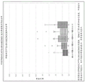


Classifications
-
- A—HUMAN NECESSITIES
- A61—MEDICAL OR VETERINARY SCIENCE; HYGIENE
- A61K—PREPARATIONS FOR MEDICAL, DENTAL OR TOILETRY PURPOSES
- A61K51/00—Preparations containing radioactive substances for use in therapy or testing in vivo
- A61K51/02—Preparations containing radioactive substances for use in therapy or testing in vivo characterised by the carrier, i.e. characterised by the agent or material covalently linked or complexing the radioactive nucleus
- A61K51/04—Organic compounds
- A61K51/08—Peptides, e.g. proteins, carriers being peptides, polyamino acids, proteins
- A61K51/10—Antibodies or immunoglobulins; Fragments thereof, the carrier being an antibody, an immunoglobulin or a fragment thereof, e.g. a camelised human single domain antibody or the Fc fragment of an antibody
- A61K51/1045—Antibodies or immunoglobulins; Fragments thereof, the carrier being an antibody, an immunoglobulin or a fragment thereof, e.g. a camelised human single domain antibody or the Fc fragment of an antibody against animal or human tumor cells or tumor cell determinants
- A61K51/1069—Antibodies or immunoglobulins; Fragments thereof, the carrier being an antibody, an immunoglobulin or a fragment thereof, e.g. a camelised human single domain antibody or the Fc fragment of an antibody against animal or human tumor cells or tumor cell determinants the tumor cell being from blood cells, e.g. the cancer being a myeloma
-
- A—HUMAN NECESSITIES
- A61—MEDICAL OR VETERINARY SCIENCE; HYGIENE
- A61K—PREPARATIONS FOR MEDICAL, DENTAL OR TOILETRY PURPOSES
- A61K51/00—Preparations containing radioactive substances for use in therapy or testing in vivo
- A61K51/02—Preparations containing radioactive substances for use in therapy or testing in vivo characterised by the carrier, i.e. characterised by the agent or material covalently linked or complexing the radioactive nucleus
- A61K51/04—Organic compounds
- A61K51/08—Peptides, e.g. proteins, carriers being peptides, polyamino acids, proteins
- A61K51/10—Antibodies or immunoglobulins; Fragments thereof, the carrier being an antibody, an immunoglobulin or a fragment thereof, e.g. a camelised human single domain antibody or the Fc fragment of an antibody
- A61K51/1093—Antibodies or immunoglobulins; Fragments thereof, the carrier being an antibody, an immunoglobulin or a fragment thereof, e.g. a camelised human single domain antibody or the Fc fragment of an antibody conjugates with carriers being antibodies
-
- A—HUMAN NECESSITIES
- A61—MEDICAL OR VETERINARY SCIENCE; HYGIENE
- A61B—DIAGNOSIS; SURGERY; IDENTIFICATION
- A61B6/00—Apparatus for radiation diagnosis, e.g. combined with radiation therapy equipment
- A61B6/02—Devices for diagnosis sequentially in different planes; Stereoscopic radiation diagnosis
- A61B6/03—Computerised tomographs
- A61B6/037—Emission tomography
-
- A—HUMAN NECESSITIES
- A61—MEDICAL OR VETERINARY SCIENCE; HYGIENE
- A61B—DIAGNOSIS; SURGERY; IDENTIFICATION
- A61B6/00—Apparatus for radiation diagnosis, e.g. combined with radiation therapy equipment
- A61B6/48—Diagnostic techniques
- A61B6/481—Diagnostic techniques involving the use of contrast agents
-
- A—HUMAN NECESSITIES
- A61—MEDICAL OR VETERINARY SCIENCE; HYGIENE
- A61K—PREPARATIONS FOR MEDICAL, DENTAL OR TOILETRY PURPOSES
- A61K51/00—Preparations containing radioactive substances for use in therapy or testing in vivo
- A61K51/02—Preparations containing radioactive substances for use in therapy or testing in vivo characterised by the carrier, i.e. characterised by the agent or material covalently linked or complexing the radioactive nucleus
- A61K51/04—Organic compounds
- A61K51/08—Peptides, e.g. proteins, carriers being peptides, polyamino acids, proteins
- A61K51/10—Antibodies or immunoglobulins; Fragments thereof, the carrier being an antibody, an immunoglobulin or a fragment thereof, e.g. a camelised human single domain antibody or the Fc fragment of an antibody
- A61K51/1093—Antibodies or immunoglobulins; Fragments thereof, the carrier being an antibody, an immunoglobulin or a fragment thereof, e.g. a camelised human single domain antibody or the Fc fragment of an antibody conjugates with carriers being antibodies
- A61K51/1096—Antibodies or immunoglobulins; Fragments thereof, the carrier being an antibody, an immunoglobulin or a fragment thereof, e.g. a camelised human single domain antibody or the Fc fragment of an antibody conjugates with carriers being antibodies radioimmunotoxins, i.e. conjugates being structurally as defined in A61K51/1093, and including a radioactive nucleus for use in radiotherapeutic applications
-
- A—HUMAN NECESSITIES
- A61—MEDICAL OR VETERINARY SCIENCE; HYGIENE
- A61P—SPECIFIC THERAPEUTIC ACTIVITY OF CHEMICAL COMPOUNDS OR MEDICINAL PREPARATIONS
- A61P35/00—Antineoplastic agents
-
- A—HUMAN NECESSITIES
- A61—MEDICAL OR VETERINARY SCIENCE; HYGIENE
- A61P—SPECIFIC THERAPEUTIC ACTIVITY OF CHEMICAL COMPOUNDS OR MEDICINAL PREPARATIONS
- A61P35/00—Antineoplastic agents
- A61P35/02—Antineoplastic agents specific for leukemia
-
- A—HUMAN NECESSITIES
- A61—MEDICAL OR VETERINARY SCIENCE; HYGIENE
- A61P—SPECIFIC THERAPEUTIC ACTIVITY OF CHEMICAL COMPOUNDS OR MEDICINAL PREPARATIONS
- A61P43/00—Drugs for specific purposes, not provided for in groups A61P1/00-A61P41/00
-
- C—CHEMISTRY; METALLURGY
- C07—ORGANIC CHEMISTRY
- C07K—PEPTIDES
- C07K16/00—Immunoglobulins [IGs], e.g. monoclonal or polyclonal antibodies
- C07K16/18—Immunoglobulins [IGs], e.g. monoclonal or polyclonal antibodies against material from animals or humans
- C07K16/28—Immunoglobulins [IGs], e.g. monoclonal or polyclonal antibodies against material from animals or humans against receptors, cell surface antigens or cell surface determinants
- C07K16/2803—Immunoglobulins [IGs], e.g. monoclonal or polyclonal antibodies against material from animals or humans against receptors, cell surface antigens or cell surface determinants against the immunoglobulin superfamily
- C07K16/2815—Immunoglobulins [IGs], e.g. monoclonal or polyclonal antibodies against material from animals or humans against receptors, cell surface antigens or cell surface determinants against the immunoglobulin superfamily against CD8
-
- G—PHYSICS
- G01—MEASURING; TESTING
- G01N—INVESTIGATING OR ANALYSING MATERIALS BY DETERMINING THEIR CHEMICAL OR PHYSICAL PROPERTIES
- G01N33/00—Investigating or analysing materials by specific methods not covered by groups G01N1/00 - G01N31/00
- G01N33/48—Biological material, e.g. blood, urine; Haemocytometers
- G01N33/50—Chemical analysis of biological material, e.g. blood, urine; Testing involving biospecific ligand binding methods; Immunological testing
- G01N33/53—Immunoassay; Biospecific binding assay; Materials therefor
- G01N33/531—Production of immunochemical test materials
- G01N33/532—Production of labelled immunochemicals
- G01N33/534—Production of labelled immunochemicals with radioactive label
-
- G—PHYSICS
- G01—MEASURING; TESTING
- G01N—INVESTIGATING OR ANALYSING MATERIALS BY DETERMINING THEIR CHEMICAL OR PHYSICAL PROPERTIES
- G01N33/00—Investigating or analysing materials by specific methods not covered by groups G01N1/00 - G01N31/00
- G01N33/48—Biological material, e.g. blood, urine; Haemocytometers
- G01N33/50—Chemical analysis of biological material, e.g. blood, urine; Testing involving biospecific ligand binding methods; Immunological testing
- G01N33/53—Immunoassay; Biospecific binding assay; Materials therefor
- G01N33/566—Immunoassay; Biospecific binding assay; Materials therefor using specific carrier or receptor proteins as ligand binding reagents where possible specific carrier or receptor proteins are classified with their target compounds
-
- G—PHYSICS
- G01—MEASURING; TESTING
- G01N—INVESTIGATING OR ANALYSING MATERIALS BY DETERMINING THEIR CHEMICAL OR PHYSICAL PROPERTIES
- G01N33/00—Investigating or analysing materials by specific methods not covered by groups G01N1/00 - G01N31/00
- G01N33/48—Biological material, e.g. blood, urine; Haemocytometers
- G01N33/50—Chemical analysis of biological material, e.g. blood, urine; Testing involving biospecific ligand binding methods; Immunological testing
- G01N33/68—Chemical analysis of biological material, e.g. blood, urine; Testing involving biospecific ligand binding methods; Immunological testing involving proteins, peptides or amino acids
- G01N33/6872—Intracellular protein regulatory factors and their receptors, e.g. including ion channels
-
- C—CHEMISTRY; METALLURGY
- C07—ORGANIC CHEMISTRY
- C07B—GENERAL METHODS OF ORGANIC CHEMISTRY; APPARATUS THEREFOR
- C07B2200/00—Indexing scheme relating to specific properties of organic compounds
- C07B2200/05—Isotopically modified compounds, e.g. labelled
-
- C—CHEMISTRY; METALLURGY
- C07—ORGANIC CHEMISTRY
- C07K—PEPTIDES
- C07K2317/00—Immunoglobulins specific features
- C07K2317/30—Immunoglobulins specific features characterized by aspects of specificity or valency
-
- C—CHEMISTRY; METALLURGY
- C07—ORGANIC CHEMISTRY
- C07K—PEPTIDES
- C07K2317/00—Immunoglobulins specific features
- C07K2317/50—Immunoglobulins specific features characterized by immunoglobulin fragments
- C07K2317/56—Immunoglobulins specific features characterized by immunoglobulin fragments variable (Fv) region, i.e. VH and/or VL
- C07K2317/565—Complementarity determining region [CDR]
-
- C—CHEMISTRY; METALLURGY
- C07—ORGANIC CHEMISTRY
- C07K—PEPTIDES
- C07K2317/00—Immunoglobulins specific features
- C07K2317/60—Immunoglobulins specific features characterized by non-natural combinations of immunoglobulin fragments
- C07K2317/62—Immunoglobulins specific features characterized by non-natural combinations of immunoglobulin fragments comprising only variable region components
- C07K2317/626—Diabody or triabody
-
- G—PHYSICS
- G01—MEASURING; TESTING
- G01N—INVESTIGATING OR ANALYSING MATERIALS BY DETERMINING THEIR CHEMICAL OR PHYSICAL PROPERTIES
- G01N2800/00—Detection or diagnosis of diseases
- G01N2800/52—Predicting or monitoring the response to treatment, e.g. for selection of therapy based on assay results in personalised medicine; Prognosis
-
- G—PHYSICS
- G01—MEASURING; TESTING
- G01N—INVESTIGATING OR ANALYSING MATERIALS BY DETERMINING THEIR CHEMICAL OR PHYSICAL PROPERTIES
- G01N2800/00—Detection or diagnosis of diseases
- G01N2800/56—Staging of a disease; Further complications associated with the disease
Abstract
Antigen binding constructs that bind CD8, and formulations and methods of using the same, are described herein.
Description
Priority
This application is PCT and claims priority from us application No. 62/738,938 filed on 28.9.2018 and us application No. 62/826,606 filed on 29.3.2019, both of which are hereby incorporated by reference in their entirety.
Sequence listing
This application is filed in conjunction with a sequence listing in electronic format. The sequence listing is provided as a file named ignab 047woseqilist. txt created on 27.9.9.2019, and is 126,124 bytes in size. The information in the sequence listing in electronic format is incorporated by reference herein in its entirety.
Background
Technical Field
The present technology relates generally to imaging and therapy aspects related to CD 8.
Background
CD8 (cluster of differentiation) is a transmembrane glycoprotein that is a specific marker of T cell subsets, including cytotoxic T cells. CD8 pools as heterodimers of the CD8 α and CD8 β subunits or CD8 α homodimers. The collective dimeric CD8 complex functions together with the T Cell Receptor (TCR) as a co-receptor to recognize antigen presentation by MHC class I cells. CD8 plays a role in the development of T cells and activation of mature T cells. Changes in T cell localization may reflect the progress of the immune response and may occur over time.
There are a variety of CD 8-directed antigen binding constructs, including those discussed in U.S. patent publication No. 20160024209.
Disclosure of Invention
In some embodiments, a method of monitoring CD8 in vivo is provided. The method comprises the following steps: providing a CD8 minibody to a subject, wherein the CD8 minibody binds CD8 as shown in figure 1C, and wherein the minibody is labeled with a detectable label; and monitoring the distribution of CD8 minibodies in the subject within 6-36 hours of administering the CD8 minibodies to the subject.
In some embodiments, the method of monitoring CD8 in vivo comprises: providing a CD8 minibody to a subject, wherein the CD8 minibody binds CD8 as shown in figure 1C, and wherein the minibody is labeled with a detectable label; and monitoring the distribution of CD8 minibodies in the subject, wherein the monitoring detects each mm in the subject2Tissue infiltrated with 500 or fewer cells carrying CD8, and wherein monitoring is achieved by PET.
In some embodiments, a method of visualizing CD8 positive cells comprises: providing a CD8 minibody to a subject, wherein the CD8 minibody is labeled with a detectable label, wherein the sequence of the CD8 minibody is humanized and binds to human CD 8; and monitoring the distribution of CD8 minibodies in the subject within 6-36 hours of administering the CD8 minibodies to the subject, wherein the monitoring is detectable per mm in the subject 2Tissue infiltrated with 500 or fewer CD8 positive cells, and wherein monitoring is achieved by PET.
In some embodiments, the cell count (e.g., 500 or less) is in terms of 10 microns or 30 microns depth.
In some embodiments, a method of visualizing a cell of a human comprises: providing a means for binding to human CD8 positive cells to a subject, wherein the means comprises a detectable label; and monitoring and determining a first distribution of the means for binding human CD8 positive cells of the subject within 6-36 hours of administering the means for binding human CD8 positive cells to the subject.
In some embodiments, a method of administering a minibody to a subject comprises providing to the subject a labeled minibody that binds human CD8, wherein 3mCi ± 20% radiation is provided to the subject by the labeled minibody, and wherein between 0.2mg and 10mg of total protein is provided to the subject.
In some embodiments, a method of administering a minibody to a subject comprises providing to the subject a labeled minibody that binds human CD8, wherein radiation of 0.75-1.5 +/-20% mCi is provided to the subject by the labeled minibody, and wherein the subject is provided with total protein in an amount between 0.2mg and 10 mg.
In some embodiments, a method of treating a patient comprises administering to a human patient diagnosed with cancer a dose of an antigen binding construct that binds human CD8, wherein the dose comprises: providing about 3mCi of radioactivity89A Zr-labeled antigen-binding construct; and about 10mg or less of the antigen-binding construct. The method further comprises the following steps: in applicationDetecting the patient with a first point in time after said dose89Zr-labeled antigen-binding construct to generate a first patient image corresponding to a first time point, wherein the first time point is about 6 hours to 36 hours after administration; determining a first abundance and/or distribution of CD8 cells in one or more tissues and/or neoplasia of the patient based on the first patient image; and administering to the patient a first treatment for the cancer based on the first abundance and/or distribution of CD8 cells in the one or more tissues.
In some embodiments, a method of treating a tumor comprises administering a minibody to a subject such that the minibody binds to the tumor in the subject to form a labeled tumor, wherein the minibody binds to human CD8, and wherein the minibody is linked to a detectable label. Administering a treatment selected from the group consisting of an immune checkpoint inhibitor if the minibody binds in a biased manner to the surface of any tumor in forming a labeled tumor. If the minibody binds to all the entire tumor in the tumor that forms the marker, no immune checkpoint inhibitor is administered.
In some embodiments, a method of treating a subject comprises: administering to a patient having a neoplasia a human minibody that binds human CD 8; monitoring the distribution of human miniantibodies to determine a first tumor infiltrating lymphocyte ("TIL") status within the neoplasia; and treating the patient based at least on the first TIL status of the neoplasia.
In some embodiments, a method of analyzing CD8 distribution in a subject comprises: providing an image of the distribution of the detectable label by the first PET image, wherein the detectable label is attached to the CD8 minibody, wherein the CD8 minibody binds to human CD8, and wherein the CD8 minibody does not elicit an immune response in the subject; providing an image of the distribution of the FDG markers from a second PET image of the subject; creating a third PET image comprising an overlay of the first PET image onto the second PET image; and identifying the tumor as a TIL based on the third PET image.
In some embodiments, a method of treating a subject comprises: administering to a patient having a neoplasia an antigen binding construct that binds human CD 8; monitoring the distribution of antigen binding constructs to determine tumor infiltrating lymphocyte ("TIL") status within the neoplasia; and administering immunotherapy ("IOT") to the patient if the at least one neoplastic TIL status is negative. This operation is optionally continued until the status becomes TIL positive.
In some embodiments, a method of treating a subject comprises: administering to a patient having a neoplasia an antigen binding construct that binds human CD 8; monitoring the distribution of antigen binding constructs to determine tumor infiltrating lymphocyte ("TIL") status within the neoplasia; and administering replacement immunotherapy ("IOT") to the patient if the tins status of the neoplasia is not positive. This operation is optionally continued until the status becomes TIL positive.
In some embodiments, a method of characterizing a tumor comprises: applying an antigen-binding construct that binds human CD8 to a human subject, wherein the antigen-binding construct comprises a detectable label; monitoring the distribution of the detectable label; generating an image based on the distribution of the detectable labels; and displaying the image to the user in a manner that allows the user to characterize the tumor.
In some embodiments, a method of generating an image comprises: using an antigen binding construct that binds CD8, wherein the antigen binding construct comprises a detectable label; detecting at least one detectable marker within a location within the tumor and assigning a positive pixel to each detectable marker detected; and generating an image based on the assigned distribution of each positive pixel, wherein the image is stored in a non-transitory computer readable medium.
In some embodiments, a method of positron emission tomography ("PET") comprises: administering to the subject a tracer that binds CD 8; providing a scintillator; detecting a pair of photons produced by the tracer using the scintillator; and using the detection of the pair of photons to locate the tracer source by a list of coincident events, the coincident events being processed by a processor configured to acquire an output from the scintillator and convert it to the list of coincident events, wherein each mm within the subject3Can detect between about 300 and about 500 CD8 positive cells.
In some embodiments, a method of making a diagnostic composition comprises: conjugating the minibody with desferrioxamine to form a Df-minibody; by using89Zr radiolabelling the Df-minibody to form a radiolabelled minibody; purifying the radiolabeled minibody; and mixing the radiolabeled minibody with the cold minibody to form a diagnostic composition, wherein the minibody and the cold minibody bind to the same epitope on CD 8.
In some embodiments, the diagnostic composition comprises: 3.0 + -20% mCi of 89Zr-Df labeled minibody; 20mM histidine; 5% of sucrose; 51-62mM sodium chloride; 141-194 arginine; and 2-20mM glutamic acid.
In some embodiments, the diagnostic composition comprises a labeled minibody comprising the structure:

wherein the miniantibody comprises a heavy chain variable region within SEQ ID NO 1, 3, 16 or 147 and a light chain variable region within SEQ ID NO 7, 9, 15 or 147 and wherein the composition provides at least 3mCi of radiation.
In some embodiments, the composition comprises: a first miniantibody that binds CD8, comprising an active label; and a second miniantibody that binds CD8, wherein the second miniantibody comprises an inactive label or lacks an active label.
In some embodiments, the diagnostic composition comprises: a first CD8 minibody conjugated to a radiolabel; and a second CD8 miniantibody not conjugated to a radiolabel, wherein the first CD8 miniantibody and the second CD8 miniantibody have the same sequence, wherein the composition provides about 3mCi of radiation, and wherein the composition provides 1.5-10mg of total mass protein.
In some embodiments, a method of imaging a subject comprises: administering a first dose of any of the compositions provided above; imaging a subject to obtain a first image; administering a second dose of any of the compositions provided above; and imaging the subject to obtain a second image; and comparing the first image with the second image.
In some embodiments, the method of determining a standard uptake value comprises: applying an antigen-binding construct to the subject, wherein the antigen-binding construct comprises a radioactive probe; determining r, wherein r is the concentration of radioactivity measured by a PET scanner within the ROI of radiation from the antigen binding construct (kBq/ml); determining a ', wherein a' is the amount of decay correction (kBq) of the injected radiolabeled tracer; determining w, the weight of the patient; and determining the SUV as a result of r (a'/W).
In some embodiments, a method of analyzing an image comprises: providing an image; defining a first region of interest (ROI) on the image by marking the image; determining signal strengths of data points within the first ROI; determining a maximum signal strength within the first ROI; determining an average of signal intensities within the first ROI; summing each signal intensity within the first ROI to obtain a first summed signal level for the first ROI, wherein the first ROI represents data on the amount of detectable label associated with the antigen-binding construct that has been administered to the subject.
In some embodiments, methods of administering an antigen-binding construct to a subject are provided. The method comprises providing to the subject a labeled antigen binding construct that binds human CD 8. The mark comprises89Zr, label provides 0.75 (or 0.5) -3mCi ± 20% radiation at the time of injection and provides total antigen binding construct to the subject in an amount between 0.2mg and 10 mg.
In some embodimentsMethods of treating a patient are provided. The method comprises the following steps: a) administering to a human patient diagnosed with cancer a dose of an antigen binding construct that binds human CD8, wherein the dose comprises: providing about 0.75 (or 0.5) to 3.6mCi of radioactivity89A Zr-labeled antigen-binding construct; and about 10mg or less of the antigen-binding construct; b) detecting the patient at a first point in time after administration of said dose89Zr-labeled antigen-binding construct to generate a first patient image corresponding to a first time point, wherein the first time point is about 6 hours to 36 hours after administration; c) determining a first abundance and/or distribution of CD8 cells in one or more tissues and/or neoplasia of the patient based on the first patient image; d) administering to the patient a first treatment for the cancer; e) administering a second dose of the antigen-binding construct to a human patient diagnosed with cancer after administering the first therapy; f) determining a second abundance and/or distribution of CD8 cells in one or more tissues and/or neoplasia of the patient based on the second patient image; and g) comparing the first patient image with the second patient image and administering a second treatment for the cancer to the patient based on the comparison of the first patient image with the second patient image.
In some embodiments, methods of treating a patient are provided. The method comprises the following steps: a) administering a dose of a CD8 PET tracer to a human patient diagnosed with cancer, wherein the dose comprises: a radioactive CD8 PET tracer providing about 0.75 (or 0.5) to 3.6mCi of radioactivity; and between about 5 micrograms and 50mg (or between about 20 micrograms and 10 mg) of a CD8 PET tracer; b) detecting the patient's CD8 PET tracer at a first time point after administration of the dose to generate a first patient image corresponding to the first time point, wherein the first time point is about 1 hour to 36 hours after administration; c) determining a first abundance and/or distribution of a CD8 PET tracer in one or more tissues and/or neoplasias of the patient based on the first patient image; d) administering to the patient a first treatment for the cancer; e) administering a second dose of a CD8 PET tracer to a human patient diagnosed with cancer after administration of the first therapy; f) determining a second abundance and/or distribution of a CD8 PET tracer in one or more tissues and/or neoplasias of the patient based on the second patient image; and g) comparing the first patient image with the second patient image and administering a second treatment for the cancer to the patient based on the comparison of the first patient image with the second patient image.
In some embodiments, the method comprises: administering to a patient having a neoplasia and being treated with immunotherapy ("IOT") an antigen-binding construct that binds human CD8, wherein the antigen-binding construct comprises a detectable label, wherein the antigen-binding construct does not elicit an immune response in the subject, wherein the detectable label comprises89Zr, wherein the detectable label provides 0.5-3mCi ± 20% of radiation upon injection, and wherein the patient is provided with a total antigen binding construct in an amount between 0.2mg and 10 mg; monitoring the distribution of antigen binding constructs to determine tumor infiltrating lymphocyte ("TIL") status within the neoplasia; and if the neoplastic TIL status is not positive, administering a replacement IOT to the patient, and repeating the foregoing method until the neoplastic TIL status becomes positive.
In some embodiments, methods of treating a human subject having cancer are provided. The method comprises the following steps: i) providing a first image of CD8 cells within a tumor of a subject using a CD8 antigen binding construct; ii) administering to the subject a therapy comprising a candidate therapeutic agent; iii) providing a second image of CD8 cells within the tumor of the subject using the CD8 antigen binding construct after administration of the therapy comprising the candidate therapeutic agent; and iv) comparing the first image with the second image to determine: a) whether the tumor shows increased CD8 infiltration or b) whether the tumor shows the same or decreased CD8 infiltration, wherein, if a), the subject is instructed to continue the therapy.
In some embodiments, methods of treating a human subject having cancer are provided. The method comprises the following steps: i) providing a first image of CD8 cells within a tumor of a subject using a CD8 PET tracer; ii) administering to the subject a therapy comprising a candidate therapeutic agent; iii) providing a second image of CD8 cells within the tumor of the subject using a CD8 PET tracer after administration of the therapy; and iv) comparing the first image with the second image to determine: a) whether the tumor shows increased CD8 infiltration or b) whether the tumor shows the same or decreased CD8 infiltration, wherein, if a), the subject is instructed to continue the therapy.
In some embodiments, methods of treating a human subject are provided. The method comprises the following steps: a) administering a candidate therapeutic agent to a human subject; b) determining whether the tumor size of the human subject increases or decreases in response to the candidate therapeutic agent; and c) if the size increases after the candidate therapeutic agent, determining whether the amount of CD8 present within the tumor corresponds to an increase or decrease in the candidate therapeutic agent using the CD8 antigen binding construct. An increase in tumor size without an increase in the amount of CD8 is indicative of tumor progression, whereas an increase in tumor size with an increase in the amount of CD8 is indicative of tumor pseudoprogression, wherein treatment is continued if the patient is undergoing tumor pseudoprogression, and wherein treatment is altered if the patient is undergoing tumor progression.
In some embodiments, methods of administering a marker to a human subject are provided. The method comprises the following steps: administering to a subject18F; within about 6 hours, the subject is PET scanned to obtain a subject18F distribution; administering a CD8 PET tracer to a subject, wherein the CD8 PET tracer comprises89Zr; and within about 36 hours of administration of the CD8 PET tracer, performing a PET scan on the subject to obtain89Distribution of Zr. In some embodiments, 18F is any PET isotope with a half-life of less than 3 hours.
In some embodiments, the diagnostic composition comprises: 3.0 + -20% mCi of an 89 Zr-Df-labeled antigen-binding construct; 20mM histidine; 5% of sucrose; 51-62mM sodium chloride; 141-194 arginine; and 2-20mM glutamic acid.
In some embodiments, the formulation of the CD8 composition comprises: a CD8 antigen-binding construct, wherein the CD8 antigen-binding construct is less than 105kDa in size; and associated with CD8 antigen binding constructs89A Zr radiolabel, wherein the radiolabel provides a radiation of greater than 0.5 but less than 3.6 (or 3.0) mCi for the formulation, wherein the formulation is configured for administration to a human.
Drawings
Fig. 1A shows some embodiments of a schematic representation of a miniantibody having bivalent binding to CD 8.
Fig. 1B shows some embodiments of a schematic of a minibody.
Fig. 1C provides an example of CD8 a.
FIG. 2A shows OKT8 variable weight (V)H) Regions relative to human antibody (4D5V8) and humanized V of the invention (IAB-huCD8 construct)HSome embodiments of the alignment of regions. Some embodiments of the CDR regions (Chothia) are indicated by boxed regions.
FIG. 2B shows that OKT8 is variably light (V)L) Region alignment to humanized VLSome embodiments of the alignment of regions and IAB-huCD8 constructs. Some embodiments of the CDR regions (Chothia) are indicated by boxed regions.
Figure 3A shows some embodiments of a schematic representation of cys-diabodies that exhibit bivalent binding to an antigen.
FIG. 3B shows a schematic representation of cys-diabodies that show bivalent binding to the antigen.
FIG. 4 shows chimeric IAB-huCD8 minibody VL-VHSome embodiments of the sequence.
FIG. 5 shows chimeric IAB-huCD8 minibody VH-VLSome embodiments of the sequence.
FIG. 6 shows humanized IAB-huCD8 minibody VL-VHSome embodiments of the sequence.
FIG. 7 shows humanized IAB-huCD8 minibody VH-VLSome embodiments of the sequence.
FIG. 8 shows humanized IAB-huCD8 cys-diabody VL-5-VHSome embodiments of the sequence.
FIG. 9 shows humanized IAB-huCD8 cys-diabody V H-5-VLSome embodiments of the sequence.
FIG. 10 shows humanized IAB-huCD8 cys-diabody VL-8-VHSome embodiments of the sequence.
FIG. 11 shows humanized IAB-huCD8 cys-diabody VH-8-VLSome embodiments of the sequence.
Figure 12A depicts some embodiments of sequences of cys-diabodies.
Fig. 12B depicts some embodiments of the sequence of a minibody.
FIG. 12C depicts VLSome embodiments of the sequence of (a).
FIG. 12D depicts huVLSome embodiments of the sequence of (a).
FIG. 12E depicts VHSome embodiments of the sequence of (a).
FIG. 12F depicts huVH(version "a" of version 1).
FIG. 12G depicts huVH(version "b" of version 1).
FIG. 12H depicts huVH(version "c" of version 2).
FIG. 12I depicts huVH(version "c" of version 2).
FIG. 13 shows pcDNATMSome embodiments of a vector map of myc-His (-) pattern A, B, C are 1/myc-His (-).
Fig. 14 illustrates some embodiments of a method of detecting the presence or absence of a target.
Figure 15 shows protein sequence information for some embodiments of IAB22M γ 2 EH 1.
Figure 16 shows protein sequence information for some embodiments of IAB22M γ 2 EH2 variants.
Fig. 17A shows protein sequence information for some embodiments of IAB22M γ 1 EH 1.
Fig. 17B shows protein sequence information for some embodiments of IAB22M γ 2 NH 1.
Fig. 17C shows protein sequence information for some embodiments of IAB22M γ 2 NH 2.
Fig. 18 is a diagrammatic representation of some embodiments of the hinge variants listed in table 3 (above γ 1 EH1, in γ 1 EH3, below γ 1 EH 4).
Fig. 19A shows protein sequence information for some embodiments of IAB22M γ 1 EH 3.
Fig. 19B shows protein sequence information for some embodiments of IAB22M γ 1 EH 5.
Figure 19C shows protein sequence information for some embodiments of IAB22M γ 3/γ 1 EH 6.
Figure 19D shows protein sequence information for some embodiments of IAB22M γ 3/γ 1 EH 7.
Figure 19E shows protein sequence information for some embodiments of IAB22M γ 3/γ 1 EH 8.
Figure 20 shows protein sequence information for some embodiments of IAB22M γ 1 EH 2.
Figure 21 shows the protein sequence information for VL and VH domains of Mb with different antigen specificities.
Figure 22 shows protein sequence information for various embodiments of linker sequences.
Figure 23 shows protein sequence information for various embodiments of the hinge region.
FIG. 24 shows C H3 domain, or a fragment thereof.
Fig. 25 shows an alignment of protein sequences of respective embodiments of IAB22M γ 1 EH1(M1) and IAB22M γ 1 EH3 (M1). Sequence differences are shown in boxes.
Fig. 26 shows the DNA and translated protein sequences of an embodiment of IAB22M γ 1 EH3 (M1). Signal, CDR, linker and hinge sequences are shown in boxes.
Fig. 27 shows the DNA and translated protein sequences of an embodiment of IAB22M γ 1 EH5 (M1).
Fig. 28 shows the DNA and translated protein sequences of an embodiment of IAB22M γ 1 EH7 (M1).
Fig. 29 shows the DNA and translated protein sequences of an embodiment of IAB22M γ 1 EH8 (M1).
Fig. 30 shows the DNA and translated protein sequences of an embodiment of IAB22M γ 2 EH2 (M1).
FIG. 31 shows the DNA and translated protein sequences of an embodiment of IAB22M γ 2 EH2(M1) with the VH-K67R polymorphism.
Figure 32 shows the protein sequence of an embodiment of the IAB22M VH domain.
Fig. 33 shows the protein sequence of an embodiment of the T cell surface glycoprotein CD8 alpha chain from homo sapiens.
Fig. 34 shows the protein sequence of an embodiment of the T cell surface glycoprotein CD8 β chain from homo sapiens.
Fig. 35 shows a schematic representation of an engineered minibody (Mb).
Figure 36 shows a schematic representation of various embodiments of an Mb hinge based on human IgG1 (above γ 1EH1 and below γ 1EH 2).
Figure 37 shows a schematic representation of various embodiments of an Mb hinge based on human IgG2 (above γ 2EH1 and below γ 2EH 2).
Figure 38 shows a schematic representation of various embodiments of additional Mb hinges based on human IgG2 (above γ 2 NH1 and below γ 2 NH 2).
Fig. 39 depicts some embodiments of CD8 minibodies.
FIG. 40 is a drawing showing some embodiments according to the present disclosure89Schematic illustration of the manufacture of Zr-Df-IAB22M 2C.
FIG. 41 shows a schematic representation of some embodiments according to the disclosure89Flow chart of cell steps and corresponding testing during fabrication of Zr-Df-IAB22M 2C.
FIGS. 42A and 42B show the results of a competitive FACS assay with AlexaFluor-488 pre-labeled IAB22M2C on HPB-ALL (FIG. 3A) and purified human T cells (FIG. 3B).
Figure 43 shows the results of a T cell proliferation assay using human PBMC.
Figure 44 shows the results of a human PBMC cytokine release assay.
Figure 45 shows the results of T cell survival (figure 6A) and T cell proliferation (figure 6B) assays using human PBMC.
FIG. 46 shows Table 3, which shows the intravenous 20 μ g/kg dose in humans 89Predicted pharmacokinetic parameters for Zr-Df-IAB22M 2C.
Fig. 47 shows the results of FACS analysis of T cell populations in blood of NSG mice transplanted with human CD34 stem cells at baseline.
Fig. 48 shows the results of FACS analysis of T cell population in blood of NSG mice transplanted with human CD34 stem cells at 24 hours.
FIG. 49 shows LegendPlex performed on serum samples from mice engrafted with human CD34 progenitor cellsTMResults of the Th1 bead assay.
FIGS. 50-54 depict PET results for 89ZR-Df-IAB22M2C 2.
Figure 55 is a flow chart of a schedule of use of some antigen binding constructs.
Fig. 56 is a flow diagram of some embodiments of various methods for screening and patient selection provided herein. The legend is shown in 5624 in fig. 56.
Fig. 57 is a flow diagram of some embodiments of various methods for treatment initiation and monitoring effectiveness provided herein. The legend is shown in 5719 in fig. 57.
Fig. 58 is a flow diagram of some embodiments of various methods for therapy monitoring toxicity provided herein. The legend is shown in 5824 of fig. 58.
Fig. 59 is a flow diagram of some embodiments of various methods for screening and patient selection for product concept use cases provided herein. The legend is shown in 5920 in fig. 59.
Figure 60 is a flow diagram of some embodiments of various methods for therapy initiation and monitoring effectiveness for a product use case provided herein. The legend is shown in 6019 in fig. 60.
Figure 61 is a flow diagram of some embodiments of various methods provided herein for therapy screening for product use case toxicity. The legend is shown in 6125 in fig. 61.
Fig. 62A is a diagram depicting a center read design outlined in embodiment 85.
Fig. 62B-62H show Time Activity Curves (TAC) for reference tissues as determined from SUV averages for each subject in phase I cohort. For fig. 62B to 62J, 62L, and 62M, the sequence of the legends below the graphs shows the sequence of the bars in the graphs.
FIGS. 62I and 62J are bar graphs of stage I and stage II at 0.5mg and 1.5 mg.
Fig. 62K is a graph showing nodule uptake frequency.
Fig. 62L and M are distribution plots of SUV maxima and SUV peak uptake values at each mean time point of target tumor lesion.
FIG. 62N generally depicts a PET scan, which is shown in use89The ability of the Zr labeled antigen binding construct to bind to the target (CD8) within 6 hours or 6 days of administration.
Fig. 63A is a graph depicting the steps that provide quantitative results of the correlation of radiotracer uptake with CD8+ T cell counts as determined by IHC analysis.
Fig. 63B and 63C are graphs showing the linear relationship between SUV-based measurements (SUV maxima and SUV peaks) and mean CD8+ T cell counts for baseline (a) and in-treatment (B) biopsy samples, including regression coefficients (r)2)。
FIG. 63D is a graph showing the inclusion of regression coefficients (r) when plotting baseline and in-treatment results together for SUV maxima (a) and SUV peaks (b)2) Graph of the linear relationship inside.
Fig. 64A depicts PET images from two subjects over several different time points.
Fig. 64B and 64C depict the combination of FDG PET and CD8 PET to more fully characterize the tumor microenvironment.
FIG. 64D is a responder image (PET/CT).
Figure 64E is a PET scan and a fused PET/CT scan showing the results of testing the effectiveness of various treatments using the minibody constructs.
FIG. 64F is a non-responder image (PET and PET/CT scans).
Detailed Description
The present disclosure provides methods for visualizing CD8 molecules and thus CD8+ T cells in vivo by non-invasive methods. In some embodiments, a radiolabeled antigen-binding construct of the disclosure (e.g., a monoclonal antibody or a monoclonal antibody fragment thereof) is administered to a subject in need thereof89Zr-Df-IAB22M2C) is a PET imaging agent for in vivo detection of CD8+ T cells in human patients. In some embodiments, the methods of the present disclosure can provide local and systemic immune responses to immunotherapy It should be better understood that a generally superior immunotherapy or cancer therapy is selected for a subject and/or that novel immunotherapeutics for treating cancer are generated.
Provided herein are various methods and compositions relating to monitoring, diagnosing, and treating various cancers associated with CD8 expression, including various compositions thereof and additional alternative uses thereof. Some embodiments provided herein allow for monitoring the distribution of CD8 molecules and/or CD 8-bearing cells (e.g., CD8+ T cells) in a subject. In some embodiments, this is achieved, at least in part, using antigen binding constructs (such as minibodies) that are labeled and bind to human CD 8.
In some embodiments, the properties of the antigen-binding construct allow for excellent timing of distribution of the antigen-binding construct (which may be linked to or associated with a detectable label), may be imaged on the same day, or may be imaged for multiple days in a single dose.
Some embodiments provided herein allow for better characterization of tumors or neoplasias in human subjects. For example, the process may allow for determining how or how effectively various CD 8-bearing cells infiltrate a tumor or neoplasia. Based on this process, the correct therapy choice for the subject can then be determined by, for example, providing an alternative treatment to observe which is most effective in changing the state of the tumor or neoplasia from one state to another. For example, by being able to observe the immunophenotype, it is possible to determine what the subject's current immunophenotype is (e.g., immune desert, immune rejection, or TIL positive (or inflamed)), and then determine whether this state should be changed, what therapy should be used to change the state, and in the case of a new state, what follow-up therapy should be used to treat the patient.
The skilled person will understand that "characterization of a tumor" as employed in the present invention may be represented by various words and phrases other than "immune desert, immune rejection or TIL positive (or inflamed)". Some practitioners will use the "hot", "cold" or "intermediate" scales to describe observations. Other characterizations may provide more detailed histological information about CD 8-bearing lymphocytes, such as localization (invasive margin, tumor surface, tumor core); or the density (e.g., immunodensity and number) of the tumor. Other characterizations can distinguish whether a "hot" tumor is uniformly or non-uniformly infiltrated. Some professionals may prefer a more comprehensive four-class classification of tumors: hot tumors, rejection-altered tumors, immunosuppression-altered tumors, and cold tumors. All such words and phrases are acceptable for describing the observations of the present invention, and the use of any one phrase herein is not intended to exclude the use of any other suitable description of an observation.
In addition, various methods for analyzing and characterizing the information obtained using these CD8 antigen binding constructs are also provided, allowing, for example, for the provision of the correct therapy to a subject.
As provided herein, the use of these CD8 antigen binding constructs, methods, and formulations can be used to determine the CD8 status of neoplasias. This information can then be used in various methods, for example, to determine whether immunotherapy in general matches the subject well (e.g., neoplasia TIL positive). Alternatively, this information can be used to screen a subject for various therapies to determine whether a particular immunotherapy is effective in the subject when the subject receives it (e.g., a neoplasia is initially immune-exclusive or immune-desert, but becomes TIL-positive (or inflamed)). In general, any of the methods provided (for monitoring or treatment, etc.) may be used in either arrangement. Thus, disclosure of one method in a particular instance or embodiment is also intended to disclose another method (in the corresponding particular instance or embodiment). However, subsequent steps (e.g., i) administration of immunotherapy to TIL-positive subjects will differ relative to ii) administration of immunotherapy (or new immunotherapy) to subjects to change their tumor phenotype to TIL-positive).
The following section provides various definitions of various terms as well as a brief listing of various embodiments of some of the terms. This section is followed by a detailed description of various methods, as well as additional details regarding various options and embodiments contemplated for the invention provided herein. That section is followed by a series of examples.
Definitions and various embodiments
Unless defined otherwise, the plain and ordinary meaning of the terms as understood by those of ordinary skill in the art applies.
"treating" a condition may refer to preventing the condition, slowing the onset and/or rate of progression of the condition, reducing the risk of developing the condition, preventing and/or delaying the progression of symptoms associated with the condition, reducing or terminating the symptoms associated with the condition, producing a complete or partial regression of the condition, or some combination thereof. The term "preventing" does not require absolute prohibition of a condition or disease. Treatment includes immunophenotyping (from desert to rejection to TIL positive) that alters the tumor or neoplasia in the subject and subsequent therapeutic applications for tumors of a particular phenotype.
A "therapeutically effective amount" or "therapeutically effective dose" is an amount that produces a desired therapeutic effect (e.g., preventing, treating a target condition, delaying the onset of a disorder and/or symptom, and/or alleviating a symptom associated with a condition) in a subject. This amount will vary depending on a variety of factors including, but not limited to, the characteristics of the therapeutic compound (including activity, pharmacokinetics, pharmacodynamics, and bioavailability), the physiological condition of the subject (including age, sex, disease type and stage, general physical condition, responsiveness to a given dose, and type of drug), the nature of the pharmaceutically acceptable carrier(s) in the formulation, and the route of administration. Given the present disclosure, one of skill in the clinical and pharmacological arts can determine a therapeutically effective amount by routine experimentation, for example, by monitoring the subject's response to administration of the compound and adjusting the dosage accordingly. For further guidance, see Remington, The Science and Practice of Pharmacy, 21 st edition, Univ.of Sciences In Philadelphia (USIP), Lippincott Williams & Wilkins, Philadelphia, Pa., 2005.
The term "CD 8PET tracer" denotes any molecule that can associate or selectively bind to CD8 and associate a marker or tag with a CD8 molecule. This includes aspects such as antigen binding constructs, antibodies, minibodies, diabodies, cys-diabodies, nanobodies, and the like. Also included within the scope of the CD8PET tracers are small peptides and molecules that selectively bind to CD8 and to which a PET label or PET detectable label may be associated (e.g., linked or covalently bonded). Other examples of CD8PET tracers include CD 8-specific capture agents, such as those disclosed in WO2017/176769, which is incorporated herein by reference in its entirety with respect to such CD-8-specific capture agents. In some embodiments, any of the methods provided herein can use a CD8 capture agent (or simply "ligand") as provided in WO2017/176769, including capture agents of any of the following:
(1) HGRGH-linker-wplrf, which targets epitope 2C (AAEGLDTQR) and epitope IN (SQFRVSPLD).
(2) HGRGH-linker-AKYRG, which targets epitope 2C (AAEGLDTQR) and epitope IN (SQFRVSPLD).
(3) ghwpp-linker-hGrGh, which targets epitope 2n (fllylsqnkp) and epitope 2c (aaegldtqr).
(4) PWTHG-linker-AKYRG, which targets epitope 2n (fllylsqnkp) and epitope in (sqfvspld).
In some embodiments, the molecule that binds CD8 consists of or comprises one or more of the following:
1) a sequence 80-100% identical to at least one of: HGSYG; KRLGA; AKYRG; h, hellw; lrgyw; vshf; gnvh; h.wplrf; rwfnv; havwh; wvplw; ffrly; and m.wyygf; or
2) A sequence having 80-100% identity to an amino acid sequence selected from the group consisting of: agdsw; HVRHG; HGRGH; THPTT; e.FAGYH; f.WTEHG; pwthg; tndfd; LFPFD; slrfG; yfrgs; wnwvG; m.vawlG; fhvhg; wvsnv; wsvnv; InshG; r.yGGvr; nsvhg; ttvhg; fdvgh; rhgwk; (w.Ghtwp; and x.hgrgh.
In some embodiments, methods for screening and/or preparing capture agents may also be found in international publication nos. WO 2012/106671, WO 2013/033561, WO 2013/009869, and WO 2014/074907, each of which is incorporated herein by reference in its entirety. In some embodiments, the CD8 PET tracer is less than 200kDA, 170kDA, 150kDA, 120kDA, 105kDA, 100kDA, 80kDA, 50kDA, 30kDA, 10kDA, 5kDA, or 2 kDA.
The term "antigen binding construct" includes all kinds of antibodies, including binding fragments thereof. Also included are constructs comprising 1, 2, 3, 4, 5, and/or 6 CDRs. In some embodiments, these CDRs may be distributed between their appropriate framework regions in a traditional antibody. In some embodiments, the CDRs may be comprised within the heavy and/or light chain variable regions. In some embodiments, the CDRs may be within the heavy and/or light chain. In some embodiments, the CDRs may be within a single peptide chain. In some embodiments, the CDRs may be within two or more peptides that are covalently linked together. In some embodiments, they may be covalently linked together by disulfide bonds. In some embodiments, they may be linked by a linking molecule or moiety. In some embodiments, the antigen binding protein is non-covalent, such as diabodies and monovalent scfvs. Unless otherwise specified herein, the antigen binding constructs described herein bind to the target molecule. The term also includes miniantibodies.
The term "CD 8 antigen-binding construct" refers to an antigen-binding construct that binds to CD 8. This includes minibodies, cys-diabodies, ScFv, etc. Unless expressly or excluded by context, the term also includes full-length antibodies. CD8 means "human CD 8", which means human CD8 alpha chainHGNC:HGNC:1706Ensembl: ENSG00000153563MIM: 186910; human CD8 beta chainHGNC:HGNC:1707Ensembl: ENSG00000172116 MIM: 186730. In some embodiments, the CD8 antigen binding construct is single chain. In some embodiments, the CD8 antigen binding construct is a linked multiplex. In some embodiments, the CD8 antigen binding construct is humanized. In some embodiments, the CD8 antigen binding construct exhibits no or low immunogenicity when administered (repeatedly) to humans. In some embodiments, the CD8 antigen binding construct is humanized and exhibits no or low immunogenicity when administered (in duplicate) to humans. In some embodiments, unless otherwise indicated, the CD8 antigen-binding construct includes a detectable label.
The terms "cell proliferative disorder" and "proliferative disorder" refer to a disorder associated with a degree of abnormal cell proliferation. In one embodiment, the cell proliferative disorder is cancer.
As used herein, "tumor" refers to all neoplastic cell growth and proliferation, whether malignant or benign, and all precancerous and cancerous cells and tissues. As referred to herein, the terms "cancer," "cancerous," "cell proliferative disorder," "proliferative disorder," and "tumor" are not mutually exclusive. The term "neoplasia" encompasses the term tumor.
Unless otherwise indicated, "tumor" means a solid tumor; including neoplasia and any abnormal cell growth of the subject's human cells (but not including infection by foreign organisms).
By "surface of a tumor" or "tumor surface" is meant the periphery of a tumor mass that is in contact with normal (e.g., non-tumor and non-tumor-induced) cells of a subject. Sometimes interchangeably referred to as "tumor margin" or "tumor boundary". At the cellular level, its thickness may range from a few cells to hundreds of cells, and may not be uniformly integrated with surrounding normal cells.
The terms "cancer" and "cancerous" refer to or describe the physiological condition of a mammal that is generally characterized by unregulated cell growth. Examples of cancer include, but are not limited to, carcinoma, lymphoma, blastoma, sarcoma, and leukemia or lymphoid malignancies. More specific examples of such cancers include squamous cell cancer (e.g., epithelial squamous cell cancer), lung cancer (including small-cell lung cancer, non-small cell lung cancer, adenocarcinoma of the lung, and squamous carcinoma of the lung), cancer of the peritoneum, hepatocellular cancer, gastric (including gastrointestinal cancer), pancreatic cancer, glioblastoma, cervical cancer, ovarian cancer, liver cancer, bladder cancer, urinary tract cancer, hepatoma, breast cancer, colon cancer, rectal cancer, colorectal cancer, endometrial or uterine carcinoma, salivary gland carcinoma, kidney cancer, prostate cancer, vulval cancer, thyroid cancer, hepatic carcinoma, anal carcinoma, penile carcinoma, melanoma, multiple myeloma and B-cell lymphoma, brain cancer, as well as head and neck cancer and related metastases. The term cancer includes adult and childhood solid cancers. In some embodiments, the cancer may be a solid tumor.
The term "target" or "target molecule" denotes a CD8 protein. Examples of CD8 proteins are known in the art and include, for example, the CD8 protein of SEQ ID NO:24 (FIG. 1C), or FIG. 33, or FIG. 34. With respect to binding of antigen binding constructs that bind CD8, for example, the term CD8 denotes the alpha chain of CD8, the beta chain of CD8, and both the alpha and beta chains of CD 8.
The term "antibody" includes, but is not limited to, genetically engineered or otherwise modified forms of immunoglobulins, such as, for example, intrabodies, chimeric antibodies, fully human antibodies, humanized antibodies, antibody fragments, and heteroconjugate antibodies (e.g., bispecific antibodies, diabodies, triabodies, tetrabodies, and the like). The term "antibody" includes cys-diabodies and minibodies. Thus, unless otherwise expressly indicated, each and every embodiment provided herein with respect to an "antibody" is also contemplated as a cys-diabody and/or minibody embodiment. The term "antibody" includes polypeptides of the immunoglobulin family or polypeptides comprising fragments of an immunoglobulin capable of binding a corresponding antigen non-covalently, reversibly and in a specific manner. Exemplary antibody building blocks include tetramers. In some embodiments, a full-length antibody may be composed of two pairs of identical polypeptide chains, each pair having one "light" chain and one "heavy" chain connected by a disulfide bond. Recognized immunoglobulin genes include the kappa, lambda, alpha, gamma, delta, epsilon and mu constant region genes, as well as myriad immunoglobulin variable region genes. For full-length chains, light chains are classified as either kappa or lambda. For full-length chains, heavy chains are classified as gamma, mu, alpha, delta, or epsilon, which in turn define the immunoglobulin classes IgG, IgM, IgA, IgD, and IgE, respectively. The N-terminus of each chain defines a variable region of about 100 to 110 or more amino acids primarily responsible for antigen recognition. The term variable light chain (V) L) And a variable heavy chain (V)H) These regions refer to light and heavy chains, respectively. As used herein, "antibody" encompasses all variations of antibodies and fragments thereof. Thus, full length antibodies, chimeric antibodies, humanized antibodies, single chain antibodies (scFv), Fab 'and multimeric versions of these fragments (e.g., F (ab')2) Within the scope of this concept. In some embodiments, the antibody specifically binds to a desired target.
"complementarity determining domain" or "Complementarity Determining Region (CDR)" refer interchangeably to VLAnd VHA hypervariable region. CDRs are the target protein binding sites of antibody chains, which are specific for this target protein. In some embodiments, each V isLAnd/or VHIn which there are three CDRs (CDR1-3, numbered sequentially from the N-terminus) that constitute about 15-20% of the variable domain. The CDRs are structurally complementary to epitopes of the target protein and are therefore directly responsible for the binding specificity. VLOr VHThe amino acid sequence of the remaining fragment of (a so-called Framework Region (FR)) shows minor variations (Kuby, Immunology, 4 th edition, chapter 4. W.H.Freeman)&Co.,New York,2000)。
The positions of the CDRs and framework regions may be determined using various well-known definitions in the art, such as Kabat (Wu, T.T., E.A.Kabat.1970.Annalysis of the Sequences of the variable regions of the beer Jones Proteins and myelomas light chains and the hair innovations for the anti-impact property.J.Exp.Med.132: 211-250; Kabat, E.A., Wu, T.T., Perry, H.H., Gotteseman, K., and Foeller, C. (1991) Sequences of Proteins of biological Interest, 5 th edition, NIH Publication No.91-3242, Bethesda, MD); chothia (Chothia and Lesk, J.mol.biol.,196: 901-; ImmunoGeneTiCs database (IMGT) (world Wide Web IMGT. org /) (Giudicelli, V., Duroux, P., Ginestoux, C., Folch, G., Jabado-Michaloud, J., Chaume, D. and Lefranc, M. -P.IMGT/LIGM-DB, the  (iii) a compound database of immunoglobulin and T cell receptor nucleotide sequences Nucl. acids Res.,34, D781-D784(2006), PMID: 16381979; lefranc, M.P., Pommi, C.A., Ruiz, M.S., Giudicelli, V.S., Foulquier, E.S., Truong, L.S., Thievenin-Contet, V.S. and Lefranc, G.S., IMGT unique number for Immunoglobulin and T cell receiver variable domains and Ig superfamiliesly V-like domains dev.comp.immunol.,27,55-77(2003) PMID: 12477501; brochet, X, Lefranc, M, P, and Giudilli, V.IMGT/V-QUEST the high throughput system for IG and TR staged V-J and V-D-J sequence analysis nucleic acids Res,36, W503-508 (2008); AbM (Martin et al, proc. natl. acad. sci. usa,86: 9268-.
The term "binding specificity determinant" or "BSD" interchangeably refers to the smallest contiguous or non-contiguous sequence of amino acids within a complementarity determining region that is necessary to determine the binding specificity of an antibody. The minimum binding specificity determinant may be within one or more CDR sequences. In some embodiments, the minimum binding specificity determinant is located within (i.e., is determined solely by) a portion of or the full length of the antibody heavy and light chain CDR3 sequences. In some embodiments, CDR3 of the heavy chain variable region is sufficient for antigen binding construct specificity.
The terms "preferentially binds" and "unbiased binding" with respect to the surface of a tumor refer to tumor images where it is observed that the detectable label is substantially bound to the surface of the tumor (relatively reduced or absent binding within the tumor) ("preferentially") or that the detectable label is not significantly associated with the surface of the tumor ("unbiased") and may, for example, be uniformly or non-uniformly dispersed throughout the interior of the tumor or not present at all in the tumor. "biased" includes selectively binding to any tumor margin without substantially perfusing the tumor volume.
The term "region of interest" or "ROI" means a sub-region of an image that is selected by a human operator, optionally with assistance by an automated or semi-automated imaging processing method (which closely limits the image region identifying a tumor), or expected to contain a tumor based on other diagnostic methods (e.g., FDG-PET, CT scan, MRI, biopsy, visual examination, etc.), in or on an image of the CD8 distribution of a human subject.
The terms "pseudo-progression" and "pseudo-progression" are used interchangeably in the academic literature and herein. Hyper-progression (hyper-progression) is a rapid increase in disease progression after initiation of immunotherapy. In some embodiments, any of the pseudo-progression methods provided herein can also be applied to hyper-progression.
The term "active label" with respect to a composition comprising a CD8 antigen-binding construct is interchangeable with "detectable label" and means a chemical moiety or compound that can be detected by interrogation-based emission or by spontaneous emission using a specified detection method that recognizes the emission, such labels including fluorescent imaging components or radioactive labels. "Inactive marker" refers to a compound or chemical moiety on a construct that is no longer detectable using a given detection method due to chemical breakdown or due to previous spontaneous emission such that the compound or chemical moiety is no longer detectable in such detection method.
In monitoring, detecting, comparing, or observing administration to a subject89In the context of the distribution of the Zr-associated CD8 antigen binding construct, the term "distribution" means a visual image of the biodistribution of the detected marker associated with the CD8 antigen binding construct scanned relative to the whole or part of the body of the subject, which image may be represented as a planar image (two-dimensional) or a computer-assisted three-dimensional representation (including a hologram), and which is in a form that can be used by an operator or clinician to observe the distribution of the CD8 antigen binding construct at the level of the individual tissue and at the level of the individual tumor. In advanced imaging modalities, the "distribution" may not be a visual image of a whole-body or partial-body scan, but may be a report of a computerized assessment of the presence and TIL status of a tumor in a subject. "will 18F distribution and89zr profile comparison "requires a scan of subjects against each profile so that individual tissues and tumors can be compared.
Criteria for solid tumor response evaluation ("RECIST"). For Computed Tomography (CT) anatomical imaging of tumor response, the most common criterion is RECIST. This evaluated the size of the tumor to account for changes, growth indicated treatment failed, and a decrease in tumor size indicated treatment success.
Solid tumor PET reaction markQuasi ("PERCIST"). In the use of 18F-FDGPositron emission tomographyIn nuclear imaging for metabolic tumor response assessment of (PET), the criterion is the solid tumor PET response criterion (PERCIST).
As used herein, "antibody variable light chain" or "antibody variable heavy chain" refers to a variable domain comprising V, respectivelyLOr VHThe polypeptide of (1). Endogenous VLEncoded by gene segments V (variable) and J (joining), and endogenous VHEncoded by V, D (diversity) and J. VLOr VHEach of which includes CDRs and framework regions. In the present application, an antibody variable light chain and/or an antibody variable heavy chain may sometimes be collectively referred to as an "antibody chain". These terms encompass the inclusion of nondestructive VLOr VHAs would be readily recognized by one skilled in the art. In some embodiments, full-length heavy and/or light chains are contemplated. In some embodiments, it is contemplated that only heavy and/or light chain variable regions are present.
Antibodies can exist as intact immunoglobulins or as numerous fragments produced by digestion with various peptidases. Thus, for example, pepsin digests antibody below disulfide linkages in the hinge region to produce F (ab)'2Which is itself linked to V via a disulfide bondH-C H1 conjugated light chain (V)L-CL) The dimer of Fab' of (1). F (ab)'2It may be reduced under mild conditions to cleave disulfide linkages in the hinge region, thereby cleaving F (ab)'2The dimer is converted to Fab' monomer. The Fab' monomer is a Fab with a hinge region portion. (Paul, Fundamental Immunology 3 rd edition (1993)). Although various antibody fragments are defined in terms of digestion of intact antibodies, the skilled artisan will recognize that such fragments can be synthesized de novo either chemically or by using recombinant DNA methods. Thus, the term "antibody" as used herein also includes antibody fragments produced by modification of whole antibodies, or synthesized de novo using recombinant DNA methods (e.g., single chain Fv), or identified using phage display libraries (see, e.g., McCafferty et al, Nature 348: 552-.
For the preparation of Monoclonal or polyclonal Antibodies, any technique known in the art may be used (see, for example, Kohler and Milstein, Nature 256: 495-. Techniques for producing single chain antibodies (U.S. Pat. No. 4,946,778) can be adapted to produce antibodies to polypeptides of the invention. Transgenic mice or other organisms (e.g., other mammals) can also be used to express fully human monoclonal antibodies. Alternatively, phage display technology can be used to identify high affinity binders to selected antigens (see, e.g., McCafferty et al, supra; Marks et al, Biotechnology,10: 779-.
Methods for humanizing or primatizing non-human antibodies are well known in the art. Typically, humanized antibodies have one or more amino acid residues introduced into them from a non-human source. These non-human amino acid residues are often referred to as import residues, which are typically taken from an import variable domain. In some embodiments, the terms "donor" and "acceptor" sequences may be used. Humanization can be essentially performed by replacing the corresponding sequences of a human antibody with rodent CDRs or CDR sequences (see, e.g., Jones et al, Nature 321:522-525 (1986); Riechmann et al, Nature 332:323-327 (1988); Verhoeyen et al, Science 239:1534-1536(1988) and Presta, curr. Op. struct. biol.2:593-596(1992)) according to the methods of Winter and co-workers. Thus, such humanized antibodies are chimeric antibodies (U.S. Pat. No. 4,816,567) in which substantially less than an entire human variable domain is replaced by the corresponding sequence from a non-human species. In practice, humanized antibodies are typically human antibodies in which some complementarity determining region ("CDR") residues and possibly some framework ("FR") residues are substituted by residues from analogous sites in rodent antibodies.
A "chimeric antibody" is an antibody molecule in which: (a) the constant region or a portion thereof is altered, replaced or exchanged such that the antigen binding site (variable region) is linked to a different or altered class, effector function and/or species of constant region or an entirely different molecule (e.g., enzymes, toxins, hormones, growth factors and drugs) that confers new properties to the chimeric antibody; or (b) the variable region or a portion thereof is altered, replaced or exchanged by a variable region having a different or altered antigenic specificity.
Antibodies also include one or more immunoglobulin chains chemically conjugated to or expressed as fusion proteins with other proteins. In some embodiments, the antigen binding construct may be a monovalent scFv construct. In some embodiments, the antigen-binding construct may be a bispecific construct. Bispecific or bifunctional antibodies are artificial hybrid antibodies with two different heavy/light chain pairs and two different binding sites. Other antigen-binding fragments or antibody portions of the invention include bivalent scFv (diabodies), bispecific scFv antibodies (where the antibody molecule recognizes two different epitopes), single binding domains (sdabs or nanobodies), and minibodies.
The term "antibody fragment" includes, but is not limited to, one or more antigen-binding fragments of an antibody, alone or in combination with other molecules, including, but not limited to, Fab ', F (ab')2Fab, Fv, rIgG (reduced IgG), scFv fragments (monovalent, trivalent, etc.), single domain fragments (nanobodies), peptide antibodies, minibodies, diabodies, and cys-diabodies. The term "scFv" refers to a single chain Fv ("variable fragment") antibody in which the variable domains of the heavy and light chains of a traditional diabody are joined to form one chain.
A pharmaceutically acceptable carrier may be a pharmaceutically acceptable substance, composition or vehicle involved in carrying or transporting a compound of interest from one tissue, organ or part of the body to another tissue, organ or part of the body. For example, the carrier may be a liquid or solid filler, diluent, excipient, solvent, or encapsulating material, or some combination thereof. Each component of the carrier is "pharmaceutically acceptable" in that it is compatible with the other ingredients of the formulation. It must also be suitable for use in contact with any body tissue, organ or part it may encounter, which means that it must not carry the risk of toxicity, irritation, allergic response, immunogenicity, or any other complications that outweigh their therapeutic benefits. The pharmaceutical compositions described herein may be administered by any suitable route of administration. The route of administration may refer to any route of administration known in the art, including, but not limited to, aerosol, enteral, nasal, ophthalmic, oral, parenteral, rectal, transdermal (e.g., topical creams or ointments, patches), or vaginal administration. "transdermal" administration may be achieved using a topical cream or ointment or by a transdermal patch. "parenteral" refers to a route of administration typically associated with injection, including intraorbital, infusion, intraarterial, intracapsular, intracardiac, intradermal, intramuscular, intraperitoneal, intrapulmonary, intraspinal, intrasternal, intrathecal, intrauterine, intravenous, subarachnoid, subcapsular, subcutaneous, transmucosal, or transtracheal administration. In some embodiments, the antigen binding construct may be delivered intraoperatively as a local application during intervention or resection.
The term "CD 8-dependent disorder" includes cancers in which an immune component (including response to cancer immunotherapy) is present, autoimmune disorders, inflammatory disorders, and cancers including, but not limited to, lung cancer, ovarian cancer, colorectal melanoma, and the like.
Miniantibodies are a form of antibody that is smaller in molecular weight than full-length antibodies while maintaining bivalent binding properties against an antigen. Due to the smaller size, the minibodies clear more rapidly from the system and penetration is enhanced when targeting tumor tissue. Due to the strong targeting and rapid clearance capabilities, minibodies are advantageous for diagnostic imaging and delivery of cytotoxic/radioactive payloads, and their extended cycle time can lead to adverse patient dosing or dosimetry.
The phrase "specifically (or selectively) binds," when used in the context of describing an interaction between an antigen (e.g., a protein) and an antibody or antibody-derived binding agent, refers to determining the binding reaction of an antigen present in a heterogeneous population of proteins and other biological agents (e.g., in a biological sample, such as blood, serum, plasma, or tissue sample). Thus, under specified immunoassay conditions, in some embodiments, an antibody or binding agent with a particular binding specificity binds to a particular antigen at least twice that of the background and does not substantially bind in significant amounts to other antigens present in the sample. Specific binding to an antibody or binding agent under such conditions may require selection of the antibody or agent for its specificity for a particular protein. A variety of immunoassay formats can be used to select antibodies specifically immunoreactive with a particular protein. For example, solid phase ELISA immunoassays are routinely used to select Antibodies specifically immunoreactive with a protein (see, e.g., Harlow and Lane, Using Antibodies, a Laboratory Manual (1998) for a description of immunoassay formats and conditions that can be used to determine specific immunoreactivity). Typically, a specific or selective binding reaction will produce a signal that is at least two times greater than background and more typically at least 10 to 100 times greater than background.
The term "equilibrium dissociation constant (K)DM) "refers to the dissociation rate constant (k)dTime of day-1) Divided by the association rate constant (k)aTime of day-1,M-1). The equilibrium dissociation constant can be measured using any method known in the art. Antibodies of the invention typically have an equilibrium dissociation constant of less than about 10-7Or 10-8M, e.g. less than about 10-9M or 10-10M, in some embodiments less than about 10-11M、10-12M or 10-13M。
The term "isolated" when applied to a nucleic acid or protein means that the nucleic acid or protein is substantially free of other cellular components with which it is associated in its native state. In some embodiments, it may be an anhydrous solution or an aqueous solution. Purity and homogeneity can be determined using analytical chemistry techniques such as polyacrylamide gel electrophoresis or high performance liquid chromatography. The protein, which is the major species present in the preparation, is substantially purified. In particular, the isolated gene is separated from the open reading frame that flanks the gene and encodes a protein other than the gene of interest. The term "purified" means that the nucleic acid or protein produces a substantially single band in the electrophoresis gel. In some embodiments, this may mean that the nucleic acid or protein is at least 85% pure, more preferably at least 95% pure, and most preferably at least 99% pure of the molecule present under in vivo conditions.
The term "nucleic acid" or "polynucleotide" refers to deoxyribonucleic acid (DNA) or ribonucleic acid (RNA) in single-or double-stranded form, and polymers thereof. Unless specifically limited, the term encompasses nucleic acids containing known analogs of natural nucleotides that have similar binding properties as the reference nucleic acid and are metabolized in a manner similar to naturally occurring nucleotides. Unless otherwise indicated, a particular nucleic acid sequence also implicitly encompasses conservatively modified variants thereof (e.g., degenerate codon substitutions), alleles, orthologs, SNPs, and complementary sequences, as well as the sequence explicitly indicated. In particular, degenerate codon substitutions may be achieved by generating sequences in which the third position of one or more selected (or all) codons is substituted with mixed base and/or deoxyinosine residues (Batzer et al, Nucleic Acid Res.19:5081 (1991); Ohtsuka et al, J.biol.chem.260: 2605-.
The terms "polypeptide," "peptide," and "protein" are used interchangeably herein to refer to a polymer of amino acid residues. The terms apply to amino acid polymers in which one or more amino acid residues is an artificial chemical mimetic of a corresponding naturally occurring amino acid, as well as to naturally occurring amino acid polymers and non-naturally occurring amino acid polymers.
The term "amino acid" refers to naturally occurring and synthetic amino acids, as well as amino acid analogs and amino acid mimetics that function in a manner similar to the naturally occurring amino acids. Naturally occurring amino acids are those encoded by the genetic code, as well as those amino acids that are later modified, e.g., hydroxyproline, γ -carboxyglutamate, and O-phosphoserine. Amino acid analogs refer to compounds having the same basic chemical structure (i.e., hydrogen-binding alpha carbon, carboxyl group, amino group, and R group) as a naturally occurring amino acid, e.g., homoserine, norleucine, methionine sulfoxide, methionine methyl sulfonium. The analogs have modified R groups (e.g., norleucine) or modified peptide backbones, but retain the same basic chemical structure as a naturally occurring amino acid. Amino acid mimetics refers to compounds that have a structure that is different from the general chemical structure of an amino acid, but that functions in a manner similar to a naturally occurring amino acid.
"conservatively modified variants" applies to both amino acid and nucleic acid sequences. With respect to particular nucleic acid sequences, conservatively modified variants refers to those nucleic acids which encode identical or essentially identical amino acid sequences, or where the nucleic acid does not encode an amino acid sequence, to essentially identical sequences. A large number of functionally identical nucleic acids encode any given protein due to the degeneracy of the genetic code. For example, the codons GCA, GCC, GCG and GCU all encode the amino acid alanine. Thus, at each position where an alanine is specified by a codon, the codon can be changed to any of the corresponding codons described without changing the encoded polypeptide. Such nucleic acid variations are "silent variations," which are one type of conservatively modified variations. Each nucleic acid sequence herein that encodes a polypeptide also describes each possible silent variation of the nucleic acid. The skilled artisan will recognize that each codon in a nucleic acid (except AUG, which is typically the only codon for methionine, and TGG, which is typically the only codon for tryptophan) can be modified to produce a functionally identical molecule. Thus, each silent variation of a nucleic acid encoding a polypeptide is implicit in each described sequence.
With respect to amino acid sequences, one of skill will recognize that individual substitutions, deletions or additions of an alteration, addition or deletion of a nucleic acid, peptide, polypeptide, or protein sequence to a single amino acid or a small percentage of amino acids in the encoded sequence are "conservatively modified variants" where the alteration results in the substitution of an amino acid with a chemically similar amino acid. Conservative substitution tables providing functionally similar amino acids are well known in the art. These conservatively modified variants are in addition to and do not exclude polymorphic variants, interspecies homologs, and alleles of the invention.
The following eight groups each contain amino acids that are conservative substitutions for one another: 1) alanine (a), glycine (G); 2) aspartic acid (D), glutamic acid (E); 3) asparagine (N), glutamine (Q); 4) arginine (R), lysine (K); 5) isoleucine (I), leucine (L), methionine (M), valine (V); 6) phenylalanine (F), tyrosine (Y), tryptophan (W); 7) serine (S), threonine (T); and 8) cysteine (C), methionine (M) (see, e.g., Creighton, Proteins (1984)).
"percent sequence identity" can be determined by comparing two optimally aligned sequences over a comparison window, wherein the portion of the polynucleotide sequence in the comparison window can comprise additions or deletions (i.e., gaps) as compared to the reference sequence (e.g., a polypeptide of the invention which does not comprise additions or deletions) to achieve optimal alignment of the two sequences. The percentages are calculated by: the number of positions at which the identical nucleic acid base or amino acid residue occurs in both sequences is determined to give the number of matched positions, the number of matched positions is divided by the total number of positions in the window of comparison, and the result is multiplied by 100 to give the percentage of sequence identity.
The terms "identical" and "percent identity," in the context of two or more nucleic acid or polypeptide sequences, refer to two or more sequences or subsequences that are the same sequence. Two sequences are "substantially identical" if they have a specified percentage of amino acid residues or nucleotides that are identical (e.g., 70%, 75%, 80%, 85%, 90%, 95%, 96%, 97%, 98%, or 99% sequence identity over the designated region, or over the entire sequence of a reference sequence when not designated) when compared and aligned for maximum correspondence over a comparison window or designated region, as measured using one of the following sequence comparison algorithms or by manual alignment and visual inspection. Some embodiments provided herein provide a polypeptide or polynucleotide that is substantially the same as the polypeptide or polynucleotide exemplified herein, respectively (e.g., a variable region as exemplified in any of fig. 2A, 2B, or 4-11, 12C-12I; a CDR as exemplified in any of fig. 2A, 2B, or 12C-12I; a FR as exemplified in any of fig. 2A, 2B, or 12C-12I; and a nucleic acid sequence as exemplified in any of fig. 12A-12I, or 4-11). Optionally, identity exists over a region of at least about 15, 25 or 50 nucleotides in length, or more preferably over a region of 100 to 500 or 1000 or more nucleotides in length, or over the full length of the reference sequence. With respect to amino acid sequences, identity or substantial identity may exist over a region of at least 5, 10, 15, or 20 amino acids in length, optionally at least about 25, 30, 35, 40, 50, 75, or 100 amino acids in length, optionally at least about 150, 200, or 250 amino acids in length, or over the entire length of a reference sequence. With respect to shorter amino acid sequences, e.g., amino acid sequences of 20 or fewer amino acids, in some embodiments, substantial identity exists when one or two amino acid residues are conservatively substituted, according to conservative substitutions as defined herein.
For sequence comparison, typically one sequence acts as a reference sequence to which test sequences are compared. When using a sequence comparison algorithm, the test sequence and the reference sequence are input into a computer, subsequence coordinates are designated (if necessary), and sequence algorithm program parameters are designated. Default program parameters may be used or alternative parameters may be specified. The sequence comparison algorithm then calculates the percent sequence identity of the test sequence relative to the reference sequence based on the program parameters.
As used herein, a "comparison window" includes reference to a segment of contiguous positions selected from any one of a number consisting of 20 to 600, typically about 50 to about 200, more typically about 100 to about 150, wherein after two sequences are optimally aligned, the sequences can be compared to a reference sequence of the same number of contiguous positions. Methods of alignment of sequences for comparison are well known in the art. Optimal alignment of sequences for comparison can be performed, for example, by: the local homology algorithm of Smith and Waterman (1970) adv.Appl.Math.2:482 c; homology alignment algorithm of Needleman and Wunsch (1970) J.mol.biol.48: 443; pearson and Lipman (1988) Proc.nat' l.Acad.Sci.USA 85: 2444; computer implementation of these algorithms (GAP, BESTFIT, FASTA and TFASTA in the Wisconsin Genetics software package, Genetics Computer Group (Genetics Computer Group), 575Science Dr., Madison, Wis.); or manual alignment and visual inspection (see, e.g., Ausubel et al, Current Protocols in Molecular Biology (1995 supplement)).
Two examples of algorithms suitable for determining percent sequence identity and sequence similarity are the BLAST and BLAST 2.0 algorithms described in Altschul et al (1977) Nuc. acids Res.25:3389-3402 and Altschul et al (1990) J.mol.biol.215:403-410, respectively. Software for performing BLAST analysis is publicly available through the National Center for Biotechnology Information. This algorithm involves first identifying high scoring sequence pairs (HSPs) by identifying short fields of length W in the query sequence that match or satisfy some positively valued threshold score T when aligned with a field of the same length in a database sequence. T is referred to as the neighborhood field score threshold (Altschul et al, supra). These initial neighborhood field hits act as seeds for initiating searches to find longer HSPs containing them. As long as the cumulative sequence alignment score can be increased, the field hits extend in both directions along each sequence. Cumulative scores were calculated for nucleotide sequences using the parameters M (reward score for a pair of matching residues, usually >0) and N (penalty for mismatching residues, usually < 0). For amino acid sequences, cumulative scores were calculated using a scoring matrix. The extension of field hits in each direction stops when: the cumulative alignment score is decreased by an amount X from its maximum value reached; (ii) a cumulative score that falls to 0 or below as a result of accumulation of one or more negative-scoring residue alignments; or to the end of either sequence. The BLAST algorithm parameters W, T and X determine the sensitivity and speed of the alignment. The BLASTN program (for nucleotide sequences) uses the default values: word length (W)11, expectation (E)10, M-5, N-4, and a comparison of the two chains. For amino acid sequences, the BLASTP program uses the following defaults: word length 3, and expectation (E)10, and BLOSUM62 scoring matrix (see Henikoff and Henikoff (1989) proc. natl. acad. sci. usa 89:10915) alignment (B)50, expectation (E)10, M5, N-4, and two-strand comparison.
The BLAST algorithm also performs a statistical analysis of the similarity between two sequences (see, e.g., Karlin and Altschul (1993) Proc. Natl. Acad. Sci. USA90: 5873-. One measure of similarity provided by the BLAST algorithm is the smallest sum probability (P (N)), which indicates the probability by which a match between two nucleotide or amino acid sequences occurs by chance. For example, a nucleic acid is considered similar to a reference sequence if the smallest sum probability in a comparison of the test nucleic acid to the reference nucleic acid is less than about 0.2, more preferably less than about 0.01, and most preferably less than about 0.001.
Two nucleic acid sequences or polypeptides that are substantially identical are indicated as: as described below, the polypeptide encoded by the first nucleic acid is immunologically cross-reactive with an antibody raised against the polypeptide encoded by the second nucleic acid. Thus, in some embodiments, the polypeptide is generally substantially identical to the second polypeptide, e.g., where the two polypeptides differ only by conservative substitutions. Another indication that two nucleic acid sequences are substantially identical is: as described below, two molecules or their complements hybridize to each other under stringent conditions. Yet another indication that two nucleic acid sequences are substantially identical is: the same primers can be used to amplify the sequences.
The terms "subject," "patient," and "individual" interchangeably refer to the entity being examined and/or treated. This may include, for example, mammals, such as human or non-human primate mammals. The mammal may also be a laboratory mammal, such as a mouse, rat, rabbit, hamster. In some embodiments, the mammal may be an agricultural mammal (e.g., equine, ovine, bovine, porcine, camel) or a domestic mammal (e.g., canine, feline).
The terms "therapeutically acceptable amount" or "therapeutically effective dose" interchangeably refer to an amount sufficient to achieve a desired result. In some embodiments, the therapeutically acceptable amount does not induce or cause undesirable side effects. A therapeutically acceptable amount can be determined by first administering a low dose and then gradually increasing the dose until the desired effect is achieved.
The term "co-administration" refers to the administration of two active agents in the blood of an individual or in a sample to be tested. The co-administered active agents may be delivered concurrently or sequentially.
"label" or "detectable label" are used interchangeably herein and refer to a detectable compound or composition that is conjugated directly or indirectly to an antibody to generate a "labeled" antibody. The label may be detectable by itself (e.g., radioisotope labels or fluorescent labels) or, in the case of an enzymatic label, may catalyze chemical alteration of a substrate compound or composition that is detectable.
The term "immunopet" is a term used for Positron Emission Tomography (PET) of radiolabeled antibodies.
The term "cytotoxic agent" as used herein refers to a substance that inhibits or prevents cellular function and/or causes cell death or destruction. The term is intended to include radioisotopes (e.g., At211, I131, I125, Y90, Re186, Re188, Sm153, Bi212, P32, Pb212, and radioisotopes of Lu), chemotherapeutic agents (e.g., methotrexate (methotrexate), adriamycin (adriamicin), vinca alkaloids (vincristine), vinblastine (vinblastine), etoposide (etoposide)), doxorubicin (doxorubicin), melphalan (melphalan), mitomycin c (mitomycin c), chlorambucil (chlorembucil), daunorubicin (daunorubicin), or other inserters, enzymes, and fragments thereof, such as nucleolytic enzymes, antibiotics, and toxins such as small molecule toxins or enzymatically active toxins of bacterial, fungal, plant or animal origin, including fragments and/or variants thereof, toxins, growth inhibitory agents, drug moieties, and various anti-neoplastic or anti-cancer agents disclosed below.
A "toxin" is any substance that can have a deleterious effect on the growth or proliferation of a cell.
A "chemotherapeutic agent" is a chemical compound that can be used to treat cancer. Examples of chemotherapeutic agents include: alkylating agents, such as thiotepa and CYTOXANTMCyclophosphamide; alkyl sulfonates such as busulfan, improsulfan and piposulfan; aziridines such as bentopa (benzodopa), carbaquinone (carboquone), mitripopa (meteredopa), and ulidapa (uredpa); ethyleneimine and methylmelamine, including hexamethylmelamine, triethylenemelamine (triethylemelamine), triethylenephosphoramide (triethylenephosphoramide), triethylenethiophosphoramide (triethylenethiophosphoramidine), and trimethylolmelamine (trimetylomelamine); polyacetylene (acetogenin) (especially bullatacin and bullatacin); delta-9-tetrahydrocannabinol (dronabinol), MARINOLTM) (ii) a Beta-lapachone (beta-lapachone); lapachol (lapachol); colchicine; betulinic acid; camptothecin (including the synthetic analogue topotecan (HYCAMTIN)TM) CPT-11 (irinotecan), CAMPTOSAR TM) Acetyl camptothecin, scopolectin (scopolectin) and 9-aminocamptothecin); bryostatin; kelitin (callystatin); CC-1065 (including its adozelesin (adozelesin), carvelesin (carzelesin), and bizelesin (bizelesin) synthetic analogs); podophyllotoxin (podophylotoxin); podophyllinic acid (podophyllic acid); teniposide (teniposide); cryptophycin (especially cryptophycin 1 and cryptophycin 8); dolastatin (dolastatin); duocarmycins (duocarmycins) (including the synthetic analogs KW-2189 and CB1-TM 1); eiscosahol (eleutherobin); pancratistatin; sarcandra glabra alcohol (sarcodictyin); spongistatin (spongistatin); nitrogen mustards such as chlorambucil, chlorophosphamide, estramustine (estramustine), ifosfamide, dichloromethyl diethylamine (mechlorethamine), dichloromethyl diethylamine oxide hydrochloride, melphalan, neonebustine (novembichin), benzene mustarne (pherenesterone), prednimustine (prednimustine), chloroacetoamide (trofosfamide), uracil mustard (uracilmustard); nitrosoureas such as carmustine (carmustine), chlorozotocin (chlorozotocin), fotemustine (fotemustine), lomustine (lomustine), nimustine (nimustine) and ramustine (ranirnustine); Antibiotics, such as enediyne antibiotics (e.g., calicheamicin, particularly calicheamicin γ 1I and calicheamicin ω I1 (see, e.g., Agnew, Chem Intl.Ed.Engl.,33:183- > 186 (1994))); daptomycin (dynemicin), including daptomycin a; epothilones (esperamicins); and neocarzinostatin (neocarzinostatin) chromophores and related chromoproteenediyne antibiotic chromophores, aclacinomycin (acacetin), actinomycin (actinomycin), amphenicol (authramycin), azaserine (azaserine), bleomycin (bleomycin), actinomycin C (cactinomycin), karabicin (carabicin), carminomycin (carminomycin), carcinomycin (carzinophilin), chromomycin (chromomycin), actinomycin D (dactinomycin), daunomycin, ditorexin (desoubicin), 6-diazo-5-oxo-L-norleucine, ADRIAMYCINTMDoxorubicin (including morpholino-doxorubicin, cyanomorpholino-doxorubicin, 2-pyrrolinyl-doxorubicin and deoxydoxorubicin), epirubicin (epirubicin), esorubicin (esorubicin), idarubicin (idarubicin), marijumycin (marcelomycin), mitomycin (mitomycin) (such as mitomycin C), mycophenolic acid (mycophenolic acid), nogomycin (nogalamycin), olivomycin (olivomycin), pelomycin (pelomycin), peziomycin (polyplomycin), pofiromycin (porfiromycin), puromycin (puromycin), triiron doxorubicin (quelamycin), rodobicin (rodorubicin), streptonigrin (streptonigrogrin), streptozocin (streptazocin), tubercidin (tucidin), ubenidin (medrystatin), zostatin (zorubicin), zorubicin (zorubicin); antimetabolites such as methotrexate and 5-fluorouracil (5-FU); folic acid analogs such as denopterin, methotrexate, pteropterin, trimetrexate; purine analogs such as fludarabine (fludarabine), 6-mercaptopurine (6-mercaptopurine), thiamiprine (thiamiprine), thioguanine (thioguanine); pyrimidine analogs such as cyclocytidine (ancitabine), azacitidine (azacitidine), 6-azauridine (6-azauridine), carmofur (carmofur), cytarabine (cytarabine), dideoxyuridine (dideoxyuridine), deoxyfluorouridine (doxifluridine), enocitabine (enocitabine), floxuridine (floxuridine); androgens (androgens), such as Such as dimethyltestosterone (calusterone), dromostanolone propionate (dromostanolone propionate), epithioandrostanol (epitiostanol), mepiquitane (mepiquitane), testolactone (testolactone); anti-adrenal agents such as aminoglutethimide (aminoglutethimide), mitotane (mitotane), trostane (trilostane); folic acid (folic acid) supplements such as frolinic acid (frolicic acid); acetyl glucuronate (acegultone); (ii) an aldophosphamide glycoside; aminolevulinic acid (aminolevulinic acid); eniluracil (eniluracil); amsacrine (amsacrine); doubly-branched betuzucil; bisantrene; edatrexate (edatraxate); deflazafamine (defofamine); dimecorsine (demecolcine); diazaquinone (diaziqutone); edenisol (elfornitine); ammonium etitanium acetate; epothilone (epothilone); etoglut (etoglucid); gallium nitrate (gallium nitrate); hydroxyurea (hydroxyurea); lentinan (lentinan); lonidanine (lonidainine); maytansinoids such as maytansinoid (maytansine) and ansamitocins (ansamitocin); mitoguazone (mitoguzone); mitoxantrone (mitoxantrone); mupidumol (mopidanmol); diamine nitracridine (nitrarine); pentostatin (pentostatin); phenamet (phenamett); pirarubicin (pirarubicin); losoxantrone (losoxantrone); 2-ethyl hydrazide; procarbazine (procarbazine); rtm. polysaccharide complex (JHS Natural Products, Eugene, Oreg.); razoxane (rizoxane); rhizomycin (rhizoxin); sisofilan (sizofiran); helical germanium (spirogermanium); tenuazonic acid (tenuazonic acid); triimine quinone (triaziquone); 2,2' -trichlorotriethylamine; trichothecenes (trichothecenes) (especially T-2 toxin, vilasoline A (verracutinin A), bacillocin A (roridin A) and serpentinine (anguidine)); urethane (urethan); vindesine (eldinine) TM,FILDESINTM) (ii) a Dacarbazine (dacarbazine); mannitol mustard (mannomustine); dibromomannitol (mitobronitol); dibromodulcitol (mitolactol); pipobromane (pipobroman); ganciclovir (gacytosine); arabinoside ("Ara-C"); thiotepa (thiotepa); taxanes (taxoids), such as taxol. rtm. paclitaxel (Bristol-Myers squirb Oncology, prince)eton,N.J.)、ABRAXANETMAlbumin-engineered paclitaxel nanoparticle formulations without polyoxyethylated castor oil (Cremophor-free) (American Pharmaceutical Partners, Schaumberg, Ill.) and TAXOTERETMDocetaxel (docetaxel) (Rhone-Poulenc Rorer, Antony, France); chlorambucil; gemcitabine (gemcitabine) (GEMZAR)TM) (ii) a 6-thioguanine (6-thioguanine); mercaptopurine (mercaptoprine); methotrexate; platinum analogs such as cisplatin (cissplatin) and carboplatin (carboplatin); vinblastine (vinblastine) (VELBAN)TM) (ii) a Platinum; etoposide (VP-16); ifosfamide; mitoxantrone; vincristine (vincristine) (ONCOVIN)TM) (ii) a Oxaliplatin (oxaliplatin); leucovorin (leucovovin); vinorelbine (vinorelbine) (NAVELBINE)TM) (ii) a Norfloxacin (novantrone); edatrexate (edatrexate); daunomycin; aminopterin (aminopterin); ibandronate (ibandronate); topoisomerase inhibitor RFS 2000; difluoromethylornithine acid (DMFO); retinoids such as retinoic acid (retinic acid); capecitabine (XELODA) TM) (ii) a A pharmaceutically acceptable salt, acid or derivative of any of the foregoing; and combinations of two or more of the foregoing, such as CHOP, an abbreviation for combination therapy of cyclophosphamide, doxorubicin, vincristine, and prednisolone (prednisolone); and FOLFOX, i.e. oxaliplatin (ELOXATIN)TM) Abbreviation for treatment protocol in combination with 5-FU and folinic acid.
Also included in this definition are anti-hormonal agents which are used to modulate, reduce, block or inhibit the action of hormones which can promote the growth of cancer and are often in the form of systemic or systemic treatment. They may be hormones themselves. Examples include antiestrogens and Selective Estrogen Receptor Modulators (SERMs), including for example tamoxifen (including NOLVADEX)TMTamoxifen), EVISTATMRaloxifene, droloxifene, 4-hydroxyttamoxifen, troloxifene, keoxifene, LY117018, onapristone, and FARESTONTMToremifene; antiprogestin; estrogen receptor down-regulator (ERD); agents for inhibiting or arresting the ovary, e.g. luteinizing hormone-releasing hormone (luteinizing hormone-r)An elassing hormone, LHRH) agonist such as LUPRONTMAnd ELIGARDTMLeuprorelin acetate, goserelin acetate, buserelin acetate, and triptorelin (tripterelin); other antiandrogens such as flutamide, nilutamide and bicalutamide; and aromatase inhibitors which inhibit aromatase, an enzyme which regulates estrogen production in the adrenal gland, such as 4(5) -imidazole, aminoglutethimide, MEGASE TMMegestrol acetate, AROMASINTMExemestane, formestane (formestanie), fadrozole, rivistorTMVorozole, FEMARATMLetrozole and ARIMIDEXTMAnastrozole. In addition, such definitions of chemotherapeutic agents include bisphosphonates, such as clodronate (e.g., BONEFOS)TMOr OSTACTM)、DIDROCALTMEtidronate, NE-58095, ZOMETATMZoledronic acid/zoledronic acid salt, FOSAMAXTMAlendronate, AREDIATMPamidronate, SKELIDTMTiludronate or ACTONELTMRisedronate; and troxacitabine (1, 3-dioxolane nucleoside cytosine analogues); antisense oligonucleotides, particularly those that inhibit gene expression in signaling pathways involved in abnormal cell proliferation, such as PKC- α, Raf, H-Ras and Epidermal Growth Factor Receptor (EGFR); vaccines, such as THERATOPETMVaccines and gene therapy vaccines, e.g. ALLOVECTINTMVaccine, LEUVECTINTMVaccine and VAXIDTMA vaccine; LURTOTECANTMA topoisomerase 1 inhibitor; ABARELIXTMrmRH; lapatinib ditosylate salt (ErbB-2 and EGFR double tyrosine kinase small molecule inhibitor, also known as GW 572016); and pharmaceutically acceptable salts, acids or derivatives of any of the above chemotherapeutic agents.
As used herein, "growth inhibitory agent" refers to a compound or composition that inhibits cell growth in vitro or in vivo. Thus, the growth inhibitory agent may be one that significantly reduces the percentage of cells in S phase. Examples of growth inhibitory agents include agents that block cell cycle progression (at points other than S phase), such as agents that induce G1 arrest and M phase arrest. Classical M-phase blockers include the vinca rosea (vinpocetine)Neoalkaloids and vinblastine), taxanes and topoisomerase II inhibitors such as doxorubicin, epirubicin, daunorubicin, etoposide and bleomycin. Those agents that arrest G1 also spill over into S phase arrest, for example DNA alkylating agents such as tamoxifen, prednisone, dacarbazine, mechlorethamine, cisplatin, methotrexate, 5-fluorouracil and cytarabine. Additional information can be found in chapter 1 of "The Molecular Basis of Cancer", edited by Mendelsohn and Israel, under The name "Cell cycle regulations, oncogenes, and anticancer drugs" (WB Saunders: Philadelphia,1995), especially page 13. Taxanes (paclitaxel and docetaxel) are anticancer drugs derived from the yew tree. Docetaxel (TAXOTERE) from european sweater TMRhone-Poulenc Rorer) is paclitaxel (TAXOL)TMBristol-Myers Squibb). Paclitaxel and docetaxel promote the assembly of microtubules from tubulin dimers and stabilize microtubules by preventing depolymerization, which results in the inhibition of mitosis in cells.
"immunotherapy" (also referred to as "immunostimulation" and "IOT") means the prevention or treatment of a disease with a therapy (e.g., a dose or course of therapy) that stimulates the host's immune response to the disease. Many diseases can be treated with immunotherapy. Recent academic literature often refers specifically to immunooncology using immunotherapy, which represents a cancer treatment aimed at reducing the immune-evasive character of a tumor or neoplasia, allowing natural or modified immune cells to identify and eliminate cancerous tissue. As used herein, immunotherapy refers to immunooncology, unless the context specifically indicates otherwise. Depending on the context, "immunotherapy" may also refer to immunotherapeutic agents or to methods of using such agents. Various immunotherapeutic agents are now available and are more in clinical and preclinical development. Well-known immunotherapeutic agents include, but are not limited to: checkpoint inhibitor ("CPI") therapies (e.g., anti-PD-1: (CPI))  Pembrolizumab) or anti-PD-L1 (
 Nivolumab) binding agent); IL2 and fragments or prodrugs thereof (e.g., NKTR-214, a prodrug of PEG-conjugated IL2 (aldesleukin)); other CD122(
IL2RB interleukin 2 receptor subunit beta) binding ligands, GAd-NOUS-20 neo-antigen vaccines (D' Alise et al 2017; which can enhance NKTR-214 activity); t cell bispecific agent therapy; therapies for reversing T cell depletion, inhibiting indoleamine 2, 3-dioxygenase (IDO), such as the use of indomethastat (INCB 024360); and CAR-T therapy.
The term "immune checkpoint inhibitor" (sometimes referred to as "ICI"), or "checkpoint inhibitor" (sometimes referred to as "CPI"), or "immune checkpoint blockade inhibitor" and all similar terms refer to a subset of immunotherapy. Examples include molecules that block certain proteins produced by some types of immune system cells (e.g., T cells) and some cancer cells. These proteins help to maintain a check for immune responses and may prevent T cells from killing cancer cells. When these proteins are blocked, the immune system is free to move and T cells are able to kill cancer cells. Some embodiments include anti-PD 1 and anti-PD-L1 binding agents, anti-CTLA 4 agents, and multispecific agents including, but not limited to, anti-CTLA-4/B7-1/B7-2. Other immunotherapies include checkpoint inhibitors such as ipilimumab (Yervoy), pembrolizumab (keytruruda), nivolumab (Opdivo), atuzumab (Tecentriq), avizumab (Bavencio), and tevolumab (infinzi). IOT also includes Techimumab and PIDISUBIRUMAB, small molecule ICI is also under development, including BMS-1001, BMS-1116, CA-170, CA-327, Imiquimod (Imiquimod), Resiquimod (Resiquimod), 852A, VTX-2337, ADU-S100, MK-1454, ibrutinib, 3AC, Idelalisib (Idelalisib), IPI-549, Epacadostat, AT-38, CPI-444, Vepadendan (Vipadendan), Prebutan (Preladiant), PBF, AZD4635, Galenistritib (Galenicillib), OTX015/MK-8628, CPI-0610 (refer to Ke and Chisolm (2019) Journal of The Murology, 2019,202: 11-19)
IOT also includes other modalities that are not CPI but also activate the host immune system against the cancer or render the tumor susceptible to CPI therapy. Such alternative IOT include, but are not limited to: t cell immunomodulators such as cytokines IL-2, IL-7, IL-15, IL-21, IL-12, GM-CSF and IFN α (including THOR-707 by Synthorx Therapeutics; and NKTR-214 bempegdesleukin by Nektar Therapeutics); various other interferons and interleukins; TGF-beta 1 inhibitors (e.g., SRK-181 being developed by Scholar Rock); oncolytic therapy (including oncolytic viral therapy); adoptive cell therapies such as T cell therapy (including CAR-T cell therapy); cancer vaccines (based on prevention and treatment). Immunotherapy also includes strategies to increase neoantigen burden in tumor cells, including targeted therapies that cause tumor cells to express or reveal tumor-associated antigens. (see Galon and Bruni (2019) Nature Reviews Drug Discovery v.18, pp. 197-218). Other IOTs include TLR9 ligand (Checkmate Pharmaceuticals), A2A/A2B dual antagonist (Arcus Biosciences), and vaccination peptides against endogenous enzymes such as IDO-1 and arginase (IO Biotech). IOT includes HS-110, HS-130 and PTX-35(Heat Biologics).
Those skilled in the art recognize that immunotherapy may be used in combination with each other. Immunotherapy can also be used before, after or in combination with other therapies of the disease, including radiation therapy in the case of cancer, all types of chemotherapy (including the cytotoxic, chemotherapeutic, anti-hormonal, and growth inhibitory agents mentioned above), and surgical resection.
In some embodiments, any method of treatment that acts on or through CD8 may be used. This is called "CD 8 therapy". This includes: immunotherapy; cell therapy; a vaccine; therapeutic agents to enhance/restore immune response/function; any treatment that affects the presence, activation, number or trafficking of CD8+ T cells associated with a neoplastic lesion; chemotherapy; an oncolytic virus; radiotherapy; cell therapy, and the like.
"tumor infiltrating lymphocytes" or "TILs" refer to lymphocytes found within the borders of a solid tumor, such as CD 8-bearing T cells.
The "tumor infiltrating lymphocyte status", or "TIL" status, or other similar terms, indicate the extent to which lymphocytes, and in particular CD 8-bearing T cells, can penetrate into the tumor, or neoplasia, or tumor stroma. When CD8 cells can penetrate themselves into the tumor or tumor stroma and are themselves found within the tumor or tumor stroma, the neoplasia is characterized as TIL positive. TIL-positive can be further classified as TIL-positive homogeneous (evenly distributed throughout the tumor) or TIL-positive heterogeneous (significantly unevenly or partially distributed within the tumor). TIL-positive tumors can also be described as "inflamed". When no CD8+ cells actually infiltrated the tumor or neoplasia (e.g., except for general background noise signals), the neoplasia was characterized as TIL negative. Typically, these TIL-negative neoplasias are immune-exclusive or immune-desert.
"PET" is a diagnostic technique that can be used to observe the function and metabolism of human organs and tissues at the molecular level. For PET, positron emitting radiopharmaceuticals (e.g. PET)18F-FDG) into a human. If FDG is used, it will accumulate in cells that digest glucose, since the metabolism of Fluorodeoxyglucose (FDG) is similar to that of glucose. Uptake of radiopharmaceuticals by rapidly growing tumor tissue is different. By18The positron emitted by the F decay and the electron in the tissue will undergo an annihilation reaction to generate two gamma photons having the same energy in opposite directions. A detector array surrounding the human body may detect two photons using coincidence measurement techniques and determine positional information of the positrons. A tomographic image of the positron in the human body can then be constructed by processing the position information using image reconstruction software. In some cases, immuno-PET can be used, wherein a label (e.g., 18F) is attached or associated with the antigen binding construct. In such embodiments, the distribution of the antigen-binding constructs may be monitored, which will depend on the binding and distribution characteristics of the antigen-binding constructs. For example, if CD 8-directed minibodies are used, PET can be used to monitor the distribution of CD8 molecules in the host system. PET systems are known in the art and include, for example, U.S. patent publication nos. 20170357015, 20170153337, 20150196266, 20150087974, 20120318988, and 20090159804, the entire contents of each of which are incorporated herein by reference for their description of PET and its uses.
The embodiments and corresponding detailed description provided herein may be presented in terms of software, or algorithms and symbolic representations of operations on data bits within a computer memory. These descriptions and representations are the ones by which those of ordinary skill in the art effectively convey the substance of their work to others of ordinary skill in the art. An algorithm, as the term is used here, and as it is used generally, is conceived to be a self-consistent sequence of steps leading to a desired result. The steps are those requiring physical manipulations of physical quantities. Usually, though not necessarily, these quantities take the form of optical, electrical, or magnetic signals capable of being stored, transferred, combined, compared, and otherwise manipulated. It has proven convenient at times, principally for reasons of common usage, to refer to these signals as bits, values, elements, symbols, characters, terms, numbers, or the like.
In the following description, illustrative embodiments may be described with reference to acts and symbolic representations of operations that can be implemented as program modules or functional processes, including routines, programs, objects, components, data structures, etc. that perform particular tasks or implement particular abstract data types and that may be implemented using existing hardware at existing network elements. Such existing hardware may include one or more Central Processing Units (CPUs), Digital Signal Processors (DSPs), application specific integrated circuits, Field Programmable Gate Arrays (FPGAs) computers, and the like.
It should also be noted that the software implemented aspects of the illustrative embodiments may typically be encoded on some form of program storage medium or implemented over some type of transmission medium. The program storage medium (e.g., a non-transitory storage medium) may be a magnetic medium (e.g., a floppy disk or a hard drive) or an optical medium (e.g., a compact disk read only memory or "CD ROM"), and may be read only or random access. Similarly, the transmission medium may be twisted wire pairs, coaxial cable, optical fiber, or some other transmission medium known to the art. The exemplary embodiments are not limited by these aspects of any given implementation.
It should be borne in mind, however, that all of these and similar terms are to be associated with the appropriate physical quantities and are merely convenient labels applied to these quantities. Unless specifically stated otherwise, or as is apparent from the discussion, terms such as "processing" or "computing" or "calculating" or "determining" or "displaying" or the like, refer to the action and processes of a computer system, or similar electronic computing device, that manipulates and transforms data represented as physical, electronic quantities within the computer system's registers and memories into other data similarly represented as physical quantities within the computer system memories or registers or other such information storage, transmission or display devices.
Imaging-monitoring person responses
In some embodiments, a method of determining whether a subject is responsive to IOT comprises: providing a baseline CD8 distribution of the subject within the ROI by in vivo techniques; administering an IOT; and providing a CD8 distribution of treatment of the subject within the ROI. A therapeutic CD8 profile was obtained within 35 days of IOT administration. The treated CD8 profile was compared to the baseline CD8 profile to determine whether the subject responded to IOT.
In some embodiments, a method of determining whether a subject is responsive to IOT comprises providing a baseline CD8 distribution for the subject within an ROI. In some embodiments, the method of determining whether a subject is responsive to IOT comprises providing a baseline CD8 distribution within the ROI of the subject by in vivo techniques. In some embodiments, a method of determining whether a subject is responsive to IOT comprises: providing a baseline CD8 distribution of the subject within the ROI by in vivo techniques; and administering the IOT.
In some embodiments, a method of determining whether a subject is responsive to IOT comprises: providing a baseline CD8 distribution of the subject within the ROI by in vivo techniques; administering an IOT; and providing a CD8 distribution of treatment of the subject within the ROI.
In some embodiments of the methods of determining whether a subject is responsive to IOT, the treated CD8 distribution is obtained within 5, 10, 15, 20, 25, or 30 days of administration of IOT.
In some embodiments, a method of predicting responsiveness of a subject to a checkpoint inhibitor comprises: providing a baseline CD8 distribution of the subject within the ROI by in vivo techniques; administering a checkpoint inhibitor; the distribution of checkpoint inhibitor CD8 within the ROI of the subject is provided. Checkpoint inhibitor CD8 distribution was obtained within 35 days of IOT administration. The checkpoint inhibitor CD8 profile is compared to a baseline CD8 profile to determine whether the subject is responsive to the checkpoint inhibitor. In some embodiments, a method of predicting responsiveness of a subject to a checkpoint inhibitor comprises providing a baseline CD8 distribution of the subject within a ROI.
In some embodiments, the method of predicting the responsiveness of a subject to a checkpoint inhibitor comprises providing a baseline CD8 distribution of the subject within the ROI by in vivo techniques.
In some embodiments, a method of predicting responsiveness of a subject to a checkpoint inhibitor comprises: providing a baseline CD8 distribution of the subject within the ROI by in vivo techniques; and administering a checkpoint inhibitor. In some embodiments the method of predicting the responsiveness of a subject to a checkpoint inhibitor comprises: providing a baseline CD8 distribution of the subject within the ROI by in vivo techniques; administering a checkpoint inhibitor; and providing a checkpoint inhibitor CD8 distribution within the ROI of the subject.
In some embodiments of the methods of predicting responsiveness of a subject to a checkpoint inhibitor, a treated CD8 profile is obtained within 3-8 days of administration of the IOT. In some embodiments of the methods of predicting responsiveness of a subject to a checkpoint inhibitor, the checkpoint inhibitor CD8 distribution is obtained within 5, 10, 15, 20, 25, or 30 days of administration of the IOT. In some embodiments of the methods of predicting responsiveness of a subject to a checkpoint inhibitor, a treated CD8 profile is obtained within 3-5 days, 4-6 days, 5-7 days, or 6-8 days of administration of the IOT.
In some embodiments, a method of monitoring the effectiveness of a therapy comprises: providing a first CD8 image of a subject, the subject being at baseline of therapy; administering a therapy to the subject; and providing a second CD8 image of the subject. At least one day has elapsed since the administration of the therapy to the subject. If the CD8 imaging agent is not significantly increased, the subject is not responsive to the therapy.
In some embodiments, a method of monitoring the effectiveness of a therapy comprises providing a first CD8 image. In some embodiments, a method of monitoring the effectiveness of a therapy comprises providing a first CD8 image of a subject, the subject being at a baseline of the therapy. In some embodiments, a method of monitoring the effectiveness of a therapy comprises: providing a first CD8 image of a subject, the subject being at baseline of therapy; and administering the therapy to the subject. In some embodiments, a method of monitoring the effectiveness of a therapy comprises: providing a first CD8 image of a subject, the subject being at baseline of therapy; administering a therapy to the subject; and providing a second CD8 image of the subject.
In some embodiments, a method of analyzing a tumor comprises characterizing the relative degree of CD8 infiltration in a tumor of a subject in response to a candidate therapeutic agent, the characterizing comprising observing whether the degree of CD8 infiltration is increased or decreased by a CD8 antigen binding construct comprising a radiolabel. An increase in CD8 infiltration indicates that the candidate therapeutic acts as a therapeutic. A decrease in CD8 infiltration indicates that the candidate therapeutic does not act as a therapeutic. The size of the tumor increases between before and after administration of the candidate therapeutic agent.
In some embodiments, the method of analyzing a tumor comprises characterizing the relative degree of CD8 infiltration in a tumor of a subject. In some embodiments, the method of analyzing a tumor comprises characterizing the relative degree of CD8 infiltration in a tumor of a subject in response to a candidate therapeutic agent. In some embodiments, the method of analyzing a tumor comprises characterizing the relative degree of CD8 infiltration in a tumor of a subject in response to a candidate therapeutic agent, the characterizing comprising observing whether the degree of CD8 infiltration increases or decreases.
In some embodiments, a method of analyzing a tumor comprises characterizing the relative degree of CD8 infiltration in a tumor of a subject in response to a candidate therapeutic agent, the characterizing comprising observing whether the degree of CD8 infiltration is increased or decreased by a CD8 antigen binding construct comprising a radiolabel.
Use of CD8 antigen binding constructs for non-invasive imaging
In some embodiments, the distribution of CD 8-bearing cells in a human subject by PETThe visualization method comprises the following steps: administering to the subject a CD8 antigen binding construct, the CD8 antigen binding construct labeled with a PET detectable label; and monitoring the distribution of the subject's CD8 antigen binding construct within 6-36 hours of administration of the CD8 antigen binding construct. Monitoring can detect every mm in the subject2A tissue infiltrated with 500 or fewer CD8 positive cells. Monitoring is achieved by PET and converted into images for visualizing the distribution.
In some embodiments, a method of visualizing the distribution of CD 8-bearing cells in a human subject by PET comprises administering to the subject a CD8 antigen binding construct. In some embodiments, a method of visualizing the distribution of CD 8-bearing cells in a human subject by PET comprises administering to the subject a CD8 antigen-binding construct labeled with a PET detectable label. In some embodiments, a method of visualizing the distribution of CD 8-bearing cells of a human subject by PET comprises: administering to the subject a CD8 antigen binding construct, the CD8 antigen binding construct labeled with a PET detectable label; and monitoring the distribution of CD8 antigen binding constructs in the subject.
In some embodiments, a method of visualizing the distribution of CD 8-bearing cells of a human subject by PET comprises: administering to the subject a CD8 antigen binding construct, the CD8 antigen binding construct labeled with a PET detectable label; and monitoring the distribution of the subject's CD8 antigen binding construct within 6-36 hours of administration of the CD8 antigen binding construct.
In some embodiments of the method of visualizing the distribution of CD 8-bearing cells of a human subject by PET, the monitoring can detect every mm in the subject2A tissue infiltrated with 1, 5, 10, 25, 50, 100, 150, 200, 250, 300, 350, 400, 450, or less than 500 (or a value within a range defined by any two of the foregoing values) CD8 positive cells.
In some embodiments, a method of visualizing a cell of a human comprises: providing a subject with a means for binding to human CD8 positive cells, the means comprising a detectable label; and monitoring and determining a first distribution of the means for binding human CD8 positive cells of the subject within 6-36 hours of administering the means for binding human CD8 positive cells to the subject.
In some embodiments, a method of visualizing cells of a human comprises providing to a subject a means of binding to human CD8 positive cells, the means comprising a detectable label.
In some embodiments, a method of visualizing a cell of a human comprises: providing a subject with a means for binding to human CD8 positive cells, the means comprising a detectable label; and monitoring and determining a first distribution of the means for binding human CD8 positive cells of the subject.
In some embodiments, a method of visualizing a cell of a human comprises: providing a subject with a means for binding to human CD8 positive cells, the means comprising a detectable label; and monitoring and determining a first distribution of the means for binding human CD8 positive cells of the subject within 6-36 hours of administering the means for binding human CD8 positive cells to the subject.
In some embodiments, a method of determining the TIL status of a tumor in a human subject comprises: administering to the subject a CD8 antigen binding construct, the CD8 antigen binding construct labeled with a PET detectable label; and monitoring the distribution of CD8 antigen binding constructs with PET detectable labels in the subject; and assessing the extent to which the PET detectable label penetrates a known or suspected tumor in the subject. Tumors are TIL positive if the PET detectable label has penetrated the tumor, and TIL negative otherwise.
In some embodiments, a method of determining the TIL status of a tumor in a human subject comprises administering to the subject a CD8 antigen binding construct.
In some embodiments, a method of determining TIL status of a tumor in a human subject comprises administering to the subject a CD8 antigen binding construct, the CD8 antigen binding construct labeled with a PET detectable label.
In some embodiments, a method of determining the TIL status of a tumor in a human subject comprises: administering to the subject a CD8 antigen binding construct, the CD8 antigen binding construct labeled with a PET detectable label; and monitoring the distribution of CD8 antigen binding constructs with PET detectable labels in the subject.
In some embodiments, a method of determining the TIL status of a tumor in a human subject comprises: administering to the subject a CD8 antigen binding construct, the CD8 antigen binding construct labeled with a PET detectable label; and monitoring the distribution of CD8 antigen binding constructs with PET detectable labels in the subject; and assessing the extent to which the PET detectable label penetrates a known or suspected tumor in the subject. In some embodiments, a method of imaging changes in the distribution of CD8 cells in a human subject having cancer comprises: administering to the subject a CD8 antigen-binding construct; determining a first distribution of CD8 antigen binding constructs for the subject by PET; administering immunotherapy to a subject; the subject's second distribution of CD8 antigen-binding constructs was then determined by PET. The difference between the first distribution and the second distribution indicates a change. In some embodiments, a method of imaging a change in the distribution of CD8 cells in a human subject having cancer comprises administering to the subject a CD8 antigen binding construct.
In some embodiments, a method of imaging changes in the distribution of CD8 cells in a human subject having cancer comprises: administering to the subject a CD8 antigen-binding construct; and determining a first distribution of CD8 antigen binding constructs for the subject by PET.
In some embodiments, a method of imaging changes in the distribution of CD8 cells in a human subject having cancer comprises: administering to the subject a CD8 antigen-binding construct; determining a first distribution of CD8 antigen binding constructs for the subject by PET; and administering immunotherapy to the subject.
In some embodiments, a method of imaging changes in the distribution of CD8 cells in a human subject having cancer comprises: administering to the subject a CD8 antigen-binding construct; determining a first distribution of CD8 antigen binding constructs for the subject by PET; administering immunotherapy to a subject; the subject's second distribution of CD8 antigen-binding constructs was then determined by PET. In some embodiments of the method of imaging changes in the distribution of CD8 cells in a human subject having cancer, the second distribution is determined based on a second PET image of the subject obtained within 10 days of administration of the CD8 antigen binding construct. In some embodiments of the method of imaging a change in distribution of CD8 cells in a human subject having cancer, the second distribution is determined based on a second administration of the CD8 antigen binding construct to the subject.
Quantification of CD 8T cell density in TIL-positive tumors
In some embodiments, a method of determining the TIL status of a tumor in a human subject comprises: administering to the subject a CD8 antigen-binding construct; the distribution of the subject's CD8 antigen-binding constructs was then monitored by PET. Monitoring identified TIL positive tumors. The monitoring provides at least one of: per mm in subject2A tumor infiltrated with 500 or fewer CD 8-bearing cells; per mm in subject2A tumor infiltrated with more or less than 500 CD 8-bearing cells; per mm in subject2A tumor infiltrated with more or less than 200 CD 8-bearing cells; per mm2Tumors infiltrated with more or less than 100-500 CD 8-bearing cells/tumor; a tumor infiltrated with less than 1% CD 8-bearing cells, 1-50% CD 8-bearing cells, or more than 50% CD 8-bearing cells; a tumor comprising cells carrying CD8 corresponding to IHC measurements of 2.5 to 25% of the cells in a 4 micron tissue section; per mm3Tumors comprising 10,000 to 100,000 CD 8-bearing cells corresponding to tumors; low (cold) or high (hot) cell intensity of CD 8-bearing cells corresponding to tumors. In some embodiments, this percentage may be calculated as the percentage of CD 8-bearing cells/total tumor cells estimated in the corresponding tumor.
In some embodiments, a method of determining the TIL status of a tumor in a human subject comprises administering to the subject a CD8 antigen binding construct. In some embodiments, a method of determining the TIL status of a tumor in a human subject comprises: administering to the subject a CD8 antigen-binding construct; the distribution of the subject's CD8 antigen-binding constructs was then monitored by PET. In some embodiments, a method of determining the TIL status of a tumor in a human subject comprises: administering to the subject a CD8 antigen-binding construct; the subject was then monitored by PET for the distribution of CD8 antigen binding constructs, which identified TIL positive tumors.
In some embodiments, a method of determining the TIL status of a tumor in a human subject comprises: administering to the subject a CD8 antigen-binding construct; the subject is then monitored by PET for the distribution of CD8 antigen binding constructs, the monitoring identifying TIL positive tumors, and the monitoring providing one or more of: per mm in subject2A tumor infiltrated with 500 or fewer CD 8-bearing cells; per mm in subject2A tumor infiltrated with more or less than 500 CD 8-bearing cells; per mm in subject 2A tumor infiltrated with more or less than 200 CD 8-bearing cells; per mm2Tumors infiltrated with more or less than 100-500 CD 8-bearing cells/tumor; a tumor infiltrated with less than 1% CD 8-bearing cells, 1-50% CD 8-bearing cells, or more than 50% CD 8-bearing cells; a tumor comprising cells carrying CD8 corresponding to IHC measurements of 2.5 to 25% of the cells in a 4 micron tissue section; per mm3Tumors comprising 10,000 to 100,000 CD 8-bearing cells corresponding to tumors; and low (cold) or high (hot) cell strength of CD 8-bearing cells corresponding to tumors.
In some embodiments of the methods of determining the TIL status of a tumor in a human subject, tumor infiltration of cells bearing CD8 is instead confirmed by immunohistochemistry on a biopsy of the corresponding tumor.
In some embodiments of the methods of determining TIL status of a tumor in a human subject, tumor infiltration at a cell density that carries CD8 is not confirmed by biopsy of the corresponding tumor.
In some embodiments, a report is obtained regarding the tumor status of a human subject having cancer. In some embodiments, the report includes a portion comprising results from the method of determining TIL status of a tumor of a human subject. In some embodiments, the report includes a portion comprising results from the method of determining TIL status of a tumor of a human subject, and optionally, a portion of biopsy results for a corresponding tumor of the subject.
In some embodiments, the report includes a portion comprising the results from the method of determining the TIL status of a tumor of a human subject, and optionally, a portion relating to the tumor status according to RECIST or PERCIST criteria.
In some embodiments, the reporting comprises: a portion comprising results from the method of determining TIL status of a tumor of a human subject; optionally, a portion of the biopsy results for the corresponding tumor of the subject; and optionally, a moiety for tumor status according to RECIST or PERCIST criteria.
Brain
In some embodiments, a method for identifying a solid tumor in the brain of a human subject comprises: administering to the subject a CD8 antigen binding construct having a PET detectable label; scanning at least the head of the subject by PET; and providing a distribution of PET detectable labels in the brain. Identification of a concentrated region of distributed CD8 indicates the presence of a solid tumor in the brain of the subject.
In some embodiments, a method for identifying a solid tumor in the brain of a human subject comprises: administering to the subject a CD8 antigen binding construct having a PET detectable label; and scanning at least the head of the subject by PET.
In some embodiments, a method for identifying a solid tumor in the brain of a human subject comprises: administering to the subject a CD8 antigen binding construct having a PET detectable label; and scanning at least the head of the subject by PET; and providing a distribution of PET detectable labels in the brain. In some embodiments, a method of determining the effectiveness of a tumor treatment comprises: identifying a tumor by a method for identifying a solid tumor in the brain of a human subject, the method comprising administering to the subject a CD8 antigen binding construct having a PET detectable label, scanning at least the head of the subject by PET, and providing a distribution of the PET detectable label in the brain, identification of a concentrated region of CD8 of the distribution indicating the presence of a solid tumor in the brain of the subject; treating the subject with immunotherapy, radiation, and/or chemotherapy; a method for repeatedly identifying a solid tumor in the brain of a human subject, the method comprising administering to the subject a CD8 antigen binding construct having a PET detectable label, scanning at least the head of the subject by PET, and providing a distribution of the PET detectable label in the brain. The identification of a concentrated region of CD8 distributed indicates the presence of a solid tumor in the brain of the subject to determine whether the size or TIL status of the tumor has changed following treatment with B and to determine whether the treatment is effective.
In some embodiments, a method of determining the effectiveness of a tumor treatment comprises identifying a tumor by a method for identifying a solid tumor in the brain of a human subject comprising administering to the subject a CD8 antigen binding construct having a PET detectable label.
In some embodiments, a method of determining the effectiveness of a tumor treatment comprises identifying a tumor by a method for identifying a solid tumor in the brain of a human subject, the method comprising administering to the subject a CD8 antigen binding construct having a PET detectable label, and scanning at least the head of the subject by PET.
In some embodiments, a method of determining the effectiveness of a tumor treatment comprises identifying a tumor by a method for identifying a solid tumor in the brain of a human subject, the method comprising administering to the subject a CD8 antigen binding construct having a PET detectable label, scanning at least the head of the subject by PET.
In some embodiments, a method of determining the effectiveness of a tumor treatment comprises: identifying a tumor by a method for identifying a solid tumor in the brain of a human subject, the method comprising administering to the subject a CD8 antigen binding construct having a PET detectable label, scanning at least the head of the subject by PET, and providing a distribution of the PET detectable label in the brain, identification of a concentrated region of CD8 of the distribution indicating the presence of a solid tumor in the brain of the subject; the subject is treated with immunotherapy, radiation, and/or chemotherapy.
In some embodiments, a method of determining the effectiveness of a tumor treatment comprises: identifying a tumor by a method for identifying a solid tumor in the brain of a human subject, the method comprising administering to the subject a CD8 antigen binding construct having a PET detectable label, scanning at least the head of the subject by PET, and providing a distribution of the PET detectable label in the brain, identification of a concentrated region of CD8 of the distribution indicating the presence of a solid tumor in the brain of the subject; treating the subject with immunotherapy, radiation, and/or chemotherapy; a method for repeatedly identifying a solid tumor in the brain of a human subject, the method comprising administering to the subject a CD8 antigen binding construct having a PET detectable label.
In some embodiments, a method of determining the effectiveness of a tumor treatment comprises: identifying a tumor by a method for identifying a solid tumor in the brain of a human subject, the method comprising administering to the subject a CD8 antigen binding construct having a PET detectable label, scanning at least the head of the subject by PET, and providing a distribution of the PET detectable label in the brain, identification of a concentrated region of CD8 of the distribution indicating the presence of a solid tumor in the brain of the subject; treating the subject with immunotherapy, radiation, and/or chemotherapy; a method for repeatedly identifying a solid tumor in the brain of a human subject, the method comprising administering to the subject a CD8 antigen binding construct having a PET detectable label, and scanning at least the head of the subject by PET.
In some embodiments, a method of determining the effectiveness of a tumor treatment comprises: identifying a tumor by a method for identifying a solid tumor in the brain of a human subject, the method comprising administering to the subject a CD8 antigen binding construct having a PET detectable label, scanning at least the head of the subject by PET, and providing a distribution of the PET detectable label in the brain, identification of a concentrated region of CD8 of the distribution indicating the presence of a solid tumor in the brain of the subject; treating the subject with immunotherapy, radiation, and/or chemotherapy; a method for repeatedly identifying a solid tumor in the brain of a human subject, the method comprising administering to the subject a CD8 antigen binding construct having a PET detectable label, and scanning at least the head of the subject by PET, and providing a distribution of the PET detectable label in the brain.
In some embodiments of the methods of determining the effectiveness of a tumor treatment, if the treatment is ineffective, the subject is administered a replacement therapy.
In some embodiments, a method of monitoring CD 8-bearing cells in vivo comprises: providing a CD8 minibody to a human subject, the CD8 minibody binding to human CD8 as shown in figure 1C, and the minibody labeled with a PET detectable label; and monitoring the distribution of the miniantibodies in the subject. Monitoring distinguishes whether the volume of tissue of the subject is infiltrated with greater than or less than a selected amount of CD 8-bearing cells. Monitoring is achieved by PET.
In some embodiments, a method of monitoring CD 8-bearing cells in vivo comprises providing a human subject with CD8 minibodies.
In some embodiments, a method of in vivo monitoring of cells bearing CD8 comprises providing a human subject with a CD8 miniantibody that binds to human CD8 as shown in figure 1C.
In some embodiments, a method of monitoring CD 8-bearing cells in vivo comprises: providing a CD8 minibody to a human subject, said CD8 minibody binding to human CD8 as shown in figure 1C, and said minibody labeled with a PET detectable label.
In some embodiments, a method of monitoring CD 8-bearing cells in vivo comprises: providing a CD8 minibody to a human subject, the CD8 minibody binding to human CD8 as shown in figure 1C, and the minibody labeled with a PET detectable label; and monitoring the distribution of the miniantibodies in the subject.
In some embodiments, a method of visualizing CD 8-bearing cells of a human subject comprises: administering to a human subject a CD8 minibody labeled with a PET detectable label, the minibody sequence being humanized and binding to human CD 8; and monitoring the distribution of the minibodies in the subject within 6-36 hours of administering the minibodies to the subject. The method may identify whether the volume of tissue infiltrated in the subject is greater than or less than a selected amount of CD 8-bearing cells. Monitoring is achieved by PET. In some embodiments, a method of visualizing a human subject's CD 8-bearing cells comprises administering a CD8 minibody to a human subject. In some embodiments, a method of visualizing a human subject's CD 8-bearing cells comprises administering to a human subject a CD8 miniantibody labeled with a PET detectable label.
In some embodiments, a method of visualizing a human subject's CD 8-bearing cells comprises administering to a human subject a CD8 minibody labeled with a PET detectable label, and the minibody sequence is humanized and binds to human CD 8.
In some embodiments, a method of visualizing CD 8-bearing cells of a human subject comprises: administering to a human subject a CD8 minibody labeled with a PET detectable label and the minibody sequence is humanized and binds to human CD 8; and monitoring the distribution of the minibodies in the subject within 6-36 hours of administering the minibodies to the subject.
In some embodiments of the methods of visualizing cells carrying CD8 in a human subject, the selected amount of cells carrying CD8 corresponds to 100-500 cells/mm2IHC measurement of (a). In some embodiments of the methods of visualizing cells carrying CD8 in a human subject, the selected amount of cells carrying CD8 corresponds to an IHC measurement of 2.5 to 25% of the cells in a 4 micron tissue section. In some embodiments of the method of visualizing CD 8-bearing cells of a human subject, the selected amount of CD 8-bearing cells is 10,000 to 100,000 cells/mm 3. In some embodiments of the method of visualizing CD 8-bearing cells of a human subject, the selected amount of CD 8-bearing cells is 1,000 to 1000,000 cells/mm3。
In some embodiments of the method of visualizing CD 8-bearing cells of a human subject, the tissue is 4 microns thick. In some embodiments of the methods of visualizing CD 8-bearing cells of a human subject, the uptake time is uniform between all CD8 antigen binding constructs.
In any of the embodiments provided herein, the antigen binding construct can be an artificial construct that does not occur in nature.
Method of treatment
In some embodiments, any of the observation or diagnostic methods provided herein can be combined with or supplemented with a method of treating a subject.
In some embodiments, methods of treating a patient are provided. The method comprises administering to a human patient diagnosed with cancer a dose of an antigen-binding construct that binds human CD 8. In some embodiments, the dose comprises a dose that provides about 0.7 to 3mCi of radioactivity89A Zr-labeled antigen binding construct and about 10mg or less of the antigen binding construct. The method detects the patient at a first time point after administration of the dose 89Zr-labeled antigen-binding construct to generate a first patient image corresponding to the first time point, wherein the first time point is about 6 hours to 36 hours after administration. The method further comprises the following steps: determining a first abundance and/or distribution of CD8 cells in one or more tissues (and/or neoplasia) of the patient based on the first patient image; and administering to the patient a first treatment for the cancer (designed to destroy the cancer or alter the phenotype) based on the first abundance and/or distribution of CD8 cells in the one or more tissues.
In some embodiments, the method further comprises determining a second abundance and/or distribution of CD8 cells of one or more tissues (and/or neoplasia) of the patient based on a second patient image corresponding to a second time point and comprising second site-specific information for the target of the antigen-binding construct, after administering the first therapy. The method may further include administering a second treatment for the cancer to the patient based on the comparison of the first patient image and the second patient image.
In some embodiments, the method further comprises optimizing a treatment dose of the second treatment based on the comparison of the first patient image and the second patient image.
In some embodiments, a method of treating a tumor is provided, comprising administering a minibody to a subject, such that the minibody binds to the tumor in the subject to form a labeled tumor. The minibody binds to human CD8, and the minibody is linked to a detectable label. The immune checkpoint inhibitor is administered if the miniantibodies bind to the tumor surface in a biased manner in the formation of the labeled tumor, and the immune checkpoint inhibitor is not administered if the miniantibodies bind to the entire tumor in the formation of the labeled tumor. If the minibody binds to all the entire tumor in the tumor that forms the marker, no immune checkpoint inhibitor is administered.
In some embodiments, methods of treating a tumor are provided. The method comprises administering a minibody to a subject such that the minibody binds to a tumor in the subject to form a labeled tumor, wherein the minibody binds to human CD8, and wherein the minibody is linked to a detectable label. Administering a treatment selected from the group consisting of an immune checkpoint inhibitor if the minibody binds in a biased manner to any tumor surface in the formation of the labeled tumor, and not administering an immune checkpoint inhibitor if the minibody binds all the entire tumor in the formation of the labeled tumor.
In some embodiments, methods of treating a subject are provided. The method comprises the following steps: administering to a patient having a neoplasia an antigen binding construct that binds human CD 8; monitoring the distribution of antigen binding constructs to determine tumor infiltrating lymphocyte ("TIL") status within the neoplasia; and administering immunotherapy ("IOT") to the patient if the at least one neoplastic TIL status is negative. This allows the neoplasia to then be converted to TIL positive.
In some embodiments, there is provided a method of treating a subject, the method comprising: administering to a patient having a neoplasia an antigen binding construct that binds human CD 8; monitoring the distribution of antigen binding constructs to determine tumor infiltrating lymphocyte ("TIL") status within the neoplasia; and administering replacement immunotherapy ("IOT") to the patient if the tins status of the neoplasia is not positive (e.g., it is TIL negative, such as desert or immune rejection). This change is optionally continued until the status becomes TIL positive.
In some embodiments, a method of treating a human subject having cancer comprises: administering a therapy comprising a candidate therapeutic agent to the subject in response to providing a first image of the tumor of the subject using the CD8 antigen-binding construct; providing a second image of the tumor of the subject using the CD8 antigen binding construct after administration of the therapy comprising the candidate therapeutic agent; comparing the first image to the second image to determine whether (a) the tumor shows an increase in CD8 infiltration, or (b) the tumor shows the same or a decrease in CD8 infiltration, if a), the subject is instructed to continue the therapy.
In some embodiments, a method of treating a human subject having cancer comprises administering to the subject a therapy comprising a candidate therapeutic agent in response to providing a first image of a tumor of the subject using a CD8 antigen binding construct.
In some embodiments, a method of treating a human subject having cancer comprises: administering a therapy comprising a candidate therapeutic agent to the subject in response to providing a first image of the tumor of the subject using the CD8 antigen-binding construct; and providing a second image of the tumor of the subject using the CD8 antigen binding construct after administration of the therapy comprising the candidate therapeutic agent.
In some embodiments, a method of treating a human subject having cancer comprises: administering a therapy comprising a candidate therapeutic agent to the subject in response to providing a first image of the tumor of the subject using the CD8 antigen-binding construct; and comparing the first image to a second image after administering a therapy comprising the candidate therapeutic agent and after providing the second image of the tumor of the subject using the CD8 antigen binding construct.
In some embodiments, a method of treating a human subject having cancer comprises: administering a therapy comprising a candidate therapeutic agent to the subject in response to providing a first image of the tumor of the subject using the CD8 antigen-binding construct; and after administering a therapy comprising the candidate therapeutic agent, and after providing a second image of the tumor of the subject using the CD8 antigen-binding construct, comparing the first image to the second image to determine whether the tumor shows an increase in CD8 infiltration.
In some embodiments, a method of treating a human subject having cancer comprises: administering a therapy comprising a candidate therapeutic agent to the subject in response to providing a first image of the tumor of the subject using the CD8 antigen-binding construct; after administration of a therapy comprising a candidate therapeutic agent, and after providing a second image of the tumor of the subject using the CD8 antigen-binding construct, the first image is compared to the second image to determine whether the tumor shows the same or reduced CD8 infiltration.
In some embodiments, a method of treating a human subject having cancer comprises: administering a therapy comprising a candidate therapeutic agent to the subject in response to providing a first image of the tumor of the subject using the CD8 antigen-binding construct; after administration of a therapy comprising a candidate therapeutic agent, after providing a second image of the tumor of the subject using the CD8 antigen-binding construct, the first image is compared to the second image to determine whether the tumor shows an increase in CD8 infiltration, and if the tumor shows an increase in CD8 infiltration, the subject is instructed to continue the therapy.
Methods of treatment using IOT
In some embodiments, methods of treatment and determining which candidate therapeutic agent to administer are provided. In some embodiments, a method of treating a human subject having cancer comprises: providing a first image of CD8 cells within a tumor of a subject using a CD8 antigen binding construct; administering to the subject a therapy comprising a candidate therapeutic agent; providing a second image of CD8 cells within the tumor of the subject using the CD8 antigen binding construct after administration of the therapy comprising the candidate therapeutic agent; comparing the first image to the second image to determine whether (a) the tumor shows an increase in CD8 infiltration, or (b) the tumor shows the same or a decrease in CD8 infiltration, wherein if (a), the subject is instructed to continue the therapy.
In some embodiments, a method of treating a human subject having cancer comprises: providing a first image of CD8 cells within a tumor of a subject using a CD8 antigen binding construct; and administering to the subject a therapy comprising the candidate therapeutic agent.
In some embodiments, in addition to monitoring the tumor to be treated (the first tumor in this context), a different tumor (the second tumor in this context) may be monitored and treatment of the first tumor based on the condition or responsiveness of the second tumor. This may be done in any of the embodiments provided herein.
In some embodiments, a method of treating a human subject having cancer comprises: providing a first image of CD8 cells within a tumor of a subject using a CD8 antigen binding construct; administering to the subject a therapy comprising a candidate therapeutic agent; and providing a second image of CD8 cells within the tumor of the subject using the CD8 antigen binding construct after administration of the therapy comprising the candidate therapeutic agent.
In some embodiments, a method of treating a human subject having cancer comprises: providing a first image of CD8 cells within a tumor of a subject using a CD8 antigen binding construct; administering to the subject a therapy comprising a candidate therapeutic agent; and providing a second image of CD8 cells within the tumor of the subject using the CD8 antigen binding construct after administration of the therapy comprising the candidate therapeutic agent; and comparing the first image with the second image.
In some embodiments, a method of treating a human subject having cancer comprises: providing a first image of CD8 cells within a tumor of a subject using a CD8 antigen binding construct; administering to the subject a therapy comprising a candidate therapeutic agent; providing a second image of CD8 cells within the tumor of the subject using the CD8 antigen binding construct after administration of the therapy comprising the candidate therapeutic agent; and comparing the first image to the second image to determine whether the tumor shows an increase in CD8 infiltration.
In some embodiments, a method of treating a human subject having cancer comprises: providing a first image of CD8 cells within a tumor of a subject using a CD8 antigen binding construct; administering to the subject a therapy comprising a candidate therapeutic agent; providing a second image of CD8 cells within the tumor of the subject using the CD8 antigen binding construct after administration of the therapy comprising the candidate therapeutic agent; and comparing the first image to the second image to determine whether the tumor shows the same or reduced CD8 infiltration.
In some embodiments, a method of treating a human subject having cancer comprises: providing a first image of CD8 cells within a tumor of a subject using a CD8 antigen binding construct; administering to the subject a therapy comprising a candidate therapeutic agent; and providing a second image of CD8 cells within the tumor of the subject using the CD8 antigen binding construct after administration of the therapy comprising the candidate therapeutic agent; comparing the first image to the second image to determine whether the tumor shows the same or decreased CD8 infiltration, wherein if the tumor shows increased CD8 infiltration, the subject is instructed to continue the therapy.
In some embodiments of the method of treating a human subject having cancer, if the tumor shows the same or reduced CD8 infiltration, another round of therapy is administered, providing a third image of CD8 cells within the tumor; comparing the first image to the third image to determine whether the tumor shows the same or reduced CD8 infiltration; and discontinuing the therapy if the tumor shows the same or reduced CD8 infiltration. In some embodiments of the method of treating a human subject having cancer, the therapy comprises radiation therapy. In some embodiments of the method of treating a human subject having cancer, the first image is an image of CD8 cells within a tumor of the subject. In some embodiments of the method of treating a human subject having cancer, the first image is not an image of CD8 cells within a tumor from the subject. In some embodiments of the method of treating a human subject having cancer, the first image is from a database and a composite of data from individuals or from a collection of individuals. In some embodiments of the methods of treating a human subject having cancer, a therapy comprising an alternative candidate therapeutic agent is administered if the tumor shows the same or reduced CD8 infiltration.
In some embodiments of the methods of treating a human subject having cancer, if the tumor shows the same or reduced CD8 infiltration, administering a therapy comprising a surrogate candidate therapeutic agent, and after administering the therapy, providing a third image of CD8 cells within the tumor of the subject using a CD8 antigen binding construct.
In some embodiments of methods of treating a human subject having cancer, if the tumor shows the same or reduced CD8 infiltration, administering a therapy comprising a surrogate candidate therapeutic agent, after administering the therapy, providing a third image of CD8 cells within the tumor of the subject using a CD8 antigen binding construct, and comparing the third image to the second image or the third image to the first image.
In some embodiments of methods of treating a human subject having cancer, if the tumor shows the same or decreased CD8 infiltration, administering a therapy comprising a surrogate candidate therapeutic agent, after administering the therapy, providing a third image of CD8 cells within the tumor of the subject using a CD8 antigen binding construct, and comparing the third image to the second image or the third image to the first image to determine whether the tumor shows an increase in CD8 infiltration.
In some embodiments of methods of treating a human subject having cancer, if the tumor shows the same or reduced CD8 infiltration, administering a therapy comprising a surrogate candidate therapeutic agent, and after administering the therapy, providing a third image of CD8 cells within the tumor of the subject using a CD8 antigen binding construct, and comparing the third image to the second image or the third image to the first image to determine whether the tumor shows the same or reduced CD8 infiltration. In some embodiments of methods of treating a human subject having cancer, if the tumor shows the same or decreased CD8 infiltration, administering a therapy comprising a replacement candidate therapeutic agent, after administering the therapy, providing a third image of CD8 cells within the tumor of the subject using a CD8 antigen binding construct, comparing the third image to the second image or the third image to the first image to determine if the tumor shows increased CD8 infiltration, and if the tumor shows increased CD8 infiltration, instructing the subject to continue the therapy comprising the replacement candidate therapeutic agent.
In some embodiments, a method of treating a human subject having cancer comprises: administering to the subject a CD8 antigen-binding construct; monitoring the distribution of CD8 antigen binding constructs in a subject; and discontinuing immunotherapy if at least one tumor in the subject is TIL negative according to the distribution of the CD8 antigen binding construct.
In some embodiments, a method of treating a human subject having cancer comprises administering to the subject a CD8 antigen binding construct. In some embodiments, a method of treating a human subject having cancer comprises: administering to the subject a CD8 antigen-binding construct; and monitoring the distribution of the CD8 antigen binding construct in the subject. In some embodiments, a method of treating a human subject having cancer comprises: administering to the subject a CD8 antigen-binding construct; monitoring the distribution of CD8 antigen binding constructs in a subject; and discontinuing immunotherapy. In some embodiments, a method of treating a human subject having cancer comprises: administering to the subject a CD8 antigen-binding construct; monitoring the distribution of CD8 antigen binding constructs in a subject; and discontinuing immunotherapy if at least one tumor in the subject is TIL negative according to the distribution of the CD8 antigen binding construct.
In some embodiments, a method of treating a human subject having cancer comprises: administering to the subject a CD8 antigen-binding construct; monitoring the distribution of CD8 antigen binding constructs in a subject; and continuing immunotherapy if at least one tumor in the subject is TIL positive according to the distribution of the CD8 antigen binding construct.
In some embodiments, a method of treating a human subject having cancer comprises administering to the subject a CD8 antigen binding construct. In some embodiments, a method of treating a human subject having cancer comprises: administering to the subject a CD8 antigen-binding construct; and monitoring the distribution of the CD8 antigen binding construct in the subject. In some embodiments, a method of treating a human subject having cancer comprises: administering to the subject a CD8 antigen-binding construct; monitoring the distribution of CD8 antigen binding constructs in a subject; and continuing the immunotherapy. In some embodiments, a method of treating a human subject having cancer comprises: administering to the subject a CD8 antigen-binding construct; monitoring the distribution of CD8 antigen binding constructs in a subject; and continuing immunotherapy if at least one tumor in the subject is TIL positive according to the distribution of the CD8 antigen binding construct.
In some embodiments, the method allows distinguishing initial patients not previously treated with IOT from patients previously treated with IOT. In some embodiments, the method is for a patient previously treated with IOT.
In some embodiments, a method of treating a human subject having cancer comprises: administering immunotherapy to a subject; then administering to the subject a CD8 antigen-binding construct; monitoring the distribution of CD8 antigen binding constructs in a subject; if all tumors in the subject were TIL negative according to the distribution of the CD8 antigen binding construct, immunotherapy was discontinued.
In some embodiments, a method of treating a human subject having cancer comprises administering immunotherapy to the subject.
In some embodiments, a method of treating a human subject having cancer comprises: administering immunotherapy to a subject; the subject is then administered a CD8 antigen binding construct.
In some embodiments, a method of treating a human subject having cancer comprises: administering immunotherapy to a subject; then administering to the subject a CD8 antigen-binding construct; and monitoring the distribution of the CD8 antigen binding construct in the subject.
In some embodiments, a method of treating a human subject having cancer comprises: administering immunotherapy to a subject; then administering to the subject a CD8 antigen-binding construct; and monitoring the distribution of CD8 antigen binding constructs in the subject; and discontinuing immunotherapy if all tumors in the subject are TIL negative according to the distribution of the CD8 antigen binding construct.
In some embodiments, a method of treating a human subject having cancer comprises: administering to a subject a CD8 antigen binding construct in a subject not previously treated with immunotherapy; monitoring the distribution of CD8 antigen binding constructs in a subject; if at least one tumor in the subject is TIL negative according to the distribution of the CD8 antigen binding construct, then immunotherapy is administered.
In some embodiments, a method of treating a human subject having cancer comprises administering to the subject a CD8 antigen binding construct in a subject not previously treated with immunotherapy.
In some embodiments, a method of treating a human subject having cancer comprises: administering to a subject a CD8 antigen binding construct in a subject not previously treated with immunotherapy; and monitoring the distribution of the CD8 antigen binding construct in the subject.
In some embodiments, a method of treating a human subject having cancer comprises: administering to a subject a CD8 antigen binding construct in a subject not previously treated with immunotherapy; monitoring the distribution of CD8 antigen binding constructs in a subject; and administering immunotherapy if at least one tumor in the subject is TIL negative according to the distribution of the CD8 antigen binding construct.
In some embodiments, a method of treating a human subject having cancer comprises: administering to a subject a CD8 antigen binding construct in a subject not previously treated with immunotherapy; monitoring the distribution of CD8 antigen binding constructs in a subject; if all tumors within the subject were TIL positive according to the distribution of the CD8 antigen binding construct, then it was recommended that no immunotherapy be administered.
In some embodiments, a method of treating a human subject having cancer comprises administering to the subject a CD8 antigen binding construct in a subject not previously treated with immunotherapy.
In some embodiments, a method of treating a human subject having cancer comprises: administering to a subject a CD8 antigen binding construct in a subject not previously treated with immunotherapy; and monitoring the distribution of the CD8 antigen binding construct in the subject.
In some embodiments, a method of treating a human subject having cancer comprises: administering to a subject a CD8 antigen binding construct in a subject not previously treated with immunotherapy; monitoring the distribution of CD8 antigen binding constructs in a subject; and recommending not to administer immunotherapy if all tumors within the subject are TIL positive according to the distribution of the CD8 antigen binding construct.
In some embodiments, a method of treating a human subject having cancer comprises: administering to a subject a CD8 antigen binding construct in a subject previously treated with immunotherapy; monitoring the distribution of CD8 antigen binding constructs in a subject; and administering a replacement immunotherapy if at least one tumor in the subject is TIL negative according to the distribution of the CD8 antigen binding construct. In some embodiments, a method of treating a human subject having cancer comprises administering to the subject a CD8 antigen binding construct in a subject previously treated with immunotherapy.
In some embodiments, a method of treating a human subject having cancer comprises: administering to a subject a CD8 antigen binding construct in a subject previously treated with immunotherapy; and monitoring the distribution of the CD8 antigen binding construct in the subject.
In some embodiments, a method of treating a human subject having cancer comprises: administering to a subject a CD8 antigen binding construct in a subject previously treated with immunotherapy; monitoring the distribution of CD8 antigen binding constructs in a subject; and administering a replacement immunotherapy if at least one tumor in the subject is TIL negative according to the distribution of the CD8 antigen binding construct.
In some embodiments, a method of treating a human subject having cancer comprises: administering to a subject a CD8 antigen binding construct in a subject previously treated with immunotherapy; monitoring the distribution of CD8 antigen binding constructs in a subject; if all tumors within the subject were TIL positive according to the distribution of the CD8 antigen binding construct, then immunotherapy was administered.
In some embodiments, a method of treating a human subject having cancer comprises administering to the subject a CD8 antigen binding construct in a subject previously treated with immunotherapy.
In some embodiments, a method of treating a human subject having cancer comprises: administering to a subject a CD8 antigen binding construct in a subject previously treated with immunotherapy; and monitoring the distribution of the CD8 antigen binding construct in the subject.
In some embodiments, a method of treating a human subject having cancer comprises: administering to a subject a CD8 antigen binding construct in a subject previously treated with immunotherapy; monitoring the distribution of CD8 antigen binding constructs in a subject; if all tumors within the subject were TIL positive according to the distribution of the CD8 antigen binding construct, then immunotherapy was administered.
In some embodiments, a method of treating a human subject having cancer comprises measuring the level of another biomarker in a patient sample to inform the selection of immunotherapy. In some embodiments, a method of treating a human subject having cancer comprises measuring the level of another biomarker selected from PD-L1 status, T cell receptor clonality, mutation burden, neoantigen burden, immunogene signature, and multiple immunohistochemistry in a patient sample to inform selection of immunotherapy.
ID tumor status and administered compounds
In some embodiments, a method of treating a tumor in a human subject comprises: administering a CD8 antigen-binding construct to the subject such that the CD8 antigen-binding construct binds to CD8 cells within the tumor of the subject to form a labeled tumor, wherein the CD8 antigen-binding construct binds to human CD8, and wherein the CD8 antigen-binding construct is linked to a detectable label; detecting the distribution of CD8 antigen binding constructs in or around the tumor; administering a treatment selected from the group consisting of immunotherapy if the CD8 antigen-binding construct binds in a biased manner to the surface of any tumor; and if the CD8 antigen binding construct binds all whole tumors in a non-biased manner, no immune checkpoint inhibitor is administered; or if the CD8 antigen-binding construct binds in a non-biased manner but is not inside the tumor.
In some embodiments, a method of treating a tumor in a human subject comprises administering to the subject a CD8 antigen binding construct.
In some embodiments, a method of treating a tumor in a human subject comprises administering a CD8 antigen binding construct to the subject such that the CD8 antigen binding construct binds to CD8 cells within the tumor of the subject to form a labeled tumor.
In some embodiments, a method of treating a tumor in a human subject comprises administering a CD8 antigen binding construct to the subject such that the CD8 antigen binding construct binds to CD8 cells within the tumor of the subject to form a labeled tumor, wherein the CD8 antigen binding construct binds to human CD 8.
In some embodiments, a method of treating a tumor in a human subject comprises administering a CD8 antigen binding construct to the subject such that the CD8 antigen binding construct binds to CD8 cells within the tumor of the subject to form a labeled tumor, wherein the CD8 antigen binding construct binds to human CD8, and wherein the CD8 antigen binding construct is linked to a detectable label.
In some embodiments, a method of treating a tumor in a human subject comprises: administering a CD8 antigen-binding construct to the subject such that the CD8 antigen-binding construct binds to CD8 cells within the tumor of the subject to form a labeled tumor, wherein the CD8 antigen-binding construct binds to human CD8, and wherein the CD8 antigen-binding construct is linked to a detectable label; and detecting the distribution of CD8 antigen binding constructs in or around the tumor.
In some embodiments, a method of treating a tumor in a human subject comprises: administering a CD8 antigen-binding construct to the subject such that the CD8 antigen-binding construct binds to CD8 cells within the tumor of the subject to form a labeled tumor, wherein the CD8 antigen-binding construct binds to human CD8, and wherein the CD8 antigen-binding construct is linked to a detectable label; and detecting the distribution of CD8 antigen binding constructs in or around the tumor; and administering a treatment selected from the group consisting of immunotherapy if the CD8 antigen-binding construct binds in a biased manner to the surface of any tumor.
In some embodiments, a method of treating a tumor in a human subject comprises: administering a CD8 antigen-binding construct to the subject such that the CD8 antigen-binding construct binds to CD8 cells within the tumor of the subject to form a labeled tumor, wherein the CD8 antigen-binding construct binds to human CD8, and wherein the CD8 antigen-binding construct is linked to a detectable label; and detecting the distribution of CD8 antigen binding constructs in or around the tumor; and if the CD8 antigen binding construct binds all whole tumors in a non-biased manner, no immune checkpoint inhibitor is administered.
In some embodiments, a method of treating a tumor in a human subject comprises: administering a CD8 antigen-binding construct to the subject such that the CD8 antigen-binding construct binds to CD8 cells within the tumor of the subject to form a labeled tumor, wherein the CD8 antigen-binding construct binds to human CD8, and wherein the CD8 antigen-binding construct is linked to a detectable label; and detecting the distribution of CD8 antigen binding constructs in or around the tumor; and administering immunotherapy if the CD8 antigen-binding construct binds in a non-biased manner but not inside the tumor.
In some embodiments, a method of treating a tumor in a human subject comprises: administering a CD8 antigen-binding construct to a human subject; monitoring the distribution of CD8 antigen binding constructs to determine first tumor infiltrating lymphocyte ("TIL") status within the tumor; and treating the patient based at least on the first TIL status of the neoplasia.
In some embodiments, a method of treating a tumor in a human subject comprises administering to the human subject a CD8 antigen binding construct. In some embodiments, a method of treating a tumor in a human subject comprises: administering a CD8 antigen-binding construct to a human subject; and monitoring the distribution of the CD8 antigen-binding constructs to determine a first tumor infiltrating lymphocyte ("TIL") status within the tumor.
In some embodiments, a method of treating a tumor in a human subject comprises: administering a CD8 antigen-binding construct to a human subject; and monitoring the distribution of the CD8 antigen-binding constructs to determine a first tumor infiltrating lymphocyte ("TIL") status within the tumor; and treating the patient based at least on the first TIL status of the neoplasia.
In some embodiments of the methods of treating a tumor in a human subject, the first TIL status is when the CD8 antigen binding construct binds to the entire volume of all identified tumors. In some embodiments of the methods of treating a tumor in a human subject, the second TIL status is when the CD8 antigen binding construct binds to any tumor margin in a biased manner.
In some embodiments of the method of treating a tumor in a human subject, if the subject has a second TIL status, immunotherapy is applied to the subject. In some embodiments of the method of treating a tumor in a human subject, if the subject has a first TIL status, no immunotherapy is administered to the subject. In some embodiments of the method of treating a tumor in a human subject, the at least first TIL state is for at least one of: classifying or selecting a human subject for clinical trials; recommending or qualifying a therapeutic treatment for a human subject; predicting a response of the human subject to the therapy; and/or predicting response to a therapy selected from the group consisting of radiation therapy, chemotherapy, biological therapy, and immunotherapy. In some embodiments of the methods of treating a tumor in a human subject, monitoring the CD8 antigen binding construct is accomplished with a change in the distribution of detectable labels at the surface of the tumor, which distribution indicates increased binding to CD8 positive cells at the surface of the tumor as compared to the interior region or volume of the tumor. In some embodiments of the methods of treating a tumor in a human subject, monitoring the CD8 antigen binding construct is accomplished as a function of the distribution of detectable labels at an internal region or volume of the tumor.
In some embodiments of the methods of treating a tumor in a human subject, if monitoring the CD8 antigen binding construct is achieved with a change in the distribution of detectable labels at the surface of the tumor that indicates increased binding to CD8 positive cells at the surface of the tumor as compared to the interior region or volume of the tumor, then immunotherapy is provided to the subject.
In some embodiments of the methods of treating a tumor in a human subject, if monitoring the CD8 antigen binding construct is effected with a change in the distribution of detectable markers at the surface of the tumor, the distribution indicating increased binding to CD8 positive cells at the surface of the tumor as compared to the interior region or volume of the tumor, then immunotherapy is provided to the subject, and if the distribution of detectable markers at the surface of the tumor persists after treatment with such immunotherapy, the distribution indicating increased binding to CD8 positive cells at the surface of the tumor as compared to the interior region or volume of the tumor, then alternative immunotherapy is provided to the subject.
In some embodiments, a method of treating a human subject comprises: administering to a patient having a neoplasia an antigen binding construct that binds human CD 8; monitoring the distribution of antigen binding constructs to determine tumor infiltrating lymphocyte ("TIL") status within the neoplasia; and if the at least one neoplastic TIL status is negative, administering an immunotherapy ("IOT") to the patient to convert the TIL status from negative to positive.
In some embodiments, a method of treating a human subject comprises administering an antigen-binding construct to a patient having a neoplasia. In some embodiments, a method of treating a human subject comprises administering to a patient having a neoplasia an antigen binding construct that binds human CD 8. In some embodiments, a method of treating a human subject comprises: administering to a patient having a neoplasia an antigen binding construct that binds human CD 8; and monitoring the distribution of the antigen binding constructs to determine tumor infiltrating lymphocyte ("TIL") status within the neoplasia.
In some embodiments, a method of treating a human subject comprises: administering to a patient having a neoplasia an antigen binding construct that binds human CD 8; and monitoring the distribution of the antigen-binding constructs to determine tumor infiltrating lymphocyte ("TIL") status within the neoplasia; and administering immunotherapy ("IOT") to the patient if the at least one neoplastic TIL status is negative.
In some embodiments, a method of treating a human subject comprises: administering to a patient having a neoplasia an antigen binding construct that binds human CD 8; and monitoring the distribution of the antigen-binding constructs to determine tumor infiltrating lymphocyte ("TIL") status within the neoplasia; and if the at least one neoplastic TIL status is negative, administering an immunotherapy ("IOT") to the patient to convert the TIL status from negative to positive.
In some embodiments, a method of treating a human subject comprises: administering to a patient having a neoplasia an antigen binding construct that binds human CD 8; monitoring the distribution of antigen binding constructs to determine tumor infiltrating lymphocyte ("TIL") status within the neoplasia; and if the neoplastic TIL status is not positive, administering replacement immunotherapy ("IOT") to the patient until the neoplastic TIL status becomes positive. In some embodiments, a method of treating a human subject comprises administering to a patient having a neoplasia an antigen binding construct that binds human CD 8.
In some embodiments, a method of treating a human subject comprises: administering to a patient having a neoplasia an antigen binding construct that binds human CD 8; and monitoring the distribution of the antigen binding constructs to determine tumor infiltrating lymphocyte ("TIL") status within the neoplasia.
In some embodiments, a method of treating a human subject comprises: administering to a patient having a neoplasia an antigen binding construct that binds human CD 8; and monitoring the distribution of the antigen-binding constructs to determine tumor infiltrating lymphocyte ("TIL") status within the neoplasia; and administering replacement immunotherapy ("IOT") to the patient if the tins status of the neoplasia is not positive.
In some embodiments, a method of treating a human subject comprises: administering to a patient having a neoplasia an antigen binding construct that binds human CD 8; and monitoring the distribution of the antigen-binding constructs to determine tumor infiltrating lymphocyte ("TIL") status within the neoplasia; and if the neoplastic TIL status is not positive, administering replacement immunotherapy ("IOT") to the patient until the neoplastic TIL status becomes positive.
In some embodiments of the methods of treating a human subject, the monitoring is performed within 8 hours of administration. In some embodiments of the methods of treating a human subject, the monitoring or second monitoring is performed 36 hours after administration.
In some embodiments, a method of treating a tumor in a human subject comprises: determining, if the human subject is receiving a first treatment, by a CD8 binding molecule, whether the subject's tumor has CD8 cells and if so, continuing the therapy and if cold, changing the therapy to a second treatment; and if the human subject is not receiving the first treatment, determining whether the subject's tumor has CD8 cells by the CD8 binding molecule, and if so, administering the IOT to the subject.
In some embodiments, a method of treating a tumor in a human subject comprises determining whether the tumor in the subject has CD8 cells via a CD8 binding molecule if the human subject is receiving a first treatment.
In some embodiments, a method of treating a tumor in a human subject comprises determining whether the tumor in the subject has CD8 cells via a CD8 binding molecule if the human subject is receiving a first treatment, and if so, continuing the therapy.
In some embodiments, a method of treating a tumor in a human subject comprises determining whether the tumor in the subject has CD8 cells via a CD8 binding molecule if the human subject is receiving a first treatment, and changing the therapy to a second treatment if it is cold.
In some embodiments, a method of treating a tumor in a human subject comprises determining whether the tumor in the subject has CD8 cells by a CD8 binding molecule if the human subject has not received a first treatment.
In some embodiments, a method of treating a tumor in a human subject comprises determining whether the tumor in the subject has CD8 cells by a CD8 binding molecule if the human subject has not received a first treatment, and if so, administering an IOT to the subject.
In some embodiments, a method of selecting a treatment for a patient comprises: administering a CD8 binding construct to a human patient; determining the TIL status of the tumor of the patient, wherein if the tumor is cold, administering an IOT that alters the TIL status of the tumor. In some embodiments, a method of selecting a treatment for a patient comprises administering a CD8 binding construct to a human patient.
In some embodiments, a method of selecting a treatment for a patient comprises: administering a CD8 binding construct to a human patient; and determining the TIL status of the tumor of the patient.
In some embodiments, a method of selecting a treatment for a patient comprises: administering a CD8 binding construct to a human patient; and determining the TIL status of the tumor of the patient, and administering the IOT if the tumor is cold. In some embodiments, a method of selecting a treatment for a patient comprises: administering a CD8 binding construct to a human patient; and determining the TIL status of the tumor of the patient, and if the tumor is cold, administering an IOT that alters the TIL status of the tumor.
In some embodiments, a method of altering the treatment of a subject comprises: providing a first therapy to a human subject; administering a CD8 binding construct to a human subject; determining whether the subject has a TIL-positive tumor under the first therapy, and if the subject has a TIL-positive tumor under the first therapy, continuing the therapy, and if the subject has a TIL-negative tumor under the first therapy, stopping the first therapy.
In some embodiments, the method of altering the treatment of a subject comprises identifying a human subject at the time of the first therapy. In some embodiments, a method of altering the treatment of a subject comprises: identifying the human subject at the time of the first therapy; and administering the CD8 binding construct to a human subject. In some embodiments, a method of altering the treatment of a subject comprises: identifying the human subject at the time of the first therapy; and administering a CD8 binding construct to a human subject; and determining whether the subject has a TIL-positive tumor under the first therapy. In some embodiments, a method of altering the treatment of a subject comprises: providing a first therapy to a human subject; and administering a CD8 binding construct to a human subject; and determining whether the subject has a TIL-positive tumor under the first therapy, and continuing the therapy if the subject has a TIL-positive tumor under the first therapy.
In some embodiments, a method of altering the treatment of a subject comprises: identifying the human subject at the time of the first therapy; and administering a CD8 binding construct to a human subject; and determining whether the subject has a TIL-positive tumor under the first therapy, and if the subject has a TIL-negative tumor under the first therapy, discontinuing the first therapy.
As used herein, "selecting a treatment" and "altering a treatment" based on the TIL status may mean selecting, ceasing, adding, combining, or modifying one or more of the administration of one or more therapeutic agents or one or more treatment sessions. The agent or treatment may be immunotherapy, or may be chemotherapy, radiation, or surgery. Typically, when a tumor is identified as a member of a known class of tumors, the treatment is selected from those known as the standard of care for such tumors. Alternatively, it may be a laboratory treatment that the physician and patient decide to use. As an example of the foregoing, in the case of immunotherapy, first-line therapy is typically monotherapy with PD1 or CTLA4 immune checkpoint inhibitors. Second line therapy is often a combination of checkpoint inhibitors. The recommended treatment methods are frequently changing in this area of development. A unified feature of the invention is that such therapeutic approaches can now be selected based on the rapid, non-invasive determination of CD8 TIL status of one or more tumors by PET imaging of administered CD8 antigen binding constructs (or conversely, disregard).
A summary of treatments using a heat/rejection change/immunosuppression change/cold scale that may be suggested from the CD8 TIL status is shown in fig. 3 of Galon and Bruni (2019) Nature Reviews Drug Discovery v.18, page 197-218, which is expressly incorporated herein by reference. The present invention now provides a significant improvement over methods of determining CD8 TIL status, and thus making appropriate therapy selections.
Examples of treatments affected by the present invention can be found in common cancers that respond to immunotherapy: melanoma and lung cancer. These and other cancers can metastasize to the brain. Current standards of care for such disorders include:
in the case of malignant melanoma: (incidence of 2.8-3.1 cases per 100,000 persons): surgery plus checkpoint single or combination inhibitors (PD-1 or PD-1 plus CTLA-4); second-line treatment: according to the first treatment: repeat first line therapy, chemotherapy, combination immune checkpoint inhibitors or targeted therapy (if the tumor has acquired certain mutations).
In the case of non-small cell lung cancer (NSCL): (incidence of 52-75 cases per 100,000 persons): surgery (or radiotherapy) plus chemotherapy; second-line treatment: different chemotherapeutic agents (docetaxel, gemcitabine, pemetrexed and erlotinib), or targeted therapies (afatinib, axitinib, crizotinib, arlitinib and ceritinib) or immunotherapy if certain oncogenes show mutations.
In the case of the aggressive form of brain cancer glioblastoma multiforme (GBM): (incidence of 3.19 cases per 100,000 persons) first line therapy is maximal surgical resection, radiation therapy, and concurrent and adjuvant chemotherapy with temozolomide; second-line treatment: different chemotherapies (procarbazine/lomustine/vincristine (PCV)) or chemotherapies plus biologicals (bevacizumab/irinotecan (BI)).
These exemplary treatments can now be improved with the insight provided by the rapid, non-invasive determination of CD8 TIL status as provided by the present invention.
In some embodiments, the method comprises a non-invasive method of determining the TIL status of a tumor of a human subject. The method may comprise one or more of: a. administering a CD8 PET tracer to the subject; b. obtaining a PET image of the subject; determining the TIL status of the tumor of the subject based on the SUV of the tumor in the PET image. In some embodiments, the SUV is calculated based on the ratio of tumor PET emissions to the L3 vertebrae and aorta.
therapeutic-Cold Heat combination
In some embodiments, a method of treating a human subject having a tumor comprises: administering to a subject a dose of a CD8 antigen-binding construct, wherein the dose comprises a dose that provides about 0.5mCi to about 3.6mCi of radioactivity89A Zr-labeled antigen binding construct and about 10mg or less of the antigen binding construct; detecting the patient at a first point in time after administration of said dose89Zr-labeled antigen-binding constructs to generate a first patient image corresponding to a first time point, wherein the The first time point is about 6 hours to 36 hours after administration; determining a first abundance and/or distribution of CD8 positive cells in one or more tumors of the patient based on the first patient image; and administering a first treatment to the patient for the first abundance and/or distribution of CD8 in the one or more tumors.
In some embodiments, a method of treating a human subject having a tumor comprises administering to the subject a dose of a CD8 antigen binding construct.
In some embodiments, a method of treating a human subject having a tumor comprises administering to the subject a dose of a CD8 antigen binding construct, wherein the dose comprises89Zr-labelled antigen binding constructs. In some embodiments, a method of treating a human subject having a tumor comprises administering to the subject a dose of a CD8 antigen binding construct, wherein the dose comprises providing radioactivity89Zr-labelled antigen binding constructs. In some embodiments, a method of treating a human subject having a tumor comprises administering to the subject a dose of a CD8 antigen-binding construct, wherein the dose comprises a dose that provides about 0.5mCi to about 3.6mCi of radioactivity 89Zr-labelled antigen binding constructs. In some embodiments, a method of treating a human subject having a tumor comprises administering to the subject a dose of a CD8 antigen binding construct, wherein the dose comprises89The Zr-labeled antigen-binding construct, wherein the dose comprises about 10mg or less of the antigen-binding construct.
In some embodiments, a method of treating a human subject having a tumor comprises: administering to a subject a dose of a CD8 antigen-binding construct, wherein the dose comprises a dose that provides about 0.5mCi to about 3.6mCi of radioactivity89A Zr-labeled antigen-binding construct; and detecting the patient89Zr-labelled antigen binding constructs.
In some embodiments, a method of treating a human subject having a tumor comprises: administering to the subject a dose of a CD8 antigen binding construct, wherein the dose comprises89Zr-labeled antigen binding constructsConstructs, wherein the dose comprises about 10mg or less of the antigen-binding construct; and detecting the patient89Zr-labelled antigen binding constructs.
In some embodiments, a method of treating a human subject having a tumor comprises administering to the subject a dose of a CD8 antigen-binding construct, wherein the dose comprises a dose that provides about 0.5mCi to about 3.6mCi of radioactivity 89A Zr-labeled antigen binding construct and about 10mg or less of the antigen binding construct.
In some embodiments, a method of treating a human subject having a tumor comprises: administering to a subject a dose of a CD8 antigen-binding construct, wherein the dose comprises a dose that provides about 0.5mCi to about 3.6mCi of radioactivity89A Zr-labeled antigen binding construct and about 10mg or less of the antigen binding construct; and detecting the patient89Zr-labelled antigen binding constructs.
In some embodiments, a method of treating a human subject having a tumor comprises: administering to a subject a dose of a CD8 antigen-binding construct, wherein the dose comprises a dose that provides about 0.5mCi to about 3.6mCi of radioactivity89A Zr-labeled antigen binding construct and about 10mg or less of the antigen binding construct; and detecting the patient at a first point in time after administration of said dose89Zr-labelled antigen binding constructs.
In some embodiments, a method of treating a human subject having a tumor comprises: administering to a subject a dose of a CD8 antigen-binding construct, wherein the dose comprises a dose that provides about 0.5mCi to about 3.6mCi of radioactivity 89A Zr-labeled antigen binding construct and about 10mg or less of the antigen binding construct; and detecting the patient at a first point in time after administration of said dose89The Zr-labeled antigen-binding construct to generate a first patient image corresponding to a first time point.
In some embodiments, a method of treating a human subject having a tumor comprises: administering to the subject a dose of a CD8 antigen binding construct, wherein the dose comprises providing aboutFrom 0.5mCi to about 3.6mCi of radioactivity89A Zr-labeled antigen binding construct and about 10mg or less of the antigen binding construct; detecting the patient at a first point in time after administration of said dose89Zr-labeled antigen-binding construct to generate a first patient image corresponding to a first time point, wherein said first time point is about 6 hours to 36 hours after administration of said dose.
In some embodiments, a method of treating a human subject having a tumor comprises: administering to a subject a dose of a CD8 antigen-binding construct, wherein the dose comprises a dose that provides about 0.5mCi to about 3.6mCi of radioactivity89A Zr-labeled antigen binding construct and about 10mg or less of the antigen binding construct; detecting the patient at a first point in time after administration of said dose 89Zr-labeled antigen-binding construct to generate a first patient image corresponding to a first time point, wherein the first time point is about 6 hours to 36 hours after administration; and determining a first abundance and/or distribution of CD8 positive cells in one or more tumors of the patient based on the first patient image.
In some embodiments, a method of treating a human subject having a tumor comprises: administering to a subject a dose of a CD8 antigen-binding construct, wherein the dose comprises a dose that provides about 0.5mCi to about 3.6mCi of radioactivity89A Zr-labeled antigen binding construct and about 10mg or less of the antigen binding construct; detecting the patient at a first point in time after administration of said dose89Zr-labeled antigen-binding construct to generate a first patient image corresponding to a first time point, wherein the first time point is about 6 hours to 36 hours after administration; and determining a first abundance and/or distribution of CD8 positive cells in one or more tumors of the patient based on the first patient image; and administering a first treatment to the patient for the first abundance and/or distribution of CD8 in the one or more tumors.
In some embodiments, the first time point is a value that is about 1, 4, 8, 12, 16, 20, 24, 28, 32, 36, 40, 44, or 48 hours after administration of the dose, or within a range defined by any two of the foregoing values.
In some embodiments, the method of treating a human subject having a tumor further comprises determining a second abundance and/or distribution of CD8 cells in one or more tissues and/or tumors of the patient after administering the first treatment.
In some embodiments, the method of treating a human subject having a tumor further comprises determining a second abundance and/or distribution of CD8 cells in one or more tissues and/or tumors of the patient based on the second patient image after administering the first treatment.
In some embodiments, the method of treating a human subject having a tumor further comprises determining a second abundance and/or distribution of CD8 cells in one or more tissues and/or tumors of the patient based on a second patient image corresponding to a second time point after administering the first treatment.
In some embodiments, the method of treating a human subject having a tumor further comprises determining a second abundance and/or distribution of CD8 cells in one or more tissues and/or tumors of the patient based on a second patient image comprising second site-specific information of the CD8 antigen-binding construct target after administration of the first treatment.
In some embodiments, the method of treating a human subject having a tumor further comprises determining a second abundance and/or distribution of CD8 cells in one or more tissues and/or tumors of the patient based on a second patient image corresponding to a second time point and comprising second site-specific information of the CD8 antigen-binding construct target after administration of the first treatment.
In some embodiments, the method of treating a human subject having a tumor further comprises: determining a second abundance and/or distribution of CD8 cells in one or more tissues and/or tumors of the patient based on a second patient image corresponding to a second time point and comprising second site-specific information of the CD8 antigen-binding construct target after administration of the first therapy; and administering a second treatment for the tumor to the patient based on the comparison of the first patient image and the second patient image.
In some embodiments, the method of treating a human subject having a tumor further comprises optimizing the therapeutic dose of the second treatment.
In some embodiments, the method of treating a human subject having a tumor further comprises optimizing a treatment dose of a second treatment based on a comparison of the first patient image and the second patient image.
Therapeutic-global approach
In some embodiments, a method of treating a human subject having cancer comprises: administering a CD8 antigen binding construct to a subject, wherein the CD8 antigen binding construct is labeled with a detectable label; scanning at least 30% of the subject's body to determine the distribution of CD8 antigen binding constructs in vivo; identifying at least two separate tumors in the subject by distribution of the CD8 antigen binding constructs; determining Tumor Infiltrating Lymphocyte (TIL) status for each of two separate tumors; and administering a first therapy to the subject if the TIL status of the at least two tumors is the same, or administering a second therapy to the subject if the TIL status of the at least two tumors is different. In some embodiments, a method of treating a human subject having cancer comprises administering to the subject a CD8 antigen binding construct. In some embodiments, a method of treating a human subject having cancer comprises administering to the subject a CD8 antigen binding construct, wherein the CD8 antigen binding construct is labeled.
In some embodiments, a method of treating a human subject having cancer comprises administering to the subject a CD8 antigen binding construct, wherein the CD8 antigen binding construct is labeled with a detectable label.
In some embodiments, a method of treating a human subject having cancer comprises: administering a CD8 antigen binding construct to a subject, wherein the CD8 antigen binding construct is labeled with a detectable label; and scanning at least 30% of the subject's body.
In some embodiments, a method of treating a human subject having cancer comprises: administering a CD8 antigen binding construct to a subject, wherein the CD8 antigen binding construct is labeled with a detectable label; and scanning at least 30% of the subject's body to determine the distribution of CD8 antigen binding constructs in vivo.
In some embodiments, a method of treating a human subject having cancer comprises: administering a CD8 antigen binding construct to a subject, wherein the CD8 antigen binding construct is labeled with a detectable label; and scanning at least 30% of the subject's body to determine the distribution of CD8 antigen binding constructs in vivo; and identifying at least two separate tumors in the subject.
In some embodiments, a method of treating a human subject having cancer comprises: administering a CD8 antigen binding construct to a subject, wherein the CD8 antigen binding construct is labeled with a detectable label; and scanning at least 30% of the subject's body to determine the distribution of CD8 antigen binding constructs in vivo; and identifying at least two separate tumors in the subject by distribution of the CD8 antigen binding constructs.
In some embodiments, a method of treating a human subject having cancer comprises: administering a CD8 antigen binding construct to a subject, wherein the CD8 antigen binding construct is labeled with a detectable label; and scanning at least 30% of the subject's body to determine the distribution of CD8 antigen binding constructs in vivo; and identifying at least two separate tumors in the subject by distribution of the CD8 antigen binding constructs; and determining Tumor Infiltrating Lymphocyte (TIL) status for each of the two individual tumors.
In some embodiments, a method of treating a human subject having cancer comprises: administering a CD8 antigen binding construct to a subject, wherein the CD8 antigen binding construct is labeled with a detectable label; and scanning at least 30% of the subject's body to determine the distribution of CD8 antigen binding constructs in vivo; and identifying at least two separate tumors in the subject by distribution of the CD8 antigen binding constructs; and determining a Tumor Infiltrating Lymphocyte (TIL) status for each of the two individual tumors; and administering a first therapy to the subject if the TIL status of the at least two tumors is the same.
In some embodiments, a method of treating a human subject having cancer comprises: administering a CD8 antigen binding construct to a subject, wherein the CD8 antigen binding construct is labeled with a detectable label; and scanning at least 30% of the subject's body to determine the distribution of CD8 antigen binding constructs in vivo; and identifying at least two separate tumors in the subject by distribution of the CD8 antigen binding constructs; and determining a Tumor Infiltrating Lymphocyte (TIL) status for each of the two individual tumors; and administering a second therapy to the subject if the TIL status of the at least two tumors is different.
In some embodiments, a method of treating a human subject having cancer comprises: administering a CD8 antigen binding construct to a subject, wherein the CD8 antigen binding construct is labeled with a detectable label; and scanning the subject's body for at least 30%, 35%, 40%, 45%, 50%, 55%, 60%, 65%, 70%, 75%, 80%, 85%, 90%, 95%, or 99%, or a value defined by any two of the foregoing values.
In some embodiments of the methods of treating a human subject having cancer, determining whether at least two tumors are the same or different comprises displaying the status and location of the at least two tumors. In some embodiments of the method of treating a human subject having cancer, a 2D or 3D representation comprising the localization of the at least two tumors is displayed. In some embodiments of the method of treating a human subject having cancer, the CD8 antigen binding construct is combined with 89Zr is associated.
In some embodiments, methods of administering an antigen-binding construct to a subject are provided. The method comprises providing to the subject a labeled antigen binding construct that binds human CD 8. The mark comprises89Zr, label provides 0.75 (or 0.5) -3mCi ± 20% radiation at the time of injection and provides total antigen binding construct to the subject in an amount between 0.2mg and 10 mg.
For any embodiments provided herein that refer to an amount of radiation, it is noted that the amount can be the amount of radiation present at the time of injection or administration to a subject (e.g., at that point in time).
In some embodiments, methods of treating a patient are provided. The method comprises the following steps: a) administering to a human patient diagnosed with cancer a dose of an antigen binding construct that binds human CD8, wherein the dose comprises: providing about 0.75 (or 0.5) to 3.6mCi of radioactivity89A Zr-labeled antigen-binding construct; and about 10mg or less of the antigen-binding construct; b) detecting the patient at a first point in time after administration of said dose89Zr-labeled antigen-binding construct to generate a first patient image corresponding to a first time point, wherein the first time point is about 6 hours to 36 hours after administration; c) determining a first abundance and/or distribution of CD8 cells in one or more tissues and/or neoplasia of the patient based on the first patient image; d) administering to the patient a first treatment for the cancer; e) administering a second dose of the antigen-binding construct to a human patient diagnosed with cancer after administering the first therapy; f) determining a second abundance and/or distribution of CD8 cells in one or more tissues and/or neoplasia of the patient based on the second patient image; and g) comparing the first patient image with the second patient image and administering a second treatment for the cancer to the patient based on the comparison of the first patient image with the second patient image.
In some embodiments, for the methods above, comparing comprises: determining whether there is an increase in infiltration in the second image, the increase in infiltration indicating that the first treatment is effective and thus continuing the first treatment, and wherein no change or decrease in infiltration in the second image indicates that the first treatment is not effective; and administering a second treatment for the cancer to the patient based on the comparison of the first patient image and the second patient image.
In some embodiments, methods of treating a patient are provided. The method comprises the following steps: a) administering a dose of a CD8 PET tracer to a human patient diagnosed with cancer, wherein the dose comprises: a radioactive CD8 PET tracer providing about 0.75 (or 0.5) to 3.6mCi of radioactivity; and between about 5 micrograms and 50mg (or between about 20 micrograms and 10 mg) of a CD8 PET tracer; b) detecting the patient's CD8 PET tracer at a first time point after administration of the dose to generate a first patient image corresponding to the first time point, wherein the first time point is about 1 hour to 36 hours after administration; c) determining a first abundance and/or distribution of a CD8 PET tracer in one or more tissues and/or neoplasias of the patient based on the first patient image; d) administering to the patient a first treatment for the cancer; e) administering a second dose of a CD8 PET tracer to a human patient diagnosed with cancer after administration of the first therapy; f) determining a second abundance and/or distribution of a CD8 PET tracer in one or more tissues and/or neoplasias of the patient based on the second patient image; and g) comparing the first patient image with the second patient image and administering a second treatment for the cancer to the patient based on the comparison of the first patient image with the second patient image.
In some embodiments, a method of treating a subject comprises: administering to a patient having a neoplasia and being treated with immunotherapy ("IOT") an antigen-binding construct that binds human CD8, wherein the antigen-binding construct comprises a detectable label, wherein the antigen-binding construct does not elicit an immune response in the subject, wherein the detectable label comprises89Zr, wherein the detectable label provides 0.5-3mCi ± 20% of radiation upon injection, and wherein the patient is provided with a total antigen binding construct in an amount between 0.2mg and 10 mg; monitoring the distribution of antigen binding constructs to determine tumor infiltrating lymphocyte ("TIL") status within the neoplasia; and if the neoplastic TIL status is not positive, administering a replacement IOT to the patient, and repeating the foregoing method until the neoplastic TIL status becomes positive.
In some embodiments, for any of the methods provided herein, the first abundance is the CD8 PET tracer or the CD8 PET tracer on the first patient image89A first intensity of a Zr signal, and wherein said second abundance is said CD8 PET tracer or said second patient image89Second intensity of Zr signal.
In some embodiments, for any of the methods provided herein, tumor location is indicated as an increase in radioactivity in the second patient image compared to the first patient image.
In some embodiments, for any of the methods provided herein, the first time point is between 20 and 34 hours after administration, and wherein determining the second abundance and/or distribution occurs 20 to 34 hours after administration of the second dose.
In some embodiments, for any of the methods provided herein, the CD8 PET tracer comprises18F. In some embodiments, the CD8 PET tracer comprises a PET isotope with a half-life of less than 3 hours.
In some embodiments, for any of the methods provided herein, the monitoring is performed within 8 hours of administration. In some embodiments, for any of the methods provided herein, the monitoring or further second monitoring is performed 36 hours after administration.
In some embodiments, methods of treating a human subject having cancer are provided. The method comprises the following steps: i) providing a first image of CD8 cells within a tumor of a subject using a CD8 antigen binding construct; ii) administering to the subject a therapy comprising a candidate therapeutic agent; iii) providing a second image of CD8 cells within the tumor of the subject using the CD8 antigen binding construct after administration of the therapy comprising the candidate therapeutic agent; and iv) comparing the first image with the second image to determine: a) whether the tumor shows increased CD8 infiltration or b) whether the tumor shows the same or decreased CD8 infiltration, wherein, if a), the subject is instructed to continue the therapy. In some embodiments, if b), then: administering another round of therapy, providing a third image of the CD8 cells within the tumor; comparing the first image and the third image to determine whether the tumor shows the same or reduced CD8 infiltration; and discontinuing the therapy if the tumor shows the same or reduced CD8 infiltration.
In some embodiments, methods of treating a human subject having cancer are provided. The method comprises the following steps: i) providing a first image of CD8 cells within a tumor of a subject using a CD8 PET tracer; ii) administering to the subject a therapy comprising a candidate therapeutic agent; iii) providing a second image of CD8 cells within the tumor of the subject using a CD8 PET tracer after administration of the therapy; and iv) comparing the first image with the second image to determine: a) whether the tumor shows increased CD8 infiltration or b) whether the tumor shows the same or decreased CD8 infiltration, wherein, if a), the subject is instructed to continue the therapy.
In some embodiments, methods of treating a human subject are provided. The method comprises the following steps: a) administering a candidate therapeutic agent to a human subject; b) determining whether the tumor size of the human subject increases or decreases in response to the candidate therapeutic agent; and c) if the size increases after the candidate therapeutic agent, determining whether the amount of CD8 present within the tumor corresponds to an increase or decrease in the candidate therapeutic agent using the CD8 antigen binding construct. An increase in tumor size without an increase in the amount of CD8 is indicative of tumor progression, whereas an increase in tumor size with an increase in the amount of CD8 is indicative of tumor pseudoprogression, wherein treatment is continued if the patient is undergoing tumor pseudoprogression, and wherein treatment is altered if the patient is undergoing tumor progression.
In some embodiments, methods of administering a marker to a human subject are provided. The method comprises the following steps: administering to a subject18F; within about 6 hours, the subject is PET scanned to obtain a subject18F distribution; administering a CD8 PET tracer to a subject, wherein the CD8 PET tracer comprises89Zr; and within about 36 hours of administration of the CD8 PET tracer, performing a PET scan on the subject to obtain89Distribution of Zr. The time for 18F PET may be, for example, 0.5, 1, 2, 3, 4, 5, or 6 hours, including any range therebetween. The time for 89Zr PET may be 6, 7, 8, 9, 10, 11, 12, 13, 14, 15, 16, 17, 18, 19, 20, 21, 22, 23, 24, 25, 26, 27, 28, 29, 30, 31, 32, 33, 34, 35, or 36 hours, including any range therebetween.
In some embodiments, for any of the methods provided herein (including but not limited to methods of treatment or diagnosis or imaging), the subject or patient receives a therapeutic agent, and wherein the therapeutic agent is selected from at least one of: ipilimumab, pembrolizumab, alemtuzumab, avizumab, and Devolumab. In some embodiments, for any one of the methods provided herein, wherein the subject or patient receives a therapeutic agent, and wherein the therapeutic agent is selected from at least one of: BMS-1001, BMS-1116, CA-170, CA-327, imiquimod, resiquimod, 852A, VTX-2337, ADU-S100, MK-1454, ibrutinib, 3AC, Idelalisib, IPI-549, Idindostat, AT-38, CPI-444, Velpatenel, Princetenel, PBF, ZD4635, ganeit, OTX015/MK-8628, CPI-0610. In some embodiments, for any one of the methods provided herein, wherein the subject or patient receives a therapeutic agent, and wherein the therapeutic agent is selected from at least one of: an antigen binding construct selective for one or more of LAG-3, TIM3, B7-H4, and/or TIGIT or alternatively MK-4280, MK-7684, and/or a combination of any of the foregoing with Keytruda.
In some embodiments, for any of the methods provided herein (including but not limited to therapeutic or diagnostic or imaging methods), the CD8 antigen binding construct is a minibody or a diabody, and wherein the antigen binding construct comprises a heavy chain variable region and a light chain variable region, and wherein the heavy chain variable region consists essentially of a human amino acid sequence and the light chain variable region consists essentially of a human amino acid sequence.
In some embodiments, for any of the methods provided herein (including but not limited to therapeutic or diagnostic or imaging methods), the antigen binding construct comprises three heavy chain CDRs within SEQ ID NO:16 or 147 and three light chain CDRs within SEQ ID NO:15 or 147. In some embodiments, for any of the methods provided herein, the antigen binding construct comprises a heavy chain variable region having at least 80% identity to a heavy chain variable region within SEQ ID No. 1, 3, 16, or 147 and a light chain variable region having at least 80% identity to a light chain variable region within SEQ ID No. 7, 9, 15, or 147.
In some embodiments, the antigen binding construct has at least 70, 75, 80, 85, 90, 91, 92, 93, 94, 95, 96, 97, 98, 99, or more percent identity to the sequence in SEQ ID No. 147. In some embodiments, the antigen binding construct has at least 70, 75, 80, 85, 90, 91, 92, 93, 94, 95, 96, 97, 98, 99, or more percent identity to the sequence in SEQ ID No. 147.
In some embodiments, for any of the methods provided herein (including but not limited to methods of treatment or diagnosis or imaging), the subject or patient has a cancer selected from one or more of the following: carcinomas, lymphomas, blastomas, sarcomas and leukemias, lymphoid malignancies, squamous cell cancer (e.g., epithelial squamous cell cancer), lung cancer, small-cell lung cancer, non-small cell lung cancer, adenocarcinoma of the lung and squamous carcinoma of the lung, cancer of the peritoneum, hepatocellular cancer, gastric cancer, gastrointestinal cancer, pancreatic cancer, glioblastoma, cervical cancer, ovarian cancer, liver cancer, bladder cancer, urinary tract cancer, liver cancer, breast cancer, colon cancer, rectal cancer, colorectal cancer, endometrial or uterine carcinoma, salivary gland carcinoma, kidney cancer, prostate cancer, vulval cancer, thyroid cancer, hepatic carcinoma, anal carcinoma, penile carcinoma, melanoma, multiple myeloma and B-cell lymphoma, brain cancer, head and neck cancer, adult and child solid cancers and solid tumors.
In some embodiments, for any of the methods provided herein (including but not limited to methods of treatment or diagnosis or imaging), treatment includes at least one of the therapies provided herein, including but not limited to: an immune checkpoint inhibitor, an IOT, or a chemotherapeutic agent provided herein.
In some embodiments, for any of the methods provided herein (including but not limited to therapeutic or diagnostic or imaging methods), human CD8 is human CD8 a chain. In some embodiments, the antigen binding construct has selectivity for the human CD8 alpha chain over the human CD8 beta chain for any of the methods provided herein (including but not limited to therapeutic or diagnostic or imaging methods). In some embodiments, the antigen binding construct has selectivity for the human CD8 β chain over the human CD8 α chain for any of the methods provided herein (including but not limited to therapeutic or diagnostic or imaging methods). In some embodiments, for any of the methods provided herein, the alpha chain is the sequence of SEQ ID NO: 134. In some embodiments, for any of the methods provided herein (including but not limited to therapeutic or diagnostic or imaging methods), human CD8 is human CD8 β chain. In some embodiments, for any of the methods provided herein, the beta strand is the sequence SEQ ID NO: 135. In some embodiments, for any of the methods provided herein (including but not limited to therapeutic or diagnostic or imaging methods), an increase in the detectable label is indicative of an increase in CD8 infiltration. In some embodiments, for any of the methods provided herein (including but not limited to therapeutic or diagnostic or imaging methods), an increase in signal of the CD8 PET tracer from the first image to the second image indicates an increase in infiltration in the tumor.
In some embodiments, for any of the methods provided herein (including but not limited to therapeutic or diagnostic or imaging methods), an increase in infiltration indicates that the first therapy is effective on the tumor, wherein an increase in infiltration is indicated by an increase in PET detection signal between the first image and the second image, and wherein the PET detection signal is from a CD8 PET tracer or a detectable label of a CD8 antigen binding construct. .
In some embodiments, the increase in PET detection signal is at least 10% for any one of the methods provided herein (including but not limited to therapeutic or diagnostic or imaging methods). In some embodiments, the increase in PET detection signal is at least 100% for any one of the methods provided herein. In some embodiments, the increase in PET detection signal is at least 1000% for any one of the methods provided herein. In some embodiments, the increase in PET detection signal is at least 5000% for any one of the methods provided herein. In some embodiments, the increase in PET detection signal is at least 10,000% for any one of the methods provided herein.
In some embodiments, for any of the methods provided herein (including but not limited to methods of treatment or diagnosis or imaging), further comprising determining a standard uptake value ("SUV"), the method of determining the SUV comprises: a) determining r, wherein r is the concentration of radioactivity (kBq/ml) measured by a PET scanner within a region of interest ("ROI") of radiation from the antigen-binding construct; b) determining a ', wherein a' is the amount of decay correction (kBq) of the injected detectable label; c) determining w, the patient's weight; and d) determining the SUV as a result of r (a'/W).
In some embodiments, for any of the methods provided herein (including but not limited to therapeutic or diagnostic or imaging methods), the method further comprises repeating the process on a second ROI to determine a second summed signal level and comparing a first summed signal level to the second summed signal level, wherein when the first summed signal level is less than the second summed signal level it is indicative of an increase in CD8 being present in the second ROI.
In some embodiments, for any of the methods provided herein (including but not limited to therapeutic or diagnostic or imaging methods), the sample thickness analyzed is 20 microns.
Monitoring method
In some embodiments, methods of monitoring CD8 in vivo are provided. The method may comprise providing a CD8 minibody to a subject. The CD8 minibody binds to the CD8 sequence shown in figure 1C, and the minibody is labeled with a detectable label. The method may further comprise monitoring the subject's distribution of the CD8 minibodies within 6-36 hours of administering the CD8 minibodies to the subject. In some embodiments, any antigen binding construct may be used. In some embodiments, the minibody used is one of the minibodies specified herein, such as the minibody in FIG. 39 (SEQ ID NO:147), and a label or detectable marker, such as 89Zr. The methods may allow for monitoring the distribution of CD8 in a host in combination with other techniques or alone. In some embodiments, monitoring and/or determining CD8 distribution in a host provides an indication of the distribution of CD8+ cells (e.g., CD8+ T cells). Thus, using one or more constructs provided herein, may allow for monitoring of CD8+ T cells in a host.
In some embodiments, the duration between administration of the CD8 minibody to a subject and subsequent monitoring is as needed or appropriate for a particular application. In some embodiments, it is within 6-36 hours, such as 10-32, 12-30, 14-30, or 18-28 hours. In some embodiments, the duration between administration and monitoring (e.g., by PET) is 6, 7, 8, 9, 10, 11, 12, 13, 14, 15, 16, 17, 18, 19, 20, 21, 22, 23, 24, 25, 26, 27, 28, 29, 30, 31, 32, 33, 34, 35, or 36 hours, including any range defined between any two of the foregoing values. These ranges may be applicable to any of the methods provided herein.
In some embodiments, methods of monitoring CD8 in vivo are provided. The method comprises providing a CD8 minibody to a subject, wherein the CD8 minibody binds to CD8 as shown in figure 1C, and the minibody is labeled with a detectable label. The method further comprises monitoring the distribution of CD8 minibodies in the subject, wherein the monitoring detects each mm in the subject 2Tissue infiltrated with 500 or fewer CD 8-bearing cells (e.g., at 20 microns depth). Monitoring is achieved by PET.
In some embodiments, the minibody (or any antigen binding construct) belongs to its class of binding to human CD8 (including those in SEQ ID NO:24 (FIG. 1C), FIG. 33, or FIG. 34). In some embodiments, the antigen binding construct may also bind other CD8 sequences. In some embodiments, it does not bind to non-human CD 8. In some embodiments, it may bind to naturally occurring human CD 8.
In some embodiments, the sensitivity of detection may be less than 500 cells per cubic millimeter, e.g., 200, 250, 300, 350, 400, 410, 420, 430, 440, 450, 460, 470, 480, 490, 495, 499, or 500, including any range defined between any two of the foregoing values. In some embodiments, greater than 500 cells/mm2May be a sensitivity level of detection (e.g., at 20 microns depth). Any duration provided herein can also be used between the time of administration and the time of detection of the subject.
In some embodiments, methods of visualizing CD8 positive cells are provided. The method comprises providing a CD8 minibody to a subject. The CD8 minibody is labeled with a detectable label. The sequence of the CD8 miniantibody was humanized and/or deimmunized and bound to human CD 8. The method Monitoring the distribution of the CD8 minibodies of the subject within 6-36 hours of administering the CD8 minibodies to the subject, wherein the monitoring is detectable per mm in the subject2Tissue infiltrated with 500 or fewer CD8 positive cells (e.g., at 20 microns depth), and wherein monitoring is achieved by PET.
In some embodiments, methods of visualizing cells of a human are provided. The method comprises providing a means for binding to a human CD8 positive cell to a subject, wherein the means comprises a detectable label. The method further comprises monitoring and determining a first distribution of the means for binding human CD8 positive cells of the subject within 6-36 hours of administering the means for binding human CD8 positive cells to the subject.
In some embodiments, the means for binding human CD8 positive cells in the subject is any one or more of the CD8 antigen binding constructs provided herein. In some embodiments, the means is any sequence provided in any figure or table herein. In some embodiments, the means is any antigen binding construct comprising all six CDRs from any antigen binding construct provided herein.
In some embodiments, the method further comprises: monitoring and determining a second distribution of the means for binding human CD8 positive cells of the subject after monitoring and determining the first distribution; and comparing the first distribution and the second distribution to determine a change in the distribution.
In some embodiments, the method further comprises administering a first therapeutic agent to the subject, either with or prior to providing the subject with a means to bind human CD8 positive cells. The change in distribution is used to determine the effectiveness of the first therapeutic agent. Administering at least a second dose of the first therapeutic agent to the subject if the change in the distribution indicates an increase in the amount of CD8 positive cells within the tumor, and administering a second therapeutic agent to the subject if the change in the distribution indicates a decrease in the amount of CD8 positive cells within the tumor.
In some embodiments, the method further comprises administering a first therapeutic agent to the subject, either with or prior to providing the subject with a means to bind human CD8 positive cells. The change in distribution is used to determine the effectiveness of the first therapeutic agent. Administering at least a second dose of the first therapeutic agent to the subject if the change in the distribution indicates an increase in the amount of CD8 positive cells within the tumor, and not administering the second dose of the first therapeutic agent to the subject if the change in the distribution indicates no change or a decrease in the amount of CD8 positive cells within the tumor.
In some embodiments, the method further comprises administering a first therapeutic agent to the subject, either with or prior to providing the subject with a means to bind human CD8 positive cells. The change in distribution is used to determine the effectiveness of the first therapeutic agent. Administering at least a second dose of the first therapeutic agent to the subject if the change in the distribution indicates no change or an increase in the amount of CD 8-positive cells within the tumor, and not administering the second dose of the first therapeutic agent to the subject if the change in the distribution indicates a decrease in the amount of CD 8-positive cells within the tumor.
Thus, in some embodiments (using any of the tools provided herein that bind to human CD8 positive cells, or using any of the antigen binding constructs provided herein for CD 8), a change in the distribution of CD8 and/or CD8+ cells and/or CD8+ T cells in a subject may be determined. Such variations may be useful for a variety of options, including, for example, determining the potential or current therapeutic agent, and/or formulation thereof, and/or amount thereof, and/or effectiveness of the administration regimen thereof. As described in more detail below, the methods may also be used to monitor the TIL status of neoplasias.
In some embodiments, a method of administering a minibody to a subject is provided. The method comprises providing to the subject a labeled minibody that binds to human CD 8. An appropriate amount of radiation administered by the minibody can be provided. In some embodiments, 3mCi ± 20% radiation is provided to the subject by the labeled minibody and between 0.2mg and 10mg of total protein is provided to the subject. In some embodiments, any amount of radiation provided herein can be administered to a subject while having 1.5 to 10mg of total protein as a delivery system for at least a portion of the radioactivity.
In some embodiments, a method of administering a minibody to a subject is provided. The method comprises providing to the subject a labeled miniantibody that binds human CD8, wherein radiation of 0.75 (or 0.5) -1.5 +/-20% mCi is provided to the subject by the labeled miniantibody, and wherein the total protein is provided to the subject in an amount between 0.2mg and 10 mg. Where a lower dose of radiation is used, in some embodiments, the second dose may be administered within 3-4 days after the first dose, which may allow for a sharper image.
In some embodiments, 0.5, 0.75, 1, 2, 3, 4, 5, 6, 7, 8, 9, or 10 or more mg of a miniantibody or antigen-binding construct is used to administer to a subject radiation of 0.5, 0.6, 0.7, 0.75, 0.8, 0.9, 1, 1.1, 1.2, 1.3, 1.4, 1.5, 1.6, 1.7, 1.8, 1.9, 2, 2.1, 2.2, 2.3, 2.4, 2.5, 2.6, 2.7, 2.8, 2.9, 3 or more mCi. In some embodiments, a dose of about 3mCi allows for an initial image at 6-36 hours, plus a second image at 3-10 days, possibly 14 days, without administering an additional dose.
PET-based imaging
In some embodiments, a method of PET scanning in a human subject comprises: administering any one or more of the compositions or formulations provided herein; waiting for a specified period of time to achieve distribution of the CD8 antigen binding construct within the subject; and performing a PET scan over a specified time period to effect the scan. In some embodiments, the method of performing a PET scan in a human subject comprises administering any one or more of the compositions or formulations provided herein.
In some embodiments, a method of PET scanning in a human subject comprises: administering any one or more of the compositions or formulations provided herein; and waiting for a specified period of time to achieve distribution of the CD8 antigen binding construct within the subject.
In some embodiments, a method of PET scanning in a human subject comprises: administering any one or more of the compositions or formulations provided herein; and waiting for a specified period of time to achieve distribution of the CD8 antigen binding construct within the subject; and performing a PET scan over a specified time period to effect the scan. In some embodiments of the methods of PET scanning in a human subject, the specified time period for CD8 antigen binding construct distribution is 1 hour to 36 hours. In some embodiments of the methods of PET scanning in a human subject, the specified time period for CD8 antigen binding construct distribution is 5 hours to 24 hours. In some embodiments of the methods of PET scanning in a human subject, the specified time period for distribution of CD8 antigen-binding constructs is 0.25h, 0.5h, 1h, 4h, 8h, 12h, 16h, 20h, 24h, 28h, 32h, 36h, 40h, 44h, 48h, 52h, 56h, 60h, 64h, 68h, or 72h, or a value within a range defined by any two of the aforementioned values.
In some embodiments of the method of performing a PET scan in a human subject, the specified time period for the scan is between 10 minutes and 1 hour. In some embodiments of the method of performing a PET scan in a human subject, the specified time period for the scan is between 12 minutes and 40 minutes. In some embodiments of the method of performing a PET scan in a human subject, the specified time period for the scan is about 12 to about 20 minutes, and the radiolabel provides more than 0.5 but less than 1.1mCi of radiation. In some embodiments of the method of performing a PET scan in a human subject, the radiolabel provides less than 1.0mCi of radiation. In some embodiments of the method of performing a PET scan in a human subject, the PET scan is performed to obtain a location, distribution, ratio, and/or tumor status in the subject.
In some embodiments of the method of PET scanning in a human subject, the PET scanning is performed to obtain a location, distribution, ratio and/or tumor status in the subject, and wherein the PET scanning apparatus is configured for use with a human.
In some embodiments of the method of performing a PET scan in a human subject, the specified time period for the scan is about 5, 10, 15, 20, 25, 30, 35, 40, 45, 50, 55, or 60 minutes, or a value within a range defined by any two of the aforementioned values.
In some embodiments of the method of performing a PET scan in a human subject, the radiolabel provides 0.25, 0.5, 0.75, 1.0, 1.25, 1.5, 1.75, or 2mCi of radiation, or a value within a range defined by any two of the above values.
In some embodiments, a method of PET scanning in a human subject comprises: administering a minibody that binds to human CD8 antigen, wherein the minibody is conjugated to a radiolabel, and wherein administration of the minibody provides greater than 0.5 but less than 1.1mCi of radiation; waiting for 1-36 hours; and performing a PET scan within 15-30 minutes to effect the scan. In some embodiments, any range provided herein with respect to the amount of radiation and the duration of the wait may be used in the context of this embodiment.
In some embodiments, a method of PET scanning in a human subject comprises administering a minibody. In some embodiments, a method of PET scanning in a human subject comprises administering a minibody that binds to human CD8 antigen.
In some embodiments, a method of PET scanning in a human subject comprises administering a minibody that binds to human CD8 antigen, wherein the minibody is conjugated to a radiolabel.
In some embodiments, a method of PET scanning in a human subject comprises administering a minibody that binds to human CD8 antigen, wherein the minibody is conjugated to a radiolabel, and wherein administering the minibody provides greater than 0.5 but less than 1.1mCi of radiation.
In some embodiments, a method of PET scanning in a human subject comprises: administering a minibody that binds to human CD8 antigen, wherein the minibody is conjugated to a radiolabel; and waiting 1-36 hours.
In some embodiments, a method of PET scanning in a human subject comprises: administering a minibody that binds to human CD8 antigen, wherein the minibody is conjugated to a radiolabel, wherein administration of the minibody provides greater than 0.5 but less than 1.1mCi of radiation; and waiting 1-36 hours.
In some embodiments, a method of PET scanning in a human subject comprises: administering a minibody that binds to human CD8 antigen, wherein the minibody is conjugated to a radiolabel, wherein administration of the minibody provides greater than 0.5 but less than 1.1mCi of radiation; and waiting for 1-36 hours; and performing a PET scan within 15-30 minutes to effect the scan.
In some embodiments, a method of positron emission tomography ("PET") comprises: administering to a human subject a tracer that binds CD 8; providing a scintillator; detecting a pair of photons produced by the tracer using the scintillator; using the detection of the pair of photons to locate the tracer source by a list of coincident events, the coincident events being processed by a processor configured to acquire an output from the scintillator and convert it to a list of coincident events, wherein each mm within the subject 2Can detect between about 300 and about 500 CD8 positive cells. In some embodiments, any range provided herein with respect to the number of CD8 positive cells may be used in the context of this embodiment.
In some embodiments, a method of PET comprises: administering to a human subject a tracer that binds CD 8; providing a scintillator; detecting a pair of photons produced by the tracer using the scintillator; using the detection of the pair of photons to locate the tracer source by a list of coincident events, the coincident events being processed by a processor configured to acquire an output from the scintillator and convert it to a list of coincident events, wherein each mm within the subject2Can detect between about 300 and about 500 CD8 positive cells.
In some embodiments, the method of PET comprises administering a tracer. In some embodiments, the method of PET comprises administering a tracer that binds CD 8. In some embodiments, the method of PET comprises administering to a human subject a tracer that binds CD 8. In some embodiments, a method of PET comprises: administering to a human subject a tracer that binds CD 8; and providing a scintillator. In some embodiments, a method of PET comprises: administering to a human subject a tracer that binds CD 8; and providing a scintillator; and detecting a pair of photons generated by the tracer using the scintillator. In some embodiments, a method of PET comprises: administering to a human subject a tracer that binds CD 8; and providing a scintillator; and detecting a pair of photons generated by the tracer using the scintillator; and using the detection of the pair of photons to locate the tracer source.
In some embodiments, a method of PET comprises: administering to a human subject a tracer that binds CD 8; and providing a scintillator; and detecting a pair of photons produced by the tracer using the scintillator; and using the detection of the pair of photons to locate the tracer source by a list of coincidence events, the coincidence events being processed by a processor configured to take an output from the scintillator and convert it to the list of coincidence events.
In some embodiments, a method of PET comprises: administering to a human subject a tracer that binds CD 8; providing a scintillator; detecting a pair of photons produced by the tracer using the scintillator; using the detection of the pair of photons to locate the tracer source by a list of coincident events, the coincident events being processed by a processor configured to acquire an output from the scintillator and convert it to a list of coincident events, wherein each mm within the subject2Can detect between about 300 and about 500 CD8 positive cells. In some embodiments, per mm in the subject2A tissue or neoplasia may detect about 250 to about 550 CD8 positive cells.
In some embodiments, per mm in the subject2A tissue or neoplasia may detect from about 200 to about 600 CD8 positive cells. In some embodiments, per mm in the subject2A tissue or neoplasia may detect from about 150 to about 600 CD8 positive cells.
In some embodiments, methods of analyzing the distribution of CD8 in a subject are provided. The method includes providing an image of the distribution of the detectable marker via the first PET image. The detectable label is attached to the CD8 minibody. The CD8 minibody binds to human CD8, and wherein the CD8 minibody does not elicit an immune response in the subject. The method further comprises the following steps: second PET image through the subjectProviding FDG labels (e.g., fluorodeoxyglucose: (A))18F) Images of the distribution of); and creating a third PET image comprising an overlay of the first PET image onto the second PET image; and identifying the tumor as a TIL based on the third PET image.
In some embodiments, methods of analyzing the distribution of CD8 in a subject are provided. The method includes providing an image of the distribution of the detectable marker via the first PET image. The detectable label is attached to the CD8 minibody. The CD8 minibody binds to human CD8, and wherein the CD8 minibody does not elicit an immune response in the subject. The method further includes providing an FDG marker (e.g., fluorodeoxyglucose: (b)) via a second PET image of the subject 18F) Images of the distribution of). Based on the first PET image and the second PET image, a tumor status is identified.
In some embodiments, MRI and/or CT scans may be performed to identify the location of a tumor. This can be done for any of the methods provided herein. The activity status of the tumor or neoplasia and its TIL status can then be determined using other techniques provided herein.
Imaging-double application/double imaging options
In some embodiments, a method of administering a marker to a human subject comprises: administering to a subject18F; within about 6 hours, the subject is PET scanned to obtain a subject18F distribution; administering to the subject a CD8 antigen binding construct, wherein the CD8 antigen binding construct is linked to89Zr; and within about 36 hours of administration of the CD8 antigen-binding construct, performing a PET scan on the subject to obtain89Distribution of Zr. In some embodiments, the method of administering a marker to a human subject comprises administering to the subject18F。
In some embodiments, a method of administering a marker to a human subject comprises: administering to a subject18F; and performing a PET scan on the subject over about 6 hours. In some embodiments, a method of administering a marker to a human subject comprises: administering to a subject 18F; and within about 6 hours, to the subjectPerforming a PET scan to obtain a subject18F distribution. In some embodiments, a method of administering a marker to a human subject comprises: administering to a subject18F; and performing a PET scan of the subject to obtain the subject's score within about 6 hours18F distribution; and administering to the subject a CD8 antigen binding construct. In some embodiments, a method of administering a marker to a human subject comprises: administering to a subject18F; and performing a PET scan of the subject to obtain the subject's score within about 6 hours18F distribution; and administering to the subject a CD8 antigen binding construct, wherein the CD8 antigen binding construct is linked to89Zr。
In some embodiments, a method of administering a marker to a human subject comprises: administering to a subject18F; and performing a PET scan of the subject to obtain the subject's score within about 6 hours18F distribution; and administering to the subject a CD8 antigen binding construct, wherein the CD8 antigen binding construct is linked to89Zr; and performing a PET scan of the subject within about 36 hours of administering the CD8 antigen binding construct.
In some embodiments, a method of administering a marker to a human subject comprises: administering to a subject 18F; and performing a PET scan of the subject to obtain the subject's score within about 6 hours18F distribution; and administering to the subject a CD8 antigen binding construct, wherein the CD8 antigen binding construct is linked to89Zr; and within about 36 hours of administration of the CD8 antigen-binding construct, performing a PET scan on the subject to obtain89Zr distribution.
In some embodiments of the method of administering a marker to a human subject, within about 2 hours, the subject is PET scanned to obtain a PET image of the subject18F distribution.
In some embodiments, the method of administering a marker to a human subject further comprises administering the marker to a human subject18Distribution of F and89distribution of Zr to determine subject18F or89Whether regions of increased Zr distribution overlap with each other, wherein18F in the region of elevated F ifAlso overlapping in the same area89Elevated Zr indicates a TIL positive tumor if there is no overlap in the same region89An increase in Zr was determined to be TIL negative.
In some embodiments, the method of administering a marker to a human subject further comprises administering the marker to a human subject18Distribution of F and89the distribution of Zr is compared. In some embodiments, the method of administering a marker to a human subject further comprises administering the marker to a human subject18Distribution of F and89distribution of Zr to determine subject 18F or89Whether the regions in which the Zr distributions increase overlap each other.
In some embodiments, the method of administering a marker to a human subject further comprises administering the marker to a human subject18Distribution of F and89distribution of Zr to determine subject18F or89Whether the regions of increased Zr distribution overlap with each other, and for18F elevated area, if there is also overlap in the same area89Zr is elevated, the region indicates a TIL positive tumor.
In some embodiments, the method of administering a marker to a human subject further comprises administering the marker to a human subject18Distribution of F and89distribution of Zr to determine subject18F or89Whether the regions of increased Zr distribution overlap with each other, and for18F elevated area, if there is also overlap in the same area89Zr is elevated, the region indicates a TIL positive tumor, and for18F elevated area, if there is no overlap in the same area89And Zr is increased, the region is determined to be TIL negative.
In some embodiments of the method of administering a marker to a human subject, administering18The length of time between F and administration of the CD8 antigen-binding construct did not exceed 10 days. In some embodiments of the method of administering a marker to a human subject, administering18The length of time between F and administration of the CD8 antigen binding construct is 1, 2, 3, 4, 5, 6, 7, 8, 9, or 10 days, or a value within a range defined by any two of the aforementioned values.
In some embodiments, a method of preparing an image of a human subject is provided. In some embodiments, the method comprises: providing a first dataset relating to a representation of an MRI or FDG-PET scan, wherein the MRI or FDG-PET scan is indicative of at least one tumor; providing a second dataset relating to a representation of a PET scan of an 89Zr-CD8 antigen binding construct, wherein the second dataset is indicative of at least one CD8 region of interest ("ROI"); and generating a combined image of the two data sets, wherein the combined image provides a comparison of at least one tumor to at least one CD8 ROI.
In some embodiments, a method of analyzing CD8 distribution in a subject comprises: providing an image of the distribution of the detectable label via a first PET image, wherein the detectable label is attached to the CD8 antigen binding construct; providing an image of the distribution of the FDG markers from a second PET image of the subject; creating a third image comprising an overlay of the first PET image onto the second PET image; and identifying a TIL status of the tumor based on the third image.
In some embodiments of the method of analyzing CD8 distribution in a subject, the FDG marker is 18F。
In some embodiments, a method for identifying a tumor feature comprises: scanning the inside of a human subject18F to identify at least one tumor; scanning the inside of a human subject89Distribution of Zr-labeled CD8 antigen binding constructs to identify subjects in vivo89The Zr concentration is increased; and if the location of at least one tumor is associated with any one or more of these in the subject89An overlap in the increase in Zr concentration identifies the tumor as inflamed. In some embodiments, the method for identifying a tumor feature comprises scanning18Distribution of F.
In some embodiments, the method for characterizing a tumor comprises scanning a human subject in vivo18Distribution of F. In some embodiments, the method for characterizing a tumor comprises scanning a human subject in vivo18F to identify at least one tumor.
In some embodiments, methods for identifying a tumor featureThe method comprises the following steps: scanning the inside of a human subject18F to identify at least one tumor; and scanning the body of a human subject89Distribution of Zr-labelled CD8 antigen binding constructs.
In some embodiments, a method for identifying a tumor feature comprises: scanning the inside of a human subject 18F to identify at least one tumor; scanning the inside of a human subject89Distribution of Zr-labeled CD8 antigen binding constructs to identify subjects in vivo89The Zr concentration increased.
In some embodiments, a method for identifying a tumor feature comprises: scanning the inside of a human subject18F to identify at least one tumor; scanning the inside of a human subject89Distribution of Zr-labeled CD8 antigen binding constructs to identify subjects in vivo89The Zr concentration is increased; and if the location of at least one tumor is associated with any one or more of these in the subject89An overlap in the increase in Zr concentration identifies the tumor as inflamed. In some embodiments of the methods for identifying a characteristic of a tumor, one or more89The Zr concentration is increased relative to the localization of at least one region not being tumor growth. In some embodiments of the methods for identifying a characteristic of a tumor, one or more89The Zr concentration is increased relative to localization in the same organ containing the tumor but not in at least one region where the tumor is growing.
In some embodiments, the method for identifying a tumor feature further comprises the step of determining if the tumor is characterized by a tumor89Tumors were treated with IOT if there was no overlap between Zr concentration increase and tumor localization.
In some embodiments of the methods for characterizing a tumor, if the location of at least one tumor in the subject is not correlated with any one or more of the locations of the tumors89Where the increase in Zr concentration overlaps, the candidate therapeutic agent is administered to the subject to change the state of the tumor from immune rejection or immune desert to infiltration. In some embodiments, the method for identifying a tumor feature further comprises: repeated scanning of the inside of a human subject89Second distribution of Zr-labeled CD8 antigen-binding constructs for the purpose ofIn vivo of a subject89Whether the location of the increase in Zr concentration is altered by the candidate therapeutic agent; and if according to89Second distribution of Zr-labeled CD8 antigen-binding constructs, localization of at least one tumor and any one or more thereof in the subject89An overlap in the increase in Zr concentration identifies the tumor as inflamed.
In some embodiments, a method for identifying a tumor feature comprises: providing to a subject18F; performing a PET scan on a subject to identify in the subject18F distribution to present a representation of the tumor; providing to a subject89A Zr-labeled CD8 antigen-binding construct; PET scanning of a subject identifies the subject as in vivo in the form of a representation of the subject's CD8 distribution 89The Zr concentration is increased; and comparing the representation of the tumor to the representation of the CD8 distribution to identify an overlapping region of the two representations. Tumor localization and89regions of increased Zr concentration indicate that the tumor is inflamed and does not require IOT, and that there is a tumor but not89Regions of increased Zr concentration indicate that the tumor is not inflamed and requires IOT to modulate its TIL status.
In some embodiments, the method for identifying a tumor feature comprises providing a subject with18F。
In some embodiments, a method for identifying a tumor feature comprises: providing to a subject18F; and performing a PET scan on the subject.
In some embodiments, a method for identifying a tumor feature comprises: providing to a subject18F; and performing a PET scan on the subject to identify in vivo in the subject18F distribution.
In some embodiments, a method for identifying a tumor feature comprises: providing to a subject18F; and performing a PET scan on the subject to identify in vivo in the subject18F distribution to present a representation of the tumor.
In some embodiments, a method for identifying a tumor feature comprises: providing to a subject18F; and performing a PET scan on the subject to identify in vivo in the subject 18F distribution to present a representation of the tumor; and providing to the subject89Zr-labelled CD8 antigen binding constructs. In some embodiments, a method for identifying a tumor feature comprises: providing to a subject18F; and performing a PET scan on the subject to identify the subject18F distribution to present a representation of the tumor; and providing to the subject89A Zr-labeled CD8 antigen-binding construct; PET scanning of a subject identifies the subject as in vivo in the form of a representation of the subject's CD8 distribution89The Zr concentration increased.
In some embodiments, a method for identifying a tumor feature comprises: providing to a subject18F; and performing a PET scan on the subject to identify the subject18F distribution to present a representation of the tumor; and providing to the subject89A Zr-labeled CD8 antigen-binding construct; and identifying the subject as in vivo by performing a PET scan on the subject in the form of a representation of the subject's CD8 distribution89The Zr concentration is increased; and comparing the representation of the tumor to the representation of the CD8 distribution to identify an overlapping region of the two representations.
In some embodiments of the methods for identifying a characteristic of a tumor, tumor localization and89regions of increased Zr concentration indicate that the tumor is inflamed and does not require IOT, and that there is a tumor but not 89Regions of increased Zr concentration indicate that the tumor is not inflamed and requires IOT to modulate its TIL status. In some embodiments of the methods for identifying a tumor feature, the subject is administered at a first time point18F, or performing an MRI scan, and wherein the subject is administered within 10 days after the first time point89Zr。
In some embodiments of the methods for identifying a tumor feature, the subject is administered at a first time point18F, or performing an MRI scan, and wherein the subject is administered 1, 2, 3, 4, 5, 6, 7, 8, 9, or 10 days after the first time point, or within a value within a range defined by any two of the foregoing values89Zr. In some embodiments, each image is presented to the user via a display. In some embodimentsThe one or more images are stored in a memory or recorded on a computer readable medium. In some embodiments, the first and second images are each assigned a different color to facilitate visualization in the third PET image of an overlap or non-overlap of their signals and/or signal intensities. In some embodiments, any imaging option may be used as an alternative to second PET imaging of FDG as long as it indicates local activity or presence of cancer or neoplasia in the subject.
In some embodiments, the FDG image presents or defines a region of a tumor or neoplasia in the subject. The overlap of the first images (images based on CD8) allows the determination of the extent or amount of TIL within a determined tumor or neoplasia. Thus, when the detectable marker (CD8) of the first image is not present in the region of the subject defined as a tumor or neoplasia (by the second image, such as FDG PET), the neoplasia can be identified as CD8 desert or CD8 rejection (e.g., very few if any CD8+ T cells are present within the tumor). Alternatively, if the first image indicates that CD8 is only present in the periphery of the region defined as neoplasia, then the tumor may be identified as an immune rejection tumor in terms of CD8+ T cells. Finally, when the first image indicates that CD8 is present throughout the area defined as neoplasia, the tumor may be identified as TIL infiltration.
In some embodiments, methods of monitoring CD8 distribution in a human subject are provided. The method comprises the following steps: providing a first PET scan, wherein the first PET scan comprises a CD8 minibody PET scan; providing a second PET scan, wherein the second PET scan comprises a fluorodeoxyglucose ("FDG") PET scan; and when co-localization exists for the first PET scan and the second PET scan, then treating the subject with a therapy suitable for TIL neoplasia; and when there is no significant co-localization of the first and second PET scans, then treating the subject with a therapy suitable for immune rejection tumors or immune desert tumors.
In some embodiments, the treatment suitable for a TIL-positive neoplasia is at least one of: immunotherapy, including strategies aimed at destroying tumors by increasing T cell activity (e.g., PD-L1, MEK, FAP-IL2v, VEGF, CEA-IL2v, CSF-1R, A2A, TIGIT, CD38 therapy) or redirecting and engaging T cells (e.g., CEA CD3 or CD20 CD3 therapy). When a tumor is indicated as TIL positive (or similar terms), any of these therapies may be used in any of the methods provided herein. In some embodiments, the therapy includes external beam and targeted radiation therapy as well as standard chemotherapy. In some embodiments, therapies that have been shown to increase or modulate T cells may be used. In some embodiments, CART therapy can be used. In some embodiments, it may include any treatment that affects the presence, activation, number, or trafficking of CD8+ T cells associated with a neoplastic lesion. This may include chemotherapy, oncolytic viruses, vaccines, radiation therapy, cell therapy, and the like. In some embodiments, when a tumor is converted from TIL-negative to TIL-positive, different therapies, methods, combinations, etc., may be continued to be changed until the tumor is converted from TIL-negative (e.g., immune rejection or immune desert) to TIL-positive.
In some embodiments, a treatment suitable for immune rejection is one or more methods that allow tumor infiltration by overcoming a matrix barrier (e.g., hyaluronic acid), recruiting T cells to the tumor (e.g., VEGF and CXCR4 therapies), or redirecting and engaging T cells (e.g., CEA CD3 or CD20 CD3 therapies). When a tumor is indicated as immune rejection (or similar terms), any of these therapies may be used in any of the methods provided herein. In some embodiments, the treatment suitable for immunizing desert neoplasia is one or more of the following: strategies to increase T cell immunity via increased T cell priming, recruitment, and redirection by enhancing antigen production and presentation, production/release/delivery of antigens (including but not limited to: NY-ESO-1, CD19, HDAC, EZH2, DNMT, HER2, BRAF, EGFR-TKI, ALK, PARP, MEK therapy), enhancing antigen presentation and T cell priming (CD40 therapy), and redirecting and engaging T cells (e.g., CEA CD3 and CD20 CD3 therapy). When a tumor is indicated as an immune desert (or similar term), any of these therapies may be used in any of the methods provided herein. In some embodiments, immune rejection indicates that anti-tumor T cells accumulate at the tumor site due to an inability to effectively infiltrate the tumor microenvironment, and that T cells become ineffective due to an inability to infiltrate the tumor stroma. In some embodiments, an immune desert indicates a lack of pre-existing immunity and a lack of efficient production of tumor-specific T cells. In some embodiments, TIL positive or "inflamed" indicates that T cells infiltrate a tumor and there is pre-existing immunity at the tumor site. In some embodiments, the vaccine may be used for these options and/or modulation of mesenchymal-epithelial transformation. Anti-tumor T cells accumulate at the tumor site, but do not effectively infiltrate the tumor microenvironment. T cells become ineffective due to their inability to infiltrate the tumor stroma. Thus, the rate-limiting step is the penetration of T cells through the tumor stroma. Anti-tumor T cells accumulate at the tumor site, but do not effectively infiltrate the tumor microenvironment. T cells become ineffective due to their inability to infiltrate the tumor stroma. Thus, the rate-limiting step is the penetration of T cells through the tumor stroma. Anti-tumor T cells accumulate at the tumor site, but do not effectively infiltrate the tumor microenvironment. T cells become ineffective due to their inability to infiltrate the tumor stroma. Thus, the rate-limiting step is the penetration of T cells through the tumor stroma. Anti-tumor T cells accumulate at the tumor site, but do not effectively infiltrate the tumor microenvironment. T cells become ineffective due to their inability to infiltrate the tumor stroma. Therefore, the rate-limiting step is the penetration of T cells through the tumor stroma
In some embodiments, methods of characterizing a tumor are provided. The method comprises administering to a human subject an antigen-binding construct (such as a minibody provided herein) that binds to human CD8, wherein the antigen-binding construct comprises a detectable label. The method further comprises the following steps: monitoring the distribution of the detectable label; generating an image based on the distribution of the detectable labels; and displaying the image to the user in a manner that allows the user to characterize the tumor.
In some embodiments, one or more images provided or generated herein are stored to a memory or displayed to a user and/or processed from PET data or combined with other PET data. In some embodiments, the image is stored in a non-transitory computer readable medium.
In some embodiments, multiple PET scans may be performed, for example at least 2 to 300 times. In some embodiments, the dose and/or method allows for one PET scan every 0.25, 0.5, 1, 2, 3, 4, 5, or 6 months until the subject is cancer free. In some embodiments, a lower radiation dose, e.g., less than 1miC (e.g., 0.7 or 0.75), can allow for continuous monitoring of a subject for any of the methods provided herein.
In some embodiments, a method of generating an image is provided. The method comprises administering an antigen-binding construct that binds CD8, wherein the antigen-binding construct comprises a detectable label. The method further comprises the following steps: at least one detectable marker within a location within the tumor is detected, and a positive pixel is assigned to each detected detectable marker. The method also includes generating an image based on the assigned distribution of each positive pixel, wherein the image is stored in a non-transitory computer readable medium.
In some embodiments, a method of positron emission tomography ("PET") is provided. The method comprises the following steps: administering to the subject a tracer that binds CD 8; providing a scintillator; detecting a pair of photons produced by the tracer using the scintillator; the detection of the pair of photons is used to locate the tracer source by a list of coincidence events, which are processed by a processor configured to take the output from the scintillator and convert it to a list of coincidence events. In some embodiments, the tracer comprises a CD8 minibody with a detectable label or marker. In some embodiments, per mm in the subject 2Between about 300 and about 500 CD8 positive cells (e.g., in a 20 micron thick section) can be detected in the tissue or neoplasia. Any other range of cell counts provided herein can also be used in this method.
In some embodiments, a method of imaging a subject is provided. The method comprises the following steps: administering a first dose of any of the compositions provided herein; imaging a subject to obtain a first image; administering a second dose of any of the compositions provided herein; and imaging the subject to obtain a second image; and comparing the first image with the second image.
In some embodiments, the comparison facilitates assessing a subject's response to treatment. This may indicate, for example, a change in tumor status (between TIL-positive or immune rejection and/or immune deserts) or a change in tumor size or number, for example, when the effectiveness of a particular drug or therapy is measured in a subject.
In some embodiments, the comparison is used to determine the therapeutic dose to be provided to the subject. For example, after obtaining the first image, the subject is administered a first potential dose. The second image may then be compared to the first image to determine if a change is beneficial at a given dose. Thereafter the dose can be adjusted up or down and further images can be obtained. This can be used to test any variable of treatment including, for example, formulation, concentration, combination, frequency of administration, replacement compound, additional excipients, time of administration, other activities experienced by the subject during treatment, and the like. In some embodiments, the methods provided herein can be used to observe whether the phenotype of a tumor can metastasize from or between an immune desert, immune rejection, and/or TIL positive (or inflammatory). In some embodiments, the methods provided herein can be used to observe the effectiveness of a particular therapeutic compound for treating a tumor to reduce or kill the tumor itself. In some embodiments, two goals may be employed.
In some embodiments, a pre-treatment baseline CD8 PET scan may inform one of the TIL status of a tumor lesion. In addition, post-treatment CD8 PET scans taken within 1-8 weeks after any treatment affecting CD 8T cells can detect the effect of treatment on TIL status compared to baseline. This functional change in TIL status may be correlated with the patient's ultimate therapeutic response and clinical outcome. Thus, the CD8 PET scan can predict outcomes significantly earlier than what is currently detected using standard of care follow-up imaging of CT, MRI, or FDG PET scans.
In some embodiments, rather than simply scanning a single tumor/neoplasia or cluster thereof, the process is applied to the entire body or relevant portion of the subject. Where the radiation dose is low and the effectiveness of the CD8 construct is high, visualization need not be limited to only a portion of the subject when combined with related methods, rather, the antigen binding construct may be administered in a sufficient level and appropriate manner so as to distribute the antigen binding construct (e.g., the miniantibodies provided herein) in or throughout the subject. In some embodiments, when imaging a large portion of a tumor in the body, a more complete and/or effective prediction can be made and appropriate treatment decided. In some embodiments, at least 2 tumors are imaged simultaneously, e.g., 2, 5, 10, 20, 30, 40, 50, 60, 70, 80, 90, or 100 or more tumors are imaged in a single PET or scanning session. In some embodiments, at least 10% of the subject volume is scanned in a single session, e.g., 10%, 20%, 30%, 40%, 50%, 60%, 70%, 80%, 90%, 95%, 96%, 97%, 98%, 99%, or 100% of the subject volume is scanned in a single session. These sessions, as well as the present status of tumor number and/or subject volume, may be scanned multiple times (e.g., weekly, biweekly, monthly, bimonthly, or every three months), for example.
Therapy-image PET/MRI combination
Some embodiments provided herein allow monitoring the effectiveness of any one therapy in functioning in a subject by monitoring the distribution of CD8 (by itself or in combination with other imaging techniques). Some embodiments provided herein allow for the combination of various CD8 imaging technologies.
In some embodiments, a method of treating a human subject having a tumor comprises: providing a PET and a) MRI or b) CT combined image of a subject, wherein the combined image emphasizes an overlap of a region of at least one tumor with a region of CD8 interest (ROI), wherein the region of at least one tumor is defined by a) MRI data or b) CT data, and a CD8 ROI is defined by PET data; reviewing the PET and a) MRI or b) CT combined images to determine the TIL status of the patient's tumor; and providing IOT therapy to the patient if the tumor is TIL negative.
In some embodiments, a method of treating a human subject having a tumor comprises providing a PET and MRI combined image of the subject. In some embodiments, a method of treating a human subject having a tumor comprises providing a PET and CT combined image of the subject. In some embodiments, a method of treating a human subject having a tumor comprises providing a PET and MRI and/or CT combined image of the subject. In some embodiments, a method of treating a human subject having a tumor comprises providing a PET and MRI combined image of the subject, wherein the combined image emphasizes the overlap of a region of at least one tumor and a CD8 ROI. In some embodiments, a method of treating a human subject having a tumor comprises providing a PET and CT combined image of the subject, wherein the combined image emphasizes the overlap of a region of at least one tumor and a CD8 ROI. In some embodiments, a method of treating a human subject having a tumor comprises providing a PET and MRI and/or CT combined image of the subject, wherein the combined image emphasizes the overlap of the region of at least one tumor and the CD8 ROI.
In some embodiments, a method of treating a human subject having a tumor comprises providing a PET and MRI and/or CT combined image of the subject, wherein the combined image emphasizes overlap of a region of at least one tumor and a CD8 ROI, wherein the region of at least one tumor is defined by the MRI data and the CD8 ROI is defined by the PET data.
In some embodiments, a method of treating a human subject having a tumor comprises providing a PET and MRI and/or CT combined image of the subject, wherein the combined image emphasizes overlap of a region of at least one tumor and a CD8 ROI, wherein the region of at least one tumor is defined by the CT data and the CD8 ROI is defined by the PET data.
In some embodiments, a method of treating a human subject having a tumor comprises: providing a combined PET and MRI and/or CT image of the subject, wherein the combined image emphasizes the overlap of the region of the at least one tumor and the CD8 ROI, wherein the region of the at least one tumor is defined by the MRI data or the CT data and the CD8 ROI is defined by the PET data; the PET and MRI combined images are reviewed to determine the TIL status of the patient's tumor.
In some embodiments, a method of treating a human subject having a tumor comprises: providing a combined PET and MRI and/or CT image of the subject, wherein the combined image emphasizes the overlap of the region of the at least one tumor and the CD8 ROI, wherein the region of the at least one tumor is defined by the MRI data or the CT data and the CD8 ROI is defined by the PET data; the PET and CT combined images are reviewed to determine the TIL status of the patient's tumor.
In some embodiments, a method of treating a human subject having a tumor comprises: providing a combined PET and MRI and/or CT image of the subject, wherein the combined image emphasizes the overlap of the region of the at least one tumor and the CD8 ROI, wherein the region of the at least one tumor is defined by the MRI data or the CT data and the CD8 ROI is defined by the PET data; reviewing the PET and MRI or CT combined image to determine the TIL status of the patient's tumor; and providing IOT therapy to the patient if the tumor is TIL negative.
Treatment of-specific tumors
In some embodiments, a method of treating a human subject having cancer comprises: administering a CD8 antigen binding construct to the subject, wherein the CD8 antigen binding construct is labeled with a detectable label; identifying at least a primary tumor and a secondary tumor status in the subject; identifying the TIL status of the primary tumor by distribution of CD8 antigen binding constructs; administering a first therapy to the subject based on the TIL status of the primary tumor; administering a CD8 antigen-binding construct to the subject to determine the subsequent TIL status of the primary tumor; and using the TIL status of the primary tumor to determine a subsequent appropriate treatment for the subject. In some embodiments, a method of treating a human subject having cancer comprises administering to the subject a CD8 antigen binding construct.
In some embodiments, a method of treating a human subject having cancer comprises administering to the subject a CD8 antigen binding construct, wherein the CD8 antigen binding construct is labeled.
In some embodiments, a method of treating a human subject having cancer comprises administering to the subject a CD8 antigen binding construct, wherein the CD8 antigen binding construct is labeled with a detectable label.
In some embodiments, a method of treating a human subject having cancer comprises: administering a CD8 antigen binding construct to a subject, wherein the CD8 antigen binding construct is labeled with a detectable label; and identifying at least a primary tumor status in the subject.
In some embodiments, a method of treating a human subject having cancer comprises: administering a CD8 antigen binding construct to a subject, wherein the CD8 antigen binding construct is labeled with a detectable label; and identifying at least a primary tumor and a secondary tumor status in the subject.
In some embodiments, a method of treating a human subject having cancer comprises: administering a CD8 antigen binding construct to a subject, wherein the CD8 antigen binding construct is labeled with a detectable label; and identifying at least a primary tumor status in the subject; and identifying the TIL status of the primary tumor by distribution of the CD8 antigen binding constructs.
In some embodiments, a method of treating a human subject having cancer comprises: administering a CD8 antigen binding construct to the subject, wherein the CD8 antigen binding construct is labeled with a detectable label; and identifying at least a primary tumor status in the subject; and identifying the TIL status of the primary tumor by distribution of the CD8 antigen binding constructs; and administering a first therapy to the subject based on the TIL status of the primary tumor.
In some embodiments, a method of treating a human subject having cancer comprises: administering a CD8 antigen binding construct to the subject, wherein the CD8 antigen binding construct is labeled with a detectable label; and identifying at least a primary tumor status in the subject; and identifying the TIL status of the primary tumor by distribution of the CD8 antigen binding constructs; and administering a first therapy to the subject based on the TIL status of the primary tumor; and administering the CD8 antigen-binding construct to the subject to determine a subsequent TIL status of the primary tumor.
In some embodiments, a method of treating a human subject having cancer comprises: administering a CD8 antigen binding construct to the subject, wherein the CD8 antigen binding construct is labeled with a detectable label; and identifying at least a primary tumor status in the subject; and identifying the TIL status of the primary tumor by distribution of the CD8 antigen binding constructs; and administering a first therapy to the subject based on the TIL status of the primary tumor; and administering the CD8 antigen-binding construct to the subject to determine the subsequent TIL status of the primary tumor; and using the TIL status of the primary tumor to determine a subsequent appropriate treatment for the subject.
In some embodiments, a method of treating a human subject having cancer comprises: administering a CD8 antigen binding construct to the subject, wherein the CD8 antigen binding construct is labeled with a detectable label; and identifying at least a primary tumor and a secondary tumor status in the subject; and identifying the TIL status of the primary tumor by distribution of the CD8 antigen binding constructs; and administering a first therapy to the subject based on the TIL status of the primary tumor; and administering the CD8 antigen-binding construct to the subject to determine the subsequent TIL status of the primary tumor; and using the TIL status of the primary tumor to determine a subsequent appropriate treatment for the subject.
In some embodiments of the method of treating a human subject having cancer, the TIL status of a secondary tumor is also determined after the first therapy. In some embodiments of the method of treating a human subject having cancer, the TIL status of a secondary tumor is also determined after the first therapy, but the TIL status is not used to treat the primary tumor. In some embodiments of the method of treating a human subject having cancer, the TIL status of the primary tumor indicates that the first therapy is effective against the first tumor. In some embodiments of the method of treating a human subject having cancer, the TIL status of the primary tumor indicates that the first therapy is effective for the first tumor, and thus the first therapy is continued.
In some embodiments of the method of treating a human subject having cancer, the first therapy is ineffective against a secondary tumor. In some embodiments of the method of treating a human subject having cancer, the first therapy is ineffective against a secondary tumor, and the first therapy is continued.
In some embodiments of the method of treating a human subject having cancer, the first therapy is ineffective against the primary tumor.
In some embodiments of the method of treating a human subject having cancer, the first therapy is ineffective against a primary tumor but effective against a secondary tumor.
In some embodiments of the method of treating a human subject having cancer, the first therapy is ineffective against a primary tumor but effective against a secondary tumor, and the method further comprises administering a second therapy to the subject.
Other embodiments of monitoring/treatment
Any of the embodiments provided herein may include or be substituted with one or more of the following embodiments and/or alternatives or one or more of them for one or more of the components indicated herein.
In any of the embodiments provided herein, to determine the importance of the SUV value (peak, mean, variation, etc.), it is compared to a standard or reference value. In some embodiments, such a standard or reference value may be a set or predefined value. In some embodiments, the standard or reference value is from the PET image itself, but from another location. In some embodiments, the standard or reference is from a PET image and is at one or more of the following levels: liver, spleen, bone marrow (vertebra L3), skeletal muscle (right paraspinal muscle of L5), and/or aorta. In some embodiments, this is a visual comparison of the degree of change in one of the reference regions compared to the degree of change at the tumor. Thus, in any of the embodiments provided herein, when a change in a detectable marker (signal, CD8 antigen binding, etc.) is observed, a change in tumor localization can be observed as compared to a change (or no change) in one or more of the liver, spleen, bone marrow (L3 vertebra), skeletal muscle (right paraspinal muscle of L5), and/or aorta.
As provided herein, the use of these CD8 antigen binding constructs, methods, and formulations can be used to determine the CD8 status of neoplasias. This information can then be used in various methods, for example, to determine whether immunotherapy in general matches the subject well (e.g., neoplasia TIL positive). Alternatively, this information can be used to screen a subject for various therapies to determine whether a particular immunotherapy is effective in the subject when the subject receives it (e.g., a neoplasia is initially immune-exclusive or immune-desert, but becomes TIL-positive (or inflamed)). In general, any of the methods provided (for monitoring or treatment, etc.) may be used in either arrangement. Thus, disclosure of one method in a particular instance or embodiment is also intended to disclose another method (in the corresponding particular instance or embodiment).
In some embodiments, monitoring CD8 is accomplished with changes determined to: a) distribution of detectable markers at the outer border of the tumor, indicating increased binding at the outer border compared to the inner region or volume of the tumor; b) distribution of detectable marker at an internal region or volume of the tumor (e.g., throughout the tumor); or c) distribution of detectable labels that are not significantly biased or linked to tumor localization (e.g., immune deserts). In some embodiments, if a), the subject is provided with a non-immunotherapy based therapy. If b) or c), providing an immunotherapy-based therapy to the subject. This type of method does not require the use of immunotherapy in all cases, but rather allows the determination of whether immunotherapy is appropriate for the subject. In other embodiments, CD8 may be monitored to determine the extent to which a particular immunotherapy is effective in converting option b) or c) (where immunotherapy is not effective) into option a) (where immunotherapy is effective on the subject).
In some embodiments, a) is defined as having at least 1% or more of the marker present at the outermost periphery of the tumor, wherein the outermost periphery of the tumor is no greater than, for example, 1%, 2%, 4%, 5%, 6%, 7%, 8%, 9%, or 10% of the tumor image area. In some embodiments, this is determined as the count density of each region on the image.
In some embodiments, any relative differences between the various distribution patterns may be considered in characterizing a tumor type. In some embodiments, a) is an increase of at least 10%, 15%, 20%, 25%, 30%, 35%, 40%, 45%, 50%, 60%, 70%, 80%, 90%, or 100% in one instance relative to another (e.g., peripheral deviation relative to consistent, relative to absent). In some embodiments, the increase in a) is at least 2, 3, 4, 5, 6, 7, 8, 9, 10, 15, 20, 30, 40, 50-fold or greater. In some embodiments, b) is an increase of at least 10%, 15%, 20%, 25%, 30%, 35%, 40%, 45%, 50%, 60%, 70%, 80%, 90%, or 100%. In some embodiments, the increase in b) is at least 2, 3, 4, 5, 6, 7, 8, 9, 10, 15, 20, 30, 40, 50-fold or greater. The increase in b) may be an increase in a marker or a signal from a marker inside the neoplasia compared to non-neoplastic tissue outside or surrounding the neoplasia. In some embodiments, the comparison may be of a tumor region to a known control (i.e., known to be tumor-free, for example). In some embodiments, the comparison may also be made to a positive control or tissue known to meet the conditions of a), b), or c). In some embodiments, a) represents an immune rejection tumor. In some embodiments, b) represents a TIL tumor (or inflammation). In some embodiments, c) represents an immune desert tumor.
In some embodiments, CD8 signaling is indicative of the number of CD8+ T cells, which may or may not be localized to a neoplasia or tumor. Co-localization can be determined by any technique or assay that allows for the identification of tumors or neoplasia masses. In some embodiments, the distribution of CD8 molecules (or signals, or pixels in an image or data representing such information in an electronic file) is used to determine whether the CD 8T cell distribution and/or tumor or neoplasia is CD8+ and/or infiltrated, or the degree of infiltration of a tumor or change in tumor infiltration by CD8+ T cells.
In some embodiments, b) is defined as no more than 100% difference in image intensity between the inner region and the outer region, wherein the outer region is defined as the outermost perimeter of the tumor that is no more than 10% of the tumor image area. That is, b) indicates that the distribution of CD8 within the tumor is relatively small or insignificant compared to the outer surface of the tumor. In other words, cells with CD8 were able to infiltrate neoplasias. In some embodiments, b) may be applicable as long as infiltration is detectable. In some embodiments, b) is monitored for a change in the extent (e.g., whether the distribution between the peripheral and internal portions improves (becomes more uniform) or decreases (becomes more biased such that there is less CD8 within the tumor)).
Any of the embodiments provided herein may suitably employ any or all of the parameters of the PET scanning process, or any other parameter set, as shown in table 0.1A below.
TABLE 0.1A

In some embodiments provided herein, the relative "intensity" of a signal is described. Another option to describe this is in the form of signal distribution. For TIL-positive cases, detectable markers can be found throughout the tumor. That is, there is relatively little deviation in the signal distribution throughout the tumor, and the detectable marker is within the tumor. In the case of immune rejection, the distribution of the detectable label of the antigen-binding construct is biased toward the periphery of the tumor or neoplasia. In immunizing deserts, the detectable label of the antigen-binding construct is biased such that any portion of the tumor is less internal than the exterior of the tumor. Any embodiments provided herein may be described with respect to relative intensity, or signal intensity, or distribution, or signal distribution. Furthermore, it is understood that embodiments described with respect to "signal" or "pixel" also disclose the actual localization of the antigen binding construct and/or detectable label. Accordingly, these terms may be appropriately exchanged with each other throughout the application.
In some embodiments, the difference between the outer perimeter and the inner region (differences a and b) can be defined by the rejection of a tumor by a known TIL-positive tumor relative to a known CD 8. In some embodiments, the difference may be a number, e.g., the outermost 1%, 2%, 3%, 4%, 5%, 6%, 7%, 8%, 9%, or 10% of the tumor may be defined as the perimeter, and the remaining area surrounded by this outer region may be the inner region. Other values may be used as appropriate. In some embodiments, a ratio or percentage between the perimeter and the interior region may be used. For example, if 50%, 60%, 70%, 80%, 90%, 95%, 96%, 97%, 98%, 99% or 100% of the signal or distribution of CD8 (or CD8 marker or marker) is present in the periphery, the tumor may be more a) (exclusive) than b) (TIL), as the case may be. Of course, when there is no significant signal at all within the tumor, then the tumor remains option c) (immune desert).
In some embodiments, the image or analysis may be a PET scan and a CT image. That is, for any embodiment provided herein, any reference to "PET" with respect to imaging is also contemplated to include further additions of a combination of PET and CT images (e.g., fusing PET/CT images or scanning processes or results). Thus, in some embodiments, methods provided herein that refer to a scan or a scanning procedure, etc., may include PET scans or PET and CT scans. For various embodiments, a CT scan will allow better information about a particular organ.
In some embodiments, when monitoring the effectiveness of a therapy, the status of a tumor and changes in the tumor status can be monitored (e.g., TIL positive, immune rejection (peripheral only) or immune desert (T cells do not associate with tumor at all)). In some embodiments, changes within each category are observed, e.g., a tumor becomes more or less TIL or immune rejection. In some embodiments, any positive change (towards the TIL) will indicate that the therapy is working for such a transition. In some embodiments, the change is at least 5%, 10%, 20%, 30%, 40%, 50%, 60%, 70%, 80%, 90%, 100%. In some embodiments, the variation is at least 2, 3, 4, 5, 10, 20, 30, 40, 50, 60, 70, 80, 90, or 100 fold. In some embodiments, any negative change (away from the TIL, or towards immune rejection) will indicate that the therapy is not functioning. In some embodiments, the change is at least 5%, 10%, 20%, 30%, 40%, 50%, 60%, 70%, 80%, 90%, 100%. In some embodiments, the variation is at least 2, 3, 4, 5, 10, 20, 30, 40, 50, 60, 70, 80, 90, or 100 fold.
In some embodiments, monitoring is performed within 6 hours after administration. In some embodiments, monitoring is performed within 8 hours after administration. In some embodiments, monitoring is performed between 1 and 48 hours after administration. For example, 1, 2, 3, 4, 5, 6, 7, 8, 9, 10, 11, 12, 13, 14, 15, 16, 17, 18, 19, 20, 21, 22, 23, 24, 25, 26, 27, 28, 29, 30, 31, 32, 33, 34, 35, 36, 37, 38, 39, 40, 41, 42, 43, 44, 45, 46, 47, or 48, including any range defined between any two of the preceding values. As will be appreciated, monitoring may be performed earlier (e.g., 5 hours or earlier, including at the time of administration) to obtain a negative control, or to determine whether any marker or marker remains from a previous administration. Typically, 6-36 hours will provide the correct amount of time for the distribution of the marker throughout the tissue of interest.
In some embodiments, more than one monitoring step may be performed, e.g., 2, 3, 4, 5, 6, 7, or more times. In some embodiments, the monitoring or second monitoring is performed 36 hours after administration. The same applies to other repetitions of the monitoring step. In some embodiments, the subsequent monitoring step involves administering the antigen binding construct again.
In some embodiments, multiple antigen binding constructs may be applied simultaneously. In some embodiments, other diagnostic agents such as FDG and/or 18F may also be administered to the subject such that tumors may be imaged and/or co-localized with CD8 data. In some embodiments, any option for imaging or visualizing a tumor or neoplasia may be combined with the present methods for CD8 imaging, such that CD8 may be co-localized to the tumor.
All monitoring and/or treatment methods provided herein are also contemplated and may be modified according to the perspective of the subject. Thus, any of the embodiments provided herein can be re-characterized from the perspective of the subject, e.g., as receiving, rather than providing or administering, etc. Thus, in some embodiments, the subject first receives and undergoes a monitoring process, and then receives appropriate therapy based on the particular tumor type. Similarly, in some embodiments, the subject first receives the first test therapeutic agent, and monitors the effectiveness of the first therapeutic agent to determine whether they should receive (and do receive) more of the first therapeutic agent or a replacement therapeutic agent.
In some embodiments, monitoring is achieved by providing a first PET scan. In some embodiments, the first or second or subsequent monitoring is achieved by an alternative scan, such as a CT scan. In some embodiments, any scanning system or method that allows for monitoring of the CD8 antigen binding construct may be used. In some embodiments, this may be combined with any scanning system or method that allows the location and shape of a tumor or neoplasia to be monitored or determined, so long as the results from the two systems may be combined to allow a determination of whether co-location exists.
In some embodiments, monitoring further comprises providing a second PET fluorodeoxyglucose ("FDG") PET scan. In some embodiments, any scan that allows imaging of a tumor may be employed.
In some embodiments, the first scan is compared to the second scan to determine whether there is co-localization of CD8 in the neoplasia. In some embodiments, the first scan is compared to the second scan to determine whether the therapy administered to the subject improves the health of the subject, or alters the immune status of the tumor (e.g., changes from immune rejection to TIL positive).
In some embodiments, the neoplasia is a known tumor. In some embodiments, the known tumor is selected from any one or more of the cancer types provided herein.
In some embodiments, the treatment comprises: a) directly treating the tumor according to the status of the identified tumor (e.g., a TIL positive appropriate therapy for TIL positive tumors or an immune desert appropriate therapy for immune desert tumors); or b) continued therapy (which may vary over time when monitored) to convert an immunodesert tumor to immunorejection or TIL positive therapy (where appropriate therapy is given) to convert an immunorejection tumor to a TIL positive tumor (where TIL therapy may be administered to the subject). For a particular tumor state, any of the therapies provided herein can be administered as appropriate.
In some embodiments, CD8 distribution determination comprises determining CD8 positive cell density as observed by PET. In some embodiments, the CD8 determination is made by PET imaging CD8 positive T cells in the subject.
In some embodiments, methods of treating a subject are provided. The method comprises the following steps: administering to a patient having a neoplasia a human minibody that binds human CD 8; monitoring the distribution of human miniantibodies to determine a first tumor infiltrating lymphocyte ("TIL") status within the neoplasia; and treating the patient based at least on the first TIL status of the neoplasia (e.g., immune desert, immune rejection, or TIL positive).
In some embodiments, the subject receives therapy between the first TIL status determination and the second TIL status determination. The effectiveness of the treatment on the TIL status may then be determined. The therapy may also be changed in a first round or after a second TIL status determination to see how to improve and/or customize the therapy for the subject. Any variable can be altered and monitored by this process, including, for example, the time of dose, the amount of dose, combinations of different therapies, transition of therapy based on tumor status (e.g., transition to TIL status when it is an immune desert tumor). In some embodiments, when the therapy is not effective or is effective as desired, the first therapy is discontinued and then a second, different therapy is administered. In some embodiments, the first therapy is not stopped, but a second and/or additional number of therapies are added. Thus, the method may allow monitoring the effectiveness of the therapy and then selecting a better therapy (either in the form of direct treatment or in the form of an option to convert immune rejection and/or immune desert tumors or neoplasias to TIL-positive tumors or neoplasias) if the first therapy is not effective.
Pseudoprogression
In some embodiments, another advantage of some of the methods provided herein is that they allow a clinician to distinguish tumor progression from false progression. The nature of the anti-tumor immune response can lead to the appearance of disease progression, in the form of tumor growth or the appearance of new lesions. (1) This is called a false progress. False progression does not reflect tumor cell growth, but may be misclassified as disease progression. (1) The spurious progression actually reflects the acquisition of anti-tumor immunity, which can be induced by therapeutic agents such as immunotherapy. When immune cells infiltrate a tumor site, the tumor may appear to grow or new lesions may appear. (1) Due to the time required to generate an adaptive immune response, false progression may also reflect continued tumor growth until an adequate response is generated. (1,3) it may be difficult to distinguish false progression from disease progression. (1) Histological confirmation is not always feasible. Some embodiments provided herein provide a convenient and rapid method of identifying and distinguishing between false advancements. False progression was identified by observing the transformation of neoplasia from TIL negative to TIL positive state, while neoplasia showed sustained growth of total volume. If progression can be classified as false progression indeed, the patient will ideally continue with the existing treatment regime rather than change. (1.Wolchok JD, hos A, O' Day S, et al guides for the Evaluation of Immune Therapy in Solid turbines: Immune-Related Response polymers. Clin Cancer Res. 2009; 15: 7412-7420.2. Chiou VL, butyl M.pseudoprogram and Immune-Related Response in Solid turbines. J. Clin Oncol. 2015; 33: 3541-3543.3. Hales, Banchereau J, Ribas A et al, examination of organic diagnostic in Solid polyols of organic therapeutics, Anncol. 2010; 1951: 1951)
In some embodiments, a method of treating a human subject comprises: administering a candidate therapeutic agent to a human subject; determining whether the size of the tumor in the human subject increases or decreases in response to the candidate therapeutic agent; determining, using the CD8 antigen binding construct, whether the amount of CD8 present within the tumor corresponds to an increase or decrease in the candidate therapeutic if the size increases after the candidate therapeutic, wherein an increase in tumor size without an increase in the amount of CD8 is indicative of tumor progression and an increase in tumor size and an increase in the amount of CD8 is indicative of tumor false progression, wherein treatment is continued if the patient is undergoing tumor false progression, and wherein treatment is altered if the patient is undergoing tumor progression. In some embodiments, a method of treating a human subject comprises administering a candidate therapeutic agent to the human subject.
In some embodiments, a method of treating a human subject comprises: administering a candidate therapeutic agent to a human subject; and determining whether the tumor size of the human subject increases or decreases in response to the candidate therapeutic agent.
In some embodiments, a method of treating a human subject comprises: administering a candidate therapeutic agent to a human subject; determining whether the tumor size of the human subject increases or decreases in response to the candidate therapeutic agent; and if the size increases after the candidate therapeutic agent, determining whether the amount of CD8 present within the tumor corresponds to an increase or decrease in the candidate therapeutic agent using the CD8 antigen binding construct.
In some embodiments, a method of treating a human subject comprises: administering a candidate therapeutic agent to a human subject; determining whether the tumor size of the human subject increases or decreases in response to the candidate therapeutic agent; and determining, using the CD8 antigen binding construct, if the size increases after the candidate therapeutic agent, whether the amount of CD8 present within the tumor corresponds to an increase or decrease in the candidate therapeutic agent, wherein if the tumor size increases and the amount of CD8 does not increase, then tumor progression is indicated.
In some embodiments, a method of treating a human subject comprises: administering a candidate therapeutic agent to a human subject; determining whether the tumor size of the human subject increases or decreases in response to the candidate therapeutic agent; and determining, using the CD8 antigen binding construct, if the size increases after the candidate therapeutic agent, whether the amount of CD8 present within the tumor corresponds to an increase or decrease in the candidate therapeutic agent, wherein an increase in the size of the tumor and an increase in the amount of CD8 is indicative of tumor pseudoprogression.
In some embodiments, a method of treating a human subject comprises: administering a candidate therapeutic agent to a human subject; determining whether the tumor size of the human subject increases or decreases in response to the candidate therapeutic agent; and determining, using the CD8 antigen binding construct, if the size increases after the candidate therapeutic agent, whether the amount of CD8 present within the tumor corresponds to an increase or decrease in the candidate therapeutic agent, wherein if the tumor size increases and the amount of CD8 does not increase, then tumor progression is indicated, and if the patient is undergoing tumor progression, then treatment is altered.
In some embodiments, a method of treating a human subject comprises: administering a candidate therapeutic agent to a human subject; determining whether the tumor size of the human subject increases or decreases in response to the candidate therapeutic agent; and determining, using the CD8 antigen binding construct, if the size increases after the candidate therapeutic agent, whether the amount of CD8 present within the tumor corresponds to an increase or decrease in the candidate therapeutic agent, wherein if the tumor size increases and the amount of CD8 increases, then tumor pseudoprogression is indicated, and if the patient is undergoing tumor pseudoprogression, then treatment is continued.
In some embodiments, a method of treating a human subject comprises: administering a candidate therapeutic agent to a human subject; and determining whether the size of the tumor in the human subject increases or decreases in response to the candidate therapeutic agent; and determining, using the CD8 antigen binding construct, whether the amount of CD8 present within the tumor corresponds to an increase or decrease in the candidate therapeutic agent if the size increases after the candidate therapeutic agent, wherein an increase in tumor size without an increase in the amount of CD8 is indicative of tumor progression and an increase in tumor size with an increase in the amount of CD8 is indicative of tumor false progression, wherein treatment is continued if the patient is undergoing tumor false progression, and wherein treatment is altered if the patient is undergoing tumor progression. In some embodiments of the method of treating a human subject, the patient is first identified as a patient suspected of having false progression.
In some embodiments, a method of differentiating tumor progression from tumor pseudo-progression after immunotherapy in a human patient comprises: administering a CD8 antigen-binding construct to a human subject; detecting a first distribution of CD8 antigen binding constructs and a first ROI in the subject; then administering to the subject a candidate immunotherapy; a second distribution of CD8 antigen binding constructs and a second ROI in the subject is then examined, wherein an increase in tumor size without an increase in CD8 binding indicates tumor progression and an increase in tumor size with an increase in CD8 binding indicates tumor pseudoprogression, wherein treatment is continued if the patient is undergoing tumor pseudoprogression, and wherein treatment is altered if the patient is undergoing tumor progression.
In some embodiments, a method of differentiating tumor progression from tumor pseudo-progression after immunotherapy in a human patient comprises administering a CD8 antigen binding construct.
In some embodiments, a method of differentiating tumor progression from tumor pseudo-progression after immunotherapy in a human patient comprises administering a CD8 antigen binding construct to a human subject.
In some embodiments, a method of differentiating tumor progression from tumor pseudo-progression after immunotherapy in a human patient comprises: administering a CD8 antigen-binding construct to a human subject; and detecting a first distribution of CD8 antigen binding constructs in the subject.
In some embodiments, a method of differentiating tumor progression from tumor pseudo-progression after immunotherapy in a human patient comprises: administering a CD8 antigen-binding construct to a human subject; and detecting the first ROI.
In some embodiments, a method of differentiating tumor progression from tumor pseudo-progression after immunotherapy in a human patient comprises: administering a CD8 antigen-binding construct to a human subject; and detecting a first distribution of CD8 antigen binding constructs and a first ROI in the subject; the candidate immunotherapy is then administered to the subject.
In some embodiments, a method of differentiating tumor progression from tumor pseudo-progression after immunotherapy in a human patient comprises: administering a CD8 antigen-binding construct to a human subject; and detecting a first distribution of CD8 antigen binding constructs and a first ROI in the subject; then administering to the subject a candidate immunotherapy; a second distribution of CD8 antigen binding constructs and a second ROI in the subject is then examined, wherein an increase in tumor size without an increase in CD8 binding indicates tumor progression and an increase in tumor size with an increase in CD8 binding indicates tumor pseudoprogression, wherein treatment is continued if the patient is undergoing tumor pseudoprogression, and wherein treatment is altered if the patient is undergoing tumor progression.
In some embodiments, a method of differentiating tumor progression from tumor pseudo-progression after immunotherapy in a human patient comprises: administering a CD8 antigen-binding construct to a human subject; and detecting a first distribution of CD8 antigen binding constructs and a first ROI in the subject; then administering to the subject a candidate immunotherapy; a second distribution of CD8 antigen binding constructs and a second ROI in the subject is then examined, wherein an increase in tumor size without an increase in CD8 binding indicates tumor progression and an increase in tumor size with an increase in CD8 binding indicates tumor pseudoprogression, wherein treatment is continued if the patient is undergoing tumor pseudoprogression, and wherein treatment is altered if the patient is undergoing tumor progression.
In some embodiments, there is provided a method of treating a human subject having a tumor, comprising: administering a CD8 antigen-binding construct to a subject and detecting a first distribution of CD8 antigen-binding constructs and a first ROI in the subject; administering a candidate immunotherapy to the subject, detecting a second distribution of CD8 antigen binding constructs and a second ROI in the subject, wherein if the second ROI is the same or larger in area (relative to the subject) relative to the first ROI and shows an increase in CD8 infiltration, continuing administration of the candidate immunotherapy; or discontinuing administration of the candidate immunotherapy if the second ROI is the same or larger in area (relative to the subject) relative to the first ROI and shows no increase in CD8 infiltration.
In some embodiments, the first TIL state is a state in which human minibodies bind to the entire volume of the tumor and the second TIL state is a state in which human minibodies selectively bind to the tumor margin, and if the subject has the second TIL state (e.g., immune rejection, or in other embodiments, immune desertion), then a specific therapy or immunotherapy is applied to the subject. In some embodiments, if the subject has the first TIL state, no immunotherapy is applied to the subject because their body has already targeted the tumor. In some embodiments, a therapy suitable for targeting a TIL-positive tumor is used.
In some embodiments, the first TIL state is when human minibodies bind to the entire volume of all identified tumors (e.g., inflammatory or TIL positive), and the second TIL state is when human minibodies selectively bind to any tumor margin while non-uniformly perfusing the tumor volume (e.g., immune rejection). "binding to the entire volume" means that the tumor is not immune rejection or immune desert. In contrast, tumors allow T cells to enter at a level where they can function within the host. Generally, this may be referred to as TIL positive or "inflamed". In contrast to binding occurring throughout the volume of the tumor, there are also binding occurring "at the tumor margin" or "primarily at the margin". These are different cases. While it is technically correct for TIL positive (or inflammatory) conditions that a certain amount of binding occurs at the tumor margin, it is generally uniform in the presence of T cells within the tumor. Conversely, when something is characterized as "at an edge" bond, it means that the bond that occurs at the edge is much more pronounced than elsewhere. That is, a localization deviation of the tumor margin is indicated. In some embodiments, the tumor is immune rejection when there is at least some binding at the tumor margin without uniformly perfusing the tumor volume with cells.
In some embodiments, the state may be for at least one of: a) classifying or selecting a human subject for clinical trials; b) recommending or qualifying a therapeutic treatment for a human subject; c) predicting a response of the human subject to the therapy; and/or d) predicting response to a therapy selected from at least one of the group consisting of: radiotherapy, chemotherapy, biotherapy and immunotherapy. In some embodiments, the method comprises one or more of: immune stimulation, checkpoint inhibitor therapy, viral vector therapy (e.g., Nektar), vaccine therapy, bispecific agent therapy, chemotherapy, T cell depletion therapy (e.g., tryptophan), IDO therapy, CAR-T therapy, and/or PDL1 inhibition therapy
In some embodiments, the clinician may then utilize the TIL status of the tumor to predict and/or plan a response to therapy. The TIL status may be used by a clinician to select or recommend a treatment selected from radiation therapy, chemotherapy, biological therapy (tumor-targeted therapeutic biological agents), and immunotherapy.
Preclinical imaging data show that the imageable range is about 400 to 12,000 cells/mm2CD8+ T cell density of (a). Tumor samples from patients have been shown to contain 58 to 7230 cells/mm 2CD8+ T cell density [ Tumeh et al, 2014]Which shows89Zr-Df-IAB22M2C can be used in patients with more than 400 cells/mm2The tumors were imaged at various cell densities.
In some embodiments, any one or more of the methods or compositions may include (or in the case of a composition is configured to include as such) one or more of the following aspects.
As for the radiopharmaceutical, the following can be applied. The compound may be 89Zr-Df-IAB22M 2C. The positron E of 89Zr has an average value of 398KeV and an E maximum value of 897KeV, which is substantially different from 18F. Some scanners may have the option of selecting the 89Zr isotope for image acquisition. Other scanners may be implemented with manual input 89Zr settings. Relevant input parameters for 89Zr imaging include, by way of example only, T1/2-3.3 days (78.41 hours or 4705 minutes or 282,276 seconds) and a fraction of positron abundance of 0.227 (22.7%). Note that not using the correct settings will result in a large degradation of image quality, which is not recoverable by post-imaging processing algorithms.
For administration, 89Zr-Df-IAB22M2C (0.2 to 1.5mg protein) at a dose of 3 (+ -20%) mCi may be used (by way of example only). The dose may be consistent across time points. For follow-up scans, the radiotracer dose should be ± 10% of the self-cleaning baseline dose unless the patient's body weight changes dramatically.
With respect to modes, many options may be employed, such as 2D, 3D list mode or TOF. Acquisition may be performed in list mode, if possible.
With respect to patient position, many options are available, including supine, with both arms on both sides. The position between CT and PET acquisitions should coincide.
With respect to scan position/coverage, a variety of options may be considered. For melanoma subjects: the cranial apex may be used to the foot and include the upper limbs. For non-melanoma subjects: skull apex to knee or ensuring that the subject's scan covers a consistent region of interest may be used.
With respect to transmitting scan parameters, a variety of options may be employed, including by way of example only:
140kVp,
mAs:
scans 1-2 hours after o infusion: low dose CT (80mAs)
6-8 hours (optional), 24 hours, 48 hours, and 92-144 hours post-o infusion
Scanning: low dose CT (10mAs)
0.8 seconds per CT revolution
3-5mm layer thickness
Pitch: 1.5:1
With respect to emission scanning, a variety of options may be used. By way of example only, the following may be relevant: 2-7 min/bed and depending on the subject's height, a single bed may provide sufficient anatomical coverage.
With respect to image reconstruction, a variety of options may be used. By way of example only, the following may be relevant:
The reconstruction algorithm should be consistent across time points.
Quantitative iterative reconstruction is preferred.
Normalization, dead time, random event, scatter, attenuation, and sensitivity correction
In some embodiments, the matrix may be 128 x 128 or better
In some embodiments, the following views may be employed: non-decay correction and/or decay correction
In some embodiments, the patient should void the bladder prior to imaging.
In some embodiments, the following may also apply: DICOM requirements. Localization of administration a small sample vial (container) of 89Zr-Df-IAB22M2C may be prepared, which may be placed at the ankle of the patient or the bottom of the intended scan field (e.g., thigh or knee) during the scan, to be used as a reference for biological dosimetry analysis. The purpose of the sample vial was to provide an external source of known amounts of 89Zr activity for scanner count calibration required for bio-dosimetry estimation. One exemplary practice for preparing and storing such sample vials is as follows:
1) after infusion, 0.15cc of 89Zr-Df-IAB22M2C was removed from the vial provided using a tuberculin syringe.
2) Put into a dose calibrator and record the measurements and the time of the measurements. These values are reported to the IE in the notes section of the data transfer Table (see appendix VI)
3) Diluted to 5cc of normal saline in a 10cc syringe
4) Packaging the syringe in a double bag and sealing to prevent any leakage
5) The sample vial is placed near the patient's ankle or at the bottom of the planned scan field of view at each scan (e.g.: mid-thigh or knee if no imaging is performed below the thigh).
6) Stored in a refrigerator with appropriate shielding between scans and disposed of after the patient is subjected to a final scan.
Method for detecting the presence of a target molecule
The antigen binding constructs can be used to detect the presence or absence of a target molecule in vivo and/or in vitro. Any antigen binding construct provided herein (including any minibody) can be used in any method and/or formulation provided herein. Thus, some embodiments include methods of detecting the presence or absence of a target. The method may comprise applying the antigen binding construct to a sample. The method may comprise detecting whether the antigen binding construct binds to the target molecule CD 8.
Fig. 14 illustrates some embodiments of a method for detecting the presence of CD8 in vitro. It should be understood that the steps shown in fig. 14 may be performed in any order, and/or may optionally be repeated and/or eliminated, and that other steps may optionally be added to the method. An antigen binding construct as described herein may be applied to the sample 100. An optional wash 110 may be performed. Optionally, a second antigen-binding construct can be used for sample 120. An optional wash 130 may be performed. It can be detected whether the antigen binding construct binds to the target molecule 140.
In some embodiments, the antigen binding constructs described herein are applied to a sample in vivo. The antigen binding construct can be administered to a subject. In some embodiments, the subject is a human. In some embodiments, the antigen binding construct is infused into the subject. In some embodiments, the infusion is an intravenous infusion. In some embodiments, the infusion is intraperitoneal. In some embodiments, the antigen binding construct is applied topically or locally to the subject (as in the case of interventional or intraoperative applications). In some embodiments, the capsule containing the antigen-binding construct is applied to the subject, e.g., orally or intraperitoneally. In some embodiments, the antigen-binding construct is selected to reduce the risk of an immunogenic response in the subject. For example, for a human subject, the antigen binding construct may be humanized as described herein. In some embodiments, after in vivo application of the antigen-binding construct, the sample or a portion of the sample is removed from the host. In some embodiments, the antigen binding construct is applied in vivo, incubated in vivo for a period of time as described herein, and a sample is removed for in vitro analysis, e.g., to detect in vitro the antigen binding construct bound to a target molecule or the absence thereof as described herein.
In some embodiments, the antigen binding construct is applied to the sample in vitro. In some embodiments, the sample is freshly harvested from the subject, e.g., a biopsy. In some embodiments, the sample is incubated after harvesting from the subject. In some embodiments, the sample is fixed. In some embodiments, the sample comprises the entire organ and/or tissue. In some embodiments, the sample comprises one or more whole cells. In some embodiments, the sample is from a cell extract, e.g., a lysate. In some embodiments, the antigen binding construct in solution is added to a solution in the sample. In some embodiments, the antigen binding construct in solution is added to a sample that does not contain solution, e.g., a lyophilized sample, thereby reconstituting the sample. In some embodiments, the lyophilized antigen-binding construct is added to a sample containing a solution, thereby reconstituting the antigen-binding construct.
In some embodiments, the antigen binding construct is optionally incubated with a sample for in vitro analysis. The antigen-binding construct may be incubated for no more than about 10 days, e.g., no more than about 10 days, 9 days, 8 days, 7 days, 6 days, 5 days, 4 days, 3 days, 2 days, or 1 day, or no more than about 23 hours, e.g., no more than about 23 hours, 22 hours, 21 hours, 20 hours, 19 hours, 18 hours, 17 hours, 16 hours, 15 hours, 14 hours, 13 hours, 12 hours, 11 hours, 10 hours, 9 hours, 8 hours, 7 hours, 6 hours, 5 hours, 4 hours, 3 hours, 2 hours, 1 hour, 0.75 hours, 0.5 hours, 0.25 hours, or 0.1 hours, including ranges between any two of the listed values. In some embodiments, the incubation is performed in a subject administered the antigen-binding construct. In some embodiments, the incubation is performed in an incubator. In some embodiments, the incubator is maintained at a fixed temperature, e.g., about 21 ℃, room temperature, 25 ℃, 29 ℃, 34 ℃, 37 ℃, or 40 ℃.
In some embodiments, the antigen binding construct that is not bound to the target is optionally removed from the sample. In some embodiments, the sample is washed. Washing the sample may include: removing the solution containing unbound antigen-binding construct; and adding a solution, e.g., a buffer solution, that does not contain the antigen-binding construct. In some embodiments, the in vitro sample is washed, for example by aspirating, pipetting, pumping or draining the solution containing unbound antigen binding construct and adding the solution not containing antigen binding construct. In some embodiments, the in vivo sample is washed, for example by administering to the subject a solution that does not contain the antigen binding construct, or by washing the surface antigen binding construct administration site. In some embodiments, the washing is performed at least twice, e.g., at least 2 times, 3 times, 4 times, 5 times, 6 times, 7 times, 8 times, 9 times, 10 times, 15 times, or 20 times. In some embodiments, at least about 50% of the unbound antibody is removed from the sample after one or more washes, e.g., at least about 50%, 60%, 70%, 75%, 80%, 85%, 90%, 95%, 97%, 98%, 99%, or more.
In some embodiments, unbound antigen binding construct is eliminated from the sample. After the antigen-binding construct is applied to the sample, the antigen-binding construct that binds the target equilibrates with the antigen-binding construct that does not bind the target such that the amount of the antigen-binding construct that binds the target does not substantially increase at some time after the antigen-binding construct is applied. Thereafter, at least a portion of the amount of the antigen-binding construct that is not bound to the target may be eliminated. In some embodiments, unbound antigen binding constructs are eliminated by metabolism or other bodily processes of the subject delivering the antibody or fragment. In some embodiments, unbound antigen-binding constructs are eliminated by adding an agent (e.g., a protease or neutralizing antibody) that disrupts or destabilizes the unbound antigen-binding construct. In some embodiments, at least about 30% of the applied antigen binding construct is eliminated, e.g., at least about 30%, 40%, 60%, 70%, 75%, 80%, 85%, 90%, 95%, 97%, 98%, 99%, or 99.9% 1 day after application of the antigen binding construct. In some embodiments, at least about 40% of the applied antigen binding construct is eliminated, e.g., at least about 40%, 50%, 60%, 70%, 75%, 80%, 85%, 90%, 95%, 97%, 98%, 99%, or 99.9% 2 days after application of the antigen binding construct.
In some embodiments, the presence or absence of the target CD8 is detected. The presence or absence of the target can be detected based on the presence or absence of the antigen-binding construct in the sample. After removal and/or elimination of the antigen binding construct from the sample, e.g., by washing and/or metabolic elimination, the antigen binding construct remaining in the sample can indicate the presence of the target, while the absence of the antigen binding construct in the sample can indicate the absence of the target. In some embodiments, the antigen binding construct is configured such that it is biased towards elimination through the kidney. In some embodiments, the antigen binding construct is configured such that it is biased towards elimination through the liver. In some embodiments, a polypeptide comprising an hIgG2 or IgG2 hinge and/or C as described hereinH3 from zone toA small portion of the antigen-binding construct may be configured such that it is substantially cleared by the liver. In some embodiments, as described herein, comprising an IgG2 hinge and/or CHThe antigen binding construct of at least a portion of region 3 may be configured such that it is substantially cleared by the liver, but not substantially cleared by the kidney. In some embodiments, the ratio of antigen binding construct that is eliminated in the liver to antigen binding construct that is eliminated in the kidney is at least about 2:1, e.g., about 2:1, 3:1, 3.5:1, 4:1, 5:1, 6:1, 7:1, 8:1, 9:1, 10:1, 20:1, 30:1, 40:1, 50:1, 100:1, or 200:1, including ranges between any of the listed values. In some embodiments, the antigen binding construct remains dimeric during elimination, so as to have a molecular weight that increases the likelihood of elimination through the liver rather than the kidney. In some embodiments, the antigen-binding construct maintains an atomic mass of about 60kDa greater than the renal threshold, e.g., about 65kDa, 70kDa, 80kDa, 90kDa, 100kDa, 120kDa, 150kDa, or 200kDa, including ranges between any two of the listed values.
In some embodiments, the antigen binding construct comprises a detectable label as described herein. Thus, the presence of the antigen binding construct can be inferred by detecting the detectable label.
In some embodiments, the antigen-binding construct is detected using a second antigen-binding construct. The second antigen-binding construct can specifically bind to the antigen-binding construct. For example, the second antigen-binding construct may include polyclonal or monoclonal antibodies, diabodies, minibodies, and the like, directed against the host type of antibody or against the antigen-binding construct itself. The second antigen-binding construct may be conjugated to a detectable label as described herein. The second antigen-binding construct can be applied to the sample. In some embodiments, the second antigen-binding construct is applied to the sample in substantially the same manner as the antigen-binding construct. For example, if the antigen-binding construct is infused into a subject, the second antigen-binding construct may also be infused into the subject.
In some embodiments, whether the antigen binding construct binds is detected by at least one of: positron Emission Tomography (PET), Single Photon Emission Computed Tomography (SPECT), magnetic resonance imaging (NMR), or fluorescence emission detection. PET may include, but is not limited to, microPET imaging. In some embodiments, whether the antigen binding construct binds is detected by two or more forms of imaging. In some embodiments, detection may be by Near Infrared (NIR) and/or Cerenkov.
Some embodiments include the detection of human CD8, a specific biomarker found on the surface of T cell subsets, for diagnostic imaging of the immune system. Imaging of the target molecule may allow detection of T cell localization in vivo. Changes in T cell localization may reflect the progress of the immune response and may occur over time, resulting in various therapeutic treatments or even disease states. For example, imaging T cell localization can be used for immunotherapy. Adoptive immunotherapy is a form of therapy in which the patient's own T cells are manipulated in vitro and reintroduced into the patient. For this form of therapy, imaging of T cells can be used to monitor and/or determine the status of the therapy. Thus, in some embodiments, monitoring the localization of target molecules may be used to analyze the mechanism of action, efficacy, and/or safety in drug development and/or may aid in the clinical management of disease.
In some embodiments, the CDRs of an antigen binding construct that specifically binds a target (e.g., the antibody OKT8) are adapted into a minibody and cys-diabody arrangement. The CDRs of the murine antibody were grafted onto the human minibody and cys-diabody frameworks, thus generating chimeric minibodies. Antibody V domains typically contain two cysteines that form disulfide bonds. OKT 8V HThere is an additional cysteine in framework 3(FR3) which can interfere with the expression of the protein as it can lead to aggregation and thus retention in the endoplasmic reticulum. Thus, some embodiments include minibodies made with serine in place of the extra cysteine in the framework.
In some embodiments, a method of targeting CD8+ cells to a first antigen is provided. The method can include applying the bispecific antigen binding construct to a sample. Bispecific antigen binding constructs mayTo include a CD8 antigen binding construct as described herein. Bispecific antibodies may include an antigen-binding construct that binds to a first antigen, e.g., 1, 2, 3, 4, 5, or 6 CDRs, an scFv, or a monomer of a minibody or a cys-diabody. In some embodiments, a bispecific antibody comprises 1, 2, or 3 HCDRs of an antigen-binding construct as described herein and/or 1, 2, or 3 LCDRs of an antigen-binding construct as described herein. In some embodiments, the bispecific antigen binding construct comprises an scFv of an antigen binding construct as described herein. In some embodiments, the bispecific antigen binding construct comprises V as described herein HOr VLAnd (4) sequencing. In some embodiments, the bispecific antigen-binding construct comprises a miniantibody or cys-diabody monomer as described herein. In some embodiments, the bispecific antigen binding construct is applied to an in vivo sample, such as an organ or tissue of a subject. In some embodiments, the bispecific antigen binding construct is applied to an in vitro sample. Without being limited to any theory, in some embodiments, the bispecific antigen binding construct binds to a target on a target-positive cell and binds to a first antigen (which may be different from CD 8) on a first cell, thus bringing the target-positive cell into proximity with the first cell. For example, CD8+ T cells may be brought into proximity with cancer cells and immune responses against the cancer cells may be promoted.
In some embodiments, the anti-CD 8 antigen-binding construct may be an imaging agent that specifically targets human CD8+ T cells. In some embodiments, anti-CD 8 fragments can directly bind to and detect the localization of a particular subset of CD 8-expressing T cells. In some embodiments, engineered fragments capable of cross-linking CD8 may enhance signaling through T cell receptors and enhance the ability of a subject to clear viral pathogens and respond to tumor antigens and vaccines.
In some embodiments, the minibody and cys-diabody antibody formats have the pharmacokinetic characteristics required for diagnostic imaging while maintaining high binding affinity and specificity of the parent antibody. These fragments cleared much faster than imaged with the full-length parental antibody; they are able to target antigens for rapid high contrast imaging. The same favorable pharmacokinetic profile favors targeting immune responses, allowing for more controlled T cell stimulation and preventing the adverse effects of over-stimulation (e.g., cytokine storm). In preclinical models, serum half-lives of minibodies and cys-diabodies are short, allowing optimal imaging to be achieved approximately 16-20 hours after minibody injection and 2-6 hours after cys-diabody injection. Daily imaging may provide significant advantages in clinics in terms of patient care management.
Furthermore, cys-diabody antibody formats are characterized by a C-terminal cysteine tail. These two sulfhydryl groups (after mild reduction) provide a site-specific conjugation strategy for functional moieties such as radiolabels that do not interfere with the binding activity of the cys-diabodies.
In some embodiments, these antigen binding constructs may be diagnostic imaging agents (after labeling with an appropriate radioisotope such as iodine-124, Cu-64 or Zr-89 (for PET imaging) or a fluorophore (for fluorescence imaging)). As a clinical imaging agent, these CD8 antigen binding constructs can help monitor therapy and serve as a patient selection tool.
In some embodiments, the antigen binding constructs may be used in applications requiring highly specific and high affinity binding to CD 8. In addition to diagnostic imaging, these fragments can be used for different purposes depending on the attachment of different functional groups.
These constructs can be used as targeting agents for image-guided intraoperative surgery by attachment of appropriate infrared or fluorescent dyes.
In some embodiments, by using bispecific fragments (where the fragment is capable of binding 2 different antigens), in addition to modifications to the functional groups attached to the fragment, it is possible to bring CD8+ cells together with a second antigen. Bispecific full length antibodies have been used in cancer immunotherapy to bring cytotoxic cells of the immune system to tumor cells. Thus, such embodiments are also contemplated for appropriate antigen binding constructs.
In some embodiments, provided herein are engineered scFv, minibodies, and cys-diabody antibody fragments that are capable of binding and specifically targeting human CD8 α in vitro and in vivo.
Composition/preparation/kit
In some embodiments, any composition suitable for use in the methods provided herein can be used in the methods. In some embodiments, the composition used comprises at least: 89 Zr-Df-labeled minibody of 3.0 + -20% mCi, 20mM histidine, 5% sucrose, 51-62mM sodium chloride, 141-194 arginine and 2-20mM glutamic acid. In some embodiments, the amount of radiation is between 0.5 and 3.6mCi, e.g., 0.5, 0.6, 0.7, 0.8, 0.9, 1, 1.1, 1.2, 1.3, 1.4, 1.5, 1.6, 1.7, 1.8, 1.9, 2, 2.1, 2.2, 2.3, 2.4, 2.5, 2.6, 2.7, 2.8, 2.9, 3.0, 3.1, 3.2, 3.3, 3.4, 3.5, and 3.6, including any amount defined by any two of the foregoing values. In some embodiments, 10, 15, 20, 25, or 30mM histidine may be used, including any amount defined by any two of the foregoing values. In some embodiments, 2%, 3%, 4%, 5%, 6%, 7%, 8%, 9%, or 10% sucrose or a substitute for sucrose may be used, including any amount defined by any two of the preceding values. In some embodiments, sodium chloride may be used in an amount of 40mM, 45mM, 50mM, 55mM, 60mM, 65mM, or 70mM, including any amount defined by any two of the preceding values. In some embodiments, arginine may be used in an amount of 120mM, 125mM, 130mM, 135mM, 140mM, 145mM, 150mM, 155mM, or 160mM, including any amount that may be defined by any two of the foregoing values. In some embodiments, glutamic acid can be used in an amount of 1mM, 2mM, 5mM, 10mM, 20mM, 25mM, or 30mM, including any amount defined by any two of the foregoing values can be used. In some embodiments, the dose is between 0.5 and 1 and may be, for example, 0.7mCi or 0.75 mCi. In some embodiments, this lower amount of radiation allows for a shorter period of time between subsequent repeated administrations of labeled miniantibodies.
In some embodiments, 1 to 250 micrograms of the composition of the minibody or antigen-binding construct per kilogram of body weight of the subject, e.g., 1, 2, 3, 4, 5, 10, 20, 30, 40, 50, 60, 70, 80, 90, 100, 110, 120, 130, 140, 150, 160, 170, 180, 190, 200, 210, 220, 230, 240, or 250, including any amount defined between any two of the foregoing values, may be used.
In some embodiments, the composition comprises 1.2-1.5mg of labeled minibody (or antigen-binding construct, which may be higher).
In some embodiments, the diagnostic composition comprises a labeled minibody having the structure:

in some embodiments, the diagnostic composition comprises a labeled antibody binding construct comprising the structure:

wherein the miniantibody comprises the heavy chain variable region of SEQ ID NO:1, 3 or 16 (or the heavy chain variable region within 147) and the light chain variable region of SEQ ID NO:7, 9 or 15 (or the light chain variable region within 147), and wherein the composition provides at least 3mCi of radiation.
In some embodiments, the metal chelator is desferoxamine ("DF"). In some embodiments, the metal chelator is DOTA. In some embodiments, the metal chelator is PCTA. In some embodiments, the metal chelator is DTPA. In some embodiments, the metal chelator is NODAGA. In some embodiments, any of these (or others) can be used to carry modifications such as isothiocyanates, NHS-esters, CHX-a "-DTPA, HBED, NOTA, DO2P, cyclam, TETA, TE2P, SBAD, NOTAM, DOTAM, PCTA, NO2A, or maleimides to allow conjugation to a protein. When the detectable label is a radioactive metal or paramagnetic ion, in some embodiments, the label may be reacted with a reagent having a long tail to which one or more chelating groups are attached to bind the ions. The long tail may be a polymer, e.g. polylysine An amino acid, polysaccharide or other derivatized or derivatized chain having pendant groups that can be bound to chelating groups for binding ions. Examples of chelating groups that may be used in accordance with embodiments herein include, but are not limited to, ethylenediaminetetraacetic acid (EDTA), diethylenetriaminepentaacetic acid (DTPA), DOTA, NOTA, NOGADA, NETA, deferoxamine (DfO), porphyrins, polyamines, crown ethers, dithiosemicarbazones, polyoxime, and the like. The chelate may be linked to the anti-antigen binding construct by a group that allows formation of a bond with the molecule with minimal loss of immunoreactivity and minimal aggregation and/or internal cross-linking. When used with the antigen binding constructs and vehicles described herein, the same chelates (when complexed with non-radioactive metals such as manganese, iron, and gadolinium) can be used for MRI. Macrocyclic chelates such as NOTA, NOGADA, DOTA and TETA are used with various metals and radioactive metals, respectively, including but not limited to radionuclides of gallium, yttrium and copper. Other cyclic chelates, such as macrocyclic polyethers, may be used, which stabilize the bound nuclide, such as for RAIT223Ra is of interest. In certain embodiments, chelating moieties may be used to image PET as Al- 18The F complex is linked to a targeting molecule for PET analysis.
In some embodiments, the miniantibody (e.g., SEQ ID NO:147) further comprises a heavy chain variable region as shown in figure 39 and a light chain variable region as shown in figure 39, and wherein the composition provides at least 0.7 to 1.5 to 3mCi of radiation. In some embodiments, only the CDRs shown in figure 39 are used in the miniantibody or antigen binding construct.
In some embodiments, there is provided a composition comprising: a first miniantibody that binds to CD8, comprising an active label (e.g., a detectable label); and a second miniantibody that binds CD8, wherein the second miniantibody comprises an inactive label or lacks an active label (e.g., does not indicate the presence or signal indicated by a detectable label).
In some embodiments, the first minibody and the second minibody are present in the composition at a ratio of about 1: 7.
In some embodiments, the diagnostic composition comprises a first CD8 minibody conjugated to a radiolabel and a second CD8 minibody not conjugated to a radiolabel. The first CD8 minibody and the second CD8 minibody may have the same sequence, the composition provides about 0.7 to 1.5 to 3mCi of radiation, and wherein the composition provides 1.5-10mg of total mass protein.
In some embodiments, any of the compositions provided herein can be used in any of the methods provided herein. In some embodiments, any CD8 antigen binding construct may be used in the methods of the invention, so long as it is configured to provide the aspects described herein.
In some embodiments, the CD8 miniantibody comprises a heavy chain variable region and a light chain variable region. In some embodiments, the heavy chain variable region consists essentially of a human amino acid sequence and the light chain variable region consists essentially of a human amino acid sequence. In some embodiments, any one or more of the CD8 minibodies (or antigen binding constructs) provided herein can be used, wherein the CD8 minibody (or antigen binding construct) binds to a CD8 sequence consisting of the sequence of SEQ ID NO:24 (fig. 1C) or fig. 33 or fig. 34.
In some embodiments, the miniantibody (or antigen binding construct; SEQ ID NO:147) comprises the heavy chain CDRs in FIG. 39 (e.g., the heavy chain CDRs within SEQ ID NO 147) and the light chain CDRs in FIG. 39 (e.g., the light chain CDRs within SEQ ID NO 147).
In some embodiments, the minibody (or antigen binding construct) comprises a heavy chain variable region having at least 80% identity to the heavy chain variable region amino acid sequence in FIG. 39 (within SEQ ID NO:147) and a light chain variable region having at least 80% identity to the light chain amino acid sequence in FIG. 39 (within SEQ ID NO: 147).
In some embodiments, the diagnostic composition may include one or more of the following: 3.0 ± 20% mCi of 89Zr-Df labeled antigen binding construct; 20mM histidine; 5% of sucrose; 51-62mM sodium chloride; 141-194 arginine; and 2-20mM glutamic acid. In a further embodiment, there are 1-250 micrograms of antigen-binding construct per kilogram subject body weight, and wherein the antigen-binding construct is a minibody or a diabody. In some embodiments, the antigen binding construct is replaced with a CD8 PET tracer.
In some embodiments, the formulation of the CD8 composition (for detecting CD8) includes one or more of the following: a CD8 antigen-binding construct, wherein the CD8 antigen-binding construct is less than 105kDa in size; and associated with CD8 antigen binding constructs89A Zr radiolabel, wherein the radiolabel provides greater than 0.5 but less than 3 (or 3.6) mCi for the formulation, wherein the formulation is configured for administration to a human. In a further embodiment, the radiolabel provides more than 0.5 but less than 1.0mCi of radiation. In a further embodiment, the radiolabel provides less than 0.75mCi of radiation. In some embodiments, the antigen binding construct is a minibody or a diabody. In some embodiments, the antigen binding construct is replaced with a CD8 PET tracer.
Low radiation dose
In some embodiments, a formulation of a CD8 composition comprises a CD8 antigen binding construct and associated with a CD8 antigen binding construct89Zr radiolabel, the size of the CD8 antigen binding construct was less than 105 kDa. The radiolabel provides more than 0.5 but less than 3mCi of radiation for the formulation. The formulation is configured for administration to a human. In some embodiments, a formulation of a CD8 composition comprises a CD8 antigen binding construct. In some embodiments, the formulation of a CD8 composition comprises a CD8 antigen binding construct, the CD8 antigen binding construct being less than 105kDa in size.
In some embodiments, a formulation of a CD8 composition comprises a CD8 antigen binding construct and associated with a CD8 antigen binding construct89Zr radiolabel, the size of the CD8 antigen binding construct was less than 105 kDa.
In some embodiments of formulations of the CD8 composition, the radiolabel provides more than 0.5 but less than 1.0mCi of radiation. In some embodiments of formulations of the CD8 composition, the radiolabel provides less than 0.75mCi of radiation.
In some embodiments, the CD8 minibody formulation comprises at least 1.5mg of CD8 minibody and 1.5mCi or less89Zr. The formulation is configured for administration to a human.
In some embodiments, the CD8 minibody formulation comprises at least 1.5mg of CD8 minibody and 1.5mCi or less89Zr, wherein the formulation is formulated for administration to a human.
In some embodiments, the CD8 minibody formulation comprises at least 1.5mg of CD8 minibody and 1.5mCi or less89Zr。
Heating/cooling
In some embodiments, the compositions comprise a first portion comprising a CD8 antigen-binding construct with an active marker and a second portion comprising a CD8 antigen-binding construct with an inactive marker or lacking an active marker. In some embodiments, the composition comprises a first part and a second part.
In some embodiments, the compositions comprise a first part comprising a CD8 antigen binding construct with an active label.
In some embodiments, the compositions comprise a second part comprising a CD8 antigen binding construct with an inactive label or lacking an active label. In some embodiments of the composition, the first part and the second part are present in the composition in a molar ratio of about 1:1 to about 1:50 or about 1: 7.
In some embodiments, the diagnostic composition comprises a first CD8 antigen binding construct that binds to a radiolabel and a second CD8 antigen binding construct that does not bind to a radiolabel. The first CD8 antigen-binding construct and the second CD8 antigen-binding construct have the same amino acid sequence and are present in a molar ratio of 1:1 to 1: 50. The composition provides from about 0.5mCi to about 3.6mCi of radiation, and wherein the composition provides 0.1-10mg of total mass protein. In some embodiments, the diagnostic composition comprises a first CD8 antigen-binding construct and a second CD8 antigen-binding construct. In some embodiments, the diagnostic composition comprises a first CD8 antigen binding construct conjugated to a radiolabel.
In some embodiments, the diagnostic composition comprises a second CD8 antigen binding construct that is not conjugated to a radiolabel.
In some embodiments of the diagnostic composition, the total mass protein is between 0.5mg and 10 mg.
In some embodiments, a method of making a diagnostic composition comprises: conjugating the CD8 antigen-binding construct to deferoxamine to form a Df-labeled CD8 antigen-binding construct; by using89Zr radiolabelling the Df-labelled CD8 antigen-binding construct to form a radiolabelled CD8 antigen-binding construct; and mixing the radiolabeled CD8 antigen-binding construct with a non-radiolabeled (cold) Df labeled CD8 antigen-binding construct to form a diagnostic composition. The formulation is formulated for administration to a human.
In some embodiments, the method of making a diagnostic composition comprises conjugating a CD8 antigen binding construct with deferoxamine to form a Df-labeled CD8 antigen binding construct.
In some embodiments, a method of making a diagnostic composition comprises: conjugating the CD8 antigen-binding construct to deferoxamine to form a Df-labeled CD8 antigen-binding construct; and use of89Zr radiolabels the Df-labelled CD8 antigen-binding construct to form a radiolabelled CD8 antigen-binding construct.
In some embodiments, a method of making a diagnostic composition comprises: conjugating the CD8 antigen-binding construct to deferoxamine to form a Df-labeled CD8 antigen-binding construct; and use of89Zr radiolabelling the Df-labelled CD8 antigen-binding construct to form a radiolabelled CD8 antigen-binding construct; and mixing the radiolabeled CD8 antigen-binding construct with a non-radiolabeled (cold) Df-labeled CD8 antigen-binding construct to form a diagnostic composition.
Arginine preparation
In some embodiments, the composition comprises89The Zr-labeled CD8 antigen-binding construct, 20mM histidine, 5% sucrose, 51-62mM sodium chloride arginine, and 2-20M glutamic acid. The composition is configured for administration to a human subject as a diagnostic agent. In some embodiments, the composition comprises89Zr-labelled CD8 antigen binding constructs.
In some embodiments, the composition comprises89Zr-labelled CD8 antigen binding construct and 20mM histidine. In some embodiments, the composition comprises89Zr-labeled CD8 antigen binding construct, 20mM histidine and 5% sucrose. In some embodiments, the composition comprises89The Zr-labeled CD8 antigen-binding construct, 20mM histidine, 5% sucrose, and 51-62mM sodium chloride. In some embodiments, the composition comprises 89Zr-labeled CD8 antigen-binding construct, 20mM histidine, 5% sucrose, 51-62mM sodium chloride, and arginine. In some embodiments, the composition comprises89The Zr-labeled CD8 antigen-binding construct, 20mM histidine, 5% sucrose, 51-62mM sodium chloride, arginine, and 2-20M glutamic acid. In some embodiments, the composition comprises89A Zr-labeled CD8 antigen binding construct and one or more of: 20mM histidine, 5% sucrose, 51-62mM sodium chloride, arginine and 2-20M glutamic acid. In some embodiments of the composition, the 89 Zr-labeled CD8 antigen binding construct is a minibody, and the composition comprises 1-250 micrograms/kg of subject body weight.
In some embodiments of the composition, the 89 Zr-labeled CD8 antigen binding construct is a minibody, and the composition comprises 0.5-500, 0.25-750, or 0.125-1000 micrograms/kg of subject body weight. In some embodiments of the composition, the 89 Zr-labeled CD8 antigen binding construct is a minibody, and the composition comprises 1.2-1.5mg of CD8 antigen binding construct. In some embodiments of the composition, the 89 Zr-labeled CD8 antigen binding construct is a minibody, and the composition comprises 1.1-1.6, 1.0-1.7, 0.9-1.8, 0.8-1.9, or 0.7-2.0mg of CD8 antigen binding construct.
In some embodiments, the CD8 antigen-binding construct formulation comprises a CD8 antigen-binding construct and free arginine. The formulation is configured for administration to a human. In some embodiments, the CD8 antigen-binding construct formulation comprises a CD8 antigen-binding construct.
In some embodiments, the CD8 antigen-binding construct formulation comprises a CD8 antigen-binding construct and free arginine.
In some embodiments of the CD8 antigen-binding construct preparation, the free arginine is present at 80 to 200 mM. In some embodiments of the CD8 antigen-binding construct preparation, the free arginine is present at 60 to 250 mM. In some embodiments of the CD8 antigen-binding construct preparation, the free arginine is present at 40 to 300 mM. In some embodiments of the CD8 antigen-binding construct preparation, the free arginine is present at about 10, 20, 40, 80, 120, 160, 200, 240, 280, or 320mM, or a value within a range defined by any two of the foregoing values.
Antigen binding constructs (including antibodies and binding fragments)
An antigen binding construct is a molecule that includes one or more portions of an immunoglobulin or immunoglobulin-related molecule that specifically binds to or immunoreacts with a target molecule. Any antigen binding construct that binds CD8 can be used in any of the compositions (e.g., formulations) or methods provided herein.
In some embodiments, the antigen binding constructs allow for the detection of human CD8, a specific biomarker found on the surface of T cell subsets, for diagnostic imaging of the immune system. Imaging of CD8 allows for detection of T cell localization in vivo. Changes in T cell localization may reflect the progress of the immune response and may occur over time, resulting in various therapeutic treatments or even disease states.
In some embodiments, this is useful for mapping T cells for immunotherapy. Adoptive immunotherapy is a form of therapy in which the patient's own T cells are manipulated in vitro and reintroduced into the patient. For this form of therapy, imaging of T cells can be used to determine the status of the therapy.
In addition, CD8 plays a role in activating downstream signaling pathways important for activating cytolytic T cells, which function to clear viral pathogens and provide immunity to tumors. CD 8-positive T cells recognize short peptides presented within the mhc i protein of antigen-presenting cells. In some embodiments, engineered fragments directed to CD8 may enhance signaling through the T cell receptor and enhance the ability of a subject to clear viral pathogens and respond to tumor antigens. Thus, in some embodiments, the antigen binding constructs provided herein may be agonists and may activate CD8 targets. In some embodiments, agonist scFv, minibodies, cys-diabodies and/or antibodies are provided. In some embodiments, the agonist antigen binding construct comprises one or more of the CDRs, heavy chain variable regions, or light chain variable regions provided herein. In some embodiments, an agonist can activate the cytolytic T cells by CD8 to function to clear viral pathogens and provide immunity to tumors. Downstream signaling pathways to activate cytolytic T cells,
In some cases, imaging with full-length antibodies is not optimal because they typically require imaging times of more than 1 week after administration due to their long serum half-life.
Another target-based approach for imaging immune cell subtypes involves small molecules. For example, one approach for diagnostic imaging of the endogenous immune system involves the use of small molecule tracers that detect changes in cellular metabolic pathways, e.g.18F-fluoroacetic acid [ alpha ], [ alpha ] -fluoroacetic acid18F]FAC). Since such tracers detect changes in metabolic pathways, they target populations of cells with increased metabolic activity, including mainly activated T cells. The limitation of this approach is that it only detects a subset of activated T cells, whereas imaging with anti-CD 8 antibody fragments will detect the entire population of CD8 expressing T cells, since the target is expressed on both activated and resting CD8 cells. In some embodiments, two methods may be employed.
In some embodiments, the minibody format is a homodimer, wherein each monomer has a homology to human IgG 1CH3-Domain-linked Single chain variable fragment (scFv) (see FIGS. 1A and 1B). In some embodiments, the scFv is composed of a variable heavy (V) H) And light (V)L) GlySer-rich by 18 amino acidsAnd (4) connecting by using a joint. In some embodiments, the scFv is linked to human IgG 1C by a human IgG1 upper and core hinge region (15 residues)H3 domains, then linked by a 10 amino acid GlySer linker. Minibody (V)H-VL-CH3) Due to CHThe association between the 3 domains and the formation of disulfide bonds in the hinge region exist as stable dimers. To allow miniantibody secretion, a signal sequence is fused to the N-terminus of the variable heavy domain. In some embodiments, the GlySer residues allow flexibility. In some embodiments, glutamine and/or lysine residues may be added to enhance solubility.
Two variants of the chimeric IAB-huCD8 miniantibody have also been engineered, with the orientation of their variable regions (V)HTo VLAnd VLTo VH) Different. Each antibody V domain contains two cysteines that form disulfide bonds. OKT 8VHThere is an additional cysteine in framework 3(FR3) which can interfere with the expression of the protein as it can lead to aggregation and thus retention in the Endoplasmic Reticulum (ER). Replacement of an additional cysteine in the framework with serine (murine V)HC84S) to make chimeric miniantibodies. In some embodiments, any of the embodiments provided herein can be adjusted to include C84S adjustment. Tables 0.1, 0.2, and 0.3 summarize some embodiments of the arrangement of the various antigen binding constructs provided herein.
TABLE 0.1 minibodies (SEQ ID refer to those in FIGS. 12A-12I)

TABLE 0.2 affinity matured antibodies (SEQ ID refer to those in FIGS. 12A-12I)


TABLE 0.3 Cys-diabodies (SEQ ID refer to those in FIGS. 12A-12I)

The sequence arrangements of monomers that can be used for the miniantibodies (tables 0.1 and 0.2) and cys-diabodies (table 0.3) are described in tables 0.1, 0.2 and 03(SEQ ID refers to those in fig. 12A to 12I). Each row of the table represents the sequence of the monomer construct, from left to right from N-terminus to C-terminus. In some embodiments, the indicated sequences of each monomeric construct are directly linked to each other. Thus, in some embodiments, the construct may comprise any construct on a single row in table 0.1, table 0.2, or table 0.3 (SEQ ID refers to those in fig. 12A-12I). In some embodiments, the construct may comprise any combination of table 0.1, table 0.2, or table 0.3 (SEQ ID refers to those in fig. 12A-12I). In some embodiments, for example, the first entry in the first row, column 2 can be combined with the first row, column 3 through the first row, column 4, through the first row, column 5, through the first row, column 6. In some embodiments, column 3 and column 6 may be interchanged with each other. In some embodiments, the first entry in the first row, column 2 can be combined with the first row, column 3, through the second row, column 4, through the second row, column 5, through the second row, column 6. Thus, these tables represent all possible combinations within a single row and across rows (and columns exchanged).
In some embodiments, the antigen binding construct comprises: the heavy chain CDR1(HCDR1) of HCDR1 in SEQ ID NO 3, 6, 16, 44, 46, 48, 50, 52, or 147; the heavy chain CDR2(HCDR2) of HCDR2 in SEQ ID NO 3, 6, 16, 44, 46, 48, 50, 52, or 147; the heavy chain CDR3(HCDR3) of HCDR3 in SEQ ID NO 3, 6, 16, 44, 46, 48, 50, 52, or 147; the light chain CDR1(LCDR1) of LCDR1 in SEQ ID NO 9, 15, 42 or 147; the light chain CDR2(LCDR2) of LCDR2 in SEQ ID NO 9, 15, 42 or 147; and/or the light chain CDR3(LCDR3) of LCDR3 in SEQ ID NO 9, 15, 42 or 147. In some embodiments, the antigen binding construct comprises: HCDR1 of HCDR1 in SEQ ID NO 48 or 147; HCDR2 of HCDR2 in SEQ ID NO 48 or 147; HCDR3 of HCDR3 in SEQ ID NO 48 or 147; LCDR1 of LCDR1 in SEQ ID NO 42 or 147; LCDR2 of LCDR2 in SEQ ID NO 42 or 147; and LCDR3 of LCDR3 in SEQ ID NO 42 or 147. (SEQ ID refer to those in FIGS. 12A to 12I)
In some embodiments, the antigen binding construct comprises 6, 5, 4, 3, 2, or 1 CDRs described above (some embodiments of the CDRs are shown in figure 2A, figure 2B, figure 12C through figure 12I, or figure 39). In some embodiments, the antigen binding construct comprises HCDR 3. In some embodiments, the antigen binding construct specifically binds to a target molecule. In some embodiments, the antigen binding construct competes for binding with one or more antibodies having CDRs provided herein. In some embodiments, the antigen binding construct comprises at least 3 heavy chain CDRs described herein. In some embodiments, the antigen binding construct comprises heavy chain CDR 3. In some embodiments, the antigen binding construct further comprises any of the heavy chain CDR2 sequences provided herein.
In some embodiments, the antigen binding construct is human or humanized. In some embodiments, the antigen binding construct comprises at least one human framework region, or a framework region having at least about 80% sequence identity, e.g., at least about 80%, 85%, 90%, 93%, 95%, 97%, or 99% identity, to a human framework region. In some embodiments, the antigen binding construct comprises: heavy chain FR1(HFR1) of HFR1 in SEQ ID NO 3, 6, 16, 44, 46, 48, 50, 52 or 147; heavy chain FR2(HFR2) of HFR2 in SEQ ID NO 3, 6, 16, 44, 46, 48, 50, 52 or 147; heavy chain FR3(HFR3) of HFR3 in SEQ ID NO 3, 6, 16, 44, 46, 48, 50, 52 or 147; heavy chain FR4(HFR4) of HFR4 in SEQ ID NO 3, 6, 16, 44, 46, 48, 50, 52 or 147; light chain FR1(LFR1) of LFR1 in SEQ ID NO 9, 15, 42 or 147; light chain FR2(LFR2) of LFR2 in SEQ ID NO 9, 15, 42 or 147; light chain FR3(LFR3) of LFR3 in SEQ ID NO 9, 15, 42 or 147; and light chain FR4(LFR4) of LFR4 in SEQ ID NO 9, 15, 42 or 147. In some embodiments, the antigen binding construct comprises: heavy chain FR1(HFR1) of HFR1 in SEQ ID NO 48 or 147; heavy chain FR2(HFR2) of HFR2 in SEQ ID NO 48 or 147; heavy chain FR3(HFR3) of HFR3 in SEQ ID NO 48 or 147; heavy chain FR4(HFR4) of HFR4 in SEQ ID NO 48 or 147; light chain FR1(LFR1) of LFR1 in SEQ ID NO 42 or 147; light chain FR2(LFR2) of LFR2 in SEQ ID NO 42 or 147; light chain FR3(LFR3) of LFR3 in SEQ ID NO 42 or 147; and light chain FR4(LFR4) of LFR4 in SEQ ID NO:42 or 147. In some embodiments, the antigen binding construct comprises 8, 7, 6, 5, 4, 3, 2, or 1 of the listed FRs. (SEQ ID refer to those in FIGS. 12A to 12I)
In some embodiments, the antigen binding construct comprises a detectable label. In some embodiments, the antigen binding construct comprises a therapeutic agent.
In some embodiments, the antigen binding construct is bivalent. The bivalent antigen-binding construct may include at least a first antigen-binding domain, e.g., a first scFv, and at least a second antigen-binding domain, e.g., a second scFv. In some embodiments, a bivalent antigen-binding construct is a multimer comprising at least two monomers, e.g., at least 2, 3, 4, 5, 6, 7, or 8 monomers, each monomer having an antigen-binding domain. In some embodiments, the antigen binding construct is a minibody. In some embodiments, the antigen binding construct is a diabody, including, for example, a cys-diabody. The scFv and/or minibody and/or cys-diabody can include any of the CDR and heavy chain variable region and/or light chain variable region embodiments provided herein (e.g., the CDR sequences provided in fig. 2A, fig. 2B, and fig. 12C-12I). In some embodiments, the antigen binding construct is a monovalent scFv. In some embodiments, a monovalent scFv is provided comprising: HCDR1 of HCDR1 in FIGS. 2A, 12E-12I or SEQ ID NOs 3, 6, 16, 44, 46, 48, 50, 52 or 147; HCDR2 of HCDR2 in FIGS. 2A, 12E-12I or SEQ ID NOs 3, 6, 16, 44, 46, 48, 50, 52 or 147; HCDR3 of HCDR3 in FIGS. 2A, 12E-12I or SEQ ID NOs 3, 6, 16, 44, 46, 48, 50, 52 or 147; LCDR1 of LCDR1 in FIGS. 2B, 12C, 12D, SEQ ID NO. 9, 15, 42 or 147; LCDR2 of LCDR2 in FIG. 2B, FIG. 12C, FIG. 12D, SEQ ID NO. 9, 15, 42 or 147, and LCDR3 of LCDR3 in FIG. 2B, FIG. 12C, FIG. 12D, SEQ ID NO. 9, 15, 42 or 147. In some embodiments, a monovalent scFv is provided comprising: HCDR1 of HCDR1 of SEQ ID NO. 48 or 147; HCDR2 of HCDR2 of SEQ ID NO. 48 or 147; HCDR3 of HCDR3 of SEQ ID NO. 48 or 147; LCDR1 in LCDR1 of SEQ ID NO 42 or 147; (ii) a LCDR2 in LCDR2 of SEQ ID NO. 42 or 147 and LCDR3 in LCDR3 of SEQ ID NO. 42 or 147. In some embodiments, as shown in fig. 12D, CDRs are defined according to Chothia. (SEQ ID refer to those in FIGS. 12A to 12I)
In some embodiments, the monovalent scFv comprises the heavy chain variable region of the heavy chain variable region in figure 2A, figure 4 to figure 11, figure 12C to figure 12I, or SEQ ID NOs 3, 6, 16, 44, 46, 48, 50, 52, or 147. In some embodiments, the monovalent scFv comprises the light chain variable region of the light chain variable region in figure 2B, figure 4 through figure 11, figure 12C, figure 12D, SEQ ID NO:9, 15, 40, 42, or 147. In some embodiments, the monovalent scFv comprises the heavy chain variable region of figure 2A, figure 4 to figure 11, figure 12C to figure 12I, or SEQ ID NOs 3, 6, 16, 44, 46, 48, 50, 52, or 147 and the light chain variable region of figure 2B, figure 4 to figure 11, figure 12C, figure 12D, SEQ ID NOs 9, 15, 40, 42, or 147. In some embodiments, the monovalent scFv comprises a heavy chain variable region of the heavy chain variable region of SEQ ID NO 48 or 147 and a light chain variable region of the light chain variable region of SEQ ID NO 42 or 147. (SEQ ID refer to those in FIGS. 12A to 12I)
In some embodiments, the antigen binding construct is bispecific. A bispecific antibody can include at least a first binding domain, e.g., an scFv that specifically binds a first epitope, and at least a second binding domain, e.g., an scFv that specifically binds a second epitope. Thus, the bispecific antigen binding construct may bind two or more epitopes. In some embodiments, the first epitope and the second epitope are part of the same antigen, and thus the bispecific antigen binding construct can bind to both epitopes of the same antigen. In some embodiments, the first epitope is part of a first antigen and the second epitope is part of a second antigen, so the bispecific antigen binding construct can bind to two different antigens. In some embodiments, the antigen binding construct binds two epitopes simultaneously.
In some embodiments, the antigen binding construct has the heavy chain variable region of the heavy chain variable region in SEQ ID NOs 3, 6, 16, 18, 20, 22, 44, 46, 48, 50, 52, or 147. In some embodiments, the antigen-binding construct has a heavy chain variable region comprising a sequence at least about 80% identical, e.g., at least about 80%, 85%, 86%, 87%, 88%, 89%, 90%, 91%, 92%, 93%, 94%, 95%, 96%, 97%, 98%, or 99% identical to SEQ ID No. 3. In some embodiments, the antigen-binding construct has a heavy chain variable region comprising a sequence at least about 80% identical, e.g., at least about 80%, 85%, 86%, 87%, 88%, 89%, 90%, 91%, 92%, 93%, 94%, 95%, 96%, 97%, 98%, or 99% identical to SEQ ID No. 6. In some embodiments, the antigen-binding construct has a heavy chain variable region comprising a sequence at least about 80% identical, e.g., at least about 80%, 85%, 86%, 87%, 88%, 89%, 90%, 91%, 92%, 93%, 94%, 95%, 96%, 97%, 98%, or 99% identical to SEQ ID No. 44. In some embodiments, the antigen-binding construct has a heavy chain variable region comprising a sequence at least about 80% identical, e.g., at least about 80%, 85%, 86%, 87%, 88%, 89%, 90%, 91%, 92%, 93%, 94%, 95%, 96%, 97%, 98%, or 99% identical to SEQ ID No. 46. In some embodiments, the antigen binding construct has a heavy chain variable region comprising a sequence at least about 80% identical, e.g., at least about 80%, 85%, 86%, 87%, 88%, 89%, 90%, 91%, 92%, 93%, 94%, 95%, 96%, 97%, 98%, or 99% identical to SEQ ID No. 48. In some embodiments, the antigen-binding construct has a heavy chain variable region comprising a sequence at least about 80% identical, e.g., at least about 80%, 85%, 86%, 87%, 88%, 89%, 90%, 91%, 92%, 93%, 94%, 95%, 96%, 97%, 98%, or 99% identical to SEQ ID No. 50. In some embodiments, the antigen-binding construct has a heavy chain variable region comprising a sequence at least about 80% identical, e.g., at least about 80%, 85%, 86%, 87%, 88%, 89%, 90%, 91%, 92%, 93%, 94%, 95%, 96%, 97%, 98%, or 99% identical to SEQ ID No. 52. (SEQ ID refers to those in FIGS. 12A to 12I). In some embodiments, the antigen binding construct has heavy and light chain variable regions comprising a sequence that is at least about 80% identical, e.g., at least about 80%, 85%, 86%, 87%, 88%, 89%, 90%, 91%, 92%, 93%, 94%, 95%, 96%, 97%, 98%, or 99% identical to SEQ ID No. 52.
In some embodiments, the antigen binding construct has a light chain variable region comprising SEQ ID NOs 9, 16, 18, 20, 22, 40, or 42. In some embodiments, the antigen binding construct has a light chain variable region comprising a sequence at least about 80% identical, e.g., at least about 80%, 85%, 86%, 87%, 88%, 89%, 90%, 91%, 92%, 93%, 94%, 95%, 96%, 97%, 98%, or 99% identical to SEQ ID No. 9. In some embodiments, the antigen binding construct has a light chain variable region comprising a sequence at least about 80% identical, e.g., at least about 80%, 85%, 86%, 87%, 88%, 89%, 90%, 91%, 92%, 93%, 94%, 95%, 96%, 97%, 98%, or 99% identical to SEQ ID No. 40. In some embodiments, the antigen binding construct has a light chain variable region comprising a sequence at least about 80% identical, e.g., at least about 80%, 85%, 86%, 87%, 88%, 89%, 90%, 91%, 92%, 93%, 94%, 95%, 96%, 97%, 98%, or 99% identical to SEQ ID No. 42. In some embodiments, the antigen-binding construct is a human antigen-binding construct and has at least a heavy chain variable region, a light chain variable region, or both heavy and light chains identical to the heavy and/or light chain variable region sequences described above. In some embodiments, the antigen binding construct has a heavy chain variable region comprising a sequence at least about 80% identical, e.g., at least about 80%, 85%, 86%, 87%, 88%, 89%, 90%, 91%, 92%, 93%, 94%, 95%, 96%, 97%, 98%, or 99% identical to SEQ ID No. 48 and a light chain variable region comprising a sequence at least about 80% identical, e.g., at least about 80%, 85%, 86%, 87%, 88%, 89%, 90%, 91%, 92%, 93%, 94%, 95%, 96%, 97%, 98%, or 99% identical to SEQ ID No. 42. (SEQ ID refer to those in FIGS. 12A to 12I)
Some embodiments provided herein include antigen binding constructs that compete with one or more antigen binding constructs provided herein for binding to a target molecule. In some embodiments, the competitive antigen binding construct binds to the same epitope on the target molecule as the reference antigen binding construct. In some embodiments, the reference antigen binding construct binds to a first epitope of the target molecule and the competing antigen binding construct binds to a second epitope of the target molecule but interferes with the binding of the reference antigen binding construct to the target molecule, for example by sterically blocking the binding of the reference antigen binding construct, or by inducing a conformational change in the target molecule. In some embodiments, the first epitope overlaps with the second epitope. In some embodiments, columns 3 and 5 of tables 0.1 and/or 0.2 may be swapped. In some embodiments, any of the heavy chain variable regions provided herein can be combined with any of the light chain variable regions provided herein to yield a scFv, minibody, and/or diabody. In some embodiments, any of the heavy and/or light chain variable regions (columns 3 and 5) in tables 0.1, 0.2, and 0.3 may be exchanged with each other or with another light or heavy chain variable region to produce an antigen binding construct (such as an scFv, cys-diabody, minibody, or antibody).
In some embodiments, the minibody and cys-diabody formats have favorable pharmacokinetic profiles for diagnostic imaging and certain therapeutic applications, while maintaining high binding affinity and specificity of the parent antibody. The pharmacokinetics of these fragments are more desirable than imaging with the full-length parent antibody because they are able to target the antigen and then rapidly clear from the system to enable rapid high contrast imaging. In some embodiments, the shorter serum half-life of the minibody and cys-diabody allows imaging to be performed over a range of times, about 8-48 hours post-injection for minibodies and 2-24 hours post-injection for cys-diabodies. Rapid serum clearance and better tissue penetration may allow imaging the same day, providing significant advantages in the clinic with respect to patient care management.
Furthermore, cys-diabody antibody formats are characterized by a C-terminal cysteine tail. These two sulfhydryl groups (after mild reduction) provide a site-specific conjugation strategy that does not require functional moieties such as radiolabels that interfere with the binding activity of the cys-diabodies.
Diabodies that bind target molecules
In some embodiments, the antigen binding construct may be a diabody. The diabodies can include a first polypeptide chain that includes a light chain variable domain (V) linked to the first polypeptide chain L) Heavy chain of (V)H) A variable domain. In some embodiments, the light chain and heavy chain variable domains may be linked by a linker. The linker may be of suitable length to reduce the likelihood of pairing between the two domains on the first polypeptide chain, and the second polypeptide chain comprises a heavy chain variable domain V on the second polypeptide chain via the linkerHLinked light chain variable domains (V)L) The linker is too short to achieve significant pairing between the two domains on the second polypeptide chain.
In some embodiments, a linker of suitable length facilitates chain pairing between the complementary domains of the first and second polypeptide chains, and can facilitate assembly of a dimeric molecule having two functional antigen binding sites. Thus, in some embodiments, the diabody is bivalent. In some embodiments, the diabody may be a cysteine-linked diabody (Cys-Db). Schematic representation of Cys-Db binding to two antigenic sites is shown in FIGS. 3A and 3B.
In some embodiments, the linker may be a peptide. In some embodiments, the linker may be any suitable length that facilitates such assembly, e.g., between 1 and 20 amino acids in length, such as between 5 and 10 amino acids. As further described herein, some cys-diabodies may include peptide linkers that are 5 to 8 amino acids in length. In some embodiments, linkers need not be made of or only of Amino acids, and can include, for example, modified Amino acids (see, e.g., incorporated Resistance of Peptides to Serum proteins by Modification of the Amino Groups, Rossella Galati, Alessandra Verdina, Giuliana Falasca and Alberto Chersi, (2003) Z. Naturforsch,58c, 558. 561). In some embodiments, the linker may be 1, 2, 3, 4, 5, 6, 7, 8, 9, 10, 11, 12, 13, 14, 15, 16, 17, 18, 19, or 20 amino acids in length. In some embodiments, the linker can be 2 to 30 angstroms, e.g., 2.5 to 27 angstroms, in length.
In some embodiments, the antigen binding construct comprises a humanized cys-diabody. Humanized cys-diabodies may comprise single chain variable fragments (scFv) comprising a variable light chain (V)L) Domain linked variable heavy chain (V)H) A domain and a C-terminal cysteine. In some embodiments, the humanized cys-diabodies are homodimers. In some embodiments, the humanized diabody is a heterodimer. In some embodiments, single monomers are provided each having a cysteine terminal residue.
In some embodiments, the scFv of the humanized cys-diabody has VH-VLOrientation or VL-VHAnd (4) orientation. As used herein, VH-VL(also referred to herein as "VHVL") orientation means the variable heavy chain domain (V) of the scFvH) In the variable light chain Domain (V)L) And V isLVHOrientation means V of scFvLDomain at VHUpstream of the domain. As used herein, "upstream" means toward the N-terminus of an amino acid or toward the 5' terminus of a nucleotide sequence.
The antibody variable regions may be linked together by linkers as described herein. In some embodiments, the linker is a GlySer linker as described herein.
In some embodiments, the cys-diabodies comprise a detectable label.
In some embodiments, the cys-diabodies comprise a pair of monomers. Each monomer may comprise a polypeptide. In some embodiments, the monomeric polypeptides are identical (e.g., the cys-diabodies may be homodimers). In some embodiments, the monomeric polypeptides are different (e.g., the cys-diabodies may be heterodimers).
In some embodiments, the monomeric polypeptide comprises SEQ ID NO 12 (see FIG. 8). In some embodiments, the monomeric polypeptide comprises a sequence identical to SEQ ID NO:12 (cys-diabodies (V)L-5-VH) ) at least about 80% identical, e.g., at least about 80%, 85%, 86%, 87%, 88%, 89%, 90%, 91%, 92%, 93%, 94%, 95%, 96%, 97%, 98%, or 99% identical. (SEQ ID refer to those in FIG. 8)
In some embodiments, the monomeric polypeptide comprises SEQ ID NO 13 (see FIG. 9). In some embodiments, the monomeric polypeptide comprises a sequence identical to SEQ ID NO 13 (cys-diabody (V)H-5-VL) ) at least about 80% identical, e.g., at least about 80%, 85%, 86%, 87%, 88%, 89%, 90%, 91%, 92%, 93%, 94%, 95%, 96%, 97%, 98%, or 99% identical. (SEQ ID refer to those in FIG. 9)
In some embodiments, the monomeric polypeptide comprises SEQ ID NO 14 (V)L-8-VH) (see FIG. 10). In some embodiments, the monomeric polypeptide comprises a sequence that is at least about 80% identical, e.g., at least about 80%, 85%, 86%, 87%, 88%, 89%, 90%, 91%, 92%, 93%, 94%, 95%, 96%, 97%, 98%, 99%, or 100% identical to SEQ ID No. 14.
In some embodiments, the monomeric polypeptide comprises SEQ ID NO:15 (humanized IAB-huCD8 cys-diabody (V)H-8-VL) (see FIG. 11). In some embodiments, the monomeric polypeptide comprises at least about 80% identity, e.g., at least about 80%, 85%, 86%, 87%, 88%, 89%, 90%, 91%, 92%, 93%, 94%, 95%, to SEQ ID NO. 15,96%, 97%, 98%, 99% or 100% identical.
In some embodiments, the monomeric polypeptide comprises SEQ ID NO: 147. In some embodiments, the monomeric polypeptide comprises a sequence that is at least about 80% identical, e.g., at least about 80%, 85%, 86%, 87%, 88%, 89%, 90%, 91%, 92%, 93%, 94%, 95%, 96%, 97%, 98%, 99%, or 100% identical to SEQ ID No. 147.
In some embodiments, the polypeptide of a monomer comprises any combination of portions as indicated in table 0.3, including polypeptides of monomers having a sequence at least about 80% identical, e.g., at least about 80%, 85%, 86%, 87%, 88%, 89%, 90%, 91%, 92%, 93%, 94%, 95%, 96%, 97%, 98%, 99%, or 100% identical to a monomer shown in table 0.3.
In some embodiments, the cysteines are cross-linked to each other. In some embodiments, the cysteines are reduced so that these tail-forming cysteines do not form disulfide bonds with each other. In some embodiments, one or more "tail-forming" cysteines are covalently bonded to one or more detectable labels, such as a fluorescent probe.
As will be appreciated by those skilled in the art, while the present disclosure generally refers to "cys-diabodies," alternative arrangements may be employed to achieve the same or similar objectives. In some embodiments, any covalently modifiable moiety can be used in place of one or more cysteines. For example, this may include a GlySer linker, a GlyLeu linker, and/or an inserted cysteine after a short tag. In some embodiments, the linkage may be established by a coiled coil or a leucine zipper. In some embodiments, the "tail" itself may include functional groups at its termini, such that it can selectively bind to desired residues and/or locations at the termini of each polypeptide, in place of the disulfide bonds themselves. In some embodiments, the covalently modifiable moiety can be directly linked to the terminus of a heavy or light chain polypeptide, rather than a tail that provides a space between the two polypeptide chains, but the two covalently modifiable moieties can be linked by a linker.
In some embodiments, chimeric cys-diabodies that bind to a target molecule are provided. In some embodiments, the chimeric cys-diabodies comprise VL-VHForms thereof, and includes a sequence of SEQ ID NO 12 or 14 (FIG. 8 or FIG. 10) or at least about 80% identical thereto, e.g., at least about 80%, 85%, 90%, 95%, 96%, 97%, 98%, or 99% identical thereto. In some embodiments, the chimeric cys-diabodies comprise VH-VLA form thereof, and includes a sequence of SEQ ID NO 13 or 15 (FIG. 9 or FIG. 11) or at least about 80% identical thereto, e.g., at least about 80%, 85%, 90%, 95%, 96%, 97%, 98%, or 99% identical thereto.
In some embodiments, any of the constructs provided herein (including those arrangements referred to as cys-diabody embodiments) may be provided as scFv embodiments. In such embodiments, the construct may also contain a cysteine at the tail, but only no cross-linking. In other embodiments, the construct need not have a cysteine at the tail at all.
Joint and/or tail options
In some embodiments, for a single antibody, the heavy and light chain variable domains may associate in different ways. For this reason, the use of different linker lengths allows conformational flexibility and range of motion to ensure disulfide bond formation.
In some embodiments, two linkers may be between about 1 to 50 amino acids in length (inclusive), e.g., 2 to 15, 2 to 14, 3 to 13, 4 to 10, or 5 amino acids to 8 amino acids. In some embodiments, each linker in a pair of diabodies can be the same length. In some embodiments, each linker in the pair may be a different length. In some embodiments, any combination of linker length pairs may be used, so long as they allow and/or facilitate the desired combination. In some embodiments, modified amino acids may be used.
FIGS. 8-11 provide the four Cys-Db variants, VH-5-VL、VH-8-VL、VL-5-VHAnd VL8VH (see fig. 8-11 and table 0.3). The expression and binding of all four variants were generated and tested allowing the identification of the desired form of protein production for each new Cys-Db. Evaluating this set of variants can help identify high quality, stable proteins that are produced where disulfide bonds are available. Thus, engineering Cys-Db may involve the use of two different linker lengths, rather than one, as in minibodies, and two orientations V of the variable regionH-VLAnd VL-VH。
In some embodiments, the linker is a GlySer linker. The GlySer linker may be a polypeptide rich in Gly and/or Ser residues. In some embodiments, at least about 40% of the amino acid residues of the GlySer linker are Gly, Ser, or a combination of Gly and Ser, e.g., at least about 40%, 50%, 60%, 70%, 80%, or 90%. In some embodiments, the GlySer linker is at least about 2 amino acids in length, for example at least about 3, 4, 5, 6, 7, 8, 9, 10, 11, 12, 13, 14, 15, 16, 17, 18, 19, 20, 25, 30, 35, or 40 amino acids in length. In some embodiments, the linker comprises at least one of SEQ ID NOs 28, 30, and/or 36. (FIG. 12A)
In some embodiments, a cysteine is added at the C-terminus of the diabody. This cysteine may allow the diabody complex to form a covalent cysteine bond and provide an option for available sulfur residues to achieve site-specific conjugation of functional moieties such as radiolabels. In some embodiments, the ends of the antibody itself are altered so as to contain a cysteine. In some embodiments, a tail sequence, such as (Gly-Cys), is added at the C-terminus. In some embodiments, the cysteine tail sequence allows two monomers of a cys-diabody to form a disulfide bond with each other. In some embodiments, the cysteine tail sequence allows the cys-diabody to form a disulfide linkage with a detectable moiety, such as a detectable label and/or a therapeutic agent. The thiol group of the cysteine tail may be slightly reduced prior to site-specific conjugation of the desired functional moiety (e.g., detectable label and/or therapeutic agent). In some embodiments, the tail is at least about 1 amino acid in length, for example at least about 1, 2, 3, 4, 5, 6, 7, 8, 9, 10, 11, 12, 13, 14, 15, 16, 17, 18, 19, 20, 25, 30, 35, or 40 amino acids in length. In some embodiments, the tail comprises at least one of SEQ ID NO:32 (FIG. 12A). In some embodiments, the tail is 3 to 8 amino acids in length. In some embodiments, the tail may be and/or include a coiled coil and/or a leucine zipper. As described above, in some embodiments, the cysteine is located at the C-terminus; however, this does not require the cysteine to be located as the last C-terminal amino acid. Instead, this means that the cysteine can be part of any residue located at the C-terminus of the protein.
In some embodiments, the linking option between the two C-termini may be achieved by direct and/or indirect cysteine crosslinking.
Minibody binding to a target molecule
In some embodiments, the antigen binding construct is or comprises a minibody. "minibody" as described herein includes homodimers where each monomer is joined to human IgG 1C through a linker, such as a hinge sequence H3 domain linked single chain variable fragment (scFv). In some embodiments, the hinge sequence is a human IgG1 or IgG2 hinge sequence as set forth in SEQ ID NOS: 53-60 of FIG. 12B. In some embodiments, the CH3 sequence comprises IgG 1C as shown in FIGS. 12A and 12B SEQ ID NOS: 37-38 and 80-81H3 or IgG 2C H3 sequence (b).
In some embodiments, the hinge sequence is an artificial hinge sequence. In some embodiments, the hinge sequence can be an IgG hinge from any one or more of the four classes. The artificial hinge sequence may include a portion of the human IgG1 or IgG2 hinge and a GlySer linker (also referred to as an "extension" when this portion is distinguished from the universal linker sequence linking Vh and Vl regions) sequence.
In some embodiments, the artificial hinge sequence comprises about the first 14 or 15 residues of the human IgG1 hinge, followed by an extension sequence. In some embodiments, the extension may be any of those provided herein. In some embodiments, the extension may be 6, 7, 8, 9, or 9 in length A 10 amino acid GlySer stretch sequence. In some embodiments, the artificial hinge sequence comprises about the first 15 residues of an IgG1 hinge, followed by a GlySer extension sequence of about 10 amino acids in length. In some embodiments, CHThe association between the 3 domains results in the minibody existing as a stable dimer.
In some embodiments, the hinge sequence comprises a human IgG2 hinge sequence. In some embodiments, the hinge sequence comprises an IgG2 hinge sequence. In some embodiments, the hinge sequence comprises a native hIgG2 IgG2 sequence ("NH"). FIG. 12B SEQ ID NO:55 shows an exemplary native hinge sequence that can be used in conjunction with embodiments herein. In some embodiments, any and all constructs provided herein can be used with an hIgG2 hinge region.
In some embodiments, the hinge sequence comprises the native human IgG2 sequence of FIG. 12B, SEQ ID NO: 55. In some embodiments, the hinge sequence comprises an artificial IgG2 hinge sequence. In some embodiments, the native IgG2 sequence comprises an artificial hinge-Extension (EH) sequence. In some embodiments, the artificial hinge-extension sequence comprises SEQ ID NO:79 of fig. 12B. In some embodiments, the artificial hinge sequence comprises about the first 12 to 15 residues, e.g., 12 residues, of the human IgG2 hinge sequence followed by a GlySer extension sequence of 6, 7, 8, 9, 10, 11, or 12 amino acids in length. Exemplary artificial hinge sequences that can be used in conjunction with the embodiments herein are shown in FIG. 12B SEQ ID NO: 79. In some embodiments, any of the hinge region options described above (e.g., table 0.1 or 0.2) can be used for the minibody. In some embodiments, any of the hinge regions described in table 0.1 or 0.2 may be replaced with an IgG2 hinge region. In some embodiments, any construct in table 0.1 or 0.2 and/or any construct depicted in the figures may have a depicted hinge region replaced partially or fully with a hinge region from hIgG2 or IgG 2.
In some embodiments, the minibody scFv sequences can include CDRs and/or FRs, and/or variable region sequences similar and/or identical to the diabody sequences described herein (e.g., see fig. 2A, fig. 2B, fig. 4, fig. 5, fig. 6, fig. 7, fig. 8, fig. 9, fig. 10, fig. 11, and fig. 12C through fig. 12I, and tables 0.1 and 0.2). In some embodiments, the miniantibody scFv has the same sequence (CDRs, full set of 6 CDRs, heavy chain variable regions, light chain variable regions, heavy and light chain variable regions, etc.) as the scFv of the cys-diabodies described herein.
In some embodiments, the miniantibody has a sequence that is at least about 80% identical, e.g., at least about 80%, 85%, 86%, 87%, 88%, 89%, 90%, 91%, 92%, 93%, 94%, 95%, 96%, 97%, 98%, or 99% identical to the sequence in SEQ ID NOs 3, 6, 9, 16, 18, 20, 22, 34, 36, 38, 53-60, 40, 42, 44, 46, 48, 50, 52, 147 and/or the arranged sequences in table 0.1 and/or 0.2. (SEQ ID NO: refer to those in FIGS. 2A to 12I)
In some embodiments, the miniantibody has a variable region that is at least about 80% identical, e.g., at least about 80%, 85%, 86%, 87%, 88%, 89%, 90%, 91%, 92%, 93%, 94%, 95%, 96%, 97%, 98%, or 99% identical, to a sequence in SEQ ID No. 3, 6, 9, 16, 18, 20, 22, 40, 42, 44, 46, 48, 50, 52, or 147. (SEQ ID NO: refer to those in FIGS. 2A to 12I)
The scFv may have VH-VLOr VL-VHAnd (4) orientation. In some embodiments, VHAnd VLAre linked to each other by amino acid linker sequences. The amino acid linker may be a linker as described herein. In some embodiments, the linker is rich in GlySer and is about 15-20 amino acids in length. In another embodiment, the linker is rich in GlySer and is 18 amino acids in length. In some embodiments, the linker length varies between about 1 to 50 amino acids (inclusive), e.g., 2 to 30, 3 to 20, 4 to 15, or 5 amino acids to 8 amino acids. In some embodiments, the miniantibody scFv has a sequence that is at least about 80% identical, e.g., at least about 80%, 85%, 86%, 87%, 88%, 89%, 90%, 91%, 92%, 93%, 94%, 95%, 96%, 97%, 98%, or 99% identical to the scFv of the cys-diabodies described herein. The scFv may have VHVLOr VLVHAnd (4) orientation.
In some embodiments, each monomer of the minibody comprises the following elements from N-terminus to C-terminus: (a) an scFv sequence comprising a linker to VLV of the DomainHA domain and binds a target molecule; (b) a hinge-extension domain comprising a human IgG1 hinge region; and (C) human IgG C H3 sequence (b). In some embodiments, each monomer of the minibody comprises the following elements from N-terminus to C-terminus: (a) an scFv sequence comprising a linker to VLV of the DomainHA domain and binds a target molecule; (b) a hinge-extension domain comprising an IgG2 hinge region as described herein; and (C) human IgG C H3 sequence (b). In some embodiments, each monomer of the minibody comprises IgG2, IgG3, or IgG 4C H3. In some embodiments, each monomer of the minibody may include a CH3 domain of IgA or IgD and/or a CH4 domain of IgM and/or IgE. In some embodiments, the minibody is encoded by a nucleic acid that can be expressed by a cell, cell line, or other suitable expression system as described herein. Thus, when expressed in a cell or cell line, a signal sequence may be fused to the N-terminus of the scFv to enable secretion of the minibody.
In some embodiments, the scFv, minibody, cys-diabody, and/or antibody comprises one or more residues of the humanized sequence shown in figure 2A and/or figure 2B and indicated by an asterisk. In some embodiments, the remaining order may change when there are one or more residues marked with an asterisk in fig. 2A or fig. 2B. For example, a sequence may be 80, 81, 82, 83, 84, 85, 86, 87, 88, 89, 90, 91, 92, 93, 94, 95, 96, 97, 98, 99% or more identical to the remainder of the sequence. In some embodiments, the human and/or humanized antigen binding construct will comprise one or more of the asterisked residues in figure 2A, e.g., 1, 2, 3, 4, 5, 6, 7, 8, 9, 10, 11, 12, 13, 14, 15, 16, 17, 18, 19, 20, 21, 22, 23, 24, 25, 26, 27, 28, 29, 30, 31, 32, 33, 34, 35, 36, 37, 38, 39, 40, 41, 42, 43, 44, 45, 46, or 47. In some embodiments, the antigen binding construct comprises one or more of the underlined residues in figure 2A. In some embodiments, the antigen binding construct comprises one or more of the non-underlined residues in fig. 2A. In some embodiments, the antigen binding construct includes one or more of the non-underlined residues in fig. 2A and the boxed CDR segments, while allowing for other residue changes. In some embodiments, the antigen binding construct
Alternatively, and/or in addition, the antigen binding construct may comprise one or more of the residues in figure 2A or figure 2B, for example 1, 2, 3, 4, 5, 6, 7, 8, 9, 10, 11, 12, 13, 14, 15, 16, 17, 18, 19 or 20, which are asterisked. In some embodiments, the antigen binding construct comprises one or more of the non-underlined residues in figure 2A or figure 2B. In some embodiments, the antigen binding construct includes one or more of the non-underlined residues in fig. 2A or fig. 2B and the boxed CDR segments, while allowing for other residue changes. In some embodiments, CDR residues are maintained and residues with asterisks are maintained, but one or more other residues are allowed to change.
In some embodiments, chimeric minibodies that bind to a target molecule are provided. In some embodiments, the chimeric minibody comprises VL-VHForm 16 or 20, or a sequence at least about 80% identical thereto, e.g., at least about 80%, 85%, 90%, 95%, 96%, 97%, 98%, or 99% identical thereto. In some embodiments, the chimeric minibody comprises VH-VLForm(s) and includes the sequence SEQ ID NO 18 or 22, or a sequence at least about 80% identical thereto, e.g., at least about 80%, 85%, 90%, 95%, 96%, 97%, 98%, or 99% identical thereto. (SEQ ID NO: refer to those in FIGS. 2A to 12I)
In some embodiments, the polypeptide of a monomer comprises any combination of portions indicated in tables 0.1 and 0.2, including polypeptides of monomers having a sequence at least about 80% identical, e.g., at least about 80%, 85%, 86%, 87%, 88%, 89%, 90%, 91%, 92%, 93%, 94%, 95%, 96%, 97%, 98%, 99%, or 100% identical to a monomer shown in tables 0.1 and 0.2.
In some embodiments, the minibody may be IAB22M Mb made with a huIgG2 hinge variant (extended hinge (γ 2 EH2) (fig. 16) or native hinge (γ 2 NH1) (fig. 17B)).
Additional structural aspects of some embodiments of minibodies that may be used are depicted in fig. 17A, 17B, and 16, and include CD8 binding constructs89the-Df-IAB 22M- γ 1-EH1 (fig. 17A), 89-Df-IAB22M- γ 2 NH1 (fig. 17B) and 89-Df-IAB22M- γ 2 EH2 (fig. 16) variants.
In some embodiments, a minibody can be as shown in fig. 17C, which depicts protein sequence information for some embodiments of IAB22M γ 2 NH 2.
In some embodiments, the minibody may include a hinge variant, as shown in fig. 18.
In some embodiments, the minibody comprises an engineered hinge EH2, EH3(γ 1 EH2 (fig. 20), and γ 1 EH3 (fig. 19A)) derived from an IgG1 sequence.
In some embodiments, the monovalent scFv comprises a light chain variable region of the light chain variable region of figure 21.
In some embodiments, a minibody can include one or more linker sequences as shown in figure 22.
In some embodiments, any hinge region provided herein (e.g., table 0.4) can be applied to any one or more of the miniantibodies provided herein.
TABLE 0.4
Hinge variant (numbering with reference to figure 23)


Some embodiments of the components of the antigen binding construct are shown in FIG. 24, which shows C H3 various embodiments of the DomainProtein sequence information for embodiments.
Fig. 25 shows an alignment of protein sequences of respective embodiments of IAB22M γ 1 EH1(M1) and IAB22M γ 1 EH3 (M1). Sequence differences are shown in boxes. In some embodiments, one or more of these constructs may be used, or alternative constructs including conserved sequences between the various constructs.
Fig. 26 shows the DNA and translated protein sequences of an embodiment of IAB22M γ 1 EH3 (M1). Signal, CDR, linker and hinge sequences are shown in boxes. In some embodiments, one or more of these components may be used together or separately in any one or more of the methods provided herein or the formulations provided herein.
Fig. 27 shows the DNA and translated protein sequences of an embodiment of IAB22M γ 1 EH5 (M1). In some embodiments, one or more of these components may be used together or separately in any one or more of the methods provided herein or the formulations provided herein.
Fig. 28 shows the DNA and translated protein sequences of an embodiment of IAB22M γ 1 EH7 (M1). In some embodiments, one or more of these components may be used together or separately in any one or more of the methods provided herein or the formulations provided herein.
Fig. 29 shows the DNA and translated protein sequences of an embodiment of IAB22M γ 1 EH8 (M1). In some embodiments, one or more of these components may be used together or separately in any one or more of the methods provided herein or the formulations provided herein.
Fig. 30 shows the DNA and translated protein sequences of an embodiment of IAB22M γ 2 EH2 (M1). In some embodiments, one or more of these components may be used together or separately in any one or more of the methods provided herein or the formulations provided herein.
FIG. 31 shows the DNA and translated protein sequences of an embodiment of IAB22M γ 2 EH2(M1) with the VH-K67R polymorphism. In some embodiments, one or more of these components may be used together or separately in any one or more of the methods provided herein or the formulations provided herein.
Figure 32 shows the protein sequence of an embodiment of the IAB22M VH domain. In some embodiments, one or more of the components of such a construct may be used together or separately in any one or more of the methods provided herein or in the formulations provided herein.
As described herein, the antigen binding construct may bind CD 8. These CD8 proteins are known in the art. Examples of such proteins include, for example, CD8 (e.g., alpha chain) (FIG. 33; SEQ ID NO:134) and CD8 (e.g., beta chain) (FIG. 34; SEQ ID NO: 135).
Fig. 35 shows a schematic representation of an engineered minibody (Mb). In some embodiments, such constructs may be used in one or more arrangements (methods, formulations, etc.) provided herein.
Figure 36 shows a schematic representation of various embodiments of an Mb hinge based on human IgG1 (above γ 1EH1 and below γ 1EH 2). In some embodiments, these components may be used in one or more arrangements (methods, formulations, etc.) of the CD8 antigen binding constructs provided herein.
Figure 37 shows a schematic representation of various embodiments of an Mb hinge based on human IgG2 (above γ 2EH1 and below γ 2EH 2). In some embodiments, these components may be used in one or more arrangements (methods, formulations, etc.) of the CD8 antigen binding constructs provided herein.
Figure 38 shows a schematic representation of various embodiments of additional Mb hinges based on human IgG2 (above γ 2 NH1 and below γ 2 NH 2). In some embodiments, these components may be used in one or more arrangements (methods, formulations, etc.) of the CD8 antigen binding constructs provided herein.
Fig. 39 depicts some embodiments of CD8 minibodies. In some embodiments, such constructs (or variants thereof) may be used in any other arrangement (e.g., methods, formulations, etc.) provided herein. In some embodiments, such molecules are improved over other CD8 binding constructs when used in one or more of the methods provided herein or as part of one or more of the formulations provided herein. In some embodiments, such a molecule is linked by a cysteine to a detectable label, typically two detectable labels:


antigen binding construct modifications
In some embodiments, the antigen binding construct comprises at least one modification. Exemplary modifications include, but are not limited to, antigen binding constructs modified by glycosylation, acetylation, pegylation, phosphorylation, amidation, derivatization by known protecting/blocking groups, proteolytic cleavage, and attachment to cellular ligands or other proteins. Any of a number of chemical modifications can be made by known techniques, including but not limited to specific chemical cleavage, acetylation, formylation, and metabolic synthesis of tunicamycin. In some embodiments, the derivative may contain one or more unnatural amino acids.
In some embodiments, the antigen binding construct is conjugated to another agent to form an anti-target conjugate. The conjugates described herein can be prepared by known methods of attaching antigen binding constructs to lipids, carbohydrates, proteins, or other atoms and molecules. In some embodiments, the conjugate is formed by site-specific conjugation using a suitable linkage or bond. Site-specific conjugation is more likely to retain the binding activity of the antigen-binding construct. The substance may be conjugated or linked to the hinge region of the reduced antigen binding construct by formation of a disulfide bond. For example, the introduction of cysteine residues at the C-terminus of scFv fragments, such as those that may be introduced in cys-diabodies as described herein, allows for site-specific thiol-reactive conjugation with a variety of agents at sites remote from the antigen binding site. Other linkages or bonds used to form conjugates may include, but are not limited to, covalent bonds, non-covalent bonds, sulfur bonds, hydrazone bonds, hydrazine bonds, ester bonds, amide and amino bonds, imino bonds, thiosemicarbazone bonds, semicarbazone bonds, oxime bonds, and carbon-carbon bonds.
Detectable labels
In some embodiments, the modified antigen binding construct is conjugated to a detectable label. As used herein, "detectable label" includes an atom, molecule, or compound that can be used to diagnose, detect, or visualize the location and/or amount of a target molecule, cell, tissue, organ, etc. Detectable labels that may be used according to embodiments herein include, but are not limited to, radioactive substances (e.g., radioisotopes, radionuclides, radiolabels, or radiotracers), dyes, contrast agents, fluorescent compounds or molecules, bioluminescent compounds or molecules, enzymes, and enhancers (e.g., paramagnetic ions). In addition, some nanoparticles, such as quantum dots and metal nanoparticles (described below), may be suitable for use as detection agents. In some embodiments, the detectable label is indocyanine green (ICG). When used in a particular environment (e.g., PET), a detectable label that is visible through the PET is used.
Exemplary radioactive materials that can be used as detectable labels according to embodiments herein include, but are not limited to18F、18F-FAC、32P、33P、45Ti、47Sc、52Fe、59Fe、62Cu、64Cu、67Cu、67Ga、68Ga、75Sc、77As、86Y、90Y、89Sr、89Zr、94Tc、94Tc、99mTc、99Mo、105Pd、105Rh、111Ag、111In、123I、124I、125I、131I、142Pr、143Pr、149Pm、153Sm、154-158Gd、161Tb、166Dy、166Ho、169Er、175Lu、177Lu、186Re、188Re、189Re、194Ir、198Au、199Au、211At、211Pb、212Bi、212Pb、213Bi、223Ra and225ac, is used. Exemplary paramagnetic ion species that can be used as a detectable label include, but are not limited to, ions of transition metals and lanthanide metals (e.g., metals having atomic numbers 6 to 9, 21-29, 42, 43, 44, or 57-71). These metals include ions of Cr, V, Mn, Fe, Co, Ni, Cu, La, Ce, Pr, Nd, Pm, Sm, Eu, Gd, Tb, Dy, Ho, Er, Tm, Yb and Lu.
When the detectable label is a radioactive metal or paramagnetic ion, in some embodiments, the label may be reacted with a reagent having a long tail to which one or more chelating groups are attached to bind the ions. The long tail may be a polymer, such as polylysine, polysaccharide, or other derivatized or derivatizable chain having pendant groups that can be bound to chelating groups for binding ions. Examples of chelating groups that may be used in accordance with embodiments herein include, but are not limited to, ethylenediaminetetraacetic acid (EDTA), diethylenetriaminepentaacetic acid (DTPA), DOTA, NOTA, NOGADA, NETA, deferoxamine (DfO), porphyrins, polyamines, crown ethers, dithiosemicarbazones, polyoxime, and the like. The chelate may be linked to the anti-antigen binding construct by a group that allows formation of a bond with the molecule with minimal loss of immunoreactivity and minimal aggregation and/or internal cross-linking. When used with the antigen binding constructs and vehicles described herein, the same chelates (when complexed with non-radioactive metals such as manganese, iron, and gadolinium) can be used for MRI. Macrocyclic chelates such as NOTA, NOGADA, DOTA and TETA are used with various metals and radioactive metals, respectively, including but not limited to radionuclides of gallium, yttrium and copper. Other cyclic chelates may be used, such as macrocyclic polyethers, which have received attention for stable binding of radionuclides such as radium-223 for RAIT. In certain embodiments, chelating moieties can be used to bind PET imaging agents such as aluminum- 18The F complex is linked to a targeting molecule for PET analysis.
Exemplary contrast agents that may be used as detectable labels according to embodiments of the present invention include, but are not limited to, barium, diatrizoate, ethiodized oil, gallium citrate, iocarmic acid, iocetamic acid, iododaramine, iodoxamine, iodixanoic acid, iodoarchamide, iohexol, iopamidol, iopanoic acid, iopromic acid, iosefamic acid, ioseric acid, iosulamide meglumine, iosuccinic acid (iosemetic acid), iophtol, iotinic acid, iothalamic acid, iotroxic acid, ioxaglic acid, ioposidate, meglumine, metrizamide, metrizoate, propiolone, thallium chloride, or combinations thereof.
Bioluminescent and fluorescent compounds or molecules and dyes that can be used as detectable labels according to embodiments of the present invention include, but are not limited to, Fluorescein Isothiocyanate (FITC), OREGON GREENTMRhodamine, texas red, tetrarhodamine isothiocyanate (TRITC), Cy3, Cy5, and the like), fluorescent labels (e.g., Green Fluorescent Protein (GFP), phycoerythrin, and the like), self-quenching fluorescent compounds activated by tumor associated proteases, enzymes (e.g., luciferase, horseradish peroxidase, alkaline phosphatase, and the like), nanoparticles, biotin, digoxigenin, or combinations thereof.
Enzymes that may be used as detectable labels according to embodiments of the present disclosure include, but are not limited to, horseradish peroxidase, alkaline phosphatase, acid phosphatase, glucose oxidase, beta-galactosidase, beta-glucuronidase, or beta-lactamase. These enzymes may be used in combination with chromogens, fluorescent compounds or luminescent compounds to generate a detectable signal.
In some embodiments, the antigen-binding construct is conjugated to a nanoparticle. The term "nanoparticle" refers to microscopic particles, e.g., particles having at least one dimension less than about 100nm, measured in nanometers in size. Nanoparticles can be used as detectable substances because they are small enough to scatter visible light rather than absorb it. For example, gold nanoparticles have significant visible light extinction characteristics and appear dark red to black in solution. Thus, compositions comprising antigen-binding constructs conjugated to nanoparticles may be used for in vivo imaging of T cells in a subject. At the small end of the size range, the nanoparticles are often referred to as clusters. Metallic, dielectric, and semiconductor nanoparticles, as well as hybrid structures (e.g., core-shell nanoparticles) have been formed. Nanospheres, nanorods, and nanocups are just a few shapes of growth. Semiconductor quantum dots and nanocrystals are examples of another type of nanoparticle. When conjugated to an antigen-binding construct, such nanoscale particles can be used as imaging agents for detecting T cells in vivo as described herein.
Reagent kit
In some embodiments, kits are provided. In some embodiments, a kit comprises an antigen binding construct as described herein. In some embodiments, a kit comprises a nucleic acid encoding an antigen binding construct as described herein. In some embodiments, the kit comprises a cell line that produces an antigen binding construct as described herein. In some embodiments, the kit comprises a detectable label as described herein. In some embodiments, the kit comprises a therapeutic agent as described herein. In some embodiments, the kit comprises a buffer. In some embodiments, the kit includes a positive control, such as CD8, CD8+ cells, or fragments thereof. In some embodiments, the kit includes a negative control, such as a surface or solution that is substantially free of CD 8. In some embodiments, the kit comprises a package. In some embodiments, the kit comprises instructions. In some embodiments, the kit includes a document or a sheet of paper or other medium for one or more of the following with respect to the composition: a date of receipt, a date of dispensing, a quantity dispensed, and a patient identification number of the medication dispensed.
Nucleic acids
In some embodiments, the polypeptides of the antigen-binding constructs may be encoded by nucleic acids and expressed in vivo or in vitro, or the peptides may be chemically synthesized. Thus, in some embodiments, nucleic acids encoding antigen binding constructs are provided. In some embodiments, the nucleic acid encodes a portion or monomer of a cys-diabody or minibody. In some embodiments, the nucleic acid encodes two or more monomers, e.g., at least 2 monomers. Nucleic acids encoding multiple monomers may include a nucleic acid cleavage site between at least two monomers, may encode a transcription or translation initiation site between two or more monomers, and/or may encode a proteolytic target site between two or more monomers.
In some embodiments, the expression vector contains a nucleic acid encoding an antigen binding construct as disclosed herein. In some embodiments, the expression vector includes pcDNA3.1 for mammalian expressionTMa/myc-His (-) version a vector (Invitrogen, Inc.) or variant thereof (see fig. 13). The pcDNA3.1 expression vector is characterized by a CMV promoter for mammalian expression and mammalian (neomycin) and bacterial (ampicillin) selection markers (see FIG. 10). In some embodiments, the expression vector comprises a plasmid. In some embodiments, the vector comprises a viral vector, such as a retroviral or adenoviral vector. In embodiments, the vector comprises a cosmid, YAC, or BAC.
In some embodiments, the nucleotide sequence encoding at least one minibody monomer comprises at least one of SEQ ID NOs 17, 19, 21, 23, 39, 41, 43, 45, 47, 49, 51 or a sequence at least about 80% identical thereto, e.g., about 80%, 85%, 86%, 87%, 88%, 89%, 90%, 91%, 92%, 93%, 94%, 95%, 96%, 97%, 98%, 99% or more identical thereto.
In some embodiments, the nucleotide sequence encoding at least one cys-diabody monomer comprises SEQ ID NOs 77, 78, 10, 11, 39, 41, 43, 45, 47, 49, 51, or sequences at least about 80% identical thereto, e.g., about 80%, 85%, 86%, 87%, 88%, 89%, 90%, 91%, 92%, 93%, 94%, 95%, 96%, 97%, 98%, 99%, or more identical thereto. In some embodiments, the nucleotides encoding SEQ ID NO:147 are provided.
Method for preparing composition
In some embodiments, a method of making a diagnostic composition is provided. The method comprises the following steps: conjugating the minibody with desferrioxamine to form a Df-minibody; by using89Zr radiolabelling the Df-minibody to form a radiolabelled minibody; purifying the radiolabeled minibody; and mixing the radiolabeled minibody with the cold minibody to form a diagnostic composition, wherein the minibody and the cold minibody bind to the same epitope on CD 8.
Fig. 40 depicts the reaction mechanism for making labeled miniantibodies, which is also discussed in more detail in the examples below (e.g., example 10).
Data and analysis
In some embodiments, a non-transitory computer-readable medium is provided. It includes data stored with respect to PET scan images, where the data has been obtained by the methods provided herein.
There are several methods for measuring the rate and/or total amount of FDG accumulation in tumors. The PET scanner is designed to measure the in vivo radioactive concentration [ kBq/ml ], which is directly related to the radiotracer concentration. However, in some embodiments, the relative tissue uptake of the radiotracer is of interest. The two most important sources of variation that occur in practice are the amount of radiotracer injected and the patient size. To compensate for these changes, at least first order compensation, normalized uptake values (SUVs) are typically used as a relative measure of radiotracer uptake.
The basic expression of SUV is
SUV=r(a'/w)
Where r is the radioactive concentration [ kBq/ml ] measured by the PET scanner within the region of interest (ROI), a' is the amount of decay correction [ kBq ] of the injected radiolabeled tracer, and w is the patient weight [ g ], which serves as a surrogate for the tracer distribution volume. If all the injected radiotracer is retained and evenly distributed throughout the body, the SUV will be 1 μ g/ml everywhere, regardless of the amount of radiotracer injected or the patient size. SUV is dimensionless assuming 1ml of tissue weighs 1 gm.
In some embodiments, the user draws a 2D region of interest (ROI) on a single cross-section of the image, or a 3D volume of interest (VOI) on multiple sections of the lesion/organ, etc. The image analysis software looks at the signal intensities of all individual voxels (3D volume equivalents of pixels) within the ROI/VOI, selects the maximum value and calculates the average of all signal intensities. The SUV for each voxel may be automatically calculated based on the signal intensity in the particular voxel that has been previously calibrated to represent the active concentration of the radiotracer in question. The software then provides the SUV mean or SUV maximum in the form of an output value specifying the ROI or VOI.
In some embodiments, a method of determining a standard uptake value is provided. The method comprises the following steps: applying an antigen-binding construct to the subject, wherein the antigen-binding construct comprises a radioactive probe; determining r, wherein r is the concentration of radioactivity measured by a PET scanner within the ROI of radiation from the antigen binding construct (kBq/ml); determining a ', wherein a' is the amount of decay correction (kBq) of the injected radiolabeled tracer; determining w, the weight of the patient; and determining the SUV as a result of r (a'/W).
In some embodiments, a method of analyzing an image is provided. The method comprises the following steps: providing an image; defining a first region of interest (ROI) on the image by marking the image; determining signal strengths of data points within the first ROI; determining a maximum signal strength within the first ROI; determining an average of signal intensities within the first ROI; and summing each signal intensity within the first ROI to obtain a first summed signal level for the first ROI. The first ROI represents data on the amount of detectable label associated with the antigen-binding construct that has been administered to the subject.
In some implementations, the method further includes repeating the process for a second ROI to determine a second summed signal level and comparing the first summed signal level to the second summed signal level. When the first summed signal level is less than the second summed signal level, it indicates that there are fewer CDs 8 in the second ROI.
In some embodiments, the first ROI is the same location as the second ROI, but differs in that the first ROI is provided at a first time point and the second ROI is provided at a second time point, and the candidate therapeutic agent is administered after the first time point but before the second time point. In some embodiments, any of the variables or options for the methods provided herein can be applied to the methods of analyzing data provided herein.
In some embodiments, the method of determining a standard uptake value comprises: applying an antigen-binding construct to the subject, the antigen-binding construct comprising a radioactive probe; determining r, wherein r is the concentration of radioactivity measured by a PET scanner within the ROI of radiation from the antigen binding construct (kBq/ml); determining a ', wherein a' is the amount of decay correction (kBq) of the injected radiolabeled tracer; determining w, the weight of the patient; and determining the SUV as a result of r (a'/W).
In some embodiments, the method of determining a standard uptake value comprises applying an antigen binding construct to a subject.
In some embodiments, the method of determining a standard uptake value comprises applying to a subject an antigen-binding construct comprising a radioactive probe.
In some embodiments, the method of determining a standard uptake value comprises: applying an antigen-binding construct to the subject, the antigen-binding construct comprising a radioactive probe; and determining r, wherein r is the concentration of radioactivity measured by a PET scanner within the ROI of radiation from the antigen binding construct (kBq/ml); and determining a ', wherein a' is the amount of decay correction (kBq) of the injected radiolabeled tracer; and determining w, the weight of the patient; and determining the SUV as a result of r (a'/W). In some embodiments, a method of analyzing an image comprises: providing an image; defining a first region of interest (ROI) on the image by marking the image; determining signal strengths of data points within the first ROI; determining a maximum signal strength within the first ROI; determining an average of signal intensities within the first ROI; and summing each signal intensity within the first ROI to obtain a first summed signal level for the first ROI. The first ROI represents data on the amount of detectable label associated with the antigen-binding construct that has been administered to the subject. In some embodiments, a method of analyzing an image comprises: providing an image; defining a first region of interest (ROI) on the image by marking the image; determining signal strengths of data points within the first ROI; determining a maximum signal strength within the first ROI; determining an average of signal intensities within the first ROI; and summing each signal intensity within the first ROI to obtain a first summed signal level for the first ROI.
In some embodiments, the method of analyzing the image further comprises repeating the method of analyzing the image for a second ROI to determine a second summed signal level and comparing the first summed signal level to the second summed signal level. When the first summed signal level is less than the second summed signal level, it indicates that there are fewer CDs 8 in the second ROI. In some embodiments of the method of analyzing an image, the first ROI is the same location as the second ROI, but differs in that the first ROI is provided at a first time point and the second ROI is provided at a second time point, and the candidate therapeutic agent is administered after the first time point but before the second time point.
In some embodiments, a method of generating an image comprises: using an antigen binding construct that binds CD8, the antigen binding construct comprising a detectable label; and detecting at least one detectable marker within a location within the tumor and assigning a positive pixel to each detectable marker detected; and generating an image based on the assigned distribution of each positive pixel. The image is stored in a non-transitory computer readable medium.
In some embodiments, the method of generating an image comprises applying an antigen binding construct that binds CD 8.
In some embodiments, the method of generating an image comprises applying an antigen binding construct that binds CD8, the antigen binding construct comprising a detectable label.
In some embodiments, a method of generating an image comprises: using an antigen binding construct that binds CD8, the antigen binding construct comprising a detectable label; and detecting at least one detectable marker within a location within the tumor and assigning a positive pixel to each detectable marker detected.
In some embodiments, a method of generating an image comprises: using an antigen binding construct that binds CD8, the antigen binding construct comprising a detectable label; and detecting at least one detectable marker within a location within the tumor and assigning a positive pixel to each detectable marker detected; and generating an image based on the assigned distribution of each positive pixel.
In some embodiments, the non-transitory computer readable medium comprises data regarding an image of a PET scan, wherein the data is obtained by any of the imaging methods provided herein.
Immune PET
PET imaging systems create images based on the distribution of positron emitting isotopes in patient tissue. The isotope is typically administered to the patient by injection of a probe molecule comprising a positron emitting isotope, such as F-18, C-11, N-13 or O-15, covalently linked to a molecule that is readily metabolized or localized in the body (e.g., glucose) or that chemically binds to a receptor site in the body. In some cases, the isotope is administered to the patient in the form of an ionic solution or by inhalation. Small immunopet imaging agents, such as Fab antibody fragments (50kDa) or diabodies, the homodimer of covalently associated VH-VL regions of mabs (55kDa) (Shively et al (2007) J nuclear Med 48:170-2), can be particularly useful because they exhibit short circulating half-lives, high tissue penetration, and achieve optimal tumor-to-background ratios between 2 and 4 hours post-injection, facilitating the use of short half-life isotopes such as widely used 18F (109.8 min).
The antigen binding construct may be conjugated to any labeling moiety that can be covalently attached to the antibody by a reactive moiety, an activating moiety or a reactive cysteine thiol group (Singh et al (2002) anal. biochem.304: 147-15; Harlow E. and Lane, D. (1999) Using Antibodies: A Laboratory Manual, Cold Springs Harbor Laboratory Press, Cold Springs Harbor, N.Y.; Lundblad R.L. (1991) Chemical Reagents for Protein Modification, 2 nd edition CRC Press, Boca Raton, Fla.). The linked tags may serve the following functions: (i) providing a detectable signal; (ii) interact with the second label to modify the detectable signal provided by the first label or the second label, e.g., to generate FRET (fluorescence resonance energy transfer); (iii) stabilizing the interaction with the antigen or ligand or increasing its binding affinity; (iv) influence mobility by charge, hydrophobicity, shape or other physical parameters, such as electrophoretic mobility or cell permeability; or (v) providing a capture moiety to modulate ligand affinity, antibody/antigen binding or ion complexation.
The labeled cysteine-engineered antigen-binding constructs can be used in diagnostic assays, for example, to detect expression of an antigen of interest in a particular cell, tissue, or serum. For diagnostic applications, the antibody is typically labeled with a detectable moiety. Many markers can be used, and can be generally classified into the following categories:
Antibodies can be labeled with a ligand agent that binds, chelates, or otherwise complexes to the radioisotope metal using techniques described in Current Protocols in Immunology, volumes 1 and 2, Coligen et al, eds., Wiley-Interscience, New York, N.Y., Pubs. (1991), wherein the agent reacts with the engineered cysteine thiol of the antibody. Chelating ligands that can complex metal ions include DOTA, DOTP, DOTMA, DTPA, and TETA (Macrocyclics, Dallas, Tex.). Radionuclides can be targeted by complexing with the antibody-drug conjugates of the invention (Wu et al (2005) Nature Biotechnology 23(9): 1137-.
Linker reagents such as DOTA-maleimide (4-maleimidobutanamide benzyl-DOTA) can be prepared by reacting aminobenzyl-DOTA with 4-maleimidobutanoic acid (Fluka) activated with isopropyl chloride (Aldrich) according to the procedure of Axworthy et al (2000) Proc.Natl.Acad.Sci.USA 97(4): 1802-. DOTA-maleimide reagent was reacted with the free cysteine amino acid of the cysteine engineered antibody and provided a metal complexing ligand on the antibody (Lewis et al (1998) bioconj. chem.9: 72-86). Chelating linker labeling reagents such as DOTA-NHS (1,4,7, 10-tetraazacyclododecane-1, 4,7, 10-tetraacetic acid mono (N-hydroxysuccinimide ester) are commercially available (Macrocyclics, Dallas, Tex.) receptor target imaging with radionuclide-labeled antibodies can provide pathway-activated labels by detecting and quantifying the progressive accumulation of antibodies in tumor tissue (Albert et al (1998) bioorg.med.chem.lett.8: 1207-1210.) conjugated radiometals can be retained intracellularly after lysosomal degradation.
Metal chelate complexes labeled with antibodies suitable for use in imaging assays are disclosed in: U.S. patent nos. 5,342,606; U.S. patent nos. 5,428,155; U.S. patent nos. 5,316,757; U.S. patent nos. 5,480,990; U.S. patent nos. 5,462,725; U.S. patent nos. 5,428,139; U.S. patent nos. 5,385,893; U.S. patent nos. 5,739,294; U.S. patent nos. 5,750,660; U.S. patent nos. 5,834,456; hnatowich et al (1983) J.Immunol.methods 65: 147-157; meares et al (1984) anal. biochem.142: 68-78; mirzadeh et al (1990) Bioconjugate chem.1: 59-65; meares et al (1990) j. cancer 1990, suppl 10: 21-26; IZard et al (1992) Bioconjugate chem.3: 346-350; nikula et al (1995) Nucl.Med.biol.22: 387-90; camera et al (1993) Nucl.Med.biol.20: 955-62; kukis et al (1998) J.Nucl.Med.39: 2105-2110; verel et al (2003) J.Nucl.Med.44: 1663-1670; camera et al (1994) J.Nucl.Med.21: 640-646; ruegg et al (1990) Cancer Res.50: 4221-4226; verel et al (2003) J.Nucl.Med.44: 1663-1670; lee et al (2001) Cancer Res.61: 4474-4482; mitchell et al (2003) J.Nucl.Med.44: 1105-1112; kobayashi et al (1999) Bioconjugate chem.10: 103-111; miederer et al (2004) J.Nucl.Med.45: 129-137; DeNardo et al (1998) Clinical Cancer Research 4: 2483-90; blend et al (2003) Cancer Biotherapy & Radiopharmaceuticals 18: 355-363; nikula et al (1999) J.Nucl.Med.40: 166-76; kobayashi et al (1998) J.Nucl.Med.39: 829-36; mardirossian et al (1993) Nucl. Med.biol.20: 65-74; roselli et al (1999) Cancer Biotherapy & Radiopharmaceuticals,14: 209-20.
(b) Fluorescent labels, such as rare earth chelates (europium chelates); fluorescein types including FITC, 5-carboxyfluorescein, 6-carboxyfluorescein; rhodamine types, including TAMRA; dansyl; lissamine; cyanine; phycoerythrin; texas Red; and the like. Fluorescent labels can be conjugated to antibodies using techniques such as those disclosed in Current Protocols in Immunology, supra. Fluorescent dyes and fluorescent labeling reagents include those commercially available from Invitrogen/Molecular Probes (Eugene, Oreg.) and Pierce Biotechnology, Inc. (Rockford, Ill.).
(c) Various enzyme-substrate labels are available or disclosed (U.S. Pat. No. 4,275,149). Enzymes typically catalyze chemical changes of a chromogenic substrate, which can be measured using various techniques. For example, the enzyme may catalyze a color change in the substrate, which can be measured spectrophotometrically. Alternatively, the enzyme may alter the fluorescence or chemiluminescence of the substrate. Techniques for quantifying the change in fluorescence are described above. The chemiluminescent substrate is electronically excited by a chemical reaction and can then emit light that can be measured (e.g., using a chemiluminometer) or provide energy to a fluorescent acceptor. Examples of enzymatic labels include luciferases (e.g., firefly luciferase and bacterial luciferases; U.S. Pat. No. 4,737,456), luciferin, 2, 3-dihydrophthalazinediones, malate dehydrogenase, urease, peroxidases such as horseradish peroxidase (HRP), Alkaline Phosphatase (AP), beta-galactosidase, glucoamylase, lysozyme, carbohydrate oxidases (e.g., glucose oxidase, galactose oxidase, and glucose-6-phosphate dehydrogenase), heterocyclic oxidases (e.g., uricase and xanthine oxidase), lactoperoxidase, microperoxidase, and the like. Techniques for conjugating enzymes to antibodies are described in O' Sullivan et al (1981) "Methods for the Preparation of Enzyme-Antibody Conjugates for use in Enzyme Immunoassay", Methods in Enzyme (ed. J.Langon and H.Van Vunakis), Academic Press, New York,73: 147-166.
The labeled antigen-binding constructs are useful as imaging biomarkers and probes for various methods and techniques of biomedical and molecular imaging, such as: (i) MRI (magnetic resonance imaging); (ii) micro ct (computed tomography); (iii) SPECT (single photon emission computed tomography); (iv) positron Emission Tomography (PET) or immunopositron emission tomography (immunoPET) (see van Dongen G A, et al, "Immuno-PET: a navigator in monoclonal antibody determination and applications" Oncoloist 2007; 12: 1379-89); (v) bioluminescence; (vi) fluorescence; and (vii) ultrasound. Immunoscintigraphy (immunoscintigraphy) is an imaging method in which antibodies labeled with a radioactive substance are administered to an animal or human patient and a photograph of the body part to which the antibodies are localized is taken (U.S. patent No. 6,528,624). Imaging biomarkers can be objectively measured and evaluated as indicators of normal biological processes, pathogenic processes, or pharmacological responses to therapeutic interventions. Biomarkers can be of several types: type 0 is a natural historical marker of disease and is longitudinally correlated with known clinical indicators, e.g., MRI assessment of synovial inflammation in rheumatoid arthritis; type I markers capture the effect of intervention according to the mechanism of action, even though the mechanism may not correlate with clinical outcome; the type II marker serves as a surrogate endpoint, where changes in the biomarker or signals from the biomarker are predictive of clinical benefit to "validate" the targeted response, e.g., bone erosion in rheumatoid arthritis as measured by CT. Thus, imaging biomarkers can provide Pharmacodynamic (PD) treatment information about: (i) expression of the target protein; (ii) binding, i.e., selectivity, of a therapeutic agent to a target protein; and (iii) clearance and half-life pharmacokinetic data. Advantages of in vivo imaging biomarkers over laboratory-based biomarkers include: non-invasive treatment, quantifiable, systemic evaluation, repeated dosing and evaluation (i.e., multiple time points), and potentially transferable effects from preclinical (small animals) to clinical (human) results. For some applications, bioimaging replaces or minimizes the number of animal experiments in preclinical studies.
Peptide labeling methods are well known. See Haughland, 2003, Molecular Probes Handbook of Fluorescent Probes and Research Chemicals, Molecular Probes, Inc.; brinkley,1992, Bioconjugate chem.3: 2; garman, (1997) Non-Radioactive laboratory A Practical Approach, Academic Press, London; means (1990) Bioconjugate chem.1: 2; glazer et al (1975) Chemical Modification of proteins laboratory Techniques in Biochemistry and Molecular Biology (T.S. Work and E.Work eds.) American Elsevier Publishing Co., New York; lundblad, r.l. and Noyes, C.M, (1984) Chemical Reagents for Protein Modification, volumes I and II, CRC Press, New York; pfleiderer, G. (1985) "Chemical Modification of Proteins", Modern Methods in Protein Chemistry, H.Tscheschesche eds., Walter DeGryter, Berlin and New York; and Wong (1991) Chemistry of Protein Conjugation and Cross-linking, CRC Press, Boca Raton, Fla.); de Leon-Rodriguez et al (2004) chem. Eur. J.10: 1149-; lewis et al (2001) Bioconjugate chem.12: 320-324; li et al (2002) Bioconjugate chem.13: 110-115; mier et al (2005) Bioconjugate chem.16: 237-240.
Peptides and proteins labeled with two moieties (fluorescent reporter and quencher in sufficient proximity) undergo Fluorescence Resonance Energy Transfer (FRET). The reporter group is typically a fluorescent dye that is excited by light of a particular wavelength and transfers energy to the acceptor or quencher group at the appropriate stokes shift to achieve emission at maximum brightness. Fluorescent dyes include molecules with extended aromaticity, such as fluorescein and rhodamine, and their derivatives. The fluorescent reporter can be partially or significantly quenched by a quencher moiety in the intact peptide. A detectable increase in fluorescence can be measured when the peptide is cleaved by peptidases or proteases (Knight, C. (1995) "fluorometric Assays of Proteolytic Enzymes", Methods in Enzymology, Academic Press,248: 18-34).
Biopsy:
in some embodiments, a method of performing a biopsy in a subject comprises: providing to the subject an antigen-binding construct that binds CD 8; selecting a particular tumor for biopsy based on the elevated level of the antigen binding construct within the particular tumor; and collecting a sample from the specific tumor. In any of the CD8 detection embodiments provided herein, the methods may be performed without biopsy in some embodiments. Similarly, in some embodiments, any method of CD8 detection (e.g., by CD8 antigen binding construct or CD8 PET tracer, etc.) is used, which can be performed without invasive techniques or methods (e.g., without involving or requiring IHC).
In some embodiments, a method of performing a biopsy in a subject comprises providing an antigen binding construct that binds CD8 to the subject.
In some embodiments, a method of performing a biopsy in a subject comprises: providing to the subject an antigen-binding construct that binds CD 8; and selecting a particular tumor for biopsy based on the elevated level of the antigen binding construct within the particular tumor.
In some embodiments, a method of performing a biopsy in a subject comprises: providing to the subject an antigen-binding construct that binds CD 8; and selecting a particular tumor for biopsy based on the elevated level of the antigen binding construct within the particular tumor; and collecting a sample from the specific tumor.
In some embodiments, a method of performing a biopsy in a subject comprises: providing to the subject an antigen-binding construct that binds CD 8; selecting a specific location within the tumor to be biopsied based on the distribution of the antigen-binding construct within the specific tumor; and collecting a sample from a specific location within the tumor. Specific localization is within the tumor or on the tumor surface.
In some embodiments, a method of performing a biopsy in a subject comprises providing an antigen binding construct that binds CD8 to the subject.
In some embodiments, a method of performing a biopsy in a subject comprises: providing to the subject an antigen-binding construct that binds CD 8; and selecting a specific location within the tumor to be biopsied based on the distribution of the antigen-binding construct within the specific tumor.
In some embodiments, a method of performing a biopsy in a subject comprises: providing to the subject an antigen-binding construct that binds CD 8; and selecting a specific location within the tumor to be biopsied based on the distribution of the antigen-binding construct within the specific tumor; and collecting a sample from a specific location within the tumor.
irAE
In some embodiments, a method of detecting an immune-related adverse event (irAE) comprises: identifying the human subject at the time of checkpoint inhibitor therapy; administering to a human subject a CD8 antigen-binding construct, the CD8 antigen-binding construct comprising a detectable label; monitoring the detectable label within the subject to determine whether immune-related toxicity is present. irAE may be caused by the fact that: many IOT's that target the PD-1-PD-L1 axis (or other targets) may also promote immune attack on normal tissues, including the gastrointestinal tract, lungs, and thyroid. This immune-related adverse event occurs with significant frequency and, in some patients, has serious and sometimes fatal consequences. The in vivo prolongation of t1/2 in the humanized CPI was associated with contributing to these toxicities. Immune-related toxicity is indicated by an increase in CD8 binding localization. Thus, the present invention provides a rapid and non-invasive method to identify irAE in non-tumor tissue caused by CPI. The methods use immunopet with CD8 antigen-binding constructs to identify abnormal accumulation of CD 8-bearing cells in non-tumor tissue of patients receiving CPI therapy. Checkpoint inhibitor therapy is discontinued if the immune-related toxicity is severe or irreversible, and suspended if the immune-related toxicity is not severe or irreversible while continuing to monitor CD8 levels until they return to lower levels. In some embodiments, a method of detecting irAE comprises identifying a human subject at the time of checkpoint inhibitor therapy. In some embodiments of the method of detecting an irAE, the location of the tumor indicates which organ or system is experiencing an irAE.
Additional embodiments that may be combined/substituted into any of the above
In some embodiments, for any of the methods provided herein, the radiation dose is administered in an amount of 0.5-3.0mCi +/-20%. In some embodiments, for any of the methods provided herein, the radiation dose is administered in an amount of 0.5-3.0mCi +/-10%. In some embodiments, for any of the methods provided herein, the radiation dose is administered in an amount of 0.5-3.0mCi +/-5%.
In some embodiments, for any of the methods provided herein, the radiation dose is administered in an amount of 0.4-3.6 mCi. In some embodiments, for any of the methods provided herein, the radiation dose is administered in an amount of 0.45-3.3 mCi. In some embodiments, for any of the methods provided herein, the radiation dose is administered in an amount of 0.475-3.15 mCi.
In some embodiments, for any of the methods provided herein, the amount of antigen binding construct is 0.2-10 mg. In some embodiments, for any of the methods provided herein, the amount of antigen binding construct is about 0.1, 0.2, 0.5, 1, 2.5, 5, 7.5, 10, 12.5, 15, 17.5, or 20mg, or a value within a range defined by any two of the above-mentioned values.
In some embodiments, the CD8 antigen-binding construct is less than 105kDa in size for any of the methods provided herein. In some embodiments, for any of the methods provided herein, the CD8 antigen-binding construct has a size of about 75, 80, 85, 90, 95, 100, or less than 105kDa, or a value within a range defined by any two of the aforementioned values.
In some embodiments, for any of the methods provided herein, the CD8 antigen binding construct is linked to89Zr。
In some embodiments, for any of the methods provided herein, 6 to 36 hours elapse after administration of the antigen binding construct before a subsequent step of the process. In some embodiments, for any of the methods provided herein, a value of about 6, 9, 12, 15, 18, 21, 24, 27, 30, 33, or 36 hours, or within a range defined by any two of the foregoing values, passes after administration of the antigen binding construct before a subsequent step of the method.
In some embodiments, the PET scan time is less than 40 minutes for any of the methods provided herein. In some embodiments, for any of the methods provided herein, the PET scan time is about 30, 25, 20, 15, 10, or 5 minutes, or a value within a range defined by any two of the above values.
In some embodiments, the PET scan time is less than 25 minutes and more than 2 minutes for any of the methods provided herein. In some embodiments, the PET scan time is less than 25 minutes and more than 5 minutes for any of the methods provided herein. In some embodiments, the PET scan time is less than 25 minutes and more than 10 minutes for any of the methods provided herein. In some embodiments, the PET scan time is less than 25 minutes and more than 15 minutes for any of the methods provided herein. In some embodiments, the PET scan time is less than 25 minutes and more than 20 minutes for any of the methods provided herein.
In some embodiments, for any of the methods provided herein, the subject receives a therapeutic agent, and wherein the therapeutic agent is selected from at least one of: eltromodelpha, Rillarizumab, OMP-313M32, EOS8844488, TJT6, AB154 and dual TIGIT/PD-L1 target small molecule drugs.
In some embodiments, for any of the methods provided herein, the subject receives a therapeutic agent, and wherein the therapeutic agent is selected from at least one of: an antibody to LAG-3, and an antibody to TIGIT, MK-4280, MK-7684, and/or a combination of any of the foregoing and Kreida.
In some embodiments, for any of the methods provided herein, the subject receives a therapeutic agent, and wherein the therapeutic agent is selected from at least one of: EOS8844488, TJT6, AB154, Aurigene, and/or dual TIGIT/PD-L1 inhibitors.
In some embodiments, for any of the methods provided herein, the state is selected from one or more of: (A) i) immune inflammation or ii) immune rejection or immune desert; (B) homogeneous infiltration and heterogeneous infiltration; and/or (C) for an inflamed tumor: high, moderate, low inflammation.
In some embodiments, for any of the methods provided herein, the subject's body is scanned by PET for at least 30% of CD8+ TIL burden. In some embodiments, for any of the methods provided herein, the subject's body is scanned by PET for at least 40% of CD8+ TIL burden. In some embodiments, for any of the methods provided herein, the subject's body is scanned by PET for at least 50% of CD8+ TIL burden. In some embodiments, for any of the methods provided herein, the subject's body is scanned by PET for at least 60% of CD8+ TIL burden. In some embodiments, for any of the methods provided herein, the subject's body is scanned by PET for at least 70% of CD8+ TIL burden. In some embodiments, for any of the methods provided herein, the subject's body is scanned by PET for at least 80% of CD8+ TIL burden. In some embodiments, for any of the methods provided herein, the subject's body is scanned by PET for at least 90% of CD8+ TIL burden. In some embodiments, for any of the methods provided herein, the subject's body is scanned by PET for 100% CD8+ TIL burden.
In some embodiments, for any of the methods provided herein, further comprising a treatment that alters CD8+ T cell presence, CD8+ T cell activation, CD8+ T cell number, or CD8+ T cell trafficking associated with the neoplastic lesion.
In some embodiments, for any of the methods provided herein, the treatment comprises at least one of chemotherapy, oncolytic virus, vaccine, radiation therapy, cell therapy, and the like.
In some embodiments, for any of the methods provided herein or for any of the compositions provided herein, wherein the CD8 miniantibody comprises a heavy chain variable region and a light chain variable region, and wherein the heavy chain variable region consists essentially of a human amino acid sequence and the light chain variable region consists essentially of a human amino acid sequence.
In some embodiments, for any of the methods provided herein, the CD8 minibody binds to a CD8 sequence consisting of the sequence of at least one of: 24, 134 and/or 135 (FIGS. 34 and 33).
In some embodiments, for any of the methods provided herein, the miniantibody comprises a heavy chain CDR in any of the CDR SEQ ID NOs provided herein and a light chain CDR in any of the CDR SEQ ID NOs provided herein.
In some embodiments, for any of the methods provided herein, the minibody comprises a heavy chain variable region having at least 80% identity to a heavy chain amino acid sequence in any of the heavy chain SEQ ID NOs provided herein, and a light chain variable region having at least 80% identity to a light chain amino acid sequence in any of the heavy chain SEQ ID NOs provided herein.
In any of the embodiments of the methods herein, the tumor is known to be selected from one or more of the following cancers: carcinomas, lymphomas, blastomas, sarcomas and leukemias, lymphoid malignancies, squamous cell cancer (e.g., epithelial squamous cell cancer), lung cancer, small-cell lung cancer, non-small cell lung cancer, adenocarcinoma of the lung and squamous carcinoma of the lung, cancer of the peritoneum, hepatocellular cancer, gastric cancer, gastrointestinal cancer, pancreatic cancer, glioblastoma, cervical cancer, ovarian cancer, liver cancer, bladder cancer, urinary tract cancer, liver cancer, breast cancer, colon cancer, rectal cancer, colorectal cancer, endometrial or uterine carcinoma, salivary gland carcinoma, kidney cancer, prostate cancer, vulval cancer, thyroid cancer, hepatic carcinoma, anal carcinoma, penile carcinoma, melanoma, multiple myeloma and B-cell lymphoma, brain cancer, head and neck cancer, solid cancers of adults and children, squamous cell carcinoma of the head and neck, classical Hodgkin's lymphoma, primary mediastinal large B-cell lymphoma, urothelial cancer, high microsatellite instability cancer, Cervical cancer, merkel cell carcinoma, esophageal cancer, cutaneous squamous cell carcinoma, prostate cancer, nasopharyngeal cancer, small cell lung mesothelioma, primary mediastinal B-cell lymphoma, and solid tumors.
In some embodiments, antigen binding constructs for use in the methods described herein or as a medicament are provided.
In some embodiments, there is provided a CD8 PET tracer for use in the methods described herein or as a medicament.
In some embodiments, the antigen binding construct or CD8 PET tracer has a radiolabel that provides greater than 0.5 but less than 1.0mCi of radiation. In some embodiments, the antigen binding construct or CD8 PET tracer has a radiolabel that provides less than 0.75mCi of radiation.
In some embodiments, the formulation, antigen binding construct, or CD8 PET tracer is used for at least one of: imaging, detecting, diagnosing, operating, staging, treating, monitoring treatment, monitoring disease progression, and monitoring therapy.
In some embodiments, the composition, formulation, antigen binding construct or CD8 PET tracer is for use against at least one of the following for a CD8 dependent disorder, optionally cancer: imaging, detecting, diagnosing, operating, staging, treating, monitoring treatment, monitoring disease progression, and monitoring therapy.
Other embodiments
In some embodiments, a dynamic workflow is provided that involves patient care cycles and diagnostic tests for screening and patient selection (fig. 56).
In some embodiments, a cancer patient is identified (5608).
In some embodiments, a patient identified as cancer (5608) is diagnosed and staged (5609) based on cancer type, imaging data (e.g., CT, PET, MRI, US), tumor biopsy and pathology, laboratory tests, molecular diagnostic tests (e.g., PCR, sequencing).
In some embodiments, tumor biopsies (5610) from patients identified as cancer (5608) are diagnosed and staged (5609) and used for screening and patient selection based on standard of care markers and additional biomarkers of the test tumor biopsy (5611).
In some embodiments, a tumor tissue biopsy (5610) from a patient identified as cancer (5608) is diagnosed and staged (5609) and screened for PD-L1IHC (5601), which is a prognostic marker for CPI therapy targeting PD-1 or PD-L1. In some embodiments, pembrolizumab or the substitute is suitable for PD-L1-positive NSCLC, gastric cancer, and cervical cancer.
In some embodiments, tumor tissue biopsies (5610) from patients identified as cancer (5608) are diagnosed and staged (5609) and screened for microsatellite instability (MSI) and mismatch repair deficiency (dMMR) (5602) which is used as a prognostic marker for CPI therapy with multiple CPIs suitable for MSI-H or dMMR patients. In some embodiments, pembrolizumab or a surrogate is suitable for all MSI-H tumors. In some embodiments, nivolumab or surrogate is suitable for MSI-H or dMMR metastatic CRC.
In some embodiments, a tumor biopsy (5610) from a patient identified as cancer (5608) is diagnosed and staged (5609) and screened for TMB (5603), which is an emerging marker of tumor mutation burden (F1 CDx). In some embodiments, a high TMB may predict a response to CPI therapy. In some embodiments, implementing TMB in the form of a clinical test can present challenges due to the variability of tumor type and test, and the definition of cutoff metrics.
In some embodiments, tumor biopsies (5610) from patients identified as cancer (5608) are diagnosed and staged (5609) and screened for tumor immune markers (5604) by ihc (tim). In some embodiments, TIMs are used to assess whether a tumor is immunocompetent, e.g., to assess the presence and type of Tumor Infiltrating Lymphocytes (TILs).
In some embodiments, based on information obtained from the test tumor biopsy (5610), it is determined whether the tumor type and stage are CPI compliant (5612).
In some embodiments, based on information obtained from a test tumor biopsy (5610), it is determined whether the tumor contains an operable driver mutation (5613).
In some embodiments, based on information obtained from the test tumor biopsy (5610), it is determined whether the tumor type and stage are CPI compliant (5612) and whether the tumor contains an operable driver mutation (5613).
In some embodiments, if the tumor contains an operable driver mutation (5613), a mutation-specific therapy is taken (5615), which requires consideration of immunotherapy in combination with targeted therapy or as second-line therapy following targeted therapy (5616).
In some embodiments, if the tumor type and stage is CPI compliant (5612), a CPI eligibility screen is performed (5614), which requires screening for PD-L1(5601), microsatellite instability (MSI), and mismatch repair deficiency (dMMR) (5602), and TMB (5603) by IHC, as well as evaluation and clinical evaluation of additional biomarkers such as Circulating Tumor Cells (CTC) (5605), PDL-1PET imaging (5606), markers of T cell activity (granzyme-B, VisAct or IL2) (5607).
In some embodiments, if the tumor does not contain an operational driver mutation (5613), a CPI eligibility screen (5614) is performed, which requires screening for PD-L1(5601), microsatellite instability (MSI), and mismatch repair deficiency (dMMR) (5602), and TMB (5603) by IHC, as well as evaluation and clinical evaluation of additional biomarkers such as Circulating Tumor Cells (CTC) (5605), PDL-1PET imaging (5606), markers of T cell activity (granzyme-B, VisAct or IL2) (5607).
In some embodiments, assessing additional biomarkers involves screening a patient blood sample for CTCs (circulating tumor cells) (5605).
In some embodiments, assessing additional biomarkers involves imaging tumor tissue biopsies by PET for PD-L1(5606) as a potential alternative to the PDL-1IHC test.
In some embodiments, assessing other biomarkers involves measuring granzyme B, by intracellular cytokine staining, (Cell slide) and IL2 to image tumor tissue biopsies for T Cell activity (5607).
In some embodiments, whether the patient is suitable for CPI therapy is determined based on a CPI eligibility screen (5614) for the standard and additional biomarkers (5617) (5621).
In some embodiments, it is determined whether the cancer is PD-L1+ (5618) based on a CPI eligibility screen (5614) for PD-L1IHC (5601) and PD-L1 by PET (5606). If the cancer is PD-L1+ (5618), it is determined whether the patient is suitable for CPI therapy (5621), i.e., whether the patient is suitable for CPI therapy (5623).
In some embodiments, it is determined whether the cancer is PD-L1+ (5618) based on a CPI eligibility screen (5614) for PD-L1IHC (5601) and PD-L1 by PET (5606). If the cancer is PD-L1+ (5618), it is determined whether the patient is eligible for CPI therapy (5621), i.e., whether the patient is eligible for alternative/additional therapy (5622).
In some embodiments, it is determined whether the cancer is PD-L1+ (5618) based on a CPI eligibility screen (5614) for PD-L1IHC (5601) and PD-L1 by PET (5606). If the cancer is PD-L1+ (5618), it is determined whether the patient is eligible for CPI therapy (5621), i.e., whether the patient is eligible for CPI therapy (5622) and replacement/additional therapy (5623).
In some embodiments, it is determined whether the cancer has a high mutation load (5619) based on a CPI eligibility screen (5614) for microsatellite instability (MSI) and mismatch repair deficiency (dMMR) (5602) and TMB (5603). If the cancer has a high mutational load (5619), it is determined whether the patient is suitable for CPI therapy (5621), i.e., whether the patient is suitable for CPI therapy (5622).
In some embodiments, it is determined whether the cancer has a high mutation load (5619) based on a CPI eligibility screen (5614) for microsatellite instability (MSI) and mismatch repair deficiency (dMMR) (5602) and TMB (5603). If the cancer has a high mutational load (5619), it is determined whether the patient is suitable for CPI therapy (5621), i.e., whether the patient is suitable for replacement/additional therapy (5623).
In some embodiments, it is determined whether the cancer has a high mutation load (5619) based on a CPI eligibility screen (5614) for microsatellite instability (MSI) and mismatch repair deficiency (dMMR) (5602) and TMB (5603). If the cancer has a high mutational load (5619), it is determined whether the patient is suitable for CPI therapy (5621), i.e., whether the patient is suitable for CPI therapy (5623) and replacement/other therapy (5622).
In some embodiments, whether the cancer immune microenvironment has a hot or cold tumor immune response is determined based on a CPI eligibility screen (5614) for tumor immune markers (5604) and T cell activity (5607) (5620). If the cancer shows a "hot" tumor immune response (5620), it is determined (5621) whether the patient is suitable for CPI therapy (5622).
In some embodiments, whether the cancer immune microenvironment has a hot or cold tumor immune response is determined based on a CPI eligibility screen (5614) for tumor immune markers (5604) and T cell activity (5607) (5620). If the cancer shows a "hot" tumor immune response (5620), it is determined (5621) whether the patient is suitable for alternative/additional therapy (5623).
In some embodiments, the determination of whether the cancer immune microenvironment has a hot or cold tumor immune response is based on a CPI eligibility screen (5614) for tumor immune markers (5604) and T cell activity (5607) (5620). If the cancer shows a "hot" tumor immune response (5620), it is determined (5621) whether the patient is eligible for CPI therapy (5622) or replacement/other therapy (5623).
In some embodiments, a dynamic workflow is provided that involves a patient care cycle and diagnostic tests for treatment initiation and monitoring effectiveness (fig. 57).
In some embodiments, patients suitable for CPI therapy are identified (5711).
In some embodiments, a treatment regimen is selected and treatment is initiated for the patient suitable for CPI therapy (5711) (5712).
In some embodiments, selecting a treatment regimen and beginning treatment (5712) for a patient suitable for CPI therapy (5711) involves CPI alone or in combination, selecting CPI Rx (PD-1, PD-L1, CTLA-4), and/or non-CPI therapy. In some embodiments, the approved CPI comprises a PD-1 checkpoint inhibitor (pembrolizumab, nivolumab, cimiraprizumab), a PD-L1 checkpoint inhibitor (alemtuzumab, avizumab, devolizumab), and a CTLA-4 checkpoint inhibitor (ipilimumab) or a surrogate. Thereafter, in some embodiments, the effectiveness of the therapy is monitored based on imaging and/or clinical assessment.
In some embodiments, the effectiveness of the therapy is monitored (5713).
In some embodiments, therapy effectiveness is monitored (5713) based on imaging (5720) and/or clinical assessment.
In some embodiments, therapy effectiveness is monitored (5713) based on standard of care imaging (5720) and involves determination of tumor burden by CT (5708), which requires an overall clinical assessment. In some embodiments, for assessing tumor burden by CT in a clinical trial (5708), tumor burden by CT is determined by RECIST 1.1 criteria on contrast enhanced CT (5708). In some embodiments, assessing tumor burden by CT involves imaging at baseline and about 6-12 weeks (t1), about 4-12 weeks (t2) after t1, and then every about 6-12 weeks (5708).
In some embodiments, therapy effectiveness is monitored (5713) based on standard of care imaging (5720) and involves assessing tumor metabolism by PET (5709). In some embodiments, assessment of tumor metabolism by PET (5709) requires imaging with FDG-PET to monitor tumor metabolism as an indicator of tumor growth and activity (5709).
In some embodiments, based on the determination of tumor burden by CT (5708), it is determined whether to respond to therapy (5714).
In some embodiments, it is determined whether to respond to therapy (5714) based on an assessment of tumor metabolism by PET (5709).
In some embodiments, it is determined whether to respond to therapy (5714) based on the determination of tumor burden by CT (5708) and the assessment of tumor metabolism by PET (5709).
In some embodiments, based on the assessment of response to therapy (5714), if it is determined to be responsive to therapy, it is determined whether the therapy is complete (5716).
In some embodiments, based on the assessment of response to therapy (5714), if it is determined to be responsive to therapy, it is determined whether the therapy is complete (5716). If the therapy is not complete, CPI treatment continues (5717).
In some embodiments, based on the assessment of response to therapy (5714), if it is determined to be responsive to therapy, it is determined whether the therapy is complete (5716).
In some embodiments, based on the assessment of response to therapy (5714), if it is determined to be responsive to therapy, it is determined whether the therapy is complete (5716). If the therapy is complete, CPI treatment is terminated 5718.
In some embodiments, based on the assessment of response to therapy (5714), if it is determined that there is no response to therapy, additional imaging and/or clinical assessment is performed.
In some embodiments, based on the assessment of response to therapy (5714), if it is determined that there is no response to therapy, the additional imaging and/or clinical assessment involves excluding spurious progression (5710). If imaging of tumor burden by CT (5708) indicates progression, patients are followed at least 4 weeks later to confirm compliance with iRC or irrecist guidelines to rule out spurious progression (5710). In some embodiments, FDG-PET (5709) is also used.
In some embodiments, based on the assessment of response to therapy (5714), if it is determined that there is no response to therapy, additional imaging and/or clinical assessments are performed to rule out spurious progression (5710), and optionally, therapy effectiveness is monitored (5713).
In some embodiments, based on the assessment of response to therapy (5714), if it is determined that there is no response to therapy, the therapy is altered (5715).
In some embodiments, based on the assessment of response to therapy (5714), if it is determined that there is no response to therapy, the therapy is changed (involving a change in CPI dose or regimen) (5715).
In some embodiments, based on the assessment of response to therapy (5714), if it is determined that there is no response to therapy, the therapy is changed (involving non-CPI therapy) (5715).
In some embodiments, based on the assessment of response to therapy (5714), if it is determined that there is no response to therapy, the therapy is changed (involving a change in CPI dose or regimen and non-CPI therapy) (5715).
In some embodiments, a dynamic workflow is provided that involves a patient care cycle and diagnostic testing for toxicity therapy monitoring (fig. 58).
In some embodiments, a patient undergoing CPI therapy is identified (5813).
In some embodiments, the patient undergoing CPI therapy is identified (5813), and a treatment regimen is selected and treatment is initiated (5814).
In some embodiments, a patient undergoing CPI therapy is identified (5813), and a treatment regimen is selected and treatment is initiated (involving CPI alone or in combination, CPI Rx (PD-1, PD-L1, CTLA-4) and/or non-CPI therapy) (5814). In some embodiments, the approved CPI comprises a PD-1 checkpoint inhibitor (pembrolizumab, nivolumab, cimiraprizumab), a PD-L1 checkpoint inhibitor (alemtuzumab, avizumab, devolizumab), and a CTLA-4 checkpoint inhibitor (ipilimumab) or a surrogate.
In some embodiments, a treatment regimen is selected and treatment is initiated for a patient undergoing CPI therapy (5813) (5814), and toxicity (immune-related adverse events, irAE) is monitored (5815).
In some embodiments, a treatment regimen is selected and treatment is initiated for a patient undergoing CPI therapy (5813) (5814), and toxicity (irAE) is monitored based on patient history, imaging, and/or laboratory testing (5815).
In some embodiments, toxicity (irAE) (5815) is monitored by selecting a treatment regimen and initiating treatment for a patient undergoing CPI therapy (5813) (5814). In some embodiments, the standard of care monitoring test (5823) involves immune-related toxicity testing (5811) and differential diagnosis irAE (5812).
In some embodiments, toxicity (irAE) (5815) is monitored by selecting a treatment regimen and initiating treatment for a patient undergoing CPI therapy (5813) (5814). In some embodiments, the standard of care monitoring test involves immune-related toxicity detection (involving detection of irAE by patient history, laboratory tests, and/or CT and/or MRI based imaging) (5811).
In some embodiments, toxicity is determined (5816) after monitoring toxicity based on an immune-related toxicity assay (5811).
In some embodiments, toxicity is determined (5816) after monitoring toxicity based on an immune-related toxicity assay (5811). If there is no toxicity, CPI therapy is continued/resumed (5817).
In some embodiments, toxicity is determined (5816) after monitoring toxicity based on an immune-related toxicity assay (5811). If there is no toxicity, CPI treatment (involving single or combined CPI therapy) is continued/resumed (5817).
In some embodiments, toxicity is determined (5816) after monitoring toxicity based on an immune-related toxicity assay (5811). If toxicity is present, an assessment based on differential diagnosis of irAE is performed (5812) to confirm the diagnosis of immune-related toxicity, to exclude other causes and to directly manage patient toxicity. The assessment involves diagnosis by patient history, laboratory testing, imaging.
In some embodiments, following the differential diagnosis irAE-based assessment (5812), optionally, toxicity is treated while continuing immunotherapy (5818).
In some embodiments, after an evaluation based on differential diagnosis irAE (5812), it is determined whether toxicity is severe or irreversible (5819).
In some embodiments, after an evaluation based on differential diagnosis of irAE (5812), if it is determined whether toxicity is severe or irreversible (5819), CPI treatment is stopped and immunosuppressive agents are started (5822).
In some embodiments, after an evaluation based on differential diagnosis of irAE (5812), if it is determined whether toxicity is severe or irreversible (5819), CPI treatment is stopped and immunosuppressive agents (steroids and/or immunosuppressive agents are involved) (5822).
In some embodiments, after an evaluation based on differential diagnosis of irAE (5812), if it is determined whether toxicity is not severe or irreversible (5819), CPI treatment is suspended and immunosuppressive agents are started (5820).
In some embodiments, after an evaluation based on differential diagnosis of irAE (5812), if it is determined whether toxicity is not severe or irreversible (5819), CPI treatment is suspended and immunosuppressive agents (steroids and/or immunosuppressive agents are involved) (5820).
In some embodiments, after determining that toxicity is not severe or irreversible (5819), and after suspending CPI treatment and initiating immunosuppressive agents (steroids and/or immunosuppressive agents are involved) (5820), assessing whether toxicity subsides (5821).
In some embodiments, if the assessment of whether toxicity has resolved (5821) indicates that toxicity has not resolved, CPI treatment is discontinued and immunosuppression is initiated (5822).
In some embodiments, if the assessment of whether toxicity has resolved (5821) indicates that toxicity has not resolved, CPI treatment is discontinued and immunosuppressants (both steroids and/or immunosuppressants are involved) (5822).
In some embodiments, if the assessment of whether toxicity subsides (5821) indicates that toxicity subsides, CPI treatment is continued/resumed (5817).
In some embodiments, if the assessment of whether toxicity subsides (5821) indicates that toxicity subsides, CPI treatment (involving single or combined CPI therapy) is continued/resumed (5817).
In some embodiments, a dynamic workflow is provided that involves patient care cycles and diagnostic testing for screening and patient selection of product concept use cases (fig. 59).
In some embodiments, a cancer patient is identified (5904).
In some embodiments, patients identified as cancer (5904) are diagnosed and staged (5905) based on cancer type, imaging data (e.g., CT, PET, MRI, US), tumor biopsy and pathology, laboratory tests, molecular diagnostic tests (e.g., PCR, sequencing).
In some embodiments, cancer patients are identified (5904) and diagnosed and staged (5905), and tumor biopsies are taken (5906) and T-cell PET imaging is performed (5907).
In some embodiments, cancer patients are identified (5904) and diagnosed and staged (5905), and tumor biopsies are taken (5906) and T cell PET imaging (5907) is performed (involving detection of in vivo T cell trafficking (5901)).
In some embodiments, cancer patients are identified (5904) and diagnosed and staged (5905), and tumor biopsies are taken (5906) and T cell PET imaging (5907) (involving detection of in vivo T cell trafficking (5901)) is performed to guide therapy decisions. In some embodiments, the detection of T cell trafficking in vivo (5901) provides a measure of the patient's systemic immune status (e.g., T cell trafficking) (5921).
In some embodiments, cancer patients are identified (5904) and diagnosed and staged (5905), and tumor biopsies are taken (5906) and T cell PET imaging (5907) (involving detection of in vivo T cell trafficking (5901)) is performed to guide therapy decisions. In some embodiments, the detection of in vivo T cell trafficking (5901) provides a measure of the patient's cancer immune status (e.g., T cell trafficking) (5922) including: (i) the overall cancer immune burden; and/or (ii) the immune status (e.g., T cell trafficking) of each tumor lesion.
In some embodiments, cancer patients are identified (5904) and diagnosed and staged (5905), and tumor biopsies are taken (5906) and T cell PET imaging (5907) (involving detection of in vivo T cell trafficking (5901)) is performed to guide therapy decisions. In some embodiments, the detection of in vivo T cell trafficking (5901) provides a measure of the patient's systemic immune status (e.g., T cell trafficking) (5921) and a measure of the patient's cancer immune status (e.g., T cell trafficking) (5922) including: (i) the overall cancer immune burden; and/or (ii) the immune status (e.g., T cell trafficking) of each tumor lesion.
In some embodiments, cancer patients are identified (5904) and diagnosed and staged (5905), and tumor biopsies are taken (5906) and T-cell PET imaging (5907) is performed as a disease prognosis test (5902) for guiding treatment selection.
In some embodiments, cancer patients are identified (5904) and diagnosed and staged (5905), and tumor biopsies are taken (5906) and T-cell PET imaging (5907) is performed as a test to predict response to CPI therapy (5903).
In some embodiments, cancer patients are identified (5904) and diagnosed and staged (5905), and tumor biopsies are taken (5906) and T-cell PET imaging (5907) is performed as a test to predict response to CPI therapy (5903) to identify patients who should use CPI therapy for benefit (5923).
In some embodiments, cancer patients are identified (5904) and diagnosed and staged (5905), and tumor biopsies are taken (5906) and T-cell PET imaging (5907) is performed as a test to predict response to CPI therapy (5903) to identify patients who should not use CPI therapy because of its non-benefit (5924).
In some embodiments, cancer patients are identified (5904) and diagnosed and staged (5905), and tumor biopsies are taken (5906) and T-cell PET imaging (5907) is performed as a test (5903) to predict response to CPI therapy to: A. identifying patients who should use CPI therapy for benefit (5923) and identifying patients who should not use CPI therapy for non-benefit (5924).
In some embodiments, based on information obtained from a test tumor biopsy (5906), or based on T-cell PET imaging (5907), it is determined whether the tumor type and stage meets CPI (5908).
In some embodiments, it is determined whether the tumor contains an operable driver mutation (5909) based on information obtained from a test tumor biopsy (5906), or based on T cell PET imaging (5907).
In some embodiments, based on information obtained from a test tumor biopsy (5906), or based on T-cell PET imaging (5907), it is determined whether the tumor type and stage meet CPI (5908) and whether the tumor contains an operable driver mutation (5909).
In some embodiments, if the tumor contains an operable driver mutation (5908), a mutation-specific therapy is taken (5911), which requires consideration of immunotherapy in combination with targeted therapy or as second-line therapy following targeted therapy (5912).
In some embodiments, if the tumor type and stage are CPI compliant (5908), a CPI eligibility screen (5910) is performed, which entails screening for IHCPD-L1 (5601; FIG. 56), microsatellite instability (MSI) and mismatch repair deficiency (dMMR) (5602; FIG. 56), and TMB (5603; FIG. 56) that pass through, as well as the assessment and clinical assessment of additional biomarkers (5605-.
In some embodiments, if the tumor does not contain an actionable driver mutation (5909), a CPI eligibility screen (5910) is performed, which entails screening for PD-L1IHC (5601; FIG. 56), microsatellite instability (MSI) and mismatch repair deficiency (dMMR) (5602; FIG. 56) and TMB (5603; FIG. 56), and evaluation and clinical evaluation of additional biomarkers (5605-.
In some embodiments, whether a patient is suitable for CPI therapy is determined based on a CPI eligibility screen (5910) for criteria and additional biomarkers (5913) (5917).
In some embodiments, it is determined whether the cancer is PD-L1+ (5914) based on a CPI eligibility screen (5910) for criteria and additional biomarkers (5913). If the cancer is PD-L1+ (5914), then it is determined whether the patient is eligible for CPI therapy (5917).
In some embodiments, it is determined whether the cancer is PD-L1+ (5914) based on a CPI eligibility screen (5910) for criteria and additional biomarkers (5913). If the cancer is PD-L1+ (5914), it is determined whether the patient is suitable for CPI therapy (5917), i.e., whether the patient is suitable for replacement/additional therapy (5918).
In some embodiments, it is determined whether the cancer is PD-L1+ (5914) based on a CPI eligibility screen (5910) for criteria and additional biomarkers (5913). If the cancer is PD-L1+ (5914), it is determined whether the patient is eligible for CPI therapy (5917), i.e., whether the patient is eligible for CPI therapy (5919).
In some embodiments, it is determined whether the cancer is PD-L1+ (5914) based on a CPI eligibility screen (5910) for criteria and additional biomarkers (5913). If the cancer is PD-L1+ (5914), it is determined whether the patient is appropriate for CPI therapy (5917), i.e., whether the patient is appropriate for replacement/additional therapy (5918) or whether the patient is appropriate for CPI therapy (5919).
In some embodiments, it is determined whether the cancer has a high mutation load (5915) based on a CPI eligibility screen (5910) for criteria and additional biomarkers (5913). If the cancer has a high mutational burden (5915), it is determined whether the patient is suitable for CPI therapy (5917).
In some embodiments, it is determined whether the cancer has a high mutation load (5915) based on a CPI eligibility screen (5910) for criteria and additional biomarkers (5913). If the cancer has a high mutational burden (5915), it is determined whether the patient is suitable for CPI therapy (5917), i.e., whether the patient is suitable for replacement/additional therapy (5918).
In some embodiments, it is determined whether the cancer has a high mutation load (5915) based on a CPI eligibility screen (5910) for criteria and additional biomarkers (5913). If the cancer has a high mutational burden (5915), it is determined whether the patient is suitable for CPI therapy (5917), i.e., whether the patient is suitable for CPI therapy (5919).
In some embodiments, it is determined whether the cancer has a high mutation load (5915) based on a CPI eligibility screen (5910) for criteria and additional biomarkers (5913). If the cancer has a high mutational load (5915), it is determined whether the patient is suitable for CPI therapy (5917), i.e., whether the patient is suitable for replacement/additional therapy (5918) and whether the patient is suitable for CPI therapy (5919).
In some embodiments, a determination of whether a cancer has a hot or cold tumor immune response is made (5916) based on CPI eligibility screening (5910) for standards and additional biomarkers (5913), alternatively involving detection of T cell trafficking in vivo (5901), testing for disease prognosis to guide therapy selection (5902), and/or testing to predict response to CPI therapy (5903).
In some embodiments, based on the determination of whether the cancer has a hot or cold tumor immune response (5916), if it is determined that the cancer shows a "hot" tumor immune response, it is determined whether the patient is suitable for CPI therapy (5917).
In some embodiments, based on a determination (5916) that a cancer has a hot or cold tumor immune response, if it is determined that the cancer shows a "hot" tumor immune response, it is determined whether the patient is suitable for CPI therapy (5917), i.e., whether the patient is suitable for replacement/other therapy (5918).
In some embodiments, based on the determination of whether the cancer has a hot or cold tumor immune response (5916), if it is determined that the cancer shows a "hot" tumor immune response, it is determined whether the patient is suitable for CPI therapy (5917), i.e., whether the patient is suitable for CPI therapy (5919).
In some embodiments, based on a determination (5916) that a cancer has a hot or cold tumor immune response, if it is determined that the cancer shows a "hot" tumor immune response, it is determined whether the patient is suitable for CPI therapy (5917), i.e., whether the patient is suitable for CPI therapy (5919) and replacement/other therapies (5918).
In some embodiments, a dynamic workflow involving patient care cycles and diagnostic tests for treatment initiation and monitoring effectiveness of product use cases is shown in fig. 60.
In some embodiments, the patient is identified for CPI therapy (6010).
In some embodiments, a treatment regimen is selected and treatment is initiated for a patient suitable for CPI therapy (6010) (6011).
In some embodiments, a therapy is selected, the effectiveness of the therapy is monitored and the therapy is adjusted by imaging based on T cell PET imaging (6014).
In some embodiments, a treatment regimen is selected and treatment is initiated for a patient suitable for CPI therapy (6010) (6011), and the therapy (single or combination) most likely to be effective for the patient is selected (6005).
In some embodiments, selecting a treatment regimen and starting treatment (6011) for a patient suitable for CPI therapy (6010) involves CPI alone or in combination, selecting CPI Rx (PD-1, PD-L1, CTLA-4), and/or non-CPI therapy. In some embodiments, the approved CPI comprises PD-1 (pembrolizumab, nivolumab, cimiraprimab), PD-L1 (alemtuzumab, avizumab, de wauzumab), and CTLA-4 (ipilimumab) or a substitute. Thereafter, in some embodiments, the effectiveness of the therapy is monitored based on imaging and/or clinical assessment.
In some embodiments, treatment effectiveness is monitored by T cell PET imaging (6014) based on measurement of heterogeneity between tumor lesions in immune activation (e.g., T cell trafficking) (6012) to guide therapy decisions (6004).
In some embodiments, therapy effectiveness is monitored by T cell PET imaging (6014) (6012) to enable early detection of immune activation (e.g., T cell trafficking), monitoring pharmacodynamic response to immunotherapy to determine if there is response to therapy (6006).
In some embodiments, based on early detection of immune activation (e.g., T cell trafficking), pharmacodynamic responses to immunotherapy are monitored to determine whether there is response to therapy (6006), and response to therapy is determined (6013).
In some embodiments, based on early detection of immune activation (e.g., T cell trafficking), pharmacodynamic response to immunotherapy is monitored to determine if there is response to therapy (6006), and if it is determined to be response to therapy (6013), it is determined if therapy is complete (6016).
In some embodiments, based on early detection of immune activation (e.g., T cell trafficking), pharmacodynamic responses to immunotherapy are monitored to determine if there is response to therapy (6006), if its response to therapy is assessed (6013), and if it is determined if therapy is not complete (6016), CPI treatment is continued based on single or combination CPI treatment (6017).
In some embodiments, based on early detection of immune activation (e.g., T cell trafficking), pharmacodynamic response to immunotherapy is monitored to determine if there is response to therapy (6006), if its response to therapy is assessed (6013), and if it is determined if therapy is complete (6016), the duration of therapy is assessed and it is determined when therapy is complete [ PP8] (6009), and CPI treatment is terminated (6018).
In some embodiments, based on early detection of immune activation (e.g., T cell trafficking), pharmacodynamic response to immunotherapy is monitored to determine whether it responds to therapy (6006), if it is assessed to respond to therapy (6013), if it is determined to not respond to therapy, T cell PET imaging is performed (6014) to achieve differential diagnosis of spurious versus true progression (6007).
In some embodiments, based on early detection of immune activation (e.g., T cell trafficking), pharmacodynamic response to immunotherapy is monitored to determine whether it responds to therapy (6006), if its response to therapy is assessed (6013), if it is determined not to respond to therapy, T cell PET imaging is performed (6014) to achieve differential diagnosis of spurious versus true progression (6007) and optionally, therapy effectiveness is monitored (6012).
In some embodiments, based on early detection of immune activation (e.g., T cell trafficking), pharmacodynamic response to immunotherapy is monitored to determine if there is response to therapy (6006), if its response to therapy is assessed (6013), if it is determined not to respond to therapy, T cell PET imaging is performed (6014) to achieve differential diagnosis of spurious progression versus true progression (6007), and whether therapy is changed by switching from monotherapy to combination therapy (predicting combined CPI efficacy after failure of monotherapy) (6008), and/or involves changing CPI dose or regimen and/or using non-CPI therapy (6015).
In some embodiments, based on early detection of immune activation (e.g., T cell trafficking), pharmacodynamic response to immunotherapy is monitored to determine if there is response to therapy (6006), if its response to therapy is assessed (6013), if it is determined not to respond to therapy, T cell PET imaging is performed (6014) to achieve differential diagnosis of spurious progression versus true progression (6007), and whether therapy is changed by switching from monotherapy to combination therapy (predicting combined CPI efficacy after failure of monotherapy) (6008) (6015), and/or involves changing CPI dose or regimen and/or using non-CPI therapy, and CPI treatment is terminated (6018).
In some embodiments, a dynamic workflow is provided that involves a patient care cycle and diagnostic testing for toxicity therapy monitoring (fig. 61).
In some embodiments, a patient undergoing CPI therapy is identified (6112).
In some embodiments, patients undergoing CPI therapy are identified (6112), and a treatment regimen is selected and treatment is initiated (6113).
In some embodiments, patients undergoing CPI therapy are identified (6112), and a treatment regimen is selected and treatment is initiated (involving CPI alone or in combination, CPI Rx (PD-1, PD-L1, CTLA-4) and/or non-CPI therapy) (6113). In some embodiments, the approved CPI comprises PD-1 (pembrolizumab, nivolumab, cimiraprimab), PD-L1 (alemtuzumab, avizumab, de wauzumab), and CTLA-4 (ipilimumab) or a substitute.
In some embodiments, toxicity (irAE) (6114) is monitored for selecting a treatment regimen and starting treatment (6113) for patients undergoing CPI therapy (6112).
In some embodiments, a treatment regimen is selected and treatment is initiated (6113) for a patient undergoing CPI therapy (6112), and toxicity (irAE) is monitored based on patient history, imaging, and/or laboratory testing (6114).
In some embodiments, toxicity (irAE) (6114) is monitored for selecting a treatment regimen and starting treatment (6113) for patients undergoing CPI therapy (6112). In some embodiments, the monitoring test is based on T cell PET imaging (6122).
In some embodiments, a treatment regimen is selected and treatment is initiated (6113) for a patient undergoing CPI therapy (6112), toxicity (irAE) is monitored based on T cell PET imaging (6122) (6114) as a predictive or early detection method to monitor early signs or risk of immune-related toxicity before the patient is symptomatic (6110).
In some embodiments, toxicity is determined (6115) after monitoring toxicity (6114) as a predictive or early detection method (6110) to monitor early signs or risk of immune-related toxicity (irAE) before the patient is symptomatic.
In some embodiments, toxicity is determined (6115) after monitoring toxicity (6114), as a prediction or early detection (6110) of early signs or risk of monitoring immune-related toxicity (irAE) before the patient is symptomatic. If there is no toxicity, CPI treatment is continued based on single or combination CPI therapy (6116).
In some embodiments, after monitoring toxicity (6114), toxicity is determined (6115) as a predictive or early detection method (6110) to monitor early signs or risk of immune-related toxicity (irAE) before the patient is symptomatic. If toxic, the irAE differential diagnosis is evaluated to distinguish immune-related toxicity from non-immune-related signs and symptoms (6111).
In some embodiments, after monitoring toxicity (6114), toxicity is determined (6115) as a predictive or early detection method (6110) to monitor early signs or risk of immune-related toxicity (irAE) before the patient is symptomatic. If toxic, the irAE differential diagnosis is evaluated to distinguish immune-related toxicity from non-immune-related signs and symptoms (6111), and optionally, toxicity is treated while continuing immunotherapy (6117).
In some embodiments, after monitoring toxicity (6114), toxicity is determined (6115) as a predictive or early detection method (6110) to monitor early signs or risk of immune-related toxicity (irAE) before the patient is symptomatic. If toxic, the irAE differential diagnosis is evaluated to distinguish immune-related toxicity from non-immune related signs and symptoms (6111) to distinguish immune-related toxicity from non-immune conditions (6123).
In some embodiments, after monitoring toxicity (6114), toxicity is determined (6115) as a predictive or early detection method (6110) to monitor early signs or risk of immune-related toxicity (irAE) before the patient is symptomatic. If toxic, the irAE differential diagnosis is evaluated to distinguish immune-related toxicity from non-immune-related signs and symptoms (6111) to distinguish tumor-related toxicity from drug-related toxicity (6124).
In some embodiments, after an irAE differential diagnosis is performed to distinguish immune-related toxicity from non-immune-related signs and symptoms (6111), it is determined whether the toxicity is severe or irreversible (6118).
In some embodiments, after an irAE differential diagnosis is performed to distinguish immune-related toxicity from non-immune related signs and symptoms (6111), if toxicity is determined to be severe or irreversible (6118), CPI treatment is stopped and immunosuppressive agents are started (6121).
In some embodiments, after an irAE differential diagnosis is performed to distinguish immune-related toxicity from non-immune related signs and symptoms (6111), if toxicity is determined to be severe or irreversible (6118), CPI treatment is stopped and immunosuppressive agents (steroids and/or immunosuppressive agents are involved) (6121).
In some embodiments, after an irAE differential diagnosis is performed to distinguish immune-related toxicity from non-immune-related signs and symptoms (6111), if toxicity is determined not to be severe or irreversible, CPI treatment is suspended and immunosuppressive agents are initiated (6119).
In some embodiments, after an irAE differential diagnosis is performed to distinguish immune-related toxicity from non-immune-related signs and symptoms (6111), if toxicity is determined not to be severe or irreversible, CPI treatment is suspended and immunosuppressive agents (steroids and/or immunosuppressive agents are involved) (6119).
In some embodiments, after determining that toxicity is not severe or irreversible, and after suspending CPI treatment and initiating immunosuppressive agents (steroids and/or immunosuppressive agents are involved) (6119), assessing whether toxicity subsides (6120).
In some embodiments, if the assessment of whether toxicity has resolved (6120) indicates that toxicity has resolved, CPI treatment is continued (6116).
In some embodiments, if the assessment of whether toxicity subsides (6120) indicates that toxicity subsides, CPI treatment (involving single or combined CPI therapy) is continued (6116).
In some embodiments, if the assessment of whether toxicity has resolved (6120) indicates that toxicity has not resolved, CPI treatment is stopped and an immunosuppressive agent is started (6121).
In some embodiments, if the assessment of whether toxicity has resolved (6120) indicates that toxicity has not resolved, CPI treatment is stopped and an immunosuppressant (steroid and/or immunosuppressant involved) (6121) is initiated.
Other numbered embodiments
The following embodiments are illustrated as optional combinations but are not limiting. The numbered arrangements may be combined with any other numbered arrangement or with any embodiment or disclosure provided herein.
Arrangement number:
1. a method of monitoring CD8 in vivo, the method comprising:
providing a CD8 minibody to a subject, wherein the CD8 minibody binds CD8 as shown in figure 1C, and wherein the minibody is labeled with a detectable label; and
monitoring the distribution of the CD8 minibodies of the subject within 6-36 hours of administering the CD8 minibodies to the subject.
2. A method of monitoring CD8 in vivo, the method comprising:
providing a CD8 minibody to a subject, wherein the CD8 minibody binds CD8 as shown in figure 1C, and wherein the minibody is labeled with a detectable label; and
monitoring the distribution of said CD8 minibodies in said subject, wherein said monitoring detects each mm in said subject2Tissue infiltrated with 500 or fewer cells carrying CD8, and wherein monitoring is achieved by PET.
3. A method of visualizing CD8 positive cells, the method comprising:
providing a CD8 minibody to a subject, wherein the CD8 minibody is labeled with a detectable label, wherein the sequence of the CD8 minibody is humanized and binds to human CD 8; and
monitoring the distribution of the CD8 minibodies of the subject within 6-36 hours of administering the CD8 minibodies to the subject, wherein the monitoring is detectable per mm in the subject 2Tissue infiltrated with 500 or fewer CD8 positive cells, and wherein monitoring is achieved by PET.
4. A method of visualizing cells of a human, the method comprising:
providing a means for binding to human CD8 positive cells to a subject, wherein the means comprises a detectable label; and
monitoring and determining a first distribution of the means for binding human CD8 positive cells of the subject within 6-36 hours of administering the means for binding human CD8 positive cells to the subject.
5. The method of arrangement 4, further comprising:
monitoring and determining a second distribution of said means for binding human CD8 positive cells of said subject after monitoring and determining said first distribution; and
comparing the first distribution and the second distribution to determine a change in distribution.
6. The method of arrangement 5, wherein the method further comprises:
administering a first therapeutic agent to the subject with or prior to providing the means for binding human CD8 positive cells to the subject,
wherein the change in distribution is used to determine the effectiveness of the first therapeutic agent,
wherein if a change in distribution indicates an increase in the amount of CD8 positive cells within the tumor, then administering at least a second dose of the first therapeutic agent to the subject, and
Wherein a second therapeutic agent is administered to the subject if a change in distribution indicates a decrease in the amount of CD8 positive cells within the tumor.
7. The method of arrangement 5, wherein the method further comprises:
administering a first therapeutic agent to the subject with or prior to providing the means for binding human CD8 positive cells to the subject,
wherein the change in distribution is used to determine the effectiveness of the first therapeutic agent,
wherein if a change in distribution indicates an increase in the amount of CD8 positive cells within the tumor, then administering at least a second dose of the first therapeutic agent to the subject, and
wherein a second dose of the first therapeutic agent is not administered to the subject if a change in the distribution indicates no change or a decrease in the amount of CD8 positive cells within the tumor.
8. The method of arrangement 5, wherein the method further comprises:
administering a first therapeutic agent to the subject with or prior to providing the means for binding human CD8 positive cells to the subject,
wherein the change in distribution is used to determine the effectiveness of the first therapeutic agent,
wherein at least a second dose of the first therapeutic agent is administered to the subject if a change in the distribution indicates no change or an increase in the amount of CD8 positive cells within the tumor, and
Wherein a second dose of the first therapeutic agent is not administered to the subject if a change in the distribution indicates a decrease in the amount of CD8 positive cells within the tumor.
9. A method of administering a minibody to a subject, the method comprising:
providing to the subject a labeled miniantibody that binds human CD8, wherein 3mCi ± 20% radiation is provided to the subject by the labeled miniantibody, and wherein the subject is provided with total protein in an amount between 0.2mg and 10 mg.
10. A method of administering a minibody to a subject, the method comprising:
providing to the subject a labeled miniantibody that binds human CD8, wherein radiation of 0.75-1.5 +/-20% mCi is provided to the subject by the labeled miniantibody, and wherein total protein is provided to the subject in an amount between 0.2mg and 10 mg.
11. A method of treating a patient, comprising:
administering to a human patient diagnosed with cancer a dose of an antigen binding construct that binds human CD8, wherein the dose comprises:
providing about 3mCi of radioactivity89A Zr-labeled antigen-binding construct; and
about 10mg or less of the antigen-binding construct;
detecting the patient at a first time point after administration of the dose 89Zr-labeled antigen-binding construct to generate a first patient image corresponding to the first time point, wherein the first time point is about 6 hours to 36 hours after administration;
determining a first abundance and/or distribution of CD8 cells in one or more tissues and/or neoplasias of the patient based on the first patient image; and
administering a first treatment for the cancer to the patient based on the first abundance and/or distribution of CD8 cells of the one or more tissues.
12. The method of arrangement 8, further comprising:
determining a second abundance and/or distribution of one or more tissues and/or neoplastic CD8 cells of the patient based on a second patient image corresponding to a second time point and comprising second site-specific information for the target of the antigen binding construct after administration of the first therapy; and
administering a second treatment for the cancer to the patient based on the comparison of the first patient image and the second patient image.
13. The method of arrangement 9, further comprising optimizing a treatment dose of the second treatment based on the comparison of the first patient image and the second patient image.
14. A method of treating a tumor, the method comprising:
administering a minibody to a subject such that the minibody binds to a tumor in the subject to form a labeled tumor, wherein the minibody binds to human CD8, and wherein the minibody is linked to a detectable label;
administering a treatment selected from the group consisting of an immune checkpoint inhibitor if the minibody binds in a biased manner to the surface of any tumor in forming the labeled tumor; and
if the minibody binds to all whole tumors in the formation of the labeled tumor, no immune checkpoint inhibitor is administered.
15. A method of treating a subject, the method comprising:
administering to a patient having a neoplasia a human minibody that binds human CD 8;
monitoring the distribution of the human miniantibodies to determine a first tumor infiltrating lymphocyte ("TIL") status within the neoplasia; and
treating the patient based at least on the first TIL status of the neoplasia.
16. The method of arrangement 12, wherein the first TIL state is when the human minibody binds to the entire volume of all identified tumors, and wherein the second TIL state is when the human minibody selectively binds to any tumor margin while non-uniformly perfusing the tumor volume.
17. The method of arrangement 13, wherein if the subject has the second TIL status, applying appropriate immunotherapy to the subject.
18. The method of arrangement 12, wherein if the subject has the first TIL state, no immunotherapy is applied to the subject because the body is sufficiently resistant to a tumor.
19. The method of any of arrangements 12-15, wherein at least the first TIL status is for at least one of:
a. classifying or selecting the human subject for clinical trials
b. Recommending or qualifying a therapeutic treatment for said human subject
c. Predicting the response of the human subject to therapy; and/or
d. Predicting response to a therapy selected from the group consisting of
i. In the radiation therapy, the radiation therapy is carried out,
(ii) a chemotherapeutic treatment,
biological therapy, and
immunotherapy
20. The method of any of arrangements 1-5 and 12, wherein monitoring CD8 is accomplished with determining a change in:
a) distribution of detectable markers at the outer border of the tumor, indicating increased binding at the outer border compared to the inner region or volume of the tumor; or
b) The distribution of the detectable label at an internal region or volume of the tumor.
21. The method of arrangement 17, wherein
If a), providing to the subject an immunotherapy-based therapy to convert the tumor to a TIL-positive tumor, and if a) persists after treatment with such immunotherapy, providing to the subject a replacement immunotherapy-based therapy.
22. The method of arrangement 17, wherein a) is defined as having at least 10% or more of the marker present at an outermost periphery of the tumor, wherein the outermost periphery of the tumor is no greater than 10% of the tumor image area.
23. The method of arrangement 17, wherein b) is defined as no more than 100% difference in image intensity between the inner region and the outer region, wherein the outer region is defined as the outermost perimeter of the tumor that is no more than 10% of the tumor image area.
24. A method of analyzing CD8 distribution in a subject, the method comprising:
providing an image of the distribution of a detectable label by a first PET image, wherein the detectable label is attached to a CD8 minibody, wherein the CD8 minibody binds to human CD8, and wherein the CD8 minibody does not elicit an immune response in the subject;
providing an image of the distribution of FDG markers from a second PET image of the subject;
Creating a third PET image comprising an overlay of the first PET image onto the second PET image;
identifying the tumor as a TIL based on the third PET image.
25. A method of treating a subject, the method comprising:
administering to a patient having a neoplasia an antigen binding construct that binds human CD 8;
monitoring the distribution of the antigen-binding constructs to determine tumor infiltrating lymphocyte ("TIL") status within the neoplasia; and
administering an immunotherapy ("IOT") to the patient if at least one neoplastic TIL state is negative to convert the TIL state from negative to positive.
26. A method of treating a subject, the method comprising:
administering to a patient having a neoplasia an antigen binding construct that binds human CD 8;
monitoring the distribution of the antigen-binding constructs to determine tumor infiltrating lymphocyte ("TIL") status within the neoplasia; and
if the neoplastic TIL status is not positive, administering replacement immunotherapy ("IOT") to the patient until the neoplastic TIL status becomes positive.
27. The method of arrangement 24, wherein monitoring is performed within 8 hours of administration.
28. The method of arrangement 24 or 25, wherein the monitoring or second monitoring is performed before 36 hours after administration.
29. A method of characterizing a tumor, the method comprising:
applying an antigen-binding construct that binds human CD8 to a human subject, wherein the antigen-binding construct comprises a detectable label;
monitoring the distribution of the detectable label;
generating an image based on the distribution of the detectable labels; and
displaying the image to a user in a manner that allows the user to characterize a tumor.
30. A method of generating an image, the method comprising:
use of an antigen binding construct that binds CD8, wherein the antigen binding construct comprises a detectable label; and
detecting at least one detectable marker within a location within the tumor and assigning a positive pixel to each detected detectable marker; and
generating an image based on the assigned distribution of each positive pixel, wherein the image is stored in a non-transitory computer readable medium.
31. A method of positron emission tomography ("PET"), the method comprising:
administering to the subject a tracer that binds CD 8;
providing a scintillator;
Detecting a pair of photons produced by the tracer using the scintillator;
using the detection of the pair of photons to locate the tracer source by a list of coincident events, the coincident events being processed by a processor configured to acquire an output from the scintillator and convert it to a list of coincident events, wherein each mm within the subject2Can detect between about 300 and about 500 CD8 positive cells.
32. A method of making a diagnostic composition, the method comprising:
conjugating the minibody with desferrioxamine to form a Df-minibody;
by using89Zr radiolabelling the Df-minibody to form a radiolabeled minibody;
purifying the radiolabeled minibody;
mixing the radiolabeled minibody with a cold minibody to form a diagnostic composition, wherein the minibody and the cold minibody bind to the same epitope on CD 8.
33. A diagnostic composition comprising:
3.0 + -20% mCi of 89Zr-Df labeled minibody;
20mM histidine;
5% of sucrose;
51-62mM sodium chloride;
141-194 arginine; and
2-20mM glutamic acid.
34. The composition of arrangement 31, wherein there are 1-250 micrograms of antigen-binding construct per kilogram body weight of the subject.
35. The composition of arrangement 31, wherein there is 1.2-1.5mg of labeled minibody.
36. A diagnostic composition comprising:
a labeled minibody comprising the structure:

wherein the minibody comprises the heavy chain variable region of SEQ ID NO 1, 3, 16 or 147 and the light chain variable region of SEQ ID NO 7, 9, 15 or 147 and wherein the composition provides at least 3mCi of radiation.
37. A composition, comprising:
a first miniantibody that binds CD8, comprising an active label; and
a second miniantibody that binds CD8, wherein the second miniantibody comprises an inactive label or lacks an active label.
38. The composition of arrangement 1, wherein the first and second minibodies are present in the composition in a ratio of about 1: 7.
39. A diagnostic composition comprising:
a first CD8 minibody conjugated to a radiolabel;
a second CD8 minibody that is not conjugated to the radiolabel, wherein the first CD8 minibody and the second CD8 minibody have the same sequence, wherein the composition provides about 3mCi of radiation, and wherein the composition provides 1.5-10mg total mass protein.
40. A method of imaging a subject, the method comprising:
Administering a first dose of any of the compositions provided above;
imaging the subject to obtain a first image;
administering a second dose of any of the compositions provided above; and
imaging the subject to obtain a second image; and
comparing the first image with the second image.
41. The method of arrangement 38, wherein the comparison facilitates assessment of the subject's response to treatment.
42. The method of arrangement 38, wherein the comparison is used to determine a therapeutic dose to be provided to the subject, wherein a first potential dose is administered to the subject after the first image is obtained.
43. The method of any one of arrangements 1-28 and 37-39 or the composition of any one of arrangements 30-36, wherein the CD8 miniantibody comprises a heavy chain variable region and a light chain variable region, and wherein the heavy chain variable region consists essentially of a human amino acid sequence and the light chain variable region consists essentially of a human amino acid sequence.
44. The method of any one of arrangements 1-28 and 37-39 or the composition of any one of arrangements 30-36, wherein the CD8 minibody binds to a CD8 sequence consisting of sequences SEQ ID NO:134 and/or 135.
45. The method of any one of arrangements 1-28 and 37-39, wherein the miniantibody comprises a heavy chain CDR of any CDR SEQ ID NO provided herein and a light chain CDR of any CDR SEQ ID NO provided herein.
46. The method of any of arrangements 1-28 and 37-39, wherein the minibody comprises a heavy chain variable region having at least 80% identity to a heavy chain amino acid sequence in any heavy chain SEQ ID NO provided herein, and a light chain variable region having at least 80% identity to a light chain amino acid sequence in any heavy chain SEQ ID NO provided herein.
47. The method of any of the arrangements relating to monitoring, wherein said monitoring is effected by providing a first PET scan.
48. The method of arrangement 44, wherein monitoring further comprises providing a second PET fluorodeoxyglucose ("FDG") PET scan.
49. The method of any of the arrangements involving a first scan and a second scan, wherein the first scan and the second scan are compared to determine whether there is co-localization of CD8 in a neoplasia.
50. The method of any one of the arrangements involving neoplasia, wherein the neoplasia is a known tumor.
51. The method of any of arrangements 1-28 and 37-39, wherein the known tumor is selected from one or more cancers of the group consisting of: carcinomas, lymphomas, blastomas, sarcomas and leukemias, lymphoid malignancies, squamous cell cancer (e.g., epithelial squamous cell cancer), lung cancer, small-cell lung cancer, non-small cell lung cancer, adenocarcinoma of the lung and squamous carcinoma of the lung, cancer of the peritoneum, hepatocellular cancer, gastric cancer, gastrointestinal cancer, pancreatic cancer, glioblastoma, cervical cancer, ovarian cancer, liver cancer, bladder cancer, urinary tract cancer, liver cancer, breast cancer, colon cancer, rectal cancer, colorectal cancer, endometrial or uterine carcinoma, salivary gland carcinoma, kidney cancer, prostate cancer, vulval cancer, thyroid cancer, hepatic carcinoma, anal carcinoma, penile carcinoma, melanoma, multiple myeloma and B-cell lymphoma, brain cancer, head and neck cancer, adult and child solid cancers and solid tumors.
52. The treatment method of any of arrangements 1-28 and 37-39, wherein the treatment comprises at least one of the therapies provided herein, including but not limited to: an immune checkpoint inhibitor, an IOT, or a chemotherapeutic agent provided herein.
53. The method of any of arrangements 1-28 and 37-39, wherein CD8 distribution comprises determining CD8 positive cell density as observed by PET.
54. A non-transitory computer-readable medium, comprising:
data relating to an image of a PET scan, wherein said data is obtained by a method as described in any of arrangements 1-28 and 37-39 above.
55. A method of determining a standard uptake value, the method comprising:
applying an antigen-binding construct to a subject, wherein the antigen-binding construct comprises a radioactive probe;
determining r, wherein r is the concentration of radioactivity measured by a PET scanner within the ROI of radiation from the antigen binding construct (kBq/ml);
determining a ', wherein a' is the amount of decay correction (kBq) of the injected radiolabeled tracer;
determining w, the patient's weight; and
the SUV is determined as a result of r (a'/W).
56. A method of analyzing an image, the method comprising:
Providing an image;
defining a first region of interest (ROI) on an image by marking the image;
determining signal strengths of data points within the first ROI;
determining a maximum signal strength within the first ROI;
determining an average of the signal intensities within the first ROI;
summing each signal intensity within the first ROI to obtain a first summed signal level for the first ROI, wherein the first ROI represents data on the amount of detectable label associated with the antigen-binding construct that has been administered to the subject.
57. The method of arrangement 55, further comprising repeating the process of arrangement 2 for a second ROI to determine a second summed signal level and comparing the first summed signal level to the second summed signal level, wherein when the first summed signal level is less than the second summed signal level, it indicates that there is an increase in CD8 in the second ROI.
58. The method of arrangement 56, wherein the first ROI is the same location as the second ROI, but differs in that the first ROI is provided at a first time point and the second ROI is provided at a second time point, and a candidate therapeutic agent is administered after the first time point but before the second time point.
59. The method of any of arrangements 2, 3 or 28, wherein the tissue is 20 microns thick.
60. A method of treating a human subject having cancer, the method comprising:
in response to:
providing a first image of a tumor of a subject using a CD8 antigen binding construct;
administering to the subject a therapy comprising a candidate therapeutic agent;
providing a second image of the tumor of the subject using the CD8 antigen binding construct after administration of the therapy comprising the candidate therapeutic agent;
comparing the first image and the second image to determine:
a) whether the tumor shows increased CD8 infiltration or
b) The tumors showed the same or reduced CD8 infiltration,
if a), instructing the subject to continue the therapy.
61. A method of treating a human subject having cancer, the method comprising:
providing a first image of CD8 cells within a tumor of a subject using a CD8 antigen binding construct;
administering to the subject a therapy comprising a candidate therapeutic agent;
providing a second image of the CD8 cells within the tumor of the subject using the CD8 antigen binding construct after administration of the therapy comprising the candidate therapeutic agent;
Comparing the first image and the second image to determine:
a) whether the tumor shows increased CD8 infiltration or
b) The tumors showed the same or reduced CD8 infiltration,
wherein, if a), the subject is instructed to continue the therapy.
62. The method of arrangement 61, wherein if b), then: administering another round of therapy, providing a third image of the CD8 cells within the tumor; comparing the first image and the third image to determine whether the tumor shows the same or reduced CD8 infiltration; and discontinuing the therapy if the tumor shows the same or reduced CD8 infiltration.
63. The method of arrangement 61, wherein the therapy comprises radiation therapy.
64. The method of arrangement 61, wherein the first image is an image of CD8 cells within a tumor of the subject.
65. The method of arrangement 61, wherein the first image is not an image of CD8 cells within a tumor from the subject.
66. The image of arrangement 62, wherein the first image is a composite of data from a database and from an individual or from a collection of individuals.
67. In the method as set forth in the arrangement 61,
wherein if b), a therapy comprising a surrogate candidate therapeutic agent is administered, and after administration of the therapy, a third image of CD8 cells within a tumor of the subject is provided using the CD8 antigen binding construct;
Comparing the third image to the second image or the third image to the first image to determine:
c) whether the tumor shows increased CD8 infiltration or
d) The tumors showed the same or reduced CD8 infiltration,
wherein, if c), the subject is instructed to continue the therapy comprising the surrogate candidate therapeutic agent.
68. A method of treating a human subject having cancer, the method comprising:
administering a CD8 antigen-binding construct to the subject;
monitoring the distribution of the CD8 antigen binding construct in the subject; and
discontinuing immunotherapy treatment if at least one tumor in the subject is TIL negative according to the distribution of the CD8 antigen binding construct.
69. A method of treating a human subject having cancer, the method comprising:
administering a CD8 antigen-binding construct to the subject;
monitoring the distribution of the CD8 antigen binding construct in the subject; and
continuing immunotherapy treatment if at least one tumor in the subject is TIL positive according to the distribution of the CD8 antigen binding construct.
70. A method of treating a human subject having cancer, the method comprising
Administering immunotherapy to the subject;
then administering to the subject a CD8 antigen binding construct;
monitoring the distribution of the CD8 antigen binding construct in the subject;
discontinuing said immunotherapy if all tumors in said subject are TIL negative according to the distribution of said CD8 antigen binding construct.
71. A method of treating a human subject having cancer, the method comprising:
in a subject not previously treated with immunotherapy,
administering a CD8 antigen-binding construct to the subject;
monitoring the distribution of the CD8 antigen binding construct in the subject;
administering immunotherapy if at least one tumor in the subject is TIL negative according to the profile of the CD8 antigen binding construct.
72. A method of treating a human subject having cancer, the method comprising:
in a subject not previously treated with immunotherapy,
administering a CD8 antigen-binding construct to the subject;
monitoring the distribution of the CD8 antigen binding construct in the subject;
if all tumors in the subject were TIL positive according to the distribution of the CD8 antigen binding construct, then administration of immunotherapy is not advised.
73. A method of treating a human subject having cancer, the method comprising:
administering to a subject previously treated with immunotherapy a CD8 antigen-binding construct to the subject;
monitoring the distribution of the CD8 antigen binding construct in the subject; and
administering a surrogate immunotherapy if at least one tumor in the subject is TIL negative according to the distribution of the CD8 antigen binding construct.
74. A method of treating a human subject having cancer, the method comprising:
administering to a subject previously treated with immunotherapy a CD8 antigen-binding construct to the subject;
monitoring the distribution of the CD8 antigen binding construct in the subject;
administering the immunotherapy if all tumors in the subject are TIL positive according to the distribution of the CD8 antigen binding construct.
75. The method of arrangements 61-74, further comprising measuring the level of another biomarker in a patient sample to inform selection of immunotherapy.
76. The method of arrangement 75, wherein the additional biomarker is selected from the group consisting of PD-L1 status, T cell receptor clonality, mutation burden, neoantigen burden, immunogene signature, and multiple immunohistochemistry.
77. A method of treating a tumor in a human subject, the method comprising:
administering a CD8 antigen-binding construct to the subject such that the CD8 antigen-binding construct binds to CD8 cells within the tumor of the subject to form a labeled tumor, wherein the CD8 antigen-binding construct binds to human CD8, and wherein the CD8 antigen-binding construct is linked to a detectable label;
detecting the distribution of the CD8 antigen binding construct within or around the tumor;
administering a treatment selected from the group consisting of immunotherapy if the CD8 antigen-binding construct binds in a biased manner to the surface of any tumor; and
if the CD8 antigen binding construct binds all whole tumors in a non-biased manner, no immune checkpoint inhibitor is administered
If the CD8 antigen binding construct binds in a non-biased manner but is not inside the tumor, immunotherapy is administered.
78. A method of treating a tumor in a human subject, the method comprising:
administering a CD8 antigen-binding construct to a human subject;
monitoring the distribution of the CD8 antigen-binding constructs to determine a first tumor infiltrating lymphocyte ("TIL") status within the tumor; and
Treating the patient based at least on the first TIL status of the neoplasia.
79. The method of deployment 78, wherein a first TIL status is when the CD8 antigen binding construct binds to the entire volume of all identified tumors, and wherein a second TIL status is when the CD8 antigen binding construct binds to any tumor margin in a biased manner.
80. The method of arrangement 79, wherein if the subject has the second TIL state, immunotherapy is applied to the subject.
81. The method of arrangement 79, wherein if the subject has the first TIL state, no immunotherapy is administered to the subject.
82. The method of any of arrangements 78-81, wherein at least the first TIL state is for at least one of:
a. classifying or selecting the human subject for clinical trials;
b. recommending or qualifying a therapeutic treatment for the human subject;
c. predicting the response of the human subject to therapy; and/or
d. Predicting response to a therapy selected from the group consisting of
i. In the radiation therapy, the radiation therapy is carried out,
(ii) a chemotherapeutic treatment,
biological therapy, and
immunotherapy
83. The method of any one of arrangements 78-82, wherein monitoring the CD8 antigen binding construct is achieved with determining a change in:
a) a distribution of the detectable label at the surface of the tumor, the distribution indicating increased binding to CD8 positive cells at the surface of the tumor as compared to the interior region or volume of the tumor; or
b) Distribution of the detectable label at an internal region or volume of the tumor.
84. The method of arrangement 83, wherein
If a), providing a therapy to the subject, and if a) persists after treatment with such immunotherapy, providing a replacement immunotherapy to the subject.
85. A method of treating a human subject, the method comprising:
administering to a patient having a neoplasia an antigen binding construct that binds human CD 8;
monitoring the distribution of the antigen-binding constructs to determine tumor infiltrating lymphocyte ("TIL") status within the neoplasia; and
administering an immunotherapy ("IOT") to the patient if at least one neoplastic TIL state is negative to convert the TIL state from negative to positive.
86. A method of treating a human subject, the method comprising:
Administering to a patient having a neoplasia an antigen binding construct that binds human CD 8;
monitoring the distribution of the antigen-binding constructs to determine tumor infiltrating lymphocyte ("TIL") status within the neoplasia; and
if the neoplastic TIL status is not positive, administering replacement immunotherapy ("IOT") to the patient until the neoplastic TIL status becomes positive.
87. The method of arrangement 86, wherein monitoring is performed within 8 hours of administration.
88. The method of arrangement 86 or 87, wherein the monitoring or second monitoring is performed before 36 hours after administration.
89. A method of treating a tumor in a human subject, the method comprising:
determining, if a human subject is receiving a first treatment, by a CD8 binding molecule, whether the subject's tumor has CD8 cells and if so, continuing the therapy and if cold, changing the therapy to a second treatment; and
determining whether the subject's tumor has CD8 cells by a CD8 binding molecule if the human subject is not receiving a first treatment, and if so, administering an IOT to the subject.
90. A method of selecting a treatment for a patient, the method comprising:
Administering a CD8 binding construct to a human patient;
determining the TIL status of a tumor of the patient, wherein if the tumor is cold, administering an IOT that alters the TIL status of the tumor.
91. A method of altering treatment of a subject, the method comprising:
providing a first therapy to a human subject;
administering a CD8 binding construct to the human subject;
determining whether the subject has a TIL-positive tumor under the first therapy; and
continuing the therapy if the subject has a TIL-positive tumor under the first therapy, and discontinuing the first therapy if the subject has a TIL-negative tumor under the first therapy.
92. A method of treating a human subject having a tumor, comprising:
administering to the subject a dose of a CD8 antigen binding construct, wherein the dose comprises:
providing about 0.5mCi to about 3.6mCi of radioactivity89A Zr-labeled antigen-binding construct; and
about 10mg or less of the antigen-binding construct;
detecting the patient at a first time point after administration of the dose89Zr-labeled antigen-binding construct to generate a first patient image corresponding to the first time point, wherein the first time point is about 6 hours to 36 hours after administration;
Determining a first abundance and/or distribution of CD8 positive cells in one or more tumors of the patient based on the first patient image; and
administering to the patient a first treatment for a first abundance and/or distribution of CD8 in the one or more tumors.
93. The method of arrangement 92, further comprising:
determining a second abundance and/or distribution of CD8 cells in the one or more tissues and/or tumors of the patient based on a second patient image corresponding to a second time point and comprising second site-specific information of the CD8 antigen-binding construct target after administration of the first therapy; and
administering a second treatment for the tumor to the patient based on the comparison of the first patient image and the second patient image.
94. The method of arrangement 93, further comprising optimizing a treatment dose of the second treatment based on the comparison of the first patient image and the second patient image.
95. A method of treating a human subject having cancer, the method comprising:
administering a CD8 antigen binding construct to a subject, wherein the CD8 antigen binding construct is labeled with a detectable label;
Scanning at least 30% of the subject's body to determine the distribution of the CD8 antigen binding construct in vivo;
identifying at least two separate tumors in the subject by distribution of the CD8 antigen binding constructs;
determining a Tumor Infiltrating Lymphocyte (TIL) status for each of the two separate tumors; and
administering a first therapy to the subject if the TIL status of the at least two tumors is the same, or administering a second therapy to the subject if the TIL status of the at least two tumors is different.
96. The method of arrangement 95, wherein determining whether the at least two tumors are the same or different involves displaying a status and a location of the at least two tumors.
97. The method of arrangement 95, wherein a 2D or 3D representation comprising the locations of the at least two tumors is displayed.
98. The method of arrangement 95, wherein the CD8 antigen binding construct is conjugated to89Zr is associated.
99. A method of treating a human subject having cancer, the method comprising:
administering a CD8 antigen binding construct to a subject, wherein the CD8 antigen binding construct is labeled with a detectable label;
Identifying at least a primary tumor and a secondary tumor status in the subject,
identifying the TIL status of the primary tumor by distribution of the CD8 antigen-binding constructs;
administering a first therapy to the subject based on the TIL status of the primary tumor;
administering a CD8 antigen-binding construct to the subject to determine the subsequent TIL status of the primary tumor; and
using the TIL status of the primary tumor to determine a subsequent appropriate treatment for the subject.
100. The method of arrangement 99, wherein the TIL status of the secondary tumor is determined after the first therapy, but TIL status is not used to treat the primary tumor.
101. The method of arrangement 100, wherein the TIL status of the primary tumor indicates that the first therapy is effective for the first tumor, and the first therapy is therefore continued.
102. The method of arrangement 101, wherein the first therapy is ineffective against the secondary tumor and the first therapy is continued.
103. The method of arrangement 100, wherein the first therapy is ineffective against the primary tumor but effective against the secondary tumor, and further comprising administering a second therapy to the subject.
104. A formulation of a CD8 composition, the composition comprising:
a CD8 antigen-binding construct, wherein the CD8 antigen-binding construct is less than 105kDa in size; and
associated with said CD8 antigen binding construct89A Zr radiolabel, wherein the radiolabel provides greater than 0.5 but less than 3 (or 3.6) mCi of the preparation,
wherein the formulation is configured for administration to a human.
105. The formulation of arrangement 104, wherein the radiolabel provides more than 0.5 but less than 1.0mCi of radiation.
106. The formulation of arrangement 104 or 105, wherein the radiolabel provides less than 0.75mCi of radiation.
107. A CD8 minibody formulation comprising:
at least 1.5mg of CD8 minibody; and
1.5mCi or less89Zr, wherein the formulation is configured for administration to a human.
108. A composition, comprising:
a first part comprising a CD8 antigen binding construct with an active label; and
a second part comprising a CD8 antigen binding construct with an inactive label or lacking an active label.
109. The composition of arrangement 108, wherein the first portion and the second portion are present in the composition in a molar ratio of about 1:1 to about 1:50 or about 1: 7.
110. A diagnostic composition comprising:
a first CD8 antigen binding construct conjugated to a radiolabel; and
a second CD8 antigen binding construct that is not conjugated to a radiolabel,
wherein the first CD8 antigen binding construct and the second CD8 antigen binding construct have the same amino acid sequence and are present in a molar ratio of 1:1 to 1:50,
wherein the composition provides from about 0.5mCi to about 3.6mCi of radiation,
and wherein the composition provides a total mass of protein of 0.1-10 mg.
111. The diagnostic composition of arrangement 110, wherein the total mass protein is between 0.5 and 10 mg.
112. A method of making a diagnostic composition, the method comprising:
conjugating the CD8 antigen-binding construct to deferoxamine to form a Df-labeled CD8 antigen-binding construct;
by using89Zr radiolabelling the Df-labelled CD8 antigen-binding construct to form a radiolabeled CD8 antigen-binding construct; and
mixing the radiolabeled CD8 antigen-binding construct with a non-radiolabeled (cold) Df-labeled CD8 antigen-binding construct to form a diagnostic composition, and wherein the formulation is formulated for administration to a human.
113. A composition, comprising:
89A Zr-labeled CD8 antigen-binding construct;
20mM histidine;
5% of sucrose;
51-62mM sodium chloride;
141-194 arginine; and
2-20M glutamic acid, wherein the composition is configured for administration to a human subject as a diagnostic agent.
114. The composition of arrangement 113, wherein the 89 Zr-labeled CD8 antigen binding construct is a minibody and composition comprises 1-250 micrograms/kg of subject body weight.
115. The composition of arrangement 114, comprising 1.2-1.5mg of CD8 antigen-binding construct.
116. A CD8 antigen binding construct formulation, the formulation comprising:
a CD8 antigen binding construct; and
free arginine, and wherein the formulation is configured for administration to a human.
117. The preparation of CD8 antigen-binding constructs of arrangement 116, wherein the arginine is present at 80 to 200 mM.
118. A method of performing a PET scan of a human subject, the method comprising:
administering any one or more of the compositions or formulations as described in relation to the disposition of the compositions or formulations;
waiting a specified period of time to achieve distribution of the CD8 antigen binding construct in the subject; and
PET scans are performed over a specified period of time to achieve the scan.
119. The method of arrangement 118, wherein the specified time period for the CD8 antigen binding construct distribution is 1 hour to 36 hours.
120. The method of arrangement 118 or 119, wherein the specified time period for the CD8 antigen binding construct distribution is 5 to 24 hours.
121. The method as set forth in any one of the arrangements 118-120, wherein the specified time period for scanning is between 10 minutes and 1 hour.
122. The method as in any of the arrangements 118-121, wherein the specified time period for scanning is between 12 minutes and 40 minutes.
123. The method as in any of arrangements 118 and 122, wherein the specified time period for scanning is about 12 to about 20 minutes and the radiolabel provides more than 0.5 but less than 1.1mCi of radiation.
124. The method as recited in any of arrangements 118 and 123, wherein the radiolabel is less than 1.0mCi of radiation.
125. The method as claimed in any of the arrangements 118-124, wherein a PET scan is performed to obtain the location, distribution, ratio and/or tumor status in the subject, and wherein the PET scanning device is configured for use in a human.
126. A method of performing a PET scan of a human subject, the method comprising:
Administering a minibody that binds to human CD8 antigen, wherein the minibody is conjugated to a radiolabel, and wherein administration of the minibody provides greater than 0.5 but less than 1.1mCi of radiation;
waiting for 1-36 hours; and
PET scanning was performed within 15-30 minutes to achieve scanning.
127. A method of positron emission tomography ("PET"), the method comprising:
administering to a human subject a tracer that binds CD 8;
providing a scintillator;
detecting a pair of photons produced by the tracer using the scintillator;
using the detection of the pair of photons to locate the tracer source by a list of coincident events, the coincident events being processed by a processor configured to acquire an output from the scintillator and convert it to a list of coincident events, wherein each mm within the subject2Can detect between about 300 and about 500 CD8 positive cells.
128. A method of treating a human subject having a tumor, comprising:
providing a PET and a) MRI or b) CT combined image of the subject, wherein the combined image emphasizes the overlap of a region of at least one tumor and a CD8 ROI, wherein the region of at least one tumor is defined by a) MRI data or b) CT data, and the CD8 ROI is defined by PET data;
Reviewing the PET and a) MRI or b) CT combined images to determine the TIL status of the patient's tumor; and
providing IOT treatment to the patient if the tumor is TIL negative.
129. A method of treating a human subject, the method comprising:
administering a candidate therapeutic agent to a human subject;
determining whether the tumor size of the human subject increases or decreases in response to the candidate therapeutic agent;
determining, using a CD8 antigen binding construct, whether the amount of CD8 present within the tumor is responsive to the candidate therapeutic agent being increased or decreased, if the size increases after the candidate therapeutic agent,
wherein an increase in tumor size without an increase in the amount of CD8 is indicative of tumor progression and an increase in tumor size with an increase in the amount of CD8 is indicative of tumor pseudoprogression, wherein treatment is continued if the patient is undergoing tumor pseudoprogression, and wherein treatment is altered if the patient is undergoing tumor progression.
130. The method of arrangement 129, wherein the patient is first identified as a patient suspected of having pseudoprogression.
131. A method of differentiating tumor progression from tumor pseudo progression in a human patient after immunotherapy, the method comprising:
Administering a CD8 antigen-binding construct to a human subject;
detecting a first profile and a first ROI of the CD8 antigen binding construct in the subject;
then administering to the subject a candidate immunotherapy;
then detecting a second distribution of CD8 antigen binding constructs and a second ROI in the subject;
wherein an increase in tumor size without an increase in CD8 binding indicates tumor progression and an increase in tumor size with an increase in CD8 binding indicates tumor pseudoprogression, wherein treatment is continued if the patient is undergoing tumor pseudoprogression, and wherein treatment is altered if the patient is undergoing tumor progression.
132. A method of treating a human subject having a tumor, the method comprising:
administering a CD8 antigen-binding construct to the subject and detecting a first distribution of CD8 antigen-binding constructs and a first ROI in the subject;
administering a candidate immunotherapy to the subject;
detecting a second distribution of CD8 antigen binding constructs and a second ROI in the subject;
wherein the content of the first and second substances,
a) continuing administration of the candidate immunotherapy if the second ROI is the same or larger in area (relative to the subject) relative to the first ROI and shows an increase in CD8 infiltration; or
b) Discontinuing administration of the candidate immunotherapy if the second ROI is the same or larger in area (relative to the subject) relative to the first ROI and shows no increase in CD8 infiltration.
133. A method of administering a marker to a human subject, the method comprising:
administering to a subject18F;
Within about 6 hours, performing a PET scan on the subject to obtain a subject' s18F distribution;
administering a CD8 antigen binding construct to the subject, wherein the CD antigen binding construct is linked to89Zr; and
within about 36 hours of administration of the CD8 antigen-binding construct, performing a PET scan on the subject to obtain89Distribution of Zr.
134. The method of arrangement 133, wherein the subject is PET scanned within about 2 hours to obtain a PET scan of the subject18F distribution.
135. The method of arrangement 133, further comprising coupling the first and second electrodes to a common electrode18Distribution of F and89distribution of Zr to determine of said subject18F or89Whether the regions of increased Zr distribution overlap with one another, with the regions of increased 18F, if any, in the same region89Zr is increased then indicatesTIL positive tumors if there is no overlap in the same region89An increase in Zr was determined to be TIL negative.
136. The method of arrangement 133, wherein the length of time between administration of the 18F and administration of the CD8 antigen binding construct is no more than 10 days.
137. A method of preparing an image of a human subject, the method comprising:
providing a first dataset relating to a representation of an MRI or FDG-PET scan, wherein the MRI or FDG-PET scan is indicative of at least one tumor;
providing a second dataset relating to a representation of a PET scan of an 89Zr-CD8 antigen binding construct, wherein the second dataset is indicative of at least one CD8 region of interest ("ROI"); and
generating a combined image of the two data sets, wherein the combined image provides a comparison of the at least one tumor to the at least one CD8 ROI.
138. A method of analyzing CD8 distribution in a subject, the method comprising:
providing an image of the distribution of the detectable label via a first PET image, wherein the detectable label is attached to the CD8 antigen binding construct;
providing an image of the distribution of FDG markers from a second PET image of the subject;
creating a third image comprising an overlay of the first PET image onto the second PET image; and
based on the third image, the TIL status of the tumor is identified.
139. The method of arrangement 138, wherein the FDG marker is18F。
140. A method for identifying a tumor feature, the method comprising:
scanning the inside of a human subject18F to identify at least one tumor;
scanning the human subject89Distribution of Zr-labeled CD8 antigen binding constructs to identify in vivo in the subject89The Zr concentration is increased; and
if the location of the at least one tumor in the subject is correlated with any one or more of89An overlap in the increase in Zr concentration identifies the tumor as inflamed.
141. The method of arrangement 140, wherein the one or more89The Zr concentration is increased relative to the localization in the same organ containing the tumor but not in at least one region where the tumor is growing.
142. The method as recited in arrangement 140, further comprising if89(iii) elevated Zr concentration and no overlap in localization of said tumor, treating said tumor with IOT.
143. The method of arrangement 140, wherein the localization of the at least one tumor is not correlated with any one or more if within the subject89Overlapping Zr concentration increases, administering a candidate therapeutic agent to the subject to change the state of the tumor from immune rejection or immune desert to infiltration.
144. The method of arrangement 143, further comprising:
repeatedly scanning the human subject89A second distribution of Zr-labeled CD8 antigen-binding constructs to identify in vivo in the subject89Whether the location of the increase in Zr concentration is altered by the candidate therapeutic agent; and
if according to89A second distribution of Zr-labeled CD8 antigen-binding constructs, localization of the at least one tumor and any one or more of89An overlap in the increase in Zr concentration identifies the tumor as inflamed.
145. A method for identifying a tumor feature, the method comprising:
providing to a subject18F;
Performing a PET scan on the subject to identify in vivo in the subject18F distribution to present a representation of the tumor;
providing to the subject89A Zr-labeled CD8 antigen-binding construct;
performing a PET scan on the subject to receiveFormat of representation of CD8 distribution of a subject to identify the subject in vivo89The Zr concentration is increased; and
comparing the representation of the tumor with a representation of the CD8 distribution to identify an overlapping region of the two representations, wherein the tumor is located and89regions of increased Zr concentration indicate that the tumor is inflamed and does not require IOT, and that there is a tumor but not 89Regions of increased Zr concentration indicate that the tumor is not inflamed and requires IOT to modulate its TIL status.
146. The method of any of arrangements 138-145, wherein the subject is administered at a first time point18F, or performing an MRI scan, and wherein the subject is administered within 10 days after the first time point89Zr。
147. A method for identifying a solid tumor in the brain of a human subject, comprising:
administering to the subject a CD8 antigen binding construct having a PET detectable label;
scanning at least the head of the subject by PET; and
providing a distribution of said PET detectable label in the brain,
wherein identification of a concentrated region of distributed CD8 indicates the presence of a solid tumor in the brain of said subject.
148. A method of determining the effectiveness of a tumor treatment, comprising:
A. identifying the tumor by the method of arrangement 147;
B. treating the subject with immunotherapy, radiation and/or chemotherapy,
C. repeating the method of arrangement 147 to determine if the size or TIL status of the tumor changed after the treatment in B to determine if the treatment was effective.
149. The method of arrangement 148, wherein if the treatment is ineffective, administering a replacement treatment to the subject.
150. A method of monitoring CD 8-bearing cells in vivo, the method comprising:
providing a CD8 minibody to a human subject, wherein the CD8 minibody binds to human CD8 as shown in figure 1C, and wherein the minibody is labeled with a PET detectable label; and
monitoring the distribution of the minibodies of the subject, wherein the monitoring distinguishes whether the subject's tissue volume is infiltrated with greater than or less than a selected amount of CD 8-bearing cells, and wherein monitoring is achieved by PET.
151. A method of visualizing cells carrying CD8 in a human subject, the method comprising:
administering a CD8 minibody to a human subject, wherein the minibody is labeled with a PET detectable label, wherein the minibody sequence is humanized and binds to human CD 8; and monitoring the distribution of the minibodies in the subject within 6-36 hours of administration of the minibodies to the subject, wherein the method can discern whether a tissue volume infiltration in the subject is greater than or less than a selected amount of CD 8-bearing cells, and wherein monitoring is achieved by PET.
152. The method of arrangement 151, wherein the selected amount of CD 8-bearing cells corresponds to 100-500 cells/mm 2IHC measurement of (a).
153. The method as in any of arrangements 151-153, wherein the selected amount of CD 8-bearing cells corresponds to IHC measurements of 2.5 to 25% of the cells in a 4 micron tissue section.
154. The method as described in any of arrangement 151-153, wherein the CD 8-bearing cells are selected in an amount of 10,000 to 100,000 cells/mm3。
155. The method as recited in any of arrangements 151 and 154, wherein the tissue is 4 microns thick.
156. The method as described in any of arrangements 151-155 wherein the uptake time is uniform between all CD8 antigen binding constructs.
157. A method of determining the TIL status of a tumor in a human subject, comprising:
a) administering a CD8 antigen-binding construct to the subject;
b) then monitoring the distribution of the CD8 antigen binding construct of the subject by PET;
wherein the monitoring identifies a TIL-positive tumor,
and wherein the monitoring provides at least one of:
a) per mm in the subject2Tumors infiltrated with 500 or fewer CD 8-bearing cells,
b) per mm in the subject2Tumors infiltrated with greater than or less than 500 CD 8-bearing cells,
c) per mm in the subject 2Tumors infiltrated with greater than or less than 200 CD 8-bearing cells,
d) per mm2Tumors infiltrated with greater than or less than 100-500 CD 8-bearing cells,
e) tumors infiltrated with less than 1% of cells carrying CD8, 1-50% of cells carrying CD8, or more than 50% of cells carrying CD8,
f) tumors comprising cells carrying CD8 corresponding to IHC measurements of 2.5 to 25% of the cells in 4 micron tissue sections,
g) per mm3Tumors comprising 10,000 to 100,000 CD 8-bearing cells corresponding to tumors,
h) low (cold) or high (hot) cell intensity of CD 8-bearing cells corresponding to tumors.
158. The method of arrangement 157, wherein tumor infiltration of CD8 bearing cells is instead confirmed by immunohistochemistry on a biopsy of the corresponding tumor.
159. The method of arrangement 158, wherein tumor infiltration at a cell density carrying CD8 is not confirmed by biopsy of the corresponding tumor.
160. A report on the status of a tumor in a human subject suffering from cancer comprising:
a. a portion comprising results from the method as in arrangement 157;
b. optionally, a portion of the biopsy results for the corresponding tumor of the subject;
c. optionally, a moiety for tumor status according to RECIST or PERCIST criteria.
161. A method of visualizing the distribution of CD 8-bearing cells of a human subject by PET, the method comprising:
administering a CD8 antigen binding construct to the subject, wherein the CD8 antigen binding construct is labeled with a PET detectable label; and
monitoring the distribution of the CD8 antigen binding construct of the subject within 6-36 hours of administration of the CD8 antigen binding construct, wherein monitoring can detect each mm in the subject2Tissue infiltrated with 500 or fewer CD8 positive cells, and wherein monitoring is achieved by PET and converted into images for visualizing distribution.
162. A method of visualizing cells of a human, the method comprising:
providing a means for binding to human CD8 positive cells to a subject, wherein the means comprises a detectable label; and
monitoring and determining a first distribution of the means for binding human CD8 positive cells of the subject within 6-36 hours of administering the means for binding human CD8 positive cells to the subject.
163. A method of determining the TIL status of a tumor in a human subject, comprising:
administering a CD8 antigen binding construct to the subject, wherein the CD8 antigen binding construct is labeled with a PET detectable label, and monitoring the distribution of the CD8 antigen binding construct with the PET detectable label of the subject; and
Assessing the extent to which the PET detectable label penetrates a known or suspected tumor of the subject, wherein a tumor is TIL positive if the PET detectable label has penetrated a tumor and TIL negative otherwise.
164. A method of imaging changes in CD8 cell distribution in a human subject having cancer, comprising:
administering a CD8 antigen-binding construct to the subject;
determining a first distribution of CD8 antigen binding constructs for the subject by PET;
administering immunotherapy to the subject; and
a second distribution of CD8 antigen binding constructs for the subject is then determined by PET, wherein a difference between the first distribution and the second distribution indicates a change.
165. The method of arrangement 164, wherein a second distribution is determined based on a second PET image of the subject obtained within 10 days of administration of the CD8 antigen binding construct.
166. The method of arrangement 164, wherein a second profile is determined based on a second administration of the CD8 antigen binding construct to the subject.
167. A method of determining whether a subject is responsive to IOT, the method comprising:
providing a baseline CD8 distribution of the subject within the ROI by in vivo techniques;
Administering an IOT;
providing a treated CD8 profile of the subject within the ROI, wherein the treated CD8 profile is obtained within 35 days of administration of IOT; and
comparing the treated CD8 profile to the baseline CD8 profile to determine whether the subject is responsive to the IOT.
168. A method of predicting responsiveness of a subject to a checkpoint inhibitor, the method comprising:
providing a baseline CD8 distribution of the subject within the ROI by in vivo techniques;
administering a checkpoint inhibitor;
providing a checkpoint inhibitor CD8 distribution for the subject within the ROI, wherein the checkpoint inhibitor CD8 distribution is obtained within 35 days of administration of the IOT; and
comparing the checkpoint inhibitor CD8 profile to the baseline CD8 profile to determine whether the subject is responsive to the checkpoint inhibitor.
169. The method of arrangement 168, wherein the CD8 profile of the treatment is obtained within 3-8 days of administering the IOT.
170. A method of monitoring the effectiveness of a therapy, the method comprising:
providing a first CD8 image of a subject, wherein the subject is at baseline of therapy;
administering a therapy to the subject;
Providing a second CD8 image of the subject, wherein at least one day has elapsed since administering a therapy to the subject, wherein the subject is not responsive to the therapy if there is no significant increase in CD8 imaging agent.
171. A method of analyzing a tumor, the method comprising:
characterizing the relative degree of CD8 infiltration in a tumor of a subject in response to a candidate therapeutic agent, the characterizing comprising observing whether the degree of CD8 infiltration increases or decreases by a CD8 antigen binding construct comprising a radiolabel, wherein an increase in CD8 infiltration indicates that the candidate therapeutic agent functions as a therapeutic agent, wherein a decrease in CD8 infiltration indicates that the candidate therapeutic agent does not function as a therapeutic agent, and wherein the tumor increases in size between before administration of the candidate therapeutic agent and after administration of the candidate therapeutic agent.
172. A method of determining a standard uptake value, the method comprising:
applying an antigen-binding construct to a subject, wherein the antigen-binding construct comprises a radioactive probe;
determining r, wherein r is the concentration of radioactivity measured by a PET scanner within the ROI of radiation from the antigen binding construct (kBq/ml);
determining a ', wherein a' is the amount of decay correction (kBq) of the injected radiolabeled tracer;
Determining w, the patient's weight; and
the SUV is determined as a result of r (a'/W).
173. A method of analyzing an image, the method comprising:
providing an image;
defining a first region of interest (ROI) on an image by marking the image;
determining signal strengths of data points within the first ROI;
determining a maximum signal strength within the first ROI;
determining an average of the signal intensities within the first ROI; and
summing each signal intensity within the first ROI to obtain a first summed signal level for the first ROI, wherein the first ROI represents data on the amount of detectable label associated with the antigen-binding construct that has been administered to the subject.
174. The method of arrangement 173, further comprising repeating the process of arrangement 97 for a second ROI to determine a second summed signal level and comparing the first summed signal level to the second summed signal level, wherein when the first summed signal level is less than the second summed signal level, it indicates that there is an increase in CD8 in the second ROI.
175. The method of arrangement 173, wherein the first ROI is with the second ROI
The ROIs are identically located, but differ in that the first ROI is provided at a first time point and the second ROI is provided at a second time point, and a candidate therapeutic agent is administered after the first time point but before the second time point.
176. A method of generating an image, the method comprising:
use of an antigen binding construct that binds CD8, wherein the antigen binding construct comprises a detectable label; and
detecting at least one detectable marker within a location within the tumor and assigning a positive pixel to each detected detectable marker; and
generating an image based on the assigned distribution of each positive pixel, wherein the image is stored in a non-transitory computer readable medium.
177. A non-transitory computer-readable medium, comprising:
data relating to an image of a PET scan, wherein said data is obtained by a method as described in any of the arrangements above relating to PET scanning.
178. A method of taking a biopsy in a subject, the method comprising:
providing to the subject an antigen-binding construct that binds CD 8;
selecting a particular tumor for biopsy based on the elevated level of the antigen binding construct within the particular tumor; and
Collecting a sample from the specific tumor.
179. A method of taking a biopsy in a subject, the method comprising:
providing to the subject an antigen-binding construct that binds CD 8;
selecting a particular location within a particular tumor to be biopsied based on the distribution of the antigen-binding construct within the tumor; and
collecting a sample from the specific location within the tumor, wherein the specific location is a) within the tumor or b) on the surface of the tumor.
180. A method of detecting an immune-related adverse event (irAE), the method comprising:
identifying the human subject at the time of checkpoint inhibitor therapy;
administering a CD8 antigen-binding construct to the human subject, wherein the CD8 antigen-binding construct comprises a detectable label;
monitoring is the detectable label within the subject to determine whether there is immune-related toxicity, wherein the immune-related toxicity is indicated by an increase in CD8 binding;
if the immune-related toxicity is severe or irreversible, discontinuing the checkpoint inhibitor therapy,
if the immune-related toxicity is not severe or irreversible, the checkpoint inhibitor therapy is suspended while continuing to monitor CD8 levels until they return to lower levels.
181. The method of arrangement 180, wherein the location of the tumor indicates which organ or system is experiencing an immune related adverse event (irAE).
182. The method of arrangement 180, wherein the comparison provides an indication of the severity of the irAE.
183. The method of any of the arrangements involving radiation, wherein the radiation dose is administered in an amount of 0.5-3.0mCi +/-20%.
184. The method of any of the arrangements involving radiation, wherein the radiation dose is administered in an amount of 0.4-3.6 mCi.
185. The method of any one of the arrangements involving an antigen binding construct, wherein the amount of the antigen binding construct is 0.2-10 mg.
186. The method of any one of the arrangements involving a CD8 antigen binding construct, wherein the CD8 antigen binding construct is less than 105kDa in size.
187. The method of any one of the arrangements involving a CD8 antigen binding construct, wherein the CD8 antigen binding construct is linked to89Zr。
188. The method of any of the arrangements involving administration of an antigen binding construct, wherein 6 to 36 hours have elapsed after administration of the antigen binding construct before a subsequent step of the method.
189. The method of any of the arrangements relating to PET scanning, wherein the PET scanning time is less than 40 minutes.
190. The method of any of the arrangements relating to PET scanning, wherein the PET scanning time is less than 25 minutes and greater than 2 minutes.
191. The method as described in any of arrangements 1-32, 40-53, 55-103, 112, 118-: eltromodelpha, Rillarizumab, OMP-313M32, EOS8844488, TJT6, AB154 and dual TIGIT/PD-L1 target small molecule drugs.
192. The method of any of arrangements 1-32, 40-53, 55-103, 112, 118-: an antibody to LAG-3, and an antibody to TIGIT, MK-4280, MK-7684, and/or a combination of any of the foregoing and Kreida.
193. The method of any of arrangements 1-32, 40-53, 55-103, 112, 118-: EOS8844488, TJT6, AB154, Aurigene, and/or dual TIGIT/PD-L1 inhibitors.
194. The method as described in any of arrangements 1-32, 40-53, 55-103, 112, 118-:
A) i) immune inflammation or ii) immune rejection or immune desert;
B) homogeneous infiltration and heterogeneous infiltration; and/or
C) For an inflamed tumor: high, moderate, low inflammation.
195. The method as described in any of arrangements 1-32, 40-53, 55-103, 112, 118-.
196. The method as described in any of deployment nos. 1-32, 40-53, 55-103, 112, 118-159 and 161-195, further comprising a treatment that alters the presence of CD8+ T cells, CD8+ T cell activation, CD8+ T cell number or CD8+ T cell trafficking associated with a neoplastic lesion.
197. The method of any of the arrangements involving treatment, wherein the treatment comprises at least one of chemotherapy, oncolytic virus, vaccine, radiation therapy, cell therapy, and the like.
198. The method of any one of, or the composition of any one of, the arrangements relating to, the composition, wherein the CD8 miniantibody comprises a heavy chain variable region and a light chain variable region, and wherein the heavy chain variable region consists essentially of a human amino acid sequence and the light chain variable region consists essentially of a human amino acid sequence.
199. The method of any one of the arrangements involving a CD8 minibody, wherein the CD8 minibody binds to a CD8 sequence consisting of sequences SEQ ID NOs 134 and/or 135.
200. The method of any one of the arrangements involving a miniantibody, wherein the miniantibody comprises a heavy chain CDR in any CDR SEQ ID NO provided herein and a light chain CDR in any CDR SEQ ID NO provided herein.
201. The method of any one of the arrangements involving a minibody, wherein the minibody comprises a heavy chain variable region having at least 80% identity to a heavy chain amino acid sequence in any heavy chain SEQ ID No. provided herein, and a light chain variable region having at least 80% identity to a light chain amino acid sequence in any heavy chain SEQ ID No. provided herein.
202. The method of any one of the arrangements involving a known tumor, wherein the known tumor is selected from one or more of the following cancers: carcinomas, lymphomas, blastomas, sarcomas and leukemias, lymphoid malignancies, squamous cell cancer (e.g., epithelial squamous cell cancer), lung cancer, small-cell lung cancer, non-small cell lung cancer, adenocarcinoma of the lung and squamous carcinoma of the lung, cancer of the peritoneum, hepatocellular cancer, gastric cancer, gastrointestinal cancer, pancreatic cancer, glioblastoma, cervical cancer, ovarian cancer, liver cancer, bladder cancer, urinary tract cancer, liver cancer, breast cancer, colon cancer, rectal cancer, colorectal cancer, endometrial or uterine carcinoma, salivary gland carcinoma, kidney cancer, prostate cancer, vulval cancer, thyroid cancer, hepatic carcinoma, anal carcinoma, penile carcinoma, melanoma, multiple myeloma and B-cell lymphoma, brain cancer, head and neck cancer, solid cancers of adults and children, squamous cell carcinoma of the head and neck, classical Hodgkin's lymphoma, primary mediastinal large B-cell lymphoma, urothelial cancer, high microsatellite instability cancer, Cervical cancer, merkel cell carcinoma, esophageal cancer, cutaneous squamous cell carcinoma, prostate cancer, nasopharyngeal cancer, small cell lung mesothelioma, primary mediastinal B-cell lymphoma, and solid tumors.
203. An antigen binding construct for use in a method as described in any one of the preceding arrangements or for use as a medicament.
204. A CD8 PET tracer for use in the method of any one of the preceding arrangements or for use as a medicament.
205. The antigen binding construct or CD8 PET tracer of any one of the preceding arrangements, wherein the radiolabel provides more than 0.5 but less than 1.0mCi of radiation.
206. The antigen binding construct or the CD8 PET tracer of any one of the preceding arrangements, wherein the radiolabel provides less than 0.75mCi of radiation.
207. A composition, formulation, antigen-binding construct or CD8 PET tracer according to any preceding arrangement for use in at least one of: imaging, detecting, diagnosing, operating, staging, treating, monitoring treatment, monitoring disease progression, and monitoring therapy.
208. A composition, formulation, antigen-binding construct or CD8 PET tracer according to any one of the preceding arrangements for use against a CD8 dependent disorder, optionally cancer, at least one of: imaging, detecting, diagnosing, operating, staging, treating, monitoring treatment, monitoring disease progression, and monitoring therapy.
209. A non-invasive method of visualizing CD 8-bearing cells of a human subject, the method comprising:
(a) administering to the human subject a tracer that selectively binds CD8 and is labeled with a PET detectable label,
(b) monitoring the distribution of the tracer in the subject by PET within 1-36 hours of (a) in the absence of IHC (immunohistochemistry), wherein a PET scan indicates whether the volume of tissue in the subject is infiltrated with greater or less than a selected amount of CD 8-bearing cells.
Examples
Example 1
Formalin-fixed, paraffin-embedded normal human and cancer tissues were Immunohistochemically (IHC) stained using validated anti-CD 8 reagent (clone C8/144B; Dako) to determine the distribution of CD8+ T cells. The results indicate specific membrane and cytoplasmic staining of CD8+ T cells in normal spleen, tonsils, lymph nodes and thymus, as well as in various other tissues including the intestine. Staining of melanoma and lung cancer showed CD8+ T cells to be distributed mostly in a sporadic and non-aggregated manner, and did not show strong associations with specific anatomical structures in the disease environment.
Example 2
Develop and use89Zr-Df-IAB22M2C for imaging human CD8+ T cells conjugated with desferrioxamine (Df) and using the positron emitting radionuclide zirconium-89 (B: (B)) 89Zr;T1/278.4 hours) radiolabeled about 80kDa minibody (Mb) with high affinity (binding EC50 ═ 0.4nM) for CD8 glycoprotein. The Mb fragment is a bivalent homodimer, each monomer having a single-chain variable fragment (scFv) linked to a human IgG1 CH3 domain by a modified IgG1 hinge sequence. Mb lacks the Fc receptor interaction domain and is smaller in size compared to the intact mAb. IAB22M2C was engineered from humanized variable heavy and light chain sequences of OKT8 antibody targeting human CD8 (i.e., developed using information on these).
In vitro and in vivo testing to evaluate89The safety and potential efficacy of Zr-Df-IAB22M 2C. IAB22M2C and Df-IAB22M2C bound with high affinity to CD8 expressing purified human T cells and HPB-ALL leukemia cells (DSMZ, Braunschweig, Germany) at 0.4nM and 0.3nM EC50, respectively. Binding affinity and specificity are not affected by conjugation and chelation with zirconium. Studies using Peripheral Blood Mononuclear Cells (PBMCs) obtained from 10 healthy human donors showed no measurable or reproducible effect on the proliferation or depletion of CD8+ T cells and no consistent activation of cytokines when donor T cells were exposed to soluble or immobilized Mb protein. Evaluating the saturation concentration 89A study of the effect of Zr-Df-IAB22M2C on the proliferation and viability of CD8+ T cells also showed no effect.
Humanized NSG in transplanted CD34+ stem cellsTMPreclinical studies in mice showed that Df-IAB22M2C had no acute effect on CD8+ T cell population or cytokine release after intravenous administration. In contrast, IAB22FL (a full-length human IgG1 molecule with the same variable sequence that binds to the same epitope as IAB22M 2C) elicited hu-CD34NSGTMDepletion of CD8+ T cells in mice. The inert pharmacology of Df-IAB22M2C, compared to IAB22FL and OKT3 (another antibody known to induce T cell depletion and cytokine activation), is likely due to the removal of the CH2 domain and the corresponding effector functions associated with intact IgG. Phase 1 clinical trialThe experiments suggest that in these toxicity studies, the initial human dose of 1.5mg was 16.6 times lower than the dose administered to the humanized mice, and no adverse effect on CD8+ T cell population and cytokine release was observed in the studies.
Non-human primate (NHP) GLP toxicology studies performed showed that the twice intravenous administration of Zr-Df-IAB22M2C at doses of 0, 2, 10 or 25 mg/kg/dose once a week did not result in early death, no gross observation, no change in organ weight, and no microscopic findings at the endpoint and recovery time points. The No Observable Adverse Effect Level (NOAEL) from this toxicology study was determined to be 25mg/kg and supported planned human doses of 1.5mg, 5mg and 10mg total protein. The maximum feasible dose of 25mg/kg administered in the NHP study corresponds to 1.5gm of drug administered to a 60kg human or 1.75gm of drug administered to a 70kg human. These doses provide a safety margin of greater than 1000-fold for the proposed 1.5mg starting dose, and 150 and 175-fold at the maximum 10mg dose for 60 or 70kg humans, respectively.
Preclinical imaging and biodistribution studies have shown favorable pharmacokinetics and demonstrated89Zr-Df-IAB22M2C in NSG transplantation by using human PBMCTMThe ability to infiltrate CD8+ T cells was detected in a model of graft versus host disease (GvHD) established in mice and in a MAtrigel plug assay (MAtrigel plug assay).
Example 3
89The preparation of Zr-Df-IAB22M2C combines conjugation and radiolabeling of IAB22M2C to generate a unit dose for each patient. Conjugation of desferrioxamine (Df) to a lysine residue on IAB22M2C to give intermediate Df-IAB22M2C, followed by positron-emitting radionuclide89Zr (as oxalate salt) was radiolabelled as shown in fig. 40. After radiolabelling and purification, the product was formulated to produce a cohort-specific protein dose and sterile filtered before administration.
FIG. 40: the schematic structure of IAB22M2C Mb (having the sequence provided in FIG. 39; SEQ ID NO:147) reacts with the chemical structure of desferrioxamine (Df) and is radiolabeled to produce a pharmaceutical product89Zr-Df-IAB22M 2C. DS is bulk drug; DP ═ pharmaceutical products.
IAB22M2C Mb was used as a starting reference for the public domain murine anti-huCD 8 monoclonal antibody (mAb) OKT 8. DNA sequence information taken from the OKT8 variable region was humanized by CDR grafting, in which amino acids in the murine framework were replaced with amino acids suitable for use in humans. The resulting humanized variable domain is predicted to have a reduced likelihood of eliciting a human anti-drug antibody (ADA) response. The humanized variable VL and VH sequences were linked by an 18 amino acid Gly-Ser rich linker sequence to form a scFv linked by a modified human IgG1 hinge sequence to the human IgG1CH3 domain to produce IAB22M 2C.
The bulk drug IAB22M2C was manufactured, tested and distributed by Lonza Biologics plc (Slough, UK) according to current good manufacturing practice (cGMP) and general Biologics standards.
(1- (4-isothiocyanatophenyl) -3- [6, 17-dihydroxy-7, 10,18, 21-tetraoxo-27- [ N-acetylhydroxyamino) -6,11,17, 22-tetraazaheptacosane) in an isothiocyanate activated form of a desferrioxamine (Df) chelator]Thiourea); b-705) (GMP grade) was conjugated to IAB22M2C to produce a Df-IAB22M2C key intermediate with a chelator to minibody ratio (CMR) of about 2. By mixing Df-IAB22M2C with89Zr-oxalate was reacted to prepare the final drug product 89Zr-Df-IAB22M 2C. After the reaction, the reaction was challenged with excess chelator DTPA89Zr-Df-IAB22M2C to remove any free or loosely bound89Zr-oxalate. By gel filtration89Zr-Df-IAB22M2C and89the Zr-DTPA was isolated and buffer exchanged to formulation buffer. The product was then sterile filtered through a 0.2 μm PVDF membrane and samples were taken for quality management. A flow chart describing the conjugation and radiolabelling process is presented in figure 41. Manufacture according to cGMP for PET drugs and USP requirements for PET radiopharmaceuticals89Zr-Df-IAB22M2C。
The manufacturing process combines conjugation and radiolabeling of IAB22M2C to generate a unit dose for each patient. The conjugated intermediate Df-IAB22M2C was assayed before radiolabelling. In-process test specifications for Df-IAB22M2C intermediate are shown in table 3.1. The release test and acceptance criteria for 89Zr-Df-IAB22M2C are presented in Table 3.2.
Table 3.1: in-process test specification for Df-IAB22M2C

Table 3.2:89issue test and acceptance criteria for Zr-Df-IAB22M2C

The final drug product was 3.0 (+ -20%) mCi of 89Zr-Df-IAB22M2C formulated in 20mM histidine, 5% sucrose, 51-62mM sodium chloride, 141-194mM arginine and 2-20mM glutamic acid, depending on the total protein dose administered. The route of administration is intravenous infusion. The doses were stored at room temperature and used within 24 hours after the end of the synthesis.
In some embodiments, the final drug product may be placed within a container. The label may be located on or associated with the container. In some embodiments, for bulk materials, the label will indicate storage at-65 ℃ or below, and a protein concentration of 3.0 ± 0.3 mg/ml. In some embodiments, for a drug substance, the label will state: 20mM histidine, 100mM sodium chloride, 0.2M L-arginine hydrochloride, pH6.5 and stored at 2-8 ℃. The appropriate compositions are in respective vials. In some embodiments, there is a vial label in the process. The tag will include a recording space for one or more of: lot number, calibration date, expiration date, IRB, calibration time, expiration time, activity, mass, and volume.
Example 4
Non-clinical studies to determine the in vitro properties of IAB22M2C, Df-IAB22M2C, Zr-Df-IAB22M2C and 89Zr-Df-IAB22M2C were performed to establish binding and functional properties in CD8 overexpressing cell lines, purified human T cells and Peripheral Blood Mononuclear Cells (PBMCs) obtained from different donors. Functional assays included positive controls OKT3 and TGN1412 (known asTwo reference iggs that activate human T cells). IAB22M2C lacks the CH1 and CH2 domains present in intact antibodies, so the pharmacological effects of the unconjugated form and the corresponding Df-conjugated form are mediated primarily by engagement of the CD8 antigen on the cell surface. To date, none of IAB22M2C, Df-IAB22M2C, Zr-Df-IAB22M2C, and 89Zr-Df-IAB22M2C mediated any CD8 specific adverse pharmacological effects, suggesting that89Zr-Df-IAB22M2C is useful as an agent for immunopet imaging of CD8+ T cells in response to immunotherapy and other therapies.
Binding affinity and specificity
The binding affinity and specificity of IAB22M2C and Zr-Df-IAB22M2C for human CD8 expressed on the cell surface was assessed by flow cytometry for several cell types including HPB-ALL leukemia cells (DSMZ, Braunschweig, Germany), purified human T cells, and PC3 prostate cancer cells engineered to express huCD8 (ImaginAb, Inc.).
IAB22M2C bound to CD8 expressed on human T cells and HPB-ALL cell lines with EC50 at 0.4nM and 0.3nM, respectively. Df conjugation and Zr chelation did not significantly affect the binding of Zr-Df-IAB22M2C to human CD8 expressed on HPBALL or human T cells. In this binding assay, the IC50 values for IAB22M2C and Zr-Df-IAB22M2C were similar and ranged from 0.9 to 1.5 nM.
In vitro proliferation assay using cultured human Peripheral Blood Mononuclear Cells (PBMC)
When cultured in vitro, T cells can be stimulated by a suitable stimulus (e.g., an antigen or TCR cross-linking) to proliferate and secrete cytokines. Human PBMCs isolated from donors were used to evaluate the ability of IAB22M2C and Df-IAB22M2C to stimulate T cell proliferation. OKT3 and TGN1412 were used as positive controls. The assay comprises a pre-incubation using a RESTORE protocol optimized to detect T cell response to a potentially activating mAb such as TGN1412 Et al 2011]. Immobilization of proteins to tissue culture plates promotes cross-linking of the T cell receptor complex to deliver a stronger mitogenic response [ Stebbings et al,2007]. The ability of Zr-Df-IAB22M2C, IAB22M2C and the control antibodies OKT3 and TGN1412 to stimulate proliferation in the presence of proteins fixed to tissue culture plates was evaluated and the results are presented in fig. 43.
FIG. 43: effect of OKT3, Zr-Df-IAB22M2C (22-160209-03), IAB22M2C (CHT020916), IAB22FL, TGN1412, Df-Mb controls and human IgG isotype controls on T cell proliferation after 4 days of incubation of human PBMCs from 4 identical human donors with the indicated concentrations of immobilized study compounds. (. about p <0.0001)
No measurable proliferation was detected when PBMCs from 4 different human donors were incubated in the presence of immobilized Zr-Df-IAB22M2C, IAB22M2C, Df control Mb, or isotype control antibody. In contrast, plate-bound OKT3 induced a statistically significant proliferative response when compared to human IgG1 control (p <0.0001, analysis performed using one-way ANOVA and Dunnetts multiple comparisons).
Cytokine release from cultured human PBMC
In vivo and in vitro activation of T cells is accompanied by secretion of pro-inflammatory cytokines, including IL-2, IFN- γ, and TNF- α. The ability of Df-IAB22MC and IAB22MC to trigger cytokine release was evaluated when cultured with human PBMC. Human PBMCs from 5 donors were pre-cultured using the RESTORE protocol to evaluate the activity of the test preparations Zr-Df-IAB22M2C and IAB22M2C, as well as TGN1412 and OKT3 as positive controls. In this study, test articles were evaluated by addition to aqueous media (solution) or after immobilization on a solid support to simulate the delivery of local signals. The results are presented in fig. 44.
FIG. 44: cytokine release from human PBMC incubated with OKT3, Zr-Df-IAB22M2C (22-160209-03), IAB22M2C (CHT020916), IAB22FL, Df-Mb control and IgG1 isotype in solution (left panel) or after immobilization (right panel) for 18 hours. Using LegendPlexTMMultiple bead assays determine cytokine levels.
The results show that there was no significant IFN- γ, TNF- α or IL-2 release when human PBMCs from 5 different donors were incubated with soluble or immobilized Zr-Df-IAB22M2C, IAB22M2C, IAB22FL, Df-control Mb and isotype control antibody for 18 hours (time period sufficient for major and minor cytokine release). After incubation with immobilized Zr-Df-IAB22M2C or aqueous Df-IAB22M2C, an increase in IL-6 was detected in PBMCs from one donor. OKT3 stimulated strong cytokine release from PBMCs of five donors when test articles were presented in solution or immobilized to plates. In contrast, TGN 1412-mediated cytokine release was lower than OKT3, but continued to increase when PBMCs were incubated with immobilized test preparations.
Effect of 89Zr-Df-IAB22M2C on T cell proliferation and viability
The potential effect of radiolabeled 89Zr-Df-IAB22M2C on T cell proliferation and survival was evaluated in vitro. All preparations contained 89The Zr-Df-IAB22M2C or Zr-Df-IAB22M2C exceeded the estimated number of CD8 receptors present on the cells by about 20-fold. The recovered human PBMC were incubated with the recovered PBMC at 37 deg.C89The Zr-Df-IAB22M2C or unlabeled Zr-Df-IAB22M2C Mb preparations were incubated for 2 hours. After incubation and washing, cells were counted and plated in tissue culture plates in RPMI medium (with or without 1. mu.g/mL OKT3) containing 50IU/mL IL-2 and 10% heat-inactivated human serum to stimulate proliferation. Cells were counted every other day for 6 days to determine viability or proliferation in response to OKT 3. Cells treated without Mb or with non-radioactive Zr-Df-IAB22M2C were used as controls. The results are presented in fig. 45.
FIG. 45: 89Zr-Df-IAB22M2C (10. mu. Ci/. mu.g) after 2, 4 and 6 days of incubation of human PBMC with the indicated radiolabel and control compounds,89Zr-Df-IAB22M2C (0.1. mu. Ci/. mu.g) and Zr-Df-IAB22M2C had the following effects compared to untreated: (A) t cell survival and (B) T cell proliferation.
After 2, 4 and 6 days incubation in media supporting survival of CD8+ T cells, with radiolabeled89Zr-Df-IAB22M2C incubation with water had no statistically significant effect on cell viability (two-way ANOVA analysis using Turkey's multiple comparison test). When compared to cells treated with non-radioactive Zr-Df-IAB22M2C or no treatment control 89Cell viability of the Zr-Df-IAB22M 2C-treated cells was not altered. In addition, dual factors using Turkey's multiple comparison testAnalysis by elemental ANOVA with radiolabel after 2, 4 and 6 days of incubation89The Zr-Df-IAB22M2C incubation together had no statistically significant effect on OKT3 and IL-2 stimulated proliferation of CD8+ T cells. A statistically significant increase in the number of cells at day 6 was measurable (P) compared to the number of viable cells present at day 2 or day 4 under all conditions evaluated (P)<0.0001 using two-way ANOVA analysis with Turkey's multiple comparison test). These results indicate that human PBMC are exposed to high specific activity89Zr-Df-IAB22M2C had no measurable effect on CD8+ T cell viability or proliferation.
Example 7
A phase I, open label, single-center, non-randomized, single dose safety study was performed in patients with metastatic melanoma treated with one or more Checkpoint Inhibitors (CIs) as part of their standard medical care. This is a dose escalation study using Positron Emission Tomography (PET) imaging, where 3mCi89Zr-Df-IAB22M2C was administered at 1.5, 5 and 10mg total mass protein doses. Safety tests and assessments were performed during the study visit. The data acquired in this study were used for evaluation and recommendation for imaging with PET/CT and 89Zr-Df-IAB22M2C was used to test the superior time points and protein doses for CD8+ activity. The secondary endpoints included: determining89The ability of Zr-Df-IAB22M2C PET/CT to localize to lymphoid organs and tumor sites where CD 8T cells may be present; will be provided with89The Zr-Df-IAB22M2C PET/CT signal correlates with T cells expressing CD8 and other immune biomarkers (e.g., PD1, PD-L1, IFN- γ, and CD107a) in tissue samples obtained by optional biopsy; and in the tumor site89The Zr-Df-IAB22M2C PET/CT uptake was correlated with tumor response as assessed at follow-up routine imaging.
Once established89Initial safety and feasibility of Zr-Df-IAB22M2C, including dose optimization and image acquisition time, were evaluated89Safety of repeated doses (two doses) of Zr-Df-IAB22M 2C. Product development includes testing the feasibility of repeat dosing and repeat imaging to better assess the therapeutic response and therapeutic dose following administration of immunotherapyAnd (6) optimizing.
Example 8
Non-clinical in vitro and in vivo evaluation based on extensive and well-controlled, use89Zr-Df-IAB22M2C did not pose any significant risk to human subjects, confirming that there was no functional activity of Mb targeting CD 8.
Because of the fact that89Zr-Df-IAB22M2C is a humanized antibody fragment and therefore risks an infusion reaction. In previous trials using radiolabeled Mb fragment for PSMA, no infusion response events occurred in 57 patients studied. The risk of an infusion reaction occurring can be reduced by predosing with intravenous diphenhydramine (25-50mg) and oral acetaminophen (650 mg). See example 9.
Mb(s) after receiving different investigational radiolabels89Zr-Df-IAB2M) in a subject. Of the 47 evaluable serum samples, 5 were immunoreactive. The temporal or persistent nature of the immune response was not determined. Titers in these positive samples were low and were independent of total protein dose, and also of any safety findings that distinguished them from patients who did not generate antibodies against the product. It is not clear89Whether Zr-Df-IAB22M2C should cause any immunogenicity.
Example 9
Infusion over a period of 5 to 10 minutes using a syringe pump89The dose of Zr-Df-IAB22M 2C. After infusion, the IV tube was slowly rinsed with 10-20cc of infusion physiological saline. Due to possible infusion or anaphylaxis, the first patient in each cohort was monitored for any possible adverse reactions (AE) within 2 hours after infusion, as monitoring any clinically significant change in vital signs from pre-infusion to post-infusion led to changes in subject management. This monitoring included pre-infusion vital signs, as well as vital signs monitored every 5 minutes to 60 minutes, then every 15 minutes for an additional hour. If the first subject in the cohort did not experience any AE, subsequent subjects were monitored for vital signs for 1 hour. Such monitoring would include pre-infusion Vital signs, and vital signs monitored every 5 minutes to 15 minutes, then every 15 minutes to 1 hour. If an AE is detected within the queue, return to the 2 hour monitoring period. The attending physician may perform longer monitoring times as appropriate.
For severe infusion reactions, the infusion was immediately discontinued. Signs and symptoms of severe infusion reactions may include urticaria, hypotension, angioedema, hypoxia, bronchospasm, lung infiltration, acute respiratory distress syndrome, myocardial infarction, ventricular fibrillation, and/or cardiogenic shock. For less severe or mild infusion reactions (no signs or symptoms of cardiopulmonary distress), the infusion rate is temporarily reduced or stopped until symptoms improve, and then continued at a slower rate
Example 10
Radiolabelling
Using general radiolabelling procedures89The Df-conjugates were test-labeled with Zr. Pharmaceutical product89Zr-Df-IAB22M2C was prepared by conjugating deferoxamine isothiocyanate B-705(Df) with bulk drug IAB22M2C, followed by zirconium-89 (R: (R) (R))89Zr) radiolabel. In short, will89The pH-neutralized solution of Zr oxalate was mixed with Df-IAB22M2C, the specific activity varied between 3-30 mCi/mg. The solution is incubated at ambient temperature for a period ranging from 30 minutes to 1 hour. Finally, the reaction was quenched with 5mM DTPA solution (pH 7-8) and the radiolabeled product was recovered by desalting using a G-25 Sephadex column equilibrated with the appropriate formulation buffer. Reaction labeling efficiency was assessed by iTLC prior to purification. Preparation of Final Using Cold unconjugated IAB22M2C and formulation buffer 89The Zr-Df-IAB22M2C drug product, such that the dose administered at infusion was 3.0 mCi.
Table 10.1: representative radiolabel summary using general procedure


Example 11
As outlined in examples 7-9, administration to various patients89Zr-Df-IAB22M 2C. The imaging results of the patient are provided in fig. 50 to 54.
The results of using the minibody in a subject demonstrate that the construct has a number of additional advantageous properties. No drug related adverse events or side effects were reported. Furthermore, the results again demonstrate that CD 8T cells can be observed using this construct. Furthermore, the signals obtained by the compositions and techniques allow significant visualization of the CD8 immune system of cancer patients by PET. Furthermore, the results indicate that it is possible to image co-localisation of the tumour on the same day.
The results also demonstrate low background activity. This should provide accurate interpretation (co-localization) in known tumor regions. For example, patient 1 (melanoma) showed focal intramuscular uptake co-localized with the tumor. Patient 2 (liver) showed multifocal89The Zr IAB22M2C uptake co-localized with known tumor regions and potential radio-occult lesions. Patient 3 (lung) showed none89Co-localization of Zr-IAB22M2C, which may be associated with a lack of sufficient CD 8T cell density within the tumor itself (as 30% of subjects treated with IOT did not respond). The results are consistent with the initial goals based on preclinical work. Result highlights 89The effectiveness of Zr-IAB22M2C to address and monitor CD8 distribution in vivo for the methods provided herein, including immunotherapy options against cancer.
The results in example 11 also show that the method can allow stable imaging over many days. Thus, in some embodiments, imaging is performed within 2, 3, 4, or more days.
Example 12
The safety, tolerability, Pharmacokinetic (PK) profile and radiation dose determination of 89Zr-Df-IAB22M2C were studied in 15 adult patients with selected solid malignancies in a phase 2I study. The study included a dose escalation phase for one subject, with each of the escalated minibody protein dose levels (active pharmaceutical ingredient; API) being: 0.2mg, 0.5mg, 1.0mg, 1.5mg, 5.0mg and 10.0mg followed by a dose cohort extension phase in which pharmacokinetics and dose determinations are further studied in 9 subjects at 0.5mg and 1.5mg api. All patients received 3mCi of 89Zr-Df-IAB22M 2C.
All dosage levels were found to be safe to use. No drug related adverse events were reported in any of the 15 subjects. Blood samples collected from all subjects did not show any evidence of immune response, as measured by cytokine levels or anti-drug antibody measurements.
A dose proportional trend of biodistribution and tracer clearance was observed, with blood pool clearance being slowest at 5mg and 10mg API doses, and rapid clearance from the blood pool within hours being observed at API doses of 1.5mg or less. Tissues such as spleen, bone marrow and lymph nodes where CD8 cells aggregated were visible at all dose ranges and demonstrated a dose-dependent trend in ingested API. Liver and kidney also showed significant but not API dose-dependent uptake, mainly hepatic excretion of tracer. Variable tracer uptake was observed in tumor lesions and metastatic (target) lymph nodes, while consistently low background uptake at all API doses was observed in non-CD 8-rich tissues such as muscle, lung and brain. All tissue activities were relatively similar between the extended cohorts of API doses of 0.5mg to 1.5 mg.
All 15 subjects were dosed with an effective radiation dose (ICRP 103) in the range 1.7-2.7rem/mCi, with an average of 2.4 rem/mCi. No correlation between the administered API dose and the effective radiation dose was observed.
Example 13
This example will further determine the safety level of two doses of 89Zr-Df-IAB22M2C, confirm efficacy and provide further evidence of clinical utility.
This is an open label, multi-dose study of positron emission tomography (PET/CT) using 89Zr-Df-IAB22M2C (CD8 tracer) in metastatic solid tumor patients. This study will evaluate the safety of 89Zr-Df-IAB22M2C at repeated doses; establishing a relation between the lesion positivity of 89Zr-Df-IAB22M2C PET/CT and CD8+ T cell infiltrating lymphocytes (TILs); assessing 89Zr-Df-IAB22M2C uptake in tumor lesions and reference tissues (including T cell-rich tissues) at baseline and post-treatment; measuring potential changes in 89Zr-Df-IAB22M2C uptake in tumor lesions and reference tissues (including T cell-rich tissues) between baseline and post-treatment; measuring potential changes in CD8+ TIL of the tumor lesion between baseline and post-treatment in the biopsy sample; and biodistribution of 89Zr-Df-IAB22M2C was evaluated at PET baseline and PETTX.
The goals of study validation include: □ evaluation of safety of repeated doses of 89Zr-Df-IAB22M2C (CD8 PET tracer)
□ establishment of the relation between 89Zr-Df-IAB22M2C PET/CT lesion positivity and CD8+ T cell infiltrating lymphocyte (TIL)
□ assessment of 89Zr-Df-IAB22M2C uptake in baseline and post-treatment tumor lesions and reference tissues (including T cell-rich tissues)
□ measures potential changes in 89Zr-Df-IAB22M2C uptake in tumor lesions and reference tissues (including T cell-rich tissues) between baseline and post-treatment
□ measurement of potential changes in CD8+ TIL in tumor lesions between baseline and post-treatment in biopsy samples
□ evaluation of biodistribution of 89Zr-Df-IAB22M2C at PET baseline and PETTX
□ explored the visual and semi-quantitative 89Zr-Df-IAB22M2C PET measurements most relevant to the true standard.
□ positive predictive value, negative predictive value, sensitivity and specificity for the detection of CD8+ T cells were estimated for 89Zr-IAB22M2C PET as confirmed by IHC.
□ potential changes in 89Zr-Df-IAB22M2C distribution in tumor lesions and reference tissues were assessed and measured between baseline and 5-7 days post-treatment.
□ the gene expression of the subject was evaluated before and after treatment as measured in whole blood samples.
□ visual and semi-quantitative 89Zr-Df-IAB22M2C PET measurements were explored using clinical results.
□ visual and semi-quantitative 89Zr-Df-IAB22M2C PET measurements were evaluated using RECIST1.1 radiology responses on a subject basis and waterfall plot analysis was performed on lesion diameters on a per lesion basis; and
□ assessment of the relevance of 89Zr-Df-IAB22M2C uptake to immune infiltration and expression of other molecular biomarkers (CD4, CD8, PD-1 and PD-L1) by IHC
Study design of example 13
Eligible subjects meeting inclusion/exclusion criteria will receive 89Zr-Df-IAB22M2C as an IV infusion within 1 week prior to initiation of cancer treatment and within 4 to 5 weeks after initiation of cancer treatment. They will receive conventional chest, abdomen and pelvis (including neck for SCCHN) CT or MRI and/or whole body 18FDG-PET scans, all within 45 days prior to the first infusion of 89Zr-Df-IAB22M 2C. Fresh tumor biopsies (1 to 3 core biopsies) were performed at baseline (28 to 7 days before the first infusion of 89Zr-Df-IAB22M2C, or after 89Zr-Df-IAB22M2C PET/CT scans and before the start of cancer treatment) and 0 to 2 weeks after the second infusion of 89Zr-Df-IAB22M2C and its associated PET/CT scans. Archived tumor biopsies obtained within 3 months prior to the first 89Zr-Df-IAB22M2C infusion can be performed instead of baseline fresh biopsies. PET/CT scans (PET baseline and PETTX) were obtained 24 + -3 hours after each infusion of 89Zr-Df-IAB22M 2C. The optional 89Zr-Df-IAB22M2C PET/CT was obtained within 5-7 days after the start of cancer treatment. Anti-drug antibody (ADA) blood samples were collected at the following time points: safety follow-up was performed at baseline, before receiving a second infusion of 89Zr-Df-IAB22M2C, and at the end of the study.
The following table provides a detailed study schedule and evaluation for each visit. The general application time is shown in fig. 55.
Study schedule of parts

Note that:
1. in the process of infusion89Zr-Df-IAB22M2C was evaluated 15 (+ -55 min) before and after
2. CT/MRI/FDG-PET performed within 45 days before 1 day of the 2 nd visit was acceptable
3. Can be administered by infusion on day 1 of visit 289Zr-Df-IAB22M2C was performed before, rather than at visit 1 screening, provided that laboratory results can be supplied to researchers for safety assessments prior to infusion
4. Blood samples may be collected for ADA and nanostring on day 1 of visit 2, prior to infusion
5. Baseline fresh tumor biopsy occurred at the first infusion89Between-28 and-7 days before Zr-Df-IAB22M2C, or for the first time89After the Zr-Df-IAB22M2C PET/CT scan and before the start of cancer treatment, if the patient is on the first infusion89If a diagnostic/staging biopsy (excluding skin biopsies from melanoma patients) was performed within 3 months prior to Zr-Df-IAB22M2C, then the archived tissue/section is acceptable and the patient is free of baseline fresh tumor biopsy
6. In the process of infusion89Performed before Zr-Df-IAB22M2C, safety laboratory results were available to the investigator for safety assessment prior to infusion
7. Can be at89Second tumor biopsy on the day of second PET imaging after Zr-Df-IAB22M2C infusion
8. For patients with early discontinuation, last infusion89 Study termination visit 4 to 5 weeks after Zr-Df-IAB22M2C
9. If the subject enters the optional 5 th visit, proceed
This study grouped about 40 subjects from up to 20 centers in the united states, canada and europe. A subject qualifies for inclusion if it meets the following criteria:
1. metastatic solid tumor patients.
2. Within 45 days prior to the first 89Zr-Df-IAB22M2C infusion, at least 1 measurable lesion was recorded on CT/MRI (RECIST standard 1.1).
3. At least 1 lesion is assessed by the investigator as being accessible and eligible for biopsy. If there is only a single RECIST measurable lesion, the investigator determines whether the tumor biopsy interferes with RECIST response assessment.
4. Eastern Cooperative Oncology Group (ECOG) behavioral status ≦ 2.
5. All clinical safety laboratory values or investigator decisions according to institutional care standards were met for patients receiving cancer treatment.
6. The age is more than or equal to 18 years old.
Subjects were eligible for inclusion in this study only if they did not meet the following criteria:
1. researchers and/or imaginabs consider serious non-malignant diseases or conditions that may compromise the goals of the regimen.
2. A single RECIST can measure patients with lesions whose biopsy may interfere with RECIST response assessment, depending on the investigator's assessment.
3. Patients with any splenic disorder or who received a splenectomy, investigator and/or ImaginAb thought to be likely to compromise the objectives of the regimen.
4. A pregnant woman or a nursing mother.
Drug administration information
89Zr-Df-IAB22M2C is an about 80kDa molecular weight miniantibody (Mb) with high affinity for CD8 glycoprotein (binding EC50 ═ 0.4nM), conjugated to deferoxamine (Df) and radiolabeled with 89Zr for imaging of human CD8+ T cells. Within one week prior to initiation of cancer treatment and 4 to 5 weeks after initiation of IOT, 3.0 (+ -20%) mCi of 89Zr-Df-IAB22M2C between 0.5mg and 1.5mg API was administered intravenously over 5-10 minutes. The patient will have at least two PET/CT scans (imaging from the top of the skull to the feet).
The dispensed dose was infused in IV infusion form using an infusion pump over no less than 5 minutes, followed by a saline flush. The total infusion time should be between 5 and 10 minutes. Vital signs were assessed before and 15 (+ -5 min) minutes after 89Zr-Df-IAB22M2C infusion.
Tumor biopsy
Fresh tumor biopsies from RECIST measurable lesions (excluding skin lesions) were taken within-28 to-7 days prior to the first infusion, or after the first 89Zr-Df-IAB22M2C PET/CT scan and prior to the start of cancer treatment, and 0-2 weeks after the second 89Zr-Df-IAB22M2C, according to the instructions contained in the central laboratory biopsy manual.
PET/CT scanner validation
To generate the most accurate and consistent imaging data for all participating sites in a study to support data collection and analysis for this study, the nuclear medicine organization (nuclear medicine clinical research institute or NMSCR) will perform PET/CT scanner validation for each participating site used in the trial.
The nmsrc will provide the site with a phantom (size: 16"W × 14" L × 7"H) and a dose of 89Zr for validation on the PET scanner or scanners intended for this experiment for a detailed description of how the phantom was imaged. Once the verification is complete, the site will submit the data to the NMSCR for analysis.
After the PET/CT scanner validation is complete, the NMSCR will provide the certificate of validation and the settings specific to the validation scanner, after which the station can begin imaging the subjects who were first enrolled in the trial. It is desirable that the site use a validated PET/CT scanner with setup parameters generated by a phantom validation procedure to scan the subject enrolled in the trial according to the protocol.
PET/CT imaging
Using an 89Zr imaging-specific acquisition setup, a whole-body scan (WBS) was obtained from the top of the skull to the middle of the thigh, including additional areas of known or suspected disease.
The results of example 13 will provide one or more of the following:
the safety and tolerability of repeated doses of 89Zr-Df-IAB22M2C was assessed by local and systemic signs and symptoms of infusion response, incidence of adverse events according to CTCAE criteria, changes in laboratory test results, vital signs, and 12-lead Electrocardiogram (ECG) results.
Biopsy tumors were analyzed for 89Zr-Df-IAB22M2C uptake, and CD8+ TIL density was determined by IHC from biopsy samples as determined by quantitative measures based on SUV (e.g., SUV maximum, SUV peak, SUV mean, CD8 tumor volume defined as the volume of tumor tissue with increased CD8 uptake, and tumor: reference ratio).
Assessment of the uptake and distribution of 89Zr-Df-IAB22M2C for baseline and post-treatment tumors, and measurement of any change between paired observations, as determined by:
o quantitative analysis based on SUV (e.g., SUV max, SUV peak, SUV mean, 89Zr-Df-IAB22M2C volume, Total CD8 index as defined by SUV mean x tumor volume)
Analysis of uptake patterns (heterogeneity) within tumors, visual and software-based
Assessment of baseline and post-treatment 89Zr-Df-IAB22M2C uptake and distribution of lymphoid organs (including affected and systemic lymph nodes), and measurement of any change between paired observations, as determined by:
Visual and SUV-based quantitative analysis (e.g., SUV maximum, SUV peak, SUV mean, 89Zr-Df-IAB22M2C nodule uptake volume, total CD8 index as defined by SUV mean x nodule volume).
o lymph node chain visibility, defined as the location and number of nodal chains identified with increased 89Zr-Df-IAB22M2C activity.
one or more patterns of uptake changes in o-lymphoid tissues (including spleen and lymph nodes), other T-cell rich tissues (such as bone marrow), and other reference organs.
Measurement of change in 89Zr-Df-IAB22M2C uptake for biopsied tumors as determined by SUV-based quantitative analysis from baseline to post-treatment PET scan (e.g., SUV maxima, SUV peaks, SUV averages, 89Zr-Df-IAB22M2C volumes, tissue modeling/compartmental analysis, and tumor: reference ratios), and correlation to true standards as measured by change in CD8+ TIL density determined by IHC in biopsy samples obtained before and 4 to 7 weeks after initiation of cancer treatment.
89Zr-Df-IAB22M2C biodistribution pattern on PET baseline and PETIOT and any change in biodistribution between baseline and post-treatment.
Assessment of gene expression profiles of subjects before and after treatment, as measured by whole blood Nanostring analysis.
Correlation of 89Zr-Df-IAB22M2C uptake with the radioactivity based on:
correlation of subject response rates compared by comparing RECIST 1.1 to visual and semi-quantitative SUV-based assays
Correlation of target lesion response rates with baseline and semi-quantitative SUV-based analysis after cancer therapy as determined by lesion diameter change after cancer therapy using waterfall plot analysis
Correlation of 89Zr-Df-IAB22M2C uptake with clinical outcome (response rate, duration of response, rate of stable disease at 12 weeks and PFS determined at defined intervals).
Correlation of 89Zr-Df-IAB22M2C tumor and lymph node uptake by IHC with immune infiltration and expression of other molecular biomarkers (CD8)
Assessment of 89Zr-Df-IAB22M2C uptake and distribution changes from baseline to 5-7 days after initiation of cancer treatment.
Additional details regarding example 13 are provided in the following paragraphs:
the dose is delivered by a central manufacturing facility to a radiopharmacy at the research site of the nuclear medicine department. Upon receipt, qualified personnel should ensure that the study medication is delivered, inventoried, properly stored, labeled, and distributed in good condition following the pharmacy manual, all regulatory agencies, and according to the prescription instructions of the researcher.
The 89Zr dose was measured by qualified personnel in a dose calibrator prior to dispensing. The syringe is then placed in a shielded carrier with the designated IV infusion pump for radioactive infusion and administration. After dose administration, qualified personnel will return the syringe for the nuclear pharmacist to take residual measurements at the site. The measured radioactivity values and the time of measurement as well as the total injection volume were recorded.
Acquisition and reconstruction parameters should be obtained according to recommendations provided to each site for scanner-based phantom validation and should be obtained in list mode, whenever possible, to allow for later reconstruction of virtual radioactive doses. A Whole Body Scan (WBS) from the apex of the skull to the mid-thigh, including additional areas of known or suspected disease, should be obtained at each scan visit using acquisition settings optimized for 89Zr imaging.
Example 14
This example provides additional guidance regarding the details of dosing and administration of some embodiments provided herein, including examples 7-9 and 11.
Administration of drugs
89Zr-Df-IAB22M2C is an about 80kDa molecular weight miniantibody (Mb) with high affinity for CD8 glycoprotein (binding EC50 ═ 0.4nM), conjugated to deferoxamine (Df) and radiolabeled with 89Zr for imaging of human CD8+ T cells. 89Zr-Df-IAB22M2C was manufactured by conjugating IAB22M2C with desferrioxamine (Df) and then radiolabelling with 89Zr to produce a PET imaging agent.
At visit 2 (day 1), a single dose of 3.0 (+ -20%) mCi of 89Zr-Df-IAB22M2C was injected for imaging. The patient underwent 5 PET/CT scans (imaging from the top of the skull to the feet). In each PET/CT scan, a patient-specific calibration phantom is placed side-by-side with the lower limbs.
□ fasting: no fasting was required prior to 89Zr-Df-IAB22M2C infusion or PET/CT imaging.
□ 89 IV catheterization of Zr-Df-IAB22M2C
□ PET scanning: if feasible, 2 Intravenous (IV) catheters (20 to 24 gauge) were placed for radiopharmaceutical administration and blood sampling, or alternatively, the centerline was used for infusion or sampling. The IV catheter is removed at the end of the day imaging session. If 2 lines are not feasible, 1 line is used for infusion and PK sampling, with a 10cc saline flush inserted between infusion and PK sampling.
89Zr-Df-IAB22M2C preparation for PET/CT scanning
The 89Zr-Df-IAB22M2C drug product was prepared at the central manufacturer using the following procedure: about 20-25mg of IAB22M2C was conjugated with deferoxamine and a portion of the conjugate (about 1mg) was immediately radiolabeled with 89Zr to achieve a specific activity of 6 mCi/mg. The 3mCi patient unit dose is prepared and dispensed under the supervision of an approved nuclear pharmacist. Doses were dispensed after sterile addition of IAB22M2C, yielding total protein doses of 0.2mg, 0.5mg, 1.0mg, 1.5mg, 5.0mg, and 10.0mg for dose escalation.
Each single dose is radiolabeled at a central manufacturing facility for a single IV administration to a particular subject for whom the dose is prepared. For all drug products, a drug numbering system that meets all requirements for cGMP is used. This ensures that any dose of 89Zr-Df-IAB22M2C can be identified for each subject and traced back to the production run. The prepared 89Zr-Df-IAB22M2C tracer was stored at ambient temperature in a safe location at a central manufacturing facility, shielded and shipped at 2-8 ℃ until delivered to a radiopharmacy for infusion dispensing.
Radiology pharmacy dispensing of 89Zr-Df-IAB22M2C
The dose is delivered by a central manufacturing facility to a radiopharmacy at the research site of the nuclear medicine department. Upon receipt, the radiopharmacist ensures that study medication is delivered, inventoried, properly stored, labeled, and dispensed in good condition following all regulatory agencies and in accordance with the prescription instructions of the study physician. Another syringe vial containing a small amount of 89Zr-Df-IAB22M2C (50. + -. 5 microCuries in 10cc saline) was provided by the central manufacturer to the radiopharmacy for use as a patient-specific calibration phantom.
The 89Zr dose was measured by a central manufacturer nuclear pharmacist in a dose calibrator prior to dispensing. The syringe is then placed in a shielded carrier with the designated IV infusion pump for radioactive infusion and administration. After dose administration, the attending physician withdraws the syringe for the central manufacturer to take a residue measurement.
Another syringe vial with a small amount of 89Zr-Df-IAB22M2C (50 microcuries in 10cc saline) was used as a patient-specific calibration phantom, obtained from an off-site radiopharmacy at each imaging session. This vial is placed in the scanner during patient image acquisition for each imaging time window. The vials are stored in appropriate shielded containers between imaging visits when not in use, according to institutional policies and procedures.
89Zr-Df-IAB22M2C infusion and intervention
The dispensed dose is infused in an IV infusion form with an infusion pump for no less than 5 minutes under the direct supervision of a nuclear medicine physician, followed by a saline flush. Within 2 hours post-infusion, the first patient in each cohort was monitored for any possible AE, as monitoring any clinically significant change in vital signs from pre-infusion to post-infusion led to changes in subject management. This monitoring includes:
□ vital signs before infusion and every 10 minutes up to 60 minutes.
□ persists for one more hour vital signs every 15 minutes (+ -5 minutes).
If the first subject in the cohort did not experience any infusion-related AEs, the subsequent subjects were monitored for vital signs for 1 hour as follows:
□ Vital signs before infusion and every 10 to 60 minutes
If an AE is detected within the queue, return to the 2 hour monitoring period. The attending physician performs longer monitoring times as appropriate. Signs and symptoms of severe infusion reactions include urticaria, hypotension, angioedema, hypoxia, bronchospasm, lung infiltration, acute respiratory distress syndrome, myocardial infarction, ventricular fibrillation, and/or cardiogenic shock. For less severe or mild infusion reactions (no signs or symptoms of cardiopulmonary distress), the infusion rate is temporarily reduced or stopped until symptoms improve, and then continued at a slower rate.
Example 15
The present embodiment provides details regarding imaging schedules and imaging parameters. The procedure was used for examples 7-9 and 11.
Imaging timetable
Imaging for the 2015-22M2C-01 protocol should be imaged according to the protocol
Timetable (which is summarized in the table below) and using the image acquisition parameters provided below
And (5) collecting.

The image acquisition parameters of ImaginAb 2015-22M2C-01 are provided in the list under which they comply.
89Zr-Df-IAB22M2C PET (Whole body)
Radiopharmaceutical agents
89Zr-Df-IAB22M2C
89Zr has a positron E average value of 398KeV and an E maximum value of 897KeV,
The value is substantially different from 18F. Some scanners may have a scanner for
The option of selecting the 89Zr isotope to achieve image acquisition. Others may need to have
Manually entered 89Zr settings. Relevant input parameters for 89Zr imaging
Including T1/2 ═ 3.3 days (78.41 hours or 4705 minutes)
Or 282,276 seconds) and a fraction of positron abundance of 0.227 (22.7%).
Dosage form
3 (+ -20%) mCi 89Zr-Df-IAB22M2C (0.2 to 1.5mg protein)
The dose should be consistent at each time point. For subsequent scans
The radiotracer dose should be ± 10% of the self-cleaning baseline dose,
unless the patient's body weight changes dramatically.
Mode(s)
2D, 3D list mode or TOF
The acquisition is performed in list mode, if possible.
Patient position
Lying on the back with both arms on both sides
The position between T and PET acquisition should coincide.
Scanning location/coverage
Melanoma subjects: the skull apex to the foot, including the upper limb.
Non-melanoma subjects: vertex-to-knee of the skull or
Ensure that the scan of the subject in the trial covers a consistent region of interest.
Transmission scan guidance
·140kVp
·mAs
Scans 1-2 hours post infusion: low dose CT (80mAs)
6-8 hours (optional), 24 hours, 48 hours, and 92-144 hours post infusion
Scanning: low dose CT (10mAs)
0.8 seconds per CT revolution
3-5mm layer thickness
Pitch: 1.5:1
Emission scanning
2-7 min/bed
Depending on the height of the subject, a single bed may provide adequate anatomical coverage.
Image reconstruction
The reconstruction algorithm should be consistent across time points.
Quantitative iterative reconstruction is preferred.
Normalization, dead time, random event, scatter, attenuation, and sensitivity correction
Substrate
128 x 128 or better
View of the drawing
Non-decay correction
Decay correction
Preparation of instructions for patients
The patient should void the bladder prior to imaging
Additional description
DICOM requirements may be adhered to or followed.
Each position should be prepared with a small sample vial (container) of 89Zr-Df-IAB22M2C, which should be placed at the patient's ankle or bottom of the intended scan field (e.g., thigh or knee) during the scan, to be used as a reference for biological dosimetry analysis. The purpose of the sample vial was to provide an external source of known amounts of 89Zr activity for scanner count calibration required for bio-dosimetry estimation. One practice for preparing and storing such sample vials is as follows:
1) after infusion, 0.15cc of 89Zr-Df-IAB22M2C was removed from the vial provided using a tuberculin syringe.
2) Put into a dose calibrator and record the measurements and the time of the measurements. These values are reported to the IE in the notes section of the data transfer Table (see appendix VI)
3) Diluted to 5cc of normal saline in a 10cc syringe
4) Packaging the syringe in a double bag and sealing to prevent any leakage
5) The sample vial is placed near the patient's ankle or at the bottom of the planned scan field of view at each scan (e.g.: mid-thigh or knee if no imaging is performed below the thigh).
6) Stored in a refrigerator with appropriate shielding between scans and disposed of after the patient is subjected to a final scan.
EXAMPLE 16 compositions for diagnosis and/or treatment
This example outlines various compositions that may be used in any of the examples below.
The diagnostic composition is manufactured by a method comprising conjugating a minibody with deferoxamine to form a Df-minibody. By using89Zr radiolabels the Df-minibody to form a radiolabelled minibody. Purifying the radiolabeled minibody. The radiolabeled minibodies are mixed with cold minibodies to form a diagnostic composition. The minibody and cold minibody bind to the same epitope on CD 8.
The diagnostic composition comprises: 3.0 + -20% mCi of 89Zr-Df labeled minibody; 20mM histidine; 5% of sucrose; 51-62mM sodium chloride; 141-194 arginine; and 2-20mM glutamic acid.
The diagnostic composition comprises a labeled minibody or antigen binding construct comprising the structure:

The minibody comprises a heavy chain variable region within SEQ ID NO 1, 3, 16 or 147 and a light chain variable region within SEQ ID NO 7, 9, 16 or 147. The composition provides at least 3mCi of radiation.
The composition comprises a first miniantibody that binds CD8 (which comprises an active marker) and a second miniantibody that binds CD 8. The second miniantibody comprises an inactive label or lacks an active label.
The diagnostic composition comprises a first CD8 minibody conjugated to a radiolabel and a second CD8 minibody not conjugated to a radiolabel. The first CD8 miniantibody and the second CD8 miniantibody have the same sequence. The composition provides about 3mCi of radiation. The composition provides 1.5-10mg total mass protein.
The composition comprises89The Zr-labeled CD8 antigen-binding construct, 20mM histidine, 5% sucrose, 51-62mM sodium chloride arginine, and 2-20M glutamic acid. The composition is configured for administration to a human subject as a diagnostic agent.
Compositions comprise a first part comprising a CD8 antigen binding construct with an active label and a second part comprising a CD8 antigen binding construct with an inactive label or lacking an active label.
The diagnostic compositions comprise a first CD8 antigen binding construct that binds to a radiolabel and a second CD8 antigen binding construct that does not bind to a radiolabel. The first CD8 antigen-binding construct and the second CD8 antigen-binding construct have the same amino acid sequence and are present in a molar ratio of 1:1 or alternatively 1: 50. The composition provides from about 0.5mCi to about 3.6mCi of radiation. The composition provides 0.1mg or alternatively 10mg of total mass protein.
Example 17 in vivo CD8 monitoring
One of the compositions of example 16 was selected for use in this example
CD8 is monitored in vivo by a method of providing a subject with a CD8 minibody, wherein the CD8 minibody binds to CD8 as shown in figure 1C, the minibody being labeled with a detectable label. The distribution of CD8 minibodies in a subject is monitored within 6 hours or alternatively within 36 hours of administering the CD8 minibodies to the subject. This may be used for treatment and/or diagnosis of a subject.
Example 18 in vivo CD8 monitoring
One of the compositions of example 16 was selected for this example.
CD8 is monitored in vivo by a method of providing a subject with a CD8 minibody, wherein the CD8 minibody binds to CD8 as shown in figure 1C, the minibody being labeled with a detectable label. The distribution of CD8 minibodies was monitored in the subjects. Monitoring can detect every mm in the subject2Tissue infiltrated with 500 or fewer CD 8-bearing cells. Monitoring is achieved by PET. This may be used for treatment and/or diagnosis of a subject.
Example 19-visualization of CD8 Positive cells
One of the compositions of example 16 was selected for this example.
The CD8 positive cells are visualized by a method of providing a subject with a CD8 minibody, the CD8 minibody is labeled with a detectable label, and the sequence of the CD8 minibody is humanized and binds to human CD 8. The distribution of CD8 minibodies was monitored within 6-36 hours of administering CD8 minibodies to the subject. Monitoring can detect every mm in the subject 2A tissue infiltrated with 500 or fewer CD8 positive cells. Monitoring is achieved by PET. This may be used for treatment and/or diagnosis of a subject.
Example 20 visualization of human cells
One of the compositions of example 16 was selected for this example.
Cells of the human are visualized by providing the subject with a means for binding human CD8 positive cells, the means comprising a detectable label. Monitoring and determining a first distribution of the means for binding human CD8 positive cells of the subject within 6-36 hours of administering the means for binding human CD8 positive cells to the subject. This may be used for treatment and/or diagnosis of a subject.
Example 21 minibody administration method
One of the compositions of example 16 was selected for this example.
Administering a minibody to a subject by a method of providing to the subject a labeled minibody that binds human CD8, wherein the subject is provided 3mCi ± 20% radiation by the labeled minibody. The subject is provided with total protein in an amount between 0.2 and 10 mg. This may be used for treatment and/or diagnosis of a subject.
Example 22 minibody administration method
One of the compositions of example 16 was selected for this example.
Administering a minibody to a subject by a method of providing to the subject a labeled minibody that binds human CD8, wherein radiation of 0.75-1.5 +/-20% mCi is provided to the subject by the labeled minibody. The subject is provided with total protein in an amount between 0.2 and 10 mg. This may be used for treatment and/or diagnosis of a subject.
Example 23 methods of treatment
One of the compositions of example 16 was selected for this example.
A human patient diagnosed with cancer is treated by administering a dose of an antigen-binding construct that binds human CD8 to the patient. The dose comprises providing about 3mCi of radioactivity89A Zr-labeled antigen binding construct and about 10mg or less of the antigen binding construct. Detecting the patient at a first point in time after administration of said dose89Zr-labeled antigen-binding construct to generate a first patient image corresponding to a first time point, wherein the first time point is about 6 hours to 36 hours after administration, determining a first abundance and/or distribution of CD8 cells in one or more tissues and/or neoplasia of the patient based on the first patient image, and administering a first treatment for the cancer to the patient based on the first abundance and/or distribution of CD8 cells in the one or more tissues. This may be used for treatment and/or diagnosis of a subject.
EXAMPLE 24 method of tumor treatment
One of the compositions of example 16 was selected for this example.
A tumor is treated by administering a minibody to a subject such that the minibody binds to the tumor in the subject to form a labeled tumor. The minibody binds to human CD8, and the minibody is linked to a detectable label. Administering a treatment selected from the group consisting of an immune checkpoint inhibitor if the minibody binds to the surface of any tumor in a biased manner in the tumor that forms the marker. If the minibody binds to all tumors in the tumor that forms the marker, no immune checkpoint inhibitor is administered. This may be used for treatment and/or diagnosis of a subject.
EXAMPLE 25 neoplasia treatment methods
One of the compositions of example 16 was selected for this example.
The subject is treated by a method of administering a human minibody that binds to human CD8 to a patient having a neoplasia. Monitoring the distribution of human miniantibodies to determine a first tumor infiltrating lymphocyte ("TIL") status within the neoplasia, and treating the patient based at least on the first TIL status of the neoplasia. This may be used for treatment and/or diagnosis of a subject.
Example 26 CD8 distribution analysis
One of the compositions of example 16 was selected for this example.
The CD8 distribution of the subject is analyzed by a method that provides an image of the distribution of the detectable marker via the first PET image. The detectable label is attached to the CD8 minibody, the CD8 minibody binds to human CD8, and the CD8 minibody does not elicit an immune response in the subject. An image of the FDG marker distribution is provided via a second PET image of the subject. A third PET image is created that includes an overlay of the first PET image onto the second PET image. Based on the third PET image, the tumor was identified as TIL. This may be used for treatment and/or diagnosis of a subject.
EXAMPLE 27 neoplasia treatment methods
One of the compositions of example 16 was selected for this example.
The subject is treated by a method of administering to a patient having a neoplasia an antigen binding construct that binds human CD 8. The distribution of antigen binding constructs was monitored to determine tumor infiltrating lymphocyte ("TIL") status within the neoplasia. If at least one neoplastic TIL status is negative, an immunotherapy ("IOT") is administered to the patient to convert the TIL status from negative to positive. This may be used for treatment and/or diagnosis of a subject.
EXAMPLE 28 neoplasia treatment methods
One of the compositions of example 16 was selected for this example.
The subject is treated by a method of administering to a patient having a neoplasia an antigen binding construct that binds human CD 8. The distribution of antigen binding constructs was monitored to determine tumor infiltrating lymphocyte ("TIL") status within the neoplasia. If the neoplastic TIL status is not positive, then alternative immunotherapy ("IOT") is administered to the patient until the neoplastic TIL status becomes positive. This may be used for treatment and/or diagnosis of a subject.
Example 29 characterization of tumors
One of the compositions of example 16 was selected for this example.
The tumor is characterized by a method comprising administering to a human subject an antigen-binding construct that binds human CD 8. The antigen binding construct comprises a detectable label. The distribution of detectable labels is monitored. Based on the distribution of the detectable labels, an image is generated. The image is displayed to the user in a manner that allows the user to characterize the tumor. This may be used for treatment and/or diagnosis of a subject.
Example 30 image Generation method
One of the compositions of example 16 was selected for this example.
The image is generated by a method comprising administering an antigen binding construct that binds CD 8. The antigen binding construct comprises a detectable label. At least one detectable marker within a location within the tumor is detected, and a positive pixel is assigned to each detected detectable marker. Based on the assigned distribution of each positive pixel, an image is generated, wherein the image is stored in a non-transitory computer readable medium. This may be used for treatment and/or diagnosis of a subject.
Example 31 PET Process
One of the compositions of example 16 was selected for this example.
Positron emission tomography ("PET") is performed by a method comprising administering to a subject a tracer that binds CD 8. A scintillator is provided. A scintillator is used to detect a pair of photons produced by the tracer. The detection of the pair of photons is used to locate the tracer source by a list of coincidence events, which are processed by a processor configured to take the output from the scintillator and convert it to a list of coincidence events. Per mm in the subject2A tissue or neoplasia may detect between about 300 and about 500 CD8 positive cells. This may be used for treatment and/or diagnosis of a subject.
Example 32-determination of Standard Uptake Value (SUV)
One of the compositions of example 16 was selected for this example.
Standard Uptake Values (SUVs) are determined by methods of applying the antigen binding construct to a subject. The antigen-binding construct comprises a radioactive probe. Determining r, wherein r is the concentration of radioactivity (kBq/ml) measured by a PET scanner within the ROI of radiation from the antigen binding construct. Determining a ', wherein a' is the amount of decay correction (kBq) of the injected radiolabeled tracer. Determining w, wherein w is the body weight of the patient. Determining the SUV, wherein the SUV is the result of r (a'/W). This may be used for treatment and/or diagnosis of a subject.
Example 33 image analysis method
One of the compositions of example 16 was selected for this example.
The images were analyzed by the following method. An image is provided. A first region of interest (ROI) is defined on the image by marking the image. Signal strengths of data points within the first ROI are determined. The maximum signal strength within the first ROI is determined. An average of the signal intensities within the first ROI is determined. Each signal intensity within the first ROI is summed to obtain a first summed signal level for the first ROI. The first ROI represents data on the amount of detectable label associated with the antigen-binding construct that has been administered to the subject. This may be used for treatment and/or diagnosis of a subject.
Example 34 cancer treatment methods
One of the compositions of example 16 was selected for this example.
A human subject having cancer is treated by the following method. In response to providing a first image of a tumor of the subject using the CD8 antigen-binding construct, a therapy comprising a candidate therapeutic agent is administered to the subject. After administration of the therapy comprising the candidate therapeutic agent, a second image of the tumor of the subject is provided using the CD8 antigen binding construct. The first image is compared to the second image to determine whether the tumor shows an increase in CD8 infiltration or the tumor shows the same or a decrease in CD8 infiltration. If the tumor shows an increase in CD8 infiltration, the subject is instructed to continue therapy. This may be used for treatment and/or diagnosis of a subject.
Example 35 cancer treatment methods
One of the compositions of example 16 was selected for this example.
A human subject having cancer is treated by a method of providing a first image of CD8 cells within a tumor of the subject using a CD8 antigen binding construct. Administering to the subject a therapy comprising a candidate therapeutic agent. Following administration of a therapy comprising a candidate therapeutic agent, a second image of CD8 cells within the tumor of the subject is provided using the CD8 antigen binding construct. The first image is compared to the second image to determine whether the tumor shows an increase in CD8 infiltration or the tumor shows the same or a decrease in CD8 infiltration. If the tumor shows an increase in CD8 infiltration, the subject is instructed to continue therapy. This may be used for treatment and/or diagnosis of a subject.
Example 36 cancer treatment methods
One of the compositions of example 16 was selected for this example.
A human subject having cancer is treated by a method of administering to the subject a CD8 antigen binding construct. The distribution of CD8 antigen binding constructs in the subject is monitored. Discontinuing immunotherapy treatment if at least one tumor in the subject is TIL negative according to the distribution of the CD8 antigen binding construct. This may be used for treatment and/or diagnosis of a subject.
Example 37 cancer treatment methods
One of the compositions of example 16 was selected for this example.
A method of monitoring the distribution of CD8 antigen binding constructs in a subject by administering a CD8 antigen binding construct to the subject to treat a human subject having cancer. Continuing the immunotherapy treatment if at least one tumor in the subject is TIL positive according to the distribution of the CD8 antigen binding construct. This may be used for treatment and/or diagnosis of a subject.
Example 38 cancer treatment methods
One of the compositions of example 16 was selected for this example.
A human subject having cancer is treated by a method of administering an immunotherapy construct to the subject. The subject is then administered a CD8 antigen binding construct. The distribution of CD8 antigen binding constructs in the subject is monitored. Discontinuing immunotherapy if all tumors in the subject are TIL negative according to the distribution of the CD8 antigen binding construct. This may be used for treatment and/or diagnosis of a subject.
Example 39 methods of cancer treatment
One of the compositions of example 16 was selected for this example.
A human subject having cancer is treated by the following method. In a subject not previously treated with immunotherapy, the subject is administered a CD8 antigen binding construct. The distribution of CD8 antigen binding constructs in the subject is monitored. If at least one tumor in the subject is TIL negative according to the distribution of the CD8 antigen binding construct, then immunotherapy is administered. This may be used for treatment and/or diagnosis of a subject.
Example 40 cancer treatment methods
One of the compositions of example 16 was selected for this example.
Treating a human subject having cancer. In a subject not previously treated with immunotherapy, the subject is administered a CD8 antigen binding construct. The distribution of CD8 antigen binding constructs in the subject is monitored. Subject if all tumors in the subject were TIL positive according to the distribution of the CD8 antigen binding construct, then it is not advisable to administer immunotherapy to the subject. This may be used for treatment and/or diagnosis of a subject.
Example 41 cancer treatment methods
One of the compositions of example 16 was selected for this example.
Treating a human subject having cancer. In a subject previously treated with immunotherapy, the subject is administered a CD8 antigen binding construct. The distribution of CD8 antigen binding constructs in the subject is monitored. Administering a replacement immunotherapy if at least one tumor in the subject is TIL negative according to the distribution of the CD8 antigen binding construct. This may be used for treatment and/or diagnosis of a subject.
Example 42 cancer treatment methods
One of the compositions of example 16 was selected for this example.
Treating a human subject having cancer. In a subject previously treated with immunotherapy, the subject is administered a CD8 antigen binding construct. The distribution of CD8 antigen binding constructs in the subject is monitored. If all tumors in the subject are TIL positive according to the distribution of the CD8 antigen binding construct, then immunotherapy is administered. This may be used for treatment and/or diagnosis of a subject.
EXAMPLE 43 tumor therapy method
One of the compositions of example 16 was selected for this example.
A method of treating a tumor in a human subject by administering a CD8 antigen binding construct to the subject such that the CD8 antigen binding construct binds to CD8 cells within the tumor of the subject to form a labeled tumor. The CD8 antigen binding construct binds to human CD 8. The CD8 antigen binding construct is linked to a detectable label. The distribution of CD8 antigen binding constructs within or around the tumor was examined. Administering a treatment selected from the group consisting of immunotherapy if the CD8 antigen binding construct binds in a biased manner to the surface of any tumor. If the CD8 antigen binding construct binds all whole tumors in a non-biased manner, no immune checkpoint inhibitor is administered. If the CD8 antigen-binding construct binds in a non-biased manner but is not inside the tumor, immunotherapy is administered. This may be used for treatment and/or diagnosis of a subject.
Example 44 methods of treatment
One of the compositions of example 16 was selected for this example.
A tumor in a human subject is treated by a method of administering a CD8 antigen binding construct to the human subject. The distribution of CD8 antigen binding constructs was monitored to determine the first tumor infiltrating lymphocyte ("TIL") status within the tumor. Treating the patient based at least on the first TIL status of the neoplasia. This may be used for treatment and/or diagnosis of a subject.
EXAMPLE 45 neoplasia treatment methods
One of the compositions of example 16 was selected for this example.
A human subject is treated by a method of administering to a patient having a neoplasia an antigen binding construct that binds human CD 8. The distribution of antigen binding constructs was monitored to determine tumor infiltrating lymphocyte ("TIL") status within the neoplasia. If at least one neoplastic TIL status is negative, an immunotherapy ("IOT") is administered to the patient to convert the TIL status from negative to positive. This may be used for treatment and/or diagnosis of a subject.
EXAMPLE 46 method of treatment of neoplasia
One of the compositions of example 16 was selected for this example.
A human subject is treated by a method of administering to a patient having a neoplasia an antigen binding construct that binds human CD 8. The distribution of antigen binding constructs was monitored to determine tumor infiltrating lymphocyte ("TIL") status within the neoplasia. If the neoplastic TIL status is not positive, then alternative immunotherapy ("IOT") is administered to the patient until the neoplastic TIL status becomes positive. This may be used for treatment and/or diagnosis of a subject.
EXAMPLE 47 treatment of tumors in subjects
One of the compositions of example 16 was selected for this example.
Tumors in human subjects were treated by the following methods. If the human subject is receiving a first treatment, determining whether the subject's tumor has CD8 cells by a CD8 binding molecule and if so, continuing the therapy and if cold, changing the therapy to a second treatment. If the human subject is not receiving the first treatment, determining whether the subject's tumor has CD8 cells by a CD8 binding molecule, and if so, administering the IOT to the subject. This may be used for treatment and/or diagnosis of a subject.
EXAMPLE 48 methods of treatment of patients
One of the compositions of example 16 was selected for this example.
Treatment of a human patient is selected by a method of administering a CD8 binding construct to the patient. The TIL status of the tumor of the patient is determined. If the tumor is cold, an IOT that alters the TIL status of the tumor is administered. This may be used for treatment and/or diagnosis of a subject.
Example 49 methods of treatment
One of the compositions of example 16 was selected for this example.
The treatment of the subject is altered by a method of identifying a human subject at the time of the first therapy. Administering a CD8 binding construct to a human subject. Determining whether the subject has a TIL-positive tumor under the first therapy. If the subject has a TIL-positive tumor under the first therapy, the therapy is continued. If the subject has a TIL-negative tumor under the first therapy, the first therapy is discontinued. This may be used for treatment and/or diagnosis of a subject.
EXAMPLE 50 method of treating a human afflicted with a tumor
One of the compositions of example 16 was selected for this example.
A human subject having a tumor is treated by a method comprising administering to the subject a dose of a CD8 antigen binding construct. The dose comprises a radioactive material providing about 0.5mCi or alternatively about 3.6mCi89Zr-labelled antigen binding constructs. The dose comprises about 10mg or less of the antigen-binding construct. Detecting the patient at a first point in time after administration of the dose89The Zr-labeled antigen-binding construct to generate a first patient image corresponding to a first time point. The first time point is about 6 hours or alternatively about 36 hours after administration. Based on the first patient image, a first abundance and/or distribution of CD8 positive cells in one or more tumors of the patient is determined. Administering to the patient a first treatment for a first abundance and/or distribution of CD8 in the one or more tumors. This may be used for treatment and/or diagnosis of a subject.
Example 51-methods of treating a human with cancer
One of the compositions of example 16 was selected for this example.
A human subject having cancer is treated by a method of administering to the subject a CD8 antigen binding construct, wherein the CD8 antigen binding construct is labeled with a detectable label. Identifying at least a primary tumor and a secondary tumor status in the subject. The TIL status of the primary tumor was identified by the distribution of CD8 antigen binding constructs. A first therapy is administered to the subject based on the TIL status of the primary tumor. Administering a CD8 antigen binding construct to the subject to determine the subsequent TIL status of the primary tumor. The TIL status of the primary tumor is used to determine the appropriate subsequent treatment for the subject. This may be used for treatment and/or diagnosis of a subject.
EXAMPLE 52 formulation of CD8 composition
Formulations of CD8 compositions comprise a CD8 antigen binding construct. The CD8 antigen binding construct is less than 105kDa in size, and89the Zr radiolabel was associated with the CD8 antigen-binding construct. The radiolabel provides more than 0.5 but less than 3mCi of radiation for the formulation. The formulation is configured for administration to a human.
Example 53-CD8 minibody formulation
The CD8 minibody formulation comprises at least 1.5mg of CD8 minibody and 1.5mCi or less89Zr. The formulation is configured for administration to a human.
EXAMPLE 54 diagnostic composition preparation method
One of the compositions of example 16 was selected for this example.
Diagnostic compositions are prepared by a process of conjugating a CD8 antigen binding construct with deferoxamine to form a Df-labeled CD8 antigen binding construct. By using89Zr radiolabels the Df-labelled CD8 antigen-binding construct to form a radiolabelled CD8 antigen-binding construct. The radiolabeled CD8 antigen-binding construct was mixed with a non-radiolabeled (cold) Df labeled CD8 antigen-binding construct to form a diagnostic composition. The formulation is formulated for administration to a human. This may be used for treatment and/or diagnosis of a subject.
Example 55-CD8 antigen binding construct formulations
The CD8 antigen binding construct formulation comprises a CD8 antigen binding construct and free arginine. The formulation is configured for administration to a human.
Example 56-PET scanning method
One of the compositions of example 16 was selected for this example.
PET scanning is performed in a human subject by a method comprising administering a minibody that binds to human CD8 antigen. The minibody is conjugated to a radiolabel. The minibody is administered and provides greater than 0.5 but less than 1.1mCi of radiation. After waiting 1-36 hours, a PET scan was performed for 15-30 minutes to achieve scanning. This may be used for treatment and/or diagnosis of a subject.
Example 57 PET Process
One of the compositions of example 16 was selected for this example.
Positron emission tomography ("PET") methods include administering a tracer that binds CD8 to a human subject. A scintillator is provided. A scintillator is used to detect a pair of photons produced by the tracer. The detection of the pair of photons is used to locate the tracer source by a list of coincidence events, which are processed by a processor configured to take the output from the scintillator and convert it to a list of coincidence events. Per mm in the subject2A tissue or neoplasia may detect between about 300 and about 500 CD8 positive cells. This may be used for treatment and/or diagnosis of a subject.
EXAMPLE 58 method of treating a human afflicted with a tumor
One of the compositions of example 16 was selected for this example.
A human subject having a tumor is treated by the following method. Providing a PET and a) MRI or b) CT combined image of the subject. The combined image highlights the overlap of the region of at least one tumor and the CD8 ROI. The region of the at least one tumor is defined by a) MRI data or b) CT data, and the CD8 ROI is defined by PET data. The PET and a) MRI or b) CT combined images are reviewed to determine the TIL status of the patient's tumor. If the tumor is TIL negative, IOT treatment is provided to the patient. This may be used for treatment and/or diagnosis of a subject.
Example 59 methods of treating humans with candidate therapies
One of the compositions of example 16 was selected for this example.
A human subject is treated by the following method. Administering a candidate therapeutic agent to a human subject. Determining whether the tumor size of the human subject increases or decreases in response to the candidate therapeutic agent. Determining, using a CD8 antigen binding construct, whether the amount of CD8 present within the tumor is responsive to an increase or decrease in the candidate therapeutic agent if the size increases after the candidate therapeutic agent. An increase in tumor size without an increase in the amount of CD8 indicates tumor progression. An increase in tumor size and an increase in the amount of CD8 indicates tumor pseudoprogression. Treatment is continued if the patient is experiencing tumor pseudoprogression and is altered if the patient is experiencing tumor progression. This may be used for treatment and/or diagnosis of a subject.
Example 60 differentiation of tumor progression from tumor pseudo-progression
One of the compositions of example 16 was selected for this example.
Following immunotherapy in a human patient, tumor progression is distinguished from tumor pseudo-progression by administering a CD8 antigen binding construct to the human subject. Detecting a first distribution and a first ROI of CD8 antigen binding constructs in a subject. Administering a candidate immunotherapy to the subject. Detecting a second distribution of CD8 antigen binding constructs and a second ROI in the subject. An increase in tumor size without increased CD8 binding indicates tumor progression. An increase in tumor size and increased CD8 binding indicates tumor pseudoprogression. Treatment is continued if the patient is experiencing tumor pseudoprogression and is altered if the patient is experiencing tumor progression. This may be used for treatment and/or diagnosis of a subject.
EXAMPLE 61 method of treating a human afflicted with a tumor
One of the compositions of example 16 was selected for this example.
A human subject having a tumor is treated by a method of administering a CD8 antigen binding construct to the subject and detecting a first distribution of CD8 antigen binding constructs and a first ROI in the subject. Administering a candidate immunotherapy to the subject. Detecting a second distribution of CD8 antigen binding constructs and a second ROI in the subject. If the second ROI is the same or larger in area (relative to the subject) relative to the first ROI and shows an increase in CD8 infiltration, administration of the candidate immunotherapy is continued. Discontinuing administration of the candidate immunotherapy if the second ROI is the same or larger in area (relative to the subject) relative to the first ROI and shows no increase in CD8 infiltration. This may be used for treatment and/or diagnosis of a subject.
Example 62 method of Label application
One of the compositions of example 16 was selected for this example.
By administering to a subject18F to a human subject. Within about 6 hours, the subject is PET scanned to obtain a subject18F distribution. Administering to the subject a CD8 antigen binding construct. The CD8 antigen binding construct is linked to89Zr. Within about 36 hours of administration of the CD8 antigen-binding construct, the subject was PET scanned for89Distribution of Zr. This may be used for treatment and/or diagnosis of a subject.
Implementing 63-person image preparation method
One of the compositions of example 16 was selected for this example.
An image of a human subject is prepared by a method of providing a first data set involving a representation of an MRI or FDG-PET scan. An MRI or FDG-PET scan indicates at least one tumor. A second dataset is provided that relates to a representation of a PET scan of the 89Zr-CD8 antigen binding construct. The second data set indicates at least one CD8 region of interest ("ROI"). A combined image of the two data sets is generated, wherein the combined image provides a comparison of the at least one tumor to the at least one CD8 ROI. This may be used for treatment and/or diagnosis of a subject.
Example 64 analysis of CD8 distribution
One of the compositions of example 16 was selected for this example.
The CD8 distribution of the subject is analyzed by a method that provides an image of the distribution of the detectable marker via the first PET image. The detectable label is attached to the CD8 antigen binding construct. An image of the FDG marker distribution is provided via a second PET image of the subject. A third image is created that includes an overlay of the first PET image onto the second PET image. Based on the third image, the TIL status of the tumor is identified. This may be used for treatment and/or diagnosis of a subject.
Example 65 identification of tumor characteristics
One of the compositions of example 16 was selected for this example.
By scanning the inside of a human subject18F profile to characterize the tumor in a method for identifying at least one tumor. Scanning the inside of a human subject89Distribution of Zr-labeled CD8 antigen binding constructs to identify subjects in vivo89The Zr concentration increased. If the location of at least one tumor in the subject is correlated with any one or more of the locations of the tumors89An overlap in the increase in Zr concentration identifies the tumor as inflamed. This may be used for treatment and/or diagnosis of a subject.
Example 66 identification of tumor characteristics
One of the compositions of example 16 was selected for this example.
By providing to the subject18F to characterize the tumor. Performing a PET scan on a subject to identify in vivo in the subject18F distribution to present a representation of the tumor. Providing to a subject89Zr-labelled CD8 antigen binding constructs. PET scanning of a subject identifies the subject as in vivo in the form of a representation of the subject's CD8 distribution89The Zr concentration increased. The representation of the tumor was compared to the representation of the CD8 distribution to identify overlapping regions of the two representations. Tumor localization and89regions of increased Zr concentration indicate that the tumor is inflamed and does not require IOT. With but without a tumour89Regions of increased Zr concentration indicate that the tumor is not inflamed and requires IOT to modulate its TIL status. This may be used for treatment and/or diagnosis of a subject.
Example 67 identification of solid tumors in the brain
One of the compositions of example 16 was selected for this example.
A solid tumor in the brain of a human subject is identified by a method of administering to the subject a CD8 antigen binding construct having a PET detectable label. A PET scan is performed of at least the head of the subject. The distribution of PET detectable labels in the brain was obtained. Identification of a concentrated region of distributed CD8 indicates the presence of a solid tumor in the brain of the subject. This may be used for treatment and/or diagnosis of a subject.
Example 68 monitoring of cells carrying CD8
One of the compositions of example 16 was selected for this example.
Cells carrying CD8 are monitored in vivo by providing CD8 minibodies to human subjects. The CD8 minibody binds to human CD8 as shown in figure 1C. The minibody is labeled with a PET detectable label. The distribution of miniantibodies in the subject is monitored. Monitoring distinguishes whether the volume of tissue of the subject is infiltrated with greater than or less than a selected amount of CD 8-bearing cells. Monitoring is achieved by PET. This may be used for treatment and/or diagnosis of a subject.
Example 69 visualization of CD 8-carrying cells
One of the compositions of example 16 was selected for this example.
Cells carrying CD8 of a human subject are visualized by administering CD8 minibodies to the human subject. The minibody is labeled with a PET detectable label. The minibody sequence was humanized and bound to human CD 8. The distribution of minibodies in the subject is monitored over 6-36 hours of minibody administration to the subject. The method may identify whether the volume of tissue infiltrated in the subject is greater than or less than a selected amount of CD 8-bearing cells. Monitoring is achieved by PET. This may be used for treatment and/or diagnosis of a subject.
Example 70 determination of TIL status of tumors
One of the compositions of example 16 was selected for this example.
The TIL status of a tumor in a human subject is determined by a method of administering a CD8 antigen binding construct to the subject. The distribution of CD8 antigen binding constructs was monitored by PET for subjects. Monitoring identified TIL positive tumors. The monitoring provides at least one of:
per mm in subject2Tumors infiltrated with 500 or fewer CD 8-bearing cells,
per mm in subject2Tumors infiltrated with greater than or less than 500 CD 8-bearing cells,
per mm in subject2Tumors infiltrated with greater than or less than 200 CD 8-bearing cells,
per mm2Tumors infiltrated with greater than or less than 100-500 CD 8-bearing cells,
tumors infiltrated with less than 1% of cells carrying CD8, 1-50% of cells carrying CD8, or more than 50% of cells carrying CD8,
tumors comprising cells carrying CD8 corresponding to IHC measurements of 2.5 to 25% of the cells in 4 micron tissue sections,
per mm3Tumors comprising 10,000 to 100,000 CD 8-bearing cells corresponding to tumors,
low (cold) or high (hot) cell intensity of CD 8-bearing cells corresponding to tumors.
This may be used for treatment and/or diagnosis of a subject.
Example 71-visualization of the distribution of CD 8-carrying cells
One of the compositions of example 16 was selected for this example.
The distribution of CD 8-bearing cells of human subjects was visualized by PET. Administering to the subject a CD8 antigen binding construct. The CD8 antigen binding construct was labeled with a PET detectable label. The distribution of CD8 minibodies was monitored in subjects within 6-36 hours of CD8 minibody administration. Monitoring can detect every mm in the subject2A tissue infiltrated with 500 or fewer CD8 positive cells. Monitoring is achieved by PET and converted into images for visualizing the distribution. This may be used for treatment and/or diagnosis of a subject.
Example 72 visualization of human cells
One of the compositions of example 16 was selected for this example.
Cells of humans are visualized by providing a subject with a means to bind human CD8 positive cells. The means comprises a detectable label. Monitoring and determining a first distribution of the means for binding human CD8 positive cells of the subject within 6-36 hours of administering the means for binding human CD8 positive cells to the subject. This may be used for treatment and/or diagnosis of a subject.
Example 73 determination of TIL status of tumors
One of the compositions of example 16 was selected for this example.
The TIL status of a tumor in a human subject is determined by a method of administering a CD8 antigen binding construct to the subject. The CD8 antigen binding construct was labeled with a PET detectable label. The distribution of CD8 antigen binding constructs with PET detectable labels was monitored in subjects. The extent to which the PET detectable label penetrates a known or suspected tumor in the subject is assessed. Tumors are TIL positive if the PET detectable label has penetrated the tumor, and TIL negative otherwise. This may be used for treatment and/or diagnosis of a subject.
Example 74-determination of the Change in the distribution of CD8
One of the compositions of example 16 was selected for this example.
Changes in CD8 cell distribution in human subjects with cancer are imaged by administering to the subject a CD8 antigen binding construct. A first distribution of CD8 antigen binding constructs was determined for the subject using PET. Administering immunotherapy to the subject. Determining a second distribution of CD8 antigen binding constructs for the subject using PET. The difference between the first distribution and the second distribution indicates a change. This may be used for treatment and/or diagnosis of a subject.
Example 75 evaluation of reactivity to IOT
One of the compositions of example 16 was selected for this example.
Whether the subject is responsive to IOT is determined by in vivo techniques by providing a baseline CD8 distribution of the subject within the ROI. IOT is administered. CD8 distribution of treatment of the subject within the ROI is provided. A therapeutic CD8 profile was obtained within 35 days of IOT administration. The treated CD8 profile was compared to the baseline CD8 profile to determine whether the subject responded to IOT. This may be used for treatment and/or diagnosis of a subject.
Example 76 prediction of checkpoint inhibitor reactivity
One of the compositions of example 16 was selected for this example.
The responsiveness of a subject to a checkpoint inhibitor is predicted by in vivo techniques by a method that provides a baseline CD8 distribution of the subject within the ROI. A checkpoint inhibitor is administered. The distribution of checkpoint inhibitor CD8 within the ROI of the subject is provided. Checkpoint inhibitor CD8 distribution was obtained within 35 days of IOT administration. The checkpoint inhibitor CD8 profile is compared to a baseline CD8 profile to determine whether the subject is responsive to the checkpoint inhibitor. This may be used for treatment and/or diagnosis of a subject.
Example 77 monitoring the effectiveness of therapy
One of the compositions of example 16 was selected for this example.
The effectiveness of the therapy is monitored by a method that provides a first CD8 image of the subject. The subject was at baseline of therapy. Administering a therapy to the subject. A second CD8 image of the subject is provided. At least one day has elapsed since the administration of the therapy to the subject. If the CD8 imaging agent is not significantly increased, the subject is not responsive to the therapy. This may be used for treatment and/or diagnosis of a subject.
Example 78 tumor analysis method
One of the compositions of example 16 was selected for this example.
The tumors of the subject are analyzed by methods that characterize the relative degree of CD8 infiltration in the tumors in response to candidate therapeutic agents. Whether the degree of CD8 infiltration was increased or decreased was observed by CD8 antigen binding constructs comprising a radiolabel. An increase in CD8 infiltration indicates that the candidate therapeutic acts as a therapeutic. A decrease in CD8 infiltration indicates that the candidate therapeutic does not act as a therapeutic and that the size of the tumor increases between before administration of the candidate therapeutic and after administration of the candidate therapeutic. This may be used for treatment and/or diagnosis of a subject.
Example 79 determination of Standard Uptake Value (SUV)
One of the compositions of example 16 was selected for this example.
Standard Uptake Values (SUVs) are determined by methods of applying the antigen binding construct to a subject. The antigen-binding construct comprises a radioactive probe. Determining r, wherein r is the concentration of radioactivity (kBq/ml) measured by a PET scanner within the ROI of radiation from the antigen binding construct. Determining a ', wherein a' is the amount of decay correction (kBq) of the injected radiolabeled tracer. Determining w, wherein w is the body weight of the patient. The SUV is determined as a result of r (a'/W). This may be used for treatment and/or diagnosis of a subject.
Example 80 image analysis method
One of the compositions of example 16 was selected for this example.
The image is analyzed by providing the image. A first region of interest (ROI) is defined on the image by marking the image. Signal strengths of data points within the first ROI are determined. The maximum signal strength within the first ROI is determined. An average of the signal intensities within the first ROI is determined. Each signal intensity within the first ROI is summed to obtain a first summed signal level for the first ROI. The first ROI represents data on the amount of detectable label associated with the antigen-binding construct that has been administered to the subject. This may be used for treatment and/or diagnosis of a subject.
Example 81 image Generation method
One of the compositions of example 16 was selected for this example.
The image is generated by including administering an antigen binding construct that binds CD 8. The antigen-binding construct comprises a detectable label; and detecting at least one detectable marker within a location within the tumor and assigning a positive pixel to each detectable marker detected; and generating an image based on the assigned distribution of each positive pixel, wherein the image is stored in a non-transitory computer readable medium. This may be used for treatment and/or diagnosis of a subject.
Example 82 biopsy method
One of the compositions of example 16 was selected for this example.
A biopsy is performed in a subject by providing the subject with an antigen binding construct that binds CD 8. A particular tumor is selected for biopsy based on the elevated level of the antigen binding construct within the particular tumor, and a sample is collected from the particular tumor. This may be used for treatment and/or diagnosis of a subject.
Example 83 biopsy method
One of the compositions of example 16 was selected for this example.
A biopsy is performed in a subject by providing the subject with an antigen binding construct that binds CD8, selecting a particular location within a tumor to be biopsied based on the distribution of the antigen binding construct within the particular tumor, and collecting a sample from the particular location within the tumor. The specific location is either a) inside the tumor or b) on the tumor surface. This may be used for treatment and/or diagnosis of a subject.
Example 84 method for detection of Immune Related Adverse Events (IRAE)
One of the compositions of example 16 was selected for this example.
Immune-related adverse events (irAE) are detected by a method of identifying a human subject at the time of checkpoint inhibitor therapy. Administering a CD8 antigen binding construct to a human subject. The antigen binding construct comprises a detectable label. Monitoring the detectable label in the subject to determine whether there is immune-related toxicity. Immune-related toxicity is indicated by changes in CD8 distribution. Checkpoint inhibitor therapy is discontinued if the immune-related toxicity is severe or irreversible. If the immune-related toxicity is not severe or irreversible, checkpoint inhibitor therapy is suspended while continuing to monitor CD8 levels until they return to lower levels. This may be used for treatment and/or diagnosis of a subject.
Example 85 period I
A central imaging review of the 2015-22M2C-01 clinical trial was conducted according to a single radiology review paradigm, meaning that a single radiologist reviewed the imaging of a given subject. The IE selects and trains a single reader to perform this study before starting an imaging reading. To be collected for a subject89The Zr-Df-IAB22M2C PET/CT time point was provided to the reader simultaneously with one or more pre-study conventional imaging scans for evaluation. The reader evaluates each 89The Zr-Df-IAB22M2C PET/CT time point and the conventional imaging scan can be toggled between time point and mode before issuing a read. Once the evaluation is signed, it is locked and can only be edited by data changes or re-checks. The center read design is summarized in FIG. 62A, and each numbered step in the figure is further described after the figure.
The following steps were applied to the central imaging reading of each subject:
1. target lesions identified on routine imaging were selected and measured:
● use diseased and/or abnormal lymph nodes identified by a pre-study conventional imaging scan. These lesions were measured on a pre-study scan using a two-dimensional ruler.
A lymph node minor axis ≧ 10 millimeters (mm) is considered abnormal, but this is not an absolute requirement; if the lymph node morphology is clearly abnormal, but the short axis <10mm, it can be selected as the target lesion.
If the conventional imaging scan before study is18F-FDG-PET, the SUV of the lesion is measured as well as the two-dimensional diameter.
Up to three target lesions identified by conventional imaging were selected per organ.
■ if there are more than three lesions, the three largest lymph nodes and/or solid tumors are selected. If both abnormal lymph nodes and solid tumors are present, at least one is selected.
● measured SUV of each target lesion on each 89Zr-Df-IAB22M2C PET/CT scan. If the lesion is not visible on the 89Zr-Df-IAB22M2C PET/CT, the lesion is indicated as PET negative and the SUV of the region visualizing the lesion on one or more other 89Zr-Df-IAB22M2C PET/CT scans or at an equivalent anatomical site equivalent to its location on a conventional scan is still measured. If any target lesion could not be measured on 89Zr-Df-IAB22M2C PET/CT, an annotation was entered on the analytical chart.
O if the lesion is PET positive, then in each case89The Zr-Df-IAB22M2C PET/CT time points indicated the uptake type of each target lesion as homogeneous, peripheral, mixed or central.
2. Is selected and measured at89Target lesions identified on Zr-Df-IAB22M2C PET/CT
If not visible on a routine imaging scan before the study, one can choose to do so in a series89Up to three diseased/abnormal lymph nodes were present on Zr-Df-IAB22M2C PET/CT.
If in a series89If more than three lesion/abnormal lymph nodes in Zr-Df-IAB22M2C PET/CT showed uptake, the three lymph nodes showing the most uptake were selected.
O each one89SUV was measured on these lesions on Zr-Df-IAB22M2C PET/CT. If any target lesion could not be measured on 89Zr-Df-IAB22M2C PET/CT, this is indicated by entering a comment on the analytical chart.
Each of these lesions was measured or marked on a pre-study routine imaging scan. Each lesion was measured using a two-dimensional ruler unless it was not visible on conventional imaging, in which case it was located with an arrow mark and evaluated as 'absent' or indicated by recording an annotation on the analysis table if not at all.
3. Each lymph node chain is evaluated at89Uptake on Zr-Df-IAB22M2C PET/CT:
indicate whether 89Zr-Df-IAB22M2C PET/CT uptake was present in each lymph node chain.
The lymph node chain includes: cervix, supraclavicular, axillary, mediastinum, hilum, mesentery, hind leg, retroperitoneum, paraaortic, pelvis, and groin.
4. SUV of lymphatic and non-lymphatic reference tissues was measured:
measure SUV of the following reference tissues: liver, spleen, aorta, muscle, bone marrow, lung, and left kidney. The reference tissue must be unaffected and evaluated as PET negative. If no reference tissue can be measured, an explanatory note is entered. The reference tissue measurements are as follows:
volume of interest (VOI) having an intra-right lobe diameter of about 3 centimeters (cm)
omicron-VOI with intra-splenic diameter of about 1.5cm
omicron-VOI with middle thoracic positioning diameter of about 1.5cm
o-muscle-L5 VOI with intra-spinal diameter of about 1.5cm
Omicron bone marrow-L5 or VOI of about 1.5cm in similar vertebras
VOI with intra-pulmonary-right inferior lobe diameter of about 3cm
O left kidney-VOI of about 3cm within the left kidney. If the left kidney cannot be used (e.g., if the subject does not have the left kidney), the right kidney can be used.
5. One or more optimal 89Zr-Df-IAB22M2C PET/CT scans were selected:
once all have been reviewed89Zr-Df-IAB22M2C PET/CT scan, indications are indicated on PET/CT89One or more scans of the optimal distribution of Zr-Df-IAB22M 2C. The optimal time point or points were considered to be the most concentrated showing the presence of CD8+ T cells89One or more time points for Zr-Df-IAB22M2C PET/CT uptake.
To evaluate the best scan, multiple scans may be indicated as best, or only a single scan may be evaluated as best. Alternatively, it is acceptable not to select any scan as the best scan. This is for any89A situation where no uptake was shown on Zr-Df-IAB22M2C PET/CT was expected. If the best scan is not selected and any89Zr-Df-IAB22M2C PET/CT showed uptake, and notes were entered to provide theory.
The following table 85.1A summarizes the imaging schedule.
TABLE 85.1A

1Accepting historical CT scans if done on an unapproved scanner
89Zr-Df-IAB22M2C uptake in reference tissues:
89Zr-Df-IAB22M2C uptake in the reference tissue from both cohorts was measured at each time point on each available scan. The reference tissues included the spleen, bone marrow (vertebra L5), and lymph nodes as prospective sites for a population enriched with CD8+ TIL. The lack of FDG uptake was demonstrated if the CD8+ TIL enriched reference tissue had normal size (minor axis diameter <15mm on conventional imaging), normal morphology, included lymph node uptake as part thereof, and/or if PET scanning was available for review. Liver, lung, kidney (left) and muscle (paraspinal) were selected as reference tissues because of their expected low CD8+ TIL density. The aorta was selected as a measure of 89Zr-Df-IAB22M2C activity for blood pool (circulatory depot-arterial infusion function) activity.
Table 85.1B lists 89Zr-Df-IAB22M2C activity in reference tissues of all subjects. Note that unless otherwise stated in the document, the times indicated in the tables and figures represent the average time of acquisition scans. For example, scan acquisition time points of 1-2 hours, 6-8 hours, 24 hours, 48 hours, and 5-7 days written in the protocol were obtained at 1.6 hours, 6.2 hours, 24.3 hours, 48.4 hours, and 135.0 hours, respectively, on average. Based on the provided patient data, the user can apply any of the analysis techniques also disclosed herein to arrive at a diagnostic conclusion and make the treatment decisions described herein.
TABLE 85.1B summary of individual subject measurements


Fig. 62B-62H show the Time Activity Curves (TAC) of the reference tissue as determined from the SUV mean of each subject enrolled from phase I.
Reference tissue pairs, in particular at 0.5mg and 1.5mg89The assessment of the uptake of Zr-Df-IAB22M2C (subjects included in the groups phase I and II) is shown in fig. 62I and 62J. The figures show reference organ tissues enriched with CD8+ TIL and not enriched with CD8+ TIL.
In most subjects, normal-sized lymph nodes were visible in the neck, chest, abdomen, pelvis, and inguinal canal of the two cohorts of subjects. KnotThe frequency of nodal uptake is shown in FIG. 62K below. The uptake of nodules in the neck and groin canal is89The most common site of uptake for Zr-Df-IAB22M2C was visualized, while the pelvic lymph node was the least common site of visualization.
89Zr-Df-IAB22M2C in tumor tissue
As described herein, in neoplastic lesions89Co-localized measurement of Zr-Df-IAB22M2C tracer uptake involves identification89Up to 3 sites and/or regions of the tumor observed on conventional imaging (CT and/or FDG PET) previously obtained for Zr-Df-IAB22M 2C. These lesions were labeled as target lesions (T01, T02, or T03) in the following tables and figures.
Independent readers measured all 15 subjects in tumor lesions observed on routine imaging89Zr-Df-IAB22M2C activity (SUV Max and SUV Peak).
Tables 85.2 and 85.3 show the uptake values for each T0X lesion selected by the reader. Table 85.2: in neoplastic lesions identified on routine imaging89Maximum value of SUV of Zr-Df-IAB22M2C


Table 85.3: in neoplastic lesions identified on routine imaging89Peak value of SUV of Zr-Df-IAB22M2C


Distribution plots of SUV maximum and SUV peak uptake values for each mean time point of the target tumor lesion are shown in fig. 62L and 62M, respectively. Table 85.4 provides89Summary of the uptake of nodular lesions of Zr-Df-IAB22M2C into the aorta over time
Table 85.4:89summary of the uptake of nodular lesions of Zr-Df-IAB22M2C into the aorta over time

In non-nodular neoplastic lesions89Descriptive statistics of the Zr-Df-IAB22M2C tumor aorta.
IE A central blind independent review was performed on subjects from a phase I study of 89Zr-Df-IAB22M2C PET/CT scans from selected subjects enrolled in metastatic solid malignancies. The study was conducted as a single reader paradigm. The imaging objective of this study was to determine the feasibility of using 89Zr-Df-IAB22M2C as a PET agent to detect the reference organ tissue and the intratumoral CD8+ TIL population. The analysis included qualitative and quantitative assessment of 89Zr-Df-IAB22M2C radiotracer distribution in lymphoid organs (spleen, bone marrow and lymph nodes) and tumors.
The specific quantitative evaluation of PET/CT scans focused primarily on SUV analysis as a measure of CD8+ TIL activity. Furthermore, derived from this data set89API of Zr-Df-IAB22M2C and89optimal time for PET/CT scan acquisition after Zr-Df-IAB22M2C injection.
Design of research protocol
Phase I is a rapid dose escalation period. Individual subjects were enrolled at one of 6 unlabeled doses of API protein: 0.2mg, 0.5mg, 1.0mg, 1.5mg, 5.0mg and 10.0 mg. The purpose of this period was to test a series of protein doses based on saturation of the CD8+ TIL pool in CD8+ enriched organ tissue, while maintaining low background activity in non-CD 8+ enriched organ tissue, for scan optimization and evaluationIs worth at89Feasibility of tumor co-localization was examined on a Zr-Df-IAB22M2C PET/CT scan.
Phase II is a dose extension phase consisting of 0.5mg (n ═ 4) and 1.5mg (n ═ 5)89An additional 9 subjects were dosed with the Zr-Df-IAB22M2C API protein. The purpose of this period was to further define in lymphoid and non-lymphoid reference organ tissues (CD 8+ rich and non-CD 8+ rich organs)89Zr-Df-IAB22M2C uptake behavior and study of tumor co-localization.
Stage I results and theory
Results from phase I subjects demonstrated CD8+ TIL-enriched organ tissue at all tested protein dose levels 89The Zr-Df-IAB22M2C activity uptake was robust and uptake in non-CD 8+ TIL rich organ tissues was relatively low.1Based on the evaluation of SUV mean values in reference organ tissues (fig. 62B to 62I), the following observations were made:
1. at all protein dose levels, splenic and bone marrow were clearly visualized89Zr-Df-IAB22M2C activity, uptake was variable in normal size lymph nodes. Although 0.2mg89The Zr-Df-IAB22M2C API dose showed the highest number of normal size lymph node sites, but other dose levels also showed lymph node uptake.
2. In non-CD 8+ enriched organ tissues, all API doses tested, compared to CD8+ enriched organ tissues89The uptake of Zr-Df-IAB22M2C was relatively low, indicating that for89The potential detection of tumor co-localization of Zr-Df-IAB22M2C is a favorable target versus background environment.
3. At all protein dose levels, it was observed in those organ tissues known to be rich in CD8+ TIL89The highest uptake of Zr-Df-IAB22M2C was observed in the spleen>Bone marrow>Lymph node uptake.2
4.89The signal of Zr-Df-IAB22M2C in non-CD 8+ TIL rich organ tissues had a relatively low mean SUV, with the liver comprising the highest activity in this type of reference organ tissue, and the lowest signal observed in lung and spine muscles.
5. In ascending doses89The Zr-Df-IAB22M2C API resulted in lower SUV mean values in reference organ tissues enriched with CD8+ TIL, while having only modest uptake effects in organ tissues not enriched with CD8+ TIL.
TAC was shown in almost all organ tissues89The change in Zr-Df-IAB22M2C uptake over time, which may allow for future examination of T cell trafficking
a. In subjects, the TAC dose of organ tissues enriched in CD8+ TIL decreased from 0.2mg to 1.5mg API dose over time, but increased over time at 5.0mg and 10.0 mg.

b. Organ tissues not rich in CD8+ TIL had more variable TAC, but at almost all protein dose levels there was a trend for increasing SUV mean values over time.
7. Aortic (blood pool) clearance is usually rapid, but delayed with increasing API dose tracer.
These results show that it is possible to determine,89Zr-Df-IAB22M2C followed a typical receptor saturation behavior profile with higher doses of unlabeled89The Zr-Df-IAB22M2C API resulted in lower89Zr-Df-IAB22M2C uptake due to more binding, occupancy and saturation of the CD8+ TIL receptor (so-called saturation of the CD8+ pool) and89the transfer of the Zr-Df-IAB22M2C agent to the distribution of non-CD 8+ enriched organ tissues (i.e., higher uptake in clearing organs such as the liver) and slower clearance from the blood pool. The results are also consistent with the reported results for the preclinical model.
In addition, in the dosage>At 5.0mg, note that89Change in TAC pattern taken up by Zr-Df-IAB22M 2C. For example, TAC display at API dose less than 5.0mg89Zr-Df-IAB22M2C activity (clearance) decreased over time from CD8+ -enriched organ reference tissue, whereas API doses of 5.0mg or higher showed89The accumulation of Zr-Df-IAB22M2C increased with time. This indicates that at these higher dose levels, CD8+ visualization in lymphoid organ tissues is no longer beneficial. Finally, the process is carried out in a batch,due to the dosage level of 0.5mg and 1.5mg89The overall uptake of Zr-Df-IAB22M2C in lymphoid-rich organ tissues (especially lymph nodes) was much less than the 0.2mg dose level, and since no tumor lesions were truly targeted at the 1.0mg dose level, the promoter selection was focused on 0.5mg and 1.5mg89The Zr-Df-IAB22M2C API dose level was used for further studies in protocol stage II.
Localization results of 89Zr-Df-IAB22M2C reference tissues and tumor lesions during dose extension
In organ tissue, regardless of the group entry of phase I and phase II subjects at 0.5mg and 1.5mg89The Zr-Df-IAB22M2C uptake was very similar (FIGS. 62I and 62J). This indicates that89The uptake of Zr-Df-IAB22M2C produced a stable signal for the possible detection of CD8+ TIL in these reference organ tissues. Furthermore, it was observed in many subjects 89Visualization of Zr-Df-IAB22M2C uptake in normal lymph nodes. The frequency of identifying normal nodule stations varies according to anatomical location (fig. 62K). Normal size lymph nodes in the head/neck and inguinal canal are more common than in the chest, abdomen and pelvis. The cervical and inguinal lymph nodes are common sites of exposure to environmental, infectious and inflammatory processes, compared to other sites, which results in a greater chance of expanding their CD8+ TIL population when exposed to foreign antigens. In addition, their relative proximity to the PET camera detectors and the thinner surrounding soft tissue (i.e., attenuation effects) may make them more easily detected than deeper normal lymph nodes.
Use of89Zr-Df-IAB22M2C visualises normal size lymph nodes and the lack of morphological findings on conventional imaging or 18F-FDG-PET uptake indicate metastatic disease does not necessarily completely rule out the absence of metastatic tumor deposits at these nodal sites. There was no biopsy planned in any of the cohorts for subjects enrolled in this trial to confirm the benign nature of these lymph nodes. However, in normal subjects89The test of Zr-Df-IAB22M2C will solve this problem more directly, since it is expected that in the healthy normal population, the detection and frequency of nodule standing visualization will be such that compared to the subjects enrolled in the study Similarly.
89Evaluation of Zr-Df-IAB22M2C tumor Co-localization
89The results of the tumor co-localization analysis of the Zr-Df-IAB22M2C PET/CT signal are shown below:
signals were detected in neoplastic lesions in 9 of 1.15 subjects. Tumor co-localization was observed in 1 of 1 subjects enrolled at a 0.2mg API dose, 4 of 5 subjects enrolled at a 0.5mg API dose, 0 subjects enrolled at a 1.0mg API dose, 3 of 6 subjects enrolled at a 1.5mg API dose, 0 subjects enrolled at a 5.0mg API dose, and 1 subject enrolled at a 10.0mg API dose. All uptake in the lesion is considered homogeneous in nature.
2. SUV (maximum and peak) range in tumors visualized on PET about 1.0 to 59. In 1.5mg API dose cohort lesions89The Zr-Df-IAB22M2C signal was greater than 0.5mg API dose cohort lesions even though abnormal lesions were excluded (splenic lesions in one subject in the 1.5mg API cohort).
3. When in all89TAC was shown when all tumors from all subjects were evaluated at the Zr-Df-IAB22M2C API dose level89As the Zr-Df-IAB22M2C uptake increased with time, the TAC tended to be generally flat between 24 and 48 hours.
4. When in use89Melanoma and NSCLC tumor lesions have similarities when Zr-Df-IAB22M2C signals are layered according to tumor types 89Zr-Df-IAB22M2C uptake value and TAC. SUV values were higher for one HCC subject than for the other histologies.
5. Mean in non-nodular diseases89The Zr-Df-IAB22M2C signal is generally less than that of nodular diseases.
7. The range of signal variation for tumor lesions varies at different API doses. This may be related to the subject and the expected changes in lesion type, treatment history, tumor metastatic burden and CD8+ TIL density in the lesion.
8. The size and type of lesion may affect89Zr-IAB22M2C signal. For example,89Zr-Df-IAB22M2C in nodular diseasesUptake in the disease tends to be in smaller lesions (<Shows a relatively large lesion in 10 mm: (>30mm) and plateau more quickly in smaller lesions based on TAC. However, in non-nodal disease, larger lesions have higher signals but have a plateau of signal similar to lymphadenopathy. It is interesting to note that if spleen lesions were excluded from the size analysis, a similar pattern of higher uptake was still seen in larger lesions at a dose of 0.5mg api, but at 1.5mg89No observation was made at the dose of Zr-Df-IAB22M 2C. Therefore, care should be taken to explain these results. These observations may need to be further studied in the next phase of the experiment.
9. Among lesions located in various anatomical sites, compared to other anatomical sites (not including two spleen lesions)89The Zr-Df-IAB22M2C signal showed the greatest uptake in liver lesions. Due to normal higher uptake compared to lower normal uptake in the lungs89The partial volume effect of Zr-Df-IAB22M2C liver activity was responsible for the "overflow" effect of counts, and therefore the contribution of signal in liver tumors was somewhat unknown.
Taken together, these results indicate that, in most subjects,89co-localization of Zr-Df-IAB22M2C in tumor lesions is feasible. Furthermore, tumor co-localization was noted at 0.5mg and 1.5mg API dose levels, one dose was not significantly better than the other in most cases, and there was a similar plateau of signal between 24-48 hours.
Tumor reference (aortic-blood pool) Activity
Measured by89The Zr-Df-IAB22M2C PET signal consists of non-specific uptake (depending on tissue blood volume fraction, as well as other tissue features such as enhanced penetration and retention) and targeted CD8+ receptor mediated specific uptake. It may involve distinguishing the two contributions to the PET signal. However, assessing the ratio of tumor uptake to aortic activity may help to reduce the effects of non-specific uptake by normalizing to arterial input function. Lymphadenopathy and non-nodular disease show similar TAC, but greater over time 89Zr-Df-IAB22M2C activity, particularly in nodular lesions. These results include all tumor lesions regardless of size, API dose level, or tumor type. These results indicate that specific CD8+ TIL binding occurs at the tumor site, since non-specific uptake should be similar to aortic (arterial infusion function) activity when the agent is cleared from the body.
Summary results of 89Zr-Df-IAB22M2C uptake in tumor lesions and reference tissues
The above findings indicate that from 0.2mg to 10.0mg of all tested in two reference organ tissues with CD8+ TIL enriched populations and tumor lesions89The API doses of Zr-Df-IAB22M2C were all found. The results of testing 5.0mg and 10.0mg doses in phase I show the disadvantage of maintaining high target to background levels, as uptake in non-CD 8+ TIL-rich tissues is increased, aortic clearance is delayed and CD8+ TIL receptors may be blocked in low T cell density populations in tumors at these levels. Furthermore, due to the high uptake by lymphoid tissues and nodule visualization, a 0.2mg API dose was considered to be too small of unlabeled API. The 1.0mg dose showed expected uptake in spleen and bone marrow, but no tumor visualization or significant nodule uptake. Dose extension was performed using 0.5mg and 1.5mg API doses (phase II). During this phase, the 0.5mg and 1.5mg protein doses showed: 1) favorable signals in lymphoid and non-CD 8+ -rich reference organ tissues; 2) favorable TAC by SUV analysis and tumor to aorta ratio over 24 hours; and 3) tumor co-localization in nodular and non-nodular lesions. The 1.5mg API dose level did show a higher signal compared to the 0.5mg API dose. Based on these results, 1.5mg 89The Zr-Df-IAB22M2C API and image acquisition at 24 hours are suitable options for RP2P dosing. A 0.5mg dose may also be an acceptable alternative for imaging.
1.5mg of89The Zr-Df-IAB22M2C API dose showed advantageous features in visualization of lymphoid organs and tumor lesions while maintaining low background activity of non-CD 8+ rich organ tissues. 24 hours after injection, both nodular and non-nodular neoplastic lesions were visualized in a number of subjects with appropriate background activityEvidence is provided for recommendations using this time point for phase II studies. The dosage of 0.5mg can also be89An acceptable alternative to Zr-Df-IAB22M2C PET/CT imaging. However, other information from radiation dosimetry, biodistribution studies, and safety signals should be considered in the final selection of RP 2D. In selected subjects with metastatic solid malignancies encompassed in this report89The results of the phase I study of the Zr-Df-IAB22M2C PET/CT scan show89Zr-Df-IAB22M2C is of great value in visualizing the CD8+ TIL immune system.
Further results of the phase I study described above are shown in FIG. 62N, which generally depicts a PET scan, which is shown in use89The ability of the Zr-labeled antigen binding construct to bind to the target (CD8) within 6 hours or 6 days after administration.
A summary of the results of the phase I data is provided in table 85.5, with respect to the ability to image various tumors at various treatments and obtain SUV values and determine positive or negative infiltration status based on their relative review to background signals.
TABLE 85.5


Example 86 period II
89Zr-Df-IAB22M2C is a zirconium-labeled CD8 minibody that retains its specificity and high affinity for CD8+ T cell receptor, accelerated blood clearance, inert immunogenicity, and effector function. The results of the first human phase I trial of IAB (trial 2015-22M2C-01-2) demonstrated advantageous safety profiles and biodistribution while retaining imaging features to visualize tumor lesions and lymphoid organs and maintaining low background activity of non-CD 8+ T cell organ tissue. Although the phase I (1) test was detected as early as P.I.1-2 hours89Co-localization of Zr-Df-IAB22M2C in tumor lesions, but not in numbersThe final analysis concluded that 3.0mCi with 0.5-1.5mg API was used89The full-body PET imaging acquisition of Zr-Df-IAB22M2C at p.i.24 hours would provide valuable imaging conditions for the goal of pursuing phase II trials.
Phase II study objectives and results measurements:
phase II study IAB-CD8-201 is one89The Zr-Df-IAB22M2C was used for positron emission tomography/computed tomography (PET/CT) open label, multi-dose studies in patients with metastatic solid tumors. Briefly, specific image-based primary and secondary targets for this trial included:
1. Primary outcome measure
Analysis of biopsied tumors determined by SUV-based quantitative measures (e.g., SUV max, SUV peak, SUV mean, and tumor: reference ratio) with CD8+ TIL measurements determined by Immunohistochemistry (IHC) of biopsy samples89The Zr-Df-IAB22M2C was taken in.
2. Secondary outcome measure
Of neoplastic lesions, lymphoid organs and reference tissues (including T cell-rich tissues) at visit 3 (baseline) and visit 7 (under treatment)89Assessment of Zr-Df-IAB22M2C uptake
Between baseline and in-treatment tumor lesions and of reference tissue (including T cell-rich tissue)89Measurement of potential changes in Zr-Df-IAB22M2C uptake.
By independent central imaging review89Zr-Df-IAB22M2C PET/CT scan analysis. Quantitative IHC analysis and qualitative pathology readings, as well as statistical analysis using the measurements made, were also performed independently.
Imaging timetable
Image guided biopsy data acquisition for PET/CT and image analysis
Whole body89The Zr-Df-IAB22M2C PET/CT imaging data was acquired at up to three different p.i. time points as described by the imaging visit schedule provided in table 86.1. Mandatory property to be collected89The Zr-Df-IAB22M2C PET/CT image included 3.0 + -20% mCi at 24 + -3 hours P.I. 89Visit 3 under Zr-Df-IAB22M2C and 1.5mg API IAB22M2C (referred to as PET)Base line) And 7 th visit (called PET)Tx). Visit 5 (PET)Day 5Optional scan) may be performed 5-7 days after the IOT start (visit 4). All images in this example were acquired according to scanner-specific acquisition parameters provided in the NMCTG scanner report (2), appropriate for each scanner at each site and according to IM. The images obtained were used for further analysis as provided in IRC (3). To be included in a clinical trial analysis, a subject must have at least one acceptance89Zr-Df-IAB22M2C PET scan.
Table 86.1: imaging visit schedule published in phase II protocol number IAB-CD8-201


Image acquisition parameters
Image acquisition parameters and analysis method
Image acquisition parameters used by ImaginAb phase II protocol No. IAB-CD8-201 and 5 patients are shown in Table 86.2 below:
table 86.2: general image acquisition parameters.

The image analysis method comprises the following steps:
measuring CD8
IHC analysis method of T cells
At baseline (visit 1) and during treatment89Zr-Df-IAB22M2C PET/CTImage-guided core biopsy samples were collected 0-14 days after scanning (visit 8), as described above (table 86.1) and as seen in phase II protocol IAB-CD 8-201. For any biopsy sample to be included in the analysis, the subject must have at least one biopsy sample of acceptable (diagnostic quality). The collected samples were sent as described in biopsy protocol (5). Histopathological analysis of core biopsy samples was performed according to the methods outlined in the manual for the BIOCARE protocol. Pairing with CD8 antibody [ SP57 ] using an automated quantitative detection algorithm ]Stained core biopsy samples determine CD8+ T cell counts (6). The results of CD8+ T cell counts are reported as the average number of CD8+ cells per square millimeter of total tissue area collected during image-guided biopsy (7). CD8+ T cell staining and qualitative confirmation of tumor content were reviewed collectively by a pathologist certified by a committee with expertise in CD8+ T cell IHC interpretation. Additional information provided after pathology review but not included in this report includes: (1) h-score based on CD8+ T cells; (2) tumor and necrosis% in tissue samples; (3) estimation of% CD 8T cells in total tissue samples; (4) localization of CD8+ T cell distribution; and (5) qualitative results regarding intratumoral and peritumoral distribution of CD8+ T cells.
Center imaging review design and method
Central imaging review of the phase II IAB-CD8-201 clinical trial was performed using the single reader paradigm outlined in IRC (3). The readers selected for this analysis were radiologists and nuclear physicians certified by a committee with PET interpretation and89zr-imaging expertise and appropriate education and qualifications, including working knowledge of published RECIST1.1 standard (8). Using remote on-line training method, using training data from stage I 895 test case studies of Zr-Df-IAB22M2C PET/CT clinical trial on89Diagnostic interpretation of the Zr-Df-IAB22M2C interpretation a specialized reader training and test course was performed.
For the interpretations contained in this report, the local research site submitted a de-identified and HIPPA-compliant image dataset from CT-guided biopsy imaging sessions (visit 1 and visit 8) using the secure transport service (AGMedNet) andpairings of visit 3 and visit 789Zr-Df-IAB22M2C PET/CT. The image quality is checked for compliance with the parameters provided in the imaging manual (9). Transferring the query-free scan to a PET analysis workstation configured and validated for interpretation89Zr-Df-IAB22M2C PET scanning and conventional imaging studies (mint Vision, V3.2.3).
For image interpretation, a CT guided biopsy scan includes a series of images containing the placement of the core biopsy needle tip, identifying one or more target lesions on the CT guided biopsy image.
Once authenticated, the reader is in place89The same area of lesion biopsy on the Zr-Df-IAB22M2C PET scan was selected for image interpretation and analysis. The reader is instructed to base-line the biopsy guide image and the baseline 89The Zr-Df-IAB22M2C PET/CT was evaluated together as one read session and the in-treatment image was used to guide biopsy scans and in-treatment89The Zr-Df-IAB22M2C PET/CT scan was evaluated as the second read session.
Quantitative image analysis
From visit 3 and visit 789Image interpretation of the Zr-Df-IAB22M2C PET/CT images included the following steps to provide quantitative results of correlation of radiotracer uptake with CD8+ T cell counts as determined by IHC analysis (fig. 63A).
1. One or more biopsy tumor lesions are identified on the biopsy CT. The central reader should review the biopsy CT to locate the targeted lesion and the biopsy needle location within the lesion.
2. In that89Identification of biopsy lesions on Zr-Df-IAB22M2C PET Scan
3. In that89VOI (location of VOI specified) is rendered on biopsy tumor lesion on Zr-Df-IAB22M2C PET scan, where biopsy needle is placed to obtain SUV-based measurements
Omicron SUV maximum value
Omicron SUV Peak value
Mean value of omicron SUV
4. Questionnaires were performed for each lesion, including approximation of lesion size on CTACSUV measurements of values and reference tissues. To obtain the SUV average of the reference tissue, the reader should pass through the thoracic aorta (VOI should be contained within the aortic lumen, possibly not about 1.5cm in diameter) 3) ROI mapping on reference tissue by placement of appropriate sized VOI in liver, spleen, bone marrow (L3 vertebral body), right paraspinal muscle (L3 vertebral body)
Statistical analysis of primary outcome measures
Biopsy tissue samples were taken at two time points: visit 1, at baseline89Before treatment with Zr-Df-IAB22M2C PET/CT and IOT; and visit 8, during Collection treatment890-2 weeks after Zr-Df-IAB22M2C PET/CT;
descriptive statistical analysis was performed on all quantitative PET imaging data (PET baseline, PETTx) measurements acquired from biopsy lesions and reference tissues. This descriptive statistical analysis included measuring the arithmetic mean, standard deviation, median, maximum, minimum, and fourth of the SUV values for biopsy lesions (SUV Max and SUV Peak) and reference tissue (SUV mean)AAnd a firstIIIQuartile number.
Furthermore, at each89For the Zr-Df-IAB22M2C time points (i.e., PET baseline and PETTX scans), SUV maxima and SUV peaks from biopsy lesions and SUV averages from reference tissues were calculated89The target to reference (T: R) ratio of Zr-Df-IAB22M2C uptake in biopsy lesions compared to reference tissue. The reference tissues selected for inclusion in the T: R ratio estimation included the aorta, normal liver, spleen, normal bone marrow (calculated at the L3 vertebral body), and right paraspinal (calculated at the L5 vertebral body). Regression analysis (using pearson linear correlation) was performed to evaluate the relationship of SUV maximum and SUV peak measurements normalized by activity from reference tissues relative to IHC measurements.
Regression analysis (using pearson linear correlation) was performed using IHC-determined CD8+ T cell counts as gold standard to provide89Evidence of diagnostic performance of the Zr-Df-IAB22M2C PET signal as a reliable indicator of CD8+ T cells in tumor lesions. Primary outcome of correlation of IHC to PET imaging measurements including biopsy lesionsAnalysis, SUV maxima and SUV peaks from PET baseline scan were compared to IHC measurements from baseline (visit 1) biopsy, while SUV maxima and SUV peaks from PETTx scan were compared to in-treatment (visit 8) biopsy. The CD8+ T cell count used for this analysis included the average CD8+ cell count per square millimeter of tissue area reported during IHC analysis. Statistical significance was defined as p-value ≦ 0.05. No fitting correction was performed.
For evaluation in reference tissue89Zr-Df-IAB22M2C uptake from baseline to treatment89Potential variations of the Zr-Df-IAB22M2C sweep based on89The SUV mean measurements of the reference tissue were calculated for the absolute and percent change in Zr-Df-IAB22M2C uptake. For biopsy lesions, the SUV maxima and SUV peaks from the PET baseline and PETTx scans were used to calculate the absolute and percent change from baseline, provided the same lesion was selected for tissue sampling during both biopsies. In the current analysis, all biopsies are from the same lesion, except that the baseline is from only the top left brain lesion of the subject.
Results and analysis
All successful completion of baseline and in treatment 895 subjects for the Zr-Df-IAB22M2C PET scan and biopsy are shown in Table 86.3. .
Table 86.3: experience with89Subject Profile for Zr-Df-IAB22M2C PET/CT imaging

The following points should be noted:
1. subject BA: at baseline, there were two separate biopsies obtained from the same bone lesion, including a section of cortical bone (biopsy 1) and bone marrow aspirate (biopsy 2), by personal communication with site PI. The results of selecting IHC CD8+ T cells from sample biopsy 2 are included in this report because the sample has more tumor tissue (90% for sample 2 versus 40% for sample 1) and the decalcification procedure required to lyse cortical bone in sample biopsy 1 may affect the staining of CD8+ T cells by the SP57 antibody.
2. Subject BB: at baseline, two separate tissue samples from different anatomical locations were submitted for central pathology review. Sample biopsy 1 included archived tissue from a left parietal brain metastasis lesion, while sample biopsy 2 included a left superior lobe lung lesion. Results for both samples and their correspondence89The Zr-Df-IAB22M2C PET lesion analysis is included in this report.
3. With the exception of the subject BA baseline (visit 2) study, all subjects received 3 + -20% mCi at baseline and at the in-treatment scan injection visit 89Zr-Df-IAB22M 2C. However, subject BA received a dose of about 1.4mCi of the expected 3.0 mCi. Despite the low dose injection on subject BA, the central reader assessed image quality as acceptable. However, the results of the PET baseline scan are included in the report.
All biopsy samples were successfully received and evaluated at BIOCARE Medical and the results of the automated IHC analysis were sent to AMAREX for further statistical calculations.
89Zr-Df-IAB22M2C uptake in reference tissues at baseline and on-treatment PET scans
Each subject was evaluated for 89Zr-Df-IAB22M2C uptake in the reference tissue at PET baseline and PETTX scans. Reference tissues included the liver, spleen, bone marrow (vertebra L3), skeletal muscle (right paraspinal muscle at L5), and aorta. Table 86.4A lists the SUV averages measured at PET baseline and PETTx for each reference region for each subject. The arithmetic mean, standard deviation, median and quartile (1 st and 3 rd), minimum and maximum values for each scan are also provided.
Table 86.4A: PET from all 5 subjectsBase lineAnd PETTxSUV mean of scanned reference tissue.

89Zr-Df-IAB22M2C uptake in biopsy lesions at baseline and on-treatment PET scans
Evaluation of each subject in biopsy lesions from PET baseline and PETTX scans89The Zr-Df-IAB22M2C was taken in. The SUV maximum and SUV peak values for each biopsy lesion were measured and recorded in table 86.4B. The arithmetic mean, standard deviation, median and quartile (1 st and 3 rd) of all values at visit for each reference region included in the calculation table.
Table 86.4B: SUV maxima and SUV peaks from biopsy lesions from PET baseline and PETtx scans in 5 subjects.

IHC on baseline and in-treatment biopsy samples collected from 5 subjects
CD8T cell assay measurement results
IHC analysis was performed on biopsy samples received from 5 subjects reported above at baseline visit 1 and visit 8 in treatment. The total number of CD8 positive cells in the total tissue area (mm squared) was evaluated for each biopsy sample per visit according to IHC analysis, as shown in table 86.5 below.
Table 86.5: IHC analytical measurements of each biopsy sample from all five subjects

Using PET measurements SUV max, SUV peak at baseline and in-treatment PET scan of biopsy lesions
Correlation of 89Zr-Df-IAB22M2C uptake with IHC measurements on CD8T cells
To determine 89Relationship between Zr-Df-IAB22M2C uptake and CD8+ T cell count regression analysis between SUV values (SUV max and SUV peak) of biopsy lesions at PET baseline and PETTx scans and IHC measurements collected at visit 1 and visit 8 was performed using pearson 'r' correlation. Watch 866 shows the correlation analysis between the baseline and in-treatment PET measurements and their respective IHC measurements (tables 86.6A and B, respectively) and the combined results (Table 86.6C). The analysis includes correlation based on the SUV maximum and the SUV peak.
Table 86.6A: pet measurement (SUV Max) analysis and IHC analysis of 5 subjects with correlation of baseline and in-treatment data alone

Table 86.6B: pet measurement (SUV Peak) analysis and IHC analysis of 5 subjects with correlation of baseline and in-treatment data alone

Table 86.6C: correlation analysis between Pet measurements (SUV maxima and SUV peaks) and IHC analysis for 5 subjects with baseline and in-treatment data combinations


Fig. 63B and 63C show linear relationships between SUV-based measurements (SUV maxima and SUV peaks) and mean CD8+ T cell counts for baseline (a) and in-treatment (B) biopsy samples, including regression coefficients (r) 2). FIG. 63D shows inclusion of regression coefficients (r) when plotting baseline and in-treatment results together for SUV maxima (a) and SUV peaks (b)2) Linear relationship inside. Note that the SUV values for subject BA at baseline are included in the figure.
Target (biopsy lesion): reference tissue ratio
The mean and standard deviation of the SUV ratio between the SUV maximum of the biopsy lesion at baseline and the SUV mean of the reference tissue were 1.83 + -0.74 (aorta), 0.47 + -0.26 (liver), 0.06 + -0.02 (spleen), 0.52 + -0.20 (bone marrow), and 3.10 + -1.68 (muscle), respectively. Similarly, the mean and standard deviation of SUV ratios between the SUV maximum of biopsy lesions and the SUV mean of reference tissue in treatment with aorta, liver, spleen, bone marrow (L3 vertebrae) and right paraspinal muscle L5 were 1.10. + -. 0.83 (aorta), 0.30. + -. 0.26 (liver), 0.04. + -. 0.02 (spleen), 0.32. + -. 0.22 (bone marrow), and 1.92. + -. 1.28 (muscle), respectively.
The lesion SUV peak-to-reference SUV mean was used for similar calculations as described above. Mean and standard deviation were 1.54. + -. 0.69 (aorta), 0.39. + -. 0.17 (liver), 0.05. + -. 0.02 (spleen), 0.43. + -. 0.18 (bone marrow) and 2.46. + -. 1.32 (muscle), respectively. Similarly, mean and standard deviation of tumor to reference ratios in treatment were 1.07. + -. 0.47 (aorta), 0.27. + -. 0.12 (liver), 0.04. + -. 0.02 (spleen), 0.30. + -. 0.12 (bone marrow) and 1.73. + -. 0.91 (muscle), respectively.
Table 86.7(a and B) shows the correlation analysis between PET measurements represented by SUV ratios (SUV max/SUV mean or SUV peak/SUV mean) and IHC measurements for PET baseline/Tx combinations to all biopsies, using each data point as an independent variable in the analysis.
Table 86.7A: combined Pearson correlation of IHC measurements of biopsy samples taken at baseline and treatment with SUVr (SUV Max/SUV mean reference tissue)

Table 86.7B: combined pearson correlation of IHC measurements and SUVr (SUV peak/SUV mean reference tissue) for biopsies obtained at baseline and during treatment


Change in 89Zr-Df-IAB22M2C uptake in reference tissue and biopsy lesions
Tables 86.8 and 86.9 show the biopsy lesions and reference tissue, respectively89Absolute and percent change in Zr-Df-IAB22M2C uptake. For biopsy lesions, calculating SUV maxima and SUV peak variation; whereas SUV averages are used for reference tissue. Note that the same lesions that need to be biopsied at baseline and during treatment are included in the lesion analysis table. Therefore, the left parietal brain metastases in a were not included. In addition, the SUV peak at baseline of the subject's peritoneum was not included, as the SUV was very low (0.02) resulting in a contrast89The estimation of the Zr-Df-IAB22M2C uptake was not reliable.
Table 86.8: biopsy lesions from PETtx to PET baseline89Absolute and percent changes in Zr-Df-IAB22M2C uptake


Table 86.9A: SUV mean from PETTX to PET Baseline in reference tissue89Absolute Change in Zr-Df-IAB22M2C uptake

Table 86.9B: from Pet in reference tissuetxTo PetBase lineAverage value of (SUV)89Percent change in uptake of zr-Df-Iab22m2c

The embodiment provides89Zr-Df-IAB22M2C PET was used for the first analysis to detect the diagnostic performance of CD8+ T cells in biopsy tumor samples from 5 subjects who had completed baseline and in-treatment PET/CT scans and concomitant biopsies of tumor lesions. The additional information contained in this embodiment is included in the selected reference organization89Measurement of Zr-Df-IABM22C uptake and between baseline and mid-treatment scans89Change in the uptake of Zr-Df-IAB22M 2C.
Table 86.3 provides a summary of tumor types and locations of biopsy lesions evaluated by PET and IHC CD8+ T cells in the first 5 subjects. Of the 5 subjects, 3 had metastatic non-small cell lung cancer, and 2 had metastatic renal cell carcinoma. All subjects in this report were in the CARTI cohort and used exclusively for CARTI89Anthropometric phantoms for Zr PET imaging were imaged on the same PET/CT system approved by NMCTG. In this regard, the patient is caused to experience a PET scanner problem between the patient and the point in time 89Variability in the uptake of Zr-Df-IAB22M2C was potentially minimized. Subject received approximately 1.4mCi at baseline89Zr-Df-IAB22M 2C. Thus, less than 1.5mg of API IAB22MC was delivered intravenously. However, central reviewers blinded to this information considered image quality to be acceptable.
Localization of biopsy samples from 5 subjects included lung tumors (n ═ 2; subjects BB and BE), bone lesions (n ═ 2; subjects BA and BD), brain metastases (n ═ 1; subject BB at baseline) and peritoneal metastases (n ═ 1; subject BC). Notably, subject BA had right iliac metastases that underwent bone biopsy and bone marrow aspiration at baseline. CD8+ T cell IHC results from later samples were used in conjunction with those in this report89The uptake of Zr-Df-IAB22M2C was correlated. This is based on the opinion of the pathologist who indicates that decalcification of trabeculae in the first sample may interfere with CD8+ T cell binding, and that the second sample contains more tumor cells (i.e., 90% tumor cells and 40% tumor cells, respectively) over a larger area of the sampled tissue than the first sample.
PET measurements with CD8
Correlation of IHC measurements of T cell counts
Table 86.6 and FIGS. 63B-63D show89Correlation of Zr-Df-IAB22M2C uptake with CD8+ T cell count. As can be seen in Table 86.6C and FIG. 63D, when all biopsy lesions (whether from baseline or PET under treatment) were included in the analysis, there was a positive correlation between SUV maximum uptake and CD8+ T cell count (Pearson r 0.5, p 0.09; r 20.2). Similarly, SUV peak uptake and CD8+ T cell count tend to show a positive correlation, but are less robust than SUV maxima (Pearson r 0.5, p 0.14; r2=0.2186)。
Furthermore, for the SUV max measurements, there was a positive correlation between PEttx SUV max and SUV peak uptake and CD8+ T cell count under treatment (Pearson r 0.8, p 0.10 for SUV max; r20.6 and for the SUV peak, pearson r 0.8 and p 0.11; r is20.6). Note the PET baseline SUV maximum measurement and CD8+ T cell count (Pearson r 0.4, p 0.48; r20.1) and SUV peak (pearson r 0.4, p 0.48; r is20.1) is weak. When subject BA baseline SUV lesion measurements were removed, an improvement in correlation statistics was noted (data not shown here but will be included in the following report).
89The threshold for detectability of Zr-Df-IAB22M2C PET/CT on CD 8T cells is one of the targets of the phase II assay. Examination of the linear regression plots in FIGS. 86B-86D shows that the CD 8T cell count was about 150/mm2Or higher, the data points tend to be less than 150/mm in ratio2Closer to the regression line, although lower injections were injected at baseline 89Subjects dosed with Zr-Df-IAB22M2C presented recognized problems.89The final threshold for Zr-Df-IAB22M2C PET detection of CD8+ T cells may benefit from more subjects and biopsy samples.
Summary results of 89Zr-Df-IAB22M2C uptake in biopsy lesions and reference tissues
In the present embodiment, measurements in reference tissue are described89Zr-Df-IAB22M2C uptake and at PEttx vs. PET baselineReference tissue changes between scans. Consistent with the results in example 85, the uptake of the radiotracer in both the tumor lesion and the reference tissue was similar from a quantitative perspective and within the range of values previously reported (1). However, this assay was designed for changes in uptake in tumor and reference tissues before and after IOT. In this regard, tables 86.4 and 86.8 and 86.9 emphasize the noted changes between the PET baseline and the PETTx scan. Generally, uptake values in reference tissues at the time of in-treatment scan tended to be higher than at baseline, ranging from small changes (9.1% in spleen tissue) to 22.0 and 28.4% in bone marrow and liver, respectively. Thoracic aorta and muscle uptake were also 28.8% and 56.5% higher, respectively. However, the absolute change between scans is less pronounced, especially if the mean absolute difference in these latter two reference tissues is less than 0.5SUV units.
The SUV maxima and the mean SUVr of the SUV peaks using baseline and in-treatment data were similar. The SUVr values are typically used to normalize PET uptake measurements in the target region. When these values were used in the regression (tables 86.7A and B), it was observed that both the SUV maximum and SUV peak divided by SUV mean from the spleen showed a correlation with the highest r value of 0.6 compared to the correlation results with other reference tissues. Correlations using the spleen as reference also showed significant relationships with low p-values (p < 0.05). Using other reference regions such as bone marrow or aorta as reference tissue, a positive correlation was observed between SUV maxima (bone marrow: pearson r ═ 0.5, p ═ 0.19; aorta: pearson r ═ 0.4, p ═ 0.18) and SUV peaks (bone marrow: pearson r ═ 0.5, p ═ 0.11; aorta: pearson r ═ 0.4, p ═ 0.16). The ratio calculated using SUV maxima relative to bone marrow as reference tissue gives a correlation coefficient of 0.5 (next to spleen) and p-values tend to be significant (0.05< p < 0.10).
Some considerations should be kept in mind when interpreting the results. This data set is relatively small. The present example employs core biopsy to provide a tumor tissue sample for correlation of IHC data. The core biopsy sample represents a small fraction of the total tumor mass, and the method may lead to sampling errors, including a false representation of the cell mass and cell distribution in the total tumor . Current imaging analysis methods attempt to compensate for this limitation by requesting the central PET reader to limit the ROI analysis to the region around the biopsy needle seen in the tumor. This approach may lead to increased variability of the signal of the SUV in the isolated tumor slices, and uncertainty in the placement of the ROI onto the PET/CT to obtain accurate localization of the core biopsy sample. In some cases, for example, subjects BA and BC, due to their smaller size89The entire lesion was evaluated on a Zr-Df-IAB22M2C PET scan. In other cases, only a portion of the lesion is included in the ROI. Another approach would be to alter the analysis of the biopsy lesion to include the entire tumor volume under consideration.
Other observations to date for the top 5 subjects included:
1. in repetition89No drug-related SAE was reported for Zr-Df-IAB22M2C injections. And continuing to monitor the safety.
2. Compliance of image acquisition and processing of image quality and protocol requirements is well performed by central core laboratory instructions in IM.
3.89The distribution of Zr-Df-IAB22M2C activity appeared to follow the same uptake pattern as described in phase I, in reference tissue uptake between baseline and in-treatment scans89The Zr-Df-IAB22M2C uptake pattern did not change significantly.
4. Tumor lesions and in lymph nodes between baseline and in-treatment scans89There is a continuous dynamic range for Zr-Df-IAB22M2C uptake.
5. The central reader interpreted the 89Zr-Df-IAB22M2C uptake in bone lesions and peritoneal metastases of RCC subjects as negative for baseline and in-treatment scans, while the uptake in lung lesions of NSCLC subjects was interpreted as positive.
6. In the subject, in treatment, compared to baseline89New and incremental uptake in Zr-Df-IAB22M2C PET scanned regional lymph nodes (mediastinum) and post-treatment primary tumor lesions89The amount of Zr-Df-IAB22M2C appeared to be greater.
7. Discussion of the CARTI and local PI states that some subjects (negative positive PET baseline and PETTx scan) did not respond to IOT, while subject BB (positive PET baseline and PETTx scan) responded to treatment. In addition, subject BE has developed clinical spurious progression (positive PET baseline and PETTX scans), but elects to seek comfort care. There is no clinical information available for BD at this time.
The results presented in this example show that high quality whole body is obtained with repeated administration of 89Zr-Df-IAB22M2C89Capability of Zr-Df-IAB22M2C PET/CT scan. Using simple SUV methods89A positive correlation between Zr-Df-IAB22M2C uptake and CD8+ T cell count by IHC tended to be statistically significant for all biopsy lesions, with a stronger correlation seen for the treatment data points. In addition, in reference tissue 89Zr-Df-IAB22M2C uptake and was found to be similar to phase I results. The present data are presented in89Zr-Df-IAB22M2C was very encouraging in its ability to detect CD8+ T cells in humans with cancer.
Reference to the literature
1.Pandit-Taskar N.,et.al.First-in-human imaging with 89Zr-Df-IAB22M2C anti-CD8 mini-body in patients with solid malignancies:preliminary pharmacokinetics,biodistribution,and lesion targeting.JNM 2019:(accepted for publication).
2.NMCTG A scanner specific report generated by the NMCTG as a part of the scanner validation process,AMAREX TMF Site Specific 2.10.3 SIISA
3.Image Review Charter“IA010_IRC”Version 1.0 dated 29Jul2019 developed by Imaging Endpoint.
4.Image Guided Biopsy Manual“IA0101-IGBM”Version 2.0 dated 15Aug2019 developed by Imaging Endpoint.
5.LABORATORY MANUAL Sample Collection,Processing and Shipping Instructions Version 3.0 Date 15FEB2019 developed by ImaginAb.
6.Validation Report“CD8[SP57]Immunohistochemistry IHC Assay Staining-ImaginAb”Version 1.0 dated 3MAR2019 developed by BIOCARE Medical.
7.Immunohistochemistry for CD8 and H-score Calculation Technical Report BCM IAB-CD8-201/02 Version 1.0 dated 8MAR2019 developed by BIOCARE Medical.
8.Eisenhauer,E.,Therasse,P.,Bogaerts,J.,et.al.New response evaluation criteria in solid tumors:Revised RECIST guideline(version 1.1).2009 Eur J Cancer.2008;45:(97-110).
9.Imaging Manual“IA010-IM”Version 1.0 dated 14SEP2018 developed by Imaging Endpoint.
Example 86 Subtractive measurements of Individual Subjects
Table 86.10 lists the SUV maximum and SUV peak for all biopsy lesions for each subject. And SUV mean of all reference tissues of 89Zr-DF-IAB22M2CPET scan at baseline and under treatment for each subject. IHC assay measurements for each subject at baseline and during treatment are also listed.
TABLE 86.10
Patient numbering BA, Baseline


Patient number BA, in treatment

Patient number BB, baseline

Patient number BB, in treatment


Patient number BC, baseline

Patient number BC, in treatment

Patient number BD, baseline


Patient number BD, in treatment

Patient numbering BE, Baseline

Patient number BE, in treatment

Additional PET images from this embodiment are provided in fig. 64D to 64F.
A responder image is depicted in FIG. 64D (stage II, IAB-CD8-201 responder: patient BE, tumor type: NSCLC, treatment type: OPDIVO). A 60 year old subject diagnosed with non-small cell lung cancer. Images were acquired 24 + -3 hours after injection of 89Zr-Df-IAB22M2C at 111 + -20% MBq before (pre-treatment) and within one month after (under treatment) initiation of OPDIVO therapy. The 89Zr-Df-IAB22M2C PET/CT fused image showed uptake in biopsied upper left lobe lung tumors (white circles; lesions ≧ 3cm lesions), with the 89Zr-Df-IAB22M2C uptake increased in treatment prior to treatment. Uptake at baseline and during treatment was visually assessed as positive with a visual assessment score of 3 (equal to normal vertebrae calculated at L3). The gray levels for these images range from the SUV lower limit 0 to the SUV upper limit 5.
Figure 64E is a PET scan and a fused PET/CT scan showing the results of testing the effectiveness of various treatments using the minibody constructs. The images are of a 75 year old male diagnosed with metastatic non-small cell lung cancer axial fusion PET/CT images at baseline and under treatment (Keytruda, carboplatin, paclitaxel). At injection of 111 + -20% MBq89Images were acquired 24 + -3 hours after Zr-Df-IAB22M 2C. Arrow shows in the left upper lung>3cm metastatic lesions. This lesion was biopsied at baseline and during treatment. Uptake at baseline and on treatment was assessed as positive with a visual assessment score of 2 (uptake between aorta and bone marrow (calculated at L3 vertebra)). Thus, this demonstrates that imaging using the miniantibodies of the invention can determine the effectiveness of IOT therapy.
Non-responder images are shown in FIG. 64F (patient BC, tumor type: RCC, treatment type: OPDIVO. II, IAB-CD 8-201). The non-responder was a 67 year old subject diagnosed with metastatic renal cell carcinoma. Injection of 111 + -20% MBq before (before treatment) and within one month after (during treatment) initiation of OPDIVO therapy89Zr-Df-IAB22M2C collected 24 + -3 hours afterAnd (4) an image.
Tables 86.11 and 86.12 provide a summary of subject image assessments (SUV values and CD8+ cell numbers) and their treatments performed to facilitate review. As shown in table 86.12, the method allowed identification of responders and non-responders.
Table 86.11: SUV maximum, SUV peak and CD8 SUV burden values of identified lesions from two subjects BC and BE.


Table 86.12: date 2-SUV value

Example 87 image analysis
This embodiment outlines the options for image analysis.
For image interpretation, a CT guided biopsy scan includes a series of images containing the placement of the core biopsy needle tip, identifying one or more target lesions on the CT guided biopsy image. Once authenticated, the reader is in place89The same biopsy was selected on the Zr-Df-IAB22M2C PET scan for image interpretation and analysis. The reader is instructed to base-line the biopsy guide image and the baseline89The Zr-Df-IAB22M2C PET/CT was evaluated together as one read session and the in-treatment image was used to guide biopsy scans and in-treatment89The Zr-Df-IAB22M2C PET/CT scan was evaluated as the second read session.
Qualitative image analysis
The following outline89The qualitative generalized lesion uptake process of Zr-Df-IAB22M2C, but reference tissue uptake was not considered. In the method, readers are trained to rank lesions as follows:
positive (any uptake observed in biopsy lesions)
Negative (no uptake observed in biopsy lesions)
Equiangular dichotomy (indeterminate uptake in biopsy lesions)
The method is similar to that used by radiologists and nuclear medicine physicians to interpret FDG-PET scans in oncology (5) subjects.
The following outline89Process for extensive lesion uptake of Zr-Df-IAB22M2C, but without consideration of tissue-based comparison with selected reference tissues89Visual point scale for Zr-Df-IAB22M2C lesion uptake. This scale was constructed based on a similar approach in FDG PET scans for lymphoma analysis, according to Deauville criteria (6) seen in Lugano criteria. Therein, the89In the Zr-Df-IAB22M2C uptake rating system, a score between 1 and 5 was assigned to biopsy lesions according to the following definition:
VS1 ═ uptake < blood pool (aorta);
VS2 ═ aorta < uptake < normal L3 vertebral body;
VS3 ═ uptake equal to bone marrow (normal L3 vertebral body);
VS4 ═ uptake > normal L3 vertebral body
VS5 ═ uptake > > normal L3 vertebral body
These reference tissues provided the low (aorta) to high (bone marrow) CD8+ T cell concentration ranges found in each of these anatomical sites.
Quantitative image analysis
From before and during treatment89Image interpretation of the Zr-Df-IAB22M2C PET/CT images included the following steps to provide quantitative results of correlation of radiotracer uptake with CD8+ T cell counts as determined by IHC analysis.
1.1.1. In that89Identification of Pre-or in-treatment tumor lesions undergoing CT guided biopsy on Zr-Df-IAB22M2C PET/CT
1.1.2. The lesion size of biopsy lesions on CTAC is estimated (lesions will also be stratified by size (<3cm or >3cm) for future analysis to further understand how lesion size on PET scans affects 89Zr-Df-IAB22M2C uptake since tumor growth rates may be different for different subjects, lesion size on CTAC of 89Zr-Df-IAB22M2C PET/CT scans should represent the closest estimate of the size of tumor lesions.
1.1.3. Mapping VOI around biopsy tumor lesions to obtain SUV-based measurements
SUV maximum
SUV Peak-
SUV mean value-
CD8 SUV payload (SUVmean X Isocontour volume)
2.2.4 Once completed, approximately 3cm diameter VOI was placed on the reference tissue to obtain SUV averages. Reference tissues include thoracic aorta (VOI should be contained within the aortic lumen, possibly not about 3cm in diameter), liver, spleen, bone marrow (L3 vertebral body), right paraspinal muscle (L5 vertebral body)
Table 87.1 summarizes the image acquisition parameters.
TABLE 87.1


Reference to this example.
1.2.Pandit-Taskar N.,et.al.First-in-human imaging with89Zr-Df-IAb22m@c anti-CD8 mini-body in patients with solid malignancies:preliminary pharmacokinetics,biodistribution,and lesion targeting.JNM 2019:(in press).
1.3.Image Review Charter“IA010_IRC”Version 1.0 dated 29Jul2019 developed by Imaging Endpoint.
1.4.Eisenhauer,E.,Therasse,P.,Bogaerts,J.,et.al.New response evaluation criteria in solid tumors:Revised RECIST guideline(version 1.1).2009 Eur J Cancer.2008;45:(97-110).
1.5.Imaging Manual“IA010-IM”Version 1.0 dated 14SEP2018 developed by Imaging Endpoint.
1.6.Hoffman MS.,and Hicks RJ.How we read Oncologic FDG PET/CT.Cancer Img.2016;16:35
1.7.Barrington SF.,and Kluge R.FDG PET for therapy monitoring in Hodgkin and non-Hodgkin lymphomas Eur J Nucl.Med Mol Imaging.2017;44(Suppl 1):97-110
Example 88
Additional PET images from this embodiment are provided in fig. 64D to 64F (data generated in the context of embodiment 85).
Fig. 64A depicts PET images from two subjects over several different time points. An increase in signal from the added detectable label associated with the CD8 antigen binding molecule at the circled location can be observed.
Fig. 64B and 64C depict the combination of FDG PET and CD8 PET to more fully characterize the tumor microenvironment. As shown, FDG can be used to identify tumors, while the CD8 antigen binding construct can be used to determine infiltration.
Example 89-1.0mCi and following
The method of example 10 was used and adjusted to produce89The Zr-Df-IAB22M2C drug product was administered to the patient at infusion at a dose of about 0.5mCi, about 0.75mCi, or about 1.0mCi, in each case + 20% radiation. All other components of the administered dose were identical, including the use of cold, unconjugated IAB22M2C and formulation buffers. It is observed that due to the transportation requirements of a typical centralized manufacturing facility, which may be geographically separated from the patient's site of administration, the radiation dose at the time of manufacture is substantially higher than the administration dose.
As described in examples 7-9, various patients received the doses described89Zr-Df-IAB22M 2C. The imaging of the patient is preferably performed between 2 and 24 hours after administration, most preferably 2-6 hours after administration.
The benefit of a reduced dose of about 0.5mCi, about 0.75mCi, or about 1.0mCi (in each case + 20% radiation) is significant in the following respects: 1) improved patient safety due to lower overall radiation dose provided; 2) imaging acceleration, which allows administration and imaging during a single day visit to an imaging center; 3) manufacturing flexibility is improved whereby undesirable delivery delays can be accommodated due to the lower final dose required; 4) imaging results that are equal to or at least insufficient to provide the diagnostic and therapeutic insights described elsewhere herein, as well as other reasons that will be apparent to those skilled in the art.
Sequence listing
<110> Emaginbo (Imagimab, Inc.)
Iran Andrew Wilson (Wilson, Ian Andrew)
Dipacket beetland (Behera, deep)
William Heracle (Le, William Huy)
<120> CD8 imaging constructs and methods of use thereof
<130> IGNAB.047WO
<150> 62/826606
<151> 2019-03-29
<150> 62/738938
<151> 2018-09-28
<160> 147
<170> FastSEQ for Windows Version 4.0
<210> 1
<211> 118
<212> PRT
<213> mouse (Murine)
<400> 1
Glu Val Gln Leu Gln Gln Ser Gly Ala Glu Leu Val Lys Pro Gly Ala
1 5 10 15
Ser Val Lys Leu Ser Cys Thr Ala Ser Gly Phe Asn Ile Lys Asp Thr
20 25 30
Tyr Ile His Phe Val Arg Gln Arg Pro Glu Gln Gly Leu Glu Trp Ile
35 40 45
Gly Arg Ile Asp Pro Ala Asn Asp Asn Thr Leu Tyr Ala Ser Lys Phe
50 55 60
Gln Gly Lys Ala Thr Ile Thr Ala Asp Thr Ser Ser Asn Thr Ala Tyr
65 70 75 80
Met His Leu Cys Ser Leu Thr Ser Gly Asp Thr Ala Val Tyr Tyr Cys
85 90 95
Gly Arg Gly Tyr Gly Tyr Tyr Val Phe Asp His Trp Gly Gln Gly Thr
100 105 110
Thr Leu Thr Val Ser Ser
115
<210> 2
<211> 120
<212> PRT
<213> Intelligent (Homo sapiens)
<400> 2
Glu Val Gln Leu Val Glu Ser Gly Gly Gly Leu Val Gln Pro Gly Gly
1 5 10 15
Ser Leu Arg Leu Ser Cys Ala Ala Ser Gly Phe Asn Ile Lys Asp Thr
20 25 30
Tyr Ile His Trp Val Arg Gln Ala Pro Gly Lys Gly Leu Glu Trp Val
35 40 45
Ala Arg Ile Tyr Pro Thr Asn Gly Tyr Thr Arg Tyr Ala Asp Ser Val
50 55 60
Lys Gly Arg Phe Thr Ile Ser Ala Asp Thr Ser Lys Asn Thr Ala Tyr
65 70 75 80
Leu Gln Met Asn Ser Leu Arg Ala Glu Asp Thr Ala Val Tyr Tyr Cys
85 90 95
Ser Arg Trp Gly Gly Asp Gly Phe Tyr Ala Met Asp Tyr Trp Gly Gln
100 105 110
Gly Thr Leu Val Thr Val Ser Ser
115 120
<210> 3
<211> 118
<212> PRT
<213> Artificial Sequence (Artificial Sequence)
<220>
<223> antigen binding constructs or fragments thereof
<400> 3
Glu Val Gln Leu Val Glu Ser Gly Gly Gly Leu Val Gln Pro Gly Gly
1 5 10 15
Ser Leu Arg Leu Ser Cys Ala Ala Ser Gly Phe Asn Ile Lys Asp Thr
20 25 30
Tyr Ile His Trp Val Arg Gln Ala Pro Gly Lys Gly Leu Glu Trp Val
35 40 45
Ala Arg Ile Asp Pro Ala Asn Asp Asn Thr Leu Tyr Ala Ser Lys Phe
50 55 60
Gln Gly Arg Ala Thr Ile Ser Ala Asp Thr Ser Lys Asn Thr Ala Tyr
65 70 75 80
Leu Gln Met Asn Ser Leu Arg Ala Glu Asp Thr Ala Val Tyr Tyr Cys
85 90 95
Gly Arg Gly Tyr Gly Tyr Tyr Val Phe Asp His Trp Gly Gln Gly Thr
100 105 110
Leu Val Thr Val Ser Ser
115
<210> 4
<211> 118
<212> PRT
<213> mouse (Murine)
<400> 4
Gln Val Gln Leu Gln Gln Ser Gly Ala Glu Leu Val Lys Pro Gly Ala
1 5 10 15
Ser Val Lys Leu Ser Cys Thr Ala Ser Gly Phe Asn Ile Lys Asp Thr
20 25 30
Tyr Ile His Phe Val Arg Gln Arg Pro Glu Gln Gly Leu Glu Trp Ile
35 40 45
Gly Arg Ile Asp Pro Ala Asn Asp Asn Thr Leu Tyr Ala Ser Lys Phe
50 55 60
Gln Gly Lys Ala Thr Ile Thr Ala Asp Thr Ser Ser Asn Thr Ala Tyr
65 70 75 80
Met His Leu Cys Ser Leu Thr Ser Gly Asp Thr Ala Val Tyr Tyr Cys
85 90 95
Gly Arg Gly Tyr Gly Tyr Tyr Val Phe Asp His Trp Gly Gln Gly Thr
100 105 110
Thr Leu Thr Val Ser Ser
115
<210> 5
<211> 115
<212> PRT
<213> Intelligent (Homo sapiens)
<400> 5
Glu Val Gln Leu Val Gln Ser Gly Ala Glu Val Lys Lys Pro Gly Ala
1 5 10 15
Thr Val Lys Ile Ser Cys Lys Val Ser Gly Tyr Thr Phe Thr Asp Tyr
20 25 30
Tyr Met His Trp Val Gln Gln Ala Pro Gly Lys Gly Leu Glu Trp Met
35 40 45
Gly Leu Val Asp Pro Glu Asp Gly Glu Thr Ile Tyr Ala Glu Lys Phe
50 55 60
Gln Gly Arg Val Thr Ile Thr Ala Asp Thr Ser Thr Asp Thr Ala Tyr
65 70 75 80
Met Glu Leu Ser Ser Leu Arg Ser Glu Asp Thr Ala Val Tyr Tyr Cys
85 90 95
Ala Thr Ala Glu Tyr Phe Gln His Trp Gly Gln Gly Thr Leu Val Thr
100 105 110
Val Ser Ser
115
<210> 6
<211> 118
<212> PRT
<213> Artificial Sequence (Artificial Sequence)
<220>
<223> antigen binding constructs or fragments thereof
<400> 6
Gln Val Gln Leu Val Gln Ser Gly Ala Glu Val Lys Lys Pro Gly Ala
1 5 10 15
Thr Val Lys Ile Ser Cys Lys Val Ser Gly Phe Asn Ile Lys Asp Thr
20 25 30
Tyr Ile His Trp Val Gln Gln Ala Pro Gly Lys Gly Leu Glu Trp Met
35 40 45
Gly Arg Ile Asp Pro Ala Asn Asp Asn Thr Leu Tyr Ala Ser Lys Phe
50 55 60
Gln Gly Arg Val Thr Ile Thr Ala Asp Thr Ser Thr Asp Thr Ala Tyr
65 70 75 80
Met Glu Leu Ser Ser Leu Arg Ser Glu Asp Thr Ala Val Tyr Tyr Cys
85 90 95
Ala Arg Gly Tyr Gly Tyr Tyr Val Phe Asp His Trp Gly Gln Gly Thr
100 105 110
Leu Val Thr Val Ser Ser
115
<210> 7
<211> 107
<212> PRT
<213> mouse (Murine)
<400> 7
Asp Val Gln Ile Asn Gln Ser Pro Ser Phe Leu Ala Ala Ser Pro Gly
1 5 10 15
Glu Thr Ile Thr Ile Asn Cys Arg Thr Ser Arg Ser Ile Ser Gln Tyr
20 25 30
Leu Ala Trp Tyr Gln Glu Lys Pro Gly Lys Thr Asn Lys Leu Leu Ile
35 40 45
Tyr Ser Gly Ser Thr Leu Gln Ser Gly Ile Pro Ser Arg Phe Ser Gly
50 55 60
Ser Gly Ser Gly Thr Asp Phe Thr Leu Thr Ile Ser Gly Leu Glu Pro
65 70 75 80
Glu Asp Phe Ala Met Tyr Tyr Cys Gln Gln His Asn Glu Asn Pro Leu
85 90 95
Thr Phe Gly Ala Gly Thr Lys Leu Glu Leu Lys
100 105
<210> 8
<211> 107
<212> PRT
<213> Intelligent (Homo sapiens)
<400> 8
Asp Ile Gln Met Thr Gln Ser Pro Ser Ser Leu Ser Ala Ser Val Gly
1 5 10 15
Asp Arg Val Thr Ile Thr Cys Arg Ala Ser Gln Gly Ile Ser Asn Tyr
20 25 30
Leu Ala Trp Tyr Gln Gln Lys Pro Gly Lys Val Pro Lys Leu Leu Ile
35 40 45
Tyr Ala Ala Ser Thr Leu Gln Ser Gly Val Pro Ser Arg Phe Ser Gly
50 55 60
Ser Gly Ser Gly Thr Asp Phe Thr Leu Thr Ile Ser Ser Leu Gln Pro
65 70 75 80
Glu Asp Val Ala Thr Tyr Tyr Cys Gln Lys Tyr Asn Ser Ala Pro Leu
85 90 95
Thr Phe Gly Gly Gly Thr Lys Val Glu Ile Lys
100 105
<210> 9
<211> 107
<212> PRT
<213> Artificial Sequence (Artificial Sequence)
<220>
<223> antigen binding constructs or fragments thereof
<400> 9
Asp Val Gln Ile Thr Gln Ser Pro Ser Ser Leu Ser Ala Ser Val Gly
1 5 10 15
Asp Arg Val Thr Ile Thr Cys Arg Thr Ser Arg Ser Ile Ser Gln Tyr
20 25 30
Leu Ala Trp Tyr Gln Gln Lys Pro Gly Lys Val Pro Lys Leu Leu Ile
35 40 45
Tyr Ser Gly Ser Thr Leu Gln Ser Gly Val Pro Ser Arg Phe Ser Gly
50 55 60
Ser Gly Ser Gly Thr Asp Phe Thr Leu Thr Ile Ser Ser Leu Gln Pro
65 70 75 80
Glu Asp Val Ala Thr Tyr Tyr Cys Gln Gln His Asn Glu Asn Pro Leu
85 90 95
Thr Phe Gly Gly Gly Thr Lys Val Glu Ile Lys
100 105
<210> 10
<211> 768
<212> DNA
<213> Artificial Sequence (Artificial Sequence)
<220>
<223> antigen binding constructs or fragments thereof
<400> 10
atggagaccg atacactgct gctgtgggtg ctgctgctct gggtccctgg cagcacagga 60
gacatccaga tgacacagag ccctagctcc ctgagcgctt ccgtgggaga tagggtgacc 120
atcacatgcc ggacctccag gtccatctcc cagtacctgg cctggtacca gcagaagccc 180
ggcaaggtgc ccaagctgct catctatagc ggcagcaccc tgcagagcgg agtgccttcc 240
cggttttccg gatccggctc cggcacagac tttaccctga ccatctccag cctgcagcct 300
gaggatgtcg ccacctacta ctgccaacag cacaacgaga accccctgac cttcggcggc 360
ggaaccaagg tcgagatcaa gggaggaggc tccggaggag gaggccaagt gcagctggtc 420
caatccggcg ccgaagtgaa aaagcccggc gccaccgtga agatcagctg caaggtgtcc 480
ggcttcaaca tcaaggacac ctatatccac tgggtccagc aagcccccgg aaaaggcctg 540
gagtggatgg gacggattga ccccgccaac gacaacacac tctatgcctc caagttccag 600
ggcagggtga caatcaccgc cgacaccagc accgacacag cttatatgga gctgtcctcc 660
ctccggtccg aggataccgc cgtctactac tgcgccaggg gctacggcta ctacgtgttt 720
gaccactggg gccagggcac cctggtgaca gtgtccagcg gaggctgc 768
<210> 11
<211> 768
<212> DNA
<213> Artificial Sequence (Artificial Sequence)
<220>
<223> antigen binding constructs or fragments thereof
<400> 11
atggagaccg acaccctgct gctctgggtc ctcctgctgt gggtgcctgg cagcacagga 60
caggtgcaac tggtgcagag cggcgccgag gtcaagaaac ctggcgccac cgtgaagatc 120
agctgcaagg tgtccggctt caacatcaag gacacctaca tccactgggt ccaacaagcc 180
cccggaaagg gcctggaatg gatgggccgg attgaccccg ccaacgacaa caccctctat 240
gccagcaagt tccagggcag ggtcaccatc accgccgaca ccagcaccga caccgcctac 300
atggagctga gcagcctgcg gagcgaagac accgccgtgt actactgcgc caggggctac 360
ggctactacg tcttcgacca ttggggacag ggcaccctcg tgacagtgtc cagcggagga 420
ggatccggcg gaggaggaga tatccagatg acccagagcc cttccagcct gtccgcttcc 480
gtgggagatc gggtgaccat cacatgcagg acctccaggt ccatctccca gtacctggcc 540
tggtaccaac agaagcccgg caaggtgccc aagctgctga tctacagcgg cagcacactg 600
caatccggcg tcccttcccg gttttccgga tccggatccg gcaccgactt caccctgacc 660
atcagctccc tgcaacccga ggacgtggcc acctactact gtcagcagca caacgagaac 720
cccctcacct ttggcggcgg aaccaaggtc gagatcaagg gcggctgc 768
<210> 12
<211> 253
<212> PRT
<213> Artificial Sequence (Artificial Sequence)
<220>
<223> antigen binding constructs or fragments thereof
<400> 12
Met Glu Thr Asp Thr Leu Leu Leu Trp Val Leu Leu Leu Trp Val Pro
1 5 10 15
Gly Ser Thr Gly Asp Ile Gln Met Thr Gln Ser Pro Ser Ser Leu Ser
20 25 30
Ala Ser Val Gly Asp Arg Val Thr Ile Thr Cys Arg Thr Ser Arg Ser
35 40 45
Ile Ser Gln Tyr Leu Ala Trp Tyr Gln Gln Lys Pro Gly Lys Val Pro
50 55 60
Lys Leu Leu Ile Tyr Ser Gly Ser Thr Leu Gln Ser Gly Val Pro Ser
65 70 75 80
Arg Phe Ser Gly Ser Gly Ser Gly Thr Asp Phe Thr Leu Thr Ile Ser
85 90 95
Ser Leu Gln Pro Glu Asp Val Ala Thr Tyr Tyr Cys Gln Gln His Asn
100 105 110
Glu Asn Pro Leu Thr Phe Gly Gly Gly Thr Lys Val Glu Ile Lys Ser
115 120 125
Gly Gly Gly Gly Gln Val Gln Leu Val Gln Ser Gly Ala Glu Val Lys
130 135 140
Lys Pro Gly Ala Thr Val Lys Ile Ser Cys Lys Val Ser Gly Phe Asn
145 150 155 160
Ile Lys Asp Thr Tyr Ile His Trp Val Gln Gln Ala Pro Gly Lys Gly
165 170 175
Leu Glu Trp Met Gly Arg Ile Asp Pro Ala Asn Asp Asn Thr Leu Tyr
180 185 190
Ala Ser Lys Phe Gln Gly Arg Val Thr Ile Thr Ala Asp Thr Ser Thr
195 200 205
Asp Thr Ala Tyr Met Glu Leu Ser Ser Leu Arg Ser Glu Asp Thr Ala
210 215 220
Val Tyr Tyr Cys Ala Arg Gly Tyr Gly Tyr Tyr Val Phe Asp His Trp
225 230 235 240
Gly Gln Gly Thr Leu Val Thr Val Ser Ser Gly Gly Cys
245 250
<210> 13
<211> 253
<212> PRT
<213> Artificial Sequence (Artificial Sequence)
<220>
<223> antigen binding constructs or fragments thereof
<400> 13
Met Glu Thr Asp Thr Leu Leu Leu Trp Val Leu Leu Leu Trp Val Pro
1 5 10 15
Gly Ser Thr Gly Gln Val Gln Leu Val Gln Ser Gly Ala Glu Val Lys
20 25 30
Lys Pro Gly Ala Thr Val Lys Ile Ser Cys Lys Val Ser Gly Phe Asn
35 40 45
Ile Lys Asp Thr Tyr Ile His Trp Val Gln Gln Ala Pro Gly Lys Gly
50 55 60
Leu Glu Trp Met Gly Arg Ile Asp Pro Ala Asn Asp Asn Thr Leu Tyr
65 70 75 80
Ala Ser Lys Phe Gln Gly Arg Val Thr Ile Thr Ala Asp Thr Ser Thr
85 90 95
Asp Thr Ala Tyr Met Glu Leu Ser Ser Leu Arg Ser Glu Asp Thr Ala
100 105 110
Val Tyr Tyr Cys Ala Arg Gly Tyr Gly Tyr Tyr Val Phe Asp His Trp
115 120 125
Gly Gln Gly Thr Leu Val Thr Val Ser Ser Ser Gly Gly Gly Gly Asp
130 135 140
Ile Gln Met Thr Gln Ser Pro Ser Ser Leu Ser Ala Ser Val Gly Asp
145 150 155 160
Arg Val Thr Ile Thr Cys Arg Thr Ser Arg Ser Ile Ser Gln Tyr Leu
165 170 175
Ala Trp Tyr Gln Gln Lys Pro Gly Lys Val Pro Lys Leu Leu Ile Tyr
180 185 190
Ser Gly Ser Thr Leu Gln Ser Gly Val Pro Ser Arg Phe Ser Gly Ser
195 200 205
Gly Ser Gly Thr Asp Phe Thr Leu Thr Ile Ser Ser Leu Gln Pro Glu
210 215 220
Asp Val Ala Thr Tyr Tyr Cys Gln Gln His Asn Glu Asn Pro Leu Thr
225 230 235 240
Phe Gly Gly Gly Thr Lys Val Glu Ile Lys Gly Gly Cys
245 250
<210> 14
<211> 256
<212> PRT
<213> Artificial Sequence (Artificial Sequence)
<220>
<223> antigen binding constructs or fragments thereof
<400> 14
Met Glu Thr Asp Thr Leu Leu Leu Trp Val Leu Leu Leu Trp Val Pro
1 5 10 15
Gly Ser Thr Gly Asp Ile Gln Met Thr Gln Ser Pro Ser Ser Leu Ser
20 25 30
Ala Ser Val Gly Asp Arg Val Thr Ile Thr Cys Arg Thr Ser Arg Ser
35 40 45
Ile Ser Gln Tyr Leu Ala Trp Tyr Gln Gln Lys Pro Gly Lys Val Pro
50 55 60
Lys Leu Leu Ile Tyr Ser Gly Ser Thr Leu Gln Ser Gly Val Pro Ser
65 70 75 80
Arg Phe Ser Gly Ser Gly Ser Gly Thr Asp Phe Thr Leu Thr Ile Ser
85 90 95
Ser Leu Gln Pro Glu Asp Val Ala Thr Tyr Tyr Cys Gln Gln His Asn
100 105 110
Glu Asn Pro Leu Thr Phe Gly Gly Gly Thr Lys Val Glu Ile Lys Gly
115 120 125
Gly Gly Ser Gly Gly Gly Gly Gln Val Gln Leu Val Gln Ser Gly Ala
130 135 140
Glu Val Lys Lys Pro Gly Ala Thr Val Lys Ile Ser Cys Lys Val Ser
145 150 155 160
Gly Phe Asn Ile Lys Asp Thr Tyr Ile His Trp Val Gln Gln Ala Pro
165 170 175
Gly Lys Gly Leu Glu Trp Met Gly Arg Ile Asp Pro Ala Asn Asp Asn
180 185 190
Thr Leu Tyr Ala Ser Lys Phe Gln Gly Arg Val Thr Ile Thr Ala Asp
195 200 205
Thr Ser Thr Asp Thr Ala Tyr Met Glu Leu Ser Ser Leu Arg Ser Glu
210 215 220
Asp Thr Ala Val Tyr Tyr Cys Ala Arg Gly Tyr Gly Tyr Tyr Val Phe
225 230 235 240
Asp His Trp Gly Gln Gly Thr Leu Val Thr Val Ser Ser Gly Gly Cys
245 250 255
<210> 15
<211> 256
<212> PRT
<213> Artificial Sequence (Artificial Sequence)
<220>
<223> antigen binding constructs or fragments thereof
<400> 15
Met Glu Thr Asp Thr Leu Leu Leu Trp Val Leu Leu Leu Trp Val Pro
1 5 10 15
Gly Ser Thr Gly Gln Val Gln Leu Val Gln Ser Gly Ala Glu Val Lys
20 25 30
Lys Pro Gly Ala Thr Val Lys Ile Ser Cys Lys Val Ser Gly Phe Asn
35 40 45
Ile Lys Asp Thr Tyr Ile His Trp Val Gln Gln Ala Pro Gly Lys Gly
50 55 60
Leu Glu Trp Met Gly Arg Ile Asp Pro Ala Asn Asp Asn Thr Leu Tyr
65 70 75 80
Ala Ser Lys Phe Gln Gly Arg Val Thr Ile Thr Ala Asp Thr Ser Thr
85 90 95
Asp Thr Ala Tyr Met Glu Leu Ser Ser Leu Arg Ser Glu Asp Thr Ala
100 105 110
Val Tyr Tyr Cys Ala Arg Gly Tyr Gly Tyr Tyr Val Phe Asp His Trp
115 120 125
Gly Gln Gly Thr Leu Val Thr Val Ser Ser Gly Gly Gly Ser Gly Gly
130 135 140
Gly Gly Asp Ile Gln Met Thr Gln Ser Pro Ser Ser Leu Ser Ala Ser
145 150 155 160
Val Gly Asp Arg Val Thr Ile Thr Cys Arg Thr Ser Arg Ser Ile Ser
165 170 175
Gln Tyr Leu Ala Trp Tyr Gln Gln Lys Pro Gly Lys Val Pro Lys Leu
180 185 190
Leu Ile Tyr Ser Gly Ser Thr Leu Gln Ser Gly Val Pro Ser Arg Phe
195 200 205
Ser Gly Ser Gly Ser Gly Thr Asp Phe Thr Leu Thr Ile Ser Ser Leu
210 215 220
Gln Pro Glu Asp Val Ala Thr Tyr Tyr Cys Gln Gln His Asn Glu Asn
225 230 235 240
Pro Leu Thr Phe Gly Gly Gly Thr Lys Val Glu Ile Lys Gly Gly Cys
245 250 255
<210> 16
<211> 394
<212> PRT
<213> Artificial Sequence (Artificial Sequence)
<220>
<223> antigen binding constructs or fragments thereof
<400> 16
Met Glu Thr Asp Thr Leu Leu Leu Trp Val Leu Leu Leu Trp Val Pro
1 5 10 15
Gly Ser Thr Gly Asp Val Gln Ile Asn Gln Ser Pro Ser Phe Leu Ala
20 25 30
Ala Ser Pro Gly Glu Thr Ile Thr Ile Asn Cys Arg Thr Ser Arg Ser
35 40 45
Ile Ser Gln Tyr Leu Ala Trp Tyr Gln Glu Lys Pro Gly Lys Thr Asn
50 55 60
Lys Leu Leu Ile Tyr Ser Gly Ser Thr Leu Gln Ser Gly Ile Pro Ser
65 70 75 80
Arg Phe Ser Gly Ser Gly Ser Gly Thr Asp Phe Thr Leu Thr Ile Ser
85 90 95
Gly Leu Glu Pro Glu Asp Phe Ala Met Tyr Tyr Cys Gln Gln His Asn
100 105 110
Glu Asn Pro Leu Thr Phe Gly Ala Gly Thr Lys Leu Glu Leu Lys Gly
115 120 125
Ser Thr Ser Gly Gly Gly Ser Gly Gly Gly Ser Gly Gly Gly Gly Ser
130 135 140
Ser Glu Val Gln Leu Gln Gln Ser Gly Ala Glu Leu Val Lys Pro Gly
145 150 155 160
Ala Ser Val Lys Leu Ser Cys Thr Ala Ser Gly Phe Asn Ile Lys Asp
165 170 175
Thr Tyr Ile His Phe Val Arg Gln Arg Pro Glu Gln Gly Leu Glu Trp
180 185 190
Ile Gly Arg Ile Asp Pro Ala Asn Asp Asn Thr Leu Tyr Ala Ser Lys
195 200 205
Phe Gln Gly Lys Ala Thr Ile Thr Ala Asp Thr Ser Ser Asn Thr Ala
210 215 220
Tyr Met His Leu Ser Ser Leu Thr Ser Gly Asp Thr Ala Val Tyr Tyr
225 230 235 240
Cys Gly Arg Gly Tyr Gly Tyr Tyr Val Phe Asp His Trp Gly Gln Gly
245 250 255
Thr Thr Leu Thr Val Ser Ser Glu Pro Lys Ser Cys Asp Lys Thr His
260 265 270
Thr Cys Pro Pro Cys Gly Gly Gly Ser Ser Gly Gly Gly Ser Gly Gly
275 280 285
Gln Pro Arg Glu Pro Gln Val Tyr Thr Leu Pro Pro Ser Arg Asp Glu
290 295 300
Leu Thr Lys Asn Gln Val Ser Leu Thr Cys Leu Val Lys Gly Phe Tyr
305 310 315 320
Pro Ser Asp Ile Ala Val Glu Trp Glu Ser Asn Gly Gln Pro Glu Asn
325 330 335
Asn Tyr Lys Thr Thr Pro Pro Val Leu Asp Ser Asp Gly Ser Phe Phe
340 345 350
Leu Tyr Ser Lys Leu Thr Val Asp Lys Ser Arg Trp Gln Gln Gly Asn
355 360 365
Val Phe Ser Cys Ser Val Met His Glu Ala Leu His Asn His Tyr Thr
370 375 380
Gln Lys Ser Leu Ser Leu Ser Pro Gly Lys
385 390
<210> 17
<211> 1185
<212> DNA
<213> Artificial Sequence (Artificial Sequence)
<220>
<223> antigen binding constructs or fragments thereof
<400> 17
atggagacag acaccctgct cctgtgggtg ctgctcctct gggtccctgg atccaccggc 60
gatgtccaga tcaaccaaag ccccagcttt ctggctgcct cccctggaga gacaatcacc 120
atcaattgcc ggaccagccg gagcatttcc cagtacctcg cctggtacca ggaaaagcct 180
ggcaagacca acaagctgct gatctactcc ggctccacac tccagagcgg cattccctcc 240
aggtttagcg gatccggatc cggaaccgac ttcacactca ccatctccgg cctggagccc 300
gaggacttcg ccatgtatta ttgccagcag cacaatgaga accccctgac cttcggcgct 360
ggcaccaagc tggagctgaa aggctccacc agcggaggcg gatccggagg aggaagcggc 420
ggcggaggct cctccgaagt gcagctgcaa cagagcggcg ccgaactggt gaagcctgga 480
gcttccgtga aactcagctg taccgccagc ggcttcaaca tcaaggatac ctacatccac 540
ttcgtgcggc aaaggcctga gcagggcctg gaatggatcg gcaggatcga ccccgccaac 600
gacaacaccc tctacgcctc caagttccaa ggcaaggcca caatcaccgc tgatacaagc 660
tccaacaccg cctacatgca cctcagctcc ctgaccagcg gagacaccgc cgtgtactac 720
tgcggacggg gatacggcta ctatgtgttc gaccactggg gccaaggcac cacactcacc 780
gtgtcctccg agcccaagtc ctgcgacaag acacacacct gtcccccttg tggaggagga 840
tcctccggag gcggctccgg cggacagcct agggagcccc aggtgtacac actgccccct 900
tccagggacg aactcaccaa gaaccaggtg tccctgacct gcctggtgaa gggattctac 960
cccagcgaca tcgccgtgga gtgggagtcc aacggccaac ccgagaacaa ttacaagacc 1020
accccccctg tgctcgattc cgacggctcc ttcttcctgt actccaagct caccgtggac 1080
aagtcccggt ggcaacaggg caatgtgttc tcctgcagcg tcatgcacga ggccctgcat 1140
aaccactaca cccagaaatc cctcagcctc tcccctggaa aatga 1185
<210> 18
<211> 394
<212> PRT
<213> Artificial Sequence (Artificial Sequence)
<220>
<223> antigen binding constructs or fragments thereof
<400> 18
Met Glu Thr Asp Thr Leu Leu Leu Trp Val Leu Leu Leu Trp Val Pro
1 5 10 15
Gly Ser Thr Gly Glu Val Gln Leu Gln Gln Ser Gly Ala Glu Leu Val
20 25 30
Lys Pro Gly Ala Ser Val Lys Leu Ser Cys Thr Ala Ser Gly Phe Asn
35 40 45
Ile Lys Asp Thr Tyr Ile His Phe Val Arg Gln Arg Pro Glu Gln Gly
50 55 60
Leu Glu Trp Ile Gly Arg Ile Asp Pro Ala Asn Asp Asn Thr Leu Tyr
65 70 75 80
Ala Ser Lys Phe Gln Gly Lys Ala Thr Ile Thr Ala Asp Thr Ser Ser
85 90 95
Asn Thr Ala Tyr Met His Leu Ser Ser Leu Thr Ser Gly Asp Thr Ala
100 105 110
Val Tyr Tyr Cys Gly Arg Gly Tyr Gly Tyr Tyr Val Phe Asp His Trp
115 120 125
Gly Gln Gly Thr Thr Leu Thr Val Ser Ser Gly Ser Thr Ser Gly Gly
130 135 140
Gly Ser Gly Gly Gly Ser Gly Gly Gly Gly Ser Ser Asp Val Gln Ile
145 150 155 160
Asn Gln Ser Pro Ser Phe Leu Ala Ala Ser Pro Gly Glu Thr Ile Thr
165 170 175
Ile Asn Cys Arg Thr Ser Arg Ser Ile Ser Gln Tyr Leu Ala Trp Tyr
180 185 190
Gln Glu Lys Pro Gly Lys Thr Asn Lys Leu Leu Ile Tyr Ser Gly Ser
195 200 205
Thr Leu Gln Ser Gly Ile Pro Ser Arg Phe Ser Gly Ser Gly Ser Gly
210 215 220
Thr Asp Phe Thr Leu Thr Ile Ser Gly Leu Glu Pro Glu Asp Phe Ala
225 230 235 240
Met Tyr Tyr Cys Gln Gln His Asn Glu Asn Pro Leu Thr Phe Gly Ala
245 250 255
Gly Thr Lys Leu Glu Leu Lys Glu Pro Lys Ser Cys Asp Lys Thr His
260 265 270
Thr Cys Pro Pro Cys Gly Gly Gly Ser Ser Gly Gly Gly Ser Gly Gly
275 280 285
Gln Pro Arg Glu Pro Gln Val Tyr Thr Leu Pro Pro Ser Arg Asp Glu
290 295 300
Leu Thr Lys Asn Gln Val Ser Leu Thr Cys Leu Val Lys Gly Phe Tyr
305 310 315 320
Pro Ser Asp Ile Ala Val Glu Trp Glu Ser Asn Gly Gln Pro Glu Asn
325 330 335
Asn Tyr Lys Thr Thr Pro Pro Val Leu Asp Ser Asp Gly Ser Phe Phe
340 345 350
Leu Tyr Ser Lys Leu Thr Val Asp Lys Ser Arg Trp Gln Gln Gly Asn
355 360 365
Val Phe Ser Cys Ser Val Met His Glu Ala Leu His Asn His Tyr Thr
370 375 380
Gln Lys Ser Leu Ser Leu Ser Pro Gly Lys
385 390
<210> 19
<211> 1185
<212> DNA
<213> Artificial Sequence (Artificial Sequence)
<220>
<223> antigen binding constructs or fragments thereof
<400> 19
atggagaccg acacactcct gctctgggtg ctcctgctgt gggtgcctgg cagcacagga 60
gaagtgcagc tgcagcagtc cggcgccgaa ctcgtcaaac ccggagcctc cgtcaaactg 120
tcctgcacag ccagcggctt caacatcaag gacacctaca tccatttcgt gcggcaaagg 180
cctgaacagg gactcgagtg gatcggcagg atcgaccccg ccaacgacaa taccctctac 240
gcctccaagt tccagggaaa ggccaccatt accgccgaca catccagcaa caccgcctac 300
atgcacctca gctccctgac atccggcgac accgccgtgt actactgcgg caggggctac 360
ggctactacg tgtttgacca ctggggccag ggaacaaccc tgaccgtgtc cagcggctcc 420
acctccggag gcggaagcgg cggaggatcc ggaggaggag gctcctccga cgtgcaaatc 480
aaccagtccc ctagcttcct ggccgctagc cctggcgaga caatcacaat caattgtcgg 540
accagccggt ccatctccca gtatctggcc tggtaccagg agaagcccgg caagacaaac 600
aagctgctca tctacagcgg cagcaccctc caatccggca tcccttcccg gtttagcggc 660
tccggatccg gaaccgactt taccctgacc atcagcggcc tggaacccga ggatttcgcc 720
atgtactact gccagcagca caacgagaat cccctgacct ttggagccgg cacaaagctc 780
gagctgaagg agcccaagag ctgcgacaaa acccacacct gtcccccttg cggaggagga 840
tcctccggcg gcggaagcgg aggacaaccc agggagcccc aggtctacac cctgcctcct 900
agccgggacg aactgacaaa gaaccaggtg tccctgacct gtctcgtcaa gggcttctac 960
ccttccgaca tcgccgtcga gtgggaaagc aacggccagc ccgagaacaa ttacaagacc 1020
acaccccccg tcctggacag cgatggcagc ttcttcctct actccaagct gaccgtggac 1080
aagagccggt ggcaacaagg caacgtgttc tcctgcagcg tcatgcatga ggccctgcac 1140
aatcactaca cccagaagag cctgagcctc tcccccggca agtga 1185
<210> 20
<211> 394
<212> PRT
<213> Artificial Sequence (Artificial Sequence)
<220>
<223> antigen binding constructs or fragments thereof
<400> 20
Met Glu Thr Asp Thr Leu Leu Leu Trp Val Leu Leu Leu Trp Val Pro
1 5 10 15
Gly Ser Thr Gly Asp Ile Gln Met Thr Gln Ser Pro Ser Ser Leu Ser
20 25 30
Ala Ser Val Gly Asp Arg Val Thr Ile Thr Cys Arg Thr Ser Arg Ser
35 40 45
Ile Ser Gln Tyr Leu Ala Trp Tyr Gln Gln Lys Pro Gly Lys Val Pro
50 55 60
Lys Leu Leu Ile Tyr Ser Gly Ser Thr Leu Gln Ser Gly Val Pro Ser
65 70 75 80
Arg Phe Ser Gly Ser Gly Ser Gly Thr Asp Phe Thr Leu Thr Ile Ser
85 90 95
Ser Leu Gln Pro Glu Asp Val Ala Thr Tyr Tyr Cys Gln Gln His Asn
100 105 110
Glu Asn Pro Leu Thr Phe Gly Gly Gly Thr Lys Val Glu Ile Lys Gly
115 120 125
Ser Thr Ser Gly Gly Gly Ser Gly Gly Gly Ser Gly Gly Gly Gly Ser
130 135 140
Ser Gln Val Gln Leu Val Gln Ser Gly Ala Glu Val Lys Lys Pro Gly
145 150 155 160
Ala Thr Val Lys Ile Ser Cys Lys Val Ser Gly Phe Asn Ile Lys Asp
165 170 175
Thr Tyr Ile His Trp Val Gln Gln Ala Pro Gly Lys Gly Leu Glu Trp
180 185 190
Met Gly Arg Ile Asp Pro Ala Asn Asp Asn Thr Leu Tyr Ala Ser Lys
195 200 205
Phe Gln Gly Arg Val Thr Ile Thr Ala Asp Thr Ser Thr Asp Thr Ala
210 215 220
Tyr Met Glu Leu Ser Ser Leu Arg Ser Glu Asp Thr Ala Val Tyr Tyr
225 230 235 240
Cys Ala Arg Gly Tyr Gly Tyr Tyr Val Phe Asp His Trp Gly Gln Gly
245 250 255
Thr Leu Val Thr Val Ser Ser Glu Pro Lys Ser Cys Asp Lys Thr His
260 265 270
Thr Cys Pro Pro Cys Gly Gly Gly Ser Ser Gly Gly Gly Ser Gly Gly
275 280 285
Gln Pro Arg Glu Pro Gln Val Tyr Thr Leu Pro Pro Ser Arg Asp Glu
290 295 300
Leu Thr Lys Asn Gln Val Ser Leu Thr Cys Leu Val Lys Gly Phe Tyr
305 310 315 320
Pro Ser Asp Ile Ala Val Glu Trp Glu Ser Asn Gly Gln Pro Glu Asn
325 330 335
Asn Tyr Lys Thr Thr Pro Pro Val Leu Asp Ser Asp Gly Ser Phe Phe
340 345 350
Leu Tyr Ser Lys Leu Thr Val Asp Lys Ser Arg Trp Gln Gln Gly Asn
355 360 365
Val Phe Ser Cys Ser Val Met His Glu Ala Leu His Asn His Tyr Thr
370 375 380
Gln Lys Ser Leu Ser Leu Ser Pro Gly Lys
385 390
<210> 21
<211> 1185
<212> DNA
<213> Artificial Sequence (Artificial Sequence)
<220>
<223> antigen binding constructs or fragments thereof
<400> 21
atggagacag acaccctcct gctgtgggtc ctgctgctgt gggtgcctgg cagcacagga 60
gacatccaaa tgacccagtc ccctagcagc ctcagcgctt ccgtcggaga cagggtcacc 120
atcacatgca ggacctccag gtccatcagc cagtatctgg cctggtatca gcagaaaccc 180
ggcaaggtgc ctaagctgct gatctacagc ggcagcacac tccagagcgg agtgcccagc 240
cggttttccg gaagcggatc cggaaccgac ttcaccctga ccatttccag cctgcaacct 300
gaagacgtgg ccacctacta ctgtcagcag cacaacgaga accccctcac cttcggcgga 360
ggcaccaaag tcgagatcaa gggcagcacc agcggaggag gaagcggcgg aggctccgga 420
ggaggaggct cctcccaagt gcagctcgtc caaagcggcg ctgaggtgaa aaagcccggc 480
gccacagtca aaatctcctg caaggtcagc ggcttcaaca tcaaggatac ctacatccac 540
tgggtgcaac aggcccccgg caaaggactc gaatggatgg gccggatcga ccctgctaac 600
gacaacacac tctacgcctc caagttccag ggcagggtga ccatcaccgc cgatacctcc 660
accgacacag cctacatgga gctgagcagc ctgaggtccg aggacaccgc cgtctattac 720
tgcgcccggg gatacggcta ctacgtgttt gaccattggg gacagggaac actcgtgacc 780
gtgagctccg agcccaagag ctgcgacaag acccacacat gtcctccttg cggaggaggc 840
agctccggag gcggatccgg cggacaacct agggagcccc aggtctatac cctgcccccc 900
agcagggacg agctgacaaa gaaccaggtc tccctgacct gcctggtgaa aggattctac 960
cccagcgaca tcgctgtcga atgggagtcc aacggccagc ccgagaacaa ctacaagaca 1020
accccccccg tgctggattc cgacggcagc ttcttcctct actccaagct gaccgtcgac 1080
aagtccaggt ggcagcaggg caacgtgttt tcctgctccg tgatgcatga ggccctgcac 1140
aaccactaca cccagaagtc cctgagcctc agccctggca agtga 1185
<210> 22
<211> 394
<212> PRT
<213> Artificial Sequence (Artificial Sequence)
<220>
<223> antigen binding constructs or fragments thereof
<400> 22
Met Glu Thr Asp Thr Leu Leu Leu Trp Val Leu Leu Leu Trp Val Pro
1 5 10 15
Gly Ser Thr Gly Gln Val Gln Leu Val Gln Ser Gly Ala Glu Val Lys
20 25 30
Lys Pro Gly Ala Thr Val Lys Ile Ser Cys Lys Val Ser Gly Phe Asn
35 40 45
Ile Lys Asp Thr Tyr Ile His Trp Val Gln Gln Ala Pro Gly Lys Gly
50 55 60
Leu Glu Trp Met Gly Arg Ile Asp Pro Ala Asn Asp Asn Thr Leu Tyr
65 70 75 80
Ala Ser Lys Phe Gln Gly Arg Val Thr Ile Thr Ala Asp Thr Ser Thr
85 90 95
Asp Thr Ala Tyr Met Glu Leu Ser Ser Leu Arg Ser Glu Asp Thr Ala
100 105 110
Val Tyr Tyr Cys Ala Arg Gly Tyr Gly Tyr Tyr Val Phe Asp His Trp
115 120 125
Gly Gln Gly Thr Leu Val Thr Val Ser Ser Gly Ser Thr Ser Gly Gly
130 135 140
Gly Ser Gly Gly Gly Ser Gly Gly Gly Gly Ser Ser Asp Ile Gln Met
145 150 155 160
Thr Gln Ser Pro Ser Ser Leu Ser Ala Ser Val Gly Asp Arg Val Thr
165 170 175
Ile Thr Cys Arg Thr Ser Arg Ser Ile Ser Gln Tyr Leu Ala Trp Tyr
180 185 190
Gln Gln Lys Pro Gly Lys Val Pro Lys Leu Leu Ile Tyr Ser Gly Ser
195 200 205
Thr Leu Gln Ser Gly Val Pro Ser Arg Phe Ser Gly Ser Gly Ser Gly
210 215 220
Thr Asp Phe Thr Leu Thr Ile Ser Ser Leu Gln Pro Glu Asp Val Ala
225 230 235 240
Thr Tyr Tyr Cys Gln Gln His Asn Glu Asn Pro Leu Thr Phe Gly Gly
245 250 255
Gly Thr Lys Val Glu Ile Lys Glu Pro Lys Ser Cys Asp Lys Thr His
260 265 270
Thr Cys Pro Pro Cys Gly Gly Gly Ser Ser Gly Gly Gly Ser Gly Gly
275 280 285
Gln Pro Arg Glu Pro Gln Val Tyr Thr Leu Pro Pro Ser Arg Asp Glu
290 295 300
Leu Thr Lys Asn Gln Val Ser Leu Thr Cys Leu Val Lys Gly Phe Tyr
305 310 315 320
Pro Ser Asp Ile Ala Val Glu Trp Glu Ser Asn Gly Gln Pro Glu Asn
325 330 335
Asn Tyr Lys Thr Thr Pro Pro Val Leu Asp Ser Asp Gly Ser Phe Phe
340 345 350
Leu Tyr Ser Lys Leu Thr Val Asp Lys Ser Arg Trp Gln Gln Gly Asn
355 360 365
Val Phe Ser Cys Ser Val Met His Glu Ala Leu His Asn His Tyr Thr
370 375 380
Gln Lys Ser Leu Ser Leu Ser Pro Gly Lys
385 390
<210> 23
<211> 1185
<212> DNA
<213> Artificial Sequence (Artificial Sequence)
<220>
<223> antigen binding constructs or fragments thereof
<400> 23
atggagaccg atacactgct gctctgggtg ctgctgctgt gggtgcctgg aagcaccgga 60
caggtgcaac tggtccagtc cggcgccgag gtgaaaaagc ctggcgccac cgtcaagatc 120
tcctgtaagg tgagcggctt caacatcaag gacacctaca tccactgggt gcagcaggct 180
cccggaaagg gactggagtg gatgggcagg atcgaccctg ccaatgacaa caccctctac 240
gccagcaagt tccaaggacg ggtgaccatc acagccgaca catccaccga cacagcctat 300
atggagctct ccagcctgag gtccgaggac accgccgtgt actactgtgc caggggatac 360
ggctattacg tgttcgacca ctggggacag ggcaccctgg tgaccgtgag cagcggaagc 420
accagcggcg gaggcagcgg aggcggaagc ggcggcggcg gatcctccga cattcagatg 480
acccaatccc cctccagcct gtccgctagc gtgggagaca gggtgacaat cacatgtcgg 540
acctccaggt ccatcagcca atatctcgcc tggtatcagc agaagcccgg caaggtgccc 600
aagctcctga tctacagcgg ctccaccctc caaagcggag tgccttcccg gtttagcgga 660
agcggcagcg gcacagactt taccctgaca atcagctccc tgcaacctga ggacgtcgcc 720
acatactact gccagcagca caacgagaac cctctcacct ttggcggcgg caccaaagtg 780
gagatcaagg agcccaaatc ctgcgacaag acacacacct gccccccttg tggaggaggc 840
agctccggcg gcggcagcgg cggacaaccc cgggaacctc aggtgtatac actcccccct 900
tccagggatg agctgaccaa gaaccaagtc tccctgacct gtctggtgaa aggcttctac 960
ccctccgaca tcgctgtcga gtgggagagc aacggccagc ccgaaaacaa ctataagacc 1020
accccccccg tgctcgattc cgatggcagc ttcttcctgt actccaagct cacagtcgac 1080
aagagccggt ggcaacaggg caacgtcttc tcctgtagcg tcatgcacga ggccctccac 1140
aaccactaca cccagaagtc cctctccctg agccccggaa aatga 1185
<210> 24
<211> 235
<212> PRT
<213> Intelligent (Homo sapiens)
<400> 24
Met Ala Leu Pro Val Thr Ala Leu Leu Leu Pro Leu Ala Leu Leu Leu
1 5 10 15
His Ala Ala Arg Pro Ser Gln Phe Arg Val Ser Pro Leu Asp Arg Thr
20 25 30
Trp Asn Leu Gly Glu Thr Val Glu Leu Lys Cys Gln Val Leu Leu Ser
35 40 45
Asn Pro Thr Ser Gly Cys Ser Trp Leu Phe Gln Pro Arg Gly Ala Ala
50 55 60
Ala Ser Pro Thr Phe Leu Leu Tyr Leu Ser Gln Asn Lys Pro Lys Ala
65 70 75 80
Ala Glu Gly Leu Asp Thr Gln Arg Phe Ser Gly Lys Arg Leu Gly Asp
85 90 95
Thr Phe Val Leu Thr Leu Ser Asp Phe Arg Arg Glu Asn Glu Gly Tyr
100 105 110
Tyr Phe Cys Ser Ala Leu Ser Asn Ser Ile Met Tyr Phe Ser His Phe
115 120 125
Val Pro Val Phe Leu Pro Ala Lys Pro Thr Thr Thr Pro Ala Pro Arg
130 135 140
Pro Pro Thr Pro Ala Pro Thr Ile Ala Ser Gln Pro Leu Ser Leu Arg
145 150 155 160
Pro Glu Ala Cys Arg Pro Ala Ala Gly Gly Ala Val His Thr Arg Gly
165 170 175
Leu Asp Phe Ala Cys Asp Ile Tyr Ile Trp Ala Pro Leu Ala Gly Thr
180 185 190
Cys Gly Val Leu Leu Leu Ser Leu Val Ile Thr Leu Tyr Cys Asn His
195 200 205
Arg Asn Arg Arg Arg Val Cys Lys Cys Pro Arg Pro Val Val Lys Ser
210 215 220
Gly Asp Lys Pro Ser Leu Ser Ala Arg Tyr Val
225 230 235
<210> 25
<211> 60
<212> DNA
<213> Artificial Sequence (Artificial Sequence)
<220>
<223> antigen binding constructs or fragments thereof
<400> 25
atggaaaccg acaccctgct gctgtgggtg ctgctgctct gggtcccagg ctccaccggt 60
<210> 26
<211> 20
<212> PRT
<213> Artificial Sequence (Artificial Sequence)
<220>
<223> antigen binding constructs or fragments thereof
<400> 26
Met Glu Thr Asp Thr Leu Leu Leu Trp Val Leu Leu Leu Trp Val Pro
1 5 10 15
Gly Ser Thr Gly
20
<210> 27
<211> 15
<212> DNA
<213> Artificial Sequence (Artificial Sequence)
<220>
<223> antigen binding constructs or fragments thereof
<400> 27
<210> 28
<211> 5
<212> PRT
<213> Artificial Sequence (Artificial Sequence)
<220>
<223> antigen binding constructs or fragments thereof
<400> 28
Ser Gly Gly Gly Gly
1 5
<210> 29
<211> 24
<212> DNA
<213> Artificial Sequence (Artificial Sequence)
<220>
<223> antigen binding constructs or fragments thereof
<400> 29
ggcggaggga gtggcggagg cggc 24
<210> 30
<211> 8
<212> PRT
<213> Artificial Sequence (Artificial Sequence)
<220>
<223> antigen binding constructs or fragments thereof
<400> 30
Gly Gly Gly Ser Gly Gly Gly Gly
1 5
<210> 31
<211> 9
<212> DNA
<213> Artificial Sequence (Artificial Sequence)
<220>
<223> antigen binding constructs or fragments thereof
<400> 31
<210> 32
<211> 3
<212> PRT
<213> Artificial Sequence (Artificial Sequence)
<220>
<223> antigen binding constructs or fragments thereof
<400> 32
Gly Gly Cys
1
<210> 33
<211> 60
<212> DNA
<213> Artificial Sequence (Artificial Sequence)
<220>
<223> antigen binding constructs or fragments thereof
<400> 33
atggaaaccg acaccctgct gctgtgggtg ctgctgctct gggtcccagg ctccaccggt 60
<210> 34
<211> 20
<212> PRT
<213> Artificial Sequence (Artificial Sequence)
<220>
<223> antigen binding constructs or fragments thereof
<400> 34
Met Glu Thr Asp Thr Leu Leu Leu Trp Val Leu Leu Leu Trp Val Pro
1 5 10 15
Gly Ser Thr Gly
20
<210> 35
<211> 54
<212> DNA
<213> Artificial Sequence (Artificial Sequence)
<220>
<223> antigen binding constructs or fragments thereof
<400> 35
ggctccacat ccggcggagg ctctggcggt ggatctggcg gaggcggctc atcc 54
<210> 36
<211> 18
<212> PRT
<213> Artificial Sequence (Artificial Sequence)
<220>
<223> antigen binding constructs or fragments thereof
<400> 36
Gly Ser Thr Ser Gly Gly Gly Ser Gly Gly Gly Ser Gly Gly Gly Gly
1 5 10 15
Ser Ser
<210> 37
<211> 393
<212> DNA
<213> Artificial Sequence (Artificial Sequence)
<220>
<223> antigen binding constructs or fragments thereof
<400> 37
gagcctaagt cctgcgacaa gacccacacc tgtccccctt gcggcggagg aagcagcgga 60
ggcggatccg gtggccagcc tcgggagcct caggtgtaca ccctgcctcc ctcccgggac 120
gagctgacca agaaccaggt gtccctgacc tgtctggtca agggcttcta cccttccgat 180
atcgccgtgg agtgggagtc caacggccag cctgagaaca actacaagac cacccctcct 240
gtgctggact ccgacggctc cttcttcctg tactccaagc tcacagtgga taagtcccgg 300
tggcagcagg gcaacgtgtt ctcctgctcc gtgatgcacg aggccctgca caaccactat 360
acccagaagt ccctgtccct gtctcctggc aag 393
<210> 38
<211> 131
<212> PRT
<213> Artificial Sequence (Artificial Sequence)
<220>
<223> antigen binding constructs or fragments thereof
<400> 38
Glu Pro Lys Ser Cys Asp Lys Thr His Thr Cys Pro Pro Cys Gly Gly
1 5 10 15
Gly Ser Ser Gly Gly Gly Ser Gly Gly Gln Pro Arg Glu Pro Gln Val
20 25 30
Tyr Thr Leu Pro Pro Ser Arg Asp Glu Leu Thr Lys Asn Gln Val Ser
35 40 45
Leu Thr Cys Leu Val Lys Gly Phe Tyr Pro Ser Asp Ile Ala Val Glu
50 55 60
Trp Glu Ser Asn Gly Gln Pro Glu Asn Asn Tyr Lys Thr Thr Pro Pro
65 70 75 80
Val Leu Asp Ser Asp Gly Ser Phe Phe Leu Tyr Ser Lys Leu Thr Val
85 90 95
Asp Lys Ser Arg Trp Gln Gln Gly Asn Val Phe Ser Cys Ser Val Met
100 105 110
His Glu Ala Leu His Asn His Tyr Thr Gln Lys Ser Leu Ser Leu Ser
115 120 125
Pro Gly Lys
130
<210> 39
<211> 321
<212> DNA
<213> mouse (Murine)
<400> 39
gatgtccaga taaaccagtc tccatctttt cttgctgcgt ctcctggaga aaccattact 60
ataaattgca ggacaagtag gagtattagt caatatttag cctggtatca agagaaacct 120
gggaaaacta ataagcttct tatctactct ggatccactc tgcaatctgg aattccatca 180
aggttcagtg gcagtggatc tggtacagat ttcactctca ccatcagtgg cctggagcct 240
gaagattttg caatgtatta ctgtcaacag cataatgaaa acccgctcac gttcggtgct 300
gggaccaagc tggagctgaa g 321
<210> 40
<211> 107
<212> PRT
<213> mouse (Murine)
<400> 40
Asp Val Gln Ile Asn Gln Ser Pro Ser Phe Leu Ala Ala Ser Pro Gly
1 5 10 15
Glu Thr Ile Thr Ile Asn Cys Arg Thr Ser Arg Ser Ile Ser Gln Tyr
20 25 30
Leu Ala Trp Tyr Gln Glu Lys Pro Gly Lys Thr Asn Lys Leu Leu Ile
35 40 45
Tyr Ser Gly Ser Thr Leu Gln Ser Gly Ile Pro Ser Arg Phe Ser Gly
50 55 60
Ser Gly Ser Gly Thr Asp Phe Thr Leu Thr Ile Ser Gly Leu Glu Pro
65 70 75 80
Glu Asp Phe Ala Met Tyr Tyr Cys Gln Gln His Asn Glu Asn Pro Leu
85 90 95
Thr Phe Gly Ala Gly Thr Lys Leu Glu Leu Lys
100 105
<210> 41
<211> 321
<212> DNA
<213> Artificial Sequence (Artificial Sequence)
<220>
<223> antigen binding constructs or fragments thereof
<400> 41
gacgtccaga taacccagtc tccatcctcc ctgtctgcat ctgtaggaga cagagtcacc 60
atcacttgca ggacaagtag gagtattagt caatatttag cctggtatca gcagaaacca 120
gggaaagttc ctaagctcct gatctattct ggatccactc tgcaatctgg agtcccatct 180
cggttcagtg gcagtggatc tgggacagat ttcactctca ccatcagcag cctgcagcct 240
gaagatgttg caacttatta ctgtcaacag cataatgaaa acccgctcac gttcggcgga 300
gggaccaagg tggagatcaa a 321
<210> 42
<211> 107
<212> PRT
<213> Artificial Sequence (Artificial Sequence)
<220>
<223> antigen binding constructs or fragments thereof
<400> 42
Asp Val Gln Ile Thr Gln Ser Pro Ser Ser Leu Ser Ala Ser Val Gly
1 5 10 15
Asp Arg Val Thr Ile Thr Cys Arg Thr Ser Arg Ser Ile Ser Gln Tyr
20 25 30
Leu Ala Trp Tyr Gln Gln Lys Pro Gly Lys Val Pro Lys Leu Leu Ile
35 40 45
Tyr Ser Gly Ser Thr Leu Gln Ser Gly Val Pro Ser Arg Phe Ser Gly
50 55 60
Ser Gly Ser Gly Thr Asp Phe Thr Leu Thr Ile Ser Ser Leu Gln Pro
65 70 75 80
Glu Asp Val Ala Thr Tyr Tyr Cys Gln Gln His Asn Glu Asn Pro Leu
85 90 95
Thr Phe Gly Gly Gly Thr Lys Val Glu Ile Lys
100 105
<210> 43
<211> 354
<212> DNA
<213> mouse (Murine)
<400> 43
gaggtccagc tgcagcagtc tggggcagag cttgtgaagc caggggcctc agtcaagttg 60
tcctgcacag cttctggctt caacattaaa gacacctata tacacttcgt gaggcagagg 120
cctgaacagg gcctggagtg gattggaagg attgatcctg cgaatgataa tactttatat 180
gcctcaaagt tccagggcaa ggccactata acagcagaca catcatccaa cacagcctac 240
atgcacctct gcagcctgac atctggggac actgccgtct attactgtgg tagaggttat 300
ggttactacg tatttgacca ctggggccaa ggcaccactc tcacagtctc ctca 354
<210> 44
<211> 118
<212> PRT
<213> mouse (Murine)
<400> 44
Glu Val Gln Leu Gln Gln Ser Gly Ala Glu Leu Val Lys Pro Gly Ala
1 5 10 15
Ser Val Lys Leu Ser Cys Thr Ala Ser Gly Phe Asn Ile Lys Asp Thr
20 25 30
Tyr Ile His Phe Val Arg Gln Arg Pro Glu Gln Gly Leu Glu Trp Ile
35 40 45
Gly Arg Ile Asp Pro Ala Asn Asp Asn Thr Leu Tyr Ala Ser Lys Phe
50 55 60
Gln Gly Lys Ala Thr Ile Thr Ala Asp Thr Ser Ser Asn Thr Ala Tyr
65 70 75 80
Met His Leu Cys Ser Leu Thr Ser Gly Asp Thr Ala Val Tyr Tyr Cys
85 90 95
Gly Arg Gly Tyr Gly Tyr Tyr Val Phe Asp His Trp Gly Gln Gly Thr
100 105 110
Thr Leu Thr Val Ser Ser
115
<210> 45
<211> 354
<212> DNA
<213> Artificial Sequence (Artificial Sequence)
<220>
<223> antigen binding constructs or fragments thereof
<400> 45
gaagtgcagc tggtggaaag cggcggcggc ctggtgcagc cgggcggcag cctgcgcctg 60
agctgcgcgg cgagcggctt taacattaaa gatacctata ttcattttgt gcgccaggcg 120
ccgggcaaag gcctggaatg gattggccgc attgatccgg cgaacgataa caccctgtat 180
gcgagcaaat ttcagggcaa agcgaccatt agcgcggata ccagcaaaaa caccgcgtat 240
ctgcagatga acagcctgcg cgcgggagat accgcggtgt attattgcgg ccgcggctat 300
ggctattatg tgtttgatca ttggggccag ggcaccctgg tgaccgtgag cagc 354
<210> 46
<211> 118
<212> PRT
<213> Artificial Sequence (Artificial Sequence)
<220>
<223> antigen binding constructs or fragments thereof
<400> 46
Glu Val Gln Leu Val Glu Ser Gly Gly Gly Leu Val Gln Pro Gly Gly
1 5 10 15
Ser Leu Arg Leu Ser Cys Ala Ala Ser Gly Phe Asn Ile Lys Asp Thr
20 25 30
Tyr Ile His Phe Val Arg Gln Ala Pro Gly Lys Gly Leu Glu Trp Ile
35 40 45
Gly Arg Ile Asp Pro Ala Asn Asp Asn Thr Leu Tyr Ala Ser Lys Phe
50 55 60
Gln Gly Lys Ala Thr Ile Ser Ala Asp Thr Ser Lys Asn Thr Ala Tyr
65 70 75 80
Leu Gln Met Asn Ser Leu Arg Ala Gly Asp Thr Ala Val Tyr Tyr Cys
85 90 95
Gly Arg Gly Tyr Gly Tyr Tyr Val Phe Asp His Trp Gly Gln Gly Thr
100 105 110
Leu Val Thr Val Ser Ser
115
<210> 47
<211> 354
<212> DNA
<213> Artificial Sequence (Artificial Sequence)
<220>
<223> antigen binding constructs or fragments thereof
<400> 47
gaagtgcagc tggtggaaag cggcggcggc ctggtgcagc cgggcggcag cctgcgcctg 60
agctgcgcgg cgagcggctt taacattaaa gatacctata ttcattttgt gcgccaggcg 120
ccgggcaaag gcctggaatg gattggccgc attgatccgg cgaacgataa caccctgtat 180
gcgagcaaat ttcagggcaa agcgaccatt agcgcggata ccagcaaaaa caccgcgtat 240
ctgcagatga acagcctgcg cgcggaagat accgcggtgt attattgcgg ccgcggctat 300
ggctattatg tgtttgatca ttggggccag ggcaccctgg tgaccgtgag cagc 354
<210> 48
<211> 118
<212> PRT
<213> Artificial Sequence (Artificial Sequence)
<220>
<223> antigen binding constructs or fragments thereof
<400> 48
Glu Val Gln Leu Val Glu Ser Gly Gly Gly Leu Val Gln Pro Gly Gly
1 5 10 15
Ser Leu Arg Leu Ser Cys Ala Ala Ser Gly Phe Asn Ile Lys Asp Thr
20 25 30
Tyr Ile His Phe Val Arg Gln Ala Pro Gly Lys Gly Leu Glu Trp Ile
35 40 45
Gly Arg Ile Asp Pro Ala Asn Asp Asn Thr Leu Tyr Ala Ser Lys Phe
50 55 60
Gln Gly Lys Ala Thr Ile Ser Ala Asp Thr Ser Lys Asn Thr Ala Tyr
65 70 75 80
Leu Gln Met Asn Ser Leu Arg Ala Glu Asp Thr Ala Val Tyr Tyr Cys
85 90 95
Gly Arg Gly Tyr Gly Tyr Tyr Val Phe Asp His Trp Gly Gln Gly Thr
100 105 110
Leu Val Thr Val Ser Ser
115
<210> 49
<211> 354
<212> DNA
<213> Artificial Sequence (Artificial Sequence)
<220>
<223> antigen binding constructs or fragments thereof
<400> 49
caggtgcagc tggtgcagag cggcgcggaa gtgaaaaaac cgggcgcgac cgtgaaaatt 60
agctgcaaag tgagcggctt taacattaaa gatacctata ttcattttgt gcagcaggcg 120
ccgggcaaag gcctggaatg gattggccgc attgatccgg cgaacgataa caccctgtat 180
gcgagcaaat ttcagggcaa agcgaccatt accgcggata ccagcaccga taccgcgtat 240
atggaactga gcagcctgcg cagcggagat accgcggtgt attattgcgg ccgcggctat 300
ggctattatg tgtttgatca ttggggccag ggcaccctgg tgaccgtgag cagc 354
<210> 50
<211> 118
<212> PRT
<213> Artificial Sequence (Artificial Sequence)
<220>
<223> antigen binding constructs or fragments thereof
<400> 50
Gln Val Gln Leu Val Gln Ser Gly Ala Glu Val Lys Lys Pro Gly Ala
1 5 10 15
Thr Val Lys Ile Ser Cys Lys Val Ser Gly Phe Asn Ile Lys Asp Thr
20 25 30
Tyr Ile His Phe Val Gln Gln Ala Pro Gly Lys Gly Leu Glu Trp Ile
35 40 45
Gly Arg Ile Asp Pro Ala Asn Asp Asn Thr Leu Tyr Ala Ser Lys Phe
50 55 60
Gln Gly Lys Ala Thr Ile Thr Ala Asp Thr Ser Thr Asp Thr Ala Tyr
65 70 75 80
Met Glu Leu Ser Ser Leu Arg Ser Gly Asp Thr Ala Val Tyr Tyr Cys
85 90 95
Gly Arg Gly Tyr Gly Tyr Tyr Val Phe Asp His Trp Gly Gln Gly Thr
100 105 110
Leu Val Thr Val Ser Ser
115
<210> 51
<211> 354
<212> DNA
<213> Artificial Sequence (Artificial Sequence)
<220>
<223> antigen binding constructs or fragments thereof
<400> 51
caggtgcagc tggtgcagag cggcgcggaa gtgaaaaaac cgggcgcgac cgtgaaaatt 60
agctgcaaag tgagcggctt taacattaaa gatacctata ttcattttgt gcagcaggcg 120
ccgggcaaag gcctggaatg gattggccgc attgatccgg cgaacgataa caccctgtat 180
gcgagcaaat ttcagggcaa agcgaccatt accgcggata ccagcaccga taccgcgtat 240
atggaactga gcagcctgcg cagcgaagat accgcggtgt attattgcgg ccgcggctat 300
ggctattatg tgtttgatca ttggggccag ggcaccctgg tgaccgtgag cagc 354
<210> 52
<211> 117
<212> PRT
<213> Artificial Sequence (Artificial Sequence)
<220>
<223> antigen binding constructs or fragments thereof
<400> 52
Gln Val Gln Leu Val Gln Ser Gly Ala Glu Val Lys Lys Pro Gly Ala
1 5 10 15
Thr Val Lys Ile Ser Cys Lys Val Ser Gly Phe Asn Ile Lys Asp Thr
20 25 30
Tyr Ile His Phe Val Gln Gln Ala Pro Gly Lys Gly Leu Glu Trp Ile
35 40 45
Gly Arg Ile Asp Pro Ala Asn Asp Asn Thr Leu Tyr Ala Ser Lys Phe
50 55 60
Gln Gly Lys Ala Thr Ile Thr Ala Asp Thr Ser Thr Asp Thr Ala Tyr
65 70 75 80
Met Glu Leu Ser Ser Leu Arg Ser Glu Asp Thr Ala Val Tyr Tyr Cys
85 90 95
Gly Arg Gly Tyr Gly Tyr Tyr Val Phe Asp His Trp Gly Gln Gly Thr
100 105 110
Leu Val Thr Val Ser
115
<210> 53
<211> 23
<212> PRT
<213> Artificial Sequence (Artificial Sequence)
<220>
<223> antigen binding constructs or fragments thereof
<400> 53
Glu Pro Lys Ser Cys Asp Lys Thr His Thr Cys Pro Pro Cys Pro Ala
1 5 10 15
Pro Glu Leu Leu Gly Gly Pro
20
<210> 54
<211> 25
<212> PRT
<213> Artificial Sequence (Artificial Sequence)
<220>
<223> antigen binding constructs or fragments thereof
<400> 54
Glu Pro Lys Ser Cys Asp Lys Thr His Thr Cys Pro Pro Cys Pro Gly
1 5 10 15
Gly Gly Ser Ser Gly Gly Gly Ser Gly
20 25
<210> 55
<211> 19
<212> PRT
<213> Artificial Sequence (Artificial Sequence)
<220>
<223> antigen binding constructs or fragments thereof
<400> 55
Glu Arg Lys Cys Cys Val Glu Cys Pro Pro Cys Pro Ala Pro Pro Val
1 5 10 15
Ala Gly Pro
<210> 56
<211> 70
<212> PRT
<213> Artificial Sequence (Artificial Sequence)
<220>
<223> antigen binding constructs or fragments thereof
<400> 56
Glu Leu Lys Thr Pro Leu Gly Asp Thr Thr His Thr Cys Pro Arg Cys
1 5 10 15
Pro Glu Pro Lys Ser Cys Asp Thr Pro Pro Pro Cys Pro Arg Cys Pro
20 25 30
Glu Pro Lys Ser Cys Asp Thr Pro Pro Pro Cys Pro Arg Cys Pro Glu
35 40 45
Pro Lys Ser Cys Asp Thr Pro Pro Pro Cys Pro Arg Cys Pro Ala Pro
50 55 60
Glu Leu Leu Gly Gly Pro
65 70
<210> 57
<211> 25
<212> PRT
<213> Artificial Sequence (Artificial Sequence)
<220>
<223> antigen binding constructs or fragments thereof
<400> 57
Glu Leu Lys Thr Pro Leu Gly Asp Thr Thr His Thr Cys Pro Arg Cys
1 5 10 15
Pro Ala Pro Glu Leu Leu Gly Gly Pro
20 25
<210> 58
<211> 23
<212> PRT
<213> Artificial Sequence (Artificial Sequence)
<220>
<223> antigen binding constructs or fragments thereof
<400> 58
Glu Pro Lys Ser Cys Asp Thr Pro Pro Pro Cys Pro Arg Cys Pro Ala
1 5 10 15
Pro Glu Leu Leu Gly Gly Pro
20
<210> 59
<211> 20
<212> PRT
<213> Artificial Sequence (Artificial Sequence)
<220>
<223> antigen binding constructs or fragments thereof
<400> 59
Glu Ser Lys Tyr Gly Pro Pro Cys Pro Ser Cys Pro Ala Pro Glu Phe
1 5 10 15
Leu Gly Gly Pro
20
<210> 60
<211> 20
<212> PRT
<213> Artificial Sequence (Artificial Sequence)
<220>
<223> antigen binding constructs or fragments thereof
<400> 60
Glu Ser Lys Tyr Gly Pro Pro Cys Pro Pro Cys Pro Ala Pro Glu Phe
1 5 10 15
Leu Gly Gly Pro
20
<210> 61
<211> 15
<212> DNA
<213> Artificial Sequence (Artificial Sequence)
<220>
<223> restriction sites
<400> 61
<210> 62
<211> 15
<212> DNA
<213> Artificial Sequence (Artificial Sequence)
<220>
<223> restriction sites
<400> 62
<210> 63
<211> 6
<212> DNA
<213> Artificial Sequence (Artificial Sequence)
<220>
<223> restriction sites
<400> 63
<210> 64
<211> 6
<212> DNA
<213> Artificial Sequence (Artificial Sequence)
<220>
<223> restriction sites
<400> 64
<210> 65
<211> 15
<212> DNA
<213> Artificial Sequence (Artificial Sequence)
<220>
<223> restriction sites
<400> 65
<210> 66
<211> 6
<212> DNA
<213> Artificial Sequence (Artificial Sequence)
<220>
<223> restriction sites
<400> 66
<210> 67
<211> 15
<212> DNA
<213> Artificial Sequence (Artificial Sequence)
<220>
<223> restriction sites
<400> 67
<210> 68
<211> 6
<212> DNA
<213> Artificial Sequence (Artificial Sequence)
<220>
<223> restriction sites
<400> 68
<210> 69
<211> 15
<212> DNA
<213> Artificial Sequence (Artificial Sequence)
<220>
<223> restriction sites
<400> 69
<210> 70
<211> 6
<212> DNA
<213> Artificial Sequence (Artificial Sequence)
<220>
<223> restriction sites
<400> 70
<210> 71
<211> 15
<212> DNA
<213> Artificial Sequence (Artificial Sequence)
<220>
<223> restriction sites
<400> 71
<210> 72
<211> 6
<212> DNA
<213> Artificial Sequence (Artificial Sequence)
<220>
<223> restriction sites
<400> 72
<210> 73
<211> 15
<212> DNA
<213> Artificial Sequence (Artificial Sequence)
<220>
<223> restriction sites
<400> 73
<210> 74
<211> 6
<212> DNA
<213> Artificial Sequence (Artificial Sequence)
<220>
<223> restriction sites
<400> 74
<210> 75
<211> 15
<212> DNA
<213> Artificial Sequence (Artificial Sequence)
<220>
<223> restriction sites
<400> 75
<210> 76
<211> 6
<212> DNA
<213> Artificial Sequence (Artificial Sequence)
<220>
<223> restriction sites
<400> 76
<210> 77
<211> 759
<212> DNA
<213> Artificial Sequence (Artificial Sequence)
<220>
<223> antigen binding constructs or fragments thereof
<400> 77
atggagaccg atacactgct gctgtgggtg ctgctgctct gggtccctgg cagcacagga 60
gacatccaga tgacacagag ccctagctcc ctgagcgctt ccgtgggaga tagggtgacc 120
atcacatgcc ggacctccag gtccatctcc cagtacctgg cctggtacca gcagaagccc 180
ggcaaggtgc ccaagctgct catctatagc ggcagcaccc tgcagagcgg agtgccttcc 240
cggttttccg gatccggctc cggcacagac tttaccctga ccatctccag cctgcagcct 300
gaggatgtcg ccacctacta ctgccaacag cacaacgaga accccctgac cttcggcggc 360
ggaaccaagg tcgagatcaa gtccggagga ggaggccaag tgcagctggt ccaatccggc 420
gccgaagtga aaaagcccgg cgccaccgtg aagatcagct gcaaggtgtc cggcttcaac 480
atcaaggaca cctatatcca ctgggtccag caagcccccg gaaaaggcct ggagtggatg 540
ggacggattg accccgccaa cgacaacaca ctctatgcct ccaagttcca gggcagggtg 600
acaatcaccg ccgacaccag caccgacaca gcttatatgg agctgtcctc cctccggtcc 660
gaggataccg ccgtctacta ctgcgccagg ggctacggct actacgtgtt tgaccactgg 720
ggccagggca ccctggtgac agtgtccagc ggaggctgc 759
<210> 78
<211> 759
<212> DNA
<213> Artificial Sequence (Artificial Sequence)
<220>
<223> antigen binding constructs or fragments thereof
<400> 78
atggagaccg acaccctgct gctctgggtc ctcctgctgt gggtgcctgg cagcacagga 60
caggtgcaac tggtgcagag cggcgccgag gtcaagaaac ctggcgccac cgtgaagatc 120
agctgcaagg tgtccggctt caacatcaag gacacctaca tccactgggt ccaacaagcc 180
cccggaaagg gcctggaatg gatgggccgg attgaccccg ccaacgacaa caccctctat 240
gccagcaagt tccagggcag ggtcaccatc accgccgaca ccagcaccga caccgcctac 300
atggagctga gcagcctgcg gagcgaagac accgccgtgt actactgcgc caggggctac 360
ggctactacg tcttcgacca ttggggacag ggcaccctcg tgacagtgtc cagctccggc 420
ggaggaggag atatccagat gacccagagc ccttccagcc tgtccgcttc cgtgggagat 480
cgggtgacca tcacatgcag gacctccagg tccatctccc agtacctggc ctggtaccaa 540
cagaagcccg gcaaggtgcc caagctgctg atctacagcg gcagcacact gcaatccggc 600
gtcccttccc ggttttccgg atccggatcc ggcaccgact tcaccctgac catcagctcc 660
ctgcaacccg aggacgtggc cacctactac tgtcagcagc acaacgagaa ccccctcacc 720
tttggcggcg gaaccaaggt cgagatcaag ggcggctgc 759
<210> 79
<211> 22
<212> PRT
<213> Artificial Sequence (Artificial Sequence)
<220>
<223> antigen binding constructs or fragments thereof
<400> 79
Glu Arg Lys Cys Cys Val Glu Cys Pro Pro Cys Pro Gly Gly Gly Ser
1 5 10 15
Ser Gly Gly Gly Ser Gly
20
<210> 80
<211> 107
<212> PRT
<213> Artificial Sequence (Artificial Sequence)
<220>
<223> antigen binding constructs or fragments thereof
<400> 80
Gly Gln Pro Arg Glu Pro Gln Val Tyr Thr Leu Pro Pro Ser Arg Glu
1 5 10 15
Glu Met Thr Lys Asn Gln Val Ser Leu Thr Cys Leu Val Lys Gly Phe
20 25 30
Tyr Pro Ser Asp Ile Ala Val Glu Trp Glu Ser Asn Gly Gln Pro Glu
35 40 45
Asn Asn Tyr Lys Thr Thr Pro Pro Met Leu Asp Ser Asp Gly Ser Phe
50 55 60
Phe Leu Tyr Ser Lys Leu Thr Val Asp Lys Ser Arg Trp Gln Gln Gly
65 70 75 80
Asn Val Phe Ser Cys Ser Val Met His Glu Ala Leu His Asn His Tyr
85 90 95
Thr Gln Lys Ser Leu Ser Leu Ser Pro Gly Lys
100 105
<210> 81
<211> 107
<212> PRT
<213> Artificial Sequence (Artificial Sequence)
<220>
<223> antigen binding constructs or fragments thereof
<400> 81
Gly Gln Pro Arg Glu Pro Gln Val Tyr Thr Leu Pro Pro Ser Arg Asp
1 5 10 15
Glu Leu Thr Lys Asn Gln Val Ser Leu Thr Cys Leu Val Lys Gly Phe
20 25 30
Tyr Pro Ser Asp Ile Ala Val Glu Trp Glu Ser Asn Gly Gln Pro Glu
35 40 45
Asn Asn Tyr Lys Thr Thr Pro Pro Val Leu Asp Ser Asp Gly Ser Phe
50 55 60
Phe Leu Tyr Ser Lys Leu Thr Val Asp Lys Ser Arg Trp Gln Gln Gly
65 70 75 80
Asn Val Phe Ser Cys Ser Val Met His Glu Ala Leu His Asn His Tyr
85 90 95
Thr Gln Lys Ser Leu Ser Leu Ser Pro Gly Lys
100 105
<210> 82
<211> 107
<212> PRT
<213> Artificial Sequence (Artificial Sequence)
<220>
<223> IAB22M variable light chain sequence
<400> 82
Asp Val Gln Ile Thr Gln Ser Pro Ser Ser Leu Ser Ala Ser Val Gly
1 5 10 15
Asp Arg Val Thr Ile Thr Cys Arg Thr Ser Arg Ser Ile Ser Gln Tyr
20 25 30
Leu Ala Trp Tyr Gln Gln Lys Pro Gly Lys Val Pro Lys Leu Leu Ile
35 40 45
Tyr Ser Gly Ser Thr Leu Gln Ser Gly Val Pro Ser Arg Phe Ser Gly
50 55 60
Ser Gly Ser Gly Thr Asp Phe Thr Leu Thr Ile Ser Ser Leu Gln Pro
65 70 75 80
Glu Asp Val Ala Thr Tyr Tyr Cys Gln Gln His Asn Glu Asn Pro Leu
85 90 95
Thr Phe Gly Gly Gly Thr Lys Val Glu Ile Lys
100 105
<210> 83
<211> 18
<212> PRT
<213> Artificial Sequence (Artificial Sequence)
<220>
<223> linker sequence
<400> 83
Gly Ser Thr Ser Gly Gly Gly Ser Gly Gly Gly Ser Gly Gly Gly Gly
1 5 10 15
Ser Ser
<210> 84
<211> 118
<212> PRT
<213> Artificial Sequence (Artificial Sequence)
<220>
<223> IAB22M variable heavy chain sequence
<400> 84
Glu Val Gln Leu Val Glu Ser Gly Gly Gly Leu Val Gln Pro Gly Gly
1 5 10 15
Ser Leu Arg Leu Ser Cys Ala Ala Ser Gly Phe Asn Ile Lys Asp Thr
20 25 30
Tyr Ile His Phe Val Arg Gln Ala Pro Gly Lys Gly Leu Glu Trp Ile
35 40 45
Gly Arg Ile Asp Pro Ala Asn Asp Asn Thr Leu Tyr Ala Ser Lys Phe
50 55 60
Gln Gly Lys Ala Thr Ile Ser Ala Asp Thr Ser Lys Asn Thr Ala Tyr
65 70 75 80
Leu Gln Met Asn Ser Leu Arg Ala Glu Asp Thr Ala Val Tyr Tyr Cys
85 90 95
Gly Arg Gly Tyr Gly Tyr Tyr Val Phe Asp His Trp Gly Gln Gly Thr
100 105 110
Leu Val Thr Val Ser Ser
115
<210> 85
<211> 22
<212> PRT
<213> Artificial Sequence (Artificial Sequence)
<220>
<223> human IgG2 EH1
<400> 85
Glu Arg Lys Cys Cys Val Glu Cys Pro Pro Cys Pro Gly Gly Gly Ser
1 5 10 15
Ser Gly Gly Gly Ser Gly
20
<210> 86
<211> 3
<212> PRT
<213> Artificial Sequence (Artificial Sequence)
<220>
<223> upper hinge
<400> 86
Glu Arg Lys
1
<210> 87
<211> 9
<212> PRT
<213> Intelligent (Homo sapiens)
<400> 87
Cys Cys Val Glu Cys Pro Pro Cys Pro
1 5
<210> 88
<211> 10
<212> PRT
<213> Intelligent (Homo sapiens)
<400> 88
Gly Gly Gly Ser Ser Gly Gly Gly Ser Gly
1 5 10
<210> 89
<211> 107
<212> PRT
<213> Artificial Sequence (Artificial Sequence)
<220>
<223> human IgG2 CH3
<400> 89
Gly Gln Pro Arg Glu Pro Gln Val Tyr Thr Leu Pro Pro Ser Arg Glu
1 5 10 15
Glu Met Thr Lys Asn Gln Val Ser Leu Thr Cys Leu Val Lys Gly Phe
20 25 30
Tyr Pro Ser Asp Ile Ala Val Glu Trp Glu Ser Asn Gly Gln Pro Glu
35 40 45
Asn Asn Tyr Lys Thr Thr Pro Pro Met Leu Asp Ser Asp Gly Ser Phe
50 55 60
Phe Leu Tyr Ser Lys Leu Thr Val Asp Lys Ser Arg Trp Gln Gln Gly
65 70 75 80
Asn Val Phe Ser Cys Ser Val Met His Glu Ala Leu His Asn His Tyr
85 90 95
Thr Gln Lys Ser Leu Ser Leu Ser Pro Gly Lys
100 105
<210> 90
<211> 22
<212> PRT
<213> Artificial Sequence (Artificial Sequence)
<220>
<223> human IgG2 EH2
<400> 90
Glu Arg Lys Ser Cys Val Glu Cys Pro Pro Cys Pro Gly Gly Gly Ser
1 5 10 15
Ser Gly Gly Gly Ser Gly
20
<210> 91
<211> 9
<212> PRT
<213> Intelligent (Homo sapiens)
<400> 91
Ser Cys Val Glu Cys Pro Pro Cys Pro
1 5
<210> 92
<211> 24
<212> PRT
<213> Artificial Sequence (Artificial Sequence)
<220>
<223> human IgG1 EH1
<400> 92
Glu Pro Lys Ser Cys Asp Lys Thr His Thr Cys Pro Pro Cys Gly Gly
1 5 10 15
Gly Ser Ser Gly Gly Gly Ser Gly
20
<210> 93
<211> 10
<212> PRT
<213> Artificial Sequence (Artificial Sequence)
<220>
<223> upper hinge
<400> 93
Glu Pro Lys Ser Cys Asp Lys Thr His Thr
1 5 10
<210> 94
<211> 4
<212> PRT
<213> Intelligent (Homo sapiens)
<400> 94
Cys Pro Pro Cys
1
<210> 95
<211> 107
<212> PRT
<213> Artificial Sequence (Artificial Sequence)
<220>
<223> human IgG1 CH3 (G1m1 allotype)
<400> 95
Gly Gln Pro Arg Glu Pro Gln Val Tyr Thr Leu Pro Pro Ser Arg Asp
1 5 10 15
Glu Leu Thr Lys Asn Gln Val Ser Leu Thr Cys Leu Val Lys Gly Phe
20 25 30
Tyr Pro Ser Asp Ile Ala Val Glu Trp Glu Ser Asn Gly Gln Pro Glu
35 40 45
Asn Asn Tyr Lys Thr Thr Pro Pro Val Leu Asp Ser Asp Gly Ser Phe
50 55 60
Phe Leu Tyr Ser Lys Leu Thr Val Asp Lys Ser Arg Trp Gln Gln Gly
65 70 75 80
Asn Val Phe Ser Cys Ser Val Met His Glu Ala Leu His Asn His Tyr
85 90 95
Thr Gln Lys Ser Leu Ser Leu Ser Pro Gly Lys
100 105
<210> 96
<211> 107
<212> PRT
<213> Artificial Sequence (Artificial Sequence)
<220>
<223> human IgG1 CH3 (nG1m1 allotype)
<400> 96
Gly Gln Pro Arg Glu Pro Gln Val Tyr Thr Leu Pro Pro Ser Arg Glu
1 5 10 15
Glu Met Thr Lys Asn Gln Val Ser Leu Thr Cys Leu Val Lys Gly Phe
20 25 30
Tyr Pro Ser Asp Ile Ala Val Glu Trp Glu Ser Asn Gly Gln Pro Glu
35 40 45
Asn Asn Tyr Lys Thr Thr Pro Pro Val Leu Asp Ser Asp Gly Ser Phe
50 55 60
Phe Leu Tyr Ser Lys Leu Thr Val Asp Lys Ser Arg Trp Gln Gln Gly
65 70 75 80
Asn Val Phe Ser Cys Ser Val Met His Glu Ala Leu His Asn His Tyr
85 90 95
Thr Gln Lys Ser Leu Ser Leu Ser Pro Gly Lys
100 105
<210> 97
<211> 19
<212> PRT
<213> Artificial Sequence (Artificial Sequence)
<220>
<223> human IgG2 NH1
<400> 97
Glu Arg Lys Cys Cys Val Glu Cys Pro Pro Cys Pro Ala Pro Pro Val
1 5 10 15
Ala Gly Pro
<210> 98
<211> 7
<212> PRT
<213> Intelligent (Homo sapiens)
<400> 98
Ala Pro Pro Val Ala Gly Pro
1 5
<210> 99
<211> 19
<212> PRT
<213> Artificial Sequence (Artificial Sequence)
<220>
<223> human IgG2 NH2
<400> 99
Glu Arg Lys Ser Cys Val Glu Cys Pro Pro Cys Pro Ala Pro Pro Val
1 5 10 15
Ala Gly Pro
<210> 100
<211> 27
<212> PRT
<213> Artificial Sequence (Artificial Sequence)
<220>
<223> human IgG1 EH3
<400> 100
Glu Pro Lys Ser Ser Asp Lys Thr His Thr Cys Pro Pro Cys Pro Pro
1 5 10 15
Cys Gly Gly Gly Ser Ser Gly Gly Gly Ser Gly
20 25
<210> 101
<211> 10
<212> PRT
<213> Artificial Sequence (Artificial Sequence)
<220>
<223> upper hinge
<400> 101
Glu Pro Lys Ser Ser Asp Lys Thr His Thr
1 5 10
<210> 102
<211> 7
<212> PRT
<213> Intelligent (Homo sapiens)
<400> 102
Cys Pro Pro Cys Pro Pro Cys
1 5
<210> 103
<211> 30
<212> PRT
<213> Artificial Sequence (Artificial Sequence)
<220>
<223> human IgG1 EH5
<400> 103
Glu Pro Lys Ser Ser Asp Lys Thr His Thr Cys Pro Pro Cys Pro Pro
1 5 10 15
Cys Pro Pro Cys Gly Gly Gly Ser Ser Gly Gly Gly Ser Gly
20 25 30
<210> 104
<211> 10
<212> PRT
<213> Intelligent (Homo sapiens)
<400> 104
Cys Pro Pro Cys Pro Pro Cys Pro Pro Cys
1 5 10
<210> 105
<211> 10
<212> PRT
<213> Intelligent (Homo sapiens)
<400> 105
Cys Pro Pro Cys Val Glu Cys Pro Pro Cys
1 5 10
<210> 106
<211> 29
<212> PRT
<213> Artificial Sequence (Artificial Sequence)
<220>
<223> IgG3/IgG1 EH6
<400> 106
Glu Leu Lys Thr Pro Leu Gly Asp Thr Thr His Thr Cys Val Glu Cys
1 5 10 15
Pro Pro Cys Gly Gly Gly Ser Ser Gly Gly Gly Ser Gly
20 25
<210> 107
<211> 12
<212> PRT
<213> Artificial Sequence (Artificial Sequence)
<220>
<223> upper hinge
<400> 107
Glu Leu Lys Thr Pro Leu Gly Asp Thr Thr His Thr
1 5 10
<210> 108
<211> 7
<212> PRT
<213> Intelligent (Homo sapiens)
<400> 108
Cys Val Glu Cys Pro Pro Cys
1 5
<210> 109
<211> 29
<212> PRT
<213> Artificial Sequence (Artificial Sequence)
<220>
<223> IgG3/IgG1 EH7
<400> 109
Glu Leu Lys Thr Pro Leu Gly Asp Thr Thr His Thr Cys Pro Pro Cys
1 5 10 15
Pro Pro Cys Gly Gly Gly Ser Ser Gly Gly Gly Ser Gly
20 25
<210> 110
<211> 32
<212> PRT
<213> Artificial Sequence (Artificial Sequence)
<220>
<223> IgG3/IgG1 EH8
<400> 110
Glu Leu Lys Thr Pro Leu Gly Asp Thr Thr His Thr Cys Pro Pro Cys
1 5 10 15
Pro Pro Cys Pro Pro Cys Gly Gly Gly Ser Ser Gly Gly Gly Ser Gly
20 25 30
<210> 111
<211> 24
<212> PRT
<213> Artificial Sequence (Artificial Sequence)
<220>
<223> human IgG1 EH2
<400> 111
Glu Pro Lys Ser Ser Asp Lys Thr His Thr Cys Pro Pro Cys Gly Gly
1 5 10 15
Gly Ser Ser Gly Gly Gly Ser Gly
20
<210> 112
<211> 12
<212> PRT
<213> Artificial Sequence (Artificial Sequence)
<220>
<223> linker sequence
<400> 112
Gly Ser Thr Ser Gly Gly Gly Ser Gly Gly Gly Ser
1 5 10
<210> 113
<211> 15
<212> PRT
<213> Artificial Sequence (Artificial Sequence)
<220>
<223> linker sequence
<400> 113
Gly Gly Gly Gly Ser Gly Gly Gly Gly Ser Gly Gly Gly Gly Ser
1 5 10 15
<210> 114
<211> 10
<212> PRT
<213> Artificial Sequence (Artificial Sequence)
<220>
<223> linker sequence
<400> 114
Gly Gly Gly Gly Ser Gly Gly Gly Gly Ser
1 5 10
<210> 115
<211> 5
<212> PRT
<213> Artificial Sequence (Artificial Sequence)
<220>
<223> linker sequence
<400> 115
Gly Gly Gly Gly Ser
1 5
<210> 116
<211> 23
<212> PRT
<213> Artificial Sequence (Artificial Sequence)
<220>
<223> human IgG1 NH1
<400> 116
Glu Pro Lys Ser Cys Asp Lys Thr His Thr Cys Pro Pro Cys Pro Ala
1 5 10 15
Pro Glu Leu Leu Gly Gly Pro
20
<210> 117
<211> 23
<212> PRT
<213> Intelligent (Homo sapiens)
<220>
<223> human IgG1 NH2
<400> 117
Glu Pro Lys Ser Ser Asp Lys Thr His Thr Cys Pro Pro Cys Pro Ala
1 5 10 15
Pro Glu Leu Leu Gly Gly Pro
20
<210> 118
<211> 25
<212> PRT
<213> Artificial Sequence (Artificial Sequence)
<220>
<223> human IgG1 NH3
<400> 118
Glu Pro Lys Ser Ser Asp Lys Thr His Thr Cys Pro Pro Cys Pro Pro
1 5 10 15
Cys Ala Pro Glu Leu Leu Gly Gly Pro
20 25
<210> 119
<211> 28
<212> PRT
<213> Artificial Sequence (Artificial Sequence)
<220>
<223> human IgG1 NH4
<400> 119
Glu Pro Lys Ser Ser Asp Lys Thr His Thr Cys Pro Pro Cys Val Glu
1 5 10 15
Cys Pro Pro Cys Ala Pro Glu Leu Leu Gly Gly Pro
20 25
<210> 120
<211> 30
<212> PRT
<213> Artificial Sequence (Artificial Sequence)
<220>
<223> human IgG1 EH4
<400> 120
Glu Pro Lys Ser Ser Asp Lys Thr His Thr Cys Pro Pro Cys Val Glu
1 5 10 15
Cys Pro Pro Cys Gly Gly Gly Ser Ser Gly Gly Gly Ser Gly
20 25 30
<210> 121
<211> 28
<212> PRT
<213> Artificial Sequence (Artificial Sequence)
<220>
<223> human IgG1 NH5
<400> 121
Glu Pro Lys Ser Ser Asp Lys Thr His Thr Cys Pro Pro Cys Pro Pro
1 5 10 15
Cys Pro Pro Cys Ala Pro Glu Leu Leu Gly Gly Pro
20 25
<210> 122
<211> 5
<212> PRT
<213> Intelligent (Homo sapiens)
<400> 122
Cys Pro Pro Cys Pro
1 5
<210> 123
<211> 8
<212> PRT
<213> Intelligent (Homo sapiens)
<400> 123
Ala Pro Glu Leu Leu Gly Gly Pro
1 5
<210> 124
<211> 20
<212> PRT
<213> Artificial Sequence (Artificial Sequence)
<220>
<223> IgG4 NH
<400> 124
Glu Ser Lys Tyr Gly Pro Pro Cys Pro Pro Cys Pro Ala Pro Glu Phe
1 5 10 15
Leu Gly Gly Pro
20
<210> 125
<211> 22
<212> PRT
<213> Artificial Sequence (Artificial Sequence)
<220>
<223> IgG4 EH
<400> 125
Glu Ser Lys Tyr Gly Pro Pro Cys Pro Pro Cys Pro Gly Gly Gly Ser
1 5 10 15
Ser Gly Gly Gly Ser Gly
20
<210> 126
<211> 7
<212> PRT
<213> Artificial Sequence (Artificial Sequence)
<220>
<223> upper hinge
<400> 126
Glu Ser Lys Tyr Gly Pro Pro
1 5
<210> 127
<211> 8
<212> PRT
<213> Intelligent (Homo sapiens)
<400> 127
Ala Pro Glu Phe Leu Gly Gly Pro
1 5
<210> 128
<211> 107
<212> PRT
<213> Artificial Sequence (Artificial Sequence)
<220>
<223> human IgG3 CH3
<400> 128
Gly Gln Pro Arg Glu Pro Gln Val Tyr Thr Leu Pro Pro Ser Arg Glu
1 5 10 15
Glu Met Thr Lys Asn Gln Val Ser Leu Thr Cys Leu Val Lys Gly Phe
20 25 30
Tyr Pro Ser Asp Ile Ala Val Glu Trp Glu Ser Ser Gly Gln Pro Glu
35 40 45
Asn Asn Tyr Asn Thr Thr Pro Pro Met Leu Asp Ser Asp Gly Ser Phe
50 55 60
Phe Leu Tyr Ser Lys Leu Thr Val Asp Lys Ser Arg Trp Gln Gln Gly
65 70 75 80
Asn Ile Phe Ser Cys Ser Val Met His Glu Ala Leu His Asn Arg Phe
85 90 95
Thr Gln Lys Ser Leu Ser Leu Ser Pro Gly Lys
100 105
<210> 129
<211> 107
<212> PRT
<213> Artificial Sequence (Artificial Sequence)
<220>
<223> human IgG4 CH3
<400> 129
Gly Gln Pro Arg Glu Pro Gln Val Tyr Thr Leu Pro Pro Ser Gln Glu
1 5 10 15
Glu Met Thr Lys Asn Gln Val Ser Leu Thr Cys Leu Val Lys Gly Phe
20 25 30
Tyr Pro Ser Asp Ile Ala Val Glu Trp Glu Ser Asn Gly Gln Pro Glu
35 40 45
Asn Asn Tyr Lys Thr Thr Pro Pro Val Leu Asp Ser Asp Gly Ser Phe
50 55 60
Phe Leu Tyr Ser Arg Leu Thr Val Asp Lys Ser Arg Trp Gln Glu Gly
65 70 75 80
Asn Val Phe Ser Cys Ser Val Met His Glu Ala Leu His Asn His Tyr
85 90 95
Thr Gln Lys Ser Leu Ser Leu Ser Leu Gly Lys
100 105
<210> 130
<211> 394
<212> PRT
<213> Artificial Sequence (Artificial Sequence)
<220>
<223> IAB22M gamma 1 EH1 (minibody 1)
<400> 130
Met Glu Thr Asp Thr Leu Leu Leu Trp Val Leu Leu Leu Trp Val Pro
1 5 10 15
Gly Ser Thr Gly Asp Val Gln Ile Thr Gln Ser Pro Ser Ser Leu Ser
20 25 30
Ala Ser Val Gly Asp Arg Val Thr Ile Thr Cys Arg Thr Ser Arg Ser
35 40 45
Ile Ser Gln Tyr Leu Ala Trp Tyr Gln Gln Lys Pro Gly Lys Val Pro
50 55 60
Lys Leu Leu Ile Tyr Ser Gly Ser Thr Leu Gln Ser Gly Val Pro Ser
65 70 75 80
Arg Phe Ser Gly Ser Gly Ser Gly Thr Asp Phe Thr Leu Thr Ile Ser
85 90 95
Ser Leu Gln Pro Glu Asp Val Ala Thr Tyr Tyr Cys Gln Gln His Asn
100 105 110
Glu Asn Pro Leu Thr Phe Gly Gly Gly Thr Lys Val Glu Ile Lys Gly
115 120 125
Ser Thr Ser Gly Gly Gly Ser Gly Gly Gly Ser Gly Gly Gly Gly Ser
130 135 140
Ser Glu Val Gln Leu Val Glu Ser Gly Gly Gly Leu Val Gln Pro Gly
145 150 155 160
Gly Ser Leu Arg Leu Ser Cys Ala Ala Ser Gly Phe Asn Ile Lys Asp
165 170 175
Thr Tyr Ile His Phe Val Arg Gln Ala Pro Gly Lys Gly Leu Glu Trp
180 185 190
Ile Gly Arg Ile Asp Pro Ala Asn Asp Asn Thr Leu Tyr Ala Ser Lys
195 200 205
Phe Gln Gly Lys Ala Thr Ile Ser Ala Asp Thr Ser Lys Asn Thr Ala
210 215 220
Tyr Leu Gln Met Asn Ser Leu Arg Ala Glu Asp Thr Ala Val Tyr Tyr
225 230 235 240
Cys Gly Arg Gly Tyr Gly Tyr Tyr Val Phe Asp His Trp Gly Gln Gly
245 250 255
Thr Leu Val Thr Val Ser Ser Glu Pro Lys Ser Cys Asp Lys Thr His
260 265 270
Thr Cys Pro Pro Cys Gly Gly Gly Ser Ser Gly Gly Gly Ser Gly Gly
275 280 285
Gln Pro Arg Glu Pro Gln Val Tyr Thr Leu Pro Pro Ser Arg Asp Glu
290 295 300
Leu Thr Lys Asn Gln Val Ser Leu Thr Cys Leu Val Lys Gly Phe Tyr
305 310 315 320
Pro Ser Asp Ile Ala Val Glu Trp Glu Ser Asn Gly Gln Pro Glu Asn
325 330 335
Asn Tyr Lys Thr Thr Pro Pro Val Leu Asp Ser Asp Gly Ser Phe Phe
340 345 350
Leu Tyr Ser Lys Leu Thr Val Asp Lys Ser Arg Trp Gln Gln Gly Asn
355 360 365
Val Phe Ser Cys Ser Val Met His Glu Ala Leu His Asn His Tyr Thr
370 375 380
Gln Lys Ser Leu Ser Leu Ser Pro Gly Lys
385 390
<210> 131
<211> 396
<212> PRT
<213> Artificial Sequence (Artificial Sequence)
<220>
<223> IAB22M gamma 1 EH3 (minibody 1)
<400> 131
Met Glu Thr Asp Thr Leu Leu Leu Trp Val Leu Leu Leu Trp Val Pro
1 5 10 15
Gly Ser Thr Gly Asp Val Gln Ile Thr Gln Ser Pro Ser Ser Leu Ser
20 25 30
Ala Ser Val Gly Asp Arg Val Thr Ile Thr Cys Arg Thr Ser Arg Ser
35 40 45
Ile Ser Gln Tyr Leu Ala Trp Tyr Gln Gln Lys Pro Gly Lys Val Pro
50 55 60
Lys Leu Leu Ile Tyr Ser Gly Ser Thr Leu Gln Ser Gly Val Pro Ser
65 70 75 80
Arg Phe Ser Gly Ser Gly Ser Gly Thr Asp Phe Thr Leu Thr Ile Ser
85 90 95
Ser Leu Gln Pro Glu Asp Val Ala Thr Tyr Tyr Cys Gln Gln His Asn
100 105 110
Glu Asn Pro Leu Thr Phe Gly Gly Gly Thr Lys Val Glu Ile Lys Gly
115 120 125
Ser Thr Ser Gly Gly Gly Ser Gly Gly Gly Ser Gly Gly Gly Gly Ser
130 135 140
Ser Glu Val Gln Leu Val Glu Ser Gly Gly Gly Leu Val Gln Pro Gly
145 150 155 160
Gly Ser Leu Arg Leu Ser Cys Ala Ala Ser Gly Phe Asn Ile Lys Asp
165 170 175
Thr Tyr Ile His Phe Val Arg Gln Ala Pro Gly Lys Gly Leu Glu Trp
180 185 190
Ile Gly Arg Ile Asp Pro Ala Asn Asp Asn Thr Leu Tyr Ala Ser Lys
195 200 205
Phe Gln Gly Lys Ala Thr Ile Ser Ala Asp Thr Ser Lys Asn Thr Ala
210 215 220
Tyr Leu Gln Met Asn Ser Leu Arg Ala Glu Asp Thr Ala Val Tyr Tyr
225 230 235 240
Cys Gly Arg Gly Tyr Gly Tyr Tyr Val Phe Asp His Trp Gly Gln Gly
245 250 255
Thr Leu Val Thr Val Ser Ser Glu Pro Lys Ser Ser Asp Lys Thr His
260 265 270
Thr Cys Pro Pro Cys Pro Pro Cys Gly Gly Gly Ser Ser Gly Gly Gly
275 280 285
Ser Gly Gly Gln Pro Arg Glu Pro Gln Val Tyr Thr Leu Pro Pro Ser
290 295 300
Arg Glu Glu Met Thr Lys Asn Gln Val Ser Leu Thr Cys Leu Val Lys
305 310 315 320
Gly Phe Tyr Pro Ser Asp Ile Ala Val Glu Trp Glu Ser Asn Gly Gln
325 330 335
Pro Glu Asn Asn Tyr Lys Thr Thr Pro Pro Val Leu Asp Ser Asp Gly
340 345 350
Ser Phe Phe Leu Tyr Ser Lys Leu Thr Val Asp Lys Ser Arg Trp Gln
355 360 365
Gln Gly Asn Val Phe Ser Cys Ser Val Met His Glu Ala Leu His Asn
370 375 380
His Tyr Thr Gln Lys Ser Leu Ser Leu Ser Pro Gly
385 390 395
<210> 132
<211> 1191
<212> DNA
<213> Artificial Sequence (Artificial Sequence)
<220>
<223> IAB22M γ 1 EH3(M1)
<400> 132
atggagaccg ataccctgct gctgtgggtg ctgctgctgt gggtgcccgg ctccacaggc 60
gatgtgcaga tcacacagag ccctagcagc ctgagcgcca gcgtgggaga tagagtcacc 120
atcacatgca ggaccagcag aagcattagc cagtacctgg cctggtacca gcaaaagccc 180
ggcaaggtgc ccaagctgct gatctacagc ggctccaccc tgcagagcgg cgtgcccagc 240
agattctccg gctccggaag cggcacagac tttaccctga ccatctcctc cctgcagccc 300
gaggatgtcg ccacctacta ctgccagcag cacaacgaaa accccctgac atttggcggc 360
ggcaccaagg tggagatcaa gggcagcacc agcggtggag gaagtggagg tggaagtgga 420
ggaggcggaa gcagcgaggt gcagctggtg gagagtggtg gaggactggt gcagcccgga 480
ggcagcctga gactgagctg tgctgcctcc ggattcaata tcaaggacac ctacatccac 540
ttcgtgagac aggcccccgg caagggactg gagtggattg gaaggatcga ccccgccaac 600
gacaacaccc tgtacgccag caaattccag ggcaaggcca caatcagcgc cgacaccagc 660
aagaacaccg cctatctgca gatgaactcc ctgagagccg aggacacagc cgtgtactac 720
tgcggcaggg gctacggcta ttacgtgttc gaccactggg gccagggcac cctggtgaca 780
gtgagcagcg aacccaagag ctccgacaag acccacacct gtcccccttg ccctccttgt 840
ggcggaggaa gctccggagg cggaagcgga ggacagccta gggagcccca ggtgtatacc 900
ctccccccct ccagggaaga gatgaccaag aaccaggtga gcctgacctg cctcgtgaag 960
ggcttttatc cctccgatat cgccgtggag tgggagagca acggccagcc tgagaacaat 1020
tacaagacca ccccccctgt gctggactcc gatggcagct tcttcctgta ttccaagctg 1080
accgtcgaca agtccaggtg gcaacagggc aacgtcttca gctgcagcgt gatgcacgag 1140
gccctgcaca atcactacac ccagaagtcc ctctccctga gccccggctg a 1191
<210> 133
<211> 399
<212> PRT
<213> Artificial Sequence (Artificial Sequence)
<220>
<223> IAB22M γ 1 EH5(M1)
<400> 133
Met Glu Thr Asp Thr Leu Leu Leu Trp Val Leu Leu Leu Trp Val Pro
1 5 10 15
Gly Ser Thr Gly Asp Val Gln Ile Thr Gln Ser Pro Ser Ser Leu Ser
20 25 30
Ala Ser Val Gly Asp Arg Val Thr Ile Thr Cys Arg Thr Ser Arg Ser
35 40 45
Ile Ser Gln Tyr Leu Ala Trp Tyr Gln Gln Lys Pro Gly Lys Val Pro
50 55 60
Lys Leu Leu Ile Tyr Ser Gly Ser Thr Leu Gln Ser Gly Val Pro Ser
65 70 75 80
Arg Phe Ser Gly Ser Gly Ser Gly Thr Asp Phe Thr Leu Thr Ile Ser
85 90 95
Ser Leu Gln Pro Glu Asp Val Ala Thr Tyr Tyr Cys Gln Gln His Asn
100 105 110
Glu Asn Pro Leu Thr Phe Gly Gly Gly Thr Lys Val Glu Ile Lys Gly
115 120 125
Ser Thr Ser Gly Gly Gly Ser Gly Gly Gly Ser Gly Gly Gly Gly Ser
130 135 140
Ser Glu Val Gln Leu Val Glu Ser Gly Gly Gly Leu Val Gln Pro Gly
145 150 155 160
Gly Ser Leu Arg Leu Ser Cys Ala Ala Ser Gly Phe Asn Ile Lys Asp
165 170 175
Thr Tyr Ile His Phe Val Arg Gln Ala Pro Gly Lys Gly Leu Glu Trp
180 185 190
Ile Gly Arg Ile Asp Pro Ala Asn Asp Asn Thr Leu Tyr Ala Ser Lys
195 200 205
Phe Gln Gly Lys Ala Thr Ile Ser Ala Asp Thr Ser Lys Asn Thr Ala
210 215 220
Tyr Leu Gln Met Asn Ser Leu Arg Ala Glu Asp Thr Ala Val Tyr Tyr
225 230 235 240
Cys Gly Arg Gly Tyr Gly Tyr Tyr Val Phe Asp His Trp Gly Gln Gly
245 250 255
Thr Leu Val Thr Val Ser Ser Glu Pro Lys Ser Ser Asp Lys Thr His
260 265 270
Thr Cys Pro Pro Cys Pro Pro Cys Pro Pro Cys Gly Gly Gly Ser Ser
275 280 285
Gly Gly Gly Ser Gly Gly Gln Pro Arg Glu Pro Gln Val Tyr Thr Leu
290 295 300
Pro Pro Ser Arg Glu Glu Met Thr Lys Asn Gln Val Ser Leu Thr Cys
305 310 315 320
Leu Val Lys Gly Phe Tyr Pro Ser Asp Ile Ala Val Glu Trp Glu Ser
325 330 335
Asn Gly Gln Pro Glu Asn Asn Tyr Lys Thr Thr Pro Pro Val Leu Asp
340 345 350
Ser Asp Gly Ser Phe Phe Leu Tyr Ser Lys Leu Thr Val Asp Lys Ser
355 360 365
Arg Trp Gln Gln Gly Asn Val Phe Ser Cys Ser Val Met His Glu Ala
370 375 380
Leu His Asn His Tyr Thr Gln Lys Ser Leu Ser Leu Ser Pro Gly
385 390 395
<210> 134
<211> 235
<212> PRT
<213> Intelligent (Homo sapiens)
<400> 134
Met Ala Leu Pro Val Thr Ala Leu Leu Leu Pro Leu Ala Leu Leu Leu
1 5 10 15
His Ala Ala Arg Pro Ser Gln Phe Arg Val Ser Pro Leu Asp Arg Thr
20 25 30
Trp Asn Leu Gly Glu Thr Val Glu Leu Lys Cys Gln Val Leu Leu Ser
35 40 45
Asn Pro Thr Ser Gly Cys Ser Trp Leu Phe Gln Pro Arg Gly Ala Ala
50 55 60
Ala Ser Pro Thr Phe Leu Leu Tyr Leu Ser Gln Asn Lys Pro Lys Ala
65 70 75 80
Ala Glu Gly Leu Asp Thr Gln Arg Phe Ser Gly Lys Arg Leu Gly Asp
85 90 95
Thr Phe Val Leu Thr Leu Ser Asp Phe Arg Arg Glu Asn Glu Gly Tyr
100 105 110
Tyr Phe Cys Ser Ala Leu Ser Asn Ser Ile Met Tyr Phe Ser His Phe
115 120 125
Val Pro Val Phe Leu Pro Ala Lys Pro Thr Thr Thr Pro Ala Pro Arg
130 135 140
Pro Pro Thr Pro Ala Pro Thr Ile Ala Ser Gln Pro Leu Ser Leu Arg
145 150 155 160
Pro Glu Ala Cys Arg Pro Ala Ala Gly Gly Ala Val His Thr Arg Gly
165 170 175
Leu Asp Phe Ala Cys Asp Ile Tyr Ile Trp Ala Pro Leu Ala Gly Thr
180 185 190
Cys Gly Val Leu Leu Leu Ser Leu Val Ile Thr Leu Tyr Cys Asn His
195 200 205
Arg Asn Arg Arg Arg Val Cys Lys Cys Pro Arg Pro Val Val Lys Ser
210 215 220
Gly Asp Lys Pro Ser Leu Ser Ala Arg Tyr Val
225 230 235
<210> 135
<211> 210
<212> PRT
<213> Intelligent (Homo sapiens)
<400> 135
Met Arg Pro Arg Leu Trp Leu Leu Leu Ala Ala Gln Leu Thr Val Leu
1 5 10 15
His Gly Asn Ser Val Leu Gln Gln Thr Pro Ala Tyr Ile Lys Val Gln
20 25 30
Thr Asn Lys Met Val Met Leu Ser Cys Glu Ala Lys Ile Ser Leu Ser
35 40 45
Asn Met Arg Ile Tyr Trp Leu Arg Gln Arg Gln Ala Pro Ser Ser Asp
50 55 60
Ser His His Glu Phe Leu Ala Leu Trp Asp Ser Ala Lys Gly Thr Ile
65 70 75 80
His Gly Glu Glu Val Glu Gln Glu Lys Ile Ala Val Phe Arg Asp Ala
85 90 95
Ser Arg Phe Ile Leu Asn Leu Thr Ser Val Lys Pro Glu Asp Ser Gly
100 105 110
Ile Tyr Phe Cys Met Ile Val Gly Ser Pro Glu Leu Thr Phe Gly Lys
115 120 125
Gly Thr Gln Leu Ser Val Val Asp Phe Leu Pro Thr Thr Ala Gln Pro
130 135 140
Thr Lys Lys Ser Thr Leu Lys Lys Arg Val Cys Arg Leu Pro Arg Pro
145 150 155 160
Glu Thr Gln Lys Gly Pro Leu Cys Ser Pro Val Thr Leu Gly Leu Leu
165 170 175
Val Ala Gly Val Leu Val Leu Leu Val Ser Leu Gly Val Ala Met His
180 185 190
Leu Cys Cys Arg Arg Arg Arg Ala Arg Leu Arg Phe Met Lys Gln Phe
195 200 205
Tyr Lys
210
<210> 136
<211> 1200
<212> DNA
<213> Artificial Sequence (Artificial Sequence)
<220>
<223> IAB22M γ 1 EH5(M1)
<400> 136
atggagacag acaccctcct cctgtgggtg ctgctgctgt gggtgcccgg atccaccgga 60
gacgtgcaga tcacacagag ccccagctcc ctgtccgcta gcgtgggcga cagagtgacc 120
atcacctgca ggaccagcag gagcatctcc cagtacctcg cttggtacca gcagaagcct 180
ggcaaggtgc ccaagctgct gatttacagc ggatccaccc tgcagagcgg cgtgcctagc 240
aggtttagcg gcagcggatc cggaacagac ttcaccctga ccatcagcag cctgcagcct 300
gaagatgtgg ccacctacta ctgtcagcag cacaacgaaa accccctcac cttcggcggc 360
ggcacaaagg tggaaatcaa gggcagcacc tccggaggag gcagcggcgg aggcagcgga 420
ggcggcggct ccagcgaagt gcagctggtc gagagcggag gcggactggt gcaacccgga 480
ggaagcctga ggctgagctg tgccgccagc ggcttcaaca tcaaggacac atacattcac 540
tttgtgaggc aggctcctgg aaagggcctg gagtggatcg gcagaatcga ccccgctaac 600
gacaacaccc tgtacgccag caagttccag ggcaaggcca ccatctccgc cgacacaagc 660
aagaataccg cctacctgca gatgaactcc ctgagggccg aggataccgc cgtgtactac 720
tgcggcaggg gctatggcta ctacgtgttt gaccactggg gccagggcac actggtgaca 780
gtgagctccg agcccaagag ctccgacaag acacacacct gccctccttg ccccccttgt 840
cctccctgtg gaggaggaag cagcggagga ggaagcggcg gacagcccag agagcctcaa 900
gtgtataccc tgcccccctc cagggaagag atgaccaaga accaggtgag cctgacatgc 960
ctggtcaaag gcttctaccc cagcgatatt gctgtggagt gggagagcaa cggccagccc 1020
gagaacaact acaagaccac accccccgtc ctggatagcg atggcagctt cttcctgtac 1080
agcaagctga ccgtggacaa gtccaggtgg cagcagggca acgtcttctc ctgcagcgtg 1140
atgcacgagg ctctgcataa ccactacaca cagaagtccc tcagcctgag ccctggatga 1200
<210> 137
<211> 398
<212> PRT
<213> Artificial Sequence (Artificial Sequence)
<220>
<223> IAB22M gamma 1 EH7 (minibody 1)
<400> 137
Met Glu Thr Asp Thr Leu Leu Leu Trp Val Leu Leu Leu Trp Val Pro
1 5 10 15
Gly Ser Thr Gly Asp Val Gln Ile Thr Gln Ser Pro Ser Ser Leu Ser
20 25 30
Ala Ser Val Gly Asp Arg Val Thr Ile Thr Cys Arg Thr Ser Arg Ser
35 40 45
Ile Ser Gln Tyr Leu Ala Trp Tyr Gln Gln Lys Pro Gly Lys Val Pro
50 55 60
Lys Leu Leu Ile Tyr Ser Gly Ser Thr Leu Gln Ser Gly Val Pro Ser
65 70 75 80
Arg Phe Ser Gly Ser Gly Ser Gly Thr Asp Phe Thr Leu Thr Ile Ser
85 90 95
Ser Leu Gln Pro Glu Asp Val Ala Thr Tyr Tyr Cys Gln Gln His Asn
100 105 110
Glu Asn Pro Leu Thr Phe Gly Gly Gly Thr Lys Val Glu Ile Lys Gly
115 120 125
Ser Thr Ser Gly Gly Gly Ser Gly Gly Gly Ser Gly Gly Gly Gly Ser
130 135 140
Ser Glu Val Gln Leu Val Glu Ser Gly Gly Gly Leu Val Gln Pro Gly
145 150 155 160
Gly Ser Leu Arg Leu Ser Cys Ala Ala Ser Gly Phe Asn Ile Lys Asp
165 170 175
Thr Tyr Ile His Phe Val Arg Gln Ala Pro Gly Lys Gly Leu Glu Trp
180 185 190
Ile Gly Arg Ile Asp Pro Ala Asn Asp Asn Thr Leu Tyr Ala Ser Lys
195 200 205
Phe Gln Gly Lys Ala Thr Ile Ser Ala Asp Thr Ser Lys Asn Thr Ala
210 215 220
Tyr Leu Gln Met Asn Ser Leu Arg Ala Glu Asp Thr Ala Val Tyr Tyr
225 230 235 240
Cys Gly Arg Gly Tyr Gly Tyr Tyr Val Phe Asp His Trp Gly Gln Gly
245 250 255
Thr Leu Val Thr Val Ser Ser Glu Leu Lys Thr Pro Leu Gly Asp Thr
260 265 270
Thr His Thr Cys Pro Pro Cys Pro Pro Cys Gly Gly Gly Ser Ser Gly
275 280 285
Gly Gly Ser Gly Gly Gln Pro Arg Glu Pro Gln Val Tyr Thr Leu Pro
290 295 300
Pro Ser Arg Glu Glu Met Thr Lys Asn Gln Val Ser Leu Thr Cys Leu
305 310 315 320
Val Lys Gly Phe Tyr Pro Ser Asp Ile Ala Val Glu Trp Glu Ser Asn
325 330 335
Gly Gln Pro Glu Asn Asn Tyr Lys Thr Thr Pro Pro Val Leu Asp Ser
340 345 350
Asp Gly Ser Phe Phe Leu Tyr Ser Lys Leu Thr Val Asp Lys Ser Arg
355 360 365
Trp Gln Gln Gly Asn Val Phe Ser Cys Ser Val Met His Glu Ala Leu
370 375 380
His Asn His Tyr Thr Gln Lys Ser Leu Ser Leu Ser Pro Gly
385 390 395
<210> 138
<211> 1197
<212> DNA
<213> Artificial Sequence (Artificial Sequence)
<220>
<223> IAB22M gamma 1 EH7 (minibody 1)
<400> 138
atggagacag acaccctcct cctgtgggtg ctgctgctgt gggtgcccgg atccaccgga 60
gacgtgcaga tcacacagag ccccagctcc ctgtccgcta gcgtgggcga cagagtgacc 120
atcacctgca ggaccagcag gagcatctcc cagtacctcg cttggtacca gcagaagcct 180
ggcaaggtgc ccaagctgct gatttacagc ggatccaccc tgcagagcgg cgtgcctagc 240
aggtttagcg gcagcggatc cggaacagac ttcaccctga ccatcagcag cctgcagcct 300
gaagatgtgg ccacctacta ctgtcagcag cacaacgaaa accccctcac cttcggcggc 360
ggcacaaagg tggaaatcaa gggcagcacc tccggaggag gcagcggcgg aggcagcgga 420
ggcggcggct ccagcgaagt gcagctggtc gagagcggag gcggactggt gcaacccgga 480
ggaagcctga ggctgagctg tgccgccagc ggcttcaaca tcaaggacac atacattcac 540
tttgtgaggc aggctcctgg aaagggcctg gagtggatcg gcagaatcga ccccgctaac 600
gacaacaccc tgtacgccag caagttccag ggcaaggcca ccatctccgc cgacacaagc 660
aagaataccg cctacctgca gatgaactcc ctgagggccg aggataccgc cgtgtactac 720
tgcggcaggg gctatggcta ctacgtgttt gaccactggg gccagggcac actggtgaca 780
gtgagctccg agctgaagac acctctgggc gacacaacac acacctgccc cccttgtcct 840
ccctgtggag gaggaagcag cggaggagga agcggcggac agcccagaga gcctcaagtg 900
tataccctgc ccccctccag ggaagagatg accaagaacc aggtgagcct gacatgcctg 960
gtcaaaggct tctaccccag cgatattgct gtggagtggg agagcaacgg ccagcccgag 1020
aacaactaca agaccacacc ccccgtcctg gatagcgatg gcagcttctt cctgtacagc 1080
aagctgaccg tggacaagtc caggtggcag cagggcaacg tcttctcctg cagcgtgatg 1140
cacgaggctc tgcataacca ctacacacag aagtccctca gcctgagccc tggatga 1197
<210> 139
<211> 401
<212> PRT
<213> Artificial Sequence (Artificial Sequence)
<220>
<223> IAB22M gamma 1 EH8 (minibody 1)
<400> 139
Met Glu Thr Asp Thr Leu Leu Leu Trp Val Leu Leu Leu Trp Val Pro
1 5 10 15
Gly Ser Thr Gly Asp Val Gln Ile Thr Gln Ser Pro Ser Ser Leu Ser
20 25 30
Ala Ser Val Gly Asp Arg Val Thr Ile Thr Cys Arg Thr Ser Arg Ser
35 40 45
Ile Ser Gln Tyr Leu Ala Trp Tyr Gln Gln Lys Pro Gly Lys Val Pro
50 55 60
Lys Leu Leu Ile Tyr Ser Gly Ser Thr Leu Gln Ser Gly Val Pro Ser
65 70 75 80
Arg Phe Ser Gly Ser Gly Ser Gly Thr Asp Phe Thr Leu Thr Ile Ser
85 90 95
Ser Leu Gln Pro Glu Asp Val Ala Thr Tyr Tyr Cys Gln Gln His Asn
100 105 110
Glu Asn Pro Leu Thr Phe Gly Gly Gly Thr Lys Val Glu Ile Lys Gly
115 120 125
Ser Thr Ser Gly Gly Gly Ser Gly Gly Gly Ser Gly Gly Gly Gly Ser
130 135 140
Ser Glu Val Gln Leu Val Glu Ser Gly Gly Gly Leu Val Gln Pro Gly
145 150 155 160
Gly Ser Leu Arg Leu Ser Cys Ala Ala Ser Gly Phe Asn Ile Lys Asp
165 170 175
Thr Tyr Ile His Phe Val Arg Gln Ala Pro Gly Lys Gly Leu Glu Trp
180 185 190
Ile Gly Arg Ile Asp Pro Ala Asn Asp Asn Thr Leu Tyr Ala Ser Lys
195 200 205
Phe Gln Gly Lys Ala Thr Ile Ser Ala Asp Thr Ser Lys Asn Thr Ala
210 215 220
Tyr Leu Gln Met Asn Ser Leu Arg Ala Glu Asp Thr Ala Val Tyr Tyr
225 230 235 240
Cys Gly Arg Gly Tyr Gly Tyr Tyr Val Phe Asp His Trp Gly Gln Gly
245 250 255
Thr Leu Val Thr Val Ser Ser Glu Leu Lys Thr Pro Leu Gly Asp Thr
260 265 270
Thr His Thr Cys Pro Pro Cys Pro Pro Cys Pro Pro Cys Gly Gly Gly
275 280 285
Ser Ser Gly Gly Gly Ser Gly Gly Gln Pro Arg Glu Pro Gln Val Tyr
290 295 300
Thr Leu Pro Pro Ser Arg Glu Glu Met Thr Lys Asn Gln Val Ser Leu
305 310 315 320
Thr Cys Leu Val Lys Gly Phe Tyr Pro Ser Asp Ile Ala Val Glu Trp
325 330 335
Glu Ser Asn Gly Gln Pro Glu Asn Asn Tyr Lys Thr Thr Pro Pro Val
340 345 350
Leu Asp Ser Asp Gly Ser Phe Phe Leu Tyr Ser Lys Leu Thr Val Asp
355 360 365
Lys Ser Arg Trp Gln Gln Gly Asn Val Phe Ser Cys Ser Val Met His
370 375 380
Glu Ala Leu His Asn His Tyr Thr Gln Lys Ser Leu Ser Leu Ser Pro
385 390 395 400
Gly
<210> 140
<211> 1206
<212> DNA
<213> Artificial Sequence (Artificial Sequence)
<220>
<223> IAB22M gamma 1 EH8 (minibody 1)
<400> 140
atggagacag acaccctcct cctgtgggtg ctgctgctgt gggtgcccgg atccaccgga 60
gacgtgcaga tcacacagag ccccagctcc ctgtccgcta gcgtgggcga cagagtgacc 120
atcacctgca ggaccagcag gagcatctcc cagtacctcg cttggtacca gcagaagcct 180
ggcaaggtgc ccaagctgct gatttacagc ggatccaccc tgcagagcgg cgtgcctagc 240
aggtttagcg gcagcggatc cggaacagac ttcaccctga ccatcagcag cctgcagcct 300
gaagatgtgg ccacctacta ctgtcagcag cacaacgaaa accccctcac cttcggcggc 360
ggcacaaagg tggaaatcaa gggcagcacc tccggaggag gcagcggcgg aggcagcgga 420
ggcggcggct ccagcgaagt gcagctggtc gagagcggag gcggactggt gcaacccgga 480
ggaagcctga ggctgagctg tgccgccagc ggcttcaaca tcaaggacac atacattcac 540
tttgtgaggc aggctcctgg aaagggcctg gagtggatcg gcagaatcga ccccgctaac 600
gacaacaccc tgtacgccag caagttccag ggcaaggcca ccatctccgc cgacacaagc 660
aagaataccg cctacctgca gatgaactcc ctgagggccg aggataccgc cgtgtactac 720
tgcggcaggg gctatggcta ctacgtgttt gaccactggg gccagggcac actggtgaca 780
gtgagctccg agctgaagac acctctgggc gacacaacac acacctgccc tccttgcccc 840
ccttgtcctc cctgtggagg aggaagcagc ggaggaggaa gcggcggaca gcccagagag 900
cctcaagtgt ataccctgcc cccctccagg gaagagatga ccaagaacca ggtgagcctg 960
acatgcctgg tcaaaggctt ctaccccagc gatattgctg tggagtggga gagcaacggc 1020
cagcccgaga acaactacaa gaccacaccc cccgtcctgg atagcgatgg cagcttcttc 1080
ctgtacagca agctgaccgt ggacaagtcc aggtggcagc agggcaacgt cttctcctgc 1140
agcgtgatgc acgaggctct gcataaccac tacacacaga agtccctcag cctgagccct 1200
ggatga 1206
<210> 141
<211> 391
<212> PRT
<213> Artificial Sequence (Artificial Sequence)
<220>
<223> IAB22M gamma 2 EH2 (minibody 1)
<400> 141
Met Glu Thr Asp Thr Leu Leu Leu Trp Val Leu Leu Leu Trp Val Pro
1 5 10 15
Gly Ser Thr Gly Asp Val Gln Ile Thr Gln Ser Pro Ser Ser Leu Ser
20 25 30
Ala Ser Val Gly Asp Arg Val Thr Ile Thr Cys Arg Thr Ser Arg Ser
35 40 45
Ile Ser Gln Tyr Leu Ala Trp Tyr Gln Gln Lys Pro Gly Lys Val Pro
50 55 60
Lys Leu Leu Ile Tyr Ser Gly Ser Thr Leu Gln Ser Gly Val Pro Ser
65 70 75 80
Arg Phe Ser Gly Ser Gly Ser Gly Thr Asp Phe Thr Leu Thr Ile Ser
85 90 95
Ser Leu Gln Pro Glu Asp Val Ala Thr Tyr Tyr Cys Gln Gln His Asn
100 105 110
Glu Asn Pro Leu Thr Phe Gly Gly Gly Thr Lys Val Glu Ile Lys Gly
115 120 125
Ser Thr Ser Gly Gly Gly Ser Gly Gly Gly Ser Gly Gly Gly Gly Ser
130 135 140
Ser Glu Val Gln Leu Val Glu Ser Gly Gly Gly Leu Val Gln Pro Gly
145 150 155 160
Gly Ser Leu Arg Leu Ser Cys Ala Ala Ser Gly Phe Asn Ile Lys Asp
165 170 175
Thr Tyr Ile His Phe Val Arg Gln Ala Pro Gly Lys Gly Leu Glu Trp
180 185 190
Ile Gly Arg Ile Asp Pro Ala Asn Asp Asn Thr Leu Tyr Ala Ser Lys
195 200 205
Phe Gln Gly Lys Ala Thr Ile Ser Ala Asp Thr Ser Lys Asn Thr Ala
210 215 220
Tyr Leu Gln Met Asn Ser Leu Arg Ala Glu Asp Thr Ala Val Tyr Tyr
225 230 235 240
Cys Gly Arg Gly Tyr Gly Tyr Tyr Val Phe Asp His Trp Gly Gln Gly
245 250 255
Thr Leu Val Thr Val Ser Ser Glu Arg Lys Ser Cys Val Glu Cys Pro
260 265 270
Pro Cys Pro Gly Gly Gly Ser Ser Gly Gly Gly Ser Gly Gly Gln Pro
275 280 285
Arg Glu Pro Gln Val Tyr Thr Leu Pro Pro Ser Arg Glu Glu Met Thr
290 295 300
Lys Asn Gln Val Ser Leu Thr Cys Leu Val Lys Gly Phe Tyr Pro Ser
305 310 315 320
Asp Ile Ala Val Glu Trp Glu Ser Asn Gly Gln Pro Glu Asn Asn Tyr
325 330 335
Lys Thr Thr Pro Pro Met Leu Asp Ser Asp Gly Ser Phe Phe Leu Tyr
340 345 350
Ser Lys Leu Thr Val Asp Lys Ser Arg Trp Gln Gln Gly Asn Val Phe
355 360 365
Ser Cys Ser Val Met His Glu Ala Leu His Asn His Tyr Thr Gln Lys
370 375 380
Ser Leu Ser Leu Ser Pro Gly
385 390
<210> 142
<211> 1236
<212> DNA
<213> Artificial Sequence (Artificial Sequence)
<220>
<223> IAB22M gamma 2 EH2 (minibody 1)
<400> 142
atggaaaccg acacactgct gctgtgggtg ctgctgctgt gggtccctgg ctccaccgga 60
gacgtccaga tcacacagag ccccagcagc ctgtccgcca gcgtgggaga cagggtgacc 120
gacgtccaga tcacacagag ccccagcagc ctgtccgcca gcgtgggaga cagggtgacc 180
gacgtccaga tcacacagag ccccagcagc ctgtccgcca gcgtgggaga cagggtgacc 240
ggcaaggtcc ccaaactgct gatctacagc ggctccaccc tgcagtccgg cgtgcctagc 300
aggttctccg gcagcggatc cggcaccgac ttcaccctga ccatcagctc cctgcagcct 360
gaggacgtgg ctacctacta ctgccaacag cacaacgaga accccctgac ctttggaggc 420
ggcaccaagg tggaaatcaa gggcagcacc agcggcggag gaagcggagg aggatccgga 480
ggaggcggaa gctccgaggt gcagctggtg gaaagcggcg gcggactggt gcagcctgga 540
ggaagcctca gactgagctg tgccgccagc ggattcaaca tcaaagacac ctacattcat 600
ttcgtgagac aggcccccgg caagggcctc gaatggatcg gaaggatcga ccccgctaac 660
gacaataccc tgtacgcctc caagttccag ggaaaggcca ccatctccgc cgatacctcc 720
aagaacaccg cctacctcca gatgaactcc ctgagggccg aagataccgc cgtctactac 780
tgtggcaggg gctacggcta ctatgtgttc gatcactggg gccaaggaac cctggtgacc 840
gtgagcagcg aaaggaagag ctgcgtggag tgtcctcctt gtcccggcgg cggctccagc 900
ggcggaggct ccggcggcca gcctagagaa cctcaggtgt acaccctccc cccctccaga 960
gaggagatga ccaagaacca ggtgtccctg acctgcctgg tgaaaggctt ctatcccagc 1020
gacatcgccg tggaatggga gtccaacggc cagcccgaga acaactacaa gaccacccct 1080
cccatgctgg attccgacgg cagctttttc ctgtacagca agctcaccgt ggacaagagc 1140
agatggcagc agggcaacgt gttcagctgc agcgtgatgc acgaggctct gcataaccac 1200
tacacccaga agagcctgtc cctgtccccc ggatga 1236
<210> 143
<211> 391
<212> PRT
<213> Artificial Sequence (Artificial Sequence)
<220>
<223> IAB22M gamma 2 EH2 (minibody 1) (VH-K67R)
<400> 143
Met Glu Thr Asp Thr Leu Leu Leu Trp Val Leu Leu Leu Trp Val Pro
1 5 10 15
Gly Ser Thr Gly Asp Val Gln Ile Thr Gln Ser Pro Ser Ser Leu Ser
20 25 30
Ala Ser Val Gly Asp Arg Val Thr Ile Thr Cys Arg Thr Ser Arg Ser
35 40 45
Ile Ser Gln Tyr Leu Ala Trp Tyr Gln Gln Lys Pro Gly Lys Val Pro
50 55 60
Lys Leu Leu Ile Tyr Ser Gly Ser Thr Leu Gln Ser Gly Val Pro Ser
65 70 75 80
Arg Phe Ser Gly Ser Gly Ser Gly Thr Asp Phe Thr Leu Thr Ile Ser
85 90 95
Ser Leu Gln Pro Glu Asp Val Ala Thr Tyr Tyr Cys Gln Gln His Asn
100 105 110
Glu Asn Pro Leu Thr Phe Gly Gly Gly Thr Lys Val Glu Ile Lys Gly
115 120 125
Ser Thr Ser Gly Gly Gly Ser Gly Gly Gly Ser Gly Gly Gly Gly Ser
130 135 140
Ser Glu Val Gln Leu Val Glu Ser Gly Gly Gly Leu Val Gln Pro Gly
145 150 155 160
Gly Ser Leu Arg Leu Ser Cys Ala Ala Ser Gly Phe Asn Ile Lys Asp
165 170 175
Thr Tyr Ile His Phe Val Arg Gln Ala Pro Gly Lys Gly Leu Glu Trp
180 185 190
Ile Gly Arg Ile Asp Pro Ala Asn Asp Asn Thr Leu Tyr Ala Ser Lys
195 200 205
Phe Gln Gly Arg Ala Thr Ile Ser Ala Asp Thr Ser Lys Asn Thr Ala
210 215 220
Tyr Leu Gln Met Asn Ser Leu Arg Ala Glu Asp Thr Ala Val Tyr Tyr
225 230 235 240
Cys Gly Arg Gly Tyr Gly Tyr Tyr Val Phe Asp His Trp Gly Gln Gly
245 250 255
Thr Leu Val Thr Val Ser Ser Glu Arg Lys Ser Cys Val Glu Cys Pro
260 265 270
Pro Cys Pro Gly Gly Gly Ser Ser Gly Gly Gly Ser Gly Gly Gln Pro
275 280 285
Arg Glu Pro Gln Val Tyr Thr Leu Pro Pro Ser Arg Glu Glu Met Thr
290 295 300
Lys Asn Gln Val Ser Leu Thr Cys Leu Val Lys Gly Phe Tyr Pro Ser
305 310 315 320
Asp Ile Ala Val Glu Trp Glu Ser Asn Gly Gln Pro Glu Asn Asn Tyr
325 330 335
Lys Thr Thr Pro Pro Met Leu Asp Ser Asp Gly Ser Phe Phe Leu Tyr
340 345 350
Ser Lys Leu Thr Val Asp Lys Ser Arg Trp Gln Gln Gly Asn Val Phe
355 360 365
Ser Cys Ser Val Met His Glu Ala Leu His Asn His Tyr Thr Gln Lys
370 375 380
Ser Leu Ser Leu Ser Pro Gly
385 390
<210> 144
<211> 1176
<212> DNA
<213> Artificial Sequence (Artificial Sequence)
<220>
<223> IAB22M gamma 2 EH2 (minibody 1)
<400> 144
atggagaccg acaccctcct cctgtgggtg ctgctgctgt gggtgcctgg aagcaccggc 60
gatgtgcaga tcacccagag ccctagcagc ctgtccgctt ccgtgggcga cagggtgacc 120
atcacctgta ggacctccag gagcatctcc cagtacctgg cctggtacca gcagaagccc 180
ggcaaggtgc ccaaactgct catctactcc ggcagcacac tccagagcgg cgtccctagc 240
agattcagcg gaagcggcag cggcaccgac ttcaccctga ccatcagctc cctgcagccc 300
gaggacgtgg ccacctatta ctgtcagcag cacaacgaaa accccctgac cttcggcggc 360
ggcacaaaag tggagatcaa gggcagcacc agcggaggcg gatccggcgg cggcagcggc 420
ggcggaggat ccagcgaagt gcagctggtc gaaagcggag gcggactggt gcagcctgga 480
ggaagcctga gactcagctg cgccgcctcc ggattcaaca tcaaggacac ctacatccac 540
ttcgtgaggc aggctcccgg caaaggcctc gagtggattg gaaggattga ccccgccaac 600
gacaacaccc tgtacgccag caagttccaa ggaagggcca ccatctccgc cgacaccagc 660
aagaataccg cctacctgca gatgaactcc ctgagggctg aggacaccgc cgtgtactac 720
tgcggcagag gctacggcta ctacgtgttc gaccactggg gacagggcac actggtgaca 780
gtgagcagcg agaggaaaag ctgcgtggag tgccccccct gccctggcgg cggcagctcc 840
ggcggaggaa gcggaggaca acccagggag ccccaggtgt acacactccc ccctagcagg 900
gaagagatga ccaagaacca ggtgtccctg acctgcctcg tgaagggatt ctaccccagc 960
gacattgccg tcgagtggga gagcaacggc cagcctgaga acaactacaa gacaaccccc 1020
cctatgctcg atagcgatgg ctccttcttc ctgtactcca agctcaccgt cgacaagagc 1080
aggtggcagc agggcaacgt cttctcctgt agcgtgatgc acgaggctct gcacaaccac 1140
tacacccaga agagcctgag cctgagcccc ggctga 1176
<210> 145
<211> 118
<212> PRT
<213> Artificial Sequence (Artificial Sequence)
<220>
<223> IAB22M variable heavy chain (VH-K67R) sequence
<400> 145
Glu Val Gln Leu Val Glu Ser Gly Gly Gly Leu Val Gln Pro Gly Gly
1 5 10 15
Ser Leu Arg Leu Ser Cys Ala Ala Ser Gly Phe Asn Ile Lys Asp Thr
20 25 30
Tyr Ile His Phe Val Arg Gln Ala Pro Gly Lys Gly Leu Glu Trp Ile
35 40 45
Gly Arg Ile Asp Pro Ala Asn Asp Asn Thr Leu Tyr Ala Ser Lys Phe
50 55 60
Gln Gly Arg Ala Thr Ile Ser Ala Asp Thr Ser Lys Asn Thr Ala Tyr
65 70 75 80
Leu Gln Met Asn Ser Leu Arg Ala Glu Asp Thr Ala Val Tyr Tyr Cys
85 90 95
Gly Arg Gly Tyr Gly Tyr Tyr Val Phe Asp His Trp Gly Gln Gly Thr
100 105 110
Leu Val Thr Val Ser Ser
115
<210> 146
<211> 22
<212> PRT
<213> Artificial Sequence (Artificial Sequence)
<220>
<223> IgG4 EH
<400> 146
Glu Ser Lys Tyr Gly Pro Pro Cys Pro Pro Cys Pro Gly Gly Gly Ser
1 5 10 15
Ser Gly Gly Gly Ser Gly
20
<210> 147
<211> 376
<212> PRT
<213> Artificial Sequence (Artificial Sequence)
<220>
<223> IAB22 (CD8)
<400> 147
Asp Val Gln Ile Thr Gln Ser Pro Ser Ser Leu Ser Ala Ser Val Gly
1 5 10 15
Asp Arg Val Thr Ile Thr Cys Arg Thr Ser Arg Ser Ile Ser Gln Tyr
20 25 30
Leu Ala Trp Tyr Gln Gln Lys Pro Gly Lys Val Pro Lys Leu Leu Ile
35 40 45
Tyr Ser Gly Ser Thr Leu Gln Ser Gly Val Pro Ser Arg Phe Ser Gly
50 55 60
Ser Gly Ser Gly Thr Asp Phe Thr Leu Thr Ile Ser Ser Leu Gln Pro
65 70 75 80
Glu Asp Val Ala Thr Tyr Tyr Cys Gln Gln His Asn Glu Asn Pro Leu
85 90 95
Thr Phe Gly Gly Gly Thr Lys Val Glu Ile Lys Gly Ser Thr Ser Gly
100 105 110
Gly Gly Ser Gly Gly Gly Ser Gly Gly Gly Gly Ser Ser Glu Val Gln
115 120 125
Leu Val Glu Ser Gly Gly Gly Leu Val Gln Pro Gly Gly Ser Leu Arg
130 135 140
Leu Ser Cys Ala Ala Ser Gly Phe Asn Ile Lys Asp Thr Tyr Ile His
145 150 155 160
Phe Val Arg Gln Ala Pro Gly Lys Gly Leu Glu Trp Ile Gly Arg Ile
165 170 175
Asp Pro Ala Asn Asp Asn Thr Leu Tyr Ala Ser Lys Phe Gln Gly Lys
180 185 190
Ala Thr Ile Ser Ala Asp Thr Ser Lys Asn Thr Ala Tyr Leu Gln Met
195 200 205
Asn Ser Leu Arg Ala Glu Asp Thr Ala Val Tyr Tyr Cys Gly Arg Gly
210 215 220
Tyr Gly Tyr Tyr Val Phe Asp His Trp Gly Gln Gly Thr Leu Val Thr
225 230 235 240
Val Ser Ser Glu Pro Lys Ser Ser Asp Lys Thr His Thr Cys Pro Pro
245 250 255
Cys Pro Pro Cys Gly Gly Gly Ser Ser Gly Gly Gly Ser Gly Gly Gln
260 265 270
Pro Arg Glu Pro Gln Val Tyr Thr Leu Pro Pro Ser Arg Glu Glu Met
275 280 285
Thr Lys Asn Gln Val Ser Leu Thr Cys Leu Val Lys Gly Phe Tyr Pro
290 295 300
Ser Asp Ile Ala Val Glu Trp Glu Ser Asn Gly Gln Pro Glu Asn Asn
305 310 315 320
Tyr Lys Thr Thr Pro Pro Val Leu Asp Ser Asp Gly Ser Phe Phe Leu
325 330 335
Tyr Ser Lys Leu Thr Val Asp Lys Ser Arg Trp Gln Gln Gly Asn Val
340 345 350
Phe Ser Cys Ser Val Met His Glu Ala Leu His Asn His Tyr Thr Gln
355 360 365
Lys Ser Leu Ser Leu Ser Pro Gly
370 375
Claims (49)
1. A method of administering an antigen-binding construct to a subject, the method comprising:
providing to the subject a labeled antigen binding construct that binds human CD8,
wherein the mark comprises89Zr,
Wherein the label provides 0.5-3mCi + -20% of the radiation at the time of injection,
wherein the subject is provided with a total antigen binding construct in an amount between 0.2mg and 10 mg.
2. A method of treating a patient, comprising:
administering to a human patient diagnosed with cancer a dose of an antigen binding construct that binds human CD8, wherein the dose comprises:
providing about 0.5 to 3.6mCi of radioactivity89A Zr-labeled antigen-binding construct; and
About 10mg or less of the antigen-binding construct;
detecting the patient at a first time point after administration of the dose89Zr-labeled antigen-binding construct to generate a first patient image corresponding to the first time point, wherein the first time point is about 6 hours to 36 hours after administration;
determining a first abundance and/or distribution of CD8 cells in one or more tissues and/or neoplasias of the patient based on the first patient image;
administering to the patient a first treatment for the cancer;
administering a second dose of the antigen-binding construct to the human patient diagnosed with the cancer after administering the first therapy;
determining a second abundance and/or distribution of CD8 cells in the one or more tissues and/or neoplasia of the patient based on a second patient image;
comparing the first patient image to the second patient image, and administering a second treatment for the cancer to the patient based on the comparison of the first patient image to the second patient image.
3. The method of claim 2, wherein comparing comprises: determining whether there is an increase in infiltration in the second image, the increase in infiltration indicating that the first treatment is effective and thus continuing the first treatment, and wherein no change or decrease in infiltration in the second image indicates that the first treatment is not effective; and administering a second treatment for the cancer to the patient based on the comparison of the first patient image and the second patient image.
4. A method of treating a patient, comprising:
administering a dose of a CD8 PET tracer to a human patient diagnosed with cancer, wherein the dose comprises:
a CD8 PET tracer that provides about 0.5 to 3.6mCi of radioactivity upon injection; and
between about 5 micrograms and 50mg of the CD8 PET tracer;
detecting the CD8 PET tracer in the patient at a first time point after administration of the dose to generate a first patient image corresponding to the first time point, wherein the first time point is about 1 hour to 36 hours after administration;
determining a first abundance and/or distribution of the CD8 PET tracer in one or more tissues and/or neoplasias of the patient based on the first patient image;
administering to the patient a first treatment for the cancer;
administering a second dose of the CD8 PET tracer to the human patient diagnosed with the cancer after administration of the first therapy;
determining a second abundance and/or distribution of the CD8 PET tracer in the one or more tissues and/or neoplasia of the patient based on a second patient image;
comparing the first patient image to the second patient image, and administering a second treatment for the cancer to the patient based on the comparison of the first patient image to the second patient image.
5. The method of any one of claims 1-4, wherein the first abundance is the CD8 PET tracer or the first patient image89A first intensity of a Zr signal, and wherein said second abundance is said CD8 PET tracer or said second patient image89Second intensity of Zr signal.
6. The method of any one of claims 1-4, wherein tumor localization is indicated as an increase in radiation in the second patient image compared to the first patient image.
7. The method of any one of claims 2-6, wherein the first time point is between 20 and 34 hours after administration, and wherein determining the second abundance and/or distribution occurs 20 to 34 hours after administration of the second dose.
8. The method of claim 4, wherein the CD8 PET tracer comprises a PET isotope with a half-life of less than 3 hours.
9. A method of treating a subject, the method comprising:
administering an antigen-binding construct that binds human CD8 to a patient having a neoplasia and being treated with immunotherapy ("IOT");
wherein the antigen binding construct comprises a detectable label,
wherein the antigen binding construct does not elicit an immune response in the subject,
Wherein the detectable label comprises89Zr,
Wherein the detectable label provides 0.5-3mCi ± 20% of radiation upon injection, and
wherein the patient is provided with total antigen binding construct in an amount between 0.2mg and 10 mg;
monitoring the distribution of the antigen-binding constructs to determine tumor infiltrating lymphocyte ("TIL") status within the neoplasia; and
if the neoplastic TIL status is not positive, administering a replacement IOT to the patient, and repeating the foregoing method until the neoplastic TIL status becomes positive.
10. The method of claim 9, wherein monitoring is performed within 8 hours of administration.
11. The method of claim 9, wherein monitoring or the further second monitoring is performed before 36 hours after administration.
12. A method of treating a human subject having cancer, the method comprising:
providing a first image of CD8 cells within a tumor of a subject using a CD8 antigen binding construct;
administering to the subject a therapy comprising a candidate therapeutic agent;
providing a second image of the CD8 cells within the tumor of the subject using the CD8 antigen binding construct after administration of the therapy comprising the candidate therapeutic agent;
Comparing the first image and the second image to determine:
a) whether the tumor shows increased CD8 infiltration or
b) The tumors showed the same or reduced CD8 infiltration,
wherein, if a), the subject is instructed to continue the therapy.
13. The method of claim 12, wherein if b), then: administering another round of therapy, providing a third image of the CD8 cells within the tumor; comparing the first image and the third image to determine whether the tumor shows the same or reduced CD8 infiltration; and discontinuing the therapy if the tumor shows the same or reduced CD8 infiltration.
14. A method of treating a human subject having cancer, the method comprising:
providing a first image of CD8 cells within a tumor of a subject using a CD8 PET tracer;
administering to the subject a therapy comprising a candidate therapeutic agent;
providing a second image of the CD8 cells within the tumor of the subject using the CD8 PET tracer after administration of the therapy;
comparing the first image and the second image to determine:
a) whether the tumor shows increased CD8 infiltration or
b) The tumors showed the same or reduced CD8 infiltration,
Wherein, if a), the subject is instructed to continue the therapy.
15. A method of treating a human subject, the method comprising:
administering a candidate therapeutic agent to a human subject;
determining whether the tumor size of the human subject increases or decreases in response to the candidate therapeutic agent;
determining, using a CD8 antigen binding construct, whether the amount of CD8 present within the tumor is responsive to the candidate therapeutic agent being increased or decreased, if the size increases after the candidate therapeutic agent,
wherein an increase in tumor size without an increase in the amount of CD8 is indicative of tumor progression and an increase in tumor size with an increase in the amount of CD8 is indicative of tumor pseudoprogression, wherein treatment is continued if the patient is undergoing tumor pseudoprogression, and wherein treatment is altered if the patient is undergoing tumor progression.
16. A method of administering a marker to a human subject, the method comprising:
administering to a subject18F;
Within about 6 hours, performing a PET scan on the subject to obtain a subject' s18F distribution;
administering a CD8 PET tracer to the subject, wherein the CD8 PET tracer comprises89Zr; and
within about 36 hours of administration of the CD8 PET tracer, performing a PET scan on the subject to obtain 89Distribution of Zr.
17. The method of any one of claims 1-16, wherein the subject or patient receives a therapeutic agent, and wherein the therapeutic agent is selected from at least one of: ipilimumab, pembrolizumab, nivolumab, alemtuzumab, avizumab, and Devolumab.
18. The method of any one of claims 1-16, wherein the subject or patient receives a therapeutic agent, and wherein the therapeutic agent is selected from at least one of: BMS-1001, BMS-1116, CA-170, CA-327, imiquimod, resiquimod, 852A, VTX-2337, ADU-S100, MK-1454, ibrutinib, 3AC, Idelalisib, IPI-549, Idindostat, AT-38, CPI-444, Velpatenel, Princetenel, PBF, ZD4635, ganeit, OTX015/MK-8628, CPI-0610.
19. The method of any one of claims 1-16, wherein the subject or patient receives a therapeutic agent, and wherein the therapeutic agent is selected from at least one of: an antibody to LAG-3, an antibody to TIGIT, MK-4280, MK-7684 and/or a combination of any of the foregoing and Kreida.
20. The method of any one of claims 1-3, 5-7, 9-11, 12, 13, 15, 17, 18, or 19, wherein the CD8 antigen-binding construct is a miniantibody or a diabody, and wherein the antigen-binding construct comprises a heavy chain variable region and a light chain variable region, and wherein the heavy chain variable region consists essentially of a human amino acid sequence, and the light chain variable region consists essentially of a human amino acid sequence.
21. The method of any one of claims 1-3, 5-7, 9-11, 12, 13, 15, 17, 18, or 19, wherein the antigen binding construct comprises three heavy chain CDRs within SEQ ID No. 147 and three light chain CDRs within SEQ ID No. 147.
22. The method of any one of claims 1-3, 5-7, 9-11, 12, 13, 15, 17, 18, or 19, wherein the antigen-binding construct comprises a heavy chain variable region having at least 80% identity to the heavy chain amino acid sequence in SEQ ID No. 147 and a light chain variable region having at least 80% identity to the light chain amino acid sequence in SEQ ID No. 147.
23. The method of any one of claims 1-3, 5-7, 9-11, 12, 13, 15, 17, 18, or 19, wherein the subject or patient has a cancer selected from one or more of the following: carcinomas, lymphomas, blastomas, sarcomas and leukemias, lymphoid malignancies, squamous cell cancer (e.g., epithelial squamous cell cancer), lung cancer, small-cell lung cancer, non-small cell lung cancer, adenocarcinoma of the lung and squamous carcinoma of the lung, cancer of the peritoneum, hepatocellular cancer, gastric cancer, gastrointestinal cancer, pancreatic cancer, glioblastoma, cervical cancer, ovarian cancer, liver cancer, bladder cancer, urinary tract cancer, liver cancer, breast cancer, colon cancer, rectal cancer, colorectal cancer, endometrial or uterine carcinoma, salivary gland carcinoma, kidney cancer, prostate cancer, vulval cancer, thyroid cancer, hepatic carcinoma, anal carcinoma, penile carcinoma, melanoma, multiple myeloma and B-cell lymphoma, brain cancer, head and neck cancer, adult and child solid cancers and solid tumors.
24. The treatment method of any one of claims 2-15 and 17, wherein the treatment comprises at least one of the therapies provided herein, including but not limited to: an immune checkpoint inhibitor, an IOT, or a chemotherapeutic agent provided herein.
25. The method of any one of the preceding claims, wherein the human CD8 is a human CD8 a chain.
26. The method of any one of the preceding claims, wherein the antigen binding construct is more selective for the human CD8 a chain than for the human CD8 β chain.
27. The method of any one of the preceding claims, wherein the antigen binding construct is more selective for the human CD8 β chain than for the human CD8 a chain.
28. The method of any one of the preceding claims, wherein the alpha chain has the sequence SEQ ID NO 134.
29. The method of any one of the preceding claims, wherein the human CD8 is human CD8 beta chain.
30. The method of any one of the preceding claims, wherein an increase in detectable label is indicative of an increase in CD8 infiltration.
31. The method of any one of the preceding claims, wherein an increase in signal of a CD8 PET tracer from a first image to a second image indicates an increase in infiltration in the tumor.
32. The method of any one of the preceding claims,
wherein an increased infiltration indicates that the first therapy is effective against the tumor,
wherein an increase in wetting is indicated by an increase in PET detection signal between the first image and the second image, and
wherein the PET detection signal is from a CD8 PET tracer or a detectable label of a CD8 antigen binding construct.
33. The method of claim 32, wherein the increase in PET detection signal is at least 10%.
34. The method of claim 32, wherein the increase in PET detection signal is at least 100%.
35. The method of claim 32, wherein the increase in PET detection signal is at least 1000%.
36. The method of any one of claims 1-15 and 17, further comprising determining a standard uptake value ("SUV"), the method of determining the SUV comprising:
determining r, wherein r is the concentration of radioactivity (kBq/ml) measured by a PET scanner within a region of interest ("ROI") of radiation from the antigen-binding construct;
determining a ', wherein a' is the amount of decay correction (kBq) of the injected detectable label;
determining w, the patient's weight; and
the SUV is determined as a result of r (a'/W).
37. The method of claim 36, further comprising repeating the process for a second ROI to determine a second summed signal level and comparing a first summed signal level to the second summed signal level, wherein when the first summed signal level is less than the second summed signal level, it indicates that there is an increase in CD8 in the second ROI.
38. A non-invasive method of visualizing CD 8-bearing cells of a human subject, the method comprising:
(a) administering to the human subject a tracer that selectively binds CD8 and is labeled with a PET detectable label,
(b) monitoring the distribution of the tracer in the subject by PET within 1-36 hours of (a) in the absence of IHC (immunohistochemistry), wherein a PET scan indicates whether the volume of tissue in the subject is infiltrated with greater or less than a selected amount of CD 8-bearing cells.
39. A diagnostic composition comprising:
3.0+89Zr-Df labeled antigen binding constructs at 20% mCi;
20mM histidine;
5% of sucrose;
51-62mM sodium chloride;
141-194 arginine; and
2-20mM glutamic acid.
40. The composition of claim 39, wherein there are 1-250 micrograms of antigen-binding construct per kilogram subject body weight, and wherein the antigen-binding construct is a minibody.
41. A formulation of a CD8 composition, the composition comprising:
a CD8 antigen-binding construct, wherein the CD8 antigen-binding construct is less than 105kDa in size; and
associated with said CD8 antigen binding construct89A Zr radiolabel, wherein the radiolabel provides greater than 0.5 but less than 3.6mCi of radiation to the formulation,
wherein the formulation is configured for administration to a human.
42. The formulation of claim 41, wherein the radiolabel provides more than 0.5 but less than 1.0mCi of radiation.
43. The formulation of claim 42, wherein the radiolabel provides less than 0.75mCi of radiation.
44. An antigen binding construct for use in or as a medicament in a method according to any one of claims 1 to 3, 5 to 7, 9 to 13, 15 or 17 to 38.
45. A CD8 PET tracer for use in or as a medicament in a method according to any one of claims 4 to 8, 14, 16, 17 to 19 or 24 to 38.
46. The antigen binding construct of claim 44 or the CD8 PET tracer of claim 45, wherein the radiolabel provides more than 0.5 but less than 1.0mCi of radiation.
47. The antigen binding construct of claim 44 or the CD8 PET tracer of claim 45, wherein the radiolabel provides less than 0.75mCi of radiation.
48. The composition of any one of claims 39-40, the formulation of any one of claims 41-43, the antigen-binding construct of claim 44, 46 or 47, or the CD8 PET tracer of claim 45, 46 or 47 for use in at least one of: imaging, detecting, diagnosing, operating, staging, treating, monitoring treatment, monitoring disease progression, and monitoring therapy.
49. The composition of any one of claims 39-40, the formulation of any one of claims 41-43, the antigen-binding construct of claim 44, 46 or 47, or the CD8 PET tracer of claim 45, 46 or 47 for use in at least one of the following against a CD8 dependent disorder, optionally cancer: imaging, detecting, diagnosing, operating, staging, treating, monitoring treatment, monitoring disease progression, and monitoring therapy.
Priority Applications (1)
| Application Number | Priority Date | Filing Date | Title |
|---|---|---|---|
| CN202211648348.2A CN116271117A (en) | 2018-09-28 | 2019-09-27 | CD8 imaging constructs and methods of use thereof |
Applications Claiming Priority (5)
| Application Number | Priority Date | Filing Date | Title |
|---|---|---|---|
| US201862738938P | 2018-09-28 | 2018-09-28 | |
| US62/738,938 | 2018-09-28 | ||
| US201962826606P | 2019-03-29 | 2019-03-29 | |
| US62/826,606 | 2019-03-29 | ||
| PCT/US2019/053642 WO2020069433A1 (en) | 2018-09-28 | 2019-09-27 | Cd8 imaging constructs and methods of use thereof |
Related Child Applications (1)
| Application Number | Title | Priority Date | Filing Date |
|---|---|---|---|
| CN202211648348.2A Division CN116271117A (en) | 2018-09-28 | 2019-09-27 | CD8 imaging constructs and methods of use thereof |
Publications (1)
| Publication Number | Publication Date |
|---|---|
| CN113166255A true CN113166255A (en) | 2021-07-23 |
Family
ID=69949940
Family Applications (2)
| Application Number | Title | Priority Date | Filing Date |
|---|---|---|---|
| CN202211648348.2A Pending CN116271117A (en) | 2018-09-28 | 2019-09-27 | CD8 imaging constructs and methods of use thereof |
| CN201980079379.0A Pending CN113166255A (en) | 2018-09-28 | 2019-09-27 | CD8 imaging constructs and methods of use thereof |
Family Applications Before (1)
| Application Number | Title | Priority Date | Filing Date |
|---|---|---|---|
| CN202211648348.2A Pending CN116271117A (en) | 2018-09-28 | 2019-09-27 | CD8 imaging constructs and methods of use thereof |
Country Status (8)
| Country | Link |
|---|---|
| US (1) | US20220001043A1 (en) |
| EP (2) | EP4205769A1 (en) |
| JP (1) | JP2022501402A (en) |
| KR (1) | KR20210087938A (en) |
| CN (2) | CN116271117A (en) |
| AU (1) | AU2019351264A1 (en) |
| CA (1) | CA3113080A1 (en) |
| WO (1) | WO2020069433A1 (en) |
Families Citing this family (13)
| Publication number | Priority date | Publication date | Assignee | Title |
|---|---|---|---|---|
| SG11201607705XA (en) | 2014-03-19 | 2016-10-28 | Infinity Pharmaceuticals Inc | Heterocyclic compounds for use in the treatment of pi3k-gamma mediated disorders |
| EP4137158A1 (en) | 2015-08-07 | 2023-02-22 | Imaginab, Inc. | Antigen binding constructs to target molecules |
| JP6980649B2 (en) | 2015-09-14 | 2021-12-15 | インフィニティー ファーマシューティカルズ, インコーポレイテッド | The solid form of the isoquinolinone derivative, the method for producing it, the composition containing it, and the method for using it. |
| GB2584086A (en) * | 2019-05-17 | 2020-11-25 | Univ Oxford Innovation Ltd | A method for identity validation and quality assurance of quantitative magnetic resonance imaging protocols |
| WO2022150731A1 (en) | 2021-01-11 | 2022-07-14 | Sana Biotechnology, Inc. | Use of cd8-targeted viral vectors |
| WO2023086803A1 (en) | 2021-11-10 | 2023-05-19 | Iovance Biotherapeutics, Inc. | Methods of expansion treatment utilizing cd8 tumor infiltrating lymphocytes |
| WO2023114949A1 (en) | 2021-12-16 | 2023-06-22 | Sana Biotechnology, Inc. | Methods and systems of particle production |
| WO2023133595A2 (en) | 2022-01-10 | 2023-07-13 | Sana Biotechnology, Inc. | Methods of ex vivo dosing and administration of lipid particles or viral vectors and related systems and uses |
| WO2023150647A1 (en) | 2022-02-02 | 2023-08-10 | Sana Biotechnology, Inc. | Methods of repeat dosing and administration of lipid particles or viral vectors and related systems and uses |
| WO2023193015A1 (en) | 2022-04-01 | 2023-10-05 | Sana Biotechnology, Inc. | Cytokine receptor agonist and viral vector combination therapies |
| WO2024026377A1 (en) | 2022-07-27 | 2024-02-01 | Sana Biotechnology, Inc. | Methods of transduction using a viral vector and inhibitors of antiviral restriction factors |
| WO2024044655A1 (en) | 2022-08-24 | 2024-02-29 | Sana Biotechnology, Inc. | Delivery of heterologous proteins |
| WO2024064838A1 (en) | 2022-09-21 | 2024-03-28 | Sana Biotechnology, Inc. | Lipid particles comprising variant paramyxovirus attachment glycoproteins and uses thereof |
Family Cites Families (30)
| Publication number | Priority date | Publication date | Assignee | Title |
|---|---|---|---|---|
| US4275149A (en) | 1978-11-24 | 1981-06-23 | Syva Company | Macromolecular environment control in specific receptor assays |
| US4816567A (en) | 1983-04-08 | 1989-03-28 | Genentech, Inc. | Recombinant immunoglobin preparations |
| US5342606A (en) | 1984-10-18 | 1994-08-30 | Board Of Regents, The University Of Texas System | Polyazamacrocyclic compounds for complexation of metal ions |
| US5316757A (en) | 1984-10-18 | 1994-05-31 | Board Of Regents, The University Of Texas System | Synthesis of polyazamacrocycles with more than one type of side-chain chelating groups |
| US4737456A (en) | 1985-05-09 | 1988-04-12 | Syntex (U.S.A.) Inc. | Reducing interference in ligand-receptor binding assays |
| US4946778A (en) | 1987-09-21 | 1990-08-07 | Genex Corporation | Single polypeptide chain binding molecules |
| US5428139A (en) | 1991-12-10 | 1995-06-27 | The Dow Chemical Company | Bicyclopolyazamacrocyclophosphonic acid complexes for use as radiopharmaceuticals |
| US5739294A (en) | 1991-12-10 | 1998-04-14 | The Dow Chemical Company | Bicyclopol yazamacrocyclophosphonic acid complexes for use as contrast agents |
| US5480990A (en) | 1991-12-10 | 1996-01-02 | The Dow Chemical Company | Bicyclopolyazamacrocyclocarboxylic acid complexes for use as contrast agents |
| US5462725A (en) | 1993-05-06 | 1995-10-31 | The Dow Chemical Company | 2-pyridylmethylenepolyazamacrocyclophosphonic acids, complexes and derivatives thereof, for use as contrast agents |
| US5385893A (en) | 1993-05-06 | 1995-01-31 | The Dow Chemical Company | Tricyclopolyazamacrocyclophosphonic acids, complexes and derivatives thereof, for use as contrast agents |
| US5834456A (en) | 1996-02-23 | 1998-11-10 | The Dow Chemical Company | Polyazamacrocyclofluoromonoalkylphosphonic acids, and their complexes, for use as contrast agents |
| US6528624B1 (en) | 1998-04-02 | 2003-03-04 | Genentech, Inc. | Polypeptide variants |
| US8557257B2 (en) * | 2006-03-24 | 2013-10-15 | Oncovac Inc. | Mycobacterial immunotherapy for cancer treatment |
| JP4877766B2 (en) | 2006-08-25 | 2012-02-15 | 独立行政法人放射線医学総合研究所 | Positron emission tomography imaging apparatus and radiation detector |
| US20130137975A1 (en) * | 2010-02-09 | 2013-05-30 | OSI Pharmaceuticals, LLC | Pet imaging |
| AU2012212006A1 (en) | 2011-02-03 | 2013-08-15 | California Institute Of Technology | PSA capture agents, compositions, methods and preparation thereof |
| US8993971B2 (en) | 2011-06-15 | 2015-03-31 | The Board Of Trustees Of The Leland Stanford Junior University | High resolution positron emission tomography |
| US9239332B2 (en) | 2011-07-11 | 2016-01-19 | Indi Molecular, Inc. | Akt-specific capture agents, compositions, and methods of using and making |
| CA2846030A1 (en) | 2011-08-31 | 2013-03-07 | Indi Molecular, Inc. | Vegf-specific capture agents, compositions and methods of using and making |
| US20130315830A1 (en) * | 2012-02-28 | 2013-11-28 | Cornell University | PSMA as a BioMarker for Androgen Activity in Prostate Cancer |
| WO2014025828A1 (en) * | 2012-08-06 | 2014-02-13 | The Regents Of The University Of California | Engineered antibody fragments for targeting and imaging cd8 expression in vivo |
| WO2014074907A1 (en) | 2012-11-09 | 2014-05-15 | Indi Molecular, Inc. | C-met-specific capture agents, compositions, and methods of using and making |
| KR20150131208A (en) | 2013-03-13 | 2015-11-24 | 이미지냅 인코포레이티드 | Antigen binding constructs to cd8 |
| US10674983B2 (en) | 2013-09-25 | 2020-06-09 | Richard R. Black | Patient-specific analysis of positron emission tomography data |
| DE102014200303B4 (en) | 2014-01-10 | 2015-10-29 | Siemens Aktiengesellschaft | Method for carrying out a positron emission tomography in a hybrid system and corresponding hybrid system |
| MA42059A (en) * | 2015-05-06 | 2018-03-14 | Janssen Biotech Inc | PROSTATE-SPECIFIC MEMBRANE ANTIGEN (PSMA) BISPECIFIC BINDING AGENTS AND USES |
| CN105395209B (en) | 2015-12-01 | 2018-10-02 | 沈阳东软医疗系统有限公司 | A kind of positron emission computerized tomography imaging system and method |
| CN109476721B (en) | 2016-04-04 | 2022-06-28 | 英蒂分子公司 | CD 8-specific capture agents, compositions and methods of use and preparation |
| CN109414238B (en) | 2016-06-12 | 2023-01-03 | 上海联影医疗科技股份有限公司 | Positron emission tomography system and image reconstruction method thereof |
-
2019
- 2019-09-27 CN CN202211648348.2A patent/CN116271117A/en active Pending
- 2019-09-27 EP EP22182384.2A patent/EP4205769A1/en active Pending
- 2019-09-27 CN CN201980079379.0A patent/CN113166255A/en active Pending
- 2019-09-27 US US17/280,137 patent/US20220001043A1/en active Pending
- 2019-09-27 WO PCT/US2019/053642 patent/WO2020069433A1/en unknown
- 2019-09-27 JP JP2021517437A patent/JP2022501402A/en active Pending
- 2019-09-27 CA CA3113080A patent/CA3113080A1/en active Pending
- 2019-09-27 AU AU2019351264A patent/AU2019351264A1/en active Pending
- 2019-09-27 EP EP19866804.8A patent/EP3856786A4/en active Pending
- 2019-09-27 KR KR1020217012365A patent/KR20210087938A/en unknown
Also Published As
| Publication number | Publication date |
|---|---|
| CA3113080A1 (en) | 2020-04-02 |
| EP3856786A1 (en) | 2021-08-04 |
| JP2022501402A (en) | 2022-01-06 |
| AU2019351264A1 (en) | 2021-04-29 |
| EP3856786A4 (en) | 2022-07-20 |
| EP4205769A1 (en) | 2023-07-05 |
| WO2020069433A1 (en) | 2020-04-02 |
| EP4205769A9 (en) | 2023-08-23 |
| CN116271117A (en) | 2023-06-23 |
| KR20210087938A (en) | 2021-07-13 |
| US20220001043A1 (en) | 2022-01-06 |
Similar Documents
| Publication | Publication Date | Title |
|---|---|---|
| CN113166255A (en) | CD8 imaging constructs and methods of use thereof | |
| US11739150B2 (en) | Anti-CD8 antibodies and uses thereof | |
| CN101824090B (en) | Monoclonal antibody hpam4 | |
| AU2011312830B2 (en) | Tumor specific antibodies and uses therefor | |
| US7781568B2 (en) | Anti-mullerian inhibiting substance type II receptor (MISIIR) immunoconjugates to detect and treat cancer | |
| O’Donoghue et al. | Pharmacokinetics and biodistribution of a [89Zr] Zr-DFO-MSTP2109A anti-STEAP1 antibody in metastatic castration-resistant prostate cancer patients | |
| CN114616250A (en) | CD8 binding agents and uses thereof | |
| Achmad et al. | Predicting cetuximab accumulation in KRAS wild‐type and KRAS mutant colorectal cancer using 64Cu‐labeled cetuximab positron emission tomography | |
| US11427648B2 (en) | Anti-CD146 antibodies and uses thereof | |
| CN115103633B (en) | Imaging method using multiple imaging agents | |
| Ghosh et al. | Clinical development of an anti-GPC-1 antibody for the treatment of cancer | |
| WO2021045728A1 (en) | Tumor specific antibody conjugates and uses therefor | |
| TW200948963A (en) | Cancerous disease modifying antibodies |
Legal Events
| Date | Code | Title | Description |
|---|---|---|---|
| PB01 | Publication | ||
| PB01 | Publication | ||
| SE01 | Entry into force of request for substantive examination | ||
| SE01 | Entry into force of request for substantive examination | ||
| REG | Reference to a national code |
Ref country code: HK Ref legal event code: DE Ref document number: 40059336 Country of ref document: HK |