KR20200091961A - Methods of detecting signatures of disease or conditions in bodily fluids - Google Patents
Methods of detecting signatures of disease or conditions in bodily fluids Download PDFInfo
- Publication number
- KR20200091961A KR20200091961A KR1020207021675A KR20207021675A KR20200091961A KR 20200091961 A KR20200091961 A KR 20200091961A KR 1020207021675 A KR1020207021675 A KR 1020207021675A KR 20207021675 A KR20207021675 A KR 20207021675A KR 20200091961 A KR20200091961 A KR 20200091961A
- Authority
- KR
- South Korea
- Prior art keywords
- cells
- tumor
- cancer
- dna
- macrophages
- Prior art date
Links
Images


Classifications
-
- C—CHEMISTRY; METALLURGY
- C12—BIOCHEMISTRY; BEER; SPIRITS; WINE; VINEGAR; MICROBIOLOGY; ENZYMOLOGY; MUTATION OR GENETIC ENGINEERING
- C12Q—MEASURING OR TESTING PROCESSES INVOLVING ENZYMES, NUCLEIC ACIDS OR MICROORGANISMS; COMPOSITIONS OR TEST PAPERS THEREFOR; PROCESSES OF PREPARING SUCH COMPOSITIONS; CONDITION-RESPONSIVE CONTROL IN MICROBIOLOGICAL OR ENZYMOLOGICAL PROCESSES
- C12Q1/00—Measuring or testing processes involving enzymes, nucleic acids or microorganisms; Compositions therefor; Processes of preparing such compositions
- C12Q1/68—Measuring or testing processes involving enzymes, nucleic acids or microorganisms; Compositions therefor; Processes of preparing such compositions involving nucleic acids
- C12Q1/6876—Nucleic acid products used in the analysis of nucleic acids, e.g. primers or probes
- C12Q1/6883—Nucleic acid products used in the analysis of nucleic acids, e.g. primers or probes for diseases caused by alterations of genetic material
- C12Q1/6886—Nucleic acid products used in the analysis of nucleic acids, e.g. primers or probes for diseases caused by alterations of genetic material for cancer
-
- C—CHEMISTRY; METALLURGY
- C12—BIOCHEMISTRY; BEER; SPIRITS; WINE; VINEGAR; MICROBIOLOGY; ENZYMOLOGY; MUTATION OR GENETIC ENGINEERING
- C12Q—MEASURING OR TESTING PROCESSES INVOLVING ENZYMES, NUCLEIC ACIDS OR MICROORGANISMS; COMPOSITIONS OR TEST PAPERS THEREFOR; PROCESSES OF PREPARING SUCH COMPOSITIONS; CONDITION-RESPONSIVE CONTROL IN MICROBIOLOGICAL OR ENZYMOLOGICAL PROCESSES
- C12Q1/00—Measuring or testing processes involving enzymes, nucleic acids or microorganisms; Compositions therefor; Processes of preparing such compositions
- C12Q1/68—Measuring or testing processes involving enzymes, nucleic acids or microorganisms; Compositions therefor; Processes of preparing such compositions involving nucleic acids
- C12Q1/6876—Nucleic acid products used in the analysis of nucleic acids, e.g. primers or probes
- C12Q1/6883—Nucleic acid products used in the analysis of nucleic acids, e.g. primers or probes for diseases caused by alterations of genetic material
-
- G—PHYSICS
- G01—MEASURING; TESTING
- G01N—INVESTIGATING OR ANALYSING MATERIALS BY DETERMINING THEIR CHEMICAL OR PHYSICAL PROPERTIES
- G01N33/00—Investigating or analysing materials by specific methods not covered by groups G01N1/00 - G01N31/00
- G01N33/48—Biological material, e.g. blood, urine; Haemocytometers
- G01N33/50—Chemical analysis of biological material, e.g. blood, urine; Testing involving biospecific ligand binding methods; Immunological testing
- G01N33/5005—Chemical analysis of biological material, e.g. blood, urine; Testing involving biospecific ligand binding methods; Immunological testing involving human or animal cells
- G01N33/5008—Chemical analysis of biological material, e.g. blood, urine; Testing involving biospecific ligand binding methods; Immunological testing involving human or animal cells for testing or evaluating the effect of chemical or biological compounds, e.g. drugs, cosmetics
- G01N33/502—Chemical analysis of biological material, e.g. blood, urine; Testing involving biospecific ligand binding methods; Immunological testing involving human or animal cells for testing or evaluating the effect of chemical or biological compounds, e.g. drugs, cosmetics for testing non-proliferative effects
- G01N33/5023—Chemical analysis of biological material, e.g. blood, urine; Testing involving biospecific ligand binding methods; Immunological testing involving human or animal cells for testing or evaluating the effect of chemical or biological compounds, e.g. drugs, cosmetics for testing non-proliferative effects on expression patterns
-
- G—PHYSICS
- G01—MEASURING; TESTING
- G01N—INVESTIGATING OR ANALYSING MATERIALS BY DETERMINING THEIR CHEMICAL OR PHYSICAL PROPERTIES
- G01N33/00—Investigating or analysing materials by specific methods not covered by groups G01N1/00 - G01N31/00
- G01N33/48—Biological material, e.g. blood, urine; Haemocytometers
- G01N33/50—Chemical analysis of biological material, e.g. blood, urine; Testing involving biospecific ligand binding methods; Immunological testing
- G01N33/5005—Chemical analysis of biological material, e.g. blood, urine; Testing involving biospecific ligand binding methods; Immunological testing involving human or animal cells
- G01N33/5008—Chemical analysis of biological material, e.g. blood, urine; Testing involving biospecific ligand binding methods; Immunological testing involving human or animal cells for testing or evaluating the effect of chemical or biological compounds, e.g. drugs, cosmetics
- G01N33/5044—Chemical analysis of biological material, e.g. blood, urine; Testing involving biospecific ligand binding methods; Immunological testing involving human or animal cells for testing or evaluating the effect of chemical or biological compounds, e.g. drugs, cosmetics involving specific cell types
- G01N33/5047—Cells of the immune system
- G01N33/505—Cells of the immune system involving T-cells
-
- G—PHYSICS
- G01—MEASURING; TESTING
- G01N—INVESTIGATING OR ANALYSING MATERIALS BY DETERMINING THEIR CHEMICAL OR PHYSICAL PROPERTIES
- G01N33/00—Investigating or analysing materials by specific methods not covered by groups G01N1/00 - G01N31/00
- G01N33/48—Biological material, e.g. blood, urine; Haemocytometers
- G01N33/50—Chemical analysis of biological material, e.g. blood, urine; Testing involving biospecific ligand binding methods; Immunological testing
- G01N33/5005—Chemical analysis of biological material, e.g. blood, urine; Testing involving biospecific ligand binding methods; Immunological testing involving human or animal cells
- G01N33/5008—Chemical analysis of biological material, e.g. blood, urine; Testing involving biospecific ligand binding methods; Immunological testing involving human or animal cells for testing or evaluating the effect of chemical or biological compounds, e.g. drugs, cosmetics
- G01N33/5044—Chemical analysis of biological material, e.g. blood, urine; Testing involving biospecific ligand binding methods; Immunological testing involving human or animal cells for testing or evaluating the effect of chemical or biological compounds, e.g. drugs, cosmetics involving specific cell types
- G01N33/5047—Cells of the immune system
- G01N33/5052—Cells of the immune system involving B-cells
-
- G—PHYSICS
- G01—MEASURING; TESTING
- G01N—INVESTIGATING OR ANALYSING MATERIALS BY DETERMINING THEIR CHEMICAL OR PHYSICAL PROPERTIES
- G01N33/00—Investigating or analysing materials by specific methods not covered by groups G01N1/00 - G01N31/00
- G01N33/48—Biological material, e.g. blood, urine; Haemocytometers
- G01N33/50—Chemical analysis of biological material, e.g. blood, urine; Testing involving biospecific ligand binding methods; Immunological testing
- G01N33/5005—Chemical analysis of biological material, e.g. blood, urine; Testing involving biospecific ligand binding methods; Immunological testing involving human or animal cells
- G01N33/5008—Chemical analysis of biological material, e.g. blood, urine; Testing involving biospecific ligand binding methods; Immunological testing involving human or animal cells for testing or evaluating the effect of chemical or biological compounds, e.g. drugs, cosmetics
- G01N33/5044—Chemical analysis of biological material, e.g. blood, urine; Testing involving biospecific ligand binding methods; Immunological testing involving human or animal cells for testing or evaluating the effect of chemical or biological compounds, e.g. drugs, cosmetics involving specific cell types
- G01N33/5047—Cells of the immune system
- G01N33/5055—Cells of the immune system involving macrophages
-
- G—PHYSICS
- G01—MEASURING; TESTING
- G01N—INVESTIGATING OR ANALYSING MATERIALS BY DETERMINING THEIR CHEMICAL OR PHYSICAL PROPERTIES
- G01N33/00—Investigating or analysing materials by specific methods not covered by groups G01N1/00 - G01N31/00
- G01N33/48—Biological material, e.g. blood, urine; Haemocytometers
- G01N33/50—Chemical analysis of biological material, e.g. blood, urine; Testing involving biospecific ligand binding methods; Immunological testing
- G01N33/5005—Chemical analysis of biological material, e.g. blood, urine; Testing involving biospecific ligand binding methods; Immunological testing involving human or animal cells
- G01N33/5091—Chemical analysis of biological material, e.g. blood, urine; Testing involving biospecific ligand binding methods; Immunological testing involving human or animal cells for testing the pathological state of an organism
-
- G—PHYSICS
- G01—MEASURING; TESTING
- G01N—INVESTIGATING OR ANALYSING MATERIALS BY DETERMINING THEIR CHEMICAL OR PHYSICAL PROPERTIES
- G01N33/00—Investigating or analysing materials by specific methods not covered by groups G01N1/00 - G01N31/00
- G01N33/48—Biological material, e.g. blood, urine; Haemocytometers
- G01N33/50—Chemical analysis of biological material, e.g. blood, urine; Testing involving biospecific ligand binding methods; Immunological testing
- G01N33/53—Immunoassay; Biospecific binding assay; Materials therefor
- G01N33/569—Immunoassay; Biospecific binding assay; Materials therefor for microorganisms, e.g. protozoa, bacteria, viruses
- G01N33/56966—Animal cells
-
- G—PHYSICS
- G01—MEASURING; TESTING
- G01N—INVESTIGATING OR ANALYSING MATERIALS BY DETERMINING THEIR CHEMICAL OR PHYSICAL PROPERTIES
- G01N33/00—Investigating or analysing materials by specific methods not covered by groups G01N1/00 - G01N31/00
- G01N33/48—Biological material, e.g. blood, urine; Haemocytometers
- G01N33/50—Chemical analysis of biological material, e.g. blood, urine; Testing involving biospecific ligand binding methods; Immunological testing
- G01N33/53—Immunoassay; Biospecific binding assay; Materials therefor
- G01N33/574—Immunoassay; Biospecific binding assay; Materials therefor for cancer
- G01N33/57407—Specifically defined cancers
- G01N33/57434—Specifically defined cancers of prostate
-
- G—PHYSICS
- G01—MEASURING; TESTING
- G01N—INVESTIGATING OR ANALYSING MATERIALS BY DETERMINING THEIR CHEMICAL OR PHYSICAL PROPERTIES
- G01N33/00—Investigating or analysing materials by specific methods not covered by groups G01N1/00 - G01N31/00
- G01N33/48—Biological material, e.g. blood, urine; Haemocytometers
- G01N33/50—Chemical analysis of biological material, e.g. blood, urine; Testing involving biospecific ligand binding methods; Immunological testing
- G01N33/53—Immunoassay; Biospecific binding assay; Materials therefor
- G01N33/574—Immunoassay; Biospecific binding assay; Materials therefor for cancer
- G01N33/57407—Specifically defined cancers
- G01N33/57449—Specifically defined cancers of ovaries
-
- G—PHYSICS
- G01—MEASURING; TESTING
- G01N—INVESTIGATING OR ANALYSING MATERIALS BY DETERMINING THEIR CHEMICAL OR PHYSICAL PROPERTIES
- G01N33/00—Investigating or analysing materials by specific methods not covered by groups G01N1/00 - G01N31/00
- G01N33/48—Biological material, e.g. blood, urine; Haemocytometers
- G01N33/50—Chemical analysis of biological material, e.g. blood, urine; Testing involving biospecific ligand binding methods; Immunological testing
- G01N33/53—Immunoassay; Biospecific binding assay; Materials therefor
- G01N33/574—Immunoassay; Biospecific binding assay; Materials therefor for cancer
- G01N33/57484—Immunoassay; Biospecific binding assay; Materials therefor for cancer involving compounds serving as markers for tumor, cancer, neoplasia, e.g. cellular determinants, receptors, heat shock/stress proteins, A-protein, oligosaccharides, metabolites
-
- G—PHYSICS
- G01—MEASURING; TESTING
- G01N—INVESTIGATING OR ANALYSING MATERIALS BY DETERMINING THEIR CHEMICAL OR PHYSICAL PROPERTIES
- G01N33/00—Investigating or analysing materials by specific methods not covered by groups G01N1/00 - G01N31/00
- G01N33/48—Biological material, e.g. blood, urine; Haemocytometers
- G01N33/50—Chemical analysis of biological material, e.g. blood, urine; Testing involving biospecific ligand binding methods; Immunological testing
- G01N33/68—Chemical analysis of biological material, e.g. blood, urine; Testing involving biospecific ligand binding methods; Immunological testing involving proteins, peptides or amino acids
- G01N33/6893—Chemical analysis of biological material, e.g. blood, urine; Testing involving biospecific ligand binding methods; Immunological testing involving proteins, peptides or amino acids related to diseases not provided for elsewhere
-
- C—CHEMISTRY; METALLURGY
- C12—BIOCHEMISTRY; BEER; SPIRITS; WINE; VINEGAR; MICROBIOLOGY; ENZYMOLOGY; MUTATION OR GENETIC ENGINEERING
- C12Q—MEASURING OR TESTING PROCESSES INVOLVING ENZYMES, NUCLEIC ACIDS OR MICROORGANISMS; COMPOSITIONS OR TEST PAPERS THEREFOR; PROCESSES OF PREPARING SUCH COMPOSITIONS; CONDITION-RESPONSIVE CONTROL IN MICROBIOLOGICAL OR ENZYMOLOGICAL PROCESSES
- C12Q2563/00—Nucleic acid detection characterized by the use of physical, structural and functional properties
- C12Q2563/107—Nucleic acid detection characterized by the use of physical, structural and functional properties fluorescence
-
- C—CHEMISTRY; METALLURGY
- C12—BIOCHEMISTRY; BEER; SPIRITS; WINE; VINEGAR; MICROBIOLOGY; ENZYMOLOGY; MUTATION OR GENETIC ENGINEERING
- C12Q—MEASURING OR TESTING PROCESSES INVOLVING ENZYMES, NUCLEIC ACIDS OR MICROORGANISMS; COMPOSITIONS OR TEST PAPERS THEREFOR; PROCESSES OF PREPARING SUCH COMPOSITIONS; CONDITION-RESPONSIVE CONTROL IN MICROBIOLOGICAL OR ENZYMOLOGICAL PROCESSES
- C12Q2600/00—Oligonucleotides characterized by their use
- C12Q2600/106—Pharmacogenomics, i.e. genetic variability in individual responses to drugs and drug metabolism
-
- C—CHEMISTRY; METALLURGY
- C12—BIOCHEMISTRY; BEER; SPIRITS; WINE; VINEGAR; MICROBIOLOGY; ENZYMOLOGY; MUTATION OR GENETIC ENGINEERING
- C12Q—MEASURING OR TESTING PROCESSES INVOLVING ENZYMES, NUCLEIC ACIDS OR MICROORGANISMS; COMPOSITIONS OR TEST PAPERS THEREFOR; PROCESSES OF PREPARING SUCH COMPOSITIONS; CONDITION-RESPONSIVE CONTROL IN MICROBIOLOGICAL OR ENZYMOLOGICAL PROCESSES
- C12Q2600/00—Oligonucleotides characterized by their use
- C12Q2600/156—Polymorphic or mutational markers
-
- C—CHEMISTRY; METALLURGY
- C12—BIOCHEMISTRY; BEER; SPIRITS; WINE; VINEGAR; MICROBIOLOGY; ENZYMOLOGY; MUTATION OR GENETIC ENGINEERING
- C12Q—MEASURING OR TESTING PROCESSES INVOLVING ENZYMES, NUCLEIC ACIDS OR MICROORGANISMS; COMPOSITIONS OR TEST PAPERS THEREFOR; PROCESSES OF PREPARING SUCH COMPOSITIONS; CONDITION-RESPONSIVE CONTROL IN MICROBIOLOGICAL OR ENZYMOLOGICAL PROCESSES
- C12Q2600/00—Oligonucleotides characterized by their use
- C12Q2600/158—Expression markers
-
- G—PHYSICS
- G01—MEASURING; TESTING
- G01N—INVESTIGATING OR ANALYSING MATERIALS BY DETERMINING THEIR CHEMICAL OR PHYSICAL PROPERTIES
- G01N2570/00—Omics, e.g. proteomics, glycomics or lipidomics; Methods of analysis focusing on the entire complement of classes of biological molecules or subsets thereof, i.e. focusing on proteomes, glycomes or lipidomes
Abstract
개체에서 암 세포의 존재를 진단하기 위한 방법 및 조성물을 제공한다. 암이 있는 개체에서 종양-특이적 시그너쳐를 확인하기 위한 방법 및 조성물을 또한 제공한다. 감염된 개체에서 감염 물질의 존재를 진단하고/하거나 감염 물질-특이적 시그너쳐를 확인하기 위한 방법 및 조성물을 제공한다. 개체에서 질병의 존재를 진단하기 위한 방법 및 조성물을 또한 제공한다. 질병이 있는 개체에서 질병-특이적 시그너쳐를 확인하기 위한 방법 및 조성물을 또한 제공한다.Provided are methods and compositions for diagnosing the presence of cancer cells in a subject. Methods and compositions for identifying tumor-specific signatures in individuals with cancer are also provided. Provided are methods and compositions for diagnosing the presence of an infectious agent in an infected individual and/or identifying an infectious agent-specific signature. Methods and compositions for diagnosing the presence of a disease in an individual are also provided. Methods and compositions for identifying disease-specific signatures in diseased individuals are also provided.
Description
관련 미국 특허 출원Related US patent applications
본원은 각각 그 전부가 모든 목적을 위해 본원에 참고로 포함되는, 2008년 6월 18일 출원된 미국 특허 가출원 61/073,434 및 2008년 1월 18일 출원된 61/022,033의 출원일의 이익을 주장한다.This application claims the benefit of the filing date of U.S. Patent Provisional Application 61/073,434, filed June 18, 2008, and 61/022,033 filed January 18, 2008, each of which is incorporated herein by reference for all purposes. .
본 발명은 환자의 체액으로부터 얻은 세포에서 태아의 성별과 같은 상태 또는 질병의 마커 (marker), 예를 들어 종양 게놈, 단백체 (proteomic), 대사체, 복합당질체 (glycomic), 당단백체, 지질체 및/또는 지질단백체 시그너쳐 (signature)의 확인 방법에 관한 것이다. The present invention is a marker of a condition or disease, such as the sex of a fetus, in cells obtained from a patient's body fluids, such as tumor genomes, proteomics, metabolites, glycosaccharides, glycoproteins, lipids And/or a method for identifying a lipoprotein signature.
종양은 정상 세포로부터 유전자 및 후생학적 (epigenetic) 변경의 축적 시에 유발된다. 상기 다단계 과정은 정상 세포의 악성 표현형으로의 점진적인 변환을 일으키는 다수의 유전자 변경을 수반한다. 이들 변경은 DNA 서열에서 비가역적인 변화 (예를 들어, 돌연변이, 결실, 전위)로 이루어지고, 발암유전자의 활성화, 종양 억제인자 유전자의 불활성화, 및 유전자의 융합을 일으킨다. 이들 사건의 확률성은 그들에 특유한 표현형을 제공하는 암의 표시인 형질전환된 세포 분자 지문 (예를 들어, 하나 이상의 세포 성분, 예를 들어 DNA, RNA, 단백질, 지질, 탄수화물 등)을 제공하는 유전적 이질성을 부여한다. 결론적으로, 다양한 종양에 의해 특유한 유전자 세트 특징/시그너쳐가 발현되는 것으로 알려져 있다 ([Perou et al. (2000) Nature 406:747]; [Lobenhoferet al. (2001) Health Perspect. 109:881]; [van't Veer et al. (2002) Nature 415:530 (2002)]; [Liotta and Kohn (2003) Nat. Genet. 33:10]; [Ginos et al. (2004) Cancer Res. 64:55]; [Liu (2005) Proc. Natl. Acad. Sci. USA 102:3531]; [Grigoriadis et al. (2006) Breast Cancer Research 8:R56]).Tumors are triggered by accumulation of genetic and epigenetic alterations from normal cells. The multi-step process involves a number of genetic alterations that result in a gradual transformation of normal cells into a malignant phenotype. These alterations consist of irreversible changes in the DNA sequence (eg, mutations, deletions, translocations), leading to activation of oncogenes, inactivation of tumor suppressor genes, and fusion of genes. The probability of these events is a genetic that provides a transformed cell molecule fingerprint (e.g., one or more cellular components, e.g. DNA, RNA, proteins, lipids, carbohydrates, etc.) that is an indication of cancer that gives them a unique phenotype. Gives the enemy heterogeneity. In conclusion, it is known that a unique gene set feature/signature is expressed by various tumors ([Perou et al. (2000) Nature 406:747]; [Lobenhofer et al. (2001) Health Perspect. 109:881]; [ van't Veer et al. (2002) Nature 415:530 (2002); [Liotta and Kohn (2003) Nat. Genet. 33:10]; [Ginos et al. (2004) Cancer Res. 64:55] ; [Liu (2005) Proc. Natl. Acad. Sci. USA 102:3531]; [Grigoriadis et al. (2006) Breast Cancer Research 8:R56]).
원발성 종양과 전이성 종양은 모두 증상을 보이지 않은 상태로 수년 동안 검출되지 않을 수 있다. 그러나, 이들 잠복 및 잠재 종양, 및 이전에 진단된 원발성 및 전이성 고형 종양은 매일 순환계 내로 종양 1 g당 약 1백만 내지 6백만 개의 세포를 흘려보낸다. 많은 비율의 이들 순환 종양 세포 (CTC로 알려짐)는 세포자멸 (apoptosis)을 겪고 죽는 반면, 구별되는 세포 집단들은 전이성 질병으로 발달할 수 있다. 종양 세포 세포자멸체, DNA, 뉴클레오좀 (nucleosome), RNA 및 단백질이 또한 암 환자의 혈액 내에서 발견된다 (Holmgren et al., Blood 93, 3956 (1999)). 종양의 시그너쳐가 확인될 수 있는지 및 이들이 암을 검출 또는 모니터링하기 위해 사용될 수 있는지 조사하기 위해 노력이 경주되었다 (문헌 [Ransohoff, Nature Reviews Cancer 5, 142 (2005)] 및 [McLerran et al., Clin. Chem. 54, 44 (2008)] 참조). Both primary and metastatic tumors are asymptomatic and may not be detected for many years. However, these latent and latent tumors, and previously diagnosed primary and metastatic solid tumors, flow about 1 million to 6 million cells per gram of tumor daily into the circulatory system. A large proportion of these circulating tumor cells (known as CTCs) undergo apoptosis and die, while distinct cell populations can develop into metastatic disease. Tumor cell apoptosis, DNA, nucleosomes, RNA and proteins are also found in the blood of cancer patients (Holmgren et al., Blood 93, 3956 (1999)). Efforts have been made to investigate whether tumor signatures can be identified and whether they can be used to detect or monitor cancer (Ransohoff, Nature Reviews Cancer 5, 142 (2005) and McLerran et al., Clin) Chem. 54, 44 (2008)).
DNA는 다양한 진핵 세포 내로 쉽게 형질감염될 수 있고, 즉, 일단 세포의 세포질 내로 내재화되면 그의 유전자를 숙주 세포의 게놈 내로 통합시킬 수 있다. 예를 들어, 호중구 및 대식세포는 빠르고 매우 효율적으로 (50%-90%) 형질감염될 수 있다. 원핵 세포로부터 진핵 세포로의 DNA의 통과가 또한 입증되었고, 진핵 세포로부터 진핵 세포로 발생하는 것으로 생각된다. 종양 세포로부터 방출된 DNA는 높은 형질전환 활성을 갖는다. 배양된 종양 세포로부터의 상등액 배지를 정상 세포에 첨가하면, 칼슘 침전물로서 투여된 클로닝된 ras 유전자를 사용한 형질감염 후에 발생하는 것만큼 많은 형질전환된 병소 (focus)가 발생한다. 또한, 건강한 래트에게 종양 보유 래트로부터의 혈장 (따라서, 종양 DNA 함유)을 주사할 때, 종양 마커 유전자가 그들의 폐 세포의 DNA에서 발견되었고, 즉, 종양 유전자가 폐 세포 내에서 전사되었다. DNA can be easily transfected into a variety of eukaryotic cells, ie, once internalized into the cell's cytoplasm, its genes can be integrated into the genome of the host cell. For example, neutrophils and macrophages can be quickly and very efficiently (50%-90%) transfected. The passage of DNA from prokaryotic cells to eukaryotic cells has also been demonstrated and is believed to occur from eukaryotic cells to eukaryotic cells. DNA released from tumor cells has high transformation activity. When supernatant medium from cultured tumor cells is added to normal cells, as many transformed foci as occur after transfection with the cloned ras gene administered as a calcium precipitate. In addition, when healthy rats were injected with plasma from tumor bearing rats (and thus containing tumor DNA), tumor marker genes were found in the DNA of their lung cells, ie tumor genes were transcribed in the lung cells.
백혈구는 만능 조혈 줄기세포로서 골수에서 시작하고, 골수계 (단핵구, 대식세포, 호중구, 호산구 및 호염기구) 또는 림프계 (T 및 B 림프구 및 천연 킬러 세포)를 따라 발달한다. 골수계 세포 (예를 들어, 호중구 및 대식세포)의 주기능은 감염성 유기체, 살아있는 원치 않는 손상된 세포, 늙은 세포와 죽은 세포 (세포자멸성 및 괴사성)의 포식작용, 및 세포 파쇄물의 청소이다. 건강한 동물로부터의 포식세포는 복제하지 않고 이배체 (diploid)이고, 즉, DNA 지수가 1이다. 평균적으로, 각각의 세포는 <10 ng DNA, <20 ng RNA, 및 <300 ng의 단백질을 함유한다. Leukocytes are pluripotent hematopoietic stem cells that start in the bone marrow and develop along the bone marrow system (monocytes, macrophages, neutrophils, eosinophils and basophils) or lymphatic system (T and B lymphocytes and natural killer cells). The main functions of myeloid cells (eg, neutrophils and macrophages) are infectious organisms, living unwanted and damaged cells, phagocytosis of old and dead cells (apoptotic and necrotic), and clearance of cell debris. Phagocytes from healthy animals do not replicate and are diploid, that is, the DNA index is 1. On average, each cell contains <10 ng DNA, <20 ng RNA, and <300 ng protein.
변이, 예를 들어, 세포 유형, 성별, 나이, 개체간 차이 등과 연관된 것의 뚜렷한 유전자 발현 패턴이 건강한 공여자의 WBC에서 인식되었다. 예를 들어, "림프구-연관된" 클러스터 (cluster)는 55개의 특유한 유전자를 갖는다. 호중구에서, 52개의 특유한 유전자 클러스터의 발현에서 유의한 변이성이 또한 보고되었다. 상기 클러스터 내의 유전자는 다음과 같이 3개의 특이성이 더 커지는 순서의 패밀리로 분류될 수 있다: (i) 많은 종류의 순환 면역세포 내에서 어디서나 발현되는 것; (ii) 골수계의 세포에 의해 발현된 것; 및 (iii) 과립구에 특이적인 것.Distinct gene expression patterns, such as those associated with variations, such as cell type, gender, age, interindividual differences, etc., were recognized in the healthy donor's WBC. For example, a “lymphocyte-associated” cluster has 55 distinct genes. In neutrophils, significant variability in the expression of 52 distinct gene clusters has also been reported. The genes in the cluster can be classified into a family of three specificities in the following order: (i) expressed everywhere in many types of circulating immune cells; (ii) expressed by cells of the bone marrow system; And (iii) specific to granulocytes.
다양한 WBC 아집단의 생애는 수일 (예를 들어, 호중구) 내지 수개월 (예를 들어, 대식세포)로 다르다. 다른 세포 유형처럼, 백혈구는 나이가 들고 결국 죽는다. 그들의 노화 과정 동안, 인간 혈액- 및 조직-유래 포식세포 (예를 들어, 호중구)는 모두 프로그래밍된 (programmed) 세포 사멸 (즉, 세포자멸)의 전형적인 마커, 예를 들어 카스파제 활성화, 농축핵, 및 염색질 절편화를 보인다. 이들 세포는 또한 그들의 형질막의 세포외 표면 상에 많은 "이트-미 (eat-me)" 깃발 (예를 들어, 포스파티딜세린, 당)을 나타낸다. 그 결과, 죽어가는 및 죽은 세포 및 그의 하위세포형 (subcellular) 단편은 다른 포식세포에 의해 조직 및 혈액으로부터 제거된다. The lifetime of various WBC subpopulations varies from days (eg, neutrophils) to months (eg, macrophages). Like other cell types, white blood cells age and eventually die. During their aging process, human blood- and tissue-derived phagocytes (e.g. neutrophils) are all typical markers of programmed cell death (i.e., apoptosis), e.g. caspase activation, enrichment nuclei, And chromatin fragmentation. These cells also display many “eat-me” flags (eg, phosphatidylserine, sugar) on the extracellular surface of their plasma membrane. As a result, dying and dead cells and their subcellular fragments are removed from tissue and blood by other phagocytic cells.
포식세포의 세포자멸은 그들의 활성화 후에 가속된다. 예를 들어, 호중구에 의한 에스. 아우레우스 (S. aureus)의 탐식 후에, 포스파티딜세린이 그들의 형질막 상에 외면화되어, 대식세포에 의한 그들의 신속한 포식작용을 일으킨다. 활성화된 단핵구는 또한 상승된 수준의 포스파티딜세린을 갖는 다양한 종양-세포주에 결합하는 것으로 나타났다. Phagocyte apoptosis accelerates after their activation. For example, S by neutrophils. After phagocytosis of S. aureus , phosphatidylserine is externalized on their plasma membrane, causing their rapid phagocytosis by macrophages. Activated monocytes have also been shown to bind to various tumor-cell lines with elevated levels of phosphatidylserine.
순환 포식세포는 살아있는 및 죽은 CTC와 그의 단편을 탐식하는 것으로 알려져 있다 (포식 세포의 DNA (및 다른 세포 구성분) 함량의 증가를 일으키는 과정). 예를 들어, 세포자멸성 종양 세포는 대식세포 및 수지상 세포에 의해 포식되는 것으로 나타났다. 그러한 포식 활성의 결과로, 전립선암 환자로부터 얻은 혈액 대식세포는 양성 전립선 병태의 환자로부터 얻은 대식세포보다 훨씬 더 높은 수준의 전립선-특이적 항원 (PSA)을 세포 내에 함유하는 것으로 나타났다 (문헌 [Herwig et al., Clinical Prostate Cancer 3, 184 (2004)] 및 [Herwig et al., Prostate 62 290 (2005)] 참조). 이는 종양 세포를 포식한 결과인 것으로 생각된다. 태아 줄기세포, 유핵 적혈구, 태아 림프구, 및 유의한 양의 무세포 태아 핵산이 모체 혈액 내에 순환하는 것으로 알려져 있다 (문헌 [Cheung et al., Nat. Genet. 14, 264 (1996)] 참조). Circulating phagocytes are known to phagocytic live and dead CTCs and their fragments (a process that causes an increase in the DNA (and other cellular components) content of phagocytic cells). For example, apoptotic tumor cells have been shown to be predated by macrophages and dendritic cells. As a result of such phagocytic activity, it has been shown that blood macrophages obtained from patients with prostate cancer contain significantly higher levels of prostate-specific antigen (PSA) in the cells than macrophages obtained from patients with benign prostate conditions (Herwig et al., Clinical Prostate Cancer 3, 184 (2004) and Herwig et al., Prostate 62 290 (2005)). This is thought to be the result of the predation of tumor cells. It is known that fetal stem cells, nucleated red blood cells, fetal lymphocytes, and significant amounts of acellular fetal nucleic acids circulate in maternal blood (see Cheung et al., Nat. Genet. 14, 264 (1996)).
인간 버킷 (Burkitt) 림프종 세포로부터 유래된 세포자멸체 (막-봉입된 세포 단편)를 인간 단핵구 (포식성) 또는 혈관 평활근 세포 (비-포식성)와 함께 배양할 때, 단핵구는 높은 비율의 엡스타인-바 (Epstein-Barr) 바이러스 (EBV)-특이적, 종양-유전자-양성 세포를 보인 반면, 평활근 세포는 약 0.01% 빈도의 섭취 (uptake) 및 발현을 보이는 것으로 또한 나타났다. When incubating apoptosis (membrane-encapsulated cell fragments) derived from human Burkitt lymphoma cells with human monocytes (phagophagia) or vascular smooth muscle cells (non-phagophagia), monocytes have a high proportion of Epstein-Bar. (Epstein-Barr) virus (EBV)-specific, tumor-gene-positive cells, while smooth muscle cells also showed uptake and expression of about 0.01% frequency.
개체, 예를 들어 질병에 걸린 것으로 알려지지 않은 개체 또는 재발성 질병에 걸린 개체에서 질병 (예를 들어, 종양)의 존재의 조기 진단을 가능하게 하는 방법이 필요하다. 본 발명의 하나의 목적은 인간을 포함한 동물에서 백혈구 (WBC)의 아집단 내에서 질병-특이적 (예를 들어, 종양-특이적) 마커, 예를 들어, 단백질, RNA, DNA, 탄수화물 및/또는 지질 등의 검출을 용이하게 하는 것이다. What is needed is a method that enables early diagnosis of the presence of a disease (eg, a tumor) in an individual, eg, an individual not known to have the disease or an individual with a recurrent disease. One object of the present invention is a disease-specific (eg, tumor-specific) marker within a subpopulation of white blood cells (WBC) in animals, including humans, eg proteins, RNA, DNA, carbohydrates and/or Alternatively, it is easy to detect lipids and the like.
<발명의 개요><Overview of invention>
본 발명의 실시태양은 특정 질병 또는 병태와 연관된 마커의 존재 또는 부재를 결정하기 위한 포식세포의 사용에 기초한다. 본 발명의 특정 실시태양에 따르면, 포식세포는 특정 질병 또는 병태의 특징인 혈액 내에 순환하는 세포 및/또는 그의 단편 및/또는 성분을 포함한다. 포식세포 함유물은 예를 들어 세포 내의 DNA 및/또는 단백질 함유를 통해, 또는 세포에 의한 DNA 또는 단백질 발현을 통해 질병 또는 병태에 대한 마커 프로파일을 제공한다. 포식성 및 비-포식성 WBC의 DNA 발현 프로파일을 비교하면, 비-포식세포 내에서 발현되지 않거나 과소발현된, 포식세포 내의 종양 특이적, 질병 특이적 또는 병태 특이적 DNA 시그너쳐를 검출한다. 마찬가지로, 포식성 및 비-포식성 WBC의 단백질 발현 프로파일은 비-포식세포 내에서 발현되지 않거나 과소발현된, 포식세포 내의 종양 특이적, 질병 특이적 또는 병태 특이적 단백질 시그너쳐를 검출한다. 따라서, 특정 실시태양에서, 본 발명의 방법은 암이 있는 것으로 의심되는 개체에서 고형 종양 (예를 들어, 원발성 및 전이성 병변)의 존재를 확인하고/하거나 병적 징후 및 증상의 표명 전에 암의 존재를 확인하고 질병 재발을 검출한다. 다른 실시태양에 따르면, 본 발명의 방법은 혈액 또는 다른 체액으로부터 특이적 시그너쳐를 확인함으로써 특정 질병 또는 다른 병태를 진단한다. Embodiments of the invention are based on the use of phagocytic cells to determine the presence or absence of markers associated with a particular disease or condition. According to a particular embodiment of the invention, the phagocyte comprises cells and/or fragments and/or components thereof circulating in the blood that are characteristic of a particular disease or condition. Phagocytic inclusions provide marker profiles for a disease or condition, for example through DNA and/or protein inclusion within a cell, or through DNA or protein expression by a cell. Comparing the DNA expression profiles of predatory and non-predible WBCs detects tumor specific, disease specific or condition specific DNA signatures in phagocytic cells that are not expressed or underexpressed in non-phagoblasts. Likewise, the protein expression profile of predatory and non-predible WBCs detects tumor specific, disease specific or condition specific protein signatures in phagocytic cells that are not expressed or underexpressed in non-phagocytic cells. Thus, in certain embodiments, the methods of the invention confirm the presence of solid tumors (e.g., primary and metastatic lesions) in an individual suspected of having cancer and/or the presence of cancer prior to manifestation of pathological signs and symptoms. Identify and detect disease recurrence. According to another embodiment, the methods of the present invention diagnose a specific disease or other condition by identifying specific signatures from blood or other body fluids.
본 발명은 부분적으로, 개체의 혈액 세포 성분, 예를 들어 포식세포 및 비-포식세포가 종양 특이적 및 정상적인 비특이적 시그너쳐의 용이한 확인 및 분화, 및 따라서 유전자 발현의 고유한 개체간 변이 (예를 들어, 나이, 성별, 인종적 배경, 건강 상태) 및 일시적 변이의 결과로서 기준선의 불균등성의 제거를 위해 이상적으로 적합하다는 발견에 기초한다. In part, the present invention facilitates identification and differentiation of blood cell components of an individual, e.g., phagocytic and non-phagocytic, tumor-specific and normal non-specific signatures, and thus unique inter-individual variation of gene expression (e.g. (E.g., age, gender, ethnic background, health status) and the findings that are ideally suited for the elimination of baseline inhomogeneity as a result of temporary variations.
예시적인 특정 실시태양에서, 암 및/또는 하나 이상의 다른 질병 또는 질환 또는 병태가 있는 것으로 의심되는 개체의 WBC (혈액 또는 다른 체액, 예를 들어, 소변, 대변, 타액, 림프액, 뇌척수액 등으로부터 얻은) 내의 종양- 및/또는 다른 질병-특이적 시그너쳐의 확인을 위한 방법을 제공한다. 본 발명의 실시태양은 환자 특이적 결과를 제공하고, "건강한" 대조군으로부터 얻은 집단-유래된 평균 시그너쳐 프로파일 및 값에 의존하지 않고, 즉, 기준선/배경 시그너쳐(들)은 평가되는 개체의 게놈, 단백체, 대사체, 복합당질체, 당단백체, 지질체, 및/또는 지질단백체 프로파일(들)에 특이적이다. 본 발명의 실시태양은 질병의 스크리닝, 진단, 및 모니터링에 대한 개인 맞춤된 소인을 제공한다. In certain exemplary embodiments, WBC of an individual suspected of having cancer and/or one or more other diseases or conditions or conditions (obtained from blood or other body fluids, such as urine, feces, saliva, lymphatic fluid, cerebrospinal fluid, etc.) Provided are methods for the identification of tumor- and/or other disease-specific signatures in the body. Embodiments of the present invention provide patient specific results and do not rely on population-derived mean signature profiles and values obtained from “healthy” controls, ie baseline/background signature(s) is the genome of the subject being assessed, Specific for a protein, metabolite, polysaccharide, glycoprotein, lipid, and/or lipoprotein profile(s). Embodiments of the invention provide personalized postmarks for screening, diagnosis, and monitoring of diseases.
특정 실시태양에서, 도 1을 참고로 하여, 본 발명은 생존가능한, 죽어가는 및 죽은 세포 (예를 들어, 세포자멸성 세포, 괴사성 세포), 미생물 (예를 들어, 세균 (예를 들어, 리케챠), 바이러스, 진균, 효모, 원생동물 등) 하위세포형 입자 및/또는 그의 단편 (카잘체 (cajal body), 세포막, 중심소체, 중심체, 젬 (gem), 골지체, 리소좀, 미토콘드리아, 핵막, 핵, 핵소체, 파라스펙클 (paraspeckle), 전골수구성 백혈병체 (PML body), 리보좀, 조면 소포체, 활면 소포체, 액포, 소포, 미세소포 (microvesicle) 등), 및 세포 파쇄물, 예를 들어 염색체, DNA (핵 및 미토콘드리아), 엑손, 유전자, 인트론, 단백질, 프리온, 탄수화물-결합 단백질, 당단백질, 지질단백질, RNA, 마이크로RNA, 지질, 세포자멸체, 핵, 미세소포, 엑소좀 (exosome), 뉴클레오좀, 다형성 간기 핵소체 회합 (PIKA), 스플라이싱 스프레클 (splicing spreckle) 등)을 탐식하고 섭취하는 포식세포의 능력, 및 비-포식세포에서 이들 특징의 부재에 기초한다. 따라서, 포식성 WBC의 DNA (핵, 미토콘드리아), RNA, 마이크로RNA, 단백질, 프리온, 탄수화물 결합 단백질, 당단백질, 지질, 지질단백질, 세포자멸체, 핵, 미세소포, 엑소좀 및/또는 뉴클레오좀 및/또는 발현 프로파일의 분석, 및 동일한 공여자의 혈액 또는 다른 체액으로부터 얻은 비-포식세포로부터의 것과 이들의 비교는 비-포식세포에서 발현되지 않거나 유의하게 차별적으로 발현된 (환자-특이적 노이즈 (noise)), 포식세포 내의 종양- 및/또는 질병-특이적 시그너쳐 (환자-특이적 신호)의 확인을 제공한다. 포식세포 및 비-포식세포는 모두 골수 내에서 동일한 만능 줄기세포로부터 발생하므로, 비-종양-연관된/유도된 시그너쳐 프로파일 (비-포식세포에서 확인된)을 포식세포에서 발견된 시그너쳐로부터 공제하면 도 2에 제시된 바와 같이 특정 환자의 샘플 내에서 종양- 및/또는 질병-특이적 시그너쳐의 확인을 허용한다. 특정한 다른 실시태양에 따르면, 체액 내의 세포 파쇄물은 엔토시스 (entosis) (세포 흡수), 세포내이입 (endocytosis) 및 음세포작용 (pinocytosis)에 의해 내재화된다. In certain embodiments, with reference to FIG. 1, the present invention provides viable, dying and dead cells (e.g., apoptotic cells, necrotic cells), microorganisms (e.g., bacteria (e.g., Rickettsia), viruses, fungi, yeast, protozoa, etc.) subcellular particles and/or fragments thereof (cajal body, cell membrane, centrosome, centrosome, gem, Golgi, lysosome, mitochondria, nuclear membrane , Nuclei, nucleolus, paraspeckle, promyelocytic leukemia (PML body), ribosomes, rough vesicles, synovial vesicles, vacuoles, vesicles, microvesicles, etc.), and cell debris, e.g. chromosomes , DNA (nucleus and mitochondria), exons, genes, introns, proteins, prions, carbohydrate-binding proteins, glycoproteins, lipoproteins, RNA, microRNA, lipids, apoptosis, nuclei, microvesicles, exosomes , Nucleosomes, polymorphic interphase nucleolus association (PIKA), splicing spreckle, etc.), and the ability of phagocytic cells to ingest and ingest, and the absence of these features in non-phagocytic cells. Thus, predatory WBC DNA (nucleus, mitochondria), RNA, microRNA, protein, prion, carbohydrate binding protein, glycoprotein, lipid, lipoprotein, apoptosis, nucleus, microvesicle, exosome and/or nucleosome And/or analysis of expression profiles, and comparisons of those from non-phagoblasts from blood or other bodily fluids of the same donor and their comparisons were not expressed or significantly differentially expressed in non-phagoblasts (patient-specific noise ( noise)), tumor- and/or disease-specific signatures in phagocytic cells (patient-specific signals). Since both phagocytic and non-phagoblasts originate from the same pluripotent stem cell in the bone marrow, subtracting the non-tumor-associated/derived signature profile (identified in non-phagoblasts) from the signature found in the phagocytes Allows identification of tumor- and/or disease-specific signatures within a sample of a particular patient as shown in 2. According to certain other embodiments, cell debris in body fluids is internalized by entosis (cell uptake), endocytosis and pinocytosis.
본 발명의 한 실시태양에 따르면, 도 3을 참고로 하여, 혈액 샘플을 포식세포 및 비-포식세포 (예를 들어, WBC)를 모두 포함하는 혈액 샘플을 갖는 개체로부터 수득한다. 이어서, 포식세포(들) (예를 들어, 호중구, 단핵구, 대식세포, 수지상 세포, 포말 (foam) 세포)을 당업자에게 공지된 다양한 방법에 의해 비-포식세포(들) (예를 들어, T 세포, B 세포, 무표지(null) 세포, 호염기구)로부터 분리한다. 본 발명에 따라, WBC의 표현형은 혈액 내에 존재하는 살아있는/죽어가는/죽은 CTC (및 그의 하위세포형 단편) 및/또는 종양- 및/또는 질병-특이적 DNA, RNA, 단백질, 탄수화물 및/또는 지질의 포식작용에 의해 변경된다. 포식작용은 이들 종양 및/또는 질병 시그너쳐를 포식하는 세포 내로 내재화시키고, 가능하게는 그의 종양-특이적 체세포 돌연변이 (또는 다른 질병-관련 돌연변이)를 갖는 종양-세포 DNA를 정상 포식세포 DNA 내로 통합시킨다 (즉, 그의 표적 세포의 염색체의 형질감염). 포식세포의 "형질감염된" DNA의 RNA로의 후속적인 전사 및 후자의 단백질로의 번역이 비-포식성 WBC와 상이한 표현형을 생성시킨다. According to one embodiment of the present invention, with reference to FIG. 3, a blood sample is obtained from an individual having a blood sample comprising both phagocytic and non-phagocytes (eg, WBC). The phagocytic cell(s) (e.g., neutrophils, monocytes, macrophages, dendritic cells, foam cells) are then non-phagocytic cell(s) (e.g., T) by various methods known to those skilled in the art. Cells, B cells, null cells, basophils). According to the present invention, the phenotype of WBC is a living/dying/dead CTC (and subcellular type fragments thereof) and/or tumor- and/or disease-specific DNA, RNA, protein, carbohydrate and/or It is altered by the predation of lipids. Phagocytosis internalizes these tumor and/or disease signatures into cells that prey, possibly integrating tumor-cell DNA with its tumor-specific somatic mutations (or other disease-related mutations) into normal phagocytic DNA. (Ie, transfection of chromosomes of its target cells). Subsequent transcription of the phagocytic “transfected” DNA into RNA and translation of the latter into a protein produces a phenotype different from that of the non-phasic WBC.
따라서, 암 (및/또는 하나 이상의 다른 질병)이 있는 개체로부터 얻은 포식성 및 비-포식성 WBC의 DNA, RNA, 단백질, 및/또는 지질 발현 프로파일의 당업자에게 공지된 게놈, 단백체, 대사체, 복합당질체, 당단백체, 지질체 및/또는 지질단백체 방법을 사용한 비교 (도 3에 제시된 바와 같이)를 이용하여 선택적으로 포식세포에서 종양-특이적 (및/또는 질병-특이적 및/또는 병태 특이적) 시그너쳐(들) 및/또는 프로파일(들)을 확인하고, 이에 의해 개체에서 잠재 종양(들) (또는 다른 질병 또는 병태)의 존재를 확인한다. 본 발명에 따라, 비-포식세포로부터 포식세포의 DNA, RNA, 단백질, 탄수화물 및/또는 지질 프로파일을 공제하면 특정 환자의 혈액 샘플 (및/또는 다른 생물학적 샘플)에서 종양-특이적 (및/또는 질병-특이적) 시그너쳐를 (예를 들어, 게놈, 단백체, 대사체, 복합당질체, 당단백체, 지질체 및/또는 지질단백체 분석 후에) 확인하고, 도 2에 제시된 바와 같이 잠재 종양(들) 및/또는 다른 질병의 존재를 표시하는 방법을 제공한다. Thus, genomic, proteomic, metabolic, complex sugars known to those skilled in the art of DNA, RNA, protein, and/or lipid expression profiles of predatory and non-predatory WBCs obtained from individuals with cancer (and/or one or more other diseases) Tumor-specific (and/or disease-specific and/or condition specific) selectively in phagocytic cells using comparisons (as shown in Figure 3) using sieve, glycoprotein, lipid and/or lipoprotein methods ) Check the signature(s) and/or profile(s), thereby confirming the presence of latent tumor(s) (or other disease or condition) in the individual. According to the present invention, subtracting the DNA, RNA, protein, carbohydrate and/or lipid profile of a phagocytic cell from a non-phagocytic cell is tumor-specific (and/or) in a patient's blood sample (and/or other biological sample). Disease-specific) signatures (e.g., after genomic, proteomic, metabolic, complex glycoprotein, glycoprotein, lipid and/or lipoprotein analysis) are identified and potential tumor(s) as shown in Figure 2 And/or other diseases.
예시적인 특정 실시태양에서, 포식세포 및 비-포식세포 (예를 들어, 혈액 또는 하나 이상의 다른 생물학적 샘플, 예를 들어, 소변, 대변, 타액, 림프액, 뇌척수액 등으로부터 얻은)를 분리시킨다. 포식성 WBC에 의한 CTC (및 그의 하위세포형 단편)의 포식작용은 종양 세포를 포식세포의 세포질 내로 내재화시키므로, 포식세포 내의 DNA, RNA, 단백질, 탄수화물 및/또는 지질의 양은 비-포식세포의 양보다 더 많을 것이다. 따라서, 포식세포 및 비-포식세포 사이에서 이들 성분의 양 및 프로파일의 비교는 암의 존재의 표시로서 사용된다. In certain exemplary embodiments, phagocytic and non-phagocytic cells (eg, from blood or one or more other biological samples, such as from urine, feces, saliva, lymphatic fluid, cerebrospinal fluid, etc.) are isolated. The phagocytosis of CTC (and its subcellular type fragments) by predatory WBCs internalizes tumor cells into the cytoplasm of phagocytic cells, so the amount of DNA, RNA, protein, carbohydrates and/or lipids in the phagocytic cells is the amount of non-phagocytic cells. There will be more than that. Thus, a comparison of the amount and profile of these components between phagocytic and non-phagoblasts is used as an indication of the presence of cancer.
예시적인 특정 실시태양에서, 개체에서 암 세포의 존재를 진단하는 방법을 제공한다. 이 방법은 개체로부터의 혈액 포식세포로부터 제1 발현 프로파일을 얻는 단계, 개체로부터의 혈액 비-포식세포로부터 제2 발현 프로파일을 얻는 단계, 제1 및 제2 발현 프로파일을 비교하는 단계, 제1 발현 프로파일에 특이적인 하나 이상의 마커의 차별적인 발현을 확인하는 단계, 제1 발현 프로파일에 특이적인 하나 이상의 마커의 차별적인 발현을 개체 내의 암 세포의 존재에 관련시키는 단계를 포함한다. In certain exemplary embodiments, a method of diagnosing the presence of cancer cells in an individual is provided. The method comprises obtaining a first expression profile from blood phagocytes from an individual, obtaining a second expression profile from blood non-phagoblasts from an individual, comparing first and second expression profiles, first expression Identifying differential expression of one or more markers specific to the profile, and associating differential expression of one or more markers specific to the first expression profile to the presence of cancer cells in the individual.
예시적인 특정 실시태양에서, 암이 있는 개체에서 종양-특이적 시그너쳐를 확인하는 방법을 제공한다. 이 방법은 암이 있는 개체로부터의 혈액 포식세포로부터 제1 발현 프로파일을 얻는 단계, 암이 있는 개체로부터의 혈액 비-포식세포로부터 제2 발현 프로파일을 얻는 단계, 제1 및 제2 발현 프로파일을 비교하는 단계, 제1 발현 프로파일에 특이적인 2개 이상의 마커의 차별적인 발현을 확인하는 단계, 특이적인 2개 이상의 마커의 차별적인 발현을 암이 있는 개체 내의 종양-특이적 시그너쳐에 관련시키는 단계를 포함한다. In certain exemplary embodiments, a method of identifying a tumor-specific signature in an individual with cancer is provided. The method comprises obtaining a first expression profile from blood phagocytes from an individual with cancer, obtaining a second expression profile from blood non-phagocytes from an individual with cancer, comparing the first and second expression profiles A method comprising: identifying differential expression of two or more markers specific to a first expression profile, and associating differential expression of two or more specific markers with a tumor-specific signature in an individual with cancer. do.
예시적인 특정 실시태양에서, 개체로부터의 혈액 포식세포로부터 제1 발현 프로파일을 얻는 단계, 개체로부터의 혈액 비-포식세포로부터 제2 발현 프로파일을 얻는 단계를 포함하는, 개체에서 암 세포의 존재를 진단하는 방법을 제공한다. 상기 방법은 제1 및 제2 발현 프로파일을 비교하는 단계, 제1 발현 프로파일에 특이적인 순환 종양 세포 또는 그의 하위세포형 단편의 존재를 확인하는 단계, 순환 종양 세포 또는 그의 하위세포형 단편의 존재를 개체에서 암 세포의 존재에 관련시키는 단계를 포함한다. 특정 측면에서, 제2 발현 프로파일에 비해 제1 발현 프로파일에서 마커의 양의 증가는 순환 종양 세포 또는 그의 하위세포형 단편의 존재를 나타낸다. In certain exemplary embodiments, diagnosing the presence of cancer cells in a subject, comprising obtaining a first expression profile from blood phagocytes from the subject, and obtaining a second expression profile from blood non-phagocytes from the subject. How to do. The method comprises comparing the first and second expression profiles, identifying the presence of circulating tumor cells or subcellular type fragments specific to the first expression profile, and the presence of circulating tumor cells or subcellular type fragments thereof. And involving the presence of cancer cells in the individual. In certain aspects, an increase in the amount of marker in the first expression profile compared to the second expression profile indicates the presence of circulating tumor cells or subcellular fragments thereof.
특정 예시적인 실시태양에서, 도 4-6을 참고로 하여, 개체로부터 포식세포의 집단을 단리하는 단계, >2n 포식세포로부터 2n 포식세포를 분리하는 단계를 포함하는, 개체에서 암 세포의 존재를 진단하는 방법을 제공한다. 상기 방법은 2n 포식세포로부터 제1 발현 프로파일을 얻는 단계, >2n 포식세포로부터 제2 발현 프로파일을 얻는 단계, 제1 및 제2 발현 프로파일을 비교하는 단계, 제1 발현 프로파일에 특이적인 하나 이상의 마커의 차별적인 발현을 확인하는 단계를 포함한다. 이 방법은 또한 제1 발현 프로파일에 특이적인 하나 이상의 마커의 차별적인 발현을 개체 내의 암 세포의 존재에 관련시키는 단계를 포함한다.In certain exemplary embodiments, with reference to FIGS. 4-6, isolating a population of phagocytes from an individual, and isolating 2n phagocytes from >2n phagocytes, comprising the presence of cancer cells in the individual. Provides a method for diagnosis. The method comprises obtaining a first expression profile from 2n phagocytes, obtaining a second expression profile from >2n phagocytes, comparing first and second expression profiles, one or more markers specific to the first expression profile And identifying the differential expression of. The method also includes associating differential expression of one or more markers specific to the first expression profile to the presence of cancer cells in the individual.
본원에서 설명되는 방법의 특정 측면에서, 마커는 DNA, RNA, 마이크로RNA (예를 들어, 암 유전자, 발암유전자, 종양 억제인자 유전자 또는 이들의 임의의 조합물에 대응하는 DNA 또는 RNA), 단백질 (예를 들어, 암 유전자, 발암유전자, 종양 억제인자 유전자 또는 이들의 임의의 조합물에 의해 코딩되는 단백질 또는 폴리펩티드), 지질, 탄수화물 및/또는 이들의 임의의 조합물을 포함한다. 특정 측면에서, 혈액 포식세포는 호중구, 대식세포, 단핵구, 수지상 세포, 호산구, 포말 세포 또는 이들의 임의의 조합이다. 특정 측면에서, 혈액 비-포식세포는 T 세포, B 세포, 무표지 세포, 호염기구 또는 이들의 임의의 조합이다. 다른 측면에서, 혈액 포식세포 및 혈액 비-포식세포는 당업자에게 공지된 방법, 예를 들어 항체를 사용하여 전혈로부터 단리된다. 또 다른 측면에서, 혈액 포식세포 및 혈액 비-포식세포는 당업자에게 공지된 방법, 예를 들어 형광 활성화 세포 분류 (FACS)를 사용하여 백혈구의 집단으로부터 단리된다. 다른 측면에서, 혈액 포식세포 및 혈액 비-포식세포는 WBC 집단의 형질막 상에 발현되는 분자 수용체에 결합하는 리간드를 사용하여 분리된다. 또 다른 측면에서, 혈액 포식세포 및 혈액 비-포식세포는 여과, 구배 기반 원심분리, 용출, 미세유체공학 (microfluidics) 등을 포함하는 하나 이상의 방법에 의해 분리된다. 특정 측면에서, 개체는 하나 이상의 잠재 (예를 들어, 잠복, 비진단된, 숨겨진 또는 드러나지 않은) 암, 이전에 진단된 원발성 암 및 전이성 암을 갖는다. 특정 측면에서, 방법은 하나 이상의 마커의 존재를 암 치료법의 효능에 관련시키는 단계를 포함한다. In certain aspects of the methods described herein, the markers are DNA, RNA, microRNA (e.g., DNA or RNA corresponding to a cancer gene, oncogene, tumor suppressor gene, or any combination thereof), protein ( For example, a protein or polypeptide encoded by a cancer gene, carcinogen, tumor suppressor gene or any combination thereof), lipids, carbohydrates and/or any combinations thereof. In certain aspects, the blood phagocytes are neutrophils, macrophages, monocytes, dendritic cells, eosinophils, foam cells, or any combination thereof. In certain aspects, the blood non-phagocytes are T cells, B cells, unlabeled cells, basophils, or any combination thereof. In another aspect, blood phagocytes and blood non-phagocytes are isolated from whole blood using methods known to those skilled in the art, such as antibodies. In another aspect, blood phagocytes and blood non-phagocytes are isolated from a population of white blood cells using methods known to those skilled in the art, such as fluorescence activated cell sorting (FACS). In another aspect, blood phagocytes and blood non-phagocytes are isolated using a ligand that binds a molecular receptor expressed on the plasma membrane of the WBC population. In another aspect, blood phagocytes and blood non-phagocytes are isolated by one or more methods including filtration, gradient-based centrifugation, elution, microfluidics, and the like. In certain aspects, the individual has one or more latent (eg, latent, undiagnosed, hidden or undisclosed) cancers, primary cancers that have been previously diagnosed, and metastatic cancers. In certain aspects, the method includes the step of associating the presence of one or more markers to the efficacy of the cancer therapy.
예시적인 특정 실시태양에서, 상기한 방법은 감염 물질 또는 암 이외의 다른 질병의 마커 특징의 차별적인 발현을 결정하기 위해서 포식세포 및 비-포식세포의 발현 프로파일을 비교함으로써, 감염 물질 또는 암 이외의 다른 질병의 존재를 검출, 확인 또는 진단하기 위해 적용된다. 또 다른 측면에서, 본원에서 설명되는 하나 이상의 방법은 병원체 (예를 들어, 바이러스, 세균, 리케챠, 원생동물, 연충, 진균, 효모 등) 및 다른 질병 또는 병상 (예를 들어, 알츠하이머 (Alzheimer) 병, 치매, 심부전, 죽상경화증, 관절염, 유전 질환, 뼈 질병, 위장관 질병, 프리온 질병 및 감염성 질병)의 DNA, RNA, 단백질, 탄수화물 및/또는 지질 프로파일을 검출하기 위해 사용된다. In certain exemplary embodiments, the methods described above compare the expression profiles of phagocytic and non-phagocytic cells to determine differential expression of marker characteristics of an infectious agent or other disease than cancer, thereby It is applied to detect, confirm or diagnose the presence of other diseases. In another aspect, one or more of the methods described herein can include pathogens (e.g., viruses, bacteria, Rickettsia, protozoa, worms, fungi, yeast, etc.) and other diseases or conditions (e.g., Alzheimer's). It is used to detect DNA, RNA, protein, carbohydrate and/or lipid profiles of diseases, dementia, heart failure, atherosclerosis, arthritis, genetic diseases, bone diseases, gastrointestinal diseases, prion diseases and infectious diseases).
본원에서 설명되는 방법의 특정 측면에서, 마커는 병원체 DNA, 병원체 RNA, 병원체 단백질, 병원체 폴리펩티드, 병원체 지질 및 이들의 임의의 조합물을 포함한다. 특정 측면에서, 감염 물질은 바이러스, 세균, 진균, 기생충, 감염성 단백질 및 이들의 임의의 조합물이다. 특정 측면에서, 방법은 하나 이상의 마커의 존재를 병원체 치료법의 효능에 관련시키는 단계를 포함한다. In certain aspects of the methods described herein, markers include pathogen DNA, pathogen RNA, pathogen proteins, pathogen polypeptides, pathogen lipids, and any combination thereof. In certain aspects, the infectious agent is a virus, bacterium, fungus, parasite, infectious protein, and any combination thereof. In certain aspects, the method includes associating the presence of one or more markers to the efficacy of the pathogen therapy.
따라서, 본원에서 설명되는 방법 및 조성물은 "건강한" 대조군으로부터 얻은 집단-유래된 평균 시그너쳐 프로파일 및 값에 의존하지 않으면서, 환자의 혈액 샘플 내에서 종양 특이적 시그너쳐의 용이한 확인을 가능하게 한다. 구체적으로, 본원에서 설명되는 방법 및 조성물은 쉽고 경제적으로, (i) 병적 징후 및 증상의 표명 전에 개체에서 종양 (원발성 및 전이성 병변)의 존재를 확인하고/하거나; (ii) 암이 있는 것으로 의심되는 개체에서 종양 (원발성 및 전이성 병변)의 존재를 확인하고/하거나; (iii) 다양한 치료를 받고 있는/받은 후에 개체에서 종양 (원발성 및 전이성 병변)의 재발을 검출할 수 있다. Thus, the methods and compositions described herein enable easy identification of tumor specific signatures in a patient's blood sample, without relying on population-derived mean signature profiles and values obtained from “healthy” controls. Specifically, the methods and compositions described herein are easy and economical to (i) confirm the presence of tumors (primary and metastatic lesions) in a subject prior to manifestation of pathological signs and symptoms; (ii) confirming the presence of tumors (primary and metastatic lesions) in individuals suspected of having cancer; and/or (iii) Recurrence of tumors (primary and metastatic lesions) in a subject can be detected after receiving/receiving various treatments.
따라서, 본원에서 설명되는 방법 및 조성물은 (i) 암의 비침습성 스크리닝을 가능하게 하고; (ii) 특히 가장 빠른 시점에 종양의 진단을 허용하고; (iii) 유의미한 개입(들)을 종양 진행 경로에서 훨씬 더 이른 지점으로 옮겨서, 전이성 질병의 발병을 방지하고; (iv) 일상적 또는 실험적 치료(들)에 대한 초기 반응을 모니터링하고; (v) 일상적 또는 실험적 치료(들)에 대한 반응을 예측하고; (vi) 그의 부작용이 예상된 이익에 의해 상쇄될 수 없는 무효한 치료의 신속한 확인을 허용함으로써 효과적으로 치료의 선택을 용이하게 하고; (vii) 환자 불편 및 무력함을 최소화하고; (viii) 종양 검출, 진단 및 치료가 밀접하게 결합되도록 허용하고 (예를 들어, 항암 요법의 개인 맞춤); (ix) 종양 종류와 병기 결정의 예측 및 조기 검출을 제공하고; (x) 치료법 선택을 제공하고; (xi) 종양이 전이성인지 아닌지 결정하고; (xii) 질병의 모니터링을 위한 방법을 제공하고; (xiii) 질병의 예후를 위한 방법을 제공한다. Thus, the methods and compositions described herein enable (i) non-invasive screening of cancer; (ii) allowing the diagnosis of tumors, especially at the earliest time point; (iii) moving significant intervention(s) to a much earlier point in the path of tumor progression, preventing the development of metastatic disease; (iv) monitoring the initial response to routine or experimental treatment(s); (v) predict response to routine or experimental treatment(s); (vi) facilitate the selection of treatment effectively by allowing rapid identification of invalid treatments whose side effects cannot be offset by the expected benefits; (vii) minimize patient discomfort and helplessness; (viii) allow tumor detection, diagnosis and treatment to be tightly coupled (eg, personalized anti-cancer therapy); (ix) provide prediction and early detection of tumor type and staging; (x) provide a treatment choice; (xi) determining whether the tumor is metastatic or not; (xii) providing a method for monitoring disease; (xiii) Provide a method for prognosis of disease.
특허 또는 출원 파일은 컬러로 작성된 적어도 하나의 도면을 포함한다. 컬러 도면(들)을 포함하는 본 특허 또는 특허 출원의 사본은 필요한 비용을 납부하면서 요청할 경우 특허청에서 제공할 것이다. 본 발명의 상기 및 다른 특징 및 잇점은 첨부 도면과 함께 아래의 예시적인 실시태양의 상세한 설명으로부터 보다 충분히 이해될 것이다:
도 1은 살아있는 CTC, 세포자멸성 CTC, 단편화된 (fragmented) CTC, 생육가능 및/또는 세포자멸성 종양 세포에 의해 방출된 종양 DNA, RNA, 단백질, 및 지질의 탐식 후에 포식세포에 의한 종양-특이적 DNA, RNA, 단백질 및/또는 지질 시그너쳐의 획득을 야기하는 제시된 경로를 개략적으로 도시한 것이다. 포식세포만 (비-포식세포가 아니라) 종양 시그너쳐를 획득함에 주목한다.
도 2는 난소암 (OC) 환자의 포식세포에서/포식세포에 의해 발현된 암 시그너쳐의 확인에 사용되는 분석 방법을 개략적으로 도시한 것이다.
도 3은 본 발명의 방법의 한 실시태양의 일반적인 흐름도를 개략적으로 도시한 것이다.
도 4는 살아있는 CTC, 세포자멸성 CTC, 단편화된 CTC, 생육가능 및/또는 세포자멸성 종양 세포에 의해 방출된 종양 DNA, RNA, 단백질 및 지질의 탐식 후에 혈액 포식세포에 의한 종양-특이적 DNA, RNA, 단백질 및 지질 시그너쳐의 획득을 야기하는 제시된 경로를 개략적으로 도시한 것이다. 포식작용 후의 포식세포의 DNA 함량은 >2n임을 주목한다.
도 5는 유방암 (BC)-보유 동물에서 유방암 (BC) 시그너쳐의 확인에 사용되는 분석 방법을 개략적으로 도시한 것이다.
도 6은 본 발명의 방법의 다른 실시태양의 일반적인 흐름도를 개략적으로 도시한 것이다.
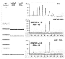
도 7은 LNCaP 및 LLC1 세포로부터 단리된 총 RNA의 겔 전기영동 분석을 도시한 것이다.
도 8은 마우스 백혈구 (WBC)로부터 얻은 RNA의 수율 및 질을 나열한 것이다.
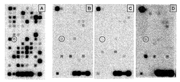
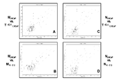
도 9A-9D는 LNCaP (인간 전립선암) 종양 보유 누드 마우스로부터의 호중구에서 검출된 7개의 상향조절된 (2배 이상) 암 관련 유전자를 보여주는 어레이를 도시한 것이다. (A) LNCaP 종양. (B) LNCaP 종양 보유 누드 마우스로부터 얻은 호중구 (NT). (C) LNCaP 종양 보유 누드 마우스로부터 얻은 T 세포 (TT). (D) 비-종양 보유 누드 마우스로부터 얻은 호중구 (NN). 원으로 표시된 시그너쳐는 종양 세포 (A)에서 및 종양 보유 마우스로부터의 호중구 (B)에서 발현되고, 비-종양 보유 마우스로부터의 호중구 (D)에서 및 비-포식성 T 세포 (C)에서 최소로 발현된 것이다. NT에서의 발현은 NN 및 TT에서보다 2배 이상 더 컸다.
도 10A-10D은 LNCaP (인간 전립선암) 종양 보유 누드 마우스로부터의 대식세포에서 검출된 3개의 상향조절된 암 관련 유전자를 보여주는 어레이를 도시한 것이다. (A) LNCaP 종양. (B) LNCaP 종양 보유 누드 마우스로부터 얻은 대식세포 (MT). (C) LNCaP 종양 보유 누드 마우스로부터 얻은 T 세포 (TT). (D) 비-종양 보유 누드 마우스로부터 얻은 대식세포 (MN). 원으로 표시된 시그너쳐는 종양 세포 (A)에서 및 종양 보유 마우스로부터의 대식세포 (B)에서 발현되고, 비-종양 보유 마우스로부터의 대식세포 (D)에서 및 비-포식성 T 세포 (C)에서 최소로 발현된 것이다. MT에서의 발현은 MN 및 TT에서보다 2배 이상 더 컸다.
도 11A-11D는 LS174T (인간 결장암) 종양 보유 누드 마우스로부터의 호중구에서 검출된 4개의 상향조절된 (2배 이상) 암 관련 유전자를 보여주는 어레이를 도시한 것이다. (A) LS174T 종양. (B) LS174T 종양 보유 누드 마우스로부터 얻은 호중구 (NT). (C) LS174T 종양 보유 누드 마우스로부터 얻은 T 세포 (TT). (D) 비-종양 보유 누드 마우스로부터 얻은 호중구 (NN). 원으로 표시된 시그너쳐는 종양 세포 (A)에서 및 종양 보유 마우스로부터의 호중구 (B)에서 발현되고, 비-종양 보유 마우스로부터의 호중구 (D)에서 및 비-포식성 T 세포 (C)에서 최소로 발현된 것이다. NT에서의 발현은 NN 및 TT에서보다 2배 이상 더 컸다.
도 12A-12D는 LS174T (인간 결장암) 종양 보유 누드 마우스로부터의 대식세포에서 검출된 3개의 상향조절된 (2배 이상) 암 관련 유전자를 보여주는 어레이를 도시한 것이다. (A) LS174T 종양. (B) LS174T 종양 보유 누드 마우스로부터 얻은 대식세포 (MT). (C) LS174T 종양 보유 누드 마우스로부터 얻은 T 세포 (TT). (D) 비-종양 보유 누드 마우스로부터 얻은 대식세포 (MN). 원으로 표시된 시그너쳐는 종양 세포 (A)에서 및 종양 보유 마우스로부터의 대식세포 (B)에서 발현되고, 비-종양 보유 마우스로부터의 대식세포 (D)에서 및 비-포식성 T 세포 (C)에서 최소로 발현된 것이다. MT에서의 발현은 MN 및 TT에서보다 2배 이상 더 컸다.
도 13A-13D는 LLC1 (마우스 전이성 폐암) 종양-보유 C57/B1 마우스로부터의 호중구에서 검출된 5개의 상향조절된 (2배 이상) 암 관련 유전자를 보여주는 어레이를 도시한 것이다. (A) LLC1 종양. (B) LLC1 종양 보유 C57/B1 마우스로부터 얻은 호중구 (NT). (C) LLC1 종양 보유 C57/B1 마우스로부터 얻은 T 세포 (TT). (D) 비-종양 보유 C57/B1 마우스로부터 얻은 호중구 (NN). 원으로 표시된 시그너쳐는 종양 세포 (A)에서 및 종양 보유 마우스로부터의 호중구 (B)에서 발현되고, 비-종양 보유 마우스로부터의 호중구 (D)에서 및 비-포식성 T 세포 (C)에서 최소로 발현된 것이다. NT에서의 발현은 NN 및 TT에서보다 2배 이상 더 컸다.
도 14A-14D는 LLC1 (마우스 전이성 폐암) 종양-보유 C57/B1 마우스로부터의 대식세포에서 검출된 2개의 상향조절된 (2배 이상) 암 관련 유전자를 보여주는 어레이를 도시한 것이다. (A) LLC1 종양. (B) LLC1 종양 보유 C57/B1 마우스로부터 얻은 대식세포 (MT). (C) LLC1 종양 보유 C57/B1 마우스로부터 얻은 T 세포 (TT). (D) 비-종양 보유 C57/B1 마우스로부터 얻은 대식세포 (MN). 원으로 표시된 시그너쳐는 종양 세포 (A)에서 및 종양 보유 마우스로부터의 호중구 (B)에서 발현되고, 비-종양 보유 마우스로부터의 호중구 (D)에서 및 비-포식성 T 세포 (C)에서 최소로 발현된 것이다. MT에서의 발현은 MN 및 TT에서보다 2배 이상 더 컸다.
도 15A-15D는 B16F10 (마우스 전이성 흑색종) 종양 보유 C57/B1 마우스로부터의 호중구에서 검출된 2개의 상향조절된 (2배 이상) 암 관련 유전자를 보여주는 어레이를 도시한 것이다. (A) B16F10 종양. (B) B16F10 종양 보유 C57/B1 마우스로부터 얻은 호중구 (NT). (C) B16F10 종양 보유 C57/B1 마우스로부터 얻은 T 세포 (TT). (D) 비-종양 보유 C57/B1 마우스로부터 얻은 호중구 (NN). 원으로 표시된 시그너쳐는 종양 세포 (A)에서 및 종양 보유 마우스로부터의 호중구 (B)에서 발현되고, 비-종양 보유 마우스로부터의 호중구 (D)에서 및 비-포식성 T 세포 (C)에서 최소로 발현된 것이다. NT에서의 발현은 NN 및 TT에서보다 2배 이상 더 컸다.
도 16A-16D는 B16F10 (마우스 전이성 흑색종) 종양-보유 C57/B1 마우스로부터의 대식세포에서 검출된 1개의 상향조절된 (2배 이상) 암 관련 유전자를 보여주는 어레이를 도시한 것이다. (A) B16F10 종양. (B) B16F10 종양 보유 C57/B1 마우스로부터 얻은 대식세포 (MT). (C) B16F10 종양 보유 C57/B1 마우스로부터 얻은 T 세포 (TT). (D) 비-종양 보유 C57/B1 마우스로부터 얻은 대식세포 (MN). 원으로 표시된 시그너쳐는 종양 세포 (A)에서 및 종양 보유 마우스로부터의 대식세포 (B)에서 발현되고, 비-종양 보유 마우스로부터의 대식세포 (D)에서 및 비-포식성 T 세포 (C)에서 최소로 발현된 것이다. MT에서의 발현은 MN 및 TT에서보다 2배 이상 더 컸다.
도 17A-17D는 두경부암 (편평세포 암종)의 환자로부터의 호중구에서 검출된 5개의 상향조절된 (2배 이상) 암 관련 유전자를 보여주는 어레이를 도시한 것이다. (A) 정상 조직 (피부) 생검. (B) 종양 조직 생검. (C) 환자 혈액으로부터 얻은 호중구 (NT), (D) 환자 혈액으로부터 얻은 T 세포 (TT). 원으로 표시된 시그너쳐는 종양 세포 (B)에서 및 환자 혈액으로부터의 호중구 (C)에서 발현되고, 정상 피부 (A)에서 또는 비-포식성 T 세포 (D)에서 최소로 발현되거나 발현되지 않은 것이다. NT에서의 발현은 TT 및 피부에서보다 2배 이상 더 컸다.
도 18A-18D는 난소암 (선암종)의 환자로부터의 대식세포에서 검출된 23개의 상향조절된 (2배 이상) 암 관련 유전자를 보여주는 어레이를 도시한 것이다. (A) 환자 혈액으로부터 얻은 대식세포 (MT). (B) 환자 혈액으로부터 얻은 T 세포 (TT). 원으로 표시된 시그너쳐는 환자로부터 얻은 대식세포 (A)에서 발현되고, 비-포식성 T 세포 (B)에서 최소로 발현된 것이다. MT에서의 발현은 TT에서보다 2배 이상 더 컸다.
도 19는 포식세포 내의 종양 시그너쳐를 확인하기 위해 사용되는 방법을 도시한 것이다. 본 예에서, 종양 보유 동물로부터의 대식세포 (MT)에서의 암 연관 유전자의 발현 강도를 동일한 동물로부터의 T 세포 (TT)에서의 강도와 비교하여 정량하였고, 2배 초과로 과다발현된 유전자가 확인되었다. 이어서, MT에서 발현된 모든 유전자의 강도를 비-종양 보유 동물로부터 얻은 대식세포 (MNT)에서의 강도와 비교하여 정량하였고, 2배 초과로 과다발현된 유전자가 확인되었다. 두 목록에 공통적인 유전자를 선택하여, 동일한 종양에 의해 발현된 것과 비교하였다 (음영 영역).
도 20A-20B는 (A) LNCaP 인간 전립선 종양 보유 누드 마우스로부터 얻은 대식세포 (MLNCaP) 및 동일한 동물로부터의 T 세포 (T 세포LNCaP), (B) MLNCaP 및 비-종양 보유 마우스로부터 얻은 대식세포 (M비-종양), (C) LNCaP 인간 전립선 종양 보유 누드 마우스로부터 얻은 호중구 (NLNCaP) 및 동일한 동물로부터의 T 세포 (T 세포LNCaP), 및 (D) NLNCaP 및 비-종양 보유 마우스로부터 얻은 대식세포 (N비-종양)에서 유전자 발현 강도 비교를 도시한 것이다. 적색의 유전자는 2배 초과로 과다발현되었고; 녹색의 유전자는 2배 초과로 과소발현되었다.
도 21은 포식성 호중구 (N) 및 대식세포 (M) 내에서 암 관련 유전자의 발현을 나열한 것이다.
도 22는 비-포식성 T 세포에 비해 난소암 환자의 포식성 대식세포에서 상향조절된 (2배 초과) 암 관련 유전자를 나열한 것이다.
도 23은 마우스 WBC로부터 얻은 단백질 샘플 (5.9 ㎍)의 SDS 겔 (10%) 전기영동을 도시한 것이다.
도 24는 종양 보유 마우스로부터 얻은 T 세포 및 단핵구/대식세포 (M/M)에서 TAG-72 및 PSA 발현에 대한 웨스턴 블롯 (Western blot) 분석을 도시한 것으로, 포식세포 내에만 시그너쳐가 존재함을 입증한다.The patent or application file includes at least one drawing in color. Copies of this patent or patent application, including color drawing(s), will be provided by the Patent Office upon request while paying the necessary costs. The above and other features and advantages of the present invention will be better understood from the following detailed description of exemplary embodiments in conjunction with the accompanying drawings:
Figure 1 Tumors by phagocytic cells after phagocytosis of tumor DNA, RNA, protein, and lipids released by living CTC, apoptotic CTC, fragmented CTC, viable and/or apoptotic tumor cells- It shows schematically the suggested pathways leading to the acquisition of specific DNA, RNA, protein and/or lipid signatures. Note that only macrophages (not non-phagophages) acquire a tumor signature.
FIG. 2 schematically illustrates an analytical method used to identify cancer signatures expressed in/by phagocytic cells of ovarian cancer (OC) patients.
3 schematically depicts a general flow diagram of one embodiment of the method of the present invention.
Figure 4 Tumor-specific DNA by blood phagocytes after phagocytosis of tumor DNA, RNA, protein and lipid released by living CTC, apoptotic CTC, fragmented CTC, viable and/or apoptotic tumor cells , Schematically showing the proposed pathways leading to the acquisition of RNA, protein and lipid signatures. Note that the DNA content of phagocytic cells after phagocytosis is >2n.
FIG. 5 schematically depicts the analytical method used to identify breast cancer (BC) signatures in breast cancer (BC)-bearing animals.
6 schematically illustrates a general flow diagram of another embodiment of the method of the present invention.
7 shows gel electrophoresis analysis of total RNA isolated from LNCaP and LLC1 cells.
Figure 8 lists the yield and quality of RNA obtained from mouse leukocytes (WBC).
9A-9D depict an array showing seven up-regulated (two-fold or more) cancer-related genes detected in neutrophils from nude mice bearing LNCaP (human prostate cancer) tumors. (A) LNCaP tumor. (B) Neutrophils (N T ) obtained from nude mice bearing LNCaP tumors. (C) T cells (T T ) obtained from nude mice bearing LNCaP tumors. (D) Neutrophils (N N ) obtained from non-tumor bearing nude mice. The circled signature is expressed in tumor cells (A) and in neutrophils (B) from tumor bearing mice, minimally expressed in neutrophils (D) from non-tumor bearing mice and in non-prediction T cells (C). It is done. Expression of N T is more than twice in the N T T N and greater.
10A-10D depict an array showing three up-regulated cancer-related genes detected in macrophages from nude mice bearing LNCaP (human prostate cancer) tumors. (A) LNCaP tumor. (B) Macrophages (M T ) obtained from nude mice bearing LNCaP tumors. (C) T cells (T T ) obtained from nude mice bearing LNCaP tumors. (D) Macrophages (M N ) obtained from non-tumor bearing nude mice. The circled signature is expressed in tumor cells (A) and in macrophages (B) from tumor bearing mice, minimal in macrophages (D) from non-tumor bearing mice and in non-phagocytic T cells (C). It is expressed as. Expression in M T was greater than 2 times longer in the M and T N T.
11A-11D depict an array showing four up-regulated (two-fold or more) cancer-related genes detected in neutrophils from nude mice bearing LS174T (human colon cancer) tumors. (A) LS174T tumor. (B) Neutrophils (N T ) obtained from LS174T tumor bearing nude mice. (C) T cells (T T ) obtained from LS174T tumor bearing nude mice. (D) Neutrophils (N N ) obtained from non-tumor bearing nude mice. The circled signature is expressed in tumor cells (A) and in neutrophils (B) from tumor bearing mice, minimally expressed in neutrophils (D) from non-tumor bearing mice and in non-prediction T cells (C). It is done. Expression of N T is more than twice in the N T T N and greater.
12A-12D depict an array showing three up-regulated (two-fold or more) cancer-related genes detected in macrophages from nude mice bearing LS174T (human colon cancer) tumors. (A) LS174T tumor. (B) Macrophages (M T ) obtained from nude mice bearing LS174T tumor. (C) T cells (T T ) obtained from LS174T tumor bearing nude mice. (D) Macrophages (M N ) obtained from non-tumor bearing nude mice. The circled signature is expressed in tumor cells (A) and in macrophages (B) from tumor bearing mice, minimal in macrophages (D) from non-tumor bearing mice and in non-phagocytic T cells (C). It is expressed as. Expression in M T was greater than 2 times longer in the M and T N T.
13A-13D depict an array showing five up-regulated (more than 2 fold) cancer-related genes detected in neutrophils from LLC1 (mouse metastatic lung cancer) tumor-bearing C57/B1 mice. (A) LLC1 tumor. (B) Neutrophils (N T ) obtained from LLC1 tumor bearing C57/B1 mice. (C) T cells obtained from LLC1 tumor bearing C57/B1 mice (T T ). (D) Neutrophils (N N ) obtained from non-tumor bearing C57/B1 mice. The circled signature is expressed in tumor cells (A) and in neutrophils (B) from tumor bearing mice, minimally expressed in neutrophils (D) from non-tumor bearing mice and in non-prediction T cells (C). It is done. Expression of N T is more than twice in the N T T N and greater.
14A-14D depict an array showing two up-regulated (more than 2 fold) cancer-related genes detected in macrophages from LLC1 (mouse metastatic lung cancer) tumor-bearing C57/B1 mice. (A) LLC1 tumor. (B) Macrophages (M T ) obtained from LLC1 tumor bearing C57/B1 mice. (C) T cells obtained from LLC1 tumor bearing C57/B1 mice (T T ). (D) Macrophages (M N ) obtained from non-tumor bearing C57/B1 mice. The circled signature is expressed in tumor cells (A) and in neutrophils (B) from tumor bearing mice, minimally expressed in neutrophils (D) from non-tumor bearing mice and in non-prediction T cells (C). It is done. Expression in M T was greater than 2 times longer in the M and T N T.
15A-15D depict an array showing two up-regulated (more than 2 fold) cancer-related genes detected in neutrophils from C16/B1 mice bearing B16F10 (mouse metastatic melanoma) tumors. (A) B16F10 tumor. (B) Neutrophils (N T ) obtained from C57/B1 mice bearing B16F10 tumors. (C) T cells (T T ) obtained from C57/B1 mice bearing B16F10 tumors. (D) Neutrophils (N N ) obtained from non-tumor bearing C57/B1 mice. The circled signature is expressed in tumor cells (A) and in neutrophils (B) from tumor bearing mice, minimally expressed in neutrophils (D) from non-tumor bearing mice and in non-prediction T cells (C). It is done. Expression of N T is more than twice in the N T T N and greater.
16A-16D depict an array showing one up-regulated (more than 2 fold) cancer-related gene detected in macrophages from B16F10 (mouse metastatic melanoma) tumor-bearing C57/B1 mice. (A) B16F10 tumor. (B) Macrophages (M T ) obtained from C57/B1 mice bearing B16F10 tumors. (C) T cells (T T ) obtained from C57/B1 mice bearing B16F10 tumors. (D) Macrophages (M N ) obtained from non-tumor bearing C57/B1 mice. The circled signature is expressed in tumor cells (A) and in macrophages (B) from tumor bearing mice, minimal in macrophages (D) from non-tumor bearing mice and in non-phagocytic T cells (C). It is expressed as. Expression in M T was greater than 2 times longer in the M and T N T.
17A-17D show an array showing five up-regulated (more than two-fold) cancer-related genes detected in neutrophils from patients with head and neck cancer (squamous cell carcinoma). (A) Normal tissue (skin) biopsy. (B) Tumor tissue biopsy. (C) Neutrophils from patient blood (N T ), (D) T cells from patient blood (T T ). The circled signature is expressed in tumor cells (B) and in neutrophils (C) from patient blood, and minimally expressed or not expressed in normal skin (A) or in non-predatory T cells (D). Expression in N T was more than 2 times greater than in T T and skin.
18A-18D depict an array showing 23 up-regulated (more than 2 fold) cancer-related genes detected in macrophages from patients of ovarian cancer (adenocarcinoma). (A) Macrophages (M T ) obtained from patient blood. (B) T cells from patient blood (T T ). The circled signature is expressed in macrophages (A) obtained from the patient and minimally expressed in non-phagocytic T cells (B). The expression in M T was more than 2 times greater than in T T.
19 shows a method used to identify tumor signatures in phagocytic cells. In this example, the intensity of expression of cancer-associated genes in macrophages (M T ) from tumor bearing animals was quantified compared to the intensity in T cells (T T ) from the same animal, and overexpressed more than 2 fold. The gene has been identified. Subsequently, the intensity of all genes expressed in M T was quantified compared to the intensity in macrophages (M NT ) obtained from non-tumor bearing animals, and genes overexpressed more than twice were identified. Genes common to both lists were selected and compared to those expressed by the same tumor (shaded region).
Figures 20A-20B show (A) macrophages (M LNCaP ) from LNCaP human prostate tumor bearing nude mice and T cells from the same animal (T cell LNCaP ), (B) M LNCaP and large from tumors Phagocytes (M non-tumor ), (C) Neutrophils (N LNCaP ) from LNCaP human prostate tumor bearing nude mice and T cells from the same animal (T cell LNCaP ), and (D) N LNCaP and non-tumor bearing mice Gene expression intensity comparison in macrophages (N non-tumor ) obtained from. The red gene was overexpressed more than 2 fold; The green gene was under-expressed more than twice.
Figure 21 lists the expression of cancer-related genes in macrophages (N) and macrophages (M).
Figure 22 lists cancer-related genes that are upregulated (more than 2 fold) in the macrophages of ovarian cancer patients compared to non-phagoblastic T cells.
23 shows SDS gel (10%) electrophoresis of protein samples (5.9 μg) from mouse WBC.
Figure 24 shows Western blot analysis for TAG-72 and PSA expression in T cells and monocytes/macrophages (M/M) obtained from tumor bearing mice. Prove.
본 발명의 실시태양은 포식세포의 세포내 함유물 및/또는 발현 프로파일에 기초하여 질병, 감염 물질 및 신체 상태와 연관된 마커의 환자-특이적 발현 프로파일을 제공하는 방법에 관한 것이다. 본 발명의 한 측면에 따라, 포식세포의 세포내 함유물 및/또는 발현 프로파일을 특정 질병 상태를 검출하고/하거나 진단하기 위해 특정 질병 상태에 대한 공지의 마커에 비교한다. 본 발명의 추가의 측면에 따라, 포식세포의 세포내 함유물 및/또는 발현 프로파일을 단일 환자의 혈액으로부터의 비-포식세포의 세포내 함유물 및/또는 발현 프로파일에 비교한다. 비-포식세포의 세포내 함유물 및/또는 발현 프로파일을 포식세포의 것으로부터 공제하면 개체의 질병 상태를 대표하는 세포내 함유물 및/또는 발현 프로파일만을 생성시킨다. Embodiments of the invention relate to methods of providing patient-specific expression profiles of markers associated with disease, infectious agents and body conditions based on intracellular inclusion and/or expression profiles of phagocytic cells. According to one aspect of the invention, the intracellular inclusion and/or expression profile of a phagocytic cell is compared to a known marker for a particular disease state to detect and/or diagnose a specific disease state. According to a further aspect of the invention, the intracellular content and/or expression profile of phagocytic cells is compared to the intracellular content and/or expression profile of non-phagocytic cells from the blood of a single patient. Subtracting the intracellular inclusion and/or expression profile of a non-phagocytic from that of a phagocyte produces only the intracellular inclusion and/or expression profile representative of the individual's disease state.
본 발명의 추가의 실시태양에 따라, 개체로부터 포식세포 집단을 얻고, DNA 함량이 2n보다 큰 집단으로부터의 포식세포의 세포내 함유물 및/또는 발현 프로파일을 DNA 함량이 2n인 동일한 집단으로부터의 포식세포의 세포내 함유물 및/또는 발현 프로파일에 비교한다. 본 발명의 추가의 실시태양에 따라, 개체로부터의 포식세포 집단을 얻고, RNA, 단백질, 탄수화물 및/또는 지질 함량이 정상보다 많고, DNA 지수가 1을 초과하는 집단으로부터의 포식세포의 발현 프로파일을 RNA, 단백질, 탄수화물 및/또는 지질 함량이 정상이고/이거나 DNA 지수가 1인 동일한 집단으로부터의 포식세포의 발현 프로파일에 비교한다. According to a further embodiment of the invention, a population of phagocytic cells is obtained from an individual, and the intracellular inclusion and/or expression profile of phagocytic cells from a population having a DNA content greater than 2n is predated from the same population having a DNA content of 2n. Compare to the cell's intracellular content and/or expression profile. According to a further embodiment of the invention, a population of phagocytic cells from an individual is obtained, and the expression profile of phagocytic cells from a population having a RNA, protein, carbohydrate and/or lipid content greater than normal and a DNA index greater than 1 is obtained. Compare the expression profile of phagocytic cells from the same population with normal RNA content of RNA, protein, carbohydrate and/or lipid and/or
상기 환자 특이적 발현 프로파일은 개체에서 특정 질병의 검출 또는 진단에 오차를 도입할 수 있는 특정 질병 또는 감염 물질에 대한 집단-유래된 평균 시그너쳐 프로파일에 대한 의존성을 제거한다. 본 발명의 질병 상태에 대한 상기 환자 특이적 발현 프로파일은 검출, 진단 및 치료가 개체에게 개인 맞춤되도록 허용한다.The patient specific expression profile eliminates dependence on a population-derived mean signature profile for a particular disease or infectious agent that can introduce errors in the detection or diagnosis of a particular disease in an individual. The patient specific expression profile for the disease state of the present invention allows detection, diagnosis and treatment to be personalized to the individual.
도 1-3을 참고로 하여 본 발명의 특정 실시태양에 따르면, 인간 피하 (s.c.) 종양 (전립선 LNCaP 선암종 또는 LS174T 결장 선암종) 또는 마우스 종양 (정맥내 투여된 B16F10 전이성 흑색종, 또는 s.c 주사된 LLC1 폐암)을 보유하는 약 3주령의 마우스로부터 얻은 포식성 및 비-포식성 WBC의 유전자 발현 프로파일을 비교하였다. 결과는 이들 종양 보유 마우스로부터 얻은 호중구 및 대식세포가 각각의 개별 종양에서 또한 발현되는 다양한 발암유전자 및 다른 암 관련 유전자 시그너쳐를 발현하는 것을 입증하였다 (도 9-16 및 19-21 참조). 이들 암 관련 유전자 및 발암유전자 (예를 들어, ERBB2, Jun, Fos 등)는 발현되지 않거나, (i) 종양 보유 마우스로부터 단리된 비-포식성 T 세포, 및 (ii) 비-종양 보유 마우스로부터 얻은 호중구 및 대식세포에 의해 최소로 발현된다. 또한, 종양 보유 마우스로부터의 포식세포는 단지 종양-특이적 단백질만을 발현하는 것으로 밝혀졌다 (도 23 및 24 참조). 마우스의 혈액 내의 CTC 및/또는 종양-특이적 DNA 및/또는 단백질이 포식되었고, 일부의 종양-세포 DNA는 그의 종양-특이적 돌연변이 및 유전자와 함께 아마도 형질감염에 의해 (특정 이론에 매이도록 의도되지 않음) 정상 포식세포 DNA 내로 통합되고, RNA로 전사되고, 단백질로 번역되었다. According to certain embodiments of the present invention with reference to FIGS. 1-3, human subcutaneous (sc) tumors (prostate LNCaP adenocarcinoma or LS174T colon adenocarcinoma) or mouse tumors (B16F10 metastatic melanoma administered intravenously, or sc injected LLC1) The gene expression profiles of predatory and non-predatory WBCs obtained from mice about 3 weeks old with lung cancer) were compared. The results demonstrated that neutrophils and macrophages from these tumor bearing mice express various carcinogens and other cancer related gene signatures that are also expressed in each individual tumor (see FIGS. 9-16 and 19-21). These cancer-related genes and carcinogens (e.g., ERBB2, Jun, Fos, etc.) are not expressed, or are obtained from (i) non-predatory T cells isolated from tumor bearing mice, and (ii) from non-tumor bearing mice. It is minimally expressed by neutrophils and macrophages. It has also been found that phagocytes from tumor bearing mice express only tumor-specific proteins (see Figures 23 and 24). CTC and/or tumor-specific DNA and/or protein in the blood of the mouse has been predated, and some tumor-cell DNA, along with its tumor-specific mutations and genes, is probably intended to be bound by a specific theory (by specific transfection) Not) integrated into normal phagocytic DNA, transcribed into RNA, and translated into protein.
도 1-3을 참고로 하여 본 발명의 특정 예시적인 실시태양에 따르면, 두경부 종양 또는 난소암의 환자로부터 얻은 포식성 및 비-포식성 WBC의 유전자 발현 프로파일을 또한 비교하였다. 결과는 이들 환자로부터 얻은 호중구 및 대식세포가 개별 종양에서 또한 발현되는 다양한 발암유전자 및 다른 암 관련 유전자 시그너쳐를 발현함을 입증하였다 (도 17-18 및 22 참조). 이들 암 관련 유전자 및 발암유전자는 동일한 개별 환자로부터 단리된 비-포식성 T 세포에 의해 발현되지 않거나 최소로 발현되었다. 환자의 혈액 내의 CTC 및/또는 종양-특이적 DNA 및 RNA가 포식되었고, 일부의 종양-세포 DNA 및/또는 RNA는 그의 종양-특이적 돌연변이 및 유전자와 함께 아마도 형질감염을 통해 (특정 이론에 매이도록 의도되지 않음) 정상 포식세포 DNA 내로 통합되고, RNA로 전사되고, 단백질로 번역되었다. According to certain exemplary embodiments of the invention with reference to Figures 1-3, the gene expression profiles of predatory and non-predatory WBCs obtained from patients with head and neck tumors or ovarian cancers were also compared. The results demonstrated that neutrophils and macrophages obtained from these patients express various carcinogens and other cancer-related gene signatures that are also expressed in individual tumors (see FIGS. 17-18 and 22). These cancer-related genes and carcinogens were expressed minimally or not expressed by non-predatory T cells isolated from the same individual patient. CTC and/or tumor-specific DNA and RNA in the patient's blood have been predated, and some of the tumor-cell DNA and/or RNA, along with its tumor-specific mutations and genes, may be transfected (perhaps according to certain theories). Not intended to be) integrated into normal phagocyte DNA, transcribed into RNA, and translated into protein.
도 4-6을 참고로 하여 특정 예시적인 실시태양에 따르면, (1) DNA 함량이 >2n (Pn>2)이거나, (2) RNA, 단백질, 탄수화물 및/또는 지질 함량이 정상보다 더 많은 혈액 또는 하나 이상의 다른 생물학적 샘플 (예를 들어, 소변, 대변, 타액, 림프액, 뇌척수액 등)으로부터 얻은 포식세포 (예를 들어, 대식세포), 즉, 포식된 CTC 및/또는 그들의 하위세포형 단편 또는 DNA/RNA/지질 (즉, 종양-특이적 시그너쳐 또는 다른 질병-특이적 시그너쳐)을 갖고/갖거나 DNA 지수가 1을 초과하는 세포의 DNA (핵 및/또는 미토콘드리아), RNA, 마이크로RNA, 단백질, 및/또는 지질 발현 프로파일의 정량 분석, 및 이를 (1) DNA 함량이 2n (Pn=2)이거나, (2) RNA, 단백질, 탄수화물 및/또는 지질 함량이 정상인 동일한 포식세포 집단 (예를 들어, 대식세포), 즉, 포식된 CTC 및/또는 그들의 하위세포형 단편을 갖지 않고 DNA 지수가 1인 세포와 비교하는 것은 Pn=2 세포에서 발현되지 않거나 최소로 발현되는 (환자-특이적 노이즈) Pn>2 세포 내의 종양-특이적 (또는 다른 질병-특이적) 시그너쳐 (환자-특이적 신호)를 검출하는 방법을 제공한다. 도 6을 참고로 하여, Pn=2의 DNA, RNA, 단백질, 및/또는 지질 프로파일을 도 5에 제시된 바와 같이 Pn>2의 것으로부터 공제하는 것은 암 (및/또는 질병 및/또는 신체 상태)이 있는 동물 및/또는 인간의 혈액 샘플 (또는 하나 이상의 다른 생물학적 샘플, 예를 들어, 다른 체액) 내에서 종양-특이적 (및/또는 질병-특이적 및/또는 병태 특이적) 시그너쳐를 (예를 들어, 하나 이상의 게놈, 단백체, 대사체, 복합당질체, 당단백체, 지질체 및/또는 지질단백체 분석 후에) 확인하고, 잠재 종양(들) 및/또는 다른 질병 및/또는 다른 병태의 존재를 표시하는 방법을 제공한다. 포식세포의 게놈, 단백체, 대사체, 복합당질체, 당단백체, 지질체 및/또는 지질단백체 프로파일을 비-포식세포의 것과 비교한 상기 설명된 방법과는 달리, 본 발명에 따른 상기 분석 검출 방법의 주요 잇점은 (i) "종양-특이적" (예를 들어, Pn>2 대식세포) 및 "정상-비-특이적" (예를 들어, Pn=2 대식세포) 시그너쳐의 공급원으로서 단일 포식세포 아집단을 이용하고, 즉, 둘 모두 동일한 기준선 유전자형을 공유하고; (ii) 시그너쳐-획득 세포 (예를 들어, Pn>2 호중구)가 죽은 CTC 및/또는 그의 단편을 포식하지 않고 따라서 그를 획득하지 않은 것 (예를 들어, Pn=2 호중구)으로 희석되지 않는다는 것이다.According to certain exemplary embodiments with reference to FIGS. 4-6, (1) the DNA content is >2n (P n>2 ), or (2) the RNA, protein, carbohydrate and/or lipid content is greater than normal. Phagocytic cells (e.g., macrophages) obtained from blood or one or more other biological samples (e.g., urine, feces, saliva, lymphatic fluid, cerebrospinal fluid, etc.), i.e., predated CTCs and/or their subcellular fragments or DNA of DNA (nucleus and/or mitochondria), RNA, microRNA, protein of DNA having DNA/RNA/lipid (i.e. tumor-specific signature or other disease-specific signature) and/or having a DNA index greater than 1 , And/or quantitative analysis of lipid expression profiles, and (1) the same phagocytic population with normal DNA content of 2n (P n=2 ) or (2) RNA, protein, carbohydrate and/or lipid content (e.g. For example, macrophages), i.e., compared to cells without a predated CTC and/or their subcellular type fragments and having a DNA index of 1, are not expressed or minimally expressed in P n=2 cells (patient-specific Noise) provides a method for detecting tumor-specific (or other disease-specific) signatures (patient-specific signals) in P n>2 cells. With reference to FIG. 6, subtracting the DNA, RNA, protein, and/or lipid profile of P n=2 from that of P n >2 as shown in FIG. 5 is a cancer (and/or disease and/or body Tumor-specific (and/or disease-specific and/or condition specific) signatures in animal and/or human blood samples (or one or more other biological samples, e.g., different body fluids) (E.g., after analysis of one or more genomes, proteins, metabolites, complexes, glycoproteins, lipids and/or lipoproteins), and potential tumor(s) and/or other diseases and/or other conditions It provides a way to indicate presence. Unlike the method described above comparing the genome, proteome, metabolite, complex glycoside, glycoprotein, lipid and/or lipoprotein profile of a phagocytic cell to that of a non-phagocytic cell, the assay detection method according to the present invention The main advantages of (i) as a source of "tumor-specific" (eg, P n>2 macrophages) and "normal-non-specific" (eg, P n=2 macrophages) signatures Use a single phagocytic subpopulation, ie both share the same baseline genotype; (ii) Signature-acquiring cells (e.g., P n >2 neutrophils) are not diluted with predation of the dead CTC and/or fragments thereof and thus not obtained (e.g., P n=2 neutrophils) Is that
도 4-6을 참고로 하여 특정 예시적인 실시태양에 따르면, 다른 살아있는, 죽어가는, 또는 죽은 (예를 들어, 세포자멸성 또는 괴사성) 세포, 세포자멸체, 핵, 미세소포, 엑소좀, 뉴클레오좀, 미토콘드리아, 소포체 등의 포식작용 또는 내재화의 결과로서 세포내 함유물이, 정상 세포내 함유물을 갖는 동일한 포식세포 집단 (예를 들어, 대식세포) (PNIC), 즉, 임의의 상기 언급된 세포 및/세포 파쇄물을 포식하지 않은 세포 (환자-특이적 노이즈)의 것보다 더 큰 혈액 또는 하나 이상의 다른 생물학적 샘플 또는 체액 (예를 들어, 소변, 대변, 타액, 림프액, 뇌척수액 등)으로부터 얻은 포식세포 (예를 들어, 대식세포)의 정량 분석은 정상 세포내 함유물을 갖는 포식세포에서 발현되지 않거나 최소로 발현되는 (환자-특이적 노이즈), 증가된 세포내 함유물을 갖는 포식세포 (PIIC) 내에서 종양-특이적 (또는 다른 질병-특이적 또는 다른 병태 특이적) 시그너쳐를 검출하는 방법을 제공한다. 도 6을 참고로 하여, PNIC의 DNA, RNA, 단백질 및/또는 지질 프로파일을 도 5에 제시된 바와 같이 PIIC의 것으로부터 공제하면 암 또는 다른 질병이 있는 동물의 혈액 샘플 (또는 하나 이상의 다른 생물학적 샘플, 예를 들어, 다른 체액) 내에서 종양-특이적 (및/또는 질병-특이적) 시그너쳐를 (예를 들어, 하나 이상의 게놈, 단백체, 대사체, 복합당질체, 당단백체, 지질체, 및/또는 지질단백체 분석 후에) 확인하고, 잠재 종양(들) 및/또는 다른 질병의 존재를 표시하는 방법을 제공한다. 포식세포의 게놈, 단백체, 대사체, 복합당질체, 당단백체, 지질체 및/또는 지질단백체 프로파일을 비-포식세포의 것과 비교한 상기 설명된 방법과는 달리, 본 발명에 따른 상기 분석 검출 방법의 주요 잇점은 (i) "질병-특이적" (예를 들어, PIIC 대식세포) 및 "정상-비-특이적" (예를 들어, PNIC 대식세포) 시그너쳐의 공급원으로서 단일 포식세포 아집단을 이용하고, 즉, 둘 모두 동일한 기준선 유전자형을 공유하고; (ii) 시그너쳐-획득 세포 (예를 들어, PIIC 호중구)가 죽은 CTC 및/또는 그의 단편을 포식하지 않고 따라서 그를 획득하지 않은 것 (예를 들어, PNIC 호중구)으로 희석되지 않는다는 것이다. According to certain exemplary embodiments with reference to FIGS. 4-6, other living, dying, or dead (eg, apoptotic or necrotic) cells, apoptosis, nucleus, microvesicles, exosomes, The same phagocytic population (e.g., macrophages) (P NIC ) with the intracellular inclusion, as a result of phagocytosis or internalization of nucleosomes, mitochondria, endoplasmic reticulum, etc., i.e., any Blood or one or more other biological samples or bodily fluids (e.g. urine, feces, saliva, lymphatic fluid, cerebrospinal fluid, etc.) that are larger than those of the cells and/or cell debris that have not predated the above-mentioned cells (patient-specific noise) Quantitative analysis of phagocytic cells (eg, macrophages) obtained from a phagocytosis with increased intracellular inclusions, which is expressed in minimal or not expressed (patient-specific noise) in phagocytes with normal intracellular inclusions Provided are methods for detecting tumor-specific (or other disease-specific or other condition-specific) signatures in cells (P IIC ). With reference to FIG. 6, subtracting DNA, RNA, protein and/or lipid profiles of P NIC from those of P IIC as shown in FIG. 5, blood samples of animals with cancer or other diseases (or one or more other biologicals Tumor-specific (and/or disease-specific) signatures (eg, one or more genomes, proteins, metabolites, polysaccharides, glycoproteins, lipids) in a sample, eg, other body fluid), And/or after lipoprotein analysis), and to indicate the presence of latent tumor(s) and/or other diseases. Unlike the method described above comparing the genome, proteome, metabolite, complex glycoside, glycoprotein, lipid and/or lipoprotein profile of a phagocytic cell to that of a non-phagocytic cell, the assay detection method according to the present invention The main advantages of (i) single disease macrophages as a source of "disease-specific" (eg, P IIC macrophages) and "normal-non-specific" (eg, P NIC macrophages) signatures. Using populations, ie both share the same baseline genotype; (ii) Signature-acquiring cells (e.g. P IIC neutrophils) do not prey on dead CTCs and/or fragments thereof and thus are not diluted with those not obtained (e.g. P NIC neutrophils).
본원에서 설명되는 방법은 (i) 높은 특이성, 감수성 및 정확성을 갖고, 혈액 샘플 (또는 예를 들어, 체액과 같은 다른 생물학적 샘플) 내에 존재하는 종양-특이적 (및/또는 다른 질병-특이적) 및 정상적인 비특이적 시그너쳐의 검출을 가능하게 할 것이고; (ii) 유전자 발현에서 고유한 변이 (예를 들어, 나이, 성별, 인종적 배경, 건강 상태 등) 및 일시적 변이로 인해 개체들 사이에서 발생하는 것으로 알려진 "기준선의 불균등성"을 제거한다. 따라서, 특정 측면에서, 본 발명은 질병이 통상적인 영상화 기술 (예를 들어, PET, MRI, CT 등)에 의해 진단될 수 있기 전에 환자에서 잠재 원발성 및 전이성 종양 (및/또는 하나 이상의 다른 질병 또는 병태)의 조기 검출을 위한 비침습 분석을 제공하고, 따라서, 암이 있는 개체의 개입, 예방 및 치료를 위한 필요 및 전략에 비해 개선된 결정을 위한 기반을 제공한다. The methods described herein are (i) tumor-specific (and/or other disease-specific) with high specificity, sensitivity and accuracy, and present in a blood sample (or other biological sample, eg, body fluids). And detection of normal non-specific signatures; (ii) Eliminate "baseline unevenness" known to occur between individuals due to mutations inherent in gene expression (eg, age, gender, ethnic background, health status, etc.) and transient mutations. Thus, in certain aspects, the present invention provides a potential primary and metastatic tumor (and/or one or more other diseases or) in a patient before the disease can be diagnosed by conventional imaging techniques (eg, PET, MRI, CT, etc.). Non-invasive analysis for early detection of conditions), thus providing a basis for improved decision making compared to the needs and strategies for intervention, prevention and treatment of individuals with cancer.
본원에서 사용되는 바와 같이, 용어 "종양 특이적 마커"는 하나 이상의 세포 성분, 예를 들어 하나 이상의 DNA 서열, 하나 이상의 RNA 서열, 하나 이상의 단백질, 하나 이상의 폴리펩티드, 하나 이상의 지질 등을 포함하고, 이로 제한되지 않는 것으로 의도된다. 특정 측면에서, 종양 특이적 마커는 하나 이상의 WBC, 예를 들어, 호중구, 대식세포 및/또는 수지상 세포에 존재한다. As used herein, the term “tumor specific marker” includes one or more cellular components, such as one or more DNA sequences, one or more RNA sequences, one or more proteins, one or more polypeptides, one or more lipids, and the like, thereby It is intended to be not limiting. In certain aspects, tumor specific markers are present in one or more WBCs, eg, neutrophils, macrophages and/or dendritic cells.
본원에서 사용되는 바와 같이, 용어 "암 관련 유전자"는 예를 들어 암성 세포 (예를 들어, WBC, 예를 들어, 대식세포, 호중구, T 세포 등)에서 변경된 발현 (예를 들어, 비암성 세포에 비해 증가된 발현 또는 감소된 발현)을 갖는 암 유전자, 발암유전자 및/또는 종양 억제인자 유전자와 같은 유전자를 의미한다. 많은 암 관련 유전자가 당업계에 공지되어 있다. 암 관련 유전자는 예를 들어 ERBB2, JUN, RB1, SUPP1, MDM2, MAP2K1, MMP2, PDGFB, PLAUR, FGR, MYCL1, BLYM, NRAS1, PE1, SKI, TRK, ABL2, MYCN, RAB1, REL, RALB, LCO, ERBB4, RAF1, ECT2, KIT, FGF5, GRO1, GRO2, GRO3, FMS, PIM, KRAS1P, FYN, MYB, ROS1, MAS1, RALA, MYCLK1, GL13, ARAF2, MET, BRAF, MOS, LYN, MYBL1, MYC, OVC, VAV2, BMI1, RET, HRAS, SPI1, RELA, SEA, EMS1, ETS1, KRAS2, ERBB3, GLI, FLT, BRCA2, RB1, FOS, AKT1, ELK2, FES, MAF, TP53, CRK, ERBA1, NF1, EVI2, ERBBB2, INT4, BRCA1, YES1, JUND, JUNB, MEL, LPSA, VAV1, AKT2, FOSB, RRAS, HKR1, HKR2, ERBAL2, SRC, MYBL2, ETS2, ERG, ARAF1, YUASA, HS2, INT3, SNO, RMYC, BMYC, HRASP, TC21, TIM, PTI-1, JAK, CEA 패밀리의 하나 이상의 멤버 (예를 들어, 문헌 [Zhou et al. (2001) Gene 264:105)] 참조), PSA, MUC-16 등을 포함하고, 이로 제한되지 않는다. As used herein, the term “cancer related gene” means altered expression (eg, non-cancerous cells), eg, in cancerous cells (eg, WBCs, eg, macrophages, neutrophils, T cells, etc.). As compared to an increased expression or decreased expression), a gene such as a cancer gene, a carcinogen and/or a tumor suppressor gene. Many cancer related genes are known in the art. Cancer related genes are e.g. ERBB2, JUN, RB1, SUPP1, MDM2, MAP2K1, MMP2, PDGFB, PLAUR, FGR, MYCL1, BLYM, NRAS1, PE1, SKI, TRK, ABL2, MYCN, RAB1, REL, RALB, LCO , ERBB4, RAF1, ECT2, KIT, FGF5, GRO1, GRO2, GRO3, FMS, PIM, KRAS1P, FYN, MYB, ROS1, MAS1, RALA, MYCLK1, GL13, ARAF2, MET, BRAF, MOS, LYN, MYBL1, MYC , OVC, VAV2, BMI1, RET, HRAS, SPI1, RELA, SEA, EMS1, ETS1, KRAS2, ERBB3, GLI, FLT, BRCA2, RB1, FOS, AKT1, ELK2, FES, MAF, TP53, CRK, ERBA1, NF1 , EVI2, ERBBB2, INT4, BRCA1, YES1, JUND, JUNB, MEL, LPSA, VAV1, AKT2, FOSB, RRAS, HKR1, HKR2, ERBAL2, SRC, MYBL2, ETS2, ERG, ARAF1, YUASA, HS2, INT3, SNO , RMYC, BMYC, HRASP, TC21, TIM, PTI-1, JAK, one or more members of the CEA family (see, eg, Zhou et al. (2001) Gene 264:105), PSA, MUC- 16, and the like.
본원에서 사용되는 바와 같이, 용어 "암"은 다양한 종류의 악성 신생물을 의미하고, 그 대부분은 주위 조직을 침습할 수 있고, 상이한 부위로 전이될 수 있다 (예를 들어, 그 전부가 모든 목적을 위해 본원에 참고로 포함된 문헌 [PDR Medical Dictionary, 1st edition (1995)] 참조). 용어 "신생물" 및 "종양"은 세포 증식에 의해 정상보다 더 빠르게 성장하고 증식을 개시한 자극이 제거된 후에도 계속 성장하는 비정상적인 조직을 의미한다 (상기 문헌 참조). 상기 비정상적인 조직은 구조적 조직화 및 정상 조직과의 기능적 협동의 부분적인 또는 완전한 결여를 보이고, 양성 (즉, 양성 종양) 또는 악성 (즉, 악성 종양)일 수 있다.As used herein, the term “cancer” refers to various types of malignant neoplasms, most of which can invade surrounding tissues and metastasize to different sites (eg, all of which are all purposed). For reference, see PDR Medical Dictionary, 1st edition (1995), incorporated herein by reference). The terms “neoplastic” and “tumor” refer to abnormal tissue that grows faster than normal by cell proliferation and continues to grow even after the stimulus that initiated proliferation is removed (see above). The abnormal tissue shows a partial or complete lack of structural organization and functional collaboration with normal tissue, and can be benign (ie, benign tumor) or malignant (ie, malignant tumor).
암의 일반적인 범주의 예는 암종 (즉, 상피 세포 유래 악성 종양, 예를 들어, 통상적인 형태의 유방, 전립선, 폐 및 결장암), 육종 (즉, 연결 조직 또는 중간엽 세포 유래 악성 종양), 림프종 (즉, 조혈 세포 유래 악성 종양), 백혈병 (즉, 조혈 세포 유래 악성 종양), 생식세포 종양 (즉, 전능성 (totipotent) 세포 유래 종양, 성인은 정소 또는 난소에서 가장 자주 발견되고; 태아, 유아 및 아동은 신체의 정중선, 특히 미골의 끝부분에서 가장 자주 발견됨), 모세포성 종양 (즉, 일반적으로 미성숙 또는 배아 조직에 유사한 악성 종양) 등을 포함하고, 이로 제한되지 않는다.Examples of common categories of cancer include carcinoma (i.e., malignant tumors derived from epithelial cells, e.g., conventional forms of breast, prostate, lung and colon cancer), sarcoma (i.e., malignant tumors derived from connective tissue or mesenchymal cells), lymphoma (Ie, hematopoietic cell-derived malignant tumors), leukemia (ie, hematopoietic cell-derived malignant tumors), germ cell tumors (ie, totipotent cell-derived tumors, adults are most often found in the testis or ovary; fetus, infant and Children include, but are not limited to, the midline of the body, especially most often found at the tip of the coccyx, blast tumors (ie, malignancies that are generally immature or similar to embryonic tissue).
본 발명에 포함되는 것으로 의도되는 신생물의 종류의 예는 신경 조직, 혈액 형성 조직, 유방, 피부, 뼈, 전립선, 난소, 자궁, 자궁경부, 간, 폐, 뇌, 후두, 담낭, 췌장, 직장, 부갑상선, 갑상선, 부신, 면역계, 두경부, 결장, 위, 기관지, 및/또는 신장의 암과 연관된 신생물을 포함하고, 이로 제한되지 않는다.Examples of types of neoplasms intended to be included in the present invention include nerve tissue, blood forming tissue, breast, skin, bone, prostate, ovary, uterus, cervix, liver, lung, brain, larynx, gallbladder, pancreas, rectum , Parathyroid, thyroid, adrenal, immune system, head and neck, colon, stomach, bronchial, and/or neoplastic associated with kidney cancer.
예시적인 특정 실시태양에서, 본원에서 설명되는 하나 이상의 방법 및/또는 조성물은 모체 혈액 내의 태아 세포 및 DNA의 존재로 인해 태아 염색체 이상 (예를 들어, 다운 (Down) 증후군, 자폐증 및 관련 자폐 스펙트럼 장애 (아스퍼거 (Asperger) 증후군 및 달리 분류되지 않는 전반적 발달장애를 포함하고, 이로 제한되지 않음), 겸상적혈구 빈혈, 지중해 빈혈 등)의 존재와 연관된 질환의 검출, 확인 및/또는 진단에 적용된다. 하나 이상의 상기 질환의 스크리닝 및 진단은 모체 혈액 포식세포 내에서 하나 이상의 염색체 마커, 예를 들어 DNA 및 RNA 등을 검출하기 위해 본원에서 설명되는 방법 및/또는 조성물을 사용하여 수행될 수 있다.In certain exemplary embodiments, one or more of the methods and/or compositions described herein may include fetal chromosomal abnormalities (e.g., Down syndrome, autism and related autism spectrum disorders) due to the presence of fetal cells and DNA in maternal blood. (Including, but not limited to, Asperger's syndrome and generalized developmental disorders not elsewhere classified), sickle cell anemia, mediterranean anemia, etc.). Screening and diagnosis of one or more of the above diseases can be performed using the methods and/or compositions described herein to detect one or more chromosomal markers, such as DNA and RNA, in maternal blood phagocytes.
예시적인 특정 실시태양에서, 본원에서 설명되는 하나 이상의 방법 및/또는 조성물은 태아 줄기세포, 유핵 적혈구, 태아 림프구, 및 유의한 양의 세포 미함유 태아 핵산이 모체 혈액에서 순환하는 것으로 알려져 있기 때문에, 임신한 여성의 혈액 내에서 태아-유래 단백체, 지질체, 및/또는 게놈 시그너쳐의 존재를 검출함으로써 임신한 여성의 태아의 성별을 시험하기 위해 적용될 수 있다. 본원에서 설명되는 방법에 따르면, 포식세포의 세포내 함유물 및/또는 발현 프로파일을 임신한 여성의 혈액으로부터의 비-포식세포의 세포내 함유물 및/또는 발현 프로파일과 비교한다. 비-포식세포의 세포내 함유물 및/또는 발현 프로파일을 포식세포의 것으로부터 공제하면, 임신한 여성의 태아의 성별을 나타내는 세포내 함유물 및/또는 발현 프로파일을 생성시킬 수 있다.In certain exemplary embodiments, one or more methods and/or compositions described herein are known to circulate fetal stem cells, nucleated erythrocytes, fetal lymphocytes, and significant amounts of cell-free fetal nucleic acid in the maternal blood, It can be applied to test the sex of a fetus in a pregnant woman by detecting the presence of fetal-derived proteins, lipids, and/or genomic signatures in the blood of the pregnant woman. According to the methods described herein, the intracellular content and/or expression profile of phagocytic cells is compared to the intracellular content and/or expression profile of non-phagocytic cells from the blood of a pregnant woman. Subtracting the intracellular inclusion and/or expression profile of non-phagophages from that of the phagocytic cells can produce intracellular inclusion and/or expression profiles that represent the sex of the fetus of a pregnant woman.
예시적인 특정 실시태양에서, 본원에서 설명되는 하나 이상의 방법 및/또는 조성물은 죽어가는/죽은 근세포 및/또는 그의 단편 (예를 들어, DNA, 단백질 등)의 존재를 검출함으로써 심장병 (예를 들어, 심근경색, 만성 심부전, 허혈성 심장병, 심혈관 사멸 등)이 존재하거나 발생할 위험이 있는 대상의 혈액 내의 단백체 및/또는 게놈 근세포 시그너쳐의 존재와 연관된 질환의 검출, 확인 및/또는 진단에 사용될 수 있다. 하나 이상의 상기 질환의 스크리닝 및 진단은 혈액 포식세포 내에서 하나 이상의 마커, 예를 들어 DNA 및 RNA, 단백질 등을 검출하기 위해 본원에서 설명되는 방법 및/또는 조성물을 사용하여 수행될 수 있다.In certain exemplary embodiments, one or more methods and/or compositions described herein can be used to detect cardiac disease (eg, by detecting the presence of dying/dead myocytes and/or fragments thereof (eg, DNA, proteins, etc.)). Myocardial infarction, chronic heart failure, ischemic heart disease, cardiovascular death, etc.) can be used for the detection, identification and/or diagnosis of diseases associated with the presence of proteomic and/or genomic myocyte signatures in the blood of a subject at or at risk of developing. Screening and diagnosis of one or more of the above diseases can be performed using the methods and/or compositions described herein to detect one or more markers in blood phagocytes, such as DNA and RNA, proteins, and the like.
예시적인 특정 실시태양에서, 본원에서 설명되는 하나 이상의 방법 및/또는 조성물은 관상동맥 협착, 복부 대동맥류 등에 의해 죽상경화증이 존재하거나 발생할 위험이 있는 대상의 혈액 내의 단백체, 지질체, 및/또는 게놈 시그너쳐의 존재와 연관된 질환의 검출, 확인 및/또는 진단에 사용될 수 있다. 상기 질환의 스크리닝 및 진단은 혈액 포식세포 내에서 하나 이상의 마커, 예를 들어 DNA 및 RNA, 단백질 등을 검출하기 위해 본원에서 설명되는 방법 및/또는 조성물을 사용하여 수행될 수 있다.In certain exemplary embodiments, one or more of the methods and/or compositions described herein is a protein, lipid, and/or genome in a subject's blood that is at or at risk of developing atherosclerosis due to coronary artery stenosis, abdominal aortic aneurysm, etc. It can be used for the detection, identification and/or diagnosis of diseases associated with the presence of a signature. Screening and diagnosis of the disease can be performed using the methods and/or compositions described herein to detect one or more markers in blood phagocytes, such as DNA and RNA, proteins, and the like.
생검-확인 (biopsy-confirmed) 거부 (동종이식 거부에 대한 하나의 진단 방법)는 침습성이고, 샘플링 오차가 발생한다. 따라서, 이식 장기 거부를 진단하고 관리하기 위한 분자 생체마커를 검출하는 비침습 분석의 개발은 (a) 거부가 이식 기능부전을 야기하기 전에 치료 개입을 허용하는 예비-거부 (pre-rejection) 프로파일을 검출하고, (b) 거부 진단의 감수성 및 특이성을 개선하고, (c) 예후를 개선할, 거부에 대한 새로운 분류 시스템을 개발하고, (d) 약물 독성을 최소화하면서 거부를 예방할 수 있는 개별적인 면역억제 요법을 설계하기 위한 정보를 제공함으로써 이식 수여자의 관리에 유용하다. Biopsy-confirmed rejection (one diagnostic method for allograft rejection) is invasive and sampling errors occur. Thus, the development of a non-invasive assay that detects molecular biomarkers for diagnosing and managing transplant organ rejection (a) results in a pre-rejection profile that allows therapeutic intervention before rejection causes transplant dysfunction. Individual immunosuppression that can detect, (b) improve susceptibility and specificity of rejection diagnosis, (c) improve prognosis, develop a new classification system for rejection, and (d) prevent drug rejection while minimizing drug toxicity. It is useful for the management of transplant recipients by providing information to design the therapy.
따라서, 예시적인 특정 실시태양에서, 본원에서 설명되는 하나 이상의 방법 및/또는 조성물은 혈액 포식세포 내에서 하나 이상의 마커, 예를 들어, DNA, RNA, 단백질 등을 검출함으로써 장기 이식을 받은 대상의 혈액 내의 단백체, 지질체, 및 게놈 시그너쳐의 존재와 연관된 질환의 검출, 확인 또는 진단에 사용될 수 있다.Thus, in certain exemplary embodiments, one or more of the methods and/or compositions described herein can be used to detect the blood of a subject who has undergone organ transplantation by detecting one or more markers, such as DNA, RNA, protein, etc., in blood phagocytes. It can be used for the detection, identification or diagnosis of diseases associated with the presence of intracellular proteins, lipids, and genomic signatures.
미토콘드리아 질병은 미토콘드리아의 부전에 의해 발생한다. 세포 손상 및 심지어 세포 사멸이 뒤따른다. 미토콘드리아 질병은 뇌, 심장, 간, 골격근, 신장 및 내분비 및 호흡계의 세포에 대한 대부분의 손상, 및 당뇨병, 호흡기 합병증, 발작, 알츠하이머병, 시력/청력 문제, 락트산증, 발달 지연, 감염 감수성, 및 암을 야기하는 것으로 보인다.Mitochondrial disease is caused by mitochondrial dysfunction. Cell damage and even cell death follow. Mitochondrial diseases include most damage to cells of the brain, heart, liver, skeletal muscle, kidney and endocrine and respiratory systems, and diabetes, respiratory complications, seizures, Alzheimer's disease, vision/hearing problems, lactic acidosis, delayed development, susceptibility to infection, and It seems to cause cancer.
따라서, 예시적인 특정 실시태양에서, 본원에서 설명되는 하나 이상의 방법 및/또는 조성물은 혈액 포식세포 내에서 하나 이상의 게놈의 미토콘드리아-연관 DNA 마커를 검출함으로써 미토콘드리아 질병을 스크리닝하고/하거나, 진단하고/하거나 검출하기 위해 사용될 수 있다. Thus, in certain exemplary embodiments, one or more of the methods and/or compositions described herein screen for and/or diagnose mitochondrial disease by detecting mitochondrial-associated DNA markers of one or more genomes in blood phagocytes. It can be used to detect.
예시적인 특정 실시태양에서, 본원에서 설명되는 하나 이상의 방법 및/또는 조성물은 혈액 포식세포 내에서 하나 이상의 마커, 예를 들어, DNA, RNA, 단백질 등을 검출함으로써 알츠하이머병 및/또는 치매를 스크리닝하고/하거나, 진단하고/하거나 검출하기 위해 사용될 수 있다. In certain exemplary embodiments, one or more methods and/or compositions described herein screen for Alzheimer's disease and/or dementia by detecting one or more markers, such as DNA, RNA, proteins, etc., in blood phagocytes. And/or can be used to diagnose and/or detect.
전신 홍반 루푸스 (SLE)는 다양한 장기 및 시스템에 영향을 주는 복잡한 자가면역 질환이다. 따라서, 예시적인 특정 실시태양에서, 본원에서 설명되는 하나 이상의 방법 및/또는 조성물은 혈액 포식세포 내에서 하나 이상의 마커, 예를 들어, DNA, RNA, 지질, 단백질 등을 검출함으로써 SLE를 스크리닝하고/하거나, 진단하고/하거나 검출하기 위해 사용될 수 있다. Systemic lupus erythematosus (SLE) is a complex autoimmune disease that affects various organs and systems. Thus, in certain exemplary embodiments, one or more methods and/or compositions described herein screen for SLE by detecting one or more markers, e.g., DNA, RNA, lipids, proteins, etc., in blood phagocytes. Or can be used to diagnose and/or detect.
예시적인 특정 실시태양에서, 본원에서 설명되는 하나 이상의 방법 및/또는 조성물은 혈액 포식세포 내에서 하나 이상의 마커, 예를 들어, DNA, RNA, 단백질 등을 검출함으로써 치료 및/또는 영상화 분자의 개발에 유용한 게놈 및/또는 단백체 시그너쳐를 스크리닝하고/하거나 검출하기 위해 사용될 수 있다. In certain exemplary embodiments, one or more methods and/or compositions described herein can be used to develop therapeutic and/or imaging molecules by detecting one or more markers, such as DNA, RNA, proteins, etc., in blood phagocytes. It can be used to screen and/or detect useful genomic and/or proteomic signatures.
예시적인 특정 실시태양에서, 본원에서 설명되는 하나 이상의 방법 및/또는 조성물은 혈액 포식세포 내에서 하나 이상의 마커, 예를 들어, DNA, RNA, 단백질, 지질 등을 검출함으로써 하나 이상의 외부 또는 내부 손상 (예를 들어, 방사능 폭탄 노출, 방사선 노출, 화학물질 노출, 방사선 요법, 방사성 의약품 투여, 치료 분자 노출, 라돈 노출, 석면 노출, 공해물질 노출 등)에 의한 질병 및 병상의 검출에 유용한 게놈, 단백체 및/또는 지질체 시그너쳐의 변경을 스크리닝하고/하거나, 진단하고/하거나 검출하기 위해 사용될 수 있다.In certain exemplary embodiments, one or more methods and/or compositions described herein can be used to detect one or more external or internal damage by detecting one or more markers, e.g., DNA, RNA, protein, lipid, etc., in blood phagocytes ( Useful for the detection of diseases and conditions by radiation bomb exposure, radiation exposure, chemical exposure, radiation therapy, radiopharmaceutical administration, therapeutic molecular exposure, radon exposure, asbestos exposure, pollutant exposure, etc.) And/or can be used to screen for and/or detect and/or detect alterations in lipid signatures.
본원에서 사용되는 바와 같이, 용어 "유기체"는 인간 개체, 비-인간 영장류, 소, 말, 양, 염소, 돼지, 개, 고양이, 토끼, 마우스, 래트, 저빌 (gerbil), 개구리, 두꺼비 및 이들의 트랜스제닉 (transgenic) 종을 포함하고, 이로 제한되지 않는다. 용어 "유기체"는 병원체, 예를 들어 기생충, 효모 세포, 효모 4분체 (tetrad), 효모 콜로니, 세균, 세균 콜로니, 비리온, 비로좀, 바이러스-유사 입자 및/또는 이들의 임의의 배양액 등을 포함하고 이로 제한되지 않는 병원성 유기체를 더 포함한다. As used herein, the term “organic” refers to human subjects, non-human primates, cows, horses, sheep, goats, pigs, dogs, cats, rabbits, mice, rats, gerbils, frogs, toads and these. It includes, but is not limited to, transgenic species. The term “organic” refers to pathogens such as parasites, yeast cells, yeast tetrads, yeast colonies, bacteria, bacterial colonies, virions, virosomes, virus-like particles and/or any cultures thereof, etc. Pathogenic organisms, including but not limited to.
예시적인 특정 실시태양에서, 본원에서 설명되는 분석은 감염 물질의 검출 및/또는 감염 물질에 의한 세포, 조직, 장기 등의 감염과 연관된 질환의 진단을 위해 사용될 수 있다. 특정 측면에서, 감염 물질의 검출 및/또는 감염과 연관된 질환의 진단은 하나 이상의 감염 물질로부터 하나 이상의 감염 물질 마커, 예를 들어, DNA, RNA, 단백질, 지질 등을 검출하기 위해 본원에서 설명되는 방법 및/또는 조성물을 사용하여 수행된다.In certain exemplary embodiments, the assays described herein can be used for the detection of infectious agents and/or the diagnosis of diseases associated with infection of cells, tissues, organs, etc. by the infectious agents. In certain aspects, detection of an infectious agent and/or diagnosis of a disease associated with an infection is a method described herein to detect one or more infectious agent markers, eg, DNA, RNA, protein, lipid, etc., from one or more infectious agent And/or compositions.
본원에서 사용되는 바와 같이, 용어 "감염 물질"은 병원성 유기체, 예를 들어 바이러스, 세균, 진균, 기생충, 감염성 단백질 등을 포함하고, 이로 제한되지 않는다. As used herein, the term “infectious agent” includes, but is not limited to, pathogenic organisms such as viruses, bacteria, fungi, parasites, infectious proteins, and the like.
바이러스는 DNA 또는 RNA 동물 바이러스를 포함하고, 이로 제한되지 않는다. 본원에서 사용되는 바와 같이, RNA 바이러스는 바이러스 패밀리, 예를 들어 피크르나비리대 (Picornaviridae) (예를 들어, 폴리오바이러스), 레오비리대 (Reoviridae) (예를 들어, 로타바이러스), 토가비리대 (Togaviridae) (예를 들어, 뇌염 바이러스, 황열 바이러스, 풍진 바이러스), 오르토믹소비리대 (Orthomyxoviridae) (예를 들어, 인플루엔자 바이러스), 파라믹소비리대 (Paramyxoviridae) (예를 들어, 호흡기 세포융합 바이러스, 홍역 바이러스, 볼거리 바이러스, 파라인플루엔자 바이러스), 랍도비리대 (Rhabdoviridae) (예를 들어, 광견병 바이러스), 코로나비리대 (Coronaviridae), 분야비리대 (Bunyaviridae), 플라비비리대 (Flaviviridae), 필로비리대 (Filoviridae), 아레나비리대 (Arenaviridae), 분야비리대 및 레트로비리대 (Retroviridae) (예를 들어, 인간 T 세포 림프친화성 바이러스 (HTLV), 인간 면역결핍 바이러스 (HIV))를 포함하고, 이로 제한되지 않는다. 본원에서 사용되는 바와 같이, DNA 바이러스는 파포바비리대 (Papovaviridae) (예를 들어, 유두종 바이러스), 아데노비리대 (Adenoviridae) (예를 들어, 아데노바이러스), 헤르페스비리대 (Herpesviridae) (예를 들어, 단순 포진 바이러스), 및 폭스비리대 (Poxviridae) (예를 들어, 천연두 바이러스)와 같은 바이러스과를 포함하고, 이로 제한되지 않는다. Viruses include, but are not limited to, DNA or RNA animal viruses. As used herein, RNA virus is a viral family, such as Picornaviridae (eg, poliovirus), Reoviridae (eg, rotavirus), togaviri Togaviridae (e.g., encephalitis virus, yellow fever virus, rubella virus), Orthomyxoviridae (e.g., influenza virus), Paramyxoviridae (e.g., respiratory cell fusion) Virus, measles virus, mumps virus, parainfluenza virus), Rhabdoviridae (e.g., rabies virus), Coronaviridae , Bunyaviridae , Flaviviridae , the pillow irregularities for (Filoviridae), arena irregularities for (Arenaviridae), field irregularities against and retro irregularities for (Retroviridae) (e.g., a human T-cell lymphoblastic affinity virus (HTLV), human immunodeficiency virus (HIV)) Includes, but is not limited to. As used herein, DNA viruses include Papovaviridae (e.g., papilloma virus), Adenoviridae (e.g., adenovirus), Herpesviridae (e.g. For example, herpes simplex virus), and viruses such as Poxviridae (e.g., smallpox virus), and the like.
세균은 그람 양성 세균, 그람 음성 세균, 항산균 등을 포함하고, 이로 제한되지 않는다. Bacteria include, but are not limited to, Gram-positive bacteria, Gram-negative bacteria, and antibacterial bacteria.
본원에서 사용되는 바와 같이, 그람 양성 세균은 악티노메두래 (Actinomedurae), 악티노마이세스 이스라엘리이 (Actinomyces israelii), 바실러스 안트라시스 (Bacillus anthracis), 바실러스 세레우스 (B. cereus), 클로스트리듐 보툴리눔 (Clostridium botulinum), 클로스트리듐 디피실레 (C. difficile), 클로스트리듐 페르프린겐스 (C. perfringens), 클로스트리듐 테타니 (C tetani), 코리네박테리움 (Corynebacterium), 엔테로코커스 파에칼리스 (Enterococcus faecalis), 리스테리아 모노시토게네스 (Listeria monocytogenes), 노카르디아 (Nocardia), 프로피오니박테리움 아크네스 (Propionibacterium acnes), 스타필로코커스 아우레우스 (Staphylococcus aureus), 스타필로코커스 에피덤 (S. epiderm), 스트렙토코커스 뮤탄스 (Streptococcus mutans), 스트렙토코커스 뉴모니아에 (S. pneumoniae) 등을 포함하고, 이로 제한되지 않는다. As used herein, the gram-positive bacteria is wicked Martino methoxy durae (Actinomedurae), evil Martino My process Israel riyi (Actinomyces israelii), Bacillus anthraquinone system (Bacillus anthracis), Bacillus cereus (B. cereus), Clostridium Clostridium botulinum , C. difficile , C. perfringens , C tetani , Corynebacterium , Enterococcus paecalis ( Enterococcus faecalis ), Listeria monocytogenes , Nocardia , Propionibacterium acnes , Staphylococcus aureus , Staphylococcus aureus S. epiderm ), Streptococcus mutans , Streptococcus pneumoniae , and the like.
본원에서 사용되는 바와 같이, 그람 음성 세균은 아피피아 펠리스 (Afipia felis), 박테리오데스 (Bacteriodes), 바르토넬라 바실리포르미스 (Bartonella bacilliformis), 보르타델라 페르투시스 (Bortadella pertussis), 보렐리아 부르그도르페리 (Borrelia burgdorferi), 보렐리아 레쿠렌티스 (B. recurrentis), 브루셀라 (Brucella), 칼림마토박테리움 그라눌로마티스 (Calymmatobacterium granulomatis), 캄필로박터 (Campylobacter), 에스케리치아 콜라이 (Escherichia coli), 프란시셀라 툴라렌시스 (Francisella tularensis), 가르드네렐라 바기날리스 (Gardnerella vaginalis), 해모필리우스 아에깁티우스 (Haemophilius aegyptius), 해모필리우스 두크레이이 (H. ducreyi), 해모필리우스 인플루엔지아에 (H. influenziae), 헬리코박터 필로리 (Heliobacter pylori), 레지오넬라 뉴모필라 (Legionella pneumophila), 렙토스피라 인테로간스 (Leptospira interrogans), 네이세리아 메닝기티디아 (Neisseria meningitidia), 포르피로모나스 깅기발리스 (Porphyromonas gingivalis), 프로비덴시아 스투르티 (Providencia sturti), 슈도모나스 아에루기노사 (Pseudomonas aeruginosa), 살모넬라 엔테리디스 (Salmonella enteridis), 살모넬라 티피 (S. typhi), 세라티아 마르세센스 (Serratia marcescens), 시겔라 보이디이 (Shigella boydii), 스트렙토바실러스 모닐리포르미스 (Streptobacillus moniliformis), 스트렙토코커스 피오게네스 (S. pyogenes), 트레포네마 팔리둠 (Treponema pallidum), 비브리오 콜레라에 (Vibrio cholerae), 예르시니아 엔테로콜리티카 (Yersinia enterocolitica), 예르시니아 페스티스 (Yersinia pestis) 등을 포함하고, 이로 제한되지 않는다.As used herein, a gram-negative bacterium Bahia Piazza Felice (Afipia felis), bacteriophage des (Bacteriodes), Bar-Sat Nella Bashile formate miss (Bartonella bacilliformis), Bor other DE FER-to-cis (Bortadella pertussis), borelri O Hamburg Borrelia burgdorferi , B. recurrentis , Brucella , Calymmatobacterium granulomatis , Campylobacter , Escherichia coli ), Francisella tularensis , Gardnerella vaginalis , Haemophilius aegyptius , H. ducreyi , H. ducreyi Influenzaae ( H. influenziae ), Helicobacter pylori , Legionella pneumophila , Leptospira interrogans , Neisseria meningitidia , Neisseria meningitidia lease (Porphyromonas gingivalis), Providencia's Tour tee (Providencia sturti), Pseudomonas rugi Ah labor (Pseudomonas aeruginosa), Salmonella Entebbe Leadis (Salmonella enteridis), Salmonella typhimurium (S. typhi), Serratia sense Marseille (Serratia marcescens ), Shigella boydii , Streptobacillus moniliformis , Strepto S. pyogenes , Treponema pallidum , Vibrio cholerae , Yersinia enterocolitica , Yersinia pestis , etc. Includes, but is not limited to.
본원에서 사용되는 바와 같이, 항산균은 미오박테리움 아비움 (Myobacterium avium), 미오박테리움 레프라에 (M. leprae), 미오박테리움 투베르큘로시스 (M. tuberculosis) 등을 포함하고, 이로 제한되지 않는다.As used herein, antibacterial agents include Myobacterium avium, Mycobacterium leprae , Mycobacterium tuberculosis , and the like. It is not limited.
본원에서 사용되는 바와 같이, 다른 3개의 범주에 속하지 않는 다른 세균은 바르토넬라 헨세이아에 (Bartonella henseiae), 클라미디아 시타시 (Chlamydia psittaci), 클라미디아 트라코마티스 (C. trachomatis), 콕시엘라 부르네티이 (Coxiella burnetii), 미코플라스마 뉴모니아에 (Mycoplasma pneumoniae), 리케챠 아카리 (Rickettsia akari), 리케챠 프로와제키이 (R. prowazekii), 리케챠 리케치이 (R. rickettsii), 리케챠 쯔쯔가무시 (R. tsutsugamushi), 리케챠 티피 (R. typhi), 우레아플라스마 우레아리티쿰 (Ureaplasma urealyticum), 디플로코커스 뉴모니아에 (Diplococcus pneumoniae), 에를리치아 차펜시스 (Ehrlichia chafensis), 엔테로코커스 파에시움 (Enterococcus faecium), 메닝고코시 (Meningococci) 등을 포함하고, 이로 제한되지 않는다.As used herein, other bacteria that do not fall into the other three categories are Bartonella henseiae , Chlamydia psittaci , Chlamydia psittaci , C. trachomatis , Cocciella brunetti ( Coxiella burnetii ), Mycoplasma pneumoniae , Rickettsia akari , R. prowazekii , R. rickettsii, Ricketts Tsugagamushi ( R. tsutsugamushi ), R. typhi , Ureaplasma urealyticum , Diplococcus pneumoniae , Ehrlichia chafensis , Enterococcus faecium ( Enterococcus faecium ), Meningococci , and the like.
본원에서 사용되는 바와 같이, 진균은 아스페르길리 (Aspergilli), 칸디다에 (Candidae), 칸디다 알비칸스 (Candida albicans), 콕시디오이데스 이미티스 (Coccidioides immitis), 크립토코키 (Cryptococci), 및 이들의 조합을 포함하고, 이로 제한되지 않는다.As used herein, the fungi are Aspergilli , Candidae , Candida albicans , Coccidioides immitis , Cryptococci , and their Combinations, but are not limited thereto.
본원에서 사용되는 바와 같이, 기생 미생물은 발란티듐 콜라이 (Balantidium coli), 크립토스포리듐 파르붐 (Cryptosporidium parvum), 시클로스포라 카야타넨시스 (Cyclospora cayatanensis), 엔세팔리토조아 (Encephalitozoa), 엔타모에바 히스톨리티카 (Entamoeba histolytica), 엔테로시토준 비에네우시 (Enterocytozoon bieneusi), 기아르디아 람블리아 (Giardia lamblia), 레슈마니아에 (Leishmaniae), 플라스모디이 (Plasmodii), 톡소플라스마 곤디이 (Toxoplasma gondii), 트리파노소마에 (Trypanosomae), 트라페조이달 아메바 (trapezoidal amoeba) 등을 포함하고, 이로 제한되지 않는다. As used herein, parasitic microorganisms are Balantidium coli, Cryptosporidium parvum , Cyclospora cayatanensis , Encephalitozoa , Entamoeva Heath Tolly Utica (Entamoeba histolytica), Enterobacter Cistercian four in the preparation of Wuxi (Enterocytozoon bieneusi), groups are Dia Ram assembly Ah (Giardia lamblia), the resyu mania (Leishmaniae), plasminogen modiyi (Plasmodii), Toxoplasma Oregon diimide (Toxoplasma gondii and the like), the teuripanosoma (Trypanosomae), Tra pejo two months amoeba (trapezoidal amoeba), and without limitation.
본원에서 사용되는 바와 같이, 기생충은 벌레 (예를 들어, 연충), 특히 선충 (회충, 예를 들어, 편충, 십이지장충, 요충, 회충속, 사상충 등), 촌충류 (예를 들어, 촌충)를 포함하고, 이로 제한되지 않는 기생 벌레를 포함한다.As used herein, parasites include worms (e.g., worms), in particular nematodes (roundworms, e.g., worms, duodenum, pinworms, roundworms, filamentous insects, etc.), tapeworms (e.g. tapeworms). And parasitic worms, including but not limited to.
본원에서 사용되는 바와 같이, 감염성 단백질은 프리온을 포함한다. 프리온에 의해 야기되는 질환은 인간 질환, 예를 들어 크로이츠펠트-야콥 (Creutzfeldt-Jakob) 병 (CJD) (예를 들어, 의원성 (iatrogenic) 크로이츠펠트-야콥 질병 (iCJD), 변종 크로이츠펠트-야콥 질병 (vCJD), 가족성 크로이츠펠트-야콥 질병 (fCJD), 및 산발성 크로이츠펠트-야콥 질병 (sCJD) 포함), 게르스트만-쉬트로이슬러 샤잉커 (Gerstmann-Straeussler-Scheinker) 증후군 (GSS), 치명적 가족성 불면증 (fFI), 산발성 치명적 불면증 (sFI), 쿠루병 등, 및 동물 질환, 예를 들어 스크래피 (scrapie) (양 및 염소), 소 해면상 뇌병증 (BSE) (소), 전염성 밍크 뇌병증 (TME) (밍크), 만성 소모성 질병 (CWD) (엘크, 뮬 사슴 (mule deer)), 고양이 해면상 뇌병증 (고양이), 광록병 (EUE) (니알라 (nyala), 큰 영양, 그레이터 쿠두 (greater kudu)), 타조의 해면상 뇌병증 등을 포함하고, 이로 제한되지 않는다. As used herein, infectious proteins include prions. Diseases caused by prions are human diseases, such as Creutzfeldt-Jakob disease (CJD) (e.g., iatrogenic Creutzfeldt-Jakob disease (iCJD), variant Creutzfeldt-Jakob) Diseases (vCJD), familial Creutzfeldt-Jakob disease (fCJD), and sporadic Creutzfeldt-Jakob disease (sCJD)), Gerstmann-Straeussler-Scheinker syndrome (GSS), Fatal familial insomnia (fFI), sporadic fatal insomnia (sFI), Kuru's disease, etc., and animal diseases such as scrapie (sheep and goats), bovine spongiform encephalopathy (BSE) (small), infectious mink encephalopathy ( TME) (mink), chronic wasting disease (CWD) (elk, mule deer), cat spongiform encephalopathy (cat), erythematosus (EUE) (nyala), great antelope, greater kudu )), ostrich's spongiform encephalopathy, and the like.
예시적인 특정 실시태양에서, 생물학적 샘플에서 핵산 서열 (예를 들어, DNA, RNA 등), 단백질, 폴리펩티드, 지질 다당류 등과 같은 마커의 검출 방법이 제공된다. 본원에서 사용되는 바와 같이, 용어 "핵산"은 DNA 분자 (예를 들어, cDNA 또는 게놈 DNA) 및 RNA 분자 (예를 들어, mRNA) 및 뉴클레오티드 유사체를 사용하여 생성된 DNA 또는 RNA의 유사체를 포함하고자 의도된다. 핵산 분자는 단일 가닥 또는 이중 가닥일 수 있다. In certain exemplary embodiments, methods of detecting markers, such as nucleic acid sequences (eg, DNA, RNA, etc.), proteins, polypeptides, lipid polysaccharides, and the like in biological samples are provided. As used herein, the term “nucleic acid” is intended to include DNA molecules (eg, cDNA or genomic DNA) and RNA molecules (eg, mRNA) and analogs of DNA or RNA generated using nucleotide analogs. Is intended The nucleic acid molecule can be single stranded or double stranded.
본원에서 사용되는 바와 같이, 용어 "아미노산"은 염기성 아미노기 및 산성 카르복실기를 모두 함유하는 유기 화합물을 포함한다. 상기 용어에는 천연 아미노산 (예를 들어, L-아미노산), 변형된 및 비통상적인 아미노산 (예를 들어, D-아미노산 및 β-아미노산), 및 생물학적으로 유리되거나 조합된 형태로 발생하는 것으로 알려져 있지만 대체로 단백질에서 발생하지 않는 아미노산이 포함된다. 천연 단백질 존재 아미노산은 알라닌, 아르기닌, 아스파라긴, 아스파르트산, 시스테인, 글루탐산, 글루타민, 글라이신, 히스티딘, 이소류신, 류신, 라이신, 메티오닌, 페닐알라닌, 세린, 트레오닌, 타이로신, 트립토판, 프롤린, 및 발린을 포함한다. 천연 비-단백질 아미노산은 아르기노숙신산, 시트룰린, 시스테인 황산, 3,4-디히드록시페닐알라닌, 호모시스테인, 호모세린, 오르니틴, 3-모노요오도타이로신, 3,5-디요오도타이로신, 3,5,5-트리요오도타이로신, 및 3,3',5,5'-테트라요오도타이로신을 포함한다. 변형된 또는 비통상적인 아미노산은 D-아미노산, 히드록시라이신, 4-히드록시프롤린, N-Cbz-보호된 아미노산, 2,4-디아미노부티르산, 호모아르기닌, 노르류신, N-메틸아미노부티르산, 나프틸알라닌, 페닐글라이신, 알파-페닐프롤린, t-류신, 4-아미노시클로헥실알라닌, N-메틸-노르류신, 3,4-데히드로프롤린, N,N-디메틸아미노글라이신, N-메틸아미노글라이신, 4-아미노피페리딘-4-카르복실산, 6-아미노카프로산, 트랜스-4-(아미노메틸)-시클로헥산카르복실산, 2-, 3-, 및 4-(아미노메틸)-벤조산, 1-아미노시클로펜탄카르복실산, 1-아미노시클로프로판카르복실산, 및 2-벤질-5-아미노펜탄산을 포함한다. As used herein, the term "amino acid" includes organic compounds containing both basic amino acids and acidic carboxyl groups. The term is known to occur in natural amino acids (eg, L-amino acids), modified and unconventional amino acids (eg, D-amino acids and β-amino acids), and biologically liberated or combined forms. It usually contains amino acids that do not occur in proteins. Natural protein present amino acids include alanine, arginine, asparagine, aspartic acid, cysteine, glutamic acid, glutamine, glycine, histidine, isoleucine, leucine, lysine, methionine, phenylalanine, serine, threonine, tyrosine, tryptophan, proline, and valine. Natural non-protein amino acids include arginosuccinic acid, citrulline, cysteine sulfuric acid, 3,4-dihydroxyphenylalanine, homocysteine, homoserine, ornithine, 3-monoiodotyrosine, 3,5-diiodotyrosine, 3, 5,5-triiodotyrosine, and 3,3',5,5'-tetraiodotyrosine. Modified or unconventional amino acids include D-amino acids, hydroxylysine, 4-hydroxyproline, N-Cbz-protected amino acids, 2,4-diaminobutyric acid, homoarginine, norleucine, N-methylaminobutyric acid, Naphthylalanine, phenylglycine, alpha-phenylproline, t-leucine, 4-aminocyclohexylalanine, N-methyl-norleucine, 3,4-dehydroproline, N,N-dimethylaminoglycine, N-methylamino Glycine, 4-aminopiperidine-4-carboxylic acid, 6-aminocaproic acid, trans-4-(aminomethyl)-cyclohexanecarboxylic acid, 2-, 3-, and 4-(aminomethyl)- Benzoic acid, 1-aminocyclopentanecarboxylic acid, 1-aminocyclopropanecarboxylic acid, and 2-benzyl-5-aminopentanoic acid.
본원에서 사용되는 바와 같이, 용어 "펩티드"는 펩티드 결합에 의해 연결되는 2개 이상의 아미노산으로 이루어지는 화합물을 포함한다. 펩티드의 분자량은 10,000 달톤 미만, 5,000 달톤 미만, 또는 2,500 달톤 미만일 수 있다. 용어 "펩티드"는 또한 펩티드 및 비-펩티드 성분, 예를 들어 슈도펩티드 (pseudopeptide) 또는 펩티드모방체 (peptidomimetic) 잔기 또는 다른 비-아미노산 성분을 모두 함유하는 화합물을 포함한다. 펩티드 및 비-펩티드 성분을 모두 함유하는 그러한 화합물은 "펩티드 유사체"로도 언급될 수 있다. As used herein, the term “peptide” includes compounds consisting of two or more amino acids linked by peptide bonds. The molecular weight of the peptide can be less than 10,000 daltons, less than 5,000 daltons, or less than 2,500 daltons. The term “peptide” also includes compounds containing both peptide and non-peptide components, such as pseudopeptide or peptidomimetic residues or other non-amino acid components. Such compounds containing both peptide and non-peptide components may also be referred to as “peptide analogs”.
본원에서 사용되는 바와 같이, 용어 "단백질"은, 선형 사슬로 배열되고 인접 아미노산 잔기의 카르복실기와 아미노기 사이의 펩티드 결합에 의해 함께 연결된 아미노산으로 구성된 화합물을 포함한다.As used herein, the term "protein" encompasses compounds consisting of amino acids arranged in a linear chain and linked together by peptide bonds between the carboxyl and amino groups of adjacent amino acid residues.
본원에서 사용되는 바와 같이, 용어 "지질"은 일반적으로 양친매성이고 생체적합성인 합성 또는 천연 생성 화합물을 포함한다. 지질은 일반적으로 친수성 성분 및 소수성 성분을 포함한다. 예시적인 지질은 지방산, 중성 지방, 포스파티드, 당지질 등을 포함하고, 이로 제한되지 않는다. 본원에서 사용되는 바와 같이, 용어 "지질 조성물"은 일반적으로 수성 매질 내에 지질 화합물을 포함하는 조성물을 의미한다. 예시적인 지질 조성물은 현탁액, 에멀젼, 소포 조성물 등을 포함하고, 이로 제한되지 않는다. As used herein, the term “lipid” generally includes amphiphilic and biocompatible synthetic or naturally occurring compounds. Lipids generally include hydrophilic and hydrophobic components. Exemplary lipids include, but are not limited to, fatty acids, triglycerides, phosphatides, glycolipids, and the like. As used herein, the term "lipid composition" generally refers to a composition comprising a lipid compound in an aqueous medium. Exemplary lipid compositions include, but are not limited to, suspensions, emulsions, vesicle compositions, and the like.
생물학적 샘플 내에서 본 발명의 마커에 대응하는 폴리펩티드 또는 핵산의 존재 또는 부재를 검출하기 위한 예시적인 방법은 시험 대상으로부터 생물학적 샘플 (예를 들어, 체액 샘플 (예를 들어, 혈액) 및/또는 종양 샘플)을 얻고, 생물학적 샘플을 하나 이상의 마커 (예를 들어, DNA, RNA, 단백질, 폴리펩티드, 탄수화물, 지질 등)를 검출할 수 있는 화합물 또는 물질과 접촉시키는 것을 수반한다.Exemplary methods for detecting the presence or absence of a polypeptide or nucleic acid corresponding to a marker of the present invention in a biological sample include biological samples (eg, bodily fluid samples (eg, blood) and/or tumor samples from test subjects) ), and contacting the biological sample with a compound or substance capable of detecting one or more markers (eg, DNA, RNA, protein, polypeptide, carbohydrate, lipid, etc.).
본원에서 설명되는 검출 방법은 시험관 내에서 및 생체 내에서 생물학적 샘플 내의 하나 이상의 마커 (예를 들어, DNA, RNA, 단백질, 폴리펩티드, 탄수화물, 지질 등)를 검출하기 위해 사용될 수 있다. 예를 들어, mRNA의 검출을 위한 시험관내 기술은 노던 (Northern) 혼성화 및 계내 (in situ) 혼성화를 포함한다. 본 발명의 마커에 대응하는 폴리펩티드의 검출을 위한 시험관내 기술은 효소 연결 면역흡착 분석 (ELISA), 웨스턴 블롯, 면역침전 및 면역형광을 포함한다. 게놈 DNA의 검출을 위한 시험관내 기술은 서던 (Southern) 혼성화를 포함한다. 또한, 본 발명의 마커에 대응하는 폴리펩티드의 검출을 위한 시험관내 기술은 폴리펩티드에 대해 작용하는 표지된 항체를 대상 내에 도입하는 것을 포함한다. 예를 들어, 항체는 그의 대상 내의 존재 및 위치를 표준 영상화 기술에 의해 검출할 수 있는 방사성 마커로 표지될 수 있다. The detection methods described herein can be used to detect one or more markers (eg, DNA, RNA, protein, polypeptide, carbohydrate, lipid, etc.) in a biological sample in vitro and in vivo. For example, in vitro techniques for the detection of mRNA include Northern hybridization and in situ hybridization. In vitro techniques for the detection of polypeptides corresponding to the markers of the invention include enzyme linked immunosorbent assay (ELISA), Western blot, immunoprecipitation and immunofluorescence. In vitro techniques for the detection of genomic DNA include Southern hybridization. In addition, in vitro techniques for the detection of a polypeptide corresponding to a marker of the present invention include introducing a labeled antibody that acts against the polypeptide into the subject. For example, the antibody can be labeled with a radioactive marker capable of detecting the presence and location within its subject by standard imaging techniques.
생물학적 샘플에서 지질 함량을 분석하는 방법은 당업계에 공지되어 있다 (예를 들어, 문헌 [Kang et al. (1992) Biochim. Biophys. Acta. 1128:267]; [Weylandt et al. (1996) Lipids 31:977]; [J. Schiller et al. (1999) Anal. Biochem. 267:46]; [Kang et al. (2001) Proc. Natl. Acad. Sci. USA 98:4050]; [Schiller et al. (2004) Prog. Lipid Res. 43:499] 참조). 예시적인 지질 분석 방법은 생물학적 샘플로부터 지질을 추출하고 (예를 들어, 0.005% 부틸화 히드록시톨루엔 (BHT, 항산화제로서)을 함유하는 클로로포름:메탄올 (2:1, vol:vol)을 사용하여), 지방산 메틸 에스테르를 제조하고 (예를 들어, 14% BF3-메탄올 시약), 지방산 메틸 에스테르를 정량하는 것이다 (예를 들어, HPLC, TLC, 상업적으로 이용가능한 기체 크로마토그래프, 질량 분광계, 및/또는 기체 크로마토그래프/질량 분광계의 조합을 사용한 기체 크로마토그래피-질량 분광분석에 의해). 지방산 질량은 다양한 분석되는 지방산의 면적을 고정된 농도의 내부 표준품의 면적과 비교함으로써 결정된다. Methods for analyzing lipid content in biological samples are known in the art (e.g., Kang et al. (1992) Biochim. Biophys. Acta. 1128:267); Weylandt et al. (1996) Lipids 31:977]; [J. Schiller et al. (1999) Anal. Biochem. 267:46]; [Kang et al. (2001) Proc. Natl. Acad. Sci. USA 98:4050]; [Schiller et al (2004) Prog. Lipid Res. 43:499). An exemplary lipid analysis method uses chloroform:methanol (2:1, vol:vol) that extracts lipids from biological samples (e.g., contains 0.005% butylated hydroxytoluene (BHT, as antioxidant)). ), to prepare fatty acid methyl esters (e.g., 14% BF3-methanol reagent), and to quantify fatty acid methyl esters (e.g. HPLC, TLC, commercially available gas chromatographs, mass spectrometers, and/or Or by gas chromatography-mass spectrometry using a combination of gas chromatograph/mass spectrometer). Fatty acid mass is determined by comparing the area of various analyzed fatty acids to that of a fixed concentration of internal standards.
진단 및 예후 분석의 일반적인 원칙은 마커 및 프로브가 상호작용하고 결합하여 반응 혼합물에서 제거 및/또는 검출될 수 있는 복합체를 형성할 수 있는 적절한 조건 하에서 및 충분한 시간 동안 마커 (예를 들어, 하나 이상의 DNA, RNA, 단백질, 폴리펩티드, 탄수화물, 지질 등) 및 프로브를 함유할 수 있는 샘플 또는 반응 혼합물의 제조를 포함한다. 상기 분석은 다양한 방식으로 수행될 수 있다.The general principle of diagnostic and prognostic analysis is that markers (e.g., one or more DNA) are under sufficient conditions and for a sufficient time to allow the markers and probes to interact and bind to form a complex that can be removed and/or detected in the reaction mixture. , RNA, proteins, polypeptides, carbohydrates, lipids, etc.) and probes. The analysis can be performed in a variety of ways.
예를 들어, 상기 분석을 수행하는 한 방법은 기재로도 불리는 고상 지지체 상에 마커 또는 프로브를 고정시키고, 반응 종료시에 고상에 고정된 표적 마커/프로브 복합체를 검출하는 것을 포함할 것이다. 상기 방법의 한 실시태양에서, 마커의 존재 및/또는 농도에 대해 분석되는 대상으로부터의 샘플은 담체 또는 고상 지지체에 고정될 수 있다. 다른 실시태양에서, 프로브가 고상에 고정될 수 있고, 대상으로부터의 샘플이 분석물의 비고정된 성분으로서 반응하도록 허용될 수 있는 반대의 상황도 가능하다. For example, one method of performing the assay would include immobilizing a marker or probe on a solid support, also called a substrate, and detecting a target marker/probe complex immobilized on the solid at the end of the reaction. In one embodiment of the method, a sample from a subject analyzed for the presence and/or concentration of a marker can be immobilized on a carrier or solid support. In other embodiments, probes can be immobilized on a solid phase, and vice versa, where a sample from the subject can be allowed to react as an immobilized component of the analyte.
분석 성분을 고상에 고정하기 위한 많은 확립된 방법이 존재한다. 이들 방법은 비오틴 및 스트렙타비딘의 컨쥬게이션을 통해 고정된 마커 또는 프로브 분자 (이로 제한되지 않음)를 포함한다. 상기 비오티닐화 분석 성분은 당업계에 공지된 기술 (예를 들어, 비오티닐화 키트, 피어스 케미칼스 (Pierce Chemicals, 미국 일리노이주 록포드))을 사용하여 비오틴-NHS (N-히드록시-숙신이미드)로부터 제조되고, 스트렙타비딘-코팅된 96웰 플레이트 (피어스 케미칼)의 웰에 고정된다. 특정 실시태양에서, 고정된 분석 성분이 존재하는 표면을 미리 제조하여 보관할 수 있다. There are many established methods for immobilizing analyte components in a solid phase. These methods include, but are not limited to, marker or probe molecules immobilized through the conjugation of biotin and streptavidin. The biotinylated analytical component is biotin-NHS (N-hydroxy-succinate) using techniques known in the art (e.g., biotinylation kits, Pierce Chemicals, Rockford, Ill.). Imide) and immobilized on wells of a streptavidin-coated 96 well plate (Pierce Chemical). In certain embodiments, surfaces with fixed analyte components may be prepared and stored in advance.
상기 분석에 적합한 다른 담체 또는 고상 지지체는 마커 또는 프로브가 속하는 분자 종류에 결합할 수 있는 임의의 물질을 포함한다. 잘 공지된 지지체 또는 담체는 유리, 폴리스티렌, 나일론, 폴리프로파일렌, 나일론, 폴리에틸렌, 덱스트란, 아밀라제, 천연 및 변형된 셀룰로스, 폴리아크릴아미드, 반려암, 및 자철석을 포함하고, 이로 제한되지 않는다. Other carriers or solid supports suitable for the analysis include any material capable of binding to the molecular type to which the marker or probe belongs. Well known supports or carriers include, but are not limited to, glass, polystyrene, nylon, polypropylene, nylon, polyethylene, dextran, amylase, natural and modified celluloses, polyacrylamides, companion rocks, and magnetite.
상기 언급된 방법을 사용하여 분석을 수행하기 위해서, 제2 성분이 고정된 고상에 비-고정된 성분이 첨가된다. 반응이 완료된 후에, 복합체를 형성하지 않은 성분은 형성된 임의의 복합체가 고상에 고정된 상태로 유지되도록 하는 조건 하에 제거될 수 있다 (예를 들어, 세척에 의해). 고상에 고정된 마커/프로브 복합체의 검출은 본원에서 개관되는 많은 방법으로 수행될 수 있다.To perform the analysis using the above-mentioned method, a non-immobilized component is added to a solid phase in which the second component is immobilized. After the reaction is complete, the non-complexing component can be removed (eg, by washing) under conditions that cause any complexes formed to remain fixed in the solid phase. Detection of a marker/probe complex immobilized on a solid phase can be performed in many ways outlined herein.
예시적인 특정 실시태양에서, 프로브가 비고정된 분석 성분일 때, 프로브는 본원에서 논의되고 당업자에게 잘 알려져 있는 검출가능한 표지로 직접 또는 간접적으로 검출 및 분석 판독을 위해 표지될 수 있다.In certain exemplary embodiments, when the probe is an immobilized analytical component, the probe may be labeled for detection and analytical readout directly or indirectly with a detectable label discussed herein and well known to those skilled in the art.
또한, 예를 들어 형광 에너지 전달 기술 (예를 들어, 미국 특허 5,631,169 및 4,868,103 참조)을 이용하여 성분 (마커 또는 프로브)의 추가 조작이나 표지를 실시하지 않으면서 마커/프로브 복합체 형성을 직접 검출할 수 있다. 제1 '공여자' 분자 상의 형광단 표지는, 적절한 파장의 입사광을 사용한 여기시에 그의 방출된 형광 에너지가 제2 '수여자' 분자 상의 형광 표지에 의해 흡수되고, 흡수된 에너지에 의해 다시 형광을 낼 수 있도록 선택된다. 별법으로, '공여자' 단백질 분자는 단순히 트립토판 잔기의 천연 형광 에너지를 이용할 수 있다. '수여자' 분자 표지가 '공여자' 분자 표지와 구별될 수 있도록 상이한 파장의 광을 방출하는 표지가 선택된다. 표지 사이의 에너지 전달 효율은 분자를 분리하는 거리에 관련되기 때문에, 분자 사이의 공간적 관계가 평가될 수 있다. 분자 사이에 결합이 발생하는 상황 하에서, 분석에서의 '수여자' 분자 표지의 형광 방출은 최대이어야 한다. FET 결합 사건은 당업계에 공지된 표준 형광 검출 수단을 통해 편리하게 측정될 수 있다 (예를 들어, 형광계를 사용하여). In addition, marker/probe complex formation can be directly detected without further manipulation or labeling of components (markers or probes) using, for example, fluorescence energy transfer technology (see, e.g., U.S. Pat.Nos. 5,631,169 and 4,868,103). have. The fluorophore label on the first'donor' molecule has its emitted fluorescence energy absorbed by the fluorescence label on the second'donor' molecule upon excitation using incident light of the appropriate wavelength, and again fluoresces by the absorbed energy. It is chosen to be able to pay. Alternatively,'donor' protein molecules can simply utilize the natural fluorescence energy of the tryptophan residue. Labels that emit light of different wavelengths are selected so that the'donor' molecular label can be distinguished from the'donor' molecular label. Since the efficiency of energy transfer between labels is related to the distance separating molecules, the spatial relationship between molecules can be evaluated. Under the circumstances in which binding between molecules occurs, the fluorescence emission of the'recipient' molecular label in the assay should be maximal. FET binding events can be conveniently measured via standard fluorescence detection means known in the art (eg, using a fluorimeter).
다른 실시태양에서, 마커를 인식하는 프로브의 능력 결정은 실시간 생체분자 상호작용 분석 (BIA)과 같은 기술을 이용하여 분석 성분 (프로브 또는 마커)을 표지하지 않으면서 수행될 수 있다 (예를 들어, 문헌 [Sjolander, S. and Urbaniczky, C., 1991, Anal. Chem. 63:2338 2345] 및 [Szabo et al., 1995, Curr. Opin. Struct. Biol. 5:699 705] 참조). 본원에서 사용되는 바와 같이, "BIA" 또는 "표면 플라즈몬 공명"은 임의의 상호작용물을 표지하지 않으면서 생체특이적 상호작용을 실시간으로 연구하기 위한 기술 (예를 들어, BIAcore)이다. 결합 표면의 질량 변화 (결합 사건의 지표임)는 표면 근처에서 광의 굴절률을 변경시키고 (표면 플라즈몬 공명 (SPR)의 광학 현상), 이에 의해 생물학적 분자들 사이에서 실시간 반응의 표시로서 사용될 수 있는 검출가능한 신호가 생성된다.In other embodiments, determining the ability of a probe to recognize a marker can be performed without labeling the analytical component (probe or marker) using techniques such as real-time biomolecular interaction analysis (BIA) (eg, See Sjolander, S. and Urbaniczky, C., 1991, Anal. Chem. 63:2338 2345 and Szabo et al., 1995, Curr. Opin. Struct. Biol. 5:699 705). As used herein, “BIA” or “surface plasmon resonance” is a technique (eg, BIAcore) for studying biospecific interactions in real time without labeling any interactions. The change in mass of the binding surface (which is an indicator of the binding event) alters the refractive index of the light near the surface (optical phenomenon of surface plasmon resonance (SPR)), whereby it can be used as an indication of real-time reactions between biological molecules. The signal is generated.
별법으로, 다른 실시태양에서, 유사한 진단 및 예후 분석은 액상 내의 용질로서 마커 및 프로브를 사용하여 수행할 수 있다. 상기 분석에서, 복합체를 형성한 마커 및 프로브는 분별 원심분리, 크로마토그래피, 전기영동 및 면역침전을 포함하고 이로 제한되지 않는 임의의 많은 표준 기술에 의해 비복합체화 성분으로부터 분리된다. 분별 원심분리에서, 마커/프로브 복합체는 그들의 상이한 크기 및 밀도를 기초로 한 복합체의 상이한 침강 평형 때문에 일련의 원심분리 단계를 통해 비복합체화 분석 성분으로부터 분리될 수 있다 (예를 들어, 문헌 [Rivas and Minton (1993) Trends Biochem. Sci. 18:284] 참조). 표준 크로마토그래피 기술도 비복합체화 분자로부터 복합체화 분자를 분리하기 위해 이용될 수 있다. 예를 들어, 겔 여과 크로마토그래피는 분자를 크기를 기초로 하여, 컬럼 방식에 적절한 겔 여과 수지를 이용하여 분리하고, 예를 들어 비교적 더 큰 복합체는 비교적 더 작은 비복합체화 성분으로부터 분리될 수 있다. 유사하게, 비복합체화 성분에 비해 마커/프로브 복합체의 비교적 상이한 전하 특성은 예를 들어 이온-교환 크로마토그래피 수지를 이용하여 비복합체화 성분으로부터 복합체를 구별하기 위해 이용될 수 있다. 상기 수지 및 크로마토그래피 기술은 당업자에게 잘 알려져 있다 (예를 들어, 문헌 [Heegaard (1998) J. Mol. Recognit. 11:141]; [Hage and Tweed (1997) J. Chromatogr. B. Biomed. Sci. Appl. 12:499] 참조). 또한, 겔 전기영동이 복합체화된 분석 성분을 비결합된 성분으로부터 분리하기 위해 사용될 수 있다 (예를 들어, 문헌 [Ausubel et al., ed., Current Protocols in Molecular Biology, John Wiley & Sons, New York, 1987 1999] 참조). 상기 기술에서, 단백질 또는 핵산 복합체는 예를 들어 크기 또는 전하를 기초로 하여 분리된다. 전기영동 과정 동안 결합 상호작용을 유지하기 위해서, 비-변성 겔 매트릭스 물질 및 환원제 부재 하의 조건이 일반적으로 바람직하다. 특정 분석에 적절한 조건 및 그의 성분은 당업자에게 잘 알려져 있을 것이다. Alternatively, in other embodiments, similar diagnostic and prognostic assays can be performed using markers and probes as solutes in the liquid phase. In this assay, the complexed markers and probes are separated from the uncomplexed component by any number of standard techniques, including but not limited to fractional centrifugation, chromatography, electrophoresis and immunoprecipitation. In fractional centrifugation, marker/probe complexes can be separated from uncomplexed analytical components through a series of centrifugation steps due to different sedimentation equilibrium of the complexes based on their different sizes and densities (e.g., Rivas and Minton (1993) Trends Biochem. Sci. 18:284]. Standard chromatographic techniques can also be used to separate complexed molecules from uncomplexed molecules. For example, gel filtration chromatography can separate molecules based on size, using a gel filtration resin suitable for column mode, for example relatively larger complexes can be separated from relatively smaller uncomplexed components. . Similarly, the relatively different charge properties of the marker/probe complex compared to the non-complexed component can be used to differentiate the complex from the non-complexed component, for example using an ion-exchange chromatography resin. Such resin and chromatographic techniques are well known to those skilled in the art (eg, Heegaard (1998) J. Mol. Recognit. 11:141; Hage and Tweed (1997) J. Chromatogr. B. Biomed. Sci .Appl. 12:499). In addition, gel electrophoresis can be used to separate complexed analyte components from unbound components (e.g., Ausubel et al., ed., Current Protocols in Molecular Biology, John Wiley & Sons, New York, 1987 1999). In this technique, protein or nucleic acid complexes are separated, for example, based on size or charge. In order to maintain binding interactions during the electrophoresis process, conditions in the absence of non-denaturing gel matrix materials and reducing agents are generally preferred. Conditions suitable for a particular assay and components thereof will be well known to those skilled in the art.
예시적인 특정 실시태양에서, 마커에 대응하는 mRNA의 수준은 당업계에 공지된 방법을 사용하여 생물학적 샘플에서 계내 및/또는 시험관내 방식에 의해 결정될 수 있다. 많은 발현 검출 방법은 단리된 RNA를 이용한다. 시험관내 방법을 위해, mRNA의 단리를 선택하지 않는 임의의 RNA 단리 기술이 혈액 세포로부터 RNA의 정제를 위해 이용될 수 있다 (예를 들어, 문헌 [Ausubel et al., ed., Current Protocols in Molecular Biology, John Wiley & Sons, New York 1987 1999] 참조). 추가로, 매우 많은 세포 및/또는 샘플이 당업자에게 잘 공지된 기술, 예를 들어 촘진스키 (Chomczynski)의 1단계 RNA 단리 과정 (1989, 미국 특허 4,843,155)을 사용하여 쉽게 처리될 수 있다. In certain exemplary embodiments, the level of mRNA corresponding to a marker can be determined by in vitro and/or in vitro methods in biological samples using methods known in the art. Many expression detection methods use isolated RNA. For in vitro methods, any RNA isolation technique that does not select isolation of mRNA can be used for purification of RNA from blood cells (see, e.g., Ausubel et al., ed., Current Protocols in Molecular Biology, John Wiley & Sons, New York 1987 1999). Additionally, a large number of cells and/or samples can be readily processed using techniques well known to those skilled in the art, such as Chomczynski's one step RNA isolation process (1989, U.S. Patent 4,843,155).
단리된 mRNA는 서던 또는 노던 분석, 중합효소 연쇄 반응 분석 및 프로브 어레이를 포함하고 이로 제한되지 않는 혼성화 또는 증폭 분석에 사용될 수 있다. 예시적인 특정 실시태양에서, mRNA 수준의 검출을 위한 진단 방법은 검출되는 유전자에 의해 코딩되는 mRNA에 혼성화할 수 있는 핵산 분자 (프로브)와 단리된 mRNA를 접촉시키는 것을 포함한다. 핵산 프로브는 예를 들어 전장 cDNA, 또는 그의 일부, 예를 들어 길이가 적어도 7, 15, 30, 50, 100, 250 또는 500개 뉴클레오티드이고 엄격한 조건 하에 본 발명의 마커를 코딩하는 mRNA 또는 게놈 DNA에 특이적으로 혼성화하기에 충분한 올리고뉴클레오티드일 수 있다. 본 발명의 진단 분석에 사용하기 위한 다른 적합한 프로브가 본원에서 설명된다. mRNA와 프로브의 혼성화는 해당 마커가 발현되고 있음을 나타낸다.The isolated mRNA can be used for hybridization or amplification analysis, including, but not limited to, Southern or Northern analysis, polymerase chain reaction analysis and probe arrays. In certain exemplary embodiments, diagnostic methods for the detection of mRNA levels include contacting the isolated mRNA with a nucleic acid molecule (probe) that is capable of hybridizing to the mRNA encoded by the gene being detected. Nucleic acid probes are, for example, full-length cDNA, or portions thereof, such as at least 7, 15, 30, 50, 100, 250 or 500 nucleotides in length, and under stringent conditions encoding mRNA or genomic DNA encoding a marker of the invention. It may be an oligonucleotide sufficient to hybridize specifically. Other suitable probes for use in the diagnostic analysis of the invention are described herein. Hybridization of the mRNA and the probe indicates that the corresponding marker is being expressed.
한 방식에서, mRNA는 고체 표면에 고정되고, 예를 들어 단리된 mRNA를 아가로스 겔 상에서 전개시키고, mRNA를 겔로부터 막, 예를 들어 니트로셀룰로스로 전달함으로써 프로브와 접촉하게 된다. 다른 방식에서, 프로브(들)은 고체 표면에 고정되고, mRNA는 예를 들어 유전자 칩 어레이에서 프로브(들)과 접촉된다. 당업자는 본 발명의 마커에 의해 코딩되는 mRNA의 수준을 검출할 때 사용하기 위한 공지의 mRNA 검출 방법을 쉽게 적용할 수 있다.In one way, the mRNA is immobilized on a solid surface and is brought into contact with the probe, for example by developing the isolated mRNA on an agarose gel and transferring the mRNA from the gel to the membrane, for example nitrocellulose. In another way, the probe(s) is immobilized on a solid surface, and the mRNA is contacted with the probe(s), for example in a gene chip array. Those skilled in the art can easily apply a known mRNA detection method for use in detecting the level of mRNA encoded by the marker of the present invention.
샘플에서 본 발명의 마커에 대응하는 mRNA의 수준을 결정하기 위한 다른 방법은 예를 들어 rtPCR (미국 특허 4,683,195 및 4,683,202에 제시된 실험 실시태양), COLD-PCR (Li et al. (2008) Nat. Med. 14:579), 리가제 연쇄 반응 (Barany, 1991, Proc. Natl. Acad. Sci. USA, 88:189), 자기 지속 서열 복제 (Guatelli et al., 1990, Proc. Natl. Acad. Sci. USA 87:1874), 전사 증폭 시스템 (Kwoh et al. (1989) Proc. Natl. Acad. Sci. USA 86:1173), Q-베타 레플리카제 (Lizardi et al. (1988) Bio/Technology 6:1197), 롤링 서클 (rolling circle) 복제 (미국 특허 5,854,033) 또는 임의의 다른 핵산 증폭 방법에 의한 핵산 증폭 과정, 이어서 당업자에게 잘 공지된 기술을 사용한 증폭 분자의 검출을 수반한다. 상기 검출 방식은 핵산 분자가 매우 적은 수로 존재할 경우 상기 분자의 검출에 특히 유용하다. 본원에서 사용되는 바와 같이, 증폭 프라이머는 유전자의 5' 또는 3' 구역에 어닐링되고 그 사이에 짧은 구역이 존재할 수 있는 한 쌍의 핵산 분자 (각각 + 및 - 가닥 또는 그 반대)로서 규정된다. 일반적으로, 증폭 프라이머는 그 길이가 약 10 내지 30개의 뉴클레오티드이고, 길이가 약 50 내지 200개의 뉴클레오티드인 구역의 측면에 인접한다. 적절한 조건 하에 적절한 시약을 사용할 때, 상기 프라이머는 프라이머가 측면에 인접하는 뉴클레오티드 서열을 포함하는 핵산 분자의 증폭을 허용한다. Other methods for determining the level of mRNA corresponding to the markers of the invention in a sample include, for example, rtPCR (experimental embodiments shown in U.S. Patents 4,683,195 and 4,683,202), COLD-PCR (Li et al. (2008) Nat. Med .14:579), ligase chain reaction (Barany, 1991, Proc. Natl. Acad. Sci. USA, 88:189), self-sustained sequence replication (Guatelli et al., 1990, Proc. Natl. Acad. Sci. USA 87:1874), transcription amplification system (Kwoh et al. (1989) Proc. Natl. Acad. Sci. USA 86:1173), Q-beta replicase (Lizardi et al. (1988) Bio/Technology 6:1197 ), rolling circle replication (US Pat. No. 5,854,033) or any other nucleic acid amplification process, followed by detection of amplification molecules using techniques well known to those skilled in the art. The detection scheme is particularly useful for the detection of nucleic acid molecules in very small numbers. As used herein, amplification primers are defined as a pair of nucleic acid molecules (+ and-strands or vice versa, respectively) that can anneal to the 5'or 3'region of a gene and have short regions between them. Generally, amplification primers are flanked by regions that are about 10 to 30 nucleotides in length and about 50 to 200 nucleotides in length. When appropriate reagents are used under appropriate conditions, the primers allow amplification of nucleic acid molecules whose primers contain flanking nucleotide sequences.
계내 방법을 위해, mRNA는 검출 전에 샘플 (예를 들어, 체액 (예를 들어, 혈액 세포))로부터 단리될 필요가 없다. 상기 방법에서, 세포 또는 조직 샘플은 공지의 조직학적 방법을 사용하여 제조/처리된다. 이어서, 샘플은 지지체, 일반적으로 유리 슬라이드 상에 고정된 후, 마커를 코딩하는 mRNA에 혼성화할 수 있는 프로브와 접촉된다. For in situ methods, mRNA need not be isolated from a sample (eg, body fluid (eg, blood cells)) prior to detection. In this method, cells or tissue samples are prepared/treated using known histological methods. The sample is then immobilized on a support, usually a glass slide, and then contacted with a probe capable of hybridizing to the mRNA encoding the marker.
마커의 절대적인 발현 수준을 기초로 하여 결정하는 대안으로서, 결정은 마커의 정규화된 발현 수준을 기초로 할 수 있다. 발현 수준은 그의 발현을 마커가 아닌 유전자, 예를 들어, 구성적으로 발현되는 하우스키핑 (housekeeping) 유전자의 발현에 비교함으로써 마커의 절대적인 발현 수준을 보정하여 정규화된다. 정규화에 적합한 유전자는 하우스키핑 유전자, 예를 들어 액틴 유전자, 또는 상피 세포-특이적 유전자를 포함한다. 이러한 정규화를 통해, 예를 들어 개체로부터의 포식 혈액 세포를 그 개체로부터의 비-포식 혈액 세포에 비교하기 위해 한 공급원으로부터의 환자 샘플 내의 발현 수준을 다른 공급원으로부터의 환자 샘플과 비교할 수 있다.As an alternative to determining based on the absolute expression level of the marker, the determination can be based on the normalized expression level of the marker. The expression level is normalized by correcting the absolute expression level of the marker by comparing its expression to the expression of a non-marker gene, eg, a constitutively expressed housekeeping gene. Genes suitable for normalization include housekeeping genes, such as actin genes, or epithelial cell-specific genes. Through this normalization, the level of expression in a patient sample from one source can be compared to a patient sample from another source, for example, to compare phagocytic blood cells from an individual to non-predatory blood cells from that individual.
다른 예시적인 실시태양에서, 마커에 대응하는 단백질 또는 폴리펩티드가 검출된다. 예시적인 특정 실시태양에서, 본 발명의 폴리펩티드를 검출하기 위한 물질은 본 발명의 마커에 대응하는 폴리펩티드에 결합할 수 있는 항체, 예를 들어 검출가능한 표지를 갖는 항체이다. 항체는 폴리클로날, 또는 보다 바람직하게는, 모노클로날 항체일 수 있다. 무손상 항체, 또는 그의 단편 (예를 들어, Fab 또는 F(ab')2)이 사용될 수 있다. 프로브 또는 항체에 대한 용어 "표지된"은 검출가능한 물질을 프로브 또는 항체에 커플링 (즉, 물리적으로 연결)시키는 프로브 또는 항체의 직접 표지, 및 직접 표지된 다른 시약과의 반응성에 의한 프로브 또는 항체의 간접적인 표지를 포함하고자 의도된다. 간접적인 표지의 예는 형광 표지된 2차 항체를 사용한 1차 항체의 검출 및 형광 표지된 스트렙타비딘으로 검출될 수 있도록 DNA 프로브를 비오틴으로 말단 표지하는 것을 포함한다.In another exemplary embodiment, a protein or polypeptide corresponding to the marker is detected. In certain exemplary embodiments, a substance for detecting a polypeptide of the invention is an antibody capable of binding a polypeptide corresponding to a marker of the invention, for example, an antibody having a detectable label. The antibody can be a polyclonal, or more preferably, a monoclonal antibody. Intact antibodies, or fragments thereof (eg Fab or F(ab') 2 ) can be used. The term "labeled" for a probe or antibody is a direct labeling of a probe or antibody that couples (ie physically connects) a detectable substance to a probe or antibody, and a probe or antibody by reactivity with other directly labeled reagents. It is intended to include an indirect label of. Examples of indirect labeling include detection of a primary antibody using a fluorescently labeled secondary antibody and terminal labeling of the DNA probe with biotin for detection with fluorescently labeled streptavidin.
샘플이 제시된 항체에 결합하는 단백질을 함유하는지를 결정하기 위해 다양한 방식이 이용될 수 있다. 상기 방식의 예는 효소 면역분석 (EIA), 방사성 면역분석 (RIA), 웨스턴 블롯 분석, 효소 연결 면역흡착 분석 (ELISA) 등을 포함하고, 이로 제한되지 않는다. 당업자는 세포 (예를 들어, 체액 세포, 예를 들어 혈액 세포)가 본 발명의 마커를 발현하는지를 결정할 때 사용하기 위해 공지의 단백질/항체 검출 방법을 쉽게 적용할 수 있다. Various methods can be used to determine whether a sample contains a protein that binds to a given antibody. Examples of such methods include, but are not limited to, enzyme immunoassay (EIA), radioactive immunoassay (RIA), western blot analysis, enzyme linked immunosorbent assay (ELISA), and the like. Those skilled in the art can readily apply known protein/antibody detection methods for use in determining whether cells (eg, body fluid cells, eg, blood cells) express the markers of the present invention.
한 방식에서, 항체 또는 항체 단편은 발현된 단백질을 검출하기 위해 웨스턴 블롯 또는 면역형광 기술과 같은 방법에 사용될 수 있다. 이러한 용도에서, 항체 또는 단백질을 고체 지지체 상에 고정시키는 것이 일반적으로 바람직하다. 적합한 고상 지지체 또는 담체는 항원 또는 항체에 결합할 수 있는 임의의 지지체를 포함한다. 잘 공지된 지지체 또는 담체는 유리, 폴리스티렌, 폴리프로파일렌, 폴리에틸렌, 덱스트란, 나일론, 아밀라제, 천연 및 변형 셀룰로스, 폴리아크릴아미드, 반려암, 자철석 등을 포함한다. In one way, the antibody or antibody fragment can be used in methods such as Western blot or immunofluorescence techniques to detect the expressed protein. In this use, it is generally desirable to immobilize the antibody or protein on a solid support. Suitable solid supports or carriers include any support capable of binding an antigen or antibody. Well-known supports or carriers include glass, polystyrene, polypropylene, polyethylene, dextran, nylon, amylase, natural and modified celluloses, polyacrylamides, companion rocks, magnetite and the like.
당업자는 항체 또는 항원에 결합하기 위한 많은 다른 적합한 담체를 알 것이고, 상기 지지체를 본 발명에 사용하기 위해 적용할 수 있을 것이다. 예를 들어, 세포 (예를 들어, 체액 세포, 예를 들어 혈액 세포)로부터 단리된 단백질은 폴리아크릴아미드 겔 전기영동에서 전개되어, 고상 지지체, 예를 들어 니트로셀룰로스 상에 고정될 수 있다. 이어서, 지지체를 적합한 버퍼로 세척한 후, 검출가능하게 표지된 항체로 처리할 수 있다. 이어서, 고상 지지체를 다시 버퍼로 세척하여 비결합 항체를 제거할 수 있다. 이어서, 고체 지지체 상에 결합된 표지의 양은 통상적인 수단에 의해 검출될 수 있다. Those skilled in the art will know many other suitable carriers for binding to antibodies or antigens, and the support will be applicable for use in the present invention. For example, proteins isolated from cells (eg, body fluid cells, eg, blood cells) can be deployed in polyacrylamide gel electrophoresis and immobilized on a solid support, such as nitrocellulose. The support can then be washed with a suitable buffer and then treated with detectably labeled antibodies. The solid support can then be washed again with a buffer to remove unbound antibody. The amount of label bound on the solid support can then be detected by conventional means.
예시적인 특정 실시태양에서, 진단 분석이 제공된다. 생물학적 샘플 내에서 신체 병태, 암과 연관된 질병 및/또는 질환, 감염 물질, 및/또는 다른 질병의 존재 또는 부재를 검출하기 위한 예시적인 방법은 시험 대상으로부터의 생물학적 샘플을 얻고, 마커 핵산 또는 핵산에 의해 코딩되는 마커 펩티드의 존재가 생물학적 샘플에서 검출되도록 생물학적 샘플을, 암과 연관된 질병 및/또는 질환, 감염 물질, 및/또는 다른 질병 또는 병태의 하나 이상의 마커, 예를 들어, 마커 핵산 (예를 들어, mRNA, 게놈 DNA), 마커 핵산에 의해 코딩되는 마커 펩티드 (예를 들어, 폴리펩티드 또는 단백질) 또는 마커 지질을 검출할 수 있는 화합물 또는 물질과 접촉시키는 것을 포함한다. 한 실시태양에서, 마커 mRNA 또는 게놈 DNA를 검출하기 위한 물질은 마커 mRNA 또는 게놈 DNA에 혼성화할 수 있는 표지된 핵산 프로브이다. 핵산 프로브는 예를 들어 전장 마커 핵산 또는 그의 일부일 수 있다. 본 발명의 진단 분석에 사용하기 위한 다른 적합한 프로브가 본원에서 설명된다.In certain exemplary embodiments, diagnostic analysis is provided. Exemplary methods for detecting the presence or absence of a body condition, a disease and/or disease associated with cancer, an infectious agent, and/or other diseases within a biological sample are obtained from a biological sample from a test subject, and are linked to a marker nucleic acid or nucleic acid. The biological sample is subjected to one or more markers, e.g., marker nucleic acids (e.g., marker nucleic acids) of diseases and/or disorders, infectious agents, and/or other diseases or conditions associated with cancer, such that the presence of a marker peptide encoded by the biological sample is detected in the biological sample. For example, mRNA, genomic DNA), marker peptides (eg, polypeptides or proteins) encoded by a marker nucleic acid or marker lipids. In one embodiment, the substance for detecting the marker mRNA or genomic DNA is a labeled nucleic acid probe capable of hybridizing to the marker mRNA or genomic DNA. The nucleic acid probe can be, for example, a full-length marker nucleic acid or part thereof. Other suitable probes for use in the diagnostic analysis of the invention are described herein.
마커 펩티드를 검출하기 위한 물질은 마커 펩티드에 결합할 수 있는 항체, 예를 들어 검출가능한 표지를 갖는 항체일 수 있다. 항체는 폴리클로날, 또는 모노클로날 항체일 수 있다. 무손상 항체, 또는 그의 단편 (예를 들어, Fab 또는 F(ab')2)이 사용될 수 있다. 프로브 또는 항체에 대한 용어 "표지된"은 검출가능한 물질을 프로브 또는 항체에 커플링 (즉, 물리적으로 연결)시키는 프로브 또는 항체의 직접 표지, 및 직접 표지된 다른 시약과의 반응성에 의한 프로브 또는 항체의 간접적인 표지를 포함하고자 의도된다. 간접적인 표지의 예는 형광 표지된 2차 항체를 사용한 1차 항체의 검출 및 형광 표지된 스트렙타비딘으로 검출될 수 있도록 DNA 프로브를 비오틴으로 말단 표지하는 것을 포함한다.The substance for detecting the marker peptide may be an antibody capable of binding the marker peptide, for example, an antibody having a detectable label. The antibody can be a polyclonal or monoclonal antibody. Intact antibodies, or fragments thereof (eg Fab or F(ab') 2 ) can be used. The term "labeled" for a probe or antibody is a direct labeling of a probe or antibody that couples (ie physically connects) a detectable substance to a probe or antibody, and a probe or antibody by reactivity with other directly labeled reagents. It is intended to include an indirect label of. Examples of indirect labeling include detection of a primary antibody using a fluorescently labeled secondary antibody and terminal labeling of the DNA probe with biotin for detection with fluorescently labeled streptavidin.
본원에서 사용되는 바와 같이, 용어 "생물학적 샘플"은 대상으로부터의 단리된 조직, 세포 (예를 들어, 포식세포, 비-포식세포, 2n 세포, >2n 세포 등) 및 생물학적 유체 (예를 들어, 전혈, WBC 등), 및 대상 내에 존재하는 조직, 세포 (예를 들어, 포식세포, 비-포식세포, 2n 세포, >2n 세포 등) 및 체액 (예를 들어, 소변, 전혈, WBC 등)을 포함하고자 의도된다. 즉, 본 발명의 검출 방법은 시험관 내에서 및 생체 내에서 생물학적 샘플 내의 마커 폴리펩티드, 단백질, 탄수화물, 지질, 올리고당, mRNA, 마이크로RNA, 게놈 DNA 등을 검출하기 위해 사용될 수 있다. 한 실시태양에서, 생물학적 샘플은 시험 대상으로부터의 단백질, 폴리펩티드, 지질 및/또는 올리고당을 함유한다. 별법으로, 생물학적 샘플은 시험 대상으로부터의 mRNA 분자 및/또는 시험 대상으로부터의 게놈 DNA 분자를 함유할 수 있다. 한 실시태양에서, 생물학적 샘플은 대상으로부터 통상적인 수단에 의해 단리된 혈청 샘플, 타액 샘플 또는 생검 샘플이다. As used herein, the term “biological sample” refers to isolated tissue from a subject, cells (eg, phagocytes, non-phagocytes, 2n cells, >2n cells, etc.) and biological fluids (eg, Whole blood, WBC, etc.), and tissues, cells (e.g., phagocytes, non-phagocytes, 2n cells, >2n cells, etc.) and body fluids (e.g., urine, whole blood, WBC, etc.) present in the subject. It is intended to be included. That is, the detection method of the present invention can be used to detect marker polypeptides, proteins, carbohydrates, lipids, oligosaccharides, mRNA, microRNA, genomic DNA, etc. in biological samples in vitro and in vivo. In one embodiment, the biological sample contains protein, polypeptide, lipid and/or oligosaccharide from the test subject. Alternatively, the biological sample may contain mRNA molecules from the test subject and/or genomic DNA molecules from the test subject. In one embodiment, the biological sample is a serum sample, saliva sample or biopsy sample isolated by conventional means from the subject.
다른 실시태양에서, 방법은 대조 대상으로부터 대조 생물학적 샘플 (예를 들어, 비-포식세포 또는 2n 세포)을 얻고, 대조 샘플 (예를 들어, 비-포식세포 또는 2n 세포)을 생물학적 샘플 내에서 마커 폴리펩티드, 단백질, 지질, 올리고당, mRNA, 마이크로RNA, 게놈 DNA 등을 검출할 수 있는 화합물 또는 물질과 접촉시키고, 대조 샘플 내의 마커 폴리펩티드, 단백질, 지질, 올리고당, mRNA, 게놈 DNA 등의 존재를 시험 샘플 (예를 들어, 포식세포 또는 >2n 세포) 내의 마커 폴리펩티드, 단백질, 지질, 올리고당, mRNA, 게놈 DNA 등의 존재와 비교하는 것을 더 포함한다. 별법으로, 시험 샘플 (예를 들어, 포식세포 또는 >2n 세포) 내의 마커 폴리펩티드, 단백질, 지질, 올리고당, mRNA, 게놈 DNA 등의 존재는 검출 또는 진단을 위해 데이타 베이스 또는 차트 상의 정보와 비교할 수 있다.In other embodiments, the method obtains a control biological sample (e.g., non-phagoblasts or 2n cells) from a control subject, and a control sample (e.g., non-phagocytosis or 2n cells) in a biological sample Test samples are tested for the presence of marker polypeptides, proteins, lipids, oligosaccharides, mRNA, genomic DNA, etc. in contact with compounds or substances capable of detecting polypeptides, proteins, lipids, oligosaccharides, mRNA, microRNA, genomic DNA, etc. And comparing the presence of marker polypeptides, proteins, lipids, oligosaccharides, mRNA, genomic DNA, etc. (eg, phagocytes or >2n cells). Alternatively, the presence of marker polypeptides, proteins, lipids, oligosaccharides, mRNA, genomic DNA, etc. in a test sample (e.g., phagocytic or >2n cells) can be compared to information in a database or chart for detection or diagnosis. .
또한, 본 발명은 생물학적 샘플 내에서 암 및/또는 감염 물질과 연관된 하나 이상의 마커의 존재를 검출하기 위한 키트를 포함한다. 예를 들어, 키트는 생물학적 샘플 내에서 마커 폴리펩티드, 단백질, 지질, 올리고당, mRNA, 마이크로RNA, 게놈 DNA 등을 검출할 수 있는 표지된 화합물 또는 물질; 샘플 내의 마커의 양을 결정하기 위한 수단; 및 샘플 내의 마커의 양을 표준품 (예를 들어, 비-포식세포 또는 2n 세포)과 비교하기 위한 수단을 포함할 수 있다. 화합물 또는 물질은 적합한 용기 내에 포장될 수 있다. 키트는 마커 펩티드 또는 핵산을 검출하기 위해 키트를 사용하기 위한 지시서를 더 포함할 수 있다. In addition, the present invention includes kits for detecting the presence of one or more markers associated with cancer and/or infectious agents in a biological sample. For example, the kit may include a labeled compound or substance capable of detecting a marker polypeptide, protein, lipid, oligosaccharide, mRNA, microRNA, genomic DNA, etc. in a biological sample; Means for determining the amount of marker in the sample; And means for comparing the amount of marker in the sample to a standard product (eg, non-phagocytic or 2n cells). The compound or material can be packaged in suitable containers. The kit can further include instructions for using the kit to detect a marker peptide or nucleic acid.
예시적인 특정 실시태양에서, 예후 분석이 제공된다. 본원에서 설명되는 진단 방법은 또한 암 및/또는 감염 물질과 연관된 질병 및/또는 질환, 또는 본원에서 설명되는 하나 이상의 마커의 상향조절된 (또는 하향조절된) 발현과 연관된 본원에서 설명되는 다른 질환이 존재하거나 발생할 위험이 있는 대상을 확인하기 위해 이용될 수 있다. 예를 들어, 본원에서 설명되는 분석, 예를 들어 상기 진단 분석 또는 하기 분석은 암 및/또는 감염 물질과 연관된 질병 및/또는 질환 및/또는 본원에서 설명되는 하나 이상의 다른 질환이 존재하거나 발생할 위험이 있는 대상을 확인하기 위해 사용될 수 있다.In certain exemplary embodiments, a prognostic analysis is provided. Diagnostic methods described herein also include diseases and/or disorders associated with cancer and/or infectious agents, or other diseases described herein associated with up-regulated (or down-regulated) expression of one or more markers described herein. It can be used to identify objects that exist or are at risk of occurring. For example, the assays described herein, e.g., the diagnostic assays or the following assays, are at risk for the presence or development of diseases and/or disorders associated with cancer and/or infectious agents and/or one or more other disorders described herein. It can be used to identify objects.
본원에서 설명되는 예후 분석은 암 및/또는 감염 물질과 연관된 질병 및/또는 질환, 및/또는 본원에서 설명되는 하나 이상의 마커와 연관된 본원에서 설명되는 하나 이상의 다른 질환의 치료를 위해 물질 (예를 들어, 작용제, 길항제, 펩티드모방체, 단백질, 펩티드, 핵산, 소분자, 또는 다른 약물 후보)을 대상에게 투여할 수 있는지를 결정하기 위해 사용될 수 있다. 예를 들어, 상기 방법은 대상이 암과 연관된 하나 이상의 증상을 치료, 개선 또는 감소시키기 위한 물질로 효과적으로 치료될 수 있는지를 결정하기 위해 사용될 수 있다. 따라서, 본 발명은 암 및/또는 감염 물질과 연관된 질병 및/또는 질환, 및/또는 본원에서 설명되는 하나 이상의 다른 질환에 대한 물질로 대상이 효과적으로 치료될 수 있는지를 결정하기 위한 방법을 제공한다. Prognostic assays described herein can be used to treat a substance (eg, for the treatment of a disease and/or condition associated with a cancer and/or infectious agent, and/or one or more other diseases described herein associated with one or more markers described herein). , Agonists, antagonists, peptidomimetics, proteins, peptides, nucleic acids, small molecules, or other drug candidates). For example, the method can be used to determine whether a subject can be effectively treated with a substance to treat, ameliorate or reduce one or more symptoms associated with cancer. Thus, the present invention provides a method for determining whether a subject can be effectively treated with a substance for a disease and/or condition associated with cancer and/or infectious agents, and/or one or more other diseases described herein.
또한, 본 발명의 방법은 마커 유전자 내의 유전자 변경을 검출하여, 변경된 유전자가 존재하는 대상에, 암 및/또는 감염 물질과 연관된 질병 및/또는 질환, 및/또는 마커 단백질 활성 또는 핵산 발현의 오조절을 특징으로 하는 본원에서 설명되는 하나 이상의 다른 질환, 예를 들어 암의 발생 위험이 있는지를 결정하기 위해 사용될 수 있다. 특정 실시태양에서, 이 방법은 대상으로부터의 세포 (예를 들어, 체액 세포, 예를 들어 혈액 세포)의 샘플에서 마커 펩티드를 코딩하는 유전자 및/또는 마커 유전자의 통합성에 영향을 주는 변경을 특징으로 하는 유전자 변경의 존재 또는 부재의 검출을 포함한다. 예를 들어, 상기 유전자 변경은 다음 중의 적어도 하나의 존재를 확인함으로써 검출될 수 있다: 1) 하나 이상의 마커 유전자로부터 하나 이상의 뉴클레오티드의 결실; 2) 하나 이상의 마커 유전자에 대한 하나 이상의 뉴클레오티드의 부가; 3) 하나 이상의 마커 유전자의 하나 이상의 뉴클레오티드의 치환, 4) 하나 이상의 마커 유전자의 염색체 재배열; 5) 하나 이상의 마커 유전자의 mRNA 전사체의 수준 변경; 6) 하나 이상의 마커 유전자의 이상 변형, 예를 들어 게놈 DNA의 메틸화 패턴의 이상 변형; 7) 하나 이상의 마커 유전자의 mRNA 전사체의 비-야생형 스플라이싱 패턴의 존재; 8) 하나 이상의 마커 단백질의 비-야생형 수준; 9) 하나 이상의 마커 유전자의 대립유전자 손실; 및 10) 하나 이상의 마커 단백질의 부적절한 번역후 변형. 본원에서 설명되는 바와 같이, 하나 이상의 마커 유전자의 변경을 검출하기 위해 사용될 수 있는 매우 많은 분석이 당업계에 공지되어 있다. In addition, the method of the present invention detects genetic alteration in the marker gene, thereby causing disease and/or disorders associated with cancer and/or infectious agents and/or misregulation of marker protein activity or nucleic acid expression in a subject in which the altered gene is present It can be used to determine if there is a risk of developing one or more other diseases described herein, such as cancer. In certain embodiments, the method is characterized by alterations affecting the integrity of a gene encoding a marker peptide and/or a marker gene in a sample of cells from a subject (e.g., body fluid cells, e.g., blood cells). This includes the detection of the presence or absence of genetic alterations. For example, the genetic alteration can be detected by confirming the presence of at least one of the following: 1) deletion of one or more nucleotides from one or more marker genes; 2) addition of one or more nucleotides to one or more marker genes; 3) substitution of one or more nucleotides of one or more marker genes, 4) chromosomal rearrangement of one or more marker genes; 5) altering the level of mRNA transcripts of one or more marker genes; 6) abnormal modification of one or more marker genes, eg, an abnormal modification of the methylation pattern of genomic DNA; 7) the presence of non-wild-type splicing patterns of mRNA transcripts of one or more marker genes; 8) non-wild-type levels of one or more marker proteins; 9) allele loss of one or more marker genes; And 10) improper post-translational modification of one or more marker proteins. As described herein, a number of assays are known in the art that can be used to detect alterations of one or more marker genes.
특정 실시태양에서, 변경의 검출은 중합효소 연쇄 반응 (PCR) (예를 들어, 미국 특허 4,683,195, 4,683,202 및 5,854,033 참조), 예를 들어 실시간 PCR, COLD-PCR, 앵커 (anchor) PCR, 순환 (recursive) PCR 또는 RACE PCR에서, 또는 별법으로 라이게이션 연쇄 반응 (LCR) (예를 들어, 문헌 [Landegran et al. (1988) Science 241:1077]; [Prodromou and Pearl (1992) Protein Eng. 5:827]; 및 [Nakazawa et al. (1994) Proc. Natl. Acad. Sci. USA 91:360] 참조)에서 프로브/프라이머의 사용을 수반하고, 후자는 특히 마커 유전자 내의 점 돌연변이의 검출에 유용할 수 있다 (문헌 [Abravaya et al. (1995) Nucleic Acids Res. 23:675] 참조). 상기 방법은 대상으로부터 세포의 샘플을 모으고, 핵산 (예를 들어, 게놈, mRNA 또는 둘 모두)을 샘플의 세포로부터 단리하고, 마커 유전자 (존재할 경우)의 혼성화 및 증폭이 발생하도록 하는 조건 하에서 핵산 샘플을 마커 유전자에 특이적으로 혼성화하는 하나 이상의 프라이머와 접촉시키고, 증폭 산물의 존재 또는 부재를 검출하거나, 증폭 산물의 크기를 검출하고, 길이를 대조 샘플과 비교하는 단계를 포함할 수 있다. PCR 및/또는 LCR은 본원에서 설명되는 돌연변이를 검출하기 위해 사용되는 임의의 기술과 함께 예비 증폭 단계로서 사용하는 것이 바람직할 수 있는 것으로 예상된다.In certain embodiments, detection of alterations involves polymerase chain reaction (PCR) (see, e.g., U.S. Patents 4,683,195, 4,683,202 and 5,854,033), e.g., real-time PCR, COLD-PCR, anchor PCR, recursive ) PCR or RACE PCR, or alternatively ligation chain reaction (LCR) (e.g., Landegran et al. (1988) Science 241:1077); Prodromou and Pearl (1992) Protein Eng. 5:827 ]; and [Nakazawa et al. (1994) Proc. Natl. Acad. Sci. USA 91:360]), the latter may be particularly useful for the detection of point mutations within the marker gene. Yes, see (Abravaya et al. (1995) Nucleic Acids Res. 23:675). The method collects a sample of a cell from a subject, isolates the nucleic acid (e.g., genome, mRNA or both) from the cell of the sample, and samples the nucleic acid under conditions that allow hybridization and amplification of the marker gene (if present) to occur. And contacting one or more primers specifically hybridizing to the marker gene, detecting the presence or absence of the amplification product, detecting the size of the amplification product, and comparing the length to a control sample. It is contemplated that PCR and/or LCR may be desirable to use as a preliminary amplification step in combination with any technique used to detect the mutations described herein.
다른 증폭 방법은 자기 지속 서열 복제 (Guatelli et al., (1990) Proc. Natl. Acad. Sci. USA 87:1874), 전사 증폭 시스템 (Kwoh et al., (1989) Proc. Natl. Acad. Sci. USA 86:1173), Q-베타 레플리카제 (Lizardi et al. (1988) Bio-Technology 6:1197), 또는 임의의 다른 핵산 증폭 방법, 및 이어서 당업자에게 잘 공지된 기술을 사용한 증폭 분자의 검출을 포함한다. 상기 검출 방식은 핵산 분자가 매우 적은 수로 존재할 경우 상기 분자의 검출에 특히 유용하다.Other amplification methods include self-sustaining sequence replication (Guatelli et al., (1990) Proc. Natl. Acad. Sci. USA 87:1874), transcription amplification system (Kwoh et al., (1989) Proc. Natl. Acad. Sci USA 86:1173), Q-Beta Replicaase (Lizardi et al. (1988) Bio-Technology 6:1197), or any other nucleic acid amplification method, and then detection of amplification molecules using techniques well known to those skilled in the art It includes. The detection scheme is particularly useful for the detection of nucleic acid molecules in very small numbers.
별도의 실시태양에서, 샘플 세포로부터의 하나 이상의 마커 유전자 내의 돌연변이는 제한 효소 절단 패턴의 변경에 의해 확인될 수 있다. 예를 들어, 샘플 및 대조 DNA는 단리되고, 임의로 증폭된 후, 하나 이상의 제한 엔도뉴클레아제에 의해 소화되고, 단편 길이 크기를 겔 전기영동으로 결정하고, 비교한다. 샘플과 대조 DNA 사이의 단편 길이 크기의 차이는 샘플 DNA 내의 돌연변이를 나타낸다. 또한, 서열 특이적 리보자임 (예를 들어, 미국 특허 5,498,531 참조)은 리보자임 절단 부위의 발생 또는 상실에 의해 특이적 돌연변이의 존재를 평가하기 위해 사용될 수 있다.In separate embodiments, mutations in one or more marker genes from sample cells can be identified by altering restriction enzyme cleavage patterns. For example, sample and control DNA are isolated, optionally amplified, digested with one or more restriction endonucleases, and fragment length size determined by gel electrophoresis and compared. The difference in fragment length size between the sample and control DNA indicates a mutation in the sample DNA. In addition, sequence specific ribozymes (see, eg, US Pat. No. 5,498,531) can be used to assess the presence of specific mutations by the occurrence or loss of ribozyme cleavage sites.
다른 실시태양에서, 본원에서 설명되는 하나 이상의 마커의 유전자 돌연변이는 샘플 및 대조 핵산, 예를 들어, DNA 또는 RNA를 수백 또는 수천 개의 올리고뉴클레오티드 프로브를 함유하는 고밀도 어레이에 혼성화시킴으로써 확인될 수 있다 ([Cronin et al. (1996) Human Mutation 7: 244]; [Kozal et al. (1996) Nature Medicine 2:753]). 예를 들어, 마커 핵산 내의 유전자 돌연변이는 문헌 [Cronin, M. T. et al. 상기 문헌]에 기재된 바와 같은 광 생성 DNA 프로브를 함유하는 2차원 어레이에서 확인될 수 있다. 간단히 설명하면, 프로브의 제1 혼성화 어레이는 순차적으로 중복되는 프로브의 선형 어레이를 제조함으로써 서열 사이의 염기 변화를 확인하기 위해 샘플 및 대조군 내의 DNA의 긴 스트레치를 스캐닝하기 위해 사용될 수 있다. 이 단계에 의해, 점 돌연변이를 확인할 수 있다. 이 단계에 이어서, 검출되는 모든 변이체 또는 돌연변이에 상보성인 보다 작은 특수 프로브 어레이를 사용함으로써 특이적인 돌연변이의 특성화를 가능하게 하는 제2 혼성화 어레이가 사용된다. 각각의 돌연변이 어레이는 야생형 유전자에 상보성인 프로브 및 돌연변이체 유전자에 상보성인 다른 프로브의 병렬 프로브 세트로 이루어진다.In other embodiments, gene mutations in one or more markers described herein can be identified by hybridizing sample and control nucleic acids, such as DNA or RNA, to a high-density array containing hundreds or thousands of oligonucleotide probes ([ Cronin et al. (1996) Human Mutation 7: 244]; [Kozal et al. (1996) Nature Medicine 2:753]). For example, gene mutations in marker nucleic acids are described in Cronin, M. T. et al. It can be identified in a two-dimensional array containing a light-producing DNA probe as described above. Briefly, a first hybridized array of probes can be used to scan long stretches of DNA in samples and controls to identify base changes between sequences by preparing linear arrays of probes that overlap sequentially. By this step, point mutations can be identified. Following this step, a second hybridization array is used that enables characterization of specific mutations by using a smaller specialized probe array complementary to all variants or mutations detected. Each mutant array consists of a set of parallel probes of probes complementary to the wild-type gene and other probes complementary to the mutant gene.
또다른 실시태양에서, 샘플 마커 유전자의 서열을 대응하는 야생형 (대조) 서열과 비교함으로써 마커 유전자의 서열을 직접 결정하고 돌연변이를 검출하기 위해 당업계에 공지된 임의의 다양한 서열결정 반응을 사용할 수 있다. 서열결정 반응의 예는 막삼 (Maxam) 및 길버트 (Gilbert) ((1977) Proc. Natl. Acad. Sci. USA 74:560) 또는 생거 (Sanger) ((1977) Proc. Natl. Acad. Sci. USA 74:5463)에 의해 개발된 기술을 기초로 한 것을 포함한다. 또한, 진단 분석 ((1995) Biotechniques 19:448)을 수행할 때, 질량 분광법 (예를 들어, PCT 국제 공개 WO 94/16101; [Cohen et al. (1996) Adv. Chromatogr. 36:127-162]; 및 [Griffin et al. (1993) Appl. Biochem. Biotechnol. 38:147] 참조)에 의한 서열결정을 포함하여 임의의 다양한 자동 서열결정 절차를 이용할 수 있음이 고려된다. In another embodiment, any of a variety of sequencing reactions known in the art can be used to directly determine the sequence of the marker gene and detect mutations by comparing the sequence of the sample marker gene to the corresponding wild-type (control) sequence. . Examples of sequencing reactions are Maxam and Gilbert ((1977) Proc. Natl. Acad. Sci. USA 74:560) or Sanger ((1977) Proc. Natl. Acad. Sci. USA 74:5463). In addition, when performing diagnostic analysis ((1995) Biotechniques 19:448), mass spectrometry (e.g., PCT International Publication WO 94/16101; [Cohen et al. (1996) Adv. Chromatogr. 36:127-162 It is contemplated that any of a variety of automated sequencing procedures can be used, including sequencing by Griffin et al. (1993) Appl. Biochem. Biotechnol. 38:147).
마커 유전자에서 돌연변이를 검출하기 위한 다른 방법은 RNA/RNA 또는 RNA/DNA 이종이중체에서 미스매치된 염기를 검출하기 위해 절단제로부터의 보호가 사용되는 방법을 포함한다 (Myers et al. (1985) Science 230:1242). 일반적으로, 당업계의 "미스매치 절단" 기술은 야생형 마커 서열을 함유하는 (표지된) RNA 또는 DNA를 조직 샘플로부터 얻은 잠재적인 돌연변이체 RNA 또는 DNA와 혼성화시킴으로써 형성된 이종이중체를 제공함으로써 개시된다. 이중 가닥 이중체는 대조 가닥과 샘플 가닥 사이의 염기쌍 미스매치에 의해 존재하는 것과 같은 이중체의 단일 가닥 구역을 절단하는 물질로 처리된다. 예를 들어, 미스매치된 구역을 효소에 의해 소화시키기 위해서 RNA/DNA 이중체는 RNase로 처리되고, DNA/DNA 혼성체 (hybrid)는 S1 뉴클레아제로 처리될 수 있다. 다른 실시태양에서, DNA/DNA 또는 RNA/DNA 이중체는 미스매치된 구역을 소화시키기 위해서 히드록실아민 또는 사산화오스뮴으로 및 피페리딘으로 처리될 수 있다. 미스매치된 구역의 소화 후에, 생성되는 물질은 돌연변이 부위를 결정하기 위해서 변형 폴리아크릴아미드 겔 상에서 크기에 의해 분리된다. 예를 들어, 문헌 [Cotton et al. (1988) Proc. Natl. Acad. Sci. USA 85:4397]; [Saleeba et al. (1992) Methods Enzymol. 217:286]을 참조한다. 한 실시태양에서, 대조 DNA 또는 RNA는 검출을 위해 표지될 수 있다. Other methods for detecting mutations in the marker gene include methods in which protection from cleavage agents is used to detect mismatched bases in RNA/RNA or RNA/DNA heteroduplexes (Myers et al. (1985). Science 230:1242). In general, "mismatch cleavage" techniques in the art are initiated by providing a heterodimer formed by hybridizing (labeled) RNA or DNA containing a wild type marker sequence with a potential mutant RNA or DNA obtained from a tissue sample. . The double stranded duplex is treated with a material that cleaves the single stranded region of the duplex, such as is present by a base pair mismatch between the control and sample strands. For example, RNA/DNA duplexes can be treated with RNase and DNA/DNA hybrids can be treated with S1 nucleases to enzymatically digest mismatched regions. In other embodiments, the DNA/DNA or RNA/DNA duplex can be treated with hydroxylamine or osmium tetraoxide and piperidine to digest mismatched zones. After digestion of the mismatched zone, the resulting material is separated by size on a modified polyacrylamide gel to determine the site of mutation. For example, Cotton et al. (1988) Proc. Natl. Acad. Sci. USA 85:4397]; [Saleeba et al. (1992) Methods Enzymol. 217:286. In one embodiment, control DNA or RNA can be labeled for detection.
또다른 실시태양에서, 미스매치 절단 반응은 세포의 샘플로부터 얻은 마커 cDNA 내의 점 돌연변이를 검출하고 매핑하기 위해 규정된 시스템에서 이중 가닥 DNA 내의 미스매치된 염기쌍을 인식하는 하나 이상의 단백질 ("DNA 미스매치 복구" 효소로도 불림)을 이용한다. 예를 들어, 이. 콜라이의 mutY 효소는 G/A 미스매치에서 A를 절단하고, HeLa 세포로부터의 티미딘 DNA 글리코실라제는 G/T 미스매치에서 T를 절단한다 (Hsu et al. (1994) Carcinogenesis 15:1657). 예시적인 실시태양에 따르면, 마커 서열, 예를 들어 야생형 마커 서열을 기초로 한 프로브는 시험 세포(들)로부터의 cDNA 또는 다른 DNA 산물에 혼성화된다. 이중체는 DNA 미스매치 복구 효소로 처리되고, 존재할 경우 절단 산물은 전기영동 프로토콜 등에 의해 검출될 수 있다. 예를 들어, 미국 특허 5,459,039를 참조한다.In another embodiment, a mismatch cleavage reaction is one or more proteins that recognize mismatched base pairs in double-stranded DNA in a system defined to detect and map point mutations in marker cDNA obtained from a sample of cells ("DNA Mismatch. Recovery” enzymes. For example, this. E. coli's mutY enzyme cleaves A in the G/A mismatch, and thymidine DNA glycosylase from HeLa cells cleaves T in the G/T mismatch (Hsu et al. (1994) Carcinogenesis 15:1657) . According to an exemplary embodiment, a probe based on a marker sequence, eg, a wild-type marker sequence, hybridizes to cDNA or other DNA products from test cell(s). Duplexes are treated with DNA mismatch repair enzymes and, if present, cleavage products can be detected by electrophoresis protocols and the like. See, for example, U.S. Patent 5,459,039.
다른 실시태양에서, 전기영동 이동성의 변화가 마커 유전자 내의 돌연변이를 확인하기 위해 사용될 것이다. 예를 들어, 단일 가닥 입체형태 다형성 (SSCP)은 돌연변이체 핵산과 야생형 핵산 사이의 전기영동 이동성의 차이를 검출하기 위해 사용될 수 있다 (문헌 [Orita et al. (1989) Proc. Natl. Acad. Sci. USA 86:2766], 또한, 문헌 [Cotton (1993) Mutat. Res. 285:125]; 및 [Hayashi (1992) Genet. Anal. Tech. Appl. 9:73] 참조). 샘플 및 대조 마커 핵산의 단일 가닥 DNA 단편은 변성된 후, 재생되도록 허용될 것이다. 단일 가닥 핵산의 2차 구조는 서열에 따라 상이하고, 생성되는 전기영동 이동성의 변화를 통해 심지어 단일 염기 변화도 검출할 수 있다. DNA 단편은 표지되거나 또는 표지된 프로브로 검출될 수 있다. 분석 감도는 2차 구조가 서열의 변화에 보다 감수성인 RNA (DNA보다는)를 사용하여 향상시킬 수 있다. 한 실시태양에서, 이 방법은 전기영동 이동성의 변화를 기초로 하여 이중 가닥 이종이중체 분자를 분리하기 위해 이종이중체 분석을 이용한다 (Keen et al. (1991) Trends Genet. 7:5). In other embodiments, changes in electrophoretic mobility will be used to identify mutations in the marker gene. For example, single strand conformational polymorphism (SSCP) can be used to detect differences in electrophoretic mobility between mutant and wild-type nucleic acids (Orita et al. (1989) Proc. Natl. Acad. Sci USA 86:2766, see also Cotton (1993) Mutat. Res. 285:125; and Hayashi (1992) Genet. Anal. Tech. Appl. 9:73). Single-stranded DNA fragments of sample and control marker nucleic acids will be allowed to regenerate after denaturation. The secondary structure of a single-stranded nucleic acid differs according to sequence, and even a single base change can be detected through a change in the resulting electrophoretic mobility. DNA fragments can be labeled or detected with a labeled probe. Analysis sensitivity can be improved by using RNA (rather than DNA) whose secondary structure is more susceptible to sequence changes. In one embodiment, the method utilizes a heteroduplex analysis to separate double-stranded heteroduplex molecules based on changes in electrophoretic mobility (Keen et al. (1991) Trends Genet. 7:5).
또 다른 실시태양에서, 변성제의 구배가 존재하는 폴리아크릴아미드 겔에서 돌연변이체 또는 야생형 단편의 이동은 변성 구배 겔 전기영동 (DGGE)을 사용하여 분석한다 (Myers et al. (1985) Nature 313:495). DGGE가 분석 방법으로서 사용될 때, DNA는 예를 들어 PCR에 의해 약 40 bp의 고융점 GC-풍부 DNA의 GC 클램프 (clamp)를 첨가함으로써, DNA가 완전히 변성되지는 않음을 보장하도록 변형될 것이다. 추가의 실시태양에서, 대조 및 샘플 DNA의 이동성의 차이를 확인하기 위해 온도 구배가 변성 구배 대신에 사용된다 (Rosenbaum and Reissner (1987) Biophys. Chem. 265:12753). In another embodiment, migration of mutants or wild-type fragments in a polyacrylamide gel with a gradient of denaturant is analyzed using denaturation gradient gel electrophoresis (DGGE) (Myers et al. (1985) Nature 313:495 ). When DGGE is used as an analytical method, the DNA will be modified to ensure that the DNA is not completely denatured, for example by adding a GC clamp of about 40 bp high melting point GC-rich DNA by PCR. In a further embodiment, a temperature gradient is used instead of the denaturation gradient to identify differences in the mobility of control and sample DNA (Rosenbaum and Reissner (1987) Biophys. Chem. 265:12753).
점 돌연변이를 검출하기 위한 다른 기술의 예는 선택적인 올리고뉴클레오티드 혼성화, 선택적인 증폭 또는 선택적인 프라이머 연장을 포함하고, 이로 제한되지 않는다. 예를 들어, 공지의 돌연변이가 중앙에 배치된 후, 완전한 매치가 발견된 경우에만 혼성화를 허용하는 조건 하에서 표적 DNA에 혼성화하는 올리고뉴클레오티드 프라이머를 제조할 수 있다 ([Saiki et al. (1986) Nature 324:163]; [Saiki et al. (1989) Proc. Natl. Acad. Sci. USA 86:6230]). 상기 대립유전자 특이적 올리고뉴클레오티드는 올리고뉴클레오티드가 혼성화 막에 부착되고 표지된 표적 DNA와 혼성화될 때 PCR 증폭된 표적 DNA 또는 많은 상이한 돌연변이에 혼성화된다.Examples of other techniques for detecting point mutations include, but are not limited to, selective oligonucleotide hybridization, selective amplification or selective primer extension. For example, oligonucleotide primers that hybridize to target DNA can be prepared after known mutations have been centrally located and hybridize to target DNA under conditions that allow hybridization only when a complete match is found (Saiki et al. (1986) Nature. 324:163] (Saiki et al. (1989) Proc. Natl. Acad. Sci. USA 86:6230). The allele-specific oligonucleotide hybridizes to the PCR amplified target DNA or many different mutations when the oligonucleotide is attached to the hybridization membrane and hybridized with the labeled target DNA.
별법으로, 선택적인 PCR 증폭에 의존하는 대립유전자 특이적 증폭 기술을 본 발명과 함께 사용할 수 있다. 특이적인 증폭을 위한 프라이머로서 사용되는 올리고뉴클레오티드는 분자의 중앙에 (증폭이 차별적인 혼성화에 의존하도록) (Gibbs et al. (1989) Nucl. Acids Res. 17:2437), 또는 적절한 조건 하에 미스매치가 중합효소 연장을 억제하거나 감소시킬 수 있는, 한 프라이머의 3'의 최말단부에 (Prossner (1993) Tibtech 11:238) 관심있는 돌연변이를 보유할 수 있다. 또한, 절단에 기초하여 검출할 수 있도록 신규한 제한 부위를 돌연변이의 구역 내에 도입하는 것이 바람직할 수 있다 (Gasparini et al. (1992) Mol. Cell Probes 6:1). 특정 실시태양에서, 증폭은 또한 증폭을 위해 Taq 리가제를 사용하여 수행될 수 있음이 예상된다 (Barany (1991) Proc. Natl. Acad. Sci. USA 88:189). 이 경우에, 증폭의 존재 또는 부재를 확인함으로써 특이적인 부위에서 공지의 돌연변이의 존재를 검출할 수 있도록 5' 서열의 3' 말단에 완전한 매치가 존재하는 경우에만 라이게이션이 발생할 것이다.Alternatively, allele-specific amplification techniques that rely on selective PCR amplification can be used with the present invention. Oligonucleotides used as primers for specific amplification are mismatched in the center of the molecule (so that the amplification depends on differential hybridization) (Gibbs et al. (1989) Nucl. Acids Res. 17:2437), or under appropriate conditions. Can retain the mutation of interest at the 3'end of the primer (Prossner (1993) Tibtech 11:238), which can inhibit or reduce polymerase extension. In addition, it may be desirable to introduce a new restriction site into the region of the mutation for detection based on cleavage (Gasparini et al. (1992) Mol. Cell Probes 6:1). In certain embodiments, it is expected that amplification can also be performed using Taq ligase for amplification (Barany (1991) Proc. Natl. Acad. Sci. USA 88:189). In this case, ligation will only occur if there is a complete match at the 3'end of the 5'sequence to detect the presence of a known mutation at a specific site by confirming the presence or absence of amplification.
상기 설명한 본 발명의 실시태양은 단지 본 발명의 원리의 적용례의 일부를 예시한 것임을 이해하여야 한다. 본 발명의 진정한 취지 및 범위를 벗어나지 않으면서 본원에 제시된 교시내용을 기초로 하여 당업자에 의해 많은 변형이 실시될 수 있다. 본원에 걸쳐서 언급되는 모든 참고문헌, 특허 및 특허 출원 공개의 내용은 그 전부가 모든 목적을 위해 본원에 참고로 포함된다.It should be understood that the embodiments of the invention described above are merely illustrative of some of the application examples of the principles of the invention. Many modifications can be made by those skilled in the art based on the teachings presented herein without departing from the true spirit and scope of the invention. The contents of all references, patents, and patent applications published throughout this application are incorporated herein by reference for all purposes.
다음 실시예는 본 발명의 대표적인 것으로서 설명된다. 이들 실시예는 이들 및 다른 동등한 실시태양이 본 명세서, 도면 및 첨부되는 특허청구범위를 살펴보아 명백해질 것이므로 본 발명의 범위를 제한하는 것으로 해석되지 않는다.The following examples are described as representative of the invention. These examples are not to be construed as limiting the scope of the invention as these and other equivalent embodiments will become apparent upon examination of this specification, drawings, and appended claims.
실시예 1Example 1
비-포식세포로부터 포식세포의 분리 및 발현 프로파일의 분석을 위한 대표적인 방법 I Representative Method I for Analysis of Expression Profiles and Isolation of Phagocytes from Non-phagocytes
1. 도 2 및 3을 참고로 하여, 플레이트를 아비딘으로 코팅한다. 1. Referring to Figures 2 and 3, the plate is coated with avidin.
2. 비-포식 혈액 세포 (예를 들어, T 세포)에 대한 비오티닐화 항체를 웰에 첨가하고, 30 min 동안 RT에서 인큐베이팅하고, 웰을 세척한다. 2. Biotinylated antibodies to non-predatory blood cells (eg, T cells) are added to the wells, incubated at RT for 30 min, and the wells washed.
3. 자기 비드를 첨가한다. 3. Add magnetic beads.
4. WBC 혈액 샘플을 첨가한다. 4. Add WBC blood sample.
5. 37℃에서 (30분 - 1시간) 인큐베이팅한다. 5. Incubate at 37°C (30 min-1 hour).
6. 포식세포에 의한 비드의 포식작용 및 비-포식세포에 대한 아비딘-비오틴-항체의 결합 후에, 플레이트를 자석 상부에 놓고, 세척한다 (자기 비드를 내재화한 포식세포 및 항체에 결합된 비-포식세포는 유지될 것이고; 다른 모든 세포는 세척 제거될 것이다). 6. After phagocytosis of beads by phagocytes and binding of avidin-biotin-antibodies to non-phagocytes, the plate is placed on the magnet and washed (non-binding to phagocytes and antibodies internalizing magnetic beads) Phagocytes will be maintained; all other cells will be washed away).
7. 자석을 제거하고, 포식세포를 모은다. 7. Remove the magnet and collect the phagocytes.
8. 포식세포 (예를 들어, 자기 비드에 결합된 세포)로부터 및 비-포식세포 (예를 들어, 결합된 항-비-포식세포 비오티닐화 항체-아비딘을 통해 웰의 바닥에 부착된 세포)의 RNA를 단리하고, cDNA, cRNA를 제조하고, 포식세포 및 비-포식세포의 유전자 프로파일 (예를 들어, 전체 유전자 어레이 및/또는 암 유전자 어레이)을 구별하기 위해 사용한다. 8. Cells attached to the bottom of the well from phagocytic cells (eg, cells bound to magnetic beads) and through non-phagocytic cells (eg, bound anti-non-phagocytic biotinylated antibody-avidin) ) Is isolated, cDNA, cRNA are prepared and used to distinguish gene profiles of phagocytic and non-phagocytic cells (eg, whole gene arrays and/or cancer gene arrays).
9. 각각의 세포 샘플로부터 DNA를 단리하고, 포식세포 내에 선택적으로 존재하는 (즉, 비-포식세포 내에 부재하는) 종양-DNA 시그너쳐를 확인하고; 프로파일 (예를 들어, 포식세포 및 비-포식세포에서 얻어진 전체 유전자 어레이, DNA 돌연변이 및/또는 SNP)을 비교한다. 9. Isolate DNA from each cell sample and identify tumor-DNA signatures that are selectively present in macrophages (ie, absent in non-phagophages); Profiles (eg, whole gene arrays obtained from phagocytic and non-phagocytes, DNA mutations and/or SNPs) are compared.
10. 각각의 세포 샘플로부터 단백질을 단리하고, 인간 종양에 의해 과다발현된 공지의 단백질 (예를 들어, 전립선암에서 PSA 및 PSMA; 결장암에서 CEA; 및 난소암에서 CA125)에 대한 항체를 사용하여 웨스턴 블롯을 실행하고, 포식세포 및 비-포식세포에서 얻어진 프로파일을 비교한다. 별법으로, 질량 분광분석을 이용하여 단백질을 확인한다. 10. Isolate proteins from each cell sample and use antibodies against known proteins overexpressed by human tumors (e.g., PSA and PSMA in prostate cancer; CEA in colon cancer; and CA125 in ovarian cancer) Western blots are run and the profiles obtained from macrophages and non-phagophages are compared. Alternatively, the protein is identified using mass spectrometry.
11. 각각의 세포 샘플로부터 지질을 단리하고, 예를 들어 HPLC를 이용하여 양 및 품질을 비교한다. 11. Isolate lipids from each cell sample and compare quantity and quality using, for example, HPLC.
실시예 2Example 2
비-포식세포로부터 포식세포의 분리 및 발현 프로파일의 분석을 위한 대표적인 방법 II Representative Method II for Analysis of Phagocytic Isolation and Expression Profiles from Non-phagocytes
1. 도 2 및 3을 참고로 하여, 혈액 샘플 내의 RBC를 용해시킨다. 1. Referring to Figures 2 and 3, the RBC in the blood sample is dissolved.
2. WBC를 유리 슬라이드 상에서 세포원심분리 (cytospinning)한다. 2. WBC is cytospinning on a glass slide.
3. 세포를 아세톤/메탄올 (5분 동안 -20℃) 내에서 고정시킨다. 3. Cells are fixed in acetone/methanol (-20° C. for 5 minutes).
4. 헤마톡실린 및 에오신 스테인 및 항-T 세포 항체를 사용하여 염색한다. 4. Staining with hematoxylin and eosin stains and anti-T cell antibodies.
5. 레이저 포획 현미경 (LCM)을 사용하여 T 세포 (비-포식성) 및 대식세포 (포식성)을 단리한다. 5. T cells (non-phagophages) and macrophages (phagophages) are isolated using laser capture microscopy (LCM).
6. 포식세포로부터 및 비-포식세포의 RNA를 단리하고, cDNA, cRNA를 제조하고, 포식세포 및 비-포식세포의 유전자 프로파일 (예를 들어, 전체 유전자 어레이 및/또는 암 유전자 어레이)을 구별하기 위해 사용한다. 6. Isolate RNA from and from non-phagophages, prepare cDNA, cRNA, and distinguish gene profiles of phagocytic and non-phagocytic cells (eg, whole gene array and/or cancer gene array) To do it.
7. 각각의 세포 샘플로부터 DNA를 단리하고, DNA 어레이를 실행하고, 포식세포 및 비-포식세포에서 얻어진 프로파일 (예를 들어, 전체 유전자 어레이, DNA 돌연변이 및/또는 SNP)을 비교한다. 7. Isolate DNA from each cell sample, run a DNA array, and compare the profiles obtained from phagocytic and non-phagocytes (eg, whole gene arrays, DNA mutations and/or SNPs).
8. 각각의 세포 샘플로부터 단백질을 단리하고, 인간 종양에 의해 과다발현된 공지의 단백질 (예를 들어, 전립선암에서 PSA 및 PSMA; 결장암에서 CEA; 및 난소암에서 CA125)에 대한 항체를 사용하여 웨스턴 블롯을 실행하고, 포식세포 및 비-포식세포에서 얻어진 프로파일을 비교한다. 별법으로, 질량 분광분석을 이용하여 단백질을 확인한다. 8. Isolate proteins from each cell sample and use antibodies against known proteins overexpressed by human tumors (eg, PSA and PSMA in prostate cancer; CEA in colon cancer; and CA125 in ovarian cancer) Western blots are run and the profiles obtained from macrophages and non-phagophages are compared. Alternatively, the protein is identified using mass spectrometry.
9. 각각의 세포 샘플로부터 지질을 단리하고, 예를 들어 HPLC를 이용하여 양 및 품질을 비교한다. 9. Isolate lipids from each cell sample and compare quantity and quality using, for example, HPLC.
실시예 3Example 3
비-포식세포로부터 포식세포의 분리 및 발현 프로파일의 분석을 위한 대표적인 방법 III Representative Method III for Isolation of Phagocytes from Non-phagocytes and Analysis of Expression Profiles
1. 도 2 및 3을 참고로 하여, 혈액 샘플로부터 RBC를 용해시킨다. 1. With reference to Figures 2 and 3, RBCs are dissolved from blood samples.
2. 자성 항체-컨쥬게이팅된 비드를 사용하여 전혈로부터 비-포식세포 (예를 들어, T 세포) 및 포식세포 (예를 들어, 호중구 및/또는 대식세포 및/또는 단핵구)를 단리한다. 2. Isolate non-phagocytes (eg T cells) and phagocytes (eg neutrophils and/or macrophages and/or monocytes) from whole blood using magnetic antibody-conjugated beads.
3. 각각의 세포 샘플로부터 RNA를 단리하고, cDNA, cRNA를 제조하고, 포식세포 및 비-포식세포의 유전자 프로파일 (예를 들어, 암 유전자 어레이)을 구별하기 위해 사용한다. 3. Isolate RNA from each cell sample, prepare cDNA, cRNA, and use it to distinguish gene profiles (eg, cancer gene arrays) of phagocytic and non-phagocytic cells.
4. 각각의 세포 샘플로부터 DNA를 단리하고, DNA 어레이를 실행하고, 포식세포 및 비-포식세포에서 얻어진 프로파일을 비교한다. 4. Isolate DNA from each cell sample, run a DNA array, and compare the profiles obtained in phagocytic and non-phagocytes.
5. 각각의 세포 샘플로부터 단백질을 단리하고, 인간 종양에 의해 과다발현된 공지의 단백질 (예를 들어, 전립선암에서 PSA 및 PSMA; 결장암에서 CEA; 및 난소암에서 CA125)에 대한 항체를 사용하여 웨스턴 블롯을 실행하고, 포식세포 및 비-포식세포에서 얻어진 프로파일을 비교한다. 별법으로, 질량 분광분석을 이용하여 단백질을 확인한다. 5. Isolate proteins from each cell sample and use antibodies against known proteins overexpressed by human tumors (eg, PSA and PSMA in prostate cancer; CEA in colon cancer; and CA125 in ovarian cancer) Western blots are run and the profiles obtained from macrophages and non-phagophages are compared. Alternatively, the protein is identified using mass spectrometry.
6. 각각의 세포 샘플로부터 지질을 단리하고, 예를 들어 HPLC를 이용하여 양 및 품질을 비교한다. 6. Lipids are isolated from each cell sample and compared in quantity and quality using, for example, HPLC.
실시예 4Example 4
비-포식세포로부터 포식세포의 분리 및 발현 프로파일의 분석을 위한 대표적인 방법 IV Representative Method IV for Analysis of Expression Profiles and Isolation of Phagocytes from Non-phagocytes
1. 도 2 및 3을 참고로 하여, WBC를 특정 세포 아집단 (예를 들어, 호중구, 대식세포, 단핵구, T 세포 등)에 대해 특이적인 형광 항체로 염색한다. 1. Referring to FIGS. 2 and 3, WBC is stained with a fluorescent antibody specific for a specific cell subpopulation (eg, neutrophils, macrophages, monocytes, T cells, etc.).
2. 세포를 (예를 들어, FACS에 의해) 분류한다. 2. Cells are sorted (eg, by FACS).
3. 각각의 세포 샘플로부터 RNA를 단리하고, cDNA, cRNA를 제조하고, 포식세포 및 비-포식세포의 유전자 프로파일 (예를 들어, 암 유전자 어레이)을 구별하기 위해 사용한다. 3. Isolate RNA from each cell sample, prepare cDNA, cRNA, and use it to distinguish gene profiles (eg, cancer gene arrays) of phagocytic and non-phagocytic cells.
4. 각각의 세포 샘플로부터 DNA를 단리하고, DNA 어레이를 실행하고, 포식세포 및 비-포식세포에서 얻어진 프로파일을 비교한다. 4. Isolate DNA from each cell sample, run a DNA array, and compare the profiles obtained in phagocytic and non-phagocytes.
5. 각각의 세포 샘플로부터 단백질을 단리하고, 인간 종양에 의해 과다발현된 공지의 단백질 (예를 들어, 전립선암에서 PSA 및 PSMA; 결장암에서 CEA; 및 난소암에서 CA125)에 대한 항체를 사용하여 웨스턴 블롯을 실행하고, 포식세포 및 비-포식세포에서 얻어진 프로파일을 비교한다. 별법으로, 질량 분광분석을 이용하여 단백질을 확인한다. 5. Isolate proteins from each cell sample and use antibodies against known proteins overexpressed by human tumors (eg, PSA and PSMA in prostate cancer; CEA in colon cancer; and CA125 in ovarian cancer) Western blots are run and the profiles obtained from macrophages and non-phagophages are compared. Alternatively, the protein is identified using mass spectrometry.
6. 각각의 세포 샘플로부터 지질을 단리하고, 예를 들어 HPLC를 이용하여 양 및 품질을 비교한다. 6. Lipids are isolated from each cell sample and compared in quantity and quality using, for example, HPLC.
실시예 5Example 5
비-포식세포로부터 포식세포의 분리 및 발현 프로파일의 분석을 위한 대표적인 방법 V Representative Method V for Analysis of Expression Profiles and Isolation of Phagocytes from Non-phagocytes
1. 도 5 및 6을 참고로 하여, WBC를 각각의 세포 아집단 (예를 들어, 호중구, 대식세포, 단핵구, 및 T 세포)에 대한 형광 항체로 염색한 후, DNA 염료 (예를 들어, 프로피듐 요오다이드)로 염색한다.1. Referring to Figures 5 and 6, WBC is stained with a fluorescent antibody against each cell subpopulation (e.g., neutrophils, macrophages, monocytes, and T cells), followed by DNA dyes (e.g., Staining with propidium iodide).
2. 세포를 T 세포, 호중구 (2n), 호중구 (>2n), 대식세포 (2n), 대식세포 (>2n), 단핵구 (2n), 및 단핵구 (>2n)로 분류한다 (FACS). 2. Cells are sorted into T cells, neutrophils (2n), neutrophils (>2n), macrophages (2n), macrophages (>2n), monocytes (2n), and monocytes (>2n) (FACS).
3. T 세포, 호중구 (>2n), 대식세포 (>2n), 및 단핵구 (>2n)로부터 RNA를 단리한다. 이어서, cDNA, cRNA를 제조하고, 포식세포 및 비-포식세포의 유전자 프로파일 (예를 들어, 암 유전자 어레이)을 구별하기 위해 사용한다. 3. RNA is isolated from T cells, neutrophils (>2n), macrophages (>2n), and monocytes (>2n). Then, cDNA, cRNA are prepared and used to distinguish gene profiles of phagocytic and non-phagocytic cells (eg, cancer gene arrays).
4. T 세포, 호중구 (>2n), 대식세포 (>2n), 및 단핵구 (>2n)로부터 DNA를 단리한다. DNA 어레이를 실행하고, 포식세포 및 비-포식세포에서 얻어진 프로파일을 비교한다. 4. DNA is isolated from T cells, neutrophils (>2n), macrophages (>2n), and monocytes (>2n). DNA arrays are run and profiles obtained from phagocytic and non-phagocytic cells are compared.
5. T 세포, 호중구 (>2n), 대식세포 (>2n), 및 단핵구 (>2n)로부터 단백질을 단리한다. 인간 종양에 의해 과다발현된 공지의 단백질 (예를 들어, 전립선암에서 PSA 및 PSMA; 결장암에서 CEA; 및 난소암에서 CA125)에 대한 항체를 사용하여 웨스턴 블롯을 실행하고, 포식세포 및 비-포식세포에서 얻어진 프로파일을 비교한다. 별법으로, 질량 분광분석을 이용하여 단백질을 확인한다. 5. Proteins are isolated from T cells, neutrophils (>2n), macrophages (>2n), and monocytes (>2n). Western blots are performed using antibodies against known proteins overexpressed by human tumors (eg, PSA and PSMA in prostate cancer; CEA in colon cancer; and CA125 in ovarian cancer), phagocytes and non-phagophages Compare the profiles obtained from the cells. Alternatively, the protein is identified using mass spectrometry.
6. T 세포, 호중구 (>2n), 대식세포 (>2n), 및 단핵구 (>2n)로부터 지질을 단리한다. 예를 들어, HPLC를 이용하여 지질의 양 및 품질을 비교한다. 6. Lipids are isolated from T cells, neutrophils (>2n), macrophages (>2n), and monocytes (>2n). For example, the amount and quality of lipids are compared using HPLC.
실시예 6Example 6
포식세포의 분리 및 발현 프로파일의 분석을 위한 대표적인 방법 VIRepresentative Method VI for Analysis of Phagocytic Isolation and Expression Profile
1. 도 5 및 6을 참고로 하여, WBC를 하나 이상의 포식세포 (예를 들어, 호중구, 대식세포, 또는 단핵구)에 대해 특이적인 형광 항체로 염색한 후, DNA-결합 염료 (예를 들어, 프로피듐 요오다이드)로 염색한다. 1. Referring to Figures 5 and 6, WBC is stained with a fluorescent antibody specific for one or more phagocytic cells (e.g., neutrophils, macrophages, or monocytes), followed by DNA-binding dyes (e.g., Staining with propidium iodide).
2. 세포를 2n 및 >2n 포식세포로 분류한다 (FACS). 2. Cells are sorted into 2n and >2n phagocytes (FACS).
3. 각각의 2n 및 >2n 포식세포로부터 RNA를 단리한다. cDNA, cRNA를 제조하고, 2n-포식세포 및 >2n-포식세포의 유전자 프로파일 (예를 들어, 암 유전자 어레이)을 구별하기 위해 사용한다. 3. RNA is isolated from each 2n and >2n phagocyte. cDNA, cRNA are prepared and used to differentiate gene profiles (eg, cancer gene arrays) of 2n-phagophages and >2n-phagophages.
4. 각각의 2n 및 >2n 포식세포로부터 DNA를 단리한다. DNA 어레이를 실행하고, 2n-포식세포 및 >2n-포식세포에서 얻어진 프로파일을 비교한다. 4. DNA is isolated from each 2n and >2n phagocyte. The DNA array is run and the profiles obtained in 2n-phagophages and >2n-phagophages are compared.
5. 각각의 2n 및 >2n 포식세포로부터 단백질을 단리한다. 인간 종양에 의해 과다발현된 공지의 단백질 (예를 들어, 전립선암에서 PSA 및 PSMA; 결장암에서 CEA; 및 난소암에서 CA125)에 대한 항체를 사용하여 웨스턴 블롯을 실행하고, 2n-포식세포 및 >2n-포식세포에서 얻어진 프로파일을 비교한다. 5. Protein is isolated from each 2n and >2n phagocytic cell. Western blots were run using antibodies against known proteins overexpressed by human tumors (eg, PSA and PSMA in prostate cancer; CEA in colon cancer; and CA125 in ovarian cancer), 2n-phagophages and> Compare the profiles obtained in 2n-phagocytes.
6. 각각의 2n 및 >2n 포식세포로부터 지질을 단리한다. 예를 들어, HPLC를 이용하여 지질의 양 및 품질을 비교한다. 6. Lipids are isolated from each 2n and >2n phagocyte. For example, the amount and quality of lipids are compared using HPLC.
실시예 7Example 7
종양 보유 마우스로부터 얻은 포식세포에서 종양-특이적 유전자 시그너쳐의 검출 Detection of tumor-specific gene signatures in phagocytic cells obtained from tumor bearing mice
본 발명의 실시태양에 따라, 혈액 또는 다른 체액 내에서 "정상 비-특이적 노이즈" 및 "종양-특이적" 및/또는 "질병-특이적" 시그너쳐 사이를 구별하는 방법을 제공한다. 종양 보유 마우스로부터의 혈액 단핵구/대식세포 및 호중구의 유전자-발현 프로파일을 동일한 공여자 마우스로부터의 비-포식성 T 세포의 것과 비교하여, 동일한 종양 보유 마우스로부터 및 비-종양 보유 동물로부터의 비-포식세포에서 발현되지 않거나 유의하게 차별적으로 발현되는 포식세포 내의 종양-특이적 시그너쳐를 확인하였다. In accordance with embodiments of the present invention, a method is provided for distinguishing between "normal non-specific noise" and "tumor-specific" and/or "disease-specific" signatures in blood or other body fluids. The gene-expression profiles of blood monocytes/macrophages and neutrophils from tumor bearing mice are compared to those of non-phagoblastic T cells from the same donor mouse, and non-phagoblasts from the same tumor bearing mouse and from non-tumor bearing animals. The tumor-specific signature in phagocytic cells that are not expressed or significantly differentiated was confirmed.
인간 전립선 LNCaP 암세포Human prostate LNCaP cancer cell
무흉선 누드 마우스 (n = 5)에게 인간 전립선 LNCaP 암세포를 피하 (s.c.) 주사하였다. 27일 후에 (종양 크기 = 약 0.4 cm), 마우스를 심장 천자에 의해 EDTA-함유 튜브 내로 출혈시킨 (약 1 mL/마우스) 후, 이를 원심분리하였다. 연막 (buffy coat)을 단리하고 세척하고, 호중구, 대식세포, 및 T 세포를 각각 항-마우스 호중구-, 대식세포-, 및 T 세포-면역자성 DynaBeads를 사용하여 분리하였다. RNA를 각각의 세포 샘플로부터 단리하였다 (Triazol®). RNA 품질은 도 3에 제시된 바와 같이 결정하였다. RNA 수율은 도 20에 제시한다. cDNA 및 비오티닐화 cRNA (cRNA-B)를 제조하였다. 마지막으로, cRNA-B 샘플을 암-유전자 인간 마이크로어레이 (Oligo GEArray® Human Cancer PathwayFinder Microarray - OHS-033 - 수퍼어레이 바이오사이언스 (SuperArray Bioscience))와 함께 인큐베이팅하였다. 혼성화 후에, 막을 세척하고, 아비딘-알칼리성 포스파타제로 염색하고, 유전자를 화학발광 (X-선 필름)을 사용하여 검출하였다.Athymic nude mice (n = 5) were injected subcutaneously (sc) with human prostate LNCaP cancer cells. After 27 days (tumor size = about 0.4 cm), mice were bleeded into EDTA-containing tubes by heart puncture (about 1 mL/mouse), then centrifuged. The buffy coat was isolated and washed, and neutrophils, macrophages, and T cells were isolated using anti-mouse neutrophil-, macrophages-, and T cell-immunomagnetic DynaBeads, respectively. RNA was isolated from each cell sample (Triazol ® ). RNA quality was determined as shown in Figure 3. RNA yield is shown in Figure 20. cDNA and biotinylated cRNA (cRNA-B) were prepared. Finally, the cRNA-B samples cancer were incubated with (Super Array Bioscience (SuperArray Bioscience) Oligo GEArray ® Human Cancer PathwayFinder Microarray - - OHS-033) gene human microarrays. After hybridization, the membrane was washed, stained with avidin-alkaline phosphatase, and the gene was detected using chemiluminescence (X-ray film).
인간 LS174T 결장 선암종 종양, LLC1 암종 세포, B16F10 마우스 흑색종 세포Human LS174T colon adenocarcinoma tumor, LLC1 carcinoma cell, B16F10 mouse melanoma cell
인간 LS174T 결장 선암종 종양 (종양 크기= 약 0.3 cm)을 s.c. 주사한 무흉선 누드 마우스 (n = 5), 루이스 (Lewis) 마우스 LLC1 폐 암종 세포 (종양 크기= 약 0.6 cm)를 s.c. 주사한 C57B1 마우스 (n = 5), 및 106개의 B16F10 마우스 흑색종 세포를 22일 전에 정맥내 주사한 C57B1 마우스 (n = 5)로부터 단리된 세포를 사용하여 유사한 실험을 수행하였다 (종양 세포가 마우스 기원의 것일 때, cRNA-B 샘플을 Oligo GEArray® Mouse Cancer PathwayFinder Microarray - OMM-033 - 수퍼어레이 바이오사이언스)와 혼성화시켰다). RNA를 또한 지수적으로 증식하는 LS174T, LLC1, B16F10, 및 LNCaP 세포로부터 배양액 내에서, 및 비-종양-보유 C57B1 및 누드 마우스로부터 단리된 호중구, 대식세포 및 T 세포로부터 단리하고, 그들의 암 관련 유전자 프로파일을 결정하였다. Athymic nude mice (n = 5) sc injected with human LS174T colon adenocarcinoma tumor (tumor size=about 0.3 cm), Lewis mice C1B1 mice sc sc injected with lung carcinoma cells (tumor size=about 0.6 cm) Similar experiments were performed with cells isolated from C57B1 mice (n=5) injected intravenously (n=5), and 10 6 B16F10 mouse melanoma cells 22 days ago (tumor cells are of mouse origin) when, the cRNA-B samples Oligo GEArray ® Mouse Cancer PathwayFinder Microarray - was Super array Bioscience) and hybridization) - OMM-033. RNA is also isolated from neutrophils, macrophages and T cells isolated in culture from LS174T, LLC1, B16F10, and LNCaP cells that also proliferate exponentially, and from non-tumor-bearing C57B1 and nude mice, and their cancer related genes The profile was determined.
이들 실험 실험으로부터 수득하고 도 9-17에 제시된 데이타에 따르면, 호중구 및 대식세포 (인간 전립선 또는 결장 종양 세포를 주사한 마우스로부터 및 마우스 폐암 또는 흑색종을 보유하는 마우스로부터 수득한)는 그들의 각각의 종양 세포에서 또한 발견되는 다양한 암 관련 유전자 시그너쳐를 갖는다. 상기 암 관련 유전자는 (i) 종양 보유 마우스로부터 단리된 비-포식성 T 세포; 및 (ii) 비-종양 보유 마우스로부터 얻은 포식성 호중구 및 대식세포에 의해 발현되지 않거나 최소로 발현되었다. According to the data obtained from these experimental experiments and shown in Figures 9-17, neutrophils and macrophages (from mice injected with human prostate or colon tumor cells and from mice bearing mouse lung cancer or melanoma) are their respective It has a variety of cancer-related gene signatures that are also found in tumor cells. The cancer-related gene includes (i) non-predatory T cells isolated from tumor bearing mice; And (ii) macrophages and macrophages obtained from non-tumor bearing mice.
예를 들어, LNCaP 인간 전립선암 세포를 보유하는 누드 마우스의 혈액으로부터 단리된 호중구는 LNCaP 세포에서 또한 발현되는 몇몇 인간 종양 유전자 시그너쳐 (Human Cancer PathwayFinder Microarray)를 발현하였다 (도 9A 및 9B 내의 어레이의 프로파일을 비교한다). 이들 유전자는 종양 보유 마우스로부터 얻은 T 세포 또는 정상 마우스로부터 단리된 호중구에서 발현되지 않거나 최소로 발현되었다 (도 9C 및 9D의 프로파일 참조). 유사하게, LLC1 마우스 폐암 세포를 보유하는 마우스의 혈액으로부터 단리된 호중구는 LLC1 세포에서 발현되는 몇몇 마우스 종양 유전자 시그너쳐 (Mouse Cancer PathwayFinder Microarray)를 발현하였다 (도 13A 및 13B 내의 어레이의 프로파일을 비교한다). For example, neutrophils isolated from the blood of nude mice bearing LNCaP human prostate cancer cells expressed several human tumor gene signatures (Human Cancer PathwayFinder Microarray) that are also expressed in LNCaP cells (profiles of arrays in FIGS. 9A and 9B). Compare). These genes were expressed minimally or not expressed in T cells obtained from tumor bearing mice or neutrophils isolated from normal mice (see profiles in FIGS. 9C and 9D). Similarly, neutrophils isolated from the blood of mice bearing LLC1 mouse lung cancer cells expressed several mouse tumor gene signatures (Mouse Cancer PathwayFinder Microarray) expressed in LLC1 cells (compare the profiles of the arrays in Figures 13A and 13B). .
이들 유전자는 종양 보유 마우스로부터 얻은 T 세포 또는 정상 마우스로부터 단리된 호중구에서 발현되지 않거나 최소로 발현되었다 (도 13C 및 13D에 제시된 프로파일 참조). 마지막으로, 어레이를 스캐닝하고, 각각의 유전자의 강도를 회사에서 제공되는 소프트웨어를 사용하여 정량하고, 포식세포에 의해 선택적으로 과다발현된 유전자를 도 19 및 20에 제시된 바와 같이 확인하였다. 도 21 및 22에서는 종양 보유 마우스의 포식성 WBC에 의해 획득되고 차별적으로 발현된 유전자 시그너쳐를 나열한다. 도 21에 제시된 바와 같이, 많은 발암유전자 (적색으로 도시된 유전자, 예를 들어, ERBB2 및 Jun)가 검출되었고, 종종 이들은 대식세포 및 호중구에서 동시에 발현되었다. These genes were expressed minimally or not expressed in T cells obtained from tumor bearing mice or neutrophils isolated from normal mice (see profiles presented in FIGS. 13C and 13D). Finally, the array was scanned, the intensity of each gene was quantified using software provided by the company, and genes selectively overexpressed by phagocytes were identified as shown in FIGS. 19 and 20. 21 and 22 list gene signatures obtained and differentially expressed by predatory WBCs in tumor bearing mice. As shown in FIG. 21, many oncogenic genes (genes shown in red, for example, ERBB2 and Jun ) were detected, and often they were expressed simultaneously in macrophages and neutrophils.
C57B1 마우스 (n = 5)에게 1E6 루이스 마우스 폐 암종 세포 (LLC1)을 피하 주사하였다. 20일 후에, 마우스를 마취하고, 심장 천자에 의해 EDTA-함유 튜브 내로 출혈시켰다 (약 1 mL/마우스). 2,000 rpm에서 5분 동안 실온에서 원심분리 후에, 연막을 튜브에 옮기고, PBS로 세척하였다. C57B1 mice (n=5) were injected subcutaneously with 1E6 Lewis mouse lung carcinoma cells (LLC1). After 20 days, the mice were anesthetized and bleeded into the EDTA-containing tube by cardiac puncture (about 1 mL/mouse). After centrifugation at 2,000 rpm for 5 minutes at room temperature, the smoke screen was transferred to a tube and washed with PBS.
항-마우스 대식세포/단핵구 래트 IgG 항체 (단핵구/대식세포 마커 - F4/80 - IgG2b (에이비디 세로텍 (AbD Serotec, 미국 노쓰캐롤라니아주 롤리)로부터))를 항-래트 IgG 항체 자기 비드 (DYNABEAD® 양 항-래트 IgG (INVITROGEN™, 미국 캘리포니아주 칼스바드)로부터))와 함께 인큐베이팅하였다 (30분 동안 실온). 이어서, 항-대식세포/단핵구 비드를 PBS 내에서 세척하고, 얼음 상에 저장하였다. Anti-mouse macrophage/monocyte rat IgG antibody (monocyte/macrophage marker-F4/80-IgG2b (from AbD Serotec, Raleigh, N.C.)) anti-rat IgG antibody magnetic beads (DYNABEAD ® sheep anti-rat IgG (from INVITROGEN™, Carlsbad, Calif.))) (room temperature for 30 minutes). The anti-macrophage/monocyte beads were then washed in PBS and stored on ice.
항-마우스 호중구 래트 IgG (호중구 마커 NIMP-R14 - IgG2a - 산타 크루즈 바이오테크놀로지 (Santa Cruz Biotechnology, 미국 캘리포니아주 산타 크루즈))를 항-래트 IgG 항체 자기 비드 (DYNABEAD® 양 항-래트 IgG - INVITROGEN™)와 함께 인큐베이팅하고 (30분 동안 실온), PBS 내에서 세척하고, 얼음 상에 저장하였다. Anti-mouse neutrophil rat IgG (neutrophil marker NIMP-R14-IgG2a-Santa Cruz Biotechnology, Santa Cruz, CA) anti-rat IgG antibody magnetic beads (DYNABEAD ® sheep anti-rat IgG-INVITROGEN™ ) And incubated (room temperature for 30 minutes), washed in PBS and stored on ice.
DYNABEAD® 마우스 Pan T (Thy1.2) 비드 (INVITROGEN™)를 또한 PBS 내에서 세척하고, 얼음 상에 저장하였다. The DYNABEAD ® mouse Pan T (Thy1.2) beads (INVITROGEN ™) was also washed in PBS and stored on ice.
마우스 혈액 대식세포 및 단핵구를 항-대식세포/단핵구 비드를 사용하여 상기 제조된 WBC 현탁액으로부터 단리하였다. 본질적으로, 비드를 WBC 샘플에 첨가하고, 이들의 인큐베이션 (30분 동안 4℃) 후에, 대식세포-결합된 비드를 자석을 사용하여 단리하고, PBS로 3회 세척하고, 얼음 상에 저장하였다. Mouse blood macrophages and monocytes were isolated from the WBC suspension prepared above using anti-macrophage/monocyte beads. Essentially, beads were added to the WBC samples, and after their incubation (4° C. for 30 minutes), macrophage-bound beads were isolated using a magnet, washed 3 times with PBS and stored on ice.
이어서, 마우스 T 세포를 나머지 WBC로부터 단리하였다. 간단히 설명하면, 항-마우스 T 세포 비드를 WBC 현탁액에 첨가하고, 샘플을 인큐베이팅하고 (30분 동안 4℃), T 세포-결합된 비드를 자석을 사용하여 단리하고, PBS로 세척하고, 얼음 상에서 저장하였다. Subsequently, mouse T cells were isolated from the remaining WBCs. Briefly, anti-mouse T cell beads are added to the WBC suspension, samples are incubated (4° C. for 30 minutes), T cell-bound beads are isolated using a magnet, washed with PBS and on ice. Stored.
마지막으로, 마우스 호중구를 나머지 WBC 샘플로부터 단리하였다. 항-마우스 호중구 자기 비드를 세포에 첨가하고, 샘플을 인큐베이팅하였다 (30분 동안 4℃). 호중구-결합된 비드를 자석을 사용하여 단리하고, PBS로 세척하고, 얼음 상에서 저장하였다.Finally, mouse neutrophils were isolated from the remaining WBC samples. Anti-mouse neutrophil magnetic beads were added to the cells and samples were incubated (4° C. for 30 min). Neutrophil-bound beads were isolated using a magnet, washed with PBS and stored on ice.
이어서, 각각의 샘플로부터 RNA를 단리하였다 (TRIZOL® (INVITROGEN™, 미국 캘리포니아주 칼스바드)를 사용하여). RNA 품질은 도 7에 제시된 바와 같이 결정하였다. RNA 수율은 도 8에 제시한다. 이어서, cDNA (비오티닐화)를 제조하고, 암-유전자 인간 마이크로어레이 (OLIGO GEARRAY® Human Cancer PathwayFinder Microarray OMM-033, 수퍼어레이 바이오사이언스, 미국 매릴랜드주 프레더릭)와 함께 인큐베이팅하였다 (60℃ 철야). 혼성화 후에, 막을 세척하고 아비딘-알칼리성 포스파타제로 염색하고, 유전자를 화학발광 (X-선 필름)을 사용하여 검출하였다. RNA was then isolated from each sample (using TRIZOL ® (INVITROGEN™, Carlsbad, Calif.)). RNA quality was determined as shown in Figure 7. RNA yield is shown in Figure 8. CDNA (biotinylated) was then prepared and incubated with Oligo GEARRAY ® Human Cancer Pathway Finder Microarray OMM-033, Superarray Biosciences, Frederick, Maryland, USA (overnight at 60° C.). . After hybridization, the membrane was washed and stained with avidin-alkaline phosphatase, and the gene was detected using chemiluminescence (X-ray film).
인간 LS175T 결장 선암종 종양, LLC1 암종 및 B16F10 마우스 흑색종 세포Human LS175T colon adenocarcinoma tumor, LLC1 carcinoma and B16F10 mouse melanoma cells
인간 LS174T 결장 선암종 종양 (종양 크기= 약 0.3 cm)을 s.c. 주사한 무흉선 누드 마우스 (n = 5), 루이스 마우스 LLC1 폐 암종 세포 (종양 크기= 약 0.6 cm)를 s.c. 주사한 C57/B1 마우스 (n = 5), 및 106개의 B16F10 마우스 흑색종 세포를 22일 전에 정맥내 주사한 C57B1 마우스 (n = 5)로부터 단리된 세포를 사용하여 유사한 실험을 수행하였다 (종양 세포가 마우스 기원의 것일 때, cRNA-B 샘플을 Oligo GEArray® Mouse Cancer PathwayFinder Microarray - OMM-033 - 수퍼어레이 바이오사이언스)와 혼성화시켰고, 종양이 인간 기원의 것일 때, Oligo GEArray® Human Cancer PathwayFinder Microarray - OHS-033 -을 사용하였다). RNA를 또한 지수적으로 증식하는 LS174T, LLC1, B16F10, 및 LNCaP 세포로부터 배양액 내에서, 및 비-종양 보유 C57B1 및 누드 마우스로부터 단리된 호중구, 대식세포 및 T 세포로부터 단리하고, 그들의 암 관련 유전자 프로파일을 결정하였다. Athymic nude mice (n = 5) sc injected with human LS174T colon adenocarcinoma tumor (tumor size = about 0.3 cm), C57/B1 mice sc injected with Lewis mouse LLC1 lung carcinoma cells (tumor size = about 0.6 cm) ( Similar experiments were performed using cells isolated from C57B1 mice (n=5) injected intravenously 22 days before n=5), and 10 6 B16F10 mouse melanoma cells (when tumor cells are of mouse origin) sikyeotgo Super array Bioscience) and hybridization, tumor at this time be of human origin, Oligo GEArray ® human cancer PathwayFinder Microarray - -, the cRNA-B samples Oligo GEArray ® Mouse cancer PathwayFinder Microarray - OMM-033 was used - OHS-033 ). RNA is also isolated from neutrophils, macrophages and T cells isolated in culture from LS174T, LLC1, B16F10, and LNCaP cells that also proliferate exponentially, and from non-tumor bearing C57B1 and nude mice, and their cancer related gene profiles Decided.
이들 실험으로부터 수득하고 도 9A-9D, 10A-10D, 11A-11D, 12A-12D, 13A-13D, 14A-14D, 15A-15D, 16A-16D에 제시된 데이타에 따르면, 호중구 및 대식세포 (인간 전립선 또는 결장 종양 세포를 주사한 마우스로부터 및 마우스 폐암 또는 흑색종을 보유하는 마우스로부터 얻은)는 그들의 각각의 종양 세포에서 또한 발견되는 다양한 암-관련 유전자 시그너쳐를 가졌다 (도 21). 이들 암 관련 유전자는 (i) 종양 보유 마우스로부터 단리된 비-포식성 T 세포; 및 (ii) 비-종양 보유 마우스로부터 얻은 포식성 호중구 및 대식세포에 의해 발현되지 않거나 최소로 발현되었다. According to the data obtained from these experiments and presented in Figures 9A-9D, 10A-10D, 11A-11D, 12A-12D, 13A-13D, 14A-14D, 15A-15D, 16A-16D, neutrophils and macrophages (human prostate Or from mice injected with colon tumor cells and from mice bearing mouse lung cancer or melanoma) had various cancer-related gene signatures also found in their respective tumor cells (FIG. 21 ). These cancer related genes include (i) non-predatory T cells isolated from tumor bearing mice; And (ii) macrophages and macrophages obtained from non-tumor bearing mice.
예를 들어, LNCaP 인간 전립선암 세포를 보유하는 누드 마우스의 혈액으로부터 단리된 호중구는 LNCaP 세포에서 또한 발현되는 7가지의 인간 종양 유전자 시그너쳐 (Human Cancer PathwayFinder Microarray)를 발현하였다 (도 9A 및 9B 내의 어레이의 프로파일을 비교한다). 이들 유전자는 종양 보유 마우스로부터 얻은 T 세포 또는 정상 마우스로부터 단리된 호중구에서 발현되지 않거나 최소로 발현되었다 (도 9C 및 9D 내의 프로파일 참조). 마지막으로, 어레이를 스캐닝하고, 각각의 유전자의 강도를 회사에서 제공되는 소프트웨어를 사용하여 정량하고, 포식세포에 의해 선택적으로 과다발현된 유전자를 도 19 및 20에 제시된 바와 같이 확인하였다. 도 21 및 22에서는 종양 보유 마우스의 포식성 WBC에 의해 획득되고 차별적으로 발현된 유전자 시그너쳐를 나열한다. 도 21에 제시된 바와 같이, 많은 발암유전자 (예를 들어, ERBB2 및 Jun)가 검출되었고, 종종 이들은 대식세포 및 호중구에서 동시에 발현되었다 (초록색으로 강조한 유전자에 의해 제시됨).For example, neutrophils isolated from the blood of nude mice bearing LNCaP human prostate cancer cells expressed seven human tumor gene signatures (Human Cancer PathwayFinder Microarray) also expressed in LNCaP cells (arrays in FIGS. 9A and 9B). Let's compare the profile). These genes were expressed either minimally or not expressed in T cells obtained from tumor bearing mice or neutrophils isolated from normal mice (see profiles in Figures 9C and 9D). Finally, the array was scanned, the intensity of each gene was quantified using software provided by the company, and genes selectively overexpressed by phagocytes were identified as shown in FIGS. 19 and 20. 21 and 22 list gene signatures obtained and differentially expressed by predatory WBCs in tumor bearing mice. As shown in Figure 21, many oncogenic genes (e.g., ERBB2 and Jun ) were detected, and often they were expressed simultaneously in macrophages and neutrophils (shown by the green highlighted genes).
실시예 8Example 8
암 환자로부터 얻은 포식세포에서 종양-특이적 유전자 시그너쳐의 검출Detection of tumor-specific gene signatures in phagocytes from cancer patients
본 발명의 특정 실시태양에 따라, 암 환자로부터의 혈액 단핵구/대식세포 및 호중구의 유전자-발현 프로파일을 동일한 공여자 개체로부터의 비-포식성 T 세포의 것과 비교하여, 비-포식세포에서 발현되지 않거나 유의하게 차별적으로 발현되는 포식세포 내의 종양-특이적 시그너쳐를 확인하였다. According to a particular embodiment of the invention, the gene-expression profile of blood monocytes/macrophages and neutrophils from cancer patients is compared to that of non-phagocytic T cells from the same donor individual, and is not expressed or significant in non-phagocytic cells. Tumor-specific signatures in phagocytic cells that were differentially expressed were identified.
두경부 종양의 환자Patients with head and neck tumors
경부의 편평세포 암종을 갖고 수술이 예약된 환자로부터 10 밀리리터의 정맥 혈액을 얻었다 (EDTA-함유 튜브 내로). 2,000 rpm에서 5분 동안 실온에서 원심분리 후에, 연막을 튜브에 옮기고 PBS로 세척하였다. 10 milliliters of venous blood was obtained from a patient with cervical squamous cell carcinoma and scheduled for surgery (into an EDTA-containing tube). After centrifugation at 2,000 rpm for 5 minutes at room temperature, the smoke screen was transferred to a tube and washed with PBS.
T 세포-, 호중구-, 및 대식세포/단핵구-래트 항-인간 면역자성 DynaBeads® (INVITROGEN™, 미국 캘리포니아주 칼스바드)를 사용하여 세포를 분리하였다. 본질적으로, WBC 샘플에 비드를 연속적으로 첨가하고, 각각 4℃에서 30분 인큐베이션 후에, T 세포-, 호중구-, 및 대식세포/단핵구-결합된 비드를 자석을 사용하여 단리하고, PBS로 3회 세척하였다. T cells, neutrophils -, and macrophage / monocyte-rat anti-human immunoglobulin using the magnetic DynaBeads ® (INVITROGEN ™, Carlsbad, Calif.) To remove the cells. Essentially, beads were continuously added to the WBC samples, and after 30 minutes incubation at 4° C., T cells-, neutrophils, and macrophages/monocytes-bound beads were isolated using magnets and 3 times with PBS. Washed.
이어서, 각각의 샘플로부터 RNA를 단리하였다 (TRIZOL® (INVITROGEN™, 미국 캘리포니아주 칼스바드)를 사용하여). RNA 양 및 품질을 결정하고, cDNA 및 비오티닐화 cRNA (cRNA-B)를 제조하였다. 마지막으로, cRNA-B 샘플을 암-유전자 인간 마이크로어레이 (Oligo GEArray® Human Cancer PathwayFinder Microarray - OHS-033 - 수퍼어레이 바이오사이언스)와 함께 인큐베이팅하였다 (60℃ 철야). 혼성화 후에, 막을 세척하고 아비딘-알칼리성 포스파타제로 염색하고, 유전자를 화학발광 (X-선 필름)을 사용하여 검출하였다. RNA was then isolated from each sample (using TRIZOL ® (INVITROGEN™, Carlsbad, Calif.)). RNA amount and quality were determined and cDNA and biotinylated cRNA (cRNA-B) were prepared. Finally, the cRNA-B samples cancer-gene human microarrays were incubated with (Oligo GEArray ® Human Cancer PathwayFinder Microarray - - OHS-033 Super Array Bioscience) (60 ℃ overnight). After hybridization, the membrane was washed and stained with avidin-alkaline phosphatase, and the gene was detected using chemiluminescence (X-ray film).
이들 실험으로부터 수득한 데이타에 따르면, 호중구 및 대식세포 (두경부암 환자로부터 얻은)는 그들의 각각의 종양 세포에서 또한 발견되는 다양한 암 관련 유전자 시그너쳐를 가졌다. 상기 암 관련 유전자는 비-포식성 T 세포에 의해 발현되지 않거나 최소로 발현되었다. According to the data obtained from these experiments, neutrophils and macrophages (obtained from head and neck cancer patients) had various cancer-related gene signatures also found in their respective tumor cells. The cancer related gene was not expressed or minimally expressed by non-predatory T cells.
예를 들어, 하나의 그러한 환자의 혈액으로부터 단리된 호중구는 동일한 환자로부터 얻은 종양 생검에서 또한 발현되는 4가지의 인간 종양 유전자 시그너쳐 (Human Cancer PathwayFinder Microarray)를 발현하였다 (도 17B 및 17C 내의 어레이의 프로파일을 비교한다). 이들 유전자는 정상 피부 생검에서 및 동일한 혈액 샘플로부터 단리된 T 세포에서 발현되지 않거나 최소로 발현되었다 (각각 도 17A 및 17D 내의 프로파일 참조). 마지막으로, 어레이를 스캐닝하고, 각각의 유전자의 강도를 회사에서 제공되는 소프트웨어를 사용하여 정량하고, 포식세포에 의해 선택적으로 (>2배) 과다발현된 다음 유전자를 확인하였다: E26 바이러스 발암유전자 상동체 (ETS2), HIV-1 Tat 상호작용성 단백질 (HTAT1P2), IL8 (호중구 활성화 및 화학주성), Jun 발암유전자 (JUN), 및 매트릭스 메탈로프로테이나제 9 (MMP9).For example, neutrophils isolated from the blood of one such patient expressed four human tumor gene signatures (Human Cancer PathwayFinder Microarray) that are also expressed in tumor biopsies from the same patient (profile of arrays in FIGS. 17B and 17C) Compare). These genes were either expressed or minimally expressed in normal skin biopsies and in T cells isolated from the same blood sample (see profiles in FIGS. 17A and 17D , respectively). Finally, the array was scanned, the intensity of each gene was quantified using software provided by the company, and the following genes selectively (>2 fold) overexpressed by phagocytic cells were identified: E26 virus oncogene phase Fuselage (ETS2), HIV-1 Tat interacting protein (HTAT1P2), IL8 (neutrophil activation and chemotaxis), Jun oncogenic gene (JUN), and matrix metalloproteinase 9 (MMP9).
난소암 환자Ovarian cancer patients
난소암의 환자로부터 단리된 세포를 사용하여 유사한 실험을 수행하였다. 이들 실험으로부터 수득한 데이타에 따르면, 호중구 및 대식세포 (질병에 걸린 여성으로부터 얻은)는 비-포식성 T 세포에 의해 발현되지 않거나 최소로 발현되는 많은 암 관련 유전자를 발현하였다. Similar experiments were performed using cells isolated from patients with ovarian cancer. According to the data obtained from these experiments, neutrophils and macrophages (obtained from women with the disease) expressed many cancer related genes that are not expressed or minimally expressed by non-phagotic T cells.
예를 들어, 난소암 환자의 혈액으로부터 단리된 대식세포는 동일한 혈액 샘플로부터 단리된 T 세포에서 발현되지 않거나 최소로 발현되는 23가지의 인간 종양 유전자 시그너쳐 (Human Cancer PathwayFinder Microarray)를 발현하였다 (도 18A 및 18B 내의 프로파일을 비교한다). 마지막으로, 어레이를 스캐닝하고, 각각의 유전자의 강도를 회사에서 제공되는 소프트웨어를 사용하여 정량하고, 각각의 세포 종류에서 각각의 암 관련 유전자의 강도를 결정하였다. 대식세포에서 차별적으로 상향조절된/과다발현된 23개의 암 관련 유전자, 및 대식세포-대-T 세포 강도 비를 또한 도 22에 제시한다. 총 5개의 발암유전자가 검출되었음에 주목한다 (도 21에서 적색으로 제시함). For example, macrophages isolated from the blood of an ovarian cancer patient expressed 23 human tumor gene signatures (Human Cancer PathwayFinder Microarray) that are not expressed or minimally expressed in T cells isolated from the same blood sample (Figure 18A). And profiles in 18B). Finally, the arrays were scanned, the intensity of each gene was quantified using software provided by the company, and the intensity of each cancer-related gene in each cell type was determined. The 23 cancer-related genes differentially upregulated/overexpressed in macrophages, and the macrophage-to-T cell intensity ratio are also presented in FIG. 22. Note that a total of five oncogenic genes were detected (shown in red in FIG. 21).
실시예 9Example 9
인간 전립선 LNCaP 종양 및 인간 결장 LS174T 종양을 보유하는 마우스로부터 얻은 포식세포에서 종양-특이적 단백질 시그너쳐의 검출Detection of tumor-specific protein signatures in macrophages obtained from mice bearing human prostate LNCaP tumors and human colon LS174T tumors
단백질 정제 키트 (노르겐, 인코포레이티드 (Norgen, Incorporated), Product #23500)를 사용하여, 마우스 WBC, T 세포, 및 대식세포로부터 단백질을 단리하고 정제하였다. 분석은 매우 단순하고 신속하였고 (약 30분), 고 품질 및 뛰어난 수율의 단리된 단백질 (117.6±10.60 ㎍/4 mL 혈액, n = 5)은 많은 하류 용도, 예를 들어 도 23에 제시된 바와 같은 SDS-PAGE 분석 및 웨스턴 블롯에서 사용될 수 있다. Proteins were isolated and purified from mouse WBC, T cells, and macrophages using a protein purification kit (Norgen, Incorporated, Product #23500). The assay was very simple and rapid (about 30 minutes), high quality and excellent yield of isolated protein (117.6±10.60 μg/4 mL blood, n=5) for many downstream uses, eg as shown in FIG. 23. It can be used in SDS-PAGE analysis and Western blot.
단백질 샘플을 LNCaP 및 LS174T 종양을 보유하는 마우스로부터 얻은 포식세포 (단핵구/대식세포) 및 비-포식세포 (T-림프구)로부터 단리하였고, 상기 종양들은 전자의 세포주는 PSA를 발현하고 ([Denmeade et al. (2001) Prostate 48:1]; [Lin et al. (2001) J. Urol. 166:1943]), 후자는 종양-특이적 당단백질 (TAG-72), 고분자량 뮤신을 발현하므로 ([Colcher et al. (1981) Proc. Natl. Acad. Sci. USA 78:3199]; [Colcher et al. (1984) Cancer Res. 44:5744]; [Kassis et al. (1996) J. Nucl. Med. 37:343]) 이들 연구를 위해 선택되었다. 16 ㎍의 정제된 단백질 샘플을 사용하여 웨스턴 블롯 분석을 수행하였다. 본질적으로, 각각의 샘플을 2 부피의 SDS 로딩 버퍼와 혼합하고, 10% SDS-PAGE 상에서 염색하지 않은 프리시전 플러스 (precision plus) 단백질 표준품 (바이오라드 (Biorad))과 함께 Tris-글라이신-SDS 버퍼 (pH 8.4) 내에서 200볼트에서 전개시켰다. 단백질을 Mini Trans-Blot (바이오라드) 장치 및 25 mM Tris (pH 8.4), 192 mM 글라이신, 및 20% 메탄올을 함유하는 전달 버퍼를 사용하여 니트로셀룰로스 막으로 옮겼다 (4℃에서 철야). 막을 5% 무지방 분유 (실온 (RT)에서 60분)를 사용하여 차단시키고, B72.3 (인간 TAG-72에 대한 마우스 모노클로날 항체), 또는 ER-PR8 (인간 PSA에 대한 마우스 모노클로날 항체)과 함께 인큐베이팅하였다 (1시간, RT). 블롯을 세척한 후, Immun-Star 염소 항-마우스-HRP 컨쥬게이트 (바이오라드) (마우스 IgG에 특이적인 2차 항체)와 함께 인큐베이팅하고, 루미놀 용액 및 퍼옥시드 버퍼의 1:1 혼합물 (바이오라드)과 함께 인큐베이션 (5 min, RT)에 의해 발색시킨 후 자가방사선 촬영을 실시하였다. Protein samples were isolated from macrophages (monocytes/macrophages) and non-phagoblasts (T-lymphocytes) obtained from mice bearing LNCaP and LS174T tumors, the tumors expressing the former cell line PSA ([Denmeade et. al. (2001) Prostate 48:1]; [Lin et al. (2001) J. Urol. 166:1943]), the latter expressing tumor-specific glycoproteins (TAG-72), as they express high molecular weight mucins ( [Colcher et al. (1981) Proc. Natl. Acad. Sci. USA 78:3199]; [Colcher et al. (1984) Cancer Res. 44:5744]; [Kassis et al. (1996) J. Nucl. Med. 37:343]) were selected for these studies. Western blot analysis was performed using 16 μg of purified protein sample. In essence, each sample was mixed with 2 volumes of SDS loading buffer and Tris-glycine-SDS buffer (with Bioplus) unprecipitated on a 10% SDS-PAGE (precision plus protein standard). pH 8.4). The protein was transferred to a nitrocellulose membrane (overnight at 4° C.) using a Mini Trans-Blot (Biorad) apparatus and a transfer buffer containing 25 mM Tris (pH 8.4), 192 mM glycine, and 20% methanol. The membrane was blocked with 5% skim milk powder (60 min at room temperature (RT)) and B72.3 (mouse monoclonal antibody against human TAG-72), or ER-PR8 (mouse monoclonal against human PSA) Day antibody) (1 hour, RT). After washing the blot, incubate with Immun-Star goat anti-mouse-HRP conjugate (Biorad) (secondary antibody specific for mouse IgG), 1:1 mixture of Luminol solution and peroxide buffer (Biorad) ), followed by color development by incubation (5 min, RT), followed by autoradiography.
데이타는 LNCaP 종양 보유 마우스로부터의 포식세포가 PSA에 대해 양성인 반면, 도 24에 제시된 바와 같이 상기 단백질은 동일한 동물로부터의 비-포식성 T 세포에서 검출될 수 없음을 명백하게 나타냈다. 유사하게, TAG-72는 LS174T 종양 보유 마우스로부터 얻은 단핵구/대식세포에 의해 발현되었고, 동일한 동물로부터의 T 세포에서는 완전히 부재한다. 이들 발견은 포식세포에 의한 종양-특이적 단백질 시그너쳐의 "획득" 및 발현을 입증한다. The data clearly showed that while phagocytic cells from LNCaP tumor bearing mice are positive for PSA, the protein cannot be detected in non-predatory T cells from the same animal, as shown in FIG. 24. Similarly, TAG-72 was expressed by monocytes/macrophages obtained from LS174T tumor bearing mice and is completely absent from T cells from the same animal. These findings demonstrate the "acquisition" and expression of tumor-specific protein signatures by phagocytic cells.
이들 데이타는 암이 있는 동물, 및 마우스의 혈액으로부터 얻은 포식세포 및 비-포식세포에 특이적이지만, 설명된 방법은 인간에서 및 하나 이상의 다른 질환 및/또는 질병의 진단 및/또는 검출에서 및 다른 체액으로부터 얻은 포식세포 및 비-포식세포를 사용할 때 또한 유용하다. These data are specific for phagocytic and non-phagocytic cells obtained from the blood of animals with cancer, and mice, but the described methods are in human and in the diagnosis and/or detection of one or more other diseases and/or diseases and other It is also useful when using phagocytic and non-phagocytic cells obtained from body fluids.
실시예 10Example 10
프로파일링 실험Profiling experiment
혈액 포식세포의 단리Isolation of blood phagocytes
환자로부터 혈액 샘플을 얻는다. 혈액 (약 5 mL)을 50 μL의 0.5 M EDTA (최종 EDTA 농도 = 약 4.8 mM)을 함유하는 50-mL 튜브에 옮긴다. 튜브를 부드럽게 볼텍싱하고, 25 mL의 RBC 용해 버퍼 (노르겐, 인코포레이티드)를 첨가한다. 튜브를 다시 부드럽게 볼텍싱하고, 용액의 색상이 밝은 적색으로 변할 때까지 (3-5 min) 실온에서 인큐베이팅하고, 2,000 rpm에서 3 min 동안 원심분리한다. 상등액을 주의깊게 흡인한 후, WBC를 40 mL Ca/Mg-비함유 0.1 M PBS (2% FBS, 2 mM EDTA, 및 20 mM 글루코스 함유)로 세척하고, 이어서 세포 (106/mL)를 다음을 함유하는 세포-함유 용액과 함께 인큐베이팅한다 (30 min, 4℃, 암소에서): (i) DNA, 생육가능 세포-침투성 염색제 Hoechst 33342 (4 ㎍/mL; Em = 483 nm), (ii) 항-인간 단핵구/대식세포 모노클로날 항체 (Alexa Fluor® 647-컨쥬게이트; Em = 668 nm) (이것은 순환 단핵구/대식세포에 의해 발현되는 인간 F4/80 항원을 인식한다), 및 (iii) 항-인간 호중구 모노클로날 항체 (RPE-컨쥬게이트; Em = 578 nm) (이것은 인간 순환 호중구를 인식한다). 이어서, 세포를 세척하고, 호중구 (Nn=2), 호중구 (Nn>2), 단핵구/대식세포 (M/Mn=2), 및 단핵구/대식세포 (M/Mn>2)로 분류한다 (BD FACSAria). Blood samples are obtained from the patient. Blood (about 5 mL) is transferred to a 50-mL tube containing 50 μL of 0.5 M EDTA (final EDTA concentration = about 4.8 mM). The tubes are gently vortexed and 25 mL of RBC lysis buffer (Norgen, Inc.) is added. The tube is gently vortexed again, incubated at room temperature until the color of the solution turns bright red (3-5 min) and centrifuged at 2,000 rpm for 3 min. After carefully aspirating the supernatant, WBC was washed with 40 mL Ca/Mg-free 0.1 M PBS (containing 2% FBS, 2 mM EDTA, and 20 mM glucose), followed by washing of cells (10 6 /mL). Incubate with a cell-containing solution containing (30 min, 4° C., in the dark): (i) DNA, viable cell-penetrating stain Hoechst 33342 (4 μg/mL; Em = 483 nm), (ii) anti-human monocyte / macrophage monoclonal antibody (Alexa Fluor ® 647- conjugate; Em = 668 nm) (which recognizes a human F4 / 80 antigen that is expressed by circulating monocytes macrophages / vehicle), and (iii) Anti-human neutrophil monoclonal antibody (RPE-conjugate; Em = 578 nm) (this recognizes human circulating neutrophils). Cells were then washed and with neutrophils (N n=2 ), neutrophils (N n>2 ), monocytes/macrophages (M/M n=2 ), and monocytes/macrophages (M/M n>2 ). Classify (BD FACSAria).
유전자 프로파일링Gene profiling
인간 전체-게놈 유전자 프로파일링을 수행한다. 인간 종양 세포 또는 호중구 (Nn=2, Nn>2) 및 단핵구/대식세포 (M/Mn=2, M/Mn>2)로부터 얻은 RNA 샘플에 대해, GeneChip® Human Genome U133 Plus 2.0 Array (애피메트릭스, 인코포레이티드 (Affymetrix, Incorporated))를 사용한다. 상기 어레이는 38,500개 웰의 잘 특성화된 인간 유전자를 포함한 47,000개를 초과하는 전사체 및 변이체의 발현 수준을 분석한다. 일반적으로, 추출된 RNA는 상기 언급된 어레이를 사용하여 인간 유전자의 발현 프로파일을 결정하기 위해 사용될 것이다. 어레이 재현가능성을 보장하기 위해, 각각의 샘플은 삼중으로 프로파일링되고, 실험은 1회 반복된다. 마이크로어레이 데이타를 아래 설명되는 바와 같이 암-유도-관련 유전자에 대해 여과하고, 정량적 실시간, 역전사효소, 중합효소 연쇄 반응 (RT-PCR)을 이용하여 입증할 것이다. Human whole-genome gene profiling is performed. For RNA samples from human tumor cells or neutrophils (N n=2 , N n>2 ) and monocytes/macrophages (M/M n=2 , M/M n>2 ), GeneChip ® Human Genome U133 Plus 2.0 Array (Affymetrix, Incorporated) is used. The array analyzes expression levels of more than 47,000 transcripts and variants, including 38,500 wells of well characterized human genes. Generally, the extracted RNA will be used to determine the expression profile of human genes using the arrays mentioned above. To ensure array reproducibility, each sample is profiled in triplicate and the experiment is repeated once. Microarray data will be filtered for cancer-inducing-related genes as described below and demonstrated using quantitative real-time, reverse transcriptase, polymerase chain reaction (RT-PCR).
암-유도-관련 유전자의 상향조절/하향조절Up-regulation/down-regulation of cancer-induced-related genes
RNA를 Triazol (인비트로겐, 인코포레이티드 (Invitrogen, Incorporated))을 사용하여 단리하고, 키트 내에 제공된 카트리지를 사용하여 정제한다. RNA 품질 및 양은 Bioanalyzer 2100 (애질런트 테크놀로지스, 인코포레이티드 (Agilent Technologies, Incorporated, 미국 캘리포니아주 팔로 알토)) 및 Degradometer 소프트웨어 버전 1.41 (Worldwide Web: dnaarrays.org)을 사용하여 평가한다. 이들 실험 결과는 종양의 존재로 인해 혼란된 분자 경로를 구분하는 것을 도울 것이다.RNA is isolated using Triazol (Invitrogen, Incorporated) and purified using the cartridge provided in the kit. RNA quality and quantity are assessed using Bioanalyzer 2100 (Agilent Technologies, Incorporated, Palo Alto, Calif.) and Degradometer software version 1.41 (Worldwide Web: dnaarrays.org). The results of these experiments will help distinguish molecular pathways that have been disrupted by the presence of tumors.
마이크로어레이 실험의 분석Analysis of microarray experiments
생성된 대규모/고출력 분자 발현 데이타의 분석은 (i) DNA 함량>2인 포식세포에서 차별적으로 발현된 유전자를 확인하고, (ii) 확인된 유전자에 주석을 달고, (iii) 주석을 단 유전자를 특이적 종양에 의해 특이적으로 발현되는 것으로 배정하는 능력에 크게 의존할 것이다. 마이크로어레이 데이타의 통계학적 분석은 예를 들어, 상기 종류의 유전자 목록 작성을 그의 "샘플의 분석/비교" 메뉴 내에 쉽게 수용하는 dChip 패키지를 사용하여 이루어질 수 있다. Affymetrix GeneChips을 사용할 때, 하나 이상의 Gene Chips 및 연관된 방법이 비처리 (raw) 마이크로어레이 데이타의 품질을 입증하기 위해 적용될 것이다 (Gautier et al. (2004) Bioinformatics 20:307). 또한, (발현 값을 생성하기 위해) 프로브 세트의 정규화 및 요약화를 위한 최적 프로토콜에 도달하도록 다양한 배경 보정 및 정규화 절차가 이용될 것이다 ([Huber et al. (2002) Bioinformatics 18 (Suppl. 1):S96]; [Wu et al. (2004) Journal of the American Statistical Association 99:909]; [Seo and Hoffman (2006) BioMed Central Bioinformatics 7:395]). 2-단계 여과 방안에서, 본 발명자들은 Pn=2의 유전자 프로파일을 Pn>2의 것에 비교하고, 발현된 유전자의 목록을 작성한 후, 이들 유전자를 각각의 종양 세포주에 대해 확인된 종양-특이적 유전자에 비교한다 (도 5에 제시된 바와 같은 Pn=2 유전자 프로파일의 여과 후). 예를 들어, (i) 혈액을 유방암 환자로부터 얻고; (ii) 호중구 (n>2 및 n=2)를 단리하고 그들의 유전자 프로파일을 삼중으로 결정하고; (iii) 각각의 확인된 유전자의 평균 (3개의 샘플로부터의) 및 그의 각각의 표준 오차 (SE)를 각각의 군 (Nn>2 및 Nn=2)에 대해 계산하고; 이어서, (iv) 2개의 군의 유전자 발현 프로파일을 비교하고, 발현된 유전자의 목록 (L-1)을 웰치 (Welch) 변형된 2-샘플 t-검정에 따라 절대치 ≥2배 로그 변화 (Nn>2/Nn=2)에 기초하여 확인하고; (v) Nn=2 및 유방암 (종양 및 정상 유방 조직 생검으로부터 얻은)의 유전자 발현 프로파일을 비교하고, 발현된 유전자의 목록 (L-2)를 확인하고; (vi) L-1 및 L-2에서 유전자를 비교하고 ("샘플의 분석/비교/비교의 조합", dChip) 공통 유전자를 여과함으로써 Nn>2에 의해 획득/확인된 유방암-특이적 유전자 시그너쳐를 확인한다.Analysis of the generated large-scale/high-power molecular expression data revealed: (i) identifying genes differentially expressed in phagocytic cells having a DNA content>2, (ii) annotating the identified genes, and (iii) annotated genes. It will largely depend on the ability to be assigned to be specifically expressed by a specific tumor. Statistical analysis of the microarray data can be done, for example, using a dChip package that easily accommodates the creation of this kind of gene listing within its “analyze/compare samples” menu. When using Affymetrix GeneChips, one or more Gene Chips and associated methods will be applied to demonstrate the quality of raw microarray data (Gautier et al. (2004) Bioinformatics 20:307). In addition, various background correction and normalization procedures will be used to reach the optimal protocol for normalization and summarization of probe sets (to generate expression values) (Huber et al. (2002) Bioinformatics 18 (Suppl. 1) :S96]; [Wu et al. (2004) Journal of the American Statistical Association 99:909]; [Seo and Hoffman (2006) BioMed Central Bioinformatics 7:395]. In a two-step filtration scheme, we compared the gene profile of P n=2 to that of P n >2 , and after listing the expressed genes, these genes were identified as tumor-specific for each tumor cell line. Compare to red gene (after filtration of P n=2 gene profile as shown in Figure 5). For example, (i) blood is obtained from a breast cancer patient; (ii) neutrophils (n>2 and n=2) were isolated and their genetic profile was determined in triplicate; (iii) the average of each identified gene (from 3 samples) and their respective standard error (SE) are calculated for each group (N n >2 and N n=2 ); Subsequently, (iv) the gene expression profiles of the two groups are compared, and the list of expressed genes (L-1) is absolute ≥2 times logarithmic change according to Welch's modified 2-sample t-test (N n >2 /N n=2 ); (v) comparing the gene expression profile of N n=2 and breast cancer (obtained from tumor and normal breast tissue biopsies) and confirming the list of expressed genes (L-2); (vi) Breast cancer-specific genes obtained/confirmed by N n>2 by comparing genes in L-1 and L-2 (“analysis/comparison/combination of samples”, dChip) and filtering common genes Check the signature.
단백질 프로파일링Protein profiling
각각의 종류의 세포로부터 50 내지 100 마이크로그램의 총 단백질을 변성시키고, 트리스-(2-카르복시에틸)포스핀트립신 (1 mM) 및 0.02% 소듐 도데실 술페이트를 사용하여 60℃에서 1시간 동안 환원시킨다. 시스테인을 후속적으로 차단시키고, 총 단백질을 37℃에서 12-16시간 동안 트립신으로 소화시킨다. 생성되는 펩티드를 1시간 동안 iTRAQ-표지한다 (태그 113-119 및 121 사용) (비교할 세포 유형의 수에 따라 4-플렉스 (plex) 또는 8-플렉스). 표지한 후에, 따로 태깅된 샘플을 합하고, 강한 양이온 교환 컬럼 (어플라이드 바이오시스템즈 (Applied Biosystems) 4.6 x 100 Porous)이 장치된 Agilent 1200 Series HPLC 시스템 내로 주입한다. 이어서, 96개의 수집된 분획을 14개의 분획으로 모으고, 각각의 분획을 역상 조건 하에 제2 라운드의 분획화를 위해 LC Packings Ultimate HPLC 시스템 (LC Packings 15 cm x 75 ㎛ 분석 컬럼) 내로 주입한다. 역상 분획을 LC Packings Probot를 사용하여 표적 플레이트 상에 직접 점적하고, 질량 분광법 (어플라이드 바이오시스템즈 4800 Plus 단백체 분석기)으로 분석한다. 데이타 획득 후에, 스펙트럼을 ProteinPilot 소프트웨어 패키지 (어플라이드 바이오시스템즈 MDS Sciex)를 사용하여 처리하고, 각각의 세포 유형 내의 개별 단백질을 그들의 상대 발현 수준과 함께 ProteinPilot™ 소프트웨어를 사용하여 확인한다 (암-연관 단백체 시그너쳐의 분석 및 확인은 게놈 시그너쳐에 대해 도 5에 개략된 것과 유사할 것이다).
Claims (1)
Priority Applications (1)
| Application Number | Priority Date | Filing Date | Title |
|---|---|---|---|
| KR1020227000583A KR20220008944A (en) | 2008-01-18 | 2009-01-19 | Methods of detecting signatures of disease or conditions in bodily fluids |
Applications Claiming Priority (5)
| Application Number | Priority Date | Filing Date | Title |
|---|---|---|---|
| US2203308P | 2008-01-18 | 2008-01-18 | |
| US61/022,033 | 2008-01-18 | ||
| US7343408P | 2008-06-18 | 2008-06-18 | |
| US61/073,434 | 2008-06-18 | ||
| PCT/US2009/031395 WO2009092068A1 (en) | 2008-01-18 | 2009-01-19 | Methods of detecting signatures of disease or conditions in bodily fluids |
Related Parent Applications (1)
| Application Number | Title | Priority Date | Filing Date |
|---|---|---|---|
| KR1020197007504A Division KR20190030779A (en) | 2008-01-18 | 2009-01-19 | Methods of detecting signatures of disease or conditions in bodily fluids |
Related Child Applications (1)
| Application Number | Title | Priority Date | Filing Date |
|---|---|---|---|
| KR1020227000583A Division KR20220008944A (en) | 2008-01-18 | 2009-01-19 | Methods of detecting signatures of disease or conditions in bodily fluids |
Publications (1)
| Publication Number | Publication Date |
|---|---|
| KR20200091961A true KR20200091961A (en) | 2020-07-31 |
Family
ID=40885676
Family Applications (7)
| Application Number | Title | Priority Date | Filing Date |
|---|---|---|---|
| KR1020187022433A KR20180090396A (en) | 2008-01-18 | 2009-01-19 | Methods of detecting signatures of disease or conditions in bodily fluids |
| KR1020207021675A KR20200091961A (en) | 2008-01-18 | 2009-01-19 | Methods of detecting signatures of disease or conditions in bodily fluids |
| KR1020107018216A KR20100105776A (en) | 2008-01-18 | 2009-01-19 | Methods of detecting signatures of disease or conditions in bodily fluids |
| KR1020177037415A KR20180001596A (en) | 2008-01-18 | 2009-01-19 | Methods of detecting signatures of disease or conditions in bodily fluids |
| KR1020197007504A KR20190030779A (en) | 2008-01-18 | 2009-01-19 | Methods of detecting signatures of disease or conditions in bodily fluids |
| KR1020227000583A KR20220008944A (en) | 2008-01-18 | 2009-01-19 | Methods of detecting signatures of disease or conditions in bodily fluids |
| KR1020167018141A KR101815138B1 (en) | 2008-01-18 | 2009-01-19 | Methods of detecting signatures of disease or conditions in bodily fluids |
Family Applications Before (1)
| Application Number | Title | Priority Date | Filing Date |
|---|---|---|---|
| KR1020187022433A KR20180090396A (en) | 2008-01-18 | 2009-01-19 | Methods of detecting signatures of disease or conditions in bodily fluids |
Family Applications After (5)
| Application Number | Title | Priority Date | Filing Date |
|---|---|---|---|
| KR1020107018216A KR20100105776A (en) | 2008-01-18 | 2009-01-19 | Methods of detecting signatures of disease or conditions in bodily fluids |
| KR1020177037415A KR20180001596A (en) | 2008-01-18 | 2009-01-19 | Methods of detecting signatures of disease or conditions in bodily fluids |
| KR1020197007504A KR20190030779A (en) | 2008-01-18 | 2009-01-19 | Methods of detecting signatures of disease or conditions in bodily fluids |
| KR1020227000583A KR20220008944A (en) | 2008-01-18 | 2009-01-19 | Methods of detecting signatures of disease or conditions in bodily fluids |
| KR1020167018141A KR101815138B1 (en) | 2008-01-18 | 2009-01-19 | Methods of detecting signatures of disease or conditions in bodily fluids |
Country Status (18)
| Country | Link |
|---|---|
| US (7) | US20110033839A1 (en) |
| EP (3) | EP3779444A1 (en) |
| JP (6) | JP5781767B2 (en) |
| KR (7) | KR20180090396A (en) |
| CN (3) | CN107119108A (en) |
| AU (1) | AU2009205956B2 (en) |
| BR (2) | BRPI0906858B8 (en) |
| CA (2) | CA2712303C (en) |
| DK (1) | DK2240781T3 (en) |
| EA (2) | EA020920B1 (en) |
| ES (2) | ES2663662T3 (en) |
| HK (2) | HK1243737A1 (en) |
| IL (4) | IL207027A0 (en) |
| MX (2) | MX340916B (en) |
| NZ (2) | NZ602007A (en) |
| PT (1) | PT2240781T (en) |
| SG (2) | SG187453A1 (en) |
| WO (1) | WO2009092068A1 (en) |
Families Citing this family (35)
| Publication number | Priority date | Publication date | Assignee | Title |
|---|---|---|---|---|
| BRPI0906858B8 (en) | 2008-01-18 | 2021-07-27 | Harvard College | in vitro methods to detect the presence of a cancer cell in an individual and to identify a specific tumor signature in an individual having cancer. |
| KR101900016B1 (en) | 2009-03-12 | 2018-09-20 | 캔서 프리벤션 앤 큐어, 리미티드 | Methods of identification, assessment, prevention and therapy of lung diseases and kits thereof including gender-based disease identification, assessment, prevention and therapy |
| JP2013538565A (en) * | 2010-07-23 | 2013-10-17 | プレジデント アンド フェロウズ オブ ハーバード カレッジ | Methods for detecting disease or symptom signatures in body fluids |
| EP2596353A4 (en) | 2010-07-23 | 2014-01-15 | Harvard College | Methods of detecting prenatal or pregnancy-related diseases or conditions |
| US20130178375A1 (en) * | 2010-07-23 | 2013-07-11 | President And Fellows Of Harvard College | Methods of Detecting Neurological or Neuropsychiatric Diseases or Conditions |
| AU2011281007A1 (en) * | 2010-07-23 | 2013-02-28 | President And Fellows Of Harvard College | Methods of detecting kidney-associated diseases or conditions |
| US20130210647A1 (en) * | 2010-07-23 | 2013-08-15 | President And Fellows Of Harvard College | Methods of Detecting Cardiovascular Diseases or Conditions |
| US20130184178A1 (en) * | 2010-07-23 | 2013-07-18 | President And Fellows Of Harvard College | Methods of Detecting Autoimmune or Immune-Related Diseases or Conditions |
| TW201209171A (en) * | 2010-07-23 | 2012-03-01 | Harvard College | Methods of detecting diseases or conditions using phagocytic cells |
| WO2012125721A2 (en) * | 2011-03-14 | 2012-09-20 | Mayo Foundation For Medical Education And Research | Methods and materials for using the contents of phagocytes to detect neoplasms |
| AU2012249288C1 (en) | 2011-04-29 | 2017-12-21 | Cancer Prevention And Cure, Ltd. | Methods of identification and diagnosis of lung diseases using classification systems and kits thereof |
| SG10201606970UA (en) * | 2012-02-23 | 2016-10-28 | Juno Therapeutics Gmbh | Chromatographic isolation of cells and other complex biological materials |
| JP6425250B2 (en) * | 2012-06-01 | 2018-11-21 | クリエイティブ マイクロテック インコーポレイテッドCreatv Microtech, Inc. | Capture, identification and use of new biomarkers of solid tumors in body fluids |
| US11156596B2 (en) | 2012-06-01 | 2021-10-26 | Creatv Microtech, Inc. | Capture, identification and use of a new biomarker of solid tumors in body fluids |
| WO2013188846A1 (en) * | 2012-06-15 | 2013-12-19 | Harry Stylli | Methods of detecting diseases or conditions |
| NZ771629A (en) | 2013-03-09 | 2022-12-23 | Harry Stylli | Methods of detecting cancer |
| WO2014164362A1 (en) * | 2013-03-09 | 2014-10-09 | Harry Stylli | Methods of detecting prostate cancer |
| WO2015095359A1 (en) * | 2013-12-17 | 2015-06-25 | Harry Stylli | Methods of detecting diseases or conditions |
| DE102014206441A1 (en) * | 2014-04-03 | 2015-10-08 | Siemens Aktiengesellschaft | A method for molecular diagnostics for enriching a nucleic acid from a biological sample |
| WO2015128396A1 (en) * | 2014-02-26 | 2015-09-03 | Siemens Aktiengesellschaft | Method for molecular diagnostics for enriching a nucleic acid from a biological sample |
| RS62471B1 (en) | 2014-04-16 | 2021-11-30 | Juno Therapeutics Gmbh | Methods, kits and apparatus for expanding a population of cells |
| AU2015306692B2 (en) | 2014-08-25 | 2021-07-22 | Creatv Microtech, Inc. | Use of circulating cell biomarkers in the blood for detection and diagnosis of diseases and methods of isolating them |
| EP3693742B1 (en) * | 2014-09-11 | 2022-04-06 | Harry Stylli | Methods of detecting prostate cancer |
| GB2601954A (en) * | 2015-03-06 | 2022-06-15 | Micromass Ltd | Spectrometric analysis |
| US20170097355A1 (en) * | 2015-10-06 | 2017-04-06 | University Of Washington | Biomarkers and methods to distinguish ovarian cancer from benign tumors |
| MA45489A (en) | 2015-10-22 | 2018-08-29 | Juno Therapeutics Gmbh | CELL CULTURE PROCESSES, ASSOCIATED KITS AND APPARATUS |
| CN108474002B (en) | 2015-10-22 | 2023-05-23 | 朱诺治疗学有限公司 | Method, reagent cartridge, reagent and device for transduction |
| MA45488A (en) | 2015-10-22 | 2018-08-29 | Juno Therapeutics Gmbh | CELL CULTURE PROCESSES, KITS AND APPARATUS |
| EP3426826A4 (en) * | 2016-03-09 | 2019-09-04 | Molecular Stethoscope, Inc. | Methods and systems for detecting tissue conditions |
| CA3058481A1 (en) | 2017-04-04 | 2018-10-11 | Lung Cancer Proteomics, Llc | Plasma based protein profiling for early stage lung cancer prognosis |
| AU2018260380A1 (en) | 2017-04-27 | 2019-11-07 | Juno Therapeutics Gmbh | Oligomeric particle reagents and methods of use thereof |
| US10837970B2 (en) | 2017-09-01 | 2020-11-17 | Venn Biosciences Corporation | Identification and use of glycopeptides as biomarkers for diagnosis and treatment monitoring |
| JP2021500539A (en) * | 2017-10-18 | 2021-01-07 | ヴェン バイオサイエンシズ コーポレーション | Identification and use of biological parameters for diagnostic and therapeutic monitoring |
| EP3830134A4 (en) * | 2018-07-31 | 2022-07-20 | Immunis.AI, Inc. | Compositions and methods for detecting prostate cancer |
| CN109207425A (en) * | 2018-09-29 | 2019-01-15 | 中国医科大学附属口腔医院 | Porphyromonas gingivalis inducing macrophage excretion body rna expression research method |
Family Cites Families (276)
| Publication number | Priority date | Publication date | Assignee | Title |
|---|---|---|---|---|
| US4814434A (en) * | 1984-02-03 | 1989-03-21 | Ventres Laboratories, Inc. | Inducer of T-suppressor cells |
| US5043267A (en) * | 1984-05-18 | 1991-08-27 | E. I. Du Pont De Nemours And Company | Method for rapid detection of bacterial and fungal infection |
| US4683195A (en) | 1986-01-30 | 1987-07-28 | Cetus Corporation | Process for amplifying, detecting, and/or-cloning nucleic acid sequences |
| US4683202A (en) | 1985-03-28 | 1987-07-28 | Cetus Corporation | Process for amplifying nucleic acid sequences |
| US4868103A (en) | 1986-02-19 | 1989-09-19 | Enzo Biochem, Inc. | Analyte detection by means of energy transfer |
| US4843155A (en) | 1987-11-19 | 1989-06-27 | Piotr Chomczynski | Product and process for isolating RNA |
| US5077216A (en) | 1988-07-06 | 1991-12-31 | The Trustees Of Dartmouth College | Monoclonal antibodies specific for a human mononculear phagocyte-specific antigen |
| US5459039A (en) | 1989-05-12 | 1995-10-17 | Duke University | Methods for mapping genetic mutations |
| DE69326967T2 (en) | 1992-01-17 | 2000-06-15 | Lakowicz Joseph R | Phase modulation energy transfer fluoroimmunoassay |
| CA2153387A1 (en) | 1993-01-07 | 1994-07-21 | Hubert Koester | Dna sequencing by mass spectrometry |
| WO1994021686A1 (en) | 1993-03-19 | 1994-09-29 | Northern Sydney Area Health Service | Papp-a, its immunodetection and uses |
| US5594637A (en) | 1993-05-26 | 1997-01-14 | Base Ten Systems, Inc. | System and method for assessing medical risk |
| US5498531A (en) | 1993-09-10 | 1996-03-12 | President And Fellows Of Harvard College | Intron-mediated recombinant techniques and reagents |
| US5514598A (en) | 1993-11-30 | 1996-05-07 | Doody; Michael | Prenatal detection of meconium |
| US6025395A (en) | 1994-04-15 | 2000-02-15 | Duke University | Method of preventing or delaying the onset and progression of Alzheimer's disease and related disorders |
| US7625697B2 (en) * | 1994-06-17 | 2009-12-01 | The Board Of Trustees Of The Leland Stanford Junior University | Methods for constructing subarrays and subarrays made thereby |
| US6117985A (en) * | 1995-06-16 | 2000-09-12 | Stemcell Technologies Inc. | Antibody compositions for preparing enriched cell preparations |
| US5854033A (en) | 1995-11-21 | 1998-12-29 | Yale University | Rolling circle replication reporter systems |
| US6031088A (en) | 1996-05-23 | 2000-02-29 | Albert Einstein College Of Medicine Of Yeshiva University | Polycystic kidney disease PKD2 gene and uses thereof |
| US20030044859A1 (en) | 1996-08-19 | 2003-03-06 | Henslee Jerry G. | Reagents and methods useful for detecting diseases of the breast |
| US20010051341A1 (en) | 1997-03-04 | 2001-12-13 | Isis Innovation Limited | Non-invasive prenatal diagnosis |
| GB9704444D0 (en) | 1997-03-04 | 1997-04-23 | Isis Innovation | Non-invasive prenatal diagnosis |
| US20020119118A1 (en) | 1997-11-03 | 2002-08-29 | Genentech, Inc. | Novel polypeptides and nucleic acids encoding bolekine |
| JPH11118792A (en) * | 1997-10-17 | 1999-04-30 | Ajinomoto Co Inc | Method for sorting macrophage, assay for immunological disease and screening method of remedy for immunological disease |
| JP3301963B2 (en) * | 1998-04-14 | 2002-07-15 | 和久 鍬田 | Screening method for severe invasion |
| AU1853600A (en) | 1999-01-06 | 2000-07-24 | Choong-Chin Liew | Method for the detection of gene transcripts in blood and uses thereof |
| US6762032B1 (en) | 1999-08-23 | 2004-07-13 | Biocrystal, Ltd. | Compositions, assay kits, and methods for use related to a disease condition comprising multiple sclerosis and/or a pro-MS immune response |
| US20020039736A1 (en) | 2000-06-05 | 2002-04-04 | Byrne Michael C. | Compositions, kits, and methods for identification and modulation of type I diabetes |
| ES2291246T3 (en) | 2000-06-27 | 2008-03-01 | Von Recklinghausen Gesellschaft E.V. | PROCEDURE FOR PURCHASING DATA FOR THE PRESINTOMATIC OR PRENATAL DIAGNOSIS OF NEUROPHRIBROMATOSIS OF TYPE 1. |
| US6631330B1 (en) | 2000-08-21 | 2003-10-07 | Assistance Publique-Hopitaux De Paris (Ap-Hp) | Diagnosis method of inflammatory, fibrotic or cancerous disease using biochemical markers |
| AU2126502A (en) * | 2000-10-03 | 2002-04-15 | Gene Logic, Inc. | Gene expression profiles in granulocytic cells |
| US6664056B2 (en) | 2000-10-17 | 2003-12-16 | The Chinese University Of Hong Kong | Non-invasive prenatal monitoring |
| AU2002258379A1 (en) * | 2000-10-24 | 2002-09-19 | Whitehead Institute For Biomedical Research | Response of dendritic cells to a diverse set of pathogens |
| FR2816411B1 (en) | 2000-11-03 | 2003-07-04 | Inst Nat Sante Rech Med | MEANS FOR DETECTING THE PATHOLOGICAL TRANSFORMATION OF THE APP PROTEIN AND THEIR APPLICATIONS |
| CN101612402A (en) | 2001-01-09 | 2009-12-30 | 贝勒研究院 | The purposes of interferon antagonist and Flt3L antagonist |
| WO2002061434A2 (en) | 2001-01-29 | 2002-08-08 | The Center For Blood Research, Inc | Anergy-regulated molecules |
| US6753137B2 (en) | 2001-01-31 | 2004-06-22 | The Chinese University Of Hong Kong | Circulating epstein-barr virus DNA in the serum of patients with gastric carcinoma |
| GB0104690D0 (en) | 2001-02-26 | 2001-04-11 | Cytogenetic Dna Services Ltd | Diagnostic test |
| FR2824144B1 (en) | 2001-04-30 | 2004-09-17 | Metagenex S A R L | METHOD OF PRENATAL DIAGNOSIS ON FETAL CELLS ISOLATED FROM MATERNAL BLOOD |
| JP2004532633A (en) | 2001-05-09 | 2004-10-28 | ビオ・ビジョン・アーゲー | Method for detecting progressive chronic dementia disease, peptide and detection reagent suitable therefor |
| US7235358B2 (en) * | 2001-06-08 | 2007-06-26 | Expression Diagnostics, Inc. | Methods and compositions for diagnosing and monitoring transplant rejection |
| US6905827B2 (en) | 2001-06-08 | 2005-06-14 | Expression Diagnostics, Inc. | Methods and compositions for diagnosing or monitoring auto immune and chronic inflammatory diseases |
| US7297556B2 (en) | 2001-08-30 | 2007-11-20 | Vermillion, Inc. | Method of diagnosing nephrotic syndrome |
| US6927028B2 (en) | 2001-08-31 | 2005-08-09 | Chinese University Of Hong Kong | Non-invasive methods for detecting non-host DNA in a host using epigenetic differences between the host and non-host DNA |
| US20040038201A1 (en) * | 2002-01-22 | 2004-02-26 | Whitehead Institute For Biomedical Research | Diagnostic and therapeutic applications for biomarkers of infection |
| US6986995B2 (en) | 2002-02-28 | 2006-01-17 | Prometheus Laboratories, Inc. | Methods of diagnosing liver fibrosis |
| US7744876B2 (en) | 2002-04-09 | 2010-06-29 | The Curators Of The University Of Missouri | Methods and compositions for treatment, prevention, suppression, and/or delaying the onset of type 1 diabetes |
| US7009038B2 (en) | 2002-05-02 | 2006-03-07 | Univ. Of Medicine & Dentistry Of N.J. | pDJA1, a cardiac specific gene, corresponding proteins, and uses thereof |
| PT3072978T (en) | 2002-05-09 | 2018-10-15 | Brigham & Womens Hospital Inc | 1l1rl-1 as a cardiovascular disease marker |
| US20040009518A1 (en) | 2002-05-14 | 2004-01-15 | The Chinese University Of Hong Kong | Methods for evaluating a disease condition by nucleic acid detection and fractionation |
| US7157235B2 (en) | 2002-06-17 | 2007-01-02 | St. Vincent's Hospital Sydney Limited | Methods of diagnosis, prognosis and treatment of cardiovascular disease |
| WO2004003548A1 (en) * | 2002-06-26 | 2004-01-08 | Fischer Ag | Method for the determination of characteristics and/or the classification of circulating macrophages, and analysis arrangement for carrying out said method |
| US20060259990A1 (en) | 2002-07-05 | 2006-11-16 | Evotec Neurosciences Gmbh | Diagnostic and therapeutic use of tb2 gene and protein for neurodegenerative diseases |
| FR2844279A1 (en) | 2002-09-06 | 2004-03-12 | Inst Nat Sante Rech Med | MEANS FOR DETECTING NEURODEGENERATIVE PROCESSES AND THEIR APPLICATIONS |
| JP2006511471A (en) | 2002-09-16 | 2006-04-06 | アブジェニックス インコーポレイテッド | Method for treating nephritis using anti-PDGF-DD antibody |
| EP1571970B1 (en) | 2002-10-02 | 2011-08-17 | DMI Biosciences, Inc. | Diagnosis and monitoring of diseases |
| ATE474851T1 (en) * | 2002-10-02 | 2010-08-15 | Hoffmann La Roche | METHOD FOR IDENTIFYING ANTIGEN PEPTIDES |
| US20040086864A1 (en) | 2002-10-22 | 2004-05-06 | The Chinese University Of Hong Kong | Novel classification methods for pleural effusions |
| GB0225360D0 (en) | 2002-10-31 | 2002-12-11 | Univ London | Genetic markers |
| US8039218B2 (en) * | 2002-11-14 | 2011-10-18 | John Wayne Cancer Institute | Detection of cancer cells in body fluids |
| MXPA05005283A (en) * | 2002-11-21 | 2005-07-25 | Wyeth Corp | Methods for diagnosing rcc and other solid tumors. |
| AU2002952993A0 (en) | 2002-11-29 | 2002-12-12 | The Corporation Of The Trustees Of The Order Of The Sisters Of Mercy In Queensland | Therapeutic and diagnostic agents |
| CN101245376A (en) | 2003-01-17 | 2008-08-20 | 香港中文大学 | Circulating mRNA as diagnostic markers for pregnancy-related disorders |
| EP1590673B1 (en) | 2003-02-04 | 2009-04-15 | EVOTEC Neurosciences GmbH | Diagnostic use of scn2b for alzheimer's disease |
| JP2006518214A (en) | 2003-02-13 | 2006-08-10 | 株式会社インテレクチャル・プロパティ・コンサルティング | Genetic markers, compositions and uses thereof for diagnosis and treatment of neurological disorders and diseases |
| WO2004076639A2 (en) | 2003-02-26 | 2004-09-10 | Wyeth | Use of gene expression profiling in the diagnosis and treatment of lupus nephritis and systemic lupus erythematosus |
| JP2006519616A (en) | 2003-03-03 | 2006-08-31 | アリゾナ ボード オブ リージェンツ オン ビハーフ オブ ザ ユニバーシティー オブ アリゾナ | Tyrosine phosphatase-PRL-1, marker for pancreatic cancer and therapeutic target |
| WO2005020784A2 (en) * | 2003-05-23 | 2005-03-10 | Mount Sinai School Of Medicine Of New York University | Surrogate cell gene expression signatures for evaluating the physical state of a subject |
| US7648825B2 (en) | 2003-06-20 | 2010-01-19 | University Of Florida Research Foundation, Inc. | Biomarkers for differentiating between type 1 and type 2 diabetes |
| DE10333406A1 (en) | 2003-07-15 | 2005-02-10 | Protagen Ag | T-regulatory cells containing galectins for the therapy and diagnosis of diseases |
| WO2005012907A1 (en) | 2003-08-01 | 2005-02-10 | Renovar, Inc. | Systems and methods for characterizing kidney diseases |
| WO2005017192A2 (en) | 2003-08-14 | 2005-02-24 | The General Hospital Corporation | Screening for gestational disorders |
| WO2005023091A2 (en) | 2003-09-05 | 2005-03-17 | The Trustees Of Boston University | Method for non-invasive prenatal diagnosis |
| US7445886B2 (en) | 2003-09-10 | 2008-11-04 | Board Of Regents, The University Of Texas System | Macrophage migration inhibitory factor as a marker for cardiovascular risk |
| US20050181386A1 (en) | 2003-09-23 | 2005-08-18 | Cornelius Diamond | Diagnostic markers of cardiovascular illness and methods of use thereof |
| WO2005033341A2 (en) | 2003-10-03 | 2005-04-14 | Government Of The United States Of America, Represented By The Secretary, Department Of Health And Human Services | Use of cripto-1 as a biomarker for neurodegenerative disease and method of inhibiting progression thereof |
| CN101985619B (en) | 2003-10-08 | 2014-08-20 | 波士顿大学信托人 | Methods for prenatal diagnosis of chromosomal abnormalities |
| ATE435301T1 (en) | 2003-10-16 | 2009-07-15 | Sequenom Inc | NON-INVASIVE DETECTION OF FETAL GENETIC CHARACTERISTICS |
| US7670764B2 (en) | 2003-10-24 | 2010-03-02 | Prometheus Laboratories Inc. | Methods of diagnosing tissue fibrosis |
| US20060094064A1 (en) | 2003-11-19 | 2006-05-04 | Sandip Ray | Methods and compositions for diagnosis, stratification, and monitoring of alzheimer's disease and other neurological disorders in body fluids |
| ES2411455T3 (en) | 2003-11-19 | 2013-07-05 | Rules-Based Medicine, Inc. | Procedure for diagnosis and monitoring of Alzheimer's disease |
| US20070162983A1 (en) | 2003-11-19 | 2007-07-12 | Evotec Neurosciences Gmbh | Diagnostic and therapeutic use of the human sgpl1 gene and protein for neurodegenerative diseases |
| US7939634B2 (en) | 2004-01-27 | 2011-05-10 | Compugen Ltd. | Polynucleotides encoding polypeptides and methods using same |
| EP1711633A1 (en) | 2004-02-04 | 2006-10-18 | EVOTEC Neurosciences GmbH | Diagnostic and therapeutic use of the kcne4 gene and protein for alzheimer disease |
| WO2005082110A2 (en) | 2004-02-26 | 2005-09-09 | Illumina Inc. | Haplotype markers for diagnosing susceptibility to immunological conditions |
| DE102004016437A1 (en) | 2004-04-04 | 2005-10-20 | Oligene Gmbh | Method for detecting signatures in complex gene expression profiles |
| CA2560726A1 (en) | 2004-04-05 | 2005-10-20 | Universite Bordeaux 2 | Peptides and peptidomimetics binding to cd23 |
| US20080051334A1 (en) | 2004-04-16 | 2008-02-28 | Evotec Neurosciences Gmbh | Diagnostic and Therapeutic Use of Kcnc1 for Neurodegenerative Diseases |
| US7838245B2 (en) | 2004-04-20 | 2010-11-23 | Sphingotec Gmbh | Use of precursors of tachykinins and/or their fragments in medical diagnostic |
| CA2569083A1 (en) | 2004-05-07 | 2005-11-17 | Garvan Institute Of Medical Research | Detecting disease association with aberrant glycogen synthase kinase 3.beta. expression |
| EP1745296A2 (en) | 2004-05-10 | 2007-01-24 | EVOTEC Neurosciences GmbH | Diagnostic and therapeutic use of kcnj6 for alzheimer's disease |
| JP4976282B2 (en) | 2004-05-13 | 2012-07-18 | ベー・エル・アー・ハー・エム・エス・ゲーエムベーハー | Use of enkephalin precursors and / or fragments thereof in medical diagnosis |
| EP1766060A4 (en) * | 2004-05-14 | 2007-11-28 | King Pharmaceuticals Res & Dev | Methods of diagnosing and prognosticating solid tumors and melanoma |
| US7678544B2 (en) | 2004-05-19 | 2010-03-16 | Statens Serum Institut | ADAM12, a novel marker for abnormal cell function |
| US7709194B2 (en) | 2004-06-04 | 2010-05-04 | The Chinese University Of Hong Kong | Marker for prenatal diagnosis and monitoring |
| CA2574727A1 (en) | 2004-07-19 | 2006-02-23 | University Of Rochester | Biomarkers of neurodegenerative disease |
| CA2575675A1 (en) | 2004-07-30 | 2006-03-09 | Adeza Biomedical Corporation | Oncofetal fibronectin as a marker for disease and other conditions and methods for detection of oncofetal fibronectin |
| WO2006020899A2 (en) | 2004-08-13 | 2006-02-23 | Metrigenix Corporation | Markers for autoimmune disease detection |
| US7833795B2 (en) | 2004-08-24 | 2010-11-16 | The Trustees Of The University Of Pennsylvania | Assessment of cardiovascular risk using isoprostane biomarkers and COX-2 selective inhibitors |
| US20070087376A1 (en) | 2004-08-30 | 2007-04-19 | Potashkin Judith A | Splice variants of pre-mRNA transcripts as biomarkers in idiopathic neurodegenerative diseases |
| WO2006032126A1 (en) | 2004-09-24 | 2006-03-30 | Syn X Pharma, Inc. | Diagnosis and treatment of early pre-type-1 diabetes utilizing neuronal proteins |
| US7595159B2 (en) | 2004-11-03 | 2009-09-29 | The Brigham And Women's Hospital, Inc. | Prediction of Parkinson's disease using gene expression levels of peripheral blood samples |
| WO2006048778A1 (en) | 2004-11-08 | 2006-05-11 | King's College London | Markers of predisposition to addictive states |
| US20060115855A1 (en) | 2004-12-01 | 2006-06-01 | Power3 Medical Products, Inc. | FK506-binding protein 7 related protein as a biomarker for neurodegenerative disease |
| US20060115854A1 (en) | 2004-12-01 | 2006-06-01 | Power3 Medical Products, Inc. | Acetyl-LDL receptor related proteins and peptides as a biomarker for neurodegenerative disease |
| EP1825275B1 (en) | 2004-12-07 | 2010-06-16 | Electrophoretics Limited | Diagnosis of neurodegenerative diseases |
| GB0426859D0 (en) | 2004-12-07 | 2005-01-12 | Proteome Sciences Plc | Diagnosis of neurodegenerative disorders |
| AP2007004034A0 (en) | 2004-12-31 | 2007-06-30 | Genentech Inc | Polypeptides sthat bind br3 and uses thereof |
| FR2880897B1 (en) | 2005-01-18 | 2010-12-17 | Inst Nat Sante Rech Med | METHOD OF DETECTION, NON-INVASIVE, PRENATAL, IN VITRO OF NORMAL HEALTHY CONDITION, HEALTHY CARRIER STATUS OR SICK CARRIER STATUS OF MUCOVISCIDOSIS |
| US7432107B2 (en) | 2005-01-24 | 2008-10-07 | Roche Diagnostics Operations, Inc. | Cardiac hormones for assessing cardiovascular risk |
| US7572592B2 (en) | 2005-01-31 | 2009-08-11 | Glycominds Ltd | Method for diagnosing multiple sclerosis |
| EP1844322A4 (en) | 2005-01-31 | 2009-06-10 | Insilicos Llc | Methods of identification of biomarkers with mass spectrometry techniques |
| US20070141625A1 (en) | 2005-02-03 | 2007-06-21 | Santos Jose H | Method for assessing risk of and predisposition to development of a pathology related to the presence of anti-epcr autoantibodies |
| ITRM20050067A1 (en) | 2005-02-17 | 2006-08-18 | Istituto Naz Per Le Malattie I | METHOD FOR THE DETECTION OF VIRAL OR VIRAL NUCLEIC ACIDS IN URINE. |
| US7718367B2 (en) | 2005-03-18 | 2010-05-18 | The Chinese University Of Hong Kong | Markers for prenatal diagnosis and monitoring |
| ES2398233T3 (en) | 2005-03-18 | 2013-03-14 | The Chinese University Of Hong Kong | A method for the detection of chromosomal aneuploidies |
| EP1866441A4 (en) | 2005-03-24 | 2009-11-11 | Beth Israel Hospital | Methods of diagnosing fetal trisomy 13 or a risk of fetal trisomy 13 during pregnancy |
| WO2006105907A1 (en) | 2005-04-06 | 2006-10-12 | Diamed-Eurogen N.V. | Neurodegenerative markers for psychiatric conditions |
| WO2006111946A2 (en) | 2005-04-18 | 2006-10-26 | The Provost, Fellows And Scholars Of The College Of The Holy And Undivided Trinity Of Queen Elizabeth, Near Dublin | Toll-like receptor 14 (tlr14 ) and use thereof |
| WO2006114661A1 (en) | 2005-04-26 | 2006-11-02 | Dwek Raymond A | High throughput glycan analysis for diagnosing and monitoring rheumatoid arthritis and other autoimmune diseases |
| US7604948B2 (en) | 2005-05-05 | 2009-10-20 | The Regents Of The University Of California | Biomarkers for diagnosing an autism spectrum disorder |
| US8333970B2 (en) | 2005-05-18 | 2012-12-18 | Novartis Ag | Methods of monitoring the efficacy of anti-CD40 antibodies in treating a subject having an inflammatory or autoimmune disease |
| US20070015172A1 (en) * | 2005-06-01 | 2007-01-18 | Z-Biomed, Inc. | Expression profiles for microbial infection |
| WO2006135886A2 (en) * | 2005-06-13 | 2006-12-21 | The Regents Of The University Of Michigan | Compositions and methods for treating and diagnosing cancer |
| WO2006135904A2 (en) * | 2005-06-13 | 2006-12-21 | Kohne David E | Method for producing improved results for applications which directly or indirectly utilize gene expression assay results |
| GB0512401D0 (en) | 2005-06-17 | 2005-07-27 | Randox Lab Ltd | Method |
| GB2428240A (en) | 2005-07-14 | 2007-01-24 | Univ Gen Ve | Diagnostic method for brain damage-related disorders |
| US20070148661A1 (en) | 2005-07-19 | 2007-06-28 | Duke University | LSAMP Gene Associated With Cardiovascular Disease |
| TW200712493A (en) * | 2005-09-28 | 2007-04-01 | Univ Nat Taiwan | Tumor marker for ovarian cancer diagnosis |
| US20070218469A1 (en) | 2005-10-03 | 2007-09-20 | Ruth Navon | Novel mutations in hexosaminidase A |
| IL172297A (en) | 2005-10-03 | 2016-03-31 | Compugen Ltd | Soluble vegfr-1 variants for diagnosis of preeclamsia |
| CN101326439A (en) | 2005-10-11 | 2008-12-17 | 特提斯生物科学公司 | Diabetes-associated markers and methods of use thereof |
| WO2007047907A2 (en) | 2005-10-14 | 2007-04-26 | The Regents Of The University Of Michigan | Dek protein compositions and methods of using the same |
| TW200736260A (en) | 2005-11-10 | 2007-10-01 | Smithkline Beecham Corp | Inhibitors of Akt activity |
| CA2633523A1 (en) | 2005-12-22 | 2007-07-05 | Baxter International Inc. | Improved monocyte activation test better able to detect non-endotoxin pyrogenic contaminants in medical products |
| TW200726845A (en) | 2006-01-02 | 2007-07-16 | Nat Defense Medical Ct | Biomarker molecular of renal illness and detecting method for the same |
| EP1808694A1 (en) | 2006-01-17 | 2007-07-18 | Universitätsklinikum Freiburg | Method for diagnosing polycystic kidney disease |
| FR2896588B1 (en) | 2006-01-20 | 2016-08-19 | Univ D'angers | METHOD OF IN VITRO DIAGNOSIS OF AUTOIMMUNE IMMUNE RESPONSE BY DETECTION OF ANTIBODIES AGAINST ANTIGEN PENTRAXIN 3. |
| US20080038761A1 (en) | 2006-01-30 | 2008-02-14 | Invitrogen Corporation | Compositions and methods for detecting and quantifying toxic substances in disease states |
| US7459280B2 (en) | 2006-02-27 | 2008-12-02 | Picobella, Llc | Methods for diagnosing and treating kidney cancer |
| EP1922325B1 (en) | 2006-02-28 | 2011-11-30 | Phenomenome Discoveries Inc. | Methods for the diagnosis of dementia and other neurological disorders |
| US7488584B2 (en) | 2006-03-24 | 2009-02-10 | Picobella | Methods for diagnosing and treating kidney and colorectal cancer |
| US20070224638A1 (en) | 2006-03-27 | 2007-09-27 | Institut Pasteur | Secreted proteins as early markers and drug targets for autoimmunity, tumorigenesis and infections |
| GB0606776D0 (en) | 2006-04-03 | 2006-05-10 | Novartis Pharma Ag | Predictive biomarkers for chronic allograft nephropathy |
| EP1847615A1 (en) | 2006-04-18 | 2007-10-24 | Genoscreen | Expression and polymorphism of the ornithine transcarbamylase (OTC) gene as markers for diagnosing Alzheimer's disease |
| US7662578B2 (en) | 2006-04-21 | 2010-02-16 | Children's Hospital Medical Center | Method and kit for the early detection of impaired renal status |
| EP2021799B1 (en) | 2006-04-27 | 2011-07-20 | Critical Care Diagnostics, Inc. | Interleukin-33 (il-33) for the diagnosis and prognosis of cardiovascular disease |
| ATE545032T1 (en) | 2006-05-01 | 2012-02-15 | Critical Care Diagnostics Inc | DIAGNOSIS OF CARDIOVASCULAR DISEASE |
| US7754428B2 (en) | 2006-05-03 | 2010-07-13 | The Chinese University Of Hong Kong | Fetal methylation markers |
| US7901884B2 (en) | 2006-05-03 | 2011-03-08 | The Chinese University Of Hong Kong | Markers for prenatal diagnosis and monitoring |
| WO2007131345A1 (en) | 2006-05-12 | 2007-11-22 | The Hospital For Sick Children | Genetic risk factor in sod1 and sfrs15 in renal disease, diabetic cataract, cardiovascular disease and longevity |
| JP5210156B2 (en) | 2006-05-12 | 2013-06-12 | 学校法人慶應義塾 | Composition for detection of inflammatory diseases and prevention or treatment of inflammatory diseases |
| US8114399B2 (en) | 2006-05-17 | 2012-02-14 | Ludwig Institute For Cancer Research | Targeting VEGF-B regulation of fatty acid transporters to modulate human diseases |
| EP2032983A2 (en) | 2006-05-24 | 2009-03-11 | Cellumen, Inc. | Method for modeling a disease |
| US20080038269A1 (en) | 2006-05-25 | 2008-02-14 | Mount Sinai Hospital | Methods for detecting and treating kidney disease |
| CA2659082A1 (en) | 2006-06-07 | 2007-12-21 | Tethys Bioscience, Inc. | Markers associated with arteriovascular events and methods of use thereof |
| DE102006027818A1 (en) | 2006-06-16 | 2007-12-20 | B.R.A.H.M.S. Aktiengesellschaft | In vitro multiparameter determination method for the diagnosis and early diagnosis of neurodegenerative diseases |
| WO2008084331A2 (en) | 2006-06-21 | 2008-07-17 | Hopitaux Universitaires De Geneve | Biomarkers for renal disorders |
| WO2008003826A1 (en) | 2006-07-07 | 2008-01-10 | Oy Jurilab Ltd | Novel genes and markers in essential arterial hypertension |
| KR100856375B1 (en) | 2006-07-18 | 2008-09-04 | 김현기 | Marker for diagnosing Neurodegenerative diseases |
| US7851172B2 (en) | 2006-07-25 | 2010-12-14 | University Of Kentucky Research Foundation | Biomarkers of mild cognitive impairment and alzheimer's disease |
| WO2008014516A2 (en) | 2006-07-28 | 2008-01-31 | Living Microsystems, Inc. | Selection of cells using biomarkers |
| US20080026378A1 (en) | 2006-07-28 | 2008-01-31 | Gian Franco Bottazzo | Prediction and prophylactic treatment of type 1 diabetes |
| ITTO20060614A1 (en) | 2006-08-21 | 2008-02-22 | Uni Degli Studi Del Piemonte | "DIFFERENTIAL DIAGNOSIS FOR THE SCLERODERMIA" |
| US20080152589A1 (en) | 2006-09-08 | 2008-06-26 | Peter Schofield | Diagnostics and Therapeutics of Neurological Disease |
| EP2064544A1 (en) | 2006-09-14 | 2009-06-03 | Zora Biosciences OY | Biofluid metabolite profiling as a tool for early prediction of autoimmunity and type 1 diabetes risk |
| EP1905841A1 (en) | 2006-09-25 | 2008-04-02 | Max Delbrück Centrum für Molekulare Medizin (MDC) Berlin-Buch; | Trex1 as a marker for lupus erythematosus |
| US20090318354A1 (en) | 2006-09-26 | 2009-12-24 | Proteosys Ag | Use of at least one isoform of progesterone receptor membrane component 1 (PGRMC1) |
| WO2008042012A1 (en) | 2006-10-05 | 2008-04-10 | Rhode Island Hospital | Compositions and methods for detecting and treating renal injury and inflammation |
| WO2008043725A1 (en) | 2006-10-10 | 2008-04-17 | Novartis Ag | Biomarker in inflammatory disorders |
| EP2081649A2 (en) | 2006-10-11 | 2009-07-29 | Novartis AG | Modulation of cd1b or cd1d expression in autoimmune disorders |
| ES2443042T3 (en) | 2006-10-16 | 2014-02-17 | Bayer Intellectual Property Gmbh | LTBP2 as a biomarker, therapeutic and diagnostic target |
| WO2008046510A1 (en) | 2006-10-16 | 2008-04-24 | Bayer Healthcare Ag | Fn1 as a biomarker, therapeutic and diagnostic target |
| US20100104501A1 (en) | 2006-10-16 | 2010-04-29 | Stefan Golz | Prss23 as a biomarker, therapeutic and diagnostic target |
| US20100081136A1 (en) | 2006-10-16 | 2010-04-01 | Stefan Golz | Crtac as a biomarker, therapeutic and diagnostic target |
| WO2008046160A1 (en) | 2006-10-20 | 2008-04-24 | Newcastle Innovation Limited | Assay for the detection of biomarkers associated with pregnancy related conditions |
| WO2008063369A2 (en) | 2006-11-01 | 2008-05-29 | George Mason Intellectual Properties, Inc. | Biomarkers for neurological conditions |
| US20080254482A1 (en) | 2006-11-22 | 2008-10-16 | Invitrogen Corporation | Autoimmune disease biomarkers |
| WO2008068024A2 (en) | 2006-12-06 | 2008-06-12 | Universität Zürich | Means and methods for isolating and determining novel targets for the treatment of neurodegenerative, neurological or neuropsychiatric disorders and compositions comprising the same |
| IL180095A0 (en) | 2006-12-14 | 2007-05-15 | Ohad Birk | Method for antenatal estimation of down syndrome risk |
| US20090104605A1 (en) | 2006-12-14 | 2009-04-23 | Gary Siuzdak | Diagnosis of sepsis |
| US8362224B2 (en) | 2006-12-19 | 2013-01-29 | The Trustees Of The University Of Pennsylvania | Screening for CD93 (C1qRp)-associated polymorphism(S) in the diagnosis, prevention and treatment of autoimmune diseases |
| US8815519B2 (en) | 2006-12-22 | 2014-08-26 | Hvidovre Hospital | Method for predicting cancer and other diseases |
| EP2606903B1 (en) | 2007-01-10 | 2016-09-14 | Purdue Research Foundation | Polypeptide inhibitors of HSP27 kinase and uses therefor |
| WO2008085024A1 (en) | 2007-01-12 | 2008-07-17 | Erasmus University Medical Center Rotterdam | Identification and detection of peptides relating to specific disorders |
| WO2008089936A1 (en) | 2007-01-22 | 2008-07-31 | Medizinische Universität Innsbruck | Novel markers for chronic kidney disease |
| CA2677339A1 (en) | 2007-02-08 | 2008-08-14 | Powmri Limited | Method of diagnosing a neurodegenerative disease |
| US20080261226A1 (en) | 2007-02-15 | 2008-10-23 | Rengang Wang | Biomarkers of neurodegenerative disease |
| HUE030996T2 (en) | 2007-02-21 | 2017-06-28 | Decode Genetics Ehf | Genetic susceptibility variants associated with cardiovascular disease |
| US20080227709A1 (en) | 2007-03-14 | 2008-09-18 | Baylor Research Institute | Gene expression in peripheral blood mononuclear cells from children with diabetes |
| AU2008231012A1 (en) | 2007-03-26 | 2008-10-02 | The United States Of America, As Represented By The Secretary, Department Of Health And Human Services | Schizophrenia-related isoform of KCNH2 and development of antipsychotic drugs |
| EP1975252A1 (en) | 2007-03-29 | 2008-10-01 | INSERM (Institut National de la Santé et de la Recherche Medicale) | Methods for the prognosis or for the diagnosis of a thyroid disease |
| US20120052592A9 (en) | 2007-03-30 | 2012-03-01 | Nano Solution, Inc., | Method for determining prognosis of acute central nervous system disorder |
| US8772225B2 (en) | 2007-04-12 | 2014-07-08 | Universiteit Hasselt | Biomarkers for multiple sclerosis |
| GB0707933D0 (en) | 2007-04-24 | 2007-05-30 | Apitope Technology Bristol Ltd | Disease markers |
| GB0708075D0 (en) | 2007-04-26 | 2007-06-06 | Univ Nottingham | Nethods |
| AU2008247649A1 (en) | 2007-05-01 | 2008-11-13 | University Of Miami | Transcriptomic biomarkers for individual risk assessment in new onset heart failure |
| CA2685147A1 (en) | 2007-05-03 | 2008-11-13 | Medimmune, Llc | Auto-antibody markers of autoimmune disease |
| WO2008147938A2 (en) | 2007-05-24 | 2008-12-04 | Centocor, Inc. | Wnt5a as an inflammatory disease marker |
| WO2008154238A1 (en) | 2007-06-06 | 2008-12-18 | Siemens Healthcare Diagnostics Inc. | Predictive diagnostics for kidney disease |
| WO2008148858A1 (en) | 2007-06-08 | 2008-12-11 | Vib Vzw | The novel adipocytokine visfatin/pbef1 is an apoptosis associated factor induced in monocytes during in vivo hiv-1 infection |
| EP2158326B1 (en) | 2007-06-21 | 2012-06-20 | The Board of Trustees of The Leland Stanford Junior University | Biomarkers for rheumatoid arthritis |
| ITRM20070351A1 (en) | 2007-06-22 | 2008-12-23 | Istituto Naz Per Le Malattie I | GENE CODIFYING AMBER PROTEIN 1 HAVING REGULATORY ACTIVITY OF THE SELF-FAILURE AND DEVELOPMENT OF THE CENTRAL NERVOUS SYSTEM |
| US20110212075A1 (en) | 2007-06-25 | 2011-09-01 | Siemens Aktiengesellschaft | Screening method for polymorphic markers in htra1 gene in neurodegenerative disorders |
| EP2167961A4 (en) | 2007-06-27 | 2010-07-21 | Univ Leland Stanford Junior | Beta2-microglobulin and c reactive protein (crp) as biomarkers for peripheral artery disease |
| US20090258025A1 (en) | 2007-07-13 | 2009-10-15 | Genentech, Inc. | Treatments and diagnostics for cancer, inflammatory disorders and autoimmune disorders |
| WO2009014639A2 (en) | 2007-07-17 | 2009-01-29 | Metabolon, Inc. | Biomarkers for pre-diabetes, cardiovascular diseases, and other metabolic-syndrome related disorders and methods using the same |
| WO2009017444A2 (en) | 2007-08-02 | 2009-02-05 | Iss Immune System Stimulation Ab | Diagnosis, staging and monitoring of inflammatory bowel disease |
| US20090275046A1 (en) | 2007-08-29 | 2009-11-05 | Power3 Medical Products, Inc. | Complement factor H protein as a biomarker of Parkinson's disease |
| US9086425B2 (en) | 2007-08-29 | 2015-07-21 | The Cleveland Clinic Foundation | Carbamylated proteins and risk of cardiovascular disease |
| US8748100B2 (en) | 2007-08-30 | 2014-06-10 | The Chinese University Of Hong Kong | Methods and kits for selectively amplifying, detecting or quantifying target DNA with specific end sequences |
| GB0717637D0 (en) | 2007-09-10 | 2007-10-17 | Univ Leiden | Future cardiac event biomarkers |
| ITUD20070183A1 (en) | 2007-10-01 | 2009-04-02 | Univ Degli Studi Udine | DIAGNOSTIC AND PROGNOSTIC METHOD FOR DIAGNOSIS AND PROGNOSIS OF LYMPHOPROLIFERATION IN AUTOIMMUNE DISEASES |
| US8062852B2 (en) | 2007-10-01 | 2011-11-22 | The Children's Hospital And Regional Medical Center | Detection and treatment of autoimmune disorders |
| EP2209810A1 (en) | 2007-10-15 | 2010-07-28 | King's College London | Viral therapeutic |
| WO2009055487A1 (en) | 2007-10-22 | 2009-04-30 | The Regents Of The University Of California | Biomarkers for prenatal diagnosis of congenital cytomegalovirus |
| FI20070795A0 (en) | 2007-10-24 | 2007-10-24 | Faron Pharmaceuticals Oy | A new biomarker for monitoring disease development and assessing the effectiveness of therapies |
| CN101910844A (en) | 2007-10-26 | 2010-12-08 | 拜奥希特公司 | Be used to diagnose the autoimmune disease that interrelates with atrophic gastritis and the method and the product of cancer of the stomach |
| WO2009059259A2 (en) | 2007-10-31 | 2009-05-07 | Children's Hospital Medical Center | Detection of worsening renal disease in subjects with systemic lupus erythematosus |
| WO2009058168A1 (en) | 2007-11-04 | 2009-05-07 | Prediction Sciences Llc | Cellular fibronectin as a diagnostic marker in cardiovascular disease and methods of use thereof |
| EP2463383A3 (en) | 2007-11-08 | 2012-10-17 | Novartis AG | Gene expression signatures for chronic/sclerosing allograft nephropathy |
| WO2009065132A1 (en) | 2007-11-16 | 2009-05-22 | Oklahoma Medical Research Foundation | Predicting and diagnosing patients with autoimmune disease |
| WO2009068591A2 (en) | 2007-11-28 | 2009-06-04 | Novartis Forschungsstiftung, Zweigniederlassung, Friedrich Miescher Institute For Biomedical Research | Biomarkers for the onset of neurodegenerative diseases |
| US8506933B2 (en) | 2007-11-30 | 2013-08-13 | Msdx, Inc. | Methods of detecting a neurological condition via analysis of circulating phagocytes |
| WO2009074331A2 (en) | 2007-12-11 | 2009-06-18 | Julius-Maximilians-Universität Würzburg | Early and differential diagnosis test for alzheimer's disease |
| WO2009075566A1 (en) | 2007-12-12 | 2009-06-18 | Erasmus University Medical Center Rotterdam | Biomarkers for cardiovascular disease |
| EP2075343A1 (en) | 2007-12-21 | 2009-07-01 | Gert Mayer | A method of diagnosing a progressive disease |
| WO2009083950A2 (en) | 2007-12-27 | 2009-07-09 | Compugen Ltd. | Biomarkers for the prediction of renal injury |
| BRPI0906858B8 (en) | 2008-01-18 | 2021-07-27 | Harvard College | in vitro methods to detect the presence of a cancer cell in an individual and to identify a specific tumor signature in an individual having cancer. |
| WO2009092382A1 (en) | 2008-01-23 | 2009-07-30 | Rigshospitalet, Region Hovedstaden | Classification of individuals suffering from cardiovascular diseases according to survival prognoses as found by measuring the levels of biomarker ykl-40 |
| WO2009097450A1 (en) | 2008-01-30 | 2009-08-06 | Dexcom. Inc. | Continuous cardiac marker sensor system |
| EP2245466A2 (en) | 2008-02-04 | 2010-11-03 | Banyan Biomarkers, Inc. | Process to diagnose or treat brain injury |
| BRPI0908065A2 (en) | 2008-02-08 | 2015-08-11 | Medimmune Llc | Methods for treating a patient who has an ifn type i or ifnalfa-mediated disease or disorder, to counteract a differentially regulated myna marker profile in a patient, to monitor or predict a progression of a patient's inflammatory disease and myositis disease, to monitor the progression of a patient's inflammatory or autoimmune disease and myositis disease and to treat a patient with myositis |
| US20090239242A1 (en) | 2008-03-18 | 2009-09-24 | Biotrin Intellectual Properties Limited | Method for the early identification and prediction of kidney injury |
| CA2646040A1 (en) | 2008-03-22 | 2009-09-22 | David V. Pow | Detection of a biomarker of aberrant cells of neuroectodermal origin in a body fluid |
| GB0809069D0 (en) | 2008-05-19 | 2008-06-25 | Univ Leuven Kath | Gene signatures |
| WO2009122387A1 (en) | 2008-04-04 | 2009-10-08 | Biotrin Group Ltd. | Method for the detection and prediction of obesity-related renal disease |
| EP2107377A1 (en) | 2008-04-04 | 2009-10-07 | BRAHMS Aktiengesellschaft | Pro-endothelin-1 levels for the prediction of risk of tachyarrhytmic events |
| US8338096B2 (en) | 2008-04-15 | 2012-12-25 | Rainer Oberbauer | Markers of acute kidney failure |
| US8030097B2 (en) | 2008-04-30 | 2011-10-04 | Versitech Limited and R & C Biogenius Limited | Lipocalin-2 as a prognostic and diagnostic marker for heart and stroke risks |
| WO2010005750A2 (en) | 2008-06-16 | 2010-01-14 | The Regents Of The University Of California | Potential prognostic markers and therapeutic targets for neurological disorders |
| US20100167937A1 (en) | 2008-07-08 | 2010-07-01 | Power3 Medical Products, Inc. | Multiple forms of Alzheimer's disease based on differences in concentrations of protein biomarkers in blood serum |
| ES2532153T3 (en) | 2008-07-18 | 2015-03-24 | Trovagene, Inc. | Methods for detecting "ultrashort" nucleic acid sequences based on PCR |
| US20110177509A1 (en) | 2008-07-23 | 2011-07-21 | The Washington University | Risk factors and a therapeutic target for neurodegenerative disorders |
| US8859725B2 (en) | 2008-07-31 | 2014-10-14 | Queen Mary And Westfield College, University Of London | Healthy kidney biomarkers |
| JP5781436B2 (en) | 2008-08-11 | 2015-09-24 | バンヤン・バイオマーカーズ・インコーポレーテッド | Biomarker detection methods and assays for neurological conditions |
| ES2341419B1 (en) | 2008-08-14 | 2011-05-03 | Hospital Clinic I Provincial De Barcelona | WNT1 AS RENAL DAMAGE BIOMARCATOR. |
| US8673574B2 (en) | 2008-08-21 | 2014-03-18 | Pxbiosciences Llc | Diagnosis and monitoring of renal failure using peptide biomarkers |
| EP2324354B1 (en) | 2008-08-29 | 2014-07-16 | Astute Medical, Inc. | Methods for prognosis of acute renal failure |
| EP2161577A1 (en) | 2008-09-01 | 2010-03-10 | Atlas Antibodies AB | ANLN protein |
| JP2012504245A (en) | 2008-09-30 | 2012-02-16 | ジェネンテック, インコーポレイテッド | Biological markers predicting responsiveness of rheumatoid arthritis to lymphotoxin antagonists |
| US20100124756A1 (en) | 2008-10-10 | 2010-05-20 | Sandip Ray | Collection of biomarkers for diagnosis and monitoring of alzheimer's disease in body fluids |
| GB0818650D0 (en) | 2008-10-10 | 2008-11-19 | Uni I Oslo | Methods |
| CN104391116B (en) | 2008-10-21 | 2016-09-07 | 阿斯图特医药公司 | Methods and Compositions for Diagnosis and Prognosis of Renal Injury and Renal Failure |
| CN104076152B (en) | 2008-10-21 | 2017-04-19 | 阿斯图特医药公司 | Methods and Compositions for Diagnosis and Prognosis of Renal Injury and Renal Failure |
| KR101051435B1 (en) | 2008-10-22 | 2011-07-22 | 한국생명공학연구원 | Colorectal cancer diagnostic kit using colorectal cancer-related markers and colorectal cancer diagnostic method using the same |
| EP2180322A1 (en) | 2008-10-22 | 2010-04-28 | BRAHMS Aktiengesellschaft | Prognostic biomarkers for the progression of primary chronic kidney disease |
| WO2010048497A1 (en) | 2008-10-24 | 2010-04-29 | Genizon Biosciences Inc. | Genetic profile of the markers associated with alzheimer's disease |
| US20110212849A1 (en) | 2008-10-24 | 2011-09-01 | Vereniging VU-Windesheim; Vereniging voor Christelijk Hoger Onderwijs, | Biomarkers for predicting the development of chronic autoimmune diseases |
| EP2352502A4 (en) | 2008-11-06 | 2012-12-26 | Univ Miami | Limited proteolysis of cd2ap and progression of renal disease |
| CA2742380C (en) | 2008-11-07 | 2018-05-29 | Sequenta, Inc. | Methods of monitoring conditions by sequence analysis |
| CN104330574B (en) | 2008-11-10 | 2017-04-12 | 阿斯图特医药公司 | Methods and compositions for diagnosis and prognosis of renal injury and renal failure |
| EP2350643A4 (en) | 2008-11-11 | 2012-06-27 | Entelos Inc | Biomarkers for assessing atherosclerotic potential |
| WO2010059242A2 (en) | 2008-11-21 | 2010-05-27 | The Johns Hopkins University | Neurodegenerative disease diagnostic compositions and methods of use |
| NZ592552A (en) | 2008-11-22 | 2013-12-20 | Astute Medical Inc | Methods and compositions for diagnosis and prognosis of renal injury and renal failure |
| IT1392551B1 (en) | 2008-11-25 | 2012-03-09 | Bioindustry Park Del Canavese S P A | BIOMARCERS FOR DIAGNOSIS AND TO DETECT THE PROGRESSION OF NEURODEGENERATIVE DISEASES, IN PARTICULAR OF AMIOTROPHIC LATERAL SCLEROSIS |
| US20120094315A1 (en) | 2008-12-09 | 2012-04-19 | Stephanie Fryar-Williams | Biomarkers for the diagnosis and/or prediction of susceptibility to mental and neurodegenerative disorders |
| EP2406636B1 (en) | 2008-12-10 | 2018-11-07 | Joslin Diabetes Center, Inc. | Methods of diagnosing and predicting renal disease |
| TW201209171A (en) | 2010-07-23 | 2012-03-01 | Harvard College | Methods of detecting diseases or conditions using phagocytic cells |
| EP2596353A4 (en) | 2010-07-23 | 2014-01-15 | Harvard College | Methods of detecting prenatal or pregnancy-related diseases or conditions |
| AU2011281007A1 (en) | 2010-07-23 | 2013-02-28 | President And Fellows Of Harvard College | Methods of detecting kidney-associated diseases or conditions |
| JP2013538565A (en) | 2010-07-23 | 2013-10-17 | プレジデント アンド フェロウズ オブ ハーバード カレッジ | Methods for detecting disease or symptom signatures in body fluids |
| US20130178375A1 (en) | 2010-07-23 | 2013-07-11 | President And Fellows Of Harvard College | Methods of Detecting Neurological or Neuropsychiatric Diseases or Conditions |
| US20130184178A1 (en) | 2010-07-23 | 2013-07-18 | President And Fellows Of Harvard College | Methods of Detecting Autoimmune or Immune-Related Diseases or Conditions |
| US20130210647A1 (en) | 2010-07-23 | 2013-08-15 | President And Fellows Of Harvard College | Methods of Detecting Cardiovascular Diseases or Conditions |
| EP2678448A4 (en) | 2011-02-22 | 2014-10-01 | Caris Life Sciences Luxembourg Holdings S A R L | Circulating biomarkers |
-
2009
- 2009-01-19 BR BRPI0906858A patent/BRPI0906858B8/en active IP Right Grant
- 2009-01-19 KR KR1020187022433A patent/KR20180090396A/en not_active Application Discontinuation
- 2009-01-19 KR KR1020207021675A patent/KR20200091961A/en not_active Application Discontinuation
- 2009-01-19 AU AU2009205956A patent/AU2009205956B2/en active Active
- 2009-01-19 BR BR122018069446A patent/BR122018069446B8/en active IP Right Grant
- 2009-01-19 CA CA2712303A patent/CA2712303C/en active Active
- 2009-01-19 EA EA201070863A patent/EA020920B1/en unknown
- 2009-01-19 EP EP20185088.0A patent/EP3779444A1/en active Pending
- 2009-01-19 KR KR1020107018216A patent/KR20100105776A/en not_active Application Discontinuation
- 2009-01-19 ES ES09703015.9T patent/ES2663662T3/en active Active
- 2009-01-19 CN CN201710063918.4A patent/CN107119108A/en active Pending
- 2009-01-19 SG SG2013002159A patent/SG187453A1/en unknown
- 2009-01-19 NZ NZ602007A patent/NZ602007A/en unknown
- 2009-01-19 NZ NZ586834A patent/NZ586834A/en unknown
- 2009-01-19 EP EP18150848.2A patent/EP3358354B1/en active Active
- 2009-01-19 CA CA2878025A patent/CA2878025C/en active Active
- 2009-01-19 ES ES18150848T patent/ES2809171T3/en active Active
- 2009-01-19 EA EA201400710A patent/EA201400710A1/en unknown
- 2009-01-19 SG SG10201704689XA patent/SG10201704689XA/en unknown
- 2009-01-19 DK DK09703015.9T patent/DK2240781T3/en active
- 2009-01-19 KR KR1020177037415A patent/KR20180001596A/en not_active Application Discontinuation
- 2009-01-19 KR KR1020197007504A patent/KR20190030779A/en not_active Application Discontinuation
- 2009-01-19 EP EP09703015.9A patent/EP2240781B1/en active Active
- 2009-01-19 JP JP2010543301A patent/JP5781767B2/en active Active
- 2009-01-19 KR KR1020227000583A patent/KR20220008944A/en active Search and Examination
- 2009-01-19 PT PT97030159T patent/PT2240781T/en unknown
- 2009-01-19 KR KR1020167018141A patent/KR101815138B1/en active Application Filing
- 2009-01-19 MX MX2010007822A patent/MX340916B/en active IP Right Grant
- 2009-01-19 WO PCT/US2009/031395 patent/WO2009092068A1/en active Application Filing
- 2009-01-19 CN CN2009801096954A patent/CN101978269A/en active Pending
- 2009-01-19 CN CN201710366586.7A patent/CN107254517A/en active Pending
-
2010
- 2010-07-14 US US12/836,191 patent/US20110033839A1/en not_active Abandoned
- 2010-07-15 IL IL207027A patent/IL207027A0/en unknown
- 2010-07-16 MX MX2019011992A patent/MX2019011992A/en unknown
-
2012
- 2012-01-13 US US13/349,670 patent/US20130023426A1/en not_active Abandoned
-
2014
- 2014-02-13 JP JP2014025368A patent/JP2014128275A/en not_active Withdrawn
-
2015
- 2015-11-25 IL IL242781A patent/IL242781A/en active IP Right Grant
- 2015-11-25 IL IL242780A patent/IL242780B/en active IP Right Grant
- 2015-12-29 US US14/982,663 patent/US10934588B2/en active Active
-
2016
- 2016-03-14 US US15/069,173 patent/US20160195516A1/en not_active Abandoned
- 2016-03-17 JP JP2016053608A patent/JP2016145835A/en not_active Withdrawn
- 2016-10-31 US US15/338,786 patent/US10934589B2/en active Active
-
2017
- 2017-06-08 US US15/617,577 patent/US11001894B2/en active Active
-
2018
- 2018-02-24 HK HK18102674.9A patent/HK1243737A1/en unknown
- 2018-02-26 HK HK18102752.4A patent/HK1243462A1/en unknown
- 2018-02-26 IL IL25773918A patent/IL257739B/en active IP Right Grant
- 2018-03-13 JP JP2018045123A patent/JP2018112558A/en not_active Withdrawn
-
2020
- 2020-04-28 JP JP2020079819A patent/JP2020126067A/en not_active Withdrawn
-
2021
- 2021-04-19 US US17/233,625 patent/US20210238697A1/en not_active Abandoned
-
2022
- 2022-12-27 JP JP2022210167A patent/JP2023033342A/en active Pending
Also Published As
Similar Documents
| Publication | Publication Date | Title |
|---|---|---|
| US20210238697A1 (en) | Methods of detecting signatures of disease or conditions in bodily fluids | |
| JP2020126067A5 (en) | ||
| AU2020203158B2 (en) | Methods of detecting signatures of disease or conditions in bodily fluids | |
| NZ763196B2 (en) | Methods of detecting signatures of disease or conditions in bodily fluids | |
| NZ763196A (en) | Methods of detecting signatures of disease or conditions in bodily fluids |
Legal Events
| Date | Code | Title | Description |
|---|---|---|---|
| A107 | Divisional application of patent | ||
| E902 | Notification of reason for refusal | ||
| E90F | Notification of reason for final refusal | ||
| E601 | Decision to refuse application |