KR20200079565A - Diagnostic assays and kits for detection of folate receptor 1 - Google Patents
Diagnostic assays and kits for detection of folate receptor 1 Download PDFInfo
- Publication number
- KR20200079565A KR20200079565A KR1020207018333A KR20207018333A KR20200079565A KR 20200079565 A KR20200079565 A KR 20200079565A KR 1020207018333 A KR1020207018333 A KR 1020207018333A KR 20207018333 A KR20207018333 A KR 20207018333A KR 20200079565 A KR20200079565 A KR 20200079565A
- Authority
- KR
- South Korea
- Prior art keywords
- folr1
- antibody
- ser
- seq
- thr
- Prior art date
Links
- 238000003556 assay Methods 0.000 title claims abstract description 70
- 238000001514 detection method Methods 0.000 title claims abstract description 65
- 102000010451 Folate receptor alpha Human genes 0.000 title description 5
- 108050001931 Folate receptor alpha Proteins 0.000 title description 5
- 238000000034 method Methods 0.000 claims abstract description 132
- 208000005443 Circulating Neoplastic Cells Diseases 0.000 claims abstract description 67
- 101001023230 Homo sapiens Folate receptor alpha Proteins 0.000 claims abstract description 36
- 230000027455 binding Effects 0.000 claims description 200
- 108090000765 processed proteins & peptides Proteins 0.000 claims description 196
- 102000004196 processed proteins & peptides Human genes 0.000 claims description 189
- 229920001184 polypeptide Polymers 0.000 claims description 187
- 239000000427 antigen Substances 0.000 claims description 161
- 108091007433 antigens Proteins 0.000 claims description 161
- 102000036639 antigens Human genes 0.000 claims description 161
- 239000012634 fragment Substances 0.000 claims description 124
- 210000004027 cell Anatomy 0.000 claims description 119
- 206010028980 Neoplasm Diseases 0.000 claims description 111
- 241000282414 Homo sapiens Species 0.000 claims description 87
- 201000011510 cancer Diseases 0.000 claims description 71
- 239000013543 active substance Substances 0.000 claims description 48
- 125000003275 alpha amino acid group Chemical group 0.000 claims description 43
- 239000003153 chemical reaction reagent Substances 0.000 claims description 40
- 239000003795 chemical substances by application Substances 0.000 claims description 37
- 238000006467 substitution reaction Methods 0.000 claims description 27
- 241001529936 Murinae Species 0.000 claims description 24
- 238000002965 ELISA Methods 0.000 claims description 22
- 102000004190 Enzymes Human genes 0.000 claims description 17
- 108090000790 Enzymes Proteins 0.000 claims description 17
- 102100035139 Folate receptor alpha Human genes 0.000 claims description 15
- 238000004519 manufacturing process Methods 0.000 claims description 14
- 238000000684 flow cytometry Methods 0.000 claims description 12
- 238000003018 immunoassay Methods 0.000 claims description 12
- 238000004163 cytometry Methods 0.000 claims description 11
- 238000003127 radioimmunoassay Methods 0.000 claims description 10
- 150000007523 nucleic acids Chemical class 0.000 claims description 8
- 102000039446 nucleic acids Human genes 0.000 claims description 7
- 108020004707 nucleic acids Proteins 0.000 claims description 7
- 238000003364 immunohistochemistry Methods 0.000 claims description 6
- 238000012258 culturing Methods 0.000 claims description 2
- 238000010166 immunofluorescence Methods 0.000 claims description 2
- 238000001114 immunoprecipitation Methods 0.000 claims description 2
- 238000001262 western blot Methods 0.000 claims description 2
- 210000004748 cultured cell Anatomy 0.000 claims 1
- 208000037265 diseases, disorders, signs and symptoms Diseases 0.000 abstract description 85
- 230000014509 gene expression Effects 0.000 abstract description 66
- 201000010099 disease Diseases 0.000 abstract description 53
- 230000001404 mediated effect Effects 0.000 abstract description 34
- 102000053180 human FOLR1 Human genes 0.000 abstract description 21
- 102000006815 folate receptor Human genes 0.000 abstract description 18
- 108020005243 folate receptor Proteins 0.000 abstract description 18
- 238000002560 therapeutic procedure Methods 0.000 abstract description 14
- 108090000623 proteins and genes Proteins 0.000 description 131
- 102000004169 proteins and genes Human genes 0.000 description 123
- 235000018102 proteins Nutrition 0.000 description 121
- 235000001014 amino acid Nutrition 0.000 description 48
- 239000000523 sample Substances 0.000 description 45
- 102000040430 polynucleotide Human genes 0.000 description 44
- 239000002157 polynucleotide Substances 0.000 description 44
- 108091033319 polynucleotide Proteins 0.000 description 44
- 230000000694 effects Effects 0.000 description 39
- 229940024606 amino acid Drugs 0.000 description 37
- 150000001413 amino acids Chemical class 0.000 description 35
- 208000035475 disorder Diseases 0.000 description 32
- OVBPIULPVIDEAO-LBPRGKRZSA-N folic acid Chemical compound C=1N=C2NC(N)=NC(=O)C2=NC=1CNC1=CC=C(C(=O)N[C@@H](CCC(O)=O)C(O)=O)C=C1 OVBPIULPVIDEAO-LBPRGKRZSA-N 0.000 description 29
- ZOHXWSHGANNQGO-DSIKUUPMSA-N 1-amino-4-[[5-[[(2S)-1-[[(1S,2R,3S,5S,6S,16E,18E,20R,21S)-11-chloro-21-hydroxy-12,20-dimethoxy-2,5,9,16-tetramethyl-8,23-dioxo-4,24-dioxa-9,22-diazatetracyclo[19.3.1.110,14.03,5]hexacosa-10,12,14(26),16,18-pentaen-6-yl]oxy]-1-oxopropan-2-yl]-methylamino]-2-methyl-5-oxopentan-2-yl]disulfanyl]-1-oxobutane-2-sulfonic acid Chemical compound CO[C@@H]([C@@]1(O)C[C@H](OC(=O)N1)[C@@H](C)[C@@H]1O[C@@]1(C)[C@@H](OC(=O)[C@H](C)N(C)C(=O)CCC(C)(C)SSCCC(C(N)=O)S(O)(=O)=O)CC(=O)N1C)\C=C\C=C(C)\CC2=CC(OC)=C(Cl)C1=C2 ZOHXWSHGANNQGO-DSIKUUPMSA-N 0.000 description 28
- 108060003951 Immunoglobulin Proteins 0.000 description 26
- 102000018358 immunoglobulin Human genes 0.000 description 26
- 108020004414 DNA Proteins 0.000 description 24
- BASFCYQUMIYNBI-UHFFFAOYSA-N platinum Chemical group [Pt] BASFCYQUMIYNBI-UHFFFAOYSA-N 0.000 description 24
- 238000011282 treatment Methods 0.000 description 24
- 241000880493 Leptailurus serval Species 0.000 description 23
- 238000004458 analytical method Methods 0.000 description 23
- 210000004408 hybridoma Anatomy 0.000 description 22
- 230000004044 response Effects 0.000 description 22
- 206010061535 Ovarian neoplasm Diseases 0.000 description 20
- 125000003729 nucleotide group Chemical group 0.000 description 20
- 108010021625 Immunoglobulin Fragments Proteins 0.000 description 19
- 102000008394 Immunoglobulin Fragments Human genes 0.000 description 19
- 206010033128 Ovarian cancer Diseases 0.000 description 19
- 210000002381 plasma Anatomy 0.000 description 19
- YBJHBAHKTGYVGT-ZKWXMUAHSA-N (+)-Biotin Chemical compound N1C(=O)N[C@@H]2[C@H](CCCCC(=O)O)SC[C@@H]21 YBJHBAHKTGYVGT-ZKWXMUAHSA-N 0.000 description 18
- 239000002773 nucleotide Substances 0.000 description 18
- 230000001225 therapeutic effect Effects 0.000 description 16
- 229940088598 enzyme Drugs 0.000 description 15
- 235000019152 folic acid Nutrition 0.000 description 15
- 239000011724 folic acid Substances 0.000 description 15
- OVBPIULPVIDEAO-UHFFFAOYSA-N N-Pteroyl-L-glutaminsaeure Natural products C=1N=C2NC(N)=NC(=O)C2=NC=1CNC1=CC=C(C(=O)NC(CCC(O)=O)C(O)=O)C=C1 OVBPIULPVIDEAO-UHFFFAOYSA-N 0.000 description 14
- 239000000872 buffer Substances 0.000 description 14
- 230000007423 decrease Effects 0.000 description 14
- 229960000304 folic acid Drugs 0.000 description 14
- 108010047041 Complementarity Determining Regions Proteins 0.000 description 13
- 230000004048 modification Effects 0.000 description 13
- 238000012986 modification Methods 0.000 description 13
- 208000002154 non-small cell lung carcinoma Diseases 0.000 description 13
- 208000029729 tumor suppressor gene on chromosome 11 Diseases 0.000 description 13
- 239000013598 vector Substances 0.000 description 13
- 239000013604 expression vector Substances 0.000 description 12
- 238000001943 fluorescence-activated cell sorting Methods 0.000 description 12
- 238000005259 measurement Methods 0.000 description 12
- 229910052697 platinum Inorganic materials 0.000 description 12
- 230000004083 survival effect Effects 0.000 description 12
- -1 FGR4 Chemical compound 0.000 description 11
- 239000002299 complementary DNA Substances 0.000 description 11
- 239000012530 fluid Substances 0.000 description 11
- 239000000203 mixture Substances 0.000 description 11
- 239000013074 reference sample Substances 0.000 description 11
- 210000002966 serum Anatomy 0.000 description 11
- 239000007787 solid Substances 0.000 description 11
- 108010050848 glycylleucine Proteins 0.000 description 10
- 238000003118 sandwich ELISA Methods 0.000 description 10
- 108010061238 threonyl-glycine Proteins 0.000 description 10
- 210000001519 tissue Anatomy 0.000 description 10
- NFGXHKASABOEEW-UHFFFAOYSA-N 1-methylethyl 11-methoxy-3,7,11-trimethyl-2,4-dodecadienoate Chemical compound COC(C)(C)CCCC(C)CC=CC(C)=CC(=O)OC(C)C NFGXHKASABOEEW-UHFFFAOYSA-N 0.000 description 9
- 206010003445 Ascites Diseases 0.000 description 9
- 206010014759 Endometrial neoplasm Diseases 0.000 description 9
- WSFSSNUMVMOOMR-UHFFFAOYSA-N Formaldehyde Chemical compound O=C WSFSSNUMVMOOMR-UHFFFAOYSA-N 0.000 description 9
- ZFBBMCKQSNJZSN-AUTRQRHGSA-N Gln-Val-Gln Chemical compound [H]N[C@@H](CCC(N)=O)C(=O)N[C@@H](C(C)C)C(=O)N[C@@H](CCC(N)=O)C(O)=O ZFBBMCKQSNJZSN-AUTRQRHGSA-N 0.000 description 9
- FEHQLKKBVJHSEC-SZMVWBNQSA-N Leu-Glu-Trp Chemical compound C1=CC=C2C(C[C@H](NC(=O)[C@H](CCC(O)=O)NC(=O)[C@@H](N)CC(C)C)C(O)=O)=CNC2=C1 FEHQLKKBVJHSEC-SZMVWBNQSA-N 0.000 description 9
- 108091028043 Nucleic acid sequence Proteins 0.000 description 9
- UIGMAMGZOJVTDN-WHFBIAKZSA-N Ser-Gly-Ser Chemical compound OC[C@H](N)C(=O)NCC(=O)N[C@@H](CO)C(O)=O UIGMAMGZOJVTDN-WHFBIAKZSA-N 0.000 description 9
- 108010086434 alanyl-seryl-glycine Proteins 0.000 description 9
- 125000000539 amino acid group Chemical group 0.000 description 9
- 229960002685 biotin Drugs 0.000 description 9
- 235000020958 biotin Nutrition 0.000 description 9
- 239000011616 biotin Substances 0.000 description 9
- 210000001124 body fluid Anatomy 0.000 description 9
- 239000010839 body fluid Substances 0.000 description 9
- 230000030833 cell death Effects 0.000 description 9
- 108010037850 glycylvaline Proteins 0.000 description 9
- 239000011159 matrix material Substances 0.000 description 9
- 230000036961 partial effect Effects 0.000 description 9
- 230000002829 reductive effect Effects 0.000 description 9
- 230000028327 secretion Effects 0.000 description 9
- 238000012360 testing method Methods 0.000 description 9
- 206010014733 Endometrial cancer Diseases 0.000 description 8
- 102100035144 Folate receptor beta Human genes 0.000 description 8
- NVTPVQLIZCOJFK-FOHZUACHSA-N Gly-Thr-Asp Chemical compound [H]NCC(=O)N[C@@H]([C@@H](C)O)C(=O)N[C@@H](CC(O)=O)C(O)=O NVTPVQLIZCOJFK-FOHZUACHSA-N 0.000 description 8
- 101001023204 Homo sapiens Folate receptor beta Proteins 0.000 description 8
- 108010001336 Horseradish Peroxidase Proteins 0.000 description 8
- 102000007056 Recombinant Fusion Proteins Human genes 0.000 description 8
- 108010008281 Recombinant Fusion Proteins Proteins 0.000 description 8
- FMDHKPRACUXATF-ACZMJKKPSA-N Ser-Gln-Ser Chemical compound OC[C@H](N)C(=O)N[C@@H](CCC(N)=O)C(=O)N[C@@H](CO)C(O)=O FMDHKPRACUXATF-ACZMJKKPSA-N 0.000 description 8
- HTONZBWRYUKUKC-RCWTZXSCSA-N Val-Thr-Val Chemical compound CC(C)[C@H](N)C(=O)N[C@@H]([C@@H](C)O)C(=O)N[C@@H](C(C)C)C(O)=O HTONZBWRYUKUKC-RCWTZXSCSA-N 0.000 description 8
- 238000007792 addition Methods 0.000 description 8
- 108010092854 aspartyllysine Proteins 0.000 description 8
- 239000011230 binding agent Substances 0.000 description 8
- 238000010790 dilution Methods 0.000 description 8
- 239000012895 dilution Substances 0.000 description 8
- 239000003814 drug Substances 0.000 description 8
- 108010063718 gamma-glutamylaspartic acid Proteins 0.000 description 8
- 108010049041 glutamylalanine Proteins 0.000 description 8
- XKUKSGPZAADMRA-UHFFFAOYSA-N glycyl-glycyl-glycine Natural products NCC(=O)NCC(=O)NCC(O)=O XKUKSGPZAADMRA-UHFFFAOYSA-N 0.000 description 8
- 210000004072 lung Anatomy 0.000 description 8
- 208000020816 lung neoplasm Diseases 0.000 description 8
- 239000013615 primer Substances 0.000 description 8
- 241000894007 species Species 0.000 description 8
- 108010073969 valyllysine Proteins 0.000 description 8
- 108091032973 (ribonucleotides)n+m Proteins 0.000 description 7
- 206010006187 Breast cancer Diseases 0.000 description 7
- 208000026310 Breast neoplasm Diseases 0.000 description 7
- 102100035143 Folate receptor gamma Human genes 0.000 description 7
- BYYNJRSNDARRBX-YFKPBYRVSA-N Gly-Gln-Gly Chemical compound NCC(=O)N[C@@H](CCC(N)=O)C(=O)NCC(O)=O BYYNJRSNDARRBX-YFKPBYRVSA-N 0.000 description 7
- 101001023202 Homo sapiens Folate receptor gamma Proteins 0.000 description 7
- VGSPNSSCMOHRRR-BJDJZHNGSA-N Ile-Ser-Lys Chemical compound CC[C@H](C)[C@@H](C(=O)N[C@@H](CO)C(=O)N[C@@H](CCCCN)C(=O)O)N VGSPNSSCMOHRRR-BJDJZHNGSA-N 0.000 description 7
- 208000008839 Kidney Neoplasms Diseases 0.000 description 7
- 206010058467 Lung neoplasm malignant Diseases 0.000 description 7
- UWKNTTJNVSYXPC-CIUDSAMLSA-N Lys-Ala-Ser Chemical compound OC[C@@H](C(O)=O)NC(=O)[C@H](C)NC(=O)[C@@H](N)CCCCN UWKNTTJNVSYXPC-CIUDSAMLSA-N 0.000 description 7
- 210000004369 blood Anatomy 0.000 description 7
- 239000008280 blood Substances 0.000 description 7
- 230000002860 competitive effect Effects 0.000 description 7
- 239000013068 control sample Substances 0.000 description 7
- 229940079593 drug Drugs 0.000 description 7
- 230000006870 function Effects 0.000 description 7
- 201000005202 lung cancer Diseases 0.000 description 7
- 108010038320 lysylphenylalanine Proteins 0.000 description 7
- 125000002496 methyl group Chemical group [H]C([H])([H])* 0.000 description 7
- 238000012544 monitoring process Methods 0.000 description 7
- 239000013610 patient sample Substances 0.000 description 7
- 108010024654 phenylalanyl-prolyl-alanine Proteins 0.000 description 7
- 238000012216 screening Methods 0.000 description 7
- 108010069117 seryl-lysyl-aspartic acid Proteins 0.000 description 7
- 239000000758 substrate Substances 0.000 description 7
- 235000000346 sugar Nutrition 0.000 description 7
- AOHKLEBWKMKITA-IHRRRGAJSA-N Arg-Phe-Ser Chemical compound C1=CC=C(C=C1)C[C@@H](C(=O)N[C@@H](CO)C(=O)O)NC(=O)[C@H](CCCN=C(N)N)N AOHKLEBWKMKITA-IHRRRGAJSA-N 0.000 description 6
- KYQNAIMCTRZLNP-QSFUFRPTSA-N Asp-Ile-Val Chemical compound [H]N[C@@H](CC(O)=O)C(=O)N[C@@H]([C@@H](C)CC)C(=O)N[C@@H](C(C)C)C(O)=O KYQNAIMCTRZLNP-QSFUFRPTSA-N 0.000 description 6
- 108091026890 Coding region Proteins 0.000 description 6
- OZHXXYOHPLLLMI-CIUDSAMLSA-N Cys-Lys-Ala Chemical compound [H]N[C@@H](CS)C(=O)N[C@@H](CCCCN)C(=O)N[C@@H](C)C(O)=O OZHXXYOHPLLLMI-CIUDSAMLSA-N 0.000 description 6
- 238000012286 ELISA Assay Methods 0.000 description 6
- WHUUTDBJXJRKMK-UHFFFAOYSA-N Glutamic acid Natural products OC(=O)C(N)CCC(O)=O WHUUTDBJXJRKMK-UHFFFAOYSA-N 0.000 description 6
- NZGTYCMLUGYMCV-XUXIUFHCSA-N Ile-Lys-Arg Chemical compound CC[C@H](C)[C@@H](C(=O)N[C@@H](CCCCN)C(=O)N[C@@H](CCCN=C(N)N)C(=O)O)N NZGTYCMLUGYMCV-XUXIUFHCSA-N 0.000 description 6
- SKRGVGLIRUGANF-AVGNSLFASA-N Lys-Leu-Glu Chemical compound [H]N[C@@H](CCCCN)C(=O)N[C@@H](CC(C)C)C(=O)N[C@@H](CCC(O)=O)C(O)=O SKRGVGLIRUGANF-AVGNSLFASA-N 0.000 description 6
- 241000124008 Mammalia Species 0.000 description 6
- SPSSJSICDYYTQN-HJGDQZAQSA-N Met-Thr-Gln Chemical compound CSCC[C@H](N)C(=O)N[C@@H]([C@@H](C)O)C(=O)N[C@H](C(O)=O)CCC(N)=O SPSSJSICDYYTQN-HJGDQZAQSA-N 0.000 description 6
- 206010061902 Pancreatic neoplasm Diseases 0.000 description 6
- FSPGBMWPNMRWDB-AVGNSLFASA-N Phe-Cys-Gln Chemical compound C1=CC=C(C=C1)C[C@@H](C(=O)N[C@@H](CS)C(=O)N[C@@H](CCC(=O)N)C(=O)O)N FSPGBMWPNMRWDB-AVGNSLFASA-N 0.000 description 6
- FDMKYQQYJKYCLV-GUBZILKMSA-N Pro-Pro-Ser Chemical compound OC[C@@H](C(O)=O)NC(=O)[C@@H]1CCCN1C(=O)[C@H]1NCCC1 FDMKYQQYJKYCLV-GUBZILKMSA-N 0.000 description 6
- 239000012980 RPMI-1640 medium Substances 0.000 description 6
- 206010038389 Renal cancer Diseases 0.000 description 6
- YUJLIIRMIAGMCQ-CIUDSAMLSA-N Ser-Leu-Ser Chemical compound [H]N[C@@H](CO)C(=O)N[C@@H](CC(C)C)C(=O)N[C@@H](CO)C(O)=O YUJLIIRMIAGMCQ-CIUDSAMLSA-N 0.000 description 6
- 108010090804 Streptavidin Proteins 0.000 description 6
- FQPDRTDDEZXCEC-SVSWQMSJSA-N Thr-Ile-Ser Chemical compound [H]N[C@@H]([C@@H](C)O)C(=O)N[C@@H]([C@@H](C)CC)C(=O)N[C@@H](CO)C(O)=O FQPDRTDDEZXCEC-SVSWQMSJSA-N 0.000 description 6
- BIBYEFRASCNLAA-CDMKHQONSA-N Thr-Phe-Gly Chemical compound C[C@@H](O)[C@H](N)C(=O)N[C@H](C(=O)NCC(O)=O)CC1=CC=CC=C1 BIBYEFRASCNLAA-CDMKHQONSA-N 0.000 description 6
- QUILOGWWLXMSAT-IHRRRGAJSA-N Tyr-Gln-Gln Chemical compound [H]N[C@@H](CC1=CC=C(O)C=C1)C(=O)N[C@@H](CCC(N)=O)C(=O)N[C@@H](CCC(N)=O)C(O)=O QUILOGWWLXMSAT-IHRRRGAJSA-N 0.000 description 6
- 108010044940 alanylglutamine Proteins 0.000 description 6
- 108010069205 aspartyl-phenylalanine Proteins 0.000 description 6
- 230000001580 bacterial effect Effects 0.000 description 6
- 230000004071 biological effect Effects 0.000 description 6
- 230000003247 decreasing effect Effects 0.000 description 6
- 238000012217 deletion Methods 0.000 description 6
- 230000037430 deletion Effects 0.000 description 6
- VPZXBVLAVMBEQI-UHFFFAOYSA-N glycyl-DL-alpha-alanine Natural products OC(=O)C(C)NC(=O)CN VPZXBVLAVMBEQI-UHFFFAOYSA-N 0.000 description 6
- 230000003053 immunization Effects 0.000 description 6
- 230000005847 immunogenicity Effects 0.000 description 6
- 201000010982 kidney cancer Diseases 0.000 description 6
- 229920000642 polymer Polymers 0.000 description 6
- 238000000746 purification Methods 0.000 description 6
- 230000001105 regulatory effect Effects 0.000 description 6
- 108010044292 tryptophyltyrosine Proteins 0.000 description 6
- 210000004881 tumor cell Anatomy 0.000 description 6
- IOFVWPYSRSCWHI-JXUBOQSCSA-N Ala-Thr-Leu Chemical compound CC(C)C[C@@H](C(O)=O)NC(=O)[C@H]([C@@H](C)O)NC(=O)[C@H](C)N IOFVWPYSRSCWHI-JXUBOQSCSA-N 0.000 description 5
- SNDBKTFJWVEVPO-WHFBIAKZSA-N Asp-Gly-Ser Chemical compound [H]N[C@@H](CC(O)=O)C(=O)NCC(=O)N[C@@H](CO)C(O)=O SNDBKTFJWVEVPO-WHFBIAKZSA-N 0.000 description 5
- 108090001008 Avidin Proteins 0.000 description 5
- 241000283707 Capra Species 0.000 description 5
- 241000588724 Escherichia coli Species 0.000 description 5
- SYZZMPFLOLSMHL-XHNCKOQMSA-N Gln-Ser-Pro Chemical compound C1C[C@@H](N(C1)C(=O)[C@H](CO)NC(=O)[C@H](CCC(=O)N)N)C(=O)O SYZZMPFLOLSMHL-XHNCKOQMSA-N 0.000 description 5
- PMGDADKJMCOXHX-UHFFFAOYSA-N L-Arginyl-L-glutamin-acetat Natural products NC(=N)NCCCC(N)C(=O)NC(CCC(N)=O)C(O)=O PMGDADKJMCOXHX-UHFFFAOYSA-N 0.000 description 5
- PDIDTSZKKFEDMB-UWVGGRQHSA-N Lys-Pro-Gly Chemical compound [H]N[C@@H](CCCCN)C(=O)N1CCC[C@H]1C(=O)NCC(O)=O PDIDTSZKKFEDMB-UWVGGRQHSA-N 0.000 description 5
- DIBZLYZXTSVGLN-CIUDSAMLSA-N Lys-Ser-Ser Chemical compound [H]N[C@@H](CCCCN)C(=O)N[C@@H](CO)C(=O)N[C@@H](CO)C(O)=O DIBZLYZXTSVGLN-CIUDSAMLSA-N 0.000 description 5
- 241001465754 Metazoa Species 0.000 description 5
- 241000699666 Mus <mouse, genus> Species 0.000 description 5
- 241000699670 Mus sp. Species 0.000 description 5
- 108010079364 N-glycylalanine Proteins 0.000 description 5
- ZLXKLMHAMDENIO-DCAQKATOSA-N Pro-Lys-Asp Chemical compound [H]N1CCC[C@H]1C(=O)N[C@@H](CCCCN)C(=O)N[C@@H](CC(O)=O)C(O)=O ZLXKLMHAMDENIO-DCAQKATOSA-N 0.000 description 5
- 108010076504 Protein Sorting Signals Proteins 0.000 description 5
- KDGARKCAKHBEDB-NKWVEPMBSA-N Ser-Gly-Pro Chemical compound C1C[C@@H](N(C1)C(=O)CNC(=O)[C@H](CO)N)C(=O)O KDGARKCAKHBEDB-NKWVEPMBSA-N 0.000 description 5
- LGIMRDKGABDMBN-DCAQKATOSA-N Ser-Val-Lys Chemical compound CC(C)[C@@H](C(=O)N[C@@H](CCCCN)C(=O)O)NC(=O)[C@H](CO)N LGIMRDKGABDMBN-DCAQKATOSA-N 0.000 description 5
- GXUWHVZYDAHFSV-FLBSBUHZSA-N Thr-Ile-Thr Chemical compound [H]N[C@@H]([C@@H](C)O)C(=O)N[C@@H]([C@@H](C)CC)C(=O)N[C@@H]([C@@H](C)O)C(O)=O GXUWHVZYDAHFSV-FLBSBUHZSA-N 0.000 description 5
- VRUFCJZQDACGLH-UVOCVTCTSA-N Thr-Leu-Thr Chemical compound [H]N[C@@H]([C@@H](C)O)C(=O)N[C@@H](CC(C)C)C(=O)N[C@@H]([C@@H](C)O)C(O)=O VRUFCJZQDACGLH-UVOCVTCTSA-N 0.000 description 5
- GFRIEEKFXOVPIR-RHYQMDGZSA-N Thr-Pro-Lys Chemical compound C[C@@H](O)[C@H](N)C(=O)N1CCC[C@H]1C(=O)N[C@@H](CCCCN)C(O)=O GFRIEEKFXOVPIR-RHYQMDGZSA-N 0.000 description 5
- XHWCDRUPDNSDAZ-XKBZYTNZSA-N Thr-Ser-Glu Chemical compound C[C@H]([C@@H](C(=O)N[C@@H](CO)C(=O)N[C@@H](CCC(=O)O)C(=O)O)N)O XHWCDRUPDNSDAZ-XKBZYTNZSA-N 0.000 description 5
- ANHVRCNNGJMJNG-BZSNNMDCSA-N Tyr-Tyr-Cys Chemical compound C1=CC(=CC=C1C[C@@H](C(=O)N[C@@H](CC2=CC=C(C=C2)O)C(=O)N[C@@H](CS)C(=O)O)N)O ANHVRCNNGJMJNG-BZSNNMDCSA-N 0.000 description 5
- 208000002495 Uterine Neoplasms Diseases 0.000 description 5
- PZTZYZUTCPZWJH-FXQIFTODSA-N Val-Ser-Ser Chemical compound CC(C)[C@@H](C(=O)N[C@@H](CO)C(=O)N[C@@H](CO)C(=O)O)N PZTZYZUTCPZWJH-FXQIFTODSA-N 0.000 description 5
- 238000013459 approach Methods 0.000 description 5
- 108010008355 arginyl-glutamine Proteins 0.000 description 5
- 108010068265 aspartyltyrosine Proteins 0.000 description 5
- 230000008859 change Effects 0.000 description 5
- 238000006243 chemical reaction Methods 0.000 description 5
- 238000010367 cloning Methods 0.000 description 5
- 238000011161 development Methods 0.000 description 5
- 238000005516 engineering process Methods 0.000 description 5
- 230000009970 fire resistant effect Effects 0.000 description 5
- 230000004927 fusion Effects 0.000 description 5
- 229930004094 glycosylphosphatidylinositol Natural products 0.000 description 5
- 108010010147 glycylglutamine Proteins 0.000 description 5
- 238000002649 immunization Methods 0.000 description 5
- 238000000338 in vitro Methods 0.000 description 5
- 108010044374 isoleucyl-tyrosine Proteins 0.000 description 5
- 210000003734 kidney Anatomy 0.000 description 5
- 230000003902 lesion Effects 0.000 description 5
- 125000005647 linker group Chemical group 0.000 description 5
- 208000015486 malignant pancreatic neoplasm Diseases 0.000 description 5
- 239000012528 membrane Substances 0.000 description 5
- 210000001672 ovary Anatomy 0.000 description 5
- 208000008443 pancreatic carcinoma Diseases 0.000 description 5
- 108010024607 phenylalanylalanine Proteins 0.000 description 5
- 239000013612 plasmid Substances 0.000 description 5
- 208000037821 progressive disease Diseases 0.000 description 5
- 230000002285 radioactive effect Effects 0.000 description 5
- 238000012163 sequencing technique Methods 0.000 description 5
- 108010026333 seryl-proline Proteins 0.000 description 5
- 239000000243 solution Substances 0.000 description 5
- 238000010561 standard procedure Methods 0.000 description 5
- 239000006228 supernatant Substances 0.000 description 5
- 108010033670 threonyl-aspartyl-tyrosine Proteins 0.000 description 5
- 230000014616 translation Effects 0.000 description 5
- 108010038745 tryptophylglycine Proteins 0.000 description 5
- 108010020532 tyrosyl-proline Proteins 0.000 description 5
- 206010046766 uterine cancer Diseases 0.000 description 5
- NCQMBSJGJMYKCK-ZLUOBGJFSA-N Ala-Ser-Ser Chemical compound [H]N[C@@H](C)C(=O)N[C@@H](CO)C(=O)N[C@@H](CO)C(O)=O NCQMBSJGJMYKCK-ZLUOBGJFSA-N 0.000 description 4
- GCTANJIJJROSLH-GVARAGBVSA-N Ala-Tyr-Ile Chemical compound CC[C@H](C)[C@@H](C(=O)O)NC(=O)[C@H](CC1=CC=C(C=C1)O)NC(=O)[C@H](C)N GCTANJIJJROSLH-GVARAGBVSA-N 0.000 description 4
- DEAGTWNKODHUIY-MRFFXTKBSA-N Ala-Tyr-Trp Chemical compound [H]N[C@@H](C)C(=O)N[C@@H](CC1=CC=C(O)C=C1)C(=O)N[C@@H](CC1=CNC2=C1C=CC=C2)C(O)=O DEAGTWNKODHUIY-MRFFXTKBSA-N 0.000 description 4
- PRLPSDIHSRITSF-UNQGMJICSA-N Arg-Phe-Thr Chemical compound [H]N[C@@H](CCCNC(N)=N)C(=O)N[C@@H](CC1=CC=CC=C1)C(=O)N[C@@H]([C@@H](C)O)C(O)=O PRLPSDIHSRITSF-UNQGMJICSA-N 0.000 description 4
- HGKHPCFTRQDHCU-IUCAKERBSA-N Arg-Pro-Gly Chemical compound NC(N)=NCCC[C@H](N)C(=O)N1CCC[C@H]1C(=O)NCC(O)=O HGKHPCFTRQDHCU-IUCAKERBSA-N 0.000 description 4
- XHTUGJCAEYOZOR-UBHSHLNASA-N Asn-Ser-Trp Chemical compound [H]N[C@@H](CC(N)=O)C(=O)N[C@@H](CO)C(=O)N[C@@H](CC1=CNC2=C1C=CC=C2)C(O)=O XHTUGJCAEYOZOR-UBHSHLNASA-N 0.000 description 4
- XYBJLTKSGFBLCS-QXEWZRGKSA-N Asp-Arg-Val Chemical compound NC(N)=NCCC[C@@H](C(=O)N[C@@H](C(C)C)C(O)=O)NC(=O)[C@@H](N)CC(O)=O XYBJLTKSGFBLCS-QXEWZRGKSA-N 0.000 description 4
- LKIYSIYBKYLKPU-BIIVOSGPSA-N Asp-Asp-Pro Chemical compound C1C[C@@H](N(C1)C(=O)[C@H](CC(=O)O)NC(=O)[C@H](CC(=O)O)N)C(=O)O LKIYSIYBKYLKPU-BIIVOSGPSA-N 0.000 description 4
- WMLFFCRUSPNENW-ZLUOBGJFSA-N Asp-Ser-Ala Chemical compound [H]N[C@@H](CC(O)=O)C(=O)N[C@@H](CO)C(=O)N[C@@H](C)C(O)=O WMLFFCRUSPNENW-ZLUOBGJFSA-N 0.000 description 4
- SZQCDCKIGWQAQN-FXQIFTODSA-N Cys-Arg-Ala Chemical compound [H]N[C@@H](CS)C(=O)N[C@@H](CCCNC(N)=N)C(=O)N[C@@H](C)C(O)=O SZQCDCKIGWQAQN-FXQIFTODSA-N 0.000 description 4
- JXFLPKSDLDEOQK-JHEQGTHGSA-N Gln-Gly-Thr Chemical compound C[C@@H](O)[C@@H](C(O)=O)NC(=O)CNC(=O)[C@@H](N)CCC(N)=O JXFLPKSDLDEOQK-JHEQGTHGSA-N 0.000 description 4
- IESFZVCAVACGPH-PEFMBERDSA-N Glu-Asp-Ile Chemical compound CC[C@H](C)[C@@H](C(O)=O)NC(=O)[C@H](CC(O)=O)NC(=O)[C@@H](N)CCC(O)=O IESFZVCAVACGPH-PEFMBERDSA-N 0.000 description 4
- JVSBYEDSSRZQGV-GUBZILKMSA-N Glu-Asp-Leu Chemical compound CC(C)C[C@@H](C(O)=O)NC(=O)[C@H](CC(O)=O)NC(=O)[C@@H](N)CCC(O)=O JVSBYEDSSRZQGV-GUBZILKMSA-N 0.000 description 4
- FFALDIDGPLUDKV-ZDLURKLDSA-N Gly-Thr-Ser Chemical compound [H]NCC(=O)N[C@@H]([C@@H](C)O)C(=O)N[C@@H](CO)C(O)=O FFALDIDGPLUDKV-ZDLURKLDSA-N 0.000 description 4
- DHMQDGOQFOQNFH-UHFFFAOYSA-N Glycine Chemical compound NCC(O)=O DHMQDGOQFOQNFH-UHFFFAOYSA-N 0.000 description 4
- 241000238631 Hexapoda Species 0.000 description 4
- TYYLDKGBCJGJGW-UHFFFAOYSA-N L-tryptophan-L-tyrosine Natural products C=1NC2=CC=CC=C2C=1CC(N)C(=O)NC(C(O)=O)CC1=CC=C(O)C=C1 TYYLDKGBCJGJGW-UHFFFAOYSA-N 0.000 description 4
- KAFOIVJDVSZUMD-DCAQKATOSA-N Leu-Gln-Gln Chemical compound [H]N[C@@H](CC(C)C)C(=O)N[C@@H](CCC(N)=O)C(=O)N[C@@H](CCC(N)=O)C(O)=O KAFOIVJDVSZUMD-DCAQKATOSA-N 0.000 description 4
- KAFOIVJDVSZUMD-UHFFFAOYSA-N Leu-Gln-Gln Natural products CC(C)CC(N)C(=O)NC(CCC(N)=O)C(=O)NC(CCC(N)=O)C(O)=O KAFOIVJDVSZUMD-UHFFFAOYSA-N 0.000 description 4
- LKXANTUNFMVCNF-IHPCNDPISA-N Leu-His-Trp Chemical compound [H]N[C@@H](CC(C)C)C(=O)N[C@@H](CC1=CNC=N1)C(=O)N[C@@H](CC1=CNC2=C1C=CC=C2)C(O)=O LKXANTUNFMVCNF-IHPCNDPISA-N 0.000 description 4
- LCNASHSOFMRYFO-WDCWCFNPSA-N Leu-Thr-Gln Chemical compound CC(C)C[C@H](N)C(=O)N[C@@H]([C@@H](C)O)C(=O)N[C@H](C(O)=O)CCC(N)=O LCNASHSOFMRYFO-WDCWCFNPSA-N 0.000 description 4
- ILDSIMPXNFWKLH-KATARQTJSA-N Leu-Thr-Ser Chemical compound CC(C)C[C@H](N)C(=O)N[C@@H]([C@@H](C)O)C(=O)N[C@@H](CO)C(O)=O ILDSIMPXNFWKLH-KATARQTJSA-N 0.000 description 4
- AIRZWUMAHCDDHR-KKUMJFAQSA-N Lys-Leu-Leu Chemical compound [H]N[C@@H](CCCCN)C(=O)N[C@@H](CC(C)C)C(=O)N[C@@H](CC(C)C)C(O)=O AIRZWUMAHCDDHR-KKUMJFAQSA-N 0.000 description 4
- RMOKGALPSPOYKE-KATARQTJSA-N Lys-Thr-Ser Chemical compound [H]N[C@@H](CCCCN)C(=O)N[C@@H]([C@@H](C)O)C(=O)N[C@@H](CO)C(O)=O RMOKGALPSPOYKE-KATARQTJSA-N 0.000 description 4
- VOOINLQYUZOREH-SRVKXCTJSA-N Met-Gln-Leu Chemical compound CC(C)C[C@@H](C(=O)O)NC(=O)[C@H](CCC(=O)N)NC(=O)[C@H](CCSC)N VOOINLQYUZOREH-SRVKXCTJSA-N 0.000 description 4
- WRXOPYNEKGZWAZ-FXQIFTODSA-N Met-Ser-Cys Chemical compound CSCC[C@H](N)C(=O)N[C@@H](CO)C(=O)N[C@@H](CS)C(O)=O WRXOPYNEKGZWAZ-FXQIFTODSA-N 0.000 description 4
- KLYYKKGCPOGDPE-OEAJRASXSA-N Phe-Thr-Leu Chemical compound [H]N[C@@H](CC1=CC=CC=C1)C(=O)N[C@@H]([C@@H](C)O)C(=O)N[C@@H](CC(C)C)C(O)=O KLYYKKGCPOGDPE-OEAJRASXSA-N 0.000 description 4
- 206010035226 Plasma cell myeloma Diseases 0.000 description 4
- UAYHMOIGIQZLFR-NHCYSSNCSA-N Pro-Gln-Val Chemical compound [H]N1CCC[C@H]1C(=O)N[C@@H](CCC(N)=O)C(=O)N[C@@H](C(C)C)C(O)=O UAYHMOIGIQZLFR-NHCYSSNCSA-N 0.000 description 4
- 101001039269 Rattus norvegicus Glycine N-methyltransferase Proteins 0.000 description 4
- 108020004511 Recombinant DNA Proteins 0.000 description 4
- WDXYVIIVDIDOSX-DCAQKATOSA-N Ser-Arg-Leu Chemical compound CC(C)C[C@@H](C(O)=O)NC(=O)[C@@H](NC(=O)[C@@H](N)CO)CCCN=C(N)N WDXYVIIVDIDOSX-DCAQKATOSA-N 0.000 description 4
- YQQKYAZABFEYAF-FXQIFTODSA-N Ser-Glu-Gln Chemical compound [H]N[C@@H](CO)C(=O)N[C@@H](CCC(O)=O)C(=O)N[C@@H](CCC(N)=O)C(O)=O YQQKYAZABFEYAF-FXQIFTODSA-N 0.000 description 4
- SFTZWNJFZYOLBD-ZDLURKLDSA-N Ser-Gly-Thr Chemical compound C[C@@H](O)[C@@H](C(O)=O)NC(=O)CNC(=O)[C@@H](N)CO SFTZWNJFZYOLBD-ZDLURKLDSA-N 0.000 description 4
- OQPNSDWGAMFJNU-QWRGUYRKSA-N Ser-Gly-Tyr Chemical compound OC[C@H](N)C(=O)NCC(=O)N[C@H](C(O)=O)CC1=CC=C(O)C=C1 OQPNSDWGAMFJNU-QWRGUYRKSA-N 0.000 description 4
- SRSPTFBENMJHMR-WHFBIAKZSA-N Ser-Ser-Gly Chemical compound OC[C@H](N)C(=O)N[C@@H](CO)C(=O)NCC(O)=O SRSPTFBENMJHMR-WHFBIAKZSA-N 0.000 description 4
- PYTKULIABVRXSC-BWBBJGPYSA-N Ser-Ser-Thr Chemical compound [H]N[C@@H](CO)C(=O)N[C@@H](CO)C(=O)N[C@@H]([C@@H](C)O)C(O)=O PYTKULIABVRXSC-BWBBJGPYSA-N 0.000 description 4
- SIEBDTCABMZCLF-XGEHTFHBSA-N Ser-Val-Thr Chemical compound [H]N[C@@H](CO)C(=O)N[C@@H](C(C)C)C(=O)N[C@@H]([C@@H](C)O)C(O)=O SIEBDTCABMZCLF-XGEHTFHBSA-N 0.000 description 4
- NJEMRSFGDNECGF-GCJQMDKQSA-N Thr-Ala-Asp Chemical compound C[C@@H](O)[C@H](N)C(=O)N[C@@H](C)C(=O)N[C@H](C(O)=O)CC(O)=O NJEMRSFGDNECGF-GCJQMDKQSA-N 0.000 description 4
- SPVHQURZJCUDQC-VOAKCMCISA-N Thr-Lys-Leu Chemical compound [H]N[C@@H]([C@@H](C)O)C(=O)N[C@@H](CCCCN)C(=O)N[C@@H](CC(C)C)C(O)=O SPVHQURZJCUDQC-VOAKCMCISA-N 0.000 description 4
- AHERARIZBPOMNU-KATARQTJSA-N Thr-Ser-Leu Chemical compound [H]N[C@@H]([C@@H](C)O)C(=O)N[C@@H](CO)C(=O)N[C@@H](CC(C)C)C(O)=O AHERARIZBPOMNU-KATARQTJSA-N 0.000 description 4
- ZPFLBLFITJCBTP-QWRGUYRKSA-N Tyr-Ser-Gly Chemical compound [H]N[C@@H](CC1=CC=C(O)C=C1)C(=O)N[C@@H](CO)C(=O)NCC(O)=O ZPFLBLFITJCBTP-QWRGUYRKSA-N 0.000 description 4
- NUQZCPSZHGIYTA-HKUYNNGSSA-N Tyr-Trp-Gly Chemical compound C1=CC=C2C(=C1)C(=CN2)C[C@@H](C(=O)NCC(=O)O)NC(=O)[C@H](CC3=CC=C(C=C3)O)N NUQZCPSZHGIYTA-HKUYNNGSSA-N 0.000 description 4
- ZXYPHBKIZLAQTL-QXEWZRGKSA-N Val-Pro-Asp Chemical compound CC(C)[C@@H](C(=O)N1CCC[C@H]1C(=O)N[C@@H](CC(=O)O)C(=O)O)N ZXYPHBKIZLAQTL-QXEWZRGKSA-N 0.000 description 4
- 238000002835 absorbance Methods 0.000 description 4
- 108010069020 alanyl-prolyl-glycine Proteins 0.000 description 4
- 108010047857 aspartylglycine Proteins 0.000 description 4
- 238000004113 cell culture Methods 0.000 description 4
- 238000004587 chromatography analysis Methods 0.000 description 4
- 239000002254 cytotoxic agent Substances 0.000 description 4
- 229940127089 cytotoxic agent Drugs 0.000 description 4
- 231100000599 cytotoxic agent Toxicity 0.000 description 4
- 230000034994 death Effects 0.000 description 4
- 231100000517 death Toxicity 0.000 description 4
- 210000004696 endometrium Anatomy 0.000 description 4
- 238000001917 fluorescence detection Methods 0.000 description 4
- 102000037865 fusion proteins Human genes 0.000 description 4
- 108020001507 fusion proteins Proteins 0.000 description 4
- 108010057083 glutamyl-aspartyl-leucine Proteins 0.000 description 4
- 108010089804 glycyl-threonine Proteins 0.000 description 4
- 108010015792 glycyllysine Proteins 0.000 description 4
- 230000001900 immune effect Effects 0.000 description 4
- 230000002163 immunogen Effects 0.000 description 4
- 230000016784 immunoglobulin production Effects 0.000 description 4
- 108010073472 leucyl-prolyl-proline Proteins 0.000 description 4
- 230000000670 limiting effect Effects 0.000 description 4
- 210000004698 lymphocyte Anatomy 0.000 description 4
- 108010003700 lysyl aspartic acid Proteins 0.000 description 4
- 108010017391 lysylvaline Proteins 0.000 description 4
- 239000003550 marker Substances 0.000 description 4
- SVVGCFZPFZGWRG-OTKBOCOUSA-N maytansinoid dm4 Chemical compound CO[C@@H]([C@@]1(O)C[C@H](OC(=O)N1)C(C)(C)[C@@H]1O[C@@]1(C)[C@@H](OC(=O)[C@H](C)N(C)C(=O)CCC(C)(C)S)CC(=O)N1C)\C=C\C=C(C)\CC2=CC(OC)=C(Cl)C1=C2 SVVGCFZPFZGWRG-OTKBOCOUSA-N 0.000 description 4
- 210000004379 membrane Anatomy 0.000 description 4
- 230000035772 mutation Effects 0.000 description 4
- 201000000050 myeloid neoplasm Diseases 0.000 description 4
- 210000000496 pancreas Anatomy 0.000 description 4
- 108010070409 phenylalanyl-glycyl-glycine Proteins 0.000 description 4
- 239000002243 precursor Substances 0.000 description 4
- 238000002360 preparation method Methods 0.000 description 4
- 102000005962 receptors Human genes 0.000 description 4
- 108020003175 receptors Proteins 0.000 description 4
- 230000009467 reduction Effects 0.000 description 4
- 239000000126 substance Substances 0.000 description 4
- 238000013518 transcription Methods 0.000 description 4
- 230000035897 transcription Effects 0.000 description 4
- 230000002103 transcriptional effect Effects 0.000 description 4
- 210000004291 uterus Anatomy 0.000 description 4
- 108010027345 wheylin-1 peptide Proteins 0.000 description 4
- UAIUNKRWKOVEES-UHFFFAOYSA-N 3,3',5,5'-tetramethylbenzidine Chemical compound CC1=C(N)C(C)=CC(C=2C=C(C)C(N)=C(C)C=2)=C1 UAIUNKRWKOVEES-UHFFFAOYSA-N 0.000 description 3
- QTBSBXVTEAMEQO-UHFFFAOYSA-N Acetic acid Chemical compound CC(O)=O QTBSBXVTEAMEQO-UHFFFAOYSA-N 0.000 description 3
- NHCPCLJZRSIDHS-ZLUOBGJFSA-N Ala-Asp-Ala Chemical compound [H]N[C@@H](C)C(=O)N[C@@H](CC(O)=O)C(=O)N[C@@H](C)C(O)=O NHCPCLJZRSIDHS-ZLUOBGJFSA-N 0.000 description 3
- OSRZOHXQCUFIQG-FPMFFAJLSA-N Ala-Phe-Pro Chemical compound C([C@H](NC(=O)[C@@H]([NH3+])C)C(=O)N1[C@H](CCC1)C([O-])=O)C1=CC=CC=C1 OSRZOHXQCUFIQG-FPMFFAJLSA-N 0.000 description 3
- WQLDNOCHHRISMS-NAKRPEOUSA-N Ala-Pro-Ile Chemical compound [H]N[C@@H](C)C(=O)N1CCC[C@H]1C(=O)N[C@@H]([C@@H](C)CC)C(O)=O WQLDNOCHHRISMS-NAKRPEOUSA-N 0.000 description 3
- AAWLEICNDUHIJM-MBLNEYKQSA-N Ala-Thr-His Chemical compound C[C@H]([C@@H](C(=O)N[C@@H](CC1=CN=CN1)C(=O)O)NC(=O)[C@H](C)N)O AAWLEICNDUHIJM-MBLNEYKQSA-N 0.000 description 3
- QNJIRRVTOXNGMH-GUBZILKMSA-N Asn-Gln-Lys Chemical compound NCCCC[C@@H](C(O)=O)NC(=O)[C@H](CCC(N)=O)NC(=O)[C@@H](N)CC(N)=O QNJIRRVTOXNGMH-GUBZILKMSA-N 0.000 description 3
- MECFLTFREHAZLH-ACZMJKKPSA-N Asn-Glu-Cys Chemical compound C(CC(=O)O)[C@@H](C(=O)N[C@@H](CS)C(=O)O)NC(=O)[C@H](CC(=O)N)N MECFLTFREHAZLH-ACZMJKKPSA-N 0.000 description 3
- OOWSBIOUKIUWLO-RCOVLWMOSA-N Asn-Gly-Val Chemical compound [H]N[C@@H](CC(N)=O)C(=O)NCC(=O)N[C@@H](C(C)C)C(O)=O OOWSBIOUKIUWLO-RCOVLWMOSA-N 0.000 description 3
- RBOBTTLFPRSXKZ-BZSNNMDCSA-N Asn-Phe-Tyr Chemical compound [H]N[C@@H](CC(N)=O)C(=O)N[C@@H](CC1=CC=CC=C1)C(=O)N[C@@H](CC1=CC=C(O)C=C1)C(O)=O RBOBTTLFPRSXKZ-BZSNNMDCSA-N 0.000 description 3
- LMIWYCWRJVMAIQ-NHCYSSNCSA-N Asn-Val-Lys Chemical compound CC(C)[C@@H](C(=O)N[C@@H](CCCCN)C(=O)O)NC(=O)[C@H](CC(=O)N)N LMIWYCWRJVMAIQ-NHCYSSNCSA-N 0.000 description 3
- VNXQRBXEQXLERQ-CIUDSAMLSA-N Asp-Ser-Lys Chemical compound C(CCN)C[C@@H](C(=O)O)NC(=O)[C@H](CO)NC(=O)[C@H](CC(=O)O)N VNXQRBXEQXLERQ-CIUDSAMLSA-N 0.000 description 3
- YIDFBWRHIYOYAA-LKXGYXEUSA-N Asp-Ser-Thr Chemical compound [H]N[C@@H](CC(O)=O)C(=O)N[C@@H](CO)C(=O)N[C@@H]([C@@H](C)O)C(O)=O YIDFBWRHIYOYAA-LKXGYXEUSA-N 0.000 description 3
- 101100315624 Caenorhabditis elegans tyr-1 gene Proteins 0.000 description 3
- 108020004705 Codon Proteins 0.000 description 3
- 206010009944 Colon cancer Diseases 0.000 description 3
- 206010061818 Disease progression Diseases 0.000 description 3
- DHNWZLGBTPUTQQ-QEJZJMRPSA-N Gln-Asp-Trp Chemical compound C1=CC=C2C(=C1)C(=CN2)C[C@@H](C(=O)O)NC(=O)[C@H](CC(=O)O)NC(=O)[C@H](CCC(=O)N)N DHNWZLGBTPUTQQ-QEJZJMRPSA-N 0.000 description 3
- RBWKVOSARCFSQQ-FXQIFTODSA-N Gln-Gln-Ser Chemical compound NC(=O)CC[C@H](N)C(=O)N[C@@H](CCC(N)=O)C(=O)N[C@@H](CO)C(O)=O RBWKVOSARCFSQQ-FXQIFTODSA-N 0.000 description 3
- XKBASPWPBXNVLQ-WDSKDSINSA-N Gln-Gly-Asn Chemical compound [H]N[C@@H](CCC(N)=O)C(=O)NCC(=O)N[C@@H](CC(N)=O)C(O)=O XKBASPWPBXNVLQ-WDSKDSINSA-N 0.000 description 3
- YRHZWVKUFWCEPW-GLLZPBPUSA-N Gln-Thr-Gln Chemical compound C[C@H]([C@@H](C(=O)N[C@@H](CCC(=O)N)C(=O)O)NC(=O)[C@H](CCC(=O)N)N)O YRHZWVKUFWCEPW-GLLZPBPUSA-N 0.000 description 3
- RCCDHXSRMWCOOY-GUBZILKMSA-N Glu-Arg-Gln Chemical compound [H]N[C@@H](CCC(O)=O)C(=O)N[C@@H](CCCNC(N)=N)C(=O)N[C@@H](CCC(N)=O)C(O)=O RCCDHXSRMWCOOY-GUBZILKMSA-N 0.000 description 3
- QDMVXRNLOPTPIE-WDCWCFNPSA-N Glu-Lys-Thr Chemical compound [H]N[C@@H](CCC(O)=O)C(=O)N[C@@H](CCCCN)C(=O)N[C@@H]([C@@H](C)O)C(O)=O QDMVXRNLOPTPIE-WDCWCFNPSA-N 0.000 description 3
- YQPFCZVKMUVZIN-AUTRQRHGSA-N Glu-Val-Gln Chemical compound [H]N[C@@H](CCC(O)=O)C(=O)N[C@@H](C(C)C)C(=O)N[C@@H](CCC(N)=O)C(O)=O YQPFCZVKMUVZIN-AUTRQRHGSA-N 0.000 description 3
- UGVQELHRNUDMAA-BYPYZUCNSA-N Gly-Ala-Gly Chemical compound [NH3+]CC(=O)N[C@@H](C)C(=O)NCC([O-])=O UGVQELHRNUDMAA-BYPYZUCNSA-N 0.000 description 3
- LJPIRKICOISLKN-WHFBIAKZSA-N Gly-Ala-Ser Chemical compound NCC(=O)N[C@@H](C)C(=O)N[C@@H](CO)C(O)=O LJPIRKICOISLKN-WHFBIAKZSA-N 0.000 description 3
- FMNHBTKMRFVGRO-FOHZUACHSA-N Gly-Asn-Thr Chemical compound C[C@@H](O)[C@@H](C(O)=O)NC(=O)[C@H](CC(N)=O)NC(=O)CN FMNHBTKMRFVGRO-FOHZUACHSA-N 0.000 description 3
- SOEGEPHNZOISMT-BYPYZUCNSA-N Gly-Ser-Gly Chemical compound NCC(=O)N[C@@H](CO)C(=O)NCC(O)=O SOEGEPHNZOISMT-BYPYZUCNSA-N 0.000 description 3
- FKESCSGWBPUTPN-FOHZUACHSA-N Gly-Thr-Asn Chemical compound [H]NCC(=O)N[C@@H]([C@@H](C)O)C(=O)N[C@@H](CC(N)=O)C(O)=O FKESCSGWBPUTPN-FOHZUACHSA-N 0.000 description 3
- GBYYQVBXFVDJPJ-WLTAIBSBSA-N Gly-Tyr-Thr Chemical compound C[C@H]([C@@H](C(=O)O)NC(=O)[C@H](CC1=CC=C(C=C1)O)NC(=O)CN)O GBYYQVBXFVDJPJ-WLTAIBSBSA-N 0.000 description 3
- RVKIPWVMZANZLI-UHFFFAOYSA-N H-Lys-Trp-OH Natural products C1=CC=C2C(CC(NC(=O)C(N)CCCCN)C(O)=O)=CNC2=C1 RVKIPWVMZANZLI-UHFFFAOYSA-N 0.000 description 3
- HRGGKHFHRSFSDE-CIUDSAMLSA-N His-Asn-Ser Chemical compound C1=C(NC=N1)C[C@@H](C(=O)N[C@@H](CC(=O)N)C(=O)N[C@@H](CO)C(=O)O)N HRGGKHFHRSFSDE-CIUDSAMLSA-N 0.000 description 3
- AKEDPWJFQULLPE-IUCAKERBSA-N His-Glu-Gly Chemical compound N[C@@H](Cc1cnc[nH]1)C(=O)N[C@@H](CCC(O)=O)C(=O)NCC(O)=O AKEDPWJFQULLPE-IUCAKERBSA-N 0.000 description 3
- BRQKGRLDDDQWQJ-MBLNEYKQSA-N His-Thr-Ala Chemical compound [H]N[C@@H](CC1=CNC=N1)C(=O)N[C@@H]([C@@H](C)O)C(=O)N[C@@H](C)C(O)=O BRQKGRLDDDQWQJ-MBLNEYKQSA-N 0.000 description 3
- NJGXXYLPDMMFJB-XUXIUFHCSA-N Ile-Val-Lys Chemical compound CC[C@H](C)[C@@H](C(=O)N[C@@H](C(C)C)C(=O)N[C@@H](CCCCN)C(=O)O)N NJGXXYLPDMMFJB-XUXIUFHCSA-N 0.000 description 3
- 108010054477 Immunoglobulin Fab Fragments Proteins 0.000 description 3
- 102000001706 Immunoglobulin Fab Fragments Human genes 0.000 description 3
- 108010067060 Immunoglobulin Variable Region Proteins 0.000 description 3
- 102000017727 Immunoglobulin Variable Region Human genes 0.000 description 3
- FADYJNXDPBKVCA-UHFFFAOYSA-N L-Phenylalanyl-L-lysin Natural products NCCCCC(C(O)=O)NC(=O)C(N)CC1=CC=CC=C1 FADYJNXDPBKVCA-UHFFFAOYSA-N 0.000 description 3
- COLNVLDHVKWLRT-QMMMGPOBSA-N L-phenylalanine Chemical compound OC(=O)[C@@H](N)CC1=CC=CC=C1 COLNVLDHVKWLRT-QMMMGPOBSA-N 0.000 description 3
- OUYCCCASQSFEME-QMMMGPOBSA-N L-tyrosine Chemical compound OC(=O)[C@@H](N)CC1=CC=C(O)C=C1 OUYCCCASQSFEME-QMMMGPOBSA-N 0.000 description 3
- BRTVHXHCUSXYRI-CIUDSAMLSA-N Leu-Ser-Ser Chemical compound CC(C)C[C@H](N)C(=O)N[C@@H](CO)C(=O)N[C@@H](CO)C(O)=O BRTVHXHCUSXYRI-CIUDSAMLSA-N 0.000 description 3
- LINKCQUOMUDLKN-KATARQTJSA-N Leu-Thr-Cys Chemical compound C[C@H]([C@@H](C(=O)N[C@@H](CS)C(=O)O)NC(=O)[C@H](CC(C)C)N)O LINKCQUOMUDLKN-KATARQTJSA-N 0.000 description 3
- OVIVOCSURJYCTM-GUBZILKMSA-N Lys-Asp-Glu Chemical compound NCCCC[C@H](N)C(=O)N[C@@H](CC(O)=O)C(=O)N[C@H](C(O)=O)CCC(O)=O OVIVOCSURJYCTM-GUBZILKMSA-N 0.000 description 3
- DUTMKEAPLLUGNO-JYJNAYRXSA-N Lys-Glu-Phe Chemical compound [H]N[C@@H](CCCCN)C(=O)N[C@@H](CCC(O)=O)C(=O)N[C@@H](CC1=CC=CC=C1)C(O)=O DUTMKEAPLLUGNO-JYJNAYRXSA-N 0.000 description 3
- GQZMPWBZQALKJO-UWVGGRQHSA-N Lys-Gly-Arg Chemical compound [H]N[C@@H](CCCCN)C(=O)NCC(=O)N[C@@H](CCCNC(N)=N)C(O)=O GQZMPWBZQALKJO-UWVGGRQHSA-N 0.000 description 3
- GILLQRYAWOMHED-DCAQKATOSA-N Lys-Val-Ser Chemical compound OC[C@@H](C(O)=O)NC(=O)[C@H](C(C)C)NC(=O)[C@@H](N)CCCCN GILLQRYAWOMHED-DCAQKATOSA-N 0.000 description 3
- KDXKERNSBIXSRK-UHFFFAOYSA-N Lysine Natural products NCCCCC(N)C(O)=O KDXKERNSBIXSRK-UHFFFAOYSA-N 0.000 description 3
- YBAFDPFAUTYYRW-UHFFFAOYSA-N N-L-alpha-glutamyl-L-leucine Natural products CC(C)CC(C(O)=O)NC(=O)C(N)CCC(O)=O YBAFDPFAUTYYRW-UHFFFAOYSA-N 0.000 description 3
- SITLTJHOQZFJGG-UHFFFAOYSA-N N-L-alpha-glutamyl-L-valine Natural products CC(C)C(C(O)=O)NC(=O)C(N)CCC(O)=O SITLTJHOQZFJGG-UHFFFAOYSA-N 0.000 description 3
- XZFYRXDAULDNFX-UHFFFAOYSA-N N-L-cysteinyl-L-phenylalanine Natural products SCC(N)C(=O)NC(C(O)=O)CC1=CC=CC=C1 XZFYRXDAULDNFX-UHFFFAOYSA-N 0.000 description 3
- 108091034117 Oligonucleotide Proteins 0.000 description 3
- 241000283973 Oryctolagus cuniculus Species 0.000 description 3
- 102000003992 Peroxidases Human genes 0.000 description 3
- HXSUFWQYLPKEHF-IHRRRGAJSA-N Phe-Asn-Arg Chemical compound C1=CC=C(C=C1)C[C@@H](C(=O)N[C@@H](CC(=O)N)C(=O)N[C@@H](CCCN=C(N)N)C(=O)O)N HXSUFWQYLPKEHF-IHRRRGAJSA-N 0.000 description 3
- KBVJZCVLQWCJQN-KKUMJFAQSA-N Phe-Leu-Asn Chemical compound [H]N[C@@H](CC1=CC=CC=C1)C(=O)N[C@@H](CC(C)C)C(=O)N[C@@H](CC(N)=O)C(O)=O KBVJZCVLQWCJQN-KKUMJFAQSA-N 0.000 description 3
- YTILBRIUASDGBL-BZSNNMDCSA-N Phe-Leu-Leu Chemical compound CC(C)C[C@@H](C(O)=O)NC(=O)[C@H](CC(C)C)NC(=O)[C@@H](N)CC1=CC=CC=C1 YTILBRIUASDGBL-BZSNNMDCSA-N 0.000 description 3
- UIMCLYYSUCIUJM-UWVGGRQHSA-N Pro-Gly-Lys Chemical compound NCCCC[C@@H](C(O)=O)NC(=O)CNC(=O)[C@@H]1CCCN1 UIMCLYYSUCIUJM-UWVGGRQHSA-N 0.000 description 3
- CFVRJNZJQHDQPP-CYDGBPFRSA-N Pro-Ile-Met Chemical compound CSCC[C@@H](C(O)=O)NC(=O)[C@H]([C@@H](C)CC)NC(=O)[C@@H]1CCCN1 CFVRJNZJQHDQPP-CYDGBPFRSA-N 0.000 description 3
- CXGLFEOYCJFKPR-RCWTZXSCSA-N Pro-Thr-Val Chemical compound [H]N1CCC[C@H]1C(=O)N[C@@H]([C@@H](C)O)C(=O)N[C@@H](C(C)C)C(O)=O CXGLFEOYCJFKPR-RCWTZXSCSA-N 0.000 description 3
- 241000700159 Rattus Species 0.000 description 3
- 240000004808 Saccharomyces cerevisiae Species 0.000 description 3
- ZUGXSSFMTXKHJS-ZLUOBGJFSA-N Ser-Ala-Ala Chemical compound [H]N[C@@H](CO)C(=O)N[C@@H](C)C(=O)N[C@@H](C)C(O)=O ZUGXSSFMTXKHJS-ZLUOBGJFSA-N 0.000 description 3
- QFBNNYNWKYKVJO-DCAQKATOSA-N Ser-Arg-Lys Chemical compound NCCCC[C@@H](C(O)=O)NC(=O)[C@@H](NC(=O)[C@@H](N)CO)CCCN=C(N)N QFBNNYNWKYKVJO-DCAQKATOSA-N 0.000 description 3
- UQFYNFTYDHUIMI-WHFBIAKZSA-N Ser-Gly-Ala Chemical compound OC(=O)[C@H](C)NC(=O)CNC(=O)[C@@H](N)CO UQFYNFTYDHUIMI-WHFBIAKZSA-N 0.000 description 3
- YMTLKLXDFCSCNX-BYPYZUCNSA-N Ser-Gly-Gly Chemical compound OC[C@H](N)C(=O)NCC(=O)NCC(O)=O YMTLKLXDFCSCNX-BYPYZUCNSA-N 0.000 description 3
- LWMQRHDTXHQQOV-MXAVVETBSA-N Ser-Ile-Phe Chemical compound [H]N[C@@H](CO)C(=O)N[C@@H]([C@@H](C)CC)C(=O)N[C@@H](CC1=CC=CC=C1)C(O)=O LWMQRHDTXHQQOV-MXAVVETBSA-N 0.000 description 3
- FUMGHWDRRFCKEP-CIUDSAMLSA-N Ser-Leu-Ala Chemical compound [H]N[C@@H](CO)C(=O)N[C@@H](CC(C)C)C(=O)N[C@@H](C)C(O)=O FUMGHWDRRFCKEP-CIUDSAMLSA-N 0.000 description 3
- NADLKBTYNKUJEP-KATARQTJSA-N Ser-Thr-Leu Chemical compound [H]N[C@@H](CO)C(=O)N[C@@H]([C@@H](C)O)C(=O)N[C@@H](CC(C)C)C(O)=O NADLKBTYNKUJEP-KATARQTJSA-N 0.000 description 3
- DYEGLQRVMBWQLD-IXOXFDKPSA-N Ser-Thr-Phe Chemical compound C[C@H]([C@@H](C(=O)N[C@@H](CC1=CC=CC=C1)C(=O)O)NC(=O)[C@H](CO)N)O DYEGLQRVMBWQLD-IXOXFDKPSA-N 0.000 description 3
- VAIWUNAAPZZGRI-IHPCNDPISA-N Ser-Trp-Phe Chemical compound C1=CC=C(C=C1)C[C@@H](C(=O)O)NC(=O)[C@H](CC2=CNC3=CC=CC=C32)NC(=O)[C@H](CO)N VAIWUNAAPZZGRI-IHPCNDPISA-N 0.000 description 3
- BEBVVQPDSHHWQL-NRPADANISA-N Ser-Val-Glu Chemical compound [H]N[C@@H](CO)C(=O)N[C@@H](C(C)C)C(=O)N[C@@H](CCC(O)=O)C(O)=O BEBVVQPDSHHWQL-NRPADANISA-N 0.000 description 3
- SYCFMSYTIFXWAJ-DCAQKATOSA-N Ser-Val-His Chemical compound CC(C)[C@@H](C(=O)N[C@@H](CC1=CN=CN1)C(=O)O)NC(=O)[C@H](CO)N SYCFMSYTIFXWAJ-DCAQKATOSA-N 0.000 description 3
- HNDMFDBQXYZSRM-IHRRRGAJSA-N Ser-Val-Phe Chemical compound [H]N[C@@H](CO)C(=O)N[C@@H](C(C)C)C(=O)N[C@@H](CC1=CC=CC=C1)C(O)=O HNDMFDBQXYZSRM-IHRRRGAJSA-N 0.000 description 3
- DCCGCVLVVSAJFK-NUMRIWBASA-N Thr-Asp-Gln Chemical compound C[C@H]([C@@H](C(=O)N[C@@H](CC(=O)O)C(=O)N[C@@H](CCC(=O)N)C(=O)O)N)O DCCGCVLVVSAJFK-NUMRIWBASA-N 0.000 description 3
- DHPPWTOLRWYIDS-XKBZYTNZSA-N Thr-Cys-Glu Chemical compound [H]N[C@@H]([C@@H](C)O)C(=O)N[C@@H](CS)C(=O)N[C@@H](CCC(O)=O)C(O)=O DHPPWTOLRWYIDS-XKBZYTNZSA-N 0.000 description 3
- YOOAQCZYZHGUAZ-KATARQTJSA-N Thr-Leu-Ser Chemical compound [H]N[C@@H]([C@@H](C)O)C(=O)N[C@@H](CC(C)C)C(=O)N[C@@H](CO)C(O)=O YOOAQCZYZHGUAZ-KATARQTJSA-N 0.000 description 3
- VUXIQSUQQYNLJP-XAVMHZPKSA-N Thr-Ser-Pro Chemical compound C[C@H]([C@@H](C(=O)N[C@@H](CO)C(=O)N1CCC[C@@H]1C(=O)O)N)O VUXIQSUQQYNLJP-XAVMHZPKSA-N 0.000 description 3
- KZTLZZQTJMCGIP-ZJDVBMNYSA-N Thr-Val-Thr Chemical compound [H]N[C@@H]([C@@H](C)O)C(=O)N[C@@H](C(C)C)C(=O)N[C@@H]([C@@H](C)O)C(O)=O KZTLZZQTJMCGIP-ZJDVBMNYSA-N 0.000 description 3
- OSYOKZZRVGUDMO-HSCHXYMDSA-N Trp-Lys-Ile Chemical compound [H]N[C@@H](CC1=CNC2=C1C=CC=C2)C(=O)N[C@@H](CCCCN)C(=O)N[C@@H]([C@@H](C)CC)C(O)=O OSYOKZZRVGUDMO-HSCHXYMDSA-N 0.000 description 3
- UOXPLPBMEPLZBW-WDSOQIARSA-N Trp-Val-Lys Chemical compound C1=CC=C2C(C[C@H](N)C(=O)N[C@@H](C(C)C)C(=O)N[C@@H](CCCCN)C(O)=O)=CNC2=C1 UOXPLPBMEPLZBW-WDSOQIARSA-N 0.000 description 3
- XQYHLZNPOTXRMQ-KKUMJFAQSA-N Tyr-Glu-Arg Chemical compound [H]N[C@@H](CC1=CC=C(O)C=C1)C(=O)N[C@@H](CCC(O)=O)C(=O)N[C@@H](CCCNC(N)=N)C(O)=O XQYHLZNPOTXRMQ-KKUMJFAQSA-N 0.000 description 3
- NHOVZGFNTGMYMI-KKUMJFAQSA-N Tyr-Ser-Lys Chemical compound NCCCC[C@@H](C(O)=O)NC(=O)[C@H](CO)NC(=O)[C@@H](N)CC1=CC=C(O)C=C1 NHOVZGFNTGMYMI-KKUMJFAQSA-N 0.000 description 3
- XYBNMHRFAUKPAW-IHRRRGAJSA-N Tyr-Ser-Met Chemical compound CSCC[C@@H](C(=O)O)NC(=O)[C@H](CO)NC(=O)[C@H](CC1=CC=C(C=C1)O)N XYBNMHRFAUKPAW-IHRRRGAJSA-N 0.000 description 3
- MWUYSCVVPVITMW-IGNZVWTISA-N Tyr-Tyr-Ala Chemical compound C([C@@H](C(=O)N[C@@H](C)C(O)=O)NC(=O)[C@@H](N)CC=1C=CC(O)=CC=1)C1=CC=C(O)C=C1 MWUYSCVVPVITMW-IGNZVWTISA-N 0.000 description 3
- UEHRGZCNLSWGHK-DLOVCJGASA-N Val-Glu-Val Chemical compound CC(C)[C@H](N)C(=O)N[C@@H](CCC(O)=O)C(=O)N[C@@H](C(C)C)C(O)=O UEHRGZCNLSWGHK-DLOVCJGASA-N 0.000 description 3
- ZIGZPYJXIWLQFC-QTKMDUPCSA-N Val-His-Thr Chemical compound C[C@H]([C@@H](C(=O)O)NC(=O)[C@H](CC1=CN=CN1)NC(=O)[C@H](C(C)C)N)O ZIGZPYJXIWLQFC-QTKMDUPCSA-N 0.000 description 3
- FMQGYTMERWBMSI-HJWJTTGWSA-N Val-Phe-Ile Chemical compound CC[C@H](C)[C@@H](C(=O)O)NC(=O)[C@H](CC1=CC=CC=C1)NC(=O)[C@H](C(C)C)N FMQGYTMERWBMSI-HJWJTTGWSA-N 0.000 description 3
- SSYBNWFXCFNRFN-GUBZILKMSA-N Val-Pro-Ser Chemical compound CC(C)[C@H](N)C(=O)N1CCC[C@H]1C(=O)N[C@@H](CO)C(O)=O SSYBNWFXCFNRFN-GUBZILKMSA-N 0.000 description 3
- KSFXWENSJABBFI-ZKWXMUAHSA-N Val-Ser-Asn Chemical compound [H]N[C@@H](C(C)C)C(=O)N[C@@H](CO)C(=O)N[C@@H](CC(N)=O)C(O)=O KSFXWENSJABBFI-ZKWXMUAHSA-N 0.000 description 3
- BZDGLJPROOOUOZ-XGEHTFHBSA-N Val-Thr-Cys Chemical compound C[C@H]([C@@H](C(=O)N[C@@H](CS)C(=O)O)NC(=O)[C@H](C(C)C)N)O BZDGLJPROOOUOZ-XGEHTFHBSA-N 0.000 description 3
- ZLNYBMWGPOKSLW-LSJOCFKGSA-N Val-Val-Asp Chemical compound CC(C)[C@H](N)C(=O)N[C@@H](C(C)C)C(=O)N[C@@H](CC(O)=O)C(O)=O ZLNYBMWGPOKSLW-LSJOCFKGSA-N 0.000 description 3
- ZHWZDZFWBXWPDW-GUBZILKMSA-N Val-Val-Cys Chemical compound CC(C)[C@H](N)C(=O)N[C@@H](C(C)C)C(=O)N[C@@H](CS)C(O)=O ZHWZDZFWBXWPDW-GUBZILKMSA-N 0.000 description 3
- 230000002378 acidificating effect Effects 0.000 description 3
- KOSRFJWDECSPRO-UHFFFAOYSA-N alpha-L-glutamyl-L-glutamic acid Natural products OC(=O)CCC(N)C(=O)NC(CCC(O)=O)C(O)=O KOSRFJWDECSPRO-UHFFFAOYSA-N 0.000 description 3
- 108010050025 alpha-glutamyltryptophan Proteins 0.000 description 3
- 239000012472 biological sample Substances 0.000 description 3
- 230000000903 blocking effect Effects 0.000 description 3
- 230000000295 complement effect Effects 0.000 description 3
- 230000021615 conjugation Effects 0.000 description 3
- 230000005750 disease progression Effects 0.000 description 3
- 231100000673 dose–response relationship Toxicity 0.000 description 3
- 239000000975 dye Substances 0.000 description 3
- 239000012636 effector Substances 0.000 description 3
- 238000001378 electrochemiluminescence detection Methods 0.000 description 3
- 230000002357 endometrial effect Effects 0.000 description 3
- 230000002255 enzymatic effect Effects 0.000 description 3
- 210000003527 eukaryotic cell Anatomy 0.000 description 3
- MHMNJMPURVTYEJ-UHFFFAOYSA-N fluorescein-5-isothiocyanate Chemical compound O1C(=O)C2=CC(N=C=S)=CC=C2C21C1=CC=C(O)C=C1OC1=CC(O)=CC=C21 MHMNJMPURVTYEJ-UHFFFAOYSA-N 0.000 description 3
- 239000007850 fluorescent dye Substances 0.000 description 3
- 210000004602 germ cell Anatomy 0.000 description 3
- 108010055341 glutamyl-glutamic acid Proteins 0.000 description 3
- 230000036541 health Effects 0.000 description 3
- 206010073071 hepatocellular carcinoma Diseases 0.000 description 3
- 108010018006 histidylserine Proteins 0.000 description 3
- 229940127121 immunoconjugate Drugs 0.000 description 3
- 229940072221 immunoglobulins Drugs 0.000 description 3
- 230000008676 import Effects 0.000 description 3
- 239000004615 ingredient Substances 0.000 description 3
- 108010031424 isoleucyl-prolyl-proline Proteins 0.000 description 3
- 238000002372 labelling Methods 0.000 description 3
- 108010057821 leucylproline Proteins 0.000 description 3
- 239000000463 material Substances 0.000 description 3
- 239000002609 medium Substances 0.000 description 3
- 108020004999 messenger RNA Proteins 0.000 description 3
- 229910052751 metal Inorganic materials 0.000 description 3
- 239000002184 metal Substances 0.000 description 3
- 150000002739 metals Chemical class 0.000 description 3
- 210000000056 organ Anatomy 0.000 description 3
- 108040007629 peroxidase activity proteins Proteins 0.000 description 3
- COLNVLDHVKWLRT-UHFFFAOYSA-N phenylalanine Natural products OC(=O)C(N)CC1=CC=CC=C1 COLNVLDHVKWLRT-UHFFFAOYSA-N 0.000 description 3
- 230000000069 prophylactic effect Effects 0.000 description 3
- 238000000159 protein binding assay Methods 0.000 description 3
- 238000004007 reversed phase HPLC Methods 0.000 description 3
- 108010048397 seryl-lysyl-leucine Proteins 0.000 description 3
- 125000006850 spacer group Chemical group 0.000 description 3
- 210000000130 stem cell Anatomy 0.000 description 3
- 150000008163 sugars Chemical class 0.000 description 3
- 239000003053 toxin Substances 0.000 description 3
- 231100000765 toxin Toxicity 0.000 description 3
- 108700012359 toxins Proteins 0.000 description 3
- 238000012546 transfer Methods 0.000 description 3
- 238000013519 translation Methods 0.000 description 3
- OUYCCCASQSFEME-UHFFFAOYSA-N tyrosine Natural products OC(=O)C(N)CC1=CC=C(O)C=C1 OUYCCCASQSFEME-UHFFFAOYSA-N 0.000 description 3
- 238000005406 washing Methods 0.000 description 3
- MTCFGRXMJLQNBG-REOHCLBHSA-N (2S)-2-Amino-3-hydroxypropansäure Chemical compound OC[C@H](N)C(O)=O MTCFGRXMJLQNBG-REOHCLBHSA-N 0.000 description 2
- YYSWCHMLFJLLBJ-ZLUOBGJFSA-N Ala-Ala-Ser Chemical compound C[C@H](N)C(=O)N[C@@H](C)C(=O)N[C@@H](CO)C(O)=O YYSWCHMLFJLLBJ-ZLUOBGJFSA-N 0.000 description 2
- JBVSSSZFNTXJDX-YTLHQDLWSA-N Ala-Ala-Thr Chemical compound C[C@@H](O)[C@@H](C(O)=O)NC(=O)[C@H](C)NC(=O)[C@H](C)N JBVSSSZFNTXJDX-YTLHQDLWSA-N 0.000 description 2
- BTYTYHBSJKQBQA-GCJQMDKQSA-N Ala-Asp-Thr Chemical compound C[C@H]([C@@H](C(=O)O)NC(=O)[C@H](CC(=O)O)NC(=O)[C@H](C)N)O BTYTYHBSJKQBQA-GCJQMDKQSA-N 0.000 description 2
- KUDREHRZRIVKHS-UWJYBYFXSA-N Ala-Asp-Tyr Chemical compound [H]N[C@@H](C)C(=O)N[C@@H](CC(O)=O)C(=O)N[C@@H](CC1=CC=C(O)C=C1)C(O)=O KUDREHRZRIVKHS-UWJYBYFXSA-N 0.000 description 2
- SFNFGFDRYJKZKN-XQXXSGGOSA-N Ala-Gln-Thr Chemical compound C[C@H]([C@@H](C(=O)O)NC(=O)[C@H](CCC(=O)N)NC(=O)[C@H](C)N)O SFNFGFDRYJKZKN-XQXXSGGOSA-N 0.000 description 2
- KXEVYGKATAMXJJ-ACZMJKKPSA-N Ala-Glu-Asp Chemical compound C[C@H](N)C(=O)N[C@@H](CCC(O)=O)C(=O)N[C@@H](CC(O)=O)C(O)=O KXEVYGKATAMXJJ-ACZMJKKPSA-N 0.000 description 2
- NHLAEBFGWPXFGI-WHFBIAKZSA-N Ala-Gly-Asn Chemical compound C[C@@H](C(=O)NCC(=O)N[C@@H](CC(=O)N)C(=O)O)N NHLAEBFGWPXFGI-WHFBIAKZSA-N 0.000 description 2
- HUUOZYZWNCXTFK-INTQDDNPSA-N Ala-His-Pro Chemical compound C[C@@H](C(=O)N[C@@H](CC1=CN=CN1)C(=O)N2CCC[C@@H]2C(=O)O)N HUUOZYZWNCXTFK-INTQDDNPSA-N 0.000 description 2
- SDZRIBWEVVRDQI-CIUDSAMLSA-N Ala-Lys-Asp Chemical compound [H]N[C@@H](C)C(=O)N[C@@H](CCCCN)C(=O)N[C@@H](CC(O)=O)C(O)=O SDZRIBWEVVRDQI-CIUDSAMLSA-N 0.000 description 2
- MFMDKJIPHSWSBM-GUBZILKMSA-N Ala-Lys-Glu Chemical compound [H]N[C@@H](C)C(=O)N[C@@H](CCCCN)C(=O)N[C@@H](CCC(O)=O)C(O)=O MFMDKJIPHSWSBM-GUBZILKMSA-N 0.000 description 2
- OINVDEKBKBCPLX-JXUBOQSCSA-N Ala-Lys-Thr Chemical compound [H]N[C@@H](C)C(=O)N[C@@H](CCCCN)C(=O)N[C@@H]([C@@H](C)O)C(O)=O OINVDEKBKBCPLX-JXUBOQSCSA-N 0.000 description 2
- XWFWAXPOLRTDFZ-FXQIFTODSA-N Ala-Pro-Ser Chemical compound C[C@H](N)C(=O)N1CCC[C@H]1C(=O)N[C@@H](CO)C(O)=O XWFWAXPOLRTDFZ-FXQIFTODSA-N 0.000 description 2
- KLALXKYLOMZDQT-ZLUOBGJFSA-N Ala-Ser-Asn Chemical compound C[C@H](N)C(=O)N[C@@H](CO)C(=O)N[C@H](C(O)=O)CC(N)=O KLALXKYLOMZDQT-ZLUOBGJFSA-N 0.000 description 2
- RTZCUEHYUQZIDE-WHFBIAKZSA-N Ala-Ser-Gly Chemical compound C[C@H](N)C(=O)N[C@@H](CO)C(=O)NCC(O)=O RTZCUEHYUQZIDE-WHFBIAKZSA-N 0.000 description 2
- HOVPGJUNRLMIOZ-CIUDSAMLSA-N Ala-Ser-Leu Chemical compound CC(C)C[C@@H](C(O)=O)NC(=O)[C@H](CO)NC(=O)[C@H](C)N HOVPGJUNRLMIOZ-CIUDSAMLSA-N 0.000 description 2
- CREYEAPXISDKSB-FQPOAREZSA-N Ala-Thr-Tyr Chemical compound [H]N[C@@H](C)C(=O)N[C@@H]([C@@H](C)O)C(=O)N[C@@H](CC1=CC=C(O)C=C1)C(O)=O CREYEAPXISDKSB-FQPOAREZSA-N 0.000 description 2
- ZDILXFDENZVOTL-BPNCWPANSA-N Ala-Val-Tyr Chemical compound [H]N[C@@H](C)C(=O)N[C@@H](C(C)C)C(=O)N[C@@H](CC1=CC=C(O)C=C1)C(O)=O ZDILXFDENZVOTL-BPNCWPANSA-N 0.000 description 2
- GIVATXIGCXFQQA-FXQIFTODSA-N Arg-Ala-Ser Chemical compound OC[C@@H](C(O)=O)NC(=O)[C@H](C)NC(=O)[C@@H](N)CCCN=C(N)N GIVATXIGCXFQQA-FXQIFTODSA-N 0.000 description 2
- JEXPNDORFYHJTM-IHRRRGAJSA-N Arg-Leu-Leu Chemical compound CC(C)C[C@@H](C(O)=O)NC(=O)[C@H](CC(C)C)NC(=O)[C@@H](N)CCCN=C(N)N JEXPNDORFYHJTM-IHRRRGAJSA-N 0.000 description 2
- ISJWBVIYRBAXEB-CIUDSAMLSA-N Arg-Ser-Glu Chemical compound [H]N[C@@H](CCCNC(N)=N)C(=O)N[C@@H](CO)C(=O)N[C@@H](CCC(O)=O)C(O)=O ISJWBVIYRBAXEB-CIUDSAMLSA-N 0.000 description 2
- OQPAZKMGCWPERI-GUBZILKMSA-N Arg-Ser-Val Chemical compound [H]N[C@@H](CCCNC(N)=N)C(=O)N[C@@H](CO)C(=O)N[C@@H](C(C)C)C(O)=O OQPAZKMGCWPERI-GUBZILKMSA-N 0.000 description 2
- BVLIJXXSXBUGEC-SRVKXCTJSA-N Asn-Asn-Tyr Chemical compound [H]N[C@@H](CC(N)=O)C(=O)N[C@@H](CC(N)=O)C(=O)N[C@@H](CC1=CC=C(O)C=C1)C(O)=O BVLIJXXSXBUGEC-SRVKXCTJSA-N 0.000 description 2
- PPMTUXJSQDNUDE-CIUDSAMLSA-N Asn-Glu-Arg Chemical compound NC(=O)C[C@H](N)C(=O)N[C@@H](CCC(O)=O)C(=O)N[C@H](C(O)=O)CCCN=C(N)N PPMTUXJSQDNUDE-CIUDSAMLSA-N 0.000 description 2
- WONGRTVAMHFGBE-WDSKDSINSA-N Asn-Gly-Gln Chemical compound C(CC(=O)N)[C@@H](C(=O)O)NC(=O)CNC(=O)[C@H](CC(=O)N)N WONGRTVAMHFGBE-WDSKDSINSA-N 0.000 description 2
- IBLAOXSULLECQZ-IUKAMOBKSA-N Asn-Ile-Thr Chemical compound C[C@@H](O)[C@@H](C(O)=O)NC(=O)[C@H]([C@@H](C)CC)NC(=O)[C@@H](N)CC(N)=O IBLAOXSULLECQZ-IUKAMOBKSA-N 0.000 description 2
- HDHZCEDPLTVHFZ-GUBZILKMSA-N Asn-Leu-Glu Chemical compound [H]N[C@@H](CC(N)=O)C(=O)N[C@@H](CC(C)C)C(=O)N[C@@H](CCC(O)=O)C(O)=O HDHZCEDPLTVHFZ-GUBZILKMSA-N 0.000 description 2
- AWXDRZJQCVHCIT-DCAQKATOSA-N Asn-Pro-Lys Chemical compound NCCCC[C@@H](C(O)=O)NC(=O)[C@@H]1CCCN1C(=O)[C@@H](N)CC(N)=O AWXDRZJQCVHCIT-DCAQKATOSA-N 0.000 description 2
- IDUUACUJKUXKKD-VEVYYDQMSA-N Asn-Pro-Thr Chemical compound [H]N[C@@H](CC(N)=O)C(=O)N1CCC[C@H]1C(=O)N[C@@H]([C@@H](C)O)C(O)=O IDUUACUJKUXKKD-VEVYYDQMSA-N 0.000 description 2
- JBDLMLZNDRLDIX-HJGDQZAQSA-N Asn-Thr-Leu Chemical compound [H]N[C@@H](CC(N)=O)C(=O)N[C@@H]([C@@H](C)O)C(=O)N[C@@H](CC(C)C)C(O)=O JBDLMLZNDRLDIX-HJGDQZAQSA-N 0.000 description 2
- BCADFFUQHIMQAA-KKHAAJSZSA-N Asn-Thr-Val Chemical compound [H]N[C@@H](CC(N)=O)C(=O)N[C@@H]([C@@H](C)O)C(=O)N[C@@H](C(C)C)C(O)=O BCADFFUQHIMQAA-KKHAAJSZSA-N 0.000 description 2
- FHCRKXCTKSHNOE-QEJZJMRPSA-N Asn-Trp-Glu Chemical compound C1=CC=C2C(=C1)C(=CN2)C[C@@H](C(=O)N[C@@H](CCC(=O)O)C(=O)O)NC(=O)[C@H](CC(=O)N)N FHCRKXCTKSHNOE-QEJZJMRPSA-N 0.000 description 2
- QNNBHTFDFFFHGC-KKUMJFAQSA-N Asn-Tyr-Lys Chemical compound C1=CC(=CC=C1C[C@@H](C(=O)N[C@@H](CCCCN)C(=O)O)NC(=O)[C@H](CC(=O)N)N)O QNNBHTFDFFFHGC-KKUMJFAQSA-N 0.000 description 2
- LTDGPJKGJDIBQD-LAEOZQHASA-N Asn-Val-Gln Chemical compound [H]N[C@@H](CC(N)=O)C(=O)N[C@@H](C(C)C)C(=O)N[C@@H](CCC(N)=O)C(O)=O LTDGPJKGJDIBQD-LAEOZQHASA-N 0.000 description 2
- MRQQMVZUHXUPEV-IHRRRGAJSA-N Asp-Arg-Phe Chemical compound [H]N[C@@H](CC(O)=O)C(=O)N[C@@H](CCCNC(N)=N)C(=O)N[C@@H](CC1=CC=CC=C1)C(O)=O MRQQMVZUHXUPEV-IHRRRGAJSA-N 0.000 description 2
- QQXOYLWJQUPXJU-WHFBIAKZSA-N Asp-Cys-Gly Chemical compound OC(=O)C[C@H](N)C(=O)N[C@@H](CS)C(=O)NCC(O)=O QQXOYLWJQUPXJU-WHFBIAKZSA-N 0.000 description 2
- QNFRBNZGVVKBNJ-PEFMBERDSA-N Asp-Ile-Gln Chemical compound CC[C@H](C)[C@@H](C(=O)N[C@@H](CCC(=O)N)C(=O)O)NC(=O)[C@H](CC(=O)O)N QNFRBNZGVVKBNJ-PEFMBERDSA-N 0.000 description 2
- KLYPOCBLKMPBIQ-GHCJXIJMSA-N Asp-Ile-Ser Chemical compound CC[C@H](C)[C@@H](C(=O)N[C@@H](CO)C(=O)O)NC(=O)[C@H](CC(=O)O)N KLYPOCBLKMPBIQ-GHCJXIJMSA-N 0.000 description 2
- QNMKWNONJGKJJC-NHCYSSNCSA-N Asp-Leu-Val Chemical compound [H]N[C@@H](CC(O)=O)C(=O)N[C@@H](CC(C)C)C(=O)N[C@@H](C(C)C)C(O)=O QNMKWNONJGKJJC-NHCYSSNCSA-N 0.000 description 2
- NVFSJIXJZCDICF-SRVKXCTJSA-N Asp-Lys-Lys Chemical compound C(CCN)C[C@@H](C(=O)N[C@@H](CCCCN)C(=O)O)NC(=O)[C@H](CC(=O)O)N NVFSJIXJZCDICF-SRVKXCTJSA-N 0.000 description 2
- UCHSVZYJKJLPHF-BZSNNMDCSA-N Asp-Phe-Phe Chemical compound [H]N[C@@H](CC(O)=O)C(=O)N[C@@H](CC1=CC=CC=C1)C(=O)N[C@@H](CC1=CC=CC=C1)C(O)=O UCHSVZYJKJLPHF-BZSNNMDCSA-N 0.000 description 2
- USNJAPJZSGTTPX-XVSYOHENSA-N Asp-Phe-Thr Chemical compound [H]N[C@@H](CC(O)=O)C(=O)N[C@@H](CC1=CC=CC=C1)C(=O)N[C@@H]([C@@H](C)O)C(O)=O USNJAPJZSGTTPX-XVSYOHENSA-N 0.000 description 2
- JSHWXQIZOCVWIA-ZKWXMUAHSA-N Asp-Ser-Val Chemical compound [H]N[C@@H](CC(O)=O)C(=O)N[C@@H](CO)C(=O)N[C@@H](C(C)C)C(O)=O JSHWXQIZOCVWIA-ZKWXMUAHSA-N 0.000 description 2
- MNQMTYSEKZHIDF-GCJQMDKQSA-N Asp-Thr-Ala Chemical compound [H]N[C@@H](CC(O)=O)C(=O)N[C@@H]([C@@H](C)O)C(=O)N[C@@H](C)C(O)=O MNQMTYSEKZHIDF-GCJQMDKQSA-N 0.000 description 2
- NJLLRXWFPQQPHV-SRVKXCTJSA-N Asp-Tyr-Asn Chemical compound [H]N[C@@H](CC(O)=O)C(=O)N[C@@H](CC1=CC=C(O)C=C1)C(=O)N[C@@H](CC(N)=O)C(O)=O NJLLRXWFPQQPHV-SRVKXCTJSA-N 0.000 description 2
- QOJJMJKTMKNFEF-ZKWXMUAHSA-N Asp-Val-Ser Chemical compound OC[C@@H](C(O)=O)NC(=O)[C@H](C(C)C)NC(=O)[C@@H](N)CC(O)=O QOJJMJKTMKNFEF-ZKWXMUAHSA-N 0.000 description 2
- 101100505161 Caenorhabditis elegans mel-32 gene Proteins 0.000 description 2
- 101100148606 Caenorhabditis elegans pst-1 gene Proteins 0.000 description 2
- 241000282693 Cercopithecidae Species 0.000 description 2
- 241000699800 Cricetinae Species 0.000 description 2
- 241000699802 Cricetulus griseus Species 0.000 description 2
- NDUSUIGBMZCOIL-ZKWXMUAHSA-N Cys-Asn-Val Chemical compound CC(C)[C@@H](C(=O)O)NC(=O)[C@H](CC(=O)N)NC(=O)[C@H](CS)N NDUSUIGBMZCOIL-ZKWXMUAHSA-N 0.000 description 2
- OHLLDUNVMPPUMD-DCAQKATOSA-N Cys-Leu-Val Chemical compound CC(C)C[C@@H](C(=O)N[C@@H](C(C)C)C(=O)O)NC(=O)[C@H](CS)N OHLLDUNVMPPUMD-DCAQKATOSA-N 0.000 description 2
- XMVZMBGFIOQONW-GARJFASQSA-N Cys-Lys-Pro Chemical compound C1C[C@@H](N(C1)C(=O)[C@H](CCCCN)NC(=O)[C@H](CS)N)C(=O)O XMVZMBGFIOQONW-GARJFASQSA-N 0.000 description 2
- SRBFZHDQGSBBOR-IOVATXLUSA-N D-xylopyranose Chemical compound O[C@@H]1COC(O)[C@H](O)[C@H]1O SRBFZHDQGSBBOR-IOVATXLUSA-N 0.000 description 2
- RTZKZFJDLAIYFH-UHFFFAOYSA-N Diethyl ether Chemical compound CCOCC RTZKZFJDLAIYFH-UHFFFAOYSA-N 0.000 description 2
- BWGNESOTFCXPMA-UHFFFAOYSA-N Dihydrogen disulfide Chemical compound SS BWGNESOTFCXPMA-UHFFFAOYSA-N 0.000 description 2
- 108010087819 Fc receptors Proteins 0.000 description 2
- 102000009109 Fc receptors Human genes 0.000 description 2
- OYTPNWYZORARHL-XHNCKOQMSA-N Gln-Ala-Pro Chemical compound C[C@@H](C(=O)N1CCC[C@@H]1C(=O)O)NC(=O)[C@H](CCC(=O)N)N OYTPNWYZORARHL-XHNCKOQMSA-N 0.000 description 2
- XOKGKOQWADCLFQ-GARJFASQSA-N Gln-Arg-Pro Chemical compound C1C[C@@H](N(C1)C(=O)[C@H](CCCN=C(N)N)NC(=O)[C@H](CCC(=O)N)N)C(=O)O XOKGKOQWADCLFQ-GARJFASQSA-N 0.000 description 2
- DTMLKCYOQKZXKZ-HJGDQZAQSA-N Gln-Arg-Thr Chemical compound [H]N[C@@H](CCC(N)=O)C(=O)N[C@@H](CCCNC(N)=N)C(=O)N[C@@H]([C@@H](C)O)C(O)=O DTMLKCYOQKZXKZ-HJGDQZAQSA-N 0.000 description 2
- SXIJQMBEVYWAQT-GUBZILKMSA-N Gln-Asp-His Chemical compound C1=C(NC=N1)C[C@@H](C(=O)O)NC(=O)[C@H](CC(=O)O)NC(=O)[C@H](CCC(=O)N)N SXIJQMBEVYWAQT-GUBZILKMSA-N 0.000 description 2
- XZLLTYBONVKGLO-SDDRHHMPSA-N Gln-Lys-Pro Chemical compound C1C[C@@H](N(C1)C(=O)[C@H](CCCCN)NC(=O)[C@H](CCC(=O)N)N)C(=O)O XZLLTYBONVKGLO-SDDRHHMPSA-N 0.000 description 2
- ZEEPYMXTJWIMSN-GUBZILKMSA-N Gln-Lys-Ser Chemical compound NCCCC[C@@H](C(=O)N[C@@H](CO)C(O)=O)NC(=O)[C@@H](N)CCC(N)=O ZEEPYMXTJWIMSN-GUBZILKMSA-N 0.000 description 2
- AQPZYBSRDRZBAG-AVGNSLFASA-N Gln-Phe-Asn Chemical compound C1=CC=C(C=C1)C[C@@H](C(=O)N[C@@H](CC(=O)N)C(=O)O)NC(=O)[C@H](CCC(=O)N)N AQPZYBSRDRZBAG-AVGNSLFASA-N 0.000 description 2
- FNAJNWPDTIXYJN-CIUDSAMLSA-N Gln-Pro-Ala Chemical compound OC(=O)[C@H](C)NC(=O)[C@@H]1CCCN1C(=O)[C@@H](N)CCC(N)=O FNAJNWPDTIXYJN-CIUDSAMLSA-N 0.000 description 2
- FQCILXROGNOZON-YUMQZZPRSA-N Gln-Pro-Gly Chemical compound NC(=O)CC[C@H](N)C(=O)N1CCC[C@H]1C(=O)NCC(O)=O FQCILXROGNOZON-YUMQZZPRSA-N 0.000 description 2
- WLRYGVYQFXRJDA-DCAQKATOSA-N Gln-Pro-Pro Chemical compound NC(=O)CC[C@H](N)C(=O)N1CCC[C@H]1C(=O)N1[C@H](C(O)=O)CCC1 WLRYGVYQFXRJDA-DCAQKATOSA-N 0.000 description 2
- OKARHJKJTKFQBM-ACZMJKKPSA-N Gln-Ser-Asn Chemical compound C(CC(=O)N)[C@@H](C(=O)N[C@@H](CO)C(=O)N[C@@H](CC(=O)N)C(=O)O)N OKARHJKJTKFQBM-ACZMJKKPSA-N 0.000 description 2
- OSCLNNWLKKIQJM-WDSKDSINSA-N Gln-Ser-Gly Chemical compound [H]N[C@@H](CCC(N)=O)C(=O)N[C@@H](CO)C(=O)NCC(O)=O OSCLNNWLKKIQJM-WDSKDSINSA-N 0.000 description 2
- SXFPZRRVWSUYII-KBIXCLLPSA-N Gln-Ser-Ile Chemical compound CC[C@H](C)[C@@H](C(=O)O)NC(=O)[C@H](CO)NC(=O)[C@H](CCC(=O)N)N SXFPZRRVWSUYII-KBIXCLLPSA-N 0.000 description 2
- UXXIVIQGOODKQC-NUMRIWBASA-N Gln-Thr-Asn Chemical compound C[C@H]([C@@H](C(=O)N[C@@H](CC(=O)N)C(=O)O)NC(=O)[C@H](CCC(=O)N)N)O UXXIVIQGOODKQC-NUMRIWBASA-N 0.000 description 2
- WPJDPEOQUIXXOY-AVGNSLFASA-N Gln-Tyr-Asn Chemical compound C1=CC(=CC=C1C[C@@H](C(=O)N[C@@H](CC(=O)N)C(=O)O)NC(=O)[C@H](CCC(=O)N)N)O WPJDPEOQUIXXOY-AVGNSLFASA-N 0.000 description 2
- ZMXZGYLINVNTKH-DZKIICNBSA-N Gln-Val-Phe Chemical compound NC(=O)CC[C@H](N)C(=O)N[C@@H](C(C)C)C(=O)N[C@H](C(O)=O)CC1=CC=CC=C1 ZMXZGYLINVNTKH-DZKIICNBSA-N 0.000 description 2
- PAQUJCSYVIBPLC-AVGNSLFASA-N Glu-Asp-Phe Chemical compound OC(=O)CC[C@H](N)C(=O)N[C@@H](CC(O)=O)C(=O)N[C@H](C(O)=O)CC1=CC=CC=C1 PAQUJCSYVIBPLC-AVGNSLFASA-N 0.000 description 2
- XTZDZAXYPDISRR-MNXVOIDGSA-N Glu-Ile-Lys Chemical compound CC[C@H](C)[C@@H](C(=O)N[C@@H](CCCCN)C(=O)O)NC(=O)[C@H](CCC(=O)O)N XTZDZAXYPDISRR-MNXVOIDGSA-N 0.000 description 2
- HVYWQYLBVXMXSV-GUBZILKMSA-N Glu-Leu-Ala Chemical compound [H]N[C@@H](CCC(O)=O)C(=O)N[C@@H](CC(C)C)C(=O)N[C@@H](C)C(O)=O HVYWQYLBVXMXSV-GUBZILKMSA-N 0.000 description 2
- IVGJYOOGJLFKQE-AVGNSLFASA-N Glu-Leu-Lys Chemical compound CC(C)C[C@@H](C(=O)N[C@@H](CCCCN)C(=O)O)NC(=O)[C@H](CCC(=O)O)N IVGJYOOGJLFKQE-AVGNSLFASA-N 0.000 description 2
- GJBUAAAIZSRCDC-GVXVVHGQSA-N Glu-Leu-Val Chemical compound [H]N[C@@H](CCC(O)=O)C(=O)N[C@@H](CC(C)C)C(=O)N[C@@H](C(C)C)C(O)=O GJBUAAAIZSRCDC-GVXVVHGQSA-N 0.000 description 2
- OCJRHJZKGGSPRW-IUCAKERBSA-N Glu-Lys-Gly Chemical compound NCCCC[C@@H](C(=O)NCC(O)=O)NC(=O)[C@@H](N)CCC(O)=O OCJRHJZKGGSPRW-IUCAKERBSA-N 0.000 description 2
- HMJULNMJWOZNFI-XHNCKOQMSA-N Glu-Ser-Pro Chemical compound C1C[C@@H](N(C1)C(=O)[C@H](CO)NC(=O)[C@H](CCC(=O)O)N)C(=O)O HMJULNMJWOZNFI-XHNCKOQMSA-N 0.000 description 2
- KXRORHJIRAOQPG-SOUVJXGZSA-N Glu-Tyr-Pro Chemical compound C1C[C@@H](N(C1)C(=O)[C@H](CC2=CC=C(C=C2)O)NC(=O)[C@H](CCC(=O)O)N)C(=O)O KXRORHJIRAOQPG-SOUVJXGZSA-N 0.000 description 2
- QIZJOTQTCAGKPU-KWQFWETISA-N Gly-Ala-Tyr Chemical compound [NH3+]CC(=O)N[C@@H](C)C(=O)N[C@H](C([O-])=O)CC1=CC=C(O)C=C1 QIZJOTQTCAGKPU-KWQFWETISA-N 0.000 description 2
- UPOJUWHGMDJUQZ-IUCAKERBSA-N Gly-Arg-Arg Chemical compound NC(=N)NCCC[C@H](NC(=O)CN)C(=O)N[C@@H](CCCNC(N)=N)C(O)=O UPOJUWHGMDJUQZ-IUCAKERBSA-N 0.000 description 2
- KKBWDNZXYLGJEY-UHFFFAOYSA-N Gly-Arg-Pro Natural products NCC(=O)NC(CCNC(=N)N)C(=O)N1CCCC1C(=O)O KKBWDNZXYLGJEY-UHFFFAOYSA-N 0.000 description 2
- XLFHCWHXKSFVIB-BQBZGAKWSA-N Gly-Gln-Gln Chemical compound NCC(=O)N[C@@H](CCC(N)=O)C(=O)N[C@@H](CCC(N)=O)C(O)=O XLFHCWHXKSFVIB-BQBZGAKWSA-N 0.000 description 2
- STVHDEHTKFXBJQ-LAEOZQHASA-N Gly-Glu-Ile Chemical compound [H]NCC(=O)N[C@@H](CCC(O)=O)C(=O)N[C@@H]([C@@H](C)CC)C(O)=O STVHDEHTKFXBJQ-LAEOZQHASA-N 0.000 description 2
- CCQOOWAONKGYKQ-BYPYZUCNSA-N Gly-Gly-Ala Chemical compound OC(=O)[C@H](C)NC(=O)CNC(=O)CN CCQOOWAONKGYKQ-BYPYZUCNSA-N 0.000 description 2
- UQJNXZSSGQIPIQ-FBCQKBJTSA-N Gly-Gly-Thr Chemical compound C[C@@H](O)[C@@H](C(O)=O)NC(=O)CNC(=O)CN UQJNXZSSGQIPIQ-FBCQKBJTSA-N 0.000 description 2
- MIIVFRCYJABHTQ-ONGXEEELSA-N Gly-Leu-Val Chemical compound [H]NCC(=O)N[C@@H](CC(C)C)C(=O)N[C@@H](C(C)C)C(O)=O MIIVFRCYJABHTQ-ONGXEEELSA-N 0.000 description 2
- PDUHNKAFQXQNLH-ZETCQYMHSA-N Gly-Lys-Gly Chemical compound NCCCC[C@H](NC(=O)CN)C(=O)NCC(O)=O PDUHNKAFQXQNLH-ZETCQYMHSA-N 0.000 description 2
- OQQKUTVULYLCDG-ONGXEEELSA-N Gly-Lys-Val Chemical compound CC(C)[C@H](NC(=O)[C@H](CCCCN)NC(=O)CN)C(O)=O OQQKUTVULYLCDG-ONGXEEELSA-N 0.000 description 2
- RVGMVLVBDRQVKB-UWVGGRQHSA-N Gly-Met-His Chemical compound CSCC[C@@H](C(=O)N[C@@H](CC1=CN=CN1)C(=O)O)NC(=O)CN RVGMVLVBDRQVKB-UWVGGRQHSA-N 0.000 description 2
- LPHQAFLNEHWKFF-QXEWZRGKSA-N Gly-Met-Ile Chemical compound [H]NCC(=O)N[C@@H](CCSC)C(=O)N[C@@H]([C@@H](C)CC)C(O)=O LPHQAFLNEHWKFF-QXEWZRGKSA-N 0.000 description 2
- MDKCBHZLQJZOCJ-STQMWFEESA-N Gly-Met-Tyr Chemical compound CSCC[C@@H](C(=O)N[C@@H](CC1=CC=C(C=C1)O)C(=O)O)NC(=O)CN MDKCBHZLQJZOCJ-STQMWFEESA-N 0.000 description 2
- GGAPHLIUUTVYMX-QWRGUYRKSA-N Gly-Phe-Ser Chemical compound OC[C@@H](C([O-])=O)NC(=O)[C@@H](NC(=O)C[NH3+])CC1=CC=CC=C1 GGAPHLIUUTVYMX-QWRGUYRKSA-N 0.000 description 2
- WNZOCXUOGVYYBJ-CDMKHQONSA-N Gly-Phe-Thr Chemical compound C[C@H]([C@@H](C(=O)O)NC(=O)[C@H](CC1=CC=CC=C1)NC(=O)CN)O WNZOCXUOGVYYBJ-CDMKHQONSA-N 0.000 description 2
- WNGHUXFWEWTKAO-YUMQZZPRSA-N Gly-Ser-Leu Chemical compound CC(C)C[C@@H](C(O)=O)NC(=O)[C@H](CO)NC(=O)CN WNGHUXFWEWTKAO-YUMQZZPRSA-N 0.000 description 2
- CUVBTVWFVIIDOC-YEPSODPASA-N Gly-Thr-Val Chemical compound CC(C)[C@@H](C(O)=O)NC(=O)[C@H]([C@@H](C)O)NC(=O)CN CUVBTVWFVIIDOC-YEPSODPASA-N 0.000 description 2
- PNUFMLXHOLFRLD-KBPBESRZSA-N Gly-Tyr-Lys Chemical compound NCCCC[C@@H](C(O)=O)NC(=O)[C@@H](NC(=O)CN)CC1=CC=C(O)C=C1 PNUFMLXHOLFRLD-KBPBESRZSA-N 0.000 description 2
- OCRQUYDOYKCOQG-IRXDYDNUSA-N Gly-Tyr-Phe Chemical compound C([C@H](NC(=O)CN)C(=O)N[C@@H](CC=1C=CC=CC=1)C(O)=O)C1=CC=C(O)C=C1 OCRQUYDOYKCOQG-IRXDYDNUSA-N 0.000 description 2
- YGHSQRJSHKYUJY-SCZZXKLOSA-N Gly-Val-Pro Chemical compound CC(C)[C@@H](C(=O)N1CCC[C@@H]1C(=O)O)NC(=O)CN YGHSQRJSHKYUJY-SCZZXKLOSA-N 0.000 description 2
- CNHSMSFYVARZLI-YJRXYDGGSA-N His-His-Thr Chemical compound [H]N[C@@H](CC1=CNC=N1)C(=O)N[C@@H](CC1=CNC=N1)C(=O)N[C@@H]([C@@H](C)O)C(O)=O CNHSMSFYVARZLI-YJRXYDGGSA-N 0.000 description 2
- QYZYJFXHXYUZMZ-UGYAYLCHSA-N Ile-Asn-Asn Chemical compound CC[C@H](C)[C@@H](C(=O)N[C@@H](CC(=O)N)C(=O)N[C@@H](CC(=O)N)C(=O)O)N QYZYJFXHXYUZMZ-UGYAYLCHSA-N 0.000 description 2
- VQUCKIAECLVLAD-SVSWQMSJSA-N Ile-Cys-Thr Chemical compound CC[C@H](C)[C@@H](C(=O)N[C@@H](CS)C(=O)N[C@@H]([C@@H](C)O)C(=O)O)N VQUCKIAECLVLAD-SVSWQMSJSA-N 0.000 description 2
- PFPUFNLHBXKPHY-HTFCKZLJSA-N Ile-Ile-Ser Chemical compound CC[C@H](C)[C@@H](C(=O)N[C@@H]([C@@H](C)CC)C(=O)N[C@@H](CO)C(=O)O)N PFPUFNLHBXKPHY-HTFCKZLJSA-N 0.000 description 2
- RVNOXPZHMUWCLW-GMOBBJLQSA-N Ile-Met-Asn Chemical compound CC[C@H](C)[C@@H](C(=O)N[C@@H](CCSC)C(=O)N[C@@H](CC(=O)N)C(=O)O)N RVNOXPZHMUWCLW-GMOBBJLQSA-N 0.000 description 2
- CAHCWMVNBZJVAW-NAKRPEOUSA-N Ile-Pro-Ser Chemical compound CC[C@H](C)[C@@H](C(=O)N1CCC[C@H]1C(=O)N[C@@H](CO)C(=O)O)N CAHCWMVNBZJVAW-NAKRPEOUSA-N 0.000 description 2
- JODPUDMBQBIWCK-GHCJXIJMSA-N Ile-Ser-Asn Chemical compound [H]N[C@@H]([C@@H](C)CC)C(=O)N[C@@H](CO)C(=O)N[C@@H](CC(N)=O)C(O)=O JODPUDMBQBIWCK-GHCJXIJMSA-N 0.000 description 2
- ZLFNNVATRMCAKN-ZKWXMUAHSA-N Ile-Ser-Gly Chemical compound CC[C@H](C)[C@@H](C(=O)N[C@@H](CO)C(=O)NCC(=O)O)N ZLFNNVATRMCAKN-ZKWXMUAHSA-N 0.000 description 2
- HXIDVIFHRYRXLZ-NAKRPEOUSA-N Ile-Ser-Val Chemical compound CC[C@H](C)[C@@H](C(=O)N[C@@H](CO)C(=O)N[C@@H](C(C)C)C(=O)O)N HXIDVIFHRYRXLZ-NAKRPEOUSA-N 0.000 description 2
- RKQAYOWLSFLJEE-SVSWQMSJSA-N Ile-Thr-Cys Chemical compound CC[C@H](C)[C@@H](C(=O)N[C@@H]([C@@H](C)O)C(=O)N[C@@H](CS)C(=O)O)N RKQAYOWLSFLJEE-SVSWQMSJSA-N 0.000 description 2
- KBDIBHQICWDGDL-PPCPHDFISA-N Ile-Thr-Leu Chemical compound CC[C@H](C)[C@@H](C(=O)N[C@@H]([C@@H](C)O)C(=O)N[C@@H](CC(C)C)C(=O)O)N KBDIBHQICWDGDL-PPCPHDFISA-N 0.000 description 2
- HQLSBZFLOUHQJK-STECZYCISA-N Ile-Tyr-Arg Chemical compound CC[C@H](C)[C@@H](C(=O)N[C@@H](CC1=CC=C(C=C1)O)C(=O)N[C@@H](CCCN=C(N)N)C(=O)O)N HQLSBZFLOUHQJK-STECZYCISA-N 0.000 description 2
- SITWEMZOJNKJCH-UHFFFAOYSA-N L-alanine-L-arginine Natural products CC(N)C(=O)NC(C(O)=O)CCCNC(N)=N SITWEMZOJNKJCH-UHFFFAOYSA-N 0.000 description 2
- UGTHTQWIQKEDEH-BQBZGAKWSA-N L-alanyl-L-prolylglycine zwitterion Chemical compound C[C@H](N)C(=O)N1CCC[C@H]1C(=O)NCC(O)=O UGTHTQWIQKEDEH-BQBZGAKWSA-N 0.000 description 2
- CKLJMWTZIZZHCS-REOHCLBHSA-N L-aspartic acid Chemical compound OC(=O)[C@@H](N)CC(O)=O CKLJMWTZIZZHCS-REOHCLBHSA-N 0.000 description 2
- WHUUTDBJXJRKMK-VKHMYHEASA-N L-glutamic acid Chemical compound OC(=O)[C@@H](N)CCC(O)=O WHUUTDBJXJRKMK-VKHMYHEASA-N 0.000 description 2
- HNDVDQJCIGZPNO-YFKPBYRVSA-N L-histidine Chemical compound OC(=O)[C@@H](N)CC1=CN=CN1 HNDVDQJCIGZPNO-YFKPBYRVSA-N 0.000 description 2
- AGPKZVBTJJNPAG-WHFBIAKZSA-N L-isoleucine Chemical compound CC[C@H](C)[C@H](N)C(O)=O AGPKZVBTJJNPAG-WHFBIAKZSA-N 0.000 description 2
- AYFVYJQAPQTCCC-GBXIJSLDSA-N L-threonine Chemical compound C[C@@H](O)[C@H](N)C(O)=O AYFVYJQAPQTCCC-GBXIJSLDSA-N 0.000 description 2
- QIVBCDIJIAJPQS-VIFPVBQESA-N L-tryptophane Chemical compound C1=CC=C2C(C[C@H](N)C(O)=O)=CNC2=C1 QIVBCDIJIAJPQS-VIFPVBQESA-N 0.000 description 2
- KZSNJWFQEVHDMF-BYPYZUCNSA-N L-valine Chemical compound CC(C)[C@H](N)C(O)=O KZSNJWFQEVHDMF-BYPYZUCNSA-N 0.000 description 2
- WSGXUIQTEZDVHJ-GARJFASQSA-N Leu-Ala-Pro Chemical compound CC(C)C[C@H](N)C(=O)N[C@@H](C)C(=O)N1CCC[C@@H]1C(O)=O WSGXUIQTEZDVHJ-GARJFASQSA-N 0.000 description 2
- OIARJGNVARWKFP-YUMQZZPRSA-N Leu-Asn-Gly Chemical compound [H]N[C@@H](CC(C)C)C(=O)N[C@@H](CC(N)=O)C(=O)NCC(O)=O OIARJGNVARWKFP-YUMQZZPRSA-N 0.000 description 2
- TWQIYNGNYNJUFM-NHCYSSNCSA-N Leu-Asn-Val Chemical compound [H]N[C@@H](CC(C)C)C(=O)N[C@@H](CC(N)=O)C(=O)N[C@@H](C(C)C)C(O)=O TWQIYNGNYNJUFM-NHCYSSNCSA-N 0.000 description 2
- AXZGZMGRBDQTEY-SRVKXCTJSA-N Leu-Gln-Met Chemical compound [H]N[C@@H](CC(C)C)C(=O)N[C@@H](CCC(N)=O)C(=O)N[C@@H](CCSC)C(O)=O AXZGZMGRBDQTEY-SRVKXCTJSA-N 0.000 description 2
- DZQMXBALGUHGJT-GUBZILKMSA-N Leu-Glu-Ala Chemical compound [H]N[C@@H](CC(C)C)C(=O)N[C@@H](CCC(O)=O)C(=O)N[C@@H](C)C(O)=O DZQMXBALGUHGJT-GUBZILKMSA-N 0.000 description 2
- BTNXKBVLWJBTNR-SRVKXCTJSA-N Leu-His-Asn Chemical compound [H]N[C@@H](CC(C)C)C(=O)N[C@@H](CC1=CNC=N1)C(=O)N[C@@H](CC(N)=O)C(O)=O BTNXKBVLWJBTNR-SRVKXCTJSA-N 0.000 description 2
- HVHRPWQEQHIQJF-AVGNSLFASA-N Leu-Lys-Glu Chemical compound [H]N[C@@H](CC(C)C)C(=O)N[C@@H](CCCCN)C(=O)N[C@@H](CCC(O)=O)C(O)=O HVHRPWQEQHIQJF-AVGNSLFASA-N 0.000 description 2
- POMXSEDNUXYPGK-IHRRRGAJSA-N Leu-Met-His Chemical compound CC(C)C[C@@H](C(=O)N[C@@H](CCSC)C(=O)N[C@@H](CC1=CN=CN1)C(=O)O)N POMXSEDNUXYPGK-IHRRRGAJSA-N 0.000 description 2
- IDGZVZJLYFTXSL-DCAQKATOSA-N Leu-Ser-Arg Chemical compound CC(C)C[C@H](N)C(=O)N[C@@H](CO)C(=O)N[C@H](C(O)=O)CCCN=C(N)N IDGZVZJLYFTXSL-DCAQKATOSA-N 0.000 description 2
- KIZIOFNVSOSKJI-CIUDSAMLSA-N Leu-Ser-Cys Chemical compound CC(C)C[C@@H](C(=O)N[C@@H](CO)C(=O)N[C@@H](CS)C(=O)O)N KIZIOFNVSOSKJI-CIUDSAMLSA-N 0.000 description 2
- VDIARPPNADFEAV-WEDXCCLWSA-N Leu-Thr-Gly Chemical compound CC(C)C[C@H](N)C(=O)N[C@@H]([C@@H](C)O)C(=O)NCC(O)=O VDIARPPNADFEAV-WEDXCCLWSA-N 0.000 description 2
- AIMGJYMCTAABEN-GVXVVHGQSA-N Leu-Val-Glu Chemical compound [H]N[C@@H](CC(C)C)C(=O)N[C@@H](C(C)C)C(=O)N[C@@H](CCC(O)=O)C(O)=O AIMGJYMCTAABEN-GVXVVHGQSA-N 0.000 description 2
- YQFZRHYZLARWDY-IHRRRGAJSA-N Leu-Val-Lys Chemical compound CC(C)C[C@H](N)C(=O)N[C@@H](C(C)C)C(=O)N[C@H](C(O)=O)CCCCN YQFZRHYZLARWDY-IHRRRGAJSA-N 0.000 description 2
- QESXLSQLQHHTIX-RHYQMDGZSA-N Leu-Val-Thr Chemical compound CC(C)C[C@H](N)C(=O)N[C@@H](C(C)C)C(=O)N[C@@H]([C@@H](C)O)C(O)=O QESXLSQLQHHTIX-RHYQMDGZSA-N 0.000 description 2
- KPJJOZUXFOLGMQ-CIUDSAMLSA-N Lys-Asp-Asn Chemical compound C(CCN)C[C@@H](C(=O)N[C@@H](CC(=O)O)C(=O)N[C@@H](CC(=O)N)C(=O)O)N KPJJOZUXFOLGMQ-CIUDSAMLSA-N 0.000 description 2
- SVJRVFPSHPGWFF-DCAQKATOSA-N Lys-Cys-Arg Chemical compound [H]N[C@@H](CCCCN)C(=O)N[C@@H](CS)C(=O)N[C@@H](CCCNC(N)=N)C(O)=O SVJRVFPSHPGWFF-DCAQKATOSA-N 0.000 description 2
- GRADYHMSAUIKPS-DCAQKATOSA-N Lys-Glu-Gln Chemical compound [H]N[C@@H](CCCCN)C(=O)N[C@@H](CCC(O)=O)C(=O)N[C@@H](CCC(N)=O)C(O)=O GRADYHMSAUIKPS-DCAQKATOSA-N 0.000 description 2
- IMAKMJCBYCSMHM-AVGNSLFASA-N Lys-Glu-Lys Chemical compound NCCCC[C@H](N)C(=O)N[C@@H](CCC(O)=O)C(=O)N[C@H](C(O)=O)CCCCN IMAKMJCBYCSMHM-AVGNSLFASA-N 0.000 description 2
- NKKFVJRLCCUJNA-QWRGUYRKSA-N Lys-Gly-Lys Chemical compound NCCCC[C@H](N)C(=O)NCC(=O)N[C@H](C(O)=O)CCCCN NKKFVJRLCCUJNA-QWRGUYRKSA-N 0.000 description 2
- NNKLKUUGESXCBS-KBPBESRZSA-N Lys-Gly-Tyr Chemical compound [H]N[C@@H](CCCCN)C(=O)NCC(=O)N[C@@H](CC1=CC=C(O)C=C1)C(O)=O NNKLKUUGESXCBS-KBPBESRZSA-N 0.000 description 2
- GAHJXEMYXKLZRQ-AJNGGQMLSA-N Lys-Lys-Ile Chemical compound [H]N[C@@H](CCCCN)C(=O)N[C@@H](CCCCN)C(=O)N[C@@H]([C@@H](C)CC)C(O)=O GAHJXEMYXKLZRQ-AJNGGQMLSA-N 0.000 description 2
- GOVDTWNJCBRRBJ-DCAQKATOSA-N Lys-Met-Asn Chemical compound CSCC[C@@H](C(=O)N[C@@H](CC(=O)N)C(=O)O)NC(=O)[C@H](CCCCN)N GOVDTWNJCBRRBJ-DCAQKATOSA-N 0.000 description 2
- WGILOYIKJVQUPT-DCAQKATOSA-N Lys-Pro-Asp Chemical compound [H]N[C@@H](CCCCN)C(=O)N1CCC[C@H]1C(=O)N[C@@H](CC(O)=O)C(O)=O WGILOYIKJVQUPT-DCAQKATOSA-N 0.000 description 2
- LKDXINHHSWFFJC-SRVKXCTJSA-N Lys-Ser-His Chemical compound C1=C(NC=N1)C[C@@H](C(=O)O)NC(=O)[C@H](CO)NC(=O)[C@H](CCCCN)N LKDXINHHSWFFJC-SRVKXCTJSA-N 0.000 description 2
- IOQWIOPSKJOEKI-SRVKXCTJSA-N Lys-Ser-Leu Chemical compound [H]N[C@@H](CCCCN)C(=O)N[C@@H](CO)C(=O)N[C@@H](CC(C)C)C(O)=O IOQWIOPSKJOEKI-SRVKXCTJSA-N 0.000 description 2
- RPWTZTBIFGENIA-VOAKCMCISA-N Lys-Thr-Leu Chemical compound [H]N[C@@H](CCCCN)C(=O)N[C@@H]([C@@H](C)O)C(=O)N[C@@H](CC(C)C)C(O)=O RPWTZTBIFGENIA-VOAKCMCISA-N 0.000 description 2
- CAVRAQIDHUPECU-UVOCVTCTSA-N Lys-Thr-Thr Chemical compound [H]N[C@@H](CCCCN)C(=O)N[C@@H]([C@@H](C)O)C(=O)N[C@@H]([C@@H](C)O)C(O)=O CAVRAQIDHUPECU-UVOCVTCTSA-N 0.000 description 2
- SUZVLFWOCKHWET-CQDKDKBSSA-N Lys-Tyr-Ala Chemical compound [H]N[C@@H](CCCCN)C(=O)N[C@@H](CC1=CC=C(O)C=C1)C(=O)N[C@@H](C)C(O)=O SUZVLFWOCKHWET-CQDKDKBSSA-N 0.000 description 2
- QLFAPXUXEBAWEK-NHCYSSNCSA-N Lys-Val-Asp Chemical compound [H]N[C@@H](CCCCN)C(=O)N[C@@H](C(C)C)C(=O)N[C@@H](CC(O)=O)C(O)=O QLFAPXUXEBAWEK-NHCYSSNCSA-N 0.000 description 2
- HWROAFGWPQUPTE-OSUNSFLBSA-N Met-Ile-Thr Chemical compound CC[C@H](C)[C@@H](C(=O)N[C@@H]([C@@H](C)O)C(=O)O)NC(=O)[C@H](CCSC)N HWROAFGWPQUPTE-OSUNSFLBSA-N 0.000 description 2
- GHQFLTYXGUETFD-UFYCRDLUSA-N Met-Tyr-Tyr Chemical compound CSCC[C@@H](C(=O)N[C@@H](CC1=CC=C(C=C1)O)C(=O)N[C@@H](CC2=CC=C(C=C2)O)C(=O)O)N GHQFLTYXGUETFD-UFYCRDLUSA-N 0.000 description 2
- 206010027476 Metastases Diseases 0.000 description 2
- UFWIBTONFRDIAS-UHFFFAOYSA-N Naphthalene Chemical compound C1=CC=CC2=CC=CC=C21 UFWIBTONFRDIAS-UHFFFAOYSA-N 0.000 description 2
- 206010061309 Neoplasm progression Diseases 0.000 description 2
- 101100068676 Neurospora crassa (strain ATCC 24698 / 74-OR23-1A / CBS 708.71 / DSM 1257 / FGSC 987) gln-1 gene Proteins 0.000 description 2
- 108700026244 Open Reading Frames Proteins 0.000 description 2
- 102000004316 Oxidoreductases Human genes 0.000 description 2
- 108090000854 Oxidoreductases Proteins 0.000 description 2
- QEPZQAPZKIPVDV-KKUMJFAQSA-N Phe-Cys-His Chemical compound C1=CC=C(C=C1)C[C@@H](C(=O)N[C@@H](CS)C(=O)N[C@@H](CC2=CN=CN2)C(=O)O)N QEPZQAPZKIPVDV-KKUMJFAQSA-N 0.000 description 2
- FQUUYTNBMIBOHS-IHRRRGAJSA-N Phe-Met-Ser Chemical compound CSCC[C@@H](C(=O)N[C@@H](CO)C(=O)O)NC(=O)[C@H](CC1=CC=CC=C1)N FQUUYTNBMIBOHS-IHRRRGAJSA-N 0.000 description 2
- JLLJTMHNXQTMCK-UBHSHLNASA-N Phe-Pro-Ala Chemical compound OC(=O)[C@H](C)NC(=O)[C@@H]1CCCN1C(=O)[C@@H](N)CC1=CC=CC=C1 JLLJTMHNXQTMCK-UBHSHLNASA-N 0.000 description 2
- QARPMYDMYVLFMW-KKUMJFAQSA-N Phe-Pro-Glu Chemical compound C([C@H](N)C(=O)N1[C@@H](CCC1)C(=O)N[C@@H](CCC(O)=O)C(O)=O)C1=CC=CC=C1 QARPMYDMYVLFMW-KKUMJFAQSA-N 0.000 description 2
- MCIXMYKSPQUMJG-SRVKXCTJSA-N Phe-Ser-Ser Chemical compound [H]N[C@@H](CC1=CC=CC=C1)C(=O)N[C@@H](CO)C(=O)N[C@@H](CO)C(O)=O MCIXMYKSPQUMJG-SRVKXCTJSA-N 0.000 description 2
- MRWOVVNKSXXLRP-IHPCNDPISA-N Phe-Ser-Trp Chemical compound [H]N[C@@H](CC1=CC=CC=C1)C(=O)N[C@@H](CO)C(=O)N[C@@H](CC1=CNC2=C1C=CC=C2)C(O)=O MRWOVVNKSXXLRP-IHPCNDPISA-N 0.000 description 2
- RAGOJJCBGXARPO-XVSYOHENSA-N Phe-Thr-Asp Chemical compound OC(=O)C[C@@H](C(O)=O)NC(=O)[C@H]([C@H](O)C)NC(=O)[C@@H](N)CC1=CC=CC=C1 RAGOJJCBGXARPO-XVSYOHENSA-N 0.000 description 2
- YDUGVDGFKNXFPL-IXOXFDKPSA-N Phe-Thr-Cys Chemical compound C[C@H]([C@@H](C(=O)N[C@@H](CS)C(=O)O)NC(=O)[C@H](CC1=CC=CC=C1)N)O YDUGVDGFKNXFPL-IXOXFDKPSA-N 0.000 description 2
- FGWUALWGCZJQDJ-URLPEUOOSA-N Phe-Thr-Ile Chemical compound [H]N[C@@H](CC1=CC=CC=C1)C(=O)N[C@@H]([C@@H](C)O)C(=O)N[C@@H]([C@@H](C)CC)C(O)=O FGWUALWGCZJQDJ-URLPEUOOSA-N 0.000 description 2
- BPIMVBKDLSBKIJ-FCLVOEFKSA-N Phe-Thr-Phe Chemical compound C([C@H](N)C(=O)N[C@@H]([C@H](O)C)C(=O)N[C@@H](CC=1C=CC=CC=1)C(O)=O)C1=CC=CC=C1 BPIMVBKDLSBKIJ-FCLVOEFKSA-N 0.000 description 2
- YFXXRYFWJFQAFW-JHYOHUSXSA-N Phe-Thr-Thr Chemical compound C[C@H]([C@@H](C(=O)N[C@@H]([C@@H](C)O)C(=O)O)NC(=O)[C@H](CC1=CC=CC=C1)N)O YFXXRYFWJFQAFW-JHYOHUSXSA-N 0.000 description 2
- KIQUCMUULDXTAZ-HJOGWXRNSA-N Phe-Tyr-Tyr Chemical compound N[C@@H](Cc1ccccc1)C(=O)N[C@@H](Cc1ccc(O)cc1)C(=O)N[C@@H](Cc1ccc(O)cc1)C(O)=O KIQUCMUULDXTAZ-HJOGWXRNSA-N 0.000 description 2
- 241000276498 Pollachius virens Species 0.000 description 2
- IWNOFCGBMSFTBC-CIUDSAMLSA-N Pro-Ala-Glu Chemical compound [H]N1CCC[C@H]1C(=O)N[C@@H](C)C(=O)N[C@@H](CCC(O)=O)C(O)=O IWNOFCGBMSFTBC-CIUDSAMLSA-N 0.000 description 2
- IHCXPSYCHXFXKT-DCAQKATOSA-N Pro-Arg-Glu Chemical compound [H]N1CCC[C@H]1C(=O)N[C@@H](CCCNC(N)=N)C(=O)N[C@@H](CCC(O)=O)C(O)=O IHCXPSYCHXFXKT-DCAQKATOSA-N 0.000 description 2
- MGDFPGCFVJFITQ-CIUDSAMLSA-N Pro-Glu-Asp Chemical compound [H]N1CCC[C@H]1C(=O)N[C@@H](CCC(O)=O)C(=O)N[C@@H](CC(O)=O)C(O)=O MGDFPGCFVJFITQ-CIUDSAMLSA-N 0.000 description 2
- LGSANCBHSMDFDY-GARJFASQSA-N Pro-Glu-Pro Chemical compound C1C[C@H](NC1)C(=O)N[C@@H](CCC(=O)O)C(=O)N2CCC[C@@H]2C(=O)O LGSANCBHSMDFDY-GARJFASQSA-N 0.000 description 2
- OFGUOWQVEGTVNU-DCAQKATOSA-N Pro-Lys-Ala Chemical compound [H]N1CCC[C@H]1C(=O)N[C@@H](CCCCN)C(=O)N[C@@H](C)C(O)=O OFGUOWQVEGTVNU-DCAQKATOSA-N 0.000 description 2
- PCWLNNZTBJTZRN-AVGNSLFASA-N Pro-Pro-Lys Chemical compound NCCCC[C@@H](C(O)=O)NC(=O)[C@@H]1CCCN1C(=O)[C@H]1NCCC1 PCWLNNZTBJTZRN-AVGNSLFASA-N 0.000 description 2
- SBVPYBFMIGDIDX-SRVKXCTJSA-N Pro-Pro-Pro Chemical compound OC(=O)[C@@H]1CCCN1C(=O)[C@H]1N(C(=O)[C@H]2NCCC2)CCC1 SBVPYBFMIGDIDX-SRVKXCTJSA-N 0.000 description 2
- FNGOXVQBBCMFKV-CIUDSAMLSA-N Pro-Ser-Glu Chemical compound [H]N1CCC[C@H]1C(=O)N[C@@H](CO)C(=O)N[C@@H](CCC(O)=O)C(O)=O FNGOXVQBBCMFKV-CIUDSAMLSA-N 0.000 description 2
- OOZJHTXCLJUODH-QXEWZRGKSA-N Pro-Val-Asp Chemical compound OC(=O)C[C@@H](C(O)=O)NC(=O)[C@H](C(C)C)NC(=O)[C@@H]1CCCN1 OOZJHTXCLJUODH-QXEWZRGKSA-N 0.000 description 2
- IMNVAOPEMFDAQD-NHCYSSNCSA-N Pro-Val-Glu Chemical compound [H]N1CCC[C@H]1C(=O)N[C@@H](C(C)C)C(=O)N[C@@H](CCC(O)=O)C(O)=O IMNVAOPEMFDAQD-NHCYSSNCSA-N 0.000 description 2
- 206010060862 Prostate cancer Diseases 0.000 description 2
- 208000000236 Prostatic Neoplasms Diseases 0.000 description 2
- QEDMOZUJTGEIBF-FXQIFTODSA-N Ser-Arg-Asp Chemical compound [H]N[C@@H](CO)C(=O)N[C@@H](CCCNC(N)=N)C(=O)N[C@@H](CC(O)=O)C(O)=O QEDMOZUJTGEIBF-FXQIFTODSA-N 0.000 description 2
- CTLVSHXLRVEILB-UBHSHLNASA-N Ser-Asn-Trp Chemical compound C1=CC=C2C(=C1)C(=CN2)C[C@@H](C(=O)O)NC(=O)[C@H](CC(=O)N)NC(=O)[C@H](CO)N CTLVSHXLRVEILB-UBHSHLNASA-N 0.000 description 2
- CDVFZMOFNJPUDD-ACZMJKKPSA-N Ser-Gln-Asn Chemical compound [H]N[C@@H](CO)C(=O)N[C@@H](CCC(N)=O)C(=O)N[C@@H](CC(N)=O)C(O)=O CDVFZMOFNJPUDD-ACZMJKKPSA-N 0.000 description 2
- ULVMNZOKDBHKKI-ACZMJKKPSA-N Ser-Gln-Asp Chemical compound [H]N[C@@H](CO)C(=O)N[C@@H](CCC(N)=O)C(=O)N[C@@H](CC(O)=O)C(O)=O ULVMNZOKDBHKKI-ACZMJKKPSA-N 0.000 description 2
- VMVNCJDKFOQOHM-GUBZILKMSA-N Ser-Gln-Lys Chemical compound C(CCN)C[C@@H](C(=O)O)NC(=O)[C@H](CCC(=O)N)NC(=O)[C@H](CO)N VMVNCJDKFOQOHM-GUBZILKMSA-N 0.000 description 2
- LALNXSXEYFUUDD-GUBZILKMSA-N Ser-Glu-Leu Chemical compound [H]N[C@@H](CO)C(=O)N[C@@H](CCC(O)=O)C(=O)N[C@@H](CC(C)C)C(O)=O LALNXSXEYFUUDD-GUBZILKMSA-N 0.000 description 2
- AEGUWTFAQQWVLC-BQBZGAKWSA-N Ser-Gly-Arg Chemical compound [H]N[C@@H](CO)C(=O)NCC(=O)N[C@@H](CCCNC(N)=N)C(O)=O AEGUWTFAQQWVLC-BQBZGAKWSA-N 0.000 description 2
- CAOYHZOWXFFAIR-CIUDSAMLSA-N Ser-His-Ser Chemical compound [H]N[C@@H](CO)C(=O)N[C@@H](CC1=CNC=N1)C(=O)N[C@@H](CO)C(O)=O CAOYHZOWXFFAIR-CIUDSAMLSA-N 0.000 description 2
- LQESNKGTTNHZPZ-GHCJXIJMSA-N Ser-Ile-Asn Chemical compound [H]N[C@@H](CO)C(=O)N[C@@H]([C@@H](C)CC)C(=O)N[C@@H](CC(N)=O)C(O)=O LQESNKGTTNHZPZ-GHCJXIJMSA-N 0.000 description 2
- DOSZISJPMCYEHT-NAKRPEOUSA-N Ser-Ile-Val Chemical compound [H]N[C@@H](CO)C(=O)N[C@@H]([C@@H](C)CC)C(=O)N[C@@H](C(C)C)C(O)=O DOSZISJPMCYEHT-NAKRPEOUSA-N 0.000 description 2
- GJFYFGOEWLDQGW-GUBZILKMSA-N Ser-Leu-Gln Chemical compound CC(C)C[C@@H](C(=O)N[C@@H](CCC(=O)N)C(=O)O)NC(=O)[C@H](CO)N GJFYFGOEWLDQGW-GUBZILKMSA-N 0.000 description 2
- KCGIREHVWRXNDH-GARJFASQSA-N Ser-Leu-Pro Chemical compound CC(C)C[C@@H](C(=O)N1CCC[C@@H]1C(=O)O)NC(=O)[C@H](CO)N KCGIREHVWRXNDH-GARJFASQSA-N 0.000 description 2
- FPCGZYMRFFIYIH-CIUDSAMLSA-N Ser-Lys-Ser Chemical compound [H]N[C@@H](CO)C(=O)N[C@@H](CCCCN)C(=O)N[C@@H](CO)C(O)=O FPCGZYMRFFIYIH-CIUDSAMLSA-N 0.000 description 2
- LRZLZIUXQBIWTB-KATARQTJSA-N Ser-Lys-Thr Chemical compound [H]N[C@@H](CO)C(=O)N[C@@H](CCCCN)C(=O)N[C@@H]([C@@H](C)O)C(O)=O LRZLZIUXQBIWTB-KATARQTJSA-N 0.000 description 2
- RQXDSYQXBCRXBT-GUBZILKMSA-N Ser-Met-Arg Chemical compound OC[C@H](N)C(=O)N[C@@H](CCSC)C(=O)N[C@H](C(O)=O)CCCN=C(N)N RQXDSYQXBCRXBT-GUBZILKMSA-N 0.000 description 2
- ASGYVPAVFNDZMA-GUBZILKMSA-N Ser-Met-Val Chemical compound CC(C)[C@@H](C(=O)O)NC(=O)[C@H](CCSC)NC(=O)[C@H](CO)N ASGYVPAVFNDZMA-GUBZILKMSA-N 0.000 description 2
- ADJDNJCSPNFFPI-FXQIFTODSA-N Ser-Pro-Ala Chemical compound OC(=O)[C@H](C)NC(=O)[C@@H]1CCCN1C(=O)[C@@H](N)CO ADJDNJCSPNFFPI-FXQIFTODSA-N 0.000 description 2
- FKYWFUYPVKLJLP-DCAQKATOSA-N Ser-Pro-Leu Chemical compound CC(C)C[C@@H](C(O)=O)NC(=O)[C@@H]1CCCN1C(=O)[C@@H](N)CO FKYWFUYPVKLJLP-DCAQKATOSA-N 0.000 description 2
- QUGRFWPMPVIAPW-IHRRRGAJSA-N Ser-Pro-Phe Chemical compound OC[C@H](N)C(=O)N1CCC[C@H]1C(=O)N[C@H](C(O)=O)CC1=CC=CC=C1 QUGRFWPMPVIAPW-IHRRRGAJSA-N 0.000 description 2
- KQNDIKOYWZTZIX-FXQIFTODSA-N Ser-Ser-Arg Chemical compound OC[C@H](N)C(=O)N[C@@H](CO)C(=O)N[C@H](C(O)=O)CCCNC(N)=N KQNDIKOYWZTZIX-FXQIFTODSA-N 0.000 description 2
- BMKNXTJLHFIAAH-CIUDSAMLSA-N Ser-Ser-Leu Chemical compound [H]N[C@@H](CO)C(=O)N[C@@H](CO)C(=O)N[C@@H](CC(C)C)C(O)=O BMKNXTJLHFIAAH-CIUDSAMLSA-N 0.000 description 2
- CUXJENOFJXOSOZ-BIIVOSGPSA-N Ser-Ser-Pro Chemical compound C1C[C@@H](N(C1)C(=O)[C@H](CO)NC(=O)[C@H](CO)N)C(=O)O CUXJENOFJXOSOZ-BIIVOSGPSA-N 0.000 description 2
- XQJCEKXQUJQNNK-ZLUOBGJFSA-N Ser-Ser-Ser Chemical compound OC[C@H](N)C(=O)N[C@@H](CO)C(=O)N[C@@H](CO)C(O)=O XQJCEKXQUJQNNK-ZLUOBGJFSA-N 0.000 description 2
- XJDMUQCLVSCRSJ-VZFHVOOUSA-N Ser-Thr-Ala Chemical compound [H]N[C@@H](CO)C(=O)N[C@@H]([C@@H](C)O)C(=O)N[C@@H](C)C(O)=O XJDMUQCLVSCRSJ-VZFHVOOUSA-N 0.000 description 2
- YXEYTHXDRDAIOJ-CWRNSKLLSA-N Ser-Trp-Pro Chemical compound C1C[C@@H](N(C1)C(=O)[C@H](CC2=CNC3=CC=CC=C32)NC(=O)[C@H](CO)N)C(=O)O YXEYTHXDRDAIOJ-CWRNSKLLSA-N 0.000 description 2
- ZVBCMFDJIMUELU-BZSNNMDCSA-N Ser-Tyr-Phe Chemical compound C1=CC=C(C=C1)C[C@@H](C(=O)O)NC(=O)[C@H](CC2=CC=C(C=C2)O)NC(=O)[C@H](CO)N ZVBCMFDJIMUELU-BZSNNMDCSA-N 0.000 description 2
- KIEIJCFVGZCUAS-MELADBBJSA-N Ser-Tyr-Pro Chemical compound C1C[C@@H](N(C1)C(=O)[C@H](CC2=CC=C(C=C2)O)NC(=O)[C@H](CO)N)C(=O)O KIEIJCFVGZCUAS-MELADBBJSA-N 0.000 description 2
- YEDSOSIKVUMIJE-DCAQKATOSA-N Ser-Val-Leu Chemical compound [H]N[C@@H](CO)C(=O)N[C@@H](C(C)C)C(=O)N[C@@H](CC(C)C)C(O)=O YEDSOSIKVUMIJE-DCAQKATOSA-N 0.000 description 2
- 206010041067 Small cell lung cancer Diseases 0.000 description 2
- FAPWRFPIFSIZLT-UHFFFAOYSA-M Sodium chloride Chemical compound [Na+].[Cl-] FAPWRFPIFSIZLT-UHFFFAOYSA-M 0.000 description 2
- 108091008874 T cell receptors Proteins 0.000 description 2
- 102000016266 T-Cell Antigen Receptors Human genes 0.000 description 2
- PXQUBKWZENPDGE-CIQUZCHMSA-N Thr-Ala-Ile Chemical compound CC[C@H](C)[C@@H](C(=O)O)NC(=O)[C@H](C)NC(=O)[C@H]([C@@H](C)O)N PXQUBKWZENPDGE-CIQUZCHMSA-N 0.000 description 2
- TZKPNGDGUVREEB-FOHZUACHSA-N Thr-Asn-Gly Chemical compound C[C@@H](O)[C@H](N)C(=O)N[C@@H](CC(N)=O)C(=O)NCC(O)=O TZKPNGDGUVREEB-FOHZUACHSA-N 0.000 description 2
- YOSLMIPKOUAHKI-OLHMAJIHSA-N Thr-Asp-Asp Chemical compound [H]N[C@@H]([C@@H](C)O)C(=O)N[C@@H](CC(O)=O)C(=O)N[C@@H](CC(O)=O)C(O)=O YOSLMIPKOUAHKI-OLHMAJIHSA-N 0.000 description 2
- DIPIPFHFLPTCLK-LOKLDPHHSA-N Thr-Gln-Pro Chemical compound C[C@H]([C@@H](C(=O)N[C@@H](CCC(=O)N)C(=O)N1CCC[C@@H]1C(=O)O)N)O DIPIPFHFLPTCLK-LOKLDPHHSA-N 0.000 description 2
- RFKVQLIXNVEOMB-WEDXCCLWSA-N Thr-Leu-Gly Chemical compound C[C@H]([C@@H](C(=O)N[C@@H](CC(C)C)C(=O)NCC(=O)O)N)O RFKVQLIXNVEOMB-WEDXCCLWSA-N 0.000 description 2
- JLNMFGCJODTXDH-WEDXCCLWSA-N Thr-Lys-Gly Chemical compound [H]N[C@@H]([C@@H](C)O)C(=O)N[C@@H](CCCCN)C(=O)NCC(O)=O JLNMFGCJODTXDH-WEDXCCLWSA-N 0.000 description 2
- LHNNQVXITHUCAB-QTKMDUPCSA-N Thr-Met-His Chemical compound C[C@H]([C@@H](C(=O)N[C@@H](CCSC)C(=O)N[C@@H](CC1=CN=CN1)C(=O)O)N)O LHNNQVXITHUCAB-QTKMDUPCSA-N 0.000 description 2
- WVVOFCVMHAXGLE-LFSVMHDDSA-N Thr-Phe-Ala Chemical compound [H]N[C@@H]([C@@H](C)O)C(=O)N[C@@H](CC1=CC=CC=C1)C(=O)N[C@@H](C)C(O)=O WVVOFCVMHAXGLE-LFSVMHDDSA-N 0.000 description 2
- WYLAVUAWOUVUCA-XVSYOHENSA-N Thr-Phe-Asp Chemical compound [H]N[C@@H]([C@@H](C)O)C(=O)N[C@@H](CC1=CC=CC=C1)C(=O)N[C@@H](CC(O)=O)C(O)=O WYLAVUAWOUVUCA-XVSYOHENSA-N 0.000 description 2
- JMBRNXUOLJFURW-BEAPCOKYSA-N Thr-Phe-Pro Chemical compound C[C@H]([C@@H](C(=O)N[C@@H](CC1=CC=CC=C1)C(=O)N2CCC[C@@H]2C(=O)O)N)O JMBRNXUOLJFURW-BEAPCOKYSA-N 0.000 description 2
- MROIJTGJGIDEEJ-RCWTZXSCSA-N Thr-Pro-Pro Chemical compound C[C@@H](O)[C@H](N)C(=O)N1CCC[C@H]1C(=O)N1[C@H](C(O)=O)CCC1 MROIJTGJGIDEEJ-RCWTZXSCSA-N 0.000 description 2
- ZMYCLHFLHRVOEA-HEIBUPTGSA-N Thr-Thr-Ser Chemical compound C[C@@H](O)[C@H](N)C(=O)N[C@@H]([C@@H](C)O)C(=O)N[C@@H](CO)C(O)=O ZMYCLHFLHRVOEA-HEIBUPTGSA-N 0.000 description 2
- BJJRNAVDQGREGC-HOUAVDHOSA-N Thr-Trp-Asn Chemical compound C[C@H]([C@@H](C(=O)N[C@@H](CC1=CNC2=CC=CC=C21)C(=O)N[C@@H](CC(=O)N)C(=O)O)N)O BJJRNAVDQGREGC-HOUAVDHOSA-N 0.000 description 2
- YOPQYBJJNSIQGZ-JNPHEJMOSA-N Thr-Tyr-Tyr Chemical compound C([C@H](NC(=O)[C@@H](N)[C@H](O)C)C(=O)N[C@@H](CC=1C=CC(O)=CC=1)C(O)=O)C1=CC=C(O)C=C1 YOPQYBJJNSIQGZ-JNPHEJMOSA-N 0.000 description 2
- QGVBFDIREUUSHX-IFFSRLJSSA-N Thr-Val-Gln Chemical compound [H]N[C@@H]([C@@H](C)O)C(=O)N[C@@H](C(C)C)C(=O)N[C@@H](CCC(N)=O)C(O)=O QGVBFDIREUUSHX-IFFSRLJSSA-N 0.000 description 2
- KPMIQCXJDVKWKO-IFFSRLJSSA-N Thr-Val-Glu Chemical compound [H]N[C@@H]([C@@H](C)O)C(=O)N[C@@H](C(C)C)C(=O)N[C@@H](CCC(O)=O)C(O)=O KPMIQCXJDVKWKO-IFFSRLJSSA-N 0.000 description 2
- QNXZCKMXHPULME-ZNSHCXBVSA-N Thr-Val-Pro Chemical compound C[C@H]([C@@H](C(=O)N[C@@H](C(C)C)C(=O)N1CCC[C@@H]1C(=O)O)N)O QNXZCKMXHPULME-ZNSHCXBVSA-N 0.000 description 2
- MNYNCKZAEIAONY-XGEHTFHBSA-N Thr-Val-Ser Chemical compound C[C@@H](O)[C@H](N)C(=O)N[C@@H](C(C)C)C(=O)N[C@@H](CO)C(O)=O MNYNCKZAEIAONY-XGEHTFHBSA-N 0.000 description 2
- AYFVYJQAPQTCCC-UHFFFAOYSA-N Threonine Natural products CC(O)C(N)C(O)=O AYFVYJQAPQTCCC-UHFFFAOYSA-N 0.000 description 2
- 239000004473 Threonine Substances 0.000 description 2
- 239000007983 Tris buffer Substances 0.000 description 2
- LJCLHMPCYYXVPR-VJBMBRPKSA-N Trp-Gln-Trp Chemical compound C1=CC=C2C(=C1)C(=CN2)C[C@@H](C(=O)N[C@@H](CCC(=O)N)C(=O)N[C@@H](CC3=CNC4=CC=CC=C43)C(=O)O)N LJCLHMPCYYXVPR-VJBMBRPKSA-N 0.000 description 2
- GQHAIUPYZPTADF-FDARSICLSA-N Trp-Ile-Arg Chemical compound C1=CC=C2C(C[C@H](N)C(=O)N[C@@H]([C@@H](C)CC)C(=O)N[C@@H](CCCN=C(N)N)C(O)=O)=CNC2=C1 GQHAIUPYZPTADF-FDARSICLSA-N 0.000 description 2
- UJGDFQRPYGJBEH-AAEUAGOBSA-N Trp-Ser-Gly Chemical compound C1=CC=C2C(=C1)C(=CN2)C[C@@H](C(=O)N[C@@H](CO)C(=O)NCC(=O)O)N UJGDFQRPYGJBEH-AAEUAGOBSA-N 0.000 description 2
- SJWLQICJOBMOGG-PMVMPFDFSA-N Trp-Tyr-His Chemical compound C1=CC=C2C(=C1)C(=CN2)C[C@@H](C(=O)N[C@@H](CC3=CC=C(C=C3)O)C(=O)N[C@@H](CC4=CN=CN4)C(=O)O)N SJWLQICJOBMOGG-PMVMPFDFSA-N 0.000 description 2
- MBLJBGZWLHTJBH-SZMVWBNQSA-N Trp-Val-Arg Chemical compound C1=CC=C2C(C[C@H](N)C(=O)N[C@@H](C(C)C)C(=O)N[C@@H](CCCN=C(N)N)C(O)=O)=CNC2=C1 MBLJBGZWLHTJBH-SZMVWBNQSA-N 0.000 description 2
- NMOIRIIIUVELLY-WDSOQIARSA-N Trp-Val-Leu Chemical compound C1=CC=C2C(C[C@H](N)C(=O)N[C@H](C(=O)N[C@@H](CC(C)C)C(O)=O)C(C)C)=CNC2=C1 NMOIRIIIUVELLY-WDSOQIARSA-N 0.000 description 2
- QIVBCDIJIAJPQS-UHFFFAOYSA-N Tryptophan Natural products C1=CC=C2C(CC(N)C(O)=O)=CNC2=C1 QIVBCDIJIAJPQS-UHFFFAOYSA-N 0.000 description 2
- CKKFTIQYURNSEI-IHRRRGAJSA-N Tyr-Asn-Arg Chemical compound NC(N)=NCCC[C@@H](C(O)=O)NC(=O)[C@H](CC(N)=O)NC(=O)[C@@H](N)CC1=CC=C(O)C=C1 CKKFTIQYURNSEI-IHRRRGAJSA-N 0.000 description 2
- ARPONUQDNWLXOZ-KKUMJFAQSA-N Tyr-Gln-Arg Chemical compound [H]N[C@@H](CC1=CC=C(O)C=C1)C(=O)N[C@@H](CCC(N)=O)C(=O)N[C@@H](CCCNC(N)=N)C(O)=O ARPONUQDNWLXOZ-KKUMJFAQSA-N 0.000 description 2
- GITNQBVCEQBDQC-KKUMJFAQSA-N Tyr-Lys-Asn Chemical compound [H]N[C@@H](CC1=CC=C(O)C=C1)C(=O)N[C@@H](CCCCN)C(=O)N[C@@H](CC(N)=O)C(O)=O GITNQBVCEQBDQC-KKUMJFAQSA-N 0.000 description 2
- SZEIFUXUTBBQFQ-STQMWFEESA-N Tyr-Pro-Gly Chemical compound [H]N[C@@H](CC1=CC=C(O)C=C1)C(=O)N1CCC[C@H]1C(=O)NCC(O)=O SZEIFUXUTBBQFQ-STQMWFEESA-N 0.000 description 2
- VYQQQIRHIFALGE-UWJYBYFXSA-N Tyr-Ser-Ala Chemical compound OC(=O)[C@H](C)NC(=O)[C@H](CO)NC(=O)[C@@H](N)CC1=CC=C(O)C=C1 VYQQQIRHIFALGE-UWJYBYFXSA-N 0.000 description 2
- MQGGXGKQSVEQHR-KKUMJFAQSA-N Tyr-Ser-Leu Chemical compound CC(C)C[C@@H](C(O)=O)NC(=O)[C@H](CO)NC(=O)[C@@H](N)CC1=CC=C(O)C=C1 MQGGXGKQSVEQHR-KKUMJFAQSA-N 0.000 description 2
- NZBSVMQZQMEUHI-WZLNRYEVSA-N Tyr-Thr-Ile Chemical compound CC[C@H](C)[C@@H](C(=O)O)NC(=O)[C@H]([C@@H](C)O)NC(=O)[C@H](CC1=CC=C(C=C1)O)N NZBSVMQZQMEUHI-WZLNRYEVSA-N 0.000 description 2
- LDKDSFQSEUOCOO-RPTUDFQQSA-N Tyr-Thr-Phe Chemical compound [H]N[C@@H](CC1=CC=C(O)C=C1)C(=O)N[C@@H]([C@@H](C)O)C(=O)N[C@@H](CC1=CC=CC=C1)C(O)=O LDKDSFQSEUOCOO-RPTUDFQQSA-N 0.000 description 2
- KSGKJSFPWSMJHK-JNPHEJMOSA-N Tyr-Tyr-Thr Chemical compound [H]N[C@@H](CC1=CC=C(O)C=C1)C(=O)N[C@@H](CC1=CC=C(O)C=C1)C(=O)N[C@@H]([C@@H](C)O)C(O)=O KSGKJSFPWSMJHK-JNPHEJMOSA-N 0.000 description 2
- RMRFSFXLFWWAJZ-HJOGWXRNSA-N Tyr-Tyr-Tyr Chemical compound C([C@H](N)C(=O)N[C@@H](CC=1C=CC(O)=CC=1)C(=O)N[C@@H](CC=1C=CC(O)=CC=1)C(O)=O)C1=CC=C(O)C=C1 RMRFSFXLFWWAJZ-HJOGWXRNSA-N 0.000 description 2
- REJBPZVUHYNMEN-LSJOCFKGSA-N Val-Ala-His Chemical compound C[C@@H](C(=O)N[C@@H](CC1=CN=CN1)C(=O)O)NC(=O)[C@H](C(C)C)N REJBPZVUHYNMEN-LSJOCFKGSA-N 0.000 description 2
- ZLFHAAGHGQBQQN-GUBZILKMSA-N Val-Ala-Pro Natural products CC(C)[C@H](N)C(=O)N[C@@H](C)C(=O)N1CCC[C@H]1C(O)=O ZLFHAAGHGQBQQN-GUBZILKMSA-N 0.000 description 2
- VJOWWOGRNXRQMF-UVBJJODRSA-N Val-Ala-Trp Chemical compound C1=CC=C2C(C[C@H](NC(=O)[C@H](C)NC(=O)[C@@H](N)C(C)C)C(O)=O)=CNC2=C1 VJOWWOGRNXRQMF-UVBJJODRSA-N 0.000 description 2
- CVUDMNSZAIZFAE-UHFFFAOYSA-N Val-Arg-Pro Natural products NC(N)=NCCCC(NC(=O)C(N)C(C)C)C(=O)N1CCCC1C(O)=O CVUDMNSZAIZFAE-UHFFFAOYSA-N 0.000 description 2
- JLFKWDAZBRYCGX-ZKWXMUAHSA-N Val-Asn-Ser Chemical compound CC(C)[C@@H](C(=O)N[C@@H](CC(=O)N)C(=O)N[C@@H](CO)C(=O)O)N JLFKWDAZBRYCGX-ZKWXMUAHSA-N 0.000 description 2
- HZYOWMGWKKRMBZ-BYULHYEWSA-N Val-Asp-Asp Chemical compound CC(C)[C@@H](C(=O)N[C@@H](CC(=O)O)C(=O)N[C@@H](CC(=O)O)C(=O)O)N HZYOWMGWKKRMBZ-BYULHYEWSA-N 0.000 description 2
- KZKMBGXCNLPYKD-YEPSODPASA-N Val-Gly-Thr Chemical compound CC(C)[C@H](N)C(=O)NCC(=O)N[C@@H]([C@@H](C)O)C(O)=O KZKMBGXCNLPYKD-YEPSODPASA-N 0.000 description 2
- LYERIXUFCYVFFX-GVXVVHGQSA-N Val-Leu-Glu Chemical compound CC(C)C[C@@H](C(=O)N[C@@H](CCC(=O)O)C(=O)O)NC(=O)[C@H](C(C)C)N LYERIXUFCYVFFX-GVXVVHGQSA-N 0.000 description 2
- SYSWVVCYSXBVJG-RHYQMDGZSA-N Val-Leu-Thr Chemical compound C[C@H]([C@@H](C(=O)O)NC(=O)[C@H](CC(C)C)NC(=O)[C@H](C(C)C)N)O SYSWVVCYSXBVJG-RHYQMDGZSA-N 0.000 description 2
- MCRPZFQGVZLTAI-UHFFFAOYSA-N Val-Leu-Trp-Tyr Natural products C=1NC2=CC=CC=C2C=1CC(NC(=O)C(NC(=O)C(N)C(C)C)CC(C)C)C(=O)NC(C(O)=O)CC1=CC=C(O)C=C1 MCRPZFQGVZLTAI-UHFFFAOYSA-N 0.000 description 2
- YTNGABPUXFEOGU-SRVKXCTJSA-N Val-Pro-Arg Chemical compound CC(C)[C@H](N)C(=O)N1CCC[C@H]1C(=O)N[C@@H](CCCN=C(N)N)C(O)=O YTNGABPUXFEOGU-SRVKXCTJSA-N 0.000 description 2
- RYQUMYBMOJYYDK-NHCYSSNCSA-N Val-Pro-Glu Chemical compound CC(C)[C@@H](C(=O)N1CCC[C@H]1C(=O)N[C@@H](CCC(=O)O)C(=O)O)N RYQUMYBMOJYYDK-NHCYSSNCSA-N 0.000 description 2
- DEGUERSKQBRZMZ-FXQIFTODSA-N Val-Ser-Ala Chemical compound CC(C)[C@H](N)C(=O)N[C@@H](CO)C(=O)N[C@@H](C)C(O)=O DEGUERSKQBRZMZ-FXQIFTODSA-N 0.000 description 2
- VIKZGAUAKQZDOF-NRPADANISA-N Val-Ser-Glu Chemical compound CC(C)[C@H](N)C(=O)N[C@@H](CO)C(=O)N[C@H](C(O)=O)CCC(O)=O VIKZGAUAKQZDOF-NRPADANISA-N 0.000 description 2
- VHIZXDZMTDVFGX-DCAQKATOSA-N Val-Ser-Leu Chemical compound CC(C)C[C@@H](C(=O)O)NC(=O)[C@H](CO)NC(=O)[C@H](C(C)C)N VHIZXDZMTDVFGX-DCAQKATOSA-N 0.000 description 2
- QZKVWWIUSQGWMY-IHRRRGAJSA-N Val-Ser-Phe Chemical compound CC(C)[C@H](N)C(=O)N[C@@H](CO)C(=O)N[C@H](C(O)=O)CC1=CC=CC=C1 QZKVWWIUSQGWMY-IHRRRGAJSA-N 0.000 description 2
- UJMCYJKPDFQLHX-XGEHTFHBSA-N Val-Ser-Thr Chemical compound C[C@H]([C@@H](C(=O)O)NC(=O)[C@H](CO)NC(=O)[C@H](C(C)C)N)O UJMCYJKPDFQLHX-XGEHTFHBSA-N 0.000 description 2
- PDDJTOSAVNRJRH-UNQGMJICSA-N Val-Thr-Phe Chemical compound C[C@H]([C@@H](C(=O)N[C@@H](CC1=CC=CC=C1)C(=O)O)NC(=O)[C@H](C(C)C)N)O PDDJTOSAVNRJRH-UNQGMJICSA-N 0.000 description 2
- PGBMPFKFKXYROZ-UFYCRDLUSA-N Val-Tyr-Phe Chemical compound CC(C)[C@@H](C(=O)N[C@@H](CC1=CC=C(C=C1)O)C(=O)N[C@@H](CC2=CC=CC=C2)C(=O)O)N PGBMPFKFKXYROZ-UFYCRDLUSA-N 0.000 description 2
- PMKQKNBISAOSRI-XHSDSOJGSA-N Val-Tyr-Pro Chemical compound CC(C)[C@@H](C(=O)N[C@@H](CC1=CC=C(C=C1)O)C(=O)N2CCC[C@@H]2C(=O)O)N PMKQKNBISAOSRI-XHSDSOJGSA-N 0.000 description 2
- OWFGFHQMSBTKLX-UFYCRDLUSA-N Val-Tyr-Tyr Chemical compound CC(C)[C@@H](C(=O)N[C@@H](CC1=CC=C(C=C1)O)C(=O)N[C@@H](CC2=CC=C(C=C2)O)C(=O)O)N OWFGFHQMSBTKLX-UFYCRDLUSA-N 0.000 description 2
- AEFJNECXZCODJM-UWVGGRQHSA-N Val-Val-Gly Chemical compound CC(C)[C@H]([NH3+])C(=O)N[C@@H](C(C)C)C(=O)NCC([O-])=O AEFJNECXZCODJM-UWVGGRQHSA-N 0.000 description 2
- KZSNJWFQEVHDMF-UHFFFAOYSA-N Valine Natural products CC(C)C(N)C(O)=O KZSNJWFQEVHDMF-UHFFFAOYSA-N 0.000 description 2
- JLCPHMBAVCMARE-UHFFFAOYSA-N [3-[[3-[[3-[[3-[[3-[[3-[[3-[[3-[[3-[[3-[[3-[[5-(2-amino-6-oxo-1H-purin-9-yl)-3-[[3-[[3-[[3-[[3-[[3-[[5-(2-amino-6-oxo-1H-purin-9-yl)-3-[[5-(2-amino-6-oxo-1H-purin-9-yl)-3-hydroxyoxolan-2-yl]methoxy-hydroxyphosphoryl]oxyoxolan-2-yl]methoxy-hydroxyphosphoryl]oxy-5-(5-methyl-2,4-dioxopyrimidin-1-yl)oxolan-2-yl]methoxy-hydroxyphosphoryl]oxy-5-(6-aminopurin-9-yl)oxolan-2-yl]methoxy-hydroxyphosphoryl]oxy-5-(6-aminopurin-9-yl)oxolan-2-yl]methoxy-hydroxyphosphoryl]oxy-5-(6-aminopurin-9-yl)oxolan-2-yl]methoxy-hydroxyphosphoryl]oxy-5-(6-aminopurin-9-yl)oxolan-2-yl]methoxy-hydroxyphosphoryl]oxyoxolan-2-yl]methoxy-hydroxyphosphoryl]oxy-5-(5-methyl-2,4-dioxopyrimidin-1-yl)oxolan-2-yl]methoxy-hydroxyphosphoryl]oxy-5-(4-amino-2-oxopyrimidin-1-yl)oxolan-2-yl]methoxy-hydroxyphosphoryl]oxy-5-(5-methyl-2,4-dioxopyrimidin-1-yl)oxolan-2-yl]methoxy-hydroxyphosphoryl]oxy-5-(5-methyl-2,4-dioxopyrimidin-1-yl)oxolan-2-yl]methoxy-hydroxyphosphoryl]oxy-5-(6-aminopurin-9-yl)oxolan-2-yl]methoxy-hydroxyphosphoryl]oxy-5-(6-aminopurin-9-yl)oxolan-2-yl]methoxy-hydroxyphosphoryl]oxy-5-(4-amino-2-oxopyrimidin-1-yl)oxolan-2-yl]methoxy-hydroxyphosphoryl]oxy-5-(4-amino-2-oxopyrimidin-1-yl)oxolan-2-yl]methoxy-hydroxyphosphoryl]oxy-5-(4-amino-2-oxopyrimidin-1-yl)oxolan-2-yl]methoxy-hydroxyphosphoryl]oxy-5-(6-aminopurin-9-yl)oxolan-2-yl]methoxy-hydroxyphosphoryl]oxy-5-(4-amino-2-oxopyrimidin-1-yl)oxolan-2-yl]methyl [5-(6-aminopurin-9-yl)-2-(hydroxymethyl)oxolan-3-yl] hydrogen phosphate Polymers Cc1cn(C2CC(OP(O)(=O)OCC3OC(CC3OP(O)(=O)OCC3OC(CC3O)n3cnc4c3nc(N)[nH]c4=O)n3cnc4c3nc(N)[nH]c4=O)C(COP(O)(=O)OC3CC(OC3COP(O)(=O)OC3CC(OC3COP(O)(=O)OC3CC(OC3COP(O)(=O)OC3CC(OC3COP(O)(=O)OC3CC(OC3COP(O)(=O)OC3CC(OC3COP(O)(=O)OC3CC(OC3COP(O)(=O)OC3CC(OC3COP(O)(=O)OC3CC(OC3COP(O)(=O)OC3CC(OC3COP(O)(=O)OC3CC(OC3COP(O)(=O)OC3CC(OC3COP(O)(=O)OC3CC(OC3COP(O)(=O)OC3CC(OC3COP(O)(=O)OC3CC(OC3COP(O)(=O)OC3CC(OC3COP(O)(=O)OC3CC(OC3CO)n3cnc4c(N)ncnc34)n3ccc(N)nc3=O)n3cnc4c(N)ncnc34)n3ccc(N)nc3=O)n3ccc(N)nc3=O)n3ccc(N)nc3=O)n3cnc4c(N)ncnc34)n3cnc4c(N)ncnc34)n3cc(C)c(=O)[nH]c3=O)n3cc(C)c(=O)[nH]c3=O)n3ccc(N)nc3=O)n3cc(C)c(=O)[nH]c3=O)n3cnc4c3nc(N)[nH]c4=O)n3cnc4c(N)ncnc34)n3cnc4c(N)ncnc34)n3cnc4c(N)ncnc34)n3cnc4c(N)ncnc34)O2)c(=O)[nH]c1=O JLCPHMBAVCMARE-UHFFFAOYSA-N 0.000 description 2
- MZVQCMJNVPIDEA-UHFFFAOYSA-N [CH2]CN(CC)CC Chemical group [CH2]CN(CC)CC MZVQCMJNVPIDEA-UHFFFAOYSA-N 0.000 description 2
- 108010081404 acein-2 Proteins 0.000 description 2
- 239000002253 acid Substances 0.000 description 2
- DZBUGLKDJFMEHC-UHFFFAOYSA-N acridine Chemical compound C1=CC=CC2=CC3=CC=CC=C3N=C21 DZBUGLKDJFMEHC-UHFFFAOYSA-N 0.000 description 2
- 108010087924 alanylproline Proteins 0.000 description 2
- 239000005557 antagonist Substances 0.000 description 2
- 230000009830 antibody antigen interaction Effects 0.000 description 2
- 230000000890 antigenic effect Effects 0.000 description 2
- PYMYPHUHKUWMLA-UHFFFAOYSA-N arabinose Natural products OCC(O)C(O)C(O)C=O PYMYPHUHKUWMLA-UHFFFAOYSA-N 0.000 description 2
- 108010062796 arginyllysine Proteins 0.000 description 2
- 125000003118 aryl group Chemical group 0.000 description 2
- 108010077245 asparaginyl-proline Proteins 0.000 description 2
- 210000003719 b-lymphocyte Anatomy 0.000 description 2
- SRBFZHDQGSBBOR-UHFFFAOYSA-N beta-D-Pyranose-Lyxose Natural products OC1COC(O)C(O)C1O SRBFZHDQGSBBOR-UHFFFAOYSA-N 0.000 description 2
- 239000012148 binding buffer Substances 0.000 description 2
- 239000000090 biomarker Substances 0.000 description 2
- 230000015572 biosynthetic process Effects 0.000 description 2
- 210000004204 blood vessel Anatomy 0.000 description 2
- 150000001720 carbohydrates Chemical class 0.000 description 2
- 235000014633 carbohydrates Nutrition 0.000 description 2
- 229910052799 carbon Inorganic materials 0.000 description 2
- 229910002091 carbon monoxide Inorganic materials 0.000 description 2
- 230000010261 cell growth Effects 0.000 description 2
- 239000006285 cell suspension Substances 0.000 description 2
- 239000002738 chelating agent Substances 0.000 description 2
- 208000029742 colonic neoplasm Diseases 0.000 description 2
- 150000001875 compounds Chemical class 0.000 description 2
- 239000012228 culture supernatant Substances 0.000 description 2
- 108010016616 cysteinylglycine Proteins 0.000 description 2
- 108010060199 cysteinylproline Proteins 0.000 description 2
- 238000004925 denaturation Methods 0.000 description 2
- 230000036425 denaturation Effects 0.000 description 2
- 238000013461 design Methods 0.000 description 2
- FSXRLASFHBWESK-UHFFFAOYSA-N dipeptide phenylalanyl-tyrosine Natural products C=1C=C(O)C=CC=1CC(C(O)=O)NC(=O)C(N)CC1=CC=CC=C1 FSXRLASFHBWESK-UHFFFAOYSA-N 0.000 description 2
- 238000010494 dissociation reaction Methods 0.000 description 2
- 230000005593 dissociations Effects 0.000 description 2
- 239000003623 enhancer Substances 0.000 description 2
- 238000011156 evaluation Methods 0.000 description 2
- 238000002474 experimental method Methods 0.000 description 2
- 238000001914 filtration Methods 0.000 description 2
- 230000002068 genetic effect Effects 0.000 description 2
- 208000005017 glioblastoma Diseases 0.000 description 2
- 108010080575 glutamyl-aspartyl-alanine Proteins 0.000 description 2
- RWSXRVCMGQZWBV-WDSKDSINSA-N glutathione Chemical compound OC(=O)[C@@H](N)CCC(=O)N[C@@H](CS)C(=O)NCC(O)=O RWSXRVCMGQZWBV-WDSKDSINSA-N 0.000 description 2
- 108010067216 glycyl-glycyl-glycine Proteins 0.000 description 2
- 108010079413 glycyl-prolyl-glutamic acid Proteins 0.000 description 2
- 108010045126 glycyl-tyrosyl-glycine Proteins 0.000 description 2
- 230000012010 growth Effects 0.000 description 2
- 239000001963 growth medium Substances 0.000 description 2
- 231100000844 hepatocellular carcinoma Toxicity 0.000 description 2
- HNDVDQJCIGZPNO-UHFFFAOYSA-N histidine Natural products OC(=O)C(N)CC1=CN=CN1 HNDVDQJCIGZPNO-UHFFFAOYSA-N 0.000 description 2
- 125000002887 hydroxy group Chemical group [H]O* 0.000 description 2
- 230000028993 immune response Effects 0.000 description 2
- 238000010348 incorporation Methods 0.000 description 2
- 238000011534 incubation Methods 0.000 description 2
- 206010022000 influenza Diseases 0.000 description 2
- 229910052500 inorganic mineral Inorganic materials 0.000 description 2
- 238000003780 insertion Methods 0.000 description 2
- 230000037431 insertion Effects 0.000 description 2
- 230000003993 interaction Effects 0.000 description 2
- 230000002452 interceptive effect Effects 0.000 description 2
- AGPKZVBTJJNPAG-UHFFFAOYSA-N isoleucine Natural products CCC(C)C(N)C(O)=O AGPKZVBTJJNPAG-UHFFFAOYSA-N 0.000 description 2
- 229960000310 isoleucine Drugs 0.000 description 2
- 210000000265 leukocyte Anatomy 0.000 description 2
- 150000002632 lipids Chemical class 0.000 description 2
- 238000004895 liquid chromatography mass spectrometry Methods 0.000 description 2
- 201000007270 liver cancer Diseases 0.000 description 2
- 208000014018 liver neoplasm Diseases 0.000 description 2
- 230000004807 localization Effects 0.000 description 2
- 230000014759 maintenance of location Effects 0.000 description 2
- 210000004962 mammalian cell Anatomy 0.000 description 2
- 230000009401 metastasis Effects 0.000 description 2
- 239000011707 mineral Substances 0.000 description 2
- 239000003607 modifier Substances 0.000 description 2
- 229940126619 mouse monoclonal antibody Drugs 0.000 description 2
- 230000007935 neutral effect Effects 0.000 description 2
- 239000002547 new drug Substances 0.000 description 2
- 231100001221 nontumorigenic Toxicity 0.000 description 2
- 238000011275 oncology therapy Methods 0.000 description 2
- 230000002611 ovarian Effects 0.000 description 2
- 239000002245 particle Substances 0.000 description 2
- 230000001575 pathological effect Effects 0.000 description 2
- 239000008188 pellet Substances 0.000 description 2
- 210000005259 peripheral blood Anatomy 0.000 description 2
- 239000011886 peripheral blood Substances 0.000 description 2
- 239000013641 positive control Substances 0.000 description 2
- 230000008569 process Effects 0.000 description 2
- 108010020755 prolyl-glycyl-glycine Proteins 0.000 description 2
- 108010087846 prolyl-prolyl-glycine Proteins 0.000 description 2
- 108010031719 prolyl-serine Proteins 0.000 description 2
- 108010070643 prolylglutamic acid Proteins 0.000 description 2
- 108010029020 prolylglycine Proteins 0.000 description 2
- 108010015796 prolylisoleucine Proteins 0.000 description 2
- 108010053725 prolylvaline Proteins 0.000 description 2
- 125000006239 protecting group Chemical group 0.000 description 2
- 238000002731 protein assay Methods 0.000 description 2
- 238000001742 protein purification Methods 0.000 description 2
- ZCCUUQDIBDJBTK-UHFFFAOYSA-N psoralen Chemical compound C1=C2OC(=O)C=CC2=CC2=C1OC=C2 ZCCUUQDIBDJBTK-UHFFFAOYSA-N 0.000 description 2
- 230000005180 public health Effects 0.000 description 2
- 238000012797 qualification Methods 0.000 description 2
- 238000004445 quantitative analysis Methods 0.000 description 2
- 238000003259 recombinant expression Methods 0.000 description 2
- 238000010188 recombinant method Methods 0.000 description 2
- 230000010076 replication Effects 0.000 description 2
- 230000002441 reversible effect Effects 0.000 description 2
- 230000035945 sensitivity Effects 0.000 description 2
- 238000002741 site-directed mutagenesis Methods 0.000 description 2
- 208000000587 small cell lung carcinoma Diseases 0.000 description 2
- 206010041823 squamous cell carcinoma Diseases 0.000 description 2
- 238000010186 staining Methods 0.000 description 2
- 238000003786 synthesis reaction Methods 0.000 description 2
- 229940104230 thymidine Drugs 0.000 description 2
- 238000004448 titration Methods 0.000 description 2
- LENZDBCJOHFCAS-UHFFFAOYSA-N tris Chemical compound OCC(N)(CO)CO LENZDBCJOHFCAS-UHFFFAOYSA-N 0.000 description 2
- 108010080629 tryptophan-leucine Proteins 0.000 description 2
- 230000004614 tumor growth Effects 0.000 description 2
- 230000005751 tumor progression Effects 0.000 description 2
- 239000004474 valine Substances 0.000 description 2
- 239000003981 vehicle Substances 0.000 description 2
- 238000011179 visual inspection Methods 0.000 description 2
- FUHCFUVCWLZEDQ-UHFFFAOYSA-N 1-(2,5-dioxopyrrolidin-1-yl)oxy-1-oxo-4-(pyridin-2-yldisulfanyl)butane-2-sulfonic acid Chemical compound O=C1CCC(=O)N1OC(=O)C(S(=O)(=O)O)CCSSC1=CC=CC=N1 FUHCFUVCWLZEDQ-UHFFFAOYSA-N 0.000 description 1
- OWEGMIWEEQEYGQ-UHFFFAOYSA-N 100676-05-9 Natural products OC1C(O)C(O)C(CO)OC1OCC1C(O)C(O)C(O)C(OC2C(OC(O)C(O)C2O)CO)O1 OWEGMIWEEQEYGQ-UHFFFAOYSA-N 0.000 description 1
- VILCJCGEZXAXTO-UHFFFAOYSA-N 2,2,2-tetramine Chemical compound NCCNCCNCCN VILCJCGEZXAXTO-UHFFFAOYSA-N 0.000 description 1
- KGLPWQKSKUVKMJ-UHFFFAOYSA-N 2,3-dihydrophthalazine-1,4-dione Chemical compound C1=CC=C2C(=O)NNC(=O)C2=C1 KGLPWQKSKUVKMJ-UHFFFAOYSA-N 0.000 description 1
- WYMDDFRYORANCC-UHFFFAOYSA-N 2-[[3-[bis(carboxymethyl)amino]-2-hydroxypropyl]-(carboxymethyl)amino]acetic acid Chemical compound OC(=O)CN(CC(O)=O)CC(O)CN(CC(O)=O)CC(O)=O WYMDDFRYORANCC-UHFFFAOYSA-N 0.000 description 1
- ASJSAQIRZKANQN-CRCLSJGQSA-N 2-deoxy-D-ribose Chemical compound OC[C@@H](O)[C@@H](O)CC=O ASJSAQIRZKANQN-CRCLSJGQSA-N 0.000 description 1
- 108020005065 3' Flanking Region Proteins 0.000 description 1
- OALHHIHQOFIMEF-UHFFFAOYSA-N 3',6'-dihydroxy-2',4',5',7'-tetraiodo-3h-spiro[2-benzofuran-1,9'-xanthene]-3-one Chemical compound O1C(=O)C2=CC=CC=C2C21C1=CC(I)=C(O)C(I)=C1OC1=C(I)C(O)=C(I)C=C21 OALHHIHQOFIMEF-UHFFFAOYSA-N 0.000 description 1
- VXGRJERITKFWPL-UHFFFAOYSA-N 4',5'-Dihydropsoralen Natural products C1=C2OC(=O)C=CC2=CC2=C1OCC2 VXGRJERITKFWPL-UHFFFAOYSA-N 0.000 description 1
- FWBHETKCLVMNFS-UHFFFAOYSA-N 4',6-Diamino-2-phenylindol Chemical compound C1=CC(C(=N)N)=CC=C1C1=CC2=CC=C(C(N)=N)C=C2N1 FWBHETKCLVMNFS-UHFFFAOYSA-N 0.000 description 1
- NNPUPCNWEHWRPW-UHFFFAOYSA-N 4-(pyridin-2-yldisulfanyl)-2-sulfobutanoic acid Chemical group OC(=O)C(S(O)(=O)=O)CCSSC1=CC=CC=N1 NNPUPCNWEHWRPW-UHFFFAOYSA-N 0.000 description 1
- PQYGLZAKNWQTCV-HNNXBMFYSA-N 4-[N'-(2-hydroxyethyl)thioureido]-L-benzyl EDTA Chemical compound OCCNC(=S)NC1=CC=C(C[C@@H](CN(CC(O)=O)CC(O)=O)N(CC(O)=O)CC(O)=O)C=C1 PQYGLZAKNWQTCV-HNNXBMFYSA-N 0.000 description 1
- 108020005029 5' Flanking Region Proteins 0.000 description 1
- 229940105150 5-methyltetrahydrofolic acid Drugs 0.000 description 1
- 102100031126 6-phosphogluconolactonase Human genes 0.000 description 1
- 108010029731 6-phosphogluconolactonase Proteins 0.000 description 1
- CJIJXIFQYOPWTF-UHFFFAOYSA-N 7-hydroxycoumarin Natural products O1C(=O)C=CC2=CC(O)=CC=C21 CJIJXIFQYOPWTF-UHFFFAOYSA-N 0.000 description 1
- 208000035657 Abasia Diseases 0.000 description 1
- HRPVXLWXLXDGHG-UHFFFAOYSA-N Acrylamide Chemical compound NC(=O)C=C HRPVXLWXLXDGHG-UHFFFAOYSA-N 0.000 description 1
- 208000010507 Adenocarcinoma of Lung Diseases 0.000 description 1
- 208000036832 Adenocarcinoma of ovary Diseases 0.000 description 1
- 229920000936 Agarose Polymers 0.000 description 1
- UWQJHXKARZWDIJ-ZLUOBGJFSA-N Ala-Ala-Cys Chemical compound C[C@H](N)C(=O)N[C@@H](C)C(=O)N[C@@H](CS)C(O)=O UWQJHXKARZWDIJ-ZLUOBGJFSA-N 0.000 description 1
- JAMAWBXXKFGFGX-KZVJFYERSA-N Ala-Arg-Thr Chemical compound [H]N[C@@H](C)C(=O)N[C@@H](CCCNC(N)=N)C(=O)N[C@@H]([C@@H](C)O)C(O)=O JAMAWBXXKFGFGX-KZVJFYERSA-N 0.000 description 1
- NJPMYXWVWQWCSR-ACZMJKKPSA-N Ala-Glu-Asn Chemical compound C[C@H](N)C(=O)N[C@@H](CCC(O)=O)C(=O)N[C@@H](CC(N)=O)C(O)=O NJPMYXWVWQWCSR-ACZMJKKPSA-N 0.000 description 1
- SHKGHIFSEAGTNL-DLOVCJGASA-N Ala-His-Lys Chemical compound NCCCC[C@@H](C(O)=O)NC(=O)[C@@H](NC(=O)[C@@H](N)C)CC1=CN=CN1 SHKGHIFSEAGTNL-DLOVCJGASA-N 0.000 description 1
- AWNAEZICPNGAJK-FXQIFTODSA-N Ala-Met-Ser Chemical compound [H]N[C@@H](C)C(=O)N[C@@H](CCSC)C(=O)N[C@@H](CO)C(O)=O AWNAEZICPNGAJK-FXQIFTODSA-N 0.000 description 1
- IPZQNYYAYVRKKK-FXQIFTODSA-N Ala-Pro-Ala Chemical compound C[C@H](N)C(=O)N1CCC[C@H]1C(=O)N[C@@H](C)C(O)=O IPZQNYYAYVRKKK-FXQIFTODSA-N 0.000 description 1
- FEGOCLZUJUFCHP-CIUDSAMLSA-N Ala-Pro-Gln Chemical compound [H]N[C@@H](C)C(=O)N1CCC[C@H]1C(=O)N[C@@H](CCC(N)=O)C(O)=O FEGOCLZUJUFCHP-CIUDSAMLSA-N 0.000 description 1
- FFZJHQODAYHGPO-KZVJFYERSA-N Ala-Pro-Thr Chemical compound C[C@@H](O)[C@@H](C(O)=O)NC(=O)[C@@H]1CCCN1C(=O)[C@H](C)N FFZJHQODAYHGPO-KZVJFYERSA-N 0.000 description 1
- RIPMDCIXRYWXSH-KNXALSJPSA-N Ala-Trp-Pro Chemical compound C[C@@H](C(=O)N[C@@H](CC1=CNC2=CC=CC=C21)C(=O)N3CCC[C@@H]3C(=O)O)N RIPMDCIXRYWXSH-KNXALSJPSA-N 0.000 description 1
- VHAQSYHSDKERBS-XPUUQOCRSA-N Ala-Val-Gly Chemical compound C[C@H](N)C(=O)N[C@@H](C(C)C)C(=O)NCC(O)=O VHAQSYHSDKERBS-XPUUQOCRSA-N 0.000 description 1
- 239000012103 Alexa Fluor 488 Substances 0.000 description 1
- 102000002260 Alkaline Phosphatase Human genes 0.000 description 1
- 108020004774 Alkaline Phosphatase Proteins 0.000 description 1
- MCYJBCKCAPERSE-FXQIFTODSA-N Arg-Ala-Asp Chemical compound OC(=O)C[C@@H](C(O)=O)NC(=O)[C@H](C)NC(=O)[C@@H](N)CCCN=C(N)N MCYJBCKCAPERSE-FXQIFTODSA-N 0.000 description 1
- VYSRNGOMGHOJCK-GUBZILKMSA-N Arg-Ala-Met Chemical compound C[C@@H](C(=O)N[C@@H](CCSC)C(=O)O)NC(=O)[C@H](CCCN=C(N)N)N VYSRNGOMGHOJCK-GUBZILKMSA-N 0.000 description 1
- RVDVDRUZWZIBJQ-CIUDSAMLSA-N Arg-Asn-Glu Chemical compound [H]N[C@@H](CCCNC(N)=N)C(=O)N[C@@H](CC(N)=O)C(=O)N[C@@H](CCC(O)=O)C(O)=O RVDVDRUZWZIBJQ-CIUDSAMLSA-N 0.000 description 1
- RCAUJZASOAFTAJ-FXQIFTODSA-N Arg-Asp-Cys Chemical compound C(C[C@@H](C(=O)N[C@@H](CC(=O)O)C(=O)N[C@@H](CS)C(=O)O)N)CN=C(N)N RCAUJZASOAFTAJ-FXQIFTODSA-N 0.000 description 1
- PNQWAUXQDBIJDY-GUBZILKMSA-N Arg-Glu-Glu Chemical compound [H]N[C@@H](CCCNC(N)=N)C(=O)N[C@@H](CCC(O)=O)C(=O)N[C@@H](CCC(O)=O)C(O)=O PNQWAUXQDBIJDY-GUBZILKMSA-N 0.000 description 1
- QEHMMRSQJMOYNO-DCAQKATOSA-N Arg-His-Asn Chemical compound C1=C(NC=N1)C[C@@H](C(=O)N[C@@H](CC(=O)N)C(=O)O)NC(=O)[C@H](CCCN=C(N)N)N QEHMMRSQJMOYNO-DCAQKATOSA-N 0.000 description 1
- GMFAGHNRXPSSJS-SRVKXCTJSA-N Arg-Leu-Gln Chemical compound [H]N[C@@H](CCCNC(N)=N)C(=O)N[C@@H](CC(C)C)C(=O)N[C@@H](CCC(N)=O)C(O)=O GMFAGHNRXPSSJS-SRVKXCTJSA-N 0.000 description 1
- SSZGOKWBHLOCHK-DCAQKATOSA-N Arg-Lys-Asn Chemical compound NC(=O)C[C@@H](C(O)=O)NC(=O)[C@H](CCCCN)NC(=O)[C@@H](N)CCCN=C(N)N SSZGOKWBHLOCHK-DCAQKATOSA-N 0.000 description 1
- CVXXSWQORBZAAA-SRVKXCTJSA-N Arg-Lys-Glu Chemical compound OC(=O)CC[C@@H](C(O)=O)NC(=O)[C@H](CCCCN)NC(=O)[C@@H](N)CCCN=C(N)N CVXXSWQORBZAAA-SRVKXCTJSA-N 0.000 description 1
- KSUALAGYYLQSHJ-RCWTZXSCSA-N Arg-Met-Thr Chemical compound [H]N[C@@H](CCCNC(N)=N)C(=O)N[C@@H](CCSC)C(=O)N[C@@H]([C@@H](C)O)C(O)=O KSUALAGYYLQSHJ-RCWTZXSCSA-N 0.000 description 1
- FVBZXNSRIDVYJS-AVGNSLFASA-N Arg-Pro-Lys Chemical compound NCCCC[C@@H](C(O)=O)NC(=O)[C@@H]1CCCN1C(=O)[C@@H](N)CCCN=C(N)N FVBZXNSRIDVYJS-AVGNSLFASA-N 0.000 description 1
- ATABBWFGOHKROJ-GUBZILKMSA-N Arg-Pro-Ser Chemical compound [H]N[C@@H](CCCNC(N)=N)C(=O)N1CCC[C@H]1C(=O)N[C@@H](CO)C(O)=O ATABBWFGOHKROJ-GUBZILKMSA-N 0.000 description 1
- QHVRVUNEAIFTEK-SZMVWBNQSA-N Arg-Pro-Trp Chemical compound N[C@@H](CCCNC(N)=N)C(=O)N1CCC[C@H]1C(=O)N[C@@H](Cc1c[nH]c2ccccc12)C(O)=O QHVRVUNEAIFTEK-SZMVWBNQSA-N 0.000 description 1
- VUGWHBXPMAHEGZ-SRVKXCTJSA-N Arg-Pro-Val Chemical compound CC(C)[C@@H](C(O)=O)NC(=O)[C@@H]1CCCN1C(=O)[C@@H](N)CCCN=C(N)N VUGWHBXPMAHEGZ-SRVKXCTJSA-N 0.000 description 1
- QHUOOCKNNURZSL-IHRRRGAJSA-N Arg-Tyr-Ser Chemical compound [H]N[C@@H](CCCNC(N)=N)C(=O)N[C@@H](CC1=CC=C(O)C=C1)C(=O)N[C@@H](CO)C(O)=O QHUOOCKNNURZSL-IHRRRGAJSA-N 0.000 description 1
- 239000004475 Arginine Substances 0.000 description 1
- SLKLLQWZQHXYSV-CIUDSAMLSA-N Asn-Ala-Lys Chemical compound NC(=O)C[C@H](N)C(=O)N[C@@H](C)C(=O)N[C@@H](CCCCN)C(O)=O SLKLLQWZQHXYSV-CIUDSAMLSA-N 0.000 description 1
- DNYRZPOWBTYFAF-IHRRRGAJSA-N Asn-Arg-Tyr Chemical compound C1=CC(=CC=C1C[C@@H](C(=O)O)NC(=O)[C@H](CCCN=C(N)N)NC(=O)[C@H](CC(=O)N)N)O DNYRZPOWBTYFAF-IHRRRGAJSA-N 0.000 description 1
- ACRYGQFHAQHDSF-ZLUOBGJFSA-N Asn-Asn-Asn Chemical compound NC(=O)C[C@H](N)C(=O)N[C@@H](CC(N)=O)C(=O)N[C@@H](CC(N)=O)C(O)=O ACRYGQFHAQHDSF-ZLUOBGJFSA-N 0.000 description 1
- NVGWESORMHFISY-SRVKXCTJSA-N Asn-Asn-Phe Chemical compound [H]N[C@@H](CC(N)=O)C(=O)N[C@@H](CC(N)=O)C(=O)N[C@@H](CC1=CC=CC=C1)C(O)=O NVGWESORMHFISY-SRVKXCTJSA-N 0.000 description 1
- GWNMUVANAWDZTI-YUMQZZPRSA-N Asn-Gly-His Chemical compound C1=C(NC=N1)C[C@@H](C(=O)O)NC(=O)CNC(=O)[C@H](CC(=O)N)N GWNMUVANAWDZTI-YUMQZZPRSA-N 0.000 description 1
- OFQPMRDJVWLMNJ-CIUDSAMLSA-N Asn-His-Cys Chemical compound C1=C(NC=N1)C[C@@H](C(=O)N[C@@H](CS)C(=O)O)NC(=O)[C@H](CC(=O)N)N OFQPMRDJVWLMNJ-CIUDSAMLSA-N 0.000 description 1
- FODVBOKTYKYRFJ-CIUDSAMLSA-N Asn-Lys-Cys Chemical compound C(CCN)C[C@@H](C(=O)N[C@@H](CS)C(=O)O)NC(=O)[C@H](CC(=O)N)N FODVBOKTYKYRFJ-CIUDSAMLSA-N 0.000 description 1
- OSZBYGVKAFZWKC-FXQIFTODSA-N Asn-Pro-Cys Chemical compound NC(=O)C[C@H](N)C(=O)N1CCC[C@H]1C(=O)N[C@@H](CS)C(O)=O OSZBYGVKAFZWKC-FXQIFTODSA-N 0.000 description 1
- XTMZYFMTYJNABC-ZLUOBGJFSA-N Asn-Ser-Ala Chemical compound C[C@@H](C(=O)O)NC(=O)[C@H](CO)NC(=O)[C@H](CC(=O)N)N XTMZYFMTYJNABC-ZLUOBGJFSA-N 0.000 description 1
- MKJBPDLENBUHQU-CIUDSAMLSA-N Asn-Ser-Leu Chemical compound [H]N[C@@H](CC(N)=O)C(=O)N[C@@H](CO)C(=O)N[C@@H](CC(C)C)C(O)=O MKJBPDLENBUHQU-CIUDSAMLSA-N 0.000 description 1
- ZNYKKCADEQAZKA-FXQIFTODSA-N Asn-Ser-Met Chemical compound [H]N[C@@H](CC(N)=O)C(=O)N[C@@H](CO)C(=O)N[C@@H](CCSC)C(O)=O ZNYKKCADEQAZKA-FXQIFTODSA-N 0.000 description 1
- HPNDKUOLNRVRAY-BIIVOSGPSA-N Asn-Ser-Pro Chemical compound C1C[C@@H](N(C1)C(=O)[C@H](CO)NC(=O)[C@H](CC(=O)N)N)C(=O)O HPNDKUOLNRVRAY-BIIVOSGPSA-N 0.000 description 1
- HNXWVVHIGTZTBO-LKXGYXEUSA-N Asn-Ser-Thr Chemical compound C[C@@H](O)[C@@H](C(O)=O)NC(=O)[C@H](CO)NC(=O)[C@@H](N)CC(N)=O HNXWVVHIGTZTBO-LKXGYXEUSA-N 0.000 description 1
- MYTHOBCLNIOFBL-SRVKXCTJSA-N Asn-Ser-Tyr Chemical compound [H]N[C@@H](CC(N)=O)C(=O)N[C@@H](CO)C(=O)N[C@@H](CC1=CC=C(O)C=C1)C(O)=O MYTHOBCLNIOFBL-SRVKXCTJSA-N 0.000 description 1
- WLVLIYYBPPONRJ-GCJQMDKQSA-N Asn-Thr-Ala Chemical compound [H]N[C@@H](CC(N)=O)C(=O)N[C@@H]([C@@H](C)O)C(=O)N[C@@H](C)C(O)=O WLVLIYYBPPONRJ-GCJQMDKQSA-N 0.000 description 1
- QYRMBFWDSFGSFC-OLHMAJIHSA-N Asn-Thr-Asn Chemical compound C[C@H]([C@@H](C(=O)N[C@@H](CC(=O)N)C(=O)O)NC(=O)[C@H](CC(=O)N)N)O QYRMBFWDSFGSFC-OLHMAJIHSA-N 0.000 description 1
- HPASIOLTWSNMFB-OLHMAJIHSA-N Asn-Thr-Asp Chemical compound [H]N[C@@H](CC(N)=O)C(=O)N[C@@H]([C@@H](C)O)C(=O)N[C@@H](CC(O)=O)C(O)=O HPASIOLTWSNMFB-OLHMAJIHSA-N 0.000 description 1
- QUMKPKWYDVMGNT-NUMRIWBASA-N Asn-Thr-Gln Chemical compound C[C@H]([C@@H](C(=O)N[C@@H](CCC(=O)N)C(=O)O)NC(=O)[C@H](CC(=O)N)N)O QUMKPKWYDVMGNT-NUMRIWBASA-N 0.000 description 1
- ULZOQOKFYMXHPZ-AQZXSJQPSA-N Asn-Trp-Thr Chemical compound [H]N[C@@H](CC(N)=O)C(=O)N[C@@H](CC1=CNC2=C1C=CC=C2)C(=O)N[C@@H]([C@@H](C)O)C(O)=O ULZOQOKFYMXHPZ-AQZXSJQPSA-N 0.000 description 1
- RTFXPCYMDYBZNQ-SRVKXCTJSA-N Asn-Tyr-Asn Chemical compound [H]N[C@@H](CC(N)=O)C(=O)N[C@@H](CC1=CC=C(O)C=C1)C(=O)N[C@@H](CC(N)=O)C(O)=O RTFXPCYMDYBZNQ-SRVKXCTJSA-N 0.000 description 1
- XLDMSQYOYXINSZ-QXEWZRGKSA-N Asn-Val-Arg Chemical compound CC(C)[C@@H](C(=O)N[C@@H](CCCN=C(N)N)C(=O)O)NC(=O)[C@H](CC(=O)N)N XLDMSQYOYXINSZ-QXEWZRGKSA-N 0.000 description 1
- FMWHSNJMHUNLAG-FXQIFTODSA-N Asp-Cys-Arg Chemical compound OC(=O)C[C@H](N)C(=O)N[C@@H](CS)C(=O)N[C@H](C(O)=O)CCCN=C(N)N FMWHSNJMHUNLAG-FXQIFTODSA-N 0.000 description 1
- UWOPETAWXDZUJR-ACZMJKKPSA-N Asp-Cys-Glu Chemical compound [H]N[C@@H](CC(O)=O)C(=O)N[C@@H](CS)C(=O)N[C@@H](CCC(O)=O)C(O)=O UWOPETAWXDZUJR-ACZMJKKPSA-N 0.000 description 1
- WBDWQKRLTVCDSY-WHFBIAKZSA-N Asp-Gly-Asp Chemical compound OC(=O)C[C@H](N)C(=O)NCC(=O)N[C@@H](CC(O)=O)C(O)=O WBDWQKRLTVCDSY-WHFBIAKZSA-N 0.000 description 1
- MYOHQBFRJQFIDZ-KKUMJFAQSA-N Asp-Leu-Tyr Chemical compound [H]N[C@@H](CC(O)=O)C(=O)N[C@@H](CC(C)C)C(=O)N[C@@H](CC1=CC=C(O)C=C1)C(O)=O MYOHQBFRJQFIDZ-KKUMJFAQSA-N 0.000 description 1
- MYLZFUMPZCPJCJ-NHCYSSNCSA-N Asp-Lys-Val Chemical compound [H]N[C@@H](CC(O)=O)C(=O)N[C@@H](CCCCN)C(=O)N[C@@H](C(C)C)C(O)=O MYLZFUMPZCPJCJ-NHCYSSNCSA-N 0.000 description 1
- GYWQGGUCMDCUJE-DLOVCJGASA-N Asp-Phe-Ala Chemical compound [H]N[C@@H](CC(O)=O)C(=O)N[C@@H](CC1=CC=CC=C1)C(=O)N[C@@H](C)C(O)=O GYWQGGUCMDCUJE-DLOVCJGASA-N 0.000 description 1
- KPSHWSWFPUDEGF-FXQIFTODSA-N Asp-Pro-Ala Chemical compound OC(=O)[C@H](C)NC(=O)[C@@H]1CCCN1C(=O)[C@@H](N)CC(O)=O KPSHWSWFPUDEGF-FXQIFTODSA-N 0.000 description 1
- IQCJOIHDVFJQFV-LKXGYXEUSA-N Asp-Thr-Cys Chemical compound C[C@H]([C@@H](C(=O)N[C@@H](CS)C(=O)O)NC(=O)[C@H](CC(=O)O)N)O IQCJOIHDVFJQFV-LKXGYXEUSA-N 0.000 description 1
- RSMZEHCMIOKNMW-GSSVUCPTSA-N Asp-Thr-Thr Chemical compound [H]N[C@@H](CC(O)=O)C(=O)N[C@@H]([C@@H](C)O)C(=O)N[C@@H]([C@@H](C)O)C(O)=O RSMZEHCMIOKNMW-GSSVUCPTSA-N 0.000 description 1
- ITGFVUYOLWBPQW-KKHAAJSZSA-N Asp-Thr-Val Chemical compound [H]N[C@@H](CC(O)=O)C(=O)N[C@@H]([C@@H](C)O)C(=O)N[C@@H](C(C)C)C(O)=O ITGFVUYOLWBPQW-KKHAAJSZSA-N 0.000 description 1
- USENATHVGFXRNO-SRVKXCTJSA-N Asp-Tyr-Asp Chemical compound OC(=O)C[C@H](N)C(=O)N[C@H](C(=O)N[C@@H](CC(O)=O)C(O)=O)CC1=CC=C(O)C=C1 USENATHVGFXRNO-SRVKXCTJSA-N 0.000 description 1
- HTSSXFASOUSJQG-IHPCNDPISA-N Asp-Tyr-Trp Chemical compound [H]N[C@@H](CC(O)=O)C(=O)N[C@@H](CC1=CC=C(O)C=C1)C(=O)N[C@@H](CC1=CNC2=C1C=CC=C2)C(O)=O HTSSXFASOUSJQG-IHPCNDPISA-N 0.000 description 1
- MFDPBZAFCRKYEY-LAEOZQHASA-N Asp-Val-Gln Chemical compound [H]N[C@@H](CC(O)=O)C(=O)N[C@@H](C(C)C)C(=O)N[C@@H](CCC(N)=O)C(O)=O MFDPBZAFCRKYEY-LAEOZQHASA-N 0.000 description 1
- XMKXONRMGJXCJV-LAEOZQHASA-N Asp-Val-Glu Chemical compound OC(=O)C[C@H](N)C(=O)N[C@@H](C(C)C)C(=O)N[C@@H](CCC(O)=O)C(O)=O XMKXONRMGJXCJV-LAEOZQHASA-N 0.000 description 1
- GIKOVDMXBAFXDF-NHCYSSNCSA-N Asp-Val-Leu Chemical compound [H]N[C@@H](CC(O)=O)C(=O)N[C@@H](C(C)C)C(=O)N[C@@H](CC(C)C)C(O)=O GIKOVDMXBAFXDF-NHCYSSNCSA-N 0.000 description 1
- DCXYFEDJOCDNAF-UHFFFAOYSA-N Asparagine Natural products OC(=O)C(N)CC(N)=O DCXYFEDJOCDNAF-UHFFFAOYSA-N 0.000 description 1
- 241000193830 Bacillus <bacterium> Species 0.000 description 1
- 241000894006 Bacteria Species 0.000 description 1
- 108090000363 Bacterial Luciferases Proteins 0.000 description 1
- 102100026189 Beta-galactosidase Human genes 0.000 description 1
- 206010005003 Bladder cancer Diseases 0.000 description 1
- ZOXJGFHDIHLPTG-UHFFFAOYSA-N Boron Chemical compound [B] ZOXJGFHDIHLPTG-UHFFFAOYSA-N 0.000 description 1
- 241000701822 Bovine papillomavirus Species 0.000 description 1
- 208000003174 Brain Neoplasms Diseases 0.000 description 1
- 206010006895 Cachexia Diseases 0.000 description 1
- 101100512078 Caenorhabditis elegans lys-1 gene Proteins 0.000 description 1
- 101100476210 Caenorhabditis elegans rnt-1 gene Proteins 0.000 description 1
- 201000009030 Carcinoma Diseases 0.000 description 1
- 208000010667 Carcinoma of liver and intrahepatic biliary tract Diseases 0.000 description 1
- 102000014914 Carrier Proteins Human genes 0.000 description 1
- ZEOWTGPWHLSLOG-UHFFFAOYSA-N Cc1ccc(cc1-c1ccc2c(n[nH]c2c1)-c1cnn(c1)C1CC1)C(=O)Nc1cccc(c1)C(F)(F)F Chemical compound Cc1ccc(cc1-c1ccc2c(n[nH]c2c1)-c1cnn(c1)C1CC1)C(=O)Nc1cccc(c1)C(F)(F)F ZEOWTGPWHLSLOG-UHFFFAOYSA-N 0.000 description 1
- 206010008342 Cervix carcinoma Diseases 0.000 description 1
- 108091033380 Coding strand Proteins 0.000 description 1
- 208000001333 Colorectal Neoplasms Diseases 0.000 description 1
- 108091035707 Consensus sequence Proteins 0.000 description 1
- 241000557626 Corvus corax Species 0.000 description 1
- KLLFLHBKSJAUMZ-ACZMJKKPSA-N Cys-Asn-Glu Chemical compound C(CC(=O)O)[C@@H](C(=O)O)NC(=O)[C@H](CC(=O)N)NC(=O)[C@H](CS)N KLLFLHBKSJAUMZ-ACZMJKKPSA-N 0.000 description 1
- ATPDEYTYWVMINF-ZLUOBGJFSA-N Cys-Cys-Ser Chemical compound [H]N[C@@H](CS)C(=O)N[C@@H](CS)C(=O)N[C@@H](CO)C(O)=O ATPDEYTYWVMINF-ZLUOBGJFSA-N 0.000 description 1
- XGHYKIDVGYYHDC-JBDRJPRFSA-N Cys-Ile-Cys Chemical compound CC[C@H](C)[C@@H](C(=O)N[C@@H](CS)C(=O)O)NC(=O)[C@H](CS)N XGHYKIDVGYYHDC-JBDRJPRFSA-N 0.000 description 1
- ZLHPWFSAUJEEAN-KBIXCLLPSA-N Cys-Ile-Gln Chemical compound CC[C@H](C)[C@@H](C(=O)N[C@@H](CCC(=O)N)C(=O)O)NC(=O)[C@H](CS)N ZLHPWFSAUJEEAN-KBIXCLLPSA-N 0.000 description 1
- BNCKELUXXUYRNY-GUBZILKMSA-N Cys-Lys-Glu Chemical compound C(CCN)C[C@@H](C(=O)N[C@@H](CCC(=O)O)C(=O)O)NC(=O)[C@H](CS)N BNCKELUXXUYRNY-GUBZILKMSA-N 0.000 description 1
- PEZINYWZBQNTIX-NAKRPEOUSA-N Cys-Met-Ile Chemical compound CC[C@H](C)[C@@H](C(=O)O)NC(=O)[C@H](CCSC)NC(=O)[C@H](CS)N PEZINYWZBQNTIX-NAKRPEOUSA-N 0.000 description 1
- RESAHOSBQHMOKH-KKUMJFAQSA-N Cys-Phe-Leu Chemical compound CC(C)C[C@@H](C(=O)O)NC(=O)[C@H](CC1=CC=CC=C1)NC(=O)[C@H](CS)N RESAHOSBQHMOKH-KKUMJFAQSA-N 0.000 description 1
- ABLQPNMKLMFDQU-BIIVOSGPSA-N Cys-Ser-Pro Chemical compound C1C[C@@H](N(C1)C(=O)[C@H](CO)NC(=O)[C@H](CS)N)C(=O)O ABLQPNMKLMFDQU-BIIVOSGPSA-N 0.000 description 1
- NDNZRWUDUMTITL-FXQIFTODSA-N Cys-Ser-Val Chemical compound [H]N[C@@H](CS)C(=O)N[C@@H](CO)C(=O)N[C@@H](C(C)C)C(O)=O NDNZRWUDUMTITL-FXQIFTODSA-N 0.000 description 1
- 241000701022 Cytomegalovirus Species 0.000 description 1
- IGXWBGJHJZYPQS-SSDOTTSWSA-N D-Luciferin Chemical compound OC(=O)[C@H]1CSC(C=2SC3=CC=C(O)C=C3N=2)=N1 IGXWBGJHJZYPQS-SSDOTTSWSA-N 0.000 description 1
- HAIWUXASLYEWLM-UHFFFAOYSA-N D-manno-Heptulose Natural products OCC1OC(O)(CO)C(O)C(O)C1O HAIWUXASLYEWLM-UHFFFAOYSA-N 0.000 description 1
- HMFHBZSHGGEWLO-SOOFDHNKSA-N D-ribofuranose Chemical compound OC[C@H]1OC(O)[C@H](O)[C@@H]1O HMFHBZSHGGEWLO-SOOFDHNKSA-N 0.000 description 1
- 102000053602 DNA Human genes 0.000 description 1
- 230000005778 DNA damage Effects 0.000 description 1
- 231100000277 DNA damage Toxicity 0.000 description 1
- 239000003155 DNA primer Substances 0.000 description 1
- 102000016928 DNA-directed DNA polymerase Human genes 0.000 description 1
- 108010014303 DNA-directed DNA polymerase Proteins 0.000 description 1
- 108090000626 DNA-directed RNA polymerases Proteins 0.000 description 1
- 102000004163 DNA-directed RNA polymerases Human genes 0.000 description 1
- CYCGRDQQIOGCKX-UHFFFAOYSA-N Dehydro-luciferin Natural products OC(=O)C1=CSC(C=2SC3=CC(O)=CC=C3N=2)=N1 CYCGRDQQIOGCKX-UHFFFAOYSA-N 0.000 description 1
- 229920002307 Dextran Polymers 0.000 description 1
- 238000008157 ELISA kit Methods 0.000 description 1
- 102100023593 Fibroblast growth factor receptor 1 Human genes 0.000 description 1
- 101710182386 Fibroblast growth factor receptor 1 Proteins 0.000 description 1
- 102100023600 Fibroblast growth factor receptor 2 Human genes 0.000 description 1
- 101710182389 Fibroblast growth factor receptor 2 Proteins 0.000 description 1
- 102100027842 Fibroblast growth factor receptor 3 Human genes 0.000 description 1
- 101710182396 Fibroblast growth factor receptor 3 Proteins 0.000 description 1
- 108090000331 Firefly luciferases Proteins 0.000 description 1
- BJGNCJDXODQBOB-UHFFFAOYSA-N Fivefly Luciferin Natural products OC(=O)C1CSC(C=2SC3=CC(O)=CC=C3N=2)=N1 BJGNCJDXODQBOB-UHFFFAOYSA-N 0.000 description 1
- 102000010449 Folate receptor beta Human genes 0.000 description 1
- 108050001930 Folate receptor beta Proteins 0.000 description 1
- 102000010453 Folate receptor gamma Human genes 0.000 description 1
- 108050001933 Folate receptor gamma Proteins 0.000 description 1
- 108010015133 Galactose oxidase Proteins 0.000 description 1
- 102000002464 Galactosidases Human genes 0.000 description 1
- 108010093031 Galactosidases Proteins 0.000 description 1
- ZPDVKYLJTOFQJV-WDSKDSINSA-N Gln-Asn-Gly Chemical compound [H]N[C@@H](CCC(N)=O)C(=O)N[C@@H](CC(N)=O)C(=O)NCC(O)=O ZPDVKYLJTOFQJV-WDSKDSINSA-N 0.000 description 1
- LMPBBFWHCRURJD-LAEOZQHASA-N Gln-Asn-Val Chemical compound CC(C)[C@@H](C(=O)O)NC(=O)[C@H](CC(=O)N)NC(=O)[C@H](CCC(=O)N)N LMPBBFWHCRURJD-LAEOZQHASA-N 0.000 description 1
- CRRFJBGUGNNOCS-PEFMBERDSA-N Gln-Asp-Ile Chemical compound [H]N[C@@H](CCC(N)=O)C(=O)N[C@@H](CC(O)=O)C(=O)N[C@@H]([C@@H](C)CC)C(O)=O CRRFJBGUGNNOCS-PEFMBERDSA-N 0.000 description 1
- WLODHVXYKYHLJD-ACZMJKKPSA-N Gln-Asp-Ser Chemical compound C(CC(=O)N)[C@@H](C(=O)N[C@@H](CC(=O)O)C(=O)N[C@@H](CO)C(=O)O)N WLODHVXYKYHLJD-ACZMJKKPSA-N 0.000 description 1
- QYKBTDOAMKORGL-FXQIFTODSA-N Gln-Gln-Asp Chemical compound C(CC(=O)N)[C@@H](C(=O)N[C@@H](CCC(=O)N)C(=O)N[C@@H](CC(=O)O)C(=O)O)N QYKBTDOAMKORGL-FXQIFTODSA-N 0.000 description 1
- NVEASDQHBRZPSU-BQBZGAKWSA-N Gln-Gln-Gly Chemical compound [H]N[C@@H](CCC(N)=O)C(=O)N[C@@H](CCC(N)=O)C(=O)NCC(O)=O NVEASDQHBRZPSU-BQBZGAKWSA-N 0.000 description 1
- XSBGUANSZDGULP-IUCAKERBSA-N Gln-Gly-Lys Chemical compound [H]N[C@@H](CCC(N)=O)C(=O)NCC(=O)N[C@@H](CCCCN)C(O)=O XSBGUANSZDGULP-IUCAKERBSA-N 0.000 description 1
- ILKYYKRAULNYMS-JYJNAYRXSA-N Gln-Lys-Phe Chemical compound [H]N[C@@H](CCC(N)=O)C(=O)N[C@@H](CCCCN)C(=O)N[C@@H](CC1=CC=CC=C1)C(O)=O ILKYYKRAULNYMS-JYJNAYRXSA-N 0.000 description 1
- QENSHQJGWGRPQS-QEJZJMRPSA-N Gln-Ser-Trp Chemical compound C1=CC=C2C(C[C@H](NC(=O)[C@H](CO)NC(=O)[C@H](CCC(N)=O)N)C(O)=O)=CNC2=C1 QENSHQJGWGRPQS-QEJZJMRPSA-N 0.000 description 1
- OTQSTOXRUBVWAP-NRPADANISA-N Gln-Ser-Val Chemical compound [H]N[C@@H](CCC(N)=O)C(=O)N[C@@H](CO)C(=O)N[C@@H](C(C)C)C(O)=O OTQSTOXRUBVWAP-NRPADANISA-N 0.000 description 1
- HLRLXVPRJJITSK-IFFSRLJSSA-N Gln-Thr-Val Chemical compound [H]N[C@@H](CCC(N)=O)C(=O)N[C@@H]([C@@H](C)O)C(=O)N[C@@H](C(C)C)C(O)=O HLRLXVPRJJITSK-IFFSRLJSSA-N 0.000 description 1
- CGYFDYFOAWDTPI-VJBMBRPKSA-N Gln-Trp-Trp Chemical compound C1=CC=C2C(=C1)C(=CN2)C[C@@H](C(=O)N[C@@H](CC3=CNC4=CC=CC=C43)C(=O)O)NC(=O)[C@H](CCC(=O)N)N CGYFDYFOAWDTPI-VJBMBRPKSA-N 0.000 description 1
- VDMABHYXBULDGN-LAEOZQHASA-N Gln-Val-Asp Chemical compound [H]N[C@@H](CCC(N)=O)C(=O)N[C@@H](C(C)C)C(=O)N[C@@H](CC(O)=O)C(O)=O VDMABHYXBULDGN-LAEOZQHASA-N 0.000 description 1
- OGMQXTXGLDNBSS-FXQIFTODSA-N Glu-Ala-Gln Chemical compound [H]N[C@@H](CCC(O)=O)C(=O)N[C@@H](C)C(=O)N[C@@H](CCC(N)=O)C(O)=O OGMQXTXGLDNBSS-FXQIFTODSA-N 0.000 description 1
- FYBSCGZLICNOBA-XQXXSGGOSA-N Glu-Ala-Thr Chemical compound [H]N[C@@H](CCC(O)=O)C(=O)N[C@@H](C)C(=O)N[C@@H]([C@@H](C)O)C(O)=O FYBSCGZLICNOBA-XQXXSGGOSA-N 0.000 description 1
- LTUVYLVIZHJCOQ-KKUMJFAQSA-N Glu-Arg-Phe Chemical compound [H]N[C@@H](CCC(O)=O)C(=O)N[C@@H](CCCNC(N)=N)C(=O)N[C@@H](CC1=CC=CC=C1)C(O)=O LTUVYLVIZHJCOQ-KKUMJFAQSA-N 0.000 description 1
- SRZLHYPAOXBBSB-HJGDQZAQSA-N Glu-Arg-Thr Chemical compound [H]N[C@@H](CCC(O)=O)C(=O)N[C@@H](CCCNC(N)=N)C(=O)N[C@@H]([C@@H](C)O)C(O)=O SRZLHYPAOXBBSB-HJGDQZAQSA-N 0.000 description 1
- CYHBMLHCQXXCCT-AVGNSLFASA-N Glu-Asp-Tyr Chemical compound [H]N[C@@H](CCC(O)=O)C(=O)N[C@@H](CC(O)=O)C(=O)N[C@@H](CC1=CC=C(O)C=C1)C(O)=O CYHBMLHCQXXCCT-AVGNSLFASA-N 0.000 description 1
- XMVLTPMCUJTJQP-FXQIFTODSA-N Glu-Gln-Cys Chemical compound C(CC(=O)O)[C@@H](C(=O)N[C@@H](CCC(=O)N)C(=O)N[C@@H](CS)C(=O)O)N XMVLTPMCUJTJQP-FXQIFTODSA-N 0.000 description 1
- PVBBEKPHARMPHX-DCAQKATOSA-N Glu-Gln-Leu Chemical compound CC(C)C[C@@H](C(O)=O)NC(=O)[C@H](CCC(N)=O)NC(=O)[C@@H](N)CCC(O)=O PVBBEKPHARMPHX-DCAQKATOSA-N 0.000 description 1
- GYCPQVFKCPPRQB-GUBZILKMSA-N Glu-Gln-Met Chemical compound CSCC[C@@H](C(=O)O)NC(=O)[C@H](CCC(=O)N)NC(=O)[C@H](CCC(=O)O)N GYCPQVFKCPPRQB-GUBZILKMSA-N 0.000 description 1
- RFDHKPSHTXZKLL-IHRRRGAJSA-N Glu-Gln-Phe Chemical compound C1=CC=C(C=C1)C[C@@H](C(=O)O)NC(=O)[C@H](CCC(=O)N)NC(=O)[C@H](CCC(=O)O)N RFDHKPSHTXZKLL-IHRRRGAJSA-N 0.000 description 1
- KRRFFAHEAOCBCQ-SIUGBPQLSA-N Glu-Ile-Tyr Chemical compound [H]N[C@@H](CCC(O)=O)C(=O)N[C@@H]([C@@H](C)CC)C(=O)N[C@@H](CC1=CC=C(O)C=C1)C(O)=O KRRFFAHEAOCBCQ-SIUGBPQLSA-N 0.000 description 1
- NJCALAAIGREHDR-WDCWCFNPSA-N Glu-Leu-Thr Chemical compound [H]N[C@@H](CCC(O)=O)C(=O)N[C@@H](CC(C)C)C(=O)N[C@@H]([C@@H](C)O)C(O)=O NJCALAAIGREHDR-WDCWCFNPSA-N 0.000 description 1
- FMBWLLMUPXTXFC-SDDRHHMPSA-N Glu-Lys-Pro Chemical compound C1C[C@@H](N(C1)C(=O)[C@H](CCCCN)NC(=O)[C@H](CCC(=O)O)N)C(=O)O FMBWLLMUPXTXFC-SDDRHHMPSA-N 0.000 description 1
- RBXSZQRSEGYDFG-GUBZILKMSA-N Glu-Lys-Ser Chemical compound [H]N[C@@H](CCC(O)=O)C(=O)N[C@@H](CCCCN)C(=O)N[C@@H](CO)C(O)=O RBXSZQRSEGYDFG-GUBZILKMSA-N 0.000 description 1
- DLISPGXMKZTWQG-IFFSRLJSSA-N Glu-Thr-Val Chemical compound [H]N[C@@H](CCC(O)=O)C(=O)N[C@@H]([C@@H](C)O)C(=O)N[C@@H](C(C)C)C(O)=O DLISPGXMKZTWQG-IFFSRLJSSA-N 0.000 description 1
- ZNOHKCPYDAYYDA-BPUTZDHNSA-N Glu-Trp-Gln Chemical compound [H]N[C@@H](CCC(O)=O)C(=O)N[C@@H](CC1=CNC2=C1C=CC=C2)C(=O)N[C@@H](CCC(N)=O)C(O)=O ZNOHKCPYDAYYDA-BPUTZDHNSA-N 0.000 description 1
- RXJFSLQVMGYQEL-IHRRRGAJSA-N Glu-Tyr-Glu Chemical compound OC(=O)CC[C@H](N)C(=O)N[C@H](C(=O)N[C@@H](CCC(O)=O)C(O)=O)CC1=CC=C(O)C=C1 RXJFSLQVMGYQEL-IHRRRGAJSA-N 0.000 description 1
- QXUPRMQJDWJDFR-NRPADANISA-N Glu-Val-Ser Chemical compound CC(C)[C@H](NC(=O)[C@@H](N)CCC(O)=O)C(=O)N[C@@H](CO)C(O)=O QXUPRMQJDWJDFR-NRPADANISA-N 0.000 description 1
- SOYWRINXUSUWEQ-DLOVCJGASA-N Glu-Val-Val Chemical compound CC(C)[C@@H](C(O)=O)NC(=O)[C@H](C(C)C)NC(=O)[C@@H](N)CCC(O)=O SOYWRINXUSUWEQ-DLOVCJGASA-N 0.000 description 1
- 108010073178 Glucan 1,4-alpha-Glucosidase Proteins 0.000 description 1
- 102100022624 Glucoamylase Human genes 0.000 description 1
- 108010015776 Glucose oxidase Proteins 0.000 description 1
- 239000004366 Glucose oxidase Substances 0.000 description 1
- 108010018962 Glucosephosphate Dehydrogenase Proteins 0.000 description 1
- 108010024636 Glutathione Proteins 0.000 description 1
- OCQUNKSFDYDXBG-QXEWZRGKSA-N Gly-Arg-Ile Chemical compound CC[C@H](C)[C@@H](C(O)=O)NC(=O)[C@@H](NC(=O)CN)CCCN=C(N)N OCQUNKSFDYDXBG-QXEWZRGKSA-N 0.000 description 1
- DTPOVRRYXPJJAZ-FJXKBIBVSA-N Gly-Arg-Thr Chemical compound C[C@@H](O)[C@@H](C(O)=O)NC(=O)[C@@H](NC(=O)CN)CCCN=C(N)N DTPOVRRYXPJJAZ-FJXKBIBVSA-N 0.000 description 1
- XBWMTPAIUQIWKA-BYULHYEWSA-N Gly-Asp-Ile Chemical compound CC[C@H](C)[C@@H](C(O)=O)NC(=O)[C@H](CC(O)=O)NC(=O)CN XBWMTPAIUQIWKA-BYULHYEWSA-N 0.000 description 1
- CEXINUGNTZFNRY-BYPYZUCNSA-N Gly-Cys-Gly Chemical compound [NH3+]CC(=O)N[C@@H](CS)C(=O)NCC([O-])=O CEXINUGNTZFNRY-BYPYZUCNSA-N 0.000 description 1
- GNPVTZJUUBPZKW-WDSKDSINSA-N Gly-Gln-Ser Chemical compound [H]NCC(=O)N[C@@H](CCC(N)=O)C(=O)N[C@@H](CO)C(O)=O GNPVTZJUUBPZKW-WDSKDSINSA-N 0.000 description 1
- NTOWAXLMQFKJPT-YUMQZZPRSA-N Gly-Glu-Met Chemical compound CSCC[C@@H](C(=O)O)NC(=O)[C@H](CCC(=O)O)NC(=O)CN NTOWAXLMQFKJPT-YUMQZZPRSA-N 0.000 description 1
- LIXWIUAORXJNBH-QWRGUYRKSA-N Gly-Leu-His Chemical compound CC(C)C[C@@H](C(=O)N[C@@H](CC1=CN=CN1)C(=O)O)NC(=O)CN LIXWIUAORXJNBH-QWRGUYRKSA-N 0.000 description 1
- GMTXWRIDLGTVFC-IUCAKERBSA-N Gly-Lys-Glu Chemical compound [H]NCC(=O)N[C@@H](CCCCN)C(=O)N[C@@H](CCC(O)=O)C(O)=O GMTXWRIDLGTVFC-IUCAKERBSA-N 0.000 description 1
- HAOUOFNNJJLVNS-BQBZGAKWSA-N Gly-Pro-Ser Chemical compound NCC(=O)N1CCC[C@H]1C(=O)N[C@@H](CO)C(O)=O HAOUOFNNJJLVNS-BQBZGAKWSA-N 0.000 description 1
- IRJWAYCXIYUHQE-WHFBIAKZSA-N Gly-Ser-Ala Chemical compound OC(=O)[C@H](C)NC(=O)[C@H](CO)NC(=O)CN IRJWAYCXIYUHQE-WHFBIAKZSA-N 0.000 description 1
- WCORRBXVISTKQL-WHFBIAKZSA-N Gly-Ser-Ser Chemical compound NCC(=O)N[C@@H](CO)C(=O)N[C@@H](CO)C(O)=O WCORRBXVISTKQL-WHFBIAKZSA-N 0.000 description 1
- LLWQVJNHMYBLLK-CDMKHQONSA-N Gly-Thr-Phe Chemical compound [H]NCC(=O)N[C@@H]([C@@H](C)O)C(=O)N[C@@H](CC1=CC=CC=C1)C(O)=O LLWQVJNHMYBLLK-CDMKHQONSA-N 0.000 description 1
- YJDALMUYJIENAG-QWRGUYRKSA-N Gly-Tyr-Asn Chemical compound C1=CC(=CC=C1C[C@@H](C(=O)N[C@@H](CC(=O)N)C(=O)O)NC(=O)CN)O YJDALMUYJIENAG-QWRGUYRKSA-N 0.000 description 1
- FULZDMOZUZKGQU-ONGXEEELSA-N Gly-Val-His Chemical compound CC(C)[C@@H](C(=O)N[C@@H](CC1=CN=CN1)C(=O)O)NC(=O)CN FULZDMOZUZKGQU-ONGXEEELSA-N 0.000 description 1
- 239000004471 Glycine Substances 0.000 description 1
- JBCLFWXMTIKCCB-UHFFFAOYSA-N H-Gly-Phe-OH Natural products NCC(=O)NC(C(O)=O)CC1=CC=CC=C1 JBCLFWXMTIKCCB-UHFFFAOYSA-N 0.000 description 1
- HVLSXIKZNLPZJJ-TXZCQADKSA-N HA peptide Chemical compound C([C@@H](C(=O)N[C@@H](CC(O)=O)C(=O)N[C@@H](C(C)C)C(=O)N1[C@@H](CCC1)C(=O)N[C@@H](CC(O)=O)C(=O)N[C@@H](CC=1C=CC(O)=CC=1)C(=O)N[C@@H](C)C(O)=O)NC(=O)[C@H]1N(CCC1)C(=O)[C@@H](N)CC=1C=CC(O)=CC=1)C1=CC=C(O)C=C1 HVLSXIKZNLPZJJ-TXZCQADKSA-N 0.000 description 1
- 101710154606 Hemagglutinin Proteins 0.000 description 1
- 206010073069 Hepatic cancer Diseases 0.000 description 1
- 102100026122 High affinity immunoglobulin gamma Fc receptor I Human genes 0.000 description 1
- FYVHHKMHFPMBBG-GUBZILKMSA-N His-Gln-Asp Chemical compound C1=C(NC=N1)C[C@@H](C(=O)N[C@@H](CCC(=O)N)C(=O)N[C@@H](CC(=O)O)C(=O)O)N FYVHHKMHFPMBBG-GUBZILKMSA-N 0.000 description 1
- FMRKUXFLLPKVPG-JYJNAYRXSA-N His-Gln-Tyr Chemical compound C1=CC(=CC=C1C[C@@H](C(=O)O)NC(=O)[C@H](CCC(=O)N)NC(=O)[C@H](CC2=CN=CN2)N)O FMRKUXFLLPKVPG-JYJNAYRXSA-N 0.000 description 1
- 108010093488 His-His-His-His-His-His Proteins 0.000 description 1
- AKAPKBNIVNPIPO-KKUMJFAQSA-N His-His-Lys Chemical compound C([C@@H](C(=O)N[C@@H](CCCCN)C(O)=O)NC(=O)[C@@H](N)CC=1NC=NC=1)C1=CN=CN1 AKAPKBNIVNPIPO-KKUMJFAQSA-N 0.000 description 1
- LYDKQVYYCMYNMC-SRVKXCTJSA-N His-Lys-Cys Chemical compound NCCCC[C@@H](C(=O)N[C@@H](CS)C(O)=O)NC(=O)[C@@H](N)CC1=CN=CN1 LYDKQVYYCMYNMC-SRVKXCTJSA-N 0.000 description 1
- XKIYNCLILDLGRS-QWRGUYRKSA-N His-Lys-Gly Chemical compound NCCCC[C@@H](C(=O)NCC(O)=O)NC(=O)[C@@H](N)CC1=CN=CN1 XKIYNCLILDLGRS-QWRGUYRKSA-N 0.000 description 1
- BKOVCRUIXDIWFV-IXOXFDKPSA-N His-Lys-Thr Chemical compound C[C@@H](O)[C@@H](C(O)=O)NC(=O)[C@H](CCCCN)NC(=O)[C@@H](N)CC1=CN=CN1 BKOVCRUIXDIWFV-IXOXFDKPSA-N 0.000 description 1
- CHIAUHSHDARFBD-ULQDDVLXSA-N His-Pro-Tyr Chemical compound C([C@H](N)C(=O)N1[C@@H](CCC1)C(=O)N[C@@H](CC=1C=CC(O)=CC=1)C(O)=O)C1=CN=CN1 CHIAUHSHDARFBD-ULQDDVLXSA-N 0.000 description 1
- VIJMRAIWYWRXSR-CIUDSAMLSA-N His-Ser-Ser Chemical compound OC[C@@H](C(O)=O)NC(=O)[C@H](CO)NC(=O)[C@@H](N)CC1=CN=CN1 VIJMRAIWYWRXSR-CIUDSAMLSA-N 0.000 description 1
- DQZCEKQPSOBNMJ-NKIYYHGXSA-N His-Thr-Glu Chemical compound [H]N[C@@H](CC1=CNC=N1)C(=O)N[C@@H]([C@@H](C)O)C(=O)N[C@@H](CCC(O)=O)C(O)=O DQZCEKQPSOBNMJ-NKIYYHGXSA-N 0.000 description 1
- 241000282412 Homo Species 0.000 description 1
- 101000690301 Homo sapiens Aldo-keto reductase family 1 member C4 Proteins 0.000 description 1
- 101000913074 Homo sapiens High affinity immunoglobulin gamma Fc receptor I Proteins 0.000 description 1
- 101000878605 Homo sapiens Low affinity immunoglobulin epsilon Fc receptor Proteins 0.000 description 1
- 101000917826 Homo sapiens Low affinity immunoglobulin gamma Fc region receptor II-a Proteins 0.000 description 1
- 101000917824 Homo sapiens Low affinity immunoglobulin gamma Fc region receptor II-b Proteins 0.000 description 1
- 101000917858 Homo sapiens Low affinity immunoglobulin gamma Fc region receptor III-A Proteins 0.000 description 1
- 101000917839 Homo sapiens Low affinity immunoglobulin gamma Fc region receptor III-B Proteins 0.000 description 1
- 101001116548 Homo sapiens Protein CBFA2T1 Proteins 0.000 description 1
- 101000738771 Homo sapiens Receptor-type tyrosine-protein phosphatase C Proteins 0.000 description 1
- 101001050294 Homo sapiens Sperm-egg fusion protein Juno Proteins 0.000 description 1
- 101000914514 Homo sapiens T-cell-specific surface glycoprotein CD28 Proteins 0.000 description 1
- QIHJTGSVGIPHIW-QSFUFRPTSA-N Ile-Asn-Val Chemical compound CC[C@H](C)[C@@H](C(=O)N[C@@H](CC(=O)N)C(=O)N[C@@H](C(C)C)C(=O)O)N QIHJTGSVGIPHIW-QSFUFRPTSA-N 0.000 description 1
- NKRJALPCDNXULF-BYULHYEWSA-N Ile-Asp-Gly Chemical compound [H]N[C@@H]([C@@H](C)CC)C(=O)N[C@@H](CC(O)=O)C(=O)NCC(O)=O NKRJALPCDNXULF-BYULHYEWSA-N 0.000 description 1
- ZGGWRNBSBOHIGH-HVTMNAMFSA-N Ile-Gln-His Chemical compound CC[C@H](C)[C@@H](C(=O)N[C@@H](CCC(=O)N)C(=O)N[C@@H](CC1=CN=CN1)C(=O)O)N ZGGWRNBSBOHIGH-HVTMNAMFSA-N 0.000 description 1
- RMNMUUCYTMLWNA-ZPFDUUQYSA-N Ile-Lys-Asp Chemical compound CC[C@H](C)[C@@H](C(=O)N[C@@H](CCCCN)C(=O)N[C@@H](CC(=O)O)C(=O)O)N RMNMUUCYTMLWNA-ZPFDUUQYSA-N 0.000 description 1
- PARSHQDZROHERM-NHCYSSNCSA-N Ile-Lys-Gly Chemical compound CC[C@H](C)[C@@H](C(=O)N[C@@H](CCCCN)C(=O)NCC(=O)O)N PARSHQDZROHERM-NHCYSSNCSA-N 0.000 description 1
- NNVXABCGXOLIEB-PYJNHQTQSA-N Ile-Met-His Chemical compound CC[C@H](C)[C@H](N)C(=O)N[C@@H](CCSC)C(=O)N[C@H](C(O)=O)CC1=CN=CN1 NNVXABCGXOLIEB-PYJNHQTQSA-N 0.000 description 1
- FQYQMFCIJNWDQZ-CYDGBPFRSA-N Ile-Pro-Pro Chemical compound CC[C@H](C)[C@H](N)C(=O)N1CCC[C@H]1C(=O)N1[C@H](C(O)=O)CCC1 FQYQMFCIJNWDQZ-CYDGBPFRSA-N 0.000 description 1
- FBGXMKUWQFPHFB-JBDRJPRFSA-N Ile-Ser-Cys Chemical compound CC[C@H](C)[C@@H](C(=O)N[C@@H](CO)C(=O)N[C@@H](CS)C(=O)O)N FBGXMKUWQFPHFB-JBDRJPRFSA-N 0.000 description 1
- PELCGFMHLZXWBQ-BJDJZHNGSA-N Ile-Ser-Leu Chemical compound CC[C@H](C)[C@@H](C(=O)N[C@@H](CO)C(=O)N[C@@H](CC(C)C)C(=O)O)N PELCGFMHLZXWBQ-BJDJZHNGSA-N 0.000 description 1
- RQJUKVXWAKJDBW-SVSWQMSJSA-N Ile-Ser-Thr Chemical compound CC[C@H](C)[C@@H](C(=O)N[C@@H](CO)C(=O)N[C@@H]([C@@H](C)O)C(=O)O)N RQJUKVXWAKJDBW-SVSWQMSJSA-N 0.000 description 1
- JDCQDJVYUXNCGF-SPOWBLRKSA-N Ile-Ser-Trp Chemical compound CC[C@H](C)[C@@H](C(=O)N[C@@H](CO)C(=O)N[C@@H](CC1=CNC2=CC=CC=C21)C(=O)O)N JDCQDJVYUXNCGF-SPOWBLRKSA-N 0.000 description 1
- QHUREMVLLMNUAX-OSUNSFLBSA-N Ile-Thr-Val Chemical compound CC[C@H](C)[C@@H](C(=O)N[C@@H]([C@@H](C)O)C(=O)N[C@@H](C(C)C)C(=O)O)N QHUREMVLLMNUAX-OSUNSFLBSA-N 0.000 description 1
- RQZFWBLDTBDEOF-RNJOBUHISA-N Ile-Val-Pro Chemical compound CC[C@H](C)[C@@H](C(=O)N[C@@H](C(C)C)C(=O)N1CCC[C@@H]1C(=O)O)N RQZFWBLDTBDEOF-RNJOBUHISA-N 0.000 description 1
- 102000009786 Immunoglobulin Constant Regions Human genes 0.000 description 1
- 108010009817 Immunoglobulin Constant Regions Proteins 0.000 description 1
- 108700001097 Insect Genes Proteins 0.000 description 1
- 108010065920 Insulin Lispro Proteins 0.000 description 1
- 108091092195 Intron Proteins 0.000 description 1
- 102000011782 Keratins Human genes 0.000 description 1
- 108010076876 Keratins Proteins 0.000 description 1
- XUJNEKJLAYXESH-REOHCLBHSA-N L-Cysteine Chemical compound SC[C@H](N)C(O)=O XUJNEKJLAYXESH-REOHCLBHSA-N 0.000 description 1
- ONIBWKKTOPOVIA-BYPYZUCNSA-N L-Proline Chemical compound OC(=O)[C@@H]1CCCN1 ONIBWKKTOPOVIA-BYPYZUCNSA-N 0.000 description 1
- QNAYBMKLOCPYGJ-REOHCLBHSA-N L-alanine Chemical compound C[C@H](N)C(O)=O QNAYBMKLOCPYGJ-REOHCLBHSA-N 0.000 description 1
- ODKSFYDXXFIFQN-BYPYZUCNSA-P L-argininium(2+) Chemical compound NC(=[NH2+])NCCC[C@H]([NH3+])C(O)=O ODKSFYDXXFIFQN-BYPYZUCNSA-P 0.000 description 1
- DCXYFEDJOCDNAF-REOHCLBHSA-N L-asparagine Chemical compound OC(=O)[C@@H](N)CC(N)=O DCXYFEDJOCDNAF-REOHCLBHSA-N 0.000 description 1
- HSNZZMHEPUFJNZ-UHFFFAOYSA-N L-galacto-2-Heptulose Natural products OCC(O)C(O)C(O)C(O)C(=O)CO HSNZZMHEPUFJNZ-UHFFFAOYSA-N 0.000 description 1
- ZDXPYRJPNDTMRX-VKHMYHEASA-N L-glutamine Chemical compound OC(=O)[C@@H](N)CCC(N)=O ZDXPYRJPNDTMRX-VKHMYHEASA-N 0.000 description 1
- ROHFNLRQFUQHCH-YFKPBYRVSA-N L-leucine Chemical compound CC(C)C[C@H](N)C(O)=O ROHFNLRQFUQHCH-YFKPBYRVSA-N 0.000 description 1
- KDXKERNSBIXSRK-YFKPBYRVSA-N L-lysine Chemical compound NCCCC[C@H](N)C(O)=O KDXKERNSBIXSRK-YFKPBYRVSA-N 0.000 description 1
- FFEARJCKVFRZRR-BYPYZUCNSA-N L-methionine Chemical compound CSCC[C@H](N)C(O)=O FFEARJCKVFRZRR-BYPYZUCNSA-N 0.000 description 1
- RNKSNIBMTUYWSH-YFKPBYRVSA-N L-prolylglycine Chemical compound [O-]C(=O)CNC(=O)[C@@H]1CCC[NH2+]1 RNKSNIBMTUYWSH-YFKPBYRVSA-N 0.000 description 1
- OXKYZSRZKBTVEY-ZPFDUUQYSA-N Leu-Asn-Ile Chemical compound [H]N[C@@H](CC(C)C)C(=O)N[C@@H](CC(N)=O)C(=O)N[C@@H]([C@@H](C)CC)C(O)=O OXKYZSRZKBTVEY-ZPFDUUQYSA-N 0.000 description 1
- RIMMMMYKGIBOSN-DCAQKATOSA-N Leu-Asn-Met Chemical compound [H]N[C@@H](CC(C)C)C(=O)N[C@@H](CC(N)=O)C(=O)N[C@@H](CCSC)C(O)=O RIMMMMYKGIBOSN-DCAQKATOSA-N 0.000 description 1
- OGCQGUIWMSBHRZ-CIUDSAMLSA-N Leu-Asn-Ser Chemical compound [H]N[C@@H](CC(C)C)C(=O)N[C@@H](CC(N)=O)C(=O)N[C@@H](CO)C(O)=O OGCQGUIWMSBHRZ-CIUDSAMLSA-N 0.000 description 1
- PVMPDMIKUVNOBD-CIUDSAMLSA-N Leu-Asp-Ser Chemical compound CC(C)C[C@H](N)C(=O)N[C@@H](CC(O)=O)C(=O)N[C@@H](CO)C(O)=O PVMPDMIKUVNOBD-CIUDSAMLSA-N 0.000 description 1
- NEEOBPIXKWSBRF-IUCAKERBSA-N Leu-Glu-Gly Chemical compound [H]N[C@@H](CC(C)C)C(=O)N[C@@H](CCC(O)=O)C(=O)NCC(O)=O NEEOBPIXKWSBRF-IUCAKERBSA-N 0.000 description 1
- OGUUKPXUTHOIAV-SDDRHHMPSA-N Leu-Glu-Pro Chemical compound CC(C)C[C@@H](C(=O)N[C@@H](CCC(=O)O)C(=O)N1CCC[C@@H]1C(=O)O)N OGUUKPXUTHOIAV-SDDRHHMPSA-N 0.000 description 1
- WQWSMEOYXJTFRU-GUBZILKMSA-N Leu-Glu-Ser Chemical compound [H]N[C@@H](CC(C)C)C(=O)N[C@@H](CCC(O)=O)C(=O)N[C@@H](CO)C(O)=O WQWSMEOYXJTFRU-GUBZILKMSA-N 0.000 description 1
- QJUWBDPGGYVRHY-YUMQZZPRSA-N Leu-Gly-Cys Chemical compound CC(C)C[C@@H](C(=O)NCC(=O)N[C@@H](CS)C(=O)O)N QJUWBDPGGYVRHY-YUMQZZPRSA-N 0.000 description 1
- YFBBUHJJUXXZOF-UWVGGRQHSA-N Leu-Gly-Pro Chemical compound CC(C)C[C@H](N)C(=O)NCC(=O)N1CCC[C@H]1C(O)=O YFBBUHJJUXXZOF-UWVGGRQHSA-N 0.000 description 1
- KXODZBLFVFSLAI-AVGNSLFASA-N Leu-His-Glu Chemical compound OC(=O)CC[C@@H](C(O)=O)NC(=O)[C@@H](NC(=O)[C@@H](N)CC(C)C)CC1=CN=CN1 KXODZBLFVFSLAI-AVGNSLFASA-N 0.000 description 1
- IAJFFZORSWOZPQ-SRVKXCTJSA-N Leu-Leu-Asn Chemical compound CC(C)C[C@H](N)C(=O)N[C@@H](CC(C)C)C(=O)N[C@@H](CC(N)=O)C(O)=O IAJFFZORSWOZPQ-SRVKXCTJSA-N 0.000 description 1
- JNDYEOUZBLOVOF-AVGNSLFASA-N Leu-Leu-Gln Chemical compound [H]N[C@@H](CC(C)C)C(=O)N[C@@H](CC(C)C)C(=O)N[C@@H](CCC(N)=O)C(O)=O JNDYEOUZBLOVOF-AVGNSLFASA-N 0.000 description 1
- WXUOJXIGOPMDJM-SRVKXCTJSA-N Leu-Lys-Asn Chemical compound CC(C)C[C@H](N)C(=O)N[C@@H](CCCCN)C(=O)N[C@@H](CC(N)=O)C(O)=O WXUOJXIGOPMDJM-SRVKXCTJSA-N 0.000 description 1
- DDVHDMSBLRAKNV-IHRRRGAJSA-N Leu-Met-Leu Chemical compound [H]N[C@@H](CC(C)C)C(=O)N[C@@H](CCSC)C(=O)N[C@@H](CC(C)C)C(O)=O DDVHDMSBLRAKNV-IHRRRGAJSA-N 0.000 description 1
- ADJWHHZETYAAAX-SRVKXCTJSA-N Leu-Ser-His Chemical compound CC(C)C[C@@H](C(=O)N[C@@H](CO)C(=O)N[C@@H](CC1=CN=CN1)C(=O)O)N ADJWHHZETYAAAX-SRVKXCTJSA-N 0.000 description 1
- XOWMDXHFSBCAKQ-SRVKXCTJSA-N Leu-Ser-Leu Chemical compound CC(C)C[C@H](N)C(=O)N[C@@H](CO)C(=O)N[C@H](C(O)=O)CC(C)C XOWMDXHFSBCAKQ-SRVKXCTJSA-N 0.000 description 1
- LJBVRCDPWOJOEK-PPCPHDFISA-N Leu-Thr-Ile Chemical compound [H]N[C@@H](CC(C)C)C(=O)N[C@@H]([C@@H](C)O)C(=O)N[C@@H]([C@@H](C)CC)C(O)=O LJBVRCDPWOJOEK-PPCPHDFISA-N 0.000 description 1
- QWWPYKKLXWOITQ-VOAKCMCISA-N Leu-Thr-Leu Chemical compound CC(C)C[C@H](N)C(=O)N[C@@H]([C@@H](C)O)C(=O)N[C@H](C(O)=O)CC(C)C QWWPYKKLXWOITQ-VOAKCMCISA-N 0.000 description 1
- KLSUAWUZBMAZCL-RHYQMDGZSA-N Leu-Thr-Pro Chemical compound CC(C)C[C@H](N)C(=O)N[C@@H]([C@@H](C)O)C(=O)N1CCC[C@H]1C(O)=O KLSUAWUZBMAZCL-RHYQMDGZSA-N 0.000 description 1
- FPFOYSCDUWTZBF-IHPCNDPISA-N Leu-Trp-Leu Chemical compound C1=CC=C2C(C[C@H](NC(=O)[C@@H]([NH3+])CC(C)C)C(=O)N[C@@H](CC(C)C)C([O-])=O)=CNC2=C1 FPFOYSCDUWTZBF-IHPCNDPISA-N 0.000 description 1
- UCRJTSIIAYHOHE-ULQDDVLXSA-N Leu-Tyr-Arg Chemical compound CC(C)C[C@@H](C(=O)N[C@@H](CC1=CC=C(C=C1)O)C(=O)N[C@@H](CCCN=C(N)N)C(=O)O)N UCRJTSIIAYHOHE-ULQDDVLXSA-N 0.000 description 1
- VJGQRELPQWNURN-JYJNAYRXSA-N Leu-Tyr-Glu Chemical compound [H]N[C@@H](CC(C)C)C(=O)N[C@@H](CC1=CC=C(O)C=C1)C(=O)N[C@@H](CCC(O)=O)C(O)=O VJGQRELPQWNURN-JYJNAYRXSA-N 0.000 description 1
- BGGTYDNTOYRTTR-MEYUZBJRSA-N Leu-Tyr-Thr Chemical compound C[C@H]([C@@H](C(=O)O)NC(=O)[C@H](CC1=CC=C(C=C1)O)NC(=O)[C@H](CC(C)C)N)O BGGTYDNTOYRTTR-MEYUZBJRSA-N 0.000 description 1
- FBNPMTNBFFAMMH-AVGNSLFASA-N Leu-Val-Arg Chemical compound CC(C)C[C@H](N)C(=O)N[C@@H](C(C)C)C(=O)N[C@H](C(O)=O)CCCN=C(N)N FBNPMTNBFFAMMH-AVGNSLFASA-N 0.000 description 1
- FBNPMTNBFFAMMH-UHFFFAOYSA-N Leu-Val-Arg Natural products CC(C)CC(N)C(=O)NC(C(C)C)C(=O)NC(C(O)=O)CCCN=C(N)N FBNPMTNBFFAMMH-UHFFFAOYSA-N 0.000 description 1
- MVJRBCJCRYGCKV-GVXVVHGQSA-N Leu-Val-Gln Chemical compound [H]N[C@@H](CC(C)C)C(=O)N[C@@H](C(C)C)C(=O)N[C@@H](CCC(N)=O)C(O)=O MVJRBCJCRYGCKV-GVXVVHGQSA-N 0.000 description 1
- NTXYXFDMIHXTHE-WDSOQIARSA-N Leu-Val-Trp Chemical compound C1=CC=C2C(C[C@H](NC(=O)[C@H](C(C)C)NC(=O)[C@@H](N)CC(C)C)C(O)=O)=CNC2=C1 NTXYXFDMIHXTHE-WDSOQIARSA-N 0.000 description 1
- ROHFNLRQFUQHCH-UHFFFAOYSA-N Leucine Natural products CC(C)CC(N)C(O)=O ROHFNLRQFUQHCH-UHFFFAOYSA-N 0.000 description 1
- 102100038007 Low affinity immunoglobulin epsilon Fc receptor Human genes 0.000 description 1
- 102100029204 Low affinity immunoglobulin gamma Fc region receptor II-a Human genes 0.000 description 1
- 102100029185 Low affinity immunoglobulin gamma Fc region receptor III-B Human genes 0.000 description 1
- 108060001084 Luciferase Proteins 0.000 description 1
- DDWFXDSYGUXRAY-UHFFFAOYSA-N Luciferin Natural products CCc1c(C)c(CC2NC(=O)C(=C2C=C)C)[nH]c1Cc3[nH]c4C(=C5/NC(CC(=O)O)C(C)C5CC(=O)O)CC(=O)c4c3C DDWFXDSYGUXRAY-UHFFFAOYSA-N 0.000 description 1
- 206010025323 Lymphomas Diseases 0.000 description 1
- BRSGXFITDXFMFF-IHRRRGAJSA-N Lys-Arg-His Chemical compound C1=C(NC=N1)C[C@@H](C(=O)O)NC(=O)[C@H](CCCN=C(N)N)NC(=O)[C@H](CCCCN)N BRSGXFITDXFMFF-IHRRRGAJSA-N 0.000 description 1
- IWWMPCPLFXFBAF-SRVKXCTJSA-N Lys-Asp-Leu Chemical compound [H]N[C@@H](CCCCN)C(=O)N[C@@H](CC(O)=O)C(=O)N[C@@H](CC(C)C)C(O)=O IWWMPCPLFXFBAF-SRVKXCTJSA-N 0.000 description 1
- NRQRKMYZONPCTM-CIUDSAMLSA-N Lys-Asp-Ser Chemical compound [H]N[C@@H](CCCCN)C(=O)N[C@@H](CC(O)=O)C(=O)N[C@@H](CO)C(O)=O NRQRKMYZONPCTM-CIUDSAMLSA-N 0.000 description 1
- MWVUEPNEPWMFBD-SRVKXCTJSA-N Lys-Cys-Lys Chemical compound NCCCC[C@H](N)C(=O)N[C@@H](CS)C(=O)N[C@H](C(O)=O)CCCCN MWVUEPNEPWMFBD-SRVKXCTJSA-N 0.000 description 1
- CRNNMTHBMRFQNG-GUBZILKMSA-N Lys-Glu-Cys Chemical compound C(CCN)C[C@@H](C(=O)N[C@@H](CCC(=O)O)C(=O)N[C@@H](CS)C(=O)O)N CRNNMTHBMRFQNG-GUBZILKMSA-N 0.000 description 1
- NJNRBRKHOWSGMN-SRVKXCTJSA-N Lys-Leu-Asn Chemical compound [H]N[C@@H](CCCCN)C(=O)N[C@@H](CC(C)C)C(=O)N[C@@H](CC(N)=O)C(O)=O NJNRBRKHOWSGMN-SRVKXCTJSA-N 0.000 description 1
- QKXZCUCBFPEXNK-KKUMJFAQSA-N Lys-Leu-His Chemical compound NCCCC[C@H](N)C(=O)N[C@@H](CC(C)C)C(=O)N[C@H](C(O)=O)CC1=CN=CN1 QKXZCUCBFPEXNK-KKUMJFAQSA-N 0.000 description 1
- PLDJDCJLRCYPJB-VOAKCMCISA-N Lys-Lys-Thr Chemical compound [H]N[C@@H](CCCCN)C(=O)N[C@@H](CCCCN)C(=O)N[C@@H]([C@@H](C)O)C(O)=O PLDJDCJLRCYPJB-VOAKCMCISA-N 0.000 description 1
- JCVOHUKUYSYBAD-DCAQKATOSA-N Lys-Pro-Cys Chemical compound C1C[C@H](N(C1)C(=O)[C@H](CCCCN)N)C(=O)N[C@@H](CS)C(=O)O JCVOHUKUYSYBAD-DCAQKATOSA-N 0.000 description 1
- LUTDBHBIHHREDC-IHRRRGAJSA-N Lys-Pro-Lys Chemical compound NCCCC[C@H](N)C(=O)N1CCC[C@H]1C(=O)N[C@@H](CCCCN)C(O)=O LUTDBHBIHHREDC-IHRRRGAJSA-N 0.000 description 1
- GIKFNMZSGYAPEJ-HJGDQZAQSA-N Lys-Thr-Asp Chemical compound [H]N[C@@H](CCCCN)C(=O)N[C@@H]([C@@H](C)O)C(=O)N[C@@H](CC(O)=O)C(O)=O GIKFNMZSGYAPEJ-HJGDQZAQSA-N 0.000 description 1
- XGZDDOKIHSYHTO-SZMVWBNQSA-N Lys-Trp-Glu Chemical compound C1=CC=C2C(C[C@H](NC(=O)[C@@H](N)CCCCN)C(=O)N[C@@H](CCC(O)=O)C(O)=O)=CNC2=C1 XGZDDOKIHSYHTO-SZMVWBNQSA-N 0.000 description 1
- YUTZYVTZDVZBJJ-IHPCNDPISA-N Lys-Trp-Lys Chemical compound C1=CC=C2C(C[C@H](NC(=O)[C@@H](N)CCCCN)C(=O)N[C@@H](CCCCN)C(O)=O)=CNC2=C1 YUTZYVTZDVZBJJ-IHPCNDPISA-N 0.000 description 1
- 239000004472 Lysine Substances 0.000 description 1
- 241000282553 Macaca Species 0.000 description 1
- 241000829100 Macaca mulatta polyomavirus 1 Species 0.000 description 1
- GUBGYTABKSRVRQ-PICCSMPSSA-N Maltose Natural products O[C@@H]1[C@@H](O)[C@H](O)[C@@H](CO)O[C@@H]1O[C@@H]1[C@@H](CO)OC(O)[C@H](O)[C@H]1O GUBGYTABKSRVRQ-PICCSMPSSA-N 0.000 description 1
- 206010027406 Mesothelioma Diseases 0.000 description 1
- KUQWVNFMZLHAPA-CIUDSAMLSA-N Met-Ala-Gln Chemical compound [H]N[C@@H](CCSC)C(=O)N[C@@H](C)C(=O)N[C@@H](CCC(N)=O)C(O)=O KUQWVNFMZLHAPA-CIUDSAMLSA-N 0.000 description 1
- QAHFGYLFLVGBNW-DCAQKATOSA-N Met-Ala-Lys Chemical compound CSCC[C@H](N)C(=O)N[C@@H](C)C(=O)N[C@H](C(O)=O)CCCCN QAHFGYLFLVGBNW-DCAQKATOSA-N 0.000 description 1
- SJDQOYTYNGZZJX-SRVKXCTJSA-N Met-Glu-Leu Chemical compound CSCC[C@H](N)C(=O)N[C@@H](CCC(O)=O)C(=O)N[C@@H](CC(C)C)C(O)=O SJDQOYTYNGZZJX-SRVKXCTJSA-N 0.000 description 1
- PPHLBTXVBJNKOB-FDARSICLSA-N Met-Ile-Trp Chemical compound [H]N[C@@H](CCSC)C(=O)N[C@@H]([C@@H](C)CC)C(=O)N[C@@H](CC1=CNC2=C1C=CC=C2)C(O)=O PPHLBTXVBJNKOB-FDARSICLSA-N 0.000 description 1
- LXCSZPUQKMTXNW-BQBZGAKWSA-N Met-Ser-Gly Chemical compound CSCC[C@H](N)C(=O)N[C@@H](CO)C(=O)NCC(O)=O LXCSZPUQKMTXNW-BQBZGAKWSA-N 0.000 description 1
- HMEVNCOJHJTLNB-BVSLBCMMSA-N Met-Trp-Phe Chemical compound CSCC[C@@H](C(=O)N[C@@H](CC1=CNC2=CC=CC=C21)C(=O)N[C@@H](CC3=CC=CC=C3)C(=O)O)N HMEVNCOJHJTLNB-BVSLBCMMSA-N 0.000 description 1
- 108700005443 Microbial Genes Proteins 0.000 description 1
- 102000016943 Muramidase Human genes 0.000 description 1
- 108010014251 Muramidase Proteins 0.000 description 1
- 241000699660 Mus musculus Species 0.000 description 1
- 108010062010 N-Acetylmuramoyl-L-alanine Amidase Proteins 0.000 description 1
- WUGMRIBZSVSJNP-UHFFFAOYSA-N N-L-alanyl-L-tryptophan Natural products C1=CC=C2C(CC(NC(=O)C(N)C)C(O)=O)=CNC2=C1 WUGMRIBZSVSJNP-UHFFFAOYSA-N 0.000 description 1
- XMBSYZWANAQXEV-UHFFFAOYSA-N N-alpha-L-glutamyl-L-phenylalanine Natural products OC(=O)CCC(N)C(=O)NC(C(O)=O)CC1=CC=CC=C1 XMBSYZWANAQXEV-UHFFFAOYSA-N 0.000 description 1
- 108091006036 N-glycosylated proteins Proteins 0.000 description 1
- KZNQNBZMBZJQJO-UHFFFAOYSA-N N-glycyl-L-proline Natural products NCC(=O)N1CCCC1C(O)=O KZNQNBZMBZJQJO-UHFFFAOYSA-N 0.000 description 1
- AJHCSUXXECOXOY-UHFFFAOYSA-N N-glycyl-L-tryptophan Natural products C1=CC=C2C(CC(NC(=O)CN)C(O)=O)=CNC2=C1 AJHCSUXXECOXOY-UHFFFAOYSA-N 0.000 description 1
- 108010002311 N-glycylglutamic acid Proteins 0.000 description 1
- 238000005481 NMR spectroscopy Methods 0.000 description 1
- 108091007491 NSP3 Papain-like protease domains Proteins 0.000 description 1
- 108091061960 Naked DNA Proteins 0.000 description 1
- 101100342977 Neurospora crassa (strain ATCC 24698 / 74-OR23-1A / CBS 708.71 / DSM 1257 / FGSC 987) leu-1 gene Proteins 0.000 description 1
- 101710163270 Nuclease Proteins 0.000 description 1
- 101710093908 Outer capsid protein VP4 Proteins 0.000 description 1
- 101710135467 Outer capsid protein sigma-1 Proteins 0.000 description 1
- 206010061328 Ovarian epithelial cancer Diseases 0.000 description 1
- 229910019142 PO4 Inorganic materials 0.000 description 1
- 108090000526 Papain Proteins 0.000 description 1
- 102000057297 Pepsin A Human genes 0.000 description 1
- 108090000284 Pepsin A Proteins 0.000 description 1
- NOFBJKKOPKJDCO-KKXDTOCCSA-N Phe-Ala-Tyr Chemical compound [H]N[C@@H](CC1=CC=CC=C1)C(=O)N[C@@H](C)C(=O)N[C@@H](CC1=CC=C(O)C=C1)C(O)=O NOFBJKKOPKJDCO-KKXDTOCCSA-N 0.000 description 1
- JEGFCFLCRSJCMA-IHRRRGAJSA-N Phe-Arg-Ser Chemical compound C1=CC=C(C=C1)C[C@@H](C(=O)N[C@@H](CCCN=C(N)N)C(=O)N[C@@H](CO)C(=O)O)N JEGFCFLCRSJCMA-IHRRRGAJSA-N 0.000 description 1
- WGXOKDLDIWSOCV-MELADBBJSA-N Phe-Asn-Pro Chemical compound C1C[C@@H](N(C1)C(=O)[C@H](CC(=O)N)NC(=O)[C@H](CC2=CC=CC=C2)N)C(=O)O WGXOKDLDIWSOCV-MELADBBJSA-N 0.000 description 1
- JOXIIFVCSATTDH-IHPCNDPISA-N Phe-Asn-Trp Chemical compound C1=CC=C(C=C1)C[C@@H](C(=O)N[C@@H](CC(=O)N)C(=O)N[C@@H](CC2=CNC3=CC=CC=C32)C(=O)O)N JOXIIFVCSATTDH-IHPCNDPISA-N 0.000 description 1
- CSYVXYQDIVCQNU-QWRGUYRKSA-N Phe-Asp-Gly Chemical compound [H]N[C@@H](CC1=CC=CC=C1)C(=O)N[C@@H](CC(O)=O)C(=O)NCC(O)=O CSYVXYQDIVCQNU-QWRGUYRKSA-N 0.000 description 1
- MIICYIIBVYQNKE-QEWYBTABSA-N Phe-Ile-Gln Chemical compound CC[C@H](C)[C@@H](C(=O)N[C@@H](CCC(=O)N)C(=O)O)NC(=O)[C@H](CC1=CC=CC=C1)N MIICYIIBVYQNKE-QEWYBTABSA-N 0.000 description 1
- CJAHQEZWDZNSJO-KKUMJFAQSA-N Phe-Lys-Cys Chemical compound NCCCC[C@@H](C(=O)N[C@@H](CS)C(O)=O)NC(=O)[C@@H](N)CC1=CC=CC=C1 CJAHQEZWDZNSJO-KKUMJFAQSA-N 0.000 description 1
- YOFKMVUAZGPFCF-IHRRRGAJSA-N Phe-Met-Asn Chemical compound [H]N[C@@H](CC1=CC=CC=C1)C(=O)N[C@@H](CCSC)C(=O)N[C@@H](CC(N)=O)C(O)=O YOFKMVUAZGPFCF-IHRRRGAJSA-N 0.000 description 1
- WKLMCMXFMQEKCX-SLFFLAALSA-N Phe-Phe-Pro Chemical compound C1C[C@@H](N(C1)C(=O)[C@H](CC2=CC=CC=C2)NC(=O)[C@H](CC3=CC=CC=C3)N)C(=O)O WKLMCMXFMQEKCX-SLFFLAALSA-N 0.000 description 1
- ZJPGOXWRFNKIQL-JYJNAYRXSA-N Phe-Pro-Pro Chemical compound C([C@H](N)C(=O)N1[C@@H](CCC1)C(=O)N1[C@@H](CCC1)C(O)=O)C1=CC=CC=C1 ZJPGOXWRFNKIQL-JYJNAYRXSA-N 0.000 description 1
- BSKMOCNNLNDIMU-CDMKHQONSA-N Phe-Thr-Gly Chemical compound [H]N[C@@H](CC1=CC=CC=C1)C(=O)N[C@@H]([C@@H](C)O)C(=O)NCC(O)=O BSKMOCNNLNDIMU-CDMKHQONSA-N 0.000 description 1
- QTDBZORPVYTRJU-KKXDTOCCSA-N Phe-Tyr-Ala Chemical compound [H]N[C@@H](CC1=CC=CC=C1)C(=O)N[C@@H](CC1=CC=C(O)C=C1)C(=O)N[C@@H](C)C(O)=O QTDBZORPVYTRJU-KKXDTOCCSA-N 0.000 description 1
- FRMKIPSIZSFTTE-HJOGWXRNSA-N Phe-Tyr-Phe Chemical compound [H]N[C@@H](CC1=CC=CC=C1)C(=O)N[C@@H](CC1=CC=C(O)C=C1)C(=O)N[C@@H](CC1=CC=CC=C1)C(O)=O FRMKIPSIZSFTTE-HJOGWXRNSA-N 0.000 description 1
- GOUWCZRDTWTODO-YDHLFZDLSA-N Phe-Val-Asn Chemical compound [H]N[C@@H](CC1=CC=CC=C1)C(=O)N[C@@H](C(C)C)C(=O)N[C@@H](CC(N)=O)C(O)=O GOUWCZRDTWTODO-YDHLFZDLSA-N 0.000 description 1
- JSGWNFKWZNPDAV-YDHLFZDLSA-N Phe-Val-Asp Chemical compound OC(=O)C[C@@H](C(O)=O)NC(=O)[C@H](C(C)C)NC(=O)[C@@H](N)CC1=CC=CC=C1 JSGWNFKWZNPDAV-YDHLFZDLSA-N 0.000 description 1
- ABLZXFCXXLZCGV-UHFFFAOYSA-N Phosphorous acid Chemical group OP(O)=O ABLZXFCXXLZCGV-UHFFFAOYSA-N 0.000 description 1
- 239000004698 Polyethylene Substances 0.000 description 1
- 229920002535 Polyethylene Glycol 1500 Polymers 0.000 description 1
- 239000002202 Polyethylene glycol Substances 0.000 description 1
- CGBYDGAJHSOGFQ-LPEHRKFASA-N Pro-Ala-Pro Chemical compound C[C@@H](C(=O)N1CCC[C@@H]1C(=O)O)NC(=O)[C@@H]2CCCN2 CGBYDGAJHSOGFQ-LPEHRKFASA-N 0.000 description 1
- XQLBWXHVZVBNJM-FXQIFTODSA-N Pro-Ala-Ser Chemical compound OC[C@@H](C(O)=O)NC(=O)[C@H](C)NC(=O)[C@@H]1CCCN1 XQLBWXHVZVBNJM-FXQIFTODSA-N 0.000 description 1
- OBVCYFIHIIYIQF-CIUDSAMLSA-N Pro-Asn-Glu Chemical compound [H]N1CCC[C@H]1C(=O)N[C@@H](CC(N)=O)C(=O)N[C@@H](CCC(O)=O)C(O)=O OBVCYFIHIIYIQF-CIUDSAMLSA-N 0.000 description 1
- DXTOOBDIIAJZBJ-BQBZGAKWSA-N Pro-Gly-Ser Chemical compound [H]N1CCC[C@H]1C(=O)NCC(=O)N[C@@H](CO)C(O)=O DXTOOBDIIAJZBJ-BQBZGAKWSA-N 0.000 description 1
- AUQGUYPHJSMAKI-CYDGBPFRSA-N Pro-Ile-Val Chemical compound CC(C)[C@@H](C(O)=O)NC(=O)[C@H]([C@@H](C)CC)NC(=O)[C@@H]1CCCN1 AUQGUYPHJSMAKI-CYDGBPFRSA-N 0.000 description 1
- GNADVDLLGVSXLS-ULQDDVLXSA-N Pro-Phe-His Chemical compound [H]N1CCC[C@H]1C(=O)N[C@@H](CC1=CC=CC=C1)C(=O)N[C@@H](CC1=CNC=N1)C(O)=O GNADVDLLGVSXLS-ULQDDVLXSA-N 0.000 description 1
- SPLBRAKYXGOFSO-UNQGMJICSA-N Pro-Phe-Thr Chemical compound C[C@H]([C@@H](C(=O)O)NC(=O)[C@H](CC1=CC=CC=C1)NC(=O)[C@@H]2CCCN2)O SPLBRAKYXGOFSO-UNQGMJICSA-N 0.000 description 1
- KDBHVPXBQADZKY-GUBZILKMSA-N Pro-Pro-Ala Chemical compound OC(=O)[C@H](C)NC(=O)[C@@H]1CCCN1C(=O)[C@H]1NCCC1 KDBHVPXBQADZKY-GUBZILKMSA-N 0.000 description 1
- GFHOSBYCLACKEK-GUBZILKMSA-N Pro-Pro-Asn Chemical compound [H]N1CCC[C@H]1C(=O)N1CCC[C@H]1C(=O)N[C@@H](CC(N)=O)C(O)=O GFHOSBYCLACKEK-GUBZILKMSA-N 0.000 description 1
- HWLKHNDRXWTFTN-GUBZILKMSA-N Pro-Pro-Cys Chemical compound C1C[C@H](NC1)C(=O)N2CCC[C@H]2C(=O)N[C@@H](CS)C(=O)O HWLKHNDRXWTFTN-GUBZILKMSA-N 0.000 description 1
- RCYUBVHMVUHEBM-RCWTZXSCSA-N Pro-Pro-Thr Chemical compound [H]N1CCC[C@H]1C(=O)N1CCC[C@H]1C(=O)N[C@@H]([C@@H](C)O)C(O)=O RCYUBVHMVUHEBM-RCWTZXSCSA-N 0.000 description 1
- MKGIILKDUGDRRO-FXQIFTODSA-N Pro-Ser-Ser Chemical compound OC[C@@H](C(O)=O)NC(=O)[C@H](CO)NC(=O)[C@@H]1CCCN1 MKGIILKDUGDRRO-FXQIFTODSA-N 0.000 description 1
- AIOWVDNPESPXRB-YTWAJWBKSA-N Pro-Thr-Pro Chemical compound C[C@H]([C@@H](C(=O)N1CCC[C@@H]1C(=O)O)NC(=O)[C@@H]2CCCN2)O AIOWVDNPESPXRB-YTWAJWBKSA-N 0.000 description 1
- RMJZWERKFFNNNS-XGEHTFHBSA-N Pro-Thr-Ser Chemical compound [H]N1CCC[C@H]1C(=O)N[C@@H]([C@@H](C)O)C(=O)N[C@@H](CO)C(O)=O RMJZWERKFFNNNS-XGEHTFHBSA-N 0.000 description 1
- NBDHWLZEMKSVHH-UVBJJODRSA-N Pro-Trp-Ala Chemical compound C[C@@H](C(=O)O)NC(=O)[C@H](CC1=CNC2=CC=CC=C21)NC(=O)[C@@H]3CCCN3 NBDHWLZEMKSVHH-UVBJJODRSA-N 0.000 description 1
- QKWYXRPICJEQAJ-KJEVXHAQSA-N Pro-Tyr-Thr Chemical compound C[C@H]([C@@H](C(=O)O)NC(=O)[C@H](CC1=CC=C(C=C1)O)NC(=O)[C@@H]2CCCN2)O QKWYXRPICJEQAJ-KJEVXHAQSA-N 0.000 description 1
- YDTUEBLEAVANFH-RCWTZXSCSA-N Pro-Val-Thr Chemical compound C[C@@H](O)[C@@H](C(O)=O)NC(=O)[C@H](C(C)C)NC(=O)[C@@H]1CCCN1 YDTUEBLEAVANFH-RCWTZXSCSA-N 0.000 description 1
- ONIBWKKTOPOVIA-UHFFFAOYSA-N Proline Natural products OC(=O)C1CCCN1 ONIBWKKTOPOVIA-UHFFFAOYSA-N 0.000 description 1
- 239000004365 Protease Substances 0.000 description 1
- 101710176177 Protein A56 Proteins 0.000 description 1
- 102100037422 Receptor-type tyrosine-protein phosphatase C Human genes 0.000 description 1
- 108091028664 Ribonucleotide Proteins 0.000 description 1
- PYMYPHUHKUWMLA-LMVFSUKVSA-N Ribose Natural products OC[C@@H](O)[C@@H](O)[C@@H](O)C=O PYMYPHUHKUWMLA-LMVFSUKVSA-N 0.000 description 1
- 102220492414 Ribulose-phosphate 3-epimerase_H35A_mutation Human genes 0.000 description 1
- 241000283984 Rodentia Species 0.000 description 1
- 206010061934 Salivary gland cancer Diseases 0.000 description 1
- HAIWUXASLYEWLM-AZEWMMITSA-N Sedoheptulose Natural products OC[C@H]1[C@H](O)[C@H](O)[C@H](O)[C@@](O)(CO)O1 HAIWUXASLYEWLM-AZEWMMITSA-N 0.000 description 1
- 238000012300 Sequence Analysis Methods 0.000 description 1
- YQHZVYJAGWMHES-ZLUOBGJFSA-N Ser-Ala-Ser Chemical compound OC[C@H](N)C(=O)N[C@@H](C)C(=O)N[C@@H](CO)C(O)=O YQHZVYJAGWMHES-ZLUOBGJFSA-N 0.000 description 1
- QVOGDCQNGLBNCR-FXQIFTODSA-N Ser-Arg-Ser Chemical compound [H]N[C@@H](CO)C(=O)N[C@@H](CCCNC(N)=N)C(=O)N[C@@H](CO)C(O)=O QVOGDCQNGLBNCR-FXQIFTODSA-N 0.000 description 1
- WXUBSIDKNMFAGS-IHRRRGAJSA-N Ser-Arg-Tyr Chemical compound NC(N)=NCCC[C@H](NC(=O)[C@H](CO)N)C(=O)N[C@H](C(O)=O)CC1=CC=C(O)C=C1 WXUBSIDKNMFAGS-IHRRRGAJSA-N 0.000 description 1
- UBRXAVQWXOWRSJ-ZLUOBGJFSA-N Ser-Asn-Asp Chemical compound C([C@@H](C(=O)N[C@@H](CC(=O)O)C(=O)O)NC(=O)[C@H](CO)N)C(=O)N UBRXAVQWXOWRSJ-ZLUOBGJFSA-N 0.000 description 1
- ICHZYBVODUVUKN-SRVKXCTJSA-N Ser-Asn-Tyr Chemical compound [H]N[C@@H](CO)C(=O)N[C@@H](CC(N)=O)C(=O)N[C@@H](CC1=CC=C(O)C=C1)C(O)=O ICHZYBVODUVUKN-SRVKXCTJSA-N 0.000 description 1
- BGOWRLSWJCVYAQ-CIUDSAMLSA-N Ser-Asp-Leu Chemical compound [H]N[C@@H](CO)C(=O)N[C@@H](CC(O)=O)C(=O)N[C@@H](CC(C)C)C(O)=O BGOWRLSWJCVYAQ-CIUDSAMLSA-N 0.000 description 1
- ZOHGLPQGEHSLPD-FXQIFTODSA-N Ser-Gln-Glu Chemical compound [H]N[C@@H](CO)C(=O)N[C@@H](CCC(N)=O)C(=O)N[C@@H](CCC(O)=O)C(O)=O ZOHGLPQGEHSLPD-FXQIFTODSA-N 0.000 description 1
- PVDTYLHUWAEYGY-CIUDSAMLSA-N Ser-Glu-Arg Chemical compound [H]N[C@@H](CO)C(=O)N[C@@H](CCC(O)=O)C(=O)N[C@@H](CCCNC(N)=N)C(O)=O PVDTYLHUWAEYGY-CIUDSAMLSA-N 0.000 description 1
- GZFAWAQTEYDKII-YUMQZZPRSA-N Ser-Gly-Leu Chemical compound CC(C)C[C@@H](C(O)=O)NC(=O)CNC(=O)[C@@H](N)CO GZFAWAQTEYDKII-YUMQZZPRSA-N 0.000 description 1
- JFWDJFULOLKQFY-QWRGUYRKSA-N Ser-Gly-Phe Chemical compound [H]N[C@@H](CO)C(=O)NCC(=O)N[C@@H](CC1=CC=CC=C1)C(O)=O JFWDJFULOLKQFY-QWRGUYRKSA-N 0.000 description 1
- MUJQWSAWLLRJCE-KATARQTJSA-N Ser-Leu-Thr Chemical compound [H]N[C@@H](CO)C(=O)N[C@@H](CC(C)C)C(=O)N[C@@H]([C@@H](C)O)C(O)=O MUJQWSAWLLRJCE-KATARQTJSA-N 0.000 description 1
- GDUZTEQRAOXYJS-SRVKXCTJSA-N Ser-Phe-Asn Chemical compound C1=CC=C(C=C1)C[C@@H](C(=O)N[C@@H](CC(=O)N)C(=O)O)NC(=O)[C@H](CO)N GDUZTEQRAOXYJS-SRVKXCTJSA-N 0.000 description 1
- XKFJENWJGHMDLI-QWRGUYRKSA-N Ser-Phe-Gly Chemical compound [H]N[C@@H](CO)C(=O)N[C@@H](CC1=CC=CC=C1)C(=O)NCC(O)=O XKFJENWJGHMDLI-QWRGUYRKSA-N 0.000 description 1
- MQUZANJDFOQOBX-SRVKXCTJSA-N Ser-Phe-Ser Chemical compound [H]N[C@@H](CO)C(=O)N[C@@H](CC1=CC=CC=C1)C(=O)N[C@@H](CO)C(O)=O MQUZANJDFOQOBX-SRVKXCTJSA-N 0.000 description 1
- RHAPJNVNWDBFQI-BQBZGAKWSA-N Ser-Pro-Gly Chemical compound OC[C@H](N)C(=O)N1CCC[C@H]1C(=O)NCC(O)=O RHAPJNVNWDBFQI-BQBZGAKWSA-N 0.000 description 1
- GZGFSPWOMUKKCV-NAKRPEOUSA-N Ser-Pro-Ile Chemical compound CC[C@H](C)[C@@H](C(O)=O)NC(=O)[C@@H]1CCCN1C(=O)[C@@H](N)CO GZGFSPWOMUKKCV-NAKRPEOUSA-N 0.000 description 1
- HHJFMHQYEAAOBM-ZLUOBGJFSA-N Ser-Ser-Ala Chemical compound [H]N[C@@H](CO)C(=O)N[C@@H](CO)C(=O)N[C@@H](C)C(O)=O HHJFMHQYEAAOBM-ZLUOBGJFSA-N 0.000 description 1
- AABIBDJHSKIMJK-FXQIFTODSA-N Ser-Ser-Met Chemical compound [H]N[C@@H](CO)C(=O)N[C@@H](CO)C(=O)N[C@@H](CCSC)C(O)=O AABIBDJHSKIMJK-FXQIFTODSA-N 0.000 description 1
- VGQVAVQWKJLIRM-FXQIFTODSA-N Ser-Ser-Val Chemical compound [H]N[C@@H](CO)C(=O)N[C@@H](CO)C(=O)N[C@@H](C(C)C)C(O)=O VGQVAVQWKJLIRM-FXQIFTODSA-N 0.000 description 1
- FLMYSKVSDVHLEW-SVSWQMSJSA-N Ser-Thr-Ile Chemical compound [H]N[C@@H](CO)C(=O)N[C@@H]([C@@H](C)O)C(=O)N[C@@H]([C@@H](C)CC)C(O)=O FLMYSKVSDVHLEW-SVSWQMSJSA-N 0.000 description 1
- SNXUIBACCONSOH-BWBBJGPYSA-N Ser-Thr-Ser Chemical compound OC[C@H](N)C(=O)N[C@@H]([C@H](O)C)C(=O)N[C@@H](CO)C(O)=O SNXUIBACCONSOH-BWBBJGPYSA-N 0.000 description 1
- HKHCTNFKZXAMIF-KKUMJFAQSA-N Ser-Tyr-Lys Chemical compound NCCCC[C@@H](C(O)=O)NC(=O)[C@@H](NC(=O)[C@@H](N)CO)CC1=CC=C(O)C=C1 HKHCTNFKZXAMIF-KKUMJFAQSA-N 0.000 description 1
- OSFZCEQJLWCIBG-BZSNNMDCSA-N Ser-Tyr-Tyr Chemical compound [H]N[C@@H](CO)C(=O)N[C@@H](CC1=CC=C(O)C=C1)C(=O)N[C@@H](CC1=CC=C(O)C=C1)C(O)=O OSFZCEQJLWCIBG-BZSNNMDCSA-N 0.000 description 1
- ODRUTDLAONAVDV-IHRRRGAJSA-N Ser-Val-Tyr Chemical compound [H]N[C@@H](CO)C(=O)N[C@@H](C(C)C)C(=O)N[C@@H](CC1=CC=C(O)C=C1)C(O)=O ODRUTDLAONAVDV-IHRRRGAJSA-N 0.000 description 1
- HSWXBJCBYSWBPT-GUBZILKMSA-N Ser-Val-Val Chemical compound CC(C)[C@H](NC(=O)[C@@H](NC(=O)[C@@H](N)CO)C(C)C)C(O)=O HSWXBJCBYSWBPT-GUBZILKMSA-N 0.000 description 1
- MTCFGRXMJLQNBG-UHFFFAOYSA-N Serine Natural products OCC(N)C(O)=O MTCFGRXMJLQNBG-UHFFFAOYSA-N 0.000 description 1
- VYPSYNLAJGMNEJ-UHFFFAOYSA-N Silicium dioxide Chemical compound O=[Si]=O VYPSYNLAJGMNEJ-UHFFFAOYSA-N 0.000 description 1
- 108010003723 Single-Domain Antibodies Proteins 0.000 description 1
- 108020004682 Single-Stranded DNA Proteins 0.000 description 1
- 208000013738 Sleep Initiation and Maintenance disease Diseases 0.000 description 1
- 102220497176 Small vasohibin-binding protein_T47D_mutation Human genes 0.000 description 1
- 102100023119 Sperm-egg fusion protein Juno Human genes 0.000 description 1
- 108010057517 Strep-avidin conjugated horseradish peroxidase Proteins 0.000 description 1
- 102100027213 T-cell-specific surface glycoprotein CD28 Human genes 0.000 description 1
- 210000001744 T-lymphocyte Anatomy 0.000 description 1
- WDLRUFUQRNWCPK-UHFFFAOYSA-N Tetraxetan Chemical compound OC(=O)CN1CCN(CC(O)=O)CCN(CC(O)=O)CCN(CC(O)=O)CC1 WDLRUFUQRNWCPK-UHFFFAOYSA-N 0.000 description 1
- RYYWUUFWQRZTIU-UHFFFAOYSA-N Thiophosphoric acid Chemical class OP(O)(S)=O RYYWUUFWQRZTIU-UHFFFAOYSA-N 0.000 description 1
- LVHHEVGYAZGXDE-KDXUFGMBSA-N Thr-Ala-Pro Chemical compound C[C@H]([C@@H](C(=O)N[C@@H](C)C(=O)N1CCC[C@@H]1C(=O)O)N)O LVHHEVGYAZGXDE-KDXUFGMBSA-N 0.000 description 1
- MQBTXMPQNCGSSZ-OSUNSFLBSA-N Thr-Arg-Ile Chemical compound CC[C@H](C)[C@@H](C(O)=O)NC(=O)[C@@H](NC(=O)[C@@H](N)[C@@H](C)O)CCCN=C(N)N MQBTXMPQNCGSSZ-OSUNSFLBSA-N 0.000 description 1
- UNURFMVMXLENAZ-KJEVXHAQSA-N Thr-Arg-Tyr Chemical compound [H]N[C@@H]([C@@H](C)O)C(=O)N[C@@H](CCCNC(N)=N)C(=O)N[C@@H](CC1=CC=C(O)C=C1)C(O)=O UNURFMVMXLENAZ-KJEVXHAQSA-N 0.000 description 1
- PQLXHSACXPGWPD-GSSVUCPTSA-N Thr-Asn-Thr Chemical compound [H]N[C@@H]([C@@H](C)O)C(=O)N[C@@H](CC(N)=O)C(=O)N[C@@H]([C@@H](C)O)C(O)=O PQLXHSACXPGWPD-GSSVUCPTSA-N 0.000 description 1
- ODSAPYVQSLDRSR-LKXGYXEUSA-N Thr-Cys-Asn Chemical compound [H]N[C@@H]([C@@H](C)O)C(=O)N[C@@H](CS)C(=O)N[C@@H](CC(N)=O)C(O)=O ODSAPYVQSLDRSR-LKXGYXEUSA-N 0.000 description 1
- YAAPRMFURSENOZ-KATARQTJSA-N Thr-Cys-Lys Chemical compound C[C@H]([C@@H](C(=O)N[C@@H](CS)C(=O)N[C@@H](CCCCN)C(=O)O)N)O YAAPRMFURSENOZ-KATARQTJSA-N 0.000 description 1
- MMTOHPRBJKEZHT-BWBBJGPYSA-N Thr-Cys-Ser Chemical compound [H]N[C@@H]([C@@H](C)O)C(=O)N[C@@H](CS)C(=O)N[C@@H](CO)C(O)=O MMTOHPRBJKEZHT-BWBBJGPYSA-N 0.000 description 1
- DSLHSTIUAPKERR-XGEHTFHBSA-N Thr-Cys-Val Chemical compound [H]N[C@@H]([C@@H](C)O)C(=O)N[C@@H](CS)C(=O)N[C@@H](C(C)C)C(O)=O DSLHSTIUAPKERR-XGEHTFHBSA-N 0.000 description 1
- RKDFEMGVMMYYNG-WDCWCFNPSA-N Thr-Gln-Leu Chemical compound [H]N[C@@H]([C@@H](C)O)C(=O)N[C@@H](CCC(N)=O)C(=O)N[C@@H](CC(C)C)C(O)=O RKDFEMGVMMYYNG-WDCWCFNPSA-N 0.000 description 1
- UDQBCBUXAQIZAK-GLLZPBPUSA-N Thr-Glu-Glu Chemical compound [H]N[C@@H]([C@@H](C)O)C(=O)N[C@@H](CCC(O)=O)C(=O)N[C@@H](CCC(O)=O)C(O)=O UDQBCBUXAQIZAK-GLLZPBPUSA-N 0.000 description 1
- NQVDGKYAUHTCME-QTKMDUPCSA-N Thr-His-Arg Chemical compound C[C@H]([C@@H](C(=O)N[C@@H](CC1=CN=CN1)C(=O)N[C@@H](CCCN=C(N)N)C(=O)O)N)O NQVDGKYAUHTCME-QTKMDUPCSA-N 0.000 description 1
- NCXVJIQMWSGRHY-KXNHARMFSA-N Thr-Leu-Pro Chemical compound C[C@H]([C@@H](C(=O)N[C@@H](CC(C)C)C(=O)N1CCC[C@@H]1C(=O)O)N)O NCXVJIQMWSGRHY-KXNHARMFSA-N 0.000 description 1
- ZSPQUTWLWGWTPS-HJGDQZAQSA-N Thr-Lys-Asp Chemical compound [H]N[C@@H]([C@@H](C)O)C(=O)N[C@@H](CCCCN)C(=O)N[C@@H](CC(O)=O)C(O)=O ZSPQUTWLWGWTPS-HJGDQZAQSA-N 0.000 description 1
- DXPURPNJDFCKKO-RHYQMDGZSA-N Thr-Lys-Val Chemical compound CC(C)[C@H](NC(=O)[C@H](CCCCN)NC(=O)[C@@H](N)[C@@H](C)O)C(O)=O DXPURPNJDFCKKO-RHYQMDGZSA-N 0.000 description 1
- MXNAOGFNFNKUPD-JHYOHUSXSA-N Thr-Phe-Thr Chemical compound [H]N[C@@H]([C@@H](C)O)C(=O)N[C@@H](CC1=CC=CC=C1)C(=O)N[C@@H]([C@@H](C)O)C(O)=O MXNAOGFNFNKUPD-JHYOHUSXSA-N 0.000 description 1
- MEBDIIKMUUNBSB-RPTUDFQQSA-N Thr-Phe-Tyr Chemical compound [H]N[C@@H]([C@@H](C)O)C(=O)N[C@@H](CC1=CC=CC=C1)C(=O)N[C@@H](CC1=CC=C(O)C=C1)C(O)=O MEBDIIKMUUNBSB-RPTUDFQQSA-N 0.000 description 1
- HUPLKEHTTQBXSC-YJRXYDGGSA-N Thr-Ser-Tyr Chemical compound C[C@@H](O)[C@H](N)C(=O)N[C@@H](CO)C(=O)N[C@H](C(O)=O)CC1=CC=C(O)C=C1 HUPLKEHTTQBXSC-YJRXYDGGSA-N 0.000 description 1
- LECUEEHKUFYOOV-ZJDVBMNYSA-N Thr-Thr-Val Chemical compound CC(C)[C@@H](C(O)=O)NC(=O)[C@H]([C@@H](C)O)NC(=O)[C@@H](N)[C@@H](C)O LECUEEHKUFYOOV-ZJDVBMNYSA-N 0.000 description 1
- CYCGARJWIQWPQM-YJRXYDGGSA-N Thr-Tyr-Ser Chemical compound C[C@@H](O)[C@H]([NH3+])C(=O)N[C@H](C(=O)N[C@@H](CO)C([O-])=O)CC1=CC=C(O)C=C1 CYCGARJWIQWPQM-YJRXYDGGSA-N 0.000 description 1
- LVRFMARKDGGZMX-IZPVPAKOSA-N Thr-Tyr-Thr Chemical compound C[C@@H](O)[C@H](N)C(=O)N[C@H](C(=O)N[C@@H]([C@@H](C)O)C(O)=O)CC1=CC=C(O)C=C1 LVRFMARKDGGZMX-IZPVPAKOSA-N 0.000 description 1
- FYBFTPLPAXZBOY-KKHAAJSZSA-N Thr-Val-Asp Chemical compound [H]N[C@@H]([C@@H](C)O)C(=O)N[C@@H](C(C)C)C(=O)N[C@@H](CC(O)=O)C(O)=O FYBFTPLPAXZBOY-KKHAAJSZSA-N 0.000 description 1
- BKVICMPZWRNWOC-RHYQMDGZSA-N Thr-Val-Leu Chemical compound CC(C)C[C@@H](C(O)=O)NC(=O)[C@H](C(C)C)NC(=O)[C@@H](N)[C@@H](C)O BKVICMPZWRNWOC-RHYQMDGZSA-N 0.000 description 1
- 208000024770 Thyroid neoplasm Diseases 0.000 description 1
- MHNHRNHJMXAVHZ-AAEUAGOBSA-N Trp-Asn-Gly Chemical compound C1=CC=C2C(=C1)C(=CN2)C[C@@H](C(=O)N[C@@H](CC(=O)N)C(=O)NCC(=O)O)N MHNHRNHJMXAVHZ-AAEUAGOBSA-N 0.000 description 1
- UKINEYBQXPMOJO-UBHSHLNASA-N Trp-Asn-Ser Chemical compound C1=CC=C2C(=C1)C(=CN2)C[C@@H](C(=O)N[C@@H](CC(=O)N)C(=O)N[C@@H](CO)C(=O)O)N UKINEYBQXPMOJO-UBHSHLNASA-N 0.000 description 1
- OCCYDHCUKXRPSJ-SXNHZJKMSA-N Trp-Ile-Gln Chemical compound [H]N[C@@H](CC1=CNC2=C1C=CC=C2)C(=O)N[C@@H]([C@@H](C)CC)C(=O)N[C@@H](CCC(N)=O)C(O)=O OCCYDHCUKXRPSJ-SXNHZJKMSA-N 0.000 description 1
- XGFGVFMXDXALEV-XIRDDKMYSA-N Trp-Leu-Asn Chemical compound CC(C)C[C@@H](C(=O)N[C@@H](CC(=O)N)C(=O)O)NC(=O)[C@H](CC1=CNC2=CC=CC=C21)N XGFGVFMXDXALEV-XIRDDKMYSA-N 0.000 description 1
- BGWSLEYVITZIQP-DCPHZVHLSA-N Trp-Phe-Ala Chemical compound C[C@H](NC(=O)[C@H](Cc1ccccc1)NC(=O)[C@@H](N)Cc1c[nH]c2ccccc12)C(O)=O BGWSLEYVITZIQP-DCPHZVHLSA-N 0.000 description 1
- OJKVFAWXPGCJMF-BPUTZDHNSA-N Trp-Pro-Ser Chemical compound C1C[C@H](N(C1)C(=O)[C@H](CC2=CNC3=CC=CC=C32)N)C(=O)N[C@@H](CO)C(=O)O OJKVFAWXPGCJMF-BPUTZDHNSA-N 0.000 description 1
- DDHFMBDACJYSKW-AQZXSJQPSA-N Trp-Thr-Asp Chemical compound C[C@H]([C@@H](C(=O)N[C@@H](CC(=O)O)C(=O)O)NC(=O)[C@H](CC1=CNC2=CC=CC=C21)N)O DDHFMBDACJYSKW-AQZXSJQPSA-N 0.000 description 1
- MPYZGXUYLNPSNF-NAZCDGGXSA-N Trp-Thr-His Chemical compound C[C@H]([C@@H](C(=O)N[C@@H](CC1=CN=CN1)C(=O)O)NC(=O)[C@H](CC2=CNC3=CC=CC=C32)N)O MPYZGXUYLNPSNF-NAZCDGGXSA-N 0.000 description 1
- VMXLNDRJXVAJFT-JYBASQMISA-N Trp-Thr-Ser Chemical compound C[C@H]([C@@H](C(=O)N[C@@H](CO)C(=O)O)NC(=O)[C@H](CC1=CNC2=CC=CC=C21)N)O VMXLNDRJXVAJFT-JYBASQMISA-N 0.000 description 1
- LGEYOIQBBIPHQN-UWJYBYFXSA-N Tyr-Ala-Ser Chemical compound OC[C@@H](C(O)=O)NC(=O)[C@H](C)NC(=O)[C@@H](N)CC1=CC=C(O)C=C1 LGEYOIQBBIPHQN-UWJYBYFXSA-N 0.000 description 1
- KSVMDJJCYKIXTK-IGNZVWTISA-N Tyr-Ala-Tyr Chemical compound C([C@H](N)C(=O)N[C@@H](C)C(=O)N[C@@H](CC=1C=CC(O)=CC=1)C(O)=O)C1=CC=C(O)C=C1 KSVMDJJCYKIXTK-IGNZVWTISA-N 0.000 description 1
- SCCKSNREWHMKOJ-SRVKXCTJSA-N Tyr-Asn-Ser Chemical compound N[C@@H](Cc1ccc(O)cc1)C(=O)N[C@@H](CC(N)=O)C(=O)N[C@@H](CO)C(O)=O SCCKSNREWHMKOJ-SRVKXCTJSA-N 0.000 description 1
- PMDWYLVWHRTJIW-STQMWFEESA-N Tyr-Gly-Arg Chemical compound NC(N)=NCCC[C@@H](C(O)=O)NC(=O)CNC(=O)[C@@H](N)CC1=CC=C(O)C=C1 PMDWYLVWHRTJIW-STQMWFEESA-N 0.000 description 1
- QAYSODICXVZUIA-WLTAIBSBSA-N Tyr-Gly-Thr Chemical compound [H]N[C@@H](CC1=CC=C(O)C=C1)C(=O)NCC(=O)N[C@@H]([C@@H](C)O)C(O)=O QAYSODICXVZUIA-WLTAIBSBSA-N 0.000 description 1
- NXRGXTBPMOGFID-CFMVVWHZSA-N Tyr-Ile-Asn Chemical compound [H]N[C@@H](CC1=CC=C(O)C=C1)C(=O)N[C@@H]([C@@H](C)CC)C(=O)N[C@@H](CC(N)=O)C(O)=O NXRGXTBPMOGFID-CFMVVWHZSA-N 0.000 description 1
- HVPPEXXUDXAPOM-MGHWNKPDSA-N Tyr-Ile-Leu Chemical compound CC(C)C[C@@H](C(O)=O)NC(=O)[C@H]([C@@H](C)CC)NC(=O)[C@@H](N)CC1=CC=C(O)C=C1 HVPPEXXUDXAPOM-MGHWNKPDSA-N 0.000 description 1
- QARCDOCCDOLJSF-HJPIBITLSA-N Tyr-Ile-Ser Chemical compound CC[C@H](C)[C@@H](C(=O)N[C@@H](CO)C(=O)O)NC(=O)[C@H](CC1=CC=C(C=C1)O)N QARCDOCCDOLJSF-HJPIBITLSA-N 0.000 description 1
- FASACHWGQBNSRO-ZEWNOJEFSA-N Tyr-Phe-Ile Chemical compound CC[C@H](C)[C@@H](C(=O)O)NC(=O)[C@H](CC1=CC=CC=C1)NC(=O)[C@H](CC2=CC=C(C=C2)O)N FASACHWGQBNSRO-ZEWNOJEFSA-N 0.000 description 1
- FGVFBDZSGQTYQX-UFYCRDLUSA-N Tyr-Phe-Val Chemical compound [H]N[C@@H](CC1=CC=C(O)C=C1)C(=O)N[C@@H](CC1=CC=CC=C1)C(=O)N[C@@H](C(C)C)C(O)=O FGVFBDZSGQTYQX-UFYCRDLUSA-N 0.000 description 1
- BIWVVOHTKDLRMP-ULQDDVLXSA-N Tyr-Pro-Leu Chemical compound [H]N[C@@H](CC1=CC=C(O)C=C1)C(=O)N1CCC[C@H]1C(=O)N[C@@H](CC(C)C)C(O)=O BIWVVOHTKDLRMP-ULQDDVLXSA-N 0.000 description 1
- MNWINJDPGBNOED-ULQDDVLXSA-N Tyr-Pro-Lys Chemical compound NCCCC[C@@H](C(O)=O)NC(=O)[C@@H]1CCCN1C(=O)[C@@H](N)CC1=CC=C(O)C=C1 MNWINJDPGBNOED-ULQDDVLXSA-N 0.000 description 1
- RWOKVQUCENPXGE-IHRRRGAJSA-N Tyr-Ser-Arg Chemical compound [H]N[C@@H](CC1=CC=C(O)C=C1)C(=O)N[C@@H](CO)C(=O)N[C@@H](CCCNC(N)=N)C(O)=O RWOKVQUCENPXGE-IHRRRGAJSA-N 0.000 description 1
- QFHRUCJIRVILCK-YJRXYDGGSA-N Tyr-Thr-Cys Chemical compound C[C@H]([C@@H](C(=O)N[C@@H](CS)C(=O)O)NC(=O)[C@H](CC1=CC=C(C=C1)O)N)O QFHRUCJIRVILCK-YJRXYDGGSA-N 0.000 description 1
- PWKMJDQXKCENMF-MEYUZBJRSA-N Tyr-Thr-Leu Chemical compound [H]N[C@@H](CC1=CC=C(O)C=C1)C(=O)N[C@@H]([C@@H](C)O)C(=O)N[C@@H](CC(C)C)C(O)=O PWKMJDQXKCENMF-MEYUZBJRSA-N 0.000 description 1
- XFEMMSGONWQACR-KJEVXHAQSA-N Tyr-Thr-Met Chemical compound C[C@H]([C@@H](C(=O)N[C@@H](CCSC)C(=O)O)NC(=O)[C@H](CC1=CC=C(C=C1)O)N)O XFEMMSGONWQACR-KJEVXHAQSA-N 0.000 description 1
- WQOHKVRQDLNDIL-YJRXYDGGSA-N Tyr-Thr-Ser Chemical compound [H]N[C@@H](CC1=CC=C(O)C=C1)C(=O)N[C@@H]([C@@H](C)O)C(=O)N[C@@H](CO)C(O)=O WQOHKVRQDLNDIL-YJRXYDGGSA-N 0.000 description 1
- GZWPQZDVTBZVEP-BZSNNMDCSA-N Tyr-Tyr-Asn Chemical compound [H]N[C@@H](CC1=CC=C(O)C=C1)C(=O)N[C@@H](CC1=CC=C(O)C=C1)C(=O)N[C@@H](CC(N)=O)C(O)=O GZWPQZDVTBZVEP-BZSNNMDCSA-N 0.000 description 1
- HZDQUVQEVVYDDA-ACRUOGEOSA-N Tyr-Tyr-Leu Chemical compound C([C@@H](C(=O)N[C@@H](CC(C)C)C(O)=O)NC(=O)[C@@H](N)CC=1C=CC(O)=CC=1)C1=CC=C(O)C=C1 HZDQUVQEVVYDDA-ACRUOGEOSA-N 0.000 description 1
- 108010092464 Urate Oxidase Proteins 0.000 description 1
- 208000007097 Urinary Bladder Neoplasms Diseases 0.000 description 1
- 208000006105 Uterine Cervical Neoplasms Diseases 0.000 description 1
- DDRBQONWVBDQOY-GUBZILKMSA-N Val-Ala-Arg Chemical compound CC(C)[C@H](N)C(=O)N[C@@H](C)C(=O)N[C@@H](CCCN=C(N)N)C(O)=O DDRBQONWVBDQOY-GUBZILKMSA-N 0.000 description 1
- BYOHPUZJVXWHAE-BYULHYEWSA-N Val-Asn-Asn Chemical compound CC(C)[C@@H](C(=O)N[C@@H](CC(=O)N)C(=O)N[C@@H](CC(=O)N)C(=O)O)N BYOHPUZJVXWHAE-BYULHYEWSA-N 0.000 description 1
- WBUOKGBHGDPYMH-GUBZILKMSA-N Val-Cys-Met Chemical compound CSCC[C@@H](C(O)=O)NC(=O)[C@H](CS)NC(=O)[C@@H](N)C(C)C WBUOKGBHGDPYMH-GUBZILKMSA-N 0.000 description 1
- ZEVNVXYRZRIRCH-GVXVVHGQSA-N Val-Gln-Lys Chemical compound CC(C)[C@@H](C(=O)N[C@@H](CCC(=O)N)C(=O)N[C@@H](CCCCN)C(=O)O)N ZEVNVXYRZRIRCH-GVXVVHGQSA-N 0.000 description 1
- FEXILLGKGGTLRI-NHCYSSNCSA-N Val-Leu-Asn Chemical compound CC(C)C[C@@H](C(=O)N[C@@H](CC(=O)N)C(=O)O)NC(=O)[C@H](C(C)C)N FEXILLGKGGTLRI-NHCYSSNCSA-N 0.000 description 1
- BZOSBRIDWSSTFN-AVGNSLFASA-N Val-Leu-Met Chemical compound CC(C)C[C@@H](C(=O)N[C@@H](CCSC)C(=O)O)NC(=O)[C@H](C(C)C)N BZOSBRIDWSSTFN-AVGNSLFASA-N 0.000 description 1
- MHHAWNPHDLCPLF-ULQDDVLXSA-N Val-Phe-Lys Chemical compound NCCCC[C@@H](C(O)=O)NC(=O)[C@@H](NC(=O)[C@@H](N)C(C)C)CC1=CC=CC=C1 MHHAWNPHDLCPLF-ULQDDVLXSA-N 0.000 description 1
- NHXZRXLFOBFMDM-AVGNSLFASA-N Val-Pro-Leu Chemical compound CC(C)C[C@@H](C(O)=O)NC(=O)[C@@H]1CCCN1C(=O)[C@@H](N)C(C)C NHXZRXLFOBFMDM-AVGNSLFASA-N 0.000 description 1
- PGQUDQYHWICSAB-NAKRPEOUSA-N Val-Ser-Ile Chemical compound CC[C@H](C)[C@@H](C(=O)O)NC(=O)[C@H](CO)NC(=O)[C@H](C(C)C)N PGQUDQYHWICSAB-NAKRPEOUSA-N 0.000 description 1
- LCHZBEUVGAVMKS-RHYQMDGZSA-N Val-Thr-Leu Chemical compound CC(C)C[C@H](NC(=O)[C@@H](NC(=O)[C@@H](N)C(C)C)[C@@H](C)O)C(O)=O LCHZBEUVGAVMKS-RHYQMDGZSA-N 0.000 description 1
- DVLWZWNAQUBZBC-ZNSHCXBVSA-N Val-Thr-Pro Chemical compound C[C@H]([C@@H](C(=O)N1CCC[C@@H]1C(=O)O)NC(=O)[C@H](C(C)C)N)O DVLWZWNAQUBZBC-ZNSHCXBVSA-N 0.000 description 1
- UEXPMFIAZZHEAD-HSHDSVGOSA-N Val-Thr-Trp Chemical compound C[C@H]([C@@H](C(=O)N[C@@H](CC1=CNC2=CC=CC=C21)C(=O)O)NC(=O)[C@H](C(C)C)N)O UEXPMFIAZZHEAD-HSHDSVGOSA-N 0.000 description 1
- IECQJCJNPJVUSB-IHRRRGAJSA-N Val-Tyr-Ser Chemical compound CC(C)[C@H](N)C(=O)N[C@@H](Cc1ccc(O)cc1)C(=O)N[C@@H](CO)C(O)=O IECQJCJNPJVUSB-IHRRRGAJSA-N 0.000 description 1
- BGTDGENDNWGMDQ-KJEVXHAQSA-N Val-Tyr-Thr Chemical compound C[C@H]([C@@H](C(=O)O)NC(=O)[C@H](CC1=CC=C(C=C1)O)NC(=O)[C@H](C(C)C)N)O BGTDGENDNWGMDQ-KJEVXHAQSA-N 0.000 description 1
- LLJLBRRXKZTTRD-GUBZILKMSA-N Val-Val-Ser Chemical compound CC(C)[C@@H](C(=O)N[C@@H](C(C)C)C(=O)N[C@@H](CO)C(=O)O)N LLJLBRRXKZTTRD-GUBZILKMSA-N 0.000 description 1
- 108700005077 Viral Genes Proteins 0.000 description 1
- 206010047741 Vulval cancer Diseases 0.000 description 1
- 208000004354 Vulvar Neoplasms Diseases 0.000 description 1
- 102100033220 Xanthine oxidase Human genes 0.000 description 1
- 108010093894 Xanthine oxidase Proteins 0.000 description 1
- 230000021736 acetylation Effects 0.000 description 1
- 238000006640 acetylation reaction Methods 0.000 description 1
- 230000009471 action Effects 0.000 description 1
- 239000012190 activator Substances 0.000 description 1
- 239000004480 active ingredient Substances 0.000 description 1
- 125000002015 acyclic group Chemical group 0.000 description 1
- 239000000654 additive Substances 0.000 description 1
- 230000009824 affinity maturation Effects 0.000 description 1
- 239000011543 agarose gel Substances 0.000 description 1
- 235000004279 alanine Nutrition 0.000 description 1
- 108010005233 alanylglutamic acid Proteins 0.000 description 1
- 108010070944 alanylhistidine Proteins 0.000 description 1
- 125000001931 aliphatic group Chemical group 0.000 description 1
- 125000003342 alkenyl group Chemical group 0.000 description 1
- 125000000217 alkyl group Chemical group 0.000 description 1
- 239000002168 alkylating agent Substances 0.000 description 1
- 229940100198 alkylating agent Drugs 0.000 description 1
- HMFHBZSHGGEWLO-UHFFFAOYSA-N alpha-D-Furanose-Ribose Natural products OCC1OC(O)C(O)C1O HMFHBZSHGGEWLO-UHFFFAOYSA-N 0.000 description 1
- SRBFZHDQGSBBOR-STGXQOJASA-N alpha-D-lyxopyranose Chemical compound O[C@@H]1CO[C@H](O)[C@@H](O)[C@H]1O SRBFZHDQGSBBOR-STGXQOJASA-N 0.000 description 1
- 229940059260 amidate Drugs 0.000 description 1
- 150000001412 amines Chemical class 0.000 description 1
- 230000003321 amplification Effects 0.000 description 1
- 239000012805 animal sample Substances 0.000 description 1
- 239000003957 anion exchange resin Substances 0.000 description 1
- 230000000692 anti-sense effect Effects 0.000 description 1
- 238000011091 antibody purification Methods 0.000 description 1
- 238000009175 antibody therapy Methods 0.000 description 1
- 238000011319 anticancer therapy Methods 0.000 description 1
- 239000002246 antineoplastic agent Substances 0.000 description 1
- 229940041181 antineoplastic drug Drugs 0.000 description 1
- PYMYPHUHKUWMLA-WDCZJNDASA-N arabinose Chemical compound OC[C@@H](O)[C@@H](O)[C@H](O)C=O PYMYPHUHKUWMLA-WDCZJNDASA-N 0.000 description 1
- ODKSFYDXXFIFQN-UHFFFAOYSA-N arginine Natural products OC(=O)C(N)CCCNC(N)=N ODKSFYDXXFIFQN-UHFFFAOYSA-N 0.000 description 1
- 108010013835 arginine glutamate Proteins 0.000 description 1
- 108010052670 arginyl-glutamyl-glutamic acid Proteins 0.000 description 1
- 108010069926 arginyl-glycyl-serine Proteins 0.000 description 1
- 108010084758 arginyl-tyrosyl-aspartic acid Proteins 0.000 description 1
- 108010068380 arginylarginine Proteins 0.000 description 1
- 108010060035 arginylproline Proteins 0.000 description 1
- 125000003710 aryl alkyl group Chemical group 0.000 description 1
- 235000009582 asparagine Nutrition 0.000 description 1
- 229960001230 asparagine Drugs 0.000 description 1
- 235000003704 aspartic acid Nutrition 0.000 description 1
- 108010040443 aspartyl-aspartic acid Proteins 0.000 description 1
- HFACYLZERDEVSX-UHFFFAOYSA-N benzidine Chemical class C1=CC(N)=CC=C1C1=CC=C(N)C=C1 HFACYLZERDEVSX-UHFFFAOYSA-N 0.000 description 1
- 108010005774 beta-Galactosidase Proteins 0.000 description 1
- OQFSQFPPLPISGP-UHFFFAOYSA-N beta-carboxyaspartic acid Natural products OC(=O)C(N)C(C(O)=O)C(O)=O OQFSQFPPLPISGP-UHFFFAOYSA-N 0.000 description 1
- 238000002306 biochemical method Methods 0.000 description 1
- 230000005540 biological transmission Effects 0.000 description 1
- 238000005460 biophysical method Methods 0.000 description 1
- 238000001574 biopsy Methods 0.000 description 1
- 201000000053 blastoma Diseases 0.000 description 1
- 210000000601 blood cell Anatomy 0.000 description 1
- 230000036765 blood level Effects 0.000 description 1
- 238000006664 bond formation reaction Methods 0.000 description 1
- 229910052796 boron Inorganic materials 0.000 description 1
- 210000000481 breast Anatomy 0.000 description 1
- 238000010805 cDNA synthesis kit Methods 0.000 description 1
- 208000035269 cancer or benign tumor Diseases 0.000 description 1
- 239000012830 cancer therapeutic Substances 0.000 description 1
- 150000004657 carbamic acid derivatives Chemical class 0.000 description 1
- 125000002837 carbocyclic group Chemical group 0.000 description 1
- 150000001718 carbodiimides Chemical class 0.000 description 1
- 125000004432 carbon atom Chemical group C* 0.000 description 1
- 125000002057 carboxymethyl group Chemical group [H]OC(=O)C([H])([H])[*] 0.000 description 1
- 238000005341 cation exchange Methods 0.000 description 1
- 125000002091 cationic group Chemical group 0.000 description 1
- 150000001768 cations Chemical class 0.000 description 1
- 210000000170 cell membrane Anatomy 0.000 description 1
- 230000004663 cell proliferation Effects 0.000 description 1
- 108091092328 cellular RNA Proteins 0.000 description 1
- 230000001413 cellular effect Effects 0.000 description 1
- 239000001913 cellulose Substances 0.000 description 1
- 229920002678 cellulose Polymers 0.000 description 1
- 238000005119 centrifugation Methods 0.000 description 1
- 201000010881 cervical cancer Diseases 0.000 description 1
- 239000003638 chemical reducing agent Substances 0.000 description 1
- 229940044683 chemotherapy drug Drugs 0.000 description 1
- 210000004978 chinese hamster ovary cell Anatomy 0.000 description 1
- 210000003161 choroid Anatomy 0.000 description 1
- 239000011248 coating agent Substances 0.000 description 1
- 238000000576 coating method Methods 0.000 description 1
- 238000012875 competitive assay Methods 0.000 description 1
- 230000006957 competitive inhibition Effects 0.000 description 1
- 238000004590 computer program Methods 0.000 description 1
- 239000012141 concentrate Substances 0.000 description 1
- 238000009833 condensation Methods 0.000 description 1
- 230000005494 condensation Effects 0.000 description 1
- 238000010276 construction Methods 0.000 description 1
- 238000011109 contamination Methods 0.000 description 1
- 230000008602 contraction Effects 0.000 description 1
- 239000007822 coupling agent Substances 0.000 description 1
- 238000004132 cross linking Methods 0.000 description 1
- 239000003431 cross linking reagent Substances 0.000 description 1
- 238000012866 crystallographic experiment Methods 0.000 description 1
- 125000000392 cycloalkenyl group Chemical group 0.000 description 1
- 125000000753 cycloalkyl group Chemical group 0.000 description 1
- XUJNEKJLAYXESH-UHFFFAOYSA-N cysteine Natural products SCC(N)C(O)=O XUJNEKJLAYXESH-UHFFFAOYSA-N 0.000 description 1
- 235000018417 cysteine Nutrition 0.000 description 1
- 239000000824 cytostatic agent Substances 0.000 description 1
- 230000001085 cytostatic effect Effects 0.000 description 1
- 231100000433 cytotoxic Toxicity 0.000 description 1
- 230000001472 cytotoxic effect Effects 0.000 description 1
- 239000002619 cytotoxin Substances 0.000 description 1
- 125000001295 dansyl group Chemical group [H]C1=C([H])C(N(C([H])([H])[H])C([H])([H])[H])=C2C([H])=C([H])C([H])=C(C2=C1[H])S(*)(=O)=O 0.000 description 1
- 230000008260 defense mechanism Effects 0.000 description 1
- 239000005547 deoxyribonucleotide Substances 0.000 description 1
- 125000002637 deoxyribonucleotide group Chemical group 0.000 description 1
- 238000003745 diagnosis Methods 0.000 description 1
- 239000000032 diagnostic agent Substances 0.000 description 1
- 229940039227 diagnostic agent Drugs 0.000 description 1
- 238000002405 diagnostic procedure Methods 0.000 description 1
- 238000010586 diagram Methods 0.000 description 1
- 235000005911 diet Nutrition 0.000 description 1
- 230000037213 diet Effects 0.000 description 1
- 230000004069 differentiation Effects 0.000 description 1
- 230000029087 digestion Effects 0.000 description 1
- 108010054813 diprotin B Proteins 0.000 description 1
- 230000008034 disappearance Effects 0.000 description 1
- 238000004090 dissolution Methods 0.000 description 1
- 125000002228 disulfide group Chemical group 0.000 description 1
- NAGJZTKCGNOGPW-UHFFFAOYSA-N dithiophosphoric acid Chemical class OP(O)(S)=S NAGJZTKCGNOGPW-UHFFFAOYSA-N 0.000 description 1
- 201000008184 embryoma Diseases 0.000 description 1
- 201000003908 endometrial adenocarcinoma Diseases 0.000 description 1
- 208000029382 endometrium adenocarcinoma Diseases 0.000 description 1
- 210000002919 epithelial cell Anatomy 0.000 description 1
- NPUKDXXFDDZOKR-LLVKDONJSA-N etomidate Chemical compound CCOC(=O)C1=CN=CN1[C@H](C)C1=CC=CC=C1 NPUKDXXFDDZOKR-LLVKDONJSA-N 0.000 description 1
- 230000005713 exacerbation Effects 0.000 description 1
- 239000013613 expression plasmid Substances 0.000 description 1
- 238000000605 extraction Methods 0.000 description 1
- 201000010255 female reproductive organ cancer Diseases 0.000 description 1
- 239000012467 final product Substances 0.000 description 1
- GNBHRKFJIUUOQI-UHFFFAOYSA-N fluorescein Chemical compound O1C(=O)C2=CC=CC=C2C21C1=CC=C(O)C=C1OC1=CC(O)=CC=C21 GNBHRKFJIUUOQI-UHFFFAOYSA-N 0.000 description 1
- 238000002875 fluorescence polarization Methods 0.000 description 1
- 229940014144 folate Drugs 0.000 description 1
- 150000002224 folic acids Chemical class 0.000 description 1
- 238000009472 formulation Methods 0.000 description 1
- 230000002538 fungal effect Effects 0.000 description 1
- 108010006664 gamma-glutamyl-glycyl-glycine Proteins 0.000 description 1
- 210000001035 gastrointestinal tract Anatomy 0.000 description 1
- 238000007429 general method Methods 0.000 description 1
- 229940116332 glucose oxidase Drugs 0.000 description 1
- 235000019420 glucose oxidase Nutrition 0.000 description 1
- 235000013922 glutamic acid Nutrition 0.000 description 1
- 239000004220 glutamic acid Substances 0.000 description 1
- ZDXPYRJPNDTMRX-UHFFFAOYSA-N glutamine Natural products OC(=O)C(N)CCC(N)=O ZDXPYRJPNDTMRX-UHFFFAOYSA-N 0.000 description 1
- 229960003180 glutathione Drugs 0.000 description 1
- 230000013595 glycosylation Effects 0.000 description 1
- 238000006206 glycosylation reaction Methods 0.000 description 1
- XBGGUPMXALFZOT-UHFFFAOYSA-N glycyl-L-tyrosine hemihydrate Natural products NCC(=O)NC(C(O)=O)CC1=CC=C(O)C=C1 XBGGUPMXALFZOT-UHFFFAOYSA-N 0.000 description 1
- 108010050475 glycyl-leucyl-tyrosine Proteins 0.000 description 1
- 108010081551 glycylphenylalanine Proteins 0.000 description 1
- 108010077515 glycylproline Proteins 0.000 description 1
- PCHJSUWPFVWCPO-UHFFFAOYSA-N gold Chemical compound [Au] PCHJSUWPFVWCPO-UHFFFAOYSA-N 0.000 description 1
- 201000010536 head and neck cancer Diseases 0.000 description 1
- 208000014829 head and neck neoplasm Diseases 0.000 description 1
- 238000004128 high performance liquid chromatography Methods 0.000 description 1
- 108010092114 histidylphenylalanine Proteins 0.000 description 1
- 230000001744 histochemical effect Effects 0.000 description 1
- 102000054751 human RUNX1T1 Human genes 0.000 description 1
- 235000020256 human milk Nutrition 0.000 description 1
- 210000004251 human milk Anatomy 0.000 description 1
- 230000007062 hydrolysis Effects 0.000 description 1
- 238000006460 hydrolysis reaction Methods 0.000 description 1
- 230000002209 hydrophobic effect Effects 0.000 description 1
- 210000002865 immune cell Anatomy 0.000 description 1
- 238000003119 immunoblot Methods 0.000 description 1
- 230000000984 immunochemical effect Effects 0.000 description 1
- 238000003365 immunocytochemistry Methods 0.000 description 1
- 230000002055 immunohistochemical effect Effects 0.000 description 1
- 239000003547 immunosorbent Substances 0.000 description 1
- 238000012606 in vitro cell culture Methods 0.000 description 1
- 201000006747 infectious mononucleosis Diseases 0.000 description 1
- 230000008595 infiltration Effects 0.000 description 1
- 238000001764 infiltration Methods 0.000 description 1
- 230000000977 initiatory effect Effects 0.000 description 1
- 206010022437 insomnia Diseases 0.000 description 1
- 210000000936 intestine Anatomy 0.000 description 1
- 238000005342 ion exchange Methods 0.000 description 1
- 238000004255 ion exchange chromatography Methods 0.000 description 1
- 210000003292 kidney cell Anatomy 0.000 description 1
- 108010034529 leucyl-lysine Proteins 0.000 description 1
- 208000032839 leukemia Diseases 0.000 description 1
- ZNOVTXRBGFNYRX-ABLWVSNPSA-N levomefolic acid Chemical compound C1NC=2NC(N)=NC(=O)C=2N(C)C1CNC1=CC=C(C(=O)N[C@@H](CCC(O)=O)C(O)=O)C=C1 ZNOVTXRBGFNYRX-ABLWVSNPSA-N 0.000 description 1
- 230000029226 lipidation Effects 0.000 description 1
- 239000002502 liposome Substances 0.000 description 1
- 239000007788 liquid Substances 0.000 description 1
- 238000004811 liquid chromatography Methods 0.000 description 1
- 201000002250 liver carcinoma Diseases 0.000 description 1
- 201000005249 lung adenocarcinoma Diseases 0.000 description 1
- 210000004880 lymph fluid Anatomy 0.000 description 1
- 229960000274 lysozyme Drugs 0.000 description 1
- 239000004325 lysozyme Substances 0.000 description 1
- 235000010335 lysozyme Nutrition 0.000 description 1
- 108010044348 lysyl-glutamyl-aspartic acid Proteins 0.000 description 1
- 108010057952 lysyl-phenylalanyl-lysine Proteins 0.000 description 1
- 108010009298 lysylglutamic acid Proteins 0.000 description 1
- 239000006249 magnetic particle Substances 0.000 description 1
- 230000036210 malignancy Effects 0.000 description 1
- 230000003211 malignant effect Effects 0.000 description 1
- 238000013507 mapping Methods 0.000 description 1
- 238000004949 mass spectrometry Methods 0.000 description 1
- 210000003519 mature b lymphocyte Anatomy 0.000 description 1
- 238000002844 melting Methods 0.000 description 1
- 229930182817 methionine Natural products 0.000 description 1
- 125000001360 methionine group Chemical group N[C@@H](CCSC)C(=O)* 0.000 description 1
- DFTAZNAEBRBBKP-UHFFFAOYSA-N methyl 4-sulfanylbutanimidate Chemical compound COC(=N)CCCS DFTAZNAEBRBBKP-UHFFFAOYSA-N 0.000 description 1
- YACKEPLHDIMKIO-UHFFFAOYSA-L methylphosphonate(2-) Chemical compound CP([O-])([O-])=O YACKEPLHDIMKIO-UHFFFAOYSA-L 0.000 description 1
- 230000000813 microbial effect Effects 0.000 description 1
- 235000013336 milk Nutrition 0.000 description 1
- 210000004080 milk Anatomy 0.000 description 1
- 239000008267 milk Substances 0.000 description 1
- 239000000178 monomer Substances 0.000 description 1
- 230000000877 morphologic effect Effects 0.000 description 1
- 238000002703 mutagenesis Methods 0.000 description 1
- 231100000350 mutagenesis Toxicity 0.000 description 1
- 230000036963 noncompetitive effect Effects 0.000 description 1
- 238000003199 nucleic acid amplification method Methods 0.000 description 1
- 239000002777 nucleoside Substances 0.000 description 1
- 150000003833 nucleoside derivatives Chemical class 0.000 description 1
- 238000002515 oligonucleotide synthesis Methods 0.000 description 1
- 229940043515 other immunoglobulins in atc Drugs 0.000 description 1
- 208000013371 ovarian adenocarcinoma Diseases 0.000 description 1
- 201000006588 ovary adenocarcinoma Diseases 0.000 description 1
- 230000002018 overexpression Effects 0.000 description 1
- 210000003101 oviduct Anatomy 0.000 description 1
- 230000001590 oxidative effect Effects 0.000 description 1
- 201000002528 pancreatic cancer Diseases 0.000 description 1
- 229940055729 papain Drugs 0.000 description 1
- 235000019834 papain Nutrition 0.000 description 1
- 230000035515 penetration Effects 0.000 description 1
- 229940111202 pepsin Drugs 0.000 description 1
- 238000010647 peptide synthesis reaction Methods 0.000 description 1
- 230000002093 peripheral effect Effects 0.000 description 1
- 210000004303 peritoneum Anatomy 0.000 description 1
- 201000002628 peritoneum cancer Diseases 0.000 description 1
- 230000035699 permeability Effects 0.000 description 1
- 238000002823 phage display Methods 0.000 description 1
- 239000008177 pharmaceutical agent Substances 0.000 description 1
- 239000000546 pharmaceutical excipient Substances 0.000 description 1
- 239000012071 phase Substances 0.000 description 1
- 238000009520 phase I clinical trial Methods 0.000 description 1
- 108010084572 phenylalanyl-valine Proteins 0.000 description 1
- NBIIXXVUZAFLBC-UHFFFAOYSA-K phosphate Chemical compound [O-]P([O-])([O-])=O NBIIXXVUZAFLBC-UHFFFAOYSA-K 0.000 description 1
- 239000010452 phosphate Substances 0.000 description 1
- 125000002467 phosphate group Chemical group [H]OP(=O)(O[H])O[*] 0.000 description 1
- 150000004713 phosphodiesters Chemical class 0.000 description 1
- 230000026731 phosphorylation Effects 0.000 description 1
- 238000006366 phosphorylation reaction Methods 0.000 description 1
- 230000004962 physiological condition Effects 0.000 description 1
- 230000008488 polyadenylation Effects 0.000 description 1
- 229920001223 polyethylene glycol Polymers 0.000 description 1
- 229940093430 polyethylene glycol 1500 Drugs 0.000 description 1
- 238000006116 polymerization reaction Methods 0.000 description 1
- 238000010837 poor prognosis Methods 0.000 description 1
- 239000000843 powder Substances 0.000 description 1
- 125000002924 primary amino group Chemical group [H]N([H])* 0.000 description 1
- 239000000047 product Substances 0.000 description 1
- 230000000750 progressive effect Effects 0.000 description 1
- 108010004914 prolylarginine Proteins 0.000 description 1
- 230000000644 propagated effect Effects 0.000 description 1
- 230000007065 protein hydrolysis Effects 0.000 description 1
- 238000001243 protein synthesis Methods 0.000 description 1
- 230000002797 proteolythic effect Effects 0.000 description 1
- 210000000512 proximal kidney tubule Anatomy 0.000 description 1
- 238000011002 quantification Methods 0.000 description 1
- 150000003254 radicals Chemical class 0.000 description 1
- 229910052761 rare earth metal Inorganic materials 0.000 description 1
- 150000002910 rare earth metals Chemical class 0.000 description 1
- 239000011541 reaction mixture Substances 0.000 description 1
- 230000009257 reactivity Effects 0.000 description 1
- 238000011084 recovery Methods 0.000 description 1
- 238000011160 research Methods 0.000 description 1
- 230000000241 respiratory effect Effects 0.000 description 1
- 108091008146 restriction endonucleases Proteins 0.000 description 1
- 230000000717 retained effect Effects 0.000 description 1
- 238000003757 reverse transcription PCR Methods 0.000 description 1
- PYWVYCXTNDRMGF-UHFFFAOYSA-N rhodamine B Chemical compound [Cl-].C=12C=CC(=[N+](CC)CC)C=C2OC2=CC(N(CC)CC)=CC=C2C=1C1=CC=CC=C1C(O)=O PYWVYCXTNDRMGF-UHFFFAOYSA-N 0.000 description 1
- 239000002336 ribonucleotide Substances 0.000 description 1
- 125000002652 ribonucleotide group Chemical group 0.000 description 1
- 210000003296 saliva Anatomy 0.000 description 1
- 201000003804 salivary gland carcinoma Diseases 0.000 description 1
- 238000005185 salting out Methods 0.000 description 1
- 150000003839 salts Chemical class 0.000 description 1
- 238000003345 scintillation counting Methods 0.000 description 1
- 230000003248 secreting effect Effects 0.000 description 1
- HSNZZMHEPUFJNZ-SHUUEZRQSA-N sedoheptulose Chemical compound OC[C@@H](O)[C@@H](O)[C@@H](O)[C@H](O)C(=O)CO HSNZZMHEPUFJNZ-SHUUEZRQSA-N 0.000 description 1
- 238000000926 separation method Methods 0.000 description 1
- 238000002864 sequence alignment Methods 0.000 description 1
- 108010071207 serylmethionine Proteins 0.000 description 1
- 238000004904 shortening Methods 0.000 description 1
- 239000000741 silica gel Substances 0.000 description 1
- 229910002027 silica gel Inorganic materials 0.000 description 1
- 238000001542 size-exclusion chromatography Methods 0.000 description 1
- 238000004513 sizing Methods 0.000 description 1
- 239000011780 sodium chloride Substances 0.000 description 1
- 238000002764 solid phase assay Methods 0.000 description 1
- 239000002904 solvent Substances 0.000 description 1
- 238000000527 sonication Methods 0.000 description 1
- 238000004611 spectroscopical analysis Methods 0.000 description 1
- 210000000952 spleen Anatomy 0.000 description 1
- 210000004988 splenocyte Anatomy 0.000 description 1
- 108010005652 splenotritin Proteins 0.000 description 1
- 239000003381 stabilizer Substances 0.000 description 1
- 230000010473 stable expression Effects 0.000 description 1
- 238000013517 stratification Methods 0.000 description 1
- 239000013589 supplement Substances 0.000 description 1
- 238000002198 surface plasmon resonance spectroscopy Methods 0.000 description 1
- 238000001356 surgical procedure Methods 0.000 description 1
- 208000024891 symptom Diseases 0.000 description 1
- 238000010189 synthetic method Methods 0.000 description 1
- 230000009897 systematic effect Effects 0.000 description 1
- 238000002626 targeted therapy Methods 0.000 description 1
- 230000008685 targeting Effects 0.000 description 1
- 210000001138 tear Anatomy 0.000 description 1
- 210000001550 testis Anatomy 0.000 description 1
- 229940124597 therapeutic agent Drugs 0.000 description 1
- 108010072986 threonyl-seryl-lysine Proteins 0.000 description 1
- 201000002510 thyroid cancer Diseases 0.000 description 1
- 210000001685 thyroid gland Anatomy 0.000 description 1
- 231100000331 toxic Toxicity 0.000 description 1
- 230000002588 toxic effect Effects 0.000 description 1
- 230000005030 transcription termination Effects 0.000 description 1
- 238000001890 transfection Methods 0.000 description 1
- 238000011830 transgenic mouse model Methods 0.000 description 1
- 230000010474 transient expression Effects 0.000 description 1
- 230000014621 translational initiation Effects 0.000 description 1
- 230000032258 transport Effects 0.000 description 1
- 108010029384 tryptophyl-histidine Proteins 0.000 description 1
- 108010084932 tryptophyl-proline Proteins 0.000 description 1
- 239000000439 tumor marker Substances 0.000 description 1
- 231100000588 tumorigenic Toxicity 0.000 description 1
- 230000000381 tumorigenic effect Effects 0.000 description 1
- 238000000108 ultra-filtration Methods 0.000 description 1
- ORHBXUUXSCNDEV-UHFFFAOYSA-N umbelliferone Chemical compound C1=CC(=O)OC2=CC(O)=CC=C21 ORHBXUUXSCNDEV-UHFFFAOYSA-N 0.000 description 1
- HFTAFOQKODTIJY-UHFFFAOYSA-N umbelliferone Natural products Cc1cc2C=CC(=O)Oc2cc1OCC=CC(C)(C)O HFTAFOQKODTIJY-UHFFFAOYSA-N 0.000 description 1
- 241000701161 unidentified adenovirus Species 0.000 description 1
- 241000701447 unidentified baculovirus Species 0.000 description 1
- 241001515965 unidentified phage Species 0.000 description 1
- 210000003932 urinary bladder Anatomy 0.000 description 1
- 201000005112 urinary bladder cancer Diseases 0.000 description 1
- 208000012991 uterine carcinoma Diseases 0.000 description 1
- 208000013464 vaginal disease Diseases 0.000 description 1
- 108010009962 valyltyrosine Proteins 0.000 description 1
- 239000013603 viral vector Substances 0.000 description 1
- 230000003612 virological effect Effects 0.000 description 1
- 230000000007 visual effect Effects 0.000 description 1
- 201000005102 vulva cancer Diseases 0.000 description 1
- 238000002424 x-ray crystallography Methods 0.000 description 1
- 229910052727 yttrium Inorganic materials 0.000 description 1
Images

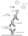
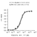

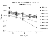
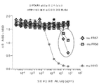

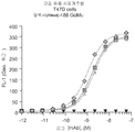


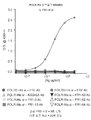


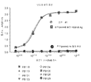

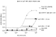


Classifications
-
- G—PHYSICS
- G01—MEASURING; TESTING
- G01N—INVESTIGATING OR ANALYSING MATERIALS BY DETERMINING THEIR CHEMICAL OR PHYSICAL PROPERTIES
- G01N33/00—Investigating or analysing materials by specific methods not covered by groups G01N1/00 - G01N31/00
- G01N33/48—Biological material, e.g. blood, urine; Haemocytometers
- G01N33/50—Chemical analysis of biological material, e.g. blood, urine; Testing involving biospecific ligand binding methods; Immunological testing
- G01N33/53—Immunoassay; Biospecific binding assay; Materials therefor
- G01N33/574—Immunoassay; Biospecific binding assay; Materials therefor for cancer
- G01N33/57484—Immunoassay; Biospecific binding assay; Materials therefor for cancer involving compounds serving as markers for tumor, cancer, neoplasia, e.g. cellular determinants, receptors, heat shock/stress proteins, A-protein, oligosaccharides, metabolites
- G01N33/57492—Immunoassay; Biospecific binding assay; Materials therefor for cancer involving compounds serving as markers for tumor, cancer, neoplasia, e.g. cellular determinants, receptors, heat shock/stress proteins, A-protein, oligosaccharides, metabolites involving compounds localized on the membrane of tumor or cancer cells
-
- A—HUMAN NECESSITIES
- A61—MEDICAL OR VETERINARY SCIENCE; HYGIENE
- A61K—PREPARATIONS FOR MEDICAL, DENTAL OR TOILETRY PURPOSES
- A61K39/00—Medicinal preparations containing antigens or antibodies
- A61K39/395—Antibodies; Immunoglobulins; Immune serum, e.g. antilymphocytic serum
- A61K39/39533—Antibodies; Immunoglobulins; Immune serum, e.g. antilymphocytic serum against materials from animals
- A61K39/39558—Antibodies; Immunoglobulins; Immune serum, e.g. antilymphocytic serum against materials from animals against tumor tissues, cells, antigens
-
- A—HUMAN NECESSITIES
- A61—MEDICAL OR VETERINARY SCIENCE; HYGIENE
- A61K—PREPARATIONS FOR MEDICAL, DENTAL OR TOILETRY PURPOSES
- A61K47/00—Medicinal preparations characterised by the non-active ingredients used, e.g. carriers or inert additives; Targeting or modifying agents chemically bound to the active ingredient
- A61K47/50—Medicinal preparations characterised by the non-active ingredients used, e.g. carriers or inert additives; Targeting or modifying agents chemically bound to the active ingredient the non-active ingredient being chemically bound to the active ingredient, e.g. polymer-drug conjugates
- A61K47/51—Medicinal preparations characterised by the non-active ingredients used, e.g. carriers or inert additives; Targeting or modifying agents chemically bound to the active ingredient the non-active ingredient being chemically bound to the active ingredient, e.g. polymer-drug conjugates the non-active ingredient being a modifying agent
- A61K47/68—Medicinal preparations characterised by the non-active ingredients used, e.g. carriers or inert additives; Targeting or modifying agents chemically bound to the active ingredient the non-active ingredient being chemically bound to the active ingredient, e.g. polymer-drug conjugates the non-active ingredient being a modifying agent the modifying agent being an antibody, an immunoglobulin or a fragment thereof, e.g. an Fc-fragment
- A61K47/6835—Medicinal preparations characterised by the non-active ingredients used, e.g. carriers or inert additives; Targeting or modifying agents chemically bound to the active ingredient the non-active ingredient being chemically bound to the active ingredient, e.g. polymer-drug conjugates the non-active ingredient being a modifying agent the modifying agent being an antibody, an immunoglobulin or a fragment thereof, e.g. an Fc-fragment the modifying agent being an antibody or an immunoglobulin bearing at least one antigen-binding site
- A61K47/6849—Medicinal preparations characterised by the non-active ingredients used, e.g. carriers or inert additives; Targeting or modifying agents chemically bound to the active ingredient the non-active ingredient being chemically bound to the active ingredient, e.g. polymer-drug conjugates the non-active ingredient being a modifying agent the modifying agent being an antibody, an immunoglobulin or a fragment thereof, e.g. an Fc-fragment the modifying agent being an antibody or an immunoglobulin bearing at least one antigen-binding site the antibody targeting a receptor, a cell surface antigen or a cell surface determinant
-
- A—HUMAN NECESSITIES
- A61—MEDICAL OR VETERINARY SCIENCE; HYGIENE
- A61P—SPECIFIC THERAPEUTIC ACTIVITY OF CHEMICAL COMPOUNDS OR MEDICINAL PREPARATIONS
- A61P35/00—Antineoplastic agents
-
- C—CHEMISTRY; METALLURGY
- C07—ORGANIC CHEMISTRY
- C07K—PEPTIDES
- C07K16/00—Immunoglobulins [IGs], e.g. monoclonal or polyclonal antibodies
- C07K16/18—Immunoglobulins [IGs], e.g. monoclonal or polyclonal antibodies against material from animals or humans
- C07K16/28—Immunoglobulins [IGs], e.g. monoclonal or polyclonal antibodies against material from animals or humans against receptors, cell surface antigens or cell surface determinants
- C07K16/30—Immunoglobulins [IGs], e.g. monoclonal or polyclonal antibodies against material from animals or humans against receptors, cell surface antigens or cell surface determinants from tumour cells
- C07K16/3015—Breast
-
- C—CHEMISTRY; METALLURGY
- C07—ORGANIC CHEMISTRY
- C07K—PEPTIDES
- C07K16/00—Immunoglobulins [IGs], e.g. monoclonal or polyclonal antibodies
- C07K16/18—Immunoglobulins [IGs], e.g. monoclonal or polyclonal antibodies against material from animals or humans
- C07K16/28—Immunoglobulins [IGs], e.g. monoclonal or polyclonal antibodies against material from animals or humans against receptors, cell surface antigens or cell surface determinants
- C07K16/30—Immunoglobulins [IGs], e.g. monoclonal or polyclonal antibodies against material from animals or humans against receptors, cell surface antigens or cell surface determinants from tumour cells
- C07K16/3023—Lung
-
- C—CHEMISTRY; METALLURGY
- C07—ORGANIC CHEMISTRY
- C07K—PEPTIDES
- C07K16/00—Immunoglobulins [IGs], e.g. monoclonal or polyclonal antibodies
- C07K16/18—Immunoglobulins [IGs], e.g. monoclonal or polyclonal antibodies against material from animals or humans
- C07K16/28—Immunoglobulins [IGs], e.g. monoclonal or polyclonal antibodies against material from animals or humans against receptors, cell surface antigens or cell surface determinants
- C07K16/30—Immunoglobulins [IGs], e.g. monoclonal or polyclonal antibodies against material from animals or humans against receptors, cell surface antigens or cell surface determinants from tumour cells
- C07K16/303—Liver or Pancreas
-
- C—CHEMISTRY; METALLURGY
- C07—ORGANIC CHEMISTRY
- C07K—PEPTIDES
- C07K16/00—Immunoglobulins [IGs], e.g. monoclonal or polyclonal antibodies
- C07K16/18—Immunoglobulins [IGs], e.g. monoclonal or polyclonal antibodies against material from animals or humans
- C07K16/28—Immunoglobulins [IGs], e.g. monoclonal or polyclonal antibodies against material from animals or humans against receptors, cell surface antigens or cell surface determinants
- C07K16/30—Immunoglobulins [IGs], e.g. monoclonal or polyclonal antibodies against material from animals or humans against receptors, cell surface antigens or cell surface determinants from tumour cells
- C07K16/3069—Reproductive system, e.g. ovaria, uterus, testes, prostate
-
- G—PHYSICS
- G01—MEASURING; TESTING
- G01N—INVESTIGATING OR ANALYSING MATERIALS BY DETERMINING THEIR CHEMICAL OR PHYSICAL PROPERTIES
- G01N15/00—Investigating characteristics of particles; Investigating permeability, pore-volume, or surface-area of porous materials
- G01N15/10—Investigating individual particles
- G01N15/14—Electro-optical investigation, e.g. flow cytometers
-
- G—PHYSICS
- G01—MEASURING; TESTING
- G01N—INVESTIGATING OR ANALYSING MATERIALS BY DETERMINING THEIR CHEMICAL OR PHYSICAL PROPERTIES
- G01N33/00—Investigating or analysing materials by specific methods not covered by groups G01N1/00 - G01N31/00
- G01N33/48—Biological material, e.g. blood, urine; Haemocytometers
- G01N33/50—Chemical analysis of biological material, e.g. blood, urine; Testing involving biospecific ligand binding methods; Immunological testing
- G01N33/53—Immunoassay; Biospecific binding assay; Materials therefor
- G01N33/566—Immunoassay; Biospecific binding assay; Materials therefor using specific carrier or receptor proteins as ligand binding reagents where possible specific carrier or receptor proteins are classified with their target compounds
-
- A—HUMAN NECESSITIES
- A61—MEDICAL OR VETERINARY SCIENCE; HYGIENE
- A61K—PREPARATIONS FOR MEDICAL, DENTAL OR TOILETRY PURPOSES
- A61K39/00—Medicinal preparations containing antigens or antibodies
- A61K2039/505—Medicinal preparations containing antigens or antibodies comprising antibodies
-
- C—CHEMISTRY; METALLURGY
- C07—ORGANIC CHEMISTRY
- C07K—PEPTIDES
- C07K2317/00—Immunoglobulins specific features
- C07K2317/20—Immunoglobulins specific features characterized by taxonomic origin
- C07K2317/21—Immunoglobulins specific features characterized by taxonomic origin from primates, e.g. man
-
- C—CHEMISTRY; METALLURGY
- C07—ORGANIC CHEMISTRY
- C07K—PEPTIDES
- C07K2317/00—Immunoglobulins specific features
- C07K2317/20—Immunoglobulins specific features characterized by taxonomic origin
- C07K2317/24—Immunoglobulins specific features characterized by taxonomic origin containing regions, domains or residues from different species, e.g. chimeric, humanized or veneered
-
- C—CHEMISTRY; METALLURGY
- C07—ORGANIC CHEMISTRY
- C07K—PEPTIDES
- C07K2317/00—Immunoglobulins specific features
- C07K2317/30—Immunoglobulins specific features characterized by aspects of specificity or valency
- C07K2317/33—Crossreactivity, e.g. for species or epitope, or lack of said crossreactivity
-
- C—CHEMISTRY; METALLURGY
- C07—ORGANIC CHEMISTRY
- C07K—PEPTIDES
- C07K2317/00—Immunoglobulins specific features
- C07K2317/70—Immunoglobulins specific features characterized by effect upon binding to a cell or to an antigen
- C07K2317/76—Antagonist effect on antigen, e.g. neutralization or inhibition of binding
-
- C—CHEMISTRY; METALLURGY
- C07—ORGANIC CHEMISTRY
- C07K—PEPTIDES
- C07K2317/00—Immunoglobulins specific features
- C07K2317/90—Immunoglobulins specific features characterized by (pharmaco)kinetic aspects or by stability of the immunoglobulin
- C07K2317/92—Affinity (KD), association rate (Ka), dissociation rate (Kd) or EC50 value
-
- G—PHYSICS
- G01—MEASURING; TESTING
- G01N—INVESTIGATING OR ANALYSING MATERIALS BY DETERMINING THEIR CHEMICAL OR PHYSICAL PROPERTIES
- G01N2333/00—Assays involving biological materials from specific organisms or of a specific nature
- G01N2333/435—Assays involving biological materials from specific organisms or of a specific nature from animals; from humans
- G01N2333/705—Assays involving receptors, cell surface antigens or cell surface determinants
Abstract
본 발명은 일반적으로 검정 인간 엽산 수용체 1에 결합하는 항체 및 엽산 수용체 기반의 요법을 위한 진단성 검정에 관한 것이다. 요법을 모니터링하기 위한 항체를 사용하는 방법이 추가로 제공된다. 본 발명은 표본에서 FOLR1의 검출을 위한 방법을 제공하며, 예를 들어, 환자들을 층화하는데 사용될 수 있다. 그러므로, 일 구현예에서, 본 발명은 엽산 수용체 1- 매개된 질병을 갖는 환자를 치료하는 방법을 제공하며, 이는 (a) 분비된 엽산 수용체 1 (FOLR1) 발현 수준 또는 환자로부터 채득된 표본에서의 순환 종양 세포 (CTC) 상에서의 FOLR1을 측정하는 것을 포함한다.The present invention relates generally to antibodies that bind to assay human folate receptor 1 and diagnostic assays for folate receptor based therapies. Methods of using antibodies to monitor therapy are further provided. The present invention provides a method for the detection of FOLR1 in a sample, and can be used, for example, to stratify patients. Therefore, in one embodiment, the present invention provides a method of treating a patient with folic acid receptor 1-mediated disease, which comprises (a) secreted folic acid receptor 1 (FOLR1) expression level or in a sample obtained from a patient. And measuring FOLR1 on circulating tumor cells (CTC).
Description
본 발명의 분야는 일반적으로 인간 엽산 수용체 1 (FOLR1)에 결합하는 항체, FOLR1를 검출하는 방법, 암을 진단하고 치료하는 방법, 및 FOLR1-계 요법을 위한 진단성 검정 및 키트에 관한 것이다.The field of the invention relates generally to antibodies that bind to human folic acid receptor 1 (FOLR1), methods of detecting FOLR1, methods of diagnosing and treating cancer, and diagnostic assays and kits for FOLR1-based therapy.
암은 선진국에서 사망의 주요 원인 중 하나로서, 미국에서만 백만명을 초과하는 사람들이 암으로 진단받고, 연간 500,000명의 사망이 발생한다. 전체적으로, 3명 중 1명 초과의 사람들에서 그들의 일생 동안 암의 일정 형태가 발병한다. 200개 초과의 상이한 암의 유형이 있으며, 이 중 유방, 폐, 결장(colorectal), 및 전립선 암이 모든 새로운 경우들 중 반을 초과하는 부분을 차지한다 (Jemal et al., 2003, Cancer J. Clin. 53:5-26).Cancer is one of the leading causes of death in developed countries, with more than 1 million people diagnosed with cancer in the United States alone, with 500,000 deaths per year. Overall, more than 1 in 3 people develop some form of cancer during their lifetime. There are more than 200 different types of cancer, of which breast, lung, colorectal, and prostate cancer account for more than half of all new cases (Jemal et al., 2003, Cancer J. Clin. 53:5-26).
또한 엽산 수용체-알파 또는 결합 단백질로 알려진, 엽산 수용체 1 (FOLR1)는 세포의 원형질막 상에서 발현된 N-글리코실화된 단백질이다. FOLR1는 엽산 및 몇몇의 환원된 엽산 유도체들에 고친화도을 갖는다. FOLR1는 생리학적 엽산, 5-메틸테트라히드로엽산의 세포 내부로의 전달을 매개한다.Folic acid receptor 1 (FOLR1), also known as folate receptor-alpha or binding protein, is an N-glycosylated protein expressed on the plasma membrane of cells. FOLR1 has high affinity to folic acid and some reduced folate derivatives. FOLR1 mediates the delivery of physiological folic acid, 5-methyltetrahydrofolic acid into cells.
FOLR1는 난소암의 광범위한 대부분에서, 뿐만 아니라 많은 자궁, 자궁내막, 췌장, 신장, 폐, 및 유방암에서 과발현되며, 한편 정상 조직 상에서 FOLR1의 발현은 신장 근위세뇨관, 폐의 폐포세포, 방광, 고환, 맥락막망, 및 갑상선에서 상피 세포의 꼭지면 막으로 제한된다 (Weitman SD, et al., Cancer Res 52: 3396-3401 (1992); Antony AC, Annu Rev Nutr 16: 501-521 (1996); Kalli KR, et al. Gynecol Oncol 108: 619-626 (2008)). FOLR1의 발현 패턴은 FOLR1-지시된 암 요법에 있어 바람직한 표적이 되게 한다.FOLR1 is overexpressed in the vast majority of ovarian cancers, as well as in many uterine, endometrial, pancreatic, kidney, lung, and breast cancers, while expression of FOLR1 on normal tissues is renal proximal tubule, alveolar cells in the lung, bladder, testis, Limited to the apical membrane of epithelial cells in the choroid network, and the thyroid (Weitman SD, et al. , Cancer Res 52: 3396-3401 (1992); Antony AC, Annu Rev Nutr 16: 501-521 (1996); Kalli KR, et al. Gynecol Oncol 108: 619-626 (2008)). The expression pattern of FOLR1 makes it a desirable target for FOLR1-directed cancer therapy.
난소 암은 전형적으로 진행단계에 이를 때까지 무증상이므로, 이는 종종 말기에 진단되고, 현재 이용가능한 시술, 전형적으로 수술적 제거 이후 화학요법의 약물로 치료할 경우 불량한 예후를 갖는다 (von Gruenigen V et al., Cancer 112: 2221-2227 (2008); Ayhan A et al., Am J Obstet Gynecol 196: 81 e81-86 (2007); Harry VN et al., Obstet Gynecol Surv 64: 548-560 (2009)). 따라서, 난소암을 위한 더욱 효과적인 요법에 대한 명백히 충족되지 않은 의학적 필요성이 있다.Ovarian cancer is typically asymptomatic until progression, so it is often diagnosed at the end and has a poor prognosis when treated with currently available procedures, typically chemotherapy drugs after surgical removal (von Gruenigen V et al. , Cancer 112: 2221-2227 (2008); Ayhan A et al ., Am J Obstet Gynecol 196: 81 e81-86 (2007); Harry VN et al. , Obstet Gynecol Surv 64: 548-560 (2009)). Thus, there is a clearly unmet medical need for a more effective therapy for ovarian cancer.
분비된(shed) FOLR1를 검출하는데 사용되는 몇몇의 이전의 검정은 FOLR1에 충분히 특이적이지 않다. 예를 들어, 몇몇의 검정은 FOLR1 및 다른 엽산 수용체 계통 구성원 (FOLR2, 3, & 4) 사이를 구별하거나 또는 총 FBP (엽산 결합 단백질)에 대한 값들을 보고하지 않는다. 추가로, 몇몇 검정들은 인간 표본(예를 들어, 혈장)이 상기 수용체로부터 엽산을 분리하는 가벼운 산 세정 단계로 사전처리될 것을 요구한다. 몇몇 검정 결과들은 또한 항체 요법 및 진단성 항체 사이에서의 경쟁 효과로 인한 부정확성을 가질 수 있다. 추가로, 많은 상업적으로 수득가능한 키트는 그들의 시약에서, 및 그들의 랏간(lot-to-lot) 안정성에서 통례적으로 신뢰가능하지 않다. 이러한 키트들의 평가는 소정의 매우 혼조의 결과를 가지고, 연구 용도로만 의도된다. 많은 것들은 상기 인간 표본이 분석 전에 “매트릭스 효과”로 인한 위양성의 가능성을 줄이기 위하여 사전-희석되는 것을 요구한다. 그러므로, FOLR1-계 요법을 위한 지침으로서 높은 정도로 민감하고 정확한 진단성 검정에 대한 명백한 필요성이 있다.Several previous assays used to detect shed FOLR1 are not sufficiently specific for FOLR1. For example, some assays differentiate between FOLR1 and other folic acid receptor lineage members (FOLR2, 3, & 4) or do not report values for total FBP (folate binding protein). Additionally, some assays require human samples (eg, plasma) to be pretreated with a light acid wash step to separate folic acid from the receptor. Some assay results may also have inaccuracies due to the competitive effect between antibody therapy and diagnostic antibodies. Additionally, many commercially available kits are customarily unreliable in their reagents, and in their lot-to-lot stability. Evaluation of these kits has some very mixed results and is intended for research use only. Many require that the human sample be pre-diluted prior to analysis to reduce the likelihood of false positives due to the “matrix effect”. Therefore, there is a clear need for a highly sensitive and accurate diagnostic assay as a guideline for FOLR1-based therapy.
발명의 요약Summary of the invention
본 발명은 표본에서 FOLR1의 검출을 위한 방법을 제공하며, 예를 들어, 환자들을 층화하는데 사용될 수 있다. 그러므로, 일 구현예에서, 본 발명은 (a) 참조 표본에서 분비된 또는 CTC FOLR1 수준과 비교하여, 항체 huMov19를 FOLR1에 결합하는 것을 경쟁적으로 억제하지 않는 항체 또는 그의 항원-결합 단편을 사용하여, 환자로부터 채득된 표본에서의 분비된 엽산 수용체 1 (FOLR1) 발현 수준 또는 순환 종양 세포 상에서의 FOLR1을 측정하는 것; 및 (b) 상기 환자의 분비된 또는 CTC FOLR1 수준이 상승되었을 때 FOLR1 활성을 조절하는 항체 또는 항원-결합 단편의 고정된 용량을 상기 환자에게 투여하는 것; 여기서 상기 항체 또는 항원-결합 단편의 고정된 용량은 상기 질병 또는 장애를 효과적으로 치료하는 것을 포함하는, 엽산 수용체 1-매개된 질병을 갖는 환자를 치료하는 방법을 제공한다.The present invention provides a method for the detection of FOLR1 in a sample, and can be used, for example, to stratify patients. Therefore, in one embodiment, the present invention uses (a) an antibody or antigen-binding fragment thereof that does not competitively inhibit the binding of the antibody huMov19 to FOLR1, compared to CTC FOLR1 levels secreted in a reference sample, Measuring the level of secreted folate receptor 1 (FOLR1) expression in a sample taken from a patient or FOLR1 on circulating tumor cells; And (b) administering to the patient a fixed dose of an antibody or antigen-binding fragment that modulates FOLR1 activity when the patient's secreted or CTC FOLR1 level is elevated; Here, a fixed dose of the antibody or antigen-binding fragment provides a method of treating a patient with folic acid receptor 1-mediated disease, comprising effectively treating the disease or disorder.
또 다른 구현예에서, 본 발명은 (a) FOLR1 활성을 조절하는 항체 또는 항원-결합 단편의 고정된 용량을 FOLR1-매개된 질병 또는 장애를 갖는 환자에게 투여하는 것; (b) 항체 huMov19의 FOLR1로의 결합을 경쟁적으로 억제하지 않는 항체 또는 그의 항원-결합 단편을 사용하여 대조 표본에서의 FOLR1 수준과 비교하여 상기 환자의 분비된 또는 CTC FOLR1 발현 수준을 측정하는 것; 및 (c) 상기 환자의 분비 또는 CTC FOLR1 수준이 상승될 경우, 후속적인 고정된 용량의 양 또는 빈도를 증가시키는 것; 여기서 상기 환자의 FOLR1 수준을 증가 (예를 들어 증가된 세포사가 분비된 FOLR1의 방출 증가를 초래하기 때문에) 또는 감소가 치료 효능을 나타내는 것을 포함하는, FOLR1-매개된 질병 또는 장애를 갖는 환자를 치료하는 방법을 제공한다. 또 다른 구현예에서, 후속적인 고정된 용량의 상기 양 또는 빈도는 상기 환자의 분비된 또는 CTC FOLR1 수준이 감소될 경우 증가한다. In another embodiment, the invention provides (a) administering a fixed dose of an antibody or antigen-binding fragment that modulates FOLR1 activity to a patient with FOLR1-mediated disease or disorder; (b) measuring the secreted or CTC FOLR1 expression level of the patient compared to FOLR1 level in a control sample using an antibody or antigen-binding fragment thereof that does not competitively inhibit the binding of antibody huMov19 to FOLR1; And (c) if the patient's secretion or CTC FOLR1 level is elevated, increasing the amount or frequency of subsequent fixed doses; Wherein a patient with a FOLR1-mediated disease or disorder comprising increasing or decreasing the patient's FOLR1 level (e.g. because increased cell death results in increased release of secreted FOLR1) How to do. In another embodiment, the amount or frequency of subsequent fixed doses increases when the patient's secreted or CTC FOLR1 level is reduced.
또 다른 구현예에서, 본 발명은 (a) 항체 huMov19의 FOLR1로의 결합을 경쟁적으로 억제하지 않는 항체 또는 그의 항원-결합 단편을 사용하여 대조 표본에서의 FOLR1 수준과 비교하여 FOLR1-매개된 질병 또는 장애를 갖는 환자로부터 채득된 표본에서 분비된 또는 CTC FOLR1 수준을 측정하는 것; 및 (b) 상기 환자의 분비된 또는 CTC FOLR1 수준이 상승될 경우 FOLR1 활성을 조절하는 항체 또는 그의 항원-결합 단편의 고정된 용량을 상기 환자에게 투여하는 것; 여기서 상기 항체 또는 그의 항원-결합 단편의 투여는 상기 환자의 FOLR1을 증가 (예를 들어 증가된 세포사가 분비된 FOLR1의 방출 증가를 초래하기 때문에) 또는 감소시키는 것을 포함하는, 환자에서 FOLR1 발현을 감소시키는 방법을 제공한다.In another embodiment, the present invention provides an FOLR1-mediated disease or disorder compared to (a) FOLR1 levels in a control sample using an antibody or antigen-binding fragment thereof that does not competitively inhibit the binding of antibody huMov19 to FOLR1. Measuring secreted or CTC FOLR1 levels in a sample obtained from a patient with; And (b) administering to the patient a fixed dose of an antibody or antigen-binding fragment thereof that modulates FOLR1 activity when the patient's secreted or CTC FOLR1 level is elevated; Wherein administration of the antibody or antigen-binding fragment thereof decreases FOLR1 expression in a patient, including increasing or decreasing FOLR1 in the patient (e.g., because increased cell death results in increased release of secreted FOLR1) How to order.
또 다른 구현예에서, 본 발명은 (a) FOLR1 활성을 조절하는 항체 또는 그의 항원-결합 단편의 고정된 용량을 FOLR1-매개된 질병 또는 장애를 갖는 환자에게 투여하는 것; (b) 대조 표본에서의 FOLR1 수준과 비교하여 상기 환자의 분비된 또는 CTC FOLR1 발현 수준을 측정하는 것; 및 (c) 상기 환자의 분비 또는 CTC FOLR1 수준이 상승될 경우 후속적인 고정된 용량의 양 또는 빈도를 증가시키는 것; 여기서 상기 환자의 FOLR1 수준을 증가 (예를 들어 증가된 세포사가 분비된 FOLR1의 방출 증가를 초래하기 때문에) 또는 감소시키는 것을 포함하는, 환자에서 FOLR1 발현을 감소시키는 방법을 제공한다.In another embodiment, the invention provides (a) administering a fixed dose of an antibody or antigen-binding fragment thereof that modulates FOLR1 activity to a patient with FOLR1-mediated disease or disorder; (b) measuring the level of secreted or CTC FOLR1 expression in the patient compared to FOLR1 level in a control sample; And (c) increasing the amount or frequency of subsequent fixed doses when the patient's secretion or CTC FOLR1 level is elevated; Provided herein is a method of reducing FOLR1 expression in a patient, comprising increasing or decreasing the patient's FOLR1 level (eg because increased cell death results in increased release of secreted FOLR1).
일 구현예에서, 상기 질병은 암이다. 또 다른 구현예에서, 상기 암은 난소, 비-소세포 폐암, 자궁, 자궁내막, 췌장, 신장(renal), 폐, 및 유방암으로 구성된 그룹으로부터 선택된 FOLR1 상승된 암이다. 또 다른 구현예에서, 상기 암은 백금 저항성 또는 백금 내화성(refractory)인 난소암이다.In one embodiment, the disease is cancer. In another embodiment, the cancer is a FOLR1 elevated cancer selected from the group consisting of ovary, non-small cell lung cancer, uterus, endometrium, pancreas, kidney, lung, and breast cancer. In another embodiment, the cancer is ovarian cancer that is platinum resistant or platinum refractory.
본 발명은 또한 (a) 항체 huMov19를 FOLR1에 결합하는 것을 경쟁적으로 억제하지 않는 항체 또는 그의 항원-결합 단편을 사용하여, FOLR1-매개된 질병 또는 장애를 갖는 환자로부터 채득된 표본에서 제1 분비된 또는 CTC FOLR1 수준을 측정하는 것; (b) FOLR1 활성을 조절하는 항체 또는 항원-결합 단편의 고정된 용량을 상기 환자에게 투여하는 것; (c) 항체 huMov19를 FOLR1에 결합하는 것을 경쟁적으로 억제하지 않는 항체 또는 그의 항원-결합 단편을 사용하여, 항체 투여 후 상기 환자로부터 채득된 표본에서 제2 분비된 또는 CTC FOLR1 수준을 측정하는 것; 및 (d) 상기 제2 FOLR1 수준을 상기 제1 FOLR1 수준과 비교하는 것; 여기서 상기 제1 및 제2 FOLR1 스코어(score) 간의 증가 (예를 들어 증가된 세포사가 분비된 FOLR1의 방출 증가를 초래하기 때문에) 또는 감소는 치료 효능을 나타내는 것을 포함하는, 환자에서 FOLR1 활성을 조절하는 항체 또는 그의 항원-결합 단편의 고정된 용량의 치료적 효과를 모니터링하는 방법을 제공한다. The present invention also provides (a) the first secretion from a sample obtained from a patient with a FOLR1-mediated disease or disorder using an antibody or antigen-binding fragment thereof that does not competitively inhibit the binding of antibody huMov19 to FOLR1. Or measuring CTC FOLR1 levels; (b) administering a fixed dose of an antibody or antigen-binding fragment that modulates FOLR1 activity to the patient; (c) measuring the second secreted or CTC FOLR1 level in a sample obtained from the patient after antibody administration, using an antibody or antigen-binding fragment thereof that does not competitively inhibit the binding of antibody huMov19 to FOLR1; And (d) comparing the second FOLR1 level to the first FOLR1 level; Wherein an increase between the first and second FOLR1 scores (e.g. because increased cell death results in an increased release of secreted FOLR1) or decrease modulates FOLR1 activity in patients, including indicating therapeutic efficacy Provides a method for monitoring the therapeutic effect of a fixed dose of an antibody or antigen-binding fragment thereof.
일 구현예에서, 상기 FOLR1 발현 수준은 체액에서 측정된다. 또 다른 구현예에서, 상기 체액은 복수(ascites) 유체이다. 또 다른 구현예에서, 상기 체액은 혈청, 혈액, 또는 혈장이다. 일 구현예에서, 상기 FOLR1 발현 수준은 말초 혈액 표본에서 측정된다.In one embodiment, the FOLR1 expression level is measured in body fluids. In another embodiment, the body fluid is ascites fluid. In another embodiment, the body fluid is serum, blood, or plasma. In one embodiment, the FOLR1 expression level is measured in peripheral blood samples.
일 구현예에서, 상기 환자는 암을 갖는다. 또 다른 구현예에서, 상기 암은 난소, 비-소세포 폐암, 자궁, 자궁내막, 췌장, 신장(renal), 폐, 및 유방암으로 구성된 그룹으로부터 선택된 FOLR1 상승된 암이다. 또 다른 구현예에서, 상기 암은 백금 저항성 또는 백금 내화성인 난소암이다.In one embodiment, the patient has cancer. In another embodiment, the cancer is a FOLR1 elevated cancer selected from the group consisting of ovary, non-small cell lung cancer, uterus, endometrium, pancreas, kidney, lung, and breast cancer. In another embodiment, the cancer is ovarian cancer that is platinum resistant or platinum fire resistant.
일 구현예에서, 상기 FOLR1 발현은 적어도 하나의 부가적인 항-FOLR1 항체 또는 그의 항원-결합 단편을 사용하여 측정된다. 또 다른 구현예에서, 상기 FOLR1 발현은 2개의 항-FOLR1 항체 또는 그의 항원-결합 단편을 사용하여 측정된다. 또 다른 구현예에서, 상기 항체는 뮤린(murine)의, 키메라의, 인간화된, 또는 인간 항체이다. 또 다른 구현예에서, 상기 항체 또는 그의 항원-결합 단편은 약 1.0 내지 약 10 nM의 Kd로 인간 엽산 수용체 1에 결합한다. 또 다른 구현예에서, 상기 항체 또는 그의 항원-결합 단편은 약 0.5 내지 약 5 nM의 Kd로 인간 엽산 수용체 1에 결합한다. 또 다른 구현예에서, 상기 결합 친화도는 세포계측법, 비아코어(Biacore), ELISA, 또는 방사성면역검정에 의하여 측정된다. 또 다른 구현예에서, 상기 세포계측법은 유동 세포계측법이다.In one embodiment, the FOLR1 expression is measured using at least one additional anti-FOLR1 antibody or antigen-binding fragment thereof. In another embodiment, the FOLR1 expression is measured using two anti-FOLR1 antibodies or antigen-binding fragments thereof. In another embodiment, the antibody is a murine, chimeric, humanized, or human antibody. In another embodiment, the antibody or antigen-binding fragment thereof binds human
일 구현예에서, 상기 항체 또는 그의 항원-결합 단편은 엽산 수용체 2 또는 엽산 수용체 3과 결합하지 않는다.In one embodiment, the antibody or antigen-binding fragment thereof does not bind
일 구현예에서, 상기 적어도 하나의 항체 또는 그의 항원-결합 단편은 고형 지지체에 결합된다. 또 다른 구현예에서, 상기 적어도 하나의 항체 또는 그의 항원-결합 단편은 마이크로티터 플레이트(microtiter plate)에 결합된다. 또 다른 구현예에서, 상기 적어도 하나의 항체 또는 그의 항원-결합 단편은 검출제를 포함한다. 또 다른 구현예에서, 상기 검출제는 발색성 검출제, 형광원성 검출제, 효소적 검출제, 또는 전기화학발광 검출제이다. 또 다른 구현예에서, 상기 검출제는 호스래디쉬(horseradish) 퍼옥시다제(HRP)이다.In one embodiment, the at least one antibody or antigen-binding fragment thereof is bound to a solid support. In another embodiment, the at least one antibody or antigen-binding fragment thereof is bound to a microtiter plate. In another embodiment, the at least one antibody or antigen-binding fragment thereof comprises a detection agent. In another embodiment, the detection agent is a chromogenic detection agent, a fluorescence detection agent, an enzymatic detection agent, or an electrochemiluminescence detection agent. In another embodiment, the detection agent is horseradish peroxidase (HRP).
일 구현예에서, 상기 FOLR1 수준은 효소 결합된 면역흡착검정 (ELISA) 또는 세포계측법 (예를 들어, 유동 세포계측법)를 사용하여 측정된다. 또 다른 구현예에서, 상기 ELISA은 샌드위치(sandwich) ELISA이다.In one embodiment, the FOLR1 level is measured using an enzyme-linked immunosorbent assay (ELISA) or cytometry (eg, flow cytometry). In another embodiment, the ELISA is a sandwich ELISA.
일 구현예에서, 상기 적어도 하나의 항체 또는 그의 항원 결합 단편은 상동한 FOLR1 에피토프에 다음으로 구성된 그룹으로부터 선택된 항체로서 특이적으로 결합한다: (a) 서열 식별 번호 25의 폴리펩티드 및 서열 식별 번호 29의 폴리펩티드를 포함하는 항체; (b) 서열 식별 번호 26의 폴리펩티드 및 서열 식별 번호 30의 폴리펩티드를 포함하는 항체; (c) 서열 식별 번호 27의 폴리펩티드 및 서열 식별 번호 31의 폴리펩티드를 포함하는 항체; 및 (d) 서열 식별 번호 28의 폴리펩티드 및 서열 식별 번호 32의 폴리펩티드를 포함하는 항체.In one embodiment, the at least one antibody or antigen-binding fragment thereof specifically binds the homologous FOLR1 epitope as an antibody selected from the group consisting of: (a) the polypeptide of SEQ ID NO: 25 and the sequence of SEQ ID NO: 29 An antibody comprising a polypeptide; (b) an antibody comprising the polypeptide of SEQ ID NO: 26 and the polypeptide of SEQ ID NO: 30; (c) an antibody comprising the polypeptide of SEQ ID NO: 27 and the polypeptide of SEQ ID NO: 31; And (d) the polypeptide of SEQ ID NO: 28 and the polypeptide of SEQ ID NO: 32.
일 구현예에서, 상기 적어도 하나의 항체 또는 그의 항원 결합 단편은 FOLR1에 특이적으로 결합하며, 상기 항체 또는 그의 단편은 다음으로 구성된 그룹으로부터 선택된 항체의 FOLR1 결합을 경쟁적으로 억제한다: (a) 서열 식별 번호 25의 폴리펩티드 및 서열 식별 번호 29의 폴리펩티드를 포함하는 항체; (b) 서열 식별 번호 26의 폴리펩티드 및 서열 식별 번호 30의 폴리펩티드를 포함하는 항체; (c) 서열 식별 번호 27의 폴리펩티드 및 서열 식별 번호 31의 폴리펩티드를 포함하는 항체; 및 (d) 서열 식별 번호 28의 폴리펩티드 및 서열 식별 번호 32의 폴리펩티드를 포함하는 항체.In one embodiment, the at least one antibody or antigen-binding fragment thereof specifically binds FOLR1, and the antibody or fragment thereof competitively inhibits FOLR1 binding of an antibody selected from the group consisting of: (a) sequence An antibody comprising the polypeptide of SEQ ID NO: 25 and the polypeptide of SEQ ID NO: 29; (b) an antibody comprising the polypeptide of SEQ ID NO: 26 and the polypeptide of SEQ ID NO: 30; (c) an antibody comprising the polypeptide of SEQ ID NO: 27 and the polypeptide of SEQ ID NO: 31; And (d) the polypeptide of SEQ ID NO: 28 and the polypeptide of SEQ ID NO: 32.
일 구현예에서, 상기 적어도 하나의 항체 또는 그의 항원 결합 단편은 FOLR1에 특이적으로 결합하며, 상기 항체는 다음으로 구성된 그룹으로부터 선택된 폴리펩티드 서열들을 포함한다: (a) 서열 식별 번호 1, 2, 및 3 및 서열 식별 번호: 13, 14, 및 15; (b) 서열 식별 번호: 4, 5, 및 6 및 서열 식별 번호: 16, 17, 및 18; (c) 서열 식별 번호: 7, 8, 및 9 및 서열 식별 번호: 19, 20, 및 21; (d) 서열 식별 번호: 10, 11, 및 12 및 서열 식별 번호: 22, 23, 및 24; 및 (e) 1, 2, 3, 또는 4의 보존적 아미노산 치환을 갖는 (a) 내지 (d)의 변이체들.In one embodiment, said at least one antibody or antigen binding fragment thereof specifically binds FOLR1, said antibody comprising polypeptide sequences selected from the group consisting of: (a) SEQ ID NOs: 1, 2, and 3 and SEQ ID NOs: 13, 14, and 15; (b) SEQ ID NOs: 4, 5, and 6 and SEQ ID NOs: 16, 17, and 18; (c) SEQ ID NO: 7, 8, and 9 and SEQ ID NO: 19, 20, and 21; (d) SEQ ID NOs: 10, 11, and 12 and SEQ ID NOs: 22, 23, and 24; And (e) variants of (a) to (d) having conservative amino acid substitutions of 1, 2, 3, or 4.
일 구현예에서, 상기 적어도 하나의 항체 또는 그의 항원-결합 단편은 검출가능하게 표지된다.In one embodiment, the at least one antibody or antigen-binding fragment thereof is detectably labeled.
일 구현예에서, 상기 투여된 항체는 FOLR1 항체 huMov19를 포함한다.In one embodiment, the administered antibody comprises the FOLR1 antibody huMov19.
일 구현예에서, 상기 huMov19는 항체 메이탄시노이드 공액체로서 투여된다.일 구현예에서, 상기 항체 메이탄시노이드 공액체는 메이탄시노이드 DM4 및 절단가능한 설포-SPDB 링커 (IMGN853)를 포함한다.In one embodiment, the huMov19 is administered as an antibody maytansinoid conjugate. In one embodiment, the antibody maytansinoid conjugate comprises maytansinoid DM4 and a cleavable sulfo-SPDB linker (IMGN853). do.
본 발명은 다음을 포함하는 FOLR1-매개된 질병 또는 장애를 갖는 환자를 치료하는 방법을 또한 제공한다: (a) FOLR1 활성을 조절하는 항체 또는 그의 항원-결합 단편의 고정된 용량을 FOLR1-매개된 질병 또는 장애를 갖는 환자에게 투여하는 것; (b) FOLR1 발현 수준의 측정을 위하여 상기 환자로부터 채득된 표본을 제출하는 것; (c) 대조 표본에서의 FOLR1 수준과 비교하여 상기 환자의 분비된 또는 CTC FOLR1 수준이 상승하였는지 여부를 측정의 결과로부터 결정하는 것; 및 (d) 상기 환자의 분비된 또는 CTC FOLR1 수준이 상승할 경우 후속적인 고정된 용량의 양 또는 빈도를 증가시키는 것.The invention also provides a method of treating a patient with a FOLR1-mediated disease or disorder comprising: (a) FOLR1-mediated fixed doses of an antibody or antigen-binding fragment thereof that modulates FOLR1 activity. Administering to a patient with a disease or disorder; (b) submitting samples obtained from the patient for measurement of FOLR1 expression levels; (c) determining from the results of the measurement whether the patient's secreted or CTC FOLR1 level was elevated compared to the FOLR1 level in the control sample; And (d) increasing the amount or frequency of subsequent fixed doses when the patient's secreted or CTC FOLR1 levels rise.
본 발명은 또한 다음을 포함하는 FOLR1-매개된 질병 또는 장애를 갖는 환자를 치료하는 방법을 제공한다: (a) FOLR1 활성을 조절하는 항체 또는 그의 항원-결합 단편의 고정된 용량을 FOLR1-매개된 질병 또는 장애를 갖는 환자에게 투여하는 것; (b) 분비된 또는 CTC FOLR1 발현 수준의 측정 및 참조 표본에서 FOLR1 수준과의 비교를 위하여 환자로부터 채득된 표본을 제출하는 것; 및 (c) 상기 환자의 분비된 또는 CTC FOLR1 수준이 상승될 경우 후속적인 고정된 용량의 양 또는 빈도를 증가시키는 것; 여기서 상기 환자의 FOLR1 수준의 증가 (예를 들어 증가된 세포사가 분비된 FOLR1의 방출 증가를 초래하기 때문에) 또는 감소가 치료 효능을 나타낸다.The invention also provides a method of treating a patient with FOLR1-mediated disease or disorder comprising: (a) FOLR1-mediated fixed doses of an antibody or antigen-binding fragment thereof that modulates FOLR1 activity. Administering to a patient with a disease or disorder; (b) submitting samples obtained from the patient for measurement of secreted or CTC FOLR1 expression levels and comparison with FOLR1 levels in reference samples; And (c) increasing the amount or frequency of subsequent fixed doses when the patient's secreted or CTC FOLR1 level is elevated; Here, an increase or decrease in the patient's FOLR1 level (eg because increased cell death results in an increased release of secreted FOLR1) or a decrease in therapeutic efficacy.
일 구현예에서, 상기 투여된 항체는 FOLR1 항체 huMov19를 포함한다. 또 다른 구현예에서, 상기 huMov19는 항체 메이탄시노이드 공액체로서 투여된다. 일 구현예에서, 상기 항체 메이탄시노이드 공액체는 메이탄시노이드 DM4 및 절단가능한 설포-SPDB 링커 (IMGN853)를 포함한다.In one embodiment, the administered antibody comprises the FOLR1 antibody huMov19. In another embodiment, the huMov19 is administered as an antibody maytansinoid conjugate. In one embodiment, the antibody maytansinoid conjugate comprises maytansinoid DM4 and a cleavable sulfo-SPDB linker (IMGN853).
본 발명은 또한 (a) FOLR1-매개된 질병 또는 장애를 갖는 환자로부터 표본을 수득하는 것; 여기서 상기 환자는 FOLR1 활성을 조절하는 항체 또는 항원-결합 단편의 고정된 용량을 수용하였으며; (b) 항체 huMov19의 FOLR1로의 결합을 경쟁적으로 억제하지 않는 항체 또는 그의 항원-결합 단편을 사용하여 대조 표본에서의 FOLR1 수준과 비교하여 상기 환자의 분비된 또는 CTC FOLR1 발현 수준을 측정하는 것; (c) 대조 표본에서의 FOLR1 수준과 비교하여 상기 환자의 분비된 또는 CTC FOLR1 수준이 상승하였는지 여부를 측정의 결과로부터 결정하는 것; (d) 상기 환자의 분비 또는 CTC FOLR1 수준이 상승될 경우 후속적인 고정된 용량의 양 또는 빈도를 증가시킬 것을 건강관리 공급자에게 나타내는 것; 여기서 상기 환자의 FOLR1 수준의 증가 (예를 들어 증가된 세포사가 분비된 FOLR1의 방출 증가를 초래하기 때문에) 또는 감소가 치료 효능을 나타내는 것을 포함하는, FOLR1-매개된 질병 또는 장애를 갖는 환자를 치료하는 방법을 제공한다.The invention also provides (a) obtaining a sample from a patient with FOLR1-mediated disease or disorder; Wherein the patient received a fixed dose of antibody or antigen-binding fragment that modulates FOLR1 activity; (b) measuring the secreted or CTC FOLR1 expression level of the patient compared to FOLR1 level in a control sample using an antibody or antigen-binding fragment thereof that does not competitively inhibit the binding of antibody huMov19 to FOLR1; (c) determining from the results of the measurement whether the patient's secreted or CTC FOLR1 level was elevated compared to the FOLR1 level in the control sample; (d) informing the healthcare provider to increase the amount or frequency of subsequent fixed doses if the patient's secretion or CTC FOLR1 level is elevated; Wherein a patient with FOLR1-mediated disease or disorder comprising increasing or decreasing the FOLR1 level of the patient (e.g., because increased cell death results in increased release of secreted FOLR1) How to do.
본 발명은 표본에서 분비된 또는 CTC FOLR1를 검출하기 위한 면역검정 키트를 또한 제공하며, 상기 키트는 (a) 인간 FOLR1에 대한 포획 항체, 여기서 상기 키트는 포획 항체 또는 그의 항원-결합 단편은 huMov19의 FOLR1로의 결합을 경쟁적으로 억제하지 않으며, 및 (b) 검출 시약을 포함한다. 또 다른 구현예에서, 상기 키트는 상기 포획 시약을 위한 고형 지지체를 추가로 포함한다. 또 다른 구현예에서, 상기 포획 시약은 상기 고형 지지체 상에 고정된다. 또 다른 구현예에서, 상기 포획 시약은 마이크로티터 플레이트(microtiter plate) 상에서 코팅된다. 또 다른 구현예에서, 상기 검출 시약은 제2 FOLR1 항체이다. 또 다른 구현예에서, 상기 제1 및/또는 제2 FOLR1 항체는 다음으로 구성된 그룹으로부터 선택된 폴리펩티드 서열들을 포함한다: (a) 서열 식별 번호 1, 2, 및 3 및 서열 식별 번호: 13, 14, 및 15; (b) 서열 식별 번호: 4, 5, 및 6 및 서열 식별 번호: 16, 17, 및 18; (c) 서열 식별 번호: 7, 8, 및 9 및 서열 식별 번호: 19, 20, 및 21; (d) 서열 식별 번호: 10, 11, 및 12 및 서열 식별 번호: 22, 23, 및 24; 및 (e) 1, 2, 3, 또는 4의 보존적 아미노산 치환을 갖는 (a) 내지 (d)의 변이체들.The present invention also provides an immunoassay kit for detecting CTC FOLR1 secreted from a sample, the kit comprising (a) a capture antibody against human FOLR1, wherein the kit is a capture antibody or an antigen-binding fragment thereof of huMov19 Does not competitively inhibit binding to FOLR1, and (b) detection reagents. In another embodiment, the kit further comprises a solid support for the capture reagent. In another embodiment, the capture reagent is immobilized on the solid support. In another embodiment, the capture reagent is coated on a microtiter plate. In another embodiment, the detection reagent is a second FOLR1 antibody. In another embodiment, the first and/or second FOLR1 antibody comprises polypeptide sequences selected from the group consisting of: (a) SEQ ID NOs: 1, 2, and 3 and SEQ ID NOs: 13, 14, And 15; (b) SEQ ID NOs: 4, 5, and 6 and SEQ ID NOs: 16, 17, and 18; (c) SEQ ID NO: 7, 8, and 9 and SEQ ID NO: 19, 20, and 21; (d) SEQ ID NOs: 10, 11, and 12 and SEQ ID NOs: 22, 23, and 24; And (e) variants of (a) to (d) having conservative amino acid substitutions of 1, 2, 3, or 4.
일 구현예에서, 상기 검출 시약은 종 특이적 항체를 사용하여 검출된다. 또 다른 구현예에서, 상기 키트는 검출 가능한 항체를 위한 검출 수단을 추가로 포함한다. 또 다른 구현예에서, 상기 검출 수단은 비색성(colorimetric)이다. 또 다른 구현예에서, 상기 키트는 항원표준 으로서의 FOLR1 폴리펩티드를 추가로 포함한다. 또 다른 구현예에서, 상기 FOLR1 폴리펩티드는 FOLR1-Fc이다.In one embodiment, the detection reagent is detected using a species specific antibody. In another embodiment, the kit further comprises detection means for detectable antibodies. In another embodiment, the detection means is colorimetric. In another embodiment, the kit further comprises a FOLR1 polypeptide as an antigen standard. In another embodiment, the FOLR1 polypeptide is FOLR1-Fc.
본 발명은 또한 상동한 FOLR1 에피토프에 다음으로 구성된 그룹으로부터 선택된 항체로서 특이적으로 결합하는 항체 또는 그의 항원 결합 단편을 제공한다: (a) 서열 식별 번호 25의 폴리펩티드 및 서열 식별 번호 29의 폴리펩티드를 포함하는 항체; (b) 서열 식별 번호 26의 폴리펩티드 및 서열 식별 번호 30의 폴리펩티드를 포함하는 항체; (c) 서열 식별 번호 27의 폴리펩티드 및 서열 식별 번호 31의 폴리펩티드를 포함하는 항체; 및 (d) 서열 식별 번호 28의 폴리펩티드 및 서열 식별 번호 32의 폴리펩티드를 포함하는 항체.The invention also provides an antibody or antigen-binding fragment thereof that specifically binds the homologous FOLR1 epitope as an antibody selected from the group consisting of: (a) comprising the polypeptide of SEQ ID NO: 25 and the polypeptide of SEQ ID NO: 29 Antibody; (b) an antibody comprising the polypeptide of SEQ ID NO: 26 and the polypeptide of SEQ ID NO: 30; (c) an antibody comprising the polypeptide of SEQ ID NO: 27 and the polypeptide of SEQ ID NO: 31; And (d) the polypeptide of SEQ ID NO: 28 and the polypeptide of SEQ ID NO: 32.
본 발명은 또한 FOLR1에 특이적으로 결합하며, 항체 또는 그의 단편은 다음으로 구성된 그룹으로부터 선택된 항체의 FOLR1를 경쟁적으로 억제하는 항체 또는 그의 항원 결합 단편을 제공한다: (a) 서열 식별 번호 25의 폴리펩티드 및 서열 식별 번호 29의 폴리펩티드를 포함하는 항체; (b) 서열 식별 번호 26의 폴리펩티드 및 서열 식별 번호 30의 폴리펩티드를 포함하는 항체; (c) 서열 식별 번호 27의 폴리펩티드 및 서열 식별 번호 31의 폴리펩티드를 포함하는 항체; 및 (d) 서열 식별 번호 28의 폴리펩티드 및 서열 식별 번호 32의 폴리펩티드를 포함하는 항체.The invention also provides an antibody or antigen-binding fragment thereof that specifically binds FOLR1, the antibody or fragment thereof competitively inhibits FOLR1 of an antibody selected from the group consisting of: (a) the polypeptide of SEQ ID NO: 25 And an antibody comprising SEQ ID NO: 29; (b) an antibody comprising the polypeptide of SEQ ID NO: 26 and the polypeptide of SEQ ID NO: 30; (c) an antibody comprising the polypeptide of SEQ ID NO: 27 and the polypeptide of SEQ ID NO: 31; And (d) the polypeptide of SEQ ID NO: 28 and the polypeptide of SEQ ID NO: 32.
본 발명은 또한 FOLR1에 특이적으로 결합하며, 항체는 다음으로 구성된 그룹으로부터 선택된 폴리펩티드 서열들을 포함하는 항체 또는 그의 항원 결합 단편을 제공한다: (a) 서열 식별 번호 1, 2, 및 3 및 서열 식별 번호: 13, 14, 및 15; (b) 서열 식별 번호: 4, 5, 및 6 및 서열 식별 번호: 16, 17, 및 18; (c) 서열 식별 번호: 7, 8, 및 9 및 서열 식별 번호: 19, 20, 및 21; (d) 서열 식별 번호: 10, 11, 및 12 및 서열 식별 번호: 22, 23, 및 24; 및 (e) 1, 2, 3, 또는 4의 보존적 아미노산 치환을 갖는 (a) 내지 (d)의 변이체들.The invention also provides an antibody or antigen binding fragment thereof that specifically binds FOLR1, the antibody comprising polypeptide sequences selected from the group consisting of: (a) SEQ ID NOs: 1, 2, and 3 and sequence identification Numbers: 13, 14, and 15; (b) SEQ ID NOs: 4, 5, and 6 and SEQ ID NOs: 16, 17, and 18; (c) SEQ ID NO: 7, 8, and 9 and SEQ ID NO: 19, 20, and 21; (d) SEQ ID NOs: 10, 11, and 12 and SEQ ID NOs: 22, 23, and 24; And (e) variants of (a) to (d) having conservative amino acid substitutions of 1, 2, 3, or 4.
일 구현예에서, 상기 항체는 다음으로 구성된 그룹으로부터 선택된 폴리펩티드와 적어도 90%의 상동성을 갖는 폴리펩티드 서열을 포함한다: (a) 서열 식별 번호 25 및 서열 식별 번호 29; (b) 서열 식별 번호 26 및 서열 식별 번호 30; (c) 서열 식별 번호 27 및 서열 식별 번호 31; 및 (d) 서열 식별 번호 28 및 서열 식별 번호 32.In one embodiment, the antibody comprises a polypeptide sequence having at least 90% homology to a polypeptide selected from the group consisting of: (a) SEQ ID NO: 25 and SEQ ID NO: 29; (b) SEQ ID NO: 26 and SEQ ID NO: 30; (c) SEQ ID NO: 27 and SEQ ID NO: 31; And (d) SEQ ID NO: 28 and SEQ ID NO: 32.
또 다른 구현예에서, 상기 폴리펩티드 서열은 다음으로 구성된 그룹으로부터 선택된 폴리펩티드와 적어도 95%의 상동성을 갖는다: (a) 서열 식별 번호 25 및 서열 식별 번호 29; (b) 서열 식별 번호 26 및 서열 식별 번호 30; (c) 서열 식별 번호 27 및 서열 식별 번호 31; 및 (d) 서열 식별 번호 28 및 서열 식별 번호 32. 또 다른 구현예에서, 상기 폴리펩티드 서열은 다음으로 구성된 그룹으로부터 선택된 폴리펩티드와 적어도 99%의 상동성을 갖는다: (a) 서열 식별 번호 25 및 서열 식별 번호 29; (b) 서열 식별 번호 26 및 서열 식별 번호 30; (c) 서열 식별 번호 27 및 서열 식별 번호 31; 및 (d) 서열 식별 번호 28 및 서열 식별 번호 32.In another embodiment, the polypeptide sequence has at least 95% homology with a polypeptide selected from the group consisting of: (a) SEQ ID NO: 25 and SEQ ID NO: 29; (b) SEQ ID NO: 26 and SEQ ID NO: 30; (c) SEQ ID NO: 27 and SEQ ID NO: 31; And (d) SEQ ID NO: 28 and SEQ ID NO: 32. In another embodiment, the polypeptide sequence has at least 99% homology with a polypeptide selected from the group consisting of: (a) SEQ ID NO: 25 and
일 구현예에서, 상기 항체 또는 그의 항원-결합 단편은 뮤린(murine)의, 비-인간의, 인간화된, 키메라의, 재표면화된(resurfaced) 또는 인간의 것이다. 또 다른 구현예에서, 상기 항체는 인간 FOLR1에 결합하나, FOLR2 또는 FOLR3에는 결합하지 않는다. 또 다른 구현예에서, 상기 항체는 전체 길이 항체 또는 항원-결합 단편이다. 또 다른 구현예에서, 상기 항체 또는 그의 항원-결합 단편은 Fab, Fab', F(ab')2, Fd, 단일 사슬 Fv 또는 scFv, 이황화결합된 Fv, V-NAR 도메인, IgNar, 인트라바디, IgGΔCH2, 미니바디, F(ab')3, 테트라바디, 트리아바디, 디아바디, 단일-도메인 항체, DVD-Ig, Fcab, mAb2, (scFv)2, 또는 scFv-Fc을 포함한다.In one embodiment, the antibody or antigen-binding fragment thereof is murine, non-human, humanized, chimeric, resurfaced or human. In another embodiment, the antibody binds human FOLR1 but does not bind FOLR2 or FOLR3. In another embodiment, the antibody is a full length antibody or antigen-binding fragment. In another embodiment, the antibody or antigen-binding fragment thereof is Fab, Fab', F(ab')2, Fd, single chain Fv or scFv, disulfide Fv, V-NAR domain, IgNar, intrabody, IgGΔCH2, minibody, F(ab')3, tetrabody, tribody, diabody, single-domain antibody, DVD-Ig, Fcab, mAb2, (scFv)2, or scFv-Fc.
본 발명은 또한 FOLR1에 특이적으로 결합하며, 폴리펩티드는 다음으로 구성된 그룹으로부터 선택된 서열들을 포함하는 폴리펩티드를 제공한다: (a) 서열 식별 번호 1, 2, 및 3 및 서열 식별 번호: 13, 14, 및 15; (b) 서열 식별 번호: 4, 5, 및 6 및 서열 식별 번호: 16, 17, 및 18; (c) 서열 식별 번호: 7, 8, 및 9 및 서열 식별 번호: 19, 20, 및 21; (d) 서열 식별 번호: 10, 11, 및 12 및 서열 식별 번호: 22, 23, 및 24; 및 (e) 1, 2, 3, 또는 4의 보존적 아미노산 치환을 갖는 (a) 내지 (d)의 변이체들. 또 다른 구현예에서, 상기 폴리펩티드는 다음으로 구성된 그룹으로부터 선택된 서열들과 적어도 90%의 상동성을 갖는 서열들을 포함한다: (a) 서열 식별 번호 25 및 서열 식별 번호 29; (b) 서열 식별 번호 26 및 서열 식별 번호 30; (c) 서열 식별 번호 27 및 서열 식별 번호 31; 및 (d) 서열 식별 번호 28 및 서열 식별 번호 32. 또 다른 구현예에서, 상기 서열들은 다음으로 구성된 그룹으로부터 선택된 서열들과 적어도 95%의 상동성을 갖는다: (a) 서열 식별 번호 25 및 서열 식별 번호 29; (b) 서열 식별 번호 26 및 서열 식별 번호 30; (c) 서열 식별 번호 27 및 서열 식별 번호 31; 및 (d) 서열 식별 번호 28 및 서열 식별 번호 32. 또 다른 구현예에서, 상기 서열들은 다음으로 구성된 그룹으로부터 선택된 서열들과 적어도 99%의 상동성을 갖는다: (a) 서열 식별 번호 25 및 서열 식별 번호 29; (b) 서열 식별 번호 26 및 서열 식별 번호 30; (c) 서열 식별 번호 27 및 서열 식별 번호 31; 및 (d) 서열 식별 번호 28 및 서열 식별 번호 32.The present invention also specifically binds FOLR1, and the polypeptide provides a polypeptide comprising sequences selected from the group consisting of: (a) SEQ ID NOs: 1, 2, and 3 and SEQ ID NOs: 13, 14, And 15; (b) SEQ ID NOs: 4, 5, and 6 and SEQ ID NOs: 16, 17, and 18; (c) SEQ ID NO: 7, 8, and 9 and SEQ ID NO: 19, 20, and 21; (d) SEQ ID NOs: 10, 11, and 12 and SEQ ID NOs: 22, 23, and 24; And (e) variants of (a) to (d) having conservative amino acid substitutions of 1, 2, 3, or 4. In another embodiment, the polypeptide comprises sequences having at least 90% homology to sequences selected from the group consisting of: (a) SEQ ID NO: 25 and SEQ ID NO: 29; (b) SEQ ID NO: 26 and SEQ ID NO: 30; (c) SEQ ID NO: 27 and SEQ ID NO: 31; And (d) SEQ ID NO: 28 and SEQ ID NO: 32. In another embodiment, the sequences have at least 95% homology with sequences selected from the group consisting of: (a) SEQ ID NO: 25 and
일 구현예에서, 상기 항체 또는 폴리펩티드는 약 1.0 내지 약 10 nM의 Kd로 인간 엽산 수용체 1에 결합한다. 또 다른 구현예에서, 상기 항체 또는 폴리펩티드는 약 1.0 nM의, 또는 그보다 양호한 Kd로 인간 엽산 수용체 1에 결합한다. 또 다른 구현예에서, 상기 결합 친화도는 세포계측법, 비아코어(Biacore), ELISA, 또는 방사성면역검정에 의하여 측정된다. In one embodiment, the antibody or polypeptide binds human
또 다른 구현예에서, 상기 세포계측법은 유동 세포계측법이다. 본 발명은 또한 본 발명의 항체 또는 그의 항원-결합 단편 또는 폴리펩티드로 표본을 접촉시키는 것을 포함하는, 표본에서 FOLR1 발현을 검출하는 방법을 제공한다. 또 다른 구현예에서, 상기 항체 또는 그의 항원-결합 단편은 검출가능하게 표지된다. 또 다른 구현예에서, 상기 표지는 면역형광 표지, 화학발광 표지, 인광성 표지, 효소 표지, 방사성 표지, 아비딘/비오틴 콜로이드성 금 입자, 착색된 입자, 및 자기성 입자로 구성된 그룹에서 선택된다. 또 다른 구현예에서, 상기 FOLR1 발현은 방사성면역검정, 웨스턴 블랏(western blot) 검정, 면역형광 검정, 효소 면역검정, 면역침전 검정, 화학발광 검정, 또는 면역조직화학 검정에 의하여 측정된다. 또 다른 구현예에서, 상기 FOLR1 발현은 순환 종양 세포 (CTC) 검정을 사용하여 측정되며, 여기서 CTC는 혈액, 혈장, 또는 혈청의 표본으로부터 농축(enriched)되며, 본 발명의 항체 또는 그의 항원-결합 단편을 사용하여 FOLR1 발현을 위하여 염색된다. CTC 검정에 유용한 항체들의 비제한적인 예시들은 FR1-9 및 FR1-13을 포함한다. 본 발명의 항체들을 사용한 CTC 검정은 FOLR1-계 요법에 응할 수 있는 대상체를 식별하는데 유용할 수 있다.In another embodiment, the cytometry is flow cytometry. The invention also provides a method of detecting FOLR1 expression in a sample, comprising contacting the sample with an antibody or antigen-binding fragment or polypeptide thereof. In another embodiment, the antibody or antigen-binding fragment thereof is detectably labeled. In another embodiment, the label is selected from the group consisting of immunofluorescent labels, chemiluminescent labels, phosphorescent labels, enzyme labels, radioactive labels, avidin/biotin colloidal gold particles, colored particles, and magnetic particles. In another embodiment, the FOLR1 expression is measured by radioimmunoassay, western blot assay, immunofluorescence assay, enzyme immunoassay, immunoprecipitation assay, chemiluminescence assay, or immunohistochemistry assay. In another embodiment, the FOLR1 expression is measured using a circulating tumor cell (CTC) assay, where CTC is enriched from a sample of blood, plasma, or serum, and the antibody of the invention or antigen-binding thereof Fragments are used to stain for FOLR1 expression. Non-limiting examples of antibodies useful for CTC assays include FR1-9 and FR1-13. CTC assays using the antibodies of the invention can be useful in identifying subjects who can respond to FOLR1-based therapy.
본 발명은 또한 본 발명의 항체 또는 그의 항원-결합 단편 또는 폴리펩티드를 생성하는 단리된 세포를 제공한다.The invention also provides isolated cells that produce the antibodies or antigen-binding fragments or polypeptides of the invention.
본 발명은 또한 (a) 본 발명의 항체 또는 그의 항원-결합 단편 또는 폴리펩티드를 발현하는 세포를 배양하는 것을 포함하는, 본 발명의 항체 또는 그의 항원-결합 단편 또는 폴리펩티드를 제조하는 방법을 제공한다. The invention also provides a method of making an antibody of the invention or an antigen-binding fragment or polypeptide thereof, comprising (a) culturing a cell expressing the antibody or antigen-binding fragment or polypeptide thereof.
본 발명은 또한 암을 치료하는 방법에 사용하기 위한 FOLR1 활성을 조절하는 항체 또는 그의 항원-결합 단편을 포함하는 활성제를 제공하며, 여기서 FOLR1 단백질의 증가된 발현은 상기 활성제의 투여 전에 항체, 그의 항원-결합 단편, 또는 본원에 제공된 폴리펩티드를 사용하여 상기 대상체로부터의 암성 표본에서 측정되었다.The present invention also provides an active agent comprising an antibody or antigen-binding fragment thereof that modulates FOLR1 activity for use in a method for treating cancer, wherein increased expression of the FOLR1 protein is an antibody, its antigen prior to administration of the active agent -Measured in cancerous samples from the subject using binding fragments, or polypeptides provided herein.
본 발명은 또한 FOLR1-매개된 질병 또는 장애를 치료하는 방법에 사용하기 위한 FOLR1 활성을 조절하는 항체 또는 그의 항원-결합 단편을 포함하는 활성제를 제공하며, 상기 방법은 다음을 포함한다: (a) 본원에 제공된 항체, 그의 항원-결합 단편, 또는 폴리펩티드를 사용하여 환자 표본에서 FOLR1 단백질 수준을 측정하는 것; 및 (b) 상기 환자의 FOLR1 단백질 수준이 참조 FOLR1 단백질 수준에 비교하여 상승될 경우 상기 활성제의 고정된 용량을 상기 환자에게 투여하는 것을 포함한다.The invention also provides an active agent comprising an antibody or antigen-binding fragment thereof that modulates FOLR1 activity for use in a method of treating a FOLR1-mediated disease or disorder, the method comprising: (a) Measuring FOLR1 protein levels in patient samples using the antibodies, antigen-binding fragments, or polypeptides provided herein; And (b) administering a fixed dose of the active agent to the patient when the patient's FOLR1 protein level is elevated compared to a reference FOLR1 protein level.
본 발명은 또한 FOLR1-매개된 질병 또는 장애를 치료하는 방법에 사용하기 위한 FOLR1 활성을 조절하는 항체 또는 그의 항원-결합 단편을 포함하는 활성제를 제공하며, 상기 방법은 다음을 포함한다: (a) 상기 활성제의 고정된 용량을 FOLR1-매개된 질병 또는 장애를 갖는 환자에게 투여하는 것; (b) 본원에 제공된 항체, 그의 항원-결합 단편, 또는 폴리펩티드를 사용하여 FOLR1 단백질 수준을 측정하는 것; 및 (c) 상기 환자의 FOLR1 단백질 수준이 참조 FOLR1 단백질 수준에 비교하여 상승될 경우 후속적인 고정된 용량의 양 또는 빈도를 증가시키는 것.The invention also provides an active agent comprising an antibody or antigen-binding fragment thereof that modulates FOLR1 activity for use in a method of treating a FOLR1-mediated disease or disorder, the method comprising: (a) Administering a fixed dose of the active agent to a patient with FOLR1-mediated disease or disorder; (b) measuring FOLR1 protein levels using the antibodies, antigen-binding fragments, or polypeptides provided herein; And (c) increasing the amount or frequency of subsequent fixed doses when the patient's FOLR1 protein level is elevated relative to the reference FOLR1 protein level.
본 발명은 또한 FOLR1-매개된 질병 또는 장애를 치료하는 방법에 사용하기 위한 FOLR1 활성을 조절하는 항체 또는 그의 항원-결합 단편을 포함하는 활성제를 제공하며, 여기서: (a) 환자 표본에서 측정된 FOLR1 단백질 수준은 본원에 제공된 항체, 그의 항원-결합 단편, 또는 폴리펩티드를 사용하여, 참조 FOLR1 단백질 수준과 비교되며; 그리고 (b) 상기 활성제의 고정된 용량은 상기 환자의 FOLR1 단백질 수준이 참조 FOLR1 단백질 수준에 비교하여 상승될 경우 투여되며, 여기서 상기 제제의 투여는 FOLR1 단백질 수준을 감소시킨다.The invention also provides an active agent comprising an antibody or antigen-binding fragment thereof that modulates FOLR1 activity for use in a method of treating FOLR1-mediated disease or disorder, wherein: (a) FOLR1 measured in a patient sample Protein levels are compared to reference FOLR1 protein levels using antibodies, antigen-binding fragments, or polypeptides provided herein; And (b) a fixed dose of the active agent is administered when the patient's FOLR1 protein level is elevated relative to the reference FOLR1 protein level, where administration of the agent decreases the FOLR1 protein level.
본 발명은 또한 FOLR1-매개된 질병 또는 장애를 치료하는 방법에 사용하기 위한 FOLR1 활성을 조절하는 항체 또는 그의 항원-결합 단편을 포함하는 활성제를 제공하며, 여기서 환자에서의 FOLR1-발현 세포는 감소되며, (a) 상기 활성제의 고정된 용량이 상기 환자에게 투여되며; (b) 환자 표본에서 측정된 FOLR1 단백질 수준은 본원에 제공된 항체, 그의 항원-결합 단편, 또는 폴리펩티드를 사용하여, 참조 FOLR1 단백질 수준과 비교되며; 그리고 (c) 상기 활성제의 고정된 용량은 상기 환자의 FOLR1 단백질 수준이 참조 FOLR1 단백질 수준에 비교하여 상승될 경우 투여되며, 여기서 상기 제제의 투여는 FOLR1 단백질 수준을 감소시킨다.The invention also provides an activator comprising an antibody or antigen-binding fragment thereof that modulates FOLR1 activity for use in a method of treating FOLR1-mediated disease or disorder, wherein FOLR1-expressing cells in the patient are reduced and , (a) a fixed dose of the active agent is administered to the patient; (b) FOLR1 protein levels measured in patient samples are compared to reference FOLR1 protein levels using the antibodies, antigen-binding fragments, or polypeptides provided herein; And (c) a fixed dose of the active agent is administered when the patient's FOLR1 protein level is elevated relative to the reference FOLR1 protein level, where administration of the agent decreases the FOLR1 protein level.
본 발명은 또한 환자에서의 활성제의 고정된 용량의 치료 효능을 모니터링하는 방법에 사용하기 위한 FOLR1 활성을 조절하는 항체 또는 그의 항원-결합 단편을 포함하는 활성제를 제공하며, 상기 방법은 다음을 포함한다: (a) 본원에 제공된 항체, 그의 항원-결합 단편, 또는 폴리펩티드를 사용하여 FOLR1-매개된 질병 또는장애를 갖는 환자로부터의 표본에서 제1 FOLR1 단백질 수준을 측정하는 것; (b) 상기 환자에게 상기 활성제의 고정된 용량을 투여하는 것; (c) 상기 환자로부터 채득된 표본에서 제2 FOLR1 단백질 수준을, 활성제 투여 후 본원에 제공된 항체, 그의 항원-결합 단편, 또는 폴리펩티드를 사용하여 측정하는 것; 및 (d) 상기 제2 FOLR1 단백질 수준을 상기 제1 FOLR1 단백질 수준과 비교하는 것; 여기서 상기 제1 및 제2 단백질 수준 사이의 감소는 치료 효능을 나타낸다.The invention also provides an active agent comprising an antibody or antigen-binding fragment thereof that modulates FOLR1 activity for use in a method for monitoring the therapeutic efficacy of a fixed dose of an active agent in a patient, the method comprising: : (a) measuring a first FOLR1 protein level in a sample from a patient with a FOLR1-mediated disease or disorder using an antibody, antigen-binding fragment, or polypeptide thereof provided herein; (b) administering a fixed dose of the active agent to the patient; (c) measuring the second FOLR1 protein level in a sample obtained from said patient using an antibody, antigen-binding fragment, or polypeptide provided herein after administration of the active agent; And (d) comparing the second FOLR1 protein level to the first FOLR1 protein level; Here, a decrease between the first and second protein levels indicates therapeutic efficacy.
본 발명은 또한 환자에서 FOLR1-매개된 질병 또는 장애를 치료하는 방법에 사용하기 위한 FOLR1 활성을 조절하는 항체 또는 그의 항원-결합 단편을 포함하는 활성제를 제공하며, 상기 방법은 다음을 포함한다: (a) 상기 활성제의 고정된 용량을 FOLR1-매개된 질병 또는 장애를 갖는 환자에게 투여하는 것; (b) FOLR1 발현 수준의 측정을 위하여 환자로부터 상기 채득된 표본을 본원에 제공된 항체, 그의 항원-결합 단편, 또는 폴리펩티드를 사용하여 제출하는 것; (c) 대조 표본에서의 FOLR1 수준과 비교하여 상기 환자의 분비된 또는 FOLR1 수준이 상승하였는지 여부를 측정의 결과로부터 결정하는 것; 및 (d) 상기 환자의 FOLR1 수준이 참조 FOLR1 단백질 수준과 비교하여 증가할 경우 후속적인 고정된 용량의 상기 양 및/또는 빈도를 증가시키는 것.The present invention also provides an active agent comprising an antibody or antigen-binding fragment thereof that modulates FOLR1 activity for use in a method of treating a FOLR1-mediated disease or disorder in a patient, the method comprising: ( a) administering a fixed dose of said active agent to a patient with FOLR1-mediated disease or disorder; (b) submitting the sample obtained from the patient for determination of FOLR1 expression level using the antibody, antigen-binding fragment, or polypeptide provided herein; (c) determining from the results of the measurement whether the patient's secreted or FOLR1 level was elevated compared to the FOLR1 level in the control sample; And (d) increasing the amount and/or frequency of subsequent fixed doses when the patient's FOLR1 level increases compared to a reference FOLR1 protein level.
본 발명은 또한 FOLR1-매개된 질병 또는 장애를 치료하는 방법에 사용하기 위한 FOLR1 활성을 조절하는 항체 또는 그의 항원-결합 단편을 포함하는 활성제를 제공하며, 상기 방법은 다음을 포함한다: (a) 상기 활성제의 고정된 용량을 FOLR1-매개된 질병 또는 장애를 갖는 환자에게 투여하는 것; (b) 본원에 제공된 항체, 그의 항원-결합 단편, 또는 폴리펩티드, 그리고 참조 FOLR1 단백질 수준과 상기 측정된 FOLR1 단백질 수준의 비교를 사용하여 FOLR1 발현 수준의 측정을 위하여 환자로부터 채득된 표본을 제출하는 것; 및 (c) 상기 환자의 FOLR1 단백질 수준이 참조 FOLR1 단백질 수준에 비교하여 상승될 경우 후속적인 고정된 용량의 양 또는 빈도를 증가시키는 것; 여기서 상기 환자의 FOLR1 수준의 감소는 치료 효능을 나타낸다.The invention also provides an active agent comprising an antibody or antigen-binding fragment thereof that modulates FOLR1 activity for use in a method of treating a FOLR1-mediated disease or disorder, the method comprising: (a) Administering a fixed dose of the active agent to a patient with FOLR1-mediated disease or disorder; (b) submitting a sample obtained from the patient for determination of FOLR1 expression level using the antibody, antigen-binding fragment, or polypeptide thereof provided herein, and a comparison of the reference FOLR1 protein level with the measured FOLR1 protein level. ; And (c) increasing the amount or frequency of subsequent fixed doses when the patient's FOLR1 protein level is elevated relative to the reference FOLR1 protein level; Here, the decrease in the patient's FOLR1 level indicates therapeutic efficacy.
본 발명은 또한 FOLR1-매개된 질병 또는 장애를 치료하는 방법에 사용하기 위한 FOLR1 활성을 조절하는 항체 또는 그의 항원-결합 단편을 포함하는 활성제를 제공하며, 상기 방법은 다음을 포함한다: (a) FOLR1-매개된 질병 또는 장애를 갖는 환자로부터 표본을 수득하는 것, 여기서 상기 환자는 활성제의 고정된 용량을 수용하였으며; (b) 본원에 제공된 항체, 그의 항원-결합 단편, 또는 폴리펩티드를 사용하여 상기 표본으로부터의 FOLR1 단백질 발현 수준을 측정하는 것; (c) 참조 FOLR1 단백질 수준과 비교하여 상기 환자의 단백질 FOLR1 수준이 상승하였는지 여부를 결정하는 것; (d) 상기 환자의 FOLR1 단백질 수준이 참조 FOLR1 단백질 수준에 비교하여 상승될 경우 후속적인 고정된 용량의 양 및/또는 빈도를 증가시키는 것 또는 증가시킬 것을 건강관리 공급자에게 나타내는 것; 여기서 상기 환자의 FOLR1 수준의 감소는 치료 효능을 나타낸다.The invention also provides an active agent comprising an antibody or antigen-binding fragment thereof that modulates FOLR1 activity for use in a method of treating a FOLR1-mediated disease or disorder, the method comprising: (a) Obtaining a sample from a patient with FOLR1-mediated disease or disorder, wherein the patient received a fixed dose of active agent; (b) measuring the level of FOLR1 protein expression from said sample using an antibody, antigen-binding fragment, or polypeptide thereof provided herein; (c) determining whether the patient's protein FOLR1 level is elevated compared to a reference FOLR1 protein level; (d) informing the healthcare provider to increase or/or increase the amount and/or frequency of subsequent fixed doses when the patient's FOLR1 protein level is elevated relative to the reference FOLR1 protein level; Here, the decrease in the patient's FOLR1 level indicates therapeutic efficacy.
본 발명은 또한 FOLR1-매개된 질병 또는 장애를 치료하는 방법에 사용하기 위한 FOLR1 활성을 조절하는 항체 또는 그의 항원-결합 단편을 포함하는 활성제를 제공하며, 여기서 FOLR1 단백질의 증가된 발현은 상기 활성제의 투여 전에 본원에 제공된 항체, 그의 항원-결합 단편, 또는 폴리펩티드를 사용하여 상기 대상체로부터의 표본에서 측정되었다. The present invention also provides an active agent comprising an antibody or antigen-binding fragment thereof that modulates FOLR1 activity for use in a method of treating FOLR1-mediated disease or disorder, wherein increased expression of the FOLR1 protein is It was measured in a sample from the subject using an antibody, antigen-binding fragment thereof, or polypeptide provided herein prior to administration.
일부 구현예에서, 상기 측정된 FOLR1 단백질은 분비된 FOLR1이다. 일부 구현예에서, 상기 측정된 FOLR1 단백질은 순환 종양 세포 상에 있다. In some embodiments, the measured FOLR1 protein is secreted FOLR1. In some embodiments, the measured FOLR1 protein is on circulating tumor cells.
일부 구현예에서, 상기 FOLR1 단백질 수준은 체액에서 측정된다. 일부 구현예에서, 상기 체액은 복수(ascites) 유체이다. 일부 구현예에서, 상기 체액은 혈청, 혈액, 또는 혈장이다. 일부 구현예에서, 상기 FOLR1 단백질 수준은 말초 혈액 표본에서 측정된다.In some embodiments, the FOLR1 protein level is measured in body fluids. In some embodiments, the body fluid is ascites fluid. In some embodiments, the body fluid is serum, blood, or plasma. In some embodiments, the FOLR1 protein level is measured in peripheral blood samples.
일부 구현예에서, 상기 환자는 암을 갖는다. 일부 구현예에서,상기 FOLR1-매개된 질환 또는 장애는 암이다. 일부 구현예에서, 상기 암은 난소, 비-소세포 폐암, 자궁, 자궁내막, 췌장, 신장(renal), 폐, 및 유방암으로 구성된 그룹으로부터 선택된 FOLR1 상승된 암이다. 일부 구현예에서, 상기 난소암은 백금 저항성 또는 백금 내화성이다. 일부 구현예에서, 상기 폐암은 비-소세포 폐암(NSCLC)이다. 일부 구현예에서, 상기 암은 자궁내막암이다.In some embodiments, the patient has cancer. In some embodiments, the FOLR1-mediated disease or disorder is cancer. In some embodiments, the cancer is a FOLR1 elevated cancer selected from the group consisting of ovarian, non-small cell lung cancer, uterus, endometrium, pancreas, kidney, lung, and breast cancer. In some embodiments, the ovarian cancer is platinum resistant or platinum fire resistant. In some embodiments, the lung cancer is non-small cell lung cancer (NSCLC). In some embodiments, the cancer is endometrial cancer.
일부 구현예에서, FOLR1 단백질 수준은 FOLR1을 특이적으로 결합하는 2개의 상이한 항체들, 또는 그의 항원 결합 단편들, 또는 폴리펩티드들을 사용하여 측정된다. 일부 구현예에서, FOLR1 단백질을 검출하기 위하여 사용되는 상기 항체 또는 그의 항원-결합 단편, 또는 폴리펩티드는 고형 지지체에 결합된다. 일부 구현예에서, 상기 고형 지지체는 마이크로티터 플레이트이다.In some embodiments, FOLR1 protein levels are measured using two different antibodies that specifically bind FOLR1, or antigen binding fragments thereof, or polypeptides. In some embodiments, the antibody or antigen-binding fragment, or polypeptide thereof used to detect the FOLR1 protein is bound to a solid support. In some embodiments, the solid support is a microtiter plate.
일부 구현예에서, FOLR1 단백질을 검출하기 위하여 사용되는 상기 항체 또는 그의 항원-결합 단편, 또는 폴리펩티드는 검출제를 포함한다. 일부 구현예에서, 상기 검출제는 발색성 검출제, 형광원성 검출제, 효소적 검출제, 또는 전기화학발광 검출제이다. 일부 구현예에서, 상기 검출제는 호스래디쉬(horseradish) 퍼옥시다제(HRP)이다. In some embodiments, the antibody or antigen-binding fragment thereof, or polypeptide used to detect the FOLR1 protein includes a detection agent. In some embodiments, the detection agent is a chromogenic detection agent, a fluorescence detection agent, an enzymatic detection agent, or an electrochemiluminescence detection agent. In some embodiments, the detection agent is horseradish peroxidase (HRP).
일부 구현예에서, 상기 FOLR1 단백질 수준은 효소 결합된 면역흡착검정(ELISA)을 사용하여 측정된다. 일부 구현예에서, 상기 ELISA는 샌드위치(sandwich) ELISA이다.In some embodiments, the FOLR1 protein level is measured using an enzyme-linked immunosorbent assay (ELISA). In some embodiments, the ELISA is a sandwich ELISA.
일부 구현예에서, 상기 활성제는 FOLR1 항체 huMov19를 포함한다. 일부 구현예에서, 상기 huMov19는 세포독성 제제에 공액된다. 일부 구현예에서, 상기 huMov19는 메이탄시노이드 DM4 및 절단가능한 설포-SPDB 링커 (IMGN853)를 추가로 포함하는 항체 메이탄시노이드 공액체로서 투여된다.In some embodiments, the active agent comprises the FOLR1 antibody huMov19. In some embodiments, the huMov19 is conjugated to a cytotoxic agent. In some embodiments, the huMov19 is administered as an antibody maytansinoid conjugate further comprising maytansinoid DM4 and a cleavable sulfo-SPDB linker (IMGN853).
본 발명은 또한 진단으로서 사용되기 위하여 본원에 제공되는 항체 또는 그의 항원-결합 단편 또는 폴리펩티드를 제공한다.The invention also provides an antibody or antigen-binding fragment or polypeptide provided herein for use as a diagnostic.
본 발명은 또한 FOLR1 활성을 조절하는 항체 또는 그의 항원-결합 단편을 포함하는 활성제로 FOLR1-매개된 질병 또는 장애를 치료하고/하거나 환자에서의 FOLR1 활성을 조절하는 항체 또는 그의 항원-결합 단편을 포함하는 활성제의 고정된 용량의 치료 효능을 모니터링하기 위하여 사용되기 위하여, 예를 들어 FOLR1 활성을 조절하는 항체 또는 그의 항원-결합 단편에서 제공되는, 활성제의 FOLR1로의 결합을 경쟁적으로 억제하지 않는 항체 또는 그의 항원-결합 단편인, 항체, 그의 항원-결합 단편, 또는 폴리펩티드를 제공한다.The invention also includes an antibody or antigen-binding fragment thereof that modulates FOLR1 activity in a patient and/or treats FOLR1-mediated disease or disorder with an active agent comprising an antibody that modulates FOLR1 activity or an antigen-binding fragment thereof Antibodies that do not competitively inhibit the binding of an active agent to FOLR1, or an antigen-binding fragment thereof, provided for example in an antibody that modulates FOLR1 activity, or to be used to monitor the therapeutic efficacy of a fixed dose of an active agent Antigen-binding fragments, antibodies, antigen-binding fragments thereof, or polypeptides are provided.
본 발명은 또한 본원에 제공된 항체, 그의 항원-결합 단편, 또는 폴리펩티드를 제공하며, 이는 다음을 진단하는 방법에 사용되기 위한 것이다: (i) FOLR1-매개된 질병 또는 장애 및/또는 (ii) FOLR1 활성을 조절하는 항체 또는 그의 항원-결합 단편을 포함하는 활성제의 고정된 용량으로 FOLR1-매개된 질병 또는 장애의 치료에 대한 대응 및/또는 (iii) FOLR1 활성을 조절하는 항체 또는 그의 항원-결합 단편을 포함하는 활성제의 고정된 용량으로의 치료의 치료적 효능. 일부 구현예에서, 상기 항체 또는 그의 항원-결합 단편, 또는 폴리펩티드는 그로 인하여 고통받는 환자에서 암 진단을 위한 방법에서 사용되기 위한 것이다. 일부 구현예에서, 상기 암은 FOLR1의 상승된 수준과 관련된다. 일부 구현예에서, 상기 항체, 그의 항원-결합 단편, 또는 폴리펩티드는 검출제를 포함한다. 일부 구현예에서, 상기 검출제는 발색성 검출제, 형광원성 검출제, 효소적 검출제, 또는 전기화학발광 검출제이다.The invention also provides antibodies, antigen-binding fragments, or polypeptides provided herein, which are for use in a method of diagnosing: (i) FOLR1-mediated disease or disorder and/or (ii) FOLR1 Response to treatment of FOLR1-mediated diseases or disorders with a fixed dose of an active agent comprising an antibody that modulates the activity or an antigen-binding fragment thereof and/or (iii) an antibody or antigen-binding fragment thereof that modulates FOLR1 activity The therapeutic efficacy of treatment with a fixed dose of an active agent comprising a. In some embodiments, the antibody or antigen-binding fragment, or polypeptide thereof, is for use in a method for diagnosing cancer in a patient suffering from it. In some embodiments, the cancer is associated with elevated levels of FOLR1. In some embodiments, the antibody, antigen-binding fragment thereof, or polypeptide comprises a detection agent. In some embodiments, the detection agent is a chromogenic detection agent, a fluorescence detection agent, an enzymatic detection agent, or an electrochemiluminescence detection agent.
본 발명은 또한 방법들을 제공하며, 여기서 FOLR-1 매개된 질병은 암이며, 상기 활성제는 IMGN853를 포함하며, 그리고 여기서 상기 분비된 FOLR1 단백질 수준은 적어도 2개의 상기 활성제의 FOLR1로의 결합을 경쟁적으로 억제하지 않는 항-FOLR1 항체들을 사용하여, ELISA 검정을 사용하여 측정되며, 여기서 상기 적어도 2개의 항-FOLR1의 각각은 다음으로 구성된 그룹으로부터 선택된 아미노산 서열들을 포함한다: (a) 서열 식별 번호 1, 2, 및 3 및 서열 식별 번호: 13, 14, 및 15; (b) 서열 식별 번호: 4, 5, 및 6 및 서열 식별 번호: 16, 17, 및 18; (c) 서열 식별 번호: 7, 8, 및 9 및 서열 식별 번호: 19, 20, 및 21; (d) 서열 식별 번호: 10, 11, 및 12 및 서열 식별 번호: 22, 23, 및 24.The present invention also provides methods wherein the FOLR-1 mediated disease is cancer, the active agent comprises IMGN853, and wherein the secreted FOLR1 protein level competitively inhibits the binding of at least two of the active agents to FOLR1. Measured using an ELISA assay, with anti-FOLR1 antibodies not present, wherein each of the at least two anti-FOLR1 comprises amino acid sequences selected from the group consisting of: (a) SEQ ID NOs: 1, 2 , And 3 and sequence identification numbers: 13, 14, and 15; (b) SEQ ID NOs: 4, 5, and 6 and SEQ ID NOs: 16, 17, and 18; (c) SEQ ID NO: 7, 8, and 9 and SEQ ID NO: 19, 20, and 21; (d) SEQ ID NOs: 10, 11, and 12 and SEQ ID NOs: 22, 23, and 24.
본 발명은 또한 방법들을 제공하며, 여기서 FOLR-1 매개된 질병은 암이며, 상기 활성제는 IMGN853를 포함하며, 그리고 여기서 상기 적어도 2개의 상기 활성제의 FOLR1로의 결합을 경쟁적으로 억제하지 않는 항-FOLR1 항체들은 다음으로 구성된 그룹으로부터 선택된 아미노산 서열들을 포함한다: (a) 서열 식별 번호 1, 2, 및 3 및 서열 식별 번호: 13, 14, 및 15; (b) 서열 식별 번호: 4, 5, 및 6 및 서열 식별 번호: 16, 17, 및 18; (c) 서열 식별 번호: 7, 8, 및 9 및 서열 식별 번호: 19, 20, 및 21; (d) 서열 식별 번호: 10, 11, 및 12 및 서열 식별 번호: 22, 23, 및 24; 여기서 상기 FOLR1 단백질은 세포계측법에 의하여 검출된다.The present invention also provides methods, wherein the FOLR-1 mediated disease is cancer, the active agent comprises IMGN853, and wherein the anti-FOLR1 antibody does not competitively inhibit the binding of the at least two of the active agents to FOLR1. These include amino acid sequences selected from the group consisting of: (a) SEQ ID NOs: 1, 2, and 3 and SEQ ID NOs: 13, 14, and 15; (b) SEQ ID NOs: 4, 5, and 6 and SEQ ID NOs: 16, 17, and 18; (c) SEQ ID NO: 7, 8, and 9 and SEQ ID NO: 19, 20, and 21; (d) SEQ ID NOs: 10, 11, and 12 and SEQ ID NOs: 22, 23, and 24; Here, the FOLR1 protein is detected by cytometry.
상기 방법들의 일부 구현예에서, 상기 암은 난소암이다. 일부 구현예에서, 상기 난소암은 백금 저항성 또는 백금 내화성이다. 일부 구현예에서, 상기 암은 NSCLC이다. 일부 구현예에서, 상기 암은 자궁내막암이다.In some embodiments of the methods, the cancer is ovarian cancer. In some embodiments, the ovarian cancer is platinum resistant or platinum fire resistant. In some embodiments, the cancer is NSCLC. In some embodiments, the cancer is endometrial cancer.
본 발명은 또한 활성제들을 제공하며, 여기서 FOLR-1 매개된 질병은 암이며, 상기 활성제는 IMGN853를 포함하며, 그리고 여기서 상기 분비된 FOLR1 단백질 수준은 적어도 2개의 상기 활성제의 FOLR1로의 결합을 경쟁적으로 억제하지 않는 항-FOLR1 항체들을 사용하여, ELISA 검정을 사용하여 측정되며, 여기서 상기 적어도 2개의 항-FOLR1의 각각은 다음으로 구성된 그룹으로부터 선택된 아미노산 서열들을 포함한다: (a) 서열 식별 번호 1, 2, 및 3 및 서열 식별 번호: 13, 14, 및 15; (b) 서열 식별 번호: 4, 5, 및 6 및 서열 식별 번호: 16, 17, 및 18; (c) 서열 식별 번호: 7, 8, 및 9 및 서열 식별 번호: 19, 20, 및 21; (d) 서열 식별 번호: 10, 11, 및 12 및 서열 식별 번호: 22, 23, 및 24.The present invention also provides active agents, wherein the FOLR-1 mediated disease is cancer, the active agent comprises IMGN853, and wherein the secreted FOLR1 protein level competitively inhibits the binding of at least two of the active agents to FOLR1 Measured using an ELISA assay, with anti-FOLR1 antibodies not present, wherein each of the at least two anti-FOLR1 comprises amino acid sequences selected from the group consisting of: (a) SEQ ID NOs: 1, 2 , And 3 and sequence identification numbers: 13, 14, and 15; (b) SEQ ID NOs: 4, 5, and 6 and SEQ ID NOs: 16, 17, and 18; (c) SEQ ID NO: 7, 8, and 9 and SEQ ID NO: 19, 20, and 21; (d) SEQ ID NOs: 10, 11, and 12 and SEQ ID NOs: 22, 23, and 24.
본 발명은 또한 활성제들을 제공하며, 여기서 FOLR-1 매개된 질병은 암이며, 상기 활성제는 IMGN853를 포함하며, 그리고 여기서 상기 분비된 FOLR1 단백질 수준은 적어도 2개의 상기 활성제의 FOLR1로의 결합을 경쟁적으로 억제하지 않는 항-FOLR1 항체들을 사용하여, ELISA 검정을 사용하여 측정되며, 여기서 상기 적어도 2개의 항-FOLR1의 각각은 다음으로 구성된 그룹으로부터 선택된 아미노산 서열들을 포함한다: (a) 서열 식별 번호 1, 2, 및 3 및 서열 식별 번호: 13, 14, 및 15; (b) 서열 식별 번호: 4, 5, 및 6 및 서열 식별 번호: 16, 17, 및 18; (c) 서열 식별 번호: 7, 8, 및 9 및 서열 식별 번호: 19, 20, 및 21; (d) 서열 식별 번호: 10, 11, 및 12 및 서열 식별 번호: 22, 23, 및 24.The present invention also provides active agents, wherein the FOLR-1 mediated disease is cancer, the active agent comprises IMGN853, and wherein the secreted FOLR1 protein level competitively inhibits the binding of at least two of the active agents to FOLR1 Measured using an ELISA assay, with anti-FOLR1 antibodies not present, wherein each of the at least two anti-FOLR1 comprises amino acid sequences selected from the group consisting of: (a) SEQ ID NOs: 1, 2 , And 3 and sequence identification numbers: 13, 14, and 15; (b) SEQ ID NOs: 4, 5, and 6 and SEQ ID NOs: 16, 17, and 18; (c) SEQ ID NO: 7, 8, and 9 and SEQ ID NO: 19, 20, and 21; (d) SEQ ID NOs: 10, 11, and 12 and SEQ ID NOs: 22, 23, and 24.
상기 활성제들의 일부 구현예에서, 상기 암은 난소암이다. 일부 구현예에서, 상기 난소암은 백금 저항성 또는 백금 내화성이다. 일부 구현예에서, 상기 암은 NSCLC이다. 일부 구현예에서, 상기 암은 자궁내막암이다.In some embodiments of the actives, the cancer is ovarian cancer. In some embodiments, the ovarian cancer is platinum resistant or platinum fire resistant. In some embodiments, the cancer is NSCLC. In some embodiments, the cancer is endometrial cancer.
본 발명은 또한 FOLR1 활성을 조절하는 항체 또는 그의 항원-결합 단편을 포함하는 활성제를, 참조 FOLR1 단백질 수준과 비교하여 상승된 분비된 FOLR1 단백질 수준을 갖는 환자에게 투여하는 것을 포함하는 암을 치료하는 방법을 제공하며, 여기서 상기 환자의 FOLR1 단백질 수준은 본원에 제공된 항체, 그의 항원-결합 단편, 또는 폴리펩티드를 사용하여 측정되었다.The present invention also provides a method of treating cancer comprising administering an active agent comprising an antibody that modulates FOLR1 activity or an antigen-binding fragment thereof to a patient with elevated secreted FOLR1 protein level compared to a reference FOLR1 protein level. , Wherein the patient's FOLR1 protein level was measured using the antibody, antigen-binding fragment, or polypeptide provided herein.
본 발명은 또한 FOLR1 활성을 조절하는 항체 또는 그의 항원-결합 단편을 포함하는 활성제를, 참조 FOLR1 단백질 수준과 비교하여 순환 종양 세포 상에서 상승된 FOLR1 단백질 수준을 갖는 환자에게 투여하는 것을 포함하는 암을 치료하는 방법을 제공하며, 여기서 상기 환자의 FOLR1 단백질 수준은 본원에 제공된 항체, 그의 항원-결합 단편, 또는 폴리펩티드를 사용하여 측정되었다. 일부 구현예에서, 상기 활성제는 IMGN853를 포함한다. 일부 구현예에서, 상기 암은 난소암이다. 일부 구현예에서, 상기 난소암은 백금 저항성 또는 백금 내화성이다. 일부 구현예에서, 상기 암은 NSCLC이다. 일부 구현예에서, 상기 암은 자궁내막암이다.The present invention also treats cancer comprising administering an active agent comprising an antibody that modulates FOLR1 activity or an antigen-binding fragment thereof to a patient with elevated FOLR1 protein levels on circulating tumor cells compared to a reference FOLR1 protein level. Method, wherein the patient's FOLR1 protein level was measured using the antibodies, antigen-binding fragments, or polypeptides provided herein. In some embodiments, the active agent comprises IMGN853. In some embodiments, the cancer is ovarian cancer. In some embodiments, the ovarian cancer is platinum resistant or platinum fire resistant. In some embodiments, the cancer is NSCLC. In some embodiments, the cancer is endometrial cancer.
본 발명은 또한 시험관내 표본에서 FOLR1 단백질 수준을 측정하기 위하여 본원에 제공되는 항체 또는 그의 항원-결합 단편 또는 폴리펩티드의 용도를 제공한다.The invention also provides the use of the antibodies or antigen-binding fragments or polypeptides thereof provided herein to measure FOLR1 protein levels in an in vitro sample.
도 1. FOLR1 분비된 항원 검정의 도식적 묘사.
도 2. (A) Mov19 경쟁 ELISA 검정의 도식적 묘사. (B) Mov19를 사용한 샌드위치 ELISA에 의한 muFR1-13의 결합 친화도 측정.
도 3. (A) 비-경쟁 FOLR1 결합 에피토프를 측정하기 위한 직접적인 결합 경쟁 ELISA의 도식적 묘사. (B) Mov19를 갖는 항-FOLR1 항체의 결합 간섭에 있어 스크리닝을 위한 경쟁 ELISA 결과의 로그 변형 그래프.
도 4. muFR1-13를 갖는 항-FOLR1 항체의 결합 간섭에 있어 스크리닝을 위한 경쟁 ELISA 결과의 로그 변형 그래프.
도 5. 샌드위치 ELISA에 의한 항-FOLR1 항체의 결합 친화도.
도 6. (A) 유동 세포계측법 및 (B) 샌드위치 ELISA 둘 다에 의한 FR1-13의 결합 친화도.
도 7. (A) FOLR2로의 항체 결합 및 (B) 샌드위치 ELISA에 의한 FOLR3를 위한 결과의 로그 변형 그래프.
도 8. FR1-9 및 FR1-13을 사용한 분비된 FOLR1 항원의 검출 상에서 FOLR1로의 사전-결합된 엽산의 효과.
도 9. FOLR1의 존재 및 간섭 검정 단백질의 존재에 있어 인간 복수(ascites) 표본의 분석.
도 10. FOLR1의 존재 및 간섭 검정 단백질의 존재에 있어 정상인 혼주(pooled) 혈장 표본의 분석.
도 11. FOLR1 샌드위치 ELISA를 사용한 인간 난소 환자 혈장 표본의 FOLR1 농도의 측정.
도 12. 연속-희석된 정제된 FOLR1-Fc 융합 단백질 기준의 4PL S자형 용량 반응 곡선 적합(curve fit)에 기반한 환자 표본에서의 FOLR1의 양의 보간을 위한 도식적 묘사.
도 13. FOLR1 발현 수준의 범위를 갖는 세포주를 사용한 항-FOLR1 항체의 적정. 각각의 세포주 및 희석에 있어서, 3가지의 염색이 수행되었다. 평균 형광 강도 (MFI)는 FRA 발현을 위하여 측정되었고, 평균산출되었고, 그리고 표에서 보여진다 (오차는 SEM을 나타낸다).
도 14. 항-FOLR1 항체의 최적 희석을 사용한 세포주에서의 FOLR1 발현을 보여주는 막대그래프.
도 15. 항-FOLR1 항체 및 IMGN853 사이의 경쟁을 보여주는 그래프.Figure 1. Schematic depiction of the FOLR1 secreted antigen assay.
Figure 2. (A) Schematic depiction of the Mov19 competitive ELISA assay. (B) Determination of binding affinity of muFR1-13 by sandwich ELISA using Mov19.
Figure 3. (A) Schematic depiction of direct binding competitive ELISA to measure non-competitive FOLR1 binding epitopes. (B) Log modification graph of competitive ELISA results for screening for binding interference of anti-FOLR1 antibodies with Mov19.
Figure 4. Log modification graph of competitive ELISA results for screening in binding interference of anti-FOLR1 antibodies with muFR1-13.
Figure 5. Binding affinity of anti-FOLR1 antibody by sandwich ELISA.
Figure 6. Binding affinity of FR1-13 by both (A) flow cytometry and (B) sandwich ELISA.
Figure 7. Log modification graph of results for FOLR3 by (A) antibody binding to FOLR2 and (B) sandwich ELISA.
Figure 8. Effect of pre-bound folic acid with FOLR1 on detection of secreted FOLR1 antigen using FR1-9 and FR1-13.
Figure 9. Analysis of human ascites specimens in the presence of FOLR1 and the presence of interference assay proteins.
10. Analysis of pooled plasma samples normal in the presence of FOLR1 and the presence of interference assay proteins.
Figure 11.Measurement of FOLR1 concentration in human ovarian patient plasma samples using a FOLR1 sandwich ELISA.
Figure 12. Schematic depiction for the interpolation of the amount of FOLR1 in a patient sample based on a 4PL S-shaped dose response curve fit based on a serial-diluted purified FOLR1-Fc fusion protein.
Figure 13. Titration of anti-FOLR1 antibodies using cell lines with a range of FOLR1 expression levels. For each cell line and dilution, three staining were performed. Average fluorescence intensity (MFI) was measured for FRA expression, averaged, and shown in the table (error indicates SEM).
Figure 14. Bar graph showing FOLR1 expression in cell lines using optimal dilution of anti-FOLR1 antibody.
15. Graph showing competition between anti-FOLR1 antibody and IMGN853.
발명의 상세한 설명Detailed description of the invention
본 발명은 환자 표본에서 순환 종양 세포 상에서의 분비된 인간 엽산 수용체 1 (FOLR1) 또는 FOLR1을 검출하는 신규한 방법을 제공한다. 상기 FOLR1는 항-FOLR1 활성제 (예컨대 항체 huMov19를 포함하는 활성제)의 FOLR1로의 결합을 경쟁적으로 억제하지 않는 항체들을 사용하여 검출될 수 있다. 항-FOLR1 활성제의 결합을 경쟁적으로 억제하지 않는 항체들은 항-FOLR1 활성제로 치료받았던 환자들로부터의 표본에서 FOLR1 (예컨대 분비된 FOLR1 또는 순환 종양 세포 상에서의FOLR1)를 검출하는데 있어 특히 유용하다. 분비된 FOLR1 또는 순환 종양 세포 상에서의 FOLR1는 FOLR1의 과발현에 의하여 특성화된 암 치료에 대한 반응의 치료 효능, 또는 공산(likelihood)을 모니터링 또는 측정하기 위하여 사용될 수 있다. 분비된 FOLR1 검출 방법, 뿐만 아니라 부가적인 FOLR1 검출 방법 (예컨대, 막 결합 및 세포 연관된 FOLR1 및 CTC 검정을 위한 IHC) 에서 유용한 신규한 FOLR1-결합 폴리펩티드들, 예컨대 항체들, 이 또한 개시된다. 관련된 폴리펩티드 및 폴리뉴클레오티드, FOLR1-결합 제제를 포함하는 조성물, 및 FOLR1-결합 제제를 제조하는 방법이 또한 제공된다. 또한, 본원에 제공된 방법은 환자 층화(stratification)를 위하여 사용될 수 있다.The present invention provides a novel method for detecting secreted human folate receptor 1 (FOLR1) or FOLR1 on circulating tumor cells in a patient sample. The FOLR1 can be detected using antibodies that do not competitively inhibit the binding of an anti-FOLR1 active agent (such as an active agent comprising the antibody huMov19) to FOLR1. Antibodies that do not competitively inhibit the binding of an anti-FOLR1 active agent are particularly useful for detecting FOLR1 (such as secreted FOLR1 or FOLR1 on circulating tumor cells) in a sample from patients treated with an anti-FOLR1 active agent. Secreted FOLR1 or FOLR1 on circulating tumor cells can be used to monitor or measure the therapeutic efficacy, or likelihood, of the response to cancer treatment characterized by overexpression of FOLR1. Also disclosed are novel FOLR1-binding polypeptides, such as antibodies, useful in secreted FOLR1 detection methods, as well as additional FOLR1 detection methods (eg, IHC for membrane binding and cell-associated FOLR1 and CTC assays). Also provided are related polypeptides and polynucleotides, compositions comprising FOLR1-binding agents, and methods of making FOLR1-binding agents. In addition, the methods provided herein can be used for patient stratification.
I. 정의I. Definition
본 발명의 이해를 촉진하기 위하여, 많은 용어들 및 구들이 아래 정의된다.To facilitate the understanding of the present invention, many terms and phrases are defined below.
용어 “인간 엽산 수용체 1", FOLR1”, 또는 “엽산 수용체 알파 (FR-α)”는, 본원에 사용된 바와 같이, 달리 지시되지 않는다면, 임의의 원산(native) 인간 FOLR1를 나타낸다. 그러므로, 이러한 용어들 모두는 본원에 지시된 바와 같이, 단백질 또는 핵산 서열을 나타낼 수 있다. 용어 “FOLR1”는 “전체-길이”, 비처리된 FOLR1, 뿐만 아니라 세포 내에서의 처리로부터 초래된 FOLR1의 임의의 형태를 포괄한다. 상기 용어는 또한 FOLR1의 자연발생 변이체, 예컨대 스플라이스 변이체 (FOLR2, FOLR3, 또는 FOLR4를 포괄하는 변이체들 제외), 대립성 변이체 및 이소형을 포괄한다. 본원에 기술된 FOLR1 폴리펩티는 다양한 공급원으로부터, 예컨대 인간 조직 유형, 또는 또 다른 공급원으로부터 단리될 수 있거나 재조합 또는 합성 방법에 의하여 제조될 수 있다. FOLR1 서열의 예시는 비제한적으로 NCBI 참조 번호 P15328, NP_001092242.1, AAX29268.1, AAX37119.1, NP_057937.1, 및 NP_057936.1를 포함한다. 인간 FOLR1 서열은 아래와 같다:The terms “human

용어 “분비된 항원” 및 “분비된 FOLR1”는 본원에서 상호교환가능하게 사용된다. 이러한 용어들은 용해성이고, 세포 연관되지 않은 FOLR1 단백질을 나타낸다. 일부 구현예에서, 이는 세포외 도메인 (ECD) 및 글리코실포스파티딜 이노시톨 (GPI) 링커를 포함한다. 일 구현예에서, 상기 분비된 FOLR1는 오직 ECD를 포함한다. FOLR1는 신호 펩티드 (아미노산 1-24), FOLR1 단백질 사슬 (아미노산 25-233 또는 234), 및 절단할 수 있는 프로펩티드 (아미노산 235 내지 257)를 포함한다. 분비된 FOLR는 아미노산 1 내지 257, 1 내지 233, 1 내지 234, 25 내지 233, 25 내지 234 또는 그의 임의의 단편을 포함할 수 있다. 일부 구현예에서, 상기 신호 서열은 절단된다. 다른 구현예에서, 상기 ECD 및 GPI 부분은 막 (예컨대 용해성 지질 뗏목(raft)) 에서 내장될 수 있다. 일 구현예에서, 상기 분비된 FOLR1는 아미노산 1-233 또는 그의 단편을 포함할 수 있다.The terms “secreted antigen” and “secreted FOLR1” are used interchangeably herein. These terms refer to the FOLR1 protein, which is soluble and not cell-associated. In some embodiments, it comprises an extracellular domain (ECD) and a glycosylphosphatidyl inositol (GPI) linker. In one embodiment, the secreted FOLR1 comprises only ECD. FOLR1 includes signal peptide (amino acids 1-24), FOLR1 protein chain (amino acids 25-233 or 234), and cleavable propeptides (amino acids 235 to 257). The secreted FOLR may include
용어 “항체”는 면역글로불린 분자의 가변 영역 내의 적어도 하나의 항원 인식 부위를 통하여, 표적, 예컨대 단백질, 폴리펩티드, 펩티드, 탄수화물, 폴리뉴클레오티드, 지질, 또는 앞서 언급한 것의 조합을 인식하고 이에 특이적으로 결합하는 면역글로불린 분자를 의미한다. 본원에서 사용된 바와 같이, 용어 “항체”는 온전한 폴리클로날 항체, 온전한 모노클로날 항체, 항체 단편 (예컨대 Fab, Fab', F(ab')2, 및 Fv 단편), 단일 사슬 Fv (scFv) 돌연변이체, 이중특이 항체와 같은 다가특이적 항체, 키메라 항체, 인간화된 항체, 및 상기 항체가 원하는 생물학적 활성을 나타내는 한 항원 인식 부위를 포함하는 다른 임의의 개질된 면역글로불린 분자를 포괄한다. 항체는 다음 면역글로불린의 5개의 주요 분류 중 임의의 것일 수 있다: IgA, IgD, IgE, IgG, 및 IgM, 또는 그의 하위분류 (이소형) (예컨대, IgG1, IgG2, IgG3, IgG4, IgA1 및 IgA2)이며, 이는 각각 알파, 델타, 엡실론, 감마, 및 뮤로 각각 불리는 그들의 중쇄 불변 도메인의 식별정보에 근거한다. 면역글로불린의 상이한 분류는 상이하고 잘 알려진 하부단위 구조 및 3-차원 구성을 갖는다. 항체들은 독소, 방사성 동위원소 등과 같은 다른 분자들에 대해 나출형(naked) 또는 공액형일 수 있다.The term “antibody” recognizes and specifically targets, such as proteins, polypeptides, peptides, carbohydrates, polynucleotides, lipids, or combinations of the foregoing, through at least one antigen recognition site in the variable region of the immunoglobulin molecule. Refers to the immunoglobulin molecule that binds. As used herein, the term “antibody” is an intact polyclonal antibody, an intact monoclonal antibody, antibody fragments (such as Fab, Fab', F(ab')2, and Fv fragments), single chain Fv (scFv ) Mutants, multispecific antibodies such as bispecific antibodies, chimeric antibodies, humanized antibodies, and any other modified immunoglobulin molecule that includes an antigen recognition site as long as the antibody exhibits the desired biological activity. Antibodies can be any of the following five major classes of immunoglobulins: IgA, IgD, IgE, IgG, and IgM, or subclasses (isotypes) (e.g., IgG1, IgG2, IgG3, IgG4, IgA1 and IgA2) ), which is based on the identification information of their heavy chain constant domains, respectively called alpha, delta, epsilon, gamma, and mu. Different classes of immunoglobulins have different and well-known subunit structures and three-dimensional configurations. Antibodies can be naked or conjugated to other molecules such as toxins, radioactive isotopes, and the like.
일부 구현예에서, 항체는 비-자연발생 항체이다. 일부 구현예들에서, 항체는 천연 성분으로부터 정제된다. 일부 구현예에서, 항체는 재조합 생성된다. 일부 구현예에서, 항체는 하이브리도마(hybridoma)에 의하여 생성된다.In some embodiments, the antibody is a non-naturally occurring antibody. In some embodiments, the antibody is purified from natural ingredients. In some embodiments, antibodies are recombinantly produced. In some embodiments, antibodies are produced by hybridomas.
“차단” 항체 또는 “길항제” 항체는, FOLR1와 같이, 결합되는 항원의 생물학적 활성을 억제시키거나 또는 감소시키는 것이다. 특정 구현예에서, 차단 항체 또는 길항제 항체는 실질적으로 또는 완전히 상기 항원의 생물학적 활성을 억제한다. 바람직하게, 상기 생물학적 활성은 10%, 20%, 30%, 50%, 70%, 80%, 90%, 95%, 또는 심지어 100% 만큼 감소한다.“Blocking” antibodies or “antagonist” antibodies, such as FOLR1, inhibit or reduce the biological activity of the antigen to which they are bound. In certain embodiments, blocking antibodies or antagonist antibodies substantially or completely inhibit the biological activity of the antigen. Preferably, the biological activity is reduced by 10%, 20%, 30%, 50%, 70%, 80%, 90%, 95%, or even 100%.
용어들 "항-FOLR1 항체" 또는 "FOLR1에 결합하는 항체"는 충분한 친화도로 FOLR1과 결합할 수 있어, 이로써 상기 항체가 FOLR1을 표적화하는데 진단성 및/또는 치료성 제제로서 유용한, 항체를 나타낸다. 항-FOLR1 항체의 비연관된, 비-FOLR1 단백질과의 결합의 정도는, 예로 방사성 면역 검정 (RIA)에 의해 측정된 바와 같이, 상기 항체가 항원에 결합하는 정도의 10% 미만이다. 특정 구현예들에서, FOLR1에 결합하는 항체는 분리 계수 (Kd) ≤ 1μM, ≤ 100 nM, ≤ 10 nM, ≤ 1 nM, 또는 ≤ 0.1 nM를 갖는다. 일 구현예에서, 상기 항-FGFR1 항체는 FGFR1, FGFR2, FGFR3, FGR4, 또는 엽산과 결합하지 않는다.The terms “anti-FOLR1 antibody” or “antibody that binds FOLR1” refer to an antibody that is capable of binding FOLR1 with sufficient affinity, such that the antibody is useful as a diagnostic and/or therapeutic agent in targeting FOLR1. The degree of binding of the anti-FOLR1 antibody to a non-associated, non-FOLR1 protein is less than 10% of the degree to which the antibody binds the antigen, as measured, for example, by radioimmunoassay (RIA). In certain embodiments, the antibody that binds FOLR1 has a separation factor (Kd) ≤ 1 μM, ≤ 100 nM, ≤ 10 nM, ≤ 1 nM, or ≤ 0.1 nM. In one embodiment, the anti-FGFR1 antibody does not bind FGFR1, FGFR2, FGFR3, FGR4, or folic acid.
용어 “항체 단편”은 온전한 항체의 부분을 나타내며, 그리고 온전한 항체의 항원 결정 가변 영역을 나타낸다. 항체 단편들의 예시들은 비제한적으로 Fv, Fab, Fab', F(ab')2, 및 Fv 단편들, 선형 항체들, 단일-사슬 항체들, 및 항체 단편들로부터 형성된 다가특이적 항체들을 포함한다. 본원에 사용된 용어 “모노클로날 항체”는 실질적으로 상동한 항체들의 집단, 즉 적은 양으로 존재할 수 있는 가능한 돌연변이체, 예를 들어 자연발생 돌연변이체, 를 제외하고 상동한 집단을 포함하는 개별 항체들로부터 수득된 항체를 나타낸다. 그러므로, 변형제 "모노클로날”은 상기 항체의 특성이 개별 항체들의 혼합물이 아님을 지시한다. 특정 구현예에서, 그러한 모노클로날 항체는 전형적으로 표적에 결합하는 폴리펩티드 서열을 포함하는 항체를 포함하며, 여기서 상기 항체-결합 폴리펩티드 서열은 다수의 폴리펩티드 서열로부터 단일 표적 결합 폴리펩티드 서열의 선택을 포함하는 과정에 의하여 수득되었다. 예를 들어, 상기 선택은 다수의 클론들, 예컨대 하이브리도마 클론, 파지 클론, 또는 재조합 DNA 클론으로부터 특이 클론의 선택일 수 있다. 선택된 표적 결합 서열은 예를 들어, 상기 표적에 대한 친화도를 개선시키기 위하여, 상기 표적 결합 서열을 인간화하기 위하여, 세포 배양에서의 생성을 개선하기 위하여, 체내 면역원성을 감소시키기 위하여, 다가특이적 항체를 창조하기 위하여, 등등을 위하여 추가로 변경될 수 있으며, 상기 변경된 표적 결합 서열을 포함하는 항체 또한 본 발명의 모노클로날 항체인 것이 이해되어야 한다. 전형적으로 상이한 결정인자 (에피토프)에 지시된 상이한 항체를 포함하는 폴리클로날 항체 제조와는 대조적으로, 모노클로날 항체 제조의 각 모노클로날 항체는 항원 상에서 단일 결정인자에 대해 지시된다. 그들의 특이성에 부가하여, 모노클로날 항체 제조는 전형적으로 다른 면역글로불린에 의하여 비오염된다는 점에 있어 이롭다. 변형제 "모노클로날”은 상기 항체의 특성이 항체들의 실질적으로 상동한 집단으로부터 수득됨을 지시하며, 임의의 특별한 방법에 의하여 상기 항체의 생성이 필요한 것으로 해석되지 않는다.The term “antibody fragment” refers to the portion of an intact antibody, and the antigenic determinant variable region of the intact antibody. Examples of antibody fragments include, but are not limited to, Fv, Fab, Fab', F(ab')2, and Fv fragments, linear antibodies, single-chain antibodies, and multispecific antibodies formed from antibody fragments. . As used herein, the term “monoclonal antibody” is an individual antibody comprising a population of substantially homologous antibodies, ie, a possible homologous mutant that may be present in a small amount, such as a naturally occurring mutant, a homologous population. The antibodies obtained from these are shown. Therefore, the modifier “monoclonal” indicates that the properties of the antibody are not a mixture of individual antibodies, in certain embodiments, such monoclonal antibodies typically include antibodies comprising a polypeptide sequence that binds a target. Wherein the antibody-binding polypeptide sequence has been obtained by a process comprising the selection of a single target binding polypeptide sequence from multiple polypeptide sequences, for example, the selection can be multiple clones, such as hybridoma clones, phages It may be a clone, or a selection of specific clones from recombinant DNA clones The selected target binding sequence can be produced in cell culture, eg, to improve affinity for the target, to humanize the target binding sequence. In order to improve, to reduce immunogenicity in the body, to create multispecific antibodies, etc., it can be further modified, and the antibody comprising the altered target binding sequence is also a monoclonal antibody of the present invention It should be understood that, in contrast to polyclonal antibody preparations comprising different antibodies, which are typically directed to different determinants (epitopes), each monoclonal antibody in the monoclonal antibody preparation is directed against a single determinant on the antigen. In addition to their specificity, monoclonal antibody production is advantageous in that it is typically non-contaminated by other immunoglobulins. The modifier “monoclonal” is derived from a population of substantially homologous antibodies. It indicates that it is obtained, and it is not interpreted that the production of the antibody by any special method is necessary.
예를 들어, 본 발명에 따라 사용될 모노클로날 항체는 다음을 포함하는 다양한 기술에 의하여 제조될 수 있다: 예를 들어, 하이브리도마 방법 (예를 들어, Kohler and Milstein, Nature, 256:495-97 (1975); Hongo et al., Hybridoma, 14 (3): 253-260 (1995), Harlow et al., Antibodies: 실험실 매뉴얼(A Laboratory Manual), (Cold Spring Harbor Laboratory Press, 2nd ed. 1988); Hammerling et al., 다음에서의: 모노클로날 항체 및 T-세포 하이브리도마 563-681 (Elsevier, N.Y., 1981)), 재조합 DNA 방법 (예를 들어, 미국 특허 번호 4,816,567 참고), 파지-디스플레이(phage-display) 기술 (예를 들어, Clackson et al., Nature, 352: 624-628 (1991); Marks et al., J. Mol. Biol. 222: 581-597 (1992); Sidhu et al., J. Mol. Biol. 338(2): 299-310 (2004); Lee et al., J. Mol. Biol. 340(5): 1073-1093 (2004); Fellouse, Proc. Natl. Acad. Sci. USA 101(34): 12467-12472 (2004); 및 Lee et al., J. Immunol. Methods 284(1-2): 119-132(2004), 및 인간 면역글로불린 서열을 인코딩하는 인간 면역글로불린 유전자좌 또는 유전자의 부분 또는 전체를 갖는 동물에서의 인간 또는 인간-유사 항체 생성을 위한 기술 (예를 들어, WO 1998/24893; WO 1996/34096; WO 1996/33735; WO 1991/10741; Jakobovits et al., Proc. Natl. Acad. Sci. USA 90: 2551 (1993); Jakobovits et al., Nature 362: 255-258 (1993); Bruggemann et al., Year in Immunol. 7:33 (1993); 미국 특허 번호 5,545,807; 5,545,806; 5,569,825; 5,625,126; 5,633,425; 및 5,661,016; Marks et al., Bio/Technology 10: 779-783 (1992); Lonberg et al., Nature 368: 856-859 (1994); Morrison, Nature 368: 812-813 (1994); Fishwild et al., Nature Biotechnol. 14: 845-851 (1996); Neuberger, Nature Biotechnol. 14: 826 (1996); 및 Lonberg and Huszar, Intern. Rev. Immunol. 13: 65-93 (1995). For example, monoclonal antibodies to be used in accordance with the present invention can be prepared by a variety of techniques, including: For example, hybridoma methods (eg, Kohler and Milstein, Nature, 256:495- 97 (1975); Hongo et al., Hybridoma, 14 (3): 253-260 (1995), Harlow et al., Antibodies: A Laboratory Manual, (Cold Spring Harbor Laboratory Press, 2nd ed. 1988 ); Hammerling et al., in: monoclonal antibodies and T-cell hybridomas 563-681 (Elsevier, NY, 1981)), recombinant DNA methods (see, e.g., U.S. Pat.No. 4,816,567), phage Phage-display technology (e.g., Clackson et al., Nature, 352: 624-628 (1991); Marks et al., J. Mol. Biol. 222: 581-597 (1992); Sidhu et al., J. Mol. Biol. 338(2): 299-310 (2004); Lee et al., J. Mol. Biol. 340(5): 1073-1093 (2004); Fellouse, Proc. Natl Acad. Sci. USA 101(34): 12467-12472 (2004); and Lee et al., J. Immunol. Methods 284(1-2): 119-132(2004), and encoding human immunoglobulin sequences Techniques for the production of human or human-like antibodies in animals with human immunoglobulin loci or partial or total genes (e.g., WO 1998/24893; WO 1996/34096; WO 1996/33735; WO 1991/10741 ; Jakobovits et al., Proc. Natl. Acad. Sci. USA 90: 2551 (1993); Jak obovits et al., Nature 362: 255-258 (1993); Bruggemann et al., Year in Immunol. 7:33 (1993); U.S. Patent No. 5,545,807; 5,545,806; 5,569,825; 5,625,126; 5,633,425; And 5,661,016; Marks et al., Bio/Technology 10: 779-783 (1992); Lonberg et al., Nature 368: 856-859 (1994); Morrison, Nature 368: 812-813 (1994); Fishwild et al., Nature Biotechnol. 14: 845-851 (1996); Neuberger, Nature Biotechnol. 14: 826 (1996); And Lonberg and Huszar, Intern. Rev. Immunol. 13: 65-93 (1995).
용어 “인간화 항체”는 최소의 비-인간 (예를 들어, 뮤린의) 서열을 함유하는 특이적 면역글로불린 사슬, 키메라 면역글로불린, 또는 그의 단편인 비-인간 (예를 들어, 뮤린의) 항체의 유형을 나타낸다. 전형적으로, 인간화 항체는 상보적 결정 영역(CDR)으로부터의 잔기가 원하는 특이성, 친화도 및 능력을 갖는 비-인간 종들 (예를 들어, 마우스, 래트, 토끼, 햄스터)의 CDR로부터의 잔기로 대체되는 인간 면역글로불린이다 (Jones et al., 1986, Nature, 321:522-525; Riechmann et al., 1988, Nature, 332:323-327; Verhoeyen et al., 1988, Science, 239:1534-1536). 일부 예시에서, 인간 면역글로불린의 상기 뼈대구조(framework) 영역 (FR) 잔기들은 원하는 특이성, 친화도 및 능력을 갖는 비-인간 종으로부터의 항체에서 상응하는 잔기들로 교체된다. 인간화 항체는 항체 특이성, 친화도 및/또는 능력을 개선하고 최적화하기 위해 Fv 뼈대구조 구역에서 및/또는 대체된 비인간 잔기 내에 추가의 잔기의 치환으로 추가로 변형될 수 있다. 일반적으로, 인간화 항체는 비인간 면역글로불린에 상응하는 모든 또는 실질적으로 모든 CDR 구역을 함유하는 적어도 1개, 전형적으로 2개 또는 3개의 가변 도메인 모두를 실질적으로 포함할 것이며, 한편 모든 또는 실질적으로 모든 FR 영역은 인간 면역글로불린 공통 서열의 구역이다. 인간화 항체는 또한 면역글로불린 불변 영역 또는 도메인 (Fc)의 적어도 한 부분, 전형적으로 인간 면역글로불린의 부분을 포함할 수 있다. 인간화 항체를 생성시키기 위해 사용되는 방법의 예시들은 미국 특허 5,225,539, 또는 5,639,641에 기재되어 있다.“재표면화(resurfacing)” 항체는 일반적으로 경쇄 및 중쇄 둘다에서 가변 영역 뼈대구조 표면 잔기의 식별 및 그들을 인간 동등체로 교체하는 것을 포함한다. 항체를 재표면화하는 방법은, 예를 들어 Roguska et al., Proc. Natl. Acad. Sci., USA, 91(3):969-973 (1994) 및 Roguska et al., Protein Eng. 9(10):895-904 (1996)에 제공되었으며, 이는 그 전체가 참조로서 본원에 포함된다. The term “humanized antibody” refers to a non-human (eg, murine) antibody that is a specific immunoglobulin chain, chimeric immunoglobulin, or fragment thereof that contains minimal non-human (eg, murine) sequence. Indicates the type. Typically, humanized antibodies are replaced with residues from the CDRs of non-human species (eg, mouse, rat, rabbit, hamster) with residues from the complementary determining region (CDR) having the desired specificity, affinity, and ability. Is a human immunoglobulin (Jones et al., 1986, Nature , 321:522-525; Riechmann et al., 1988, Nature , 332:323-327; Verhoeyen et al., 1988, Science , 239:1534-1536 ). In some examples, the framework region (FR) residues of the human immunoglobulin are replaced by corresponding residues in an antibody from a non-human species having the desired specificity, affinity and ability. Humanized antibodies can be further modified with substitution of additional residues in the Fv framework region and/or within the replaced non-human residues to improve and optimize antibody specificity, affinity and/or ability. Generally, a humanized antibody will comprise substantially all of at least one, typically two or three variable domains containing all or substantially all CDR regions corresponding to a non-human immunoglobulin, while all or substantially all FRs The region is the region of the human immunoglobulin consensus sequence. Humanized antibodies may also include at least a portion of an immunoglobulin constant region or domain (Fc), typically a portion of a human immunoglobulin. Examples of methods used to generate humanized antibodies are described in US Pat. Nos. 5,225,539, or 5,639,641. “Resurfacing” antibodies generally identify variable region framework surface residues in both the light and heavy chains and human them. Includes replacement with an equivalent. Methods for resurfacing antibodies are described, for example, in Roguska et al., Proc. Natl. Acad. Sci., USA, 91(3):969-973 (1994) and Roguska et al., Protein Eng. 9(10):895-904 (1996), which is incorporated herein by reference in its entirety.
항체의 “가변 영역”은 항체 경쇄의 가변 영역, 또는 항체 중쇄의 가변 영역, 단독 또는 조합을 나타낸다. 중쇄 및 경쇄의 가변 영역은 각각 전형적으로 초가변 영역으로도 알려진 3개의 상보성 결정 영역(CDR)에 의하여 연결된, 4개의 뼈대구조 영역 (FR)으로 구성된다. 각각의 사슬에서의 CDR은 FR에 의해 매우 인접하게 고정되고, 다른 사슬로부터의 CDR에 의해, 항체의 항원 결합 자리의 형성에 기여한다. CDR을 결정하는 적어도 두가지 기술이 있다: (1) 교차 종 서열 가변성에 기초한 접근법(즉, 카밧(Kabat) 등의 문헌. Sequences of Proteins of Immunological Interest, (5th ed., 1991, National Institutes of Health, Bethesda Md.)); 및 (2) 항원-항체 복합체의 결정학적 연구에 기초한 접근법(Al-lazikani et al (1997) J. Molec. Biol. 273:927-948)). 추가로, CDR을 결정하기 위해 이러한 2가지 접근법의 조합이 본 분야에서 종종 사용된다.The “variable region” of an antibody refers to the variable region of an antibody light chain, or the variable region of an antibody heavy chain, alone or in combination. The variable regions of the heavy and light chains each consist of four framework regions (FR), each linked by three complementarity determining regions (CDRs), also known as hypervariable regions. CDRs in each chain are immobilized very closely by FR, and by CDRs from other chains, contribute to the formation of the antigen binding site of the antibody. There are at least two techniques for determining CDRs: (1) Approaches based on cross species sequence variability (i.e., Kabat et al. Sequences of Proteins of Immunological Interest, (5th ed., 1991, National Institutes of Health, Bethesda Md.)); And (2) an approach based on crystallographic studies of antigen-antibody complexes (Al-lazikani et al (1997) J. Molec. Biol. 273:927-948)). Additionally, a combination of these two approaches is often used in the art to determine CDRs.
카밧 넘버링(Kabat numbering) 시스템은 일반적으로 가변 도메인의 잔기 (대략 경쇄의 1-107개 잔기 및 중쇄의 1-113개 잔기)를 나타낼 때 사용된다 (예를 들어, Kabat et al., Sequences of Immunological Interest. 5th Ed. Public Health Service, National Institutes of Health, Bethesda, Md.(1991)).Kabat numbering systems are generally used to represent residues of the variable domain (approximately 1-107 residues of the light chain and 1-113 residues of the heavy chain) (e.g., Kabat et al., Sequences of Immunological) Interest.5th Ed.Public Health Service, National Institutes of Health, Bethesda, Md. (1991).
카밧에서와 같이 넘버링된 아미노산 위치는 다음에서 항체들의 모음(compilation)의 중쇄 가변 도메인 또는 경쇄 가변 도메인을 위하여 사용되는 넘버링 시스템을 나타낸다: Kabat et al., Sequences of Proteins of Immunological Interest, 5th Ed. Public Health Service, National Institutes of Health, Bethesda, Md. (1991). 이러한 넘버링 시스템을 사용하여, 상기 실제적인 선형 아미노산 서열은, 가변 도메인의 FR 또는 CDR의 단축, 또는 이로의 삽입에 상응하는 더 적은 또는 부가적인 아미노산을 함유할 수 있다. 예를 들어, 중쇄 가변 도메인은 H2의 잔기 52 이후 단일 아미노산 삽입체 (카밧에 따른 잔기 52a) 및 중쇄 잔기 82 이후 삽입된 잔기 (예를 들어, 카밧에 따른 잔기 82a, 82b, 및 82c)를 포함할 수 있다. 잔기의 카밧 넘버링은 소정의 항체에 있어 “기준” 카밧 넘버링된 서열로 항체의 서열 상동체의 영역에서의 정렬에 의하여 결정될 수 있다. 조시아(Chothia)는 구조적 루프 대신에 상기 위치를 언급한다 Chothia and Lesk J. Mol. Biol. 196:901-917 (1987)). 조시아(Chothia) CDR-H1 루프의 말단은 카밧 넘버링 방식을 사용하여 넘버링되었을 때 H32 및 H34 사이에서 변화한다 (이는 카밧 넘버링 도식이 상기 삽입을 H35A 및 H35B에서 위치시키며; 35A와 35B가 모두 존재하지 않을 경우, 루프는 32에서 종료되고; 35A만 존재할 경우, 루프는 33에서 종료되며; 35A와 35B가 모두 존재할 경우, 루프는 34에서 종료되기 때문이다). AbM 초가변영역은 카밧 CDR 및 조시아(Chothia) 구조 루프 사이에서의 절충을 나타내며, 옥스포드 분자(Oxford Molecular)의 AbM 항체 모델링 소프트웨어에 의해 사용된다.The numbered amino acid position as in Kabat represents the numbering system used for the heavy chain variable domain or light chain variable domain of a compilation of antibodies in the following: Kabat et al., Sequences of Proteins of Immunological Interest, 5th Ed. Public Health Service, National Institutes of Health, Bethesda, Md. (1991). Using this numbering system, the actual linear amino acid sequence may contain fewer or additional amino acids corresponding to shortening, or insertion of, the FR or CDR of the variable domain. For example, the heavy chain variable domain comprises a single amino acid insert after residue 52 of H2 (residue 52a according to Kabat) and a residue inserted after heavy chain residue 82 (e.g., residues 82a, 82b, and 82c according to Kabat) can do. The Kabat numbering of residues can be determined by alignment in the region of the sequence homology of the antibody to the “reference” Kabat numbered sequence for a given antibody. Chothia refers to this location instead of the structural loop. Chothia and Lesk J. Mol. Biol. 196:901-917 (1987). The ends of the Chothia CDR-H1 loop change between H32 and H34 when numbered using the Kabat numbering scheme (this Kabat numbering scheme positions the insert in H35A and H35B; both 35A and 35B are present) Otherwise, the loop ends at 32; if only 35A is present, the loop ends at 33; if both 35A and 35B are present, the loop ends at 34). The AbM hypervariable region represents a compromise between Kabat CDRs and Chothia structural loops, and is used by the Oxford Molecular AbM antibody modeling software.

용어 “인간 항체”는 본 분야에서 알려진 임의의 기술을 사용하여 만들어진 인간에 의하여 생성된 항체에 상응한 아미노산 서열을 갖는 인간 또는 항체에 의하여 생성된 항체를 의미한다. 인간 항체의 정의는 온전한 또는 전체-길이 항체, 그의 단편, 및/또는 적어도 하나의 인간 중쇄 및/또는 경쇄 폴리펩티드를 포함하는 항체, 예를 들어, 예컨대 뮤린의 경쇄 및 인간 중쇄 폴리펩티드를 포함하는 항체를 포함한다.The term “human antibody” means a human or antibody produced by an antibody having an amino acid sequence corresponding to a human produced antibody made using any technique known in the art. Definitions of human antibodies include intact or full-length antibodies, fragments thereof, and/or antibodies comprising at least one human heavy and/or light chain polypeptide, e.g., antibodies comprising light and human heavy chain polypeptides of murine Includes.
용어 “키메라 항체”는 면역글로불린 분자의 아미노산 서열이 2개 또는 그 이상의 종으로부터 유래된 항체를 나타낸다. 전형적으로, 경쇄 및 중쇄 둘다의 가변 영역은 원하는 특이성, 친화도, 및 능력을 갖는 포유 동물 (예를 들어, 마우스, 래트, 토끼 등) 중 하나의 종으로부터 유래된 항체들의 가변 영역과 상응하며, 한편 상기 불변 영역은 그러한 종에서 면역 반응의 야기를 회피라기 위하여 다른 (보통 인간)으로부터 유래된 항체에서의 서열과 상동하다.The term “chimeric antibody” refers to an antibody in which the amino acid sequence of an immunoglobulin molecule is derived from two or more species. Typically, the variable regions of both the light and heavy chains correspond to the variable regions of antibodies derived from one species of a mammal (eg, mouse, rat, rabbit, etc.) having the desired specificity, affinity, and ability, On the other hand, the constant region is homologous to a sequence in an antibody derived from another (usually human) to avoid causing an immune response in such a species.
용어 “에피토프” 또는 “항원성 결정인자”는 본원에서 상호교환가능하게 사용되며, 인식될 수 있고 특정 항체에 의하여 특이적으로 결합되는 항원의 부분을 나타낸다. 항원이 폴리펩티드일 때, 에피토프는 단백질의 3차 접힘(folding)에 의하여 병치된 인접한 아미노산 및 비인접한 아미노산 둘다로부터 형성될 수 있다. 인접 아미노산으로부터 형성된 에피토프는 통상적으로 단백질 변성 시 보유되고, 한편 3차 접힘에 의해 형성된 에피토프는 통상적으로 단백질 변성 시 소실된다. 에피토프는 독특한 공간 구성에서 통상적으로 적어도 3개, 더 일반적으로, 적어도 5개 또는 8-10개의 아미노산을 포함한다.The terms “epitope” or “antigenic determinant” are used interchangeably herein and refer to a portion of an antigen that is recognizable and specifically bound by a particular antibody. When the antigen is a polypeptide, epitopes can be formed from both contiguous and noncontiguous amino acids juxtaposed by tertiary folding of proteins. Epitopes formed from adjacent amino acids are usually retained upon protein denaturation, while epitopes formed by tertiary folding are usually lost upon protein denaturation. Epitopes typically contain at least 3, more generally, at least 5 or 8-10 amino acids in a unique spatial configuration.
"결합 친화도"는 일반적으로 분자(예를 들면, 항체)의 단일 결합 자리와 이의 결합 파트너(예를 들면, 항원) 사이의 비공유 상호작용의 총합의 강도를 의미한다. 달리 표시되지 않은 한, 본원에 기재된 "결합 친화도"는 결합쌍(예를 들면, 항체 및 항원)의 구성원 간의 1:1 상호작용을 반영하는 고유 결합 친화도를 의미한다. 분자 X의 이의 파트너 Y에 대한 친화도는 일반적으로 해리 상수(Kd)로 표시될 수 있다. 본원에에 기재된 방법을 비롯하여 본 분야에 알려진 일반 방법에 의해 친화도를 측정할 수 있다. 저친화도 항체는 일반적으로 항원에 천천히 결합하고 용이하게 분해되는 경향이 있고, 고친화도 항체는 일반적으로 항원에 신속히 결합하고 더 오래 결합된 채 있는 경향이 있다. 결합 친화도를 측정하는 다양한 방법은 본 분야에 알려져 있고, 이의 임의의 것은 본 발명의 목적을 위하여 사용될 수 있다. 구체적인 예시의 구현예가 본원에 기술된다.“Binding affinity” generally refers to the strength of the sum of noncovalent interactions between a single binding site of a molecule (eg, an antibody) and its binding partner (eg, an antigen). Unless otherwise indicated, “binding affinity” as described herein refers to a native binding affinity that reflects a 1:1 interaction between members of a binding pair (eg, antibody and antigen). The affinity of a molecule X for its partner Y can generally be represented by the dissociation constant (Kd). Affinity can be measured by general methods known in the art, including the methods described herein. Low-affinity antibodies generally bind antigen slowly and tend to degrade easily, while high-affinity antibodies generally bind antigen quickly and tend to remain bound longer. Various methods of measuring binding affinity are known in the art and any of them can be used for the purposes of the present invention. Specific exemplary implementations are described herein.
“또는 더 양호한”은 본원에서 결합 친화도를 언급하기 위하여 사용될 때 분자 및 이의 결합 파트너 사이의 보다 강한 결합을 나타낸다.“또는 더 양호한”은 본원에서 사용될 때 보다 작은 수치의 Kd 값에 의하여 나타난 보다 강한 결합을 나타낸다. 예를 들어, "0.6 nM 또는 그보다 양호한” 항원 결합 친화도를 갖는 항체의 경우, 상기 항체의 항원에 대한 친화도는 <0.6 nM, 즉, 0.59 nM, 0.58 nM, 0.57 nM 등 또는 0.6 nM 미만의 임의의 값이다. 일 구현예에서, Kd에 의하여 측정된 바와 같은 항체의 친화도는 약 10-3 내지 약 10-12 M 사이, 약 10-6 내지 약 10-11 M 사이, 약 10-6 내지 약 10-10 M 사이, 약 10-6 내지 약 10-9 M 사이, 약 10-6 내지 약 10-8 M 사이, 또는 약 10-6 내지 약 10-7 M 사이일 것이다.“Or better” as used herein refers to stronger binding between a molecule and its binding partner when used to refer to binding affinity. “or better” is more than indicated by a lower numerical Kd value when used herein. Indicates strong binding. For example, for antibodies with an antigen binding affinity of "0.6 nM or better", the affinity of the antibody for the antigen is <0.6 nM, i.e., 0.59 nM, 0.58 nM, 0.57 nM, etc., or less than 0.6 nM. In one embodiment, the affinity of the antibody as measured by Kd is between about 10 -3 to about 10 -12 M, between about 10 -6 to about 10 -11 M, and about 10 -6. To about 10 -10 M, about 10 -6 to about 10 -9 M, about 10 -6 to about 10 -8 M, or about 10 -6 to about 10 -7 M.
본원에 사용된 구 "실질적으로 유사한” 또는 “실질적으로 상동한”은 2개의 수치 (일반적으로 본 발명의 항체와 연관된 하나와 참조/비교자 항체와 연관된 다른 것) 사이의 유사성의 충분히 높은 정도를 나타내며, 이로써 본 분야의 숙련가는 두 수치 사이의 차이가 상기 수치에 의하여 측정된 생물학적 문맥 (예를 들어, Kd 값) 내에서의 생물학적 및/또는 통계학적 유의성이 매우 작거나 또는 없는 것으로 고려할 것이다. 상기 두 수치 사이의 차이점은 참조/비교자 항체에 있어서의 수치의 기능으로서 약 50% 미만, 약 40% 미만, 약 30% 미만, 약 20% 미만, 또는 약 10% 미만이다.The phrase “substantially similar” or “substantially homologous” as used herein refers to a sufficiently high degree of similarity between two values (one typically associated with an antibody of the invention and another associated with a reference/comparator antibody). Indications, whereby a person skilled in the art will consider that the difference between the two values is of very little or no biological and/or statistical significance within the biological context (e.g., Kd value) measured by the value. The difference between the two values is less than about 50%, less than about 40%, less than about 30%, less than about 20%, or less than about 10% as a function of the value for the reference/comparator antibody.
“단리된” 폴리펩티드, 항체, 폴리뉴클레오티드, 벡터, 세포, 또는 조성물은 자연에서 발견되지 않는 유형인 폴리펩티드, 항체, 폴리뉴클레오티드, 벡터, 세포, 또는 조성물이다. 단리된 폴리펩티드, 항체, 폴리뉴클레오티드, 벡터, 세포, 또는 조성물은 그들이 더 이상 자연에서 발견되지 않는 유형인 정도로 정제되었던 것을 포함한다. 일부 구현예에서, 단리된 항체, 폴리뉴클레오티드, 벡터, 세포, 또는 조성물은 실질적으로 순수(pure)하다.An “isolated” polypeptide, antibody, polynucleotide, vector, cell, or composition is a type, polypeptide, antibody, polynucleotide, vector, cell, or composition not found in nature. Isolated polypeptides, antibodies, polynucleotides, vectors, cells, or compositions include those that have been purified to the extent that they are no longer found in nature. In some embodiments, the isolated antibody, polynucleotide, vector, cell, or composition is substantially pure.
본원에서 사용된 “실질적으로 순수한”은 적어도 50% 순수한 (즉, 오염으로부터 자유로운), 적어도 90% 순수한, 적어도 95% 순수한, 적어도 98% 순수한, 또는 적어도 99% 순수한 물질을 나타낸다.“Substantially pure” as used herein refers to a material that is at least 50% pure (ie, free from contamination), at least 90% pure, at least 95% pure, at least 98% pure, or at least 99% pure.
용어 FOLR1의 “증가된 발현”은 참조 표본, 참조 FOLR1 수준, 또는 동일 대상체로부터 검출된 이전의 FOLR1과 비교하여 증가된 수준의 FOLR1 발현을 함유하는 표본을 나타낸다. 그러므로, 예를 들어, 환자 표본에서 “증가된 FOLR1 단백질 수준”은 비-암성 참조 표본에서의 FOLR1 단백질 수준보다 높은 FOLR1 단백질 수준을 가질 수 있다. 환자 표본에서 “증가된 FOLR1 단백질 수준”은 또한, 예를 들어 암성 참조 표본에서의 FOLR1 단백질 수준과 동등한 FOLR1 단백질 수준을 가질 수 있다. 일부 구현예에서, “증가된 FOLR1 단백질 수준”이 검출될 때, 여기서 환자의 FOLR1 단백질 수준은 동일한 대상체로부터 검출된 이전의 FOLR1 수준보다 적어도 약 5%, 적어도 약 10%, 적어도 약 15%, 적어도 약 20%, 또는 적어도 약 25%, 적어도 약 30%, 또는 적어도 약 50% 크다. 순환 종양 세포 검정에서, “증가된 FOLR1 단백질 수준”은 FOLR1가 세포 상에서 높은 수준으로 검출되는 세포 또는 표본보다 더 큰 % 상에서 FOLR1이 검출되는 표본을 나타낼 수 있다. 그러므로, 일부 구현예에서, “증가된 FOLR1 단백질 수준”이 CTC 검정에서 검출될 때, 여기서 적어도 약 5%, 적어도 약 10%, 적어도 약 15%, 적어도 약 20%, 또는 적어도 약 25%, 적어도 약 30%, 또는 적어도 약 50% 더 많은 세포들이 FOLR1 발현을 보여준다. 부가적으로, 일부 구현예에서, “증가된 FOLR1 단백질 수준”이 CTC 검정에서 검출될 때, 여기서 적어도 약 5%, 적어도 약 10%, 적어도 약 15%, 적어도 약 20%, 또는 적어도 약 25%, 적어도 약 30%, 또는 적어도 약 50% 더 많은 FOLR1이 세포들 상에서 검출된다.The term “increased expression” of FOLR1 refers to a reference sample, a reference FOLR1 level, or a sample containing an increased level of FOLR1 expression compared to a previous FOLR1 detected from the same subject. Thus, for example, “increased FOLR1 protein level” in a patient sample may have a higher FOLR1 protein level than the FOLR1 protein level in a non-cancerous reference sample. The “increased FOLR1 protein level” in a patient sample may also have a FOLR1 protein level equivalent to, for example, the FOLR1 protein level in a cancerous reference sample. In some embodiments, when “increased FOLR1 protein level” is detected, wherein the patient's FOLR1 protein level is at least about 5%, at least about 10%, at least about 15%, at least than the previous FOLR1 level detected from the same subject About 20%, or at least about 25%, at least about 30%, or at least about 50% larger. In a circulating tumor cell assay, “increased FOLR1 protein level” can refer to a cell in which FOLR1 is detected on a greater percentage than the cell or sample in which FOLR1 is detected at high levels on the cell. Thus, in some embodiments, when “increased FOLR1 protein level” is detected in a CTC assay, where at least about 5%, at least about 10%, at least about 15%, at least about 20%, or at least about 25%, at least About 30%, or at least about 50% more cells show FOLR1 expression. Additionally, in some embodiments, when “increased FOLR1 protein level” is detected in a CTC assay, where at least about 5%, at least about 10%, at least about 15%, at least about 20%, or at least about 25% , At least about 30%, or at least about 50% more FOLR1 is detected on the cells.
“참조 표본”은 시험 표본으로부터 본 발명의 방법으로 수득된 결과를 연관 및 비교하는데 사용될 수 있다. 참조 표본은 세포 (예를 들어, 세포주, 세포 펠렛(pellet)), 체액, 또는 조직일 수 있다.“참조 표본”에서의 FOLR1 수준은 FOLR1의 절대량 또는 상대량, 양의 범위, 최소 및/또는 최대 양, 평균량, 및/또는 중앙치(median)의 양일 수 있다.“참조 표본”은 또한 시험 표본과 비교되는 FOLR1 발현의 기준선으로서 작용할 수 있다. “참조 표본”은 상동한 환자로부터의 이전 표본 또는 기준선 표본, 정상 참조, 또는 관련된 환자 집단으로부터의 참조를 포함할 수 있다. 일반적으로, FOLR1 수준은 표준 곡선에서의 값으로 표현된다. 표준 곡선은 표본에서 FOLR1의 농도를 측정하기 위하여 검정 데이터를 나타내는 양적 방법이다. 일 구현예에서, 참조 표본은 정제된 FOLR1 또는 FOLR1-Fc를 포함하는 항원 표준이다. 본 발명의 진단 방법은 시험 표본에서의 FOLR1의 발현 수준 사이에서의 비교 및 “참조값” 또는 “참조 수준”을 포함한다. 일부 구현예에서, 상기 참조값은 참조 표본에서의 FOLR1 발현 수준이다. 참조값은 설정된 값일 수 있고, 시험 표본과 병행하여 시험된 참조 표본 (예를 들어, 대조군 생물학적 표본)으로부터 또한 측정될 수 있다. 참조 값은 단일 컷오프(cut-off) 값, 예컨대 중앙치 또는 평균, 또는 값의 범위, 예컨대 신뢰구간일 수 있다. 참조 값은 개체의 다양한 하위그룹들, 예컨대 암에 취약한 개체들, 조기 또는 말기 단계 암을 갖는 개체들, 암 치료를 겪는 남성 및/또는 여성 개체들을 위하여 확립될 수 있다. 정상 참조 표본 또는 값, 및 양성 참조 표본 또는 값의 예시들은 본원에 기술된다.“Reference specimen” can be used to correlate and compare the results obtained by the method of the invention from a test specimen. The reference sample can be a cell (eg, cell line, cell pellet), body fluid, or tissue. FOLR1 levels in a “reference sample” are absolute or relative amounts of FOLR1, ranges of amounts, minimum and/or It can be the maximum amount, the average amount, and/or the median amount. A “reference sample” can also serve as a baseline for FOLR1 expression compared to the test sample. A “reference sample” can include a previous or baseline sample from the same patient, a normal reference, or a reference from a related patient population. Generally, FOLR1 levels are expressed as values in the standard curve. The standard curve is a quantitative method that represents assay data to measure the concentration of FOLR1 in a sample. In one embodiment, the reference sample is an antigen standard comprising purified FOLR1 or FOLR1-Fc. The diagnostic methods of the present invention include comparisons between expression levels of FOLR1 in test specimens and “reference values” or “reference levels”. In some embodiments, the reference value is the level of FOLR1 expression in a reference sample. The reference value can be a set value and can also be measured from a reference sample tested (eg, a control biological sample) in parallel with the test sample. The reference value can be a single cut-off value, such as a median or average, or a range of values, such as a confidence interval. Reference values can be established for various subgroups of individuals, such as those vulnerable to cancer, individuals with early or late stage cancer, male and/or female individuals undergoing cancer treatment. Examples of normal reference samples or values, and positive reference samples or values are described herein.
본원에서 용어 “1차 항체”는 표본에서 표적 단백질 항원에 특이적으로 결합하는 항체를 나타낸다. 1차 항체는 일반적으로 ELISA 검정에 사용되는 제1 항체이다. 일 구현예에서, 1차 항체는 IHC 절차에서 사용되는 유일한 항체이다. 본원에서 용어 “2차 항체”는 가능할 경우 1차 항체에 특이적으로 결합하여, 이로써 상기 1차 항체 및 후속적인 시약 사이에서 가교를 형성하는 항체를 나타낸다. 2차 항체는 일반적으로 면역조직화학 절차에서 사용되는 제2 항체이다.The term “primary antibody” herein refers to an antibody that specifically binds a target protein antigen in a sample. The primary antibody is usually the first antibody used in the ELISA assay. In one embodiment, the primary antibody is the only antibody used in the IHC procedure. The term “secondary antibody” as used herein refers to an antibody that specifically binds to a primary antibody, where possible, thereby forming a bridge between the primary antibody and subsequent reagents. Secondary antibodies are second antibodies that are commonly used in immunohistochemical procedures.
본원에서 사용된 바와 같이, "면역조직화학”은 예를 들어 세포 또는 조직을 분석하기 위하여 사용되는 조직화학적 및 면역학적 방법을 나타낸다. 그러므로, 용어 "면역조직화학”, “면역세포화학” 및 “면역화학”은 상호교환가능하게 사용된다.As used herein, “immunohistochemistry” refers to histochemical and immunological methods used, for example, to analyze cells or tissues. Therefore, the terms “immunohistochemistry”, “immunocytochemistry” and “ Immunochemistry” is used interchangeably.
본 발명의 “표본" 또는 “생물학적 표본”은 생물학적 기원의 것이며, 특이적 구현예에서, 예컨대 진핵 생물로부터의 것이다. 바람직한 구현예에서, 상기 표본인 인간 표본이나, 동물 표본 또한 본 발명의 실시에서 사용될 수 있다. 본 발명에 사용되는 표본의 비제한적 공급원은 예를 들어, 고형 조직, 생검 흡인물, 복수액, 유체 추출물, 혈액, 혈장, 혈청, 척수액, 림프액, 피부의 외부 구획, 호흡기, 장, 및 비뇨생식기 관, 눈물, 타액, 유액, 종양, 기관, 세포 배양물, 및/또는 세포 배양 구성성분들을 포함한다. 본 발명은 복수액과 같은 체액을 일반적으로 포함하는, 가능한 물질의 양이 작을 경우의, 암 표본에 특히 유용하다. 상기 방법은 FOLR1의 발현 또는 표본의 상태를 관찰하기 위하여 사용될 수 있으며, 비제한적으로 세포 또는 조직의 상이한 유형을 비교하는 것, 상이한 진행성 단계를 비교하는 것, 및 질병 또는 이상현상의 존재 및/또는 유형을 검출 또는 측정하는 것을 포함한다.The “sample” or “biological sample” of the present invention is of biological origin, and in a specific embodiment, such as from a eukaryotic organism. In a preferred embodiment, the sample is a human sample, or an animal sample, also in the practice of the present invention. Non-limiting sources of specimens used in the present invention may include, for example, solid tissue, biopsy aspirate, ascites fluid, fluid extract, blood, plasma, serum, spinal fluid, lymph fluid, external compartments of the skin, respiratory organs, intestines , And genitourinary tracts, tears, saliva, milk, tumors, organs, cell cultures, and/or cell culture components. The present invention provides a possible amount of substance, generally comprising body fluids such as ascites fluid. Particularly useful for cancer specimens in small cases, the method can be used to monitor the expression of FOLR1 or the state of the specimen, including but not limited to comparing different types of cells or tissues, comparing different progressive stages And detecting or measuring the presence and/or type of a disease or anomaly.
본원에 사용된 바와 같이, 용어 “포획 시약”은 표본에서 표적 분자를 결합 및 포획이 가능하여, 이로써 적절한 조건 하에서 상기 포획 시약-표적 분자 복합체는 상기 표본의 나머지로부터 분리될 수 있는 시약을 나타낸다. 일 구현예에서, 상기 포획 시약은 고정된다. 일 구현예에서, 샌드위치 면역 검정 상의 상기 포획 시약은 항체 또는 표적 항원에 대한 상이한 항체들의 혼합물이다. As used herein, the term “capture reagent” refers to a reagent capable of binding and capturing a target molecule in a sample, whereby the capture reagent-target molecular complex can be separated from the rest of the sample under appropriate conditions. In one embodiment, the capture reagent is immobilized. In one embodiment, the capture reagent on a sandwich immune assay is an antibody or a mixture of different antibodies to a target antigen.
본원에 사용된 바와 같이, 용어 “검출가능한 항체”는 직접적으로 검출 수단을 이용하여 증폭된 표지를 통하여 또는 간접적으로 예를 들어 표지된 다른 항체를 통하여 검출될 수 있는 항체를 나타낸다. 직접적인 표지를 위하여, 상기 항체는 전형적으로 몇몇 수단에 의하여 검출 가능한 모이어티에 공액된다. 일 구현예에서, 검출가능한 항체는 비오틴결합된(biotinylated) 항체이다.As used herein, the term “detectable antibody” refers to an antibody that can be detected directly through amplified labels using detection means or indirectly, eg, through other labeled antibodies. For direct labeling, the antibody is typically conjugated to a detectable moiety by several means. In one embodiment, the detectable antibody is a biotinylated antibody.
용어 "표지"는 본원에서 사용될 때 "표지된" 항체를 생성하도록 항체에 직접적으로 또는 간접적으로 접합된 탈착형 화합물 또는 조성물을 의미한다. 상기 표지는 그 자체로 검출가능할 수 있으며 (예를 들어, 방사성 동위원소 표지 또는 형광 표지), 또는 효소 표지의 경우에, 검출 가능한 기질 화합물 또는 조성물의 화학적 변화를 촉진할 수 있다.The term “label” as used herein refers to a removable compound or composition that is directly or indirectly conjugated to an antibody to produce a “labeled” antibody. The label can be detectable by itself (eg, a radioisotope label or a fluorescent label), or in the case of an enzyme label, can promote a chemical change of a detectable substrate compound or composition.
본원에 사용된 바와 같이, 용어 “검출 수단”은 본원의 ELISA에서 검출가능한 항체의 존재를 검출하는데 사용되는 모이어티 또는 기술을 나타내고, 마이크로티터 플레이트 상에서 포획되는 표지과 같은 고정된 표지를 증폭시키는 검출제를 포함한다. 일 구현예에서, 상기 검출 수단은 아비딘, 스트렙타비딘과 같은 형광측정 검출제이다.As used herein, the term “measurement means” refers to a moiety or technique used to detect the presence of a detectable antibody in an ELISA herein, and a detection agent that amplifies a fixed label, such as a label captured on a microtiter plate. It includes. In one embodiment, the detection means is a fluorometric detection agent such as avidin or streptavidin.
보통 “샌드위치 ELISA”는 다음 단계를 채용한다: (1) 마이크로티터 플레이트는 포획 항체로 코팅된다; (2) 표본이 부가되고, 존재하는 임의의 항원은 포획 항체에 결합한다; (3) 검출 항체가 부가되고, 항원에 결합한다; (4) 효소-결합된 2차 항체가 부가되고, 검출 항체에 결합한다; 및 (5) 기질이 부가되고 효소에 의해 검출 가능한 형태로 변환된다.Usually “sandwich ELISA” employs the following steps: (1) Microtiter plate is coated with capture antibody; (2) Specimens are added and any antigen present binds to the capture antibody; (3) detection antibody is added and binds the antigen; (4) Enzyme-bound secondary antibody is added and binds to the detection antibody; And (5) a substrate is added and converted into a form detectable by an enzyme.
"연관짓다” 또는 “연관짓는”은 임의의 방식에서, 제1 분석의 수행 및/또는 결과를 제2 분석의 수행 및/또는 결과로 비교하는 것을 의미한다. 예를 들어, 상기 제2 분석을 수행함에 있어 제1 분석의 결과를 사용할 수 있고/있거나 제1 분석의 결과를 사용하여 제2 분석이 수행되여야만 하는지의 여부를 결정할 수 있으며, 및/또는 제2 분석의 결과로 제1 분석의 결과를 비교할 수 있다. 일 구현예에서, FOLR의 증가된 발현은 FOLR1-표적화 항암 요법의 효과의 증가된 공산(likelihood)과 연관지을 수 있다.“Associating” or “associating” means, in any manner, comparing the performance and/or results of the first analysis to the performance and/or results of the second analysis. For example, the second analysis In performing, the results of the first analysis may be used and/or the results of the first analysis may be used to determine whether the second analysis should be performed, and/or the results of the second analysis may include The results can be compared In one embodiment, increased expression of FOLR can be associated with increased likelihood of the effect of FOLR1-targeted anti-cancer therapy.
"암" 및 "암성(cancerous)"은 조절되지 않은 세포 성장을 전형적으로 특징으로 하는 포유동물에서 생리적 상태를 의미하거나 기술한다. 암의 예시는 비제한적으로: 암종, 림프종, 아세포종, 및 백혈병을 포함한다. 그러한 암의 더욱 특별한 예시는 자궁내막, 간엽세포, 또는 상피 기원의 암, 예컨대 폐암 (예를 들어 편평상피 세포 암, 소세포 폐암, 비-소세포 폐암, 폐의 선암종, 중피종, 및 폐의 편평상피 암종), 복막암 (예를 들어, 1차 복막), 간세포 암, 소화관 암, 췌장암, 교모세포종, 자궁경부암, 난소암, 간암, 방광암, 간세포암, 유방암, 대장암, 결장암, 자궁내막의 (예를 들어 자국내막 선암종) 또는 자궁 암종, 타액선 암종, 신장암, 간암, 전립선암, 외음부암, 갑상선암, 간 암종, 뇌암 (예컨대, 교모세포종, 맥락막망의 종양) 및 두경부암의 다양한 유형, 및 또한 혈관 및 나팔관의 종양들을 포함한다. 암은 또한 상승된 FOLR1 발현 수준을 갖는 세포들을 함유하는 암을 포괄한다. 그러한 FOLR1-상승된 암은 비제한적으로 난소, 비-소세포 폐암, 자궁, 자궁내막, 췌장, 신장(renal), 폐, 및 유방암을 포함한다.“Cancer” and “cancerous” mean or describe a physiological condition in a mammal that is typically characterized by unregulated cell growth. Examples of cancer include, but are not limited to: carcinoma, lymphoma, blastoma, and leukemia. More specific examples of such cancers are endometrial, mesenchymal, or cancers of epithelial origin, such as lung cancer (eg squamous cell carcinoma, small cell lung cancer, non-small cell lung cancer, lung adenocarcinoma, mesothelioma, and squamous carcinoma of the lung) ), peritoneal cancer (e.g., primary peritoneum), hepatocellular carcinoma, digestive tract cancer, pancreatic cancer, glioblastoma, cervical cancer, ovarian cancer, liver cancer, bladder cancer, hepatocellular carcinoma, breast cancer, colon cancer, colon cancer, endometrial (e.g. For example, endometrial adenocarcinoma) or uterine carcinoma, salivary gland carcinoma, kidney cancer, liver cancer, prostate cancer, vulvar cancer, thyroid cancer, liver carcinoma, brain cancer (e.g. glioblastoma, tumor of choroidal network) and various types of head and neck cancer, and also Tumors of the blood vessels and fallopian tubes. Cancer also encompasses cancer containing cells with elevated FOLR1 expression levels. Such FOLR1-elevated cancers include, but are not limited to, ovary, non-small cell lung cancer, uterus, endometrium, pancreas, kidney, lung, and breast cancer.
“종양” 및 “신생세포(neoplasm)"는 전-암적(pre-cancerous) 병변을 포함하는 양성 (비암성) 또는 악성 (암성)인, 과도한 세포 성장 또는 증식으로부터 초래된 임의의 조직 덩어리를 나타낸다. “Tumor” and “neoplasm” refer to any tissue mass resulting from excessive cell growth or proliferation, which is benign (non-cancerous) or malignant (cancerous), including pre-cancerous lesions. .
용어 "암 세포", "종양 세포" 및 문법상 등가물은 종양 세포 집단 대부분을 포함하는 비종양형성 세포 및 종양형성 줄기 세포(암 줄기 세포) 둘 다를 포함하는 종양 또는 전암성 병변 유래의 전체 세포 집단을 의미한다. 본원에서 사용되는 용어 "종양 세포"는 신생시키고 분화시키는 능력이 결여된 그러한 종양 세포를 단독으로 언급할 때 암 줄기 세포로부터 이 종양 세포를 구별하기 위해 용어 "비-종양형성"으로 변형될 것이다.The terms “cancer cell”, “tumor cell” and grammatical equivalents are the entire cell population from a tumor or precancerous lesion comprising both non-tumorigenic cells and tumorigenic stem cells (cancer stem cells), including most of the tumor cell populations. Means As used herein, the term “tumor cell” will be modified to the term “non-tumorigenic” to distinguish these tumor cells from cancer stem cells when referring alone to those tumor cells that lack the ability to develop and differentiate.
용어 "대상체"는 특정한 치료의 수용인인 인간, 비인간 영장류, 설치류 등(이들로 제한되지는 않음)을 비롯한 임의의 동물(예를 들면, 포유동물)을 의미한다. 통상적으로, 용어 "대상체" 및 "환자"는 인간 대상체를 언급할 때 본원에서 상호 교환되어 사용된다.The term “subject” means any animal (eg, a mammal), including, but not limited to, human, non-human primate, rodent, and the like, which are recipients of a particular treatment. Typically, the terms “subject” and “patient” are used interchangeably herein when referring to a human subject.
용어 "약제학적 제제"는 활성 성분의 생물학적 활성이 효과적이도록 하는 형태로 있고 제제가 투여되는 대상체에게 허용되지 않게 독성인 추가의 성분을 포함하지 않는 제제를 의미한다. 그러한 제제는 무균성일 수 있다. The term "pharmaceutical agent" means an agent that is in a form that allows the biological activity of the active ingredient to be effective and does not contain additional ingredients that are unacceptably toxic to the subject to whom the agent is administered. Such formulations can be sterile.
본원에 개시된 "유효량"의 항체는 구체적으로 설명된 목적을 수행하기에 충분한 양이다. "유효량"은 언급된 방식과 비교하여 경험상 및 일상적 방식으로 결정할 수 있다.The “effective amount” of the antibodies disclosed herein is an amount sufficient to serve the purpose specifically described. The "effective amount" can be determined empirically and in a routine manner compared to the mentioned manner.
용어 "치료적 유효량" 또는 “고정된 용량”은 대상 또는 포유동물에서 질병 또는 장애를 "치료"하는데 효과적인 항체 또는 다른 약물의 양을 나타낸다. 암의 경우, 약물의 치료적 유효량은 암 세포의 수를 감소시키고; 종양 크기를 감소시키고; 말초 기관 내로의 암 세포 침윤을 억제하고 (즉, 어느 정도로 감소시키거나, 특정 구현예에서 정지시키고); 종양 전이를 억제하고 (즉, 어느 정도로 감소시키거나, 특정 구현예에서 정지시키고); 어느 정도까지 종양 성장을 억제하고; 암과 연관된 하나 또는 그 이상의 증상을 어느 정도 완화하고; 및/또는 양호한 반응, 예컨대 증가된 진행-유리 생존 (PFS), 질병-유리 생존 (DFS), 또는 전체 생존 (OS), 완전 반응 (CR), 부분 반응 (PR), 또는, 몇몇 경우에서, 변화없는(stable) 질병 (SD), 진행성 질병의 감소 (PD), 감소된 진행 시간 (TTP), 난소암의 경우에서 CA125에서의 감소, 또는 그의 임의의 조합을 초래할 수 있다. "치료”의 본원의 정의를 참고하라. 상기 약물이 성장을 막고/막거나 암 세포를 사멸할 수 있는 정도까지, 이는 세포정지성 및/또는 세포독성일 수 있다. "예방학적 유효량"은 필요한 용량에서 및 시간 동안 원하는 예방학적 결과를 성취하기 위해 효과적인 양을 의미한다. 전형적으로, 그러나 비필수적으로, 초기 질환 병기 전에 또는 이 병기에서 대상체에서 예방학적 용량을 사용할 수 있으므로, 반드시 그런 것은 아니지만 통상적으로 예방학적 유효량은 치료학적 유효량보다 적다.The term “therapeutically effective amount” or “fixed dose” refers to the amount of an antibody or other drug that is effective in “treating” a disease or disorder in a subject or mammal. In the case of cancer, a therapeutically effective amount of the drug reduces the number of cancer cells; Reduce tumor size; Inhibit cancer cell infiltration into peripheral organs (ie, reduce to some extent, or stop in certain embodiments); Inhibit tumor metastasis (ie, reduce to some extent, or stop in certain embodiments); Inhibit to some extent tumor growth; Relieve to some extent one or more symptoms associated with cancer; And/or good response, such as increased progression-free survival (PFS), disease-free survival (DFS), or overall survival (OS), complete response (CR), partial response (PR), or in some cases, Stable disease (SD), progressive disease reduction (PD), reduced progression time (TTP), reduction in CA125 in the case of ovarian cancer, or any combination thereof. See the definition herein of “treatment.” To the extent that the drug can prevent growth and/or kill cancer cells, it may be cytostatic and/or cytotoxic. “Prophylactically effective amount” is the required dose Refers to an effective amount to achieve the desired prophylactic result at and for time, typically, but not necessarily, as the prophylactic dose can be used in a subject prior to or at an early stage of the disease, but not usually The prophylactically effective amount is less than the therapeutically effective amount.
PFS, DFS, 및 OS는 국립 암 연구소(National Cancer Institute) 및 미국 식품의약국(the U.S. Food and Drug Administration)에 의하여 설정된 표준에 의하여 신규한 약물의 승인을 위하여 측정될 수 있다. Johnson et al, (2003) J. Clin. Oncol. 21(7):1404-1411를 참고하라.PFS, DFS, and OS can be measured for approval of new drugs by standards established by the National Cancer Institute and the U.S. Food and Drug Administration. Johnson et al, (2003) J. Clin. Oncol. 21(7):1404-1411.
“진행 유리 생존” (PFS)는 질병 진행 또는 사망까지의 시작으로부터의 시간을 나타낸다. PFS는 일반적으로 고형 종양 (RECIST) 1.1 표준에서 카플란-마이어 방법 및 반응 평가 기준을 사용하여 측정된다. 일반적으로, 진행 유리 생존은 환자가 암의 악화 없이 생존할 경우의 상황을 나타낸다.“Progress free survival” (PFS) represents the time from disease progression or onset to death. PFS is generally measured using the Kaplan-Meier method and response evaluation criteria in the solid tumor (RECIST) 1.1 standard. In general, progression free survival represents the situation when a patient survives without exacerbation of cancer.
“종양 진행 시간” (TTP)는 질병 진행까지의 시작으로부터의 시간으로 정의된다. TTP는 일반적으로 RECIST 1.1 기준을 사용하여 측정된다.“Tumor progression time” (TTP) is defined as the time from onset to disease progression. TTP is generally measured using the RECIST 1.1 standard.
“완전 반응” 또는 “완전 관해기(remission)” 또는 “CR”은 치료에 반응한 종양 또는 암의 모든 신호의 소멸을 지시한다. 이는 언제나 상기 암이 치료되었다는 것을 의미하지 않는다.“Complete response” or “complete remission” or “CR” indicates the disappearance of all signals of a tumor or cancer that responded to treatment. This does not always mean that the cancer has been treated.
“부분 반응” 또는 “PR”은 하나 또는 그 이상의 종양 또는 병변의 크기 또는 부피에서의, 또는 치료에 반응하여 체내 암의 정도에서의 감소를 나타낸다.“Partial response” or “PR” refers to a decrease in the size or volume of one or more tumors or lesions, or the extent of cancer in the body in response to treatment.
“변화없는(stable) 질병”은 진행 또는 재발 없는 질병을 나타낸다. 변화없는 질병에서는 부분 반응의 자격으로 충분한 종양 수축 및 진행성 질병 자격으로 충분한 종양 증가가 둘 다 없다.“Stable disease” refers to a disease without progression or relapse. In unchanged disease, there is neither a sufficient tumor contraction as a partial response qualification and a sufficient tumor increase as a progressive disease qualification.
“진행성 질병”은 하나 또는 그 이상의 신규한 병변 또는 종양의 발생 및/또는 기존 비-종양 병변의 명백한 진행을 나타낸다. 진행성 질병은 또한 치료가 종양 질량의 증가 또는 확산으로 인하여 시작되었으므로 20% 초과의 종양 성장을 나타낼 수 있다.“Progressive disease” refers to the development of one or more new lesions or tumors and/or the apparent progression of existing non-tumor lesions. Progressive disease may also show tumor growth in excess of 20% since treatment was initiated due to an increase or spread of tumor mass.
“질병 유리 생존” (DFS)는 환자가 질명의 유리상태에 남아있는 치료 동안 및 후에 시간의 길이를 나타낸다.“Disease free survival” (DFS) refers to the length of time during and after treatment in which the patient remains free of vaginal disease.
“전체 생존”(OS)는 환자 등록으로부터 사망 또는 알려진 마지막 생존 일자까지의 시간의 길이를 나타낸다. OS는 순수(naive) 또는 비처리된 개체 또는 환자와 비교하여 기대 수명에서의 연장을 포함한다. 전체 생존은 환자가 치료 또는 진단 시간으로부터 정의된 시간 기간, 예컨대 1년, 5년, 등등 동안 생존 상태에 남아있을 경우의 상황을 나타낸다.“Overall survival” (OS) refers to the length of time from patient enrollment to the date of death or last known survival. OS includes prolongation in life expectancy compared to naive or untreated individuals or patients. Overall survival refers to the situation when the patient remains alive for a defined period of time from treatment or diagnosis time, such as 1 year, 5 years, etc.
“CA125 수준의 감소”는 (부인암학회(Gynecologic Cancer Intergroup) (GCIG) 가이드라인에 따라 평가될 수 있다. 예를 들어, CA125 수준은 기준선 CA125 수준을 확힙하기 위하여 치료전 측정될 수 있다. CA125 수준은 치료 도중 또는 후에 1회 또는 그 이상으로 측정될 수 있고, 기준선 수준과 비교하여 시간에 따른 CA125 수준의 감소는 CA125 수준의 감소로 고려된다.“Reduction of CA125 levels” can be assessed according to the Gynecologic Cancer Intergroup (GCIG) guidelines. For example, CA125 levels can be measured prior to treatment to augment baseline CA125 levels. Levels can be measured once or more during or after treatment, and a decrease in CA125 level over time compared to a baseline level is considered a decrease in CA125 level.
"치료하는 것", 또는 "치료", 또는 “치료하는” 또는 “완화하는 것” 또는 “완화하는”과 같은 용어는 1) 진단된 병리학적 상태 또는 장애의 진행을 치료, 완화, 감소, 및/또는 정지하는 치료적 수단 및 2) 표적화된 병리학적 상태 또는 장애의 진행을 방지 및/또는 완화하는 예방학적 또는 방지적 수단 둘 다를 포함한다. 따라서, 치료를 필요로 하는 사람은 이미 질환을 앓고 있는 사람; 질환을 앓기 쉬운 사람; 및 질환을 예방하고자 하는 사람을 포함한다. 특정 구현예에서, 대상체는 본 발명의 방법에 따라, 환자가 다음 중 하나 이상을 보여줄 경우, 암에 대해 성공적으로 “치료"된다: 악액질의 감소, 생존 시간의 증가, 종양 진행까지의 시간 연장, 종양 질량 감소, 종양 부하 감소, 및/또는 종양 전이, 종양 재발까지의 시간 연장, 완전 반응, 부분 반응, 변화없는 질병, 진행성 질병, 진행 유리 생존 (PFS), 전체 생존 (OS), 이는 각각 국립 암 연구소(National Cancer Institute) 및 미국 식품의약국(the U.S. Food and Drug Administration)에 의하여 설정된 표준에 의하여 신규한 약물의 승인을 위하여 측정된다. Johnson et al, (2003) J. Clin. Oncol. 21(7):1404-1411를 참고하라.Terms such as “treating”, or “treating”, or “treating” or “relieving” or “relieving” 1) treat, alleviate, reduce, or treat the progression of a diagnosed pathological condition or disorder; and And/or both therapeutic means to stop and 2) prophylactic or preventative means to prevent and/or alleviate the progression of the targeted pathological condition or disorder. Thus, a person in need of treatment is already a person suffering from a disease; A person susceptible to disease; And people who want to prevent disease. In certain embodiments, the subject, according to the methods of the invention, is successfully “treated” for cancer if the patient shows one or more of the following: decreased cachexia, increased survival time, extended time to tumor progression, Reduced tumor mass, reduced tumor load, and/or tumor metastasis, extended time to tumor recurrence, complete response, partial response, unchanged disease, progressive disease, advanced free survival (PFS), overall survival (OS), respectively Measured for approval of new drugs by standards established by the National Cancer Institute and the US Food and Drug Administration Johnson et al, (2003) J. Clin.Oncol. 21 (7):1404-1411.
본원에서 상호교환가능하게 사용된 “폴리뉴클레오티드” 또는 “핵산”은 임의의 길이의 뉴클레오티드의 폴리머를 나타내며, DNA 및 RNA를 포함한다. 상기 뉴클레오티드는 디옥시리보뉴클레오티드, 리보뉴클레오티드, 개질된 뉴클레오티드 또는 염기, 및/또는 그들의 유사체, 또는 DNA 또는 RNA 폴리머라아제에 의하여 폴리머로 포함될 수 있는 임의의 기질일 수 있다. 폴리뉴클레오티드는 메틸화된 뉴클레오티드와 같은 개질된 뉴클레오티드 및 그들의 유사체를 포함할 수 있다. 존재한다면, 뉴클레오티드 구조에 대한 개질은 중합체의 조합 이전 또는 이후에 부여될 수 있다. 뉴클레오티드의 서열은 비-뉴클레오티드 성분에 의하여 중단될 수 있다. 폴리뉴클레오티드는 중합 반응 후에 예를 들어 표지 성분으로의 공액에 의하여 추가로 개질될 수 있다. 개질의 다른 유형들은, 예를 들어, “캡(cap)”, 하나 또는 그 이상의 자연 발생 뉴클레오티드의 유사체로의 치환, 예컨대 비하전된 결합 (예를 들어, 메틸 포스포네이트, 포스포트리에스터, 포스포아미데이트, 카바메이트 등) 을 갖는 및 하전된 결합 (예를 들어, 포스포로티오에이트, 포스포로디티오에이트 등)을 갖는 것들과 같은 뉴클레오티드간 개질, 측쇄(pendant) 모이어티를 함유하는, 예를 들어 단백질들 (예를 들어 뉴클라아제, 독소, 항체, 신호 펩티드, ply-L-리신, 등)과 같은 것을, 삽입제를 갖는 것들 (예를 들어, 아크리딘, 소랄렌 등), 킬레이터를 갖는 것들 (예를 들어 금속, 방사성 금속, 붕소, 산화성 금속 등), 알킬화제를 함유하는 것들, 개질된 결합을 갖는 것들 (예를 들어, 알파 아노머 핵산 등), 뿐만 아니라 폴리뉴클레오티드(들)의 비개질된 형태를 포함한다. 더욱이, 당에 보통 존재하는 히드록실 기의 임의의 것은 예를 들어 포스포네이트 기, 포스페이트 기로 교체될 수 있으며, 표준 보호기에 의하여 보호되거나, 또는 부가적인 뉴클레오티드들로 부가적인 결합을 제조하기 위하여 활성화될 수 있으며, 또는 고형 지지체로 공액될 수 있다. 5' 및 3' 말단 OH는 1 내지 20의 탄소 원자들의 아민 또는 유기 캐핑(capping) 기 모이어티로 포스포릴화 또는 치환될 수 있다. 다른 히드록실 또한 표준 보호기로 유도될 수 있다. 폴리뉴클레오티드는 또한 리보스 또는 디옥시리보스 당이 유사 형태를 포함할 수 있으며, 이는 본 분야에 잘 알려져 있고, 예를 들어, 2'-O-메틸-, 2'-O-알릴, 2'-O-플루오로, 또는 2'-아지도-리보스, 탄소고리 당 유사체, 알파-아노머 당, 아라비노스, 자일로스 또는 릭소오스와 같은 에피머 당, 피라노스 당, 푸라노스 당, 세도헵툴로오스, 비고리 유사체 및 메틸 리보시드와 같은 무염기 뉴클레오시드 유사체를 함유한다. 하나 또는 그 이상의 포스포디에스터 결합은 대안적인 결합기로 교체될 수 있다. 이러한 대안적인 결합기는, 비제한적으로 구현예들을 포함하며, 여기서 포스페이트는 P(O)S (“티오에이트”), P(S)S ("디티오에이트"), (O)NR2 ("아미데이트"), P(O)R, P(O)OR', CO or CH2 ("폼아세탈(formacetal)")로 교체될 수 있으며, 여기서 각 R 또는 R'은 독립적으로 H 또는 임의로 에터 (--O--) 결합, 아릴,. 알케닐, 시클로알킬, 시클로알케닐, 또는 아랄딜을 함유하는 치환된 또는 비치환된 알킬 (1-20 C)이다. 폴리뉴클레오티드에서 모든 결합이 상동할 필요는 없다. 선행하는 기재사항은 본원에 언급된, RNA 및 DNA를 포함하는 모든 폴리뉴클레오티드에 적용된다.“Polynucleotide” or “nucleic acid” as used interchangeably herein refers to a polymer of nucleotides of any length, and includes DNA and RNA. The nucleotides can be deoxyribonucleotides, ribonucleotides, modified nucleotides or bases, and/or analogs thereof, or any substrate that can be included as a polymer by DNA or RNA polymerase. Polynucleotides can include modified nucleotides, such as methylated nucleotides, and analogs thereof. If present, modifications to the nucleotide structure can be imparted before or after the combination of polymers. The sequence of nucleotides can be interrupted by non-nucleotide components. The polynucleotide can be further modified after the polymerization reaction, for example by conjugation to a labeling component. Other types of modification are, for example, “caps”, substitutions of one or more naturally occurring nucleotides with analogs, such as uncharged bonds (eg methyl phosphonate, phosphotriester, phos. Containing inter-nucleotide modifications, side chain moieties, such as those having poamidates, carbamates, etc. and having charged bonds (e.g., phosphorothioates, phosphorodithioates, etc.), For example proteins (e.g., nucleases, toxins, antibodies, signal peptides, ply-L-lysine, etc.), those with inserts (e.g., acridine, psoralen, etc.) , Those with chelators (e.g. metals, radioactive metals, boron, oxidizing metals, etc.), those containing alkylating agents, those with modified bonds (e.g. alpha anionic nucleic acids, etc.), as well as polynucleotides Includes unmodified form of (s). Moreover, any of the hydroxyl groups usually present in sugars can be replaced by, for example, phosphonate groups, phosphate groups, protected by standard protecting groups, or activated to make additional bonds with additional nucleotides Can be, or can be conjugated to a solid support. The 5'and 3'terminal OH can be phosphorylated or substituted with an amine of 1 to 20 carbon atoms or an organic capping group moiety. Other hydroxyls can also be derived with standard protecting groups. Polynucleotides can also include ribose or deoxyribose sugar-like forms, which are well known in the art, for example, 2'-O-methyl-, 2'-O-allyl, 2'-O- Fluoro, or 2'-azido-ribose, carbocyclic sugar analogs, alpha-anomer sugars, epimer sugars such as arabinose, xylose or lyxose, pyranose sugars, furanose sugars, sedoheptulose, Acyclic analogs and abasic nucleoside analogs such as methyl riboside. One or more phosphodiester linkages can be replaced with alternative linkers. Such alternative linking groups include, but are not limited to, embodiments where phosphate is P(O)S (“thioate”), P(S)S (“dithioate”), (O)NR 2 (“ Amidate"), P(O)R, P(O)OR', CO or CH 2 ("formacetal"), wherein each R or R'is independently H or optionally an ether (--O--) bond, aryl,. Substituted or unsubstituted alkyl (1-20 C) containing alkenyl, cycloalkyl, cycloalkenyl, or aralkyl. Not all of the bonds in a polynucleotide need to be homologous. The preceding description applies to all polynucleotides, including RNA and DNA, referred to herein.
용어 “벡터”는 숙주 세포에서 관련된 하나 또는 그 이상의 유전자(들) 또는 서열(들)을 전달 및 발현할 수 있는 작제물을 의미한다. 벡터의 예시들은 비제한적으로, 바이러스 벡터, 나출형(naked) DNA 또는 RNA 발현 벡터, 플라스미드, 코스미드 또는 파지 벡터, 양이온성 축합 제제와 연관된 DNA 또는 RNA 발현 벡터, 리포솜에서 캡슐화된 DNA 또는 RNA 발현 벡터, 및 생산자세포와 같은 진핵 세포를 포함한다.The term “vector” refers to a construct capable of delivering and expressing one or more related gene(s) or sequence(s) in a host cell. Examples of vectors include, but are not limited to, viral vectors, naked DNA or RNA expression vectors, plasmids, cosmids or phage vectors, DNA or RNA expression vectors associated with cationic condensation agents, DNA or RNA expression encapsulated in liposomes Eukaryotic cells such as vectors, and producer cells.
용어 폴리펩티드”, “펩티드”, 및 “단백질”은 본원에서 임의의 길이의 아미노산의 중합체를 나타내기 위해 상호교환가능하게 사용된다. 상기 중합체는 선형 또는 분지형일 수 있으며, 이는 개질된 아미노산을 포함할 수 있고, 이는 비-아미노산에 의하여 중단될 수 있다. 상기 용어들은 또한 자연적으로, 또는 개입에 의하여 개질된 아미노산 중합체; 예를 들어 이황화결합 형성, 글리코실화, 지질화, 아세틸화, 인산화, 또는 임의의 다른 조작 또는 개질, 예컨대 표지 구성성분으로의 공액을 포괄할 수 있다. 상기 정의 내에 또한 포함된 것은, 예를 들어 (예를 들어, 비천연 아미노산 등을 포함하는) 아미노산의 하나 또는 그 이상의 유사체를 함유하는 폴리펩티드, 뿐만 아니라 본 분야에 알려진 다른 변형들이다. 본 발명의 폴리펩티드가 항체를 기반으로 하고 있으므로, 특정 구현예에서, 상기 폴리펩티드는 단일 사슬 또는 연합 사슬로서 발생할 수 있다는 것이 이해된다. 일부 구현예에서, 폴리펩티드, 펩티드, 또는 단백질은 비-자연적으로 발생한다. 일부 구현예에서, 폴리펩티드, 펩티드, 또는 단백질은 다른 자연적으로 발생한 구성성분으로부터 정제된다. 일부 구현예에서, 폴리펩티드, 펩티드, 또는 단백질은 재조합으로 생성된다.The terms polypeptide, “peptide,” and “protein” are used interchangeably herein to refer to polymers of amino acids of any length. The polymer can be linear or branched, which can contain modified amino acids, which can be interrupted by non-amino acids. The terms also include amino acid polymers modified naturally or by intervention; For example, it may encompass disulfide bond formation, glycosylation, lipidation, acetylation, phosphorylation, or any other manipulation or modification, such as conjugation to a labeling component. Also included within the above definitions are, for example, polypeptides containing one or more analogs of amino acids (including, for example, non-natural amino acids, etc.), as well as other modifications known in the art. Since the polypeptides of the present invention are based on antibodies, it is understood that in certain embodiments, the polypeptides can occur as a single chain or as an associated chain. In some embodiments, a polypeptide, peptide, or protein occurs non-naturally. In some embodiments, the polypeptide, peptide, or protein is purified from other naturally occurring components. In some embodiments, a polypeptide, peptide, or protein is recombinantly produced.
두개 또는 그 이상의 핵산 또는 폴리펩티드의 문맥에서, 용어 “상동한” 또는 퍼센트(%) “상동성”은, 최대 상응성을 위하여 비교 및 정렬(필요하다면 갭(gap)을 도입하여) 시에, 상동한 뉴클레오티드 또는 아미노산 잔기들의 특정 %를 갖거나 또는 상동한 2개 또는 그 이상의 서열 또는 하부서열을 나나내며, 서열 상동성의 일부로서 임의의 보존성 치환들은 고려하지 않는다. % 상동성은 서열 비교 소프트웨어 또는 알고리즘을 사용하여, 또는 외관 검사에 의하여 측정될 수 있다.다양한 알고리즘 및 소프트웨어는 본 분야에 잘 알려져 있으며, 이는 아미노산 또는 뉴클레오티드 서열의 정렬을 수득하기 위하여 사용될 수 있다. 서열 정렬 알고리즘의 그러한 하나의 비제한적인 예시는 Karlin et al, 1990, Proc. Natl. Acad. Sci., 87:2264-2268에 기술되어 있으며, Karlin et al., 1993, Proc. Natl. Acad. Sci., 90:5873-5877에서 변형된 바 있으며, NBLAST 및 XBLAST 프로그램에 포함된다 (Altschul et al., 1991, Nucleic Acids Res., 25:3389-3402). 특정 구현예에서, 갭핑된(Gapped) BLAST는 Altschul et al., 1997, Nucleic Acids Res. 25:3389-3402에 기술된 바와 같이 사용될 수 있다. BLAST-2, WU-BLAST-2 (Altschul et al., 1996, Methods in Enzymology, 266:460-480), ALIGN, ALIGN-2 (Genentech, South San Francisco, California) 또는 Megalign (DNASTAR)은 서열 정렬을 위하여 사용될 수 있는 부가적인 공개적으로 이용가능한 소프트웨어 프로그램이다. 특정 구현예에서, 2개의 뉴클레오티드 서열들 사이의 % 상동성은 GCG 소프트웨어에서의 GAP 프로그램을 사용하여 (예를 들어 NWSgapdna.CMP 매트릭스 및 40, 50, 60, 70, 또는 90의 갭 중량, 및 1, 2, 3, 4, 5, 또는 6의 길이 중량을 사용하여) 측정될 수 있다. 특정 구현예에서, 니들맨 및 운치(Needleman and Wunsch) (J. Mol. Biol. (48):444-453 (1970)) 의 알고리즘을 포함하는 GCG 소프트웨어 패키지에서의 GAP 프로그램은 2개의 아미노산 서열 사이의 % 상동성을 측정하기 위하여 사용될 수 있다 (예를 들어, Blossum 62 매트릭스 또는 PAM250 매트릭스, 및 16, 14, 12, 10, 8, 6, 또는 4의 갭 중량, 및 1, 2, 3, 4, 5의 길이 중량을 사용하여). 대안적으로, 2개의 뉴클레오티드 또는 아미노산 서열들 사이의 % 상동성은 마이어 및 밀러(Myers and Miller)의 알고리즘을 사용하여 측정될 수 있다 (CABIOS, 4:11-17 (1989)). 예를 들어, % 상동성은 ALIGN 프로그램 (버전 2.0)를 사용하여, 그리고 잔기 표, 12의 갭 길이 패널티, 및 4의 갭 패널티를 갖는 PAM120를 사용하여 측정될 수 있다. 특정한 정렬 소프트웨어에 의한 최대한의 정렬을 위한 적절한 파라미터는 본 분야의 숙련가에 의하여 결정될 수 있다. 특정 구현예에서, 상기 정렬 소프트웨어의 디폴트 파라미터가 사용된다. 특정 구현예에서, 제1 아미노산 서열의 제2 아미노산 서열에 대한 % 상동성 “X”는 100 x (Y/Z)로 산출되며, 여기서 Y는 상기 제1 및 제2 서열들의 정렬에서 상동한 매치로서 스코어링되는 아미노산 잔기의 수이며 (외관 검사 또는 특정 서열 정렬 프로그램에 의하여 정렬된 바와 같이), 그리고 Z는 상기 제2 서열에서의 잔기의 총 수이다. 제1 서열의 길이가 제2 서열의 길이보다 길 경우, 상기 제2 서열에 대한 제1 서열의 % 상동성은 상기 제2 서열의 상기 제1 서열에 대한 % 상동성보다 길 것이다.In the context of two or more nucleic acids or polypeptides, the terms “homologous” or percent (%) “homologous” are homologous in comparison and alignment (by introducing gaps if necessary) for maximum correspondence. It refers to two or more sequences or subsequences that have or have a certain percentage of a nucleotide or amino acid residue, and does not consider any conservative substitutions as part of sequence homology. Percent homology can be measured using sequence comparison software or algorithms, or by visual inspection. Various algorithms and software are well known in the art and can be used to obtain alignment of amino acid or nucleotide sequences. One such non-limiting example of a sequence alignment algorithm is Karlin et al, 1990, Proc. Natl. Acad. Sci. , 87:2264-2268, Karlin et al., 1993, Proc. Natl. Acad. Sci. , 90:5873-5877 and included in the NBLAST and XBLAST programs (Altschul et al., 1991, Nucleic Acids Res. , 25:3389-3402). In certain embodiments, the Gapped BLAST is Altschul et al., 1997, Nucleic Acids Res. 25:3389-3402. BLAST-2, WU-BLAST-2 (Altschul et al., 1996, Methods in Enzymology , 266:460-480), ALIGN, ALIGN-2 (Genentech, South San Francisco, California) or Megalign (DNASTAR) are sequence aligned It is an additional publicly available software program that can be used for. In certain embodiments,% homology between two nucleotide sequences is determined using a GAP program in GCG software (e.g., NWSgapdna.CMP matrix and gap weights of 40, 50, 60, 70, or 90, and 1, 2, 3, 4, 5, or 6). In certain embodiments, the GAP program in the GCG software package comprising the algorithm of Needleman and Wunsch ( J. Mol. Biol. (48):444-453 (1970)) is between two amino acid sequences. Can be used to measure% homology (eg, Blossum 62 matrix or PAM250 matrix, and gap weights of 16, 14, 12, 10, 8, 6, or 4, and 1, 2, 3, 4 , Using a length weight of 5). Alternatively,% homology between two nucleotide or amino acid sequences can be measured using Myers and Miller's algorithm (CABIOS, 4:11-17 (1989)). For example,% homology can be measured using the ALIGN program (version 2.0) and using PAM120 with a residue table, a gap length penalty of 12, and a gap penalty of 4. Suitable parameters for maximal alignment by specific alignment software can be determined by one skilled in the art. In certain embodiments, default parameters of the alignment software are used. In certain embodiments,% homology “X” to the second amino acid sequence of the first amino acid sequence is calculated as 100 x (Y/Z), where Y is a homologous match in the alignment of the first and second sequences Is the number of amino acid residues scored as (as aligned by visual inspection or a specific sequence alignment program), and Z is the total number of residues in the second sequence. If the length of the first sequence is longer than the length of the second sequence, the percent homology of the first sequence to the second sequence will be longer than the percent homology to the first sequence of the second sequence.
비제한적인 예시로서, 임의의 특별한 폴리뉴클레오티드가 참조 서열에 대해 특정 % 서열 상동성 (예를 들어, 적어도 80% 상동한, 적어도 85% 상동한, 적어도 90% 상동한, 및 일부 구현예에서, 적어도 95%, 96%, 97%, 98%, 또는 99% 상동한)을 갖는지의 여부는, 특정 구현예에서, 베스트핏(Bestfit) 프로그램을 사용하여 측정될 수 있다 (Wisconsin Sequence Analysis Package, Version 8 for Unix, Genetics Computer Group, University Research Park, 575 Science Drive, Madison, WI 53711). 베스트핏(Bestfit) 프로그램은 2개의 서열 사이에서 최적의 상동 세그먼트를 찾기 위하여 스미스 및 워터맨(Smith and Waterman), Advances in Applied Mathematics 2: 482 489 (1981), 의 국지적 상동성 알고리즘을 사용한다. 베스트핏 또는 임의의 다른 서열 정렬 프로그램을 사용하여 특정 서열이, 예를 들어, 본 발명에 따른 참조 서열과 95% 상동한지를 측정할 시에, 파라미터는 상동성의 %가 참조 뉴클레오티드 서열의 전체 길이를 초과하여 산출되도록, 그리고 참조 서열에서의 뉴클레오티드의 총 숫자의 5% 이하의 상동성 갭이 허용되도록 설정된다. As a non-limiting example, any particular polynucleotide has a specific% sequence homology to a reference sequence (e.g., at least 80% homologous, at least 85% homologous, at least 90% homologous, and in some embodiments, Whether it has at least 95%, 96%, 97%, 98%, or 99% homology) can, in certain embodiments, be measured using a Bestfit program (Wisconsin Sequence Analysis Package, Version). 8 for Unix, Genetics Computer Group, University Research Park, 575 Science Drive, Madison, WI 53711). The Bestfit program uses the local homology algorithm of Smith and Waterman, Advances in Applied Mathematics 2: 482 489 (1981), to find the best homologous segment between two sequences. When measuring whether a particular sequence is 95% homologous to a reference sequence according to the present invention using a best fit or any other sequence alignment program, the parameter is that the percent homology is the total length of the reference nucleotide sequence. It is set to be calculated in excess and to allow a homology gap of 5% or less of the total number of nucleotides in the reference sequence.
일부 구현예에서, 본 발명의 2개의 핵산 또는 폴리펩티드는 실질적으로 상동하며, 최대 상응성을 위하여 비교되고 정렬될 때, 서열 비교 알고리즘을 사용하여 또는 외관 관찰에 의하여 측정된 바와 같이, 이는 그들이 적어도 70%, 적어도 75%, 적어도 80%, 적어도 85%, 적어도 90%, 및 일부 구현예에서 적어도 95%, 96%, 97%, 98%, 99% 뉴클레오티드 또는 아미노산 잔기 상동성을 갖는다는 것을 의미한다. 특정 구현예에서, 상동성은 길이가 약 10, 약 20, 약 40-60 잔기 또는 또는 그들 사이의 임의의 적분치인 서열의 영역 상에 존재하거나, 또는 60-80 잔기들, 적어도 약 90-100 잔기들보다 긴 영역 상에서 존재하며, 또는 상기 서열들은 예를 들면 뉴클레오티드 서열의 코딩 영역과 같이, 실질적으로 비교된 서열들의 전체 길이 상에서, 실질적으로 상동하다.In some embodiments, the two nucleic acids or polypeptides of the invention are substantially homologous, and when compared and aligned for maximum correspondence, as determined using sequence comparison algorithms or by visual observation, they may be at least 70 %, at least 75%, at least 80%, at least 85%, at least 90%, and in some embodiments at least 95%, 96%, 97%, 98%, 99% nucleotide or amino acid residue homology. . In certain embodiments, homology is present on a region of the sequence that is about 10, about 20, about 40-60 residues in length, or any integral value therebetween, or 60-80 residues, at least about 90-100 residues It is present on a region longer than the fields, or the sequences are substantially homologous, substantially on the entire length of the compared sequences, such as the coding region of a nucleotide sequence.
“보존적 아미노산 치환”은 하나의 아미노산 잔기가 유사한 측쇄를 갖는 또 다른 아미노산 잔기로 교체되는 것이다. 유사한 측쇄를 갖는 아미노산 잔기의 계통군은 본 분야에 정의되어 있으며, 이는 염기성 측쇄 (예를 들어, 리신, 아르기닌, 히스티딘), 산성 측쇄 (예를 들어, 아스파르트산, 글루타민산), 비하전된 극성 측쇄 (예를 들어, 아스파라긴, 글루타민, 세린, 트레오닌, 티로신, 시스테인), 비극성 측쇄 (예를 들어, 글리신, 알라닌, 발린, 류신, 이소류신, 프롤린, 페닐알라닌, 메티오닌, 트립토판), 베타-분지형 측쇄 (예를 들어, 트레오닌, 발린, 이소류신) 및 방향족 측쇄 (예를 들어 티로신, 페닐알라닌, 트립토판, 히스티딘)을 포함한다.예를 들어, 페닐알라닌의 티로신으로의 치환은 보존적 치환이다. 특정 구현예에서, 본 발명의 폴리펩티드 및 항체의 서열에서의 보존적 치환은 아미노산 서열을 함유하는 폴리펩티드 또는 항체의 항원(들), 즉 폴리펩티드 또는 항체가 결합하는 FOLR1로의 결합을 폐지(abrogate)하지 않는다. 항원-결합을 제거하지 않는 뉴클레오티드 및 아미노산 보존적 치환을 식별하는 방법은 본 분야에 잘 알려져 있다 (예를 들어, Brummell et al., Biochem. 32: 1180-1 187 (1993); Kobayashi et al. Protein Eng. 12(10):879-884 (1999); 및 Burks et al. Proc. Natl. Acad. Sci. USA 94:.412-417 (1997) 참고). “Conservative amino acid substitution” is the replacement of one amino acid residue with another amino acid residue having a similar side chain. A family of amino acid residues with similar side chains is defined in the art, which are basic side chains (e.g. lysine, arginine, histidine), acidic side chains (e.g. aspartic acid, glutamic acid), uncharged polar side chains (E.g., asparagine, glutamine, serine, threonine, tyrosine, cysteine), non-polar side chains (e.g. glycine, alanine, valine, leucine, isoleucine, proline, phenylalanine, methionine, tryptophan), beta-branched side chains ( For example, threonine, valine, isoleucine) and aromatic side chains (eg tyrosine, phenylalanine, tryptophan, histidine). For example, substitution of phenylalanine for tyrosine is a conservative substitution. In certain embodiments, conservative substitutions in the sequences of the polypeptides and antibodies of the invention do not abrogate the binding of the polypeptide or antibody containing the amino acid sequence to the antigen(s), ie FOLR1 to which the polypeptide or antibody binds. . Methods of identifying nucleotide and amino acid conservative substitutions that do not eliminate antigen-binding are well known in the art (e.g., Brummell et al., Biochem . 32: 1180-1 187 (1993); Kobayashi et al. Protein Eng. 12(10):879-884 (1999); and Burks et al. Proc. Natl. Acad. Sci. USA 94:.412-417 (1997)).
명세서 및 청구항들에 사용된 바와 같이, 단수 형태 "하나" ("a", "an") 및 "당해" ("the")는 문맥에 명확히 지시되지 않으면, 복수의 언급을 포함한다.As used in the specification and claims, the singular forms “one” (“a”, “an”) and “corresponding” (“the”) include multiple references unless expressly indicated in the context.
용어 “포함하는”으로 본원에 기술된 구현예들은, 달리 "로 구성된” 및/또는 “필수적으로 구성된”의 측면에서 기술된 유사한 구현예들이 또한 제공되는 것으로 이해된다.It is understood that the implementations described herein with the term “comprising” also provide similar implementations described in terms of “consisting of” and/or “consisting essentially of”.
본원에서 "A 및/또는 B"와 같은 구문에서 사용되는 용어 "및/또는"는 "A 및 B", "A 또는 B", "A" 및 "B" 둘 다를 의미하는 것으로 의도된다. 유사하게, "A, B, 및/또는 C"와 같은 구에서 사용된 바와 같은 용어 "및/또는"은 다음 구현예의 각각을 포괄하는 것으로 의도된다: A, B, 및 C; A, B, 또는 C; A 또는 C; A 또는 B; B 또는 C; A 및 C; A 및 B; B 및 C; A (단독); B (단독); 및 C (단독).The term “and/or” as used herein in a phrase such as “A and/or B” is intended to mean both “A and B”, “A or B”, “A” and “B”. Similarly, the term “and/or” as used in a phrase such as “A, B, and/or C” is intended to encompass each of the following embodiments: A, B, and C; A, B, or C; A or C; A or B; B or C; A and C; A and B; B and C; A (alone); B (alone); And C (alone).
Ⅱ. 분비된 항원 검정Ⅱ. Secreted antigen assay
항체 메이탄시노이드 공액체 (AMC), IMGN853는 메이탄시노이드 DM4 (N(2')-데아세틸-N2'-(4-메르캅토-4-메틸-1-옥소펜틸)-메이탄신)에 공액된, 절단가능한 설포-SPDB (N-숙시미딜 4-(2-피리딜디티오)-2-설포부타노에이트) 링커를 통하여 부착된, FOLR1-결합 모노클로날 항체, huMov19 (M9346A)를 포함한다. IMGN853 및 huMov19은 동시 미국 출원 공보 번호 2012/0009181에서 기술되며, 이것은 본원에 참조로 그 전체가 편입되어 있다. FOLR1 항원은 Mov19에 의하여 인식된 단일 에피토프를 함유한다. 일 구현예에서, 상기 huMov19는 다음 서열을 갖는 중쇄 및 경쇄를 포함한다: Antibody maytansinoid conjugate (AMC), IMGN853 is maytansinoid DM4 (N(2')-deacetyl-N2'-(4-mercapto-4-methyl-1-oxopentyl)-maytansine) FOLR1-binding monoclonal antibody, huMov19 (M9346A), attached via a cleavable sulfo-SPDB (N-succimidyl 4-(2-pyridyldithio)-2-sulfobutanoate) linker conjugated to ). IMGN853 and huMov19 are described in concurrent US application publication no. 2012/0009181, which is hereby incorporated by reference in its entirety. The FOLR1 antigen contains a single epitope recognized by Mov19. In one embodiment, the huMov19 comprises heavy and light chains having the following sequence:
서열 식별 번호:46: huMov19 vHCSEQ ID NO:46: huMov19 vHC

서열 식별 번호:47 - huMov19 vLCv1.00SEQ ID NO: 47-huMov19 vLCv1.00
![]()
![]()
서열 식별 번호:48 - huMov19 vLCv1.60SEQ ID NO:48-huMov19 vLCv1.60
![]()
![]()
일부 구현예에서, 항-FOLR1 활성제, 예컨대 IMGN853는 FOLR1 활성을 조절하며, 예를 들어 FOLR1 단백질의 활성을 감소시킨다. In some embodiments, an anti-FOLR1 active agent, such as IMGN853, modulates FOLR1 activity, eg, decreases the activity of the FOLR1 protein.
IMGN853는 현재 FOLR1 양성 난소암, 비소세포 폐암, 자궁내막양(endometrioid) 암, 신장암, 및 다른 상피 악성종양들을 포함하는 다양한 치료적 지시들에 대한 임상적 진행 중에 있다. 난소암은 가장 큰 FOLR1 침투도를 나타내고, IMGN853로의 치료를 위한 주요 지시들로 고려된다 (Antony AC. Ann Rev Nutr 16:501-1 (1996); Yuan Y et al. Hum Pathol 40(10):1453-1460 (2009)).IMGN853 is currently in clinical progress for a variety of therapeutic indications, including FOLR1 positive ovarian cancer, non-small cell lung cancer, endometrioid cancer, kidney cancer, and other epithelial malignancies. Ovarian cancer exhibits the greatest FOLR1 penetration and is considered major indications for treatment with IMGN853 (Antony AC. Ann Rev Nutr 16:501-1 (1996); Yuan Y et al. Hum Pathol 40(10):1453 -1460 (2009)).
환자 혈장 표본 (분비된 표본)에서 순환하는 항원의 수준을 측정하는 것은 AMC 치료에 반응할 가능성이 더욱 큰 환자 집단을 식별하는데 도움을 줄 수 있다. 분비된 항원의 높은 수준은 치료성 항체의 약동학에 현저히 영향을 주는 것으로 보고되어 왔다 (Tolcher A. et al. 20th Symposium on Molecular Targets and Cancer Therapeutics; October 21-24, 2008; Geneva, Switzerland: EORTC-NCI-AACR, p163, #514; Baselga J, et al. J Clin Oncol 14:737-744 (1996)). 환자 혈장 표본으로부터의 분비된 항원 수준은 항원 표적, 질병 지시, 및 질병 전개와 같은 인자에 따라 가변적일 확률이 크다. 현재 IMGN853에 대한 질병 지시에서의 분비된 항원 수준은 충분치 않게 관찰되었으며, 한편 고형 종양 발현과의 연관관계는 제한적이다. FOLR1의 상승이 난소 선암종에서 보고된 한편, 데이터는 예컨대 소세포 폐암과 같은 다른 FOLR1+ 종양 지시에서 상승되지 않은 것을 제시한다 (Mantovani LT, et al. Eur J Cancer 30A(3):363-9 (1994); Basal E, et al. PLoS ONE 4(7): e6292 (2009)). 본 발명의 방법은 높은 엽산의 존재 하에서 FOLR1 수용체의 검출을 가능케한다. 이전의 검정은 검정의 설계에서 Mov19를 사용하였다. IMGN853가 Mov19를 함유하므로, 그리고 일 구현예에서 이는 본 발명의 표적화된 요법이므로, IMGN853가 FOLR1의 검출 이전에 투여되는 일부 구현예에서 상기 방법이 Mov19의 존재 또는 부재 하에서 FOLR1를 검출하는 것이 필수적이다. Mov19를 사용하는 이전 검정은 경쟁적 효과를 가지고, IMGN853 치료를 수용한 환자들에서 FOLR1이 현저히 더 적거나 없는 것을 검출할 것이다. Measuring the level of circulating antigen in patient plasma samples (secreted samples) can help identify a population of patients who are more likely to respond to AMC treatment. High levels of secreted antigens have been reported to significantly affect the pharmacokinetics of therapeutic antibodies (Tolcher A. et al. 20th Symposium on Molecular Targets and Cancer Therapeutics; October 21-24, 2008; Geneva, Switzerland: EORTC- NCI-AACR, p163, #514; Baselga J, et al. J Clin Oncol 14:737-744 (1996)). The secreted antigen levels from patient plasma samples are likely to vary depending on factors such as antigen target, disease indication, and disease development. Currently, secreted antigen levels in disease indication for IMGN853 have been observed insufficiently, while their association with solid tumor expression is limited. While elevation of FOLR1 was reported in ovarian adenocarcinoma, data suggest that it was not elevated in other FOLR1+ tumor indications, such as small cell lung cancer (Mantovani LT, et al. Eur J Cancer 30A(3):363-9 (1994) ; Basal E, et al. PLoS ONE 4(7): e6292 (2009)). The method of the present invention allows detection of FOLR1 receptor in the presence of high folic acid. The previous assay used Mov19 in the design of the assay. Since IMGN853 contains Mov19, and in one embodiment this is the targeted therapy of the present invention, in some embodiments where IMGN853 is administered prior to detection of FOLR1, it is essential that the method detects FOLR1 in the presence or absence of Mov19. . Previous assays using Mov19 will have a competitive effect and will detect significantly less or no FOLR1 in patients receiving IMGN853 treatment.
일 구현예에서, 인간 공급원의 유체 표본에서 FOLR1를 검출하기 위한 본 발명의 방법은 통례의 샌드위치 ELISA 포맷을 사용한다 (도 1). 일 구현예에서, 상기 방법은 고형 지지체에 부착된 FOLR1에 대한 포획 제제 (즉, 항체, 다른 단백질)을 사용한다. 일 구현예에서, 상기 고형 지지체는 마이크로티터 플레이트이다. 여기까지, 상기 표본 (복수 유체, 혈약, 혈청, 혈장 등)은 희석 없이 부가되며, 상기 제1 포획 제제의 결합을 간섭하지 않는 상이한 검출제 (상이한 항체 또는 단백질) 에 의하여 검출된다. 상기 검출제는 상기 제1 검출제에 1회 초과로 결합할 수 있는 제2 검출제 (비오틴/스트렙타비딘, 항-인간 2차 모노 또는 폴리클로날 항체 등)의 사용을 통해 이후 검출되며, 이로써 검출의 신호가 증폭된다. 상기 2차 검출제는 몇몇 다른 수단들 (예를 들어, TMB/퍼옥시다제, 신틸레이션 계수, 형광 프로브 등)의 사용에 의하여 이후 정량화된다. 부가적으로, 상기 검정은 FOLR1를 검출하고, Mov19, IMGN853, 다른 FOLR1 계통군 구성원, 또는 엽산의 존재에 의하여 부정적으로 영향받지 않는다.In one embodiment, the method of the present invention for detecting FOLR1 in a fluid sample from a human source uses a conventional sandwich ELISA format (Figure 1). In one embodiment, the method uses a capture agent for FOLR1 attached to a solid support (ie, antibody, other protein). In one embodiment, the solid support is a microtiter plate. Up to this point, the sample (multiple fluid, blood, serum, plasma, etc.) is added without dilution and is detected by different detection agents (different antibodies or proteins) that do not interfere with the binding of the first capture agent. The detection agent is subsequently detected through the use of a second detection agent (biotin/streptavidin, anti-human secondary mono or polyclonal antibody, etc.) capable of binding more than once to the first detection agent, As a result, the detection signal is amplified. The secondary detection agent is then quantified by the use of several other means (eg, TMB/peroxidase, scintillation counting, fluorescent probes, etc.). Additionally, the assay detects FOLR1 and is not negatively affected by the presence of Mov19, IMGN853, other FOLR1 lineage members, or folic acid.
본 발명의 검정은 FOLR1-계 요법을 수용하기에 적합한 환자들들 선택하기 위한 검정, 및 환자 반응을 모니터하기 위한 검정을 둘다 포함한다. 반응 예측을 위한 검정은 요법 선정 전에 진행되며, 분비된 FOLR1의 수준은 요법 결정에 영향을 줄 수 있다. 환자 반응을 모니터링하기 위하여, 상기 검정은 요법 개시 시에 진행되어 표본에서 FOLR1의 기준선 (또는 설정된) 수준을 확립한다. 상동한 표본은 이후 검정되고, FOLR1의 수준은 기준선 또는 설정된 수준과 비교된다. 본원에 사용된 바와 같이, 용어 “설정된 수준”은 일반적으로, 상기 설정된 수준에 대하여 검정 결과를 비교함으로써 진단성 결과를 평가라는데 사용되는 검정 컷오프 값을 나타내며, 여기서 상기 설정된 수준은 다양한 임상적 파라미터들과 결합 또는 연관된다 (예를 들어, 약물로 치료되는 대상체가 상기 약물의 효과적인 혈액 수준을 달성하는지 여부를 모니터링하는 것, 항-암 약물로 암 치료를 수용하는 대상체의 반응을 모니터링라는 것, 상기 종양 치료를 수용하는 대상체에서 종양의 반응을 모니터링하는 것, 등등). 상기 설정된 수준은 절대값 또는 요법 개시 이전에 환자로부터 수득된 값을 제함으로써 정상화된 값일 수 있다. 사용될 수 있는 설정된 값의 예시는 하나 또는 그 이상의 질병 또는 병태로부터 임의로 고통받고 있는 하나 또는 그 이상의 대상체로부터 수득된 기준선 수준이다. 상기 검정된 바이오마커의 기준선 또는 설정된 수준과의 비교(또는 정보 분석)는 자동화 시스템, 예컨대 상기 검정이 수행되는 장치 (예를 들어 컴퓨터 플랫폼)의 부분인 또는 이와 호환되는 소프트웨어 프로그램 또는 지능 시스템에 의하여 수행될 수 있다. 대안적으로, 이러한 비교 또는 정보 분석은 의사에 의하여 수행될 수 있다. 일 구현예에서, 상기 수준이 상동하거나 또는 감소하는 상태로 남아있을 경우, 상기 요법은 효과적일 수 있고, 계속될 수 있다. 기준선 수준 (또는 설정된 수준)을 초과하여 현저한 증가가 발생하였을 경우에, 환자는 응하지 않을 수 있다. 또 다른 구현예에서, 분비된 FOLR1 수준의 증가는 증가된 세포사 및 분비된 FOLR1의 증가된 방출을 나타낼 수 있다. 이러한 구현예에서, 분비된 FOLR1의 증가는 치료 효능을 나타낸다. 따라서, 일부 구현예에서, 분비된 FOLR1가 측정되며, 세포사가 측정된다. 세포사를 측정하는 검정은 본 분야에 잘 알려져 있으며, 이는 예를 들어 M30-항원의 검출 (카스파제-절단된 시토케라틴), γ-H2AX와 같은 DNA 손상의 마커, 또는 단편화된 및/또는 축합된 DAPI-염색된 핵과 같은 세포의 형태학적 특성을 포함한다.The assays of the present invention include both assays for selecting patients suitable for receiving FOLR1-based therapy, and assays for monitoring patient response. Assays for predicting responses are conducted prior to therapy selection, and the level of secreted FOLR1 can influence therapy determination. To monitor patient response, the assay is run at the start of therapy to establish a baseline (or established) level of FOLR1 in the sample. Homologous samples are then tested and the level of FOLR1 is compared to a baseline or set level. As used herein, the term “set level” generally refers to a test cutoff value used to evaluate diagnostic results by comparing test results against the set levels, where the set levels are various clinical parameters. Coupled or associated (e.g., monitoring whether a subject being treated with a drug achieves an effective blood level of the drug, monitoring the response of a subject receiving cancer treatment with an anti-cancer drug, the Monitoring the response of a tumor in a subject receiving tumor therapy, etc.). The set level can be a normalized value by subtracting the absolute value or the value obtained from the patient prior to initiating therapy. An example of a set value that can be used is a baseline level obtained from one or more subjects suffering randomly from one or more diseases or conditions. Comparison of the assayed biomarker with a baseline or set level (or analysis of information) may be performed by an automated system, such as a software program or intelligent system that is part of or compatible with the device on which the assay is performed (eg, a computer platform). Can be performed. Alternatively, this comparison or information analysis can be performed by a physician. In one embodiment, if the level remains homologous or decreasing, the therapy can be effective and continued. If a significant increase has occurred beyond the baseline level (or set level), the patient may not respond. In another embodiment, an increase in secreted FOLR1 level can indicate increased cell death and increased release of secreted FOLR1. In this embodiment, an increase in secreted FOLR1 indicates therapeutic efficacy. Thus, in some embodiments, secreted FOLR1 is measured and cell death is measured. Assays for measuring cell death are well known in the art, which include, for example, detection of M30-antigen (caspase-cleaved cytokeratin), markers of DNA damage such as γ-H2AX, or fragmented and/or condensed. Morphological properties of cells such as DAPI-stained nuclei.
본 발명의 검정은 임의의 단백질 검정 방법에 의하여 수행될 수 있다. 본 발명에서 유용한 단백질 검정 방법은 본 분야에 잘 알려져 있으며, 특이적 표지되지 않은 또한 표지된 항체 또는 단백질의 FOLR1의 발현된 단백질 또는 단편으로의 결합을 포함한 면역 검정을 포함한다. 유용한 면역 검정 방법은 본 분야에 알려진 임의의 포맷을 사용하여 수행된 비제한적으로, 비아코어(Biacore), 시간-분해 형광 에너지 전달 (TR-FRET), ELISA 포맷 (샌드위치, 정방향 및 역방향 경쟁적 억제) 또는 형광 편광화 포맷을 포함하는 용액 상 검정 및 면역 조직 화학과 같은 고형 상 검정 둘 다를 포함한다. 아래 제공된 FOLR-1 결합 제제는 이러한 면역검정 방법에 특히 유용하다.The assay of the present invention can be performed by any protein assay method. Protein assay methods useful in the present invention are well known in the art and include immunoassays including the binding of FOLR1 to an expressed protein or fragment of a specific unlabeled or labeled antibody or protein. Useful immunoassay methods are non-limiting, Biacore, time-resolved fluorescence energy transfer (TR-FRET), ELISA format (sandwich, forward and reverse competitive inhibition) performed using any format known in the art. Or both solid phase assays such as immunohistochemistry and solution phase assays that include a fluorescence polarization format. The FOLR-1 binding agent provided below is particularly useful for such immunoassay methods.
III. FOLR1-결합 제제III. FOLR1-binding agents
본 발명은 인간 FOLR1에 특이적으로 결합하는 제제를 제공한다. 이들 제제는 본원에서 “FOLR1-결합 제제”로 불리운다.The present invention provides an agent that specifically binds human FOLR1. These agents are referred to herein as “FOLR1-binding agents”.
FOLR1-결합 제제는 muFR1-9, muFR1-13, muFR1-53, muFR1-62, 및 muFR1-64의 중쇄 및 경쇄 CDR 서열을 포함하는 FOLR1-결합 제제를 포함한다. CDR 서열 muFR1-9, muFR1-13, muFR1-53, 및 muFR1-62는 아래 표 1 및 2에 기술된다.FOLR1-binding agents include FOLR1-binding agents comprising the heavy and light chain CDR sequences of muFR1-9, muFR1-13, muFR1-53, muFR1-62, and muFR1-64. CDR sequences muFR1-9, muFR1-13, muFR1-53, and muFR1-62 are described in Tables 1 and 2 below.
표 1: 가변 중쇄 CDR 아미노산 서열Table 1: Variable heavy chain CDR amino acid sequences

표 2: 가변 경쇄 CDR 아미노산 서열Table 2: Variable light chain CDR amino acid sequences

FOLR1 결합 분자는 muFR1-9, muFR1-13, muFR1-53, muFR1-62, 또는 muFR1-64를 CDR 당 4개 이하 (즉 0, 1, 2, 3, 또는 4)의 보존적 아미노산 치환을 포함하는 FOLR1에 특이적으로 결합하는 항체 또는 항원-결합 단편일 수 있다. FOLR1 binding molecules contain no more than 4 conservative amino acid substitutions of muFR1-9, muFR1-13, muFR1-53, muFR1-62, or muFR1-64 per CDR (
폴리펩티드는 본원에 기술된 개별적 가변 경쇄 또는 가변 중쇄 중 하나를 포함할 수 있다. 항체 및 폴리펩티드는 또한 가변 경쇄 및 가변 중쇄를 둘 다 포함할 수 있다. 뮤린 muFR1-9, muFR1-13, muFR1-53, 및 muFR1-62 항체의 가변 경쇄 및 가변 중쇄 서열이 아래 표 3 및 4에서 제공된다.Polypeptides may comprise either the individual variable light chains or variable heavy chains described herein. Antibodies and polypeptides can also include both variable light and variable heavy chains. The variable light chain and variable heavy chain sequences of murine muFR1-9, muFR1-13, muFR1-53, and muFR1-62 antibodies are provided in Tables 3 and 4 below.
표 3: 가변 중쇄 CDR 아미노산 서열Table 3: Variable heavy chain CDR amino acid sequences

표 4: 가변 경쇄 CDR 아미노산 서열Table 4: Variable light chain CDR amino acid sequences

또한 제공되는 것은 서열 식별 번호: 25-28와 적어도 약 90% 서열 상동성을 갖는 폴리펩티드; 및/또는 (b) 서열 식별 번호: 29-32와 적어도 약 90% 서열 상동성을 갖는 폴리펩티드를 포함하는 폴리펩티드이다. 특정 구현예에서, 상기 폴리펩티드는 서열 식별 번호: 25-32와 적어도 약 95%, 적어도 약 96%, 적어도 약 97%, 적어도 약 98%, 또는 적어도 약 99% 서열 상동성을 갖는 폴리펩티드를 포함한다. 따라서, 특정 구현예에서, 상기 폴리펩티드는 (a) 서열 식별 번호: 25-28와 적어도 약 95% 서열 상동성을 갖는 폴리펩티드; 및/또는 (b) 서열 식별 번호: 29-32와 적어도 약 95% 서열 상동성을 갖는 폴리펩티드를 포함한다. 특정 구현예에서, 상기 폴리펩티드는 (a) 서열 식별 번호: 25-28와 아미노산 서열 상동성을 갖는 폴리펩티드; 및/또는 (b) 서열 식별 번호: 29-32와 아미노산 서열 상동성을 갖는 폴리펩티드를 포함한다. 특정 구현예에서, 상기 폴리펩티드는 FOLR1에 특이적으로 결합하는 항체 및/또는 폴리펩티드이다. 특정 구현예에서, 상기 폴리펩티드는 FOLR1에 특이적으로 결합하는 뮤린의, 키메라의, 또는 인간화된 항체이다. 특정 구현예에서, 서열 식별 번호: 25-32와 특정 %의 서열 상동성을 갖는 상기 폴리펩티드는 서열 식별 번호: 25-32와 오직 보존적 아미노산 치환에 의하여 다르다.Also provided are polypeptides having at least about 90% sequence homology with SEQ ID NOs: 25-28; And/or (b) a polypeptide having at least about 90% sequence homology with SEQ ID NOs: 29-32. In certain embodiments, the polypeptide comprises a polypeptide having at least about 95%, at least about 96%, at least about 97%, at least about 98%, or at least about 99% sequence homology with SEQ ID NO: 25-32 . Thus, in certain embodiments, the polypeptide comprises (a) a polypeptide having at least about 95% sequence homology with SEQ ID NOs: 25-28; And/or (b) a polypeptide having at least about 95% sequence homology with SEQ ID NOs: 29-32. In certain embodiments, the polypeptide comprises (a) a polypeptide having amino acid sequence homology with SEQ ID NOs: 25-28; And/or (b) a polypeptide having amino acid sequence homology with SEQ ID NOs: 29-32. In certain embodiments, the polypeptide is an antibody and/or polypeptide that specifically binds FOLR1. In certain embodiments, the polypeptide is a murine, chimeric, or humanized antibody that specifically binds FOLR1. In certain embodiments, the polypeptide having a specific percent sequence homology with SEQ ID NO: 25-32 differs from SEQ ID NO: 25-32 only by conservative amino acid substitutions.
폴리펩티드는 본원에 기술된 개별적 가변 경쇄 또는 가변 중쇄 중 하나를 포함할 수 있다. 항체 및 폴리펩티드는 또한 가변 경쇄 및 가변 중쇄를 둘 다 포함할 수 있다. 뮤린의 muFR1-9, muFR1-13, muFR1-53, 및 muFR1-62 항체의 가변 경쇄 및 가변 중쇄 서열이 아래 표 5 및 6에서 제공된다.Polypeptides may comprise either the individual variable light chains or variable heavy chains described herein. Antibodies and polypeptides can also include both variable light and variable heavy chains. The variable light chain and variable heavy chain sequences of muFR1 muFR1-9, muFR1-13, muFR1-53, and muFR1-62 antibodies are provided in Tables 5 and 6 below.
표 5: 전체-길이 중쇄 CDR 아미노산 서열Table 5: Full-length heavy chain CDR amino acid sequence


표 6: 전체-길이 경쇄 아미노산 서열Table 6: Full-length light chain amino acid sequence

또한 제공되는 것은 서열 식별 번호: 33-36과 적어도 약 90% 서열 상동성을 갖는 폴리펩티드; 및/또는 (b) 서열 식별 번호: 37-40과 적어도 약 90% 서열 상동성을 갖는 폴리펩티드를 포함하는 폴리펩티드이다. 특정 구현예에서, 상기 폴리펩티드는 서열 식별 번호: 33-40과 적어도 약 95% 적어도 약 96%, 적어도 약 97%, 적어도 약 98%, 또는 적어도 약 99% 서열 상동성을 갖는 폴리펩티드를 포함한다. 따라서, 특정 구현예에서, 상기 폴리펩티드는 (a) 서열 식별 번호: 33-36과 적어도 약 95% 서열 상동성을 갖는 폴리펩티드; 및/또는 (b) 서열 식별 번호: 37-40와 적어도 약 95% 서열 상동성을 갖는 폴리펩티드를 포함한다. 특정 구현예에서, 상기 폴리펩티드는 (a) 서열 식별 번호: 33-36과 아미노산 서열 상동성을 갖는 폴리펩티드; 및/또는 (b) 서열 식별 번호: 37-40과 아미노산 서열 상동성을 갖는 폴리펩티드를 포함한다. 특정 구현예에서, 상기 폴리펩티드는 FOLR1에 특이적으로 결합하는 항체 및/또는 폴리펩티드이다. 특정 구현예에서, 상기 폴리펩티드는 FOLR1에 특이적으로 결합하는 뮤린의, 키메라의, 또는 인간화된 항체이다. 특정 구현예에서, 서열 식별 번호: 33-40과 특정 %의 서열 상동성을 갖는 상기 폴리펩티드는 서열 식별 번호: 33-40과 오직 보존적 아미노산 치환에 의하여 다르다.Also provided are polypeptides having at least about 90% sequence homology with SEQ ID NOs: 33-36; And/or (b) a polypeptide having at least about 90% sequence homology with SEQ ID NO: 37-40. In certain embodiments, the polypeptide comprises a polypeptide having SEQ ID NO: 33-40 and at least about 95% at least about 96%, at least about 97%, at least about 98%, or at least about 99% sequence homology. Thus, in certain embodiments, the polypeptide comprises (a) a polypeptide having at least about 95% sequence homology with SEQ ID NOs: 33-36; And/or (b) a polypeptide having at least about 95% sequence homology with SEQ ID NO: 37-40. In certain embodiments, the polypeptide comprises (a) a polypeptide having amino acid sequence homology with SEQ ID NOs: 33-36; And/or (b) a polypeptide having amino acid sequence homology with SEQ ID NO: 37-40. In certain embodiments, the polypeptide is an antibody and/or polypeptide that specifically binds FOLR1. In certain embodiments, the polypeptide is a murine, chimeric, or humanized antibody that specifically binds FOLR1. In certain embodiments, the polypeptide having a specific percent sequence homology with SEQ ID NO: 33-40 differs from SEQ ID NO: 33-40 only by conservative amino acid substitutions.
항원에 대한 항체의 친화도 또는 결합력(avidity)는 본 분야에 잘 알려진 임의의 적절한 방법을 사용하여 실험적으로 측정될 수 있으며, 이는 예를 들어 세포계측법 (유동 계측법 포함), 효소-결합된 면역흡착 검정 (ELISA), 또는 방사성면역검정 (RIA), 또는 역학 (예를 들어, 표면 플라스몬 공명 (BIACORE™) 분석)이 있다. 직접적 결합 검정 뿐만 아니라 경쟁적 결합 검정 포맷이 용이하게 채용될 수 있다 (예를 들어, 다음 참고 Berzofsky, et al., "Antibody-Antigen Interactions," In Fundamental Immunology, Paul, W. E., Ed., Raven Press: New York, N.Y. (1984); Kuby, Janis Immunology, W. H. Freeman and Company: New York, N.Y. (1992); 및 본원에 기술된 방법들. 특정한 항체-항원 상호작용의 측정된 친화도은 상이한 조건하에서 측정될 경우 변화할 수 있다 (예로, 염 농도, pH, 온도). 그러므로, 친화도 및 다른 항원-결합 파라미터 (예를 들어, KD 또는 Kd, Kon, Koff) 의 측정은 항체 및 항원의 표준화된 용액 및 본원에 기술된 완충액과 같이 본 분야에서 잘 알려진 표준화된 완충액으로 제조된다. The affinity or avidity of an antibody to an antigen can be determined empirically using any suitable method well known in the art, for example, cytometry (including flow measurement), enzyme-linked immunosorbent. Assay (ELISA), or radioimmunoassay (RIA), or dynamics (eg, surface plasmon resonance (BIACORE™) analysis). Direct binding assays as well as competitive binding assay formats can be readily employed (see, for example, Berzofsky, et al., "Antibody-Antigen Interactions," In Fundamental Immunology, Paul, WE, Ed., Raven Press: New York, NY (1984); Kuby, Janis Immunology, WH Freeman and Company: New York, NY (1992); and methods described herein.The measured affinity of a particular antibody-antigen interaction can be measured under different conditions. (E.g., salt concentration, pH, temperature).Measurement of affinity and other antigen-binding parameters (e.g., KD or Kd, K on , K off ) can be standardized for antibodies and antigens. It is prepared with standardized buffers well known in the art, such as solutions and buffers described herein.
일 양태에서, 결합 검정은 세포계측법 (예를 들어, 유동 세포계측법)을 사용하여 표면 상에서 FOLR1 항원을 발현하는 세포 상에서 수행될 수 있다. 예를 들어, SKOV3와 같은 FOLR1-양성 세포는 항-FOLR1 항체의 변화하는 농도로 100 μL FACS 완충액 (2% 정상 염소 혈청으로 보충된 RPMI-1640 배지)에서 표본당 1 x10⁵ 세포를 사용하여 인큐베이팅되었다. 이후, 세포들은 펠렛화되고, 세정되고, 그리고 1시간 동안 100 μL의 FITC-공액된 염소-항-마우스 또는 염소-항-인간 IgG-항체로 인큐베이팅되었다(예를 들어 잭슨 실험실(Jackson Laboratory)로부터 수득가능한 것과 같은 FACS 완충액에서 6 μg/mL). 세포들은 다시 펠렛화되고, FACS 완충액으로 세정되며, 그리고 1% 포름알데히드를 함유하는 200 μL의 PBS로 재현탁되었다. 표본은 예를 들어, FACSCalibur 유동 세포계측기를 사용하여 HTS 멀티웰(multiwell) 샘플러(sampler)로 획득되었으며, CellQuest Pro (모두 BD Biosciences, San Diego, US로부터의)를 사용하여 분석되었다. 각 표본에 있어서, FL1 (MFI)에 대한 평균 형광 강도는 결합 곡선을 생성하는 세미-로그 플롯에서 항체 농도에 대하여 엑스포팅(export) 및 플로팅(plot)되었다. S자형 용량-반응 곡선은 결합 곡선에 피팅되며, EC50 값은 GraphPad 프리즘 v4와 같은 프로그램을 디폴트 파라미터로 사용하여 산출된다 (GraphPad software, San Diego, CA). EC50 값은 각 항체에 대하여 분명한 해리 상수 "Kd" 또는 "KD"에 대한 측정치로서 사용될 수 있다.In one aspect, the binding assay can be performed on cells expressing the FOLR1 antigen on the surface using cytometry (eg, flow cytometry). For example, FOLR1-positive cells such as SKOV3 are incubated with 1×10 5 cells per sample in 100 μL FACS buffer (RPMI-1640 medium supplemented with 2% normal goat serum) at varying concentrations of anti-FOLR1 antibody. Became. Cells were then pelleted, washed, and incubated with 100 μL of FITC-conjugated goat-anti-mouse or goat-anti-human IgG-antibody for 1 hour (eg from the Jackson Laboratory). 6 μg/mL in FACS buffer as obtainable). Cells were pelleted again, washed with FACS buffer, and resuspended in 200 μL PBS containing 1% formaldehyde. Samples were obtained with an HTS multiwell sampler using, for example, a FACSCalibur flow cytometer, and analyzed using CellQuest Pro (all from BD Biosciences, San Diego, US). For each sample, the mean fluorescence intensity for FL1 (MFI) was exported and plotted against antibody concentration in a semi-log plot producing binding curves. The sigmoidal dose-response curve is fitted to the binding curve, and the EC50 value is calculated using a program such as GraphPad Prism v4 as the default parameter (GraphPad software, San Diego, CA). EC50 values can be used as a measure for the distinct dissociation constant "Kd" or "KD" for each antibody.
모노클로날 항체는 하이브리도마 방법, 예컨대 Kohler and Milstein (1975) Nature 256:495에 기술된 것들을 사용하여 제조될 수 있다. 하이브리도마 방법을 사용하여, 마우스, 햄스터, 또는 다른 적절한 동물은 면역화 항원에 특이적으로 결합될 항체의 림프구에 의한 생성을 야기하기 위하여 상기 기술된 바와 같이 면역화될 수 있다. 림프구는 또는 시험관내 면역화될 수 있다. 면역화 후, 림프구는 단리되며, 적절한 골수종 세포주와, 예를 들어 폴리에틸렌 글리콜을 사용하여 융합되며, 비융합된 림프구 및 골수종 세포들로부터 떨어져 선택될 수 있는 하이브리도마 세포를 형성한다. 특이적으로 선택된 항원에 지시된 모노클로날 항체를 생성하는 히브리도마는, 면역침전, 면역블랏팅, 또는 시험관내 결합 검정 (예를 들어, 방사성면역검정 (RIA); 효소-결합된 면역흡착 검정 (ELISA))에 의하여 측정된 바와 같이, 이후 표준 방법을 사용하여 시험관내 배양 (Goding, Monoclonal Antibodies: Principles and Practice, Academic Press, 1986) 또는 동물에서의 체내 복수 종양으로 전파될 수 있다. 모노클로날 항체는 이후 폴리클로날 항체에 있어 기술된 바와 같이 배양 배지 또는 복수 유체로부터 정제될 수 있다.Monoclonal antibodies can be prepared using hybridoma methods, such as those described in Kohler and Milstein (1975) Nature 256:495. Using the hybridoma method, mice, hamsters, or other suitable animals can be immunized as described above to cause production by lymphocytes of antibodies that will specifically bind to the immunizing antigen. Lymphocytes can also be immunized in vitro. After immunization, lymphocytes are isolated and fused with an appropriate myeloma cell line, eg, polyethylene glycol, to form hybridoma cells that can be selected apart from unfused lymphocyte and myeloma cells. Hybridomas that produce monoclonal antibodies directed against specifically selected antigens can be immunoprecipitated, immunoblotting, or in vitro binding assays (e.g., radioimmunoassay (RIA); enzyme-linked immunosorbent assays). (ELISA)) can then be propagated to multiple tumors in the body in animals or in vitro cultures (Goding, Monoclonal Antibodies: Principles and Practice, Academic Press, 1986) using standard methods. Monoclonal antibodies can then be purified from culture medium or ascites fluid as described for polyclonal antibodies.
대안적으로 모노클로날 항체는 또한 재조합 DNA 방법을 사용하여 미국 특허 4,816,567에서 기술된 바와 같이 제조될 수 있다. 모노클로날 항체를 인코딩하는 폴리뉴클레오티드는 성숙한 B-세포 또는 히브리도마 세포로부터, 예컨대 RT-PCR에 의하여 항체의 중쇄 및 경쇄를 인코딩하는 유전자를 특이적으로 증폭시키는 올리고뉴클레오티드 프라이머를 사용하여 단리되며, 그들의 서열은 통례적인 절차를 사용하여 측정된다. 중쇄 및 경쇄를 인코딩하는 단리된 폴리뉴클레오티드는 이후 적절한 발현 벡터로 클로닝되며, 이는 달리 면역글로불린 단백질을 생성하지 않는 이. 콜라이 세포, 시미안(simian) COS 세포, 중국 햄스터 난소 (CHO) 세포, 또는 골수종 세포와 같은 숙주세포로 형질감염될 때, 모노클로날 항체는 상기 숙주세포에 의하여 생성된다. 또한, 원하는 종의 재조합 모노클로날 항체 또는 그의 단편은 (McCafferty et al., 1990, Nature, 348:552-554; Clackson et al., 1991, Nature, 352:624-628; 및 Marks et al., 1991, J. Mol. Biol., 222:581-597)에 기술된 바와 같이 원하는 항체의 CDR을 발현하는 파지 디스플레이 라이브러리로부터 단리될 수 있다. Alternatively, monoclonal antibodies can also be prepared as described in US Pat. No. 4,816,567 using recombinant DNA methods. The polynucleotide encoding a monoclonal antibody is isolated from mature B-cells or hybridoma cells using oligonucleotide primers that specifically amplify genes encoding the heavy and light chains of the antibody, such as by RT-PCR, Their sequence is measured using conventional procedures. Isolated polynucleotides encoding the heavy and light chains are then cloned into appropriate expression vectors, which are otherwise not producing immunoglobulin proteins. When transfected with host cells such as E. coli cells, simian COS cells, Chinese hamster ovary (CHO) cells, or myeloma cells, monoclonal antibodies are produced by the host cells. In addition, recombinant monoclonal antibodies of the desired species or fragments thereof (McCafferty et al., 1990, Nature, 348:552-554; Clackson et al., 1991, Nature, 352:624-628; and Marks et al. , 1991, J. Mol. Biol., 222:581-597).
모노클로날 항체를 인코딩하는 폴리뉴클레오티드(들)은 추가로 많은 상이한 방식에서 재조합 DNA 기술을 사용하여 개질되어 대안적인 항체를 생산할 수 있다. 일부 구현예에서, 예를 들어, 마우스모노클로날 항체의 경쇄 및 중쇄의 불변 도매인은 1) 예를 들어, 키메라 항체를 생성하는, 예를 들어 인간 항체의 영역들로 또는 2) 융합 합체를 생성하는 비-면역글로불린 폴리펩티드로 치환될 수 있다. 일부 구현예에서, 상기 불변 영역은 모노클로날 항체의 원하는 항체 단편을 생성하기 위하여 절단 또는 제거된다. 가변 영역의 부위-지시의 또는 고-밀도 돌연변이생성은 모노클로날 항체의 특이성, 친화도 등을 최적화하기 위하여 사용될 수 있다.The polynucleotide(s) encoding monoclonal antibodies can be further modified using recombinant DNA technology in many different ways to produce alternative antibodies. In some embodiments, the constant wholesalers of the light and heavy chains of, e.g., mouse monoclonal antibodies are 1) produced, e.g., a chimeric antibody, e.g., regions of a human antibody, or 2) a fusion union. Can be substituted with a non-immunoglobulin polypeptide. In some embodiments, the constant region is cleaved or removed to produce the desired antibody fragment of monoclonal antibody. Site-directed or high-density mutagenesis of variable regions can be used to optimize the specificity, affinity, etc. of monoclonal antibodies.
일부 구현예에서, 인간 FOLR1 항체에 대한 모노클로날 항체는 인간화된 항체이다. 특정 구현예에서, 그러한 항체는 인간 대상체에 투여될 때 항원성 및 HAMA (인간 항-마우스 항체) 반응을 줄이기 위하여 치료적으로 사용될 수 있다. In some embodiments, the monoclonal antibody to human FOLR1 antibody is a humanized antibody. In certain embodiments, such antibodies can be used therapeutically to reduce antigenicity and HAMA (human anti-mouse antibody) responses when administered to human subjects.
비-인간 또는 인간 항체를 조작, 인간화, 또는 재표면화하기 위한 방법이 또한 사용될 수 있고 본 분야에 잘 알려져 있다. 인간화된, 재표면화된, 또는 유사하게 조작된 항체는 비-인간, 예를 들면, 비제한적으로 마우스, 래트, 토끼, 비-인간 영장류 또는 다른 포유동물인 공급원으로부터 하나 또는 그 이상의 아미노산 잔기를 가질 수 있다. 이러한 비-인간 아미노산 잔기는, 전형적으로 알려진 인간 서열의 “임포트(import)” 가변, 불변, 또는 다른 도메인으로부터 채득된, 종종 “임포트” 잔기로 불리우는 잔기로 교체된다. Methods for engineering, humanizing, or resurfacing non-human or human antibodies can also be used and are well known in the art. Humanized, resurfaced, or similarly engineered antibodies can have one or more amino acid residues from a source that is non-human, such as, but not limited to, a mouse, rat, rabbit, non-human primate, or other mammal. Can. These non-human amino acid residues are replaced with residues, often referred to as “import” residues, which are typically obtained from “import” variable, constant, or other domains of known human sequences.
그러한 임포팅된 서열은 면역원성(immunogenicity)을 감소하거나, 본 분야에 알려진 바와 같이 결합, 친화도, 온-레이트(on-rate), 오프-레이트(off-rate), 결합력, 특이성, 반감기, 또는 임의의 다른 적절한 특성을 감소, 개선, 또는 개질시키기 위하여 사용될 수 있다. 일반적으로, CDR 잔기는 직접적으로, 그리고 가장 실질적으로 FOLR1 결합에 영향을 주는 것에 관련된다. 따라서, 비-인간 또는 인간 CDR 서열의 부분 또는 전부는 유지되는 한편, 가변 및 불면 영역의 비-인간 서열은 인간 또는 다른 아미노산으로 교체될 수 있다.Such imported sequences reduce immunogenicity, or binding, affinity, on-rate, off-rate, binding, specificity, half-life, or as known in the art Any other suitable properties can be used to reduce, improve, or modify. Generally, CDR residues are directly and most substantially involved in affecting FOLR1 binding. Thus, part or all of the non-human or human CDR sequences are maintained, while non-human sequences of variable and insomnia regions can be replaced with human or other amino acids.
항체들은 또한 임의로 항원 FOLR1에 대한 높은 친화도 및 다른 양호한 생물학적 특성의 보유로 조작된 인간화된, 재표면화된, 조작된, 또는 인간의 항체일 수 있다. 이러한 목표를 달성하기 위하여, 인간화된 (또는 인간의) 또는 조작된 항-FOLR1 항체 및 재표면화된 항체는 임의로 부모서열, 및 부모의, 조작된, 그리고 인간화된 서열의 3-차원 모델을 사용한 다양한 개념적 인간화된 및 조작된 생성물의 분석의 과정에 의하여 제조될 수 있다. 3-차원 면역글로불린 모델은 일반적으로 이용가능하며, 본 분야의 숙련가에게 친숙하다. 선택된 후보 면역글로불린 서열의 개연성있는 3-차원 형태 구조를 예시하고 보여주는 컴퓨터 프로그램이 이용가능하다. 이러한 디스플레이의 관찰은 후보 면역글로불린 서열의 기능에 있어서 잔기의 있음직한 역할의 분석, 즉 FOLR1와 같은 이의 항원에 결합하는 후보 면역글로불린의 능력에 영향을 주는 잔기의 분석을 허용한다. 이러한 경우, 뼈대구조 (FR) 잔기는 공동 및 임포트 서열로부터 선택 및 조합될 수 있으며, 이로써 상기 원하는 항체 특성, 예컨대 표적 항원(들)에 대한 증가된 친화도가 달성된다.The antibodies may also be humanized, resurfaced, engineered, or human antibodies, optionally engineered with high affinity for antigen FOLR1 and retention of other good biological properties. To achieve this goal, humanized (or human) or engineered anti-FOLR1 antibodies and resurfaced antibodies can be variously selected using a three-dimensional model of the parental sequence, and parental, engineered, and humanized sequences. It can be prepared by the process of conceptual humanized and engineered product analysis. Three-dimensional immunoglobulin models are generally available and are familiar to those skilled in the art. Computer programs are available that illustrate and show probable three-dimensional conformational structures of selected candidate immunoglobulin sequences. Observation of this display allows analysis of the likely role of residues in the function of the candidate immunoglobulin sequence, i.e., analysis of residues that affect the ability of the candidate immunoglobulin to bind its antigen, such as FOLR1. In this case, framework (FR) residues can be selected and combined from the co- and import sequences, thereby achieving the desired antibody properties, such as increased affinity for the target antigen(s).
본 발명의 항체의 인간화, 재표면화, 또는 조작은 임의의 알려진 방법을 사용하여 수행될 수 있으며, 이는 비제한 적으로 다음에 기술되어 있다: Winter (Jones et al., Nature 321:522 (1986); Riechmann et al., Nature 332:323 (1988); Verhoeyen et al., Science 239:1534 (1988)), Sims et al., J. Immunol. 151: 2296 (1993); Chothia and Lesk, J. Mol. Biol. 196:901 (1987), Carter et al., Proc. Natl. Acad. Sci. U.S.A. 89:4285 (1992); Presta et al., J. Immunol. 151:2623 (1993), Roguska et al., Proc. Natl. Acad. Sci., USA, 91(3):969-973 (1994), Roguska et al., Protein Eng. 9(10):895-904 (1996), 미국 특허 번호 5,639,641, 5,723,323; 5,976,862; 5,824,514; 5,817,483; 5,814,476; 5,763,192; 5,723,323; 5,766,886; 5,714,352; 6,204,023; 6,180,370; 5,693,762; 5,530,101; 5,585,089; 5,225,539; 4,816,567; PCT/: US98/16280; US96/18978; US91/09630; US91/05939; US94/01234; GB89/01334; GB91/01134; GB92/01755; WO90/14443; WO90/14424; WO90/14430; EP 229246; 7,557,189; 7,538,195; 및 7,342,110, 이의 각각은 그에 인용된 참조를 포함하여 그 전체가 참조로 본원에 포함되어 있다.Humanization, resurfacing, or manipulation of the antibodies of the invention can be performed using any known method, including, but not limited to: Winter (Jones et al., Nature 321:522 (1986). ; Riechmann et al., Nature 332:323 (1988); Verhoeyen et al., Science 239:1534 (1988)), Sims et al., J. Immunol. 151: 2296 (1993); Chothia and Lesk, J. Mol. Biol. 196:901 (1987), Carter et al., Proc. Natl. Acad. Sci. U.S.A. 89:4285 (1992); Presta et al., J. Immunol. 151:2623 (1993), Roguska et al., Proc. Natl. Acad. Sci., USA, 91(3):969-973 (1994), Roguska et al., Protein Eng. 9(10):895-904 (1996), US Patent Nos. 5,639,641, 5,723,323; 5,976,862; 5,824,514; 5,817,483; 5,814,476; 5,763,192; 5,723,323; 5,766,886; 5,714,352; 6,204,023; 6,180,370; 5,693,762; 5,530,101; 5,585,089; 5,225,539; 4,816,567; PCT/: US98/16280; US96/18978; US91/09630; US91/05939; US94/01234; GB89/01334; GB91/01134; GB92/01755; WO90/14443; WO90/14424; WO90/14430; EP 229246; 7,557,189; 7,538,195; And 7,342,110, each of which is incorporated herein by reference in its entirety, including the references cited therein.
특정 대안적인 구현예에서, FOLR1에 대한 항체는 인간 항체이다. 인간 항체는 본 분야에 알려진 다양한 방법을 사용하여 직접적으로 제조될 수 있다. 표적 항원에 대해 지시된 항체를 생성하는 면역화된 개체로부터 체내 면역화되거나 단리된 불멸화된 인간 B 림프구가 생성될 수 있다 (예를 들어 Cole et al., Monoclonal Antibodies and Cancer Therapy, Alan R. Liss, p. 77 (1985); Boemer et al., 1991, J. Immunol., 147 (1):86-95; 및 미국 특허 5,750,373 참고). 또한, 인간 항체는 파지 라이브러리에서 선택될 수 있고, 여기서 파지 라이브러리는 인간 항체를 발현하며, 이는 예를 들어 다음에 기술된 바와 같다: Vaughan et al., 1996, Nat. Biotech., 14:309-314, Sheets et al., 1998, Proc. Nat'l. Acad. Sci., 95:6157-6162, Hoogenboom and Winter, 1991, J. Mol. Biol., 227:381, 및 Marks et al., 1991, J. Mol. Biol., 222:581). 항체 파지 라이브러리의 생성 및 사용을 위한 기술은 또한 미국 특허 번호 5,969,108, 6,172,197, 5,885,793, 6,521,404; 6,544,731; 6,555,313; 6,582,915; 6,593,081; 6,300,064; 6,653,068; 6,706,484; 및 7,264,963; 및 Rothe et al., 2007, J. Mol. Bio., doi:10.1016/j.jmb.2007.12.018에 기술되어 있다 (이의 각각은 그 전체가 참조로 본원에 포함되어 있다). 친화도 성숙 전략 및 사슬 셔플링 전략 (Marks et al., 1992, Bio/Technology 10:779-783, 그 전체가 참조로 본원에 포함) 은 본 분야에 알려져 있으며, 높은 친화도의 인간 항체를 생성하기 위하여 채용될 수 있다.In certain alternative embodiments, the antibody against FOLR1 is a human antibody. Human antibodies can be prepared directly using various methods known in the art. Immortalized human B lymphocytes immunized or isolated in the body from immunized individuals that produce antibodies directed against the target antigen can be produced (e.g. Cole et al., Monoclonal Antibodies and Cancer Therapy, Alan R. Liss, p. 77 (1985); Boemer et al., 1991, J. Immunol., 147 (1):86-95; and U.S. Patent 5,750,373). In addition, human antibodies can be selected from phage libraries, where the phage library expresses human antibodies, as described for example in the following: Vaughan et al., 1996, Nat. Biotech., 14:309-314, Sheets et al., 1998, Proc. Nat'l. Acad. Sci., 95:6157-6162, Hoogenboom and Winter, 1991, J. Mol. Biol., 227:381, and Marks et al., 1991, J. Mol. Biol., 222:581). Techniques for the generation and use of antibody phage libraries are also described in US Pat. Nos. 5,969,108, 6,172,197, 5,885,793, 6,521,404; 6,544,731; 6,555,313; 6,582,915; 6,593,081; 6,300,064; 6,653,068; 6,706,484; And 7,264,963; And Rothe et al., 2007, J. Mol. Bio., doi:10.1016/j.jmb.2007.12.018 (each of which is incorporated herein by reference in its entirety). Affinity maturation strategies and chain shuffling strategies (Marks et al., 1992, Bio/Technology 10:779-783, incorporated herein by reference in its entirety) are known in the art and produce high affinity human antibodies. It can be employed to do.
인간화된 항체는 또한 내생성 면역글로불린 생성의 부재에서 인간 항체의 전체 레퍼토리를 면역화상에서 생성할 수 있는 인간 면역글로불린 유전자좌를 함유하는 형질전환 마우스에서 또한 제조될 수 있다. 이러한 접근은 미국 특허 5,545,807; 5,545,806; 5,569,825; 5,625,126; 5,633,425; 및 5,661,016에 기술되어 있다. Humanized antibodies can also be made in transgenic mice containing human immunoglobulin loci that are capable of producing an entire repertoire of human antibodies in an immunogen in the absence of endogenous immunoglobulin production. This approach is described in U.S. Patent 5,545,807; 5,545,806; 5,569,825; 5,625,126; 5,633,425; And 5,661,016.
이러한 발명은 또한 인간 엽산 수용체 1를 특이적으로 인식하는 이중특이적 항체를 포괄한다. 이중특이적 항체는 적어도 2개의 상이한 에피토프를 특이적으로 인식하고 결합할 수 있는 항체이다. 상기 상이한 에피토프는 상동한 분자 (예를 들어, 상동한 인간 엽산 수용체 1) 내에, 또는 상이한 분자들 상에 있을 수 있으며, 예를 들어, 항체들은 인간 엽산 수용체 1, 뿐만 아니라, 예를 들어, 1) T-세포 수용체 (예를 들어, CD3)와 같은 백혈구 상에서의 효과기 분자 또는 Fc 수용체 (예를 들어, CD64, CD32, 또는 CD16) 또는 2) 아래 상세하게 기술된 세포독성 제제를 특이적으로 인식 및 결합할 수 있다.This invention also encompasses bispecific antibodies that specifically recognize
예시적인 이중특이적 항체는 2개의 상이한 에피토프에 결합할 수 있으며, 이의 적어도 하나는 본 발명의 폴리펩티드에서 기원한다. 대안적으로, 면역글로불린 분자의 항-항원성 암은 T 세포 수용체 분자 (예를 들어, CD2, CD3, CD28, 또는 B7), 또는 IgG에 대한 Fc 수용체와 같은 백혈구 상에서 특정 항원을 발현하는 세포에 세포 방어 매커니즘을 집중시키기 위하여 촉발 분자에 결합하는 암과 결합될 수 있다. 이중특이적 항체는 또한 세포독성 제제를 특정 항원을 발현하는 세포들로 나타내는데 사용될 수 있다. 이러한 항체는 항원-결합 암 및 EOTUBE, DPTA, DOTA, 또는 TETA와 같은 세포독성 제제 또는 방사성 핵종 킬레이터에 결합하는 암을 소지한다. 이중특이적 항체를 제조하는 기술은 본 분야에 일반적이다 (Millstein et al., 1983, Nature 305:537-539; Brennan et al., 1985, Science 229:81; Suresh et al, 1986, Methods in Enzymol. 121:120; Traunecker et al., 1991, EMBO J. 10:3655-3659; Shalaby et al., 1992, J. Exp. Med. 175:217-225; Kostelny et al., 1992, J. Immunol. 148:1547-1553; Gruber et al., 1994, J. Immunol. 152:5368; 및 미국 특허 5,731,168). 2 초과의 원자가를 갖는 항체가 또한 고려된다. 예를 들어, 삼중특이적 항체가 제조될 수 있다 (Tutt et al., J. Immunol. 147:60 (1991)). Exemplary bispecific antibodies are capable of binding two different epitopes, at least one of which originates in the polypeptides of the invention. Alternatively, the anti-antigenic cancer of an immunoglobulin molecule is directed to a cell expressing a specific antigen on a T cell receptor molecule (eg, CD2, CD3, CD28, or B7), or a white blood cell such as an Fc receptor for IgG. It can be combined with a cancer that binds to the triggering molecule to focus the cell defense mechanism. Bispecific antibodies can also be used to represent cytotoxic agents as cells expressing a specific antigen. Such antibodies possess antigen-binding cancers and cancers that bind to cytotoxic agents such as EOTUBE, DPTA, DOTA, or TETA or radionuclide chelators. Techniques for making bispecific antibodies are common in the art (Millstein et al., 1983, Nature 305:537-539; Brennan et al., 1985, Science 229:81; Suresh et al, 1986, Methods in Enzymol 121:120; Traunecker et al., 1991, EMBO J. 10:3655-3659; Shalaby et al., 1992, J. Exp.Med. 175:217-225; Kostelny et al., 1992, J. Immunol 148:1547-1553; Gruber et al., 1994, J. Immunol. 152:5368; and U.S. Patent 5,731,168). Antibodies having a valence of more than 2 are also contemplated. For example, trispecific antibodies can be prepared (Tutt et al., J. Immunol. 147:60 (1991)).
그러므로, 특정 구현예에서, FOLR1에 대한 항체는 인간 항체이다. 특정 구현예에에서 예를 들어 종양 투과도를 증가시키는 항체 단편이 제공된다. 항체 단편의 생성을 위한 다양한 기술이 알려져 있다. 전통적으로, 이러한 단편들은 온전한 항체의 단백질 가수분해를 통하여 유래된다 (예를 들어 Morimoto et al., 1993, Journal of Biochemical and Biophysical Methods 24:107-117; Brennan et al., 1985, Science, 229:81). 특정 구현예에서, 항체 단편은 재조합으로 생성된다. Fab, Fv, 및 scFv 항체 단편은 모두 이. 콜라이(E. coli) 또는 다른 숙주 세포에서 발현되고 분비될 수 있으며, 이로써 이들의 단편들의 많은 양의 생성을 허락한다. 그러한 항체 단편은 또한 상기 고찰된 항체 파지 라이브러리로부터 단리될 수 있다. 항체 단편은 또한 예를 들어 미국 특허 5,641,870에 기술된 선형 항체일 수 있으며, 단일특이적 또는 이중특이적일 수 있다. 항체 단편의 생성을 위한 다른 기술들은 숙련된 실시자에게 자명할 것이다.Therefore, in certain embodiments, the antibody against FOLR1 is a human antibody. In certain embodiments, for example, antibody fragments that increase tumor permeability are provided. Various techniques are known for the production of antibody fragments. Traditionally, these fragments are derived through proteolytic hydrolysis of intact antibodies (e.g., Morimoto et al., 1993, Journal of Biochemical and Biophysical Methods 24:107-117; Brennan et al., 1985, Science, 229: 81). In certain embodiments, antibody fragments are produced recombinantly. Fab, Fv, and scFv antibody fragments are all E. It can be expressed and secreted in E. coli or other host cells, thereby allowing the production of large amounts of their fragments. Such antibody fragments can also be isolated from the antibody phage libraries contemplated above. Antibody fragments can also be linear antibodies described, for example, in US Pat. No. 5,641,870, and can be monospecific or bispecific. Other techniques for the production of antibody fragments will be apparent to the skilled practitioner.
본 발명의 따라, 기술들은 인간 엽산 수용체 1에 특이적인 단일-사슬 항체의 생성을 위하여 조정될 수 있다 (미국 특허 번호 4,946,778 참고). 부가적으로, 방법들은 엽산 1 수용체에 대한 원하는 특이성, 또는 유도체, 단편, 그의 유사체 또는 상동체를 갖는 모노클로날 Fab 단편의 빠르고 효과적인 식별을 허용하기 위한 Fab 발현 라이브러리의 작제를 위하여 조정될 수 있다 (Huse, et al., Science 246:1275-1281 (1989)). 항체 단편들은 다음을 비제한적으로 포함하는 본 분야의 기술에 의하여 생성될 수 있다: (a) 항체 분자의 펩신 소화에 의하여 생성된 F(ab')2 단편; (b) F(ab')2 단편의 이황화가교를 감소시킴으로서 생성된 Fab 단편, (c) 파파인 및 감소제로의 항체 분자 치료에 의하여 생성된 Fab 단편, 및 (d) Fv 단편. According to the present invention, techniques can be tailored for the production of single-chain antibodies specific for human folic acid receptor 1 (see US Pat. No. 4,946,778). Additionally, the methods can be tailored for the construction of Fab expression libraries to allow fast and effective identification of monoclonal Fab fragments with desired specificity for the
이는 추가로 항체 단편이 이의 혈청 반감기를 증가시키기 위하여 항체를 개질하는 경우 특히 바람직할 수 있다. 이는 예를 들어, 항체 단편에서의 적절한 영역의 돌연변이에 의한 항체 단편으로의 구조 수용체 결합 에피토프의 포함에 의하여, 또는 상기 에피토프를 말단 또는 중앙에서 항체 단편에 이후 결합되는 펩티드 태그에 포함시킴에 의하여 (예를 들어 DNA 또는 펩티드 합성) 달성될 수 있다. This may be particularly desirable if the antibody fragment further modifies the antibody to increase its serum half-life. This can be achieved, for example, by incorporation of a structural receptor binding epitope into an antibody fragment by mutation of an appropriate region in the antibody fragment, or by incorporating the epitope into a peptide tag that is subsequently bound to the antibody fragment at the terminal or central end ( DNA or peptide synthesis for example).
이종공액(heteroconjugate) 항체는 또한 본 발명의 범위 내에 있다. 이종 공액 항체는 두가지의 공유결합된 항체로 구성되어 있다. 그러한 항체는, 예를 들어, 면역 세포를 원치 않는 세포로 표적화하는데 제안되어 왔다 (미국 특허 번호 4,676,980). 항체들은 알려진 방법을 사용하여, 가교제를 포함한 것들을 포함하는 합성 단백질 화학에서 시험관내 제조될 수 있다. 예를 들어, 면역 독소는 이황화 교환 반응을 사용하여, 또는 티오에테르 결합을 형성함으로써 작제될 수 있다. 이러한 목적에 적절한 시약은 이미노티오에이트 및 메틸-4-메르캅토부티르이미데이트를 포함한다. Heteroconjugate antibodies are also within the scope of the present invention. Heteroconjugated antibodies consist of two covalently bound antibodies. Such antibodies have been proposed, for example, to target immune cells to unwanted cells (US Pat. No. 4,676,980). Antibodies can be prepared in vitro in synthetic protein chemistry, including those involving crosslinking agents, using known methods. For example, immune toxins can be constructed using a disulfide exchange reaction, or by forming a thioether bond. Reagents suitable for this purpose include iminothioate and methyl-4-mercaptobutyrimidate.
본 발명의 목적을 위하여, 개질된 항체는 인간 FOLR1의 폴리펩티드를 갖는 항체의 연합을 제공하는 가변 영역의 임의의 유형을 포함할 수 있다. 이와 비교하여, 가변 영역은 채액 반응을 증가시키도록 유도될 수 있는 포유동물의 임의의 유형을 포함하거나 이로부터 유래될 수 있고, 원하는 종양 연관된 항원에 대해 면역글로불린을 생성할 수 있다. 그러하게, 개질된 항체의 가변 영역은, 예를 들어, 인간, 뮤린, 비-인간 영장류 (예를 들어 사이토몰구스 원숭이, 마카크 등), 또는 이리 유래의 것일 수 있다. 일부 구현예에서, 상기 개질된 면역글로불린의 가변 및 불변 영역은 인간이다. 다른 구현예에서, 호환가능한 항체 (일반적으로 비-인간 공급원으로부터 유래된)의 가변 영역은 결합 특성을 개선하거나 상기 분자의 면역원성을 감소시키기 위하여 조작 또는 특이적으로 재단될 수 있다. 이러한 관점에서, 본 발명에서 유용한 가변 영역은 인간화되거나 또는 달리 임포팅(import)된 아미노산 서열의 포함을 통하여 변화될 수 있다.For the purposes of the present invention, a modified antibody can include any type of variable region that provides association of antibodies with the polypeptide of human FOLR1. In comparison, the variable region can include or be derived from any type of mammal that can be induced to increase the salivary response, and can produce immunoglobulins against a desired tumor associated antigen. As such, the variable region of the modified antibody may be, for example, a human, murine, non-human primate (eg, cytomolgus monkey, macaque, etc.), or derived from here. In some embodiments, the variable and constant regions of the modified immunoglobulin are human. In other embodiments, variable regions of compatible antibodies (generally derived from non-human sources) can be engineered or specifically tailored to improve binding properties or reduce the immunogenicity of the molecule. In this regard, variable regions useful in the present invention can be humanized or otherwise altered through the incorporation of imported amino acid sequences.
특정 구현예에서, 상기 가변 도메인은 중쇄 및 경쇄 둘 다에서 하나 또는 그 이상의 CDR의 적어도 부분적인 교체에 의하여, 필요하다면, 부분적인 뼈대구조 영역 교체 및 서열 변화에 의하여 변화된다. CDR이 그 뼈대구조 영역이 유래한 항체와 상동한 분류의, 또는 심지어 상동한 하부 분류의 항체로부터 유래될 수 있음에도 불구하고, CDR이 상이한 분류의 항체로부터, 그리고 특정 구현예에서 상이한 종으로부터의 항체로부터 유래될 것임이 고려된다. 하나의 가변 도메인의 항원-결합 능력을 다른 것으로 전달하기 위하여 공여자 가변 영역으로부터의 완전한 CDR로 모든 CDR을 교체할 필요는 없다. 대신, 항원-결합 부위의 활성을 유지시키기 위하여 필요한 잔기들을 전달하는 것이 오직 필요할 것일 수 있다. 미국 특허 번호에 나타난 설명에 근거하여 5,585,089, 5,693,761 및 5,693,762, 이는 통상적인 실험을 수행함으로써 또는 실험 및 감소된 면역원성을 갖는 기능성 항체를 수득하기 위한 오류 시험에 의하여 능히 본 분야의 숙련가의 능숙함 내에 있을 것이다. In certain embodiments, the variable domains are altered by at least partial replacement of one or more CDRs in both the heavy and light chains, if necessary by partial framework region replacement and sequence changes. Although CDRs can be derived from antibodies of the same or even subclass of the antibody from which the framework region is derived, the CDRs are from antibodies of different classes, and in certain embodiments from different species It is contemplated that it will be derived from. It is not necessary to replace all CDRs with complete CDRs from a donor variable region in order to transfer the antigen-binding capacity of one variable domain to another. Instead, it may only be necessary to deliver the residues necessary to maintain the activity of the antigen-binding site. 5,585,089, 5,693,761 and 5,693,762, based on the descriptions shown in U.S. Patent No., are within the proficiency of those skilled in the art, either by carrying out conventional experiments or by testing and error testing to obtain functional antibodies with reduced immunogenicity There will be.
가변 영역의 변화에도 불구하고, 본 분야의 숙련가는 본 분야의 개질된 항체가. 원산 또는 변화되지 않은 불변 영역을 포함하는 대략적으로 상동한 면역원성의 항체와 비교하였을 때 증가된 종양 국지화 또는 감소된 혈청 반감기와 같은 원하는 생화학적 특성을 제공하기 위하여, 적어도 하나 또는 그 이상의 불변 영역 도메인이 결실되거나 달리 변화된 항체 (예를 들어 전체-길이 항체 또는 그의 면역활성 단편)을 포함할 것임을 알 것이다. 일부 구현예에서, 상기 개질된 면역글로불린의 불변 영역은 인간 불변 영역을 포함할 것이다. 본 발명과 호환 가능한 불변 영역에 대한 변형은 하나 또는 그 이상의 도메인에서 하나 또는 그 이상의 아미노산의 부가, 결실 또는 치환을 포함한다. 즉, 본원에 개시된 개질된 항체는 하나 또는 그 이상의 3개의 중쇄 불변 도메인 (CH1, CH2 또는 CH3) 및/또는 경쇄 불변 도메인 (CL) 중 하나 또는 그 이상에 대한 변화 또는 변형을 포함할 수 있다. 일부 구현예에서, 하나 또는 그 이상의 도메인이 부분적으로 또는 전체적으로 결실된 개질된 불면 영역이 고려된다. 일부 구현예에서, 상기 개질된 항체는 상기 전체 CH2 도메인이 제거될 경우 도메인 결실된 작제물 또는 변이체를 포함할 것이다 (ΔCH2 작제물). 일부 구현예에서, 상기 누락된 불변 영역 도메인은 상기 부재한 불변 영역에 의하여 전형적으로 부여된 분자 가요성의 일부를 제공하는 짧은 아미노산 스페이서 (예를 들어 10개 잔기)에 의하여 교체될 것이다. Despite changes in the variable region, one skilled in the art is a modified antibody in the art. At least one or more constant region domains to provide the desired biochemical properties, such as increased tumor localization or reduced serum half-life, as compared to an approximately homologous immunogenic antibody comprising a native or unchanged constant region. It will be appreciated that this will include deleted or otherwise altered antibodies (eg full-length antibodies or immunoactive fragments thereof). In some embodiments, the constant region of the modified immunoglobulin will include a human constant region. Modifications to constant regions compatible with the present invention include addition, deletion or substitution of one or more amino acids in one or more domains. That is, the modified antibodies disclosed herein can include changes or modifications to one or more of the three or more heavy chain constant domains (CH1, CH2 or CH3) and/or the light chain constant domain (CL). In some embodiments, modified sleep regions in which one or more domains are partially or wholly deleted are contemplated. In some embodiments, the modified antibody will include a domain deleted construct or variant when the entire CH2 domain is removed (ΔCH2 construct). In some embodiments, the missing constant region domain will be replaced by a short amino acid spacer (
특정 구현예에서, 상기 개질된 항체는 각각의 개질된 항체의 경첩(hinge) 영역에 직접적으로 CH3을 융합시키도록 조작될 수 있다는 것이 고지될 것이다. 다른 작제물에서, 이는 경첩 영역 및 개질된 CH2 및/또는 CH3 도메인 사이에서 펩티드 스페이서를 제공하는데 바람직할 수 있다는 것이 공지될 것이다. 예를 들어, 호환가능한 작제물은 CH2 도메인이 결실되고 잔여 CH3 도메인 (개질된 또는 비개질된)이 5-20 아미노산 스페이서를 갖는 경첩 영역에 결합될 경우 발현될 수 있다. 그러한 스페이서는 예를 들어 불변 도메인의 조절 요소가 유리되고 접근가능한 채로 있거나 경첩 영역이 가요적인 채로 있는 것을 보장하기 위하여 부가될 수 있다. 그러나, 아미노산 스페이서는, 몇몇 예시에서, 면역원성임을 입증하고 작제물에 대하여 원치않는 면역 반응을 야기할 수 있다는 것을 주목해야 한다. 따라서, 특정 구현예에서, 작제물에 부가된 임의의 스페이서는 상기 개질된 항체의 원하는 생화학적 질을 유지하기 위하여 상대적으로 비-면역원성일 것이고, 또는 심지어 생략될 것이다.It will be noted that in certain embodiments, the modified antibody can be engineered to fuse CH3 directly to the hinge region of each modified antibody. It will be appreciated that in other constructs, it may be desirable to provide a peptide spacer between the hinge region and a modified CH2 and/or CH3 domain. For example, compatible constructs can be expressed when the CH2 domain is deleted and the remaining CH3 domain (modified or unmodified) is bound to the hinge region with a 5-20 amino acid spacer. Such spacers can be added, for example, to ensure that the regulatory elements of the constant domain remain free and accessible, or that the hinge region remains flexible. It should be noted, however, that the amino acid spacer, in some instances, demonstrates immunogenicity and can cause an unwanted immune response to the construct. Thus, in certain embodiments, any spacers added to the construct will be relatively non-immunogenic, or even omitted, to maintain the desired biochemical quality of the modified antibody.
전체 불변 영역의 도메인 결실 이외에, 본 발명의 항체가 수가 적거나 심지어 단일의 아미노산의 부분적인 결실 또는 치환에 의해 제공 될 수 있다는 것을 이해할 것이다. 예를 들어, CH2 도메인의 선택된 영역에서의 단일 아미노산의 돌연변이는 실질적으로 Fc 결합을 감소시키기에 충분하며, 이로써 종양 국지화를 증가시킬 수 있다. 유사하게, 조절될 효과기 기능(예, C1Q에 결합 보완)을 제어하는 하나 또는 그 이상의 불변 영역 도메인의 일부를 단순히 제거하는 것이 바람직 할 수 있다. 이러한 불변 영역의 부분 결실는 항체의 선택된 특성(혈청 반감기)을 향상시킬 수 있는 한편, 대상체 불변 영역 도메인과 연관된 다른 바람직한 기능을 유지할 수 있다. 더욱이, 위에서 언급한 바와 같이, 개시된 항체의 불변 영역은 생성된 작제물의 프로파일을 개선하는 하나 또는 그 이상의 아미노산의 치환 또는 돌연변이를 통해 수정 될 수 있다. 이 점에서 실질적으로 변형 된 항체의 면역 원성 및 프로파일 구성을 유지하면서 그 보존된 결합 부위 (예를 들어, Fc 결합)에 의해 제공된 활성을 방해하는 것이 가능할 수 있다. 특정 구현예들은 이펙터 기능을 감소 또는 증가와 같은 바람직한 특성을 향상시키기 위하여 또는 더 큰 세포 독소 또는 탄수화물 부착을 위하여 불변 영역에 하나 또는 그 이상의 아미노산의 부가를 포함할 수 있다. 그러한 구현예에서, 선택된 불변 영역 도메인으로부터 유래된 특정 서열을 삽입또는 복제하는 것이 바람직 할 수 있다.It will be understood that, in addition to domain deletion of the entire constant region, the antibodies of the invention can be provided by fewer or even partial deletions or substitutions of a single amino acid. For example, mutation of a single amino acid in a selected region of the CH2 domain is substantially sufficient to reduce Fc binding, thereby increasing tumor localization. Similarly, it may be desirable to simply remove a portion of one or more constant region domains that control effector functions to be regulated (eg binding complement to C1Q). Partial deletion of this constant region can enhance the antibody's selected properties (serum half-life), while maintaining other desirable functions associated with the subject constant region domain. Moreover, as mentioned above, the constant regions of the disclosed antibodies can be modified through substitution or mutation of one or more amino acids to improve the profile of the resulting construct. In this regard, it may be possible to interfere with the activity provided by its conserved binding site (eg, Fc binding) while maintaining the immunogenicity and profile composition of the substantially modified antibody. Certain embodiments may include the addition of one or more amino acids to the constant region to enhance desirable properties, such as reducing or increasing effector function, or for larger cell toxin or carbohydrate attachment. In such embodiments, it may be desirable to insert or replicate specific sequences derived from selected constant region domains.
본 발명은 또한 본원에서 키메라, 인간화 및 인간 항체 또는 그의 항체 단편과 실질적으로 상동한 변이체 및 등가물을 추가로 포함한다. 이는 예를 들어, 보존적 치환에 의하여, 즉 유사한 아미노산에 의한 하나 또는 그 이상의 아미노산 치환을 함유할 수 있다. 예를 들어, 보존적 치환은, 예컨대, 하나의 산성 아미노산을 또 다른 산성 아미노산으로, 하나의 염기성 아미노산을 또 다른 염기성 아미노산으로 또는 하나의 중성 아미노산을 또 다른 중성 아미노산으로와 같이, 상동한 일반 분류 내의 하나의 아미노산을 또 다른 것으로 치환하는 것을 나타낸다. 보존적 아미노산 치환에 의해 의도되는 것은 본 분야에 알려져 있다.The invention further includes herein variants and equivalents substantially identical to chimeric, humanized and human antibodies or antibody fragments thereof. It may contain, for example, one or more amino acid substitutions by conservative substitutions, ie by similar amino acids. For example, conservative substitutions are homogeneous general classifications, such as one acidic amino acid to another acidic amino acid, one basic amino acid to another basic amino acid, or one neutral amino acid to another neutral amino acid. It represents replacing one amino acid in another with another. What is intended by conservative amino acid substitutions is known in the art.
본 발명의 폴리펩티드는 인간 FOLR1 대해 이들 항체 또는 그의 단편을 포함하는 재조합 폴리펩티드, 천연 폴리펩티드, 또는 합성 폴리펩티드일 수 있다. 본 발명의 일부의 아미노산 서열은 단백질의 구조 또는 기능에 현저한 영향없이 변화시킬 수있는 기술 분야에서 인식 될 것이다. 따라서, 본 발명은 또한 인간 엽산 수용체 단백질에 대해 실질적인 활성을 나타내는 항체 또는 이들의 영역, 또는 그의 단편을 포함하는 폴리펩티드의 변형을 추가로 포함한다. 그러한 돌연변이는 결실, 삽입, 반전, 반복 및 유형 치환를 포함한다.Polypeptides of the invention can be recombinant polypeptides, natural polypeptides, or synthetic polypeptides comprising these antibodies or fragments thereof against human FOLR1. The amino acid sequence of some of the present invention will be recognized in the art of being able to change without significantly affecting the structure or function of the protein. Accordingly, the present invention further includes modifications of polypeptides comprising antibodies or regions thereof, or fragments thereof, that exhibit substantial activity against human folate receptor proteins. Such mutations include deletions, insertions, inversions, repetitions and type substitutions.
폴리펩티드 및 유사체는 부가적인 화학적 모이어티를 함유하고, 정상적으로 단백질의 일부를 함유하지 않도록 추가로 개질될 수 있다. 이러한 유도화된 잔기는 단백질의 용해도, 생물학적 반감기 또는 흡수를 향상시킬 수 있다. 모이어티는 또한 단백질 및 기타 등등의 임의의 바람직한 부작용을 감소시키거나 제거할 수 있다. 그러한 모이어티에 대한 고찰은 REMINGTON'S PHARMACEUTICAL SCIENCES, 20th ed., Mack Publishing Co., Easton, PA (2000)에서 발견할 수 있다.Polypeptides and analogs contain additional chemical moieties and can be further modified to normally not contain portions of the protein. Such derivatized residues can enhance the solubility, biological half-life or uptake of proteins. The moieties can also reduce or eliminate any desirable side effects, such as proteins and others. Considerations on such moieties can be found in REMINGTON'S PHARMACEUTICAL SCIENCES, 20th ed., Mack Publishing Co., Easton, PA (2000).
본원에서 기술된 단리 폴리펩티드는 본 분야에 공지된 임의의 적합한 방법에 의해 생성될 수 있다. 그러한 방법은 직접적인 단백질 합성 방법으로부터 격리 폴리펩티드 서열을 인코딩하는 DNA 서열을 작제하고 적합한 형질전환된 숙주에서 이러한 서열을 발현하는 것까지 이른다. 일부 구현예에서, DNA 서열은 야생형 단백질을 인코딩하는 DNA 서열을 합성하거나 단리하는 재조합 기술을 사용하여 작제된다.임의로, 서열은 이의 작용 유사체를 제공하는 부위-특이적 돌연변이 유발에 의해 돌연변이 형성 될 수 있다. 예를 들어 Zoeller et al., Proc. Nat'l. Acad. Sci. USA 81:5662-5066 (1984) 및 미국 특허 번호 4,588,585를 참고하라.The isolated polypeptides described herein can be produced by any suitable method known in the art. Such methods range from direct protein synthesis methods to constructing DNA sequences encoding sequestering polypeptide sequences and expressing these sequences in suitable transformed hosts. In some embodiments, DNA sequences are constructed using recombinant techniques to synthesize or isolate DNA sequences encoding wild-type proteins. Optionally, the sequences can be mutated by site-specific mutagenesis to provide analogs of their action. have. See, eg, Zoeller et al., Proc. Nat'l. Acad. Sci. USA 81:5662-5066 (1984) and US Pat. No. 4,588,585.
일부 구현예에서, 관련된 폴리펩티드를 코딩하는 DNA 서열은 올리고뉴클레오티드 합성을 사용하여 화학적 합성에 의하여 작제될 것이다. 그러한 올리고뉴클레오티드는 원하는 폴리펩티드의 아미노산 서열 및 관련된 재조합 폴리펩티드를 제조할 수 있는 숙주 세포에서 선호되는 코돈을 선택하는 것에 기반하여 설계 될 수 있다. 표준 방법은 관련된 단리된 폴리펩티드를 인코딩하는 단리된 폴리뉴클레오티드 서열을 합성하는데 적용될 수 있다. 예를 들어, 완전한 아미노산 서열은 역번역 유전자를 구성하기 위하여 또한 사용될 수 있다. 또한, 특정 단리된 폴리펩티드를 코딩하는 뉴클레오티드 서열을 함유하는 DNA 올리고머가 합성될 수 있다. 예를 들어, 원하는 폴리펩티드의 일부를 코딩하는 몇몇 작은 올리고뉴클레오티드가 합성될 수 있고 이후 결찰될 수 있다. 뉴클레오티드는 전형적으로 상보적 어셈블리에 대한 5 ' 또는 3' 오버행을 함유한다. In some embodiments, DNA sequences encoding related polypeptides will be constructed by chemical synthesis using oligonucleotide synthesis. Such oligonucleotides can be designed based on the amino acid sequence of the desired polypeptide and the selection of a preferred codon in a host cell capable of producing the relevant recombinant polypeptide. Standard methods can be applied to synthesize isolated polynucleotide sequences encoding related isolated polypeptides. For example, a complete amino acid sequence can also be used to construct the reverse translation gene. In addition, DNA oligomers containing nucleotide sequences encoding specific isolated polypeptides can be synthesized. For example, several small oligonucleotides encoding a portion of the desired polypeptide can be synthesized and then ligated. Nucleotides typically contain 5'or 3'overhangs for complementary assembly.
일단 (합성, 부위 특이 적변이 또는 다른 방법에 의해) 조립되면, 특정 관련된 단리된 폴리펩티드를 인코딩하는 폴리뉴클레오티드 서열은 발현 벡터에 삽입되고 원하는 숙주에서 단백질의 발현에 적합한 발현 조절 서열에 작동적으로 결합된다. 적절한 어셈블리는 적합한 숙주에서 생물학적으로 활성인 폴리펩티드의 뉴클레오티드 서열, 제한 효소 맵핑 및 발현에 의해 확인 될 수 있다. 본 분야에 알려진 바와 같이, 숙주에서 형질감염된 유전자의 높은 발현 수준을 수득하기 위해, 선택된 발현 숙주에서 기능적인 유전자는 전사 및 번역 발현 대조군 서열에 작동적으로 결합되어야만 한다.Once assembled (by synthesis, site-specific mutagenesis or other method), the polynucleotide sequence encoding a specific related isolated polypeptide is inserted into an expression vector and operably linked to an expression control sequence suitable for expression of the protein in the desired host. do. Proper assembly can be confirmed by nucleotide sequence, restriction enzyme mapping and expression of the biologically active polypeptide in a suitable host. As is known in the art, in order to obtain a high expression level of a transfected gene in a host, the functional gene in the selected expression host must be operatively linked to transcriptional and translational expression control sequences.
특정 구현예에서, 재조합 발현 벡터는 인간 FOLR1 대하여, DNA 부호화 항체 또는 그의 단편을 증폭 및 발현하기 위해 사용된다. 재조합 발현 벡터는 포유 동물, 미생물, 바이러스 또는 곤충 유전자 유래의 적절한 전사 또는 번역 조절 요소에 작동적으로 결합하는, 항-FOLR1 항체 또는 그의 단편의 폴리펩티드 사슬을 인코딩하는 합성 또는 cDNA-유래된 DNA 단편을 갖는 복제 가능한 DNA 작제물이다. 전사 단위는 일반적으로 (1) 전사 프로모터 또는 인핸서를 포함하는 예를 들어, 유전자 발현에서 조절 역할을 갖는 유전적 요소 또는 요소들 (2) mRNA로 전사 및 단백질로 번역되는 구조 또는 암호화 서열, 및 (3) 적절한 전사 및 번역 개시 및 종결 서열의 어셈블리를, 아래에서 상세하게 기술된 것처럼, 포함한다. 그러한 조절 요소는 전사를 제어하는 오퍼레이터 서열을 포함 할 수 있다. 보통 복제 원점에 의해 부여된 숙주에서 복제 할 수있는 능력, 및 형질 전환체의 인지를 용이하게 하기 위한 선별 유전자가 추가로 포함될 수 있다. DNA 영역은 서로 기능적으로 관련될 때 작동적으로 연결된다. 예를 들면, 신호 펩티드 (분비 리더)에 대한 DNA는 그것이 폴리펩티드의 분비에 참여하는 전구체로서 발현되는 경우 폴리펩티드를위한 DNA에 작동적으로 연결되며; 프로모터는 그것이 서열의 전사를 제어하는 경우에 코딩 서열에 작동 가능하게 연결되고; 또는 리보솜 결합 부위는 그것이 번역을 허용하기 위하여 위치하였을 경우 코딩 서열에 작동적으로 연결된다. 효모 발현 시스템에 사용하기 위한 구조적 요소는 숙주 세포에 의해 번역된 단백질의 세포 외 분비를 가능하게 하는 리더 서열을 포함한다. 대안적으로, 재조합 단백질이 리더 또는 전송 서열 없이 발현되는 경우, 그것은 N 말단 메티오닌 잔기를 포함 할 수 있다. 이러한 잔기는 임의로 최종 생성물을 제공하는 발현된 재조합 단백질로부터 후속적으로 절단될 수 있다.In certain embodiments, recombinant expression vectors are used to amplify and express DNA encoding antibodies or fragments thereof, against human FOLR1. The recombinant expression vector is a synthetic or cDNA-derived DNA fragment encoding a polypeptide chain of an anti-FOLR1 antibody or fragment thereof, operably binding to an appropriate transcriptional or translational regulatory element derived from a mammalian, microbial, viral or insect gene. It is a replicable DNA construct. The transcription unit is generally (1) a genetic element or elements having a regulatory role in gene expression, including (1) a transcription promoter or enhancer (2) a structure or coding sequence that is translated into mRNA and translated into a protein, and ( 3) Assembly of appropriate transcriptional and translational initiation and termination sequences is included, as detailed below. Such regulatory elements can include operator sequences that control transcription. The ability to replicate in the host, usually given by the origin of replication, and a selection gene to facilitate recognition of the transformant may be further included. DNA regions are operatively linked when they are functionally related to each other. For example, DNA for a signal peptide (secretory leader) is operably linked to DNA for a polypeptide when it is expressed as a precursor that participates in the secretion of the polypeptide; The promoter is operably linked to the coding sequence when it controls transcription of the sequence; Or the ribosome binding site is operably linked to the coding sequence when it is located to allow translation. Structural elements for use in yeast expression systems include leader sequences that enable extracellular secretion of proteins translated by host cells. Alternatively, when a recombinant protein is expressed without a leader or transmission sequence, it may contain an N-terminal methionine residue. These residues can optionally be subsequently cleaved from the expressed recombinant protein that provides the final product.
발현 조절 서열 및 발현 벡터의 선택은 숙주의 선택에 따라 달라질 것이다.다양한 발현 숙주 / 벡터 조합이 채용 될 수 있다. 진핵 호스트에 유용한 발현 벡터는 예를 들어, SV40의 발현 조절 서열, 소 파필 로마 바이러스, 사이토 메갈로 바이러스 및 아데노 바이러스를 포함하는 벡터들를 포함한다. 박테리아 호스트에 유용한 발현 벡터는 알려진 박테리아 플라스미드, 대장균 콜라이로부터의 플라스미드, pCR 1, pBR322, pMB9 및 그의 유도체, M13와 필라멘트 단일가닥 DNA 파지와 같은 넓은 숙주 범위의 플라스미드를 포함한다. FOLR1 결합 폴리펩티드 또는 항체의 발현을 위한 적합한 숙주 세포(또는 FOLR1 단백질)는 항원으로서 사용할 적절한 프로모터의 제어 하에서 원핵 생물, 효모, 곤충 또는 그보다 높은 분류의 진핵 세포를 포함한다. 원핵생물은 그람 음성 또는 그람 양성 유기체, 예를 들면 이. 콜라이 또는 바킬루스이다. 고등 진핵 세포는 아래 기술된 바와 같이 곤충 세포 및 포유동물 기원의 확립된 세포주를 포함한다. 또한 무세포 번역 시스템이 채용되었다. 박테리아, 진균, 효모 및 포유동물 세포성 숙주와 함께 사용하기 위한 적절한 클로닝 및 발현 벡터는 'Pouwels et al. (Cloning Vectors: A Laboratory Manual, Elsevier, New York, 1985)'에 의해 기술되었으며, 이의 관련 개시시항은 본원에 참조로서 포함된다. 항체 생성을 포함한 단백질 생성 방법에 관한 부가적 정보는 예를 들면 미국 특허 공보 번호 2008/0187954, 미국 특허 번호 6,413,746 및 6,660,501 및 국제 특허 공보 번호 WO 04009823에서 기술되며, 이의 각각은 본원에 참조로 그 전체가 편입되어 있다.The choice of expression control sequences and expression vectors will depend on the choice of host. Various expression host/vector combinations can be employed. Expression vectors useful for eukaryotic hosts include, for example, vectors comprising the expression regulatory sequence of SV40, bovine papilloma virus, cytomegalo virus, and adenovirus. Expression vectors useful for bacterial hosts include known bacterial plasmids, plasmids from E. coli,
각종 곤충 또는 포유 동물 세포 배양 시스템은 유리하게 재조합 단백질을 발현하는 데 이용된다. 포유 동물 세포에서 재조합 단백질의 발현은 그러한 단백질이 일반적으로 옳게 폴딩(fold)되고, 적절하게 개질되며, 그리고 완전히 기능적이므로 수행될 수 있다. 적절한 포유 동물 숙주 세포주의 예는 Gluzman (Cell 23:175, 1981)에 기재된, HEK-293 및 HEK-293T, 원숭이 신장 세포주의 COS-7주, 및 예를 들어 L 세포, C127, 3T3, 중국 햄스터 난소 (CHO), HeLa 및 BHK 세포주를 포함하는 다른 세포주를 포함한다. 포유 동물 발현 벡터는 복제 원점과 같은 비전사 요소, 발현될 유전자에 결합된 적절한 프로모터 및 인핸서, 및 다른 5 '또는 3' 비전사 플랭킹 서열, 필요한 리보솜 결합 부위와 같은 5 '또는 3' 비번역 서열, 폴리아데닐화 부위, 스플라이스(splice) 공여체 및 수용체 부위, 및 전사 종결 서열을 포함할 수 있다. 곤충 세포에서 이종 단백질의 생산을 위한 배큘로바이러스(Baculovirus) 시스템은 Luckow and Summers, Bio/Technology 6:47 (1988)에 의하여 고찰된다.Various insect or mammalian cell culture systems are advantageously used to express recombinant proteins. Expression of recombinant proteins in mammalian cells can be performed as such proteins are generally correctly folded, properly modified, and fully functional. Examples of suitable mammalian host cell lines are HEK-293 and HEK-293T, described in Gluzman (Cell 23:175, 1981), COS-7 lines of monkey kidney cell lines, and for example L cells, C127, 3T3, Chinese hamsters Ovary (CHO), HeLa and other cell lines including BHK cell lines. Mammalian expression vectors include non-translated elements such as origins of replication, appropriate promoters and enhancers bound to the gene to be expressed, and other 5'or 3'non-transcriptional flanking sequences, 5'or 3'untranslated, such as the required ribosome binding site. Sequences, polyadenylation sites, splice donor and acceptor sites, and transcription termination sequences. The baculovirus system for the production of heterologous proteins in insect cells is considered by Luckow and Summers, Bio/Technology 6:47 (1988).
형질전환된 숙주에 의해 생성된 단백질은 임의의 적절한 방법에 따라 정제 될 수 있다. 이러한 표준 방법은 단백질 정제에 대한 (예를 들어, 이온 교환, 친 화성 컬럼 및 사이징 크로마토 그래피) 크로마토 그래피, 원심 분리, 차등 용해도, 또는 임의의 다른 표준 기술에 의해 포함된다. 헥사티딘, 말토오스 결합 도메인, 인플루엔자 코트(coat) 서열 및 글루타치온 S 트랜스퍼와 같은 친화도 태그는 적당한 친화도 컬럼을 통해 통로가 용이한 정제를 가능케할 수 있도록 단백질에 부착 될 수 있다. 단리된 단백질은 물리적으로 단백질 가수 분해, 및 핵 자기 공명 X 선 결정학 등의 기술을 이용하여 특성화 될 수 있다. Proteins produced by the transformed host can be purified according to any suitable method. These standard methods are encompassed by chromatography (eg, ion exchange, affinity column and sizing chromatography) for protein purification, centrifugation, differential solubility, or any other standard technique. Affinity tags, such as hexatidine, maltose binding domains, influenza coat sequences, and glutathione S transfers, can be attached to the protein to allow easy purification of the passage through the appropriate affinity column. The isolated protein can be physically characterized using techniques such as protein hydrolysis and nuclear magnetic resonance X-ray crystallography.
예를 들면, 배양 배지로 재조합 단백질을 분비하는 시스템으로부터의 상청액은 예를 들어 아미콘(Amicon) 또는 밀리포어 펠리콘(Millipore Pellicon) 한외여과(ultrafiltration) 단위과 같은 상업적으로 수득가능한 단백질 농축 여과기를 사용하여 우선 농축시킬 수 있다. 농축 단계 후, 농축액을 적절한 정제 매트릭스에 적용할 수 있다. 대안적으로, 음이온 교환 수지는, 예를 들면, 측쇄 디에틸 아미노에틸 (DEAE) 기를 갖는 기질 또는 매트릭스로, 채용 될 수 있다. 매트릭스는 아크릴아미드, 아가로스, 덱스트란, 셀룰로오스 또는 일반적으로 단백질 정제에 사용되는 다른 형태일 수 있다. 대안적으로, 양이온 교환 단계를 사용할 수 있다. 적합한 양이온 교환기는 나프탈렌 또는 카르복시 메틸 기를 포함하는 다양한 불용성 매트릭스를 포함한다. 마지막으로, 예로 측쇄 메틸 또는 다른 지방족기를 갖는 실리카겔을 채용하는 소수성 RP-HPLC 매체를 이용하는 하나 또는 그 이상의 역상 고성능 액체 크로마토 그래피 (RP-HPLC) 단계는 FOLR1-결합 제제를 정제하기 위해 추가로 채용될 수 있다. 일부 또는 모두의 상기 정제 단계는, 다양한 조합으로, 또한 균일 한 재조합 단백질을 제공하기 위해 이용될 수 있다. For example, the supernatant from a system that secretes recombinant protein into the culture medium uses a commercially available protein concentration filter such as, for example, Amicon or Millipore Pellicon ultrafiltration units. It can be concentrated first. After the concentration step, the concentrate can be applied to a suitable tablet matrix. Alternatively, an anion exchange resin can be employed, for example, as a substrate or matrix having side chain diethyl aminoethyl (DEAE) groups. The matrix can be acrylamide, agarose, dextran, cellulose or other forms commonly used for protein purification. Alternatively, a cation exchange step can be used. Suitable cation exchangers include various insoluble matrices comprising naphthalene or carboxy methyl groups. Finally, one or more reverse phase high performance liquid chromatography (RP-HPLC) steps using, for example, hydrophobic RP-HPLC media employing silica gel with side chain methyl or other aliphatic groups may be further employed to purify the FOLR1-binding agent. You can. Some or all of the above purification steps, in various combinations, can also be used to provide uniform recombinant proteins.
박테리아 배양물에서 생성된 재조합 단백질은 하나 또는 그 이상의 농축, 솔팅-아웃(salting-out), 수용성 이온 교환 또는 크기 배제 크로마토 그래피 단계 후 세포 펠렛(pellet)으로부터 초기 추출에 의하여 단리될 수 있다. 고성능 액체 크로마토 그래피 (HPLC)는 최종 정제 단계를 위해 사용될 수 있다. 재조합 단백질의 발현에 채용된 균체는 동결-해동 반복, 초음파 처리, 기계적 파쇄, 또는 용해제의 사용을 포함하여, 임의의 편리한 방법에 의해 중단 될 수 있다. 항체 및 다른 단백질을 정제하기 위한 방법은 또한, 예를 들어 미국 특허 공보 번호 2008/0312425, 2008/0177048, 및 2009/0187005에서 기술되며, 이의 각각은 본원에 참조로 그 전체가 편입되어 있다.Recombinant proteins produced in bacterial cultures can be isolated by initial extraction from cell pellets after one or more enrichment, salting-out, water-soluble ion exchange or size exclusion chromatography steps. High performance liquid chromatography (HPLC) can be used for the final purification step. The cells employed for the expression of the recombinant protein can be stopped by any convenient method, including freeze-thaw repetition, sonication, mechanical crushing, or the use of a solubilizing agent. Methods for purifying antibodies and other proteins are also described, for example, in US Patent Publication Nos. 2008/0312425, 2008/0177048, and 2009/0187005, each of which is incorporated herein by reference in its entirety.
IV. 폴리뉴클레오티드IV. Polynucleotide
특정 구현예에서, 본 발명은 그러한 폴리펩티드의 인간 FOLR1 수용체 또는 단편에 특이적으로 결합하는 폴리펩티드를 인코딩하는 폴리뉴클레오티드를 포함하는 폴리뉴클레오티드를 포괄한다. 예를 들어, 본 발명은 인간 FOLR1에 항체를 인코딩하거나 그러한 항체의 단편을 인코딩하는 핵산 서열을 포함하는 폴리뉴클레오티드를 제공한다. 본 발명의 폴리뉴클레오티드는 RNA 형태 또는 DNA 형태일 수 있다. DNA는 cDNA, 게놈 DNA, 합성 DNA를 포함하며; 그리고 만약 단일 가닥이 코딩 가닥 또는 비-코딩 (안티센스) 가닥이 될 수 있으면 이중 가닥 또는 단일 가닥일 수 있다. 일부 구현예에서, 폴리 뉴클레오티드의 cDNA 또는 하나 또는 그 이상의 내인성 인트론이 결여된 DNA이다.In certain embodiments, the invention encompasses polynucleotides comprising polynucleotides that encode polypeptides that specifically bind human FOLR1 receptors or fragments of such polypeptides. For example, the present invention provides a polynucleotide comprising a nucleic acid sequence encoding an antibody to human FOLR1 or a fragment of such an antibody. The polynucleotide of the present invention may be in the form of RNA or DNA. DNA includes cDNA, genomic DNA, and synthetic DNA; And if the single strand can be a coding strand or a non-coding (antisense) strand it can be double stranded or single stranded. In some embodiments, the polynucleotide is a cDNA or DNA lacking one or more endogenous introns.
일부 구현예에서, 폴리뉴클레오티드는 비-자연발생 폴리뉴클레오티드이다. 일부 구현예에서, 폴리뉴클레오티드는 재조합 생성된다. In some embodiments, the polynucleotide is a non-naturally occurring polynucleotide. In some embodiments, polynucleotides are recombinantly produced.
특정 구현예에서, 폴리뉴클레오티드는 단리된다. 특정 구현예에서, 폴리뉴클레오티드는 실질적으로 순수하다. 몇몇의 구현예들에서, 폴리뉴클레오티드는 천연 성분으로부터 정제된다.In certain embodiments, polynucleotides are isolated. In certain embodiments, polynucleotides are substantially pure. In some embodiments, the polynucleotide is purified from natural ingredients.
본 발명은 단리된 폴리뉴클레오타이드, 여기서 상기 폴리뉴클레오타이드는 서열 식별 번호: 1-40으로 이루어진 그룹으로부터 선택되는 아미노산 서열을 포함하는 하나 또는 그 이상의 폴리펩티드를 인코딩하는 폴리펩티드를 제공한다. 또한 제공되는 것은 서열 식별 번호: 1-40과 적어도 약 95% 적어도 약 96%, 적어도 약 97%, 적어도 약 98%, 또는 적어도 약 99% 서열 상동성을 갖는 폴리펩티드를 인코딩하는 폴리뉴클레오티드이다.The present invention provides an isolated polynucleotide, wherein the polynucleotide encodes a polypeptide encoding one or more polypeptides comprising an amino acid sequence selected from the group consisting of SEQ ID NOs: 1-40. Also provided are polynucleotides encoding a polypeptide having at least about 95% at least about 96%, at least about 97%, at least about 98%, or at least about 99% sequence homology with SEQ ID NO: 1-40.
특정 구현예에서, 폴리뉴클레오티드는 예를 들어, 숙주 세포로부터 폴리펩티드의 발현 및 분비를 돕는 상동한 리딩 프레임에 결합된 성숙한 폴리펩티드를 위한 코딩 서열을 포함할 수 있다 (예를 들어, 세포에서 폴리펩티드의 수송을 제어하기 위한 분비 서열로서 작용하는 리더 서열). 리더 서열을 갖는 폴리뉴클레오티드는 또한 전구단백질(preprotein)이고 폴리펩티드의 성숙한 단백질을 형성하기 위하여 숙주 세포에 의해 절단되는 리더 서열을 가질 수 있다. 폴리뉴클레오티드는 또한 성숙한 단백질과 함께 5 '아미노산 잔기인 프로단백질에 대해 인코딩 할 수 있다. 프로 서열을 갖는 성숙 단백질은 프로단백질(proprotein)이고 단백질의 비활성 형태이다. 일단 프로 서열이 절단되면 활성 성숙한 단백질이 남아있다.In certain embodiments, the polynucleotide can include, for example, a coding sequence for a mature polypeptide bound to an identical reading frame that aids in the expression and secretion of the polypeptide from the host cell (e.g., transport of the polypeptide from the cell. Leader sequence, which acts as a secretion sequence to control). Polynucleotides with a leader sequence are also preproteins and can have a leader sequence that is cleaved by a host cell to form a mature protein of the polypeptide. Polynucleotides can also encode for 5'amino acid residues, proproteins, along with mature proteins. A mature protein with a pro sequence is a proprotein and is an inactive form of protein. Once the pro sequence is cleaved, active mature protein remains.
특정 구현예에서, 폴리뉴클레오티드는 예를 들어, 인코딩퇸 폴리펩티드의 정제를 가능케하는 마커 서열로의 상동한 리딩 프레임에 결합된 성숙한 폴리펩티드를 위한 코딩 서열을 포함할 수 있다. 예를 들어, 마커 서열은 박테리아 숙주의 경우에서 마커에 융합된 성숙한 폴리펩티드의 정제를 제공하기 위하여 pQE-9 벡터에 의해 공급된 헥사 히스티딘 태그일 수 있으며, 또는 마커 서열은 포유 동물 숙주 (예, COS-7 세포)을 사용되는 경우, 인플루엔자 헤마글루티닌 단백질로부터 유도된 헤마글루티닌 (HA) 태그일 수 있다.In certain embodiments, a polynucleotide can include, for example, a coding sequence for a mature polypeptide bound to the homologous reading frame with a marker sequence that enables purification of the encoded polypeptide. For example, the marker sequence can be a hexa histidine tag supplied by the pQE-9 vector to provide purification of the mature polypeptide fused to the marker in the case of a bacterial host, or the marker sequence is a mammalian host (eg, COS -7 cells), hemagglutinin (HA) tag derived from influenza hemagglutinin protein.
또한, 본 발명은 추가로 예를 들어, 단편, 유사체 및 유도체를 인코딩하는 앞서 언급한 폴리뉴클레오티드의 변이체에 관한 것이다. In addition, the present invention further relates to variants of the aforementioned polynucleotides encoding, for example, fragments, analogs and derivatives.
폴리뉴클레오티드 변이체는 코딩 영역, 비-코딩 영역 또는 둘 모두의 변화를 포함 할 수 있다. 일부 구현예에서 폴리 뉴클레오티드 변이체는 치환, 추가 또는 결실을 생성하는 변화를 함유하지만, 인코딩된 폴리펩티드의 특성이나 활성을 변경하지는 않는다. 일부 구현예에서, 뉴클레오티드 변이체는 유전자 코드의 축퇴성으로 인하여 침묵(silent) 치환에 의해 제조된다. 폴리 뉴클레오티드 변이체는 다양한 이유로, 예를 들어 특정 숙주를 위한 코돈 발현을 최적화하기 위해 (이. 콜라이와 같은 박테리아 숙주에 의해 선호되는 것들로 인간 mRNA에서 코돈 변화), 생성 될 수 있다.Polynucleotide variants can include changes in coding regions, non-coding regions, or both. In some embodiments, polynucleotide variants contain changes that result in substitutions, additions or deletions, but do not alter the properties or activity of the encoded polypeptide. In some embodiments, nucleotide variants are prepared by silent substitution due to degeneracy of the genetic code. Polynucleotide variants can be generated for a variety of reasons, for example to optimize codon expression for a particular host (codon changes in human mRNA with those preferred by bacterial hosts such as E. coli).
본원에 기술된 벡터 및 폴리뉴클레오티드를 포함하는 세포가 또한 제공된다.Cells comprising the vectors and polynucleotides described herein are also provided.
V. 검출V. Detection
샌드위치 ELISA 포맷을 사용하는 경우, 포획 항체가 표지되지 않을 것이다. 검출 항체가 직접적으로 표지되거나, 또는 간접적으로 제1 항체에 대해 지시된 제2 표지된 항체의 몰 과향의 부가(과량 검출 항체를 세척 후)에 의하여 검출될 것이다. 검출에 사용되는 항체는 표지 FOLR1 항체의 결합을 방해하지 않는 임의의 검출 가능한 기능이다.When using the sandwich ELISA format, the capture antibody will not be labeled. The detection antibody will be either directly labeled or indirectly detected by the addition of a molar excess of the second labeled antibody directed against the first antibody (after washing the excess detection antibody). The antibody used for detection is any detectable function that does not interfere with the binding of the labeled FOLR1 antibody.
적절한 표지의 예로는 효소 등의 형광색소, 화학발광 및 방사성표지로서 직접 검출 될 수 있는 모이어티를 포함하는 면역 검정에서 사용되기 위한 것으로 알려진 많은 표지들 뿐만 아니라, 검출되기 위하여 반드시 반응 및 유도화되어야 하는 효소와 같은 모이어티이다. 그러한 표지의 예는 방사성 동위 원소 32P, 14C, 125I, 3H, 및 131I, 희토류 킬레이트 또는 플루오레세인 및 그의 유도체와 같은 형광소, 로다민 및 그의 유도체, 단실, 움벨리페론, 루시페라아제, 예를 들어, 개똥 벌레 루시퍼라제 및 박테리아 루시페라제 (미국 특허 번호 4,737,456), 루시페린, 2,3-디히드로프탈아진디온, 홀스래디쉬 퍼옥시다제 (HRP), 알칼리 포스파타아제, β-갈락토시다제, 글루코아밀라제, 리소자임, 당류 옥시다제, 예를 들어 글루코스 옥시다제, 갈락토스 옥시다제 및 글루코스-6-포스페이트 탈수소 효소, 우리카제 및 산틴 옥시다제와 같은 헤테로시클릭 옥시다제를, 마이크로퍼옥시다제, 비오틴/아비딘, 비오틴/스트렙 타비딘, 비오틴/스트렙타비딘-β-갈락토스같은 염료 전구체를 산화시키기 위해 MUG, 스핀 표지, 박테리오파지 표지, 안정한 유리 라디칼과 함께 채용하는 효소와 결합되어, 포함할 수 있다. 전술한 바와 같이, 형광 검출은 하나의 예시이다.Examples of suitable labels are fluorescent dyes such as enzymes, chemiluminescence, and many labels known to be used in immunoassays that contain moieties that can be directly detected as radioactive labels, as well as those that must be reacted and induced to be detected It is an enzyme-like moiety. Examples of such labels are radioactive isotopes 32 P, 14 C, 125 I, 3 H, and 131 I, rare earth chelates or fluoresceins such as fluorescein and derivatives thereof, rhodamine and derivatives thereof, dansyl, umbelliferone, Luciferases such as firefly luciferase and bacterial luciferase (US Pat. No. 4,737,456), luciferin, 2,3-dihydrophthalazinedione, horseradish peroxidase (HRP), alkaline phosphatase, β -Heterocyclic oxidase such as galactosidase, glucoamylase, lysozyme, sugar oxidase, such as glucose oxidase, galactose oxidase and glucose-6-phosphate dehydrogenase, uricase and xanthine oxidase, micro In combination with enzymes employed with MUG, spin labels, bacteriophage labels, and stable free radicals to oxidize dye precursors such as peroxidase, biotin/avidin, biotin/streptavidin, biotin/streptavidin-β-galactose, It can contain. As described above, fluorescence detection is an example.
기존의 방법은 단백질이나 폴리펩티드에 공유 결합된 표지를 결합할 수 있다. 예를 들면, 디알데히드, 카르보디이미드, 디말레이미드, 비스-이미데이트, 비스-디아조화된 벤지딘 등과 같은 커플링제는 상술한 형광, 화학발광 및 효소 표지를 갖는 항체를 태깅하는데 사용될 수 있다. 예를 들어, 미국 특허 번호 3,940,475 (fluorimetry) and 3,645,090 (enzymes); Hunter et al. Nature 144:945 (1962); David et al. Biochemistry 13:1014-1021 (1974); Pain et al. J. Immunol. Methods 40:219-230 (1981); 및 Nygren J. Histochem. and Cytochem. 30:407-412 (1982)를 참고하라. 특정 구현예에서, 표지는 본원에서 증폭 및 민감도를 8 pg/ml로 증가시키기 위하여 형광이며, 더욱 바람직하게 신호를 증폭시키기 위한 스트렙타비딘-β-갈락토시다제를 갖는 비오틴이다. 특정 구현예에서, 비색성 표지가 사용되며, 예로, 여기서 검출 가능한 항체는 비오틴결합되며, 검출 수단은 아비딘 또는 스트렙타비딘-퍼옥시다제 및 3,3',5,5'- 테트라 메틸 벤지딘이다.Existing methods can bind a label that is covalently bound to a protein or polypeptide. For example, coupling agents such as dialdehydes, carbodiimides, dimaleimides, bis-imidates, bis-diazotized benzidines, and the like can be used to tag antibodies having the fluorescence, chemiluminescence and enzyme labels described above. For example, US Patent Nos. 3,940,475 (fluorimetry) and 3,645,090 (enzymes); Hunter et al. Nature 144:945 (1962); David et al. Biochemistry 13:1014-1021 (1974); Pain et al. J. Immunol. Methods 40:219-230 (1981); And Nygren J. Histochem. and Cytochem. 30:407-412 (1982). In certain embodiments, the label is fluorescence herein to increase amplification and sensitivity to 8 pg/ml, and more preferably biotin with streptavidin-β-galactosidase to amplify the signal. In certain embodiments, a colorimetric label is used, such as where the detectable antibody is biotinylated, and the means of detection is avidin or streptavidin-peroxidase and 3,3',5,5'-tetra methyl benzidine .
효소를 포함한 그러한 표지의 항체로의 접합은 면역검정 기술에서 당업자에게 표준 조작 절차이다. 예를 들어, O'Sullivan et al. "Methods for the Preparation of Enzyme-antibody Conjugates for Use in Enzyme Immunoassay," in Methods in Enzymology, ed. J. J. Langone 및 H. Van Vunakis, Vol. 73 (Academic Press, New York, N.Y., 1981), pp. 147-166 를 참고하라. Conjugation of such labels, including enzymes, to antibodies is a standard manipulation procedure for those skilled in the art of immunoassay. For example, O'Sullivan et al. "Methods for the Preparation of Enzyme-antibody Conjugates for Use in Enzyme Immunoassay," in Methods in Enzymology, ed. J. J. Langone and H. Van Vunakis, Vol. 73 (Academic Press, New York, N.Y., 1981), pp. See 147-166.
마지막 표지 항체의 부가 후, 결합된 항체의 양은 세척을 통해 과량의 미결합 표지된 항체를 제거하고, 레이블에 적절한 검출 방법을 이용하여 부착된 표지의 양을 측정하고, 측정된 양을 생물학적 시료에서 순환 종양 세포에 상에서 분비된 FOLR1 또는FOLR1의 양으로 연관지음으로써 결정된다. 예를 들어, 효소의 경우에는, 현상 및 측정된 색의 양이 순환 종양 세포 상에 존재하는 분비된 FOLR1 또는 FOLR1의 직접적인 측정일 것이다. 특히, HRP이 레이블인 경우 색은 기질 3,3',5,5'테트라메틸 벤지딘을 450 nm의 흡광도에서 사용하여 검출 될 수 있다.After the addition of the last labeled antibody, the amount of bound antibody is removed by washing to remove excess unbound labeled antibody, the amount of the label attached is measured using an appropriate detection method on the label, and the measured amount is removed from the biological sample. It is determined by correlating with the amount of FOLR1 or FOLR1 secreted on the circulating tumor cells. For example, in the case of enzymes, the amount of development and measured color would be a direct measurement of the secreted FOLR1 or FOLR1 present on circulating tumor cells. In particular, when HRP is a label, the color can be detected using the
VI. 순환 종양 세포 검정VI. Circulating tumor cell assay
본원에 기술된 항-FOLR1 항체는 순환 종양 세포 검정에서 FOLR1 검출을 위하여 사용될 수 있다. 순환 종양 세포 (CTC)는 종양 혈관으로 분비되고 혈류에서 순환하는 세포이다. CTC는 매우 낮은 농도로 순환에 존재한다. 일반적으로, CTC는 본 분야에 공지 된 다양한 기술에 의해 환자의 혈액 또는 혈장에서 농축된다. CTC는 세포계측법 (예를 들면, 유동 세포 계측법)-기반의 방법 및 IHC-기반의 방법을 비제한적으로 포함하는, 본 분야에 알려진 방법을 사용하여 특이적 마커에 대해 염색될 수 있다. CTC는 정상적인 혈액 세포로부터 CTC의 식별 및 구별을 허용하는 종양 세포에 고유한 단백질 마커에 대해 염색될 수 있다. CTC는 FR1-9 및 FR1-13를 비제한적으로 포함하는 본 발명의 항체를 사용한 FOLR1에 대해 또한 염색될 수 있다. CTC 분석 또한 CTC 수의 정량 분석 및/또는 FOLR1 양성 CTC의 개수를 포함할 수 있다. 본 발명에서, 본원에 기재된 FOLR1 항체는 CTC에서의 FOLRl 존재를 측정하기 위한 암을 가진 개체로부터 단리된 CTC를 염색하는데 사용될 수 있다. FOLR1 발현 CTC의 증가는 FOLR1계 요법에 응하고 FOLR1 항체 또는 면역공액체로의 치료성 식이요법의 최적화를 가능케할 가능성이 큰 암을 갖는 대상체를 식별하는데 도움을 줄 수 있다. CTC FOLR1 정량은 종양의 단계에 대한 정보, 치료 및 / 또는 질병의 진행에 대한 응답을 제공할 수 있다. 이는 예후, 예측, 또는 약생화학적(pharmacodimamic) 바이오 마커로서 사용될 수 있다. 또한, 특정 마커에 대한 CTC의 염색은 비제한적으로 FOLR1를 포함하며, 액상 표본 단독으로 또는 추가적인 종양 마커 분석과 조합하여 사용될 수 있다.The anti-FOLR1 antibodies described herein can be used for FOLR1 detection in circulating tumor cell assays. Circulating tumor cells (CTCs) are cells that are secreted into tumor blood vessels and circulate in the bloodstream. CTC is present in circulation at very low concentrations. In general, CTCs are concentrated in a patient's blood or plasma by various techniques known in the art. CTCs can be stained for specific markers using methods known in the art, including, but not limited to, cytometry (e.g., flow cytometry)-based and IHC-based methods. CTCs can be stained for protein markers unique to tumor cells that allow identification and differentiation of CTCs from normal blood cells. CTCs can also be stained for FOLR1 using antibodies of the invention, including but not limited to FR1-9 and FR1-13. CTC analysis may also include quantitative analysis of CTC numbers and/or the number of FOLR1 positive CTCs. In the present invention, the FOLR1 antibody described herein can be used to stain CTCs isolated from individuals with cancer to measure the presence of FOLRl in CTCs. An increase in FOLR1 expressing CTCs can help identify subjects with cancer that are likely to respond to FOLR1 based therapies and optimize the therapeutic diet with FOLR1 antibodies or immunoconjugates. CTC FOLR1 quantification can provide information about the stage of the tumor, treatment and/or response to disease progression. It can be used as a prognostic, predictive, or pharmacodimamic biomarker. In addition, staining of CTCs for specific markers includes, but is not limited to, FOLR1, and can be used in liquid samples alone or in combination with additional tumor marker analysis.
VII. 키트VII. Kit
편의상, 본 발명의 검정 방법은 키트 형태로 제공 될 수 있다. 그러한 키트는 다음의 기본 요소를 포함하는 패키지 조합이다: (a) 제1 시약, 이는 인간 FOLR1에 대한 모노클로 날 항체로 이루어진 포획 항체일 수 있다; 및/또는 (b) 검출 시약인 제2 시약. 검출 시약은 또한 FOLR1 모노클로 날 항체를 포함할 수 있을 뿐만 아니라 FOLR1에 결합된 검출가능한 (또는 표지 된 표지 된) 항체를 포함한다. 그러한 기본 요소는 상기 및 아래의 예에 정의되어 있다.For convenience, the assay method of the present invention can be provided in the form of a kit. Such a kit is a package combination comprising the following basic elements: (a) a first reagent, which may be a capture antibody consisting of a monoclonal antibody against human FOLR1; And/or (b) a second reagent that is a detection reagent. Detection reagents may also include FOLR1 monoclonal antibodies, as well as detectable (or labeled labeled) antibodies bound to FOLR1. Such basic elements are defined in the examples above and below.
일 구현예에서, 상기 제1 시약 및 상기 제2 시약은 항체, 그의 항원-결합 단편, 또는 FOLR1에 결합하는 폴리펩티드이며, 상기 제1 및 제2 시약은 상이한 항체, 그의 항원-결합 단편, 또는 폴리펩티드이다. 일 구현예에서, 제1 시약은 제2 FOLR1 시약 보다 상이한 FOLR1 에피토프에 결합한다. 일 구현예에서, 상기 제1 시약 또는 상기 제2 시약 어떤 것도 FOLR1으로의 결합으로부터 활성제 (예컨대 항체 huMov19를 포함하는 활성제 또는 그의 항원-결합 단편)의 결합을 경쟁적으로 억제하지 않는다. In one embodiment, the first reagent and the second reagent are antibodies, antigen-binding fragments thereof, or a polypeptide that binds FOLR1, and the first and second reagents are different antibodies, antigen-binding fragments thereof, or polypeptides to be. In one embodiment, the first reagent binds a different FOLR1 epitope than the second FOLR1 reagent. In one embodiment, neither the first reagent nor the second reagent competitively inhibits the binding of an active agent (such as an active agent comprising the antibody huMov19 or an antigen-binding fragment thereof) from binding to FOLR1.
일 구현예에서, 상기 키트는 별도의 소자로서 제공되거나 포획 시약이 이미 고정화된 포획 시약을 위한 고형 지지체를 추가로 포함한다. 그러므로, 키트에 포획 항체는 고형 지지체에 부착 될 수 있거나, 이들은 키트에 포함 또는 키트는 별도로 제공되는 그러한 지지체 상에 고정화 될 수 있다.In one embodiment, the kit further comprises a solid support for a capture reagent that is provided as a separate device or the capture reagent is already immobilized. Therefore, the capture antibodies in the kit can be attached to a solid support, or they can be included in the kit or immobilized on such support provided separately from the kit.
일 구현예에서, 상기 포획 시약은 마이크로티터 플레이트(microtiter plate) 상에서 코팅된다. 검출 시약이 직접적으로 검출된 항체이거나, 또는 상이한 종에서 봉기된 비표지된 항체에 대하여 지시된 표지된 항체에 의하여 검출된 비표지된 항체일 수 있다. 표지가 표지되지 않은 경우, 상기 키트는 효소에 의하여 요구된 기질 및 공동인자를 보통 포함하며, 여기서 상기 표지는 광 물질, 검출 가능한 발색단을 제공하는 염료 전구체이다. 검출 시약이 표지되지 않은 경우, 상기 키트는 예를 들어 형광계-검출된 포맷에서 비표지된 항체에 지시된 표지된 항체와 같은 검출가능한 항체의 검출 수단을 추가로 포함할 수 있다. 표지가 표지되지 않은 경우, 상기 키트는 효소에 의하여 요구된 기질 및 공동인자를 보통 포함하며, 여기서 상기 표지는 광 물질, 검출 가능한 발색단을 제공하는 염료 전구체이며, 여기서 상기 표지는 비오틴, 아비딘, 스트렙타아비딘, 또는 MUG로 HRP 또는 β-갈락토시다아제에 공액된 스트렙타아비딘과 같은 아비딘이다.In one embodiment, the capture reagent is coated on a microtiter plate. The detection reagent may be an antibody directly detected or an unlabeled antibody detected by a labeled antibody directed against an unlabeled antibody raised from a different species. If the label is unlabeled, the kit usually contains the substrate and cofactor required by the enzyme, where the label is a mineral, dye precursor that provides a detectable chromophore. If the detection reagent is unlabeled, the kit may further include means for detecting detectable antibodies, such as labeled antibodies directed to unlabeled antibodies in a fluorescence-detected format. If the label is unlabeled, the kit usually contains the substrate and cofactor required by the enzyme, where the label is a dye precursor that provides a mineral substance, a detectable chromophore, wherein the label is biotin, avidin, strep Avidin, or avidin, such as streptavidin conjugated to HRP or β-galactosidase with MUG.
일 구현예에서, 포획 시약은 FOLR1 항체 FR1-9 및 검출 시약은 FOLR1 항체 FR1-13이다. 또 다른 구현예에서, 상기 FR1-13은 비오틴결합된다.In one embodiment, the capture reagent is FOLR1 antibody FR1-9 and the detection reagent is FOLR1 antibody FR1-13. In another embodiment, the FR1-13 is biotinylated.
상기 키트는 전형적으로 항원 표준으로서 검정, 및/또는 FOLR1 단백질, 또는 그의 단편 (예로 FOLR1 외부 도메인 또는 FOLR1 외부 도메인 및 GPI의 결합 도메인의 전부 또는 일부) 수행을 위한 지침들, 뿐만 아니라 안정제, 세척 및 배양 완충제 등과 같은 다른 첨가제를 함유할 수 있다. 일 구현예에서, FOLR1 항원 표준은 FOLR1-Fc 면역접합체이다. 상기 키트는 또한 FOLR1 발현의 검출 및 스코어링을 위한 지침을 포함 할 수 있다.The kit is typically an antigen standard, and/or instructions for performing the FOLR1 protein, or fragment thereof (e.g., all or part of the FOLR1 external domain or FOLR1 external domain and the binding domain of GPI), as well as stabilizers, washes and It may contain other additives such as culture buffer. In one embodiment, the FOLR1 antigen standard is a FOLR1-Fc immunoconjugate. The kit can also include instructions for the detection and scoring of FOLR1 expression.
키트의 성분은 실질적으로 검정의 감도를 최대화하는 시약의 용액 농도를 제공하기 위해 적합하게 다양한 각종 시약의 상대적인 양으로, 소정의 비율로 제공 될 것이다. 특히, 시약은 용해사 테스트 할 표본을 결합하기 위한 적절한 농도를 갖는 시약 용액을 제공할 것인 부형제를 포함하는, 통상 동결건조된 건조 분말로서 제공될 수 있다.The components of the kit will be provided in predetermined proportions, with relative amounts of various various reagents suitably suited to provide solution concentrations of reagents that substantially maximize the sensitivity of the assay. In particular, the reagents can be provided as a lyophilized dry powder, usually containing an excipient that will provide a reagent solution with an appropriate concentration to bind the sample to be tested for dissolution.
본 발명의 구현예들은 본 개시사항의 특정 항체의 구체적인 제조 및 본 개시사항의 항체를 사용하기 위한 방법을 기술하는 다음의 비제한적인 실시예들을 참조하여 추가로 정의될 수 있다. 많은 변형들이 본 개시사항의 범위로부터 벗어나는 것 없이 실시될 수 있다는 점이 본 분야의 숙련가들에게 명백할 것이다.Embodiments of the invention may be further defined with reference to the following non-limiting examples that describe the specific preparation of a particular antibody of the present disclosure and methods for using the antibody of the present disclosure. It will be apparent to those skilled in the art that many modifications can be made without departing from the scope of the present disclosure.
실시예Example
본원에 기재된 실시예 및 실시양태가 오직 예시적인 목적을 위한 것이고 이에 대한 다양한 변경 또는 변형이 본 분야의 당업자에게 제시되고 본원의 정신 및 이해범위 내에 포함되는 것으로 이해된다.It is understood that the examples and embodiments described herein are for illustrative purposes only and that various modifications or variations thereof are presented to those skilled in the art and are included within the spirit and understanding of this application.
실시예 1Example 1
뮤린 항-FOLR1 항체의 개발Development of murine anti-FOLR1 antibodies
두 개의 상이한 면역화/스크리닝 시리즈가 있었다. 제1 시리즈에서, 마우스는 대략 5x10⁶ FOLR1-발현 KB 세포로 피하 면역화되었다 (American Tissue Culture Collection, ATCC CCL-17). 제2 시리즈에서, 표면에서 인간 FOLR1를 발현하는 300-19 세포는 마우스를 면역화하기 위하여 사용되었다. 이들 세포를 만들기 위해, 인간 FOLR1 아미노산 서열은 NCBI 홈페이지 (접수(accession) NP_057937)로부터 수득한 다음, 이를 pSRa 포유 동물 발현 벡터 내로 클로닝 촉진하도록 EcoRI 및 Xba1에 의하여 플랭킹된 블루 헤론 바이오기술에 의해 코돈 최적화 및 합성되었다. 300-19 세포, Balb / C 마우스 (Reth et al., Nature, 317:353-355 (1985)) 로부터 유도 된 예비 B 세포주를 세포 표면에서 안정적으로 높은 수준 발현하는 pSRa-FolR1 발현 플라스미드로 형질감염시켰다. 예를 들면 ImmunoGen, Inc에서 사용되는 것과 같은, 당업자에게 공지된 표준 면역 프로토콜은 두 시리즈에 적용되었다. 면역화된 마우스는 3일간 하이브리도마 생성에 희생되기 전에 항원접종 하였다. 마우스의 비장과 같은 동물의 표준 프로토콜에 따라 수집하고 RPMI-1640 배지에서 단일 세포 현탁액을 수득하기 위하여 두 멸균 젖빛 현미경 슬라이드 간 조직을 분쇄하였다. 비장 세포를 폴리에틸렌 글리콜-1500을 사용하여 원심분리, 펠렛화, 세정하였으며, 그리고 예를 들어 P3X63Ag8.653와 같은 뮤린 골수종과 융합하였다 (Kearney et al., J. Immunol., 123:1548-1550 (1979). 융합된 세포를 하이포크산틴-아미노프테린-티미딘 (HAT) (시그마 H-0262)를 함유하는 RPMI-1640 선택 배지에 재현탁시키고, 5 % CO2 37 ℃에서 96 웰 평면 바닥 배양 플레이트 (코닝-코스타 3596, 웰 당 세포 현탁액 0.2 ㎖)의 성장을 위하여 선정하였다. 배양 5 일 후에 배양 상청액 0.1 ml를 각 웰로부터 제거하고, 배지 함유 산틴 - 티미딘 (HT) 보충제 (시그마 H-0137) RPMI-1640 0.1 ㎖로 교체했다. 5 % CO2 37 ℃에서의 인큐베이션은 하이브리도마 클론이 항체 스크리닝에 대한 준비가 될 때까지 계속되었다. 면역화 및 하이브리도마 생성의 다른 기술들이 사용될 수 있으며, 이는 Langone 등의 (Langone et al. (Eds., "Immunochemical Techniques, Part I", Methods in Enzymology, Academic Press, volume 121, Florida) 및 Harlow et al. ("Antibodies: A Laboratory Manual"; Cold Spring Harbor Laboratory Press, New York (1988)) 문헌에 기재된 것들을 포함한다.There were two different immunization/screening series. In the first series, mice were immunized subcutaneously with approximately 5×10 6 FOLR1-expressing KB cells (American Tissue Culture Collection, ATCC CCL-17). In the second series, 300-19 cells expressing human FOLR1 at the surface were used to immunize mice. To make these cells, the human FOLR1 amino acid sequence was obtained from the NCBI website (accession NP_057937) and then codoned by BlueRI Heron biotechnology flanked by EcoRI and Xba1 to facilitate cloning into the pSRa mammalian expression vector. Optimized and synthesized. Transfection of 300-19 cells, a preliminary B cell line derived from Balb/C mice (Reth et al., Nature , 317:353-355 (1985)) with a pSRa-FolR1 expressing plasmid stably expressing high levels on the cell surface Ordered. Standard immunization protocols known to those skilled in the art, such as those used by ImmunoGen, Inc., have been applied in both series. Immunized mice were inoculated for 3 days before sacrificing hybridoma production. Tissues between two sterile frosted microscope slides were crushed to collect according to standard protocols from animals such as mouse spleens and obtain a single cell suspension in RPMI-1640 medium. Splenocytes were centrifuged, pelleted, washed using polyethylene glycol-1500, and fused with murine myeloma such as, for example, P3X63Ag8.653 (Kearney et al., J. Immunol., 123:1548-1550 ( 1979).Fused cells were resuspended in RPMI-1640 selection medium containing hypoxanthine-aminopterin-thymidine (HAT) (Sigma H-0262) and 96 well flat bottom culture at 37° C. with 5% CO 2. Plates (Corning-Costa 3596, 0.2 ml cell suspension per well) were selected for growth. After 5 days of culture, 0.1 ml of culture supernatant was removed from each well, and medium-containing acid-thymidine (HT) supplement (Sigma H- 0137) RPMI-1640 was replaced with 0.1 ml Incubation at 37° C. of 5% CO 2 continued until hybridoma clones were ready for antibody screening Other techniques of immunization and hybridoma production could be used. Langone et al. (Eds., "Immunochemical Techniques, Part I", Methods in Enzymology, Academic Press, volume 121, Florida) and Harlow et al. ("Antibodies: A Laboratory Manual"; Cold Spring Harbor Laboratory Press, New York (1988).
실시예 2Example 2
하이브리도마 스크리닝 및 선정Hybridoma screening and selection
인간 FOLR1 및 KB 세포로 형질감염된 FOLR1-300-19 세포가 상응하게 스크리닝의 제1 및 제2 시리즈에 사용되었다. 하이브리도마로부터의 배양 상청액을 예컨대 FOLR1-발현 300-19 세포 또는 KB 세포 같은 FOLR1 양성 세포에 결합하고, 비-형질감염된 300-19 세포 같은 FOLR1 음성 세포에는 결합하지 않는 마우스 모노클로날 항체의 분비를 위한 유동 세포계측법에 의하여 스크리닝 하였다. 0.1 ml의 하이브리도마 상청액을 0.1 ㎖ FACS 완충액 (2 % 정상 염소 혈청에 보충된 RPMI-1640 배지)에서 FOLR1-양성 세포 또는 비-형질감염된 300-19 세포 (표본당 1 x10⁵ 세포)로 3시간 동안 인큐베이팅하였다. 이후, 세포들은 펠렛화되고, 세정되고, 그리고 1시간 동안 0.1 ml 의 PE-공액된 염소-항-마우스 또는 염소-항-인간 IgG-항체로 인큐베이팅되었다 (예를 들어 잭슨 실험실(Jackson Laboratory)로부터 수득가능한 것과 같은 FACS 완충액에서 6 μg/mL). 세포들은 다시 펠렛화되고, FACS 완충액으로 세정되며, 그리고 1% 포름알데히드를 함유하는 0.2 μL의 PBS로 재현탁되었다. 세포-연관된 형광은 예를 들어, FACSCalibur 유동 세포계측기 또는 FACS 검정 유동 세포계측기를 사용하여 CellQuest Pro (모두 BD Biosciences, San Diego, US로부터의)를 사용하여 측정되었다. 양성 하이브리도마 클론을 희석 제한에 의하여 서브클로닝하였다. 각각의 하이브리도마에서 하나의 서브클론(subclone)은, 유동 세포 계측법에 의해 부모(parental) 세포와 FOLR1에 대해 상동한 반응성을 보였으며, 후속적인 분석을 위해 선택되었다. 안정적인 서브클론을 배양하고 각 분비 항-FOLR1 항체의 이소형은 상업적 이소유형 시약 (로슈 1493027)를 사용하여 확인 하였다. 뮤린 항체는 전술한 바와 같이 청소된 하이드로마 배지로부터 정제된 단백질 A였다. 이 항체는 FR-1 항체로 지정되었다.FOLR1-300-19 cells transfected with human FOLR1 and KB cells were correspondingly used in the first and second series of screening. Secretion of mouse monoclonal antibodies that bind culture supernatants from hybridomas to FOLR1 positive cells such as FOLR1-expressing 300-19 cells or KB cells and not to FOLR1 negative cells such as non-transfected 300-19 cells. It was screened by flow cytometry for. 0.1 ml of hybridoma supernatant was added to FOLR1-positive cells or non-transfected 300-19 cells (1 x10 5 cells per sample) in 0.1 ml FACS buffer (RPMI-1640 medium supplemented with 2% normal goat serum). Incubated for hours. Cells were then pelleted, washed, and incubated with 0.1 ml of PE-conjugated goat-anti-mouse or goat-anti-human IgG-antibody for 1 hour (eg from Jackson Laboratory). 6 μg/mL in FACS buffer as obtainable). Cells were pelleted again, washed with FACS buffer, and resuspended in 0.2 μL PBS containing 1% formaldehyde. Cell-associated fluorescence was measured using CellQuest Pro (all from BD Biosciences, San Diego, US) using, for example, a FACSCalibur flow cytometer or a FACS assay flow cytometer. Positive hybridoma clones were subcloned by dilution limits. One subclone in each hybridoma showed homologous reactivity to FOLR1 with parental cells by flow cytometry and was selected for subsequent analysis. Stable subclones were cultured and the isotype of each secreted anti-FOLR1 antibody was confirmed using a commercial isotype reagent (Roche 1493027). The murine antibody was Protein A purified from hydroma medium cleaned as described above. This antibody was designated as FR-1 antibody.
하나의 클론, muFR1-13을 (1) 동시에 Mov19의 상동한 항원으로의 결합을 경쟁 또는 저해하지 않았고, (2) 일반적인 유동 세포 계측법 기술에 의하여 입증된 바와 같은 표적으로의 높은 결합 특이성을 가졌던 항-FOLR1 클론으로서 식별하였다(도 2a 및도 2b). 이러한 두 특성은 이 검정의 개발에서 필요하며, 그러므로 상기 검정에서 사용되기 위하여 클론 muFR1-13이 선택되었다.An antigen that did not compete or inhibit binding of one clone, muFR1-13 (1) at the same time to the homologous antigen of Mov19, and (2) had a high binding specificity to the target as demonstrated by general flow cytometry techniques. -FOLR1 clone (FIG. 2A and 2B). Both of these properties are needed in the development of this assay, so clone muFR1-13 was selected for use in this assay.
나머지 64 항-FOLR1 클론 패널에서, 제2 항체가 muFR1-13에 대하여 필요했던 상동한 기준을 설정한 검정을 위하여 요구되었다. 또한, 상기 제2 항체는 같은 항원으로의 muFR1-13의 동시 결합을 경쟁 또는 방해할 수 없었다 (즉, Mov19, muFR1-13, 및 최종 클론은 모두 뚜렷하게 개별적인 에피토프를 가져야 한다). 이러한 조건을 만족시키기 위해, 경쟁 ELISA 실험을 일련의 65개의 클론 항-엽산의 나머지 패널에 실시하였다(도 3a). 항체가 상동한 또는 입체적으로 유사한 에피토프를 공유할 경우, 신호의 감소가 관찰된다(도 3b). 이러한 방법을 사용하여, 64개의 시험물 중 5개의 항체는 Mov19과 경쟁하는 에피토프를 갖는 것으로 식별되었으며, 그러므로 추가적인 고려 대상에서 제거되었다.In the remaining 64 anti-FOLR1 clone panels, a second antibody was required for the assay to establish the same criteria needed for muFR1-13. In addition, the second antibody could not compete or interfere with the simultaneous binding of muFR1-13 to the same antigen (i.e., Mov19, muFR1-13, and final clones must all have distinct individual epitopes). To satisfy these conditions, a competitive ELISA experiment was conducted on the remaining panel of a series of 65 clone anti-folic acids (FIG. 3A). When the antibodies share homologous or sterically similar epitopes, a decrease in signal is observed (Figure 3B). Using this method, 5 of 64 antibodies were identified as having epitopes competing with Mov19 and were therefore removed for further consideration.
동일한 방법으로 Mov19 공액된 비오틴을 공액된 muFR1-13 비오틴으로 치환하는 것이 반복되었다 (도 4). 이 방법을 사용하여, 6개의 추가 항원은 muFR1-13과 경쟁하는 에피토프를 갖는 것으로 식별되었으며, 그러므로 추가적인 고려 대상에서 제거되었다. 잔여 53개 클론 중에서, 13개의 클론이 불량한 친화도를 갖는 것으로 나타났고 고려 대상에서 제거되었다.The same method was repeated to replace Mov19 conjugated biotin with conjugated muFR1-13 biotin (FIG. 4 ). Using this method, six additional antigens were identified as having epitopes competing with muFR1-13 and were therefore removed for further consideration. Of the remaining 53 clones, 13 clones were found to have poor affinity and were removed from consideration.
나머지 40 개의 클론을 도 1에 보여진 유사한 ELISA 포맷을 사용하여 스크리닝하였다. 40개의 클론을 대안적으로 다이어그램에서 muFR1-9 대신에, 검정 플레이트 상에 코팅하고, 생성된 결합 곡선은 도 5에서 보여진 바와 같이 분석하였다. 최하위 하프(half)-최대 반응 (EC50)을 함유했던 항체들은 FOLR1에 가장 높은 결합 특이성을 가지는 것으로 고려되었고, 따라서 분석을 위한 최고의 후보로서 선택되었다. 본 방법으로 검정된 상위 4개 클론의 결합 친화도는, 일단 신규하고 높은 질의 물질이 시험을 위하여 이용가능하면, ~1-5x10⁻⁹ M 범위이다.The remaining 40 clones were screened using a similar ELISA format shown in FIG. 1. Forty clones were alternatively coated on assay plates, instead of muFR1-9 in the diagram, and the resulting binding curves were analyzed as shown in FIG. 5. Antibodies that contained the lowest half-maximal response (EC50) were considered to have the highest binding specificity to FOLR1 and were therefore selected as the best candidates for analysis. The binding affinity of the top 4 clones assayed with this method is in the range of ˜1-5×10 −9 M, once a new and high quality material is available for testing.
실시예 3Example 3
뮤린 모노클로날 항체 정제Purification of murine monoclonal antibodies
예시적인 항체는 단백질 A 또는 G 크로마토그래피(HiTrap 단백질 A 또는 G HP, 1 ㎖, Amersham Biosciences)와 같은 표준 방법을 사용하여 하이브리도마의 서브클론 상청액으로부터 정제하였다. 간단히, 상청액을 1 M 트리스/HCl 완충액, pH가 8.0의 1/10 용적의 부가에 의한 크로마토그래피를 위해 제조 하였다. pH의 조정된 상청액을 0.22 μm 필터 막을 통해 여과하였고 결합 버퍼 (PBS, pH를 7.3)로 평형된 컬럼에서 로딩하였다. 열에 안정한 기준선 280 nm에서의 흡광도보다 높지 않게 수득될 때까지 결합 완충액으로 세척하였다. 항체는 0.5 mL/분의 유속을 사용하여, 0.15 M 염화나트륨, pH가 2.8를 함유하는 0.1 M 초산 완충액으로 용출시켰다. 대략 0.25 ㎖ 단위의 분획물을 수거하고, 1 M 트리스 / 염산, pH 8.0 1/10 용적의 부가에 의하여 중화시켰다. 피크 분획(들)을 1X PBS에 대해 하룻밤 투석하고 0.2 μm 여과 막을 통해 여과에 의해 멸균 하였다. 정제된 항체를 흡광도 A280에서 정량화하였다.Exemplary antibodies were purified from subclonal supernatants of hybridomas using standard methods such as Protein A or G chromatography (HiTrap Protein A or G HP, 1 ml, Amersham Biosciences). Briefly, supernatants were prepared for chromatography by addition of 1/10 volume of 1 M Tris/HCl buffer, pH 8.0. The adjusted supernatant of pH was filtered through a 0.22 μm filter membrane and loaded on a column equilibrated with binding buffer (PBS, pH 7.3). It was washed with binding buffer until a heat-stable baseline no higher than absorbance at 280 nm was obtained. The antibody was eluted with 0.1 M acetic acid buffer containing 0.15 M sodium chloride, pH 2.8, using a flow rate of 0.5 mL/min. Fractions of approximately 0.25 ml units were collected and neutralized by addition of 1/10 volume of 1 M Tris/HCl, pH 8.0. The peak fraction(s) was dialyzed overnight against 1X PBS and sterilized by filtration through a 0.2 μm filtration membrane. The purified antibody was quantified at absorbance A 280 .
실시예 4Example 4
유동 계측법에 의한 결합 특성Combined properties by flow measurement
결합 특이성을 정제 된 항체를 사용하여 유동 세포 계측법으로 시험하였다.각 항체를 FOLR1-발현 300-19 세포 또는 비-형질감염된 300-19 세포 (표본당 1 x10⁵ 세포)로 0.1 ㎖ FACS 완충액 (2 % 정상 염소 혈청에 보충된 RPMI-1640 배지)에서 3시간 동안 인큐베이팅하였다. 이후, 세포들은 펠렛화되고, 세정되고, 그리고 1시간 동안 0.1 ml 의 FITC-공액된 염소-항-마우스 또는 염소-항-인간 IgG-항체로 인큐베이팅되었다 (예를 들어 잭슨 실험실(Jackson Laboratory)로부터 수득가능한 것과 같은 FACS 완충액에서 6 μg/mL). 세포들은 다시 펠렛화되고, FACS 완충액으로 세정되며, 그리고 1% 포름알데히드를 함유하는 200 μL의 PBS로 재현탁되었다. 표본은 예를 들어, FACSCalibur 유동 세포계측기를 사용하여 HTS 멀티웰(multiwell) 샘플러 또는 FACS 검정 유동 세포계측기로 획득되었으며, CellQuest Pro (모두 BD Biosciences, San Diego, US로부터의)를 사용하여 분석되었다. 항-FOLR1 항체의 FACS 히스토그램은 형광 변화를 보여준 한편, 부모 300-19 세포는 그렇지 않았다. 또한, 세포주 중 하나가 단독으로 FITC 공액된 염소 항-인간 IgG 항체와 함께 배양했을 때, 유의한 변화는 형광 검출되지 않았다.Binding specificity was tested by flow cytometry using purified antibodies. Each antibody was 0.1 ml FACS buffer (2 x FOLR1-expressing 300-19 cells or non-transfected 300-19 cells (1 x 10 5 cells per sample). % Normal goat serum supplemented with RPMI-1640 medium) for 3 hours. Cells were then pelleted, washed, and incubated with 0.1 ml of FITC-conjugated goat-anti-mouse or goat-anti-human IgG-antibody for 1 hour (eg from Jackson Laboratory). 6 μg/mL in FACS buffer as obtainable). Cells were pelleted again, washed with FACS buffer, and resuspended in 200 μL PBS containing 1% formaldehyde. Samples were obtained, for example, using an HTS multiwell sampler or FACS assay flow cytometer using a FACSCalibur flow cytometer, and analyzed using CellQuest Pro (all from BD Biosciences, San Diego, US). The FACS histogram of the anti-FOLR1 antibody showed fluorescence change, while the parental 300-19 cells did not. In addition, when one of the cell lines was incubated with FITC conjugated goat anti-human IgG antibody alone, no significant change was detected in fluorescence.
실시예 5Example 5
muFR1-9과 FR1-53의 VL 및 VH 영역의 클로닝 및 시퀀싱Cloning and sequencing of the VL and VH regions of muFR1-9 and FR1-53
총 세포 RNA를 5 x 10⁶ 하이브리도마 세포로부터 Rneasy 키트(QIAgen)를 사용하여 제조 업체의 프로토콜에 따라 제조하였다. cDNA를 슈퍼스크립트(SuperScript) II cDNA 합성 키트 (Invitrogen 사)를 사용하여 전체 RNA로부터 이후 합성하였다. 하이브리도마 세포로부터 유래 된 cDNA의 제1 라운드 비분화 PCR 반응을 위한 절차는 다음 문헌에 기재된 방법에 기초 하였다: Wang et al. (2000) J Immunol Methods Jan 13;233(1-2):167-77) 및 Co et al. (1992) J Immunol. Feb 15;148(4):1149-54)). VH 서열은 다음 비분화 프라이머를 사용한 PCR에 의하여 증폭되었다: EcoMH1 ![]()
![]()
![]()
![]()
![]()
![]()
![]()
![]()
![]()
![]()
이후, PCR 반응 혼합물은 1 % 저용융 아가로스(agarose) 겔상에서 수행되었으며, 300 내지 400 bp의 밴드가 Zymo DNA 미니 컬럼을 사용하여 절제, 정제되었으며, 그리고 Agencourt 생명 과학에 시퀀싱을 위하여 보내졌다. 각각의 5 '및 3' PCR 프라이머는 두 방향에서 가변 영역의 cDNA를 시퀀싱 시퀀싱 프라이머로 사용하였다. VH 및 VL 영역의 아미노산 서열은 VectorNTI 소프트웨어 DNA 시퀀싱 결과를 변환함으로써 수득하였다.Subsequently, the PCR reaction mixture was performed on a 1% low-melting agarose gel, and a band of 300-400 bp was excised, purified using a Zymo DNA mini column, and sent to Agencourt Life Sciences for sequencing. Each of the 5'and 3'PCR primers used cDNA of variable regions in both directions as sequencing primers. The amino acid sequences of the VH and VL regions were obtained by converting VectorNTI software DNA sequencing results.
예비 가변 영역의 cDNA 서열은 뮤린 항체 mRNA보다는 미분화 PCR 프라이머로부터 유래된 5' 말단 서열을 포함하였으며, 그러므로 마우스 항체 생식 계열 서열과의 서열 비교는 이러한 시퀀싱 작제물의 식별 및 제거를 용이하게 하였다. NCBI IgBlast 사이트 (www.ncbi.nlm.nih.gov/igblast/)는 예비 항체의 cDNA 서열 유래하고 프라이머 유도된 5' 말단 서열이 상응하는 생식계 서열로 교체되는 뮤린 생식계 서열을 검색하기 위해 이용되었다. 투명 가변 영역 서열을 이후 뮤린 카파 및 IgG1의 불변 영역 (각각 접수번호 AJ294736.1 및 D78344.1)을 위한 NCBI 참조 서열과 결합하여 전체 길이 뮤린 항체 서열을 조립하였다. 예상된 뮤린 FR1-9 및 FR1-53 경쇄 및 중쇄의 분자량을 산출하고, 액체 크로마토 그래피 / 질량 분광 분석 (LC / MS)에 의해 측정된 질량과 비교하였다.The cDNA sequence of the preliminary variable region contained a 5'terminal sequence derived from undifferentiated PCR primers rather than murine antibody mRNA, and thus sequence comparison with mouse antibody germline sequences facilitated identification and removal of these sequencing constructs. The NCBI IgBlast site (www.ncbi.nlm.nih.gov/igblast/) was used to search for the murine germline sequence from which the cDNA sequence of the preliminary antibody was derived and the primer derived 5'end sequence was replaced with the corresponding germline sequence. The clear variable region sequence was then combined with the NCBI reference sequence for the constant regions of murine kappa and IgG1 (reception numbers AJ294736.1 and D78344.1, respectively) to assemble the full length murine antibody sequence. The molecular weights of the predicted murine FR1-9 and FR1-53 light and heavy chains were calculated and compared to the mass measured by liquid chromatography/mass spectrometry (LC/MS).
뮤린 FR1-9 경쇄를 시퀀싱하기 위한 초기 노력은, 상술한 방법에 따라, 성공적이지 못한 것으로 밝혀졌으며, 그래서 대안적인 방법이 채용되었다. FR1-9와 관련된 하이드로마의 경쇄 서열은 FR1-9 경쇄 프레임 워크의 추정 리더 서열에 어닐링하기 위하여 KS77LClead PCR 프라이머 (ttttgagctctggattccagcctccagaggt)를 설계하는데 사용되었다. 이러한 리더 프라이머 PCR 반응 및 시퀀싱이 상술한 바와 같이 수행되었으며, LC / MS에 의해 측정된 FR1-9 경쇄 질량과 매칭되는 경쇄를 인코딩하는 완전한 cDNA 서열을 산출하였다.Initial efforts to sequence the murine FR1-9 light chain were found unsuccessful, according to the methods described above, so alternative methods were employed. The light chain sequence of hydroma associated with FR1-9 was used to design the KS77LClead PCR primer (ttttgagctctggattccagcctccagaggt) to anneal to the putative leader sequence of the FR1-9 light chain framework. This leader primer PCR reaction and sequencing was performed as described above, yielding a complete cDNA sequence encoding a light chain matching the FR1-9 light chain mass measured by LC/MS.
실시예 6Example 6
FOLR1 Fc 융합 대조군 표본FOLR1 Fc fusion control specimen
인간 엽산 수용체 1 Fc 융합체 분자는 통상적으로 모유 유래의 인간 엽산 결합 단백질로 대체 가용성 항원 소스로서 작제하였다. 상기 면역화 예에 기재된 인간 FolR1 cDNA 아미노 말단을, EcoRI 및 다이제스트의 Pst1 제한 전장 서열로부터 절제 하였다. 이 단편은 huFolR1의 GPI 결합 사이트 (NCBI 수탁 NM_016731.2)의 스트림 최대 잔기 N 말단 신호 펩티드로부터 233개의 아미노산을 코딩하는 cDNA를 함유하였다. BamHI 올리고뉴클레오타이드 링커에 대한 Pst1은 pmuFc2ANL-EK 포유류 발현 플라스미드에서 뮤린의 IgG2a의 경첩, CH2 및 CH3 항체 불변 영역 서열 (NCBI 수탁 P01863)으로 인-프레임된 FolR1 단편의 클로닝을 촉진하였다. 인간 FolR1-Fc 융합 단백질은 그런 다음 HEK-293T 또는 CHO 세포와 같은 포유 동물 숙주 세포주의 일시적인 또는 안정한 발현에 의해 형질 감염시켰다. 475 아미노산의 융합 단백질은 뮤린 IgG2a 불변 영역을 포함하므로, 분자는 전술한 표준 뮤린 항체 정제 절차에 따라 정제하였다.The human
실시예 7Example 7
분비된 항원 ELISA 검정Secreted antigen ELISA assay
재료들이 예상대로 연속 수행되었다는 것을 보장하기 위해, 모든 항체는 본 분야에 공지된 방법, ELISA 및 유동 세포 계측법에 의한 결합을 위하여 스크리닝하였다. 유동 계측법을 당업계에 공지된 시험관 내 세포 배양 기술을 이용하여 T47D 세포를 발현하는 FOLR1를 사용하여 수행하였다. 항체는 이 세포에 결합되었고 및 팩스칼리버(FACScalibur) 기계에 염소 항-뮤린 알렉사-불소 488 검출 항체를 이용하여 간접적으로 검출하였다 (도 6a-b). 동일한 방법으로 다른 항체에 적용되었고, 최종 선택된 항체들 FR1-13 및 FR1-9은 양 방법에 의하여 각각 대략 1-3x10⁻⁹ M 및 2-4x10⁻⁹의 결합 친화도를 보였다.To ensure that the materials performed continuously as expected, all antibodies were screened for binding by methods known in the art, ELISA and flow cytometry. Flow measurements were performed using FOLR1 expressing T47D cells using in vitro cell culture techniques known in the art. Antibodies were bound to these cells and detected indirectly using a goat anti-murine Alexa-Fluor 488 detection antibody on a FACScalibur machine (FIGS. 6A-B ). The same method was applied to other antibodies, and the final selected antibodies FR1-13 and FR1-9 showed binding affinity of approximately 1-3x10 -9 M and 2-4x10 -9 by both methods, respectively.
검정은 오직 FOLR1를 검출하고, FOLR2 또는 특히 FOLR3 (일반적으로 인간 혈장에서 분비된 단백질로 발견)를 검출하지 않는 것이 중요하며, 이는 Mov19가 오직 FOLR1에 특이적이기 때문이다. 이를 확인하기 위하여, 상위 4개 클론은 상업적으로 이용 가능한 ELISA 키트 (도 7a-b)에 의해 스크리닝되었다. 양성 대조군 검출 항체는 FOLR2 또는 FOLR3의 검출을 나타내는 배경 위로 양성 신호를 나타낸다. 나머지 항체 (FR1-9, FR1-53, FR1-62, FR1-64, & Mov19)는 검정에서 신호를 생성하지 않으며, 그러므로 FOLR2 또는 FOLR3에 결합하지 않는다.It is important that the assay only detects FOLR1 and not FOLR2 or in particular FOLR3 (generally found as a protein secreted in human plasma) since Mov19 is specific for FOLR1 only. To confirm this, the top 4 clones were screened by a commercially available ELISA kit (Figure 7A-B). The positive control detection antibody shows a positive signal over a background indicating the detection of FOLR2 or FOLR3. The remaining antibodies (FR1-9, FR1-53, FR1-62, FR1-64, & Mov19) do not generate a signal in the assay and therefore do not bind FOLR2 or FOLR3.
추가로, FOLR1에 결합된 엽산의 존재는 잠재적으로 선택된 항체의 에피토프를 모호(obscure)하게 할 수 있다. 인간 혈액에서의 엽산의 생리학적인 양으로 FOLR1를 검출 할 것을 보장하기 위해, 엽산을 FOLR1 표준 정제 단백질로 사전-인큐베이팅하였고 검정 플레이트에 부가하였다. 도 8에 도시된 바와 같이, 엽산의 존재는 엽산 함유하지 않는 양성 대조군과 비교하였을 때 검정의 검출 친화도 상에서 무시할만한 영향을 미쳤다. 따라서, 상기 검정이 심지어 결합된 엽산의 존재에서 FOLR1를 검출할 수 있다고 결론 지었다.Additionally, the presence of folic acid bound to FOLR1 can potentially obscure the epitope of the selected antibody. To ensure the detection of FOLR1 with a physiological amount of folic acid in human blood, folic acid was pre-incubated with FOLR1 standard purified protein and added to assay plates. As shown in Figure 8, the presence of folic acid had a negligible effect on the detection affinity of the assay as compared to the positive control without folic acid. Therefore, it was concluded that the assay was able to detect FOLR1 even in the presence of bound folic acid.
상위 4개 항체 클론(FR1-9, 53, 62, 또는 64) 중 어떤 것도 Mov19 또는 FR1-13의 결합에 간섭을 보이지 않았고, FOLR2, FOLR3 또는 엽산의 존재에서 어떠한 부정적 결합 특성도 관찰되지 않았으므로, 원래의 64개 클론의 이러한 4개의 후보는 검정에서 사용하기 위하여 생존하였다. 이러한 4개 클론 중, 이는 클론 FR1-9 및 FR1-53이 FR1-62 및 FR1-64에 비하여 더 높은 결합 친화도를 갖는다는 것이 측정되었다. 부모 하이브리도마로부터의 FR1-53 항체의 생성은 지속적으로 불량한 수율을 생성하였으며, 그러므로 클론 FR1-9는 항체 생성, 높은 항체 순도, 및 % 단량체의 용이성을 위하여 선택되었다. None of the top 4 antibody clones (FR1-9, 53, 62, or 64) interfered with binding of Mov19 or FR1-13, and no negative binding properties were observed in the presence of FOLR2, FOLR3 or folic acid , These 4 candidates of the original 64 clones survived for use in the assay. Of these 4 clones, it was determined that clones FR1-9 and FR1-53 had higher binding affinity compared to FR1-62 and FR1-64. The production of FR1-53 antibody from parent hybridomas consistently produced poor yields, therefore clone FR1-9 was selected for antibody production, high antibody purity, and% monomer ease.
분석을 최적화 하기 위한 노력에서, FR1-9, 비오틴결합된 FR1-13, Strp-HRP의 농도 및 각각의 인큐베이션 시간이 FOLR1-Fc 융합 단백질을 항원 표준으로서 사용하여 최적화된 체계적인 접근이 사용되었다. FOLR1-Fc 융합은 huFOLR1 및 뮤린 IgG2A 경첩, CH2, CH3의 융합 펩티드이다. 최적 조건을 설정하기 위한 기준은 잡음 비율, 인간 혈장 표본에서의 최소 매트릭스 효과, 높은 반복성 및 정밀도 및 검출의 가장 낮은 하한치에 대한 높은 신호를 갖는 재현성 신호였다. In an effort to optimize the assay, a systematic approach was used in which the concentration of FR1-9, biotinylated FR1-13, Strp-HRP and the respective incubation time were optimized using the FOLR1-Fc fusion protein as an antigen standard. FOLR1-Fc fusion is a fusion peptide of huFOLR1 and murine IgG2A hinges, CH2, CH3. Criteria for establishing the optimal conditions were reproducible signals with high signal for noise ratio, minimal matrix effect in human plasma samples, high repeatability and precision, and the lowest lower limit of detection.
보다 구체적으로, 상기 분석은 2 ㎍ / ㎖의에 muFR1-9로 분석 플레이트를 코팅함으로써 수행하였고 인큐베이팅하였다. 비특이적 단백질로 차단한 후, 표본을 (표준 항원과 인간 혈장 표본을 포함) 인큐베이팅할 검정 플레이트에 부가하였다. 플레이트를 세척하고 muFR1-13b 검출 항체 (2 ㎍ / ml)을 각 웰에 첨가 하였다. 플레이트를 스트렙타비딘 공액된 홀스래디쉬 퍼옥시다제 (1:5,000) 의 몰 과량을 첨가하기 전에 다시 세척 하였다. 플레이트를 3,3', 5,5'-테트라메틸벤지딘 (TMB) 전에 다시 세정하고, 첨가 하였다. TMB는 표본에 존재하는 FOLR1의 양에 상응하는 강도로 청색을 형성하는 퍼옥시다제와 반응시켰다. 반응은 색이 황색으로 변할 때 산 함유 용액으로 중단시켰다. 검정은 이후 각 표본 (흡광도)의 발색 반응의 강도를 측정하는 SPECTRAmax 플레이트 판독기에서 판독되었다. 필요시, S 자형 용량-반응 (가변 경사) 곡선은 4PL 방정식: Y = 하부 + (상부 - 하부)/1 + 10^ ((LogEC50-X)* 힐 슬로프(Hill Slope)를 모든 희석 시리즈에 사용하여 Graphpad Prism v5.04 소프트웨어로 생성되었다. More specifically, the assay was performed by coating the assay plate with muFR1-9 at 2 μg/ml and incubated. After blocking with non-specific proteins, samples (including standard antigen and human plasma samples) were added to the assay plate to be incubated. The plate was washed and muFR1-13b detection antibody (2 μg/ml) was added to each well. The plate was washed again before adding a molar excess of streptavidin conjugated horseradish peroxidase (1:5,000). Plates were washed again before 3,3', 5,5'-tetramethylbenzidine (TMB) and added. TMB was reacted with peroxidase forming a blue color with an intensity corresponding to the amount of FOLR1 present in the sample. The reaction was stopped with an acid containing solution when the color turned yellow. The assay was then read on a SPECTRAmax plate reader measuring the intensity of the color reaction of each sample (absorbance). If necessary, the S-shaped dose-response (variable slope) curve is the 4PL equation: Y = lower + (upper-lower)/1 + 10^ ((LogEC50-X)* Hill Slope for all dilution series It was created with Graphpad Prism v5.04 software.
최종 ELISA 형식의 적합성을 결정하기 위해, 인간의 난소 암 환자의 혈장과 비-종양 공급원의 인간 복수 표본을 시험 하였다. 복수 유체를 갖는 비-종양 갖는 환자에서, 15개 표본이 FOLR1의 존재를 위하여 분석되었다. 어떤 FOLR1도 모든 15개 표본의 검정에 의해 검출되지 않았으며, 어떤 위양성 반응도 매트릭스 효과로 인해 관찰되지 않았다 (도 9). 대안적으로, 이들 복수의 표본들을 추가로 인간 FOLR1 단백질을 정제하였고, 후속 검출을 수행 하였다. 상기 검정으로 측정된 FOLR1 단백질의 회수는 알려진 첨가량의 85 % 이상이었다 (도시 생략). 따라서, 표본의 희석이 심지어 없더라도 비-종양 관련 인간의 복수 유체에서 간섭 단백질이 없었다고 추정되었다.To determine the suitability of the final ELISA format, human ascites specimens from plasma and non-tumor sources of human ovarian cancer patients were tested. In non-tumor patients with ascites fluid, 15 specimens were analyzed for the presence of FOLR1. No FOLR1 was detected by the assay of all 15 samples, and no false positive response was observed due to the matrix effect (FIG. 9 ). Alternatively, these multiple samples were further purified for human FOLR1 protein, and subsequent detection was performed. The recovery of FOLR1 protein measured by this assay was at least 85% of the known addition amount (not shown). Thus, it was assumed that there was no interfering protein in ascites fluid in non-tumor related humans even if there was no dilution of the sample.
같은 분석을 혼주(pooled) 정상인 혈장에서 수행하였다 (혼주된 것은 랏당 n=10 환자들이었다). 어떠한 내인성 FOLR1 분석으로는 검출되지 않았고, 어떠한 간섭 단백질도 추가로 부가된 정제 huFOLR1-Fc 단백질을 갖는 인간 혈장의 희석되지 않은 표본에서 발견되지 않았다(도 10). 인간의 난소 암 플라즈마의 표본은 다나 파버(Dana Farber) 암 연구소 또는 매스 제네럴(Mass General) 병원에서 제공되었다. 지금까지 분석 된 표본 72개 중, 7개 표본이 0.7-30.6 nM의 범위의 범위로 FOLR1의 검출 가능한 수준을 가지는 것으로 확인되었다. 이 데이터의 대표적인 분석은 도 11에 나타난다. 본원에서, 8개 표본 중 3개는 각각 표본 PB105, PB106 및 PB109에 대한 0.74, 0.91 및 30.6 nM의 FOLR1의 검출가능한 수준이 함유되었다. huFOLR1-Fc를 사용하여 생성된 관련 표준 곡선 데이터의 보간을 위한 방법은 도 12에 도시된다.The same analysis was performed in pooled normal plasma (n=10 patients per lot were pooled). No endogenous FOLR1 analysis was detected, and no interfering protein was found in undiluted samples of human plasma with additionally added purified huFOLR1-Fc protein (FIG. 10 ). Samples of human ovarian cancer plasma were provided by the Dana Farber Cancer Institute or Mass General Hospital. Of the 72 samples analyzed so far, 7 were found to have detectable levels of FOLR1 in the range of 0.7-30.6 nM. A representative analysis of this data is shown in FIG. 11. Here, three of the eight samples contained detectable levels of 0.74, 0.91 and 30.6 nM of FOLR1 for samples PB105, PB106 and PB109, respectively. A method for interpolation of relevant standard curve data generated using huFOLR1-Fc is shown in FIG. 12.
실시예 8Example 8
순환 종양 세포 검정Circulating tumor cell assay
FOLR1 발현의 다양한 수준을 갖는 3개 세포주가 높은 발현 (KB), 낮은 발현 (OVCAR3), 및 발현 없음 (A549)으로서 선정되었다. 비표지된 FR1-9 및 FR1-13뿐만 아니라 상업적 항-FOLR1 항체 ("상용 FRA")를 적정하고, 최적의 적정은 선택된 셀 (LSC) 검출 레이저 스캐닝 계측법을 사용하여 항체를 각각 측정 하였다. 형광은 평균 형광 강도 (MFI)로 측정되었고, 도 13에 도시된다. 3개 항체 모두가 KB 세포에서 엽산 수용체의 발현을 보였다. 최적의 희석은 다음과 같이 각각의 항체에 대해 확인되었다: 상업적 FRA에 있어 1:5, muFR1-9에 있어 1:100, 및 muFR1-13에 있어 1:200. 시험된 항체 중, 상기 상업적 항체는 배경 비율 (8.0 대 4.07 (muFR1-9) 및 4.19 (muFR1-13))에 대해 가장 좋은 신호를 주었으며, 그러나 오직 muFR1-9만이 OVCAR3 세포에서 (A549 세포 보다 대략 30% 더 많은 신호)를 보여주었다. 도 14를 참고하라.Three cell lines with various levels of FOLR1 expression were selected as high expression (KB), low expression (OVCAR3), and no expression (A549). Unlabeled FR1-9 and FR1-13 as well as commercial anti-FOLR1 antibodies ("commercial FRA") were titrated, and optimal titration was measured for each antibody using selected cell (LSC) detection laser scanning spectroscopy, respectively. Fluorescence was measured as the mean fluorescence intensity (MFI) and is shown in FIG. 13. All three antibodies showed folate receptor expression in KB cells. Optimal dilutions were identified for each antibody as follows: 1:5 for commercial FRA, 1:100 for muFR1-9, and 1:200 for muFR1-13. Of the antibodies tested, the commercial antibody gave the best signal for background ratios (8.0 to 4.07 (muFR1-9) and 4.19 (muFR1-13)), but only muFR1-9 was found in OVCAR3 cells (approximately than A549 cells). 30% more signal). See FIG. 14.
MGN853로의 치료 또는 효능을 모니터링하는 것을 포함하는 적용에 있어서, CTC 검정에서 사용하기 위해 선택된 항체는 FOLR1로의 결합을 위하여 IMGN853 (즉, huMov19 (M9346A))의 항체 성분과 경쟁하지 않아야 한다. 경쟁 검정은 임의의 항체가 FOLR1로의 결합을 위해 IMGN853와 경쟁하는 항체 중 임의의 것인지 측정하기 위하여 수행되었다. 이러한 검정의 경우, A549 (음성)과 KB (높은 발현자) 세포는 M9346A 항체 또는 비히클 단독으로 처리 하였다. 세포를 세척한 후, 각각의 세 항체로 염색하고, 발현은 LSC로 분석하였다. 결과를 도 15에 도시한다. 연한 막대 (왼쪽)는 비히클 단독을 나타내고, 어두운 막대 (오른쪽)은 IMGN853 처리된 세포를 나타낸다. 치료성 IMGN853과의 경쟁이 존재한다면, KB 세포에서 어두운 막대 (우측)은 연한 막대 (왼쪽)보다 낮을 것이다. 도 15의 결과는 상업적인 FRA 항체가 IMGN853와의 결합을 위하여 경쟁한다는 것 (FRA 신호에서 ~66 % 감소)을 보여주며, 한편 muFR1-9 및 muFR1-13 항체는 IMGN853 처리에 영향받지 않았다.For applications involving monitoring treatment or efficacy with MGN853, the antibody selected for use in the CTC assay should not compete with the antibody component of IMGN853 (ie, huMov19 (M9346A)) for binding to FOLR1. Competition assays were performed to determine if any antibody was competing with IMGN853 for binding to FOLR1. For these assays, A549 (negative) and KB (high expressor) cells were treated with M9346A antibody or vehicle alone. Cells were washed, stained with each of the three antibodies, and expression was analyzed by LSC. The results are shown in FIG. 15. Light bars (left) represent vehicle alone and dark bars (right) represent IMGN853 treated cells. If competition with therapeutic IMGN853 exists, the dark bars (right) in KB cells will be lower than the light bars (left). The results in FIG. 15 show that commercial FRA antibodies compete for binding with IMGN853 (˜66% reduction in FRA signal), while muFR1-9 and muFR1-13 antibodies were not affected by IMGN853 treatment.
종합하면, 이들 결과는 muFR1-9 및 muFR1-13이 IMGN853과 경쟁하지 않았다는 것을 입증하며, 이로써 IMGN853 치료 후에 체액 (예 : 혈액 또는 혈장), 순환 종양 세포, 또는 조직 표본에서 FOLR1 수준을 모니터링하는 검정에 사용되기 위한 더욱 바람직한 후보임을 보여주었다. 또한 muFR1-9은 더 민감하고 낮은 수준의 발현을 분석했고 이러한 유형의 바람직한 후보 항체 OVCAR3 세포에서의 발현을 검출하는 독특한 능력을 보여 주었다.Taken together, these results demonstrate that muFR1-9 and muFR1-13 did not compete with IMGN853, thereby assaying to monitor FOLR1 levels in body fluids (e.g. blood or plasma), circulating tumor cells, or tissue samples after IMGN853 treatment. It has been shown to be a more desirable candidate for use in. In addition, muFR1-9 analyzed more sensitive and lower levels of expression and showed a unique ability to detect expression in this type of preferred candidate antibody OVCAR3 cells.
실시예 9Example 9
NSCLC 및 난소암 환자로부터 단리된 순환 종양 세포 (CTC)에서의 FOLR1의 검출Detection of FOLR1 in circulating tumor cells (CTC) isolated from NSCLC and ovarian cancer patients
혈액 표본은 다음의 시점에서 NSCLC 난소 암 또는 암 환자로부터 채득되었다: 스크리닝 (기준선까지 28일 이하); 기준선; 주기 3 이전; 및 주기 4의 종료. CTC는 표본으로부터 농축되었으며, 상기 실시예 8에서 기술된 항체 및 희석을 사용하여 CK, CD45, 핵, 및 FOLR1에 대해 염색되었다. CTC의 개수 (즉, CK + / CD45- 유핵 세포), CK-/ CD45- 유핵 세포의 수에 CTC FOLR1의 발현, CK-/ CD45- 유핵 세포의 수 및 FOLR1 포지티브 백분율 CTC 및 CK-/ CD45- 핵 세포는 각각의 표본에 대한 LSC에 의해 결정된다. 데이터는 IMGN853 용 상 I 임상 시험 동안 다양한 시점에서의 CTC에서의 FOLR1 발현 수준을 측정하는데 사용된다. Blood samples were obtained from NSCLC ovarian cancer or cancer patients at the following time points: screening (up to 28 days until baseline); base line; Before
본원에 언급된 모든 공보, 특허, 특허 출원, 인터넷 사이트, 및 접수 번호/데이터베이스 서열(폴리뉴클레오티드 및 폴리펩티드 서열)은 각각의 개별적인 공보, 특허, 또는 특허 출원이 참조로 포함되는 것으로 구체적으로 그리고 개별적으로 표시된 것과 동일한 정도로 본원에 참조로 포함되어 있다.All publications, patents, patent applications, Internet sites, and accession number/database sequences (polynucleotide and polypeptide sequences) mentioned herein are specifically and individually incorporated by reference to each individual publication, patent, or patent application. To the extent indicated, it is incorporated herein by reference.
SEQUENCE LISTING <110> TESTA, NATHAN E. CARRIGAN, CHRISTINA N. AB, OLGA TAVARES, DANIEL WOLF, BENI B. <120> Diagnostic Assays and Kits for Detection of Folate Receptor 1 <130> 2921.037PC02/EKS/CLD <140> To be assigned <141> Herewith <150> 61/695,791 <151> 2012-08-31 <150> 61/756,254 <151> 2013-01-24 <160> 49 <170> PatentIn version 3.5 <210> 1 <211> 5 <212> PRT <213> Artificial Sequence <220> <223> muFR1-9 (VH-CDR1) <400> 1 Ser Phe Gly Met His 1 5 <210> 2 <211> 17 <212> PRT <213> Artificial Sequence <220> <223> muFR1-9 (VH-CDR2) <400> 2 Tyr Ile Ser Ser Gly Ser Ser Thr Phe Tyr Tyr Ala Asp Thr Val Lys 1 5 10 15 Gly <210> 3 <211> 8 <212> PRT <213> Artificial Sequence <220> <223> muFR1-9 (VH-CDR3) <400> 3 Glu Leu Thr Gly Thr Phe Ala Tyr 1 5 <210> 4 <211> 5 <212> PRT <213> Artificial Sequence <220> <223> muFR1-13 (VH-CDR1) <400> 4 Arg Tyr Ser Val His 1 5 <210> 5 <211> 16 <212> PRT <213> Artificial Sequence <220> <223> muFR1-13 (VH-CDR2) <400> 5 Met Ile Trp Ser Gly Gly Asn Thr Asp Tyr Asn Ser Val Phe Lys Ser 1 5 10 15 <210> 6 <211> 10 <212> PRT <213> Artificial Sequence <220> <223> muFR1-13 (VH-CDR3) <400> 6 Phe Asp Gly Lys Val Ser Trp Phe Ala Tyr 1 5 10 <210> 7 <211> 5 <212> PRT <213> Artificial Sequence <220> <223> muFR1-53 (VH-CDR1) <400> 7 Asp Tyr Asp Ile Ser 1 5 <210> 8 <211> 17 <212> PRT <213> Artificial Sequence <220> <223> muFR1-53 (VH-CDR2) <400> 8 Glu Ile Tyr Pro Gly Ser Gly Arg Thr Tyr Tyr Asn Glu Arg Phe Lys 1 5 10 15 Gly <210> 9 <211> 12 <212> PRT <213> Artificial Sequence <220> <223> muFR1-53 (VH-CDR3) <400> 9 Ser Tyr Tyr Tyr Gly Thr Asn Ser Pro Phe Ala Tyr 1 5 10 <210> 10 <211> 5 <212> PRT <213> Artificial Sequence <220> <223> muFR1-62 (VH-CDR1) <400> 10 Thr Tyr Thr Met His 1 5 <210> 11 <211> 17 <212> PRT <213> Artificial Sequence <220> <223> muFR1-62 (VH-CDR2) <400> 11 Tyr Ile Asn Pro Thr Ser Gly Tyr Asn Asn Tyr Asn Gln Lys Phe Lys 1 5 10 15 Glu <210> 12 <211> 11 <212> PRT <213> Artificial Sequence <220> <223> muFR1-62 (VH-CDR3) <400> 12 Gly Gly Ala Tyr Gly Arg Arg Pro Val Asp Tyr 1 5 10 <210> 13 <211> 11 <212> PRT <213> Artificial Sequence <220> <223> muFR1-9 (VL-CDR1) <400> 13 Arg Ala Ser Gln Ser Ile Asn Asn Asn Leu His 1 5 10 <210> 14 <211> 7 <212> PRT <213> Artificial Sequence <220> <223> muFR1-9 (VL-CDR2) <400> 14 Tyr Ala Ser Gln Ser Ile Ser 1 5 <210> 15 <211> 10 <212> PRT <213> Artificial Sequence <220> <223> muFR1-9 (VL-CDR3) <400> 15 Gln Gln Ser Asn Ser Trp Pro Gln Val Thr 1 5 10 <210> 16 <211> 11 <212> PRT <213> Artificial Sequence <220> <223> muFR1-13 (VL-CDR1) <400> 16 Lys Ala Ser Gln Ser Val Ser Asn Asp Val Leu 1 5 10 <210> 17 <211> 7 <212> PRT <213> Artificial Sequence <220> <223> muFR1-13 (VL-CDR2) <400> 17 Tyr Ala Tyr Asn Arg Tyr Ser 1 5 <210> 18 <211> 9 <212> PRT <213> Artificial Sequence <220> <223> muFR1-13 (VL-CDR3) <400> 18 Gln Gln Asp His Ser Ser Pro Phe Thr 1 5 <210> 19 <211> 11 <212> PRT <213> Artificial Sequence <220> <223> muFR1-53 (VL-CDR1) <400> 19 Arg Ala Ser Gln Asp Ile Ser Asn Tyr Leu His 1 5 10 <210> 20 <211> 7 <212> PRT <213> Artificial Sequence <220> <223> muFR1-53 (VL-CDR2) <400> 20 Tyr Thr Ser Arg Leu Gln Ser 1 5 <210> 21 <211> 9 <212> PRT <213> Artificial Sequence <220> <223> muFR1-53 (VL-CDR3) <400> 21 Gln Gln Gly Asn Ser Leu Pro Pro Thr 1 5 <210> 22 <211> 11 <212> PRT <213> Artificial Sequence <220> <223> muFR1-62 (VL-CDR1) <400> 22 Lys Ala Ser Gln Asn Val Gly Thr Asn Val Ala 1 5 10 <210> 23 <211> 7 <212> PRT <213> Artificial Sequence <220> <223> muFR1-62 (VL-CDR2) <400> 23 Ser Ala Ser Ser Arg Tyr Ser 1 5 <210> 24 <211> 9 <212> PRT <213> Artificial Sequence <220> <223> muFR1-62 (VL-CDR3) <400> 24 His Gln Tyr Asn Ser Tyr Pro Tyr Thr 1 5 <210> 25 <211> 117 <212> PRT <213> Artificial Sequence <220> <223> muFR1-9HCvar <400> 25 Gln Val Gln Leu Val Glu Ser Gly Gly Gly Leu Val Gln Pro Gly Gly 1 5 10 15 Ser Arg Lys Leu Ser Cys Ala Ala Ser Gly Phe Thr Phe Ser Ser Phe 20 25 30 Gly Met His Trp Val Arg Gln Ala Pro Glu Lys Gly Leu Glu Trp Val 35 40 45 Ala Tyr Ile Ser Ser Gly Ser Ser Thr Phe Tyr Tyr Ala Asp Thr Val 50 55 60 Lys Gly Arg Phe Thr Ile Ser Arg Asp Asn Pro Lys Asn Thr Leu Phe 65 70 75 80 Leu Gln Met Thr Ser Leu Arg Ser Glu Asp Thr Ala Met Tyr Tyr Cys 85 90 95 Ala Lys Glu Leu Thr Gly Thr Phe Ala Tyr Trp Gly Gln Gly Thr Leu 100 105 110 Val Thr Val Ser Ala 115 <210> 26 <211> 118 <212> PRT <213> Artificial Sequence <220> <223> muFR1-13HCvar <400> 26 Gln Val Gln Leu Lys Glu Ser Gly Pro Asp Leu Val Ala Pro Ser Gln 1 5 10 15 Ser Leu Ser Ile Thr Cys Thr Val Ser Gly Phe Ser Leu Ser Arg Tyr 20 25 30 Ser Val His Trp Ile Arg Gln Pro Pro Gly Lys Gly Leu Glu Trp Leu 35 40 45 Gly Met Ile Trp Ser Gly Gly Asn Thr Asp Tyr Asn Ser Val Phe Lys 50 55 60 Ser Arg Leu Asn Ile Thr Lys Asp Asn Ser Lys Ser Gln Val Phe Leu 65 70 75 80 Lys Met Asn Ser Leu Gln Thr Asp Asp Thr Ala Ile Tyr Tyr Cys Ala 85 90 95 Thr Phe Asp Gly Lys Val Ser Trp Phe Ala Tyr Trp Gly Gln Gly Thr 100 105 110 Leu Val Thr Val Ser Ala 115 <210> 27 <211> 121 <212> PRT <213> Artificial Sequence <220> <223> muFR1-53HC <400> 27 Gln Val Gln Leu Gln Gln Ser Gly Pro Glu Leu Val Arg Pro Gly Ala 1 5 10 15 Ser Val Lys Met Ser Cys Lys Ala Ser Gly Tyr Lys Phe Thr Asp Tyr 20 25 30 Asp Ile Ser Trp Val Leu Gln Arg Thr Gly Gln Gly Leu Glu Trp Ile 35 40 45 Gly Glu Ile Tyr Pro Gly Ser Gly Arg Thr Tyr Tyr Asn Glu Arg Phe 50 55 60 Lys Gly Lys Ala Thr Leu Thr Ala Asp Lys Ser Ser Asn Thr Val Tyr 65 70 75 80 Met Gln Leu Ser Ser Leu Thr Ser Glu Asp Ser Ala Val Tyr Phe Cys 85 90 95 Ala Ser Ser Tyr Tyr Tyr Gly Thr Asn Ser Pro Phe Ala Tyr Trp Gly 100 105 110 Gln Gly Thr Thr Leu Thr Val Ser Ser 115 120 <210> 28 <211> 120 <212> PRT <213> Artificial Sequence <220> <223> muFR1-62HC <400> 28 Gln Val Gln Leu Gln Gln Ser Gly Ala Glu Leu Ala Arg Pro Gly Ala 1 5 10 15 Ser Val Lys Met Ser Cys Lys Ala Ser Gly Tyr Thr Phe Thr Thr Tyr 20 25 30 Thr Met His Trp Val Lys Gln Arg Pro Gly Gln Gly Leu Glu Trp Ile 35 40 45 Ala Tyr Ile Asn Pro Thr Ser Gly Tyr Asn Asn Tyr Asn Gln Lys Phe 50 55 60 Lys Glu Lys Ala Thr Leu Thr Ala Asp Lys Ser Ser Ser Thr Ala Tyr 65 70 75 80 Met Gln Leu Thr Ser Leu Thr Ser Glu Asp Ser Ala Val Tyr Tyr Cys 85 90 95 Ala Ser Gly Gly Ala Tyr Gly Arg Arg Pro Val Asp Tyr Trp Gly Gln 100 105 110 Gly Thr Ser Val Thr Val Ser Ser 115 120 <210> 29 <211> 109 <212> PRT <213> Artificial Sequence <220> <223> muFR1-9LCvar <400> 29 Asp Ile Val Leu Thr Gln Ser Pro Ala Thr Leu Ser Val Thr Pro Gly 1 5 10 15 Asp Ser Val Ser Leu Ser Cys Arg Ala Ser Gln Ser Ile Asn Asn Asn 20 25 30 Leu His Trp Tyr Gln Gln Lys Ser His Glu Ser Pro Arg Leu Leu Ile 35 40 45 Lys Tyr Ala Ser Gln Ser Ile Ser Gly Ile Pro Ser Arg Phe Ser Gly 50 55 60 Ser Gly Ser Gly Thr Asp Phe Thr Leu Ser Ile Asn Ser Val Glu Thr 65 70 75 80 Glu Asp Phe Gly Met Tyr Phe Cys Gln Gln Ser Asn Ser Trp Pro Gln 85 90 95 Val Thr Phe Gly Ala Gly Thr Lys Leu Glu Leu Lys Arg 100 105 <210> 30 <211> 108 <212> PRT <213> Artificial Sequence <220> <223> muFR1-13LCvar <400> 30 Ser Ile Val Met Thr Gln Thr Pro Lys Phe Leu Leu Val Ser Thr Gly 1 5 10 15 Asp Arg Phe Thr Ile Thr Cys Lys Ala Ser Gln Ser Val Ser Asn Asp 20 25 30 Val Leu Trp Tyr Gln Gln Lys Pro Gly Gln Ser Pro Lys Leu Leu Ile 35 40 45 Tyr Tyr Ala Tyr Asn Arg Tyr Ser Gly Val Pro Asp Arg Phe Thr Gly 50 55 60 Ser Gly Tyr Gly Thr Asp Phe Thr Phe Thr Ile Thr Thr Val Gln Ser 65 70 75 80 Glu Asp Leu Ala Val Tyr Phe Cys Gln Gln Asp His Ser Ser Pro Phe 85 90 95 Thr Phe Gly Ser Gly Thr Lys Leu Glu Ile Lys Arg 100 105 <210> 31 <211> 108 <212> PRT <213> Artificial Sequence <220> <223> muFR1-53LC <400> 31 Asp Ile Gln Met Thr Gln Thr Thr Ser Ser Leu Ser Ala Ser Leu Gly 1 5 10 15 Asp Arg Val Thr Ile Ser Cys Arg Ala Ser Gln Asp Ile Ser Asn Tyr 20 25 30 Leu His Trp Tyr Gln Arg Lys Pro Asp Gly Thr Val Lys Leu Leu Val 35 40 45 Tyr Tyr Thr Ser Arg Leu Gln Ser Gly Val Pro Ser Arg Phe Ser Gly 50 55 60 Ser Gly Ser Gly Thr Asp Tyr Ser Leu Thr Ile Ser Asn Leu Glu Gln 65 70 75 80 Glu Asp Ile Ala Thr Tyr Phe Cys Gln Gln Gly Asn Ser Leu Pro Pro 85 90 95 Thr Phe Gly Ser Gly Thr Lys Leu Glu Ile Lys Arg 100 105 <210> 32 <211> 108 <212> PRT <213> Artificial Sequence <220> <223> muFR1-62LC <400> 32 Asp Ile Val Met Thr Gln Ser Gln Lys Phe Met Ser Ile Ser Val Gly 1 5 10 15 Asp Arg Val Ser Val Thr Cys Lys Ala Ser Gln Asn Val Gly Thr Asn 20 25 30 Val Ala Trp Tyr Gln Gln Lys Pro Gly Gln Ser Pro Lys Thr Leu Ile 35 40 45 Tyr Ser Ala Ser Ser Arg Tyr Ser Gly Val Pro Asp Arg Phe Thr Gly 50 55 60 Ser Gly Ser Gly Thr Asp Phe Thr Leu Thr Ile Ser Asn Val Gln Ser 65 70 75 80 Glu Asp Leu Ala Asp Tyr Phe Cys His Gln Tyr Asn Ser Tyr Pro Tyr 85 90 95 Thr Phe Gly Gly Gly Thr Lys Leu Glu Ile Lys Arg 100 105 <210> 33 <211> 441 <212> PRT <213> Artificial Sequence <220> <223> muFR1-9HC <400> 33 Gln Val Gln Leu Val Glu Ser Gly Gly Gly Leu Val Gln Pro Gly Gly 1 5 10 15 Ser Arg Lys Leu Ser Cys Ala Ala Ser Gly Phe Thr Phe Ser Ser Phe 20 25 30 Gly Met His Trp Val Arg Gln Ala Pro Glu Lys Gly Leu Glu Trp Val 35 40 45 Ala Tyr Ile Ser Ser Gly Ser Ser Thr Phe Tyr Tyr Ala Asp Thr Val 50 55 60 Lys Gly Arg Phe Thr Ile Ser Arg Asp Asn Pro Lys Asn Thr Leu Phe 65 70 75 80 Leu Gln Met Thr Ser Leu Arg Ser Glu Asp Thr Ala Met Tyr Tyr Cys 85 90 95 Ala Lys Glu Leu Thr Gly Thr Phe Ala Tyr Trp Gly Gln Gly Thr Leu 100 105 110 Val Thr Val Ser Ala Ala Lys Thr Thr Pro Pro Ser Val Tyr Pro Leu 115 120 125 Ala Pro Gly Ser Ala Ala Gln Thr Asn Ser Met Val Thr Leu Gly Cys 130 135 140 Leu Val Lys Gly Tyr Phe Pro Glu Pro Val Thr Val Thr Trp Asn Ser 145 150 155 160 Gly Ser Leu Ser Ser Gly Val His Thr Phe Pro Ala Val Leu Glu Ser 165 170 175 Asp Leu Tyr Thr Leu Ser Ser Ser Val Thr Val Pro Ser Ser Met Arg 180 185 190 Pro Ser Glu Thr Val Thr Cys Asn Val Ala His Pro Ala Ser Ser Thr 195 200 205 Lys Val Asp Lys Lys Ile Val Pro Arg Asp Cys Gly Cys Lys Pro Cys 210 215 220 Ile Cys Thr Val Pro Glu Val Ser Ser Val Phe Ile Phe Pro Pro Lys 225 230 235 240 Pro Lys Asp Val Leu Thr Ile Thr Leu Thr Pro Lys Val Thr Cys Val 245 250 255 Val Val Asp Ile Ser Lys Asp Asp Pro Glu Val Gln Phe Ser Trp Phe 260 265 270 Val Asp Asp Val Glu Val His Thr Ala Gln Thr Gln Pro Arg Glu Glu 275 280 285 Gln Phe Asn Ser Thr Phe Arg Ser Val Ser Glu Leu Pro Ile Met His 290 295 300 Gln Asp Trp Leu Asn Gly Lys Glu Phe Lys Cys Arg Val Asn Ser Ala 305 310 315 320 Ala Phe Pro Ala Pro Ile Glu Lys Thr Ile Ser Lys Thr Lys Gly Arg 325 330 335 Pro Lys Ala Pro Gln Val Tyr Thr Ile Pro Pro Pro Lys Glu Gln Met 340 345 350 Ala Lys Asp Lys Val Ser Leu Thr Cys Met Ile Thr Asp Phe Phe Pro 355 360 365 Glu Asp Ile Thr Val Glu Trp Gln Trp Asn Gly Gln Pro Ala Glu Asn 370 375 380 Tyr Lys Asn Thr Gln Pro Ile Met Asn Thr Asn Gly Ser Tyr Phe Val 385 390 395 400 Tyr Ser Lys Leu Asn Val Gln Lys Ser Asn Trp Glu Ala Gly Asn Thr 405 410 415 Phe Thr Cys Ser Val Leu His Glu Gly Leu His Asn His His Thr Glu 420 425 430 Lys Ser Leu Ser His Ser Pro Gly Lys 435 440 <210> 34 <211> 454 <212> PRT <213> Artificial Sequence <220> <223> muFR1-13HC <400> 34 Gln Val Gln Leu Lys Glu Ser Gly Pro Asp Leu Val Ala Pro Ser Gln 1 5 10 15 Ser Leu Ser Ile Thr Cys Thr Val Ser Gly Phe Ser Leu Ser Arg Tyr 20 25 30 Ser Val His Trp Ile Arg Gln Pro Pro Gly Lys Gly Leu Glu Trp Leu 35 40 45 Gly Met Ile Trp Ser Gly Gly Asn Thr Asp Tyr Asn Ser Val Phe Lys 50 55 60 Ser Arg Leu Asn Ile Thr Lys Asp Asn Ser Lys Ser Gln Val Phe Leu 65 70 75 80 Lys Met Asn Ser Leu Gln Thr Asp Asp Thr Ala Ile Tyr Tyr Cys Ala 85 90 95 Thr Phe Asp Gly Lys Val Ser Trp Phe Ala Tyr Trp Gly Gln Gly Thr 100 105 110 Leu Val Thr Val Ser Ala Ala Lys Thr Thr Pro Pro Ser Val Tyr Pro 115 120 125 Leu Ala Pro Gly Cys Gly Asp Thr Thr Gly Ser Ser Val Thr Leu Gly 130 135 140 Cys Leu Val Lys Gly Tyr Phe Pro Glu Ser Val Thr Val Thr Trp Asn 145 150 155 160 Ser Gly Ser Leu Ser Ser Ser Val His Thr Phe Pro Ala Leu Leu Gln 165 170 175 Ser Gly Leu Tyr Thr Met Ser Ser Ser Val Thr Val Pro Ser Ser Thr 180 185 190 Trp Pro Ser Gln Thr Val Thr Cys Ser Val Ala His Pro Ala Ser Ser 195 200 205 Thr Thr Val Asp Lys Lys Leu Glu Pro Ser Gly Pro Ile Ser Thr Ile 210 215 220 Asn Pro Cys Pro Pro Cys Lys Glu Cys His Lys Cys Pro Ala Pro Asn 225 230 235 240 Leu Glu Gly Gly Pro Ser Val Phe Ile Phe Pro Pro Asn Ile Lys Asp 245 250 255 Val Leu Met Ile Ser Leu Thr Pro Lys Val Thr Cys Val Val Val Asp 260 265 270 Val Ser Glu Asp Asp Pro Asp Val Gln Ile Ser Trp Phe Val Asn Asn 275 280 285 Val Glu Val His Thr Ala Gln Thr Gln Thr His Arg Glu Asp Tyr Asn 290 295 300 Ser Thr Ile Arg Val Val Ser Thr Leu Pro Ile Gln His Gln Asp Trp 305 310 315 320 Met Ser Gly Lys Glu Phe Lys Cys Lys Val Asn Asn Lys Asp Leu Pro 325 330 335 Ser Pro Ile Glu Arg Thr Ile Ser Lys Ile Lys Gly Leu Val Arg Ala 340 345 350 Pro Gln Val Tyr Ile Leu Pro Pro Pro Ala Glu Gln Leu Ser Arg Lys 355 360 365 Asp Val Ser Leu Thr Cys Leu Val Val Gly Phe Asn Pro Gly Asp Ile 370 375 380 Ser Val Glu Trp Thr Ser Asn Gly His Thr Glu Glu Asn Tyr Lys Asp 385 390 395 400 Thr Ala Pro Val Leu Asp Ser Asp Gly Ser Tyr Phe Ile Tyr Ser Lys 405 410 415 Leu Asn Met Lys Thr Ser Lys Trp Glu Lys Thr Asp Ser Phe Ser Cys 420 425 430 Asn Val Arg His Glu Gly Leu Lys Asn Tyr Tyr Leu Lys Lys Thr Ile 435 440 445 Ser Arg Ser Pro Gly Lys 450 <210> 35 <211> 445 <212> PRT <213> Artificial Sequence <220> <223> muFR1-53HC <400> 35 Gln Val Gln Leu Gln Gln Ser Gly Pro Glu Leu Val Arg Pro Gly Ala 1 5 10 15 Ser Val Lys Met Ser Cys Lys Ala Ser Gly Tyr Lys Phe Thr Asp Tyr 20 25 30 Asp Ile Ser Trp Val Leu Gln Arg Thr Gly Gln Gly Leu Glu Trp Ile 35 40 45 Gly Glu Ile Tyr Pro Gly Ser Gly Arg Thr Tyr Tyr Asn Glu Arg Phe 50 55 60 Lys Gly Lys Ala Thr Leu Thr Ala Asp Lys Ser Ser Asn Thr Val Tyr 65 70 75 80 Met Gln Leu Ser Ser Leu Thr Ser Glu Asp Ser Ala Val Tyr Phe Cys 85 90 95 Ala Ser Ser Tyr Tyr Tyr Gly Thr Asn Ser Pro Phe Ala Tyr Trp Gly 100 105 110 Gln Gly Thr Thr Leu Thr Val Ser Ser Ala Lys Thr Thr Pro Pro Ser 115 120 125 Val Tyr Pro Leu Ala Pro Gly Ser Ala Ala Gln Thr Asn Ser Met Val 130 135 140 Thr Leu Gly Cys Leu Val Lys Gly Tyr Phe Pro Glu Pro Val Thr Val 145 150 155 160 Thr Trp Asn Ser Gly Ser Leu Ser Ser Gly Val His Thr Phe Pro Ala 165 170 175 Val Leu Glu Ser Asp Leu Tyr Thr Leu Ser Ser Ser Val Thr Val Pro 180 185 190 Ser Ser Met Arg Pro Ser Glu Thr Val Thr Cys Asn Val Ala His Pro 195 200 205 Ala Ser Ser Thr Lys Val Asp Lys Lys Ile Val Pro Arg Asp Cys Gly 210 215 220 Cys Lys Pro Cys Ile Cys Thr Val Pro Glu Val Ser Ser Val Phe Ile 225 230 235 240 Phe Pro Pro Lys Pro Lys Asp Val Leu Thr Ile Thr Leu Thr Pro Lys 245 250 255 Val Thr Cys Val Val Val Asp Ile Ser Lys Asp Asp Pro Glu Val Gln 260 265 270 Phe Ser Trp Phe Val Asp Asp Val Glu Val His Thr Ala Gln Thr Gln 275 280 285 Pro Arg Glu Glu Gln Phe Asn Ser Thr Phe Arg Ser Val Ser Glu Leu 290 295 300 Pro Ile Met His Gln Asp Trp Leu Asn Gly Lys Glu Phe Lys Cys Arg 305 310 315 320 Val Asn Ser Ala Ala Phe Pro Ala Pro Ile Glu Lys Thr Ile Ser Lys 325 330 335 Thr Lys Gly Arg Pro Lys Ala Pro Gln Val Tyr Thr Ile Pro Pro Pro 340 345 350 Lys Glu Gln Met Ala Lys Asp Lys Val Ser Leu Thr Cys Met Ile Thr 355 360 365 Asp Phe Phe Pro Glu Asp Ile Thr Val Glu Trp Gln Trp Asn Gly Gln 370 375 380 Pro Ala Glu Asn Tyr Lys Asn Thr Gln Pro Ile Met Asn Thr Asn Gly 385 390 395 400 Ser Tyr Phe Val Tyr Ser Lys Leu Asn Val Gln Lys Ser Asn Trp Glu 405 410 415 Ala Gly Asn Thr Phe Thr Cys Ser Val Leu His Glu Gly Leu His Asn 420 425 430 His His Thr Glu Lys Ser Leu Ser His Ser Pro Gly Lys 435 440 445 <210> 36 <211> 444 <212> PRT <213> Artificial Sequence <220> <223> muFR1-62HC <400> 36 Gln Val Gln Leu Gln Gln Ser Gly Ala Glu Leu Ala Arg Pro Gly Ala 1 5 10 15 Ser Val Lys Met Ser Cys Lys Ala Ser Gly Tyr Thr Phe Thr Thr Tyr 20 25 30 Thr Met His Trp Val Lys Gln Arg Pro Gly Gln Gly Leu Glu Trp Ile 35 40 45 Ala Tyr Ile Asn Pro Thr Ser Gly Tyr Asn Asn Tyr Asn Gln Lys Phe 50 55 60 Lys Glu Lys Ala Thr Leu Thr Ala Asp Lys Ser Ser Ser Thr Ala Tyr 65 70 75 80 Met Gln Leu Thr Ser Leu Thr Ser Glu Asp Ser Ala Val Tyr Tyr Cys 85 90 95 Ala Ser Gly Gly Ala Tyr Gly Arg Arg Pro Val Asp Tyr Trp Gly Gln 100 105 110 Gly Thr Ser Val Thr Val Ser Ser Ala Lys Thr Thr Pro Pro Ser Val 115 120 125 Tyr Pro Leu Ala Pro Gly Ser Ala Ala Gln Thr Asn Ser Met Val Thr 130 135 140 Leu Gly Cys Leu Val Lys Gly Tyr Phe Pro Glu Pro Val Thr Val Thr 145 150 155 160 Trp Asn Ser Gly Ser Leu Ser Ser Gly Val His Thr Phe Pro Ala Val 165 170 175 Leu Glu Ser Asp Leu Tyr Thr Leu Ser Ser Ser Val Thr Val Pro Ser 180 185 190 Ser Met Arg Pro Ser Glu Thr Val Thr Cys Asn Val Ala His Pro Ala 195 200 205 Ser Ser Thr Lys Val Asp Lys Lys Ile Val Pro Arg Asp Cys Gly Cys 210 215 220 Lys Pro Cys Ile Cys Thr Val Pro Glu Val Ser Ser Val Phe Ile Phe 225 230 235 240 Pro Pro Lys Pro Lys Asp Val Leu Thr Ile Thr Leu Thr Pro Lys Val 245 250 255 Thr Cys Val Val Val Asp Ile Ser Lys Asp Asp Pro Glu Val Gln Phe 260 265 270 Ser Trp Phe Val Asp Asp Val Glu Val His Thr Ala Gln Thr Gln Pro 275 280 285 Arg Glu Glu Gln Phe Asn Ser Thr Phe Arg Ser Val Ser Glu Leu Pro 290 295 300 Ile Met His Gln Asp Trp Leu Asn Gly Lys Glu Phe Lys Cys Arg Val 305 310 315 320 Asn Ser Ala Ala Phe Pro Ala Pro Ile Glu Lys Thr Ile Ser Lys Thr 325 330 335 Lys Gly Arg Pro Lys Ala Pro Gln Val Tyr Thr Ile Pro Pro Pro Lys 340 345 350 Glu Gln Met Ala Lys Asp Lys Val Ser Leu Thr Cys Met Ile Thr Asp 355 360 365 Phe Phe Pro Glu Asp Ile Thr Val Glu Trp Gln Trp Asn Gly Gln Pro 370 375 380 Ala Glu Asn Tyr Lys Asn Thr Gln Pro Ile Met Asn Thr Asn Gly Ser 385 390 395 400 Tyr Phe Val Tyr Ser Lys Leu Asn Val Gln Lys Ser Asn Trp Glu Ala 405 410 415 Gly Asn Thr Phe Thr Cys Ser Val Leu His Glu Gly Leu His Asn His 420 425 430 His Thr Glu Lys Ser Leu Ser His Ser Pro Gly Lys 435 440 <210> 37 <211> 215 <212> PRT <213> Artificial Sequence <220> <223> muFR1-9LC <400> 37 Asp Ile Val Leu Thr Gln Ser Pro Ala Thr Leu Ser Val Thr Pro Gly 1 5 10 15 Asp Ser Val Ser Leu Ser Cys Arg Ala Ser Gln Ser Ile Asn Asn Asn 20 25 30 Leu His Trp Tyr Gln Gln Lys Ser His Glu Ser Pro Arg Leu Leu Ile 35 40 45 Lys Tyr Ala Ser Gln Ser Ile Ser Gly Ile Pro Ser Arg Phe Ser Gly 50 55 60 Ser Gly Ser Gly Thr Asp Phe Thr Leu Ser Ile Asn Ser Val Glu Thr 65 70 75 80 Glu Asp Phe Gly Met Tyr Phe Cys Gln Gln Ser Asn Ser Trp Pro Gln 85 90 95 Val Thr Phe Gly Ala Gly Thr Lys Leu Glu Leu Lys Arg Ala Asp Ala 100 105 110 Ala Pro Thr Val Ser Ile Phe Pro Pro Ser Ser Glu Gln Leu Thr Ser 115 120 125 Gly Gly Ala Ser Val Val Cys Phe Leu Asn Asn Phe Tyr Pro Lys Asp 130 135 140 Ile Asn Val Lys Trp Lys Ile Asp Gly Ser Glu Arg Gln Asn Gly Val 145 150 155 160 Leu Asn Ser Trp Thr Asp Gln Asp Ser Lys Asp Ser Thr Tyr Ser Met 165 170 175 Ser Ser Thr Leu Thr Leu Thr Lys Asp Glu Tyr Glu Arg His Asn Ser 180 185 190 Tyr Thr Cys Glu Ala Thr His Lys Thr Ser Thr Ser Pro Ile Val Lys 195 200 205 Ser Phe Asn Arg Asn Glu Cys 210 215 <210> 38 <211> 214 <212> PRT <213> Artificial Sequence <220> <223> muFR1-13LC <400> 38 Ser Ile Val Met Thr Gln Thr Pro Lys Phe Leu Leu Val Ser Thr Gly 1 5 10 15 Asp Arg Phe Thr Ile Thr Cys Lys Ala Ser Gln Ser Val Ser Asn Asp 20 25 30 Val Leu Trp Tyr Gln Gln Lys Pro Gly Gln Ser Pro Lys Leu Leu Ile 35 40 45 Tyr Tyr Ala Tyr Asn Arg Tyr Ser Gly Val Pro Asp Arg Phe Thr Gly 50 55 60 Ser Gly Tyr Gly Thr Asp Phe Thr Phe Thr Ile Thr Thr Val Gln Ser 65 70 75 80 Glu Asp Leu Ala Val Tyr Phe Cys Gln Gln Asp His Ser Ser Pro Phe 85 90 95 Thr Phe Gly Ser Gly Thr Lys Leu Glu Ile Lys Arg Ala Asp Ala Ala 100 105 110 Pro Thr Val Ser Ile Phe Pro Pro Ser Ser Glu Gln Leu Thr Ser Gly 115 120 125 Gly Ala Ser Val Val Cys Phe Leu Asn Asn Phe Tyr Pro Lys Asp Ile 130 135 140 Asn Val Lys Trp Lys Ile Asp Gly Ser Glu Arg Gln Asn Gly Val Leu 145 150 155 160 Asn Ser Trp Thr Asp Gln Asp Ser Lys Asp Ser Thr Tyr Ser Met Ser 165 170 175 Ser Thr Leu Thr Leu Thr Lys Asp Glu Tyr Glu Arg His Asn Ser Tyr 180 185 190 Thr Cys Glu Ala Thr His Lys Thr Ser Thr Ser Pro Ile Val Lys Ser 195 200 205 Phe Asn Arg Asn Glu Cys 210 <210> 39 <211> 214 <212> PRT <213> Artificial Sequence <220> <223> muFR1-53LC <400> 39 Asp Ile Gln Met Thr Gln Thr Thr Ser Ser Leu Ser Ala Ser Leu Gly 1 5 10 15 Asp Arg Val Thr Ile Ser Cys Arg Ala Ser Gln Asp Ile Ser Asn Tyr 20 25 30 Leu His Trp Tyr Gln Arg Lys Pro Asp Gly Thr Val Lys Leu Leu Val 35 40 45 Tyr Tyr Thr Ser Arg Leu Gln Ser Gly Val Pro Ser Arg Phe Ser Gly 50 55 60 Ser Gly Ser Gly Thr Asp Tyr Ser Leu Thr Ile Ser Asn Leu Glu Gln 65 70 75 80 Glu Asp Ile Ala Thr Tyr Phe Cys Gln Gln Gly Asn Ser Leu Pro Pro 85 90 95 Thr Phe Gly Ser Gly Thr Lys Leu Glu Ile Lys Arg Ala Asp Ala Ala 100 105 110 Pro Thr Val Ser Ile Phe Pro Pro Ser Ser Glu Gln Leu Thr Ser Gly 115 120 125 Gly Ala Ser Val Val Cys Phe Leu Asn Asn Phe Tyr Pro Lys Asp Ile 130 135 140 Asn Val Lys Trp Lys Ile Asp Gly Ser Glu Arg Gln Asn Gly Val Leu 145 150 155 160 Asn Ser Trp Thr Asp Gln Asp Ser Lys Asp Ser Thr Tyr Ser Met Ser 165 170 175 Ser Thr Leu Thr Leu Thr Lys Asp Glu Tyr Glu Arg His Asn Ser Tyr 180 185 190 Thr Cys Glu Ala Thr His Lys Thr Ser Thr Ser Pro Ile Val Lys Ser 195 200 205 Phe Asn Arg Asn Glu Cys 210 <210> 40 <211> 214 <212> PRT <213> Artificial Sequence <220> <223> muFR1-62LC <400> 40 Asp Ile Val Met Thr Gln Ser Gln Lys Phe Met Ser Ile Ser Val Gly 1 5 10 15 Asp Arg Val Ser Val Thr Cys Lys Ala Ser Gln Asn Val Gly Thr Asn 20 25 30 Val Ala Trp Tyr Gln Gln Lys Pro Gly Gln Ser Pro Lys Thr Leu Ile 35 40 45 Tyr Ser Ala Ser Ser Arg Tyr Ser Gly Val Pro Asp Arg Phe Thr Gly 50 55 60 Ser Gly Ser Gly Thr Asp Phe Thr Leu Thr Ile Ser Asn Val Gln Ser 65 70 75 80 Glu Asp Leu Ala Asp Tyr Phe Cys His Gln Tyr Asn Ser Tyr Pro Tyr 85 90 95 Thr Phe Gly Gly Gly Thr Lys Leu Glu Ile Lys Arg Ala Asp Ala Ala 100 105 110 Pro Thr Val Ser Ile Phe Pro Pro Ser Ser Glu Gln Leu Thr Ser Gly 115 120 125 Gly Ala Ser Val Val Cys Phe Leu Asn Asn Phe Tyr Pro Lys Asp Ile 130 135 140 Asn Val Lys Trp Lys Ile Asp Gly Ser Glu Arg Gln Asn Gly Val Leu 145 150 155 160 Asn Ser Trp Thr Asp Gln Asp Ser Lys Asp Ser Thr Tyr Ser Met Ser 165 170 175 Ser Thr Leu Thr Leu Thr Lys Asp Glu Tyr Glu Arg His Asn Ser Tyr 180 185 190 Thr Cys Glu Ala Thr His Lys Thr Ser Thr Ser Pro Ile Val Lys Ser 195 200 205 Phe Asn Arg Asn Glu Cys 210 <210> 41 <211> 35 <212> DNA <213> Artificial Sequence <220> <223> EcoMH2 <220> <221> misc_feature <222> (1)..(35) <223> n is A, C, G, or T <220> <221> misc_feature <222> (1)..(35) <223> s is G or C <220> <221> misc_feature <222> (1)..(35) <223> m is A or C <220> <221> misc_feature <222> (1)..(35) <223> r is A or G <220> <221> misc_feature <222> (1)..(35) <223> w is A or T <400> 41 cttccggaat tcsargtnma gctgsagsag tcwgg 35 <210> 42 <211> 36 <212> DNA <213> Artificial Sequence <220> <223> BamIgG1 <400> 42 ggaggatcca tagacagatg ggggtgtcgt tttggc 36 <210> 43 <211> 31 <212> DNA <213> Artificial Sequence <220> <223> SacIMK <220> <221> misc_feature <223> (1)..(31) <220> <221> misc_feature <223> (1)..(31) <220> <221> misc_feature <223> (1)..(31) <220> <221> misc_feature <223> (1)..(31) <220> <221> misc_feature <223> (1)..(31) <400> 43 ggagctcgay attgtgmtsa cmcarwctmc a 31 <210> 44 <211> 46 <212> DNA <213> Artificial Sequence <220> <223> HindKL <400> 44 tatagagctc aagcttggat ggtgggaaga tggatacagt tggtgc 46 <210> 45 <211> 32 <212> DNA <213> Artificial Sequence <220> <223> EcoMH1 <220> <221> misc_feature <223> (1)..(31) <220> <221> misc_feature <223> (1)..(31) <220> <221> misc_feature <222> (1)..(31) <223> n is A, C, G, or T <220> <221> misc_feature <222> (1)..(35) <223> s is G or C <400> 45 cttccggaat tcsargtnma gctgsagsag tc 32 <210> 46 <211> 118 <212> PRT <213> Artificial Sequence <220> <223> huMov19 vHC <400> 46 Gln Val Gln Leu Val Gln Ser Gly Ala Glu Val Val Lys Pro Gly Ala 1 5 10 15 Ser Val Lys Ile Ser Cys Lys Ala Ser Gly Tyr Thr Phe Thr Gly Tyr 20 25 30 Phe Met Asn Trp Val Lys Gln Ser Pro Gly Gln Ser Leu Glu Trp Ile 35 40 45 Gly Arg Ile His Pro Tyr Asp Gly Asp Thr Phe Tyr Asn Gln Lys Phe 50 55 60 Gln Gly Lys Ala Thr Leu Thr Val Asp Lys Ser Ser Asn Thr Ala His 65 70 75 80 Met Glu Leu Leu Ser Leu Thr Ser Glu Asp Phe Ala Val Tyr Tyr Cys 85 90 95 Thr Arg Tyr Asp Gly Ser Arg Ala Met Asp Tyr Trp Gly Gln Gly Thr 100 105 110 Thr Val Thr Val Ser Ser 115 <210> 47 <211> 112 <212> PRT <213> Artificial Sequence <220> <223> huMov19 vLCv1.00 <400> 47 Asp Ile Val Leu Thr Gln Ser Pro Leu Ser Leu Ala Val Ser Leu Gly 1 5 10 15 Gln Pro Ala Ile Ile Ser Cys Lys Ala Ser Gln Ser Val Ser Phe Ala 20 25 30 Gly Thr Ser Leu Met His Trp Tyr His Gln Lys Pro Gly Gln Gln Pro 35 40 45 Arg Leu Leu Ile Tyr Arg Ala Ser Asn Leu Glu Ala Gly Val Pro Asp 50 55 60 Arg Phe Ser Gly Ser Gly Ser Lys Thr Asp Phe Thr Leu Asn Ile Ser 65 70 75 80 Pro Val Glu Ala Glu Asp Ala Ala Thr Tyr Tyr Cys Gln Gln Ser Arg 85 90 95 Glu Tyr Pro Tyr Thr Phe Gly Gly Gly Thr Lys Leu Glu Ile Lys Arg 100 105 110 <210> 48 <211> 112 <212> PRT <213> Artificial Sequence <220> <223> huMov19 vLCv1.60 <400> 48 Asp Ile Val Leu Thr Gln Ser Pro Leu Ser Leu Ala Val Ser Leu Gly 1 5 10 15 Gln Pro Ala Ile Ile Ser Cys Lys Ala Ser Gln Ser Val Ser Phe Ala 20 25 30 Gly Thr Ser Leu Met His Trp Tyr His Gln Lys Pro Gly Gln Gln Pro 35 40 45 Arg Leu Leu Ile Tyr Arg Ala Ser Asn Leu Glu Ala Gly Val Pro Asp 50 55 60 Arg Phe Ser Gly Ser Gly Ser Lys Thr Asp Phe Thr Leu Thr Ile Ser 65 70 75 80 Pro Val Glu Ala Glu Asp Ala Ala Thr Tyr Tyr Cys Gln Gln Ser Arg 85 90 95 Glu Tyr Pro Tyr Thr Phe Gly Gly Gly Thr Lys Leu Glu Ile Lys Arg 100 105 110 <210> 49 <211> 257 <212> PRT <213> Homo sapiens <400> 49 Met Ala Gln Arg Met Thr Thr Gln Leu Leu Leu Leu Leu Val Trp Val 1 5 10 15 Ala Val Val Gly Glu Ala Gln Thr Arg Ile Ala Trp Ala Arg Thr Glu 20 25 30 Leu Leu Asn Val Cys Met Asn Ala Lys His His Lys Glu Lys Pro Gly 35 40 45 Pro Glu Asp Lys Leu His Glu Gln Cys Arg Pro Trp Arg Lys Asn Ala 50 55 60 Cys Cys Ser Thr Asn Thr Ser Gln Glu Ala His Lys Asp Val Ser Tyr 65 70 75 80 Leu Tyr Arg Phe Asn Trp Asn His Cys Gly Glu Met Ala Pro Ala Cys 85 90 95 Lys Arg His Phe Ile Gln Asp Thr Cys Leu Tyr Glu Cys Ser Pro Asn 100 105 110 Leu Gly Pro Trp Ile Gln Gln Val Asp Gln Ser Trp Arg Lys Glu Arg 115 120 125 Val Leu Asn Val Pro Leu Cys Lys Glu Asp Cys Glu Gln Trp Trp Glu 130 135 140 Asp Cys Arg Thr Ser Tyr Thr Cys Lys Ser Asn Trp His Lys Gly Trp 145 150 155 160 Asn Trp Thr Ser Gly Phe Asn Lys Cys Ala Val Gly Ala Ala Cys Gln 165 170 175 Pro Phe His Phe Tyr Phe Pro Thr Pro Thr Val Leu Cys Asn Glu Ile 180 185 190 Trp Thr His Ser Tyr Lys Val Ser Asn Tyr Ser Arg Gly Ser Gly Arg 195 200 205 Cys Ile Gln Met Trp Phe Asp Pro Ala Gln Gly Asn Pro Asn Glu Glu 210 215 220 Val Ala Arg Phe Tyr Ala Ala Ala Met Ser Gly Ala Gly Pro Trp Ala 225 230 235 240 Ala Trp Pro Phe Leu Leu Ser Leu Ala Leu Met Leu Leu Trp Leu Leu 245 250 255 Ser SEQUENCE LISTING <110> TESTA, NATHAN E. CARRIGAN, CHRISTINA N. AB, OLGA TAVARES, DANIEL WOLF, BENI B. <120> Diagnostic Assays and Kits for Detection of Folate Receptor 1 <130> 2921.037PC02/EKS/CLD <140> To be assigned <141> Herewith <150> 61/695,791 <151> 2012-08-31 <150> 61/756,254 <151> 2013-01-24 <160> 49 <170> PatentIn version 3.5 <210> 1 <211> 5 <212> PRT <213> Artificial Sequence <220> <223> muFR1-9 (VH-CDR1) <400> 1 Ser Phe Gly Met His 1 5 <210> 2 <211> 17 <212> PRT <213> Artificial Sequence <220> <223> muFR1-9 (VH-CDR2) <400> 2 Tyr Ile Ser Ser Gly Ser Ser Thr Phe Tyr Tyr Ala Asp Thr Val Lys 1 5 10 15 Gly <210> 3 <211> 8 <212> PRT <213> Artificial Sequence <220> <223> muFR1-9 (VH-CDR3) <400> 3 Glu Leu Thr Gly Thr Phe Ala Tyr 1 5 <210> 4 <211> 5 <212> PRT <213> Artificial Sequence <220> <223> muFR1-13 (VH-CDR1) <400> 4 Arg Tyr Ser Val His 1 5 <210> 5 <211> 16 <212> PRT <213> Artificial Sequence <220> <223> muFR1-13 (VH-CDR2) <400> 5 Met Ile Trp Ser Gly Gly Asn Thr Asp Tyr Asn Ser Val Phe Lys Ser 1 5 10 15 <210> 6 <211> 10 <212> PRT <213> Artificial Sequence <220> <223> muFR1-13 (VH-CDR3) <400> 6 Phe Asp Gly Lys Val Ser Trp Phe Ala Tyr 1 5 10 <210> 7 <211> 5 <212> PRT <213> Artificial Sequence <220> <223> muFR1-53 (VH-CDR1) <400> 7 Asp Tyr Asp Ile Ser 1 5 <210> 8 <211> 17 <212> PRT <213> Artificial Sequence <220> <223> muFR1-53 (VH-CDR2) <400> 8 Glu Ile Tyr Pro Gly Ser Gly Arg Thr Tyr Tyr Asn Glu Arg Phe Lys 1 5 10 15 Gly <210> 9 <211> 12 <212> PRT <213> Artificial Sequence <220> <223> muFR1-53 (VH-CDR3) <400> 9 Ser Tyr Tyr Tyr Gly Thr Asn Ser Pro Phe Ala Tyr 1 5 10 <210> 10 <211> 5 <212> PRT <213> Artificial Sequence <220> <223> muFR1-62 (VH-CDR1) <400> 10 Thr Tyr Thr Met His 1 5 <210> 11 <211> 17 <212> PRT <213> Artificial Sequence <220> <223> muFR1-62 (VH-CDR2) <400> 11 Tyr Ile Asn Pro Thr Ser Gly Tyr Asn Asn Tyr Asn Gln Lys Phe Lys 1 5 10 15 Glu <210> 12 <211> 11 <212> PRT <213> Artificial Sequence <220> <223> muFR1-62 (VH-CDR3) <400> 12 Gly Gly Ala Tyr Gly Arg Arg Pro Val Asp Tyr 1 5 10 <210> 13 <211> 11 <212> PRT <213> Artificial Sequence <220> <223> muFR1-9 (VL-CDR1) <400> 13 Arg Ala Ser Gln Ser Ile Asn Asn Asn Leu His 1 5 10 <210> 14 <211> 7 <212> PRT <213> Artificial Sequence <220> <223> muFR1-9 (VL-CDR2) <400> 14 Tyr Ala Ser Gln Ser Ile Ser 1 5 <210> 15 <211> 10 <212> PRT <213> Artificial Sequence <220> <223> muFR1-9 (VL-CDR3) <400> 15 Gln Gln Ser Asn Ser Trp Pro Gln Val Thr 1 5 10 <210> 16 <211> 11 <212> PRT <213> Artificial Sequence <220> <223> muFR1-13 (VL-CDR1) <400> 16 Lys Ala Ser Gln Ser Val Ser Asn Asp Val Leu 1 5 10 <210> 17 <211> 7 <212> PRT <213> Artificial Sequence <220> <223> muFR1-13 (VL-CDR2) <400> 17 Tyr Ala Tyr Asn Arg Tyr Ser 1 5 <210> 18 <211> 9 <212> PRT <213> Artificial Sequence <220> <223> muFR1-13 (VL-CDR3) <400> 18 Gln Gln Asp His Ser Ser Pro Phe Thr 1 5 <210> 19 <211> 11 <212> PRT <213> Artificial Sequence <220> <223> muFR1-53 (VL-CDR1) <400> 19 Arg Ala Ser Gln Asp Ile Ser Asn Tyr Leu His 1 5 10 <210> 20 <211> 7 <212> PRT <213> Artificial Sequence <220> <223> muFR1-53 (VL-CDR2) <400> 20 Tyr Thr Ser Arg Leu Gln Ser 1 5 <210> 21 <211> 9 <212> PRT <213> Artificial Sequence <220> <223> muFR1-53 (VL-CDR3) <400> 21 Gln Gln Gly Asn Ser Leu Pro Pro Thr 1 5 <210> 22 <211> 11 <212> PRT <213> Artificial Sequence <220> <223> muFR1-62 (VL-CDR1) <400> 22 Lys Ala Ser Gln Asn Val Gly Thr Asn Val Ala 1 5 10 <210> 23 <211> 7 <212> PRT <213> Artificial Sequence <220> <223> muFR1-62 (VL-CDR2) <400> 23 Ser Ala Ser Ser Arg Tyr Ser 1 5 <210> 24 <211> 9 <212> PRT <213> Artificial Sequence <220> <223> muFR1-62 (VL-CDR3) <400> 24 His Gln Tyr Asn Ser Tyr Pro Tyr Thr 1 5 <210> 25 <211> 117 <212> PRT <213> Artificial Sequence <220> <223> muFR1-9HCvar <400> 25 Gln Val Gln Leu Val Glu Ser Gly Gly Gly Leu Val Gln Pro Gly Gly 1 5 10 15 Ser Arg Lys Leu Ser Cys Ala Ala Ser Gly Phe Thr Phe Ser Ser Phe 20 25 30 Gly Met His Trp Val Arg Gln Ala Pro Glu Lys Gly Leu Glu Trp Val 35 40 45 Ala Tyr Ile Ser Ser Gly Ser Ser Thr Phe Tyr Tyr Ala Asp Thr Val 50 55 60 Lys Gly Arg Phe Thr Ile Ser Arg Asp Asn Pro Lys Asn Thr Leu Phe 65 70 75 80 Leu Gln Met Thr Ser Leu Arg Ser Glu Asp Thr Ala Met Tyr Tyr Cys 85 90 95 Ala Lys Glu Leu Thr Gly Thr Phe Ala Tyr Trp Gly Gln Gly Thr Leu 100 105 110 Val Thr Val Ser Ala 115 <210> 26 <211> 118 <212> PRT <213> Artificial Sequence <220> <223> muFR1-13HCvar <400> 26 Gln Val Gln Leu Lys Glu Ser Gly Pro Asp Leu Val Ala Pro Ser Gln 1 5 10 15 Ser Leu Ser Ile Thr Cys Thr Val Ser Gly Phe Ser Leu Ser Arg Tyr 20 25 30 Ser Val His Trp Ile Arg Gln Pro Pro Gly Lys Gly Leu Glu Trp Leu 35 40 45 Gly Met Ile Trp Ser Gly Gly Asn Thr Asp Tyr Asn Ser Val Phe Lys 50 55 60 Ser Arg Leu Asn Ile Thr Lys Asp Asn Ser Lys Ser Gln Val Phe Leu 65 70 75 80 Lys Met Asn Ser Leu Gln Thr Asp Asp Thr Ala Ile Tyr Tyr Cys Ala 85 90 95 Thr Phe Asp Gly Lys Val Ser Trp Phe Ala Tyr Trp Gly Gln Gly Thr 100 105 110 Leu Val Thr Val Ser Ala 115 <210> 27 <211> 121 <212> PRT <213> Artificial Sequence <220> <223> muFR1-53HC <400> 27 Gln Val Gln Leu Gln Gln Ser Gly Pro Glu Leu Val Arg Pro Gly Ala 1 5 10 15 Ser Val Lys Met Ser Cys Lys Ala Ser Gly Tyr Lys Phe Thr Asp Tyr 20 25 30 Asp Ile Ser Trp Val Leu Gln Arg Thr Gly Gln Gly Leu Glu Trp Ile 35 40 45 Gly Glu Ile Tyr Pro Gly Ser Gly Arg Thr Tyr Tyr Asn Glu Arg Phe 50 55 60 Lys Gly Lys Ala Thr Leu Thr Ala Asp Lys Ser Ser Asn Thr Val Tyr 65 70 75 80 Met Gln Leu Ser Ser Leu Thr Ser Glu Asp Ser Ala Val Tyr Phe Cys 85 90 95 Ala Ser Ser Tyr Tyr Tyr Gly Thr Asn Ser Pro Phe Ala Tyr Trp Gly 100 105 110 Gln Gly Thr Thr Leu Thr Val Ser Ser 115 120 <210> 28 <211> 120 <212> PRT <213> Artificial Sequence <220> <223> muFR1-62HC <400> 28 Gln Val Gln Leu Gln Gln Ser Gly Ala Glu Leu Ala Arg Pro Gly Ala 1 5 10 15 Ser Val Lys Met Ser Cys Lys Ala Ser Gly Tyr Thr Phe Thr Thr Tyr 20 25 30 Thr Met His Trp Val Lys Gln Arg Pro Gly Gln Gly Leu Glu Trp Ile 35 40 45 Ala Tyr Ile Asn Pro Thr Ser Gly Tyr Asn Asn Tyr Asn Gln Lys Phe 50 55 60 Lys Glu Lys Ala Thr Leu Thr Ala Asp Lys Ser Ser Ser Thr Ala Tyr 65 70 75 80 Met Gln Leu Thr Ser Leu Thr Ser Glu Asp Ser Ala Val Tyr Tyr Cys 85 90 95 Ala Ser Gly Gly Ala Tyr Gly Arg Arg Pro Val Asp Tyr Trp Gly Gln 100 105 110 Gly Thr Ser Val Thr Val Ser Ser 115 120 <210> 29 <211> 109 <212> PRT <213> Artificial Sequence <220> <223> muFR1-9LCvar <400> 29 Asp Ile Val Leu Thr Gln Ser Pro Ala Thr Leu Ser Val Thr Pro Gly 1 5 10 15 Asp Ser Val Ser Leu Ser Cys Arg Ala Ser Gln Ser Ile Asn Asn Asn 20 25 30 Leu His Trp Tyr Gln Gln Lys Ser His Glu Ser Pro Arg Leu Leu Ile 35 40 45 Lys Tyr Ala Ser Gln Ser Ile Ser Gly Ile Pro Ser Arg Phe Ser Gly 50 55 60 Ser Gly Ser Gly Thr Asp Phe Thr Leu Ser Ile Asn Ser Val Glu Thr 65 70 75 80 Glu Asp Phe Gly Met Tyr Phe Cys Gln Gln Ser Asn Ser Trp Pro Gln 85 90 95 Val Thr Phe Gly Ala Gly Thr Lys Leu Glu Leu Lys Arg 100 105 <210> 30 <211> 108 <212> PRT <213> Artificial Sequence <220> <223> muFR1-13LCvar <400> 30 Ser Ile Val Met Thr Gln Thr Pro Lys Phe Leu Leu Val Ser Thr Gly 1 5 10 15 Asp Arg Phe Thr Ile Thr Cys Lys Ala Ser Gln Ser Val Ser Asn Asp 20 25 30 Val Leu Trp Tyr Gln Gln Lys Pro Gly Gln Ser Pro Lys Leu Leu Ile 35 40 45 Tyr Tyr Ala Tyr Asn Arg Tyr Ser Gly Val Pro Asp Arg Phe Thr Gly 50 55 60 Ser Gly Tyr Gly Thr Asp Phe Thr Phe Thr Ile Thr Thr Val Gln Ser 65 70 75 80 Glu Asp Leu Ala Val Tyr Phe Cys Gln Gln Asp His Ser Ser Pro Phe 85 90 95 Thr Phe Gly Ser Gly Thr Lys Leu Glu Ile Lys Arg 100 105 <210> 31 <211> 108 <212> PRT <213> Artificial Sequence <220> <223> muFR1-53LC <400> 31 Asp Ile Gln Met Thr Gln Thr Thr Ser Ser Leu Ser Ala Ser Leu Gly 1 5 10 15 Asp Arg Val Thr Ile Ser Cys Arg Ala Ser Gln Asp Ile Ser Asn Tyr 20 25 30 Leu His Trp Tyr Gln Arg Lys Pro Asp Gly Thr Val Lys Leu Leu Val 35 40 45 Tyr Tyr Thr Ser Arg Leu Gln Ser Gly Val Pro Ser Arg Phe Ser Gly 50 55 60 Ser Gly Ser Gly Thr Asp Tyr Ser Leu Thr Ile Ser Asn Leu Glu Gln 65 70 75 80 Glu Asp Ile Ala Thr Tyr Phe Cys Gln Gln Gly Asn Ser Leu Pro Pro 85 90 95 Thr Phe Gly Ser Gly Thr Lys Leu Glu Ile Lys Arg 100 105 <210> 32 <211> 108 <212> PRT <213> Artificial Sequence <220> <223> muFR1-62LC <400> 32 Asp Ile Val Met Thr Gln Ser Gln Lys Phe Met Ser Ile Ser Val Gly 1 5 10 15 Asp Arg Val Ser Val Thr Cys Lys Ala Ser Gln Asn Val Gly Thr Asn 20 25 30 Val Ala Trp Tyr Gln Gln Lys Pro Gly Gln Ser Pro Lys Thr Leu Ile 35 40 45 Tyr Ser Ala Ser Ser Arg Tyr Ser Gly Val Pro Asp Arg Phe Thr Gly 50 55 60 Ser Gly Ser Gly Thr Asp Phe Thr Leu Thr Ile Ser Asn Val Gln Ser 65 70 75 80 Glu Asp Leu Ala Asp Tyr Phe Cys His Gln Tyr Asn Ser Tyr Pro Tyr 85 90 95 Thr Phe Gly Gly Gly Thr Lys Leu Glu Ile Lys Arg 100 105 <210> 33 <211> 441 <212> PRT <213> Artificial Sequence <220> <223> muFR1-9HC <400> 33 Gln Val Gln Leu Val Glu Ser Gly Gly Gly Leu Val Gln Pro Gly Gly 1 5 10 15 Ser Arg Lys Leu Ser Cys Ala Ala Ser Gly Phe Thr Phe Ser Ser Phe 20 25 30 Gly Met His Trp Val Arg Gln Ala Pro Glu Lys Gly Leu Glu Trp Val 35 40 45 Ala Tyr Ile Ser Ser Gly Ser Ser Thr Phe Tyr Tyr Ala Asp Thr Val 50 55 60 Lys Gly Arg Phe Thr Ile Ser Arg Asp Asn Pro Lys Asn Thr Leu Phe 65 70 75 80 Leu Gln Met Thr Ser Leu Arg Ser Glu Asp Thr Ala Met Tyr Tyr Cys 85 90 95 Ala Lys Glu Leu Thr Gly Thr Phe Ala Tyr Trp Gly Gln Gly Thr Leu 100 105 110 Val Thr Val Ser Ala Ala Lys Thr Thr Pro Pro Ser Val Tyr Pro Leu 115 120 125 Ala Pro Gly Ser Ala Ala Gln Thr Asn Ser Met Val Thr Leu Gly Cys 130 135 140 Leu Val Lys Gly Tyr Phe Pro Glu Pro Val Thr Val Thr Trp Asn Ser 145 150 155 160 Gly Ser Leu Ser Ser Gly Val His Thr Phe Pro Ala Val Leu Glu Ser 165 170 175 Asp Leu Tyr Thr Leu Ser Ser Ser Val Thr Val Pro Ser Ser Met Arg 180 185 190 Pro Ser Glu Thr Val Thr Cys Asn Val Ala His Pro Ala Ser Ser Thr 195 200 205 Lys Val Asp Lys Lys Ile Val Pro Arg Asp Cys Gly Cys Lys Pro Cys 210 215 220 Ile Cys Thr Val Pro Glu Val Ser Ser Val Phe Ile Phe Pro Pro Lys 225 230 235 240 Pro Lys Asp Val Leu Thr Ile Thr Leu Thr Pro Lys Val Thr Cys Val 245 250 255 Val Val Asp Ile Ser Lys Asp Asp Pro Glu Val Gln Phe Ser Trp Phe 260 265 270 Val Asp Asp Val Glu Val His Thr Ala Gln Thr Gln Pro Arg Glu Glu 275 280 285 Gln Phe Asn Ser Thr Phe Arg Ser Val Ser Glu Leu Pro Ile Met His 290 295 300 Gln Asp Trp Leu Asn Gly Lys Glu Phe Lys Cys Arg Val Asn Ser Ala 305 310 315 320 Ala Phe Pro Ala Pro Ile Glu Lys Thr Ile Ser Lys Thr Lys Gly Arg 325 330 335 Pro Lys Ala Pro Gln Val Tyr Thr Ile Pro Pro Pro Lys Glu Gln Met 340 345 350 Ala Lys Asp Lys Val Ser Leu Thr Cys Met Ile Thr Asp Phe Phe Pro 355 360 365 Glu Asp Ile Thr Val Glu Trp Gln Trp Asn Gly Gln Pro Ala Glu Asn 370 375 380 Tyr Lys Asn Thr Gln Pro Ile Met Asn Thr Asn Gly Ser Tyr Phe Val 385 390 395 400 Tyr Ser Lys Leu Asn Val Gln Lys Ser Asn Trp Glu Ala Gly Asn Thr 405 410 415 Phe Thr Cys Ser Val Leu His Glu Gly Leu His Asn His His Thr Glu 420 425 430 Lys Ser Leu Ser His Ser Pro Gly Lys 435 440 <210> 34 <211> 454 <212> PRT <213> Artificial Sequence <220> <223> muFR1-13HC <400> 34 Gln Val Gln Leu Lys Glu Ser Gly Pro Asp Leu Val Ala Pro Ser Gln 1 5 10 15 Ser Leu Ser Ile Thr Cys Thr Val Ser Gly Phe Ser Leu Ser Arg Tyr 20 25 30 Ser Val His Trp Ile Arg Gln Pro Pro Gly Lys Gly Leu Glu Trp Leu 35 40 45 Gly Met Ile Trp Ser Gly Gly Asn Thr Asp Tyr Asn Ser Val Phe Lys 50 55 60 Ser Arg Leu Asn Ile Thr Lys Asp Asn Ser Lys Ser Gln Val Phe Leu 65 70 75 80 Lys Met Asn Ser Leu Gln Thr Asp Asp Thr Ala Ile Tyr Tyr Cys Ala 85 90 95 Thr Phe Asp Gly Lys Val Ser Trp Phe Ala Tyr Trp Gly Gln Gly Thr 100 105 110 Leu Val Thr Val Ser Ala Ala Lys Thr Thr Pro Pro Ser Val Tyr Pro 115 120 125 Leu Ala Pro Gly Cys Gly Asp Thr Thr Gly Ser Ser Val Thr Leu Gly 130 135 140 Cys Leu Val Lys Gly Tyr Phe Pro Glu Ser Val Thr Val Thr Trp Asn 145 150 155 160 Ser Gly Ser Leu Ser Ser Ser Val His Thr Phe Pro Ala Leu Leu Gln 165 170 175 Ser Gly Leu Tyr Thr Met Ser Ser Ser Val Thr Val Pro Ser Ser Thr 180 185 190 Trp Pro Ser Gln Thr Val Thr Cys Ser Val Ala His Pro Ala Ser Ser 195 200 205 Thr Thr Val Asp Lys Lys Leu Glu Pro Ser Gly Pro Ile Ser Thr Ile 210 215 220 Asn Pro Cys Pro Pro Cys Lys Glu Cys His Lys Cys Pro Ala Pro Asn 225 230 235 240 Leu Glu Gly Gly Pro Ser Val Phe Ile Phe Pro Pro Asn Ile Lys Asp 245 250 255 Val Leu Met Ile Ser Leu Thr Pro Lys Val Thr Cys Val Val Val Asp 260 265 270 Val Ser Glu Asp Asp Pro Asp Val Gln Ile Ser Trp Phe Val Asn Asn 275 280 285 Val Glu Val His Thr Ala Gln Thr Gln Thr His Arg Glu Asp Tyr Asn 290 295 300 Ser Thr Ile Arg Val Val Ser Thr Leu Pro Ile Gln His Gln Asp Trp 305 310 315 320 Met Ser Gly Lys Glu Phe Lys Cys Lys Val Asn Asn Lys Asp Leu Pro 325 330 335 Ser Pro Ile Glu Arg Thr Ile Ser Lys Ile Lys Gly Leu Val Arg Ala 340 345 350 Pro Gln Val Tyr Ile Leu Pro Pro Ala Glu Gln Leu Ser Arg Lys 355 360 365 Asp Val Ser Leu Thr Cys Leu Val Val Gly Phe Asn Pro Gly Asp Ile 370 375 380 Ser Val Glu Trp Thr Ser Asn Gly His Thr Glu Glu Asn Tyr Lys Asp 385 390 395 400 Thr Ala Pro Val Leu Asp Ser Asp Gly Ser Tyr Phe Ile Tyr Ser Lys 405 410 415 Leu Asn Met Lys Thr Ser Lys Trp Glu Lys Thr Asp Ser Phe Ser Cys 420 425 430 Asn Val Arg His Glu Gly Leu Lys Asn Tyr Tyr Leu Lys Lys Thr Ile 435 440 445 Ser Arg Ser Pro Gly Lys 450 <210> 35 <211> 445 <212> PRT <213> Artificial Sequence <220> <223> muFR1-53HC <400> 35 Gln Val Gln Leu Gln Gln Ser Gly Pro Glu Leu Val Arg Pro Gly Ala 1 5 10 15 Ser Val Lys Met Ser Cys Lys Ala Ser Gly Tyr Lys Phe Thr Asp Tyr 20 25 30 Asp Ile Ser Trp Val Leu Gln Arg Thr Gly Gln Gly Leu Glu Trp Ile 35 40 45 Gly Glu Ile Tyr Pro Gly Ser Gly Arg Thr Tyr Tyr Asn Glu Arg Phe 50 55 60 Lys Gly Lys Ala Thr Leu Thr Ala Asp Lys Ser Ser Asn Thr Val Tyr 65 70 75 80 Met Gln Leu Ser Ser Leu Thr Ser Glu Asp Ser Ala Val Tyr Phe Cys 85 90 95 Ala Ser Ser Tyr Tyr Tyr Gly Thr Asn Ser Pro Phe Ala Tyr Trp Gly 100 105 110 Gln Gly Thr Thr Leu Thr Val Ser Ser Ala Lys Thr Thr Pro Pro Ser 115 120 125 Val Tyr Pro Leu Ala Pro Gly Ser Ala Ala Gln Thr Asn Ser Met Val 130 135 140 Thr Leu Gly Cys Leu Val Lys Gly Tyr Phe Pro Glu Pro Val Thr Val 145 150 155 160 Thr Trp Asn Ser Gly Ser Leu Ser Ser Gly Val His Thr Phe Pro Ala 165 170 175 Val Leu Glu Ser Asp Leu Tyr Thr Leu Ser Ser Ser Val Thr Val Pro 180 185 190 Ser Ser Met Arg Pro Ser Glu Thr Val Thr Cys Asn Val Ala His Pro 195 200 205 Ala Ser Ser Thr Lys Val Asp Lys Lys Ile Val Pro Arg Asp Cys Gly 210 215 220 Cys Lys Pro Cys Ile Cys Thr Val Pro Glu Val Ser Ser Val Phe Ile 225 230 235 240 Phe Pro Pro Lys Pro Lys Asp Val Leu Thr Ile Thr Leu Thr Pro Lys 245 250 255 Val Thr Cys Val Val Val Asp Ile Ser Lys Asp Asp Pro Glu Val Gln 260 265 270 Phe Ser Trp Phe Val Asp Asp Val Glu Val His Thr Ala Gln Thr Gln 275 280 285 Pro Arg Glu Glu Gln Phe Asn Ser Thr Phe Arg Ser Val Ser Glu Leu 290 295 300 Pro Ile Met His Gln Asp Trp Leu Asn Gly Lys Glu Phe Lys Cys Arg 305 310 315 320 Val Asn Ser Ala Ala Phe Pro Ala Pro Ile Glu Lys Thr Ile Ser Lys 325 330 335 Thr Lys Gly Arg Pro Lys Ala Pro Gln Val Tyr Thr Ile Pro Pro Pro 340 345 350 Lys Glu Gln Met Ala Lys Asp Lys Val Ser Leu Thr Cys Met Ile Thr 355 360 365 Asp Phe Phe Pro Glu Asp Ile Thr Val Glu Trp Gln Trp Asn Gly Gln 370 375 380 Pro Ala Glu Asn Tyr Lys Asn Thr Gln Pro Ile Met Asn Thr Asn Gly 385 390 395 400 Ser Tyr Phe Val Tyr Ser Lys Leu Asn Val Gln Lys Ser Asn Trp Glu 405 410 415 Ala Gly Asn Thr Phe Thr Cys Ser Val Leu His Glu Gly Leu His Asn 420 425 430 His His Thr Glu Lys Ser Leu Ser His Ser Pro Gly Lys 435 440 445 <210> 36 <211> 444 <212> PRT <213> Artificial Sequence <220> <223> muFR1-62HC <400> 36 Gln Val Gln Leu Gln Gln Ser Gly Ala Glu Leu Ala Arg Pro Gly Ala 1 5 10 15 Ser Val Lys Met Ser Cys Lys Ala Ser Gly Tyr Thr Phe Thr Thr Tyr 20 25 30 Thr Met His Trp Val Lys Gln Arg Pro Gly Gln Gly Leu Glu Trp Ile 35 40 45 Ala Tyr Ile Asn Pro Thr Ser Gly Tyr Asn Asn Tyr Asn Gln Lys Phe 50 55 60 Lys Glu Lys Ala Thr Leu Thr Ala Asp Lys Ser Ser Ser Thr Ala Tyr 65 70 75 80 Met Gln Leu Thr Ser Leu Thr Ser Glu Asp Ser Ala Val Tyr Tyr Cys 85 90 95 Ala Ser Gly Gly Ala Tyr Gly Arg Arg Pro Val Asp Tyr Trp Gly Gln 100 105 110 Gly Thr Ser Val Thr Val Ser Ser Ala Lys Thr Thr Pro Pro Ser Val 115 120 125 Tyr Pro Leu Ala Pro Gly Ser Ala Ala Gln Thr Asn Ser Met Val Thr 130 135 140 Leu Gly Cys Leu Val Lys Gly Tyr Phe Pro Glu Pro Val Thr Val Thr 145 150 155 160 Trp Asn Ser Gly Ser Leu Ser Ser Gly Val His Thr Phe Pro Ala Val 165 170 175 Leu Glu Ser Asp Leu Tyr Thr Leu Ser Ser Ser Val Thr Val Pro Ser 180 185 190 Ser Met Arg Pro Ser Glu Thr Val Thr Cys Asn Val Ala His Pro Ala 195 200 205 Ser Ser Thr Lys Val Asp Lys Lys Ile Val Pro Arg Asp Cys Gly Cys 210 215 220 Lys Pro Cys Ile Cys Thr Val Pro Glu Val Ser Ser Val Phe Ile Phe 225 230 235 240 Pro Pro Lys Pro Lys Asp Val Leu Thr Ile Thr Leu Thr Pro Lys Val 245 250 255 Thr Cys Val Val Val Asp Ile Ser Lys Asp Asp Pro Glu Val Gln Phe 260 265 270 Ser Trp Phe Val Asp Asp Val Glu Val His Thr Ala Gln Thr Gln Pro 275 280 285 Arg Glu Glu Gln Phe Asn Ser Thr Phe Arg Ser Val Ser Glu Leu Pro 290 295 300 Ile Met His Gln Asp Trp Leu Asn Gly Lys Glu Phe Lys Cys Arg Val 305 310 315 320 Asn Ser Ala Ala Phe Pro Ala Pro Ile Glu Lys Thr Ile Ser Lys Thr 325 330 335 Lys Gly Arg Pro Lys Ala Pro Gln Val Tyr Thr Ile Pro Pro Pro Lys 340 345 350 Glu Gln Met Ala Lys Asp Lys Val Ser Leu Thr Cys Met Ile Thr Asp 355 360 365 Phe Phe Pro Glu Asp Ile Thr Val Glu Trp Gln Trp Asn Gly Gln Pro 370 375 380 Ala Glu Asn Tyr Lys Asn Thr Gln Pro Ile Met Asn Thr Asn Gly Ser 385 390 395 400 Tyr Phe Val Tyr Ser Lys Leu Asn Val Gln Lys Ser Asn Trp Glu Ala 405 410 415 Gly Asn Thr Phe Thr Cys Ser Val Leu His Glu Gly Leu His Asn His 420 425 430 His Thr Glu Lys Ser Leu Ser His Ser Pro Gly Lys 435 440 <210> 37 <211> 215 <212> PRT <213> Artificial Sequence <220> <223> muFR1-9LC <400> 37 Asp Ile Val Leu Thr Gln Ser Pro Ala Thr Leu Ser Val Thr Pro Gly 1 5 10 15 Asp Ser Val Ser Leu Ser Cys Arg Ala Ser Gln Ser Ile Asn Asn Asn 20 25 30 Leu His Trp Tyr Gln Gln Lys Ser His Glu Ser Pro Arg Leu Leu Ile 35 40 45 Lys Tyr Ala Ser Gln Ser Ile Ser Gly Ile Pro Ser Arg Phe Ser Gly 50 55 60 Ser Gly Ser Gly Thr Asp Phe Thr Leu Ser Ile Asn Ser Val Glu Thr 65 70 75 80 Glu Asp Phe Gly Met Tyr Phe Cys Gln Gln Ser Asn Ser Trp Pro Gln 85 90 95 Val Thr Phe Gly Ala Gly Thr Lys Leu Glu Leu Lys Arg Ala Asp Ala 100 105 110 Ala Pro Thr Val Ser Ile Phe Pro Pro Ser Ser Glu Gln Leu Thr Ser 115 120 125 Gly Gly Ala Ser Val Val Cys Phe Leu Asn Asn Phe Tyr Pro Lys Asp 130 135 140 Ile Asn Val Lys Trp Lys Ile Asp Gly Ser Glu Arg Gln Asn Gly Val 145 150 155 160 Leu Asn Ser Trp Thr Asp Gln Asp Ser Lys Asp Ser Thr Tyr Ser Met 165 170 175 Ser Ser Thr Leu Thr Leu Thr Lys Asp Glu Tyr Glu Arg His Asn Ser 180 185 190 Tyr Thr Cys Glu Ala Thr His Lys Thr Ser Thr Ser Pro Ile Val Lys 195 200 205 Ser Phe Asn Arg Asn Glu Cys 210 215 <210> 38 <211> 214 <212> PRT <213> Artificial Sequence <220> <223> muFR1-13LC <400> 38 Ser Ile Val Met Thr Gln Thr Pro Lys Phe Leu Leu Val Ser Thr Gly 1 5 10 15 Asp Arg Phe Thr Ile Thr Cys Lys Ala Ser Gln Ser Val Ser Asn Asp 20 25 30 Val Leu Trp Tyr Gln Gln Lys Pro Gly Gln Ser Pro Lys Leu Leu Ile 35 40 45 Tyr Tyr Ala Tyr Asn Arg Tyr Ser Gly Val Pro Asp Arg Phe Thr Gly 50 55 60 Ser Gly Tyr Gly Thr Asp Phe Thr Phe Thr Ile Thr Thr Val Gln Ser 65 70 75 80 Glu Asp Leu Ala Val Tyr Phe Cys Gln Gln Asp His Ser Ser Pro Phe 85 90 95 Thr Phe Gly Ser Gly Thr Lys Leu Glu Ile Lys Arg Ala Asp Ala Ala 100 105 110 Pro Thr Val Ser Ile Phe Pro Pro Ser Ser Glu Gln Leu Thr Ser Gly 115 120 125 Gly Ala Ser Val Val Cys Phe Leu Asn Asn Phe Tyr Pro Lys Asp Ile 130 135 140 Asn Val Lys Trp Lys Ile Asp Gly Ser Glu Arg Gln Asn Gly Val Leu 145 150 155 160 Asn Ser Trp Thr Asp Gln Asp Ser Lys Asp Ser Thr Tyr Ser Met Ser 165 170 175 Ser Thr Leu Thr Leu Thr Lys Asp Glu Tyr Glu Arg His Asn Ser Tyr 180 185 190 Thr Cys Glu Ala Thr His Lys Thr Ser Thr Ser Pro Ile Val Lys Ser 195 200 205 Phe Asn Arg Asn Glu Cys 210 <210> 39 <211> 214 <212> PRT <213> Artificial Sequence <220> <223> muFR1-53LC <400> 39 Asp Ile Gln Met Thr Gln Thr Thr Ser Ser Leu Ser Ala Ser Leu Gly 1 5 10 15 Asp Arg Val Thr Ile Ser Cys Arg Ala Ser Gln Asp Ile Ser Asn Tyr 20 25 30 Leu His Trp Tyr Gln Arg Lys Pro Asp Gly Thr Val Lys Leu Leu Val 35 40 45 Tyr Tyr Thr Ser Arg Leu Gln Ser Gly Val Pro Ser Arg Phe Ser Gly 50 55 60 Ser Gly Ser Gly Thr Asp Tyr Ser Leu Thr Ile Ser Asn Leu Glu Gln 65 70 75 80 Glu Asp Ile Ala Thr Tyr Phe Cys Gln Gln Gly Asn Ser Leu Pro Pro 85 90 95 Thr Phe Gly Ser Gly Thr Lys Leu Glu Ile Lys Arg Ala Asp Ala Ala 100 105 110 Pro Thr Val Ser Ile Phe Pro Pro Ser Ser Glu Gln Leu Thr Ser Gly 115 120 125 Gly Ala Ser Val Val Cys Phe Leu Asn Asn Phe Tyr Pro Lys Asp Ile 130 135 140 Asn Val Lys Trp Lys Ile Asp Gly Ser Glu Arg Gln Asn Gly Val Leu 145 150 155 160 Asn Ser Trp Thr Asp Gln Asp Ser Lys Asp Ser Thr Tyr Ser Met Ser 165 170 175 Ser Thr Leu Thr Leu Thr Lys Asp Glu Tyr Glu Arg His Asn Ser Tyr 180 185 190 Thr Cys Glu Ala Thr His Lys Thr Ser Thr Ser Pro Ile Val Lys Ser 195 200 205 Phe Asn Arg Asn Glu Cys 210 <210> 40 <211> 214 <212> PRT <213> Artificial Sequence <220> <223> muFR1-62LC <400> 40 Asp Ile Val Met Thr Gln Ser Gln Lys Phe Met Ser Ile Ser Val Gly 1 5 10 15 Asp Arg Val Ser Val Thr Cys Lys Ala Ser Gln Asn Val Gly Thr Asn 20 25 30 Val Ala Trp Tyr Gln Gln Lys Pro Gly Gln Ser Pro Lys Thr Leu Ile 35 40 45 Tyr Ser Ala Ser Ser Arg Tyr Ser Gly Val Pro Asp Arg Phe Thr Gly 50 55 60 Ser Gly Ser Gly Thr Asp Phe Thr Leu Thr Ile Ser Asn Val Gln Ser 65 70 75 80 Glu Asp Leu Ala Asp Tyr Phe Cys His Gln Tyr Asn Ser Tyr Pro Tyr 85 90 95 Thr Phe Gly Gly Gly Thr Lys Leu Glu Ile Lys Arg Ala Asp Ala Ala 100 105 110 Pro Thr Val Ser Ile Phe Pro Pro Ser Ser Glu Gln Leu Thr Ser Gly 115 120 125 Gly Ala Ser Val Val Cys Phe Leu Asn Asn Phe Tyr Pro Lys Asp Ile 130 135 140 Asn Val Lys Trp Lys Ile Asp Gly Ser Glu Arg Gln Asn Gly Val Leu 145 150 155 160 Asn Ser Trp Thr Asp Gln Asp Ser Lys Asp Ser Thr Tyr Ser Met Ser 165 170 175 Ser Thr Leu Thr Leu Thr Lys Asp Glu Tyr Glu Arg His Asn Ser Tyr 180 185 190 Thr Cys Glu Ala Thr His Lys Thr Ser Thr Ser Pro Ile Val Lys Ser 195 200 205 Phe Asn Arg Asn Glu Cys 210 <210> 41 <211> 35 <212> DNA <213> Artificial Sequence <220> <223> EcoMH2 <220> <221> misc_feature <222> (1)..(35) <223> n is A, C, G, or T <220> <221> misc_feature <222> (1)..(35) <223> s is G or C <220> <221> misc_feature <222> (1)..(35) <223> m is A or C <220> <221> misc_feature <222> (1)..(35) <223> r is A or G <220> <221> misc_feature <222> (1)..(35) <223> w is A or T <400> 41 cttccggaat tcsargtnma gctgsagsag tcwgg 35 <210> 42 <211> 36 <212> DNA <213> Artificial Sequence <220> <223> BamIgG1 <400> 42 ggaggatcca tagacagatg ggggtgtcgt tttggc 36 <210> 43 <211> 31 <212> DNA <213> Artificial Sequence <220> <223> SacIMK <220> <221> misc_feature <223> (1)..(31) <220> <221> misc_feature <223> (1)..(31) <220> <221> misc_feature <223> (1)..(31) <220> <221> misc_feature <223> (1)..(31) <220> <221> misc_feature <223> (1)..(31) <400> 43 ggagctcgay attgtgmtsa cmcarwctmc a 31 <210> 44 <211> 46 <212> DNA <213> Artificial Sequence <220> <223> HindKL <400> 44 tatagagctc aagcttggat ggtgggaaga tggatacagt tggtgc 46 <210> 45 <211> 32 <212> DNA <213> Artificial Sequence <220> <223> EcoMH1 <220> <221> misc_feature <223> (1)..(31) <220> <221> misc_feature <223> (1)..(31) <220> <221> misc_feature <222> (1)..(31) <223> n is A, C, G, or T <220> <221> misc_feature <222> (1)..(35) <223> s is G or C <400> 45 cttccggaat tcsargtnma gctgsagsag tc 32 <210> 46 <211> 118 <212> PRT <213> Artificial Sequence <220> <223> huMov19 vHC <400> 46 Gln Val Gln Leu Val Gln Ser Gly Ala Glu Val Val Lys Pro Gly Ala 1 5 10 15 Ser Val Lys Ile Ser Cys Lys Ala Ser Gly Tyr Thr Phe Thr Gly Tyr 20 25 30 Phe Met Asn Trp Val Lys Gln Ser Pro Gly Gln Ser Leu Glu Trp Ile 35 40 45 Gly Arg Ile His Pro Tyr Asp Gly Asp Thr Phe Tyr Asn Gln Lys Phe 50 55 60 Gln Gly Lys Ala Thr Leu Thr Val Asp Lys Ser Ser Asn Thr Ala His 65 70 75 80 Met Glu Leu Leu Ser Leu Thr Ser Glu Asp Phe Ala Val Tyr Tyr Cys 85 90 95 Thr Arg Tyr Asp Gly Ser Arg Ala Met Asp Tyr Trp Gly Gln Gly Thr 100 105 110 Thr Val Thr Val Ser Ser 115 <210> 47 <211> 112 <212> PRT <213> Artificial Sequence <220> <223> huMov19 vLCv1.00 <400> 47 Asp Ile Val Leu Thr Gln Ser Pro Leu Ser Leu Ala Val Ser Leu Gly 1 5 10 15 Gln Pro Ala Ile Ile Ser Cys Lys Ala Ser Gln Ser Val Ser Phe Ala 20 25 30 Gly Thr Ser Leu Met His Trp Tyr His Gln Lys Pro Gly Gln Gln Pro 35 40 45 Arg Leu Leu Ile Tyr Arg Ala Ser Asn Leu Glu Ala Gly Val Pro Asp 50 55 60 Arg Phe Ser Gly Ser Gly Ser Lys Thr Asp Phe Thr Leu Asn Ile Ser 65 70 75 80 Pro Val Glu Ala Glu Asp Ala Ala Thr Tyr Tyr Cys Gln Gln Ser Arg 85 90 95 Glu Tyr Pro Tyr Thr Phe Gly Gly Gly Thr Lys Leu Glu Ile Lys Arg 100 105 110 <210> 48 <211> 112 <212> PRT <213> Artificial Sequence <220> <223> huMov19 vLCv1.60 <400> 48 Asp Ile Val Leu Thr Gln Ser Pro Leu Ser Leu Ala Val Ser Leu Gly 1 5 10 15 Gln Pro Ala Ile Ile Ser Cys Lys Ala Ser Gln Ser Val Ser Phe Ala 20 25 30 Gly Thr Ser Leu Met His Trp Tyr His Gln Lys Pro Gly Gln Gln Pro 35 40 45 Arg Leu Leu Ile Tyr Arg Ala Ser Asn Leu Glu Ala Gly Val Pro Asp 50 55 60 Arg Phe Ser Gly Ser Gly Ser Lys Thr Asp Phe Thr Leu Thr Ile Ser 65 70 75 80 Pro Val Glu Ala Glu Asp Ala Ala Thr Tyr Tyr Cys Gln Gln Ser Arg 85 90 95 Glu Tyr Pro Tyr Thr Phe Gly Gly Gly Thr Lys Leu Glu Ile Lys Arg 100 105 110 <210> 49 <211> 257 <212> PRT <213> Homo sapiens <400> 49 Met Ala Gln Arg Met Thr Thr Gln Leu Leu Leu Leu Leu Val Trp Val 1 5 10 15 Ala Val Val Gly Glu Ala Gln Thr Arg Ile Ala Trp Ala Arg Thr Glu 20 25 30 Leu Leu Asn Val Cys Met Asn Ala Lys His His Lys Glu Lys Pro Gly 35 40 45 Pro Glu Asp Lys Leu His Glu Gln Cys Arg Pro Trp Arg Lys Asn Ala 50 55 60 Cys Cys Ser Thr Asn Thr Ser Gln Glu Ala His Lys Asp Val Ser Tyr 65 70 75 80 Leu Tyr Arg Phe Asn Trp Asn His Cys Gly Glu Met Ala Pro Ala Cys 85 90 95 Lys Arg His Phe Ile Gln Asp Thr Cys Leu Tyr Glu Cys Ser Pro Asn 100 105 110 Leu Gly Pro Trp Ile Gln Gln Val Asp Gln Ser Trp Arg Lys Glu Arg 115 120 125 Val Leu Asn Val Pro Leu Cys Lys Glu Asp Cys Glu Gln Trp Trp Glu 130 135 140 Asp Cys Arg Thr Ser Tyr Thr Cys Lys Ser Asn Trp His Lys Gly Trp 145 150 155 160 Asn Trp Thr Ser Gly Phe Asn Lys Cys Ala Val Gly Ala Ala Cys Gln 165 170 175 Pro Phe His Phe Tyr Phe Pro Thr Pro Thr Val Leu Cys Asn Glu Ile 180 185 190 Trp Thr His Ser Tyr Lys Val Ser Asn Tyr Ser Arg Gly Ser Gly Arg 195 200 205 Cys Ile Gln Met Trp Phe Asp Pro Ala Gln Gly Asn Pro Asn Glu Glu 210 215 220 Val Ala Arg Phe Tyr Ala Ala Ala Met Ser Gly Ala Gly Pro Trp Ala 225 230 235 240 Ala Trp Pro Phe Leu Leu Ser Leu Ala Leu Met Leu Leu Trp Leu Leu 245 250 255 Ser
Claims (17)
(a) 서열 식별 번호 25의 폴리펩티드 및 서열 식별 번호 29의 폴리펩티드를 포함하는 항체;
(b) 서열 식별 번호 26의 폴리펩티드 및 서열 식별 번호 30의 폴리펩티드를 포함하는 항체;
(c) 서열 식별 번호 27의 폴리펩티드 및 서열 식별 번호 31의 폴리펩티드를 포함하는 항체; 및
(d) 서열 식별 번호 28의 폴리펩티드 및 서열 식별 번호 32의 폴리펩티드룰 포함하는 항체.An antibody or antigen-binding fragment thereof that specifically binds to a FOLR1 epitope homologous to an antibody selected from the group consisting of or competitively inhibits its binding to FOLR1:
(a) an antibody comprising the polypeptide of SEQ ID NO: 25 and the polypeptide of SEQ ID NO: 29;
(b) an antibody comprising the polypeptide of SEQ ID NO: 26 and the polypeptide of SEQ ID NO: 30;
(c) an antibody comprising the polypeptide of SEQ ID NO: 27 and the polypeptide of SEQ ID NO: 31; And
(d) An antibody comprising the polypeptide of SEQ ID NO: 28 and the polypeptide of SEQ ID NO: 32.
(a) 서열 식별 번호: 1, 2, 및 3 및 서열 식별 번호: 13, 14, 및 15;
(b) 서열 식별 번호: 4, 5, 및 6 및 서열 식별 번호: 16, 17, 및 18;
(c) 서열 식별 번호: 7, 8, 및 9 및 서열 식별 번호: 19, 20, 및 21;
(d) 서열 식별 번호: 10, 11, 및 12 및 서열 식별 번호: 22, 23, 및 24; 및
(e) 1, 2, 3, 또는 4의 보존적 아미노산 치환을 포함하는 (a) 내지 (d)의 변이체.The antibody or antigen-binding fragment thereof of claim 1, wherein the antibody or antigen-binding fragment thereof comprises an amino acid sequence selected from the group consisting of:
(a) SEQ ID NOs: 1, 2, and 3 and SEQ ID NOs: 13, 14, and 15;
(b) SEQ ID NOs: 4, 5, and 6 and SEQ ID NOs: 16, 17, and 18;
(c) SEQ ID NO: 7, 8, and 9 and SEQ ID NO: 19, 20, and 21;
(d) SEQ ID NOs: 10, 11, and 12 and SEQ ID NOs: 22, 23, and 24; And
(e) Variants of (a) to (d) comprising conservative amino acid substitutions of 1, 2, 3, or 4.
(a) 서열 식별 번호: 25 및 서열 식별 번호: 29;
(b) 서열 식별 번호: 26 및 서열 식별 번호: 30;
(c) 서열 식별 번호: 27 및 서열 식별 번호: 31; 및
(d) 서열 식별 번호: 28 및 서열 식별 번호: 32.The antibody or antigen-binding fragment thereof of claim 1 or 2, wherein the antibody or antigen-binding fragment thereof comprises an amino acid sequence selected from the group consisting of:
(a) SEQ ID NO: 25 and SEQ ID NO: 29;
(b) SEQ ID NO: 26 and SEQ ID NO: 30;
(c) SEQ ID NO: 27 and SEQ ID NO: 31; And
(d) SEQ ID NO: 28 and SEQ ID NO: 32.
(a) 서열 식별 번호: 1, 2, 및 3 및 서열 식별 번호: 13, 14, 및 15;
(b) 서열 식별 번호: 4, 5, 및 6 및 서열 식별 번호: 16, 17, 및 18;
(c) 서열 식별 번호: 7, 8, 및 9 및 서열 식별 번호: 19, 20, 및 21;
(d) 서열 식별 번호: 10, 11, 및 12 및 서열 식별 번호: 22, 23, 및 24; 및
(e) 1, 2, 3, 또는 4의 보존적 아미노산 치환을 포함하는 (a) 내지 (d)의 변이체.A polypeptide that specifically binds FOLR1, the polypeptide comprising a sequence selected from the group consisting of:
(a) SEQ ID NOs: 1, 2, and 3 and SEQ ID NOs: 13, 14, and 15;
(b) SEQ ID NOs: 4, 5, and 6 and SEQ ID NOs: 16, 17, and 18;
(c) SEQ ID NO: 7, 8, and 9 and SEQ ID NO: 19, 20, and 21;
(d) SEQ ID NOs: 10, 11, and 12 and SEQ ID NOs: 22, 23, and 24; And
(e) Variants of (a) to (d) comprising conservative amino acid substitutions of 1, 2, 3, or 4.
A method of treating cancer, comprising administering to a patient with elevated FOLR1 protein level an active agent comprising an antibody specifically binding FOLR1 or an antigen-binding fragment thereof, wherein the elevated FOLR1 protein level is claimed. A method, as measured using the antibody of any one of 1 to 4 or the antigen-binding fragment thereof or the polypeptide of claim 5.
Priority Applications (1)
| Application Number | Priority Date | Filing Date | Title |
|---|---|---|---|
| KR1020217038453A KR102617499B1 (en) | 2012-08-31 | 2013-08-30 | Diagnostic assays and kits for detection of folate receptor 1 |
Applications Claiming Priority (5)
| Application Number | Priority Date | Filing Date | Title |
|---|---|---|---|
| US201261695791P | 2012-08-31 | 2012-08-31 | |
| US61/695,791 | 2012-08-31 | ||
| US201361756254P | 2013-01-24 | 2013-01-24 | |
| US61/756,254 | 2013-01-24 | ||
| PCT/US2013/057682 WO2014036495A2 (en) | 2012-08-31 | 2013-08-30 | Diagnostic assays and kits for detection of folate receptor 1 |
Related Parent Applications (1)
| Application Number | Title | Priority Date | Filing Date |
|---|---|---|---|
| KR1020157007933A Division KR20150046316A (en) | 2012-08-31 | 2013-08-30 | Diagnostic assays and kits for detection of folate receptor 1 |
Related Child Applications (1)
| Application Number | Title | Priority Date | Filing Date |
|---|---|---|---|
| KR1020217038453A Division KR102617499B1 (en) | 2012-08-31 | 2013-08-30 | Diagnostic assays and kits for detection of folate receptor 1 |
Publications (1)
| Publication Number | Publication Date |
|---|---|
| KR20200079565A true KR20200079565A (en) | 2020-07-03 |
Family
ID=50184673
Family Applications (4)
| Application Number | Title | Priority Date | Filing Date |
|---|---|---|---|
| KR1020157007933A KR20150046316A (en) | 2012-08-31 | 2013-08-30 | Diagnostic assays and kits for detection of folate receptor 1 |
| KR1020237043959A KR20240005129A (en) | 2012-08-31 | 2013-08-30 | Diagnostic assays and kits for detection of folate receptor 1 |
| KR1020217038453A KR102617499B1 (en) | 2012-08-31 | 2013-08-30 | Diagnostic assays and kits for detection of folate receptor 1 |
| KR1020207018333A KR20200079565A (en) | 2012-08-31 | 2013-08-30 | Diagnostic assays and kits for detection of folate receptor 1 |
Family Applications Before (3)
| Application Number | Title | Priority Date | Filing Date |
|---|---|---|---|
| KR1020157007933A KR20150046316A (en) | 2012-08-31 | 2013-08-30 | Diagnostic assays and kits for detection of folate receptor 1 |
| KR1020237043959A KR20240005129A (en) | 2012-08-31 | 2013-08-30 | Diagnostic assays and kits for detection of folate receptor 1 |
| KR1020217038453A KR102617499B1 (en) | 2012-08-31 | 2013-08-30 | Diagnostic assays and kits for detection of folate receptor 1 |
Country Status (27)
| Country | Link |
|---|---|
| US (6) | US9200073B2 (en) |
| EP (2) | EP3712175A1 (en) |
| JP (6) | JP6293147B2 (en) |
| KR (4) | KR20150046316A (en) |
| CN (2) | CN104755498B (en) |
| AU (4) | AU2013308497B2 (en) |
| BR (1) | BR112015004229B1 (en) |
| CA (2) | CA2883222C (en) |
| CY (1) | CY1122907T1 (en) |
| DK (1) | DK2890717T3 (en) |
| ES (1) | ES2788151T3 (en) |
| HK (1) | HK1210188A1 (en) |
| HR (1) | HRP20200482T1 (en) |
| HU (1) | HUE049693T2 (en) |
| IL (4) | IL299620A (en) |
| LT (1) | LT2890717T (en) |
| ME (1) | ME03799B (en) |
| MX (1) | MX362514B (en) |
| NZ (2) | NZ726258A (en) |
| PL (1) | PL2890717T3 (en) |
| PT (1) | PT2890717T (en) |
| RS (1) | RS60217B1 (en) |
| RU (1) | RU2668824C2 (en) |
| SG (2) | SG10201804260QA (en) |
| SI (1) | SI2890717T1 (en) |
| WO (1) | WO2014036495A2 (en) |
| ZA (1) | ZA201707245B (en) |
Families Citing this family (21)
| Publication number | Priority date | Publication date | Assignee | Title |
|---|---|---|---|---|
| TW202348631A (en) | 2010-02-24 | 2023-12-16 | 美商免疫遺傳股份有限公司 | Folate receptor 1 antibodies and immunoconjugates and uses thereof |
| HUE036172T2 (en) | 2011-04-01 | 2018-06-28 | Immunogen Inc | Methods for increasing efficacy of folr1 cancer therapy |
| NZ726258A (en) | 2012-08-31 | 2019-07-26 | Immunogen Inc | Antibodies and uses thereof to detect folate receptor 1 |
| EP3011013A4 (en) * | 2013-06-20 | 2017-03-15 | Morphotek, Inc. | Methods for treatment of ovarian cancer |
| KR102585409B1 (en) | 2013-08-30 | 2023-10-05 | 이뮤노젠 아이엔씨 | Antibodies and assays for detection of folate receptor 1 |
| NZ718766A (en) * | 2013-10-08 | 2020-02-28 | Immunogen Inc | Anti-folr1 immunoconjugate dosing regimens |
| WO2015149018A1 (en) * | 2014-03-28 | 2015-10-01 | Immunogen, Inc. | Anti-folr1 immunoconjugate dosing regimens |
| JPWO2015186823A1 (en) * | 2014-06-06 | 2017-04-20 | 協和発酵キリン株式会社 | Treatment method and medicine for cancer patient with FOLR1 target drug and antifolate |
| MX2017006571A (en) * | 2014-11-20 | 2017-09-29 | Hoffmann La Roche | T cell activating bispecific antigen binding molecules agiant folr1 and cd3. |
| EP3349796A4 (en) | 2015-09-17 | 2019-05-29 | ImmunoGen, Inc. | Therapeutic combinations comprising anti-folr1 immunoconjugates |
| CN105548059B (en) * | 2016-01-29 | 2018-08-24 | 福建医科大学 | Folic acid-bovine serum albumin(BSA)-platinum bismuth nanocomposite |
| US10940112B2 (en) | 2016-05-04 | 2021-03-09 | L.E.A.F. Holdings Group Llc | Targeted liposomal gemcitabine compositions and methods thereof |
| WO2018098277A1 (en) * | 2016-11-23 | 2018-05-31 | Eisai R&D Management Co., Ltd | Anti-folate receptor alpha antibodies and uses thereof |
| CN108982430B (en) * | 2017-05-31 | 2020-09-29 | 北京大学 | Kit and method for marking bacterial flora sample and application |
| KR102643780B1 (en) * | 2017-09-05 | 2024-03-05 | 이뮤노젠 아이엔씨 | Methods for detection of folate receptor 1 in patient samples |
| CN115925952A (en) * | 2018-03-13 | 2023-04-07 | 东莞凡恩世生物医药有限公司 | Anti-folate receptor 1 antibodies and uses thereof |
| BR112021017046A2 (en) * | 2019-04-12 | 2021-11-09 | Phanes Therapeutics Inc | Humanized antifolate receptor 1 chimeric antigen receptors and their uses |
| SG11202111109UA (en) | 2019-04-29 | 2021-11-29 | Immunogen Inc | Biparatopic fr-alpha antibodies and immunoconjugates |
| CN112794911B (en) * | 2021-04-14 | 2021-08-03 | 上海偌妥生物科技有限公司 | Humanized anti-folate receptor 1 antibody and application thereof |
| CN114163520B (en) * | 2021-11-23 | 2024-01-30 | 南京京达生物技术有限公司 | Preparation method of hematogenous human IgM antibody purification medium |
| CN117700556B (en) * | 2024-02-05 | 2024-04-12 | 苏州百道医疗科技有限公司 | anti-FR alpha mouse monoclonal antibody and application thereof |
Family Cites Families (153)
| Publication number | Priority date | Publication date | Assignee | Title |
|---|---|---|---|---|
| GB1319315A (en) | 1969-06-19 | 1973-06-06 | Citizen Watch Co Ltd | Calendar timepiece |
| US3940475A (en) | 1970-06-11 | 1976-02-24 | Biological Developments, Inc. | Radioimmune method of assaying quantitatively for a hapten |
| US4588585A (en) | 1982-10-19 | 1986-05-13 | Cetus Corporation | Human recombinant cysteine depleted interferon-β muteins |
| US4816567A (en) | 1983-04-08 | 1989-03-28 | Genentech, Inc. | Recombinant immunoglobin preparations |
| US6492107B1 (en) | 1986-11-20 | 2002-12-10 | Stuart Kauffman | Process for obtaining DNA, RNA, peptides, polypeptides, or protein, by recombinant DNA technique |
| EP0229046B1 (en) | 1985-03-30 | 1994-05-04 | BALLIVET, Marc | Method for obtaining dna, rna, peptides, polypeptides or proteins by means of a dna recombinant technique |
| US4737456A (en) | 1985-05-09 | 1988-04-12 | Syntex (U.S.A.) Inc. | Reducing interference in ligand-receptor binding assays |
| US4676980A (en) | 1985-09-23 | 1987-06-30 | The United States Of America As Represented By The Secretary Of The Department Of Health And Human Services | Target specific cross-linked heteroantibodies |
| US5618920A (en) | 1985-11-01 | 1997-04-08 | Xoma Corporation | Modular assembly of antibody genes, antibodies prepared thereby and use |
| DE3600905A1 (en) | 1986-01-15 | 1987-07-16 | Ant Nachrichtentech | METHOD FOR DECODING BINARY SIGNALS AND VITERBI DECODERS AND APPLICATIONS |
| US5681718A (en) | 1986-03-14 | 1997-10-28 | Celltech Limited | Methods for enhanced production of tissue plasminogen activator in cell culture using alkanoic acids or salts thereof |
| US5225539A (en) | 1986-03-27 | 1993-07-06 | Medical Research Council | Recombinant altered antibodies and methods of making altered antibodies |
| US4946778A (en) | 1987-09-21 | 1990-08-07 | Genex Corporation | Single polypeptide chain binding molecules |
| WO1989006692A1 (en) | 1988-01-12 | 1989-07-27 | Genentech, Inc. | Method of treating tumor cells by inhibiting growth factor receptor function |
| GB8823869D0 (en) | 1988-10-12 | 1988-11-16 | Medical Res Council | Production of antibodies |
| GB8826530D0 (en) | 1988-11-12 | 1988-12-14 | Ped Capacitors Ltd | Electrical capacitors |
| US20040049014A1 (en) | 1988-12-28 | 2004-03-11 | Protein Design Labs, Inc. | Humanized immunoglobulins |
| US5530101A (en) | 1988-12-28 | 1996-06-25 | Protein Design Labs, Inc. | Humanized immunoglobulins |
| US5108921A (en) | 1989-04-03 | 1992-04-28 | Purdue Research Foundation | Method for enhanced transmembrane transport of exogenous molecules |
| CA2016841C (en) | 1989-05-16 | 1999-09-21 | William D. Huse | A method for producing polymers having a preselected activity |
| CA2016842A1 (en) | 1989-05-16 | 1990-11-16 | Richard A. Lerner | Method for tapping the immunological repertoire |
| WO1990014443A1 (en) | 1989-05-16 | 1990-11-29 | Huse William D | Co-expression of heteromeric receptors |
| DE69120146T2 (en) | 1990-01-12 | 1996-12-12 | Cell Genesys Inc | GENERATION OF XENOGENIC ANTIBODIES |
| US6916605B1 (en) | 1990-07-10 | 2005-07-12 | Medical Research Council | Methods for producing members of specific binding pairs |
| US6172197B1 (en) | 1991-07-10 | 2001-01-09 | Medical Research Council | Methods for producing members of specific binding pairs |
| GB9015198D0 (en) | 1990-07-10 | 1990-08-29 | Brien Caroline J O | Binding substance |
| WO1992003461A1 (en) | 1990-08-24 | 1992-03-05 | Ixsys, Inc. | Methods of synthesizing oligonucleotides with random codons |
| US5633425A (en) | 1990-08-29 | 1997-05-27 | Genpharm International, Inc. | Transgenic non-human animals capable of producing heterologous antibodies |
| ES2246502T3 (en) | 1990-08-29 | 2006-02-16 | Genpharm International, Inc. | TRANSGENIC NON-HUMAN ANIMALS ABLE TO PRODUCE HETEROLOGICAL ANTIBODIES. |
| US5545806A (en) | 1990-08-29 | 1996-08-13 | Genpharm International, Inc. | Ransgenic non-human animals for producing heterologous antibodies |
| US5661016A (en) | 1990-08-29 | 1997-08-26 | Genpharm International Inc. | Transgenic non-human animals capable of producing heterologous antibodies of various isotypes |
| US5625126A (en) | 1990-08-29 | 1997-04-29 | Genpharm International, Inc. | Transgenic non-human animals for producing heterologous antibodies |
| ES2113940T3 (en) | 1990-12-03 | 1998-05-16 | Genentech Inc | ENRICHMENT METHOD FOR PROTEIN VARIANTS WITH ALTERED UNION PROPERTIES. |
| DE69131017T2 (en) | 1990-12-20 | 1999-07-15 | Ixsys Inc | OPTIMIZATION OF BINDING PROTEINS |
| DK0605522T3 (en) | 1991-09-23 | 2000-01-17 | Medical Res Council | Process for producing humanized antibodies |
| DK1024191T3 (en) | 1991-12-02 | 2008-12-08 | Medical Res Council | Preparation of autoantibodies displayed on phage surfaces from antibody segment libraries |
| ES2202310T3 (en) | 1991-12-13 | 2004-04-01 | Xoma Corporation | METHODS AND MATERIALS FOR THE PREPARATION OF VARIABLE DOMAINS OF MODIFIED ANTIBODIES AND THEIR THERAPEUTIC USES. |
| US5965132A (en) | 1992-03-05 | 1999-10-12 | Board Of Regents, The University Of Texas System | Methods and compositions for targeting the vasculature of solid tumors |
| EP0627940B1 (en) | 1992-03-05 | 2003-05-07 | Board of Regents, The University of Texas System | Use of immunoconjugates for the diagnosis and/or therapy of vascularized tumors |
| US20030148406A1 (en) | 1992-03-17 | 2003-08-07 | David John King | Multivalent antigen-binding proteins |
| US5639641A (en) | 1992-09-09 | 1997-06-17 | Immunogen Inc. | Resurfacing of rodent antibodies |
| AU6132994A (en) | 1993-02-02 | 1994-08-29 | Scripps Research Institute, The | Methods for producing antibody libraries using universal or randomized immunoglobulin light chains |
| JPH08511420A (en) | 1993-06-16 | 1996-12-03 | セルテック・セラピューテイクス・リミテッド | Body |
| US5731168A (en) | 1995-03-01 | 1998-03-24 | Genentech, Inc. | Method for making heteromultimeric polypeptides |
| US5641870A (en) | 1995-04-20 | 1997-06-24 | Genentech, Inc. | Low pH hydrophobic interaction chromatography for antibody purification |
| ES2304786T3 (en) | 1995-04-27 | 2008-10-16 | Amgen Fremont Inc. | ANTI-IL-8 HUMAN ANTIBODIES, DERIVED FROM IMMUNIZED XENORATONES. |
| EP0823941A4 (en) | 1995-04-28 | 2001-09-19 | Abgenix Inc | Human antibodies derived from immunized xenomice |
| US7264963B1 (en) | 1995-08-18 | 2007-09-04 | Morphosys Ag | Protein(poly)peptide libraries |
| EP0859841B1 (en) | 1995-08-18 | 2002-06-19 | MorphoSys AG | Protein/(poly)peptide libraries |
| AU7378096A (en) | 1995-09-28 | 1997-04-17 | Alexion Pharmaceuticals, Inc. | Porcine cell interaction proteins |
| US6331431B1 (en) | 1995-11-28 | 2001-12-18 | Ixsys, Inc. | Vacuum device and method for isolating periplasmic fraction from cells |
| US5714352A (en) | 1996-03-20 | 1998-02-03 | Xenotech Incorporated | Directed switch-mediated DNA recombination |
| DK1500329T3 (en) | 1996-12-03 | 2012-07-09 | Amgen Fremont Inc | Human antibodies that specifically bind TNF-alpha |
| US6596850B1 (en) | 1998-01-30 | 2003-07-22 | Ixsys, Incorporated | Anti-αv3β3 recombinant human antibodies, nucleic acids encoding same |
| WO1999006834A2 (en) | 1997-08-04 | 1999-02-11 | Ixsys, Incorporated | Methods for identifying ligand specific binding molecules |
| CA2354862A1 (en) | 1998-10-19 | 2000-04-27 | Yeda Research And Development Co. Ltd. | Treatment of systemic lupus erythematosus by down-regulating the autoimmune response to autoantigens |
| US20040031072A1 (en) | 1999-05-06 | 2004-02-12 | La Rosa Thomas J. | Soy nucleic acid molecules and other molecules associated with transcription plants and uses thereof for plant improvement |
| WO2001002588A2 (en) | 1999-07-02 | 2001-01-11 | Morphosys Ag | Generation of specific binding partners binding to (poly)peptides encoded by genomic dna fragments or ests |
| IT1307309B1 (en) | 1999-12-30 | 2001-10-30 | Enea Ente Nuove Tec | STABILIZING PEPTIDES, POLYPEPTIDES AND ANTIBODIES THAT INCLUDE THEM. |
| WO2001074382A1 (en) | 2000-03-31 | 2001-10-11 | Purdue Research Foundation | Method of treatment using ligand-immunogen conjugates |
| DE10037759A1 (en) | 2000-08-03 | 2002-07-04 | Gruenenthal Gmbh | screening process |
| EP1998266A3 (en) | 2001-02-19 | 2009-02-11 | Merck Patent GmbH | Method for identification of T-cell epitopes and use for preparing molecules with reduced immunogenicity |
| US20030087250A1 (en) | 2001-03-14 | 2003-05-08 | Millennium Pharmaceuticals, Inc. | Nucleic acid molecules and proteins for the identification, assessment, prevention, and therapy of ovarian cancer |
| MXPA03010035A (en) | 2001-05-02 | 2004-06-30 | Purdue Research Foundation | Treatment and diagnosis of macrophage mediated disease. |
| US7314974B2 (en) | 2002-02-21 | 2008-01-01 | Monsanto Technology, Llc | Expression of microbial proteins in plants for production of plants with improved properties |
| EP1486566A4 (en) | 2002-03-01 | 2005-10-12 | Japan Enviro Chemicals Ltd | Proteins capable of binding to female sex hormones and process for producing the same |
| US7531522B2 (en) | 2002-04-22 | 2009-05-12 | Fraunhofer-Gesellschaft Zur Forderung Der Angewandten Forschung E.V. | Antibodies, recombinant antibodies, recombinant antibody fragments and fusions mediated plant disease resistance against fungi |
| US7538195B2 (en) | 2002-06-14 | 2009-05-26 | Immunogen Inc. | Anti-IGF-I receptor antibody |
| GB0216648D0 (en) | 2002-07-18 | 2002-08-28 | Lonza Biologics Plc | Method of expressing recombinant protein in CHO cells |
| MXPA05004712A (en) | 2002-11-07 | 2005-11-23 | Immunogen Inc | Anti-cd33 antibodies and method for treatment of acute myeloid leukemia using the same. |
| WO2004043989A2 (en) | 2002-11-07 | 2004-05-27 | Medarex, Inc. | Human monoclonal antibodies to heparanase |
| EP1481993A1 (en) | 2003-05-28 | 2004-12-01 | Xerion Pharmaceuticals AG | Modulation of the poliovirus receptor function |
| US20050255114A1 (en) | 2003-04-07 | 2005-11-17 | Nuvelo, Inc. | Methods and diagnosis for the treatment of preeclampsia |
| US20050025763A1 (en) | 2003-05-08 | 2005-02-03 | Protein Design Laboratories, Inc. | Therapeutic use of anti-CS1 antibodies |
| WO2005000901A2 (en) | 2003-05-09 | 2005-01-06 | Duke University | Cd20-specific antibodies and methods of employing same |
| KR101424624B1 (en) | 2003-05-14 | 2014-07-31 | 이뮤노젠 아이엔씨 | Drug Conjugate Composition |
| US7276497B2 (en) | 2003-05-20 | 2007-10-02 | Immunogen Inc. | Cytotoxic agents comprising new maytansinoids |
| JP2008500949A (en) | 2003-07-02 | 2008-01-17 | ジェネンテック・インコーポレーテッド | Compositions and methods for tumor diagnosis and treatment |
| US7622112B2 (en) | 2003-12-05 | 2009-11-24 | Jody Berry | Anti-SARS monoclonal antibodies |
| AU2005214331B2 (en) | 2004-02-12 | 2011-09-15 | Eisai, Inc. | Monoclonal antibodies that specifically bind to folate receptor alpha |
| WO2005075514A2 (en) | 2004-03-10 | 2005-08-18 | Lonza Ltd. | Method for producing antibodies |
| MXPA06012162A (en) | 2004-04-27 | 2007-03-30 | Galapagos Nv | Methods, agents, and compound screening assays for inducing differentiation of undifferentiated mammalian cells into osteoblasts. |
| WO2006024497A1 (en) | 2004-08-30 | 2006-03-09 | Lonza Biologics Plc. | Affinity- plus ion exchange- chromatography for purifying antibodies |
| US7776869B2 (en) | 2004-10-18 | 2010-08-17 | Amgen Inc. | Heteroaryl-substituted alkyne compounds and method of use |
| WO2006055178A2 (en) | 2004-10-25 | 2006-05-26 | Merck & Co., Inc. | Anti-addl antibodies and uses thereof |
| CA2875402C (en) | 2004-12-21 | 2021-09-28 | Monsanto Technology Llc | Transgenic plants with enhanced agronomic traits |
| JP2008526205A (en) | 2004-12-31 | 2008-07-24 | ジェネンテック・インコーポレーテッド | Polypeptides that bind to BR3 and uses thereof |
| WO2007094754A2 (en) | 2005-01-27 | 2007-08-23 | The Regents Of The University Of Califordnia | Therapeutic monoclonal antibodies that neutralize botulinum neurotoxins |
| EP1866339B8 (en) | 2005-03-25 | 2021-12-01 | GITR, Inc. | Gitr binding molecules and uses therefor |
| US7608413B1 (en) | 2005-03-25 | 2009-10-27 | Celera Corporation | Kidney disease targets and uses thereof |
| EP1864133B1 (en) | 2005-03-30 | 2010-03-10 | Purdue Research Foundation | Method for breastcancer prognosis using cellular folate vitamin receptor quantification |
| CA2607444C (en) | 2005-04-22 | 2015-03-10 | Morphotek, Inc. | Antibodies with immune effector activity and that internalize in folate receptor alpha-positive cells |
| JP2008539693A (en) | 2005-05-12 | 2008-11-20 | オンコセラピー・サイエンス株式会社 | Method for damaging cells using effector function of anti-DSC2 antibody |
| KR101273829B1 (en) | 2005-05-20 | 2013-06-11 | 론자 바이올로직스 피엘씨 | High-level expression of recombinant antibody in a mammalian host cell |
| JP2009508467A (en) | 2005-05-24 | 2009-03-05 | アベスタゲン リミテッド | Methods for generating monoclonal antibodies against CD20 for the treatment of B-cell lymphoma |
| JP5175723B2 (en) | 2005-07-05 | 2013-04-03 | パーデュー・リサーチ・ファウンデーション | Preparation of compositions for treating monocyte-mediated diseases |
| US7521195B1 (en) | 2005-07-21 | 2009-04-21 | Celera Corporation | Lung disease targets and uses thereof |
| US8124076B2 (en) | 2005-08-18 | 2012-02-28 | Ramot At Tel Aviv University Ltd. | Single chain antibodies against β-amyloid peptide |
| ES2543341T3 (en) | 2005-09-13 | 2015-08-18 | National Research Council Of Canada | Methods and compositions to modulate the activity of tumor cells |
| US20070099251A1 (en) | 2005-10-17 | 2007-05-03 | Institute For Systems Biology | Tissue-and serum-derived glycoproteins and methods of their use |
| EP1790664A1 (en) | 2005-11-24 | 2007-05-30 | Ganymed Pharmaceuticals AG | Monoclonal antibodies against claudin-18 for treatment of cancer |
| DK2913343T3 (en) | 2005-12-20 | 2018-11-26 | Sbi Biotech Co Ltd | Anti-ILT7 antibody |
| WO2007143561A1 (en) | 2006-06-01 | 2007-12-13 | Mayo Foundation For Medical Education And Research | Immunity to folate receptors |
| CA2655933C (en) | 2006-06-23 | 2014-09-09 | Alethia Biotherapeutics Inc. | Polynucleotides and polypeptide sequences involved in cancer |
| US7910702B2 (en) | 2006-07-28 | 2011-03-22 | The Governors Of The University Of Alberta | Recombinant antibodies to sclerotinia antigens |
| US8586006B2 (en) | 2006-08-09 | 2013-11-19 | Institute For Systems Biology | Organ-specific proteins and methods of their use |
| US20090182955A1 (en) | 2006-09-08 | 2009-07-16 | Rao Cherukuri | Application configuration across client devices of a local system |
| EP1900752A1 (en) | 2006-09-15 | 2008-03-19 | DOMPE' pha.r.ma s.p.a. | Human anti-folate receptor alpha antibodies and antibody fragments for the radioimmunotherapy of ovarian carcinoma |
| AU2007312367B2 (en) | 2006-10-12 | 2012-09-06 | Chugai Seiyaku Kabushiki Kaisha | Diagnosis and treatment of cancer using anti-EREG antibody |
| EP2103628A4 (en) | 2006-12-14 | 2012-02-22 | Forerunner Pharma Res Co Ltd | Anti-claudin-3 monoclonal antibody, and treatment and diagnosis of cancer using the same |
| US8465741B2 (en) | 2006-12-20 | 2013-06-18 | Mmrglobal, Inc. | Antibodies and methods for making and using them |
| US20080227704A1 (en) | 2006-12-21 | 2008-09-18 | Kamens Joanne S | CXCL13 binding proteins |
| US7691980B2 (en) | 2007-01-09 | 2010-04-06 | Bio-Rad Laboratories, Inc. | Enhanced capacity and purification of antibodies by mixed mode chromatography in the presence of aqueous-soluble nonionic organic polymers |
| EP2606911A1 (en) | 2007-02-16 | 2013-06-26 | KTB Tumorforschungsgesellschaft mbH | Receptor And Antigen Targeted Prodrug |
| WO2008101231A2 (en) | 2007-02-16 | 2008-08-21 | Endocyte, Inc. | Methods and compositions for treating and diagnosing kidney disease |
| WO2008145136A1 (en) | 2007-05-30 | 2008-12-04 | Aarhus Universitet | Stat3 inactivation by inhibition of the folate receptor pathway |
| RU2523909C2 (en) | 2007-06-25 | 2014-07-27 | Эндосайт, Инк. | Conjugates, containing hydrophilic spacers of linkers |
| CN101139613B (en) | 2007-08-01 | 2011-06-08 | 姜荣锡 | Antineoplastic dibasic polypeptide and application and preparation method thereof |
| PT2227549E (en) | 2007-12-21 | 2015-10-08 | Novartis Ag | Selection system for eukaryotic cell culture based on a membrane-bound folate receptor gene |
| EP3064512B1 (en) | 2008-01-11 | 2023-08-30 | The University of Tokyo | Anti-cldn6 antibody |
| EP2287174B8 (en) | 2008-01-18 | 2016-12-07 | Bio-Rad Laboratories, Inc. | Enhanced purification of antibody fragments by apatite chromatography |
| JP5470817B2 (en) | 2008-03-10 | 2014-04-16 | 日産自動車株式会社 | Battery electrode, battery using the same, and manufacturing method thereof |
| EP2288428A2 (en) | 2008-04-21 | 2011-03-02 | Swapsol Corp. | Hydrogen sulfide conversion to hydrogen |
| WO2009132081A2 (en) | 2008-04-24 | 2009-10-29 | The Research Foundation Of State University Of New York | Monoclonal antibody-based targeting of folate receptors |
| US8236319B2 (en) | 2008-04-30 | 2012-08-07 | Immunogen, Inc. | Cross-linkers and their uses |
| SG189817A1 (en) | 2008-04-30 | 2013-05-31 | Immunogen Inc | Potent conjugates and hydrophilic linkers |
| US8383351B2 (en) | 2008-06-11 | 2013-02-26 | Oxford Brookes University | Antibody to inhibin/ activin β-B subunit |
| US20100069298A1 (en) * | 2008-09-16 | 2010-03-18 | Robert Penny | Methods for Detecting Overexpression of SPARC and Therapeutic and Diagnostic Methods Relating to Same |
| NZ592255A (en) | 2008-09-17 | 2013-07-26 | Endocyte Inc | Folate receptor binding conjugates of antifolates |
| US20100092470A1 (en) | 2008-09-22 | 2010-04-15 | Icb International, Inc. | Antibodies, analogs and uses thereof |
| CN101440130B (en) | 2008-11-21 | 2011-07-27 | 中国人民解放军第四军医大学 | Variable regions of heavy chain and light chain of antihuman IL-13R alpha 2 monoclonal antibody |
| WO2010111388A2 (en) | 2009-03-24 | 2010-09-30 | Biocept, Inc. | Devices and methods of cell capture and analysis |
| EP2462161B1 (en) | 2009-08-06 | 2017-03-08 | Immunas Pharma, Inc. | Antibodies that specifically bind to a beta oligomers and use thereof |
| TW201129383A (en) | 2010-02-10 | 2011-09-01 | Immunogen Inc | CD20 antibodies and uses thereof |
| TW202348631A (en) * | 2010-02-24 | 2023-12-16 | 美商免疫遺傳股份有限公司 | Folate receptor 1 antibodies and immunoconjugates and uses thereof |
| MX2013004202A (en) * | 2010-10-20 | 2013-10-17 | Morphotek Inc | Anti-folate receptor alpha antibody glycoforms. |
| SG10201701933XA (en) * | 2010-11-05 | 2017-05-30 | Morphotek Inc | Folate receptor alpha as a diagnostic and prognostic marker for folate receptor alpha-expressing cancers |
| ME03353B (en) | 2011-03-29 | 2019-10-20 | Immunogen Inc | Preparation of maytansinoid antibody conjugates by a one-step process |
| KR20140019415A (en) | 2011-03-29 | 2014-02-14 | 이뮤노젠 아이엔씨 | Process for manufacturing conjugates of improved homogeneity |
| HUE036172T2 (en) | 2011-04-01 | 2018-06-28 | Immunogen Inc | Methods for increasing efficacy of folr1 cancer therapy |
| US20120282282A1 (en) | 2011-04-04 | 2012-11-08 | Immunogen, Inc. | Methods for Decreasing Ocular Toxicity of Antibody Drug Conjugates |
| ES2663556T3 (en) * | 2011-07-15 | 2018-04-13 | Eisai R&D Management Co., Ltd. | Antibodies to the alpha folate receptor and uses thereof |
| NZ726258A (en) | 2012-08-31 | 2019-07-26 | Immunogen Inc | Antibodies and uses thereof to detect folate receptor 1 |
| ES2691794T3 (en) | 2012-12-07 | 2018-11-28 | Kyowa Hakko Kirin Co., Ltd. | Anti-FOLR1 antibody |
| JP6136279B2 (en) | 2013-01-15 | 2017-05-31 | 株式会社ジェイテクト | Rolling bearing device |
| TWI503850B (en) | 2013-03-22 | 2015-10-11 | Polytronics Technology Corp | Over-current protection device |
| MX2015015735A (en) | 2013-05-14 | 2016-03-31 | Immunogen Inc | Anti-folr1 immunoconjugate dosing regimens. |
| KR102585409B1 (en) | 2013-08-30 | 2023-10-05 | 이뮤노젠 아이엔씨 | Antibodies and assays for detection of folate receptor 1 |
| TWI510996B (en) | 2013-10-03 | 2015-12-01 | Acer Inc | Methods for controlling a touch panel and portable computers using the same |
| NZ718766A (en) | 2013-10-08 | 2020-02-28 | Immunogen Inc | Anti-folr1 immunoconjugate dosing regimens |
| WO2015149018A1 (en) | 2014-03-28 | 2015-10-01 | Immunogen, Inc. | Anti-folr1 immunoconjugate dosing regimens |
| EP3349796A4 (en) | 2015-09-17 | 2019-05-29 | ImmunoGen, Inc. | Therapeutic combinations comprising anti-folr1 immunoconjugates |
| US9816280B1 (en) | 2016-11-02 | 2017-11-14 | Matthew Reitnauer | Portable floor |
-
2013
- 2013-08-30 NZ NZ726258A patent/NZ726258A/en unknown
- 2013-08-30 PT PT138335260T patent/PT2890717T/en unknown
- 2013-08-30 DK DK13833526.0T patent/DK2890717T3/en active
- 2013-08-30 RU RU2015104579A patent/RU2668824C2/en active
- 2013-08-30 CN CN201380045151.2A patent/CN104755498B/en active Active
- 2013-08-30 EP EP20160627.4A patent/EP3712175A1/en active Pending
- 2013-08-30 US US14/015,653 patent/US9200073B2/en active Active
- 2013-08-30 CN CN201910418596.XA patent/CN110294805B/en active Active
- 2013-08-30 BR BR112015004229-5A patent/BR112015004229B1/en active IP Right Grant
- 2013-08-30 CA CA2883222A patent/CA2883222C/en active Active
- 2013-08-30 MX MX2015002605A patent/MX362514B/en active IP Right Grant
- 2013-08-30 SG SG10201804260QA patent/SG10201804260QA/en unknown
- 2013-08-30 RS RS20200501A patent/RS60217B1/en unknown
- 2013-08-30 KR KR1020157007933A patent/KR20150046316A/en not_active Application Discontinuation
- 2013-08-30 CA CA3137438A patent/CA3137438A1/en active Pending
- 2013-08-30 JP JP2015530124A patent/JP6293147B2/en active Active
- 2013-08-30 WO PCT/US2013/057682 patent/WO2014036495A2/en active Application Filing
- 2013-08-30 SI SI201331702T patent/SI2890717T1/en unknown
- 2013-08-30 IL IL299620A patent/IL299620A/en unknown
- 2013-08-30 EP EP13833526.0A patent/EP2890717B1/en active Active
- 2013-08-30 AU AU2013308497A patent/AU2013308497B2/en active Active
- 2013-08-30 HU HUE13833526A patent/HUE049693T2/en unknown
- 2013-08-30 ES ES13833526T patent/ES2788151T3/en active Active
- 2013-08-30 ME MEP-2020-142A patent/ME03799B/en unknown
- 2013-08-30 SG SG11201500938XA patent/SG11201500938XA/en unknown
- 2013-08-30 KR KR1020237043959A patent/KR20240005129A/en active Search and Examination
- 2013-08-30 PL PL13833526T patent/PL2890717T3/en unknown
- 2013-08-30 KR KR1020217038453A patent/KR102617499B1/en active IP Right Grant
- 2013-08-30 LT LTEP13833526.0T patent/LT2890717T/en unknown
- 2013-08-30 KR KR1020207018333A patent/KR20200079565A/en not_active Application Discontinuation
- 2013-08-30 NZ NZ630433A patent/NZ630433A/en unknown
-
2015
- 2015-02-04 IL IL237087A patent/IL237087B/en active IP Right Grant
- 2015-10-23 US US14/921,596 patent/US9702881B2/en active Active
- 2015-11-03 HK HK15110850.1A patent/HK1210188A1/en unknown
-
2017
- 2017-06-12 US US15/620,117 patent/US10180432B2/en active Active
- 2017-10-25 ZA ZA2017/07245A patent/ZA201707245B/en unknown
- 2017-12-25 JP JP2017247587A patent/JP6480558B2/en active Active
-
2018
- 2018-03-20 JP JP2018052302A patent/JP2018088942A/en not_active Withdrawn
- 2018-06-04 AU AU2018203934A patent/AU2018203934B2/en active Active
- 2018-11-28 US US16/203,276 patent/US10613093B2/en active Active
-
2019
- 2019-02-11 IL IL264770A patent/IL264770B/en active IP Right Grant
- 2019-11-11 JP JP2019203933A patent/JP2020018325A/en active Pending
-
2020
- 2020-02-28 US US16/804,664 patent/US20200333347A1/en not_active Abandoned
- 2020-03-24 HR HRP20200482TT patent/HRP20200482T1/en unknown
- 2020-04-30 CY CY20201100401T patent/CY1122907T1/en unknown
- 2020-07-09 AU AU2020204602A patent/AU2020204602B2/en active Active
-
2021
- 2021-02-03 IL IL280634A patent/IL280634B2/en unknown
- 2021-12-10 JP JP2021200640A patent/JP7289346B2/en active Active
-
2022
- 2022-06-02 US US17/831,254 patent/US20230213524A1/en active Pending
-
2023
- 2023-05-30 JP JP2023088416A patent/JP2023111942A/en active Pending
- 2023-08-04 AU AU2023210668A patent/AU2023210668A1/en active Pending
Also Published As
Similar Documents
| Publication | Publication Date | Title |
|---|---|---|
| US10613093B2 (en) | Diagnostic assays and kits for detection of folate receptor 1 | |
| KR102585409B1 (en) | Antibodies and assays for detection of folate receptor 1 | |
| RU2788126C2 (en) | Diagnostic analyses and kits for detection of folate receptor 1 |
Legal Events
| Date | Code | Title | Description |
|---|---|---|---|
| A107 | Divisional application of patent | ||
| E902 | Notification of reason for refusal | ||
| E90F | Notification of reason for final refusal | ||
| E601 | Decision to refuse application | ||
| J201 | Request for trial against refusal decision | ||
| J301 | Trial decision |
Free format text: TRIAL NUMBER: 2021101002993; TRIAL DECISION FOR APPEAL AGAINST DECISION TO DECLINE REFUSAL REQUESTED 20211124 Effective date: 20220428 |