KR20190107683A - Tofu measurement analysis method - Google Patents
Tofu measurement analysis method Download PDFInfo
- Publication number
- KR20190107683A KR20190107683A KR1020197022265A KR20197022265A KR20190107683A KR 20190107683 A KR20190107683 A KR 20190107683A KR 1020197022265 A KR1020197022265 A KR 1020197022265A KR 20197022265 A KR20197022265 A KR 20197022265A KR 20190107683 A KR20190107683 A KR 20190107683A
- Authority
- KR
- South Korea
- Prior art keywords
- patient
- computed
- head
- asymmetric
- display
- Prior art date
Links
- 238000004458 analytical method Methods 0.000 title claims abstract description 56
- 235000013527 bean curd Nutrition 0.000 title claims description 6
- 238000005259 measurement Methods 0.000 title description 16
- 238000000034 method Methods 0.000 claims abstract description 49
- 206010068737 Facial asymmetry Diseases 0.000 claims abstract description 14
- 238000002591 computed tomography Methods 0.000 claims abstract description 13
- 238000004364 calculation method Methods 0.000 claims description 14
- 230000001186 cumulative effect Effects 0.000 claims description 12
- 238000010586 diagram Methods 0.000 claims description 9
- 230000001815 facial effect Effects 0.000 claims description 9
- 210000004513 dentition Anatomy 0.000 claims description 5
- 230000036346 tooth eruption Effects 0.000 claims description 5
- 238000010990 cephalometric method Methods 0.000 claims description 4
- 238000006073 displacement reaction Methods 0.000 claims description 3
- 238000005070 sampling Methods 0.000 claims 1
- 210000000515 tooth Anatomy 0.000 description 138
- 210000003128 head Anatomy 0.000 description 86
- 210000001847 jaw Anatomy 0.000 description 32
- 238000011156 evaluation Methods 0.000 description 27
- 210000004283 incisor Anatomy 0.000 description 21
- 238000012545 processing Methods 0.000 description 17
- 239000013598 vector Substances 0.000 description 17
- 230000011218 segmentation Effects 0.000 description 13
- 238000004422 calculation algorithm Methods 0.000 description 12
- 238000003384 imaging method Methods 0.000 description 11
- 238000004590 computer program Methods 0.000 description 9
- 210000003484 anatomy Anatomy 0.000 description 7
- 230000007717 exclusion Effects 0.000 description 7
- 210000004373 mandible Anatomy 0.000 description 7
- 230000008901 benefit Effects 0.000 description 6
- 230000003287 optical effect Effects 0.000 description 6
- 230000008569 process Effects 0.000 description 6
- 230000015572 biosynthetic process Effects 0.000 description 5
- 210000002050 maxilla Anatomy 0.000 description 5
- 238000003786 synthesis reaction Methods 0.000 description 5
- 210000000988 bone and bone Anatomy 0.000 description 4
- 210000003254 palate Anatomy 0.000 description 4
- 230000005855 radiation Effects 0.000 description 4
- 206010061274 Malocclusion Diseases 0.000 description 3
- 230000002159 abnormal effect Effects 0.000 description 3
- 230000009471 action Effects 0.000 description 3
- 238000004891 communication Methods 0.000 description 3
- 238000009795 derivation Methods 0.000 description 3
- 238000003745 diagnosis Methods 0.000 description 3
- 230000033001 locomotion Effects 0.000 description 3
- 230000004044 response Effects 0.000 description 3
- 230000000007 visual effect Effects 0.000 description 3
- 230000005856 abnormality Effects 0.000 description 2
- 230000002547 anomalous effect Effects 0.000 description 2
- 239000002131 composite material Substances 0.000 description 2
- 238000005094 computer simulation Methods 0.000 description 2
- 230000001419 dependent effect Effects 0.000 description 2
- 238000011161 development Methods 0.000 description 2
- 230000018109 developmental process Effects 0.000 description 2
- 230000006870 function Effects 0.000 description 2
- 239000007943 implant Substances 0.000 description 2
- 230000003993 interaction Effects 0.000 description 2
- 238000011160 research Methods 0.000 description 2
- 238000012552 review Methods 0.000 description 2
- 238000005096 rolling process Methods 0.000 description 2
- 210000003625 skull Anatomy 0.000 description 2
- 210000004872 soft tissue Anatomy 0.000 description 2
- 239000012536 storage buffer Substances 0.000 description 2
- 230000009466 transformation Effects 0.000 description 2
- 229930091051 Arenine Natural products 0.000 description 1
- 241000878128 Malleus Species 0.000 description 1
- 238000013459 approach Methods 0.000 description 1
- 238000003556 assay Methods 0.000 description 1
- 239000000872 buffer Substances 0.000 description 1
- 230000008859 change Effects 0.000 description 1
- 230000000295 complement effect Effects 0.000 description 1
- 238000013499 data model Methods 0.000 description 1
- 238000013500 data storage Methods 0.000 description 1
- 230000007547 defect Effects 0.000 description 1
- 238000002059 diagnostic imaging Methods 0.000 description 1
- 238000002224 dissection Methods 0.000 description 1
- 238000005516 engineering process Methods 0.000 description 1
- 230000001747 exhibiting effect Effects 0.000 description 1
- 210000002082 fibula Anatomy 0.000 description 1
- 210000002454 frontal bone Anatomy 0.000 description 1
- 230000006872 improvement Effects 0.000 description 1
- 238000001727 in vivo Methods 0.000 description 1
- 230000002452 interceptive effect Effects 0.000 description 1
- 230000007774 longterm Effects 0.000 description 1
- 210000002331 malleus Anatomy 0.000 description 1
- 238000013507 mapping Methods 0.000 description 1
- 239000011159 matrix material Substances 0.000 description 1
- 239000000203 mixture Substances 0.000 description 1
- 238000012986 modification Methods 0.000 description 1
- 230000004048 modification Effects 0.000 description 1
- 238000012544 monitoring process Methods 0.000 description 1
- 210000000056 organ Anatomy 0.000 description 1
- 230000002085 persistent effect Effects 0.000 description 1
- 230000000704 physical effect Effects 0.000 description 1
- 239000000047 product Substances 0.000 description 1
- 239000000523 sample Substances 0.000 description 1
- 210000001154 skull base Anatomy 0.000 description 1
- 239000007787 solid Substances 0.000 description 1
- 239000013589 supplement Substances 0.000 description 1
- 230000002195 synergetic effect Effects 0.000 description 1
- 238000012285 ultrasound imaging Methods 0.000 description 1
- 238000012800 visualization Methods 0.000 description 1
Images
Classifications
-
- A—HUMAN NECESSITIES
- A61—MEDICAL OR VETERINARY SCIENCE; HYGIENE
- A61B—DIAGNOSIS; SURGERY; IDENTIFICATION
- A61B6/00—Apparatus or devices for radiation diagnosis; Apparatus or devices for radiation diagnosis combined with radiation therapy equipment
- A61B6/50—Apparatus or devices for radiation diagnosis; Apparatus or devices for radiation diagnosis combined with radiation therapy equipment specially adapted for specific body parts; specially adapted for specific clinical applications
- A61B6/51—Apparatus or devices for radiation diagnosis; Apparatus or devices for radiation diagnosis combined with radiation therapy equipment specially adapted for specific body parts; specially adapted for specific clinical applications for dentistry
-
- A—HUMAN NECESSITIES
- A61—MEDICAL OR VETERINARY SCIENCE; HYGIENE
- A61B—DIAGNOSIS; SURGERY; IDENTIFICATION
- A61B6/00—Apparatus or devices for radiation diagnosis; Apparatus or devices for radiation diagnosis combined with radiation therapy equipment
- A61B6/50—Apparatus or devices for radiation diagnosis; Apparatus or devices for radiation diagnosis combined with radiation therapy equipment specially adapted for specific body parts; specially adapted for specific clinical applications
- A61B6/501—Apparatus or devices for radiation diagnosis; Apparatus or devices for radiation diagnosis combined with radiation therapy equipment specially adapted for specific body parts; specially adapted for specific clinical applications for diagnosis of the head, e.g. neuroimaging or craniography
-
- A—HUMAN NECESSITIES
- A61—MEDICAL OR VETERINARY SCIENCE; HYGIENE
- A61B—DIAGNOSIS; SURGERY; IDENTIFICATION
- A61B6/00—Apparatus or devices for radiation diagnosis; Apparatus or devices for radiation diagnosis combined with radiation therapy equipment
- A61B6/02—Arrangements for diagnosis sequentially in different planes; Stereoscopic radiation diagnosis
- A61B6/025—Tomosynthesis
-
- A61B6/14—
-
- A—HUMAN NECESSITIES
- A61—MEDICAL OR VETERINARY SCIENCE; HYGIENE
- A61B—DIAGNOSIS; SURGERY; IDENTIFICATION
- A61B6/00—Apparatus or devices for radiation diagnosis; Apparatus or devices for radiation diagnosis combined with radiation therapy equipment
- A61B6/46—Arrangements for interfacing with the operator or the patient
- A61B6/461—Displaying means of special interest
- A61B6/463—Displaying means of special interest characterised by displaying multiple images or images and diagnostic data on one display
-
- A—HUMAN NECESSITIES
- A61—MEDICAL OR VETERINARY SCIENCE; HYGIENE
- A61B—DIAGNOSIS; SURGERY; IDENTIFICATION
- A61B6/00—Apparatus or devices for radiation diagnosis; Apparatus or devices for radiation diagnosis combined with radiation therapy equipment
- A61B6/52—Devices using data or image processing specially adapted for radiation diagnosis
- A61B6/5211—Devices using data or image processing specially adapted for radiation diagnosis involving processing of medical diagnostic data
- A61B6/5217—Devices using data or image processing specially adapted for radiation diagnosis involving processing of medical diagnostic data extracting a diagnostic or physiological parameter from medical diagnostic data
-
- A—HUMAN NECESSITIES
- A61—MEDICAL OR VETERINARY SCIENCE; HYGIENE
- A61B—DIAGNOSIS; SURGERY; IDENTIFICATION
- A61B6/00—Apparatus or devices for radiation diagnosis; Apparatus or devices for radiation diagnosis combined with radiation therapy equipment
- A61B6/52—Devices using data or image processing specially adapted for radiation diagnosis
- A61B6/5211—Devices using data or image processing specially adapted for radiation diagnosis involving processing of medical diagnostic data
- A61B6/5229—Devices using data or image processing specially adapted for radiation diagnosis involving processing of medical diagnostic data combining image data of a patient, e.g. combining a functional image with an anatomical image
- A61B6/5235—Devices using data or image processing specially adapted for radiation diagnosis involving processing of medical diagnostic data combining image data of a patient, e.g. combining a functional image with an anatomical image combining images from the same or different ionising radiation imaging techniques, e.g. PET and CT
-
- A—HUMAN NECESSITIES
- A61—MEDICAL OR VETERINARY SCIENCE; HYGIENE
- A61C—DENTISTRY; APPARATUS OR METHODS FOR ORAL OR DENTAL HYGIENE
- A61C19/00—Dental auxiliary appliances
- A61C19/04—Measuring instruments specially adapted for dentistry
- A61C19/05—Measuring instruments specially adapted for dentistry for determining occlusion
-
- A—HUMAN NECESSITIES
- A61—MEDICAL OR VETERINARY SCIENCE; HYGIENE
- A61C—DENTISTRY; APPARATUS OR METHODS FOR ORAL OR DENTAL HYGIENE
- A61C7/00—Orthodontics, i.e. obtaining or maintaining the desired position of teeth, e.g. by straightening, evening, regulating, separating, or by correcting malocclusions
- A61C7/002—Orthodontic computer assisted systems
-
- G—PHYSICS
- G06—COMPUTING; CALCULATING OR COUNTING
- G06T—IMAGE DATA PROCESSING OR GENERATION, IN GENERAL
- G06T7/00—Image analysis
- G06T7/0002—Inspection of images, e.g. flaw detection
- G06T7/0012—Biomedical image inspection
-
- G—PHYSICS
- G06—COMPUTING; CALCULATING OR COUNTING
- G06T—IMAGE DATA PROCESSING OR GENERATION, IN GENERAL
- G06T2207/00—Indexing scheme for image analysis or image enhancement
- G06T2207/10—Image acquisition modality
- G06T2207/10072—Tomographic images
- G06T2207/10081—Computed x-ray tomography [CT]
-
- G—PHYSICS
- G06—COMPUTING; CALCULATING OR COUNTING
- G06T—IMAGE DATA PROCESSING OR GENERATION, IN GENERAL
- G06T2207/00—Indexing scheme for image analysis or image enhancement
- G06T2207/30—Subject of image; Context of image processing
- G06T2207/30004—Biomedical image processing
- G06T2207/30036—Dental; Teeth
Landscapes
- Health & Medical Sciences (AREA)
- Engineering & Computer Science (AREA)
- Life Sciences & Earth Sciences (AREA)
- Medical Informatics (AREA)
- General Health & Medical Sciences (AREA)
- Animal Behavior & Ethology (AREA)
- Veterinary Medicine (AREA)
- Public Health (AREA)
- Radiology & Medical Imaging (AREA)
- Nuclear Medicine, Radiotherapy & Molecular Imaging (AREA)
- Physics & Mathematics (AREA)
- Biomedical Technology (AREA)
- Biophysics (AREA)
- Optics & Photonics (AREA)
- High Energy & Nuclear Physics (AREA)
- Pathology (AREA)
- Heart & Thoracic Surgery (AREA)
- Molecular Biology (AREA)
- Surgery (AREA)
- Oral & Maxillofacial Surgery (AREA)
- Dentistry (AREA)
- Computer Vision & Pattern Recognition (AREA)
- Epidemiology (AREA)
- Quality & Reliability (AREA)
- Theoretical Computer Science (AREA)
- General Physics & Mathematics (AREA)
- General Engineering & Computer Science (AREA)
- Human Computer Interaction (AREA)
- Physiology (AREA)
- Neurology (AREA)
- Neurosurgery (AREA)
- Apparatus For Radiation Diagnosis (AREA)
- Image Analysis (AREA)
Abstract
컴퓨터 프로세서 상에서 적어도 부분적으로 실행되는, 환자의 3-D 두부 계측 분석을 위한 방법은, 적어도 제1 2-D 뷰(view)로부터 환자의 머리의 컴퓨터 단층촬영 스캔으로부터 재구성된 볼륨 이미지 데이터를 디스플레이하고, 적어도 디스플레이된 제1 2-D 뷰 상에 적어도 하나의 기준 마크를 위치설정 및 디스플레이하는 조작자 명령을 수용한다. 환자의 입 안의 하나 이상의 치열 요소가 세그먼트화되고, 환자에 대한 두부 계측 파라미터가 적어도 하나의 기준 마크 및 세그먼트화된 하나 이상의 치열 요소로부터의 데이터에 따라 연산 및 디스플레이된다. 연산된 두부 계측 파라미터를 사용해서, 상악안면 비대칭을 나타내는 하나 이상의 결과가 연산되고, 그래픽 또는 텍스트 디스플레이에 전력이 공급되어 비대칭을 나타내는 연산된 결과를 보여준다.The method for 3-D head metrology analysis of a patient, executed at least in part on a computer processor, displays volume image data reconstructed from a computed tomography scan of the patient's head from at least a first 2-D view. Accept an operator command to position and display at least one reference mark on at least the displayed first 2-D view. One or more dental elements in the patient's mouth are segmented, and the head metrology parameters for the patient are computed and displayed according to at least one reference mark and data from the segmented one or more dental elements. Using the computed head metrology parameters, one or more results indicative of maxillary facial asymmetry are computed, and the graphical or text display is powered to show the computed results indicative of asymmetry.
Description
본 발명은, 일반적으로 x-선 컴퓨터 단층촬영(x-ray computed tomography)에서의 이미지 처리에 관한 것으로, 특히 3차원 두부 계측 분석(cephalometric analysis)을 위한 3-D 데이터 획득에 관한 것이다.The present invention relates generally to image processing in x-ray computed tomography, and in particular to 3-D data acquisition for three-dimensional head cephalometric analysis.
두부 계측 분석은 머리에 대한 치아 및 골격 관계의 연구이며, 환자의 개선된 치료를 위한 평가 및 계획 도구로서 치과 의사 및 치열 교정 의사에 의해 사용된다. 종래의 두부 계측 분석은 치료 전에 안면의 피처(feature; 생김새) 및 이상(abnormality)을 진단하거나 또는 치료의 진척을 평가하기 위해 2-D 두부 계측 방사선 사진에서 뼈 및 연조직 랜드마크를 식별한다.Head metrology analysis is a study of the tooth and skeletal relationship to the head and is used by dentists and orthodontists as an evaluation and planning tool for improved treatment of patients. Conventional head metrology analysis identifies bone and soft tissue landmarks on 2-D head metrology radiographs to diagnose facial features and abnormalities prior to treatment or to assess progress of treatment.
예를 들어, 두부 계측 분석에서 식별될 수 있는 지배적인 이상은 상악골과 하악골 사이의 골격 관계와 관련되는 전후위 부정교합 문제이다. 부정교합은 상악골 첫 번째 대구치의 상대적인 위치에 기초하여 분류된다. Class I, 중심교합(neutrocclusion)의 경우, 대구치 관계는 정상이지만, 그 밖의 치아들은 이격(spacing), 밀집(crowding), 또는 과잉 또는 과부족 붕출(over- or under-eruption)과 같은 문제가 있을 수 있다. Class II, 원심교합(distrocclusion)의 경우, 상악골 첫 번째 대구치의 근심협측 첨단(mesiobuccal cusp)이 첫 번째 하악골 대구치와 두 번째 소구치 사이에 위치된다. Class III, 근심교합(mesiocclusion)의 경우, 상악골 첫 번째 대구치의 근심협측 첨단이 하악골 첫 번째 대구치의 근심협측 홈(mesiobuccal grooves)보다 후위에 위치된다.For example, the dominant abnormality that can be identified in the head metrology analysis is the anteroposterior malocclusion problem associated with the skeletal relationship between the maxilla and the mandible. The malocclusion is classified based on the relative position of the maxillary first molar. For Class I, neutrocclusion, the molar relationship is normal, but other teeth may have problems such as spacing, crowding, or over- or under-eruption. have. In Class II, distrocclusion, the mesiobuccal cusp of the first maxillary molar is located between the first mandibular molar and the second premolar. In Class III, mesiocclusion, the mesial buccal tip of the first maxillary molar is located behind the mesiobuccal grooves of the first mandibular molar.
"Cephalometrics in Clinical Practice"(Charles H. Tweed Foundation for Orthodontic Research, October 1956, pp. 8-29에서 발표된 논문)라는 제목의 논문에서 Steiner에 의해 설명된 예시적인 종래의 2-D 두부 계측 분석 방법은 각도 측정을 사용해서 두개 기저부에 관하여 상악골 및 하악골을 평가한다. 설명된 절차에 있어서, Steiner는 4개의 랜드마크, 즉 비근점(Nasion), 포인트 A(Point A), 포인트 B(Point B) 및 안형요와(Sella)를 선택한다. 비근점은 두개골의 전두골과 2개의 비골의 교점이다. 포인트 A는 상악골의 치근첨단 기저부의 전위 한계로서 간주된다. 포인트 B는 하악골의 치근첨단 기저부의 전위 한계로서 간주된다. 안형요와는 터키안장(sella turcica)의 중간 지점에 있다. 각도 SNA(안형요와(Sella)로부터 비근점(Nasion)에 이어 포인트 A까지)는 상악골이 두개 기저부의 전위 또는 후위에 위치되는지를 판정하는 데 사용되고; 약 82도의 판독치가 정상으로 간주된다. 각도 SNB(안형요와(Sella)로부터 비근점(Nasion)에 이어 포인트 B까지)는 하악골이 두개 기저부의 전위 또는 후위에 위치되는지를 판정하는 데 사용되고; 약 80도의 판독치가 정상으로 간주된다.Exemplary conventional 2-D head metrology assay described by Steiner in a paper entitled "Cephalometrics in Clinical Practice" (papers published in Charles H. Tweed Foundation for Orthodontic Research, October 1956, pp. 8-29). Uses an angle measurement to evaluate the maxilla and mandible with respect to the cranial base. In the described procedure, Steiner selects four landmarks: Nasion, Point A, Point B, and Sella. The nasal point is the intersection of the frontal bone of the skull and the two fibula. Point A is considered as the displacement limit of the base of the root of the maxilla. Point B is considered as the dislocation limit of the base of the root of the mandible. Anh-Yowa is midway between the turkish saddle. The angle SNA (from Sella to Nasion to Point A) is used to determine if the maxilla is located at the dislocation or posterior of the two bases; A reading of about 82 degrees is considered normal. The angle SNB (from Sella to Nasion to point B) is used to determine if the mandible is located at the dislocation or posterior of the two bases; A reading of about 80 degrees is considered normal.
치열 교정학에 있어서의 최근 연구에 따르면, 종래의 2-D 두부 계측 분석을 사용해서 제공되는 결과에 있어서는 부정확 및 불일치가 지속되는 것으로 나타났다. 하나의 주목할 만한 연구는, Angle Orthodontics, September 2008, pp. 873-879에서의 Vandana Kumar 등에 의한 "In vivo comparison of conventional and cone beam CT synthesized cephalograms"라는 제목의 연구이다.Recent studies in orthodontics have shown that inaccuracies and inconsistencies persist in the results provided using conventional 2-D head metrology analysis. One noteworthy study is Angle Orthodontics , September 2008, pp. A study titled "In vivo comparison of conventional and cone beam CT synthesized cephalograms" by Vandana Kumar et al., 873-879.
데이터 획득에 있어서의 근본적인 한계로 인해, 종래의 2-D 두부 계측 분석은 인간의 안면에 대한 균형 및 대칭을 염려하지 않고 주로 미학에 중점을 둔다. World Journal of Orthodontics, pp. 1-6에서의 Treil 등에 의한 "The human face as a 3D model for cephalometric analysis"라는 제목의 논문에 언급된 바와 같이, 평면 기하학은 해부학적 볼륨 및 그 성장을 분석하기에는 부적절하고; 3-D 진단만이 해부학적 상악안면 복합체를 적절하게 분석할 수 있다. 정상적인 관계는 2가지의 중요한 양태, 즉 균형 및 대칭을 더 가지며, 모델의 균형 및 대칭이 안정적일 경우, 이들 특성은 각각의 사람에 대하여 무엇이 정상인지를 정의한다.Due to the fundamental limitations in data acquisition, conventional 2-D head metrology analysis focuses primarily on aesthetics without concern for the balance and symmetry of the human face. World Journal of Orthodontics , pp. As mentioned in a paper entitled "The human face as a 3D model for cephalometric analysis" by Treil et al. In 1-6, planar geometry is inadequate for analyzing anatomical volume and its growth; Only a 3-D diagnosis can properly analyze the anatomical maxillofacial complex. Normal relationships have two more important aspects: balance and symmetry, and when the balance and symmetry of the model are stable, these properties define what is normal for each person.
Tuncay 등에 의한 "System and method of digitally modeling craniofacial features for the purposes of diagnosis and treatment predictions"라는 제목의 미국 특허 제6879712호는 두개안면 피처의 컴퓨터 모델을 생성하는 방법을 개시한다. 3차원 안면 피처 데이터는 레이저 스캐닝 및 디지털 사진을 사용해서 획득되고; 치아를 물리적으로 모델링함으로써 치아 피처가 획득된다. 모델이 레이저 스캐닝된다. 이후, 방사선 사진으로부터 골격 피처가 취득된다. 데이터는 3차원으로 조작 및 관찰될 수 있는 단일의 컴퓨터 모델에 결합된다. 모델은 또한, 현재 모델링된 두개안면 피처와 이론적인 두개안면 피처 사이의 애니메이션 능력을 갖는다.U. S. Patent No. 6879712 entitled “System and method of digitally modeling craniofacial features for the purposes of diagnosis and treatment predictions” by Tuncay et al. Discloses a method of generating a computer model of craniofacial features. Three-dimensional facial feature data is obtained using laser scanning and digital photography; Tooth features are obtained by physically modeling the teeth. The model is laser scanned. The skeletal features are then obtained from the radiograph. The data is combined into a single computer model that can be manipulated and viewed in three dimensions. The model also has the ability to animate between the currently modeled craniofacial features and the theoretical craniofacial features.
Sachdeva 등에 의한 "Method and apparatus for simulating tooth movement for an orthodontic patient"라는 제목의 미국 특허 제6,250,918호는 실제 치열 교정 구조의 3-D 디지털 모델 및 원하는 치열 교정 구조의 3-D 모델로부터 3-D 직접 이동 경로를 결정하는 방법을 개시한다. 이 방법은 레이저 스캐닝된 크라운 및 스케일링을 위한 치아 표면 상의 마커를 사용해서 각각의 치아의 상응하는 3차원 직접 경로에 기초하여 치아 이동을 시뮬레이션한다. 설명되어 있는 방법을 사용한 진정한 전체 치아 3-D 데이터는 없다.US Patent 6,250,918, entitled "Method and apparatus for simulating tooth movement for an orthodontic patient," by Sachdeva et al., Discloses 3-D direct from 3-D digital models of actual orthodontic structures and 3-D models of desired orthodontic structures. A method of determining a travel path is disclosed. This method simulates tooth movement based on the corresponding three-dimensional direct path of each tooth using laser scanned crowns and markers on the tooth surface for scaling. There is no true total tooth 3-D data using the method described.
측정의 입력 및 이러한 측정에 기초한 두개안면 피처에 대한 생체 계측 데이터의 연산을 자동화하는 기술을 개발하는 쪽으로 유의미한 연구가 이루어지고 있지만, 개선의 여지가 상당하다. 기존의 도구의 이점이 있다고 해도, 의사는 생체 계측 데이터를 효과적으로 사용하기 위해 충분한 수련이 필요하다. 상당한 양의 측정 및 계산된 데이터는 치료 계획을 개발 및 유지하는 작업을 복잡하게 만들고 사람의 실수 및 오류의 위험을 증가시킬 수 있다.Significant research is being made towards developing techniques for automating the input of measurements and the computation of biometric data for craniofacial features based on such measurements, but there is considerable room for improvement. Even with the benefits of existing tools, doctors need to be fully trained to use biometric data effectively. Significant amounts of measured and calculated data can complicate the task of developing and maintaining treatment plans and increase the risk of human error and errors.
따라서, 치료 계획을 지시하고 진행 중인 치료의 다른 단계에서 환자 진척을 추적하는 데 도움이 될 수 있는 두부 계측 결과를 생성 및 보고하는 분석 유틸리티의 개발에 특별한 가치가 있음을 알 수 있다.Thus, it can be seen that there is a particular value in the development of analytical utilities that generate and report on head metrology results that can be helpful in directing treatment plans and tracking patient progress at different stages of ongoing treatment.
두부 계측 분석을 위한 3-D 해부학적 데이터를 획득하는 개선된 방법에 대한 필요성에 대처하는 것이 본 개시물의 목적이다. 이러한 목적을 염두에 두고, 본 개시물은 3-D 두부 계측 분석을 위한 방법을 제공하고, 해당 방법은 컴퓨터 프로세서 상에서 적어도 부분적으로 실행되며 환자의 3-D 두부 계측 분석 방법을 포함하고, 해당 방법은 컴퓨터 프로세서 상에서 적어도 부분적으로 실행되며, 적어도 제1 2-D 뷰(view)로부터 환자의 머리의 컴퓨터 단층촬영 스캔으로부터 재구성된 볼륨 이미지 데이터를 디스플레이하는 단계; 적어도 디스플레이된 제1 2-D 뷰 상에 적어도 하나의 기준 마크를 위치설정 및 디스플레이하는 조작자 명령을 수용하는 단계; 환자의 입 안의 하나 이상의 치열 요소를 세그먼트화하는 단계; 적어도 하나의 기준 마크 및 세그먼트화된 하나 이상의 치열 요소로부터의 데이터에 따라 환자에 대한 복수의 두부 계측 파라미터를 연산 및 디스플레이하는 단계; 연산된 두부 계측 파라미터를 사용해서, 상악안면 비대칭을 나타내는 하나 이상의 결과를 연산하는 단계; 및 그래픽 또는 텍스트 디스플레이에 전력을 공급해서 상악안면 비대칭을 나타내는 연산된 결과를 보여주는 단계를 포함한다.It is an object of the present disclosure to address the need for an improved method of obtaining 3-D anatomical data for head metrology analysis. With this objective in mind, the present disclosure provides a method for 3-D head metrology analysis, which method is executed at least partially on a computer processor and includes a method for analyzing 3-D head metrology of a patient. Is executed at least in part on a computer processor, displaying volume image data reconstructed from a computed tomography scan of a patient's head from at least a first 2-D view; Accepting operator instructions to position and display at least one reference mark on at least the displayed first 2-D view; Segmenting one or more dental elements in the mouth of the patient; Calculating and displaying a plurality of head metrology parameters for the patient in accordance with data from at least one reference mark and one or more segmented one or more dental elements; Calculating one or more results indicative of maxillary facial asymmetry using the computed head metrology parameters; And powering the graphical or textual display to show the computed results indicative of maxillary facial asymmetry.
본 개시물의 특징은 해부학적 피처를 나타내는 기준 마크의 위치를 식별하기 위해 조작자와 상호작용하는 것이다.A feature of the present disclosure is to interact with the operator to identify the location of the fiducial mark representing the anatomical feature.
본 개시물의 실시형태들은, 상승적인 방식으로, 피처 식별을 위해 시스템의 인간 조작자의 숙련도를 컴퓨터 기능과 통합한다. 이는, 창의성, 휴리스틱의 사용, 융통성, 및 판단력에 관하여 인간의 숙련도를 이용하고, 이들을 연산 속도, 철저하고 정확한 처리 능력, 및 보고 및 데이터 액세스 능력과 같은 컴퓨터 이점과 결합한다.Embodiments of the present disclosure integrate the proficiency of the human operator of the system with computer functionality for feature identification in a synergistic manner. It exploits human proficiency with respect to creativity, the use of heuristics, flexibility, and judgment and combines them with computer benefits such as computational speed, thorough and accurate processing power, and reporting and data access capability.
본 개시물의 이들 및 다른 양태, 목적, 특징 및 이점은 이하의 바람직한 실시형태의 상세한 설명 및 첨부된 청구범위의 검토, 그리고 첨부 도면의 참조에 의해 보다 명확하게 이해되고 인식될 것이다.These and other aspects, objects, features, and advantages of the present disclosure will be more clearly understood and appreciated by the following detailed description of the preferred embodiments, a review of the appended claims, and reference to the accompanying drawings.
본 발명의 상기 및 다른 목적, 특징, 및 이점은, 첨부 도면에서 예시되는 바와 같이, 본 개시물의 실시형태들의 이하의 보다 구체적인 설명으로부터 명백해질 것이다.
도면의 요소들은 반드시 서로에 대하여 비례하는 것은 아니다.
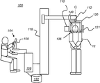
도 1은 두부 계측 분석을 제공하기 위한 이미징 시스템을 도시하는 개략도이다.
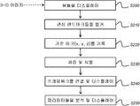
도 2는 본 개시물의 실시형태에 따른 3-D 두부 계측 분석 프로세스를 도시하는 논리 흐름도이다.
도 3은 3-D 렌더링된 CBCT 머리 볼륨 이미지의 뷰이다.
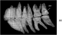
도 4는 치아 세그먼트화 이후의 3-D 렌더링된 치아 볼륨 이미지의 뷰이다.
도 5는 CBCT 머리 볼륨 이미지 및 조작자-입력 기준 마크의 3가지 직교 뷰를 디스플레이하는 사용자 인터페이스의 뷰이다.
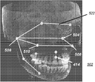
도 6은 3-D 기준 마크 세트가 디스플레이되어 있는 3-D 렌더링된 CBCT 머리 볼륨 이미지의 뷰이다.
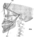
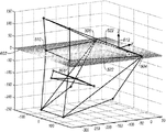
도 7a, 7b, 및 7c는 두부 계측 분석을 위한 프레임워크를 제공하는 식별된 해부학적 피처를 도시하는 투시도이다.
도 8은 두부 계측 분석에 사용되는 프레임워크를 생성하는 조작자 명령을 수용하는 단계들을 도시하는 논리 흐름도이다.
도 9a, 9b, 및 9c는 조작자-입력 기준 마크를 사용해서 해부학적 피처의 위치를 지정하기 위한 조작자 인터페이스를 도시한다.
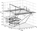
도 10a, 10b, 10c, 10d, 및 10e는 볼륨 이미지 데이터 및 상응하는 조작자-입력 기준 마크를 사용해서 다양한 파생된 파라미터를 계산하는 방법을 도시하는 그래프이다.
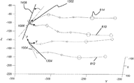
도 11은 세그먼트화된 치아 데이터로부터 다수의 파생된 두부 계측 파라미터를 도시하는 3-D 그래프이다.
도 12는 세그먼트화된 치아 데이터로부터 파생된 두부 계측 파라미터를 도시하는 2-D 그래프이다.
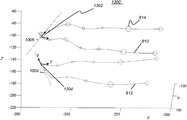
도 13은 세그먼트화된 치아 데이터로부터 파생된 두부 계측 파라미터를 도시하는 다른 3-D 그래프이다.
도 14는 세그먼트화된 치아 데이터 및 치료 파라미터로부터 파생된 두부 계측 파라미터를 도시하는 그래프이다.
도 15는 시스템에 의해 치아 배제를 학습하는 방법을 도시하는 3-D 그래프이다.
도 16a는 디지털 모형(digital phantom)의 치아를 도시하는 사시도이다.
도 16b는 위턱 및 아래턱에 대한 관성계의 연산된 축선들을 도시하는 3-D 그래프이다.
도 17a는 특정 치아 구조에 대한 평행도(parallelism)를 도시하는 그래프이다.
도 17b는 특정 치아 구조에 대한 평행도를 도시하는 그래프이다.
도 18a는 치아 결손이 있는 디지털 모형의 치아를 도시하는 사시도이다.
도 18b는 도 18a의 실시예에 대한 위턱 및 아래턱에 대한 관성계의 연산된 축선들을 도시하는 그래프이다.
도 19a는 특정 치아 구조에 대한 평행도의 결여를 도시하는 그래프이다.
도 19b는 특정 치아 구조에 대한 평행도의 결여를 도시하는 그래프이다.
도 20a는 치아 배제가 있는 디지털 모형의 치아를 도시하는 사시도이다.
도 20b는 도 20a의 실시예에 대한 위턱 및 아래턱에 대한 관성계의 연산된 축선들을 도시하는 그래프이다.
도 21a는 결손 치아에 대한 치아 배제를 도시하는 실시예이다.
도 21b는 도 21a의 실시예에 대한 위턱 및 아래턱에 대한 관성계의 연산된 축선들을 도시하는 그래프이다.
도 22a는 결손 치아에 대한 치아 배제를 도시하는 실시예이다.
도 22b는 도 22a의 실시예에 대한 위턱 및 아래턱에 대한 관성계의 연산된 축선들을 도시하는 그래프이다.
도 23a는 특정 치아들을 배제한 결과를 보여주는 이미지이다.
도 23b는 도 23a의 실시예에 대한 위턱 및 아래턱에 대한 관성계의 연산된 축선들을 도시하는 그래프이다.
도 24는 DOL 기준계의 다수의 랜드마크 및 좌표 축선 또는 벡터를 도시한다.
도 25는 DOL 기준계의 대체 공간으로의 랜드마크 리맵핑(re-mapping)을 도시한다.
도 26은 이 리맵핑을 사용해서 변환된 치아 관성계를 구비한 실시예를 측면 뷰로 도시한다.
도 27은 본 개시물의 실시형태에 따른 분석 엔진을 위한 독립 네트워크를 도시하는 개략도이다.
도 28은 본 개시물의 실시형태에 따른 분석 엔진을 위한 종속 또는 결합 네트워크를 도시하는 개략도이다.
도 29는 도 27의 독립 네트워크 배열을 사용하는 알고리즘에 대한 의사-코드(pseudo-code)를 도시한다.
도 30은 도 28의 종속적인 네트워크 배열을 사용하는 알고리즘에 대한 의사-코드를 도시한다.
도 31a는 예시적인 파라미터들을 수치 및 그 해석으로서 열거한다.
도 31b, 31c 및 31d는, 본원의 예시적인 실시형태들에 따른 예시적인 전체 상악안면 비대칭 파라미터들에 기초하여, 특정 환자에 대하여, 예시적인 파라미터들을 상악안면 비대칭에 대한 수치 및 그 해석으로서 열거한다.
도 32a는 교합 분석 및 아치 각도 특성을 갖는 특정 실시예에 대한 예시적인 테이블 형식의 결과를 도시한다.
도 32b는 상절치 및 하절치(upper and lower incisors)의 토크의 특정 실시예에 대한 예시적인 테이블 형식의 결과를 도시한다.
도 32c는 양후퇴(biretrusion) 또는 양돌출(biprotrusion)의 평가를 갖는 다른 실시예에 대한 예시적인 테이블 형식의 결과를 도시한다.
도 32d는 특정 환자의 두부 계측 분석에 대한 결과의 예시적인 요약 목록을 도시한다.
도 32e는 도 35에 열거된 상태들 중 하나에 대한 상세한 목록을 도시한다.
도 33은 분석 결과에 기초하여 추전 메시지가 있는 시스템 디스플레이를 도시한다.
도 34는 분석 결과를 돕기 위한 그래픽 묘사가 있는 시스템 디스플레이를 도시한다.
도 35는 본 개시물의 실시형태에 따른 비대칭에 대한 예시적인 보고서를 도시한다.
도 36은 환자에 대한 상대적인 좌-우 비대칭을 정면 뷰로 보여주는 그래프이다.
도 37은 환자의 안면의 좌측 및 우측의 상대적인 중첩을 보여주는 그래프이다.
도 38은 안면 분기를 보여주는 그래프이다.These and other objects, features, and advantages of the present invention will become apparent from the following more detailed description of embodiments of the present disclosure, as illustrated in the accompanying drawings.
Elements in the figures are not necessarily proportional to each other.
1 is a schematic diagram illustrating an imaging system for providing head metrology analysis.
2 is a logic flow diagram illustrating a 3-D head metrology analysis process in accordance with an embodiment of the present disclosure.
3 is a view of a 3-D rendered CBCT head volume image.
4 is a view of a 3-D rendered tooth volume image after tooth segmentation.
5 is a view of a user interface displaying three orthogonal views of a CBCT head volume image and an operator-input reference mark.
6 is a view of a 3-D rendered CBCT head volume image with a 3-D reference mark set displayed.
7A, 7B, and 7C are perspective views illustrating identified anatomical features that provide a framework for head metrology analysis.
8 is a logic flow diagram illustrating steps for accepting operator instructions to create a framework used for head metrology analysis.
9A, 9B, and 9C illustrate an operator interface for positioning anatomical features using operator-input reference marks.
10A, 10B, 10C, 10D, and 10E are graphs showing how to calculate various derived parameters using volume image data and corresponding operator-input reference marks.
11 is a 3-D graph showing a number of derived head metrology parameters from segmented tooth data.
12 is a 2-D graph showing head metrology parameters derived from segmented tooth data.
13 is another 3-D graph showing head metrology parameters derived from segmented tooth data.
14 is a graph showing head metrology parameters derived from segmented tooth data and treatment parameters.
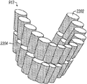
15 is a 3-D graph illustrating a method of learning tooth exclusion by the system.
FIG. 16A is a perspective view showing teeth of a digital phantom. FIG.
16B is a 3-D graph showing calculated axes of the inertial system for the upper and lower jaws.
FIG. 17A is a graph showing parallelism for a particular tooth structure. FIG.
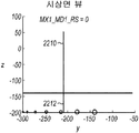
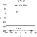
17B is a graph showing parallelism for a particular tooth structure.
18A is a perspective view showing the teeth of a digital model with tooth defects.
FIG. 18B is a graph showing calculated axes of the inertial system for the upper and lower jaws for the embodiment of FIG. 18A.
19A is a graph showing the lack of parallelism for a particular tooth structure.
19B is a graph showing the lack of parallelism for certain tooth structures.
20A is a perspective view illustrating the teeth of a digital model with tooth exclusion.
FIG. 20B is a graph showing calculated axes of the inertial system for the upper and lower jaws for the embodiment of FIG. 20A.
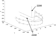
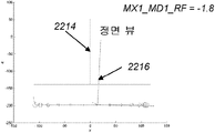
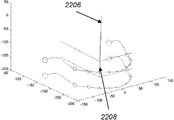
21A is an embodiment illustrating tooth exclusion for missing teeth.
FIG. 21B is a graph showing calculated axes of the inertial system for the upper and lower jaws for the embodiment of FIG. 21A.
22A is an embodiment illustrating tooth exclusion for missing teeth.
FIG. 22B is a graph showing calculated axes of the inertial system for the upper and lower jaws for the embodiment of FIG. 22A.
Figure 23a is an image showing the result of excluding certain teeth.
FIG. 23B is a graph showing calculated axes of the inertial system for the upper and lower jaws for the embodiment of FIG. 23A.
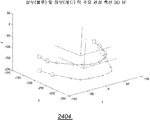
24 illustrates a number of landmarks and coordinate axes or vectors of a DOL reference system.
FIG. 25 illustrates landmark re-mapping to an alternative space of a DOL reference system.
FIG. 26 shows a side view of an embodiment with a tooth inertia system converted using this remapping.
27 is a schematic diagram illustrating an independent network for an analysis engine, in accordance with an embodiment of the present disclosure.
FIG. 28 is a schematic diagram illustrating a dependent or combined network for an analysis engine, in accordance with an embodiment of the present disclosure. FIG.
FIG. 29 illustrates a pseudo-code for an algorithm using the independent network arrangement of FIG. 27.
FIG. 30 illustrates pseudo-code for an algorithm using the dependent network arrangement of FIG. 28.
31A lists example parameters as numerical and interpretation thereof.
31B, 31C and 31D list exemplary parameters as numerical values and interpretations for maxillary facial asymmetry, for a particular patient, based on exemplary overall maxillofacial asymmetry parameters according to exemplary embodiments herein. .
32A shows example tabular results for a particular embodiment with occlusion analysis and arch angle characteristics.
32B shows an exemplary tabular result for a particular embodiment of the torque of upper and lower incisors.
FIG. 32C shows exemplary tabular results for another embodiment with evaluation of biretrusion or biprotrusion. FIG.
32D shows an exemplary summary list of results for a head metrology analysis of a particular patient.
32E shows a detailed listing of one of the states listed in FIG. 35.
33 shows a system display with a recommendation message based on the analysis result.
34 shows a system display with graphical depictions to assist with analysis results.
35 illustrates an example report for asymmetry in accordance with an embodiment of the present disclosure.
36 is a graph showing in frontal view the relative left-right asymmetry for a patient.
37 is a graph showing the relative overlap of the left and right sides of the patient's face.
38 is a graph showing facial branches.
본 개시물의 이하의 실시형태들의 상세한 설명에 있어서는, 연이은 도면들에서의 동일한 요소들에 동일한 참조 번호를 할당한 도면들을 참조한다. 이들 도면은 본 발명의 실시형태들에 따라 전체 기능 및 관계를 예시하기 위해 제공되는 것이지 실제 크기 및 비율을 나타내려는 의도로 제공되는 것은 아니라는 점에 유의해야 한다.In the detailed description of the following embodiments of the present disclosure, reference is made to the drawings in which like elements in the following figures have been assigned the same reference numerals. It should be noted that these figures are provided to illustrate the overall functionality and relationships in accordance with embodiments of the present invention and are not intended to represent actual size and proportions.
"제1(first)", "제2(second)", "제3(thrid)" 등의 용어를 사용하는 경우, 이는 반드시 어떠한 서수 또는 우선순위 관계를 나타내는 것이 아니고, 하나의 요소 또는 시간 간격을 다른 요소 또는 시간 간격과 더욱 명확하게 구별하는 데 사용될 수 있다.When using terms such as "first", "second", "thrid", etc., this does not necessarily indicate any ordinal or priority relationship, but an element or time interval. Can be used to more clearly distinguish it from other elements or time intervals.
본 개시물의 맥락에서, "이미지(image)"라는 용어는 개별 이미지 요소들로 구성된 다차원 이미지 데이터를 의미한다. 2-D 이미지의 경우, 개별 이미지 요소들은 사진 요소, 또는 픽셀이다. 3-D 이미지의 경우, 개별 이미지 요소들은 볼륨 이미지 요소, 또는 복셀(voxel)이다. "볼륨 이미지(volume image)"라는 용어는 "3-D 이미지"라는 용어와 동의어로 간주된다.In the context of this disclosure, the term "image" means multidimensional image data composed of individual image elements. In the case of a 2-D image, the individual image elements are photographic elements, or pixels. In the case of a 3-D image, the individual image elements are volume image elements, or voxels. The term "volume image" is considered synonymous with the term "3-D image".
본 개시물의 맥락에서, "코드 값(code value)"이라는 용어는 각각의 2-D 이미지 픽셀과 연관된 값, 또는 그에 상응하여, 재구성된 3-D 볼륨 이미지에서의 각각의 볼륨 이미지 데이터 요소 또는 복셀과 연관된 값을 의미한다. 컴퓨터 단층촬영(CT) 또는 원추형-빔 컴퓨터 단층촬영(CBCT) 이미지에 대한 코드 값은 각각의 복셀의 감쇠 계수에 대한 정보를 제공하는 하운스필드(Hounsfield) 단위로 표현되는 경우가 많지만, 항상 그런 것은 아니다.In the context of this disclosure, the term “code value” means a value associated with each 2-D image pixel, or correspondingly, each volume image data element or voxel in a reconstructed 3-D volume image. The value associated with. Code values for computed tomography (CT) or conical-beam computed tomography (CBCT) images are often expressed in Hounsfield units, which provide information about the attenuation coefficients of each voxel, but always It is not.
본 개시물의 맥락에서, "기하학적 프리미티브(geometric primitive)"라는 용어는 직사각형, 원, 선, 트레이스 곡선, 또는 그 밖의 트레이스 패턴과 같은 개방 또는 폐쇄 형상과 관련된다. "랜드마크(landmark)" 및 "해부학적 피처(anatomical feature)"라는 용어들은 동등한 것으로 간주되고, 디스플레이되는 환자의 해부학적 구조의 구체적인 생김새를 의미한다.In the context of this disclosure, the term "geometric primitive" relates to an open or closed shape, such as a rectangle, circle, line, trace curve, or other trace pattern. The terms "landmark" and "anatomical feature" are considered equivalent and refer to the specific appearance of the anatomical structure of the patient to be displayed.
본 개시물의 맥락에서, "뷰어(viewer)", "조작자(operator)", 및 "사용자(user)"라는 용어들은 동등한 것으로 간주되고, 디스플레이 모니터 상의 치아 이미지와 같은 이미지를 보면서 조작하는 시술 의사 또는 그 밖의 사람을 의미한다. "조작자 명령(operator instruction)" 또는 "뷰어 명령(viewer instruction)"은, 예를 들면 컴퓨터 마우스 또는 터치 스크린 또는 키보드 입력을 사용해서 뷰어에 의해 입력되는 명시적인 커맨드로부터 취득된다.In the context of the present disclosure, the terms "viewer", "operator", and "user" are considered equivalent and include a practitioner or operator who operates while viewing an image, such as a tooth image on a display monitor. It means other people. An "operator instruction" or "viewer instruction" is obtained from an explicit command input by the viewer, for example using a computer mouse or touch screen or keyboard input.
디스플레이된 피처에 대한 "강조표시(highlighting)"라는 용어는 정보 및 이미지 디스플레이 분야의 당업자에게 이해되는 바와 같은 그 종래의 의미를 갖는다. 일반적으로, 강조표시는 뷰어의 관심을 끌기 위해 일부 국부화된 디스플레이 강화 형태를 사용한다. 예를 들어, 개별 장기, 뼈, 또는 구조와 같은 이미지의 일부분, 또는 하나의 챔버로부터 다음 챔버까지의 경로를 강조표시하는 것은, 한정되는 것은 아니지만, 주석 달기(annotating), 인근의 또는 중첩되는 기호를 디스플레이하기, 윤곽선 긋기(outlining or tracing), 다른 이미지 또는 정보 콘텐츠와는 상이한 컬러로 또는 뚜렷하게 상이한 강도 또는 그레이 스케일로 디스플레이하기, 디스플레이의 일부분의 명멸하기 또는 애니메이션, 또는 보다 높은 선명도 또는 콘트라스트로 디스플레이하기를 포함하는 다수의 방법들 중 어느 하나로 달성될 수 있다.The term " highlighting " for displayed features has its conventional meaning as understood by those skilled in the art of information and image display. In general, highlighting uses some form of localized display enhancement to attract the viewer's attention. For example, highlighting a portion of an image, such as an individual organ, bone, or structure, or the path from one chamber to the next, includes, but is not limited to, annotating, nearby or overlapping symbols. Display, outlining or tracing, displaying in a different color or different intensity or gray scale than other image or information content, flashing or animation of a portion of the display, or displaying with higher clarity or contrast It can be achieved in any of a number of ways, including the following.
본 개시물의 맥락에서, "파생된 파라미터(derived parameters)"라는 설명적인 용어는 취득된 또는 입력된 데이터 값의 처리로부터 계산되는 값과 관련된다. 파생된 파라미터는 스칼라, 점, 선, 체적, 벡터, 평면, 곡선, 각도값, 이미지, 폐쇄 윤곽선, 면적, 길이, 매트릭스, 텐서(tensor), 또는 수학적 표현일 수 있다.In the context of this disclosure, the descriptive term “derived parameters” relates to values calculated from the processing of acquired or input data values. The derived parameter may be a scalar, point, line, volume, vector, plane, curve, angle value, image, closed contour, area, length, matrix, tensor, or mathematical representation.
본 명세서에서 사용되는 "세트(set)"라는 용어는, 세트의 요소들 또는 멤버들의 개념이 기초 수학에서 폭넓게 이해되기 때문에, 비어있지 않은 세트를 의미한다. "서브세트(subset)"라는 용어는, 달리 명시적으로 언급되지 않는 한, 본 명세서에서는 하나 이상의 멤버를 갖는 비어있지 않은 적절한 서브세트, 즉 보다 큰 세트의 하위 세트를 의미하는 것으로 사용된다. 세트 S의 경우, 서브세트는 완전한 세트 S를 포함할 수 있다. 그러나, 세트 S의 "적절한 서브세트(proper subset)"는 세트 S에 엄격하게 포함되고 세트 S의 적어도 하나의 멤버를 배제한다. 대안으로서, 보다 형식적으로 언급하면, 해당 용어가 본 개시물에서 사용될 때, (i) 서브세트 B가 비어있지 않고, (ii) B ∩ S 도 비어있지 않으며 서브세트 B가 세트 S에 있는 요소들만을 더 포함하고 세트 S보다 적은 카디널리티(cardinality)를 가지면, 서브세트 B는 세트 S의 적절한 서브세트라고 간주될 수 있다.As used herein, the term "set" means a non-empty set because the concept of elements or members of the set is widely understood in basic mathematics. The term "subset" is used herein to refer to an appropriate non-empty subset, ie, a larger set of subsets, with one or more members, unless explicitly stated otherwise. For set S, the subset may comprise a complete set S. However, a "proper subset" of set S is strictly included in set S and excludes at least one member of set S. As an alternative, more formally, when the term is used in this disclosure, (i) subset B is not empty, (ii) B ∩ S is not empty and subset B is only the elements in set S If further includes and have less cardinality than set S, subset B may be considered an appropriate subset of set S.
본 개시물의 맥락에서, "평면도(plan view)" 또는 "2-D 뷰(view)"는 수평면의 위치로부터 3차원(3-D) 객체를 통한 해당 객체의 2차원(2-D) 표현 또는 투영이다. 이 용어는 기존에 특정 시점에서 3-D 볼륨 이미지 데이터 내부로부터 2-D 평면 표현을 디스플레이하는 것을 설명하는 데 사용된 "이미지 슬라이스(image slice)"라는 용어와 동의어이다. 3-D 볼륨 데이터의 2-D 뷰들은, 해당 뷰들이 취해지는 상응하는 평면들이 서로 90(+/-10)도로, 또는 서로 90도의 정수(n)배(n*90도, +/-10도)로 배치될 경우 실질적으로 직교하는 것으로 간주된다.In the context of this disclosure, a "plan view" or "2-D view" is a two-dimensional (2-D) representation of that object through a three-dimensional (3-D) object from a location in the horizontal plane or Projection. This term is synonymous with the term " image slice " used to describe displaying a 2-D planar representation from within the 3-D volume image data at a specific point in time. 2-D views of 3-D volume data can be represented by 90 (+/- 10) degrees to each other, or by integer (n) times (n * 90 degrees, +/- 10 degrees) Is considered to be substantially orthogonal when deployed.
본 개시물의 맥락에서, "치열 요소(dentition element)"라는 일반적인 용어는 치아, 의치 및 임플란트와 같은 보철 장치, 및 치아 및 연관 보철 장치를 위한 턱을 포함하는 지지 구조와 관련된다.In the context of the present disclosure, the general term “dentition element” relates to a support structure that includes a prosthetic device such as a tooth, a denture and an implant, and a jaw for a tooth and an associated prosthetic device.
본 개시물의 청구 대상은, 디지털 이미지로부터 데이터를 디지털로 처리해서 인식하고, 그에 따라 유용한 의미를 인간이 이해할 수 있는 객체, 속성 또는 상태에 부여하고 나서, 디지털 이미지의 추가적인 처리에서 얻어진 결과를 이용하는 기술을 의미하는 것으로 이해되는 디지털 이미지 처리 및 컴퓨터 비전 기술과 관련된다.The subject matter of the present disclosure is a technique that utilizes the results obtained in further processing of a digital image, after digitally processing and recognizing data from the digital image, thereby assigning useful meanings to an object, attribute or state that can be understood by a human being. Related to digital image processing and computer vision technology.
배경기술 부분에서 앞서 주지한 바와 같이, 종래의 2-D 두부 계측 분석은 다수의 중요한 결점을 갖는다. 환자의 머리를 머리 고정 장치 또는 다른 측정 장치의 중앙에 위치시키는 것이 어렵기 때문에, 재현성이 떨어진다. 취득되는 2차원 방사선 사진은 3-D 이미지가 아닌 중첩되는 머리 해부학 이미지를 생성한다. 두부 계측 사진 상에 랜드마크들을 배치하는 것은 어려울 수 있으며, 결과들이 불일치되곤 한다(The Future of Orthodontics, ed. Carine Carels, Guy Willems, Leuven University Press, 1998, pp. 181 - 192에서의 P. Planche 및 J. Treil에 의한 “for the next millennium”이라는 제목의 논문을 참조). 치료 계획을 개발 및 추적하는 일은, 부분적으로는, 수집 및 계산된 두부 계측 데이터의 양이 상당하기 때문에 복잡하다.As noted earlier in the background section, conventional 2-D head metrology analysis has a number of significant drawbacks. Reproducibility is poor because it is difficult to position the patient's head in the center of the head fixation device or other measuring device. The acquired two-dimensional radiographs produce overlapping head anatomy images rather than 3-D images. Placing landmarks on tofu metrology photographs can be difficult, and results are often inconsistent (P. Planche at The Future of Orthodontics , ed. Carine Carels, Guy Willems, Leuven University Press, 1998, pp. 181-192). And paper titled “for the next millennium” by J. Treil). Developing and tracking a treatment plan is complex, in part because of the significant amount of collected and calculated head metrology data.
본 개시물의 실시형태는 3-D 해부학적 피처 지점들의 선택, 이들 피처 지점들로부터 도출되는 파라미터, 및 이들 도출된 파라미터들을 두부 계측 분석에서 이용하는 방법에 관하여 Treil의 이론을 이용한다. Treil이 저술한 참고 문헌은 World Journal of Orthodontics, 2005 Supplement, Vol. 6, issue 5, pp. 33-38에서의 Jacques Treil, B, Waysenson, J. Braga 및 J. Casteigt에 의한 "The Human Face as a 3D Model for Cephalometric Analysis”및 Seminars in Orthodontics, Vol. 15, No. 1, March 2009에서의 J. Treil, J. Braga, J.-M. Loubes, E. Maza, J.-M. Inglese, J. Casteigt, 및 B. Waysenson에 의한 “Tooth Modeling for Orthodontic Assessment”를 포함한다.Embodiments of the present disclosure utilize Treil's theory regarding the selection of 3-D anatomical feature points, the parameters derived from these feature points, and how to use these derived parameters in head metrology analysis. Treil's references include the World Journal of Orthodontics , 2005 Supplement, Vol. 6,
도 1의 개략도는 3-D CBCT 두부 계측 이미징을 위한 이미징 장치(100)를 도시한다. 환자(12)를 이미징하는 경우, 일련의 다중 2-D 투영 이미지가 취득되고 이미징 장치(100)를 사용해서 처리된다. 회전식 마운트(130)가, 바람직하게는 환자(12)의 사이즈에 적합한 높이로 조절 가능하게 칼럼(118) 상에 설치된다. 마운트(130)는 환자(12)의 머리의 양측에 x-선 소스(110) 및 방사선 센서(121)를 유지하고 머리에 대한 스캔 패턴으로 소스(110) 및 센서(121)를 선회시키도록 회전한다. 마운트(130)는 환자의 머리의 중심부에 대응하는 축선(Q)을 중심으로 회전하고, 마운트(130)에 부착되는 컴포넌트들은 머리의 주위를 선회한다. 센서(121), 즉 디지털 센서는 CBCT 볼륨 이미징에 적합한 방사선 패턴을 방출하는 x-선 소스(110)에 대향하여 마운트(130)에 결합된다. 턱 받침대 또는 물기(bite) 요소와 같은 선택적인 머리 지지부(136)는 이미지를 획득하는 동안 환자의 머리를 안정화시킨다. 컴퓨터(106)는 조작자 커맨드를 수용하고 이미징 장치(100)에 의해 취득되는 치열 교정 이미지 데이터의 볼륨 이미지의 디스플레이를 위해 조작자 인터페이스(104) 및 디스플레이(108)를 구비한다. 컴퓨터(106)는 이미지 데이터를 취득하기 위해 센서(121)와 신호 통신하고, 소스(110)의 제어를 위한 신호 및, 선택적으로, 마운트(130) 컴포넌트용 회전 액추에이터(112)의 제어를 위한 신호를 제공한다. 컴퓨터(106)는 또한, 이미지 데이터를 저장하기 위해 메모리(132)와 신호 통신한다. 선택적인 정렬 장치(140)가 이미징 프로세스를 위해 환자의 머리의 적절한 정렬을 보조하도록 제공된다.1 shows an
도 2의 논리 흐름도를 참조하면, 본 개시물의 실시형태에 따른 치과용 CBCT 볼륨으로 3-D 두부 계측 분석을 위한 치열 교정 데이터를 획득하는 데 사용되는 단계들의 시퀀스(200)가 도시되어 있다. CBCT 볼륨 이미지 데이터는 데이터 획득 단계 S102에서 액세스된다. 볼륨은 하나 이상의 2-D 이미지(또는 등가로, 슬라이스)의 이미지 데이터를 포함한다. 원래의 재구성된 CT 볼륨은 CT 스캐너로부터 취득되는 다중 2-D 투영 또는 부비동조영상(sinogram)을 사용하는 표준 재구성 알고리즘을 사용해서 형성된다. 실시예로서, 도 3은 뼈 해부학, 연조직, 및 치아를 포함하는 예시적인 치과용 CBCT 볼륨(202)을 도시한다.Referring to the logic flow diagram of FIG. 2, a
도 2의 시퀀스를 계속하여, 세그먼트화 단계 S104에서는, 3-D 치아 세그먼트화 알고리즘을 치과용 CBCT 볼륨(202)에 적용함으로써 3-D 치열 요소 데이터가 수집된다. 치아 및 관련 치열 요소에 대한 세그먼트화 알고리즘은 치과 이미징 분야에서 잘 알려져 있다. 예시적인 치아 세그먼트화 알고리즘은, 예를 들어, 전부 본 명세서에 참조로 포함되는, 공통적으로 양도된 Chen 등에 의한 "PANORAMIC IMAGE GENERATION FROM CBCT DENTAL IMAGES"라는 제목의 미국 특허출원 공개 제2013/0022252호; Chen 등에 의한 "METHOD AND SYSTEM FOR TOOTH SEGMENTATION IN DENTAL IMAGES"라는 제목의 미국 특허출원 공개 제2013/0022255호; 및 Chen에 의한 "METHOD FOR TOOTH DISSECTION IN CBCT VOLUME"이라는 제목의 미국 특허출원 공개 제2013/0022254호에서 설명된다.Continuing the sequence of FIG. 2, in segmentation step S104, 3-D dental element data is collected by applying the 3-D tooth segmentation algorithm to the
도 4에 도시된 바와 같이, 치아 세그먼트화 결과가 이미지(302)로 렌더링되고, 여기서 치아들은 전체로서 렌더링되지만, 개별적으로 세그먼트화된다. 각각의 치아는 치아 볼륨, 예를 들어, 치아 볼륨(304)이라고 하는 별도의 개체이다.As shown in FIG. 4, the tooth segmentation result is rendered as an
세그먼트화된 치아들의 각각의 치아, 또는 보다 광범위하게, 세그먼트화되어 있는 각각의 치열 요소는, 최소한, 세그먼트화된 치열 요소 내의 각각의 복셀에 대한 3-D 위치 좌표를 포함하는 3-D 위치 리스트, 및 세그먼트화된 요소 내의 각각의 복셀의 코드 값 리스트를 갖는다. 이 점에서, 각각의 복셀에 대한 3-D 위치가 CBCT 볼륨 좌표계에 대하여 정의된다.Each tooth of the segmented teeth, or more broadly, each segmented segment, is a 3-D position list containing 3-D position coordinates for each voxel in the segmented segment. And a code value list of each voxel in the segmented element. In this regard, a 3-D position for each voxel is defined relative to the CBCT volume coordinate system.
도 2의 시퀀스에서의 기준 마크 선택 단계 S106에서는, CBCT 볼륨 이미지들이 상이한 화각들에 대하여 취득되는 2개 이상의 상이한 2-D 뷰로 디스플레이된다. 상이한 2-D 뷰들은, 예를 들어, 상이한 각도들일 수 있고 상이한 이미지 슬라이스들일 수 있거나, 또는 정투영 또는 실질적으로 정투영일 수 있거나, 또는 투시도일 수 있다. 본 개시물의 실시형태에 따르면, 3가지의 뷰는 상호간에 직교한다.In the reference mark selection step S106 in the sequence of FIG. 2, CBCT volume images are displayed in two or more different 2-D views obtained for different angles of view. Different 2-D views may be, for example, different angles and may be different image slices, or may be orthographic or substantially orthographic, or may be perspective. According to embodiments of the present disclosure, the three views are orthogonal to each other.
도 5는 3가지의 직교 2-D 뷰를 도시하는 디스플레이 인터페이스(402)를 갖는 예시적인 포맷을 도시한다. 디스플레이 인터페이스(402)에 있어서, 이미지(404)는 CBCT 볼륨 이미지(202)(도 3)의 축방향 2-D 뷰들 중 하나이고, 이미지(406)는 CBCT 볼륨 이미지(202)의 관상면 2-D 뷰들 중 하나이고, 이미지(408)는 CBCT 볼륨 이미지(202)의 시상면 2-D 뷰들 중 하나이다. 디스플레이 인터페이스는, 의사 또는 기술자와 같은 뷰어가 복수의 3-D 두부 계측 분석 작업을 달성하기 위해 다양한 이미지 처리/컴퓨터 알고리즘을 실행하는 컴퓨터 시스템과 상호작용할 수 있게 한다. 뷰어 상호작용은, 보다 상세하게는 후속하여 설명되는 상호작용을 위해, 동작을 선택하거나 이미지의 좌표를 지정하기 위해 컴퓨터 마우스 조이스틱 또는 터치패드와 같은 포인터를 사용하거나, 또는 터치 스크린을 사용하는 바와 같이, 사용자 인터페이스 분야의 당업자에게 알려져 있는 다수의 형태들 중 어느 하나를 취할 수 있다.5 shows an example format with a
3-D 두부 계측 분석 작업들 중 하나는 도 2의 3-D 기준 마크 선택 단계 S106에서 자동 식별을 수행하는 것이다. 디스플레이된 이미지 상에서 뷰어에 의해 식별되는 일종의 3-D 랜드마크 또는 피처 지점과 균등한 3-D 기준 마크들이 도 5에서의 디스플레이 인터페이스(402)의 서로 다른 상호 직교 2-D 뷰들에 도시된다. 도 5에 도시되는 예시적인 3-D 해부학적 기준 마크들은 기준 마크(414)에서의 하부 코 구개공(nasal palatine foramen)이다. 도 6의 뷰에 도시된 바와 같이, 디스플레이된 이미지(502) 상에서 뷰어에 의해 지시될 수 있는 다른 해부학적 기준 마크들은 기준 마크(508 및 510)에서의 안와하공(infraorbital foramen), 및 기준 마크(504 및 506)에서의 추골(malleus)을 포함한다.One of the 3-D head metrology analysis tasks is to perform automatic identification in the 3-D reference mark selection step S106 of FIG. 2. A kind of 3-D landmark or feature point identified by the viewer on the displayed image is shown in different mutually orthogonal 2-D views of the
도 2의 단계 S106에서, 뷰어는 포인팅 장치(예를 들어, 마우스 또는 터치 스크린과 같음)를 사용해서 3가지 뷰 중 어느 하나에서 적절한 위치에 일종의 기하학적 프리미티브로서 기준 마크를 위치설정한다. 본 명세서의 도면들에 도시된 본 개시물의 실시형태에 따르면, 기준 마크는 원으로서 디스플레이된다. 예를 들어, 도 5의 디스플레이 인터페이스 스크린을 이용하면, 뷰어는 이미지(404)로서 도시된 뷰에서 위치(414)에 기준점에 대한 기준 마크로서 작은 원을 배치한다. 기준 마크(414)는 이미지(404)에서뿐만 아니라, 이미지(406 및 408)에서의 상응하는 뷰의 적절한 위치에 작은 원으로서 디스플레이된다. 뷰어는 디스플레이된 뷰들 중 하나(404, 406 또는 408)에서만 기준 마크(414)의 위치를 지시할 필요가 있으며; 시스템은 환자 해부구조의 다른 뷰들에서 동일한 기준 마크(414)를 보여주는 것으로 응답한다는 점에 유의하는 것이 중요하다. 따라서, 뷰어는 가장 쉽게 볼 수 있는 뷰에서 기준 마크(414)를 식별할 수 있다.In step S106 of FIG. 2, the viewer uses a pointing device (such as a mouse or a touch screen, for example) to position the fiducial mark as a kind of geometric primitive at an appropriate location in either of the three views. In accordance with an embodiment of the present disclosure shown in the drawings herein, the fiducial mark is displayed as a circle. For example, using the display interface screen of FIG. 5, the viewer places a small circle as a reference mark for the reference point at
사용자는, 기준 마크(414)를 입력한 후에, 디스플레이된 뷰들 중 어느 하나에서 기준 마크(414)의 위치를 조정하기 위해 키보드 또는 디스플레이된 아이콘과 같은 조작자 인터페이스 도구를 사용할 수 있다. 뷰어는 또한, 입력된 기준 마크를 제거하고 새로운 기준 마크를 입력할 수도 있다.After entering the
디스플레이 인터페이스(402)(도 5)는 디스플레이된 뷰들 중 일부 또는 전부의 사이즈를 조절하기 위한 줌 인/아웃 유틸리티를 제공한다. 따라서, 뷰어는 향상된 기준 마크 위치설정을 위해 상이한 이미지들을 효율적으로 조작할 수 있다.Display interface 402 (FIG. 5) provides a zoom in / out utility for resizing some or all of the displayed views. Thus, the viewer can efficiently manipulate different images for improved reference mark positioning.
3-D 이미지 콘텐츠의 뷰들을 참조하여 이루어지며 해당 뷰들에서 보이는 기준 마크들의 집합은 환자의 머리 형상 및 구조를 보다 정확하게 특정하는 데 사용될 수 있는 두부 계측 파라미터들의 세트를 제공한다. 두부 계측 파라미터들은 환자 머리의 특정한 피처에 대한 기준 마크 입력에 의해 직접적으로 제공된 좌표 정보를 포함한다. 두부 계측 파라미터들은 또한, 좌표 또는 기하학적 구조로서 직접적으로 입력되는 것이 아니라 좌표 정보로부터 파생된 환자 머리의 해부구조의 다양한 측정 가능한 특성들에 대한 정보, 말하자면 "파생된 두부 계측 파라미터(derived cephalometric parameters)"를 포함한다. 파생된 두부 계측 파라미터들은 상대적인 사이즈 또는 체적, 대칭, 방위, 형상, 이동 경로 및 가능한 이동 범위, 관성 축선, 질량 중심, 및 그 밖의 데이터에 대한 정보를 제공할 수 있다. 본 개시물의 맥락에 있어서, "두부 계측 파라미터(cephalometric parameters)"라는 용어는, 예컨대 기준 마크에 의해 직접적으로 식별되는 것, 또는 기준 마크에 따라 연산되는 파생된 두부 계측 파라미터에 적용된다. 예를 들어, 특정한 기준점들이 그들의 상응하는 기준 마크들에 의해 식별되면, 도 6에서 보다 명확하게 도시된 바와 같이, 전체 피처들의 적절한 특정을 위해 프레임워크 연결선(522)이 기준점들을 연걸하도록 구성된다. 프레임워크 연결선(522)은 3-D 공간에서 벡터로서 간주될 수 있으며; 그들의 치수 및 공간 특성은 치열 교정 목적 및 그 밖의 목적의 연산에서 사용될 수 있는 추가적인 볼륨 이미지 데이터를 제공한다.The set of reference marks made with reference to the views of the 3-D image content and shown in the views provide a set of head metrology parameters that can be used to more accurately specify the patient's head shape and structure. Head metrology parameters include coordinate information provided directly by reference mark input for a particular feature of the patient's head. Head metrology parameters are also not directly input as coordinates or geometry, but information about the various measurable characteristics of the anatomy of the patient's head derived from coordinate information, namely "derived cephalometric parameters." It includes. Derived head metrology parameters can provide information about relative size or volume, symmetry, orientation, shape, path of travel and possible range of motion, inertia axis, center of mass, and other data. In the context of the present disclosure, the term “cephalometric parameters” applies to, for example, those that are directly identified by reference marks, or derived head measurement parameters calculated according to the reference marks. For example, if certain reference points are identified by their corresponding reference marks, the
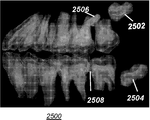
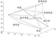
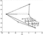
각각의 기준 마크(414, 504, 506, 508, 510)는 이미지 처리 장치(100)의 컴퓨터(106)에 의해 볼륨 데이터 내에 자동으로 생성되며 후속 분석 및 측정 처리를 용이하게 하는 프레임워크를 형성하는 하나 이상의 프레임워크 연결선(522)에 대한 말단 지점이다. 도 7a, 7b, 및 7c는, 상이한 투시도들로부터 디스플레이된 3-D 이미지(502a, 502b, 및 502c)에 대하여, 정점들에 기준점들이 있는 선택된 기준점들의 프레임워크(520)가 전체 머리 구조의 치수 양태를 정의하는 데 어떻게 도움이 되는지를 보여준다. 본 개시물의 실시형태에 따르면, 조작자 명령은 도 5에 도시된 것들과 유사한 2-D 뷰들과 도 6에 도시된 볼륨 표현 사이를 조작자가 환자의 머리의 복셀들의 부분적인 투명도로 토글할 수 있게 한다. 이를 통해, 조작자는 기준 마크 배치 및 연결선 배치를 여러 각도에서 검사할 수 있게 되고; 디스플레이된 뷰들 중 임의의 뷰에서 기준 마크 위치의 조정이 이루어질 수 있다. 또한, 본 개시물의 실시형태에 따르면, 조작자는 특정한 기준 마크에 대하여 보다 정확한 좌표를 기입할 수 있다.Each
도 8의 논리 흐름도는 기준 마크 입력 및 식별을 위해 조작자 명령을 수용 및 처리하고 이미지 데이터 및 기준 마크에 따라 연산된 파라미터를 제공하기 위한 시퀀스의 단계들을 도시한다. 디스플레이 단계 S200은, 예를 들어 환자의 머리의 컴퓨터 단층촬영 스캔으로부터 재구성된 3-D 이미지 데이터의 서로 다른 각도, 예컨대 서로 직교하는 각도에서의 하나 이상의 2-D 뷰를 디스플레이한다. 선택적인 열거 단계 S210에서, 시스템은 재구성된 3-D 이미지에서 다수의 랜드마크 또는 해부학적 피처에 대한 위치 데이터의 입력을 필요로 하는 수치 입력을 위한 테이블 형식의 리스트, 일련의 프롬프트, 또는 일련의 레이블링된 필드와 같은 텍스트 목록을 제공한다. 이 목록은, 후술하는 바와 같이, 사용자 인터페이스 프롬프트 또는 메뉴 선택의 형태로 조작자에게 명시적으로 제공될 수 있다. 대안으로서, 목록은 암시적으로 정의될 수 있으며, 조작자는 위치 정보의 입력을 위해 특정한 시퀀스를 따를 필요가 없다. 상이한 해부학적 피처들에 x, y, z 위치 데이터를 제공하는 기준 마크들은 기록 단계 S220에서 입력된다. 해부학적 피처들은 환자의 입 안에 또는 밖에 있을 수 있다. 본 개시물의 실시형태들은 단계 S220에서 입력되는 바와 같이 디스플레이 상에서 식별되는 해부학적 피처들과, 도 2를 참조하여 앞서 주지된 바와 같이 치아 및 그 밖의 치열 요소에 대하여 자동으로 생성되는 세그먼트화 데이터의 조합을 사용할 수 있다.The logic flow diagram of FIG. 8 illustrates the steps of a sequence for accepting and processing operator commands for providing reference marks and identifying and providing parameters computed according to image data and reference marks. Display step S200 displays one or more 2-D views at different angles, such as angles orthogonal to each other, for example, reconstructed from a computed tomography scan of a patient's head. In an optional enumeration step S210, the system generates a tabular list, a series of prompts, or a series of numerical inputs that require entry of position data for multiple landmarks or anatomical features in the reconstructed 3-D image. Provide a list of text, such as labeled fields. This list may be provided explicitly to the operator in the form of a user interface prompt or menu selection, as described below. As an alternative, the list can be defined implicitly, and the operator does not have to follow a particular sequence for entry of position information. Reference marks that provide x, y, z position data to different anatomical features are input in recording step S220. Anatomical features may be in or outside the patient's mouth. Embodiments of the present disclosure provide a combination of anatomical features identified on a display as input in step S220 and segmentation data automatically generated for teeth and other orthodontic elements as noted above with reference to FIG. 2. Can be used.
도 8의 기록 단계 S220에서, 시스템은 해부구조의 각각의 랜드마크 피처에 대응하는 기준 마크를 위치설정하는 조작자 명령을 수용한다. 기준 마크는 조작자에 의해 제1 또는 제2 2-D 뷰에, 또는 2개 이상의 뷰가 존재할 경우에는 다른 뷰들 중 어느 하나에 입력되고, 이어서 입력은 디스플레이된 뷰들 각각에 디스플레이된다. 식별 단계 S230은 입력된 기준 마크에 대응하는 해부학적 피처 또는 랜드마크를 식별하고, 선택적으로, 조작자 입력의 정확성을 검증한다. 주어진 조작자 입력이 특정한 해부학적 피처에 대한 기준 마크의 위치를 정확하게 식별할 가능성을 결정하도록 비례 값이 계산된다. 예를 들어, 안와하공은 통상적으로 구개공으로부터 특정 거리 범위 내에 있으며; 시스템은 입력된 거리를 확인하고 상응하는 기준 마크가 적절히 위치되지 않은 경우 조작자에게 통지한다.In the recording step S220 of Fig. 8, the system receives an operator command for positioning the reference mark corresponding to each landmark feature of the anatomy. The fiducial mark is input by the operator to the first or second 2-D view, or to one of the other views if two or more views are present, and then the input is displayed in each of the displayed views. The identifying step S230 identifies an anatomical feature or landmark corresponding to the input reference mark, and optionally verifies the accuracy of the operator input. The proportional value is calculated to determine the likelihood that a given operator input will correctly identify the location of the fiducial mark for the particular anatomical feature. For example, the orbital cavity is typically within a certain distance from the palatal opening; The system checks the distance entered and notifies the operator if the corresponding reference mark is not properly positioned.
도 8의 시퀀스를 계속하여, 구성 단계 S240에서, 프레임워크 연결선은 프레임 생성을 위해 기준 마크들을 연결하도록 생성된다. 이후, 연산 및 디스플레이 단계 S250이 실행되어, 위치설정된 기준 마크들에 따라 하나 이상의 두부 계측 파라미터를 연산한다. 이어서, 연산된 파라미터들이 조작자에게 디스플레이된다.Continuing the sequence of Fig. 8, in configuration step S240, a framework connection line is generated to connect the reference marks for frame generation. Then, the calculation and display step S250 is executed to calculate one or more head measurement parameters according to the positioned reference marks. The calculated parameters are then displayed to the operator.
도 9a, 9b, 및 9c는 디스플레이(108) 상에 나타나는 조작자 인터페이스를 도시한다. 조작자 인터페이스는, 디스플레이(108) 상에, 조작자 명령을 수용하고 특정 환자의 두부 계측 파라미터에 대한 연산 결과를 디스플레이하기 위한 대화식 유틸리티를 제공한다. 디스플레이(108)는, 예를 들어, 조작자-지정 기준 마크 및 그 밖의 명령의 입력을 위한 터치 스크린 디스플레이일 수 있다. 디스플레이(108)는 볼륨 이미지 데이터의 적어도 하나의 2-D 뷰 또는 볼륨 이미지 데이터의 2개 이상의 2-D 뷰를 상이한 각도 또는 시각에서 동시에 디스플레이한다. 실시예로서, 도 9a는 측면 또는 시상면 뷰(152)와 쌍을 이루는 정면 또는 관상면 뷰(150)를 도시한다. 2개 이상의 뷰가 동시에 보여질 수 있으며, 서로 다른 2-D 뷰들이 보여질 수 있고, 디스플레이된 뷰들 각각은 본 개시물의 실시형태에 따라 독립적으로 위치된다. 뷰들은 서로 직교하는 뷰일 수 있거나 또는 단순히 서로 다른 각도에서의 뷰일 수 있다. 디스플레이(108)의 인터페이스의 일부로서, 선택적인 제어부(166)는 뷰어가 대안적인 고정 뷰들 사이를 토글함으로써 또는 3-D 축선 (x, y, z) 중 어느 하나를 따르는 증분으로 상대적인 투시 각도를 변경함으로써 하나 이상의 2-D 뷰가 취득되는 투시 각도를 조정할 수 있게 한다. 상응하는 제어부(166)가, 도 9c에 도시된 바와 같이, 각각의 2-D 뷰와 함께 제공될 수 있다. 디스플레이(108)에 도시되는 조작자 인터페이스를 사용하면, 마우스 또는 그 밖의 전자식 포인터일 수 있거나 또는 도 9a에 도시된 바와 같은 터치스크린 입력일 수 있는 어떤 타입의 포인터를 이용해서 각각의 기준 마크(414)가 조작자에 의해 입력된다. 조작자 인터페이스의 일부로서, 조작자가 프롬프트에 따라 특정한 기준 마크를 입력하는 것을 안내하거나, 또는 도 9b의 실시예에 도시된 바와 같이, 예컨대 드롭-다운 메뉴(168)로부터의 선택에 의한 조작자 입력을 식별하기 위해 선택적인 목록(156)이 제공된다. 따라서, 조작자는 목록(156)에 값을 입력하거나, 또는 필드(158)에 값을 입력하고 나서, 입력된 값과 연관되는 이름을 드롭-다운 메뉴(168)로부터 선택할 수 있다. 도 9a-9c는 기준점들 사이에 구성되는 프레임워크(154)를 도시한다. 도 9a에 도시되는 바와 같이, 각각의 입력되는 기준 마크(414)는 뷰(150) 및 뷰(152) 모두에서 보여질 수 있다. 선택된 기준 마크(414)는 굵게 보이거나 또는 다른 컬러로 보이는 바와 같이, 디스플레이(108) 상에서 강조표시된다. 특정한 기준 마크는, 예를 들어, 기준 마크에 대한 정보를 취득 또는 입력하기 위해 또는 그 위치를 옮기는 것과 같은 어떤 동작을 수행하기 위해 선택된다.9A, 9B, and 9C show the operator interface shown on the
도 9b에 도시된 실시형태에 있어서, 조작자에 의해 단순히 입력 또는 선택되는 기준 마크(414)는 목록(156)으로부터의 선택에 의해 식별된다. 도시된 실시예의 경우, 조작자는 지시된 기준 마크(414)를 선택하고 나서, 메뉴(168)로부터 "안와하공(infraorbital foramen)"과 같은 메뉴 선택을 한다. 선택적인 필드(158)는 강조표시된 기준 마크(414)를 식별한다. 예를 들어, 모델에 기초한 또는 표준적인 기지의 해부학적 관계에 기초한 계산이 기준 마크(414)를 식별하는 데 사용될 수 있다.In the embodiment shown in FIG. 9B, the
도 9c는 조작자가 시스템에 의해 부정확하거나 가능성이 없는 것으로 검출된 기준 마크(414) 명령을 입력하는 실시예를 도시한다. 에러 프롬프트 또는 에러 메시지(160)가 디스플레이되어, 조작자 입력이 에러인 것 같다고 지시한다. 시스템은, 예를 들어, 모델에 기초하여 또는 학습된 데이터에 기초하여 특정한 랜드마크 또는 해부학적 피처에 대한 가능한 위치를 연산한다. 조작자 입력이 부정확한 것으로 보이면, 선택적인 대체 위치(416)와 함께 메시지(160)가 디스플레이된다. 시스템으로부터 계산된 정보에 따라 기준 마크를 재배치하기 위한 재배치 명령(164)과 함께, 오버라이드 명령(162)이 디스플레이된다. 디스플레이 스크린 또는 키보드로부터의 다른 조작자 입력을 수용함으로써 또는 도 9c의 실시예의 대체 위치(416)에서 시스템-연산된 기준 마크 위치를 수용함으로써, 재배치가 행해질 수 있다.9C illustrates an embodiment in which an operator enters a
본 개시물의 대안적인 실시형태에 따르면, 조작자는 기준 마크들이 입력될 때 이들을 레이블링할 필요가 없다. 대신에, 디스플레이는 조작자가 디스플레이된 2-D 뷰들 중 어느 하나에 특정한 랜드마크 또는 해부학적 피처를 표시하도록 유도하고 표시된 피처를 자동으로 레이블링한다. 이 안내 시퀀스에 있어서, 조작자는 지정된 랜드마크에 대한 상응하는 기준 마크의 위치를 표시함으로써 각각의 시스템 프롬프트에 응답한다.According to an alternative embodiment of the present disclosure, the operator does not need to label them when reference marks are input. Instead, the display prompts the operator to display a landmark or anatomical feature specific to either of the displayed 2-D views and automatically labels the displayed feature. In this guide sequence, the operator responds to each system prompt by indicating the location of the corresponding reference mark relative to the designated landmark.
본 개시물의 대안적인 다른 실시형태에 따르면, 시스템은 조작자가 기준 마크를 표시할 때 어떤 랜드마크 또는 해부학적 피처가 식별되었는지를 결정하고; 조작자는 기준 마크들이 입력될 때 이들을 레이블링할 필요가 없다. 시스템은 이미 식별되어 있는 해부학적 피처에 대한 기지의 정보를 사용해서, 내지는 대안으로서, 재구성된 3-D 이미지 자체의 치수를 사용한 연산에 의해 가장 가능성이 높은 기준 마크를 연산한다.According to another alternative embodiment of the present disclosure, the system determines which landmark or anatomical feature has been identified when the operator marks the reference mark; The operator does not need to label them when the reference marks are entered. The system calculates the most likely reference mark using known information about the anatomical features that have already been identified, or alternatively by calculation using the dimensions of the reconstructed 3-D image itself.
도 9a-9c의 실시예들에 도시된 조작자 인터페이스를 사용하면, 본 개시물의 실시형태들은 3-D 두부 계측 분석 프로세스에서 시스템의 인간 조작자의 숙련도를 컴퓨터의 능력과 상승적으로 통합하는 실용적인 3-D 두부 계측 분석 시스템을 제공한다. 이는, 창의성, 휴리스틱의 사용, 융통성, 및 판단력에 관하여 인간의 숙련도를 이용하고, 이들을 연산 속도, 정확하고 반복 가능한 처리 능력, 보고 및 데이터 액세스 및 저장 능력, 및 디스플레이 융통성과 같은 컴퓨터 이점과 결합한다.Using the operator interface shown in the embodiments of FIGS. 9A-9C, embodiments of the present disclosure provide a practical 3-D that synergistically integrates the human operator's proficiency of the system with the computer's capabilities in a 3-D head metrology analysis process. Provides head measurement analysis system. It exploits human proficiency with respect to creativity, the use of heuristics, flexibility, and judgment and combines them with computer advantages such as computational speed, accurate and repeatable processing power, reporting and data access and storage capabilities, and display flexibility. .
도 2의 시퀀스를 다시 참조하면, 충분한 랜드마크 세트가 입력될 때 연산 단계 S108에서 파생된 두부 계측 파라미터들이 연산된다. 도 10a 내지 10e는 두부 계측 데이터를 연산 및 분석하기 위한 처리 시퀀스를 도시하는 한편, 조작자 입력 명령에 따라, 그리고 치열 요소들의 세그먼트화에 따라 결합된 볼륨 이미지 데이터 및 해부학적 피처 정보로부터 다수의 두부 계측 파라미터를 취득하는 방법을 도시한다. 본 개시물의 실시형태에 따르면, 도 10a 내지 10e에 도시된 피처들의 부분들이 디스플레이(108)(도 1) 상에 디스플레이된다.Referring back to the sequence of Fig. 2, the head measurement parameters derived in operation step S108 are computed when a sufficient landmark set is input. 10A-10E illustrate a processing sequence for computing and analyzing head metrology data, while multiple head metrology from volume image data and anatomical feature information combined in accordance with operator input commands and in accordance with segmentation of dentition elements. It shows how to get the parameters. In accordance with an embodiment of the present disclosure, portions of the features shown in FIGS. 10A-10E are displayed on display 108 (FIG. 1).
도 10a에 도시되는 예시적인 파생된 두부 계측 파라미터는 도 6을 참조하여 전술한 바와 같이 기준 마크(504, 506, 508 및 510)를 가진 제1 기하학적 프리미티브들의 세트의 서브세트를 사용하는 것에 의해 연산된 3-D 평면(602)(두부 계측 분석에 있어서 t-기준면이라고 함)이다. 추가적인 파생된 두부 계측 파라미터는 t-기준계라고 하며 앞서 주지된 공보에서 Treil에 의해 설명된 3-D 좌표 기준계(612)이다. t-기준계(612)의 z 축선은 3-D t-기준면(602)에 수직하게 선택된다. t-기준계(612)의 y 축선은 기준 마크(508)와 기준 마크(504) 사이의 프레임워크 연결선(522)과 정렬된다. t-기준계(612)의 x 축선은 평면(602)에 있으며 t-기준계의 z 축선 및 y 축선 모두에 직교한다. t-기준계 축선들의 방향은 도 10a에, 그리고 후속하는 도 10b, 10c, 10d, 및 10e에 표시된다. t-기준계의 원점은 기준 마크(504)와 기준 마크(506)를 연결하는 프레임워크 연결선(522)의 중간에 있다.The exemplary derived head metrology parameters shown in FIG. 10A are computed by using a subset of the first set of geometric primitives with
t-기준계(612)의 확립에 의해, 단계 S106으로부터의 3-D 기준 마크 및 단계 S104로부터의 3-D 치아 데이터(치아의 3-D 위치 리스트)는 CBCT 볼륨 좌표계로부터 t-기준계(612)로 변환된다. 이 변환에 의하면, 파생된 두부 계측 파라미터들 및 분석들의 후속 연산이 t-기준계(612)에 대하여 수행될 수 있게 된다.By establishing the t-
도 10b를 참조하면, t-기준계(612)에서의 치아 데이터로부터의 두부 계측 파라미터들로부터 3-D 위턱 평면(704) 및 3-D 아래턱 평면(702)이 파생된다. 파생된 위턱 평면(704)은 위턱(상악골)으로부터 세그먼트화되는 치아 데이터에 따라 연산된다. 두부 계측 측정 및 분석 분야의 당업자에게 친숙한 방법들을 사용하면, 파생된 아래턱 평면(702)이 아래턱(하악골)으로부터 세그먼트화되는 치아 데이터에 따라 유사하게 연산된다.Referring to FIG. 10B, the 3-D
치아 데이터로부터의 3-D 평면의 예시적인 연산의 경우, 턱에 있는 모든 치아의 복셀의 3-D 위치 벡터 및 코드 값을 사용해서 관성 텐서가 형성되고(Treil에 의한 인용 공보에서 설명됨); 이후, 관성 텐서로부터 고유벡터들이 연산된다. 이들 고유벡터는 턱의 방위를 t-기준계(612)에서 수학적으로 설명한다. 3-D 평면은 평면 법선으로서 고유벡터들 중 2개를 사용하거나, 또는 고유벡터들 중 하나를 사용해서 형성될 수 있다.For an exemplary operation of the 3-D plane from the tooth data, an inertial tensor is formed using the 3-D position vector and code values of the voxels of all teeth in the jaw (described in the cited publication by Treil); The eigenvectors are then computed from the inertial tensor. These eigenvectors mathematically describe the orientation of the jaw in t-
도 10c를 참조하면, 추가적인 파생 파리미터들이 도시된다. 각각의 턱에 대하여, 파생된 파라미터들로서 턱 곡선들이 연산된다. 위턱 곡선(810)은 위턱에 대하여 연산되고; 아래턱 곡선(812)은 아래턱에 대하여 파생된다. 턱 곡선은 각각의 턱에 있는 각 치아의 질량 중심과 교차하고 상응하는 턱 평면에 놓이도록 구성된다. 결과적으로, 치아의 질량 중심은 세그먼트화된 치아들에 대한 3-D 위치 리스트 및 코드 값 리스트를 사용해서 계산될 수 있다.With reference to FIG. 10C, additional derivative parameters are shown. For each jaw, jaw curves are computed as derived parameters. The
또한, 치아의 질량은 치아의 코드 값 리스트로부터 연산되는 파생된 두부 계측 파라미터이다. 도 10c에 있어서, 예시적인 치아 질량은 위턱 치아에 대해서는 원(814) 또는 다른 타입의 형상으로서 디스플레이된다. 본 개시물의 실시형태에 따르면, 예를 들어, 원 반경과 같이, 상대적인 형상 치수들 중 하나 이상이 상대적인 질량 값, 즉 턱에 있는 다른 치아들의 질량에 대한 특정 치아의 질량 값을 나타낸다. 예를 들어, 위턱의 첫 번째 대구치는 이웃하는 치아들의 질량 값보다 큰 질량 값을 갖는다.In addition, the mass of the tooth is a derived head metrology parameter computed from the tooth's code value list. In FIG. 10C, exemplary tooth mass is displayed as a
본 개시물의 실시형태에 따르면, 각각의 치아에 대하여, 고유벡터 시스템이 또한 연산된다. 관성 텐서는, Treil에 의한 인용 공보에서 설명된 바와 같이, 처음에 치아의 복셀의 3-D 위치 벡터 및 코드 값을 사용해서 형성된다. 이후, 고유벡터들이 관성 텐서로부터 파생된 두부 계측 파라미터로서 연산된다. 이들 고유벡터는 치아의 방위를 t-기준계에서 수학적으로 설명한다.According to an embodiment of the present disclosure, for each tooth, an eigenvector system is also calculated. The inertial tensor is initially formed using the 3-D position vector and code value of the voxel of the tooth, as described in the cited publication by Treil. The eigenvectors are then computed as head measurement parameters derived from the inertial tensor. These eigenvectors mathematically describe the orientation of the teeth in the t-reference system.
도 10d에 도시된 바와 같이, 다른 파생된 파리미터인 교합 평면, 즉 3-D 평면(908)이 2개의 턱 평면(702 및 704)으로부터 연산된다. 교합 평면, 즉 3-D 평면(908)은 2개의 턱 평면(702 및 704) 사이에 놓인다. 평면(908)의 법선은 평면(702)의 법선 및 평면(704)의 법선의 평균이다.As shown in FIG. 10D, another derived parameter, the occlusal plane, ie, the 3-
개개의 치아에 대하여, 일반적으로, 최대 연산 고유값에 대응하는 고유벡터는 치아의 중심 축선을 나타내는 다른 파생된 두부 계측 파라미터이다. 도 10e는 치아들에 대한 2가지 타입의 예시적인 중심 축선, 즉 상절치에 대한 중심 축선(1006) 및 하절치에 대한 중심 축선(1004)을 도시한다.For an individual tooth, in general, the eigenvector corresponding to the maximum computational eigenvalue is another derived head metrology parameter that represents the central axis of the tooth. FIG. 10E shows two types of exemplary central axis for teeth, namely, the
치아의 중심 축선의 계산된 길이는 두부 계측 분석 및 치료 계획에 있어서 다른 파생된 파라미터들과 함께 유용한 두부 계측 파라미터이다. Treil에 의한 인용 공보에서 제안된 바와 같이 축선의 길이를 설정하기 위해 고유값을 사용하는 대신, 본 개시물의 실시형태들은 상이한 접근법을 이용해서 실제 중심 축선 길이를 파생된 파라미터로서 연산한다는 점에 유의해야 한다. 처음에, 치아 볼륨의 하단 슬라이스와 중심 축선의 제1 교차점이 위치된다. 이어서, 치아 볼륨의 상단 슬라이스와 중심 축선의 제2 교차점이 식별된다. 이후, 본 개시물의 실시형태는 2개의 교차점 사이의 길이를 연산한다.The calculated length of the central axis of the tooth is a useful head metrology parameter along with other derived parameters in head metrology analysis and treatment planning. Note that instead of using eigenvalues to set the length of the axis as suggested in the citation by Treil, embodiments of the present disclosure use a different approach to calculate the actual center axis length as a derived parameter. do. Initially, the first intersection of the lower slice of the tooth volume and the central axis is located. Subsequently, a second intersection of the top slice of the tooth volume and the central axis is identified. Thereafter, embodiments of the present disclosure calculate the length between two intersections.
도 11은 위턱 평면(704) 및 아래턱 평면(702)과 관련하여 교합 평면(908)을 분리하고 턱 곡선들(810 및 812)의 상대적인 위치 및 곡률을 보여주는 확대도를 제공하는 그래프(1102)를 도시한다.FIG. 11 shows a
도 12는 윗니 중심 축선(1006)과 아랫니 중심 축선(1004) 사이의 위치 및 각도 관계를 보여주는 그래프(1202)를 도시한다.12 shows a
전술한 설명에서 주지되고 상응하는 도면에 도시된 바와 같이, 치열 요소 세그먼트화 및 조작자-입력 기준 마크를 포함하여 결합된 볼륨 이미지 데이터로부터 파생될 수 있는 다수의 두부 계측 파라미터가 존재한다. 이들은 컴퓨터를 이용한 두부 계측 분석 단계 S110(도 2)에서 연산된다.As noted in the foregoing description and shown in the corresponding figures, there are a number of head metrology parameters that can be derived from the combined volume image data including dental element segmentation and operator-input reference marks. These are calculated in the head measurement analysis step S110 (Fig. 2) using a computer.
특히 가치 있는 것일 수 있는 단계 S110에서의 한 가지 예시적인 3-D 두부 계측 분석 절차는 상악골(위턱) 및 하악골(아래턱) 평면(702 및 704)의 상대적인 평행도와 관련된다. 위턱 평면 및 아래턱 평면(702 및 704)은 모두, 제각기, 앞서 주지된 바와 같이, 파생된 파라미터이다. 이하의 순서에 따라 평가를 수행할 수 있다:One exemplary 3-D head metrology analysis procedure in step S110 that may be particularly valuable relates to the relative parallelism of the maxillary (upper jaw) and mandibular (lower jaw) planes 702 and 704. The upper and
ㆍ 상악골 관성계의 x 축선(즉, 고유벡터)을 t-기준계의 x-z 평면에 투영하고 t-기준계의 z 축선과 투영 사이의 각도 MX1_RF를 연산하고;Project the x axis of the maxillary inertial system (ie the eigenvector) to the x-z plane of the t-reference system and calculate the angle MX1_RF between the z-axis and projection of the t-reference system;
ㆍ 하악골 관성계의 x 축선(즉, 고유벡터)을 t-기준계의 x-z 평면에 투영하고 t-기준계의 z 축선과 투영 사이의 각도 MD1_RF를 연산하고;Project the x-axis of the mandibular inertial system (ie, the eigenvector) to the x-z plane of the t-reference system and calculate the angle MD1_RF between the z-axis and projection of the t-reference system;
ㆍ MX1_MD1_RF = MX1_RF - MD1_RF는 t-기준계의 x-z 평면에서 위턱 및 아래턱의 평행도 평가를 제공하고;MX1_MD1_RF = MX1_RF-MD1_RF provides an evaluation of the parallelism of the upper and lower jaws in the x-z plane of the t-reference system;
ㆍ 상악골 관성계의 y 축선(즉, 고유벡터)을 t-기준계의 y-z 평면에 투영하고 t-기준계의 y 축선과 투영 사이의 각도 MX2_RS를 연산하고;Project the y axis of the maxillary inertial system (ie the eigenvector) to the y-z plane of the t-reference system and compute the angle MX2_RS between the y axis of the t-reference system and the projection;
ㆍ 하악골 관성계의 y 축선(즉, 고유벡터)을 t-기준계의 y-z 평면에 투영하고 t-기준계의 y 축선과 투영 사이의 각도 MD2_RS를 연산하고;Project the y axis of the mandibular inertial system (ie the eigenvector) to the y-z plane of the t-reference system and calculate the angle MD2_RS between the y axis of the t-reference system and the projection;
ㆍ MX2_MD2_RS = MX2_RS - MD2_RS는 t-기준계의 y-z 평면에서 위턱 및 아래턱의 평행도 평가를 제공한다.MX2_MD2_RS = MX2_RS-MD2_RS provides an evaluation of the parallelism of the upper and lower jaws in the y-z plane of the t-reference system.
단계 S110에서 실행된 다른 예시적인 3-D 두부 계측 분석 절차는 중심 축선(1006 및 1004)(도 10e, 도 12)을 사용해서 상악골(위턱) 절치 및 하악골(아래턱) 절치 사이의 각도 특성을 평가하는 것이다. 이하의 순서에 따라 평가를 수행할 수 있다:Another exemplary 3-D head metrology analysis procedure performed in step S110 uses the
ㆍ 상절치 중심 축선(1006)을 t-기준계의 x-z 평면에 투영하고 t-기준계의 z 축선과 투영 사이의 각도 MX1_AF를 연산하고;Project the
ㆍ 하절치 중심 축선(1004)을 t-기준계의 x-z 평면에 투영하고 t-기준계의 z 축선과 투영 사이의 각도 MD1_AF를 연산하고;Project the lower
ㆍ MX1_MD1_AF = MX1_AF - MD1_AF는 t-기준계의 x-z 평면에서 상절치 및 하절치의 각도 특성 평가를 제공하고;MX1_MD1_AF = MX1_AF—MD1_AF provides an angular characteristic evaluation of upper and lower incisors in the x-z plane of the t-reference system;
ㆍ 상절치 중심 축선(1006)을 t-기준계의 y-z 평면에 투영하고 t-기준계의 y 축선과 투영 사이의 각도 MX2_AS를 연산하고;Project the
ㆍ 하절치 중심 축선(1004)을 t-기준계의 y-z 평면에 투영하고 t-기준계의 y 축선과 투영 사이의 각도 MD2_AS를 연산하고;Projecting the lower
ㆍ MX2_MD2_AS = MX2_AS - MD2_AS는 t-기준계의 y-z 평면에서 상절치 및 하절치의 각도 특성 평가를 제공한다.MX2_MD2_AS = MX2_AS-MD2_AS provides an angular characteristic evaluation of the upper and lower incisors in the y-z plane of the t-reference system.
도 13은 상절치에 대한 국소 x-y-z 좌표계(1302) 및 하절치에 대한 국소 x-y-z 좌표계(1304)를 보여주는 그래프(1300)를 도시한다. x-y-z 좌표계의 국소 축선들은 해당 특정 치아와 연관되는 고유벡터들과 정렬된다. x 축선은 도시되어 있지 않지만 오른손 좌표계 법칙을 충족시킨다.FIG. 13 shows a
도 13에서, 좌표계(1302)의 원점은 축선(1006)을 따르는 임의의 장소로 선택될 수 있다. 좌표계(1302)의 예시적인 원점은 축선(1006)과 연관된 치아의 질량 중심이다. 유사하게, 좌표계(1304)의 원점은 축선(1004)을 따르는 임의의 장소로 선택될 수 있다. 좌표계(1304)의 예시적인 원점은 축선(1004)과 연관된 치아의 질량 중심이다.In FIG. 13, the origin of coordinate
단계 S110(도 2)에서 수행되는 분석에 기초하여, 계획 단계 S112에서 조정 또는 치료 계획이 배열된다. 예시적인 치료 계획은 그 국소 좌표계 원점과 같은 3-D 지점에서 국소 x-y-z 좌표계의 x 축선과 같은 임의의 3-D 축선을 중심으로 상절치를 반시계방향으로 회전시키는 것이다. 도 14의 그래프는 축선 위치(1408)로의 회전을 도시한다.Based on the analysis performed in step S110 (FIG. 2), an adjustment or treatment plan is arranged in planning step S112. An exemplary treatment plan is to rotate an incidence counterclockwise about any 3-D axis, such as the x-axis of the local x-y-z coordinate system, at a 3-D point, such as the local coordinate system origin. The graph of FIG. 14 shows the rotation to the
도 2의 치료 단계 S114에서, 예를 들어, 상절치 회전에 기초한 계획에 기초하여 치료가 수행된다. 치료 계획은 실제 치료가 수행되기 전에 시각화 단계 S116에서 시각적으로 시험 및 검증될 수 있다.In treatment step S114 of FIG. 2, for example, treatment is performed based on a scheme based on upper incidence rotation. The treatment plan may be visually tested and verified in visualization step S116 before the actual treatment is performed.
도 2를 다시 참조하면, 단계 S114로부터 단계 S102로의 선(120)이 도시되어 있다. 이는 시퀀스(200) 작업 흐름에 피드백 루프가 있음을 나타낸다. 환자가 치료를 받은 후, 치료의 즉각적인 평가 또는, 대안으로서, 계획된 평가가 관련 데이터를 시스템에 대한 입력으로서 입력하는 것에 의해 수행될 수 있다. 이 목적을 위한 예시적인 관련 데이터는 광학, 방사선, MRI, 또는 초음파 이미징으로부터의 결과 및/또는 임의의 의미있는 관련 측정치 또는 결과를 포함할 수 있다.Referring again to FIG. 2, a
도 2의 시퀀스(200)에서는, 선택적인 치아 배제 단계 S124가 또한 도시된다. 예를 들어, 환자가 하나 이상의 치아를 제거했을 경우, 제거된 치아를 보완한 치아는 배제될 수 있다. 이 단계의 경우, 조작자는 턱 평면들의 평행도에 관한 Treil의 이론에 기초하여 나머지 처리 단계들로부터 배제될 하나 이상의 치아를, 만약에 있다면, 특정한다. 도 15의 그래프는 가상 또는 디지털 모형(912)을 사용해서 시스템에 의해 치아 배제를 어떻게 학습할 수 있는지를 도시한다. 디지털 모형(912)은 연산에 사용되는 가상 모델이고, 랜드마크 세트와 위턱의 디지털 모델의 윗니 세트 및 아래턱의 디지털 모델의 아랫니 세트를 사용해서 구성된 디스플레이이다. 디지털 모형(912)은 환자 해부구조로부터 취득되며 랜드마크 및 그 밖의 제공된 해부학적 정보를 사용해서 생성된 이미지 데이터를 나타내는 3-D 또는 볼륨 이미지 데이터 모델이고, 필요에 따라 참조를 위해 저장될 수 있거나 또는 사용을 위해 생성될 수 있다. 다양한 타입의 디지털 모형의 사용은 디지털 방사선 분야의 당업자에게 잘 알려져 있다. 디지털 모형(912)의 기준 마크(504, 506, 508 및 510)와 같은 랜드마크는 CBCT 볼륨(202)(도 3)으로부터 식별되는 실제 기준 마크에 대응한다. 이들 랜드마크는 t-기준계(612)(도 10a-10e)를 연산하는 데 사용된다.In the
조작자는 디스플레이로부터 치아를 선택함으로써 또는 디스플레이에서 배제된 치아들을 식별하는 정보를 입력함으로써 하나 이상의 치아를 배제할 수 있다.The operator can exclude one or more teeth by selecting the teeth from the display or by entering information identifying the teeth excluded from the display.
도 15의 표현에 있어서, 디지털 모형(912)의 디지털 치아(2202 및 2204)와 같은 윗니 및 아랫니가 디지털 방식으로 생성된다. 디지털 치아의 예시적인 형상은, 도시된 바와 같이 원통형이다. 이 실시예에서의 디지털 치아에 대한 예시적인 복셀 값은 255이다. 모형(912) 표현 및 처리를 위해 다른 형상들 및 값들이 사용될 수 있음을 이해할 수 있다.In the representation of FIG. 15, upper teeth and lower teeth, such as
도 16a는 디지털 모형(912)의 디지털 치아(2202 및 2204)를 도시한다. 디지털 위턱 및 디지털 아래턱에 있는 상응하는 디지털 치아들이 동일한 방식, 동일한 크기 및 동일한 코드 값으로 생성된다.16A shows
디지털 위턱 및 아래턱의 평행도를 평가하기 위해, 디지털 턱에 있는 모든 디지털 치아의 복셀의 3-D 위치 벡터 및 코드 값을 사용함으로써 각각의 디지털 턱에 대한 관성 텐서가 형성된다(앞서 인용된 Treil 공보를 참조). 이후, 관성 텐서로부터 고유벡터들이 연산된다. 관성계로서 이들 고유벡터는 t-기준계(612)(도 10a)에서의 턱의 방위를 수학적으로 설명한다. 앞서 주지된 바와 같이, 관성 텐서 데이터로부터 연산되는 고유벡터들은 파생된 두부 계측 파라미터의 한 유형이다.In order to evaluate the parallelism of the digital upper and lower jaws, an inertial tensor is formed for each digital jaw by using the 3-D position vector and code values of the voxels of all the digital teeth in the digital jaw (see Treil publication cited above). Reference). The eigenvectors are then computed from the inertial tensor. As the inertial system, these eigenvectors mathematically describe the orientation of the jaw in the t-reference system 612 (FIG. 10A). As noted above, eigenvectors computed from inertial tensor data are a type of derived head measurement parameter.
도 16b에 도시된 바와 같이, 디지털 위턱 관성계(2206) 및 디지털 아래턱 관성계(2208)의 연산된 축선들은, 위턱 및 아래턱 치아들이 동일한 방식으로 생성되기 때문에, 예상대로 생성된 디지털 모형(912)에 대하여 평행하다. 도 17a는 위턱에 대한 선(2210) 및 아래턱에 대한 선(2212)을 따라 시상면 뷰에서 이러한 평행도를 도시하고; 도 17b는 위턱에 대한 선(2214) 및 아래턱에 대한 선(2216)에서 정면(관상면) 뷰에서 평행도를 도시한다.As shown in FIG. 16B, the computed axes of the digital
도 18a 및 18b를 참조하면, 디지털 치아(2204)가 결손되어 있는 경우가 도시되어 있다. 디지털 위턱 관성계(2206) 및 디지털 아래턱 관성계(2208)의 연산된 축선들은 더 이상 평행하지 않다. 상응하는 도 19a 및 19b에 있어서, 이 오정렬은 또한, 위턱에 대한 선(2210) 및 아래턱에 대한 선(2212)을 따라 시상면 뷰에서; 그리고 위턱에 대한 선(2214) 및 아래턱에 대한 선(2216)을 따라 정면 뷰에서 검사될 수 있다. 본 개시물의 실시형태에 따르면, 하나 이상의 결손 치아로 인한 이러한 타입의 위턱 및 아래턱 평면(관성계)의 오정렬은 도 20a 및 20b에서 예시되는 바와 같이 각각의 결손 치아의 동반 치아를 배제함으로써 보정될 수 있다. 치아(2204)의 동반 치아는 치아들(2304, 2302 및 2202)이다. 치아(2304)는 치아(2204)에 대한 위턱에 있는 대응 치아이다. 치아들(2202 및 2302)은 치아들(2304 및 2204)에 대한 다른 쪽에 있는 대응 치아들이다. 결손 치아(2204)에 대한 동반 치아들을 배제하고 나면, 위턱에 대한 관성계(2206) 및 아래턱에 대한 관성계(2208)의 연산된 축선들이 다시 평행해진다.18A and 18B, the case where the
도 21a 및 21b는 결손 치아에 대한 동반 치아들이 배제된 경우 CBCT 볼륨으로부터 세그먼트화된 치아들을 예시한다. 세그먼트화 결과는 이미지(2402)에 도시된다. 위턱 및 아래턱에 대한 관성계의 연산된 축선들은 그래프(2404)에 예시된 바와 같이 평행하다.21A and 21B illustrate the teeth segmented from the CBCT volume when the companion teeth for the missing tooth were excluded. The segmentation result is shown in
도 22a 및 22b는 치아 배제 단계 S124(도 2)를 사용해서 다른 환자에게 적용되는 동반 치아들의 배제 방법을 도시한다. 이미지(2500)에 도시된 바와 같이, 치아들(2502, 2504, 2506 및 2508)은 완전히 발달되어 있지 않다. 그들의 위치, 사이즈, 및 방위는 관성계 연산에 관하여 위턱 및 아래턱의 물리적 특성을 심각하게 왜곡시킨다. 도 22b의 그래프(2510)는 위턱 관성계(2512) 및 아래턱 관성계(2514)가 심각하게 오정렬(평행하지 않음)된 상황을 묘사한다.22A and 22B show a method of excluding accompanying teeth that is applied to another patient using tooth exclusion step S124 (FIG. 2). As shown in the
도 23a 및 23b는 해당 이미지에서 특정 치아들을 배제한 결과를 도시한다. 이미지(2600)는 도 22a의 이미지(2500)에서 치아들(2502, 2504, 2506 및 2508)을 배제한 결과를 도시한다. 이들 치아의 교란 없이, 이미지(2600)에서 보여지는 치아들의 위턱의 관성계(2612) 및 아래턱의 관성계(2614)의 축선들은 그래프(2610)에 묘사된 바와 같이 평행하다.23A and 23B show the results of excluding certain teeth from the image.
생체 계측 연산(Biometry Computation)Biometry Computation
해부학적 기준점들에 대한 입력된 랜드마크 데이터, 치아, 임플란트, 및 턱과 같은 치열 요소 및 관련 지지 구조의 세그먼트화, 및 전술한 바와 같이 취득되는 연산된 파라미터들을 고려해서, 상세한 생체 계측 연산이 수행될 수 있으며, 그 결과는 치료 계획의 수립 및 진행 중인 치료 진척의 모니터링을 보조하는 데 사용된다. 다시 도 8을 참조하면, 후속하여 설명되는 생체 계측 연산은 기록된 기준 마크들로부터 생성되는 파라미터들을 분석 및 디스플레이하기 위한 단계 S250에 관한 더 세부적인 내용을 제공한다.Detailed biometric calculations are performed, taking into account input landmark data for anatomical reference points, segmentation of orthodontic elements such as teeth, implants, and jaws and associated support structures, and computed parameters obtained as described above. The results can be used to assist in the planning of treatment and monitoring of ongoing treatment progress. Referring again to FIG. 8, the biometric calculations described below provide further details regarding step S250 for analyzing and displaying parameters generated from recorded reference marks.
본 개시물의 실시형태에 따르면, 치아들의 입력된 랜드마크 및 연산된 관성계는 원래의 CBCT 이미지 복셀 공간으로부터 대체 기준계로, 말하자면 좌표(xd, yd, zd)를 갖는 직접 직교 랜드마크(direct orthogonal landmark)(DOL) 기준계로 변환된다. 도 24는 DOL 기준계의 다수의 랜드마크 및 좌표 축선 또는 벡터를 도시한다. 랜드마크(RIO 및 LIO)는 안와하공을 나타내고; 랜드마크(RHM 및 LHM)는 추골을 표시한다. (xd, yd, zd)의 원점(od)은 선 연결 랜드마크(RIO 및 LIO)의 중간에서 선택된다. 벡터(xd) 방향은 랜드마크(RIO)로부터 랜드마크(LIO)로 정의된다. YZ 평면은 점(od)에서 벡터(xd)에 직교한다. RHM과 LHM을 연결하는 선과 YZ 평면의 교차점(o'd)이 존재한다. 벡터(yd) 방향은 o'd로부터 od로 정의된다. 벡터(zd)는 xd와 yd의 외적(cross product)이다.According to an embodiment of the present disclosure, the input landmark of the teeth and the calculated inertial system are direct orthogonal landmarks with coordinates (x d , y d , z d ) from the original CBCT image voxel space to an alternative reference system, ie. orthogonal landmark (DOL) reference system. 24 illustrates a number of landmarks and coordinate axes or vectors of a DOL reference system. The landmarks RIO and LIO represent the orbital cavity; Landmarks RHM and LHM represent the vertebrae. The origin o d of (x d , y d , z d ) is selected from the middle of the line connection landmarks RIO and LIO. The vector x d direction is defined from landmark RIO to landmark LIO. The YZ plane is orthogonal to the vector x d at point o d . There is an intersection point o ' d between the line connecting RHM and LHM and the YZ plane. The vector y d direction is defined from o ' d to o d . The vector z d is the cross product of x d and y d .
이 변환을 사용하면, 식별된 랜드마크들이 도 25에 도시된 좌표 공간에 리맵핑될 수 있다. 도 26은 이 리맵핑을 사용해서 변환된 관성계를 구비한 실시예를 측면 뷰로 도시한다.Using this transformation, the identified landmarks can be remapped to the coordinate space shown in FIG. 25. FIG. 26 shows a side view of an embodiment with an inertial system transformed using this remapping.
제한이 아닌 실시예로서, 하기의 목록은 변환된 랜드마크, 치열 세그먼트화, 및 관성계 데이터를 사용한 추가 분석을 위해 계산 및 사용될 수 있는 다수의 개별 데이터 파라미터를 식별한다.As a non-limiting embodiment, the following list identifies a number of individual data parameters that can be calculated and used for further analysis using transformed landmarks, dental segmentation, and inertial data.
변환된 공간에서 랜드마크들을 사용해서 계산될 수 있는 데이터 파라미터들의 제1 그룹은 전후위(antero-posterior) 값들을 제공한다:The first group of data parameters, which can be calculated using landmarks in the transformed space, provide antero-posterior values:
1. Antero-posterior.alveolar.GIM-Gim: 상절치 및 하절치의 평균 관성 중심들간의 y 위치 차이.Antero-posterior.alveolar.GIM-Gim: y position difference between mean inertia centers of upper and lower incisors.
2. Antero-posterior.alveolar.GM-Gm: 윗니 및 아랫니의 평균 관성 중심들간의 차이.2. Antero-posterior.alveolar.GM-Gm: Difference between the mean centers of inertia of the upper and lower teeth.
3. Antero-posterior.alveolar.TqIM: 평균 상절치 토크.3. Antero-posterior.alveolar.TqIM: mean upper incidence torque.
4. Antero-posterior.alveolar.Tqim: 평균 하절치 토크.4. Antero-posterior.alveolar.Tqim: mean lower incidence torque.
5. Antero-posterior.alveolar.(GIM+Gim)/2: GIM 및 Gim의 평균 y 위치.5. Antero-posterior.alveolar. (GIM + Gim) / 2: mean y position of GIM and Gim.
6. Antero-posterior.basis.MNP-MM: 평균 코 구개 및 평균 이공(mental foramen)간의 y 위치 차이.6. Antero-posterior.basis.MNP-MM: The difference in y position between the mean nasal palate and the mean foramen.
7. Antero-posterior.basis.MFM-MM: 평균 하악공 및 평균 이공간의 실제 거리.7. Antero-posterior.basis.MFM-MM: The actual distance of the mean mandibular and mean bispace.
8. Antero-posterior.architecture.MMy: 평균 이공의 y 위치.8. Antero-posterior.architecture.MMy: y position of mean perforation.
9. Antero-posterior.architecture.MHM-MM: 평균 추골 및 평균 이공간의 실제 거리.9. Antero-posterior.architecture.MHM-MM: Actual distance of mean vertebra and mean bispace.
제2 그룹은 종적인(vertical) 값들을 제공한다:The second group provides vertical values:
10. Vertical.alveolar.Gdz: 모든 치아의 관성 중심의 z 위치.10. Vertical.alveolar.Gdz: z position of the center of inertia of all teeth.
11. Vertical.alveolar.MxII-MdII: 상부 아치 및 하부 아치의 제2 축선들의 각도들간의 차이.11. Vertical.alveolar.MxII-MdII: the difference between the angles of the second axes of the upper and lower arches.
12. Vertical.basis.<MHM-MIO,MFM-MM>: 벡터 MHM-MIO 및 MFM-MM간의 각도 차이.12. Vertical.basis. <MHM-MIO, MFM-MM>: Angle difference between the vectors MHM-MIO and MFM-MM.
13. Vertical.architecture.MMz: 평균 이공의 z 위치.13. Vertical.architecture.MMz: z position of the average hole.
14. Vertical.architecture.13: 벡터 MHM-MIO 및 MHM-MM간의 각도 차이.14. Vertical.architecture.13: Angle difference between the vectors MHM-MIO and MHM-MM.
횡적인(transverse) 값들도 제공된다:Transverse values are also provided:
15. Transverse.alveolar.dM-dm: 우상/좌상 대구치 거리 및 우하/좌하 대구치 거리간의 차이.15. Transverse.alveolar.dM-dm: Difference between upper right / left molar distance and lower right / lower molar distance.
16. Transverse.alveolar.TqM-Tqm: 상부 첫 번째 및 두 번째 대구치의 토크 및 하부 첫 번째 및 두 번째 대구치의 토크간의 차이.16. Transverse.alveolar.TqM-Tqm: The difference between the torque of the upper first and second molar and the torque of the lower first and second molar.
17. Transverse.basis.(RGP-LGP)/(RFM-LFM): 보다 큰 우/좌 구개 거리 및 하악공 거리의 비.17. Transverse.basis. (RGP-LGP) / (RFM-LFM): The ratio of greater right / left palate and mandibular distances.
18. Transverse.architecture.(RIO-LIO)/(RM-LM): 우/좌 안와하공 거리 및 이공 거리의 비.18.Transverse.architecture. (RIO-LIO) / (RM-LM): The ratio of right / left orbital and alveolar distances.
그 밖의 계산된 또는 "추론된(deduced)" 값들이 다음과 같이 제공된다:Other calculated or "deduced" values are provided as follows:
19. Deduced.hidden.GIM: 평균 상절치 y 위치.19. Deduced.hidden.GIM: mean upper incidence y position.
20. Deduced.hidden.Gim: 평균 하절치 y 위치.20. Deduced.hidden.Gim: Average lower incidence y position.
21. Deduced.hidden.(TqIM+Tqim)/2: 평균 상절치 토크 및 평균 하절치 토크의 평균.21. Deduced.hidden. (TqIM + Tqim) / 2: mean of average upper incisal torque and average lower incidence torque.
22. Deduced.hidden.TqIM-Tqim: 평균 상절치 토크 및 평균 하절치 토크의 차이.22. Deduced.hidden.TqIM-Tqim: Difference between mean upper incisal torque and mean lower incisal torque.
23. Deduced.hidden.MNPy: 평균 코 구개 y 위치.23. Deduced.hidden.MNPy: mean nasal palate y position.
24. Deduced.hidden.GIM-MNP(y): 평균 상절치 y 위치 및 평균 코 구개 y 위치의 차이.24. Deduced.hidden.GIM-MNP (y): Difference between mean incisor y position and mean nasal palate y position.
25. Deduced.hidden.Gim-MM(y): 평균 이공 y 위치.25. Deduced.hidden.Gim-MM (y): Average spacing y position.
26. Deduced.hidden.Gdz/(MMz-Gdz): Gdz의 값 및 MMz-Gdz의 값간의 비.26. Deduced.hidden.Gdz / (MMz-Gdz): The ratio between the value of Gdz and the value of MMz-Gdz.
이 목록은 예시적인 것이며 본 개시물의 범위 안에서 일부 다른 방식으로 확대, 편집, 또는 변경될 수 있다는 점에 유의해야 한다.It should be noted that this list is illustrative and may be expanded, edited, or changed in some other manner within the scope of the present disclosure.
위에 주어진 예시적인 목록에 있어서는, 전위-후위 카테고리에 9개의 파라미터, 종적인 카테고리에 5개의 파라미터, 그리고 횡적인 카테고리에 4개의 파라미터가 있다. 상기 카테고리들 각각은, 차례로, 3가지 타입: 즉, alveolar(치조), basis(기저), architectural(구조)를 갖는다. 부가적으로, 특정한 공간 위치 또는 관계를 나타내지 않지만 후속 연산에서 사용될 수 있는 8개의 추론된 파라미터가 있다. 이들 파라미터는 정상 또는 이상으로서 추가로 레이블링될 수 있다.In the example list given above, there are nine parameters in the prefix-back category, five parameters in the vertical category, and four parameters in the horizontal category. Each of the above categories, in turn, has three types: alveolar, basis, and architectural. In addition, there are eight inferred parameters that do not represent a particular spatial location or relationship but can be used in subsequent operations. These parameters can be further labeled as normal or abnormal.
정상 파라미터들은 전위-후위 부조화와 긍정적인 관계를 가지며, 즉 그들의 값에 관해서는 다음과 같다:The normal parameters have a positive relationship with the potential- posterior mismatch, i.e. as to their values:
Class III < Class I < Class II.Class III <Class I <Class II.
여기서, Class I 값들은 윗니, 아랫니 및 턱들 또는 균형잡힌 교합간의 정상적인 관계를 나타내고; Class II 값들은 하부 첫 번째 대구치가 상부 첫 번째 대구치에 대하여 후위에 있음을 나타내고; Class III 값들은 하부 첫 번째 대구치가 상부 첫 번째 대구치에 대하여 전위에 있음을 나타낸다.Where Class I values represent a normal relationship between upper teeth, lower teeth and jaws or balanced occlusion; Class II values indicate that the lower first molar is posterior to the upper first molar; Class III values indicate that the lower first molar is at a potential relative to the upper first molar.
이상 파라미터들은 전위-후위 부조화와 부정적인 관계를 가지며, 즉 그들의 교합 관련 값에 관해서는 다음과 같다:The anomaly parameters have a negative relationship with the potential- posterior mismatch, ie with respect to their occlusal related values:
Class II < Class I < Class III.Class II <Class I <Class III.
본 개시물의 실시형태들은 해석을 위해 그리고 치료 계획에 대한 지침으로서 사용될 수 있는 가능한 상태 세트를 연산하기 위해 분석 엔진을 사용할 수 있다. 도 27-38은 분석 엔진 동작 및 구성의 다양한 양태와 분석 엔진에 의해 생성되는 텍스트, 테이블 형식, 및 그래픽 결과 중 일부를 도시한다. 컴퓨터, 워크스테이션, 또는 호스트 프로세서는 필수 작업 및 기능을 달성하는 사전에 프로그램된 명령어 세트에 따라 분석 엔진으로서 구성될 수 있다는 점에 유의해야 한다.Embodiments of the present disclosure can use an analysis engine to calculate a set of possible states that can be used for interpretation and as a guide for treatment planning. 27-38 illustrate various aspects of analysis engine operation and configuration and some of the text, tabular, and graphical results produced by the analysis engine. It should be noted that a computer, workstation, or host processor may be configured as an analysis engine according to a preprogrammed set of instructions to accomplish the necessary tasks and functions.
본 개시물의 실시형태에 따르면, 분석 엔진은 도 27에 도시된 바와 같이 3 계층 네트워크(2700)로서 모델링될 수 있다. 이 모델에서는, 행 및 열 노드 입력은 행 및 열 입력 신호에 기초하여 이진 출력을 제공하는 비교기 세트(2702)로 지향되는 것으로 간주될 수 있다. 도시된 바와 같이, 각각의 가능한 입력 상태 세트에 대하여 하나의 출력 셀(2704)이 활성화된다. 도시된 실시예에 있어서, 입력 계층 1(2710)에는 앞서 열거된 26개의 파라미터 중 하나가 공급되고, 입력 계층 2(2720)에는 26개의 파라미터 중 다른 하나가 공급된다. 출력 계층(2730)은 9개의 셀을 포함하고, 각각의 셀은 2개의 입력이 특정 기준을 충족하는 경우, 즉 그 값들이 특정한 범위 내의 값일 경우, 한 가지 가능한 분석을 나타낸다.In accordance with an embodiment of the present disclosure, the analysis engine may be modeled as a three-
본 개시물의 실시형태에 따르면, 분석 엔진은 13개의 네트워크를 갖는다. 이들은 도 27에 도시된 것과 유사한 독립 네트워크와 도 28에 도시된 바와 같은 결합 네트워크(2800 및 2810)를 포함한다.According to an embodiment of the present disclosure, the analysis engine has 13 networks. These include independent networks similar to those shown in FIG. 27 and combined
도 29에 도시된 알고리즘은 도 27의 실시예에 도시된 것과 같이, 독립적인 분석 네트워크의 동작을 설명한다. 여기서, 값 x 및 y는 입력 파라미터 값이고; m은 네트워크 인덱스를 나타내고; D(i,j)는 출력 셀이다. 열 값들에 대한 "벡터(cm) 평가" 및 행 값들에 대한 "벡터(rm) 평가" 단계는 입력 갑들이 충족하는 평가 기준이 무엇인지를 결정한다. 예를 들어, 다음 식에 있어서, -∞<xm≤μXm이면, cm = [참, 거짓, 거짓]이다.The algorithm shown in FIG. 29 describes the operation of an independent analysis network, as shown in the embodiment of FIG. 27. Where the values x and y are input parameter values; m represents a network index; D (i, j) is the output cell. The “vector (c m ) evaluation” on the column values and the “vector (r m ) evaluation” on the row values determine what evaluation criteria the input columns meet. For example, in the following formula, c- m = [true, false, false] when -∞ <x m ≤ μ Xm .
도 28의 결합 네트워크는 2개의 다른 네트워크로부터의 결과를 조합하고 도 30에서의 알고리즘에 의해 설명되는 바와 같이 동작할 수 있다. 마찬가지로, 값 x 및 y는 입력 값이고; m은 네트워크 인덱스를 나타내고; D(i,j)는 출력 셀이다. 열 값들에 대한 "벡터(ck) 평가" 및 행 값들에 대한 "벡터(rk) 평가" 단계는 입력 갑들이 충족하는 평가 기준이 무엇인지를 결정한다.The combined network of FIG. 28 may combine the results from two different networks and operate as described by the algorithm in FIG. 30. Similarly, the values x and y are input values; m represents a network index; D (i, j) is the output cell. The "vector (c k ) evaluation" on the column values and the "vector (r k ) evaluation" on the row values determine what evaluation criteria the input columns meet.
보다 넓은 양태에 있어서, 도 27을 참조하여 설명된 독립 네트워크 모델 또는 도 28을 참조하여 설명된 결합 네트워크 모델을 사용하는 전체 네트워크 배열은 의사에게 보고되어 치료 계획에 사용될 수 있는 유용한 결과를 제공하기 위해 분석이 다양한 지표를 검사, 비교, 및 결합할 수 있게 한다.In a broader aspect, the entire network arrangement using the independent network model described with reference to FIG. 27 or the combined network model described with reference to FIG. 28 may be reported to a physician to provide useful results that can be used in treatment planning. Allows analysis to examine, compare, and combine various indicators.
도 31a는, 앞서 주어진 26개의 파라미터 목록에 기초하여, 특정 환자에 대하여, 예시적인 파라미터들을 주로 치아의 부정교합에 대한 수치 및 그 해석으로서 열거한다. 도 31b, 31c 및 31d는, 본원의 예시적인 실시형태에 주어진 총 63개의 파라미터 목록에 기초하여, 특정 환자에 대하여, 예시적인 파라미터들을 상악안면 비대칭에 대한 수치 및 그 해석으로서 열거한다. 도 32a는 교합 분석 및 아치 각도 특성을 갖는 특정 실시예에 대한 예시적인 테이블 형식의 결과(3200)를 도시한다. 도 32a의 실시예에 있어서, 열들은 언더제트(underjet), 정상 절치 관계, 또는 오버제트(overjet) 상태를 나타낸다. 행들은 교합 등급 및 아치 각도 상태를 나타낸다. 도 32a에 도시된 바와 같이, 강조표시는 특정 관심사의 이상 상태 또는 다른 상태를 나타내는 정보의 디스플레이를 강조하는 데 사용될 수 있다. 도 32a에서의 특정 환자의 실시예의 경우, 분석은 결과적으로, Class III 교합 특성을 갖는 언더제트 상태를 나타낸다. 이 결과는 중증도 및 의사 판단에 따라 치료 계획을 추진하는 데 사용될 수 있다.FIG. 31A lists exemplary parameters primarily for numerical malocclusion and interpretation thereof, for a particular patient, based on the 26 parameter lists given above. 31B, 31C, and 31D list exemplary parameters as numerical values and interpretations for maxillary facial asymmetry for a particular patient, based on a total of 63 parameter lists given in the exemplary embodiments herein. 32A shows an example
도 32b는 앞서 주어진 목록으로부터 파라미터 3 및 4를 사용해서 상절치 및 하절치에 대한 토크를 분석하는 다른 실시예에 대한 예시적인 테이블 형식의 결과(3200)를 도시한다.32B shows an exemplary
도 32c는 앞서 주어진 계산된 파라미터를 파라미터 (5) 및 (21)로서 사용해서 양후퇴 또는 양돌출의 평가를 갖는 다른 실시예에 대한 예시적인 테이블 형식의 결과(3200)를 도시한다.32C shows an exemplary
도 32d는 특정 환자의 두부 계측 분석에 대한 결과의 예시적인 요약 목록을 도시한다. 도시된 목록은 앞서 열거된 파라미터 1 - 26에 관하여 취해진 분석 표시를 나타낸다. 도 32d의 특정 실시예에 있어서는, 본 명세서에서 설명되는 바와 같이 파생된 생체 계측 파라미터 및 치열 정보를 사용한 파라미터 비교에 대한 13가지 결과가 존재한다. 실제로는 더 많거나 또는 더 적은 결과가 제공될 수도 있다. 도 32e는 후속하여 도시되는 바(도 35)와 같이 셀(3294)이 있는 테이블(3292)에 의해 테이블 형식의 목록으로 보고되는 상태들 중 하나에 대한 상세한 목록을 도시한다.32D shows an exemplary summary list of results for a head metrology analysis of a particular patient. The list shown represents the analysis indications taken with respect to parameters 1-26 listed above. In the particular embodiment of FIG. 32D, there are 13 results for parameter comparison using derived biometric parameters and orthodontic information as described herein. In practice more or fewer results may be provided. FIG. 32E shows a detailed listing of one of the states reported in a tabular list by table 3292 with cell 3294 as shown subsequently (FIG. 35).
생체 계측 연산으로부터의 결과 정보는 다양한 상이한 형식으로 의사에게 제공될 수 있다. 도 31a - 32e에 도시된 것과 같은 테이블 형식의 정보는, 테이블 형식의 스프레드시트 배열로 디스플레이 및 추가적인 계산에 대하여 호환되는 CSV(comma-separated value)와 같은 파일 형태로 제공될 수 있거나, 또는 텍스트 메시지를 제공하는 것에 의한 바와 같이 다른 형태로 표시될 수 있다. 도 26에 도시된 것과 같은 그래픽 디스플레이는 대안으로서, 예컨대 피처들에 대한 디스플레이의 강도 또는 컬러를 강조함으로써, 강조표시된 특정 결과를 갖는 출력으로서 제공될 수 있으며, 여기서, 측정된 및 계산된 파라미터들은 오버제트, 언더제트, 및 그 밖의 상태와 같은 이상 생체 계측 관계를 나타낸다.Results information from biometric calculations can be provided to the physician in a variety of different formats. The tabular information as shown in FIGS. 31A-32E may be provided in the form of a file such as a comma-separated value (CSV) compatible for display and further calculation in a tabular spreadsheet arrangement, or as a text message. It may be displayed in other forms as by providing. A graphical display as shown in FIG. 26 may alternatively be provided as an output with a particular result highlighted, for example by emphasizing the intensity or color of the display for features, where the measured and calculated parameters are over. Abnormal biometric relationships such as jets, underjets, and other conditions are shown.
연산된 생체 계측 파라미터들은, 환자 모집단으로부터 수집된 통계 정보에 대하여 비교될 수 있는 결과를 제공하는, 관련된 파라미터들이 함께 처리되는 분석 시퀀스에서 사용될 수 있다. 이후, 비교를 사용해서, 다양한 피처들 사이의 이상 관계를 나타낼 수 있다. 이 관계 정보는 특정 환자의 경우에 상이한 파라미터들이 서로 어떻게 영향을 미치는지를 나타내는 데 도움이 될 수 있고, 치료 계획을 안내하는 데 사용된 결과 정보를 제공할 수 있다.The computed biometric parameters can be used in an analysis sequence where the relevant parameters are processed together, providing results that can be compared against statistical information collected from a patient population. The comparison can then be used to indicate anomalous relationships between the various features. This relationship information can help to indicate how different parameters affect each other in the case of a particular patient and can provide outcome information used to guide the treatment plan.
다시 도 1을 참조하면, 메모리(132)는 환자의 모집단으로부터 수집된 두부 계측 정보의 통계 데이터베이스를 저장하는 데 사용될 수 있다. 치아 및 관련 지지 구조에 관한 치수 정보를, 이 데이터에 기초하여 머리 및 입의 부분들의 맞물림, 교합, 및 상관성에 대한 추가 정보와 함께 제공하는 생체 계측 데이터의 다양한 아이템이 환자 모집단으로부터 저장되고 분석될 수 있다. 분석 결과는 그 자체로 저장되어, 개별 환자의 치료를 위해 상당한 양의 유용한 정보를 산출할 수 있는 소정의 값들의 데이터베이스가 제공될 수 있다. 본 개시물의 실시형태에 따르면, 도 31a 및 31b에 열거된 파라미터 데이터는 각각의 환자에 대하여 연산 및 저장되고, 수백 명의 환자에 대하여 또는 적어도 통계적으로 유의미한 환자 그룹에 대하여 저장될 수 있다. 저장된 정보는 정상으로 간주되거나 또는 이상 및 보정 필요로 간주되는 범위를 결정하는 데 유용한 정보를 포함한다. 이후, 개별 환자의 경우에, 환자로부터의 생체 계측 데이터와 데이터베이스로부터 계산된 저장된 값들 사이의 비교는 효과적인 치료 계획에 대한 방향을 제공하는 데 도움이 될 수 있다.Referring again to FIG. 1,
치열 교정 및 관련 분야의 당업자에게 잘 알려져 있는 바와 같이, 다양한 환자에 대하여 측정 및 계산된 다양한 생체 계측 파라미터들간의 관계는 복잡할 수 있기 때문에, 보정 조치가 필요한지를 적절히 평가하기 위해 다수의 변수가 연산 및 비교되어야 한다. 도 27 및 28을 참조하여 간단한 형태로 설명된 분석 엔진은 서로 다른 파라미터 쌍들을 비교하고 일련의 이진 출력 값들을 제공한다. 그러나, 실제로는 환자 모집단에서 보여지는 상태들 및 값들의 범위를 고려해서 보다 복잡한 처리를 수행할 수 있다.As is well known to those skilled in the orthodontics and related arts, the relationship between the various biometric parameters measured and calculated for various patients can be complex, so that a number of variables can be calculated to properly assess if corrective action is required. And must be compared. The analysis engine described in simple form with reference to FIGS. 27 and 28 compares different parameter pairs and provides a series of binary output values. In practice, however, more complex processing can be performed taking into account the range of conditions and values seen in the patient population.
특정한 측정된 또는 계산된 생체 계측 파라미터들 및 결과들을 강조표시하는 것은 환자에 대한 치료 계획의 개발을 안내할 수 있는 유용한 데이터를 제공한다.Highlighting specific measured or calculated biometric parameters and results provides useful data that can guide the development of a treatment plan for a patient.
도 33은 추천과 관련된 환자 해부구조의 분석 결과 및 강조표시 피처에 기초하여 추천 메시지(170)를 갖는 결과(3200)의 시스템 디스플레이를 도시한다. 도 34는 분석 결과(3200)의 그래픽 묘사가 있는 시스템 디스플레이(108)를 도시한다. 추천 메시지(170) 및 제어부(166)와 함께, 서로 다른 각도로 배치되는, 주석이 달린 3-D 뷰들(예컨대, 308a-308d)이 도시된다.33 shows a system display of
본 개시물에 따른 특정한 예시적인 방법 및/또는 장치 실시형태는 비대칭 안면/치아 해부학적 구조를 평가하는 것을 돕는 데 사용될 수 있는 객관적인 지표 및 디스플레이된 데이터에 대한 필요성에 대처할 수 있다. 유리하게는, 예시적인 방법 및/또는 장치 실시형태는 의사에 의한 평가에 적합한 다수의 형식으로 디스플레이되는 측정 및 분석된 결과를 제시한다.Certain example method and / or device embodiments in accordance with the present disclosure may address the need for objective indicators and displayed data that may be used to help assess asymmetric facial / teeth anatomy. Advantageously, exemplary method and / or device embodiments present measured and analyzed results displayed in a number of formats suitable for evaluation by a physician.
도 35는 본 개시물의 실시형태에 따른 상악안면 비대칭 평가를 위한 예시적인 텍스트 보고서를 도시한다. 보고서는, 시스템으로부터 이용 가능한 일련의 평가 테이블 T1 - T19를, 도 31b에서의 파라미터 P1-P15와 같은 취득된 파라미터들간의 관계와 관련되는 계산에 기초하여 구성되는 상악안면/치아의 구조적 비대칭 특성 평가 코멘트를 제공하는 셀 입력(행 및 열 인덱스가 있는 C로 표시됨, 즉 C(행, 열))과 함께 열거한다. 도 32e에서는, 4개의 행 및 4개의 열을 갖는 예시적인 평가 테이블(3292)이 묘사된다.FIG. 35 illustrates an exemplary text report for maxillary facial asymmetry evaluation in accordance with an embodiment of the present disclosure. The report assesses the structural asymmetry characteristic of the maxillary face / tooth, which is constructed based on a calculation relating to the relationship between the acquired parameters, such as parameters P1-P15 in FIG. 31B, using a series of evaluation tables T1-T19 available from the system. Enumerate with cell inputs that provide comments (indicated by C with row and column indices, ie C (rows, columns)). In FIG. 32E, an example evaluation table 3292 is depicted with four rows and four columns.
일 실시형태에 있어서, 각각의 예시적인 평가 테이블(예컨대, 19개의 평가 테이블)의 경우, 한 번에 하나의 셀(3294)만이 활성화될 수 있고; 활성화된 셀 내용은, 예를 들면 적색 글꼴로 디스플레이되는 것에 의해 강조표시된다. 예시적인 테이블(3292)에 있어서, 활성화된 셀은 절치 및 대구치 상부/하부 편차의 특성에 대하여 비대칭이 발견되지 않음을 나타내는, 내용이 "0"인 C(2,2)(3294)이다.In one embodiment, for each exemplary evaluation table (eg, 19 evaluation tables), only one cell 3294 can be active at a time; The activated cell contents are highlighted, for example by displaying in red font. In the exemplary table 3292, the activated cell is C (2,2) 3294 whose content is "0", indicating that no asymmetry is found for the characteristics of the incisor and molar upper / lower deviations.
예시적인 평가 테이블에 대한 빠른 참조를 위해, 본 개시물의 시스템은 평가 테이블 T1-T19로부터 테이블 번호(Tn), 파라미터 번호(Pk, j), 셀 인덱스(Cs, t), 및 실제 평가 코멘트에 대한 정보를 제공하는 체크리스트 타입의 간결한 요약 페이지(예컨대, 도 35)를 생성한다. 이러한 타입의 텍스트 보고서로부터 얻어지는 정보는 의사에게 도움이 될 수 있으며, 특정 환자에 대한 치료 계획을 개발하거나 치료 진척을 평가하는 데 유용할 수 있는 적어도 어느 정도 객관적인 지표를 제공한다. 의사에게 추가적으로 유리한 것은 환자의 전반적인 상태 평가에 관한 누적적인 총괄적 평가일 수 있다. 이는, 특히, 비대칭 안면/치아 해부학적 구조를 결정하는 데 이용되는 다수의 조건부 기준점들 및 그들간의 관계 또는 환자에 대한 관계가 가변적인 근본 원인과 함께 다수의 뷰-지향 및 3D-지향 치료 조건을 수반하는 상황일 수 있다.For a quick reference to the example evaluation table, the system of the present disclosure uses the evaluation table T1-T19 for table number (Tn), parameter number (Pk, j), cell index (Cs, t), and actual evaluation comments. Create a concise summary page (eg, FIG. 35) of a checklist type providing information. The information obtained from this type of text report can be helpful to the physician and provide at least some objective indicators that can be useful in developing a treatment plan or evaluating treatment progress for a particular patient. Further advantageous to the physician may be a cumulative overall assessment of the patient's overall condition assessment. This is particularly true of a number of view-oriented and 3D-oriented treatment conditions, with a number of conditional reference points used to determine asymmetric facial / teeth anatomical structures and the underlying cause in which their relationship or relationship to the patient is variable. It may be an accompanying situation.
하나의 예시적인 비대칭 결정 테이블 실시형태에 있어서, 19개의 평가 테이블에는 수백 개의 기준점들 및 그들간의 수백 가지의 관계가 포함될 수 있다. 이 예시적인 비대칭 결정 테이블 실시형태에 있어서, 테이블은, T1: 절치 및 대구치 상부/하부 편차의 비대칭 정합 ; T2: 아치 회전; T3: 상부/하부 아치 우회전 및 상부 또는 하부 아치 책임능력; T4: 상절치/하절치 횡적 편차에서의 상부 또는 하부 아치의 응답인, 상절치 횡적 편차와 절치 상부/하부 편차의 비대칭 정합; T5: 상절치/하절치 횡적 편차에서의 상부 또는 하부 아치 전위 편차의 응답인, 전위 기저부 횡적 편차와 절치 상부/하부 편차의 비대칭 정합; T6: 상부 또는 하부 대구치 횡적 편차의 응답인, 상부 대구치 횡적 편차와 절치 상부/하부 대구치 편차의 비대칭 정합; T7: 하부 대구치 횡적 편차와 절치 상부/하부 대구치 편차의 비대칭 정합; T8: 기본 뼈대 상부/하부 편차의 비대칭 정합; T9: 전위 상악골 편차와 기본 뼈대 상부/하부 전위 관계의 비대칭 정합; T10: 전위 하악골 편차와 기본 뼈대 상부/하부 전위 관계의 비대칭 정합; T11: 전위 기저부 횡적 편차와 절치 상부/하부 편차의 비대칭 정합; T12: 상악골 아치 롤링과 L/R 대구치 높이차의 종적 비대칭 비교; T13: 하악골 아치 롤링과 L/R 대구치 높이차의 비대칭 비교; T14: 기본 뼈대 R/L 후위 차이(상악골 및 하악골)의 종적 비대칭 비교; T15: 턱끝점 레벨(상악안면 영역 및 전역적 안면을 측정)에서 L/R 차이의 종적 비대칭 비교; T16: R/L 상부/하부 대구치 전위-후위 차이를 하부의 것들과 전위-후위 비대칭 비교; T17: R/L 상부/하부 대구치 전위-후위 관계 차이를 하부의 것들과 전위-후위 비대칭 비교; T18: L/R 상부 기저 측면 랜드마크의 전위-후위 차이를 하부의 것들과 전위-후위 비대칭 비교; T19: R/L 전역적 반축안면과 하악골 수평 브랜치의 전위-후위 비대칭 정합을 포함한다.In one exemplary asymmetric decision table embodiment, the nineteen evaluation tables may include hundreds of reference points and hundreds of relationships between them. In this exemplary asymmetric decision table embodiment, the table comprises T1: asymmetric matching of incisor and molar upper / lower deviations; T2: arch rotation; T3: upper / lower arch right turn and upper or lower arch responsibility; T4: asymmetric matching of upper incisor transverse deviation and incisor upper / lower deviation, which is the response of the upper or lower arch in upper incisor / lower incisor lateral deviation; T5: asymmetric registration of dislocation base transverse deviation and incisor upper / lower deviation, which is a response of upper or lower arch dislocation deviation in upper incisor / lower incisor transverse deviation; T6: asymmetric matching of upper molar lateral deviation and incisor upper / lower molar deviation, which is a response of upper or lower molar lateral deviation; T7: asymmetric matching of lower molar lateral deviation and incisor upper / lower molar deviation; T8: asymmetric registration of base skeleton upper / lower deviations; T9: asymmetric matching of dislocation maxillary deviation and base skeletal upper / lower dislocation relationship; T10: asymmetric matching of dislocation mandible deviation and base skeleton upper / lower dislocation relationship; T11: asymmetric matching of dislocation base transverse deviation and incisor upper / lower deviation; T12: Longitudinal asymmetry of maxillary arch rolling and L / R molar height difference; T13: asymmetric comparison of mandibular arch rolling and L / R molar height difference; T14: Longitudinal asymmetry comparison of base skeleton R / L posterior difference (mandibular and mandible); T15: Longitudinal asymmetry comparison of L / R differences at jaw point level (measuring maxillary and global facial); T16: R / L upper / lower molar dislocation-back difference compared with dislocation-backward asymmetry with the lower ones; T17: Comparison of R / L upper / lower molar dislocation- posterior relationship difference with dislocation-backward asymmetry with the lower ones; T18: dislocation-backward difference of L / R upper basal lateral landmarks with dislocation-backward asymmetry with the lower ones; T19: Includes dislocation- posterior asymmetric matching of the R / L global semiaxial face and the mandibular horizontal branch.
본원에 따른 이러한 복잡한 비대칭 안면/치아 해부학적 구조 또는 관계 결정에 있어서는, 환자의 전반적인 상태 평가에 관한 선택적인 누적적인 총괄적 평가를 사용하는 것이 바람직하다. 일부 실시형태에 있어서, 예시적인 누적적인 총괄적 또는 전체적 진단 코멘트는, 비대칭 전위 후위 방향(Asymmetry anterior posterior direction)(AP 코멘트 또는 S1), 비대칭 종방향(Asymmetry vertical direction)(VT 코멘트 또는 S2), 및 비대칭 횡방향(Asymmetry Transverse direction)(TRANS 코멘트 또는 S3)을 포함할 수 있다. 또한, 최고 수준의 평가 스코어(들)는 비대칭 전역적 스코어를 결정(비대칭 전역적 결정(Asymmetry Global determination))하도록 S1, S2 및 S3을 하나 이상 사용하거나 또는 조합하는 것에 의해 사용될 수 있다. 예를 들어, 예시적인 비대칭 전역적 스코어는 요약(예컨대, 전체 Class I, II, III)일 수 있거나, 소수의 제한된 카테고리(예컨대, 정상, 제한된 평가, 상세한 평가 제안)로 나뉠 수 있거나, 또는 지배적인 비대칭 상태(예컨대, S1, S2, S3)에 의해 표현/특정될 수 있다.In determining such complex asymmetrical facial / tooth anatomical structures or relationships in accordance with the present application, it is desirable to use an optional cumulative global assessment regarding the assessment of the overall condition of the patient. In some embodiments, exemplary cumulative global or global diagnostic comments include an asymmetry anterior posterior direction (AP comment or S1), an asymmetry vertical direction (VT comment or S2), and It may include an asymmetry transverse direction (TRANS comment or S3). In addition, the highest level of evaluation score (s) can be used by using or combining one or more of S1, S2, and S3 to determine an asymmetric global score (Asymmetry Global determination). For example, an exemplary asymmetric global score may be a summary (eg full Class I, II, III), divided into a few restricted categories (eg normal, limited assessment, detailed assessment proposal), or dominant Can be represented / specifically by asymmetric states (e.g., S1, S2, S3).
도 35에 도시된 바와 같이, 예시적인 텍스트 보고서는 또한, S1 전위-후위 방향 "합성(synthetic)" 비대칭 코멘트, S2 종방향 합성 비대칭 코멘트, 및 S3 횡방향 합성 비대칭 코멘트를 제시한다.As shown in FIG. 35, the exemplary text report also presents S1 translocation-backward "synthetic" asymmetric comments, S2 longitudinal synthetic asymmetric comments, and S3 transverse synthetic asymmetric comments.
"합성(synthetic)"이라는 용어는 본원에서는 각 방향으로 한 쌍의 테이블로부터 파생된다. 특정한 예시적인 실시형태들에 있어서, "합성(synthetic)"이라는 용어는 각각의 평가 타입(예컨대, 두개골의 상당한(예컨대, >50%) 부분을 포함 또는 나타내는 AP, V, Trans)으로부터의 복수의 테이블의 조합으로부터 또는 각 방향으로 한 쌍의 테이블로부터 결정될 수 있다.The term "synthetic" is derived herein from a pair of tables in each direction. In certain exemplary embodiments, the term “synthetic” refers to a plurality of phrases from each type of assessment (eg, AP, V, Trans, including or representing a significant (eg,> 50%) portion of the skull). It can be determined from a combination of tables or from a pair of tables in each direction.
예를 들어, S1 합성 코멘트는 테이블 17 및 테이블 19로부터 파생된다. 파생은 먼저 테이블 17 및 테이블 19의 각각의 셀에 스코어를 할당한다. 예시적인 스코어 할당을 다음과 같이 설명한다.For example, the S1 composite comment is derived from Table 17 and Table 19. Derivation first assigns a score to each cell of Table 17 and Table 19. An example score assignment is described as follows.
테이블 17의 경우, C(1,3) = -2; C(1,2) = C(2,3) = -1; C(2,1) = C(3,2) = 1; C(3,1) = 2; 다른 셀들은 값이 0으로 할당된다.For Table 17, C (1,3) = -2; C (1,2) = C (2,3) = -1; C (2,1) = C (3,2) = 1; C (3, 1) = 2; The other cells are assigned a value of zero.
테이블 19의 경우, C(1,1) = -2; C(1,2) = C(2,1) = -1; C(2,3) = C(3,2) = 1; C(3,3) = 2; 다른 셀들은 값이 0으로 할당된다.For Table 19, C (1,1) = -2; C (1,2) = C (2,1) = -1; C (2,3) = C (3,2) = 1; C (3, 3) = 2; The other cells are assigned a value of zero.
S1 합성 코멘트의 파생은 테이블 17 및 테이블 19로부터의 스코어들을 더해서 합계 스코어를 평가한다.Derivation of the S1 composite comment adds the scores from Table 17 and Table 19 to evaluate the sum score.
예를 들면, 테이블 17에서 C(1,3)이 활성화되고 테이블 19에서 C(1,1)이 활성화되면, 합계 스코어는 테이블 17의 C(1,3) 및 테이블 19의 C(1,1)의 스코어들의 합계일 것이다. 테이블 17에서의 C(1,3)에는 값 -2가 할당되고 테이블 19에서의 C(1,1)에는 값 -2가 할당되므로, 합계 스코어는 -4이다. 분명히, S1에 대한 가능한 합계 스코어 값들은 -4, -3, -2, -1, 0, 1, 2, 3 및 4이다.For example, if C (1,3) is activated in Table 17 and C (1,1) is activated in Table 19, the sum score is C (1,3) in Table 17 and C (1,1) in Table 19. ) Will be the sum of the scores. Since the value -2 is assigned to C (1,3) in Table 17 and the value -2 is assigned to C (1,1) in Table 19, the total score is -4. Clearly, the possible sum score values for S1 are -4, -3, -2, -1, 0, 1, 2, 3 and 4.
예시적인 S1 합성 코멘트는 합계 스코어 값에 기초할 수 있으며 아래에 요약된다.Exemplary S1 synthesis comments may be based on sum score values and are summarized below.
합계 스코어 = -4 또는 -3이면, S1 합성 코멘트 = 강한 좌측 전위-후위 초과이다.If the sum score = -4 or -3, then S1 synthesis comment = strong left dislocation-back exceeded.
합계 스코어 = -2이면, S1 합성 코멘트 = 좌측 전위-후위 초과 경향이다.If the sum score = -2, then S1 synthesis comment = left translocation-back exceeded tendency.
합계 스코어 = 2이면, S1 합성 코멘트 = 우측 전위-후위 초과 경향이다.If the sum score = 2, then S1 synthesis comment = right translocation-back exceeded tendency.
합계 스코어 = 4 또는 3이면, S1 합성 코멘트 = 강한 우측 전위-후위 초과이다.If the sum score = 4 or 3, then S1 synthesis comment = strong right potential-back exceeded.
합계 스코어 = 0이면, 코멘트가 없다.If the sum score = 0, there is no comment.
유사한 합성 코멘트 파생들이 종방향 및 횡방향에도 적용된다.Similar synthetic comment derivations also apply in the longitudinal and transverse directions.
다시 도 35를 참조하면, 예시적인 텍스트 보고서는 S1 = 강한 우측 전후위 초과, S2 = 없음 및 S3 = 좌측 상부 편차 경향을 디스플레이한다.Referring again to FIG. 35, an exemplary text report displays S1 = strong right front and back, S2 = none and S3 = left upper deviation trend.
매우 드문 경우에 있어서, 합성 코멘트가 3 방향 모두에 나타나거나, 또는 코멘트가 진단 및/또는 치료를 더욱 확장시킬 수 있는 몇 가지 타입의 합성 코멘트의 혼합을 제시한다.In very rare cases, a synthetic comment appears in all three directions, or a comment suggests a mix of several types of synthetic comments that can further extend diagnosis and / or treatment.
또한, 본원에 따라 선택된 예시적인 방법 및/또는 장치 실시형태는 환자의 상악안면/치아 구조의 비대칭 특성의 신속한 시각적 평가를 제공할 수도 있다.In addition, exemplary method and / or device embodiments selected in accordance with the present disclosure may provide a rapid visual assessment of the asymmetrical nature of the maxillary / tooth structure of a patient.
도 36은 조작자에 의해 선택되는 랜드마크, 즉 기준 마크(414)(도 5 참조)를 사용해서 묘사되는 정면 뷰에 대하여 환자에 대한 상악안면/치아 구조 피처를 보여주는 도표 또는 그래프이다. 이러한 타입의 디스플레이된 도표는 이러한 예시적인 환자에 대한 비대칭(좌측 대 우측)을 객관적인 방식으로 명확하게 도시한다.FIG. 36 is a chart or graph showing maxillary / teeth structure features for a patient with respect to a frontal view depicted using a landmark selected by an operator, ie, reference mark 414 (see FIG. 5). This type of displayed plot clearly shows the asymmetry (left to right) for this exemplary patient in an objective manner.
유사하게, 도 37은 비대칭의 다른 객관적인 지표로서 환자의 안면의 좌측면 및 우측면이 얼마나 밀접하게 중첩되는지를 보여주는 기준 마크(414)를 갖는 시상면 뷰의 도표 또는 그래프이다.Similarly, FIG. 37 is a plot or graph of the sagittal view with
도 38은 위턱 및 아래턱 평면(704 및 702)의 시상면 뷰에서 환자의 현저한 부적절한 교합 정렬의 신속한 시각적 평가를 제공하는 도표 또는 그래프이다. 턱 평면(704)은 파생된 위턱 마크(814)에 기초하여 연산되고, 아래턱 평면(702)은 파생된 아래턱 마크(814)에 기초하여 연산된다. 파생된 마크(814)들은 도 4에 도시된 세그먼트화된 치아(304)에 기초하여 연산되고, 치아의 위치를 보여준다. 도 38에 도시된 실시예는 과도한 분기 패턴을 갖는 환자에 대한 예시적인 시각적 큐를 묘사한다.38 is a diagram or graph that provides a quick visual assessment of the patient's significant inappropriate occlusal alignment in the sagittal view of the upper and
본 명세서에서는, 환자의 상악안면 비대칭의 3-D 두부 계측 분석을 위한 컴퓨터 실행 방법이 설명된다. 해당 방법은, 적어도 제1 2-D 뷰로부터 환자의 머리의 컴퓨터 단층촬영 스캔으로부터 재구성된 볼륨 이미지 데이터를 획득 및 디스플레이한다. 조작자 명령은 적어도 디스플레이된 제1 2-D 뷰 상에 적어도 하나의 기준 마크를 위치설정 및 디스플레이한다. 이후, 환자의 입 안의 하나 이상의 치열 요소가 세그먼트화된다. 방법은 적어도 하나의 기준 마크 및 세그먼트화된 하나 이상의 치열 요소로부터의 데이터에 따라 환자에 대한 복수의 두부 계측 파라미터를 연산 및 디스플레이하고, 연산된 두부 계측 파라미터를 사용해서, 상악안면 비대칭을 나타내는 하나 이상의 결과를 연산한다. 이후, 방법은 그래픽 또는 텍스트 디스플레이에 전력을 공급해서 상악안면 비대칭을 나타내는 연산된 결과를 보여준다.In this specification, a computer-implemented method for 3-D head metrology analysis of maxillary facial asymmetry of a patient is described. The method acquires and displays reconstructed volume image data from a computed tomography scan of the patient's head from at least the first 2-D view. The operator command positions and displays at least one reference mark on at least the displayed first 2-D view. Thereafter, one or more dental elements in the patient's mouth are segmented. The method computes and displays a plurality of head metrology parameters for a patient in accordance with data from at least one fiducial mark and one or more segmented one or more dental elements, and using the computed head metrology parameters, one or more exhibiting maxillary facial asymmetry. Compute the result. The method then shows a computed result representing maxillary facial asymmetry by powering the graphical or textual display.
본 명세서의 예시적인 실시형태와 일치하여, 컴퓨터 프로그램은 전자 메모리로부터 액세스된 이미지 데이터에 대한 3D 생체 계측 분석을 수행하는 저장된 명령을 사용할 수 있다. 이미지 처리 분야의 당업자가 이해할 수 있듯이, 본원의 예시적인 실시형태들에 있어서 이미징 시스템 및 프로브를 조작하고 이미지 데이터를 획득하기 위한 컴퓨터 프로그램은 개인용 컴퓨터 또는 워크스테이션과 같이, 본 명세서에서 설명된 바와 같은 제어 논리 프로세서로서 동작하는 적절한 범용 컴퓨터 시스템에 의해 이용될 수 있다. 그러나, 예를 들어, 네트워크화된 프로세서들의 배치를 포함하여, 본 발명의 컴퓨터 프로그램을 실행하기 위해 많은 다른 타입의 컴퓨터 시스템이 사용될 수 있다. 예시적인 방법 실시형태들을 수행하기 위한 컴퓨터 프로그램은 컴퓨터 판독가능 저장 매체에 저장될 수 있다. 이 매체는, 예를 들어, 하드 드라이브 또는 착탈식 드라이브 또는 자기 테이프와 같은 자기 디스크와 같은 자기 저장 매체; 광학 디스크, 광학 테이프, 또는 기계 판독 가능한 광학 인코딩과 같은 광학 저장 매체; 랜덤 액세스 메모리(RAM), 또는 리드 온리 메모리(ROM)와 같은 고체 전자 저장 장치; 또는 컴퓨터 프로그램을 저장하는 데 사용되는 임의의 다른 물리 장치 또는 매체를 포함할 수 있다. 예시적인 방법 실시형태들을 수행하기 위한 컴퓨터 프로그램은 또한, 인터넷 또는 다른 네트워크 또는 통신 매체를 통해 이미지 프로세서에 접속된 컴퓨터 판독 가능 저장 매체에 저장될 수도 있다. 당업자는 그러한 컴퓨터 프로그램 제품의 등가물이 하드웨어로 구성될 수도 있다는 점을 더 용이하게 인식할 것이다.In accordance with example embodiments herein, the computer program may use stored instructions to perform 3D biometric analysis on image data accessed from electronic memory. As will be appreciated by those skilled in the art of image processing, in the exemplary embodiments herein, a computer program for manipulating imaging systems and probes and obtaining image data may be as described herein, such as a personal computer or workstation. It can be used by any suitable general purpose computer system that operates as a control logic processor. However, many other types of computer systems can be used to execute the computer programs of the present invention, including, for example, the placement of networked processors. The computer program for performing the example method embodiments may be stored in a computer readable storage medium. This medium may be, for example, a magnetic storage medium such as a hard disk or a magnetic disk such as a removable drive or a magnetic tape; Optical storage media such as optical disks, optical tapes, or machine readable optical encoding; Solid state electronic storage devices such as random access memory (RAM), or read only memory (ROM); Or any other physical device or medium used to store a computer program. The computer program for performing the example method embodiments may also be stored in a computer readable storage medium connected to the image processor via the Internet or other network or communication medium. Those skilled in the art will more readily recognize that equivalents of such computer program products may be constructed in hardware.
본원의 맥락에서, "컴퓨터 액세스 가능한 메모리(computer-accessible memory)"에 상당하는, "메모리(memory)"라는 용어는 이미지 데이터를 저장 및 조작하는 데 사용되고, 예를 들어, 데이터베이스를 포함하는 컴퓨터 시스템에 액세스 가능한 임의의 타입의 일시적인 또는 보다 영속적인 데이터 저장 작업 공간을 의미할 수 있다는 점에 유의해야 한다. 메모리는, 예를 들어, 자기 또는 광학 스토리지와 같이, 장기 저장 매체를 사용하는 비휘발성 메모리일 수 있다. 대안으로서, 메모리는 마이크로프로세서 또는 그 밖의 제어 논리 프로세서 장치에 의해 일시적인 버퍼 또는 작업 공간으로서 사용되는 랜덤 액세스 메모리(RAM)와 같이, 전자 회로를 사용하는 보다 휘발성을 갖는 메모리일 수 있다. 예를 들어, 디스플레이 데이터는 통상 디스플레이 장치와 직접적으로 연관되는 일시적인 저장 버퍼에 저장되고, 디스플레이된 데이터를 제공하기 위해 필요에 따라 주기적으로 갱신된다. 이 일시적인 저장 버퍼는 또한, 해당 용어가 본원에서 사용될 때에는, 일종의 메모리인 것으로 간주된다. 또한, 메모리는 계산 및 그 밖의 처리의 중간 및 최종 결과를 실행 및 저장하기 위한 데이터 작업 공간으로서 사용된다. 컴퓨터 액세스 가능한 메모리는 휘발성, 비휘발성, 또는 휘발성과 비휘발성 타입의 하이브리드 조합일 수 있다.In the context of the present application, the term "memory", which corresponds to "computer-accessible memory", is used to store and manipulate image data, for example, a computer system comprising a database. It should be noted that it can mean any type of temporary or more persistent data storage workspace that is accessible. The memory may be nonvolatile memory using long term storage media, such as magnetic or optical storage, for example. Alternatively, the memory may be a more volatile memory using electronic circuitry, such as random access memory (RAM) used as a temporary buffer or work space by a microprocessor or other control logic processor device. For example, display data is typically stored in a temporary storage buffer that is directly associated with the display device and periodically updated as needed to provide the displayed data. This temporary storage buffer is also considered to be a kind of memory when the term is used herein. The memory is also used as a data workspace for executing and storing intermediate and final results of calculations and other processing. The computer accessible memory can be volatile, nonvolatile, or a hybrid combination of volatile and nonvolatile types.
본원의 컴퓨터 프로그램 제품은 잘 알려져 있는 다양한 이미지 조작 알고리즘 및 프로세스를 이용할 수 있다는 점을 이해할 것이다. 그러한 알고리즘 및 시스템의 추가적인 양태들과, 이미지를 생성 내지는 처리하거나 또는 본원의 예시적인 컴퓨터 프로그램 제품 실시형태들과 협동하는 하드웨어 및/또는 소프트웨어는 본 명세서에서 구체적으로 도시 및 설명되어 있지 않고, 본 기술분야에 알려져 있는 그러한 알고리즘, 시스템, 하드웨어, 컴포넌트 및 요소로부터 선택될 수 있다.It will be appreciated that the computer program products herein may utilize various well known image manipulation algorithms and processes. Additional aspects of such algorithms and systems, and hardware and / or software that generates or processes images or cooperates with the exemplary computer program product embodiments herein, are not specifically shown and described herein, and It may be selected from such algorithms, systems, hardware, components and elements known in the art.
본원에 따른 특정한 예시적인 방법 및/또는 장치 실시형태들은 의사가 비대칭 안면/치아 해부학적 구조(들)를 평가하는 것에 도움이 되도록 객관적인 지표 및/또는 디스플레이된 데이터의 이점을 취하게 할 수 있다. 유리하게는, 예시적인 방법 및 장치 실시형태들은 의사에 의한 평가에 적합한 연속적으로 보다 고차의 포맷으로 디스플레이되는 복수의 단계적인 또는 계층적인 측정 및 분석된 결과를 제공할 수 있다. 본 개시물의 실시형태들이 치과용 이미징 장치를 사용하여 예시되었지만, 유사한 원리가 다른 유형의 진단용 이미징 및 다른 해부학에 적용될 수 있다. 본원에 따른 예시적인 실시형태들은 본 명세서에서 설명되는 다양한 특징들을 (개별적으로 또는 조합하여) 포함할 수 있다.Certain example method and / or device embodiments in accordance with the present disclosure can take advantage of objective indicators and / or displayed data to assist the physician in evaluating asymmetric facial / teeth anatomical structure (s). Advantageously, the exemplary method and apparatus embodiments can provide a plurality of stepwise or hierarchical measured and analyzed results displayed in a continuously higher order format suitable for physician evaluation. Although embodiments of the present disclosure have been illustrated using a dental imaging device, similar principles can be applied to other types of diagnostic imaging and other anatomy. Exemplary embodiments according to the present disclosure may include (individually or in combination) the various features described herein.
본 발명은 하나 이상의 구현예에 관하여 예시되었지만, 첨부된 청구범위의 사상 및 범위로부터 일탈하는 일 없이, 예시된 실시예들에 대하여 변경 및/또는 수정이 이루어질 수 있다. 또한, 본 발명의 특정한 특징은 여러 구현예들/실시형태들 중 단지 하나에 관하여 개시되었을 수 있지만, 이러한 특징은 임의의 주어진 또는 특정한 기능에 대하여 바람직하고 유리할 수 있는 다른 구현예들/실시형태들의 하나 이상의 다른 특징들과 결합될 수 있다. "적어도 하나(at least one of)"라는 용어는 나열된 항목들 중 하나 이상을 선택할 수 있음을 의미하는 데 사용된다. "약(about)"이라는 용어는, 예시된 실시형태에 대하여 변경이 프로세스 또는 구조의 부적합을 초래하지 않는 한, 나열된 값이 다소 변경될 수 있음을 나타낸다. 마지막으로, "예시적인(exemplary)"이라는 용어는 설명이 이상적임을 의미하는 것이 아니라 예시로서 사용됨을 나타낸다. 본 발명의 다른 실시형태들은 본 명세서의 고찰 및 본 명세서에 개시된 발명의 실시로부터 당업자에게는 명백할 것이다. 본 명세서 및 실시예들은 단지 예시적인 것으로만 고려되며, 본 발명의 진정한 범위 및 사상은 적어도 이하의 청구범위에 의해 지시되는 것으로 의도된다.While the present invention has been illustrated with respect to one or more embodiments, changes and / or modifications may be made to the illustrated embodiments without departing from the spirit and scope of the appended claims. In addition, while a particular feature of the present invention may have been disclosed in terms of only one of several embodiments / embodiments, this feature may be of other embodiments / embodiments that may be desirable and advantageous for any given or particular function. It can be combined with one or more other features. The term "at least one of" is used to mean that one or more of the listed items can be selected. The term “about” indicates that, for the illustrated embodiment, the listed values may be changed somewhat, unless the change results in an incompatibility of the process or structure. Finally, the term "exemplary" does not mean that the description is ideal, but rather is used as an example. Other embodiments of the present invention will be apparent to those skilled in the art from a review of this specification and practice of the invention disclosed herein. It is intended that the specification and examples be considered as exemplary only, with a true scope and spirit of the invention being indicated by at least the following claims.
Claims (12)
적어도 제1 2-D 뷰(view)로부터 환자의 머리의 컴퓨터 단층촬영 스캔으로부터 재구성된 볼륨 이미지 데이터를 디스플레이하는 단계;
적어도 상기 디스플레이된 제1 2-D 뷰 상에 적어도 하나의 기준 마크를 위치설정 및 디스플레이하는 조작자 명령을 수용하는 단계;
상기 환자의 입 안의 하나 이상의 치열 요소(dentition elements)를 세그먼트화하는 단계;
상기 적어도 하나의 기준 마크 및 상기 세그먼트화된 하나 이상의 치열 요소로부터의 데이터에 따라 상기 환자에 대한 복수의 두부 계측 파라미터를 연산 및 디스플레이하는 단계;
상기 연산된 두부 계측 파라미터를 사용해서, 상악안면 비대칭을 나타내는 하나 이상의 결과를 연산하는 단계; 및
그래픽 또는 텍스트 디스플레이에 전력을 공급해서 상기 상악안면 비대칭을 나타내는 연산된 결과를 보여주는 단계를 포함하고,
상기 디스플레이는,
상이한 선택된 상악안면 전위 후위 관계, 비대칭 종적 관계 및 비대칭 횡적 관계를 나타내는 복수의 제1 입력, 및
상기 복수의 제1 입력을 사용해서 생성된, 합성된 상악안면 비대칭 누적적 전위 후위 상태, 누적적 비대칭 종적 상태, 및 누적적 비대칭 횡적 상태를 나타내는 복수의 제2 입력을 포함하는
방법.
A method for 3-D head cephalometric analysis of a patient, executed at least partially on a computer processor,
Displaying reconstructed volume image data from a computed tomography scan of the patient's head from at least a first 2-D view;
Accepting operator instructions to position and display at least one reference mark on at least the displayed first 2-D view;
Segmenting one or more dentition elements in the patient's mouth;
Computing and displaying a plurality of head metrology parameters for the patient in accordance with data from the at least one fiducial mark and the segmented one or more dental elements;
Computing one or more results indicative of maxillary facial asymmetry using the computed head metrology parameters; And
Powering a graphical or textual display to show a computed result indicative of said maxillary facial asymmetry,
The display,
A plurality of first inputs representing different selected maxillofacial displacement posterior relationships, asymmetric longitudinal relationships, and asymmetric transverse relationships, and
A plurality of second inputs representing a synthesized maxillary asymmetric cumulative potential back state, a cumulative asymmetric longitudinal state, and a cumulative asymmetric transverse state generated using the plurality of first inputs;
Way.
상기 복수의 두부 계측 파라미터는 정면 뷰 및 시상면 뷰로부터 비대칭을 그래프로 보여주는
방법.
The method of claim 1,
The plurality of head metrology parameters graphically show asymmetry from front and sagittal views.
Way.
상기 연산된 결과를 사전에 연산된 결과와 비교해서 상기 비교와 관련된 메시지를 디스플레이하는 단계를 더 포함하는
방법.
The method of claim 1,
Comparing the calculated result with a previously calculated result and displaying a message related to the comparison;
Way.
상기 제1 2-D 뷰와 실질적으로 직교하는 제2 2-D 뷰 상에 하나 이상의 상기 기준 마크를 디스플레이하는 단계를 더 포함하는
방법.
The method of claim 1,
Displaying one or more of the fiducial marks on a second 2-D view substantially orthogonal to the first 2-D view.
Way.
복수의 두부 계측 파라미터를 연산 및 디스플레이하는 단계는 상기 연산된 두부 계측 파라미터와 관련된 3차원 프레임워크를 생성하는 단계를 포함하는
방법.
The method of claim 1,
Computing and displaying a plurality of tofu metrology parameters includes generating a three-dimensional framework associated with the computed tofu metrology parameters
Way.
상기 연산된 결과를 보여주는 단계는 환자 모집단의 샘플링으로부터 계산되는 값에 대하여 하나 이상의 상기 연산된 결과를 평가하는 단계를 포함하고, 상기 적어도 하나의 기준 마크는 상기 환자의 입 바깥쪽의 해부학적 피처를 식별하는
방법.
The method of claim 1,
The displaying of the computed results includes evaluating one or more of the computed results against a value calculated from sampling of a patient population, wherein the at least one reference mark indicates an anatomical feature outside the mouth of the patient. To identify
Way.
상기 복수의 제1 입력 각각에 대한 상기 연산된 두부 계측 파라미터로부터의 계산에 따라 테이블 형식의 보고서를 생성 및 디스플레이하는 단계를 더 포함하고, 상기 복수의 제1 입력 각각은 그에 대응하는 상기 테이블 형식의 보고서 내의 셀들 중 하나의 활성 셀에 대응하는
방법.
The method of claim 1,
Generating and displaying a tabular report according to calculations from the computed tofu metrology parameters for each of the plurality of first inputs, each of the plurality of first inputs corresponding to the table format corresponding thereto. Corresponds to the active cell of one of the cells in the report
Way.
상기 디스플레이는, 상기 합성된 상악안면 비대칭 누적적 전위 후위 상태, 상기 누적적 비대칭 종적 상태, 및 상기 누적적 비대칭 횡적 상태의 조합을 나타내는 제3 입력을 더 포함하는
방법.
The method of claim 1,
The display further includes a third input representing a combination of the synthesized maxillofacial asymmetric cumulative potential back state, the cumulative asymmetric longitudinal state, and the cumulative asymmetric transverse state.
Way.
상기 적어도 하나의 기준 마크 및 상기 상악안면 비대칭을 나타내는 연산된 결과를 보여주는 그래픽 도표를 디스플레이하는 단계를 더 포함하는
방법.
The method of claim 1,
Displaying a graphical diagram showing the at least one fiducial mark and the computed result indicative of the maxillary facial asymmetry.
Way.
상기 디스플레이된 그래픽 도표는 시상면 뷰로부터 연산된 결과를 보여주고, 상기 하나 이상의 결과는 치아 편차를 나타내는
방법.
The method of claim 9,
The displayed graphical plot shows the results calculated from the sagittal view, wherein the one or more results indicate tooth deviations.
Way.
상기 인코딩된 명령어는:
적어도 제1 2-D 뷰로부터 환자의 머리의 컴퓨터 단층촬영 스캔으로부터 재구성된 볼륨 이미지 데이터를 디스플레이하고;
상기 환자의 입 안의 하나 이상의 치열 요소(dentition elements)를 세그먼트화하고;
적어도 하나의 상악안면 기준 마크 및 상기 세그먼트화된 하나 이상의 치열 요소로부터의 데이터에 따라 상기 환자에 대한 복수의 두부 계측 파라미터를 연산 및 디스플레이하고;
상기 연산된 두부 계측 파라미터를 사용해서, 상악안면 비대칭을 나타내는 하나 이상의 결과를 연산하고;
그래픽 또는 텍스트 디스플레이에 전력을 공급해서 비대칭을 나타내는 연산된 결과를 보여주고, 그래픽 또는 텍스트 디스플레이에 전력을 공급해서 상악안면 비대칭을 나타내는 연산된 결과를 보여주기 위한 것이고,
상기 디스플레이는,
상이한 선택된 상악안면 전위 후위 관계, 비대칭 종적 관계 및 비대칭 횡적 관계를 나타내는 복수의 제1 입력, 및
상기 복수의 제1 입력을 사용해서 생성된, 합성된 상악안면 비대칭 누적적 전위 후위 상태, 누적적 비대칭 종적 상태, 및 누적적 비대칭 횡적 상태를 나타내는 복수의 제2 입력을 포함하는
논리 프로세서.
A logical processor consisting of encoded instructions
The encoded command is:
Display reconstructed volume image data from a computed tomography scan of the patient's head from at least the first 2-D view;
Segmenting one or more dentition elements in the patient's mouth;
Calculate and display a plurality of head metrology parameters for the patient in accordance with data from at least one maxillary facial reference mark and the segmented one or more dental elements;
Calculate one or more results indicative of maxillary facial asymmetry using the computed head metrology parameters;
To power the graphical or text display to show computed results that indicate asymmetry, and to power the graphical or text display to show computed results that show maxillary facial asymmetry,
The display,
A plurality of first inputs representing different selected maxillofacial displacement posterior relationships, asymmetric longitudinal relationships, and asymmetric transverse relationships, and
A plurality of second inputs representing a synthesized maxillary asymmetric cumulative potential back state, a cumulative asymmetric longitudinal state, and a cumulative asymmetric transverse state generated using the plurality of first inputs;
Logical processor.
복수의 네트워크를 갖는 분석 엔진 모델을 사용해서 두부 계측 파라미터를 위해 구성되는
논리 프로세서.The method of claim 11,
Configured for tofu metrology parameters using an analysis engine model with multiple networks
Logical processor.
Applications Claiming Priority (3)
| Application Number | Priority Date | Filing Date | Title |
|---|---|---|---|
| US15/395,190 | 2016-12-30 | ||
| US15/395,190 US10368814B2 (en) | 2016-12-30 | 2016-12-30 | Method for cephalometric analysis |
| PCT/GB2017/053885 WO2018122553A1 (en) | 2016-12-30 | 2017-12-22 | Method for cephalometric analysis |
Publications (1)
| Publication Number | Publication Date |
|---|---|
| KR20190107683A true KR20190107683A (en) | 2019-09-20 |
Family
ID=60888544
Family Applications (1)
| Application Number | Title | Priority Date | Filing Date |
|---|---|---|---|
| KR1020197022265A KR20190107683A (en) | 2016-12-30 | 2017-12-22 | Tofu measurement analysis method |
Country Status (6)
| Country | Link |
|---|---|
| US (1) | US10368814B2 (en) |
| EP (1) | EP3563341B1 (en) |
| JP (2) | JP2020513918A (en) |
| KR (1) | KR20190107683A (en) |
| CN (1) | CN110326023B (en) |
| WO (1) | WO2018122553A1 (en) |
Cited By (2)
| Publication number | Priority date | Publication date | Assignee | Title |
|---|---|---|---|---|
| KR20210118328A (en) * | 2020-03-20 | 2021-09-30 | 부산대학교 산학협력단 | Method and System for Determining Reference Point for Three-Dimensional Cephalometry |
| KR102372962B1 (en) * | 2021-08-04 | 2022-03-11 | (주)아인사이트 | Method Of Determinating Cephalometric Prameters For Orthodontic Diagnosis From Three Dimensional CBCT Images Taken In Natural Head Position Based On Machine Learning |
Families Citing this family (10)
| Publication number | Priority date | Publication date | Assignee | Title |
|---|---|---|---|---|
| CN108038877B (en) * | 2017-09-01 | 2022-05-20 | 深圳市菲森科技有限公司 | Visual head shadow measuring method and system and computer processing equipment |
| US10878954B2 (en) * | 2018-03-26 | 2020-12-29 | Digibrain4, Inc. | Dento-craniofacial clinical cognitive diagnosis and treatment system and method |
| KR101952887B1 (en) * | 2018-07-27 | 2019-06-11 | 김예현 | Method for predicting anatomical landmarks and device for predicting anatomical landmarks using the same |
| DE102019130344A1 (en) * | 2019-11-11 | 2021-05-12 | Westfälische Wilhelms-Universität Münster | Method for planning an oral surgery on a patient |
| CN111388125B (en) * | 2020-03-05 | 2022-03-08 | 深圳先进技术研究院 | Method and device for calculating tooth movement amount before and after orthodontic treatment |
| CN111227977A (en) * | 2020-03-12 | 2020-06-05 | 四川大学 | Automatic change head shadow measurement system |
| US11116606B1 (en) | 2021-01-06 | 2021-09-14 | Arkimos Ltd. | Systems and methods for determining a jaw curve |
| CN113274047B (en) * | 2021-05-18 | 2022-11-11 | 北京大学第三医院(北京大学第三临床医学院) | Occlusion space head shadow measurement method based on standardized correction |
| US20230090591A1 (en) * | 2021-09-23 | 2023-03-23 | Optum, Inc. | Machine learning techniques for generating historically dynamic explanation data objects |
| CN116012436B (en) * | 2022-12-27 | 2023-06-30 | 宁波宁兴精密制造有限公司 | Symmetry grade analysis system for central channel die carrier of automobile |
Family Cites Families (27)
| Publication number | Priority date | Publication date | Assignee | Title |
|---|---|---|---|---|
| US6318994B1 (en) * | 1999-05-13 | 2001-11-20 | Align Technology, Inc | Tooth path treatment plan |
| US6250918B1 (en) | 1999-11-30 | 2001-06-26 | Orametrix, Inc. | Method and apparatus for simulating tooth movement for an orthodontic patient |
| AU2001255655A1 (en) * | 2000-04-25 | 2001-11-07 | Align Technology, Inc. | Treatment analysis systems and methods |
| US6879712B2 (en) | 2001-05-25 | 2005-04-12 | Orhan C. Tuncay | System and method of digitally modelling craniofacial features for the purposes of diagnosis and treatment predictions |
| GB0414277D0 (en) * | 2004-06-25 | 2004-07-28 | Leuven K U Res & Dev | Orthognatic surgery |
| US7309230B2 (en) * | 2004-12-14 | 2007-12-18 | Align Technology, Inc. | Preventing interference between tooth models |
| US7555403B2 (en) * | 2005-07-15 | 2009-06-30 | Cadent Ltd. | Method for manipulating a dental virtual model, method for creating physical entities based on a dental virtual model thus manipulated, and dental models thus created |
| US8843381B2 (en) | 2006-04-18 | 2014-09-23 | Align Technology, Inc. | Automated method and system for case matching assessment based on geometrical evaluation of stages in treatment plan |
| ITMI20080073U1 (en) * | 2008-02-25 | 2009-08-26 | Cross Technology S R L | VENDING MACHINE |
| US7936911B2 (en) * | 2008-06-11 | 2011-05-03 | National Cheng Kung University | 3D planning and prediction method for optimizing facial skeleton symmetry in orthognathic surgery |
| WO2010093264A1 (en) * | 2009-02-04 | 2010-08-19 | Otago Innovation Limited | Intra-oral stabilisation device |
| US8979534B2 (en) * | 2009-06-17 | 2015-03-17 | The Trustees Of Columbia University In The City Of New York | Tooth scaffolds |
| ES2536523T3 (en) * | 2009-09-04 | 2015-05-26 | Medicim Nv | Procedure to digitize dento-maxillofacial objects |
| US9082177B2 (en) * | 2009-11-25 | 2015-07-14 | Dental Imaging Technologies Corporation | Method for tracking X-ray markers in serial CT projection images |
| US8363919B2 (en) * | 2009-11-25 | 2013-01-29 | Imaging Sciences International Llc | Marker identification and processing in x-ray images |
| US8805048B2 (en) * | 2010-04-01 | 2014-08-12 | Mark Batesole | Method and system for orthodontic diagnosis |
| US8849016B2 (en) | 2011-07-21 | 2014-09-30 | Carestream Health, Inc. | Panoramic image generation from CBCT dental images |
| US8842904B2 (en) | 2011-07-21 | 2014-09-23 | Carestream Health, Inc. | Method for tooth dissection in CBCT volume |
| US8929635B2 (en) | 2011-07-21 | 2015-01-06 | Carestream Health, Inc. | Method and system for tooth segmentation in dental images |
| JP2014527434A (en) * | 2011-08-09 | 2014-10-16 | オプトビュー,インコーポレーテッド | Feature motion correction and normalization in optical coherence tomography |
| JP4958323B1 (en) | 2012-02-07 | 2012-06-20 | 大樹 平林 | Jaw deformity determination index calculation method, jaw deformity determination method, program, and computer |
| US9305347B2 (en) * | 2013-02-13 | 2016-04-05 | Dental Imaging Technologies Corporation | Automatic volumetric image inspection |
| US9855114B2 (en) * | 2013-05-21 | 2018-01-02 | Carestream Health, Inc. | Method and system for user interaction in 3-D cephalometric analysis |
| CN104346803A (en) * | 2013-08-05 | 2015-02-11 | 好来化工(中山)有限公司 | Method and device for analyzing oral tissue as well as dental analysis system |
| WO2015179084A1 (en) * | 2014-05-22 | 2015-11-26 | Carestream Health, Inc. | Method for 3-d cephalometric analysis |
| US9675305B2 (en) * | 2014-06-03 | 2017-06-13 | Ortho-Tain | System and method for determining an orthodontic diagnostic analysis of a patient |
| CN106137414B (en) * | 2015-04-14 | 2020-10-27 | 上海时代天使医疗器械有限公司 | Method and system for determining target dentition layout |
-
2016
- 2016-12-30 US US15/395,190 patent/US10368814B2/en active Active
-
2017
- 2017-12-22 JP JP2019535892A patent/JP2020513918A/en active Pending
- 2017-12-22 WO PCT/GB2017/053885 patent/WO2018122553A1/en unknown
- 2017-12-22 KR KR1020197022265A patent/KR20190107683A/en active Search and Examination
- 2017-12-22 EP EP17822769.0A patent/EP3563341B1/en active Active
- 2017-12-22 CN CN201780087656.3A patent/CN110326023B/en active Active
-
2022
- 2022-12-01 JP JP2022192684A patent/JP7543380B2/en active Active
Cited By (3)
| Publication number | Priority date | Publication date | Assignee | Title |
|---|---|---|---|---|
| KR20210118328A (en) * | 2020-03-20 | 2021-09-30 | 부산대학교 산학협력단 | Method and System for Determining Reference Point for Three-Dimensional Cephalometry |
| KR102372962B1 (en) * | 2021-08-04 | 2022-03-11 | (주)아인사이트 | Method Of Determinating Cephalometric Prameters For Orthodontic Diagnosis From Three Dimensional CBCT Images Taken In Natural Head Position Based On Machine Learning |
| WO2023013805A1 (en) * | 2021-08-04 | 2023-02-09 | 주식회사 아인사이트 | Method for deriving head measurement parameters for tooth correction diagnosis based on machine learning from three-dimensional cbct image captured at natural head position |
Also Published As
| Publication number | Publication date |
|---|---|
| US20180184989A1 (en) | 2018-07-05 |
| JP2020513918A (en) | 2020-05-21 |
| US10368814B2 (en) | 2019-08-06 |
| WO2018122553A1 (en) | 2018-07-05 |
| CN110326023A (en) | 2019-10-11 |
| JP7543380B2 (en) | 2024-09-02 |
| CN110326023B (en) | 2024-04-19 |
| JP2023014295A (en) | 2023-01-26 |
| EP3563341A1 (en) | 2019-11-06 |
| EP3563341B1 (en) | 2022-01-26 |
Similar Documents
| Publication | Publication Date | Title |
|---|---|---|
| US20230320818A1 (en) | Reconstruction of a virtual computed-tomography volume to track orthodontics treatment evolution | |
| JP7543380B2 (en) | Methods for cephalometric analysis | |
| EP3672477B1 (en) | Method and apparatus for orthodontic treatment planning | |
| EP3145411B1 (en) | Method for 3-d cephalometric analysis | |
| US10117727B2 (en) | Method and system for user interaction in 3-D cephalometric analysis | |
| US12097064B2 (en) | Method and system for 3D cephalometric analysis | |
| US20240099812A1 (en) | Method of optimization in orthodontic applications |
Legal Events
| Date | Code | Title | Description |
|---|---|---|---|
| A201 | Request for examination |
