KR20180040407A - Pumpless, microfluidic 3d skin chip mimicking skin and vascular structures - Google Patents
Pumpless, microfluidic 3d skin chip mimicking skin and vascular structures Download PDFInfo
- Publication number
- KR20180040407A KR20180040407A KR1020160132272A KR20160132272A KR20180040407A KR 20180040407 A KR20180040407 A KR 20180040407A KR 1020160132272 A KR1020160132272 A KR 1020160132272A KR 20160132272 A KR20160132272 A KR 20160132272A KR 20180040407 A KR20180040407 A KR 20180040407A
- Authority
- KR
- South Korea
- Prior art keywords
- skin
- reservoir
- microfluidic
- chip
- medium
- Prior art date
- Legal status (The legal status is an assumption and is not a legal conclusion. Google has not performed a legal analysis and makes no representation as to the accuracy of the status listed.)
- Granted
Links
Images


Classifications
-
- C—CHEMISTRY; METALLURGY
- C12—BIOCHEMISTRY; BEER; SPIRITS; WINE; VINEGAR; MICROBIOLOGY; ENZYMOLOGY; MUTATION OR GENETIC ENGINEERING
- C12N—MICROORGANISMS OR ENZYMES; COMPOSITIONS THEREOF; PROPAGATING, PRESERVING, OR MAINTAINING MICROORGANISMS; MUTATION OR GENETIC ENGINEERING; CULTURE MEDIA
- C12N5/00—Undifferentiated human, animal or plant cells, e.g. cell lines; Tissues; Cultivation or maintenance thereof; Culture media therefor
- C12N5/06—Animal cells or tissues; Human cells or tissues
- C12N5/0697—Artificial constructs associating cells of different lineages, e.g. tissue equivalents
- C12N5/0698—Skin equivalents
-
- C—CHEMISTRY; METALLURGY
- C12—BIOCHEMISTRY; BEER; SPIRITS; WINE; VINEGAR; MICROBIOLOGY; ENZYMOLOGY; MUTATION OR GENETIC ENGINEERING
- C12M—APPARATUS FOR ENZYMOLOGY OR MICROBIOLOGY; APPARATUS FOR CULTURING MICROORGANISMS FOR PRODUCING BIOMASS, FOR GROWING CELLS OR FOR OBTAINING FERMENTATION OR METABOLIC PRODUCTS, i.e. BIOREACTORS OR FERMENTERS
- C12M23/00—Constructional details, e.g. recesses, hinges
- C12M23/02—Form or structure of the vessel
- C12M23/16—Microfluidic devices; Capillary tubes
-
- C—CHEMISTRY; METALLURGY
- C12—BIOCHEMISTRY; BEER; SPIRITS; WINE; VINEGAR; MICROBIOLOGY; ENZYMOLOGY; MUTATION OR GENETIC ENGINEERING
- C12M—APPARATUS FOR ENZYMOLOGY OR MICROBIOLOGY; APPARATUS FOR CULTURING MICROORGANISMS FOR PRODUCING BIOMASS, FOR GROWING CELLS OR FOR OBTAINING FERMENTATION OR METABOLIC PRODUCTS, i.e. BIOREACTORS OR FERMENTERS
- C12M23/00—Constructional details, e.g. recesses, hinges
- C12M23/20—Material Coatings
-
- C—CHEMISTRY; METALLURGY
- C12—BIOCHEMISTRY; BEER; SPIRITS; WINE; VINEGAR; MICROBIOLOGY; ENZYMOLOGY; MUTATION OR GENETIC ENGINEERING
- C12N—MICROORGANISMS OR ENZYMES; COMPOSITIONS THEREOF; PROPAGATING, PRESERVING, OR MAINTAINING MICROORGANISMS; MUTATION OR GENETIC ENGINEERING; CULTURE MEDIA
- C12N5/00—Undifferentiated human, animal or plant cells, e.g. cell lines; Tissues; Cultivation or maintenance thereof; Culture media therefor
- C12N5/06—Animal cells or tissues; Human cells or tissues
- C12N5/0602—Vertebrate cells
- C12N5/0625—Epidermal cells, skin cells; Cells of the oral mucosa
- C12N5/0629—Keratinocytes; Whole skin
-
- C—CHEMISTRY; METALLURGY
- C12—BIOCHEMISTRY; BEER; SPIRITS; WINE; VINEGAR; MICROBIOLOGY; ENZYMOLOGY; MUTATION OR GENETIC ENGINEERING
- C12N—MICROORGANISMS OR ENZYMES; COMPOSITIONS THEREOF; PROPAGATING, PRESERVING, OR MAINTAINING MICROORGANISMS; MUTATION OR GENETIC ENGINEERING; CULTURE MEDIA
- C12N5/00—Undifferentiated human, animal or plant cells, e.g. cell lines; Tissues; Cultivation or maintenance thereof; Culture media therefor
- C12N5/06—Animal cells or tissues; Human cells or tissues
- C12N5/0602—Vertebrate cells
- C12N5/0652—Cells of skeletal and connective tissues; Mesenchyme
- C12N5/0656—Adult fibroblasts
-
- C—CHEMISTRY; METALLURGY
- C12—BIOCHEMISTRY; BEER; SPIRITS; WINE; VINEGAR; MICROBIOLOGY; ENZYMOLOGY; MUTATION OR GENETIC ENGINEERING
- C12N—MICROORGANISMS OR ENZYMES; COMPOSITIONS THEREOF; PROPAGATING, PRESERVING, OR MAINTAINING MICROORGANISMS; MUTATION OR GENETIC ENGINEERING; CULTURE MEDIA
- C12N5/00—Undifferentiated human, animal or plant cells, e.g. cell lines; Tissues; Cultivation or maintenance thereof; Culture media therefor
- C12N5/06—Animal cells or tissues; Human cells or tissues
- C12N5/0602—Vertebrate cells
- C12N5/069—Vascular Endothelial cells
-
- C—CHEMISTRY; METALLURGY
- C12—BIOCHEMISTRY; BEER; SPIRITS; WINE; VINEGAR; MICROBIOLOGY; ENZYMOLOGY; MUTATION OR GENETIC ENGINEERING
- C12N—MICROORGANISMS OR ENZYMES; COMPOSITIONS THEREOF; PROPAGATING, PRESERVING, OR MAINTAINING MICROORGANISMS; MUTATION OR GENETIC ENGINEERING; CULTURE MEDIA
- C12N2533/00—Supports or coatings for cell culture, characterised by material
- C12N2533/50—Proteins
- C12N2533/54—Collagen; Gelatin
Landscapes
- Health & Medical Sciences (AREA)
- Engineering & Computer Science (AREA)
- Life Sciences & Earth Sciences (AREA)
- Biomedical Technology (AREA)
- Chemical & Material Sciences (AREA)
- Organic Chemistry (AREA)
- Bioinformatics & Cheminformatics (AREA)
- Wood Science & Technology (AREA)
- Zoology (AREA)
- Biotechnology (AREA)
- Genetics & Genomics (AREA)
- General Health & Medical Sciences (AREA)
- Microbiology (AREA)
- Biochemistry (AREA)
- General Engineering & Computer Science (AREA)
- Cell Biology (AREA)
- Clinical Laboratory Science (AREA)
- Sustainable Development (AREA)
- Vascular Medicine (AREA)
- Dermatology (AREA)
- Immunology (AREA)
- Dispersion Chemistry (AREA)
- Rheumatology (AREA)
- Apparatus Associated With Microorganisms And Enzymes (AREA)
- Micro-Organisms Or Cultivation Processes Thereof (AREA)
Abstract
피부조직과 혈관을 모사한 무펌프 미세유체 스킨칩의 제작과 다중세포배양 방법이 개시되며, 상기 피부조직과 혈관을 모사한 무펌프 미세유체 스킨칩의 제작과 다중세포배양 방법은, (a) 수평으로 배치되는 멤브레인에 의해 내부가 상부와 하부로 분리되는 챔버, 상기 챔버의 일측 및 타측 각각에 형성되는 제1 리저버와 제2 리저버, 및 유체가 이동 가능하도록 제1 리저버, 상기 챔버의 하부 및 상기 제2 리저버를 연결하는 채널을 포함하는 미세유체 스킨칩을 준비하는 단계; (b) 상기 멤브레인의 하면 상에 혈관 내벽 조직을 형성하는 단계; (c) 상기 멤브레인의 상면 상에 진피층(dermis)을 형성하는 단계; 및 (d) 상기 진피층의 상면 상에 표피층(epidermis)을 형성하는 단계를 포함한다.A non-pump microfluidic skin chip having a skin tissue and a blood vessel simulating the skin tissue and a blood vessel is disclosed. A first reservoir and a second reservoir formed on one side and the other side of the chamber, a first reservoir, a lower portion of the chamber, and a second reservoir to allow the fluid to move, Preparing a microfluidic skin chip including a channel connecting the second reservoir; (b) forming an inner vessel wall structure on the lower surface of the membrane; (c) forming a dermis on the upper surface of the membrane; And (d) forming an epidermis on the upper surface of the dermal layer.
Description
본원은 피부조직과 혈관을 모사한 무펌프 미세유체 스킨칩의 제작과 다중세포배양 방법에 관한 것이다.The present invention relates to the production of a non-pump microfluidic skin chip that simulates skin tissue and blood vessels and a method for multiple cell culture.
피부는 몸의 가장 바깥에서 체내의 수분, 전해질 및 단백질이 소실되는 것을 막아주면서, 체온 조절, 피부의 감각 기능, 면역 기능 등 여러 가지 역할을 동시에 수행하는 인체의 중요 기관이다. 피부는 크게 표피와 진피층으로 나눌 수 있다. 표피는 피부의 보습 및 보호를 담당하는 중요한 기능을 하고, 조직의 수분 소실과 손상을 방어한다. 진피층은 약 2-3mm의 두께를 가지며 피부 부피의 대부분을 차지하고 있다. 진피층은 기질, 콜라젠, 엘라스틴 등으로 이루어져 있어서 혈관, 신경 등의 다양한 부속물을 지지해주는 결합조직이다. Skin is an important body of the human body that performs various roles such as body temperature control, skin sensation function, and immune function while preventing moisture, electrolytes and proteins from disappearing from the outermost part of the body. The skin can be roughly divided into epidermis and dermis. The epidermis plays an important role in moisturizing and protecting the skin, and protects the tissue from moisture loss and damage. The dermal layer has a thickness of about 2-3 mm and occupies most of the skin volume. The dermis is composed of matrix, collagen, and elastin, and is a connective tissue that supports various attachments such as blood vessels and nerves.
구체적으로, 표피는 각질층(stratum corneum), 과립층(stratum granulosum), 가시층(stratum spinosum) 및 기저층(stratum basale)의 다섯층으로 구성되어 있다. 또한, 표피의 대부분은 각질세포(keratinocyte)이며, 그 외에 면역 기능을 담당하는 랑게르한스세포(Langerhans cell)와 피부 색소를 만드는 멜라닌세포(melanocyte)가 표피를 구성한다. 또한, 진피는 주로 콜라젠, 면역세포 및 fibroblast(섬유아세포)로 이루어져 있고 모세관 및 기타 혈관의 네트워크를 통해 표피에 영양소를 공급한다.Specifically, the epidermis consists of five strata: the stratum corneum, the stratum granulosum, the stratum spinosum, and the stratum basale. In addition, most of the epidermis is keratinocyte. In addition, Langerhans cell (immunological function) and melanocyte (skin pigment) make skin. In addition, the dermis is mainly composed of collagen, immune cells and fibroblasts (fibroblasts) and supplies nutrients to the epidermis through a network of capillaries and other blood vessels.
세계 화장품 시장 규모가 매년 꾸준히 증가하고 있는 현재의 상황에서, 화장품은 매일 일정량이 사용되고 피부에 직접적으로 사용되므로 의약품 이상으로 안전성이 보장되어야 하지만 정확한 평가 방법의 부재로 안전성 및 효능에 대한 정확한 예측이 어렵다. 화장품의 안전성 평가를 위해 동물 모델이 사용되어 왔으나 윤리적인 문제, 비용, 시간 등의 문제점이 있었고, 동물 모델은 인체와 차이가 있기 때문에 평가 결과의 예측이 정확하지 않다는 문제점이 있었다. 또한, 2009년 EU가 화장품법에 의해 화장품 평가 시 동물 실험을 금지시켰으며 2013년 3월부터 모든 동물 실험이 금지되었고 일본, 미국 등 타 선진국도 비슷한 추세를 보이고 있으며, 국내에서도 화장품에 대한 동물 실험을 제한하는 법안이 통과 되었다. 이 법안은 식품의약품안전처에서 공식적으로 받아들인 대체시험이 있는 경우 동물 실험을 하지 못하도록 하는 내용을 포함한다. 따라서 대체시험법 개발에 대한 수요가 증가하고 있다.In the current situation where the world cosmetics market is steadily increasing every year, cosmetics are used daily and a certain amount is used directly to skin, so safety must be guaranteed beyond medicines, but accurate prediction of safety and efficacy is difficult due to lack of accurate evaluation method . Animal models have been used for safety evaluation of cosmetics, but there are problems such as ethical problems, cost and time, and there is a problem that estimation of the evaluation result is not accurate because animal models are different from human body. In 2009, the European Union (EU) banned the use of animal testing in the cosmetics evaluation in accordance with the Cosmetics Act in 2009. All animal experiments were prohibited in March 2013 and similar trends have been observed in other advanced countries such as Japan and the USA. A bill was passed that restricts. This measure includes the prohibition against animal testing if there is an alternative test officially accepted by the Food and Drug Administration. Therefore, the demand for the development of alternative test methods is increasing.
그런데, 동물 실험을 대체하기 위한 기존 in vitro assay 는 인체 내에서 관찰되는 여러 메커니즘 한가지 혹은 일부만 관찰하여 평가하는 한계가 있다. 피부 안정성 평가를 위해서는 피부 표피와 진피 조직, 혈관을 통한 물질 이동 등이 동시에 구현된 모델 시스템이 필요하다. 그런데, Dish를 이용한 일반적인 세포배양환경에서는 피부조직의 특성을 재현할 수 없다. 예를 들면 표피세포의 분화도가 낮을 수 있고, 혈관 구조가 없기 때문에 혈관을 통한 물질의 이동 및 반응을 모사할 수 없다는 한계가 있다.However, existing in vitro assays for replacing animal experiments have limitations in observing and evaluating one or several mechanisms observed in the human body. In order to evaluate skin stability, a model system is required which simultaneously implements skin epidermis, dermal tissue, and mass transfer through blood vessels. However, the characteristics of skin tissue can not be reproduced in a general cell culture environment using Dish. For example, the degree of differentiation of epidermal cells may be low, and there is a limitation that the movement and reaction of substances through blood vessels can not be simulated because there is no blood vessel structure.
따라서, 보다 생리적 유사성이 높은 피부의 3차원적인 조직구조 모델이 개발되어야 할 필요가 있었다.Therefore, it has been necessary to develop a three-dimensional tissue structure model of the skin with a higher physiological similarity.
이와 관련해, 피부의 표피와 진피구조를 재현하기 위한 종래의 대표적인 방법은 세포배양 용기인 transwell에서 Type Ⅰ collagen 내부에 섬유아세포 (fibroblast)를 포함한 지지체를 만들고 그 위에 표피세포를 분화시키는 것이다.In this regard, a typical conventional method for reproducing the skin and dermis structure of skin is to make a support including fibroblast inside the type I collagen in transwell, which is a cell culture container, and to differentiate epidermal cells thereon.
보다 구체적으로, 상술한 바와 같이, 피부는 표피(epidermis)와 진피(dermis)로 구성되어 있고, 일반적으로 진피의 세포외 기질(extracellular matrix, ECM)의 대부분은 콜라젠과 같은 구조 단백질(structural proteins)이 주를 이루며, 다당류(polysaccharides), 그리고 세포의 부착을 돕는 부착 단백질 (adhesive proteins)이 고정되어 있으며, 성장인자(growth factors) 등 다양한 생화학적 인자들이 이동하며 분포되어 있다. 종래의 방법은 ECM 의 주성분인 Type Ⅰcollagen 내부에 섬유아세포(fibroblast)를 포함하여 피부 dermis를 모사한다. 그리고 피부 dermis를 모사한 collagen 위에 표피세포를 배양하여 피부 epidermis를 모사한다.More specifically, as described above, the skin is composed of epidermis and dermis. In general, most of the extracellular matrix (ECM) of the dermis is composed of structural proteins such as collagen, In this week, polysaccharides and adhesive proteins that help cell adhesion are fixed, and various biochemical factors such as growth factors are transferred and distributed. The conventional method involves simulating skin dermis by including fibroblast inside Type I collagen, which is a main component of ECM. The skin epidermis is then replicated by culturing epidermal cells on collagen that mimics skin dermis.
그런데, 종래의 피부 모델에 의하면, 실제 표피의 3차원적인 조직 구조가 재현되어 있지 않고, 피부조직의 분화도가 제한되어 있으며(표피층 형성 및 분화의 정도가 실제 피부에 비해 부족함), 다양한 피부의 조직 사이의 상호작용을 구현되어 있지 않는 문제(예를 들어 혈관 구조가 없기 때문에 혈관을 통해 피부 조직으로 이동하는 물질 및 혈관을 통한 물질의 피부 반응을 모사할 수 없음)가 있고, 면역세포가 없기 때문에 면역반응을 재현할 수 없다는 단점이 있었다. 즉, 현재 개발되어진 인공 피부 모델은 피부의 메커니즘 중 일부만 구현하여 피부 조직 간의 다양한 상호작용을 구현할 수 없다는 단점이 있다. However, according to the conventional skin model, the three-dimensional tissue structure of the actual epidermis is not reproduced, the degree of differentiation of the skin tissue is limited (the degree of epidermal layer formation and differentiation is insufficient compared with the actual skin) (For example, it is impossible to simulate the skin reaction of substances through the blood vessels and the substances moving through the blood vessels and the blood vessels due to the absence of the vascular structure), and because there are no immune cells The immune response can not be reproduced. That is, currently developed artificial skin models have a disadvantage in that they can not implement various interactions among skin tissues by implementing only a part of skin mechanism.
종래의 피부 모델과 관련된 기술은 Sung et al., Microfabricated mammalian organ systems and their integration into models of whole animals and humans, Lab on a chip, 2013, 13, p1201에 개시되어 있다.Techniques related to conventional skin models are disclosed in Sung et al., Microfabricated mammalian organ systems and their integration into models of whole animals and humans, Lab on a chip, 2013, 13, p1201.
본원은 전술한 종래 기술의 문제점을 해결하기 위한 것으로서, 실제 인체 조직의 3차원적인 구조와 배치를 모사하고 실제 피부와 유사성이 높은 피부 모델을 구현할 수 있는 피부조직과 혈관을 모사한 무펌프 미세유체 스킨칩의 제작과 피부를 구성하는 다양한 세포를 동시에 배양하는 다중세포배양 방법을 제공하는 것을 목적으로 한다.The present invention is directed to solving the problems of the prior art described above, and it is an object of the present invention to provide a non-pump microfluidic device which simulates a three-dimensional structure and arrangement of a human body tissue and simulates skin tissue and blood vessels, It is an object of the present invention to provide a method of culturing a skin cell and a method of culturing multiple cells that simultaneously cultivate various cells constituting the skin.
다만, 본원의 실시예가 이루고자 하는 기술적 과제는 상기된 바와 같은 기술적 과제들로 한정되지 않으며, 또 다른 기술적 과제들이 존재할 수 있다.It is to be understood, however, that the technical scope of the embodiments of the present invention is not limited to the above-described technical problems, and other technical problems may exist.
상기한 기술적 과제를 달성하기 위한 기술적 수단으로서, 본원의 제 1 측면에 따른 피부조직과 혈관을 모사한 무펌프 미세유체 스킨칩의 제작과 다중세포배양 방법은, (a) 수평으로 배치되는 멤브레인에 의해 내부가 상부와 하부로 분리되는 챔버, 상기 챔버의 일측 및 타측 각각에 형성되는 제1 리저버와 제2 리저버, 및 유체가 이동 가능하도록 제1 리저버, 상기 챔버의 하부 및 상기 제2 리저버를 연결하는 채널을 포함하는 미세유체 스킨칩을 준비하는 단계; (b) 상기 멤브레인의 하면 상에 혈관 내벽 조직을 형성하는 단계; (c) 상기 멤브레인의 상면 상에 진피층(dermis)을 형성하는 단계; 및 (d) 상기 진피층의 상면 상에 표피층(epidermis)을 형성하는 단계를 포함할 수 있다.As a technical means for accomplishing the above technical object, the production of a pump-free microfluidic skin chip and a method for culturing multiple cells, wherein the skin tissue and blood vessels are simulated according to the first aspect of the present invention, comprises the steps of: (a) A first reservoir and a second reservoir formed on one side and the other side of the chamber, and a first reservoir, a lower portion of the chamber, and the second reservoir so as to allow the fluid to move, Preparing a microfluidic skin chip including a channel to be exposed; (b) forming an inner vessel wall structure on the lower surface of the membrane; (c) forming a dermis on the upper surface of the membrane; And (d) forming an epidermis on the upper surface of the dermal layer.
본원의 제2 측면에 따른 피부 모델은, 수평으로 배치되는 멤브레인에 의해 내부가 상부와 하부로 분리되는 챔버, 상기 챔버의 일측 및 타측 각각에 형성되는 제1 리저버와 제2 리저버, 및 유체가 이동 가능하도록 제1 리저버, 상기 챔버의 하부 및 상기 제2 리저버를 연결하는 채널을 포함하는 미세유체 스킨칩; 상기 멤브레인의 하면 상에 형성되는 혈관 내벽 조직; 상기 멤브레인의 상면 상에 형성되는 진피층; 및 상기 진피층의 상면 상에 형성되는 표피층을 포함할 수 있다.The skin model according to the second aspect of the present invention comprises a chamber in which the interior is divided into an upper part and a lower part by a horizontally arranged membrane, a first reservoir and a second reservoir formed on one side and the other side of the chamber, A microfluidic skin chip comprising a first reservoir, a lower portion of the chamber and a channel connecting the second reservoir, An inner vessel wall tissue formed on the lower surface of the membrane; A dermis layer formed on the upper surface of the membrane; And a skin layer formed on the upper surface of the dermal layer.
상술한 과제 해결 수단은 단지 예시적인 것으로서, 본원을 제한하려는 의도로 해석되지 않아야 한다. 상술한 예시적인 실시예 외에도, 도면 및 발명의 상세한 설명에 추가적인 실시예가 존재할 수 있다.The above-described task solution is merely exemplary and should not be construed as limiting the present disclosure. In addition to the exemplary embodiments described above, there may be additional embodiments in the drawings and the detailed description of the invention.
전술한 본원의 과제 해결 수단에 의하면, 멤브레인의 하면 상에 혈관 내벽 조직이 형성되고, 멤브레인의 상면 상에 진피층이 형성되며, 진피층의 상면 상에 표피층이 형성될 수 있어, 실제 인체 조직의 3차원적인 구조와 배치를 모사하고, 실제 피부와 유사성이 높은 피부 모델이 구현될 수 있다.According to the above-mentioned task solution of the present invention, the inner wall tissue is formed on the lower surface of the membrane, the dermal layer is formed on the upper surface of the membrane, the skin layer can be formed on the upper surface of the dermal layer, A skin model having high similarity to actual skin can be implemented.
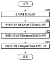
도 1은 본원의 일 실시예에 따른 피부조직과 혈관을 모사한 무펌프 미세유체 스킨칩의 제작과 다중세포배양 방법의 개략적인 순서도이다.
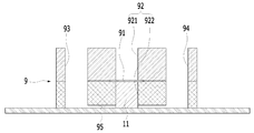
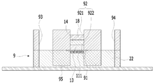
도 2는 본원의 일 실시예에 따른 피부조직과 혈관을 모사한 무펌프 미세유체 스킨칩의 제작과 다중세포배양 방법에 있어서, 미세유체 스킨칩의 개략적인 단면도이다.
도 3a는 본원의 일 실시예에 따른 피부조직과 혈관을 모사한 무펌프 미세유체 스킨칩의 제작과 다중세포배양 방법에 있어서, 멤브레인의 하면을 콜라젠으로 코팅하는 단계를 설명하기 위한 개략적인 단면도이다.
도 3b는 본원의 일 실시예에 따른 피부조직과 혈관을 모사한 무펌프 미세유체 스킨칩의 제작과 다중세포배양 방법에 있어서, 멤브레인의 하면 상에 HUVEC(혈관 내피 세포)를 배치하는 단계를 설명하기 위한 개략적인 개념도이다.
도 4a는 본원의 일 실시예에 따른 피부조직과 혈관을 모사한 무펌프 미세유체 스킨칩의 제작과 다중세포배양 방법에 있어서, 멤브레인의 상면을 콜라젠 혼합물로 코팅하는 단계를 설명하기 위한 개략적인 단면도이다.
도 4b는 본원의 일 실시예에 따른 피부조직과 혈관을 모사한 무펌프 미세유체 스킨칩의 제작과 다중세포배양 방법에 있어서, 코팅된 콜라젠 혼합물의 상면 상에 Fibroblast(섬유아세포)가 혼합된 셀룰러 콜라젠 혼합물을 주입하여 굳히는 단계를 설명하기 위한 개략적인 단면도이다.
도 5a는 본원의 일 실시예에 따른 피부조직과 혈관을 모사한 무펌프 미세유체 스킨칩의 제작과 다중세포배양 방법에 있어서, Keratinocyte의 cell line이 혼합된 cell suspension을 주입하는 단계를 설명하기 위한 개략적인 개념도이다.
도 5b 및 도 5c는 본원의 일 실시예에 따른 피부조직과 혈관을 모사한 무펌프 미세유체 스킨칩의 제작과 다중세포배양 방법에 있어서, 주입된 cell suspension의 Keratinocyte의 cell line 상에 배지를 채우고, 제1 리저버 및 제2 리저버를 배지(22)로 채우는 단계를 설명하기 위한 개략적인 단면도이다.
도 5d는 본원의 일 실시예에 따른 피부조직과 혈관을 모사한 무펌프 미세유체 스킨칩의 제작과 다중세포배양 방법에 있어서, S700 단계의 공기 노출 시키는 단계를 설명하기 위한 개략적인 단면도이다.
도 6은 본원의 일 실시예에 따른 피부조직과 혈관을 모사한 무펌프 미세유체 스킨칩의 제작과 다중세포배양 방법의 다른 구현예를 설명하기 위한 개략적인 순서도이다.
도 7a는 본원의 일 실시예에 따른 피부조직과 혈관을 모사한 무펌프 미세유체 스킨칩의 제작과 다중세포배양 방법에 있어서, Master fabrication 단계를 설명하기 위한 개략적인 개념도이다.
도 7b 및 도 7c는 본원의 일 실시예에 따른 피부조직과 혈관을 모사한 무펌프 미세유체 스킨칩의 제작과 다중세포배양 방법에 있어서, PDMS layer를 제작하는 단계를 설명하기 위한 개략적인 개념도이다.
도 7d는 본원의 일 실시예에 따른 피부조직과 혈관을 모사한 무펌프 미세유체 스킨칩의 제작과 다중세포배양 방법에 있어서, S100 단계의 Chip bonding 단계를 설명하기 위한 개략적인 개념도이다.FIG. 1 is a schematic flow diagram of a method for manufacturing a non-pump microfluidic skin chip and a method for culturing multiple cells according to an embodiment of the present invention.
FIG. 2 is a schematic cross-sectional view of a microfluidic skin chip in the manufacture of a non-pump microfluidic skin chip simulating skin tissue and blood vessels according to an embodiment of the present invention and a multiple cell culture method.
3A is a schematic cross-sectional view for explaining a step of coating a lower surface of a membrane with collagen in the production of a non-pump microfluidic skin chip and a method of culturing multiple cells, in which skin tissue and blood vessels are simulated according to an embodiment of the present invention .
FIG. 3B illustrates a step of disposing a HUVEC (vascular endothelial cell) on the lower surface of the membrane in the preparation of a non-pump microfluidic skin chip simulating skin tissue and blood vessels according to an embodiment of the present invention and a multiple cell culture method Fig.
FIG. 4A is a schematic cross-sectional view illustrating a step of coating a top surface of a membrane with a collagen mixture in the preparation of a non-pump microfluidic skin chip simulating skin tissue and blood vessels according to an embodiment of the present invention and a multiple cell culture method to be.
FIG. 4B is a view illustrating a method of manufacturing a non-pump microfluidic skin chip that mimics skin tissue and blood vessels according to an embodiment of the present invention, Sectional view for explaining a step of injecting and hardening a collagen mixture.
FIG. 5a is a view illustrating a step of injecting a cell suspension mixed with a cell line of Keratinocyte in the production of a non-pump microfluidic skin chip simulating skin tissue and blood vessels according to an embodiment of the present invention, FIG.
FIGS. 5B and 5C are cross-sectional views illustrating a method of preparing a non-pump microfluidic skin chip and a method for culturing multiple cells according to an embodiment of the present invention. The method comprises filling the cell line of the injected cell suspension on a cell line of Keratinocyte , The first reservoir and the second reservoir are filled with the medium (22).
FIG. 5D is a schematic cross-sectional view illustrating the step of exposing air in step S700 in the manufacture of a non-pump microfluidic skin chip simulating skin tissue and blood vessels according to an embodiment of the present invention and a multiple cell culture method.
FIG. 6 is a schematic flowchart for explaining another embodiment of a method for manufacturing a non-pump microfluidic skin chip and a method for culturing multiple cells, in which skin tissue and blood vessels are simulated according to an embodiment of the present invention.
FIG. 7A is a schematic diagram for explaining a master fabrication step in the manufacture of a non-pump microfluidic skin chip simulating skin tissue and blood vessels according to an embodiment of the present invention and a method for culturing multiple cells.
FIGS. 7B and 7C are schematic diagrams for explaining steps of fabricating a PDMS layer in the preparation of a non-pump microfluidic skin chip and a method of culturing multiple cells, in which skin tissue and blood vessels are simulated according to an embodiment of the present invention .
FIG. 7D is a schematic diagram for explaining the step of chip bonding in step S100 in the manufacture of a non-pump microfluidic skin chip and a method of multi-cell culture in which skin tissue and blood vessels are simulated according to an embodiment of the present invention.
아래에서는 첨부한 도면을 참조하여 본원이 속하는 기술 분야에서 통상의 지식을 가진 자가 용이하게 실시할 수 있도록 본원의 실시예를 상세히 설명한다. 그러나 본원은 여러 가지 상이한 형태로 구현될 수 있으며 여기에서 설명하는 실시예에 한정되지 않는다. 그리고 도면에서 본원을 명확하게 설명하기 위해서 설명과 관계없는 부분은 생략하였으며, 명세서 전체를 통하여 유사한 부분에 대해서는 유사한 도면 부호를 붙였다.Hereinafter, embodiments of the present invention will be described in detail with reference to the accompanying drawings so that those skilled in the art can easily carry out the present invention. It should be understood, however, that the present invention may be embodied in many different forms and should not be construed as limited to the embodiments set forth herein. In the drawings, the same reference numbers are used throughout the specification to refer to the same or like parts.
본원 명세서 전체에서, 어떤 부분이 다른 부분과 "연결"되어 있다고 할 때, 이는 "직접적으로 연결"되어 있는 경우뿐 아니라, 그 중간에 다른 소자를 사이에 두고 "전기적으로 연결"되어 있는 경우도 포함한다. Throughout this specification, when a part is referred to as being "connected" to another part, it is not limited to a case where it is "directly connected" but also includes the case where it is "electrically connected" do.
본원 명세서 전체에서, 어떤 부재가 다른 부재 "상에", "상부에", "상단에", "하에", "하부에", "하단에" 위치하고 있다고 할 때, 이는 어떤 부재가 다른 부재에 접해 있는 경우뿐 아니라 두 부재 사이에 또 다른 부재가 존재하는 경우도 포함한다.It will be appreciated that throughout the specification it will be understood that when a member is located on another member "top", "top", "under", "bottom" But also the case where there is another member between the two members as well as the case where they are in contact with each other.
본원 명세서 전체에서, 어떤 부분이 어떤 구성요소를 "포함" 한다고 할 때, 이는 특별히 반대되는 기재가 없는 한 다른 구성요소를 제외하는 것이 아니라 다른 구성 요소를 더 포함할 수 있는 것을 의미한다.Throughout this specification, when an element is referred to as "including " an element, it is understood that the element may include other elements as well, without departing from the other elements unless specifically stated otherwise.
참고로, 본원의 실시예에 관한 설명 중 방향이나 위치와 관련된 용어(상측, 상면, 하측, 하면 등)는 도면에 나타나 있는 각 구성의 배치 상태를 기준으로 설정한 것이다. 예를 들면, 도 2에서 보았을 때 전반적으로 12시 방향이 상측, 전반적으로 12시 방향을 향하는 면이 상면, 전반적으로 6시 방향이 하측, 전반적으로 6시 방향을 향하는 면이 하면 등이 될 수 있다.For reference, the terms related to directions and positions (top, top, bottom, bottom, etc.) in the description of the embodiments of the present application are set based on the arrangement state of each structure shown in the drawings. For example, when viewed from Fig. 2, the 12 o'clock direction is generally the upper side, the 12 o'clock side generally faces the upper side, the 6 o'clock side is generally lower, and the side facing the 6 o'clock direction is the lower side have.
이하에서는, 본원의 일 실시예에 따른 피부조직과 혈관을 모사한 무펌프 미세유체 스킨칩의 제작과 다중세포배양 방법(이하 '본 방법'이라 함)에 대해 설명한다.Hereinafter, the preparation of a non-pump microfluidic skin chip and a method for culturing multiple cells (hereinafter referred to as "the present method") in which skin tissue and blood vessels are simulated according to one embodiment of the present invention will be described.
도 1은 본 방법의 개략적인 순서도이고, 도 2는 본 방법의 미세유체 스킨칩에 있어서, 개략적인 단면도이다.Fig. 1 is a schematic flow chart of the present method, and Fig. 2 is a schematic cross-sectional view of a microfluidic skin chip of the present method.
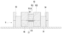
도 1을 참조하면, 본 방법은, 미세유체 스킨칩(9)을 준비하는 단계(S100)를 포함한다.Referring to FIG. 1, the method includes a step S100 of preparing a
도 2에 나타난 바와 같이, 미세유체 스킨칩(9)은 수평으로 배치되는 멤브레인(membrane)(91)에 의해 내부가 상부(921)와 하부(922)로 분리(구획)되는 챔버(92), 챔버(92)의 일측 및 타측 각각에 형성되는 제1 리저버(93)와 제2 리저버(94), 및 유체가 이동 가능하도록 제1 리저버(93), 챔버(92)의 하부(922) 및 제2 리저버(94)를 연결하는 채널(95)을 포함한다.2, the
예시적으로, 멤브레인(91)은 polycarbonate 재질로 이루어질 수 있다. 다른 예로, 멤브레인(91)은 polyester 재질로 이루어질 수 있다. 이러한 멤브레인(91)의 재질 차이에 따라, 후술하는 단계들에서 멤브레인(91)에 대한 콜라젠의 부착 정도에 다소 차이가 생길 수 있다.Illustratively, the
또한, 도 1을 참조하면, 본 방법은 멤브레인(91)의 하면 상에 혈관 내벽 조직을 형성하는 단계(S300)를 포함한다.1, the method includes forming an intravascular wall tissue on the lower surface of the membrane 91 (S300).
도 3a는 본 방법에 있어서, 멤브레인의 하면을 콜라젠으로 코팅하는 단계를 설명하기 위한 개략적인 단면도이다.3A is a schematic cross-sectional view for explaining a step of coating the lower surface of the membrane with collagen in the present method.
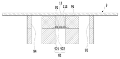
혈관 내벽 조직을 형성하는 단계(S300)는, 멤브레인(91)의 하면을 코팅물질(11)로 코팅하는 단계를 포함할 수 있다. HUVEC의 멤브레인(91)에 대한 부착력을 증가시키고 confluent monolayer 로 HUVEC를 배양시키기 위해, 멤브레인(91)의 하면이 코팅물질(11)로 코팅될 필요가 있다.Step S300 of forming the vascular endothelial tissue may include coating the lower surface of the
구체적으로, 멤브레인(91)의 하면을 코팅물질(11)로 코팅하는 단계는 도 3a에 나타난 바와 같이, 제1 리저버(93) 및 제2 리저버(94) 중 하나에 코팅물질(11)을 주입하여 챔버(92)의 하부(922)를 코팅물질(11)로 소정의 시간 동안 채워두는 단계를 포함할 수 있다.Specifically, the step of coating the lower surface of the
예시적으로, 코팅물질(11)을 채널(95)의 한 쪽 끝에서 흘려주어 채널(95)의 반대쪽까지 흐른 것이 확인되면(다시 말해, 제1 리저버(93) 및 제2 리저버(94) 중 하나에 코팅물질(11)을 주입하여 다른 하나로부터 코팅물질(11)이가 배출되는 것이 확인되면), 코팅물질(11)이가 챔버(92)의 하부(922)에 채워졌다고 생각할 수 있다. 이에 따라, 코팅물질(11)을 채널(95)의 한 쪽 끝에서 흘려주어 채널(95)의 반대쪽까지 흐른 것을 확인하고 인큐베이터(incubator)에 미세유체 스킨칩(9)을 소정의 시간, 예시적으로, 한 시간 동안 넣어둘 수 있다.Illustratively, when it is confirmed that the
또한, 참고로, 코팅물질(11)은 70 ug/ml가 미세유체 스킨칩(9) 내로 주입될 수 있다. 또한, 코팅물질(11)은 Type Ⅰ collagen일 수 있다. 다만, 코팅물질(11)의 종류는 이에 한정되지 않으며, Type Ⅰ collagen이외에 다른 종류의 콜라젠 및 다른 종류의 하이드로젤이 코팅물질(11)로 적용될 수 있다. Also, for reference, 70 ug / ml of the
또한, 멤브레인(91)의 하면을 코팅물질(11)로 코팅하는 단계는, 미세유체 스킨칩(9)의 내부를 워싱(washing)하는 단계를 포함할 수 있다.In addition, the step of coating the lower surface of the
미세유체 스킨칩(9)의 내부를 워싱하는 단계는 채널(5)에 PBS 및 EGM-2MV를 순차적으로 흘려줄 수 있다. 이러한 워싱하는 단계는 챔버(92)의 하부(922)가 코팅물질(11)로 채워졌던 소정의 시간 이후에 수행될 수 있다.Washing the interior of the
도 3b는 본 방법에 있어서, 멤브레인의 하면 상에 HUVEC(혈관 내피 세포)를 배치하는 단계를 설명하기 위한 개략적인 개념도이다.Fig. 3B is a schematic diagram for explaining the step of disposing HUVEC (vascular endothelial cells) on the lower surface of the membrane in the present method. Fig.
혈관 내벽 조직을 형성하는 단계(S300)는 멤브레인(91)의 하면 상에 HUVEC(혈관 내피 세포)(111)를 배치하는 단계를 포함할 수 있다.Step S300 of forming an inner wall tissue of the blood vessel may include disposing HUVEC (vascular endothelial cells) 111 on the lower surface of the
멤브레인(91)의 하면 상에 HUVEC(111)를 배치하는 단계는 도 3b를 참조하면, 챔버(92)의 하부(922)를 HUVEC 포함 액체(12)로 채우는 단계를 포함할 수 있다. 예시적으로, 챔버(92)의 하부(922)를 HUVEC 포함 액체(12)로 채우는 단계는 제1 리저버(93) 및 제2 리저버(94) 중 하나에 HUVEC 포함 액체(12)가 주입된 후, 나머지 하나로 HUVEC 포함 액체(12)가 배출되면 챔버(92)의 하부(922)가 HUVEC 포함 액체(12)로 채워진 것으로 판단할 수 있다. 다시 말해, 채널(95)의 한쪽 끝에서 HUVEC 포함 액체(12)를 흘려주어 반대쪽 채널(95)의 끝에서 HUVEC 포함 액체(12)가 흐르는 것이 확인되면, 챔버(92)의 하부(922)가 HUVEC 포함 액체(12)로 채워진 것으로 판단할 수 있다.The step of disposing the
멤브레인(91)의 하면 상에 HUVEC(111)를 배치하는 단계는, 7.5x10⁴cells, 250-300ul의 HUVEC 포함 액체(12)로 챔버(92)의 하부(922)를 채울 수 있다.The step of disposing the
또한, 멤브레인(91)의 하면 상에 HUVEC(111)를 배치하는 단계는 도 3b에 나타난 바와 같이, 챔버(92)의 하부(922)가 상측을 향하도록 미세유체 스킨칩(9)을 뒤집는 단계를 포함할 수 있다.The step of disposing the
상술한 바와 같이, 채널(95)의 한쪽 끝에서 HUVEC 포함 액체(12)를 흘려주어 반대쪽 채널(95)의 끝에서 HUVEC 포함 액체(12)가 흐르는 것이 확인되면 미세유체 스킨칩(9) 내부의 배지(상술한 PBS, EGM-2MV 등)를 제거하고 미세유체 스킨칩(9)을 최대한 빨리 뒤집을 수 있다. 또한, 미세유체 스킨칩(9)이 뒤집어진 상태로 인큐베이터에서 소정의 시간 동안 있을 수 있게 한다. 예시적으로, 미세유체 스킨칩(9)은 뒤집어진 상태로 인큐베이터 내에서 1시간 가량 있을 수 있다. 상술한 바와 같은 과정을 통해, HUVEC(111)가 멤브레인(91)의 하면에 붙을 수 있다.As described above, when it is confirmed that the HUVEC-containing
또한, 혈관 내벽 조직을 형성하는 단계(S300)는 제1 리저버(93), 챔버(92)의 상부(921) 및 제2 리저버(94)를 배지로 채워두는 단계를 포함할 수 있다. 이 단계(혈관 내벽 조직을 형성하는 단계(S300)의 제1 리저버(93), 챔버(92)의 상부(921) 및 제2 리저버(94)를 배지로 채워두는 단계)에서 제1 리저버(93), 챔버(92)의 상부(921) 및 제2 리저버(94)에 채워지는 배지는 EGM-2MV일 수 있다. 보다 구체적으로, 미세유체 스킨칩(9)이 인큐베이터로부터 나오면 제1 리저버(92) 및 제2 리저버(92) 각각에EGM-2MV가 1.5 ml씩 채워질 수 있고, 챔버(92)의 상부, 즉, 멤브레인(91) 상에는 EGM-2MV가 가득 채워질 수 있다.In addition, the step S300 of forming the vessel wall structure may include filling the
또한, 혈관 내벽 조직을 형성하는 단계(S300)는 제1 리저버(93), 챔버(92)의 상부(921) 및 제2 리저버(94)를 배지로 채워둔 상태에서 미리 설정된 시간 동안 배지의 흐름 없이 배양이 이루어지는 단계를 포함할 수 있다. 또한, 혈관 내벽 조직을 형성하는 단계(S300)는 배지의 흐름 없이 배양이 이루어지는 단계 이후에, 미세유체 스킨칩(9) 내에서 배지의 흐름을 형성시키며 배양이 이루어지는 단계를 포함할 수 있다.In addition, the step S300 of forming the inner wall tissue of the blood vessel is performed in a state in which the
예시적으로, 제1 리저버(93), 챔버(92)의 상부(921) 및 제2 리저버(94)에 배지가 채워지면, 하루 동안은 미세유체 스킨칩(9)이 gravity machine 에 올라가지 않은 상태로 배양이 이루어지 그 후 2-3일 동안 미세유체 스킨칩(9)이 gravity machine 위에 올라가 있는 상태로 미세유체 스킨칩(9) 내부에 flow를 줄 수 있다.Illustratively, when the medium is filled in the
또한, 배지의 흐름을 형성시키며 배양이 이루어지는 단계 이후에, 후술하는 진피층을 형성하는 단계(S500)의 수행을 위해, 제1 리저버(93), 챔버(92)의 상부(921) 및 제2 리저버(94)를 배지로 채워두는 단계에서 제1 리저버, 상기 챔버의 상부 및 상기 제2 리저버를 채웠던 배지(EGM-2MV)가 제거될 수 있다.In order to perform the step S500 of forming a dermal layer to be described later, the
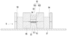
또한, 도 1을 참조하면, 본 방법은, 멤브레인(91)의 상면 상에 진피층(dermis)을 형성하는 단계(S500)를 포함한다. 1, the method includes forming a dermis on the upper surface of the membrane 91 (S500).
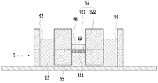
도 4a는 본 방법에 있어서, S500 단계의 멤브레인의 상면을 콜라젠 혼합물로 코팅하는 단계를 설명하기 위한 개략적인 단면도이다.4A is a schematic cross-sectional view illustrating the step of coating the upper surface of the membrane of step S500 with a collagen mixture, according to the present method.
도 4a를 참조하면, S500 단계는, 멤브레인(91)의 상면을 콜라젠 혼합물(13)로 코팅하는 단계를 포함할 수 있다. 콜라젠 혼합물(13)은, 콜라젠, 10X DMEM media, 0.5N NaOH 및 DMEM을 섞은 혼합물일 수 있다. 또한, 이러한 콜라젠 혼합물(13) 30ul 내지 35ul에 의해 멤브레인(91)의 상면이 코팅될 수 있다. 이를 테면, 콜라젠 혼합물(13) 30ul 내지 35ul이 멤브레인(91) 상에 주입될 수 있고, 콜라젠 혼합물(13)이 멤브레인(91) 상에 주입된 미세유체 스킨칩(9)이 인큐베이터 안에서 소정 시간을 보냄으로써, 콜라젠 혼합물(13)이 굳을 수 있다. 예시적으로, 콜라젠 혼합물(13)이 멤브레인(91) 상에 주입된 미세유체 스킨칩(9)은 37℃, 5% 인큐베이터 내에서 20분 정도 위치할 수 있다.Referring to FIG. 4A, step S500 may include coating the upper surface of the
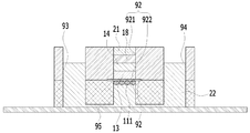
도 4b는 본 방법에 있어서, S500 단계의 코팅된 콜라젠 혼합물의 상면 상에 Fibroblast(섬유아세포)가 혼합된 셀룰러 콜라젠 혼합물을 주입하여 굳히는 단계를 설명하기 위한 개략적인 단면도이다.FIG. 4B is a schematic cross-sectional view illustrating a step of injecting and hardening a cellular collagen mixture in which Fibroblast (fibroblast) is mixed on the upper surface of the coated collagen mixture of step S500.
또한, S500 단계는, 도 4b를 참조하면, 코팅된 상기 콜라젠 혼합물(13)의 상면 상에 Fibroblast(섬유아세포)가 혼합된 셀룰러 콜라젠 혼합물(14)을 주입하여 굳히는 단계를 포함할 수 있다. 셀룰러 콜라젠 혼합물은, 콜라젠, 10X DMEM media, 0.5N NaOH 및 Fibroblast를 섞은 것일 수 있다. 예시적으로, 셀룰러 콜라젠 혼합물은 콜라젠, 10X DMEM media 및 0.5N NaOH가 혼합되고 마지막에 Fibroblast (섬유아세포) 가 섞어짐으로써 제조될 수 있다. 이때, Fibroblast이 골고루 혼합될 수 있도록 섞어질 수 있다.Referring to FIG. 4B, step S500 may include injecting a
또한, S500 단계에서, Fibroblast(섬유아세포)가 혼합된 셀룰러 콜라젠 혼합물은 섬유아세포 이외에 다른 종류의 세포를 포함할 수 있다. 예를 들어, Fibroblast(섬유아세포)가 혼합된 셀룰러 콜라젠 혼합물은 피부 진피 조직에 있다고 알려진 다른 종류의 세포(예를 들면, 면역세포)를 추가로 포함할 수 있다.Also, in step S500, the cellular collagen mixture in which Fibroblast (fibroblast) is mixed may include cells of a different kind in addition to fibroblasts. For example, a cellular collagen mixture in which Fibroblast (fibroblast) is mixed may further comprise other types of cells known to be in the dermal tissue (e. G., Immune cells).
셀룰러 콜라젠 혼합물(14) 50ul이 코팅된 콜라젠 혼합물(13) 상에 첨가될 수 있다. 이후, 미세유체 스킨칩(9)을 37℃, 5% 인큐베이터 내에 40분 정도 넣어 셀룰러 콜라젠 혼합물(14)을 굳힐 수 있다. 굳어진 셀룰러 콜라젠 혼합물(14)은 gel 형태일 수 있다.50ul of the
이후, 도 4b에 나타난 바와 같이, S500 단계는 굳은 셀룰러 콜라젠 혼합물(14) 상에 배지(15)를 채우고, 제1 리저버(93) 및 제2 리저버(94)를 배지(16)로 채우는 단계를 수행할 수 있다. 이 단계에서, 예시적으로, 셀룰러 콜라젠 혼합물(14) 상에 채워지는 배지(15)는 DMEM일 수 있다. 또한, 제1 리저버(93) 및 제2 리저버(94)에 채워지는 배지(16)는 EGM-2MV일 수 있다.Thereafter, as shown in FIG. 4B, step S500 includes the step of filling the medium 15 on the solid
또한, S500 단계는, 굳은 셀룰러 콜라젠 혼합물(14) 상에 배지(15)를 채우고, 제1 리저버(93) 및 제2 리저버(94)를 배지(16)로 채우는 단계 이후에, 미세유체 스킨칩(9) 내에서 배지(15, 16)의 흐름을 형성시키며 배양이 이루어지는 단계를 포함할 수 있다.The step S500 also includes the step of filling the medium 15 on the rigid
즉, 셀룰러 콜라젠 혼합물(14)이 굳도록 인큐베이터에 넣어졌던 미세유체 스킨칩(9)이 인큐베이터로부터 꺼내진 후, Fibroblast 및 그와 섞인 셀룰러 콜라젠 혼합물은 5 내지 7일 동안 수축하고 그 후에 수축이 안정화 되므로, 5 내지 7일 동안 EGM-2MV(16)를 제1 및 제2 리저버(93, 94) 각각에 1500ul씩 주입하고, DMEM(배지)(15)을 굳은 셀룰러 콜라젠 혼합물(14) 상에 챔버(92)의 상부(921)가 가득 차도록 채운 후, 미세유체 스킨칩(9)을 gravity machine (10ul/min) 에 올려 flow 를 주면서 배양시킬 수 있다.That is, after the
또한, 본 방법에 있어서, 배지(15, 16)의 흐름을 형성시키며 배양이 이루어지는 단계 이후에, 후술할 진피층 상면 상에 표피층(epidermis)을 형성하는 단계(S700)를 수행하기 위해, S500 단계 중 굳은 셀룰러 콜라젠 혼합물(14) 상에 배지(15)를 채우고 제1 리저버(93) 및 제2 리저버(94)를 배지(16)로 채우는 단계에서, 셀룰러 콜라젠 혼합물(14) 상에 채워졌던 배지(15)가 제거되고 제1 리저버(93)와 제2 리저버(94)를 채웠던 배지(16)가 적어도 일부 제거될 수 있다.In order to perform the step S700 of forming the epidermis on the upper surface of the dermal layer to be described later after the step of forming the culture medium and forming the flow of the
셀룰러 콜라젠 혼합물(14) 상에 채워졌던 배지(15) 및 제1 리저버(93)와 제2 리저버(94)를 채웠던 배지(16)는, 표피층을 형성하는 단계(S700)에서 표피세포가 직접적으로 굳은 셀룰러 콜라젠 혼합물(14)에 시딩될 수 있도록 표피세포 배양 20분 전(다시 말해, S700 단계가 수행되기 20분 전)에 적어도 일부 제거될 수 있다. 그 결과, 제1 리저버(93)와 제2 리저버(94)에는 EGM-2MV가 350-400 μl 정도만 채워질 수 있다. 상술한 바와 같은 S500 단계의 수행에 의해, 멤브레인(91)의 상면 상에 진피층이 형성될 수 있다.The medium 15 filled in on the
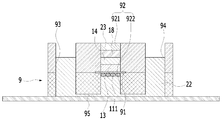
또한, 도 1을 참조하면, 본 방법은, 진피층의 상면 상에 표피층(epidermis)(S700)을 형성하는 단계를 포함한다.1, the method includes forming an epidermis S700 on the upper surface of the dermal layer.
도 5a는 본 방법에 있어서, S700 단계의 Keratinocyte의 cell line이 혼합된 cell suspension을 주입하는 단계를 설명하기 위한 개략적인 개념도이다.FIG. 5A is a schematic diagram for explaining a step of injecting a cell suspension mixed with a cell line of Keratinocyte in step S700.
도 5a를 참조하면, S700은 상기 S500 단계를 통해 형성된 진피층 상에 Keratinocyte의 cell line(세포주)이 혼합된 cell suspension(18)을 주입하는 단계를 포함할 수 있다. 참고로, Keratinocyte 는 표피세포 중 각질화 능력을 갖는 세포로 표피세포의 대부분을 차지한다. 또한, S700 단계에서, cell suspension(18)의 Keratinocyte의 cell line은 HaCaT일 수 있다. 또는, 동물의 피부 또는 인체의 피부에서 추출한 keratinocyte 또는 줄기세포가 Keratinocyte의 cell line으로 이용될 수 있다.Referring to FIG. 5A, S700 may include injecting a
또한, cell suspension(18)은 5x10⁵cells 이 포함된 50ul cell suspension일 수 있다. 또한, S500 단계를 통해 형성된 진피층이라 함은, gel 형태로 굳은 셀룰러 콜라젠 혼합물(14)을 의미할 수 있다. Also, the
또한, 상술한 바와 같이, 표피세포가 직접적으로 굳은 셀룰러 콜라젠 혼합물(14)(gel)에 시딩될 수 있도록 S700 단계의 수행 이전(대략 20분 전)에 미세유체 스킨칩(9)에 있는 DMEM을 제거하고 EGM-2MV의 양도 각각의 제1 및 제2 리저버(93, 94) 각각에 350-400μl 정도만 채워지게 할 수 있다.In addition, as described above, the DMEM in the
S700 단계의 수행 이전(대략 20분 전)에 미세유체 스킨칩(9)에 있는 DMEM이 제거된 후, 진피층 상에 Keratinocyte의 cell line(세포주)이 혼합된 cell suspension(18)을 주입하는 단계는 도 5a를 참조하면, cell suspension(18)을 굳은 셀룰러 콜라젠 혼합물(14)(gel) 위에 조심스럽게 첨가할 수 있다.After the DMEM in the
참고로, S700 단계는 cell suspension의 표피세포가 완전히 붙을 수 있도록, 미세유체 스킨칩(9)에 대한 배지 추가 없이 미세유체 스킨칩(9)을 미리 설정된 시간 동안, 예를 들면 60min 동안 37℃, 5% 인큐베이터에 둘 수 있다.For reference, in step S700, the
도 5b 및 도 5c는 본 방법에 있어서, S700 단계의 주입된 cell suspension의 Keratinocyte의 cell line 상에 배지를 채우고, 제1 리저버 및 제2 리저버를 배지(22)로 채우는 단계를 설명하기 위한 개략적인 단면도이다.FIGS. 5B and 5C are schematic views for explaining the step of filling the medium on the cell line of the Keratinocyte of the injected cell suspension of step S700 and filling the first reservoir and the second reservoir with the medium 22 Sectional view.
미세유체 스킨칩(9)을 미리 설정된 시간 동안 인큐베이터에 둔 이후에, S700 단계는 도 5b 및 도 5c를 참조하면, 주입된 상기 cell suspension(18)의 Keratinocyte의 cell line 상에 배지(21, 23)를 채우고, 제1 리저버(93) 및 제2 리저버(94)를 배지(22)로 채우는 단계를 수행할 수 있다.5B and 5C, after the
구체적으로, 도 5b에 나타난 바와 같이, 주입된 cell suspension(18)의 Keratinocyte의 cell line 상에는 소정의 시간 동안 DMEM(21)을 채워둘 수 있다. 이 때, 제1 리저버(93) 및 제2 리저버(94)에는 EGM-2MV(22)가 채워질 수 있다. 예시적으로, EGM-2MV(22)는 제1 및 제2 리저버(93, 94) 각각에 1500ul 씩 채워질 수 있고, DMEM(21)은 챔버(92)의 상부(921)에 가득 채워질 수 있다.Specifically, as shown in FIG. 5B, the
또한, 도 5c에 나타난 바와 같이, 주입된 cell suspension(18)의 Keratinocyte의 cell line 상에 배지(21)를 채우고, 제1 리저버(93) 및 제2 리저버(94)를 배지(22)로 채우는 단계는, 소정의 시간 이후에, 주입된 상기 cell suspension(18)의 Keratinocyte의 cell line 상에 소정의 시간 동안 칼슘(Ca2 +)이 첨가된 DMEM(21)을 채워둘 수 있다. 이 때, 제1 리저버(93) 및 제2 리저버(94)에는 EGM-2MV(22)를 채워둘 수 있다.5C, the medium 21 is filled on the cell line of the Keratinocyte of the injected
예시적으로, cell suspension(18)의 Keratinocyte의 cell line 상에 DMEM(21)을 채워두는 소정의 시간은 1일 내지 2일일 수 있다. 이 기간에는 표피세포가 gel(셀룰러 콜라젠 혼합물(14)) 상에서 분열과 증식이 일어날 수 있다. 이 후, 약 2일 동안 cell suspension(18)의 Keratinocyte의 cell line 상에는 칼슘이 첨가된 DMEM(21)이 채워질 수 있으며, 이에 따라, 표피세포가 분화될 수 있다.Illustratively, the predetermined time for filling the
또한, cell suspension(18)의 Keratinocyte의 cell line 상에 배지(21, 23)를 채우고, 제1 리저버(93) 및 제2 리저버(94)를 배지(22)로 채우는 단계가 수행되는 동안에는, 표피세포가 셀룰러 콜라젠 혼합물(14) 상에서 균일하게 배양될 수 있도록 미세유체 스킨칩(9)에 대한 배지의 흐름이 형성되지 않도록 한다. 다시 말해, cell suspension(18)의 Keratinocyte의 cell line 상에 배지(21, 23)를 채우고, 제1 리저버(93) 및 제2 리저버(94)를 배지(22)로 채우는 단계가 수행되는 동안에는, 미세유체 스킨칩(9)은 gravity machine 에 올리지 않도록 함이 바람직하다.During the step of filling the
도 5d는 본 방법에 있어서, S700 단계의 공기 노출 시키는 단계를 설명하기 위한 개략적인 단면도이다.5D is a schematic cross-sectional view illustrating the step of exposing air in step S700 in the present method.
또한, S700 단계는, cell suspension(18)의 Keratinocyte의 cell line 상에 배지(21, 23)를 채우고, 제1 리저버(93) 및 제2 리저버(94)를 배지(22)로 채우는 단계 이후에, 도 5d를 참조하면, 상기 cell suspension(18)의Keratinocyte의 cell line 상에 채워진 배지(여기서는, 칼슘이 첨가된 DMEM(23)) 및 제1 리저버(93) 및 제2 리저버(94)를 채우는 배지의 적어도 일부를 제거하고, 미세유체 스킨칩(9) 내에서의 배지의 흐름을 형성시키며, Keratinocyte(18)의 cell line을 공기 노출(Air exposure)시키는 단계를 포함할 수 있다.In operation S700, the medium 21 and 23 are filled on the cell line of the keratinocyte of the
보다 구체적으로, 이 단계는, 제1 및 제2 리저버(93, 94)의 EGM-2MV(22) 양을 350ul 내지 400ul로 줄이고 셀룰러 콜라젠 혼합물(14) 위의 칼슘이 첨가된 DMEM(23)을 제거해 7 일 내지 10 일 동안 flow 를 주면서 Air exposure 시킬 수 있다. 이 단계가 수행되는 동안, 표피세포의 분화가 진행될 수 있다.More specifically, this step reduces the amount of EGM-2MV (22) in the first and
도 6은 본 방법의 다른 구현예를 설명하기 위한 개략적인 순서도이다.Figure 6 is a schematic flow diagram for illustrating another embodiment of the method.
도 6을 참조하면, 본 방법에 있어서, S300 단계는 상술한 바와 같이S500 단계 이전에 수행될 수 있고, 또 다른 구현예로서, S300 단계는 S700 단계 이후에 수행될 수 있다. 참고로, 상술한 본 방법의 일 구현예와 여기에서 후술하는 본 방법의 다른 구현예는 세포를 배양하는 순서에 차이가 있는데, 순서의 차이에 따라 각 세포의 상태와 기능에 차이가 생길 수 있다Referring to FIG. 6, in this method, step S300 may be performed before step S500 as described above, and step S300 may be performed after step S700 as another embodiment. For reference, one embodiment of the method described above and another embodiment of the method described herein differs in the order of culturing the cells, and there may be differences in the state and function of each cell depending on the order of the cells
이하에서는, S300 단계가 S700 단계 이후에 수행되는 구현예에 대해 간략히 설명한다.Hereinafter, an implementation example in which the step S300 is performed after the step S700 will be briefly described.
도 6을 참조하면, S100 단계 이후에, 상술한 S500 단계가 수행될 수 있다. 이 때, S500 단계의 수행은 상술한 바와 같다.Referring to FIG. 6, after step S100, the above-described step S500 may be performed. In this case, the execution of step S500 is as described above.
또한, 도 6을 참조하면, S500 단계 이후에 S700 단계가 수행될 수 있다. S700 단계의 수행은 상술한 바와 같다.Referring to FIG. 6, step S700 may be performed after step S500. The execution of step S700 is as described above.
또한, 도 6을 참조하면, S700 단계 이후에 S300 단계가 수행될 수 있다. 이 때, S300 단계는, S700 단계의 상술한 공기 노출시키는 단계 이전에 수행되기 시작할 수 있다.Referring to FIG. 6, step S300 may be performed after step S700. At this time, the step S300 may start to be performed before the above-described air exposure step of the step S700.
예시적으로, 상술한 공기 노출 단계가 이루어지기 하루 전 미세유체 스킨칩(9)에 HUVEC를 배양할 수 있다. 미세유체 스킨칩(9)에 있는 배지를 다 제거하고 미세유체 스킨칩(9)을 PBS로 washing 할 수 있다. 그리고 상술한 방법과 같은 방법으로 HUVEC를 배양할 수 있다. 미세유체 스킨칩(9)을 뒤집은 지 1 시간이 지난 후, 미세유체 스킨칩(9)의 제1 및 제2리저버(93, 94) 각각에 EGM-2MV를 1.5ml 씩 채우고 멤브레인(91)위 챔버(92)의 상부(921)에 칼슘을 첨가한 DMEM을 가득 채워줄 수 있다. 하루 동안은 gravity machine 에 올리지 않은 상태로 배양할 수 있다. 하루가 지난 후, 제1 및 제2 리저버(93, 94) 내의 EGM-2MV의 양을 300ul 내지350ul 로 줄이고 셀룰러 콜라젠 혼합물(14) 위 챔버(92)의 상부(921)에 채워진 분화배지(칼슘을 첨가한 DMEM)를 제거해 7 일 내지 10 일 동안 flow 를 주면서 Air exposure 시킬 수 있다. 이에 따라, 표피세포의 분화가 진행될 수 있다.Illustratively, HUVEC can be cultured in the
한편, S100 단계는 이하와 같이 수행될 수 있다.Meanwhile, the step S100 may be performed as follows.
도 7a는 본 방법에 있어서, S100 단계의 Master fabrication 단계를 설명하기 위한 개략적인 개념도이다.FIG. 7A is a schematic diagram for explaining the master fabrication step of step S100 in the present method. FIG.
S100 단계는 Master fabrication 단계를 포함할 수 있다.Step S100 may include a master fabrication step.
도 7a를 참조하면, Master fabrication 단계는, wafer(82)에 SU-8 50을 도포하기 위해 spin coater의 속도를 500rpm 5sec, 1000rpm, 30sec 조건으로 맞추어 사용할 수 있다. 그 후에 65℃ Hot plate에 10분간 올려둔 뒤, 95℃ Hot plate에 30분 동안 올려두어 Soft bake해줄 수 있다. 또한, MA6 AlignerⅡ(4인치 전용)을 이용하여 photolithography를 진행할 수 있다. 이 후, 다시 Hot plate에 올려놓아 PEB를 해줄 수 있다. 그 뒤에 SU-8 Developer를 이용하여 10분 이상 Develop을 하여 가교가 되지 않은 SU-8은 제거가 되어 원하는 구조의 양각형상의 wafer(81, 82)를 얻을 수 있다. Master fabrication 단계의 결과, 너비가 200μm이고 높이가 120μm 인 채널 모양의 양각 형상이 얻어질 수 있다.Referring to FIG. 7A, in the master fabrication step, a spin coater may be used at 500 rpm for 5 seconds, 1000 rpm for 30 seconds, and spin coater in order to apply SU-850 to the
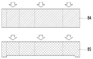
도 7b 및 도 7c는 본 방법에 있어서, S100 단계의 PDMS layer를 제작하는 단계를 설명하기 위한 개략적인 개념도이다.FIGS. 7B and 7C are schematic conceptual diagrams illustrating a step of fabricating the PDMS layer in step S100 in the present method.
또한, S100 단계는 PDMS layer를 제작하는 단계를 포함할 수 있다.In addition, the step S100 may include a step of fabricating the PDMS layer.
도 7b를 참조하면, PDMS layer를 제작하는 단계는, Master fabrication 단계를 통해, soft photolithography 를 이용하여 원하는 구조의 양각 형상으로 형성한 Wafer 기판(81, 82)에 Sylgard 184 A와 B 을 10:1 로 섞은 용액을 부어줄 수 있다. 이후, 60℃ 오븐에서 6시간 이상 굳힌 후 분리하면 PDMS(85)에 원하는 음각의 채널패턴을 얻을 수 있다. 채널 패턴이 있는 5mm 내지 6mm 두께의 PDMS(85)와 패턴이 있지 않은 6mm 두께의 PDMS(84)를 가교시켜 준비한다. 도 7c를 참조하면, 음각의 채널 패턴이 있는 PDMS(85)의 챔버(92) 부분을 8mm biopsy punch 로 구멍을 내고 제1 리저버 및 제2 리저버(93, 94) 부분은 채널(95)의 끝부분이 포함되도록 8mm biopsy punch를 이용해 연속적인 원을 뚫어 구름 모양 같은 구멍을 낼 수 있다. 패턴이 있지 않은 PDMS(84)도 똑같이 구멍을 낼 수 있다.Referring to FIG. 7B, in the step of fabricating the PDMS layer, Sylgard 184 A and B are deposited on the
도 7d는 본 방법에 있어서, S100 단계의 Chip bonding 단계를 설명하기 위한 개략적인 개념도이다.7D is a schematic diagram for explaining the chip bonding step of step S100 in the present method.
또한, S100 단계는 Chip bonding 단계를 포함할 수 있다.Also, step S100 may include a chip bonding step.
도 7d를 참조하면, Chip bonding 단계는, PDMS 표면을 플라즈마(plasma)를 처리하여 라디칼 활성화시킨 후 아래부터 slide glass(86)와 채널이 있는 PDMS(85), 멤브레인(88), 채널없는 PDMS (84)순이 되도록 결합시킬 수 있다. 이 단계가 끝나면, 도 1에 도시된 미세유체 스킨칩(9)이 형성될 수 있다.Referring to FIG. 7D, in the chip bonding step, the surface of the PDMS is subjected to radical activation by plasma treatment, and then a
또한, S100 단계는, 미세유체 스킨칩(9)을 멸균시키는 단계를 포함할 수 있다.In addition, the step S100 may include a step of sterilizing the
세균의 내생포자를 파괴하기 위해서는 100℃ 이상에서 높은 증기압으로 멸균해야 하므로 미세유체 스킨칩(9)을 멸균시키려면 고압멸균(autoclave)이 필요하다. 따라서, 미세유체 스킨칩(9)을 멸균시키는 단계에 있어서, 고압멸균(autoclave)이 121℃의 온도와 15lb psi의 압력에서 약 15분 정도 진행될 수 있다.In order to destroy the endogenous spores of bacteria, sterilization is required at a high vapor pressure of 100 ° C or higher, so autoclave is required to sterilize the microfluidic skin chip (9). Thus, in the step of sterilizing the
또한, S100 단계는, 미세유체 스킨칩(9)을 워싱하는 단계를 포함할 수 있다. 워싱하는 단계는, 미세유체 스킨칩(9)의 채널(95)의 한쪽 끝에 에탄올을 흘려주어 에탄올이 채널(95)을 따라 흐르도록 할 수 있다. 에탄올이 어느 정도 흐르는 것이 확인이 되면 PBS 를 흘려줄 수 있다.In addition, step S100 may include washing the
한편, 본 방법에 있어서, 미세유체 스킨칩(9) 내에서 흐름을 형성하는 것은 상술한 Gravity flow machine 을 포함하는 Gravity-flow 시스템에 의해 이루어질 수 있다.On the other hand, in the present method, the formation of the flow in the
기존에 개발되어진 여러 종류의 장기온어 칩 시스템의 경우, 일반적으로 펌프와 연결되어 구동되도록 설계되어 있어 사용하기에 불편함이 많고, 오염, 누수 등의 위험요소가 많았다. 따라서 사용자 친화성이 개선되고, 일반적인 생물학적 실험기법 훈련을 받은 연구자가 사용하기에 충분한, 낮은 난이도의 시스템이 필요하였다.In the case of many kinds of long-term on-chip system that have been developed in the past, it is generally designed to be connected to a pump, so that it is inconvenient to use, and there are many risk factors such as contamination and leakage. Therefore, user friendliness was improved, and a system with low difficulty was required, which is sufficient for researchers who have been trained in general biological experiment techniques.
반면에, 본 방법은, Gravity-flow 시스템을 이용해 중력 기반 방식으로 미세유체 스킨칩(9) 내에 흐름을 형성함으로써, 챔버(92)에 유체를 공급할 수 있다. 이에 따라, 기존의 펌프를 사용하는 방식에 비해 사용자 편의성과 재현성을 증대함으로써 제조 과정의 편리성을 획기적으로 개선하였다. On the other hand, the method can provide fluid to the
참고로, Gravity flow machine이란, APT User 프로그램을 통해 기울여져 있는 각도와 움직이는 속도, 기울여져서 정지해있는 시간을 자동화 할 수 있는 장치로서, Gravity flow machine 위에 올려져 있는 미세유체 스킨칩(9)의 채널(95)을 따라 배지가 일정한 volume flow rate를 가지고 흐를 수 있도록 해준다. 구동 Parameter(속도, 각도, 멈춰있는 시간)을 모니터링 컴퓨터의 APT User 프로그램에 입력하면 gravity flow machine 이 입력한 parameter 값에 따라 작동한다. 따라서 미세유체 스킨칩(9)에 대한 구동을 자동화 할 수 있다. 본 발명에서는 10ul/min 의 volume flow rate을 칩에 적용시키기 위해 (기울기 : 15°, 정지 시간 : 17min)로 parameter 들을 입력하였다.For reference, the Gravity flow machine is a device that can automate the angle, the moving speed and the tilted stop time through the APT User program. Allowing the medium to flow along the
또한, 본 방법에 있어서 위에서 언급했던 재료들을 정리하자면, Human Dermal Primary Fibroblast (진피세포), HaCaT (표피세포), HUVEC (혈관내피세포), Type Ⅰ collagen, 10X DMEM media, 0.5N NaOH, microfluidic chip(상술한 미세유체 스킨칩), DMEM (10% FBS, 1% P/S 포함) (Fibroblast, HaCaT media), EGM-2MV (HUVEC media)이 본 방법에 이용될 수 있다.HUVEC (Type II collagen), 10X DMEM media, 0.5N NaOH, microfluidic chip (microfluidic chip), and human dermal primary fibroblast (epidermal cell) (Microfluidic skin chips as described above), DMEM (including 10% FBS, 1% P / S) (Fibroblast, HaCaT media) and EGM-2MV (HUVEC media).
정리하면, 본 방법은, 실리콘 고분자 물질 PDMS(Polydimethylsiloxane), 또는 플라스틱을 이용하여 피부의 진피와 표피의 3차원적인 구조 내부에 혈관 채널이 구현된 미세유체 3차원 스킨 칩을 제작하는 것에 관한 것으로서, 미세유체 스킨칩(9) 내부에 진피와 표피, 혈관내벽 조직에 각각 해당되는 세포를 배양하고 분화를 유도하였다.In summary, the present method relates to the fabrication of a microfluidic three-dimensional skin chip in which a vascular channel is implemented inside a three-dimensional structure of the dermis and epidermis of skin using PDMS (Polydimethylsiloxane) or plastic, Cells corresponding to the dermis, epidermis, and inner wall tissue of the microfluidic skin chip (9) were cultured and differentiation was induced.
이러한 본 방법에 의하면, 3주 이상 미세유체 스킨칩(9) 내에서 세포를 배양하는 동안 피부와의 생리적 유사성을 높이고 세포의 성장, 분화가 가능하도록 진피/표피조직과 혈관의 크기, 개수 및 배지의 공급 속도 등을 조절할 수 있다.According to this method, the physiological similarity with the skin during culturing of the cells in the
또한, 본 방법은 중력을 기반으로 하는 챔버(92)에 대한 유체 공급 방식을 이용함으로써, 펌프와 튜빙의 필요성을 제거하였다. 이를 이용해, 미세유체 스킨칩(9)의 챔버(92)에 대한 배지 공급 속도를 중력 기반으로 생리적인 범위 내에서(예를 들어 혈관의 혈류속도와 일치하도록) 조절할 수 있다.The method also eliminates the need for pumps and tubing by using a fluid supply system for the gravity-based
이러한 본 방법에 의하면, 동물 및 인간 피부와 유사한 흡수 패턴 및 피부과민 반응을 재현할 수 있는 마이크로 스킨 온어 칩으로 적용 가능한 피부 모델이 구현될 수 있다. 구현되는 피부 모델은 표피, 진피, 혈관 등 실제 피부의 마이크로 조직구조와 피부를 구성하는 여러 종류의 세포를 포함한 칩 기반 인공 모델로서, 약물 또는 화장품에 대한 피부의 반응을 기존의 피부모델에 비해 실제와 더 유사하게 재현할 수 있어 동물 및 인체 실험을 대체 및 보완할 수 있다.According to this method, a skin model that can be applied to a microskin-on-a-chip capable of reproducing an absorption pattern and a skin hypersensitivity reaction similar to animal and human skin can be implemented. The skin model to be implemented is a chip-based artificial model including the microstructure of the actual skin such as the epidermis, dermis, and blood vessels and various kinds of cells constituting the skin. The skin reaction to the drug or cosmetic is compared with the existing skin model And can replace and complement animal and human experiments.
즉, 본 방법은, 실제 인체 조직의 3차원적인 구조와 배치를 모사함으로써, 실제 피부와 유사성이 높은 피부모델로 적용 가능한 피부 모델을 구현할 수 있고, 구현된 피부 모델은 피부질환 연구, 화장품 및 피부의약품 개발에 활용이 가능하다.In other words, the present method can realize a skin model that can be applied to a skin model having high similarity to real skin by simulating the three-dimensional structure and arrangement of actual human tissue, and the implemented skin model can be used for skin disease research, It can be used for drug development.
또한, 본 방법에 의하면, 피부표피와 섬유아세포의 상호작용(예를 들면 성장 인자 분비), 혈관을 통한 면역반응(백혈구의 이동) 등과 같이 기존의 in vitro 피부 모델로 구현이 불가능했던 생리적인 현상이 인공적으로 구현될 수 있고, 그 메커니즘을 연구하는데 활용이 가능하다.According to this method, the physiological phenomenon that can not be realized by the conventional in vitro skin model, such as interaction between skin epidermis and fibroblast (for example, growth factor secretion), immune response through blood vessels (migration of white blood cells) Can be artificially implemented, and can be used to study the mechanism.
또한, 본 방법은 중력기반 미세유체칩을 활용함으로써(중력 기반 방식으로 미세유체 스킨칩(9) 내에 흐름이 형성되는 시스템) 일반적인 생물실험기법 훈련을 받은 사람이라면 충분히 사용이 가능하다. 따라서, 본 방법은 미세유체칩 또는 장기온어 칩 기술의 활용도와 산업화 가능성을 높일 수 있다.In addition, the method is fully usable for anyone who has been trained in general bioanalytical techniques by utilizing a gravitic-based microfluidic chip (a system in which flow is created in a
또한, 본 방법에 따라 구현되는 피부 모델은 혈관구조를 포함한 3차원 스킨 온어 칩으로서, 표피, 진피 및 혈관 등 실제 피부의 마이크로 조직구조와 여러 종류의 세포를 포함한 모델이므로 약물 또는 화장품에 대한 실제 피부의 반응을 재현할 수 있어 동물 및 인체 실험을 대체 및 보완할 수 있다.In addition, the skin model implemented according to the present method is a three-dimensional skin-on-a-chip including a vascular structure. Since it is a model including a microstructure of actual skin such as epidermis, dermis and blood vessels and various kinds of cells, Can be reproduced, so that animal and human experiments can be substituted and supplemented.
또한, 본 방법에 따라 구현되는 피부 모델은 펌프나 튜빙 커넥션 없이 피부/혈관 조직에서 산소, 영양분, 외부물질 등의 이동 과정의 제어가 가능하여 스킨 온어 칩 구동에 필요한 비용을 절감할 수 있다.In addition, the skin model implemented according to the present method can control the movement process of oxygen, nutrients, and external substances in the skin / blood vessel tissue without a pump or tubing connection, thereby reducing the cost required for driving the skin-on-a-chip.
한편 이하에서는, 전술한 본 방법에 의해 제조된 본원의 일 실시예에 따른 피부 모델(이하 '본 피부 모델'이라 함)에 대해 설명한다. 다만, 앞서 살핀 본 방법에서 설명한 구성과 동일 또는 유사한 구성에 대해서는 동일한 도면부호를 사용하고, 중복되는 설명은 간략히 하거나 생략하기로 한다.Hereinafter, a skin model (hereinafter referred to as "skin model") according to one embodiment of the present invention manufactured by the above-described method will be described. However, the same reference numerals are used for the same or similar components as those described in the foregoing method, and redundant explanations will be simplified or omitted.
본 피부 모델은 미세유체 스킨칩(9)을 포함한다.The skin model includes a
미세유체 스킨칩(9)은 수평으로 배치되는 멤브레인(91)에 의해 내부가 상부(921)와 하부(922)로 분리되는 챔버(92), 챔버(92)의 일측 및 타측 각각에 형성되는 제1 리저버(93)와 제2 리저버(94), 및 유체가 이동 가능하도록 제1 리저버(93), 챔버(92)의 하부(933) 및 제2 리저버(94)를 연결하는 채널(95)을 포함한다.The
또한, 본 피부 모델은 멤브레인(91)의 하면에 형성되는 혈관 내벽 조직을 포함한다. 혈관 내벽 조직은 HUVEC(혈관 내피 세포)가 배양되어 형성될 수 있다.In addition, the skin model includes an inner vessel wall structure formed on the lower surface of the
또한, 본 피부 모델은 멤브레인(91)의 상면 상에 형성되는 진피층을 포함한다. 진피층은 Fibroblast(섬유아세포)가 배양되어 형성될 수 있다. 예시적으로, 진피층은 멤브레인(91) 상에 Fibroblast가 혼합된 셀룰러 콜라젠 혼합물이 주입되어 형성될 수 있다. 여기에서, Fibroblast가 혼합된 셀룰러 콜라젠 혼합물은, 콜라젠, 10X DMEM media, 0.5N NaOH 및 Fibroblast를 섞은 것일 수 있다.The skin model also includes a dermis layer formed on the upper surface of the
또한, 본 피부 모델은 진피층의 상면 상에 형성되는 표피층을 포함한다. 표피층은 Keratinocyte의 cell line이 배양되어 형성될 수 있다. cell line은 HaCaT일 수 있다.Further, the skin model includes a skin layer formed on the upper surface of the dermal layer. The epidermal layer can be formed by culturing the cell line of Keratinocyte. The cell line may be HaCaT.
전술한 본원의 설명은 예시를 위한 것이며, 본원이 속하는 기술분야의 통상의 지식을 가진 자는 본원의 기술적 사상이나 필수적인 특징을 변경하지 않고서 다른 구체적인 형태로 쉽게 변형이 가능하다는 것을 이해할 수 있을 것이다. 그러므로 이상에서 기술한 실시예들은 모든 면에서 예시적인 것이며 한정적이 아닌 것으로 이해해야만 한다. 예를 들어, 단일형으로 설명되어 있는 각 구성 요소는 분산되어 실시될 수도 있으며, 마찬가지로 분산된 것으로 설명되어 있는 구성 요소들도 결합된 형태로 실시될 수 있다.It will be understood by those of ordinary skill in the art that the foregoing description of the embodiments is for illustrative purposes and that those skilled in the art can easily modify the invention without departing from the spirit or essential characteristics thereof. It is therefore to be understood that the above-described embodiments are illustrative in all aspects and not restrictive. For example, each component described as a single entity may be distributed and implemented, and components described as being distributed may also be implemented in a combined form.
본원의 범위는 상기 상세한 설명보다는 후술하는 특허청구범위에 의하여 나타내어지며, 특허청구범위의 의미 및 범위 그리고 그 균등 개념으로부터 도출되는 모든 변경 또는 변형된 형태가 본원의 범위에 포함되는 것으로 해석되어야 한다.The scope of the present invention is defined by the appended claims rather than the detailed description, and all changes or modifications derived from the meaning and scope of the claims and their equivalents should be interpreted as being included in the scope of the present invention.
11: 콜라젠
111: 멤브레인의 하면 상에 붙어 배양된 HUVEC
12: HUVEC 포함 액체
13: 콜라젠 혼합물
14: 셀룰러 콜라젠 혼합물
15: 배지 DMEM
16: 배지 EGM-2MV
18: cell suspension
21: 배지 DMEM
22: 배지 EGM-2MV
23: 칼슘이 첨가된 DMEM
9: 미세유체 스킨칩
91: 멤브레인
92: 챔버
921: 챔버의 상부
922: 챔버의 하부
93: 제1 리저버
94: 제2 리저버
95: 채널11: Collagen
111: HUVEC incubated on the lower surface of the membrane
12: Liquid Containing HUVEC
13: collagen mixture
14: Cellular collagen mixture
15: medium DMEM
16: Medium EGM-2MV
18: cell suspension
21: medium DMEM
22: medium EGM-2MV
23: DMEM supplemented with calcium
9: Microfluidic skin chip
91: Membrane
92: chamber
921: upper part of the chamber
922: Lower part of the chamber
93: first reservoir
94: Second reservoir
95: Channel
Claims (28)
(a) 수평으로 배치되는 멤브레인에 의해 내부가 상부와 하부로 분리되는 챔버, 상기 챔버의 일측 및 타측 각각에 형성되는 제1 리저버와 제2 리저버, 및 유체가 이동 가능하도록 제1 리저버, 상기 챔버의 하부 및 상기 제2 리저버를 연결하는 채널을 포함하는 미세유체 스킨칩을 준비하는 단계;
(b) 상기 멤브레인의 하면 상에 혈관 내벽 조직을 형성하는 단계;
(c) 상기 멤브레인의 상면 상에 진피층(dermis)을 형성하는 단계; 및
(d) 상기 진피층의 상면 상에 표피층(epidermis)을 형성하는 단계를 포함하는, 피부조직과 혈관을 모사한 무펌프 미세유체 스킨칩의 제작과 다중세포배양 방법.The present invention relates to a method for producing a cell-free microfluidic skin chip,
(a) a chamber in which the interior is divided into an upper portion and a lower portion by a horizontally arranged membrane, a first reservoir and a second reservoir formed on one side and the other side of the chamber, and a first reservoir, Preparing a microfluidic skin chip including a lower portion of the first reservoir and a channel connecting the second reservoir;
(b) forming an inner vessel wall structure on the lower surface of the membrane;
(c) forming a dermis on the upper surface of the membrane; And
(d) forming an epidermis on the upper surface of the dermal layer, wherein the skin and blood vessels are simulated.
상기 (b) 단계는,
(b1) 상기 멤브레인의 하면을 코팅물질로 코팅하는 단계;
(b2) 상기 멤브레인의 하면 상에 HUVEC(혈관 내피 세포)를 배치하는 단계; 및
(b3) 상기 제1 리저버, 상기 챔버의 상부 및 상기 제2 리저버를 배지로 채워두는 단계를 포함하는 것인, 피부조직과 혈관을 모사한 무펌프 미세유체 스킨칩의 제작과 다중세포배양 방법.The method according to claim 1,
The step (b)
(b1) coating the lower surface of the membrane with a coating material;
(b2) disposing HUVEC (vascular endothelial cells) on the lower surface of the membrane; And
(b3) filling the first reservoir, the upper portion of the chamber, and the second reservoir with a culture medium, wherein the skin and blood vessels are simulated.
상기 (b1) 단계는,
상기 제1 리저버 및 상기 제2 리저버 중 하나에 코팅물질을 주입하여 상기 챔버의 하부를 코팅물질로 소정의 시간 동안 채워두는 단계; 및
상기 미세유체 스킨칩의 내부를 워싱(washing)하는 단계를 포함하는 것인, 피부조직과 혈관을 모사한 무펌프 미세유체 스킨칩의 제작과 다중세포배양 방법.3. The method of claim 2,
The step (b1)
Filling a lower portion of the chamber with a coating material for a predetermined time by injecting a coating material into one of the first reservoir and the second reservoir; And
And washing the inside of the microfluidic skin chip, wherein the skin microfluidic skin chip is simulated with skin tissue and blood vessels.
상기 미세유체 스킨칩의 내부를 워싱하는 단계는,
상기 채널에 PBS 및 EGM-2MV를 순차적으로 흘려주는 것인, 피부조직과 혈관을 모사한 무펌프 미세유체 스킨칩의 제작과 다중세포배양 방법.The method of claim 3,
The step of washing the interior of the microfluidic skin chip comprises:
Wherein the PBS and EGM-2MV are sequentially flowed through the channel, wherein the skin and blood vessels are simulated.
상기 (b2) 단계는,
상기 챔버의 하부를 HUVEC 포함 액체로 채우는 단계; 및
상기 챔버의 하부가 상측을 향하도록, 상기 미세유체 스킨칩을 뒤집는 단계를 포함하는 것인, 피부조직과 혈관을 모사한 무펌프 미세유체 스킨칩의 제작과 다중세포배양 방법.3. The method of claim 2,
The step (b2)
Filling the lower portion of the chamber with a liquid containing HUVEC; And
And reversing the microfluidic skin chip so that the lower part of the chamber faces upward, thereby producing a non-pump microfluidic skin chip simulating skin tissue and blood vessels, and a method for culturing multiple cells.
상기 챔버의 하부를 HUVEC 포함 액체로 채우는 단계는,
상기 제1 리저버 및 상기 제2 리저버 중 하나에 HUVEC 포함 액체가 주입된 후, 나머지 하나로 HUVEC 포함 액체가 배출되면, 상기 챔버의 하부가 HUVEC 포함 액체로 채워진 것으로 판단하는 것인, 피부조직과 혈관을 모사한 무펌프 미세유체 스킨칩의 제작과 다중세포배양 방법.6. The method of claim 5,
The step of filling the lower portion of the chamber with the HUVEC-
Wherein when the liquid containing the HUVEC is injected into one of the first reservoir and the second reservoir and the liquid containing the HUVEC is discharged into the remaining one, it is determined that the lower part of the chamber is filled with the HUVEC-containing liquid. Fabrication of Multipolar Microfluidic Skin Chip and Multicellular Culture Method.
상기 (b3) 단계에서, 상기 제1 리저버, 상기 챔버의 상부 및 상기 제2 리저버에 채워지는 배지는 EGM-2MV인 것인, 피부조직과 혈관을 모사한 무펌프 미세유체 스킨칩의 제작과 다중세포배양 방법.3. The method of claim 2,
In the step (b3), the preparation of the non-pump microfluidic skin chip simulating skin tissue and blood vessels, wherein the medium filled in the first reservoir, the upper part of the chamber and the second reservoir is EGM-2MV, Cell culture method.
상기 (b) 단계는,
(b4) 상기 (b3) 단계의 상태에서 미리 설정된 시간 동안 배지의 흐름 없이 배양이 이루어지는 단계; 및
(b5) 상기 (b4) 단계 이후 상기 미세유체 스킨칩 내에서 배지의 흐름을 형성시키며 배양이 이루어지는 단계를 더 포함하는 것인, 피부조직과 혈관을 모사한 무펌프 미세유체 스킨칩의 제작과 다중세포배양 방법.3. The method of claim 2,
The step (b)
(b4) culturing in the step (b3) without culture medium for a preset time; And
(b5) after the step (b4), culturing is carried out by forming a flow of the medium in the microfluidic skin chip, and the production and multiplexing of the non-pump microfluidic skin chip simulating the skin tissue and blood vessels Cell culture method.
상기 (b5) 단계 이후에, 상기 (c) 단계의 수행을 위해, 상기 (b3) 단계에서 제1 리저버, 상기 챔버의 상부 및 상기 제2 리저버를 채웠던 배지가 적어도 일부 제거되는 것인, 피부조직과 혈관을 모사한 무펌프 미세유체 스킨칩의 제작과 다중세포배양 방법.9. The method of claim 8,
The method according to any one of the preceding claims, wherein after step (b5), at least part of the medium filled with the first reservoir, the upper part of the chamber and the second reservoir is removed in step (b3) And non - pump microfluidic skin chips simulating blood vessels.
상기 (c) 단계는,
(c1) 상기 멤브레인의 상면을 콜라젠 혼합물로 코팅하는 단계;
(c2) 코팅된 상기 콜라젠 혼합물의 상면 상에 Fibroblast(섬유아세포)가 혼합된 셀룰러 콜라젠 혼합물을 주입하여 굳히는 단계; 및
(c3) 상기 굳은 셀룰러 콜라젠 혼합물 상에 배지를 채우고, 상기 제1 리저버 및 상기 제2 리저버를 배지로 채우는 단계를 포함하는 것인, 피부조직과 혈관을 모사한 무펌프 미세유체 스킨칩의 제작과 다중세포배양 방법.The method according to claim 1,
The step (c)
(c1) coating an upper surface of the membrane with a collagen mixture;
(c2) injecting a cellular collagen mixture in which fibroblasts are mixed on the upper surface of the coated collagen mixture, and hardening the mixture; And
(c3) filling the medium on the rigid cellular collagen mixture, and filling the first reservoir and the second reservoir with a medium, the manufacture of a pumpless microfluidic skin chip simulating skin tissue and blood vessels Multiple cell culture method.
상기 (c) 단계에서,
상기 콜라젠 혼합물은, 콜라젠, 10X DMEM media, 0.5N NaOH 및 DMEM 배지를 섞은 혼합물인 것인, 피부조직과 혈관을 모사한 무펌프 미세유체 스킨칩의 제작과 다중세포배양 방법.11. The method of claim 10,
In the step (c)
Wherein the collagen mixture is a mixture of collagen, 10X DMEM media, 0.5N NaOH, and DMEM medium, wherein the skin tissue and blood vessels are simulated.
상기 (c) 단계에서,
상기 셀룰러 콜라젠 혼합물은, 콜라젠, 10X DMEM media, 0.5N NaOH 및 Fibroblast를 섞은 것인, 피부조직과 혈관을 모사한 무펌프 미세유체 스킨칩의 제작과 다중세포배양 방법.11. The method of claim 10,
In the step (c)
Wherein said cellular collagen mixture is a mixture of collagen, 10X DMEM media, 0.5N NaOH, and Fibroblast, and wherein the cell-free microfluidic skin chip mimics skin tissue and blood vessels.
상기 (c3) 단계에서, 상기 셀룰러 콜라젠 혼합물 상에 채워지는 배지는 DMEM인 것인, 피부조직과 혈관을 모사한 무펌프 미세유체 스킨칩의 제작과 다중세포배양 방법.11. The method of claim 10,
In the step (c3), the medium packed on the cellular collagen mixture is DMEM, and the cell-free microfluidic skin chip is simulated and skin cells and blood vessels are simulated.
상기 (c3) 단계에서, 상기 제1 리저버 및 상기 제2 리저버에 채워지는 배지는 EGM-2MV인 것인, 피부조직과 혈관을 모사한 무펌프 미세유체 스킨칩의 제작과 다중세포배양 방법.11. The method of claim 10,
Wherein the medium filled in the first reservoir and the second reservoir in step (c3) is EGM-2MV.
상기 (c) 단계는,
상기 (c3) 단계 이후에,
(c4) 상기 미세유체 스킨칩 내에서 배지의 흐름을 형성시키며 배양이 이루어지는 단계를 더 포함하는 것인, 피부조직과 혈관을 모사한 무펌프 미세유체 스킨칩의 제작과 다중세포배양 방법.11. The method of claim 10,
The step (c)
After the step (c3)
(c4) a step of culturing the microfluidic device, wherein the microfluidic chip is cultured in the microfluidic skin chip.
상기 (c4) 단계 이후에, 상기 (d) 단계의 수행을 위해, 상기 (c3) 단계에서 상기 셀룰러 콜라젠 혼합물 상에 채워졌던 배지 및 상기 제1 리저버와 상기 제2 리저버를 채웠던 배지가 제거되는 것인, 피부조직과 혈관을 모사한 무펌프 미세유체 스킨칩의 제작과 다중세포배양 방법.16. The method of claim 15,
After the step (c4), for performing the step (d), the medium filled on the cellular collagen mixture in the step (c3) and the medium filled with the first reservoir and the second reservoir are removed Pump - free microfluidic skin chip simulating skin, blood vessels, and multiple cell culture methods.
상기 (d) 단계는,
(d1) 상기 진피층 상에 Keratinocyte의 cell line이 혼합된 cell suspension을 주입하는 단계;
(d2) 주입된 상기 cell suspension의 Keratinocyte의 cell line 상에 배지를 채우고, 상기 제1 리저버 및 상기 제2 리저버를 배지로 채우는 단계를 포함하되,
상기 (d2) 단계는 상기 (d1) 단계로부터 미리 설정된 시간 이후에 수행되는 것인, 피부조직과 혈관을 모사한 무펌프 미세유체 스킨칩의 제작과 다중세포배양 방법.The method according to claim 1,
The step (d)
(d1) injecting a cell suspension in which Keratinocyte cell line is mixed on the dermal layer;
(d2) filling the medium on the cell line of the Keratinocyte of the injected cell suspension, and filling the first reservoir and the second reservoir with the medium,
Wherein the step (d2) is performed after a predetermined time from the step (d1), wherein the skin tissue and blood vessels are simulated.
상기 (d) 단계에서, 상기 cell line은 HaCaT인 것인, 피부조직과 혈관을 모사한 무펌프 미세유체 스킨칩의 제작과 다중세포배양 방법.18. The method of claim 17,
Wherein the cell line is HaCaT in the step (d), wherein the cell line and the blood vessel are simulated.
상기 (d2) 단계는,
소정의 시간 동안, 상기 cell suspension의Keratinocyte의 cell line 상에DMEM을 채워두고 상기 제1 리저버 및 상기 제2 리저버를 EGM-2MV로 채워두는 단계; 및
소정의 시간 이후에, 상기 cell suspension의Keratinocyte의 cell line 상에 DMEM과 Ca2 +를 채워두고 상기 제1 리저버 및 상기 제2 리저버를 EGM-2MV로 채워두는 단계를 포함하는 것인, 피부조직과 혈관을 모사한 무펌프 미세유체 스킨칩의 제작과 다중세포배양 방법.18. The method of claim 17,
The step (d2)
Filling the first and second reservoirs with EGM-2MV by filling the cell line of the cell suspension with DMEM on the cell line for a predetermined time; And
Which after a predetermined time period, comprising the step of placing with the phase of the cell line Keratinocyte of the cell suspension filled with DMEM and Ca 2 + filled in the first reservoir and the second reservoir to the EGM-2MV, skin tissue, and Fabrication of Multi - Pump Microfluidic Skin Chip Mimicking Vascular and Multiple Cell Culture.
상기 (d) 단계는,
상기 (d2) 단계 이후에,
(d3) 상기 cell suspension의Keratinocyte의 cell line 상에 채워진 배지 및 상기 제1 리저버 및 상기 제2 리저버를 채우는 배지의 적어도 일부를 제거하고, 상기 미세유체 스킨칩 내에서의 배지의 흐름을 형성시키며, 상기 cell suspension의 Keratinocyte의 cell line을 공기 노출(Air exposure)시키는 단계를 더 포함하는 것인, 피부조직과 혈관을 모사한 무펌프 미세유체 스킨칩의 제작과 다중세포배양 방법.18. The method of claim 17,
The step (d)
After the step (d2)
(d3) removing at least a portion of a medium filled in the cell line of the keratinocyte of the cell suspension and a medium filling the first reservoir and the second reservoir, forming a flow of the medium in the microfluidic skin chip, Further comprising the step of exposing the cell line of the cell suspension to air exposure, wherein the skin microtubule skin chip is simulated with skin tissue and blood vessels, and a method for culturing multiple cells.
상기 (b) 단계는, 상기 (c) 단계의 전 또는 상기 (d) 단계의 후에 수행되는 것인, 피부조직과 혈관을 모사한 무펌프 미세유체 스킨칩의 제작과 다중세포배양 방법.The method according to claim 1,
Wherein the step (b) is performed before the step (c) or after the step (d), wherein the skin tissue and blood vessels are simulated.
수평으로 배치되는 멤브레인에 의해 내부가 상부와 하부로 분리되는 챔버, 상기 챔버의 일측 및 타측 각각에 형성되는 제1 리저버와 제2 리저버, 및 유체가 이동 가능하도록 제1 리저버, 상기 챔버의 하부 및 상기 제2 리저버를 연결하는 채널을 포함하는 미세유체 스킨칩;
상기 멤브레인의 하면 상에 형성되는 혈관 내벽 조직;
상기 멤브레인의 상면 상에 형성되는 진피층; 및
상기 진피층의 상면 상에 형성되는 표피층을 포함하는, 피부 모델.As a skin model,
A first reservoir and a second reservoir formed on one side and the other side of the chamber, a first reservoir, a lower portion of the chamber, and a second reservoir to allow the fluid to move, A microfluidic skin chip including a channel connecting the second reservoir;
An inner vessel wall tissue formed on the lower surface of the membrane;
A dermis layer formed on the upper surface of the membrane; And
And a skin layer formed on the upper surface of the dermal layer.
상기 혈관 내벽 조직은 HUVEC(혈관 내피 세포)가 배양되어 형성되는 것인, 피부 모델.23. The method of claim 22,
Wherein the vascular endothelial tissue is formed by culturing HUVEC (vascular endothelial cells).
상기 진피층은 Fibroblast(섬유아세포)가 배양되어 형성되는 것인, 피부 모델.23. The method of claim 22,
Wherein the dermal layer is formed by culturing Fibroblast (fibroblast).
상기 진피층은 상기 멤브레인 상에 Fibroblast(섬유아세포)가 혼합된 셀룰러 콜라젠 혼합물이 주입되어 형성되는 것인, 피부 모델.25. The method of claim 24,
Wherein the dermal layer is formed by injecting a cellular collagen mixture in which Fibroblast (fibroblast) is mixed on the membrane.
상기 셀룰러 콜라젠 혼합물은, 콜라젠, 10X DMEM media, 0.5N NaOH 및 Fibroblast를 섞은 것인, 피부 모델.26. The method of claim 25,
Wherein the cellular collagen mixture is a mixture of collagen, 10X DMEM media, 0.5N NaOH, and Fibroblast.
상기 표피층은 Keratinocyte의 cell line이 배양되어 형성되는 것인, 피부 모델.23. The method of claim 22,
Wherein the skin layer is formed by culturing a cell line of Keratinocyte.
상기 cell line은 HaCaT인 것인, 피부 모델.
28. The method of claim 27,
Wherein the cell line is HaCaT.
Priority Applications (1)
| Application Number | Priority Date | Filing Date | Title |
|---|---|---|---|
| KR1020160132272A KR101866494B1 (en) | 2016-10-12 | 2016-10-12 | Pumpless, microfluidic 3d skin chip mimicking skin and vascular structures |
Applications Claiming Priority (1)
| Application Number | Priority Date | Filing Date | Title |
|---|---|---|---|
| KR1020160132272A KR101866494B1 (en) | 2016-10-12 | 2016-10-12 | Pumpless, microfluidic 3d skin chip mimicking skin and vascular structures |
Publications (2)
| Publication Number | Publication Date |
|---|---|
| KR20180040407A true KR20180040407A (en) | 2018-04-20 |
| KR101866494B1 KR101866494B1 (en) | 2018-06-11 |
Family
ID=62088463
Family Applications (1)
| Application Number | Title | Priority Date | Filing Date |
|---|---|---|---|
| KR1020160132272A Active KR101866494B1 (en) | 2016-10-12 | 2016-10-12 | Pumpless, microfluidic 3d skin chip mimicking skin and vascular structures |
Country Status (1)
| Country | Link |
|---|---|
| KR (1) | KR101866494B1 (en) |
Cited By (8)
| Publication number | Priority date | Publication date | Assignee | Title |
|---|---|---|---|---|
| KR20200031018A (en) * | 2018-09-13 | 2020-03-23 | 한국과학기술연구원 | Epidermis and dermis matrix similar to the real skin and artificial skin comprising the same |
| WO2021137455A1 (en) * | 2019-12-30 | 2021-07-08 | 인제대학교 산학협력단 | Skin chip, skin chip preparation method, and cell culturing method of skin chip |
| KR20210086461A (en) | 2019-12-30 | 2021-07-08 | 인제대학교 산학협력단 | Skin chip, method for manufacturing skin chip and cell culture method using skin chip |
| KR20230143375A (en) | 2022-04-05 | 2023-10-12 | 한림대학교 산학협력단 | A manufacturing method for a chip that mimics organs |
| KR20230143372A (en) | 2022-04-05 | 2023-10-12 | 한림대학교 산학협력단 | A chip that mimics organs |
| KR20240017570A (en) * | 2022-08-01 | 2024-02-08 | 한국과학기술연구원 | Method and apparatus for culturing artificial skin |
| CN118308187A (en) * | 2024-04-12 | 2024-07-09 | 丹望医疗科技(上海)有限公司 | Skin chip and skin model construction method |
| CN119823870A (en) * | 2025-03-19 | 2025-04-15 | 安徽骆华生物科技有限公司 | Skin chips, skin models and their applications |
-
2016
- 2016-10-12 KR KR1020160132272A patent/KR101866494B1/en active Active
Non-Patent Citations (2)
| Title |
|---|
| Lap Chip. 2015, 15:882-888.* * |
| Nature Biotechnology. 2014, 32(8):760-772.* * |
Cited By (9)
| Publication number | Priority date | Publication date | Assignee | Title |
|---|---|---|---|---|
| KR20200031018A (en) * | 2018-09-13 | 2020-03-23 | 한국과학기술연구원 | Epidermis and dermis matrix similar to the real skin and artificial skin comprising the same |
| WO2021137455A1 (en) * | 2019-12-30 | 2021-07-08 | 인제대학교 산학협력단 | Skin chip, skin chip preparation method, and cell culturing method of skin chip |
| KR20210086461A (en) | 2019-12-30 | 2021-07-08 | 인제대학교 산학협력단 | Skin chip, method for manufacturing skin chip and cell culture method using skin chip |
| US12221596B2 (en) | 2019-12-30 | 2025-02-11 | Inje University Industry-Academic Cooperation Foundation | Skin chip, method for manufacturing skin chip and cell skin chip, method for manufacturing skin chip and cell |
| KR20230143375A (en) | 2022-04-05 | 2023-10-12 | 한림대학교 산학협력단 | A manufacturing method for a chip that mimics organs |
| KR20230143372A (en) | 2022-04-05 | 2023-10-12 | 한림대학교 산학협력단 | A chip that mimics organs |
| KR20240017570A (en) * | 2022-08-01 | 2024-02-08 | 한국과학기술연구원 | Method and apparatus for culturing artificial skin |
| CN118308187A (en) * | 2024-04-12 | 2024-07-09 | 丹望医疗科技(上海)有限公司 | Skin chip and skin model construction method |
| CN119823870A (en) * | 2025-03-19 | 2025-04-15 | 安徽骆华生物科技有限公司 | Skin chips, skin models and their applications |
Also Published As
| Publication number | Publication date |
|---|---|
| KR101866494B1 (en) | 2018-06-11 |
Similar Documents
| Publication | Publication Date | Title |
|---|---|---|
| KR101866494B1 (en) | Pumpless, microfluidic 3d skin chip mimicking skin and vascular structures | |
| US20230287324A1 (en) | Open-top microfluidic device with structural anchors | |
| US20220403313A1 (en) | Open-top microfluidic devices and methods for simulating a function of a tissue | |
| JP7588582B2 (en) | Apparatus for the assessment of mechanical strain induced in or by cells | |
| CA2720261C (en) | Three-dimensional microfluidic platforms and methods of use thereof | |
| EP2721141B1 (en) | Device and method for culturing cells in a biomimetic environment | |
| Musick et al. | Three-dimensional micro-electrode array for recording dissociated neuronal cultures | |
| US12163112B2 (en) | Production of cellular spheroids | |
| US20230365908A1 (en) | Dynamic cell culture platform for combinatorial and biomechanical stimulation | |
| CN103789206A (en) | Osteoblast electrical stimulation system based on microfluidic technology and operation method thereof | |
| KR101902521B1 (en) | Method of making artificial fattyliver, method of analyzing fattyliver, fattyliver chip and fattyliver-adipocyte chip | |
| EP3620508B1 (en) | Microfluidic device for electrical measurement and/or stimulation | |
| ES3024481T3 (en) | Methods and systems for generating three-dimensional biological structures | |
| Gómez Vázquez | Development of in vitro blood-brain barrier models: from traditional 2D models to microfluidics | |
| Zhou | Investigating the Effects of Cyclic Stretching on Lung Cancer Cells using an Automated Microfluidic System | |
| CN121013900A (en) | Microfluidic devices for forming three-dimensional tissue barriers | |
| Lee et al. | Fabrication of microfluidic system for the assessment of cell migration on 3D micropatterned substrates | |
| HK40026474A (en) | Production of cellular spheroids | |
| Zheng et al. | A Microfluidic Chip for Analysis of Mechanical Forces Generated During Cell Migration | |
| Ugolini | Multi-layer microdevices for advanced cell culture applications | |
| Devarasetty et al. | 12 Body-on-a-Chip | |
| VanDersarl | Interrogating, Manipulating, and Controlling Nano-Bio Interfaces | |
| Wei | Cell Co-culture and Signaling Analysis Based on Microfluidic Devices Coupling with ESI-Q-TOF MS |
Legal Events
| Date | Code | Title | Description |
|---|---|---|---|
| A201 | Request for examination | ||
| PA0109 | Patent application |
St.27 status event code: A-0-1-A10-A12-nap-PA0109 |
|
| PA0201 | Request for examination |
St.27 status event code: A-1-2-D10-D11-exm-PA0201 |
|
| E902 | Notification of reason for refusal | ||
| PE0902 | Notice of grounds for rejection |
St.27 status event code: A-1-2-D10-D21-exm-PE0902 |
|
| T11-X000 | Administrative time limit extension requested |
St.27 status event code: U-3-3-T10-T11-oth-X000 |
|
| E13-X000 | Pre-grant limitation requested |
St.27 status event code: A-2-3-E10-E13-lim-X000 |
|
| P11-X000 | Amendment of application requested |
St.27 status event code: A-2-2-P10-P11-nap-X000 |
|
| P13-X000 | Application amended |
St.27 status event code: A-2-2-P10-P13-nap-X000 |
|
| PG1501 | Laying open of application |
St.27 status event code: A-1-1-Q10-Q12-nap-PG1501 |
|
| E701 | Decision to grant or registration of patent right | ||
| PE0701 | Decision of registration |
St.27 status event code: A-1-2-D10-D22-exm-PE0701 |
|
| GRNT | Written decision to grant | ||
| PR0701 | Registration of establishment |
St.27 status event code: A-2-4-F10-F11-exm-PR0701 |
|
| PR1002 | Payment of registration fee |
St.27 status event code: A-2-2-U10-U11-oth-PR1002 Fee payment year number: 1 |
|
| PG1601 | Publication of registration |
St.27 status event code: A-4-4-Q10-Q13-nap-PG1601 |
|
| S17-X000 | Non-exclusive voluntary license recorded |
St.27 status event code: A-4-4-S10-S17-lic-X000 |
|
| PR1001 | Payment of annual fee |
St.27 status event code: A-4-4-U10-U11-oth-PR1001 Fee payment year number: 4 |
|
| PR1001 | Payment of annual fee |
St.27 status event code: A-4-4-U10-U11-oth-PR1001 Fee payment year number: 5 |
|
| PR1001 | Payment of annual fee |
St.27 status event code: A-4-4-U10-U11-oth-PR1001 Fee payment year number: 6 |
|
| PR1001 | Payment of annual fee |
St.27 status event code: A-4-4-U10-U11-oth-PR1001 Fee payment year number: 7 |
|
| P14-X000 | Amendment of ip right document requested |
St.27 status event code: A-5-5-P10-P14-nap-X000 |
|
| P14-X000 | Amendment of ip right document requested |
St.27 status event code: A-5-5-P10-P14-nap-X000 |
|
| R18-X000 | Changes to party contact information recorded |
St.27 status event code: A-5-5-R10-R18-oth-X000 |
|
| PN2301 | Change of applicant |
St.27 status event code: A-5-5-R10-R11-asn-PN2301 |
|
| PN2301 | Change of applicant |
St.27 status event code: A-5-5-R10-R14-asn-PN2301 |
|
| R18 | Changes to party contact information recorded |
Free format text: ST27 STATUS EVENT CODE: A-5-5-R10-R18-OTH-X000 (AS PROVIDED BY THE NATIONAL OFFICE) |
|
| R18-X000 | Changes to party contact information recorded |
St.27 status event code: A-5-5-R10-R18-oth-X000 |