KR20180039017A - Corneal Mark in Sight Correction Surgery - Google Patents
Corneal Mark in Sight Correction Surgery Download PDFInfo
- Publication number
- KR20180039017A KR20180039017A KR1020177035364A KR20177035364A KR20180039017A KR 20180039017 A KR20180039017 A KR 20180039017A KR 1020177035364 A KR1020177035364 A KR 1020177035364A KR 20177035364 A KR20177035364 A KR 20177035364A KR 20180039017 A KR20180039017 A KR 20180039017A
- Authority
- KR
- South Korea
- Prior art keywords
- mark
- cornea
- corneal
- selected location
- pupil center
- Prior art date
Links
Images


Classifications
-
- A—HUMAN NECESSITIES
- A61—MEDICAL OR VETERINARY SCIENCE; HYGIENE
- A61F—FILTERS IMPLANTABLE INTO BLOOD VESSELS; PROSTHESES; DEVICES PROVIDING PATENCY TO, OR PREVENTING COLLAPSING OF, TUBULAR STRUCTURES OF THE BODY, e.g. STENTS; ORTHOPAEDIC, NURSING OR CONTRACEPTIVE DEVICES; FOMENTATION; TREATMENT OR PROTECTION OF EYES OR EARS; BANDAGES, DRESSINGS OR ABSORBENT PADS; FIRST-AID KITS
- A61F9/00—Methods or devices for treatment of the eyes; Devices for putting-in contact lenses; Devices to correct squinting; Apparatus to guide the blind; Protective devices for the eyes, carried on the body or in the hand
- A61F9/007—Methods or devices for eye surgery
- A61F9/008—Methods or devices for eye surgery using laser
- A61F9/00825—Methods or devices for eye surgery using laser for photodisruption
- A61F9/00827—Refractive correction, e.g. lenticle
-
- A—HUMAN NECESSITIES
- A61—MEDICAL OR VETERINARY SCIENCE; HYGIENE
- A61B—DIAGNOSIS; SURGERY; IDENTIFICATION
- A61B90/00—Instruments, implements or accessories specially adapted for surgery or diagnosis and not covered by any of the groups A61B1/00 - A61B50/00, e.g. for luxation treatment or for protecting wound edges
- A61B90/39—Markers, e.g. radio-opaque or breast lesions markers
-
- A—HUMAN NECESSITIES
- A61—MEDICAL OR VETERINARY SCIENCE; HYGIENE
- A61F—FILTERS IMPLANTABLE INTO BLOOD VESSELS; PROSTHESES; DEVICES PROVIDING PATENCY TO, OR PREVENTING COLLAPSING OF, TUBULAR STRUCTURES OF THE BODY, e.g. STENTS; ORTHOPAEDIC, NURSING OR CONTRACEPTIVE DEVICES; FOMENTATION; TREATMENT OR PROTECTION OF EYES OR EARS; BANDAGES, DRESSINGS OR ABSORBENT PADS; FIRST-AID KITS
- A61F9/00—Methods or devices for treatment of the eyes; Devices for putting-in contact lenses; Devices to correct squinting; Apparatus to guide the blind; Protective devices for the eyes, carried on the body or in the hand
- A61F9/007—Methods or devices for eye surgery
- A61F9/008—Methods or devices for eye surgery using laser
- A61F9/00825—Methods or devices for eye surgery using laser for photodisruption
-
- A—HUMAN NECESSITIES
- A61—MEDICAL OR VETERINARY SCIENCE; HYGIENE
- A61F—FILTERS IMPLANTABLE INTO BLOOD VESSELS; PROSTHESES; DEVICES PROVIDING PATENCY TO, OR PREVENTING COLLAPSING OF, TUBULAR STRUCTURES OF THE BODY, e.g. STENTS; ORTHOPAEDIC, NURSING OR CONTRACEPTIVE DEVICES; FOMENTATION; TREATMENT OR PROTECTION OF EYES OR EARS; BANDAGES, DRESSINGS OR ABSORBENT PADS; FIRST-AID KITS
- A61F9/00—Methods or devices for treatment of the eyes; Devices for putting-in contact lenses; Devices to correct squinting; Apparatus to guide the blind; Protective devices for the eyes, carried on the body or in the hand
- A61F9/007—Methods or devices for eye surgery
- A61F9/008—Methods or devices for eye surgery using laser
- A61F9/009—Auxiliary devices making contact with the eyeball and coupling in laser light, e.g. goniolenses
-
- A—HUMAN NECESSITIES
- A61—MEDICAL OR VETERINARY SCIENCE; HYGIENE
- A61B—DIAGNOSIS; SURGERY; IDENTIFICATION
- A61B90/00—Instruments, implements or accessories specially adapted for surgery or diagnosis and not covered by any of the groups A61B1/00 - A61B50/00, e.g. for luxation treatment or for protecting wound edges
- A61B90/39—Markers, e.g. radio-opaque or breast lesions markers
- A61B2090/3904—Markers, e.g. radio-opaque or breast lesions markers specially adapted for marking specified tissue
-
- A—HUMAN NECESSITIES
- A61—MEDICAL OR VETERINARY SCIENCE; HYGIENE
- A61B—DIAGNOSIS; SURGERY; IDENTIFICATION
- A61B90/00—Instruments, implements or accessories specially adapted for surgery or diagnosis and not covered by any of the groups A61B1/00 - A61B50/00, e.g. for luxation treatment or for protecting wound edges
- A61B90/39—Markers, e.g. radio-opaque or breast lesions markers
- A61B2090/3937—Visible markers
-
- A—HUMAN NECESSITIES
- A61—MEDICAL OR VETERINARY SCIENCE; HYGIENE
- A61F—FILTERS IMPLANTABLE INTO BLOOD VESSELS; PROSTHESES; DEVICES PROVIDING PATENCY TO, OR PREVENTING COLLAPSING OF, TUBULAR STRUCTURES OF THE BODY, e.g. STENTS; ORTHOPAEDIC, NURSING OR CONTRACEPTIVE DEVICES; FOMENTATION; TREATMENT OR PROTECTION OF EYES OR EARS; BANDAGES, DRESSINGS OR ABSORBENT PADS; FIRST-AID KITS
- A61F9/00—Methods or devices for treatment of the eyes; Devices for putting-in contact lenses; Devices to correct squinting; Apparatus to guide the blind; Protective devices for the eyes, carried on the body or in the hand
- A61F9/007—Methods or devices for eye surgery
- A61F9/008—Methods or devices for eye surgery using laser
- A61F2009/00844—Feedback systems
- A61F2009/00846—Eyetracking
-
- A—HUMAN NECESSITIES
- A61—MEDICAL OR VETERINARY SCIENCE; HYGIENE
- A61F—FILTERS IMPLANTABLE INTO BLOOD VESSELS; PROSTHESES; DEVICES PROVIDING PATENCY TO, OR PREVENTING COLLAPSING OF, TUBULAR STRUCTURES OF THE BODY, e.g. STENTS; ORTHOPAEDIC, NURSING OR CONTRACEPTIVE DEVICES; FOMENTATION; TREATMENT OR PROTECTION OF EYES OR EARS; BANDAGES, DRESSINGS OR ABSORBENT PADS; FIRST-AID KITS
- A61F9/00—Methods or devices for treatment of the eyes; Devices for putting-in contact lenses; Devices to correct squinting; Apparatus to guide the blind; Protective devices for the eyes, carried on the body or in the hand
- A61F9/007—Methods or devices for eye surgery
- A61F9/008—Methods or devices for eye surgery using laser
- A61F2009/00861—Methods or devices for eye surgery using laser adapted for treatment at a particular location
- A61F2009/00872—Cornea
Abstract
본 개시 내용은, 변형 이후에 각막 상의 선택된 위치를 추후에 검출할 수 있게 하기 위해서 마크로 미확대 각막에 마킹하기 위한 시스템 및 방법, 그리고 그러한 마크를 기초로 시력 교정 수술을 실시하기 위한 시스템 및 방법에 관한 것이다.The present disclosure relates to a system and method for marking a macromania-expanded cornea in order to enable later detection of a selected position of the cornea image after modification, and a system and method for performing a vision correction operation based on such a mark .
Description
본 개시 내용은 시력 교정 수술을 실시하기 위한 시스템 및 방법에 관한 것이다.The present disclosure relates to a system and method for performing a vision correction surgery.
환자의 시력을 개선하기 위해서 다양한 시력 교정 수술이 현재 이용 가능하다. 이러한 수술은, 눈 내의 렌즈 결함을 교정하기 위해서 각막의 굴절 성질을 변화시키는, 레이저-보조형 각막 절삭 수술을 포함한다. 하나의 일반적인 그러한 수술은 LASIK(라식)(레이저 각막 절삭 성형술)으로 알려져 있고 근시, 난시, 또는 보다 복잡한 굴절 오류를 교정하기 위해서 이용된다. 이러한 수술은, 눈 내의 렌즈 결함을 교정하기 위해서 각막의 굴절 성질을 변화시키기 위해서 상이한 방법을 이용하는, 수정체 제거 시술을 또한 포함한다. 하나의 일반적인 그러한 수술이 SMILE(스마일)(최소 절개 렌티큘 추출법)으로 알려져 있고, 근시와 같이, 라식에 의해서 또한 교정될 수 있는 많은 굴절 오류를 교정하기 위해서 이용될 수 있다. 다른 수술은 각막 결함 또는 다른 문제를 교정할 수 있다. 이러한 수술은 단독으로 이용될 수 있으나, 일부는 또한 백내장 수술과 같은 다른 시력 교정 수술과 양립될 수 있다. 예를 들어, 난시를 교정하기 위한 라식은 종종 백내장 수술과 조합된다.A variety of vision correction surgeries are currently available to improve the patient's vision. Such surgery includes laser-assisted corneal cutting surgery, which changes the refractive properties of the cornea to correct lens defects in the eye. One common such surgery is known as LASIK (Laser Corneal Tomography) and is used to correct myopia, astigmatism, or more complex refractive errors. Such surgery also includes a lens removal procedure using a different method to change the refractive properties of the cornea to correct lens defects in the eye. One common such surgery is known as SMILE (Minimal Incision Lenticular Extraction) and can be used to correct many refractive errors that can also be corrected by LASIK, such as nearsightedness. Other surgeries can correct corneal defects or other problems. Such surgery may be used alone, but some may also be compatible with other vision correction surgery, such as cataract surgery. For example, LASIK for correcting astigmatism is often combined with cataract surgery.
그러한 모든 수술에서, 최적의 시각적 개선을 제공하기 위해서, 각막 상의 정확한 위치에서 교정 시술을 실시하는 것이 중요하다. 그러나, 많은 그러한 수술 중에, 각막은 그 정상 형상으로부터 변형되어, 최적의 위치에서 시술을 실시하기 어렵게 만든다.In all such operations, it is important to perform corrective procedures at the correct location of the cornea to provide optimal visual improvement. However, during many such operations, the cornea deforms from its normal shape, making it difficult to perform the procedure in the optimal position.
또한, 많은 그러한 수술의 경우에, 수술적 시술을 위한 하나 초과의 적절한 위치가 있으나, 상이한 위치들은 상이한 장점들을 환자에게 제공한다. 현재, 많은 시술이 단지 하나의 적절한 위치에서 실시되어, 환자가 여러 가지 상이한 장점들 중에서 선택할 여지가 없다.Also, in the case of many such operations, there are more than one suitable location for surgical procedures, but different locations provide different benefits to the patient. Currently, many procedures are performed in only one proper position, so that the patient has no choice between various different advantages.
본 개시 내용은, 각막 상의 선택된 위치를 추후에 검출할 수 있게 하기 위해서 마크로 각막에 마킹하기 위한 시스템 및 방법, 그리고 그러한 마크를 기초로 시력 교정 수술을 실시하기 위한 시스템 및 방법에 관한 것이다.The present disclosure is directed to a system and method for marking a cornea with a mark to enable a selected position of the cornea image to be detected at a later time, and a system and method for performing a vision correction operation based on such a mark.
일 실시예에서, 개시 내용은, 눈의 각막 상의 선택된 위치를 식별하는 단계, 선택된 위치에 상응하는 마크로 각막의 주변부를 마킹하는 단계, 각막을 변형시키는 단계, 마크를 기초로 변형된 각막 상의 선택된 위치를 재-식별하는 단계, 및 각막이 변형되어 있는 동안 선택된 위치에서 시력 교정 수술적 시술을 실시하는 단계에 의해서 시력 교정 수술을 실시하는 방법을 제공한다.In one embodiment, the disclosure includes identifying a selected location on the cornea of the eye, marking the periphery of the cornea with a mark corresponding to the selected location, deforming the cornea, Re-identifying the cornea, and performing a visual correction surgical procedure at the selected location while the cornea is deformed.
더 구체적인 실시예에서, 선택된 위치는, 확대된 동공 또는 미확대 동공의 동공 중심과 같은, 눈의 동공 중심이다. 선택된 위치는 수술적 시술 중에서와 달리 확대된 눈의 동공 중심일 수 있다.In a more specific embodiment, the selected location is the pupil center of the eye, such as the pupil center of the enlarged pupil or unexpanded pupil. The selected location may be the pupil center of the enlarged eye, unlike in surgical procedures.
다른 더 구체적인 실시예에서, 선택된 위치는 각막 마루(corneal vertex)이다.In another more specific embodiment, the selected position is a corneal vertex.
더 구체적인 실시예에서, 마크는 원, 또는 적어도 3개의 부-마크를 포함하는 복수의 부-마크를 포함한다.In a more specific embodiment, the mark comprises a circle, or a plurality of sub-marks comprising at least three sub-marks.
다른 더 구체적인 실시예에서, 마크는 가까운 각막 마루로부터 8 mm 및 11 mm 사이에 만들어진다.In another more specific embodiment, the mark is made between 8 mm and 11 mm from the near corneal flats.
다른 더 구체적인 실시예에서, 마크는 펨토초 레이저 또는 링 마커(ring marker)를 이용하여 만들어질 수 있다.In another more specific embodiment, the mark may be made using a femtosecond laser or a ring marker.
다른 더 구체적인 실시예에서, 마크는 생체적합 염료를 포함한다.In another more specific embodiment, the mark comprises a biocompatible dye.
더 구체적인 실시예에서, 각막을 변형시키는 단계는 각막을 압평화하는 단계(applanating)를 포함한다.In a more specific embodiment, modifying the cornea includes applanating the cornea.
다른 더 구체적인 실시예에서, 마크를 기초로 변형된 각막 상의 선택된 위치를 재-식별하는 단계는 마크를 위치결정하는 단계(locating), 이어서 마크와 관련하여 선택된 위치를 결정하는 단계를 포함한다.In another more specific embodiment, re-identifying the selected position of the deformed cornea image based on the mark includes locating the mark and then determining the selected position in relation to the mark.
다른 더 구체적인 실시예에서, 시력 교정 수술적 시술은 라식 시술과 같이 선택된 위치에서 각막의 일부를 레이저 절삭하는 것을 포함한다.In another more specific embodiment, a vision correction surgical procedure involves laser cutting a portion of the cornea at a selected location, such as a LASIK procedure.
또 다른 더 구체적인 실시예에서, 시력 교정 수술적 시술은 스마일 시술과 같이 선택된 위치에서 각막의 렌티큘을 제거하는 것을 포함한다.In yet another more specific embodiment, surgical correction of the eyesight includes removal of the corneal lenticular at a selected location, such as a smile procedure.
전술한 더 구체적인 실시예들은, 명백하게 상호 배타적이 아닌 한, 서로 또는 본원에서 설명된 다른 특징과 조합될 수 있다. 전술한 방법은 이하의 시스템과 함께 이용될 수 있다.The above-described more specific embodiments may be combined with one another or with other features described herein, unless expressly mutually exclusive. The above method can be used with the following systems.
개시 내용은 또한 눈의 각막 상의 선택된 위치를 식별하도록 동작될 수 있는 안과용 검출기, 선택된 위치에 상응하는 마크로 각막의 주변부를 마킹하도록 동작될 수 있는 마커, 각막을 변형하도록 동작될 수 있는 각막 흡입 또는 압평화 장치, 각막이 변형되어 있는 동안 선택된 위치에서 시력 교정 수술적 시술을 실시하도록 동작될 수 있는 수술적 시술 장치, 및 안과용 검출기에 의해서 제공된 정보를 이용하여, 적어도, 선택된 위치를 식별하기 위한 알고리즘 및 마크를 기초로 선택된 위치를 재-식별하기 위한 알고리즘으로 프로그래밍된 컴퓨터를 포함하는 시력 교정 수술용 시스템을 제공한다.The disclosure also includes an ophthalmologic detector operable to identify a selected position on the cornea of the eye, a marker operable to mark a periphery of the cornea with a mark corresponding to the selected position, A surgical apparatus capable of being operated to perform a vision correction surgical procedure at a selected position while the cornea is deformed and information provided by the ophthalmologic detector to identify at least the selected position And a computer programmed with an algorithm for re-identifying a selected location based on the algorithm and the mark.
더 구체적인 실시예에 따라, 마커 및 수술적 시술 장치는 동일한 장치이고, 예를 들어 그들은 모두 펨토초 레이저를 포함할 수 있다.According to a more specific embodiment, the marker and the surgical treatment device are the same device, for example they may all include femtosecond lasers.
다른 더 구체적인 실시예에 따라, 안과용 검출기는 눈으로부터 반사된 광을 디지털 화상으로 변환할 수 있는 센서를 포함할 수 있다.According to another more specific embodiment, the ophthalmologic detector may comprise a sensor capable of converting light reflected from the eye into a digital image.
전술한 더 구체적인 실시예들은, 명백하게 상호 배타적이 아닌 한, 서로 또는 본원에서 설명된 다른 특징과 조합될 수 있다. 전술한 시스템은 또한 전술한 방법과 함께 이용될 수 있다.The above-described more specific embodiments may be combined with one another or with other features described herein, unless expressly mutually exclusive. The system described above can also be used in conjunction with the methods described above.
본 발명 및 그 특징 및 장점의 보다 완전한 이해를 위해서, 이제, 첨부 도면과 함께 취해진, 이하의 설명을 참조한다.
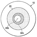
도 1a는 고도로 확장되지 않은 눈의 동공 중심의 개략도이다.
도 1b는 고도로 확장된 눈의 동공 중심의 개략도이다.
도 1c는, 본 개시 내용의 실시예에 따른, 도 1a 및 도 1b의 눈의 동공 중심을 나타내기 위한 마크의 개략도이다.
도 1d는, 본 개시 내용의 실시예에 따른, 눈의 각막 마루, 그리고 각막 마루 및 도 1a의 눈의 동공 중심을 나타내기 위한 마크의 개략도이다.
도 2는 본 개시 내용의 실시예에 따른, 시력 교정 수술을 실시하기 위한 시스템의 개략도이다.BRIEF DESCRIPTION OF THE DRAWINGS For a more complete understanding of the present invention and the features and advantages thereof, reference is now made to the following description taken in conjunction with the accompanying drawings, in which:
Figure 1a is a schematic view of the pupil center of a highly unexpanded eye.
FIG. 1B is a schematic view of the pupil center of the highly extended eye.
1C is a schematic view of a mark for indicating the pupil center of the eye of FIGS. 1A and 1B, according to an embodiment of the present disclosure; FIG.
1D is a schematic view of a corneal fl oor of the eye and a mark for indicating the corneal fl oor and the pupil center of the eye of Fig. 1A, according to an embodiment of the present disclosure;
2 is a schematic diagram of a system for performing a vision correction operation, in accordance with an embodiment of the present disclosure;
본 개시 내용은 눈의 각막을 마크로 마킹하기 위한 시스템 및 방법에 관한 것이다. 마크는, 각막이 변형되었을 때에도, 주어진 확대에서의 동공의 중심 또는 각막 마루와 같은 각막 상의 선택된 위치의 추후 검출을 가능하게 한다. 개시 내용은 또한 마크를 이용하여 시력 교정 수술을 실시하기 위한 시스템 및 방법에 관한 것이다. 각막은 그러한 수술 중에 변형되나, 마크는 변형에도 불구하고 선택된 위치에서 수술이 실시될 수 있게 하고, 이는 전형적으로, 각막이 미변형 형상으로 복귀되었을 때, 시력 교정을 개선한다. 마크는 추가적으로, 선택된 위치가 주어진 확대에서의 동공 중심 또는 각막 마루에 위치되게 할 수 있다. 동공 중심과 대조적으로 각막 마루에서 또는 다른 것과 대조적으로 하나의 확대에서 동공 중심에서 수술적 시술을 실시하는 것이 상이한 시력 교정을 제공할 수 있다.The present disclosure relates to systems and methods for macromarking an eye's cornea. The mark allows further detection of the selected position of the corneal surface, such as the center of the pupil or corneal fl oor at a given enlargement, even when the cornea is deformed. The disclosure also relates to a system and method for performing a vision correction surgery using a mark. The cornea is deformed during such surgery, but the mark permits the operation to be performed at the selected location despite the deformation, which typically improves vision correction when the cornea is returned to the undeformed shape. The mark may additionally cause the selected position to be located in the pupil center or corneal fl oor at a given enlargement. Performing surgical procedures at the center of the pupil in one magnification, as opposed to the pupil center, in contrast to the corneal floor or otherwise, can provide different visual acuity adjustments.
이하의 설명에서, 개시된 청구 대상의 설명을 돕기 위해서 예로서 상세 내용을 기술한다. 그러나, 개시된 실시예가 예시적인 것이고 모든 가능한 실시예를 전부 망라한 것이 아니라는 것이 당업자에게 자명할 것이다.In the following description, details are set forth by way of example in order to facilitate describing the disclosed subject matter. It will be apparent, however, to one skilled in the art that the disclosed embodiments are illustrative and not exhaustive of all possible embodiments.
시력 교정 수술은 종종 수술 중에 어떠한 확대가 발생되든지 간에 동공 중심에서 실시된다. 만약 동공이 항상 완전히 대칭적이라면, 동공 중심은 확대와 관계없이 동일하게 유지될 것이다. 그러나, 완전히 대칭적인 동공은 적고, 또한, 비대칭성은 확대에 따라 달라진다. 결과적으로, 동공 중심은 또한 확대에 따라 달라진다. 이는 도 1a 및 도 1b에 도시된 각막(20) 및 동공(30)을 가지는 눈(10)에 관한 도면을 참조함으로써 이해될 수 있다. 도 1a의 동공(30a)은 고도로 확대되지 않았고 동공 중심(40a)을 갖는다. 도 1b의 동공(30b)은 고도로 확대되었고, 동공 중심(40a)과 동일한 위치가 아닌, 동공 중심(40b)을 갖는다. 도 1c에 도시된 바와 같이, 미확대 동공 중심(40a)을 나타내기 위한 각막(20)의 각막 주변부 상의 마크(50a)는 그에 따라 고도로 확대된 동공 중심(40b)을 나타내기 위한 각막(20)의 각막 주변부 상에 만들어진 마크(50b)와 상이한 위치에 있다.Sight correction surgery is often performed at the center of the pupil, regardless of any enlargement during surgery. If the pupil is always perfectly symmetrical, the pupil center will remain the same regardless of magnification. However, the number of completely symmetric pupils is small, and the asymmetry depends on the magnification. As a result, the pupil center also varies with magnification. This can be understood by referring to the drawings relating to the
일부 시력 교정 수술은 각막 마루에서 수술적 시술을 실시하는 것을 목적으로 한다. 각막 마루는 고정점의 제1 푸르키네(Purkinje) 화상과 영상지형도 촬영장치(videokeratographer)와 같은 광학적 검사 기구의 광학 축의 교차부이다. 각막 마루는 각막의 중앙 부분이고, 그러한 곳에서 중앙 부분은 일반적으로 각막 주변부 보다 얇다. 각막 마루는 각막 주변부에 의해서 둘러싸이고, 그러한 각막 주변부는 또한 일반적으로 원형이고 각막의 연부까지 연장된다. 각막 마루는 동공 중심과 같은 위치에 있을 수 있으나, 또한 종종 그렇지 않을 수 있다. 도 1d에 도시된 바와 같이, 각막 마루(60)는 동공 중심(40a)과 같은 임의의 동공 중심과 상이한 위치 내에 있을 수 있고, 그에 따라 각막 마루(60)를 나타내기 위해서 각막(20)의 주변 영역 상에 만들어진 마크(50c)는 동공 중심(40a)을 나타내기 위해서 만들어진 마크(50a)와 동일한 위치에 있지 않다. Some eyesight correction surgery aims at performing surgical operation on corneal floor. The corneal floor is the intersection of the optical axis of the optical inspection instrument, such as the first Purkinje image of the anchor point and the videokeratographer. The corneal fl oor is the central part of the cornea, where the central part is generally thinner than the cornea's periphery. The corneal fl ow is surrounded by the cornea periphery, and the cornea periphery is also generally circular and extends to the cornea's edge. The corneal flats may be in the same position as the pupil center, but may also often not. 1D, the
다른 시력 교정 수술은 각막 마루 또는 주어진 확대에서의 동공 중심 이외의 위치에서 수술을 실시한다. 예를 들어, 일부 수술은 각막 마루와 주어진 확대에서의 동공 중심을 연결하는 가상의 선을 따라서 실시될 수 있다.Other vision correction operations are performed at corneal topography or at locations other than the pupil center at a given enlargement. For example, some surgeries may be performed along virtual lines connecting the corneal floor and the center of the pupil at a given enlargement.
따라서, 개시 내용은 시력 교정 수술을 위한 준비에서 마크로 각막을 마킹하는 방법을 포함한다. 첫 번째로, 수술적 시술을 위한 위치가 선택된다. 선택된 위치는 각막 마루 또는 주어진 확대에서의 동공 중심일 수 있다. 위치는, 실시하고자 하는 시력 교정 수술의 유형, 특히 각막 마루에서의 각막의 두께, 하나 이상의 확대에서 동공 중심에 대한 각막 마루의 위치, 및 상이한 확대들에서의 동공 중심의 변동과 같은, 다양한 기술적 인자를 기초로 선택될 수 있다. 그러한 위치는 또한, 고조도 또는 저조도(bright or low light)에서의 최적의 시력이 가장 바람직한 것인지의 여부와 같은, 환자 선호 인자를 기초로 선택될 수 있다. Accordingly, the disclosure includes a method of marking a cornea with a mark in preparation for a vision correction surgery. First, the location for surgical procedure is selected. The selected position may be the corneal fl oor or the pupil center at a given enlargement. The position may be determined by various technical factors such as the type of vision correction surgery to be performed, in particular the thickness of the cornea on the corneal flap, the location of the corneal fl oor to the center of the pupil in one or more enlargements, As shown in FIG. Such a position may also be selected based on a patient preference factor, such as whether high vision or optimal visual acuity at low or low light is most desirable.
일단 위치가 선택되면, 위치는 눈 상에서 실제로 식별된다. 첫 번째로, 필요한 경우에, 예를 들어 동공을 희망 확대로 확대시킴으로써, 눈이 준비된다. 다음에, 안과용 장치를 이용하여 선택된 위치를 식별한다. 수동형 및 컴퓨터-지원형 장치 모두, 그리고 라식, 스마일, 및 백내장 수술과 같은 시력 교정을 위한 진단적 또는 수술적 시술에서 현재 이용되는 장치를 포함하여, 이러한 기능을 실시할 수 있는 임의의 안과용 장치가 이용될 수 있다. 장치는 현미경 또는 다른 확대 렌즈를 포함할 수 있다. 만약 장치가 컴퓨터-지원형이라면, 장치는, 디지털 화상을 생성하기 위해서 눈으로부터 반사되는 광을 캡쳐하는, 광다이오드 어레이와 같은, 디지털 화상 센서를 포함할 수 있다. 컴퓨터-지원형 장치는 또한, 동공 중심 또는 각막 마루를 위치결정하는 알고리즘으로 프로그래밍된 프로세싱 자원을 포함할 수 있다. 예를 들어, 알고리즘은, 색채 검출 또는 원 검출 알고리즘을 이용하여 동공이 홍체에 의해서 경계 지어지는 곳을 식별할 수 있다. 알고리즘은 이어서 하나 이상의 타원 또는 원으로 홍체를 어림잡을 수 있고(approximate), 해당 확대에서의 동공 중심으로서 식별되는 그러한 타원 또는 원의 중심을 위치결정할 수 있다. 각막 마루를 검출할 때, 컴퓨터-지원형 장치는 가시광선, 적외선, 또는 다른 광 그리고 광을 검출하기 위한 센서를 이용할 수 있다. 장치는, 광의 레벨 또는 변화를 각막 마루에 대한 동공 중심 변화로 변환하기 위한 그리고, 추후의 수술적 시술을 위해서, 의사의 입력 또는 다른 입력에 따라 위치를 선택하거나 선택된 위치를 마킹하기 위한 알고리즘으로 프로그래밍될 수 있다.Once the location is selected, the location is actually identified in the eye. First, if necessary, the eye is prepared, for example by enlarging the pupil to the desired magnification. The ophthalmic device is then used to identify the selected location. Any ophthalmic device capable of performing such functions, including devices currently used in passive and computer-assisted devices and diagnostic or surgical procedures for vision correction, such as LASIK, smileys, and cataract surgeries Can be used. The device may comprise a microscope or other magnifying lens. If the device is a computer-aided device, the device may include a digital image sensor, such as a photodiode array, that captures light reflected from the eye to produce a digital image. The computer-aided device may also include processing resources programmed with an algorithm that locates the pupil center or corneal floor. For example, an algorithm can identify where a pupil is bounded by an iris using color detection or a circle detection algorithm. The algorithm can then approximate the iris with one or more ellipses or circles and locate the center of such ellipse or circle identified as the pupil center in the enlargement. When detecting corneal topography, the computer-aided device may use a sensor to detect visible light, infrared light, or other light and light. The apparatus may be programmed with an algorithm for converting a level or change of light into a pupil center change for the corneal flap and for selecting a position or marking a selected position according to a physician's input or other input for further surgical procedures. .
전술한 시술 및 장치 중 임의의 것을 이용할 때, 선택된 위치는 점일 수 있거나, 규정된 직경을 가지는 원과 같은, 규정된 형상 및 크기의 지역일 수 있다.When using any of the above procedures and devices, the selected location may be a point, or it may be a region of defined shape and size, such as a circle with a defined diameter.
선택된 위치를 식별한 후에, 선택된 위치를 추후에 재-식별하기 위해서 이용될 수 있는 각막 주변부 상에 마크가 만들어진다. 마크는 각막 주변부 상의 어디든 만들어질 수 있으나, 더 얇고 광이 통과될 필요가 있을 수 있는 각막의 내부 부분을 피하기 위해서, 가까운 각막 마루로부터 적어도 8 mm에 전형적으로 위치될 것이다. 마크는 또한, 마크가 공막에 부착되는 각막의 외부 영역 또는 공막 자체를 손상시키는 것을 방지하기 위해서, 가까운 각막 마루로부터 11 mm 이하에 위치될 수 있다.After identifying the selected position, a mark is made on the corneal periphery that may be used to re-identify the selected position in the future. The mark may be made anywhere on the corneal periphery, but will typically be located at least 8 mm from the near corneal flats to avoid the interior portion of the cornea, which may be thinner and may require light to pass through. The mark may also be located 11 mm or less from the near corneal flats to prevent the marks from damaging the outer area of the cornea or the sclera itself attached to the sclera.
마크는, 각막이 변형되는 경우에도 선택된 위치의 추후 재-식별을 가능하게 하기에 충분한 임의 형상 또는 크기를 가질 수 있다. 예를 들어, 마크는, 선택된 위치가 중심에 있는 원, 또는 선이나 점과 같은 복수의 부-마크일 수 있다. 전형적으로, 마크가 복수의 부-마크인 경우에, 선택된 위치의 삼각측량을 가능하게 하기 위해서 적어도 3개의 그러한 부-마크가 존재할 것이다.The mark may have any shape or size sufficient to enable subsequent re-identification of the selected position even when the cornea is deformed. For example, the mark may be a circle with the selected location in the center, or a plurality of sub-marks such as lines or points. Typically, where the mark is a plurality of sub-marks, there will be at least three such sub-marks to enable triangulation of the selected location.
마크는 육안으로 또는 시력 교정 수술에서 이용되는 배율로 볼 수 있거나, 컴퓨터 지원으로만 검출될 수도 있다.Marks can be viewed either visually or at the magnification used in vision correction surgery, or may be detected only by computer support.
마크는 각막의 표면 상에 있을 수 있거나, 마크가 각막을 침투할 수도 있다. 어느 경우에도, 마크는 수술 시까지 또는 수술 중에 필요로 하는 한 지속되도록 설계되나, 마크는 또한 추후에 사라지거나 치유되도록 설계될 수 있다. 예를 들어, 마크는 생체적합 염료를 포함할 수 있거나 단순히 작은 절개부일 수 있다.The mark may be on the surface of the cornea, or the mark may penetrate the cornea. In either case, the mark is designed to last as long as needed during surgery or during surgery, but the mark may also be designed to disappear or be cured at a later time. For example, the mark may comprise a biocompatible dye or may simply be a small incision.
마크는, 링 마커, 레이저, 또는 작은 바늘, 또는 메스(scalpel)와 같은, 임의의 적합한 기구를 이용하여 만들어질 수 있다.The mark may be made using any suitable mechanism, such as a ring marker, a laser, or a small needle, or a scalpel.
마크는 링 마커를 이용하는 의사와 같은 의사에 의해서 손으로 만들어질 수 있거나, 마크는 컴퓨터-지원형 장치를 이용하는 의사에 의해서 또는 컴퓨터-지원형 장치 자체에 의해서 만들어질 수 있다. 선택된 위치를 식별하는 것 그리고 마크를 만드는 것 모두를 위해서 컴퓨터-지원형 장치를 이용하는 것은 더 정확한 마크 배치를 유도할 수 있다.The mark may be made by hand by a physician such as a physician using a ring marker, or the mark may be made by a physician using a computer-assisted device or by a computer-assisted device itself. Using a computer-aided device for both identifying the selected location and making marks can lead to more accurate mark placement.
마크가 만들어진 후에, 의사는 시력 교정 수술을 실시한다. 수술 중에, 각막이 변형되고, 그에 따라, 동공 중심 또는 각막 마루가 수술 중에 위치결정되는 경우에, 그러한 위치 결정은 미변형 각막 내의 위치를 정확하게 반영하지 못할 수 있다. 예를 들어, 적어도 하나의 각막 흡입 또는 압평화 장치가 수술 중에 각막에 적용되어, 각막을 변형시킬 수 있다.After the mark is made, the doctor performs a vision correction operation. During surgery, when the cornea is deformed, and thus the pupil center or corneal flap is positioned during surgery, such positioning may not accurately reflect the position within the unmodified cornea. For example, at least one corneal suction or compression device may be applied to the cornea during surgery to deform the cornea.
각막 변형에도 불구하고, 의사는 선택된 위치를 재-식별하기 위해서 그리고 해당 위치에서 수술적 시술을 실시하기 위해서 마크를 이용한다. 의사는 육안으로 또는 수술 현미경을 이용하여 마크를 위치결정할 수 있고 선택된 위치를 마음속으로 결정할 수 있다. 그러나, 전형적으로, 의사가 마크를 위치결정하고 선택된 위치를 결정하기 위해서 컴퓨터-지원형 장치를 이용하는 것이 더 정확하다. 예를 들어, 컴퓨터-지원형 장치는, 디지털 화상을 생성하기 위해서 눈으로부터 반사되는 광을 캡쳐하는, 광다이오드 어레이와 같은, 디지털 화상 센서를 포함할 수 있다. 컴퓨터-지원형 장치는, 마크를 위치결정하기 위해서 디지털 화상을 분석하고 추가적으로 마크를 이용하여 선택된 위치를 재-식별하는 알고리즘으로 프로그래밍될 수 있다. 이어서, 컴퓨터-지원형 장치는, 예를 들어 시각적 디스플레이 상에 정보를 중첩시키는 것에 의해서 또는 기구가 선택된 위치에 해당하는 제 위치에 있을 때를 의사에게 알려주기 위한 음향 또는 다른 표시자를 이용하는 것에 의해서, 선택된 위치에 관한 정보를 의사에서 전달할 수 있다. 컴퓨터-지원형 장치는, 그 자체적으로 또는 다른 컴퓨터-지원형 장치와의 통신을 통해서, 레이저, 겸자, 또는 메스와 같은 수술적 시술을 실시하기 위한 수술 장비를 선택된 위치에 또한 배치할 수 있다. 컴퓨터-지원형 장치는 선택된 위치에서 수술적 시술을 실시할 수도 있다.Despite corneal deformation, the surgeon uses the mark to re-identify the selected location and to perform the surgical procedure at that location. The surgeon can position the mark with the naked eye or with a surgical microscope and decide in mind the chosen location. Typically, however, it is more accurate for the physician to use the computer-assisted device to locate the mark and determine the selected location. For example, the computer-aided device may include a digital image sensor, such as a photodiode array, that captures light reflected from the eye to produce a digital image. The computer-aided device may be programmed with an algorithm that analyzes the digital image to locate the mark and additionally re-identifies the selected location using the mark. The computer-aided device can then be used to display the information on the visual display, for example, by using acoustic or other indicators to inform the physician when the information is superimposed on the visual display, Information about the selected location can be communicated by the physician. The computer-aided device may also place surgical instruments for performing surgical procedures, such as lasers, forceps, or scalpels, at selected locations, either on its own or through communication with other computer-aided devices. The computer-aided device may perform surgical procedures at selected locations.
수술용 현미경 또는 컴퓨터-지원형 장치가 각막을 마킹하는 것 그리고 선택된 위치를 재-식별하는 것 모두를 위해서 이용된다면, 그러한 장치는 양 시술에서 이용될 수 있다. 그러한 장치는 또한 각막을 자동적으로 마킹하기 위해서, 수술적 시술을 실시하기 위해서, 또는 그 모두를 위해서 이용될 수 있다. 단일 장치의 이용은 감염 위험 또는 환자가 움직이는 경우에 발생될 수 있는 신체 상해 위험을 줄일 수 있다. 이는 또한 전체 시술이 짧아지게 할 수 있다.If a surgical microscope or computer-aided device is used for both marking the cornea and re-identifying the selected location, such a device may be used in both procedures. Such an apparatus may also be used for automatically marking the cornea, for performing surgical procedures, or both. The use of a single device can reduce the risk of infection or the risk of bodily injury that may occur if the patient is moving. This can also shorten the overall procedure.
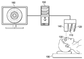
전술한 다른 특징 중 임의의 특징과 조합될 수 있는 예시적인 실시예에서, 라식 또는 스마일 수술을 위한 시스템 및 방법이 제공된다. 도 2에 도시된 바와 같은 라식 또는 스마일 시스템은 플랫폼(100)을 포함하고, 수술 중에 환자가 그러한 플랫폼 상에 놓인다. 그러한 시스템은 펨토초 레이저(110), (라식 시스템을 위한) 엑시머 레이저(120), 압평화 원뿔과 같은 압평화 장치를 포함할 수 있는 각막 흡입 장치(130), 수술용 디지털 현미경(140), 컴퓨터(150), 및 디스플레이(160)를 더 포함한다.In an exemplary embodiment, which may be combined with any of the other features described above, a system and method for LASIK or Smiley surgery is provided. A LASIK or SMILE system as shown in FIG. 2 includes a
라식 또는 스마일 수술을 실시하기 위해서, 환자의 눈이 수술용 현미경(140) 아래에 위치되는 상태로 환자가 플랫폼(100) 상에 배치된다. 수술용 디지털 현미경(140)은 확대 렌즈 및 센서를 포함하고, 그러한 확대 렌즈 및 센서는 함께 눈의 확대된 디지털 화상을 디스플레이(160) 상에 생성한다. 환자의 눈은 선택된 확대까지 미리 확대되었거나, 이어서 선택된 확대까지 확대되고, 어느 경우든 간에 의사가 시각적으로 확인한다. 이어서, 의사는, 선택된 위치인, 동공 중심 또는 각막 마루를 위치결정하기 위한 알고리즘을 실행하도록 컴퓨터(150)에 명령한다. 선택된 위치와 제시된 마크 위치의 시각적 중첩이 디스플레이(160) 상의 눈의 화상 상에 디스플레이된다. 의사는, 선택된 위치가 정확한 것으로 보이는지를 확인한다. 다음에, 펨토초 레이저(110)를 활성화시켜, 적어도 3개를 포함하는, 복수의 부-마크를 마크 위치에서 생성한다. 이러한 부-마크는 각막의 중심으로부터 약 8 mm 내지 11 mm인 각막 주변부 내의 작은 절개부이다. 부-마크는 의사에 의해서 시각적으로 검출될 수 없으나, 이러한 부-마크에서 각막으로부터 반사되는 광의 변화를 검출하는 알고리즘을 이용하여 컴퓨터에 의해서 검출될 수 있다. 다음에, 컴퓨터(150)는 부-마크를 검출하기 위한 눈의 디지털 화상에 대한 다른 알고리즘, 및 부-마크를 기초로 선택된 위치를 재-식별하기 위한 알고리즘을 작동시킨다. 컴퓨터(150)는, 재-식별된 선택된 위치가 선택된 위치에 상응하는지를 확인한다. 만약 상응하지 않는다면, 의사는, 마크를 마킹하는 동안 오류가 발생하였다는 것을 통지 받고, 부-마크가 치유될 때까지 시술이 연기될 수 있거나, 새로운 부-마크가 다른 위치에서 만들어질 수 있다. 만약 재-식별된 선택된 위치가 선택된 위치에 상응한다면, 그 두 위치의 시각적 중첩이 디스플레이(160) 상의 눈의 화상 상에서 디스플레이되고, 그에 따라 상응성이 의사에 의해서 시각적으로 확인될 수 있다.In order to perform LASIK or smiley surgery, the patient is placed on the
다음에, 각막 흡입 장치(130)가 눈에 적용된다. 이러한 장치는 눈을 제 위치에서 유지하나, 그 장치는 또한 각막을 변형시킨다. 전형적으로, 장치는 각막의 압평화를 유발한다. 컴퓨터(150)는 부-마크를 검출하기 위한 눈의 디지털 화상에 대한 알고리즘, 및 부-마크를 기초로 선택된 위치를 재-식별하기 위한 알고리즘을 작동시킨다. 재-식별된 선택된 위치의 시각적 중첩이 디스플레이(160) 상의 눈의 화상 상에서 디스플레이되고, 그에 따라 의사는 재-식별된 선택된 위치가 정확하게 보이는지를 확인할 수 있다.Next, the
만약 시술이 라식 시술이라면, 의사는, 선택된 위치 상에서 대략적으로 중심에 위치되는 각막 내에 플랩(flap)을 형성하기 위해서 펨토초 레이저(110)를 이용하도록 컴퓨터(150)에 지시한다. 이어서, 의사는 이러한 플랩을 상승시키고, 수술적 시술을 실시하고 선택된 위치 내의 각막 조직을 절삭하기 위해서 엑시머 레이저(120)를 이용하도록 컴퓨터(150)에 지시한다. 이러한 절삭은 표준 절삭, 파면-센서-안내형 절삭(wavefront-sensor-guided ablation), 또는 다른 형태의 컴퓨터-지원형 절삭일 수 있다. 절삭 이후에, 컴퓨터(150)는, 필요한 경우에, 마크를 이용하여 재-식별될 수 있는, 선택된 위치 내에서 절삭이 이루어졌는지의 여부를 포함하여, 절삭이 적절히 실행되었는지의 여부를 검출하기 위한 다른 알고리즘을 작동시킬 수 있다.If the procedure is a LASIK procedure, the physician instructs the
임의의 세척 및 시술이 정확하게 실시되었다는 것에 대한 확인 이후에, 플랩이 하강될 수 있고 각막 흡입 장치(130)가 제거될 수 있다. 이어서, 컴퓨터(150)는 마크를 이용하여 미변형된 눈 상의 선택된 영역을 재-식별하기 위한 그리고 절삭이 선택된 영역 내에서 이루어졌다는 것을 확인하기 위한 다른 알고리즘을 작동시킬 수 있다.After confirming that any cleaning and procedure has been performed correctly, the flap can be lowered and the
만약 시술이 스마일 시술이라면, 의사는 제거하기 위한 각막내 렌티큘을 규정하기 위해서 펨토초 레이저(110)를 이용하도록 컴퓨터(150)에 지시한다. 렌티큘의 규정 이후에, 컴퓨터(150)는, 필요한 경우에, 마크를 이용하여 재-식별될 수 있는, 선택된 위치 내에서 렌티큘이 형성되었는지의 여부를 검출하는 것을 포함하여, 렌티큘이 적절하게 형성되었는지를 검출하기 위한 다른 알고리즘을 작동시킬 수 있다. 만약 렌티큘이 적절하게 형성되었다면, 이어서 펨토초 레이저(110)를 이용하여 렌티큘의 연부에 작은(전형적으로 2 mm 이하) 절개부를 형성하고, 이어서 의사는 작은 절개부를 통해서 각막으로부터 렌티큘을 제거한다. 렌티큘의 제거 이후에, 컴퓨터(150)는, 마크를 이용하여 선택된 위치를 재-식별하는 것을 포함할 수 있는, 렌티큘이 적절한 위치로부터 적절하게 제거되었는지를 검출하기 위한 다른 알고리즘을 작동시킬 수 있다.If the procedure is a smile procedure, the physician instructs the
시술이 정확하게 실시되었다는 임의의 다른 확인 이후에, 각막 흡입 장치(130)가 제거될 수 있다. 이어서, 컴퓨터(150)는 마크를 이용하여 미변형된 눈 상의 선택된 영역을 재-식별하기 위한 그리고 레티큘이 선택된 영역으로부터 제거되었다는 것을 확인하기 위한 다른 알고리즘을 작동시킬 수 있다.After any other confirmation that the procedure has been performed correctly, the
컴퓨터(150) 및 본원에서 설명된 임의의 다른 컴퓨터는 표시된 알고리즘을 작동시킬 수 있는 적어도 하나의 프로세싱 자원을 포함할 수 있다. 또한, 컴퓨터는 메모리, 및 안과용 검출기 그리고 선택적으로 디스플레이 및 레이저와 같은 다른 시스템 구성요소와 통신하기 위한 통신 모듈을 포함할 수 있다.
예를 들어 마크를 만들기 위해서 염료 주입기 또는 컴퓨터-제어형 링 마커를 이용하는, 전술한 시술의 변형이 본원의 기술을 이용하여 당업자에 의해서 구상될 수 있다. 또한, 전술한 시술을, 백내장 수술과 같은, 다른 시력 교정 수술과 조합하는 방법이 또한 본원의 기술을 이용하여 당업자에 의해서 구상될 수 있다.Variations of the above-described procedures, for example using dye injectors or computer-controlled ring markers to create marks, can be envisioned by those skilled in the art using the techniques herein. Also, a method of combining the above described procedures with other vision correction procedures, such as cataract surgery, can also be envisioned by those skilled in the art using the techniques herein.
앞서서 개시된 청구 대상은 예시적인 것으로 간주되어야 하고, 제한적인 것이 아니며, 첨부된 청구항은, 본 개시 내용의 진정한 사상 및 범위 내에 포함되는 그러한 변형예, 향상예, 및 다른 실시예 모두를 포함하기 위한 것이다. 따라서, 법에 의해서 허용되는 최대 범위까지, 본 개시 내용의 범위는 이하의 청구항 및 그 균등물의 가장 넓은 허용 가능한 해석에 의해서 결정되어야 하며, 전술한 구체적인 설명에 의해서 제한되거나 한정되지 않을 것이다.The claims set forth above are to be regarded as illustrative and not restrictive, and the appended claims are intended to cover all such modifications, enhancements, and other embodiments that fall within the true spirit and scope of the present disclosure . Accordingly, to the fullest extent permitted by law, the scope of the present disclosure should be determined by the broadest permissible interpretation of the following claims and their equivalents, and shall not be limited or limited by the foregoing specific description.
Claims (27)
눈의 각막 상의 선택된 위치를 식별하는 단계;
상기 선택된 위치에 상응하는 마크로 상기 각막의 주변부를 마킹하는 단계;
상기 각막을 변형시키는 단계;
상기 마크를 기초로 상기 변형된 각막 상의 상기 선택된 위치를 재-식별하는 단계; 및
상기 각막이 변형되어 있는 동안 상기 선택된 위치에서 시력 교정 수술적 시술을 실시하는 단계를 포함하는, 방법.As a method of performing vision correction surgery:
Identifying a selected location on the cornea of the eye;
Marking a periphery of the cornea with a mark corresponding to the selected position;
Deforming the cornea;
Re-identifying the selected position of the deformed cornea on the basis of the mark; And
And performing a vision correction surgical procedure at the selected location while the cornea is deformed.
상기 선택된 위치는 상기 눈의 동공 중심인, 방법.The method according to claim 1,
Wherein the selected location is the pupil center of the eye.
상기 동공 중심은 확대된 동공의 동공 중심인, 방법.3. The method of claim 2,
Wherein the pupil center is the pupil center of the enlarged pupil.
상기 동공 중심은 미확대 동공의 동공 중심인, 방법.3. The method of claim 2,
Wherein the pupil center is the pupil center of the unexpanded pupil.
상기 선택된 위치는 상기 수술적 시술 중에서와 달리 확대된 눈의 동공 중심인, 방법.3. The method of claim 2,
Wherein the selected location is the pupil center of the enlarged eye, unlike in the surgical procedure.
상기 선택된 위치는 각막 마루인, 방법.The method according to claim 1,
Wherein the selected position is a corneal flap.
상기 선택된 위치는 상기 각막 마루와 주어진 확대에서의 동공 중심을 연결하는 가상의 선을 따르는, 방법.The method according to claim 1,
Wherein the selected location follows a virtual line connecting the corneal fl oor with the pupil center at a given enlargement.
상기 마크는 원을 포함하는, 방법.The method according to claim 1,
Wherein the mark comprises a circle.
상기 마크는, 적어도 3개의 부-마크를 포함하는 복수의 부-마크를 포함하는, 방법.The method according to claim 1,
Wherein the mark comprises a plurality of sub-marks comprising at least three sub-marks.
상기 마크는 가까운 각막 마루로부터 8 mm 및 11 mm 사이에 만들어지는, 방법.The method according to claim 1,
Wherein the mark is made between 8 mm and 11 mm from the near corneal flats.
상기 마크는 펨토초 레이저를 이용하여 만들어지는, 방법.The method according to claim 1,
Wherein the mark is made using a femtosecond laser.
상기 마크는 링 마커를 이용하여 만들어지는, 방법.The method according to claim 1,
Wherein the mark is made using a ring marker.
상기 마크는 생체적합 염료를 포함하는, 방법.The method according to claim 1,
Wherein the mark comprises a biocompatible dye.
상기 각막을 변형시키는 단계는 상기 각막을 압평화하는 단계를 포함하는, 방법.The method according to claim 1,
Wherein modifying the cornea comprises compressing the cornea.
상기 마크를 기초로 변형된 각막 상의 선택된 위치를 재-식별하는 단계는 상기 마크를 위치결정하는 단계, 이어서 상기 마크와 관련하여 상기 선택된 위치를 결정하는 단계를 포함하는, 방법.The method according to claim 1,
Wherein re-identifying a selected position of the deformed cornea image based on the mark comprises positioning the mark, and then determining the selected position in relation to the mark.
상기 시력 교정 수술적 시술은 상기 선택된 위치 내에서 상기 각막의 일부를 레이저 절삭하는 단계를 포함하는, 방법.The method according to claim 1,
Wherein the surgical correction procedure includes laser cutting a portion of the cornea within the selected position.
상기 시력 교정 수술적 시술은 상기 선택된 위치 내에서 상기 각막의 렌티큘을 제거하는 단계를 포함하는, 방법.The method according to claim 1,
Wherein the surgical correction procedure comprises removing the lenticular of the cornea within the selected position.
눈의 각막 상의 선택된 위치를 식별하도록 동작될 수 있는 안과용 검출기;
상기 선택된 위치에 상응하는 마크로 상기 각막의 주변부를 마킹하도록 동작될 수 있는 마커;
상기 각막을 변형하도록 동작될 수 있는 각막 흡입 또는 압평화 장치;
상기 각막이 변형되어 있는 동안 상기 선택된 위치에서 시력 교정 수술적 시술을 실시하도록 동작될 수 있는 수술적 시술 장치; 및
상기 안과용 검출기에 의해서 제공된 정보를 이용하여, 적어도, 상기 선택된 위치를 식별하기 위한 알고리즘 및 상기 마크를 기초로 상기 선택된 위치를 재-식별하기 위한 알고리즘으로 프로그래밍된 컴퓨터를 포함하는, 시스템.As a system for vision correction surgery:
An ophthalmologic detector operable to identify a selected location on the cornea of the eye;
A marker operable to mark a periphery of the cornea with a mark corresponding to the selected position;
A cornea suction or pressure relief device operable to deform the cornea;
A surgical procedure device operable to perform a vision correction surgical procedure at the selected location while the cornea is deformed; And
A computer programmed with at least an algorithm for identifying the selected location and an algorithm for re-identifying the selected location based on the mark, using information provided by the ophthalmologic detector.
상기 마커 및 상기 수술적 시술 장치가 동일한 장치인, 시스템.19. The method of claim 18,
Wherein the marker and the surgical treatment device are the same device.
상기 마커 및 상기 수술적 시술 장치가 펨토초 레이저를 포함하는, 시스템.20. The method according to claim 18 or 19,
Wherein the marker and the surgical treatment device comprise a femtosecond laser.
상기 선택된 위치는 눈의 확대된 동공 또는 미확대 동공의 동공 중심인, 시스템.21. The method according to any one of claims 18 to 20,
Wherein the selected location is the pupil center of the enlarged pupil or unexpanded pupil of the eye.
상기 선택된 위치는 상기 수술적 시술 중에서와 달리 확대된 눈의 동공 중심인, 시스템.22. The method of claim 21,
Wherein the selected location is the pupil center of the enlarged eye, unlike in the surgical procedure.
상기 선택된 위치는 상기 각막 마루이거나, 상기 각막 마루와 주어진 확대에서의 동공 중심을 연결하는 가상의 선을 따르는, 시스템.21. The method according to any one of claims 18 to 20,
Wherein the selected position is the corneal fl oor or along a hypothetical line connecting the corneal fl oor with the pupil center at a given enlargement.
상기 마크는: 원; 적어도 3개의 부-마크를 포함하는 복수의 부-마크; 생체적합 염료 중 하나 이상을 포함하는, 시스템.24. The method according to any one of claims 18 to 23,
The mark is: circle; A plurality of sub-marks comprising at least three sub-marks; ≪ / RTI > or a biologically acceptable dye.
상기 마크는 상기 가까운 각막 마루로부터 8 mm 내지 11 mm에서 만들어지고 및/또는 펨토초 레이저를 이용하여 만들어지며 및/또는 링 마커를 이용하여 만들어지는, 시스템.25. The method according to any one of claims 18 to 24,
Wherein the mark is made from 8 mm to 11 mm from the near corneal flats and / or is made using a femtosecond laser and / or is made using a ring marker.
상기 알고리즘은 상기 마크를 위치결정하는 것, 이어서 상기 마크에 대해서 상기 선택된 위치를 결정하는 것에 의해서 상기 선택된 위치를 재-식별하도록 구성되는, 시스템.26. The method according to any one of claims 18 to 25,
Wherein the algorithm is configured to re-identify the selected location by locating the mark and then determining the selected location for the mark.
상기 시력 교정 수술적 시술은 상기 선택된 위치 내에서 상기 각막의 일부를 레이저 절삭하는 것 및/또는 상기 선택된 위치에서 각막 레티큘을 제거하는 것을 포함하는, 시스템.27. The method according to any one of claims 18 to 26,
Wherein the vision correction surgical procedure comprises laser cutting a portion of the cornea within the selected position and / or removing the corneal reticle at the selected position.
Applications Claiming Priority (1)
| Application Number | Priority Date | Filing Date | Title |
|---|---|---|---|
| PCT/EP2015/068271 WO2017025115A1 (en) | 2015-08-07 | 2015-08-07 | Corneal marks in vision correction surgery |
Publications (1)
| Publication Number | Publication Date |
|---|---|
| KR20180039017A true KR20180039017A (en) | 2018-04-17 |
Family
ID=53835427
Family Applications (1)
| Application Number | Title | Priority Date | Filing Date |
|---|---|---|---|
| KR1020177035364A KR20180039017A (en) | 2015-08-07 | 2015-08-07 | Corneal Mark in Sight Correction Surgery |
Country Status (13)
| Country | Link |
|---|---|
| US (1) | US10835420B2 (en) |
| EP (1) | EP3145459B1 (en) |
| JP (1) | JP6697542B2 (en) |
| KR (1) | KR20180039017A (en) |
| CN (1) | CN107809985B (en) |
| AU (1) | AU2015405457A1 (en) |
| CA (1) | CA2986179A1 (en) |
| DK (1) | DK3145459T3 (en) |
| ES (1) | ES2715450T3 (en) |
| PL (1) | PL3145459T3 (en) |
| PT (1) | PT3145459T (en) |
| TW (1) | TWI608833B (en) |
| WO (1) | WO2017025115A1 (en) |
Families Citing this family (1)
| Publication number | Priority date | Publication date | Assignee | Title |
|---|---|---|---|---|
| CN108335757B (en) * | 2018-02-05 | 2021-05-25 | 王雁 | Method for predicting diopter adjustment value in SMILE operation |
Family Cites Families (9)
| Publication number | Priority date | Publication date | Assignee | Title |
|---|---|---|---|---|
| ES2368450T3 (en) * | 2007-04-25 | 2011-11-17 | Wavelight Gmbh | DEVICE, PROCEDURE AND CONTROL PROGRAM FOR REFRACTIVE SURGERY. |
| US20100324542A1 (en) * | 2007-11-02 | 2010-12-23 | Kurtz Ronald M | Method to Guide a Cataract Procedure by Corneal Imaging |
| EP2108347B1 (en) * | 2008-04-11 | 2017-08-23 | WaveLight GmbH | System for refractive ophthalmologic surgery |
| DE102008035995A1 (en) * | 2008-08-01 | 2010-02-04 | Technolas Perfect Vision Gmbh | Combination of excimer laser ablation and femtosecond laser technique |
| US20110190740A1 (en) * | 2010-02-01 | 2011-08-04 | Lensar, Inc. | Placido ring measurement of astigmatism axis and laser marking of astigmatism axis |
| CN104271087B (en) * | 2012-04-20 | 2016-04-06 | 视乐有限公司 | For the technology of pilot angle membranectomy laser |
| CA2873080C (en) * | 2012-06-07 | 2019-07-09 | Wavelight Gmbh | Marking lenticules for refractive correction |
| CA2909684C (en) | 2013-04-17 | 2021-11-16 | Optimedica Corporation | Laser fiducials for axis alignment in cataract surgery |
| JP6502322B2 (en) * | 2013-04-18 | 2019-04-17 | オプティメディカ コーポレイション | Corneal topography measurement and alignment of corneal surgical procedures |
-
2015
- 2015-08-07 CA CA2986179A patent/CA2986179A1/en not_active Abandoned
- 2015-08-07 PL PL15749781T patent/PL3145459T3/en unknown
- 2015-08-07 CN CN201580081121.6A patent/CN107809985B/en not_active Expired - Fee Related
- 2015-08-07 AU AU2015405457A patent/AU2015405457A1/en not_active Abandoned
- 2015-08-07 EP EP15749781.9A patent/EP3145459B1/en active Active
- 2015-08-07 WO PCT/EP2015/068271 patent/WO2017025115A1/en active Application Filing
- 2015-08-07 KR KR1020177035364A patent/KR20180039017A/en unknown
- 2015-08-07 US US15/033,766 patent/US10835420B2/en active Active
- 2015-08-07 PT PT15749781T patent/PT3145459T/en unknown
- 2015-08-07 ES ES15749781T patent/ES2715450T3/en active Active
- 2015-08-07 DK DK15749781.9T patent/DK3145459T3/en active
- 2015-08-07 JP JP2018502693A patent/JP6697542B2/en active Active
-
2016
- 2016-05-10 TW TW105114424A patent/TWI608833B/en not_active IP Right Cessation
Also Published As
| Publication number | Publication date |
|---|---|
| US20180200111A1 (en) | 2018-07-19 |
| US10835420B2 (en) | 2020-11-17 |
| PL3145459T3 (en) | 2019-05-31 |
| PT3145459T (en) | 2019-03-21 |
| JP2018525076A (en) | 2018-09-06 |
| AU2015405457A1 (en) | 2017-11-30 |
| EP3145459A1 (en) | 2017-03-29 |
| JP6697542B2 (en) | 2020-05-20 |
| TW201705924A (en) | 2017-02-16 |
| WO2017025115A1 (en) | 2017-02-16 |
| TWI608833B (en) | 2017-12-21 |
| ES2715450T3 (en) | 2019-06-04 |
| CN107809985B (en) | 2019-11-26 |
| CA2986179A1 (en) | 2017-02-16 |
| CN107809985A (en) | 2018-03-16 |
| DK3145459T3 (en) | 2019-04-01 |
| EP3145459B1 (en) | 2018-12-12 |
Similar Documents
| Publication | Publication Date | Title |
|---|---|---|
| AU2011372762B2 (en) | Device and method for a laser-assisted eye surgery treatment system | |
| CA2753363C (en) | Intraocular lens alignment | |
| US8858540B2 (en) | Ophthalmological laser treatment device | |
| US20110190740A1 (en) | Placido ring measurement of astigmatism axis and laser marking of astigmatism axis | |
| US11284793B2 (en) | Method and device for determining the orientation of the eye during eye surgeries | |
| US20230372149A1 (en) | Method for centering a contact glass and refractive surgical laser system | |
| JP6839272B2 (en) | Systems and methods for docking in femtosecond laser eye surgery | |
| US10835420B2 (en) | Corneal marks in vision correction surgery | |
| US10980672B2 (en) | Femtosecond laser ophthalmic surgery docking cone image processing and presentation |
Legal Events
| Date | Code | Title | Description |
|---|---|---|---|
| N231 | Notification of change of applicant |