KR20170136652A - Zirconium-radiolabeled, cysteine engineered antibody conjugates - Google Patents
Zirconium-radiolabeled, cysteine engineered antibody conjugates Download PDFInfo
- Publication number
- KR20170136652A KR20170136652A KR1020177034613A KR20177034613A KR20170136652A KR 20170136652 A KR20170136652 A KR 20170136652A KR 1020177034613 A KR1020177034613 A KR 1020177034613A KR 20177034613 A KR20177034613 A KR 20177034613A KR 20170136652 A KR20170136652 A KR 20170136652A
- Authority
- KR
- South Korea
- Prior art keywords
- antibody
- ser
- val
- cys
- cysteine
- Prior art date
Links
- 235000018417 cysteine Nutrition 0.000 title claims abstract description 184
- XUJNEKJLAYXESH-UHFFFAOYSA-N cysteine Natural products SCC(N)C(O)=O XUJNEKJLAYXESH-UHFFFAOYSA-N 0.000 title claims abstract description 182
- 229940127121 immunoconjugate Drugs 0.000 title abstract description 11
- 238000000034 method Methods 0.000 claims abstract description 103
- -1 cysteine amino acid Chemical class 0.000 claims abstract description 76
- 235000001014 amino acid Nutrition 0.000 claims abstract description 56
- 229910052726 zirconium Inorganic materials 0.000 claims abstract description 45
- QCWXUUIWCKQGHC-UHFFFAOYSA-N Zirconium Chemical compound [Zr] QCWXUUIWCKQGHC-UHFFFAOYSA-N 0.000 claims abstract description 39
- XUJNEKJLAYXESH-REOHCLBHSA-N L-Cysteine Chemical compound SC[C@H](N)C(O)=O XUJNEKJLAYXESH-REOHCLBHSA-N 0.000 claims description 212
- 238000011282 treatment Methods 0.000 claims description 10
- 238000003384 imaging method Methods 0.000 abstract description 33
- 239000000203 mixture Substances 0.000 abstract description 31
- 108010021625 Immunoglobulin Fragments Proteins 0.000 abstract description 22
- 102000008394 Immunoglobulin Fragments Human genes 0.000 abstract description 22
- 150000001413 amino acids Chemical class 0.000 abstract description 18
- 238000004519 manufacturing process Methods 0.000 abstract description 17
- 238000012216 screening Methods 0.000 abstract description 12
- 238000013461 design Methods 0.000 abstract description 8
- 230000027455 binding Effects 0.000 description 138
- 108090000623 proteins and genes Proteins 0.000 description 120
- 229960000575 trastuzumab Drugs 0.000 description 98
- 102000004169 proteins and genes Human genes 0.000 description 93
- 235000018102 proteins Nutrition 0.000 description 90
- 125000003396 thiol group Chemical class [H]S* 0.000 description 88
- 239000000427 antigen Substances 0.000 description 83
- 108091007433 antigens Proteins 0.000 description 83
- 102000036639 antigens Human genes 0.000 description 83
- 125000005647 linker group Chemical group 0.000 description 75
- 239000003153 chemical reaction reagent Substances 0.000 description 74
- 206010028980 Neoplasm Diseases 0.000 description 63
- 230000009257 reactivity Effects 0.000 description 61
- 101001012157 Homo sapiens Receptor tyrosine-protein kinase erbB-2 Proteins 0.000 description 60
- 108010090804 Streptavidin Proteins 0.000 description 60
- 210000004027 cell Anatomy 0.000 description 60
- 102100030086 Receptor tyrosine-protein kinase erbB-2 Human genes 0.000 description 59
- 239000000562 conjugate Substances 0.000 description 56
- 229940024606 amino acid Drugs 0.000 description 52
- 108090000765 processed proteins & peptides Proteins 0.000 description 52
- YBJHBAHKTGYVGT-ZKWXMUAHSA-N (+)-Biotin Chemical compound N1C(=O)N[C@@H]2[C@H](CCCCC(=O)O)SC[C@@H]21 YBJHBAHKTGYVGT-ZKWXMUAHSA-N 0.000 description 50
- 230000021615 conjugation Effects 0.000 description 46
- 241000282414 Homo sapiens Species 0.000 description 39
- 102000004196 processed proteins & peptides Human genes 0.000 description 39
- 238000003556 assay Methods 0.000 description 35
- 229920001184 polypeptide Polymers 0.000 description 31
- 201000011510 cancer Diseases 0.000 description 30
- 238000001514 detection method Methods 0.000 description 30
- 102000005962 receptors Human genes 0.000 description 30
- 108020003175 receptors Proteins 0.000 description 30
- 238000004458 analytical method Methods 0.000 description 29
- 239000000243 solution Substances 0.000 description 28
- 125000000151 cysteine group Chemical group N[C@@H](CS)C(=O)* 0.000 description 27
- 239000011616 biotin Substances 0.000 description 26
- 229960002685 biotin Drugs 0.000 description 26
- 108020004414 DNA Proteins 0.000 description 25
- 235000020958 biotin Nutrition 0.000 description 25
- 108010088751 Albumins Proteins 0.000 description 24
- 102000009027 Albumins Human genes 0.000 description 24
- 125000003275 alpha amino acid group Chemical group 0.000 description 24
- 239000012634 fragment Substances 0.000 description 24
- 102000001301 EGF receptor Human genes 0.000 description 23
- 108010001336 Horseradish Peroxidase Proteins 0.000 description 23
- 238000002835 absorbance Methods 0.000 description 22
- 238000006243 chemical reaction Methods 0.000 description 22
- 108060003951 Immunoglobulin Proteins 0.000 description 21
- 230000000694 effects Effects 0.000 description 21
- 102000018358 immunoglobulin Human genes 0.000 description 21
- 239000000543 intermediate Substances 0.000 description 21
- 238000002600 positron emission tomography Methods 0.000 description 21
- 238000002360 preparation method Methods 0.000 description 21
- 238000000746 purification Methods 0.000 description 20
- CIVGYTYIDWRBQU-UFLZEWODSA-N 5-[(3as,4s,6ar)-2-oxo-1,3,3a,4,6,6a-hexahydrothieno[3,4-d]imidazol-4-yl]pentanoic acid;pyrrole-2,5-dione Chemical compound O=C1NC(=O)C=C1.N1C(=O)N[C@@H]2[C@H](CCCCC(=O)O)SC[C@@H]21 CIVGYTYIDWRBQU-UFLZEWODSA-N 0.000 description 19
- ZMXDDKWLCZADIW-UHFFFAOYSA-N N,N-Dimethylformamide Chemical compound CN(C)C=O ZMXDDKWLCZADIW-UHFFFAOYSA-N 0.000 description 19
- 230000003993 interaction Effects 0.000 description 19
- 108060006698 EGF receptor Proteins 0.000 description 18
- 241000880493 Leptailurus serval Species 0.000 description 18
- VHJLVAABSRFDPM-QWWZWVQMSA-N dithiothreitol Chemical compound SC[C@@H](O)[C@H](O)CS VHJLVAABSRFDPM-QWWZWVQMSA-N 0.000 description 18
- 230000002829 reductive effect Effects 0.000 description 18
- XLYOFNOQVPJJNP-UHFFFAOYSA-N water Substances O XLYOFNOQVPJJNP-UHFFFAOYSA-N 0.000 description 18
- 238000002965 ELISA Methods 0.000 description 17
- 150000001875 compounds Chemical class 0.000 description 17
- 239000003814 drug Substances 0.000 description 17
- 230000014509 gene expression Effects 0.000 description 16
- 238000002372 labelling Methods 0.000 description 16
- 239000003446 ligand Substances 0.000 description 16
- DJQYYYCQOZMCRC-UHFFFAOYSA-N 2-aminopropane-1,3-dithiol Chemical group SCC(N)CS DJQYYYCQOZMCRC-UHFFFAOYSA-N 0.000 description 15
- 241001465754 Metazoa Species 0.000 description 15
- 230000035772 mutation Effects 0.000 description 15
- 239000012070 reactive reagent Substances 0.000 description 15
- 239000000523 sample Substances 0.000 description 15
- 239000002904 solvent Substances 0.000 description 15
- 238000012879 PET imaging Methods 0.000 description 14
- 125000000539 amino acid group Chemical group 0.000 description 14
- 239000000872 buffer Substances 0.000 description 14
- 229940079593 drug Drugs 0.000 description 14
- 239000000047 product Substances 0.000 description 14
- 108091034117 Oligonucleotide Proteins 0.000 description 13
- 238000010521 absorption reaction Methods 0.000 description 13
- 239000008280 blood Substances 0.000 description 13
- 239000007850 fluorescent dye Substances 0.000 description 13
- 239000000126 substance Substances 0.000 description 13
- 239000002202 Polyethylene glycol Substances 0.000 description 12
- 230000010056 antibody-dependent cellular cytotoxicity Effects 0.000 description 12
- 210000004369 blood Anatomy 0.000 description 12
- 238000009472 formulation Methods 0.000 description 12
- 125000000524 functional group Chemical group 0.000 description 12
- 238000001727 in vivo Methods 0.000 description 12
- 238000005259 measurement Methods 0.000 description 12
- 230000000269 nucleophilic effect Effects 0.000 description 12
- 239000002245 particle Substances 0.000 description 12
- 238000012636 positron electron tomography Methods 0.000 description 12
- 108010031719 prolyl-serine Proteins 0.000 description 12
- 210000002966 serum Anatomy 0.000 description 12
- FAPWRFPIFSIZLT-UHFFFAOYSA-M sodium chloride Inorganic materials [Na+].[Cl-] FAPWRFPIFSIZLT-UHFFFAOYSA-M 0.000 description 12
- 239000000758 substrate Substances 0.000 description 12
- 210000001519 tissue Anatomy 0.000 description 12
- IAZDPXIOMUYVGZ-UHFFFAOYSA-N Dimethylsulphoxide Chemical compound CS(C)=O IAZDPXIOMUYVGZ-UHFFFAOYSA-N 0.000 description 11
- 108010087819 Fc receptors Proteins 0.000 description 11
- 102000009109 Fc receptors Human genes 0.000 description 11
- 125000004429 atom Chemical group 0.000 description 11
- 230000006870 function Effects 0.000 description 11
- 238000002347 injection Methods 0.000 description 11
- 239000007924 injection Substances 0.000 description 11
- 238000002703 mutagenesis Methods 0.000 description 11
- 231100000350 mutagenesis Toxicity 0.000 description 11
- 239000013612 plasmid Substances 0.000 description 11
- 238000002415 sodium dodecyl sulfate polyacrylamide gel electrophoresis Methods 0.000 description 11
- WEVYAHXRMPXWCK-UHFFFAOYSA-N Acetonitrile Chemical compound CC#N WEVYAHXRMPXWCK-UHFFFAOYSA-N 0.000 description 10
- 108010047041 Complementarity Determining Regions Proteins 0.000 description 10
- MUBZPKHOEPUJKR-UHFFFAOYSA-N Oxalic acid Chemical compound OC(=O)C(O)=O MUBZPKHOEPUJKR-UHFFFAOYSA-N 0.000 description 10
- 239000000090 biomarker Substances 0.000 description 10
- 239000002738 chelating agent Substances 0.000 description 10
- 238000005859 coupling reaction Methods 0.000 description 10
- LOKCTEFSRHRXRJ-UHFFFAOYSA-I dipotassium trisodium dihydrogen phosphate hydrogen phosphate dichloride Chemical compound P(=O)(O)(O)[O-].[K+].P(=O)(O)([O-])[O-].[Na+].[Na+].[Cl-].[K+].[Cl-].[Na+] LOKCTEFSRHRXRJ-UHFFFAOYSA-I 0.000 description 10
- 150000002148 esters Chemical class 0.000 description 10
- 239000000499 gel Substances 0.000 description 10
- 239000002953 phosphate buffered saline Substances 0.000 description 10
- 229920001223 polyethylene glycol Polymers 0.000 description 10
- 108010073969 valyllysine Proteins 0.000 description 10
- WXTMDXOMEHJXQO-UHFFFAOYSA-N 2,5-dihydroxybenzoic acid Chemical compound OC(=O)C1=CC(O)=CC=C1O WXTMDXOMEHJXQO-UHFFFAOYSA-N 0.000 description 9
- OKKJLVBELUTLKV-UHFFFAOYSA-N Methanol Chemical compound OC OKKJLVBELUTLKV-UHFFFAOYSA-N 0.000 description 9
- JGFZNNIVVJXRND-UHFFFAOYSA-N N,N-Diisopropylethylamine (DIPEA) Chemical compound CCN(C(C)C)C(C)C JGFZNNIVVJXRND-UHFFFAOYSA-N 0.000 description 9
- 241000283973 Oryctolagus cuniculus Species 0.000 description 9
- YUJLIIRMIAGMCQ-CIUDSAMLSA-N Ser-Leu-Ser Chemical compound [H]N[C@@H](CO)C(=O)N[C@@H](CC(C)C)C(=O)N[C@@H](CO)C(O)=O YUJLIIRMIAGMCQ-CIUDSAMLSA-N 0.000 description 9
- 108010087924 alanylproline Proteins 0.000 description 9
- 238000013459 approach Methods 0.000 description 9
- 230000001588 bifunctional effect Effects 0.000 description 9
- 230000015572 biosynthetic process Effects 0.000 description 9
- 238000007413 biotinylation Methods 0.000 description 9
- 230000006287 biotinylation Effects 0.000 description 9
- 108010013768 glutamyl-aspartyl-proline Proteins 0.000 description 9
- 230000004048 modification Effects 0.000 description 9
- 238000012986 modification Methods 0.000 description 9
- 238000000163 radioactive labelling Methods 0.000 description 9
- 241000894007 species Species 0.000 description 9
- 230000001225 therapeutic effect Effects 0.000 description 9
- IESDGNYHXIOKRW-YXMSTPNBSA-N (2s)-2-[[(2s)-1-[(2s)-6-amino-2-[[(2s,3r)-2-amino-3-hydroxybutanoyl]amino]hexanoyl]pyrrolidine-2-carbonyl]amino]-5-(diaminomethylideneamino)pentanoic acid Chemical compound C[C@@H](O)[C@H](N)C(=O)N[C@@H](CCCCN)C(=O)N1CCC[C@H]1C(=O)N[C@@H](CCCNC(N)=N)C(O)=O IESDGNYHXIOKRW-YXMSTPNBSA-N 0.000 description 8
- 108091003079 Bovine Serum Albumin Proteins 0.000 description 8
- XEKOWRVHYACXOJ-UHFFFAOYSA-N Ethyl acetate Chemical compound CCOC(C)=O XEKOWRVHYACXOJ-UHFFFAOYSA-N 0.000 description 8
- 102100033423 GDNF family receptor alpha-1 Human genes 0.000 description 8
- SDTPKSOWFXBACN-GUBZILKMSA-N His-Glu-Asp Chemical compound [H]N[C@@H](CC1=CNC=N1)C(=O)N[C@@H](CCC(O)=O)C(=O)N[C@@H](CC(O)=O)C(O)=O SDTPKSOWFXBACN-GUBZILKMSA-N 0.000 description 8
- 101000579425 Homo sapiens Proto-oncogene tyrosine-protein kinase receptor Ret Proteins 0.000 description 8
- XEEYBQQBJWHFJM-UHFFFAOYSA-N Iron Chemical compound [Fe] XEEYBQQBJWHFJM-UHFFFAOYSA-N 0.000 description 8
- ODTZHNZPINULEU-KKUMJFAQSA-N Lys-Phe-Asn Chemical compound C1=CC=C(C=C1)C[C@@H](C(=O)N[C@@H](CC(=O)N)C(=O)O)NC(=O)[C@H](CCCCN)N ODTZHNZPINULEU-KKUMJFAQSA-N 0.000 description 8
- PEEHTFAAVSWFBL-UHFFFAOYSA-N Maleimide Chemical compound O=C1NC(=O)C=C1 PEEHTFAAVSWFBL-UHFFFAOYSA-N 0.000 description 8
- AFVFQIVMOAPDHO-UHFFFAOYSA-N Methanesulfonic acid Chemical compound CS(O)(=O)=O AFVFQIVMOAPDHO-UHFFFAOYSA-N 0.000 description 8
- 241000699666 Mus <mouse, genus> Species 0.000 description 8
- 241000699670 Mus sp. Species 0.000 description 8
- VPEVBAUSTBWQHN-NHCYSSNCSA-N Pro-Glu-Val Chemical compound [H]N1CCC[C@H]1C(=O)N[C@@H](CCC(O)=O)C(=O)N[C@@H](C(C)C)C(O)=O VPEVBAUSTBWQHN-NHCYSSNCSA-N 0.000 description 8
- 102100028286 Proto-oncogene tyrosine-protein kinase receptor Ret Human genes 0.000 description 8
- 108010052670 arginyl-glutamyl-glutamic acid Proteins 0.000 description 8
- 230000004071 biological effect Effects 0.000 description 8
- 229940098773 bovine serum albumin Drugs 0.000 description 8
- 238000011161 development Methods 0.000 description 8
- 230000018109 developmental process Effects 0.000 description 8
- 239000012636 effector Substances 0.000 description 8
- 102000052116 epidermal growth factor receptor activity proteins Human genes 0.000 description 8
- 108700015053 epidermal growth factor receptor activity proteins Proteins 0.000 description 8
- 238000002474 experimental method Methods 0.000 description 8
- 108010050475 glycyl-leucyl-tyrosine Proteins 0.000 description 8
- 239000002609 medium Substances 0.000 description 8
- 238000007254 oxidation reaction Methods 0.000 description 8
- 150000003839 salts Chemical class 0.000 description 8
- 239000011550 stock solution Substances 0.000 description 8
- QCWXUUIWCKQGHC-YPZZEJLDSA-N zirconium-89 Chemical compound [89Zr] QCWXUUIWCKQGHC-YPZZEJLDSA-N 0.000 description 8
- NFGXHKASABOEEW-UHFFFAOYSA-N 1-methylethyl 11-methoxy-3,7,11-trimethyl-2,4-dodecadienoate Chemical compound COC(C)(C)CCCC(C)CC=CC(C)=CC(=O)OC(C)C NFGXHKASABOEEW-UHFFFAOYSA-N 0.000 description 7
- SLKLLQWZQHXYSV-CIUDSAMLSA-N Asn-Ala-Lys Chemical compound NC(=O)C[C@H](N)C(=O)N[C@@H](C)C(=O)N[C@@H](CCCCN)C(O)=O SLKLLQWZQHXYSV-CIUDSAMLSA-N 0.000 description 7
- WSGVTKZFVJSJOG-RCOVLWMOSA-N Asp-Gly-Val Chemical compound [H]N[C@@H](CC(O)=O)C(=O)NCC(=O)N[C@@H](C(C)C)C(O)=O WSGVTKZFVJSJOG-RCOVLWMOSA-N 0.000 description 7
- 102000000844 Cell Surface Receptors Human genes 0.000 description 7
- 108010001857 Cell Surface Receptors Proteins 0.000 description 7
- BWGNESOTFCXPMA-UHFFFAOYSA-N Dihydrogen disulfide Chemical compound SS BWGNESOTFCXPMA-UHFFFAOYSA-N 0.000 description 7
- 238000012286 ELISA Assay Methods 0.000 description 7
- 102000004190 Enzymes Human genes 0.000 description 7
- 108090000790 Enzymes Proteins 0.000 description 7
- ZALGPUWUVHOGAE-GVXVVHGQSA-N Glu-Val-His Chemical compound CC(C)[C@@H](C(=O)N[C@@H](CC1=CN=CN1)C(=O)O)NC(=O)[C@H](CCC(=O)O)N ZALGPUWUVHOGAE-GVXVVHGQSA-N 0.000 description 7
- KDXKERNSBIXSRK-YFKPBYRVSA-N L-lysine Chemical compound NCCCC[C@H](N)C(O)=O KDXKERNSBIXSRK-YFKPBYRVSA-N 0.000 description 7
- RFQATBGBLDAKGI-VHSXEESVSA-N Lys-Gly-Pro Chemical compound C1C[C@@H](N(C1)C(=O)CNC(=O)[C@H](CCCCN)N)C(=O)O RFQATBGBLDAKGI-VHSXEESVSA-N 0.000 description 7
- KDXKERNSBIXSRK-UHFFFAOYSA-N Lysine Natural products NCCCCC(N)C(O)=O KDXKERNSBIXSRK-UHFFFAOYSA-N 0.000 description 7
- 102100022430 Melanocyte protein PMEL Human genes 0.000 description 7
- 241001529936 Murinae Species 0.000 description 7
- 108700020796 Oncogene Proteins 0.000 description 7
- SSSDKJMQMZTMJP-BVSLBCMMSA-N Trp-Tyr-Val Chemical compound C([C@@H](C(=O)N[C@@H](C(C)C)C(O)=O)NC(=O)[C@@H](N)CC=1C2=CC=CC=C2NC=1)C1=CC=C(O)C=C1 SSSDKJMQMZTMJP-BVSLBCMMSA-N 0.000 description 7
- JLCPHMBAVCMARE-UHFFFAOYSA-N [3-[[3-[[3-[[3-[[3-[[3-[[3-[[3-[[3-[[3-[[3-[[5-(2-amino-6-oxo-1H-purin-9-yl)-3-[[3-[[3-[[3-[[3-[[3-[[5-(2-amino-6-oxo-1H-purin-9-yl)-3-[[5-(2-amino-6-oxo-1H-purin-9-yl)-3-hydroxyoxolan-2-yl]methoxy-hydroxyphosphoryl]oxyoxolan-2-yl]methoxy-hydroxyphosphoryl]oxy-5-(5-methyl-2,4-dioxopyrimidin-1-yl)oxolan-2-yl]methoxy-hydroxyphosphoryl]oxy-5-(6-aminopurin-9-yl)oxolan-2-yl]methoxy-hydroxyphosphoryl]oxy-5-(6-aminopurin-9-yl)oxolan-2-yl]methoxy-hydroxyphosphoryl]oxy-5-(6-aminopurin-9-yl)oxolan-2-yl]methoxy-hydroxyphosphoryl]oxy-5-(6-aminopurin-9-yl)oxolan-2-yl]methoxy-hydroxyphosphoryl]oxyoxolan-2-yl]methoxy-hydroxyphosphoryl]oxy-5-(5-methyl-2,4-dioxopyrimidin-1-yl)oxolan-2-yl]methoxy-hydroxyphosphoryl]oxy-5-(4-amino-2-oxopyrimidin-1-yl)oxolan-2-yl]methoxy-hydroxyphosphoryl]oxy-5-(5-methyl-2,4-dioxopyrimidin-1-yl)oxolan-2-yl]methoxy-hydroxyphosphoryl]oxy-5-(5-methyl-2,4-dioxopyrimidin-1-yl)oxolan-2-yl]methoxy-hydroxyphosphoryl]oxy-5-(6-aminopurin-9-yl)oxolan-2-yl]methoxy-hydroxyphosphoryl]oxy-5-(6-aminopurin-9-yl)oxolan-2-yl]methoxy-hydroxyphosphoryl]oxy-5-(4-amino-2-oxopyrimidin-1-yl)oxolan-2-yl]methoxy-hydroxyphosphoryl]oxy-5-(4-amino-2-oxopyrimidin-1-yl)oxolan-2-yl]methoxy-hydroxyphosphoryl]oxy-5-(4-amino-2-oxopyrimidin-1-yl)oxolan-2-yl]methoxy-hydroxyphosphoryl]oxy-5-(6-aminopurin-9-yl)oxolan-2-yl]methoxy-hydroxyphosphoryl]oxy-5-(4-amino-2-oxopyrimidin-1-yl)oxolan-2-yl]methyl [5-(6-aminopurin-9-yl)-2-(hydroxymethyl)oxolan-3-yl] hydrogen phosphate Polymers Cc1cn(C2CC(OP(O)(=O)OCC3OC(CC3OP(O)(=O)OCC3OC(CC3O)n3cnc4c3nc(N)[nH]c4=O)n3cnc4c3nc(N)[nH]c4=O)C(COP(O)(=O)OC3CC(OC3COP(O)(=O)OC3CC(OC3COP(O)(=O)OC3CC(OC3COP(O)(=O)OC3CC(OC3COP(O)(=O)OC3CC(OC3COP(O)(=O)OC3CC(OC3COP(O)(=O)OC3CC(OC3COP(O)(=O)OC3CC(OC3COP(O)(=O)OC3CC(OC3COP(O)(=O)OC3CC(OC3COP(O)(=O)OC3CC(OC3COP(O)(=O)OC3CC(OC3COP(O)(=O)OC3CC(OC3COP(O)(=O)OC3CC(OC3COP(O)(=O)OC3CC(OC3COP(O)(=O)OC3CC(OC3COP(O)(=O)OC3CC(OC3CO)n3cnc4c(N)ncnc34)n3ccc(N)nc3=O)n3cnc4c(N)ncnc34)n3ccc(N)nc3=O)n3ccc(N)nc3=O)n3ccc(N)nc3=O)n3cnc4c(N)ncnc34)n3cnc4c(N)ncnc34)n3cc(C)c(=O)[nH]c3=O)n3cc(C)c(=O)[nH]c3=O)n3ccc(N)nc3=O)n3cc(C)c(=O)[nH]c3=O)n3cnc4c3nc(N)[nH]c4=O)n3cnc4c(N)ncnc34)n3cnc4c(N)ncnc34)n3cnc4c(N)ncnc34)n3cnc4c(N)ncnc34)O2)c(=O)[nH]c1=O JLCPHMBAVCMARE-UHFFFAOYSA-N 0.000 description 7
- 239000004480 active ingredient Substances 0.000 description 7
- 210000000349 chromosome Anatomy 0.000 description 7
- 230000008878 coupling Effects 0.000 description 7
- 238000010168 coupling process Methods 0.000 description 7
- 239000013078 crystal Substances 0.000 description 7
- 238000010586 diagram Methods 0.000 description 7
- 229940088598 enzyme Drugs 0.000 description 7
- 238000000338 in vitro Methods 0.000 description 7
- 239000000463 material Substances 0.000 description 7
- 239000003068 molecular probe Substances 0.000 description 7
- YOHYSYJDKVYCJI-UHFFFAOYSA-N n-[3-[[6-[3-(trifluoromethyl)anilino]pyrimidin-4-yl]amino]phenyl]cyclopropanecarboxamide Chemical compound FC(F)(F)C1=CC=CC(NC=2N=CN=C(NC=3C=C(NC(=O)C4CC4)C=CC=3)C=2)=C1 YOHYSYJDKVYCJI-UHFFFAOYSA-N 0.000 description 7
- 230000003647 oxidation Effects 0.000 description 7
- 238000002264 polyacrylamide gel electrophoresis Methods 0.000 description 7
- 238000011160 research Methods 0.000 description 7
- 239000007787 solid Substances 0.000 description 7
- 125000000446 sulfanediyl group Chemical group *S* 0.000 description 7
- DGVVWUTYPXICAM-UHFFFAOYSA-N β‐Mercaptoethanol Chemical compound OCCS DGVVWUTYPXICAM-UHFFFAOYSA-N 0.000 description 7
- 241000588724 Escherichia coli Species 0.000 description 6
- DHMQDGOQFOQNFH-UHFFFAOYSA-N Glycine Chemical compound NCC(O)=O DHMQDGOQFOQNFH-UHFFFAOYSA-N 0.000 description 6
- 101000997961 Homo sapiens GDNF family receptor alpha-1 Proteins 0.000 description 6
- 101000628535 Homo sapiens Metalloreductase STEAP2 Proteins 0.000 description 6
- 102100034845 KiSS-1 receptor Human genes 0.000 description 6
- 239000004472 Lysine Substances 0.000 description 6
- 102100026711 Metalloreductase STEAP2 Human genes 0.000 description 6
- YMTLKLXDFCSCNX-BYPYZUCNSA-N Ser-Gly-Gly Chemical compound OC[C@H](N)C(=O)NCC(=O)NCC(O)=O YMTLKLXDFCSCNX-BYPYZUCNSA-N 0.000 description 6
- VMHLLURERBWHNL-UHFFFAOYSA-M Sodium acetate Chemical compound [Na+].CC([O-])=O VMHLLURERBWHNL-UHFFFAOYSA-M 0.000 description 6
- 101710120037 Toxin CcdB Proteins 0.000 description 6
- 102100029690 Tumor necrosis factor receptor superfamily member 13C Human genes 0.000 description 6
- BMGOFDMKDVVGJG-NHCYSSNCSA-N Val-Asp-Lys Chemical compound CC(C)[C@@H](C(=O)N[C@@H](CC(=O)O)C(=O)N[C@@H](CCCCN)C(=O)O)N BMGOFDMKDVVGJG-NHCYSSNCSA-N 0.000 description 6
- HTONZBWRYUKUKC-RCWTZXSCSA-N Val-Thr-Val Chemical compound CC(C)[C@H](N)C(=O)N[C@@H]([C@@H](C)O)C(=O)N[C@@H](C(C)C)C(O)=O HTONZBWRYUKUKC-RCWTZXSCSA-N 0.000 description 6
- 239000002253 acid Substances 0.000 description 6
- 108010047857 aspartylglycine Proteins 0.000 description 6
- 238000002330 electrospray ionisation mass spectrometry Methods 0.000 description 6
- 238000005516 engineering process Methods 0.000 description 6
- 238000002866 fluorescence resonance energy transfer Methods 0.000 description 6
- 238000002523 gelfiltration Methods 0.000 description 6
- 238000004949 mass spectrometry Methods 0.000 description 6
- 210000003819 peripheral blood mononuclear cell Anatomy 0.000 description 6
- 238000002823 phage display Methods 0.000 description 6
- 108010024654 phenylalanyl-prolyl-alanine Proteins 0.000 description 6
- 239000000700 radioactive tracer Substances 0.000 description 6
- 230000009467 reduction Effects 0.000 description 6
- 238000012552 review Methods 0.000 description 6
- 238000002741 site-directed mutagenesis Methods 0.000 description 6
- 239000001632 sodium acetate Substances 0.000 description 6
- 235000017281 sodium acetate Nutrition 0.000 description 6
- 239000011780 sodium chloride Substances 0.000 description 6
- 239000000725 suspension Substances 0.000 description 6
- 102000002260 Alkaline Phosphatase Human genes 0.000 description 5
- 108020004774 Alkaline Phosphatase Proteins 0.000 description 5
- 208000006386 Bone Resorption Diseases 0.000 description 5
- 206010006187 Breast cancer Diseases 0.000 description 5
- 208000026310 Breast neoplasm Diseases 0.000 description 5
- 206010009944 Colon cancer Diseases 0.000 description 5
- YMWUJEATGCHHMB-UHFFFAOYSA-N Dichloromethane Chemical compound ClCCl YMWUJEATGCHHMB-UHFFFAOYSA-N 0.000 description 5
- QOSSAOTZNIDXMA-UHFFFAOYSA-N Dicylcohexylcarbodiimide Chemical compound C1CCCCC1N=C=NC1CCCCC1 QOSSAOTZNIDXMA-UHFFFAOYSA-N 0.000 description 5
- XTHFKEDIFFGKHM-UHFFFAOYSA-N Dimethoxyethane Chemical compound COCCOC XTHFKEDIFFGKHM-UHFFFAOYSA-N 0.000 description 5
- 101000620359 Homo sapiens Melanocyte protein PMEL Proteins 0.000 description 5
- 101000713169 Homo sapiens Solute carrier family 52, riboflavin transporter, member 2 Proteins 0.000 description 5
- 101000834948 Homo sapiens Tomoregulin-2 Proteins 0.000 description 5
- 108010065920 Insulin Lispro Proteins 0.000 description 5
- GPICTNQYKHHHTH-GUBZILKMSA-N Leu-Gln-Ser Chemical compound CC(C)C[C@H](N)C(=O)N[C@@H](CCC(N)=O)C(=O)N[C@@H](CO)C(O)=O GPICTNQYKHHHTH-GUBZILKMSA-N 0.000 description 5
- QESXLSQLQHHTIX-RHYQMDGZSA-N Leu-Val-Thr Chemical compound CC(C)C[C@H](N)C(=O)N[C@@H](C(C)C)C(=O)N[C@@H]([C@@H](C)O)C(O)=O QESXLSQLQHHTIX-RHYQMDGZSA-N 0.000 description 5
- 102100032129 Lymphocyte antigen 6K Human genes 0.000 description 5
- FXHOOIRPVKKKFG-UHFFFAOYSA-N N,N-Dimethylacetamide Chemical compound CN(C)C(C)=O FXHOOIRPVKKKFG-UHFFFAOYSA-N 0.000 description 5
- QPCDCPDFJACHGM-UHFFFAOYSA-N N,N-bis{2-[bis(carboxymethyl)amino]ethyl}glycine Chemical compound OC(=O)CN(CC(O)=O)CCN(CC(=O)O)CCN(CC(O)=O)CC(O)=O QPCDCPDFJACHGM-UHFFFAOYSA-N 0.000 description 5
- 102100032780 Semaphorin-5B Human genes 0.000 description 5
- HZWAHWQZPSXNCB-BPUTZDHNSA-N Ser-Arg-Trp Chemical compound [H]N[C@@H](CO)C(=O)N[C@@H](CCCNC(N)=N)C(=O)N[C@@H](CC1=CNC2=C1C=CC=C2)C(O)=O HZWAHWQZPSXNCB-BPUTZDHNSA-N 0.000 description 5
- 102100036862 Solute carrier family 52, riboflavin transporter, member 2 Human genes 0.000 description 5
- 102100026160 Tomoregulin-2 Human genes 0.000 description 5
- 239000007983 Tris buffer Substances 0.000 description 5
- 230000004913 activation Effects 0.000 description 5
- 150000001412 amines Chemical class 0.000 description 5
- 125000003277 amino group Chemical group 0.000 description 5
- 239000000611 antibody drug conjugate Substances 0.000 description 5
- 229940049595 antibody-drug conjugate Drugs 0.000 description 5
- 210000003719 b-lymphocyte Anatomy 0.000 description 5
- 239000011324 bead Substances 0.000 description 5
- 230000024279 bone resorption Effects 0.000 description 5
- 230000001086 cytosolic effect Effects 0.000 description 5
- 229940127089 cytotoxic agent Drugs 0.000 description 5
- 239000002254 cytotoxic agent Substances 0.000 description 5
- 238000009826 distribution Methods 0.000 description 5
- 238000011156 evaluation Methods 0.000 description 5
- 230000004927 fusion Effects 0.000 description 5
- XKUKSGPZAADMRA-UHFFFAOYSA-N glycyl-glycyl-glycine Chemical compound NCC(=O)NCC(=O)NCC(O)=O XKUKSGPZAADMRA-UHFFFAOYSA-N 0.000 description 5
- 108010017391 lysylvaline Proteins 0.000 description 5
- 229910052751 metal Inorganic materials 0.000 description 5
- 239000002184 metal Substances 0.000 description 5
- 238000009206 nuclear medicine Methods 0.000 description 5
- 150000007523 nucleic acids Chemical class 0.000 description 5
- 102000013415 peroxidase activity proteins Human genes 0.000 description 5
- 108040007629 peroxidase activity proteins Proteins 0.000 description 5
- 150000008300 phosphoramidites Chemical class 0.000 description 5
- 238000001556 precipitation Methods 0.000 description 5
- 238000010405 reoxidation reaction Methods 0.000 description 5
- 230000004044 response Effects 0.000 description 5
- JJAHTWIKCUJRDK-UHFFFAOYSA-N succinimidyl 4-(N-maleimidomethyl)cyclohexane-1-carboxylate Chemical compound C1CC(CN2C(C=CC2=O)=O)CCC1C(=O)ON1C(=O)CCC1=O JJAHTWIKCUJRDK-UHFFFAOYSA-N 0.000 description 5
- 238000003786 synthesis reaction Methods 0.000 description 5
- DTQVDTLACAAQTR-UHFFFAOYSA-N trifluoroacetic acid Substances OC(=O)C(F)(F)F DTQVDTLACAAQTR-UHFFFAOYSA-N 0.000 description 5
- LENZDBCJOHFCAS-UHFFFAOYSA-N tris Chemical compound OCC(N)(CO)CO LENZDBCJOHFCAS-UHFFFAOYSA-N 0.000 description 5
- VHYFNPMBLIVWCW-UHFFFAOYSA-N 4-Dimethylaminopyridine Chemical compound CN(C)C1=CC=NC=C1 VHYFNPMBLIVWCW-UHFFFAOYSA-N 0.000 description 4
- CIWBSHSKHKDKBQ-JLAZNSOCSA-N Ascorbic acid Chemical compound OC[C@H](O)[C@H]1OC(=O)C(O)=C1O CIWBSHSKHKDKBQ-JLAZNSOCSA-N 0.000 description 4
- KTTCQQNRRLCIBC-GHCJXIJMSA-N Asp-Ile-Ala Chemical compound [H]N[C@@H](CC(O)=O)C(=O)N[C@@H]([C@@H](C)CC)C(=O)N[C@@H](C)C(O)=O KTTCQQNRRLCIBC-GHCJXIJMSA-N 0.000 description 4
- GCACQYDBDHRVGE-LKXGYXEUSA-N Asp-Thr-Ser Chemical compound OC[C@@H](C(O)=O)NC(=O)[C@H]([C@H](O)C)NC(=O)[C@@H](N)CC(O)=O GCACQYDBDHRVGE-LKXGYXEUSA-N 0.000 description 4
- 102100027203 B-cell antigen receptor complex-associated protein beta chain Human genes 0.000 description 4
- 101710132601 Capsid protein Proteins 0.000 description 4
- 101710094648 Coat protein Proteins 0.000 description 4
- 108010023729 Complement 3d Receptors Proteins 0.000 description 4
- 241000699802 Cricetulus griseus Species 0.000 description 4
- YMBAVNPKBWHDAW-CIUDSAMLSA-N Cys-Asp-Lys Chemical compound C(CCN)C[C@@H](C(=O)O)NC(=O)[C@H](CC(=O)O)NC(=O)[C@H](CS)N YMBAVNPKBWHDAW-CIUDSAMLSA-N 0.000 description 4
- KCXVZYZYPLLWCC-UHFFFAOYSA-N EDTA Chemical compound OC(=O)CN(CC(O)=O)CCN(CC(O)=O)CC(O)=O KCXVZYZYPLLWCC-UHFFFAOYSA-N 0.000 description 4
- GKQLYSROISKDLL-UHFFFAOYSA-N EEDQ Chemical compound C1=CC=C2N(C(=O)OCC)C(OCC)C=CC2=C1 GKQLYSROISKDLL-UHFFFAOYSA-N 0.000 description 4
- 102100031968 Ephrin type-B receptor 2 Human genes 0.000 description 4
- 241001524679 Escherichia virus M13 Species 0.000 description 4
- MIIVFRCYJABHTQ-ONGXEEELSA-N Gly-Leu-Val Chemical compound [H]NCC(=O)N[C@@H](CC(C)C)C(=O)N[C@@H](C(C)C)C(O)=O MIIVFRCYJABHTQ-ONGXEEELSA-N 0.000 description 4
- ALPXXNRQBMRCPZ-MEYUZBJRSA-N His-Thr-Phe Chemical compound [H]N[C@@H](CC1=CNC=N1)C(=O)N[C@@H]([C@@H](C)O)C(=O)N[C@@H](CC1=CC=CC=C1)C(O)=O ALPXXNRQBMRCPZ-MEYUZBJRSA-N 0.000 description 4
- 101001091205 Homo sapiens KiSS-1 receptor Proteins 0.000 description 4
- 101001065550 Homo sapiens Lymphocyte antigen 6K Proteins 0.000 description 4
- 101000853730 Homo sapiens RING finger and transmembrane domain-containing protein 2 Proteins 0.000 description 4
- 101000654679 Homo sapiens Semaphorin-5B Proteins 0.000 description 4
- 101000835745 Homo sapiens Teratocarcinoma-derived growth factor 1 Proteins 0.000 description 4
- 101000834937 Homo sapiens Tomoregulin-1 Proteins 0.000 description 4
- 108010073807 IgG Receptors Proteins 0.000 description 4
- 108010054477 Immunoglobulin Fab Fragments Proteins 0.000 description 4
- 102000001706 Immunoglobulin Fab Fragments Human genes 0.000 description 4
- PVMPDMIKUVNOBD-CIUDSAMLSA-N Leu-Asp-Ser Chemical compound CC(C)C[C@H](N)C(=O)N[C@@H](CC(O)=O)C(=O)N[C@@H](CO)C(O)=O PVMPDMIKUVNOBD-CIUDSAMLSA-N 0.000 description 4
- SBANPBVRHYIMRR-UHFFFAOYSA-N Leu-Ser-Pro Natural products CC(C)CC(N)C(=O)NC(CO)C(=O)N1CCCC1C(O)=O SBANPBVRHYIMRR-UHFFFAOYSA-N 0.000 description 4
- VUBIPAHVHMZHCM-KKUMJFAQSA-N Leu-Tyr-Ser Chemical compound CC(C)C[C@H](N)C(=O)N[C@H](C(=O)N[C@@H](CO)C(O)=O)CC1=CC=C(O)C=C1 VUBIPAHVHMZHCM-KKUMJFAQSA-N 0.000 description 4
- YQFZRHYZLARWDY-IHRRRGAJSA-N Leu-Val-Lys Chemical compound CC(C)C[C@H](N)C(=O)N[C@@H](C(C)C)C(=O)N[C@H](C(O)=O)CCCCN YQFZRHYZLARWDY-IHRRRGAJSA-N 0.000 description 4
- 102100031036 Leucine-rich repeat-containing G-protein coupled receptor 5 Human genes 0.000 description 4
- 102100029185 Low affinity immunoglobulin gamma Fc region receptor III-B Human genes 0.000 description 4
- 102100032131 Lymphocyte antigen 6E Human genes 0.000 description 4
- UGCIQUYEJIEHKX-GVXVVHGQSA-N Lys-Val-Glu Chemical compound [H]N[C@@H](CCCCN)C(=O)N[C@@H](C(C)C)C(=O)N[C@@H](CCC(O)=O)C(O)=O UGCIQUYEJIEHKX-GVXVVHGQSA-N 0.000 description 4
- 101710125418 Major capsid protein Proteins 0.000 description 4
- SITLTJHOQZFJGG-UHFFFAOYSA-N N-L-alpha-glutamyl-L-valine Natural products CC(C)C(C(O)=O)NC(=O)C(N)CCC(O)=O SITLTJHOQZFJGG-UHFFFAOYSA-N 0.000 description 4
- UBQYURCVBFRUQT-UHFFFAOYSA-N N-benzoyl-Ferrioxamine B Chemical compound CC(=O)N(O)CCCCCNC(=O)CCC(=O)N(O)CCCCCNC(=O)CCC(=O)N(O)CCCCCN UBQYURCVBFRUQT-UHFFFAOYSA-N 0.000 description 4
- 108091007491 NSP3 Papain-like protease domains Proteins 0.000 description 4
- 101710141454 Nucleoprotein Proteins 0.000 description 4
- 229910019142 PO4 Inorganic materials 0.000 description 4
- SJRQWEDYTKYHHL-SLFFLAALSA-N Phe-Tyr-Pro Chemical compound C1C[C@@H](N(C1)C(=O)[C@H](CC2=CC=C(C=C2)O)NC(=O)[C@H](CC3=CC=CC=C3)N)C(=O)O SJRQWEDYTKYHHL-SLFFLAALSA-N 0.000 description 4
- ISWSIDIOOBJBQZ-UHFFFAOYSA-N Phenol Natural products OC1=CC=CC=C1 ISWSIDIOOBJBQZ-UHFFFAOYSA-N 0.000 description 4
- IHCXPSYCHXFXKT-DCAQKATOSA-N Pro-Arg-Glu Chemical compound [H]N1CCC[C@H]1C(=O)N[C@@H](CCCNC(N)=N)C(=O)N[C@@H](CCC(O)=O)C(O)=O IHCXPSYCHXFXKT-DCAQKATOSA-N 0.000 description 4
- UIMCLYYSUCIUJM-UWVGGRQHSA-N Pro-Gly-Lys Chemical compound NCCCC[C@@H](C(O)=O)NC(=O)CNC(=O)[C@@H]1CCCN1 UIMCLYYSUCIUJM-UWVGGRQHSA-N 0.000 description 4
- PRKWBYCXBBSLSK-GUBZILKMSA-N Pro-Ser-Val Chemical compound [H]N1CCC[C@H]1C(=O)N[C@@H](CO)C(=O)N[C@@H](C(C)C)C(O)=O PRKWBYCXBBSLSK-GUBZILKMSA-N 0.000 description 4
- 101710083689 Probable capsid protein Proteins 0.000 description 4
- 102100035928 RING finger and transmembrane domain-containing protein 2 Human genes 0.000 description 4
- 241000700159 Rattus Species 0.000 description 4
- 102100029981 Receptor tyrosine-protein kinase erbB-4 Human genes 0.000 description 4
- 101710100963 Receptor tyrosine-protein kinase erbB-4 Proteins 0.000 description 4
- GZFAWAQTEYDKII-YUMQZZPRSA-N Ser-Gly-Leu Chemical compound CC(C)C[C@@H](C(O)=O)NC(=O)CNC(=O)[C@@H](N)CO GZFAWAQTEYDKII-YUMQZZPRSA-N 0.000 description 4
- HNDMFDBQXYZSRM-IHRRRGAJSA-N Ser-Val-Phe Chemical compound [H]N[C@@H](CO)C(=O)N[C@@H](C(C)C)C(=O)N[C@@H](CC1=CC=CC=C1)C(O)=O HNDMFDBQXYZSRM-IHRRRGAJSA-N 0.000 description 4
- CDBYLPFSWZWCQE-UHFFFAOYSA-L Sodium Carbonate Chemical compound [Na+].[Na+].[O-]C([O-])=O CDBYLPFSWZWCQE-UHFFFAOYSA-L 0.000 description 4
- 102100026404 Teratocarcinoma-derived growth factor 1 Human genes 0.000 description 4
- WDLRUFUQRNWCPK-UHFFFAOYSA-N Tetraxetan Chemical compound OC(=O)CN1CCN(CC(O)=O)CCN(CC(O)=O)CCN(CC(O)=O)CC1 WDLRUFUQRNWCPK-UHFFFAOYSA-N 0.000 description 4
- XSLXHSYIVPGEER-KZVJFYERSA-N Thr-Ala-Val Chemical compound [H]N[C@@H]([C@@H](C)O)C(=O)N[C@@H](C)C(=O)N[C@@H](C(C)C)C(O)=O XSLXHSYIVPGEER-KZVJFYERSA-N 0.000 description 4
- SXAGUVRFGJSFKC-ZEILLAHLSA-N Thr-His-Thr Chemical compound [H]N[C@@H]([C@@H](C)O)C(=O)N[C@@H](CC1=CNC=N1)C(=O)N[C@@H]([C@@H](C)O)C(O)=O SXAGUVRFGJSFKC-ZEILLAHLSA-N 0.000 description 4
- MROIJTGJGIDEEJ-RCWTZXSCSA-N Thr-Pro-Pro Chemical compound C[C@@H](O)[C@H](N)C(=O)N1CCC[C@H]1C(=O)N1[C@H](C(O)=O)CCC1 MROIJTGJGIDEEJ-RCWTZXSCSA-N 0.000 description 4
- 102100026159 Tomoregulin-1 Human genes 0.000 description 4
- GZUIDWDVMWZSMI-KKUMJFAQSA-N Tyr-Lys-Cys Chemical compound NCCCC[C@@H](C(=O)N[C@@H](CS)C(O)=O)NC(=O)[C@@H](N)CC1=CC=C(O)C=C1 GZUIDWDVMWZSMI-KKUMJFAQSA-N 0.000 description 4
- ANHVRCNNGJMJNG-BZSNNMDCSA-N Tyr-Tyr-Cys Chemical compound C1=CC(=CC=C1C[C@@H](C(=O)N[C@@H](CC2=CC=C(C=C2)O)C(=O)N[C@@H](CS)C(=O)O)N)O ANHVRCNNGJMJNG-BZSNNMDCSA-N 0.000 description 4
- 102100039094 Tyrosinase Human genes 0.000 description 4
- HGJRMXOWUWVUOA-GVXVVHGQSA-N Val-Leu-Gln Chemical compound CC(C)C[C@@H](C(=O)N[C@@H](CCC(=O)N)C(=O)O)NC(=O)[C@H](C(C)C)N HGJRMXOWUWVUOA-GVXVVHGQSA-N 0.000 description 4
- XTDDIVQWDXMRJL-IHRRRGAJSA-N Val-Leu-His Chemical compound CC(C)C[C@@H](C(=O)N[C@@H](CC1=CN=CN1)C(=O)O)NC(=O)[C@H](C(C)C)N XTDDIVQWDXMRJL-IHRRRGAJSA-N 0.000 description 4
- SYSWVVCYSXBVJG-RHYQMDGZSA-N Val-Leu-Thr Chemical compound C[C@H]([C@@H](C(=O)O)NC(=O)[C@H](CC(C)C)NC(=O)[C@H](C(C)C)N)O SYSWVVCYSXBVJG-RHYQMDGZSA-N 0.000 description 4
- ZLNYBMWGPOKSLW-LSJOCFKGSA-N Val-Val-Asp Chemical compound CC(C)[C@H](N)C(=O)N[C@@H](C(C)C)C(=O)N[C@@H](CC(O)=O)C(O)=O ZLNYBMWGPOKSLW-LSJOCFKGSA-N 0.000 description 4
- LLJLBRRXKZTTRD-GUBZILKMSA-N Val-Val-Ser Chemical compound CC(C)[C@@H](C(=O)N[C@@H](C(C)C)C(=O)N[C@@H](CO)C(=O)O)N LLJLBRRXKZTTRD-GUBZILKMSA-N 0.000 description 4
- KZSNJWFQEVHDMF-UHFFFAOYSA-N Valine Chemical compound CC(C)C(N)C(O)=O KZSNJWFQEVHDMF-UHFFFAOYSA-N 0.000 description 4
- 238000007792 addition Methods 0.000 description 4
- 230000006907 apoptotic process Effects 0.000 description 4
- 238000004422 calculation algorithm Methods 0.000 description 4
- 238000012512 characterization method Methods 0.000 description 4
- 238000004440 column chromatography Methods 0.000 description 4
- 230000000536 complexating effect Effects 0.000 description 4
- 108010060199 cysteinylproline Proteins 0.000 description 4
- 231100000433 cytotoxic Toxicity 0.000 description 4
- 230000001472 cytotoxic effect Effects 0.000 description 4
- 230000004069 differentiation Effects 0.000 description 4
- 239000000539 dimer Substances 0.000 description 4
- 230000009977 dual effect Effects 0.000 description 4
- GNBHRKFJIUUOQI-UHFFFAOYSA-N fluorescein Chemical compound O1C(=O)C2=CC=CC=C2C21C1=CC=C(O)C=C1OC1=CC(O)=CC=C21 GNBHRKFJIUUOQI-UHFFFAOYSA-N 0.000 description 4
- 229960005219 gentisic acid Drugs 0.000 description 4
- 108010089804 glycyl-threonine Proteins 0.000 description 4
- 230000036541 health Effects 0.000 description 4
- 239000008241 heterogeneous mixture Substances 0.000 description 4
- 238000004128 high performance liquid chromatography Methods 0.000 description 4
- 229910052739 hydrogen Inorganic materials 0.000 description 4
- 230000002998 immunogenetic effect Effects 0.000 description 4
- 229940072221 immunoglobulins Drugs 0.000 description 4
- 239000012535 impurity Substances 0.000 description 4
- 238000011065 in-situ storage Methods 0.000 description 4
- 108010044311 leucyl-glycyl-glycine Proteins 0.000 description 4
- 108010073472 leucyl-prolyl-proline Proteins 0.000 description 4
- 108010057821 leucylproline Proteins 0.000 description 4
- 238000004895 liquid chromatography mass spectrometry Methods 0.000 description 4
- 238000011068 loading method Methods 0.000 description 4
- 125000005439 maleimidyl group Chemical group C1(C=CC(N1*)=O)=O 0.000 description 4
- 239000003550 marker Substances 0.000 description 4
- 239000003094 microcapsule Substances 0.000 description 4
- 210000001672 ovary Anatomy 0.000 description 4
- 239000000546 pharmaceutical excipient Substances 0.000 description 4
- 235000021317 phosphate Nutrition 0.000 description 4
- 210000002307 prostate Anatomy 0.000 description 4
- 230000009145 protein modification Effects 0.000 description 4
- 238000011363 radioimmunotherapy Methods 0.000 description 4
- 239000011541 reaction mixture Substances 0.000 description 4
- 108010048397 seryl-lysyl-leucine Proteins 0.000 description 4
- 239000011734 sodium Substances 0.000 description 4
- 238000006467 substitution reaction Methods 0.000 description 4
- 230000001052 transient effect Effects 0.000 description 4
- 241001515965 unidentified phage Species 0.000 description 4
- 239000013598 vector Substances 0.000 description 4
- VRDGQQTWSGDXCU-UHFFFAOYSA-N (2,5-dioxopyrrolidin-1-yl) 2-iodoacetate Chemical compound ICC(=O)ON1C(=O)CCC1=O VRDGQQTWSGDXCU-UHFFFAOYSA-N 0.000 description 3
- 125000004042 4-aminobutyl group Chemical group [H]C([*])([H])C([H])([H])C([H])([H])C([H])([H])N([H])[H] 0.000 description 3
- WQKAQKZRDIZYNV-VZFHVOOUSA-N Ala-Ser-Thr Chemical compound [H]N[C@@H](C)C(=O)N[C@@H](CO)C(=O)N[C@@H]([C@@H](C)O)C(O)=O WQKAQKZRDIZYNV-VZFHVOOUSA-N 0.000 description 3
- ZJLORAAXDAJLDC-CQDKDKBSSA-N Ala-Tyr-Leu Chemical compound [H]N[C@@H](C)C(=O)N[C@@H](CC1=CC=C(O)C=C1)C(=O)N[C@@H](CC(C)C)C(O)=O ZJLORAAXDAJLDC-CQDKDKBSSA-N 0.000 description 3
- 101100067974 Arabidopsis thaliana POP2 gene Proteins 0.000 description 3
- PNQWAUXQDBIJDY-GUBZILKMSA-N Arg-Glu-Glu Chemical compound [H]N[C@@H](CCCNC(N)=N)C(=O)N[C@@H](CCC(O)=O)C(=O)N[C@@H](CCC(O)=O)C(O)=O PNQWAUXQDBIJDY-GUBZILKMSA-N 0.000 description 3
- COXMUHNBYCVVRG-DCAQKATOSA-N Arg-Leu-Ser Chemical compound [H]N[C@@H](CCCNC(N)=N)C(=O)N[C@@H](CC(C)C)C(=O)N[C@@H](CO)C(O)=O COXMUHNBYCVVRG-DCAQKATOSA-N 0.000 description 3
- PRLPSDIHSRITSF-UNQGMJICSA-N Arg-Phe-Thr Chemical compound [H]N[C@@H](CCCNC(N)=N)C(=O)N[C@@H](CC1=CC=CC=C1)C(=O)N[C@@H]([C@@H](C)O)C(O)=O PRLPSDIHSRITSF-UNQGMJICSA-N 0.000 description 3
- RCFGLXMZDYNRSC-CIUDSAMLSA-N Asn-Lys-Ala Chemical compound [H]N[C@@H](CC(N)=O)C(=O)N[C@@H](CCCCN)C(=O)N[C@@H](C)C(O)=O RCFGLXMZDYNRSC-CIUDSAMLSA-N 0.000 description 3
- GMUOCGCDOYYWPD-FXQIFTODSA-N Asn-Pro-Ser Chemical compound [H]N[C@@H](CC(N)=O)C(=O)N1CCC[C@H]1C(=O)N[C@@H](CO)C(O)=O GMUOCGCDOYYWPD-FXQIFTODSA-N 0.000 description 3
- PUUPMDXIHCOPJU-HJGDQZAQSA-N Asn-Thr-Lys Chemical compound C[C@H]([C@@H](C(=O)N[C@@H](CCCCN)C(=O)O)NC(=O)[C@H](CC(=O)N)N)O PUUPMDXIHCOPJU-HJGDQZAQSA-N 0.000 description 3
- KBQOUDLMWYWXNP-YDHLFZDLSA-N Asn-Val-Phe Chemical compound CC(C)[C@@H](C(=O)N[C@@H](CC1=CC=CC=C1)C(=O)O)NC(=O)[C@H](CC(=O)N)N KBQOUDLMWYWXNP-YDHLFZDLSA-N 0.000 description 3
- LBFYTUPYYZENIR-GHCJXIJMSA-N Asp-Ile-Cys Chemical compound CC[C@H](C)[C@@H](C(=O)N[C@@H](CS)C(=O)O)NC(=O)[C@H](CC(=O)O)N LBFYTUPYYZENIR-GHCJXIJMSA-N 0.000 description 3
- SQIARYGNVQWOSB-BZSNNMDCSA-N Asp-Tyr-Phe Chemical compound [H]N[C@@H](CC(O)=O)C(=O)N[C@@H](CC1=CC=C(O)C=C1)C(=O)N[C@@H](CC1=CC=CC=C1)C(O)=O SQIARYGNVQWOSB-BZSNNMDCSA-N 0.000 description 3
- 102100025218 B-cell differentiation antigen CD72 Human genes 0.000 description 3
- WVDDGKGOMKODPV-UHFFFAOYSA-N Benzyl alcohol Chemical compound OCC1=CC=CC=C1 WVDDGKGOMKODPV-UHFFFAOYSA-N 0.000 description 3
- 102000004506 Blood Proteins Human genes 0.000 description 3
- 108010017384 Blood Proteins Proteins 0.000 description 3
- 102100032312 Brevican core protein Human genes 0.000 description 3
- 0 CC(C(CC(N1*C(*)=O)=O)C1=O)S* Chemical compound CC(C(CC(N1*C(*)=O)=O)C1=O)S* 0.000 description 3
- 102100024220 CD180 antigen Human genes 0.000 description 3
- 101100289888 Caenorhabditis elegans lys-5 gene Proteins 0.000 description 3
- 102000011412 Complement 3d Receptors Human genes 0.000 description 3
- 102100032768 Complement receptor type 2 Human genes 0.000 description 3
- TVYMKYUSZSVOAG-ZLUOBGJFSA-N Cys-Ala-Ala Chemical compound [H]N[C@@H](CS)C(=O)N[C@@H](C)C(=O)N[C@@H](C)C(O)=O TVYMKYUSZSVOAG-ZLUOBGJFSA-N 0.000 description 3
- OHLLDUNVMPPUMD-DCAQKATOSA-N Cys-Leu-Val Chemical compound CC(C)C[C@@H](C(=O)N[C@@H](C(C)C)C(=O)O)NC(=O)[C@H](CS)N OHLLDUNVMPPUMD-DCAQKATOSA-N 0.000 description 3
- SMEYEQDCCBHTEF-FXQIFTODSA-N Cys-Pro-Ala Chemical compound [H]N[C@@H](CS)C(=O)N1CCC[C@H]1C(=O)N[C@@H](C)C(O)=O SMEYEQDCCBHTEF-FXQIFTODSA-N 0.000 description 3
- KSMSFCBQBQPFAD-GUBZILKMSA-N Cys-Pro-Pro Chemical compound SC[C@H](N)C(=O)N1CCC[C@H]1C(=O)N1[C@H](C(O)=O)CCC1 KSMSFCBQBQPFAD-GUBZILKMSA-N 0.000 description 3
- DQGIAOGALAQBGK-BWBBJGPYSA-N Cys-Ser-Thr Chemical compound C[C@H]([C@@H](C(=O)O)NC(=O)[C@H](CO)NC(=O)[C@H](CS)N)O DQGIAOGALAQBGK-BWBBJGPYSA-N 0.000 description 3
- 102100025698 Cytosolic carboxypeptidase 4 Human genes 0.000 description 3
- 101150029707 ERBB2 gene Proteins 0.000 description 3
- 102100038132 Endogenous retrovirus group K member 6 Pro protein Human genes 0.000 description 3
- LFQSCWFLJHTTHZ-UHFFFAOYSA-N Ethanol Chemical compound CCO LFQSCWFLJHTTHZ-UHFFFAOYSA-N 0.000 description 3
- 102100031511 Fc receptor-like protein 2 Human genes 0.000 description 3
- 241000724791 Filamentous phage Species 0.000 description 3
- NVEASDQHBRZPSU-BQBZGAKWSA-N Gln-Gln-Gly Chemical compound [H]N[C@@H](CCC(N)=O)C(=O)N[C@@H](CCC(N)=O)C(=O)NCC(O)=O NVEASDQHBRZPSU-BQBZGAKWSA-N 0.000 description 3
- MSHXWFKYXJTLEZ-CIUDSAMLSA-N Gln-Met-Asn Chemical compound CSCC[C@@H](C(=O)N[C@@H](CC(=O)N)C(=O)O)NC(=O)[C@H](CCC(=O)N)N MSHXWFKYXJTLEZ-CIUDSAMLSA-N 0.000 description 3
- FQCILXROGNOZON-YUMQZZPRSA-N Gln-Pro-Gly Chemical compound NC(=O)CC[C@H](N)C(=O)N1CCC[C@H]1C(=O)NCC(O)=O FQCILXROGNOZON-YUMQZZPRSA-N 0.000 description 3
- XKPACHRGOWQHFH-IRIUXVKKSA-N Gln-Thr-Tyr Chemical compound [H]N[C@@H](CCC(N)=O)C(=O)N[C@@H]([C@@H](C)O)C(=O)N[C@@H](CC1=CC=C(O)C=C1)C(O)=O XKPACHRGOWQHFH-IRIUXVKKSA-N 0.000 description 3
- ITYRYNUZHPNCIK-GUBZILKMSA-N Glu-Ala-Leu Chemical compound [H]N[C@@H](CCC(O)=O)C(=O)N[C@@H](C)C(=O)N[C@@H](CC(C)C)C(O)=O ITYRYNUZHPNCIK-GUBZILKMSA-N 0.000 description 3
- GLWXKFRTOHKGIT-ACZMJKKPSA-N Glu-Asn-Asn Chemical compound [H]N[C@@H](CCC(O)=O)C(=O)N[C@@H](CC(N)=O)C(=O)N[C@@H](CC(N)=O)C(O)=O GLWXKFRTOHKGIT-ACZMJKKPSA-N 0.000 description 3
- HNVFSTLPVJWIDV-CIUDSAMLSA-N Glu-Glu-Gln Chemical compound OC(=O)CC[C@H](N)C(=O)N[C@@H](CCC(O)=O)C(=O)N[C@@H](CCC(N)=O)C(O)=O HNVFSTLPVJWIDV-CIUDSAMLSA-N 0.000 description 3
- ALMBZBOCGSVSAI-ACZMJKKPSA-N Glu-Ser-Asn Chemical compound C(CC(=O)O)[C@@H](C(=O)N[C@@H](CO)C(=O)N[C@@H](CC(=O)N)C(=O)O)N ALMBZBOCGSVSAI-ACZMJKKPSA-N 0.000 description 3
- MIWJDJAMMKHUAR-ZVZYQTTQSA-N Glu-Trp-Val Chemical compound CC(C)[C@@H](C(=O)O)NC(=O)[C@H](CC1=CNC2=CC=CC=C21)NC(=O)[C@H](CCC(=O)O)N MIWJDJAMMKHUAR-ZVZYQTTQSA-N 0.000 description 3
- YQPFCZVKMUVZIN-AUTRQRHGSA-N Glu-Val-Gln Chemical compound [H]N[C@@H](CCC(O)=O)C(=O)N[C@@H](C(C)C)C(=O)N[C@@H](CCC(N)=O)C(O)=O YQPFCZVKMUVZIN-AUTRQRHGSA-N 0.000 description 3
- PABFFPWEJMEVEC-JGVFFNPUSA-N Gly-Gln-Pro Chemical compound C1C[C@@H](N(C1)C(=O)[C@H](CCC(=O)N)NC(=O)CN)C(=O)O PABFFPWEJMEVEC-JGVFFNPUSA-N 0.000 description 3
- AFWYPMDMDYCKMD-KBPBESRZSA-N Gly-Leu-Tyr Chemical compound NCC(=O)N[C@@H](CC(C)C)C(=O)N[C@H](C(O)=O)CC1=CC=C(O)C=C1 AFWYPMDMDYCKMD-KBPBESRZSA-N 0.000 description 3
- GMTXWRIDLGTVFC-IUCAKERBSA-N Gly-Lys-Glu Chemical compound [H]NCC(=O)N[C@@H](CCCCN)C(=O)N[C@@H](CCC(O)=O)C(O)=O GMTXWRIDLGTVFC-IUCAKERBSA-N 0.000 description 3
- WNGHUXFWEWTKAO-YUMQZZPRSA-N Gly-Ser-Leu Chemical compound CC(C)C[C@@H](C(O)=O)NC(=O)[C@H](CO)NC(=O)CN WNGHUXFWEWTKAO-YUMQZZPRSA-N 0.000 description 3
- ZZWUYQXMIFTIIY-WEDXCCLWSA-N Gly-Thr-Leu Chemical compound [H]NCC(=O)N[C@@H]([C@@H](C)O)C(=O)N[C@@H](CC(C)C)C(O)=O ZZWUYQXMIFTIIY-WEDXCCLWSA-N 0.000 description 3
- FULZDMOZUZKGQU-ONGXEEELSA-N Gly-Val-His Chemical compound CC(C)[C@@H](C(=O)N[C@@H](CC1=CN=CN1)C(=O)O)NC(=O)CN FULZDMOZUZKGQU-ONGXEEELSA-N 0.000 description 3
- 239000004471 Glycine Substances 0.000 description 3
- 102100021181 Golgi phosphoprotein 3 Human genes 0.000 description 3
- WMKXFMUJRCEGRP-SRVKXCTJSA-N His-Asn-His Chemical compound C1=C(NC=N1)C[C@@H](C(=O)N[C@@H](CC(=O)N)C(=O)N[C@@H](CC2=CN=CN2)C(=O)O)N WMKXFMUJRCEGRP-SRVKXCTJSA-N 0.000 description 3
- 101000914491 Homo sapiens B-cell antigen receptor complex-associated protein beta chain Proteins 0.000 description 3
- 101000980829 Homo sapiens CD180 antigen Proteins 0.000 description 3
- 101000932590 Homo sapiens Cytosolic carboxypeptidase 4 Proteins 0.000 description 3
- 101100118549 Homo sapiens EGFR gene Proteins 0.000 description 3
- 101001064462 Homo sapiens Ephrin type-B receptor 2 Proteins 0.000 description 3
- 101001063456 Homo sapiens Leucine-rich repeat-containing G-protein coupled receptor 5 Proteins 0.000 description 3
- 101000825475 Homo sapiens Protein shisa-2 homolog Proteins 0.000 description 3
- FADXGVVLSPPEQY-GHCJXIJMSA-N Ile-Cys-Asn Chemical compound CC[C@H](C)[C@@H](C(=O)N[C@@H](CS)C(=O)N[C@@H](CC(=O)N)C(=O)O)N FADXGVVLSPPEQY-GHCJXIJMSA-N 0.000 description 3
- DFJJAVZIHDFOGQ-MNXVOIDGSA-N Ile-Glu-Lys Chemical compound CC[C@H](C)[C@@H](C(=O)N[C@@H](CCC(=O)O)C(=O)N[C@@H](CCCCN)C(=O)O)N DFJJAVZIHDFOGQ-MNXVOIDGSA-N 0.000 description 3
- YKZAMJXNJUWFIK-JBDRJPRFSA-N Ile-Ser-Ala Chemical compound CC[C@H](C)[C@@H](C(=O)N[C@@H](CO)C(=O)N[C@@H](C)C(=O)O)N YKZAMJXNJUWFIK-JBDRJPRFSA-N 0.000 description 3
- JHNJNTMTZHEDLJ-NAKRPEOUSA-N Ile-Ser-Arg Chemical compound CC[C@H](C)[C@H](N)C(=O)N[C@@H](CO)C(=O)N[C@@H](CCCN=C(N)N)C(O)=O JHNJNTMTZHEDLJ-NAKRPEOUSA-N 0.000 description 3
- UGTHTQWIQKEDEH-BQBZGAKWSA-N L-alanyl-L-prolylglycine zwitterion Chemical compound C[C@H](N)C(=O)N1CCC[C@H]1C(=O)NCC(O)=O UGTHTQWIQKEDEH-BQBZGAKWSA-N 0.000 description 3
- WHUUTDBJXJRKMK-VKHMYHEASA-N L-glutamic acid Chemical compound OC(=O)[C@@H](N)CCC(O)=O WHUUTDBJXJRKMK-VKHMYHEASA-N 0.000 description 3
- OUYCCCASQSFEME-QMMMGPOBSA-N L-tyrosine Chemical compound OC(=O)[C@@H](N)CC1=CC=C(O)C=C1 OUYCCCASQSFEME-QMMMGPOBSA-N 0.000 description 3
- QJUWBDPGGYVRHY-YUMQZZPRSA-N Leu-Gly-Cys Chemical compound CC(C)C[C@@H](C(=O)NCC(=O)N[C@@H](CS)C(=O)O)N QJUWBDPGGYVRHY-YUMQZZPRSA-N 0.000 description 3
- VWHGTYCRDRBSFI-ZETCQYMHSA-N Leu-Gly-Gly Chemical compound CC(C)C[C@H](N)C(=O)NCC(=O)NCC(O)=O VWHGTYCRDRBSFI-ZETCQYMHSA-N 0.000 description 3
- HYMLKESRWLZDBR-WEDXCCLWSA-N Leu-Gly-Thr Chemical compound CC(C)C[C@H](N)C(=O)NCC(=O)N[C@@H]([C@@H](C)O)C(O)=O HYMLKESRWLZDBR-WEDXCCLWSA-N 0.000 description 3
- WMIOEVKKYIMVKI-DCAQKATOSA-N Leu-Pro-Ala Chemical compound [H]N[C@@H](CC(C)C)C(=O)N1CCC[C@H]1C(=O)N[C@@H](C)C(O)=O WMIOEVKKYIMVKI-DCAQKATOSA-N 0.000 description 3
- QMKFDEUJGYNFMC-AVGNSLFASA-N Leu-Pro-Arg Chemical compound CC(C)C[C@H](N)C(=O)N1CCC[C@H]1C(=O)N[C@@H](CCCN=C(N)N)C(O)=O QMKFDEUJGYNFMC-AVGNSLFASA-N 0.000 description 3
- ILDSIMPXNFWKLH-KATARQTJSA-N Leu-Thr-Ser Chemical compound CC(C)C[C@H](N)C(=O)N[C@@H]([C@@H](C)O)C(=O)N[C@@H](CO)C(O)=O ILDSIMPXNFWKLH-KATARQTJSA-N 0.000 description 3
- AIMGJYMCTAABEN-GVXVVHGQSA-N Leu-Val-Glu Chemical compound [H]N[C@@H](CC(C)C)C(=O)N[C@@H](C(C)C)C(=O)N[C@@H](CCC(O)=O)C(O)=O AIMGJYMCTAABEN-GVXVVHGQSA-N 0.000 description 3
- 102100038210 Lymphocyte antigen 6 complex locus protein G6d Human genes 0.000 description 3
- 206010025323 Lymphomas Diseases 0.000 description 3
- QUCDKEKDPYISNX-HJGDQZAQSA-N Lys-Asn-Thr Chemical compound [H]N[C@@H](CCCCN)C(=O)N[C@@H](CC(N)=O)C(=O)N[C@@H]([C@@H](C)O)C(O)=O QUCDKEKDPYISNX-HJGDQZAQSA-N 0.000 description 3
- NRQRKMYZONPCTM-CIUDSAMLSA-N Lys-Asp-Ser Chemical compound [H]N[C@@H](CCCCN)C(=O)N[C@@H](CC(O)=O)C(=O)N[C@@H](CO)C(O)=O NRQRKMYZONPCTM-CIUDSAMLSA-N 0.000 description 3
- XNKDCYABMBBEKN-IUCAKERBSA-N Lys-Gly-Gln Chemical compound NCCCC[C@H](N)C(=O)NCC(=O)N[C@H](C(O)=O)CCC(N)=O XNKDCYABMBBEKN-IUCAKERBSA-N 0.000 description 3
- GQFDWEDHOQRNLC-QWRGUYRKSA-N Lys-Gly-Leu Chemical compound CC(C)C[C@@H](C(O)=O)NC(=O)CNC(=O)[C@@H](N)CCCCN GQFDWEDHOQRNLC-QWRGUYRKSA-N 0.000 description 3
- PBLLTSKBTAHDNA-KBPBESRZSA-N Lys-Gly-Phe Chemical compound [H]N[C@@H](CCCCN)C(=O)NCC(=O)N[C@@H](CC1=CC=CC=C1)C(O)=O PBLLTSKBTAHDNA-KBPBESRZSA-N 0.000 description 3
- OIQSIMFSVLLWBX-VOAKCMCISA-N Lys-Leu-Thr Chemical compound [H]N[C@@H](CCCCN)C(=O)N[C@@H](CC(C)C)C(=O)N[C@@H]([C@@H](C)O)C(O)=O OIQSIMFSVLLWBX-VOAKCMCISA-N 0.000 description 3
- YTJFXEDRUOQGSP-DCAQKATOSA-N Lys-Pro-Ser Chemical compound [H]N[C@@H](CCCCN)C(=O)N1CCC[C@H]1C(=O)N[C@@H](CO)C(O)=O YTJFXEDRUOQGSP-DCAQKATOSA-N 0.000 description 3
- IOQWIOPSKJOEKI-SRVKXCTJSA-N Lys-Ser-Leu Chemical compound [H]N[C@@H](CCCCN)C(=O)N[C@@H](CO)C(=O)N[C@@H](CC(C)C)C(O)=O IOQWIOPSKJOEKI-SRVKXCTJSA-N 0.000 description 3
- MIFFFXHMAHFACR-KATARQTJSA-N Lys-Ser-Thr Chemical compound C[C@@H](O)[C@@H](C(O)=O)NC(=O)[C@H](CO)NC(=O)[C@@H](N)CCCCN MIFFFXHMAHFACR-KATARQTJSA-N 0.000 description 3
- DLCAXBGXGOVUCD-PPCPHDFISA-N Lys-Thr-Ile Chemical compound [H]N[C@@H](CCCCN)C(=O)N[C@@H]([C@@H](C)O)C(=O)N[C@@H]([C@@H](C)CC)C(O)=O DLCAXBGXGOVUCD-PPCPHDFISA-N 0.000 description 3
- XABXVVSWUVCZST-GVXVVHGQSA-N Lys-Val-Gln Chemical compound NC(=O)CC[C@@H](C(O)=O)NC(=O)[C@H](C(C)C)NC(=O)[C@@H](N)CCCCN XABXVVSWUVCZST-GVXVVHGQSA-N 0.000 description 3
- GILLQRYAWOMHED-DCAQKATOSA-N Lys-Val-Ser Chemical compound OC[C@@H](C(O)=O)NC(=O)[C@H](C(C)C)NC(=O)[C@@H](N)CCCCN GILLQRYAWOMHED-DCAQKATOSA-N 0.000 description 3
- 101001033003 Mus musculus Granzyme F Proteins 0.000 description 3
- 108010021466 Mutant Proteins Proteins 0.000 description 3
- 102000008300 Mutant Proteins Human genes 0.000 description 3
- 108091028043 Nucleic acid sequence Proteins 0.000 description 3
- 108091005804 Peptidases Proteins 0.000 description 3
- MSHZERMPZKCODG-ACRUOGEOSA-N Phe-Leu-Phe Chemical compound C([C@H](N)C(=O)N[C@@H](CC(C)C)C(=O)N[C@@H](CC=1C=CC=CC=1)C(O)=O)C1=CC=CC=C1 MSHZERMPZKCODG-ACRUOGEOSA-N 0.000 description 3
- JDMKQHSHKJHAHR-UHFFFAOYSA-N Phe-Phe-Leu-Tyr Natural products C=1C=C(O)C=CC=1CC(C(O)=O)NC(=O)C(CC(C)C)NC(=O)C(NC(=O)C(N)CC=1C=CC=CC=1)CC1=CC=CC=C1 JDMKQHSHKJHAHR-UHFFFAOYSA-N 0.000 description 3
- UAYHMOIGIQZLFR-NHCYSSNCSA-N Pro-Gln-Val Chemical compound [H]N1CCC[C@H]1C(=O)N[C@@H](CCC(N)=O)C(=O)N[C@@H](C(C)C)C(O)=O UAYHMOIGIQZLFR-NHCYSSNCSA-N 0.000 description 3
- NXEYSLRNNPWCRN-SRVKXCTJSA-N Pro-Glu-Leu Chemical compound [H]N1CCC[C@H]1C(=O)N[C@@H](CCC(O)=O)C(=O)N[C@@H](CC(C)C)C(O)=O NXEYSLRNNPWCRN-SRVKXCTJSA-N 0.000 description 3
- LGSANCBHSMDFDY-GARJFASQSA-N Pro-Glu-Pro Chemical compound C1C[C@H](NC1)C(=O)N[C@@H](CCC(=O)O)C(=O)N2CCC[C@@H]2C(=O)O LGSANCBHSMDFDY-GARJFASQSA-N 0.000 description 3
- KWMUAKQOVYCQJQ-ZPFDUUQYSA-N Pro-Ile-Glu Chemical compound CC[C@H](C)[C@@H](C(=O)N[C@@H](CCC(=O)O)C(=O)O)NC(=O)[C@@H]1CCCN1 KWMUAKQOVYCQJQ-ZPFDUUQYSA-N 0.000 description 3
- FMLRRBDLBJLJIK-DCAQKATOSA-N Pro-Leu-Ala Chemical compound OC(=O)[C@H](C)NC(=O)[C@H](CC(C)C)NC(=O)[C@@H]1CCCN1 FMLRRBDLBJLJIK-DCAQKATOSA-N 0.000 description 3
- ZLXKLMHAMDENIO-DCAQKATOSA-N Pro-Lys-Asp Chemical compound [H]N1CCC[C@H]1C(=O)N[C@@H](CCCCN)C(=O)N[C@@H](CC(O)=O)C(O)=O ZLXKLMHAMDENIO-DCAQKATOSA-N 0.000 description 3
- MHHQQZIFLWFZGR-DCAQKATOSA-N Pro-Lys-Ser Chemical compound [H]N1CCC[C@H]1C(=O)N[C@@H](CCCCN)C(=O)N[C@@H](CO)C(O)=O MHHQQZIFLWFZGR-DCAQKATOSA-N 0.000 description 3
- PCWLNNZTBJTZRN-AVGNSLFASA-N Pro-Pro-Lys Chemical compound NCCCC[C@@H](C(O)=O)NC(=O)[C@@H]1CCCN1C(=O)[C@H]1NCCC1 PCWLNNZTBJTZRN-AVGNSLFASA-N 0.000 description 3
- FDMKYQQYJKYCLV-GUBZILKMSA-N Pro-Pro-Ser Chemical compound OC[C@@H](C(O)=O)NC(=O)[C@@H]1CCCN1C(=O)[C@H]1NCCC1 FDMKYQQYJKYCLV-GUBZILKMSA-N 0.000 description 3
- MKGIILKDUGDRRO-FXQIFTODSA-N Pro-Ser-Ser Chemical compound OC[C@@H](C(O)=O)NC(=O)[C@H](CO)NC(=O)[C@@H]1CCCN1 MKGIILKDUGDRRO-FXQIFTODSA-N 0.000 description 3
- 102100036735 Prostate stem cell antigen Human genes 0.000 description 3
- 239000004365 Protease Substances 0.000 description 3
- 102100022938 Protein shisa-2 homolog Human genes 0.000 description 3
- 101710100969 Receptor tyrosine-protein kinase erbB-3 Proteins 0.000 description 3
- 102100029986 Receptor tyrosine-protein kinase erbB-3 Human genes 0.000 description 3
- UEJYSALTSUZXFV-SRVKXCTJSA-N Rigin Chemical compound NCC(=O)N[C@@H](CCC(N)=O)C(=O)N1CCC[C@H]1C(=O)N[C@@H](CCCN=C(N)N)C(O)=O UEJYSALTSUZXFV-SRVKXCTJSA-N 0.000 description 3
- 101100123851 Saccharomyces cerevisiae (strain ATCC 204508 / S288c) HER1 gene Proteins 0.000 description 3
- 229920002684 Sepharose Polymers 0.000 description 3
- MOVJSUIKUNCVMG-ZLUOBGJFSA-N Ser-Cys-Ser Chemical compound C([C@@H](C(=O)N[C@@H](CS)C(=O)N[C@@H](CO)C(=O)O)N)O MOVJSUIKUNCVMG-ZLUOBGJFSA-N 0.000 description 3
- UQFYNFTYDHUIMI-WHFBIAKZSA-N Ser-Gly-Ala Chemical compound OC(=O)[C@H](C)NC(=O)CNC(=O)[C@@H](N)CO UQFYNFTYDHUIMI-WHFBIAKZSA-N 0.000 description 3
- QYSFWUIXDFJUDW-DCAQKATOSA-N Ser-Leu-Arg Chemical compound [H]N[C@@H](CO)C(=O)N[C@@H](CC(C)C)C(=O)N[C@@H](CCCNC(N)=N)C(O)=O QYSFWUIXDFJUDW-DCAQKATOSA-N 0.000 description 3
- MUJQWSAWLLRJCE-KATARQTJSA-N Ser-Leu-Thr Chemical compound [H]N[C@@H](CO)C(=O)N[C@@H](CC(C)C)C(=O)N[C@@H]([C@@H](C)O)C(O)=O MUJQWSAWLLRJCE-KATARQTJSA-N 0.000 description 3
- GZSZPKSBVAOGIE-CIUDSAMLSA-N Ser-Lys-Ala Chemical compound [H]N[C@@H](CO)C(=O)N[C@@H](CCCCN)C(=O)N[C@@H](C)C(O)=O GZSZPKSBVAOGIE-CIUDSAMLSA-N 0.000 description 3
- RRVFEDGUXSYWOW-BZSNNMDCSA-N Ser-Phe-Phe Chemical compound [H]N[C@@H](CO)C(=O)N[C@@H](CC1=CC=CC=C1)C(=O)N[C@@H](CC1=CC=CC=C1)C(O)=O RRVFEDGUXSYWOW-BZSNNMDCSA-N 0.000 description 3
- XQJCEKXQUJQNNK-ZLUOBGJFSA-N Ser-Ser-Ser Chemical compound OC[C@H](N)C(=O)N[C@@H](CO)C(=O)N[C@@H](CO)C(O)=O XQJCEKXQUJQNNK-ZLUOBGJFSA-N 0.000 description 3
- ZKOKTQPHFMRSJP-YJRXYDGGSA-N Ser-Thr-Tyr Chemical compound [H]N[C@@H](CO)C(=O)N[C@@H]([C@@H](C)O)C(=O)N[C@@H](CC1=CC=C(O)C=C1)C(O)=O ZKOKTQPHFMRSJP-YJRXYDGGSA-N 0.000 description 3
- AXKJPUBALUNJEO-UBHSHLNASA-N Ser-Trp-Asn Chemical compound [H]N[C@@H](CO)C(=O)N[C@@H](CC1=CNC2=C1C=CC=C2)C(=O)N[C@@H](CC(N)=O)C(O)=O AXKJPUBALUNJEO-UBHSHLNASA-N 0.000 description 3
- HSWXBJCBYSWBPT-GUBZILKMSA-N Ser-Val-Val Chemical compound CC(C)[C@H](NC(=O)[C@@H](NC(=O)[C@@H](N)CO)C(C)C)C(O)=O HSWXBJCBYSWBPT-GUBZILKMSA-N 0.000 description 3
- 108010071390 Serum Albumin Proteins 0.000 description 3
- 102000007562 Serum Albumin Human genes 0.000 description 3
- 239000012505 Superdex™ Substances 0.000 description 3
- PZBFGYYEXUXCOF-UHFFFAOYSA-N TCEP Chemical compound OC(=O)CCP(CCC(O)=O)CCC(O)=O PZBFGYYEXUXCOF-UHFFFAOYSA-N 0.000 description 3
- IGROJMCBGRFRGI-YTLHQDLWSA-N Thr-Ala-Ala Chemical compound C[C@@H](O)[C@H](N)C(=O)N[C@@H](C)C(=O)N[C@@H](C)C(O)=O IGROJMCBGRFRGI-YTLHQDLWSA-N 0.000 description 3
- FIFDDJFLNVAVMS-RHYQMDGZSA-N Thr-Leu-Met Chemical compound [H]N[C@@H]([C@@H](C)O)C(=O)N[C@@H](CC(C)C)C(=O)N[C@@H](CCSC)C(O)=O FIFDDJFLNVAVMS-RHYQMDGZSA-N 0.000 description 3
- JMBRNXUOLJFURW-BEAPCOKYSA-N Thr-Phe-Pro Chemical compound C[C@H]([C@@H](C(=O)N[C@@H](CC1=CC=CC=C1)C(=O)N2CCC[C@@H]2C(=O)O)N)O JMBRNXUOLJFURW-BEAPCOKYSA-N 0.000 description 3
- XKWABWFMQXMUMT-HJGDQZAQSA-N Thr-Pro-Glu Chemical compound [H]N[C@@H]([C@@H](C)O)C(=O)N1CCC[C@H]1C(=O)N[C@@H](CCC(O)=O)C(O)=O XKWABWFMQXMUMT-HJGDQZAQSA-N 0.000 description 3
- QNXZCKMXHPULME-ZNSHCXBVSA-N Thr-Val-Pro Chemical compound C[C@H]([C@@H](C(=O)N[C@@H](C(C)C)C(=O)N1CCC[C@@H]1C(=O)O)N)O QNXZCKMXHPULME-ZNSHCXBVSA-N 0.000 description 3
- MHCLIYHJRXZBGJ-AAEUAGOBSA-N Trp-Gly-Cys Chemical compound N[C@@H](CC1=CNC2=CC=CC=C12)C(=O)NCC(=O)N[C@@H](CS)C(=O)O MHCLIYHJRXZBGJ-AAEUAGOBSA-N 0.000 description 3
- SVGAWGVHFIYAEE-JSGCOSHPSA-N Trp-Gly-Gln Chemical compound C1=CC=C2C(C[C@H](N)C(=O)NCC(=O)N[C@@H](CCC(N)=O)C(O)=O)=CNC2=C1 SVGAWGVHFIYAEE-JSGCOSHPSA-N 0.000 description 3
- XGFGVFMXDXALEV-XIRDDKMYSA-N Trp-Leu-Asn Chemical compound CC(C)C[C@@H](C(=O)N[C@@H](CC(=O)N)C(=O)O)NC(=O)[C@H](CC1=CNC2=CC=CC=C21)N XGFGVFMXDXALEV-XIRDDKMYSA-N 0.000 description 3
- SINRIKQYQJRGDQ-MEYUZBJRSA-N Tyr-Lys-Thr Chemical compound C[C@@H](O)[C@@H](C(O)=O)NC(=O)[C@H](CCCCN)NC(=O)[C@@H](N)CC1=CC=C(O)C=C1 SINRIKQYQJRGDQ-MEYUZBJRSA-N 0.000 description 3
- YYLHVUCSTXXKBS-IHRRRGAJSA-N Tyr-Pro-Ser Chemical compound [H]N[C@@H](CC1=CC=C(O)C=C1)C(=O)N1CCC[C@H]1C(=O)N[C@@H](CO)C(O)=O YYLHVUCSTXXKBS-IHRRRGAJSA-N 0.000 description 3
- RIVVDNTUSRVTQT-IRIUXVKKSA-N Tyr-Thr-Gln Chemical compound C[C@H]([C@@H](C(=O)N[C@@H](CCC(=O)N)C(=O)O)NC(=O)[C@H](CC1=CC=C(C=C1)O)N)O RIVVDNTUSRVTQT-IRIUXVKKSA-N 0.000 description 3
- PWKMJDQXKCENMF-MEYUZBJRSA-N Tyr-Thr-Leu Chemical compound [H]N[C@@H](CC1=CC=C(O)C=C1)C(=O)N[C@@H]([C@@H](C)O)C(=O)N[C@@H](CC(C)C)C(O)=O PWKMJDQXKCENMF-MEYUZBJRSA-N 0.000 description 3
- JIODCDXKCJRMEH-NHCYSSNCSA-N Val-Arg-Gln Chemical compound CC(C)[C@@H](C(=O)N[C@@H](CCCN=C(N)N)C(=O)N[C@@H](CCC(=O)N)C(=O)O)N JIODCDXKCJRMEH-NHCYSSNCSA-N 0.000 description 3
- GNWUWQAVVJQREM-NHCYSSNCSA-N Val-Asn-His Chemical compound CC(C)[C@@H](C(=O)N[C@@H](CC(=O)N)C(=O)N[C@@H](CC1=CN=CN1)C(=O)O)N GNWUWQAVVJQREM-NHCYSSNCSA-N 0.000 description 3
- RKIGNDAHUOOIMJ-BQFCYCMXSA-N Val-Glu-Trp Chemical compound C1=CC=C2C(C[C@H](NC(=O)[C@H](CCC(O)=O)NC(=O)[C@@H](N)C(C)C)C(O)=O)=CNC2=C1 RKIGNDAHUOOIMJ-BQFCYCMXSA-N 0.000 description 3
- DIOSYUIWOQCXNR-ONGXEEELSA-N Val-Lys-Gly Chemical compound CC(C)[C@H](N)C(=O)N[C@@H](CCCCN)C(=O)NCC(O)=O DIOSYUIWOQCXNR-ONGXEEELSA-N 0.000 description 3
- SBJCTAZFSZXWSR-AVGNSLFASA-N Val-Met-His Chemical compound CC(C)[C@@H](C(=O)N[C@@H](CCSC)C(=O)N[C@@H](CC1=CN=CN1)C(=O)O)N SBJCTAZFSZXWSR-AVGNSLFASA-N 0.000 description 3
- PZTZYZUTCPZWJH-FXQIFTODSA-N Val-Ser-Ser Chemical compound CC(C)[C@@H](C(=O)N[C@@H](CO)C(=O)N[C@@H](CO)C(=O)O)N PZTZYZUTCPZWJH-FXQIFTODSA-N 0.000 description 3
- BZDGLJPROOOUOZ-XGEHTFHBSA-N Val-Thr-Cys Chemical compound C[C@H]([C@@H](C(=O)N[C@@H](CS)C(=O)O)NC(=O)[C@H](C(C)C)N)O BZDGLJPROOOUOZ-XGEHTFHBSA-N 0.000 description 3
- UVHFONIHVHLDDQ-IFFSRLJSSA-N Val-Thr-Glu Chemical compound C[C@H]([C@@H](C(=O)N[C@@H](CCC(=O)O)C(=O)O)NC(=O)[C@H](C(C)C)N)O UVHFONIHVHLDDQ-IFFSRLJSSA-N 0.000 description 3
- 239000008186 active pharmaceutical agent Substances 0.000 description 3
- 108010069020 alanyl-prolyl-glycine Proteins 0.000 description 3
- 108010086434 alanyl-seryl-glycine Proteins 0.000 description 3
- HAXFWIACAGNFHA-UHFFFAOYSA-N aldrithiol Chemical compound C=1C=CC=NC=1SSC1=CC=CC=N1 HAXFWIACAGNFHA-UHFFFAOYSA-N 0.000 description 3
- 230000033115 angiogenesis Effects 0.000 description 3
- 230000001188 anti-phage Effects 0.000 description 3
- 239000007900 aqueous suspension Substances 0.000 description 3
- 230000008901 benefit Effects 0.000 description 3
- 238000004113 cell culture Methods 0.000 description 3
- 230000004663 cell proliferation Effects 0.000 description 3
- 230000001413 cellular effect Effects 0.000 description 3
- 239000013522 chelant Substances 0.000 description 3
- 238000007385 chemical modification Methods 0.000 description 3
- 239000003638 chemical reducing agent Substances 0.000 description 3
- 239000003795 chemical substances by application Substances 0.000 description 3
- 210000004978 chinese hamster ovary cell Anatomy 0.000 description 3
- 230000000295 complement effect Effects 0.000 description 3
- 239000007859 condensation product Substances 0.000 description 3
- 230000001268 conjugating effect Effects 0.000 description 3
- 229920001577 copolymer Polymers 0.000 description 3
- 239000010949 copper Substances 0.000 description 3
- 108010016616 cysteinylglycine Proteins 0.000 description 3
- 238000012217 deletion Methods 0.000 description 3
- 230000037430 deletion Effects 0.000 description 3
- 238000011033 desalting Methods 0.000 description 3
- 238000003745 diagnosis Methods 0.000 description 3
- 235000014113 dietary fatty acids Nutrition 0.000 description 3
- 239000003085 diluting agent Substances 0.000 description 3
- 239000000975 dye Substances 0.000 description 3
- 235000019439 ethyl acetate Nutrition 0.000 description 3
- 230000001747 exhibiting effect Effects 0.000 description 3
- 239000000194 fatty acid Substances 0.000 description 3
- 229930195729 fatty acid Natural products 0.000 description 3
- 150000004665 fatty acids Chemical class 0.000 description 3
- 238000000855 fermentation Methods 0.000 description 3
- 230000004151 fermentation Effects 0.000 description 3
- 108020001507 fusion proteins Proteins 0.000 description 3
- 102000037865 fusion proteins Human genes 0.000 description 3
- 108010063718 gamma-glutamylaspartic acid Proteins 0.000 description 3
- 239000011521 glass Substances 0.000 description 3
- 108010078144 glutaminyl-glycine Proteins 0.000 description 3
- 108010037389 glutamyl-cysteinyl-lysine Proteins 0.000 description 3
- 108010067216 glycyl-glycyl-glycine Proteins 0.000 description 3
- 108010010147 glycylglutamine Proteins 0.000 description 3
- 108010050848 glycylleucine Proteins 0.000 description 3
- 238000013537 high throughput screening Methods 0.000 description 3
- 230000001965 increasing effect Effects 0.000 description 3
- 238000011534 incubation Methods 0.000 description 3
- 238000003780 insertion Methods 0.000 description 3
- 230000037431 insertion Effects 0.000 description 3
- 238000001990 intravenous administration Methods 0.000 description 3
- XMBWDFGMSWQBCA-OIOBTWANSA-N iodane Chemical compound [124IH] XMBWDFGMSWQBCA-OIOBTWANSA-N 0.000 description 3
- 229910052742 iron Inorganic materials 0.000 description 3
- 108010053037 kyotorphin Proteins 0.000 description 3
- 239000007788 liquid Substances 0.000 description 3
- 238000004811 liquid chromatography Methods 0.000 description 3
- 125000003588 lysine group Chemical group [H]N([H])C([H])([H])C([H])([H])C([H])([H])C([H])([H])C([H])(N([H])[H])C(*)=O 0.000 description 3
- 108010064235 lysylglycine Proteins 0.000 description 3
- 238000002595 magnetic resonance imaging Methods 0.000 description 3
- 230000014759 maintenance of location Effects 0.000 description 3
- 210000004962 mammalian cell Anatomy 0.000 description 3
- 230000001404 mediated effect Effects 0.000 description 3
- 239000012528 membrane Substances 0.000 description 3
- 125000002496 methyl group Chemical group [H]C([H])([H])* 0.000 description 3
- HBAYEVATSBINBX-UHFFFAOYSA-N n-[5-[acetyl(hydroxy)amino]pentyl]-n'-hydroxy-n'-[5-[[4-[hydroxy-[5-[(4-isothiocyanatophenyl)carbamothioylamino]pentyl]amino]-4-oxobutanoyl]amino]pentyl]butanediamide Chemical compound CC(=O)N(O)CCCCCNC(=O)CCC(=O)N(O)CCCCCNC(=O)CCC(=O)N(O)CCCCCNC(=S)NC1=CC=C(N=C=S)C=C1 HBAYEVATSBINBX-UHFFFAOYSA-N 0.000 description 3
- 231100000252 nontoxic Toxicity 0.000 description 3
- 230000003000 nontoxic effect Effects 0.000 description 3
- 102000039446 nucleic acids Human genes 0.000 description 3
- 108020004707 nucleic acids Proteins 0.000 description 3
- 230000036961 partial effect Effects 0.000 description 3
- 239000008188 pellet Substances 0.000 description 3
- 239000008194 pharmaceutical composition Substances 0.000 description 3
- 108010051242 phenylalanylserine Proteins 0.000 description 3
- NBIIXXVUZAFLBC-UHFFFAOYSA-K phosphate Chemical compound [O-]P([O-])([O-])=O NBIIXXVUZAFLBC-UHFFFAOYSA-K 0.000 description 3
- 239000010452 phosphate Substances 0.000 description 3
- 229920000036 polyvinylpyrrolidone Polymers 0.000 description 3
- 235000013855 polyvinylpyrrolidone Nutrition 0.000 description 3
- 125000002924 primary amino group Chemical group [H]N([H])* 0.000 description 3
- 230000008569 process Effects 0.000 description 3
- 108010053725 prolylvaline Proteins 0.000 description 3
- 238000011002 quantification Methods 0.000 description 3
- 230000010076 replication Effects 0.000 description 3
- PYWVYCXTNDRMGF-UHFFFAOYSA-N rhodamine B Chemical compound [Cl-].C=12C=CC(=[N+](CC)CC)C=C2OC2=CC(N(CC)CC)=CC=C2C=1C1=CC=CC=C1C(O)=O PYWVYCXTNDRMGF-UHFFFAOYSA-N 0.000 description 3
- 239000007974 sodium acetate buffer Substances 0.000 description 3
- 229940074404 sodium succinate Drugs 0.000 description 3
- ZDQYSKICYIVCPN-UHFFFAOYSA-L sodium succinate (anhydrous) Chemical compound [Na+].[Na+].[O-]C(=O)CCC([O-])=O ZDQYSKICYIVCPN-UHFFFAOYSA-L 0.000 description 3
- 239000008362 succinate buffer Substances 0.000 description 3
- KDYFGRWQOYBRFD-UHFFFAOYSA-L succinate(2-) Chemical compound [O-]C(=O)CCC([O-])=O KDYFGRWQOYBRFD-UHFFFAOYSA-L 0.000 description 3
- 239000000375 suspending agent Substances 0.000 description 3
- 238000013268 sustained release Methods 0.000 description 3
- 239000012730 sustained-release form Substances 0.000 description 3
- 238000004885 tandem mass spectrometry Methods 0.000 description 3
- 238000012360 testing method Methods 0.000 description 3
- 231100001274 therapeutic index Toxicity 0.000 description 3
- UMGDCJDMYOKAJW-UHFFFAOYSA-N thiourea Chemical compound NC(N)=S UMGDCJDMYOKAJW-UHFFFAOYSA-N 0.000 description 3
- VZCYOOQTPOCHFL-UHFFFAOYSA-N trans-butenedioic acid Natural products OC(=O)C=CC(O)=O VZCYOOQTPOCHFL-UHFFFAOYSA-N 0.000 description 3
- 210000004881 tumor cell Anatomy 0.000 description 3
- 108010071635 tyrosyl-prolyl-arginine Proteins 0.000 description 3
- 125000002987 valine group Chemical group [H]N([H])C([H])(C(*)=O)C([H])(C([H])([H])[H])C([H])([H])[H] 0.000 description 3
- 108010009962 valyltyrosine Proteins 0.000 description 3
- 230000003612 virological effect Effects 0.000 description 3
- PUPZLCDOIYMWBV-UHFFFAOYSA-N (+/-)-1,3-Butanediol Chemical compound CC(O)CCO PUPZLCDOIYMWBV-UHFFFAOYSA-N 0.000 description 2
- GEYOCULIXLDCMW-UHFFFAOYSA-N 1,2-phenylenediamine Chemical compound NC1=CC=CC=C1N GEYOCULIXLDCMW-UHFFFAOYSA-N 0.000 description 2
- BDNKZNFMNDZQMI-UHFFFAOYSA-N 1,3-diisopropylcarbodiimide Chemical compound CC(C)N=C=NC(C)C BDNKZNFMNDZQMI-UHFFFAOYSA-N 0.000 description 2
- XQUPVDVFXZDTLT-UHFFFAOYSA-N 1-[4-[[4-(2,5-dioxopyrrol-1-yl)phenyl]methyl]phenyl]pyrrole-2,5-dione Chemical compound O=C1C=CC(=O)N1C(C=C1)=CC=C1CC1=CC=C(N2C(C=CC2=O)=O)C=C1 XQUPVDVFXZDTLT-UHFFFAOYSA-N 0.000 description 2
- VOXZDWNPVJITMN-ZBRFXRBCSA-N 17β-estradiol Chemical compound OC1=CC=C2[C@H]3CC[C@](C)([C@H](CC4)O)[C@@H]4[C@@H]3CCC2=C1 VOXZDWNPVJITMN-ZBRFXRBCSA-N 0.000 description 2
- HZAXFHJVJLSVMW-UHFFFAOYSA-N 2-Aminoethan-1-ol Chemical compound NCCO HZAXFHJVJLSVMW-UHFFFAOYSA-N 0.000 description 2
- JKMHFZQWWAIEOD-UHFFFAOYSA-N 2-[4-(2-hydroxyethyl)piperazin-1-yl]ethanesulfonic acid Chemical compound OCC[NH+]1CCN(CCS([O-])(=O)=O)CC1 JKMHFZQWWAIEOD-UHFFFAOYSA-N 0.000 description 2
- LSTRKXWIZZZYAS-UHFFFAOYSA-N 2-bromoacetyl bromide Chemical compound BrCC(Br)=O LSTRKXWIZZZYAS-UHFFFAOYSA-N 0.000 description 2
- KIUMMUBSPKGMOY-UHFFFAOYSA-N 3,3'-Dithiobis(6-nitrobenzoic acid) Chemical compound C1=C([N+]([O-])=O)C(C(=O)O)=CC(SSC=2C=C(C(=CC=2)[N+]([O-])=O)C(O)=O)=C1 KIUMMUBSPKGMOY-UHFFFAOYSA-N 0.000 description 2
- FPQQSJJWHUJYPU-UHFFFAOYSA-N 3-(dimethylamino)propyliminomethylidene-ethylazanium;chloride Chemical compound Cl.CCN=C=NCCCN(C)C FPQQSJJWHUJYPU-UHFFFAOYSA-N 0.000 description 2
- PBVAJRFEEOIAGW-UHFFFAOYSA-N 3-[bis(2-carboxyethyl)phosphanyl]propanoic acid;hydrochloride Chemical compound Cl.OC(=O)CCP(CCC(O)=O)CCC(O)=O PBVAJRFEEOIAGW-UHFFFAOYSA-N 0.000 description 2
- SFYDWLYPIXHPML-UHFFFAOYSA-N 3-nitro-1-(2,4,6-trimethylphenyl)sulfonyl-1,2,4-triazole Chemical compound CC1=CC(C)=CC(C)=C1S(=O)(=O)N1N=C([N+]([O-])=O)N=C1 SFYDWLYPIXHPML-UHFFFAOYSA-N 0.000 description 2
- HBAQYPYDRFILMT-UHFFFAOYSA-N 8-[3-(1-cyclopropylpyrazol-4-yl)-1H-pyrazolo[4,3-d]pyrimidin-5-yl]-3-methyl-3,8-diazabicyclo[3.2.1]octan-2-one Chemical class C1(CC1)N1N=CC(=C1)C1=NNC2=C1N=C(N=C2)N1C2C(N(CC1CC2)C)=O HBAQYPYDRFILMT-UHFFFAOYSA-N 0.000 description 2
- QTBSBXVTEAMEQO-UHFFFAOYSA-M Acetate Chemical compound CC([O-])=O QTBSBXVTEAMEQO-UHFFFAOYSA-M 0.000 description 2
- QTBSBXVTEAMEQO-UHFFFAOYSA-N Acetic acid Chemical compound CC(O)=O QTBSBXVTEAMEQO-UHFFFAOYSA-N 0.000 description 2
- CXRCVCURMBFFOL-FXQIFTODSA-N Ala-Ala-Pro Chemical compound C[C@H](N)C(=O)N[C@@H](C)C(=O)N1CCC[C@H]1C(O)=O CXRCVCURMBFFOL-FXQIFTODSA-N 0.000 description 2
- SKHCUBQVZJHOFM-NAKRPEOUSA-N Ala-Arg-Ile Chemical compound [H]N[C@@H](C)C(=O)N[C@@H](CCCNC(N)=N)C(=O)N[C@@H]([C@@H](C)CC)C(O)=O SKHCUBQVZJHOFM-NAKRPEOUSA-N 0.000 description 2
- BUDNAJYVCUHLSV-ZLUOBGJFSA-N Ala-Asp-Ser Chemical compound C[C@H](N)C(=O)N[C@@H](CC(O)=O)C(=O)N[C@@H](CO)C(O)=O BUDNAJYVCUHLSV-ZLUOBGJFSA-N 0.000 description 2
- KXEVYGKATAMXJJ-ACZMJKKPSA-N Ala-Glu-Asp Chemical compound C[C@H](N)C(=O)N[C@@H](CCC(O)=O)C(=O)N[C@@H](CC(O)=O)C(O)=O KXEVYGKATAMXJJ-ACZMJKKPSA-N 0.000 description 2
- REAQAWSENITKJL-DDWPSWQVSA-N Ala-Met-Asp-Tyr Chemical compound [H]N[C@@H](C)C(=O)N[C@@H](CCSC)C(=O)N[C@@H](CC(O)=O)C(=O)N[C@@H](CC1=CC=C(O)C=C1)C(O)=O REAQAWSENITKJL-DDWPSWQVSA-N 0.000 description 2
- ARHJJAAWNWOACN-FXQIFTODSA-N Ala-Ser-Val Chemical compound [H]N[C@@H](C)C(=O)N[C@@H](CO)C(=O)N[C@@H](C(C)C)C(O)=O ARHJJAAWNWOACN-FXQIFTODSA-N 0.000 description 2
- 108090000672 Annexin A5 Proteins 0.000 description 2
- 102000004121 Annexin A5 Human genes 0.000 description 2
- NYZGVTGOMPHSJW-CIUDSAMLSA-N Arg-Glu-Cys Chemical compound C(C[C@@H](C(=O)N[C@@H](CCC(=O)O)C(=O)N[C@@H](CS)C(=O)O)N)CN=C(N)N NYZGVTGOMPHSJW-CIUDSAMLSA-N 0.000 description 2
- HRCIIMCTUIAKQB-XGEHTFHBSA-N Arg-Thr-Cys Chemical compound C[C@H]([C@@H](C(=O)N[C@@H](CS)C(=O)O)NC(=O)[C@H](CCCN=C(N)N)N)O HRCIIMCTUIAKQB-XGEHTFHBSA-N 0.000 description 2
- SRUUBQBAVNQZGJ-LAEOZQHASA-N Asn-Gln-Val Chemical compound CC(C)[C@@H](C(=O)O)NC(=O)[C@H](CCC(=O)N)NC(=O)[C@H](CC(=O)N)N SRUUBQBAVNQZGJ-LAEOZQHASA-N 0.000 description 2
- UDSVWSUXKYXSTR-QWRGUYRKSA-N Asn-Gly-Tyr Chemical compound [H]N[C@@H](CC(N)=O)C(=O)NCC(=O)N[C@@H](CC1=CC=C(O)C=C1)C(O)=O UDSVWSUXKYXSTR-QWRGUYRKSA-N 0.000 description 2
- ACKNRKFVYUVWAC-ZPFDUUQYSA-N Asn-Ile-Lys Chemical compound CC[C@H](C)[C@@H](C(=O)N[C@@H](CCCCN)C(=O)O)NC(=O)[C@H](CC(=O)N)N ACKNRKFVYUVWAC-ZPFDUUQYSA-N 0.000 description 2
- HSWYMWGDMPLTTH-FXQIFTODSA-N Asp-Glu-Gln Chemical compound [H]N[C@@H](CC(O)=O)C(=O)N[C@@H](CCC(O)=O)C(=O)N[C@@H](CCC(N)=O)C(O)=O HSWYMWGDMPLTTH-FXQIFTODSA-N 0.000 description 2
- SNDBKTFJWVEVPO-WHFBIAKZSA-N Asp-Gly-Ser Chemical compound [H]N[C@@H](CC(O)=O)C(=O)NCC(=O)N[C@@H](CO)C(O)=O SNDBKTFJWVEVPO-WHFBIAKZSA-N 0.000 description 2
- JDDYEZGPYBBPBN-JRQIVUDYSA-N Asp-Thr-Tyr Chemical compound [H]N[C@@H](CC(O)=O)C(=O)N[C@@H]([C@@H](C)O)C(=O)N[C@@H](CC1=CC=C(O)C=C1)C(O)=O JDDYEZGPYBBPBN-JRQIVUDYSA-N 0.000 description 2
- 102100020998 Aspartate beta-hydroxylase domain-containing protein 1 Human genes 0.000 description 2
- 108010046304 B-Cell Activation Factor Receptor Proteins 0.000 description 2
- 101710129514 B-cell differentiation antigen CD72 Proteins 0.000 description 2
- 241000894006 Bacteria Species 0.000 description 2
- CPELXLSAUQHCOX-UHFFFAOYSA-M Bromide Chemical compound [Br-] CPELXLSAUQHCOX-UHFFFAOYSA-M 0.000 description 2
- 241000283707 Capra Species 0.000 description 2
- 102100025473 Carcinoembryonic antigen-related cell adhesion molecule 6 Human genes 0.000 description 2
- 241000282693 Cercopithecidae Species 0.000 description 2
- KRKNYBCHXYNGOX-UHFFFAOYSA-K Citrate Chemical compound [O-]C(=O)CC(O)(CC([O-])=O)C([O-])=O KRKNYBCHXYNGOX-UHFFFAOYSA-K 0.000 description 2
- 108020004705 Codon Proteins 0.000 description 2
- WQZGKKKJIJFFOK-QTVWNMPRSA-N D-mannopyranose Chemical compound OC[C@H]1OC(O)[C@@H](O)[C@@H](O)[C@@H]1O WQZGKKKJIJFFOK-QTVWNMPRSA-N 0.000 description 2
- 102100020743 Dipeptidase 1 Human genes 0.000 description 2
- AOJJSUZBOXZQNB-TZSSRYMLSA-N Doxorubicin Chemical compound O([C@H]1C[C@@](O)(CC=2C(O)=C3C(=O)C=4C=CC=C(C=4C(=O)C3=C(O)C=21)OC)C(=O)CO)[C@H]1C[C@H](N)[C@H](O)[C@H](C)O1 AOJJSUZBOXZQNB-TZSSRYMLSA-N 0.000 description 2
- 101710091045 Envelope protein Proteins 0.000 description 2
- QUSNBJAOOMFDIB-UHFFFAOYSA-N Ethylamine Chemical compound CCN QUSNBJAOOMFDIB-UHFFFAOYSA-N 0.000 description 2
- IAYPIBMASNFSPL-UHFFFAOYSA-N Ethylene oxide Chemical class C1CO1 IAYPIBMASNFSPL-UHFFFAOYSA-N 0.000 description 2
- 101150064015 FAS gene Proteins 0.000 description 2
- 108010021468 Fc gamma receptor IIA Proteins 0.000 description 2
- 108010021472 Fc gamma receptor IIB Proteins 0.000 description 2
- 102100031517 Fc receptor-like protein 1 Human genes 0.000 description 2
- 101710120224 Fc receptor-like protein 1 Proteins 0.000 description 2
- VTLYFUHAOXGGBS-UHFFFAOYSA-N Fe3+ Chemical compound [Fe+3] VTLYFUHAOXGGBS-UHFFFAOYSA-N 0.000 description 2
- 101710105157 GDNF family receptor alpha-1 Proteins 0.000 description 2
- GYHNNYVSQQEPJS-YPZZEJLDSA-N Gallium-68 Chemical compound [68Ga] GYHNNYVSQQEPJS-YPZZEJLDSA-N 0.000 description 2
- PRBLYKYHAJEABA-SRVKXCTJSA-N Gln-Arg-Leu Chemical compound [H]N[C@@H](CCC(N)=O)C(=O)N[C@@H](CCCNC(N)=N)C(=O)N[C@@H](CC(C)C)C(O)=O PRBLYKYHAJEABA-SRVKXCTJSA-N 0.000 description 2
- KYFSMWLWHYZRNW-ACZMJKKPSA-N Gln-Asp-Cys Chemical compound C(CC(=O)N)[C@@H](C(=O)N[C@@H](CC(=O)O)C(=O)N[C@@H](CS)C(=O)O)N KYFSMWLWHYZRNW-ACZMJKKPSA-N 0.000 description 2
- JXFLPKSDLDEOQK-JHEQGTHGSA-N Gln-Gly-Thr Chemical compound C[C@@H](O)[C@@H](C(O)=O)NC(=O)CNC(=O)[C@@H](N)CCC(N)=O JXFLPKSDLDEOQK-JHEQGTHGSA-N 0.000 description 2
- XZLLTYBONVKGLO-SDDRHHMPSA-N Gln-Lys-Pro Chemical compound C1C[C@@H](N(C1)C(=O)[C@H](CCCCN)NC(=O)[C@H](CCC(=O)N)N)C(=O)O XZLLTYBONVKGLO-SDDRHHMPSA-N 0.000 description 2
- HMIXCETWRYDVMO-GUBZILKMSA-N Gln-Pro-Glu Chemical compound [H]N[C@@H](CCC(N)=O)C(=O)N1CCC[C@H]1C(=O)N[C@@H](CCC(O)=O)C(O)=O HMIXCETWRYDVMO-GUBZILKMSA-N 0.000 description 2
- XTZDZAXYPDISRR-MNXVOIDGSA-N Glu-Ile-Lys Chemical compound CC[C@H](C)[C@@H](C(=O)N[C@@H](CCCCN)C(=O)O)NC(=O)[C@H](CCC(=O)O)N XTZDZAXYPDISRR-MNXVOIDGSA-N 0.000 description 2
- WQZGKKKJIJFFOK-GASJEMHNSA-N Glucose Natural products OC[C@H]1OC(O)[C@H](O)[C@@H](O)[C@@H]1O WQZGKKKJIJFFOK-GASJEMHNSA-N 0.000 description 2
- WHUUTDBJXJRKMK-UHFFFAOYSA-N Glutamic acid Natural products OC(=O)C(N)CCC(O)=O WHUUTDBJXJRKMK-UHFFFAOYSA-N 0.000 description 2
- IWAXHBCACVWNHT-BQBZGAKWSA-N Gly-Asp-Arg Chemical compound NCC(=O)N[C@@H](CC(O)=O)C(=O)N[C@H](C(O)=O)CCCN=C(N)N IWAXHBCACVWNHT-BQBZGAKWSA-N 0.000 description 2
- IXKRSKPKSLXIHN-YUMQZZPRSA-N Gly-Cys-Leu Chemical compound [H]NCC(=O)N[C@@H](CS)C(=O)N[C@@H](CC(C)C)C(O)=O IXKRSKPKSLXIHN-YUMQZZPRSA-N 0.000 description 2
- UFPXDFOYHVEIPI-BYPYZUCNSA-N Gly-Gly-Asp Chemical compound NCC(=O)NCC(=O)N[C@H](C(O)=O)CC(O)=O UFPXDFOYHVEIPI-BYPYZUCNSA-N 0.000 description 2
- QAMMIGULQSIRCD-IRXDYDNUSA-N Gly-Phe-Tyr Chemical compound C([C@H](NC(=O)C[NH3+])C(=O)N[C@@H](CC=1C=CC(O)=CC=1)C([O-])=O)C1=CC=CC=C1 QAMMIGULQSIRCD-IRXDYDNUSA-N 0.000 description 2
- 239000007821 HATU Substances 0.000 description 2
- 239000007995 HEPES buffer Substances 0.000 description 2
- 101000783987 Homo sapiens Aspartate beta-hydroxylase domain-containing protein 1 Proteins 0.000 description 2
- 101000914326 Homo sapiens Carcinoembryonic antigen-related cell adhesion molecule 6 Proteins 0.000 description 2
- 101000851181 Homo sapiens Epidermal growth factor receptor Proteins 0.000 description 2
- 101000846911 Homo sapiens Fc receptor-like protein 2 Proteins 0.000 description 2
- 101000958332 Homo sapiens Lymphocyte antigen 6 complex locus protein G6d Proteins 0.000 description 2
- 101001065568 Homo sapiens Lymphocyte antigen 6E Proteins 0.000 description 2
- 101000623901 Homo sapiens Mucin-16 Proteins 0.000 description 2
- 101000829779 Homo sapiens Probable G-protein coupled receptor 19 Proteins 0.000 description 2
- 101000716102 Homo sapiens T-cell surface glycoprotein CD4 Proteins 0.000 description 2
- 101000844504 Homo sapiens Transient receptor potential cation channel subfamily M member 4 Proteins 0.000 description 2
- 101000606090 Homo sapiens Tyrosinase Proteins 0.000 description 2
- PPSQSIDMOVPKPI-BJDJZHNGSA-N Ile-Cys-Leu Chemical compound N[C@@H]([C@@H](C)CC)C(=O)N[C@@H](CS)C(=O)N[C@@H](CC(C)C)C(=O)O PPSQSIDMOVPKPI-BJDJZHNGSA-N 0.000 description 2
- GTSAALPQZASLPW-KJYZGMDISA-N Ile-His-Trp Chemical compound CC[C@H](C)[C@@H](C(=O)N[C@@H](CC1=CN=CN1)C(=O)N[C@@H](CC2=CNC3=CC=CC=C32)C(=O)O)N GTSAALPQZASLPW-KJYZGMDISA-N 0.000 description 2
- DGAQECJNVWCQMB-PUAWFVPOSA-M Ilexoside XXIX Chemical compound C[C@@H]1CC[C@@]2(CC[C@@]3(C(=CC[C@H]4[C@]3(CC[C@@H]5[C@@]4(CC[C@@H](C5(C)C)OS(=O)(=O)[O-])C)C)[C@@H]2[C@]1(C)O)C)C(=O)O[C@H]6[C@@H]([C@H]([C@@H]([C@H](O6)CO)O)O)O.[Na+] DGAQECJNVWCQMB-PUAWFVPOSA-M 0.000 description 2
- 108090000862 Ion Channels Proteins 0.000 description 2
- 102000004310 Ion Channels Human genes 0.000 description 2
- KFZMGEQAYNKOFK-UHFFFAOYSA-N Isopropanol Chemical compound CC(C)O KFZMGEQAYNKOFK-UHFFFAOYSA-N 0.000 description 2
- 108010076800 Kisspeptin-1 Receptors Proteins 0.000 description 2
- PMGDADKJMCOXHX-UHFFFAOYSA-N L-Arginyl-L-glutamin-acetat Natural products NC(=N)NCCCC(N)C(=O)NC(CCC(N)=O)C(O)=O PMGDADKJMCOXHX-UHFFFAOYSA-N 0.000 description 2
- WTDRDQBEARUVNC-UHFFFAOYSA-N L-Dopa Natural products OC(=O)C(N)CC1=CC=C(O)C(O)=C1 WTDRDQBEARUVNC-UHFFFAOYSA-N 0.000 description 2
- 102100038204 Large neutral amino acids transporter small subunit 1 Human genes 0.000 description 2
- BRTVHXHCUSXYRI-CIUDSAMLSA-N Leu-Ser-Ser Chemical compound CC(C)C[C@H](N)C(=O)N[C@@H](CO)C(=O)N[C@@H](CO)C(O)=O BRTVHXHCUSXYRI-CIUDSAMLSA-N 0.000 description 2
- 108010006444 Leucine-Rich Repeat Proteins Proteins 0.000 description 2
- 102100029205 Low affinity immunoglobulin gamma Fc region receptor II-b Human genes 0.000 description 2
- WSXTWLJHTLRFLW-SRVKXCTJSA-N Lys-Ala-Lys Chemical compound NCCCC[C@H](N)C(=O)N[C@@H](C)C(=O)N[C@@H](CCCCN)C(O)=O WSXTWLJHTLRFLW-SRVKXCTJSA-N 0.000 description 2
- YKIRNDPUWONXQN-GUBZILKMSA-N Lys-Asn-Gln Chemical compound C(CCN)C[C@@H](C(=O)N[C@@H](CC(=O)N)C(=O)N[C@@H](CCC(=O)N)C(=O)O)N YKIRNDPUWONXQN-GUBZILKMSA-N 0.000 description 2
- MWVUEPNEPWMFBD-SRVKXCTJSA-N Lys-Cys-Lys Chemical compound NCCCC[C@H](N)C(=O)N[C@@H](CS)C(=O)N[C@H](C(O)=O)CCCCN MWVUEPNEPWMFBD-SRVKXCTJSA-N 0.000 description 2
- IHRFZLQEQVHXFA-RHYQMDGZSA-N Met-Thr-Lys Chemical compound CSCC[C@H](N)C(=O)N[C@@H]([C@@H](C)O)C(=O)N[C@H](C(O)=O)CCCCN IHRFZLQEQVHXFA-RHYQMDGZSA-N 0.000 description 2
- 102100023123 Mucin-16 Human genes 0.000 description 2
- 241000699660 Mus musculus Species 0.000 description 2
- 101100335081 Mus musculus Flt3 gene Proteins 0.000 description 2
- NQTADLQHYWFPDB-UHFFFAOYSA-N N-Hydroxysuccinimide Chemical compound ON1C(=O)CCC1=O NQTADLQHYWFPDB-UHFFFAOYSA-N 0.000 description 2
- BQVUABVGYYSDCJ-UHFFFAOYSA-N Nalpha-L-Leucyl-L-tryptophan Natural products C1=CC=C2C(CC(NC(=O)C(N)CC(C)C)C(O)=O)=CNC2=C1 BQVUABVGYYSDCJ-UHFFFAOYSA-N 0.000 description 2
- RHGKLRLOHDJJDR-UHFFFAOYSA-N Ndelta-carbamoyl-DL-ornithine Natural products OC(=O)C(N)CCCNC(N)=O RHGKLRLOHDJJDR-UHFFFAOYSA-N 0.000 description 2
- 206010029260 Neuroblastoma Diseases 0.000 description 2
- 101100342977 Neurospora crassa (strain ATCC 24698 / 74-OR23-1A / CBS 708.71 / DSM 1257 / FGSC 987) leu-1 gene Proteins 0.000 description 2
- 208000008589 Obesity Diseases 0.000 description 2
- 102000004316 Oxidoreductases Human genes 0.000 description 2
- 108090000854 Oxidoreductases Proteins 0.000 description 2
- 108090000526 Papain Proteins 0.000 description 2
- 102000035195 Peptidases Human genes 0.000 description 2
- 108010067902 Peptide Library Proteins 0.000 description 2
- AXIOGMQCDYVTNY-ACRUOGEOSA-N Phe-Phe-Leu Chemical compound C([C@@H](C(=O)N[C@@H](CC(C)C)C(O)=O)NC(=O)[C@@H](N)CC=1C=CC=CC=1)C1=CC=CC=C1 AXIOGMQCDYVTNY-ACRUOGEOSA-N 0.000 description 2
- 206010035226 Plasma cell myeloma Diseases 0.000 description 2
- 229920003171 Poly (ethylene oxide) Polymers 0.000 description 2
- 239000004793 Polystyrene Substances 0.000 description 2
- 101710193132 Pre-hexon-linking protein VIII Proteins 0.000 description 2
- CGBYDGAJHSOGFQ-LPEHRKFASA-N Pro-Ala-Pro Chemical compound C[C@@H](C(=O)N1CCC[C@@H]1C(=O)O)NC(=O)[C@@H]2CCCN2 CGBYDGAJHSOGFQ-LPEHRKFASA-N 0.000 description 2
- NUZHSNLQJDYSRW-BZSNNMDCSA-N Pro-Arg-Trp Chemical compound [H]N1CCC[C@H]1C(=O)N[C@@H](CCCNC(N)=N)C(=O)N[C@@H](CC1=CNC2=C1C=CC=C2)C(O)=O NUZHSNLQJDYSRW-BZSNNMDCSA-N 0.000 description 2
- OZAPWFHRPINHND-GUBZILKMSA-N Pro-Cys-Val Chemical compound [H]N1CCC[C@H]1C(=O)N[C@@H](CS)C(=O)N[C@@H](C(C)C)C(O)=O OZAPWFHRPINHND-GUBZILKMSA-N 0.000 description 2
- HWLKHNDRXWTFTN-GUBZILKMSA-N Pro-Pro-Cys Chemical compound C1C[C@H](NC1)C(=O)N2CCC[C@H]2C(=O)N[C@@H](CS)C(=O)O HWLKHNDRXWTFTN-GUBZILKMSA-N 0.000 description 2
- KBUAPZAZPWNYSW-SRVKXCTJSA-N Pro-Pro-Val Chemical compound CC(C)[C@@H](C(O)=O)NC(=O)[C@@H]1CCCN1C(=O)[C@H]1NCCC1 KBUAPZAZPWNYSW-SRVKXCTJSA-N 0.000 description 2
- 102100023417 Probable G-protein coupled receptor 19 Human genes 0.000 description 2
- 206010060862 Prostate cancer Diseases 0.000 description 2
- 208000000236 Prostatic Neoplasms Diseases 0.000 description 2
- 108010029485 Protein Isoforms Proteins 0.000 description 2
- 102000001708 Protein Isoforms Human genes 0.000 description 2
- 101710188315 Protein X Proteins 0.000 description 2
- 101001010820 Rattus norvegicus Receptor tyrosine-protein kinase erbB-2 Proteins 0.000 description 2
- 241000283984 Rodentia Species 0.000 description 2
- 108091006232 SLC7A5 Proteins 0.000 description 2
- JFWDJFULOLKQFY-QWRGUYRKSA-N Ser-Gly-Phe Chemical compound [H]N[C@@H](CO)C(=O)NCC(=O)N[C@@H](CC1=CC=CC=C1)C(O)=O JFWDJFULOLKQFY-QWRGUYRKSA-N 0.000 description 2
- SFTZWNJFZYOLBD-ZDLURKLDSA-N Ser-Gly-Thr Chemical compound C[C@@H](O)[C@@H](C(O)=O)NC(=O)CNC(=O)[C@@H](N)CO SFTZWNJFZYOLBD-ZDLURKLDSA-N 0.000 description 2
- OQPNSDWGAMFJNU-QWRGUYRKSA-N Ser-Gly-Tyr Chemical compound OC[C@H](N)C(=O)NCC(=O)N[C@H](C(O)=O)CC1=CC=C(O)C=C1 OQPNSDWGAMFJNU-QWRGUYRKSA-N 0.000 description 2
- BMKNXTJLHFIAAH-CIUDSAMLSA-N Ser-Ser-Leu Chemical compound [H]N[C@@H](CO)C(=O)N[C@@H](CO)C(=O)N[C@@H](CC(C)C)C(O)=O BMKNXTJLHFIAAH-CIUDSAMLSA-N 0.000 description 2
- 102100038437 Sodium-dependent phosphate transport protein 2B Human genes 0.000 description 2
- 229930006000 Sucrose Natural products 0.000 description 2
- CZMRCDWAGMRECN-UGDNZRGBSA-N Sucrose Chemical compound O[C@H]1[C@H](O)[C@@H](CO)O[C@@]1(CO)O[C@@H]1[C@H](O)[C@@H](O)[C@H](O)[C@@H](CO)O1 CZMRCDWAGMRECN-UGDNZRGBSA-N 0.000 description 2
- 102100036011 T-cell surface glycoprotein CD4 Human genes 0.000 description 2
- UNURFMVMXLENAZ-KJEVXHAQSA-N Thr-Arg-Tyr Chemical compound [H]N[C@@H]([C@@H](C)O)C(=O)N[C@@H](CCCNC(N)=N)C(=O)N[C@@H](CC1=CC=C(O)C=C1)C(O)=O UNURFMVMXLENAZ-KJEVXHAQSA-N 0.000 description 2
- QWMPARMKIDVBLV-VZFHVOOUSA-N Thr-Cys-Ala Chemical compound C[C@@H](O)[C@H](N)C(=O)N[C@@H](CS)C(=O)N[C@@H](C)C(O)=O QWMPARMKIDVBLV-VZFHVOOUSA-N 0.000 description 2
- JQAWYCUUFIMTHE-WLTAIBSBSA-N Thr-Gly-Tyr Chemical compound [H]N[C@@H]([C@@H](C)O)C(=O)NCC(=O)N[C@@H](CC1=CC=C(O)C=C1)C(O)=O JQAWYCUUFIMTHE-WLTAIBSBSA-N 0.000 description 2
- FQPDRTDDEZXCEC-SVSWQMSJSA-N Thr-Ile-Ser Chemical compound [H]N[C@@H]([C@@H](C)O)C(=O)N[C@@H]([C@@H](C)CC)C(=O)N[C@@H](CO)C(O)=O FQPDRTDDEZXCEC-SVSWQMSJSA-N 0.000 description 2
- MNYNCKZAEIAONY-XGEHTFHBSA-N Thr-Val-Ser Chemical compound C[C@@H](O)[C@H](N)C(=O)N[C@@H](C(C)C)C(=O)N[C@@H](CO)C(O)=O MNYNCKZAEIAONY-XGEHTFHBSA-N 0.000 description 2
- DVIIYMVCSUQOJG-QEJZJMRPSA-N Trp-Glu-Asp Chemical compound [H]N[C@@H](CC1=CNC2=C1C=CC=C2)C(=O)N[C@@H](CCC(O)=O)C(=O)N[C@@H](CC(O)=O)C(O)=O DVIIYMVCSUQOJG-QEJZJMRPSA-N 0.000 description 2
- NLWCSMOXNKBRLC-WDSOQIARSA-N Trp-Lys-Val Chemical compound [H]N[C@@H](CC1=CNC2=C1C=CC=C2)C(=O)N[C@@H](CCCCN)C(=O)N[C@@H](C(C)C)C(O)=O NLWCSMOXNKBRLC-WDSOQIARSA-N 0.000 description 2
- 101710178300 Tumor necrosis factor receptor superfamily member 13C Proteins 0.000 description 2
- XGZBEGGGAUQBMB-KJEVXHAQSA-N Tyr-Pro-Thr Chemical compound C[C@H]([C@@H](C(=O)O)NC(=O)[C@@H]1CCCN1C(=O)[C@H](CC2=CC=C(C=C2)O)N)O XGZBEGGGAUQBMB-KJEVXHAQSA-N 0.000 description 2
- 108060008724 Tyrosinase Proteins 0.000 description 2
- XSQUKJJJFZCRTK-UHFFFAOYSA-N Urea Natural products NC(N)=O XSQUKJJJFZCRTK-UHFFFAOYSA-N 0.000 description 2
- SSYBNWFXCFNRFN-GUBZILKMSA-N Val-Pro-Ser Chemical compound CC(C)[C@H](N)C(=O)N1CCC[C@H]1C(=O)N[C@@H](CO)C(O)=O SSYBNWFXCFNRFN-GUBZILKMSA-N 0.000 description 2
- JQTYTBPCSOAZHI-FXQIFTODSA-N Val-Ser-Cys Chemical compound CC(C)[C@@H](C(=O)N[C@@H](CO)C(=O)N[C@@H](CS)C(=O)O)N JQTYTBPCSOAZHI-FXQIFTODSA-N 0.000 description 2
- 102000005789 Vascular Endothelial Growth Factors Human genes 0.000 description 2
- 108010019530 Vascular Endothelial Growth Factors Proteins 0.000 description 2
- AVAYCNNAMOJZHO-UHFFFAOYSA-N [Na+].[Na+].[O-]B[O-] Chemical compound [Na+].[Na+].[O-]B[O-] AVAYCNNAMOJZHO-UHFFFAOYSA-N 0.000 description 2
- CLZISMQKJZCZDN-UHFFFAOYSA-N [benzotriazol-1-yloxy(dimethylamino)methylidene]-dimethylazanium Chemical compound C1=CC=C2N(OC(N(C)C)=[N+](C)C)N=NC2=C1 CLZISMQKJZCZDN-UHFFFAOYSA-N 0.000 description 2
- 150000007513 acids Chemical class 0.000 description 2
- 230000003213 activating effect Effects 0.000 description 2
- 108010008685 alanyl-glutamyl-aspartic acid Proteins 0.000 description 2
- 150000001299 aldehydes Chemical class 0.000 description 2
- 125000000217 alkyl group Chemical group 0.000 description 2
- 230000029936 alkylation Effects 0.000 description 2
- 238000005804 alkylation reaction Methods 0.000 description 2
- 230000004075 alteration Effects 0.000 description 2
- 239000012080 ambient air Substances 0.000 description 2
- 150000001408 amides Chemical class 0.000 description 2
- 238000010171 animal model Methods 0.000 description 2
- 230000001093 anti-cancer Effects 0.000 description 2
- 230000002494 anti-cea effect Effects 0.000 description 2
- 230000000259 anti-tumor effect Effects 0.000 description 2
- 230000000890 antigenic effect Effects 0.000 description 2
- 239000003963 antioxidant agent Substances 0.000 description 2
- 235000006708 antioxidants Nutrition 0.000 description 2
- 239000007864 aqueous solution Substances 0.000 description 2
- 108010008355 arginyl-glutamine Proteins 0.000 description 2
- 108010038850 arginyl-isoleucyl-tyrosine Proteins 0.000 description 2
- 235000010323 ascorbic acid Nutrition 0.000 description 2
- 239000011668 ascorbic acid Substances 0.000 description 2
- 108010069205 aspartyl-phenylalanine Proteins 0.000 description 2
- 230000001580 bacterial effect Effects 0.000 description 2
- WPYMKLBDIGXBTP-UHFFFAOYSA-N benzoic acid Chemical compound OC(=O)C1=CC=CC=C1 WPYMKLBDIGXBTP-UHFFFAOYSA-N 0.000 description 2
- WQZGKKKJIJFFOK-VFUOTHLCSA-N beta-D-glucose Chemical compound OC[C@H]1O[C@@H](O)[C@H](O)[C@@H](O)[C@@H]1O WQZGKKKJIJFFOK-VFUOTHLCSA-N 0.000 description 2
- 108010005774 beta-Galactosidase Proteins 0.000 description 2
- 238000001815 biotherapy Methods 0.000 description 2
- 238000006664 bond formation reaction Methods 0.000 description 2
- 239000007853 buffer solution Substances 0.000 description 2
- 238000004364 calculation method Methods 0.000 description 2
- 150000001720 carbohydrates Chemical class 0.000 description 2
- 235000014633 carbohydrates Nutrition 0.000 description 2
- 125000003178 carboxy group Chemical group [H]OC(*)=O 0.000 description 2
- 239000000969 carrier Substances 0.000 description 2
- 238000012219 cassette mutagenesis Methods 0.000 description 2
- YCIMNLLNPGFGHC-UHFFFAOYSA-N catechol Chemical compound OC1=CC=CC=C1O YCIMNLLNPGFGHC-UHFFFAOYSA-N 0.000 description 2
- 230000030833 cell death Effects 0.000 description 2
- 230000006037 cell lysis Effects 0.000 description 2
- 230000015861 cell surface binding Effects 0.000 description 2
- 229960005395 cetuximab Drugs 0.000 description 2
- 230000009920 chelation Effects 0.000 description 2
- 239000003593 chromogenic compound Substances 0.000 description 2
- 239000011248 coating agent Substances 0.000 description 2
- 238000000576 coating method Methods 0.000 description 2
- 230000024203 complement activation Effects 0.000 description 2
- 230000004540 complement-dependent cytotoxicity Effects 0.000 description 2
- 239000002299 complementary DNA Substances 0.000 description 2
- 238000010668 complexation reaction Methods 0.000 description 2
- 238000004590 computer program Methods 0.000 description 2
- 239000000356 contaminant Substances 0.000 description 2
- 229910000365 copper sulfate Inorganic materials 0.000 description 2
- ARUVKPQLZAKDPS-UHFFFAOYSA-L copper(II) sulfate Chemical compound [Cu+2].[O-][S+2]([O-])([O-])[O-] ARUVKPQLZAKDPS-UHFFFAOYSA-L 0.000 description 2
- 230000001419 dependent effect Effects 0.000 description 2
- FAMRKDQNMBBFBR-BQYQJAHWSA-N diethyl azodicarboxylate Substances CCOC(=O)\N=N\C(=O)OCC FAMRKDQNMBBFBR-BQYQJAHWSA-N 0.000 description 2
- HPNMFZURTQLUMO-UHFFFAOYSA-N diethylamine Chemical compound CCNCC HPNMFZURTQLUMO-UHFFFAOYSA-N 0.000 description 2
- 230000029087 digestion Effects 0.000 description 2
- 229960005156 digoxin Drugs 0.000 description 2
- MHUWZNTUIIFHAS-CLFAGFIQSA-N dioleoyl phosphatidic acid Chemical compound CCCCCCCC\C=C/CCCCCCCC(=O)OCC(COP(O)(O)=O)OC(=O)CCCCCCC\C=C/CCCCCCCC MHUWZNTUIIFHAS-CLFAGFIQSA-N 0.000 description 2
- 201000010099 disease Diseases 0.000 description 2
- 208000037265 diseases, disorders, signs and symptoms Diseases 0.000 description 2
- 239000002270 dispersing agent Substances 0.000 description 2
- 125000002228 disulfide group Chemical group 0.000 description 2
- 150000002019 disulfides Chemical class 0.000 description 2
- 239000002552 dosage form Substances 0.000 description 2
- 238000004980 dosimetry Methods 0.000 description 2
- 230000003828 downregulation Effects 0.000 description 2
- 230000002255 enzymatic effect Effects 0.000 description 2
- 239000013604 expression vector Substances 0.000 description 2
- 238000001914 filtration Methods 0.000 description 2
- 238000001943 fluorescence-activated cell sorting Methods 0.000 description 2
- 238000003117 fluorescence-linked immunosorbent assay Methods 0.000 description 2
- 238000001215 fluorescent labelling Methods 0.000 description 2
- 238000001641 gel filtration chromatography Methods 0.000 description 2
- 238000007429 general method Methods 0.000 description 2
- 239000008103 glucose Substances 0.000 description 2
- 239000003102 growth factor Substances 0.000 description 2
- 125000005179 haloacetyl group Chemical group 0.000 description 2
- 229940022353 herceptin Drugs 0.000 description 2
- ZSIAUFGUXNUGDI-UHFFFAOYSA-N hexan-1-ol Chemical compound CCCCCCO ZSIAUFGUXNUGDI-UHFFFAOYSA-N 0.000 description 2
- 210000004408 hybridoma Anatomy 0.000 description 2
- 150000002429 hydrazines Chemical class 0.000 description 2
- 239000001257 hydrogen Substances 0.000 description 2
- 125000004435 hydrogen atom Chemical class [H]* 0.000 description 2
- XMBWDFGMSWQBCA-UHFFFAOYSA-N hydrogen iodide Chemical compound I XMBWDFGMSWQBCA-UHFFFAOYSA-N 0.000 description 2
- 125000002887 hydroxy group Chemical group [H]O* 0.000 description 2
- 229960001001 ibritumomab tiuxetan Drugs 0.000 description 2
- 239000012216 imaging agent Substances 0.000 description 2
- 230000001900 immune effect Effects 0.000 description 2
- 230000001976 improved effect Effects 0.000 description 2
- 238000010348 incorporation Methods 0.000 description 2
- 230000006698 induction Effects 0.000 description 2
- 208000015181 infectious disease Diseases 0.000 description 2
- 230000002458 infectious effect Effects 0.000 description 2
- 238000001802 infusion Methods 0.000 description 2
- 239000003112 inhibitor Substances 0.000 description 2
- 230000002401 inhibitory effect Effects 0.000 description 2
- PGLTVOMIXTUURA-UHFFFAOYSA-N iodoacetamide Chemical compound NC(=O)CI PGLTVOMIXTUURA-UHFFFAOYSA-N 0.000 description 2
- 230000000155 isotopic effect Effects 0.000 description 2
- 210000004901 leucine-rich repeat Anatomy 0.000 description 2
- 210000000265 leukocyte Anatomy 0.000 description 2
- RGLRXNKKBLIBQS-XNHQSDQCSA-N leuprolide acetate Chemical compound CC(O)=O.CCNC(=O)[C@@H]1CCCN1C(=O)[C@H](CCCNC(N)=N)NC(=O)[C@H](CC(C)C)NC(=O)[C@@H](CC(C)C)NC(=O)[C@@H](NC(=O)[C@H](CO)NC(=O)[C@H](CC=1C2=CC=CC=C2NC=1)NC(=O)[C@H](CC=1N=CNC=1)NC(=O)[C@H]1NC(=O)CC1)CC1=CC=C(O)C=C1 RGLRXNKKBLIBQS-XNHQSDQCSA-N 0.000 description 2
- 229960004502 levodopa Drugs 0.000 description 2
- 108020001756 ligand binding domains Proteins 0.000 description 2
- RLSSMJSEOOYNOY-UHFFFAOYSA-N m-cresol Chemical compound CC1=CC=CC(O)=C1 RLSSMJSEOOYNOY-UHFFFAOYSA-N 0.000 description 2
- VZCYOOQTPOCHFL-UPHRSURJSA-N maleic acid Chemical compound OC(=O)\C=C/C(O)=O VZCYOOQTPOCHFL-UPHRSURJSA-N 0.000 description 2
- 239000002207 metabolite Substances 0.000 description 2
- 229910021645 metal ion Inorganic materials 0.000 description 2
- 239000004005 microsphere Substances 0.000 description 2
- 238000010369 molecular cloning Methods 0.000 description 2
- 238000012544 monitoring process Methods 0.000 description 2
- 210000001616 monocyte Anatomy 0.000 description 2
- 201000000050 myeloid neoplasm Diseases 0.000 description 2
- 210000000822 natural killer cell Anatomy 0.000 description 2
- 230000009826 neoplastic cell growth Effects 0.000 description 2
- 210000000440 neutrophil Anatomy 0.000 description 2
- 239000000346 nonvolatile oil Substances 0.000 description 2
- 238000011580 nude mouse model Methods 0.000 description 2
- 235000020824 obesity Nutrition 0.000 description 2
- ZQPPMHVWECSIRJ-KTKRTIGZSA-N oleic acid Chemical compound CCCCCCCC\C=C/CCCCCCCC(O)=O ZQPPMHVWECSIRJ-KTKRTIGZSA-N 0.000 description 2
- 210000000056 organ Anatomy 0.000 description 2
- 150000002894 organic compounds Chemical class 0.000 description 2
- 235000006408 oxalic acid Nutrition 0.000 description 2
- 239000007800 oxidant agent Substances 0.000 description 2
- 150000002923 oximes Chemical class 0.000 description 2
- 229940055729 papain Drugs 0.000 description 2
- 235000019834 papain Nutrition 0.000 description 2
- 238000007911 parenteral administration Methods 0.000 description 2
- 230000001575 pathological effect Effects 0.000 description 2
- 230000037361 pathway Effects 0.000 description 2
- AQIXEPGDORPWBJ-UHFFFAOYSA-N pentan-3-ol Chemical compound CCC(O)CC AQIXEPGDORPWBJ-UHFFFAOYSA-N 0.000 description 2
- 125000001151 peptidyl group Chemical group 0.000 description 2
- 230000035699 permeability Effects 0.000 description 2
- 239000012071 phase Substances 0.000 description 2
- 229920002223 polystyrene Polymers 0.000 description 2
- 239000001267 polyvinylpyrrolidone Substances 0.000 description 2
- 239000002243 precursor Substances 0.000 description 2
- 239000003755 preservative agent Substances 0.000 description 2
- 108010077112 prolyl-proline Proteins 0.000 description 2
- QELSKZZBTMNZEB-UHFFFAOYSA-N propylparaben Chemical compound CCCOC(=O)C1=CC=C(O)C=C1 QELSKZZBTMNZEB-UHFFFAOYSA-N 0.000 description 2
- 238000000734 protein sequencing Methods 0.000 description 2
- 230000005180 public health Effects 0.000 description 2
- CWZZGQMHJJMMPM-UHFFFAOYSA-N pyrrole-2,5-dione;zirconium Chemical compound [Zr].O=C1NC(=O)C=C1 CWZZGQMHJJMMPM-UHFFFAOYSA-N 0.000 description 2
- 230000002285 radioactive effect Effects 0.000 description 2
- 238000003156 radioimmunoprecipitation Methods 0.000 description 2
- 239000012217 radiopharmaceutical Substances 0.000 description 2
- 229940121896 radiopharmaceutical Drugs 0.000 description 2
- 230000002799 radiopharmaceutical effect Effects 0.000 description 2
- 230000035484 reaction time Effects 0.000 description 2
- 125000006853 reporter group Chemical group 0.000 description 2
- GHMLBKRAJCXXBS-UHFFFAOYSA-N resorcinol Chemical compound OC1=CC=CC(O)=C1 GHMLBKRAJCXXBS-UHFFFAOYSA-N 0.000 description 2
- 206010039073 rheumatoid arthritis Diseases 0.000 description 2
- 102220206292 rs1057521851 Human genes 0.000 description 2
- DFEYYRMXOJXZRJ-UHFFFAOYSA-N sevoflurane Chemical compound FCOC(C(F)(F)F)C(F)(F)F DFEYYRMXOJXZRJ-UHFFFAOYSA-N 0.000 description 2
- 229960002078 sevoflurane Drugs 0.000 description 2
- 238000002603 single-photon emission computed tomography Methods 0.000 description 2
- 238000003998 size exclusion chromatography high performance liquid chromatography Methods 0.000 description 2
- 229910052708 sodium Inorganic materials 0.000 description 2
- 229910000029 sodium carbonate Inorganic materials 0.000 description 2
- 239000007790 solid phase Substances 0.000 description 2
- 239000012453 solvate Substances 0.000 description 2
- 239000003381 stabilizer Substances 0.000 description 2
- 238000010561 standard procedure Methods 0.000 description 2
- 239000007858 starting material Substances 0.000 description 2
- 238000007920 subcutaneous administration Methods 0.000 description 2
- KZNICNPSHKQLFF-UHFFFAOYSA-N succinimide Chemical group O=C1CCC(=O)N1 KZNICNPSHKQLFF-UHFFFAOYSA-N 0.000 description 2
- 239000005720 sucrose Substances 0.000 description 2
- 238000002198 surface plasmon resonance spectroscopy Methods 0.000 description 2
- 230000004083 survival effect Effects 0.000 description 2
- 239000003826 tablet Substances 0.000 description 2
- 238000004809 thin layer chromatography Methods 0.000 description 2
- 230000025366 tissue development Effects 0.000 description 2
- 239000003053 toxin Substances 0.000 description 2
- 231100000765 toxin Toxicity 0.000 description 2
- 108700012359 toxins Proteins 0.000 description 2
- 238000012546 transfer Methods 0.000 description 2
- 230000005945 translocation Effects 0.000 description 2
- 102000027257 transmembrane receptors Human genes 0.000 description 2
- 108091008578 transmembrane receptors Proteins 0.000 description 2
- 230000001960 triggered effect Effects 0.000 description 2
- 108010058119 tryptophyl-glycyl-glycine Proteins 0.000 description 2
- 108010038745 tryptophylglycine Proteins 0.000 description 2
- 230000007306 turnover Effects 0.000 description 2
- OUYCCCASQSFEME-UHFFFAOYSA-N tyrosine Natural products OC(=O)C(N)CC1=CC=C(O)C=C1 OUYCCCASQSFEME-UHFFFAOYSA-N 0.000 description 2
- 239000004474 valine Substances 0.000 description 2
- 239000003981 vehicle Substances 0.000 description 2
- 210000003462 vein Anatomy 0.000 description 2
- 238000001262 western blot Methods 0.000 description 2
- 239000000080 wetting agent Substances 0.000 description 2
- 150000003754 zirconium Chemical class 0.000 description 2
- HDTRYLNUVZCQOY-UHFFFAOYSA-N α-D-glucopyranosyl-α-D-glucopyranoside Natural products OC1C(O)C(O)C(CO)OC1OC1C(O)C(O)C(O)C(CO)O1 HDTRYLNUVZCQOY-UHFFFAOYSA-N 0.000 description 1
- DLNKOYKMWOXYQA-VXNVDRBHSA-N (+)-norephedrine Chemical group C[C@@H](N)[C@@H](O)C1=CC=CC=C1 DLNKOYKMWOXYQA-VXNVDRBHSA-N 0.000 description 1
- JKHVDAUOODACDU-UHFFFAOYSA-N (2,5-dioxopyrrolidin-1-yl) 3-(2,5-dioxopyrrol-1-yl)propanoate Chemical compound O=C1CCC(=O)N1OC(=O)CCN1C(=O)C=CC1=O JKHVDAUOODACDU-UHFFFAOYSA-N 0.000 description 1
- PVGATNRYUYNBHO-UHFFFAOYSA-N (2,5-dioxopyrrolidin-1-yl) 4-(2,5-dioxopyrrol-1-yl)butanoate Chemical compound O=C1CCC(=O)N1OC(=O)CCCN1C(=O)C=CC1=O PVGATNRYUYNBHO-UHFFFAOYSA-N 0.000 description 1
- BQWBEDSJTMWJAE-UHFFFAOYSA-N (2,5-dioxopyrrolidin-1-yl) 4-[(2-iodoacetyl)amino]benzoate Chemical compound C1=CC(NC(=O)CI)=CC=C1C(=O)ON1C(=O)CCC1=O BQWBEDSJTMWJAE-UHFFFAOYSA-N 0.000 description 1
- PMJWDPGOWBRILU-UHFFFAOYSA-N (2,5-dioxopyrrolidin-1-yl) 4-[4-(2,5-dioxopyrrol-1-yl)phenyl]butanoate Chemical compound O=C1CCC(=O)N1OC(=O)CCCC(C=C1)=CC=C1N1C(=O)C=CC1=O PMJWDPGOWBRILU-UHFFFAOYSA-N 0.000 description 1
- VLARLSIGSPVYHX-UHFFFAOYSA-N (2,5-dioxopyrrolidin-1-yl) 6-(2,5-dioxopyrrol-1-yl)hexanoate Chemical compound O=C1CCC(=O)N1OC(=O)CCCCCN1C(=O)C=CC1=O VLARLSIGSPVYHX-UHFFFAOYSA-N 0.000 description 1
- WCMOHMXWOOBVMZ-UHFFFAOYSA-N (2,5-dioxopyrrolidin-1-yl) 6-[3-(2,5-dioxopyrrol-1-yl)propanoylamino]hexanoate Chemical compound O=C1CCC(=O)N1OC(=O)CCCCCNC(=O)CCN1C(=O)C=CC1=O WCMOHMXWOOBVMZ-UHFFFAOYSA-N 0.000 description 1
- IHVODYOQUSEYJJ-UHFFFAOYSA-N (2,5-dioxopyrrolidin-1-yl) 6-[[4-[(2,5-dioxopyrrol-1-yl)methyl]cyclohexanecarbonyl]amino]hexanoate Chemical compound O=C1CCC(=O)N1OC(=O)CCCCCNC(=O)C(CC1)CCC1CN1C(=O)C=CC1=O IHVODYOQUSEYJJ-UHFFFAOYSA-N 0.000 description 1
- LNAZSHAWQACDHT-XIYTZBAFSA-N (2r,3r,4s,5r,6s)-4,5-dimethoxy-2-(methoxymethyl)-3-[(2s,3r,4s,5r,6r)-3,4,5-trimethoxy-6-(methoxymethyl)oxan-2-yl]oxy-6-[(2r,3r,4s,5r,6r)-4,5,6-trimethoxy-2-(methoxymethyl)oxan-3-yl]oxyoxane Chemical compound CO[C@@H]1[C@@H](OC)[C@H](OC)[C@@H](COC)O[C@H]1O[C@H]1[C@H](OC)[C@@H](OC)[C@H](O[C@H]2[C@@H]([C@@H](OC)[C@H](OC)O[C@@H]2COC)OC)O[C@@H]1COC LNAZSHAWQACDHT-XIYTZBAFSA-N 0.000 description 1
- XMQUEQJCYRFIQS-YFKPBYRVSA-N (2s)-2-amino-5-ethoxy-5-oxopentanoic acid Chemical compound CCOC(=O)CC[C@H](N)C(O)=O XMQUEQJCYRFIQS-YFKPBYRVSA-N 0.000 description 1
- MZOFCQQQCNRIBI-VMXHOPILSA-N (3s)-4-[[(2s)-1-[[(2s)-1-[[(1s)-1-carboxy-2-hydroxyethyl]amino]-4-methyl-1-oxopentan-2-yl]amino]-5-(diaminomethylideneamino)-1-oxopentan-2-yl]amino]-3-[[2-[[(2s)-2,6-diaminohexanoyl]amino]acetyl]amino]-4-oxobutanoic acid Chemical compound OC[C@@H](C(O)=O)NC(=O)[C@H](CC(C)C)NC(=O)[C@H](CCCN=C(N)N)NC(=O)[C@H](CC(O)=O)NC(=O)CNC(=O)[C@@H](N)CCCCN MZOFCQQQCNRIBI-VMXHOPILSA-N 0.000 description 1
- LTDQGCFMTVHZKP-UHFFFAOYSA-N (4-bromophenyl)-(4,6-dimethoxy-3-methyl-1-benzofuran-2-yl)methanone Chemical compound O1C2=CC(OC)=CC(OC)=C2C(C)=C1C(=O)C1=CC=C(Br)C=C1 LTDQGCFMTVHZKP-UHFFFAOYSA-N 0.000 description 1
- QGKMIGUHVLGJBR-UHFFFAOYSA-M (4z)-1-(3-methylbutyl)-4-[[1-(3-methylbutyl)quinolin-1-ium-4-yl]methylidene]quinoline;iodide Chemical compound [I-].C12=CC=CC=C2N(CCC(C)C)C=CC1=CC1=CC=[N+](CCC(C)C)C2=CC=CC=C12 QGKMIGUHVLGJBR-UHFFFAOYSA-M 0.000 description 1
- GHOKWGTUZJEAQD-ZETCQYMHSA-N (D)-(+)-Pantothenic acid Chemical compound OCC(C)(C)[C@@H](O)C(=O)NCCC(O)=O GHOKWGTUZJEAQD-ZETCQYMHSA-N 0.000 description 1
- WRIDQFICGBMAFQ-UHFFFAOYSA-N (E)-8-Octadecenoic acid Natural products CCCCCCCCCC=CCCCCCCC(O)=O WRIDQFICGBMAFQ-UHFFFAOYSA-N 0.000 description 1
- 125000003088 (fluoren-9-ylmethoxy)carbonyl group Chemical group 0.000 description 1
- TZCPCKNHXULUIY-RGULYWFUSA-N 1,2-distearoyl-sn-glycero-3-phosphoserine Chemical compound CCCCCCCCCCCCCCCCCC(=O)OC[C@H](COP(O)(=O)OC[C@H](N)C(O)=O)OC(=O)CCCCCCCCCCCCCCCCC TZCPCKNHXULUIY-RGULYWFUSA-N 0.000 description 1
- LMDZBCPBFSXMTL-UHFFFAOYSA-N 1-Ethyl-3-(3-dimethylaminopropyl)carbodiimide Substances CCN=C=NCCCN(C)C LMDZBCPBFSXMTL-UHFFFAOYSA-N 0.000 description 1
- ASOKPJOREAFHNY-UHFFFAOYSA-N 1-Hydroxybenzotriazole Chemical group C1=CC=C2N(O)N=NC2=C1 ASOKPJOREAFHNY-UHFFFAOYSA-N 0.000 description 1
- SGVWDRVQIYUSRA-UHFFFAOYSA-N 1-[2-[2-(2,5-dioxopyrrol-1-yl)ethyldisulfanyl]ethyl]pyrrole-2,5-dione Chemical compound O=C1C=CC(=O)N1CCSSCCN1C(=O)C=CC1=O SGVWDRVQIYUSRA-UHFFFAOYSA-N 0.000 description 1
- DIYPCWKHSODVAP-UHFFFAOYSA-N 1-[3-(2,5-dioxopyrrol-1-yl)benzoyl]oxy-2,5-dioxopyrrolidine-3-sulfonic acid Chemical compound O=C1C(S(=O)(=O)O)CC(=O)N1OC(=O)C1=CC=CC(N2C(C=CC2=O)=O)=C1 DIYPCWKHSODVAP-UHFFFAOYSA-N 0.000 description 1
- CULQNACJHGHAER-UHFFFAOYSA-N 1-[4-[(2-iodoacetyl)amino]benzoyl]oxy-2,5-dioxopyrrolidine-3-sulfonic acid Chemical compound O=C1C(S(=O)(=O)O)CC(=O)N1OC(=O)C1=CC=C(NC(=O)CI)C=C1 CULQNACJHGHAER-UHFFFAOYSA-N 0.000 description 1
- IXPNQXFRVYWDDI-UHFFFAOYSA-N 1-methyl-2,4-dioxo-1,3-diazinane-5-carboximidamide Chemical compound CN1CC(C(N)=N)C(=O)NC1=O IXPNQXFRVYWDDI-UHFFFAOYSA-N 0.000 description 1
- IIZPXYDJLKNOIY-JXPKJXOSSA-N 1-palmitoyl-2-arachidonoyl-sn-glycero-3-phosphocholine Chemical compound CCCCCCCCCCCCCCCC(=O)OC[C@H](COP([O-])(=O)OCC[N+](C)(C)C)OC(=O)CCC\C=C/C\C=C/C\C=C/C\C=C/CCCCC IIZPXYDJLKNOIY-JXPKJXOSSA-N 0.000 description 1
- HRBGUGQWTMBDTR-UHFFFAOYSA-N 2,3,4-tri(propan-2-yl)benzenesulfonyl chloride Chemical compound CC(C)C1=CC=C(S(Cl)(=O)=O)C(C(C)C)=C1C(C)C HRBGUGQWTMBDTR-UHFFFAOYSA-N 0.000 description 1
- LDGWQMRUWMSZIU-LQDDAWAPSA-M 2,3-bis[(z)-octadec-9-enoxy]propyl-trimethylazanium;chloride Chemical compound [Cl-].CCCCCCCC\C=C/CCCCCCCCOCC(C[N+](C)(C)C)OCCCCCCCC\C=C/CCCCCCCC LDGWQMRUWMSZIU-LQDDAWAPSA-M 0.000 description 1
- KGLPWQKSKUVKMJ-UHFFFAOYSA-N 2,3-dihydrophthalazine-1,4-dione Chemical compound C1=CC=C2C(=O)NNC(=O)C2=C1 KGLPWQKSKUVKMJ-UHFFFAOYSA-N 0.000 description 1
- LSYXXLMBRSSBGS-UHFFFAOYSA-N 2,4,6-tris(hydroxymethyl)phenol Chemical compound OCC1=CC(CO)=C(O)C(CO)=C1 LSYXXLMBRSSBGS-UHFFFAOYSA-N 0.000 description 1
- KUMMBDBTERQYCG-UHFFFAOYSA-N 2,6-bis(hydroxymethyl)-4-methylphenol Chemical compound CC1=CC(CO)=C(O)C(CO)=C1 KUMMBDBTERQYCG-UHFFFAOYSA-N 0.000 description 1
- QEVGZEDELICMKH-UHFFFAOYSA-L 2-(carboxylatomethoxy)acetate Chemical compound [O-]C(=O)COCC([O-])=O QEVGZEDELICMKH-UHFFFAOYSA-L 0.000 description 1
- FALRKNHUBBKYCC-UHFFFAOYSA-N 2-(chloromethyl)pyridine-3-carbonitrile Chemical group ClCC1=NC=CC=C1C#N FALRKNHUBBKYCC-UHFFFAOYSA-N 0.000 description 1
- FZDFGHZZPBUTGP-UHFFFAOYSA-N 2-[[2-[bis(carboxymethyl)amino]-3-(4-isothiocyanatophenyl)propyl]-[2-[bis(carboxymethyl)amino]propyl]amino]acetic acid Chemical compound OC(=O)CN(CC(O)=O)C(C)CN(CC(O)=O)CC(N(CC(O)=O)CC(O)=O)CC1=CC=C(N=C=S)C=C1 FZDFGHZZPBUTGP-UHFFFAOYSA-N 0.000 description 1
- QDGAVODICPCDMU-UHFFFAOYSA-N 2-amino-3-[3-[bis(2-chloroethyl)amino]phenyl]propanoic acid Chemical group OC(=O)C(N)CC1=CC=CC(N(CCCl)CCCl)=C1 QDGAVODICPCDMU-UHFFFAOYSA-N 0.000 description 1
- JUIKUQOUMZUFQT-UHFFFAOYSA-N 2-bromoacetamide Chemical compound NC(=O)CBr JUIKUQOUMZUFQT-UHFFFAOYSA-N 0.000 description 1
- NEAQRZUHTPSBBM-UHFFFAOYSA-N 2-hydroxy-3,3-dimethyl-7-nitro-4h-isoquinolin-1-one Chemical compound C1=C([N+]([O-])=O)C=C2C(=O)N(O)C(C)(C)CC2=C1 NEAQRZUHTPSBBM-UHFFFAOYSA-N 0.000 description 1
- 125000003903 2-propenyl group Chemical group [H]C([*])([H])C([H])=C([H])[H] 0.000 description 1
- KGIGUEBEKRSTEW-UHFFFAOYSA-N 2-vinylpyridine Chemical compound C=CC1=CC=CC=N1 KGIGUEBEKRSTEW-UHFFFAOYSA-N 0.000 description 1
- LQJBNNIYVWPHFW-UHFFFAOYSA-N 20:1omega9c fatty acid Natural products CCCCCCCCCCC=CCCCCCCCC(O)=O LQJBNNIYVWPHFW-UHFFFAOYSA-N 0.000 description 1
- HVCOBJNICQPDBP-UHFFFAOYSA-N 3-[3-[3,5-dihydroxy-6-methyl-4-(3,4,5-trihydroxy-6-methyloxan-2-yl)oxyoxan-2-yl]oxydecanoyloxy]decanoic acid;hydrate Chemical compound O.OC1C(OC(CC(=O)OC(CCCCCCC)CC(O)=O)CCCCCCC)OC(C)C(O)C1OC1C(O)C(O)C(O)C(C)O1 HVCOBJNICQPDBP-UHFFFAOYSA-N 0.000 description 1
- FEWJPZIEWOKRBE-UHFFFAOYSA-M 3-carboxy-2,3-dihydroxypropanoate Chemical compound OC(=O)C(O)C(O)C([O-])=O FEWJPZIEWOKRBE-UHFFFAOYSA-M 0.000 description 1
- NCPQROHLJFARLL-UHFFFAOYSA-N 4-(2,5-dioxopyrrol-1-yl)butanoic acid Chemical compound OC(=O)CCCN1C(=O)C=CC1=O NCPQROHLJFARLL-UHFFFAOYSA-N 0.000 description 1
- XZKIHKMTEMTJQX-UHFFFAOYSA-N 4-Nitrophenyl Phosphate Chemical compound OP(O)(=O)OC1=CC=C([N+]([O-])=O)C=C1 XZKIHKMTEMTJQX-UHFFFAOYSA-N 0.000 description 1
- QFVHZQCOUORWEI-UHFFFAOYSA-N 4-[(4-anilino-5-sulfonaphthalen-1-yl)diazenyl]-5-hydroxynaphthalene-2,7-disulfonic acid Chemical compound C=12C(O)=CC(S(O)(=O)=O)=CC2=CC(S(O)(=O)=O)=CC=1N=NC(C1=CC=CC(=C11)S(O)(=O)=O)=CC=C1NC1=CC=CC=C1 QFVHZQCOUORWEI-UHFFFAOYSA-N 0.000 description 1
- QGHDLJAZIIFENW-UHFFFAOYSA-N 4-[1,1,1,3,3,3-hexafluoro-2-(4-hydroxy-3-prop-2-enylphenyl)propan-2-yl]-2-prop-2-enylphenol Chemical group C1=C(CC=C)C(O)=CC=C1C(C(F)(F)F)(C(F)(F)F)C1=CC=C(O)C(CC=C)=C1 QGHDLJAZIIFENW-UHFFFAOYSA-N 0.000 description 1
- ZMRMMAOBSFSXLN-UHFFFAOYSA-N 4-[4-(2,5-dioxopyrrol-1-yl)phenyl]butanehydrazide Chemical compound C1=CC(CCCC(=O)NN)=CC=C1N1C(=O)C=CC1=O ZMRMMAOBSFSXLN-UHFFFAOYSA-N 0.000 description 1
- 229960000549 4-dimethylaminophenol Drugs 0.000 description 1
- PSGQCCSGKGJLRL-UHFFFAOYSA-N 4-methyl-2h-chromen-2-one Chemical group C1=CC=CC2=C1OC(=O)C=C2C PSGQCCSGKGJLRL-UHFFFAOYSA-N 0.000 description 1
- DNXDWMHPTVQBIP-ZQIUZPCESA-N 5-[(3as,4s,6ar)-2-oxo-1,3,3a,4,6,6a-hexahydrothieno[3,4-d]imidazol-4-yl]-n-[6-[(2-iodoacetyl)amino]hexyl]pentanamide Chemical compound N1C(=O)N[C@@H]2[C@H](CCCCC(=O)NCCCCCCNC(=O)CI)SC[C@@H]21 DNXDWMHPTVQBIP-ZQIUZPCESA-N 0.000 description 1
- QLPHBNRMJLFRGO-YDHSSHFGSA-N 5-[(3as,4s,6ar)-2-oxo-1,3,3a,4,6,6a-hexahydrothieno[3,4-d]imidazol-4-yl]-n-[6-[3-(pyridin-2-yldisulfanyl)propanoylamino]hexyl]pentanamide Chemical compound C([C@H]1[C@H]2NC(=O)N[C@H]2CS1)CCCC(=O)NCCCCCCNC(=O)CCSSC1=CC=CC=N1 QLPHBNRMJLFRGO-YDHSSHFGSA-N 0.000 description 1
- NJYVEMPWNAYQQN-UHFFFAOYSA-N 5-carboxyfluorescein Chemical compound C12=CC=C(O)C=C2OC2=CC(O)=CC=C2C21OC(=O)C1=CC(C(=O)O)=CC=C21 NJYVEMPWNAYQQN-UHFFFAOYSA-N 0.000 description 1
- BZTDTCNHAFUJOG-UHFFFAOYSA-N 6-carboxyfluorescein Chemical compound C12=CC=C(O)C=C2OC2=CC(O)=CC=C2C11OC(=O)C2=CC=C(C(=O)O)C=C21 BZTDTCNHAFUJOG-UHFFFAOYSA-N 0.000 description 1
- 102100031126 6-phosphogluconolactonase Human genes 0.000 description 1
- 108010029731 6-phosphogluconolactonase Proteins 0.000 description 1
- QSBYPNXLFMSGKH-UHFFFAOYSA-N 9-Heptadecensaeure Natural products CCCCCCCC=CCCCCCCCC(O)=O QSBYPNXLFMSGKH-UHFFFAOYSA-N 0.000 description 1
- 208000030507 AIDS Diseases 0.000 description 1
- 244000215068 Acacia senegal Species 0.000 description 1
- 235000006491 Acacia senegal Nutrition 0.000 description 1
- 229920000936 Agarose Polymers 0.000 description 1
- DWINFPQUSSHSFS-UVBJJODRSA-N Ala-Arg-Trp Chemical compound N[C@@H](C)C(=O)N[C@@H](CCCNC(N)=N)C(=O)N[C@@H](CC1=CNC2=CC=CC=C12)C(=O)O DWINFPQUSSHSFS-UVBJJODRSA-N 0.000 description 1
- ZIBWKCRKNFYTPT-ZKWXMUAHSA-N Ala-Asn-Val Chemical compound [H]N[C@@H](C)C(=O)N[C@@H](CC(N)=O)C(=O)N[C@@H](C(C)C)C(O)=O ZIBWKCRKNFYTPT-ZKWXMUAHSA-N 0.000 description 1
- KUDREHRZRIVKHS-UWJYBYFXSA-N Ala-Asp-Tyr Chemical compound [H]N[C@@H](C)C(=O)N[C@@H](CC(O)=O)C(=O)N[C@@H](CC1=CC=C(O)C=C1)C(O)=O KUDREHRZRIVKHS-UWJYBYFXSA-N 0.000 description 1
- WCBVQNZTOKJWJS-ACZMJKKPSA-N Ala-Cys-Glu Chemical compound [H]N[C@@H](C)C(=O)N[C@@H](CS)C(=O)N[C@@H](CCC(O)=O)C(O)=O WCBVQNZTOKJWJS-ACZMJKKPSA-N 0.000 description 1
- OILNWMNBLIHXQK-ZLUOBGJFSA-N Ala-Cys-Ser Chemical compound [H]N[C@@H](C)C(=O)N[C@@H](CS)C(=O)N[C@@H](CO)C(O)=O OILNWMNBLIHXQK-ZLUOBGJFSA-N 0.000 description 1
- UQJUGHFKNKGHFQ-VZFHVOOUSA-N Ala-Cys-Thr Chemical compound [H]N[C@@H](C)C(=O)N[C@@H](CS)C(=O)N[C@@H]([C@@H](C)O)C(O)=O UQJUGHFKNKGHFQ-VZFHVOOUSA-N 0.000 description 1
- SUMYEVXWCAYLLJ-GUBZILKMSA-N Ala-Leu-Gln Chemical compound [H]N[C@@H](C)C(=O)N[C@@H](CC(C)C)C(=O)N[C@@H](CCC(N)=O)C(O)=O SUMYEVXWCAYLLJ-GUBZILKMSA-N 0.000 description 1
- MNZHHDPWDWQJCQ-YUMQZZPRSA-N Ala-Leu-Gly Chemical compound C[C@H](N)C(=O)N[C@@H](CC(C)C)C(=O)NCC(O)=O MNZHHDPWDWQJCQ-YUMQZZPRSA-N 0.000 description 1
- DPNZTBKGAUAZQU-DLOVCJGASA-N Ala-Leu-His Chemical compound C[C@@H](C(=O)N[C@@H](CC(C)C)C(=O)N[C@@H](CC1=CN=CN1)C(=O)O)N DPNZTBKGAUAZQU-DLOVCJGASA-N 0.000 description 1
- OINVDEKBKBCPLX-JXUBOQSCSA-N Ala-Lys-Thr Chemical compound [H]N[C@@H](C)C(=O)N[C@@H](CCCCN)C(=O)N[C@@H]([C@@H](C)O)C(O)=O OINVDEKBKBCPLX-JXUBOQSCSA-N 0.000 description 1
- MDNAVFBZPROEHO-UHFFFAOYSA-N Ala-Lys-Val Natural products CC(C)C(C(O)=O)NC(=O)C(NC(=O)C(C)N)CCCCN MDNAVFBZPROEHO-UHFFFAOYSA-N 0.000 description 1
- DYJJJCHDHLEFDW-FXQIFTODSA-N Ala-Pro-Cys Chemical compound C[C@@H](C(=O)N1CCC[C@H]1C(=O)N[C@@H](CS)C(=O)O)N DYJJJCHDHLEFDW-FXQIFTODSA-N 0.000 description 1
- BTRULDJUUVGRNE-DCAQKATOSA-N Ala-Pro-Lys Chemical compound C[C@H](N)C(=O)N1CCC[C@H]1C(=O)N[C@@H](CCCCN)C(O)=O BTRULDJUUVGRNE-DCAQKATOSA-N 0.000 description 1
- XWFWAXPOLRTDFZ-FXQIFTODSA-N Ala-Pro-Ser Chemical compound C[C@H](N)C(=O)N1CCC[C@H]1C(=O)N[C@@H](CO)C(O)=O XWFWAXPOLRTDFZ-FXQIFTODSA-N 0.000 description 1
- AUFACLFHBAGZEN-ZLUOBGJFSA-N Ala-Ser-Cys Chemical compound N[C@@H](C)C(=O)N[C@@H](CO)C(=O)N[C@@H](CS)C(=O)O AUFACLFHBAGZEN-ZLUOBGJFSA-N 0.000 description 1
- YYAVDNKUWLAFCV-ACZMJKKPSA-N Ala-Ser-Gln Chemical compound [H]N[C@@H](C)C(=O)N[C@@H](CO)C(=O)N[C@@H](CCC(N)=O)C(O)=O YYAVDNKUWLAFCV-ACZMJKKPSA-N 0.000 description 1
- XMIAMUXIMWREBJ-HERUPUMHSA-N Ala-Trp-Asn Chemical compound C[C@@H](C(=O)N[C@@H](CC1=CNC2=CC=CC=C21)C(=O)N[C@@H](CC(=O)N)C(=O)O)N XMIAMUXIMWREBJ-HERUPUMHSA-N 0.000 description 1
- QDGMZAOSMNGBLP-MRFFXTKBSA-N Ala-Trp-Tyr Chemical compound C[C@@H](C(=O)N[C@@H](CC1=CNC2=CC=CC=C21)C(=O)N[C@@H](CC3=CC=C(C=C3)O)C(=O)O)N QDGMZAOSMNGBLP-MRFFXTKBSA-N 0.000 description 1
- QGZKDVFQNNGYKY-UHFFFAOYSA-O Ammonium Chemical compound [NH4+] QGZKDVFQNNGYKY-UHFFFAOYSA-O 0.000 description 1
- GDVDRMUYICMNFJ-CIUDSAMLSA-N Arg-Cys-Glu Chemical compound [H]N[C@@H](CCCNC(N)=N)C(=O)N[C@@H](CS)C(=O)N[C@@H](CCC(O)=O)C(O)=O GDVDRMUYICMNFJ-CIUDSAMLSA-N 0.000 description 1
- BGDILZXXDJCKPF-CIUDSAMLSA-N Arg-Gln-Cys Chemical compound NC(N)=NCCC[C@H](N)C(=O)N[C@@H](CCC(N)=O)C(=O)N[C@@H](CS)C(O)=O BGDILZXXDJCKPF-CIUDSAMLSA-N 0.000 description 1
- HPKSHFSEXICTLI-CIUDSAMLSA-N Arg-Glu-Ala Chemical compound [H]N[C@@H](CCCNC(N)=N)C(=O)N[C@@H](CCC(O)=O)C(=O)N[C@@H](C)C(O)=O HPKSHFSEXICTLI-CIUDSAMLSA-N 0.000 description 1
- AUFHLLPVPSMEOG-YUMQZZPRSA-N Arg-Gly-Glu Chemical compound NC(N)=NCCC[C@H](N)C(=O)NCC(=O)N[C@@H](CCC(O)=O)C(O)=O AUFHLLPVPSMEOG-YUMQZZPRSA-N 0.000 description 1
- GXXWTNKNFFKTJB-NAKRPEOUSA-N Arg-Ile-Ser Chemical compound [H]N[C@@H](CCCNC(N)=N)C(=O)N[C@@H]([C@@H](C)CC)C(=O)N[C@@H](CO)C(O)=O GXXWTNKNFFKTJB-NAKRPEOUSA-N 0.000 description 1
- AOHKLEBWKMKITA-IHRRRGAJSA-N Arg-Phe-Ser Chemical compound C1=CC=C(C=C1)C[C@@H](C(=O)N[C@@H](CO)C(=O)O)NC(=O)[C@H](CCCN=C(N)N)N AOHKLEBWKMKITA-IHRRRGAJSA-N 0.000 description 1
- ZUVMUOOHJYNJPP-XIRDDKMYSA-N Arg-Trp-Gln Chemical compound [H]N[C@@H](CCCNC(N)=N)C(=O)N[C@@H](CC1=CNC2=C1C=CC=C2)C(=O)N[C@@H](CCC(N)=O)C(O)=O ZUVMUOOHJYNJPP-XIRDDKMYSA-N 0.000 description 1
- 239000004475 Arginine Substances 0.000 description 1
- 208000002109 Argyria Diseases 0.000 description 1
- 240000003291 Armoracia rusticana Species 0.000 description 1
- 235000011330 Armoracia rusticana Nutrition 0.000 description 1
- BVLIJXXSXBUGEC-SRVKXCTJSA-N Asn-Asn-Tyr Chemical compound [H]N[C@@H](CC(N)=O)C(=O)N[C@@H](CC(N)=O)C(=O)N[C@@H](CC1=CC=C(O)C=C1)C(O)=O BVLIJXXSXBUGEC-SRVKXCTJSA-N 0.000 description 1
- PAXHINASXXXILC-SRVKXCTJSA-N Asn-Asp-Tyr Chemical compound C1=CC(=CC=C1C[C@@H](C(=O)O)NC(=O)[C@H](CC(=O)O)NC(=O)[C@H](CC(=O)N)N)O PAXHINASXXXILC-SRVKXCTJSA-N 0.000 description 1
- YQNBILXAUIAUCF-CIUDSAMLSA-N Asn-Cys-Lys Chemical compound C(CCN)C[C@@H](C(=O)O)NC(=O)[C@H](CS)NC(=O)[C@H](CC(=O)N)N YQNBILXAUIAUCF-CIUDSAMLSA-N 0.000 description 1
- QNJIRRVTOXNGMH-GUBZILKMSA-N Asn-Gln-Lys Chemical compound NCCCC[C@@H](C(O)=O)NC(=O)[C@H](CCC(N)=O)NC(=O)[C@@H](N)CC(N)=O QNJIRRVTOXNGMH-GUBZILKMSA-N 0.000 description 1
- QUAWOKPCAKCHQL-SRVKXCTJSA-N Asn-His-Lys Chemical compound C1=C(NC=N1)C[C@@H](C(=O)N[C@@H](CCCCN)C(=O)O)NC(=O)[C@H](CC(=O)N)N QUAWOKPCAKCHQL-SRVKXCTJSA-N 0.000 description 1
- WQLJRNRLHWJIRW-KKUMJFAQSA-N Asn-His-Tyr Chemical compound C1=CC(=CC=C1C[C@@H](C(=O)O)NC(=O)[C@H](CC2=CN=CN2)NC(=O)[C@H](CC(=O)N)N)O WQLJRNRLHWJIRW-KKUMJFAQSA-N 0.000 description 1
- MKJBPDLENBUHQU-CIUDSAMLSA-N Asn-Ser-Leu Chemical compound [H]N[C@@H](CC(N)=O)C(=O)N[C@@H](CO)C(=O)N[C@@H](CC(C)C)C(O)=O MKJBPDLENBUHQU-CIUDSAMLSA-N 0.000 description 1
- HNXWVVHIGTZTBO-LKXGYXEUSA-N Asn-Ser-Thr Chemical compound C[C@@H](O)[C@@H](C(O)=O)NC(=O)[C@H](CO)NC(=O)[C@@H](N)CC(N)=O HNXWVVHIGTZTBO-LKXGYXEUSA-N 0.000 description 1
- UPAGTDJAORYMEC-VHWLVUOQSA-N Asn-Trp-Ile Chemical compound CC[C@H](C)[C@@H](C(=O)O)NC(=O)[C@H](CC1=CNC2=CC=CC=C21)NC(=O)[C@H](CC(=O)N)N UPAGTDJAORYMEC-VHWLVUOQSA-N 0.000 description 1
- YNQIDCRRTWGHJD-ZLUOBGJFSA-N Asp-Asn-Ala Chemical compound OC(=O)[C@H](C)NC(=O)[C@H](CC(N)=O)NC(=O)[C@@H](N)CC(O)=O YNQIDCRRTWGHJD-ZLUOBGJFSA-N 0.000 description 1
- MUWDILPCTSMUHI-ZLUOBGJFSA-N Asp-Asn-Cys Chemical compound C([C@@H](C(=O)N[C@@H](CC(=O)N)C(=O)N[C@@H](CS)C(=O)O)N)C(=O)O MUWDILPCTSMUHI-ZLUOBGJFSA-N 0.000 description 1
- SVFOIXMRMLROHO-SRVKXCTJSA-N Asp-Asp-Phe Chemical compound OC(=O)C[C@H](N)C(=O)N[C@@H](CC(O)=O)C(=O)N[C@H](C(O)=O)CC1=CC=CC=C1 SVFOIXMRMLROHO-SRVKXCTJSA-N 0.000 description 1
- BIVYLQMZPHDUIH-WHFBIAKZSA-N Asp-Gly-Cys Chemical compound C([C@@H](C(=O)NCC(=O)N[C@@H](CS)C(=O)O)N)C(=O)O BIVYLQMZPHDUIH-WHFBIAKZSA-N 0.000 description 1
- OMMIEVATLAGRCK-BYPYZUCNSA-N Asp-Gly-Gly Chemical compound OC(=O)C[C@H](N)C(=O)NCC(=O)NCC(O)=O OMMIEVATLAGRCK-BYPYZUCNSA-N 0.000 description 1
- QNFRBNZGVVKBNJ-PEFMBERDSA-N Asp-Ile-Gln Chemical compound CC[C@H](C)[C@@H](C(=O)N[C@@H](CCC(=O)N)C(=O)O)NC(=O)[C@H](CC(=O)O)N QNFRBNZGVVKBNJ-PEFMBERDSA-N 0.000 description 1
- NVFSJIXJZCDICF-SRVKXCTJSA-N Asp-Lys-Lys Chemical compound C(CCN)C[C@@H](C(=O)N[C@@H](CCCCN)C(=O)O)NC(=O)[C@H](CC(=O)O)N NVFSJIXJZCDICF-SRVKXCTJSA-N 0.000 description 1
- DPNWSMBUYCLEDG-CIUDSAMLSA-N Asp-Lys-Ser Chemical compound [H]N[C@@H](CC(O)=O)C(=O)N[C@@H](CCCCN)C(=O)N[C@@H](CO)C(O)=O DPNWSMBUYCLEDG-CIUDSAMLSA-N 0.000 description 1
- DONWIPDSZZJHHK-HJGDQZAQSA-N Asp-Lys-Thr Chemical compound C[C@H]([C@@H](C(=O)O)NC(=O)[C@H](CCCCN)NC(=O)[C@H](CC(=O)O)N)O DONWIPDSZZJHHK-HJGDQZAQSA-N 0.000 description 1
- GYWQGGUCMDCUJE-DLOVCJGASA-N Asp-Phe-Ala Chemical compound [H]N[C@@H](CC(O)=O)C(=O)N[C@@H](CC1=CC=CC=C1)C(=O)N[C@@H](C)C(O)=O GYWQGGUCMDCUJE-DLOVCJGASA-N 0.000 description 1
- USNJAPJZSGTTPX-XVSYOHENSA-N Asp-Phe-Thr Chemical compound [H]N[C@@H](CC(O)=O)C(=O)N[C@@H](CC1=CC=CC=C1)C(=O)N[C@@H]([C@@H](C)O)C(O)=O USNJAPJZSGTTPX-XVSYOHENSA-N 0.000 description 1
- MNQMTYSEKZHIDF-GCJQMDKQSA-N Asp-Thr-Ala Chemical compound [H]N[C@@H](CC(O)=O)C(=O)N[C@@H]([C@@H](C)O)C(=O)N[C@@H](C)C(O)=O MNQMTYSEKZHIDF-GCJQMDKQSA-N 0.000 description 1
- PLOKOIJSGCISHE-BYULHYEWSA-N Asp-Val-Asn Chemical compound [H]N[C@@H](CC(O)=O)C(=O)N[C@@H](C(C)C)C(=O)N[C@@H](CC(N)=O)C(O)=O PLOKOIJSGCISHE-BYULHYEWSA-N 0.000 description 1
- MFDPBZAFCRKYEY-LAEOZQHASA-N Asp-Val-Gln Chemical compound [H]N[C@@H](CC(O)=O)C(=O)N[C@@H](C(C)C)C(=O)N[C@@H](CCC(N)=O)C(O)=O MFDPBZAFCRKYEY-LAEOZQHASA-N 0.000 description 1
- DCXYFEDJOCDNAF-UHFFFAOYSA-N Asparagine Natural products OC(=O)C(N)CC(N)=O DCXYFEDJOCDNAF-UHFFFAOYSA-N 0.000 description 1
- 241000416162 Astragalus gummifer Species 0.000 description 1
- 108090001008 Avidin Proteins 0.000 description 1
- 230000024704 B cell apoptotic process Effects 0.000 description 1
- 108091008875 B cell receptors Proteins 0.000 description 1
- 102100027205 B-cell antigen receptor complex-associated protein alpha chain Human genes 0.000 description 1
- 101710166261 B-cell antigen receptor complex-associated protein beta chain Proteins 0.000 description 1
- 102100038080 B-cell receptor CD22 Human genes 0.000 description 1
- 230000003844 B-cell-activation Effects 0.000 description 1
- 102100024222 B-lymphocyte antigen CD19 Human genes 0.000 description 1
- 102100022005 B-lymphocyte antigen CD20 Human genes 0.000 description 1
- 238000011729 BALB/c nude mouse Methods 0.000 description 1
- 108090000363 Bacterial Luciferases Proteins 0.000 description 1
- 102100026189 Beta-galactosidase Human genes 0.000 description 1
- 102000007350 Bone Morphogenetic Proteins Human genes 0.000 description 1
- 108010007726 Bone Morphogenetic Proteins Proteins 0.000 description 1
- 206010051728 Bone erosion Diseases 0.000 description 1
- 102100027052 Bone morphogenetic protein receptor type-1B Human genes 0.000 description 1
- 241000283690 Bos taurus Species 0.000 description 1
- 108010085074 Brevican Proteins 0.000 description 1
- 101710140080 Brevican core protein Proteins 0.000 description 1
- FERIUCNNQQJTOY-UHFFFAOYSA-N Butyric acid Natural products CCCC(O)=O FERIUCNNQQJTOY-UHFFFAOYSA-N 0.000 description 1
- 102100031658 C-X-C chemokine receptor type 5 Human genes 0.000 description 1
- NAEKIBZEVNIVBT-UHFFFAOYSA-N CC(N(CCCCCNC(CCC(N(CCCCCNC(CCC(N(CCCCCNC(CBr)=O)O)=O)=O)O)=O)=O)O)=O Chemical compound CC(N(CCCCCNC(CCC(N(CCCCCNC(CCC(N(CCCCCNC(CBr)=O)O)=O)=O)O)=O)=O)O)=O NAEKIBZEVNIVBT-UHFFFAOYSA-N 0.000 description 1
- 102000017420 CD3 protein, epsilon/gamma/delta subunit Human genes 0.000 description 1
- 108050005493 CD3 protein, epsilon/gamma/delta subunit Proteins 0.000 description 1
- 101150013553 CD40 gene Proteins 0.000 description 1
- 108010021064 CTLA-4 Antigen Proteins 0.000 description 1
- 229940045513 CTLA4 antagonist Drugs 0.000 description 1
- 102100035671 Cadherin EGF LAG seven-pass G-type receptor 3 Human genes 0.000 description 1
- 101100289995 Caenorhabditis elegans mac-1 gene Proteins 0.000 description 1
- 101001024441 Candida albicans (strain SC5314 / ATCC MYA-2876) Major facilitator superfamily multidrug transporter NAG3 Proteins 0.000 description 1
- 101710205625 Capsid protein p24 Proteins 0.000 description 1
- OKTJSMMVPCPJKN-NJFSPNSNSA-N Carbon-14 Chemical compound [14C] OKTJSMMVPCPJKN-NJFSPNSNSA-N 0.000 description 1
- 102000003952 Caspase 3 Human genes 0.000 description 1
- 108090000397 Caspase 3 Proteins 0.000 description 1
- 108091005462 Cation channels Proteins 0.000 description 1
- 108010067225 Cell Adhesion Molecules Proteins 0.000 description 1
- 102000016289 Cell Adhesion Molecules Human genes 0.000 description 1
- 108010008955 Chemokine CXCL13 Proteins 0.000 description 1
- 102000006574 Chemokine CXCL13 Human genes 0.000 description 1
- 101100004180 Chironomus tentans BR3 gene Proteins 0.000 description 1
- VEXZGXHMUGYJMC-UHFFFAOYSA-M Chloride anion Chemical compound [Cl-] VEXZGXHMUGYJMC-UHFFFAOYSA-M 0.000 description 1
- 208000030808 Clear cell renal carcinoma Diseases 0.000 description 1
- 208000001333 Colorectal Neoplasms Diseases 0.000 description 1
- 102100030886 Complement receptor type 1 Human genes 0.000 description 1
- 229920002785 Croscarmellose sodium Polymers 0.000 description 1
- NDUSUIGBMZCOIL-ZKWXMUAHSA-N Cys-Asn-Val Chemical compound CC(C)[C@@H](C(=O)O)NC(=O)[C@H](CC(=O)N)NC(=O)[C@H](CS)N NDUSUIGBMZCOIL-ZKWXMUAHSA-N 0.000 description 1
- BPHKULHWEIUDOB-FXQIFTODSA-N Cys-Gln-Gln Chemical compound SC[C@H](N)C(=O)N[C@@H](CCC(N)=O)C(=O)N[C@@H](CCC(N)=O)C(O)=O BPHKULHWEIUDOB-FXQIFTODSA-N 0.000 description 1
- VBPGTULCFGKGTF-ACZMJKKPSA-N Cys-Glu-Asp Chemical compound [H]N[C@@H](CS)C(=O)N[C@@H](CCC(O)=O)C(=O)N[C@@H](CC(O)=O)C(O)=O VBPGTULCFGKGTF-ACZMJKKPSA-N 0.000 description 1
- NDNZRWUDUMTITL-FXQIFTODSA-N Cys-Ser-Val Chemical compound [H]N[C@@H](CS)C(=O)N[C@@H](CO)C(=O)N[C@@H](C(C)C)C(O)=O NDNZRWUDUMTITL-FXQIFTODSA-N 0.000 description 1
- NAPULYCVEVVFRB-HEIBUPTGSA-N Cys-Thr-Thr Chemical compound C[C@@H](O)[C@@H](C(O)=O)NC(=O)[C@H]([C@@H](C)O)NC(=O)[C@@H](N)CS NAPULYCVEVVFRB-HEIBUPTGSA-N 0.000 description 1
- KFYPRIGJTICABD-XGEHTFHBSA-N Cys-Thr-Val Chemical compound C[C@H]([C@@H](C(=O)N[C@@H](C(C)C)C(=O)O)NC(=O)[C@H](CS)N)O KFYPRIGJTICABD-XGEHTFHBSA-N 0.000 description 1
- 102000004127 Cytokines Human genes 0.000 description 1
- 108090000695 Cytokines Proteins 0.000 description 1
- 102100039498 Cytotoxic T-lymphocyte protein 4 Human genes 0.000 description 1
- FBPFZTCFMRRESA-FSIIMWSLSA-N D-Glucitol Natural products OC[C@H](O)[C@H](O)[C@@H](O)[C@H](O)CO FBPFZTCFMRRESA-FSIIMWSLSA-N 0.000 description 1
- IGXWBGJHJZYPQS-SSDOTTSWSA-N D-Luciferin Chemical compound OC(=O)[C@H]1CSC(C=2SC3=CC=C(O)C=C3N=2)=N1 IGXWBGJHJZYPQS-SSDOTTSWSA-N 0.000 description 1
- FBPFZTCFMRRESA-KVTDHHQDSA-N D-Mannitol Chemical compound OC[C@@H](O)[C@@H](O)[C@H](O)[C@H](O)CO FBPFZTCFMRRESA-KVTDHHQDSA-N 0.000 description 1
- DSLZVSRJTYRBFB-LLEIAEIESA-N D-glucaric acid Chemical compound OC(=O)[C@@H](O)[C@@H](O)[C@H](O)[C@@H](O)C(O)=O DSLZVSRJTYRBFB-LLEIAEIESA-N 0.000 description 1
- FBPFZTCFMRRESA-JGWLITMVSA-N D-glucitol Chemical compound OC[C@H](O)[C@@H](O)[C@H](O)[C@H](O)CO FBPFZTCFMRRESA-JGWLITMVSA-N 0.000 description 1
- RGHNJXZEOKUKBD-SQOUGZDYSA-M D-gluconate Chemical compound OC[C@@H](O)[C@@H](O)[C@H](O)[C@@H](O)C([O-])=O RGHNJXZEOKUKBD-SQOUGZDYSA-M 0.000 description 1
- AEMOLEFTQBMNLQ-AQKNRBDQSA-N D-glucopyranuronic acid Chemical compound OC1O[C@H](C(O)=O)[C@@H](O)[C@H](O)[C@H]1O AEMOLEFTQBMNLQ-AQKNRBDQSA-N 0.000 description 1
- 102000053602 DNA Human genes 0.000 description 1
- 239000003298 DNA probe Substances 0.000 description 1
- 238000001712 DNA sequencing Methods 0.000 description 1
- 102000016928 DNA-directed DNA polymerase Human genes 0.000 description 1
- 108010014303 DNA-directed DNA polymerase Proteins 0.000 description 1
- SBJKKFFYIZUCET-JLAZNSOCSA-N Dehydro-L-ascorbic acid Chemical compound OC[C@H](O)[C@H]1OC(=O)C(=O)C1=O SBJKKFFYIZUCET-JLAZNSOCSA-N 0.000 description 1
- CYCGRDQQIOGCKX-UHFFFAOYSA-N Dehydro-luciferin Natural products OC(=O)C1=CSC(C=2SC3=CC(O)=CC=C3N=2)=N1 CYCGRDQQIOGCKX-UHFFFAOYSA-N 0.000 description 1
- SBJKKFFYIZUCET-UHFFFAOYSA-N Dehydroascorbic acid Natural products OCC(O)C1OC(=O)C(=O)C1=O SBJKKFFYIZUCET-UHFFFAOYSA-N 0.000 description 1
- 101710088194 Dehydrogenase Proteins 0.000 description 1
- 239000004375 Dextrin Substances 0.000 description 1
- 229920001353 Dextrin Polymers 0.000 description 1
- LTMHDMANZUZIPE-AMTYYWEZSA-N Digoxin Natural products O([C@H]1[C@H](C)O[C@H](O[C@@H]2C[C@@H]3[C@@](C)([C@@H]4[C@H]([C@]5(O)[C@](C)([C@H](O)C4)[C@H](C4=CC(=O)OC4)CC5)CC3)CC2)C[C@@H]1O)[C@H]1O[C@H](C)[C@@H](O[C@H]2O[C@@H](C)[C@H](O)[C@@H](O)C2)[C@@H](O)C1 LTMHDMANZUZIPE-AMTYYWEZSA-N 0.000 description 1
- 102100026245 E3 ubiquitin-protein ligase RNF43 Human genes 0.000 description 1
- 102000002045 Endothelin Human genes 0.000 description 1
- 108050009340 Endothelin Proteins 0.000 description 1
- 229910052693 Europium Inorganic materials 0.000 description 1
- 108010007457 Extracellular Signal-Regulated MAP Kinases Proteins 0.000 description 1
- 108050001049 Extracellular proteins Proteins 0.000 description 1
- 102100031507 Fc receptor-like protein 5 Human genes 0.000 description 1
- 108090000331 Firefly luciferases Proteins 0.000 description 1
- BJGNCJDXODQBOB-UHFFFAOYSA-N Fivefly Luciferin Natural products OC(=O)C1CSC(C=2SC3=CC(O)=CC=C3N=2)=N1 BJGNCJDXODQBOB-UHFFFAOYSA-N 0.000 description 1
- 108010014612 Follistatin Proteins 0.000 description 1
- 102000016970 Follistatin Human genes 0.000 description 1
- BDAGIHXWWSANSR-UHFFFAOYSA-M Formate Chemical compound [O-]C=O BDAGIHXWWSANSR-UHFFFAOYSA-M 0.000 description 1
- 102000003688 G-Protein-Coupled Receptors Human genes 0.000 description 1
- 108090000045 G-Protein-Coupled Receptors Proteins 0.000 description 1
- 108010015133 Galactose oxidase Proteins 0.000 description 1
- 108010010803 Gelatin Proteins 0.000 description 1
- 102000034615 Glial cell line-derived neurotrophic factor Human genes 0.000 description 1
- 108091010837 Glial cell line-derived neurotrophic factor Proteins 0.000 description 1
- WLODHVXYKYHLJD-ACZMJKKPSA-N Gln-Asp-Ser Chemical compound C(CC(=O)N)[C@@H](C(=O)N[C@@H](CC(=O)O)C(=O)N[C@@H](CO)C(=O)O)N WLODHVXYKYHLJD-ACZMJKKPSA-N 0.000 description 1
- DHNWZLGBTPUTQQ-QEJZJMRPSA-N Gln-Asp-Trp Chemical compound C1=CC=C2C(=C1)C(=CN2)C[C@@H](C(=O)O)NC(=O)[C@H](CC(=O)O)NC(=O)[C@H](CCC(=O)N)N DHNWZLGBTPUTQQ-QEJZJMRPSA-N 0.000 description 1
- QFTRCUPCARNIPZ-XHNCKOQMSA-N Gln-Cys-Pro Chemical compound C1C[C@@H](N(C1)C(=O)[C@H](CS)NC(=O)[C@H](CCC(=O)N)N)C(=O)O QFTRCUPCARNIPZ-XHNCKOQMSA-N 0.000 description 1
- MADFVRSKEIEZHZ-DCAQKATOSA-N Gln-Gln-Lys Chemical compound C(CCN)C[C@@H](C(=O)O)NC(=O)[C@H](CCC(=O)N)NC(=O)[C@H](CCC(=O)N)N MADFVRSKEIEZHZ-DCAQKATOSA-N 0.000 description 1
- ZNZPKVQURDQFFS-FXQIFTODSA-N Gln-Glu-Ser Chemical compound [H]N[C@@H](CCC(N)=O)C(=O)N[C@@H](CCC(O)=O)C(=O)N[C@@H](CO)C(O)=O ZNZPKVQURDQFFS-FXQIFTODSA-N 0.000 description 1
- XKBASPWPBXNVLQ-WDSKDSINSA-N Gln-Gly-Asn Chemical compound [H]N[C@@H](CCC(N)=O)C(=O)NCC(=O)N[C@@H](CC(N)=O)C(O)=O XKBASPWPBXNVLQ-WDSKDSINSA-N 0.000 description 1
- FGYPOQPQTUNESW-IUCAKERBSA-N Gln-Gly-Leu Chemical compound CC(C)C[C@@H](C(=O)O)NC(=O)CNC(=O)[C@H](CCC(=O)N)N FGYPOQPQTUNESW-IUCAKERBSA-N 0.000 description 1
- IULKWYSYZSURJK-AVGNSLFASA-N Gln-Leu-Lys Chemical compound NC(=O)CC[C@H](N)C(=O)N[C@@H](CC(C)C)C(=O)N[C@@H](CCCCN)C(O)=O IULKWYSYZSURJK-AVGNSLFASA-N 0.000 description 1
- OZEQPCDLCDRCGY-SOUVJXGZSA-N Gln-Phe-Pro Chemical compound C1C[C@@H](N(C1)C(=O)[C@H](CC2=CC=CC=C2)NC(=O)[C@H](CCC(=O)N)N)C(=O)O OZEQPCDLCDRCGY-SOUVJXGZSA-N 0.000 description 1
- LPIKVBWNNVFHCQ-GUBZILKMSA-N Gln-Ser-Leu Chemical compound [H]N[C@@H](CCC(N)=O)C(=O)N[C@@H](CO)C(=O)N[C@@H](CC(C)C)C(O)=O LPIKVBWNNVFHCQ-GUBZILKMSA-N 0.000 description 1
- FITIQFSXXBKFFM-NRPADANISA-N Gln-Val-Ser Chemical compound [H]N[C@@H](CCC(N)=O)C(=O)N[C@@H](C(C)C)C(=O)N[C@@H](CO)C(O)=O FITIQFSXXBKFFM-NRPADANISA-N 0.000 description 1
- CSMHMEATMDCQNY-DZKIICNBSA-N Gln-Val-Tyr Chemical compound [H]N[C@@H](CCC(N)=O)C(=O)N[C@@H](C(C)C)C(=O)N[C@@H](CC1=CC=C(O)C=C1)C(O)=O CSMHMEATMDCQNY-DZKIICNBSA-N 0.000 description 1
- JPHYJQHPILOKHC-ACZMJKKPSA-N Glu-Asp-Asp Chemical compound OC(=O)CC[C@H](N)C(=O)N[C@@H](CC(O)=O)C(=O)N[C@@H](CC(O)=O)C(O)=O JPHYJQHPILOKHC-ACZMJKKPSA-N 0.000 description 1
- CKOFNWCLWRYUHK-XHNCKOQMSA-N Glu-Asp-Pro Chemical compound C1C[C@@H](N(C1)C(=O)[C@H](CC(=O)O)NC(=O)[C@H](CCC(=O)O)N)C(=O)O CKOFNWCLWRYUHK-XHNCKOQMSA-N 0.000 description 1
- SBCYJMOOHUDWDA-NUMRIWBASA-N Glu-Asp-Thr Chemical compound [H]N[C@@H](CCC(O)=O)C(=O)N[C@@H](CC(O)=O)C(=O)N[C@@H]([C@@H](C)O)C(O)=O SBCYJMOOHUDWDA-NUMRIWBASA-N 0.000 description 1
- PNAOVYHADQRJQU-GUBZILKMSA-N Glu-Cys-His Chemical compound C1=C(NC=N1)C[C@@H](C(=O)O)NC(=O)[C@H](CS)NC(=O)[C@H](CCC(=O)O)N PNAOVYHADQRJQU-GUBZILKMSA-N 0.000 description 1
- HUFCEIHAFNVSNR-IHRRRGAJSA-N Glu-Gln-Tyr Chemical compound OC(=O)CC[C@H](N)C(=O)N[C@@H](CCC(N)=O)C(=O)N[C@H](C(O)=O)CC1=CC=C(O)C=C1 HUFCEIHAFNVSNR-IHRRRGAJSA-N 0.000 description 1
- KASDBWKLWJKTLJ-GUBZILKMSA-N Glu-Glu-Met Chemical compound [H]N[C@@H](CCC(O)=O)C(=O)N[C@@H](CCC(O)=O)C(=O)N[C@@H](CCSC)C(O)=O KASDBWKLWJKTLJ-GUBZILKMSA-N 0.000 description 1
- MWMJCGBSIORNCD-AVGNSLFASA-N Glu-Leu-Leu Chemical compound [H]N[C@@H](CCC(O)=O)C(=O)N[C@@H](CC(C)C)C(=O)N[C@@H](CC(C)C)C(O)=O MWMJCGBSIORNCD-AVGNSLFASA-N 0.000 description 1
- YGLCLCMAYUYZSG-AVGNSLFASA-N Glu-Lys-His Chemical compound OC(=O)CC[C@H](N)C(=O)N[C@@H](CCCCN)C(=O)N[C@H](C(O)=O)CC1=CN=CN1 YGLCLCMAYUYZSG-AVGNSLFASA-N 0.000 description 1
- NNQDRRUXFJYCCJ-NHCYSSNCSA-N Glu-Pro-Val Chemical compound [H]N[C@@H](CCC(O)=O)C(=O)N1CCC[C@H]1C(=O)N[C@@H](C(C)C)C(O)=O NNQDRRUXFJYCCJ-NHCYSSNCSA-N 0.000 description 1
- BPCLDCNZBUYGOD-BPUTZDHNSA-N Glu-Trp-Glu Chemical compound C1=CC=C2C(C[C@H](NC(=O)[C@H](CCC(O)=O)N)C(=O)N[C@@H](CCC(O)=O)C(O)=O)=CNC2=C1 BPCLDCNZBUYGOD-BPUTZDHNSA-N 0.000 description 1
- ZYRXTRTUCAVNBQ-GVXVVHGQSA-N Glu-Val-Lys Chemical compound CC(C)[C@@H](C(=O)N[C@@H](CCCCN)C(=O)O)NC(=O)[C@H](CCC(=O)O)N ZYRXTRTUCAVNBQ-GVXVVHGQSA-N 0.000 description 1
- 108010073178 Glucan 1,4-alpha-Glucosidase Proteins 0.000 description 1
- 102100022624 Glucoamylase Human genes 0.000 description 1
- 239000004366 Glucose oxidase Substances 0.000 description 1
- 108010015776 Glucose oxidase Proteins 0.000 description 1
- 108010018962 Glucosephosphate Dehydrogenase Proteins 0.000 description 1
- VSVZIEVNUYDAFR-YUMQZZPRSA-N Gly-Ala-Leu Chemical compound CC(C)C[C@@H](C(O)=O)NC(=O)[C@H](C)NC(=O)CN VSVZIEVNUYDAFR-YUMQZZPRSA-N 0.000 description 1
- DJTXYXZNNDDEOU-WHFBIAKZSA-N Gly-Asn-Cys Chemical compound C([C@@H](C(=O)N[C@@H](CS)C(=O)O)NC(=O)CN)C(=O)N DJTXYXZNNDDEOU-WHFBIAKZSA-N 0.000 description 1
- OCDLPQDYTJPWNG-YUMQZZPRSA-N Gly-Asn-Lys Chemical compound C(CCN)C[C@@H](C(=O)O)NC(=O)[C@H](CC(=O)N)NC(=O)CN OCDLPQDYTJPWNG-YUMQZZPRSA-N 0.000 description 1
- XCLCVBYNGXEVDU-WHFBIAKZSA-N Gly-Asn-Ser Chemical compound NCC(=O)N[C@@H](CC(N)=O)C(=O)N[C@@H](CO)C(O)=O XCLCVBYNGXEVDU-WHFBIAKZSA-N 0.000 description 1
- BYYNJRSNDARRBX-YFKPBYRVSA-N Gly-Gln-Gly Chemical compound NCC(=O)N[C@@H](CCC(N)=O)C(=O)NCC(O)=O BYYNJRSNDARRBX-YFKPBYRVSA-N 0.000 description 1
- HQRHFUYMGCHHJS-LURJTMIESA-N Gly-Gly-Arg Chemical compound NCC(=O)NCC(=O)N[C@H](C(O)=O)CCCN=C(N)N HQRHFUYMGCHHJS-LURJTMIESA-N 0.000 description 1
- BUEFQXUHTUZXHR-LURJTMIESA-N Gly-Gly-Pro zwitterion Chemical compound NCC(=O)NCC(=O)N1CCC[C@H]1C(O)=O BUEFQXUHTUZXHR-LURJTMIESA-N 0.000 description 1
- UQJNXZSSGQIPIQ-FBCQKBJTSA-N Gly-Gly-Thr Chemical compound C[C@@H](O)[C@@H](C(O)=O)NC(=O)CNC(=O)CN UQJNXZSSGQIPIQ-FBCQKBJTSA-N 0.000 description 1
- NNCSJUBVFBDDLC-YUMQZZPRSA-N Gly-Leu-Ser Chemical compound NCC(=O)N[C@@H](CC(C)C)C(=O)N[C@@H](CO)C(O)=O NNCSJUBVFBDDLC-YUMQZZPRSA-N 0.000 description 1
- VLIJYPMATZSOLL-YUMQZZPRSA-N Gly-Lys-Cys Chemical compound C(CCN)C[C@@H](C(=O)N[C@@H](CS)C(=O)O)NC(=O)CN VLIJYPMATZSOLL-YUMQZZPRSA-N 0.000 description 1
- PDUHNKAFQXQNLH-ZETCQYMHSA-N Gly-Lys-Gly Chemical compound NCCCC[C@H](NC(=O)CN)C(=O)NCC(O)=O PDUHNKAFQXQNLH-ZETCQYMHSA-N 0.000 description 1
- IBYOLNARKHMLBG-WHOFXGATSA-N Gly-Phe-Ile Chemical compound CC[C@H](C)[C@@H](C(O)=O)NC(=O)[C@@H](NC(=O)CN)CC1=CC=CC=C1 IBYOLNARKHMLBG-WHOFXGATSA-N 0.000 description 1
- HAOUOFNNJJLVNS-BQBZGAKWSA-N Gly-Pro-Ser Chemical compound NCC(=O)N1CCC[C@H]1C(=O)N[C@@H](CO)C(O)=O HAOUOFNNJJLVNS-BQBZGAKWSA-N 0.000 description 1
- YOBGUCWZPXJHTN-BQBZGAKWSA-N Gly-Ser-Arg Chemical compound NCC(=O)N[C@@H](CO)C(=O)N[C@H](C(O)=O)CCCN=C(N)N YOBGUCWZPXJHTN-BQBZGAKWSA-N 0.000 description 1
- FFJQHWKSGAWSTJ-BFHQHQDPSA-N Gly-Thr-Ala Chemical compound [H]NCC(=O)N[C@@H]([C@@H](C)O)C(=O)N[C@@H](C)C(O)=O FFJQHWKSGAWSTJ-BFHQHQDPSA-N 0.000 description 1
- CQMFNTVQVLQRLT-JHEQGTHGSA-N Gly-Thr-Gln Chemical compound [H]NCC(=O)N[C@@H]([C@@H](C)O)C(=O)N[C@@H](CCC(N)=O)C(O)=O CQMFNTVQVLQRLT-JHEQGTHGSA-N 0.000 description 1
- SYOJVRNQCXYEOV-XVKPBYJWSA-N Gly-Val-Glu Chemical compound [H]NCC(=O)N[C@@H](C(C)C)C(=O)N[C@@H](CCC(O)=O)C(O)=O SYOJVRNQCXYEOV-XVKPBYJWSA-N 0.000 description 1
- ZWZWYGMENQVNFU-UHFFFAOYSA-N Glycerophosphorylserin Natural products OC(=O)C(N)COP(O)(=O)OCC(O)CO ZWZWYGMENQVNFU-UHFFFAOYSA-N 0.000 description 1
- 229930186217 Glycolipid Natural products 0.000 description 1
- 102000003886 Glycoproteins Human genes 0.000 description 1
- 108090000288 Glycoproteins Proteins 0.000 description 1
- 102000009465 Growth Factor Receptors Human genes 0.000 description 1
- 108010009202 Growth Factor Receptors Proteins 0.000 description 1
- 229920000084 Gum arabic Polymers 0.000 description 1
- JBCLFWXMTIKCCB-UHFFFAOYSA-N H-Gly-Phe-OH Natural products NCC(=O)NC(C(O)=O)CC1=CC=CC=C1 JBCLFWXMTIKCCB-UHFFFAOYSA-N 0.000 description 1
- 208000031886 HIV Infections Diseases 0.000 description 1
- 102100031546 HLA class II histocompatibility antigen, DO beta chain Human genes 0.000 description 1
- 102100031573 Hematopoietic progenitor cell antigen CD34 Human genes 0.000 description 1
- 102100028999 High mobility group protein HMGI-C Human genes 0.000 description 1
- JENKOCSDMSVWPY-SRVKXCTJSA-N His-Leu-Asn Chemical compound [H]N[C@@H](CC1=CNC=N1)C(=O)N[C@@H](CC(C)C)C(=O)N[C@@H](CC(N)=O)C(O)=O JENKOCSDMSVWPY-SRVKXCTJSA-N 0.000 description 1
- HZWWOGWOBQBETJ-CUJWVEQBSA-N His-Thr-Cys Chemical compound C[C@H]([C@@H](C(=O)N[C@@H](CS)C(=O)O)NC(=O)[C@H](CC1=CN=CN1)N)O HZWWOGWOBQBETJ-CUJWVEQBSA-N 0.000 description 1
- CSTDQOOBZBAJKE-BWAGICSOSA-N His-Tyr-Thr Chemical compound C[C@H]([C@@H](C(=O)O)NC(=O)[C@H](CC1=CC=C(C=C1)O)NC(=O)[C@H](CC2=CN=CN2)N)O CSTDQOOBZBAJKE-BWAGICSOSA-N 0.000 description 1
- 102000018713 Histocompatibility Antigens Class II Human genes 0.000 description 1
- 108010027412 Histocompatibility Antigens Class II Proteins 0.000 description 1
- 241001272567 Hominoidea Species 0.000 description 1
- 101000914489 Homo sapiens B-cell antigen receptor complex-associated protein alpha chain Proteins 0.000 description 1
- 101000884305 Homo sapiens B-cell receptor CD22 Proteins 0.000 description 1
- 101000980825 Homo sapiens B-lymphocyte antigen CD19 Proteins 0.000 description 1
- 101000897405 Homo sapiens B-lymphocyte antigen CD20 Proteins 0.000 description 1
- 101000984546 Homo sapiens Bone morphogenetic protein receptor type-1B Proteins 0.000 description 1
- 101000731086 Homo sapiens Brevican core protein Proteins 0.000 description 1
- 101000922405 Homo sapiens C-X-C chemokine receptor type 5 Proteins 0.000 description 1
- 101000715671 Homo sapiens Cadherin EGF LAG seven-pass G-type receptor 3 Proteins 0.000 description 1
- 101000727061 Homo sapiens Complement receptor type 1 Proteins 0.000 description 1
- 101000941929 Homo sapiens Complement receptor type 2 Proteins 0.000 description 1
- 101000932213 Homo sapiens Dipeptidase 1 Proteins 0.000 description 1
- 101000692702 Homo sapiens E3 ubiquitin-protein ligase RNF43 Proteins 0.000 description 1
- 101100119857 Homo sapiens FCRL2 gene Proteins 0.000 description 1
- 101000846908 Homo sapiens Fc receptor-like protein 5 Proteins 0.000 description 1
- 101000866281 Homo sapiens HLA class II histocompatibility antigen, DO beta chain Proteins 0.000 description 1
- 101000777663 Homo sapiens Hematopoietic progenitor cell antigen CD34 Proteins 0.000 description 1
- 101000986379 Homo sapiens High mobility group protein HMGI-C Proteins 0.000 description 1
- 101001044893 Homo sapiens Interleukin-20 receptor subunit alpha Proteins 0.000 description 1
- 101000576802 Homo sapiens Mesothelin Proteins 0.000 description 1
- 101000628547 Homo sapiens Metalloreductase STEAP1 Proteins 0.000 description 1
- 101001024605 Homo sapiens Next to BRCA1 gene 1 protein Proteins 0.000 description 1
- 101000619708 Homo sapiens Peroxiredoxin-6 Proteins 0.000 description 1
- 101000738771 Homo sapiens Receptor-type tyrosine-protein phosphatase C Proteins 0.000 description 1
- 101000604039 Homo sapiens Sodium-dependent phosphate transport protein 2B Proteins 0.000 description 1
- 101000946843 Homo sapiens T-cell surface glycoprotein CD8 alpha chain Proteins 0.000 description 1
- 101100425948 Homo sapiens TNFRSF13C gene Proteins 0.000 description 1
- 101000662690 Homo sapiens Trafficking protein particle complex subunit 10 Proteins 0.000 description 1
- 101000708573 Homo sapiens Y+L amino acid transporter 2 Proteins 0.000 description 1
- AVXURJPOCDRRFD-UHFFFAOYSA-N Hydroxylamine Chemical group ON AVXURJPOCDRRFD-UHFFFAOYSA-N 0.000 description 1
- 206010020853 Hypertonic bladder Diseases 0.000 description 1
- 102100026120 IgG receptor FcRn large subunit p51 Human genes 0.000 description 1
- 101710177940 IgG receptor FcRn large subunit p51 Proteins 0.000 description 1
- MKWSZEHGHSLNPF-NAKRPEOUSA-N Ile-Ala-Val Chemical compound CC[C@H](C)[C@@H](C(=O)N[C@@H](C)C(=O)N[C@@H](C(C)C)C(=O)O)N MKWSZEHGHSLNPF-NAKRPEOUSA-N 0.000 description 1
- LEDRIAHEWDJRMF-CFMVVWHZSA-N Ile-Asn-Tyr Chemical compound CC[C@H](C)[C@H](N)C(=O)N[C@@H](CC(N)=O)C(=O)N[C@H](C(O)=O)CC1=CC=C(O)C=C1 LEDRIAHEWDJRMF-CFMVVWHZSA-N 0.000 description 1
- KIMHKBDJQQYLHU-PEFMBERDSA-N Ile-Glu-Asp Chemical compound CC[C@H](C)[C@@H](C(=O)N[C@@H](CCC(=O)O)C(=O)N[C@@H](CC(=O)O)C(=O)O)N KIMHKBDJQQYLHU-PEFMBERDSA-N 0.000 description 1
- NZGTYCMLUGYMCV-XUXIUFHCSA-N Ile-Lys-Arg Chemical compound CC[C@H](C)[C@@H](C(=O)N[C@@H](CCCCN)C(=O)N[C@@H](CCCN=C(N)N)C(=O)O)N NZGTYCMLUGYMCV-XUXIUFHCSA-N 0.000 description 1
- QQFSKBMCAKWHLG-UHFFFAOYSA-N Ile-Phe-Pro-Pro Chemical compound C1CCC(C(=O)N2C(CCC2)C(O)=O)N1C(=O)C(NC(=O)C(N)C(C)CC)CC1=CC=CC=C1 QQFSKBMCAKWHLG-UHFFFAOYSA-N 0.000 description 1
- PZWBBXHHUSIGKH-OSUNSFLBSA-N Ile-Thr-Arg Chemical compound CC[C@H](C)[C@H](N)C(=O)N[C@@H]([C@@H](C)O)C(=O)N[C@H](C(O)=O)CCCN=C(N)N PZWBBXHHUSIGKH-OSUNSFLBSA-N 0.000 description 1
- 108010009817 Immunoglobulin Constant Regions Proteins 0.000 description 1
- 102000009786 Immunoglobulin Constant Regions Human genes 0.000 description 1
- 108010067060 Immunoglobulin Variable Region Proteins 0.000 description 1
- 102000017727 Immunoglobulin Variable Region Human genes 0.000 description 1
- 102100022420 Inactive rhomboid protein 1 Human genes 0.000 description 1
- 101710136320 Inactive rhomboid protein 1 Proteins 0.000 description 1
- 108010008212 Integrin alpha4beta1 Proteins 0.000 description 1
- 102100025390 Integrin beta-2 Human genes 0.000 description 1
- 108010064593 Intercellular Adhesion Molecule-1 Proteins 0.000 description 1
- 102100037877 Intercellular adhesion molecule 1 Human genes 0.000 description 1
- 238000012695 Interfacial polymerization Methods 0.000 description 1
- 108091029795 Intergenic region Proteins 0.000 description 1
- 102100022706 Interleukin-20 receptor subunit alpha Human genes 0.000 description 1
- VHJLVAABSRFDPM-IMJSIDKUSA-N L-1,4-dithiothreitol Chemical compound SC[C@H](O)[C@@H](O)CS VHJLVAABSRFDPM-IMJSIDKUSA-N 0.000 description 1
- QNAYBMKLOCPYGJ-REOHCLBHSA-N L-alanine Chemical compound C[C@H](N)C(O)=O QNAYBMKLOCPYGJ-REOHCLBHSA-N 0.000 description 1
- ODKSFYDXXFIFQN-BYPYZUCNSA-P L-argininium(2+) Chemical compound NC(=[NH2+])NCCC[C@H]([NH3+])C(O)=O ODKSFYDXXFIFQN-BYPYZUCNSA-P 0.000 description 1
- DCXYFEDJOCDNAF-REOHCLBHSA-N L-asparagine Chemical compound OC(=O)[C@@H](N)CC(N)=O DCXYFEDJOCDNAF-REOHCLBHSA-N 0.000 description 1
- RHGKLRLOHDJJDR-BYPYZUCNSA-N L-citrulline Chemical group NC(=O)NCCC[C@H]([NH3+])C([O-])=O RHGKLRLOHDJJDR-BYPYZUCNSA-N 0.000 description 1
- XVOYSCVBGLVSOL-REOHCLBHSA-N L-cysteic acid Chemical compound OC(=O)[C@@H](N)CS(O)(=O)=O XVOYSCVBGLVSOL-REOHCLBHSA-N 0.000 description 1
- ZDXPYRJPNDTMRX-VKHMYHEASA-N L-glutamine Chemical compound OC(=O)[C@@H](N)CCC(N)=O ZDXPYRJPNDTMRX-VKHMYHEASA-N 0.000 description 1
- HNDVDQJCIGZPNO-YFKPBYRVSA-N L-histidine Chemical compound OC(=O)[C@@H](N)CC1=CN=CN1 HNDVDQJCIGZPNO-YFKPBYRVSA-N 0.000 description 1
- FFEARJCKVFRZRR-BYPYZUCNSA-N L-methionine Chemical compound CSCC[C@H](N)C(O)=O FFEARJCKVFRZRR-BYPYZUCNSA-N 0.000 description 1
- FEWJPZIEWOKRBE-JCYAYHJZSA-L L-tartrate(2-) Chemical compound [O-]C(=O)[C@H](O)[C@@H](O)C([O-])=O FEWJPZIEWOKRBE-JCYAYHJZSA-L 0.000 description 1
- TYYLDKGBCJGJGW-UHFFFAOYSA-N L-tryptophan-L-tyrosine Natural products C=1NC2=CC=CC=C2C=1CC(N)C(=O)NC(C(O)=O)CC1=CC=C(O)C=C1 TYYLDKGBCJGJGW-UHFFFAOYSA-N 0.000 description 1
- KZSNJWFQEVHDMF-BYPYZUCNSA-N L-valine Chemical compound CC(C)[C@H](N)C(O)=O KZSNJWFQEVHDMF-BYPYZUCNSA-N 0.000 description 1
- JVTAAEKCZFNVCJ-UHFFFAOYSA-M Lactate Chemical compound CC(O)C([O-])=O JVTAAEKCZFNVCJ-UHFFFAOYSA-M 0.000 description 1
- 102100038609 Lactoperoxidase Human genes 0.000 description 1
- 108010023244 Lactoperoxidase Proteins 0.000 description 1
- WSGXUIQTEZDVHJ-GARJFASQSA-N Leu-Ala-Pro Chemical compound CC(C)C[C@H](N)C(=O)N[C@@H](C)C(=O)N1CCC[C@@H]1C(O)=O WSGXUIQTEZDVHJ-GARJFASQSA-N 0.000 description 1
- DBVWMYGBVFCRBE-CIUDSAMLSA-N Leu-Asn-Asn Chemical compound [H]N[C@@H](CC(C)C)C(=O)N[C@@H](CC(N)=O)C(=O)N[C@@H](CC(N)=O)C(O)=O DBVWMYGBVFCRBE-CIUDSAMLSA-N 0.000 description 1
- OIARJGNVARWKFP-YUMQZZPRSA-N Leu-Asn-Gly Chemical compound [H]N[C@@H](CC(C)C)C(=O)N[C@@H](CC(N)=O)C(=O)NCC(O)=O OIARJGNVARWKFP-YUMQZZPRSA-N 0.000 description 1
- OGCQGUIWMSBHRZ-CIUDSAMLSA-N Leu-Asn-Ser Chemical compound [H]N[C@@H](CC(C)C)C(=O)N[C@@H](CC(N)=O)C(=O)N[C@@H](CO)C(O)=O OGCQGUIWMSBHRZ-CIUDSAMLSA-N 0.000 description 1
- FGNQZXKVAZIMCI-CIUDSAMLSA-N Leu-Asp-Cys Chemical compound CC(C)C[C@@H](C(=O)N[C@@H](CC(=O)O)C(=O)N[C@@H](CS)C(=O)O)N FGNQZXKVAZIMCI-CIUDSAMLSA-N 0.000 description 1
- DXYBNWJZJVSZAE-GUBZILKMSA-N Leu-Gln-Cys Chemical compound CC(C)C[C@@H](C(=O)N[C@@H](CCC(=O)N)C(=O)N[C@@H](CS)C(=O)O)N DXYBNWJZJVSZAE-GUBZILKMSA-N 0.000 description 1
- ZTLGVASZOIKNIX-DCAQKATOSA-N Leu-Gln-Glu Chemical compound CC(C)C[C@@H](C(=O)N[C@@H](CCC(=O)N)C(=O)N[C@@H](CCC(=O)O)C(=O)O)N ZTLGVASZOIKNIX-DCAQKATOSA-N 0.000 description 1
- CQGSYZCULZMEDE-UHFFFAOYSA-N Leu-Gln-Pro Natural products CC(C)CC(N)C(=O)NC(CCC(N)=O)C(=O)N1CCCC1C(O)=O CQGSYZCULZMEDE-UHFFFAOYSA-N 0.000 description 1
- FEHQLKKBVJHSEC-SZMVWBNQSA-N Leu-Glu-Trp Chemical compound C1=CC=C2C(C[C@H](NC(=O)[C@H](CCC(O)=O)NC(=O)[C@@H](N)CC(C)C)C(O)=O)=CNC2=C1 FEHQLKKBVJHSEC-SZMVWBNQSA-N 0.000 description 1
- HGFGEMSVBMCFKK-MNXVOIDGSA-N Leu-Ile-Glu Chemical compound [H]N[C@@H](CC(C)C)C(=O)N[C@@H]([C@@H](C)CC)C(=O)N[C@@H](CCC(O)=O)C(O)=O HGFGEMSVBMCFKK-MNXVOIDGSA-N 0.000 description 1
- FAELBUXXFQLUAX-AJNGGQMLSA-N Leu-Leu-Ile Chemical compound CC[C@H](C)[C@@H](C(O)=O)NC(=O)[C@H](CC(C)C)NC(=O)[C@@H](N)CC(C)C FAELBUXXFQLUAX-AJNGGQMLSA-N 0.000 description 1
- DCGXHWINSHEPIR-SRVKXCTJSA-N Leu-Lys-Cys Chemical compound CC(C)C[C@@H](C(=O)N[C@@H](CCCCN)C(=O)N[C@@H](CS)C(=O)O)N DCGXHWINSHEPIR-SRVKXCTJSA-N 0.000 description 1
- VCHVSKNMTXWIIP-SRVKXCTJSA-N Leu-Lys-Ser Chemical compound [H]N[C@@H](CC(C)C)C(=O)N[C@@H](CCCCN)C(=O)N[C@@H](CO)C(O)=O VCHVSKNMTXWIIP-SRVKXCTJSA-N 0.000 description 1
- BJWKOATWNQJPSK-SRVKXCTJSA-N Leu-Met-Glu Chemical compound CC(C)C[C@@H](C(=O)N[C@@H](CCSC)C(=O)N[C@@H](CCC(=O)O)C(=O)O)N BJWKOATWNQJPSK-SRVKXCTJSA-N 0.000 description 1
- AUNMOHYWTAPQLA-XUXIUFHCSA-N Leu-Met-Ile Chemical compound [H]N[C@@H](CC(C)C)C(=O)N[C@@H](CCSC)C(=O)N[C@@H]([C@@H](C)CC)C(O)=O AUNMOHYWTAPQLA-XUXIUFHCSA-N 0.000 description 1
- UHNQRAFSEBGZFZ-YESZJQIVSA-N Leu-Phe-Pro Chemical compound CC(C)C[C@@H](C(=O)N[C@@H](CC1=CC=CC=C1)C(=O)N2CCC[C@@H]2C(=O)O)N UHNQRAFSEBGZFZ-YESZJQIVSA-N 0.000 description 1
- AMSSKPUHBUQBOQ-SRVKXCTJSA-N Leu-Ser-Lys Chemical compound CC(C)C[C@@H](C(=O)N[C@@H](CO)C(=O)N[C@@H](CCCCN)C(=O)O)N AMSSKPUHBUQBOQ-SRVKXCTJSA-N 0.000 description 1
- SBANPBVRHYIMRR-GARJFASQSA-N Leu-Ser-Pro Chemical compound CC(C)C[C@@H](C(=O)N[C@@H](CO)C(=O)N1CCC[C@@H]1C(=O)O)N SBANPBVRHYIMRR-GARJFASQSA-N 0.000 description 1
- LINKCQUOMUDLKN-KATARQTJSA-N Leu-Thr-Cys Chemical compound C[C@H]([C@@H](C(=O)N[C@@H](CS)C(=O)O)NC(=O)[C@H](CC(C)C)N)O LINKCQUOMUDLKN-KATARQTJSA-N 0.000 description 1
- LJBVRCDPWOJOEK-PPCPHDFISA-N Leu-Thr-Ile Chemical compound [H]N[C@@H](CC(C)C)C(=O)N[C@@H]([C@@H](C)O)C(=O)N[C@@H]([C@@H](C)CC)C(O)=O LJBVRCDPWOJOEK-PPCPHDFISA-N 0.000 description 1
- HGLKOTPFWOMPOB-MEYUZBJRSA-N Leu-Thr-Tyr Chemical compound CC(C)C[C@H](N)C(=O)N[C@@H]([C@@H](C)O)C(=O)N[C@H](C(O)=O)CC1=CC=C(O)C=C1 HGLKOTPFWOMPOB-MEYUZBJRSA-N 0.000 description 1
- AIQWYVFNBNNOLU-RHYQMDGZSA-N Leu-Thr-Val Chemical compound [H]N[C@@H](CC(C)C)C(=O)N[C@@H]([C@@H](C)O)C(=O)N[C@@H](C(C)C)C(O)=O AIQWYVFNBNNOLU-RHYQMDGZSA-N 0.000 description 1
- WGAZVKFCPHXZLO-SZMVWBNQSA-N Leu-Trp-Glu Chemical compound CC(C)C[C@@H](C(=O)N[C@@H](CC1=CNC2=CC=CC=C21)C(=O)N[C@@H](CCC(=O)O)C(=O)O)N WGAZVKFCPHXZLO-SZMVWBNQSA-N 0.000 description 1
- 101710174256 Leucine-rich repeat-containing G-protein coupled receptor 5 Proteins 0.000 description 1
- 108010000817 Leuprolide Proteins 0.000 description 1
- 206010024612 Lipoma Diseases 0.000 description 1
- 102100029204 Low affinity immunoglobulin gamma Fc region receptor II-a Human genes 0.000 description 1
- 108060001084 Luciferase Proteins 0.000 description 1
- 239000005089 Luciferase Substances 0.000 description 1
- DDWFXDSYGUXRAY-UHFFFAOYSA-N Luciferin Natural products CCc1c(C)c(CC2NC(=O)C(=C2C=C)C)[nH]c1Cc3[nH]c4C(=C5/NC(CC(=O)O)C(C)C5CC(=O)O)CC(=O)c4c3C DDWFXDSYGUXRAY-UHFFFAOYSA-N 0.000 description 1
- 208000007433 Lymphatic Metastasis Diseases 0.000 description 1
- 108010064548 Lymphocyte Function-Associated Antigen-1 Proteins 0.000 description 1
- 101710160635 Lymphocyte antigen 6 complex locus protein G6d Proteins 0.000 description 1
- 101710157879 Lymphocyte antigen 6E Proteins 0.000 description 1
- 101710158212 Lymphocyte antigen 6K Proteins 0.000 description 1
- 102000008072 Lymphokines Human genes 0.000 description 1
- 108010074338 Lymphokines Proteins 0.000 description 1
- KCXUCYYZNZFGLL-SRVKXCTJSA-N Lys-Ala-Leu Chemical compound [H]N[C@@H](CCCCN)C(=O)N[C@@H](C)C(=O)N[C@@H](CC(C)C)C(O)=O KCXUCYYZNZFGLL-SRVKXCTJSA-N 0.000 description 1
- KWUKZRFFKPLUPE-HJGDQZAQSA-N Lys-Asp-Thr Chemical compound [H]N[C@@H](CCCCN)C(=O)N[C@@H](CC(O)=O)C(=O)N[C@@H]([C@@H](C)O)C(O)=O KWUKZRFFKPLUPE-HJGDQZAQSA-N 0.000 description 1
- NTBFKPBULZGXQL-KKUMJFAQSA-N Lys-Asp-Tyr Chemical compound NCCCC[C@H](N)C(=O)N[C@@H](CC(O)=O)C(=O)N[C@H](C(O)=O)CC1=CC=C(O)C=C1 NTBFKPBULZGXQL-KKUMJFAQSA-N 0.000 description 1
- ODUQLUADRKMHOZ-JYJNAYRXSA-N Lys-Glu-Tyr Chemical compound C1=CC(=CC=C1C[C@@H](C(=O)O)NC(=O)[C@H](CCC(=O)O)NC(=O)[C@H](CCCCN)N)O ODUQLUADRKMHOZ-JYJNAYRXSA-N 0.000 description 1
- CTJUSALVKAWFFU-CIUDSAMLSA-N Lys-Ser-Cys Chemical compound C(CCN)C[C@@H](C(=O)N[C@@H](CO)C(=O)N[C@@H](CS)C(=O)O)N CTJUSALVKAWFFU-CIUDSAMLSA-N 0.000 description 1
- CAVRAQIDHUPECU-UVOCVTCTSA-N Lys-Thr-Thr Chemical compound [H]N[C@@H](CCCCN)C(=O)N[C@@H]([C@@H](C)O)C(=O)N[C@@H]([C@@H](C)O)C(O)=O CAVRAQIDHUPECU-UVOCVTCTSA-N 0.000 description 1
- QFSYGUMEANRNJE-DCAQKATOSA-N Lys-Val-Cys Chemical compound CC(C)[C@@H](C(=O)N[C@@H](CS)C(=O)O)NC(=O)[C@H](CCCCN)N QFSYGUMEANRNJE-DCAQKATOSA-N 0.000 description 1
- HMZPYMSEAALNAE-ULQDDVLXSA-N Lys-Val-Tyr Chemical compound [H]N[C@@H](CCCCN)C(=O)N[C@@H](C(C)C)C(=O)N[C@@H](CC1=CC=C(O)C=C1)C(O)=O HMZPYMSEAALNAE-ULQDDVLXSA-N 0.000 description 1
- 241001082241 Lythrum hyssopifolia Species 0.000 description 1
- 239000004907 Macro-emulsion Substances 0.000 description 1
- OFOBLEOULBTSOW-UHFFFAOYSA-L Malonate Chemical compound [O-]C(=O)CC([O-])=O OFOBLEOULBTSOW-UHFFFAOYSA-L 0.000 description 1
- 229930195725 Mannitol Natural products 0.000 description 1
- 102000018697 Membrane Proteins Human genes 0.000 description 1
- 108010052285 Membrane Proteins Proteins 0.000 description 1
- 108090000015 Mesothelin Proteins 0.000 description 1
- 102000003735 Mesothelin Human genes 0.000 description 1
- 102100025096 Mesothelin Human genes 0.000 description 1
- MTBVQFFQMXHCPC-CIUDSAMLSA-N Met-Glu-Asp Chemical compound CSCC[C@H](N)C(=O)N[C@@H](CCC(O)=O)C(=O)N[C@@H](CC(O)=O)C(O)=O MTBVQFFQMXHCPC-CIUDSAMLSA-N 0.000 description 1
- BCRQJDMZQUHQSV-STQMWFEESA-N Met-Gly-Tyr Chemical compound [H]N[C@@H](CCSC)C(=O)NCC(=O)N[C@@H](CC1=CC=C(O)C=C1)C(O)=O BCRQJDMZQUHQSV-STQMWFEESA-N 0.000 description 1
- RKIIYGUHIQJCBW-SRVKXCTJSA-N Met-His-Glu Chemical compound [H]N[C@@H](CCSC)C(=O)N[C@@H](CC1=CNC=N1)C(=O)N[C@@H](CCC(O)=O)C(O)=O RKIIYGUHIQJCBW-SRVKXCTJSA-N 0.000 description 1
- ABHVWYPPHDYFNY-WDSOQIARSA-N Met-His-Trp Chemical compound C([C@H](NC(=O)[C@@H](N)CCSC)C(=O)N[C@@H](CC=1C2=CC=CC=C2NC=1)C(O)=O)C1=CN=CN1 ABHVWYPPHDYFNY-WDSOQIARSA-N 0.000 description 1
- KAKJTZWHIUWTTD-VQVTYTSYSA-N Met-Thr Chemical compound CSCC[C@H]([NH3+])C(=O)N[C@@H]([C@@H](C)O)C([O-])=O KAKJTZWHIUWTTD-VQVTYTSYSA-N 0.000 description 1
- SPSSJSICDYYTQN-HJGDQZAQSA-N Met-Thr-Gln Chemical compound CSCC[C@H](N)C(=O)N[C@@H]([C@@H](C)O)C(=O)N[C@H](C(O)=O)CCC(N)=O SPSSJSICDYYTQN-HJGDQZAQSA-N 0.000 description 1
- 102100026712 Metalloreductase STEAP1 Human genes 0.000 description 1
- 206010027459 Metastases to lymph nodes Diseases 0.000 description 1
- 102000016943 Muramidase Human genes 0.000 description 1
- 108010014251 Muramidase Proteins 0.000 description 1
- 101100182721 Mus musculus Ly6e gene Proteins 0.000 description 1
- 101000623899 Mus musculus Mucin-13 Proteins 0.000 description 1
- 101100042271 Mus musculus Sema3b gene Proteins 0.000 description 1
- HSHXDCVZWHOWCS-UHFFFAOYSA-N N'-hexadecylthiophene-2-carbohydrazide Chemical compound CCCCCCCCCCCCCCCCNNC(=O)c1cccs1 HSHXDCVZWHOWCS-UHFFFAOYSA-N 0.000 description 1
- APGLTERDKORUHK-LURJTMIESA-N N,N-dimethyl-L-Valine Chemical group CC(C)[C@H](N(C)C)C(O)=O APGLTERDKORUHK-LURJTMIESA-N 0.000 description 1
- 108010062010 N-Acetylmuramoyl-L-alanine Amidase Proteins 0.000 description 1
- WHNWPMSKXPGLAX-UHFFFAOYSA-N N-Vinyl-2-pyrrolidone Chemical compound C=CN1CCCC1=O WHNWPMSKXPGLAX-UHFFFAOYSA-N 0.000 description 1
- BAQMYDQNMFBZNA-UHFFFAOYSA-N N-biotinyl-L-lysine Natural products N1C(=O)NC2C(CCCCC(=O)NCCCCC(N)C(O)=O)SCC21 BAQMYDQNMFBZNA-UHFFFAOYSA-N 0.000 description 1
- HDFGOPSGAURCEO-UHFFFAOYSA-N N-ethylmaleimide Chemical group CCN1C(=O)C=CC1=O HDFGOPSGAURCEO-UHFFFAOYSA-N 0.000 description 1
- WTBIAPVQQBCLFP-UHFFFAOYSA-N N.N.N.CC(O)=O.CC(O)=O.CC(O)=O.CC(O)=O.CC(O)=O Chemical compound N.N.N.CC(O)=O.CC(O)=O.CC(O)=O.CC(O)=O.CC(O)=O WTBIAPVQQBCLFP-UHFFFAOYSA-N 0.000 description 1
- 229910002651 NO3 Inorganic materials 0.000 description 1
- NHNBFGGVMKEFGY-UHFFFAOYSA-N Nitrate Chemical compound [O-][N+]([O-])=O NHNBFGGVMKEFGY-UHFFFAOYSA-N 0.000 description 1
- 239000000020 Nitrocellulose Substances 0.000 description 1
- 108091005461 Nucleic proteins Chemical group 0.000 description 1
- 239000005642 Oleic acid Substances 0.000 description 1
- ZQPPMHVWECSIRJ-UHFFFAOYSA-N Oleic acid Natural products CCCCCCCCC=CCCCCCCCC(O)=O ZQPPMHVWECSIRJ-UHFFFAOYSA-N 0.000 description 1
- 108020005187 Oligonucleotide Probes Proteins 0.000 description 1
- 101710160107 Outer membrane protein A Proteins 0.000 description 1
- 206010061535 Ovarian neoplasm Diseases 0.000 description 1
- 102100037603 P2X purinoceptor 5 Human genes 0.000 description 1
- 101710189969 P2X purinoceptor 5 Proteins 0.000 description 1
- 241001504519 Papio ursinus Species 0.000 description 1
- 102100022239 Peroxiredoxin-6 Human genes 0.000 description 1
- 108010081690 Pertussis Toxin Proteins 0.000 description 1
- 206010057249 Phagocytosis Diseases 0.000 description 1
- JOXIIFVCSATTDH-IHPCNDPISA-N Phe-Asn-Trp Chemical compound C1=CC=C(C=C1)C[C@@H](C(=O)N[C@@H](CC(=O)N)C(=O)N[C@@H](CC2=CNC3=CC=CC=C32)C(=O)O)N JOXIIFVCSATTDH-IHPCNDPISA-N 0.000 description 1
- BWTKUQPNOMMKMA-FIRPJDEBSA-N Phe-Ile-Phe Chemical compound C([C@H](N)C(=O)N[C@@H]([C@@H](C)CC)C(=O)N[C@@H](CC=1C=CC=CC=1)C(O)=O)C1=CC=CC=C1 BWTKUQPNOMMKMA-FIRPJDEBSA-N 0.000 description 1
- YCCUXNNKXDGMAM-KKUMJFAQSA-N Phe-Leu-Ser Chemical compound [H]N[C@@H](CC1=CC=CC=C1)C(=O)N[C@@H](CC(C)C)C(=O)N[C@@H](CO)C(O)=O YCCUXNNKXDGMAM-KKUMJFAQSA-N 0.000 description 1
- OQTDZEJJWWAGJT-KKUMJFAQSA-N Phe-Lys-Asp Chemical compound [H]N[C@@H](CC1=CC=CC=C1)C(=O)N[C@@H](CCCCN)C(=O)N[C@@H](CC(O)=O)C(O)=O OQTDZEJJWWAGJT-KKUMJFAQSA-N 0.000 description 1
- JLLJTMHNXQTMCK-UBHSHLNASA-N Phe-Pro-Ala Chemical compound OC(=O)[C@H](C)NC(=O)[C@@H]1CCCN1C(=O)[C@@H](N)CC1=CC=CC=C1 JLLJTMHNXQTMCK-UBHSHLNASA-N 0.000 description 1
- WWPAHTZOWURIMR-ULQDDVLXSA-N Phe-Pro-Leu Chemical compound CC(C)C[C@@H](C(O)=O)NC(=O)[C@@H]1CCCN1C(=O)[C@@H](N)CC1=CC=CC=C1 WWPAHTZOWURIMR-ULQDDVLXSA-N 0.000 description 1
- ZVJGAXNBBKPYOE-HKUYNNGSSA-N Phe-Trp-Gly Chemical compound C([C@H](N)C(=O)N[C@@H](CC=1C2=CC=CC=C2NC=1)C(=O)NCC(O)=O)C1=CC=CC=C1 ZVJGAXNBBKPYOE-HKUYNNGSSA-N 0.000 description 1
- 108010092528 Phosphate Transport Proteins Proteins 0.000 description 1
- 102000016462 Phosphate Transport Proteins Human genes 0.000 description 1
- 101710177166 Phosphoprotein Proteins 0.000 description 1
- 102000004160 Phosphoric Monoester Hydrolases Human genes 0.000 description 1
- 108090000608 Phosphoric Monoester Hydrolases Proteins 0.000 description 1
- 108010004729 Phycoerythrin Proteins 0.000 description 1
- 239000004698 Polyethylene Substances 0.000 description 1
- 229920001213 Polysorbate 20 Polymers 0.000 description 1
- ALJGSKMBIUEJOB-FXQIFTODSA-N Pro-Ala-Cys Chemical compound C[C@@H](C(=O)N[C@@H](CS)C(=O)O)NC(=O)[C@@H]1CCCN1 ALJGSKMBIUEJOB-FXQIFTODSA-N 0.000 description 1
- OOLOTUZJUBOMAX-GUBZILKMSA-N Pro-Ala-Val Chemical compound [H]N1CCC[C@H]1C(=O)N[C@@H](C)C(=O)N[C@@H](C(C)C)C(O)=O OOLOTUZJUBOMAX-GUBZILKMSA-N 0.000 description 1
- QBFONMUYNSNKIX-AVGNSLFASA-N Pro-Arg-His Chemical compound C1C[C@H](NC1)C(=O)N[C@@H](CCCN=C(N)N)C(=O)N[C@@H](CC2=CN=CN2)C(=O)O QBFONMUYNSNKIX-AVGNSLFASA-N 0.000 description 1
- VCYJKOLZYPYGJV-AVGNSLFASA-N Pro-Arg-Leu Chemical compound [H]N1CCC[C@H]1C(=O)N[C@@H](CCCNC(N)=N)C(=O)N[C@@H](CC(C)C)C(O)=O VCYJKOLZYPYGJV-AVGNSLFASA-N 0.000 description 1
- QCARZLHECSFOGG-CIUDSAMLSA-N Pro-Glu-Cys Chemical compound C1C[C@H](NC1)C(=O)N[C@@H](CCC(=O)O)C(=O)N[C@@H](CS)C(=O)O QCARZLHECSFOGG-CIUDSAMLSA-N 0.000 description 1
- UUHXBJHVTVGSKM-BQBZGAKWSA-N Pro-Gly-Asn Chemical compound [H]N1CCC[C@H]1C(=O)NCC(=O)N[C@@H](CC(N)=O)C(O)=O UUHXBJHVTVGSKM-BQBZGAKWSA-N 0.000 description 1
- VWHJZETTZDAGOM-XUXIUFHCSA-N Pro-Lys-Ile Chemical compound [H]N1CCC[C@H]1C(=O)N[C@@H](CCCCN)C(=O)N[C@@H]([C@@H](C)CC)C(O)=O VWHJZETTZDAGOM-XUXIUFHCSA-N 0.000 description 1
- RMODQFBNDDENCP-IHRRRGAJSA-N Pro-Lys-Leu Chemical compound [H]N1CCC[C@H]1C(=O)N[C@@H](CCCCN)C(=O)N[C@@H](CC(C)C)C(O)=O RMODQFBNDDENCP-IHRRRGAJSA-N 0.000 description 1
- ULWBBFKQBDNGOY-RWMBFGLXSA-N Pro-Lys-Pro Chemical compound C1C[C@H](NC1)C(=O)N[C@@H](CCCCN)C(=O)N2CCC[C@@H]2C(=O)O ULWBBFKQBDNGOY-RWMBFGLXSA-N 0.000 description 1
- GOMUXSCOIWIJFP-GUBZILKMSA-N Pro-Ser-Arg Chemical compound [H]N1CCC[C@H]1C(=O)N[C@@H](CO)C(=O)N[C@@H](CCCNC(N)=N)C(O)=O GOMUXSCOIWIJFP-GUBZILKMSA-N 0.000 description 1
- OWQXAJQZLWHPBH-FXQIFTODSA-N Pro-Ser-Asn Chemical compound [H]N1CCC[C@H]1C(=O)N[C@@H](CO)C(=O)N[C@@H](CC(N)=O)C(O)=O OWQXAJQZLWHPBH-FXQIFTODSA-N 0.000 description 1
- 101710120463 Prostate stem cell antigen Proteins 0.000 description 1
- 101800004937 Protein C Proteins 0.000 description 1
- 102000017975 Protein C Human genes 0.000 description 1
- 102000004022 Protein-Tyrosine Kinases Human genes 0.000 description 1
- 108090000412 Protein-Tyrosine Kinases Proteins 0.000 description 1
- 108010067787 Proteoglycans Proteins 0.000 description 1
- 102000016611 Proteoglycans Human genes 0.000 description 1
- 108010080192 Purinergic Receptors Proteins 0.000 description 1
- 102000000033 Purinergic Receptors Human genes 0.000 description 1
- 101100501691 Rattus norvegicus Erbb2 gene Proteins 0.000 description 1
- 101100501698 Rattus norvegicus Erbb4 gene Proteins 0.000 description 1
- 102100037422 Receptor-type tyrosine-protein phosphatase C Human genes 0.000 description 1
- 108020004511 Recombinant DNA Proteins 0.000 description 1
- 208000006265 Renal cell carcinoma Diseases 0.000 description 1
- 206010062104 Renal mass Diseases 0.000 description 1
- 201000000582 Retinoblastoma Diseases 0.000 description 1
- 102000014400 SH2 domains Human genes 0.000 description 1
- 108050003452 SH2 domains Proteins 0.000 description 1
- 108091058557 SILV Proteins 0.000 description 1
- 108091006576 SLC34A2 Proteins 0.000 description 1
- 101800001700 Saposin-D Proteins 0.000 description 1
- 102000009203 Sema domains Human genes 0.000 description 1
- 108050000099 Sema domains Proteins 0.000 description 1
- 102000014105 Semaphorin Human genes 0.000 description 1
- 108050003978 Semaphorin Proteins 0.000 description 1
- 101710199399 Semaphorin-5B Proteins 0.000 description 1
- 229920005654 Sephadex Polymers 0.000 description 1
- 239000012507 Sephadex™ Substances 0.000 description 1
- BKOKTRCZXRIQPX-ZLUOBGJFSA-N Ser-Ala-Cys Chemical compound C[C@@H](C(=O)N[C@@H](CS)C(=O)O)NC(=O)[C@H](CO)N BKOKTRCZXRIQPX-ZLUOBGJFSA-N 0.000 description 1
- OYEDZGNMSBZCIM-XGEHTFHBSA-N Ser-Arg-Thr Chemical compound [H]N[C@@H](CO)C(=O)N[C@@H](CCCNC(N)=N)C(=O)N[C@@H]([C@@H](C)O)C(O)=O OYEDZGNMSBZCIM-XGEHTFHBSA-N 0.000 description 1
- VAUMZJHYZQXZBQ-WHFBIAKZSA-N Ser-Asn-Gly Chemical compound OC[C@H](N)C(=O)N[C@@H](CC(N)=O)C(=O)NCC(O)=O VAUMZJHYZQXZBQ-WHFBIAKZSA-N 0.000 description 1
- FTVRVZNYIYWJGB-ACZMJKKPSA-N Ser-Asp-Glu Chemical compound [H]N[C@@H](CO)C(=O)N[C@@H](CC(O)=O)C(=O)N[C@@H](CCC(O)=O)C(O)=O FTVRVZNYIYWJGB-ACZMJKKPSA-N 0.000 description 1
- RNMRYWZYFHHOEV-CIUDSAMLSA-N Ser-Gln-Arg Chemical compound [H]N[C@@H](CO)C(=O)N[C@@H](CCC(N)=O)C(=O)N[C@@H](CCCNC(N)=N)C(O)=O RNMRYWZYFHHOEV-CIUDSAMLSA-N 0.000 description 1
- ZOHGLPQGEHSLPD-FXQIFTODSA-N Ser-Gln-Glu Chemical compound [H]N[C@@H](CO)C(=O)N[C@@H](CCC(N)=O)C(=O)N[C@@H](CCC(O)=O)C(O)=O ZOHGLPQGEHSLPD-FXQIFTODSA-N 0.000 description 1
- SVWQEIRZHHNBIO-WHFBIAKZSA-N Ser-Gly-Cys Chemical compound [H]N[C@@H](CO)C(=O)NCC(=O)N[C@@H](CS)C(O)=O SVWQEIRZHHNBIO-WHFBIAKZSA-N 0.000 description 1
- KDGARKCAKHBEDB-NKWVEPMBSA-N Ser-Gly-Pro Chemical compound C1C[C@@H](N(C1)C(=O)CNC(=O)[C@H](CO)N)C(=O)O KDGARKCAKHBEDB-NKWVEPMBSA-N 0.000 description 1
- ZOPISOXXPQNOCO-SVSWQMSJSA-N Ser-Ile-Thr Chemical compound CC[C@H](C)[C@@H](C(=O)N[C@@H]([C@@H](C)O)C(=O)O)NC(=O)[C@H](CO)N ZOPISOXXPQNOCO-SVSWQMSJSA-N 0.000 description 1
- GDUZTEQRAOXYJS-SRVKXCTJSA-N Ser-Phe-Asn Chemical compound C1=CC=C(C=C1)C[C@@H](C(=O)N[C@@H](CC(=O)N)C(=O)O)NC(=O)[C@H](CO)N GDUZTEQRAOXYJS-SRVKXCTJSA-N 0.000 description 1
- UPLYXVPQLJVWMM-KKUMJFAQSA-N Ser-Phe-Leu Chemical compound [H]N[C@@H](CO)C(=O)N[C@@H](CC1=CC=CC=C1)C(=O)N[C@@H](CC(C)C)C(O)=O UPLYXVPQLJVWMM-KKUMJFAQSA-N 0.000 description 1
- XQAPEISNMXNKGE-FXQIFTODSA-N Ser-Pro-Cys Chemical compound C1C[C@H](N(C1)C(=O)[C@H](CO)N)C(=O)N[C@@H](CS)C(=O)O XQAPEISNMXNKGE-FXQIFTODSA-N 0.000 description 1
- AZWNCEBQZXELEZ-FXQIFTODSA-N Ser-Pro-Ser Chemical compound OC[C@H](N)C(=O)N1CCC[C@H]1C(=O)N[C@@H](CO)C(O)=O AZWNCEBQZXELEZ-FXQIFTODSA-N 0.000 description 1
- SRSPTFBENMJHMR-WHFBIAKZSA-N Ser-Ser-Gly Chemical compound OC[C@H](N)C(=O)N[C@@H](CO)C(=O)NCC(O)=O SRSPTFBENMJHMR-WHFBIAKZSA-N 0.000 description 1
- OZPDGESCTGGNAD-CIUDSAMLSA-N Ser-Ser-Lys Chemical compound NCCCC[C@@H](C(O)=O)NC(=O)[C@H](CO)NC(=O)[C@@H](N)CO OZPDGESCTGGNAD-CIUDSAMLSA-N 0.000 description 1
- CUXJENOFJXOSOZ-BIIVOSGPSA-N Ser-Ser-Pro Chemical compound C1C[C@@H](N(C1)C(=O)[C@H](CO)NC(=O)[C@H](CO)N)C(=O)O CUXJENOFJXOSOZ-BIIVOSGPSA-N 0.000 description 1
- PCJLFYBAQZQOFE-KATARQTJSA-N Ser-Thr-Lys Chemical compound C[C@H]([C@@H](C(=O)N[C@@H](CCCCN)C(=O)O)NC(=O)[C@H](CO)N)O PCJLFYBAQZQOFE-KATARQTJSA-N 0.000 description 1
- SNXUIBACCONSOH-BWBBJGPYSA-N Ser-Thr-Ser Chemical compound OC[C@H](N)C(=O)N[C@@H]([C@H](O)C)C(=O)N[C@@H](CO)C(O)=O SNXUIBACCONSOH-BWBBJGPYSA-N 0.000 description 1
- BIWBTRRBHIEVAH-IHPCNDPISA-N Ser-Tyr-Trp Chemical compound [H]N[C@@H](CO)C(=O)N[C@@H](CC1=CC=C(O)C=C1)C(=O)N[C@@H](CC1=CNC2=C1C=CC=C2)C(O)=O BIWBTRRBHIEVAH-IHPCNDPISA-N 0.000 description 1
- SIEBDTCABMZCLF-XGEHTFHBSA-N Ser-Val-Thr Chemical compound [H]N[C@@H](CO)C(=O)N[C@@H](C(C)C)C(=O)N[C@@H]([C@@H](C)O)C(O)=O SIEBDTCABMZCLF-XGEHTFHBSA-N 0.000 description 1
- MTCFGRXMJLQNBG-UHFFFAOYSA-N Serine Natural products OCC(N)C(O)=O MTCFGRXMJLQNBG-UHFFFAOYSA-N 0.000 description 1
- 239000000589 Siderophore Substances 0.000 description 1
- BQCADISMDOOEFD-UHFFFAOYSA-N Silver Chemical compound [Ag] BQCADISMDOOEFD-UHFFFAOYSA-N 0.000 description 1
- 101710149279 Small delta antigen Proteins 0.000 description 1
- 241001137868 Streptomyces pilosus Species 0.000 description 1
- 238000000692 Student's t-test Methods 0.000 description 1
- QAOWNCQODCNURD-UHFFFAOYSA-L Sulfate Chemical compound [O-]S([O-])(=O)=O QAOWNCQODCNURD-UHFFFAOYSA-L 0.000 description 1
- UCKMPCXJQFINFW-UHFFFAOYSA-N Sulphide Chemical group [S-2] UCKMPCXJQFINFW-UHFFFAOYSA-N 0.000 description 1
- 102100034922 T-cell surface glycoprotein CD8 alpha chain Human genes 0.000 description 1
- 210000001744 T-lymphocyte Anatomy 0.000 description 1
- 239000012317 TBTU Substances 0.000 description 1
- JVHROZDXPAUZFK-UHFFFAOYSA-N TETA Chemical compound OC(=O)CN1CCCN(CC(O)=O)CCN(CC(O)=O)CCCN(CC(O)=O)CC1 JVHROZDXPAUZFK-UHFFFAOYSA-N 0.000 description 1
- 101150006914 TRP1 gene Proteins 0.000 description 1
- 102000003618 TRPM4 Human genes 0.000 description 1
- 229920002253 Tannate Polymers 0.000 description 1
- 101710098080 Teratocarcinoma-derived growth factor Proteins 0.000 description 1
- CAJFZCICSVBOJK-SHGPDSBTSA-N Thr-Ala-Thr Chemical compound C[C@@H](O)[C@H](N)C(=O)N[C@@H](C)C(=O)N[C@@H]([C@@H](C)O)C(O)=O CAJFZCICSVBOJK-SHGPDSBTSA-N 0.000 description 1
- XYEXCEPTALHNEV-RCWTZXSCSA-N Thr-Arg-Arg Chemical compound [H]N[C@@H]([C@@H](C)O)C(=O)N[C@@H](CCCNC(N)=N)C(=O)N[C@@H](CCCNC(N)=N)C(O)=O XYEXCEPTALHNEV-RCWTZXSCSA-N 0.000 description 1
- DCLBXIWHLVEPMQ-JRQIVUDYSA-N Thr-Asp-Tyr Chemical compound C[C@@H](O)[C@H](N)C(=O)N[C@@H](CC(O)=O)C(=O)N[C@H](C(O)=O)CC1=CC=C(O)C=C1 DCLBXIWHLVEPMQ-JRQIVUDYSA-N 0.000 description 1
- NRUPKQSXTJNQGD-XGEHTFHBSA-N Thr-Cys-Arg Chemical compound [H]N[C@@H]([C@@H](C)O)C(=O)N[C@@H](CS)C(=O)N[C@@H](CCCNC(N)=N)C(O)=O NRUPKQSXTJNQGD-XGEHTFHBSA-N 0.000 description 1
- ASJDFGOPDCVXTG-KATARQTJSA-N Thr-Cys-Leu Chemical compound [H]N[C@@H]([C@@H](C)O)C(=O)N[C@@H](CS)C(=O)N[C@@H](CC(C)C)C(O)=O ASJDFGOPDCVXTG-KATARQTJSA-N 0.000 description 1
- DSLHSTIUAPKERR-XGEHTFHBSA-N Thr-Cys-Val Chemical compound [H]N[C@@H]([C@@H](C)O)C(=O)N[C@@H](CS)C(=O)N[C@@H](C(C)C)C(O)=O DSLHSTIUAPKERR-XGEHTFHBSA-N 0.000 description 1
- LAFLAXHTDVNVEL-WDCWCFNPSA-N Thr-Gln-Lys Chemical compound C[C@H]([C@@H](C(=O)N[C@@H](CCC(=O)N)C(=O)N[C@@H](CCCCN)C(=O)O)N)O LAFLAXHTDVNVEL-WDCWCFNPSA-N 0.000 description 1
- LGNBRHZANHMZHK-NUMRIWBASA-N Thr-Glu-Asp Chemical compound C[C@H]([C@@H](C(=O)N[C@@H](CCC(=O)O)C(=O)N[C@@H](CC(=O)O)C(=O)O)N)O LGNBRHZANHMZHK-NUMRIWBASA-N 0.000 description 1
- DDDLIMCZFKOERC-SVSWQMSJSA-N Thr-Ile-Cys Chemical compound CC[C@H](C)[C@@H](C(=O)N[C@@H](CS)C(=O)O)NC(=O)[C@H]([C@@H](C)O)N DDDLIMCZFKOERC-SVSWQMSJSA-N 0.000 description 1
- NCXVJIQMWSGRHY-KXNHARMFSA-N Thr-Leu-Pro Chemical compound C[C@H]([C@@H](C(=O)N[C@@H](CC(C)C)C(=O)N1CCC[C@@H]1C(=O)O)N)O NCXVJIQMWSGRHY-KXNHARMFSA-N 0.000 description 1
- VRUFCJZQDACGLH-UVOCVTCTSA-N Thr-Leu-Thr Chemical compound [H]N[C@@H]([C@@H](C)O)C(=O)N[C@@H](CC(C)C)C(=O)N[C@@H]([C@@H](C)O)C(O)=O VRUFCJZQDACGLH-UVOCVTCTSA-N 0.000 description 1
- KZSYAEWQMJEGRZ-RHYQMDGZSA-N Thr-Leu-Val Chemical compound [H]N[C@@H]([C@@H](C)O)C(=O)N[C@@H](CC(C)C)C(=O)N[C@@H](C(C)C)C(O)=O KZSYAEWQMJEGRZ-RHYQMDGZSA-N 0.000 description 1
- BDGBHYCAZJPLHX-HJGDQZAQSA-N Thr-Lys-Asn Chemical compound [H]N[C@@H]([C@@H](C)O)C(=O)N[C@@H](CCCCN)C(=O)N[C@@H](CC(N)=O)C(O)=O BDGBHYCAZJPLHX-HJGDQZAQSA-N 0.000 description 1
- JLNMFGCJODTXDH-WEDXCCLWSA-N Thr-Lys-Gly Chemical compound [H]N[C@@H]([C@@H](C)O)C(=O)N[C@@H](CCCCN)C(=O)NCC(O)=O JLNMFGCJODTXDH-WEDXCCLWSA-N 0.000 description 1
- XSEPSRUDSPHMPX-KATARQTJSA-N Thr-Lys-Ser Chemical compound [H]N[C@@H]([C@@H](C)O)C(=O)N[C@@H](CCCCN)C(=O)N[C@@H](CO)C(O)=O XSEPSRUDSPHMPX-KATARQTJSA-N 0.000 description 1
- DXPURPNJDFCKKO-RHYQMDGZSA-N Thr-Lys-Val Chemical compound CC(C)[C@H](NC(=O)[C@H](CCCCN)NC(=O)[C@@H](N)[C@@H](C)O)C(O)=O DXPURPNJDFCKKO-RHYQMDGZSA-N 0.000 description 1
- BIBYEFRASCNLAA-CDMKHQONSA-N Thr-Phe-Gly Chemical compound C[C@@H](O)[C@H](N)C(=O)N[C@H](C(=O)NCC(O)=O)CC1=CC=CC=C1 BIBYEFRASCNLAA-CDMKHQONSA-N 0.000 description 1
- MXNAOGFNFNKUPD-JHYOHUSXSA-N Thr-Phe-Thr Chemical compound [H]N[C@@H]([C@@H](C)O)C(=O)N[C@@H](CC1=CC=CC=C1)C(=O)N[C@@H]([C@@H](C)O)C(O)=O MXNAOGFNFNKUPD-JHYOHUSXSA-N 0.000 description 1
- SGAOHNPSEPVAFP-ZDLURKLDSA-N Thr-Ser-Gly Chemical compound [H]N[C@@H]([C@@H](C)O)C(=O)N[C@@H](CO)C(=O)NCC(O)=O SGAOHNPSEPVAFP-ZDLURKLDSA-N 0.000 description 1
- GRIUMVXCJDKVPI-IZPVPAKOSA-N Thr-Thr-Tyr Chemical compound [H]N[C@@H]([C@@H](C)O)C(=O)N[C@@H]([C@@H](C)O)C(=O)N[C@@H](CC1=CC=C(O)C=C1)C(O)=O GRIUMVXCJDKVPI-IZPVPAKOSA-N 0.000 description 1
- RPECVQBNONKZAT-WZLNRYEVSA-N Thr-Tyr-Ile Chemical compound CC[C@H](C)[C@@H](C(=O)O)NC(=O)[C@H](CC1=CC=C(C=C1)O)NC(=O)[C@H]([C@@H](C)O)N RPECVQBNONKZAT-WZLNRYEVSA-N 0.000 description 1
- CYCGARJWIQWPQM-YJRXYDGGSA-N Thr-Tyr-Ser Chemical compound C[C@@H](O)[C@H]([NH3+])C(=O)N[C@H](C(=O)N[C@@H](CO)C([O-])=O)CC1=CC=C(O)C=C1 CYCGARJWIQWPQM-YJRXYDGGSA-N 0.000 description 1
- YOPQYBJJNSIQGZ-JNPHEJMOSA-N Thr-Tyr-Tyr Chemical compound C([C@H](NC(=O)[C@@H](N)[C@H](O)C)C(=O)N[C@@H](CC=1C=CC(O)=CC=1)C(O)=O)C1=CC=C(O)C=C1 YOPQYBJJNSIQGZ-JNPHEJMOSA-N 0.000 description 1
- 108010000499 Thromboplastin Proteins 0.000 description 1
- 108060008245 Thrombospondin Proteins 0.000 description 1
- 102000002938 Thrombospondin Human genes 0.000 description 1
- 102100030859 Tissue factor Human genes 0.000 description 1
- 102100037456 Trafficking protein particle complex subunit 10 Human genes 0.000 description 1
- 229920001615 Tragacanth Polymers 0.000 description 1
- 102100031228 Transient receptor potential cation channel subfamily M member 4 Human genes 0.000 description 1
- HDTRYLNUVZCQOY-WSWWMNSNSA-N Trehalose Natural products O[C@@H]1[C@@H](O)[C@@H](O)[C@@H](CO)O[C@@H]1O[C@@H]1[C@H](O)[C@@H](O)[C@@H](O)[C@@H](CO)O1 HDTRYLNUVZCQOY-WSWWMNSNSA-N 0.000 description 1
- UKINEYBQXPMOJO-UBHSHLNASA-N Trp-Asn-Ser Chemical compound C1=CC=C2C(=C1)C(=CN2)C[C@@H](C(=O)N[C@@H](CC(=O)N)C(=O)N[C@@H](CO)C(=O)O)N UKINEYBQXPMOJO-UBHSHLNASA-N 0.000 description 1
- GQHAIUPYZPTADF-FDARSICLSA-N Trp-Ile-Arg Chemical compound C1=CC=C2C(C[C@H](N)C(=O)N[C@@H]([C@@H](C)CC)C(=O)N[C@@H](CCCN=C(N)N)C(O)=O)=CNC2=C1 GQHAIUPYZPTADF-FDARSICLSA-N 0.000 description 1
- LVTKHGUGBGNBPL-UHFFFAOYSA-N Trp-P-1 Chemical compound N1C2=CC=CC=C2C2=C1C(C)=C(N)N=C2C LVTKHGUGBGNBPL-UHFFFAOYSA-N 0.000 description 1
- STJXERBCEWQLKS-IHPCNDPISA-N Trp-Tyr-Cys Chemical compound C([C@H](NC(=O)[C@H](CC=1C2=CC=CC=C2NC=1)N)C(=O)N[C@@H](CS)C(O)=O)C1=CC=C(O)C=C1 STJXERBCEWQLKS-IHPCNDPISA-N 0.000 description 1
- MBLJBGZWLHTJBH-SZMVWBNQSA-N Trp-Val-Arg Chemical compound C1=CC=C2C(C[C@H](N)C(=O)N[C@@H](C(C)C)C(=O)N[C@@H](CCCN=C(N)N)C(O)=O)=CNC2=C1 MBLJBGZWLHTJBH-SZMVWBNQSA-N 0.000 description 1
- 102000004142 Trypsin Human genes 0.000 description 1
- 108090000631 Trypsin Proteins 0.000 description 1
- 102100022563 Tubulin polymerization-promoting protein Human genes 0.000 description 1
- 108060008682 Tumor Necrosis Factor Proteins 0.000 description 1
- 102000000852 Tumor Necrosis Factor-alpha Human genes 0.000 description 1
- 102100040245 Tumor necrosis factor receptor superfamily member 5 Human genes 0.000 description 1
- 206010053613 Type IV hypersensitivity reaction Diseases 0.000 description 1
- VYTUETMEZZLJFU-IHRRRGAJSA-N Tyr-Pro-Cys Chemical compound C1C[C@H](N(C1)C(=O)[C@H](CC2=CC=C(C=C2)O)N)C(=O)N[C@@H](CS)C(=O)O VYTUETMEZZLJFU-IHRRRGAJSA-N 0.000 description 1
- VYQQQIRHIFALGE-UWJYBYFXSA-N Tyr-Ser-Ala Chemical compound OC(=O)[C@H](C)NC(=O)[C@H](CO)NC(=O)[C@@H](N)CC1=CC=C(O)C=C1 VYQQQIRHIFALGE-UWJYBYFXSA-N 0.000 description 1
- ZPFLBLFITJCBTP-QWRGUYRKSA-N Tyr-Ser-Gly Chemical compound [H]N[C@@H](CC1=CC=C(O)C=C1)C(=O)N[C@@H](CO)C(=O)NCC(O)=O ZPFLBLFITJCBTP-QWRGUYRKSA-N 0.000 description 1
- NHOVZGFNTGMYMI-KKUMJFAQSA-N Tyr-Ser-Lys Chemical compound NCCCC[C@@H](C(O)=O)NC(=O)[C@H](CO)NC(=O)[C@@H](N)CC1=CC=C(O)C=C1 NHOVZGFNTGMYMI-KKUMJFAQSA-N 0.000 description 1
- SQUMHUZLJDUROQ-YDHLFZDLSA-N Tyr-Val-Asp Chemical compound [H]N[C@@H](CC1=CC=C(O)C=C1)C(=O)N[C@@H](C(C)C)C(=O)N[C@@H](CC(O)=O)C(O)=O SQUMHUZLJDUROQ-YDHLFZDLSA-N 0.000 description 1
- 108010092464 Urate Oxidase Proteins 0.000 description 1
- 108010046334 Urease Proteins 0.000 description 1
- LHADRQBREKTRLR-DCAQKATOSA-N Val-Cys-Leu Chemical compound CC(C)C[C@@H](C(=O)O)NC(=O)[C@H](CS)NC(=O)[C@H](C(C)C)N LHADRQBREKTRLR-DCAQKATOSA-N 0.000 description 1
- HIZMLPKDJAXDRG-FXQIFTODSA-N Val-Cys-Ser Chemical compound CC(C)[C@@H](C(=O)N[C@@H](CS)C(=O)N[C@@H](CO)C(=O)O)N HIZMLPKDJAXDRG-FXQIFTODSA-N 0.000 description 1
- WDIGUPHXPBMODF-UMNHJUIQSA-N Val-Glu-Pro Chemical compound CC(C)[C@@H](C(=O)N[C@@H](CCC(=O)O)C(=O)N1CCC[C@@H]1C(=O)O)N WDIGUPHXPBMODF-UMNHJUIQSA-N 0.000 description 1
- OACSGBOREVRSME-NHCYSSNCSA-N Val-His-Asn Chemical compound CC(C)[C@H](N)C(=O)N[C@@H](Cc1cnc[nH]1)C(=O)N[C@@H](CC(N)=O)C(O)=O OACSGBOREVRSME-NHCYSSNCSA-N 0.000 description 1
- ZIGZPYJXIWLQFC-QTKMDUPCSA-N Val-His-Thr Chemical compound C[C@H]([C@@H](C(=O)O)NC(=O)[C@H](CC1=CN=CN1)NC(=O)[C@H](C(C)C)N)O ZIGZPYJXIWLQFC-QTKMDUPCSA-N 0.000 description 1
- FMQGYTMERWBMSI-HJWJTTGWSA-N Val-Phe-Ile Chemical compound CC[C@H](C)[C@@H](C(=O)O)NC(=O)[C@H](CC1=CC=CC=C1)NC(=O)[C@H](C(C)C)N FMQGYTMERWBMSI-HJWJTTGWSA-N 0.000 description 1
- VCIYTVOBLZHFSC-XHSDSOJGSA-N Val-Phe-Pro Chemical compound CC(C)[C@@H](C(=O)N[C@@H](CC1=CC=CC=C1)C(=O)N2CCC[C@@H]2C(=O)O)N VCIYTVOBLZHFSC-XHSDSOJGSA-N 0.000 description 1
- KISFXYYRKKNLOP-IHRRRGAJSA-N Val-Phe-Ser Chemical compound CC(C)[C@@H](C(=O)N[C@@H](CC1=CC=CC=C1)C(=O)N[C@@H](CO)C(=O)O)N KISFXYYRKKNLOP-IHRRRGAJSA-N 0.000 description 1
- KSFXWENSJABBFI-ZKWXMUAHSA-N Val-Ser-Asn Chemical compound [H]N[C@@H](C(C)C)C(=O)N[C@@H](CO)C(=O)N[C@@H](CC(N)=O)C(O)=O KSFXWENSJABBFI-ZKWXMUAHSA-N 0.000 description 1
- KRAHMIJVUPUOTQ-DCAQKATOSA-N Val-Ser-His Chemical compound CC(C)[C@@H](C(=O)N[C@@H](CO)C(=O)N[C@@H](CC1=CN=CN1)C(=O)O)N KRAHMIJVUPUOTQ-DCAQKATOSA-N 0.000 description 1
- VHIZXDZMTDVFGX-DCAQKATOSA-N Val-Ser-Leu Chemical compound CC(C)C[C@@H](C(=O)O)NC(=O)[C@H](CO)NC(=O)[C@H](C(C)C)N VHIZXDZMTDVFGX-DCAQKATOSA-N 0.000 description 1
- TVGWMCTYUFBXAP-QTKMDUPCSA-N Val-Thr-His Chemical compound C[C@H]([C@@H](C(=O)N[C@@H](CC1=CN=CN1)C(=O)O)NC(=O)[C@H](C(C)C)N)O TVGWMCTYUFBXAP-QTKMDUPCSA-N 0.000 description 1
- WUFHZIRMAZZWRS-OSUNSFLBSA-N Val-Thr-Ile Chemical compound CC[C@H](C)[C@@H](C(=O)O)NC(=O)[C@H]([C@@H](C)O)NC(=O)[C@H](C(C)C)N WUFHZIRMAZZWRS-OSUNSFLBSA-N 0.000 description 1
- GVNLOVJNNDZUHS-RHYQMDGZSA-N Val-Thr-Lys Chemical compound [H]N[C@@H](C(C)C)C(=O)N[C@@H]([C@@H](C)O)C(=O)N[C@@H](CCCCN)C(O)=O GVNLOVJNNDZUHS-RHYQMDGZSA-N 0.000 description 1
- JVGDAEKKZKKZFO-RCWTZXSCSA-N Val-Val-Thr Chemical compound C[C@H]([C@@H](C(=O)O)NC(=O)[C@H](C(C)C)NC(=O)[C@H](C(C)C)N)O JVGDAEKKZKKZFO-RCWTZXSCSA-N 0.000 description 1
- 241000251539 Vertebrata <Metazoa> Species 0.000 description 1
- 102100033220 Xanthine oxidase Human genes 0.000 description 1
- 108010093894 Xanthine oxidase Proteins 0.000 description 1
- 102100032803 Y+L amino acid transporter 2 Human genes 0.000 description 1
- BBTPHKWZMDGVTM-UHFFFAOYSA-N [N]CCCl Chemical compound [N]CCCl BBTPHKWZMDGVTM-UHFFFAOYSA-N 0.000 description 1
- 235000010489 acacia gum Nutrition 0.000 description 1
- 238000009825 accumulation Methods 0.000 description 1
- 108010081404 acein-2 Proteins 0.000 description 1
- VJHCJDRQFCCTHL-UHFFFAOYSA-N acetic acid 2,3,4,5,6-pentahydroxyhexanal Chemical compound CC(O)=O.OCC(O)C(O)C(O)C(O)C=O VJHCJDRQFCCTHL-UHFFFAOYSA-N 0.000 description 1
- DPXJVFZANSGRMM-UHFFFAOYSA-N acetic acid;2,3,4,5,6-pentahydroxyhexanal;sodium Chemical compound [Na].CC(O)=O.OCC(O)C(O)C(O)C(O)C=O DPXJVFZANSGRMM-UHFFFAOYSA-N 0.000 description 1
- 125000002777 acetyl group Chemical group [H]C([H])([H])C(*)=O 0.000 description 1
- 230000023445 activated T cell autonomous cell death Effects 0.000 description 1
- 239000012190 activator Substances 0.000 description 1
- 239000013543 active substance Substances 0.000 description 1
- 230000010933 acylation Effects 0.000 description 1
- 238000005917 acylation reaction Methods 0.000 description 1
- 210000000577 adipose tissue Anatomy 0.000 description 1
- 238000012382 advanced drug delivery Methods 0.000 description 1
- 238000001042 affinity chromatography Methods 0.000 description 1
- 239000003570 air Substances 0.000 description 1
- 235000004279 alanine Nutrition 0.000 description 1
- 125000003282 alkyl amino group Chemical group 0.000 description 1
- 125000004414 alkyl thio group Chemical group 0.000 description 1
- 125000002947 alkylene group Chemical group 0.000 description 1
- HDTRYLNUVZCQOY-LIZSDCNHSA-N alpha,alpha-trehalose Chemical compound O[C@@H]1[C@@H](O)[C@H](O)[C@@H](CO)O[C@@H]1O[C@@H]1[C@H](O)[C@@H](O)[C@H](O)[C@@H](CO)O1 HDTRYLNUVZCQOY-LIZSDCNHSA-N 0.000 description 1
- 238000000540 analysis of variance Methods 0.000 description 1
- 230000001772 anti-angiogenic effect Effects 0.000 description 1
- 229940046836 anti-estrogen Drugs 0.000 description 1
- 230000001833 anti-estrogenic effect Effects 0.000 description 1
- 230000003302 anti-idiotype Effects 0.000 description 1
- 230000009830 antibody antigen interaction Effects 0.000 description 1
- 238000011230 antibody-based therapy Methods 0.000 description 1
- 239000002246 antineoplastic agent Substances 0.000 description 1
- 239000003418 antiprogestin Substances 0.000 description 1
- 230000001640 apoptogenic effect Effects 0.000 description 1
- ODKSFYDXXFIFQN-UHFFFAOYSA-N arginine Natural products OC(=O)C(N)CCCNC(N)=N ODKSFYDXXFIFQN-UHFFFAOYSA-N 0.000 description 1
- 108010009111 arginyl-glycyl-glutamic acid Proteins 0.000 description 1
- 125000004391 aryl sulfonyl group Chemical group 0.000 description 1
- 125000000732 arylene group Chemical group 0.000 description 1
- 229940072107 ascorbate Drugs 0.000 description 1
- 229960005070 ascorbic acid Drugs 0.000 description 1
- 229960001230 asparagine Drugs 0.000 description 1
- 235000009582 asparagine Nutrition 0.000 description 1
- QVGXLLKOCUKJST-UHFFFAOYSA-N atomic oxygen Chemical compound [O] QVGXLLKOCUKJST-UHFFFAOYSA-N 0.000 description 1
- 238000000376 autoradiography Methods 0.000 description 1
- 239000000022 bacteriostatic agent Substances 0.000 description 1
- 230000003796 beauty Effects 0.000 description 1
- 230000009286 beneficial effect Effects 0.000 description 1
- 229960000686 benzalkonium chloride Drugs 0.000 description 1
- 229940077388 benzenesulfonate Drugs 0.000 description 1
- SRSXLGNVWSONIS-UHFFFAOYSA-M benzenesulfonate Chemical compound [O-]S(=O)(=O)C1=CC=CC=C1 SRSXLGNVWSONIS-UHFFFAOYSA-M 0.000 description 1
- 229960001950 benzethonium chloride Drugs 0.000 description 1
- UREZNYTWGJKWBI-UHFFFAOYSA-M benzethonium chloride Chemical compound [Cl-].C1=CC(C(C)(C)CC(C)(C)C)=CC=C1OCCOCC[N+](C)(C)CC1=CC=CC=C1 UREZNYTWGJKWBI-UHFFFAOYSA-M 0.000 description 1
- 235000019445 benzyl alcohol Nutrition 0.000 description 1
- CADWTSSKOVRVJC-UHFFFAOYSA-N benzyl(dimethyl)azanium;chloride Chemical compound [Cl-].C[NH+](C)CC1=CC=CC=C1 CADWTSSKOVRVJC-UHFFFAOYSA-N 0.000 description 1
- 230000005266 beta plus decay Effects 0.000 description 1
- WQZGKKKJIJFFOK-FPRJBGLDSA-N beta-D-galactose Chemical compound OC[C@H]1O[C@@H](O)[C@H](O)[C@@H](O)[C@H]1O WQZGKKKJIJFFOK-FPRJBGLDSA-N 0.000 description 1
- 102000005936 beta-Galactosidase Human genes 0.000 description 1
- 229960000397 bevacizumab Drugs 0.000 description 1
- 210000000941 bile Anatomy 0.000 description 1
- 238000004166 bioassay Methods 0.000 description 1
- 230000003115 biocidal effect Effects 0.000 description 1
- BAQMYDQNMFBZNA-MNXVOIDGSA-N biocytin Chemical compound N1C(=O)N[C@@H]2[C@H](CCCCC(=O)NCCCC[C@H](N)C(O)=O)SC[C@@H]21 BAQMYDQNMFBZNA-MNXVOIDGSA-N 0.000 description 1
- 238000012925 biological evaluation Methods 0.000 description 1
- 230000031018 biological processes and functions Effects 0.000 description 1
- 230000033228 biological regulation Effects 0.000 description 1
- 230000029918 bioluminescence Effects 0.000 description 1
- 238000005415 bioluminescence Methods 0.000 description 1
- 108091006004 biotinylated proteins Proteins 0.000 description 1
- HUTDDBSSHVOYJR-UHFFFAOYSA-H bis[(2-oxo-1,3,2$l^{5},4$l^{2}-dioxaphosphaplumbetan-2-yl)oxy]lead Chemical compound [Pb+2].[Pb+2].[Pb+2].[O-]P([O-])([O-])=O.[O-]P([O-])([O-])=O HUTDDBSSHVOYJR-UHFFFAOYSA-H 0.000 description 1
- 230000000903 blocking effect Effects 0.000 description 1
- 230000036760 body temperature Effects 0.000 description 1
- 210000000988 bone and bone Anatomy 0.000 description 1
- 229940112869 bone morphogenetic protein Drugs 0.000 description 1
- DQXBYHZEEUGOBF-UHFFFAOYSA-N but-3-enoic acid;ethene Chemical compound C=C.OC(=O)CC=C DQXBYHZEEUGOBF-UHFFFAOYSA-N 0.000 description 1
- 235000019437 butane-1,3-diol Nutrition 0.000 description 1
- SNCZNSNPXMPCGN-UHFFFAOYSA-N butanediamide Chemical compound NC(=O)CCC(N)=O SNCZNSNPXMPCGN-UHFFFAOYSA-N 0.000 description 1
- LRHPLDYGYMQRHN-UHFFFAOYSA-N butyl alcohol Substances CCCCO LRHPLDYGYMQRHN-UHFFFAOYSA-N 0.000 description 1
- 125000000484 butyl group Chemical group [H]C([*])([H])C([H])([H])C([H])([H])C([H])([H])[H] 0.000 description 1
- 210000004899 c-terminal region Anatomy 0.000 description 1
- 239000002775 capsule Substances 0.000 description 1
- 150000001718 carbodiimides Chemical class 0.000 description 1
- 239000001768 carboxy methyl cellulose Substances 0.000 description 1
- 150000007942 carboxylates Chemical class 0.000 description 1
- 150000001732 carboxylic acid derivatives Chemical class 0.000 description 1
- 239000012876 carrier material Substances 0.000 description 1
- 238000000423 cell based assay Methods 0.000 description 1
- 230000011712 cell development Effects 0.000 description 1
- 230000024245 cell differentiation Effects 0.000 description 1
- 230000010261 cell growth Effects 0.000 description 1
- 230000008859 change Effects 0.000 description 1
- 238000012412 chemical coupling Methods 0.000 description 1
- 238000002512 chemotherapy Methods 0.000 description 1
- 229940044683 chemotherapy drug Drugs 0.000 description 1
- 238000004587 chromatography analysis Methods 0.000 description 1
- 229940001468 citrate Drugs 0.000 description 1
- 150000001860 citric acid derivatives Chemical class 0.000 description 1
- 229960002173 citrulline Drugs 0.000 description 1
- 235000013477 citrulline Nutrition 0.000 description 1
- 238000003776 cleavage reaction Methods 0.000 description 1
- 238000003759 clinical diagnosis Methods 0.000 description 1
- 238000005354 coacervation Methods 0.000 description 1
- 210000001072 colon Anatomy 0.000 description 1
- 239000003086 colorant Substances 0.000 description 1
- 238000012875 competitive assay Methods 0.000 description 1
- 230000004154 complement system Effects 0.000 description 1
- 230000009918 complex formation Effects 0.000 description 1
- 238000002591 computed tomography Methods 0.000 description 1
- 238000010276 construction Methods 0.000 description 1
- 238000011109 contamination Methods 0.000 description 1
- 239000006184 cosolvent Substances 0.000 description 1
- 229960005168 croscarmellose Drugs 0.000 description 1
- 238000004132 cross linking Methods 0.000 description 1
- 239000001767 crosslinked sodium carboxy methyl cellulose Substances 0.000 description 1
- 239000012228 culture supernatant Substances 0.000 description 1
- NZNMSOFKMUBTKW-UHFFFAOYSA-N cyclohexanecarboxylic acid Chemical compound OC(=O)C1CCCCC1 NZNMSOFKMUBTKW-UHFFFAOYSA-N 0.000 description 1
- 125000000058 cyclopentadienyl group Chemical group C1(=CC=CC1)* 0.000 description 1
- 230000009089 cytolysis Effects 0.000 description 1
- 210000001151 cytotoxic T lymphocyte Anatomy 0.000 description 1
- 231100000599 cytotoxic agent Toxicity 0.000 description 1
- 238000002784 cytotoxicity assay Methods 0.000 description 1
- 231100000263 cytotoxicity test Toxicity 0.000 description 1
- 230000007123 defense Effects 0.000 description 1
- 229960000958 deferoxamine Drugs 0.000 description 1
- 230000007812 deficiency Effects 0.000 description 1
- 235000020960 dehydroascorbic acid Nutrition 0.000 description 1
- 239000011615 dehydroascorbic acid Substances 0.000 description 1
- 238000005831 deiodination reaction Methods 0.000 description 1
- 239000003405 delayed action preparation Substances 0.000 description 1
- 238000004925 denaturation Methods 0.000 description 1
- 230000036425 denaturation Effects 0.000 description 1
- 239000000412 dendrimer Substances 0.000 description 1
- 229920000736 dendritic polymer Polymers 0.000 description 1
- 230000003831 deregulation Effects 0.000 description 1
- 230000001627 detrimental effect Effects 0.000 description 1
- 235000019425 dextrin Nutrition 0.000 description 1
- 238000002050 diffraction method Methods 0.000 description 1
- 238000009792 diffusion process Methods 0.000 description 1
- LTMHDMANZUZIPE-PUGKRICDSA-N digoxin Chemical compound C1[C@H](O)[C@H](O)[C@@H](C)O[C@H]1O[C@@H]1[C@@H](C)O[C@@H](O[C@@H]2[C@H](O[C@@H](O[C@@H]3C[C@@H]4[C@]([C@@H]5[C@H]([C@]6(CC[C@@H]([C@@]6(C)[C@H](O)C5)C=5COC(=O)C=5)O)CC4)(C)CC3)C[C@@H]2O)C)C[C@@H]1O LTMHDMANZUZIPE-PUGKRICDSA-N 0.000 description 1
- LTMHDMANZUZIPE-UHFFFAOYSA-N digoxine Natural products C1C(O)C(O)C(C)OC1OC1C(C)OC(OC2C(OC(OC3CC4C(C5C(C6(CCC(C6(C)C(O)C5)C=5COC(=O)C=5)O)CC4)(C)CC3)CC2O)C)CC1O LTMHDMANZUZIPE-UHFFFAOYSA-N 0.000 description 1
- BGRWYRAHAFMIBJ-UHFFFAOYSA-N diisopropylcarbodiimide Natural products CC(C)NC(=O)NC(C)C BGRWYRAHAFMIBJ-UHFFFAOYSA-N 0.000 description 1
- 229940042396 direct acting antivirals thiosemicarbazones Drugs 0.000 description 1
- 150000002016 disaccharides Chemical class 0.000 description 1
- 230000009266 disease activity Effects 0.000 description 1
- DLNKOYKMWOXYQA-UHFFFAOYSA-N dl-pseudophenylpropanolamine Natural products CC(N)C(O)C1=CC=CC=C1 DLNKOYKMWOXYQA-UHFFFAOYSA-N 0.000 description 1
- BJAJDJDODCWPNS-UHFFFAOYSA-N dotp Chemical compound O=C1N2CCOC2=NC2=C1SC=C2 BJAJDJDODCWPNS-UHFFFAOYSA-N 0.000 description 1
- 229960004679 doxorubicin Drugs 0.000 description 1
- 238000012377 drug delivery Methods 0.000 description 1
- 239000003480 eluent Substances 0.000 description 1
- ZSWFCLXCOIISFI-UHFFFAOYSA-N endo-cyclopentadiene Natural products C1C=CC=C1 ZSWFCLXCOIISFI-UHFFFAOYSA-N 0.000 description 1
- 239000006274 endogenous ligand Substances 0.000 description 1
- ZUBDGKVDJUIMQQ-UBFCDGJISA-N endothelin-1 Chemical compound C([C@@H](C(=O)N[C@@H](CC(C)C)C(=O)N[C@@H](CC(O)=O)C(=O)N[C@@H]([C@@H](C)CC)C(=O)N[C@@H]([C@@H](C)CC)C(=O)N[C@@H](CC=1C2=CC=CC=C2NC=1)C(O)=O)NC(=O)[C@H]1NC(=O)[C@H](CC=2C=CC=CC=2)NC(=O)[C@@H](CC=2C=CC(O)=CC=2)NC(=O)[C@H](C(C)C)NC(=O)[C@H]2CSSC[C@@H](C(N[C@H](CO)C(=O)N[C@@H](CO)C(=O)N[C@H](CC(C)C)C(=O)N[C@@H](CCSC)C(=O)N[C@H](CC(O)=O)C(=O)N[C@@H](CCCCN)C(=O)N[C@@H](CCC(O)=O)C(=O)N2)=O)NC(=O)[C@@H](CO)NC(=O)[C@H](N)CSSC1)C1=CNC=N1 ZUBDGKVDJUIMQQ-UBFCDGJISA-N 0.000 description 1
- 239000003623 enhancer Substances 0.000 description 1
- 230000006862 enzymatic digestion Effects 0.000 description 1
- 229940116977 epidermal growth factor Drugs 0.000 description 1
- 229960005309 estradiol Drugs 0.000 description 1
- 229940011871 estrogen Drugs 0.000 description 1
- 239000000262 estrogen Substances 0.000 description 1
- 239000000328 estrogen antagonist Substances 0.000 description 1
- CCIVGXIOQKPBKL-UHFFFAOYSA-M ethanesulfonate Chemical compound CCS([O-])(=O)=O CCIVGXIOQKPBKL-UHFFFAOYSA-M 0.000 description 1
- 125000001495 ethyl group Chemical group [H]C([H])([H])C([H])([H])* 0.000 description 1
- FAMRKDQNMBBFBR-UHFFFAOYSA-N ethyl n-ethoxycarbonyliminocarbamate Chemical compound CCOC(=O)N=NC(=O)OCC FAMRKDQNMBBFBR-UHFFFAOYSA-N 0.000 description 1
- 239000005038 ethylene vinyl acetate Substances 0.000 description 1
- OGPBJKLSAFTDLK-UHFFFAOYSA-N europium atom Chemical compound [Eu] OGPBJKLSAFTDLK-UHFFFAOYSA-N 0.000 description 1
- 230000029142 excretion Effects 0.000 description 1
- 238000013401 experimental design Methods 0.000 description 1
- 239000013613 expression plasmid Substances 0.000 description 1
- 210000003754 fetus Anatomy 0.000 description 1
- 229950003499 fibrin Drugs 0.000 description 1
- 239000000796 flavoring agent Substances 0.000 description 1
- UZCGKGPEKUCDTF-UHFFFAOYSA-N fluazinam Chemical compound [O-][N+](=O)C1=CC(C(F)(F)F)=C(Cl)C([N+]([O-])=O)=C1NC1=NC=C(C(F)(F)F)C=C1Cl UZCGKGPEKUCDTF-UHFFFAOYSA-N 0.000 description 1
- MHMNJMPURVTYEJ-UHFFFAOYSA-N fluorescein-5-isothiocyanate Chemical compound O1C(=O)C2=CC(N=C=S)=CC=C2C21C1=CC=C(O)C=C1OC1=CC(O)=CC=C21 MHMNJMPURVTYEJ-UHFFFAOYSA-N 0.000 description 1
- 235000013355 food flavoring agent Nutrition 0.000 description 1
- 230000037406 food intake Effects 0.000 description 1
- 235000003599 food sweetener Nutrition 0.000 description 1
- 238000013467 fragmentation Methods 0.000 description 1
- 238000006062 fragmentation reaction Methods 0.000 description 1
- 238000004108 freeze drying Methods 0.000 description 1
- 229940050411 fumarate Drugs 0.000 description 1
- VZCYOOQTPOCHFL-OWOJBTEDSA-L fumarate(2-) Chemical compound [O-]C(=O)\C=C\C([O-])=O VZCYOOQTPOCHFL-OWOJBTEDSA-L 0.000 description 1
- 210000001035 gastrointestinal tract Anatomy 0.000 description 1
- 239000008273 gelatin Substances 0.000 description 1
- 229920000159 gelatin Polymers 0.000 description 1
- 235000019322 gelatine Nutrition 0.000 description 1
- 235000011852 gelatine desserts Nutrition 0.000 description 1
- 239000003168 generic drug Substances 0.000 description 1
- 238000010353 genetic engineering Methods 0.000 description 1
- 229940050410 gluconate Drugs 0.000 description 1
- 229940116332 glucose oxidase Drugs 0.000 description 1
- 235000019420 glucose oxidase Nutrition 0.000 description 1
- 229940097042 glucuronate Drugs 0.000 description 1
- 229930195712 glutamate Natural products 0.000 description 1
- 229940049906 glutamate Drugs 0.000 description 1
- 229960002989 glutamic acid Drugs 0.000 description 1
- ZDXPYRJPNDTMRX-UHFFFAOYSA-N glutamine Natural products OC(=O)C(N)CCC(N)=O ZDXPYRJPNDTMRX-UHFFFAOYSA-N 0.000 description 1
- 108010049041 glutamylalanine Proteins 0.000 description 1
- 125000003630 glycyl group Chemical group [H]N([H])C([H])([H])C(*)=O 0.000 description 1
- 108010000434 glycyl-alanyl-leucine Proteins 0.000 description 1
- 108010015792 glycyllysine Proteins 0.000 description 1
- 108010081551 glycylphenylalanine Proteins 0.000 description 1
- 108010037850 glycylvaline Proteins 0.000 description 1
- 239000008187 granular material Substances 0.000 description 1
- 239000001963 growth medium Substances 0.000 description 1
- 229940093915 gynecological organic acid Drugs 0.000 description 1
- 150000004820 halides Chemical class 0.000 description 1
- 201000010536 head and neck cancer Diseases 0.000 description 1
- 208000014829 head and neck neoplasm Diseases 0.000 description 1
- 238000010438 heat treatment Methods 0.000 description 1
- 210000003958 hematopoietic stem cell Anatomy 0.000 description 1
- 230000002440 hepatic effect Effects 0.000 description 1
- 125000005549 heteroarylene group Chemical group 0.000 description 1
- 125000005842 heteroatom Chemical group 0.000 description 1
- 125000000623 heterocyclic group Chemical group 0.000 description 1
- ALBYIUDWACNRRB-UHFFFAOYSA-N hexanamide Chemical compound CCCCCC(N)=O ALBYIUDWACNRRB-UHFFFAOYSA-N 0.000 description 1
- 239000013628 high molecular weight specie Substances 0.000 description 1
- HNDVDQJCIGZPNO-UHFFFAOYSA-N histidine Natural products OC(=O)C(N)CC1=CN=CN1 HNDVDQJCIGZPNO-UHFFFAOYSA-N 0.000 description 1
- 229940088597 hormone Drugs 0.000 description 1
- 239000005556 hormone Substances 0.000 description 1
- 102000051957 human ERBB2 Human genes 0.000 description 1
- 238000009396 hybridization Methods 0.000 description 1
- 229940042795 hydrazides for tuberculosis treatment Drugs 0.000 description 1
- 239000000017 hydrogel Substances 0.000 description 1
- QAOWNCQODCNURD-UHFFFAOYSA-M hydrogensulfate Chemical compound OS([O-])(=O)=O QAOWNCQODCNURD-UHFFFAOYSA-M 0.000 description 1
- 230000007062 hydrolysis Effects 0.000 description 1
- 238000006460 hydrolysis reaction Methods 0.000 description 1
- 229920001477 hydrophilic polymer Polymers 0.000 description 1
- 229920001600 hydrophobic polymer Polymers 0.000 description 1
- 229920003063 hydroxymethyl cellulose Polymers 0.000 description 1
- 229940031574 hydroxymethyl cellulose Drugs 0.000 description 1
- 235000010979 hydroxypropyl methyl cellulose Nutrition 0.000 description 1
- 239000001866 hydroxypropyl methyl cellulose Substances 0.000 description 1
- 229920003088 hydroxypropyl methyl cellulose Polymers 0.000 description 1
- 150000003949 imides Chemical class 0.000 description 1
- 210000000987 immune system Anatomy 0.000 description 1
- 238000003018 immunoassay Methods 0.000 description 1
- 230000002163 immunogen Effects 0.000 description 1
- 229940027941 immunoglobulin g Drugs 0.000 description 1
- 230000016784 immunoglobulin production Effects 0.000 description 1
- 238000011532 immunohistochemical staining Methods 0.000 description 1
- 238000001114 immunoprecipitation Methods 0.000 description 1
- 230000001771 impaired effect Effects 0.000 description 1
- 230000006872 improvement Effects 0.000 description 1
- 238000011503 in vivo imaging Methods 0.000 description 1
- ADFCQWZHKCXPAJ-UHFFFAOYSA-N indofine Natural products C1=CC(O)=CC=C1C1CC2=CC=C(O)C=C2OC1 ADFCQWZHKCXPAJ-UHFFFAOYSA-N 0.000 description 1
- 230000001939 inductive effect Effects 0.000 description 1
- 239000004615 ingredient Substances 0.000 description 1
- 230000005764 inhibitory process Effects 0.000 description 1
- 108091008042 inhibitory receptors Proteins 0.000 description 1
- 238000011081 inoculation Methods 0.000 description 1
- 229910052500 inorganic mineral Inorganic materials 0.000 description 1
- 229910017053 inorganic salt Inorganic materials 0.000 description 1
- 102000006495 integrins Human genes 0.000 description 1
- 108010044426 integrins Proteins 0.000 description 1
- 230000002452 interceptive effect Effects 0.000 description 1
- 210000000936 intestine Anatomy 0.000 description 1
- 230000003834 intracellular effect Effects 0.000 description 1
- 230000010189 intracellular transport Effects 0.000 description 1
- 238000007918 intramuscular administration Methods 0.000 description 1
- 238000007913 intrathecal administration Methods 0.000 description 1
- 230000002601 intratumoral effect Effects 0.000 description 1
- 230000026045 iodination Effects 0.000 description 1
- 238000006192 iodination reaction Methods 0.000 description 1
- 238000005342 ion exchange Methods 0.000 description 1
- 150000002500 ions Chemical class 0.000 description 1
- 239000012948 isocyanate Substances 0.000 description 1
- 150000002513 isocyanates Chemical class 0.000 description 1
- 229960000310 isoleucine Drugs 0.000 description 1
- AGPKZVBTJJNPAG-UHFFFAOYSA-N isoleucine Natural products CCC(C)C(N)C(O)=O AGPKZVBTJJNPAG-UHFFFAOYSA-N 0.000 description 1
- 125000000741 isoleucyl group Chemical group [H]N([H])C(C(C([H])([H])[H])C([H])([H])C([H])([H])[H])C(=O)O* 0.000 description 1
- 108010027338 isoleucylcysteine Proteins 0.000 description 1
- TWBYWOBDOCUKOW-UHFFFAOYSA-M isonicotinate Chemical compound [O-]C(=O)C1=CC=NC=C1 TWBYWOBDOCUKOW-UHFFFAOYSA-M 0.000 description 1
- QXJSBBXBKPUZAA-UHFFFAOYSA-N isooleic acid Natural products CCCCCCCC=CCCCCCCCCC(O)=O QXJSBBXBKPUZAA-UHFFFAOYSA-N 0.000 description 1
- 150000002540 isothiocyanates Chemical class 0.000 description 1
- 239000000644 isotonic solution Substances 0.000 description 1
- 230000000366 juvenile effect Effects 0.000 description 1
- 150000002576 ketones Chemical class 0.000 description 1
- 238000011005 laboratory method Methods 0.000 description 1
- 229940057428 lactoperoxidase Drugs 0.000 description 1
- 210000002429 large intestine Anatomy 0.000 description 1
- 235000010445 lecithin Nutrition 0.000 description 1
- 239000000787 lecithin Substances 0.000 description 1
- 229940067606 lecithin Drugs 0.000 description 1
- 108010034529 leucyl-lysine Proteins 0.000 description 1
- 208000032839 leukemia Diseases 0.000 description 1
- 229960004338 leuprorelin Drugs 0.000 description 1
- 239000002502 liposome Substances 0.000 description 1
- 230000004807 localization Effects 0.000 description 1
- 230000007774 longterm Effects 0.000 description 1
- 210000004698 lymphocyte Anatomy 0.000 description 1
- 239000008176 lyophilized powder Substances 0.000 description 1
- 230000006674 lysosomal degradation Effects 0.000 description 1
- 239000004325 lysozyme Substances 0.000 description 1
- 229960000274 lysozyme Drugs 0.000 description 1
- 235000010335 lysozyme Nutrition 0.000 description 1
- 108010057952 lysyl-phenylalanyl-lysine Proteins 0.000 description 1
- 210000002540 macrophage Anatomy 0.000 description 1
- 230000036210 malignancy Effects 0.000 description 1
- 239000000594 mannitol Substances 0.000 description 1
- 235000010355 mannitol Nutrition 0.000 description 1
- 238000001819 mass spectrum Methods 0.000 description 1
- 108010082117 matrigel Proteins 0.000 description 1
- 230000007246 mechanism Effects 0.000 description 1
- 230000010534 mechanism of action Effects 0.000 description 1
- 229960004961 mechlorethamine Drugs 0.000 description 1
- 210000003593 megakaryocyte Anatomy 0.000 description 1
- 238000005374 membrane filtration Methods 0.000 description 1
- 230000004060 metabolic process Effects 0.000 description 1
- 150000002739 metals Chemical class 0.000 description 1
- 229930182817 methionine Natural products 0.000 description 1
- 229920000609 methyl cellulose Polymers 0.000 description 1
- 235000010270 methyl p-hydroxybenzoate Nutrition 0.000 description 1
- 235000010981 methylcellulose Nutrition 0.000 description 1
- 239000001923 methylcellulose Substances 0.000 description 1
- 229960002900 methylcellulose Drugs 0.000 description 1
- 239000004530 micro-emulsion Substances 0.000 description 1
- 238000010603 microCT Methods 0.000 description 1
- 230000000813 microbial effect Effects 0.000 description 1
- 108010029942 microperoxidase Proteins 0.000 description 1
- 230000005012 migration Effects 0.000 description 1
- 238000013508 migration Methods 0.000 description 1
- 239000011707 mineral Substances 0.000 description 1
- 239000003607 modifier Substances 0.000 description 1
- 238000000302 molecular modelling Methods 0.000 description 1
- 229940125645 monoclonal antibody drug Drugs 0.000 description 1
- 239000000178 monomer Substances 0.000 description 1
- 150000002772 monosaccharides Chemical class 0.000 description 1
- 230000000877 morphologic effect Effects 0.000 description 1
- 238000011512 multiplexed immunoassay Methods 0.000 description 1
- QAPPRYMOAGJPPE-UHFFFAOYSA-N n-(2,5-dioxopyrrolidin-1-yl)-2-iodoacetamide Chemical compound ICC(=O)NN1C(=O)CCC1=O QAPPRYMOAGJPPE-UHFFFAOYSA-N 0.000 description 1
- OWIUPIRUAQMTTK-UHFFFAOYSA-M n-aminocarbamate Chemical compound NNC([O-])=O OWIUPIRUAQMTTK-UHFFFAOYSA-M 0.000 description 1
- 239000002088 nanocapsule Substances 0.000 description 1
- 239000002105 nanoparticle Substances 0.000 description 1
- 239000013642 negative control Substances 0.000 description 1
- 230000004766 neurogenesis Effects 0.000 description 1
- 230000007935 neutral effect Effects 0.000 description 1
- GUCVJGMIXFAOAE-NJFSPNSNSA-N niobium-95 Chemical compound [95Nb] GUCVJGMIXFAOAE-NJFSPNSNSA-N 0.000 description 1
- 229920001220 nitrocellulos Polymers 0.000 description 1
- 229910052757 nitrogen Inorganic materials 0.000 description 1
- 239000002736 nonionic surfactant Substances 0.000 description 1
- 238000000655 nuclear magnetic resonance spectrum Methods 0.000 description 1
- 239000012038 nucleophile Substances 0.000 description 1
- 239000002777 nucleoside Substances 0.000 description 1
- 125000003835 nucleoside group Chemical group 0.000 description 1
- 239000002773 nucleotide Substances 0.000 description 1
- 125000003729 nucleotide group Chemical group 0.000 description 1
- GYCKQBWUSACYIF-UHFFFAOYSA-N o-hydroxybenzoic acid ethyl ester Natural products CCOC(=O)C1=CC=CC=C1O GYCKQBWUSACYIF-UHFFFAOYSA-N 0.000 description 1
- 229940049964 oleate Drugs 0.000 description 1
- 239000002751 oligonucleotide probe Substances 0.000 description 1
- 102000027450 oncoproteins Human genes 0.000 description 1
- 108091008819 oncoproteins Proteins 0.000 description 1
- 238000012634 optical imaging Methods 0.000 description 1
- 150000007524 organic acids Chemical class 0.000 description 1
- 235000005985 organic acids Nutrition 0.000 description 1
- 125000002524 organometallic group Chemical group 0.000 description 1
- 230000001590 oxidative effect Effects 0.000 description 1
- 239000001301 oxygen Substances 0.000 description 1
- 229910052760 oxygen Inorganic materials 0.000 description 1
- 125000000636 p-nitrophenyl group Chemical group [H]C1=C([H])C(=C([H])C([H])=C1*)[N+]([O-])=O 0.000 description 1
- WLJNZVDCPSBLRP-UHFFFAOYSA-N pamoic acid Chemical class C1=CC=C2C(CC=3C4=CC=CC=C4C=C(C=3O)C(=O)O)=C(O)C(C(O)=O)=CC2=C1 WLJNZVDCPSBLRP-UHFFFAOYSA-N 0.000 description 1
- 229940014662 pantothenate Drugs 0.000 description 1
- 235000019161 pantothenic acid Nutrition 0.000 description 1
- 239000011713 pantothenic acid Substances 0.000 description 1
- 230000008506 pathogenesis Effects 0.000 description 1
- 230000007310 pathophysiology Effects 0.000 description 1
- 230000035515 penetration Effects 0.000 description 1
- 229960003330 pentetic acid Drugs 0.000 description 1
- 230000000737 periodic effect Effects 0.000 description 1
- 210000001322 periplasm Anatomy 0.000 description 1
- 235000020030 perry Nutrition 0.000 description 1
- 230000008782 phagocytosis Effects 0.000 description 1
- 230000003285 pharmacodynamic effect Effects 0.000 description 1
- 230000000144 pharmacologic effect Effects 0.000 description 1
- JTJMJGYZQZDUJJ-UHFFFAOYSA-N phencyclidine Chemical class C1CCCCN1C1(C=2C=CC=CC=2)CCCCC1 JTJMJGYZQZDUJJ-UHFFFAOYSA-N 0.000 description 1
- 125000001997 phenyl group Chemical group [H]C1=C([H])C([H])=C(*)C([H])=C1[H] 0.000 description 1
- 108010073025 phenylalanylphenylalanine Proteins 0.000 description 1
- NMHMNPHRMNGLLB-UHFFFAOYSA-N phloretic acid Chemical compound OC(=O)CCC1=CC=C(O)C=C1 NMHMNPHRMNGLLB-UHFFFAOYSA-N 0.000 description 1
- 150000003013 phosphoric acid derivatives Chemical class 0.000 description 1
- 230000004962 physiological condition Effects 0.000 description 1
- 239000013600 plasmid vector Substances 0.000 description 1
- 229920001983 poloxamer Polymers 0.000 description 1
- 229920001200 poly(ethylene-vinyl acetate) Polymers 0.000 description 1
- 229920000747 poly(lactic acid) Polymers 0.000 description 1
- 229920003229 poly(methyl methacrylate) Polymers 0.000 description 1
- 229920000728 polyester Polymers 0.000 description 1
- 229920002338 polyhydroxyethylmethacrylate Polymers 0.000 description 1
- 229920000642 polymer Polymers 0.000 description 1
- 239000004926 polymethyl methacrylate Substances 0.000 description 1
- 235000010486 polyoxyethylene sorbitan monolaurate Nutrition 0.000 description 1
- 239000000256 polyoxyethylene sorbitan monolaurate Substances 0.000 description 1
- 235000010482 polyoxyethylene sorbitan monooleate Nutrition 0.000 description 1
- 239000000244 polyoxyethylene sorbitan monooleate Substances 0.000 description 1
- 229920000136 polysorbate Polymers 0.000 description 1
- 229920000053 polysorbate 80 Polymers 0.000 description 1
- 229920002451 polyvinyl alcohol Polymers 0.000 description 1
- 229940069328 povidone Drugs 0.000 description 1
- 239000000843 powder Substances 0.000 description 1
- 230000002028 premature Effects 0.000 description 1
- 230000003623 progesteronic effect Effects 0.000 description 1
- 108010029020 prolylglycine Proteins 0.000 description 1
- 108010090894 prolylleucine Proteins 0.000 description 1
- 230000000644 propagated effect Effects 0.000 description 1
- VVWRJUBEIPHGQF-MDZDMXLPSA-N propan-2-yl (ne)-n-propan-2-yloxycarbonyliminocarbamate Chemical compound CC(C)OC(=O)\N=N\C(=O)OC(C)C VVWRJUBEIPHGQF-MDZDMXLPSA-N 0.000 description 1
- IVRIRQXJSNCSPQ-UHFFFAOYSA-N propan-2-yl carbonochloridate Chemical compound CC(C)OC(Cl)=O IVRIRQXJSNCSPQ-UHFFFAOYSA-N 0.000 description 1
- VVWRJUBEIPHGQF-UHFFFAOYSA-N propan-2-yl n-propan-2-yloxycarbonyliminocarbamate Chemical compound CC(C)OC(=O)N=NC(=O)OC(C)C VVWRJUBEIPHGQF-UHFFFAOYSA-N 0.000 description 1
- 235000010232 propyl p-hydroxybenzoate Nutrition 0.000 description 1
- 235000019833 protease Nutrition 0.000 description 1
- 235000019419 proteases Nutrition 0.000 description 1
- 238000000159 protein binding assay Methods 0.000 description 1
- 229960000856 protein c Drugs 0.000 description 1
- 239000013636 protein dimer Substances 0.000 description 1
- 238000001742 protein purification Methods 0.000 description 1
- 229940024999 proteolytic enzymes for treatment of wounds and ulcers Drugs 0.000 description 1
- 239000012629 purifying agent Substances 0.000 description 1
- 230000005855 radiation Effects 0.000 description 1
- 239000012857 radioactive material Substances 0.000 description 1
- 229940051022 radioimmunoconjugate Drugs 0.000 description 1
- 238000001959 radiotherapy Methods 0.000 description 1
- 229910052761 rare earth metal Inorganic materials 0.000 description 1
- 150000002910 rare earth metals Chemical class 0.000 description 1
- 239000000376 reactant Substances 0.000 description 1
- 238000001525 receptor binding assay Methods 0.000 description 1
- 238000003259 recombinant expression Methods 0.000 description 1
- 238000010244 region-of-interest analysis Methods 0.000 description 1
- 230000022983 regulation of cell cycle Effects 0.000 description 1
- 230000026267 regulation of growth Effects 0.000 description 1
- 230000008060 renal absorption Effects 0.000 description 1
- 239000011347 resin Substances 0.000 description 1
- 229920005989 resin Polymers 0.000 description 1
- 108091008146 restriction endonucleases Proteins 0.000 description 1
- 230000000717 retained effect Effects 0.000 description 1
- 238000004007 reversed phase HPLC Methods 0.000 description 1
- 238000005096 rolling process Methods 0.000 description 1
- CVHZOJJKTDOEJC-UHFFFAOYSA-N saccharin Chemical compound C1=CC=C2C(=O)NS(=O)(=O)C2=C1 CVHZOJJKTDOEJC-UHFFFAOYSA-N 0.000 description 1
- 229940081974 saccharin Drugs 0.000 description 1
- 235000019204 saccharin Nutrition 0.000 description 1
- 239000000901 saccharin and its Na,K and Ca salt Substances 0.000 description 1
- YGSDEFSMJLZEOE-UHFFFAOYSA-M salicylate Chemical compound OC1=CC=CC=C1C([O-])=O YGSDEFSMJLZEOE-UHFFFAOYSA-M 0.000 description 1
- 229960001860 salicylate Drugs 0.000 description 1
- 238000010187 selection method Methods 0.000 description 1
- 150000003958 selenols Chemical class 0.000 description 1
- 230000035945 sensitivity Effects 0.000 description 1
- 108010069117 seryl-lysyl-aspartic acid Proteins 0.000 description 1
- 238000007493 shaping process Methods 0.000 description 1
- 238000004904 shortening Methods 0.000 description 1
- 108091006024 signal transducing proteins Proteins 0.000 description 1
- 102000034285 signal transducing proteins Human genes 0.000 description 1
- 230000011664 signaling Effects 0.000 description 1
- 229910052709 silver Inorganic materials 0.000 description 1
- 239000004332 silver Substances 0.000 description 1
- 238000001542 size-exclusion chromatography Methods 0.000 description 1
- 210000000813 small intestine Anatomy 0.000 description 1
- 235000010413 sodium alginate Nutrition 0.000 description 1
- 239000000661 sodium alginate Substances 0.000 description 1
- 229940005550 sodium alginate Drugs 0.000 description 1
- 235000019812 sodium carboxymethyl cellulose Nutrition 0.000 description 1
- 229920001027 sodium carboxymethylcellulose Polymers 0.000 description 1
- 239000001488 sodium phosphate Substances 0.000 description 1
- 229910000162 sodium phosphate Inorganic materials 0.000 description 1
- MKNJJMHQBYVHRS-UHFFFAOYSA-M sodium;1-[11-(2,5-dioxopyrrol-1-yl)undecanoyloxy]-2,5-dioxopyrrolidine-3-sulfonate Chemical compound [Na+].O=C1C(S(=O)(=O)[O-])CC(=O)N1OC(=O)CCCCCCCCCCN1C(=O)C=CC1=O MKNJJMHQBYVHRS-UHFFFAOYSA-M 0.000 description 1
- ULARYIUTHAWJMU-UHFFFAOYSA-M sodium;1-[4-(2,5-dioxopyrrol-1-yl)butanoyloxy]-2,5-dioxopyrrolidine-3-sulfonate Chemical compound [Na+].O=C1C(S(=O)(=O)[O-])CC(=O)N1OC(=O)CCCN1C(=O)C=CC1=O ULARYIUTHAWJMU-UHFFFAOYSA-M 0.000 description 1
- VUFNRPJNRFOTGK-UHFFFAOYSA-M sodium;1-[4-[(2,5-dioxopyrrol-1-yl)methyl]cyclohexanecarbonyl]oxy-2,5-dioxopyrrolidine-3-sulfonate Chemical compound [Na+].O=C1C(S(=O)(=O)[O-])CC(=O)N1OC(=O)C1CCC(CN2C(C=CC2=O)=O)CC1 VUFNRPJNRFOTGK-UHFFFAOYSA-M 0.000 description 1
- MIDXXTLMKGZDPV-UHFFFAOYSA-M sodium;1-[6-(2,5-dioxopyrrol-1-yl)hexanoyloxy]-2,5-dioxopyrrolidine-3-sulfonate Chemical compound [Na+].O=C1C(S(=O)(=O)[O-])CC(=O)N1OC(=O)CCCCCN1C(=O)C=CC1=O MIDXXTLMKGZDPV-UHFFFAOYSA-M 0.000 description 1
- 239000008279 sol Substances 0.000 description 1
- 239000000600 sorbitol Substances 0.000 description 1
- 230000009870 specific binding Effects 0.000 description 1
- 230000003595 spectral effect Effects 0.000 description 1
- 238000010183 spectrum analysis Methods 0.000 description 1
- 238000009987 spinning Methods 0.000 description 1
- 238000012453 sprague-dawley rat model Methods 0.000 description 1
- 230000006641 stabilisation Effects 0.000 description 1
- 238000011105 stabilization Methods 0.000 description 1
- 230000003068 static effect Effects 0.000 description 1
- 238000007619 statistical method Methods 0.000 description 1
- SFVFIFLLYFPGHH-UHFFFAOYSA-M stearalkonium chloride Chemical compound [Cl-].CCCCCCCCCCCCCCCCCC[N+](C)(C)CC1=CC=CC=C1 SFVFIFLLYFPGHH-UHFFFAOYSA-M 0.000 description 1
- 238000011146 sterile filtration Methods 0.000 description 1
- 210000002784 stomach Anatomy 0.000 description 1
- 238000000547 structure data Methods 0.000 description 1
- 125000001424 substituent group Chemical group 0.000 description 1
- KDYFGRWQOYBRFD-UHFFFAOYSA-N succinic acid Chemical compound OC(=O)CCC(O)=O KDYFGRWQOYBRFD-UHFFFAOYSA-N 0.000 description 1
- 229940014800 succinic anhydride Drugs 0.000 description 1
- 229960002317 succinimide Drugs 0.000 description 1
- 235000000346 sugar Nutrition 0.000 description 1
- 150000008163 sugars Chemical class 0.000 description 1
- 125000002653 sulfanylmethyl group Chemical group [H]SC([H])([H])[*] 0.000 description 1
- 125000001273 sulfonato group Chemical group [O-]S(*)(=O)=O 0.000 description 1
- YBBRCQOCSYXUOC-UHFFFAOYSA-N sulfuryl dichloride Chemical compound ClS(Cl)(=O)=O YBBRCQOCSYXUOC-UHFFFAOYSA-N 0.000 description 1
- 208000035782 susceptibility to 1 Hirschsprung disease Diseases 0.000 description 1
- 239000003765 sweetening agent Substances 0.000 description 1
- 230000005062 synaptic transmission Effects 0.000 description 1
- 230000008409 synovial inflammation Effects 0.000 description 1
- 238000001308 synthesis method Methods 0.000 description 1
- 230000009885 systemic effect Effects 0.000 description 1
- 201000000596 systemic lupus erythematosus Diseases 0.000 description 1
- 229940095064 tartrate Drugs 0.000 description 1
- ABZLKHKQJHEPAX-UHFFFAOYSA-N tetramethylrhodamine Chemical compound C=12C=CC(N(C)C)=CC2=[O+]C2=CC(N(C)C)=CC=C2C=1C1=CC=CC=C1C([O-])=O ABZLKHKQJHEPAX-UHFFFAOYSA-N 0.000 description 1
- MPLHNVLQVRSVEE-UHFFFAOYSA-N texas red Chemical compound [O-]S(=O)(=O)C1=CC(S(Cl)(=O)=O)=CC=C1C(C1=CC=2CCCN3CCCC(C=23)=C1O1)=C2C1=C(CCC1)C3=[N+]1CCCC3=C2 MPLHNVLQVRSVEE-UHFFFAOYSA-N 0.000 description 1
- 229940124597 therapeutic agent Drugs 0.000 description 1
- 238000002560 therapeutic procedure Methods 0.000 description 1
- 239000002562 thickening agent Substances 0.000 description 1
- SRVJKTDHMYAMHA-WUXMJOGZSA-N thioacetazone Chemical compound CC(=O)NC1=CC=C(\C=N\NC(N)=S)C=C1 SRVJKTDHMYAMHA-WUXMJOGZSA-N 0.000 description 1
- 150000003568 thioethers Chemical class 0.000 description 1
- 150000003584 thiosemicarbazones Chemical class 0.000 description 1
- 108010033670 threonyl-aspartyl-tyrosine Proteins 0.000 description 1
- 108010061238 threonyl-glycine Proteins 0.000 description 1
- 108010071097 threonyl-lysyl-proline Proteins 0.000 description 1
- 108010037277 thymic shared antigen-1 Proteins 0.000 description 1
- JOXIMZWYDAKGHI-UHFFFAOYSA-N toluene-4-sulfonic acid Chemical compound CC1=CC=C(S(O)(=O)=O)C=C1 JOXIMZWYDAKGHI-UHFFFAOYSA-N 0.000 description 1
- 238000001890 transfection Methods 0.000 description 1
- 102000035160 transmembrane proteins Human genes 0.000 description 1
- 108091005703 transmembrane proteins Proteins 0.000 description 1
- PYHOFAHZHOBVGV-UHFFFAOYSA-N triazane Chemical compound NNN PYHOFAHZHOBVGV-UHFFFAOYSA-N 0.000 description 1
- RYFMWSXOAZQYPI-UHFFFAOYSA-K trisodium phosphate Chemical compound [Na+].[Na+].[Na+].[O-]P([O-])([O-])=O RYFMWSXOAZQYPI-UHFFFAOYSA-K 0.000 description 1
- 239000012588 trypsin Substances 0.000 description 1
- 108010080629 tryptophan-leucine Proteins 0.000 description 1
- 108010044292 tryptophyltyrosine Proteins 0.000 description 1
- 125000001493 tyrosinyl group Chemical group [H]OC1=C([H])C([H])=C(C([H])=C1[H])C([H])([H])C([H])(N([H])[H])C(*)=O 0.000 description 1
- 108010079202 tyrosyl-alanyl-cysteine Proteins 0.000 description 1
- 108010051110 tyrosyl-lysine Proteins 0.000 description 1
- 238000002604 ultrasonography Methods 0.000 description 1
- 230000003827 upregulation Effects 0.000 description 1
- 125000005500 uronium group Chemical group 0.000 description 1
- 238000010200 validation analysis Methods 0.000 description 1
- 108010052774 valyl-lysyl-glycyl-phenylalanyl-tyrosine Proteins 0.000 description 1
- 229910052720 vanadium Inorganic materials 0.000 description 1
- 239000011800 void material Substances 0.000 description 1
- 238000002424 x-ray crystallography Methods 0.000 description 1
- QCWXUUIWCKQGHC-RNFDNDRNSA-N zirconium-95 Chemical compound [95Zr] QCWXUUIWCKQGHC-RNFDNDRNSA-N 0.000 description 1
Images
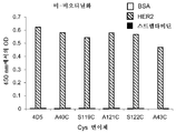
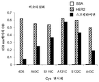
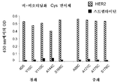
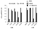
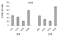


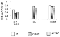
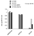


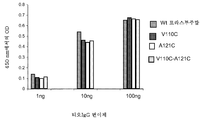

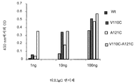


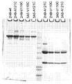

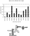


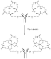

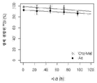
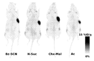

Classifications
-
- A—HUMAN NECESSITIES
- A61—MEDICAL OR VETERINARY SCIENCE; HYGIENE
- A61K—PREPARATIONS FOR MEDICAL, DENTAL OR TOILETRY PURPOSES
- A61K51/00—Preparations containing radioactive substances for use in therapy or testing in vivo
- A61K51/02—Preparations containing radioactive substances for use in therapy or testing in vivo characterised by the carrier, i.e. characterised by the agent or material covalently linked or complexing the radioactive nucleus
- A61K51/04—Organic compounds
- A61K51/08—Peptides, e.g. proteins, carriers being peptides, polyamino acids, proteins
- A61K51/10—Antibodies or immunoglobulins; Fragments thereof, the carrier being an antibody, an immunoglobulin or a fragment thereof, e.g. a camelised human single domain antibody or the Fc fragment of an antibody
- A61K51/1093—Antibodies or immunoglobulins; Fragments thereof, the carrier being an antibody, an immunoglobulin or a fragment thereof, e.g. a camelised human single domain antibody or the Fc fragment of an antibody conjugates with carriers being antibodies
-
- A—HUMAN NECESSITIES
- A61—MEDICAL OR VETERINARY SCIENCE; HYGIENE
- A61K—PREPARATIONS FOR MEDICAL, DENTAL OR TOILETRY PURPOSES
- A61K38/00—Medicinal preparations containing peptides
- A61K38/16—Peptides having more than 20 amino acids; Gastrins; Somatostatins; Melanotropins; Derivatives thereof
- A61K38/17—Peptides having more than 20 amino acids; Gastrins; Somatostatins; Melanotropins; Derivatives thereof from animals; from humans
-
- A—HUMAN NECESSITIES
- A61—MEDICAL OR VETERINARY SCIENCE; HYGIENE
- A61K—PREPARATIONS FOR MEDICAL, DENTAL OR TOILETRY PURPOSES
- A61K39/00—Medicinal preparations containing antigens or antibodies
- A61K39/395—Antibodies; Immunoglobulins; Immune serum, e.g. antilymphocytic serum
-
- A—HUMAN NECESSITIES
- A61—MEDICAL OR VETERINARY SCIENCE; HYGIENE
- A61K—PREPARATIONS FOR MEDICAL, DENTAL OR TOILETRY PURPOSES
- A61K47/00—Medicinal preparations characterised by the non-active ingredients used, e.g. carriers or inert additives; Targeting or modifying agents chemically bound to the active ingredient
- A61K47/30—Macromolecular organic or inorganic compounds, e.g. inorganic polyphosphates
- A61K47/42—Proteins; Polypeptides; Degradation products thereof; Derivatives thereof, e.g. albumin, gelatin or zein
-
- A—HUMAN NECESSITIES
- A61—MEDICAL OR VETERINARY SCIENCE; HYGIENE
- A61K—PREPARATIONS FOR MEDICAL, DENTAL OR TOILETRY PURPOSES
- A61K48/00—Medicinal preparations containing genetic material which is inserted into cells of the living body to treat genetic diseases; Gene therapy
-
- A—HUMAN NECESSITIES
- A61—MEDICAL OR VETERINARY SCIENCE; HYGIENE
- A61K—PREPARATIONS FOR MEDICAL, DENTAL OR TOILETRY PURPOSES
- A61K49/00—Preparations for testing in vivo
- A61K49/06—Nuclear magnetic resonance [NMR] contrast preparations; Magnetic resonance imaging [MRI] contrast preparations
- A61K49/08—Nuclear magnetic resonance [NMR] contrast preparations; Magnetic resonance imaging [MRI] contrast preparations characterised by the carrier
- A61K49/10—Organic compounds
- A61K49/14—Peptides, e.g. proteins
- A61K49/16—Antibodies; Immunoglobulins; Fragments thereof
-
- A—HUMAN NECESSITIES
- A61—MEDICAL OR VETERINARY SCIENCE; HYGIENE
- A61K—PREPARATIONS FOR MEDICAL, DENTAL OR TOILETRY PURPOSES
- A61K51/00—Preparations containing radioactive substances for use in therapy or testing in vivo
- A61K51/02—Preparations containing radioactive substances for use in therapy or testing in vivo characterised by the carrier, i.e. characterised by the agent or material covalently linked or complexing the radioactive nucleus
- A61K51/04—Organic compounds
- A61K51/08—Peptides, e.g. proteins, carriers being peptides, polyamino acids, proteins
- A61K51/10—Antibodies or immunoglobulins; Fragments thereof, the carrier being an antibody, an immunoglobulin or a fragment thereof, e.g. a camelised human single domain antibody or the Fc fragment of an antibody
- A61K51/1027—Antibodies or immunoglobulins; Fragments thereof, the carrier being an antibody, an immunoglobulin or a fragment thereof, e.g. a camelised human single domain antibody or the Fc fragment of an antibody against receptors, cell-surface antigens or cell-surface determinants
-
- A—HUMAN NECESSITIES
- A61—MEDICAL OR VETERINARY SCIENCE; HYGIENE
- A61K—PREPARATIONS FOR MEDICAL, DENTAL OR TOILETRY PURPOSES
- A61K51/00—Preparations containing radioactive substances for use in therapy or testing in vivo
- A61K51/02—Preparations containing radioactive substances for use in therapy or testing in vivo characterised by the carrier, i.e. characterised by the agent or material covalently linked or complexing the radioactive nucleus
- A61K51/04—Organic compounds
- A61K51/08—Peptides, e.g. proteins, carriers being peptides, polyamino acids, proteins
- A61K51/10—Antibodies or immunoglobulins; Fragments thereof, the carrier being an antibody, an immunoglobulin or a fragment thereof, e.g. a camelised human single domain antibody or the Fc fragment of an antibody
- A61K51/1045—Antibodies or immunoglobulins; Fragments thereof, the carrier being an antibody, an immunoglobulin or a fragment thereof, e.g. a camelised human single domain antibody or the Fc fragment of an antibody against animal or human tumor cells or tumor cell determinants
- A61K51/1051—Antibodies or immunoglobulins; Fragments thereof, the carrier being an antibody, an immunoglobulin or a fragment thereof, e.g. a camelised human single domain antibody or the Fc fragment of an antibody against animal or human tumor cells or tumor cell determinants the tumor cell being from breast, e.g. the antibody being herceptin
-
- C—CHEMISTRY; METALLURGY
- C07—ORGANIC CHEMISTRY
- C07C—ACYCLIC OR CARBOCYCLIC COMPOUNDS
- C07C259/00—Compounds containing carboxyl groups, an oxygen atom of a carboxyl group being replaced by a nitrogen atom, this nitrogen atom being further bound to an oxygen atom and not being part of nitro or nitroso groups
- C07C259/04—Compounds containing carboxyl groups, an oxygen atom of a carboxyl group being replaced by a nitrogen atom, this nitrogen atom being further bound to an oxygen atom and not being part of nitro or nitroso groups without replacement of the other oxygen atom of the carboxyl group, e.g. hydroxamic acids
- C07C259/06—Compounds containing carboxyl groups, an oxygen atom of a carboxyl group being replaced by a nitrogen atom, this nitrogen atom being further bound to an oxygen atom and not being part of nitro or nitroso groups without replacement of the other oxygen atom of the carboxyl group, e.g. hydroxamic acids having carbon atoms of hydroxamic groups bound to hydrogen atoms or to acyclic carbon atoms
-
- C—CHEMISTRY; METALLURGY
- C07—ORGANIC CHEMISTRY
- C07C—ACYCLIC OR CARBOCYCLIC COMPOUNDS
- C07C323/00—Thiols, sulfides, hydropolysulfides or polysulfides substituted by halogen, oxygen or nitrogen atoms, or by sulfur atoms not being part of thio groups
- C07C323/50—Thiols, sulfides, hydropolysulfides or polysulfides substituted by halogen, oxygen or nitrogen atoms, or by sulfur atoms not being part of thio groups containing thio groups and carboxyl groups bound to the same carbon skeleton
- C07C323/51—Thiols, sulfides, hydropolysulfides or polysulfides substituted by halogen, oxygen or nitrogen atoms, or by sulfur atoms not being part of thio groups containing thio groups and carboxyl groups bound to the same carbon skeleton having the sulfur atoms of the thio groups bound to acyclic carbon atoms of the carbon skeleton
- C07C323/60—Thiols, sulfides, hydropolysulfides or polysulfides substituted by halogen, oxygen or nitrogen atoms, or by sulfur atoms not being part of thio groups containing thio groups and carboxyl groups bound to the same carbon skeleton having the sulfur atoms of the thio groups bound to acyclic carbon atoms of the carbon skeleton with the carbon atom of at least one of the carboxyl groups bound to nitrogen atoms
-
- C—CHEMISTRY; METALLURGY
- C07—ORGANIC CHEMISTRY
- C07K—PEPTIDES
- C07K14/00—Peptides having more than 20 amino acids; Gastrins; Somatostatins; Melanotropins; Derivatives thereof
- C07K14/435—Peptides having more than 20 amino acids; Gastrins; Somatostatins; Melanotropins; Derivatives thereof from animals; from humans
-
- C—CHEMISTRY; METALLURGY
- C07—ORGANIC CHEMISTRY
- C07K—PEPTIDES
- C07K16/00—Immunoglobulins [IGs], e.g. monoclonal or polyclonal antibodies
-
- C—CHEMISTRY; METALLURGY
- C07—ORGANIC CHEMISTRY
- C07K—PEPTIDES
- C07K16/00—Immunoglobulins [IGs], e.g. monoclonal or polyclonal antibodies
- C07K16/18—Immunoglobulins [IGs], e.g. monoclonal or polyclonal antibodies against material from animals or humans
-
- C—CHEMISTRY; METALLURGY
- C07—ORGANIC CHEMISTRY
- C07K—PEPTIDES
- C07K16/00—Immunoglobulins [IGs], e.g. monoclonal or polyclonal antibodies
- C07K16/18—Immunoglobulins [IGs], e.g. monoclonal or polyclonal antibodies against material from animals or humans
- C07K16/28—Immunoglobulins [IGs], e.g. monoclonal or polyclonal antibodies against material from animals or humans against receptors, cell surface antigens or cell surface determinants
-
- C—CHEMISTRY; METALLURGY
- C07—ORGANIC CHEMISTRY
- C07K—PEPTIDES
- C07K16/00—Immunoglobulins [IGs], e.g. monoclonal or polyclonal antibodies
- C07K16/18—Immunoglobulins [IGs], e.g. monoclonal or polyclonal antibodies against material from animals or humans
- C07K16/32—Immunoglobulins [IGs], e.g. monoclonal or polyclonal antibodies against material from animals or humans against translation products of oncogenes
-
- C—CHEMISTRY; METALLURGY
- C07—ORGANIC CHEMISTRY
- C07K—PEPTIDES
- C07K19/00—Hybrid peptides, i.e. peptides covalently bound to nucleic acids, or non-covalently bound protein-protein complexes
-
- A—HUMAN NECESSITIES
- A01—AGRICULTURE; FORESTRY; ANIMAL HUSBANDRY; HUNTING; TRAPPING; FISHING
- A01K—ANIMAL HUSBANDRY; CARE OF BIRDS, FISHES, INSECTS; FISHING; REARING OR BREEDING ANIMALS, NOT OTHERWISE PROVIDED FOR; NEW BREEDS OF ANIMALS
- A01K2267/00—Animals characterised by purpose
- A01K2267/03—Animal model, e.g. for test or diseases
- A01K2267/0331—Animal model for proliferative diseases
-
- C—CHEMISTRY; METALLURGY
- C07—ORGANIC CHEMISTRY
- C07C—ACYCLIC OR CARBOCYCLIC COMPOUNDS
- C07C2601/00—Systems containing only non-condensed rings
- C07C2601/12—Systems containing only non-condensed rings with a six-membered ring
- C07C2601/14—The ring being saturated
-
- C—CHEMISTRY; METALLURGY
- C07—ORGANIC CHEMISTRY
- C07K—PEPTIDES
- C07K2317/00—Immunoglobulins specific features
- C07K2317/20—Immunoglobulins specific features characterized by taxonomic origin
- C07K2317/21—Immunoglobulins specific features characterized by taxonomic origin from primates, e.g. man
-
- C—CHEMISTRY; METALLURGY
- C07—ORGANIC CHEMISTRY
- C07K—PEPTIDES
- C07K2317/00—Immunoglobulins specific features
- C07K2317/50—Immunoglobulins specific features characterized by immunoglobulin fragments
- C07K2317/51—Complete heavy chain or Fd fragment, i.e. VH + CH1
-
- C—CHEMISTRY; METALLURGY
- C07—ORGANIC CHEMISTRY
- C07K—PEPTIDES
- C07K2317/00—Immunoglobulins specific features
- C07K2317/50—Immunoglobulins specific features characterized by immunoglobulin fragments
- C07K2317/52—Constant or Fc region; Isotype
-
- C—CHEMISTRY; METALLURGY
- C07—ORGANIC CHEMISTRY
- C07K—PEPTIDES
- C07K2317/00—Immunoglobulins specific features
- C07K2317/50—Immunoglobulins specific features characterized by immunoglobulin fragments
- C07K2317/55—Fab or Fab'
-
- C—CHEMISTRY; METALLURGY
- C07—ORGANIC CHEMISTRY
- C07K—PEPTIDES
- C07K2317/00—Immunoglobulins specific features
- C07K2317/60—Immunoglobulins specific features characterized by non-natural combinations of immunoglobulin fragments
- C07K2317/62—Immunoglobulins specific features characterized by non-natural combinations of immunoglobulin fragments comprising only variable region components
- C07K2317/624—Disulfide-stabilized antibody (dsFv)
Landscapes
- Health & Medical Sciences (AREA)
- Chemical & Material Sciences (AREA)
- Life Sciences & Earth Sciences (AREA)
- Organic Chemistry (AREA)
- Proteomics, Peptides & Aminoacids (AREA)
- Medicinal Chemistry (AREA)
- General Health & Medical Sciences (AREA)
- Immunology (AREA)
- Epidemiology (AREA)
- Animal Behavior & Ethology (AREA)
- Public Health (AREA)
- Veterinary Medicine (AREA)
- Biochemistry (AREA)
- Molecular Biology (AREA)
- Genetics & Genomics (AREA)
- Pharmacology & Pharmacy (AREA)
- Biophysics (AREA)
- Oncology (AREA)
- Optics & Photonics (AREA)
- Physics & Mathematics (AREA)
- Engineering & Computer Science (AREA)
- Gastroenterology & Hepatology (AREA)
- Nuclear Medicine, Radiotherapy & Molecular Imaging (AREA)
- Radiology & Medical Imaging (AREA)
- Bioinformatics & Cheminformatics (AREA)
- Zoology (AREA)
- Cell Biology (AREA)
- Toxicology (AREA)
- Biotechnology (AREA)
- Microbiology (AREA)
- Mycology (AREA)
- Inorganic Chemistry (AREA)
- Peptides Or Proteins (AREA)
- Medicines Containing Antibodies Or Antigens For Use As Internal Diagnostic Agents (AREA)
- Preparation Of Compounds By Using Micro-Organisms (AREA)
Abstract
항체는 모 항체의 하나 이상의 아미노산을 비가교된, 고도 반응성 시스테인 아미노산으로 대체하여 조작된다. 항체 단편은 또한 하나 이상의 시스테인 아미노산으로 조작되어 시스테인 조작된 항체 단편 (티오Fab)를 형성할 수 있다. 시스테인 조작된 항체의 설계, 제조, 스크리닝 및 선택의 방법이 제공된다. 시스테인 조작된 항체 (Ab)는 링커 (L)을 통해 하나 이상의 지르코늄 복합체 (Z) 표지와 접합되어 하기 화학식 I을 갖는 시스테인 조작된 지르코늄-표지된 항체 접합체를 형성한다.
<화학식 I>
![]()
(상기 식에서, p는 1 내지 4임)
지르코늄-방사성표지된, 시스테인 조작된 항체 접합체 조성물에 대한 영상화 방법 및 진단 용도가 개시된다.The antibody is engineered by replacing one or more amino acids of the parent antibody with an unmodified, highly reactive cysteine amino acid. The antibody fragment may also be engineered with one or more cysteine amino acids to form a cysteine engineered antibody fragment (thioFab). Methods for the design, manufacture, screening and selection of cysteine engineered antibodies are provided. The cysteine engineered antibody (Ab) is conjugated to one or more zirconium complex (Z) labels via a linker (L) to form a cysteine engineered zirconium-labeled antibody conjugate having the formula (I)
(I)
![]()
(Wherein p is from 1 to 4)
Disclosed are imaging methods and diagnostic uses for zirconium-radioactively labeled, cysteine engineered antibody conjugate compositions.
Description
본 출원은 현재 2009년 4월 21일에 허여된 US 7,521,541인, 2005년 9월 22일에 출원된 US 출원 번호 11/233,258의 계속 출원인 2009년 3월 6일에 출원된 US 출원 번호 12/399,241의 부분-계속 출원인 2009년 11월 5일에 출원된 US 출원 번호 12/612,912를 우선권 주장하고, 또한 35 USC §119(e)하에 2004년 9월 23일에 출원된 US 가출원 번호 60/612,468 및 2005년 6월 30일에 출원된 US 가출원 번호 60/696,353을 우선권 주장하고, 이들은 각각 그 전문이 참고로 포함된다.This application is currently US 7,521,541 filed on April 21, 2009, continuation of US Application No. 11/233,258 filed on September 22, 2005, US Application No. 12/399,241 filed on March 6, 2009 US Provisional Application No. 60/612,468 filed September 23, 2004 under 35 USC §119(e), and Priority claims are made to US Provisional Application No. 60/696,353, filed on June 30, 2005, each of which is incorporated by reference in its entirety.
<발명의 분야><Field of invention>
본 발명은 일반적으로, 반응성 시스테인 잔기로 조작된 항체에 관한 것이고, 보다 구체적으로는 치료적 또는 진단적 적용을 수반한 항체에 관한 것이다. 시스테인 조작된 항체는 화학요법 약물, 독소, 친화성 리간드, 예컨대 비오틴, 및 검출 표지, 예컨대 방사성동위원소 및 형광단과 접합시킬 수 있다. 본 발명은 또한, 포유동물 세포, 또는 관련된 병적 상태를 시험관내, 계내 또는 생체내에서 진단 또는 치료하기 위해 항체 및 항체-약물 접합체 화합물을 사용하는 방법에 관한 것이다.The present invention relates generally to antibodies engineered with reactive cysteine residues, and more particularly to antibodies involving therapeutic or diagnostic applications. Cysteine engineered antibodies can be conjugated with chemotherapeutic drugs, toxins, affinity ligands such as biotin, and detection labels such as radioisotopes and fluorophores. The invention also relates to methods of using antibodies and antibody-drug conjugate compounds to diagnose or treat mammalian cells, or related pathological conditions in vitro, in situ or in vivo.
분자 영상화는 신규 제약의 개발 및 평가에서 중요한 도구이다. 면역-양전자 방출 단층촬영 (이뮤노PET)은 모노클로날 항체 (mAb)가 높은 감수성의 PET를 높은 특이성의 mAb와 효율적으로 조합하는지 모노클로날 항체 (mAb)를 추적 및 정량하기 위해 최근에 빠르게 대두되고 있는 방법이다. 이뮤노PET는 "생체내 포괄적 면역조직화학 염색" (문헌 [van Dongen GA, et al. "Immuno-PET: a navigator in monoclonal antibody development and applications" Oncologist 2007;12:1379-89])을 제공하는 비-침습성 진단을 위한 선택의 임상적 방법일 것으로 기대된다. 이뮤노PET는 양전자-방출 방사성동위원소가 표적 특이적 분자에 커플링될 것을 요구하므로, 분자의 생물학적 반감기를 방사성핵종의 반감기와 맞추는 것이 필수적이다 (문헌 [Verel I, et al. "The promise of immuno-PET in radioimmunotherapy" J Nucl Med 2005;46 Suppl 1:164S-71S]). 항체 (약 150 kDa)는 수일 내지 수주 범위의 혈장 반감기를 갖지만, 영상화는 전형적으로 각각 3.3일 및 4.2일의 반감기를 갖는 89Zr 및 124I와 같은 방사성동위원소의 사용에 따라 항체-기재 트레이서의 투여 2 내지 6일 후에 최대 표적-대-백그라운드 비를 제공한다. 불행하게도, 용이하게 이용가능한 64Cu의 반감기 (12.7 h)는 이러한 시간 프레임에서 우수한 대비를 제공하기에 너무 짧다.Molecular imaging is an important tool in the development and evaluation of new pharmaceuticals. Immuno-positron emission tomography (ImunoPET) has recently been rapidly used to track and quantify monoclonal antibodies (mAb) to ensure that they efficiently combine highly sensitive PET with highly specific mAbs. This is the emerging method. ImmunoPET provides "comprehensive immunohistochemical staining in vivo" (van Dongen GA, et al. "Immuno-PET: a navigator in monoclonal antibody development and applications" Oncologist 2007;12:1379-89). It is expected to be the clinical method of choice for non-invasive diagnosis. Since immunoPET requires the positron-emitting radioisotope to be coupled to a target-specific molecule, it is essential to match the biological half-life of the molecule with that of the radionuclide (Verel I, et al. "The promise of immuno-PET in radioimmunotherapy" J Nucl Med 2005;46 Suppl 1:164S-71S]). Antibodies (approximately 150 kDa) have plasma half-life in the range of days to weeks, but imaging typically follows the use of radioisotopes such as 89 Zr and 124 I with half-life of 3.3 and 4.2 days, respectively. The maximum target-to-background ratio is provided 2-6 days after administration. Unfortunately, the readily available half-life of 64 Cu (12.7 h) is too short to provide good contrast in this time frame.
Mab 주형 (이뮤노-PET)으로부터 양전자 방출 단층촬영 (PET) 영상화제의 개발은 분자 표적의 국소화 및 정량을 위한 도구로서 제안되고, 병적 상태의 비-침습성 임상적 진단을 향상시킬 수 있다 (문헌 [van Dongen et al. (2007) Oncologist 12;1379-89]; [Williams et a (2001) Cancer Biother Radiopharm 16:25-35]; [Holliger et al. (2005) Nat Biotechnol 23:1126-36]). PET는 질환의 검출을 위해 사용이 증가하고 있는 분자 영상화 기술이다. PET 영상화 시스템은 환자의 조직에서 양전자-방출 동위원소의 분포에 기초하여 영상을 생성한다. 동위원소는 전형적으로 체내에서 용이하게 대사 또는 국소화되는 분자 (예를 들어, 글루코스)에 공유 결합에 의해 부착된 양전자-방출 동위원소, 예컨대 F-18, C-11, N-13, 또는 O-15를 포함하거나 또는 체내에서 수용체 부위에 화학적으로 결합하는 프로브 분자의 주사에 의해 환자에게 투여된다. 일부 경우에, 동위원소는 이온성 용액으로서 또는 흡입에 의해 환자에게 투여된다. 작은 이뮤노-PET 영상화제, 예컨대 Fab 항체 단편 (50 kDa) 또는 디아바디 (Mab의 공유 결합에 의해 연결된 VH-VL 영역의 쌍을 이루지 않은 이량체, 55 kDa) (문헌 [Shively et al. (2007) J Nucl Med 48:170-2])은 이들이 짧은 순환 반감기, 높은 조직 투과성을 나타내고, 널리 이용가능한 18F (109.8 min)와 같은 짧은 반감기 동위원소의 사용을 용이하게 하는 주사 2 내지 4시간 후에 최적의 종양 대 백그라운드 비에 도달하므로 특히 유용하다.The development of positron emission tomography (PET) imaging agents from the Mab template (immuno-PET) is proposed as a tool for localization and quantification of molecular targets, and can improve non-invasive clinical diagnosis of pathological conditions (literature [van Dongen et al. (2007) Oncologist 12;1379-89]; [Williams et a (2001) Cancer Biother Radiopharm 16:25-35]; [Holliger et al. (2005) Nat Biotechnol 23:1126-36] ). PET is a molecular imaging technology that is increasingly used for the detection of diseases. PET imaging systems generate images based on the distribution of positron-emitting isotopes in a patient's tissue. The isotope is typically a positron-releasing isotope, such as F-18, C-11, N-13, or O-, which is covalently attached to a molecule (e.g., glucose) that is easily metabolized or localized in the body. 15 or by injection of a probe molecule that chemically binds to the receptor site in the body. In some cases, the isotope is administered to the patient as an ionic solution or by inhalation. Small immuno-PET imaging agents, such as Fab antibody fragments (50 kDa) or diabodies (unpaired dimers of V H -V L regions linked by the covalent bond of Mab, 55 kDa) (Shively et al. (2007) J Nucl Med 48:170-2] show that they show short circulatory half-life, high tissue permeability, and facilitate the use of short half-life isotopes such as 18 F (109.8 min), which are widely available. This is particularly useful as the optimal tumor to background ratio is reached after 4 hours.
아이오딘 124 (124I)를 항체 3F9에 커플링시키고, 이를 사용하여 신경모세포종의 방사선 면역요법의 위한 선량측정을 추정하였다 (문헌 [Larson SM, et al. "PET scanning of iodine-124-3F9 as an approach to tumor dosimetry during treatment planning for radioimmunotherapy in a child with neuroblastoma" J Nucl Med 1992;33:2020-3]). 이후에, 보다 정교해진 PET 계기 및 새로 대두되는 방사성아이오딘화를 위한 개선된 기술로서, 124I를 다수의 이뮤노PET 연구에 사용하였다 (문헌 [Verel I, et al. "High-quality 124I-labelled monoclonal antibodies for use as PET scouting agents prior to 131I-radioimmunotherapy" European journal of nuclear medicine and molecular imaging 2004;31:1645-52]; [Lee FT et al. "Immuno-PET of human colon xenograft-bearing BALB/c nude mice using 124I-CDR-grafted humanized A33 monoclonal antibody" J Nucl Med 2001;42:764-9]; [Sundaresan G, et al. "124I-labeled engineered anti-CEA minibodies and diabodies allow high-contrast, antigen-specific small-animal PET imaging of xenografts in athymic mice" J Nucl Med 2003;44:1962-9]; [Jain M and Batra SK. "Genetically engineered antibody fragments and PET imaging: a new era of radioimmunodiagnosis" J Nucl Med 2003;44:1970-2]; [Gonzalez Trotter DE et al. "Quantitation of small-animal (124)I activity distributions using a clinical PET/CT scanner" J Nucl Med 2004;45:1237-44]; [Robinson MK, et al. "Quantitative immuno-positron emission tomography imaging of HER2-positive tumor xenografts with an iodine-124 labeled anti-HER2 diabody" Cancer Res 2005;65:1471-8]; [Jayson GC et al. "Molecular imaging and biological evaluation of HuMV833 anti-VEGF antibody: implications for trial design of antiangiogenic antibodies" J Natl Cancer Inst 2002;94:1484-93]; [Divgi CR, et al. "Preoperative characterisation of clear-cell renal carcinoma using iodine-124-labelled antibody chimeric G250 (124I-cG250) and PET in patients with renal masses: a phase I trial" Lancet Oncol 2007;8:304-10]). mAb로의 124I의 커플링을 위해 이용가능한 비교적 간단한 방사성아이오딘화 기술에도 불구하고, 중요한 제한이 이 방사성핵종의 광범위한 전임상적 용도를 지연시킨다. 주목할만하게, 복합체 붕괴 방식은 작은 동물 마이크로PET의 해상도에 부정적인 영향을 미치는 강력한 양전자 (β+ max. 1.5 및 2.1 MeV)와 관련된다. 또한, 내재화된 아이오딘화 단백질은 표적 세포로부터 빠르게 제거되는 유리 아이오다이드로의 효소적 탈아이오딘화를 경험하여 실제 mAb 흡수를 반영하지 않는 PET 영상을 제공한다 (문헌 ["Perera RM et al. "Internalization, intracellular trafficking, and biodistribution of monoclonal antibody 806: a novel anti-epidermal growth factor receptor antibody" Neoplasia (New York, N.Y 2007;9:1099-110)]). 89Zr 붕괴 (β+ max. 897 keV)에서 방출된 양전자가 18F 및 11C (대략 1 mm)와 대등한 마이크로PET 해상도를 제공하므로 89Zr의 사용은 이들 단점을 극복한다. 또한, 내재화된 89Zr-mAb의 대사물은 리소솜에서 세포내 포획되어, 실제 mAb 흡수의 PET 영상화와의 보다 양호한 상관관계를 제공한다 (문헌 [van Dongen GA, et al. "Immuno-PET: a navigator in monoclonal antibody development and applications" Oncologist 2007;12:1379-89]).Iodine 124 ( 124 I) was coupled to antibody 3F9 and used to estimate the dosimetry for radioimmunotherapy of neuroblastoma (Larson SM, et al. "PET scanning of iodine-124-3F9 as" an approach to tumor dosimetry during treatment planning for radioimmunotherapy in a child with neuroblastoma" J Nucl Med 1992;33:2020-3]). Later, as a more sophisticated PET instrument and an improved technique for the emerging radioiodination, 124 I was used in a number of immunoPET studies (Verel I, et al. "High-quality 124I-" labeled monoclonal antibodies for use as PET scouting agents prior to 131I-radioimmunotherapy" European journal of nuclear medicine and molecular imaging 2004;31:1645-52]; [Lee FT et al. "Immuno-PET of human colon xenograft-bearing BALB/ c nude mice using 124I-CDR-grafted humanized A33 monoclonal antibody" J Nucl Med 2001;42:764-9]; [Sundaresan G, et al. "124I-labeled engineered anti-CEA minibodies and diabodies allow high-contrast, antigen -specific small-animal PET imaging of xenografts in athymic mice" J Nucl Med 2003;44:1962-9]; [Jain M and Batra SK. "Genetically engineered antibody fragments and PET imaging: a new era of radioimmunodiagnosis" J Nucl Med 2003;44:1970-2]; [Gonzalez Trotter DE et al. "Quantitation of small-animal (124)I activity distributions using a clinical PET/CT scanner" J Nucl Med 2004;45:1237-44]; [Robinson MK, et al. "Quantitative immuno-posi tron emission tomography imaging of HER2-positive tumor xenografts with an iodine-124 labeled anti-HER2 diabody" Cancer Res 2005;65:1471-8]; [Jayson GC et al. "Molecular imaging and biological evaluation of HuMV833 anti-VEGF antibody: implications for trial design of antiangiogenic antibodies" J Natl Cancer Inst 2002;94:1484-93]; [Divgi CR, et al. "Preoperative characterization of clear-cell renal carcinoma using iodine-124-labelled antibody chimeric G250 (124I-cG250) and PET in patients with renal masses: a phase I trial" Lancet Oncol 2007;8:304-10]). Despite the relatively simple radioiodination techniques available for coupling of 124 I to mAb, significant limitations delay the widespread preclinical use of this radionuclide. Notably, the complex decay mode is associated with a strong positron (β + max. 1.5 and 2.1 MeV) that negatively affects the resolution of small animal microPETs. In addition, internalized iodination proteins undergo enzymatic deiodination to free iodide, which is rapidly removed from target cells, providing PET images that do not reflect actual mAb uptake ("Perera RM et al. "Internalization, intracellular trafficking, and biodistribution of monoclonal antibody 806: a novel anti-epidermal growth factor receptor antibody" Neoplasia (New York, NY 2007;9:1099-110)]). 89 Zr decay (β + max. 897 keV ), the so positron is provided a micro-PET resolution along with 18 F and 11 C (about 1 mm) using a 89 Zr overcomes these disadvantages. in addition, in the metabolites of the internalization 89 Zr-mAb Li sosom released from Captured intracellularly in, provides a better correlation with PET imaging of actual mAb uptake (van Dongen GA, et al. "Immuno-PET: a navigator in monoclonal antibody development and applications" Oncologist 2007;12: 1379-89]).
표지, 예컨대 방사성동위원소, 형광 염료 또는 약물 모이어티를 항체에 부착시키는, 즉 공유결합을 통해 연결시키는 통상적인 수단은 일반적으로 표지 모이어티가 항체 상의 많은 부위에 부착되는 분자의 불균질 혼합물을 생성시킨다. 예를 들어, 세포독성 약물은 전형적으로 항체의 종종 수많은 리신 잔기를 통해 항체에 접합되어, 불균질 항체-약물 접합체 혼합물을 생성한다. 반응 조건에 따라, 불균질 혼합물은 전형적으로 0 내지 약 8개 이상의 부착된 약물 모이어티를 갖는 항체의 분포를 포함한다. 추가로, 약물 모이어티 대 항체의 특정 정수비를 갖는 접합체의 각각의 하위군 내에 약물 모이어티가 항체 상의 다양한 부위에 부착되는 잠재적으로 불균질 혼합물이 존재한다. 분석 및 제조 방법은 접합 반응으로부터 생성되는 불균질 혼합물 내에서 항체-약물 접합체 종 분자를 분리하고 특성화하기에 부적당할 수 있다. 항체는 종종 많은 반응성 관능기를 갖는 크고 복잡하고 구조상 다양한 생체분자이다. 그의 링커 시약 및 약물-링커 중간체와의 반응성은 pH, 농도, 염 농도, 및 보조-용매와 같은 인자에 의존성이다. 또한, 다단계 접합 과정은 반응 조건을 제어하고 반응물 및 중간체를 특성화하는 어려움 때문에 재현가능하지 않을 수 있다.Conventional means of attaching a label, such as a radioisotope, fluorescent dye or drug moiety to an antibody, i.e. linking via covalent bonds, generally results in a heterogeneous mixture of molecules in which the labeling moiety is attached to many sites on the antibody. Let it. For example, cytotoxic drugs are typically conjugated to the antibody through often numerous lysine residues of the antibody, resulting in a heterogeneous antibody-drug conjugate mixture. Depending on the reaction conditions, the heterogeneous mixture typically comprises a distribution of antibodies with 0 to about 8 or more attached drug moieties. Additionally, there are potentially heterogeneous mixtures in which drug moieties are attached to various sites on the antibody within each subgroup of conjugates having a specific integer ratio of drug moiety to antibody. Analysis and preparation methods may be inappropriate for isolating and characterizing antibody-drug conjugate species molecules within a heterogeneous mixture resulting from a conjugation reaction. Antibodies are large, complex and structurally diverse biomolecules, often with many reactive functional groups. Its reactivity with linker reagents and drug-linker intermediates is dependent on factors such as pH, concentration, salt concentration, and co-solvent. In addition, the multi-step conjugation process may not be reproducible due to difficulties in controlling reaction conditions and characterizing reactants and intermediates.
pH 7 부근에서 양성자첨가되고 덜 친핵성인 대부분의 아민과는 달리, 시스테인 티올은 중성 pH에서 반응성이다. 유리 티올 (RSH, 술프히드릴)기는 비교적 반응성이므로, 시스테인 잔기를 갖는 단백질은 종종 디술피드-연결된 올리고머로서 산화된 형태로 존재하거나, 내부에 가교된 디술피드기를 갖는다. 세포외 단백질은 일반적으로 유리 티올을 갖지 않는다 (문헌 [Garman, 1997, Non-Radioactive Labelling: A Practical Approach, Academic Press, London, at page 55]). 단백질 내의 유리 티올의 양은 표준 엘만 (Ellman) 분석에 의해 추정할 수 있다. 이뮤노글로불린 M은 디술피드-연결 오량체의 예이고, 이뮤노글로불린 G는 함께 서브유닛을 연결하는 내부 디술피드 브릿지를 갖는 단백질의 예이다. 이와 같은 단백질에서, 반응성 유리 티올을 생성시키기 위해 시약, 예컨대 디티오트레이톨 (DTT) 또는 셀레놀을 사용한 환원 (문헌 [Singh et al. (2002) Anal. Biochem. 304:147-156])이 필요하다. 상기 방법은 항체 3차 구조 및 항원 결합 특이성을 손실시킬 수 있다.Unlike most amines, which are protonated around
항체 시스테인 티올 기는 일반적으로 항체 아민 또는 히드록실 기보다 친전자성 접합 시약에 대해 더 반응성이고, 즉, 더 친핵성이다. 시스테인 잔기는 유전자 조작 기술에 의해 단백질 내로 도입되어, 리간드에 공유결합에 의한 부착을 형성하거나 새로운 분자내 디술피드 결합을 형성한다 (문헌 [Better et al. (1994) J. Biol. Chem. 269(13):9644-9650]; [Bernhard et al. (1994) Bioconjugate Chem. 5:126-132]; [Greenwood et al. (1994) Therapeutic Immunology 1:247-255]; [Tu et al. (1999) Proc. Natl. Acad. Sci USA 96:4862-4867]; [Kanno et al. (2000) J. of Biotechnology, 76:207-214]; [Chmura et al. (2001) Proc. Nat. Acad. Sci. USA 98(15):8480-8484]; US 6248564). 그러나, 시스테인 아미노산에 대한 단백질의 다양한 아미노산 잔기의 돌연변이에 의한 시스테인 티올 기에서의 설계는, 특히 쌍을 이루지 않은 (유리 Cys) 잔기 또는 반응 또는 산화반응을 위해 비교적 접근가능한 것의 경우에 잠재적으로 문제가 된다. 이. 콜라이 (E. coli)의 주변세포질, 배양 상청액, 또는 부분적으로 또는 완전히 정제된 단백질 내에서든 단백질의 농축 용액에서든, 단백질의 표면 상의 쌍을 이루지 않은 Cys 잔기는 쌍을 이루고 산화되어 분자내 디술피드를 형성할 수 있고, 따라서 단백질 이량체 또는 다량체를 형성할 수 있다. 디술피드 이량체 형성은 새로운 Cys가 약물, 리간드, 또는 다른 표지에의 접합에 대해 비반응성이 되도록 한다. 또한, 단백질이 새롭게 조작된 Cys와 존재하는 Cys 잔기 사이에서 분자내 디술피드 결합을 산화에 의해 형성하면, 두 Cys 기는 활성 부위 참여 및 상호작용에 이용가능하지 않다. 또한, 단백질은 미스폴딩 또는 3차 구조의 손실에 의해 불활성 또는 비-특이적으로 될 수 있다 (문헌 [Zhang et al. (2002) Anal. Biochem. 311:1-9]).Antibody cysteine thiol groups are generally more reactive to electrophilic conjugation reagents than antibody amine or hydroxyl groups, ie more nucleophilic. Cysteine residues are introduced into proteins by genetic engineering techniques to form covalent attachments to ligands or to form new intramolecular disulfide bonds (Better et al. (1994) J. Biol. Chem. 269 ( 13):9644-9650]; [Bernhard et al. (1994) Bioconjugate Chem. 5:126-132]; [Greenwood et al. (1994) Therapeutic Immunology 1:247-255]; [Tu et al. (1999) ) Proc. Natl. Acad. Sci USA 96:4862-4867]; [Kanno et al. (2000) J. of Biotechnology, 76:207-214]; [Chmura et al. (2001) Proc. Nat. Acad. Sci. USA 98(15):8480-8484]; US 6248564). However, design at cysteine thiol groups by mutations of various amino acid residues of the protein to cysteine amino acids are potentially problematic, especially in the case of unpaired (free Cys) residues or those relatively accessible for reactions or oxidation reactions. do. this. The unpaired Cys residues on the surface of the protein, whether in the periplasm of E. coli , in the culture supernatant, or in a partially or completely purified protein or in a concentrated solution of the protein, pair and oxidize to form intramolecular disulfides. Can form, and thus protein dimers or multimers. Disulfide dimer formation renders the new Cys non-reactive to conjugation to drugs, ligands, or other labels. In addition, if the protein forms an intramolecular disulfide bond by oxidation between the newly engineered Cys and the existing Cys residue, the two Cys groups are not available for active site participation and interaction. In addition, proteins can be made inactive or non-specific by misfolding or loss of tertiary structure (Zhang et al. (2002) Anal. Biochem. 311:1-9).
부위 특이적 접합은 이것이 생물학적 활성의 완전한 보유를 촉진하고 첨가된 보결분자단의 가능한 수에 대한 제어를 허용하는, 결합 부위로부터 멀리 떨어진 부위의 화학적 변형을 가능하게 하므로 무작위 아미노 변형보다 바람직하다. 시스테인 조작된 항체는 Fab 항체 단편 (티오Fab)으로 설계되고, 전장, IgG 모노클로날 (티오Mab) 항체로 발현된다. US 7521541; 문헌 [Junutula JR et al. "Rapid identification of reactive cystein residues for site-specific labeling of antibody-Fabs" J Immunol Methods 2008;332:41-52]; [Junutula JR et al. "Site-specific conjugation of a cytotoxic drug to an antibody improves the therapeutic index" (2008) Nat Biotechnol. 26:925-32] (그의 내용이 참고로 포함됨)을 참조한다. 티오Fab 및 티오Mab 항체는 새로 도입된 시스테인 티올에서 링커를 통해 티올-반응성 링커 시약 및 약물-링커 시약과 접합되어 항암 특성을 갖는 시스테인 조작된 항체 약물 접합체 (티오 ADC), 예를 들어 항-MUC16 (US 2008/0311134), 항-CD22 (US 2008/0050310), 항-ROBO4 (US 2008/0247951), 항-TENB2 (US 2009/0117100), 항-CD79B (US 2009/0028856; US 2009/0068202) 티오 ADC를 제조한다.Site-specific conjugation is preferred over random amino modifications as it allows for chemical modification of sites away from the binding site, which facilitates complete retention of biological activity and allows control over the possible number of prosthetic groups added. Cysteine engineered antibodies are designed as Fab antibody fragments (thioFab) and expressed as full-length, IgG monoclonal (thioMab) antibodies. US 7521541; Junutula JR et al. "Rapid identification of reactive cystein residues for site-specific labeling of antibody-Fabs" J Immunol Methods 2008;332:41-52]; [Junutula JR et al. "Site-specific conjugation of a cytotoxic drug to an antibody improves the therapeutic index" (2008) Nat Biotechnol. 26:925-32] (the contents of which are incorporated by reference). ThioFab and ThioMab antibodies are conjugated with a thiol-reactive linker reagent and a drug-linker reagent through a linker in a newly introduced cysteine thiol to have anti-cancer properties cysteine engineered antibody drug conjugates (thio ADCs), such as anti-MUC16 (US 2008/0311134), anti-CD22 (US 2008/0050310), anti-ROBO4 (US 2008/0247951), anti-TENB2 (US 2009/0117100), anti-CD79B (US 2009/0028856; US 2009/0068202 ) Prepare Thio ADC.
<개요><Overview>
본 발명의 화합물에는 모 항체의 하나 이상의 아미노산을 유리 시스테인 아미노산으로 대체시킨 시스테인 조작된 항체가 포함된다. 시스테인 조작된 항체는 티올 반응성 값이 0.6 내지 1.0인 하나 이상의 유리 시스테인 아미노산을 포함한다. 유리 시스테인 아미노산은 모 항체로 조작되고 디술피드 브릿지의 일부가 아닌 시스테인 잔기이다.The compounds of the present invention include cysteine engineered antibodies in which one or more amino acids of the parent antibody have been replaced with free cysteine amino acids. Cysteine engineered antibodies comprise one or more free cysteine amino acids with thiol reactivity values of 0.6 to 1.0. Free cysteine amino acids are cysteine residues that have been engineered into the parent antibody and are not part of the disulfide bridge.
시스테인 조작된 항체는 암의 진단 및 치료에 유용할 수 있고, 세포 표면 및 막횡단 수용체, 및 종양-관련 항원 (TAA)에 특이적인 항체를 포함한다. 이러한 항체는 네이키드 (naked) 항체 (약물 또는 표지 모이어티에 접합되지 않은)로서 또는 항체-지르코늄 접합체 (AZC)로서 사용될 수 있다.Cysteine engineered antibodies can be useful in the diagnosis and treatment of cancer, and include antibodies specific for cell surface and transmembrane receptors, and tumor-associated antigens (TAA). Such antibodies can be used as naked antibodies (not conjugated to drug or labeling moieties) or as antibody-zirconium conjugates (AZC).
시스테인 조작된 항체의 제조 및 스크리닝 방법의 실시양태는 모 항체가 항체 단편, 예를 들어 hu4D5Fabv8인 실시양태를 포함한다. 모 항체는 알부민 결합 펩티드 서열 (ABP)을 포함하는 융합 단백질일 수도 있다. 모 항체는 또한 huMAb4D5-1, huMAb4D5-2, huMAb4D5-3, huMAb4D5-4, huMAb4D5-5, huMAb4D5-6, huMAb4D5-7 및 huMAb4D5-8 (트라스투주맙)로부터 선택된 인간화 항체일 수 있다.Embodiments of the methods of making and screening cysteine engineered antibodies include embodiments in which the parent antibody is an antibody fragment, eg, hu4D5Fabv8. The parent antibody may be a fusion protein comprising an albumin binding peptide sequence (ABP). The parent antibody may also be a humanized antibody selected from huMAb4D5-1, huMAb4D5-2, huMAb4D5-3, huMAb4D5-4, huMAb4D5-5, huMAb4D5-6, huMAb4D5-7 and huMAb4D5-8 (trastuzumab).
본 발명의 시스테인 조작된 항체는 티올-반응성 시약과 부위-특이적으로 및 효율적으로 커플링될 수 있다. 티올-반응성 시약은 방사성동위원소 시약, 다관능성 링커 시약, 포획 표지 시약, 형광단 시약, 또는 약물-링커 중간체일 수 있다.Cysteine engineered antibodies of the invention can be site-specifically and efficiently coupled with thiol-reactive reagents. The thiol-reactive reagent can be a radioisotope reagent, a multifunctional linker reagent, a capture label reagent, a fluorophore reagent, or a drug-linker intermediate.
시스테인 조작된 항체는 검출가능한 표지로 표지되고, 고상 지지체에 고정되고/되거나 약물 모이어티와 접합될 수 있다.Cysteine engineered antibodies can be labeled with a detectable label, immobilized on a solid support and/or conjugated with a drug moiety.
본 발명의 또 다른 측면은 유리 시스테인 아미노산을 통해 링커 (L) 및 지르코늄 복합체 (Z)에 접합된 시스테인 조작된 항체 (Ab)를 포함하는, 하기 화학식 I을 갖는 지르코늄-표지된, 시스테인 조작된 항체이다.Another aspect of the invention is a zirconium-labeled, cysteine engineered antibody having the following formula (I), comprising a cysteine engineered antibody (Ab) conjugated to a linker (L) and a zirconium complex (Z) via a free cysteine amino acid. to be.
<화학식 I><Formula I>
![]()
![]()
(상기 식에서, p는 1 내지 4임)(Wherein, p is 1 to 4)
본 발명의 또 다른 측면은 유리 시스테인 아미노산을 통해 링커 (L) 및 데스페리옥사민 모이어티 (Df)에 접합된 시스테인 조작된 항체 (Ab)를 포함하는, 하기 화학식 II를 갖는 데스페리옥사민-표지된, 시스테인 조작된 항체이다.Another aspect of the present invention is desperioxamine having the following formula (II), comprising a cysteine engineered antibody (Ab) conjugated to a linker (L) and a desperioxamine moiety (Df) via a free cysteine amino acid. It is a labeled, cysteine engineered antibody.
<화학식 II><Formula II>
![]()
![]()
(상기 식에서, L-Df는(In the above formula, L-Df is

여기서 파선은 항체 (Ab)에 대한 부착을 나타내고; Where the dashed line indicates the attachment to the antibody (Ab);
p는 1 내지 4임)p is 1 to 4)
본 발명의 또 다른 측면은 하기 구조로부터 선택된 데스페리옥사민-표지 시약이다.Another aspect of the present invention is a desperioxamine-labeled reagent selected from the following structures.

(상기 식에서, R은(In the above formula, R is

본 발명의 또 다른 측면은 하기 구조로부터 선택된 조성물을 하나 이상의 유리 시스테인 아미노산을 갖는 시스테인 조작된 항체 (Ab)와 반응시켜 데스페리옥사민-표지된, 시스테인 조작된 항체를 형성하는 것을 포함하는,Another aspect of the invention comprises reacting a composition selected from the following structures with a cysteine engineered antibody (Ab) having at least one free cysteine amino acid to form a desperioxamine-labeled, cysteine engineered antibody,

(상기 식에서, R은(In the above formula, R is

유리 시스테인 아미노산을 통해 링커 (L) 및 데스페리옥사민 모이어티 (Df)에 접합된 시스테인 조작된 항체 (Ab)를 포함하는, 하기 화학식 II를 갖는 데스페리옥사민-표지된, 시스테인 조작된 항체의 제조 방법이다.Desperioxamine-labeled, cysteine engineered antibody having the following formula II, comprising a cysteine engineered antibody (Ab) conjugated to a linker (L) and a desperioxamine moiety (Df) via a free cysteine amino acid. It is a manufacturing method of.
<화학식 II><Formula II>
![]()
![]()
(상기 식에서, L-Df는(In the above formula, L-Df is

여기서 파선은 항체 (Ab)에 대한 부착을 나타내고;Where the dashed line indicates the attachment to the antibody (Ab);
p는 1 내지 4임)p is 1 to 4)
본 발명의 또 다른 측면은 유리 시스테인 아미노산을 통해 링커 (L) 및 데스페리옥사민 모이어티 (Df)에 접합된 시스테인 조작된 항체 (Ab)를 포함하는, 하기 화학식 II를 갖는 데스페리옥사민-표지된, 시스테인 조작된 항체와 지르코늄 시약을 복합체화시켜 데스페리옥사민-표지된, 시스테인 조작된 항체를 형성하는 것을 포함하는,Another aspect of the present invention is desperioxamine having the following formula (II), comprising a cysteine engineered antibody (Ab) conjugated to a linker (L) and a desperioxamine moiety (Df) via a free cysteine amino acid. Comprising complexing a labeled, cysteine engineered antibody with a zirconium reagent to form a desperioxamine-labeled, cysteine engineered antibody,
<화학식 II><Formula II>
![]()
![]()
(상기 식에서, L-Df는(In the above formula, L-Df is

여기서 파선은 항체 (Ab)에 대한 부착을 나타내고;Where the dashed line indicates the attachment to the antibody (Ab);
p는 1 내지 4임)p is 1 to 4)
유리 시스테인 아미노산을 통해 링커 (L) 및 지르코늄 복합체 (Z)에 접합된 시스테인 조작된 항체 (Ab)를 포함하는, 하기 화학식 I을 갖는 지르코늄-표지된, 시스테인 조작된 항체의 제조 방법이다.It is a method for producing a zirconium-labeled, cysteine engineered antibody having the following formula (I), comprising a cysteine engineered antibody (Ab) conjugated to a linker (L) and a zirconium complex (Z) via a free cysteine amino acid.
<화학식 I><Formula I>
![]()
![]()
(상기 식에서, p는 1 내지 4임)(Wherein, p is 1 to 4)
본 발명의 또 다른 측면은Another aspect of the present invention
지르코늄-표지된, 시스테인 조작된 항체를 동물에게 투여하는 것; 및Administering a zirconium-labeled, cysteine engineered antibody to the animal; And
지르코늄-표지된, 시스테인 조작된 항체의 존재를 영상화에 의해 생체내에서 검출하는 것Detecting the presence of zirconium-labeled, cysteine engineered antibodies in vivo by imaging
을 포함하며, 여기서 지르코늄-표지된, 시스테인 조작된 항체는 링커 (L)을 통해 하나 이상의 지르코늄 복합체 (Z)와 접합된 하나 이상의 유리 시스테인 아미노산을 갖는 시스테인 조작된 항체 (Ab)를 포함하고 하기 화학식 I을 갖는 것인, 영상화 방법이다.Wherein the zirconium-labeled, cysteine engineered antibody comprises a cysteine engineered antibody (Ab) having one or more free cysteine amino acids conjugated with one or more zirconium complexes (Z) through a linker (L), wherein the formula It is the imaging method that has I.
<화학식 I><Formula I>
![]()
![]()
(여기서, p는 1 내지 4임)(Where p is 1 to 4)
본 발명의 또 다른 측면은 본원에 개시된 화합물 및 조성물에 대한 진단 용도를 포함한다.Another aspect of the invention includes diagnostic use for the compounds and compositions disclosed herein.
도 1a는 X선 결정 좌표에 의해 유래된 hu4D5Fabv7 항체 단편의 3차원 표현을 보여준다. 중쇄 및 경쇄의 예시적인 조작된 Cys 잔기의 구조 위치가 넘버링된다 (순차적 넘버링 시스템에 따름).
도 1b는 4D5v7fabH에 대한 카바트 넘버링 방식 (하부 열)과 비교한 N-말단에서 시작하는 순차적 넘버링 방식 (상부 열)을 보여준다. 카바트 넘버링은 a, b, c에 의해 언급된다.
도 2a 및 2b는 hu4D5Fabv8 및 hu4D5Fabv8 Cys 돌연변이체 (티오Fab) 파지 변이체의 450 nm에서의 흡광도의 검출을 이용하는 결합 측정을 보여준다: (a) 비-비오티닐화된 파지-hu4D5Fabv8 및 (b) 비오티닐화된 파지-hu4D5Fabv8 (b) (BSA (개방 막대), HER2 (줄무늬 막대) 또는 스트렙타비딘 (충전된 막대)과의 상호작용에 대한 PHESELECTOR 검정에 의함).
도 3a 및 3b는 hu4D5Fabv8 (좌측) 및 hu4D5Fabv8 Cys 돌연변이체 (티오Fab) 변이체의 450 nm에서의 흡광도의 검출을 이용하는 결합 측정을 보여준다: (a) 비-비오티닐화된 파지-hu4D5Fabv8 및 (b) 비오티닐화된 파지-hu4D5Fabv8 (BSA (개방 막대), HER2 (줄무늬 막대) 및 스트렙타비딘 (충전된 막대)과의 상호작용에 대한 PHESELECTOR 검정에 의함). 경쇄 변이체는 좌측 상에 있고, 중쇄 변이체는 우측 상에 있다. 티올 반응성 = 스트렙타비딘 결합에 대한 OD450 nm ÷ HER2 (항체) 결합에 대한 OD450 nm.
도 4a는 야생형 hu4D5Fabv8에 대한 잔기의 분획별 표면 접근성 값을 보여준다. 경쇄 부위는 좌측 상에 있고, 중쇄 부위는 우측 상에 있다.
도 4b는 HER2 (제2일), 스트렙타비딘 (SA) (제2일), HER2 (제4일), 및 SA (제4일)와의 상호작용에 대한, 비오티닐화된 hu4D5Fabv8 (좌측) 및 hu4D5Fabv8 Cys 돌연변이체 (티오Fab) 변이체의 450 nm에서의 흡광도의 검출을 이용하는 결합 측정을 보여준다. 파지-hu4D5Fabv8 Cys 변이체는 단리되고, 4℃에서 저장된다. 비오틴 접합은 실시예 2에 기재된 바와 같이 Her2 및 스트렙타비딘과의 이들의 상호작용을 모니터링하고, 조작된 티오Fab 변이체 상의 반응성 티올 기의 안정성을 프로빙하기 위해 PHESELECTOR 검정후 제2일 또는 제4일에 수행된다.
도 5는 스트렙타비딘 및 HER2에 대한 결합에 대한, 비오틴-말레이미드 접합된-hu4D5Fabv8 (A121C) 및 비-비오티닐화된 야생형 hu4D5Fabv8의 450 nm에서의 흡광도의 검출을 이용하는 결합 측정을 보여준다. 각각의 Fab을 2 ng 및 20 ng에서 시험하였다.
도 6은 토끼 알부민, 스트렙타비딘 (SA), 및 HER2와의 결합에 대한, 비오티닐화된 ABP-hu4D5Fabv8 야생형 (wt), 및 ABP-hu4D5Fabv8 시스테인 돌연변이체 V110C 및 A121C의 450 nm에서의 흡광도의 검출을 이용하는 ELISA 분석을 보여준다.
도 7은 토끼 알부민, HER2 및 스트렙타비딘 (SA)과의 결합, 및 Fab-HRP 또는 SA-HRP로의 프로빙에 대한, 비오티닐화된 ABP-hu4D5Fabv8 시스테인 돌연변이체 (티오Fab 변이체): (좌측으로부터 우측으로) 단일 Cys 변이체 ABP-V110C, ABP-A121C, 및 이중 Cys 변이체 ABP-V110C-A88C 및 ABP-V110C-A121C의 450 nm에서의 흡광도의 검출을 이용하는 ELISA 분석을 보여준다.
도 8은 HER2 (상부) 및 스트렙타비딘 (하부)에 대한 비오티닐화된 티오Fab 파지 및 항-파지 HRP 항체의 결합을 보여준다.
도 13a는 흡광도 검출을 위한 HRP 표지된 2차 항체의 결합으로 고정된 HER2에 대한 비오티닐화된 항체의 카툰 도면을 보여준다.
도 13b는 고정된 HER2에 대한 결합에서 비오틴-말레이미드 접합된 티오-트라스투주맙 변이체 및 비-비오티닐화된 야생형 트라스투주맙의 450 nm에서의 흡광도의 검출을 이용하는 결합 측정을 보여준다. 좌측으로부터 우측으로: V110C (단일 Cys), A121C (단일 Cys), V110C/A121C (이중 Cys), 및 트라스투주맙. 각각의 티오 IgG 변이체 및 트라스투주맙을 1, 10, 및 100 ng에서 시험하였다.
도 14a는 흡광도 검출을 위한 항-IgG-HRP에 대한 비오틴의 결합으로 고정된 HER2에 대한 비오티닐화된 항체 결합의 카툰 도면을 보여준다.
도 14b는 고정된 스트렙타비딘에 대한 결합에서 비오틴-말레이미드 접합된-티오 트라스투주맙 변이체 및 비-비오티닐화된 야생형 트라스투주맙의 450 nm에서의 흡광도의 검출을 이용하는 결합 측정을 보여준다. 좌측으로부터 우측으로: V110C (단일 Cys), A121C (단일 Cys), V110C/A121C (이중 Cys), 및 트라스투주맙. 각각의 티오 IgG 변이체 및 트라스투주맙을 1, 10, 및 100 ng에서 시험하였다.
도 15는 접합을 위한 세포 배양으로부터 발현된 시스테인 조작된 항체 (티오Mab)를 제조하기 위한 일반적인 방법을 보여준다.
도 16은 고정된 단백질 A 상에서의 정제 후에 2H9 티오Mab Fc 변이체 (좌측으로부터 우측으로, 레인 1-9): A339C; S337C; S324C; A287C; V284C; V282C; V279C; V273C, 및 2H9 야생형의 비-환원 (상부) 및 환원 (하부) 변성 폴리아크릴아미드 겔 전기영동 분석을 보여준다. 우측 상의 레인은 크기 마커 래더로, 무손상 단백질이 약 150 kDa이고, 중쇄 단편이 약 50 kDa이고, 경쇄 단편이 약 25 kDa임을 나타낸다.
도 17a는 고정된 단백질 A 상에서의 정제 후에 2H9 티오Mab 변이체 (좌측으로부터 우측으로, 레인 1-4): L-V15C; S179C; S375C; S400C의 비-환원 (좌측) 및 환원 (+DTT) (우측) 변성 폴리아크릴아미드 겔 전기영동 분석을 보여준다.
도 17b는 고정된 단백질 A 상에서의 정제 후에 2H9 및 3A5 티오Mab 변이체의 비-환원 (좌측) 및 환원 (+DTT) (우측) 변성 폴리아크릴아미드 겔 전기영동 분석을 보여준다.
도 18은 비오티닐화된 티오-IgG 변이체의 웨스턴 블롯 분석을 보여준다. 2H9 및 3A5 티오Mab 변이체를 환원된 변성 폴리아크릴아미드 겔 전기영동 상에서 분석하고, 단백질을 니트로셀룰로스 막으로 옮겼다. 항체 및 접합된 비오틴의 존재는 각각 항-IgG-HRP (상부) 및 스트렙타비딘-HRP (하부)로 프로빙하였다. 레인 1: 3A5 H-A121C. 레인 2: 3A5 L-V110C. 레인 3: 2H9 H-A121C. 레인 4: 2H9 L-V110C. 레인 5: 2H9 야생형.
도 19는 항-IgG-HRP로의 프로빙 및 (상부 막대 다이아그램)의 450 nm에서의 흡광도의 측정에 의한 스트렙타비딘에 대한 비오티닐화된 2H9 변이체의 결합에 대한 ELISA 분석을 보여준다. 하부 개략적 다이아그램은 ELISA 분석에 사용된 실험 설계를 도시한다.
도 20은 아미노 반응성 링커, TFP-N-SucDf 및 Df-Bz-NCS (중앙) 및 티올 반응성 링커, Df-Chx-Mal, Df-Bac, 및 Df-lac (하부)를 사용하는 89Zr 데스페리옥사민 B (Df, 상부)의 단백질과의 커플링 킬레이터를 위한 이관능성 시약을 보여준다.
도 21은 Df-Chx-Mal, Df-Bac, Df-Iac의 제조 및 Fab의 중쇄로 혼입된 Cys 잔기를 통한 티오-트라스투주맙에 대한 접합을 보여준다. 반응 조건: i. DIEA, DMF/H2O (10:1), RT, 0.5-1 h; ii. DIEA, DMF, 0℃, 4 h; iii. pH 7.5, RT, 1 h; iv. pH 9, RT, 5 h; v. pH 9, RT, 2 h.
도 22는 데스페리옥사민-표지된, 시스테인 조작된 항체, 예컨대 4개의 링커: N-Suc, Bz-SCN, Chx-말레이미드 (CHx-Mal), 또는 아세틸 (Ac)을 함유하는 Df-링커-트라스투주맙의 변이체를 사용한 지르코늄-89 옥살레이트의 킬레이트화를 보여준다.
도 23은 경쇄 및 중쇄로부터의 개별 신호를 보여주는 환원된 항체의 질량 분광측정 분석을 보여준다. A: 티오-트라스투주맙, B: Df-Ac-티오-트라스투주맙 (Df-Bac 사용), 및 C: Df-Ac-티오-트라스투주맙 (Df-Iac 사용), 및 D: Df-Chx-Mal-티오-트라스투주맙.
도 24는 37℃에서 마우스 혈청 (n = 3)에서의 89Zr-Chx-Mal-티오-트라스투주맙 (개방 원형) 및 89Zr-Df-Ac-티오-트라스투주맙 (충전된 원형)의 안정성을 보여준다.
도 25는 4개의 상이한 링커 (Bz-SCN, N-Suc, Chx-Mal, 및 Ac)를 사용하여 제조된 100 μCi의 89Zr-트라스투주맙의 꼬리 정맥 볼루스 주사 96시간 후에 수득한 대표적인 풀-바디 영상 (최대 강도 투사)을 보여준다.
도 26은 PET에 의해 측정된 주사 24, 96 및 144 h 후에 선택된 조직에서의 생체내 흡수를 보여준다.1A shows a three-dimensional representation of a hu4D5Fabv7 antibody fragment derived by X-ray crystal coordinates. The structural positions of exemplary engineered Cys residues of the heavy and light chains are numbered (according to the sequential numbering system).
1B shows a sequential numbering scheme starting at the N-terminus (top row) compared to the Kabat numbering scheme (lower row) for 4D5v7fabH. Kabat numbering is referred to by a, b, c.
Figures 2a and 2b show binding measurements using detection of absorbance at 450 nm of hu4D5Fabv8 and hu4D5Fabv8 Cys mutant (thioFab) phage variants: (a) non-biotinylated phage-hu4D5Fabv8 and (b) bioti Nylated phage-hu4D5Fabv8 (b) (by PHESELECTOR assay for interaction with BSA (open bars), HER2 (striped bars) or streptavidin (filled bars)).
3A and 3B show binding measurements using detection of absorbance at 450 nm of hu4D5Fabv8 (left) and hu4D5Fabv8 Cys mutant (thioFab) variants: (a) non-biotinylated phage-hu4D5Fabv8 and (b) Biotinylated phage-hu4D5Fabv8 (by PHESELECTOR assay for interaction with BSA (open bars), HER2 (striped bars) and streptavidin (filled bars)). The light chain variant is on the left, and the heavy chain variant is on the right. Thiol reactivity = OD 450 nm for OD 450 nm ÷ HER2 (antibody) binding to the streptavidin-binding.
4A shows the surface accessibility values for each fraction of residues for wild-type hu4D5Fabv8. The light chain region is on the left, and the heavy chain region is on the right.
Figure 4B is biotinylated hu4D5Fabv8 (left) for interactions with HER2 (day 2), streptavidin (SA) (day 2), HER2 (day 4), and SA (day 4). And detection of the absorbance at 450 nm of the hu4D5Fabv8 Cys mutant (thioFab) variant. Phage-hu4D5Fabv8 Cys variants are isolated and stored at 4°C. Biotin conjugation monitored their interaction with Her2 and streptavidin as described in Example 2, and on
5 shows binding measurements using detection of absorbance at 450 nm of biotin-maleimide conjugated-hu4D5Fabv8 (A121C) and non-biotinylated wild-type hu4D5Fabv8 for binding to streptavidin and HER2. Each Fab was tested at 2 ng and 20 ng.
6 shows the detection of absorbance at 450 nm of biotinylated ABP-hu4D5Fabv8 wild type (wt), and ABP-hu4D5Fabv8 cysteine mutants V110C and A121C for binding with rabbit albumin, streptavidin (SA), and HER2. Shows an ELISA assay using.
7 is a biotinylated ABP-hu4D5Fabv8 cysteine mutant (thioFab variant) for binding to rabbit albumin, HER2 and streptavidin (SA), and probing with Fab-HRP or SA-HRP: (from left To the right) ELISA analysis using detection of absorbance at 450 nm of single Cys variants ABP-V110C, ABP-A121C, and double Cys variants ABP-V110C-A88C and ABP-V110C-A121C is shown.
Figure 8 shows the binding of biotinylated thioFab phage and anti-phage HRP antibodies to HER2 (top) and streptavidin (bottom).
13A shows a cartoon diagram of a biotinylated antibody against HER2 immobilized by binding of an HRP-labeled secondary antibody for detection of absorbance.
13B shows binding measurements using detection of absorbance at 450 nm of biotin-maleimide conjugated thio-trastuzumab variants and non-biotinylated wild-type trastuzumab in binding to immobilized HER2. From left to right: V110C (single Cys), A121C (single Cys), V110C/A121C (dual Cys), and trastuzumab. Each thio IgG variant and trastuzumab were tested at 1, 10, and 100 ng.
14A shows a cartoon diagram of biotinylated antibody binding to HER2 immobilized by binding of biotin to anti-IgG-HRP for absorbance detection.
14B shows binding measurements using detection of absorbance at 450 nm of biotin-maleimide conjugated-thio trastuzumab variants and non-biotinylated wild-type trastuzumab in binding to immobilized streptavidin. From left to right: V110C (single Cys), A121C (single Cys), V110C/A121C (dual Cys), and trastuzumab. Each thio IgG variant and trastuzumab were tested at 1, 10, and 100 ng.
15 shows a general method for preparing a cysteine engineered antibody (thioMab) expressed from cell culture for conjugation.
Figure 16 shows 2H9 thioMab Fc variants after purification on immobilized Protein A (from left to right, lanes 1-9): A339C; S337C; S324C; A287C; V284C; V282C; V279C; Non-reducing (top) and reducing (bottom) modified polyacrylamide gel electrophoresis analysis of V273C, and 2H9 wild type. The lane on the right is a size marker ladder, indicating that the intact protein is about 150 kDa, the heavy chain fragment is about 50 kDa, and the light chain fragment is about 25 kDa.
17A shows the 2H9 thioMab variant after purification on immobilized Protein A (from left to right, lanes 1-4): L-V15C; S179C; S375C; The non-reduced (left) and reduced (+DTT) (right) modified polyacrylamide gel electrophoresis analysis of S400C is shown.
17B shows non-reducing (left) and reducing (+DTT) (right) denatured polyacrylamide gel electrophoresis analysis of 2H9 and 3A5 thioMab variants after purification on immobilized Protein A.
18 shows Western blot analysis of biotinylated thio-IgG variants. The 2H9 and 3A5 thioMab variants were analyzed on reduced denatured polyacrylamide gel electrophoresis, and the protein was transferred to a nitrocellulose membrane. The presence of antibody and conjugated biotin was probed with anti-IgG-HRP (top) and streptavidin-HRP (bottom), respectively. Lane 1: 3A5 H-A121C. Lane 2: 3A5 L-V110C. Lane 3: 2H9 H-A121C. Lane 4: 2H9 L-V110C. Lane 5: 2H9 wild type.
FIG. 19 shows ELISA analysis for binding of biotinylated 2H9 variants to streptavidin by probing with anti-IgG-HRP and measurement of absorbance at 450 nm of (top bar diagram). The lower schematic diagram shows the experimental design used for ELISA analysis.
Figure 20 shows 89 Zr Desferry using amino reactive linker, TFP-N-SucDf and Df-Bz-NCS (center) and thiol reactive linker, Df-Chx-Mal, Df-Bac, and Df-lac (bottom). A bifunctional reagent for the coupling chelator of oxamine B (Df, top) to the protein is shown.
Figure 21 shows the preparation of Df-Chx-Mal, Df-Bac, Df-Iac and conjugation to thio-trastuzumab via Cys residue incorporated into the heavy chain of Fab. Reaction conditions: i. DIEA, DMF/H 2 O (10:1), RT, 0.5-1 h; ii. DIEA, DMF, 0° C., 4 h; iii. pH 7.5, RT, 1 h; iv.
Figure 22 is a desperioxamine-labeled, cysteine engineered antibody, such as a Df-linker containing four linkers: N-Suc, Bz-SCN, Chx-maleimide (CHx-Mal), or acetyl (Ac). -Shows chelation of zirconium-89 oxalate using a variant of trastuzumab.
23 shows mass spectrometric analysis of reduced antibodies showing individual signals from light and heavy chains. A: thio-trastuzumab, B: Df-Ac-thio-trastuzumab (using Df-Bac), and C: Df-Ac-thio-trastuzumab (using Df-Iac), and D: Df- Chx-Mal-thio-trastuzumab.
Figure 24 shows 89 Zr-Chx-Mal-thio-trastuzumab (open circles) and 89 Zr-Df-Ac-thio-trastuzumab (filled circles) in mouse serum (n = 3) at 37°C. Shows stability.
Figure 25 is a representative pool obtained 96 hours after tail vein bolus injection of 100 μCi of 89 Zr-trastuzumab prepared using four different linkers (Bz-SCN, N-Suc, Chx-Mal, and Ac). -Shows the body image (maximum intensity projection).
Figure 26 shows the in vivo absorption in selected
<예시적인 실시양태의 상세한 설명><Detailed description of exemplary embodiments>
이하, 본 발명의 특정 실시양태를 상세하게 언급할 것이고, 그의 예를 첨부하는 구조식 및 화학식으로 예시한다. 본 발명이 열거된 실시양태와 관련하여 기재되지만, 본 발명을 이러한 실시양태로 제한하려는 의도가 아님을 이해할 것이다. 반대로, 본 발명은 청구항에 의해 정의된 바와 같은 본 발명의 범위 내에 포함될 수 있는 모든 대안, 변형 및 등가물을 포함하는 것으로 의도된다.Hereinafter, specific embodiments of the present invention will be mentioned in detail, and examples thereof are illustrated by the accompanying structural formulas and formulas. While the invention has been described in connection with the recited embodiments, it will be understood that it is not intended to limit the invention to these embodiments. On the contrary, the invention is intended to cover all alternatives, modifications and equivalents that may be included within the scope of the invention as defined by the claims.
당업자는 본 발명의 실시에 사용될 수 있는, 본원에 기재된 것과 유사하거나 동등한 여러 방법 및 재료를 인식할 것이다. 본 발명은 어떠한 방식으로도 본원에 기재된 방법 및 재료로 제한되지 않는다.One of skill in the art will recognize several methods and materials similar or equivalent to those described herein that can be used in the practice of the present invention. The invention is in no way limited to the methods and materials described herein.
달리 정의되지 않는 한, 본원에 사용된 기술적 및 과학 용어들은 본 발명이 속하는 분야에서 당업자가 통상적으로 이해하는 바와 동일한 의미를 갖고, 다음 문헌과 부합된다: 문헌 [Singleton et al. (1994) Dictionary of Microbiology and Molecular Biology, 2nd Ed., J. Wiley & Sons, New York, NY]; 및 [Janeway, C., Travers, P., Walport, M., Shlomchik (2001) Immunobiology, 5th Ed., Garland Publishing, New York].Unless otherwise defined, technical and scientific terms used herein have the same meaning as commonly understood by one of ordinary skill in the art to which the present invention belongs, and are consistent with the following documents: Singleton et al. (1994) Dictionary of Microbiology and Molecular Biology, 2nd Ed., J. Wiley & Sons, New York, NY]; And Janeway, C., Travers, P., Walport, M., Shlomchik (2001) Immunobiology, 5th Ed., Garland Publishing, New York.
정의Justice
달리 언급되지 않는 한, 본원에 사용된 하기 용어 및 어구는 하기 의미를 갖는 것으로 의도된다.Unless stated otherwise, the following terms and phrases used herein are intended to have the following meanings.
본원에서 상표명이 사용되는 경우, 본 출원인은 상표명의 시판 제제, 일반적 약물 및 상표명 제품의 활성 제약 성분(들)을 독립적으로 포함하고자 한다.Where a trade name is used herein, the applicant intends to independently include the trade name commercial formulations, generic drugs and the active pharmaceutical ingredient(s) of the trade name product.
본원에서의 용어 "항체"는 가장 광범위한 의미로 사용되고, 구체적으로는 모노클로날 항체, 폴리클로날 항체, 이량체, 다량체, 다중특이적 항체 (예를 들어 이중특이적 항체), 및 바람직한 생물학적 활성을 나타내는 항체 단편이 포함된다 (문헌 [Miller et al. (2003) Jour. of Immunology 170:4854-4861]). 항체는 뮤린, 인간, 인간화, 키메라 항체일 수 있거나, 또는 다른 종으로부터 유래된 것일 수도 있다. 항체는 특정 항원을 인식하고 이와 결합할 수 있는, 면역계에 의해 생성되는 단백질이다 (문헌 [Janeway, C., Travers, P., Walport, M., Shlomchik (2001) Immuno Biology, 5th Ed., Garland Publishing, New York]). 표적 항원은 일반적으로 여러 항체 상의 CDR에 의해 인식되는, 에피토프라 불리기도 하는 다수의 결합 부위를 갖는다. 상이한 에피토프에 특이적으로 결합하는 각 항체는 상이한 구조를 갖는다. 따라서, 하나의 항원은 하나 초과의 상응하는 항체를 가질 수 있다. 항체는 전장 이뮤노글로불린 분자 또는 전장 이뮤노글로불린 분자의 면역학적 활성 부분, 즉 관심 표적 항원 (이러한 표적은 암 세포, 또는 자가면역 질환과 관련이 있는 자가면역 항체를 생성하는 세포를 포함하지만 이에 제한되지 않음) 또는 그의 일부와 면역특이적으로 결합하는 항원 결합 부위를 함유하는 분자를 포함한다. 본원에 개시된 이뮤노글로불린은 이뮤노글로불린 분자의 유형 (예를 들어, IgG, IgE, IgM, IgD, 및 IgA), 클래스 (예를 들어, IgG1, IgG2, IgG3, IgG4, IgA1 및 IgA2) 또는 서브클래스 중 어느 것일 수 있다. 이뮤노글로불린은 임의의 종으로부터 유도될 수 있다. 그러나, 한 측면에서, 이뮤노글로불린은 인간, 뮤린 또는 토끼 기원의 이뮤노글로불린이다.The term “antibody” herein is used in the broadest sense, specifically monoclonal antibodies, polyclonal antibodies, dimers, multimers, multispecific antibodies (eg bispecific antibodies), and preferred biological Antibody fragments exhibiting activity are included (Miller et al. (2003) Jour. of Immunology 170:4854-4861). The antibody may be a murine, human, humanized, chimeric antibody, or may be derived from another species. Antibodies are proteins produced by the immune system that are capable of recognizing and binding to specific antigens (Janeway, C., Travers, P., Walport, M., Shlomchik (2001) Immuno Biology, 5th Ed., Garland Publishing, New York]). Target antigens generally have a number of binding sites, also called epitopes, recognized by the CDRs on several antibodies. Each antibody that specifically binds to a different epitope has a different structure. Thus, an antigen may have more than one corresponding antibody. Antibodies include, but are not limited to, full-length immunoglobulin molecules or immunologically active portions of full-length immunoglobulin molecules, i. Not) or a molecule containing an antigen-binding site that immunospecifically binds to a portion thereof. The immunoglobulins disclosed herein are of the type of immunoglobulin molecule (e.g., IgG, IgE, IgM, IgD, and IgA), class (e.g., IgG1, IgG2, IgG3, IgG4, IgA1 and IgA2) or sub It can be any of the classes. Immunoglobulin can be derived from any species. However, in one aspect, the immunoglobulin is an immunoglobulin of human, murine or rabbit origin.
"항체 단편"은 전장 항체의 일부, 일반적으로는 그의 항원 결합 또는 가변 영역을 포함한다. 항체 단편의 예는 Fab, Fab', F(ab')2, 및 Fv 단편; 디아바디; 선형 항체; 미니바디 (문헌 [Olafsen et al. (2004) Protein Eng. Design & Sel. 17(4):315-323]), Fab 발현 라이브러리에 의해 생성된 단편, 항-이디오타입 (항-Id) 항체, CDR (상보성 결정 영역), 및 암 세포 항원, 바이러스 항원 또는 미생물 항원에 면역특이적으로 결합하는 상기 중 임의의 것의 에피토프-결합 단편, 단일-쇄 항체 분자, 항체 단편으로부터 형성된 다중특이적 항체를 포함한다.An “antibody fragment” includes a portion of a full-length antibody, generally antigen binding or variable regions thereof. Examples of antibody fragments include Fab, Fab', F(ab') 2 , and Fv fragments; Diabody; Linear antibodies; Minibody (Olafsen et al. (2004) Protein Eng. Design & Sel. 17(4):315-323]), fragment produced by Fab expression library, anti-idiotype (anti-Id) antibody , CDR (complementarity determining region), and an epitope-binding fragment of any of the above that immunospecifically binds to a cancer cell antigen, a viral antigen or a microbial antigen, a single-chain antibody molecule, a multispecific antibody formed from an antibody fragment. Includes.
본원에서 사용된 바와 같이, 용어 "모노클로날 항체"는 실질적으로 동종인 항체 집단으로부터 수득된 항체를 지칭하고, 즉, 이러한 집단을 포함하는 개개의 항체는 미량으로 존재할 수 있는 가능한 자연 발생 돌연변이를 제외하고는 동일하다. 모노클로날 항체는 단일 항원 부위에 대해 지시되며 고도로 특이적이다. 추가로, 상이한 결정자 (에피토프)에 대해 지시된 상이한 항체를 포함하는 폴리클로날 항체 제제와는 반대로, 각각의 모노클로날 항체는 항원상의 단결정자에 대해 지시된다. 이러한 특이성 이외에도, 모노클로날 항체는 이들이 다른 항체에 의해 오염되지 않는 상태로 합성될 수 있다는 점에서 유익하다. 수식어 "모노클로날"은 항체의 특징을 실질적으로 동종인 항체들의 집단으로부터 수득하였다는 것으로 표시하고, 임의의 특정한 방법을 통한 항체 생성이 필요하다는 것으로 해석되어서는 안된다. 예를 들어, 본 발명에 따라서 사용될 모노클로날 항체는 문헌 [Kohler et al. (1975) Nature 256:495]에 최초로 기재된 하이브리도마 방법에 의해 제조될 수 있거나, 또는 재조합 DNA 방법 (예를 들어: US 4816567; US 5807715 참조)에 의해 제조될 수 있다. 모노클로날 항체는 또한, 예를 들어 문헌 [Clackson et al. (1991) Nature, 352:624-628]; [Marks et al. (1991) J. Mol. Biol., 222:581-597]에 기재된 기술을 사용하여 파아지 항체 라이브러리로부터 단리할 수 있다.As used herein, the term “monoclonal antibody” refers to antibodies obtained from a population of substantially homogeneous antibodies, ie, individual antibodies comprising such populations exclude possible naturally occurring mutations that may be present in trace amounts. Is the same. Monoclonal antibodies are directed against a single antigenic site and are highly specific. Additionally, as opposed to polyclonal antibody preparations comprising different antibodies directed against different determinants (epitopes), each monoclonal antibody is directed against a single determinant on the antigen. In addition to this specificity, monoclonal antibodies are beneficial in that they can be synthesized without contamination by other antibodies. The modifier “monoclonal” indicates that the characteristics of the antibody were obtained from a population of substantially homogeneous antibodies and should not be construed as requiring antibody production through any particular method. For example, monoclonal antibodies to be used in accordance with the present invention are described in Kohler et al. (1975) Nature 256:495] can be prepared by the hybridoma method first described, or by a recombinant DNA method (see, for example, US 4816567; US 5807715). Monoclonal antibodies are also described, for example, in Clackson et al. (1991) Nature, 352:624-628]; [Marks et al. (1991) J. Mol. Biol., 222:581-597] can be used to isolate from phage antibody libraries.
본원에서의 모노클로날 항체에는 구체적으로, 중쇄 및/또는 경쇄 일부가 특별한 종으로부터 유래되거나 특별한 항체 클래스 또는 서브클래스에 속하는 항체 내의 상응하는 서열과 동일하거나 이와 상동성인 반면, 나머지 쇄(들)는 또 다른 종으로부터 유래되거나 또 다른 항체 클래스 또는 서브클래스에 속하는 항체 내의 상응하는 서열과 동일하거나 이와 상동성인 "키메라" 항체 뿐만 아니라 바람직한 생물학적 활성을 나타내는 그의 단편이 포함된다 (US 4816567; 및 문헌 [Morrison et al. (1984) Proc. Natl. Acad. Sci. USA, 81:6851-6855]). 본원에서 관심 키메라 항체에는 비-인간 영장류 (예를 들어, 구세계 원숭이, 유인원 등)로부터 유래된 가변 도메인 항원 결합 서열 및 인간 불변 영역 서열을 포함하는 "영장류화" 항체가 포함된다.In the monoclonal antibody herein, specifically, a portion of the heavy and/or light chain is the same or homologous to the corresponding sequence in an antibody derived from a particular species or belonging to a particular antibody class or subclass, whereas the remaining chain(s) are Included are “chimeric” antibodies that are identical to or homologous to the corresponding sequence in an antibody derived from another species or belonging to another antibody class or subclass, as well as fragments thereof exhibiting the desired biological activity (US 4816567; and Morrison). et al. (1984) Proc. Natl. Acad. Sci. USA, 81:6851-6855]). Chimeric antibodies of interest herein include "primatized" antibodies comprising variable domain antigen binding sequences and human constant region sequences derived from non-human primates (eg, Old World monkeys, apes, etc.).
본원에서의 "무손상 항체"는 VL 및 VH 도메인 및 또한 경쇄 불변 도메인 (CL) 및 중쇄 불변 도메인, CH1, CH2 및 CH3을 포함하는 것이다. 불변 도메인은 천연 서열 불변 도메인 (예를 들어, 인간 천연 서열 불변 도메인) 또는 그의 아미노산 서열 변이체일 수 있다. 무손상 항체는 항체의 Fc 불변 영역 (천연 서열 Fc 영역 또는 아미노산 서열 변이체 Fc 영역)에서 기인하는 생물학적 활성을 지칭하는 하나 이상의 "이펙터 기능"을 가질 수 있다. 항체 이펙터 기능의 예는 C1q 결합; 보체-의존적 세포독성; Fc 수용체 결합; 항체-의존적 세포-매개 세포독성 (ADCC); 식세포작용; 및 세포 표면 수용체, 예컨대 B 세포 수용체 및 BCR의 하향 조절을 포함한다.An “intact antibody” herein is one comprising the VL and VH domains and also the light chain constant domain (CL) and heavy chain constant domains, CH1, CH2 and CH3. The constant domain can be a native sequence constant domain (eg, a human native sequence constant domain) or an amino acid sequence variant thereof. An intact antibody may have one or more “effector functions” referring to biological activity resulting from the Fc constant region (natural sequence Fc region or amino acid sequence variant Fc region) of the antibody. Examples of antibody effector functions include C1q binding; Complement-dependent cytotoxicity; Fc receptor binding; Antibody-dependent cell-mediated cytotoxicity (ADCC); Phagocytosis; And down-regulation of cell surface receptors such as B cell receptor and BCR.
무손상 항체는 그의 중쇄의 불변 도메인의 아미노산 서열에 따라 상이한 "클래스"로 배정될 수 있다. 5가지 주요 클래스의 무손상 항체: IgA, IgD, IgE, IgG 및 IgM이 존재하고, 이중 몇 가지는 "서브클래스" (이소형), 예를 들어 IgG1, IgG2, IgG3, IgG4, IgA, 및 IgA2로 추가로 나뉠 수 있다. 상이한 클래스의 항체에 상응하는 중쇄 불변 도메인은 각각 α, δ, ε, γ 및 μ라고 불린다. 상이한 클래스의 이뮤노글로불린의 서브유닛 구조 및 3차원 형태는 잘 공지되어 있다. Ig 형태는 힌지-변형 또는 힌지없는 형태를 포함한다 (문헌 [Roux et al. (1998) J. Immunol. 161:4083-4090]; [Lund et al. (2000) Eur. J. Biochem. 267:7246-7256]; US 2005/0048572; US 2004/0229310).Intact antibodies can be assigned to different “classes” depending on the amino acid sequence of the constant domain of their heavy chain. There are five main classes of intact antibodies: IgA, IgD, IgE, IgG and IgM, some of which are "subclass" (isotype), for example IgG1, IgG2, IgG3, IgG4, IgA, and IgA2. Can be further divided. The heavy chain constant domains corresponding to different classes of antibodies are called α, δ, ε, γ and μ, respectively. The subunit structures and three-dimensional morphology of different classes of immunoglobulins are well known. Ig forms include hinge-modified or hingeless forms (Roux et al. (1998) J. Immunol. 161:4083-4090); Lund et al. (2000) Eur. J. Biochem. 267: 7246-7256]; US 2005/0048572; US 2004/0229310).
"ErbB 수용체"는 그 구성원이 세포 성장, 분화 및 생존에 중요한 ErbB 수용체 부류에 속하는 수용체 단백질이다. ErbB 수용체 부류는 표피 성장 인자 수용체 (EGFR, ErbB1, HER1), HER2 (ErbB2 또는 p185neu), HER3 (ErbB3) 및 HER4 (ErbB4 또는 tyro2)를 포함하는 4가지 고유한 구성원을 포함한다. 항-ErbB2 항체의 패널은 인간 유방 종양 세포주 SKBR3을 사용하여 특성화된다 (문헌 [Hudziak et al. (1989) Mol. Cell. Biol. 9(3):1165-1172]). 최대 억제는 세포 증식을 56% 억제하는 4D5로 불리는 항체로 수득하였다. 패널 내의 다른 항체는 상기 검정보다 더 낮은 수준으로 세포 증식을 감소시켰다. 항체 4D5는 ErbB2 과다발현 유방 종양 세포주를 TNF-α (US 5677171)의 세포독성 효과에 대해 감수성으로 만드는 것으로 추가로 밝혀졌다. 문헌 [Hudziak et al.]에서 논의된 항-ErbB2 항체는 문헌 [Fendly et al. (1990) Cancer Research 50:1550-1558]; [Kotts et al. (1990) In Vitro 26(3):59A]; [Sarup et al. (1991) Growth Regulation 1:72-82]; [Shepard et al. J. (1991) Clin. Immunol. 11(3):117-127]; [Kumar et al. (1991) Mol. Cell. Biol. 11(2):979-986]; [Lewis et al. (1993) Cancer Immunol. Immunother. 37:255-263]; [Pietras et al. (1994) Oncogene 9:1829-1838]; [Vitetta et al. (1994) Cancer Research 54:5301-5309]; [Sliwkowski et al. (1994) J. Biol. Chem. 269(20):14661-14665]; [Scott et al. (1991) J. Biol. Chem. 266:14300-5]; [D'souza et al. Proc. Natl. Acad. Sci. (1994) 91:7202-7206]; [Lewis et al. (1996) Cancer Research 56:1457-1465]; 및 [Schaefer et al. (1997) Oncogene 15:1385-1394]에서 추가로 특성화된다.The "ErbB receptor" is a receptor protein whose members belong to the family of ErbB receptors, which are important for cell growth, differentiation and survival. The ErbB receptor class contains four distinct members including epidermal growth factor receptors (EGFR, ErbB1, HER1), HER2 (ErbB2 or p185neu), HER3 (ErbB3) and HER4 (ErbB4 or tyro2). A panel of anti-ErbB2 antibodies is characterized using the human breast tumor cell line SKBR3 (Hudziak et al. (1989) Mol. Cell. Biol. 9(3):1165-1172). Maximum inhibition was obtained with an antibody called 4D5 which inhibits cell proliferation by 56%. Other antibodies in the panel reduced cell proliferation to a lower level than this assay. Antibody 4D5 was further found to make the ErbB2 overexpressing breast tumor cell line susceptible to the cytotoxic effects of TNF-α (US 5677171). The anti-ErbB2 antibodies discussed in Hudziak et al. are described in Fendly et al. (1990) Cancer Research 50:1550-1558]; [Kotts et al. (1990) In Vitro 26(3):59A]; [Sarup et al. (1991) Growth Regulation 1:72-82]; [Shepard et al. J. (1991) Clin. Immunol. 11(3):117-127]; [Kumar et al. (1991) Mol. Cell. Biol. 11(2):979-986]; [Lewis et al. (1993) Cancer Immunol. Immunother. 37:255-263]; [Pietras et al. (1994) Oncogene 9:1829-1838]; [Vitetta et al. (1994) Cancer Research 54:5301-5309]; [Sliwkowski et al. (1994) J. Biol. Chem. 269(20):14661-14665]; [Scott et al. (1991) J. Biol. Chem. 266:14300-5]; [D'souza et al. Proc. Natl. Acad. Sci. (1994) 91:7202-7206]; [Lewis et al. (1996) Cancer Research 56:1457-1465]; And [Schaefer et al. (1997) Oncogene 15:1385-1394].
ErbB 수용체는 일반적으로 ErbB 리간드와 결합할 수 있는 세포외 도메인, 친유성 막횡단 도메인, 보존된 세포내 티로신 키나제 도메인, 및 인산화될 수 있는 여러개의 티로신 잔기를 함유하는 카르복실-말단 신호전달 도메인을 포함할 것이다. ErbB 수용체는 "천연 서열" ErbB 수용체 또는 그의 "아미노산 서열 변이체"일 수 있다. 바람직하게는, ErbB 수용체는 천연 서열 인간 ErbB 수용체이다. 따라서, "ErbB 수용체 부류의 구성원"은 EGFR (ErbB1), ErbB2, ErbB3, ErbB4, 또는 현재 공지되어 있거나 향후 확인될 임의의 다른 ErbB 수용체이다.ErbB receptors generally contain an extracellular domain capable of binding ErbB ligands, a lipophilic transmembrane domain, a conserved intracellular tyrosine kinase domain, and a carboxyl-terminal signaling domain containing several tyrosine residues that can be phosphorylated. Will include. The ErbB receptor may be a “natural sequence” ErbB receptor or a “amino acid sequence variant” thereof. Preferably, the ErbB receptor is a native sequence human ErbB receptor. Thus, a “member of the ErbB receptor family” is EGFR (ErbB1), ErbB2, ErbB3, ErbB4, or any other ErbB receptor currently known or to be identified in the future.
용어 "ErbB1", "표피 성장 인자 수용체", "EGFR" 및 "HER1"은 본원에서 교환가능하게 사용되고, 예를 들어 문헌 [Carpenter et al. (1987) Ann. Rev. Biochem., 56:881-914]에 개시된 바와 같은 EGFR (그의 자연 발생 돌연변이체 형태 포함 (예를 들어, 문헌 [Humphrey et al. (1990) Proc. Nat. Acad. Sci. (USA) 87:4207-4211]에서의 결실 돌연변이체 EGFR))을 나타낸다. 용어 erbB1은 EGFR 단백질 생성물을 코딩하는 유전자를 나타낸다. HER1에 대한 항체는 문헌 [Murthy et al. (1987) Arch. Biochem. Biophys., 252:549-560] 및 WO 95/25167에 기재되어 있다.The terms “ErbB1”, “epidermal growth factor receptor”, “EGFR” and “HER1” are used interchangeably herein, eg, Carpenter et al. (1987) Ann. Rev. Biochem., 56:881-914) (including its naturally occurring mutant forms (see, for example, Humphrey et al. (1990) Proc. Nat. Acad. Sci. (USA) 87:4207). -4211], the deletion mutant EGFR)). The term erbB1 refers to the gene encoding the EGFR protein product. Antibodies against HER1 are described in Murthy et al. (1987) Arch. Biochem. Biophys., 252:549-560] and WO 95/25167.
용어 "ERRP", "EGF-수용체 관련 단백질", "EGFR 관련 단백질" 및 "표피 성장 인자 수용체 관련 단백질"은 본원에서 교환가능하게 사용되고, 예를 들어 US 6399743 및 US 출원 번호 2003/0096373에 개시된 바와 같은 ERRP를 나타낸다.The terms “ERRP”, “EGF-receptor related protein”, “EGFR related protein” and “epidermal growth factor receptor related protein” are used interchangeably herein, for example as disclosed in US 6399743 and US Application No. 2003/0096373. Represents the same ERRP.
표현 "ErbB2" 및 "HER2"는 본원에서 교환가능하게 사용되고, 예를 들어 문헌 [Semba et al. (1985) Proc. Nat. Acad. Sci. (USA) 82:6497-6501] 및 [Yamamoto et al. (1986) Nature, 319:230-234]에 기재된 바와 같은 인간 HER2 단백질 (진뱅크(Genbank) 등록 번호 X03363)을 나타낸다. 용어 "erbB2"는 인간 ErbB2를 코딩하는 유전자를 나타내고, "neu"는 래트 p185neu를 코딩하는 유전자를 나타낸다. 바람직한 ErbB2는 천연 서열 인간 ErbB2이다.The expressions “ErbB2” and “HER2” are used interchangeably herein and are described, for example, in Semba et al. (1985) Proc. Nat. Acad. Sci. (USA) 82:6497-6501] and Yamamoto et al. (1986) Nature, 319:230-234], human HER2 protein (Genbank accession number X03363) is shown. The term "erbB2" refers to the gene encoding human ErbB2, and "neu" refers to the gene encoding rat p185neu. A preferred ErbB2 is the native sequence human ErbB2.
"ErbB3" 및 "HER3"은 예를 들어 U.S. 특허 번호 5183884 및 5480968 뿐만 아니라 문헌 [Kraus et al. (1989) Proc. Nat. Acad. Sci. (USA) 86:9193-9197]에 개시된 바와 같은 수용체 폴리펩티드를 나타낸다. ErbB3에 대한 항체는 당업계에 공지되어 있고, 예를 들어 U.S. 특허 번호 5183884, 5480968 및 WO 97/35885에 개시된다."ErbB3" and "HER3" refer to, for example, U.S. Patent numbers 5183884 and 5480968 as well as Kraus et al. (1989) Proc. Nat. Acad. Sci. (USA) 86:9193-9197]. Antibodies against ErbB3 are known in the art and, for example, U.S. Patent numbers 5183884, 5480968 and WO 97/35885 are disclosed.
본원에서 용어 "ErbB4" 및 "HER4"는 예를 들어 EP 특허 출원 번호 599,274; 문헌 [Plowman et al. (1993) Proc. Natl. Acad. Sci. USA 90:1746-1750]; 및 [Plowman et al. (1993) Nature 366:473-475]에 개시된 바와 같은 수용체 폴리펩티드 (예를 들어 WO 99/19488에 개시된 바와 같은 그의 이소형 포함)를 나타낸다. HER4에 대한 항체는 예를 들어 WO 02/18444에 기재된다.The terms "ErbB4" and "HER4" herein refer to, for example, EP Patent Application No. 599,274; See Plowman et al. (1993) Proc. Natl. Acad. Sci. USA 90:1746-1750]; And [Plowman et al. (1993) Nature 366:473-475, including isoforms thereof as disclosed, for example in WO 99/19488. Antibodies against HER4 are described for example in WO 02/18444.
ErbB 수용체에 대한 항체는 예를 들어 산타 크루즈 바이오테크놀로지, 인크.(Santa Cruz Biotechnology, Inc.) (미국 캘리포니아주)를 포함하는 다수의 공급원으로부터 상업적으로 입수가능하다.Antibodies to the ErbB receptor are commercially available from a number of sources including, for example, Santa Cruz Biotechnology, Inc. (California, USA).
용어 "아미노산 서열 변이체"는 천연 서열 폴리펩티드와 어느 정도 상이한 아미노산 서열을 갖는 폴리펩티드를 나타낸다. 통상적으로, 아미노산 서열 변이체는 천연 ErbB 리간드의 적어도 하나의 수용체 결합 도메인 또는 천연 ErbB 수용체의 적어도 하나의 리간드 결합 도메인과의 서열 동일성이 적어도 약 70%일 것이고, 바람직하게는 이들은 상기 수용체 또는 리간드 결합 도메인과의 서열 상동성이 적어도 약 80%이며, 보다 바람직하게는 적어도 약 90%일 것이다. 아미노산 서열 변이체는 천연 아미노산 서열의 아미노산 서열내의 특정 위치에서 치환, 결실, 및/또는 삽입을 보유한다. 아미노산은 통상의 명칭, 1-문자 코드 및 3-문자 코드에 의해 표시된다.The term “amino acid sequence variant” refers to a polypeptide having an amino acid sequence that is somewhat different from the native sequence polypeptide. Typically, amino acid sequence variants will have at least about 70% sequence identity with at least one receptor binding domain of a native ErbB ligand or at least one ligand binding domain of a native ErbB receptor, preferably they will be the receptor or ligand binding domain. The sequence homology with the family will be at least about 80%, more preferably at least about 90%. Amino acid sequence variants retain substitutions, deletions, and/or insertions at specific positions within the amino acid sequence of the native amino acid sequence. Amino acids are denoted by conventional names, one-letter codes and three-letter codes.
"서열 동일성"은 아미노산 서열 변이체에서 최대 퍼센트의 서열 동일성을 달성하기 위해 서열을 정렬시키고 필요하다면 갭을 도입한 후 동일한 잔기의 백분율로서 정의된다. 이러한 정렬을 위한 방법 및 컴퓨터 프로그램은 당업계에 널리 공지되어 있다. 이러한 컴퓨터 프로그램의 하나는 "Align 2" (저자: 제넨테크, 인크.(Genentech, Inc.), 1991년 12월 10일 미국 저작권청(United States Copyright Office; 미국 DC 20559 워싱턴주)에 사용자 문서와 함께 제출됨)이다.“Sequence identity” is defined as the percentage of identical residues after aligning the sequences and introducing gaps if necessary to achieve the maximum percent sequence identity in an amino acid sequence variant. Methods and computer programs for such alignment are well known in the art. One such computer program is "
"항체-의존성 세포-매개된 세포독성" 및 "ADCC"는 Fc 수용체 (FcR)를 발현하는 비특이적 세포독성 세포 (예를 들어, 천연 킬러 (NK) 세포, 호중구, 및 대식 세포)가 표적 세포 상의 결합된 항체를 인식한 다음, 연속해서 이러한 표적 세포의 용해를 유발시키는 세포-매개성 반응을 나타낸다. ADCC를 매개하는 주요 세포인 NK 세포는 FcγRIII만을 발현하는 반면, 단핵구는 FcγRI, FcγRII 및 FcγRIII를 발현한다. 조혈 세포 상에서의 FcR 발현은 문헌 [Ravetch and Kinet, (1991) "Annu. Rev. Immunol." 9:457-92]의 464면의 표 3에 요약되어 있다. 관심 분자의 ADCC 활성을 평가하기 위해, US 5500362 및 US 5821337에 기재된 바와 같은 시험관내 ADCC 검정을 수행할 수 있다. 이러한 검정에 유용한 이펙터 세포는 말초혈 단핵 세포 (PBMC) 및 천연 킬러 (NK) 세포를 포함한다. 대안적으로 또는 추가로, 관심 분자의 ADCC 활성은 생체내에서, 예를 들어 문헌 [Clynes et al. (1998) PROC. NAT. ACAD. SCI. (USA) (USA) 95:652-656]에 개시된 바와 같은 동물 모델에서 평가할 수 있다.“Antibody-dependent cell-mediated cytotoxicity” and “ADCC” refer to non-specific cytotoxic cells (eg, natural killer (NK) cells, neutrophils, and macrophages) expressing the Fc receptor (FcR) on target cells. Recognition of bound antibodies, then successively exhibits a cell-mediated reaction leading to lysis of these target cells. NK cells, which are major cells that mediate ADCC, express only FcγRIII, whereas monocytes express FcγRI, FcγRII and FcγRIII. FcR expression on hematopoietic cells is described in Ravetch and Kinet, (1991) "Annu. Rev. Immunol." 9:457-92] in Table 3 on page 464. To assess the ADCC activity of a molecule of interest, an in vitro ADCC assay as described in US 5500362 and US 5821337 can be performed. Effector cells useful for such assays include peripheral blood mononuclear cells (PBMC) and natural killer (NK) cells. Alternatively or additionally, the ADCC activity of the molecule of interest can be determined in vivo, for example in Clynes et al. (1998) PROC. NAT. ACAD. SCI. (USA) (USA) 95:652-656].
"인간 이펙터 세포"는 하나 이상의 불변 영역 수용체 (FcR)를 발현하고 이펙터 기능을 수행하는 백혈구이다. 바람직하게는, 세포는 적어도 FcγRIII을 발현하고, ADCC 이펙터 기능을 수행한다. ADCC를 매개하는 인간 백혈구의 예는 말초혈 단핵 세포 (PBMC), 자연 살해 (NK) 세포, 단핵구, 세포독성 T 세포 및 호중구를 포함하고, PBMC 및 NK 세포가 바람직하다. 이펙터 세포는 그의 천연 공급원, 예를 들어 본원에 기재된 바와 같은 혈액 또는 PBMC로부터 단리할 수 있다.“Human effector cells” are leukocytes that express one or more constant region receptors (FcR) and perform effector functions. Preferably, the cells express at least FcγRIII and perform ADCC effector functions. Examples of human leukocytes that mediate ADCC include peripheral blood mononuclear cells (PBMC), natural killer (NK) cells, monocytes, cytotoxic T cells and neutrophils, with PBMC and NK cells being preferred. Effector cells can be isolated from their natural source, for example blood or PBMCs as described herein.
용어 "Fc 수용체" 또는 "FcR"은 항체의 Fc 불변 영역에 결합하는 수용체를 기술하기 위해 사용된다. 바람직한 FcR은 천연 서열 인간 FcR이다. 추가로, 바람직한 FcR은 IgG 항체에 결합하는 수용체 (감마 수용체)이고, FcγRI, FcγRII 및 FcγRIII 서브클래스의 수용체를 포함하며, 이들은 이들 수용체의 대립유전자 변이체 및 대안적으로 스플라이싱된 형태를 포함한다. FcγRII 수용체는 FcγRIIA ("활성화 수용체") 및 FcγRIIB ("억제 수용체")를 포함하고, 이들은 주로 세포질 도메인에서 상이한 유사한 아미노산 서열을 갖는다. 활성화 수용체 FcγRIIA는 그의 세포질 도메인에 면역수용체 티로신-기재의 활성화 모티프 (ITAM)를 함유한다. 억제 수용체 FcγRIIB는 그의 세포질 도메인에 면역수용체 티로신-기재의 억제 모티프 (ITIM)를 함유한다 (문헌 [M. in Daeron, "Annu. Rev. Immunol." 15:203-234 (1997)]의 검토 참조). FcR은 문헌 [Ravetch and Kinet, "Annu. Rev. Immunol"., 9:457-92 (1991)]; [Capel et al. (1994) Immunomethods 4:25-34]; 및 [de Haas et al. (1995) J. Lab. Clin. Med. 126:330-41]에서 검토된다. 추후로 확인될 것을 포함하는 다른 FcR이 본원의 용어 "FcR"에 포함된다. 이 용어는 또한, 모 IgG를 태아에게 전달하는 것을 담당하는 신생아 수용체, FcRn을 포함한다 (문헌 [Guyer et al. (1976) J. Immunol., 117:587] 및 [Kim et al. (1994) J. Immunol. 24:249]).The terms “Fc receptor” or “FcR” are used to describe a receptor that binds to the Fc constant region of an antibody. A preferred FcR is a native sequence human FcR. In addition, preferred FcRs are receptors that bind IgG antibodies (gamma receptors) and include receptors of the FcγRI, FcγRII and FcγRIII subclasses, which include allelic variants of these receptors and alternatively spliced forms. . FcγRII receptors include FcγRIIA (“activating receptor”) and FcγRIIB (“inhibiting receptor”), which have similar amino acid sequences that differ primarily in the cytoplasmic domain. The activating receptor FcγRIIA contains an immunoreceptor tyrosine-based activation motif (ITAM) in its cytoplasmic domain. Inhibitory receptor FcγRIIB contains an immunoreceptor tyrosine-based inhibitory motif (ITIM) in its cytoplasmic domain (see review of M. in Daeron, “Annu. Rev. Immunol.” 15:203-234 (1997)). ). FcR is described in Ravetch and Kinet, “Annu. Rev. Immunol”., 9:457-92 (1991); [Capel et al. (1994) Immunomethods 4:25-34]; And [de Haas et al. (1995) J. Lab. Clin. Med. 126:330-41]. Other FcRs, including those to be identified later, are included in the term "FcR" herein. The term also includes the neonatal receptor, FcRn, responsible for the delivery of parental IgG to the fetus (Guyer et al. (1976) J. Immunol., 117:587) and [Kim et al. (1994) J. Immunol. 24:249]).
"보체 의존성 세포독성" 또는 "CDC"는 보체의 존재하에 표적을 용해시키는 분자의 능력을 나타낸다. 보체 활성화 경로는 동족 항원과 복합체화한 분자 (예를 들어, 항체)에 보체 시스템의 제1 성분 (C1q)이 결합함으로써 개시된다. 보체 활성화를 평가하기 위해, 예를 들어 문헌 [Gazzano-Santoro et al. J. Immunol. Methods, 202:163 (1996)]에 기재된 바와 같은 CDC 검정을 수행할 수 있다.“Complement dependent cytotoxicity” or “CDC” refers to the ability of a molecule to lyse a target in the presence of complement. The complement activation pathway is initiated by the binding of the first component (C1q) of the complement system to a molecule (eg, an antibody) complexed with a cognate antigen. To assess complement activation, see, for example, Gazzano-Santoro et al. J. Immunol. Methods, 202:163 (1996)].
"천연 항체"는 일반적으로 2개의 동일한 경쇄 (L) 및 2개의 동일한 중쇄 (H)로 이루어진 약 150,000 달톤의 이종사량체 당단백질이다. 각각의 경쇄는 1개의 디술피드 공유 결합에 의해 중쇄에 연결되고, 디술피드 연결부의 수는 상이한 이뮤노글로불린 이소형의 중쇄마다 달라진다. 또한, 각각의 중쇄 및 경쇄는 일정하게 이격된 쇄내 디술피드 브릿지를 갖는다. 각각의 중쇄는 한쪽 말단에 가변 도메인 (VH)을 갖고, 그 뒤에는 수많은 불변 도메인이 존재한다. 각 경쇄는 한 말단에 가변 도메인 (VL)을 갖고, 그의 다른 말단에 불변 도메인을 갖는다. 경쇄의 불변 도메인은 중쇄의 제1 불변 도메인에 정렬되고, 경쇄 가변 도메인은 중쇄의 가변 도메인에 정렬된다. 특정 아미노산 잔기는 경쇄 및 중쇄 가변 도메인 사이의 인터페이스를 형성한다고 여겨진다.A “natural antibody” is a heterotetrameric glycoprotein of about 150,000 Daltons, generally consisting of two identical light chains (L) and two identical heavy chains (H). Each light chain is linked to the heavy chain by one covalent disulfide bond, and the number of disulfide linkages varies for different heavy chains of different immunoglobulin isotypes. In addition, each of the heavy and light chains has uniformly spaced intra-chain disulfide bridges. Each heavy chain has a variable domain (V H ) at one end, followed by a number of constant domains. Each light chain has a variable domain (V L ) at one end and a constant domain at its other end. The constant domain of the light chain is aligned with the first constant domain of the heavy chain, and the light chain variable domain is aligned with the variable domain of the heavy chain. It is believed that certain amino acid residues form an interface between the light and heavy chain variable domains.
용어 "가변"은 가변 도메인의 특정 부분이 항체마다 서열에서 광범위하게 상이하고 각각의 특정 항체의 그의 특정 항원에 대한 결합 및 특이성과 관련하여 사용된다는 사실을 나타낸다. 그러나, 가변성이 항체 가변 도메인 전반에 걸쳐 고르게 분포되어 있는 것은 아니다. 이는 경쇄 가변 도메인과 중쇄 가변 도메인 둘 다 내에 초가변 영역이라 불리우는 3개의 절편에 집중되어 있다. 가변 도메인의 보다 고도로 보존된 부분은 프레임워크 영역 (FR)이라 불린다. 천연 중쇄 및 경쇄의 가변 도메인은 3개의 초가변 영역에 의해 연결되고 주로 β-시트 형태를 취하는 4개의 FR을 각각 포함하며, 이들 초가변 영역은 상기 β-시트 구조를 연결하고 일부 경우에는 그의 일부를 형성하는 루프를 형성한다. 각 쇄 내의 초가변 영역은 이러한 FR에 의해 아주 근접하게 묶여 있고, 다른 쇄로부터의 초가변 영역은 항체의 항원 결합 부위 형성에 기여한다 (문헌 [Kabat et al. (1991) Sequences of Proteins of Immunological Interest, 5th Ed. Public Health Service, National Institutes of Health, Bethesda, MD] 참조). 불변 도메인은 항원에 대한 항체 결합에 직접 관여하지는 않지만, 다양한 이펙터 기능, 예컨대 항체 의존적 세포 세포독성 (ADCC)에 있어서의 항체의 참여를 나타낸다.The term “variable” refers to the fact that certain portions of the variable domains differ widely in sequence from antibody to antibody and are used in connection with the binding and specificity of each particular antibody to its particular antigen. However, the variability is not evenly distributed throughout the antibody variable domains. It is concentrated in three segments called hypervariable regions in both the light chain variable domain and the heavy chain variable domain. The more highly conserved portion of the variable domain is called the framework region (FR). The variable domains of the natural heavy and light chains are linked by three hypervariable regions, each containing four FRs that mainly take the form of a β-sheet, these hypervariable regions linking the β-sheet structure and in some cases part of it To form a loop that forms. The hypervariable regions within each chain are bound in close proximity by these FRs, and the hypervariable regions from other chains contribute to the formation of the antigen binding site of the antibody (Kabat et al. (1991) Sequences of Proteins of Immunological Interest. , 5th Ed.Public Health Service, National Institutes of Health, Bethesda, MD). The constant domains are not directly involved in antibody binding to an antigen, but exhibit a variety of effector functions, such as participation of the antibody in antibody dependent cellular cytotoxicity (ADCC).
용어 "초가변 영역"이 본원에서 사용되는 경우, 이것은 항원-결합을 담당하는 항체의 아미노산 잔기를 나타낸다. 초가변 영역은 일반적으로 "상보적 결정 영역" 또는 "CDR"로부터의 아미노산 잔기 (예를 들어, 경쇄 가변 도메인의 잔기 24-34 (L1), 50-56 (L2) 및 89-97 (L3) 및 중쇄 가변 도메인의 잔기 31-35 (H1), 50-65 (H2) 및 95-102 (H3); [Kabat et al. 상기 문헌]) 및/또는 "초가변 루프"로부터의 잔기 (예를 들어, 경쇄 가변 도메인의 잔기 26-32 (L1), 50-52 (L2) 및 91-96 (L3) 및 중쇄 가변 도메인의 잔기 26-32 (H1), 53-55 (H2) 및 96-101 (H3); 문헌 [Chothia and Lesk (1987) J. Mol. Biol., 196:901-917)])를 포함한다. "프레임워크 영역" 또는 "FR" 잔기는 본원에 정의된 바와 같은 초가변 영역 잔기 이외의 가변 도메인 잔기이다.When the term “hypervariable region” is used herein, it refers to the amino acid residues of an antibody responsible for antigen-binding. Hypervariable regions are generally amino acid residues from “complementary determining regions” or “CDRs” (eg, residues 24-34 (L1), 50-56 (L2) and 89-97 (L3) of the light chain variable domain. And residues 31-35 (H1), 50-65 (H2) and 95-102 (H3) of the heavy chain variable domain; [Kabat et al. supra]) and/or residues from “hypervariable loops” (eg For example, residues 26-32 (L1), 50-52 (L2) and 91-96 (L3) of the light chain variable domain and residues 26-32 (H1), 53-55 (H2) and 96-101 of the heavy chain variable domain. (H3); Chothia and Lesk (1987) J. Mol. Biol., 196:901-917). “Framework region” or “FR” residues are variable domain residues other than hypervariable region residues as defined herein.
항체를 파파인으로 소화시키면 "Fab" 단편이라 불리는 2개의 동일한 항원 결합 단편 (각각 단일 항원-결합 부위를 가짐) 및 나머지 "Fc" 단편 (이러한 명칭은 쉽게 결정화되는 능력을 반영함)이 생성된다. 파파인 처리를 통해 2개의 항원-결합 부위를 갖는 F(ab')2 단편을 얻을 수 있고, 또한, 항원을 가교결합시킬 수 있다.Digestion of an antibody with papain results in two identical antigen-binding fragments called "Fab" fragments, each with a single antigen-binding site, and the remaining "Fc" fragment (this designation reflects the ability to crystallize easily). Through papain treatment, F(ab') 2 fragments having two antigen-binding sites can be obtained, and antigens can be crosslinked.
"Fv"는 완전한 항원 인식 및 항원-결합 부위를 함유하는 최소 항체 단편이다. 이 영역은 긴밀하게 비-공유 결합에 의해 관련된 1개의 중쇄 및 1개의 경쇄 가변 도메인의 이량체로 이루어진다. 이러한 형태에서는 각 가변 도메인의 3개의 초가변 영역이 상호작용하여 VH-VL 이량체 표면상의 항원-결합 부위를 한정한다. 집합적으로, 6개의 초가변 영역은 항체에 항원-결합 특이성을 부여한다. 그러나, 단일 가변 도메인 (또는 항원에 특이적인 초가변 영역을 단지 3개만 포함하는 Fv의 절반)일지라도 전체 결합 부위보다 친화도가 낮긴 하지만 항원을 인식하고 결합하는 능력을 갖고 있다.“Fv” is the smallest antibody fragment that contains a complete antigen recognition and antigen-binding site. This region consists of a dimer of one heavy and one light chain variable domain closely related by non-covalent bonds. In this form, the three hypervariable regions of each variable domain interact to define the antigen-binding site on the surface of the V H -V L dimer. Collectively, the six hypervariable regions confer antigen-binding specificity to the antibody. However, even a single variable domain (or half of the Fv containing only three antigen-specific hypervariable regions) has the ability to recognize and bind to the antigen, although its affinity is lower than that of the entire binding site.
Fab 단편은 또한 경쇄의 불변 도메인 및 중쇄의 제1 불변 도메인 (CH1)을 함유한다. Fab' 단편은 항체 힌지 영역으로부터의 하나 이상의 시스테인을 포함하는 중쇄 CH1 도메인의 카르복시 말단에 수개의 잔기가 부가되었다는 점에서 Fab 단편과 상이하다. Fab'-SH는 불변 도메인의 시스테인 잔기(들)가 적어도 하나의 유리 티올 기를 갖는 Fab'에 대한 본원에서의 명칭이다. F(ab')2 항체 단편은 원래 Fab' 단편들 사이에 힌지 시스테인을 갖는, Fab' 단편들의 쌍으로서 생성되었다. 항체 단편의 다른 화학적 커플링이 또한 공지되어 있다.The Fab fragment also contains the constant domain of the light chain and the first constant domain (CH1) of the heavy chain. Fab' fragments differ from Fab fragments in that several residues are added to the carboxy terminus of the heavy chain CH1 domain containing one or more cysteines from the antibody hinge region. Fab'-SH is the designation herein for a Fab' in which the cysteine residue(s) of the constant domain has at least one free thiol group. F(ab') 2 antibody fragments were originally generated as pairs of Fab' fragments, with hinge cysteines between the Fab' fragments. Other chemical couplings of antibody fragments are also known.
임의의 척추동물 종으로부터의 항체의 "경쇄"는 그의 불변 도메인의 아미노산 서열에 기초하여, 2가지 명백한 별개 유형 (카파 (κ) 및 람다 (λ)로 지칭됨) 중의 하나로 지정될 수 있다.The “light chain” of an antibody from any vertebrate species can be assigned to one of two distinct distinct types (referred to as kappa (κ) and lambda (λ)) based on the amino acid sequence of its constant domain.
"단일-쇄 Fv" 또는 "scFv" 항체 단편은 단일 폴리펩티드 쇄 내에 존재하는, 항체의 VH 및 VL 도메인을 포함한다. 바람직하게는, Fv 폴리펩티드는 scFv가 항원 결합을 위한 바람직한 구조를 형성할 수 있도록 하는, VH 및 VL 도메인 사이의 폴리펩티드 링커를 추가로 포함한다. scFv의 검토를 위해, 문헌 [Pluckthun in The Pharmacology of Monoclonal Antibodies, vol. 113, Rosenburg and Moore eds., Springer-Verlag, New York, pp. 269-315 (1994)]을 참조한다. 항-ErbB2 항체 scFv 단편은 WO 93/16185; US 특허 번호 5571894; 및 5587458에 기재된다.A “single-chain Fv” or “scFv” antibody fragment comprises the V H and V L domains of an antibody, present within a single polypeptide chain. Preferably, the Fv polypeptide further comprises a polypeptide linker between the VH and VL domains, which allows the scFv to form the desired structure for antigen binding. For review of scFv, see Pluckthun in The Pharmacology of Monoclonal Antibodies, vol. 113, Rosenburg and Moore eds., Springer-Verlag, New York, pp. 269-315 (1994). Anti-ErbB2 antibody scFv fragments are described in WO 93/16185; US Patent No. 5571894; And 5587458.
용어 "디아바디"는 2개의 항원 결합 부위를 갖는 소형 항체 단편을 지칭하는데, 이들 단편은 동일한 폴리펩티드 쇄 내에 가변 경쇄 도메인 (VL)과 연결된 가변 중쇄 도메인 (VH)을 포함한다 (VH - VL). 동일 쇄 상의 2개의 도메인 사이에서 쌍 형성을 허용하기에는 지나치게 짧은 링커를 사용함으로써, 상기 도메인은 또 다른 쇄의 상보적 도메인과 쌍을 이루게 되어 2개의 항원-결합 부위를 생성하게 된다. 디아바디는, 예를 들어 EP 404,097; WO 93/11161; 및 문헌 [Hollinger et al. (1993) Proc. Natl. Acad. Sci. USA 90:6444-6448]에 보다 상세히 기재된다.The term “diabody” refers to small antibody fragments having two antigen binding sites, which fragments comprise a variable heavy domain (VH) linked to a variable light domain (VL) within the same polypeptide chain (VH-VL). By using a linker that is too short to allow pairing between the two domains on the same chain, the domain is paired with the complementary domain of another chain, resulting in two antigen-binding sites. Diabodies are, for example, EP 404,097; WO 93/11161; And Hollinger et al. (1993) Proc. Natl. Acad. Sci. USA 90:6444-6448.
비-인간 (예를 들어, 설치류) 항체의 "인간화" 형태는 비-인간 이뮤노글로불린으로부터 유래된 최소 서열을 함유하는 키메라 항체이다. 인간화는 뮤린 항원 결합 정보를 비-면역발생 인간 항체 수용체에 전달하는 방법이고, 다수의 치료상 유용한 약물을 생성한다. 인간화 방법은 일반적으로 모든 6개의 뮤린 상보성 결정 영역 (CDR)을 인간 항체 프레임워크로 전달하여 시작된다 (문헌 [Jones et al., (1986) Nature 321:522-525]). 이들 CDR-그래프팅된 항체는 일반적으로 항원 결합에 대한 이들의 본래 친화도를 유지하지 않고, 실제로 친화도는 종종 심각하게 손상되기도 한다. CDR 이외에, 선택 비-인간 항체 프레임워크 잔기가 또한 적절한 CDR 형태를 유지하기 위해 혼입되어야 한다 (문헌 [Chothia et al. (1989) Nature 342:877]). 그래프팅된 CDR의 구조적 형태를 지지하기 위한 핵심적인 마우스 프레임워크 잔기의 인간 수용자로의 전달이 항원 결합 및 친화도를 회복시키는 것으로 나타났다 (문헌 [Riechmann et al. (1992) J. Mol. Biol. 224, 487-499]; [Foote and Winter, (1992) J. Mol. Biol. 224:487-499]; [Presta et al. (1993) J. Immunol. 151, 2623-2632]; [Werther et al. (1996) J. Immunol. Methods 157:4986-4995]; 및 [Presta et al. (2001) Thromb. Haemost. 85:379-389]). 대부분의 경우, 인간화 항체는 수용자의 초가변 영역의 잔기가 바람직한 특이성, 친화성 및 능력을 보유하는 마우스, 래트, 토끼 또는 비-인간 영장류와 같은 비-인간 종 (공여자 항체)의 초가변 영역의 잔기로 치환된 인간 이뮤노글로불린 (수용자 항체)이다. 일부 예에서, 인간 이뮤노글로불린의 프레임워크 영역 (FR) 잔기를 상응하는 비인간 잔기로 대체한다. 추가로, 인간화 항체는 수용자 항체 또는 공여자 항체에서는 발견되지 않는 잔기를 포함할 수도 있다. 이러한 변형은 항체 성능이 추가로 개선되게 한다. 일반적으로, 인간화 항체는 하나 이상, 전형적으로는 2개의 가변 도메인을 실질적으로 모두 포함할 것이고, 여기서 모든 또는 실질적으로 모든 초가변 루프는 비인간 이뮤노글로불린의 초가변 루프에 상응하고 모든 또는 실질적으로 모든 FR은 인간 이뮤노글로불린 서열의 FR이다. 또한, 인간화 항체는 임의로 이뮤노글로불린 불변 영역 (Fc)의 적어도 일부, 전형적으로 인간 이뮤노글로불린의 불변 영역의 적어도 일부를 포함할 것이다. 추가의 상세한 내용은 US 6407213; 문헌 [Jones et al. (1986) Nature, 321:522-525]; [Riechmann et al. (1988) Nature 332:323-329]; 및 [Presta, (1992) Curr. Op. Struct. Biol., 2:593-596]을 참조한다.The "humanized" form of a non-human (eg, rodent) antibody is a chimeric antibody that contains a minimal sequence derived from a non-human immunoglobulin. Humanization is a method of transferring murine antigen binding information to non-immunogenic human antibody receptors, and produces a number of therapeutically useful drugs. Humanization methods generally begin by transferring all six murine complementarity determining regions (CDRs) into the human antibody framework (Jones et al., (1986) Nature 321:522-525). These CDR-grafted antibodies generally do not retain their original affinity for antigen binding, and in fact, their affinity is often severely impaired. In addition to the CDRs, optional non-human antibody framework residues should also be incorporated to maintain the appropriate CDR conformation (Chothia et al. (1989) Nature 342:877). Transfer of key mouse framework residues to human recipients to support the structural conformation of the grafted CDRs has been shown to restore antigen binding and affinity (Riechmann et al. (1992) J. Mol. Biol. 224, 487-499]; [Foote and Winter, (1992) J. Mol. Biol. 224:487-499]; [Presta et al. (1993) J. Immunol. 151, 2623-2632]; [Werther et al. al. (1996) J. Immunol. Methods 157:4986-4995]; and Presta et al. (2001) Thromb. Haemost. 85:379-389). In most cases, humanized antibodies are those of a hypervariable region of a non-human species (donor antibody) such as a mouse, rat, rabbit or non-human primate in which the residues of the recipient's hypervariable region retain the desired specificity, affinity and ability. It is a human immunoglobulin (receptor antibody) substituted with a residue. In some instances, framework region (FR) residues of human immunoglobulins are replaced with corresponding non-human residues. Additionally, humanized antibodies may contain residues not found in the recipient antibody or donor antibody. These modifications allow further improvement of antibody performance. In general, a humanized antibody will comprise substantially all of one or more, typically two, variable domains, wherein all or substantially all of the hypervariable loops correspond to the hypervariable loops of a non-human immunoglobulin and all or substantially all of them. FR is the FR of the human immunoglobulin sequence. In addition, the humanized antibody will optionally comprise at least a portion of an immunoglobulin constant region (Fc), typically of a human immunoglobulin. For further details see US 6407213; Jones et al. (1986) Nature, 321:522-525]; [Riechmann et al. (1988) Nature 332:323-329]; And Presta, (1992) Curr. Op. Struct. Biol., 2:593-596].
"유리 시스테인 아미노산"은 모 항체 내로 조작되고, 티올 관능기 (-SH)를 갖고, 분자내 또는 분자간 디술피드 다리로서 쌍을 이루지 않는 시스테인 아미노산 잔기를 의미한다."Free cysteine amino acid" means a cysteine amino acid residue that is engineered into a parent antibody, has a thiol function (-SH), and is not paired as an intramolecular or intermolecular disulfide bridge.
용어 "티올 반응성 값"은 유리 시스테인 아미노산의 반응성에 대한 정량적 특성이다. 티올 반응성 값은 티올-반응성 시약과 반응하는, 시스테인 조작된 항체 내의 유리 시스테인 아미노산의 비율이고, 최대 1의 값으로 전환된다. 예를 들어, 100% 수율로 티올-반응성 시약, 예를 들어 비오틴-말레이미드 시약과 반응하여 비오틴-표지된 항체를 형성하는, 시스테인 조작된 항체 상의 유리 시스테인 아미노산의 티올 반응성 값은 1.0이다. 80% 수율로 티올-반응성 시약과 반응하는, 동일한 또는 상이한 모 항체로 조작된 다른 시스테인 아미노산의 티올 반응성 값은 0.8이다. 티올-반응성 시약과 전혀 반응하지 못하는, 동일한 또는 상이한 모 항체로 조작된 다른 시스테인 아미노산의 티올 반응성 값은 0이다. 특정 시스테인의 티올 반응성 값 결정은 ELISA 검정, 질량 분광분석법, 액체 크로마토그래피, 오토라디오그래피, 또는 다른 정량적 분석 시험에 의해 수행할 수 있다.The term “thiol reactivity value” is a quantitative property of the reactivity of free cysteine amino acids. The thiol reactivity value is the proportion of free cysteine amino acids in the cysteine engineered antibody that reacts with the thiol-reactive reagent, and is converted to a value of at most 1. For example, the thiol reactivity value of the free cysteine amino acid on the cysteine engineered antibody, which reacts with a thiol-reactive reagent, such as a biotin-maleimide reagent, to form a biotin-labeled antibody in 100% yield is 1.0. The thiol reactivity value of another cysteine amino acid engineered with the same or different parent antibody, reacting with a thiol-reactive reagent in 80% yield, is 0.8. The thiol reactivity value of another cysteine amino acid engineered with the same or different parent antibody, which does not react at all with the thiol-reactive reagent, is zero. Determination of the thiol reactivity value of a particular cysteine can be performed by ELISA assay, mass spectrometry, liquid chromatography, autoradiography, or other quantitative analytical tests.
"모 항체"는 그로부터 하나 이상의 아미노산 잔기가 하나 이상의 시스테인 잔기로 치환된 아미노산 서열을 포함하는 항체이다. 모 항체는 천연 또는 야생형 서열을 포함할 수 있다. 모 항체는 다른 천연, 야생형, 또는 변형된 형태의 항체에 비해 이미 존재하는 아미노산 서열 변형 (예를 들어, 부가, 결실 및/또는 치환)을 가질 수 있다. 모 항체는 관심 표적 항원, 예를 들어 생물학상 중요한 폴리펩티드에 대해 작용할 수 있다. 또한, 비폴리펩티드 항원 (예를 들어, 종양-관련 당지질 항원; US 5091178 참조)에 대해 작용하는 항체도 고려된다.A “parent antibody” is an antibody comprising an amino acid sequence from which one or more amino acid residues have been replaced with one or more cysteine residues. The parent antibody may comprise a natural or wild-type sequence. The parent antibody may have amino acid sequence modifications (eg, additions, deletions and/or substitutions) that are already present compared to other natural, wild-type, or modified forms of antibodies. The parent antibody can act against a target antigen of interest, such as a biologically important polypeptide. In addition, antibodies that act against non-polypeptide antigens (eg, tumor-associated glycolipid antigens; see US 5091178) are also contemplated.
예시적인 모 항체는 세포 표면 및 막횡단 수용체에 대한 친화도 및 선택성을 갖는 항체 및 종양-관련 항원 (TAA)을 포함한다.Exemplary parent antibodies include antibodies and tumor-associated antigens (TAA) with affinity and selectivity for cell surface and transmembrane receptors.
다른 예시적인 모 항체는 비제한적으로 항-에스트로겐 수용체 항체, 항-프로게스테론 수용체 항체, 항-p53 항체, 항-HER-2/neu 항체, 항-EGFR 항체, 항-카텝신 D 항체, 항-Bcl-2 항체, 항-E-카드헤린 항체, 항-CA125 항체, 항-CA15-3 항체, 항-CA19-9 항체, 항-c-erbB-2 항체, 항-P-당단백질 항체, 항-CEA 항체, 항-망막모세포종 단백질 항체, 항-ras 종양단백질 항체, 항-루이스 X 항체, 항-Ki-67 항체, 항-PCNA 항체, 항-CD3 항체, 항-CD4 항체, 항-CD5 항체, 항-CD7 항체, 항-CD8 항체, 항-CD9/p24 항체, 항-CD10 항체, 항-CD11c 항체, 항-CD13 항체, 항-CD14 항체, 항-CD15 항체, 항-CD19 항체, 항-CD20 항체, 항-CD22 항체, 항-CD23 항체, 항-CD30 항체, 항-CD31 항체, 항-CD33 항체, 항-CD34 항체, 항-CD35 항체, 항-CD38 항체, 항-CD41 항체, 항-LCA/CD45 항체, 항-CD45RO 항체, 항-CD45RA 항체, 항-CD39 항체, 항-CD100 항체, 항-CD95/Fas 항체, 항-CD99 항체, 항-CD106 항체, 항-유비퀴틴 항체, 항-CD71 항체, 항-c-myc 항체, 항-시토케라틴 항체, 항-비멘틴 항체, 항-HPV 단백질 항체, 항-카파 경쇄 항체, 항-람다 경쇄 항체, 항-멜라노솜 항체, 항-전립선 특이적 항원 항체, 항-S-100 항체, 항-tau 항원 항체, 항피브린 항체, 항-케라틴 항체 및 항-Tn-항원 항체로부터 선택된 항체를 포함한다.Other exemplary parental antibodies include, but are not limited to, anti-estrogen receptor antibody, anti-progesterone receptor antibody, anti-p53 antibody, anti-HER-2/neu antibody, anti-EGFR antibody, anti-cathepsin D antibody, anti-Bcl -2 antibody, anti-E-cadherin antibody, anti-CA125 antibody, anti-CA15-3 antibody, anti-CA19-9 antibody, anti-c-erbB-2 antibody, anti-P-glycoprotein antibody, anti- CEA antibody, anti-retinoblastoma protein antibody, anti-ras oncoprotein antibody, anti-Lewis X antibody, anti-Ki-67 antibody, anti-PCNA antibody, anti-CD3 antibody, anti-CD4 antibody, anti-CD5 antibody, Anti-CD7 antibody, anti-CD8 antibody, anti-CD9/p24 antibody, anti-CD10 antibody, anti-CD11c antibody, anti-CD13 antibody, anti-CD14 antibody, anti-CD15 antibody, anti-CD19 antibody, anti-CD20 Antibody, anti-CD22 antibody, anti-CD23 antibody, anti-CD30 antibody, anti-CD31 antibody, anti-CD33 antibody, anti-CD34 antibody, anti-CD35 antibody, anti-CD38 antibody, anti-CD41 antibody, anti-LCA /CD45 antibody, anti-CD45RO antibody, anti-CD45RA antibody, anti-CD39 antibody, anti-CD100 antibody, anti-CD95/Fas antibody, anti-CD99 antibody, anti-CD106 antibody, anti-ubiquitin antibody, anti-CD71 antibody , Anti-c-myc antibody, anti-cytokeratin antibody, anti-vimentin antibody, anti-HPV protein antibody, anti-kappa light chain antibody, anti-lambda light chain antibody, anti-melanosome antibody, anti-prostate specific antigen Antibodies, anti-S-100 antibodies, anti-tau antigen antibodies, anti-fibrin antibodies, anti-keratin antibodies, and anti-Tn-antigen antibodies.
"단리된" 항체는 천연 환경의 성분으로부터 확인 및 분리 및/또는 회수된 항체이다. 항체의 천연 환경의 오염 성분은 상기 항체의 진단 또는 치료 용도를 방해하는 물질이고, 효소, 호르몬 및 다른 단백질성 또는 비-단백질성 용질을 포함할 수 있다. 바람직한 실시양태에서, 항체는 (1) 로우리(Lowry) 방법으로 측정시에 항체의 95 중량%를 초과하는 정도, 가장 바람직하게는 99 중량%를 초과하는 정도로, (2) 스피닝 컵 시쿼네이터를 사용하여 N-말단 또는 내부 아미노산 서열의 적어도 15개의 잔기를 얻기에 충분한 정도로, 또는 (3) 쿠마시 블루 또는 바람직하게는 은 염색을 이용하여 환원 또는 비환원 조건하에 SDS-PAGE에 의해 균질한 것으로 나타날 정도로 정제될 것이다. 단리된 항체는 재조합 세포 내의 계내 항체를 포함하는데, 이는 항체 천연 환경의 적어도 하나의 성분이 존재하지 않을 것이기 때문이다. 그러나, 통상적으로, 단리된 항체는 적어도 1회의 정제 단계를 통해 제조될 것이다.An “isolated” antibody is an antibody that has been identified and separated and/or recovered from a component of its natural environment. Contaminant components of the natural environment of the antibody are substances that interfere with the diagnostic or therapeutic use of the antibody, and may include enzymes, hormones, and other proteinaceous or non-proteinaceous solutes. In a preferred embodiment, the antibody is (1) a degree of more than 95% by weight, most preferably more than 99% by weight of the antibody as measured by the Lowry method, (2) using a spinning cup sequencator. To an extent sufficient to obtain at least 15 residues of the N-terminal or internal amino acid sequence, or (3) homogeneous by SDS-PAGE under reducing or non-reducing conditions using Coomassie blue or preferably silver staining. It will be refined to a degree. Isolated antibodies include antibodies in situ within recombinant cells, since at least one component of the antibody's natural environment will not be present. However, typically, the isolated antibody will be prepared through at least one purification step.
관심 분자 표적 또는 항원, 예를 들어 ErbB2 항원에 "결합하는" 항체는 항체가 항원을 발현하는 세포를 표적화하는데 유용하도록 충분한 친화도로 그 항원에 결합할 수 있는 것이다. 항체가 ErbB2에 결합하는 것일 경우, 이는 일반적으로 다른 ErbB 수용체와 대조적으로 ErbB2에 우선적으로 결합할 것이고, 다른 단백질, 예컨대 EGFR, ErbB3 또는 ErbB4와 유의하게 교차반응하지 않는 것일 수 있다. 이러한 실시양태에서, 이들 비-ErbB2 단백질에 대한 항체의 결합 (예를 들어, 내인성 수용체에 대한 세포 표면 결합)의 정도는 형광 활성화 세포 분류 (FACS) 분석 또는 방사성면역침전법 (RIA)에 의해 결정된 바와 같이 10% 미만일 것이다. 때때로, 항-ErbB2 항체는 예를 들어 문헌 [Schecter et al. (1984) Nature 312:513] 및 [Drebin et al. (1984) Nature 312:545-548]에 기재된 바와 같이 래트 neu 단백질과 유의하게 교차반응하지 않을 것이다.An antibody that "binds" to a molecular target or antigen of interest, such as the ErbB2 antigen, is one that is capable of binding to that antigen with sufficient affinity such that the antibody is useful to target cells expressing the antigen. When an antibody binds to ErbB2, it will generally preferentially bind to ErbB2 as opposed to other ErbB receptors, and may not significantly cross-react with other proteins such as EGFR, ErbB3 or ErbB4. In such embodiments, the degree of binding of antibodies to these non-ErbB2 proteins (e.g., cell surface binding to endogenous receptors) is determined by fluorescence activated cell sorting (FACS) analysis or radioimmunoprecipitation (RIA). As will be less than 10%. Sometimes, anti-ErbB2 antibodies are described, for example, in Schecter et al. (1984) Nature 312:513] and Drebin et al. (1984) Nature 312:545-548] will not significantly cross-react with rat neu protein.
본 발명에 포함되는 항체에 대한 분자 표적은 CD 단백질 및 이들의 리간드,예컨대 (i) CD3, CD4, CD8, CD19, CD20, CD22, CD34, CD40, CD79α (CD79a), 및 CD79β (CD79b); (ii) ErbB 수용체 부류의 구성원, 예컨대 EGF 수용체, HER2, HER3 또는 HER4 수용체; (iii) 세포 부착 분자, 예컨대 LFA-1, Mac1, p150,95, VLA-4, ICAM-1, VCAM 및 αv/β3 인테그린 (그의 알파 또는 베타 서브유닛 포함) (예를 들어, 항-CD11a, 항-CD18 또는 항-CD11b 항체); (iv) 성장 인자, 예컨대 VEGF; IgE; 혈액형 항원; flk2/flt3 수용체; 비만 (OB) 수용체; mpl 수용체; CTLA-4; 단백질 C, BR3, c-met, 조직 인자, β7 등; 및 (v) 세포 표면 및 막횡단 종양-관련 항원 (TAA)를 포함하나 이에 제한되지는 않는다.Molecular targets for antibodies encompassed by the present invention include CD proteins and their ligands, such as (i) CD3, CD4, CD8, CD19, CD20, CD22, CD34, CD40, CD79α (CD79a), and CD79β (CD79b); (ii) a member of the family of ErbB receptors, such as the EGF receptor, HER2, HER3 or HER4 receptor; (iii) Cell adhesion molecules such as LFA-1, Mac1, p150,95, VLA-4, ICAM-1, VCAM and αv/β3 integrins (including their alpha or beta subunits) (e.g., anti-CD11a, Anti-CD18 or anti-CD11b antibody); (iv) growth factors such as VEGF; IgE; Blood type antigen; flk2/flt3 receptor; Obesity (OB) receptor; mpl receptor; CTLA-4; Protein C, BR3, c-met, tissue factor, β7, etc.; And (v) cell surface and transmembrane tumor-associated antigens (TAA).
달리 나타내지 않는 한, 용어 "모노클로날 항체 4D5"는 뮤린 4D5 항체 (ATCC CRL 10463)의 항원 결합 잔기 또는 이로부터 유래된 항원 결합 잔기를 갖는 항체를 나타낸다. 예를 들어, 모노클로날 항체 4D5는 뮤린 모노클로날 항체 4D5 또는 그의 변이체, 예컨대 인간화 4D5일 수 있다. 예시적인 인간화 4D5 항체는 US 특허 번호 5821337에서와 같은 huMAb4D5-1, huMAb4D5-2, huMAb4D5-3, huMAb4D5-4, huMAb4D5-5, huMAb4D5-6, huMAb4D5-7 및 huMAb4D5-8 (트라스투주맙, 헤르셉틴(HERCEPTIN)®)을 포함한다.Unless otherwise indicated, the term “monoclonal antibody 4D5” refers to an antibody having an antigen binding residue of or derived therefrom of a murine 4D5 antibody (ATCC CRL 10463). For example, monoclonal antibody 4D5 can be murine monoclonal antibody 4D5 or a variant thereof, such as humanized 4D5. Exemplary humanized 4D5 antibodies are huMAb4D5-1, huMAb4D5-2, huMAb4D5-3, huMAb4D5-4, huMAb4D5-5, huMAb4D5-6, huMAb4D5-7 and huMAb4D5-8 (trastuzumab, herr Contains HECEPTIN ® ).
"파지 디스플레이"는 변이체 폴리펩티드를 파지, 예를 들어 필라멘트형 파지, 입자의 표면 상의 외피 단백질에 대한 융합 단백질로서 디스플레이하는 기술이다. 파지 디스플레이의 한 가지 유용성은 무작위 단백질 변이체의 큰 라이브러리를 표적 분자에 높은 친화도로 결합하는 서열에 대해 빠르고 효율적으로 분류할 수 있다는 사실에 있다. 파지 상의 펩티드 및 단백질 라이브러리의 디스플레이는 특이적 결합 특성을 갖는 것에 대해 수백만개의 폴리펩티드를 스크리닝하는데 사용될 수 있다. 다가 파지 디스플레이 방법은 전형적으로 필라멘트형 파지의 pIII 또는 pVIII에 대한 융합을 통해 작은 무작위 펩티드 및 작은 단백질을 디스플레이하는데 사용된다 (문헌 [Wells and Lowman, (1992) Curr. Opin. Struct. Biol., 3:355-362], 및 여기에 인용된 참고문헌). 1가 파지 디스플레이에서, 단백질 또는 펩티드 라이브러리는 파지 외피 단백질 또는 그의 일부에 융합되고, 야생형 단백질의 존재하에 낮은 수준에서 발현된다. 결합력 효과는 다가 파지에 비해 감소되어, 분류가 내인성 리간드 친화도에 기초하고, DNA 조작을 간소화시키는 파지미드 벡터가 사용된다. 문헌 [Lowman and Wells, Methods: A companion to Methods in Enzymology, 3:205-0216 (1991)]. 파지 디스플레이는 항체-유사 분자를 생성하기 위한 기술을 포함한다 (문헌 [Janeway, C., Travers, P., Walport, M., Shlomchik (2001) Immunobiology, 5th Ed., Garland Publishing, New York, p627-628]; [Lee et al.]).“Phage display” is a technique for displaying a variant polypeptide as a fusion protein to a phage, eg, a filamentous phage, an envelope protein on the surface of a particle. One utility of phage display lies in the fact that large libraries of random protein variants can be quickly and efficiently sorted for sequences that bind to a target molecule with high affinity. Display of peptide and protein libraries on phage can be used to screen millions of polypeptides for having specific binding properties. Multivalent phage display methods are typically used to display small random peptides and small proteins via fusion of filamentous phage to pIII or pVIII (Wells and Lowman, (1992) Curr. Opin. Struct. Biol., 3 :355-362], and references cited therein). In monovalent phage display, a protein or peptide library is fused to a phage coat protein or a portion thereof and expressed at low levels in the presence of a wild-type protein. The avidity effect is reduced compared to multivalent phage, so that the classification is based on endogenous ligand affinity, and a phagemid vector is used that simplifies DNA manipulation. Lowman and Wells, Methods: A companion to Methods in Enzymology, 3:205-0216 (1991). Phage display includes techniques for generating antibody-like molecules (Janeway, C., Travers, P., Walport, M., Shlomchik (2001) Immunobiology, 5th Ed., Garland Publishing, New York, p627. -628]; [Lee et al.]).
"파지미드"는 박테리아 복제 기점, 예를 들어 ColE1, 및 박테리오파지의 유전자간 영역의 카피를 갖는 플라스미드 벡터이다. 파지미드는 필라멘트형 박테리오파지 및 람다형 박테리오파지를 비롯한 임의의 공지된 박테리오파지에서 사용될 수 있다. 이러한 플라스미드는 또한 일반적으로 항생제 내성에 대한 선택 마커를 함유할 것이다. 이들 벡터 내로 클로닝된 DNA 절편은 플라스미드로서 증식될 수 있다. 이들 벡터를 보유하는 세포에 파지 입자의 생성에 필요한 모든 유전자가 제공되는 경우, 플라스미드의 복제 방식이 롤링 서클(rolling circle) 복제로 변화되어 플라스미드 DNA 및 패키지 파지 입자의 1개 가닥 카피를 생성시킨다. 파지미드는 감염성 또는 비-감염성 파지 입자를 형성할 수 있다. 상기 용어는, 유전자 융합체로서 이종 폴리펩티드 유전자에 연결된 파지 외피 단백질 유전자 또는 그의 단편을 함유하여 상기 이종 폴리펩티드가 파지 입자의 표면상에 디스플레이 되도록 하는 파지미드를 포함한다.A “phagemid” is a plasmid vector having a bacterial origin of replication, eg ColE1, and a copy of the intergenic region of the bacteriophage. Phagemid can be used in any known bacteriophage, including filamentous bacteriophage and lambda-type bacteriophage. Such plasmids will also generally contain a selection marker for antibiotic resistance. DNA fragments cloned into these vectors can be propagated as plasmids. When the cells carrying these vectors are provided with all the genes necessary for the generation of phage particles, the mode of replication of the plasmid is changed to rolling circle replication to generate a single stranded copy of the plasmid DNA and packaged phage particles. Phagemids can form infectious or non-infectious phage particles. The term includes a phagemid that contains a phage coat protein gene or fragment thereof linked to a heterologous polypeptide gene as a gene fusion so that the heterologous polypeptide is displayed on the surface of a phage particle.
"링커", "링커", "링커 단위", 또는 "연결하다"는 항체를 약물 모이어티에 공유결합에 의해 부착시키는 공유결합 또는 원자의 쇄를 포함하는 화학적 모이어티를 의미한다. 다양한 실시양태에서, 링커는 L로 표시된다. 링커는 알킬디일, 아릴렌, 헤테로아릴렌과 같은 이가 라디칼, 알킬옥시의 반복 단위 -(CR2)nO(CR2)n- (예를 들어, 폴리에틸레녹시, PEG, 폴리메틸레녹시), 및 알킬아미노 (예를 들어, 폴리에틸렌아미노, 제파민(Jeffamine)TM)과 같은 모이어티; 및 숙시네이트, 숙신아미드, 디글리콜레이트, 말로네이트, 및 카프로아미드를 포함하는 이산 에스테르 및 아미드를 포함한다.“Linker”, “linker”, “linker unit”, or “link” means a chemical moiety comprising a chain of covalent bonds or atoms that covalently attaches an antibody to a drug moiety. In various embodiments, the linker is represented by L. The linker is a divalent radical such as alkyldiyl, arylene, and heteroarylene, repeating units of alkyloxy-(CR 2 ) n O (CR 2 ) n- (e.g., polyethylenoxy, PEG, polymethyllenoxy ), and moieties such as alkylamino (eg, polyethyleneamino, Jeffamine ™ ); And diacid esters and amides including succinate, succinamide, diglycolate, malonate, and caproamide.
용어 "표지"는 항체에 공유 결합에 의해 부착될 수 있고, 하기 기능을 하는 임의의 모이어티를 의미한다: (i) 검출 가능한 신호를 제공하거나; (ii) 제2 표지와 상호작용하여 제1 또는 제2 표지에 의해 제공되는 검출가능한 신호, 예를 들어 FRET (형광 공명 에너지 전이)를 변형시키거나; (iii) 항원 또는 리간드와의 상호 작용을 안정화시키거나 또는 결합 친화성을 증가시키거나; (iv) 전하, 소수성, 외형 또는 다른 물리적 파라미터에 의해 이동성, 예를 들어 전기영동 이동성 또는 세포 투과성에 영향을 미치거나; 또는 (v) 포획 모이어티를 제공하여 리간드 친화성, 항체/항원 결합, 또는 이온 착화를 조정.The term “label” refers to any moiety that can be covalently attached to an antibody and that functions: (i) provides a detectable signal; (ii) interact with the second label to modify the detectable signal provided by the first or second label, such as FRET (fluorescence resonance energy transfer); (iii) stabilize the interaction with the antigen or ligand or increase the binding affinity; (iv) influences mobility, such as electrophoretic mobility or cellular permeability, by charge, hydrophobicity, appearance or other physical parameter; Or (v) providing a capture moiety to modulate ligand affinity, antibody/antigen binding, or ion complexing.
본원에서 사용된 입체화학적 정의 및 규정은 일반적으로 문헌 [S. P. Parker, Ed., McGraw-Hill Dictionary of Chemical Terms (1984) McGraw-Hill Book Company, New York]; 및 [Eliel, E. and Wilen, S., Stereochemistry of Organic Compounds (1994) John Wiley & Sons, Inc., New York]에 따른다. 많은 유기 화합물들은 광학 활성 형태로 존재하며, 즉, 이들은 평면-편광의 평면을 회전시키는 능력을 갖는다. 광학 활성 화합물을 기재하는데 있어서, 접두어 D 및 L, 또는 R 및 S는 키랄 중심(들)에 대한 분자의 절대 배위를 나타내는데 사용된다. 접두어 d 및 l, 또는 (+) 및 (-)는 화합물에 의한 평면-편광 회전의 표시를 나타내는데 사용되며, (-) 또는 l은 화합물이 좌선성임을 의미한다. (+) 또는 d의 접두어가 사용되는 화합물은 우선성이다. 주어진 화학 구조에 있어서, 이러한 입체이성질체는 이들이 서로의 거울상이라는 점을 제외하고는 동일하다. 특정 입체이성질체가 거울상이성질체라 지칭될 수도 있으며, 이러한 이성질체들의 혼합물은 종종 거울상이성질체 혼합물이라 불린다. 거울상이성질체들의 50:50 혼합물은 라세미 혼합물 또는 라세미체라고 지칭되며, 이것은 화학 반응 또는 방법에서 입체선택성 또는 입체특이성이 없는 경우에 생성될 수 있다. 용어 "라세미 혼합물" 및 "라세미체"는 광학 활성이 없는, 2개의 거울상이성질체 종의 등몰 혼합물을 나타낸다.Stereochemical definitions and definitions as used herein are generally described in [S. P. Parker, Ed., McGraw-Hill Dictionary of Chemical Terms (1984) McGraw-Hill Book Company, New York]; And Eliel, E. and Wilen, S., Stereochemistry of Organic Compounds (1994) John Wiley & Sons, Inc., New York. Many organic compounds exist in optically active form, ie they have the ability to rotate the plane of plane-polarized light. In describing optically active compounds, the prefixes D and L, or R and S, are used to denote the absolute configuration of the molecule with respect to the chiral center(s). The prefixes d and l, or (+) and (-) are used to denote the indication of plane-polarized rotation by the compound, and (-) or l means that the compound is left-handed. Compounds in which the prefix (+) or d is used are preferential. For a given chemical structure, these stereoisomers are identical except that they are mirror images of each other. Certain stereoisomers may also be referred to as enantiomers, and mixtures of such isomers are often referred to as enantiomeric mixtures. A 50:50 mixture of enantiomers is referred to as a racemic mixture or racemate, which can be produced if there is no stereoselectivity or stereospecificity in a chemical reaction or method. The terms “racemic mixture” and “racemate” refer to an equimolar mixture of two enantiomeric species that are not optically active.
본원에 사용된 어구 "제약상 허용되는 염"은 AZC의 제약상 허용되는 유기 또는 무기 염을 나타낸다. 예시적인 염은 술페이트, 시트레이트, 아세테이트, 옥살레이트, 클로라이드, 브로마이드, 아이오다이드, 니트레이트, 비술페이트, 포스페이트, 산 포스페이트, 이소니코티네이트, 락테이트, 살리실레이트, 산 시트레이트, 타르트레이트, 올레에이트, 탄네이트, 판토테네이트, 비타르트레이트, 아스코르베이트, 숙시네이트, 말레에이트, 겐티시네이트, 푸마레이트, 글루코네이트, 글루쿠로네이트, 사카레이트, 포르메이트, 벤조에이트, 글루타메이트, 메탄술포네이트, 에탄술포네이트, 벤젠술포네이트, p-톨루엔술포네이트 및 파모에이트 (즉 1,1'-메틸렌-비스-(2-히드록시-3-나프토에이트)) 염을 포함하지만 이에 제한되지 않는다. 제약상 허용되는 염은 또 다른 분자, 예컨대 아세테이트 이온, 숙시네이트 이온 또는 다른 반대이온의 내포를 포함할 수 있다. 반대이온은 모 화합물상의 전하를 안정화시키는 임의의 유기 또는 무기 부분일 수 있다. 추가로, 제약상 허용되는 염은 그의 구조 내에 1개 초과의 하전된 원자를 가질 수 있다. 여러 개의 하전된 원자가 제약상 허용되는 염의 일부인 경우에는 여러 개의 반대이온을 가질 수 있다. 따라서, 제약상 허용되는 염은 하나 이상의 하전된 원자 및/또는 하나 이상의 반대이온을 가질 수 있다.The phrase “pharmaceutically acceptable salt” as used herein refers to a pharmaceutically acceptable organic or inorganic salt of AZC. Exemplary salts are sulfate, citrate, acetate, oxalate, chloride, bromide, iodide, nitrate, bisulfate, phosphate, acid phosphate, isonicotinate, lactate, salicylate, acid citrate, Tartrate, oleate, tannate, pantothenate, bitartrate, ascorbate, succinate, maleate, gentisinate, fumarate, gluconate, glucuronate, saccharate, formate, benzoate , Glutamate, methanesulfonate, ethanesulfonate, benzenesulfonate, p-toluenesulfonate and pamoate (i.e. 1,1'-methylene-bis-(2-hydroxy-3-naphthoate)) salts However, it is not limited thereto. Pharmaceutically acceptable salts may contain the inclusion of another molecule, such as an acetate ion, a succinate ion or another counterion. The counterion can be any organic or inorganic moiety that stabilizes the charge on the parent compound. Additionally, pharmaceutically acceptable salts may have more than one charged atom in their structure. If multiple charged atoms are part of a pharmaceutically acceptable salt, they can have multiple counterions. Thus, pharmaceutically acceptable salts may have one or more charged atoms and/or one or more counterions.
"제약상 허용되는 용매화물"은 하나 이상의 용매 분자 및 AZC의 결합체를 나타낸다. 제약상 허용되는 용매화물을 형성하는 용매의 예는 물, 이소프로판올, 에탄올, 메탄올, DMSO, 에틸 아세테이트, 아세트산 및 에탄올아민을 포함하지만 이에 제한되지 않는다.“Pharmaceutically acceptable solvate” refers to a conjugate of one or more solvent molecules and AZC. Examples of solvents that form pharmaceutically acceptable solvates include, but are not limited to, water, isopropanol, ethanol, methanol, DMSO, ethyl acetate, acetic acid and ethanolamine.
하기 약어가 본원에서 사용되고, 나타낸 정의를 갖는다: BME는 베타-메르캅토에탄올이고, Boc는 N-(t-부톡시카르보닐)이고, cit는 시트룰린 (2-아미노-5-우레이도 펜탄산)이고, dap는 돌라프로인이고, DCC는 1,3-디시클로헥실카르보디이미드이고, DCM은 디클로로메탄이고, DEA는 디에틸아민이고, DEAD는 디에틸아조디카르복실레이트이고, DEPC는 디에틸포스포릴시아니데이트이고, DIAD는 디이소프로필아조디카르복실레이트이고, DIEA는 N,N-디이소프로필에틸아민이고, dil은 돌라이소류신이고, DMA는 디메틸아세트아미드이고, DMAP는 4-디메틸아미노피리딘이고, DME는 에틸렌글리콜 디메틸 에테르 (또는 1,2-디메톡시에탄)이고, DMF는 N,N-디메틸포름아미드이고, DMSO는 디메틸술폭시드이고, doe는 돌라페닌이고, dov는 N,N-디메틸발린이고, DTNB는 5,5'-디티오비스(2-니트로벤조산)이고, DTPA는 디에틸렌트리아민펜타아세트산이고, DTT는 디티오트레이톨이고, EDCI는 1-(3-디메틸아미노프로필)-3-에틸카르보디이미드 히드로클로라이드이고, EEDQ는 2-에톡시-1-에톡시카르보닐-1,2-디히드로퀴놀린이고, ES-MS는 전기분무 질량 분광측정법이고, EtOAc는 에틸 아세테이트이고, Fmoc는 N-(9-플루오레닐메톡시카르보닐)이고, gly는 글리신이고, HATU는 O-(7-아자벤조트리아졸-1-일)-N,N,N',N'-테트라메틸우로늄 헥사플루오로포스페이트이고, HOBt는 1-히드록시벤조트리아졸이고, HPLC는 고압 액체 크로마토그래피이고, ile는 이소류신이고, lys는 리신이고, MeCN(CH3CN)은 아세토니트릴이고, MeOH는 메탄올이고, Mtr은 4-아니실디페닐메틸 (또는 4-메톡시트리틸)이고, nor은 (1S,2R)-(+)-노르에페드린이고, PAB는 p-아미노벤질카르바모일이고, PBS는 포스페이트-완충 염수 (pH 7)이고, PEG는 폴리에틸렌 글리콜이고, Ph는 페닐이고, Pnp는 p-니트로페닐이고, MC는 6-말레이미도카프로일이고, phe는 L-페닐알라닌이고, PyBrop는 브로모 트리스-피롤리디노 포스포늄 헥사플루오로포스페이트이고, SEC는 크기-배제 크로마토그래피이고, Su는 숙신이미드이고, TFA는 트리플루오로아세트산이고, TLC는 박층 크로마토그래피이고, UV는 자외선이고, val은 발린이다.The following abbreviations are used herein and have the definitions shown: BME is beta-mercaptoethanol, Boc is N-(t-butoxycarbonyl), and cit is citrulline (2-amino-5-ureido pentanoic acid). , Dap is dolaproin, DCC is 1,3-dicyclohexylcarbodiimide, DCM is dichloromethane, DEA is diethylamine, DEAD is diethylazodicarboxylate, DEPC is di Ethylphosphorylcyanidate, DIAD is diisopropylazodicarboxylate, DIEA is N,N-diisopropylethylamine, dil is dolysoleucine, DMA is dimethylacetamide, DMAP is 4- Dimethylaminopyridine, DME is ethylene glycol dimethyl ether (or 1,2-dimethoxyethane), DMF is N,N-dimethylformamide, DMSO is dimethylsulfoxide, doe is dolaphenine, dov is N,N-dimethylvaline, DTNB is 5,5'-dithiobis(2-nitrobenzoic acid), DTPA is diethylenetriaminepentaacetic acid, DTT is dithiothreitol, and EDCI is 1-(3- Dimethylaminopropyl)-3-ethylcarbodiimide hydrochloride, EEDQ is 2-ethoxy-1-ethoxycarbonyl-1,2-dihydroquinoline, ES-MS is electrospray mass spectrometry, EtOAc Is ethyl acetate, Fmoc is N-(9-fluorenylmethoxycarbonyl), gly is glycine, HATU is O-(7-azabenzotriazol-1-yl)-N,N,N', N'-tetramethyluronium hexafluorophosphate, HOBt is 1-hydroxybenzotriazole, HPLC is high pressure liquid chromatography, ile is isoleucine, lys is lysine, MeCN (CH 3 CN) is acetonitrile , MeOH is methanol, Mtr is 4-anisyldiphenylmethyl (or 4-methoxytrityl), nor is (1S,2R)-(+)-norephedrine, and PAB is p-aminobenzylcarba Moyl, PBS is phosphate-buffered saline (pH 7), PEG is polyethylene glycol, Ph is phenyl, Pnp is p-nitrophenyl, MC is 6-maleimidocaproyl , Phe is L-phenylalanine, PyBrop is bromo tris-pyrrolidino phosphonium hexafluorophosphate, SEC is size-exclusion chromatography, Su is succinimide, TFA is trifluoroacetic acid, TLC is thin layer chromatography, UV is ultraviolet light, and val is valine.
시스테인 조작된 항체Cysteine engineered antibody
본 발명의 화합물에는 야생형 또는 모 항체의 하나 이상의 아미노산을 시스테인 아미노산으로 대체시킨 시스테인 조작된 항체가 포함된다. 어떠한 형태의 항체도 상기와 같이 조작, 즉 돌연변이시킬 수 있다. 예를 들어, 모 Fab 항체 단편을 조작하여 본원에서 "티오Fab"라 지칭되는 시스테인 조작된 Fab을 형성할 수 있다. 유사하게, 모 모노클로날 항체를 조작하여 "티오Mab"를 형성할 수 있다. 티오Fab에서는 단일 부위 돌연변이가 단일 조작된 시스테인 잔기를 생성하는 반면, 티오Mab에서는 단일 부위 돌연변이가 2개의 조작된 시스테인 잔기를 생성하는데, 이는 IgG 항체의 이량체성 특징 때문이라는 것을 인지해야 한다. 대체된 ("조작시킨") 시스테인 (Cys) 잔기를 수반한 돌연변이체를 대상으로 하여, 새로이 도입되고 조작시킨 시스테인 티올 기의 반응성을 평가한다. 티올 반응성 값은 0 내지 1.0 범위의 수치 측면에서 상대적이고, 모든 시스테인 조작된 항체에 대해 측정할 수 있다. 본 발명의 시스테인 조작된 항체의 티올 반응성 값은 0.6 내지 1.0; 0.7 내지 1.0; 또는 0.8 내지 1.0의 범위 내이다.The compounds of the present invention include cysteine engineered antibodies in which one or more amino acids of a wild type or parent antibody have been replaced with cysteine amino acids. Any type of antibody can be manipulated, ie mutated, as above. For example, parental Fab antibody fragments can be engineered to form cysteine engineered Fabs referred to herein as “thioFabs”. Similarly, parental monoclonal antibodies can be engineered to form “thioMabs”. It should be noted that in ThioFab a single site mutation produces a single engineered cysteine residue, whereas in ThioMab a single site mutation produces two engineered cysteine residues, which is due to the dimeric nature of IgG antibodies. Mutants carrying replaced ("engineered") cysteine (Cys) residues are targeted to assess the reactivity of newly introduced and engineered cysteine thiol groups. Thiol reactivity values are relative in terms of numerical values ranging from 0 to 1.0 and can be measured for all cysteine engineered antibodies. The thiol reactivity value of the cysteine engineered antibody of the present invention is 0.6 to 1.0; 0.7 to 1.0; Or 0.8 to 1.0.
본 발명의 설계, 선별 및 제조 방법을 이용하여, 친전자성 관능기와 반응성인 시스테인 조작된 항체로 만들 수 있다. 상기 방법을 통해, 지정 및 설계된 선택적인 부위에 지르코늄 분자를 갖는 항체 접합체 화합물, 예를 들어 항체-지르코늄 접합체 (AZC) 화합물을 추가로 얻을 수 있다. 항체 표면에서의 반응성 시스테인 잔기는 티올 반응성 기, 예컨대 말레이미드 또는 할로아세틸을 통한 지르코늄 모이어티의 특이적인 접합을 허용한다. Cys 잔기의 티올 관능기의 말레이미드기에 대한 친핵 반응성은 단백질 내의 임의의 다른 아미노산 관능기, 예를 들어 리신 잔기의 아미노기 또는 N-말단 아미노기에 비해 약 1000배 더 크다. 아이오도아세틸 및 말레이미드 시약 내의 티올 특이적 관능기는 아민 기와 반응할 수 있지만, 보다 높은 pH (>9.0) 및 보다 긴 반응 시간이 필요하다 (문헌 [Garman, 1997, Non-Radioactive Labelling: A Practical Approach, Academic Press, London]).Using the design, selection, and manufacturing method of the present invention, cysteine engineered antibodies that are reactive with electrophilic functional groups can be made. Through the above method, antibody conjugate compounds having zirconium molecules at designated and designed optional sites, such as antibody-zirconium conjugate (AZC) compounds, can be further obtained. Reactive cysteine residues on the antibody surface allow specific conjugation of zirconium moieties through thiol reactive groups such as maleimide or haloacetyl. The nucleophilic reactivity of the thiol functional group of the Cys residue to the maleimide group is about 1000 times greater than that of any other amino acid functional group in the protein, for example the amino group of the lysine residue or the N-terminal amino group. Thiol-specific functional groups in iodoacetyl and maleimide reagents can react with amine groups, but require higher pH (>9.0) and longer reaction times (Garman, 1997, Non-Radioactive Labeling: A Practical Approach , Academic Press, London]).
본 발명의 시스테인 조작된 항체는 바람직하게는 그의 야생형 모 항체 대응물의 항원 결합 능력을 보유한다. 따라서, 시스테인 조작된 항체는 바람직하게는 항원에 특이적으로 결합할 수 있다. 상기 항원은 예를 들어 종양-관련 항원 (TAA), 세포 표면 수용체 단백질 및 다른 세포 표면 분자, 막횡단 단백질, 신호전달 단백질, 세포 생존 조절 인자, 세포 증식 조절 인자, 조직 발달 또는 분화와 연관된 분자 (예를 들어, 조직 발달 또는 분화에 기능적으로 기여하는 것으로 알려져 있거나 추정되는 분자), 림포카인, 시토카인, 세포 주기 조절에 관련된 분자, 혈관형성에 관련된 분자 및 혈관신생에 관련된 분자 (예를 들어, 혈관신생에 기능적으로 기여하는 것으로 알려져 있거나 추정되는 분자)를 포함한다. 종양-관련 항원은 클러스터 분화 인자 (즉, CD 단백질)일 수 있다. 시스테인 조작된 항체와 결합할 수 있는 항원은 상기 언급된 범주 중의 하나의 서브셋트의 구성원일 수 있는데, 상기 범주의 다른 서브셋트(들)은 (관심 항원과 관련하여) 고유한 특징을 갖는 다른 분자/항원을 포함한다.The cysteine engineered antibody of the invention preferably retains the antigen binding ability of its wild-type parent antibody counterpart. Thus, cysteine engineered antibodies are preferably capable of specifically binding to an antigen. Such antigens include, for example, tumor-associated antigens (TAA), cell surface receptor proteins and other cell surface molecules, transmembrane proteins, signaling proteins, cell survival regulators, cell proliferation regulators, molecules associated with tissue development or differentiation ( For example, molecules known or presumed to contribute functionally to tissue development or differentiation), lymphokines, cytokines, molecules involved in cell cycle regulation, molecules involved in angiogenesis, and molecules involved in angiogenesis (e.g., Molecules known or presumed to contribute functionally to angiogenesis). The tumor-associated antigen can be a cluster differentiation factor (ie, CD protein). Antigens capable of binding cysteine engineered antibodies may be members of one subset of the above-mentioned categories, with other subset(s) of that category being different molecules with unique characteristics (with respect to the antigen of interest). /Contains an antigen.
모 항체는 또한 huMAb4D5-1, huMAb4D5-2, huMAb4D5-3, huMAb4D5-4, huMAb4D5-5, huMAb4D5-6, huMAb4D5-7 및 huMAb4D5-8 (트라스투주맙, 헤르셉틴®) (명백하게 본원에 참고로 포함된 US 5821337의 표 3에 기재된 바와 같음); 인간화 520C9 (WO 93/21319) 및 본원에 기재된 바와 같은 인간화 2C4 항체로부터 선택된 인간화 항체일 수 있다.In addition, the parent antibody is huMAb4D5-1, huMAb4D5-2, huMAb4D5-3, huMAb4D5-4, huMAb4D5-5, huMAb4D5-6, huMAb4D5-7 and huMAb4D5-8 (trastuzumab, Herceptin ®) (apparent by reference As described in Table 3 of included US 5821337); Humanized 520C9 (WO 93/21319) and a humanized 2C4 antibody as described herein.
본 발명의 시스테인 조작된 항체는 티올-반응성 시약과 부위-특이적으로 및 효율적으로 커플링될 수 있다. 티올-반응성 시약은 다관능성 링커 시약, 포획, 즉 친화도, 표지 시약 (예를 들어, 비오틴-링커 시약), 검출 표지 (예를 들어, 형광단 시약), 고체상 고정 시약 (예를 들어, 세파로스(SEPHAROSE)TM 폴리스티렌, 또는 유리), 또는 지르코늄-링커 중간체일 수 있다. 티올-반응성 시약의 한예는 N-에틸 말레이미드 (NEM)이다. 예시적인 실시양태에서, 티오Fab과 비오틴-링커 시약의 반응은 조작된 시스테인 잔기의 존재 및 반응성이 그에 의해 검출 및 측정될 수 있는 바이오티닐화 티오Fab을 제공한다. 티오Fab을 다관능성 링커 시약과 반응시키면 관능화 링커를 수반한 티오Fab이 제공되는데, 이를 지르코늄 모이어티 시약 또는 다른 표지와 추가로 반응시킬 수 있다. 티오Fab와 지르코늄-링커 중간체의 반응은 티오Fab 지르코늄 접합체를 제공한다.Cysteine engineered antibodies of the invention can be site-specifically and efficiently coupled with thiol-reactive reagents. Thiol-reactive reagents are polyfunctional linker reagents, capture, i.e., affinity, labeling reagents (e.g., biotin-linker reagents), detection labels (e.g., fluorophore reagents), solid phase immobilization reagents (e.g., three SEPHAROSE ™ polystyrene, or glass), or a zirconium-linker intermediate. One example of a thiol-reactive reagent is N-ethyl maleimide (NEM). In an exemplary embodiment, the reaction of a thioFab with a biotin-linker reagent provides a biotinylated thioFab in which the presence and reactivity of the engineered cysteine residue can be detected and measured. Reaction of thioFab with a multifunctional linker reagent provides a thioFab with a functionalized linker, which can be further reacted with a zirconium moiety reagent or other label. The reaction of the thioFab with the zirconium-linker intermediate provides the thioFab zirconium conjugate.
본원에 기재된 예시적인 방법은 일반적으로 항체의 확인 및 생성에, 및 보다 일반적으로는 본원에서 기재한 설계 및 스크리닝 단계의 적용을 통해 다른 단백질에 적용될 수 있다.Exemplary methods described herein can be applied to other proteins, generally through the identification and production of antibodies, and more generally through the application of the design and screening steps described herein.
이러한 접근법은 반응성 기가, 예를 들어 말레이미드, 아이오도아세트아미드, 피리딜 디술피드, 또는 다른 티올-반응성 접합 상대인 다른 티올-반응성 작용제의 접합에 적용할 수 있다 (문헌 [Haugland, 2003, Molecular Probes Handbook of Fluorescent Probes and Research Chemicals, Molecular Probes, Inc.]; [Brinkley, 1992, Bioconjugate Chem. 3:2]; [Garman, 1997, Non-Radioactive Labelling: A Practical Approach, Academic Press, London]; [Means (1990) Bioconjugate Chem. 1:2]; [Hermanson, G. in Bioconjugate Techniques (1996) Academic Press, San Diego, pp. 40-55, 643-671]). 상대는 세포독성제 (예를 들어, 독소, 예를 들어 독소루비신 또는 백일해 독소), 형광단, 예를 들어 플루오레세인 또는 로다민과 같은 형광 염료, 영상화 또는 방사성요법 금속에 대한 킬레이트화제, 펩티딜 또는 비-펩티딜 표지 또는 검출 태그, 또는 제거-변형제, 예를 들어 폴리에틸렌 글리콜의 다양한 이성질체, 제3 성분에 결합하는 펩티드, 또는 다른 탄수화물 또는 친지성 물질일 수 있다.This approach can be applied to the conjugation of a reactive group, for example maleimide, iodoacetamide, pyridyl disulfide, or other thiol-reactive agents that are other thiol-reactive conjugation partners (Haugland, 2003, Molecular Probes Handbook of Fluorescent Probes and Research Chemicals, Molecular Probes, Inc.]; [Brinkley, 1992, Bioconjugate Chem. 3:2]; [Garman, 1997, Non-Radioactive Labeling: A Practical Approach, Academic Press, London]; [ Means (1990) Bioconjugate Chem. 1:2]; [Hermanson, G. in Bioconjugate Techniques (1996) Academic Press, San Diego, pp. 40-55, 643-671]). The opponent is a cytotoxic agent (e.g., a toxin, e.g. doxorubicin or pertussis toxin), a fluorophore, e.g. a fluorescent dye such as fluorescein or rhodamine, a chelating agent for imaging or radiotherapy metals, peptidyl Or a non-peptidyl label or detection tag, or removal-modifying agent, such as various isomers of polyethylene glycol, peptides that bind to a third component, or other carbohydrates or lipophilic substances.
본원에서 예시적인 항체 단편, hu4D5Fabv8 상에서 확인된 부위는 주로, 모든 종의 항체 전반에 걸쳐 잘 보존되는 항체의 불변 도메인 내에 있다. 이들 부위는 특이적 항체 구조에 관한 구조적 설계 또는 지식을 추가로 필요로 하지 않으면서, 및 항체의 가변 도메인에 대한 고유의 항원 결합 특성을 간섭하지 않으면서도 다른 항체에 광범위하게 적용가능해야 한다.The sites identified on the exemplary antibody fragment herein, hu4D5Fabv8, are primarily within the constant domain of the antibody, which is well conserved throughout the antibody of all species. These sites should be broadly applicable to other antibodies without requiring additional structural design or knowledge of specific antibody structures, and without interfering with the antigen binding properties inherent to the variable domains of the antibody.
암을 치료하는 데에 유용할 수 있는 시스테인 조작된 항체에는 세포 표면 수용체 및 종양 관련 항원 (TAA)에 대항한 항체가 포함되지만, 이에 제한되지 않는다. 상기 항체는 네이키드 항체 (표지 모이어티에 접합되지 않은)로서 또는 화학식 I 항체-지르코늄 접합체 (AZC)로서 사용될 수 있다. 종양 관련 항원은 당업계에 공지되어 있고, 당업계에 널리 공지되어 있는 방법 및 정보를 이용하여 항체를 생성시키는 데에 사용하기 위해 제조할 수 있다. 암 진단 및 요법을 위한 효과적인 세포 표적을 발견하기 위한 시도에서, 연구자들은 하나 이상의 정상적인 비-암성 세포(들)에 비해 하나 이상의 특정 유형(들)의 암 세포의 표면에서 특이적으로 발현되는 막횡단 또는 다른 종양-관련 폴리펩티드를 확인하고자 하였다. 종종, 이러한 종양-관련 폴리펩티드는 비-암성 세포의 표면에서와 비교하여 암 세포의 표면에서 더 풍부하게 발현된다. 이러한 종양-관련 세포 표면 항원 폴리펩티드의 확인을 통해, 항체-기재의 요법을 통해 파괴시킬 암 세포를 특이적으로 표적화하는 능력이 생성되었다.Cysteine engineered antibodies that may be useful for treating cancer include, but are not limited to, antibodies against cell surface receptors and tumor associated antigens (TAA). The antibody can be used as a naked antibody (not conjugated to a labeling moiety) or as a formula I antibody-zirconium conjugate (AZC). Tumor-associated antigens are known in the art and can be prepared for use in generating antibodies using methods and information well known in the art. In attempts to discover effective cellular targets for cancer diagnosis and therapy, researchers have found that transmembrane specifically expressed on the surface of one or more specific type(s) of cancer cells compared to one or more normal non-cancerous cell(s). Or to identify other tumor-associated polypeptides. Often, these tumor-associated polypeptides are expressed more abundantly on the surface of cancer cells compared to on the surface of non-cancerous cells. Identification of these tumor-associated cell surface antigen polypeptides has resulted in the ability to specifically target cancer cells to be destroyed through antibody-based therapy.
TAA의 예는 하기 열거된 TAA (1) 내지 (36)을 포함하지만 이에 제한되지는 않는다. 편의상, 이러한 항원에 관한 정보 (모두가 당업계에 공지되어 있음)를 아래에 기재하였고, 이것은 미국립 생물공학 정보 센터(National Center for Biotechnology Information) (NCBI)의 핵산 및 단백질 서열 확인 규정에 따른 명칭, 대안적인 명칭, 진뱅크 등록 번호 및 주요 참고문헌(들)을 포함한다. TAA (1) 내지 (36)에 상응하는 핵산 및 단백질 서열은 공공 데이터베이스, 예컨대 진뱅크에서 입수가능하다. 항체가 표적으로 하는 종양-관련 항원은 언급된 참고문헌에서 확인되는 서열과 비교하여 적어도 약 70%, 80%, 85%, 90% 또는 95%의 서열 동일성을 갖거나 언급된 참고문헌에서 확인되는 서열을 갖는 TAA와 실질적으로 동일한 생물학적 특성 또는 특징을 나타내는 모든 아미노산 서열 변이체 및 이소형을 포함한다. 예를 들어, 일반적으로, 변이체 서열을 갖는 TAA는 열거된 상응하는 서열을 갖는 TAA에 특이적으로 결합하는 항체에 특이적으로 결합할 수 있다. 본원에서 구체적으로 언급된 참고문헌에서의 서열 및 개시내용은 명백하게 참고로 포함된다.Examples of TAA include, but are not limited to, TAA (1) to (36) listed below. For convenience, information about these antigens (all known in the art) is listed below, which is the name according to the National Center for Biotechnology Information (NCBI) nucleic acid and protein sequencing regulations. , Alternative names, Genbank registration numbers, and key reference(s). Nucleic acid and protein sequences corresponding to TAA (1) to (36) are available in public databases such as GenBank. The tumor-associated antigen targeted by the antibody has a sequence identity of at least about 70%, 80%, 85%, 90% or 95% compared to the sequence identified in the cited reference or is identified in the cited reference. It includes all amino acid sequence variants and isoforms that exhibit substantially the same biological properties or characteristics as the TAA having the sequence. For example, in general, a TAA having a variant sequence is capable of specifically binding an antibody that specifically binds to a TAA having the corresponding sequence listed. The sequences and disclosures in the references specifically mentioned herein are expressly incorporated by reference.
종양-관련 항원 (1) 내지 (36):Tumor-associated antigens (1) to (36):
(1) BMPR1B (골 형태발생 단백질 수용체-유형 IB, 진뱅크 등록 번호 NM_001203)(1) BMPR1B (bone morphogenetic protein receptor-type IB, Genbank registration number NM_001203)
ten Dijke,P., et al. Science 264 (5155):101-104 (1994), Oncogene 14 (11):1377-1382 (1997)); WO2004063362 (청구항 2); WO2003042661 (청구항 12); US2003134790-A1 (페이지 38-39); WO2002102235 (청구항 13; 페이지 296); WO2003055443 (페이지 91-92); WO200299122 (실시예 2; 페이지 528-530); WO2003029421 (청구항 6); WO2003024392 (청구항 2; 도 112); WO200298358 (청구항 1; 페이지 183); WO200254940 (페이지 100-101); WO200259377(페이지 349-350); WO200230268 (청구항 27; 페이지 376); WO200148204 (실시예; 도 4)ten Dijke, P., et al. Science 264 (5155):101-104 (1994), Oncogene 14 (11):1377-1382 (1997)); WO2004063362 (Claim 2); WO2003042661 (Claim 12); US2003134790-A1 (Pages 38-39); WO2002102235 (Claim 13; Page 296); WO2003055443 (Pages 91-92); WO200299122 (Example 2; Pages 528-530); WO2003029421 (Claim 6); WO2003024392 (
NP_001194 골 형태발생 단백질 수용체, 유형 IB /pid=NP_001194.1 -NP_001194 bone morphogenetic protein receptor, type IB /pid=NP_001194.1-
상호-참조: MIM:603248; NP_001194.1; AY065994Cross-references: MIM:603248; NP_001194.1; AY065994
(2) E16 (LAT1, SLC7A5, 진뱅크 등록 번호 NM_003486)(2) E16 (LAT1, SLC7A5, Genbank registration number NM_003486)
Biochem. Biophys. Res. Commun. 255 (2), 283-288 (1999), Nature 395 (6699):288-291 (1998), Gaugitsch, H.W., et al. (1992) J. Biol. Chem. 267 (16):11267-11273); WO2004048938 (실시예 2); WO2004032842 (실시예 IV); WO2003042661 (청구항 12); WO2003016475 (청구항 1); WO200278524 (실시예 2); WO200299074 (청구항 19; 페이지 127-129); WO200286443 (청구항 27; 페이지 222, 393); WO2003003906 (청구항 10; 페이지 293); WO200264798 (청구항 33; 페이지 93-95); WO200014228 (청구항 5; 페이지 133-136); US2003224454 (도 3); WO2003025138 (청구항 12; 페이지 150);Biochem. Biophys. Res. Commun. 255 (2), 283-288 (1999), Nature 395 (6699):288-291 (1998), Gaugitsch, H.W., et al. (1992) J. Biol. Chem. 267 (16):11267-11273); WO2004048938 (Example 2); WO2004032842 (Example IV); WO2003042661 (Claim 12); WO2003016475 (Claim 1); WO200278524 (Example 2); WO200299074 (Claim 19; Pages 127-129); WO200286443 (Claim 27; Pages 222, 393); WO2003003906 (
NP_003477 용질 캐리어 부류 7 (양이온성 아미노산 수송체, y+ 시스템), 구성원 5 /pid=NP_003477.3 - 호모 사피엔스NP_003477 solute carrier class 7 (cationic amino acid transporter, y+ system),
상호-참조: MIM:600182; NP_003477.3; NM_015923; NM_003486_1Cross-references: MIM:600182; NP_003477.3; NM_015923; NM_003486_1
(3) STEAP1 (전립선의 6개 막횡단 상피 항원, 진뱅크 등록 번호 NM_012449)(3) STEAP1 (6 transmembrane epithelial antigens of the prostate, Genbank registration number NM_012449)
Cancer Res. 61 (15), 5857-5860 (2001), Hubert, R.S., et al. (1999) Proc. Natl. Acad. Sci. U.S.A. 96 (25):14523-14528); WO2004065577 (청구항 6); WO2004027049 (도 1L); EP1394274 (실시예 11); WO2004016225 (청구항 2); WO2003042661 (청구항 12); US2003157089 (실시예 5); US2003185830 (실시예 5); US2003064397 (도 2); WO200289747 (실시예 5; 페이지 618-619); WO2003022995 (실시예 9; 도 13A, 실시예 53; 페이지 173, 실시예 2; 도 2A);Cancer Res. 61 (15), 5857-5860 (2001), Hubert, R.S., et al. (1999) Proc. Natl. Acad. Sci. U.S.A. 96 (25):14523-14528); WO2004065577 (Claim 6); WO2004027049 (Fig. 1L); EP1394274 (Example 11); WO2004016225 (Claim 2); WO2003042661 (Claim 12); US2003157089 (Example 5); US2003185830 (Example 5); US2003064397 (FIG. 2); WO200289747 (Example 5; Pages 618-619); WO2003022995 (Example 9; Fig. 13A, Example 53; Page 173, Example 2; Fig. 2A);
NP_036581 전립선의 6개 막횡단 상피 항원
상호-참조: MIM:604415; NP_036581.1; NM_012449_1Cross-references: MIM:604415; NP_036581.1; NM_012449_1
(4) 0772P (CA125, MUC16, 진뱅크 등록 번호 AF361486)(4) 0772P (CA125, MUC16, Genbank registration number AF361486)
J. Biol. Chem. 276 (29):27371-27375 (2001)); WO2004045553 (청구항 14); WO200292836 (청구항 6; 도 12); WO200283866 (청구항 15; 페이지 116-121); US2003124140 (실시예 16); 상호-참조: GI:34501467; AAK74120.3; AF361486_1J. Biol. Chem. 276 (29):27371-27375 (2001)); WO2004045553 (Claim 14); WO200292836 (
(5) MPF (MPF, MSLN, SMR, 거핵구 증강 인자, 메소텔린, 진뱅크 등록 번호 NM_005823) Yamaguchi, N., et al. Biol. Chem. 269 (2), 805-808 (1994), Proc. Natl. Acad. Sci. U.S.A. 96 (20):11531-11536 (1999), Proc. Natl. Acad. Sci. U.S.A. 93 (1):136-140 (1996), J. Biol. Chem. 270 (37):21984-21990 (1995)); WO2003101283 (청구항 14); (WO2002102235 (청구항 13; 페이지 287-288); WO2002101075 (청구항 4; 페이지 308-309); WO200271928 (페이지 320-321); WO9410312 (페이지 52-57); 상호-참조: MIM:601051; NP_005814.2; NM_005823_1(5) MPF (MPF, MSLN, SMR, megakaryocyte enhancer, mesothelin, Genbank accession number NM_005823) Yamaguchi, N., et al. Biol. Chem. 269 (2), 805-808 (1994), Proc. Natl. Acad. Sci. U.S.A. 96 (20):11531-11536 (1999), Proc. Natl. Acad. Sci. U.S.A. 93 (1):136-140 (1996), J. Biol. Chem. 270 (37):21984-21990 (1995)); WO2003101283 (Claim 14); (WO2002102235 (Claim 13; Pages 287-288); WO2002101075 (
(6) Napi3b (NAPI-3B, NPTIIb, SLC34A2, 용질 캐리어 부류 34 (인산나트륨), 구성원 2, 유형 II 나트륨-의존성 포스페이트 수송체 3b, 진뱅크 등록 번호 NM_006424)(6) Napi3b (NAPI-3B, NPTIIb, SLC34A2, solute carrier class 34 (sodium phosphate),
J. Biol. Chem. 277 (22):19665-19672 (2002), Genomics 62 (2):281-284 (1999), Feild, J.A., et al. (1999) Biochem. Biophys. Res. Commun. 258 (3):578-582); WO2004022778 (청구항 2); EP1394274 (실시예 11); WO2002102235 (청구항 13; 페이지 326); EP875569 (청구항 1; 페이지 17-19); WO200157188 (청구항 20; 페이지 329); WO2004032842 (실시예 IV); WO200175177 (청구항 24; 페이지 139-140);J. Biol. Chem. 277 (22):19665-19672 (2002), Genomics 62 (2):281-284 (1999), Feild, J.A., et al. (1999) Biochem. Biophys. Res. Commun. 258 (3):578-582); WO2004022778 (Claim 2); EP1394274 (Example 11); WO2002102235 (Claim 13; Page 326); EP875569 (
상호-참조: MIM:604217; NP_006415.1; NM_006424_1Cross-references: MIM:604217; NP_006415.1; NM_006424_1
(7) Sema 5b (FLJ10372, KIAA1445, Mm.42015, SEMA5B, SEMAG, 세마포린 5b Hlog, sema 도메인, 7개 트롬보스폰딘 반복부 (유형 1 및 유형 1-유사), 막횡단 도메인 (TM) 및 짧은 세포질 도메인, (세마포린) 5B, 진뱅크 등록 번호 AB040878)(7) Sema 5b (FLJ10372, KIAA1445, Mm.42015, SEMA5B, SEMAG, semaphorin 5b Hlog, sema domain, 7 thrombospondin repeats (
Nagase T., et al. (2000) DNA Res. 7 (2):143-150); WO2004000997 (청구항 1); WO2003003984 (청구항 1); WO200206339 (청구항 1; 페이지 50); WO200188133 (청구항 1; 페이지 41-43, 48-58); WO2003054152 (청구항 20); WO2003101400 (청구항 11);Nagase T., et al. (2000) DNA Res. 7 (2):143-150); WO2004000997 (Claim 1); WO2003003984 (Claim 1); WO200206339 (
등록: Q9P283; EMBL; AB040878; BAA95969.1. Genew; HGNC:10737;Registration: Q9P283; EMBL; AB040878; BAA95969.1. Genew; HGNC:10737;
(8) PSCA hlg (2700050C12Rik, C530008O16Rik, RIKEN cDNA 2700050C12, RIKEN cDNA 2700050C12 유전자, 진뱅크 등록 번호 AY358628); Ross et al. (2002) Cancer Res. 62:2546-2553; US2003129192 (청구항 2); US2004044180 (청구항 12); US2004044179 (청구항 11); US2003096961 (청구항 11); US2003232056 (실시예 5); WO2003105758 (청구항 12); US2003206918 (실시예 5); EP1347046 (청구항 1); WO2003025148 (청구항 20);(8) PSCA hlg (2700050C12Rik, C530008O16Rik, RIKEN cDNA 2700050C12, RIKEN cDNA 2700050C12 gene, Genbank accession number AY358628); Ross et al. (2002) Cancer Res. 62:2546-2553; US2003129192 (Claim 2); US2004044180 (Claim 12); US2004044179 (Claim 11); US2003096961 (Claim 11); US2003232056 (Example 5); WO2003105758 (Claim 12); US2003206918 (Example 5); EP1347046 (Claim 1); WO2003025148 (Claim 20);
상호-참조: GI:37182378; AAQ88991.1; AY358628_1Cross-references: GI:37182378; AAQ88991.1; AY358628_1
(9) ETBR (엔도텔린 유형 B 수용체, 진뱅크 등록 번호 AY275463);(9) ETBR (endothelin type B receptor, Genbank accession number AY275463);
Nakamuta M., et al. Biochem. Biophys. Res. Commun. 177, 34-39, 1991; Ogawa Y., et al. Biochem. Biophys. Res. Commun. 178, 248-255, 1991; Arai H., et al. Jpn. Circ. J. 56, 1303-1307, 1992; Arai H., et al. J. Biol. Chem. 268, 3463-3470, 1993; Sakamoto A., Yanagisawa M., et al. Biochem. Biophys. Res. Commun. 178, 656-663, 1991; Elshourbagy N.A., et al. J. Biol. Chem. 268, 3873-3879, 1993; Haendler B., et al. J. Cardiovasc. Pharmacol. 20, s1-S4, 1992; Tsutsumi M., et al. Gene 228, 43-49, 1999; Strausberg R.L., et al. Proc. Natl. Acad. Sci. U.S.A. 99, 16899-16903, 2002; Bourgeois C., et al. J. Clin. Endocrinol. Metab. 82, 3116-3123, 1997; Okamoto Y., et al. Biol. Chem. 272, 21589-21596, 1997; Verheij J.B., et al. Am. J. Med. Genet. 108, 223-225, 2002; Hofstra R.M.W., et al. Eur. J. Hum. Genet. 5, 180-185, 1997; Puffenberger E.G., et al. Cell 79, 1257-1266, 1994; Attie T., et al., Hum. Mol. Genet. 4, 2407-2409, 1995; Auricchio A., et al. Hum. Mol. Genet. 5:351-354, 1996; Amiel J., et al. Hum. Mol. Genet. 5, 355-357, 1996; Hofstra R.M.W., et al. Nat. Genet. 12, 445-447, 1996; Svensson P.J., et al. Hum. Genet. 103, 145-148, 1998; Fuchs S., et al. Mol. Med. 7, 115-124, 2001; Pingault V., et al. (2002) Hum. Genet. 111, 198-206; WO2004045516 (청구항 1); WO2004048938 (실시예 2); WO2004040000 (청구항 151); WO2003087768 (청구항 1); WO2003016475 (청구항 1); WO2003016475 (청구항 1); WO200261087 (도 1); WO2003016494 (도 6); WO2003025138 (청구항 12; 페이지 144); WO200198351 (청구항 1; 페이지 124-125); EP522868 (청구항 8; 도 2); WO200177172 (청구항 1; 페이지 297-299); US2003109676; US6518404 (도 3); US5773223 (청구항 1a; Col 31-34); WO2004001004;Nakamuta M., et al. Biochem. Biophys. Res. Commun. 177, 34-39, 1991; Ogawa Y., et al. Biochem. Biophys. Res. Commun. 178, 248-255, 1991; Arai H., et al. Jpn. Circ. J. 56, 1303-1307, 1992; Arai H., et al. J. Biol. Chem. 268, 3463-3470, 1993; Sakamoto A., Yanagisawa M., et al. Biochem. Biophys. Res. Commun. 178, 656-663, 1991; Elshourbagy N.A., et al. J. Biol. Chem. 268, 3873-3879, 1993; Haendler B., et al. J. Cardiovasc. Pharmacol. 20, s1-S4, 1992; Tsutsumi M., et al. Gene 228, 43-49, 1999; Strausberg R.L., et al. Proc. Natl. Acad. Sci. U.S.A. 99, 16899-16903, 2002; Bourgeois C., et al. J. Clin. Endocrinol. Metab. 82, 3116-3123, 1997; Okamoto Y., et al. Biol. Chem. 272, 21589-21596, 1997; Verheij J.B., et al. Am. J. Med. Genet. 108, 223-225, 2002; Hofstra R.M.W., et al. Eur. J. Hum. Genet. 5, 180-185, 1997; Puffenberger E.G., et al. Cell 79, 1257-1266, 1994; Attie T., et al., Hum. Mol. Genet. 4, 2407-2409, 1995; Auricchio A., et al. Hum. Mol. Genet. 5:351-354, 1996; Amiel J., et al. Hum. Mol. Genet. 5, 355-357, 1996; Hofstra R.M.W., et al. Nat. Genet. 12, 445-447, 1996; Svensson P.J., et al. Hum. Genet. 103, 145-148, 1998; Fuchs S., et al. Mol. Med. 7, 115-124, 2001; Pingault V., et al. (2002) Hum. Genet. 111, 198-206; WO2004045516 (Claim 1); WO2004048938 (Example 2); WO2004040000 (Claim 151); WO2003087768 (Claim 1); WO2003016475 (Claim 1); WO2003016475 (Claim 1); WO200261087 (FIG. 1); WO2003016494 (FIG. 6); WO2003025138 (Claim 12; Page 144); WO200198351 (
(10) MSG783 (RNF124, 가설 단백질 FLJ20315, 진뱅크 등록 번호 NM_017763);(10) MSG783 (RNF124, hypothetical protein FLJ20315, Genbank accession number NM_017763);
WO2003104275 (청구항 1); WO2004046342 (실시예 2); WO2003042661 (청구항 12); WO2003083074 (청구항 14; 페이지 61); WO2003018621 (청구항 1); WO2003024392 (청구항 2; 도 93); WO200166689 (실시예 6);WO2003104275 (Claim 1); WO2004046342 (Example 2); WO2003042661 (Claim 12); WO2003083074 (Claim 14; Page 61); WO2003018621 (Claim 1); WO2003024392 (
상호-참조: LocusID:54894; NP_060233.2; NM_017763_1Cross-reference: LocusID:54894; NP_060233.2; NM_017763_1
(11) STEAP2 (HGNC_8639, IPCA-1, PCANAP1, STAMP1, STEAP2, STMP, 전립선암 관련 유전자 1, 전립선암 관련 단백질 1, 전립선의 6개 막횡단 상피 항원 2, 6개 막횡단 전립선 단백질, 진뱅크 등록 번호 AF455138)(11) STEAP2 (HGNC_8639, IPCA-1, PCANAP1, STAMP1, STEAP2, STMP, prostate cancer-related
Lab. Invest. 82 (11):1573-1582 (2002)); WO2003087306; US2003064397 (청구항 1; 도 1); WO200272596 (청구항 13; 페이지 54-55); WO200172962 (청구항 1; 도 4B); WO2003104270 (청구항 11); WO2003104270 (청구항 16); US2004005598 (청구항 22); WO2003042661 (청구항 12); US2003060612 (청구항 12; 도 10); WO200226822 (청구항 23; 도 2); WO200216429 (청구항 12; 도 10);Lab. Invest. 82 (11):1573-1582 (2002)); WO2003087306; US2003064397 (
상호-참조: GI:22655488; AAN04080.1; AF455138_1Cross-references: GI:22655488; AAN04080.1; AF455138_1
(12) TrpM4 (BR22450, FLJ20041, TRPM4, TRPM4B, 일시적 수용체 잠재성 양이온 채널, 하위부류 M, 구성원 4, 진뱅크 등록 번호 NM_017636)(12) TrpM4 (BR22450, FLJ20041, TRPM4, TRPM4B, transient receptor latent cation channel, subclass M,
Xu, X.Z., et al. Proc. Natl. Acad. Sci. U.S.A. 98 (19):10692-10697 (2001), Cell 109 (3):397-407 (2002), J. Biol. Chem. 278 (33):30813-30820 (2003)); US2003143557 (청구항 4); WO200040614 (청구항 14; 페이지 100-103); WO200210382 (청구항 1; 도 9A); WO2003042661 (청구항 12); WO200230268 (청구항 27; 페이지 391); US2003219806 (청구항 4); WO200162794 (청구항 14; 도 1A-D);Xu, X.Z., et al. Proc. Natl. Acad. Sci. U.S.A. 98 (19):10692-10697 (2001), Cell 109 (3):397-407 (2002), J. Biol. Chem. 278 (33):30813-30820 (2003)); US2003143557 (Claim 4); WO200040614 (Claim 14; Pages 100-103); WO200210382 (
상호-참조: MIM:606936; NP_060106.2; NM_017636_1Cross-references: MIM:606936; NP_060106.2; NM_017636_1
(13) CRIPTO (CR, CR1, CRGF, CRIPTO, TDGF1, 기형암종-유래된 성장 인자, 진뱅크 등록 번호 NP_003203 또는 NM_003212)(13) CRIPTO (CR, CR1, CRGF, CRIPTO, TDGF1, teratocarcinoma-derived growth factor, Genbank registration number NP_003203 or NM_003212)
Ciccodicola, A., et al. EMBO J. 8 (7):1987-1991 (1989), Am. J. Hum. Genet. 49 (3):555-565 (1991)); US2003224411 (청구항 1); WO2003083041 (실시예 1); WO2003034984 (청구항 12); WO200288170 (청구항 2; 페이지 52-53); WO2003024392 (청구항 2; 도 58); WO200216413 (청구항 1; 페이지 94-95, 105); WO200222808 (청구항 2; 도 1); US5854399 (실시예 2; Col 17-18); US5792616 (도 2);Ciccodicola, A., et al. EMBO J. 8 (7):1987-1991 (1989), Am. J. Hum. Genet. 49 (3):555-565 (1991)); US2003224411 (Claim 1); WO2003083041 (Example 1); WO2003034984 (Claim 12); WO200288170 (
상호-참조: MIM:187395; NP_003203.1; NM_003212_1Cross-references: MIM:187395; NP_003203.1; NM_003212_1
(14) CD21 (CR2 (보체 수용체 2) 또는 C3DR (C3d/엡스타인 바르(Epstein Barr) 바이러스 수용체) 또는 Hs.73792 진뱅크 등록 번호 M26004)(14) CD21 (CR2 (complement receptor 2) or C3DR (C3d/Epstein Barr virus receptor) or Hs.73792 Genbank accession number M26004)
Fujisaku et al. (1989) J. Biol. Chem. 264 (4):2118-2125); Weis J.J., et al. J. Exp. Med. 167, 1047-1066, 1988; Moore M., et al. Proc. Natl. Acad. Sci. U.S.A. 84, 9194-9198, 1987; Barel M., et al. Mol. Immunol. 35, 1025-1031, 1998; Weis J.J., et al. Proc. Natl. Acad. Sci. U.S.A. 83, 5639-5643, 1986; Sinha S.K., et al. (1993) J. Immunol. 150, 5311-5320; WO2004045520 (실시예 4); US2004005538 (실시예 1); WO2003062401 (청구항 9); WO2004045520 (실시예 4); WO9102536 (도 9.1-9.9); WO2004020595 (청구항 1);Fujisaku et al. (1989) J. Biol. Chem. 264 (4):2118-2125); Weis J.J., et al. J. Exp. Med. 167, 1047-1066, 1988; Moore M., et al. Proc. Natl. Acad. Sci. U.S.A. 84, 9194-9198, 1987; Barel M., et al. Mol. Immunol. 35, 1025-1031, 1998; Weis J.J., et al. Proc. Natl. Acad. Sci. U.S.A. 83, 5639-5643, 1986; Sinha S.K., et al. (1993) J. Immunol. 150, 5311-5320; WO2004045520 (Example 4); US2004005538 (Example 1); WO2003062401 (Claim 9); WO2004045520 (Example 4); WO9102536 (FIGS. 9.1-9.9); WO2004020595 (Claim 1);
등록: P20023; Q13866; Q14212; EMBL; M26004; AAA35786.1.Registration: P20023; Q13866; Q14212; EMBL; M26004; AAA35786.1.
(15) CD79b (CD79B, CD79b, IGb (이뮤노글로불린-관련 베타), B29, 진뱅크 등록 번호 NM_000626 또는 11038674)(15) CD79b (CD79B, CD79b, IGb (immunoglobulin-related beta), B29, Genbank registration number NM_000626 or 11038674)
Proc. Natl. Acad. Sci. U.S.A. (2003) 100 (7):4126-4131, Blood (2002) 100 (9):3068-3076, Muller et al. (1992) Eur. J. Immunol. 22 (6):1621-1625); WO2004016225 (청구항 2, 도 140); WO2003087768, US2004101874 (청구항 1, 페이지 102); WO2003062401 (청구항 9); WO200278524 (실시예 2); US2002150573 (청구항 5, 페이지 15); US5644033; WO2003048202 (청구항 1, 페이지 306 및 309); WO 99/558658, US6534482 (청구항 13, 도 17A/B); WO200055351 (청구항 11, 페이지 1145-1146);Proc. Natl. Acad. Sci. U.S.A. (2003) 100 (7):4126-4131, Blood (2002) 100 (9):3068-3076, Muller et al. (1992) Eur. J. Immunol. 22 (6):1621-1625); WO2004016225 (
상호-참조: MIM:147245; NP_000617.1; NM_000626_1Cross-references: MIM:147245; NP_000617.1; NM_000626_1
(16) FcRH2 (IFGP4, IRTA4, SPAP1A (SH2 도메인 함유 포스파타제 앵커 단백질 1a), SPAP1B, SPAP1C, 진뱅크 등록 번호 NM_030764, AY358130)(16) FcRH2 (IFGP4, IRTA4, SPAP1A (SH2 domain-containing phosphatase anchor protein 1a), SPAP1B, SPAP1C, Genbank registration number NM_030764, AY358130)
Genome Res. 13 (10):2265-2270 (2003), Immunogenetics 54 (2):87-95 (2002), Blood 99 (8):2662-2669 (2002), Proc. Natl. Acad. Sci. U.S.A. 98 (17):9772-9777 (2001), Xu, M.J., et al. (2001) Biochem. Biophys. Res. Commun. 280 (3):768-775; WO2004016225 (청구항 2); WO2003077836; WO200138490 (청구항 5; 도 18D-1-18D-2); WO2003097803 (청구항 12); WO2003089624 (청구항 25);Genome Res. 13 (10):2265-2270 (2003), Immunogenetics 54 (2):87-95 (2002), Blood 99 (8):2662-2669 (2002), Proc. Natl. Acad. Sci. U.S.A. 98 (17):9772-9777 (2001), Xu, M.J., et al. (2001) Biochem. Biophys. Res. Commun. 280 (3):768-775; WO2004016225 (Claim 2); WO2003077836; WO200138490 (
상호-참조: MIM:606509; NP_110391.2; NM_030764_1Cross-references: MIM:606509; NP_110391.2; NM_030764_1
(17) HER2 (ErbB2, 진뱅크 등록 번호 M11730)(17) HER2 (ErbB2, Genbank registration number M11730)
Coussens L., et al. Science (1985) 230(4730):1132-1139); Yamamoto T., et al. Nature 319, 230-234, 1986; Semba K., et al. Proc. Natl. Acad. Sci. U.S.A. 82, 6497-6501, 1985; Swiercz J.M., et al. J. Cell Biol. 165, 869-880, 2004; Kuhns J.J., et al. J. Biol. Chem. 274, 36422-36427, 1999; Cho H.-S., et al. Nature 421, 756-760, 2003; Ehsani A., et al. (1993) Genomics 15, 426-429; WO2004048938 (실시예 2); WO2004027049 (도 1I); WO2004009622; WO2003081210; WO2003089904 (청구항 9); WO2003016475 (청구항 1); US2003118592; WO2003008537 (청구항 1); WO2003055439 (청구항 29; 도 1A-B); WO2003025228 (청구항 37; 도 5C); WO200222636 (실시예 13; 페이지 95-107); WO200212341 (청구항 68; 도 7); WO200213847 (페이지 71-74); WO200214503 (페이지 114-117); WO200153463 (청구항 2; 페이지 41-46); WO200141787 (페이지 15); WO200044899 (청구항 52; 도 7); WO200020579 (청구항 3; 도 2); US5869445 (청구항 3; Col 31-38); WO9630514 (청구항 2; 페이지 56-61); EP1439393 (청구항 7); WO2004043361 (청구항 7); WO2004022709; WO200100244 (실시예 3; 도 4);Coussens L., et al. Science (1985) 230(4730):1132-1139); Yamamoto T., et al. Nature 319, 230-234, 1986; Semba K., et al. Proc. Natl. Acad. Sci. U.S.A. 82, 6497-6501, 1985; Swiercz J.M., et al. J. Cell Biol. 165, 869-880, 2004; Kuhns J.J., et al. J. Biol. Chem. 274, 36422-36427, 1999; Cho H.-S., et al. Nature 421, 756-760, 2003; Ehsani A., et al. (1993)
등록: P04626; EMBL; M11767; AAA35808.1. EMBL; M11761; AAA35808.1.Registration: P04626; EMBL; M11767; AAA35808.1. EMBL; M11761; AAA35808.1.
(18) NCA (CEACAM6, 진뱅크 등록 번호 M18728);(18) NCA (CEACAM6, Genbank accession number M18728);
Barnett T., et al. Genomics 3, 59-66, 1988; Tawaragi Y., et al. Biochem. Biophys. Res. Commun. 150, 89-96, 1988; Strausberg R.L., et al. Proc. Natl. Acad. Sci. U.S.A. 99:16899-16903, 2002; WO2004063709; EP1439393 (청구항 7); WO2004044178 (실시예 4); WO2004031238; WO2003042661 (청구항 12); WO200278524 (실시예 2); WO200286443 (청구항 27; 페이지 427); WO200260317 (청구항 2);Barnett T., et al.
등록: P40199; Q14920; EMBL; M29541; AAA59915.1. EMBL; M18728;Registration: P40199; Q14920; EMBL; M29541; AAA59915.1. EMBL; M18728;
(19) MDP (DPEP1, 진뱅크 등록 번호 BC017023)(19) MDP (DPEP1, Genbank registration number BC017023)
Proc. Natl. Acad. Sci. U.S.A. 99 (26):16899-16903 (2002)); WO2003016475 (청구항 1); WO200264798 (청구항 33; 페이지 85-87); JP05003790 (도 6-8); WO9946284 (도 9);Proc. Natl. Acad. Sci. U.S.A. 99 (26):16899-16903 (2002)); WO2003016475 (Claim 1); WO200264798 (Claim 33; Pages 85-87); JP05003790 (FIGS. 6-8); WO9946284 (FIG. 9);
상호-참조: MIM:179780; AAH17023.1; BC017023_1Cross-references: MIM:179780; AAH17023.1; BC017023_1
(20) IL20Rα (IL20Ra, ZCYTOR7, 진뱅크 등록 번호 AF184971);(20) IL20Rα (IL20Ra, ZCYTOR7, Genbank registration number AF184971);
Clark H.F., et al. Genome Res. 13, 2265-2270, 2003; Mungall A.J., et al. Nature 425, 805-811, 2003; Blumberg H., et al. Cell 104, 9-19, 2001; Dumoutier L., et al. J. Immunol. 167, 3545-3549, 2001; Parrish-Novak J., et al. J. Biol. Chem. 277, 47517-47523, 2002; Pletnev S., et al. (2003) Biochemistry 42:12617-12624; Sheikh F., et al. (2004) J. Immunol. 172, 2006-2010; EP1394274 (실시예 11); US2004005320 (실시예 5); WO2003029262 (페이지 74-75); WO2003002717 (청구항 2; 페이지 63); WO200222153 (페이지 45-47); US2002042366 (페이지 20-21); WO200146261 (페이지 57-59); WO200146232 (페이지 63-65); WO9837193 (청구항 1; 페이지 55-59);Clark H.F., et al. Genome Res. 13, 2265-2270, 2003; Mungall A.J., et al. Nature 425, 805-811, 2003; Blumberg H., et al. Cell 104, 9-19, 2001; Dumoutier L., et al. J. Immunol. 167, 3545-3549, 2001; Parrish-Novak J., et al. J. Biol. Chem. 277, 47517-47523, 2002; Pletnev S., et al. (2003) Biochemistry 42:12617-12624; Sheikh F., et al. (2004) J. Immunol. 172, 2006-2010; EP1394274 (Example 11); US2004005320 (Example 5); WO2003029262 (Pages 74-75); WO2003002717 (
등록: Q9UHF4; Q6UWA9; Q96SH8; EMBL; AF184971; AAF01320.1.Registration: Q9UHF4; Q6UWA9; Q96SH8; EMBL; AF184971; AAF01320.1.
(21) 브레비칸 (BCAN, BEHAB, 진뱅크 등록 번호 AF229053)(21) Brevican (BCAN, BEHAB, Genbank registration number AF229053)
Gary S.C., et al. Gene 256, 139-147, 2000; Clark H.F., et al. Genome Res. 13, 2265-2270, 2003; Strausberg R.L., et al. Proc. Natl. Acad. Sci. U.S.A. 99, 16899-16903, 2002; US2003186372 (청구항 11); US2003186373 (청구항 11); US2003119131 (청구항 1; 도 52); US2003119122 (청구항 1; 도 52); US2003119126 (청구항 1); US2003119121 (청구항 1; 도 52); US2003119129 (청구항 1); US2003119130 (청구항 1); US2003119128 (청구항 1; 도 52); US2003119125 (청구항 1); WO2003016475 (청구항 1); WO200202634 (청구항 1);Gary S.C., et al. Gene 256, 139-147, 2000; Clark H.F., et al. Genome Res. 13, 2265-2270, 2003; Strausberg R.L., et al. Proc. Natl. Acad. Sci. U.S.A. 99, 16899-16903, 2002; US2003186372 (Claim 11); US2003186373 (Claim 11); US2003119131 (
(22) EphB2R (DRT, ERK, Hek5, EPHT3, Tyro5, 진뱅크 등록 번호 NM_004442)(22) EphB2R (DRT, ERK, Hek5, EPHT3, Tyro5, Genbank registration number NM_004442)
Chan,J. and Watt, V.M., Oncogene 6 (6), 1057-1061 (1991) Oncogene 10 (5):897-905 (1995), Annu. Rev. Neurosci. 21:309-345 (1998), Int. Rev. Cytol. 196:177-244 (2000)); WO2003042661 (청구항 12); WO200053216 (청구항 1; 페이지 41); WO2004065576 (청구항 1); WO2004020583 (청구항 9); WO2003004529 (페이지 128-132); WO200053216 (청구항 1; 페이지 42);Chan,J. and Watt, V.M., Oncogene 6 (6), 1057-1061 (1991) Oncogene 10 (5):897-905 (1995), Annu. Rev. Neurosci. 21:309-345 (1998), Int. Rev. Cytol. 196:177-244 (2000)); WO2003042661 (Claim 12); WO200053216 (
상호-참조: MIM:600997; NP_004433.2; NM_004442_1Cross-references: MIM:600997; NP_004433.2; NM_004442_1
(23) ASLG659 (B7h, 진뱅크 등록 번호 AX092328)(23) ASLG659 (B7h, Genbank registration number AX092328)
US20040101899 (청구항 2); WO2003104399 (청구항 11); WO2004000221 (도 3); US2003165504 (청구항 1); US2003124140 (실시예 2); US2003065143 (도 60); WO2002102235 (청구항 13; 페이지 299); US2003091580 (실시예 2); WO200210187 (청구항 6; 도 10); WO200194641 (청구항 12; 도 7b); WO200202624 (청구항 13; 도 1A-1B); US2002034749 (청구항 54; 페이지 45-46); WO200206317 (실시예 2; 페이지 320-321, 청구항 34; 페이지 321-322); WO200271928 (페이지 468-469); WO200202587 (실시예 1; 도 1); WO200140269 (실시예 3; 페이지 190-192); WO200036107 (실시예 2; 페이지 205-207); WO2004053079 (청구항 12); WO2003004989 (청구항 1); WO200271928 (페이지 233-234, 452-453); WO 0116318;US20040101899 (Claim 2); WO2003104399 (Claim 11); WO2004000221 (FIG. 3); US2003165504 (Claim 1); US2003124140 (Example 2); US2003065143 (FIG. 60); WO2002102235 (Claim 13; Page 299); US2003091580 (Example 2); WO200210187 (
(24) PSCA (전립선 줄기 세포 항원 전구체, 진뱅크 등록 번호 AJ297436)(24) PSCA (prostate stem cell antigen precursor, Genbank registration number AJ297436)
Reiter R.E., et al. Proc. Natl. Acad. Sci. U.S.A. 95, 1735-1740, 1998; Gu Z., et al. Oncogene 19, 1288-1296, 2000; Biochem. Biophys. Res. Commun. (2000) 275(3):783-788; WO2004022709; EP1394274 (실시예 11); US2004018553 (청구항 17); WO2003008537 (청구항 1); WO200281646 (청구항 1; 페이지 164); WO2003003906 (청구항 10; 페이지 288); WO200140309 (실시예 1; 도 17); US2001055751 (실시예 1; 도 1b); WO200032752 (청구항 18; 도 1); WO9851805 (청구항 17; 페이지 97); WO9851824 (청구항 10; 페이지 94); WO9840403 (청구항 2; 도 1B);Reiter R.E., et al. Proc. Natl. Acad. Sci. U.S.A. 95, 1735-1740, 1998; Gu Z., et al. Oncogene 19, 1288-1296, 2000; Biochem. Biophys. Res. Commun. (2000) 275(3):783-788; WO2004022709; EP1394274 (Example 11); US2004018553 (Claim 17); WO2003008537 (Claim 1); WO200281646 (
등록: O43653; EMBL; AF043498; AAC39607.1.Registration: O43653; EMBL; AF043498; AAC39607.1.
(25) GEDA (진뱅크 등록 번호 AY260763);(25) GEDA (Genbank registration number AY260763);
AAP14954 지방종 HMGIC 융합-상대-유사 단백질 /pid=AAP14954.1 - 호모 사피엔스AAP14954 lipoma HMGIC fusion-relative-like protein /pid=AAP14954.1-Homo sapiens
종: 호모 사피엔스 (인간)Species: Homo sapiens (human)
WO2003054152 (청구항 20); WO2003000842 (청구항 1); WO2003023013 (실시예 3, 청구항 20); US2003194704 (청구항 45);WO2003054152 (Claim 20); WO2003000842 (Claim 1); WO2003023013 (Example 3, claim 20); US2003194704 (Claim 45);
상호-참조: GI:30102449; AAP14954.1; AY260763_1Cross-references: GI:30102449; AAP14954.1; AY260763_1
(26) BAFF-R (B 세포 -활성화 인자 수용체, BLyS 수용체 3, BR3, 진뱅크 등록 번호 AF116456); BAFF 수용체 /pid=NP_443177.1 - 호모 사피엔스(26) BAFF-R (B cell-activating factor receptor,
Thompson, J.S., et al. Science 293 (5537), 2108-2111 (2001); WO2004058309; WO2004011611; WO2003045422 (실시예; 페이지 32-33); WO2003014294 (청구항 35; 도 6B); WO2003035846 (청구항 70; 페이지 615-616); WO200294852 (Col 136-137); WO200238766 (청구항 3; 페이지 133); WO200224909 (실시예 3; 도 3);Thompson, J.S., et al. Science 293 (5537), 2108-2111 (2001); WO2004058309; WO2004011611; WO2003045422 (Example; pages 32-33); WO2003014294 (Claim 35; Fig 6B); WO2003035846 (
상호-참조: MIM:606269; NP_443177.1; NM_052945_1; AF132600Cross-references: MIM:606269; NP_443177.1; NM_052945_1; AF132600
(27) CD22 (B-세포 수용체 CD22-B 이소형, BL-CAM, Lyb-8, Lyb8, SIGLEC-2, FLJ22814, 진뱅크 등록 번호 AK026467);(27) CD22 (B-cell receptor CD22-B isoform, BL-CAM, Lyb-8, Lyb8, SIGLEC-2, FLJ22814, Genbank accession number AK026467);
Wilson et al. (1991) J. Exp. Med. 173:137-146; WO2003072036 (청구항 1; 도 1);Wilson et al. (1991) J. Exp. Med. 173:137-146; WO2003072036 (
상호-참조: MIM:107266; NP_001762.1; NM_001771_1Cross-references: MIM:107266; NP_001762.1; NM_001771_1
(28) CD79a (CD79A, CD79α, 이뮤노글로불린-관련 알파, B 세포-특이적 단백질 (Ig 베타 (CD79B)와 공유 결합에 의해 상호작용하고, 표면 상에서 IgM 분자와 복합체화하여, B-세포 분화에 관련된 신호를 변환시킴)), pI: 4.84, MW: 25028 TM: 2 [P] 유전자 염색체: 19q13.2, 진뱅크 등록 번호 NP_001774.10)(28) CD79a (CD79A, CD79α, immunoglobulin-related alpha, B cell-specific protein (Ig beta (CD79B)) interacts by covalent bonds and complexes with IgM molecules on the surface to differentiate B-cells Converts signals related to)), pI: 4.84, MW: 25028 TM: 2 [P] gene chromosome: 19q13.2, Genbank accession number NP_001774.10)
WO2003088808, US20030228319; WO2003062401 (청구항 9); US2002150573 (청구항 4, 페이지 13-14); WO9958658 (청구항 13, 도 16); WO9207574 (도 1); US5644033; Ha et al. (1992) J. Immunol. 148(5):1526-1531; Mueller et al. (1992) Eur. J. Biochem. 22:1621-1625; Hashimoto et al. (1994) Immunogenetics 40(4):287-295; Preud'homme et al. (1992) Clin. Exp. Immunol. 90(1):141-146; Yu et al. (1992) J. Immunol. 148(2) 633-637; Sakaguchi et al. (1988) EMBO J. 7(11):3457-3464;WO2003088808, US20030228319; WO2003062401 (Claim 9); US2002150573 (
(29) CXCR5 (버킷 림프종 수용체 1, G 단백질-커플링된 수용체 (CXCL13 케모카인에 의해 활성화되고, 림프구 이동 및 체액 방어에서 기능하고, HIV-2 감염 및 아마도 AIDS, 림프종, 골수종, 및 백혈병의 발생에서 소정의 역할을 함)); 372 aa, pI: 8.54 MW: 41959 TM: 7 [P] 유전자 염핵체: 11q23.3, 진뱅크 등록 번호 NP_001707.1)(29) CXCR5 (
WO2004040000; WO2004015426; US2003105292 (실시예 2); US6555339 (실시예 2); WO200261087 (도 1); WO200157188 (청구항 20, 페이지 269); WO200172830 (페이지 12-13); WO200022129 (실시예 1, 페이지 152-153, 실시예 2, 페이지 254-256); WO9928468 (청구항 1, 페이지 38); US5440021 (실시예 2, col 49-52); WO9428931 (페이지 56-58); WO9217497 (청구항 7, 도 5); Dobner et al. (1992) Eur. J. Immunol. 22:2795-2799; Barella et al. (1995) Biochem. J. 309:773-779;WO2004040000; WO2004015426; US2003105292 (Example 2); US6555339 (Example 2); WO200261087 (FIG. 1); WO200157188 (
(30) HLA-DOB (MHC 클래스 II 분자 (Ia 항원)의 베타 서브유닛 (펩티드에 결합하고, 이들을 CD4+ T 림프구에 제시함)); 273 aa, pI: 6.56 MW: 30820 TM: 1 [P] 유전자 염색체: 6p21.3, 진뱅크 등록 번호 NP_002111.1)(30) HLA-DOB (beta subunit of MHC class II molecule (Ia antigen) (binding peptides and presenting them to CD4+ T lymphocytes)); 273 aa, pI: 6.56 MW: 30820 TM: 1 [P] gene chromosome: 6p21.3, Genbank registration number NP_002111.1)
Tonnelle et al. (1985) EMBO J. 4(11):2839-2847; Jonsson et al. (1989) Immunogenetics 29(6):411-413; Beck et al. (1992) J. Mol. Biol. 228:433-441; Strausberg et al. (2002) Proc. Natl. Acad. Sci USA 99:16899-16903; Servenius et al. (1987) J. Biol. Chem. 262:8759-8766; Beck et al. (1996) J. Mol. Biol. 255:1-13; Naruse et al. (2002) Tissue Antigens 59:512-519; WO9958658 (청구항 13, 도 15); US6153408 (Col 35-38); US5976551 (col 168-170); US6011146 (col 145-146); Kasahara et al. (1989) Immunogenetics 30(1):66-68; Larhammar et al. (1985) J. Biol. Chem. 260(26):14111-14119;Tonnelle et al. (1985) EMBO J. 4(11):2839-2847; Jonsson et al. (1989) Immunogenetics 29(6):411-413; Beck et al. (1992) J. Mol. Biol. 228:433-441; Strausberg et al. (2002) Proc. Natl. Acad. Sci USA 99:16899-16903; Servenius et al. (1987) J. Biol. Chem. 262:8759-8766; Beck et al. (1996) J. Mol. Biol. 255:1-13; Naruse et al. (2002) Tissue Antigens 59:512-519; WO9958658 (Claim 13, Fig 15); US6153408 (Col 35-38); US5976551 (col 168-170); US6011146 (col 145-146); Kasahara et al. (1989) Immunogenetics 30(1):66-68; Larhammar et al. (1985) J. Biol. Chem. 260(26):14111-14119;
(31) P2X5 (퓨린성 수용체 P2X 리간드-게이팅 이온 채널 5, 세포외 ATP에 의해 게이팅된 이온 채널 (시냅스 전달 및 신경발생에 관련될 수 있고, 결핍은 특발성 배뇨근 불안정의 병태생리에 기여할 수 있음)); 422 aa), pI: 7.63, MW: 47206 TM: 1 [P] 유전자 염색체: 17p13.3, 진뱅크 등록 번호 NP_002552.2)(31) P2X5 (purine receptor P2X ligand-
Le et al. (1997) FEBS Lett. 418(1-2):195-199; WO2004047749; WO2003072035 (청구항 10); Touchman et al. (2000) Genome Res. 10:165-173; WO200222660 (청구항 20); WO2003093444 (청구항 1); WO2003087768 (청구항 1); WO2003029277 (페이지 82);Le et al. (1997) FEBS Lett. 418(1-2):195-199; WO2004047749; WO2003072035 (Claim 10); Touchman et al. (2000) Genome Res. 10:165-173; WO200222660 (Claim 20); WO2003093444 (Claim 1); WO2003087768 (Claim 1); WO2003029277 (Page 82);
(32) CD72 (B-세포 분화 항원 CD72, Lyb-2) 단백질 서열 전체 maeaity...tafrfpd (1..359; 359 aa), pI: 8.66, MW: 40225 TM: 1 [P] 유전자 염색체: 9p13.3, 진뱅크 등록 번호 NP_001773.1)(32) CD72 (B-cell differentiation antigen CD72, Lyb-2) protein sequence whole maeaity...tafrfpd (1..359; 359 aa), pI: 8.66, MW: 40225 TM: 1 [P] gene chromosome: 9p13.3, Genbank registration number NP_001773.1)
WO2004042346 (청구항 65); WO2003026493 (페이지 51-52, 57-58); WO200075655 (페이지 105-106); Von Hoegen et al. (1990) J. Immunol. 144(12):4870-4877; Strausberg et al. (2002) Proc. Natl. Acad. Sci USA 99:16899-16903;WO2004042346 (Claim 65); WO2003026493 (Pages 51-52, 57-58); WO200075655 (Pages 105-106); Von Hoegen et al. (1990) J. Immunol. 144(12):4870-4877; Strausberg et al. (2002) Proc. Natl. Acad. Sci USA 99:16899-16903;
(33) LY64 (림프구 항원 64 (RP105), 류신 풍부 반복부 (LRR) 부류의 유형 I 막 단백질 (B-세포 활성화 및 아폽토시스를 조절하고, 기능의 손실은 전신 홍반성 루푸스를 갖는 환자에서 증가된 질환 활성과 관련됨)); 661 aa, pI: 6.20, MW: 74147 TM: 1 [P] 유전자 염색체: 5q12, 진뱅크 등록 번호 NP_005573.1)(33) LY64 (lymphocyte antigen 64 (RP105), a leucine-rich repeat (LRR) class of type I membrane protein (regulates B-cell activation and apoptosis, and loss of function is increased in patients with systemic lupus erythematosus). Associated with disease activity)); 661 aa, pI: 6.20, MW: 74147 TM: 1 [P] gene chromosome: 5q12, Genbank registration number NP_005573.1)
US2002193567; WO9707198 (청구항 11, 페이지 39-42); Miura et al. (1996) Genomics 38(3):299-304; Miura et al. (1998) Blood 92:2815-2822; WO2003083047; WO9744452 (청구항 8, 페이지 57-61); WO200012130 (페이지 24-26);US2002193567; WO9707198 (Claim 11, pages 39-42); Miura et al. (1996) Genomics 38(3):299-304; Miura et al. (1998) Blood 92:2815-2822; WO2003083047; WO9744452 (
(34) FcRH1 (Fc 수용체-유사 단백질 1, C2 유형 Ig-유사 및 ITAM 도메인을 함유하는 이뮤노글로불린 Fc 도메인에 대한 추정 수용체 (B-림프구 분화에서 소정의 역할을 할 수 있음)); 429 aa, pI: 5.28, MW: 46925 TM: 1 [P] 유전자 염색체: 1q21-1q22, 진뱅크 등록 번호 NP_443170.1)(34) FcRH1 (Fc receptor-
WO2003077836; WO200138490 (청구항 6, 도 18E-1-18-E-2); Davis et al. (2001) Proc. Natl. Acad. Sci USA 98(17):9772-9777; WO2003089624 (청구항 8); EP1347046 (청구항 1); WO2003089624 (청구항 7);WO2003077836; WO200138490 (
(35) IRTA2 (이뮤노글로불린 상위부류 수용체 전위 관련 2, B 세포 발생 및 림프종발생에서 가능한 역할을 갖는 추정 면역수용체; 전위에 의한 유전자의 탈조절이 일부 B 세포 악성종양에서 일어남); 977 aa, pI: 6.88 MW: 106468 TM: 1 [P] 유전자 염색체: 1q21, 진뱅크 등록 번호 인간:AF343662, AF343663, AF343664, AF343665, AF369794, AF397453, AK090423, AK090475, AL834187, AY358085; 마우스:AK089756, AY158090, AY506558; NP_112571.1(35) IRTA2 (related to immunoglobulin upper
WO2003024392 (청구항 2, 도 97); Nakayama et al. (2000) Biochem. Biophys. Res. Commun. 277(1):124-127; WO2003077836; WO200138490 (청구항 3, 도 18B-1-18B-2);WO2003024392 (
(36) TENB2 (TMEFF2, 토모레귤린, TPEF, HPP1, TR, 추정 막횡단 프로테오글리칸 (성장 인자의 EGF/헤레귤린 부류 및 폴리스타틴과 관련됨)); 374 aa, NCBI 등록: AAD55776, AAF91397, AAG49451, NCBI RefSeq: NP_057276; NCBI 유전자: 23671; OMIM: 605734; SwissProt Q9UIK5; 진뱅크 등록 번호 AF179274; AY358907, CAF85723, CQ782436(36) TENB2 (TMEFF2, tomoregulin, TPEF, HPP1, TR, putative transmembrane proteoglycans (related to the EGF/Heregulin class of growth factors and follistatin)); 374 aa, NCBI registration: AAD55776, AAF91397, AAG49451, NCBI RefSeq: NP_057276; NCBI gene: 23671; OMIM: 605734; SwissProt Q9UIK5; Genbank registration number AF179274; AY358907, CAF85723, CQ782436
WO2004074320 (서열 810); JP2004113151 (서열 2, 4, 8); WO2003042661 (서열 580); WO2003009814 (서열 411); EP1295944 (페이지 69-70); WO200230268 (페이지 329); WO200190304 (서열 2706); US2004249130; US2004022727; WO2004063355; US2004197325; US2003232350; US2004005563; US2003124579; Horie et al. (2000) Genomics 67:146-152; Uchida et al. (1999) Biochem. Biophys. Res. Commun. 266:593-602; Liang et al. (2000) Cancer Res. 60:4907-12; Glynne-Jones et al. (2001) Int J Cancer. Oct 15;94(2):178-84;WO2004074320 (SEQ ID NO: 810); JP2004113151 (SEQ ID NOs: 2, 4, 8); WO2003042661 (SEQ ID NO: 580); WO2003009814 (SEQ ID NO: 411); EP1295944 (Pages 69-70); WO200230268 (Page 329); WO200190304 (SEQ ID NO: 2706); US2004249130; US2004022727; WO2004063355; US2004197325; US2003232350; US2004005563; US2003124579; Horie et al. (2000) Genomics 67:146-152; Uchida et al. (1999) Biochem. Biophys. Res. Commun. 266:593-602; Liang et al. (2000) Cancer Res. 60:4907-12; Glynne-Jones et al. (2001) Int J Cancer.
(37) PMEL17 (은 상동체; SILV; D12S53E; PMEL17; (SI); (SIL); ME20; gp100) BC001414; BT007202; M32295; M77348; NM_006928; McGlinchey,R.P. et al. (2009) Proc. Natl. Acad. Sci. U.S.A. 106 (33), 13731-13736; Kummer,M.P. et al. (2009) J. Biol. Chem. 284 (4), 2296-2306;(37) PMEL17 (silver homolog; SILV; D12S53E; PMEL17; (SI); (SIL); ME20; gp100) BC001414; BT007202; M32295; M77348; NM_006928; McGlinchey, R.P. et al. (2009) Proc. Natl. Acad. Sci. U.S.A. 106 (33), 13731-13736; Kummer, M.P. et al. (2009) J. Biol. Chem. 284 (4), 2296-2306;
(38) TMEFF1 (EGF-유사 및 2개의 폴리스타틴-유사 도메인을 갖는 막횡단 단백질 1; 토모레귤린-1; H7365; C9orf2; C9ORF2; U19878; X83961) NM_080655; NM_003692; Harms, P.W. (2003) Genes Dev. 17 (21), 2624-2629; Gery, S. et al. (2003) Oncogene 22 (18):2723-2727;(38) TMEFF1 (EGF-like and
(39) GDNF-Ra1 (GDNF 부류 수용체 알파 1; GFRA1; GDNFR; GDNFRA; RETL1; TRNR1; RET1L; GDNFR-알파1; GFR-알파-1; U95847; BC014962; NM_145793) NM_005264; Kim,M.H. et al. (2009) Mol. Cell. Biol. 29 (8), 2264-2277; Treanor, J.J. et al. (1996) Nature 382 (6586):80-83;(39) GDNF-Ra1 (GDNF
(40) Ly6E (림프구 항원 6 복합체, 로커스 E; Ly67,RIG-E,SCA-2,TSA-1) NP_002337.1; NM_002346.2; de Nooij-van Dalen,A.G. et al. (2003) Int. J. Cancer 103 (6), 768-774; Zammit,D.J. et al. (2002) Mol. Cell. Biol. 22 (3):946-952;(40) Ly6E (
(41) TMEM46 (shisa 상동체 2 (크세노푸스 라에비스(Xenopus laevis)); SHISA2) NP_001007539.1; NM_001007538.1; Furushima,K. et al. (2007) Dev. Biol. 306 (2), 480-492; Clark,H.F. et al. (2003) Genome Res. 13 (10):2265-2270;(41) TMEM46 (shisa homolog 2 (Xenopus laevis); SHISA2) NP_001007539.1; NM_001007538.1; Furushima, K. et al. (2007) Dev. Biol. 306 (2), 480-492; Clark, H.F. et al. (2003) Genome Res. 13 (10):2265-2270;
(42) Ly6G6D (림프구 항원 6 복합체, 로커스 G6D; Ly6-D, MEGT1) NP_067079.2; NM_021246.2; Mallya, M. et al. (2002) Genomics 80 (1):113-123; Ribas,G. et al. (1999) J. Immunol. 163 (1):278-287;(42) Ly6G6D (
(43) LGR5 (류신-풍부 반복부-함유 G 단백질-커플링된 수용체 5; GPR49, GPR67) NP_003658.1; NM_003667.2; Salanti,G. et al. (2009) Am. J. Epidemiol. 170 (5):537-545; Yamamoto,Y. et al. (2003) Hepatology 37 (3):528-533;(43) LGR5 (leucine-rich repeat-containing G protein-coupled
(44) RET (ret 원종양유전자; MEN2A; HSCR1; MEN2B; MTC1; (PTC); CDHF12; Hs.168114; RET51; RET-ELE1) NP_066124.1; NM_020975.4; Tsukamoto,H. et al. (2009) Cancer Sci. 100 (10):1895-1901; Narita,N. et al. (2009) Oncogene 28 (34):3058-3068;(44) RET (ret proto-oncogene; MEN2A; HSCR1; MEN2B; MTC1; (PTC); CDHF12; Hs.168114; RET51; RET-ELE1) NP_066124.1; NM_020975.4; Tsukamoto, H. et al. (2009) Cancer Sci. 100 (10):1895-1901; Narita, N. et al. (2009) Oncogene 28 (34):3058-3068;
(45) LY6K (림프구 항원 6 복합체, 로커스 K; LY6K; HSJ001348; FLJ35226) NP_059997.3; NM_017527.3; Ishikawa,N. et al. (2007) Cancer Res. 67 (24):11601-11611; de Nooij-van Dalen,A.G. et al. (2003) Int. J. Cancer 103 (6):768-774;(45) LY6K (
(46) GPR19 (G 단백질-커플링된 수용체 19; Mm.4787) NP_006134.1; NM_006143.2; Montpetit, A. 및 Sinnett,D. (1999) Hum. Genet. 105 (1-2):162-164; O'Dowd, B.F. et al. (1996) FEBS Lett. 394 (3):325-329;(46) GPR19 (G protein-coupled receptor 19; Mm.4787) NP_006134.1; NM_006143.2; Montpetit, A. and Sinnett, D. (1999) Hum. Genet. 105 (1-2):162-164; O'Dowd, B.F. et al. (1996) FEBS Lett. 394 (3):325-329;
(47) GPR54 (KISS1 수용체; KISS1R; GPR54; HOT7T175; AXOR12) NP_115940.2; NM_032551.4; Navenot, J.M. et al. (2009) Mol. Pharmacol. 75 (6):1300-1306; Hata, K. et al. (2009) Anticancer Res. 29 (2):617-623;(47) GPR54 (KISS1 receptor; KISS1R; GPR54; HOT7T175; AXOR12) NP_115940.2; NM_032551.4; Navenot, J.M. et al. (2009) Mol. Pharmacol. 75 (6):1300-1306; Hata, K. et al. (2009) Anticancer Res. 29 (2):617-623;
(48) ASPHD1 (아스파르테이트 베타-히드록실라제 도메인 함유 1; LOC253982) NP_859069.2; NM_181718.3; Gerhard, D.S. et al. (2004) Genome Res. 14 (10B):2121-2127;(48) ASPHD1 (aspartate beta-hydroxylase domain containing 1; LOC253982) NP_859069.2; NM_181718.3; Gerhard, D.S. et al. (2004) Genome Res. 14 (10B):2121-2127;
(49) 티로시나제 (TYR; OCAIA; OCA1A; 티로시나제; SHEP3) NP_000363.1; NM_000372.4; Bishop,D.T. et al. (2009) Nat. Genet. 41 (8):920-925; Nan, H. et al. (2009) Int. J. Cancer 125 (4):909-917;(49) Tyrosinase (TYR; OCAIA; OCA1A; Tyrosinase; SHEP3) NP_000363.1; NM_000372.4; Bishop, D.T. et al. (2009) Nat. Genet. 41 (8):920-925; Nan, H. et al. (2009) Int. J. Cancer 125 (4):909-917;
(50) TMEM118 (고리 핑거 단백질, 막횡단 2; RNFT2; FLJ14627) NP_001103373.1; NM_001109903.1; Clark, H.F. et al. (2003) Genome Res. 13 (10):2265-2270; Scherer,S.E. et al. (2006) Nature 440 (7082):346-351(50) TMEM118 (ring finger protein, transmembrane 2; RNFT2; FLJ14627) NP_001103373.1; NM_001109903.1; Clark, H.F. et al. (2003) Genome Res. 13 (10):2265-2270; Scherer, S.E. et al. (2006) Nature 440 (7082):346-351
(51) GPR172A (G 단백질-커플링된 수용체 172A; GPCR41; FLJ11856; D15Ertd747e) NP_078807.1; NM_024531.3; Ericsson, T.A. et al. (2003) Proc. Natl. Acad. Sci. U.S.A. 100 (11):6759-6764; Takeda, S. et al. (2002) FEBS Lett. 520 (1-3):97-101.(51) GPR172A (G protein-coupled receptor 172A; GPCR41; FLJ11856; D15Ertd747e) NP_078807.1; NM_024531.3; Ericsson, T.A. et al. (2003) Proc. Natl. Acad. Sci. U.S.A. 100 (11):6759-6764; Takeda, S. et al. (2002) FEBS Lett. 520 (1-3):97-101.
모 항체는 또한 알부민-결합 펩티드 (ABP) 서열을 포함하는 융합 단백질일 수 있다 (문헌 [Dennis et al. (2002) "Albumin Binding As A General Strategy For Improving The Pharmacokinetics Of Proteins" J Biol Chem. 277:35035-35043]; WO 01/45746). 본 발명의 항체는 (i) 문헌 [Dennis et al. (2002) J Biol Chem. 277:35035-35043] (표 III 및 IV, 페이지 35038); (ii) US 20040001827 ([0076]의 서열 9-22); 및 (iii) WO 01/45746 (페이지 12-13, 서열 z1-z14) (이들은 모두 본원에 참고로 포함됨)에 의해 교시된 ABP 서열을 갖는 융합 단백질을`` 포함한다.The parent antibody can also be a fusion protein comprising an albumin-binding peptide (ABP) sequence (Dennis et al. (2002) “Albumin Binding As A General Strategy For Improving The Pharmacokinetics Of Proteins” J Biol Chem. 277: 35035-35043]; WO 01/45746). The antibodies of the present invention are described in (i) by Dennis et al. (2002) J Biol Chem. 277:35035-35043] (Tables III and IV, pages 35038); (ii) US 20040001827 (SEQ ID NOs: 9-22 of [0076]); And (iii) WO 01/45746 (pages 12-13, sequences z1-z14), all of which are incorporated herein by reference.
돌연변이유발Mutagenesis
출발 폴리펩티드의 아미노산 서열 변이체를 코딩하는 DNA는 당업계에 공지된 다양한 방법에 의해 제조된다. 이러한 방법은 폴리펩티드를 코딩하는 앞서 제조된 DNA의 부위-지정 (또는 올리고뉴클레오티드-매개) 돌연변이유발, PCR 돌연변이유발, 및 카세트 돌연변이유발에 의한 제조를 포함하지만, 이에 제한되지는 않는다. 재조합 항체의 변이체는 제한 단편 조작에 의해 또는 합성 올리고뉴클레오티드를 사용한 중복 신장 PCR에 의해서도 또한 제조할 수 있다. 돌연변이유발 프라이머는 시스테인 코돈 대체물(들)을 코딩한다. 표준 돌연변이유발 기술은 이러한 돌연변이체 시스테인 조작된 항체를 코딩하는 DNA를 생성하기 위해 사용될 수 있다. 일반적인 안내는 문헌 [Sambrook et al. Molecular Cloning, A Laboratory Manual, Cold Spring Harbor Laboratory Press, Cold Spring Harbor, N.Y., 1989]; 및 [Ausubel et al. Current Protocols in Molecular Biology, Greene Publishing and Wiley-Interscience, New York, N.Y., 1993]에서 찾아볼 수 있다.DNA encoding the amino acid sequence variant of the starting polypeptide is prepared by a variety of methods known in the art. Such methods include, but are not limited to, site-directed (or oligonucleotide-mediated) mutagenesis of the previously prepared DNA encoding the polypeptide, PCR mutagenesis, and preparation by cassette mutagenesis. Variants of recombinant antibodies can also be prepared by restriction fragment engineering or by overlapping extension PCR using synthetic oligonucleotides. The mutagenesis primer encodes the cysteine codon replacement(s). Standard mutagenesis techniques can be used to generate DNA encoding these mutant cysteine engineered antibodies. For general guidance, see Sambrook et al. Molecular Cloning, A Laboratory Manual, Cold Spring Harbor Laboratory Press, Cold Spring Harbor, N.Y., 1989]; And [Ausubel et al. Current Protocols in Molecular Biology, Greene Publishing and Wiley-Interscience, New York, N.Y., 1993].
부위-지정 돌연변이유발은 치환 변이체, 즉 돌연변이체 단백질을 제조하기 위한 한 가지 방법이다. 상기 기술은 당업계에 주지되어 있다 (예를 들어, 문헌 [Carter (1985) et al. Nucleic Acids Res. 13:4431-4443]; [Ho et al. (1989) Gene (Amst.) 77:51-59]; 및 [Kunkel et al. (1987) Proc. Natl. Acad. Sci. USA 82:488] 참조). 간략하게, DNA의 부위-지정 돌연변이유발을 수행하는데 있어서, 출발 DNA는 먼저 원하는 돌연변이를 코딩하는 올리고뉴클레오티드를 이러한 출발 DNA의 단일 가닥에 혼성화시킴으로써 변경된다. 혼성화 후에, 혼성화된 올리고뉴클레오티드를 프라이머로서 사용하고 출발 DNA의 단일 가닥을 주형으로서 사용하면서 DNA 중합효소를 사용하여 전체 제2 가닥을 합성한다. 따라서, 바람직한 돌연변이를 코딩하는 올리고뉴클레오티드는 생성된 이중 가닥 DNA 내에 도입된다. 부위-지정 돌연변이유발은 발현 플라스미드에서 돌연변이유발될 단백질을 발현하는 유전자 내에서 수행될 수 있고, 생성된 플라스미드는 바람직한 시스테인 대체 돌연변이의 도입을 확인하기 위해 서열분석될 수 있다 (문헌 [Liu et al. (1998) J. Biol. Chem. 273:20252-20260]). 상업적으로 이용가능한 것, 예를 들어 퀵체인지(QuikChange)® 다중 부위 지정 돌연변이유발 키트 (스트라타젠 (Stratagene), 미국 캘리포니아주 라 졸라)를 포함한 부위-지정 돌연변이유발 프로토콜 및 포맷이 존재한다.Site-directed mutagenesis is one method for making substitutional variants, ie mutant proteins. Such techniques are well known in the art (eg, Carter (1985) et al. Nucleic Acids Res. 13:4431-4443); Ho et al. (1989) Gene (Amst.) 77:51 -59]; and Kunkel et al. (1987) Proc. Natl. Acad. Sci. USA 82:488). Briefly, in performing site-directed mutagenesis of DNA, the starting DNA is modified by first hybridizing an oligonucleotide encoding the desired mutation to a single strand of this starting DNA. After hybridization, the entire second strand is synthesized using DNA polymerase, using the hybridized oligonucleotide as a primer and a single strand of the starting DNA as a template. Thus, oligonucleotides encoding the desired mutations are introduced into the resulting double-stranded DNA. Site-directed mutagenesis can be performed in the gene expressing the protein to be mutated in the expression plasmid, and the resulting plasmid can be sequenced to confirm the introduction of the desired cysteine replacement mutation (Liu et al. (1998) J. Biol. Chem. 273:20252-20260]). That commercially available, for example, quick-change (QuikChange) ® multi-site directed mutagenesis kit, including parts (Strata Zen (Stratagene), La Jolla, Calif.) Is directed mutagenesis protocols and formats exist.
PCR 돌연변이유발은 또한 출발 폴리펩티드의 아미노산 서열 변이체를 만드는데 적합한다. 문헌 [Higuchi, (1990) in PCR Protocols, pp.177-183, Academic Press]; [Ito et al. (1991) Gene 102:67-70]; [Bernhard et al. (1994) Bioconjugate Chem. 5:126-132]; 및 [Vallette et al. (1989) Nuc. Acids Res. 17:723-733]을 참조한다. 간략하게, 소량의 주형 DNA를 PCR에서 출발 물질로 사용하였을 때, 주형 DNA에서 상응하는 영역으로부터의 서열이 약간 상이한 프라이머를 사용하여, 프라이머가 주형과 상이한 위치에서만 주형 서열과 상이한 특이적 DNA 단편을 비교적 대량으로 생성할 수 있다.PCR mutagenesis is also suitable for making amino acid sequence variants of the starting polypeptide. Higuchi, (1990) in PCR Protocols, pp.177-183, Academic Press; [Ito et al. (1991) Gene 102:67-70]; [Bernhard et al. (1994) Bioconjugate Chem. 5:126-132]; And [Vallette et al. (1989) Nuc. Acids Res. 17:723-733. Briefly, when a small amount of template DNA was used as a starting material in PCR, primers with slightly different sequences from the corresponding regions in the template DNA were used to produce specific DNA fragments different from the template sequence only at positions where the primers were different from the template. It can be produced in relatively large quantities.
변이체를 제조하기 위한 다른 방법인 카세트 돌연변이유발은 문헌 [Wells et al. (1985) Gene 34:315-323]에 기재된 기술을 기초로 한다. 출발 물질은 돌연변이시킬 출발 폴리펩티드 DNA를 포함하는 플라스미드 (또는 다른 벡터)이다. 돌연변이시킬 출발 DNA 내의 코돈(들)이 확인된다. 확인된 돌연변이 부위(들)의 각각의 측면 상에 특유한 제한 엔도뉴클레아제 부위가 존재해야 한다. 그러한 제한 부위가 존재하지 않으면, 이들은 이들을 출발 폴리펩티드 DNA 내의 적절한 위치에 도입하기 위해 상기 기재된 올리고뉴클레오티드-매개 돌연변이유발 방법을 이용하여 생성될 수 있다. 플라스미드 DNA는 선형화를 위해 상기 부위에서 절단된다. 제한 부위 사이의 DNA의 서열을 코딩하지만 바람직한 돌연변이(들)을 함유하는 이중 가닥의 올리고뉴클레오티드는 표준 절차를 이용하여 합성되고, 여기서 올리고뉴클레오티드의 2개의 가닥은 따로 합성된 후, 표준 기술을 이용하여 함께 혼성화된다. 상기 이중 가닥 올리고뉴클레오티드는 카세트로서 지칭된다. 상기 카세트는 플라스미드에 직접 라이게이션될 수 있도록 선형화된 플라스미드의 단부들과 적합성인 5' 및 3' 말단을 갖도록 설계된다. 상기 플라스미드는 이제 돌연변이된 DNA 서열을 함유한다. 코딩된 시스테인 대체를 포함하는 돌연변이체 DNA는 DNA 서열분석에서 확인될 수 있다.Cassette mutagenesis, another method for making variants, is described in Wells et al. (1985) Gene 34:315-323]. The starting material is a plasmid (or other vector) containing the starting polypeptide DNA to be mutated. The codon(s) in the starting DNA to be mutated are identified. There must be a unique restriction endonuclease site on each side of the identified mutation site(s). If such restriction sites are not present, they can be generated using the oligonucleotide-mediated mutagenesis method described above to introduce them to the appropriate positions in the starting polypeptide DNA. Plasmid DNA is cut at this site for linearization. Double-stranded oligonucleotides encoding the sequence of DNA between the restriction sites but containing the desired mutation(s) are synthesized using standard procedures, wherein the two strands of the oligonucleotide are synthesized separately and then using standard techniques. Hybridizes together. These double-stranded oligonucleotides are referred to as cassettes. The cassette is designed to have 5'and 3'ends that are compatible with the ends of the linearized plasmid so that it can be directly ligated to the plasmid. The plasmid now contains the mutated DNA sequence. Mutant DNA comprising the encoded cysteine replacement can be identified in DNA sequencing.
단일 돌연변이는 또한 PCR 기반 돌연변이유발에 의해 주형으로서 이중 가닥 플라스미드 DNA를 사용하는 올리고뉴클레오티드 지정 돌연변이유발에 의해 생성된다 (문헌 [Sambrook and Russel, (2001) Molecular Cloning: A Laboratory Manual, 3rd edition]; [Zoller et al. (1983) Methods Enzymol. 100:468-500]; [Zoller, M.J. and Smith, M. (1982) Nucl. Acids Res. 10:6487-6500]).Single mutations are also generated by oligonucleotide directed mutagenesis using double-stranded plasmid DNA as a template by PCR-based mutagenesis (Sambrook and Russel, (2001) Molecular Cloning: A Laboratory Manual, 3rd edition]; [ Zoller et al. (1983) Methods Enzymol. 100:468-500]; [Zoller, MJ and Smith, M. (1982) Nucl. Acids Res. 10:6487-6500]).
본 발명에서, M13 파지 상에 디스플레이된 hu4D5Fabv8 (문헌 [Gerstner et al. (2002) "Sequence Plasticity In The Antigen-Binding Site Of A Therapeutic Anti-HER2 Antibody", J Mol Biol. 321:851-62])을 실험에서 모델 시스템으로 사용하였다. 시스테인 돌연변이는 hu4D5Fabv8-파지, hu4D5Fabv8, 및 ABP-hu4D5Fabv8 구축물에 도입된다. hu4D5-티오Fab-파지 프렙을 앞서 기재된 바와 같은 폴리에틸렌 글리콜 (PEG) 침전 방법을 이용하여 수행하였다 (문헌 [Lowman, Henry B. (1998) Methods in Molecular Biology (Totowa, New Jersey) 87 (Combinatorial Peptide Library Protocols) 249-264]).In the present invention, hu4D5Fabv8 displayed on M13 phage (Gerstner et al. (2002) "Sequence Plasticity In The Antigen-Binding Site Of A Therapeutic Anti-HER2 Antibody", J Mol Biol. 321:851-62)) Was used as a model system in the experiment. Cysteine mutations are introduced into the hu4D5Fabv8-phage, hu4D5Fabv8, and ABP-hu4D5Fabv8 constructs. The hu4D5-thioFab-phage preparation was performed using the polyethylene glycol (PEG) precipitation method as previously described (Lowman, Henry B. (1998) Methods in Molecular Biology (Totowa, New Jersey) 87 (Combinatorial Peptide Library) Protocols) 249-264]).
올리고뉴클레오티드는 포스포르아미다이트 합성 방법에 의해 제조된다 (US 4415732; US 4458066; 문헌 [Beaucage, S. and Iyer, R. (1992) "Advances in the synthesis of oligonucleotides by the phosphoramidite approach", Tetrahedron 48:2223-2311]). 포스포르아미다이트 방법은 반응성 3' 포스포르아미다이트 모이어티를 갖는 뉴클레오티드 단량체를 제어된-공극 유리 또는 고도로 가교된 폴리스티렌으로 구성된 고체-지지체 상에서 성장하는 올리고뉴클레오티드 쇄에, 가장 통상적으로는 3' 말단 뉴클레오시드가 합성 시작시에 고체-지지체 상에 부착되는 3' → 5' 방향으로 주기적으로 부가하는 것을 수반한다 (US 5047524; US 5262530). 이 방법은 일반적으로 자동화된, 시판 합성기 (어플라이드 바이오시스템즈(Applied Biosystems), 미국 캘리포니아주 포스터 시티)를 사용하여 시행된다. 올리고뉴클레오티드는 검출, 포획, 안정화 또는 다른 목적을 위해 비-동위원소 모이어티로 화학적으로 표지될 수 있다 (문헌 [Andrus, A. "Chemical methods for 5' non-isotopic labelling of PCR probes and primers" (1995) in PCR 2: A Practical Approach, Oxford University Press, Oxford, pp. 39-54]; [Hermanson, G. in Bioconjugate Techniques (1996) Academic Press, San Diego, pp. 40-55, 643-671]; [Keller, G. and Manak, M. in DNA Probes Second Edition (1993), Stockton Press, New York, pp. 121-23]).Oligonucleotides are prepared by the phosphoramidite synthesis method (US 4415732; US 4458066; Beauty, S. and Iyer, R. (1992) "Advances in the synthesis of oligonucleotides by the phosphoramidite approach", Tetrahedron 48 :2223-2311]). The phosphoramidite method involves adding a nucleotide monomer with a reactive 3'phosphoramidite moiety to an oligonucleotide chain growing on a solid-support composed of controlled-void free or highly crosslinked polystyrene, most commonly 3 The'terminal nucleoside is attached to the solid-support at the beginning of the synthesis and involves periodic addition in the direction of 3'→ 5'(US 5047524; US 5262530). This method is generally implemented using an automated, commercially available synthesizer (Applied Biosystems, Foster City, CA, USA). Oligonucleotides can be chemically labeled with non-isotopic moieties for detection, capture, stabilization or other purposes (Andrus, A. "Chemical methods for 5'non-isotopic labeling of PCR probes and primers" ( 1995) in PCR 2: A Practical Approach, Oxford University Press, Oxford, pp. 39-54]; [Hermanson, G. in Bioconjugate Techniques (1996) Academic Press, San Diego, pp. 40-55, 643-671]. ; [Keller, G. and Manak, M. in DNA Probes Second Edition (1993), Stockton Press, New York, pp. 121-23]).
PHESELECTOR 검정PHESELECTOR test
PHESELECTOR (반응성 티올의 선택을 위한 파지 ELISA) 검정은 ELISA 파지 포맷으로 항체 내의 반응성 시스테인기의 검출을 허용한다 (US 7521541; 문헌 [Junutula JR et al. "Rapid identification of reactive cystein residues for site-specific labeling of antibody-Fabs" J Immunol Methods 2008;332:41-52]). 관심 단백질 (예를 들어, 항체)을 웰 표면 상에 코팅하고, 이어서 파지 입자와 인큐베이션한 후에 흡광도를 검출하면서 HRP 표지된 2차 항체와 인큐베이션하는 방법이 실시예 2에 상세하게 기재되어 있다. 파지 상에 디스플레이된 돌연변이체 단백질은 신속하고 강력한 고효율 방식으로 스크리닝될 수 있다. 시스테인 조작된 항체의 라이브러리를 제조하여, 항체 또는 다른 단백질의 무작위 단백질-파지 라이브러리로부터 유리 Cys 혼입을 위한 적절하게 반응성인 부위를 확인하기 위한 것과 동일한 방법을 사용하여 결합에 대해 선택할 수 있다. 상기 기술은 파지 상에 디스플레이된 시스테인 돌연변이체 단백질을 또한 티올-반응성인 친화도 시약 또는 리포터 기와 반응시키는 것을 포함한다. 도 8은 HER2에 대한 Fab 또는 티오Fab의 결합 (상부) 및 스트렙타비딘에 대한 비오티닐화된 티오Fab 결합 (하부)을 묘사하는 도식적 표현에 의해 PHESELECTOR 검정을 설명한다.The PHESELECTOR (phage ELISA for selection of reactive thiols) assay allows detection of reactive cysteine groups in antibodies in an ELISA phage format (US 7521541; Junutula JR et al. "Rapid identification of reactive cystein residues for site-specific labeling" of antibody-Fabs" J Immunol Methods 2008;332:41-52]). A method of coating a protein of interest (e.g., an antibody) on the well surface, followed by incubation with phage particles, followed by incubation with an HRP labeled secondary antibody while detecting absorbance, is described in detail in Example 2. Mutant proteins displayed on phage can be screened in a fast and powerful high-efficiency manner. Libraries of cysteine engineered antibodies can be prepared and selected for binding using the same method as to identify appropriately reactive sites for free Cys incorporation from random protein-phage libraries of antibodies or other proteins. The technique involves reacting the cysteine mutant protein displayed on the phage with an affinity reagent or reporter group that is also thiol-reactive. Figure 8 illustrates the PHESELECTOR assay by schematic representation depicting the binding of Fab or ThioFab to HER2 (top) and biotinylated thioFab binding to streptavidin (bottom).
단백질 발현 및 정제Protein expression and purification
시스테인 조작된 항체를 코딩하는 DNA는 통상적인 절차를 이용하여 (예를 들어, 뮤린 항체의 중쇄와 경쇄를 코딩하는 유전자에 특이적으로 결합할 수 있는 올리고뉴클레오티드 프로브를 사용하여) 쉽게 단리하여 서열을 결정할 수 있다. 하이브리도마 세포는 이러한 DNA의 공급원으로서 기능한다. DNA가 일단 단리되면, 이것을 발현 벡터에 위치시킬 수 있고, 이후에는 이것을 숙주 세포, 예를 들어 이. 콜라이 세포, 원숭이 COS 세포, 차이니스 햄스터 난소 (CHO) 세포, 또는 항체 단백질을 달리 생성하지는 않는 다른 포유동물 숙주 세포, 예를 들어 골수종 세포 (US 5807715; US 2005/0048572; US 2004/0229310) 등에 형질감염시켜 재조합 숙주 세포에서 모노클로날 항체의 합성물을 얻을 수 있다. hu4D5Fabv8 시스테인 조작된 항체의 수율은 야생형 hu4D5Fabv8과 유사하였다. 항체를 코딩하는 DNA의 박테리아에서의 재조합 발현에 대한 검토 문헌을 문헌 [Skerra et al. (1993) Curr. Opinion in Immunol. 5:256-262] 및 [Pluckthun (1992) Immunol. Revs. 130:151-188]을 포함한다.DNA encoding cysteine engineered antibodies is easily isolated using conventional procedures (e.g., using oligonucleotide probes capable of specifically binding to genes encoding heavy and light chains of murine antibodies) to sequence You can decide. Hybridoma cells function as a source of this DNA. Once the DNA is isolated, it can be placed in an expression vector, which can then be used in a host cell, e.g., E. E.coli cells, monkey COS cells, Chinese hamster ovary (CHO) cells, or other mammalian host cells that do not otherwise produce antibody proteins, such as myeloma cells (US 5807715; US 2005/0048572; US 2004/0229310), etc. By transfection, a monoclonal antibody synthesis can be obtained in a recombinant host cell. The yield of the hu4D5Fabv8 cysteine engineered antibody was similar to that of wild-type hu4D5Fabv8. Review literature on recombinant expression in bacteria of DNA encoding an antibody is found in Skerra et al. (1993) Curr. Opinion in Immunol. 5:256-262] and Pluckthun (1992) Immunol. Revs. 130:151-188].
설계 및 선택 후에, 고도로 반응성인 쌍을 이루지 않은 Cys 잔기를 갖는 시스테인 조작된 항체, 예를 들어 티오Fab은 (i) 박테리아, 예를 들어 이. 콜라이, 시스템 또는 포유동물 세포 배양 시스템 (WO 01/00245), 예를 들어 차이니즈 햄스터 난소 세포 (CHO)에서의 발현; 및 (ii) 통상적인 단백질 정제 기술을 사용한 정제 (문헌 [Lowman et al. (1991) J. Biol. Chem. 266(17):10982-10988])에 의해 생성될 수 있다.After design and selection, cysteine engineered antibodies with highly reactive unpaired Cys residues, e.g. ThioFab, can be (i) bacteria, e.g. E. Expression in coli, system or mammalian cell culture system (WO 01/00245), for example Chinese hamster ovary cells (CHO); And (ii) purification using conventional protein purification techniques (Lowman et al. (1991) J. Biol. Chem. 266(17):10982-10988).
티오Fab은 34B8, 비-저해제 이. 콜라이 균주에서 유도시에 발현되었다 (문헌 [Baca et al. (1997) Journal Biological Chemistry 272(16):10678-84]). 실시예 3a를 참조한다. 수확된 세포 펠릿을 PBS (포스페이트 완충 염수)에 재현탁시키고, 전체 세포 용해를 마이크로플루이다이저를 통해 통과시켜 수행하고, 티오Fab을 단백질 G 세파로스TM (아머샴(Amersham))를 사용하는 친화도 크로마토그래피에 의해 정제하였다. 티오Fab을 상기 기재된 바와 같이 비오틴-PEO-말레이미드와 접합시키고, 비오티닐화된-티오Fab을 유리 비오틴-PEO-말레이미드 및 티오Fab의 올리고머 분획을 제거하는 슈퍼덱스(Superdex)-200TM (아머샴) 겔 여과 크로마토그래피에 의해 정제하였다.ThioFab is 34B8, a non-inhibitor E. It was expressed upon induction in E. coli strains (Baca et al. (1997) Journal Biological Chemistry 272(16):10678-84). See Example 3a. The harvested cell pellet was resuspended in PBS (phosphate buffered saline), total cell lysis was carried out by passing through a microfluidizer, and thioFab was subjected to affinity using Protein G Sepharose™ (Amersham). Also purified by chromatography. The thio -PEO- Fab and biotin maleimide and bonding as described above, biotinylated-Superdex (Superdex) to remove the thio Fab glass -PEO- biotin maleimide and the oligomeric fraction thio Fab TM -200 ( Amersham) purified by gel filtration chromatography.
질량 분광법 분석Mass spectrometry analysis
액체 크로마토그래피 전기분무 이온화 질량 분광측정 (LC-ESI-MS) 분석은 비오틴 접합된 Fab의 정확한 분자량 결정을 위해 사용하였다 (문헌 [Cole, R.B. Electro Spray Ionization Mass Spectrometry: Fundamentals, Instrumentation And Applications. (1997) Wiley, New York]). 비오티닐화된 hu4D5Fabv8 (A121C) 펩티드의 아미노산 서열은 트립신 소화에 이어 LC-ESI-탠덤 MS 분석에 의해 결정하였다 (표 4, 실시예 3b).Liquid chromatography electrospray ionization mass spectrometry (LC-ESI-MS) analysis was used to determine the exact molecular weight of biotin-conjugated Fabs (Cole, RB Electro Spray Ionization Mass Spectrometry: Fundamentals, Instrumentation And Applications. (1997) ) Wiley, New York]). The amino acid sequence of the biotinylated hu4D5Fabv8 (A121C) peptide was determined by trypsin digestion followed by LC-ESI-tandem MS analysis (Table 4, Example 3b).
항체 Fab 단편 hu4D5Fabv8은 10개의 Cys 잔기 (5개는 경쇄 상에 있고, 5개는 중쇄 상에 있음)를 포함하는 약 445개 아미노산 잔기를 함유한다. 인간화 4D5 가변 단편 (Fv4D5)의 고해상도 구조가 확립되었다 (문헌 [Eigenbrot et al. "X-Ray Structures Of The Antigen-Binding Domains From Three Variants Of Humanized Anti-P185her2 Antibody 4D5 And Comparison With Molecular Modeling" (1993) J Mol Biol. 229:969-995] 참조). 모든 Cys 잔기는 디술피드 결합의 형태로 존재하고, 이에 따라 이들 잔기는 (환원제로 처리되지 않는 한) 지르코늄-말레이미드와의 접합체에 대한 임의의 반응성 티올 기를 갖지 않는다. 따라서, 새롭게 조작된 Cys 잔기는 쌍을 이루지 않고 유지될 수 있고, 친전자성 링커 시약 또는 지르코늄-링커 중간체, 예컨대 지르코늄-말레이미드와 반응할 수 있다 (즉, 이에 접합될 수 있다). 도 1a는 X선 결정 좌표에 의해 유래된 hu4D5Fabv8 항체 단편의 3차원 표현을 보여준다. 중쇄 및 경쇄의 조작된 Cys 잔기의 구조 위치는 순차적 넘버링 시스템에 따라 넘버링된다. 이 순차적 넘버링 시스템은 도 1b에 따른 트라스투주맙의 4d5v7fabH 변이체에 대한 카바트 넘버링 시스템 (문헌 [Kabat et al., (1991) Sequences of Proteins of Immunological Interest, 5th Ed. Public Health Service, National Institutes of Health, Bethesda, MD])과 상관관계가 있으며, a, b, c에 의해 언급되는 삽입에 의한 카바트 넘버링 방식 (하부 열)과 상이한 N-말단에서 출발하는 순차적 넘버링 방식 (상부 열)을 보여준다. 카바트 넘버링 시스템을 사용하여, 실제 선형 아미노산 서열은 가변 도메인의 FR 또는 CDR의 단축 또는 이에 대한 삽입에 상응하는 보다 적은 또는 추가의 아미노산을 함유할 수 있다. 시스테인 조작된 중쇄 변이체 부위는 하기 차트에서 순차적 넘버링 및 카바트 넘버링 방식에 의해 확인된다.The antibody Fab fragment hu4D5Fabv8 contains about 445 amino acid residues comprising 10 Cys residues (5 on the light chain and 5 on the heavy chain). The high-resolution structure of the humanized 4D5 variable fragment (Fv4D5) has been established (Eigenbrot et al. "X-Ray Structures Of The Antigen-Binding Domains From Three Variants Of Humanized Anti-P185her2 Antibody 4D5 And Comparison With Molecular Modeling" (1993). J Mol Biol. 229:969-995). All Cys residues exist in the form of disulfide bonds, so these residues do not have any reactive thiol groups for conjugates with zirconium-maleimide (unless treated with a reducing agent). Thus, the newly engineered Cys moiety can be kept unpaired and can react with (i.e., conjugate to) an electrophilic linker reagent or a zirconium-linker intermediate such as zirconium-maleimide. 1A shows a three-dimensional representation of a hu4D5Fabv8 antibody fragment derived by X-ray crystal coordinates. The structural positions of the engineered Cys residues of the heavy and light chains are numbered according to the sequential numbering system. This sequential numbering system is the Kabat numbering system for the 4d5v7fabH variant of trastuzumab according to FIG. 1B (Kabat et al., (1991) Sequences of Proteins of Immunological Interest, 5th Ed.Public Health Service, National Institutes of Health) , Bethesda, MD]), and shows a sequential numbering scheme starting at the N-terminus different from the Kabat numbering scheme (lower row) by insertion referred to by a, b, and c (top row). Using the Kabat numbering system, the actual linear amino acid sequence may contain fewer or additional amino acids corresponding to a shortening or insertion into the FR or CDR of the variable domain. Cysteine engineered heavy chain variant sites are identified by sequential numbering and Kabat numbering schemes in the chart below.

M13 파지미드-Cys 돌연변이체 Fab (도 3a 및 3b)은 Fab 단백질과 비교하여 빠르게 스크리닝될 수 있다. 항원 및 스트렙타비딘에 대한 파지미드-티오Fab 결합은 실시예 2에 기재되고 도 8에 도시된 바와 같이 각각 HER2 및 스트렙타비딘을 ELISA 플레이트 상에 코팅한 후에 항-Fab-HRP (양고추냉이 퍼옥시다제)로 프로빙하여 시험할 수 있다. 이 방법은 조작된 Cys 잔기/접합된 비오틴 분자에 의한 티올 기의 반응성 및 항원 결합에 대한 효과의 동시 모니터링을 허용한다. 또한, 이 방법은 M13 파지 상에 디스플레이된 임의의 단백질에 대해 반응성 티올 기를 스크리닝하는데 적용할 수 있다. 접합된 또는 접합되지 않은 파지미드-티오Fab은 간단한 PEG 침전에 의해 정제된다.The M13 phagemid-Cys mutant Fab (FIGS. 3A and 3B) can be screened quickly compared to the Fab protein. Phagemid-thioFab binding to antigen and streptavidin is described in Example 2 and as shown in Figure 8, after coating HER2 and streptavidin on an ELISA plate, respectively, and anti-Fab-HRP (horseradish Peroxidase). This method allows simultaneous monitoring of the reactivity of the thiol group by the engineered Cys residue/conjugated biotin molecule and the effect on antigen binding. In addition, this method can be applied to screen for reactive thiol groups for any protein displayed on M13 phage. Conjugated or unconjugated phagemid-thioFab is purified by simple PEG precipitation.
인간화 4D5의 항원-결합 단편 (hu4D5Fab)은 이. 콜라이에서 잘 발현되고, 박테리오파지 상에 디스플레이된다 (문헌 [Garrard et al. (1993) Gene 128:103-109]). 항체 Fab 단편 hu4D5Fabv8은 티올 반응성을 프로빙하기 위한 ELISA 기반 검정에서 모델 시스템으로서 M13 파지 상에 디스플레이된다. 도 8은 비오티닐화된 티오Fab 파지 및 항-파지 HRP 항체의 HER2 (상부) 및 스트렙타비딘 (하부)에 대한 결합을 묘사하는, PHESELECTOR 검정의 그래프 표현이다. 5개 아미노산 잔기 (L-Ala43, H-Ala40, H-Ser119, H-Ala121 및 H-Ser122)를 처음에 항원 결합 표면으로부터 멀리 떨어진 것으로 결정 구조 정보로부터 선택하였다 (문헌 [Eigenbrot et al. (1993) J Mol Biol. 229:969-995]). 단백질 데이터베이스 X선 결정 구조를 1FVC로 지칭하였다. Cys 잔기를 부위 지정 돌여변이유발에 의해 이들 위치에서 조작하였다. 티오Fab-파지 제제를 단리하고, 비오티닐화 시약과 반응시켰다.The antigen-binding fragment of humanized 4D5 (hu4D5Fab) is E. It is well expressed in E. coli and is displayed on bacteriophage (Garrard et al. (1993) Gene 128:103-109). The antibody Fab fragment hu4D5Fabv8 is displayed on M13 phage as a model system in an ELISA based assay for probing thiol reactivity. 8 is a graphical representation of the PHESELECTOR assay, depicting the binding of biotinylated thioFab phage and anti-phage HRP antibodies to HER2 (top) and streptavidin (bottom). Five amino acid residues (L-Ala43, H-Ala40, H-Ser119, H-Ala121 and H-Ser122) were initially selected from crystal structure information as being remote from the antigen binding surface (Eigenbrot et al. (199393) ) J Mol Biol. 229:969-995]). The protein database X-ray crystal structure was referred to as 1FVC. Cys residues were engineered at these positions by site-directed mutagenesis. The thioFab-phage preparation was isolated and reacted with a biotinylation reagent.
비오틴 접합된 및 접합되지 않은 변이체를 HRP (양고추냉이 퍼옥시다제)-접합된 항-파지 항체를 사용하는 ELISA 기반 PHESELECTOR 검정 (도 8, 실시예 2)을 이용하여 HER2 및 스트렙타비딘 결합에 대해 시험하였다. 비-비오티닐화된 파지-hu4D5Fabv8 (도 2a) 및 비오티닐화된 파지-hu4D5Fabv8 (도 2b)의 BSA (개방 박스), HER2 (회색 박스) 또는 스트렙타비딘 (충전된 박스)과의 상호작용을 항-M13-양고추냉이 퍼옥시다제 (HRP) 항체를 통해 표준 HRP 반응의 발생 및 450 nm에서의 흡광도 측정에 의해 모니터링하였다. 비색계 기질의 턴오버에 의해 생성된 흡광도를 450 nm에서 측정하였다. 티오Fab의 HER2와의 반응성은 항원 결합을 측정한다. 티오Fab의 스트렙타비딘과의 반응성은 비오티닐화 정도를 측정한다. 티오Fab의 BSA와의 반응성은 비특이적 상호작용에 대한 음성 대조군이다. 도 2a에서 나타난 바와 같이, 모든 티오Fab-파지 변이체는 야생형 hu4D5Fabv8-파지와 비교하여 HER2에 대해 유사한 결합을 나타낸다. 또한, 비오틴과의 접합은 HER2에 대한 티오Fab 결합을 방해하지 않는다 (도 2b).Biotin-conjugated and unconjugated variants were subjected to HER2 and streptavidin binding using an ELISA-based PHESELECTOR assay (Figure 8, Example 2) using an HRP (horseradish peroxidase)-conjugated anti-phage antibody. Was tested. Interaction of non-biotinylated phage-hu4D5Fabv8 (Figure 2A) and biotinylated phage-hu4D5Fabv8 (Figure 2B) with BSA (open box), HER2 (gray box) or streptavidin (filled box) Was monitored by the occurrence of a standard HRP reaction via an anti-M13-horseradish peroxidase (HRP) antibody and by measuring the absorbance at 450 nm. The absorbance produced by the turnover of the colorimetric substrate was measured at 450 nm. The reactivity of ThioFab with HER2 measures antigen binding. The reactivity of ThioFab with streptavidin measures the degree of biotinylation. The reactivity of ThioFab with BSA is a negative control for non-specific interactions. As shown in Figure 2A, all thioFab-phage variants show similar binding to HER2 compared to wild-type hu4D5Fabv8-phage. In addition, conjugation with biotin does not interfere with thioFab binding to HER2 (Fig. 2b).
놀랍고도 예상치 못하게, 티오Fab-파지 샘플은 다양한 수준의 스트렙타비딘 결합 활성을 나타내었다. 모든 시험된 파지-티오Fab으로부터, A121C 시스테인 조작된 항체는 최대 티올 반응성을 나타내었다. 야생형 hu4D5Fabv8-파지를 동일한 양의 비오틴-말레이미드와 인큐베이션하더라도, 이들 파지는 거의 스트렙타비딘 결합을 나타내지 않으며, 이는 hu4D5Fabv8 및 M13 파지 외피 단백질로부터의 기존의 시스테인 잔기 (디술피드 결합 형성에 관여함)가 비오틴-말레이미드의 부위 특이적 접합을 방해하지 않는다는 것을 나타낸다. 이들 결과는 파지 ELISA 검정이 Fab 표면 상의 반응성 티올 기를 성공적으로 스크리닝하기 위해 사용될 수 있다는 것을 입증한다.Surprisingly and unexpectedly, the ThioFab-phage samples showed varying levels of streptavidin binding activity. From all tested phage-thioFabs, the A121C cysteine engineered antibody showed maximum thiol reactivity. Even if wild-type hu4D5Fabv8-phages are incubated with the same amount of biotin-maleimide, these phages hardly show streptavidin binding, which is a pre-existing cysteine residue from hu4D5Fabv8 and M13 phage envelope proteins (involved in disulfide bond formation). Does not interfere with the site-specific conjugation of biotin-maleimide. These results demonstrate that the phage ELISA assay can be used to successfully screen reactive thiol groups on the Fab surface.
PHESELECTOR 분석은 항체 내의 반응성 티올 기의 스크리닝을 허용한다. 이 방법에 의한 A121C 변이체의 확인은 예시적인 것이다. 전체 Fab 분자는 반응성 티올 기를 갖는 보다 많은 티오Fab 변이체를 확인하기 위해 효과적으로 조사될 수 있다. 분획별 표면 접근성 파라미터를 사용하여 폴리펩티드 내의 아미노산 잔기에 대한 용매의 접근성을 확인 및 정량하였다. 표면 접근성은 용매 분자, 예를 들어 물이 접촉할 수 있는 표면적 (Å2)으로 표현할 수 있다. 물의 점유 공간은 1.4Å 반경 구체와 비슷하다. 소프트웨어는 공지의 x-선 결정학 유래된 좌표를 사용하여 단백질의 각각의 아미노산의 표면 접근성을 계산하기 위한 알고리즘을 이용하는 결정학 프로그램의 CCP4 Suite (문헌 ["The CCP4 Suite: Programs for Protein Crystallography" (1994) Acta. Cryst. D50:760-763])로서 무료로 이용가능하거나 허가를 받아 사용할 수 있다 (CCP4에 대한 대용, 다레버리 레보러토리(Daresbury Laboratory), 영국, WA4 4AD, 워링톤, Fax: (+44) 1925 603825, 또는 internet: www.ccp4.ac.uk/dist/html/INDEX.html). 표면 접근성 계산을 수행하는 2개의 예시적인 소프트웨어 모듈은 문헌 [B.Lee and F.M.Richards (1971) J.Mol.Biol. 55:379-400]의 알고리즘을 기초로 한 "AREAIMOL" 및 "SURFACE"이다. AREAIMOL은 단백질의 용매 접근가능한 표면을 프로브 구 (용매 분자를 나타냄) 중심의 궤적으로서 정의하는데, 이것이 단백질의 반 데르 발스 (Van der Waals) 표면을 굴러다니기 때문이다. AREAIMOL은 (원자 반경과 프로브 반경의 합과 동일한, 원자 중심에서의 거리에서) 각 원자 주위의 연장된 구 상에 표면 지점을 생성하고 이웃하는 원자들과 관련된 동등한 구 내에 있는 것들을 제거하여 용매 접근가능한 표면적을 계산한다. AREAIMOL은 PDB 좌표 파일에서 원자의 용매 접근가능한 면적을 찾아내고, 상기 접근가능한 면적을 잔기, 쇄에 의해 전체 분자에 대해 총괄한다. 개개의 원자에 대하여 접근가능한 면적 (또는 면적 차이)은 슈도-PDB 아웃풋 (pseudo-PDB output) 파일에 기록될 수 있다. AREAIMOL은 각 원소에 대한 단일 반경을 추정하고, 오직 제한된 수의 상이한 원소들만을 인식한다. 알려지지 않은 원자 유형 (즉, AREAIMOL의 내부 데이터베이스에 없음)은 1.8 Å의 디폴트 반경을 할당받을 것이다. 인정된 원자의 목록은 다음과 같다.The PHESELECTOR assay allows the screening of reactive thiol groups in the antibody. Identification of the A121C variant by this method is exemplary. The entire Fab molecule can be effectively investigated to identify more thioFab variants with reactive thiol groups. The accessibility of the solvent to the amino acid residues in the polypeptide was confirmed and quantified using the fractional surface accessibility parameters. Surface accessibility can be expressed in terms of the surface area (Å 2 ) that solvent molecules, such as water, can contact. The water occupied space is similar to a 1.4Å radius sphere. The software consists of the CCP4 Suite of Crystallography Program ("The CCP4 Suite: Programs for Protein Crystallography" (1994)), which uses an algorithm for calculating the surface accessibility of each amino acid of a protein using coordinates derived from known x-ray crystallography. Acta. Cryst. D50:760-763]), available free of charge or with permission (substitute for CCP4, Daresbury Laboratory, UK, WA4 4AD, Warrington, Fax: ( +44) 1925 603825, or internet: www.ccp4.ac.uk/dist/html/INDEX.html). Two exemplary software modules that perform surface accessibility calculations are described in B. Lee and FM Richards (1971) J. Mol. Biol. 55:379-400] based on the algorithms "AREAIMOL" and "SURFACE". AREAIMOL defines the solvent-accessible surface of a protein as the trajectory of the center of the probe sphere (representing the solvent molecule) because it rolls over the Van der Waals surface of the protein. AREAIMOL creates surface points on an elongated sphere around each atom (at a distance from the center of the atom, equal to the sum of the radius of the atom and the probe radius) and removes those within the sphere that are equivalent to neighboring atoms, making them solvent accessible Calculate the surface area. AREAIMOL finds the solvent accessible area of an atom in the PDB coordinate file, and aggregates the accessible area over the entire molecule by residue, chain. The accessible area (or area difference) for an individual atom can be recorded in a pseudo-PDB output file. AREAIMOL estimates a single radius for each element, and recognizes only a limited number of different elements. Unknown atomic types (ie, not in AREAIMOL's internal database) will be assigned a default radius of 1.8 Å. The list of recognized atoms follows.

AREAIMOL 및 SURFACE는 절대적 접근성, 즉 평방 옹스트롬(Å)의 수를 보고한다. 분획별 표면 접근성은 폴리펩티드 내 아미노산과 연관된 표준 상태를 참조하여 계산된다. 참조 상태는 트리펩티드 Gly-X-Gly이며, 여기서 X는 바람직한 아미노산이고, 참조 상태는 '연장된' 형상, 즉, 베타-가닥으로 존재하는 것과 같아야 한다. 연장된 형상은 X의 접근성을 최대화한다. 접근가능한 면적의 계산치를 Gly-X-Gly 트리펩티드 참조 상태 내의 접근가능한 면적으로 나누고 그 몫을 보고하는데, 이것이 분획별 접근성이다. 접근성(%)은 분획별 접근용이성 x 100의 값이다.AREAIMOL and SURFACE report absolute accessibility, ie the number of square angstroms (Å). Fractional surface accessibility is calculated with reference to the standard state associated with amino acids in the polypeptide. The reference state is the tripeptide Gly-X-Gly, where X is the preferred amino acid, and the reference state should be the same as existing in a'extended' shape, i.e., beta-stranded. The extended shape maximizes X's accessibility. The calculated accessible area is divided by the accessible area within the Gly-X-Gly tripeptide reference state and the quotient is reported, which is the fractional accessibility. Accessibility (%) is the value of accessibility by fraction x 100.
표면 접근성을 계산하기 위한 또 다른 예시적인 알고리즘은 폴리펩티드의 X선 좌표에 기초하여 물 구체에 대한 아미노산 잔기의 분획별 접근성을 계산하는 프로그램 xsae (Broger, C., F. Hoffman-LaRoche, Basel)의 SOLV 모듈을 기초로 한다.Another exemplary algorithm for calculating surface accessibility is the program xsae (Broger, C., F. Hoffman-LaRoche, Basel), which calculates fractional accessibility of amino acid residues to water spheres based on the X-ray coordinates of the polypeptide. It is based on the SOLV module.
hu4D5Fabv7의 모든 아미노산에 대한 분획별 표면 접근성을 결정 구조 정보를 사용하여 계산하였다 (문헌 [Eigenbrot et al. (1993) J Mol Biol. 229:969-995]). hu4D5Fabv7의 경쇄 및 중쇄의 아미노산에 대한 분획별 표면 접근성을 표 1에 내림 차순으로 나타내었다.Fractional surface accessibility for all amino acids of hu4D5Fabv7 was calculated using crystal structure information (Eigenbrot et al. (1993) J Mol Biol. 229:969-995). Table 1 shows the surface accessibility of hu4D5Fabv7 for each of the amino acids in the light and heavy chains in descending order.


하기 2가지 기준은 Cys 잔기로 대체되도록 조작될 수 있는 hu4D5Fabv8의 잔기를 확인하기 위해 적용하였다.The following two criteria were applied to identify residues of hu4D5Fabv8 that could be engineered to be replaced by Cys residues.
1. 완전히 매립된 아미노산 잔기, 즉 10% 미만의 분획별 표면 접근성 잔기를 제거하였다. 표 1은 10% 초과 접근가능한 (분획별 표면 접근성)인 hu4D5Fabv8의 134개 (경쇄) 및 151개 (중쇄) 잔기가 있다는 것을 보여준다. 상위 10개의 가장 접근가능한 Ser, Ala 및 Val 잔기는 다른 아미노산에 비해 Cys에 대한 이들의 밀접한 구조적 유사성으로 인해, 새롭게 조작된 Cys에 의해 항체에 최소의 구조적 구속을 도입하기 때문에 선택되었다. 다른 시스테인 대체 부위는 또한 스크리닝될 수 있고, 접합에 유용할 수 있다.1. Completely buried amino acid residues, that is, less than 10% of fractional surface accessibility residues were removed. Table 1 shows that there are 134 (light chain) and 151 (heavy chain) residues of hu4D5Fabv8 that are more than 10% accessible (surface accessibility by fraction). The top ten most accessible Ser, Ala, and Val residues were chosen because of their close structural similarity to Cys compared to other amino acids, as they introduce minimal structural constraints to the antibody by the newly engineered Cys. Other cysteine replacement sites may also be screened and may be useful for conjugation.
2. 잔기는 Fab의 기능적 및 구조적 상호작용에서의 이들의 역할에 기초하여 분류된다. 항원 상호작용에 관여하지 않고, 기존 디술피드 결합로부터 멀리 떨어져 있는 잔기를 추가로 선택하였다. 새롭게 조작된 Cys 잔기는 고유한 형태이어야 하고, 항원 결합을 방해하지 않고 또한 디술피드 결합 형성에 관여하는 시스테인과의 미스페어를 방해하지 않는다.2. Residues are classified based on their role in the functional and structural interactions of Fabs. Residues that are not involved in antigenic interaction and are far away from existing disulfide bonds were further selected. The newly engineered Cys residues must be of a unique form, do not interfere with antigen binding and do not interfere with mispairs with cysteine involved in the formation of disulfide bonds.
hu4D5Fabv8의 하기 잔기는 상기 기준을 보유하고, Cys로 대체되도록 선택되었다: L-V15, L-A43, L-V110, L-A144, L-S168, H-A88, H-A121, H-S122, H-A175 및 H-S179 (도 1에 나타냄).The following residues of hu4D5Fabv8 retain the above criteria and were chosen to be replaced by Cys: L-V15, L-A43, L-V110, L-A144, L-S168, H-A88, H-A121, H-S122, H-A175 and H-S179 (shown in Figure 1).
티올 반응성은 아미노산의 반응성 시스테인 아미노산으로의 치환이 L-10 내지 L-20; L-38 내지 L-48; L-105 내지 L-115; L-139 내지 L-149; L-163 내지 L-173로부터 선택된 경쇄의 범위 내에서; 및 H-35 내지 H-45; H-83 내지 H-93; H-114 내지 H-127; 및 H-170 내지 H-184로부터 선택된 중쇄의 범위 내에서, 및 H-268 내지 H-291; H-319 내지 H-344; H-370 내지 H-380; 및 H-395 내지 H-405로부터 선택된 범위 내의 Fc 영역에서 일어날 수 있는 임의의 항체에 대해 일반화될 수 있다.The thiol-reactive amino acid is substituted with a reactive cysteine amino acid from L-10 to L-20; L-38 to L-48; L-105 to L-115; L-139 to L-149; Within the range of a light chain selected from L-163 to L-173; And H-35 to H-45; H-83 to H-93; H-114 to H-127; And in the range of a heavy chain selected from H-170 to H-184, and H-268 to H-291; H-319 to H-344; H-370 to H-380; And H-395 to H-405.
또한, 티올 반응성은 항체의 특정 도메인, 예를 들어 경쇄 불변 도메인 (CL) 및 중쇄 불변 도메인, CH1, CH2 및 CH3으로 일반화될 수도 있다. 0.6 및 그보다 높은 티올 반응성 값을 나타내는 시스테인 대체는 무손상 항체: IgA, IgD, IgE, IgG, 및 IgM (IgG 서브클래스: IgG1, IgG2, IgG3, IgG4, IgA, 및 IgA2 포함) 각각의 중쇄 불변 도메인 α, δ, ε, γ 및 μ에서 만들어질 수 있다.In addition, thiol reactivity may be generalized to specific domains of an antibody, such as light chain constant domain (CL) and heavy chain constant domain, CH1, CH2 and CH3. Cysteine replacements exhibiting a thiol reactivity value of 0.6 and higher are intact antibodies: IgA, IgD, IgE, IgG, and IgM (IgG subclasses: including IgG1, IgG2, IgG3, IgG4, IgA, and IgA2) each heavy chain constant domain It can be made from α, δ, ε, γ and μ.
결정 구조 데이터로부터 선택된 10개 Cys 돌연변이체가 항원-조합 부위, 예컨대 이 경우에는 HER2와의 인터페이스로부터 멀리 떨어져 있다는 것이 입증되었다. 이들 돌연변이체는 기능적 상호작용에 대한 간접적인 영향에 대해 실험적으로 시험될 수 있다. 모든 Cys Fab 변이체의 티올 반응성을 실시예 1 및 2에 기재된 바와 같이 측정하고, 계산하여, 표 2에 나타내었다. 잔기 L-V15C, L-V110C, H-A88C 및 H-A121C는 반응성 및 안정한 티올 기를 갖는다 (도 3a 및 3b). 돌연변이체 V15C, V110C, A144C, S168C는 경쇄 Cys 변이체이다. 돌연변이체 A88C, A121C, A175C, S179C는 중쇄 Cys 변이체이다. 높은 분획별 표면 접근성을 갖는 부위가 PHESELECTOR 검정에 의해 계산시 최고 티올 반응성을 갖지 않는다는 것은 놀랍고 예상치 못한 것이었다 (표 2). 대안적으로, 분획별 표면 접근성 (표 1, 2)은 티올 반응성과 상관관계가 없었다 (표 2). 실제로, 20% 내지 80%의 중간 표면 접근성을 갖는 부위 (도 4a, 표 1), 또는 부분적으로 노출된 부위에서 조작된 Cys 잔기, 예컨대 Ala 또는 Val 잔기는 Ser 잔기에서 도입된 Cys보다 우수한 티올 반응성, 즉 > 0.6을 나타내었으며 (도 3b, 표 2), 이에 따라 결정 구조 정보만으로는 티올 반응성 부위를 선택하기에 충분하지 않기 때문에 티올 반응성 부위의 스크리닝에 PHESELECTOR 검정을 사용하는 것이 필요하게 되었다 (도 3b 및 4a).It has been demonstrated that the 10 Cys mutants selected from the crystal structure data are far from the antigen-binding site, such as the interface with HER2 in this case. These mutants can be tested experimentally for indirect effects on functional interactions. The thiol reactivity of all Cys Fab variants was measured as described in Examples 1 and 2, calculated, and shown in Table 2. Residues L-V15C, L-V110C, H-A88C and H-A121C have reactive and stable thiol groups (Figs. 3A and 3B). Mutants V15C, V110C, A144C, S168C are light chain Cys variants. Mutants A88C, A121C, A175C, S179C are heavy chain Cys variants. It was surprising and unexpected that sites with high fractional surface accessibility did not have the highest thiol reactivity as calculated by the PHESELECTOR assay (Table 2). Alternatively, fractional surface accessibility (Tables 1 and 2) did not correlate with thiol reactivity (Table 2). In fact, sites with intermediate surface accessibility of 20% to 80% (Fig. 4A, Table 1), or Cys residues engineered at partially exposed sites, such as Ala or Val residues, have better thiol reactivity than Cys introduced at Ser residues. , That is,> 0.6 (Fig. 3b, Table 2), and accordingly, since the crystal structure information alone is not sufficient to select the thiol-reactive site, it is necessary to use the PHESELECTOR assay for screening the thiol-reactive site (Fig. 3b. And 4a).
티올 반응성 데이터는 4D5 티오Fab Cys 돌연변이체: (3a) 비-비오티닐화된 (대조군) 및 (3b) 비오티닐화된 파지-티오Fab의 아미노산 잔기에 대해 도 3a 및 3b에 나타내었다. 항체/Fab 표면 상의 반응성 티올 기를 비-비오티닐화된 파지-hu4D5Fabv8 (3a) 및 비오티닐화된 파지-hu4D5Fabv8 (3b)의 BSA (개방 박스), HER2 (회색 박스) 또는 스트렙타비딘 (충전된 박스)와의 상호작용에 대한 PHESELECTOR 검정 분석에 의해 확인하였다. 검정은 실시예 2에 기재된 바와 같이 수행하였다. 경쇄 변이체는 좌측 상에 있고, 중쇄 변이체는 우측 상에 있다. 비-비오티닐화된 4D5 티오Fab Cys 돌연변이체의 결합은 예상된 바와 같이 낮았으나, HER2에 대한 강한 결합이 유지되었다. 비오티닐화된 4D5 티오Fab Cys 돌연변이체의 스트렙타비딘 및 HER2에 대한 결합의 비는 표 2에서 티올 반응성 값을 제공한다. 450 nm에서의 백그라운드 흡광도 또는 BSA에 대한 비오티닐화된 4D5 티오Fab Cys 돌연변이체의 소량의 비-특이적 단백질 결합이 또한 도 3b에서 입증되었다. Cys 잔기로 대체된 선택된 아미노산 잔기의 분획별 표면 접근성을 도 4a에 나타내었다. 분획별 표면 접근성을 이용가능한 hu4D5Fabv7 구조로부터 계산하고, 표 1에 나타내었다 (문헌 [Eigenbrot et al. (1993) J Mol Biol. 229:969-995]). hu4D5Fabv7 및 hu4D5Fabv8 구조의 형태적 파라미터는 매우 일관적이고, hu4D5Fabv7의 분획별 표면 접근성 계산과 hu4D5Fabv8 시스테인 돌연변이체의 티올 반응성 사이의 임의의 상관관계의 결정을 허용한다. 부분적으로 노출된 잔기 (Ala 또는 Val)에서 도입된 파지 티오Fab Cys 잔기의 측정된 티올 반응성은 Ser 잔기에서 도입된 것과 비교하여 보다 우수한 티올 반응성을 나타내었다 (표 2). 표 2의 티오Fab Cys 돌연변이체로부터 티오 반응성 값과 분획별 표면 접근성 사이에 거의 또는 전혀 상관관계가 없다는 것을 알 수 있었다.Thiol reactivity data are shown in Figures 3a and 3b for the amino acid residues of the 4D5 thioFab Cys mutant: (3a) non-biotinylated (control) and (3b) biotinylated phage-thioFab. BSA (open box), HER2 (gray box) or streptavidin (filled Box). The assay was performed as described in Example 2. The light chain variant is on the left, and the heavy chain variant is on the right. The binding of the non-biotinylated 4D5 thioFab Cys mutant was low as expected, but strong binding to HER2 was maintained. The ratio of binding of the biotinylated 4D5 thioFab Cys mutant to streptavidin and HER2 gives the thiol reactivity values in Table 2. Background absorbance at 450 nm or a small amount of non-specific protein binding of the biotinylated 4D5 ThioFab Cys mutant to BSA was also demonstrated in FIG. 3B. The surface accessibility of the selected amino acid residues replaced by Cys residues by fraction is shown in FIG. 4A. The fractional surface accessibility was calculated from the available hu4D5Fabv7 structure, and is shown in Table 1 (Eigenbrot et al. (1993) J Mol Biol. 229:969-995). The morphological parameters of the hu4D5Fabv7 and hu4D5Fabv8 structures are very consistent and allow the determination of any correlation between the calculation of fractional surface accessibility of hu4D5Fabv7 and the thiol reactivity of hu4D5Fabv8 cysteine mutants. The measured thiol reactivity of the phage thioFab Cys residue introduced at the partially exposed residue (Ala or Val) showed better thiol reactivity compared to that introduced at the Ser residue (Table 2). From the thioFab Cys mutants of Table 2, it was found that there was little or no correlation between the thio reactivity value and the surface accessibility of each fraction.
항체의 위치 L-15, L-43, L-110, L-144, L-168, H-40, H-88, H-119, H-121, H-122, H-175, 및 H-179에서의 아미노산을 일반적으로 유리 시스테인 아미노산으로 돌연변이 (대체)될 수 있다. 이들 위치의 각각의 측면 상에서 약 5개 아미노산 잔기 내의 범위, 즉 L-10 내지 L-20; L-38 내지 L-48; L-105 내지 L-115; L-139 내지 L-149; L-163 내지 L-173; H-35 내지 H-45; H-83 내지 H-93; H-114 내지 H-127; 및 H-170 내지 H-184 뿐만 아니라 H-268 내지 H-291; H-319 내지 H-344; H-370 내지 H-380; 및 H-395 내지 H-405로부터 선택된 Fc 영역의 범위는 또한 유리 시스테인산으로 대체되어, 본 발명의 시스테인 조작된 항체를 생성할 수 있다.Positions of antibodies L-15, L-43, L-110, L-144, L-168, H-40, H-88, H-119, H-121, H-122, H-175, and H- The amino acid at 179 can generally be mutated (replaced) with a free cysteine amino acid. Within about 5 amino acid residues on each side of these positions, ie L-10 to L-20; L-38 to L-48; L-105 to L-115; L-139 to L-149; L-163 to L-173; H-35 to H-45; H-83 to H-93; H-114 to H-127; And H-170 to H-184 as well as H-268 to H-291; H-319 to H-344; H-370 to H-380; And the range of the Fc region selected from H-395 to H-405 can also be replaced with free cysteinic acid to generate the cysteine engineered antibodies of the present invention.

L = 경쇄, H = 중쇄, A = 알라닌, S = 세린, V = 발린, C = 시스테인L = light chain, H = heavy chain, A = alanine, S = serine, V = valine, C = cysteine
* 티올 반응성은 HER2 (항체) 결합에 대한 OD450 nm에 대한 스트렙타비딘 결합에 대한 OD450 nm의 비로 측정한다 (실시예 2). 1의 티올 반응성 값은 시스테인 티올의 완전한 비오티닐화를 나타낸다. * The thiol reactivity is measured as the ratio of OD 450 nm for streptavidin binding to OD 450 nm for binding HER2 (antibody) (Example 2). A thiol reactivity value of 1 indicates complete biotinylation of cysteine thiol.
경쇄로부터의 2개의 Cys 변이체 (L-V15C 및 L-V110C) 및 중쇄로부터의 2개의 Cys 변이체 (H-A88C 및 H-A121C)가 최고 티올 반응성을 나타내므로 이들 변이체를 추가의 분석을 위해 선택하였다 (표 2).Two Cys variants from the light chain (L-V15C and L-V110C) and two Cys variants from the heavy chain (H-A88C and H-A121C) exhibit the highest thiol reactivity, so these variants were selected for further analysis. (Table 2).
파지 정제와 달리, Fab 제조는 생성 규모에 따라 2 내지 3일이 필요할 수 있다. 이 시간 동안, 티올 기는 산화에 의해 반응성이 손실될 수 있다. hu4D5Fabv8-파지 상의 티올 기의 안정성을 프로빙하기 위해, 파지-티오Fab의 티올 반응성의 안정성을 측정하였다 (도 4b). 티오Fab-파지 정제 후, 제1일, 제2일 및 제4일에, 모든 샘플을 비오틴-PEO-말레이미드와 접합시키고, 파지 ELISA 검정 (PHESELECTOR)으로 프로빙하여 HER2 및 스트렙타비딘 결합을 시험하였다. L-V15C, L-V110C, H-A88C 및 H-A121C는 다른 티오Fab 변이체와 비교하여 유의한 양의 티올 반응성을 유지하였다 (도 4b).Unlike phage purification, Fab preparation may require 2-3 days depending on the scale of production. During this time, thiol groups can lose their reactivity by oxidation. To probe the stability of the thiol group on the hu4D5Fabv8-phage, the stability of the thiol reactivity of the phage-thioFab was measured (Fig. 4b). After thioFab-phage purification, on
시스테인 조작된 항체의 제조 방법Method for producing cysteine engineered antibodies
본 발명의 화합물에는 모 항체의 하나 이상의 아미노산을 유리 시스테인 아미노산으로 대체시킨 시스테인 조작된 항체가 포함된다. 시스테인 조작된 항체는 티올 반응성 값이 0.6 내지 1.0인 하나 이상의 유리 시스테인 아미노산을 포함한다. 유리 시스테인 아미노산은 모 항체로 조작되고 디술피드 브릿지의 일부가 아닌 시스테인 잔기이다.The compounds of the present invention include cysteine engineered antibodies in which one or more amino acids of the parent antibody have been replaced with free cysteine amino acids. Cysteine engineered antibodies comprise one or more free cysteine amino acids with thiol reactivity values of 0.6 to 1.0. Free cysteine amino acids are cysteine residues that have been engineered into the parent antibody and are not part of the disulfide bridge.
한 측면에서, 시스테인 조작된 항체는In one aspect, the cysteine engineered antibody is
(a) 모 항체의 하나 이상의 아미노산 잔기를 시스테인으로 대체시키는 단계; 및(a) replacing one or more amino acid residues of the parent antibody with cysteine; And
(b) 시스테인 조작된 항체를 티올-반응성 시약과 반응시킴으로써 시스테인 조작된 항체의 티올 반응성을 결정하는 단계(b) determining the thiol reactivity of the cysteine engineered antibody by reacting the cysteine engineered antibody with a thiol-reactive reagent.
를 포함하는 방법에 의해 제조한다.It is manufactured by a method containing.
시스테인 조작된 항체는 모 항체보다 티올-반응성 시약에 대한 반응성이 더 클 수 있다.Cysteine engineered antibodies may be more reactive to thiol-reactive reagents than the parent antibody.
유리 시스테인 아미노산 잔기는 중쇄 또는 경쇄에, 또는 불변 또는 가변 도메인에 위치할 수 있다. 항체 단편, 예를 들어 Fab도 항체 단편의 아미노산을 대체하는 하나 이상의 시스테인 아미노산으로 조작되어 시스테인 조작된 항체 단편을 형성할 수 있다.Free cysteine amino acid residues may be located in the heavy or light chain, or in the constant or variable domain. Antibody fragments, such as Fabs, can also be engineered with one or more cysteine amino acids that replace amino acids in the antibody fragment to form cysteine engineered antibody fragments.
본 발명의 또 다른 측면은Another aspect of the present invention
(a) 시스테인 조작된 항체를 생성하기 위해 하나 이상의 시스테인 아미노산을 모 항체에 도입하는 단계; 및(a) introducing one or more cysteine amino acids into a parent antibody to produce a cysteine engineered antibody; And
(b) 티올-반응성 시약으로 시스테인 조작된 항체의 티올 반응성을 결정하는 단계(b) determining the thiol reactivity of the cysteine engineered antibody with a thiol-reactive reagent.
를 포함하는, 시스테인 조작된 항체의 제조 (제작) 방법을 제공하고,It provides a method for producing (preparing) a cysteine engineered antibody comprising,
여기서 시스테인 조작된 항체는 티올-반응성 시약을 갖는 모 항체보다 더 반응성이다.Here the cysteine engineered antibody is more reactive than the parent antibody with thiol-reactive reagents.
시스테인 조작된 항체의 제조 방법의 단계 (a)는 Step (a) of the method for producing a cysteine engineered antibody
(i) 시스테인 조작된 항체를 코딩하는 핵산 서열을 돌연변이유발시키는 것;(i) mutagenesis of a nucleic acid sequence encoding a cysteine engineered antibody;
(ii) 시스테인 조작된 항체를 발현시키는 것; 및(ii) expressing a cysteine engineered antibody; And
(iii) 시스테인 조작된 항체를 단리 및 정제하는 것(iii) isolating and purifying cysteine engineered antibodies
을 포함할 수 있다.It may include.
시스테인 조작된 항체의 제조 방법의 단계 (b)는 파지 또는 파지미드 입자로부터 선택된 바이러스 입자 상에서 시스테인 조작된 항체를 발현시키는 것을 포함할 수 있다.Step (b) of the method for producing a cysteine engineered antibody may comprise expressing the cysteine engineered antibody on viral particles selected from phage or phagemid particles.
시스테인 조작된 항체의 제조 방법의 단계 (b)는 또한Step (b) of the method for producing a cysteine engineered antibody also
(i) 시스테인 조작된 항체를 티올-반응성 친화도 시약과 반응시켜 친화도 표지된, 시스테인 조작된 항체를 생성하는 것;(i) reacting a cysteine engineered antibody with a thiol-reactive affinity reagent to produce an affinity labeled, cysteine engineered antibody;
(ii) 포획 매질에 대한 친화도 표지된, 시스테인 조작된 항체의 결합도를 측정하는 것(ii) measuring the degree of binding of the affinity labeled, cysteine engineered antibody to the capture medium.
을 포함할 수 있다.It may include.
본 발명의 또 다른 측면은Another aspect of the present invention
(a) 시스테인 조작된 항체를 생성하기 위해 하나 이상의 시스테인 아미노산을 모 항체 내로 도입하는 단계;(a) introducing one or more cysteine amino acids into a parent antibody to produce a cysteine engineered antibody;
(b) 시스테인 조작된 항체를 티올-반응성 친화도 시약과 반응시켜 친화도 표지된, 시스테인 조작된 항체를 생성하는 단계; 및(b) reacting the cysteine engineered antibody with a thiol-reactive affinity reagent to produce an affinity labeled, cysteine engineered antibody; And
(c) 포획 매질에 대한 친화도 표지된, 시스테인 조작된 항체의 결합을 측정하는 단계; 및(c) measuring the binding of the affinity labeled, cysteine engineered antibody to the capture medium; And
(d) 티올-반응성 시약으로 시스테인 조작된 항체의 티올 반응성을 결정하는 단계(d) determining the thiol reactivity of the cysteine engineered antibody with a thiol-reactive reagent.
를 포함하는, 티올 반응성을 알아보기 위하여 고도로 반응성이고 쌍을 이루지 않은 시스테인 아미노산을 수반한 시스테인 조작된 항체를 스크리닝하는 방법이다.It is a method of screening a cysteine engineered antibody carrying a highly reactive and unpaired cysteine amino acid in order to determine thiol reactivity, including.
시스테인 조작된 항체의 스크리닝 방법의 단계 (a)는Step (a) of the method for screening cysteine engineered antibodies
(i) 시스테인 조작된 항체를 코딩하는 핵산 서열의 돌연변이유발시키는 것;(i) mutagenesis of a nucleic acid sequence encoding a cysteine engineered antibody;
(ii) 시스테인 조작된 항체를 발현시키는 것; 및(ii) expressing a cysteine engineered antibody; And
(iii) 시스테인 조작된 항체를 단리 및 정제하는 것(iii) isolating and purifying cysteine engineered antibodies
을 포함할 수 있다.It may include.
시스테인 조작된 항체의 스크리닝 방법의 단계 (b)는 파지 또는 파지미드 입자로부터 선택되는 바이러스 입자 상에 시스테인 조작된 항체를 발현시키는 것을 포함할 수 있다.Step (b) of the method for screening cysteine engineered antibodies may comprise expressing the cysteine engineered antibody on viral particles selected from phage or phagemid particles.
시스테인 조작된 항체의 스크리닝 방법의 단계 (b)는 또한Step (b) of the method for screening cysteine engineered antibodies also
(i) 시스테인 조작된 항체를 티올-반응성 친화도 시약과 반응시켜 친화도 표지된, 시스테인 조작된 항체를 생성시키는 것; 및(i) reacting a cysteine engineered antibody with a thiol-reactive affinity reagent to produce an affinity labeled, cysteine engineered antibody; And
(ii) 포획 매질에 대한 친화도 표지된, 시스테인 조작된 항체의 결합을 측정하는 것(ii) measuring the binding of an affinity labeled, cysteine engineered antibody to the capture medium.
을 포함할 수 있다.It may include.
표지된 시스테인 조작된 항체Labeled cysteine engineered antibody
본 발명의 시스테인 조작된 항체는 반응성 시스테인 티올 기를 통해 항체에 공유결합에 의해 부착될 수 있는 임의의 표지 모이어티와 접합될 수 있다 (문헌 [Singh et al. (2002) Anal. Biochem. 304:147-15]; [Harlow E. and Lane, D. (1999) Using Antibodies: A Laboratory Manual, Cold Springs Harbor Laboratory Press, Cold Spring Harbor, NY]; [Lundblad R.L. (1991) Chemical Reagents for Protein Modification, 2nd ed. CRC Press, Boca Raton, FL]). 부착된 표지는 리간드 친화도, 항체/항원 결합, 또는 이온 복합체 형성을 조정하기 위해 (i) 검출가능한 신호를 제공하거나, (ii) 제2 표지와 상호작용하여 제1 또는 제2 표지에 의해 제공되는 검출가능한 신호를 변형시켜, 예를 들어 FRET (형광 공명 에너지 전달)를 생성시키거나; (iii) 항원 또는 리간드와의 상호작용을 안정화하거나 이들과의 결합 친화도를 증가시키거나, (iv) 이동성, 예를 들어 전기영동 이동성, 또는 세포-투과도에 전하, 소수성, 형태 또는 다른 물리적 파라미터로 인한 영향을 주거나, 또는 (v) 포획 모이어티를 제공하는 기능을 할 수 있다.Cysteine engineered antibodies of the invention can be conjugated with any labeling moiety that can be covalently attached to the antibody via a reactive cysteine thiol group (Singh et al. (2002) Anal. Biochem. 304:147). -15]; [Harlow E. and Lane, D. (1999) Using Antibodies: A Laboratory Manual, Cold Springs Harbor Laboratory Press, Cold Spring Harbor, NY]; [Lundblad RL (1991) Chemical Reagents for Protein Modification, 2nd ed CRC Press, Boca Raton, FL]). The attached label is provided by either (i) providing a detectable signal to modulate ligand affinity, antibody/antigen binding, or ionic complex formation, or (ii) interacting with a second label to provide a first or second label. Modifying the detectable signal to generate, for example, FRET (fluorescent resonance energy transfer); (iii) stabilize the interaction with an antigen or ligand or increase the binding affinity thereof, or (iv) charge, hydrophobicity, morphology or other physical parameters in mobility, e.g. electrophoretic mobility, or cell-permeability. Or (v) can function to provide a capture moiety.
표지된 시스테인 조작된 항체는 예를 들어 특이적 세포, 조직, 또는 혈청에서 관심 항원의 발현을 검출하기 위한 진단 분석에서 유용할 수 있다. 진단 용도를 위해, 항체는 전형적으로 검출가능한 모이어티로 표지될 것이다. 수많은 표지가 이용가능하고, 이들은 일반적으로 하기 카테고리로 분류될 수 있다.Labeled cysteine engineered antibodies can be useful in diagnostic assays, for example to detect the expression of an antigen of interest in specific cells, tissues, or serum. For diagnostic use, antibodies will typically be labeled with a detectable moiety. Numerous labels are available, and these can generally be classified into the following categories.
(a) 방사성동위원소 (방사성핵종), 예컨대 3H, 11C, 14C, 18F, 32P, 35S, 64Cu, 68Ga, 86Y, 89Zr, 99Tc, 111In, 123I, 124I, 125I, 131I, 133Xe, 177Lu, 211At, 또는 213Bi. 방사성동위원소 표지된 항체는 수용체 표적화된 영상화 실험에 유용하다. 항체는 문헌 [Current Protocols in Immunology, Volumes 1 and 2, Coligen et al., Ed. Wiley-Interscience, New York, NY, Pubs. (1991)]에 기재된 기술을 이용하여, 방사성동위원소 금속에 결합하거나 킬레이트화되거나 또는 다른 방식으로 복합체화하며 항체의 조작된 시스테인 티올과 반응성인 리간드 시약으로 표지될 수 있다. 금속 이온과 복합체화할 수 있는 킬레이트화 리간드로는 DOTA, DOPA, DOTP, DOTMA, DTPA 및 TETA (매크로시클릭스 (Macrocyclics), 미국 텍사스주 달라스) 등이 있다. 방사성핵종은 본 발명의 항체-지르코늄 접합체로서 시스테인 조작된 항체와의 복합체화를 통해 표적화될 수 있다 (문헌 [Wu et al. (2005) Nature Biotechnology 23(9):1137-1146]).(a) Radioactive isotopes (radionuclides), such as 3 H, 11 C, 14 C, 18 F, 32 P, 35 S, 64 Cu, 68 Ga, 86 Y, 89 Zr, 99 Tc, 111 In, 123 I , 124 I, 125 I, 131 I, 133 Xe, 177 Lu, 211 At, or 213 Bi. Radioisotope labeled antibodies are useful for receptor targeted imaging experiments. Antibodies are described in Current Protocols in Immunology,
영상화 실험을 위한 항체 표지로서 적합한 금속-킬레이트 복합체가 다음 문헌에 개시되어 있다: US 5342606; US 5428155; US 5316757; US 5480990; US 5462725; US 5428139; US 5385893; US 5739294; US 5750660; US 5834456; 문헌 [Hnatowich et al. (1983) J. Immunol. Methods 65:147-157]; [Meares et al. (1984) Anal. Biochem. 142:68-78]; [Mirzadeh et al. (1990) Bioconjugate Chem. 1:59-65]; [Meares et al. (1990) J. Cancer1990, Suppl. 10:21-26]; [Izard et al. (1992) Bioconjugate Chem. 3:346-350]; [Nikula et al. (1995) Nucl. Med. Biol. 22:387-90]; [Camera et al. (1993) Nucl. Med. Biol. 20:955-62]; [Kukis et al. (1998) J. Nucl. Med. 39:2105-2110]; [Verel et al. (2003) J. Nucl. Med. 44:1663-1670]; [Camera et al. (1994) J. Nucl. Med. 21:640-646]; [Ruegg et al. (1990) Cancer Res. 50:4221-4226]; [Verel et al. (2003) J. Nucl. Med. 44:1663-1670]; [Lee et al. (2001) Cancer Res. 61:4474-4482]; [Mitchell, et al. (2003) J. Nucl. Med. 44:1105-1112]; [Kobayashi et al. (1999) Bioconjugate Chem. 10:103-111]; [Miederer et al. (2004) J. Nucl. Med. 45:129-137]; [DeNardo et al. (1998) Clinical Cancer Research 4:2483-90]; [Blend et al. (2003) Cancer Biotherapy & Radiopharmaceuticals 18:355-363]; [Nikula et al. (1999) J. Nucl. Med. 40:166-76]; [Kobayashi et al. (1998) J. Nucl. Med. 39:829-36]; [Mardirossian et al. (1993) Nucl. Med. Biol. 20:65-74]; [Roselli et al. (1999) Cancer Biotherapy & Radiopharmaceuticals, 14:209-20].Metal-chelate complexes suitable as antibody labels for imaging experiments are disclosed in US 5342606; US 5428155; US 5316757; US 5480990; US 5462725; US 5428139; US 5385893; US 5739294; US 5750660; US 5834456; Hnatowich et al. (1983) J. Immunol. Methods 65:147-157]; [Meares et al. (1984) Anal. Biochem. 142:68-78]; [Mirzadeh et al. (1990) Bioconjugate Chem. 1:59-65]; [Meares et al. (1990) J. Cancer 1990, Suppl. 10:21-26]; [Izard et al. (1992) Bioconjugate Chem. 3:346-350]; [Nikula et al. (1995) Nucl. Med. Biol. 22:387-90]; [Camera et al. (1993) Nucl. Med. Biol. 20:955-62]; [Kukis et al. (1998) J. Nucl. Med. 39:2105-2110]; [Verel et al. (2003) J. Nucl. Med. 44:1663-1670]; [Camera et al. (1994) J. Nucl. Med. 21:640-646]; [Ruegg et al. (1990) Cancer Res. 50:4221-4226]; [Verel et al. (2003) J. Nucl. Med. 44:1663-1670]; [Lee et al. (2001) Cancer Res. 61:4474-4482]; [Mitchell, et al. (2003) J. Nucl. Med. 44:1105-1112]; [Kobayashi et al. (1999) Bioconjugate Chem. 10:103-111]; [Miederer et al. (2004) J. Nucl. Med. 45:129-137]; [DeNardo et al. (1998) Clinical Cancer Research 4:2483-90]; [Blend et al. (2003) Cancer Biotherapy & Radiopharmaceuticals 18:355-363]; [Nikula et al. (1999) J. Nucl. Med. 40:166-76]; [Kobayashi et al. (1998) J. Nucl. Med. 39:829-36]; [Mardirossian et al. (1993) Nucl. Med. Biol. 20:65-74]; [Roselli et al. (1999) Cancer Biotherapy & Radiopharmaceuticals, 14:209-20].
(b) 형광 표지, 예컨대 희토류 킬레이트 (유로퓸 킬레이트), FITC, 5-카르복시플루오레세인, 6-카르복시플루오레세인 등을 비롯한 플루오레세인 유형, TAMRA 등을 비롯한 로다민 유형; 단실; 리사민 (Lissamine); 시아닌; 피코에리트린; 텍사스 레드 (Texas Red) 및 이들의 유사체. 형광 표지는 예를 들어 [Current Protocols in Immunology, 상기 문헌]에 개시된 기술을 이용하여 항체에 접합될 수 있다. 형광 염료 및 형광 표지 시약은 인비트로겐 (Invitrogen)/몰레큘라 프로브스 (Molecular Probes, 미국 오레곤주 유진 소재) 및 피어스 바이오테크놀로지, 인크. (Pierce Biotechnology, Inc., 미국 일리노이주 록포드 소재)에서 시판하는 것을 포함한다.(b) fluorescent labels, such as fluorescein types including rare earth chelates (europium chelate), FITC, 5-carboxyfluorescein, 6-carboxyfluorescein, etc., rhodamine types including TAMRA and the like; Dansil; Lisamine; Cyanine; Phycoerythrin; Texas Red and their analogs. Fluorescent labels can be conjugated to antibodies using techniques disclosed in, for example, Current Protocols in Immunology, supra. Fluorescent dyes and fluorescent labeling reagents include Invitrogen/Molecular Probes (Eugene, Oregon, USA) and Pierce Biotechnology, Inc. (Pierce Biotechnology, Inc., Rockford, IL, USA).
(c) 다양한 효소-기질 표지가 이용가능하거나 문헌에 개시되어 있다 (US 4275149). 효소는 일반적으로 다양한 기술을 이용하여 측정할 수 있는 발색 기질의 화학적 변경을 촉매한다. 예를 들어, 효소는 기질에서의 색상 변화를 촉매할 수 있고, 이는 분광학적으로 측정할 수 있다. 대안적으로, 효소는 기질의 형광 또는 화학발광을 변경시킬 수 있다. 형광의 변화를 정량하는 기술은 상기 기재되어 있다. 화학발광 기질은 화학적 반응에 의해 전자적으로 여기된 후에 측정 (예를 들어, 화학발광계 사용)할 수 있는 광을 방출하거나 에너지를 형광 수용자에게 제공할 수 있다. 효소 표지의 예는 루시퍼라제 (예를 들어, 반딧불이 루시퍼라제 및 박테리아 루시퍼라제, US 4,737,456), 루시페린, 2,3-디히드로프탈라진디온, 말레이트 데히드로게나제, 우레아제, 퍼옥시다제, 예를 들어 양고추냉이 퍼옥시다제 (HRP), 알칼리성 포스파타제 (AP), β-갈락토시다제, 글루코아밀라제, 리소자임, 당류 옥시다제 (예를 들어, 글루코스 옥시다제, 갈락토스 옥시다제 및 글루코스-6-포스페이트 데히드로게나제), 헤테로시클릭 옥시다제 (예를 들어, 우리카제 및 크산틴 옥시다제), 락토퍼옥시다제, 마이크로퍼옥시다제 등을 포함한다. 효소를 항체에 접합시키는 기술은 문헌 [O'Sullivan et al. (1981) "Methods for the Preparation of Enzyme-Antibody Conjugates for use in Enzyme Immunoassay", in Methods in Enzym. (ed J. Langone & H. Van Vunakis), Academic Press, New York, 73:147-166]에 기재되어 있다.(c) Various enzyme-substrate labels are available or disclosed in the literature (US 4275149). Enzymes generally catalyze the chemical alteration of the chromogenic substrate, which can be measured using a variety of techniques. For example, enzymes can catalyze a color change in a substrate, which can be measured spectroscopically. Alternatively, the enzyme can alter the fluorescence or chemiluminescence of the substrate. Techniques for quantifying changes in fluorescence are described above. The chemiluminescent substrate may be electronically excited by a chemical reaction and then emit light that can be measured (eg, using a chemiluminometer) or provide energy to a fluorescence receptor. Examples of enzymatic labels include luciferase (e.g., firefly luciferase and bacterial luciferase, US 4,737,456), luciferin, 2,3-dihydrophthalazinedione, maleate dehydrogenase, urease, peroxidase, For example horseradish peroxidase (HRP), alkaline phosphatase (AP), β-galactosidase, glucoamylase, lysozyme, saccharide oxidase (e.g. glucose oxidase, galactose oxidase and glucose-6 -Phosphate dehydrogenase), heterocyclic oxidase (eg, uricase and xanthine oxidase), lactoperoxidase, microperoxidase, and the like. Techniques for conjugating enzymes to antibodies are described in O'Sullivan et al. (1981) "Methods for the Preparation of Enzyme-Antibody Conjugates for use in Enzyme Immunoassay", in Methods in Enzym. (ed J. Langone & H. Van Vunakis), Academic Press, New York, 73:147-166.
효소-기질 조합의 예는 예를 들어 다음을 포함한다:Examples of enzyme-substrate combinations include, for example:
(i) 양고추냉이 퍼옥시다제 (HRP)와 기질로서의 수소 퍼옥시다제 (여기서, 수소 퍼옥시다제는 염료 전구체 (예를 들어, 오르토페닐렌 디아민 (OPD) 또는 3,3',5,5'-테트라메틸벤지딘 히드로클로라이드 (TMB))를 산화시킴),(i) Horseradish peroxidase (HRP) and hydrogen peroxidase as a substrate, wherein the hydrogen peroxidase is a dye precursor (e.g., orthophenylene diamine (OPD) or 3,3',5,5 Oxidizes'-tetramethylbenzidine hydrochloride (TMB)),
(ii) 알칼리성 포스파타제 (AP)와 발색 기질로서의 파라-니트로페닐 포스페이트, 및(ii) alkaline phosphatase (AP) and para-nitrophenyl phosphate as a color developing substrate, and
(iii) β-D-갈락토시다제 (β-D-Gal)와 발색 기질 (예를 들어, p-니트로페닐-β-D-갈락토시다제) 또는 형광발생 기질 4-메틸움벨리페릴-β-D-갈락토시다제.(iii) β-D-galactosidase (β-D-Gal) and a chromogenic substrate (e.g., p-nitrophenyl-β-D-galactosidase) or a fluorescent substrate 4-methylumbelliferyl -β-D-galactosidase.
수많은 다른 효소-기질 조합이 당업자에게 이용가능하다. 일반적인 검토를 위해서 US 4275149 및 US 4318980을 참조한다.Numerous other enzyme-substrate combinations are available to those of skill in the art. See US 4275149 and US 4318980 for a general review.
표지는 간접적으로 시스테인 조작된 항체와 접합될 수 있다. 예를 들어, 항체를 비오틴과 접합시키고, 상기 언급한 표지의 3가지 광범위한 범주 중 임의의 표지를 아비딘 또는 스트렙타비딘과 접합시킬 수도 있고, 또는 그 반대로 접합시킬 수도 있다. 비오틴은 스트렙타비딘에 선택적으로 결합하기 때문에, 표지가 이러한 간접적인 방식으로 항체와 접합될 수 있다. 대안적으로, 표지를 폴리펩티드 변이체와 간접적으로 접합시키기 위해서, 폴리펩티드 변이체는 작은 합텐 (예를 들어, 디곡신)과 접합시키고 상기 언급한 여러 유형의 표지 중 하나를 항-합텐 폴리펩티드 변이체 (예를 들어, 항-디곡신 항체)와 접합시킨다. 이에 따라, 표지를 폴리펩티드 변이체와 간접적으로 접합시킬 수 있다 (문헌 [Hermanson, G. (1996) in Bioconjugate Techniques Academic Press, San Diego]).The label can be indirectly conjugated with a cysteine engineered antibody. For example, an antibody may be conjugated with biotin, and any of the three broad categories of labels mentioned above may be conjugated with avidin or streptavidin, or vice versa. Since biotin binds selectively to streptavidin, the label can be conjugated with the antibody in this indirect manner. Alternatively, in order to indirectly conjugate a label with a polypeptide variant, the polypeptide variant is conjugated with a small hapten (e.g., digoxin) and one of the several types of labels mentioned above is an anti-hapten polypeptide variant (e.g. Anti-digoxin antibody). Accordingly, the label can be indirectly conjugated with the polypeptide variant (Hermanson, G. (1996) in Bioconjugate Techniques Academic Press, San Diego).
본 발명의 폴리펩티드 변이체는 임의의 공지된 검정 방법, 예컨대 ELISA, 경쟁적 결합 검정, 직접 및 간접 샌드위치 검정 및 면역침전 검정에 사용될 수 있다 (문헌 [Zola, (1987) Monoclonal Antibodies: A Manual of Techniques, pp.147-158, CRC Press, Inc.]).Polypeptide variants of the invention can be used in any known assay method, such as ELISA, competitive binding assays, direct and indirect sandwich assays, and immunoprecipitation assays (Zola, (1987) Monoclonal Antibodies: A Manual of Techniques, pp. .147-158, CRC Press, Inc.]).
검출 표지는 결합 또는 인식 사건을 국소화하고 시각화하며 정량하는데 유용할 수 있다. 본 발명의 표지된 항체는 세포-표면 수용체를 검출할 수 있다. 검출가능하게 표지된 항체의 또 다른 용도는 비드를 형광 표지된 항체와 접합시키고, 리간드의 결합시에 형광 신호를 검출하는 것을 포함하는, 비드-기재의 면역포획 방법이다. 유사한 결합 검출 방법은 표면 플라즈몬 공명 (SPR) 효과를 이용하여 항체-항원 상호작용을 측정하고 검출한다.Detection markers can be useful for localizing, visualizing and quantifying binding or recognition events. The labeled antibody of the present invention can detect cell-surface receptors. Another use of detectably labeled antibodies is a method of bead-based immunocapturing, comprising conjugating beads with a fluorescently labeled antibody and detecting a fluorescent signal upon binding of the ligand. A similar binding detection method uses the surface plasmon resonance (SPR) effect to measure and detect antibody-antigen interactions.
검출 표지, 예컨대 형광 염료 및 화학발광 염료 (문헌 [Briggs et al. (1997) "Synthesis of Functionalised Fluorescent Dyes and Their Coupling to Amines and Amino Acids," J. Chem. Soc., Perkin-Trans. 1:1051-1058])는 검출가능한 신호를 제공하고, 일반적으로 바람직하게는 다음 특성을 갖는 항체 표지에 적용가능하다: (i) 표지된 항체는 낮은 백그라운드로 매우 높은 신호를 생성하여 소량의 항체가 무-세포 검정 및 세포-기반 검정 둘 모두에서 민감하게 검출될 수 있도록 해야 하고, (ii) 표지된 항체는 광안정성이어서 형광 신호가 유의한 광표백 없이 관찰되고 모니터링되며 기록될 수 있어야 한다. 표지된 항체를 특히 살아있는 세포의 막 또는 세포 표면에 세포 표면 결합시키는 것을 포함하는 적용을 위해서는, 상기 표지가 (iii) 효과적인 접합체 농도 및 검출 민감도 달성을 위해 양호한 수용성을 보유하고, (iv) 세포의 정상적인 대사 과정을 파괴하거나 조기 세포 사멸을 초래하지 않도록 살아있는 세포에 비-독성인 것이 바람직하다.Detection labels, such as fluorescent dyes and chemiluminescent dyes (Briggs et al. (1997) “Synthesis of Functionalised Fluorescent Dyes and Their Coupling to Amines and Amino Acids,” J. Chem. Soc., Perkin-Trans. 1:1051 -1058]) provides a detectable signal and is generally preferably applicable to an antibody label having the following properties: (i) the labeled antibody produces a very high signal with a low background, so that a small amount of antibody is free. It should be such that it can be sensitively detected in both cell assays and cell-based assays, and (ii) the labeled antibody should be photostable so that a fluorescent signal can be observed, monitored and recorded without significant photobleaching. For applications involving cell surface binding, particularly to the membrane or cell surface of living cells, the label (iii) retains good water solubility to achieve effective conjugate concentration and detection sensitivity, and (iv) It is preferably non-toxic to living cells so as not to disrupt normal metabolic processes or lead to premature cell death.
세포 형광 강도의 직접 정량 및 형광 표지 사건의 확인, 예를 들어 펩티드-염료 접합체의 세포 표면 결합은 살아있는 세포 또는 비드와의 혼합/해독 (mix-and-read), 비-방사성 분석을 자동화한 시스템 (FMAT® 8100 HTS 시스템, 어플라이드 바이오시스템즈 (미국 캘리포니아 포스터 시티))에서 수행될 수 있다 (문헌 [Miraglia, "Homogeneous cell- and bead-based assays for high throughput screening using fluorometric microvolume assay technology", (1999) J. of Biomolecular Screening 4:193-204]). 표지된 항체의 용도에는, 세포 표면 수용체 결합 분석, 면역포획 분석, 형광 연결된 면역흡착 분석 (FLISA), 카스파제-절단 (문헌 [Zheng, "Caspase-3 controls both cytoplasmic and nuclear events associated with Fas-mediated apoptosis in vivo", (1998) Proc. Natl. Acad. Sci. USA 95:618-23]; US 6372907), 아폽토시스 (문헌 [Vermes, "A novel assay for apoptosis. Flow cytometric detection of phosphatidylserine expression on early apoptotic cells using fluorescein labelled Annexin V" (1995) J. Immunol. Methods 184:39-51]) 및 세포독성 분석도 포함된다. 형광 마이크로부피 분석 기술은, 세포 표면에 표적화된 분자에 의한 상향 또는 하향 조절을 확인하는데 이용될 수 있다 (문헌 [Swartzman, "A homogeneous and multiplexed immunoassay for high-throughput screening using fluorometric microvolume assay technology", (1999) Anal. Biochem. 271:143-51]).Direct quantification of cellular fluorescence intensity and identification of fluorescent labeling events, e.g., binding of peptide-dye conjugates to the cell surface is a system that automates mix-and-read, non-radioactive analysis with live cells or beads (FMAT ® 8100 HTS system, Applied Biosystems (Forster City, Calif.)) (Miraglia, "Homogeneous cell- and bead-based assays for high throughput screening using fluorometric microvolume assay technology", (1999) J. of Biomolecular Screening 4:193-204]). Uses of labeled antibodies include cell surface receptor binding assays, immunocapturing assays, fluorescence linked immunosorbent assays (FLISA), caspase-cleavage (Zheng, "Caspase-3 controls both cytoplasmic and nuclear events associated with Fas-mediated. apoptosis in vivo", (1998) Proc. Natl. Acad. Sci. USA 95:618-23]; US 6372907), apoptosis (Vermes, "A novel assay for apoptosis. Flow cytometric detection of phosphatidylserine expression on early apoptotic cells using fluorescein labeled Annexin V" (1995) J. Immunol. Methods 184:39-51]) and cytotoxicity assays. Fluorescent microvolume assay technology can be used to confirm up- or down-regulation by molecules targeted to the cell surface (Swartzman, "A homogeneous and multiplexed immunoassay for high-throughput screening using fluorometric microvolume assay technology", ( 1999) Anal. Biochem. 271:143-51]).
본 발명의 표지된 시스테인 조작된 항체는 생체의학적 영상화와 분자 영상화의 다양한 방법 및 기술, 예컨대 (i) MRI (자기 공명 영상화), (ii) 마이크로CT (컴퓨터 단층촬영), (iii) SPECT (단일 광자 방출 컴퓨터 단층촬영), (iv) PET (양전자 방출 단층촬영) (문헌 [Chen et al. (2004) Bioconjugate Chem. 15:41-49]), (v) 생체발광, (vi) 형광, 및 (vii) 초음파에 의한 영상화 바이오마커 및 프로브로서 유용하다. 면역섬광조영술은 방사성 물질로 표지된 항체를 동물 또는 인간 환자에게 투여하고 항체가 위치하는 신체 부위의 사진을 찍는 영상화 절차이다 (US 6528624). 영상화 바이오마커는 객관적으로 측정되어 정상적인 생물학적 과정, 발병 과정 또는 치료 개입에 대한 약리학적 반응의 지시자로서 평가될 수 있다. 생물마커는 여러가지 유형일 수 있다: 유형 0은 질환의 본래 병력 마커이며, 공지된 임상 지표, 예컨대 류마티스성 관절염에서 활액 염증의 MRI 평가와 장기적으로 상관관계가 있고; 유형 I 마커는 작용 메카니즘이 임상 결과와 관련되지 않을 수 있는 경우에도 이 메카니즘에 따른 개입 효과를 보여주며; 유형 II 마커는 대리 종말점으로서 기능하여, 바이오마커의 변화 또는 바이오마커로부터의 신호가 표적화된 반응의 "유효성", 예를 들어 류마티스성 관절염에서 CT에 의한 골 침식 측정치에 대한 임상적 이점을 예측한다. 따라서, 영상화 바이오마커는 (i) 표적 단백질의 발현, (ii) 표적 단백질에 대한 치료제의 결합, 즉 선택성, 및 (iii) 제거율 및 반감기 약력학 데이터에 대한 약동학 (PD) 치료 정보를 제공할 수 있다. 실험에 기초한 바이오마커에 비한 생체내 영상화 바이오마커의 이점은 비-침습성 치료, 정량이능한 전신 평가, 반복적 투여 및 평가, 즉 여러 시점에서의 투여 및 평가, 및 전임상 결과 (작은 동물)로부터 임상 (인간) 결과로의 잠재적으로 전이가능한 효과를 포함한다. 몇몇 용도의 경우, 생체영상화는 전임상 연구에서의 동물 실험을 대체하거나 그 횟수를 최소화한다.The labeled cysteine engineered antibodies of the present invention can be used in various methods and techniques of biomedical imaging and molecular imaging, such as (i) MRI (magnetic resonance imaging), (ii) microCT (computed tomography), (iii) SPECT (single Photon emission computed tomography), (iv) PET (positron emission tomography) (Chen et al. (2004) Bioconjugate Chem. 15:41-49)), (v) bioluminescence, (vi) fluorescence, and (vii) Imaging by ultrasound It is useful as a biomarker and probe. Immune scintillation is an imaging procedure in which an antibody labeled with a radioactive material is administered to an animal or human patient and a picture of a body part where the antibody is located is taken (US 6528624). Imaging biomarkers can be objectively measured and evaluated as indicators of normal biological processes, pathogenesis, or pharmacological responses to therapeutic interventions. Biomarkers can be of several types:
방사성핵종 영상화 표지는 방사성핵종, 예컨대 3H, 11C, 14C, 18F, 32P, 35S, 64Cu, 68Ga, 86Y, 89Zr, 99Tc, 111In, 123I, 124I, 125I, 131I, 133Xe, 177Lu, 211At, 또는 213Bi를 포함한다. 방사성핵종 금속 이온은 킬레이트화 링커, 예컨대 DOTA와 복합체화될 수 있다. 링커 시약, 예컨대 DOTA-말레이미드 (4-말레이미도부티르아미도벤질-DOTA)는 문헌 [Axworthy et al. (2000) Proc. Natl. Acad. Sci. USA 97(4):1802-1807]의 절차에 따라 아미노벤질-DOTA를 이소프로필클로로포르메이트 (알드리치 (Aldrich))로 활성화시킨 4-말레이미도부티르산 (플루카 (Fluka))과 반응시켜 제조할 수 있다. DOTA-말레이미드 시약은 시스테인 조작된 항체의 유리 시스테인 아미노산과 반응하여 항체 상에 금속 복합체화 리간드를 제공한다 (문헌 [Lewis et al. (1998) Bioconj. Chem. 9:72-86]). 킬레이트화 링커 표지 시약, 예를 들어 DOTA-NHS (1,4,7,10-테트라아자시클로도데칸-1,4,7,10-테트라아세트산 모노 (N-히드록시숙신이미드 에스테르)는 시판중이다 (마이크로시클릭스, 미국 텍사스주 달라스). 방사성핵종 표지된 항체를 사용한 수용체 표적 영상화는 종양 조직 중 항체의 점진적인 축적을 검출 및 정량함으로써 경로 활성화의 마커를 제공할 수 있다 (문헌 [Albert et al. (1998) Bioorg. Med. Chem. Lett. 8:1207-1210]). 접합된 방사성-금속은 리소솜 분해 후 세포 내에 남아 있을 수 있다.Radionuclide imaging labels are radionuclides such as 3 H, 11 C, 14 C, 18 F, 32 P, 35 S, 64 Cu, 68 Ga, 86 Y, 89 Zr, 99 Tc, 111 In, 123 I, 124 I , 125 I, 131 I, 133 Xe, 177 Lu, 211 At, or 213 Bi. The radionuclide metal ion can be complexed with a chelating linker such as DOTA. Linker reagents such as DOTA-maleimide (4-maleimidobutyramidobenzyl-DOTA) are described in Axworthy et al. (2000) Proc. Natl. Acad. Sci. USA 97(4):1802-1807] by reacting aminobenzyl-DOTA with 4-maleimidobutyric acid (Fluka) activated with isopropylchloroformate (Aldrich). I can. The DOTA-maleimide reagent reacts with the free cysteine amino acid of the cysteine engineered antibody to provide a metal complexing ligand on the antibody (Lewis et al. (1998) Bioconj. Chem. 9:72-86). Chelating linker labeling reagents such as DOTA-NHS (1,4,7,10-tetraazacyclododecane-1,4,7,10-tetraacetic acid mono (N-hydroxysuccinimide ester) are commercially available. (Microcyclics, Dallas, TX) Receptor target imaging using radionuclide-labeled antibodies can provide markers of pathway activation by detecting and quantifying the gradual accumulation of antibodies in tumor tissue (Albert et al. (1998) Bioorg. Med. Chem. Lett. 8:1207-1210]) Conjugated radio-metals can remain in cells after lysosomal degradation.
펩티드 표지 방법이 주지되어 있다. 문헌 [Haugland, 2003, Molecular Probes Handbook of Fluorescent Probes and Research Chemicals, Molecular Probes, Inc.]; [Brinkley, 1992, Bioconjugate Chem. 3:2]; [Garman, (1997) Non-Radioactive Labelling: A Practical Approach, Academic Press, London; Means (1990) Bioconjugate Chem. 1:2]; [Glazer et al. (1975) Chemical Modification of Proteins. Laboratory Techniques in Biochemistry and Molecular Biology (T. S. Work and E. Work, Eds.) American Elsevier Publishing Co., New York]; [Lundblad, R. L. and Noyes, C. M. (1984) Chemical Reagents for Protein Modification, Vols. I and II, CRC Press, New York]; [Pfleiderer, G. (1985) "Chemical Modification of Proteins", Modern Methods in Protein Chemistry, H. Tschesche, Ed., Walter DeGryter, Berlin and New York]; 및 [Wong (1991) Chemistry of Protein Conjugation and Crosslinking, CRC Press, Boca Raton, Fla.)]; [De Leon-Rodriguez et al. (2004) Chem.Eur. J. 10:1149-1155]; [Lewis et al. (2001) Bioconjugate Chem. 12:320-324]; [Li et al. (2002) Bioconjugate Chem. 13:110-115]; [Mier et al. (2005) Bioconjugate Chem. 16:240-237]을 참조한다.Methods for labeling peptides are well known. Haugland, 2003, Molecular Probes Handbook of Fluorescent Probes and Research Chemicals, Molecular Probes, Inc.; [Brinkley, 1992, Bioconjugate Chem. 3:2]; [Garman, (1997) Non-Radioactive Labeling: A Practical Approach, Academic Press, London; Means (1990) Bioconjugate Chem. 1:2]; [Glazer et al. (1975) Chemical Modification of Proteins. Laboratory Techniques in Biochemistry and Molecular Biology (T. S. Work and E. Work, Eds.) American Elsevier Publishing Co., New York]; Lundblad, R. L. and Noyes, C. M. (1984) Chemical Reagents for Protein Modification, Vols. I and II, CRC Press, New York]; [Pfleiderer, G. (1985) “Chemical Modification of Proteins”, Modern Methods in Protein Chemistry, H. Tschesche, Ed., Walter DeGryter, Berlin and New York]; And [Wong (1991) Chemistry of Protein Conjugation and Crosslinking, CRC Press, Boca Raton, Fla.)]; [De Leon-Rodriguez et al. (2004) Chem. Eur. J. 10:1149-1155]; [Lewis et al. (2001) Bioconjugate Chem. 12:320-324]; [Li et al. (2002) Bioconjugate Chem. 13:110-115]; [Mier et al. (2005) Bioconjugate Chem. 16:240-237.
충분히 근접한 두 모이어티, 즉 형광 리포터 및 켄처로 표지된 펩티드 및 단백질에서는 형광 공명 에너지 전달 (FRET)이 일어난다. 리포터 기는 대체로 특정 파장의 광에 의해 여기되고 최대 밝기에서 방출될 수 있는 적절한 스톡스 이동 (Stokes shift)으로 에너지를 수용자기 또는 켄처 기에게 전달하는 형광 염료이다. 형광 염료에는 연장된 방향성 (확장된 방향족성)을 갖는 분자, 예를 들어 플루오레세인 및 로다민, 및 이들의 유도체 등이 있다. 형광 리포터는 무손상 펩티드 중의 켄처 모이어티에 의해 부분적으로 또는 유의하게 켄칭될 수 있다. 펩티드를 펩티다제 또는 프로테아제에 의해 절단시키면, 검출가능한 수준으로 형광 상의 증가가 측정될 수 있다 (문헌 [Knight, C. (1995) "Fluorimetric Assays of Proteolytic Enzymes", Methods in Enzymology, Academic Press, 248:18-34]).Fluorescence resonance energy transfer (FRET) takes place in two sufficiently close moieties, peptides and proteins labeled with a fluorescent reporter and quencher. Reporter groups are usually fluorescent dyes that transfer energy to the receiver or quencher group with an appropriate Stokes shift that can be excited by light of a specific wavelength and emitted at maximum brightness. Fluorescent dyes include molecules with extended aromaticity (extended aromaticity), such as fluorescein and rhodamine, and derivatives thereof. Fluorescent reporters can be partially or significantly quenched by the quencher moiety in the intact peptide. When the peptide is cleaved by peptidase or protease, an increase in fluorescence phase can be measured to a detectable level (Knight, C. (1995) "Fluorimetric Assays of Proteolytic Enzymes", Methods in Enzymology, Academic Press, 248 :18-34]).
본 발명의 표지된 항체는 친화도 정제제로서 사용될 수도 있다. 이 과정에서, 표지된 항체는 당업계에 공지된 방법을 이용하여 세파덱스 (Sephadex) 수지 또는 여과지와 같은 고체상에 고정된다. 고정된 항체를 정제될 항원을 함유하는 샘플과 접촉시킨 후, 지지체를 정제될 항원을 제외하고는 샘플 중에 존재하는 실질적으로 모든 물질을 제거할 적합한 용매로 세척하는데, 상기 항원은 고정된 폴리펩티드 변이체에 결합한다. 최종적으로, 상기 지지체를 폴리펩티드 변이체로부터 항원을 방출시킬 또 다른 적합한 용매, 예를 들어 글리신 완충액 (pH 5.0)으로 세척하였다.The labeled antibody of the present invention can also be used as an affinity purifying agent. In this process, the labeled antibody is immobilized on a solid phase such as Sephadex resin or filter paper using methods known in the art. After contacting the immobilized antibody with a sample containing the antigen to be purified, the support is washed with a suitable solvent that will remove substantially all of the material present in the sample except for the antigen to be purified, wherein the antigen is applied to the immobilized polypeptide variant. Combine. Finally, the support is washed with another suitable solvent, for example glycine buffer (pH 5.0), which will release the antigen from the polypeptide variant.
표지 시약은 전형적으로 (i) 시스테인 조작된 항체의 시스테인 티올과 직접 반응하여 표지된 항체를 형성할 수 있거나, (ii) 링커 시약과 반응하여 링커-표지 중간체를 형성할 수 있거나, 또는 (iii) 링커 항체와 반응하여 표지된 항체를 형성할 수 있는 반응성 관능기를 보유한다. 표지 시약의 반응성 관능기로는, 말레이미드, 할로아세틸, 아이오도아세트아미드 숙신이미딜 에스테르 (예를 들어, NHS, N-히드록시숙신이미드), 이소티오시아네이트, 술포닐 클로라이드, 2,6-디클로로트리아지닐, 펜타플루오로페닐 에스테르 및 포스포르아미다이트 등이 있으나, 다른 관능기도 사용될 수 있다.The labeling reagent is typically (i) can react directly with the cysteine thiol of the cysteine engineered antibody to form a labeled antibody, (ii) can react with a linker reagent to form a linker-labeled intermediate, or (iii) It has a reactive functional group capable of reacting with a linker antibody to form a labeled antibody. Reactive functional groups of the labeling reagent include maleimide, haloacetyl, iodoacetamide succinimidyl ester (e.g., NHS, N-hydroxysuccinimide), isothiocyanate, sulfonyl chloride, 2,6 -Dichlorotriazinyl, pentafluorophenyl ester and phosphoramidite, etc., but other functionalities may also be used.
예시적인 반응성 관능기는 검출가능한 표지, 예를 들어 비오틴 또는 형광 염료의 카르복실기 치환기의 N-히드록시숙신이미딜 에스테르 (NHS)이다. 표지의 NHS 에스테르는 예비 형성, 단리, 정제 및/또는 특성화될 수 있거나, 또는 계내 형성되어 항체의 친핵성 기와 반응할 수 있다. 전형적으로, 카르복실 형태의 표지는 카보디이미드 시약, 예를 들어 디시클로헥실카보디이미드, 디이소프로필카보디이미드, 또는 우로늄 시약, 예를 들어 TSTU (O-(N-숙신이미딜)-N,N,N',N'-테트라메틸우로늄 테트라플루오로보레이트, HBTU (O-벤조트리아졸-1-일)-N,N,N',N'-테트라메틸우로늄 헥사플루오로포스페이트), 또는 HATU (O-(7-아자벤조트리아졸-1-일)-N,N,N',N'-테트라메틸우로늄 헥사플루오로포스페이트, 활성화제, 예컨대 1-히드록시벤조트리아졸 (HOBt), 및 표지의 NHS 에스테를 제공하기 위한 N-히드록시숙신이미드의 몇몇 조합물과 반응시킴으로써 활성화시킨다. 몇몇 경우에는, 표지 및 항체가 표지의 계내 활성화 및 항체와의 반응으로 커플링되어 표지-항체 접합체를 1 단계로 형성할 수 있다. 다른 활성화 및 커플링 시약은 TBTU (2-(1H-벤조트리아조-1-일)-1,1,3,3-테트라메틸우로늄 헥사플루오로포스페이트), TFFH (N,N',N",N'"-테트라메틸우로늄 2-플루오로-헥사플루오로포스페이트), PyBOP (벤조트리아졸-1-일-옥시-트리스-피롤리디노-포스포늄 헥사플루오로포스페이트), EEDQ (2-에톡시-1-에톡시카르보닐-1,2-디히드로퀴놀린), DCC (디시클로헥실카보디이미드); DIPCDI (디이소프로필카보디이미드), MSNT (1-(메시틸렌-2-술포닐)-3-니트로-1H-1,2,4-트리아졸, 및 아릴 술포닐 할라이드, 예를 들어 트리이소프로필벤젠술포닐 클로라이드를 포함한다.Exemplary reactive functional groups are detectable labels, such as N-hydroxysuccinimidyl esters (NHS) of the carboxyl group substituents of biotin or fluorescent dyes. The NHS ester of the label may be preformed, isolated, purified and/or characterized, or may be formed in situ and reacted with the nucleophilic groups of the antibody. Typically, the label in the carboxyl form is a carbodiimide reagent, such as dicyclohexylcarbodiimide, diisopropylcarbodiimide, or a uronium reagent, such as TSTU (O-(N-succinimidyl). -N,N,N',N'-tetramethyluronium tetrafluoroborate, HBTU (O-benzotriazol-1-yl)-N,N,N',N'-tetramethyluronium hexafluoro Phosphate), or HATU (O-(7-azabenzotriazol-1-yl)-N,N,N',N'-tetramethyluronium hexafluorophosphate, activator such as 1-hydroxybenzotria Sol (HOBt), and some combinations of N-hydroxysuccinimide to provide the NHS ester of the label, and in some cases, the label and antibody are coupled by in situ activation of the label and reaction with the antibody. Ring to form a labeled-antibody conjugate in one step. Another activation and coupling reagent is TBTU (2-(1H-benzotriazo-1-yl)-1,1,3,3-tetramethyluronium Hexafluorophosphate), TFFH (N,N',N",N'"-tetramethyluronium 2-fluoro-hexafluorophosphate), PyBOP (benzotriazol-1-yl-oxy-tris-py Rolidino-phosphonium hexafluorophosphate), EEDQ (2-ethoxy-1-ethoxycarbonyl-1,2-dihydroquinoline), DCC (dicyclohexylcarbodiimide); DIPCDI (diisopropylcar Bodyimide), MSNT (1-(mesitylene-2-sulfonyl)-3-nitro-1H-1,2,4-triazole, and aryl sulfonyl halides such as triisopropylbenzenesulfonyl chloride Includes.
티오Fab에 대한 비오틴-말레이미드의 접합 Conjugation of biotin-maleimide to ThioFab
상기 기재된 티오Fab 특성은 파지 외피 단백질에 대한 Fab의 융합이 잠재적으로 Cys 티올 접근성 또는 반응성을 변경시킬 수 있기 때문에 파지의 존재하에 확립하였다. 따라서, 티오Fab 구축물을 알칼리성 포스파타제 프로모터 하에 발현 벡터로 클로닝하고 (문헌 [Chang et al. (1987) Gene 55:189-196]), 티오Fab 발현을 포스페이트-비함유 배지에서 이. 콜라이 세포를 성장시켜 유도하였다. 티오Fab을 단백질 G 세파로스TM 칼럼 상에서 정제하고, 환원 및 비-환원 SDS-PAGE 겔 상에서 분석하였다. 이들 분석은 티오Fab이 이들의 반응성 티올 기를 유지하는지 또는 분자내 또는 분자간 디술피드 결합 형성에 의해 불활성이 되는지 여부를 평가하는 것을 허용한다. 티오Fab L-V15C, L-V110C, H-A88C, 및 H-A121C를 발현시키고, 단백질-G 세파로스TM 칼럼 크로마토그래피에 의해 정제하였다 (상세한 내용은 방법 섹션 참조). 정제된 단백질을 환원 (DTT 사용) 및 비-환원 (DTT 없음) 조건에서 SDS-PAGE 겔 상에서 분석하였다. 다른 환원제, 예컨대 BME (베타-메르캅토에탄올)을 겔에서 사용하여 쇄간 디술피드 기를 제거할 수 있다. SDS-PAGE 겔 분석으로부터 티오Fab의 주요 (약 90%) 분획이 단량체 형태로 있는 한편, 야생형 hu4D5Fabv8은 본질적으로 단량체 형태 (47 kDa)라는 것을 입증하였다.The thioFab properties described above were established in the presence of phage because fusion of Fab to phage coat proteins could potentially alter Cys thiol accessibility or reactivity. Thus, the thioFab construct was cloned into an expression vector under an alkaline phosphatase promoter (Chang et al. (1987) Gene 55:189-196), and thioFab expression was transferred to E. E. coli cells were induced by growing. ThioFab was purified on a Protein G Sepharose ™ column and analyzed on reduced and non-reducing SDS-PAGE gels. These assays allow to evaluate whether ThioFabs retain their reactive thiol groups or become inactive by intramolecular or intermolecular disulfide bond formation. ThioFab L-V15C, L-V110C, H-A88C, and H-A121C were expressed and purified by Protein-G Sepharose™ column chromatography (see Methods section for details). The purified protein was analyzed on an SDS-PAGE gel under reducing (using DTT) and non-reducing (no DTT) conditions. Other reducing agents such as BME (beta-mercaptoethanol) can be used in the gel to remove interchain disulfide groups. SDS-PAGE gel analysis demonstrated that the major (about 90%) fraction of ThioFab was in monomeric form, while wild-type hu4D5Fabv8 was essentially in monomeric form (47 kDa).
티오Fab (A121C) 및 야생형 hu4D5Fabv8을 100배 과량의 비오틴-말레이미드와 3시간 동안 실온에서 인큐베이션하고, 비오티닐화된 Fab을 슈퍼덱스-200TM 겔 여과 칼럼 상에 로딩하였다. 이 정제 단계는 단량체 Fab을 올리고머 Fab 및 또한 잉여량의 유리 비오틴-말레이미드 (또는 유리 지르코늄 시약)으로부터 분리하는데 유용하였다.ThioFab (A121C) and wild-type hu4D5Fabv8 were incubated with a 100-fold excess of biotin-maleimide for 3 hours at room temperature, and biotinylated Fab was loaded onto a Superdex-200™ gel filtration column. This purification step was useful for separating monomeric Fabs from oligomeric Fabs and also from excess free biotin-maleimide (or free zirconium reagent).
도 5는 파지 내용물의 부재하에 티오Fab 변이체의 특성의 검증을 보여준다. 파지 융합이 없는 단백질, hu4D5Fabv8 및 hu4D5Fabv8-A121C (티오Fab-A121C)을 발현시키고, 단백질-G 아가로스 비드를 사용하여 정제한 후에 100배 몰 과량의 비오틴-말레이미드와 인큐베이션하였다. 비오티닐화된 Cys 조작된 티오Fab 및 비-비오티닐화된 야생형 Fab의 스트렙타비딘 및 HER2 결합을 비교하였다. 비오틴 접합 (스트렙타비딘과의 상호작용)의 정도 및 이들의 HER2에 대한 결합 능력을 ELISA 분석에 의해 모니터링하였다. 각각의 Fab을 2 ng 및 20 ng에서 시험하였다.5 shows validation of the properties of ThioFab variants in the absence of phage contents. Proteins without phage fusion, hu4D5Fabv8 and hu4D5Fabv8-A121C (thioFab-A121C) were expressed, purified using Protein-G agarose beads, and then incubated with a 100-fold molar excess of biotin-maleimide. Streptavidin and HER2 binding of biotinylated Cys engineered thioFab and non-biotinylated wild-type Fab were compared. The extent of biotin conjugation (interaction with streptavidin) and their ability to bind to HER2 were monitored by ELISA assay. Each Fab was tested at 2 ng and 20 ng.
비오티닐화된 A121C 티오Fab은 야생형 hu4D5Fabv8와 대등한 HER2 결합을 유지하였다 (도 5). 야생형 Fab 및 A121C-티오Fab을 겔 여과 칼럼 크로마토그래피에 의해 정제하였다. 2개의 샘플을 염소 항-Fab-HRP를 2차 항체로 사용하는 ELISA에 의해 HER2 및 스트렙타비딘 결합에 대해 시험하였다. 두 야생형 (개방 박스) 및 티오Fab (점선 박스)은 HER2에 대해 유사한 결합을 나타내었으나, 오직 티오Fab이 스트렙타비딘 결합을 유지하였다. 오직 백그라운드 수준의 스트렙타비딘과의 상호작용이 비-비오티닐화된 야생형 hu4D5Fabv8에서 관찰되었다 (도 5). 비오티닐화된-티오Fab (A121C)의 질량 스펙트럼 (LC-ESI-MS) 분석은 야생형 hu4D5Fabv8 (47737 달톤)과 비교하여 48294.5 달톤에서 주요 피크가 생성되었다. 두 분자 사이의 537.5 달톤 차이는 정확하게 티오Fab에 접합된 단일 비오틴-말레이미드에 상응한다. 질량 스펙트럼 단백질 서열분석 (LC-ESI-탠덤 질량 스펙트럼 분석) 결과는 또한 접합된 비오틴 분자가 새롭게 조작된 Cys 잔기에 있다는 것을 확인하였다 (표 4, 실시예 3).Biotinylated A121C thioFab maintained HER2 binding comparable to wild-type hu4D5Fabv8 (FIG. 5 ). Wild-type Fab and A121C-thioFab were purified by gel filtration column chromatography. Two samples were tested for HER2 and streptavidin binding by ELISA using goat anti-Fab-HRP as the secondary antibody. Both wild type (open box) and thioFab (dotted box) showed similar binding to HER2, but only thioFab retained streptavidin binding. Only background levels of interaction with streptavidin were observed in the non-biotinylated wild-type hu4D5Fabv8 (FIG. 5 ). Mass spectrum (LC-ESI-MS) analysis of biotinylated-thioFab (A121C) produced a major peak at 48294.5 Daltons compared to wild type hu4D5Fabv8 (47737 Daltons). The 537.5 Dalton difference between the two molecules exactly corresponds to a single biotin-maleimide conjugated to ThioFab. Mass spectral protein sequencing (LC-ESI-tandem mass spectral analysis) results also confirmed that the conjugated biotin molecule was in the newly engineered Cys residue (Table 4, Example 3).
알부민 결합 펩티드 (ABP)-티오Fab에 대한 비오틴-말레이미드의 부위 특이적 접합Site specific conjugation of biotin-maleimide to albumin binding peptide (ABP)-thioFab
혈장-단백질 결합은 단기 생존 분자의 약동학 특성을 개선시키는 효과적인 수단일 수 있다. 알부민은 혈장에서 가장 풍부한 단백질이다. 혈장 알부민 결합 펩티드 (ABP)는 조직 흡수, 침투 및 확산의 변경을 포함하여, 융합된 활성 도메인 단백질의 약동학을 변경시킬 수 있다. 이들 약동학적 파라미터는 적절한 혈청 알부민 결합 펩티드 서열의 특이적 선택에 의해 조정될 수 있다 (US 20040001827). 일련의 알부민 결합 펩티드를 파지 디스플레이 스크리닝에 의해 확인하였다 (문헌 [Dennis et al. (2002) "Albumin Binding As A General Strategy For Improving The Pharmacokinetics Of Proteins" J Biol Chem. 277:35035-35043]; WO 01/45746). 본 발명의 화합물은 (i) 문헌 [Dennis et al. (2002) J Biol Chem. 277:35035-35043] (표 III 및 IV, 페이지 35038); (ii) US 20040001827 ([0076]의 서열 9-22); 및 (iii) WO 01/45746 (페이지 12-13, 서열 z1-z14) (이들은 모두 본원에 참고로 포함됨)에 교시된 ABP 서열을 포함한다.Plasma-protein binding can be an effective means of improving the pharmacokinetic properties of short-lived molecules. Albumin is the most abundant protein in plasma. Plasma albumin binding peptides (ABPs) can alter the pharmacokinetics of fused active domain proteins, including alteration of tissue absorption, penetration and diffusion. These pharmacokinetic parameters can be adjusted by specific selection of an appropriate serum albumin binding peptide sequence (US 20040001827). A series of albumin binding peptides were identified by phage display screening (Dennis et al. (2002) "Albumin Binding As A General Strategy For Improving The Pharmacokinetics Of Proteins" J Biol Chem. 277:35035-35043); WO 01 /45746). The compounds of the present invention are described in (i) in Dennis et al. (2002) J Biol Chem. 277:35035-35043] (Tables III and IV, pages 35038); (ii) US 20040001827 (SEQ ID NOs: 9-22 of [0076]); And (iii) the ABP sequence taught in WO 01/45746 (pages 12-13, sequences z1-z14), all of which are incorporated herein by reference.
알부민 결합 펩티드를 Fab 중쇄의 C-말단에 1:1의 화학량론적 비율 (1 ABP/1 Fab)로 융합시켜 알부민 결합 (ABP)-Fab을 조작한다. 이들 ABP-Fab와 알부민의 결합은 토끼 및 마우스에서 이들의 반감기를 25배 초과로 증가시키는 것으로 나타났다. 따라서, 상기 기재한 반응성 Cys 잔기는 이들 ABP-Fab에 도입되고, 지르코늄 시약과의 부위-특이적 접합 및 이후의 생체내 동물 연구에 사용될 수 있다.Albumin binding (ABP)-Fab is engineered by fusion of the albumin binding peptide to the C-terminus of the Fab heavy chain at a stoichiometric ratio of 1:1 (1 ABP/1 Fab). The binding of these ABP-Fabs to albumin has been shown to increase their half-life by more than 25-fold in rabbits and mice. Thus, the reactive Cys residues described above are introduced into these ABP-Fabs and can be used for site-specific conjugation with zirconium reagents and subsequent in vivo animal studies.
예시적인 알부민 결합 펩티드 서열은 서열 1-5에 열거된 아미노산 서열을 포함하지만 이에 제한되지는 않는다.Exemplary albumin binding peptide sequences include, but are not limited to, the amino acid sequences listed in SEQ ID NOs: 1-5.

알부민 결합 펩티드 (ABP) 서열은 다수의 종 (마우스, 래트, 토끼, 소, 레서스, 개코원숭이 및 인간)으로부터의 알부민에 Kd (토끼) = 0.3 μM으로 결합한다. 알부민 결합 펩티드는 알부민에 결합하는 것으로 공지된 리간드와 경쟁하지 않고, 토끼에서 2.3hr의 반감기 (T½)를갖는다. ABP-티오Fab 단백질을 BSA-세파로스TM 상에서 정제한 후에 이전 섹션에 기재된 바와 같이 비오틴-말레이미드 접합 및 슈퍼덱스-S200 칼럼 크로마토그래피 상에서의 정제를 수행하였다. 정제된 비오티닐화된 단백질은 균질하였고, 어떠한 올리고머 형태도 없었다 (실시예 4).The albumin binding peptide (ABP) sequence binds albumin from a number of species (mouse, rat, rabbit, cow, rhesus, baboon and human) with Kd (rabbit) = 0.3 μM. Albumin binding peptides do not compete with ligands known to bind albumin and have a half-life (T½) of 2.3 hr in rabbits. The ABP-thioFab protein was purified on BSA-Sepharose ™ followed by biotin-maleimide conjugation and purification on Superdex-S200 column chromatography as described in the previous section. The purified biotinylated protein was homogeneous and had no oligomer form (Example 4).
도 6은 알부민 결합 펩티드 (ABP)-티오Fab 변이체의 특성을 보여준다. ELISA 분석을 수행하여 ABP-hu4D5Fabv8-wt, ABP-hu4D5Fabv8-V110C 및 ABP-hu4D5Fabv8-A121C의 토끼 알부민, 스트렙타비딘 및 HER2와의 결합 능력을 시험하였다. 비오티닐화된 ABP-티오Fab은 ELISA (도 6) 및 비아코어 결합 동역학 분석 (표 3)에 의해 확인된 바와 같이 알부민 및 HER2에 야생형 ABP-hu4D5Fabv8과 유사한 친화도로 결합할 수 있다. ELISA 플레이트를 기재된 바와 같이 알부민, HER2 및 SA로 코팅하였다. 알부민, HER2 및 SA에 대한 비오티닐화된 ABP-티오Fab의 결합을 항-Fab HRP로 프로빙하였다. 비오티닐화된 ABP-티오Fab은 비-비오티닐화된 대조군 ABP-hu4D5Fabv8-wt와 비교하여 스트렙타비딘에 결합할 수 있었으며, 이는 동일한 Cys 돌연변이체가 두 변이체에 대해 사용되므로 ABP-티오Fab이 비오틴 말레이미드 유사 티오Fab와 부위 특이적 방식으로 접합된다는 것을 나타낸다 (도 6).6 shows the properties of albumin binding peptide (ABP)-thioFab variants. ELISA analysis was performed to test the binding ability of ABP-hu4D5Fabv8-wt, ABP-hu4D5Fabv8-V110C and ABP-hu4D5Fabv8-A121C to rabbit albumin, streptavidin and HER2. Biotinylated ABP-thioFab can bind albumin and HER2 with similar affinity to wild-type ABP-hu4D5Fabv8 as confirmed by ELISA (Figure 6) and Biacore binding kinetics analysis (Table 3). ELISA plates were coated with albumin, HER2 and SA as described. The binding of biotinylated ABP-thioFab to albumin, HER2 and SA was probed with anti-Fab HRP. Biotinylated ABP-thioFab was able to bind streptavidin compared to the non-biotinylated control ABP-hu4D5Fabv8-wt, which is because the same Cys mutant was used for both mutants, so ABP-thioFab was biotinylated. It is shown that maleimide-like thioFab is conjugated in a site-specific manner (Figure 6).

대안적으로, 알부민-결합 펩티드는 링커 모이어티를 통해 공유 결합을 통한 부착에 의해 항체에 연결될 수 있다.Alternatively, the albumin-binding peptide can be linked to the antibody by attachment via a covalent bond through a linker moiety.
Fab당 2개의 유리 티올 기를 갖는 ABP-티오Fab의 조작 Engineering of ABP-thioFab with 2 free thiol groups per Fab
상기 결과는 모든 4개의 (L-V15C, L-V110C, H-A88C 및 H-A121C) 티오Fab (시스테인 조작된 Fab 항체) 변이체가 표지 시약, 링커 시약, 또는 지르코늄-링커 중간체와의 부위 특이적 접합에 사용될 수 있는 반응성 티올 기를 갖는다는 것을 나타낸다. L-V15C는 발현될 수 있고, 정제될 수 있으나 비교적 낮을 수율을 갖는다. 그러나, L-V110C, H-A88C 및 H-A121C 변이체의 발현 및 정제 수율은 hu4D5Fabv8과 유사하였다. 따라서, 이들 돌연변이체는 추가의 분석에 사용될 수 있고, Fab당 하나 초과의 티올 기를 얻기 위해 재조합될 수 있다. 이 목적을 위해, 경쇄 상에 하나의 티올 기 및 중쇄 상에 하나의 티올 기를 구축하여 Fab 분자당 2개의 티올 기를 수득하였다 (L-V110C/H-A88C 및 L-V110C/H-A121C). 이들 2가지 이중 Cys 변이체를 이. 콜라이 발현 시스템에서 발현시키고, 정제하였다. 정제된 비오티닐화된 ABP-티오Fab의 균질성은 단일 Cys 변이체와 유사한 것으로 밝혀졌다.The results show that all four (L-V15C, L-V110C, H-A88C and H-A121C) thioFab (cysteine engineered Fab antibody) variants are site-specific with labeling reagents, linker reagents, or zirconium-linker intermediates. It indicates that it has reactive thiol groups that can be used for conjugation. L-V15C can be expressed and purified, but has a relatively low yield. However, the expression and purification yields of L-V110C, H-A88C and H-A121C variants were similar to hu4D5Fabv8. Thus, these mutants can be used for further analysis and can be recombined to obtain more than one thiol group per Fab. For this purpose, two thiol groups were obtained per Fab molecule by constructing one thiol group on the light chain and one thiol group on the heavy chain (L-V110C/H-A88C and L-V110C/H-A121C). These two double Cys variants were transferred to E. Expressed and purified in the coli expression system. The homogeneity of the purified biotinylated ABP-thioFab was found to be similar to a single Cys variant.
Fab당 2개의 반응성 Cys 잔기의 조작 효과를 조사하였다 (도 7). 제2 비오틴의 존재를 스트렙타비딘-HRP를 사용하여 SA에 대한 비오티닐화된 ABP-티오Fab의 결합을 프로빙함으로써 시험하였다 (도 7). HER2/Fab 분석을 위해, ELISA 플레이트를 HER2로 코팅하고, 항-Fab HRP로 프로빙하였다. SA/Fab 분석을 위해, ELISA 플레이트를 SA로 코팅하고, 항-Fab HRP로 프로빙하였다. SA/SA 분석을 위해, ELISA 플레이트를 SA로 코팅하고, SA-HRP로 프로빙하였다. 도 7. 비오티닐화된 ABP-hu4D5Fabv8 Cys 변이체의 HER2, 스트렙타비딘 (SA)과의 상호작용에 대한 ELISA 분석. HER2/Fab, SA/Fab 및 SA/SA는 이들의 상호작용이 각각 항-Fab-HRP, SA-HRP에 의해 모니터링되었다는 것을 나타낸다. SA/Fab은 Fab당 단일 비오틴의 존재를 모니터링하고, Fab당 1개 초과의 비오틴이 SA/SA 분석에 의해 모니터링되었다. HER2의 이중 Cys 돌연변이체와의 결합은 단일 Cys 변이체와 유사하다 (도 7). 그러나, 이중 Cys 돌연변이체에 대한 비오티닐화의 정도는 Fab 분자당 1개 초과의 유리 티올 기로 인해 단일 Cys 변이체와 비교하여 더 높았다 (도 7).The engineering effect of two reactive Cys residues per Fab was investigated (Fig. 7). The presence of the second biotin was tested by probing the binding of biotinylated ABP-thioFab to SA using streptavidin-HRP (FIG. 7 ). For HER2/Fab analysis, ELISA plates were coated with HER2 and probed with anti-Fab HRP. For SA/Fab analysis, ELISA plates were coated with SA and probed with anti-Fab HRP. For SA/SA analysis, ELISA plates were coated with SA and probed with SA-HRP. Figure 7. ELISA analysis of the interaction of biotinylated ABP-hu4D5Fabv8 Cys variants with HER2 and streptavidin (SA). HER2/Fab, SA/Fab and SA/SA indicate that their interactions were monitored by anti-Fab-HRP and SA-HRP, respectively. SA/Fab monitored the presence of a single biotin per Fab, and more than one biotin per Fab was monitored by SA/SA assay. The binding of HER2 to a double Cys mutant is similar to a single Cys mutant (Fig. 7). However, the degree of biotinylation for the double Cys mutant was higher compared to the single Cys mutant due to more than one free thiol group per Fab molecule (FIG. 7 ).
트라스투주맙의 티오 IgG 변이체의 조작Engineering of the thio IgG variant of trastuzumab
시스테인을 특정 잔기에서 전장 모노클로날 항체, 트라스투주맙 (헤르셉틴®, 제넨테크 인크.)에 도입하였다. 트라스투주맙의 단일 Cys 돌연변이체 H-A88C, H-A121C 및 L-V110C, 및 트라스투주맙의 이중 Cys 돌연변이체 V110C-A121C 및 V110C-A121C를 CHO (차이니즈 햄스터 난소) 세포에서 1 mM 시스테인을 함유하는 배지에서의 일시적 발효에 의해 발현시켰다. A88C 돌연변이체 중쇄 서열 (450 aa)은 서열 6이다. A121C 돌연변이체 중쇄 서열 (450 aa)은 서열 7이다. V110C 돌연변이체 경쇄 서열 (214 aa)은 서열 8이다.Cysteine was introduced to me as a full-length monoclonal antibodies at a particular residue, trastuzumab (Herceptin ®, Genentech Inc.). The single Cys mutants H-A88C, H-A121C and L-V110C of trastuzumab, and the double Cys mutants V110C-A121C and V110C-A121C of trastuzumab contained 1 mM cysteine in CHO (Chinese hamster ovary) cells. It was expressed by transient fermentation in the medium described above. The A88C mutant heavy chain sequence (450 aa) is SEQ ID NO: 6. The A121C mutant heavy chain sequence (450 aa) is SEQ ID NO: 7. The V110C mutant light chain sequence (214 aa) is SEQ ID NO: 8.

한 실시양태에 따라, 시스테인 조작된 티오-트라스투주맙 항체는 유리 시스테인 아미노산을 갖는 하기 가변 영역 중쇄 서열 (서열 9-16) 중 하나 이상을 포함한다.According to one embodiment, the cysteine engineered thio-trastuzumab antibody comprises one or more of the following variable region heavy chain sequences (SEQ ID NOs: 9-16) with free cysteine amino acids.

또 다른 실시양태에 따라, 시스테인 조작된 티오-트라스투주맙 항체는 유리 시스테인 아미노산을 갖는 하기 가변 영역 경쇄 서열 (서열 17-27) 중 하나 이상을 포함한다.According to another embodiment, the cysteine engineered thio-trastuzumab antibody comprises one or more of the following variable region light chain sequences (SEQ ID NOs: 17-27) with free cysteine amino acids.

생성된 전장, 티오-트라스투주맙 IgG 변이체를 티올 반응성 및 HER2 결합 활성에 대해 검정하였다. 도 13a는 흡광도 검출을 위한 고정된 HER2 및 HRP 표지된 2차 항체에 대한 비오티닐화된 항체 결합의 카툰 도면을 보여준다. 도 13b는 (좌측으로부터 우측으로): 비-비오티닐화된 야생형 트라스투주맙 (Wt), 비오틴-말레이미드 접합된 티오-트라스투주맙 변이체 V110C (단일 Cys), A121C (단일 Cys), 및 V110C-A121C (이중 Cys)의 450 nm에서의 흡광도의 검출을 이용하는 고정된 HER2에 대한 결합 측정을 보여준다. 각각의 티오 IgG 변이체 및 트라스투주맙을 1, 10, 및 100 ng에서 시험하였다. 측정은 비오티닐화된 항-HER2 티오Mab이 HER2 결합 활성을 유지한다는 것을 나타낸다.The resulting full length, thio-trastuzumab IgG variant was assayed for thiol reactivity and HER2 binding activity. 13A shows a cartoon diagram of biotinylated antibody binding to immobilized HER2 and HRP labeled secondary antibodies for absorbance detection. 13B shows (from left to right): non-biotinylated wild-type trastuzumab (Wt), biotin-maleimide conjugated thio-trastuzumab variant V110C (single Cys), A121C (single Cys), and V110C. The binding measurement for immobilized HER2 using detection of absorbance at 450 nm of -A121C (double Cys) is shown. Each thio IgG variant and trastuzumab were tested at 1, 10, and 100 ng. Measurements indicate that biotinylated anti-HER2 thioMab retains HER2 binding activity.
도 14a는 흡광도 검출을 위한 항-IgG-HRP에 대한 비오틴의 결합을 사용하는 고정된 HER2에 대한 비오티닐화된 항체 결합의 카툰 도면을 보여준다. 도 14b는 스트렙타비딘에 대한 결합에서 비오틴-말레이미드 접합된 티오-트라스투주맙 변이체 및 비-비오티닐화된 야생형 트라스투주맙의 450 nm에서의 흡광도의 검출을 이용하는 결합 측정을 보여준다. 좌측으로부터 우측으로: V110C (단일 Cys), A121C (단일 Cys), V110C/A121C (이중 Cys), 및 트라스투주맙. 각각의 티오 IgG 트라스투주맙 변이체 및 모 트라스투주맙을 1, 10, 및 100 ng에서 시험하였다. 측정은 HER2 티오Mab이 높은 티올 반응성을 갖는다는 것을 나타낸다.14A shows a cartoon diagram of biotinylated antibody binding to immobilized HER2 using binding of biotin to anti-IgG-HRP for absorbance detection. 14B shows binding measurements using detection of absorbance at 450 nm of biotin-maleimide conjugated thio-trastuzumab variants and non-biotinylated wild-type trastuzumab in binding to streptavidin. From left to right: V110C (single Cys), A121C (single Cys), V110C/A121C (dual Cys), and trastuzumab. Each thio IgG trastuzumab variant and parent trastuzumab were tested at 1, 10, and 100 ng. Measurements indicate that HER2 thioMab has high thiol reactivity.
시스테인을 특정 잔기에서 전장 2H9 항-EphB2R 항체에 도입하였다. 2H9의 단일 Cys 돌연변이체 H-A121C를 CHO (차이니즈 햄스터 난소) 세포에서 1 mM 시스테인을 함유하는 배지에서의 일시적 발효에 의해 발현시켰다. A121C 2H9 돌연변이체 중쇄 서열 (450 aa)은 서열 28이다.Cysteine was introduced into the full length 2H9 anti-EphB2R antibody at specific residues. A single Cys mutant H-A121C of 2H9 was expressed in CHO (Chinese hamster ovary) cells by transient fermentation in medium containing 1 mM cysteine. The A121C 2H9 mutant heavy chain sequence (450 aa) is SEQ ID NO: 28.

시스테인 조작된 티오-2H9 항체는 유리 시스테인 아미노산을 갖는 하기 Fc 불변 영역 중쇄 서열 (서열 29-38)을 포함한다.The cysteine engineered thio-2H9 antibody comprises the following Fc constant region heavy chain sequence (SEQ ID NOs: 29-38) with free cysteine amino acids.

도 16은 고정된 단백질 A 상에서의 정제 후에 2H9 야생형과 함께 2H9 티오Mab Fc 변이체 (좌측으로부터 우측으로, 레인 1-9): A339C; S337C; S324C; A287C; V284C; V282C; V279C; 및 V273C의 비-환원 (상부) 및 환원 (하부) 변성 SDS-PAGE (폴리아크릴아미드 겔 전기영동) 분석을 보여준다. 우측 상의 레인은 크기 마커 래더로, 무손상 단백질이 약 150 kDa이고, 중쇄 단편이 약 50 kDa이고, 경쇄 단편이 약 25 kDa이라는 것을 나타낸다. 도 17a는 고정된 단백질 A 상에서의 정제 후에 2H9 티오Mab 변이체 (좌측으로부터 우측으로, 레인 1-4): L-V15C; S179C; S375C; S400C의 비-환원 (좌측) 및 환원 (우측) 변성 폴리아크릴아미드 겔 전기영동 분석을 보여준다. 도 17b는 고정된 단백질 A 상에서의 정제 후에 추가의 2H9 및 3A5 티오Mab 변이체의 비-환원 (좌측) 및 환원 (+DTT) (우측) 변성 폴리아크릴아미드 겔 전기영동 분석을 보여준다. 2H9 티오Mab 변이체 (Fab 뿐만 아니라 Fc 영역내)를 기재된 바와 같이 발현시키고, 정제하였다. 도 16, 17a 및 17b에 나타난 바와 같이, 모든 단백질은 SDS-PAGE에 이어 접합을 위한 반응성 티오Mab을 제조하기 위한 실시예 11의 환원 및 산화 절차 상에서 균질하였다 (실시예 12).Figure 16 shows 2H9 thioMab Fc variants with 2H9 wild type after purification on immobilized Protein A (from left to right, lanes 1-9): A339C; S337C; S324C; A287C; V284C; V282C; V279C; And non-reducing (top) and reducing (bottom) denaturing SDS-PAGE (polyacrylamide gel electrophoresis) analysis of V273C. The lane on the right is a size marker ladder, indicating that the intact protein is about 150 kDa, the heavy chain fragment is about 50 kDa, and the light chain fragment is about 25 kDa. 17A shows the 2H9 thioMab variant after purification on immobilized Protein A (from left to right, lanes 1-4): L-V15C; S179C; S375C; The non-reducing (left) and reducing (right) modified polyacrylamide gel electrophoresis analysis of S400C is shown. 17B shows non-reducing (left) and reducing (+DTT) (right) denatured polyacrylamide gel electrophoresis analysis of additional 2H9 and 3A5 thioMab variants after purification on immobilized Protein A. The 2H9 thioMab variant (in Fab as well as in the Fc region) was expressed and purified as described. As shown in Figures 16, 17A and 17B, all proteins were homogeneous on SDS-PAGE followed by reduction and oxidation procedure of Example 11 to prepare reactive thioMab for conjugation (Example 12).
시스테인을 특정 잔기에서 전장 3A5 항-MUC16 항체에 도입하였다. 3A5의 단일 Cys 돌연변이체 H-A121C를 CHO (차이니즈 햄스터 난소) 세포에서 1 mM 시스테인을 함유하는 배지에서의 일시적 발효에 의해 발현시켰다. A121C 3A5 돌연변이체 중쇄 서열 (446 aa)은 서열 39를 포함한다.Cysteine was introduced into the full length 3A5 anti-MUC16 antibody at a specific residue. A single Cys mutant H-A121C of 3A5 was expressed in CHO (Chinese hamster ovary) cells by transient fermentation in medium containing 1 mM cysteine. The A121C 3A5 mutant heavy chain sequence (446 aa) comprises SEQ ID NO: 39.

시스테인 조작된 티오-3A5 항-MUC16 항체는 유리 시스테인 아미노산을 갖는 하기 가변 영역 중쇄 서열 (서열 40-44)을 포함한다.The cysteine engineered thio-3A5 anti-MUC16 antibody comprises the following variable region heavy chain sequence (SEQ ID NOs: 40-44) with free cysteine amino acids.

시스테인 조작된 티오-3A5 항-MUC16 항체는 유리 시스테인 아미노산을 갖는 하기 가변 영역 경쇄 서열 (서열 45-49)을 포함한다.The cysteine engineered thio-3A5 anti-MUC16 antibody comprises the following variable region light chain sequence (SEQ ID NOs: 45-49) with free cysteine amino acids.

티오Mab의 티올 반응성 Thiol Reactivity of ThioMab
전장, IgG 시스테인 조작된 항체 (티오Mab)의 티올 반응성을 비오티닐화 및 스트렙타비딘 결합에 의해 측정하였다. 비오틴-말레이미드와 특이적으로 접합된 티오Mab을 스크리닝하기 위해 웨스턴 블롯 검정을 설정하였다. 이 검정에서, 항체를 환원 SDS-PAGE 상에서 분석하고, 비오틴의 존재를 스트렙타비딘-HRP와의 인큐베이션에 의해 특이적으로 프로빙하였다. 도 18에 나타난 바와 같이, 스트렙타비딘-HRP 상호작용은 어느 조작된 Cys 변이체가 사용되는지에 따라 중쇄 또는 경쇄에서 관찰되고, 야생형에서는 상호작용이 나타나지 않으며, 이는 티오Mab 변이체가 조작된 Cys 잔기에서 비오틴을 특이적으로 접합시킨다는 것을 나타낸다. 도 18은 고정된 항-IgG-HRP (상부 겔) 및 스트렙타비딘-HRP (하부 겔) 상에서의 포획 후에 환원된, 비오티닐화된 티오-IgG 변이체의 변성 겔 분석을 보여준다. 레인 1: 3A5 H-A121C. 레인 2: 3A5 L-V110C. 레인 3: 2H9 H-A121C. 레인 4: 2H9 L-V110C. 레인 5: 항-EphB2R 2H9 모, 야생형. 각각의 돌연변이체 (레인 1-4)를 HRP 검출과 함께 항-IgG에 의해 포획하였고 (상부), 이는 선택성 및 친화도가 유지되었다는 것을 나타낸다. HRP 검출과 함께 고정된 스트렙타비딘에 의한 포획 (하부)은 중쇄 및 경쇄 상의 비오틴의 위치를 확인하였다. 레인 1 및 3에서 시스테인 조작된 항체 상의 시스테인 돌연변이의 위치는 중쇄이다. 레인 2 및 4에서 시스테인 조작된 항체 상의 시스테인 돌연변이의 위치는 경쇄이다. 시스테인 돌연변이 부위는 비오틴-말레이미드 시약과 접합된다.The thiol reactivity of the full-length, IgG cysteine engineered antibody (thioMab) was measured by biotinylation and streptavidin binding. Western blot assay was set up to screen for thioMab specifically conjugated with biotin-maleimide. In this assay, antibodies were analyzed on reducing SDS-PAGE and the presence of biotin was specifically probed by incubation with streptavidin-HRP. As shown in Figure 18, streptavidin-HRP interaction is observed in the heavy chain or light chain depending on which engineered Cys variant is used, and no interaction appears in the wild type, which is the thioMab variant in the engineered Cys residue. It indicates that biotin is specifically conjugated. Figure 18 shows the denatured gel analysis of reduced, biotinylated thio-IgG variants after capture on immobilized anti-IgG-HRP (top gel) and streptavidin-HRP (bottom gel). Lane 1: 3A5 H-A121C. Lane 2: 3A5 L-V110C. Lane 3: 2H9 H-A121C. Lane 4: 2H9 L-V110C. Lane 5: anti-EphB2R 2H9 parent, wild type. Each mutant (lanes 1-4) was captured by anti-IgG with HRP detection (top), indicating that selectivity and affinity were maintained. Capture by immobilized streptavidin with HRP detection (bottom) confirmed the location of biotin on the heavy and light chains. The position of the cysteine mutation on the cysteine engineered antibody in
도 18의 티오Mab 시스테인 조작된 항체 및 2H9 V15C 변이체의 LC/MS에 의한 분석은 티올 반응성의 정량적 표시를 제공한다 (표 5).Analysis by LC/MS of the thioMab cysteine engineered antibody and 2H9 V15C variant of FIG. 18 provides a quantitative indication of thiol reactivity (Table 5).

시스테인 조작은 IgG 항체의 불변 도메인, 즉 Fc 영역에서 수행하였다. 다양한 아미노산 부위를 시스테인 부위로 전환시키고, 발현된 돌연변이체, 즉 시스테인 조작된 항체를 이들의 티올 반응성에 대해 평가하였다. 비오티닐화된 2H9 티오Mab Fc 변이체를 ELISA 검정에서 고정된 스트렙타비딘 상에서의 포획에 의한 HRP 정량에 의해 티올 반응성에 대해 평가하였다 (도 19). 반응성 티올 기를 Cys 잔기를 빠르게 스크리닝하기 위한 ELISA 검정을 확립하였다. 도 19 개략적 다이아그램에 도시된 바와 같이, 스트렙타비딘-비오틴 상호작용을 항-IgG-HRP로 프로빙한 후에 450 nm에서의 흡광도를 측정하여 모니터링하였다. 이들 결과는 2H9-티오Fc 변이체 V282C, A287C, A339C, S375C 및 S400C가 중간 내지 최고의 티올 반응성을 갖는다는 것을 확인하였다. 2H9 티오Mab Fc 변이체의 비오틴 접합의 정도를 표 6에 보고된 바와 같이 LS/MS 분석에 의해 정량하였다. LS/MS 분석은 A282C, S375C 및 S400C 변이체가 100% 비오틴 접합을 갖고, V284C 및 A339C가 50% 접합을 갖는다는 것을 확인하였으며, 이는 반응성 시스테인 티올 기의 존재를 나타낸다. 다른 티오Fc 변이체, 및 모, 야생형 2H9는 매우 적은 비오티닐화를 갖거나 전혀 갖지 않는다.Cysteine manipulation was performed in the constant domain of the IgG antibody, that is, the Fc region. Various amino acid sites were converted to cysteine sites, and the expressed mutants, ie cysteine engineered antibodies, were evaluated for their thiol reactivity. Biotinylated 2H9 thioMab Fc variants were evaluated for thiol reactivity by HRP quantification by capture on immobilized streptavidin in an ELISA assay (FIG. 19 ). An ELISA assay was established to rapidly screen for Cys residues for reactive thiol groups. As shown in the schematic diagram of FIG. 19, the streptavidin-biotin interaction was monitored by measuring the absorbance at 450 nm after probing with anti-IgG-HRP. These results confirmed that the 2H9-thioFc variants V282C, A287C, A339C, S375C and S400C had medium to highest thiol reactivity. The degree of biotin conjugation of the 2H9 thioMab Fc variant was quantified by LS/MS analysis as reported in Table 6. LS/MS analysis confirmed that the A282C, S375C and S400C variants had 100% biotin conjugation, and that V284C and A339C had 50% conjugation, indicating the presence of a reactive cysteine thiol group. Other thioFc variants, and the parent, wild-type 2H9, have very little or no biotinylation.

티오-4D5 Fab 경쇄 변이체의 티올 반응성Thiol Reactivity of Thio-4D5 Fab Light Chain Variants
항-ErbB2 항체 4D5의 다양한 시스테인 조작된 경쇄 변이체 Fab의 스크리닝은 도 8의 PHESELECTOR 검정에 의해 측정된 바와 같이 0.6 및 그보다 높은 티올 반응성 값을 갖는 다수의 변이체를 제공하였다 (표 7). 표 7의 티올 반응성 값을 HC-A121C 변이체의 완전한 비오티닐화로 추정되는 100%로 설정된 중쇄 4D5 티오Fab 변이체 (HC-A121C)에 대해 정상화하고, 백분율 값으로 표시하였다.Screening of various cysteine engineered light chain variant Fabs of anti-ErbB2 antibody 4D5 provided a number of variants with thiol reactivity values of 0.6 and higher as measured by the PHESELECTOR assay in FIG. 8 (Table 7). The thiol reactivity values in Table 7 were normalized to the heavy chain 4D5 thioFab variant (HC-A121C) set to 100% presumed to be complete biotinylation of the HC-A121C variant, and expressed as a percentage value.

지르코늄-표지 시약Zirconium-labeled reagent
데스페리옥사민 B (Df)에 기초한 예시적인 이관능성 시약을 모노클로날 항체 (mAb)를 포함하는 항체에 대한 89Zr의 복합체화를 위해 사용하였다. 데스페리옥사민 B (N'-{5-[아세틸(히드록시)아미노]펜틸}-N-[5-({4-[(5-아미노펜틸)(히드록시)아미노]-4-옥소부타노일}아미노)펜틸]-N-히드록시숙신아미드 (CAS 등록 번호 70-51-9); 또한 데페록사민, 데스페록사민 B, DFO-B, DFOA, DFB 또는 데스페랄로 공지됨)는 악티노박테리아 스트렙토미세스 필로수스(Streptomyces pilosus)에 의해 생성된 박테리아 시데로포어이다 (도 20 상부). 데스페리옥사민 B는 신체로부터 잉여량의 철을 제거하기 위해 사용되는 킬레이트화제로서의 의학적 적용을 갖는다 (문헌 [Miller, Marvin J. "Syntheses and therapeutic potential of hydroxamic acid based siderophores and analogs" (1989) Chemical Reviews 89 (7):1563-1579]). DFO-B의 메실레이트 염은 시판된다. 최초의 실험은 N-(S-아세틸)티오아세틸-Df (SATA-Df) 및 리신 측쇄에서 ε-아미노 기에 부착된 말레이미드 기, 4-[N-말레이미도메틸]시클로헥산-1-카르복실레이트 (mAb-SMCC)로 데코레이션된 mAb를 사용하여 수행하였다 (문헌 [Meijs WE et al. "Zirconium-labeled monoclonal antibodies and their distribution in tumor-bearing nude mice" (1997) J. Nucl. Med. 38:112-8]; [Meijs WE et al. "A facile method for the labeling of proteins with zirconium isotopes" (1996) Nucl Med Biol. 23:439-48]). 그러나, 생성된 티오에테르 접합체 (mAb-SMCC-SATA-Df)는 37℃의 인간 혈청에서 불안정하였다 (문헌 [Verel I et al. "89Zr immuno-PET: comprehensive procedures for the production of 89Zr-labeled monoclonal antibodies" (2003) J Nucl Med 44:1271-81]). 숙신산 무수물 (Suc)로 변형된 Df에 기초하여 또 다른 예시적인 아미노 반응성 이관능성 킬레이터를 사용하여 Df의 아미노 기를 카르복실산으로 전환시키고, 이후에 2,3,5,6-테트라플루오로페닐 에스테르 (TFP)로 활성화시켰다. TFP-N-Suc-Df (도 20 중앙)를 mAb의 리신 ε-아미노 기에 커플링시키고, 정제된 mAb-N-Suc-Df를 89Zr로 킬레이트화시켰다. 생성된 89Zr-mAb-N-Suc-Df는 생리학적 조건하에 안정하였고, 그의 생체분포를 마우스에서 mAb-SMCC-SATA-Df와 비교하였다 (문헌 [Verel I et al. "89Zr immuno-PET: comprehensive procedures for the production of 89Zr-labeled monoclonal antibodies" (2003) J Nucl Med. 44:1271-81]). 그러나, TFP-N-Suc-Df의 제조는 Fe(III) 복합체로서의 히드록스아미드 기의 보호를 필요로 한다. 89Zr로의 킬레이트화 전에 EDTA로 처리하여 철을 제거하지만, 다단계 방법은 진부하고, 데스페리옥사민으로부터 철의 불완전한 제거 및/또는 89Zr-킬레이트화 수율에 부정적인 영향을 미칠 수 있는 접합 완충제로부터 EDTA의 불완전한 제거의 위험을 갖는다. 따라서, 이종이관능성 아미노 반응성 시약, p-이소티오시아네이토벤질-데스페리옥사이민 (Df-Bz-NCS)을 Df의 티오우레아 연결을 통한 단백질로의 혼입을 위해 최근에 개발되었다 (도 20 중앙) (문헌 [Perk LR et al. "Facile radiolabeling of monoclonal antibodies and other proteins with zirconium-89 or gallium-68 for PET Imaging using p-isothiocyanatobenzyl-desferrioxamine" (2008) Nature Protocols, published online:DOI:10.1038/nprot.2008.22]; [Perk LR et al. "p-Isothiocyanatobenzyl-desferrioxamine: a new bifunctional chelate for facile radiolabeling of monoclonal antibodies with zirconium-89 for immuno-PET imaging" (2009) European Journal Of Nuclear Medicine And Molecular Imaging]). Df-Bz-NCS를 사용하여 제조된 항체 접합체는 TFP-N-Suc-Df를 사용하여 제조된 참조 접합체와 대등한 안정성 및 영상화 특성을 나타내었다. 89Zr의 리신 ε-아미노 기를 통한 항체와의 커플링을 위한 신뢰할만한 방법이 개발되었으며, 89Zr 표지된 항체를 사용하는 보고된 전임상적 및 임상적 이뮤노PET 연구의 수가 빠르게 증가하고 있다 (문헌 [Verel I, et al. "Long-lived positron emitters zirconium-89 and iodine-124 for scouting of therapeutic radioimmunoconjugate with PET" (2003) Cancer Biother Radiopharm. 18:655-61]; [Nagengast WB et al. "In vivo VEGF imaging with radiolabeled bevacizumab in a human ovarian tumor xenograft" (2007) J Nucl Med. 48:1313-9]; [Perk LR, et al. "(89)Zr as a PET surrogate radioisotope for scouting biodistribution of the therapeutic radiometals (90)Y and (177)Lu in tumor-bearing nude mice after coupling to the internalizing antibody cetuximab" (2005) J Nucl Med. 46:1898-906]; [Perk LR et al. "Quantitative PET imaging of Met-expressing human cancer xenografts with (89)Zr-labelled monoclonal antibody DN30" (2008) European Journal Of Nuclear Medicine And Molecular Imaging 35:1857-67]; [Perk LR et al. "Preparation and evaluation of (89)Zr-Zevalin for monitoring of (90)Y-Zevalin biodistribution with positron emission tomography" (2006) European Journal Of Nuclear Medicine And Molecular Imaging 33:1337-45]; [Borjesson PK et al. "Performance of immuno-positron emission tomography with zirconium-89-labeled chimeric monoclonal antibody U36 in the detection of lymph node metastases in head and neck cancer patients" (2006) Clin Cancer Res. 12:2133-40]; [Aerts HJ et al. "Disparity between in vivo EGFR expression and 89Zr-labeled cetuximab uptake assessed with PET" (2009) J Nucl Med. 50:123-31]; [Dijkers EC et al. "Development and Characterization of Clinical-Grade 89Zr-Trastuzumab for HER2/neu ImmunoPET Imaging" (2009) J Nucl Med 50(6):974-981]).An exemplary bifunctional reagent based on desperioxamine B (Df) was used for the complexation of 89 Zr to an antibody comprising a monoclonal antibody (mAb). Desperioxamine B (N'-{5-[acetyl(hydroxy)amino]pentyl}-N-[5-({4-[(5-aminopentyl)(hydroxy)amino]-4-oxobuta Noyl}amino)pentyl]-N-hydroxysuccinamide (CAS registration number 70-51-9); also known as deferoxamine, desperoxamine B, DFO-B, DFOA, DFB or desperal) It is a bacterial sideropore produced by the Tinobacteria Streptomyces pilosus (Fig. 20 upper part). Desperioxamine B has medical applications as a chelating agent used to remove excess iron from the body (Miller, Marvin J. "Syntheses and therapeutic potential of hydroxamic acid based siderophores and analogs" (1989) Chemical Reviews 89 (7):1563-1579]). The mesylate salt of DFO-B is commercially available. The first experiment was N-(S-acetyl)thioacetyl-Df (SATA-Df) and a maleimide group attached to the ε-amino group in the lysine side chain, 4-[N-maleimidomethyl]cyclohexane-1-carboxyl. It was performed using mAb decorated with rate (mAb-SMCC) (Meijs WE et al. “Zirconium-labeled monoclonal antibodies and their distribution in tumor-bearing nude mice” (1997) J. Nucl. Med. 38: 112-8]; [Meijs WE et al. "A facile method for the labeling of proteins with zirconium isotopes" (1996) Nucl Med Biol. 23:439-48]). However, the resulting thioether conjugate (mAb-SMCC-SATA-Df) was unstable in human serum at 37°C (Verel I et al. "89Zr immuno-PET: comprehensive procedures for the production of 89Zr-labeled monoclonal antibodies. "(2003) J Nucl Med 44:1271-81]). Another exemplary amino reactive bifunctional chelator based on Df modified with succinic anhydride (Suc) is used to convert the amino group of Df to a carboxylic acid, followed by 2,3,5,6-tetrafluorophenyl Activated with ester (TFP). TFP-N-Suc-Df (center of FIG. 20) was coupled to the lysine ε-amino group of mAb, and purified mAb-N-Suc-Df was chelated with 89 Zr. The resulting 89 Zr-mAb-N-Suc-Df was stable under physiological conditions, and its biodistribution was compared with mAb-SMCC-SATA-Df in mice (Verel I et al. "89Zr immuno-PET: comprehensive procedures for the production of 89Zr-labeled monoclonal antibodies" (2003) J Nucl Med. 44:1271-81]). However, the preparation of TFP-N-Suc-Df requires protection of the hydroxylamide group as an Fe(III) complex. By treatment with EDTA chelated prior to the 89 Zr remove iron but from the junction a buffer in a multistage process is a cliche, and have a negative effect on incomplete removal and / or 89 Zr- chelation yield of iron from the des-oxa Perry min EDTA Has the risk of incomplete removal of it. Therefore, a heterobifunctional amino-reactive reagent, p-isothiocyanatobenzyl-desperioximine (Df-Bz-NCS) was recently developed for incorporation into a protein through a thiourea linkage of Df (Fig. 20). Central) (Perk LR et al. "Facile radiolabeling of monoclonal antibodies and other proteins with zirconium-89 or gallium-68 for PET Imaging using p-isothiocyanatobenzyl-desferrioxamine" (2008) Nature Protocols, published online: DOI:10.1038/ nprot. 2008.22]; [Perk LR et al. "p-Isothiocyanatobenzyl-desferrioxamine: a new bifunctional chelate for facile radiolabeling of monoclonal antibodies with zirconium-89 for immuno-PET imaging" (2009) European Journal Of Nuclear Medicine And Molecular Imaging] ). The antibody conjugate prepared using Df-Bz-NCS showed stability and imaging properties comparable to the reference conjugate prepared using TFP-N-Suc-Df. A reliable method for coupling with antibodies via the 89 Zr lysine ε-amino group has been developed, and the number of reported preclinical and clinical immunoPET studies using 89 Zr labeled antibodies is rapidly increasing (literature [Verel I, et al. "Long-lived positron emitters zirconium-89 and iodine-124 for scouting of therapeutic radioimmunoconjugate with PET" (2003) Cancer Biother Radiopharm. 18:655-61]; [Nagengast WB et al. "In" vivo VEGF imaging with radiolabeled bevacizumab in a human ovarian tumor xenograft" (2007) J Nucl Med. 48:1313-9]; [Perk LR, et al. "(89)Zr as a PET surrogate radioisotope for scouting biodistribution of the therapeutic radiometals (90)Y and (177)Lu in tumor-bearing nude mice after coupling to the internalizing antibody cetuximab" (2005) J Nucl Med. 46:1898-906]; [Perk LR et al. "Quantitative PET imaging of Met -expressing human cancer xenografts with (89)Zr-labelled monoclonal antibody DN30" (2008) European Journal Of Nuclear Medicine And Molecular Imaging 35:1857-67]; [Perk LR et al. "Preparation and evaluation of (89)Zr- Zevalin for monitoring of (90)Y-Zevalin biodistr ibution with positron emission tomography" (2006) European Journal Of Nuclear Medicine And Molecular Imaging 33:1337-45]; [Borjesson PK et al. "Performance of immuno-positron emission tomography with zirconium-89-labeled chimeric monoclonal antibody U36 in the detection of lymph node metastases in head and neck cancer patients" (2006) Clin Cancer Res. 12:2133-40]; [Aerts HJ et al. "Disparity between in vivo EGFR expression and 89Zr-labeled cetuximab uptake assessed with PET" (2009) J Nucl Med. 50:123-31]; [Dijkers EC et al. "Development and Characterization of Clinical-Grade 89Zr-Trastuzumab for HER2/neu ImmunoPET Imaging" (2009) J Nucl Med 50(6):974-981]).
지르코늄 복합체의 실시양태는 또한 지르코늄-결합 (킬레이트화) 리간드, 예컨대 DTPA (CAS 등록 번호 67-43-6), DOPA(1,4,7,10-테트라아자시클로도데칸-1,4,7,10-테트라아세트산) (문헌 [Liu, Shuang (2008) Advanced Drug Delivery Reviews 60(12): 1347-1370]), 시클로펜타디에닐, 및 알릴 기 (문헌 [Erker, G. (1991) Pure and Applied Chemistry 63(6):797-806]; [Erker, G. (1990) Jour. of Organometallic Chem. 400(1-2):185-203]) (이들은 각각 본원에 참고로 포함됨)을 포함한다.Embodiments of the zirconium complex also include zirconium-binding (chelating) ligands such as DTPA (CAS Registry No. 67-43-6), DOPA (1,4,7,10-tetraazacyclododecane-1,4,7 ,10-tetraacetic acid) (Liu, Shuang (2008) Advanced Drug Delivery Reviews 60(12): 1347-1370]), cyclopentadienyl, and allyl groups (Erker, G. (1991) Pure and Applied Chemistry 63(6):797-806]; [Erker, G. (1990) Jour. of Organometallic Chem. 400(1-2):185-203]), each of which is incorporated herein by reference. .
지르코늄 복합체 (Z) 및 다른 방사성핵종은 리신 측쇄에서 ε-아미노 기를 통해 또는 시스테인의 티올 기를 통해 모노클로날 항체 (mAb)를 포함하는 항체 (Ab)에 접합될 수 있다. 대략 40개 리신 측쇄 (문헌 [Wang L et al. "Structural characterization of the maytansinoid-monoclonal antibody immunoconjugate, huN901-DM1, by mass spectrometry" (2005) Protein Sci. 14:2436-46]) 또는 8개 시스테인 (문헌 [Hamblett KJ et al. "Effects of drug loading on the antitumor activity of a monoclonal antibody drug conjugate" (2004) Clin Cancer Res. 10:7063-70])이 mAb에서 접합에 이용가능하므로, 두 접근법은 mAb 접합체 비 및 접합 부위와 관련하여 불균질성을 제공한다. 결합 부위 내의 리신 잔기의 변형은 접합체의 생물학적 활성을 감소시킬 수 있는 한편 (문헌 [Cai W et al. "PET imaging of colorectal cancer in xenograft-bearing mice by use of an 18Flabeled T84.66 anti-carcinoembryonic antigen diabody" (2007) J Nucl Med. 48:304-10]; [Shively JE. "18F labeling for immuno-PET: where speed and contrast meet" (2007) J Nucl Med. 48:170-2]; [Tait JF et al. "Improved detection of cell death in vivo with annexin V radiolabeled by site-specific methods" (2006) J Nucl Med. 47:1546-53]; [Schellenberger EA et al. "Optical imaging of apoptosis as a biomarker of tumor response to chemotherapy" (2003) Neoplasia (New York, N.Y) 5:187-92]), 힌지 영역 내의 시스테인의 변형은 감소된 혈장 반감기를 제공한다 (문헌 [Hamblett KJ et al. "Effects of drug loading on the antitumor activity of a monoclonal antibodies drug conjugate" (2004) Clin Cancer Res. 10:7063-70]). 이러한 제한은 시스테인으로의 돌연변이를 위한 항체에서 바람직한 아미노산을 빠르게 확인하기 위한 생물학적 검정, PHESELECTOR을 이용한 부위 특이적 접합의 목적을 위해 선택적으로 배치된 시스테인을 함유하도록 조작된 mAb를 사용하여 피할 수 있다 (US 7521541; 문헌 [Junutula JR et al. "Rapid identification of reactive cystein residues for site-specific labeling of antibody-Fabs" J Immunol Methods 2008;332:41-52]). 생성된 항체 (티오Mab)는 이후에 임의의 결합 친화도의 손실 또는 항체 스캐폴드 안정성에 대한 유해한 효과 없이 세포독성 약물에 화학선택적으로 및 부위 특이적으로 접합된다 (문헌 [Junutula JR et al. "Site-specific conjugation of a cytotoxic drug to an antibody improves the therapeutic index" (2008) Nat Biotechnol. 26:925-32]).The zirconium complex (Z) and other radionuclides can be conjugated to an antibody (Ab) comprising a monoclonal antibody (mAb) through an ε-amino group in the lysine side chain or through a thiol group in cysteine. Approximately 40 lysine side chains (Wang L et al. "Structural characterization of the maytansinoid-monoclonal antibody immunoconjugate, huN901-DM1, by mass spectrometry" (2005) Protein Sci. 14:2436-46]) or 8 cysteines ( Since the literature [Hamblett KJ et al. "Effects of drug loading on the antitumor activity of a monoclonal antibody drug conjugate" (2004) Clin Cancer Res. 10:7063-70) is available for conjugation in mAb, both approaches are used for mAb It provides heterogeneity with respect to conjugate ratio and conjugation site. Modification of the lysine residue in the binding site can reduce the biological activity of the conjugate (Cai W et al. "PET imaging of colorectal cancer in xenograft-bearing mice by use of an 18Flabeled T84.66 anti-carcinoembryonic antigen diabody" "(2007) J Nucl Med. 48:304-10]; [Shively JE. "18F labeling for immuno-PET: where speed and contrast meet" (2007) J Nucl Med. 48:170-2]; [Tait JF et al. "Improved detection of cell death in vivo with annexin V radiolabeled by site-specific methods" (2006) J Nucl Med. 47:1546-53]; [Schellenberger EA et al. "Optical imaging of apoptosis as a biomarker of tumor response to chemotherapy" (2003) Neoplasia (New York, NY) 5:187-92]), the modification of cysteine in the hinge region provides a reduced plasma half-life (Hamblett KJ et al. "Effects of drug loading"). on the antitumor activity of a monoclonal antibodies drug conjugate" (2004) Clin Cancer Res. 10:7063-70]). This limitation can be avoided by using a mAb engineered to contain a cysteine selectively positioned for the purpose of site-specific conjugation using PHESELECTOR, a biological assay to quickly identify the desired amino acid in an antibody for mutation to cysteine ( US 7521541; Junutula JR et al. "Rapid identification of reactive cystein residues for site-specific labeling of antibody-Fabs" J Immunol Methods 2008;332:41-52). The resulting antibody (thioMab) is then chemoselectively and site-specific conjugated to a cytotoxic drug without any loss of binding affinity or detrimental effect on antibody scaffold stability (Junutula JR et al." Site-specific conjugation of a cytotoxic drug to an antibody improves the therapeutic index" (2008) Nat Biotechnol. 26:925-32]).
영상화 관점으로부터, 최적의 영상 품질을 위해 높은 표적 치환도 및 최소의 비-특이적 흡수가 요구된다. 따라서, 부위 특이적으로 방사성표지된 시스테인 조작된 항체 (티오Mab)는 변경되지 않은 결합 친화도 및 표적 조직의 외부에서 대사물의 비-특이적인 흡수를 최소화할 수 있는 스캐폴드 안정성을 갖는 트레이서를 제공할 수 있다. 본 발명이 한 측면은 신규 Df-기재의 티올 반응성 이관능성 시약 말레이미도시클로헥실-데스페리옥사민 (Df-Chx-Mal), 브로모아세틸-데스페리옥사민 (Df-Bac) 및 아이오도아세틸-데스페리옥사민 (Df-Iac)을 사용하는 티오Mab의 부위 특이적 방사성표지를 위한 방법이다 (도 20). 예시적인 실시양태는 이들 시약이 트라스투주맙 티오Mab (티오-트라스투주맙)에 부위 특이적으로 접합되고, 89Zr로 킬레이트화되고, 시험관내 및 생체내에서 평가되는 것을 포함한다.From an imaging point of view, high target substitution degrees and minimal non-specific absorption are required for optimal image quality. Thus, site-specific radiolabeled cysteine engineered antibodies (thioMab) provide tracers with unchanged binding affinity and scaffold stability that can minimize non-specific absorption of metabolites outside of the target tissue. can do. One aspect of the present invention is a novel Df-based thiol-reactive bifunctional reagent maleimidocyclohexyl-desperioxamine (Df-Chx-Mal), bromoacetyl-desperioxamine (Df-Bac) and iodoacetyl -This is a method for site-specific radiolabeling of thioMab using desperioxamine (Df-Iac) (FIG. 20). Exemplary embodiments include those reagents are site-specifically conjugated to trastuzumab thioMab (thio-trastuzumab), chelated with 89 Zr, and evaluated in vitro and in vivo.
지르코늄의 한 준안정성 이성질체는 베타 (전자 방출), 양전자 (베타 플러스), 및 감마 방사선의 붕괴 모드로 78.4시간의 반감기를 갖는 89Zr이다.One metastable isomer of zirconium is 89 Zr with a half-life of 78.4 hours in the decay mode of beta (electron emission), positron (beta plus), and gamma radiation.
방사성동위원소 또는 다른 표지는 공지된 방식으로 접합체에 혼입될 수 있다 (문헌 [Fraker et al. (1978) Biochem. Biophys. Res. Commun. 80: 49-57; "Monoclonal Antibodies in Immunoscintigraphy" Chatal, CRC Press 1989]). 탄소-14-표지된 1-이소티오시아네이토벤질-3-메틸디에틸렌 트리아민펜타아세트산 (MX-DTPA)은 항체에 대한 방사성핵종의 접합을 위한 예시적인 킬레이트화제이다 (WO 94/11026).Radioisotopes or other labels can be incorporated into the conjugate in a known manner (Fraker et al. (1978) Biochem. Biophys. Res. Commun. 80: 49-57; “Monoclonal Antibodies in Immunoscintigraphy” Chatal, CRC Press 1989]). Carbon-14-labeled 1-isothiocyanatobenzyl-3-methyldiethylene triaminepentaacetic acid (MX-DTPA) is an exemplary chelating agent for conjugation of radionuclides to antibodies (WO 94/11026). .
링커Linker
"링커" (L)은 하나 이상의 지르코늄 복합체 모이어티 (Z) 및 항체 단위 (Ab)를 연결하여 화학식 I의 항체-지르코늄 접합체 (AZC)를 형성하는데 사용될 수 있는 2이관능성 또는 다관능성 모이어티이다. 항체-지르코늄 접합체 (AZC)는 지르코늄 및 항체에 결합하기 위한 반응성 관능기를 갖는 링커를 사용하여 편리하게 제조할 수 있다. 시스테인 조작된 항체 (Ab)의 시스테인 티올은 링커 시약, 지르코늄 표지 모이어티 또는 지르코늄-링커 중간체의 관능기와 결합을 형성할 수 있다.“Linker” (L) is a bibifunctional or multifunctional moiety that can be used to link one or more zirconium complex moieties (Z) and antibody units (Ab) to form an antibody-zirconium conjugate (AZC) of formula I. . Antibody-zirconium conjugates (AZC) can be conveniently prepared using zirconium and a linker having a reactive functional group for binding to the antibody. The cysteine thiol of the cysteine engineered antibody (Ab) can form a bond with a linker reagent, a zirconium labeled moiety, or a functional group of a zirconium-linker intermediate.
한 측면에서, 링커는 항체 상에 존재하는 친핵 시스테인에 반응성인 친전자성 기를 갖는 반응성 부위를 갖는다. 항체의 시스테인 티올은 링커 상의 친전자성 기와 반응성이고, 링커에 대한 공유결합을 형성한다. 유용한 친전자성 기는 말레이미드 및 할로아세트아미드 기를 포함하나 이에 제한되지는 않는다.In one aspect, the linker has a reactive site with an electrophilic group reactive to a nucleophilic cysteine present on the antibody. The cysteine thiol of the antibody is reactive with the electrophilic group on the linker and forms a covalent bond to the linker. Useful electrophilic groups include, but are not limited to, maleimide and haloacetamide groups.
시스테인 조작된 항체는 문헌 [Klussman, et al. (2004), Bioconjugate Chemistry 15(4):765-773]의 페이지 766의 접합 방법, 및 실시예 17 내지 19의 프로토콜에 따라 친전자성 관능기, 예컨대 말레이미드 또는 α-할로 카르보닐을 갖는 링커 시약 또는 지르코늄-링커 중간체와 반응할 수 있다.Cysteine engineered antibodies are described in Klussman, et al. (2004), Bioconjugate Chemistry 15(4):765-773], the conjugation method of page 766, and a linker reagent having an electrophilic functional group such as maleimide or α-halocarbonyl according to the protocol of Examples 17-19 Or it can react with zirconium-linker intermediates.
또 다른 실시양태에서, Z 모이어티는 동일한 것이다.In another embodiment, the Z moieties are the same.
또 다른 실시양태에서, Z 모이어티는 상이한 것이다.In another embodiment, the Z moiety is different.
화학식 I 항체-지르코늄 접합체 (AZC) 화합물의 예시적인 실시양태는 다음을 포함한다.Exemplary embodiments of Formula I antibody-zirconium conjugate (AZC) compounds include:

(상기 식에서, X는(In the above formula, X is

Y는 
R은 독립적으로 H 또는 C1-C6 알킬이고; n은 1 내지 12임)R is independently H or C 1 -C 6 alkyl; n is 1 to 12)
또 다른 실시양태에서, 링커는 항체 상에 존재하는 친전자성 기에 반응성인 친핵성 기를 갖는 반응성 관능기를 갖는다. 항체 상의 유용한 친전자성 기는 알데히드 및 케톤 카르보닐 기를 포함하지만 이에 제한되지 않는다. 링커의 친핵성 기의 헤테로원자는 항체상의 친전자성 기와 반응하여 항체 유닛에 공유 결합을 형성할 수 있다. 링커 상의 유용한 친핵성 기는 히드라지드, 옥심, 아미노, 히드라진, 티오세미카르바존, 히드라진 카르복실레이트, 및 아릴히드라지드를 포함하지만 이로 한정되지는 않는다. 항체상의 친전자성 기는 링커와의 부착에 편리한 부위를 제공한다.In another embodiment, the linker has a reactive functional group having a nucleophilic group reactive to an electrophilic group present on the antibody. Useful electrophilic groups on antibodies include, but are not limited to, aldehyde and ketone carbonyl groups. The heteroatom of the nucleophilic group of the linker can react with the electrophilic group on the antibody to form a covalent bond to the antibody unit. Useful nucleophilic groups on the linker include, but are not limited to, hydrazide, oxime, amino, hydrazine, thiosemicarbazone, hydrazine carboxylate, and arylhydrazide. The electrophilic group on the antibody provides a convenient site for attachment to the linker.
또 다른 실시양태에서, 링커는 용해도 또는 반응성을 조정한 기로 치환될 수 있다. 예를 들어, 하전된 치환기, 예컨대 술포네이트 (-SO3 -) 또는 암모늄은 시약의 수용해도를 증가시키고, 항체 또는 지르코늄 모이어티를 갖는 링커 시약의 커플링 반응을 용이하게 할 수 있거나, AZC를 제조하기 위해 사용되는 합성 경로에 따라서, Ab-L (항체-링커 중간체)의 Z와의 커플링 반응, 또는 Z-L (지르코늄-링커 중간체)의 Ab와의 커플링 반응을 용이하게 할 수 있다.In another embodiment, linkers may be substituted with groups that modulate solubility or reactivity. For example, a charged substituent such as sulfonate (-SO 3 -) or ammonium, or may facilitate the coupling reaction of the linker reagent with the increased water solubility of the reagent and the antibody or the zirconium moiety, the AZC Depending on the synthetic route used to prepare, the coupling reaction of Ab-L (antibody-linker intermediate) with Z, or coupling reaction of ZL (zirconium-linker intermediate) with Ab can be facilitated.
본 발명의 화합물은 링커 시약: BMPEO, BMPS, EMCS, GMBS, HBVS, LC-SMCC, MBS, MPBH, SBAP, SIA, SIAB, SMCC, SMPB, SMPH, 술포-EMCS, 술포-GMBS, 술포-KMUS, 술포-MBS, 술포-SIAB, 술포-SMCC, 및 술포-SMPB, 및 SVSB (숙신이미딜-(4-비닐술폰)벤조에이트)을 사용하고, 비스-말레이미드 시약: DTME, BMB, BMDB, BMH, BMOE, BM(PEO)2, 및 BM(PEO)43 (피어스 바이오테크놀로지,인크., 미국 61105 일리노이주 록포드 피.오. 박스 117, 고객 서비스 부서)을 포함하여 제조된 AZC를 명백하게 고려하나 이에 제한되지는 않는다. 비스-말레이미드 시약은 순차적 또는 동시 방식으로 티올-함유 지르코늄 모이어티, 표지 또는 링커 중간체에 대한 시스테인 조작된 항체의 티올 기의 부착을 허용한다. 시스테인 조작된 항체, 지르코늄 모이어티, 표지 또는 링커 중간체의 티올 기와 반응성인 말레이미드 이외에 다른 관능기는 아이오도아세트아미드, 브로모아세트아미드, 비닐 피리딘, 디술피드, 피리딜 디술피드, 이소시아네이트 및 이소티오시아네이트를 포함한다.The compounds of the present invention are linker reagents: BMPEO, BMPS, EMCS, GMBS, HBVS, LC-SMCC, MBS, MPBH, SBAP, SIA, SIAB, SMCC, SMPB, SMPH, Sulfo-EMCS, Sulfo-GMBS, Sulfo-KMUS, Sulfo-MBS, Sulfo-SIAB, Sulfo-SMCC, and Sulfo-SMPB, and SVSB (succinimidyl-(4-vinylsulfone)benzoate) were used, and bis-maleimide reagents: DTME, BMB, BMDB, BMH , BMOE, BM(PEO) 2 , and BM(PEO) 43 (Pierce Biotechnology, Inc., Box 117, Rockford, Illinois, USA 61105, Customer Service Department) are explicitly considered for AZCs manufactured. It is not limited thereto. The bis-maleimide reagent allows attachment of the thiol group of the cysteine engineered antibody to the thiol-containing zirconium moiety, label or linker intermediate in a sequential or simultaneous manner. Cysteine engineered antibodies, zirconium moieties, maleimide reactive with thiol groups of labels or linker intermediates, other functional groups other than iodoacetamide, bromoacetamide, vinyl pyridine, disulfide, pyridyl disulfide, isocyanate and isothiocia Includes Nate.

유용한 링커 시약은 또한 다른 상업적 공급원, 예컨대 몰레큘라 바이오사이언시즈 인크.(Molecular Biosciences Inc.) (미국 콜로라도주 볼더)를 통해 얻을 수 있거나, 또는 문헌 [Toki et al. (2002) J. Org. Chem. 67:1866-1872]; [Walker, M.A. (1995) J. Org. Chem. 60:5352-5355]; [Frisch et al. (1996) Bioconjugate Chem. 7:180-186]; US 6214345; WO 02/088172; US 2003130189; US2003096743; WO 03/026577; WO 03/043583; 및 WO 04/032828에 기재된 절차에 따라 합성될 수 있다.Useful linker reagents can also be obtained from other commercial sources such as Molecular Biosciences Inc. (Boulder, CO, USA), or by Toki et al. (2002) J. Org. Chem. 67:1866-1872]; [Walker, M.A. (1995) J. Org. Chem. 60:5352-5355]; [Frisch et al. (1996) Bioconjugate Chem. 7:180-186]; US 6214345; WO 02/088172; US 2003130189; US2003096743; WO 03/026577; WO 03/043583; And according to the procedure described in WO 04/032828.
예시적인 링커 시약은 다음을 포함한다:Exemplary linker reagents include:

(여기서, n은 1 내지 10 범위의 정수이고, T는 -H 또는 -SO3Na임);(Wherein n is an integer ranging from 1 to 10, and T is -H or -SO 3 Na);

(여기서, n은 0 내지 3 범위의 정수임);(Where n is an integer ranging from 0 to 3);

또 다른 실시양태에서, 링커 L은 하나 초과의 지르코늄 모이어티를 분지화 다관능성 링커 모이어티를 통해 항체에 공유 결합에 의해 부착시키기 위한 수지상 유형의 링커일 수 있다 (문헌 [Sun et al. (2002) Bioorganic & Medicinal Chemistry Letters 12:2213-2215]; [Sun et al. (2003) Bioorganic & Medicinal Chemistry 11:1761-1768]). 수지상 링커는 항체에 대한 지르코늄의 몰비, 즉 AZC의 로딩을 증가시킬 수 있다. 따라서, 시스테인 조작된 항체가 오직 하나의 반응성 시스테인 티올 기를 보유하는 경우에, 다수의 지르코늄 모이어티를 수지상 링커를 통해 부착시킬 수 있다.In another embodiment, linker L may be a dendritic type of linker for covalently attaching more than one zirconium moiety to an antibody via a branched multifunctional linker moiety (Sun et al. (2002) ) Bioorganic & Medicinal Chemistry Letters 12:2213-2215]; [Sun et al. (2003) Bioorganic & Medicinal Chemistry 11:1761-1768]). Dendritic linkers can increase the molar ratio of zirconium to antibody, i.e. the loading of AZC. Thus, if the cysteine engineered antibody has only one reactive cysteine thiol group, multiple zirconium moieties can be attached via dendritic linkers.
수지상 링커 시약의 하기 예시적인 실시양태는 클로로에틸 질소 머스타드 관능기와의 반응에 의해 9개까지의 친핵성 지르코늄 모이어티 시약이 접합되도록 허용한다:The following exemplary embodiments of dendritic linker reagents allow up to 9 nucleophilic zirconium moiety reagents to be conjugated by reaction with a chloroethyl nitrogen mustard functional group:

분지형, 수지상 링커의 다른 실시양태는 자기 희생적 2,6-비스(히드록시메틸)-p-크레졸 및 2,4,6-트리스(히드록시메틸)-페놀 덴드리머 단위를 갖는 것을 포함한다 (WO 2004/01993; 문헌 [Szalai et al. (2003) J. Amer. Chem. Soc. 125:15688-15689]; [Shamis et al. (2004) J. Amer. Chem. Soc. 126:1726-1731]; [Amir et al. (2003) Angew. Chem. Int. Ed. 42:4494-4499]).Other embodiments of branched, dendritic linkers include those with self-sacrificing 2,6-bis(hydroxymethyl)-p-cresol and 2,4,6-tris(hydroxymethyl)-phenol dendrimer units (WO 2004/01993; Szalai et al. (2003) J. Amer. Chem. Soc. 125:15688-15689;; Shamis et al. (2004) J. Amer. Chem. Soc. 126:1726-1731] ; [Amir et al. (2003) Angew. Chem. Int. Ed. 42:4494-4499]).
데스페리옥사민-표지된, 시스테인 조작된 항체Desperioxamine-labeled, cysteine engineered antibody
본 발명의 한 측면은 유리 시스테인 아미노산을 통해 링커 (L) 및 데스페리옥사민 모이어티 (Df)에 접합된 시스테인 조작된 항체 (Ab)를 포함하는, 하기 화학식 II를 갖는 데스페리옥사민-표지된, 시스테인 조작된 항체이다.One aspect of the present invention is a desperioxamine-labeled having the following formula (II), comprising a cysteine engineered antibody (Ab) conjugated to a linker (L) and a desperioxamine moiety (Df) via a free cysteine amino acid. Is an engineered, cysteine-engineered antibody.
<화학식 II><Formula II>
![]()
![]()
(상기 식에서, L-Df는(In the above formula, L-Df is

여기서 파선은 항체 (Ab)에 대한 부착을 나타내고; Where the dashed line indicates the attachment to the antibody (Ab);
p는 1 내지 4임)p is 1 to 4)
항체-지르코늄 접합체의 제조Preparation of antibody-zirconium conjugate
화학식 I의 항체-지르코늄 접합체 (AZC)는 다음을 포함한, 당업자에게 공지된 유기 화학 반응, 조건 및 시약을 이용하여 몇몇 경로에 의해 제조할 수 있다: (1) 시스테인 조작된 항체의 시스테인 기를 링커 시약과 반응시켜, 공유결합을 통해 항체-링커 중간체 Ab-L을 형성한 후, 이를 활성화된 지르코늄 표지 모이어티 Z와 반응시키는 방법; 및 (2) 지르코늄 모이어티의 친핵성 기를 링커 시약과 반응시켜, 공유결합을 통해 지르코늄 표지-링커 중간체 Z-L을 형성한 후, 이를 시스테인 조작된 항체의 시스테인 기와 반응시키는 방법. 접합 방법 (1) 및 (2)는 다양한 시스테인 조작된 항체, 지르코늄 표지 모이어티 및 링커를 사용하여 화학식 I의 항체-지르코늄 접합체를 제조할 수 있다.Antibody-zirconium conjugates (AZCs) of formula I can be prepared by several routes using organic chemical reactions, conditions and reagents known to those of skill in the art, including: (1) Cysteine The cysteine group of the engineered antibody is a linker reagent. Reacting with, to form an antibody-linker intermediate Ab-L through a covalent bond, and reacting it with the activated zirconium-labeled moiety Z; And (2) reacting the nucleophilic group of the zirconium moiety with a linker reagent to form a zirconium label-linker intermediate Z-L through a covalent bond, and then reacting it with a cysteine group of a cysteine-engineered antibody. Conjugation methods (1) and (2) can use various cysteine engineered antibodies, zirconium labeled moieties and linkers to prepare antibody-zirconium conjugates of formula (I).
항체 시스테인 티올 기는 친핵성이고, (i) 활성 에스테르, 예를 들어 NHS 에스테르, HOBt 에스테르, 할로포르메이트 및 산 할라이드; (ii) 알킬 및 벤질 할라이드, 예를 들어 할로아세트아미드; (iii) 알데히드, 케톤, 카르복실 및 말레이미드기; 및 (iv) 디술피드, 예를 들어 피리딜 디술피드 (술피드 교환을 통한)를 포함하는, 링커 시약 및 지르코늄-링커 중간체 상의 친전자성 기와 공유 결합을 형성할 수 있다. 지르코늄 표지 모이어티 상의 친핵성 기에는 링커 모이어티 및 링커 시약 상의 친전자성 기와 반응하여 공유 결합을 형성할 수 있는, 아민, 티올, 히드록실, 히드라지드, 옥심, 히드라진, 티오세미카르바존, 히드라진 카르복실레이트, 및 아릴히드라지드 기가 포함되나, 이들로 한정되지는 않는다.Antibody cysteine thiol groups are nucleophilic and include (i) active esters such as NHS esters, HOBt esters, haloformates and acid halides; (ii) alkyl and benzyl halides such as haloacetamide; (iii) aldehyde, ketone, carboxyl and maleimide groups; And (iv) a disulfide such as pyridyl disulfide (via sulfide exchange). Nucleophilic groups on the zirconium labeled moiety include amines, thiols, hydroxyls, hydrazides, oximes, hydrazines, thiosemicarbazones, hydrazines, which can react with the linker moiety and the electrophilic groups on the linker reagent to form a covalent bond. Carboxylate, and arylhydrazide groups are included, but are not limited to these.
특정 조건하에, 시스테인 조작된 항체를 환원제, 예컨대 DTT (클레란드(Cleland) 시약, 디티오트레이톨) 또는 TCEP (트리스(2-카르복시에틸)포스핀 히드로클로라이드로 처리하여 링커 시약과의 접합에 반응성이 되도록 할 수 있다 (문헌 [Getz et al. (1999) Anal. Biochem. Vol 273:73-80]; 솔테크 벤쳐스(Soltec Ventures), 미국 매사추세츠주 베벌리). CHO 세포에서 발현되는 전장, 시스테인 조작된 모노클로날 항체 (티오Mab)는 약 50배 과량의 TCEP로 3시간 동안 37℃에서 환원시켜 새롭게 도입된 시스테인 잔기와 배양 배지에 존재하는 시스테인 사이에 형성될 수 있는 디술피드 결합을 환원시켰다. 환원된 티오Mab을 희석하고, 10 mM 아세트산나트륨 (pH 5) 중에서 하이트랩(HiTrap) S 칼럼에 로딩하고, 0.3M 염화나트륨을 함유하는 PBS로 용리하였다. 디술피드 결합은 모 Mab에 존재하는 시스테인 잔기와 희석한 (200 nM) 수성 황산구리 (CuSO4) 간에 실온에서 밤새 재확립될 수 있다. 당업계에 공지된 다른 산화제, 즉 산화 제제, 및 산화 조건이 사용될 수 있다. 주위 공기 산화도 효과적이다. 이 온화한 부분적 재산화 단계는 효율적으로 쇄내 디술피드를 고충실도로 형성시킨다. 대략 10배 과량의 지르코늄-링커 중간체를 첨가하고, 혼합하고, 약 1시간 동안 실온에서 정치시켜 접합을 수행하고, 티오Mab 항체-지르코늄 접합체를 형성시켰다. 접합체 혼합물을 겔 여과하고, 로딩하고, 하이트랩 S 칼럼을 통해 용리하여 과량의 지르코늄-링커 중간체 및 다른 불순물을 제거하였다.Under certain conditions, cysteine engineered antibodies are reactive to conjugation with linker reagents by treatment with a reducing agent such as DTT (Cleland reagent, dithiothreitol) or TCEP (tris(2-carboxyethyl)phosphine hydrochloride. (Getz et al. (1999) Anal. Biochem. Vol 273:73-80]; Soltec Ventures, Beverly, Mass.) Full-length, cysteine manipulation expressed in CHO cells The monoclonal antibody (thioMab) was reduced with about 50-fold excess of TCEP for 3 hours at 37°C to reduce disulfide bonds that may be formed between the newly introduced cysteine residues and cysteine present in the culture medium. The reduced thioMab was diluted, loaded on a HiTrap S column in 10 mM sodium acetate (pH 5) and eluted with PBS containing 0.3 M sodium chloride Disulfide bonds were cysteine residues present in the parent Mab. And diluted (200 nM) aqueous copper sulfate (CuSO 4 ) overnight at room temperature Other oxidizing agents known in the art, ie oxidizing agents, and oxidizing conditions can be used, ambient air oxidation is also effective. This gentle partial reoxidation step efficiently forms intra-chain disulfide with high fidelity.Approximately a 10-fold excess of zirconium-linker intermediate is added, mixed, and allowed to stand at room temperature for about 1 hour to effect conjugation, and thioMab An antibody-zirconium conjugate was formed The conjugate mixture was gel filtered, loaded, and eluted through a HiTrap S column to remove excess zirconium-linker intermediate and other impurities.
도 15는 접합을 위해 세포 배양물로부터 발현된 시스테인 조작된 항체를 제조하는 일반적인 방법을 나타낸다. 시스테인 부가물은 아마도 다양한 쇄간 디술피드 결합과 함께 환원적으로 절단되어 항체의 환원된 형태를 제공한다. 쌍을 이룬 시스테인 잔기 사이의 쇄간 디술피드 결합은 부분적 산화 조건하에, 예컨대 주위 산소에 노출시켜 재형성된다. 새롭게 도입된 조작된, 쌍을 이루지 않은 시스테인 잔기는 링커 시약 또는 지르코늄-링커 중간체와의 반응에 이용가능한 상태로 남아 본 발명의 항체 접합체를 형성한다. 포유동물 세포주에서 발현된 티오Mab은 -S-S- 결합 형성을 통해 조작된 Cys에 대해 외부적으로 접합된 Cys 부가물을 생성시킨다. 따라서, 정제된 티오Mab은 실시예 11에 기재된 바와 같이 환원 및 산화 절차로 처리되어 반응성 티오Mab을 생성하여야 한다. 이들 티오Mab은 방사성표지, 세포독성 약물, 형광단, 및 다른 표지를 함유한 말레이미드와 접합시키는데 사용된다.15 shows a general method for preparing a cysteine engineered antibody expressed from cell culture for conjugation. Cysteine adducts are reductively cleaved, possibly with various interchain disulfide bonds, to provide the reduced form of the antibody. Interchain disulfide bonds between paired cysteine residues are reformed under partial oxidation conditions, such as by exposure to ambient oxygen. The newly introduced engineered, unpaired cysteine residues remain available for reaction with linker reagents or zirconium-linker intermediates to form the antibody conjugates of the invention. ThioMab expressed in mammalian cell lines produces a Cys adduct that is externally conjugated to the engineered Cys through the formation of -S-S- bonds. Therefore, purified thioMab should be subjected to reduction and oxidation procedures as described in Example 11 to produce reactive thioMab. These thioMabs are used to conjugate with maleimide containing radiolabels, cytotoxic drugs, fluorophores, and other labels.
89Zr-Df-트라스투주맙 접합체의 제조 및 분석 Preparation and analysis of 89 Zr-Df-trastuzumab conjugate
보호된 활성 에스테르 TFP-N-SucDf-Fe를 이전에 기재된 절차에 따라 제조하고 (문헌 [Verel I et al. "89Zr Immuno-PET: Comprehensive Procedures For The Production Of 89Zr-Labeled Monoclonal Antibodies" (2003) J Nucl Med 44:1271-81]), 5배 몰 과량의 TFP-N-SucDf-Fe를 사용하여 트라스투주맙에 접합시켜 N-SucDf-트라스투주맙 (데스페리옥사민의 1.6개 분자의 평균을 가짐)을 수득하였다 (표 8). Df-Bz-SCN-트라스투주맙은 8배 몰 과량의 Df-Bz-SCN를 pH 8.5에서 커플링시켜 수득하였다 (문헌 [Perk LR, et al. "Facile radiolabeling of monoclonal antibodies and other proteins with zirconium-89 or gallium-68 for PET Imaging using p-isothiocyanatobenzyl-desferrioxamine" (2008) Nature Protocols; published online:DOI:10.1038/nprot.2008.22]). 반응은 데스페리옥사민의 2.4개 분자를 갖는 평균으로 데코레이션된 Df-Bz-SCN-트라스투주맙을 제공한다 (표 1).The protected active ester TFP-N-SucDf-Fe was prepared according to the previously described procedure (Verel I et al. "89Zr Immuno-PET: Comprehensive Procedures For The Production Of 89Zr-Labeled Monoclonal Antibodies" (2003) J Nucl Med 44:1271-81]), N-SucDf-trastuzumab (having an average of 1.6 molecules of desperioxamine by conjugation to trastuzumab using a 5-fold molar excess of TFP-N-SucDf-Fe ) Was obtained (Table 8). Df-Bz-SCN-trastuzumab was obtained by coupling an 8-fold molar excess of Df-Bz-SCN at pH 8.5 (Perk LR, et al. "Facile radiolabeling of monoclonal antibodies and other proteins with zirconium- 89 or gallium-68 for PET Imaging using p-isothiocyanatobenzyl-desferrioxamine" (2008) Nature Protocols; published online: DOI:10.1038/nprot.2008.22]). The response gives the average decorated Df-Bz-SCN-trastuzumab with 2.4 molecules of desperioxamine (Table 1).
신규 말레이미드 기재의 티올 반응성 이관능성 링커 Df-Chx-Mal은 등몰량의 데스페리옥사민 메실레이트 및 SMCC로부터 제조하였다 (도 21, 실시예 13). 반응은 실온에서 30분 내에 완료하였고, 생성물을 물 첨가시 침전에 의해 45% 수율 및 95% 초과의 순도도 단리하였다. 8.5배 몰 과량의 Df-Chx-Mal의 새로 재조된 티오-트라스투주맙의 반응 (도 21 실시예 17)은 1 h 내에 정확하게 데스페리옥사민의 2개 분자를 갖는 Df-Chx-Mal-티오-트라스투주맙 접합체를 제공하였다 (표 1, 도 21). 브로모아세틸 데스페리옥사민 (BDf-Bac)을 0℃에서 등몰량의 데스페리옥사민 메실레이트 및 브로모아세틸 브로마이드의 반응에 의해 제조하였다 (실시예 14). 생성물을 HPLC 정제 후에 14% 수율로 수득하였다. 새로 제조된 티오-트라스투주맙 (도 21, 실시예 16)의 12배 물 과량의 Df-Bac로의 알킬화는 5 h 내에 항체당 Df의 1.8개 분자를 갖는 접합체 (Df-Ac-티오-트라스투주맙)를 제공하였다 (표 8, 도 21, 실시예 18). 브로마이드의 낮은 반응성은 본 발명자들이 보다 반응성인 아이오도아세틸 유도체 (Df-Iac)를 고려하도록 하였다. Df-Iac는 데스페리옥사민 메실레이트의 약간 과량의 N-히드록시숙신이미딜 아이오도아세테이트와의 반응에 의해 53% 수율로 제조하였다 (도 21, 실시예 15). 생성물을 반응 혼합물로부터의 침전 후에 95% 초과의 순도로 수득하였다. 이후에 11배 과량의 Df-Iac의 반응은 2 h 내에 Df의 1.8개 분자로 데코레이션된 Df-Ac-티오-트라스투주맙을 제공하였다 (표 1, 도 21, 실시예 19). 본 발명자들의 경험에 기초하여, Df-Chx-Mal은 조사된 3가지 화합물 중 바람직한 시약이다. 주목할만하게, 티올 기의 Df-Bac 및 Df-Iac로의 알킬화에는 보다 높은 pH 및 보다 긴 반응 시간이 필요한 것과 대조적으로 Df-Chx-Mal의 반응이 1 h 내에 중간 정도의 pH에서 완료되었다. 또한, 할로아세트아미드의 보다 낮은 반응성은 티오-트라스투주맙의 두 이용가능한 시스테인의 불완전한 로딩을 야기할 수도 있다.A novel maleimide-based thiol-reactive bifunctional linker Df-Chx-Mal was prepared from equimolar amounts of desperioxamine mesylate and SMCC (FIG. 21, Example 13). The reaction was completed within 30 minutes at room temperature and the product was isolated in 45% yield and purity above 95% by precipitation upon addition of water. The response of freshly reconstituted thio-trastuzumab with an 8.5-fold molar excess of Df-Chx-Mal (FIG. 21 Example 17) is Df-Chx-Mal-thio- with exactly two molecules of desperioxamine within 1 h. Trastuzumab conjugates were provided (Table 1, Figure 21). Bromoacetyl desperioxamine (BDf-Bac) was prepared by reaction of equimolar amounts of desperioxamine mesylate and bromoacetyl bromide at 0°C (Example 14). The product was obtained in 14% yield after HPLC purification. Alkylation of freshly prepared thio-trastuzumab (FIG. 21, Example 16) with a 12-fold water excess of Df-Bac to a conjugate with 1.8 molecules of Df per antibody within 5 h (Df-Ac-thio-trastuzumab). Zumab) was provided (Table 8, Figure 21, Example 18). The low reactivity of bromide led us to consider the more reactive iodoacetyl derivative (Df-Iac). Df-Iac was prepared in 53% yield by reaction of desperioxamine mesylate with a slight excess of N-hydroxysuccinimidyl iodoacetate (FIG. 21, Example 15). The product was obtained with a purity greater than 95% after precipitation from the reaction mixture. Subsequent reaction of an 11-fold excess of Df-Iac gave Df-Ac-thio-trastuzumab decorated with 1.8 molecules of Df within 2 h (Table 1, Fig. 21, Example 19). Based on our experience, Df-Chx-Mal is a preferred reagent among the three compounds investigated. Notably, the reaction of Df-Chx-Mal was completed at moderate pH within 1 h, as opposed to the higher pH and longer reaction times required for the alkylation of thiol groups to Df-Bac and Df-Iac. In addition, the lower reactivity of haloacetamide may lead to incomplete loading of the two available cysteines of thio-trastuzumab.

89Zr은 이전에 기재된 실험적 절차를 이용하여 Df-트라스투주맙의 모든 4가지변이체로 89-지르코늄 옥살레이트로 킬레이트화하였다 (문헌 [Verel I et al. "89Zr immuno-PET: comprehensive procedures for the production of 89Zr-labeled monoclonal antibodies" (2003) J Nucl Med. 44:1271-81]). 방사성표지된 단백질을 탈염 칼럼 상에서 정제하고, 최종 용액을 막 여과에 의해 요구되는 부피로 농축하였다. 89Zr 접합체의 수율, 순도 및 최종 비활성을 표 9에 요약하였다. 일반적으로, 킬레이트화 수율은 80%를 초과하나, 예외로 Df-N-Suc 링커는 아마도 항체 분자당 보다 적은 양의 Df 및/또는 활성화 및 접합 동안 킬레이터를 보호하기 위해 사용된 Fe(III)의 불완전한 제거로 인해 보다 낮은 수율을 얻었다. 탈염 칼럼을 사용하는 Df-트라스투주맙 변이체의 정제 후에, 생성물 순도는 90%를 초과하였으며, 소량 (1-6%)의 고분자량 응집체가 각각의 샘플에서 검출되었다. 대략 8%의 저분자량 불순물 및 2%의 고분자량 응집물로 오염된 Df-Ac 접합체와 대조적으로 Df-Chx-Mal-티오-트라스투주맙은 89Zr 복합체를 99% 순도로 제공하였다 (표 9). 오염물은 NAP-10 칼럼을 사용하는 제거에 저항하였으나, 아미콘 필터 상에서의 반복된 완충제 교환을 이용하여 제거가 가능하였다. 89 Zr was chelated with 89- zirconium oxalate with all four variants of Df-trastuzumab using the experimental procedure described previously (Verel I et al. "89Zr immuno-PET: comprehensive procedures for the production" of 89Zr-labeled monoclonal antibodies" (2003) J Nucl Med. 44:1271-81]). The radiolabeled protein was purified on a desalting column and the final solution was concentrated by membrane filtration to the required volume. The yield, purity and final specific activity of the 89 Zr conjugates are summarized in Table 9. In general, the chelating yield exceeds 80%, with the exception of the Df-N-Suc linker, perhaps a lower amount of Df per antibody molecule and/or Fe(III) used to protect the chelator during activation and conjugation. A lower yield was obtained due to incomplete removal of. After purification of the Df-trastuzumab variant using a desalting column, the product purity exceeded 90%, and a small amount (1-6%) of high molecular weight aggregates was detected in each sample. In contrast to Df-Ac conjugates contaminated with approximately 8% low molecular weight impurities and 2% high molecular weight aggregates, Df-Chx-Mal-thio-trastuzumab provided 89 Zr complexes in 99% purity (Table 9). . Contaminants resisted removal using a NAP-10 column, but could be removed using repeated buffer exchange on an Amicon filter.

89Zr-Df-트라스투주맙 접합체의 생물학적 활성 89 Biological activity of Zr-Df-trastuzumab conjugate
새롭게 제조된 부위 특이적 Df-링커-티오-트라스투주맙 접합체의 생물학적 활성은 스캐차드 분석에 의해 BT474 유방암 세포주에 대한 결합 검정을 이용하여 결정하였다. 수득한 KD 값을 비-변형된 트라스투주맙 (0.91±0.20 nM)과 비교하였다. Chx-Mal 링커를 함유하는 티오-트라스투주맙 접합체에 대한 KD는 0.93±0.15 nM이었고, Ac 링커를 함유하는 접합체에 대한 값은 Df-Bac를 사용하여 제조된 접합체의 경우 1.22±0.22 nM이었고, Df-Iac를 사용하여 제조된 접합체의 경우 0.87±0.15 nM이었다. 생물학적 활성 분석의 결과는 티오-트라스투주맙의 변형이 HER2에 대한 항체의 결합 친화도에 영향을 미치지 않는다는 것을 나타낸다.The biological activity of the newly prepared site-specific Df-linker-thio-trastuzumab conjugate was determined using a binding assay against the BT474 breast cancer cell line by Scatchard assay. The obtained K D values were compared to non-modified trastuzumab (0.91±0.20 nM). The K D for the thio-trastuzumab conjugate containing the Chx-Mal linker was 0.93±0.15 nM, and the value for the conjugate containing the Ac linker was 1.22±0.22 nM for the conjugate prepared using Df-Bac. , In the case of the conjugate prepared using Df-Iac, it was 0.87±0.15 nM. The results of the biological activity assay indicate that modification of thio-trastuzumab does not affect the binding affinity of the antibody for HER2.
시험관내 혈청 안정성Serum stability in vitro
아미드 또는 티오우레아 연결을 함유하는 N-Suc 및 Bz-SCN 링커와의 이전에 보고된 Df-항체 접합체는 시험관내에서 6일 기간에 걸쳐 37℃의 혈청에서 안정하였다 (문헌 [Verel I et al. "89Zr immuno-PET: comprehensive procedures for the production of 89Zr-labeled monoclonal antibodies" J Nucl Med 2003;44:1271-81]; [Perk LR et al., (2009) European Journal Of Nuclear Medicine And Molecular Imaging 35(10):1857-1867]). Chx-Mal 및 Ac 링커와의 89Zr-티오-트라스투주맙 접합체의 안정성은 37℃의 마우스 혈청에서 결정하였다. 항체 결합된 89Zr의 유의한 손실은 5일 기간 내에 관찰되지 않았다. 두 티오 접합체는 안정하였으며, 89Zr-Df-Chx-Mal-티오-트라스투주맙의 경우 하루당 1.8%의 항체 결합된 89Zr의 평균 손실 및 89Zr-Df-Ac-티오-트라스투주맙의 경우 하루당 1.4%의 항체 결합된 89Zr의 평균 손실을 나타내었다 (도 24). 고분자량 종의 느린 형성 (아마도 응집물)이 Df-Chx-Mal 및 Df-Ac 링커 둘 모두에서 관찰되었다.Previously reported Df-antibody conjugates with N-Suc and Bz-SCN linkers containing amide or thiourea linkers were stable in serum at 37° C. over a period of 6 days in vitro (Verel I et al. "89Zr immuno-PET: comprehensive procedures for the production of 89Zr-labeled monoclonal antibodies" J Nucl Med 2003;44:1271-81]; [Perk LR et al., (2009) European Journal Of Nuclear Medicine And Molecular Imaging 35( 10):1857-1867]). The stability of the 89 Zr-thio-trastuzumab conjugate with Chx-Mal and Ac linker was determined in mouse serum at 37°C. No significant loss of antibody bound 89 Zr was observed within the 5 day period. Both thio conjugates were stable, for 89 Zr-Df-Chx-Mal-thio-trastuzumab an average loss of 89 Zr antibody bound per day of 1.8% and 89 Zr-Df-Ac-thio-trastuzumab It showed an average loss of 89 Zr antibody bound of 1.4% per day (FIG. 24 ). Slow formation of high molecular weight species (probably aggregates) was observed in both the Df-Chx-Mal and Df-Ac linkers.
생체내 마이크로PET 영상화In vivo microPET imaging
피하 BT474M1 이종이식편 (크기 약 200 mm3)을 보유한 20마리 동물 (군당 5마리 동물)에게 89Zr-트라스투주맙을 정맥내 주사하였다. 동물당 주사된 항체의 양은 1.4±0.29 mg/kg이었다. 대표적인 동물의 최대 강도 투사 영상 (96 h p.i.)은 도 3에 나타내었다. 선택된 조직에서의 89Zr-트라스투주맙 흡수는 도 4에 요약하였다. 1 h에서의 영상 (나타내지 않음)은 친지성 불순물의 빠른 간담즙 배출이 장에서 상승된 흡수를 야기하는 Df-Bac를 제외하고는 높은 혈액 풀 흡수가 우세하였다. 불순물을 처음 24 h 내에 전체적으로 제거하였고, 상승된 소장 및 대장 흡수가 트레이서 주사후 24 h 또는 그 이후에 검출되지 않았다. Df-Ac 접합체의 조직 흡수가 결과적으로 약간 (약 8%) 더 낮더라도, 종양 대 혈액 비 (표 10)는 주사된 방사능의 손실에 영향을 받지 않았다. 96h에서의 영상은 4가지 상이한 89Zr-트라스투주맙 변이체 사이에서 관찰된 약간의 차이를 갖는 높은 종양 흡수가 우세하였다 (도 25). 각각의 트레이서에 대해 종양 흡수는 동일하였으며, 주사후 24h에 최대값에 도달하고, 혈액 제거로 인한 144h에 최대 종양-대-혈액 비에 도달하였다 (표 10). 티올 기재의 접합체 89Zr-Df-Chx-Mal-티오-트라스투주맙은 96 및 144 h p.i에서 아민 기재의 접합체 (Df-Bz-SCN 및 Df-N-Suc)와 비교하여 상승된 골 흡수를 나타내었다 (P < 0.05). Df-Ac-티오-트라스투주맙의 골 흡수는 Df-Bz-SCN 및 Df-N-Suc 링커와 비교하여 유의하게 상승하지 않았으나 (P = 0.20), 처음 24 h 동안 방사능의 8% 손실에 대해 보정하였을 때 유의해질 수 있다. 각각의 트레이서의 신장 흡수는 항체 기재의 트레이서에 대해 예측된 바와 같이 낮았으나 (도 26), 89Zr-Df-Chx-Mal-티오-트라스투주맙은 24, 96 및 144h에서 다른 링커와 비교하여 약간 더 높았다 (P < 0.05).Twenty animals (5 animals per group) bearing subcutaneous BT474M1 xenografts (size about 200 mm 3 ) were injected intravenously with 89 Zr-trastuzumab. The amount of antibody injected per animal was 1.4±0.29 mg/kg. The maximum intensity projection image (96 h pi) of a representative animal is shown in FIG. 3. 89 Zr-trastuzumab absorption in selected tissues is summarized in Figure 4. Images at 1 h (not shown) showed high blood pool absorption, except for Df-Bac, where rapid hepatic bile excretion of lipophilic impurities leads to elevated absorption in the intestine. Impurities were completely removed within the first 24 h, and elevated small and large intestine absorption was not detected 24 h or after tracer injection. Although the tissue uptake of the Df-Ac conjugate was consequently slightly (about 8%) lower, the tumor to blood ratio (Table 10) was not affected by the loss of injected radioactivity. Images at 96h were dominated by high tumor uptake with slight differences observed between the four different 89 Zr-trastuzumab variants (FIG. 25 ). For each tracer, tumor absorption was the same, reaching a

BT474 (HER2의 3+ 발현 수준) 이종이식편은 SKOV3 (HER2의 3+ 발현 수준)에서 이전에 [Dijkers et al.]에 의해 측정된 것 (33.4±7.7%ID/g)보다 낮은 트레이서의 절대 흡수 (15%ID/g)를 나타내었다 (문헌 [Dijkers EC, et al. "Development and Characterization of Clinical-Grade 89Zr-Trastuzumab for HER2/neu ImmunoPET Imaging" (2009) J Nucl Med 50(6):974-981]). 그러나, 5.7 내지 7.1의 종양 대 혈액 비 (표 10)는 SKOV3 (7.6의 종양 대 혈액 비)을 사용하여 수득한 값과 대등하였다. 종양 흡수의 차이는 종양 모델 및 트라스투주맙의 전체 투여량에 기인할 수 있다. 따라서, 보다 높은 비활성을 갖는 물질을 사용하여 SKOV3 (100 ㎍, 4 mg/kg)를 사용하는 [Dijkers et al.] 연구에 비해 유의하게 적은 항체 (35 ㎍, 1.4 mg/kg)를 주사하였다. 비활성의 차이는 또한 SKOV3 모델 (5-10%ID/g)과 비교하여 2-3%ID/g의 본원의 실험에서의 유리 89Zr의 보다 낮은 골 흡수에 기여할 수 있다. 불행하게도, 문헌 [Dijkers et al. Zirconium is known to bind plasma proteins (Mealey J, Jr. "Turn-over of carrier-free zirconium-89 in man" (1957) Nature 179:673-4]에는 상승된 골 흡수에 대한 교시가 제공되지 않았으며, 이는 이후에 무기질 골에 침착된다 (문헌 [Fletcher CR. "The radiological hazards of zirconium-95 and niobium-95" (1969) Health Phys. 16:209-20]; [Shiraishi Y and Ichikawa R. "Absorption and retention of 144 Ce and 95 Zr- 95 Nb in newborn, juvenile and adult rats" (1972) Health Phys. 22:373-8]). 주사 물질이 유리 89Zr을 함유하지 않으므로, 골 흡수는 89Zr-항체의 파괴 또는 항체와 비-특이적으로 관련된 89Zr (이후에 Df에 결합된 89Zr와 비교하여 혈장 단백질에 트랜스-킬레이트화될 수 있음)로부터 기원할 수 있다.BT474 (3+ expression level of HER2) xenografts have an absolute absorption of tracer lower than that previously measured by [Dijkers et al.] (33.4±7.7% ID/g) in SKOV3 (3+ expression level of HER2). (15% ID/g) was shown (Dijkers EC, et al. "Development and Characterization of Clinical-Grade 89Zr-Trastuzumab for HER2/neu ImmunoPET Imaging" (2009) J Nucl Med 50(6):974- 981]). However, the tumor to blood ratio of 5.7 to 7.1 (Table 10) was comparable to the value obtained using SKOV3 (tumor to blood ratio of 7.6). The difference in tumor absorption may be due to the tumor model and the total dose of trastuzumab. Therefore, significantly less antibody (35 μg, 1.4 mg/kg) was injected compared to the [Dijkers et al.] study using SKOV3 (100 μg, 4 mg/kg) using a material with higher specific activity. The difference in specific activity may also contribute to the lower bone resorption of free 89 Zr in our experiments of 2-3% ID/g compared to the SKOV3 model (5-10% ID/g). Unfortunately, Dijkers et al. Zirconium is known to bind plasma proteins (Mealey J, Jr. "Turn-over of carrier-free zirconium-89 in man" (1957) Nature 179:673-4] did not provide a teaching of elevated bone resorption. , Which are subsequently deposited in the mineral bone (Fletcher CR. "The radiological hazards of zirconium-95 and niobium-95" (1969) Health Phys. 16:209-20]; [Shiraishi Y and Ichikawa R. "Absorption" and retention of 144 Ce and 95 Zr- 95 Nb in newborn, juvenile and adult rats" (1972) Health Phys. 22:373-8]).As the injection material does not contain free 89 Zr, bone resorption is 89 Zr- Disruption of the antibody or 89 Zr non-specifically associated with the antibody (which may subsequently be trans-chelated to plasma protein compared to 89 Zr bound to Df).
시스테인 조작된 항체의 시스테인의 티올 기를 통해 모노클로날 항체에 데스페리옥사민 (Df)을 화학선택적으로 접합시키기 위한 3가지 티올 특이적 시약이 본원에 예시된다. 티올-특이적 Df-시약을 데스페리옥사민 B의 아미노 기의 아실화에 의해 14% (Df-Bac), 53% (Df-Iac) 및 45% (Df-Chx-Mal) 수율로 수득하고, 티오-트라스투주맙에 접합시켜 1 내지 5 h 내에 두 조작된 시스테인 상에 부위 특이적 변형을 생성하였다. 부위 특이적 티오-트라스투주맙 접합체의 HER2에 대한 결합 활성은 비-변형된 트라스투주맙의 활성과 동일하였다. Df-변형된 티오-트라스투주맙 (Df-Ac-티오-트라스투주맙 및 Df-Chx-Mal-티오-트라스투주맙)을 이전에 기재된 Df-Bz-SCN 및 Df-N-Suc 링커를 사용하여 제조된 리신 접합체와 대등하게 1 h 이내에 80%를 초과하는 수율로 89Zr로 킬레이트화시켰다 (도 22). 89Zr-Df-Ac-티오-트라스투주맙 및 89Zr-Df-Chx-Mal-티오-트라스투주맙 둘 모두는 마우스 혈청에서 대등한 안정성을 나타내었다. 두 화합물은 또한 6.1 내지 7.1 범위의 종양 대 혈액 비를 갖는 10-15%ID/g의 종양 흡수에 도달한 리신 접합체와 대등한 PET 영상화 능력을 BT474M1 유방암 모델에서 나타내었다. 전반적으로, 신규 시약이 용이하게 입수하능하고, 단백질의 티올 기와의 우수한 반응성을 입증하고, 89Zr로의 매우 우수한 킬레이트화 특성을 나타내었다. 89Zr-표지된 항체는 혈청에서 안정하고, 매우 우수한 PET 영상화 특성을 나타내었다. Df-Chx-Mal은 시스테인 측쇄를 통해 항체에 Df를 접합시키기에 유용한 시약이고, Df-Bac 및 Df-Iac보다 여러 이점을 나타내었다. 첫째, Df-Bac 및 Df-Iac의 경우 완전한 접합을 위해 pH 9 및 2 또는 5 h를 필요로 하는 것에 비해 1 h 이내의 Df-Chx-Mal의 완전한 접합을 위해 중간 pH 7.5를 필요로 하였다. 또한, 부위 특이적으로 89Zr 표지된 조작된 티오Mab 접합체를 생체의학 연구에서 PET 영상화 적용을 위해 유요한 도구로서 18F 표지된 티오Fab 접합체와 유사하게 사용할 수 있다 (문헌 [Gill HS, et al. "A modular platform for the rapid site-specific radiolabeling of proteins with 18F exemplified by quantitative positron emission tomography of human epidermal growth factor receptor 2" (2009) Jour. of Med. Chem. 52:5816-25]).Three thiol specific reagents are exemplified herein for chemoselective conjugation of desperioxamine (Df) to a monoclonal antibody via the thiol group of the cysteine of the cysteine engineered antibody. Thiol-specific Df-reagents were obtained in 14% (Df-Bac), 53% (Df-Iac) and 45% (Df-Chx-Mal) yields by acylation of the amino group of desperioxamine B. , Thio-trastuzumab was conjugated to create site-specific modifications on both engineered cysteines within 1-5 h. The binding activity of the site specific thio-trastuzumab conjugate to HER2 was the same as that of the non-modified trastuzumab. Df-modified thio-trastuzumab (Df-Ac-thio-trastuzumab and Df-Chx-Mal-thio-trastuzumab) using the previously described Df-Bz-SCN and Df-N-Suc linkers Similarly to the prepared lysine conjugate, it was chelated with 89 Zr in a yield exceeding 80% within 1 h (FIG. 22). Both 89 Zr-Df- Ac-thio-trastuzumab and 89 Zr-Df-Chx-Mal-thio-trastuzumab showed comparable stability in mouse serum. Both compounds also demonstrated in the BT474M1 breast cancer model a PET imaging ability comparable to lysine conjugates that reached tumor absorption of 10-15% ID/g with a tumor to blood ratio ranging from 6.1 to 7.1. Overall, new reagents were readily available, demonstrated excellent reactivity with thiol groups of the protein, and exhibited very good chelating properties with 89 Zr. The 89 Zr-labeled antibody was stable in serum and showed very good PET imaging properties. Df-Chx-Mal is a useful reagent for conjugating Df to an antibody through a cysteine side chain, and has shown several advantages over Df-Bac and Df-Iac. First, in the case of Df-Bac and Df-Iac, an intermediate pH of 7.5 was required for complete conjugation of Df-Chx-Mal within 1 h, compared to
항체-지르코늄 접합체의 투여Administration of antibody-zirconium conjugates
본 발명의 항체-지르코늄 접합체 (AZC)는 치료될 증상에 적절한 임의의 경로로 투여될 수 있다. AZC는 전형적으로 비경구로, 즉 주입, 피하, 근육내, 정맥내, 피부내, 경막내 및 경막외로 투여될 것이다.The antibody-zirconium conjugate (AZC) of the present invention can be administered by any route appropriate for the condition to be treated. AZC will typically be administered parenterally, ie by infusion, subcutaneous, intramuscular, intravenous, intradermal, intrathecal and epidural.
제약 제제Pharmaceutical formulation
본 발명의 진단상 항체-지르코늄 접합체 (AZC)의 제약 제제는 전형적으로, 제약상 허용되는 비경구 비히클과 함께 단위 투여량 주사가능한 형태로, 비경구 투여용, 즉 볼루스, 정맥내, 종양내 주사용으로 제조한다. 바람직한 정도의 순도를 갖는 항체-지르코늄 접합체 (AZC)를 동결건조된 제제 또는 수용액제의 형태로 제약상 허용되는 희석제, 담체, 부형제 또는 안정화제와 임의로 혼합한다 (문헌 [Remington's Pharmaceutical Sciences (1980) 16th edition, Osol, A. Ed.]).Pharmaceutical formulations of the diagnostic antibody-zirconium conjugate (AZC) of the present invention are typically in unit dose injectable form with a pharmaceutically acceptable parenteral vehicle, for parenteral administration, i.e., bolus, intravenous, intratumoral Manufactured for injection. An antibody-zirconium conjugate (AZC) having a desired degree of purity is optionally mixed with a pharmaceutically acceptable diluent, carrier, excipient or stabilizer in the form of a lyophilized preparation or an aqueous solution (Remington's Pharmaceutical Sciences (1980) 16th edition, Osol, A. Ed.]).
허용되는 희석제, 담체, 부형제 또는 안정화제는 사용되는 투여량 및 농도에서 수용자에게 비독성이고, 완충제, 예컨대 포스페이트, 시트레이트, 및 다른 유기산; 항산화제, 예를 들어 아스코르브산 및 메티오닌; 보존제 (예컨대, 옥타데실디메틸벤질 암모늄 클로라이드; 헥사메토늄 클로라이드; 벤즈알코늄 클로라이드, 벤즈에토늄 클로라이드; 페놀, 부틸 또는 벤질 알콜; 알킬 파라벤, 예컨대 메틸 또는 프로필 파라벤; 카테콜; 레조르시놀; 시클로헥산올; 3-펜탄올; 및 m-크레졸); 저분자량 (약 10개 잔기 미만) 폴리펩티드; 단백질, 예컨대 혈청 알부민, 젤라틴 또는 이뮤노글로불린; 친수성 중합체, 예컨대 폴리비닐피롤리돈; 아미노산, 예컨대 글리신, 글루타민, 아스파라진, 히스티딘, 아르기닌 또는 리신; 단당류, 이당류 및 다른 탄수화물, 예를 들어 글루코스, 만노스 또는 덱스트린; 킬레이트화제, 예컨대 EDTA; 당, 예컨대 수크로스, 만니톨, 트레할로스 또는 소르비톨; 염 형성 반대이온, 예컨대 나트륨; 금속 복합체 (예를 들어, Zn-단백질 복합체); 및/또는 비-이온성 계면활성제, 예컨대 트윈(TWEEN)TM, 플루로닉스(PLURONICS)TM 또는 폴리에틸렌 글리콜 (PEG)을 포함한다. 예를 들어, 동결건조된 항-ErbB2 항체 제제는 WO 97/04801에 기재되어 있고, 상기 문헌은 본원에 명백하게 참고로 포함된다.Acceptable diluents, carriers, excipients or stabilizers are non-toxic to the recipient at the dosages and concentrations used, and buffers such as phosphates, citrates, and other organic acids; Antioxidants such as ascorbic acid and methionine; Preservatives (e.g., octadecyldimethylbenzyl ammonium chloride; hexamethonium chloride; benzalkonium chloride, benzethonium chloride; phenol, butyl or benzyl alcohol; alkyl parabens such as methyl or propyl parabens; catechol; resorcinol; cyclones Hexanol; 3-pentanol; and m-cresol); Low molecular weight (less than about 10 residues) polypeptides; Proteins such as serum albumin, gelatin or immunoglobulins; Hydrophilic polymers such as polyvinylpyrrolidone; Amino acids such as glycine, glutamine, asparagine, histidine, arginine or lysine; Monosaccharides, disaccharides and other carbohydrates such as glucose, mannose or dextrin; Chelating agents such as EDTA; Sugars such as sucrose, mannitol, trehalose or sorbitol; Salt forming counterions such as sodium; Metal complexes (eg, Zn-protein complexes); And/or non-ionic surfactants such as TWEEN ™ , PLURONICS ™ or polyethylene glycol (PEG). For example, lyophilized anti-ErbB2 antibody preparations are described in WO 97/04801, which is expressly incorporated herein by reference.
활성 제약 성분은 또한 예를 들어 코아세르베이션 기술 또는 계면 중합에 의해 제조된 마이크로캡슐, 예를 들어 각각 히드록시메틸셀룰로스 또는 젤라틴-마이크로캡슐 및 폴리-(메틸메타크릴레이트) 마이크로캡슐에, 콜로이드성 약물 전달 시스템 (예를 들어, 리포솜, 알부민 마이크로스피어, 마이크로에멀젼, 나노입자 및 나노캡슐)에, 또는 매크로에멀젼에 포획될 수 있다. 이러한 기술은 문헌 [Remington's Pharmaceutical Sciences 16th edition, Osol, A. Ed. (1980)]에 개시되어 있다.The active pharmaceutical ingredient is also colloidal, for example in microcapsules prepared by coacervation technology or interfacial polymerization, for example hydroxymethylcellulose or gelatin-microcapsules and poly-(methylmethacrylate) microcapsules, respectively. Drug delivery systems (eg, liposomes, albumin microspheres, microemulsions, nanoparticles and nanocapsules), or in macroemulsions. This technique is described in Remington's Pharmaceutical Sciences 16th edition, Osol, A. Ed. (1980)].
서방성 제제를 제조할 수 있다. 서방성 제제의 적합한 예는 AZC를 함유하는 고체 소수성 중합체의 반투과성 매트릭스를 포함하고, 이러한 매트릭스는 성형품, 예를 들어 필름 또는 마이크로캡슐 형태이다. 서방성 매트릭스의 예는 폴리에스테르, 히드로겔 (예를 들어, 폴리(2-히드록시에틸-메타크릴레이트) 또는 폴리(비닐알콜)), 폴리락티드 (US 3773919), L-글루탐산 및 감마-에틸-L-글루타메이트의 공중합체, 분해가능하지 않은 에틸렌-비닐 아세테이트, 분해가능한 락트산-글리콜산 공중합체, 예컨대 루프론 데포(LUPRON DEPOT)TM (락트산-글리콜산 공중합체 및 류프롤리드 아세테이트로 구성된 주사가능한 미소구) 및 폴리-D-(-)-3-히드록시부티르산을 포함한다.Sustained release formulations can be prepared. Suitable examples of sustained-release preparations include semipermeable matrices of solid hydrophobic polymers containing AZC, which matrices are in the form of molded articles, for example films or microcapsules. Examples of sustained-release matrices are polyester, hydrogel (e.g. poly(2-hydroxyethyl-methacrylate) or poly(vinyl alcohol)), polylactide (US 3773919), L-glutamic acid and gamma- Copolymers of ethyl-L-glutamate, non-degradable ethylene-vinyl acetate, degradable lactic acid-glycolic acid copolymers such as LUPRON DEPOT ™ (comprising lactic acid-glycolic acid copolymer and leuprolide acetate Injectable microspheres) and poly-D-(-)-3-hydroxybutyric acid.
생체내 투여에 사용될 제제는 멸균되어야 하고, 이것은 멸균 여과 막을 통한 여과로 쉽게 수행된다.Formulations to be used for in vivo administration must be sterile, which is easily accomplished by filtration through a sterile filtration membrane.
제제는 상기 기재된 투여 경로에 적합한 것들을 포함한다. 제제는 편리하게는 단위 투여 형태로 제공될 수 있으며 제약 업계에 널리 공지된 임의의 방법에 의해 제조될 수 있다. 일반적으로, 기술 및 제제에 대해서는 문헌 [Remington's Pharmaceutical Sciences (Mack Publishing Co., Easton, PA)]에서 확인된다. 이러한 방법은 활성 성분을 하나 이상의 보조 성분을 구성하는 담체와 결합시키는 단계를 포함한다. 일반적으로, 제제는 활성 성분을 액체 담체 또는 미분된 고체 담체 또는 둘 다와 균일하고 치밀하게 결합시키고, 이어서 필요한 경우, 생성물을 성형함으로써 제조된다.Formulations include those suitable for the route of administration described above. The formulations may conveniently be provided in unit dosage form and may be prepared by any method well known in the pharmaceutical industry. In general, technology and formulations are found in Remington's Pharmaceutical Sciences (Mack Publishing Co., Easton, PA). Such methods include the step of bringing into association the active ingredient with a carrier that constitutes one or more accessory ingredients. In general, formulations are prepared by uniformly and densely binding the active ingredient with a liquid carrier or a finely divided solid carrier or both, and then, if necessary, shaping the product.
본 발명의 수성 현탁액은 활성 물질을 수성 현탁액의 제조에 적합한 부형제와 혼합하여 함유한다. 이러한 부형제는 현탁화제, 예컨대 나트륨 카르복시메틸셀룰로스, 크로스카르멜로스, 포비돈, 메틸셀룰로스, 히드록시프로필 메틸셀룰로스, 알긴산나트륨, 폴리비닐피롤리돈, 트라가칸트 검 및 아카시아 검, 및 분산제 또는 습윤제, 예컨대 자연 발생 포스파티드 (예를 들어 레시틴), 알킬렌 옥시드와 지방산의 축합 생성물 (예를 들어 폴리옥시에틸렌 스테아레이트), 에틸렌 옥시드와 장쇄 지방족 알콜의 축합 생성물 (예를 들어 헵타데카에틸렌옥시세탄올), 에틸렌 옥시드와 지방산 및 헥시톨 무수물 유래의 부분 에스테르의 축합 생성물 (예를 들어 폴리옥시에틸렌 소르비탄 모노올레에이트)을 포함한다. 수성 현탁액은 또한 하나 이상의 보존제, 예컨대 에틸 또는 n-프로필 p-히드록시-벤조에이트, 하나 이상의 착색제, 하나 이상의 향미제 및 하나 이상의 감미제, 예컨대 수크로스 또는 사카린을 함유할 수 있다.The aqueous suspension of the invention contains the active substance in admixture with excipients suitable for the preparation of aqueous suspensions. Such excipients are suspending agents such as sodium carboxymethylcellulose, croscarmellose, povidone, methylcellulose, hydroxypropyl methylcellulose, sodium alginate, polyvinylpyrrolidone, gum tragacanth and gum acacia, and dispersants or wetting agents such as Naturally occurring phosphatides (e.g. lecithin), condensation products of alkylene oxides and fatty acids (e.g. polyoxyethylene stearate), condensation products of ethylene oxides and long-chain aliphatic alcohols (e.g. heptadecaethylene oxide) Cisetanol), condensation products of ethylene oxide with partial esters derived from fatty acids and hexitol anhydride (eg polyoxyethylene sorbitan monooleate). Aqueous suspensions may also contain one or more preservatives such as ethyl or n-propyl p-hydroxy-benzoate, one or more coloring agents, one or more flavoring agents and one or more sweetening agents such as sucrose or saccharin.
AZC의 제약 조성물은 멸균성 주사용 제제, 예를 들어 멸균성 주사용 수성 또는 유성 현탁제의 형태일 수 있다. 상기 현탁액은 상기 언급된 적합한 분산제 또는 습윤제 및 현탁화제를 사용하여 당업계에 공지된 바에 따라 제제화될 수 있다. 멸균 주사가능한 제제는 또한 비독성의 비경구적으로 허용되는 희석제 또는 용매 중의 멸균 주사가능한 용액 또는 현탁액, 예컨대 1,3-부탄-디올 중의 용액일 수 있거나, 또는 동결건조된 분말로서 제조될 수 있다. 특히, 사용될 수 있는 허용되는 비히클 및 용매는 물, 링거액 및 등장성 염화나트륨 용액이다. 추가로, 멸균 고정유가 용매 또는 현탁 매질로서 통상적으로 사용될 수 있다. 상기 목적을 위해, 합성 모노- 또는 디글리세리드를 비롯한 임의의 배합 고정유가 사용될 수 있다. 또한, 올레산과 같은 지방산을 마찬가지로 주사제의 제조에 사용할 수 있다.Pharmaceutical compositions of AZC may be in the form of sterile injectable preparations, for example sterile injectable aqueous or oily suspensions. Such suspensions may be formulated as known in the art using the above-mentioned suitable dispersing or wetting agents and suspending agents. Sterile injectable preparations may also be sterile injectable solutions or suspensions in non-toxic parenterally acceptable diluents or solvents, such as solutions in 1,3-butane-diol, or may be prepared as lyophilized powders. In particular, acceptable vehicles and solvents that may be used are water, Ringer's solution and isotonic sodium chloride solution. Additionally, sterile fixed oils can be commonly used as a solvent or suspension medium. For this purpose, any blended fixed oil can be used including synthetic mono- or diglycerides. In addition, fatty acids such as oleic acid can likewise be used in the preparation of injections.
담체 물질과 조합되어 단일 투여 형태를 생성할 수 있는 활성 성분의 양은 치료할 숙주 및 특정 투여 방식에 따라 달라질 것이다. 예를 들어, 정맥내 주입을 위해 의도된 수용액은 용액 1 mL 당 활성 성분 약 3 내지 500 ㎍을 함유하여, 약 30 mL/시간의 속도의 적합한 부피로 주입될 수 있다.The amount of active ingredient that can be combined with a carrier material to produce a single dosage form will depend on the host being treated and the particular mode of administration. For example, an aqueous solution intended for intravenous infusion contains about 3 to 500 μg of active ingredient per mL of solution, so that it can be injected at a suitable volume at a rate of about 30 mL/hour.
비경구 투여에 적합한 제제는 항산화제, 완충제, 정균제, 및 제제가 의도된 수용자의 혈액과 등장성이 되게 하는 용질을 함유할 수 있는 수성 및 비수성 멸균 주사 용액, 및 현탁화제 및 증점제를 포함할 수 있는 수성 및 비수성 멸균 현탁액을 포함한다.Formulations suitable for parenteral administration include aqueous and non-aqueous sterile injectable solutions, and suspending and thickening agents, which may contain antioxidants, buffers, bacteriostatic agents, and solutes that render the formulation isotonic with the blood of the intended recipient. Aqueous and non-aqueous sterile suspensions.
단백질 치료제를 경구 투여하는 것이 소화관에서의 가수분해 또는 변성으로 인해 선호되지 않긴 하지만, 경구 투여용으로 적합한 AZC 제제는 별개의 단위, 예를 들어 예정량의 AZC를 각각 함유하는 캡슐, 샤세 또는 정제로서 제조할 수 있다.Although oral administration of protein therapeutics is not preferred due to hydrolysis or denaturation in the digestive tract, AZC formulations suitable for oral administration are prepared as separate units, e.g. capsules, sachets or tablets each containing a predetermined amount of AZC. can do.
제제는 단위-투여 또는 다중-투여 용기, 예를 들어 밀봉된 앰플 및 바이알에 포장될 수 있고, 사용 직전에 주사를 위한 멸균 액체 담체, 예를 들어 물을 첨가할 것만이 요구되는 냉동-건조 (동결건조) 상태로 저장될 수 있다. 즉시투여용 주사 용액 및 현탁액은 앞서 기재한 종류의 멸균 분말, 과립 및 정제로부터 제조된다. 바람직한 단위 투여 제제는 활성 성분을 본원에 상기 언급한 바와 같은 1일 용량 또는 단위 1일 분할-용량 또는 그의 적절한 분획으로 함유하는 것이다.Formulations can be packaged in unit-dose or multi-dose containers, e.g. sealed ampoules and vials, and freeze-drying, requiring only the addition of a sterile liquid carrier for injection, e.g. water, just prior to use ( Lyophilized). Injection solutions and suspensions for immediate administration are prepared from sterile powders, granules and tablets of the kind previously described. Preferred unit dosage formulations are those containing the active ingredient in a daily dose or unit daily divided-dose or appropriate fractions thereof as mentioned above herein.
추가로, 본 발명은 상기 정의된 바와 같은 하나 이상의 활성 성분을 이를 위한 수의학적 담체와 함께 포함하는 수의학 조성물을 제공한다. 수의학적 담체는 조성물 투여 목적에 유용한 물질이고, 불활성이거나 수의학 업계에서 허용되는 고체, 액체 또는 기체 물질일 수 있고, 활성 성분과 상용가능하다. 이러한 수의학 조성물은 비경구, 경구 또는 임의의 다른 바람직한 경로로 투여될 수 있다.Additionally, the present invention provides a veterinary composition comprising one or more active ingredients as defined above together with a veterinary carrier therefor. Veterinary carriers are materials useful for the purpose of administering the composition, and can be inert or acceptable solid, liquid or gaseous materials in the veterinary industry, and are compatible with the active ingredient. Such veterinary compositions can be administered parenterally, orally or by any other desired route.
표지된 항체 영상화 방법Labeled antibody imaging method
본 발명의 다른 실시양태에서, 시스테인 조작된 항체는 방사성핵종, 형광 염료, 생체발광-촉발 기질 모이어티, 화학발광-촉발 기질 모이어티, 효소, 및 진단, 약동학 및 치료적 적용을 이용하는 영상화 실험에서 다른 검출 표지를 갖는 시스테인 티올을 통해 표지될 수 있다. 일반적으로, 표지된 시스테인 조작된 항체, 즉 "바이오마커" 또는 "프로브"는 살아있는 유기체, 예를 들어 인간, 설치류, 또는 다른 작은 동물, 관류된 기관, 또는 조직 샘플에 주사, 관류, 또는 경구 섭취에 의해 투여된다. 프로브의 분포를 시간 경로에 걸쳐 검출하고, 영상으로 나타낸다.In another embodiment of the invention, the cysteine engineered antibody is used in imaging experiments using radionuclides, fluorescent dyes, bioluminescent-triggered substrate moieties, chemiluminescent-triggered substrate moieties, enzymes, and diagnostic, pharmacokinetic and therapeutic applications It can be labeled through cysteine thiols with different detection labels. In general, labeled cysteine engineered antibodies, ie “biomarkers” or “probes,” are injected, perfused, or oral ingestion into a living organism, such as a human, rodent, or other small animal, perfused organ, or tissue sample. Is administered by The distribution of the probe is detected over a time path and displayed as an image.
실시예Example
용매 및 화학물질의 준비는 달리 언급되지 않는 한 알드리치(Aldrich) (미국 위스콘신주 밀워키)로부터 구입하였다. 하기 역상 HPLC 시스템을 사용하여 생성물을 분석 및 정제하였다. 시스템 A: 페노메넥스(Phenomenex) BioSep-SEC-S 3000 (300 x 4.60 mm, 5 μm) 50 mM PBS 0.5 ml/min, UV 흡광도 및 방사능 검출기 (PMT)가 장착됨; 시스템 B 알티마(Altima) C-18 (100 x 22.0 mm, 5 μm) 0.05% TFA + 10-50% 아세토니트릴, 0-30 min, 24 mL/min, UV 검출기가 장착됨. 저분자량 생성물의 질량 분광측정법 분석은 오닉스 모노리틱(Onyx Monolithic) C18 칼럼이 장착된 PE 사이엑스(Sciex) API 150EX LCMS 시스템 상에서 수행하였다. 단백질의 LCMS 분석은 확장된 질량 범위를 갖는 TSQ 퀀텀(Quantum) 트리플 사중극자 질량 분광측정계 (써모 일렉트론, 써모 피셔 사이언티픽 인크. (Thermo Electron, Thermo Fisher Scientific Inc.), USA) 상에서 수행하였다. LCMS 분석을 위한 단백질 샘플을 20 mM 디티오트레이톨 (DTT)로 37℃에서 1시간 동안 처리하여 환원시킴으로써 중쇄 및 경쇄를 분리하였다. 샘플을 75℃로 가열된 PRLP-S 1000Å 마이크로보어 칼럼 (50 mm x 2.1 mm, 폴리머 래보러토리즈, 배리안 인크.(Polymer Laboratories, Varian Inc.), USA) 상에서 크로마토그래피 처리하였다. 30-40% B (용매 A, 물 중 0.05% TFA; 용매 B, 아세토니트릴 중 0.04% TFA)의 선형 구배를 사용하고, 용리액은 전기분무 공급원을 사용하여 직접 이온화시켰다. 데이터를 엑스칼리버(Xcalibur) 데이터 시스템에 의해 수집하고, 디컨볼루션은 프로매스(ProMass) 소프트웨어 (노바티아(Novatia), 미국 뉴저지주 몬마우스 정션)를 사용하여 수행하였다. NMR 스펙트럼을 298K에서 브루커 아반스(Bruker Avance) II 400 분광측정계 상에서 기록하고, 화학적 이동을 TMS에 대해 보고하였다. 단백질 농도를 에펜도르프 바이오포토미터(Eppendorf BioPhotometer) (미국 뉴욕주 웨스트베리)를 사용하여 280 nm에서 측정하였다. 89Zr를 메모리얼 슬론-케터링(Memorial Sloan-Kettering) 암센터 (미국 뉴욕주 뉴욕)로부터 470-1195 Ci/mmol의 비활성을 갖는 1 M 옥살산 용액 중 89Zr(IV) 옥살레이트로 수득하였다 (문헌 [Holland JP, et al. (2009) "Standardized methods for the production of high specific-activity zirconium-89" Nucl Med Biol. 36:729-39]). 이종이관능성 링커 숙신이미딜 4-[N-말레이미도메틸]시클로헥산-1-카르복실레이트 (SMCC)를 피어스 (미국 일리노이주 록포드)로부터 구입하고, N-히드록시숙신이미딜 아이오도아세테이트를 인도핀 케미칼 컴퍼니(Indofine Chemical Company) (미국 뉴저지주 힐스보로)로부터 수득하였다. NAP-10 칼럼을 (지이 헬스케어(GE Healthcare), USA)로부터 수득하고, 아미콘 울트라(Amicon Ultra)-4 원심분리 필터 (10,000 MWCO)를 밀리포어(Millipore) (미국 메사추세츠주 빌레리카)로부터 수득하였다. Df-Bz-SCN을 매크로시클릭스(Macrocyclics) (미국 텍사스주 달라스)로부터 수득하였다.Preparation of solvents and chemicals was purchased from Aldrich (Milwaukee, Wis.) unless otherwise noted. The product was analyzed and purified using the following reverse phase HPLC system. System A: Phenomenex BioSep-SEC-S 3000 (300 x 4.60 mm, 5 μm) 50 mM PBS 0.5 ml/min, equipped with UV absorbance and radioactivity detector (PMT); System B Altima C-18 (100 x 22.0 mm, 5 μm) 0.05% TFA + 10-50% acetonitrile, 0-30 min, 24 mL/min, equipped with UV detector. Mass spectrometric analysis of low molecular weight products was performed on a PE Sciex API 150EX LCMS system equipped with an Onyx Monolithic C 18 column. LCMS analysis of the protein was performed on a TSQ Quantum triple quadrupole mass spectrometer (Thermo Electron, Thermo Fisher Scientific Inc., USA) with an extended mass range. The protein samples for LCMS analysis were treated with 20 mM dithiothreitol (DTT) at 37° C. for 1 hour to reduce the heavy and light chains. Samples were chromatographed on a PRLP-S 1000Å microbore column (50 mm x 2.1 mm, Polymer Laboratories, Varian Inc., USA) heated to 75°C. A linear gradient of 30-40% B (solvent A, 0.05% TFA in water; solvent B, 0.04% TFA in acetonitrile) was used and the eluent was ionized directly using an electrospray source. Data was collected by the Xcalibur data system, and deconvolution was performed using ProMass software (Novatia, Monmouth Junction, NJ, USA). NMR spectra were recorded on a Bruker Avance II 400 spectrometer at 298K and chemical shifts were reported for TMS. Protein concentration was measured at 280 nm using an Eppendorf BioPhotometer (Westbury, NY, USA). 89 Zr was obtained from Memorial Sloan-Kettering Cancer Center (New York, NY) as 89 Zr(IV) oxalate in 1 M oxalic acid solution with a specific activity of 470-1195 Ci/mmol (literature [ Holland JP, et al. (2009) "Standardized methods for the production of high specific-activity zirconium-89" Nucl Med Biol. 36:729-39]). Heterobifunctional linker succinimidyl 4-[N-maleimidomethyl]cyclohexane-1-carboxylate (SMCC) was purchased from Pierce (Rockford, IL), and N-hydroxysuccinimidyl iodoacetate Was obtained from Indofine Chemical Company (Hillsboro, NJ, USA). A NAP-10 column was obtained from (GE Healthcare, USA), and an Amicon Ultra-4 centrifugal filter (10,000 MWCO) was obtained from Millipore (Villerica, MA). Obtained. Df-Bz-SCN was obtained from Macrocyclics (Dallas, Texas, USA).
실시예 1 비오티닐화된 티오Fab 파지의 제Example 1 Preparation of Biotinylated ThioFab Phage
티오Fab-파지 (5 x 10¹²개 파지 입자)를 150배 과량의 비오틴-PEO-말레이미드 ((+)-비오티닐-3-말레이미도프로피온아미딜-3,6-디옥사옥타인디아민, 문헌 [Oda et al. (2001) Nature Biotechnology 19:379-382], 피어스 바이오테크놀로지, 인크.)과 3시간 동안 실온에서 반응시켰다. 과량의 비오틴-PEO-말레이미드를 반복된 PEG 침전 (3 내지 4회)에 의해 비오틴-접합된 파지로부터 제거하였다. 비오틴-BMCC, PEO-아이오도아세틸 비오틴, 아이오도아세틸-LC-비오틴, 및 비오틴-HPDP (피어스 바이오테크놀로지, 인크.), 및 Nα-(3-말레이미딜프로피오닐)비오시틴 (MPB, 몰레큘라 프로브스(Molecular Probes), 미국 오레곤주 유진)을 포함한, 시스테인 티올 기와 반응성인 친전자성 기를 갖는 다른 시판되는 비오티닐화 시약을 사용할 수 있다. 비오티닐화, 이관능성 및 다관능성 링커 시약에 대한 다른 상업적 공급원은 몰레큘라 프로브스(미국 오레곤주 유진) 및 시그마(Sigma) (미국 미주리주 세인트 루이스)를 포함한다.ThioFab-phage (5 x 10 12 phage particles) was added to a 150-fold excess of biotin-PEO-maleimide ((+)-biotinyl-3-maleimidopropionamidyl-3,6-dioxaoctaindiamine, Oda et al. (2001) Nature Biotechnology 19:379-382, Pierce Biotechnology, Inc.) and reacted at room temperature for 3 hours. Excess biotin-PEO-maleimide was removed from the biotin-conjugated phage by repeated PEG precipitation (3-4 times). Biotin-BMCC, PEO-iodoacetyl biotin, iodoacetyl-LC-biotin, and biotin-HPDP (Pierce Biotechnology, Inc.), and N α -(3-maleimidylpropionyl)biocytin (MPB , Molecular Probes, Eugene, Oregon). Other commercially available biotinylation reagents having an electrophilic group reactive with a cysteine thiol group can be used. Other commercial sources for biotinylated, bifunctional and multifunctional linker reagents include Molecular Probes (Eugene, Oregon) and Sigma (St. Louis, Mo.).

실시예 2 PHESELECTOR 검정Example 2 PHESELECTOR assay
소 혈청 알부민 (BSA), erbB2 세포외 도메인 (HER2) 및 스트렙타비딘 (2 ㎍/ml의 100 ㎕)을 맥시소르프(Maxisorp) 96 웰 플레이트 상에 개별적으로 코팅하였다. 0.5% 트윈-20 (PBS 중)으로 차단한 후에, 비오티닐화된 및 비-비오티닐화된 hu4D5Fabv8-티오Fab-파지 (2x10¹⁰개 파지 입자)를 1시간 동안 실온에서 인큐베이션한 다음 양고추냉이 퍼옥시다제 (HRP) 표지된 2차 항체 (항-M13 파지 외피 단백질, pVIII 단백질 항체)와 인큐베이션하였다. 도 8은 HER2에 대한 Fab 또는 티오Fab의 결합 (상부) 및 스트렙타비딘에 대한 비오티닐화된 티오Fab의 결합 (하부)을 도시하는 도식적 표현에 의해 PHESELECTOR 검정을 설명한다.Bovine serum albumin (BSA), erbB2 extracellular domain (HER2) and streptavidin (100 μl of 2 μg/ml) were individually coated onto
표준 HRP 반응을 수행하고, 흡광도를 450 nm에서 측정하였다. 티올 반응성을 스트렙타비딘에 대한 OD450/HER2에 대한 OD450 사이의 비를 계산하여 측정하였다. 1의 티올 반응성 값은 시스테인 티올의 완전한 비오티닐화를 나타낸다. Fab 단백질 결합 측정의 경우에, hu4D5Fabv8 (2 내지 20 ng)을 사용한 다음 HRP 표지된 염소 폴리클로날 항-Fab 항체와 인큐베이션하였다.A standard HRP reaction was performed and the absorbance was measured at 450 nm. A thiol reactive was determined by calculating the ratio between the OD 450 of the OD 450 / HER2 for streptavidin. A thiol reactivity value of 1 indicates complete biotinylation of cysteine thiol. For Fab protein binding measurements, hu4D5Fabv8 (2-20 ng) was used and then incubated with HRP-labeled goat polyclonal anti-Fab antibody.
실시예 3a 티오Fab의 발현 및 정제Example 3a Expression and purification of ThioFab
34B8, 비-저해제 이. 콜라이 균주에서 유도시에 티오Fab이 발현되었다 (문헌 [Baca et al. (1997) Journal Biological Chemistry 272(16):10678-84]). 수확된 세포 펠릿을 PBS (포스페이트 완충 염수)에 재현탁시키고, 전체 세포 용해를 마이크로플루이다이저를 통해 통과시켜 수행하고, 티오Fab을 단백질 G 세파로스TM (아머샴)를 사용하는 친화도 크로마토그래피에 의해 정제하였다.34B8, non-inhibitor E. Upon induction in the E. coli strain, ThioFab was expressed (Baca et al. (1997) Journal Biological Chemistry 272(16):10678-84). The harvested cell pellet was resuspended in PBS (phosphate buffered saline), total cell lysis was carried out by passing through a microfluidizer, and thioFab was subjected to affinity chromatography using Protein G Sepharose™ (Amersham). Purified by.
티오Fab L-V15C, L-V110C, H-A88C, 및 H-A121C를 발현시키고, 단백질-G 세파로스TM 칼럼 크로마토그래피에 의해 정제하였다. 올리고머-Fab은 분획 26 내지 30에 존재하고, 대부분의 단량체 형태는 분획 31 내지 34에 존재하였다. 단량체 형태로 이루어진 분획을 모으고, 야생형 hu4D5Fabv8과 함께 SDS-PAGE에 의해 분석하고, 환원 (DTT 또는 BME 존재) 및 비-환원 (DTT 또는 BME 부재) 조건에서 SDS-PAGE 겔 상에서 분석하였다. A121C-티오Fab의 겔 여과 분획을 비-환원 SDS-PAGE 상에서 분석하였다.ThioFab L-V15C, L-V110C, H-A88C, and H-A121C were expressed and purified by Protein-G Sepharose™ column chromatography. Oligomer-Fab was present in fractions 26-30, and most of the monomeric form was present in fractions 31-34. Fractions consisting of monomeric form were pooled, analyzed by SDS-PAGE with wild-type hu4D5Fabv8, and analyzed on SDS-PAGE gel in reducing (with DTT or BME) and non-reducing (without DTT or BME) conditions. The gel filtration fraction of A121C-thioFab was analyzed on non-reducing SDS-PAGE.
티오Fab을 상기 기재된 바와 같이 비오틴-PEO-말레이미드와 접합시키고, 비오티닐화된-티오Fab을 또한 유리 비오틴-PEO-말레이미드 및 티오Fab의 올리고머 분획을 제거하는 슈퍼덱스-200TM (아머샴) 겔 여과 크로마토그래피에 의해 정제하였다. 야생형 hu4D5Fabv8 및 hu4D5Fabv8 A121C-티오Fab (0.5 mg; 정량적)를 각각 및 개별적으로 100배 몰 과량의 비오틴-PEO-말레이미드와 3시간 동안 실온에서 인큐베이션하고, 슈퍼덱스-200 겔 여과 칼럼 상에 로딩하여 유리 비오틴 뿐만 아니라 단량체 형태로부터의 올리고머 Fab을 분리하였다.As the alkylthio Fab described above -PEO- biotin maleimide and mid-conjugated, biotinylated-thio Fab also a glass biotin maleimide -PEO- Superdex TM -200 to imide and remove the oligomer fraction of thio Fab (Amersham ) Purified by gel filtration chromatography. Wild-type hu4D5Fabv8 and hu4D5Fabv8 A121C-thioFab (0.5 mg; quantitative) were each and individually incubated with a 100-fold molar excess of biotin-PEO-maleimide for 3 hours at room temperature and loaded onto a Superdex-200 gel filtration column. Oligomeric Fabs from monomeric form as well as free biotin were isolated.
실시예 3b 티오Fab의 분석Example 3b Analysis of ThioFab
비오티닐화된 hu4D5Fabv8 (A121C) 티오Fab 및 야생형 hu4D5Fabv8의 효소 소화 단편을 액체 크로마토그래피 전기분무 이온화 질량 분광분석법 (LS-ESI-MS)에 의해 분석하였다. 비오티닐화된 hu4D5Fabv8 (A121C)의 48294.5 일차 질량과 야생형 hu4D5Fabv8의 47737.0 일차 질량 사이의 차이는 557.5 질량 유닛이다. 이 단편은 단일 비오틴-PEO-말레이미드 모이어티 (C23H36N5O7S2)의 존재를 나타낸다. 표 4는 서열을 확인하는 단편화 값의 할당을 보여준다.Enzymatic digestion fragments of biotinylated hu4D5Fabv8 (A121C) thioFab and wild-type hu4D5Fabv8 were analyzed by liquid chromatography electrospray ionization mass spectrometry (LS-ESI-MS). The difference between the 48294.5 primary mass of biotinylated hu4D5Fabv8 (A121C) and the 47737.0 primary mass of wild-type hu4D5Fabv8 is 557.5 mass units. This fragment shows the presence of a single biotin-PEO-maleimide moiety (C 23 H 36 N 5 O 7 S 2 ). Table 4 shows the assignment of fragmentation values that identify the sequence.

슈퍼덱스-200 겔 여과 이전 또는 이후에, DTT 또는 BME에 의한 환원의 존재 및 부재하에, 비오티닐화된 ABP-hu4D5Fabv8-A121C, 비오티닐화된 ABP-hu4D5Fabv8-V110C, 비오티닐화된 이중 Cys ABP-hu4D5Fabv8-(V110C-A88C), 및 비오티닐화된 이중 Cys ABP-hu4D5Fabv8-(V110C-A121C)의 SDS-PAGE 겔 분석을 수행하였다.Biotinylated ABP-hu4D5Fabv8-A121C, biotinylated ABP-hu4D5Fabv8-V110C, biotinylated dual Cys ABP, before or after Superdex-200 gel filtration, with and without reduction by DTT or BME SDS-PAGE gel analysis of -hu4D5Fabv8-(V110C-A88C), and biotinylated double Cys ABP-hu4D5Fabv8-(V110C-A121C) was performed.
hu4D5Fabv8-(V110C)-BMPEO-DM1 (슈퍼덱스-200 겔 여과 정제 후)의 질량 분광법 분석 (MS/MS): Fab+1 51607.5, Fab 50515.5. 이 데이터는 91.2% 접합을 보여준다. hu4D5Fabv8-(V110C)-BMPEO-DM1 (환원됨)의 MS/MS 분석: LC 23447.2, LC+1 24537.3, HC (Fab) 27072.5. 이 데이터는 모든 DM1 접합이 Fab의 경쇄 상에 있다는 것을 보여준다.Mass spectrometric analysis (MS/MS) of hu4D5Fabv8-(V110C)-BMPEO-DM1 (after Superdex-200 gel filtration purification): Fab+1 51607.5, Fab 50515.5. This data shows 91.2% conjugation. MS/MS analysis of hu4D5Fabv8-(V110C)-BMPEO-DM1 (reduced): LC 23447.2, LC+1 24537.3, HC (Fab) 27072.5. This data shows that all DM1 conjugations are on the light chain of the Fab.
실시예 11 접합을 위한 티오Mab의 환원/산화Example 11 Reduction/oxidation of ThioMab for conjugation
CHO 세포에서 발현된 전장, 시스테인 조작된 모노클로날 항체 (티오Mab)를 약 50배 과량의 TCEP (트리스(2-카르복시에틸)포스핀 히드로클로라이드; 문헌 [Getz et al. (1999) Anal. Biochem. Vol 273:73-80]; 솔텍 벤쳐스(Soltec Ventures), 미국 메사추세츠주 비벌리)로 3시간 동안 37℃에서 환원시켰다. 환원된 티오Mab (도 15)를 희석하고, 10 mM 아세트산나트륨 (pH 5) 중 하이트랩 S 칼럼 상에 로딩하고, 0.3 M 염화나트륨을 함유하는 PBS로 용리하였다. 용리된 환원된 티오Mab을 200 nM 수성 황산구리 (CuSO4)로 실온에서 밤새 처리하였다. 주변 공기 산환가 또한 효과적이었다.Full-length, cysteine engineered monoclonal antibody (thioMab) expressed in CHO cells was treated with an approximately 50-fold excess of TCEP (tris(2-carboxyethyl)phosphine hydrochloride; Getz et al. (1999) Anal.Biochem. Vol 273:73-80]; Soltec Ventures, Beverly, MA) for 3 hours at 37°C. Reduced thioMab (Figure 15) was diluted, loaded onto a HiTrap S column in 10 mM sodium acetate (pH 5), and eluted with PBS containing 0.3 M sodium chloride. The eluted reduced thioMab was treated with 200 nM aqueous copper sulfate (CuSO 4 ) overnight at room temperature. The ambient air conversion was also effective.
실시예 13 N-[4-(N-말레이미도메틸)시클로헥산-1-카르복실]데스페리옥사민 (Df-Chx-Mal)Example 13 N-[4-(N-maleimidomethyl)cyclohexane-1-carboxyl]desperioxamine (Df-Chx-Mal)

4-[N-말레이미도메틸]시클로헥산-1-카르복실레이트 (SMCC, 40 mg, 0.12 mmol), 데스페리옥사민 메실레이트 (78 mg, 0.12 mmol) 및 N,N-디이소프로필에틸아민 (22 ㎕, 0.13 mmol)을 DMF (2.0 mL) 및 0.2 mL 물의 혼합물에 용해시켰다 (도 21). 생성된 혼탁액을 실온에서 30분 동안 교반하였다. 물 (8 mL)을 첨가하고, 침전된 생성물을 여과에 의해 단리하고, 물로 세척하고, 감압에서 건조시켜 42 mg (45%)의 N-[4-(N-말레이미도메틸)시클로헥산-1-카르복실]데스페리옥사민 (Df-Chx-Mal)을 백색 고체로 수득하였다 (도 20 하부).4-[N-maleimidomethyl]cyclohexane-1-carboxylate (SMCC, 40 mg, 0.12 mmol), desperioxamine mesylate (78 mg, 0.12 mmol) and N,N-diisopropylethylamine (22 μl, 0.13 mmol) was dissolved in a mixture of DMF (2.0 mL) and 0.2 mL water (FIG. 21 ). The resulting turbid solution was stirred at room temperature for 30 minutes. Water (8 mL) was added and the precipitated product was isolated by filtration, washed with water and dried under reduced pressure to 42 mg (45%) of N-[4-(N-maleimidomethyl)cyclohexane-1 -Carboxyl]desperioxamine (Df-Chx-Mal) was obtained as a white solid (bottom of Fig. 20).

실시예 14 N-브로모아세틸데스페리옥사민 (Df-Bac)Example 14 N-bromoacetyldesperioxamine (Df-Bac)

DMF (1 mL) 중 브로모아세틸 브로마이드 (27 ㎕, 0.30 mmol)의 용액을 5분 내에 DMF (5 mL) 중 데스페리옥사민 메실레이트 (200 mg, 0.30 mmol) 및 N,N-디이소프로필에틸아민 (106 ㎕, 0.60 mmol)의 냉각된 (0℃) 혼합물로 적가하고, 이후에 반응 혼합물을 0℃에서 4시간 동안 교반하였다 (도 21). 물 (10 mL)을 첨가하고, 생성물을 HPLC (시스템 B, 체류 시간 7.5 min)를 사용하여 단리하여 29 mg (14%)의 N-브로모아세틸데스페리옥사민 (Df-Bac)을 백색 고체로 수득하였다 (도 20 하부).A solution of bromoacetyl bromide (27 μl, 0.30 mmol) in DMF (1 mL) was added to desperioxamine mesylate (200 mg, 0.30 mmol) and N,N-diisopropyl in DMF (5 mL) within 5 min. It was added dropwise to a cooled (0° C.) mixture of ethylamine (106 μl, 0.60 mmol), after which the reaction mixture was stirred at 0° C. for 4 hours (FIG. 21 ). Water (10 mL) was added and the product was isolated using HPLC (System B, retention time 7.5 min) to obtain 29 mg (14%) of N-bromoacetyldesperioxamine (Df-Bac) as a white solid. It was obtained as (Fig. 20 lower part).

실시예 15 N-아이오도아세틸데스페리옥사민 (Df-Iac)Example 15 N-iodoacetyldesperioxamine (Df-Iac)

데스페리옥사민 메실레이트 (200 mg, 0.30 mmol) 및 N,N-디이소프로필에틸아민 (53 ㎕, 0.30 mmol)을 DMF (4 mL) 및 물 (0.4 mL)에서 혼합하였다. N-히드록시숙신이미딜 아이오도아세테이트 (93 mg, 0.33 mmol)를 첨가하고, 생성된 혼합물을 실온에서 1시간 동안 교반하였다 (도 21). 물 (8 mL)을 첨가하고, 침전된 생성물을 분리하고, 물로 세척하고, 감압에서 건조시켜 115 mg (53%)의 N-아이오도아세틸데스페리옥사민 (Df-Iac)을 백색 고체로 수득하였다 (도 20 하부).Desperioxamine mesylate (200 mg, 0.30 mmol) and N,N-diisopropylethylamine (53 μl, 0.30 mmol) were mixed in DMF (4 mL) and water (0.4 mL). N-hydroxysuccinimidyl iodoacetate (93 mg, 0.33 mmol) was added, and the resulting mixture was stirred at room temperature for 1 hour (FIG. 21 ). Water (8 mL) was added, and the precipitated product was separated, washed with water and dried under reduced pressure to give 115 mg (53%) of N-iodoacetyldesperioxamine (Df-Iac) as a white solid. (Fig. 20 lower part).

실시예 16 티오-트라스투주맙Example 16 Thio-trastuzumab
중쇄의 Ala114 (카바트 넘버링)에서 Cys 치환을 갖는 티오Mab의 구축, 발현 및 정제는 이전에 기재되어 있다 (문헌 [Junutula JR, et al. "Site-specific conjugation of a cytotoxic drug to an antibody improves the therapeutic index" (2008) Nat Biotechnol 26:925-32]). 단리된 티오-트라스투주맙은 접합을 위해 환원 및 재산화 절차에 의해 Cys114에 결합된 디술피드 부가물을 제거하여 제조하였다. 첫째, 단백질을 24 h 동안 88 mM 트리스 완충제 (pH 7.5) 중 40배 몰 과량의 DTT 및 2 mM EDTA로 처리하여 환원시켰다. 재산화 이전에 DTT를 제거하기 위해, 티오-트라스투주맙 용액은 10 mM 숙신산나트륨 완충제를 첨가하여 pH 5로 조정하였다. 이어서, 용액을 멸균 및 10 mM 숙신산나트륨 완충제 pH 5로 평형화된 이온 교환 칼럼 (하이트랩 SP FF, 지이 헬스케어) 상에 로딩하였다. 칼럼을 10 mM 숙신산나트륨 완충제 (10 mL)로 세척하고, 이어서 티오-트라스투주맙을 3 mL의 50 mM 트리스, 150 mM NaCl 완충제 (pH 7.5)로 용리하였다. 티오-트라스투주맙 재산화는 25℃에서 3.5 h 동안 75 mM 트리스, 150 mM NaCl (pH 7.5) 완충제 중 25배 몰 과량의 데히드로아스코르브산 (N,N-디메틸아세트아미드 (DMA) 중 100 mM)으로 처리하여 달성하였다. 재산화 후에, 티오-트라스투주맙을 추가로 정제하지 않고 데스페리옥사민에 접합시켰다. MS ESI (m/z): 실측치 경쇄 23440.0, 중쇄 50627.3. Construction, expression and purification of thioMab with Cys substitution at Ala 114 (Kabat numbering) of the heavy chain has been previously described (Junutula JR, et al. "Site-specific conjugation of a cytotoxic drug to an antibody improves the therapeutic index" (2008) Nat Biotechnol 26:925-32]). Isolated thio-trastuzumab was prepared by removing the disulfide adduct bound to Cys 114 by a reduction and reoxidation procedure for conjugation. First, proteins were reduced by treatment with a 40-fold molar excess of DTT and 2 mM EDTA in 88 mM Tris buffer (pH 7.5) for 24 h. To remove DTT prior to reoxidation, the thio-trastuzumab solution was adjusted to
실시예 17 Df-Chx-Mal-티오-트라스투주맙Example 17 Df-Chx-Mal-thio-trastuzumab

이관능성 킬레이터의 2 mM 원액은 44℃로 30분 동안 가열하여 Df-Chx-Mal (1.5 mg, 2 μmol)을 DMF 및 DMA의 1:1 혼합물 (1 mL)에 용해시킴으로써 제조하고, 이어서 원액을 분취하고, -80℃에 저장하였다 (도 21). 이어서, 원액의 분취액 (220 ㎕, 0.440 μmol)을 50 mM 트리스, 150 mM NaCl 완충제 (pH 7.5) (1.5 mL) 중 티오-트라스투주맙 (7.5 mg, 52 nmol)의 용액에 첨가하고, 실온에서 1 h 동안 인큐베이션하였다. 이어서, 용액을 아미콘 울트라-4 필터 상에서 0.25 M 아세트산나트륨 완충제로 완충제 교환하여 1 mL의 Df-Chx-Mal-티오-트라스투주맙 접합체 용액을 6 mg/mL의 농도에서 수득하였다. MS ESI (m/z): 실측치 경쇄 23440.2, 중쇄 51407.3 (도 23, D).A 2 mM stock solution of the bifunctional chelator is prepared by heating at 44° C. for 30 minutes to dissolve Df-Chx-Mal (1.5 mg, 2 μmol) in a 1:1 mixture (1 mL) of DMF and DMA, and then the stock solution Was aliquoted and stored at -80°C (FIG. 21). Then, an aliquot (220 μl, 0.440 μmol) of the stock solution was added to a solution of thio-trastuzumab (7.5 mg, 52 nmol) in 50 mM Tris, 150 mM NaCl buffer (pH 7.5) (1.5 mL), and room temperature Incubated for 1 h. Then, the solution was buffer exchanged with 0.25 M sodium acetate buffer on an Amicon Ultra-4 filter to obtain 1 mL of Df-Chx-Mal-thio-trastuzumab conjugate solution at a concentration of 6 mg/mL. MS ESI (m/z): found light chain 23440.2, heavy chain 51407.3 (Fig. 23, D).
실시예 18 Df-Bac를 사용한 Df-Ac-티오-트라스투주맙Example 18 Df-Ac-thio-trastuzumab with Df-Bac

이관능성 킬레이터의 12 mM 원액을 Df-Bac (8 mg, 12 μmol)를 1 mL DMA에 용해시켜 제조하였다 (도 21). 이어서, 원액을 분취하고, -80℃에 저장하였다. 재산화된 티오-트라스투주맙을 아미콘 울트라-4 필터 상에서 0.05 M 보론산나트륨 완충제 (pH 9)로 완충제 교환하였다. Df-Bac 원액 (35 ㎕, 0.410 μmol)의 분취액을 0.05 M 보론산나트륨 완충제 (pH 9) (1 mL) 중 티오-트라스투주맙 (4.9 mg, 34 nmol)의 용액에 첨가하고, 실온에서 5 h 동안 인큐베이션하였다. 반응 혼합물을 NAP-10 칼럼 상에 로딩하고, Df-Ac-티오-트라스투주맙을 1.5 mL의 0.25 M 아세트산나트륨 완충제로 용리하여 생성물을 3.2 mg/mL의 농도에서 수득하였다. MS ESI (m/z): 실측치 경쇄 23440.1, 중쇄 51228.1 (도 23, B).A 12 mM stock solution of the bifunctional chelator was prepared by dissolving Df-Bac (8 mg, 12 μmol) in 1 mL DMA (FIG. 21). Then, the stock solution was aliquoted and stored at -80°C. Re-oxidized thio-trastuzumab was buffer exchanged with 0.05 M sodium boronate buffer (pH 9) on an Amicon Ultra-4 filter. An aliquot of Df-Bac stock solution (35 μl, 0.410 μmol) was added to a solution of thio-trastuzumab (4.9 mg, 34 nmol) in 0.05 M sodium boronate buffer (pH 9) (1 mL) and at room temperature. Incubated for 5 h. The reaction mixture was loaded on a NAP-10 column, and Df-Ac-thio-trastuzumab was eluted with 1.5 mL of 0.25 M sodium acetate buffer to give the product at a concentration of 3.2 mg/mL. MS ESI (m/z): found light chain 23440.1, heavy chain 51228.1 (Fig. 23, B).
실시예 19 Df-Iac를 사용한 Df-Ac-티오-트라스투주맙Example 19 Df-Ac-thio-trastuzumab using Df-Iac

이관능성 킬레이터의 11 mM 원액은 Df-Iac (8 mg, 11 μmol)를 DMSO (1 mL)에 용해시켜 제조하고, 이어서 원액을 분취하고, -80℃에서 저장하였다 (도 21). 티오-트라스투주맙 용액 (3.2 mL)을 0.5 mL의 0.1 M 탄산나트륨을 첨가하여 pH 9로 조정하였다. 이어서, 원액의 분취액 (110 ㎕, 1.20 μmol)을 50 mM 트리스, 150 mM NaCl, 0.0125 M 탄산나트륨 완충제 (pH 9) (4 mL) 중 티오-트라스투주맙 (16 mg, 110 nmol)의 용액에 첨가하고, 실온에서 2 h 동안 인큐베이션하였다. 이어서, 용액을 아미콘 울트라-4 필터 상에서 0.25 M 아세트산나트륨 완충제로 완충제 교환하여 1 mL의 Df-Ac-티오-트라스투주맙 접합체 용액을 8 mg/mL의 농도에서 수득하였다. MS ESI (m/z): 실측치 경쇄 23440.1, 중쇄 51228.3 (도 23, C).The 11 mM stock solution of the bifunctional chelator was prepared by dissolving Df-Iac (8 mg, 11 μmol) in DMSO (1 mL), and then the stock solution was aliquoted and stored at -80°C (FIG. 21). Thio-trastuzumab solution (3.2 mL) was adjusted to
실시예 20 89Zr 킬레이터의 제조를 위한 일반적 절차Example 20 89 General procedure for the preparation of Zr chelators
1 M 옥살산 중 89Zr(IV) 옥살레이트 (2 내지 4 mCi, 100 ㎕)의 용액을 Na2CO3 (45 ㎕)의 2 M 용액과 혼합하고, 실온에서 3분 동안 인큐베이션한 후에 0.5 M HEPES 완충제 (0.15 mL)를 첨가하였다 (도 22). Df-티오-트라스투주맙 접합체 (1 mg, 7 nmol)를 0.25 M 아세트산나트륨/0.5% 겐티스산으로 0.356 mL의 최종 부피로 희석하고, 89Zr 용액에 첨가하였다. 최종적으로, HEPES 완충제 (0.350 mL)의 제2 부분을 총 부피가 1 mL가 되도록 첨가하였다. 혼합물을 실온에서 1 h 동안 인큐베이션하였다. 유리 89Zr을 제거하기 위해, 방사성표지된 단백질을 NAP-10 탈염 칼럼을 사용하여 정제하였다. NAP-10 칼럼을 20 mL의 0.25 M 아세트산나트륨/0.5% 겐티스산으로 평형화시켰다. 반응 혼합물을 NAP-10 칼럼 상에 로딩하고, 89Zr-Df-티오-트라스투주맙을 1.5 mL의 0.25 M 아세트산나트륨/0.5% 겐티스산 완충제 (1.5 mL)로 용리하였다. 필요한 경우, 89Zr-Df-티오-트라스투주맙을 아미콘 울트라-4 필터를 사용하여 바람직한 부피로 농축시켰다. 생성물을 SEC HPLC (시스템 A)에 의해 분석하였다. A solution of 89 Zr(IV) oxalate (2-4 mCi, 100 μl) in 1 M oxalic acid was mixed with a 2 M solution of Na 2 CO 3 (45 μl) and incubated for 3 minutes at room temperature, followed by 0.5 M HEPES Buffer (0.15 mL) was added (Figure 22). Df-thio-trastuzumab conjugate (1 mg, 7 nmol) was diluted with 0.25 M sodium acetate/0.5% gentisic acid to a final volume of 0.356 mL and added to the 89 Zr solution. Finally, a second portion of HEPES buffer (0.350 mL) was added so that the total volume was 1 mL. The mixture was incubated for 1 h at room temperature. To remove the free 89 Zr, the radiolabeled protein was purified using a NAP-10 desalting column. The NAP-10 column was equilibrated with 20 mL of 0.25 M sodium acetate/0.5% gentisic acid. The reaction mixture was loaded onto a NAP-10 column, and 89 Zr-Df-thio-trastuzumab was eluted with 1.5 mL of 0.25 M sodium acetate/0.5% gentisic acid buffer (1.5 mL). If necessary, 89 Zr-Df-thio-trastuzumab was concentrated to the desired volume using an Amicon Ultra-4 filter. The product was analyzed by SEC HPLC (System A).
실시예 21 시험관내 혈청 안정성Example 21 Serum stability in vitro
0.25 M 아세트산나트륨/0.5% 겐티스산 완충제 (0.1 mL) 중 89Zr-Df-티오-트라스투주맙 접합체 0.5 내지 1.5 mCi (1 mg)의 용액을 새로 제조된 마우스 혈청 (0.9 mL)에 첨가하고, 37℃에서 0 내지 96 h 동안 인큐베이션하였다. 혈청 용액의 샘플 (20 ㎕)을 SEC HPLC (시스템 A)를 사용하여 분석하고, 그 결과를 도 24에 나타내었다. A solution of 89 Zr-Df-thio-trastuzumab conjugate 0.5-1.5 mCi (1 mg) in 0.25 M sodium acetate/0.5% gentisic acid buffer (0.1 mL) was added to freshly prepared mouse serum (0.9 mL) and , Incubated at 37° C. for 0-96 h. A sample of the serum solution (20 μl) was analyzed using SEC HPLC (System A), and the results are shown in FIG. 24.
실시예 22 동물 모델Example 22 Animal Model
6 내지 8주령의 베이지색 누드 XID 마우스를 하를란 스프라그 돌리(Harlan Sprague Dawley) (미국 캘리포니아주 리버모어)로부터 수득하였다. 세포 접종 3일 전에, 마우스에 0.36 mg 60-일 지속 방출 17β-에스트라디올 펠릿 (이노바티브 리서치 오브 아메리카(Innovative Research of America))을 이식 (피하, 왼쪽 옆구리)하여 혈청 에스트로겐 수준을 유지하였다. 50% 페놀 레드-비함유 마트리겔 중 5 x 10⁶개 BT474M1 세포를 마우스의 유방 지방 패드에 접종하였다. BT474M1은 캘리포니아 퍼시픽 메디컬 센터(California Pacific Medical Center)로부터 수득한 인간 유방 종양 세포주 BT474의 서브클론이다. 동물 관리 및 처리는 실험 동물 관리 평가 인증 협회(Association for Assessment and Accreditation of Laboratory Animal Care) (AAALAC)에 의해 인증된 제넨테크의 동물 실험 윤리 위원회에 의해 승인된 프로토콜에 따른다.Six to eight week old beige nude XID mice were obtained from Harlan Sprague Dawley (Livermore, CA, USA). Three days prior to cell inoculation, mice were implanted (subcutaneously, left flank) with 0.36 mg 60-day sustained release 17β-estradiol pellets (Innovative Research of America) to maintain serum estrogen levels. 5 x 10 6 BT474M1 cells in 50% phenol red-free Matrigel were inoculated into the mammary fat pads of mice. BT474M1 is a subclone of the human breast tumor cell line BT474 obtained from California Pacific Medical Center. Animal care and treatment follows a protocol approved by Genentech's Animal Experimental Ethics Committee, accredited by the Association for Assessment and Accreditation of Laboratory Animal Care (AAALAC).
실시예 23 마이크로PET 영상화Example 23 microPET imaging
마우스를 영향을 미치기 위해 대략 3% 세보플루란으로 마취하고, 꼬리 정맥을 통해 등장성 용액 (100-130 ㎕) 중 대략 0.1 mCi의 89Zr-방사성표지된 트라스투주맙을 정맥내 주사하고, 회수를 위해 케이지로 돌려보냈다. PET 영상화 (도 25)를 다음과 같이 트레이서 주사후 1, 24, 96 및 144h에 인베온(Inveon) PET/CT 스캐너 상에서 수행하였다. 세보플루란으로 마취시킨 동물을 스캐너 베드 상에 머리부터 먼저 복와위 위치시키고, 정적 15 또는 30분 스캔을 수득하였다. 체온은 직장 프로브에 의해 측정하고, 따뜻한 공기로 유지시켰다. 풀-바디 반복적 영상 재구성은 최대 귀납적 알고리즘 (MAP, 하이퍼파라미터 베타 (β) 0.05))을 사용하여 수득하고, CT로부터 수득한 조직 밀도를 사용하여 신호 감쇄에 대해 보정하였다. ASIPro 소프트웨어 (지멘스 프리클리니컬 솔루션스(Siemens Preclinical Solutions))를 사용하여 투영을 만들고, 이를 사용하여 관심영역 분석을 사용하여 각각의 관심 기관에서 정량적 활성 수준을 수득하였다. Mice were anesthetized with approximately 3% sevoflurane to affect, intravenously injected with approximately 0.1 mCi of 89 Zr-radiolabeled trastuzumab in an isotonic solution (100-130 μl) through the tail vein and recovered Returned to the cage for. PET imaging (FIG. 25) was performed on an Inveon PET/CT scanner at 1, 24, 96 and 144 h after tracer injection as follows. Animals anesthetized with sevoflurane were placed on the scanner bed from the head first on the stomach, and a static 15 or 30 minute scan was obtained. Body temperature was measured by a rectal probe and kept in warm air. Full-body iterative image reconstruction was obtained using a maximum inductive algorithm (MAP, hyperparameter beta (β) 0.05)) and corrected for signal attenuation using the tissue density obtained from CT. Projections were made using ASIPro software (Siemens Preclinical Solutions) and used to obtain a quantitative activity level in each organ of interest using region-of-interest analysis.
통계적 분석: 도 26의 플롯은 R 소프트웨어 버전 2.4.1 (통계적 컴퓨팅을 위한 알 파운데이션(R Foundation for Statistical Computing), 오스트리아 비엔나)을 사용하여 구축하였다. 통계적 유의성은 2-테일드 스튜던츠 t-검정 또는 ANOVA를 사용하여 결정하고, 0.05 미만의 P 값을 유의한 것으로 간주하였으며; 데이터는 달리 언급되지 않는다면 평균±s.d.로 제시된다.Statistical Analysis: The plots in Fig. 26 were constructed using R software version 2.4.1 (R Foundation for Statistical Computing, Vienna, Austria). Statistical significance was determined using the 2-tailed Student's t-test or ANOVA, and P values less than 0.05 were considered significant; Data are presented as mean±s.d. unless otherwise noted.
본 발명은 본 발명의 몇몇 측면을 예시하기 위한 실시예에 개시된 특정 실시양태 및 본 발명의 범위 내에 포함되는 기능적 등가물인 임의의 실시양태에 의해 그 범위가 제한되지는 않는다. 실제로, 본원에 도시되고 기재된 것 이외의 본 발명의 다양한 변형이 당업자에게 명백할 것이고, 첨부된 특허청구범위의 범위 내에 포함되는 것으로 의도된다.The invention is not limited in scope by the specific embodiments disclosed in the Examples to illustrate some aspects of the invention and any embodiments that are functional equivalents included within the scope of the invention. Indeed, various modifications of the invention other than those shown and described herein will be apparent to those skilled in the art and are intended to be included within the scope of the appended claims.
명세서 전반에 걸쳐 인용된 모든 특허, 특허 출원 및 참고문헌은 명백하게 본원에 참고로 포함된다.All patents, patent applications, and references cited throughout the specification are expressly incorporated herein by reference.
SEQUENCE LISTING <110> GENENTECH, INC. et al. <120> ZIRCONIUM-RADIOLABELED, CYSTEINE ENGINEERED ANTIBODY CONJUGATES <130> P2066R1C1P1-WO <140> <141> <150> US 12/612,912 <151> 2009-11-05 <150> US 12/399,241 <151> 2009-03-06 <150> US 11/233,258 <151> 2005-09-22 <150> US 60/696,353 <151> 2005-06-30 <150> US 60/612,468 <151> 2004-09-23 <160> 49 <210> 1 <211> 30 <212> PRT <213> Artificial sequence <220> <223> chemically synthesized <400> 1 Cys Asp Lys Thr His Thr Gly Gly Gly Ser Gln Arg Leu Met Glu 1 5 10 15 Asp Ile Cys Leu Pro Arg Trp Gly Cys Leu Trp Glu Asp Asp Phe 20 25 30 <210> 2 <211> 20 <212> PRT <213> Artificial sequence <220> <223> chemically synthesized <400> 2 Gln Arg Leu Met Glu Asp Ile Cys Leu Pro Arg Trp Gly Cys Leu 1 5 10 15 Trp Glu Asp Asp Phe 20 <210> 3 <211> 20 <212> PRT <213> Artificial sequence <220> <223> chemically synthesized <400> 3 Gln Arg Leu Ile Glu Asp Ile Cys Leu Pro Arg Trp Gly Cys Leu 1 5 10 15 Trp Glu Asp Asp Phe 20 <210> 4 <211> 18 <212> PRT <213> Artificial sequence <220> <223> chemically synthesized <400> 4 Arg Leu Ile Glu Asp Ile Cys Leu Pro Arg Trp Gly Cys Leu Trp 1 5 10 15 Glu Asp Asp <210> 5 <211> 11 <212> PRT <213> Artificial sequence <220> <223> chemically synthesized <400> 5 Asp Ile Cys Leu Pro Arg Trp Gly Cys Leu Trp 5 10 <210> 6 <211> 450 <212> PRT <213> Artificial sequence <220> <223> artificial protein <400> 6 Glu Val Gln Leu Val Glu Ser Gly Gly Gly Leu Val Gln Pro Gly 1 5 10 15 Gly Ser Leu Arg Leu Ser Cys Ala Ala Ser Gly Phe Asn Ile Lys 20 25 30 Asp Thr Tyr Ile His Trp Val Arg Gln Ala Pro Gly Lys Gly Leu 35 40 45 Glu Trp Val Ala Arg Ile Tyr Pro Thr Asn Gly Tyr Thr Arg Tyr 50 55 60 Ala Asp Ser Val Lys Gly Arg Phe Thr Ile Ser Ala Asp Thr Ser 65 70 75 Lys Asn Thr Ala Tyr Leu Gln Met Asn Ser Leu Arg Cys Glu Asp 80 85 90 Thr Ala Val Tyr Tyr Cys Ser Arg Trp Gly Gly Asp Gly Phe Tyr 95 100 105 Ala Met Asp Tyr Trp Gly Gln Gly Thr Leu Val Thr Val Ser Ser 110 115 120 Ala Ser Thr Lys Gly Pro Ser Val Phe Pro Leu Ala Pro Ser Ser 125 130 135 Lys Ser Thr Ser Gly Gly Thr Ala Ala Leu Gly Cys Leu Val Lys 140 145 150 Asp Tyr Phe Pro Glu Pro Val Thr Val Ser Trp Asn Ser Gly Ala 155 160 165 Leu Thr Ser Gly Val His Thr Phe Pro Ala Val Leu Gln Ser Ser 170 175 180 Gly Leu Tyr Ser Leu Ser Ser Val Val Thr Val Pro Ser Ser Ser 185 190 195 Leu Gly Thr Gln Thr Tyr Ile Cys Asn Val Asn His Lys Pro Ser 200 205 210 Asn Thr Lys Val Asp Lys Lys Val Glu Pro Lys Ser Cys Asp Lys 215 220 225 Thr His Thr Cys Pro Pro Cys Pro Ala Pro Glu Leu Leu Gly Gly 230 235 240 Pro Ser Val Phe Leu Phe Pro Pro Lys Pro Lys Asp Thr Leu Met 245 250 255 Ile Ser Arg Thr Pro Glu Val Thr Cys Val Val Val Asp Val Ser 260 265 270 His Glu Asp Pro Glu Val Lys Phe Asn Trp Tyr Val Asp Gly Val 275 280 285 Glu Val His Asn Ala Lys Thr Lys Pro Arg Glu Glu Gln Tyr Asn 290 295 300 Ser Thr Tyr Arg Val Val Ser Val Leu Thr Val Leu His Gln Asp 305 310 315 Trp Leu Asn Gly Lys Glu Tyr Lys Cys Lys Val Ser Asn Lys Ala 320 325 330 Leu Pro Ala Pro Ile Glu Lys Thr Ile Ser Lys Ala Lys Gly Gln 335 340 345 Pro Arg Glu Pro Gln Val Tyr Thr Leu Pro Pro Ser Arg Glu Glu 350 355 360 Met Thr Lys Asn Gln Val Ser Leu Thr Cys Leu Val Lys Gly Phe 365 370 375 Tyr Pro Ser Asp Ile Ala Val Glu Trp Glu Ser Asn Gly Gln Pro 380 385 390 Glu Asn Asn Tyr Lys Thr Thr Pro Pro Val Leu Asp Ser Asp Gly 395 400 405 Ser Phe Phe Leu Tyr Ser Lys Leu Thr Val Asp Lys Ser Arg Trp 410 415 420 Gln Gln Gly Asn Val Phe Ser Cys Ser Val Met His Glu Ala Leu 425 430 435 His Asn His Tyr Thr Gln Lys Ser Leu Ser Leu Ser Pro Gly Lys 440 445 450 <210> 7 <211> 450 <212> PRT <213> Artificial sequence <220> <223> chemically synthesized <400> 7 Glu Val Gln Leu Val Glu Ser Gly Gly Gly Leu Val Gln Pro Gly 1 5 10 15 Gly Ser Leu Arg Leu Ser Cys Ala Ala Ser Gly Phe Asn Ile Lys 20 25 30 Asp Thr Tyr Ile His Trp Val Arg Gln Ala Pro Gly Lys Gly Leu 35 40 45 Glu Trp Val Ala Arg Ile Tyr Pro Thr Asn Gly Tyr Thr Arg Tyr 50 55 60 Ala Asp Ser Val Lys Gly Arg Phe Thr Ile Ser Ala Asp Thr Ser 65 70 75 Lys Asn Thr Ala Tyr Leu Gln Met Asn Ser Leu Arg Ala Glu Asp 80 85 90 Thr Ala Val Tyr Tyr Cys Ser Arg Trp Gly Gly Asp Gly Phe Tyr 95 100 105 Ala Met Asp Tyr Trp Gly Gln Gly Thr Leu Val Thr Val Ser Ser 110 115 120 Cys Ser Thr Lys Gly Pro Ser Val Phe Pro Leu Ala Pro Ser Ser 125 130 135 Lys Ser Thr Ser Gly Gly Thr Ala Ala Leu Gly Cys Leu Val Lys 140 145 150 Asp Tyr Phe Pro Glu Pro Val Thr Val Ser Trp Asn Ser Gly Ala 155 160 165 Leu Thr Ser Gly Val His Thr Phe Pro Ala Val Leu Gln Ser Ser 170 175 180 Gly Leu Tyr Ser Leu Ser Ser Val Val Thr Val Pro Ser Ser Ser 185 190 195 Leu Gly Thr Gln Thr Tyr Ile Cys Asn Val Asn His Lys Pro Ser 200 205 210 Asn Thr Lys Val Asp Lys Lys Val Glu Pro Lys Ser Cys Asp Lys 215 220 225 Thr His Thr Cys Pro Pro Cys Pro Ala Pro Glu Leu Leu Gly Gly 230 235 240 Pro Ser Val Phe Leu Phe Pro Pro Lys Pro Lys Asp Thr Leu Met 245 250 255 Ile Ser Arg Thr Pro Glu Val Thr Cys Val Val Val Asp Val Ser 260 265 270 His Glu Asp Pro Glu Val Lys Phe Asn Trp Tyr Val Asp Gly Val 275 280 285 Glu Val His Asn Ala Lys Thr Lys Pro Arg Glu Glu Gln Tyr Asn 290 295 300 Ser Thr Tyr Arg Val Val Ser Val Leu Thr Val Leu His Gln Asp 305 310 315 Trp Leu Asn Gly Lys Glu Tyr Lys Cys Lys Val Ser Asn Lys Ala 320 325 330 Leu Pro Ala Pro Ile Glu Lys Thr Ile Ser Lys Ala Lys Gly Gln 335 340 345 Pro Arg Glu Pro Gln Val Tyr Thr Leu Pro Pro Ser Arg Glu Glu 350 355 360 Met Thr Lys Asn Gln Val Ser Leu Thr Cys Leu Val Lys Gly Phe 365 370 375 Tyr Pro Ser Asp Ile Ala Val Glu Trp Glu Ser Asn Gly Gln Pro 380 385 390 Glu Asn Asn Tyr Lys Thr Thr Pro Pro Val Leu Asp Ser Asp Gly 395 400 405 Ser Phe Phe Leu Tyr Ser Lys Leu Thr Val Asp Lys Ser Arg Trp 410 415 420 Gln Gln Gly Asn Val Phe Ser Cys Ser Val Met His Glu Ala Leu 425 430 435 His Asn His Tyr Thr Gln Lys Ser Leu Ser Leu Ser Pro Gly Lys 440 445 450 <210> 8 <211> 214 <212> PRT <213> Artificial sequence <220> <223> chemically synthesized <400> 8 Asp Ile Gln Met Thr Gln Ser Pro Ser Ser Leu Ser Ala Ser Val 1 5 10 15 Gly Asp Arg Val Thr Ile Thr Cys Arg Ala Ser Gln Asp Val Asn 20 25 30 Thr Ala Val Ala Trp Tyr Gln Gln Lys Pro Gly Lys Ala Pro Lys 35 40 45 Leu Leu Ile Tyr Ser Ala Ser Phe Leu Tyr Ser Gly Val Pro Ser 50 55 60 Arg Phe Ser Gly Ser Arg Ser Gly Thr Asp Phe Thr Leu Thr Ile 65 70 75 Ser Ser Leu Gln Pro Glu Asp Phe Ala Thr Tyr Tyr Cys Gln Gln 80 85 90 His Tyr Thr Thr Pro Pro Thr Phe Gly Gln Gly Thr Lys Val Glu 95 100 105 Ile Lys Arg Thr Cys Ala Ala Pro Ser Val Phe Ile Phe Pro Pro 110 115 120 Ser Asp Glu Gln Leu Lys Ser Gly Thr Ala Ser Val Val Cys Leu 125 130 135 Leu Asn Asn Phe Tyr Pro Arg Glu Ala Lys Val Gln Trp Lys Val 140 145 150 Asp Asn Ala Leu Gln Ser Gly Asn Ser Gln Glu Ser Val Thr Glu 155 160 165 Gln Asp Ser Lys Asp Ser Thr Tyr Ser Leu Ser Ser Thr Leu Thr 170 175 180 Leu Ser Lys Ala Asp Tyr Glu Lys His Lys Val Tyr Ala Cys Glu 185 190 195 Val Thr His Gln Gly Leu Ser Ser Pro Val Thr Lys Ser Phe Asn 200 205 210 Arg Gly Glu Cys <210> 9 <211> 10 <212> PRT <213> Artificial sequence <220> <223> chemically synthesized <400> 9 Trp Val Arg Gln Cys Pro Gly Lys Gly Leu 5 10 <210> 10 <211> 10 <212> PRT <213> Artificial sequence <220> <223> chemically synthesized <400> 10 Asn Ser Leu Arg Cys Glu Asp Thr Ala Val 5 10 <210> 11 <211> 13 <212> PRT <213> Artificial sequence <220> <223> chemically synthesized <400> 11 Leu Val Thr Val Cys Ser Ala Ser Thr Lys Gly Pro Ser 5 10 <210> 12 <211> 13 <212> PRT <213> Artificial sequence <220> <223> chemically synthesized <400> 12 Leu Val Thr Val Ser Cys Ala Ser Thr Lys Gly Pro Ser 5 10 <210> 13 <211> 13 <212> PRT <213> Artificial sequence <220> <223> chemically synthesized <400> 13 Leu Val Thr Val Ser Ser Cys Ser Thr Lys Gly Pro Ser 5 10 <210> 14 <211> 13 <212> PRT <213> Artificial sequence <220> <223> chemically synthesized <400> 14 Leu Val Thr Val Ser Ser Ala Cys Thr Lys Gly Pro Ser 5 10 <210> 15 <211> 14 <212> PRT <213> Artificial sequence <220> <223> chemically synthesized <400> 15 His Thr Phe Pro Cys Val Leu Gln Ser Ser Gly Leu Tyr Ser 5 10 <210> 16 <211> 14 <212> PRT <213> Artificial sequence <220> <223> chemically synthesized <400> 16 His Thr Phe Pro Ala Val Leu Gln Cys Ser Gly Leu Tyr Ser 5 10 <210> 17 <211> 11 <212> PRT <213> Artificial sequence <220> <223> chemically synthesized <400> 17 Ser Leu Ser Ala Ser Cys Gly Asp Arg Val Thr 5 10 <210> 18 <211> 11 <212> PRT <213> Artificial sequence <220> <223> chemically synthesized <400> 18 Gln Lys Pro Gly Lys Cys Pro Lys Leu Leu Ile 5 10 <210> 19 <211> 11 <212> PRT <213> Artificial sequence <220> <223> chemically synthesized <400> 19 Glu Ile Lys Arg Thr Cys Ala Ala Pro Ser Val 5 10 <210> 20 <211> 12 <212> PRT <213> Artificial sequence <220> <223> chemically synthesized <400> 20 Thr Cys Ala Ala Pro Cys Val Phe Ile Phe Pro Pro 5 10 <210> 21 <211> 11 <212> PRT <213> Artificial sequence <220> <223> chemically synthesized <400> 21 Phe Ile Phe Pro Pro Cys Asp Glu Gln Leu Lys 5 10 <210> 22 <211> 11 <212> PRT <213> Artificial sequence <220> <223> chemically synthesized <400> 22 Asp Glu Gln Leu Lys Cys Gly Thr Ala Ser Val 5 10 <210> 23 <211> 11 <212> PRT <213> Artificial sequence <220> <223> chemically synthesized <400> 23 Phe Tyr Pro Arg Glu Cys Lys Val Gln Trp Lys 5 10 <210> 24 <211> 11 <212> PRT <213> Artificial sequence <220> <223> chemically synthesized <400> 24 Trp Lys Val Asp Asn Cys Leu Gln Ser Gly Asn 5 10 <210> 25 <211> 11 <212> PRT <213> Artificial sequence <220> <223> chemically synthesized <400> 25 Ala Leu Gln Ser Gly Cys Ser Gln Glu Ser Val 5 10 <210> 26 <211> 11 <212> PRT <213> Artificial sequence <220> <223> chemically synthesized <400> 26 Val Thr Glu Gln Asp Cys Lys Asp Ser Thr Tyr 5 10 <210> 27 <211> 11 <212> PRT <213> Artificial sequence <220> <223> chemically synthesized <400> 27 Gly Leu Ser Ser Pro Cys Thr Lys Ser Phe Asn 5 10 <210> 28 <211> 450 <212> PRT <213> Artificial sequence <220> <223> chemically synthesized <400> 28 Glu Val Gln Leu Val Glu Ser Gly Gly Gly Leu Val Gln Pro Gly 1 5 10 15 Gly Ser Leu Arg Leu Ser Cys Ala Ala Ser Gly Tyr Thr Phe Thr 20 25 30 Ser Tyr Trp Met His Trp Val Arg Gln Ala Pro Gly Lys Gly Leu 35 40 45 Glu Trp Val Gly Phe Ile Asn Pro Ser Thr Gly Tyr Thr Asp Tyr 50 55 60 Asn Gln Lys Phe Lys Asp Arg Phe Thr Ile Ser Ala Asp Thr Ser 65 70 75 Lys Asn Thr Ala Tyr Leu Gln Met Asn Ser Leu Arg Ala Glu Asp 80 85 90 Thr Ala Val Tyr Tyr Cys Thr Arg Arg Pro Lys Ile Pro Arg His 95 100 105 Ala Asn Val Phe Trp Gly Gln Gly Thr Leu Val Thr Val Ser Ser 110 115 120 Cys Ser Thr Lys Gly Pro Ser Val Phe Pro Leu Ala Pro Ser Ser 125 130 135 Lys Ser Thr Ser Gly Gly Thr Ala Ala Leu Gly Cys Leu Val Lys 140 145 150 Asp Tyr Phe Pro Glu Pro Val Thr Val Ser Trp Asn Ser Gly Ala 155 160 165 Leu Thr Ser Gly Val His Thr Phe Pro Ala Val Leu Gln Ser Ser 170 175 180 Gly Leu Tyr Ser Leu Ser Ser Val Val Thr Val Pro Ser Ser Ser 185 190 195 Leu Gly Thr Gln Thr Tyr Ile Cys Asn Val Asn His Lys Pro Ser 200 205 210 Asn Thr Lys Val Asp Lys Lys Val Glu Pro Lys Ser Cys Asp Lys 215 220 225 Thr His Thr Cys Pro Pro Cys Pro Ala Pro Glu Leu Leu Gly Gly 230 235 240 Pro Ser Val Phe Leu Phe Pro Pro Lys Pro Lys Asp Thr Leu Met 245 250 255 Ile Ser Arg Thr Pro Glu Val Thr Cys Val Val Val Asp Val Ser 260 265 270 His Glu Asp Pro Glu Val Lys Phe Asn Trp Tyr Val Asp Gly Val 275 280 285 Glu Val His Asn Ala Lys Thr Lys Pro Arg Glu Glu Gln Tyr Asn 290 295 300 Ser Thr Tyr Arg Val Val Ser Val Leu Thr Val Leu His Gln Asp 305 310 315 Trp Leu Asn Gly Lys Glu Tyr Lys Cys Lys Val Ser Asn Lys Ala 320 325 330 Leu Pro Ala Pro Ile Glu Lys Thr Ile Ser Lys Ala Lys Gly Gln 335 340 345 Pro Arg Glu Pro Gln Val Tyr Thr Leu Pro Pro Ser Arg Glu Glu 350 355 360 Met Thr Lys Asn Gln Val Ser Leu Thr Cys Leu Val Lys Gly Phe 365 370 375 Tyr Pro Ser Asp Ile Ala Val Glu Trp Glu Ser Asn Gly Gln Pro 380 385 390 Glu Asn Asn Tyr Lys Thr Thr Pro Pro Val Leu Asp Ser Asp Gly 395 400 405 Ser Phe Phe Leu Tyr Ser Lys Leu Thr Val Asp Lys Ser Arg Trp 410 415 420 Gln Gln Gly Asn Val Phe Ser Cys Ser Val Met His Glu Ala Leu 425 430 435 His Asn His Tyr Thr Gln Lys Ser Leu Ser Leu Ser Pro Gly Lys 440 445 450 <210> 29 <211> 25 <212> PRT <213> Artificial sequence <220> <223> chemically synthesized <400> 29 His Glu Asp Pro Glu Cys Lys Phe Asn Trp Tyr Val Asp Gly Val 1 5 10 15 Glu Val His Asn Ala Lys Thr Lys Pro Arg 20 25 <210> 30 <211> 25 <212> PRT <213> Artificial sequence <220> <223> chemically synthesized <400> 30 His Glu Asp Pro Glu Val Lys Phe Asn Trp Tyr Cys Asp Gly Val 1 5 10 15 Glu Val His Asn Ala Lys Thr Lys Pro Arg 20 25 <210> 31 <211> 25 <212> PRT <213> Artificial sequence <220> <223> chemically synthesized <400> 31 His Glu Asp Pro Glu Val Lys Phe Asn Trp Tyr Val Asp Gly Cys 1 5 10 15 Glu Val His Asn Ala Lys Thr Lys Pro Arg 20 25 <210> 32 <211> 25 <212> PRT <213> Artificial sequence <220> <223> chemically synthesized <400> 32 His Glu Asp Pro Glu Val Lys Phe Asn Trp Tyr Val Asp Gly Val 1 5 10 15 Glu Cys His Asn Ala Lys Thr Lys Pro Arg 20 25 <210> 33 <211> 25 <212> PRT <213> Artificial sequence <220> <223> chemically synthesized <400> 33 His Glu Asp Pro Glu Val Lys Phe Asn Trp Tyr Val Asp Gly Val 1 5 10 15 Glu Val His Asn Cys Lys Thr Lys Pro Arg 20 25 <210> 34 <211> 11 <212> PRT <213> Artificial sequence <220> <223> chemically synthesized <400> 34 Tyr Lys Cys Lys Val Cys Asn Lys Ala Leu Pro 5 10 <210> 35 <211> 13 <212> PRT <213> Artificial sequence <220> <223> artificial protein <400> 35 Ile Glu Lys Thr Ile Cys Lys Ala Lys Gly Gln Pro Arg 5 10 <210> 36 <211> 13 <212> PRT <213> Artificial sequence <220> <223> chemically synthesized <400> 36 Ile Glu Lys Thr Ile Ser Lys Cys Lys Gly Gln Pro Arg 5 10 <210> 37 <211> 11 <212> PRT <213> Artificial sequence <220> <223> chemically synthesized <400> 37 Lys Gly Phe Tyr Pro Cys Asp Ile Ala Val Glu 5 10 <210> 38 <211> 11 <212> PRT <213> Artificial sequence <220> <223> chemically synthesized <400> 38 Pro Pro Val Leu Asp Cys Asp Gly Ser Phe Phe 5 10 <210> 39 <211> 446 <212> PRT <213> Artificial sequence <220> <223> chemically synthesized <400> 39 Asp Val Gln Leu Gln Glu Ser Gly Pro Gly Leu Val Asn Pro Ser 1 5 10 15 Gln Ser Leu Ser Leu Thr Cys Thr Val Thr Gly Tyr Ser Ile Thr 20 25 30 Asn Asp Tyr Ala Trp Asn Trp Ile Arg Gln Phe Pro Gly Asn Lys 35 40 45 Leu Glu Trp Met Gly Tyr Ile Asn Tyr Ser Gly Tyr Thr Thr Tyr 50 55 60 Asn Pro Ser Leu Lys Ser Arg Ile Ser Ile Thr Arg Asp Thr Ser 65 70 75 Lys Asn Gln Phe Phe Leu His Leu Asn Ser Val Thr Thr Glu Asp 80 85 90 Thr Ala Thr Tyr Tyr Cys Ala Arg Trp Asp Gly Gly Leu Thr Tyr 95 100 105 Trp Gly Gln Gly Thr Leu Val Thr Val Ser Ala Cys Ser Thr Lys 110 115 120 Gly Pro Ser Val Phe Pro Leu Ala Pro Ser Ser Lys Ser Thr Ser 125 130 135 Gly Gly Thr Ala Ala Leu Gly Cys Leu Val Lys Asp Tyr Phe Pro 140 145 150 Glu Pro Val Thr Val Ser Trp Asn Ser Gly Ala Leu Thr Ser Gly 155 160 165 Val His Thr Phe Pro Ala Val Leu Gln Ser Ser Gly Leu Tyr Ser 170 175 180 Leu Ser Ser Val Val Thr Val Pro Ser Ser Ser Leu Gly Thr Gln 185 190 195 Thr Tyr Ile Cys Asn Val Asn His Lys Pro Ser Asn Thr Lys Val 200 205 210 Asp Lys Lys Val Glu Pro Lys Ser Cys Asp Lys Thr His Thr Cys 215 220 225 Pro Pro Cys Pro Ala Pro Glu Leu Leu Gly Gly Pro Ser Val Phe 230 235 240 Leu Phe Pro Pro Lys Pro Lys Asp Thr Leu Met Ile Ser Arg Thr 245 250 255 Pro Glu Val Thr Cys Val Val Val Asp Val Ser His Glu Asp Pro 260 265 270 Glu Val Lys Phe Asn Trp Tyr Val Asp Gly Val Glu Val His Asn 275 280 285 Ala Lys Thr Lys Pro Arg Glu Glu Gln Tyr Asn Ser Thr Tyr Arg 290 295 300 Val Val Ser Val Leu Thr Val Leu His Gln Asp Trp Leu Asn Gly 305 310 315 Lys Glu Tyr Lys Cys Lys Val Ser Asn Lys Ala Leu Pro Ala Pro 320 325 330 Ile Glu Lys Thr Ile Ser Lys Ala Lys Gly Gln Pro Arg Glu Pro 335 340 345 Gln Val Tyr Thr Leu Pro Pro Ser Arg Glu Glu Met Thr Lys Asn 350 355 360 Gln Val Ser Leu Thr Cys Leu Val Lys Gly Phe Tyr Pro Ser Asp 365 370 375 Ile Ala Val Glu Trp Glu Ser Asn Gly Gln Pro Glu Asn Asn Tyr 380 385 390 Lys Thr Thr Pro Pro Val Leu Asp Ser Asp Gly Ser Phe Phe Leu 395 400 405 Tyr Ser Lys Leu Thr Val Asp Lys Ser Arg Trp Gln Gln Gly Asn 410 415 420 Val Phe Ser Cys Ser Val Met His Glu Ala Leu His Asn His Tyr 425 430 435 Thr Gln Lys Ser Leu Ser Leu Ser Pro Gly Lys 440 445 <210> 40 <211> 10 <212> PRT <213> Artificial sequence <220> <223> chemically synthesized <400> 40 Asn Trp Ile Arg Gln Cys Pro Gly Asn Lys 5 10 <210> 41 <211> 11 <212> PRT <213> Artificial sequence <220> <223> chemically synthesized <400> 41 Leu Asn Ser Cys Thr Thr Glu Asp Thr Ala Thr 5 10 <210> 42 <211> 21 <212> PRT <213> Artificial sequence <220> <223> chemically synthesized <400> 42 Gly Gln Gly Thr Leu Val Thr Val Ser Ala Cys Ser Thr Lys Gly 1 5 10 15 Pro Ser Val Phe Pro Leu 20 <210> 43 <211> 14 <212> PRT <213> Artificial sequence <220> <223> chemically synthesized <400> 43 His Thr Phe Pro Cys Val Leu Gln Ser Ser Gly Leu Tyr Ser 5 10 <210> 44 <211> 14 <212> PRT <213> Artificial sequence <220> <223> chemically synthesized <400> 44 His Thr Phe Pro Ala Cys Leu Gln Ser Ser Gly Leu Tyr Ser 5 10 <210> 45 <211> 11 <212> PRT <213> Artificial sequence <220> <223> chemically synthesized <400> 45 Phe Leu Ser Val Ser Cys Gly Gly Arg Val Thr 5 10 <210> 46 <211> 11 <212> PRT <213> Artificial sequence <220> <223> chemically synthesized <400> 46 Gln Lys Pro Gly Asn Cys Pro Arg Leu Leu Ile 5 10 <210> 47 <211> 11 <212> PRT <213> Artificial sequence <220> <223> chemically synthesized <400> 47 Glu Ile Lys Arg Thr Cys Ala Ala Pro Ser Val 5 10 <210> 48 <211> 11 <212> PRT <213> Artificial sequence <220> <223> artificial protein <400> 48 Phe Tyr Pro Arg Glu Cys Lys Val Gln Trp Lys 5 10 <210> 49 <211> 11 <212> PRT <213> Artificial sequence <220> <223> chemically synthesized <400> 49 Val Thr Glu Gln Asp Cys Lys Asp Ser Thr Tyr 5 10 SEQUENCE LISTING <110> GENENTECH, INC. et al. <120> ZIRCONIUM-RADIOLABELED, CYSTEINE ENGINEERED ANTIBODY CONJUGATES <130> P2066R1C1P1-WO <140> <141> <150> US 12/612,912 <151> 2009-11-05 <150> US 12/399,241 <151> 2009-03-06 <150> US 11/233,258 <151> 2005-09-22 <150> US 60/696,353 <151> 2005-06-30 <150> US 60/612,468 <151> 2004-09-23 <160> 49 <210> 1 <211> 30 <212> PRT <213> Artificial sequence <220> <223> chemically synthesized <400> 1 Cys Asp Lys Thr His Thr Gly Gly Gly Ser Gln Arg Leu Met Glu 1 5 10 15 Asp Ile Cys Leu Pro Arg Trp Gly Cys Leu Trp Glu Asp Asp Phe 20 25 30 <210> 2 <211> 20 <212> PRT <213> Artificial sequence <220> <223> chemically synthesized <400> 2 Gln Arg Leu Met Glu Asp Ile Cys Leu Pro Arg Trp Gly Cys Leu 1 5 10 15 Trp Glu Asp Asp Phe 20 <210> 3 <211> 20 <212> PRT <213> Artificial sequence <220> <223> chemically synthesized <400> 3 Gln Arg Leu Ile Glu Asp Ile Cys Leu Pro Arg Trp Gly Cys Leu 1 5 10 15 Trp Glu Asp Asp Phe 20 <210> 4 <211> 18 <212> PRT <213> Artificial sequence <220> <223> chemically synthesized <400> 4 Arg Leu Ile Glu Asp Ile Cys Leu Pro Arg Trp Gly Cys Leu Trp 1 5 10 15 Glu Asp Asp <210> 5 <211> 11 <212> PRT <213> Artificial sequence <220> <223> chemically synthesized <400> 5 Asp Ile Cys Leu Pro Arg Trp Gly Cys Leu Trp 5 10 <210> 6 <211> 450 <212> PRT <213> Artificial sequence <220> <223> artificial protein <400> 6 Glu Val Gln Leu Val Glu Ser Gly Gly Gly Leu Val Gln Pro Gly 1 5 10 15 Gly Ser Leu Arg Leu Ser Cys Ala Ala Ser Gly Phe Asn Ile Lys 20 25 30 Asp Thr Tyr Ile His Trp Val Arg Gln Ala Pro Gly Lys Gly Leu 35 40 45 Glu Trp Val Ala Arg Ile Tyr Pro Thr Asn Gly Tyr Thr Arg Tyr 50 55 60 Ala Asp Ser Val Lys Gly Arg Phe Thr Ile Ser Ala Asp Thr Ser 65 70 75 Lys Asn Thr Ala Tyr Leu Gln Met Asn Ser Leu Arg Cys Glu Asp 80 85 90 Thr Ala Val Tyr Tyr Cys Ser Arg Trp Gly Gly Asp Gly Phe Tyr 95 100 105 Ala Met Asp Tyr Trp Gly Gln Gly Thr Leu Val Thr Val Ser Ser 110 115 120 Ala Ser Thr Lys Gly Pro Ser Val Phe Pro Leu Ala Pro Ser Ser 125 130 135 Lys Ser Thr Ser Gly Gly Thr Ala Ala Leu Gly Cys Leu Val Lys 140 145 150 Asp Tyr Phe Pro Glu Pro Val Thr Val Ser Trp Asn Ser Gly Ala 155 160 165 Leu Thr Ser Gly Val His Thr Phe Pro Ala Val Leu Gln Ser Ser 170 175 180 Gly Leu Tyr Ser Leu Ser Ser Val Val Thr Val Pro Ser Ser Ser 185 190 195 Leu Gly Thr Gln Thr Tyr Ile Cys Asn Val Asn His Lys Pro Ser 200 205 210 Asn Thr Lys Val Asp Lys Lys Val Glu Pro Lys Ser Cys Asp Lys 215 220 225 Thr His Thr Cys Pro Pro Cys Pro Ala Pro Glu Leu Leu Gly Gly 230 235 240 Pro Ser Val Phe Leu Phe Pro Pro Lys Pro Lys Asp Thr Leu Met 245 250 255 Ile Ser Arg Thr Pro Glu Val Thr Cys Val Val Val Asp Val Ser 260 265 270 His Glu Asp Pro Glu Val Lys Phe Asn Trp Tyr Val Asp Gly Val 275 280 285 Glu Val His Asn Ala Lys Thr Lys Pro Arg Glu Glu Gln Tyr Asn 290 295 300 Ser Thr Tyr Arg Val Val Ser Val Leu Thr Val Leu His Gln Asp 305 310 315 Trp Leu Asn Gly Lys Glu Tyr Lys Cys Lys Val Ser Asn Lys Ala 320 325 330 Leu Pro Ala Pro Ile Glu Lys Thr Ile Ser Lys Ala Lys Gly Gln 335 340 345 Pro Arg Glu Pro Gln Val Tyr Thr Leu Pro Pro Ser Arg Glu Glu 350 355 360 Met Thr Lys Asn Gln Val Ser Leu Thr Cys Leu Val Lys Gly Phe 365 370 375 Tyr Pro Ser Asp Ile Ala Val Glu Trp Glu Ser Asn Gly Gln Pro 380 385 390 Glu Asn Asn Tyr Lys Thr Thr Pro Pro Val Leu Asp Ser Asp Gly 395 400 405 Ser Phe Phe Leu Tyr Ser Lys Leu Thr Val Asp Lys Ser Arg Trp 410 415 420 Gln Gln Gly Asn Val Phe Ser Cys Ser Val Met His Glu Ala Leu 425 430 435 His Asn His Tyr Thr Gln Lys Ser Leu Ser Leu Ser Pro Gly Lys 440 445 450 <210> 7 <211> 450 <212> PRT <213> Artificial sequence <220> <223> chemically synthesized <400> 7 Glu Val Gln Leu Val Glu Ser Gly Gly Gly Leu Val Gln Pro Gly 1 5 10 15 Gly Ser Leu Arg Leu Ser Cys Ala Ala Ser Gly Phe Asn Ile Lys 20 25 30 Asp Thr Tyr Ile His Trp Val Arg Gln Ala Pro Gly Lys Gly Leu 35 40 45 Glu Trp Val Ala Arg Ile Tyr Pro Thr Asn Gly Tyr Thr Arg Tyr 50 55 60 Ala Asp Ser Val Lys Gly Arg Phe Thr Ile Ser Ala Asp Thr Ser 65 70 75 Lys Asn Thr Ala Tyr Leu Gln Met Asn Ser Leu Arg Ala Glu Asp 80 85 90 Thr Ala Val Tyr Tyr Cys Ser Arg Trp Gly Gly Asp Gly Phe Tyr 95 100 105 Ala Met Asp Tyr Trp Gly Gln Gly Thr Leu Val Thr Val Ser Ser 110 115 120 Cys Ser Thr Lys Gly Pro Ser Val Phe Pro Leu Ala Pro Ser Ser 125 130 135 Lys Ser Thr Ser Gly Gly Thr Ala Ala Leu Gly Cys Leu Val Lys 140 145 150 Asp Tyr Phe Pro Glu Pro Val Thr Val Ser Trp Asn Ser Gly Ala 155 160 165 Leu Thr Ser Gly Val His Thr Phe Pro Ala Val Leu Gln Ser Ser 170 175 180 Gly Leu Tyr Ser Leu Ser Ser Val Val Thr Val Pro Ser Ser Ser 185 190 195 Leu Gly Thr Gln Thr Tyr Ile Cys Asn Val Asn His Lys Pro Ser 200 205 210 Asn Thr Lys Val Asp Lys Lys Val Glu Pro Lys Ser Cys Asp Lys 215 220 225 Thr His Thr Cys Pro Pro Cys Pro Ala Pro Glu Leu Leu Gly Gly 230 235 240 Pro Ser Val Phe Leu Phe Pro Pro Lys Pro Lys Asp Thr Leu Met 245 250 255 Ile Ser Arg Thr Pro Glu Val Thr Cys Val Val Val Asp Val Ser 260 265 270 His Glu Asp Pro Glu Val Lys Phe Asn Trp Tyr Val Asp Gly Val 275 280 285 Glu Val His Asn Ala Lys Thr Lys Pro Arg Glu Glu Gln Tyr Asn 290 295 300 Ser Thr Tyr Arg Val Val Ser Val Leu Thr Val Leu His Gln Asp 305 310 315 Trp Leu Asn Gly Lys Glu Tyr Lys Cys Lys Val Ser Asn Lys Ala 320 325 330 Leu Pro Ala Pro Ile Glu Lys Thr Ile Ser Lys Ala Lys Gly Gln 335 340 345 Pro Arg Glu Pro Gln Val Tyr Thr Leu Pro Pro Ser Arg Glu Glu 350 355 360 Met Thr Lys Asn Gln Val Ser Leu Thr Cys Leu Val Lys Gly Phe 365 370 375 Tyr Pro Ser Asp Ile Ala Val Glu Trp Glu Ser Asn Gly Gln Pro 380 385 390 Glu Asn Asn Tyr Lys Thr Thr Pro Pro Val Leu Asp Ser Asp Gly 395 400 405 Ser Phe Phe Leu Tyr Ser Lys Leu Thr Val Asp Lys Ser Arg Trp 410 415 420 Gln Gln Gly Asn Val Phe Ser Cys Ser Val Met His Glu Ala Leu 425 430 435 His Asn His Tyr Thr Gln Lys Ser Leu Ser Leu Ser Pro Gly Lys 440 445 450 <210> 8 <211> 214 <212> PRT <213> Artificial sequence <220> <223> chemically synthesized <400> 8 Asp Ile Gln Met Thr Gln Ser Pro Ser Ser Leu Ser Ala Ser Val 1 5 10 15 Gly Asp Arg Val Thr Ile Thr Cys Arg Ala Ser Gln Asp Val Asn 20 25 30 Thr Ala Val Ala Trp Tyr Gln Gln Lys Pro Gly Lys Ala Pro Lys 35 40 45 Leu Leu Ile Tyr Ser Ala Ser Phe Leu Tyr Ser Gly Val Pro Ser 50 55 60 Arg Phe Ser Gly Ser Arg Ser Gly Thr Asp Phe Thr Leu Thr Ile 65 70 75 Ser Ser Leu Gln Pro Glu Asp Phe Ala Thr Tyr Tyr Cys Gln Gln 80 85 90 His Tyr Thr Thr Pro Pro Thr Phe Gly Gln Gly Thr Lys Val Glu 95 100 105 Ile Lys Arg Thr Cys Ala Ala Pro Ser Val Phe Ile Phe Pro Pro 110 115 120 Ser Asp Glu Gln Leu Lys Ser Gly Thr Ala Ser Val Val Cys Leu 125 130 135 Leu Asn Asn Phe Tyr Pro Arg Glu Ala Lys Val Gln Trp Lys Val 140 145 150 Asp Asn Ala Leu Gln Ser Gly Asn Ser Gln Glu Ser Val Thr Glu 155 160 165 Gln Asp Ser Lys Asp Ser Thr Tyr Ser Leu Ser Ser Thr Leu Thr 170 175 180 Leu Ser Lys Ala Asp Tyr Glu Lys His Lys Val Tyr Ala Cys Glu 185 190 195 Val Thr His Gln Gly Leu Ser Ser Pro Val Thr Lys Ser Phe Asn 200 205 210 Arg Gly Glu Cys <210> 9 <211> 10 <212> PRT <213> Artificial sequence <220> <223> chemically synthesized <400> 9 Trp Val Arg Gln Cys Pro Gly Lys Gly Leu 5 10 <210> 10 <211> 10 <212> PRT <213> Artificial sequence <220> <223> chemically synthesized <400> 10 Asn Ser Leu Arg Cys Glu Asp Thr Ala Val 5 10 <210> 11 <211> 13 <212> PRT <213> Artificial sequence <220> <223> chemically synthesized <400> 11 Leu Val Thr Val Cys Ser Ala Ser Thr Lys Gly Pro Ser 5 10 <210> 12 <211> 13 <212> PRT <213> Artificial sequence <220> <223> chemically synthesized <400> 12 Leu Val Thr Val Ser Cys Ala Ser Thr Lys Gly Pro Ser 5 10 <210> 13 <211> 13 <212> PRT <213> Artificial sequence <220> <223> chemically synthesized <400> 13 Leu Val Thr Val Ser Ser Cys Ser Thr Lys Gly Pro Ser 5 10 <210> 14 <211> 13 <212> PRT <213> Artificial sequence <220> <223> chemically synthesized <400> 14 Leu Val Thr Val Ser Ser Ala Cys Thr Lys Gly Pro Ser 5 10 <210> 15 <211> 14 <212> PRT <213> Artificial sequence <220> <223> chemically synthesized <400> 15 His Thr Phe Pro Cys Val Leu Gln Ser Ser Gly Leu Tyr Ser 5 10 <210> 16 <211> 14 <212> PRT <213> Artificial sequence <220> <223> chemically synthesized <400> 16 His Thr Phe Pro Ala Val Leu Gln Cys Ser Gly Leu Tyr Ser 5 10 <210> 17 <211> 11 <212> PRT <213> Artificial sequence <220> <223> chemically synthesized <400> 17 Ser Leu Ser Ala Ser Cys Gly Asp Arg Val Thr 5 10 <210> 18 <211> 11 <212> PRT <213> Artificial sequence <220> <223> chemically synthesized <400> 18 Gln Lys Pro Gly Lys Cys Pro Lys Leu Leu Ile 5 10 <210> 19 <211> 11 <212> PRT <213> Artificial sequence <220> <223> chemically synthesized <400> 19 Glu Ile Lys Arg Thr Cys Ala Ala Pro Ser Val 5 10 <210> 20 <211> 12 <212> PRT <213> Artificial sequence <220> <223> chemically synthesized <400> 20 Thr Cys Ala Ala Pro Cys Val Phe Ile Phe Pro Pro 5 10 <210> 21 <211> 11 <212> PRT <213> Artificial sequence <220> <223> chemically synthesized <400> 21 Phe Ile Phe Pro Pro Cys Asp Glu Gln Leu Lys 5 10 <210> 22 <211> 11 <212> PRT <213> Artificial sequence <220> <223> chemically synthesized <400> 22 Asp Glu Gln Leu Lys Cys Gly Thr Ala Ser Val 5 10 <210> 23 <211> 11 <212> PRT <213> Artificial sequence <220> <223> chemically synthesized <400> 23 Phe Tyr Pro Arg Glu Cys Lys Val Gln Trp Lys 5 10 <210> 24 <211> 11 <212> PRT <213> Artificial sequence <220> <223> chemically synthesized <400> 24 Trp Lys Val Asp Asn Cys Leu Gln Ser Gly Asn 5 10 <210> 25 <211> 11 <212> PRT <213> Artificial sequence <220> <223> chemically synthesized <400> 25 Ala Leu Gln Ser Gly Cys Ser Gln Glu Ser Val 5 10 <210> 26 <211> 11 <212> PRT <213> Artificial sequence <220> <223> chemically synthesized <400> 26 Val Thr Glu Gln Asp Cys Lys Asp Ser Thr Tyr 5 10 <210> 27 <211> 11 <212> PRT <213> Artificial sequence <220> <223> chemically synthesized <400> 27 Gly Leu Ser Ser Pro Cys Thr Lys Ser Phe Asn 5 10 <210> 28 <211> 450 <212> PRT <213> Artificial sequence <220> <223> chemically synthesized <400> 28 Glu Val Gln Leu Val Glu Ser Gly Gly Gly Leu Val Gln Pro Gly 1 5 10 15 Gly Ser Leu Arg Leu Ser Cys Ala Ala Ser Gly Tyr Thr Phe Thr 20 25 30 Ser Tyr Trp Met His Trp Val Arg Gln Ala Pro Gly Lys Gly Leu 35 40 45 Glu Trp Val Gly Phe Ile Asn Pro Ser Thr Gly Tyr Thr Asp Tyr 50 55 60 Asn Gln Lys Phe Lys Asp Arg Phe Thr Ile Ser Ala Asp Thr Ser 65 70 75 Lys Asn Thr Ala Tyr Leu Gln Met Asn Ser Leu Arg Ala Glu Asp 80 85 90 Thr Ala Val Tyr Tyr Cys Thr Arg Arg Pro Lys Ile Pro Arg His 95 100 105 Ala Asn Val Phe Trp Gly Gln Gly Thr Leu Val Thr Val Ser Ser 110 115 120 Cys Ser Thr Lys Gly Pro Ser Val Phe Pro Leu Ala Pro Ser Ser 125 130 135 Lys Ser Thr Ser Gly Gly Thr Ala Ala Leu Gly Cys Leu Val Lys 140 145 150 Asp Tyr Phe Pro Glu Pro Val Thr Val Ser Trp Asn Ser Gly Ala 155 160 165 Leu Thr Ser Gly Val His Thr Phe Pro Ala Val Leu Gln Ser Ser 170 175 180 Gly Leu Tyr Ser Leu Ser Ser Val Val Thr Val Pro Ser Ser Ser 185 190 195 Leu Gly Thr Gln Thr Tyr Ile Cys Asn Val Asn His Lys Pro Ser 200 205 210 Asn Thr Lys Val Asp Lys Lys Val Glu Pro Lys Ser Cys Asp Lys 215 220 225 Thr His Thr Cys Pro Pro Cys Pro Ala Pro Glu Leu Leu Gly Gly 230 235 240 Pro Ser Val Phe Leu Phe Pro Pro Lys Pro Lys Asp Thr Leu Met 245 250 255 Ile Ser Arg Thr Pro Glu Val Thr Cys Val Val Val Asp Val Ser 260 265 270 His Glu Asp Pro Glu Val Lys Phe Asn Trp Tyr Val Asp Gly Val 275 280 285 Glu Val His Asn Ala Lys Thr Lys Pro Arg Glu Glu Gln Tyr Asn 290 295 300 Ser Thr Tyr Arg Val Val Ser Val Leu Thr Val Leu His Gln Asp 305 310 315 Trp Leu Asn Gly Lys Glu Tyr Lys Cys Lys Val Ser Asn Lys Ala 320 325 330 Leu Pro Ala Pro Ile Glu Lys Thr Ile Ser Lys Ala Lys Gly Gln 335 340 345 Pro Arg Glu Pro Gln Val Tyr Thr Leu Pro Pro Ser Arg Glu Glu 350 355 360 Met Thr Lys Asn Gln Val Ser Leu Thr Cys Leu Val Lys Gly Phe 365 370 375 Tyr Pro Ser Asp Ile Ala Val Glu Trp Glu Ser Asn Gly Gln Pro 380 385 390 Glu Asn Asn Tyr Lys Thr Thr Pro Pro Val Leu Asp Ser Asp Gly 395 400 405 Ser Phe Phe Leu Tyr Ser Lys Leu Thr Val Asp Lys Ser Arg Trp 410 415 420 Gln Gln Gly Asn Val Phe Ser Cys Ser Val Met His Glu Ala Leu 425 430 435 His Asn His Tyr Thr Gln Lys Ser Leu Ser Leu Ser Pro Gly Lys 440 445 450 <210> 29 <211> 25 <212> PRT <213> Artificial sequence <220> <223> chemically synthesized <400> 29 His Glu Asp Pro Glu Cys Lys Phe Asn Trp Tyr Val Asp Gly Val 1 5 10 15 Glu Val His Asn Ala Lys Thr Lys Pro Arg 20 25 <210> 30 <211> 25 <212> PRT <213> Artificial sequence <220> <223> chemically synthesized <400> 30 His Glu Asp Pro Glu Val Lys Phe Asn Trp Tyr Cys Asp Gly Val 1 5 10 15 Glu Val His Asn Ala Lys Thr Lys Pro Arg 20 25 <210> 31 <211> 25 <212> PRT <213> Artificial sequence <220> <223> chemically synthesized <400> 31 His Glu Asp Pro Glu Val Lys Phe Asn Trp Tyr Val Asp Gly Cys 1 5 10 15 Glu Val His Asn Ala Lys Thr Lys Pro Arg 20 25 <210> 32 <211> 25 <212> PRT <213> Artificial sequence <220> <223> chemically synthesized <400> 32 His Glu Asp Pro Glu Val Lys Phe Asn Trp Tyr Val Asp Gly Val 1 5 10 15 Glu Cys His Asn Ala Lys Thr Lys Pro Arg 20 25 <210> 33 <211> 25 <212> PRT <213> Artificial sequence <220> <223> chemically synthesized <400> 33 His Glu Asp Pro Glu Val Lys Phe Asn Trp Tyr Val Asp Gly Val 1 5 10 15 Glu Val His Asn Cys Lys Thr Lys Pro Arg 20 25 <210> 34 <211> 11 <212> PRT <213> Artificial sequence <220> <223> chemically synthesized <400> 34 Tyr Lys Cys Lys Val Cys Asn Lys Ala Leu Pro 5 10 <210> 35 <211> 13 <212> PRT <213> Artificial sequence <220> <223> artificial protein <400> 35 Ile Glu Lys Thr Ile Cys Lys Ala Lys Gly Gln Pro Arg 5 10 <210> 36 <211> 13 <212> PRT <213> Artificial sequence <220> <223> chemically synthesized <400> 36 Ile Glu Lys Thr Ile Ser Lys Cys Lys Gly Gln Pro Arg 5 10 <210> 37 <211> 11 <212> PRT <213> Artificial sequence <220> <223> chemically synthesized <400> 37 Lys Gly Phe Tyr Pro Cys Asp Ile Ala Val Glu 5 10 <210> 38 <211> 11 <212> PRT <213> Artificial sequence <220> <223> chemically synthesized <400> 38 Pro Pro Val Leu Asp Cys Asp Gly Ser Phe Phe 5 10 <210> 39 <211> 446 <212> PRT <213> Artificial sequence <220> <223> chemically synthesized <400> 39 Asp Val Gln Leu Gln Glu Ser Gly Pro Gly Leu Val Asn Pro Ser 1 5 10 15 Gln Ser Leu Ser Leu Thr Cys Thr Val Thr Gly Tyr Ser Ile Thr 20 25 30 Asn Asp Tyr Ala Trp Asn Trp Ile Arg Gln Phe Pro Gly Asn Lys 35 40 45 Leu Glu Trp Met Gly Tyr Ile Asn Tyr Ser Gly Tyr Thr Thr Tyr 50 55 60 Asn Pro Ser Leu Lys Ser Arg Ile Ser Ile Thr Arg Asp Thr Ser 65 70 75 Lys Asn Gln Phe Phe Leu His Leu Asn Ser Val Thr Thr Glu Asp 80 85 90 Thr Ala Thr Tyr Tyr Cys Ala Arg Trp Asp Gly Gly Leu Thr Tyr 95 100 105 Trp Gly Gln Gly Thr Leu Val Thr Val Ser Ala Cys Ser Thr Lys 110 115 120 Gly Pro Ser Val Phe Pro Leu Ala Pro Ser Ser Lys Ser Thr Ser 125 130 135 Gly Gly Thr Ala Ala Leu Gly Cys Leu Val Lys Asp Tyr Phe Pro 140 145 150 Glu Pro Val Thr Val Ser Trp Asn Ser Gly Ala Leu Thr Ser Gly 155 160 165 Val His Thr Phe Pro Ala Val Leu Gln Ser Ser Gly Leu Tyr Ser 170 175 180 Leu Ser Ser Val Val Thr Val Pro Ser Ser Ser Leu Gly Thr Gln 185 190 195 Thr Tyr Ile Cys Asn Val Asn His Lys Pro Ser Asn Thr Lys Val 200 205 210 Asp Lys Lys Val Glu Pro Lys Ser Cys Asp Lys Thr His Thr Cys 215 220 225 Pro Pro Cys Pro Ala Pro Glu Leu Leu Gly Gly Pro Ser Val Phe 230 235 240 Leu Phe Pro Pro Lys Pro Lys Asp Thr Leu Met Ile Ser Arg Thr 245 250 255 Pro Glu Val Thr Cys Val Val Val Asp Val Ser His Glu Asp Pro 260 265 270 Glu Val Lys Phe Asn Trp Tyr Val Asp Gly Val Glu Val His Asn 275 280 285 Ala Lys Thr Lys Pro Arg Glu Glu Gln Tyr Asn Ser Thr Tyr Arg 290 295 300 Val Val Ser Val Leu Thr Val Leu His Gln Asp Trp Leu Asn Gly 305 310 315 Lys Glu Tyr Lys Cys Lys Val Ser Asn Lys Ala Leu Pro Ala Pro 320 325 330 Ile Glu Lys Thr Ile Ser Lys Ala Lys Gly Gln Pro Arg Glu Pro 335 340 345 Gln Val Tyr Thr Leu Pro Pro Ser Arg Glu Glu Met Thr Lys Asn 350 355 360 Gln Val Ser Leu Thr Cys Leu Val Lys Gly Phe Tyr Pro Ser Asp 365 370 375 Ile Ala Val Glu Trp Glu Ser Asn Gly Gln Pro Glu Asn Asn Tyr 380 385 390 Lys Thr Thr Pro Pro Val Leu Asp Ser Asp Gly Ser Phe Phe Leu 395 400 405 Tyr Ser Lys Leu Thr Val Asp Lys Ser Arg Trp Gln Gln Gly Asn 410 415 420 Val Phe Ser Cys Ser Val Met His Glu Ala Leu His Asn His Tyr 425 430 435 Thr Gln Lys Ser Leu Ser Leu Ser Pro Gly Lys 440 445 <210> 40 <211> 10 <212> PRT <213> Artificial sequence <220> <223> chemically synthesized <400> 40 Asn Trp Ile Arg Gln Cys Pro Gly Asn Lys 5 10 <210> 41 <211> 11 <212> PRT <213> Artificial sequence <220> <223> chemically synthesized <400> 41 Leu Asn Ser Cys Thr Thr Glu Asp Thr Ala Thr 5 10 <210> 42 <211> 21 <212> PRT <213> Artificial sequence <220> <223> chemically synthesized <400> 42 Gly Gln Gly Thr Leu Val Thr Val Ser Ala Cys Ser Thr Lys Gly 1 5 10 15 Pro Ser Val Phe Pro Leu 20 <210> 43 <211> 14 <212> PRT <213> Artificial sequence <220> <223> chemically synthesized <400> 43 His Thr Phe Pro Cys Val Leu Gln Ser Ser Gly Leu Tyr Ser 5 10 <210> 44 <211> 14 <212> PRT <213> Artificial sequence <220> <223> chemically synthesized <400> 44 His Thr Phe Pro Ala Cys Leu Gln Ser Ser Gly Leu Tyr Ser 5 10 <210> 45 <211> 11 <212> PRT <213> Artificial sequence <220> <223> chemically synthesized <400> 45 Phe Leu Ser Val Ser Cys Gly Gly Arg Val Thr 5 10 <210> 46 <211> 11 <212> PRT <213> Artificial sequence <220> <223> chemically synthesized <400> 46 Gln Lys Pro Gly Asn Cys Pro Arg Leu Leu Ile 5 10 <210> 47 <211> 11 <212> PRT <213> Artificial sequence <220> <223> chemically synthesized <400> 47 Glu Ile Lys Arg Thr Cys Ala Ala Pro Ser Val 5 10 <210> 48 <211> 11 <212> PRT <213> Artificial sequence <220> <223> artificial protein <400> 48 Phe Tyr Pro Arg Glu Cys Lys Val Gln Trp Lys 5 10 <210> 49 <211> 11 <212> PRT <213> Artificial sequence <220> <223> chemically synthesized <400> 49 Val Thr Glu Gln Asp Cys Lys Asp Ser Thr Tyr 5 10
Claims (1)
<화학식 I>
(상기 식에서, p는 1 내지 4임)A method of treatment using a cysteine engineered antibody (Ab) conjugated to a linker (L) and a zirconium complex (Z) via a free cysteine amino acid having the formula (I)
(I)
(Wherein p is from 1 to 4)
Applications Claiming Priority (3)
| Application Number | Priority Date | Filing Date | Title |
|---|---|---|---|
| US12/612,912 | 2009-11-05 | ||
| US12/612,912 US20100111856A1 (en) | 2004-09-23 | 2009-11-05 | Zirconium-radiolabeled, cysteine engineered antibody conjugates |
| PCT/US2010/055465 WO2011056983A1 (en) | 2009-11-05 | 2010-11-04 | Zirconium-radiolabeled, cysteine engineered antibody conjugates |
Related Parent Applications (1)
| Application Number | Title | Priority Date | Filing Date |
|---|---|---|---|
| KR1020127011607A Division KR20120102625A (en) | 2009-11-05 | 2010-11-04 | Zirconium-radiolabeled, cysteine engineered antibody conjugates |
Publications (1)
| Publication Number | Publication Date |
|---|---|
| KR20170136652A true KR20170136652A (en) | 2017-12-11 |
Family
ID=43383591
Family Applications (2)
| Application Number | Title | Priority Date | Filing Date |
|---|---|---|---|
| KR1020177034613A KR20170136652A (en) | 2009-11-05 | 2010-11-04 | Zirconium-radiolabeled, cysteine engineered antibody conjugates |
| KR1020127011607A KR20120102625A (en) | 2009-11-05 | 2010-11-04 | Zirconium-radiolabeled, cysteine engineered antibody conjugates |
Family Applications After (1)
| Application Number | Title | Priority Date | Filing Date |
|---|---|---|---|
| KR1020127011607A KR20120102625A (en) | 2009-11-05 | 2010-11-04 | Zirconium-radiolabeled, cysteine engineered antibody conjugates |
Country Status (10)
| Country | Link |
|---|---|
| US (2) | US20100111856A1 (en) |
| EP (1) | EP2496270B1 (en) |
| JP (1) | JP5850843B2 (en) |
| KR (2) | KR20170136652A (en) |
| CN (1) | CN102596260B (en) |
| BR (1) | BR112012007774A2 (en) |
| CA (1) | CA2780216A1 (en) |
| MX (1) | MX340674B (en) |
| RU (1) | RU2562862C2 (en) |
| WO (1) | WO2011056983A1 (en) |
Families Citing this family (146)
| Publication number | Priority date | Publication date | Assignee | Title |
|---|---|---|---|---|
| US6136311A (en) | 1996-05-06 | 2000-10-24 | Cornell Research Foundation, Inc. | Treatment and diagnosis of cancer |
| US9701754B1 (en) | 2002-10-23 | 2017-07-11 | City Of Hope | Covalent disulfide-linked diabodies and uses thereof |
| EP1996716B1 (en) | 2006-03-20 | 2011-05-11 | The Regents of the University of California | Engineered anti-prostate stem cell antigen (psca) antibodies for cancer targeting |
| TW200918089A (en) | 2007-07-16 | 2009-05-01 | Genentech Inc | Humanized anti-CD79b antibodies and immunoconjugates and methods of use |
| JP5469600B2 (en) | 2007-07-16 | 2014-04-16 | ジェネンテック, インコーポレイテッド | Anti-CD79b antibody and immunoconjugate and method of use thereof |
| JP6126773B2 (en) | 2007-09-04 | 2017-05-10 | ザ リージェンツ オブ ザ ユニバーシティ オブ カリフォルニア | High affinity anti-prostatic stem cell antigen (PSCA) antibody for cancer targeting and detection |
| EP2209498A2 (en) * | 2007-10-03 | 2010-07-28 | Cornell University | Treatment of proliferative disorders using antibodies to psma |
| SI2657253T1 (en) | 2008-01-31 | 2017-10-30 | Genentech, Inc. | Anti-CD79b antibodies and immunoconjugates and methods of use |
| GB0803076D0 (en) * | 2008-02-20 | 2008-03-26 | Univ Ghent | Mucosal Membrane Receptor and uses thereof |
| US20100008978A1 (en) * | 2008-05-09 | 2010-01-14 | The Regents Of The University Of California | Nanoparticles effective for internalization into cells |
| US20100009390A1 (en) * | 2008-05-09 | 2010-01-14 | The Regents Of The University Of California | Mutant antibodies with high affinity for egfr |
| US8999661B2 (en) * | 2008-07-30 | 2015-04-07 | Dana-Farber Cancer Institute, Inc. | Compositions for detecting cell death and methods of use thereof |
| WO2010096486A1 (en) | 2009-02-17 | 2010-08-26 | Cornell Research Foundation, Inc. | Methods and kits for diagnosis of cancer and prediction of therapeutic value |
| US8435488B2 (en) * | 2009-02-27 | 2013-05-07 | Genentech, Inc. | Methods and compositions for protein labelling |
| CA2782333C (en) | 2009-12-02 | 2019-06-04 | Imaginab, Inc. | J591 minibodies and cys-diabodies for targeting human prostate specific membrane antigen (psma) and methods for their use |
| MX2012009215A (en) | 2010-02-23 | 2012-11-23 | Genentech Inc | Compositions and methods for the diagnosis and treatment of tumor. |
| BR112012029866A2 (en) * | 2010-06-03 | 2017-03-07 | Genentech Inc | method for determining the presence of a steap-1 protein |
| CA3220104A1 (en) * | 2010-06-08 | 2011-12-15 | Genentech, Inc. | Cysteine engineered antibodies and conjugates |
| TW201302793A (en) | 2010-09-03 | 2013-01-16 | Glaxo Group Ltd | Novel antigen binding proteins |
| MX345519B (en) | 2010-12-20 | 2017-02-01 | Genentech Inc | Anti-mesothelin antibodies and immunoconjugates. |
| SG11201402711SA (en) | 2011-11-29 | 2014-06-27 | Genentech Inc | Compositions and methods for prostate cancer analysis |
| AR090549A1 (en) | 2012-03-30 | 2014-11-19 | Genentech Inc | ANTI-LGR5 AND IMMUNOCATE PLAYERS |
| RU2014148162A (en) | 2012-05-01 | 2016-06-20 | Дженентек, Инк. | ANTI-PMEL17 ANTIBODIES AND THEIR IMMUNO CONJUGATES |
| CA2872327A1 (en) | 2012-05-21 | 2013-11-28 | Genentech, Inc. | Anti-ly6e antibodies and immunoconjugates and methods of use |
| KR102006997B1 (en) * | 2012-07-03 | 2019-08-02 | 한국생명공학연구원 | A site-selective binding peptide for IgG Fc and a hybrid molecule comprising the same |
| CA2872192A1 (en) | 2012-07-04 | 2014-01-09 | F. Hoffmann-La Roche Ag | Anti-biotin antibodies and methods of use |
| MX353951B (en) | 2012-07-04 | 2018-02-07 | Hoffmann La Roche | Anti-theophylline antibodies and methods of use. |
| ES2604012T3 (en) | 2012-07-04 | 2017-03-02 | F. Hoffmann-La Roche Ag | Covalently bound antigen-antibody conjugates |
| WO2014011327A1 (en) * | 2012-07-12 | 2014-01-16 | Dana-Farber Cancer Institute, Inc. | Radiolabeled probes for the non-invasive detection and imaging of cell death |
| WO2014022679A2 (en) | 2012-08-02 | 2014-02-06 | Genentech, Inc. | Anti-etbr antibodies and immunoconjugates |
| EP2879711A4 (en) | 2012-08-02 | 2016-03-16 | Genentech Inc | Anti-etbr antibodies and immunoconjugates |
| CN104797599A (en) * | 2012-11-05 | 2015-07-22 | 全药工业株式会社 | Antibody and antibody composition production method |
| US20140147381A1 (en) * | 2012-11-29 | 2014-05-29 | Gregory David Espenan | 89zr compounds, to include somatostatin, apparatus and products comprising such compounds, methods of making same, and methods of using same for radio imaging and/or treatment |
| CN104994885B (en) | 2012-12-19 | 2017-09-29 | 基因泰克公司 | Method and composition for radiohalogen protein labeling |
| CN105143271A (en) | 2013-02-08 | 2015-12-09 | Irm责任有限公司 | Specific sites for modifying antibodies to make immunoconjugates |
| EP2970154B1 (en) * | 2013-03-13 | 2021-07-07 | The U.S.A. As Represented By The Secretary, Department Of Health And Human Services | Tetrahydroxamate chelators of zirconium89 and niobium90 for use in diagnostic applications |
| WO2014159835A1 (en) | 2013-03-14 | 2014-10-02 | Genentech, Inc. | Anti-b7-h4 antibodies and immunoconjugates |
| US9562099B2 (en) | 2013-03-14 | 2017-02-07 | Genentech, Inc. | Anti-B7-H4 antibodies and immunoconjugates |
| CN105143257B (en) * | 2013-03-15 | 2020-10-27 | 艾伯维生物医疗股份有限公司 | FC variants |
| EP3008212A4 (en) * | 2013-06-10 | 2017-05-24 | Millennium Pharmaceuticals, Inc. | Methods of treatment of cancer |
| CN105518027A (en) | 2013-09-17 | 2016-04-20 | 豪夫迈·罗氏有限公司 | Methods of using anti-LGR5 antibodies |
| WO2015061209A1 (en) | 2013-10-21 | 2015-04-30 | Genentech, Inc. | ANTI-Ly6E ANTIBODIES AND METHODS OF USE |
| MA39095A1 (en) | 2013-12-13 | 2018-08-31 | Genentech Inc | Anti-cd33 antibodies and immunoconjugates |
| EP3082875B1 (en) | 2013-12-16 | 2020-11-25 | Genentech, Inc. | Peptidomimetic compounds and antibody-drug conjugates thereof |
| CA2931798C (en) | 2013-12-16 | 2023-06-27 | Genentech, Inc. | Peptidomimetic compounds and antibody-drug conjugates thereof |
| DK3083689T3 (en) | 2013-12-17 | 2020-08-03 | Genentech Inc | Anti-CD3 antibodies and methods of use |
| BR112016012666A2 (en) | 2014-01-03 | 2017-09-26 | Hoffmann La Roche | conjugate, antibodies, pharmaceutical formulation and uses of conjugate |
| EP3089996B1 (en) | 2014-01-03 | 2021-07-28 | F. Hoffmann-La Roche AG | Bispecific anti-hapten/anti-blood brain barrier receptor antibodies, complexes thereof and their use as blood brain barrier shuttles |
| JP6521464B2 (en) | 2014-01-03 | 2019-05-29 | エフ.ホフマン−ラ ロシュ アーゲーF. Hoffmann−La Roche Aktiengesellschaft | Covalently linked polypeptide toxin-antibody conjugates |
| WO2015112909A1 (en) | 2014-01-24 | 2015-07-30 | Genentech, Inc. | Methods of using anti-steap1 antibodies and immunoconjugates |
| KR20160125515A (en) * | 2014-03-12 | 2016-10-31 | 노파르티스 아게 | Specific sites for modifying antibodies to make immunoconjugates |
| MX2016015162A (en) | 2014-05-22 | 2017-03-03 | Genentech Inc | Anti-gpc3 antibodies and immunoconjugates. |
| JP2017526618A (en) | 2014-06-11 | 2017-09-14 | ジェネンテック, インコーポレイテッド | Anti-LgR5 antibody and use thereof |
| AU2015308818B2 (en) | 2014-08-28 | 2021-02-25 | Bioatla Llc | Conditionally active chimeric antigen receptors for modified T-cells |
| CA2957354A1 (en) | 2014-09-12 | 2016-03-17 | Genentech, Inc. | Cysteine engineered antibodies and conjugates |
| EA201790545A1 (en) | 2014-09-12 | 2017-07-31 | Дженентек, Инк. | ANTIBODIES AND IMMUNOCONJUGATES AGAINST HER2 |
| EP3191518B1 (en) | 2014-09-12 | 2020-01-15 | Genentech, Inc. | Anti-b7-h4 antibodies and immunoconjugates |
| US9751946B2 (en) | 2014-09-12 | 2017-09-05 | Genentech, Inc. | Anti-CLL-1 antibodies and immunoconjugates |
| PL3262071T3 (en) | 2014-09-23 | 2020-08-10 | F. Hoffmann-La Roche Ag | Method of using anti-cd79b immunoconjugates |
| CN110357967B (en) * | 2014-10-16 | 2023-04-14 | 墨尔本大学 | Novel imaging compositions and uses thereof |
| WO2016073237A1 (en) * | 2014-11-05 | 2016-05-12 | Illumina Cambridge Limited | Reducing dna damage during sample preparation and sequencing using siderophore chelators |
| MX2017007503A (en) | 2014-12-17 | 2017-10-04 | Hoffmann La Roche | Novel methods for enzyme mediated polypeptide conjugation using sortase. |
| CN104645364A (en) * | 2015-01-27 | 2015-05-27 | 南京江原安迪科正电子研究发展有限公司 | Mark product of <89>Zr marked denosumab, and preparation method and application thereof |
| JP6903587B2 (en) | 2015-04-03 | 2021-07-14 | ユーリカ セラピューティックス, インコーポレイテッド | Constructs targeting AFP peptide / MHC complexes and their use |
| JP2018520642A (en) | 2015-05-01 | 2018-08-02 | ジェネンテック, インコーポレイテッド | Mask anti-CD3 antibody and method of use thereof |
| JP2018524295A (en) | 2015-06-15 | 2018-08-30 | ジェネンテック, インコーポレイテッド | Antibodies and immune complexes |
| WO2016204966A1 (en) | 2015-06-16 | 2016-12-22 | Genentech, Inc. | Anti-cd3 antibodies and methods of use |
| NZ739721A (en) | 2015-08-07 | 2019-09-27 | Imaginab Inc | Antigen binding constructs to target molecules |
| JP6895953B2 (en) | 2015-09-25 | 2021-06-30 | エフ.ホフマン−ラ ロシュ アーゲーF. Hoffmann−La Roche Aktiengesellschaft | Method for making thioester using sortase A |
| JP6861702B2 (en) | 2015-09-25 | 2021-04-21 | エフ.ホフマン−ラ ロシュ アーゲーF. Hoffmann−La Roche Aktiengesellschaft | Recombinant immunoglobulin heavy chains containing sortase-conjugated loops and their conjugates |
| EP3353291B1 (en) | 2015-09-25 | 2021-06-09 | F. Hoffmann-La Roche AG | Novel soluble sortase a |
| CN108026560A (en) | 2015-09-25 | 2018-05-11 | 豪夫迈·罗氏有限公司 | Reacted in eutectic solvent using the acid amides that turns of sorting enzyme |
| MA43345A (en) | 2015-10-02 | 2018-08-08 | Hoffmann La Roche | PYRROLOBENZODIAZEPINE ANTIBODY-DRUG CONJUGATES AND METHODS OF USE |
| US10758634B2 (en) | 2016-03-18 | 2020-09-01 | Wake Forest University | Compounds, compositions and associated methods using zirconium-89 in immuno-positron emission tomography |
| JP6727325B2 (en) | 2016-03-30 | 2020-07-22 | エフ.ホフマン−ラ ロシュ アーゲーF. Hoffmann−La Roche Aktiengesellschaft | Improved sortase |
| ES2929010T3 (en) * | 2016-04-06 | 2022-11-24 | Alteogen Inc | Antibody-drug conjugate comprising modified antibody |
| KR20190006495A (en) | 2016-04-15 | 2019-01-18 | 알파인 이뮨 사이언시즈, 인코포레이티드 | CD80 variant immunoregulatory proteins and uses thereof |
| KR20230051602A (en) | 2016-04-15 | 2023-04-18 | 알파인 이뮨 사이언시즈, 인코포레이티드 | Icos ligand variant immunomodulatory proteins and uses thereof |
| CA3021086C (en) | 2016-04-15 | 2023-10-17 | Bioatla, Llc | Anti-axl antibodies, antibody fragments and their immunoconjugates and uses thereof |
| SI3455261T1 (en) | 2016-05-13 | 2023-01-31 | Bioatla, Inc. | Anti-ror2 antibodies, antibody fragments, their immunoconjugates and uses thereof |
| CN109563160B (en) | 2016-06-24 | 2023-02-28 | 豪夫迈·罗氏有限公司 | Anti-polyubiquitin multispecific antibodies |
| WO2018022945A1 (en) | 2016-07-28 | 2018-02-01 | Alpine Immune Sciences, Inc. | Cd112 variant immunomodulatory proteins and uses thereof |
| US11471488B2 (en) | 2016-07-28 | 2022-10-18 | Alpine Immune Sciences, Inc. | CD155 variant immunomodulatory proteins and uses thereof |
| CN110088127A (en) | 2016-07-28 | 2019-08-02 | 高山免疫科学股份有限公司 | CD155 variant immune modulator and application thereof |
| CN110088133B (en) | 2016-07-29 | 2023-12-08 | 朱诺治疗学股份有限公司 | Anti-idiotype antibodies and related methods |
| TWI782930B (en) | 2016-11-16 | 2022-11-11 | 美商再生元醫藥公司 | Anti-met antibodies, bispecific antigen binding molecules that bind met, and methods of use thereof |
| KR102512833B1 (en) | 2016-11-18 | 2023-03-23 | 아스텔라스세이야쿠 가부시키가이샤 | Novel anti-human MUC1 antibody Fab fragment |
| AU2017361887B2 (en) | 2016-11-21 | 2019-08-15 | Cureab Gmbh | Anti-GP73 antibodies and immunoconjugates |
| MX2019006448A (en) | 2016-12-01 | 2020-02-05 | Regeneron Pharma | Radiolabeled anti-pd-l1 antibodies for immuno-pet imaging. |
| US20200078400A1 (en) | 2016-12-03 | 2020-03-12 | Juno Therapeutics, Inc. | Methods for determining car-t cells dosing |
| TW201831517A (en) | 2017-01-12 | 2018-09-01 | 美商優瑞科生物技術公司 | Constructs targeting histone h3 peptide/mhc complexes and uses thereof |
| WO2018147960A1 (en) | 2017-02-08 | 2018-08-16 | Imaginab, Inc. | Extension sequences for diabodies |
| JP7167041B2 (en) | 2017-02-10 | 2022-11-08 | リジェネロン・ファーマシューティカルズ・インコーポレイテッド | Radiolabeled anti-LAG3 antibody for immunoPET imaging |
| AU2018235838B2 (en) | 2017-03-16 | 2023-12-14 | Alpine Immune Sciences, Inc. | CD80 variant immunomodulatory proteins and uses thereof |
| EP3596116B1 (en) | 2017-03-16 | 2023-09-06 | Alpine Immune Sciences, Inc. | Pd-l1 variant immunomodulatory proteins and uses thereof |
| US11732022B2 (en) | 2017-03-16 | 2023-08-22 | Alpine Immune Sciences, Inc. | PD-L2 variant immunomodulatory proteins and uses thereof |
| KR102495650B1 (en) | 2017-04-12 | 2023-02-02 | 에프. 호프만-라 로슈 아게 | Methods for Labeling Aldehyde-Containing Target Molecules |
| WO2018200586A1 (en) | 2017-04-26 | 2018-11-01 | Eureka Therapeutics, Inc. | Constructs specifically recognizing glypican 3 and uses thereof |
| MA48781A (en) | 2017-06-02 | 2020-04-08 | Juno Therapeutics Inc | ARTICLES OF MANUFACTURING AND PROCESSES RELATED TO TOXICITY ASSOCIATED WITH CELL THERAPY |
| TWI795415B (en) | 2017-07-07 | 2023-03-11 | 日商安斯泰來製藥股份有限公司 | A novel anti-human CEACAM5 antibody Fab fragment |
| KR20200027522A (en) | 2017-07-13 | 2020-03-12 | 에프. 호프만-라 로슈 아게 | New binders and assays for PIVKA |
| CN110997013B (en) | 2017-07-24 | 2022-09-20 | 瑞泽恩制药公司 | anti-CD 8antibodies and uses thereof |
| EP3694870A1 (en) | 2017-10-10 | 2020-08-19 | Alpine Immune Sciences, Inc. | Ctla-4 variant immunomodulatory proteins and uses thereof |
| EA202090974A1 (en) | 2017-10-18 | 2020-08-05 | Элпайн Иммьюн Сайенсиз, Инк. | VARIANT IMMUNOMODULATING PROTEINS OF ICOS LIGAND AND ACCOMPANYING COMPOSITIONS AND METHODS |
| BR112020007321A2 (en) | 2017-10-20 | 2020-09-29 | F. Hoffmann-La Roche Ag | antibody conjugate, use, use of a stable isotope marker and kit |
| WO2019089858A2 (en) | 2017-11-01 | 2019-05-09 | Juno Therapeutics, Inc. | Methods of assessing or monitoring a response to a cell therapy |
| WO2019089848A1 (en) | 2017-11-01 | 2019-05-09 | Juno Therapeutics, Inc. | Methods associated with tumor burden for assessing response to a cell therapy |
| CN111989106A (en) | 2017-12-01 | 2020-11-24 | 朱诺治疗学股份有限公司 | Methods of administering and regulating genetically engineered cells |
| WO2019165143A1 (en) * | 2018-02-21 | 2019-08-29 | Cytomx Therapeutics, Inc. | Positron emission tomography imaging of activatable binding polypeptides and related compositions thereof |
| EP3765497A1 (en) | 2018-03-14 | 2021-01-20 | F. Hoffmann-La Roche AG | Novel anti-troponint antibodies |
| BR112020018235A2 (en) | 2018-03-14 | 2020-12-29 | F. Hoffmann-La Roche Ag | METHODS FOR GENERATING A POLYNUCLEOTIDE LIBRARY, POLYNUCLEOTIDE LIBRARIES, USE OF A LIBRARY, METHOD FOR GENERATING AN ANTIBODY LIBRARY AND METHOD OF SELECTING AN ANTIBODY |
| CN111936522A (en) | 2018-04-18 | 2020-11-13 | 豪夫迈·罗氏有限公司 | Novel anti-thymidine kinase antibodies |
| WO2019241758A1 (en) | 2018-06-15 | 2019-12-19 | Alpine Immune Sciences, Inc. | Pd-1 variant immunomodulatory proteins and uses thereof |
| WO2020043868A1 (en) | 2018-08-31 | 2020-03-05 | F. Hoffmann-La Roche Ag | Thymidine kinase (tk-1) in prognostic indices for dlbcl |
| WO2020075746A1 (en) | 2018-10-10 | 2020-04-16 | アステラス製薬株式会社 | Pharmaceutical composition containing tagged site-antihuman antibody fab fragment complex |
| CN113166261A (en) | 2018-10-11 | 2021-07-23 | 印希比股份有限公司 | B7H3 single domain antibodies and therapeutic compositions thereof |
| US11208485B2 (en) | 2018-10-11 | 2021-12-28 | Inhibrx, Inc. | PD-1 single domain antibodies and therapeutic compositions thereof |
| EP3864045A2 (en) | 2018-10-11 | 2021-08-18 | Inhibrx, Inc. | Dll3 single domain antibodies and therapeutic compositions thereof |
| TW202021986A (en) | 2018-10-11 | 2020-06-16 | 美商英伊布里克斯公司 | 5t4 single domain antibodies and therapeutic compositions thereof |
| JP2022510276A (en) | 2018-11-30 | 2022-01-26 | アルパイン イミューン サイエンシズ インコーポレイテッド | CD86 variant immunomodulatory protein and its use |
| JP2022513685A (en) | 2018-11-30 | 2022-02-09 | ジュノー セラピューティクス インコーポレイテッド | Methods for Treatment with Adoptive Cell Therapy |
| MX2021006573A (en) | 2018-12-06 | 2021-07-15 | Genentech Inc | Combination therapy of diffuse large b-cell lymphoma comprising an anti-cd79b immunoconjugates, an alkylating agent and an anti-cd20 antibody. |
| BR112021011900A2 (en) | 2018-12-21 | 2021-09-08 | Novartis Ag | ANTIBODIES TO PMEL17 AND CONJUGATES THEREOF |
| CN113396230A (en) | 2019-02-08 | 2021-09-14 | 豪夫迈·罗氏有限公司 | Methods of diagnosis and treatment of cancer |
| TW202102272A (en) | 2019-03-29 | 2021-01-16 | 美商樂天醫藥生技股份有限公司 | Methods for photoimmunotherapy and related biomarkers |
| JP2022536602A (en) | 2019-05-14 | 2022-08-18 | ジェネンテック, インコーポレイテッド | Methods of using anti-CD79B immunoconjugates to treat follicular lymphoma |
| WO2020236679A1 (en) * | 2019-05-17 | 2020-11-26 | Cytomx Therapeutics, Inc. | Methods and compositions for determining the biodistribution of activatable anti-cd166 antibody conjugates |
| EP3986934A1 (en) | 2019-06-21 | 2022-04-27 | Regeneron Pharmaceuticals, Inc. | Use of bispecific antigen-binding molecules that bind muc16 and cd3 in combination with 4-1bb co-stimulation |
| MA56515A (en) | 2019-06-21 | 2022-04-27 | Regeneron Pharma | USE OF B-SPECIFIC ANTIGEN-BINDING MOLECULES BINDING TO PSMA AND CD3 IN COMBINATION WITH 4-1BB CO-STIMULATION |
| US11896682B2 (en) | 2019-09-16 | 2024-02-13 | Regeneron Pharmaceuticals, Inc. | Radiolabeled MET binding proteins for immuno-PET imaging and methods of use thereof |
| JP7413519B2 (en) | 2019-10-18 | 2024-01-15 | ジェネンテック, インコーポレイテッド | Methods of using anti-CD79B immunoconjugates to treat diffuse large B-cell lymphoma |
| WO2021113780A1 (en) | 2019-12-06 | 2021-06-10 | Juno Therapeutics, Inc. | Anti-idiotypic antibodies to gprc5d-targeted binding domains and related compositions and methods |
| JP2023504740A (en) | 2019-12-06 | 2023-02-06 | ジュノー セラピューティクス インコーポレイテッド | Anti-idiotypic antibodies against BCMA target binding domains and related compositions and methods |
| IL294879A (en) | 2020-01-29 | 2022-09-01 | Inhibrx Inc | Cd28 single domain antibodies and multivalent and multispecific constructs thereof |
| TW202206111A (en) | 2020-04-24 | 2022-02-16 | 美商建南德克公司 | Methods of using anti-cd79b immunoconjugates |
| EP4192868A1 (en) | 2020-08-05 | 2023-06-14 | Juno Therapeutics, Inc. | Anti-idiotypic antibodies to ror1-targeted binding domains and related compositions and methods |
| EP4196612A1 (en) | 2020-08-12 | 2023-06-21 | Genentech, Inc. | Diagnostic and therapeutic methods for cancer |
| JP2023550278A (en) | 2020-10-30 | 2023-12-01 | エフ. ホフマン-ラ ロシュ アーゲー | TIMP1 as a marker for cholangiocarcinoma |
| JP2024502832A (en) | 2020-12-31 | 2024-01-23 | アラマー バイオサイエンシーズ, インコーポレイテッド | Binding agent molecules with high affinity and/or specificity and methods for their production and use |
| IL308351A (en) | 2021-05-12 | 2024-01-01 | Genentech Inc | Methods of using anti-cd79b immunoconjugates to treat diffuse large b-cell lymphoma |
| WO2023019092A1 (en) | 2021-08-07 | 2023-02-16 | Genentech, Inc. | Methods of using anti-cd79b immunoconjugates to treat diffuse large b-cell lymphoma |
| TW202325727A (en) | 2021-08-30 | 2023-07-01 | 美商建南德克公司 | Anti-polyubiquitin multispecific antibodies |
| WO2023111168A1 (en) | 2021-12-17 | 2023-06-22 | F. Hoffmann-La Roche Ag | A novel antibody for detection of amyloid beta 42 (aβ42) |
| WO2023172883A1 (en) | 2022-03-07 | 2023-09-14 | Alpine Immune Sciences, Inc. | Immunomodulatory proteins of variant cd80 polypeptides, cell therapies thereof and related methods and uses |
| WO2023178357A1 (en) | 2022-03-18 | 2023-09-21 | Evolveimmune Therapeutics, Inc. | Bispecific antibody fusion molecules and methods of use thereof |
| CN117198390B (en) * | 2023-09-08 | 2024-03-12 | 中国科学院广州生物医药与健康研究院 | Preparation method of SLC (SLC) membrane protein complex by designing and modifying disulfide bond crosslinking site |
Family Cites Families (291)
| Publication number | Priority date | Publication date | Assignee | Title |
|---|---|---|---|---|
| US3773919A (en) | 1969-10-23 | 1973-11-20 | Du Pont | Polylactide-drug mixtures |
| US4275149A (en) | 1978-11-24 | 1981-06-23 | Syva Company | Macromolecular environment control in specific receptor assays |
| US4318980A (en) | 1978-04-10 | 1982-03-09 | Miles Laboratories, Inc. | Heterogenous specific binding assay employing a cycling reactant as label |
| US4458066A (en) | 1980-02-29 | 1984-07-03 | University Patents, Inc. | Process for preparing polynucleotides |
| US4415732A (en) | 1981-03-27 | 1983-11-15 | University Patents, Inc. | Phosphoramidite compounds and processes |
| US4816567A (en) | 1983-04-08 | 1989-03-28 | Genentech, Inc. | Recombinant immunoglobin preparations |
| JPS6098584A (en) | 1983-11-02 | 1985-06-01 | Canon Inc | United vtr provided with power saving mechanism |
| US5807715A (en) | 1984-08-27 | 1998-09-15 | The Board Of Trustees Of The Leland Stanford Junior University | Methods and transformed mammalian lymphocyte cells for producing functional antigen-binding protein including chimeric immunoglobulin |
| US5316757A (en) | 1984-10-18 | 1994-05-31 | Board Of Regents, The University Of Texas System | Synthesis of polyazamacrocycles with more than one type of side-chain chelating groups |
| US5342606A (en) | 1984-10-18 | 1994-08-30 | Board Of Regents, The University Of Texas System | Polyazamacrocyclic compounds for complexation of metal ions |
| US4737456A (en) | 1985-05-09 | 1988-04-12 | Syntex (U.S.A.) Inc. | Reducing interference in ligand-receptor binding assays |
| US5091178A (en) | 1986-02-21 | 1992-02-25 | Oncogen | Tumor therapy with biologically active anti-tumor antibodies |
| IL85035A0 (en) | 1987-01-08 | 1988-06-30 | Int Genetic Eng | Polynucleotide molecule,a chimeric antibody with specificity for human b cell surface antigen,a process for the preparation and methods utilizing the same |
| GB8720833D0 (en) * | 1987-09-04 | 1987-10-14 | Celltech Ltd | Recombinant dna product |
| WO1989006692A1 (en) | 1988-01-12 | 1989-07-27 | Genentech, Inc. | Method of treating tumor cells by inhibiting growth factor receptor function |
| US5047524A (en) | 1988-12-21 | 1991-09-10 | Applied Biosystems, Inc. | Automated system for polynucleotide synthesis and purification |
| US5262530A (en) | 1988-12-21 | 1993-11-16 | Applied Biosystems, Inc. | Automated system for polynucleotide synthesis and purification |
| DE3920358A1 (en) | 1989-06-22 | 1991-01-17 | Behringwerke Ag | BISPECIFIC AND OLIGO-SPECIFIC, MONO- AND OLIGOVALENT ANTI-BODY CONSTRUCTS, THEIR PRODUCTION AND USE |
| AU6355090A (en) | 1989-08-23 | 1991-04-03 | Scripps Clinic And Research Foundation | Compositions and methods for detection and treatment of epstein-barr virus infection and immune disorders |
| US5183884A (en) | 1989-12-01 | 1993-02-02 | United States Of America | Dna segment encoding a gene for a receptor related to the epidermal growth factor receptor |
| US5256643A (en) | 1990-05-29 | 1993-10-26 | The Government Of The United States | Human cripto protein |
| AU9016591A (en) | 1990-10-25 | 1992-05-26 | Tanox Biosystems, Inc. | Glycoproteins associated with membrane-bound immunoglobulins as antibody targets on B cells |
| US5571894A (en) | 1991-02-05 | 1996-11-05 | Ciba-Geigy Corporation | Recombinant antibodies specific for a growth factor receptor |
| CA2407304A1 (en) | 1991-03-29 | 1992-10-15 | Genentech, Inc. | Human pf4a receptors and their use |
| US5440021A (en) | 1991-03-29 | 1995-08-08 | Chuntharapai; Anan | Antibodies to human IL-8 type B receptor |
| US5543503A (en) | 1991-03-29 | 1996-08-06 | Genentech Inc. | Antibodies to human IL-8 type A receptor |
| ES2206447T3 (en) | 1991-06-14 | 2004-05-16 | Genentech, Inc. | HUMANIZED ANTIBODY FOR HEREGULINE. |
| JP3050424B2 (en) | 1991-07-12 | 2000-06-12 | 塩野義製薬株式会社 | Human endothelin receptor |
| US5264557A (en) | 1991-08-23 | 1993-11-23 | The United States Of America As Represented By The Department Of Health And Human Services | Polypeptide of a human cripto-related gene, CR-3 |
| US5587458A (en) | 1991-10-07 | 1996-12-24 | Aronex Pharmaceuticals, Inc. | Anti-erbB-2 antibodies, combinations thereof, and therapeutic and diagnostic uses thereof |
| US6011146A (en) | 1991-11-15 | 2000-01-04 | Institut Pasteur | Altered major histocompatibility complex (MHC) determinant and methods of using the determinant |
| US6153408A (en) | 1991-11-15 | 2000-11-28 | Institut Pasteur And Institut National De La Sante Et De La Recherche Medicale | Altered major histocompatibility complex (MHC) determinant and methods of using the determinant |
| JPH07501451A (en) | 1991-11-25 | 1995-02-16 | エンゾン・インコーポレイテッド | Multivalent antigen binding protein |
| US5739294A (en) | 1991-12-10 | 1998-04-14 | The Dow Chemical Company | Bicyclopol yazamacrocyclophosphonic acid complexes for use as contrast agents |
| US5480990A (en) | 1991-12-10 | 1996-01-02 | The Dow Chemical Company | Bicyclopolyazamacrocyclocarboxylic acid complexes for use as contrast agents |
| US5428139A (en) | 1991-12-10 | 1995-06-27 | The Dow Chemical Company | Bicyclopolyazamacrocyclophosphonic acid complexes for use as radiopharmaceuticals |
| AU675929B2 (en) | 1992-02-06 | 1997-02-27 | Curis, Inc. | Biosynthetic binding protein for cancer marker |
| AU4025193A (en) | 1992-04-08 | 1993-11-18 | Cetus Oncology Corporation | Humanized C-erbB-2 specific antibodies |
| IL107366A (en) | 1992-10-23 | 2003-03-12 | Chugai Pharmaceutical Co Ltd | Genes coding for megakaryocyte potentiator |
| CA2149329C (en) | 1992-11-13 | 2008-07-15 | Darrell R. Anderson | Therapeutic application of chimeric and radiolabeled antibodies to human b lymphocyte restricted differentiation antigen for treatment of b cell lymphoma |
| CA2103323A1 (en) | 1992-11-24 | 1994-05-25 | Gregory D. Plowman | Her4 human receptor tyrosine kinase |
| US5644033A (en) | 1992-12-22 | 1997-07-01 | Health Research, Inc. | Monoclonal antibodies that define a unique antigen of human B cell antigen receptor complex and methods of using same for diagnosis and treatment |
| US5869445A (en) | 1993-03-17 | 1999-02-09 | University Of Washington | Methods for eliciting or enhancing reactivity to HER-2/neu protein |
| US5801005A (en) | 1993-03-17 | 1998-09-01 | University Of Washington | Immune reactivity to HER-2/neu protein for diagnosis of malignancies in which the HER-2/neu oncogene is associated |
| US5462725A (en) | 1993-05-06 | 1995-10-31 | The Dow Chemical Company | 2-pyridylmethylenepolyazamacrocyclophosphonic acids, complexes and derivatives thereof, for use as contrast agents |
| US5385893A (en) | 1993-05-06 | 1995-01-31 | The Dow Chemical Company | Tricyclopolyazamacrocyclophosphonic acids, complexes and derivatives thereof, for use as contrast agents |
| US6214345B1 (en) | 1993-05-14 | 2001-04-10 | Bristol-Myers Squibb Co. | Lysosomal enzyme-cleavable antitumor drug conjugates |
| US5773223A (en) | 1993-09-02 | 1998-06-30 | Chiron Corporation | Endothelin B1, (ETB1) receptor polypeptide and its encoding nucleic acid methods, and uses thereof |
| PT699237E (en) | 1994-03-17 | 2003-07-31 | Merck Patent Gmbh | ANTI-EGFR CHAIN FVS AND ANTI-EGFR ANTIBODIES |
| WO2001016318A2 (en) | 1999-09-01 | 2001-03-08 | Genentech, Inc. | Secreted and transmembrane polypeptides and nucleic acids encoding the same |
| US5750370A (en) | 1995-06-06 | 1998-05-12 | Human Genome Sciences, Inc. | Nucleic acid encoding human endothlein-bombesin receptor and method of producing the receptor |
| US5707829A (en) | 1995-08-11 | 1998-01-13 | Genetics Institute, Inc. | DNA sequences and secreted proteins encoded thereby |
| JPH11510170A (en) | 1995-07-27 | 1999-09-07 | ジェネンテック インコーポレーテッド | Protein Formula |
| US20020193567A1 (en) | 1995-08-11 | 2002-12-19 | Genetics Institute, Inc. | Secreted proteins and polynucleotides encoding them |
| DE69609188T2 (en) * | 1995-09-18 | 2000-12-21 | Intracel Corp | NEUTRALIZING MONOCLONAL ANTIBODIES AGAINST RESPIRATORY SYNCYTIAL VIRUS |
| US5834456A (en) | 1996-02-23 | 1998-11-10 | The Dow Chemical Company | Polyazamacrocyclofluoromonoalkylphosphonic acids, and their complexes, for use as contrast agents |
| JP3646191B2 (en) | 1996-03-19 | 2005-05-11 | 大塚製薬株式会社 | Human gene |
| PT896586E (en) | 1996-03-27 | 2007-01-31 | Genentech Inc | Erbb3 antibodies |
| EP0910636A1 (en) | 1996-05-17 | 1999-04-28 | Schering Corporation | Human b-cell antigens, related reagents |
| US5945511A (en) | 1997-02-20 | 1999-08-31 | Zymogenetics, Inc. | Class II cytokine receptor |
| US7033827B2 (en) | 1997-02-25 | 2006-04-25 | Corixa Corporation | Prostate-specific polynucleotide compositions |
| US20030185830A1 (en) | 1997-02-25 | 2003-10-02 | Corixa Corporation | Compositions and methods for the therapy and diagnosis of prostate cancer |
| US6541212B2 (en) | 1997-03-10 | 2003-04-01 | The Regents Of The University Of California | Methods for detecting prostate stem cell antigen protein |
| US6261791B1 (en) | 1997-03-10 | 2001-07-17 | The Regents Of The University Of California | Method for diagnosing cancer using specific PSCA antibodies |
| NZ337413A (en) | 1997-03-10 | 2003-02-28 | Univ California | Antibodies that bind to Prostate Stem Cell Antigen (PSCA) to treat prostate cancer. |
| US6555339B1 (en) | 1997-04-14 | 2003-04-29 | Arena Pharmaceuticals, Inc. | Non-endogenous, constitutively activated human protein-coupled receptors |
| US6319688B1 (en) | 1997-04-28 | 2001-11-20 | Smithkline Beecham Corporation | Polynucleotide encoding human sodium dependent phosphate transporter (IPT-1) |
| WO1998051824A1 (en) | 1997-05-15 | 1998-11-19 | Abbott Laboratories | Reagents and methods useful for detecting disease of the urinary tract |
| US6890749B2 (en) | 1997-05-15 | 2005-05-10 | Abbott Laboratories | Reagents and methods useful for detecting diseases of the prostate |
| US6753165B1 (en) * | 1999-01-14 | 2004-06-22 | Bolder Biotechnology, Inc. | Methods for making proteins containing free cysteine residues |
| US6248564B1 (en) * | 1997-08-29 | 2001-06-19 | Harvard University | Mutant MHC class I molecules |
| AU9805398A (en) | 1997-10-15 | 1999-05-03 | Children's Medical Center Corporation | Novel human egf receptors and use thereof |
| US20030060612A1 (en) | 1997-10-28 | 2003-03-27 | Genentech, Inc. | Compositions and methods for the diagnosis and treatment of tumor |
| US20020034749A1 (en) | 1997-11-18 | 2002-03-21 | Billing-Medel Patricia A. | Reagents and methods useful for detecting diseases of the breast |
| US6110695A (en) | 1997-12-02 | 2000-08-29 | The Regents Of The University Of California | Modulating the interaction of the chemokine, B Lymphocyte Hemoattractant, and its Receptor, BLR1 |
| WO1999046284A2 (en) | 1998-03-13 | 1999-09-16 | The Burnham Institute | Molecules that home to various selected organs or tissues |
| US6528624B1 (en) | 1998-04-02 | 2003-03-04 | Genentech, Inc. | Polypeptide variants |
| AU4078599A (en) | 1998-05-13 | 1999-11-29 | Epimmune, Inc. | Expression vectors for stimulating an immune response and methods of using the same |
| US20020187472A1 (en) | 2001-03-09 | 2002-12-12 | Preeti Lal | Steap-related protein |
| US20030064397A1 (en) | 1998-05-22 | 2003-04-03 | Incyte Genomics, Inc. | Transmembrane protein differentially expressed in prostate and lung tumors |
| WO2002016429A2 (en) | 2000-08-24 | 2002-02-28 | Genentech, Inc. | Compositions and methods for the diagnosis and treatment of tumor |
| WO2000012130A1 (en) | 1998-08-27 | 2000-03-09 | Smithkline Beecham Corporation | Rp105 agonists and antagonists |
| JP4689781B2 (en) | 1998-09-03 | 2011-05-25 | 独立行政法人科学技術振興機構 | Amino acid transport protein and its gene |
| WO2000020579A1 (en) | 1998-10-02 | 2000-04-13 | Mcmaster University | Spliced form of erbb-2/neu oncogene |
| WO2001057188A2 (en) | 2000-02-03 | 2001-08-09 | Hyseq, Inc. | Novel nucleic acids and polypeptides |
| US6962980B2 (en) | 1999-09-24 | 2005-11-08 | Corixa Corporation | Compositions and methods for the therapy and diagnosis of ovarian cancer |
| US20030091580A1 (en) | 2001-06-18 | 2003-05-15 | Mitcham Jennifer L. | Compositions and methods for the therapy and diagnosis of ovarian cancer |
| US20020119158A1 (en) | 1998-12-17 | 2002-08-29 | Corixa Corporation | Compositions and methods for the therapy and diagnosis of ovarian cancer |
| US6468546B1 (en) | 1998-12-17 | 2002-10-22 | Corixa Corporation | Compositions and methods for therapy and diagnosis of ovarian cancer |
| US6858710B2 (en) | 1998-12-17 | 2005-02-22 | Corixa Corporation | Compositions and methods for the therapy and diagnosis of ovarian cancer |
| US20030190669A1 (en) | 1998-12-30 | 2003-10-09 | Genentech, Inc. | Secreted and transmembrane polypeptides and nucleic acids encoding the same |
| JP2002536966A (en) | 1998-12-30 | 2002-11-05 | ベス・イスラエル・ディーコニス・メディカル・センター・インコーポレーテッド | Characterization of the calcium channel family |
| CA2361009C (en) | 1999-01-29 | 2012-10-23 | Corixa Corporation | Her-2/neu fusion proteins |
| GB9905124D0 (en) | 1999-03-05 | 1999-04-28 | Smithkline Beecham Biolog | Novel compounds |
| AU3395900A (en) | 1999-03-12 | 2000-10-04 | Human Genome Sciences, Inc. | Human lung cancer associated gene sequences and polypeptides |
| US7304126B2 (en) | 1999-05-11 | 2007-12-04 | Genentech, Inc. | Secreted and transmembrane polypeptides and nucleic acids encoding the same |
| US7049410B2 (en) | 1999-05-14 | 2006-05-23 | Majumdar Adhip P N | Antibodies to a novel EGF-receptor related protein (ERRP) |
| AU5003200A (en) | 1999-05-14 | 2000-12-05 | United States Of America As Represented By The Department Of Veterans Affairs, The | Isolation and characterization of epidermal growth factor related protein |
| AU4952600A (en) | 1999-06-03 | 2000-12-28 | Takeda Chemical Industries Ltd. | Screening method with the use of cd100 |
| DK2283866T3 (en) | 1999-06-25 | 2015-05-18 | Genentech Inc | METHODS OF TREATMENT USING ANTI-ERBB ANTIBODY-MAYTANSINOID CONJUGATES |
| EP1189641B1 (en) | 1999-06-25 | 2009-07-29 | Genentech, Inc. | HUMANIZED ANTI-ErbB2 ANTIBODIES AND TREATMENT WITH ANTI-ErbB2 ANTIBODIES |
| US20030119113A1 (en) | 1999-07-20 | 2003-06-26 | Genentech, Inc. | Secreted and transmembrane polypeptides and nucleic acids encoding the same |
| US7297770B2 (en) | 1999-08-10 | 2007-11-20 | Genentech, Inc. | PRO6496 polypeptides |
| US7294696B2 (en) | 1999-08-17 | 2007-11-13 | Genentech Inc. | PRO7168 polypeptides |
| US20030129192A1 (en) | 1999-09-10 | 2003-07-10 | Corixa Corporation | Compositions and methods for the therapy and diagnosis of ovarian cancer |
| US20030206918A1 (en) | 1999-09-10 | 2003-11-06 | Corixa Corporation | Compositions and methods for the therapy and diagnosis of ovarian cancer |
| US20030232056A1 (en) | 1999-09-10 | 2003-12-18 | Corixa Corporation | Compositions and methods for the therapy and diagnosis of ovarian cancer |
| IL149116A0 (en) | 1999-10-29 | 2002-11-10 | Genentech Inc | Anti-prostate stem cell antigen (psca) antibody compositions and methods of use |
| US6372907B1 (en) | 1999-11-03 | 2002-04-16 | Apptera Corporation | Water-soluble rhodamine dye peptide conjugates |
| EP2418212B1 (en) | 1999-11-29 | 2016-06-15 | The Trustees of Columbia University in the City of New York | Isolation of five novel genes coding for new fc receptors-type melanoma involved in the pathogenesis of lymphoma/melanoma |
| CA2392510A1 (en) | 1999-11-30 | 2001-06-07 | Corixa Corporation | Compositions and methods for therapy and diagnosis of breast cancer |
| EP1568373A3 (en) | 1999-12-10 | 2005-12-21 | Epimmune Inc. | Inducing cellular immune responses to HER2/neu using peptide and nucleic acid compositions |
| NZ502058A (en) | 1999-12-23 | 2003-11-28 | Ovita Ltd | Isolated mutated nucleic acid molecule for regulation of ovulation rate |
| RU2279441C2 (en) | 1999-12-23 | 2006-07-10 | Займодженетикс, Инк. | Isolated soluble il-20 receptor (variants) |
| JP2003535037A (en) | 1999-12-23 | 2003-11-25 | ザイモジェネティクス,インコーポレイティド | How to treat inflammation |
| US6610286B2 (en) | 1999-12-23 | 2003-08-26 | Zymogenetics, Inc. | Method for treating inflammation using soluble receptors to interleukin-20 |
| ATE337403T1 (en) | 1999-12-24 | 2006-09-15 | Genentech Inc | METHOD AND COMPOUNDS FOR EXTENSING THE HALF-LIFE TIMES IN THE EXCRETION OF BIOACTIVE COMPOUNDS |
| US20040001827A1 (en) | 2002-06-28 | 2004-01-01 | Dennis Mark S. | Serum albumin binding peptides for tumor targeting |
| US7294695B2 (en) | 2000-01-20 | 2007-11-13 | Genentech, Inc. | PRO10268 polypeptides |
| US20030224379A1 (en) | 2000-01-21 | 2003-12-04 | Tang Y. Tom | Novel nucleic acids and polypeptides |
| EP1252294A2 (en) | 2000-01-21 | 2002-10-30 | Corixa Corporation | Compounds and methods for prevention and treatment of her-2/neu associated malignancies |
| US20030186372A1 (en) | 2000-02-11 | 2003-10-02 | Genentech, Inc. | Secreted and transmembrane polypeptides and nucleic acids encoding the same |
| US20030219806A1 (en) | 2000-02-22 | 2003-11-27 | Millennium Pharmaceuticals, Inc. | Novel 18607, 15603, 69318, 12303, 48000, 52920, 5433, 38554, 57301, 58324, 55063, 52991, 59914, 59921 and 33751 molecules and uses therefor |
| AU2001238596A1 (en) | 2000-02-22 | 2001-09-03 | Millennium Pharmaceuticals, Inc. | 18607, a novel human calcium channel |
| US20040002068A1 (en) | 2000-03-01 | 2004-01-01 | Corixa Corporation | Compositions and methods for the detection, diagnosis and therapy of hematological malignancies |
| US20040005561A1 (en) | 2000-03-01 | 2004-01-08 | Corixa Corporation | Compositions and methods for the detection, diagnosis and therapy of hematological malignancies |
| WO2001066689A2 (en) | 2000-03-07 | 2001-09-13 | Hyseq, Inc. | Novel nucleic acids and polypeptides |
| US7097840B2 (en) * | 2000-03-16 | 2006-08-29 | Genentech, Inc. | Methods of treatment using anti-ErbB antibody-maytansinoid conjugates |
| AU4941101A (en) | 2000-03-24 | 2001-10-08 | Fahri Saatcioglu | Novel prostate-specific or testis-specific nucleic acid molecules, polypeptides,and diagnostic and therapeutic methods |
| EP1268554A2 (en) | 2000-03-31 | 2003-01-02 | IPF Pharmaceuticals GmbH | Diagnostic and medicament for analysing the cell surface proteome of tumour and inflammatory cells and for treating tumorous and inflammatory diseases, preferably using a specific chemokine receptor analysis and the chemokine receptor-ligand interaction |
| AU2001253140A1 (en) | 2000-04-03 | 2001-10-15 | The Government Of The United States Of America, As Represented By The Secretary Of The Department Of Health And Human Services | Tumor markers in ovarian cancer |
| MXPA02009902A (en) | 2000-04-07 | 2003-06-17 | Arena Pharm Inc | Methods for screening and identifying host pathogen defense genes. |
| WO2001088133A2 (en) | 2000-05-18 | 2001-11-22 | Lexicon Genetics Incorporated | Human semaphorin homologs and polynucleotides encoding the same |
| AU2001274888A1 (en) | 2000-05-19 | 2001-12-03 | Human Genome Sciences, Inc. | Nucleic acids, proteins, and antibodies |
| WO2001094641A2 (en) | 2000-06-09 | 2001-12-13 | Idec Pharmaceuticals Corporation | Gene targets and ligands that bind thereto for treatment and diagnosis of ovarian carcinomas |
| AU2001268471A1 (en) | 2000-06-16 | 2002-01-02 | Incyte Genomics, Inc. | G-protein coupled receptors |
| US20020165347A1 (en) | 2000-06-30 | 2002-11-07 | Amgen, Inc. | B7-like molecules and uses thereof |
| CA2413186A1 (en) | 2000-06-30 | 2002-01-10 | Incyte Genomics, Inc. | Extracellular matrix and cell adhesion molecules |
| WO2002002587A1 (en) | 2000-06-30 | 2002-01-10 | Human Genome Sciences, Inc. | B7-like polynucleotides, polypeptides, and antibodies |
| WO2002006339A2 (en) | 2000-07-03 | 2002-01-24 | Curagen Corporation | Proteins and nucleic acids encoding same |
| US20040044179A1 (en) | 2000-07-25 | 2004-03-04 | Genentech, Inc. | Secreted and transmembrane polypeptides and nucleic acids encoding the same |
| WO2002010187A1 (en) | 2000-07-27 | 2002-02-07 | Mayo Foundation For Medical Education And Research | B7-h3 and b7-h4, novel immunoregulatory molecules |
| US7205108B2 (en) | 2000-07-28 | 2007-04-17 | Ulrich Wissenbach | Trp8, Trp9 and Trp10, novel markers for cancer |
| US7229623B1 (en) | 2000-08-03 | 2007-06-12 | Corixa Corporation | Her-2/neu fusion proteins |
| WO2002013847A2 (en) | 2000-08-14 | 2002-02-21 | Corixa Corporation | Methods for diagnosis and therapy of hematological and virus-associated malignancies |
| WO2002014503A2 (en) | 2000-08-14 | 2002-02-21 | Corixa Corporation | Compositions and methods for the therapy and diagnosis of her-2/neu-associated malignancies |
| GB0020953D0 (en) | 2000-08-24 | 2000-10-11 | Smithkline Beecham Biolog | Vaccine |
| US20020119148A1 (en) | 2000-09-01 | 2002-08-29 | Gerritsen Mary E. | ErbB4 antagonists |
| AU2001290548A1 (en) | 2000-09-11 | 2002-03-26 | Hyseq, Inc. | Novel nucleic acids and polypeptides |
| US6613567B1 (en) | 2000-09-15 | 2003-09-02 | Isis Pharmaceuticals, Inc. | Antisense inhibition of Her-2 expression |
| AU2001290837A1 (en) | 2000-09-15 | 2002-03-26 | Zymogenetics Inc. | Use of a polypeptide comprising the extracellular domains of il-20rb for the treatment of inflammation |
| US7582293B2 (en) | 2000-09-15 | 2009-09-01 | Genentech, Inc. | Anti-PRO6308 antibodies |
| UA83458C2 (en) | 2000-09-18 | 2008-07-25 | Байоджен Айдек Ма Інк. | The isolated polypeptide baff-r (the receptor of the factor of activation of b-cells of the family tnf) |
| AU2001292724B2 (en) | 2000-09-18 | 2005-05-26 | Biogen Idec Ma Inc. | CRIPTO mutant and uses thereof |
| MXPA03003151A (en) | 2000-10-13 | 2003-08-19 | Eos Biotechnology Inc | Methods of diagnosis of prostate cancer, compositions and methods of screening for modulators of prostate cancer. |
| US20030092164A1 (en) | 2000-11-07 | 2003-05-15 | Gross Jane A. | Human tumor necrosis factor receptor |
| US20020150573A1 (en) | 2000-11-10 | 2002-10-17 | The Rockefeller University | Anti-Igalpha-Igbeta antibody for lymphoma therapy |
| WO2002061087A2 (en) | 2000-12-19 | 2002-08-08 | Lifespan Biosciences, Inc. | Antigenic peptides, such as for g protein-coupled receptors (gpcrs), antibodies thereto, and systems for identifying such antigenic peptides |
| AU2002243495A1 (en) | 2001-01-12 | 2002-07-24 | University Of Medicine And Dentistry Of New Jersey | Bone morphogenetic protein-2 in the treatment and diagnosis of cancer |
| US20030119119A1 (en) | 2001-01-16 | 2003-06-26 | Genentech, Inc. | Secreted and transmembrane polypeptides and nucleic acids encoding the same |
| US20030119126A1 (en) | 2001-01-16 | 2003-06-26 | Genentech, Inc. | Secreted and transmembrane polypeptides and nucleic acids encoding the same |
| US7754208B2 (en) | 2001-01-17 | 2010-07-13 | Trubion Pharmaceuticals, Inc. | Binding domain-immunoglobulin fusion proteins |
| MXPA03006617A (en) | 2001-01-24 | 2004-12-02 | Protein Design Labs Inc | Methods of diagnosis of breast cancer, compositions and methods of screening for modulators of breast cancer. |
| US20030073144A1 (en) | 2001-01-30 | 2003-04-17 | Corixa Corporation | Compositions and methods for the therapy and diagnosis of pancreatic cancer |
| WO2002064798A1 (en) | 2001-02-12 | 2002-08-22 | Bionomics Limited | Dna sequences differentially expressed in tumour cell lines |
| US20030087250A1 (en) | 2001-03-14 | 2003-05-08 | Millennium Pharmaceuticals, Inc. | Nucleic acid molecules and proteins for the identification, assessment, prevention, and therapy of ovarian cancer |
| AU2002311787A1 (en) | 2001-03-28 | 2002-10-15 | Zycos Inc. | Translational profiling |
| WO2003008537A2 (en) | 2001-04-06 | 2003-01-30 | Mannkind Corporation | Epitope sequences |
| US6820011B2 (en) | 2001-04-11 | 2004-11-16 | The Regents Of The University Of Colorado | Three-dimensional structure of complement receptor type 2 and uses thereof |
| AU2002254615A1 (en) | 2001-04-17 | 2002-10-28 | The Board Of Trustees Of The University Of Arkansas | Repeat sequences of the ca125 gene and their use for diagnostic and therapeutic interventions |
| AU2002309583A1 (en) | 2001-04-18 | 2002-11-05 | Protein Desing Labs, Inc. | Methods of diagnosis of lung cancer, compositions and methods of screening for modulators of lung cancer |
| PT1390389E (en) | 2001-04-26 | 2009-04-03 | Biogen Idec Inc | Cripto blocking antibodies and uses thereof |
| WO2003083041A2 (en) | 2002-03-22 | 2003-10-09 | Biogen, Inc. | Cripto-specific antibodies |
| US6884869B2 (en) | 2001-04-30 | 2005-04-26 | Seattle Genetics, Inc. | Pentapeptide compounds and uses related thereto |
| WO2002089747A2 (en) | 2001-05-09 | 2002-11-14 | Corixa Corporation | Compositions and methods for the therapy and diagnosis of prostate cancer |
| WO2002092836A2 (en) | 2001-05-11 | 2002-11-21 | Sloan-Kettering Institute For Cancer Research | Nucleic acid sequence encoding ovarian antigen, ca125, and uses thereof |
| SI1436003T1 (en) | 2001-05-24 | 2010-02-26 | Zymogenetics Inc | Taci-immunoglobulin fusion proteins |
| WO2003000842A2 (en) | 2001-06-04 | 2003-01-03 | Curagen Corporation | Novel proteins and nucleic acids encoding same |
| AU2002314901A1 (en) | 2001-06-04 | 2002-12-16 | Eos Biotechnology, Inc. | Methods of diagnosis and treatment of androgen-dependent prostate cancer, prostate cancer undergoing androgen-withdrawal, and androgen-independent prostate cancer |
| WO2002098356A2 (en) | 2001-06-05 | 2002-12-12 | Exelixis Inc. | Ppp2cs as modifiers of the p53 pathway and methods of use |
| ATE483976T1 (en) | 2001-06-05 | 2010-10-15 | Exelixis Inc | GFATS AS P53 PATHWAY MODIFIERS AND METHODS OF USE |
| US7235358B2 (en) | 2001-06-08 | 2007-06-26 | Expression Diagnostics, Inc. | Methods and compositions for diagnosing and monitoring transplant rejection |
| WO2002101075A2 (en) | 2001-06-13 | 2002-12-19 | Millennium Pharmaceuticals, Inc. | Novel genes, compositions, kits, and methods for identification, assessment, prevention, and therapy of cervical cancer |
| US7189507B2 (en) | 2001-06-18 | 2007-03-13 | Pdl Biopharma, Inc. | Methods of diagnosis of ovarian cancer, compositions and methods of screening for modulators of ovarian cancer |
| CA2451465A1 (en) | 2001-06-18 | 2002-12-27 | Eos Biotechnology Inc. | Methods of diagnosis of ovarian cancer, compositions and methods of screening for modulators of ovarian cancer |
| AU2002322280A1 (en) | 2001-06-21 | 2003-01-21 | Millennium Pharmaceuticals, Inc. | Compositions, kits, and methods for identification, assessment, prevention, and therapy of breast cancer |
| WO2003002717A2 (en) | 2001-06-28 | 2003-01-09 | Schering Corporation | Biological activity of ak155 |
| US20030120040A1 (en) | 2001-06-29 | 2003-06-26 | Genentech, Inc. | Secreted and Transmembrane polypeptides and nucleic acids encoding the same |
| AU2002314433A1 (en) | 2001-07-02 | 2003-01-21 | Licentia Ltd. | Ephrin-tie receptor materials and methods |
| US20040076955A1 (en) | 2001-07-03 | 2004-04-22 | Eos Biotechnology, Inc. | Methods of diagnosis of bladder cancer, compositions and methods of screening for modulators of bladder cancer |
| WO2003003984A2 (en) | 2001-07-05 | 2003-01-16 | Curagen Corporation | Novel proteins and nucleic acids encoding same |
| US7446185B2 (en) | 2001-07-18 | 2008-11-04 | The Regents Of The University Of California | Her2/neu target antigen and use of same to stimulate an immune response |
| US20030108963A1 (en) | 2001-07-25 | 2003-06-12 | Millennium Pharmaceuticals, Inc. | Novel genes, compositions, kit, and methods for identification, assessment, prevention and therapy of prostate cancer |
| WO2003014294A2 (en) | 2001-08-03 | 2003-02-20 | Genentech, Inc. | Tacis and br3 polypeptides and uses thereof |
| AU2002324700A1 (en) | 2001-08-14 | 2003-03-03 | Bayer Ag | Nucleic acid and amino acid sequences involved in pain |
| US20030092013A1 (en) | 2001-08-16 | 2003-05-15 | Vitivity, Inc. | Diagnosis and treatment of vascular disease |
| WO2003018621A2 (en) | 2001-08-23 | 2003-03-06 | Oxford Biomedica (Uk) Limited | Genes |
| AU2002357643A1 (en) | 2001-08-29 | 2003-04-14 | Vanderbilt University | The human mob-5 (il-24) receptors and uses thereof |
| US20040235068A1 (en) * | 2001-09-05 | 2004-11-25 | Levinson Arthur D. | Methods for the identification of polypeptide antigens associated with disorders involving aberrant cell proliferation and compositions useful for the treatment of such disorders |
| US20030124579A1 (en) | 2001-09-05 | 2003-07-03 | Eos Biotechnology, Inc. | Methods of diagnosis of ovarian cancer, compositions and methods of screening for modulators of ovarian cancer |
| AU2002336446B2 (en) | 2001-09-06 | 2008-03-06 | Agensys, Inc. | Nucleic acid and corresponding protein entitled STEAP-1 useful in treatment and detection of cancer |
| AU2002330039A1 (en) | 2001-09-17 | 2003-04-01 | Eos Biotechnology, Inc. | Methods of diagnosis of cancer compositions and methods of screening for modulators of cancer |
| IL160933A0 (en) | 2001-09-18 | 2004-08-31 | Proteologics Inc | Methods and compositions for treating ?cap associated diseases |
| WO2003024392A2 (en) | 2001-09-18 | 2003-03-27 | Genentech, Inc. | Compositions and methods for the diagnosis and treatment of tumor |
| WO2003025148A2 (en) | 2001-09-19 | 2003-03-27 | Nuvelo, Inc. | Novel nucleic acids and polypeptides |
| US7091186B2 (en) | 2001-09-24 | 2006-08-15 | Seattle Genetics, Inc. | p-Amidobenzylethers in drug delivery agents |
| WO2003026577A2 (en) | 2001-09-24 | 2003-04-03 | Seattle Genetics, Inc. | P-amidobenzylethers in drug delivery agents |
| AU2002327792A1 (en) | 2001-09-28 | 2003-04-07 | Bing Yang | Diagnosis and treatment of diseases caused by mutations in cd72 |
| US20040249144A1 (en) | 2001-10-03 | 2004-12-09 | Zairen Sun | Regulated breast cancer genes |
| WO2003029277A2 (en) | 2001-10-03 | 2003-04-10 | Rigel Pharmaceuticals, Inc. | Modulators of lymphocyte activation and migration |
| US20050123925A1 (en) | 2002-11-15 | 2005-06-09 | Genentech, Inc. | Compositions and methods for the diagnosis and treatment of tumor |
| WO2003088808A2 (en) | 2002-04-16 | 2003-10-30 | Genentech, Inc. | Compositions and methods for the diagnosis and treatment of tumor |
| JP2005522986A (en) | 2001-10-19 | 2005-08-04 | ジェネンテック・インコーポレーテッド | Compositions and methods for diagnosis and treatment of inflammatory bowel disease |
| AU2002356858A1 (en) | 2001-10-24 | 2003-05-06 | National Jewish Medical And Research Center | Structure of tall-1 and its cognate receptor |
| KR20100013352A (en) | 2001-10-31 | 2010-02-09 | 알콘, 인코퍼레이티드 | Bone morphogenic proteins (bmp), bmp receptors and bmp binding proteins and their use in the diagnosis and treatment of glaucoma |
| US20030232350A1 (en) | 2001-11-13 | 2003-12-18 | Eos Biotechnology, Inc. | Methods of diagnosis of cancer, compositions and methods of screening for modulators of cancer |
| WO2003042661A2 (en) | 2001-11-13 | 2003-05-22 | Protein Design Labs, Inc. | Methods of diagnosis of cancer, compositions and methods of screening for modulators of cancer |
| WO2003043583A2 (en) | 2001-11-20 | 2003-05-30 | Seattle Genetics, Inc. | Treatment of immunological disorders using anti-cd30 antibodies |
| AU2002339691A1 (en) | 2001-11-29 | 2003-06-10 | Genset | Agonists and antagonists of prolixin for the treatment of metabolic disorders |
| WO2003048202A2 (en) | 2001-12-03 | 2003-06-12 | Asahi Kasei Pharma Corporation | Nf-kappab activating genes |
| AU2002366951A1 (en) | 2001-12-10 | 2003-07-09 | Nuvelo,Inc. | Novel nucleic acids and polypeptides |
| US20030134790A1 (en) | 2002-01-11 | 2003-07-17 | University Of Medicine And Dentistry Of New Jersey | Bone Morphogenetic Protein-2 And Bone Morphogenetic Protein-4 In The Treatment And Diagnosis Of Cancer |
| US7452675B2 (en) | 2002-01-25 | 2008-11-18 | The Queen's Medical Center | Methods of screening for TRPM4b modulators |
| JP2005526501A (en) | 2002-02-21 | 2005-09-08 | デューク・ユニヴァーシティ | Reagents and therapeutic methods for autoimmune diseases |
| CA2476518A1 (en) | 2002-02-22 | 2003-09-04 | Genentech, Inc. | Compositions and methods for the treatment of immune related diseases |
| US20030165966A1 (en) | 2002-03-01 | 2003-09-04 | Marcia Belvin | MSRAs as modifiers of the p53 pathway and methods of use |
| WO2003104399A2 (en) | 2002-06-07 | 2003-12-18 | Avalon Pharmaceuticals, Inc | Cancer-linked gene as target for chemotherapy |
| EP2258712A3 (en) | 2002-03-15 | 2011-05-04 | Multicell Immunotherapeutics, Inc. | Compositions and Methods to Initiate or Enhance Antibody and Major-histocompatibility Class I or Class II-restricted T Cell Responses by Using Immunomodulatory, Non-coding RNA Motifs |
| CA2486490A1 (en) | 2002-03-19 | 2003-12-31 | Curagen Corporation | Therapeutic polypeptides, nucleic acids encoding same, and methods of use |
| WO2003081210A2 (en) | 2002-03-21 | 2003-10-02 | Sunesis Pharmaceuticals, Inc. | Identification of kinase inhibitors |
| US7193069B2 (en) | 2002-03-22 | 2007-03-20 | Research Association For Biotechnology | Full-length cDNA |
| EP1490085A2 (en) | 2002-03-25 | 2004-12-29 | Uab Research Foundation | Fc receptor homolog, reagents, and uses thereof |
| WO2003083074A2 (en) | 2002-03-28 | 2003-10-09 | Idec Pharmaceuticals Corporation | Novel gene targets and ligands that bind thereto for treatment and diagnosis of colon carcinomas |
| US20030194704A1 (en) | 2002-04-03 | 2003-10-16 | Penn Sharron Gaynor | Human genome-derived single exon nucleic acid probes useful for gene expression analysis two |
| WO2003087306A2 (en) | 2002-04-05 | 2003-10-23 | Agensys, Inc. | Nucleic acid and corresponding protein entitled 98p4b6 useful in treatment and detection of cancer |
| AU2003223520A1 (en) | 2002-04-12 | 2003-10-27 | Mitokor | Targets for therapeutic intervention identified in the mitochondrial proteome |
| US20030224467A1 (en) | 2002-04-17 | 2003-12-04 | Osborne C. Kent | AIB1 as a prognostic marker and predictor of resistance to endocrine therapy |
| AU2003228869A1 (en) | 2002-05-03 | 2003-11-17 | Incyte Corporation | Transporters and ion channels |
| AU2003229294A1 (en) | 2002-05-15 | 2003-12-02 | Avalon Pharmaceuticals | Cancer-linked gene as target for chemotherapy |
| WO2003101388A2 (en) | 2002-05-30 | 2003-12-11 | Bristol-Myers Squibb Company | Human solute carrier family 7 member 11 (hslc7a11) |
| EP1575492A4 (en) | 2002-06-04 | 2007-05-09 | Avalon Pharmaceuticals | Cancer-linked gene as target for chemotherapy |
| AU2003240495A1 (en) | 2002-06-04 | 2003-12-19 | Incyte Corporation | Diagnostics markers for lung cancer |
| BRPI0311822B8 (en) | 2002-06-06 | 2021-05-25 | Oncotherapy Science Inc | pharmaceutical composition for the treatment of cancer |
| AU2003242633A1 (en) | 2002-06-06 | 2003-12-22 | Molecular Engines Laboratories | Dudulin genes, non-human animal model: uses in human hematological disease |
| AU2003245441A1 (en) | 2002-06-12 | 2003-12-31 | Avalon Pharmaceuticals, Inc. | Cancer-linked gene as target for chemotherapy |
| US20040022727A1 (en) | 2002-06-18 | 2004-02-05 | Martin Stanton | Aptamer-toxin molecules and methods for using same |
| US20040249130A1 (en) | 2002-06-18 | 2004-12-09 | Martin Stanton | Aptamer-toxin molecules and methods for using same |
| WO2004001993A1 (en) | 2002-06-20 | 2003-12-31 | Snaptrack Incorporated | Reducing cross-interference in a combined gps receiver and communication system |
| AU2003245615A1 (en) | 2002-06-20 | 2004-01-06 | The Regents Of The University Of California | Compositions and methods for modulating lymphocyte activity |
| JP2005530856A (en) | 2002-06-21 | 2005-10-13 | ジョンズ ホプキンス ユニバーシティー スクール オブ メディシン | Membrane-related tumor endothelial marker |
| WO2004009622A2 (en) | 2002-07-19 | 2004-01-29 | Cellzome Ag | Protein complexes of cellular networks underlying the development of cancer and other diseases |
| CA2492447A1 (en) | 2002-07-25 | 2004-02-05 | Genentech, Inc. | Taci antibodies and uses thereof |
| US20050180972A1 (en) | 2002-07-31 | 2005-08-18 | Wahl Alan F. | Anti-CD20 antibody-drug conjugates for the treatment of cancer and immune disorders |
| AU2003251471A1 (en) | 2002-08-06 | 2004-02-25 | Bayer Healthcare Ag | Diagnostics and therapeutics for diseases associated with human cxc chemokine receptor 5(cxcr5) |
| JP2004121218A (en) | 2002-08-06 | 2004-04-22 | Jenokkusu Soyaku Kenkyusho:Kk | Method for testing bronchial asthma or chronic obstructive pulmonary disease |
| AU2003259913A1 (en) | 2002-08-19 | 2004-03-03 | Genentech, Inc. | Compositions and methods for the diagnosis and treatment of tumor |
| JP2006515742A (en) | 2002-08-27 | 2006-06-08 | ブリストル−マイヤーズ スクイブ カンパニー | Identification of polynucleotides to predict the activity of compounds that interact and / or modulate the protein tyrosine kinase and / or protein tyrosine kinase pathway in breast cancer cells |
| WO2004020595A2 (en) | 2002-08-29 | 2004-03-11 | Five Prime Therapeutics, Inc. | Novel human polypeptides encoded by polynucleotides |
| AU2002951346A0 (en) | 2002-09-05 | 2002-09-26 | Garvan Institute Of Medical Research | Diagnosis of ovarian cancer |
| CN1691964A (en) | 2002-09-06 | 2005-11-02 | 曼康公司 | Epitope sequences |
| JP2006513702A (en) | 2002-09-09 | 2006-04-27 | ヌラ インコーポレーティッド | G protein-coupled receptor and use thereof |
| JP2004113151A (en) | 2002-09-27 | 2004-04-15 | Sankyo Co Ltd | Oncogene and its application |
| AU2003278002A1 (en) | 2002-10-03 | 2004-04-23 | Mcgill Univeristy | Antibodies and cyclic peptides which bind cea (carcinoembryonic antigen) and their use as cancer therapeutics |
| CA2501131A1 (en) | 2002-10-04 | 2004-04-22 | Van Andel Research Institute | Molecular sub-classification of kidney tumors and the discovery of new diagnostic markers |
| US7608429B2 (en) * | 2002-10-31 | 2009-10-27 | Genentech, Inc. | Methods and compositions for increasing antibody production |
| CA2503748A1 (en) | 2002-11-08 | 2004-05-27 | Genentech, Inc. | Compositions and methods for the treatment of natural killer cell related diseases |
| WO2004044178A2 (en) | 2002-11-13 | 2004-05-27 | Genentech, Inc. | Methods and compositions for diagnosing dysplasia |
| AU2003294355A1 (en) | 2002-11-15 | 2004-06-15 | The Board Of Trustees Of The University Of Arkansas | Ca125 gene and its use for diagnostic and therapeutic interventions |
| EP2292259A3 (en) | 2002-11-15 | 2011-03-23 | MUSC Foundation For Research Development | Complement receptor 2 targeted complement modulators |
| US20080213166A1 (en) | 2002-11-20 | 2008-09-04 | Biogen Idec Inc. | Novel Gene Targets and Ligands that Bind Thereto for Treatment and Diagnosis of Colon Carcinomas |
| AU2003294462C1 (en) | 2002-11-21 | 2011-06-30 | University Of Utah Research Foundation | Purinergic modulation of smell |
| AU2003298786A1 (en) | 2002-11-26 | 2004-06-18 | Protein Design Labs, Inc. | Methods of detecting soft tissue sarcoma, compositions and methods of screening for soft tissue sarcoma modulators |
| EP1581641A4 (en) | 2002-12-06 | 2006-11-15 | Diadexus Inc | Compositions, splice variants and methods relating to ovarian specific genes and proteins |
| US20040157278A1 (en) | 2002-12-13 | 2004-08-12 | Bayer Corporation | Detection methods using TIMP 1 |
| WO2004058171A2 (en) | 2002-12-20 | 2004-07-15 | Protein Design Labs, Inc. | Antibodies against gpr64 and uses thereof |
| WO2004058309A1 (en) | 2002-12-23 | 2004-07-15 | Human Genome Sciences, Inc. | Neutrokine-alpha conjugate, neutrokine-alpha complex, and uses thereof |
| WO2004063709A2 (en) | 2003-01-08 | 2004-07-29 | Bristol-Myers Squibb Company | Biomarkers and methods for determining sensitivity to epidermal growth factor receptor modulators |
| US20050181375A1 (en) | 2003-01-10 | 2005-08-18 | Natasha Aziz | Novel methods of diagnosis of metastatic cancer, compositions and methods of screening for modulators of metastatic cancer |
| US20050227301A1 (en) | 2003-01-10 | 2005-10-13 | Polgen | Cell cycle progression proteins |
| US20040171823A1 (en) | 2003-01-14 | 2004-09-02 | Nadler Steven G. | Polynucleotides and polypeptides associated with the NF-kappaB pathway |
| JP2007520996A (en) | 2003-01-15 | 2007-08-02 | ミレニアム・ファーマシューティカルズ・インコーポレイテッド | 44390, 54181, 211, 5687, 884, 1405, 636, 4421, 5410, 30905, 2045, 16405, 18560, 2047, 33751, 52721, 14063, 20739, 32544, 43239, 44373, 51164, 53010, 16852, 1587, 2207, 22245, 2387, 52908, 69112, 14990, 18547, 115, 579, 15985, 15625, 760, 18603, 2395, 2554, 8675, 32720, 4809, 14303, 16816, 17827, 32620, 577, 619, 1423, 2158, 8263, 15402, 16209, 16386, 21165, 30911, 41897, 1643, 2543, 9626, 13 31,32409,84260,2882,8203,32678 or methods and compositions for treating urological disorders using 55,053 |
| CA2513113A1 (en) * | 2003-01-23 | 2004-08-05 | Genentech, Inc. | Methods for producing humanized antibodies and improving yield of antibodies or antigen binding fragments in cell culture |
| EP1594893A2 (en) | 2003-02-14 | 2005-11-16 | Sagres Discovery, Inc. | Therapeutic targets in cancer |
| US20030224411A1 (en) | 2003-03-13 | 2003-12-04 | Stanton Lawrence W. | Genes that are up- or down-regulated during differentiation of human embryonic stem cells |
| EP1791565B1 (en) * | 2004-09-23 | 2016-04-20 | Genentech, Inc. | Cysteine engineered antibodies and conjugates |
| AR060978A1 (en) * | 2006-05-30 | 2008-07-23 | Genentech Inc | ANTIBODIES AND IMMUNOCATE PLAYERS AND THEIR USES |
| CA2676766A1 (en) * | 2007-02-09 | 2008-08-21 | Genentech, Inc. | Anti-robo4 antibodies and uses therefor |
| CA2683568A1 (en) * | 2007-05-08 | 2008-11-20 | Genentech, Inc. | Cysteine engineered anti-muc16 antibodies and antibody drug conjugates |
| JP5469600B2 (en) * | 2007-07-16 | 2014-04-16 | ジェネンテック, インコーポレイテッド | Anti-CD79b antibody and immunoconjugate and method of use thereof |
| TW200918089A (en) * | 2007-07-16 | 2009-05-01 | Genentech Inc | Humanized anti-CD79b antibodies and immunoconjugates and methods of use |
| AU2008312457B2 (en) * | 2007-10-19 | 2014-04-17 | Genentech, Inc. | Cysteine engineered anti-TENB2 antibodies and antibody drug conjugates |
-
2009
- 2009-11-05 US US12/612,912 patent/US20100111856A1/en not_active Abandoned
-
2010
- 2010-11-04 RU RU2012123007/10A patent/RU2562862C2/en active
- 2010-11-04 KR KR1020177034613A patent/KR20170136652A/en not_active Application Discontinuation
- 2010-11-04 CN CN201080050134.4A patent/CN102596260B/en active Active
- 2010-11-04 JP JP2012537224A patent/JP5850843B2/en active Active
- 2010-11-04 EP EP10776064.7A patent/EP2496270B1/en active Active
- 2010-11-04 CA CA2780216A patent/CA2780216A1/en not_active Abandoned
- 2010-11-04 WO PCT/US2010/055465 patent/WO2011056983A1/en active Application Filing
- 2010-11-04 BR BR112012007774A patent/BR112012007774A2/en not_active IP Right Cessation
- 2010-11-04 MX MX2012005211A patent/MX340674B/en active IP Right Grant
- 2010-11-04 KR KR1020127011607A patent/KR20120102625A/en not_active Application Discontinuation
-
2014
- 2014-06-10 US US14/300,630 patent/US20150017094A1/en not_active Abandoned
Also Published As
| Publication number | Publication date |
|---|---|
| JP5850843B2 (en) | 2016-02-03 |
| BR112012007774A2 (en) | 2016-11-22 |
| CN102596260A (en) | 2012-07-18 |
| RU2562862C2 (en) | 2015-09-10 |
| WO2011056983A1 (en) | 2011-05-12 |
| US20100111856A1 (en) | 2010-05-06 |
| MX2012005211A (en) | 2012-06-13 |
| RU2012123007A (en) | 2013-12-10 |
| CN102596260B (en) | 2017-04-05 |
| EP2496270B1 (en) | 2018-01-31 |
| JP2013510093A (en) | 2013-03-21 |
| EP2496270A1 (en) | 2012-09-12 |
| CA2780216A1 (en) | 2011-05-12 |
| KR20120102625A (en) | 2012-09-18 |
| MX340674B (en) | 2016-07-20 |
| US20150017094A1 (en) | 2015-01-15 |
Similar Documents
| Publication | Publication Date | Title |
|---|---|---|
| EP2496270B1 (en) | Zirconium-radiolabeled, cysteine engineered antibody conjugates | |
| US11873330B2 (en) | Cysteine engineered antibodies and conjugates | |
| JP4948413B2 (en) | Cysteine engineered antibodies and conjugates | |
| MX2007003404A (en) | Cysteine engineered antibodies and conjugates |
Legal Events
| Date | Code | Title | Description |
|---|---|---|---|
| A107 | Divisional application of patent | ||
| E902 | Notification of reason for refusal | ||
| E601 | Decision to refuse application |