KR20170130441A - Biomarker panel for cancer diagnosis - Google Patents
Biomarker panel for cancer diagnosis Download PDFInfo
- Publication number
- KR20170130441A KR20170130441A KR1020177027302A KR20177027302A KR20170130441A KR 20170130441 A KR20170130441 A KR 20170130441A KR 1020177027302 A KR1020177027302 A KR 1020177027302A KR 20177027302 A KR20177027302 A KR 20177027302A KR 20170130441 A KR20170130441 A KR 20170130441A
- Authority
- KR
- South Korea
- Prior art keywords
- cancer
- biomarker
- biomarkers
- sample
- level
- Prior art date
Links
Images


Classifications
-
- G—PHYSICS
- G01—MEASURING; TESTING
- G01N—INVESTIGATING OR ANALYSING MATERIALS BY DETERMINING THEIR CHEMICAL OR PHYSICAL PROPERTIES
- G01N33/00—Investigating or analysing materials by specific methods not covered by groups G01N1/00 - G01N31/00
- G01N33/48—Biological material, e.g. blood, urine; Haemocytometers
- G01N33/50—Chemical analysis of biological material, e.g. blood, urine; Testing involving biospecific ligand binding methods; Immunological testing
- G01N33/53—Immunoassay; Biospecific binding assay; Materials therefor
- G01N33/574—Immunoassay; Biospecific binding assay; Materials therefor for cancer
- G01N33/57484—Immunoassay; Biospecific binding assay; Materials therefor for cancer involving compounds serving as markers for tumor, cancer, neoplasia, e.g. cellular determinants, receptors, heat shock/stress proteins, A-protein, oligosaccharides, metabolites
- G01N33/57488—Immunoassay; Biospecific binding assay; Materials therefor for cancer involving compounds serving as markers for tumor, cancer, neoplasia, e.g. cellular determinants, receptors, heat shock/stress proteins, A-protein, oligosaccharides, metabolites involving compounds identifable in body fluids
-
- G—PHYSICS
- G01—MEASURING; TESTING
- G01N—INVESTIGATING OR ANALYSING MATERIALS BY DETERMINING THEIR CHEMICAL OR PHYSICAL PROPERTIES
- G01N33/00—Investigating or analysing materials by specific methods not covered by groups G01N1/00 - G01N31/00
- G01N33/48—Biological material, e.g. blood, urine; Haemocytometers
- G01N33/50—Chemical analysis of biological material, e.g. blood, urine; Testing involving biospecific ligand binding methods; Immunological testing
- G01N33/53—Immunoassay; Biospecific binding assay; Materials therefor
- G01N33/574—Immunoassay; Biospecific binding assay; Materials therefor for cancer
- G01N33/57407—Specifically defined cancers
- G01N33/57419—Specifically defined cancers of colon
-
- G—PHYSICS
- G01—MEASURING; TESTING
- G01N—INVESTIGATING OR ANALYSING MATERIALS BY DETERMINING THEIR CHEMICAL OR PHYSICAL PROPERTIES
- G01N33/00—Investigating or analysing materials by specific methods not covered by groups G01N1/00 - G01N31/00
- G01N33/48—Biological material, e.g. blood, urine; Haemocytometers
- G01N33/50—Chemical analysis of biological material, e.g. blood, urine; Testing involving biospecific ligand binding methods; Immunological testing
- G01N33/53—Immunoassay; Biospecific binding assay; Materials therefor
- G01N33/574—Immunoassay; Biospecific binding assay; Materials therefor for cancer
- G01N33/57407—Specifically defined cancers
- G01N33/57438—Specifically defined cancers of liver, pancreas or kidney
-
- G—PHYSICS
- G01—MEASURING; TESTING
- G01N—INVESTIGATING OR ANALYSING MATERIALS BY DETERMINING THEIR CHEMICAL OR PHYSICAL PROPERTIES
- G01N33/00—Investigating or analysing materials by specific methods not covered by groups G01N1/00 - G01N31/00
- G01N33/48—Biological material, e.g. blood, urine; Haemocytometers
- G01N33/50—Chemical analysis of biological material, e.g. blood, urine; Testing involving biospecific ligand binding methods; Immunological testing
- G01N33/53—Immunoassay; Biospecific binding assay; Materials therefor
- G01N33/574—Immunoassay; Biospecific binding assay; Materials therefor for cancer
- G01N33/57407—Specifically defined cancers
- G01N33/57446—Specifically defined cancers of stomach or intestine
-
- G—PHYSICS
- G01—MEASURING; TESTING
- G01N—INVESTIGATING OR ANALYSING MATERIALS BY DETERMINING THEIR CHEMICAL OR PHYSICAL PROPERTIES
- G01N2333/00—Assays involving biological materials from specific organisms or of a specific nature
- G01N2333/435—Assays involving biological materials from specific organisms or of a specific nature from animals; from humans
- G01N2333/475—Assays involving growth factors
- G01N2333/485—Epidermal growth factor [EGF] (urogastrone)
-
- G—PHYSICS
- G01—MEASURING; TESTING
- G01N—INVESTIGATING OR ANALYSING MATERIALS BY DETERMINING THEIR CHEMICAL OR PHYSICAL PROPERTIES
- G01N2333/00—Assays involving biological materials from specific organisms or of a specific nature
- G01N2333/435—Assays involving biological materials from specific organisms or of a specific nature from animals; from humans
- G01N2333/475—Assays involving growth factors
- G01N2333/495—Transforming growth factor [TGF]
-
- G—PHYSICS
- G01—MEASURING; TESTING
- G01N—INVESTIGATING OR ANALYSING MATERIALS BY DETERMINING THEIR CHEMICAL OR PHYSICAL PROPERTIES
- G01N2333/00—Assays involving biological materials from specific organisms or of a specific nature
- G01N2333/435—Assays involving biological materials from specific organisms or of a specific nature from animals; from humans
- G01N2333/52—Assays involving cytokines
- G01N2333/521—Chemokines
-
- G—PHYSICS
- G01—MEASURING; TESTING
- G01N—INVESTIGATING OR ANALYSING MATERIALS BY DETERMINING THEIR CHEMICAL OR PHYSICAL PROPERTIES
- G01N2333/00—Assays involving biological materials from specific organisms or of a specific nature
- G01N2333/435—Assays involving biological materials from specific organisms or of a specific nature from animals; from humans
- G01N2333/52—Assays involving cytokines
- G01N2333/525—Tumor necrosis factor [TNF]
-
- G—PHYSICS
- G01—MEASURING; TESTING
- G01N—INVESTIGATING OR ANALYSING MATERIALS BY DETERMINING THEIR CHEMICAL OR PHYSICAL PROPERTIES
- G01N2333/00—Assays involving biological materials from specific organisms or of a specific nature
- G01N2333/435—Assays involving biological materials from specific organisms or of a specific nature from animals; from humans
- G01N2333/52—Assays involving cytokines
- G01N2333/54—Interleukins [IL]
- G01N2333/5412—IL-6
-
- G—PHYSICS
- G01—MEASURING; TESTING
- G01N—INVESTIGATING OR ANALYSING MATERIALS BY DETERMINING THEIR CHEMICAL OR PHYSICAL PROPERTIES
- G01N2333/00—Assays involving biological materials from specific organisms or of a specific nature
- G01N2333/435—Assays involving biological materials from specific organisms or of a specific nature from animals; from humans
- G01N2333/52—Assays involving cytokines
- G01N2333/555—Interferons [IFN]
- G01N2333/57—IFN-gamma
-
- G—PHYSICS
- G01—MEASURING; TESTING
- G01N—INVESTIGATING OR ANALYSING MATERIALS BY DETERMINING THEIR CHEMICAL OR PHYSICAL PROPERTIES
- G01N2333/00—Assays involving biological materials from specific organisms or of a specific nature
- G01N2333/435—Assays involving biological materials from specific organisms or of a specific nature from animals; from humans
- G01N2333/705—Assays involving receptors, cell surface antigens or cell surface determinants
- G01N2333/70596—Molecules with a "CD"-designation not provided for elsewhere in G01N2333/705
-
- G—PHYSICS
- G01—MEASURING; TESTING
- G01N—INVESTIGATING OR ANALYSING MATERIALS BY DETERMINING THEIR CHEMICAL OR PHYSICAL PROPERTIES
- G01N2333/00—Assays involving biological materials from specific organisms or of a specific nature
- G01N2333/435—Assays involving biological materials from specific organisms or of a specific nature from animals; from humans
- G01N2333/705—Assays involving receptors, cell surface antigens or cell surface determinants
- G01N2333/71—Assays involving receptors, cell surface antigens or cell surface determinants for growth factors; for growth regulators
-
- G—PHYSICS
- G01—MEASURING; TESTING
- G01N—INVESTIGATING OR ANALYSING MATERIALS BY DETERMINING THEIR CHEMICAL OR PHYSICAL PROPERTIES
- G01N2333/00—Assays involving biological materials from specific organisms or of a specific nature
- G01N2333/90—Enzymes; Proenzymes
- G01N2333/91—Transferases (2.)
- G01N2333/912—Transferases (2.) transferring phosphorus containing groups, e.g. kinases (2.7)
-
- G—PHYSICS
- G01—MEASURING; TESTING
- G01N—INVESTIGATING OR ANALYSING MATERIALS BY DETERMINING THEIR CHEMICAL OR PHYSICAL PROPERTIES
- G01N2333/00—Assays involving biological materials from specific organisms or of a specific nature
- G01N2333/90—Enzymes; Proenzymes
- G01N2333/914—Hydrolases (3)
- G01N2333/948—Hydrolases (3) acting on peptide bonds (3.4)
- G01N2333/95—Proteinases, i.e. endopeptidases (3.4.21-3.4.99)
-
- G—PHYSICS
- G01—MEASURING; TESTING
- G01N—INVESTIGATING OR ANALYSING MATERIALS BY DETERMINING THEIR CHEMICAL OR PHYSICAL PROPERTIES
- G01N2800/00—Detection or diagnosis of diseases
- G01N2800/52—Predicting or monitoring the response to treatment, e.g. for selection of therapy based on assay results in personalised medicine; Prognosis
-
- G—PHYSICS
- G01—MEASURING; TESTING
- G01N—INVESTIGATING OR ANALYSING MATERIALS BY DETERMINING THEIR CHEMICAL OR PHYSICAL PROPERTIES
- G01N2800/00—Detection or diagnosis of diseases
- G01N2800/60—Complex ways of combining multiple protein biomarkers for diagnosis
-
- G—PHYSICS
- G01—MEASURING; TESTING
- G01N—INVESTIGATING OR ANALYSING MATERIALS BY DETERMINING THEIR CHEMICAL OR PHYSICAL PROPERTIES
- G01N2800/00—Detection or diagnosis of diseases
- G01N2800/70—Mechanisms involved in disease identification
- G01N2800/7023—(Hyper)proliferation
- G01N2800/7028—Cancer
Landscapes
- Health & Medical Sciences (AREA)
- Life Sciences & Earth Sciences (AREA)
- Immunology (AREA)
- Engineering & Computer Science (AREA)
- Urology & Nephrology (AREA)
- Chemical & Material Sciences (AREA)
- Molecular Biology (AREA)
- Biomedical Technology (AREA)
- Hematology (AREA)
- Cell Biology (AREA)
- Microbiology (AREA)
- General Health & Medical Sciences (AREA)
- Oncology (AREA)
- Hospice & Palliative Care (AREA)
- Pathology (AREA)
- Food Science & Technology (AREA)
- Medicinal Chemistry (AREA)
- Physics & Mathematics (AREA)
- Analytical Chemistry (AREA)
- Biochemistry (AREA)
- Biotechnology (AREA)
- General Physics & Mathematics (AREA)
- Gastroenterology & Hepatology (AREA)
- Measuring Or Testing Involving Enzymes Or Micro-Organisms (AREA)
- Investigating Or Analysing Biological Materials (AREA)
Abstract
본 발명은 환자에서 암의, 진단, 예후, 층별화 및/또는 요법의 모니터링을 위한 신규 방법에 관한 것이다. 상기 방법은 CEA, AREG, IL-6, GDF-15, HGF-수용체, CXCL9, ErbB4-Her4, CXCL10, Flt3L, VEGFR-2, CD69, CXCL5, PSA, EMMPRIN, 카텝신-D, 카스파제-3, TNF-알파, 및 INF-감마로부터 선택된 바이오마커의 패널의 수준의 결정을 기반으로 한다. 본 발명의 신규 바이오마커 패널은 다양한 암 질환을 진단하고 심지어 층별화할 수 있게 한다. 더욱이 본 발명의 비침습적 방법을 수행하기 위한 진단 키트가 제공된다.
The present invention relates to a novel method for monitoring the diagnosis, prognosis, stratification and / or therapy of cancer in a patient. The method comprises administering a therapeutically effective amount of a compound selected from the group consisting of CEA, AREG, IL-6, GDF-15, HGF-receptor, CXCL9, ErbB4-Her4, CXCL10, Flt3L, VEGFR-2, CD69, CXCL5, PSA, EMMPRIN, , TNF-alpha, and INF-gamma. ≪ / RTI > The novel biomarker panel of the present invention enables the diagnosis and even stratification of various cancer diseases. Moreover, a diagnostic kit for performing the non-invasive method of the present invention is provided.
Description
본 발명은 환자에서 암의, 진단, 예후(prognosis), 층별화(stratification) 및/또는 요법의 모니터링을 위한 신규 방법에 관한 것이다. 상기 방법은 CEA, AREG, IL-6, GDF-15, HGF-수용체, CXCL9, ErbB4-Her4, CXCL10, Flt3L, VEGFR-2, CD69, CXCL5, PSA, EMMPRIN, 카텝신-D, 카스파제-3, TNF-알파, 및 INF-감마로부터 선택된 바이오마커(biomarker)의 패널(panel)의 수준의 결정을 기반으로 한다. 본 발명의 신규 바이오마커 패널은 다양한 암 질환을 진단하고 심지어 층별화할 수 있게 한다. 더욱이 본 발명의 비침습적 방법을 수행하기 위한 진단 키트가 제공된다.The present invention relates to a novel method for monitoring, diagnosis, prognosis, stratification and / or therapy of cancer in a patient. The method comprises administering a therapeutically effective amount of a compound selected from the group consisting of CEA, AREG, IL-6, GDF-15, HGF-receptor, CXCL9, ErbB4-Her4, CXCL10, Flt3L, VEGFR-2, CD69, CXCL5, PSA, EMMPRIN, , TNF-alpha, and INF-gamma. ≪ / RTI > The novel biomarker panel of the present invention enables the diagnosis and even stratification of various cancer diseases. Moreover, a diagnostic kit for performing the non-invasive method of the present invention is provided.
설명Explanation
암과 같은 질환과 관련된 연구의 많은 측면에서 주요 단계는 효과적이고 개선된 진단, 예후 및 치료 양식의 개발에 적합한 특이적이고 민감한 바이오마커의 동정이다. 본 발명의 목적은 신규 진단 및/또는 예후 마커로서 사용하기 위한 및/또는 신규 치료제의 개발에서 사용하기 위한 신규 바이오마커 및 바이오마커 패널을 제공하는 것이다. 질량 분광분석법, 샷건 프로테오믹스(shot gun proteomics) 및 DNA/RNA 마이크로어레이 분석 및 심층 시퀀싱(deep sequencing)은 보고된 잠재적인 종양 바이오마커의 목록을 증가시켜 왔지만, 임상 검증 단계(clinical validation phase)에 진입한 경우는 거의 없었으며 신뢰할 만한 치료 표적 또는 진단 마커로서 사용되는 경우는 훨씬 거의 없다. A key step in many aspects of research related to diseases such as cancer is the identification of specific and sensitive biomarkers that are suitable for the development of effective and improved diagnosis, prognosis and treatment modalities. It is an object of the present invention to provide novel biomarkers and biomarker panels for use as new diagnostic and / or prognostic markers and / or for use in the development of new therapeutic agents. Mass spectroscopy, shot gun proteomics, and DNA / RNA microarray analysis and deep sequencing have increased the list of reported potential tumor biomarkers, but have entered the clinical validation phase There is very little in one case and very little to be used as a reliable therapeutic target or diagnostic marker.
매년 120만건 초과의 새로운 결장직장암(colorectal cancer) (CRC) 사례 및 600,000건의 사망의 발생으로, CRC는 전 세계적으로 세 번째로 가장 흔히 진단되는 암이며 네 번째로 가장 흔한 암 사망 원인이다. 전암 병변으로부터 CRC로의 느린 진행으로 인해, 조기 발견은 이 질환의 부담을 크게 감소시킬 수 있을 것이다. 그러나, 각각 원위 및 전체 결장직장에서 CRC를 발견하기 위한 현재의 고귀한 표준인, S상 결장경 검사 및 대장내시경 검사는 몇 가지 단점, 예컨대 고비용, 제한된 자원 및 낮은 순응도에 의해 제한된다. 확립된 비침습적 스크리닝 검사(screening test)는 대변 검사(stool testing), 예컨대 구아이악(guaiac)을 기반으로 하는 대변 잠혈 검사(faecal occult blood test) (gFOBT) 및 대변 면역화학 검사 (FIT)를 기반으로 한다. 그러나, gFOBT는 낮은 민감도에 의해 제한되며 gFOBT 및 FIT 둘 다 대변 수집의 필요성과 관련된 엄수(adherence)에서 한계에 직면한다.With an incidence of more than 1.2 million new colorectal cancer (CRC) cases and 600,000 deaths each year, CRC is the third most commonly diagnosed cancer worldwide and the fourth most common cause of cancer death. Because of the slow progression from precancerous lesions to CRC, early detection may greatly reduce the burden of this disease. However, S phase colonoscopy and colonoscopy, which are current noble standards for the detection of CRC in the distal and total colon rectum, respectively, are limited by several disadvantages, such as high cost, limited resources and low compliance. Established non-invasive screening tests are based on stool testing, such as the faecal occult blood test (gFOBT) and the fecal immunochemistry test (FIT), which are based on guaiac . However, gFOBT is limited by low sensitivity, and both gFOBT and FIT face limitations in adherence with the need to collect stool.
혈액 기반 검사는, 그의 비침습적 본질 및 일상적 의료 실무에서의 적용의 용이함으로 인해, 특히 대변 샘플 추출을 선호하지 않는 개인들에 대해, 모집단 기반(population-based) CRC 스크리닝에서 1차 스크리닝 도구로서 적용될 때 높은 수준의 엄수를 보장할 수 있을 것이고, 혈액 기반 스크리닝 검사를 모색하는 것은 매우 활발한 연구 분야이다. 그러나, 신규 혈액 기반 스크리닝 마커를 발견하고 검증하는 것을 목표로 하는 대부분의 이전 연구는 참가자를 병원으로부터 직접 모집하였다. 이러한 임상 환경(clinical setting)에서, CRC 사례는 스크리닝 환경에서보다 진행된 종양 단계에서 보다 높은 비율의 사례를 전형적으로 포함한다. 더욱이, 사례는 일부 진단 또는 초기 치료 절차를 착수하였을 수 있으며, 이는 잠재적인 바이오마커에 영향을 미칠 수 있으며 건강한 대조군에서의 바이오마커 수준과의 차이의 과대 평가를 야기하여 진단 성능(diagnostic performance)의 과대 평가를 야기할 수도 있다. 게다가, 다른 인자들, 예컨대 다른 의학적 병태, 모집의 환경, 또는 혈액 샘플의 사전 분석 처리와 관련하여 사례 및 대조군의 비-비교가능성(non-comparability)으로부터 혼재(confounding)가 생길 수 있다. 따라서, 진정한 스크리닝 환경에서 바이오마커를 동정하고 그의 진단 성능을 평가하는 것이 중대한 문제이다. Blood-based screening can be applied as a primary screening tool in population-based CRC screening, for its non-invasive nature and ease of application in everyday medical practice, particularly for individuals who do not prefer to sample stool samples It will be possible to guarantee a high level of punctuality, and researching blood screening screening is a very active field of study. However, most previous studies aimed at discovering and validating new blood-based screening markers recruited participants directly from the hospital. In this clinical setting, the CRC cases typically involve a higher percentage of cases in the advanced tumor stage than in the screening environment. Furthermore, the case may have undertaken some diagnostic or initial treatment procedures, which may affect potential biomarkers and lead to overestimation of the difference from the biomarker level in healthy controls, leading to an excess of diagnostic performance It may also cause evaluation. In addition, confounding can occur from non-comparability of cases and controls with respect to other factors such as other medical conditions, the environment of recruitment, or the pre-analysis of blood samples. Therefore, identifying a biomarker in a true screening environment and evaluating its diagnostic performance is a significant problem.
비록 셉틴(Septin) 9와 같은 상이한 혈액 바이오마커가 임상 및 스크리닝 환경 둘 다에서 평가되었다고 할지라도, 동일한 연구에서 많은 수의 바이오마커의 직접 비교 분석은 여전히 드물어, 이는 상이한 연구로부터의 진단 성능의 보고된 차이를 해석하기 어렵게 하므로 동일한 연구에서 많은 수의 바이오마커의 일대일의 비교(head-to-head comparison)를 필요로 한다. 신규 실험실 기술로 인해 이러한 평가뿐만 아니라 가장 유망한 마커의 조합의 평가도 할 수 있게 되나, 이러한 고차원 데이터의 평가에서 매우 중대한 문제는 과적합(overfitting)으로부터 생기는 잠재적인 지나친 낙관에 대한 엄격한 조정이다.Although different blood biomarkers, such as Septin 9, have been evaluated in both clinical and screening environments, direct comparative analysis of a large number of biomarkers in the same study is still rare, Which makes it difficult to interpret the differences between the two biomarkers in the same study, thus requiring a head-to-head comparison of a large number of biomarkers. New laboratory techniques allow us to assess not only these assessments but also the combination of the most promising markers, but a very important issue in the evaluation of these high dimensional data is the tight coordination of potential overfitting resulting from overfitting.
신속하나, 민감하고 특이적인 암 진단에 대한 지속적인 필요로 인해, 본 발명은 다양한 암 질환의 진단 또는 모니터링을 위한 간단하고 최소한 침습적이나 특이적이고 민감한 검사 시스템을 위한 신규 접근법을 제공하고자 한다.Due to the rapid but ongoing need for sensitive and specific cancer diagnosis, the present invention seeks to provide a novel approach for a simple, minimally invasive or specific and sensitive test system for the diagnosis or monitoring of various cancer diseases.
상기 문제점은 제1 측면에서, 대상체에서 암 질환의, 진단, 예후, 층별화 및/또는 요법의 모니터링을 위한 비침습적 방법으로서,This problem is solved in a first aspect by a non-invasive method for the diagnosis, prognosis, stratification and / or monitoring of cancer therapy in a subject,
(a) 대상체로부터 생물학적 샘플을 제공하는 단계,(a) providing a biological sample from a subject,
(b) 생물학적 샘플 중의, CEA, AREG, IL-6, GDF-15, HGF-수용체, CXCL9, ErbB4-Her4, CXCL10, Flt3L, VEGFR-2, CD69, CXCL5, PSA, EMMPRIN, 카텝신-D, 카스파제-3, TNF-알파, 및 INF-감마로 이루어진 군으로부터 선택된 1종 이상의 바이오마커의 수준을 결정하는 단계(b) determining the concentration of the CEA, AREG, IL-6, GDF-15, HGF-receptor, CXCL9, ErbB4-Her4, CXCL10, Flt3L, VEGFR-2, CD69, CXCL5, PSA, EMMPRIN, Determining the level of one or more biomarkers selected from the group consisting of caspase-3, TNF-alpha, and INF-gamma
를 포함하며, 여기서 건강한 대조군 또는 기준값과 비교하여 단계 (b)에서 결정된 바와 같은 대상체로부터의 생물학적 샘플 중의 1종 이상의 바이오마커의 감별 수준(differential level)이 대상체에서 암 질환의 존재에 대한 지표인, 비침습적 방법에 의해 해결된다.Wherein a differential level of one or more biomarkers in a biological sample from a subject as determined in step (b) compared to a healthy control or reference value is indicative of the presence of cancer disease in the subject, It is solved by a non-invasive method.
본 발명의 맥락에서 "진단" 또는 용어 "진단의"는 병리학적 상태의 존재 또는 본질을 확인하는 것을 의미한다. 진단 방법은 그의 민감도 및 특이성이 다르다. 진단 검정의 "민감도"는 양성 반응을 보이는 병에 걸린 개인들의 백분율 ("진양성(true positive)"의 퍼센트)이다. 검정에 의해 발견되지 않은 병에 걸린 개인들은 "위음성(false negative)"이다. 병에 걸리지 않고 검정에서 음성 반응을 보이는 대상체를 "진음성(true negative)"으로 칭한다. 진단 검정의 "특이성"은 1에서 위양성율(false positive rate)을 뺀 값이며, 여기서 "위양성"율은 양성 반응을 보이는 질환이 없는 사람들의 비율로서 정의된다. 특정한 진단 방법이 병태에 대한 명확한 진단을 제공하지 않을 수 있지만, 상기 방법이 진단에 도움이 된다는 긍정적인 지표를 제공한다면 그것으로 충분하다.In the context of the present invention, the term " diagnosis "or" diagnosis "means identifying the presence or nature of a pathological condition. Diagnostic methods differ in their sensitivity and specificity. "Sensitivity" of diagnostic tests is the percentage of individuals with a positive reaction (percent of "true positive"). Individuals who are illnesses not found by the test are "false negative". An object that does not become diseased and shows a negative response in black is called a "true negative". The "specificity" of the diagnostic test is the value of 1 minus the false positive rate, where the "false positive" rate is defined as the percentage of people who do not have a positive disease. Although a particular diagnostic method may not provide a clear diagnosis of the condition, it is sufficient if it provides a positive indication that the method is helpful in diagnosis.
용어 "예후"는 사례의 본질 및 증상에 의해 표시되는 바와 같이 질환으로부터의 회복의 가망뿐만 아니라 질환의 개연성 있는 결과에 대한 예측을 지칭한다. 따라서, 부정적인 또는 불량한 예후는 더 낮은 치료 후(post-treatment) 생존 기간 또는 생존율에 의해 정의된다. 반대로, 긍정적인 또는 양호한 예후는 상승된 치료 후 생존 기간 또는 생존율에 의해 정의된다. 보통, 예후는 무진행 생존율 또는 전체 생존율의 시기로서 제공된다.The term "prognosis" refers to predicting the likely outcome of a disease as well as the likelihood of recovery from the disease, as indicated by the nature and symptoms of the case. Thus, a negative or poor prognosis is defined by a lower post-treatment survival period or survival rate. Conversely, positive or good prognosis is defined by survival time or survival rate after elevated treatment. Usually, the prognosis is provided as a time of progression-free survival or overall survival rate.
본 발명의 목적상 용어 "층별화"는 본 발명에 따른 방법이 환자의 치료 및 요법, 환자의 입원 여부, 1종 이상의 약물의 사용, 효과 및/또는 투여량, 치료적 조치 또는 질환의 경과 및 요법의 경과의 모니터링 또는 병인, 또는 예를 들어 새로운 또는 기존의 아형으로의 질환의 분류 또는 질환 및 그의 환자의 감별에 대한 가능한 결정을 내리게 한다는 이점을 지칭한다. 특히 결장직장암과 관련하여, "층별화"는 이 맥락에서 초기 또는 후기 단계 결장직장암으로서의 결장직장암의 분류를 의미한다.For purposes of the present invention, the term "stratified" means that the method according to the invention is suitable for the treatment and / or treatment of the patient, whether the patient is hospitalized, the use of one or more drugs, Refers to the monitoring or etiology of the course of therapy, or of making a possible determination of the disease, for example, the classification of a disease into a new or existing subtype, or the differentiation of its patient. In particular, with respect to colorectal cancer, "stratification" refers to the classification of colorectal cancer as early or late stage colorectal cancer in this context.
본 발명의 목적상 용어 "요법을 모니터링하는"은 암 요법을 받는 대상체에서 질환 진행을 관찰하는 것을 의미한다. 다시 말해, 요법 동안에 대상체는 적용된 요법의 효과에 대해 정기적으로 모니터링되어, 처방된 치료가 효과적인 지의 여부를 요법 동안에 초기 단계에서 평가할 수 있게 하므로, 그에 따라 치료 요법을 조정할 수 있게 한다.For purposes of the present invention, the term "monitoring therapy" means observing disease progression in a subject receiving cancer therapy. In other words, during therapy, the subject is monitored regularly for the effect of the applied therapy, allowing the initial treatment to assess whether the therapy being administered is effective during the therapy, thereby allowing the therapy to be adjusted accordingly.
본원에 사용된 바와 같이, 용어 "대상체" 또는 "환자"는 특정한 치료의 수령인이 되는, 인간, 비인간 영장류, 설치류 등을 포함하나 이에 제한되지는 않는, 임의의 동물 (예를 들어, 포유류)을 지칭한다. 전형적으로, 용어 "대상체 "및 "환자"는 인간 대상체와 관련하여 본원에서 교대하여 사용된다. 본원에 사용된 바와 같이, 용어 "암을 가진 것으로 의심되는 대상체"는 암의 지표가 되는 하나 이상의 증상 (예를 들어, 눈에 띄는 덩어리(lump) 또는 덩이(mass))을 나타내는 대상체를 지칭한다. 암을 가진 것으로 의심되는 대상체는 또한 하나 이상의 위험 인자를 가질 수 있다. 암을 가진 것으로 의심되는 대상체는 일반적으로 암 검사를 받은 적이 없다. 그러나, "암을 가진 것으로 의심되는 대상체"는 초기 진단 (예를 들어, 덩이를 나타내는 CT 스캔)을 받았으나 암의 아형 또는 단계가 알려지지 않은 개인을 망라한다. 상기 용어는 과거 언젠가 암을 가졌던 사람들 (예를 들어, 관해 상태에 있는 개인), 및 암을 갖고 원발 종양의 전이성 확산(metastatic spread)을 가진 것으로 의심되는 사람들을 추가로 포함한다. 이와 관련하여, 본 발명은 암의 재발에 대해 대상체를 모니터링하기 위한 추적 관리(follow-up care)로서 또한 적용할 수 있다.As used herein, the term "subject" or "patient" refers to any animal (eg, a mammal) including, but not limited to, human, non-human primates, Quot; Typically, the terms "subject" and "patient" are used interchangeably herein in connection with a human subject. As used herein, the term "subject suspected of having cancer" refers to a subject that exhibits one or more symptoms (e.g., a noticeable lump or mass) that is an indicator of cancer . A subject suspected of having a cancer may also have one or more risk factors. Objects suspected of having cancer have not been tested for cancer in general. However, a "subject suspected of having a cancer" encompasses an individual who has undergone an initial diagnosis (e.g., a CT scan indicating a mass) but the subtype or stage of the cancer is unknown. The term further encompasses those who have had cancer at some time in the past (for example, an individual in a remission state) and those who have cancer and are suspected of having a metastatic spread of the primary tumor. In this regard, the present invention may also be applied as follow-up care for monitoring an object against recurrence of cancer.
용어 "암" 및 "암 세포"는 다세포 생물의 조직 또는 기관에서 비제어된 성장을 나타내는 임의의 세포를 지칭한다. 본 발명의 맥락에서 특히 바람직한 암은 결장직장암, 췌장암, 위암, 유방암, 폐암, 전립선암, 간세포암, 자궁경부암, 난소암, 간암, 방광암, 요로의 암, 갑상선암, 신장암, 암종, 흑색종, 백혈병 또는 뇌암으로부터 선택된다. The terms "cancer" and "cancer cell" refer to any cell that exhibits uncontrolled growth in the tissues or organs of a multicellular organism. Particularly preferred in the context of the present invention are cancers of the colorectal, pancreatic, gastric, breast, lung, prostate, hepatocellular, cervical, ovarian, liver, bladder, urinary, Leukemia or brain cancer.
본원에 사용된 바와 같이, 용어 "결장직장암"은 소장 아래의 장관 (즉, 맹장, 상행 결장, 횡행 결장, 하행 결장, S상 결장, 및 직장을 포함한 대장 (결장))의 세포의 암을 특징으로 하는 의학적 병태로서 결장직장암을 정의하는 널리 용인된 의학적 정의를 포함한다. 게다가, 본원에 사용된 바와 같이, 용어 "결장직장암"은 십이지장 및 소장 (공장 및 회장)의 세포의 암을 특징으로 하는 의학적 병태를 또한 추가로 포함한다. As used herein, the term "colorectal cancer" characterizes cancer of the colon below the small intestine (i.e., cecum, ascending colon, transverse colon, descending colon, sigmoid colon, and rectum including rectum) As well as a widely accepted medical definition of colorectal cancer. In addition, as used herein, the term "colorectal cancer" additionally includes a medical condition characterized by cancer of the cells of the duodenum and small intestine (plant and ileum).
본원에 사용된 바와 같이, 용어 "위암(gastric cancer)" 또는 "위암(stomach cancer)"은 위의 암을 지칭한다. 위암의 가장 흔한 유형은 위의 상피 세포에 영향을 미치는, 선암종과 같으나 이에 제한되지는 않는 암종이다. 위암은, 예를 들어, 위의 결합 조직에 영향을 미치는 육종 및 위의 모세포 조직(blast tissue)에 영향을 미치는 모세포종(blastoma)을 추가로 포함할 수 있다.As used herein, the terms "gastric cancer" or "stomach cancer" refer to the above cancers. The most common type of gastric cancer is carcinoma, which affects the gastric epithelial cells, but is not limited to adenocarcinoma. Gastric cancer may further include, for example, blastoma that affects the connective tissue above and sarcoma affecting the stomach blast tissue.
용어 "췌장암"은 양성 또는 악성 형태의 췌장암, 뿐만 아니라 췌장의 세포로부터 발생하는 임의의 특정한 유형의 암 (예를 들어, 관세포 암종, 선포 세포(acinar cell) 암종, 유두 암종, 선편평상피(adenosquamous) 암종, 미분화 암종, 점액 암종, 거대 세포 암종, 혼합형 췌장암, 소세포 암종, 낭선암종, 미분류 췌장암, 췌장 모세포종, 및 유두-낭성 신생물(papillary-cystic neoplasm) 등.)을 망라한다.The term "pancreatic cancer" refers not only to pancreatic cancer of benign or malignant form, but also to any particular type of cancer (e.g., ductal cell carcinoma, acinar cell carcinoma, papillary carcinoma, adenocarcinoma, adenosquamous carcinoma, undifferentiated carcinoma, mucinous carcinoma, giant cell carcinoma, mixed pancreatic cancer, small cell carcinoma, adenocarcinoma, undifferentiated pancreatic cancer, pancreaticoblastoma, and papillary-cystic neoplasm).
본원에 사용된 바와 같은 용어 "생물학적 샘플"은 본 발명에 개시된 바와 같은 바이오마커 중 어느 한 바이오마커, 또는 그의 유전자 발현에 대해 검정될 수 있고 얻어진 샘플을 지칭한다. 생물학적 샘플은 생물학적 유체 (예를 들어, 혈액, 뇌척수액, 소변, 혈장, 혈청), 조직 생검(tissue biopsy) 등을 포함할 수 있다. 일부 실시양태에서, 샘플은 조직 샘플, 예를 들어, 종양 조직이고, 신선하거나, 동결되거나, 보관(archival) 파라핀 포매 조직일 수 있다. 본 발명의 목적상 바람직한 샘플은 체액, 특히 혈액 또는 혈장 샘플이다.The term "biological sample" as used herein refers to a sample that can be assayed for any biomarker, or a gene expression thereof, of a biomarker as disclosed in the present invention. Biological samples may include biological fluids (e. G., Blood, cerebrospinal fluid, urine, plasma, serum), tissue biopsies, and the like. In some embodiments, the sample is a tissue sample, e. G., A tumor tissue, and can be fresh, frozen, or archival paraffin-embedded tissue. For purposes of the present invention, preferred samples are body fluids, particularly blood or plasma samples.
본 발명의 맥락에서 "바이오마커" 또는 "마커"는 유기 생체 분자, 특히 폴리펩티드를 지칭하며, 이는 특정 병태를 갖지 않는 대상체 (예를 들어, 환자가 암 또는 전이성 암에 대한 검사를 받았는 지 여부에 따라, 음성 진단, 정상 또는 건강한 대상체, 또는 암이 아닌 환자)로부터 취해진 필적하는 샘플과 비교하여 상기 병태를 갖는 대상체로부터 취한 샘플에 차별적으로 존재한다. 예를 들어, 마커는 음성 진단을 가진 환자의 샘플과 비교하여 암 환자의 샘플 중에서 상승되거나 감소된 수준으로 존재하는 (특정한 겉보기 분자량을 갖는) 폴리펩티드 또는 폴리사카라이드일 수 있다.In the context of the present invention, a "biomarker" or "marker" refers to an organic biomolecule, in particular a polypeptide, which is a substance that does not have a particular condition (eg, whether the patient has been tested for cancer or metastatic cancer And therefore differentially present in a sample taken from a subject having the condition as compared to a comparable sample taken from a voice diagnosis, a normal or healthy subject, or a patient who is not a cancer. For example, the marker may be a polypeptide or polysaccharide (having a specific apparent molecular weight) that is present at elevated or reduced levels in a sample of a cancer patient as compared to a sample of a patient having a voice diagnosis.
본원에 기재된 바와 같이, 샘플, 대조군 또는 참조군 중의 바이오마커"의 수준을 결정하는"이라는 용어는 검사된 샘플 중의 상기 바이오마커의 존재의 정량화를 지칭하여야 한다. 예를 들어, 상기 샘플 중의 바이오마커의 농도는 검사된 샘플에 존재하는 바와 같은 단백질/폴리펩티드/폴리사카라이드의 양을 측정함으로써 직접 정량화될 수 있다. 그러나, 예를 들어 각각의 바이오마커에 대해 발현된 mRNA 코딩(encoding)의 정량화에 의해, 바이오마커의 코딩 유전자의 유전자 발현을 평가함으로써 바이오마커의 양을 간접적으로 정량화하는 것이 또한 가능하다. 본 발명은 주어진 바이오마커의 수준을 결정하기 위한 임의의 특정한 방법에 제한되는 것이 아니라, 상기 바이오마커의 수준을 직접 또는 간접적으로 정량화 또는 평가할 수 있게 하는 모든 수단을 망라하여야 한다. 따라서, 본 발명의 맥락에서 "수준"은, 예를 들어 절대 중량, 부피 또는 몰량으로서, 주어진 샘플 중의 바이오마커의 절대량을 기술하는 파라미터이거나; 대안적으로 "수준"은 상대적인 양, 예를 들어 및 바람직하게는 검사된 샘플 중의 상기 바이오마커의 농도, 예를 들어 mol/l, g/l, g/mol 등에 관한 것이다. 바람직한 실시양태에서 "수준"은 검사된 바이오마커의 농도 (g/l)를 지칭한다. As described herein, the term " determining the level of a biomarker "in a sample, control or reference group should refer to the quantification of the presence of the biomarker in the tested sample. For example, the concentration of the biomarker in the sample can be directly quantified by measuring the amount of protein / polypeptide / polysaccharide as present in the tested sample. However, it is also possible to indirectly quantify the amount of biomarker by, for example, assessing the gene expression of the coding gene of the biomarker by quantifying the mRNA encoding expressed for each biomarker. The present invention is not limited to any particular method for determining the level of a given biomarker, but should encompass any means that allows the level of the biomarker to be directly or indirectly quantified or evaluated. Thus, in the context of the present invention, "level" is, for example, absolute weight, volume or molarity, a parameter describing the absolute amount of a biomarker in a given sample; Alternatively, the "level" relates to a relative amount, e. G. And preferably, the concentration of the biomarker in the sample being examined, e.g., mol / l, g / l, g / mol, In a preferred embodiment, "level" refers to the concentration (g / l) of the biomarker tested.
대조군과 비교하여 샘플 중의 바이오마커의 수준의 "증가"는 바람직한 실시양태에서 본 발명의 바람직한 측면에서 통계적으로 유의한 증가를 지칭하여야 한다.An "increase" of the level of the biomarker in the sample as compared to the control should refer to a statistically significant increase in the preferred aspect of the invention in the preferred embodiment.
본 발명의 대안적인 실시양태에서, 본원에 개시된 바와 같은 특정 바이오마커는 또한 대상체에서 암 질환의 경우에 상당히 감소될 수 있다.In an alternative embodiment of the invention, certain biomarkers as disclosed herein can also be significantly reduced in the case of cancer disease in a subject.
본 발명의 과정에서 92종의 종양-연관 단백질의 혈장 수준을 2005-2012년에 스크리닝 대장내시경 검사의 5516명 참가자로부터 모집된 신생물이 없는 54명의 대조군의 대표적 샘플 및 결장직장암 (CRC)의 모든 이용 가능한 35명의 보균자에서 측정하였다. 본 발명자들은 이들 92종의 바이오마커의 진단 성능의 직접 대면하는 비교를 목표로 하며, 진단 성능의 잠재적인 과대 평가에 대한 철저한 조정에 특히 주의를 기울이면서, CRC의 조기 발견을 위한 가장 유망한 마커의 조합을 기반으로 하는 알고리즘을 도출하고 검증하는 것을 목표로 한다. 결과는 54건의 CRC 사례 및 38명의 대조군의 독립적인 샘플에서, 뿐만 아니라 다른 암 질환, 예컨대 위암 또는 췌장암에서 추가로 검증되었다.The plasma levels of 92 tumor-associated proteins in the course of the present invention were compared to a representative sample of 54 control groups without neoplasia recruited from 5516 participants of screening colonoscopy in 2005-2012 and all of the colorectal cancer (CRC) Were measured in 35 available carriers. We aim to compare the diagnostic performance of these 92 biomarkers in a direct confrontation with the most promising markers for the early detection of CRC, while paying particular attention to the potential over- We aim to derive and verify algorithms based on combinations. The results were further validated in 54 CRC cases and 38 independent samples of control, as well as in other cancer diseases such as gastric or pancreatic cancer.
바람직한 실시양태에서, 본원에 개시된 발명의 방법은 시험관내 또는 생체외에서 수행된다. 본원에 기재된 진단 방법이 비침습적이기 때문에, 용어 "생물학적 샘플을 제공하는"은 바람직하게는, 대상체에서 수행되는 외과적 처치를 포함하는 것으로 해석되어서는 안된다.In a preferred embodiment, the inventive methods disclosed herein are performed in vitro or ex vivo. As the diagnostic methods described herein are non-invasive, the term "providing a biological sample" should preferably not be construed as including a surgical procedure performed on a subject.
본 발명의 바람직한 실시양태는 기재된 바와 같은 진단 목적을 위해 본원에서 동정된 바와 같은 복수개의 바이오마커의 패널에 관한 것이다. 본원에 개시된 바이오마커를 조합하는 이점은 개시된 검정의 민감도 및/또는 특이성을 증가시킨다는 점이다. 따라서, 본 발명의 바람직한 실시양태는 단계 (b)가 생물학적 샘플 중의 적어도 2, 3, 4, 5, 6, 7 또는 8종의 바이오마커의 수준을 결정하는 것을 포함하는 본원에 개시된 방법에 관한 것이다. 적어도 4종의 바이오마커를 사용하는 것이 가장 바람직하다. 적어도 5종의 바이오마커를 사용하는 것이 보다 바람직하다. 적어도 6종의 바이오마커를 사용하는 것이 보다 바람직하다. 적어도 7종의 바이오마커를 사용하는 것이 보다 바람직하다. 적어도 8종의 바이오마커를 사용하는 것이 가장 바람직하다.Preferred embodiments of the invention relate to a panel of a plurality of biomarkers as identified herein for diagnostic purposes as described. The advantage of combining the biomarkers disclosed herein is that they increase the sensitivity and / or specificity of the assays disclosed. Accordingly, a preferred embodiment of the invention relates to a method as disclosed herein, wherein step (b) comprises determining the level of at least 2, 3, 4, 5, 6, 7 or 8 species of biomarkers in the biological sample . It is most preferable to use at least four kinds of biomarkers. It is more preferable to use at least five kinds of biomarkers. It is more preferable to use at least six kinds of biomarkers. It is more preferable to use at least seven kinds of biomarkers. It is most preferable to use at least 8 types of biomarkers.
본원에 개시된 발명의 한 실시양태에서 생물학적 샘플 중의, 적어도 CEA, AREG, 및 GDF-15의 수준을 결정한다. 게다가 다음의 바이오마커 중 1종을, 필요한 경우, 검사용 패널에 추가할 수 있다: IL-6, INF-감마, EMMPRIN, ErbB4-Her4, PSA, CD69, 및, HGF-수용체. In one embodiment of the invention disclosed herein, the levels of at least CEA, AREG, and GDF-15 in the biological sample are determined. In addition, one of the following biomarkers can be added to the test panel, if desired: IL-6, INF-gamma, EMMPRIN, ErbB4-Her4, PSA, CD69, and HGF-receptor.
본원에 개시된 발명의 맥락에서 사용하기 위한 한 특히 바람직한 패널은 생물학적 샘플 중의, INF-감마, EMMPRIN, ErbB4-Her4, PSA, CD69, AREG, HGF-수용체 및 CEA의 군으로부터 선택된 적어도 4, 5, 6, 7 또는 8종의 바이오마커의 선택된 것을 포함한다. 적어도 바이오마커 CEA 및 AREG, 임의로 나머지 바이오마커 중 어느 한 바이오마커가 이 패널에 포함되는 것이 이 측면에서 가장 바람직하다. 그러나, 본 발명의 가장 바람직한 실시양태는 임의로 1 또는 2종의 바이오마커가 다른 것으로 치환되거나, 생략된 모든 8종의 바이오마커의 패널의 적용에 관한 것이다. 그러나, 모든 8종의 바람직한 바이오마커의 완전한 세트는 본 발명의 가장 바람직한 패널이다. Particularly preferred panels for use in the context of the presently disclosed subject matter include at least 4, 5, 6, 6, 7, 8, 9, , And 7 or 8 types of biomarkers. It is most desirable in this respect that at least one of the biomarkers CEA and AREG, and optionally any of the remaining biomarkers, is included in the panel. However, the most preferred embodiment of the present invention relates to the application of panels of all eight biomarkers, optionally with one or two biomarkers substituted or omitted. However, the complete set of all eight preferred biomarkers is the most preferred panel of the present invention.
이와 관련하여, 본 발명의 진단 방법의 단계 (b)에서의 마커 패널의 분석은 검사된 마커 패널이 95% 신뢰 구간 (CI)에서 적어도 60%, 바람직하게는 적어도 65% 또는 보다 바람직하게는 적어도 70%의 겉보기 곡선하 면적 (AUC)을 가짐을 특징으로 하는 것이 바람직하다. AUC를 결정하는 방법은 통상의 기술자에게 공지되어 있다. 대안적으로 또는 부가적으로, 본 발명의 패널은 적어도 75%, 바람직하게는 적어도 80%의 민감도, 및 적어도 40%, 바람직하게는 적어도 50%, 보다 바람직하게는 적어도 60%의 특이성을 특징으로 할 수 있다.In this regard, the analysis of the marker panel in step (b) of the diagnostic method of the present invention requires that the examined marker panel is at least 60%, preferably at least 65% or more preferably at least 95% confidence interval (CI) (AUC) of 70% under an apparent curvature. Methods for determining AUC are known to those of ordinary skill in the art. Alternatively or additionally, the panel of the invention is characterized by a sensitivity of at least 75%, preferably at least 80%, and a specificity of at least 40%, preferably at least 50%, more preferably at least 60% can do.
지금까지, 집단 스크리닝(mass screening)에 적합한 어떠한 단일 혈액 바이오마커도 동정되어 있지 않다. 여러 마커의 조합은 집단 스크리닝에서 적용에 필요한 민감도 및 특이성을 달성하기 위한 보다 유망한 접근법일 수 있다. 비록 다른 마커 패널이 선행 기술에서 시험되었지만, 본원에 제공된 바와 같은 패널과 명백한 차이는 그러한 선행 기술 연구가 임상 환경에서 행해졌고 지나친 낙관에 대한 어떠한 조정도 적용하지 않았다는 사실에 의해 설명될 수 있다 (그렇게 하지 않았으면 우리 연구에서 훨씬 더 높은 AUC를 산출했을 것이다). 상기 언급한 한계는 CRC 발견을 위한 혈액 바이오마커에 관한 많은 다른 연구에 의해 또한 공유되었다. 도입부에서 상세히 개요를 설명한 이유로, 스크리닝 조건하에 유효한 성능 특성을 얻기 위해 스크리닝 환경으로부터의 샘플 중의 바이오마커를 동정하고 평가하는 것은 중대한 사안이다. 더욱이, 본원에서 입증된 바와 같이, 진단 성능의 잠재적인 과대 평가를 조정하기 위해 과적합 (교차-검증, 부트스트랩(bootstrap) 기술) 및/또는 외부 검증에 대한 교정이 또한 없어서는 안 된다. 따라서, 본 발명의 마커 패널은 이전의 선행 기술의 패널에 비해 유리하다.To date, no single blood biomarker suitable for mass screening has been identified. The combination of several markers may be a more promising approach to achieving the sensitivity and specificity required for application in population screening. Although other marker panels have been tested in the prior art, a clear distinction from the panel as provided herein can be explained by the fact that such prior art studies have been done in the clinical setting and have not applied any adjustments to excessive optimism Would have produced a much higher AUC in our study). The above-mentioned limitations were also shared by many other studies on blood biomarkers for CRC detection. For the reasons outlined in detail at the beginning, it is a matter of great importance to identify and evaluate the biomarkers in the sample from the screening environment in order to obtain effective performance characteristics under screening conditions. Furthermore, as demonstrated herein, calibration for over-sum (cross-validation, bootstrap techniques) and / or external verification is also necessary to adjust for potential overestimation of diagnostic performance. Thus, the marker panel of the present invention is advantageous over previous prior art panels.
본 발명의 바이오마커는 바람직하게는 단백질 바이오마커이다.The biomarker of the present invention is preferably a protein biomarker.
본원에 개시된 바와 같은 바이오마커 패널은 암 스크리닝 환경(cancer screening setting)에서 특히 유용하다. 본원에 개시된 발명에서 암 스크리닝은 진단이 확립되지 않은 대상체가 암 질환의 존재에 대해 검사되는 절차를 지칭하여야 한다. 이는 암 질환을 앓고 있는 것으로 이미 진단받은 대상체의 진단을 위한 본 발명의 바이오마커의 사용을 제외하는 것으로 해석되어서는 안된다. 이러한 적용에 대한 비제한적인 예는 이미 치료를 받았고 암이 관해 상태에 있거나 치유된 대상체에서 암의 진단, 모니터링 또는 치료 성공 또는 재발의 모니터링의 확인이다.Biomarker panels as disclosed herein are particularly useful in cancer screening settings. In the invention disclosed herein, cancer screening should refer to a procedure in which an undiagnosed subject is examined for the presence of a cancerous condition. This should not be interpreted as excluding the use of the biomarker of the present invention for the diagnosis of a subject already diagnosed as having a cancer disease. A non-limiting example of such an application is confirmation of the diagnosis, monitoring, or success of treatment, or monitoring of recurrence in a subject who has already been treated and is in a cancer-remitting state or healed subject.
본원에 개시된 발명의 맥락에서, 몇몇의 바이오마커가 건강한 대상체와 비교하여 암 진단에서 차별적으로 상향 조절되거나 하향 조절되는 것으로 밝혀졌다 (표 2 참조). 따라서, 본원에 개시된 발명의 맥락에서, CEA, GDF-15, AREG, IL-6, CXCL10, CXCL9, PSA, TNF-알파, 및 카텝신-D로부터 선택된 바이오마커의 감별 수준은, 양성 진단에서의 그 바이오마커의 보다 높은 수준이다. 다른 한편으로는, HGF-수용체, ErbB4-Heer4, CXCL5, Flt3L, EMMPRIN, VEGFR-2, CD69 및 카스파제-3으로부터 선택된 바이오마커의 감별 수준은, 양성 진단에서 보다 낮은 수준이다.In the context of the invention disclosed herein, it has been found that some biomarkers are differentially upregulated or downregulated in cancer diagnosis compared to healthy subjects (see Table 2). Thus, in the context of the invention disclosed herein, the differentiation of a biomarker selected from CEA, GDF-15, AREG, IL-6, CXCL10, CXCL9, PSA, TNF- alpha, and cathepsin- It is a higher level of the biomarker. On the other hand, the differentiation level of the biomarker selected from the HGF-receptor, ErbB4-Heer4, CXCL5, Flt3L, EMMPRIN, VEGFR-2, CD69 and caspase-3 is lower than in the positive diagnosis.
통상의 기술자는 수많은 방법을 사용하여 특정한 마커 또는 복수개의 마커에 대한 역치(threshold) 또는 기준값을 선택할 수 있다는 것을 이해할 것이다. 진단 측면에서, 특정 유형의 암을 갖는 환자의 모집단으로부터 및 암을 갖지 않은 대상체의 제2 모집단으로부터 얻어진 샘플에 대해 검정 방법을 수행함으로써 역치 값을 얻을 수 있다. 예후 또는 치료 모니터링 적용을 위해, 예를 들어, 모두 난소암을 갖는 환자의 모집단은 관심 기간 (예를 들어, 각각 진단 또는 치료 후 6개월) 동안 추적한 다음에, 종말점 (예를 들어, 질환의 재발, 사망)으로 진행되는 대상체의 제1 군; 및 종말점으로 진행되지 않은 대상체의 제2 군의 두 집단으로 모집단을 나눌 수 있다. 이들을 사용하여 각각 측정된 마커(들)에 대해 "저 위험" 및 "고 위험" 모집단 값을 확립한다. 다른 적합한 종말점은 5년 사망률이나 전이성 질환으로의 진행을 포함하나 이에 제한되지는 않는다.It will be appreciated by those of ordinary skill in the art that a number of methods may be used to select a threshold or reference value for a particular marker or a plurality of markers. In terms of diagnostics, threshold values can be obtained by performing a calibration method on a sample from a population of patients with a particular type of cancer and a second population of subjects without a cancer. For prognostic or therapeutic monitoring applications, for example, a population of patients with all ovarian cancer can be traced for a period of interest (e.g., 6 months after each diagnosis or treatment), followed by an endpoint (e.g., Recurrence, death); And the second group of subjects not progressing to the end point. These are used to establish "low risk" and "high risk" population values for each measured marker (s). Other suitable endpoints include, but are not limited to, 5-year mortality or progression to metastatic disease.
일단 이들 군이 확립되면, 진단, 예후 위험, 치료 성공 등을 예측하는 허용되는 능력을 제공하는 하나 이상의 역치가 선택될 수 있다. 실제로, 수신기 작동 특성 곡선(Receiver Operating Characteristic curve), 또는 "ROC" 곡선은 두 모집단 (예를 들어, 임의로 "질환" 및 "정상" 또는 "저 위험" 및 "고 위험"이라 칭해짐)에서 변수 대 그의 상대 빈도의 값을 플로팅함으로써 전형적으로 계산된다. 임의의 특정한 마커의 경우, 질환이 있는 대상체 및 질환이 없는 대상체에 대한 마커 수준의 분포가 겹칠 수 있다. 이러한 조건 하에, 검사는 "질환" 및 "정상"을 100% 정확도로 절대적으로 구별하지 않으며, 중첩 영역은 검사가 "질환" 및 "정상"을 구분할 수 없는 경우를 나타낸다. 역치가 선택되며, 그 초과에서 (또는 그 미만에서, 질환에 따라 마커가 어떻게 변하는 지에 따라) 검사는 "양성"으로 간주되며 그 미만에서는 검사가 "음성"으로 간주된다. ROC 곡선하 면적 (AUC)은 인지된 측정이 병태의 정확한 확인을 가능하게 할 수 있는 개연성의 척도이다.Once these groups are established, one or more thresholds may be selected that provide an acceptable ability to predict diagnosis, prognostic risk, treatment success, and the like. Indeed, the Receiver Operating Characteristic curve, or the "ROC" curve, may be used to determine the variable in two populations (e.g., Is typically computed by plotting the value of its relative frequency. In the case of any particular marker, the marker level distribution may overlap for diseased and disease-free subjects. Under these conditions, the test does not discriminate between "disease" and "normal" absolutely with 100% accuracy, and the overlapping area indicates when the test can not distinguish between "disease" and "normal". A threshold is selected, and the test is considered "positive" if it is above (or below, depending on how the marker changes according to the disease), below which the test is considered "negative". The area under the ROC curve (AUC) is a measure of the likelihood that a perceived measurement can enable accurate identification of the condition.
게다가, 동일한 환자로부터 이전 마커 결과를 얻음으로써 역치를 확립할 수 있으며, 이것과 추후의 결과를 비교할 수 있다. 일부 측면에서, 개인은 그들 자신의 "대조군"으로서 작용한다. 질환 심각성 또는 예후 위험에 따라 증가하는 마커에서, 동일한 환자에서 시간 경과에 따른 증가는 질환의 악화 또는 치료 요법의 실패를 나타낼 수 있으며, 한편 시간 경과에 따른 감소는 질환의 관해 또는 치료 요법의 성공을 나타낼 수 있다.In addition, thresholds can be established by obtaining previous marker results from the same patient, and this and future results can be compared. In some respects, individuals act as their "control". In markers that increase with disease severity or prognostic risk, an increase over time in the same patient may indicate a worsening of the disease or a failure of the therapy, while a time course decrease may be indicative of a disease remission or success .
일부 실시양태에서, 여러 역치 또는 기준값이 결정될 수 있다. 이는 소위 "3분위수", "4분위수" 또는 "5분위수" 분석의 경우일 수 있다. 이들 방법에서 "질환" 및 "정상" 군 (또는 "저 위험" 및 "고 위험") 군은 단일 모집단으로서 함께 간주될 수 있으며, 동등한 수의 개인을 갖는 3, 4 또는 5 (또는 그 초과) "빈(bin)"으로 나뉜다. 이들 두 "빈" 간의 경계는 "역치"로 간주될 수 있다. 검사 대상체가 속한 "bin"을 기준으로 하여 위험 (예를 들어, 특정한 진단 또는 예후)을 선정할 수 있다.In some embodiments, various thresholds or reference values may be determined. This may be the case for the so-called "third quartile", "quartile" or "fifth quartile" analysis. In these methods, the "disease" and "normal" (or "low risk" and "high risk") clusters can be considered together as a single population, and three, four, or five (or more) It is divided into "bin". The boundary between these two "beans" can be considered a "threshold". A risk (for example, a specific diagnosis or prognosis) can be selected based on the bin to which the test object belongs.
모든 숫자 값은 명시적으로 나타내었는 지의 여부에 관계없이, 용어 "약"에 의해 수정되는 것으로 본원에서 가정된다. 용어 "약"은 일반적으로 통상의 기술자가 열거한 값에 상당하는 (즉, 동일한 기능 또는 결과를 갖는) 것으로 간주할 수의 범위를 지칭한다. 많은 경우에, 용어 "약"은 가장 가까운 유효 숫자로 반올림되는 수를 포함할 수 있다. 본 발명의 특히 바람직한 실시양태에서, 용어 "약"은 수치 값의 최대 20%의 각각의 숫자 값의 편차를 지칭할 수 있지만, 보다 바람직하게는 15%, 10%, 5%이고 가장 바람직하게는 4%, 3%, 2%이고, 가장 바람직하게는 1%이다.All numerical values are assumed herein to be modified by the term " about "irrespective of whether or not they are explicitly indicated. The term "about" generally refers to a range of numbers that can be regarded as equivalent to (i.e., having the same function or result) as the values listed by the ordinary skilled artisan. In many cases, the term " about "may include a number rounded to the nearest significant number. In a particularly preferred embodiment of the present invention the term " about "can refer to a deviation of each numerical value of up to 20% of a numerical value, but more preferably 15%, 10%, 5% 4%, 3%, 2%, and most preferably 1%.
바람직한 실시양태에서, 상기 샘플은 체액 또는 조직으로 이루어진 군으로부터 선택되며, 바람직하게는 상기 체액 샘플은 혈액 샘플, 보다 바람직하게는 혈장 또는 혈청 샘플이다.In a preferred embodiment, the sample is selected from the group consisting of body fluids or tissues, preferably the body fluid sample is a blood sample, more preferably a plasma or serum sample.
본 발명의 모든 측면 및 실시양태에서, 상기 샘플 중의 상기 적어도 1종의 바이오마커의 수준은 핵산 검출 방법 또는 단백질 검출 방법에 의해 결정되는 것이 바람직할 수 있다. 그러나, 핵산 검출 방법은 발현된 단백질이 바이오마커인 경우에만 적용할 수 있다. 일반적으로, 본원에 개시된 바이오마커 중 어느 한 바이오마커의 발현의 정량화를 가능하게 하는 모든 수단이 본 발명에 의해 포함되어야 한다. 따라서, 또한 본 발명의 단백질 바이오마커를 코딩하는 유전자 자리의 후성적 상태를 평가하는 프로모터 분석 및 절차가 본원에 기재된 발명에 의해 포함된다.In all aspects and embodiments of the invention, it may be preferred that the level of the at least one biomarker in the sample is determined by a nucleic acid detection method or a protein detection method. However, the nucleic acid detection method can be applied only when the expressed protein is a biomarker. Generally, all means for enabling the quantification of the expression of any one of the biomarkers disclosed herein should be covered by the present invention. Thus, the present invention also discloses promoter analysis and procedures for evaluating the posterior state of the gene locus encoding the protein biomarkers of the present invention.
본원에 기재된 발명의 맥락에서 바람직한 검출 방법에서 상기 샘플 중의 상기 적어도 1종의 바이오마커의 수준은 질량 분광분석법, 질량 분광분석 면역검정(mass spectrometry immunoassay) (MSIA), 항체-기반 단백질 칩, 2-차원 겔 전기 영동, 항펩티드 항체를 사용한 안정적인 동위원소 표준 포획(stable isotope standard capture with anti-peptide antibodies) (SISCAPA), 고성능 액체 크로마토 그래피 (HPLC), 웨스턴 블롯, 세포 측정 비드 어레이(cytometry bead array) (CBA), 단백질 면역침강, 방사성 면역검정, 리간드 결합 검정, 및 효소-결합 면역흡착 검정(enzyme-linked immunosorbent assay) (ELISA)으로 이루어진 군으로부터 선택된 검출 방법에 의하여 결정되며, 바람직하게는 여기서 상기 단백질 검출 방법은 ELISA이다. 본 발명의 바이오마커의 정량화를 위한 적합한 대안적인 검출 방법은 통상의 기술자에게 공지되어 있다.In a preferred detection method in the context of the invention described herein, the level of said at least one biomarker in said sample is determined by mass spectrometry, mass spectrometry immunoassay (MSIA), antibody-based protein chip, 2- Dimensional gel electrophoresis, stable isotope standard capture with anti-peptide antibodies (SISCAPA), high performance liquid chromatography (HPLC), Western blot, cytometry bead array, (CBA), protein immunoprecipitation, radioimmunoassay, ligand binding assays, and enzyme-linked immunosorbent assays (ELISAs), and preferably, The protein detection method is ELISA. Suitable alternative detection methods for quantification of the biomarkers of the present invention are well known to those of ordinary skill in the art.
또 다른 측면에서, 본 발명은 본 발명의 바이오마커를 검출하는 데 사용될 수 있는, 암의 진단을 보조하기 위한 키트를 제공한다. 예를 들어, 키트는 상기 기재한 바이오마커 중 어느 한 바이오마커 또는 조합을 검출하는 데 사용될 수 있으며, 이들 바이오마커는 암을 갖는 환자 및 건강한 환자의 샘플에 차별적으로 존재한다. 본 발명의 키트는 많은 적용을 갖는다. 예를 들어, 키트는 대상체가 암을 갖고 있는 지, 또는 음성 진단을 갖는 지를 감별하는 데 사용될 수 있어, 암 진단을 보조한다. 또 다른 예에서, 키트는 시험관내 암세포 중 또는 암을 위한 생체내 동물 모델 중의 바이오마커의 발현을 조정하는 화합물을 동정하는 데 사용될 수 있다.In another aspect, the present invention provides a kit for assisting in the diagnosis of cancer, which can be used to detect the biomarker of the present invention. For example, a kit may be used to detect any biomarker or combination of biomarkers described above, and these biomarkers are differentially present in patients with cancer and in samples of healthy patients. The kit of the present invention has many applications. For example, the kit can be used to distinguish whether a subject has cancer or has a voice diagnosis, assisting in the diagnosis of cancer. In another example, the kit can be used to identify compounds that modulate the expression of biomarkers in cancer cells in vitro or in vivo animal models for cancer.
임의로, 키트는 표지 또는 별도의 삽입물의 형태로 적합한 조작 파라미터에 대한 설명서를 추가로 포함할 수 있다. 예를 들어, 키트는 혈장의 샘플이 프로브에 접촉된 후 소비자에게 프로브의 세척 방법을 알려 주는 표준 설명서를 가질 수 있다.Optionally, the kit may further include instructions for suitable operating parameters in the form of a label or a separate insert. For example, a kit may have a standard manual that tells the consumer how to clean the probe after a sample of plasma has contacted the probe.
또 다른 실시양태에서, 키트는 (a) 마커에 특이적으로 결합하는 항체; 및 (b) 검출 시약을 포함한다. 이러한 키트는 물질로부터 제조될 수 있으며, 물질 (예를 들어, 항체, 검출 시약, 고정화된 지지체 등)에 관한 이전의 논의는 이 섹션에 완전히 적용될 수 있으며 반복될 필요는 없다.In another embodiment, the kit comprises (a) an antibody that specifically binds to a marker; And (b) a detection reagent. Such kits can be prepared from materials and the previous discussion of materials (e. G., Antibodies, detection reagents, immobilized supports, etc.) can be fully applied to this section and need not be repeated.
어느 실시양태에서든, 키트는 표준 또는 대조 정보를 임의로 추가로 포함할 수 있어서, 샘플에서 검출된 마커의 검사량이 암의 진단과 일치하는 진단 양인 지의 여부를 결정하기 위해 검사 샘플을 대조 정보 표준과 비교할 수 있다.In any embodiment, the kit may optionally additionally include standard or control information so that the test sample is compared to a control information standard to determine whether the test dose of the marker detected in the sample is a diagnostic quantity consistent with the diagnosis of cancer .
바람직하게는 본 발명의 키트는 상기 적어도 1종의 바이오마커의 수준을 정량화하기 위한 수단을 포함하는 본 발명에 따른 방법을 수행하기 위한 진단 키트이다. 바람직하게는 본 발명의 키트는 CEA, AREG, IL-6, GDF-15, HGF-수용체, CXCL9, ErbB4-Her4, CXCL10, Flt3L, VEGFR-2, CD69, CXCL5, PSA, EMMPRIN, 카텝신-D, 카스파제-3, TNF-알파, 및 INF-감마로부터 선택된 바이오마커를 정량화하기 위한 수단을 포함한다. 이러한 정량화 수단은 예를 들어 적어도 1종의 항체이며, 바람직하게는 여기서 항체는 모노클로날 항체, 예컨대 앞서 언급한 바이오마커 중 임의의 것에 특이적으로 결합하는 모노클로날 항체이다. 이러한 항체는 관련 기술분야에 공지되어 있고 시판되고 있다.Preferably, the kit of the invention is a diagnostic kit for carrying out the method according to the invention, comprising means for quantifying the level of said at least one biomarker. Preferably, the kit of the present invention is a kit comprising CEA, AREG, IL-6, GDF-15, HGF-receptor, CXCL9, ErbB4-Her4, CXCL10, Flt3L, VEGFR-2, CD69, CXCL5, PSA, EMMPRIN, , Caspase-3, TNF-alpha, and INF-gamma. Such quantification means are, for example, at least one antibody, preferably wherein the antibody is a monoclonal antibody, for example a monoclonal antibody that specifically binds to any of the aforementioned biomarkers. Such antibodies are known in the art and are commercially available.
또 다른 실시양태에서 본 발명의 진단 키트는 적어도 8종의 항체를 포함하며 이 항체들은 각각 INF-감마, EMMPRIN, ErbB4-Her4, PSA, CD69, AREG, HGF-수용체 및 CEA에 특이적으로 결합하며, 바람직하게는 여기서 상기 항체는 모노클로날 항체이다.In another embodiment, the diagnostic kit of the present invention comprises at least 8 antibodies each specifically binding INF-gamma, EMMPRIN, ErbB4-Her4, PSA, CD69, AREG, HGF-receptor and CEA , Preferably wherein said antibody is a monoclonal antibody.
도면의 간단한 설명Brief Description of Drawings
본 발명은 이제 첨부된 도면 및 서열을 참조하여 하기 실시예에서 추가로 기재될 것이지만, 이에 제한되지는 않는다. 본 발명의 목적상, 본원에 인용된 바와 같은 모든 참고문헌은 그 전문이 참조로 포함된다. 도면 및 서열에서:The present invention will now be further described, but not limited, in the following examples with reference to the accompanying drawings and sequences. For purposes of the present invention, all references cited herein are incorporated by reference in their entirety. In the drawings and sequences:
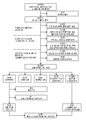
도 1: 블리츠(BliTz) 연구 (2005-2012)에서 참가자의 진단 정확도 연구의 보고에 대한 표준(STAandards for the Reporting of Diagnostic accuracy studies) (STARD) 다이어그램. Figure 1: STAandards for the Reporting of Diagnostic accuracy studies (STARD) diagrams for participants in the BliTz study (2005-2012).
도 2: 17종의 단백질 바이오마커에 대한 혈장 수준의 박스(box) 플롯: (a) CRC 사례와 대조군 간; (b) 초기 단계 (I/II) 및 진행된 단계 (III/IV) CRC. 박스의 기저부 및 최상부는 첫 번째 (Q1) 및 세 번째 (Q3)의 4분위수를 나타내며, 상자의 중간 선은 중앙값이며; 상한치는 Q3에 사분위수 범위(interquartile range) (IQR)의 1.5배를 더한 것과 동등하며; 하한치는 Q1에서 IQR의 1.5배를 뺀 것과 동등하다. Figure 2: Plasma level box plots for 17 protein biomarkers: (a) between the CRC case and the control group; (b) Initial phase (I / II) and progressive phase (III / IV) CRC. The base and top of the box represent the quartile of the first (Q1) and third (Q3), the middle line of the box is the median; The upper limit is equal to Q3 plus 1.5 times the interquartile range (IQR); The lower limit is equal to Q1 minus 1.5 times IQR.
도 3: 8종-마커 알고리즘에 대한 수신기 작동 특성 곡선의 비교: (a) 훈련 집합(training set)과 독립적 검증 집합 간; (b) 독립적 검증 집합 중의 상이한 하위 군 간 (즉, 모든 CRC 사례에서, 종양 단계 Ⅰ/Ⅱ 및 종양 단계 Ⅲ/Ⅳ). Figure 3: Comparison of receiver operating characteristic curves for an 8-species-marker algorithm: (a) between a training set and an independent verification set; (b) between different subgroups of independent assays (ie, in all CRC cases, tumor stage I / II and tumor stage III / IV).
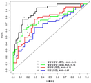
도 4: 결장직장암 훈련 집합, 결장직장암 독립적 검증 집합, 위암 집합 및 췌장암 집합 간에 8종-마커 알고리즘에 대한 수신기 작동 특성 곡선의 비교. Figure 4: Comparison of Receiver Operating Characteristic Curves for an 8-Marker Algorithm Between Colorectal Cancer Training Set, Colorectal Cancer Independent Assay Set, Gastric Cancer Set and Pancreatic Cancer Set.
실시예Example
물질 및 방법Materials and methods
1. 연구 설계 및 연구 모집단1. Research Design and Research Population
이 분석은 블리츠 연구 ("Begleitende Evaluierung innovativer Testverfahren zur Darmkrebsfrueherkennung")의 맥락에서 수행하였다. 간단히 말하면, 블리츠는 2005년 11월부터 남서 독일에서 20건의 위장병 진료(gastroenterology practice)와 협력하여 수행된 스크리닝 대장내시경 검사의 참가자 간에 계속 진행 중인 연구이며, 이는 CRC의 조기 발견을 위한 신규 유망한 바이오마커를 평가하는 것을 목표로 한다. 참가자를 모집하고, 전형적으로 스크리닝 대장내시경 검사 전 약 1주 전에, 예비 방문에서 상기 진료에서 혈액 샘플을 채취한다. This analysis was carried out in the context of the Blitz study ("Begleitende Evaluierung innovativer Testverfahren zur Darmkrebsfrueherkennung"). Briefly, Blitz is an ongoing study among participants in a screening colonoscopy conducted in collaboration with 20 gastroenterology practices in South-West Germany since November 2005. This is a new, promising biomarker for the early detection of CRC To be evaluated. Participants are recruited and a blood sample is taken from the care at the pre-visit, typically about a week before the screening colonoscopy.
이 분석을 위해, 다음의 제외 기준을 적용하여 적당한 혈액 샘플이 없는 참가자, 진정한 스크리닝 환경(true screening setting)을 나타내지 않는 참가자, 및 스크리닝 대장내시경 검사에서 잠재적으로 위음성 결과를 가진 참가자를 제외하였다: 스크리닝 대장내시경 검사 후 채취한 혈액 샘플, 또는 혈액 채취일이 알려지지 않은 혈액 샘플, CRC 또는 염증성 장 질환의 병력, 지난 5년 내에 이전 대장내시경 검사 병력, 또는 알려지지 않은 대장내시경 검사 병력, 불완전한 대장내시경 검사 또는 불충분한 장 정결(bowel preparation) (단지 대조군을 위한 후자의 2가지 기준). 2005-2012년 (N=4345)에 모집된 블리츠 연구의 나머지 참가자로부터, 새로 발견된 CRC를 가진 모든 35건의 이용 가능한 사례를 분석에 포함시켰다. 비교를 위해, 본 발명자들은 결장직장 신생물이 없는 54명의 대조군의 대표적 샘플을 포함시켰다. 이 연구는 CRC를 가진 환자가 평균적으로 약간 더 나이가 많고 다소 높은 비율의 남성을 포함할 것으로 예상되는 진정한 스크리닝 모집단에서 수행되었기 때문에, 본 발명자들은 이들 인자에 대해 일치시키지 않았는데, 그 이유는 이것이 이러한 설정에서 특이성의 편향 추정치(biased estimate)를 야기할 수 있기 때문이다. For this analysis, the following exclusion criteria were applied to exclude participants without appropriate blood samples, participants who did not exhibit true screening settings, and participants with potentially false negative results from screening colonoscopy: Screening A blood sample taken after a colonoscopy, or a blood sample of unknown blood sampling date, a history of CRC or inflammatory bowel disease, a previous history of colonoscopy, or an unknown colonoscopy history, an incomplete colonoscopy Insufficient bowel preparation (the latter two criteria for control only). From the remaining participants in the Blitz study recruited in 2005-2012 (N = 4345), all 35 cases with newly discovered CRC were included in the analysis. For comparison, we included representative samples of 54 control groups without colorectal neoplasia. We did not match these factors because the study was performed on a true screening population where patients with CRC were expected to include on average a slightly older and somewhat higher proportion of males, Because it can cause a biased estimate of the specificity in the setting.
독립적 검증을 위해, 본 발명자들은 또한 54건의 추가의 CRC 사례 (진단 후 그러나 치료의 개시 전에 하이델베르그시내 및 하이델베르그시 주변의 4개 병원에서 모집) 및 블리츠 연구로부터 신생물이 없는 38명의 추가의 무작위 선택된 대조군을 포함시켰다.For independent verification, the inventors also found that there were 38 additional randomized, non-neoplastic (50%), non-neoplastic, and non-neoplastic combinations from 54 additional CRC cases (recruitment at four hospitals around the city of Heidelberg and Heidelberg city after diagnosis, A control group was included.
대장내시경 검사 및 조직학 보고서 (블리츠 연구) 및 병원 기록 (독립적 검증 집합에 대한 54건의 CRC 사례)을 모든 참가자로부터 수집하였다. 관련 정보는 혈액 검사 결과를 알지 못하는 2명의 연구 보조원에 의해 독립적으로 추출하였다. 종양 단계는 UICC TNM 분류에 따라 분류하였다. Colonoscopy and histology reports (Blitz studies) and hospital records (54 CRC cases for independent assays) were collected from all participants. The relevant information was independently extracted by two research assistants who did not know the blood test results. Tumor stages were classified according to UICC TNM classification.
2. 실험실 절차2. Laboratory Procedure
2.1. 샘플 준비2.1. sample preparation
고지에 입각한 동의서를 제공하는 참가자로부터의 혈액 샘플을 대장내시경 검사 (블리츠 연구)를 위한 장 정결 전에 또는 EDTA 튜브에서 대장 수술 또는 신보강 화학요법(neoadjuvant chemotherapy) (임상 환경으로부터의 54건의 CRC 사례) 이전에 수집하여야 하였다. 혈액 샘플을 4℃에서 10분 동안 2123 g에서 즉시 원심분리하고 상청액을 새로운 튜브에 옮기고, 냉각 체인(cool chain)에서 DKFZ에서 바이오뱅크로 이송시켰으며, 여기서 혈장 샘플을 분석 때까지 -80℃에 보관하였다.Blood samples from participants providing informed consent were collected before colon cleansing for colorectal endoscopy (Blitz study) or in neoadjuvant chemotherapy (54 CRC cases from clinical setting) in EDTA tubes ). Blood samples were immediately centrifuged at 2123 g for 10 min at 4 ° C, the supernatant was transferred to a new tube and transferred from the DKFZ to the biobank in the cool chain, where the plasma samples were stored at -80 ° C Respectively.
2.2. 실험실 측정2.2. Laboratory measurement
92종의 인간 종양-연관 단백질 바이오마커 (보충 표 S1에서의 전체 마커 목록)의 정량화를 가능하게 하는 프로시크(Proseek) 멀티플렉스 온콜로지(Multiplex Oncology) I96x96 (오링크 바이오사이언스(Olink Bioscience), 스웨덴 웁살라)을 사용하여 단백질 프로파일링을 수행하였다. 92종의 단백질 바이오마커의 패널은 발암, 예컨대 혈관 신생, 세포-세포 신호전달, 성장 제어 및 염증에 관련된 다양한 생물학적 기전을 반영한다. 모든 실험실 조작을 TATAA 바이오센터(Biocenter) (스웨덴 예테보리)의 프로시크 멀티플렉스 온콜로지 I96x96 사용자 매뉴얼에 따라 수행하였다. 요컨대, 프로시크 시약은 근접성 확장 검정(Proximity Extension Assay) (PEA) 기술을 기반으로 하며, 여기서 92종의 올리고뉴클레오티드로 표지된 항체 프로브 쌍을 샘플에 존재하는 그의 각각의 표적에 결합시킨다. PCR 리포터 서열은 근접성 의존적 DNA 중합 사건(event)에 의해 형성되며 후속적으로 실시간 PCR을 사용하여 검출되고 정량화된다. 4개의 내부 대조군 (2개의 인큐베이션 대조군, 1개의 확장 대조군 및 1개의 검출 대조군 포함)을 검정에 포함시켰다. 게다가, 각각의 단백질에 대한 검출의 하한치 (LOD)를 계산하는 데 사용된 음성 대조군의 3개의 복제물이 있었다. 연구 모집단에 관한 모든 정보는 실험실 운영자에게 알리지 않았다.Proseek Multiplex Oncology I 96x96 (Olink Bioscience), which allows quantification of 92 human tumor-associated protein biomarkers (a full list of markers in Supplemental Table S1) ), Uppsala, Sweden) to profile protein profiling Respectively. A panel of 92 protein biomarkers reflects a variety of biological mechanisms involved in carcinogenesis, such as angiogenesis, cell-cell signaling, growth control and inflammation. All laboratory manipulations were performed at the TATAA Biocenter (Sweden) Gothenburg) according to the Procix Multiplex on Colloid I 96x96 User Manual. In short, procyclic reagents are based on the Proximity Extension Assay (PEA) technique, in which 92 pairs of oligonucleotide labeled antibody probes Bind to each target. PCR reporter sequences are formed by proximity-dependent DNA polymerisation events and are subsequently detected and quantified using real-time PCR. Four internal controls (including two incubation controls, one expansion control, and one detection control) were included in the assay. In addition, there were three replicates of the negative control used to calculate the lower limit of detection (LOD) for each protein. All information about the study population was not communicated to the laboratory operator.
3. 데이터 정규화 및 통계 분석3. Data normalization and statistical analysis
3.1 데이터 정규화3.1 Data normalization
미가공(raw) 데이터의 정규화는 제조업체로부터의 표준 프로토콜을 따랐으며 GenEx 소프트웨어 (멀티디(MultiD), 스웨덴 예테보리)의 오링크 마법사(Olink Wizard)를 통해 수행하였다. 각각의 데이터 포인트(data point)에 대해, 미가공 Cq-값 (log2 규모로)을 플루다임(Fluidigm) 실시간 PCR 분석 소프트웨어로부터 익스포트하였다. 정규화의 첫 번째 단계는 기술적 변형을 교정하기 위해 상응하는 샘플에 대한 확장 제어에 대한 미가공 Cq-값을 빼는 것이다. 계산된 Cq-값 (dCq-값)은 측정에서 결정된 음성 대조군에 대하여 추가로 정규화하였으며, 이는 ddCq-값 (이하 Cq-값, log2 규모로)을 산출하였으며, 추가 분석에 사용할 수 있을 것이다. LOD는 3개의 음성 대조군뿐만 아니라 3개의 계산된 표준 편차의 평균값으로서 정의되었다. 누락된 데이터 및 LOD 미만의 값을 가진 데이터는 다음의 통계 분석에서 LOD로 대체되었다.Normalization of raw data was performed using the Olink Wizard from GenEx software (MultiD, Gothenburg, Sweden) following the standard protocol from the manufacturer. For each data point, the raw Cq-value (log 2 Were exported from Fluidigm real-time PCR analysis software. The first step in normalization is to subtract the raw Cq-value for the expansion control for the corresponding sample to correct the technical distortion. The calculated Cq-value (dCq-value) was further normalized for the negative control determined in the measurements, which yielded the ddCq-value (Cq-value, log 2 scale) and could be used for further analysis. LOD was defined as the mean value of three calculated standard deviations as well as three negative control groups. Missing data and data with values less than LOD were replaced by LOD in the following statistical analysis.
3.2 통계 분석3.2 Statistical analysis
윌콕슨 순위 합계 검정(Wilcoxon Rank Sum Test) (이하, 윌콕슨 검정)을 사용하여 CRC 사례와 신생물이 없는 대조군 간의 혈장 단백질 수준 (Cq-값)을 맨 먼저 비교하였고, 벤자미니 운트 호크베르크(Benjamini & Hochberg) 방법을 다중 검정(multiple testing)에 추가로 사용하였다. 다음의 진단-관련 지표를 각각의 단백질 바이오마커의 진단 성능을 평가하는 데 사용하였다: 민감도 (진양성율(true positive rate)), 특이성 (진음성율(true negative rate)), 수신기 작동 특성 (ROC) 곡선, 및 ROC 곡선하 면적 (AUC). 각각의 개별 단백질 바이오마커에 대해, 로지스틱 회귀 모델(logistic regression model)을 사용하여 예측 모델을 구축하였다. 예측 모델로부터의 예측된 가능성을 기반으로 하여, AUC 및 그의 95% 신뢰 구간 (95% CI, 2000개의 부트스트랩 샘플을 기준으로 하여 계산됨)이 도출되었다. 게다가, 80% 및 90% 특이성을 산출하는 컷오프(cutoff)에서 각각의 개별 바이오마커의 민감도를 계산하였다. 진단 관련 지표의 직접 추정치 이외에, .632+ 부트스트랩 방법 (복원추출(replacement)포함 1000개의 부트스트랩 샘플)을 적용하여 진단 성능의 잠재적인 과대 평가를 조정하였다. 더욱이, CRC 사례와 대조군 간의 상당히 상이한 혈장 수준을 갖는 것으로 동정된 바이오마커의 경우, 단계-특이적 AUC (겉보기 및 .632+ 조정된 AUC)를 또한 계산하였고, 들롱 검정(Delong test)을 사용하여 초기 단계 (즉, 종양 단계 I/II)와 진행된 단계 (즉, 종양 단계 III/IV) 간의 겉보기 AUC의 차이를 비교하였다. The plasma protein level (Cq-value) between the CRC case and the control without neoplasia was firstly compared using the Wilcoxon Rank Sum Test (Wilcoxon test) Benjamini & Hochberg) method for multiple testing Were used. The following diagnostic-related indicators were used to assess the diagnostic performance of each protein biomarker: sensitivity (true positive rate), specificity (true negative rate), receiver operating characteristics (ROC ) Curve, and the area under the ROC curve (AUC). For each individual protein biomarker, a predictive model was constructed using a logistic regression model. Based on the predicted likelihood from the predictive model, the AUC and its 95% confidence interval (95% CI, Lt; RTI ID = 0.0 > 2000 < / RTI > bootstrap samples). In addition, the sensitivity of each individual biomarker was calculated in a cutoff yielding 80% and 90% specificity. In addition to direct estimates of the diagnostic indicators, a .632+ bootstrap method (1000 bootstrap samples with replacement) was applied to adjust for potential overestimation of diagnostic performance. Moreover, in the case of biomarkers identified as having significantly different plasma levels between CRC cases and controls, step-specific AUC (apparent and .632+ adjusted AUC) was also calculated and compared using the Delong test Differences in apparent AUC between the early stage (ie, tumor stage I / II) and the advanced stage (ie, tumor stage III / IV) were compared.
모든 92종의 단백질 마커를 기반으로 하는 라소(Lasso) 로지스틱 회귀 모델을 적용함으로써 다중-마커(multi-marker) 알고리즘을 도출하였다. 예측 알고리즘의 잠재적인 과적합을 조정하기 위해 ".632+ 부트스트랩 서브 샘플 추출(subsampling) 접근법"을 다음과 같은 방식으로 수행하였다: i) 1000개의 부트스트랩 샘플을 생성시킨다 (서브 샘플 추출 방법, 복원추출 없는 부트스트랩); ii) 각각의 부트스트랩샘플 세트에 대해, 라소 로지스틱 회귀 절차를 적용하여 변수를 선택하고 예측 알고리즘을 구축한다; iii) 부트스트랩 샘플에 포함되지 않은 그러한 환자에게 이 알고리즘을 적용하여 각각의 부트스트랩 샘플에 대한 예측 오류의 부트스트랩 추정치를 얻는다; iv) .632 + 방법을 사용하여 이들 결과를 추가로 조정하여 원래 알고리즘의 예후 AUC에 대한 거의 비편향 추정치(unbiased estimate)를 얻는다. 모든 CRC 사례를 포함하여 알고리즘의 구축을 행하였다. 모든 CRC 사례에 대해 마찬가지로 평가를 수행하였으며, 게다가 초기 및 진행된 종양 단계에서 CRC 사례에 대해 별도로 수행하였다. 최종적으로, 독립적 검증 샘플에서, AUC 및 각각 80% 및 90% 특이성을 산출하는 컷오프에서의 민감도, 및 다중-마커 알고리즘의 그의 95% CI를 결정하였다.By applying a Lasso logistic regression model based on all 92 protein markers, a multi-marker algorithm Respectively. Adjust the potential over-sum of the prediction algorithm A ".632+ bootstrap subsampling approach" was performed in the following manner : i) Generate 1000 bootstrap samples (sub-sample extraction method, bootstrap without recovery extraction); ii) For each set of bootstrap samples, apply the Raso logistic regression procedure to select variables and build a prediction algorithm; iii) apply the algorithm to those patients not included in the bootstrap sample to obtain a bootstrap estimate of the predicted error for each bootstrap sample ; iv) Using the .632 + method, these results are further adjusted to obtain a nearly unbiased estimate of the prognostic AUC of the original algorithm. Algorithms were constructed including all CRC cases. All CRC cases were likewise assessed, and in addition, CRC cases were performed separately at early and advanced tumor stages. Finally, in an independent validation sample, AUC and Sensitivity to cutoff yielding 80% and 90% specificity respectively, and 95% CI of the multi-marker algorithm.
통계 소프트웨어 R 버전 3.0.3으로 통계 분석을 수행하였다. R 패키지 "Daim"을 사용하여 단일 마커에 대한 .632+ 부트스트랩 분석을 수행하였다. R 패키지 "glmnet"을 사용하여 다중-마커 분석을 위한 라소 로지스틱 회귀 분석을 수행하였다. 게다가, R 패키지 "peperr"와 "c060"을 적용하여 상기 기재된 "632+ 부트스트랩 서브 샘플 추출 접근법"을 수행하였다. 모든 검정은 양측이었으며 0.05 이하의 p-값을 통계적으로 유의한 것으로 간주하였다.Statistical analysis with statistical software R version 3.0.3 Respectively. R package "Daim" was used to perform a .632+ bootstrap analysis on a single marker. R package "glmnet" was used to perform a lasso logistic regression for multi-marker analysis. Furthermore, by applying R package "peperr" and "c060" The "632+ bootstrap sub-sample extraction approach" described above was performed. All assays were bilateral and p-values <0.05 were considered statistically significant.
실시예Example 1: 17종의1: 17 species 바이오마커의Biomarker 동정 Sympathy
도 1은 2005-2012년에 블리츠 연구에 등록된 모든 대상체로부터의 연구 참가자의 선택을 나타내는 진단 정확도 연구의 보고에 관한 표준 (STARD) 다이어그램을 제공한다. 이 최종 연구 샘플은 결장직장 신생물이 없는 54명의 대조군의 대표적 샘플과 비교된 35명의 CRC 환자를 포함하였다. 상기 대조군은 과형성 용종(hyperplastic polyp)을 가진 6명의 참가자 및 결장직장 용종이 없는 48명의 참가자를 포함하였다.Figure 1 provides a standard (STARD) diagram of the reporting of diagnostic accuracy studies showing selection of study participants from all subjects enrolled in the Blitz study in 2005-2012. This final study sample included 35 CRC patients compared to a representative sample of 54 controls without colorectal neoplasia. The control group included 6 participants with hyperplastic polyp and 48 participants without colorectal polyps.
표 1은 CRC 사례 군 및 대조군에서의 사회 인구학적 특성의 분포를 제시한다. 대조군은 사례보다 평균적으로 약간 더 젊었다 (평균 ± 표준 편차: 62.8 ± 7.0세 대 66.9 ± 6.5세). 결장직장 신생물이 없는 사람들의 50.0%와 비교하여, CRC를 가진 환자의 71.4%는 남성이었다. 대략 동등한 비율의 환자가 초기 (단계 I/II) 및 진행된 단계 (단계 III/IV)로 진단되었으며, 결장 및 직장암을 가진 환자의 수가 동등하였다.Table 1 presents the distribution of socio-demographic characteristics in the CRC case group and the control group. The control group was slightly younger on average (mean ± SD: 62.8 ± 7.0 vs. 66.9 ± 6.5). Compared to 50.0% of people without colon rectal neoplasia, 71.4% of patients with CRC were males. Approximately equal proportions of patients were diagnosed with early (stage I / II) and advanced stages (stage III / IV), and the number of patients with colon and rectal cancer was equal.
전반적으로, CRC 사례와 대조군 간에 상당히 상이한 혈장 수준을 나타내는 17종의 단백질 마커가 있었다 (표 2). 다중 검정을 위한 컷오프 수준으로서 25% 위양성율 (FDR)을 사용하였을 때, 모든 17종의 바이오마커는 여전히 통계적으로 유의하였다.Overall, there were 17 protein markers showing significantly different plasma levels between CRC cases and controls (Table 2). When using the 25% false positive rate (FDR) as the cutoff level for multiple assays, all 17 biomarkers were still statistically significant.


발암배아성 항원 (CEA), 성장 분화 인자 15 (GDF-15) 및 암피레귤린 (AREG)은 5%의 FDR 역치를 충족시켰다. 여성에서보다 남성에서 혈장 수치가 통계적으로 유의하게 보다 높은 것으로 밝혀진, 전립선 특이적 항원 (PSA)을 제외하고는, 모든 나머지 다른 16종 바이오마커는 결장직장 신생물이 없는 대조군의 군 내에서 성별 또는 연령과 통계적으로 유의한 관계를 나타내지 않았다 (p-값>0.05). 게다가, 자기 기입식 설문지(self-administrated questionnaires)에서 과거에 어떠한 암 진단을 받은 적이 있는 것으로 보고한 4명의 참가자를 제외한 민감도 분석을 또한 수행하였으며, 거의 동일한 결과를 산출하였다.The carcinoembryonic antigen (CEA), growth differentiation factor 15 (GDF-15) and cancer peregrine (AREG) met the FDR threshold of 5%. Except for the prostate-specific antigen (PSA), which was found to have a statistically significantly higher plasma level in men than in women, all other 16 biomarkers were sex- or age-matched within the control group without colon rectal neoplasia There was no statistically significant relationship with age (p-value> 0.05). In addition, a sensitivity analysis, excluding four participants who reported having previously undergone any cancer diagnosis in a self-administered questionnaires, was also performed and yielded nearly identical results.

이들 17종의 단백질 마커 중에서, 9종의 단백질 마커가 과발현되었고, 8종의 단백질 마커는 대조군과 비교하여 CRC 사례에서 보다 낮은 수준을 나타냈다 (표 2). 이들 17종의 마커의 .632 + 조정된 AUC는 0.70 내지 0.55 범위였다. AREG, CEA, GDF-15 및 인터류킨 6 (IL-6)을 포함한 4종의 마커는 나머지 다른 것들보다 실질적으로 더 양호한 진단 성능을 산출하였으며, .632 + 조정된 AUC는 0.65 이상이었다. 컷오프 값이 80% 특이성을 산출하도록 설정되었을 때, 최고 .632+ 조정된 민감도가 CEA (52%)의 경우 관찰되었다. 컷-오프 값을 90% 특이성을 산출하도록 설정하면, 최고 .632+ 조정된 민감도가 AREG (36%)의 경우 관찰되었다.Of these 17 protein markers, 9 protein markers were overexpressed and 8 protein markers were lower than in the CRC case (Table 2). The .632 + adjusted AUC of these 17 markers ranged from 0.70 to 0.55. Four markers, including AREG, CEA, GDF-15 and interleukin 6 (IL-6), yielded substantially better diagnostic performance than the others and a .632 + adjusted AUC of 0.65 or greater. When the cutoff value was set to produce 80% specificity, a maximum of .632+ adjusted sensitivity was observed for CEA (52%). When cut-off values were set to produce 90% specificity, the highest .632+ adjusted sensitivity was observed for AREG (36%).
도 2는 초기 종양 단계 및 진행된 종양 단계에서 CRC 환자에 대한 17종의 단백질 마커에 대한 혈장 수준의 분포를 나타낸다. 7종의 단백질 마커 (IL-6, CXCL9, CXCL10, PSA, 카텝신-D, 카스파제-3 및 AREG)는 진행된 종양 단계에서보다 초기 종양 단계에서 더 높은 수준을 나타냈다. 그러나, IL-6에 대한 결과만 통계적으로 유의하였다 (p-값<0.05). 표 3은 초기 및 진행된 단계에서 CRC 환자 간의 이들 17종의 마커에 대한 ROC 분석의 비교를 나타낸다. 대부분의 마커 (13/17)는 진행된 종양 단계에서보다 초기 종양 단계에서 CRC 환자에서 더 높은 조정된 AUC를 나타냈다. 그러나, 그 차이 중 어떠한 것도 통계적으로 유의하지 않았다. 3종의 마커 (AREG, IL-6 및 GDF-15)의 경우, 초기 종양 단계 CRC의 경우 .632 + 조정된 AUC가 0.70보다 더 높았다 (즉, 각각 0.76, 0.74 및 0.72). 대조적으로, CEA는 진행된 단계 CRC의 경우 최고 .632 + 조정된 AUC를 나타냈다 (0.75).Figure 2 shows the distribution of plasma levels for 17 protein markers for CRC patients at the early tumor stage and advanced tumor stage. Seven protein markers (IL-6, CXCL9, CXCL10, PSA, cathepsin-D, caspase-3 and AREG) displayed higher levels at the early tumor stage than at the advanced tumor stage. However, only the results for IL-6 were statistically significant (p-value <0.05). Table 3 shows a comparison of ROC analysis for these 17 markers between CRC patients at early and advanced stages. Most markers (13/17) showed a higher adjusted AUC in CRC patients at the earlier tumor stage than in advanced tumor stages. However, none of the differences were statistically significant. For the three markers (AREG, IL-6 and GDF-15), the .632 + adjusted AUC for the initial tumor stage CRC was higher than 0.70 (ie 0.76, 0.74 and 0.72, respectively). In contrast, CEA showed a maximum .632 + adjusted AUC for advanced CRC (0.75).
실시예Example 2: 8종의2: 8 species 바이오마커의Biomarker 결장직장암Colorectal cancer 진단 패널의 개발 Development of diagnostic panel
본 발명자들은 라소 로지스틱 회귀 모델을 사용하여 모든 92종의 단백질 바이오마커를 기반으로 하는 다중-마커 예측 알고리즘을 구축하였다. 다음의 8종의 마커를 선택하여 알고리즘에 포함시켰다: IFN-감마, EMMPRIN, ErbB4-Her4, PSA, CD69, AREG, HGF-수용체 및 CEA (알고리즘은 표 4에 나타냄). 겉보기 AUC는 0.88 (95% CI, 0.81-0.95)이었다. ".632+ 부트스트랩 서브 샘플 추출 접근법"을 통해, 이 알고리즘의 조정된 AUC는 0.77 (95% CI, 0.59-0.91)이었다. 주목할 점은, 이 알고리즘은 초기 단계 CRC 및 진행된 단계 CRC에 대해 유사한 진단 값 (.632+ 조정된 AUC: 각각 0.79 대 0.75)을 나타냈다.The present inventors constructed a multi-marker prediction algorithm based on all 92 protein biomarkers using the LASO logistic regression model. The following eight markers were selected and included in the algorithm: IFN-gamma, EMMPRIN, ErbB4-Her4, PSA, CD69, AREG, HGF-receptor and CEA (algorithms shown in Table 4). The apparent AUC was 0.88 (95% CI, 0.81-0.95). Through the ".632+ bootstrap sub-sample extraction approach", the adjusted AUC of this algorithm was 0.77 (95% CI, 0.59-0.91). Notably, the algorithm showed similar diagnostic values (.632 + adjusted AUC: 0.79 vs. 0.75, respectively) for the early stage CRC and advanced stage CRC.

최종적으로, 본 발명자들은 54건의 CRC 사례 및 결장직장 신생물이 없는 38명의 대조군을 포함한, 독립적 검증 집합에서 이러한 8종-마커 알고리즘을 또한 검증하였다. 이 검증 집합의 연령 분포는, 비록 사례 둘 다 및 대조군이 다소 더 낮은 비율의 남성을 포함하였긴 하였지만, 스크리닝 환경으로부터 주요 연구에서의 연령 분포와 유사하였다. 독립적 검증 집합에서의 사례의 종양 단계 분포는 독일 스크리닝 대장내시경 검사 등록에 따라 스크리닝 대장내시경 검사에서 발견된 CRC 사례의 단계 분포와 유사하였다. 표 5 및 도 3은 독립적 검증 집합에서 CRC 예측을 위한 8종-마커 알고리즘의 진단 성능을 나타낸다. AUC는 0.76 (95% CI, 0.65-0.85)이었고, 80% 및 90% 특이성을 산출하는 컷오프에서의 민감도는 각각 65% (95% CI, 41-80%) 및 44% (95% CI, 24-72%)이었다. 이러한 독립적 검증 집합에서, 진단 성능은 초기 단계 질환의 경우보다 진행된 단계의 경우 더 양호하였다 (AUC: 각각 0.84 대 0.72).Finally, we also validated these 8-marker algorithms in an independent set of assays, including 54 CRC cases and 38 controls without colon rectal neoplasms. The age distribution of this assertion set was similar to the age distribution in the main study from the screening environment, although both cases and controls contained somewhat lower rates of males. The tumor stage distribution of cases in the independent assays set was similar to the stage distribution of CRC cases found in screening colonoscopy following registration with the German screening colonoscopy. Table 5 and Figure 3 show the diagnostic performance of the 8-marker algorithm for CRC prediction in the independent verification set. (95% CI, 41-80%) and 44% (95% CI, 24%) in the cutoff yielding 80% and 90% specificity, respectively -72%). In this independent assortment of assays, diagnostic performance was better for advanced stages than for early stage disease (AUC: 0.84 vs. 0.72, respectively).

실시예Example 3: 위암 및 췌장암의 진단에서 8종의 3: Eight species in the diagnosis of gastric cancer and pancreatic cancer 바이오마커의Biomarker 진단 패널의 검증 Verification of diagnostic panel
본 발명의 8종의 바이오마커 패널의 진단 값은 췌장암 및 위암 둘 다 (도 4)에 대해서 또한 검증될 수 있었으며, 이는 결장직장암뿐만 아니라, 암의 진단을 위한 8종의 바이오마커 패널의 일반적인 적용 가능성을 나타내는 것이다.The diagnostic values of the eight biomarker panels of the present invention could also be verified for both pancreatic cancer and stomach cancer (Figure 4), which is not only a common colorectal cancer, but also a common application of eight biomarker panels for the diagnosis of cancer It shows the possibility.
Claims (14)
(a) 대상체로부터 생물학적 샘플을 제공하는 단계,
(b) 생물학적 샘플 중의, AREG, CEA, IL-6, GDF-15, HGF-수용체, CXCL9, ErbB4-Her4, CXCL10, Flt3L, VEGFR-2, CD69, CXCL5, PSA, EMMPRIN, 카텝신-D, 카스파제-3, TNF-알파, 및 INF-감마로 이루어진 군으로부터 선택된 적어도 2종 이상의 바이오마커의 수준을 결정하는 단계
를 포함하며, 여기서 건강한 대조군 또는 기준값과 비교하여 단계 (b)에서 결정된 바와 같은 대상체로부터의 생물학적 샘플 중의 적어도 2종 이상의 바이오마커의 감별 수준이 대상체에서 암 질환의 존재에 대한 지표인, 비침습적 방법.As a non-invasive method for monitoring the diagnosis, prognosis, stratification and / or therapy of cancer in a subject,
(a) providing a biological sample from a subject,
(b) a biological sample that comprises at least one of AREG, CEA, IL-6, GDF-15, HGF-receptor, CXCL9, ErbB4-Her4, CXCL10, Flt3L, VEGFR-2, CD69, CXCL5, PSA, EMMPRIN, Determining the level of at least two or more biomarkers selected from the group consisting of caspase-3, TNF-alpha, and INF-gamma
Wherein a non-invasive method, wherein the differential level of at least two biomarkers in a biological sample from a subject as determined in step (b) compared to a healthy control or reference value is indicative of the presence of cancer disease in the subject .
The use of a compound according to any one of claims 1 to 13 for the manufacture of a medicament for the treatment of a disease or condition selected from the group consisting of CEA, AREG, IL-6, GDF-15, HGF-receptor, CXCL9, ErbB4- Her4, CXCL10, Flt3L, VEGFR- The use of an antibody or derivative thereof directed against any one of a biomarker selected from CXCL5, PSA, EMMPRIN, cathepsin-D, caspase-3, TNF-alpha, and INF-gamma.
Applications Claiming Priority (3)
| Application Number | Priority Date | Filing Date | Title |
|---|---|---|---|
| EP15161465.8A EP3073268A1 (en) | 2015-03-27 | 2015-03-27 | Biomarker panel for diagnosing cancer |
| EP15161465.8 | 2015-03-27 | ||
| PCT/EP2016/056314 WO2016156128A1 (en) | 2015-03-27 | 2016-03-23 | Biomarker panel for diagnosing cancer |
Publications (1)
| Publication Number | Publication Date |
|---|---|
| KR20170130441A true KR20170130441A (en) | 2017-11-28 |
Family
ID=52784968
Family Applications (1)
| Application Number | Title | Priority Date | Filing Date |
|---|---|---|---|
| KR1020177027302A KR20170130441A (en) | 2015-03-27 | 2016-03-23 | Biomarker panel for cancer diagnosis |
Country Status (5)
| Country | Link |
|---|---|
| US (2) | US10408839B2 (en) |
| EP (3) | EP3073268A1 (en) |
| KR (1) | KR20170130441A (en) |
| CA (1) | CA2980101A1 (en) |
| WO (1) | WO2016156128A1 (en) |
Cited By (2)
| Publication number | Priority date | Publication date | Assignee | Title |
|---|---|---|---|---|
| WO2021020882A1 (en) * | 2019-07-29 | 2021-02-04 | 서울대학교병원 | Biomarker for diagnosing pancreatic cancer, and use thereof |
| KR20220140294A (en) * | 2021-04-09 | 2022-10-18 | 주식회사 애티스랩 | Composition or kit for diagnosing cancer and method for diagnosis cancer |
Families Citing this family (16)
| Publication number | Priority date | Publication date | Assignee | Title |
|---|---|---|---|---|
| EP3073268A1 (en) * | 2015-03-27 | 2016-09-28 | Deutsches Krebsforschungszentrum Stiftung des Öffentlichen Rechts | Biomarker panel for diagnosing cancer |
| WO2019048588A1 (en) * | 2017-09-07 | 2019-03-14 | Deutsches Krebsforschungszentrum Stiftung des öffentlichen Rechts | Mixed protein and autoantibody biomarker panel for diagnosing colorectal cancer |
| US10586164B1 (en) | 2018-10-15 | 2020-03-10 | AIble Inc. | Interface for visualizing and improving model performance |
| US11409549B2 (en) | 2018-10-15 | 2022-08-09 | AIble Inc. | Interface for generating models with customizable interface configurations |
| US10936768B2 (en) * | 2018-10-15 | 2021-03-02 | Aible, Inc. | Interface for visualizing and improving model performance |
| EP3935191A2 (en) | 2019-01-02 | 2022-01-12 | Sabanci Üniversitesi Nanoteknoloji Arastirma Ve Uygulama Merkezi Sunum | Stress-regulated mirnas as novel cancer biomarkers |
| KR102247432B1 (en) * | 2019-01-21 | 2021-05-03 | 주식회사 에스씨엘헬스케어 | Biomarker composition for cancer diagnosis and use thereof |
| US20220214345A1 (en) * | 2019-05-08 | 2022-07-07 | Deutsches Krebsforschungszentrum Sitftung des öffentlichen Rechts | Colorectal cancer screening examination and early detection method |
| CN112782407A (en) * | 2019-11-01 | 2021-05-11 | 南华大学 | Method for detecting expression quantity of gamma interferon induced monocyte protein in serum and application |
| WO2021119761A1 (en) | 2019-12-20 | 2021-06-24 | Hudson Institute of Medical Research | Cxcl10 binding proteins and uses thereof |
| CN115956204A (en) * | 2020-03-19 | 2023-04-11 | 先进标志物探索公司 | Protein profile for screening the general population for colorectal cancer and/or precancerous stages thereof |
| CN112345757B (en) * | 2020-11-13 | 2021-10-22 | 吉林大学 | Application of SCTAG in preparation of kit for diagnosing gastric cancer |
| CN112522414A (en) * | 2020-12-30 | 2021-03-19 | 北京泱深生物信息技术有限公司 | Biomarker for gastric cancer diagnosis and derivative product thereof |
| CN112522413A (en) * | 2020-12-30 | 2021-03-19 | 北京泱深生物信息技术有限公司 | Biomarker for evaluating gastric cancer risk and application thereof |
| CN112795648A (en) * | 2020-12-30 | 2021-05-14 | 北京泱深生物信息技术有限公司 | Gastric cancer diagnostic product |
| CN116593702B (en) * | 2023-05-11 | 2024-04-05 | 杭州广科安德生物科技有限公司 | Biomarker and diagnostic system for lung cancer |
Family Cites Families (14)
| Publication number | Priority date | Publication date | Assignee | Title |
|---|---|---|---|---|
| US20040231909A1 (en) * | 2003-01-15 | 2004-11-25 | Tai-Yang Luh | Motorized vehicle having forward and backward differential structure |
| CA2557438C (en) * | 2004-02-19 | 2017-01-17 | Yale University | Identification of cancer protein biomarkers using proteomic techniques |
| EP1917528B1 (en) * | 2005-08-24 | 2011-08-17 | Bristol-Myers Squibb Company | Biomarkers and methods for determining sensitivity to epidermal growth factor receptor modulators |
| EP1777523A1 (en) * | 2005-10-19 | 2007-04-25 | INSERM (Institut National de la Santé et de la Recherche Médicale) | An in vitro method for the prognosis of progression of a cancer and of the outcome in a patient and means for performing said method |
| US7695913B2 (en) * | 2006-01-11 | 2010-04-13 | Genomic Health, Inc. | Gene expression markers for colorectal cancer prognosis |
| US20090275057A1 (en) * | 2006-03-31 | 2009-11-05 | Linke Steven P | Diagnostic markers predictive of outcomes in colorectal cancer treatment and progression and methods of use thereof |
| JP2010526995A (en) * | 2007-05-10 | 2010-08-05 | エフ.ホフマン−ラ ロシュ アーゲー | Use of TIMP-1 as a marker for colorectal cancer |
| GB201103726D0 (en) * | 2011-03-04 | 2011-04-20 | Immunovia Ab | Method, array and use thereof |
| WO2013152989A2 (en) * | 2012-04-10 | 2013-10-17 | Eth Zurich | Biomarker assay and uses thereof for diagnosis, therapy selection, and prognosis of cancer |
| DK2872646T3 (en) * | 2012-07-12 | 2017-12-04 | Université Paris Descartes | PROCEDURES FOR PREDICTING SURVIVAL TIME AND TREATMENT RESPONSE OF A PATIENT, SUFFERING A FAST CANCER WITH A SIGNATURE OF AT LEAST 7 GENES |
| RU2015121366A (en) * | 2012-11-05 | 2016-12-27 | Новидженикс Са | COMBINATIONS OF BIOMARKERS FOR COLORECTAL TUMORS |
| US20140236166A1 (en) * | 2013-02-15 | 2014-08-21 | The Board Of Trustees Of The Leland Stanford Junior University | Biomarkers for distinguishing benign, pre-malignant, and malignant pancreatic cysts |
| US20150038365A1 (en) * | 2013-08-01 | 2015-02-05 | Meso Scale Technologies, Llc | Lung cancer biomarkers |
| EP3073268A1 (en) * | 2015-03-27 | 2016-09-28 | Deutsches Krebsforschungszentrum Stiftung des Öffentlichen Rechts | Biomarker panel for diagnosing cancer |
-
2015
- 2015-03-27 EP EP15161465.8A patent/EP3073268A1/en not_active Withdrawn
-
2016
- 2016-03-23 US US15/561,755 patent/US10408839B2/en not_active Expired - Fee Related
- 2016-03-23 EP EP21160213.1A patent/EP3885768A1/en not_active Withdrawn
- 2016-03-23 WO PCT/EP2016/056314 patent/WO2016156128A1/en active Application Filing
- 2016-03-23 CA CA2980101A patent/CA2980101A1/en not_active Abandoned
- 2016-03-23 EP EP16714316.3A patent/EP3274718A1/en not_active Withdrawn
- 2016-03-23 KR KR1020177027302A patent/KR20170130441A/en unknown
-
2019
- 2019-08-07 US US16/534,347 patent/US20200033352A1/en not_active Abandoned
Cited By (5)
| Publication number | Priority date | Publication date | Assignee | Title |
|---|---|---|---|---|
| WO2021020882A1 (en) * | 2019-07-29 | 2021-02-04 | 서울대학교병원 | Biomarker for diagnosing pancreatic cancer, and use thereof |
| KR20220140294A (en) * | 2021-04-09 | 2022-10-18 | 주식회사 애티스랩 | Composition or kit for diagnosing cancer and method for diagnosis cancer |
| KR20230084112A (en) * | 2021-04-09 | 2023-06-12 | 주식회사 애티스랩 | Composition or kit for diagnosing cancer and method for diagnosis cancer |
| KR20240001085A (en) * | 2021-04-09 | 2024-01-03 | 주식회사 애티스랩 | Composition or kit for diagnosing cancer and method for diagnosis cancer |
| KR20240046154A (en) * | 2021-04-09 | 2024-04-08 | 주식회사 애티스랩 | Composition or kit for diagnosing cancer and method for diagnosis cancer |
Also Published As
| Publication number | Publication date |
|---|---|
| US10408839B2 (en) | 2019-09-10 |
| US20180080937A1 (en) | 2018-03-22 |
| EP3885768A1 (en) | 2021-09-29 |
| US20200033352A1 (en) | 2020-01-30 |
| CA2980101A1 (en) | 2016-10-06 |
| EP3274718A1 (en) | 2018-01-31 |
| WO2016156128A1 (en) | 2016-10-06 |
| EP3073268A1 (en) | 2016-09-28 |
Similar Documents
| Publication | Publication Date | Title |
|---|---|---|
| KR20170130441A (en) | Biomarker panel for cancer diagnosis | |
| JP6630766B2 (en) | Pancreatic cancer diagnostic composition and pancreatic cancer diagnostic method using the same | |
| JP5591711B2 (en) | Marker panel for colorectal cancer | |
| US20220214345A1 (en) | Colorectal cancer screening examination and early detection method | |
| WO2019048588A1 (en) | Mixed protein and autoantibody biomarker panel for diagnosing colorectal cancer | |
| CN111172279B (en) | Model for diagnosing lung cancer by combined detection of peripheral blood methylation gene and IDH1 | |
| ES2703714T3 (en) | Methods of identification, evaluation, prevention and therapy of pulmonary diseases and kits thereof, including the identification, evaluation, prevention and therapy of diseases on the basis of gender | |
| WO2015158652A2 (en) | New biomarkers for metastatic breast cancer | |
| CN116113712A (en) | Prognostic biomarkers for cancer | |
| JP2019527361A (en) | Colorectal cancer combination test | |
| KR20190013707A (en) | Use of the nucleosome-transcription factor complex for cancer detection | |
| JP2012526543A5 (en) | ||
| WO2021116057A1 (en) | Biomarker panel for diagnosing colorectal cancer | |
| EP3215851B1 (en) | Lung cancer sub-typing method | |
| JP2021144052A (en) | Methods of detecting relapse of lung adenocarcinoma based on marker human epididymis protein 4 (he4) and related uses | |
| KR20210134946A (en) | Protein signature for diagnosis of colorectal cancer and/or its precancerous stage | |
| KR20160021531A (en) | Maker for the diagnosis of gastric cancer | |
| EP2895863A1 (en) | New biomarkers for the diagnosis and/or prognosis of clear cell renal cell carcinoma | |
| Tamura et al. | Prognostic significance of CEA reduction rate in patients with abnormally high preoperative CEA levels who underwent surgery for lung cancer | |
| KR20160053875A (en) | Maker for the diagnosis of gastric cancer | |
| Yang et al. | Development and validation of peritoneal metastasis in gastric cancer based on simplified clinicopathological features and serum tumour markers | |
| JP2024504105A (en) | Methods for detection of lung cancer | |
| EP3903105A1 (en) | Method of diagnosis |