KR20170101847A - Calculation Method of Molecules and Uses Thereof - Google Patents
Calculation Method of Molecules and Uses Thereof Download PDFInfo
- Publication number
- KR20170101847A KR20170101847A KR1020170104267A KR20170104267A KR20170101847A KR 20170101847 A KR20170101847 A KR 20170101847A KR 1020170104267 A KR1020170104267 A KR 1020170104267A KR 20170104267 A KR20170104267 A KR 20170104267A KR 20170101847 A KR20170101847 A KR 20170101847A
- Authority
- KR
- South Korea
- Prior art keywords
- probe
- nucleic acid
- target molecule
- analysis
- signal
- Prior art date
Links
Classifications
-
- C—CHEMISTRY; METALLURGY
- C12—BIOCHEMISTRY; BEER; SPIRITS; WINE; VINEGAR; MICROBIOLOGY; ENZYMOLOGY; MUTATION OR GENETIC ENGINEERING
- C12Q—MEASURING OR TESTING PROCESSES INVOLVING ENZYMES, NUCLEIC ACIDS OR MICROORGANISMS; COMPOSITIONS OR TEST PAPERS THEREFOR; PROCESSES OF PREPARING SUCH COMPOSITIONS; CONDITION-RESPONSIVE CONTROL IN MICROBIOLOGICAL OR ENZYMOLOGICAL PROCESSES
- C12Q1/00—Measuring or testing processes involving enzymes, nucleic acids or microorganisms; Compositions therefor; Processes of preparing such compositions
- C12Q1/68—Measuring or testing processes involving enzymes, nucleic acids or microorganisms; Compositions therefor; Processes of preparing such compositions involving nucleic acids
- C12Q1/6876—Nucleic acid products used in the analysis of nucleic acids, e.g. primers or probes
-
- C—CHEMISTRY; METALLURGY
- C12—BIOCHEMISTRY; BEER; SPIRITS; WINE; VINEGAR; MICROBIOLOGY; ENZYMOLOGY; MUTATION OR GENETIC ENGINEERING
- C12Q—MEASURING OR TESTING PROCESSES INVOLVING ENZYMES, NUCLEIC ACIDS OR MICROORGANISMS; COMPOSITIONS OR TEST PAPERS THEREFOR; PROCESSES OF PREPARING SUCH COMPOSITIONS; CONDITION-RESPONSIVE CONTROL IN MICROBIOLOGICAL OR ENZYMOLOGICAL PROCESSES
- C12Q1/00—Measuring or testing processes involving enzymes, nucleic acids or microorganisms; Compositions therefor; Processes of preparing such compositions
- C12Q1/68—Measuring or testing processes involving enzymes, nucleic acids or microorganisms; Compositions therefor; Processes of preparing such compositions involving nucleic acids
- C12Q1/6813—Hybridisation assays
- C12Q1/6834—Enzymatic or biochemical coupling of nucleic acids to a solid phase
-
- C—CHEMISTRY; METALLURGY
- C12—BIOCHEMISTRY; BEER; SPIRITS; WINE; VINEGAR; MICROBIOLOGY; ENZYMOLOGY; MUTATION OR GENETIC ENGINEERING
- C12Q—MEASURING OR TESTING PROCESSES INVOLVING ENZYMES, NUCLEIC ACIDS OR MICROORGANISMS; COMPOSITIONS OR TEST PAPERS THEREFOR; PROCESSES OF PREPARING SUCH COMPOSITIONS; CONDITION-RESPONSIVE CONTROL IN MICROBIOLOGICAL OR ENZYMOLOGICAL PROCESSES
- C12Q2525/00—Reactions involving modified oligonucleotides, nucleic acids, or nucleotides
- C12Q2525/10—Modifications characterised by
- C12Q2525/191—Modifications characterised by incorporating an adaptor
-
- C—CHEMISTRY; METALLURGY
- C12—BIOCHEMISTRY; BEER; SPIRITS; WINE; VINEGAR; MICROBIOLOGY; ENZYMOLOGY; MUTATION OR GENETIC ENGINEERING
- C12Q—MEASURING OR TESTING PROCESSES INVOLVING ENZYMES, NUCLEIC ACIDS OR MICROORGANISMS; COMPOSITIONS OR TEST PAPERS THEREFOR; PROCESSES OF PREPARING SUCH COMPOSITIONS; CONDITION-RESPONSIVE CONTROL IN MICROBIOLOGICAL OR ENZYMOLOGICAL PROCESSES
- C12Q2525/00—Reactions involving modified oligonucleotides, nucleic acids, or nucleotides
- C12Q2525/10—Modifications characterised by
- C12Q2525/205—Aptamer
-
- C—CHEMISTRY; METALLURGY
- C12—BIOCHEMISTRY; BEER; SPIRITS; WINE; VINEGAR; MICROBIOLOGY; ENZYMOLOGY; MUTATION OR GENETIC ENGINEERING
- C12Q—MEASURING OR TESTING PROCESSES INVOLVING ENZYMES, NUCLEIC ACIDS OR MICROORGANISMS; COMPOSITIONS OR TEST PAPERS THEREFOR; PROCESSES OF PREPARING SUCH COMPOSITIONS; CONDITION-RESPONSIVE CONTROL IN MICROBIOLOGICAL OR ENZYMOLOGICAL PROCESSES
- C12Q2600/00—Oligonucleotides characterized by their use
- C12Q2600/154—Methylation markers
-
- C—CHEMISTRY; METALLURGY
- C12—BIOCHEMISTRY; BEER; SPIRITS; WINE; VINEGAR; MICROBIOLOGY; ENZYMOLOGY; MUTATION OR GENETIC ENGINEERING
- C12Q—MEASURING OR TESTING PROCESSES INVOLVING ENZYMES, NUCLEIC ACIDS OR MICROORGANISMS; COMPOSITIONS OR TEST PAPERS THEREFOR; PROCESSES OF PREPARING SUCH COMPOSITIONS; CONDITION-RESPONSIVE CONTROL IN MICROBIOLOGICAL OR ENZYMOLOGICAL PROCESSES
- C12Q2600/00—Oligonucleotides characterized by their use
- C12Q2600/156—Polymorphic or mutational markers
Landscapes
- Chemical & Material Sciences (AREA)
- Life Sciences & Earth Sciences (AREA)
- Organic Chemistry (AREA)
- Proteomics, Peptides & Aminoacids (AREA)
- Zoology (AREA)
- Wood Science & Technology (AREA)
- Health & Medical Sciences (AREA)
- Engineering & Computer Science (AREA)
- Analytical Chemistry (AREA)
- Microbiology (AREA)
- Immunology (AREA)
- Molecular Biology (AREA)
- Biotechnology (AREA)
- Biophysics (AREA)
- Physics & Mathematics (AREA)
- Biochemistry (AREA)
- Bioinformatics & Cheminformatics (AREA)
- General Engineering & Computer Science (AREA)
- General Health & Medical Sciences (AREA)
- Genetics & Genomics (AREA)
- Measuring Or Testing Involving Enzymes Or Micro-Organisms (AREA)
Abstract
Description
본 발명은 일반적으로 하나 또는 그 이상의 표적분자의 탐색, 동정, 정량 및 생물학적 의미 결정 분야로 생체시료에서 표적분자, 탐색, 정량 및 생물학적 의미 결정을 하기 위한 방법, 키트 및 시스템에 관한 것이다. 또한, 본 발명은 표적분자를 지시하는 색상지시바코드 신호의 증강을 위한 재료 및 방법에 관한 것이다.The present invention relates generally to methods, kits and systems for determining target molecules, searching, quantifying and determining biological significance in biological samples in the field of searching, identifying, quantifying and determining biological significance of one or more target molecules. The present invention also relates to materials and methods for the enhancement of hue indicating barcode signals indicating target molecules.
본 발명은 일반적으로 시료에서 생체분자들의 탐색, 동정, 정량 및 생물학적 의미 결정 분야로 분자계수에 의해 구체적으로 구현하는 내용이다.In general, the present invention is specifically embodied by numerator coefficients in the field of biomolecular discovery, identification, quantification and determination of biological significance in a sample.
세포에서 DNA에 암호화된 유전정보는 DNA 자신의 복제에 의해 자손에게도 유전, 전달되고, 유전정보가 DNA에서 RNA, 그리고 RNA에서 단백질로 전달되어 발현된다. 모든 세포는 유전정보에 관련된 생체분자들로 구성되어 있고 이들의 존재양상은 생물체 기능에 중요한 영향을 준다. Genetic information encoded in DNA in a cell is inherited and transferred to offspring by replication of DNA itself, and genetic information is transferred from DNA to RNA and from RNA to protein. All cells are composed of biomolecules related to genetic information, and their presence pattern has an important effect on organism function.
표적분자(바이오마커) 또는 표적분자의 세트는 일반적으로 임상적으로 확인되거나, 그것이 선택된 본래의 의도된 용도를 위한 신뢰할 수 있는 생체분자이다. 생체분자는 질병의 발생 또는 진행과 병행하여 분비되거나 세포가 파괴되어 나오는 바이오마커들을 포함할 수 있고, 조직 또는 병소에 대한 말단 조직으로부터 혈류로 쉽게 퍼진다. The target molecule (biomarker) or set of target molecules is generally a clinically confirmed or trusted biomolecule for the intended intended use thereof. Biomolecules can include biomarkers that are secreted in parallel with the onset or progression of the disease, or which are destroyed by the cell, and are easily spread from the end-tissue to the tissue or lesion into the bloodstream.
대표적인 생체분자인 유전물질인 핵산 및 단백질이다. 유전정보가 발현과정에 영향을 주는 염색체 이상, DNA 메틸화 및 유전자 변이(Gene Variation) 등이 있다. 유전자변이는 단일염기다형성(Single Nucleotide Polymorphism, SNP)와 구조변이(Structural Variation) 등이 있다. SNP은 DNA 염기서열에서 하나의 염기서열 (A, T, G, C)의 차이를 보이는 유전적 변화 또는 변이를 의미한다. 구조변이는 결실, 역위, 첨가, 복제 등과 같은 DNA 구조변이를 의미한다. 유전자 변이는 표현형(phenotype)의 변화, 질병에 대한 민감성, 그리고 치료 약제에 대한 반응의 차이 등 개체 간의 차이를 결정짓는다고 알려졌으며, 특히 질환 발생과 진행과정에 관여하는 변이들을 질환연관 유전적 변이(Disease-associated Genetic Variants)라고 칭한다.It is nucleic acid and protein which are genetic material which is representative biomolecules. Chromosomal abnormalities affecting the expression process of genetic information, DNA methylation and gene variation. Genetic variations include single nucleotide polymorphism (SNP) and structural variability. SNP means a genetic change or mutation that shows the difference of one base sequence (A, T, G, C) in DNA base sequence. Structural mutations refer to DNA structural variations such as deletions, inversions, additions, and replication. Genetic mutations are known to determine individual differences, such as phenotype changes, susceptibility to disease, and response to therapeutic agents. In particular, mutations involved in disease development and progression are known as disease-associated genetic mutations (Disease-associated Genetic Variants).
각각의 세포에는 5만 내지 10만 개 정도의 유전자가 있지만, 세포는 선택적으로 유전자를 사용한다. 그 중 상당수는 세포 자신의 생명유지활동이나 일상적인 활동을 하기 위한 유전자이다. 내인성 표준 발현 유전자는, 특정 유전자의 기능을 밝히거나, 특정 기능의 유전자를 탐색하거나, 특정 조건의 생물체의 발현 양상 작성 및 그 외의 다양한 분자생물학적 목적에 의해, 특정 혹은 다수의 유전자 발현량의 비교를 위해 개발된 전령 RNA(messenger RNA; 이하 mRNA)의 정량분석을 이용하는 다양한 유전자발현 측정법은 개발되고 있다.Each cell has about 50,000 to 100,000 genes, but cells selectively use genes. Many of them are genes for life-sustaining activities or daily activities of cells themselves. The endogenous standard expression gene may be a gene that expresses the function of a specific gene or a gene of a specific function or the expression pattern of an organism under a specific condition and a variety of other molecular biological purposes, A variety of gene expression assays using quantitative analysis of messenger RNA (mRNA) have been developed.
대표적인 고속병렬분석 기술인 DNA 마이크로어레이는 단지 혼성화 방식에 의해서 표적 서열을 검출하기 때문에 교차반응에 의한 진양성 문제가 발생되어, 혼성화 시그널의 신뢰도를 개선하여야 하는 문제점이 있다. 또한, 종래의 마이크로어레이의 경우 혼성화 방식에 의해서 검출을 하기 때문에 혼성화 후 까다로운 세척 과정이 요구되는 문제점이 있으며, 혼성화 전에 표적서열을 단일가닥으로 만드는 과정이 필수적으로 요구된다. 한편, 최근에 발표된 on-chip PCR은 혼성화 또는 probe extension방식에 의해 검출하기 때문에, 기존 마이크로 어레이와 같이 heterogenous assay system이며, 실시간 검출이 불가능 하고 정확한 정량이 어렵다는 문제점이 있다. A DNA microarray, which is a typical high-speed parallel analysis technique, detects a target sequence only by a hybridization method. Therefore, there is a problem that a positive result is caused by a cross-reaction, thereby improving the reliability of the hybridization signal. In addition, in the case of a conventional microarray, detection is carried out by a hybridization method, which requires a complicated cleaning process after hybridization, and a process of making a target sequence into a single strand before hybridization is indispensably required. On the other hand, since the recently announced on-chip PCR is detected by hybridization or probe extension method, it is a heterogenous assay system like existing microarrays, and real-time detection is impossible and accurate quantification is difficult.
생체분자는 핵산이외에도 단백질, 펩티드, 탄수화물, 지질(lipid), 다당류, 당단백질, 호르몬, 수용체, 항원, 항체, 바이러스, 병원균, 독성 물질, 기질, 대사산물, 전이 상태 유사체(transition state analog), 보조 인자(cofactor), 저해제, 약, 염료, 영양분, 성장 인자, 세포, 조직 등을 포함하고, 이론적으로는 그 범위는 제한이 없으며 거의 모든 화학적 또는 생물학적 작동체(effector)가 될 것이며 다양한 크기의 분자들을 포함하고 있으며 이들에 대한 효율적인 실시간 검출 및 정확한 정량에 대한 개발이 요망되고 있다. The biomolecule may contain, in addition to nucleic acid, a protein, a peptide, a carbohydrate, a lipid, a polysaccharide, a glycoprotein, a hormone, a receptor, an antigen, an antibody, a virus, a pathogen, a toxic substance, a substrate, a metabolite, a transition state analog, But are not limited to, cofactors, inhibitors, drugs, dyes, nutrients, growth factors, cells, tissues and the like. The scope of the invention is to be limited to almost any chemical or biological effector, Molecules, and it is desired to develop efficient real-time detection and precise quantitation of these.
다양한 생체분자 분석법은 검출감도의 향상 및 다중 분석능의 향상에 있어서 발전을 거듭했지만, 여전히 분석비용절감, 분석시간의 단축, 민감도 향상 및 재현성 증대라는 기술적 과제에 직면해 있다.Various biomolecular analytical methods have been developed to improve the detection sensitivity and improve the multi-analyzing ability, but they still face the technical problem of reducing the analysis cost, shortening the analysis time, improving the sensitivity and increasing the reproducibility.
궁극적으로 생체시료의 생체분자 분석하거나 총체적으로 분석하는 연구는, 의학적으로 질병에 관련된 생체분자 분석 및 프로파일을 분석함으로써, 질병을 진단할 수 있는 생체분자, 치료성적을 분석할 수 있는 생체분자, 질병 발병 및 진행에 중요한 역할을 하는 생체분자, 질병에 대한 감수성 관련된 생체분자, 및 신약개발의 표적분자를 구명하는 것에 사용할 있을 것이다.Ultimately, biomolecule analysis or comprehensive analysis of biological samples can be performed by analyzing biomolecule analysis and profile related to a disease medically, biomolecules capable of diagnosing diseases, biomolecules capable of analyzing therapeutic results, Biomolecules that play an important role in development and progression, susceptibility-related biomolecules to disease, and target molecules in the development of new drugs.
상술한 종래의 생체분자들을 각각 독립적으로 분석하는 기술들의 문제점을 극복하여, 생체분자들을 보다 개선된 효율성 및 민감도로 실시간 분석할 수 있는 기술을 개발하기 위하여 연구한 결과, 본원 발명과 같이 다양한 종류의 생체분자들을 분석할 수 있는 방법을 개발함으로서 본 발명의 목적을 달성하였다.As a result of studying to develop a technology capable of real-time analysis of biomolecules with improved efficiency and sensitivity by overcoming the problems of techniques for independently analyzing the conventional biomolecules described above, The object of the present invention has been achieved by developing a method for analyzing biomolecules.
본 발명은 상기의 문제점을 해결하고 상기의 필요성에 의하여 안출된 것으로서 본 발명의 목적은 신호발생물질이 표지된 이중가닥핵산 그리고 신호발생물질이 표지되고 구성 염기쌍사이에 교차결합이 있는 이중가닥핵산인 신호태그, 및 이들로 구성된 신호발생영역을 제공하는 것이다.SUMMARY OF THE INVENTION The present invention has been accomplished to solve the above problems, and it is an object of the present invention to provide a double-stranded nucleic acid having a signal-generating substance labeled thereon and a double-stranded nucleic acid having a signal- A signal tag, and a signal generation area composed of the same.
본 발명의 다른 목적은 상기 복합체의 제조방법을 제공하는 것이다.Another object of the present invention is to provide a method for producing the composite.
본 발명의 또 다른 목적은 효율적인 생체분자를 분석하고 생물학적 의미를 결정하는 방법을 제공하는 것이다.It is a further object of the present invention to provide a method for analyzing efficient biomolecules and determining biological significance.
생체분자는 생체를 구성하고 기능을 담당하는 분자로 가장 주된 것은 단백질, 핵산, 다당류 및 지질과 같은 크기가 큰 거대 분자들과 저분자물질로 아미노산, 뉴클레오타이드, 단당류, 비타민 등의 유기물질, 철, 구리 등의 금속, 무기이온 등이다. 인간의 모든 세포는 생체분자로 구성되어 있고 이들의 존재양상은 생물체 기능에 중요한 영향을 준다. Biomolecules are molecules that constitute and function in the living body. Major molecules such as proteins, nucleic acids, polysaccharides, and lipids are small molecules and small molecules. Organic substances such as amino acids, nucleotides, monosaccharides and vitamins, Etc., and inorganic ions. All human cells are composed of biomolecules and their presence patterns have an important effect on organism function.
일반적으로 임상적으로 확인되거나, 그것이 선택된 본래의 의도된 용도를 위한 신뢰할 수 있는 생체분자들이 표적분자(바이오마커) 또는 표적분자의 세트이다. 생체분자는 질병의 발생 또는 진행과 병행하여 분비되거나 세포가 파괴되어 나오는 표적분자들을 포함할 수 있고, 조직 또는 병소에 대한 말단 조직으로부터 혈류로 쉽게 퍼진다. Generally, biomolecules that are clinically identified, or are trusted for their intended intended use, are a set of target molecules (biomarkers) or target molecules. A biomolecule may contain target molecules that are secreted in parallel with the onset or progression of the disease or that the cells are destroyed and spread easily from the end tissues to the tissue or lesion into the bloodstream.
본 발명에서 프로브는 표적분자들의 동정 및 정량을 위한 분자로 표적분자인 유전자 또는 단백질, 펩티드, 탄수화물, 지질(lipid), 다당류, 당단백질, 호르몬, 수용체, 항원, 항체, 바이러스, 병원균, 독성 물질, 기질, 대사산물, 전이 상태 유사체(transition state analog), 보조 인자(cofactor), 저해제, 약, 염료, 영양분, 성장 인자, 세포, 조직 등을 포함하고, 이론적으로는 그 범위는 제한이 없으며 거의 모든 화학적 또는 생물학적 작동체(effector)가 될 것이며 어떤 크기의 분자들이든 사용될 수 있는 분자의 동정 및 계수를 위한 분자를 의미한다. In the present invention, a probe is a molecule for identification and quantification of a target molecule, and a gene or a protein or a peptide, a carbohydrate, a lipid, a polysaccharide, a glycoprotein, a hormone, a receptor, an antigen, an antibody, a virus, a pathogen, , A substrate, a metabolite, a transition state analog, a cofactor, an inhibitor, a drug, a dye, a nutrient, a growth factor, a cell, a tissue and the like. Theoretically, It is meant to be all chemical or biological effectors, and molecules of any size are molecules for identification and counting of molecules that can be used.
바람직하게 표적분자가가 핵산인 경우는 프로브는 단일가닥핵산이며 상기 핵산이 아닌 생체분자인 경우는 리간드로일 수도 있다.Preferably, when the target molecule is a nucleic acid, the probe is a single-stranded nucleic acid, and in the case of a biomolecule other than the nucleic acid, the probe may be a ligand.
리간드는 특정 물질과 결합하고, 상호 작용 또는 그렇지 않으면 특정 물질과 연합하여 특정 물질의 기능에 작용하고, 변화시키고 또는 무효화하는 하나 또는 그 이상의 리간드에 특이적인 분자 또는 분자의 클러스터를 포함하는 구조를 언급한다. 리간드는 대표적으로 항체, 펩타이드, 핵산 및 압타머 등이 있다. 항체는 항원의 항원결정부(epitope)과 특이적 결합을 하여 항원-항체 반응을 일으키는 단백질 분자를 의미한다. 본 발명의 목적상, 항체는 본 발명의 마커에 대해 특이적으로 결합하는 항체를 의미하며, 이러한 항체는 얻어진 단백질로부터 통상적인 방법에 의해 제조될 수 있다. 여기에는 상기 단백질에서 만들어질 수 있는 부분 펩티드도 포함한다. A ligand refers to a structure that includes a cluster of molecules or molecules that are specific for one or more ligands that bind to, interact with, or otherwise associate with, a particular substance, and that affect, alter, or nullify the function of a particular substance do. Ligands typically include antibodies, peptides, nucleic acids, and plasmids. An antibody refers to a protein molecule that specifically binds to an antigen epitope of an antigen to cause an antigen-antibody reaction. For purposes of the present invention, an antibody refers to an antibody that specifically binds to a marker of the present invention, and such antibody can be prepared from the obtained protein by conventional methods. It also includes partial peptides that can be made from the protein.
압타머(aptamer)는 저분자 화합물로부터 단백질까지 다양한 종류의 물질에 높은 친화성과 특이성으로 결합할 수 있는 특성을 가지는 작은 (20~60 뉴클레오타이드) 단일가닥핵산 (DNA 혹은 RNA) 조각을 뜻한다.Aptamers are small (20 to 60 nucleotides) single-stranded nucleic acid (DNA or RNA) fragments that are capable of binding with high affinity and specificity to a wide variety of substances, from small molecules to proteins.
표적분자가 핵산인 경우 프로브는 표적분자인 핵산과 특이적 결합을 이룰 수 있는 짧게는 수 염기 내지 길게는 수백 염기에 해당하는 RNA 또는 DNA 등의 핵산 단편을 의미하며, 있어서 특정 mRNA의 존재 유무를 확인할 수 있다. 표적분자 핵산과 상보적인 프로브를 이용하여 혼성화를 실시하여, 혼성화 여부를 통해 표적분자 핵산의 존재를 예측할 수 있다. 적당한 프로브의 선택 및 혼성화 조건은 당업계에 공지된 것을 기초로 변형할 수 있다. When the target molecule is a nucleic acid, the probe means a nucleic acid fragment such as RNA or DNA corresponding to a nucleic acid that is a target molecule in a short period of time or a few hundred bases in a long period. The presence or absence of a specific mRNA Can be confirmed. Hybridization is performed using a probe complementary to the target molecule nucleic acid, and the presence of the target molecule nucleic acid can be predicted through hybridization. Selection of suitable probes and hybridization conditions can be modified based on what is known in the art.
본 발명에 있어서, 용어, "핵산 정량"은 생물학적 시료에서 마커유전자의 mRNA 존재 여부와 발현 정도를 확인하는 과정으로 mRNA의 양을 측정함으로써 알 수 있다. 이를 위한 분석 방법으로는 RT-PCR, 경쟁적 RT-PCR(competitive RT-PCR), 실시간 RT-PCR(Real-time RT-PCR), RNase 보호 분석법(RPA; RNase protection assay), 노던 블랏팅(northern blotting) 또는 DNA 마이크로어레이 칩 등이 있으나, 이에 제한되는 것은 아니다. In the present invention, the term "nucleic acid quantitation" can be determined by measuring the amount of mRNA in the biological sample by confirming the presence or absence of mRNA and the expression level of the marker gene. RT-PCR, competitive RT-PCR, real-time RT-PCR, RNase protection assay (RPA), northern blotting (Northern blotting) blotting, or DNA microarray chip, but are not limited thereto.
생체정보를 암호화하고 있는 DNA상의 유전자 변이는 단일염기다형성(Single Nucleotide Polymorphism, SNP)와 구조변이(Structural Variation) 등이 있다. 유전자 변이는 표현형(phenotype)의 변화, 질병에 대한 민감성, 그리고 치료 약제에 대한 반응의 차이 등 개인 간의 차이를 결정짓는다고 알려졌으며, 특히 질환 발생과 진행과정에 관여하는 변이들을 질환연관 유전자변이(Disease-associated Genetic Variants)라고 칭한다. SNP은 DNA 염기서열에서 하나의 염기서열 (A, T, G, C)의 차이를 보이는 유전적 변화 또는 변이를 의미한다. 구조변이는 결실, 역위, 첨가, 복제 등과 같은 DNA 구조변이를 의미한다.Genetic variations on DNA encoding biometric information include Single Nucleotide Polymorphism (SNP) and Structural Variation. Genetic mutations are known to determine individual differences, such as phenotype changes, susceptibility to disease, and response to therapeutic agents. In particular, mutations involved in disease development and progression are known as disease- Disease-associated Genetic Variants. SNP means a genetic change or mutation that shows the difference of one base sequence (A, T, G, C) in DNA base sequence. Structural mutations refer to DNA structural variations such as deletions, inversions, additions, and replication.
또한 본 발명에 있어서, 핵산의 메틸화여부 분석은 핵산에 바이설파이트(bisulfite)를 처리하여 발생하는 핵산의 뉴클레오타이드 변형을 포함할 수 도 있다. 상기 핵산에 바이설파이트(bisulfite)를 처리하여 메틸화되지 않은 시토신(cytosine) 잔기는 우라실(uracil) 잔기로 변형되고 메틸화 시토신 잔기는 변형이 없는 상태로 존재한다. 이런 바이설파이트(bisulfite)의 처리 전후의 뉴클레오타이드 변화를 분석할 수 있으며 이 결과로 핵산의 메틸화 유무를 확인할 수 있다.Further, in the present invention, the analysis of nucleic acid methylation may include nucleotide modification of nucleic acid generated by treating bisulfite with nucleic acid. By treating the nucleic acid with bisulfite, the non-methylated cytosine residue is transformed into a uracil residue and the methylated cytosine residue is present in a state without modification. The nucleotide change before and after the treatment of bisulfite can be analyzed and the methylation of the nucleic acid can be confirmed as a result.
본 발명에 있어서, "단백질 정량"이란 생물학적 시료에서의 표적분자 단백질의 존재 여부와 발현 정도를 확인하는 과정으로, 상기 유전자에서 발현된 단백질에 대하여 특이적으로 결합하는 항체를 이용하여 단백질의 양을 확인할 수 있다. 이를 위한 분석 방법으로는 웨스턴블랏(western blotting), ELISA(enzyme linked immunosorbent assay), 방사선면역분석법(Radioimmunoassay), 방사면역확산법(Radioimmunodiffusion), 오우크레로니 (Ouchterlony) 면역 확산법, 로케트(Rocket) 면역전기영동, 조직면역 염색, 면역침전분석법(immunoprecipitation assay), 보체 고정 분석법(complete fixation assay), FACS, 단백질 칩(protein chip) 등이 있으나, 이에 제한되는 것은 아니다.In the present invention, "protein quantitation" is a process of confirming the presence and the degree of expression of a target molecule protein in a biological sample. Using the antibody specifically binding to the protein expressed in the gene, Can be confirmed. Methods for this analysis include Western blotting, enzyme linked immunosorbent assay (ELISA), radioimmunoassay, radioimmunodiffusion, Ouchterlony immunodiffusion, Rocket immunoelectrophoresis, Immunoprecipitation assays, complete fixation assays, FACS, protein chips, and the like, but are not limited thereto.
현재 생체분자 (DNA, RNA, 단백질, 바이러스, 세균)의 정량은, 단백질, 바이러스 및 세균의 경우는 효소면역측정법(ELISA: Enzyme linked immunosorbent assay) 와 같이 효소와 기질에 의한 존재신호의 증폭이거나 DNA 와 RNA와 같은 핵산은 PCR과 같은 핵산 증폭 및 형광물질을 사용하여, 증폭된 신호를 기준으로 하고 있다. Quantification of biomolecules (DNA, RNA, proteins, viruses, bacteria) can be achieved by amplification of existing signals by enzymes and substrates such as enzyme-linked immunosorbent assay (ELISA) And nucleic acids such as RNA are based on amplified signals using nucleic acid amplification and fluorescent materials such as PCR.
이와 같은 기존의 방법들은 존재 신호의 증폭이나 형광량으로 전체를 분석하는 방식으로 민감도나, high-throughput, 정확도는 한계가 있다. 생체분자를 정량함에 있어서 가장 정확한 방법은 생체분자를 하나씩 카운트 하는 방법이 가장 정확하다. 증폭하지 않고, 각 생체분자를 하나씩 카운트해서 총 합을 계산을 한다면 그것만큼 정확한 정량이 될 것이다.Conventional methods such as amplifying existing signals or analyzing the entire amount of light have limitations in sensitivity, high-throughput and accuracy. The most accurate method for quantifying biomolecules is the most accurate method of counting biomolecules one by one. Without amplification, counting each biomolecule one by one and calculating the total sum would be as accurate as that.
본 발명에서는 상기 생체분자의 분석방법의 한계를 극복하기 위해 하나 또는 그 이상의 표적분자를 카운트하는 방법으로 동시에 표적분자들을 동정 및 정량하는 방법, 키트를 제공하고자한다. The present invention provides a method and kit for identifying and quantifying target molecules at the same time by counting one or more target molecules in order to overcome the limitation of the method of analyzing biomolecules.
상기 표적분자를 카운트하여 생체분자인 mRNA를 계수하여 정량하는 방법(미국 특허등록 번호 US 8,519,115 B2,및 Nat Biotechnol. 2008 Mar;26(3):293-4)이 제시되고 있고 nCounter Analysis system이 출시되었다. ( US Patent No. 8,519,115 B2, and Nat Biotechnol. 2008 Mar; 26 (3): 293-4) which counts the target molecule and counts and quantifies mRNA as a biomolecule is presented, and nCounter Analysis system .
상기 nCounter Analysis system의 색상지시바코드(Digital Color-coded Barcode System)는 긴 단일가닥 DNA(약 6.4kb)에 여러 개의 짧은 단일가닥 RNA(약 0.9kb)인 신호태그(signal tag)들이 상보적 결합으로 만들어진 이중가닥핵산으로 염기쌍 간에 수소결합으로 연결된 구조체인 신호발생영역(signal region)인 색상지시바코드이다. nCounter Analysis system 신호태그의 골격은 DNA-RNA 하이브리드로 신호태그의 제조는 공지된 방법으로 실시할 수도 있다.본 발명에서는 신호태그가 연속으로 구성된 구조체를 "염기쌍 신호발생영역"라고 지칭하였다. 표적분자 분자계수하는 리포터는 신호를 발생하는 단일가닥 RNA인 신호태그가 상보적인 단일가닥 DNA에 상보적인 염기쌍의 수소결합으로 결합시키는 형태를 기본으로 하여 연속적으로 단위체가 연결된 신호발생영역과 mRNA를 인지하고 상보적으로 결합하는 단일가닥핵산으로 구성한다(미국 특허등록 번호 US 8,519,115 B2). In the nCounter Analysis system, a digital color-coded barcode system is a system in which signal tags, which are short single stranded DNA (about 6.4 kb) and multiple short single stranded RNAs (about 0.9 kb) Is a color indicating barcode that is a signal region that is a structure in which a double-stranded nucleic acid is a structure in which hydrogen bonds are connected between base pairs. The structure of the nCounter Analysis system signal tag may be a DNA-RNA hybrid, and the manufacture of the signal tag may be performed by a known method. In the present invention, the structure in which the signal tag is continuously constructed is referred to as a "base pair signal generating region". The target molecule Molecule reporter is based on the form in which a signal tag, a single-stranded RNA that generates a signal, binds with a hydrogen bond of a base pair complementary to single-stranded DNA complementary to a signal generating region and a mRNA (US Patent No. 8,519,115 B2). ≪ / RTI >
상기 nCounter Analysis system의 색상지시바코드는 긴 단일가닥 DNA(약 6.4kb)에 여러 개의 짧은 단일가닥 RNA(약 0.9kb)인 신호태그들이 상보적 결합으로 만들어진 이중가닥핵산으로 염기쌍 간에 수소결합으로 연결된 구조체인 신호발생영역이다. The color indicating barcode of the nCounter Analysis system is a double-stranded nucleic acid in which a plurality of short single-stranded RNA (about 0.9 kb) signal tags are formed by complementary binding to a long single-stranded DNA (about 6.4 kb) .
이중가닥핵산을 구성하는 염기쌍 간의 수소결합의 안정성은 온도, pH, 염, 이온 강도(ionic strength) 등의 영향을 받는다. nCounter Analysis system의 표적분자를 분석하는 과정은 온도, pH, 염, 이온 강도(ionic strength) 등을 고려해야 하는 내재적인 한계가 있다. 또한, 주형을 대상으로 한 체외전사(in vitro transcription) 방법으로 여러 종류의 RNA 제조 공정과 M13 DNA와 제조된 RNA들의 혼성화 공정 등의 복잡한 제조공정으로 색상지시바코드를 제조하여 배치(batch)간 변이(variation)가 생길 가능성이 있다.The stability of the hydrogen bond between the base pairs constituting the double-stranded nucleic acid is influenced by temperature, pH, salt, ionic strength, and the like. The process of analyzing the target molecule of the nCounter Analysis system has an inherent limit in consideration of temperature, pH, salt, and ionic strength. In addition, the in vitro transcription method for the template can be used to produce a color-indicating barcode by a complicated manufacturing process such as various kinds of RNA manufacturing process and hybridization process of M13 DNA and manufactured RNA, there is a possibility that variation occurs.
본 발명은 nCounter Analysis system의 염기쌍 신호발생영역의 한계를 극복하기 위해, 새로운 신호발생영역을 제안하고 본 신호발생영역을 이용한 제1 탐침으로 하나 또는 그 이상 표적분자를 분석하는 방법 및 키트를 제공하고자한다.The present invention proposes a new signal generation region to overcome the limitation of the base pair signal generation region of the nCounter Analysis system and provides a method and a kit for analyzing one or more target molecules with the first probe using the present signal generation region do.
본 발명은 효율적인 분자계수를 위해 표적분자와 결합한 제1 탐침의 수확을 용이하게 하기 위한 제2 탐침을 사용하여 하나 또는 그 이상의 표적분자를 분석하는 방법 및 키트를 제공하고자한다.The present invention is directed to a method and kit for analyzing one or more target molecules using a second probe to facilitate harvesting of the first probe coupled to the target molecule for efficient molecular count.
구체적으로 표적분자와 결합한 제1 탐침과 제2 탐침 복합체를 분석하여 하나 또는 그 이상의 표적분자를 동시에 동정 및 정량하는 방법을 제공하고자한다.Specifically, the present invention provides a method for simultaneously identifying and quantifying one or more target molecules by analyzing a first probe and a second probe complex bound to a target molecule.
1. 구성물질1. Constituent materials
1-1. 제1 탐침1-1. The first probe
본 발명은 신호발생물질이 표지된 이중가닥핵산(H 신호태그), 또는 신호발생물질이 표지되고 구성 염기쌍 간에 공유결합이 있는 이중가닥핵산(C 신호태그)인 구성단위 신호태그(signal tag)로 이루어진 색상지시바코드인 신호발생영역(signal region)으로 구성된 제1 탐침(1st probe)으로 하나 또는 그 이상의 표적분자를 동정, 계수 및 분석하는 방법 및 키트를 제공한다.The present invention relates to a double-stranded nucleic acid (H signal tag) in which a signal generating substance is labeled, or a constituent unit signal tag which is a double-stranded nucleic acid (C signal tag) in which a signal generating substance is labeled and a covalent bond exists between constituent base pairs A method and kit for identifying, counting, and analyzing one or more target molecules with a first probe composed of a signal region, which is a color indicating barcode made up of a color indicator bar code.
본 발명은 상기 신호태그로 신호발생영역을 구성할 수 있다. 바람직하게, 신호태그의 골격은 수소결합으로 이루어진 이중가닥핵산인 신호태그 또는 구성 염기쌍 간에 공유결합이 있는 이중가닥핵산일 수도 있다. 상기 이중가닥핵산은 단일가닥DNA와 단일가닥DNA, 단일가닥RNA와 단일가닥RNA, 단일가닥DNA와 단일가닥RNA, 단일가닥핵산과 단일가닥PNA(peptide nucleic acid)를 특징으로 한다.The present invention can constitute a signal generation region with the signal tag. Preferably, the backbone of the signal tag is a double-stranded nucleic acid consisting of a hydrogen bond, or a double-stranded nucleic acid having a covalent bond between constituent base pairs. The double-stranded nucleic acid is characterized by single-stranded DNA and single-stranded DNA, single-stranded RNA and single-stranded RNA, single-stranded DNA and single-stranded RNA, single-stranded nucleic acids and single-stranded nucleic acid (PNA).
본 발명은 상기 신호발생영역을 사용하여 제1 탐침을 제조할 수 있다. 제1 탐침은 표적분자를 인지하는 제1 프로브(1ST P)와 신호발생영역(SR)으로 구성된 비고정 제1 탐침, 또는 비고정 제1 탐침에 부가적인 구성성분을 갖는 고정 제1 탐침이 두 종류가 있을 수 있다. The present invention can produce the first probe using the signal generation region. The first probe may be an unfixed first probe consisting of a first probe (1 ST P) recognizing the target molecule and a signal generating region (SR), or a fixed first probe having additional components in the unfixed first probe There are two types.
비고정 제1 탐침은 세포 혹은 바이러스 동정 및 계수에, 고정 제1 탐침은 단백질, 유전자변이, 메칠화 DNA, 염색체이상 또는 mRNA 등을 동정 및 정량에 유용할 수 있다. The non-immobilized first probe may be useful for identifying and quantifying cells or viruses, and the immobilized first probe may be useful for identifying and quantifying proteins, gene mutations, methylated DNA, chromosomal anomalies, or mRNA.
신호발생물질이 표지된 이중가닥핵산인 H 신호태그는 비고정 제1 탐침에, 신호발생물질이 표지되고 구성 염기쌍 간에 공유결합이 있는 이중가닥핵산인 C 신호태그는 비고정 제1 탐침과 고유 제1 탐침에 유용할 수도 있다.The H signal tag, which is a double-stranded nucleic acid with a signal-generating substance labeled, is a double-stranded nucleic acid with a signal-generating substance labeled and a covalent bond between constituent base pairs. 1 probe.
<도1> 은 본 발명에 사용되는 제1 탐침을 나타내고 있다.1 shows a first probe used in the present invention.
본 발명은 비고정 제1 탐침은 표적분자를 적어도 인지하는 제1 프로브(1ST P)가 신호발생영역(SR)의 한 쪽 끝에 부착하여 제조하는 방법 및 이를 이용한 하나 또는 그 이상의 표적분자를 동시에 동정 및 정량하는 방법, 키트를 제공하고자한다. The present invention relates to a method for producing a non-immobilized first probe by attaching a first probe (1 ST P) recognizing at least one target molecule to one end of a signal generation region (SR), and using one or more target molecules A method for identification and quantification, and a kit.
<도1>은 비고정 제1 탐침의 구성으로 5'-제1 프로브(1ST P)-신호발생영역(SR)-3' 구성이고 본 발명에 사용되는 제1 탐침의 일반식(I)은 다음과 같다.<1> has the structure of a first probe 5 ' non-fixed first probe (1 ST P) - the formula of the first probe is the signal generation region (SR) -3 'configuration is used in the present invention (I) Is as follows.
5'-1ST P-SR-3' (일반식 I)5'-1 ST P-SR-3 '(general formula I)
<도1>의 고정 제1 탐침은 제1 프로브(1st probe: 1ST P), 고정영역(fixation region: FR), 신호발생영역(signal region: SR), 및 결합태그(affinity tag: AT)로 구성된다. 상기 구성단위는 공유 또는 비공유 수단에 의해 상기 제1탐침에 결합될 수 있다.The fixed first probe of FIG. 1 includes a first probe (1 ST P), a fixation region (FR), a signal region (SR), and an affinity tag (AT) . The constituent unit may be coupled to the first probe by a shared or non-shared means.
본 발명은 고정 제1 탐침은 5'-제1 프로브(1ST P)-고정영역(FR)-신호발생영역(SR)-결합태그(AT)-3' 또는 5'-결합태그(AT)-신호발생영역(SR)-고정영역(FT)-제1 프로브(1ST P)-3' 구조이며 이를 이용한 복수의 표적분자를 동시에 동정 및 정량하는 방법, 키트를 제공하고자한다. The present invention is characterized in that the fixed first probe comprises a 5'-first probe (1 ST P) -fixed region (FR) -signal generating region (SR) -binding tag (AT) -3 ' (SR) -fixed region (FT) -first probe (1 ST P) -3 'structure, and a plurality of target molecules using the same are simultaneously identified and quantified.
5'-1ST P-FR-SR-AT-3' (일반식 II)5'-1 ST P-FR-SR-AT-3 '(general formula II)
5'-AT-SR-FT-1ST P-3' (일반식 III)5'-AT-SR-FT-1 ST P-3 '(formula III)
상기 제1 탐침을 구성하는 구성단위 사이에 있는 스페이서(spacer)는 그 길이는 5~1,000개의 뉴클레오티드로 구성된 단일가닥핵산이며 제1 탐침의 반응성 그룹에 결합하는 리간드 혹은 표적분자의 속성에 따라 그 길이를 결정할 수도 있다.The spacer between the constituent units constituting the first probe is a single-stranded nucleic acid having a length of 5 to 1,000 nucleotides. Depending on the properties of the ligand or the target molecule binding to the reactive group of the first probe, .
상기 제1 프로브는 단일가닥핵산 또는 단백질이 바람직하며, 보다 구체적으로는 DNA, RNA, PNA, 압타머(aptamer), 항원, 항체, 합텐 등을 포함한다. The first probe is preferably a single-stranded nucleic acid or a protein, and more specifically, it includes DNA, RNA, PNA, aptamer, antigen, antibody, hapten and the like.
상기 제1 탐침의 반응성 그룹은 표적표자 또는 리간드의 반응성 그룹과 선택적으로 커플링하기에 적합한 임의의 친핵성 또는 친전자성 그룹을 포함하며, 반응성 친핵성 그룹인 디케톤, 아실 베타-락탐, 활성 에스테르, 할로케톤, 사이클로헥실 디케톤 그룹, 알데하이드 또는 말레이미드를 형성하기 위해 배열되는 하나 이상의 C=O 그룹을 포함한다. 다른 그룹은 락톤, 무수물, 및 알파-할로아세트아미드 또는 에폭사이드 및 기타의 공지된 친전자성 그룹이 포함된다. 바람직한 반응성 그룹은 아미노그룹, 카르복실그룹, 치올그룹 등을 포함한다.The reactive group of the first probe comprises a target nucleotide or any nucleophilic or electrophilic group suitable for selective coupling with a reactive group of the ligand and is selected from the group consisting of a reactive nucleophilic group diketone, acyl beta-lactam, An ester, a halo ketone, a cyclohexyldiketone group, an aldehyde or a maleimide. Other groups include lactones, anhydrides, and alpha-haloacetamides or epoxides and other known electrophilic groups. Preferred reactive groups include amino groups, carboxyl groups, thiol groups, and the like.
본 발명의 고정 제1 탐침은 고정영역(FR)인 골격을 포함할 수도 있다. 고정영역은 단일가닥핵산 단위체가 반복적으로 연결된 구조로 신호발생영역과 공유결합을 하며 10~50개의 뉴클레오티드로 구성된 단위가 앞뒤로 반복적으로 5~10개의 단위체가 연속적으로 연결된 구조가 바람직할 수도 있다. 구성 단위체들의 Tm(melting temperature) 변이를 최소화하도록 염기서열을 구성하며 바람직한 Tm의 차이는 1∼2℃이다. 고정영역의 염기서열은 고정핵산분자의 염기서열과 상보적 결합을 하여 제1 탐침을 고형지지체에 고정시키는 역할을 수행할 수 있다. 고정영역은 신호발생영역의 5' 끝 또는 3'끝 중 어느 한 곳에 결합할 수 있으며 이미지 또는 탐지용 신호발생영역을 포획하거나 고정하는 역할을 하고 고정영역의 상보적인 염기서열을 사용하여 고체 기판에 결합할 수 도 있다. 제1 탐침 또는 제1 탐침-표적 복합체의 affinity-정제 또는 고정하는 핵산으로 고정영역 또는 제1 탐침 또는 표적분자에 상보적인 올리고뉴클레오타이드를 사용한다. 제1 탐침은 정제 및/또는 고정(고체 표면에)을 위한 결합태그 역할을 하는 고정영역이 최소한 하나 이상으로 구성된다. The fixed first probe of the present invention may include a framework, which is a fixed region (FR). The immobilization region may be preferably a structure in which single-stranded nucleic acid units are repeatedly linked with each other and covalently bonded to the signal generating region, and a unit composed of 10 to 50 nucleotides is repeatedly linked 5- to 10-unit repeating units back and forth. The base sequence is constructed to minimize the Tm (melting temperature) variation of the constituent units, and the preferred difference in Tm is 1 to 2 ° C. The base sequence of the immobilizing region may be complementary to the base sequence of the immobilizing nucleic acid molecule, thereby fixing the first probe to the solid support. The immobilized region can be bound to either the 5 'end or the 3' end of the signal generating region and serves to capture or immobilize an image or detection signal generating region, It can also be combined. Affinity of the first probe or the first probe-target complex or an oligonucleotide complementary to the first probe or the target molecule as the nucleic acid to be immobilized is used. The first probe is configured with at least one or more fixed regions serving as a binding tag for purification and / or fixation (on a solid surface).
본 발명의 고정 제1 탐침 결합태그는 바이오틴(biotin)일 수도 있다. 바이오틴은 신호발생영역의 5'쪽 또는 3'쪽 끝에 부착된다. 바이오틴은 아비딘(avidin)과의 강한 친화력과 신호 증폭효과로 감도를 증폭할 수 있어 바이오틴/아비딘 시스템이 생체분자 분석법에서 널리 사용되고 있다. 또한 바이오틴은 아비딘이 코팅된 고체 기판에 제1 탐침이 결합할 수 있도록 하여 이미지 또는 탐지용 신호발생영역을 포획하거나 고정하는 역할을 한다.The fixed first probe binding tag of the present invention may be biotin. Biotin is attached to the 5 'or 3' end of the signal generating region. The biotin / avidin system is widely used in biomolecule analysis because it can amplify the sensitivity by its strong affinity and signal amplification effect with avidin. In addition, biotin serves to capture or fix the image or detection signal generation region by allowing the first probe to bind to the avidin-coated solid substrate.
바이오틴 구조에서 아비딘과의 강력한 비공유결합에 크게 작용을 하는 부분은 ureido ring이므로, valeric acid side chain의 carboxyl group을 통한 biotinyl derivatives로의 변형이 가능하다. 아비딘은 당단백질로 tetrameric structure를 가지기 때문에 1분자 당 4개의 바이오틴 결합이 가능하다. The biotin structure is a ureido ring that plays a major role in the strong noncovalent bond with avidin. Therefore, biotinyl derivatives of valeric acid side chains can be converted to biotinyl derivatives. Because avidin has a tetrameric structure as a glycoprotein, four biotin bonds per molecule are possible.
신호발생영역의 한 쪽은 바이오틴, 다른 끝은 고정영역 또는 스페이서와 이들 모두가 결합할 수 있다. One of the signal generation regions can be combined with biotin and the other end with a fixed region or a spacer.
본 발명의 신호발생영역인 색상지시바코드는 신호발생물질로 이루어진 신호태그가 기본 구성단위이며 표적분자를 특이적으로 지시하도록 신호태그를 연속적으로 공유결합으로 연결한 구조체로 여기서, 바람직하게 2~10 개의 신호태그로 구성될 수 있다. 신호발생영역은 수많은 신호발생물질로 신호태그를 표지하고 여러 종류의 신호태그를 연속적으로 연결하여 신호강도를 강화할 수 도 있다. The color indicating barcode, which is a signal generating region of the present invention, is a structure in which a signal tag made of a signal generating material is a basic constituent unit, and a signal tag is continuously connected by covalent bonds so as to specifically indicate a target molecule, Lt; / RTI > signal tags. The signal generating area may be used to mark signal tags with a large number of signal generating materials, and to strengthen signal strength by continuously connecting various kinds of signal tags.
(신호태그)(Signal tag)
색상지시바코드인 신호발생영역의 구성단위인 신호태그의 골격은 나노섬유 또는 이중가닥핵산 등일 수 있으며 바람직하게, 신호태그의 골격은 이중가닥핵산일 수도 있다.The skeleton of the signal tag, which is a constituent unit of the signal generation region which is a color indicating bar code, may be a nanofiber or a double-stranded nucleic acid, and preferably, the skeleton of the signal tag may be a double-stranded nucleic acid.
이중가닥핵산을 구성하는 신호태그의 염기쌍사이에 수소결합으로 결합된 경우는 "H 신호태그", 또는 염기쌍사이에 공유결합이 있는 경우는 "C 신호태그"로 구분할 수 있다. C 신호태그는 H 신호태그보다 이중가닥핵산의 안정성이 높아 다양한 조건에서 표적분자의 정량 분석을 할 수 있다.An "H signal tag" can be classified into a hydrogen bond between base pairs of a signal tag constituting a double-stranded nucleic acid, and a "C signal tag" when there is a covalent bond between base pairs. C signal tag has higher stability of double stranded nucleic acid than H signal tag, so it can quantitatively analyze target molecule under various conditions.
본 발명에서 사용되는 신호태그의 골격은 이중가닥핵산이다. 본 발명의 이중가닥핵산은 상기 이중가닥핵산은 단일가닥DNA와 단일가닥DNA, 단일가닥RNA와 단일가닥RNA, 단일가닥DNA와 단일가닥RNA, 단일가닥핵산과 단일가닥PNA(peptide nucleic acid)를 특징으로 한다. 이중가닥핵산을 구성하는 단일가닥핵산은 유기합성이나 효소학적인 방법으로 제조할 수 있다. The skeleton of the signal tag used in the present invention is a double-stranded nucleic acid. The double-stranded nucleic acid of the present invention is characterized in that the double-stranded nucleic acid comprises single-stranded DNA and single-stranded DNA, single-stranded RNA and single-stranded RNA, single-stranded DNA and single-stranded RNA, single-stranded nucleic acid and single- . Single-stranded nucleic acids constituting double-stranded nucleic acids can be prepared by organic synthesis or by enzymatic methods.
이중가닥핵산을 구성하는 단일가닥핵산을 제조하기 위해서는, 모두 4종의 뉴클레오티드 또는 변형된 뉴클레오티드 혼합물을 합성 과정중의 각 뉴클레오티드 부가 단계에 첨가하여, 변형된 염기를 포함하는 뉴클레오티드를 포함한다. 어떤 변형된 단일가닥핵산을 상기에 언급된 방법, 장치 및 키트 모두에서 사용할 수 있다. In order to prepare a single-stranded nucleic acid constituting a double-stranded nucleic acid, all four nucleotides or a mixture of modified nucleotides are added to each nucleotide addition step during the synthesis process, thereby including a nucleotide including a modified base. Any modified single-stranded nucleic acid can be used in both the above-mentioned methods, apparatus and kits.
이중가닥핵산의 가장 대표적인 DNA는 나선구조를 이루는 골격(Backbone chain)와 염기로 구성되어 있으며, 이들 모두 공유결합으로 연결되어 있다. 골격은 단당류인 디옥시리보스(Deoxyribose)에 인산기(Phosphate)가 결합되어 긴 사슬과 같은 형태이다. 염기는 DNA나 RNA의 단량체인 뉴클레오타이드를 이루는 성분으로, 수소 결합을 통해 염기쌍을 형성하여 DNA가 이중 나선 모양을 만드는 데 관여한다. 주요 핵염기는 시토신, 구아닌, 아데닌, 티민, 우라실로 각각 C, G, A, T, U의 약자로 나타낸다.The most representative DNA of the double-stranded nucleic acid is composed of a backbone chain and a base, all of which are linked by a covalent bond. The skeleton is a long chain like deoxyribose, a monosaccharide, linked to a phosphate. A base is a component of a nucleotide, which is a monomer of DNA or RNA. It forms a base pair through hydrogen bonding and is involved in DNA double helix formation. The main nucleotides are cytosine, guanine, adenine, thymine, and uracil, respectively, and are abbreviated as C, G, A, T, and U, respectively.
변형된 염기를 가진 뉴클레오티드를 포함하는 단일가닥핵산은 자연적으로 발생하는 뉴클레오티드(즉, 변형되지 않은 뉴클레오티드)를 포함하는 표준 단일가닥핵산의 성질과는 많이 다르다. 뉴클레오티드의 변형 방법은 아미드 결합의 사용을 포함한다. 그러나 다른 적절한 변형 방법이 사용될 수 있다. Single-stranded nucleic acids containing nucleotides with modified bases differ greatly from those of standard single-stranded nucleic acids containing naturally-occurring nucleotides (i.e., unmodified nucleotides). Modifications of nucleotides include the use of amide linkages. However, other suitable modification methods may be used.
뉴클레오티드의 화학적 변형은 2'-위치의 당 변형, 5-위치의 피리미딘 변형(예를 들어, 5-(N-벤질카복시아미드)-2'-데옥시우리딘, 5-(N-이소부틸카복시아미드)-2'-데옥시우리딘, 5-(N-[2-(1H-인돌-3일)에틸]카복시아미드)-2'-데옥시우리딘, 5-(N-[1-(3-트리메틸암모늄)프로필]카복시아미드)-2'-데옥시우리딘 클로라이드, 5-(N-나프틸카복시아미드)-2'-데옥시우리딘, 또는 5-(N-[1-(2,3-디하이드록시프로필)]카복시아미드)-2'-데옥시우리딘), 8-위치의 퓨린 변형, 엑소시클릭 아민(exocyclic amines)에서의 변형, 4-티오우리딘의 치환, 5-브로모- 또는 5-아이오도-우라실(5-iodo-uracil)의 치환, 기본골격(backbone) 변형, 메틸화, 이소베이스 이소시티딘(isobases isocytidine) 및 이소구아니딘(isoguanidine) 등과 같은 비정상적 염기쌍 조합(unusual base-pairing combinations)을 단독 또는 조합하여 포함할 수 있다. The chemical modification of the nucleotide may be a sugar modification at the 2'-position, a pyrimidine modification at the 5-position (for example, 5- (N-benzylcarboxyamide) -2'-deoxyuridine, 5- Carboxamido) -2'-deoxyuridine, 5- (N- [1- [2- (1H-indol- (3-trimethylammonium) propyl] carboxamido) -2'-deoxyuridine chloride, 5- (N-naphthylcarboxyamide) -2'-deoxyuridine, or 5- 2,3-dihydroxypropyl)] carboxamido) -2'-deoxyuridine), purine modification at the 8-position, modification in exocyclic amines, substitution of 4-thiouridine, Such as substitution of 5-bromo-or 5-iodo-uracil, backbone modification, methylation, isobases isocytidine and isoguanidine, Unusual base-pairing combinations may be included alone or in combination. .
폴리뉴클레오티드는 또한 2'-O-메틸-(2'-O-methyl-), 2'-O-알릴(2'-O-allyl), 2'-플루오로-(2'-fluoro-) 또는 2'-아지도-리보오스(2'-azido-ribose), 카보시클릭 당 유사체(carbocyclic sugar analogs), α-아노메릭 당(α-anomeric sugars), 아라비노스, 자일로스 또는 리소오스(lyxoses)와 같은 에피메릭 당(epimeric sugars), 피라노오스 당(pyranose sugars), 퓨라노오스 당(furanose sugars), 세도헵툴로오스(sedoheptuloses), 아시클릭 유사체(acyclic analogs) 및 메틸 리보사이드와 같은 염기 결여 뉴클레오티드 유사체(abasic nucleotide analogs)를 포함하는 당업자에게 일반적으로 잘 알려져 있는 리보오스 또는 데옥시리보오스의 유사체 형태를 포함할 수 있다. Polynucleotides may also be 2'-O-methyl- (2'-O-methyl-), 2'-O-allyl (2'-O-allyl), 2'- fluoro- (2'- 2'-azido-ribose, carbocyclic sugar analogs,? -Anomeric sugars, arabinose, xylose or lyxoses, Such as epimeric sugars, pyranose sugars, furanose sugars, sedoheptuloses, acyclic analogs, and bases such as methyl ribose And include analogous forms of ribose or deoxyribose commonly known to those skilled in the art, including abasic nucleotide analogs.
상기에 언급한 바와 같이, 하나 또는 그 이상의 포스포디에스테르 결합들은 대체가능한 연결기(alternative linking group)로 대체될 수 있다. 이러한 대안적 연결기들은 포스페이트가 P(O)S("티오에이트(thioate)"), P(S)S("디티오에이트(dithioate)"), (O)NR2("아미데이트(amidate)"), P(O)R, P(O)OR', CO 또는 CH2("포름아세탈(formacetal)")로 대체되고, 각 R 또는 R'는 독립적으로 H 이거나 에테르(-O-) 결합, 아릴, 알케닐, 시클로알킬, 시클로알케닐 또는 아랄딜(araldyl)을 선택적으로 포함하는 치환되거나 비치환된 알킬(1-20C)이다. 폴리뉴클레오티드의 모든 결합이 동일할 필요는 없다. 당, 퓨린 및 피리미딘의 유사한 형태의 치환은 예를 들어, 폴리아미드 기본골격과 같은 대체적 기본골격 구조와 같이 최종 생성물을 설계하는데 유리할 수 있다.As mentioned above, one or more phosphodiester linkages may be replaced by alternative linking groups. These alternative linkages include those where the phosphate is P (O) S ("thioate"), P (S) S ("dithioate"), (O) NR2 ("amidate" Each R or R 'is independently H or an ether (-O-) bond, aryl (O) R', P (1-20C) optionally comprising an alkyl, alkenyl, cycloalkyl, cycloalkenyl, or araldyl. Not all bonds in the polynucleotide need be identical. Substitution of similar forms of sugars, purines, and pyrimidines may be advantageous in designing end products, such as, for example, alternative basic framework structures such as polyamide basic backbones.
본 발명의 이중가닥핵산인 신호태그 제조 방법은,The method for producing a signal tag, which is a double-stranded nucleic acid of the present invention,
(I) 신호발생물질이 있는 뉴클레오티드 또는 신호발생물질과 반응성이 있는 뉴클레오티드를 기질로 해서 단일가닥핵산을 합성하는 방법으로,(I) A method for synthesizing a single-stranded nucleic acid using a nucleotide having a signal generating substance or a nucleotide reactive with a signal generating substance as a substrate,
(I-1) 신호발생물질이 있는 뉴클레오티드가 있는 단일가닥 핵산을 제1 핵산으로 하고, 상기 제1 핵산과 상보적인 서열 또는 그것에 유사한 서열을 갖는 제2 핵산을 혼성화를 하여 이중가닥핵산 합성을 행하여, 상기 신호발생물질이 있는 이중가닥핵산을 합성하는 공정A double-stranded nucleic acid is synthesized by hybridizing a single-stranded nucleic acid having a nucleotide with the (I-1) signal generating substance as a first nucleic acid and a second nucleic acid having a sequence complementary to the first nucleic acid or a sequence similar thereto , A step of synthesizing a double-stranded nucleic acid having the signal generating substance
(I-2) 신호발생물질과 결합할 수 있는 단일가닥핵산을 제1 핵산으로 하고, 상기 제1 핵산과 상보적인 서열 또는 그것에 유사한 서열을 갖는 제2 핵산을 혼성화를 하여 이중가닥핵산 합성을 행하여, 상기 신호발생물질이 있는 이중가닥핵산을 합성하는 공정과, 상기 이중가닥핵산 합성 공정A double-stranded nucleic acid is synthesized by using a single-stranded nucleic acid capable of binding to the (I-2) signal generating material as a first nucleic acid and hybridizing a second nucleic acid having a sequence complementary to the first nucleic acid or a sequence similar thereto , A step of synthesizing a double-stranded nucleic acid having the signal generating substance, a step of synthesizing the double-
(II) DNA 주형으로 신호발생물질이 있는 뉴클레오티드 또는 신호발생물질과 반응성이 있는 뉴클레오티드를 기질로 이중가닥핵산을 합성하는 방법으로,(II) A method for synthesizing a double-stranded nucleic acid using a nucleotide having a signal generating substance or a nucleotide reactive with a signal generating substance as a substrate,
(II-1) 신호발생물질이 결합된 이중가닥핵산 합성을 공정 (II-1) Synthesis of double-stranded nucleic acid with signal generating substance
(II-2) 신호발생물질과 결합할 수 있는 이중가닥핵산을 합성하는 공정과 합성된 이중가닥핵산으로부터 신호발생물질이 결합된 이중가닥핵산을 합성하는 공정 이다.A double-stranded nucleic acid capable of binding to the signal generating material (II-2), and a double-stranded nucleic acid synthesized from the synthesized double-stranded nucleic acid.
또한, 본 발명의 키트는, 핵산 합성 수단과, 신호발생물질과, 신호발생 빈도 측정 수단을 포함한다. Further, the kit of the present invention includes nucleic acid synthesizing means, a signal generating substance, and a signal generation frequency measuring means.
본 발명의 이중가닥핵산은, 상기한 구조를 가짐으로써, 예컨대, 표적분자를 효과적으로 검출 가능한 표지 물질로서 이용할 수 있다. 단, 본 발명의 이중가닥핵산의 용도는 이들에 한정되지 않고, 어떠한 용도에 이용하더라도 좋다.The double-stranded nucleic acid of the present invention has the above-described structure, for example, can be used as a labeling substance capable of effectively detecting a target molecule. However, the use of the double-stranded nucleic acid of the present invention is not limited to these, and may be used for any purpose.
상기 검출 방법은 측정가능한 시그널을 생성하는 형광발광, 화학발광, 방사성 핵종 또는 효소/기질 화합물의 조합일 수 있다. 멀티모달 시그널링(multimodal signaling)은 생체분자 분석에서 유일하고 유리한 특성을 가질 수 있다. 상기 분석 방법은 상기 생체분자 값에 대응하는 분석 가능한 시그널을 생성하는 효소/기질 화합물을 분석할 수 있다. 일반적으로, 상기 효소는 분광광도법, 형광발광 및 화학발광을 포함하는 다양한 기술을 사용하여 측정할 수 있는 발색성(chromogenic) 기질의 화학적 변경을 할 수 있다. The detection method may be a combination of fluorescent luminescence, chemiluminescence, radionuclides or an enzyme / substrate compound to produce a measurable signal. Multimodal signaling can have unique and advantageous properties in biomolecular analysis. The analysis method can analyze an enzyme / substrate compound that produces an analytical signal corresponding to the biomolecule value. In general, the enzyme can undergo chemical modification of chromogenic substrates which can be measured using a variety of techniques including spectrophotometry, fluorescence emission and chemiluminescence.
바람직하게는, 신호발생영역은 여러 개의 신호발생물질로 이루어진 이중가닥핵산 단위체, 신호태그로 할 수 있으며, 상기 단위체들이 연속적으로 일렬로 이중가닥핵산인 신호발생영역의 구성단위로 한다. Preferably, the signal generating region may be a double-stranded nucleic acid unit or signal tag composed of a plurality of signal generating materials, and the unit bodies are constituent units of a signal generating region, which is a double-stranded nucleic acid in series.
상기 형광 표지는 형광 염료 분자로 상기 형광 염료 분자는 인돌륨 고리의 3-탄소 상의 치환기가 화학적 반응기 또는 복합 물질(conjugated substance)을 포함하는, 적어도 하나의 치환된 인돌륨 고리 시스템(indolium ring system)을 포함한다. 상기 염료 분자는 알렉사플루오르 488(AlexaFluor 488), 알렉사플루오르 532(AlexaFluor 532), 알렉사플루오르 647(AlexaFluor 647), 알렉사플루오르 680(AlexaFluor 680) 또는 알렉사플루오르 1000(AlexaFluor 1000)과 같은 알렉사플루오르 분자를 포함한다. 상기 염료 분자는 두개의 상이한 알렉사플루오르 분자와 같은 제1형 및 제2형의 염료 분자를 포함한다. 상기 염료 분자는 제1형 및 제2형 염료 분자를 포함하고, 두개의 염료 분자는 서로 다른 방출 스펙트럼(emissioni spectra)를 가진다. 신호태그는 공지된 기술로 염료분자를 코팅하여 가시광선 영역에서 다양한 색상을 구현하도록 제조된 나노섬유 또는 반응성 그룹이 코팅된 금, 은 나노입자를 연결시킨 복합물질 등도 사용할 수도 있다.Wherein the fluorescent label is a fluorescent dye molecule and the fluorescent dye molecule comprises at least one substituted indolium ring system wherein the substituent on the 3-carbon of the indolium ring comprises a chemical reactor or a conjugated substance, . The dye molecules include alexafluorine molecules such as AlexaFluor 488, AlexaFluor 532, AlexaFluor 647, AlexaFluor 680, or AlexaFluor 1000. do. The dye molecule comprises dye molecules of type 1 and type 2 such as two different < RTI ID = 0.0 > alexafluor < / RTI > molecules. The dye molecules include first and second dye molecules, and the two dye molecules have different emission spectra. The signal tag may be a nanofiber prepared by coating a dye molecule with a known technique to realize various colors in a visible light region, or a composite material formed by bonding gold and silver nanoparticles coated with a reactive group.
형광발광(fluorescence)은 넓은 범위의 분석에 적합한 다양한 기구를 사용하여 측정할 수 있다. 분광형광계로 미세역가 플레이트, 현미경 슬라이드, 프린팅된 어레이(printed arrary), 큐벳(cuvettes) 등을 분석할 수 있다.Fluorescence can be measured using a variety of instruments suitable for a wide range of analyzes. Spectroscopic photosystem microtiter plates, microscope slides, printed arrays, cuvettes, and the like can be analyzed.
화학발광 물질은 생체분자 값의 검출이 가능하도록 생체분자/리간드 복합체를 표지하는데 선택적으로 사용할 수 있다. 적절한 화학발광 물질은 임의의 옥살릴 클로라이드(oxalyl chloride), 로다민 6G, Ru(bipy)32+, TMAE(tetrakis(dimethylamino)ethylene), 피로갈롤(1,2,3-트리히드록시벤젠)(Pyrogallol(1,2,3-trihydroxibenzene)), 루시레닌(Lucigenin), 퍼록시옥살레이트(peroxyosalates), 아릴 옥살레이트(Aryl oxalates), 아크리디늄 에스테르(Acridinium esters), 디오세테인(dioxetanes) 및 그 이외의 것들을 포함한다. The chemiluminescent substance can be selectively used for labeling the biomolecule / ligand complex so that the biomolecule value can be detected. Suitable chemiluminescent materials include any oxalyl chloride, rhodamine 6G, bipy 3 2+ , tetrakis (dimethylamino) ethylene, pyrogallol (1,2,3-trihydroxybenzene) But are not limited to, pyrogallol (1,2,3-trihydroxibenzene), lucigenin, peroxyosalates, Aryl oxalates, Acridinium esters, dioxetanes, And the like.
상기 신호태그는 형광태그와 화학발광 등이 있으며, 표적분자 값을 분석할 수 있도록 표적분자 또는 리간드를 표지하는데 사용될 수 있다. 상기 신호태그는 공지의 기술(미국 특허등록 번호 US 8,519,115 B2를 참조)을 사용하여 생체분자 또는 리간드에 대하여 특이적으로 지시하도록 구성될 수 있고, 그 후 대응하는 생체분자 값을 검출하기 위하여 사용될 수 있다. The signal tag includes a fluorescent tag and chemiluminescence, and can be used to label a target molecule or a ligand so as to analyze a target molecule value. The signal tag can be configured to specifically point to a biomolecule or ligand using a known technique (see U.S. Patent No. 8,519,115 B2), and then used to detect a corresponding biomolecule value have.
바람직하게, DNA 핵산 주형을 대상으로 Taq polymerase 혹은 RNA polymerase를 핵산가닥을 대량으로 합성할 수 도 있다. Preferably, a large number of nucleic acid strands can be synthesized using Taq polymerase or RNA polymerase for DNA nucleic acid templates.
본 발명에서는, 신호태그의 제1핵산과 제2핵산의 사슬간의 수소결합으로 형성된 이중가닥핵산인 신호태그를 "H 신호태그"라고 지칭하였으며, H 신호태그에 교차결합(interstrands crooslinking)을 발생시켜 신호태그의 제1핵산 단일가닥 핵산과 상보적인 제2 단일가닥 핵산간의 공유결합으로 신호발생영역을 안정화시키는 단계를 추가할 수 도 있다. 이중가닥 핵산에서 사슬간의 교차결합은 미토마이신C, 소랄렌(psoralen), 머스터드가스 등에 의해 유도할 수 있다. 이를 "C 신호태그"라고 지칭하였다.In the present invention, a signal tag, which is a double-stranded nucleic acid formed by hydrogen bonding between a first nucleic acid and a second nucleic acid chain of a signal tag, is referred to as an "H signal tag ", and interstrands crossovering is generated in an H signal tag A step of stabilizing the signaling region with a covalent bond between the first nucleic acid single-stranded nucleic acid of the signal tag and the second single-stranded nucleic acid complementary may be added. Cross-linking between chains in double-stranded nucleic acids can be induced by mitomycin C, psoralen, mustard gas, and the like. This is referred to as "C signal tag ".
수소결합은 N(질소), O(산소), F(플루오린) 등 전기 음성도가 강한 원자와 수소를 갖는 분자가 이웃한 분자의 수소 원자 사이에서 생기는 인력으로 일종의 분자간 인력(분자 사이에 끌어당기는 힘)이다. 수소 결합을 하는 물질은 분자량이 비슷한 다른 분자들에 비해 녹는점과 끓는점이 높고, 융해열과 기화열이 크다. 수소 결합은 분자 내에서 일어나는 원자간의 화학결합이 아니라 분자 사이에서 일어나는 인력에 의한 결합으로, 화학결합과는 다르며 다른 종류의 '분자간 인력' 보다 훨씬 강해 '수소결합' 이라고 부르는데 원자들의 결합보다는 약하여 열 등의 외적 요인으로도 쉽게 분리될 수 있다.A hydrogen bond is an attraction between a molecule having a strong electronegativity such as N (nitrogen), O (oxygen), or F (fluorine) and a hydrogen molecule between hydrogen atoms of neighboring molecules. Pull force). Hydrogen bonded materials have higher melting point and boiling point than other molecules of similar molecular weight, and have higher heat of fusion and higher heat of vaporization. Hydrogen bonding is not a chemical bond between atoms in a molecule, but a bonding by attraction between molecules. It is different from a chemical bond and is stronger than other kinds of intermolecular attraction. It is called a hydrogen bond. And other external factors, such as can be easily separated.
본 발명은 nCounter Analysis system의 색상지시바코드인 H 신호태그를 구성하는 염기쌍사이 수소결합에 의해 생길 수 있는 구조적인 안정성의 한계가 있어 이를 극복하기 위해서, 색상지시 바코드인 이중가닥 핵산을 구성하는 염기쌍간 결합에 공유결합을 유도하여 이중가닥 핵산의 안정성을 강화하여 nCounter Analysis system의 색상지시바코드의 한계를 해결하였다. In order to overcome the limitations of the structural stability caused by the hydrogen bond between base pairs constituting the H signal tag, which is a color indicating bar code of the nCounter Analysis system, it is preferable that the base pairs constituting the double- Covalent bond to enhance the stability of the double-stranded nucleic acid, thereby solving the limitation of the color indicating barcode of the nCounter Analysis system.
이중가닥핵산을 구성하는 염기쌍 사이에 공유결합이 일어나는 현상을 교차결합(crosslink)이라고 한다. The phenomenon of covalent bonding between base pairs constituting double-stranded nucleic acids is called crosslinking.
공유결합(共有結合, covalent bond)은 결합하려는 원자들이 각각 전자를 내놓아 전자쌍을 만들고 이를 서로 공유하여 결합하는 것으로 화학 결합중 전자를 원자들이 공유하였을 때 생성되는 결합을 이르는 말이다. 공유 결합을 형성하는 분자는 원자핵과 전자쌍간의 인력 및 원자간 척력에 의하여 안정화되어 있다. A covalent bond is a bond that is formed when the atoms to be bonded each emit electrons to form an electron pair, which is then shared by the atoms to share electrons in the chemical bond. The molecules that form covalent bonds are stabilized by attraction between the atomic nucleus and the electron pair and by interstitial repulsion.
상기 C 신호태그는 상기 H 신호태그보다 안정성이 높아 분석단계의 다양한 조건에서 신호태그가 해리되는 것을 방지할 수 있는 장점이 있다.The C signal tag is more stable than the H signal tag, so that the signal tag can be prevented from dissociating under various conditions of the analysis step.
바람직하게, 본 발명은 이중가닥핵산을 구성하는 염기쌍사이에 공유결합을 인위적으로 유도하는 방법으로 구성 염기쌍 사이에 공유결합이 있는 이중가닥핵산을 제조할 수 도 있다.Preferably, the present invention can also produce a double-stranded nucleic acid having a covalent bond between the constituent bases by artificially inducing a covalent bond between the pair of bases constituting the double-stranded nucleic acid.
또한 신호태그의 골격을 나노섬유인 경우 제조 방법에 따르면, (a) 고분자 나노섬유를 제조하는 단계, (b) 상기 고분자 나노섬유를 코팅하기 위한 2종 이상의 용액을 준비하는 단계, 및 (c) 상기 용액으로 상기 고분자 나노섬유를 코팅하는 단계를 포함하는 것을 특징으로 하는 신호발생 활성을 가지는 고분자 나노섬유 제조방법을 제안한다.(B) preparing at least two kinds of solutions for coating the polymeric nanofibers; and (c) preparing a solution of the polymer nanofibers, And coating the polymer nanofibers with the solution. The present invention also provides a method for producing polymer nanofibers having signal generating activity.
또한, 상기 단계 (a)에서, 고분자 나노섬유는 폴리스티렌, 염화비닐 수지, 아크릴로니트릴부타디엔스티렌수지, 폴리에틸렌, 아크릴수지, 나일론 및 폴리아세탈수지로 이루어진 군으로부터 선택되는 1종의 고분자를 이용하여 제조하는 것을 특징으로 한다.In addition, in the step (a), the polymer nanofiber may be manufactured using one kind of polymer selected from the group consisting of polystyrene, vinyl chloride resin, acrylonitrile butadiene styrene resin, polyethylene, acrylic resin, nylon and polyacetal resin .
또한, 상기 단계 (a)에서, 상기 고분자를 에틸아세테이트(EA), 테트라하이드로퓨란(THF), 아세톤(Acetone), 메탄올(MeOH), 에탄올(EtOH), 1-프로판올(PrOH), 1-부탄올(BtOH), 1-펜탄올(PtOH)로 이루어진 군으로부터 선택되는 1종의 용매에 혼합하여 고분자 나노섬유를 제조하는 것을 특징으로 한다.In the step (a), the polymer is dissolved in a solvent such as ethyl acetate, tetrahydrofuran, acetone, methanol, ethanol, (BtOH), and 1-pentanol (PtOH) to prepare a polymer nanofiber.
또한, 상기 단계 (a)에서, 고분자 나노섬유를 멜트 블로운, 복합 방사, 분할 방사 및 전기 방사 중 선택되는 1종의 방법을 이용하여 상기 고분자 나노섬유를 제조하는 것을 특징으로 한다.Also, in the step (a), the polymer nanofiber may be produced by one of the methods selected from melt blown, composite spinning, split spinning and electrospinning.
또한, 상기 용액은 형광물질 포함 용액, 금속 산화물 나노입자 포함 용액 및 금속 나노입자 포함 용액으로 구성되는 것을 특징으로 한다.The solution is characterized in that it is composed of a solution containing a fluorescent substance, a solution containing a metal oxide nanoparticle, and a solution containing a metal nanoparticle.
또한, 상기 형광물질은 이소시오시아네이트(Isothiocyanate), 인도시아닌(Indocyanine), 회흐스트(Hoechst), 프로피디엄이오다이드(Propidium iodide), 피코에리트린(Phycoerythrin), 아크리딘오렌지(Acridin orange), 악티노마이신(Actinomycin), 텍사스레드(Texas Red) 계 형광물질 및 로다민(Rhodamine)계 형광물질로 이루어진 군으로부터 선택되는 1종인 것을 특징으로 한다.In addition, the fluorescent material may be selected from the group consisting of isothiocyanate, indocyanine, Hoechst, Propidium iodide, Phycoerythrin, Acridin orange, Actinomycin, Texas Red-based fluorescent material and Rhodamine-based fluorescent material.
또한, 상기 금속 산화물 나노입자는 이산화티타늄루타일(TiO2 rutile), 이산화티타늄아나타제([0023] TiO2 anatase), 산화아연(ZnO), 산화텅스텐(WO3), 스트론튬티아네이트(SrTiO3), 황화카드뮴(CdS), 지르코늄디옥사이드(ZrO2), 산화주석(SnO2), 산화바나듐(V2O3) 및 몰리브데넘디셀레나이드(MoSe2)로 이루어진 군으로부터 선택되는 1종으로 이루어지는 것을 특징으로 한다.The metal oxide nanoparticles may be selected from the group consisting of titanium dioxide rutile, titanium dioxide anatase (TiO2 anatase), zinc oxide (ZnO), tungsten oxide (WO3), strontium thianate (SrTiO3), cadmium sulfide (CdS), zirconium dioxide (ZrO2), tin oxide (SnO2), vanadium oxide (V2O3) and molybdenum diselenide (MoSe2).
또한, 상기 금속 나노입자는 금(Au) 또는 은(Ag) 으로 이루어진 것을 특징으로 한다. Further, the metal nanoparticles may be formed of gold (Au) or silver (Ag).
또한, 상기 단계 (c)는, (ⅰ) 상기 고분자 나노섬유의 표면을 술폰화 시키는 단계, (ⅱ) 상기 술폰화된 표면을 가지는 고분자 나노섬유를 상기 형광물질로 코팅하는 단계, (ⅲ) 상기 형광물질로 코팅된 표면을 가지는 고분자나노섬유를 상기 금속 산화물 나노입자로 코팅하는 단계 및 (ⅳ) 상기 금속 산화물 나노입자로 코팅된 상기 고분자 나노섬유를 상기 금속입자로 코팅하는 단계를 포함하는 것을 특징으로 한다.The step (c) may further include the steps of (i) sulfonating the surface of the polymer nanofiber, (ii) coating the polymer nanofiber having the sulfonated surface with the fluorescent material, (iii) Coating the polymer nanofibers having the surface coated with the fluorescent material with the metal oxide nanoparticles; and (iv) coating the polymer nanofibers coated with the metal oxide nanoparticles with the metal particles. .
또한, 상기 단계 (ⅱ)에서 사용되는 형광물질은, 폴리아릴아민염소산((Poly)allylamine hydrochloride)과 형광 이소시오시아네이트(Fluorescine isothiocyanate), 인도시아닌(Indocyanine), 회흐스트(Hoechst), 프로피디엄 이오다이드(Propidium iodide), 피코에리트린(Phycoerythrin), 아크리딘오렌지(Acridin orange), 악티노마이신(Actinomycin), 텍사스레드(Texas Red) 계 형광물질 및 로다민(Rhodamine)계 형광물질 중 선택되는 1종을 반응시킨 상기 형광물질 포함 용액을 상기 고분자 나노섬유에 코팅하는 것을 특징으로 한다.The fluorescent material used in the step (ii) may be selected from the group consisting of polyallylamine hydrochloride, Fluorescine isothiocyanate, Indocyanine, Hoechst, A fluorescent material such as Propidium iodide, Phycoerythrin, Acridin orange, Actinomycin, Texas Red, and Rhodamine fluorescent And the polymer nanofiber is coated with the fluorescent substance-containing solution which has been reacted with one selected from among materials.
또한, 상기 단계 (ⅲ)후, 상기 금속 산화물 나노입자를 상기 나노섬유에 고정하기 위해 폴리아릴아민염소산((Poly)allylamine hydrochloride)를 코팅하는 단계를 더 포함하는 것을 특징으로 한다.Further, the method further comprises, after the step (iii), coating polyarylamine hydrochloride to fix the metal oxide nanoparticles to the nanofibers.
그리고, 본 발명은 상기 방법에 의해 제조된 신호발생 활성을 가지는 고분자 나노섬유를 제안한다.The present invention also provides a polymer nanofiber having the signal generating activity produced by the above method.
또한, 상기 고분자 나노섬유는 자외선 영역 및 가시광선 영역에서 신호발생 활성을 가지는 것을 특징으로 한다.In addition, the polymer nanofibers have a signal generating activity in an ultraviolet region and a visible light region.
본 발명은 이중가닥핵산을 구성하는 염기쌍 간에 공유결합이 있고 신호발생 물질이 표지된 이중가닥핵산으로 표적분자를 계수하는 색상지시 바코드를 구성하는 안정 신호태그를 제공한다. 여기서, 색상지시 바코드는 색상의 조합으로 표적분자를 지시하는 시스템을 의미한다.The present invention provides a stable signal tag constituting a color indicating barcode which has a covalent bond between base pairs constituting a double-stranded nucleic acid and which counts a target molecule with double-stranded nucleic acid to which a signal generating substance is labeled. Here, the hue indicating barcode indicates a system that indicates a target molecule by a combination of colors.
(신호발생영역 색상지시 바코드)(Signal generation area color indication barcode)
상기 신호발생영역은 바코드 시스템(barcode system)으로 동일한 신호발생물질을 길게 늘어트린 상태로 제조된 신호태그를 기본 단위로 하며, 여러 신호발생물질로 제조된 신호태그들을 일정 조합으로 연결시켜 바코드 마다 특이한 신호를 형성할 수 있도록 제작한 구조체로 표적분자를 신호태그들의 조합으로 특이적으로 지시할 수 있다.The signal generating region is a barcode system, and the signal tag made in a state in which the same signal generating material is elongated is used as a basic unit. Signal tags made of various signal generating materials are connected in a certain combination, The target molecule can be specifically indicated by a combination of signal tags in a structure constructed to form a signal.
nCounter Analysis system의 신호발생영역은 바코드 시스템(barcode system)으로 4종류의 dye(Alexa dye 3종류, Cy3)를 길게 늘어트린 바코드 마다 특이한 신호를 형성할 수 있도록 제작된 RNA와 단일가닥핵산으로 이루어진 구조체로 생체분자를 특이적으로 지시할 수 있으며, 최대 800개의 종류에 대해 특이한 신호를 형성할 수 있도록 제작하였다. 리포터 분자는 에피 형광 현미경에 의해 촬영할 때의 ~300 nm 나노 스폿을 형성하며, 각각의 선형 순차적 7종류의 표시된 영역을 가진다.The signal generation area of the nCounter Analysis system is a barcode system, which consists of four types of dye (Alexa dye, Cy3), a structure made of RNA and single-stranded nucleic acid, which are designed to form a unique signal for each bar code. And biomolecules can be specifically directed, and up to 800 species can be formed to produce a unique signal. The reporter molecules form ~ 300 nm nanospots when photographed by an epifluorescence microscope, and have seven linear regions of each linear sequence.
바람직하게는, 본 발명은 제1 탐침은 골격은 이중가닥핵산 또는 구성 염기쌍사이에 공유결합이 있는 이중가닥핵산이고 여러 개의 형광물질로 표지된 신호태그의 조합으로 이루어진 신호발생영역을 구성단위로 한다. Preferably, in the first probe of the present invention, the skeleton is a double-stranded nucleic acid having a covalent bond between a double-stranded nucleic acid or a constituent base pair, and a signal generating region composed of a combination of signal tags labeled with a plurality of fluorescent substances .
바람직하게, 본 발명의 신호발생영역, 색상지시 바코드 시스템의 제조는 <도2>에 제시된 바와 같으며 다음과 같이 할 수도 있다.Preferably, the manufacture of the signal generating region, hue indicating barcode system of the present invention is as shown in FIG. 2 and may be as follows.
다양한 신호발생물질로 표지된 신호태그들로 신호태그 세트를 구성하는 단계;Constructing a set of signal tags with signal tags labeled with various signal generating materials;
상기 세트에서 선택된 신호태그 하나를 최대로 세트의 신호태그 수에 해당하는 튜브들에 넣고 신호태그 세트에서 선택된 한 종류의 신호태그를 각각의 튜브에 넣어 혼합하는 방법으로 2 개의 신호태그 조합이 있는 용액들을 제조하는 단계;One signal tag selected in the set is inserted into the tubes corresponding to the maximum number of signal tags and one type of signal tag selected from the signal tag set is inserted into each tube to mix the two signal tags. ≪ / RTI >
상기 2개의 신호태그 혼합 용액의 한 종류를 최대로 세트의 신호태그 수에 해당하는 튜브들에 넣고 신호태그 세트에서 선택된 신호태그를 각각의 튜브에 넣어 상기 2개의 신호태그 용액에 신호태그를 혼합하는 방법으로 3개의 신호태그 조합이 있는 용액을 제조하는 단계; 및One kind of the two signal tag mixture solutions is put into the tubes corresponding to the maximum number of signal tags and the signal tag selected in the signal tag set is put into each tube and the signal tag is mixed with the two signal tag solutions ≪ / RTI > to produce a solution having three signal tag combinations; and
상기와 동일한 방법으로 신호태그를 한 개씩 추가하는 반응을 실시하여 원하는 만큼 신호태그의 조합이 있는 용액에서 신호태그들이 연속적으로 배열 연결된 구조체를 형성하도록 연결반응을 하여 신호태그들의 연결구조체로 이루어진 리포터 라이브러리를 제작하는 것을 특징으로 하는 신호발생영역인 색상지시 바코드를 제조할 수도 있다.A reaction is performed to add signal tags one by one in the same manner as above to perform a connection reaction so as to form a structure in which signal tags are successively arranged in a solution having a combination of signal tags as desired so that a reporter library The color indicating barcode, which is a signal generating region, can be manufactured.
1-2. 제2 탐침1-2. Second probe
제2 탐침은 검출하고자 하는 생물학적 표적분자를 적어도 인지하는 제2 프로브가 고체 지지체의 표면상에 부착되어 있는 분리용 지지체를 의미한다. 본 발명에 사용되는 제2 탐침은 도 1에 있다.The second probe means a separation support having a second probe attached to the surface of the solid support at least recognizing the biological target molecule to be detected. The second probe used in the present invention is shown in Fig.
<도 1>에 도식된 바와 같이, "제2 탐침"은 제2 프로브와 고체 지지체가 결합한 구조를 말하는 것이며 제2 프로브와 고체 지지체 사이에 5~1,000개의 뉴클레오티드로 이루어진 단일가닥핵산인 스페이서(spacer)가 있을 수도 있다.As shown in Fig. 1, "second probe" refers to a structure in which a second probe and a solid support are combined, and a spacer, which is a single-stranded nucleic acid having 5 to 1,000 nucleotides between the second probe and the solid support, ).
상기 제2 프로브는 올리고뉴클레오타이드 또는 단백질이 바람직하며, 보다 구체적으로는 DNA, RNA, PNA, 압타머(aptamer), 항원, 항체, 합텐 등을 포함한다. 제2 프로브는 제1 프로브와 최소한 표적분자를 인지하는 부위와 다른 부위를 인지하는 물질로 이루어진다.또한, 상기 제2 프로브는 다수로 존재할 수 있으며, 이에 한정하는 것은 아니나 상기 수확 입자의 표면상에 수십 내지 수백 개의 제2 프로브가 존재할 수 있다. 또한 상기 제2 프로브는 다양한 길이를 가질 수 있으며, 그 길이는 표적 물질, 프로브의 타입 등에 따라 달라질 수 있으며, 당해 기술분야의 통상의 숙련자는 주어진 조건에서 적절한 길이를 쉽게 정할 수 있다. The second probe is preferably an oligonucleotide or a protein, and more specifically includes DNA, RNA, PNA, aptamer, antigen, antibody, hapten and the like. The second probe is made of a material that recognizes a first probe and at least a site recognizing a target molecule and a second site. The second probe may be present in a plurality of, but not exclusively, on the surface of the harvest particle Several tens to several hundreds of second probes may be present. Further, the second probe may have various lengths, and the length thereof may vary depending on the target material, the type of probe, and the like, and a person skilled in the art can easily determine an appropriate length under a given condition.
상기 고체 지지체는 분자가 공유결합 또는 비공유결합 중 하나를 통하여 직접 또는 간접적으로 부착될 수 있는 표면을 가지는 임의의 기질을 말하며, 본 발명에서 제공하는 장치를 제조하는 재료일 수도 있고, 또한, 본 발명에서 상기 지지체에 특정성분을 코팅하여 상기 표지리간드가 결합할 수 있는 기질을 제조할 수 있으며 또한, 상기 지지체에 특정성분들을 코팅하여 시료가 비특이적인 결합할 수 있는 기질을 제조할 수 있다. 본 발명에서 생물분자를 계수하는 장치를 상기 지지체로 제조할 수도 있다.The solid support refers to any substrate having a surface on which molecules can be attached directly or indirectly through either covalent or noncovalent bonds and may be the material from which the device provided in the present invention is made, A substrate to which the labeled ligand can bind can be prepared by coating a specific component on the support, and a specific component can be coated on the support to produce a non-specific binding substrate. In the present invention, an apparatus for counting biomolecules may be produced from the support.
상기 지지체는 막, 칩(예를 들어, 단백질 칩), 슬라이드(예를 들어, 유리 슬라이드 또는 커버슬립), 컬럼, 속이 빈 형태(hollow), 고체, 반고체(semi-solid), 예를 들어 비드(bead)와 같은 세공(pore) 또는 공동(cravity)을 가지는 입자, 겔(gel), 광섬유 물질(fiber optic material)을 포함하는 섬유(fiber), 매트릭스(matrix) 및 샘플 용기(receptacle)를 포함할 수 있는 다양한 물리적 형태를 가질 수 있다. The support may be a membrane, a chip (e.g., a protein chip), a slide (e.g., a glass slide or a cover slip), a column, a hollow, a solid, a semi-solid, a fiber, a matrix, and a sample receptacle containing particles, gel, fiber optic material, having pores or cravities such as beads, You can have a variety of physical forms that you can do.
지지체인 대표적인 샘플 용기는 샘플 웰(wells), 튜브, 모세관, 바이알 및 샘플을 고정할 수 있는 임의의 다른 관(vessel), 홈(groove) 또는 굴곡(indentation)을 포함한다. 샘플 용기는 미세역가 플레이트(microtiter plate), 슬라이드, 미세유동 장치 등과 같은 다중-샘플 플랫폼 상에 포함될 수 있다. 지지체는 천연 또는 합성 물질, 유기 또는 무기 물질로 이루어질 수 있다. 다른 대표적 용기는 그 안에서 검정 및 관련 조작이 일어날 수 있는 미세액적(microdroplet) 및 미세유체(microfluid)가 조절되거나 부피가 커진 오일(bulk oil)/수성 유제를 포함한다. Representative sample vessels of support include sample wells, tubes, capillaries, vials, and any other vessel that can hold the sample, a groove or an indentation. The sample container may be included on a multi-sample platform such as a microtiter plate, a slide, a microfluidic device, and the like. The support may be composed of natural or synthetic materials, organic or inorganic materials. Other exemplary vessels include microdroplets and microfluid-controlled bulk oil / water emulsions within which calibration and related manipulations may occur.
적절한 고체 지지체는 플라스틱, 레진, 다당류, 실리카 또는 실리카소재인 물질, 기능화된 유리(functionalized glass), 개질된 실리콘(modified silicon), 탄소, 금속, 무기 유리(inorganic glasses), 막, 나일론, (실크, 울 및 코튼과 같은) 천연 섬유, 폴리머 등을 포함한다. Suitable solid supports may be selected from the group consisting of plastic, resin, polysaccharide, silica or silica material, functionalized glass, modified silicon, carbon, metal, inorganic glasses, , Natural fibers such as wool and cotton, polymers, and the like.
고분자성 지지체는 폴리스티렌(polystyrene), 폴리에틸렌 글리콜 테트라프탈레이트(polyethylene glycol tetraphthalate), 폴리비닐 아세테이트(polyvinyl acetate), 폴리비닐 클로라이드(polyvinyl chloride), 폴리비닐 피롤리돈(polyvinyl pyrrolidone), 폴리아크릴로니트릴(polyacrylonitrile), 폴리메틸 메타크릴레이트(polymethyl methacrylate), 폴리테트라플루오로에틸렌(polytetrafluoroethylene), 부틸 고무(butyl rubber), 스티렌부타디엔 고무(styrenebutadiene rubber), 천연고무, 폴리에틸렌(polyethylene), 폴리프로필렌(polypropylene), (폴리)테트라플루오로에틸렌((poly)tetrafluoroethylene), (폴리)비닐리덴플루오라이드((poly)vinylidenefluoride), 폴리카보네이트(polycarbonate) 및 폴리메틸펜텐(polymethylpentene)을 포함할 수 있다. The polymeric support may be selected from the group consisting of polystyrene, polyethylene glycol tetraphthalate, polyvinyl acetate, polyvinyl chloride, polyvinyl pyrrolidone, polyacrylonitrile polyacrylonitrile, polymethyl methacrylate, polytetrafluoroethylene, butyl rubber, styrenebutadiene rubber, natural rubber, polyethylene, polypropylene, (Poly) tetrafluoroethylene, (poly) vinylidenefluoride, polycarbonate, and polymethylpentene. The term " polytetrafluoroethylene "
사용될 수 있는 적절한 고체 지지체 입자는 루미넥스-타입 코드 입자(Luminex-type encoded particles), 자성 입자 또는 유리 입자와 같은 코드 입자를 포함한다.Suitable solid support particles that may be used include cord particles such as Luminex-type encoded particles, magnetic particles or glass particles.
본 발명에서는 제2 프로브에 대한 지지체의 부가는 스페이서를 이용하거나 또는 직접 결합할 수도 있다.In the present invention, the addition of the support to the second probe may be performed using a spacer or may be directly bonded.
제2 프로브가 DNA가 결합된 리간드 또는 DNA인 경우 DNA 말단을 바이오틴으로 그리고 분리용 입자는 스트렙트아비딘으로 코팅하고 두 물질을 반응시킴으로써 결합이 이루어질 수 있다. 이 외에도 아민-숙시닐 안하이드라이드, PNA-DNA 결합 및 기타 다양한 방법을 이용하여 분리용 지지체와 제2 프로브의 결합이 이루어질 수 있다. 분리 결합반응 이후에 남아 있는 반응 전구체의 분리는 분리용 자성 분리 입자를 사용함으로써 자석을 이용하여 분리하고 여액은 폐기하는 방법이 사용될 수 있으며, 또한 원심 분리를 이용하여 입자는 모으고 여액을 버리는 방법을 사용할 수 있다.When the second probe is a ligand or DNA to which the DNA is bound, the DNA ends may be coated with biotin and the separation particles may be coated with streptavidin and the two substances may be reacted. In addition, the separation support and the second probe may be combined using amine-succinyl anhydride, PNA-DNA binding, and various other methods. Separation of the reaction precursor remaining after the separation and coupling reaction can be carried out by using a magnet for separation by using a magnetic separation particle for separation, and the filtrate may be discarded. Alternatively, a method of collecting particles using a centrifugal separation method and discarding the filtrate Can be used.
고체 지지체에 제2프로브의 부착을 위해 사용되는 카르복시, 아미노 또는 하이드록실기와 같은 반응성 그룹과 스트렙타비딘(streptavidin), 아비딘(avidin) 등을 포함할 수 있는 지지체로 구성된다. Such as carboxy, amino or hydroxyl groups, used for attaching a second probe to a solid support, and a support capable of containing streptavidin, avidin, and the like.
상기 제2 프로브는 상기 분리용 지지체의 표면상에 직접 부착되는 것도 가능하나, 표면 처리 물질로 상기 수확 입자의 표면을 처리한 후 이것을 링커로 하여 부착되는 것이 효율 면에서 더욱 바람직하다. 이와 같은 표면 처리 물질로서는 실란계, 에폭시계, 카르복실계, 아민계, 알데히드계 등의 물질이 가능하다. The second probe may be directly attached on the surface of the separating support, but it is more preferable that the surface of the harvesting particle is treated with a surface treatment material and then attached as a linker in terms of efficiency. As such a surface treatment substance, a silane-based, epoxy-based, carboxyl-based, amine-based or aldehyde-based material can be used.
분리용 지지체를 코팅한 이후에는 표적 물질과 부분적으로 상보적인 제2 프로브를 결합시키게 되는데, 이러한 결합반응 이후에 남아 있는 반응 전구체는 다시 한번 분리 과정 후 폐기하여 분석에 영향이 주지 않도록 조치하는 것이 바람직하다. 이때 분리용 지지체와 제2 프로브의 결합은 다양한 방법을 통해 이루어질 수 있다. After coating the separating support, it binds the second probe which is partially complementary to the target substance. It is preferable that the reaction precursor remaining after the binding reaction is discarded after the separation process so as not to affect the analysis Do. At this time, the separation support and the second probe may be combined through various methods.
이들 자성 물질의 형상은 구형이 바람직하다. 본 발명에 사용되는 이러한 자성 물질의 크기는 분석 농도 및 분석 물질에 따라 변경 되어야 하므로 이를 한정하는 것은 곤란하다. 그럼에도 불구하고, 이들 자성 물질은 이에 한정하는 것은 아니나, 일반적인 자석을 이용해서 분리하기 위해서는 0.1 내지 100마이크로미터의 입경을 갖는 것이 바람직하다. 보다 바람직하게는 1.0 내지 2.8 마이크로미터, 가장 바람직하게는 1.0마이크로미터의 입경을 갖는다.The shape of these magnetic materials is preferably spherical. It is difficult to limit the size of the magnetic material used in the present invention because it should be changed according to the analysis concentration and the analyte. Nevertheless, these magnetic materials are not limited thereto, but it is preferable that they have a particle diameter of 0.1 to 100 micrometers for separation using a general magnet. More preferably 1.0 to 2.8 micrometers, and most preferably 1.0 micrometer.
1-3. 경쟁분자1-3. Competing molecule
생체분자 분석에서 표적분자가 저분자이거나 프로브가 인지할 수 있는 부위가 제한적인 경우, 유용한 분석 방법은 경쟁적 분석(competitive assay)일 수도 있다. 본 발명의 신호발생영역을 이용하여 제조한 제1 탐침과 표적분자가 반응할 때, 표적분자와 경쟁할 수 있는 물질인 경쟁분자를 사용하여 경쟁적 분석을 실시할 수 있다.In biomolecular analysis, if the target molecule is low molecular or where the probe can recognize, the useful assay may be a competitive assay. When a target molecule is reacted with a first probe prepared using the signal generating region of the present invention, competitive analysis can be performed using a competitive molecule, which is a substance capable of competing with the target molecule.
<도1>에 도시된 바와 같은, "경쟁분자"는 분석하고자하는 표적분자 또는 표적분자 유사체와 상기 지지체가 결합한 구조를 말하는 것이며 상기 표적분자 또는 표적분자 유사체와 지지체 사이에 10~50염기로 이루어진 단일가닥핵산인 스페이서(spacer)가 있을 수도 있다. 상기 표적분자로 경쟁분자를 제조하는 경우는 제1 프로브와 표적분자의 반응에서 상기 표적분자에 대한 경쟁분자이다. 경쟁분자는 상기 표적분자, 상기 표적분자의 유사체와 상기 제1 프로브를 사용하여 제조할 수도 있다.As shown in Fig. 1, "competing molecule" refers to a structure in which a target molecule or a target molecule analog to be analyzed is bound to the support, and is composed of 10 to 50 bases There may also be a spacer, a single-stranded nucleic acid. When a competitive molecule is prepared using the target molecule, it is a competitive molecule for the target molecule in the reaction between the first probe and the target molecule. A competitive molecule may be prepared using the target molecule, the analog of the target molecule and the first probe.
본 발명에서는 바람직하게, 경쟁분자의 제조는 표적분자에 대한 고체 지지체의 부가로 스페이서를 이용하거나 또는 직접 결합할 수 있다.In the present invention, preferably, the production of competitive molecules may be performed using a spacer or by direct bonding with the addition of a solid support to the target molecule.
1-4. 고정분자1-4. Fixed molecule
본 발명에서 표적분자를 분자계수하기 위해 감지표면에 고정 제1 탐침을 고정시켜 유효한 스폿의 이미지를 형성할 수 있도록 하는 고정분자는 고정영역의 구성단위의 염기서열에 상보적인 염기서열을 갖는 단일가닥핵산과 공유결합 또는 비공유결합 중 하나를 통하여 감지표면에 직접 또는 간접적으로 부착될 수 있는 임의의 기질로 구성된다. In the present invention, the immobilized molecule for immobilizing the immobilized first probe on the detection surface for molecular counting of the target molecule so as to form an image of an effective spot includes a single strand having a base sequence complementary to the base sequence of the constituent unit of the immobilization region And any substrate capable of being attached directly or indirectly to the sensing surface through either covalent or noncovalent bonds with the nucleic acid.
<도1>에 도시된 바와 같이, "고정분자"는 제1 탐침의 고정영역을 구성하는 단위체의 염기서열에 상보적인 염기서열로 이루어진 단일가닥핵산과 지지체 결합물질이 결합한 구조이다. 바람직하게 단일가닥핵산은 10~50개의 뉴클레오티드로 구성될 수 있다. 고정분자가 제1 탐침의 고정영역에 상보적으로 결합하여 형성된 복합체는 지지체 결합물질에 의해 지지체에 결합함으로 제1 탐침을 고형지지체에 고정시켜 유효한 스폿의 이미지를 형성할 수 있도록 한다.As shown in FIG. 1, the "fixed molecule" is a structure in which a single-stranded nucleic acid composed of a base sequence complementary to a base sequence of a unit constituting the immobilization region of the first probe is combined with a support-binding substance. Preferably the single-stranded nucleic acid is comprised of 10 to 50 nucleotides. The complex in which the immobilized molecules are formed by complementary binding to the immobilization region of the first probe is bonded to the support by the support bond material, thereby fixing the first probe to the solid support to form an image of an effective spot.
"지지체 결합물질"은 공유결합 또는 비공유결합 중 하나를 통하여 지지체에 직접 또는 간접적으로 부착될 수 있는 임의의 기질을 말하는 것으로 "지지체 결합물질"의 제조에 사용하는 특정성분은 카르복시, 아미노 또는 하이드록실기와 같은 반응성 그룹, 스트렙타비딘(streptavidin) 또는 아비딘(avidin)일 수도 있으며, 일실시례에서 아크릴(acrylic)인 지지체에 스트렙타비딘(streptavidin), 아비딘(avidin)이 코팅된 커버슬립이 적층하여 수행할 수도 있다."Support binding material" refers to any substrate that can be attached directly or indirectly to a support through either covalent or non-covalent bonding. Specific components used in the production of " A cover slip coated with streptavidin, avidin on an acrylic support in one embodiment may be laminated with a cover slip, which is coated with streptavidin, avidin, .
바람직하게 고정핵산분자에 대한 지지체 결합물질은 바이오틴이고 단일가닥핵산의 한쪽 끝에 표지될 수 있다.Preferably, the support binding material for the nucleic acid molecule is biotin and can be labeled at one end of the single-stranded nucleic acid.
본 발명은 생물학적 의미 결정을 위한 표적분자를 계수하는 방법에 관한 것으로 생체분자를 다중 검사하는 방법, 시약, 기구, 시스템 및 키트를 포함한다.The present invention relates to a method for counting a target molecule for biological significance determination, comprising a method, a reagent, an apparatus, a system, and a kit for multiplexing a biomolecule.
2. 분석방법2. Analysis method
2-1. 세포2-1. cell
세포 혹은 세균 등 단세포는 종류에 따라 차이가 있지만 몇 ㎛에 이른다. 표적분자가 세포인 경우 세포에 대한 제1 탐침과 제2 탐침으로 분석할 수도 있다. The number of single cells such as cells or bacteria differs depending on the type, but it reaches several ㎛. If the target molecule is a cell, it may be analyzed with a first probe and a second probe for the cell.
제1 탐침의 제1 프로브는 세포의 표면을 구성하는 생체분자들 중 선택되어지는 분자에 대한 리간드이며, 보다 구체적으로는 압타머(aptamer), 항원, 항체, 합텐 등을 포함한다. 상기 제조된 리간드에 신호발생영역을 결합시켜 제1 탐침을 제조한다. 제2 탐침은 상기 제1 프로브에 사용되는 표적분자 또는 바람직하게 상이한 표적분자의 리간드를 고체 지지체에 연결한다. The first probe of the first probe is a ligand for a selected molecule among the biomolecules constituting the surface of the cell, and more specifically, it includes an aptamer, an antigen, an antibody, a hapten and the like. The signal generating region is coupled to the prepared ligand to prepare a first probe. The second probe connects the ligand of the target molecule or preferably the different target molecule used in the first probe to the solid support.
표적분자가 세포인 경우 제1 탐침은 비고정 또는 고정형을 사용할 수 있으며 바람직하게, 비고정 제1 탐침을 사용하여 분석하는 것이 이상적이다. If the target molecule is a cell, the first probe may be non-immobilized or immobilized, and is preferably analyzed using a non-immobilized first probe.
상기 제조된 제1 탐침과 제2 탐침을 분석하고자하는 표적분자인 세포가 있는 시료에 처리하여 반응시킨 후 형성된 제1 탐침-표적분자-제2 탐침 복합체를 분리 수확하여 분석하여 세포를 동정 및 계수를 한다.The first probe and the second probe are treated and reacted with a sample having a target molecule, which is a target molecule, and the first probe-target molecule-second probe complex formed is separated and harvested and analyzed to identify and measure the cell .
본 발명은 하나 또는 그 이상의 표적분자를 분석하고자하는 시료를 준비하는 단계, 상기 준비된 시료에 비고정 제1 탐침와 제2 탐침을 노출시켜 제1 탐침-표적분자- 제2 탐침 복합체를 형성하는 반응용액을 제조하는 단계, 상기 반응용액에서 형성된 비고정 제1 탐침-표적분자-제2 탐침 복합체를 정제하는 단계로 구성한다.The present invention relates to a method for preparing a probe, comprising: preparing a sample to be analyzed for one or more target molecules; exposing the non-immobilized first probe and the second probe to the prepared sample to form a first probe- , And purifying the non-immobilized first probe-target molecule-second probe complex formed in the reaction solution.
상기 고체지지체가 유리슬라이드의 경우 유리슬라이드에 고정된 제2프로브와 세균이 결합한 제2 프로브-세균 복합체에 제1 탐침이 결합하여 형성된 복합체의 제1 프로브에서 발생하는 세포의 존재 신호로 형성되는 스폿을 분석할 수 있다. 형성된 스폿을 이용하여 표적분자 세포를 계수할 수 있을 것이다.In the case of the glass slide, the solid support is a glass slide, and a spot formed by the presence signal of cells generated in the first probe of the complex in which the first probe is coupled to the second probe- Can be analyzed. The target cell can be counted using the spot formed.
또한 상기 고체 지지체가 자성입자인 경우 고정된 제2 프로브를 이용하여 제1 탐침과 세균 복합체를 분리 정제하여 복합체에 있는 제1 프로브에서 발생하는 신호를 분석할 수도 있다.Also, when the solid support is magnetic particles, the first probe and the bacterial complex may be separated and purified using a fixed second probe to analyze a signal generated in the first probe in the complex.
제1 탐침에 있는 색상지시바코드를 이용하여 표적분자를 특이적으로 지시하고 있어 복합체의 색상분석을 통하여 많은 표적분자 세포들을 동시에 분석할 수 있다.By using the color indicating barcode in the first probe, the target molecule is specifically indicated, and it is possible to simultaneously analyze many target molecule cells through color analysis of the complex.
2-2. 바이러스2-2. virus
바이러스는 핵산(DNA나 RNA)과 이를 둘러싼 단백질이 전부인 복합체로 크기는 세균의 평균 크기에 비해 1000분의 1 정도에 불과해, 막대 모양은 수백㎚, 둥근 모양은 지름이 수십㎚에 불과하다. 표적분자가 바이러스인 경우 바이러스에 대한 제1 탐침과 제2 탐침으로 분석할 수도 있다. Viruses are complexes of nucleic acids (DNA or RNA) and their surrounding proteins. Their sizes are only about one-thousandth of the average size of bacteria. The rod shape is only a few hundred nm, and the round shape is only a few tens of nanometers in diameter. If the target molecule is a virus, the first probe and the second probe for the virus may be analyzed.
제1 탐침의 제1 프로브는 바이러스의 표면을 구성하는 생체분자들 중 선택되어지는 분자에 대한 리간드이며, 보다 구체적으로는 압타머(aptamer), 항원, 항체, 합텐 등을 포함한다. 상기 제조된 리간드에 신호발생영역을 결합시켜 제1 탐침을 제조한다. 제2 탐침은 상기 제1 프로브에 사용되는 표적분자 또는 바람직하게 상이한 표적분자의 리간드를 제2 프로브로 하여 고체 지지체에 연결하여 준비한다. The first probe of the first probe is a ligand for a selected molecule among the biomolecules constituting the surface of the virus. More specifically, it includes an aptamer, an antigen, an antibody, a hapten, and the like. The signal generating region is coupled to the prepared ligand to prepare a first probe. The second probe is prepared by ligating the target molecule used in the first probe or preferably a ligand of a different target molecule as a second probe to a solid support.
표적분자가 바이러스인 경우 제1 탐침은 비고정 또는 고정형을 사용할 수 있다. If the target molecule is a virus, the first probe may be non-immobilized or immobilized.
상기 제조된 제1 탐침과 제2 탐침을 분석하고자하는 표적분자인 바이러스가 있는 시료에 처리하여 반응시킨 후 형성된 제1 탐침-표적분자-제2 탐침 복합체를 분리 수확하여 분석하여 바이러스를 동정 및 계수를 할 수 있다The first probe and the second probe are treated and reacted with a virus-containing sample, which is a target molecule to be analyzed, and the first probe-target molecule-second probe complex formed is separated and harvested and analyzed to identify and measure the virus Can do
상기에 제조된 제1 탐침과 제2 탐침을 분석하고자하는 표적분자인 바이러스가 있는 시료에 처리하여 반응시킨 후 제1탐침-표적분자-제2 탐침 복합체를 분리 수확하여 분석하여 바이러스를 동정 및 계수를 한다.The first probe and the second probe are treated and reacted with a virus-containing sample, which is a target molecule to be analyzed, and then the first probe-target molecule-second probe complex is separated and harvested and analyzed to identify and measure the virus .
본 발명은 하나 또는 그 이상의 표적분자를 분석하고자하는 시료를 준비하는 단계, 상기 준비된 시료에 고정 제1 탐침과 제2 탐침을 노출시켜 제1 탐침-표적분자-제2 탐침 복합체를 형성하는 반응용액을 제조하는 단계, 상기 반응용액에서 형성된 제1 탐침-표적분자-제2 탐침 복합체를 정제하는 단계로 구성한다.The present invention relates to a method for preparing a probe, comprising the steps of preparing a sample to be analyzed for one or more target molecules, exposing a fixed first probe and a second probe to the prepared sample to form a first probe- , And purifying the first probe-target molecule-second probe complex formed in the reaction solution.
제2 탐침이 유리슬라이드에 고정된 경우, 제2 프로브를 유리슬라이드에 고르게 분포하여 결합하도록 제조한다. 유리슬라이드에 고정된 제2 탐침에 결합한 바이러스에 제1 탐침이 결합하여 형성된 복합체에서 바이러스의 존재 신호를 발생하여 형성되는 스폿을 분석할 수 있다. 형성된 스폿을 이용하여 표적분자 세포를 계수할 수 있을 것이다. When the second probe is fixed to the glass slide, the second probe is manufactured to uniformly distribute and bind the glass slide. A spot formed by generating a signal indicating the presence of a virus in the complex formed by combining the first probe with the virus bound to the second probe fixed to the glass slide can be analyzed. The target cell can be counted using the spot formed.
제2탐침이 자성입자에 고정된 경우에 표적분자 바이러스가 자성입자에 고정된 제2 탐침에 결합하고 제1 탐침들이 표적분자인 바이러스에 결합하여 형성된 복합체에서 표적분자의 존재 신호를 발생한다. 형성된 복합체에서 바이러스의 존재 신호를 발생하여 자성입자에 상응하는 신호를 계수할 수 있을 것이다.When the second probe is immobilized on the magnetic particle, the target molecule virus is bound to the second probe immobilized on the magnetic particle, and the first probes are bound to the virus, which is the target molecule, to generate the presence signal of the target molecule in the complex. The presence of the virus in the formed complex will generate a signal that corresponds to the magnetic particle.
또한, 상기 정제된 복합체에서 제1 탐침을 분리하여 계수함으로써 바이러스를 계수할 수 있다. In addition, the virus can be counted by separating and counting the first probe in the purified complex.
제1 탐침에 있는 색상지시바코드를 이용하여 표적분자를 특이적으로 지시하고 있어 복합체의 색상분석을 통하여 복수의 바이러스들을 동시에 분석할 수 있다.The color indicating barcode in the first probe is used to specifically indicate the target molecule, and the plurality of viruses can be simultaneously analyzed through the color analysis of the complex.
2-3. 생체분자2-3. Biomolecule
2-3-1. 2-3-1. 단일가닥핵산을Single stranded nucleic acid 포함하는 탐침 Probes included
(핵산 정량)(Nucleic acid quantitation)
핵산은 DNA 또는 RNA를 포함한다. DNA는 인산(H3PO4), 디옥시리보스(C5H10O4), 염기로 구성되는 뉴클레오티드의 결합체이고 RNA는 D-리보오스를 당성분으로 하는 핵산이다. RNA는 단백질을 지시하는 전사체 뿐만이 아니라 noncoding RNA 전자체(miRNA, siRNA 등)까지도 포함한다.The nucleic acid includes DNA or RNA. DNA is a conjugate of a nucleotide composed of phosphoric acid (H3PO4), deoxyribose (C5H10O4), and a base, and RNA is a nucleic acid having D-ribose as a sugar component. RNA includes not only the transcript that directs the protein but also the noncoding RNA itself (miRNA, siRNA, etc.).
특히, 길이가 짧은 miRNA의 경우 miRNA을 정량하는 다양한 PCR 방법이 개발되고 있다. 본 발명은 miRNA에 효소를 사용하여 poly(A)를 부가하고 역전사하여 상기 설계된 올리고뉴클레오타이드를 부가한 후, 본 발명 분석기술로 miRNA를 분석할 수 있고 stem-loop RT-PCR 방법에서도 프로브인 단일가닥핵산을 설계하여 miRNA를 정량할 수 있다.In particular, in the case of miRNAs of short length, various PCR methods for quantifying miRNAs have been developed. In the present invention, the miRNA can be analyzed by the present invention analysis technique after adding the designed oligonucleotide by adding poly (A) using an enzyme to the miRNA and reverse transcription, and in the stem-loop RT-PCR method, The nucleic acid can be designed to quantify miRNA.
도 4는 생체시료에서 분리된 핵산에 제1탐침과 제2 탐침으로 부분적 이중가닥핵산을 형성시킨 후 정량 분석하는 방법을 보여주는 도면이다.FIG. 4 is a view showing a method of forming a partial double-stranded nucleic acid by using a first probe and a second probe on a nucleic acid separated from a biological sample, followed by quantitative analysis.
본 발명에 사용된 바와 같이, "특정핵산을 인지하는 염기서열인 프로브"는 단일가닥핵산의 혼성화를 허용하기에 충분한 길이의 단일가닥핵산을 의미하고 상보적인 염기서열은 길이가 6~1,000개의 염기서열 범위에 있고 바람직한 범위는 8~50개 염기서열이고 최적으로 10~40개 염기서열 범위이다.As used herein, "a probe that is a base sequence recognizing a specific nucleic acid" means a single-stranded nucleic acid having a length sufficient to allow hybridization of a single-stranded nucleic acid, and a complementary base sequence has a length of 6 to 1,000 bases The preferred range is from 8 to 50 nucleotides and optimally from 10 to 40 nucleotides.
본 발명에서는 상기 특정핵산과 같은 방향으로 완전하게 상보적 결합하는 염기서열들 중에서 상류에 있는 위치하는 염기서열을 제1 프로브, 하류에 있는 위치하는 것을 제2 프로브라고 지칭한다.In the present invention, a base sequence positioned upstream of the base sequences that completely complementarily bind in the same direction as the specific nucleic acid is referred to as a first probe, and a base located downstream of the base sequence is referred to as a second probe.
본 발명에 따른 "프로브"는 바람직하게 길이가 6~100개, 보다 바람직하게는 8~80개 및 가장 바람직하게는 10∼50개인 뉴클레오타이드이다.The "probe" according to the present invention is preferably a nucleotide having a length of 6 to 100, more preferably 8 to 80, and most preferably 10 to 50 nucleotides.
제1 프로브와 제2 프로브의 거리는 0∼500개 뉴클레오타이드이며 바람직하게는 10∼50개 뉴클레오타이드이다. The distance between the first probe and the second probe is 0-500 nucleotides, preferably 10-50 nucleotides.
본 발명은 핵산 정량을 위한 탐침은 고정 제1 탐침과 제2 탐침이다.The present invention relates to a probe for nucleic acid quantitation comprising a fixed first probe and a second probe.
본 발명에 있어서 일례로, 상기 핵산 정량분석을 위해 상기 제1 프로브는 상기 표적분자 핵산과 완전하게 상보적 결합하는 염기서열인 영역 등으로 구성되고, 제2 프로브는 상기 특정핵산과 완전하게 상보적 결합하는 염기서열인 영역 등으로 구성될 수 있다. In one embodiment of the present invention, for the nucleic acid quantitative analysis, the first probe is composed of a region that is a base sequence completely complementary to the target molecule nucleic acid, and the second probe is completely complementary to the specific nucleic acid And a region which is a nucleotide sequence to be joined.
상기 프로브로 표적분자 유전자를 분석하기 위한, 상기 특정 유전자를 지시하고 분석할 수 있도록 하는 제1 프로브 및 제2 프로브를 공지된 방법으로 만들 수 있다.A first probe and a second probe for analyzing a target molecule gene with the probe can be prepared by known methods.
상기 제1 탐침의 제1 프로브로써, 상기 특정 핵산에 완전하게(perfectly)상보적인 서열이 이용될 수 있으나, 특이적 혼성화를 방해하지 않는 범위 내에서 실질적으로(substantially) 상보적인 서열이 이용될 수도 있다. 바람직하게는, 상기 제1 프로브는 본 발명의 특정 표적핵산의 10~30개의 연속 뉴클레오타이드 잔기를 포함하는 서열에 혼성화될 수 있는 서열을 포함한다.As a first probe of the first probe, a perfectly complementary sequence may be used for the specific nucleic acid, but a substantially complementary sequence may be used to the extent that it does not interfere with the specific hybridization have. Preferably, the first probe comprises a sequence that can hybridize to a sequence comprising 10 to 30 contiguous nucleotide residues of a particular target nucleic acid of the invention.
상기 일반식 (II)에 있어서, 제2 프로브(2ND P)는 상기 특정핵산에 상기 제1 프로브(1S T P)의 3' 말단이 결합하는 위치 다음의 염기서열부터 완전하게 상보적 결합을 하는 특이 영역이고 표적 단일가닥핵산을 식별하기 위해서 이용되며 혼성화 반응에서 사용하는 프로브로 적합하도록 설계할 수 있다. In the above general formula (II), the second probe (2 ND P) is completely complementary to the base sequence following the position at which the 3 'end of the first probe (1 S T P) binds to the specific nucleic acid And can be designed to be suitable as a probe used in a hybridization reaction.
상기에 제조된 제1 탐침과 제2 탐침을 분석하고자하는 표적분자인 핵산이 있는 시료에 처리하여 혼성화 반응시킨 후 제1탐침-표적분자-제2 탐침 복합체를 분리 수확하여 분석하여 핵산을 동정 및 정량을 한다.The first probe and the second probe prepared above are treated with a nucleic acid-containing sample, which is a target molecule to be analyzed, to carry out a hybridization reaction. Then, the first probe-target molecule-second probe complex is separated and harvested and analyzed, Quantification is done.
한 표적 핵산에 한 종류의 제1 프로브와 제2 프로브가 결합함으로 제1 탐침-표적 핵산- 제2 탐침 복합체에서 제1 프로브를 분자 계수하여 표적 핵산을 계수하고 이 값으로부터 표적핵산을 정량할 수 있다. By combining one kind of first probe and second probe in one target nucleic acid, the target nucleic acid can be counted by counting the first probe in the first probe-target nucleic acid-second probe complex and quantifying the target nucleic acid from this value have.
본 발명에 이용되는 제1 탐침과 제2 탐침은 주형의 한 영역에 혼성화 또는 어닐링되어, 부분적 이중가닥 구조를 형성한다. 이러한 이중가닥 구조를 형성하는 데 적합한 핵산 혼성화의 조건은 Joseph Sambrook, et al., Molecular Cloning, A Laboratory Manual, Cold Spring Harbor Laboratory Press, Cold Spring Harbor, N.Y.(2001) 및 Haymes, B. D., et al., Nucleic Acid Hybridization, A Practical Approach, IRL Press, Washington, D.C (1985)에 개시된 사항을 참조하여 결정할 수 있다.The first probe and the second probe used in the present invention are hybridized or annealed in one region of the template to form a partial double stranded structure. The conditions of nucleic acid hybridization suitable for forming such double-stranded structures are described by Joseph Sambrook, et al., Molecular Cloning, A Laboratory Manual, Cold Spring Harbor Laboratory Press, Cold Spring Harbor, NY (2001) and Haymes, BD, , Nucleic Acid Hybridization, A Practical Approach, IRL Press, Washington, DC (1985).
상기에서, 상기 혼성화 반응에서 혼성화(어닐링) 온도는 40℃ 내지 70℃이고 보다 바람직하게는 45℃ 내지 68℃이고, 보다 더 바람직하게는 50℃ 내지 65℃이며, 가장 바람직하게는 60℃ 내지 65℃이다. 혼성화의 엄격조건(stringent condition)은 온도, 이온세기 (완충액 농도) 및 유기 용매와 같은 화합물의 존재 등을 조절하여 결정될 수 있다. 이러한 엄격한 조건은 혼성화되는 서열에 따라 다르게 결정될 수 있다. In the above, the hybridization (annealing) temperature in the hybridization reaction is 40 占 폚 to 70 占 폚, more preferably 45 占 폚 to 68 占 폚, still more preferably 50 占 폚 to 65 占 폚, and most preferably 60 占 폚 to 65 / RTI > The stringent condition of hybridization can be determined by controlling the temperature, the ionic strength (buffer concentration) and the presence of a compound such as an organic solvent, and the like. These stringent conditions can be determined differently depending on the sequence to be hybridized.
제2탐침이 자성입자에 고정된 경우에 표적분자 핵산이 자성입자에 고정된 제2 탐침에 결합하고 제1 탐침들이 표적분자인 핵산에 결합하여 형성된 복합체에서 표적분자의 존재 신호를 발생한다. 즉, 상기 정제된 복합체에서 제1 탐침을 분리하여 계수함으로써 표적 핵산을 계수할 수 있으며 이로부터 표적핵산을 정량할 수 있다. When the second probe is immobilized on the magnetic particles, the target molecule nucleic acid binds to the second probe immobilized on the magnetic particle, and the first probes bind to the nucleic acid as the target molecule to generate the presence signal of the target molecule in the complex. That is, the target nucleic acid can be counted by separating and counting the first probe from the purified complex, from which the target nucleic acid can be quantified.
제1 탐침에 있는 색상지시바코드를 이용하여 표적분자를 특이적으로 지시하고 있어 복합체의 색상분석을 통하여 많은 표적분자 핵산들을 동시에 분석할 수 있다.By using the color indicating barcode in the first probe, the target molecule is specifically indicated, and it is possible to simultaneously analyze many target molecule nucleic acids through color analysis of the complex.
추가적으로 형성된 제1 탐침-표적분자 핵산-제2 탐침 복합체에서 분리된 제1 탐침을 분자 계수하여 표적분자 핵산을 동정 및 정량할 수 도 있다. In addition, the first probe-target molecule nucleic acid-the first probe separated from the second probe complex may be subjected to molecular counting to identify and quantify the target molecule nucleic acid.
(( 유전자변이Gene mutation ) )
생체정보를 암호화하고 있는 DNA상의 유전자 변이는 단일염기다형성(Single Nucleotide Polymorphism, SNP)와 구조변이(Structural Variation), 여맥체 이상 등이 있다. SNP은 DNA 염기서열에서 하나의 염기서열 (A, T, G, C)의 차이를 보이는 유전적 변화 또는 변이를 의미한다. 구조변이는 결실, 역위, 첨가, 복제 등과 같은 DNA 구조변이를 의미한다.Genetic variations on DNA encoding biometric information include Single Nucleotide Polymorphism (SNP), Structural Variation, and herniated abnormalities. SNP means a genetic change or mutation that shows the difference of one base sequence (A, T, G, C) in DNA base sequence. Structural mutations refer to DNA structural variations such as deletions, inversions, additions, and replication.
본 발명은 특정 유전자변이를 분석하기 위한, 고정 제1 탐침 및 제2 탐침을 만드는 방법 및 키트를 제공한다.The present invention provides a kit and method for making a fixed first probe and a second probe for analyzing a specific gene mutation.
도 5은 생체시료에서 핵산을 포함하는 생체분자들을 분리하여 유전자변이를 포함하고 인접영역에 완전하게 상보적인 결합을 하는 핵산을 포함하는 제1 및 제2 탐침을 제조하여 특정 유전자변이분석을 하는 전체적인 흐름도를 보여주고 있다. FIG. 5 is a graph showing the results of a total and genetic analysis of a specific gene mutation analysis by preparing first and second probes containing a nucleic acid containing a genetic mutation and having a completely complementary bond in a neighboring region by separating nucleic acid- Flow chart.
본 발명에 있어서 일례로, 상기 특정 유전자변이를 분석하기 위해 상기 제1 프로브는 상기 표적유전자과 완전하게 상보적 결합을 하는 염기서열 및 3' 말단에 상기 표적핵산의 염기서열을 구분할 수 있는 염기서열인 영역 등으로 구성되고, 상기 제2 프로브는 상기 표적핵산과 완전하게 상보적 결합하는 염기서열인 영역 등으로 구성될 수 있다. In one embodiment of the present invention, in order to analyze the specific gene mutation, the first probe may include a base sequence which is completely complementary to the target gene and a base sequence which can distinguish the base sequence of the target nucleic acid at the 3 ' Region and the like, and the second probe may be composed of a region which is a base sequence completely complementary to the target nucleic acid, or the like.
상기 일반식(IV)에서, 제1 프로브는 혼성화하는 표적핵산과 완전하게 상보적 혼성화 서열을 가지는 변이인접 특이 영역이고 유전자 변이에 해당하는 변이 특이 영역을 포함한다. In the general formula (IV), the first probe is a mutant adjacent specific region having a completely complementary hybridization sequence with the hybridizing target nucleic acid, and includes a mutation specific region corresponding to a mutation of the gene.
상기 제1 프로브로서, 상기 SNP를 포함하는 서열에 완전하게(perfectly)상보적인 서열이 이용될 수 있으나, 특이적 혼성화를 방해하지 않는 범위 내에서 실질적으로(substantially) 상보적인 서열이 이용될 수도 있다. 바람직하게는, 상기 제1 프로브는 본 발명의 SNP를 포함하는 10~30개의 연속 염기서열 잔기를 포함하는 서열에 혼성화될 수 있는 서열을 포함한다. 보다 바람직하게는, 상기 제1 프로브의 3'말단은 상기 SNP 염기에 상보적인 염기를 갖는다. 일반적으로, 혼성화에 의해 형성되는 듀플렉스(duplex)의 안정성은 말단의 서열의 일치에 의해 결정되는 경향이 있기 때문에, 3' 말단에 SNP 염기에 상보적인 염기를 갖는 제1 프로브에서 말단 영역이 혼성화되지 않으면, 이러한 듀플렉스는 엄격한 조건에서 해체될 수 있다.As the first probe, a perfectly complementary sequence may be used in the sequence including the SNP, but a substantially complementary sequence may be used so long as it does not interfere with the specific hybridization . Preferably, the first probe comprises a sequence that can hybridize to a sequence comprising 10 to 30 consecutive nucleotide sequences, including the SNPs of the present invention. More preferably, the 3 ' end of the first probe has a base complementary to the SNP base. In general, the stability of a duplex formed by hybridization tends to be determined by the match of terminal sequences, so that in the first probe with a base complementary to the SNP base at the 3 'end, the terminal region is not hybridized Otherwise, such a duplex can be disjointed under stringent conditions.
상기 일반식(IV)에 있어서, 제2 프로브는 상기 표적핵산과 완전하게 상보적 결합을 하는 변이인접특이 영역이고 변이관련 염기서열이 있는 단일가닥핵산을 식별하기 위해서 이용되며 혼성화 반응에서 사용하는 프로브로 적합하도록 설계할 수 있다. In the above general formula (IV), the second probe is a mutation adjacent specific region that completely complementary binds to the target nucleic acid and is used to identify a single-stranded nucleic acid having a mutation-related base sequence, . ≪ / RTI >
상기 표적분자 유전자변이가 있는 시료, 제1 탐침 및 제2 탐침을 반응시켜 제1 탐침-표적분자 유전자변이-제2 탐침을 형성할 수 도 있다. 용액 속에서 상기 복합체를 수확하여 표적분자 유전자변이를 분석할 수 있다. A sample having the target molecule gene mutation, a first probe and a second probe may be reacted to form a first probe-target molecule mutation-second probe. The complex can be harvested in solution to analyze target molecule gene mutations.
바람직하게, 표적분자 유전자변이를 포함한 시료를 제1 탐침과 제2 탐침에 반응시켜 제1 탐침-표적분자 유전자변이-제2 탐침 복합체를 형성하고 세척단계를 수행하여 정제된 복합체를 수확한다. 형성된 복합체의 부분적 이중가닥 핵산을 신장 및 연결 반응하여 상기 완전한 이중가닥핵산을 형성하는 반응을 두 번 또는 그 이상 반복적으로 수행하여 완전한 이중가닥핵산을 확보할 수 있다.Preferably, a sample containing the target molecule gene mutation is reacted with a first probe and a second probe to form a first probe-target molecule mutation-second probe complex and a washing step to harvest the purified complex. The double stranded nucleic acid of the formed complex is elongated and ligated to form the complete double stranded nucleic acid. This reaction may be repeated two or more times to obtain a complete double stranded nucleic acid.
본 발명에 이용되는 제1 탐침과 제2 탐침은 주형의 한 영역에 혼성화 또는 어닐링되어, 부분적 이중가닥 구조를 형성한다. 이러한 이중가닥 구조를 형성하는 데 적합한 핵산 혼성화는 핵산정량의 내용과 동일하거나 유사할 수도 있다.The first probe and the second probe used in the present invention are hybridized or annealed in one region of the template to form a partial double stranded structure. The nucleic acid hybridization suitable for forming such a double-stranded structure may be the same or similar to the contents of the nucleic acid quantification.
본 발명에 있어서 어닐링은 제1 탐침과 제2 탐침은 주형의 한 영역 사이에 특이적 결합을 가능하게 하는 엄격조건 하에서 실시된다. 어닐링을 위한 엄격조건은 서열-의존적이며 주위 환경적 변수에 따라 다양하다. 이렇게 증폭된 표적핵산은 하나의 분자 상에 다중 표적위치를 포함하는 표적핵산분자이며, 이를 이용하여 동시에 다중 표적위치를 분석할 수 있다.In the present invention, the annealing is carried out under the strict condition that enables the first probe and the second probe to undergo specific binding between one region of the template. The stringent conditions for annealing are sequence-dependent and vary with environmental variables. The amplified target nucleic acid is a target nucleic acid molecule containing multiple target positions on one molecule, and can be used to simultaneously analyze multiple target positions.
본 발명에 있어서, 제1 탐침과 제2 탐침은 주형의 한 영역과 혼성화 반응을 하여 형성된 부분적 이중가닥핵산에서 두 탐침 사이에 신장영역이 있는 경우 신장 반응하고 연결 반응하여 완전한 이중가닥핵산을 형성할 수 있다.In the present invention, in the partial double-stranded nucleic acid formed by hybridization reaction with one region of the template, when the extension region exists between the two probes, the first probe and the second probe undergo a stretch reaction and a linking reaction to form a complete double-stranded nucleic acid .
본 발명에 사용된 바와 같이, "신장 영역"은 핵산 중합화 활성을 통해 올리고뉴클레오타이드의 신장을 허용하기에 충분한 길이의 뉴클레오타이드를 언급한다. "신장 영역"은 표적 및 주형 핵산의 양태에 존재한다. "신장 영역"은 표적 또는 주형 핵산의 제1 프로브 결합 위치 및 제2 프로브 결합위치 사이에 있다. "신장 영역"은 길이가 약 1~1000개 뉴클레오타이드이고 바람직한 범위는 1~100개 뉴클레오타이드이고 보다 바람직한 범위는 3~50개 및 최적으로 길이가 3~10개의 뉴클레오타이드 범위이다.As used herein, the "stretch region" refers to nucleotides of sufficient length to allow extension of the oligonucleotide through nucleic acid polymerisation activity. The "stretch region" is present in the form of the target and template nucleic acid. The "stretch region" is between the first probe binding position and the second probe binding position of the target or template nucleic acid. The "stretch region" is about 1 to 1000 nucleotides in length, preferably 1 to 100 nucleotides in length, more preferably 3 to 50 nucleotides, and most preferably 3 to 10 nucleotides in length.
신장 반응(extension)은 중합효소를 이용하여 상기 신장영역에 뉴클레오티드들을 추가함으로써 프라이머를 연장시키는 것을 말한다. 핵산에 완전하게 상보적 결합된 올리고뉴클레오타이드를 연장시킨다면, 이 핵산은 연장 반응을 위한 주형으로 작용한다. 본 발명에 있어서, 상기 DNA 중합효소는 Taq DNA 중합효소 및 Pfu DNA 중합효소로 구성된 군에서 선택되는 것을 특징으로 할 수 있으며, 이에 한정되지 않고, thermostable DNA 중합효소라면 어떤 것이든 사용할 수 있다.The extension refers to extending the primer by adding nucleotides to the extension region using a polymerase. If the oligonucleotide is completely complementary bound to the nucleic acid, the nucleic acid acts as a template for the extension reaction. In the present invention, the DNA polymerase may be selected from the group consisting of Taq DNA polymerase and Pfu DNA polymerase, but not limited thereto, and any thermostable DNA polymerase can be used.
연결 반응(ligation)은 제1프로브의 3' 말단 또는 신장된 제1 프로브의 3' 말단과 제2 프로브에 효소 촉매적으로 연결시키는 것을 말한다. 본 발명에 있어서, 상기 리가제는 E. coli DNA 리가제, Taq DNA 리가제, T4 DNA 리가제 및 Ampligase 리가제로 구성된 군에서 선택되는 것을 특징으로 할 수 있다.The ligation refers to enzymatic catalytic coupling of the 3'end of the first probe or the 3'end of the elongated first probe to the second probe. In the present invention, the ligase may be selected from the group consisting of E. coli DNA ligase, Taq DNA ligase, T4 DNA ligase and Ampligase ligase.
본 발명에 있어서, 상기 표적핵산, 상기 제1탐침과 제2 탐침의 혼성화 반응으로 형성된 부분적 이중가닥핵산이 상기 제1탐침과 제2 탐침사이에 닉(nick)이 있는 경우 연결 반응하여 완전한 이중가닥핵산을 형성할 수 있다.In the present invention, when the partial double-stranded nucleic acid formed by the hybridization reaction of the target nucleic acid, the first probe and the second probe is nicked between the first probe and the second probe, Can form a nucleic acid.
상기 연결반응은 리가제(ligase)에 의한 뉴클레오티드 서열의 연결을 촉매할 수 있는 반응을 말한다. 특정 길이의 2종의 올리고뉴클레오타이드를 표적핵산에 나란히 완전하게 상보적 결합하도록 한 후, 이 때 형성되는 이중 나선의 닉(nick) 영역을 DNA ligase에 의하여 통상의 핵산의 연결 원리에 입각한 통상적인 연결반응을 통해 2종의 올리고뉴클레오타이드를 단일가닥상태로 연결하는 반응이다. 따라서 핵산의 연결을 유도하는 리가제로서 당업계에서 통상적으로 사용되는 효소를 제한 없이 사용할 수 있다.The linking reaction refers to a reaction capable of catalyzing the linkage of a nucleotide sequence by a ligase. After allowing two oligonucleotides of a specific length to be completely complementary to each other in the target nucleic acid, the nick region of the double helix formed at this time is amplified by a DNA ligase using a conventional Linking two oligonucleotides in a single stranded state through a linking reaction. Therefore, enzymes conventionally used in the art can be used without limitation as a ligase for inducing the linkage of nucleic acids.
고체지지체가 자성입자인 제2 탐침인 경우, 표적분자 유전자 변이, 제1 프로브와 제2 프로브가 결합하고 신장과 연결 효소반응을 하거나 연결효소 반응을 한 후 변성반응을 하여 수확된 복합체의 제1 탐침 신호발생영역에서 발생하는 신호 패턴을 분석하여 유전자변이의 유형을 알 수 있다.In the case where the solid support is a second probe having magnetic particles, a target molecule gene mutation, a first probe and a second probe are bound to each other, the first probe and the second probe are bound to each other, By analyzing the signal pattern generated in the probe signal generation region, the type of the gene mutation can be known.
한 유전자변이 유형에는 한 종류의 제1 프로브와 제2 프로브가 결합함으로 제1 탐침-표적 핵산- 제2 탐침 복합체에서 분리한 제1 프로브를 분자 계수하여 표적 유전자변이를 계수할 수 있다. In one type of gene mutation, one kind of the first probe and the second probe are combined so that the first probe separated from the first probe-target nucleic acid-second probe complex can be subjected to molecular counting to count the target gene mutation.
제1 탐침에 있는 색상지시바코드를 이용하여 표적분자를 특이적으로 지시하고 있어 복합체의 색상분석을 통하여 많은 표적분자 유전자변이들을 동시에 분석할 수 있다.By using the color indicating barcode in the first probe, the target molecule is specifically indicated, and it is possible to simultaneously analyze many target molecule gene mutations through color analysis of the complex.
(메틸화 DNA)(Methylated DNA)
본 발명은 메틸화 DNA를 분석하기 위한, 고정 제1 탐침 및 제2 탐침을 만드는 방법 및 키트를 제공한다.The present invention provides a kit and method for making a fixed first probe and a second probe for analyzing methylated DNA.
본 발명의 한 관점은 유전자의 메틸화 여부 및 비율의 분석방법에 관한 것이다. 이 분석방법은 다음과 같은 단계를 포함하여 이루어진다.One aspect of the present invention relates to a method for determining whether or not methylation of a gene occurs and a ratio thereof. This analysis method includes the following steps.
도 1을 살펴보면, Referring to FIG. 1,
분석하고자하는 시료 DNA를 소듐 바이설파이트로 처리하는 단계; Treating the sample DNA to be analyzed with sodium bisulfite;
특정유전자의 메틸화룰 확인하여 연결(ligation)을 가능하게 하는 단일가닥핵산으로 구성된 제1 프로브를 포함하는 제1 탐침 및 상기 제1 프로브의 3'말단과 상보적 결합하는 염기 바로 다음의 염기서열과 완전하게 상보적 결합을 하는 제2 프로브를 포함하는 제2 탐침을 준비하는 단계; A first probe comprising a first probe composed of a single-stranded nucleic acid which enables ligation of a specific gene by confirming the methylation rule of the specific gene and a first probe comprising a base sequence complementary to the 3 ' end of the first probe, Preparing a second probe including a second probe that makes a perfectly complementary coupling;
상기 소듐 바이설파이트가 처리된 핵산과 상기 제작된 제1 탐침과 제2 탐침을 혼성화 반응을 하여 부분적 이중가닥핵산을 형성하는 단계; Hybridizing the sodium bisulfite-treated nucleic acid with the prepared first probe and the second probe to form a partial double-stranded nucleic acid;
상기 부분적 이중가닥에서 상기 제1 탐침의 3'말단과 상기 제2 탐침의 5'말단이 연결반응을 하여 이중가닥핵산을 형성하는 단계; 및 Forming a double stranded nucleic acid by a linking reaction between the 3 'end of the first probe and the 5' end of the second probe in the partial double strand; And
상기 형성된 완전한 이증가닥핵산을 변성시켜 수확한 제1 탐침과 제2 탐침의 연결된 구조체에서 발생하는 신호를 분석하는 단계를 포함하는 것을 특징으로 하는, 유전자의 메틸화 여부와 비율을 분석하는 절차를 알 수 있다.And analyzing a signal generated in a linked structure of the first probe and the second probe harvested by denaturing the formed complete double stranded nucleic acid. have.
발명의 '메틸화(methylation)'는 DNA의 시토신(cytosine)의 5번째 탄소에 메틸기가 첨가되는 것을 의미한다. 시료 DNA를 '소듐 바이설파이트 (Sodium Bisulfate)'로 처리함으로써 시료 DNA의 염기서열 상에서 메틸화된 사이토신은 그대로 유지되고, 비메틸화된 사이토신은 우라실로 전환된다.The 'methylation' of the invention means that the methyl group is added to the 5th carbon of the cytosine of DNA. By treating the sample DNA with 'sodium bisulfate', the methylated cytosine is retained in the base sequence of the sample DNA and the unmethylated cytosine is converted to uracil.
제1 프로브(1ST P)를 포함하며, 메틸화 정보를 갖는 제1프로브는 메틸화 염기를 구분하기 위해 사이토신 또는 우라실과 메치하는 염기인 구아닌 또는 티민을 갖는 단일가닥핵산인 두 종류이다. The first probe containing the first probe (1 ST P), the first probe having the methylation information, is a single-stranded nucleic acid having guanine or thymine as a base to be cleaved with cytosine or uracil to distinguish methylation bases.
제1 프로브는 혼성화하는 표적핵산과 완전하게 상보적 혼성화 서열을 가지는 메틸화인접 특이 영역이고 메틸화 염기에 해당하는 변이 특이 영역을 포함한다. The first probe is a methylated adjacent specific region having a completely complementary hybridization sequence with the hybridizing target nucleic acid and includes a mutation specific region corresponding to a methylation base.
상기 제1 프로브로서, 상기 메틸화염기를 포함하는 서열에 완전하게(perfectly)상보적인 서열이 이용될 수 있으나, 특이적 혼성화를 방해하지 않는 범위 내에서 실질적으로(substantially) 상보적인 서열이 이용될 수도 있다. 바람직하게는, 상기 제1 프로브는 본 발명의 메틸화염기를 포함하는 10~30개의 연속 뉴클레오타이드 잔기를 포함하는 서열에 혼성화될 수 있는 서열을 포함한다. 보다 바람직하게는, 상기 제1 프로브의 3'말단은 상기 메틸화염기에 상보적인 염기를 갖는다. 일반적으로, 혼성화에 의해 형성되는 듀플렉스(duplex)의 안정성은 말단의 서열의 일치에 의해 결정되는 경향이 있기 때문에, 3' 말단에 메틸화 염기에 상보적인 염기를 갖는 제1 프로브에서 말단 영역이 혼성화되지 않으면, 이러한 듀플렉스는 엄격한 조건에서 해체될 수 있다.As the first probe, a sequence complementary to the sequence including the methylation base may be used, but a substantially complementary sequence may be used to the extent that it does not interfere with the specific hybridization have. Preferably, the first probe comprises a sequence capable of hybridizing to a sequence comprising 10 to 30 contiguous nucleotide residues comprising a methylation base of the invention. More preferably, the 3 ' end of the first probe has a base complementary to the methylation base. In general, the stability of a duplex formed by hybridization tends to be determined by the agreement of terminal sequences, so that in the first probe having a base complementary to the methylation base at the 3 'end, the terminal region is not hybridized Otherwise, such a duplex can be disjointed under stringent conditions.
상기 제1 프로브는 상기 특정유전자가 메틸화된 경우에 연결(ligation)을 가능하게 하는 염기서열 또는 상기 특정유전자가 비메틸화된 경우에 연결을 가능하게 하는 염기서열을 포함하는 단일가닥핵산 이다.The first probe is a single-stranded nucleic acid comprising a base sequence enabling ligation when the specific gene is methylated or a base sequence enabling connection when the specific gene is unmethylated.
제2 프로브는 상기 표적핵산과 완전하게 상보적 결합을 하는 메틸화인접특이 영역이고 메틸화 염기가 있는 단일가닥핵산을 식별하기 위해서 이용되며 혼성화 반응에서 사용하는 프로브로 적합하도록 설계할 수 있다. The second probe is a methylated adjacent specific region which is completely complementary to the target nucleic acid and is used to identify a single-stranded nucleic acid having a methylated base and can be designed to be suitable as a probe used in a hybridization reaction.
상기 표적분자 메틸화 DNA가 있는 시료, 제1 탐침 및 제2 탐침을 혼성화 반응시켜 제1 탐침-표적분자 메틸화 DNA-제2 탐침을 형성할 수 도 있다. 용액 속에서 상기 복합체를 수확하여 표적분자 메틸화 DNA를 분석할 수 있다. The sample having the target molecule methylated DNA, the first probe and the second probe may be hybridized to form the first probe-target molecule methylated DNA-second probe. The complex can be harvested in solution to analyze the target molecule methylated DNA.
본 발명에 이용되는 제1 탐침과 제2 탐침은 주형의 한 영역에 혼성화 또는 어닐링되어, 부분적 이중가닥 구조를 형성한다. ??????장반응 및 연결반응으로 완전한 이중가닥 구조를 형성하게 하여 DNA 메칠화 여부 및 비율을 분석할 수 있다. 혼성화, 신장 및 연결에 대한 내용은 상기 유전자변이 분석과 동일하거나 유사할 수도 있다.The first probe and the second probe used in the present invention are hybridized or annealed in one region of the template to form a partial double stranded structure. It is possible to analyze the DNA methylation status and the ratio by forming a complete double stranded structure by the long-term reaction and the linking reaction. The content of hybridization, kidney and connection may be the same as or similar to the above gene mutation analysis.
본 발명에 있어서 어닐링은 제1 탐침과 제2 탐침은 주형의 한 영역 사이에 특이적 결합을 가능하게 하는 엄격조건 하에서 실시된다. 어닐링을 위한 엄격조건은 서열-의존적이며 주위 환경적 변수에 따라 다양하다. 이렇게 증폭된 표적핵산은 하나의 분자 상에 다중 표적위치를 포함하는 표적핵산분자이며, 이를 이용하여 동시에 다중 표적위치를 분석할 수 있다.In the present invention, the annealing is carried out under the strict condition that enables the first probe and the second probe to undergo specific binding between one region of the template. The stringent conditions for annealing are sequence-dependent and vary with environmental variables. The amplified target nucleic acid is a target nucleic acid molecule containing multiple target positions on one molecule, and can be used to simultaneously analyze multiple target positions.
본 발명에 있어서, 상기 표적핵산과 상기 제1탐침과 제2 탐침이 혼성화 반응을 하여 형성된 부분적 이중가닥핵산이 상기 제1탐침과 제2 탐침사이에 닉(nick)이 있는 경우 연결 반응하여 완전한 이중가닥핵산을 형성할 수 있다.In the present invention, when the partial double-stranded nucleic acid formed by hybridizing the target nucleic acid with the first probe and the second probe has a nick between the first probe and the second probe, Stranded nucleic acid can be formed.
바람직하게, 표적분자 메틸화 DNA를 포함한 시료를 제1 탐침과 제2 탐침에 반응시켜 제1 탐침-표적분자 메틸화 DNA-제2 탐침 복합체를 형성하고 세척단계를 수행하여 정제된 복합체를 수확한다. 형성된 복합체의 부분적 이중가닥 핵산을 신장 및 연결 반응하여 상기 완전한 이중가닥핵산을 형성하는 반응을 두 번 또는 그 이상 반복적으로 수행하여 완전한 이중가닥핵산을 확보할 수 있다.Preferably, the sample containing the target molecule methylated DNA is reacted with a first probe and a second probe to form a first probe-target molecule methylated DNA-second probe complex, followed by a washing step to harvest the purified complex. The double stranded nucleic acid of the formed complex is elongated and ligated to form the complete double stranded nucleic acid. This reaction may be repeated two or more times to obtain a complete double stranded nucleic acid.
본 발명에 있어서, 제1 탐침과 제2 탐침은 주형의 한 영역이 혼성화 반응을 하여 형성된 부분적 이중가닥핵산에서 두 탐침 사이에 신장영역이 있는 경우 신장 반응하고 연결 반응하여 완전한 이중가닥핵산을 형성할 수 있다.In the present invention, in the partial double-stranded nucleic acid formed by hybridization reaction in one region of the template, when the extension region exists between the two probes, the first probe and the second probe undergo extension reaction and form a complete double-stranded nucleic acid .
제2탐침이 자성입자에 고정된 경우, 표적분자 메틸화 DNA, 제1 탐침과 제1 탐침들이 형성한 복합체에서 신장과 연결 효소반응을 하거나 연결효소 반응을 한 후 변성반응을 하여 수확된 복합체의 제1프로브 신호발생영역에서 발생하는 신호 패턴을 분석할 수 있다. 신호패턴은 형성된 복합체에서 메틸화 DNA의 존재 신호를 지시하고 있어 특정 메틸화 DNA의 메칠화 여부에 상응하는 신호를 알 수 있다.In the case where the second probe is immobilized on the magnetic particle, it is preferable that the target molecule methylated DNA, the complex formed by the first probe and the first probe, undergoes a denaturation reaction with the kidney or a linking enzyme reaction, It is possible to analyze a signal pattern occurring in a probe signal generating region. The signal pattern indicates the presence signal of the methylated DNA in the formed complex, which indicates the signal corresponding to the methylation of the specific methylated DNA.
제1 탐침에 있는 색상지시바코드를 이용하여 표적분자를 특이적으로 지시하고 있어 복합체의 색상분석을 통하여 많은 표적분자 메틸화 DNA들을 동시에 분석할 수 있다.The target molecule is specifically indicated using the hue bar code in the first probe, so that many target molecule methylated DNAs can be simultaneously analyzed through color analysis of the complex.
추가적으로 형성된 제1 탐침-메틸화 DNA-제2 탐침 복합체에서 분리된 제1 탐침을 분자 계수하여 표적분자 메틸화 DNA를 계수할 수도 있다. The first probe-methylated DNA-second probe complex formed additionally may be subjected to molecular counting of the first probe separated from the first probe-methylated DNA-second probe complex to count the target molecule methylated DNA.
또한, 본 발명은 상기 제1 프로브의 신호발생영역에서 발생하는 신호패턴을 분석하는 결과로, 식별된 메틸화 DNA 염기서열에 대한 정보로 메틸화 여부 및 비율을 제공한다.In addition, the present invention analyzes the signal pattern occurring in the signal generation region of the first probe, and provides information on the methylated DNA sequence to determine whether methylation is present or not.
게놈(Genome)상의 유전자는 메틸화되었거나 비메틸화 상태로 존재하며 생체시료의 생리적인 상태 및 병리적인 상태에 따라 다양하다.Genes on the genome are methylated or unmethylated and vary depending on the physiological and pathological conditions of the biological sample.
바람직하게, 특정 유전자가 비메틸화인 경우 제1프로브의 5'말단에 A패턴의 바코드 리포터, 메틸화인 경우는 다른 하나에는 B 패턴의 바코드 리포터로 표지하여 신호분석을 할 수 있다. 즉, 특정유전자를 주형으로 상기 제1 탐침 및 제2탐침을 연결반응하여 획득한 복합체에 있는 표지물질에서 발생하는 신호를 분석하면, 스팟을 계수하여 A 패턴 스팟의 수/B 패턴 스팟의 수의 비율을 알 수 있어 생체시료에 있는 비메틸화 및 메틸화의 비율을 분석하여 이들로부터 생체시료에서 메틸화의 비율을 알 수 있다.Preferably, when the specific gene is unmethylated, signal analysis can be performed by marking with a bar code reporter of pattern A at the 5 'end of the first probe and a bar code reporter of pattern B when the methylation is the other. That is, when a signal generated from the labeling substance in the complex obtained by linking the first probe and the second probe with a specific gene as a template is analyzed, the number of A pattern spots / B pattern spots And the ratio of methylation and methylation in the biological sample is analyzed. From these, the ratio of methylation in the biological sample can be determined.
본 발명은 상기 제1 프로브와 신호발생물질이 있는 제1 프로브로 이루어진 제1 탐침을 이용한 특정유전자의 메틸화 여부와 비율 분석방법을 통해 유전자의 메틸화 여부 및 비율 분석을 실시하는 것을 특징으로 하는 키트를 제공한다.The kit according to the present invention is characterized in that the methylation of the gene and the ratio of the methylation of the gene are analyzed using the first probe and the first probe comprising the signal generating material, to provide.
2-3-2. 2-3-2. 리간드를Ligand 이용한 분석Analysis used
본 발명은 리간드를 이용한 생체분자를 분석하기 위한, 고정 제1 탐침 및 제2 탐침을 공지된 방법으로 만드는 방법 및 키트를 제공한다.The present invention provides a kit and method for making a fixed first probe and a second probe by known methods for analyzing biomolecules using a ligand.
생체분자는 단백질, 펩티드, 탄수화물, 지질(lipid), 다당류, 당단백질, 호르몬, 수용체, 항원, 항체, 바이러스, 병원균, 독성 물질, 기질, 대사산물, 전이 상태 유사체(transition state analog), 보조 인자(cofactor), 저해제, 약, 염료, 영양분, 성장 인자, 세포, 조직 등을 포함하고, 이론적으로는 그 범위는 제한이 없으며 거의 모든 화학적 또는 생물학적 작동체(effector)를 의미한다. The biomolecule can be a protein, peptide, carbohydrate, lipid, polysaccharide, glycoprotein, hormone, receptor, antigen, antibody, virus, pathogen, toxin, substrate, metabolite, transition state analog, cofactors, inhibitors, drugs, dyes, nutrients, growth factors, cells, tissues, etc. Theoretically, the scope is not limited and means almost all chemical or biological effectors.
리간드는 대표적으로 항체, 펩타이드, 핵산 및 압타머 등이 있다. 항체는 항원의 항원결정부(epitope)과 특이적 결합을 하여 항원-항체 반응을 일으키는 단백질 분자를 의미한다. 본 발명의 목적상, 항체는 본 발명의 마커에 대해 특이적으로 결합하는 항체를 의미하며, 상기 단백질에서 만들어질 수 있는 부분 펩티드도 포함한다. 압타머(aptamer)는 저분자 화합물로부터 단백질까지 다양한 종류의 물질에 높은 친화성과 특이성으로 결합할 수 있는 특성을 가지는 작은 (20~60 뉴클레오타이드) 단일가닥핵산 (DNA 혹은 RNA) 조각을 뜻한다.Ligands typically include antibodies, peptides, nucleic acids, and plasmids. An antibody refers to a protein molecule that specifically binds to an antigen epitope of an antigen to cause an antigen-antibody reaction. For purposes of the present invention, an antibody refers to an antibody that specifically binds to a marker of the present invention, including partial peptides that can be made from the protein. Aptamers are small (20 to 60 nucleotides) single-stranded nucleic acid (DNA or RNA) fragments that are capable of binding with high affinity and specificity to a wide variety of substances, from small molecules to proteins.
본 발명은 리간드를 이용한 표적분자의 동정 및 정량하는 방법은 코팅계수법, 포획계수법과 경쟁계수법으로 표적분자를 계수 분석하는 방법 및 키트를 제공한다.The present invention provides a method and kit for identifying and quantifying a target molecule using a ligand, a method of counting target molecules by a coating coefficient method, a capture counting method and a competitive counting method.
<도3>는 상기 코팅계수방법의 흐름도이고, <도3>에 도시된 바와 같이, 본 발명은 하나 또는 그 이상의 표적분자를 분석하고자하는 시료를 준비하는 단계, 상기 준비된 시료를 코팅지지체에 고정시키는 단계, 상기 생체분자가 고정된 코팅지지체에 비고정 제1 탐침을 처리하여 코팅지지체-표적분자-제1 탐침 복합체를 형성하는 반응용액을 제조하는 단계, 상기 반응용액에서 형성된 코팅지지체-표적분자-제1 탐침 복합체를 정제하는 단계로 구성한다.FIG. 3 is a flow chart of the coating coefficient method. As shown in FIG. 3, the present invention includes a method of preparing a sample to be analyzed for one or more target molecules, fixing the prepared sample to a coating paper support Preparing a reaction solution for forming a coating suspension-target molecule-first probe complex by treating a non-fixed first probe on the coated paper support on which the biomolecules are immobilized, a step of preparing a coating suspension- - Purifying the first probe complex.
상기 고정 제1 탐침을 구성하는 구성단위 사이에 있는 스페이서(spacer)는 그 길이는 5~1,000개의 뉴클레오티드로 구성된 단일가닥핵산이며 제1 탐침의 반응성 그룹에 결합하는 리간드 혹은 표적분자의 속성에 따라 그 길이를 결정할 수도 있다.The spacer between the constituent units constituting the fixed first probe is a single-stranded nucleic acid having a length of 5 to 1,000 nucleotides. The spacer is a ligand binding to the reactive group of the first probe, You can also determine the length.
상기 제1 프로브는 리간드이며 바람직하며, 보다 구체적으로는 압타머(aptamer), 항원, 항체, 합텐 등을 포함한다. The first probe is preferably a ligand, and more specifically, it includes an aptamer, an antigen, an antibody, a hapten, and the like.
상기 제조된 고정 제1 탐침과 제2 탐침을 분석하고자하는 표적분자가 있는 시료에 처리하여 반응시킨 후 형성된 고정 제1 탐침-표적분자-제2 탐침 복합체를 분리 수확하여 분석하여 표적분자를 동정 및 정량을 한다.The fixed first probe and the second probe are treated and reacted with a sample having a target molecule to be analyzed. Then, the fixed first probe-target molecule-second probe complex formed is separated and harvested and analyzed to identify and identify the target molecule. Quantification is done.
상기 코팅지지체는 본원에서 분자가 비공유결합으로 직접 또는 간접적으로 부착될 수 있는 표면을 가지는 임의의 기질을 말하며, 본 발명에서 제공하는 장치를 제조하는 재료일 수도 있고, 또한 본 발명에서 상기 지지체에 특정성분을 코팅하여 상기 시료가 결합할 수 있는 기질을 제조할 수 있으며, 본 발명에서 생물분자를 계수하는 장치를 상기 지지체로 제조할 수도 있다.The coated paper support herein refers to any substrate having a surface to which a molecule can be directly or indirectly attached to a non-covalent bond, and may be a material for producing the apparatus provided in the present invention, A substrate on which the sample can bind can be prepared by coating the components, and an apparatus for counting biomolecules in the present invention can be manufactured from the support.
본 발명에서 상기 코팅계수방법에서 사용하는 "코팅지지체"는 나이트로셀룰로스(nitrocellulose) 막, ELISA 플레이트, 또는 C4, C8 또는 C18 알킬기(Alkyl group)이 코팅된 비드를 사용할 수도 있다. In the present invention, the "coating material" used in the coating coefficient method may be a nitrocellulose film, an ELISA plate, or a bead coated with a C4, C8 or C18 alkyl group (Alkyl group).
<도4>은 포획계수방법의 흐름도이고, <도4>에 도시된 바와 같이, 본 발명은 하나 또는 그 이상의 표적분자를 분석하고자하는 시료를 준비하는 단계, 상기 준비된 시료에 고정 제1 탐침과 제2 탐침을 노출시켜 제1 탐침-표적분자-제2 탐침 복합체를 형성하는 반응용액을 제조하는 단계, 상기 반응용액에서 형성된 제1 탐침-표적분자-제2 탐침 복합체를 정제하는 단계로 구성한다.4 is a flow chart of a capture counting method. As shown in FIG. 4, the present invention includes a sample preparation method of preparing a sample to be analyzed for one or more target molecules, Preparing a reaction solution which exposes the second probe to form a first probe-target molecule-second probe complex, and purifying the first probe-target molecule-second probe complex formed in the reaction solution .
<도5>는 경쟁계수방법의 흐름도이고, 본 발명은 하나 또는 그 이상의 표적분자를 분석하고자하는 시료를 준비하는 단계, 상기 준비된 시료에 경쟁분자와 표지분자를 노출시켜 복합체를 형성하는 반응용액을 제조하는 단계 및 상기 반응용액에서 형성된 경쟁분자-제1 탐침 복합체를 정제하는 단계로 구성하는 것을 특징으로 한다.FIG. 5 is a flow chart of a competition counting method. The present invention relates to a method of preparing a sample to be analyzed for one or more target molecules, a reaction solution for forming a complex by exposing the prepared sample to competitive molecules and label molecules, And a step of purifying the competitive molecule-first probe complex formed in the reaction solution.
본 발명은 상기 코팅계수법, 포획계수법과 경쟁계수법에서 정제한 표지리간드가 포함된 복합체의 표지리간드를 계수 분석하고 상기 분석결과로 부터 시료의 생체분자 값을 분석한다.In the present invention, the labeling ligands of the complexes containing the labeled ligands purified by the coating method, the capture counting method and the competitive counting method are subjected to the coefficient analysis, and the biomolecule values of the samples are analyzed from the analysis results.
본 발명에서 코팅계수법 및 포획계수법에서 반응용액에 존재하는 표적분자는 제1 탐침과 결합하여 표적분자-제1 탐침 복합체를 형성하고 복합체의 형성정도는 표적분자의 양에 비례한다. 표적분자에 대한 정량분석 결과인 표적분자 값은 최종적으로 정제된 복합체에서 해리된 제1 탐침에서 발생하는 신호인 스폿의 계수를 분석하여 얻은 결과를 바탕으로 표적분자 값을 분석된다. In the present invention, the target molecule present in the reaction solution in the coating coefficient method and the capture count method combines with the first probe to form the target molecule-first probe complex, and the degree of formation of the complex is proportional to the amount of the target molecule. The target molecule value, which is the result of quantitative analysis of the target molecule, is analyzed based on the result of analyzing the coefficient of the spot, which is a signal generated in the first probe which is dissociated in the finally purified complex.
특히, 경쟁계수법은 상기 반응용액에서 형성된 경쟁분자-제1 탐침 복합체(complex)를 분리하여 상기 복합체에서 해리된 제1 탐침을 분석하여 결정한다. 즉, 분석하고자하는 표적분자와 경쟁분자의 제1 탐침에 대한 반응은 경쟁적 반응으로 표적분자-제1 탐침 복합체와 경쟁분자-제1 탐침 복합체를 형성한다. 반응용액에서 경쟁분자-제1 탐침 복합체의 형성정도는 표적분자-제1 탐침 복합체의 형성정도에 의존하여 결정됨으로 경쟁분자-제1 탐침 복합체의 신호를 분석하여 표적분자 값을 결정할 수 있다. 경쟁분자-제1 탐침 복합체만을 수확하여 제1 탐침을 해리시켜 해리된 제1 탐침에서 발생하는 신호인 스폿의 계수를 분석하여 얻은 결과를 바탕으로 표적분자 값을 분석된다. Particularly, the competitive-counting method is determined by analyzing the first probe dissociated from the complex by separating the competitive molecule-first probe complex formed in the reaction solution. That is, the reaction of the target molecule and the competitive molecule to be analyzed with respect to the first probe forms a competitive probe with the target molecule-first probe complex and the competitive molecule-first probe complex. The degree of formation of the competition molecule-first probe complex in the reaction solution is determined depending on the degree of formation of the target molecule-first probe complex, so that the target molecule value can be determined by analyzing the signal of the competition molecule-first probe complex. Competitive Molecule - The target molecule value is analyzed based on the results obtained by analyzing the coefficient of the spot, which is a signal generated in the dissociated first probe, by harvesting only the first probe complex and dissociating the first probe.
표적분자에 대한 정량분석 결과인 표적분자 값은 최종적으로 정제된 제1 탐침을 포함하는 복합체에서 해리된 제1 탐침을 계수하여 기 작성된 표준곡선을 이용하여 표적분자와 결합하는 제1 탐침의 량을 평가하여 표적분자 값을 결정한다. The target molecule value, which is the result of quantitative analysis of the target molecule, is calculated by counting the first probe dissociated in the complex including the finally purified first probe, measuring the amount of the first probe binding to the target molecule using the standard curve And the target molecule value is determined.
상기 코팅계수법, 포획계수법과 경쟁계수법의 상기 표적분자 값은 생물학적 샘플에서 하나 또는 그 이상의 표적분자를 연속적 분석이 가능하게 하는 다중분석을 사용하여 결정할 수 있다. The target molecule values of the coating counting method, capture counting method and competition counting method can be determined using multiple assays that enable continuous analysis of one or more target molecules in a biological sample.
다중분석에서, 개개의 상기 복합체들에서 해리되고 개개의 표적분자를 지시하는 신호발생영역이 있는 제1 탐침들을 지지체 상의 별개의 위치에 직접 또는 간접적으로, 공유결합 또는 비공유결합으로 고정화시킨다. 다중분석은 지지체의 특정영역에 결합한 제1 탐침에 있는 양자점(quantum dot)과 같은 신호발생영역에서 발생하는 독특한 신호에 의해 둘 이상의 표적분자들을 구분할 수 있는 기구에 의해 실현한다. 개개의 기구는 생물학적 샘플에서 검출될 각각의 다중 표적분자 중 하나의 분석을 위하여 사용한다. 개개의 기구는 생물학적 샘플에서 각 표적분자를 동시에 처리할 수 있도록 구성될 수 있다. 예를 들면, 미세역가 플레이트는 플레이트 내의 각 웰이 생물학적 샘플에서 분석될 다중 표적분자 중 하나를 특별하게 분석하는데 사용되는 것처럼 사용할 수도 있다.In multiple assays, first probes with signal generating regions that are dissociated in individual complexes and point to individual target molecules are immobilized, either directly or indirectly, in covalent or noncovalent bonds at distinct locations on the support. Multiple assays are realized by a mechanism capable of distinguishing two or more target molecules by a unique signal generated in a signal generation region, such as a quantum dot in a first probe coupled to a specific region of a support. Each instrument is used for the analysis of one of each of the multiple target molecules to be detected in the biological sample. Each instrument can be configured to process each target molecule simultaneously in a biological sample. For example, microtiter plates may be used as if each well in the plate was used to specifically analyze one of the multiple target molecules to be analyzed in the biological sample.
3. 시료처리3. Sample Processing
분석하고자하는 세포, 바이러스 및 생체분자를 함유하고 있는 것으로 여겨지는 시료의 분석은 일련의 시료 준비 단계를 수반하며, 이들 단계에는 여과, 세포 용해, 핵산 정화, 및 시약과의 혼합을 포함할 수 있다. 핵산 분석 결과에 대한 확신을 갖기 위해서는 시료 준비 과정에 대한 제어가 유용할 것이다.Analysis of a sample suspected of containing cells, viruses and biomolecules to be analyzed involves a series of sample preparation steps, which may include filtration, cell lysis, nucleic acid purification, and mixing with reagents . Control of the sample preparation process may be useful to have confidence in the results of nucleic acid analysis.
상기 특정리간드에 결합하는 물질, 특정핵산, 메틸화 DNA 및 특정 유전자변이를 포함하는 생체분자를 위해 세포를 포함하는 시료인 경우, 세포를 융해하여 확보한 융해된 시료로부터 특정리간드 결합 물질을 포함하는 생체분자를 분리 정제 및 농축하여 농축된 생체분자 시료를 사용하거나 분석하고자하는 시료가 융해된 시료는 직접 분석할 시료로 사용할 수 있다.In the case of a sample containing cells for binding to the specific ligand, a specific nucleic acid, a methylated DNA, and a biomolecule containing a specific gene mutation, it is preferable that a sample containing a specific ligand binding substance The molecules can be separated, purified and concentrated, and the concentrated biomolecule sample can be used or the sample to be analyzed can be used as a sample to be directly analyzed.
바람직하게, 본 발명은 상기 분석하고자하는 생체분자가 있는 용해된 시료는 융해소단위 다음 단계인 핵산소단위로 상기 시료를 직접 주입하는 생체 분자 분석 방법, 키트 및 장비를 제공한다.Preferably, the present invention provides a biomolecule analyzing method, a kit and an apparatus for directly injecting the sample into a nucleic acid subunit, which is the next step of the fusion subunit, in the dissolved sample having the biomolecule to be analyzed.
융해입자인 zirconium silicate(ZS)비드(Biospec products 사,미국) 등이 있는 상태에서 시료를 처리(shaking 또는 vortexing)하여 세포를 융해할 수 도 있다.The sample may be treated (shaking or vortexing) with zirconium silicate (ZS) beads (Biospec products, USA), etc., to melt the cells.
바람직하게는, 일반적으로 정성정량분석의 경우 각종 시험, 검사시 비교를 위해 분석하고자하는 생체시료에 포함된 생체분자들로 내부정도관리를 한다. 상기 정도관리용 물질은 분석하고자하는 시료에 항상 일정량으로 존재하는 물질이 이상적이나 그렇지 못한 경우 시료에 포함되지 않는 물질인 외래물질을 사용할 수 있으며 바람직하게 분석하고자하는 시료가 생체시료인 경우 상기 생체시료에 포함되지 않는 외래생체분자들로 구성될 수 있다. 정도관리는 관리용 시료와 같은 외적기준을 사용하지 않고, 매회의 측정으로 얻을 수 있는 일군의 검사결과를 분석하여 측정치의 정밀도를 관리해 나가는 내부정도관리를 의미한다.Preferably, in the case of qualitative quantitative analysis, internal quality control is performed with biomolecules included in a biological sample to be analyzed for comparison in various tests and tests. The quality control material may be an exogenous material which is always contained in a sample to be analyzed in a predetermined amount, but is not included in the sample. If the sample to be analyzed is a biological sample, And < RTI ID = 0.0 > exogenous < / RTI > Quality management refers to internal quality control that manages the precision of a measurement by analyzing a group of inspection results obtained by each measurement without using external standards such as management samples.
바람직하며, 본 발명에서 내부정도관리용 물질은 인간유래 생체시료를 분석하는 경우 인간유래 생체시료에 포함되지 않는 생체분자로 식물특이생체분자를 사용할 수 있다. 현재 인간 게놈 프로젝트 및 식물의 일종인 애기장대(Arabidopsis thaliana)게놈 프로젝트가 종결되어 식물특이단백질들이 보고되어 있다. 본 발명에서는 인간유래 생체시료를 분석하기 위해, 기준물질로 식물특이단백질을 사용할 수 있다. 본 발명에 있어서, 특정리간드에 결합하는 물질 정량분석의 내부 정도관리는 분석하고자하는 생체시료에 포함되지 않는 물질을 이용할 수 있다.In the present invention, the substance for internal quality control can be a biomolecule which is not contained in a human-derived biological sample when a human-derived biological sample is analyzed, and a plant-specific biomolecule can be used. Plant specific proteins have now been reported as a result of the termination of the human genome project and the Arabidopsis thaliana genome project, a plant species. In the present invention, a plant-specific protein can be used as a reference substance for analyzing a human-derived biological sample. In the present invention, the internal quality control of the quantitative analysis of a substance bound to a specific ligand can be performed using a substance not included in the biological sample to be analyzed.
4. 분자계수 장비4. Molecular counting equipment
<도6>에 도식된 바와 같은, 생체분자 계수분석 장치(1)는 하나 또는 그 이상의 생체분자를 분석하고자하는 시료를 준비하는 시료처리구(20), 상기 준비된 시료의 생체분자들로부터 복합체를 형성시키고 상기 복합체에서 결합한 표지리간드를 해리하는 반응구(30)와 상기 해리된 표지리간드를 분석하는 분석구(40)가 제작되고, 상기 시료처리구(20), 반응구(30)와 분석구(40)를 상기 지지체(10)에서 노즐(52)로 연결하여 시료, 시약과 유체를 이송하는 유체이송부(50), 상기 장치 구성단위와 유체이송을 통합 운영 제어하는 운영제어시스템(60)와 상기 리포터 분석결과로부터 결정된 생체분자 값을 임상검사 값으로 전환하여 생물학적 의미를 결정하고, 생체정보를 입출력하며 외부 서버와 송수신하는 생물학적 의미 결정 시스템(100)을 포함할 수도 있다. 이는 예시에 불과하며 다른 구성을 더 포함할 수 있으며, 이와 달리 구성 중 일부를 생략하여 구현될 수도 있다.As shown in FIG. 6, the biomolecular-weight analyzing apparatus 1 includes a sample processing port 20 for preparing a sample to be analyzed for one or more biomolecules, a complex from the biomolecules of the prepared sample A reagent 30 for dissociating the labeled ligand bound in the complex and an analyzer 40 for analyzing the dissociated labeled ligand are prepared and the sample 20, (50) for transferring a sample and a reagent to the nozzle (52) through the support (10), an operation control system (60) The biomolecule value determined from the analysis result is converted into the clinical test value to determine the biological meaning, input / output of biometric information, And biological means for transmitting and receiving crystals can also comprise a system 100. It is to be understood that the present invention is not limited to the above-described embodiments, but may be modified and changed without departing from the scope and spirit of the invention.
상기 장치(1)는 지지체(10)에 시료처리부(20), 반응구(30), 분석구(40)와 노즐(52)들이 구성될 수도 있다.The apparatus 1 may include a sample processing unit 20, a reaction unit 30, an analysis unit 40 and nozzles 52 in a support 10.
목적하는 각 질병을 진단하는 생체분자와 리간드를 우선 선별한다. 분석하고자하는 시료(또는 사용자)에 대한 정보가 운영제어시스템(60)에 입력되고 유체이송구(50)는 시료, 포획리간드와 표지리간드가 시료처리구(20)를 걸쳐서 순차적으로 반응구(30)에 이송하고 혼합되어 반응용액을 제조한다. 포획리간드는 특정의 리간드에 수확물질을 포함하고 있으며 시료에 있는 생체분자와 리간드가 결합할 수 있다. 또한, 표지리간드는 생체분자를 리셉터로 하여 결합할 수 있는 리간드와 형광 태그 또는 양자점(quantum dot)로 이루어진 신호발생영역을 포함하는 리포터 분자로 구성되어 있다. 각 리포터는 신호발생영역의 신호로 특정 생체분자를 독특하게 동정할 수 있다. Biomolecules and ligands that diagnose each desired disease are first selected. Information about the sample (or user) to be analyzed is input to the operation control system 60 and the fluid feed port 50 is connected to the reaction port 30 through the sample processing port 20 sequentially in the order of the sample, the capturing ligand, And mixed to prepare a reaction solution. The capture ligand contains the harvest material in a particular ligand and can bind ligands with biomolecules in the sample. The labeled ligand is composed of a ligand capable of binding with a biomolecule as a receptor and a reporter molecule including a signal generating region composed of a fluorescent tag or a quantum dot. Each reporter can uniquely identify a specific biomolecule as a signal in a signal generation region.
상기 시료처리구(20)은 상기 시료를 리간드-리셉터 반응이 가능하도록 반응버퍼 등을 첨가하여 반응용액을 제조하는 챔버이고 상기 코팅계수방법에서 시료를 코팅지지체에 결합시키는 반응을 수행하는 챔버일 수도 있다.The sample processing port 20 may be a chamber for preparing a reaction solution by adding a reaction buffer or the like so that the ligand-receptor reaction can be performed on the sample, and performing a reaction for binding the sample to the coated paper in the coating coefficient method .
상기 반응구(30)에서는 노즐(52)을 통해 시료처리구(20)에서 이송된 반응용액에 준비된 시약을 첨가하고 혼합기(31)에 의해 연속회전하면서 으로 반응하여 표지리간드를 포함하는 복합체를 제조하고 정제한다. 정제되고 표지리간드를 포함하는 복합체를 포획리간드의 자성비드로 자석(32)에 의해 정제하고 히터(33)로 가열하여 복합체의 표지리간드를 해리시킨 후, 해리된 표지리간드를 함유하는 상징액을 수확한다.In the reaction vessel 30, the prepared reagent is added to the reaction solution transferred from the sample treatment port 20 through the nozzle 52, and reacted while being continuously rotated by the mixer 31 to produce a complex containing the labeled ligand Refine. The purified and complex containing the labeled ligand is purified by the magnet 32 with magnetic beads of the capturing ligand and heated with a heater 33 to dissociate the labeled ligand of the complex and then harvest the supernatant containing the dissociated labeled ligand .
반응구(30)에서 수확한 상징액을 분석구(40)로 이송하고 상기 해리된 표지리간드는 표면, 예를 들면, 결합지지체인 스트렙타비딘-코팅된 표면(41) 위에 표지리간드의 바이오틴에 의해 캡처되고 표지리간드가 고정될 수 있다. 세척용액을 주입하여 세척을 2∼3회 실시할 수도 있다.The supernatant harvested from the reaction vessel 30 is transferred to the assay port 40 and the dissociated labeled ligand is immobilized on the surface, for example, on the streptavidin-coated surface 41 as the binding support, by biotin of the labeled ligand And the labeled ligand can be immobilized. The washing solution may be injected and the washing may be performed 2-3 times.
운영제어시스템(60)의 관리로 상기 표면(41)을 영상장치(44)에 의해 현미경적으로 이미지화하기에 앞서, 표면(41)상에 모든 복합체를 동일한 방향으로 정렬시키기 위해 전기영동장치(42)로 전계(electric field)을 가하고 고정핵산분자가 주입되면 해리된 표지리간드의 리포터 단일가닥핵산과 고정핵산분자의 단일가닥핵산이 상보적 결합을 하고 고정핵산분자의 바이오틴이 표면(41)에 추가적으로 캡처되고 표지리간드가 고정될 수 있다. 잔여물은 잔여물 수집소(43)로 이송한다. Prior to microscopically imaging the surface 41 with the imaging device 44 with the management of the operating control system 60, an electrophoresis device 42 (not shown) is used to align all the complexes on the surface 41 in the same direction. ), And when the fixed nucleic acid molecule is injected, the reporter single-stranded nucleic acid of the dissociated marker ligand and the single-stranded nucleic acid of the fixed nucleic acid molecule are complementary to each other and the biotin of the fixed nucleic acid molecule is added to the surface 41 And the labeled ligand can be immobilized. The residue is transferred to the residue collector (43).
상기와 같이 생체분자의 계수에 의해 정량분석을 위한 목적으로 상업화된 제품을 사용할 수도 있다. 상업적으로 구입가능한 nCounter™ Analysis System(NanoString, Seattle, WA)이 이러한 분석을 할 수 있으나 이에 한정하는 것이 아니며 다른 시스템도 상기 분석에 이용될 수 있다. As described above, commercialized products may be used for the purpose of quantitative analysis by the coefficient of biomolecules. A commercially available nCounter ™ Analysis System (NanoString, Seattle, Wash.) Can, but is not limited to, perform this analysis and other systems may be used for such analysis.
마이크로유체(가령, "랩-온-칩(lab-on-a-chip)") 장치가 시료에서 생체분자의 정량분석을 위한 본 발명의 방법에 이용될 수 있다. 본 발명의 생체분자 계수분석 장치(1)를 구성하는 시료처리구(20), 반응구(30), 분석구(40)와 유체이송구(50)를 마이크로유체 장치로 구동할 수 있다. 이런 장치는 본 명세서에서 기술된 바와 같이 가공되지 않은 시료에서 특정한 생체분자의 분석에 이용될 수 있다. A microfluidic device (e.g., a "lab-on-a-chip") device can be used in the method of the present invention for quantitative analysis of biomolecules in a sample. The sample processing port 20, the reaction port 30, the analysis port 40 and the fluid port 50 constituting the biomolecule analysis apparatus 1 of the present invention can be driven by the microfluidic device. Such devices can be used for the analysis of specific biomolecules in unprocessed samples as described herein.
특정한 생체분자의 수준에서 변화를 검출하기 위해 다양한 영상장치(44)가 이용될 수 있다. 따라서 이런 마이크로유체 칩 기술은 본 명세서에서 기술된 방법에서 이용을 위한 진단과 예후 장치에 이용될 수 있다. 이들 마이크로유체 장치는 표지된 양자점 및 기타 리포터의 레이저 여기(laser excitation)를 통합할 수 있다. 이들 장치는 또한, 가시광선을 비롯한 다양한 검출 기전, 그리고 전하 결합 소자(charge-coupled device) 기초된 카메라를 비롯한 다양한 디지털 이미지화 센서 방법을 통한 결과적인 발광(emission)의 검출을 통합할 수 있다. 이들 장치는 또한, 결과적인 신호와 데이터를 진단 정보로 번역하기 위한 이미지 처리와 분석 능력을 통합할 수 있다.A variety of imaging devices 44 may be used to detect changes in the level of a particular biomolecule. Thus, such microfluidic chip technology can be used in diagnostic and prognostic devices for use in the methods described herein. These microfluidic devices can incorporate laser excitation of labeled quantum dots and other reporters. These devices can also incorporate the detection of the resulting emission through various digital imaging sensor methods, including cameras based on charge-coupled devices and various detection mechanisms including visible light. These devices can also incorporate image processing and analysis capabilities to translate the resulting signal and data into diagnostic information.
리포터의 특정 신호를 검출 할 수 있는 기술 분야에서 사용 가능한 임의의 수단(44)에 의해 검출된다. 리포터가 형광 표지인 경우, 적절한 광원은 램프, 크세논 램프, 레이저, 발광 다이오드 등이 있으며 이들의 조합 아크에 한정되는 것은 아니다. 적절한 광원관 관련된 광학 검출 시스템은 역위형광 현미경(inverted fluorescent microscope), 에피-형광 현미경(epi-fluorescent microscope) 또는 공초점 현미경(confocal microscope)을 사용한다. 현미경은 리포터에 상응하는 스폿의 형광태그 순서를 결정하기 위해 충분한 공간 분해능으로 검출 할 수 있도록 사용된다. 표적 분자와 복합체를 형성하여 고형지지체에 고정된 리포터의 화상을 얻을 수 있다. 리포터이 세 가지 다른 색상인 알렉사 488(Alexa 488), Cy3 및 알렉사 647(Alexa 647) 를 사용한 경우, 각각의 염색시약은 상이한 채널들에서 분석되며, 스폿들의 행으로 볼 수 있도록 하는 제 1 및 제 2 레지스터는 몇 픽셀를 자리바뀜시켜 각각 개별적으로 등록할 수 있도록 한다(US 특허 제 7,4103,767).Is detected by any means 44 available in the art capable of detecting a particular signal of a reporter. When the reporter is a fluorescent label, a suitable light source is a lamp, a xenon lamp, a laser, a light emitting diode, and the like. An appropriate optical source system-related optical detection system uses an inverted fluorescent microscope, an epi-fluorescent microscope or a confocal microscope. The microscope is used so that it can detect with enough spatial resolution to determine the fluorescent tag sequence of the spot corresponding to the reporter. It is possible to form a complex with the target molecule to obtain an image of the reporter fixed on the solid support. When the reporter was used in three different colors, Alexa 488, Cy3 and Alexa 647, each dyeing reagent was analyzed in different channels and the first and second The register allows several pixels to be digitized, each registering individually (US Pat. No. 7,4103,767).
현미경 대물렌즈에 대한 중요한 고려 사항은 개구 수(numerial aperture; NA)에 의해 결정되는 광학 해상도이다. 큰 NA를 사용하면 광학 해상도가 더 높다. 필요한 NA는 δ = 0.61λ / NA (δ = 광학 해상도 및 λ = 파장)에 기초하며 바람직하게는 1.07이다. 대물렌즈에 의한 수집되는 광량은 NA4 / 대물렌즈의 배율2에 의해 결정된다. 따라서, 가능한 많은 광을 수집하기 위해서, 높은 NA 및 낮은 배율인 접안렌즈를 사용하여야 한다.An important consideration for microscope objectives is the optical resolution determined by the numerical aperture (NA). If you use a large NA, the optical resolution is higher. The required NA is based on? = 0.61? / NA (? = Optical resolution and? = Wavelength) and is preferably 1.07. The amount of light collected by the objective lens is determined by NA 4 / magnification 2 of the objective lens. Therefore, in order to collect as much light as possible, eyepieces with high NA and low magnification should be used.
CCD 카메라를 선택할 때, 첫 번째 고려 사항은 부분적으로 이미징 시스템의 해상도를 결정하는 최종 픽셀 크기이다. 최적의 광학 해상도는 CCD 카메라에 의해서만 결정되는 것은 아니다. 생체분자에 상응하는 각각의 스폿은 적어도 두개의 화소로 명확하게 구분되기 위해서는 광학 해상도는 210~300 nm이어야 하며 이는 60 X 확대 후에 CCD 칩 상에서는 12.6∼18 μm에 해당한다. 또는, CCD 칩의 화소 크기는 거의 6.3∼9.0 μm 이어야한다. 두번째 고려 사항은 화소 사이즈, 양자 효율, 판독 잡음 및 다크(dark) 노이즈 등의 많은 요인에 의해 결정되는 검출 감도이다. 높은 감도를 달성하기 위해, 큰 집적 영역을 줄 수 있는 큰 픽셀 크기, 높은 양자 효율, 저 노이즈 정성 카메라를 선택해야 한다. 이 기준에 적합한 카메라는 Hamamatsu 사의 Orca-Ag 카메라이다. 칩 크기가 1344 X 1024 픽셀이며 60 ㅧ 대물 렌즈를 사용하는 경우, 시야는 144 X 110 μm2이다.When choosing a CCD camera, the first consideration is the final pixel size, which in part determines the resolution of the imaging system. Optimal optical resolution is not determined only by the CCD camera. For each spot corresponding to a biomolecule to be clearly distinguished by at least two pixels, the optical resolution must be 210 to 300 nm, which corresponds to 12.6 to 18 μm on a CCD chip after 60 X magnification. Alternatively, the pixel size of the CCD chip should be approximately 6.3 to 9.0 μm. The second consideration is the detection sensitivity, which is determined by many factors such as pixel size, quantum efficiency, read noise and dark noise. In order to achieve high sensitivity, a large pixel size, high quantum efficiency, and low noise qualification cameras that can give a large integration area should be chosen. The camera that meets this criterion is the Hamamatsu Orca-Ag camera. If the chip size is 1344 x 1024 pixels and a 60-mm objective is used, the field of view is 144 x 110 μm 2 .
본 발명의 생체분자는 낮은 1,000조분의 1몰 범위(femtomolar range)의 농도에서 측정되었다. 이것은 2D 겔 및/또는 질량 분석법을 사용하여 얻은 바이오마커 발견 실험에 비하여 약 4차수 정도 낮다.The biomolecule of the present invention was measured at a low femtomolar range concentration of 1,000 t. This is about four orders of magnitude lower than biomarker discovery experiments obtained using 2D gels and / or mass spectrometry.
도5에 도시된 바와 같이, 본 발명은 하나 또는 그 이상의 생체분자를 계수하고 생물학적 의미를 생산하기 위해서, 사용자로부터 하나 또는 그 이상의 생체분자를 분석하고자하는 시료를 준비하는 시료처리구(20), 상기 시료에 포획리간드와 표지리간드를 노출시켜 반응용액에서 으로 생체분자-표지리간드 복합체와 포획리간드-표지리간드 복합체를 형성하는 반응구(30), 상기 형성된 포획리간드-표지리간드 복합체의 리포터를 분석하는 분석구(40), 상기 시료처리구(20), 반응구(30)와 분석구(40)를 노즐로 연결하여 시료, 시약 및 유체를 이송하는 유체이송구(50) 및 상기 장치의 구성단위와 유체이송을 운영 제어하고, 상기 분석구에서 입력되는 리포터 분석결과로부터 하나 또는 그 이상의 생체분자 값을 결정하고 운영제어시스템(60)으로 구성된 생체분자 계수 장치를 제공하고 있다. 이는 예시에 불과하며 다른 구성을 더 포함할 수 있으며, 이와 달리 구성 중 일부를 생략하여 구현될 수도 있다.As shown in FIG. 5, the present invention includes a sample treatment device 20 for preparing one or more biomolecules from a user for the purpose of counting one or more biomolecules and producing a biological meaning, (30) for forming a biomolecule-labeled ligand complex and a capturing ligand-labeled ligand complex in the reaction solution by exposing the capturing ligand and the labeled ligand to the sample, and analyzing the reporter of the formed capturing ligand-labeled ligand complex A fluid inlet 50 for transferring a sample, a reagent and a fluid by connecting the syringe 40, the sample processing port 20, the reaction port 30 and the analysis port 40 with a nozzle, From the analysis result of the reporter inputted in the analysis section, one or more biomolecules And provides a biomolecule counting device configured as an operation control system 60. [ It is to be understood that the present invention is not limited to the above-described embodiments, but may be modified and changed without departing from the scope and spirit of the invention.
5. 생물적인 의미 예측 기술5. Biological mean prediction technology
본 발명에서 상기 생체분자 정보데이터를 이용한 생물적인 의미 예측시스템은 예시적으로 진단이지만 이에만 국한된 것이 아니다.In the present invention, the biological semantic prediction system using the biomolecule information data is exemplarily diagnosed but not limited thereto.
본 발명으로 생성되는 많은 생체분자 정보 데이터는 고차원의 특성을 갖는 방대란 양의 정보로 세포, 조직이나 질병에 관련된 다양한 정보를 생산할 수 있다. 상기 입력된 데이터를 이용하여 분석 시스템에서 전 처리를 수행하는 모듈은 환자의 상태에 영향을 미치는 특징들을 찾아내고 분류 성능을 향상시키기 위한 다변량 분석방법은 정확한 치료와 진단을 위해 중요 변수를 찾아 차원을 축소하거나 특성의 변형을 통해 주요 특질들을 찾아내는 특징 추출(Feature Selection) 절차이다.Many biomolecule information data generated by the present invention can produce various information related to cells, tissues, or diseases with information of a large amount of a substance having high dimensional characteristics. The module that performs the pre-processing in the analysis system using the input data finds the features that affect the patient's condition and the multivariate analysis method to improve the classification performance finds important parameters for accurate treatment and diagnosis, Which is a feature selection procedure that finds key attributes through reduction or transformation of characteristics.
특질 추출은 세부적으로 클래스 정보를 학습에 사용하는 지 여부에 따라 무감독 학습(Unsupervised Learning) 또는 감독 학습방식(Supervised Learning) 방식이 있다. 무감독 학습 방식에 주로 활용되고 있는 PCA(Principal Component Analysis) 또는 ICA(Independent Component Analysis)는 변수들의 특성을 고려하여 특질을 추출할 수 있다. 감독방식은 클래스 정보와 변수간의 통계적인 유의성이나 변수들 간의 상관관계를 활용하여 변수를 선택하는 방식이다. 이 방식은 주어진 특질 집합에서 전 방향(Forward) 또는 후 방향(Backward)으로 특질들을 순차적으로 추가하거나 제거해가면서 분류기에 적용시킨 성능을 통하여 주요 특질을 산출할 수 있다.Feature extraction can be classified into Unsupervised Learning or Supervised Learning depending on whether class information is used for learning. Principal Component Analysis (PCA) or Independent Component Analysis (ICA), which is mainly used in the non-supervised learning method, can extract qualities by considering the characteristics of variables. The supervisory method is a method of selecting variables using statistical significance between class information and variables or correlation between variables. This method can calculate the key properties through the performance applied to the classifier by sequentially adding or removing the features in forward or backward directions in a given feature set.
상기 전 처리된 결과를 가지고 학습을 수행하여 환자의 모델을 생성하는 모듈은 선택된 특질들을 적합한 분류기(Classifier)를 통해 각 클래스별로 분류하는 절차이다.The module for performing the learning with the pre-processed result and generating the patient's model is a procedure for classifying the selected features by each class through an appropriate classifier.
본 발명에서 인공신경망(Artificial Neural Network)을 사용하였는데, 입력과 출력에 따라 행동을 결정하는 특성 때문에 패턴인식, 함수근사, 분류기법 등 다양한 분야에서 응용되고 있다. 인공신경망은 그 구조는 여러 개의 층(layers), 노드(nodes), 신경망간 연결 가중치로 구성된다. 본 발명에 사용하는 신경망 층간 연결방식은 전향(Feed-forward)방식이다. 입력패턴에 다라 각 노드에 대한 신경망 연결 가중치와 활성화 함수를 이용하여 출력값을 산출하였다. 생성된 결합정보를 통하여 어떤 일을 처리했을 때 추정한 값과 실제 결과 값이 상이한 경우, 산출된 값을 실제 결과 값과 비교하면서 오차를 줄이는 과정을 반복해서 찾아내는 방식이다.In the present invention, an artificial neural network is used, and it is applied to various fields such as pattern recognition, function approximation, and classification technique because of its characteristics of determining behavior according to input and output. An artificial neural network is composed of several layers, nodes, and neural network connection weights. The neural network interlayer connection scheme used in the present invention is a feed-forward scheme. Based on the input pattern, the neural network connection weights for each node and the activation function were used to calculate the output value. If the estimated value is different from the actual result value through the generated combination information, the process of comparing the calculated value with the actual result value is repeatedly found to reduce the error.
상기 환자의 모델을 생성하는 방법은 선형 모델(linear models), 지지 벡터 기계(support vector machines), 신경망(neural networks), 분류 및 회귀 트리(classification and regression trees), 앙상블 학습법(ensemble learning methods), 판별식 분석(discriminant analysis), 근접 네이버 방법(nearest neighbor method), 베이즈 네트워크(Bayesian networks), 독립 성분 분석(independent components analysis)으로 구성되는 군으로부터 선택되는 것을 특징으로 하는 생체분자 분석방법 및 장치를 제공한다.Methods for generating the patient's model include linear models, support vector machines, neural networks, classification and regression trees, ensemble learning methods, Wherein the biomolecule is selected from the group consisting of discriminant analysis, nearest neighbor method, Bayesian networks, and independent components analysis. Lt; / RTI >
정확하고 종합적인 메커니즘(Mechanism)이 밝혀지지 않은 대다수의 질환들에 대해서는 사례를 기반으로 하는 진단의 중요성이 매우 크다고 할 수 있다. 그러나 특정 기계학습 기법에 국한하여 고안된 기존의 사례 기반 기계학습추론 시스템은 정확도가 낮아 개선된 시스템의 개발이 지속적으로 요구되고 있다. 또한, 기존의 시스템은 학습된 임상 검사 항목 모두를 이용한 질환 판별기로만 고안되어 있으며 각 질환별 임상 검사 항목의 중요도나 우선순위를 활용할 수 있는 방법을 제공하고 있지 않다.For most of the diseases for which an accurate and comprehensive mechanism has not been established, the case-based diagnosis is very important. However, the existing case - based machine learning reasoning system designed for specific machine learning techniques has low accuracy and development of improved system is continuously required. In addition, the existing system is designed only as a disease discriminator using all learned clinical test items, and does not provide a method to utilize the importance or priority of clinical test items for each disease.
본 발명은 의사의 정확한 질환 진단을 지원하기 위한 사례 기반 기계학습 추론을 이용한 질환 진단 및 검사항목 선정 시스템에 관한 것으로, 환자들의 사례 데이터베이스(Database)를 통해 훈련된 인공신경망(Neural Network)로 한 기계학습기인 질환 판별기를 통해 새로운 환자의 검사 정보를 분석하여 질환을 판별하고 예비진단에 의한 각 질환의 최종 판별을 위한 최소의 중요 검사 항목을 선정하여 제공하는 시스템에 관한 것이다. 기계학습 추론을 통한 질환 진단 기법은기계학습 기법을 개별적으로 적용하여 구성하고 있다. 상기 환자의 모델을 생성하는 방법은 선형 모델(linear models), 지지 벡터 기계(support vector machines), 신경망(neural networks), 분류 및 회귀 트리(classification and regression trees), 앙상블 학습법(ensemble learning methods), 판별식 분석(discriminant analysis), 근접 네이버 방법(nearest neighbor method), 베이즈 네트워크(Bayesian networks), 독립 성분 분석(independent components analysis) 등이 있다.The present invention relates to a system for diagnosing diseases and selecting test items using case-based machine learning reasoning for supporting a doctor's accurate diagnosis of diseases, and a neural network (a neural network) The present invention relates to a system for identifying a disease by analyzing test information of a new patient through a learning machine disease discriminator and selecting and providing a least important test item for final diagnosis of each disease by preliminary diagnosis. Diagnosis of disease through machine learning reasoning is composed by applying machine learning method individually. Methods for generating the patient's model include linear models, support vector machines, neural networks, classification and regression trees, ensemble learning methods, Discriminant analysis, nearest neighbor method, Bayesian networks, and independent components analysis.
본 시스템의 구성 및 실무에서의 응용은 2가지로 나누어서 구성할 수 있는데 진단만을 위한 독립된(stand alone) 형태의 진단시스템과 기존의 병원정보시스템 즉 OCS, PACS, LIS 등과 연계된 통합된 시스템으로 구성할 수 있다. 통합된 시스템과의 연동을 위해서는 HL7, 및 DICOM 규약에 의거하여 시스템을 구성하여야 한다. 본 시스템은 초기모델로 PMS(Patient Monitoring System) 와 연동하여 진단의 정확도를 높게 하였다. 또한 PMS 뿐만 아니라 OCS, EMR, PACS 등 다양한 의료 정보시스템과 연동된 시스템으로 개발하여 다양한 질환인자를 포함하고 있는 보다정확한 진단시스템으로 개발할 수 있다.The configuration and practical application of this system can be divided into two parts. It consists of a stand alone diagnostic system for diagnosis only and an integrated system linked with existing hospital information system such as OCS, PACS, LIS, etc. can do. For interworking with the integrated system, the system must be configured according to the HL7 and DICOM specifications. This system is an initial model and it is linked with PMS (Patient Monitoring System) to increase the accuracy of diagnosis. In addition, PMS, OCS, EMR, PACS and other systems linked to a variety of medical information developed by developing a more accurate diagnosis system that includes various disease factors can be developed.
본 발명에서는 상기 생체분자 정보 데이터를 이용한 생물적인 의미분석을 위한 구성은 환자군의 상기 생체분자 정보와 대조군의 상기 생체분자 정보 데이터를 입력 받고 데이터베이스를 구축하는 모듈; 상기 입력된 정보 데이터를 이용하여 분석 시스템에서 전 처리를 수행하는 모듈; 상기 전 처리된 결과를 가지고 환자의 모델을 생성하는 모듈; 상기 생성된 모델을 로딩하여 내원한 환자에 적용하여 블라인드 테스트(Blind test)인 진단을 수행하는 모듈; 및 교차검정을 통해 시스템의 성능을 평가하는 모듈; 을 포함하는 것을 특징으로 하는 생체분자 분석방법 및 장치를 제공한다.According to the present invention, there is provided a biomolecule information analyzing system comprising: a module for receiving the biomolecule information of a patient group and the biomolecule information data of a control group and constructing a database; A module for performing preprocessing in the analysis system using the input information data; A module for generating a patient's model with the pre-processed result; A module that loads the generated model and applies the resultant to a patient who visited the patient to perform a diagnosis that is a blind test; And a module for evaluating the performance of the system through cross validation; The present invention also provides a method and apparatus for analyzing biomolecules.
본 발명은 표적분자를 분자계수기술을 이용하여 분석함으로써 생체 시료에서 표적분자를 높은 감도와 재현성으로 검출할 수 있으며 표적분자의 생물학적 의미를 확인할 수 있다. 더욱이, 한 번의 검사로 다양한 생체분자들을 분석함으로써 생체시료에서 표현형의 변화, 질병에 대한 민감성 그리고 치료 약제에 대한 반응의 차이 등 개인 간의 차이를 효율적으로 결정하여 진단 및 치료에 사용할 수 있는 생물학적 의미를 결정하는 방법을 제공한다.The present invention can detect a target molecule with high sensitivity and reproducibility in a biological sample by analyzing a target molecule using a molecular counting technique and confirm the biological meaning of the target molecule. Furthermore, by analyzing various biomolecules in a single test, it is possible to efficiently determine differences between individuals such as changes in phenotype, susceptibility to diseases, and response to therapeutic agents in a biological sample, thereby providing a biological meaning And a method for determining whether or not to use the method.
이하, 첨부도면과 실시례를 참조하여 본 발명에 따라 하나 또는 그 이상의 생체분자로 구성된 생체시료에 있는 생체분자를 분석하는 핵산분석방법을 상세히 설명한다. 이하의 구체예는 본 발명을 예시적으로 설명하는 것일 뿐 본 발명의 범위를 제한하는 것으로 의도되지 아니한다.Hereinafter, a nucleic acid analysis method for analyzing a biomolecule in a biological sample composed of one or more biomolecules according to the present invention will be described in detail with reference to the accompanying drawings and embodiments. The following examples are illustrative of the present invention but are not intended to limit the scope of the invention.
실시례Example 1. 시약의 제조1. Manufacture of reagents
1-1. 제1 탐침1-1. The first probe
제1 탐침은 표적분자와 결합하여 상응하는 신호를 발생시키는 기능을 하고 구성은 표적분자를 지시하는 신호발생영역과 제1 프로브가 결합된 구조체가 기본이며 이에 추가적으로 고정영역과 결합태그가 추가될 수 있다. The first probe has a function of generating a corresponding signal by combining with a target molecule, and a structure is a structure in which a signal generating region indicating a target molecule and a first probe are coupled to each other. In addition, a fixed region and a binding tag can be added have.
<신호태그의 제조><Manufacture of Signal Tag>
신호태그는 신호발생영역을 구성하는 단위로 본 실험에서는 M13 DNA를 대상으로 PCR 산물 크기가 약 900bp를 생산할 수 있도록 PCR 프라이머를 디자인하고 제작한다(한국, 바이오니아). 신호태그의 골격 DNA는 M13 DNA로 진뱅크 (GenBank) 데이터 시스템 (http:// www.ncbi.nlm.nih.gov)에서 얻을 수 있는 M13 DNA의 염기서열 정보 (Accession number는 NC_003287)이다. The signal tag is a unit constituting the signal generation region. In this experiment, the PCR primer is designed and manufactured to produce the PCR product size of about 900bp for the M13 DNA (Korea, Bioneer). The backbone DNA of the signal tag is M13 DNA The nucleotide sequence information (accession number NC_003287) of M13 DNA obtained from the GenBank data system (http: // www.ncbi.nlm.nih.gov) is.
M13 DNA를 주형으로 하기 PCR 프라이머 쌍으로 PCR하여 DNA 절편(903 bp)을 제조하였다.The DNA fragment (903 bp) was prepared by PCR using the PCR primer pairs described below as a template for M13 DNA.
정방향 프라이머 atcaggcgaatccgttattgForward primer atcaggcgaatccgttattg
역방향 프라이머 tcggccttgctggtaatatcReverse primer tcggccttgctggtaatatc
M13 DNA에서 얻은 DNA 절편을 주형으로 3종류의 dNTP와 aminoallyl-dCTP 를 사용하여 상기 제조된 프라이머 쌍으로 다시 PCR를 하여 aminoallyl-dCTP 표지된 DNA 절편을 제조하였다.The DNA fragment obtained from the M13 DNA was used as a template, and three kinds of dNTPs and aminoallyl-dCTPs were used to perform PCR again with the above primer pairs to prepare aminoallyl-dCTP labeled DNA fragments.
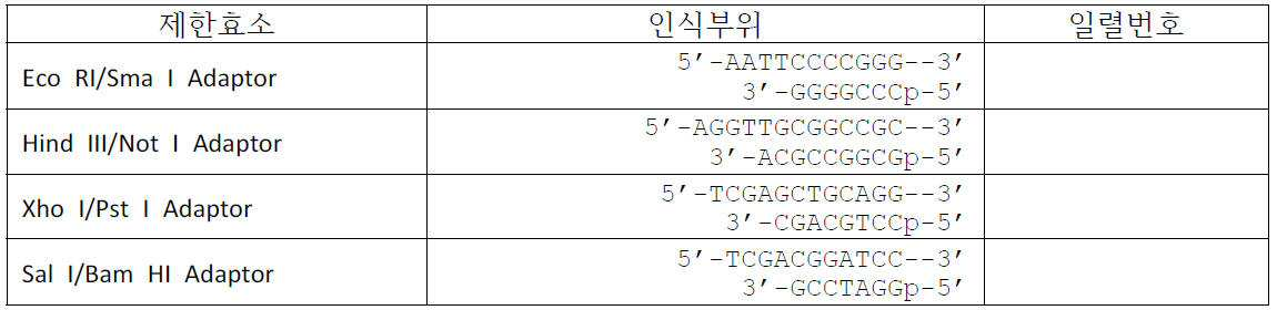
제한효소 아댑터의 염기서열The nucleotide sequence of the restriction enzyme adapter
상기 DNA 절편의 제한효소 지도를 작성하여 DNA 산물을 절단할 수 없는 제한효소(Restriction enzyme)로 이루어진 [표 3]의 adaptor들을 구입하였다(Gene Link 사, USA). 상기 adaptor를 aminoallyl-dCTP 표지된 DNA 절편에 아래와 같이 연결하였다. Adapters of [Table 3] consisting of restriction enzymes that can not cleave DNA products were purchased from Gene Link, USA. The adapter was ligated to an aminoallyl-dCTP labeled DNA fragment as follows.
1ST도메인: 5'-Blunt end-DNA절편-Eco R1/Sma I adaptor-3'1 ST domain: 5'-Blunt end-DNA fragment-Eco R1 / Sma I adapter-3 '
2ND도메인: 5'-Eco RI/Sma I adaptor-DNA절편-Sal I/Bam HI adaptor-3'2 ND domain: 5'-Eco RI / Sma I adapter-DNA fragment -Sal I / Bam HI adapter-3 '
3TH도메인: 5'-Sal I/Bam HI adaptor-DNA절편-Hind III/Not I adaptor-3'3 TH domain: 5'-Sal I / Bam HI adapter-DNA fragment-Hind III / Not I adapter-3 '
4TH도메인: 5'-Hind III/Not I adaptor-DNA절편-Xho I/Pst1 adaptor-3'4 TH domain: 5'-Hind III / Not I adapter-DNA fragment-Xho I / Pst 1 adapter-3 '
5TH도메인: 5'-Xho I/Pst1 adaptor-DNA절편-Sal I/Bam HI adaptor-3'5 TH domain: 5'-Xho I / Pst1 adapter-DNA fragment -Sal I / Bam HI adapter-3 '
6TH도메인: 5'-Sal I/Bam HI adaptor-DNA절편-Eco R1/Sma I adaptor-3'6 TH domain: 5'-Sal I / Bam HI adapter-DNA fragment-Eco R1 / Sma I adapter-3 '
7TH도메인: 5'-EcoR1/Sma I adaptor-DNA절편-3'7 TH domain: 5'-EcoRl / Sma I adapter-DNA fragment -3 '
1ST 도메인은 EcoR1, 2ND 도메인은 EcoR1과 BamHI, 3TH 도메인은 BamHI과 HindIII, 4TH 도메인은 HindIII와 Xho I, 5TH 도메인은 Xho I과 Sal I, 6TH 도메인은 Sal I과 Sma I, 7TH 도메인은 Sma I을 처리하여 반응시킨 후, 정제하여 adaptor가 절단되고 aminoallyl-dCTP 표지된 PCR 절편을 획득하였다.1 ST domain is EcoRI, 2 ND domain is EcoRl and BamHI, 3 TH domain is BamHI and HindIII, 4 TH The domains include HindIII and Xho I, 5 TH The domains are Xho I and Sal I, 6 TH Domain was treated with Sal I and Sma I, 7 TH domain was treated with Sma I, and purified, and the adapter was cleaved and aminoallyl-dCTP labeled PCR fragment was obtained.
본 발명에서 사용하는 신호발생영역은 4 종류의 신호태그의 조합으로 일렬 연속 7개 도메인으로 배열된 구조체로 이를 제조하기 위해, 4종류의 신호태그가 7개 도메인으로 연속적으로 배열된 구조체를 다음과 같이 제조하였다.The signal generating region used in the present invention is a structure in which four signal tags are arranged in seven consecutive domains in a combination of four types of signal tags and a structure in which four kinds of signal tags are continuously arranged in seven domains is described as follows: .
핵산 정량, 핵산변이, 메틸화 DNA, 단백질, 바이러스 또는 세균 등의 생체분자를 분자 계수하여 정량하기 위해, N-hydroxysuccinimide(NHS)-ester(에스테르) 형광(Alexa 488, Alexa 594, Alexa 647(미국, Invitrogen) 또는 Cy3(미국, GE Healthcare)4종류의 염색시약 세트를 준비한다.N-hydroxysuccinimide (NHS) -ester (ester) fluorescence (Alexa 488, Alexa 594, Alexa 647 (USA)) was used to quantitatively quantify biomolecules such as nucleic acid quantitation, nucleic acid variation, methylated DNA, Invitrogen) or Cy3 (USA, GE Healthcare).
제한효소로 절단된 PCR 절편을 4개의 튜브에 나누어 넣고 각각에 한 종류의 염색시약을 넣어 반응시킨다. 이와 같이 모든 도메인에 사용할 수 있는 PCR 절편을 표지하였다. The PCR fragment digested with restriction enzymes is divided into four tubes and reacted with one kind of dyeing reagent. Thus, PCR fragments that can be used in all domains were labeled.
구체적으로 박테리오파지 M13 DNA을 주형(template)로 정방향 프라이머와 역방향프라이머로 PCR로 얻은 PCR 단편(903 bp)을 주형으로 50 % amino-allyl UTP(Sigma)을 표지되도록 PCR를 하였다. 상기 PCR 단편에 제한효소 절단부위가 있는 아댑터(adapter)를 연결한 후 제한효소를 처리하였다. amino-allyl UTP가 표지되고 제한효소 절단부위가 있는 PCR 절편 각각에 4종류의 NHS-에스테르 형광(Alexa 488, Alexa 594, Alexa 647(Invitrogen) 또는 Cy3(GE Healthcare))에서 어느 하나를 먼저 연결하고 순차적으로 4종류를 연결하여 신호발생물질이 표지된 이중가닥 DNA인 신호태그를 제조하여 "H 신호태그"라고 지칭하였다. Specifically, a PCR fragment (903 bp) obtained by PCR using bacteriophage M13 DNA as a template as a forward primer and a reverse primer was used as a template and PCR was performed so that 50% amino-allyl UTP (Sigma) was labeled. The PCR fragment was ligated with an adapter having a restriction enzyme cleavage site, followed by restriction enzyme treatment. One of the four types of NHS-ester fluorescence (Alexa 488, Alexa 594, Alexa 647 (Invitrogen) or Cy3 (GE Healthcare)) was first ligated to each of the PCR fragments labeled with amino-allyl UTP and with restriction enzyme cleavage sites Signal tags with double-stranded DNA, in which signal generating substances were labeled, were produced by sequentially connecting four kinds of DNAs and referred to as "H signal tags ".
<교차결합 유도 및 검증><Cross-Coupling Induction and Verification>
상기 제조된 H 신호태그를 반응버퍼(10 mM Tris (pH 7.0), 200 mM NaCl)에 녹인 후, 8-methoxypsoralen(8-MOP; 미국, Sigma)을 71.0 mg/mL 최종농도가 되도록 첨가하여 암 상태에서 1시간 반응시켰다. 이 반응물에 자외선 365 nm 파장 (4 W)을 주사하면서 3시간동안 상기 H 신호태그 사슬간 교차결합을 시켜 신호태그 구성 염기쌍간의 공유결합을 유도하였다. 이와 같이 공유결합이 포함된 "C 신호태그"를 준비하였다. The resulting H signal tag was dissolved in reaction buffer (10 mM Tris (pH 7.0), 200 mM NaCl), and 8-methoxypsoralen (8-MOP; USA, Sigma) was added to a final concentration of 71.0 mg / Lt; / RTI > for 1 hour. Covalent bond between the signal tag constituent base pairs was induced by cross-linking the H signal tag chain for 3 hours while injecting 365 nm wavelength (4 W) of ultraviolet light into the reaction product. Thus, a "C signal tag" containing a covalent bond was prepared.
H 신호태그와 이에 상응하는 C 신호태그를 0.5℃씩 온도를 95℃까지 상승시키면서 UV-1800(Shimadzu 사, 일본)으로 260 nm에서 흡광도를 측정한 결과 <도7>와 같았으며, H 신호태그는 온도가 증가하면서 이중가닥핵산이 해리가 되어 단일가닥핵산으로 전환되고 Tm이 약 85℃ 이고 95℃에서 단일가닥핵산으로 존재하였으며, 반면 C 신호태그는 95℃에서도 이중가닥핵산을 유지하고 있음을 알 수 있었다.The H signal tag and the corresponding C signal tag were measured in absorbance at 260 nm with UV-1800 (Shimadzu, Japan) while raising the temperature to 0.5 ° C by 95 ° C, The double-stranded nucleic acid was dissociated into a single-stranded nucleic acid with an increase in temperature, and the Tm was about 85 ° C and was present as a single-stranded nucleic acid at 95 ° C, while the C-signal tag retained double-stranded nucleic acid at 95 ° C Could know.
<신호발생영역, 색상지시바코드의 제조>≪ Signal Generation Area, Production of Color Indication Barcode >
제조된 H 신호태그 또는 C 신호태그를 이용한 신호발생영역 색상지시바코드(Color-coded barcode)를 제조를 위해, 1차 도메인용 4종류의 신호태그를 <도 2>와 같이 4개의 튜브에 각각 넣고 선택되어진 튜브의 신호태그를 4개의 튜브로 나누어 넣고 각 튜브에 2차 도메인용 4종류의 신호태그를 한 종류씩 넣어 신호태그들을 혼합시키다. 나머지 3개의 2차 도메인용 신호태그도 상기와 같이 처리하여 2종류의 신호태그를 혼합시킨다. 상기 2종류의 신호태그가 있는 튜브의 용액 을 다시 4개의 튜브로 나누고 넣고 각각의 튜브에 3차 도메인용 4종류의 신호태그를 넣는 과정을 모든 2종류의 태그가 혼합된 용액에 대해여 반복하여 3종류의 신호태그가 있는 용액을 준비한다. 이와 같은 방법을 반복하여 총 7개의 신호태그가 있는 튜브를 준비한다.In order to produce a signal-generating region color-coded barcode using the manufactured H-signal tag or C-signal tag, four kinds of signal tags for the first domain are put into four tubes as shown in FIG. 2 The signal tag of the selected tube is divided into four tubes, and four types of signal tags for the secondary domain are inserted into each tube. The remaining three signal tags for the secondary domain are also processed as described above to mix the two types of signal tags. The procedure of dividing the solution of the tube with the two kinds of signal tags into four tubes again and putting four kinds of signal tags for the tertiary domain into each tube was repeatedly performed for the solution mixed with all the two kinds of tags Prepare a solution with three types of signal tags. Repeat this procedure to prepare a tube with a total of seven signal tags.
7개의 도메인용 신호태그가 있는 튜브에 T4 ligase를 처리하여 연결반응을 하여 7개의 도메인용 신호태그가 일렬로 연속적 배열을 한 최종 구조체를 제조하며 이를 "신호발생영역"이라 지칭하였다.The T4 ligase was treated in a tube with seven signal tags for the domain and ligated to produce a final structure in which signal tags for seven domains were arranged in a row and called "signal generation region".
신호발생영역은 생체분자 특이 바코드 시스템(barcode system)로 4종류의 dye(Alexa dye 3종류, Cy3)를 길게 늘어트린 신호태그 마다 특이한 신호를 형성할 수 있도록 제작이 되어 이들의 조합으로 표적분자들을 특이적으로 지시한다. 제1 탐침 분자는 에피 형광 현미경에 의해 촬영할 때의 ~300 um 나노 스폿을 형성하며, 각각의 선형 순차적 7종류의 표시된 영역을 가진다.The signal generation region is a biomolecule-specific barcode system, and it is made to be able to form a unique signal for each of four kinds of dyes (Alexa dye, Cy3) elongated in a long signal tag. The first probe molecule forms ~ 300 um nanospots when photographed by an epifluorescence microscope, and has seven linear labeled regions of each linear sequence.
<탐침의 제조><Fabrication of probe>
제1 탐침은 생체분자를 인지하여 결합하고 특이 신호를 발생시켜 표적분자를 지시하고 가시화하는 물질로 제1 프로브와 상기 신호발생영역이 결합한 구조이다. The first probe is a material that recognizes and binds biomolecules and generates a specific signal to direct and visualize a target molecule. The probe is a structure in which the first probe and the signal generating region are combined.
바람직하게, 본 발명에 사용하는 제1 프로브는 핵산정량, 핵산변이 또는 메틸화 DNA 등의 분석을 위해서 단일가닥핵산과 핵산을 제외한 생체분자를 정량하기 위해서 항체와 압타머 등의 리간드일 수도 있다. Preferably, the first probe used in the present invention may be a ligand such as an antibody and aptamer in order to quantify a biomolecule except a single-stranded nucleic acid and a nucleic acid for analysis of nucleic acid quantification, nucleic acid mutation or methylated DNA.
비고정 제1 탐침은 5'-신호발생영역-스페이서-제1 프로브-3' 구조로 주로 세균, 세포 및 바이러스 등의 계수에 사용할 수도 있다. The non-immobilized first probe may have a 5'-signal generating region-spacer-first probe-3 'structure and may be used mainly for counting bacteria, cells, viruses and the like.
스페이서; 5'-AGAAGCGCAGAGCTTGGGCGCAGAACAC-3'Spacer; 5'-AGAAGCGCAGAGCTTGGGCGCAGAACAC-3 '
단일가닥핵산인 제1 프로브는 5'-스페이서-제1 프로브-3'를 주문하여 제조하였다(Integrated DNA Technologies사, USA). 상기 제조된 5'-스페이서-제1프로브-3' 단일가닥핵산과 상기 제조된 신호발생영역을 ligase의 연결반응으로 결합하여 비고정 제1 탐침을 제조하였다. A first probe, a single stranded nucleic acid, was prepared by ordering a 5'-spacer-first probe-3 '(Integrated DNA Technologies, USA). The non-immobilized first probe was prepared by combining the prepared 5'-spacer-first probe-3 'single-stranded nucleic acid and the prepared signal-generating region by a ligase coupling reaction.
또한 리간드인 제1 프로브는 5'-스페이서-제1 프로브-3' 구조이고 리간드가 압타머인 경우는 5'-스페이서-제1 프로브-3' 구조의 골격이 단일가닥핵산으로 이를 주문하여 제조하였다(Integrated DNA Technologies사, USA). 리간드가 항체의 경우는 항체에 반응성 그룹 부가 반응을 Thunder-Linkㄾ oligo conjugation system(Innova Biosciences Ltd., 영국)의 프로토콜에 따라 실시하고 반응성 그룹이 부가된 항체를 제조하였다. 또한 반응성 그룹이 있는 스페이서인 단일가닥핵산을 주문 제조하였다(Integrated DNA Technologies사, USA). 상기 반응성그룹이 부가된 항체와 상기 반응성 그룹이 있는 스페이서인 단일가닥핵산을 반응시켜 5'-스페이서-항체 구조를 제조하였다. In addition, when the first probe as the ligand is a 5'-spacer-first probe-3 'structure and the ligand is platamer, the framework of the 5'-spacer-first probe- (Integrated DNA Technologies, USA). In the case where the ligand is an antibody, a reactive group addition reaction to the antibody was carried out according to the protocol of the Thunder-Link ㄾ oligo conjugation system (Innova Biosciences Ltd., UK), and the antibody to which the reactive group was added was prepared. A single stranded nucleic acid, a spacer with a reactive group, was also custom made (Integrated DNA Technologies, USA). The 5'-spacer-antibody structure was prepared by reacting the antibody to which the reactive group was added with a single-stranded nucleic acid which was a spacer having the reactive group.
상기와 같이 제조한 5'-스페이서-제1 프로브-3' 구조와 신호발생영역을 ligase의 연결반응으로 결합시켜 비고정 제1 탐침을 제조하였다.The non-immobilized first probe was prepared by binding the 5'-spacer-first probe-3 'structure prepared above and the signal generating region with a ligase coupling reaction.
고정 제1 탐침은 5'-제1 프로브-스페이서-고정역역-신호발생영역-바이오틴-3'(일반식 1)구조로 주로 핵산 및 단백질 등을 계수하여 정량에 사용할 수도 있다.The immobilized first probe may be used for quantitation by mainly counting nucleic acid and protein with a 5'-first probe-spacer-immobilized region-signal generating region-biotin-3 '(Formula 1) structure.
고정영역: 5'-(GCCACAGCACCTTGGTGCGTAGGATCTG)10-3'Fixed region: 5 '- (GCCACAGCACCTTGGTGCGTAGGATCTG) 10 -3'
제1 프로브가 단일가닥핵산을 제1 프로브로 사용하는 고정 제1 탐침을 제조하기 위해 먼저.First, a first probe is used to prepare a fixed first probe using a single-stranded nucleic acid as a first probe.
5'-스페이서-고정역역-3' 단일가닥핵산을 주문 제조하여 준비하였다(Integrated DNA Technologies사, USA). 신호발생영역에 바이오틴을 부가하여 5'-신호발생영역-바이오틴-3' 구조체를 제조하였다. 상기 5'-스페이서-고정역역-3' 와 5'-신호발생영역-바이오틴-3' 구조체를 ligase의 연결반응을 하여 5'-스페이서-고정영역-신호발생영역-바이오틴-3'구조체를 제조하고 정제하였다. 상기 제조 정제된 반응물과 제1 프로브을 연결반응을 하여, 5'-제1 프로브-스페이서-고정역역-신호발생영역-바이오틴-3'(일반식 1)인 제1 탐침을 제조하였다.A 5'-spacer-fixed region-3 'single stranded nucleic acid was prepared by customization (Integrated DNA Technologies, USA). Biotin was added to the signal generation region to prepare a 5'-signal generating region-biotin-3 'structure. The 5'-spacer-fixed region-3 'and the 5'-signal generating region-biotin-3' structure were ligated to each other to form a 5'-spacer-fixed region-signal generating region-biotin-3 ' And purified. A first probe having a 5'-first probe-spacer-fixed region-signal generating region-biotin-3 '(Formula 1) was prepared by performing a coupling reaction between the prepared purified reactant and a first probe.
제1 프로브가 항체를 사용하는 고정 제1 탐침은 5'-항체-스페이서-고정역역-신호발생영역-바이오틴-3' 구조이다.The fixed first probe using the first probe uses an antibody is a 5'-antibody-spacer-fixed region-signal generating region-biotin-3 'structure.
먼저, 고정 제1 탐침을 제조하기 위해 신호발생영역의 3' 한 쪽에 바이오틴을 부가 반응하여 5'-신호발생영역-바이오틴-3' 구조체를 제조하였다. 5'-스페이서-고정역역-3' 단일가닥핵산을 주문 제조하여 준비하였다(Integrated DNA Technologies사, USA).First, in order to produce a fixed first probe, a 5'-signal generating region-biotin-3 'structure was prepared by adding biotin to 3'of one side of the signal generating region. A 5'-spacer-fixed region-3 'single stranded nucleic acid was prepared by customization (Integrated DNA Technologies, USA).
상기 5'-스페이서-고정역역-3' 단일가닥핵산과 5'-신호발생영역-바이오틴-3' 구조체을 ligase의 연결반응으로 5'-스페이서-고정역역-신호발생영역-바이오틴-3'(일반식 1) 구조체를 제조하였다.The 5'-spacer-fixed region-3 'single-stranded nucleic acid and the 5'-signal generating region-biotin-3' 1) structure was prepared.
상기 5'-스페이서-고정역역-신호발생영역-바이오틴-3' 구조체의 5' 끝에 반응성 그룹 부가는 Thunder-Linkㄾ oligo conjugation system(Innova Biosciences Ltd., 영국)을 사용하여 프로토콜에 따라 실시하였다. 100μl의 60~100μM 제작된 구조체를 Oligo Activation Reagent 튜브에 첨가하며, 실온에서 1시간 반응시켜 활성화시켜 수행하였다. 상기 활성화된 반응용액을 준비된 컬럼을 이용하여 탈염시켜 반응성 그룹부가를 수행하였다.At the 5 'end of the 5'-spacer-immobilized region-signal generating region-biotin-3' structure, the reactive group addition was carried out according to the protocol using a Thunder-Link ㄾ oligo conjugation system (Innova Biosciences Ltd., UK). 100 μl of 60-100 μM construct was added to the Oligo Activation Reagent tube and reacted at room temperature for 1 hour to activate. The activated reaction solution was desalted using a prepared column to perform the reactive group addition.
항체에 대한 반응성 그룹의 부가는 100 ul의 1 mg/ml 항체를 Antibody Activation Reagent 튜브에 첨가하여 실온에서 1시간 반응시켜 활성화시켰다.Addition of the reactive group to the antibody was carried out by adding 100 μl of 1 mg / ml antibody to the Antibody Activation Reagent tube and reacting at room temperature for 1 hour.
반응성 그룹이 부가된 항체와 상기 탈염된 구조체 적절한 비율로 혼합하여 실온에서 12 ~ 24시간 반응시켰다. Conjugate Clean Up Reagent을 상기 반응혼합용액을 반응용액에 첨가하여 10분간 반응시키고 15,000g에서 5분간 원심분리하여 수확된 반응용액에 Conjugate Clean Up Reagent을 첨가한 후 다시 원심분리하여 정제된 상기 5'-제1 프로브인 항체-스페이서-고정역역-신호발생영역-바이오틴-3' 구조인 제1 탐침을 수확하였다. The reacted group-added antibody and the desalted structure were mixed at an appropriate ratio and reacted at room temperature for 12 to 24 hours. The Conjugate Clean Up Reagent was added to the reaction solution for 10 minutes, centrifuged at 15,000 g for 5 minutes, and the Conjugate Clean Up Reagent was added to the harvested reaction solution, followed by further centrifugation to remove the purified 5'- The first probe, the antibody-spacer-fixed region-signal generating region-biotin-3 'structure, was harvested.
압타머를 리간드로 사용하는 고정 제1 탐침은 5'-압타머-스페이서-고정영역-신호발생영역-바이오틴-3'(일반식 2)로 구성된 구조체일 수도 있다.The fixed first probe using platamer as a ligand may be a structure composed of a 5'-platemaker-spacer-fixed region-signal generating region-biotin-3 '(Formula 2).
상기 고정 제1 탐침의 제조는 먼저, 상기 신호발생영역의 3' 끝에 바이오틴을 부가하여 5'-신호발생영역-바이오틴-3' 구조체를 제조하고 5'-압타머-스페이서-고정영역-3' 단일가닥핵산을 주문 제작하였다(Integrated DNA Technologies사, USA). 상기 5'-신호발생영역-바이오틴-3' 구조체와 5'-제1 프로브-스페이서-고정영역-3' 단일가닥핵산을 ligase 의 연결반응으로 5'-압타머-스페이서-고정영역-신호발생영역-바이오틴-3' 구조체를 제조하였다.In the production of the fixed first probe, biotin is added to the 3'-end of the signal generating region to prepare a 5'-signal generating region-biotin-3'structure, and a 5'-abtamer-spacer- Single stranded nucleic acids were custom made (Integrated DNA Technologies, USA). A 5'-plastomer-spacer-immobilized region-signal generated by ligating the 5'-signal generating region-biotin-3 'structure with the 5'-first probe- spacer- Region-biotin-3 ' structure.
(제2 탐침과 경쟁분자)(Second probe and competing molecule)
제2 탐침은 제2 프로브와 고체 지지체를 결합시켜 제조하고 경쟁분자는 표적분자 또는 표적분자 유사체와 고체 지지체를 결합시켜 제조하였다. The second probe was prepared by combining a second probe and a solid support, and the competitive molecule was prepared by binding a target molecule or a target molecule analog to a solid support.
이하에서 리간드와 표적분자를 분자로 통칭하여 기술하였으며 구분할 필요가 있을 경우만 나누어 기술하였다.Hereinafter, the ligand and the target molecule are collectively referred to as a molecule.
제2 탐침 또는 경쟁분자는 분자에 직접 자성비드를 결합시키거나, 바이오틴(biotin)이 표지된 단일가닥핵산을 결합시킨 후, 자성비드를 결합시켜 제조하였다. 본 발명에 사용한 리간드와 생체분자는 표1 및 표2와 같다. The second probe or competing molecule is prepared by binding a magnetic bead directly to the molecule, or binding a single-stranded nucleic acid labeled with biotin, followed by binding magnetic beads. The ligands and biomolecules used in the present invention are shown in Tables 1 and 2.
분자와 활성화 자성비드(M-280 Tosylactivated magnetic beads, Dynal Biotech ASA, Norway)를 버퍼용액(0.1 M borate buffer, pH 9.5)에 넣고 상온에서 48시간 반응시켰다. 분자가 결합된 자성비드는 자석을 이용해서 분리가 가능하며, 동일한 버퍼용액으로 비드를 세척하여 결합하지 않은 분자를 제거하였다. 분자가 결합된 자성비드(이하 '포획리간드')는 세척 후 0.1 %의 BSA(Bovine Serum Albumin, Sigma Co.) 용액으로 37℃에서 4시간 반응시켜 분자와 결합하지 않은 토실(tosyl)기를 불활성화시켰다. 이어 수차례 세척 후 버퍼용액(0.1 M PBS, pH 7.4)에 자성비드를 혼합하여 4℃에서 보관하였다.Molecules and activated magnetic beads (M-280 Tosylactivated magnetic beads, Dynal Biotech ASA, Norway) were placed in a buffer solution (0.1 M borate buffer, pH 9.5) and reacted at room temperature for 48 hours. The magnetic beads to which the molecules are bonded can be separated using magnets, and unbound molecules are removed by washing the beads with the same buffer solution. Molecularly coupled magnetic beads (hereinafter referred to as "capture ligands") were washed and reacted with 0.1% BSA (Bovine Serum Albumin, Sigma Co.) solution at 37 ° C. for 4 hours to inactivate the tosyl groups . After several washes, magnetic beads were mixed with buffer solution (0.1 M PBS, pH 7.4) and stored at 4 ° C.
분자에 대한 스페이서를 수확물질 부가는 먼저, 분자의 바이오틴화로 한쪽 끝에 바이오틴이 표지되고 다른 한쪽 끝에 반응성 그룹이 있는 단일가닥핵산을 주문 제조하여(Integrated DNA Technologies사, USA) 수행하였다. 분자에 대한 반응성그룹의 부가는 Thunder-Linkㄾ oligo conjugation system (Innova Biosciences Ltd., 영국)을 사용하여 프로토콜에 따라 실시하였다. Harvesting the Spacer for the Molecules The material addition was first performed by custom biotinylation of the molecule with a single stranded nucleic acid (Biotin labeled at one end and a reactive group at the other end (Integrated DNA Technologies, USA). The addition of reactive groups to the molecules was performed according to the protocol using a Thunder-Link ㄾ oligo conjugation system (Innova Biosciences Ltd., UK).
분자에 대한 반응성 그룹의 부가는 100 ㎕의 1 mg/ml 분자를 Antibody Activation Reagent 튜브에 첨가하여 실온에서 1시간 반응시켜 활성화시켰다. 반응성 그룹이 부가된 분자와 주문 제작된 단일가닥핵산의 결합은 상기 활성화된 반응용액을 준비된 컬럼을 이용하여 탈염시킨 후, 상기 탈염된 분자와 단일가닥핵산을 적절한 비율로 혼합하여 실온에서 12~24시간 반응시켰다. Conjugate Clean Up Reagent을 상기 반응용액에 첨가하여 10분간 반응시키고 15,000 g에서 5분간 원심분리하여 수확된 바이오틴이 표지된 분자에 Conjugate Clean Up Reagent을 첨가한 후 다시 원심분리하여 정제된 바이오틴이 표지된 분자를 수확하여 바이오틴이 표지된 분자를 제조하였다.Addition of the reactive group to the molecule was carried out by adding 100 μl of 1 mg / ml molecule to the Antibody Activation Reagent tube and reacting at room temperature for 1 hour. The binding of the reactive group-added molecule to the custom-made single-stranded nucleic acid is performed by desalting the activated reaction solution using the prepared column, mixing the desalted molecule with the single-stranded nucleic acid at an appropriate ratio, Lt; / RTI > Conjugate Clean Up Reagent was added to the reaction solution for 10 minutes, centrifuged at 15,000 g for 5 minutes to add Conjugate Clean Up Reagent to the harvested biotin labeled molecule, and centrifuged again to remove the purified biotin labeled molecule Were harvested to produce biotin-labeled molecules.
이상과 같이 준비된 바이오틴이 표지된 분자들과 스트렙타비딘이 코팅된 자성비드(Streptavidin-coupled Dynabeads, Dynal Biotech ASA, Norway)를 연속 회전하면서 반응시켜 자성비트가 부가된 분자를 제조하였고 이를 "제2 탐침 또는 경쟁분자"라고 지칭하였다.The prepared biotin-labeled molecules and streptavidin-coated magnetic beads (Streptavidin-coupled Dynabeads, Dynal Biotech ASA, Norway) were reacted with each other while rotating continuously to produce a magnetic bit- Probe or competitive molecule ".
(고정분자)(Fixed molecule)
고정분자는 리포터분자의 반복구조부의 염기서열에 상보적인 염기서열이며 5' 끝에 바이오틴이 표지된 단일가닥핵산을 주문 제조하여 준비하였다(Integrated DNA Technologies사, USA). The immobilized molecule is a base sequence complementary to the nucleotide sequence of the repetitive structural part of the reporter molecule, and a single-stranded nucleic acid labeled with biotin at the 5 'end was prepared and prepared (Integrated DNA Technologies, USA).
실시례Example 2. 세균 2. Bacteria
대표적인 식중독균인 대장균, 살모넬라균, 리스테리아 및 포도상구균을 <표2>의 항체 및 <표 3>의 압타머로 제조된 비고정 제1 탐침 및 제2 탐침으로 계수하였다.Representative food poisoning bacteria such as E. coli, Salmonella, Listeria and Staphylococci were counted by using the antibody of Table 2 and the non-fixed first probe and second probe prepared from the aspirator of Table 3.
세균 계수에 사용하는 제1 탐침은 7개 도메인 모두 동일한 염색지시약으로 표지된 신호태그로 구성된 신호발생영역으로 구성하여 사용할 수도 있다. 제2 탐침은 제2 프로브에 바이오틴 또는 다양한 고체 지지대를 고정하여 제조할 수도 있다.The first probe used for bacterial counting can be used as a signal generating region composed of signal tags labeled with the same dye indicator in all seven domains. The second probe may be produced by fixing biotin or various solid supports to the second probe.

식중독균 표면분자에 결합하는 항체An antibody that binds to the surface molecule of food poisoning bacteria
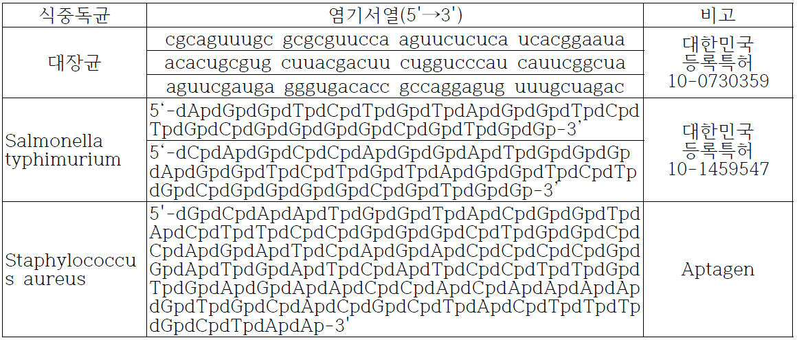
식중독균에 결합하는 압타머의 염기서열Nucleotide sequence of platamer binding to food poisoning bacteria
본 발명에서 세균 분석 반응은 하기 반응용액에서 일어나는 리간드-리셉터 반응이다. In the present invention, the bacteriological analysis reaction is a ligand-receptor reaction occurring in the following reaction solution.
반응용액은 항체리간드의 경우는 10 mM PBS 버퍼(137 mM NaCl, 10 mM Phosphate, 2.7 mM KCl, pH 7.4) 이며, 압타머 리간드의 경우는 셀렉스버퍼 (20 mM Tris-Cl buffer, pH 7.6 contained 100 mM NaCl, 2 mM MgCl2, 5 mM KCl, 1 mM CaCl2 and 0.02% Tween)를 사용하였다. 반응용액에 세균번식저해를 위한 소듐아지드 0.05 내지 0.2%, 비특이적 결합 저해를 위한 우혈청알부민 0.1 내지 0.3%을 포함할 수도 있다. 바람직하게는 반응온도는 20 내지 30℃의 온도이다.The reaction solution contained 10 mM PBS buffer (137 mM NaCl, 10 mM Phosphate, 2.7 mM KCl, pH 7.4) in the case of the antibody ligand, and the celex buffer (20 mM Tris-Cl buffer, pH 7.6 contained 100 0.05 mM of sodium azide for inhibiting bacterial growth, 0.1 to 0.3% of bovine serum albumin for inhibiting nonspecific binding, 0.1 mM of sodium chloride, %. Preferably, the reaction temperature is a temperature of 20 to 30 占 폚.
세균이 포함된 시료 70㎕에 제1 탐침 15㎕과 바이오틴이 부가된 제2 프로브 구조체 15 ㎕를 30분간 흔들어 주면서 반응시켰다. 상기 반응용액 20㎕을 아비딘이 코팅된 유리슬라이드에 처리하여 커버슬립을 덮었다. 코팅된 아비딘과 제2 프로브의 바이오틴이 반응하여 제1 탐침이 결합한 세균을 유리슬라이드에 고정된다. 상기 제1 탐침이 결합하여 형성된 복합체에 있는 세균의 존재 신호를 발생하는 스폿들(spots)을 계수하여 표적분자 세포를 계수하였다(도 9).15 μl of the first probe and 15 μl of the second probe structure to which biotin was added were reacted with 70 μl of the sample containing the bacteria for 30 minutes while shaking. 20 mu l of the reaction solution was treated on a glass slide coated with avidin to cover the cover slip. The biotin of the coated avidin reacts with the biotin of the second probe and the bacteria bound to the first probe are immobilized on a glass slide. The target cells were counted by counting spots generating a presence signal of bacteria in the complex formed by the first probe (FIG. 9).
제1 탐침의 한 쪽 끝에 바이오틴을 부가하여 세균과 바이오틴이 부가된 제1 탐침을 반응시킨 후 아비딘이 코팅된 유리슬라이드와 반응시켜 세균-제1 탐침 복합체를 포획하여 형성된 복합체에서 세포의 존재 신호를 발생하여 형성된 스폿들을 계수하여 표적분자 세균을 계수하였다.Biotin was added to one end of the first probe to react with a first probe added with biotin and reacted with a glass slide coated with avidin to capture the presence signal of the cell in the complex formed by capturing the bacterial first probe complex The resulting formed spots were counted to count the target molecule bacteria.
다양한 신호발생영역으로 다양한 세균을 특이적으로 지시하도록 제1 탐침을 제조하여 여러 종류의 세균을 동시에 동정 및 계수를 제1 탐침과 세균 복합체의 색상분석으로 할 수 있다.The first probe may be prepared to specifically indicate various bacteria to various signal generation regions, and various kinds of bacteria may be simultaneously identified and counted by color analysis of the first probe and the bacterial complex.
실시례Example 3. 바이러스3. virus
본 실험에서 간염바이러스 C형을 혈청이나 혈장에서 비고정 제1 탐침 및 제2 탐침을 사용하여 계수하였다.In this experiment, hepatitis C virus type C was counted in serum or plasma using non-immobilized first and second probes.
AxSYM HCV version 3.0 (Abbott Laboratories, Abbott Park, IL, USA)으로 시행하였다. AxSYM HCV version 3.0은 효소측정법의 한 형태인 MEIA (microparticle enzyme immunoassay) 법을 이용하여 환자의 혈청이나 혈장에서 C형 간염바이러스 항체를 정성적으로 검출하는 방법이다. 결과판정은 anti-HCV ELISA S/CO 값이 1 이상인 경우 양성으로 판정하였다.AxSYM HCV version 3.0 (Abbott Laboratories, Abbott Park, IL, USA). AxSYM HCV version 3.0 is a method for qualitatively detecting hepatitis C virus antibody in patient's serum or plasma using MEIA (microparticle enzyme immunoassay) method, which is a form of enzyme assay. Results were judged to be positive if the anti-HCV ELISA S / CO value was> 1.
비고정 제1 탐침의 프로브는 다클론 Hepatitis C Virus (HCV) 항체 고 제2 탐침의 프로브는 Hepatitis C Virus (HCV) NS3 항체이다(EastCoat Bio. Inc. USA).The probe of the unfixed first probe is a polyclonal hepatitis C virus (HCV) antibody and the probe of the second probe is a hepatitis C virus (HCV) NS3 antibody (EastCoat Bio.
바이러스 계수에 사용하는 제1 탐침은 7개 도메인 모두 동일한 염색지시약으로 표지된 신호태그로 구성된 신호발생영역으로 구성하여 사용할 수도 있다. 제2 탐침은 제2 프로브에 바이오틴 또는 다양한 고체 지지대를 고정하여 제조할 수도 있다.The first probe used for the virus count may be used as a signal generation region composed of signal tags labeled with the same dye indicator in all seven domains. The second probe may be produced by fixing biotin or various solid supports to the second probe.
바이러스 분석을 위한 반응은 혈청이나 혈장에 제1 탐침 및 제2 탐침을 처리하여 항원-항체 반응을 하였다. 반응용액은 10 mM PBS 버퍼(137 mM NaCl, 10 mM Phosphate, 2.7 mM KCl, pH 7.4)를 사용하였다.The reaction for virus analysis was performed by treating the first probe and the second probe with serum or plasma to perform an antigen-antibody reaction. For the reaction solution, 10 mM PBS buffer (137 mM NaCl, 10 mM Phosphate, 2.7 mM KCl, pH 7.4) was used.
반응용액에 세균번식저해를 위한 소듐아지드 0.05 내지 0.2%, 비특이적 결합 저해를 위한 우혈청알부민 0.1 내지 0.3%을 포함할 수도 있다. 반응온도는 바람직하게는 반응온도는 20 내지 30℃의 온도이다.The reaction solution may contain 0.05 to 0.2% of sodium azide for inhibiting bacterial growth, and 0.1 to 0.3% of bovine serum albumin for inhibiting non-specific binding. The reaction temperature is preferably a reaction temperature of 20 to 30 占 폚.
HCV가 포함된 혈청 70㎕에 제1 탐침 15㎕과 바이오틴이 부가된 제2 프로브 구조체 15 ㎕를 30분간 흔들어 주면서 반응시켰다. 상기 반응용액 20㎕을 아비딘이 코팅된 유리슬라이드에 처리하여 커버슬립을 덮었다. 코팅된 아비딘과 제2 프로브의 바이오틴이 반응하여 제1 탐침이 결합한 HCV를 유리슬라이드에 고정된다. 상기 제1 탐침이 결합하여 형성된 복합체에 있는 HCV의 존재 신호를 발생하는 스폿들(spots)을 계수하여 표적분자 HCV를 계수하였다.15 [mu] l of the first probe and 15 [mu] l of the second probe structure added with biotin were reacted with 70 [mu] l of HCV-containing serum for 30 minutes while shaking. 20 mu l of the reaction solution was treated on a glass slide coated with avidin to cover the cover slip. The biotin of the coated avidin reacts with the biotin of the second probe and the HCV to which the first probe is bound is immobilized on the glass slide. The spots generating the presence signal of HCV in the complex formed by the first probe were counted to count the target molecule HCV.
또는 제1 탐침의 한 쪽 끝에 바이오틴을 부가하여 HCV와 바이오틴이 부가된 제1 탐침을 반응시킨 후 아비딘이 코팅된 유리슬라이드와 반응시켜 HCV-제1 탐침 복합체를 포획하여 형성된 복합체에서 HCV의 존재 신호를 발생하여 생긴 스폿을 계수하여 표적분자 HCV을 계수하였다.Alternatively, biotin is added to one end of the first probe to react with HCV and a biotin-added first probe, and then reacted with avidin-coated glass slide to capture HCV-first probe complex to form HCV-present complex And the target molecule HCV was counted.
다양한 신호발생영역으로 다양한 HCV를 특이적으로 지시하도록 제1 탐침을 제조하여 여러 종류의 HCV를 동시에 동정 및 계수를 제1 탐침과 HCV 복합체의 색상분석을 통하여 할 수 있다.A first probe can be prepared to specifically indicate various HCVs in various signal generation regions, and simultaneously, various types of HCV can be identified and counted through color analysis of the first probe and the HCV complex.
실시례Example 4. 생체분자 계수 시료 준비 4. Biomolecular Coefficient Sample Preparation
4-1. 4-1. 단일가닥핵산을Single stranded nucleic acid 포함하는 탐침 Probes included
(핵산 정량)(Nucleic acid quantitation)
상기 일반식 (II)와 일반식(III)의 프로브는 [표 11]의 β-actin, IL17 및 IL17R mRNA 의 특이적인 염기서열을 참조하여 유기합성을 하였다(바이오니아. 한국). 정량 분석하는 RNA 정량분석의 대상은 IL17 및 IL17RA 이며 진뱅크 (GenBank) 데이터 시스템 (http:// www.ncbi.nlm.nih.gov)에서 얻을 수 있는 IL17 및 IL17RA 유전자의 염기서열 정보 (IL17의 Accession number는NM_002190, 및 IL17RA의 Accession number: NM_014339)이다. The probes of the general formula (II) and the general formula (III) were subjected to organic synthesis with reference to specific base sequences of? -Actin, IL17 and IL17R mRNA of Table 11 (Bioneer, Korea). RNA quantitation for quantitative analysis was performed using IL17 and IL17RA, and the nucleotide sequence information of the IL17 and IL17RA genes (obtained from the GenBank data system (http: // www.ncbi.nlm.nih.gov) The accession number is NM_002190, and the accession number of IL17RA is NM_014339).

표적 mRNA에 대한 프로브 서열The probe sequence for the target mRNA
세포 혹은 세포로 이루어진 조직 시료에서 AllPrep DNA/RNA/Protein Mini kit (Qiagen사. 미국)로 생체분자를 분리하였다. 본 발명에서는 사람의 연골조직(Promocell 사, 독일)을 생체시료로 사용하였다. 연골조직을 제조사에 제공된 버퍼로 용해시킨 후 용해된 시료를 컬럼에 처리한 후, 원심분리하여 DNA를 컬럼막에 결합시키고 여과된 용액을 수확하였다. 컬럼막에 있는 DNA분리는 DNA가 있는 컬럼을 세척하고 막에서 DNA를 추출한 후, 원심분리하여 DNA를 수확하였다. 상기 수확된 여과용액을 제조사에서 제공하는 컬럼에 처리하여 여과된 용액에서 단백질을 침전 및 원심분리하여 수확하였다. 또한 컬럼에서 있는 RNA를 세척하고 막에 있는 RNA를 추출한 후 원심분리하여 수확하였다.Biomolecules were isolated from AllPrep DNA / RNA / Protein Mini kit (Qiagen, USA) from tissue samples made of cells or cells. In the present invention, human cartilage tissue (Promocell, Germany) was used as a biological sample. After the cartilage tissue was dissolved in the buffer provided in the manufacturer, the dissolved sample was treated on the column and centrifuged to bind the DNA to the column membrane and harvest the filtered solution. DNA separation in the column membrane was performed by washing the column with the DNA, extracting the DNA from the membrane, and harvesting the DNA by centrifugation. The harvested filtrate solution was processed into a column provided by the manufacturer, and the proteins were harvested by precipitation and centrifugation of the proteins in the filtered solution. In addition, the RNA in the column was washed, the RNA in the membrane was extracted, and then harvested by centrifugation.
상기 총 RNA 100ng에 상기 제1 탐침과 제2 탐침을 혼성화 반응하고 세척하여 제1 탐침-표적 mRNA-제2 탐침 복합체인 부분적 이중가닥핵산을 제조하여 핵산 정량을 위한 핵산 분자 계수의 시료를 제조하였다.The first probe and the second probe were hybridized and washed with 100 ng of the total RNA to prepare a partial double-stranded nucleic acid as a first probe-target mRNA-second probe complex to prepare a sample of nucleic acid molecule count for nucleic acid quantification .
혼성화 조건은 0.5 M NaHPO4, 7% SDS(sodium dodecyl sulfate), 1 mM EDTA에서 65℃ 조건으로 혼성화하고 세척은 6 xSSC/0.1% SDS에서 48℃ 조건하고 0.1 x SSC(standard saline citrate)/0.1% SDS에서 68℃ 조건으로 0.2 xSSC/0.1% SDS에서 42℃ 조건으로 하였다.Hybridization conditions were hybridized in 0.5 M NaHPO4, 7% SDS (sodium dodecyl sulfate), 1 mM EDTA at 65 ℃ and washing was performed at 6 x SSC / 0.1% SDS at 48 ℃ and 0.1 x SSC (standard saline citrate) SDS at 42 ° C in 0.2 x SSC / 0.1% SDS at 68 ° C.
세척된 제1탐침-표적 mRNA-제2 탐침 복합체에서 해리된 제1 탐침을 수확하기 위해서, 상기 정제한 물질에 100 ㎕의 0.1X SSPE / 0.1 % tween-20 용액을 첨가하고 95 ℃에서 5 분간 처리한 후, 상기 복합체에서 해리된 제1 탐침을 포함하는 상징액을 수확하였다.To harvest the first probe dissociated from the washed probe-target mRNA-second probe complex, 100 μl of 0.1X SSPE / 0.1% tween-20 solution was added to the purified material and incubated at 95 ° C for 5 minutes After treatment, the supernatant containing the first probe dissociated in the complex was harvested.
45 μL의 전체 반응용액을 9 μL씩 분주하여 표적분자 계수 분석법을 위한 분석시료로 사용하였다. 시료 분석은 3 μL의 전체 반응용액을 3회 분석하였다.45 μL of the total reaction solution was dispensed in 9 μL aliquots and used as an analytical sample for the target molecule count analysis. For sample analysis, 3 μL of total reaction solution was analyzed 3 times.
제1 탐침에 있는 색상지시바코드를 이용하여 표적 mRNA를 특이적으로 지시하고 있어 복합체의 색상분석을 통하여 많은 표적 mRNA들을 동시에 분석할 수 있다.Targeting mRNA is specifically indicated using the color indicating barcode in the first probe, and many target mRNAs can be simultaneously analyzed through color analysis of the complex.
추가적으로 형성된 제1 탐침-표적 mRNA-제2 탐침 복합체에서 분리된 제1 탐침을 분자 계수하여 표적분자 핵산을 동정 및 정량할 수 도 있다. The first probe-target mRNA-second probe complex formed additionally may be subjected to molecular counting to identify and quantify the target molecule nucleic acid.
(핵산 (Nucleic acid 구조변이Structure ))
핵산 변이 분석을 위해 상기 고정 제1 탐침 및 제2 탐침을 제작하는 단계에 있어서, 상기 고정 제1 탐침의 구성은 (i) 결합태그(AT), (ii) 신호발생영역(SR), (iii) 고정영역(FR), 및 (iv) 표적핵산과 완전하게로 상보적 혼성화 서열을 가지는 변이인접 영역과 뉴클레오타이드 변이에 해당하는 변이 영역(V)으로 이루어진 제1 프로브(1ST P)를 포함하며, 뉴클레오타이드 변이정보를 갖는 제1프로브는 두 종류 또는 그 이상이다. (I) a binding tag (AT); (ii) a signal generating region (SR); (iii) a signal generating region And a first probe (1 ST P) consisting of a mutation region (V) corresponding to a mutation adjacent region having a complementary hybridization sequence completely complementary to the target nucleic acid and a nucleotide mutation, and (iv) , And the first probe having nucleotide variation information is two kinds or more.
DNA 변이 분석은 IL17 및 IL17RA의 SNP를 대상으로 하였으며 이에 대한 정보는http://www.ncbi.nlm.nih.gov/snp에서 수집하였다. DNA mutation analysis was performed on the SNPs of IL17 and IL17RA, and information was collected from http://www.ncbi.nlm.nih.gov/snp.
상기 DNA와 상기 제1 탐침과 제2 탐침을 혼성화 반응하여 부분적 이중가닥핵산을 제조하고, 이어서 부분적 이중가닥핵산을 완전한 이중가닥핵산으로 전환시키는 ligase의 연결반응을 수행하여 분석 시료를 제조하였다. An analytical sample was prepared by ligating the DNA with the first probe and the second probe to prepare a partial double-stranded nucleic acid, and then ligating the partial double-stranded nucleic acid to a complete double-stranded nucleic acid.
혼성화 조건은 0.5 M NaHPO4, 7% SDS(sodium dodecyl sulfate), 1 mM EDTA에서 65℃ 조건으로 혼성화하고 세척은 6 xSSC/0.1% SDS에서 48℃ 조건하고 0.1 x SSC(standard saline citrate)/0.1% SDS에서 68℃ 조건으로 0.2 xSSC/0.1% SDS에서 42℃ 조건으로 하였다.Hybridization conditions were hybridized in 0.5 M NaHPO4, 7% SDS (sodium dodecyl sulfate), 1 mM EDTA at 65 ℃ and washing was performed at 6 x SSC / 0.1% SDS at 48 ℃ and 0.1 x SSC (standard saline citrate) SDS at 42 ° C in 0.2 x SSC / 0.1% SDS at 68 ° C.
10-ml 반응용액은 20mM TrisHCl (pH 7.6), 25mM sodium acetate, 10mM magnesium acetate, 1mM dithiothreitol, 1mM NAD+, 0.1% Triton X-100, 상기 세척된 반응물, 1 unit Taq DNA ligase (New England Biolabs, MA, USA)로 구성하여 4시간동안 반응시켰다. 제1탐침-표적 유전자-제2 탐침 복합체의 자성입자를 이용하여 세척 및 분리 수확하였다.The 10-ml reaction solution contained 20 mM TrisHCl (pH 7.6), 25 mM sodium acetate, 10 mM magnesium acetate, 1 mM dithiothreitol, 1 mM NAD +, 0.1% Triton X-100, the washed reactants, 1 unit Taq DNA ligase (New England Biolabs, , USA) and reacted for 4 hours. And then washed and separated and harvested using magnetic particles of the first probe-target gene-second probe complex.
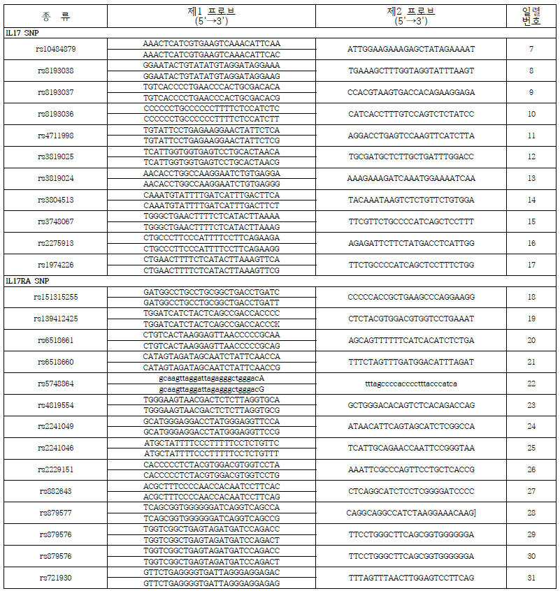
유전자 구조변이 분석에 대한 프로브 서열Probe sequence for analysis of gene structure variation
세척된 제1탐침-표적 유전자변이-제2 탐침 복합체에서 해리된 제1 탐침을 수확하기 위해서, 상기 정제한 물질에 100 ㎕의 0.1X SSPE / 0.1 % tween-20 용액을 첨가하고 95 ℃에서 5 분간 처리한 후, 상기 복합체에서 해리된 제1 탐침을 포함하는 상징액을 수확하였다.To harvest the first probe dissociated from the washed probe-target gene mutation-second probe complex, 100 μl of a 0.1X SSPE / 0.1% tween-20 solution was added to the purified material, After the differential treatment, the supernatant containing the first probe dissociated in the complex was harvested.
45 μL의 전체 반응용액을 9 μL씩 분주하여 표적분자 계수 분석법을 위한 분석시료로 사용하였다. 시료 분석은 3 μL의 전체 반응용액을 3회 분석하였다.45 μL of the total reaction solution was dispensed in 9 μL aliquots and used as an analytical sample for the target molecule count analysis. For sample analysis, 3 μL of total reaction solution was analyzed 3 times.
제1 탐침에 있는 색상지시바코드를 이용하여 유전자변이 및 변이 유형을 특이적으로 지시하고 있어 복합체의 색상분석을 통하여 많은 표적 유전자 변이 및 변이유형을 동시에 분석할 수 있다.The chromosome bar code in the first probe specifically indicates the gene mutation and the mutation type. Thus, many target gene mutations and mutation types can be simultaneously analyzed through the color analysis of the complex.
(메틸화 DNA)(Methylated DNA)
핵산 메틸화 여부 분석은 IL17 프로모터-144 위치(Thomas, R.M., et al., 2012, J Biol Chem. 287(30):25049-59.)를 수행하였다. Nucleic acid methylation analysis was performed using the IL17 promoter-144 site (Thomas, R.M., et al., 2012, J Biol Chem. 287 (30): 25049-59).
상기 일반식 (IV)와 일반식(V)의 H는 [표 1]의 IL17 메틸화여부의 특이적인 염기서열 및 T는 [표 2], P는 [표3]을 참조하여 유기합성을 하였다(바이오니아. 한국).The H of the above general formulas (IV) and (V) was subjected to organic synthesis by referring to the specific base sequence of whether IL17 methylation was shown in [Table 1], T for [Table 2] and P for [Table 3] Biona, Korea).

IL 17 프로모터 메틸화 분석에 대한 프로브 서열Probe sequence for IL 17 promoter methylation analysis
DNA 메틸화 분석을 위해 상기 제1 탐침 및 제2 탐침을 제작하는 단계에 있어서, 상기 제1 탐침의 구성은 (i) 결합태그(AT), (ii) 신호발생영역(SR), (iii) 고정영역(FR), 및 (iv) 표적핵산과 완전하게로 상보적 혼성화 서열을 가지는 메틸화 인접 영역과 메틸화 염기에 해당하는 영역(V)으로 이루어진 제1 프로브(1ST P)를 포함하며, 메틸화 정보를 갖는 제1프로브는 메틸화 염기를 구분하기 위해 사이토신 또는 우라실과 상보적인 염기인 구아닌 또는 티민을 갖는 단일가닥핵산인 두 종류이다. 비메틸화에 상응하는 신호발생영역 A, 메틸화에 상응하는 신호발생영역 B로 제1 프로브와 연결시킬 수도 있다.(I) a binding tag (AT), (ii) a signal generating region (SR), (iii) a second probe And a first probe (1 ST P) consisting of a methylation adjacent region having a complementary hybridization sequence completely complementary to the target nucleic acid and a region (V) corresponding to the methylation base, wherein the methylation information Is a single-stranded nucleic acid having guanine or thymine, which is a base complementary to cytosine or uracil in order to distinguish methylation bases. The first probe may be connected to the signal generation region A corresponding to unmethylation, and the signal generation region B corresponding to methylation.
상기 연골조직에서 특정 DNA의 메틸화 유무를 분석하기 위해 키트를 이용하여, 연골조직에서 지노믹 DNA를 추출하고 Nano Drop을 이용하여 그 농도를 측정하였다. 이후, 추출한 DNA 약 1~2㎍정도를 0.5 N 농도의 수산화나트륨과 섞어 16℃로 만들어 37℃에서 15분간 반응시켜 DNA가 단일가닥 형태가 되도록 변형시켰다. 이후, 3.5M 농도의 소듐 바이설파이트 (Sodium bisulfate)와 0.01M 농도의 하이드로퀴논 (Hydroquinone) 시약을 첨가하고 56℃에서 16시간 화학 반응시켰다. 이후 에탄올을 이용한 침전반응을 수행하여 변환된 DNA만을 농축시켜 순수 분리하여 50℃의 3차 증류수로 녹인 후, 0.1M 농도가 되도록 수산화나트륨을 첨가하고 상온에서 15분간 반응함으로써 사이토신의 탈황산화 반응을 수행하고 다시 한 번 에탄올 침전반응으로 농축시키고, 최종적으로 30℃의 3차 증류수에 녹여서 20℃에 보관하였으며 메틸화유무를 분석하기 위한 표적핵산으로 사용하였다.To analyze the presence or absence of methylation of specific DNA in the cartilage tissue, genomic DNA was extracted from cartilage tissue using a kit and its concentration was measured using Nano Drop. Approximately 1 ~ 2 ㎍ of the extracted DNA was mixed with 0.5 N sodium hydroxide to make 16 캜 and reacted at 37 캜 for 15 minutes to transform the DNA into single stranded form. Sodium bisulfate (3.5 M) and hydroquinone (0.01 M) reagent were added and reacted at 56 ° C for 16 hours. Then, the precipitated reaction with ethanol was performed to concentrate only the transformed DNA, and then pure water was separated and dissolved with tertiary distilled water at 50 ° C. Then, sodium hydroxide was added to a concentration of 0.1M, and the reaction was allowed to proceed at room temperature for 15 minutes. And then concentrated again by ethanol precipitation. Finally, the solution was dissolved in tertiary distilled water at 30 ° C and stored at 20 ° C, and used as a target nucleic acid for the presence or absence of methylation.
상기 메틸화 DNA가 있는 시료, 제1 탐침과 제2 탐침을 혼성화 반응하여 제1 탐침-메틸화 DNA-제2 탐침 부분적 이중가닥핵산을 제조하고, 이어서 부분적 이중가닥핵산을 완전한 이중가닥핵산으로 전환시키는 ligase의 연결반응을 수행하여 분석 시료를 제조하여 메틸화 DNA를 분석할 수 있다. Hybridizing the first probe with the second probe to produce a first probe-methylated DNA-second probe partial double-stranded nucleic acid, and then ligating the partial double-stranded nucleic acid into a complete double-stranded nucleic acid The methylated DNA can be analyzed by preparing an assay sample.
혼성화 조건은 0.5 M NaHPO4, 7% SDS(sodium dodecyl sulfate), 1 mM EDTA에서 65℃ 조건으로 혼성화하고 세척은 6 xSSC/0.1% SDS에서 48℃ 조건하고 0.1 x SSC(standard saline citrate)/0.1% SDS에서 68℃ 조건으로 0.2 xSSC/0.1% SDS에서 42℃ 조건으로 하였다.Hybridization conditions were hybridized in 0.5 M NaHPO4, 7% SDS (sodium dodecyl sulfate), 1 mM EDTA at 65 ℃ and washing was performed at 6 x SSC / 0.1% SDS at 48 ℃ and 0.1 x SSC (standard saline citrate) SDS at 42 ° C in 0.2 x SSC / 0.1% SDS at 68 ° C.
10-ml 반응용액은 20mM TrisHCl (pH 7.6), 25mM sodium acetate, 10mM magnesium acetate, 1mM dithiothreitol, 1mM NAD+, 0.1% Triton X-100, 상기 세척된 반응물, 1 unit Taq DNA ligase (New England Biolabs, MA, USA)로 구성하여 4시간 반응하였다. 제1탐침-메틸화 DNA-제2 탐침 복합체의 자성입자를 이용하여 세척 및 분리 수확하였다.The 10-ml reaction solution contained 20 mM TrisHCl (pH 7.6), 25 mM sodium acetate, 10 mM magnesium acetate, 1 mM dithiothreitol, 1 mM NAD +, 0.1% Triton X-100, the washed reactants, 1 unit Taq DNA ligase (New England Biolabs, , USA) and reacted for 4 hours. Washed and separated and harvested using magnetic particles of the first probe-methylated DNA-second probe complex.
세척된 제1탐침-메틸화 DNA-제2 탐침 복합체에서 해리된 제1 탐침을 수확하기 위해서, 상기 정제한 물질에 100 ㎕의 0.1X SSPE / 0.1 % tween-20 용액을 첨가하고 95 ℃에서 5 분간 처리한 후, 상기 복합체에서 해리된 제1 탐침을 포함하는 상징액을 수확하였다.To harvest the first probe dissociated from the washed probe-methylated DNA-second probe complex, 100 μl of 0.1X SSPE / 0.1% tween-20 solution was added to the purified material and incubated at 95 ° C for 5 minutes After treatment, the supernatant containing the first probe dissociated in the complex was harvested.
45 μL의 전체 반응용액을 9 μL씩 분주하여 표적분자 계수 분석법을 위한 분석시료로 사용하였다. 시료 분석은 3 μL의 전체 반응용액을 3회 분석하였다.45 μL of the total reaction solution was dispensed in 9 μL aliquots and used as an analytical sample for the target molecule count analysis. For sample analysis, 3 μL of total reaction solution was analyzed 3 times.
제1 탐침에 있는 색상지시바코드를 이용하여 메틸화 DNA의 메틸화 여부를 특이적으로 지시하고 있어 복합체의 색상분석을 통하여 많은 표적 메틸화 DNA의 메틸화 여부 및 메틸화 비율을 동시에 분석할 수 있다.The chromaticity indicator barcode in the first probe specifically indicates the methylation of the methylated DNA. Thus, the methylation of many target methylated DNA and the methylation rate can be simultaneously analyzed through color analysis of the complex.
바람직하게, 특정 DNA가 비메틸화인 경우 제1 탐침의 5'말단에 신호발생영역 A, 메틸화인 경우는 다른 하나에는 신호발생영역 B5로 형광분석을 할 수 있다. 즉, 특정 DNA를 주형으로 상기 제1 탐침 및 제2 탐침을 ligase의 연결반응을 진행하며 제1 탐침의 5'말단의 상보적인 결합 여부에 따라 완전한 이중가닥DNA 또는 부분적인 이중가닥DNA로 존재하게 된다. 즉 메틸화 DNA는 신호발생영역 B와 연결된 제1 탐침과 제2 탐침이 완전한 이중가닥을 형성하고, 비메틸화 DNA는 신호발생영역 A와 연결된 제1 탐침과 제2 탐침이 이중가닥을 형성한다.Preferably, when the specific DNA is unmethylated, fluorescence analysis can be performed in the signal generation region A at the 5 'end of the first probe and in the signal generation region B5 in the other case of methylation. That is, the first probe and the second probe are ligated to each other using a specific DNA as a template, and they are present as complete double-stranded DNA or partially double-stranded DNA depending on the complementary binding of the 5 'end of the first probe do. That is, the methylated DNA forms a complete double strand connected to the signal generating region B, and the unmethylated DNA forms a double strand of the first probe and the second probe connected to the signal generating region A.
본 발명은 상기 제1 탐침과 제2 탐침을 이용한 특정 DNA의 메틸화 여부와 비율 분석방법을 통해 DNA의 메틸화 여부 및 비율 분석을 실시하는 것을 특징으로 하는 키트를 제공한다.The kit according to the present invention is characterized in that the methylation of the DNA and the ratio of the methylation of the DNA are analyzed through the methylation of the specific DNA using the first probe and the second probe and the ratio analysis method.
4-2. 4-2. 리간드를Ligand 포함하는 탐침 Probes included
본 발명에 사용된 리간드는 항체와 압타머를 사용하였으며 표1 및 표2와 같이 준비하였다. The ligands used in the present invention were prepared as shown in Table 1 and Table 2 using antibodies and platamers.
항체와 압타머 리간드로 제조된 고정 제1 탐침과 제2 탐침을 사용하여 표적분자를 분자계수하고 정량할 수도 있다.Molecular counting and quantification of the target molecule may also be performed using a fixed first probe and a second probe, both made of antibodies and an aptamer ligand.
단백질 정량분석을 위한 제1 프로브는 상기 표적분자 단백질의 특정부위를 인지하는 리간드로 구성되고, 제2 프로브는 상기 특정 단백질의 제1 프로브가 인지하는 부위와 다른 부위를 인지하는 리간드로 구성될 수 있다. The first probe for protein quantitative analysis may be composed of a ligand recognizing a specific site of the target molecule protein and the second probe may be composed of a ligand recognizing a site different from a site recognized by the first probe of the specific protein have.
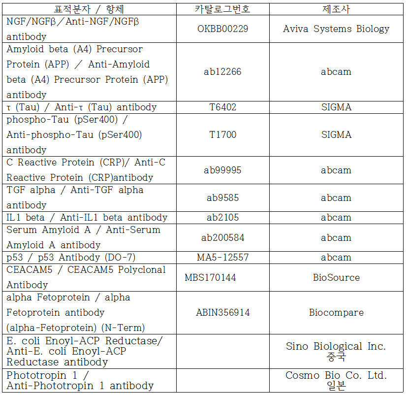
리간드를 이용한 분석법에 사용된 표적분자와 이에 상응하는 항체The target molecule used in the ligand-based assay and the corresponding antibody

리간드를 이용한 분석법에 사용된 표적분자와 이에 상응하는 압타머The target molecule used in the ligand-based assay and its corresponding platemaker
본 발명은 리간드를 이용한 표적분자의 동정 및 정량하는 방법은 코팅계수법, 포획계수법과 경쟁계수법으로 표적분자를 계수하여 정량 분석하는 방법 및 키트를 제공한다.The present invention provides a method and kit for identifying and quantifying a target molecule using a ligand by quantifying a target molecule by a coating coefficient method, a capture counting method and a competitive counting method.
□ 표적분자□ Target molecule
생체분자 계수를 코팅계수법, 포획계수법과 경쟁계수법으로 수행하기 아래의 생체분자를 각각 100 ㎍/mL 농도로 만든 후 냉장(4℃) 보관하고, 이 용액을 단계적으로 희석하여 최종 생체분자의 농도를 1,000 pg/mL, 100 pg/mL, 10 pg/mL 0.1 pg/mL 및 0pg/mL이 되도록 하였다. 분석을 위해 각각의 생체분자를 반응용액으로 혼합하여 분석시료를 준비하여 사용하였다.Biomolecular Coefficient by Coating Method, Capture Coefficient Method and Competition Coefficient Method The following biomolecules were prepared at a concentration of 100 μg / mL, respectively, and stored at 4 ° C. The solution was diluted stepwise to obtain the final concentration of biomolecules 1000 pg / mL, 100 pg / mL, 10 pg / mL 0.1 pg / mL, and 0 pg / mL. For analysis, each biomolecule was mixed with a reaction solution, and analytical samples were prepared and used.
코팅계수법 및 항체리간드를 이용한 분자계수 분석방법의 표적분자는 NGF/NGFβ, Amyloid beta (A4) Precursor Protein (APP), τ (Tau), phospho-Tau (pSer400), C Reactive Protein (CRP), TGF alpha, IL1 beta, Serum Amyloid A, p53, CEACAM5, alpha Fetoprotein 등 11종류의 단백질과 표준물질로 E. coli Enoyl-ACP Reductase(Fabl, 28kD)와 Phototropin 1(nph, 120kD) 등 2종류를 사용하였다. (TGF), phospho-Tau (pSer400), C Reactive Protein (CRP), TGF (TGF), and amyloid beta Two types of proteins and standard substances were used: E. coli Enoyl-ACP Reductase (Fabl, 28kD) and Phototropin 1 (nph, 120kD), including alpha, IL1 beta, Serum Amyloid A, p53, CEACAM5 and alpha Fetoprotein .
코팅계수법 및 압타머 리간드를 위한 생체분자는 CEA, HER-2, Interferon-γ, EGFR, PSMA, Thrombin, Tumor necrosis factor-alpha (TNFa), VEGF (165), Prion Protein, Acetylcholine Receptor Antibody, Human Osteopontin, ErbB2, HIV-1 R5 gp120, HIV-1 R5 SU Glycoprotein 등의 14종류의 고분자 단백질을 사용하였다.Biomolecules for Coating Counting and Ablamin Ligands are CEA, HER-2, Interferon-γ, EGFR, PSMA, Thrombin, Tumor Necrosis Factor-alpha (TNFa), VEGF (165), Prion Protein, Acetylcholine Receptor Antibody, Human Osteopontin , ErbB2, HIV-1 R5 gp120 and HIV-1 R5 SU Glycoprotein were used.
포획계수법 및 항체리간드를 이용한 분자계수 분석방법의 표적분자는 NGF/NGFβ, Amyloid beta (A4) Precursor Protein (APP), τ (Tau), phospho-Tau (pSer400), C Reactive Protein (CRP), TGF alpha, IL1 beta, Serum Amyloid A, p53, CEACAM5, alpha Fetoprotein 등 11종류의 단백질과 E. coli, ,Listeria, Salmonella CSA-1등 3종의 세균을, 그리고 표준물질로 E. coli Enoyl-ACP Reductase(Fabl, 28kD)와 Phototropin 1(nph, 120kD) 등 2종류를 사용하였다.The target molecules of the capture coefficient method and molecular ligand-based assay method are NGF / NGFβ, Amyloid beta (A4) Precursor Protein (APP), τ (Tau), phospho-Tau (pSer400), C Reactive Protein Three kinds of bacteria such as E. coli, Listeria and Salmonella CSA-1 and 11 kinds of proteins such as alpha, IL1 beta, Serum Amyloid A, p53, CEACAM5 and alpha Fetoprotein and E. coli Enoyl-ACP Reductase (Fabl, 28kD) and Phototropin 1 (nph, 120kD) were used.
포획계수법 및 압타머 리간드를 위한 생체분자는 CEA, HER-2, Interferon-y 등의 3종류의 단백질과 Salmonella typhimurium와 Staphylococcus aureus 등의 2종 세균을 사용하였다.Two biomolecules such as CEA, HER-2 and Interferon-y and Salmonella typhimurium and Staphylococcus aureus were used for the capture method and the biomolecule for the aptamer ligand.
경쟁계수법 및 항체 리간드를 이용한 분자계수 분석 방법의 표적분자는 cortisol, cortisone, testosterone, progesterone, androstendione과 aldosterone 등 6종류의 저분자 Steroid hormone과 NGF/NGFβ, Amyloid beta (A4) Precursor Protein (APP), τ (Tau), phospho-Tau (pSer400), C Reactive Protein (CRP), TGF alpha, IL1 beta, Serum Amyloid A, p53, CEACAM5, alpha Fetoprotein 등 11종류의 고분자 단백질을, 그리고 표준물질로 E. coli Enoyl-ACP Reductase(Fabl, 28kD)와 Phototropin 1(nph, 120kD) 등 2종류를 사용하였다.The target molecule of the competitive counting method and molecular ligand-based assay method is composed of six kinds of low molecular steroid hormones such as cortisol, cortisone, testosterone, progesterone, androstendione and aldosterone and NGF / NGFβ, Amyloid beta (A4) Precursor Protein (Tau), phospho-Tau (pSer400), C Reactive Protein (CRP), TGF alpha, IL1 beta, Serum Amyloid A, p53, CEACAM5 and alpha Fetoprotein, and E. coli Enoyl Two types of ACP Reductase (Fabl, 28kD) and Phototropin 1 (nph, 120kD) were used.
포획계수법 및 압타머 리간드를 위한 생체분자는 Kanamycin와 Streptomycin 등 2종류의 저분자 항생제와 CEA, HER2, Interferon-y, EGFR, PSMA, Thrombin, Tumor necrosis factor-alpha (TNFa), VEGF (165), Prion Protein, Acetylcholine Receptor Antibody, Human Osteopontin, ErbB2, HIV-1 R5 gp120, HIV-1 R5 SU Glycoprotein 등의 14종류의 고분자 단백질을 사용하였다.Biomolecules for capture and counting and aptamer ligands are composed of two low-molecular antibiotics such as Kanamycin and Streptomycin, and a combination of CEA, HER2, Interferon-y, EGFR, PSMA, Thrombin, Tumor necrosis factor-alpha (TNFa), VEGF Protein, Acetylcholine Receptor Antibody, Human Osteopontin, ErbB2, HIV-1 R5 gp120 and HIV-1 R5 SU Glycoprotein.
□ 표적분자와 탐침 반응□ Target molecule and probe reaction
표적분자 계수를 코팅계수법, 포획계수법과 경쟁계수법으로 수행하기 하기에서 다음과 같이 표적분자를 포함하는 시료를 준비하였다. Performing Target Molecular Coefficient with Coating Counting Method, Capture Counting Method and Competitive Counting Method In the following, a sample containing a target molecule was prepared as follows.
본 발명에서 생체분자 분석을 위한 반응은 하기 반응용액에서 일어나는 리간드-리셉터 반응이다. 반응용액은 항체리간드의 경우는 10 mM PBS 버퍼(137 mM NaCl, 10 mM Phosphate, 2.7 mM KCl, pH 7.4) 이며, 압타머 리간드의 경우는 셀렉스버퍼 (20 mM Tris-Cl buffer, pH 7.6 contained 100 mM NaCl, 2 mM MgCl2, 5 mM KCl, 1 mM CaCl2 and 0.02% Tween)를 사용하였다.In the present invention, the reaction for biomolecule analysis is a ligand-receptor reaction occurring in the following reaction solution. The reaction solution contained 10 mM PBS buffer (137 mM NaCl, 10 mM Phosphate, 2.7 mM KCl, pH 7.4) in the case of the antibody ligand, and the celex buffer (20 mM Tris-Cl buffer, pH 7.6 contained 100 mM NaCl, 2 mM MgCl2, 5 mM KCl, 1 mM CaCl2 and 0.02% Tween) was used.
반응용액에 세균번식저해를 위한 소듐아지드 0.05 내지 0.2%, 비특이적 결합 저해를 위한 우혈청알부민 0.1 내지 0.3%을 포함할 수도 있다. 반응온도는 압타머리간드의 경우는 SELEX 수행온도보다는 낮은 온도에서 수행하는 것이 이상적이며, 바람직하게는 반응온도는 20 내지 30℃의 온도이다.The reaction solution may contain 0.05 to 0.2% of sodium azide for inhibiting bacterial growth, and 0.1 to 0.3% of bovine serum albumin for inhibiting non-specific binding. The reaction temperature is ideally at a temperature lower than the SELEX performance temperature in the case of the platamer ligand, and the reaction temperature is preferably 20 to 30 占 폚.
(코팅계수방법)(Coating coefficient method)
25 ㎕의 생체분자 용액을 75 ㎕의 반응용액에 첨가하고 코팅지지체인 나이트로셀룰루로스 막(Nitrocellulose membrane) disc를 넣어 30분간 흔들어 주면서 반응시켜 시료를 준비하였다. 앞서 제작된 1㎕의 제1 탐침 용액을 투입하여 30분간 반응시켜 디스크에 부착된 표적분자-제1 탐침 복합체를 형성하였다. 과다 제1 탐침과 비특이적 결합한 제1 탐침을 제거하기 위해서, 상기 반응용액에 있는 상기 형성된 표적분자-제1 탐침 복합체가 부착한 디스크를 150 ㎕의 0.1X SSPE / 0.1 % tween-20 용액을 첨가하여 3회 세척하고 정제하였다.25 μl of the biomolecule solution was added to 75 μl of the reaction solution, and the sample was prepared by reacting with a nitrocellulose membrane disc as a coating paper for 30 minutes while shaking. 1 μl of the first probe solution prepared above was added and reacted for 30 minutes to form a target molecule-first probe complex adhered to the disk. To remove the first probe nonspecifically combined with the excess first probe, the disk with the formed target molecule-first probe complex in the reaction solution was added with 150 μl of 0.1X SSPE / 0.1% tween-20 solution Washed three times and purified.
세척된 디스코에 부착된 표적분자-제1 탐침 복합체에서 해리된 제1 탐침을 수확하기 위해서, 상기 정제한 물질에 100 ㎕의 0.1X SSPE / 0.1 % tween-20 용액을 첨가하고 95 ℃에서 5 분간 처리한 후, 상기 복합체에서 해리된 제1 탐침을 포함하는 상징액을 수확하였다.To harvest the first probe dissociated from the target molecule-first probe complex adhered to the washed disco, 100 μl of 0.1X SSPE / 0.1% tween-20 solution was added to the purified material and incubated at 95 ° C for 5 minutes After treatment, the supernatant containing the first probe dissociated in the complex was harvested.
45 μL의 전체 반응용액을 9 μL씩 분주하여 표적분자 계수 분석법을 위한 분석시료로 사용하였다. 시료 분석은 3 μL의 전체 반응용액을 3회 분석하였다.45 μL of the total reaction solution was dispensed in 9 μL aliquots and used as an analytical sample for the target molecule count analysis. For sample analysis, 3 μL of total reaction solution was analyzed 3 times.
(포획계수방법)(Capture counting method)
25 ㎕의 생체분자 용액, 1㎕의 제1 탐침 용액과 1㎕의 제2 탐침 용액을 첨가한 100 ㎕의 반응용액을 실온에서 30분간 연속 회전하면서 반응시켜 제1 탐침-생체분자-제2 탐침 복합체를 형성시켰다. 25 μl of the biomolecule solution, 1 μl of the first probe solution and 1 μl of the second probe solution were added to 100 μl of the reaction solution while being continuously rotated for 30 minutes at room temperature to form a first probe-biomolecule- Complex.
과잉 제2 탐침, 제1 탐침-생체분자-제2 탐침 복합체, 비특이적 결합으로 결합한 제1 탐침을 제거하기 위해서, 상기 반응용액에 150 ㎕의 반응용액 / 0.1 % tween-20 용액을 첨가하여 5분간 연속 회전한 후 제2 탐침의 자성비드 (Invitrogen 사, USA)로 수확하는 방법으로 3 회 정제하였다. 비특이적 결합으로 제1 탐침을 제거하기 위해서, 추가적으로 상기 수확한 침전물에 150 ㎕의 0.1X SSPE / 0.1 % tween-20 용액을 첨가하여 제2 탐침의 자성비드 (Invitrogen 사, USA)로 수확하는 방법으로 3 회 정제하였다.In order to remove the excess probe, the first probe-biomolecule-second probe complex, and the first probe coupled with non-specific binding, 150 μl of the reaction solution / 0.1% tween-20 solution was added to the reaction solution, Followed by successive rotations, followed by three rounds of purification by magnetic beads of a second probe (Invitrogen, USA). To remove the first probe by non-specific binding, 150 [mu] l of 0.1X SSPE / 0.1% tween-20 solution was further added to the harvested precipitate and harvested with magnetic beads of the second probe (Invitrogen, USA) 3 times.
세척된 제1 탐침-표적분자-제2 탐침 복합체에서 해리된 제1 탐침을 수확하기 위해서, 상기 정제한 물질에 100 ㎕의 0.1X SSPE / 0.1 % tween-20 용액을 첨가하고 95 ℃에서 5 분간 처리한 후, 상기 복합체에서 해리된 제1 탐침을 포함하는 상징액을 수확하였다.To harvest the first probe dissociated from the washed probe-target molecule-second probe complex, 100 μl of 0.1X SSPE / 0.1% tween-20 solution was added to the purified material and incubated at 95 ° C for 5 minutes After treatment, the supernatant containing the first probe dissociated in the complex was harvested.
45 μL의 전체 반응용액을 9 μL씩 분주하여 표적분자 계수 분석법을 위한 분석시료로 사용하였다. 시료 분석은 3 μL의 전체 반응용액을 3회 분석하였다.45 μL of the total reaction solution was dispensed in 9 μL aliquots and used as an analytical sample for the target molecule count analysis. For sample analysis, 3 μL of total reaction solution was analyzed 3 times.
(경쟁계수방법)(Competitive coefficient method)
경쟁계수방법은 시료를 구성하는 생체분자, 경쟁분자와 제1 탐침의 반응은 상기 반응용액에서 일어나는 경쟁적 리간드-리셉터 반응을 이용한 분석이다.The competitive coefficient method is an analysis using a competitive ligand-receptor reaction occurring in a biomolecule, a competitive molecule and a first probe constituting the sample, which occurs in the reaction solution.
25 ㎕의 생체분자 용액, 1㎕의 경쟁분자 용액과 1㎕의 제1 탐침 용액을 첨가한 100 ㎕ 반응용액을 실온에서 30분간 연속 회전하면서 반응시켜 생체분자-제1 탐침 복합체와 경쟁분자-제1 탐침 복합체를 형성시켰다. 25 μl of the biomolecule solution, 1 μl of the competing molecular solution and 1 μl of the first probe solution were added to 100 μl of the reaction solution while continuously rotating for 30 minutes at room temperature to form a biomolecule-first probe complex and a competitive molecule 1 probe complex.
생체분자-제1 탐침 복합체와 비특이적 결합으로 결합한 제1 탐침을 제거하기 위해서, 상기 반응용액에 150 ㎕의 반응용액 / 0.1 % tween-20 용액을 첨가하여 5분간 연속 회전한 후 경쟁분자의 자성비드 (Invitrogen 사, USA)로 수확하는 방법으로 3 회 정제하였다. 비특이적 결합으로 제1 탐침을 제거하기 위해서, 추가적으로 상기 수확한 침전물에 150 ㎕의 0.1X SSPE / 0.1 % tween-20 용액을 첨가하여 경쟁분자의 자성비드 (Invitrogen 사, USA)로 수확하는 방법으로 3 회 정제하였다.In order to remove the first probe bound to the biomolecule-first probe complex by non-specific binding, 150 μl of the reaction solution / 0.1% tween-20 solution was added to the reaction solution, and the mixture was continuously rotated for 5 minutes. (Invitrogen, USA). To remove the first probe with non-specific binding, 150 [mu] l of 0.1X SSPE / 0.1% tween-20 solution was further added to the harvested precipitate and harvested with magnetic beads of competitive molecules (Invitrogen, USA) .
세척된 경쟁분자-제1 탐침 복합체에서 해리된 제1 탐침을 수확하기 위해서, 상기 정제한 물질에 100 ㎕의 0.1X SSPE / 0.1 % tween-20 용액을 첨가하고 95 ℃에서 5 분간 처리한 후, 상기 복합체에서 해리된 제1 탐침을 포함하는 상징액을 수확하였다.In order to harvest the first probe dissociated from the washed competitive molecule-first probe complex, 100 μl of 0.1X SSPE / 0.1% tween-20 solution was added to the purified material, treated at 95 ° C for 5 minutes, A supernatant containing the first probe dissociated from the complex was harvested.
45 μL의 전체 반응용액을 9 μL씩 분주하여 표적분자 계수 분석법을 위한 분석시료로 사용하였다. 시료 분석은 3 μL의 전체 반응용액을 3회 분석하였다.45 μL of the total reaction solution was dispensed in 9 μL aliquots and used as an analytical sample for the target molecule count analysis. For sample analysis, 3 μL of total reaction solution was analyzed 3 times.
본 발명은 상기 코팅계수법, 포획계수법과 경쟁계수법에서 정제한 제1 탐침이 포함된 복합체의 제1 탐침을 계수 분석하고 상기 분석결과로 부터 시료의 생체분자 값을 분석한다.In the present invention, the first probe of the composite including the first probe purified by the Coefficient Counting Method, the capture counting method and the competition counting method is subjected to the coefficient analysis and the biomolecule value of the sample is analyzed from the analysis result.
본 발명에서 코팅계수법 및 포획계수법에서 반응용액에 존재하는 표적분자는 제1 탐침과 결합하여 표적분자-제1 탐침 복합체를 형성하고 복합체의 형성정도는 표적분자의 양에 비례한다. 표적분자에 대한 정량분석 결과인 표적분자 값은 최종적으로 정제된 제1 탐침을 포함하는 복합체에서 해리된 제1 탐침에서 발생하는 신호인 스폿의 계수 분석하여 얻은 결과를 바탕으로 표적분자 값을 분석된다. In the present invention, the target molecule present in the reaction solution in the coating coefficient method and the capture count method combines with the first probe to form the target molecule-first probe complex, and the degree of formation of the complex is proportional to the amount of the target molecule. The target molecule value, which is the result of quantitative analysis of the target molecule, is analyzed based on the result of the coefficient analysis of the spot, which is a signal generated in the first probe, which is dissociated in the complex containing the purified first probe .
특히, 경쟁계수법은 상기 반응용액에서 형성된 경쟁분자-제1 탐침 복합체(complex)를 분리하여 상기 복합체에서 해리된 제1 탐침을 분석하여 결정한다. 즉, 분석하고자하는 표적분자와 경쟁분자의 제1 탐침에 대한 반응은 경쟁적 반응으로 표적분자-제1 탐침 복합체와 경쟁분자-제1 탐침 복합체를 형성한다. 반응용액에서 경쟁분자-제1 탐침 복합체의 형성정도는 생체분자-제1 탐침 복합체의 형성정도에 의존하여 결정됨으로 경쟁분자-제1 탐침 복합체의 신호를 분석하여 표적분자 값을 결정할 수 있다. 경쟁분자-제1 탐침 복합체만을 수확하여 제1 탐침를 해리시켜 해리된 제1 탐침에서 발생하는 신호인 스폿의 계수 분석하여 얻은 결과를 바탕으로 표적분자 값을 분석된다. Particularly, the competitive-counting method is determined by analyzing the first probe dissociated from the complex by separating the competitive molecule-first probe complex formed in the reaction solution. That is, the reaction of the target molecule and the competitive molecule to be analyzed with respect to the first probe forms a competitive probe with the target molecule-first probe complex and the competitive molecule-first probe complex. The degree of formation of the competitive molecule-first probe complex in the reaction solution is determined depending on the degree of formation of the biomolecule-first probe complex, so that the target molecule value can be determined by analyzing the signal of the competitive molecule-first probe complex. Competitive Molecule - The target molecule value is analyzed based on the results obtained by analyzing the coefficient of the spot, which is a signal generated from the dissociated first probe, by harvesting only the first probe complex and dissociating the first probe.
표적분자에 대한 정량분석 결과인 표적분자 값은 최종적으로 정제된 제1 탐침을 포함하는 복합체에서 해리된 제1 탐침을 계수하여 기 작성된 표준곡선을 이용하여 표적분자와 결합하는 제1 탐침의 량을 평가하여 표적분자 값을 결정한다. The target molecule value, which is the result of quantitative analysis of the target molecule, is calculated by counting the first probe dissociated in the complex including the finally purified first probe, measuring the amount of the first probe binding to the target molecule using the standard curve And the target molecule value is determined.
상기 코팅계수법, 포획계수법과 경쟁계수법의 상기 표적분자 값은 생물학적 샘플에서 하나 또는 그 이상의 표적분자를 연속적 분석이 가능하게 하는 다중분석을 사용하여 결정할 수 있다. The target molecule values of the coating counting method, capture counting method and competition counting method can be determined using multiple assays that enable continuous analysis of one or more target molecules in a biological sample.
다중분석에서, 개개의 상기 복합체들에서 해리되고 개개의 표적분자를 지시하는 신호발생영역이 있는 제1 탐침들을 지지체 상의 별개의 위치에 직접 또는 간접적으로, 공유결합 또는 비공유결합으로 고정화시킨다. 다중분석은 지지체의 특정영역에 결합한 제1 탐침에 있는 양자점(quantum dot)과 같은 신호발생영역에서 발생하는 독특한 신호에 의해 둘 이상의 표적분자들을 구분할 수 있는 기구에 의해 실현한다. 개개의 기구는 생물학적 샘플에서 검출될 각각의 다중 표적분자 중 하나의 분석을 위하여 사용한다. 개개의 기구는 생물학적 샘플에서 각 표적분자를 동시에 처리할 수 있도록 구성될 수 있다. 예를 들면, 미세역가 플레이트는 플레이트 내의 각 웰이 생물학적 샘플에서 분석될 다중 표적분자 중 하나를 특별하게 분석하는데 사용되는 것처럼 사용할 수도 있다.In multiple assays, first probes with signal generating regions that are dissociated in individual complexes and point to individual target molecules are immobilized, either directly or indirectly, in covalent or noncovalent bonds at distinct locations on the support. Multiple assays are realized by a mechanism capable of distinguishing two or more target molecules by a unique signal generated in a signal generation region, such as a quantum dot in a first probe coupled to a specific region of a support. Each instrument is used for the analysis of one of each of the multiple target molecules to be detected in the biological sample. Each instrument can be configured to process each target molecule simultaneously in a biological sample. For example, microtiter plates may be used as if each well in the plate was used to specifically analyze one of the multiple target molecules to be analyzed in the biological sample.
실시례Example 5. 계수 분석 5. Coefficient analysis
핵산정량, 핵산변이, 메틸화 DNA, 리간드를 이용한 표적분자 정량 분석 등은 표적분자를 특이적으로 인지하고 결합하는 프로브를 매개로 표적분자의 정보가 제1 탐침의 신호발생영역으로 전환되는 과정을 상기에서 실시하였다. 따라서 실시례에서 준비한 분석시료에 있는 제1 탐침의 신호발생영역을 분자계수하면 표적분자의 정보를 분석할 수 있다. The process of converting the information of the target molecule to the signal generating region of the first probe through the probe that specifically recognizes and binds to the target molecule, such as nucleic acid quantification, nucleic acid mutation, methylated DNA, ligand, . Therefore, if the signal generation region of the first probe in the analytical sample prepared in the embodiment is subjected to molecular counting, the information of the target molecule can be analyzed.
상기 실시레에서 준비한 분석시료를 하기와 같이 처리하여 제1 탐침 신호발생영역을 분자 계수를 하였다.The analytical sample prepared in the above procedure was processed as follows to make the molecular count of the first probe signal generation region.
단일가닥핵산을 프로브로 하는 핵산정량은 제1 탐침-표적 핵산-제2 탐침 복합체를 세척하고 수확하여 해리된 제1 탐침을 계수하여 표적 핵산을 정량할 수 있다. 표적 mRNA와 제1 탐침은 일대일로 결합함으로 제1 탐침의 계수 값은 mRNA의 계수 값에 상응하다. 미리 작성된 표준커브를 이용하여 결정된 mRNA 계수 값을 이용하여 mRNA를 정량화하였다. The nucleic acid quantification using a single stranded nucleic acid probe can be performed by washing and harvesting the first probe-target nucleic acid-second probe complex to quantify the target nucleic acid by counting the dissociated first probe. The target mRNA and the first probe bind one-to-one, so that the coefficient value of the first probe corresponds to the count value of the mRNA. MRNA was quantified using mRNA coefficient values determined using a pre-written standard curve.
유전자 변이 유무 및 유형 분석은 제1 탐침-유전자 변이-제2 탐침 복합체에 신장반응 또는 연결반응 등의 효소반응으로 얻은 반응물에서 해리된 제1 탐침, 실시레에서 얻은 반응물에 있는 제1 탐침 혹은 제1 탐침-제2 탐침 연결체를 계수하여 할 수 있다. 전자는 신장반응 혹은 연결반응을 할 수 없는 상태로 표적 핵산의 변이부위와 제1 프로브의 V영역이 미스 메칭임을 의미하는 것이다.The presence and type of the gene mutation can be detected by the first probe-gene mutation-second probe complex, a first probe dissociated from the reaction product obtained by an enzymatic reaction such as a elongation reaction or a linking reaction, 1 probe - the second probe connector can be counted. The former means that the mutation site of the target nucleic acid and the V region of the first probe are mis-tagged in a state in which the elongation reaction or linkage reaction can not be performed.
DNA 메칠화 여부 및 비율은 제1 탐침-메틸화 DNA-제2 탐침 복합체에 신장반응 또는 연결반응 등의 효소반응으로 얻은 반응물에서 해리된 제1 탐침, 실시레에서 얻은 반응물에 있는 제1 탐침 혹은 제1 탐침-제2 탐침 연결체를 계수하여 할 수 있다. 전자는 신장반응 혹은 연결반응을 할 수 없는 상태로 표적 DNA 메틸화 부위와 제1 프로브의 V영역이 미스 메칭임을 의미하는 것이다.The presence or proportion of DNA methylation can be determined by comparing the first probe-methylated DNA-second probe complex with the first probe or the first probe in the reaction product obtained from the reaction, the first probe being dissociated from the reaction product obtained by the enzymatic reaction such as the elongation reaction or the linking reaction, 1 probe - the second probe connector can be counted. The former means that the target DNA methylation site and the V region of the first probe are mis-tagged in a state in which the elongation reaction or linkage reaction can not be performed.
프로브가 리간드인 표적분자 분석은 제1 탐침-표적분자-제2 탐침 복합체를 세척하고 수확하여 해리된 제1 탐침을 계수하여 정량할 수 있다. 표적 분자와 제1 탐침은 일대일로 결합함으로 제1 탐침의 계수 값은 표적분자의 계수 값에 상응하다. 미리 작성된 표준커브를 이용하여 결정된 표적분자 계수 값을 이용하여 mRNA를 정량화하였다.Target molecule analysis in which the probe is a ligand can be quantified by counting and dissociating the first probe-target molecule-second probe complex and harvesting the first probe. The target molecule and the first probe bind in one-to-one association so that the count value of the first probe corresponds to the count value of the target molecule. MRNA was quantified using the target molecular count values determined using a pre-written standard curve.
상기 반응물에 있는 제1 탐침 계수 분석은 아래와 같이 수행하였다.The first probe count analysis in the reaction was performed as follows.
생체분자 계수분석을 위해서, 시료는 제조사의 프로토콜을 참조하여 본 발명의 분석방법에 따라 n카운터 프렙 스테이션 (나노스트링 테크놀로지사, Seattle, WA-USA)을 통해 가공되었고, n카운터 디지탈 분석기 (나노스트링 테크놀로지사, Seattle, WA-USA)로 수행하였다. For biomolecular count analysis, the samples were processed through an n-counter prep station (Nanostring Technologies, Seattle, WA-USA) according to the assay method of the present invention with reference to the manufacturer's protocol, and an n-counter digital analyzer Technology, Seattle, WA-USA).
0.1 % Tetraspeck 형광 마이크로 스피어 용액(Invitrogen 사, USA)을 1:5,000로 희석한 1㎕의 시약을 상기 실시례4에서 준비된 정제된 복합체 용액에 첨가하여 분석시료를 준비하였다. 상기 준비된 분석시료를 스트렙타비딘이 코팅된 커버 슬립(Optichem, Accelr8 Technology Corporation)이 포함되는 NanoString 유체 장치에 로팅하여 30 um 깊이인 미세유동 채널을 생성하였다. 분석시료는 수압에 의해 채널로 이동하여 상기 복합체에서 해리된 제1 탐침의 바이오틴과 결합지지체인 채널에 있는 스트렙타비딘에 강력하게 결합하였다. 고정한 후, 표면을 90 ㎕의 1x TAE 용액으로 한번 세척하고, 각 웰에 40 ㎕의 TAE를 첨가하여 펴지도록 하였다. 제1 탐침은 유체 채널에 1 분 동안 160 V /cm을 적용하여 펴진 상태로 정렬하였다. 펴진 제1 탐침은 60 ㎕의 500 nM 고정분자(모든 제1 탐침 존재하는 반복구조에 상보적인 바이오틴이 표지된 올리고뉴클레오티드)를 첨가하여 표면에 추가적으로 고정화시켰다. 고정화 과정을 동안, 전류는 5 분 동안 유지되었다. 고정한 후 TAE 용액을 제거하고, 촬영용 안티광표백제(antiphotobleaching) SlowFade 시약 (Invitrogen 사)를 첨가하였다. 1 μl of a reagent diluted 1: 5,000 in 0.1% Tetraspeck fluorescence microsphere solution (Invitrogen, USA) was added to the purified complex solution prepared in the above Example 4 to prepare an assay sample. The prepared analytical sample was rotated in a NanoString fluid device containing a streptavidin-coated cover slip (Optichem, Accelr8 Technology Corporation) to produce a microfluidic channel with a depth of 30 μm. The analytical sample migrated to the channel by water pressure and bound strongly to the biotin of the first probe dissociated in the complex and streptavidin in the channel of the binding support. After fixation, the surface was washed once with 90 [mu] l of 1x TAE solution, and 40 [mu] l of TAE was added to each well to spread out. The first probe was aligned with the fluid channel by applying 160 V / cm for 1 minute. The expanded first probe was further immobilized on the surface by the addition of 60 [mu] l of 500 nM fixed molecule (biotin labeled oligonucleotide complementary to all of the first probe present repeating structures). During the immobilization process, the current was maintained for 5 minutes. After fixing, the TAE solution was removed and an anti-photobleaching SlowFade reagent (Invitrogen) was added for photographing.
제1 탐침이 결합한 결합지지체는 Perfect Focus, 1.4 NA Plan Apo VC 60X oil-immersion lens (Nikontk, 일본), anX-cite 120 metal halide light source (Exfo 사, USA), automated H117 stage (Prior Scientific)와 Smart Shutter (Sutter Instrument, USA)가 갖추어진 Nikon Eclipse(니콘 이클립스) TE2000E로 촬영하였다. 전형적인 촬영 밀도는 FOV당 100~200개의 리포터를 카운팅하였다. 각각의 field of view에 대해, 다른 여기 파장 (480, 545, 580, 622)에 네 개의 이미지는 MetaMorph(Universal Imaging Corporation, USA) 또는 사용자 정의 소프트웨어로 제어하는 Orca Ag CCD camera (Hamamatsu사, 일본)로 촬영하였다(도10). The combined support with the first probe was a Perfect Focus, 1.4 NA Plan Apo VC 60X oil-immersion lens (Nikontk, Japan), anX-cite 120 metal halide light source (Exfo, USA), automated H117 stage (Prior Scientific) And Nikon Eclipse (Nikon Eclipse) TE2000E equipped with Smart Shutter (Sutter Instrument, USA). A typical photographic density counts between 100 and 200 reporters per FOV. For each field of view, four images at different excitation wavelengths (480, 545, 580, 622) were measured using MetaMorph (Universal Imaging Corporation, USA) or Orca Ag CCD camera (Hamamatsu, Japan) (Fig. 10).
분석기 해상도는 600 FOV(field of view)로 설정하였다. 데이터를 다운로딩 하고, 추천된 바와 같이 (n카운터 데이터 분석 지침들) 엑셀 상에서 분석하였다. 데이타는 처음에 제공된 양성 검정 대조군의 표준곡선을 사용하여 시료내의 기준물질 및 표적분자를 정상화하였다. 정상화된 값은 시료에 포함된 기준물질들의 값을 사용하여 표적분자 값을 전반적으로 평균 정상화를 수행하였다. 개별적 표적분자들을 위한 3회 수행한 에서 표적분자 수치들의 평균을 취하여 계산하였다.The analyzer resolution was set to 600 FOV (field of view). Data was downloaded and analyzed on Excel as recommended (n-counter data analysis guidelines). The data normalized the reference material and the target molecule in the sample using the standard curve of the positive control group provided at the beginning. The normalized values were obtained by performing averaging over the whole of the target molecule values using the values of the reference materials contained in the sample. Calculated by taking the average of the target molecule values in three runs for individual target molecules.
사용한 특정 생체시료에서 mRNA 정량을 분자 계수 분석방법으로 한 결과, 제1 탐침-특정 mRNA-제2 탐침 복합체에서 해리된 제1 탐침에 의해서 발생하는 표적분자의 존재신호로 형성된 스폿을 확인하였다. 이는 표적 mRNA에 대한 RT-PCR 결과와 유사하였다.As a result of quantitative analysis of mRNA in the specific biological sample used, a spot formed by the presence signal of the target molecule generated by the first probe dissociated in the first probe-specific mRNA-second probe complex was confirmed. This was similar to the RT-PCR result for the target mRNA.
사용한 생체시료의 유전자변이를 분자계수 방법으로 확인한 결과, 특정 유전자 변이 유형을 지시하는 제1 탐침의 신호발생영역의 색상 패턴에 상응하는 신호로 형성된 스폿을 확인하였다. 유전자 변이 유형은 모두 야생형(wild type), 모두 돌연변이형(mutant type), 야생형과 돌연변이형이 같이 존재하는 경우가 다양하게 존재하고 있었다. 이는 염기서열 방법으로 확인한 결과와 동일하였다.As a result of checking the gene mutation of the used biological sample by the molecular counting method, a spot formed by a signal corresponding to the color pattern of the signal generation region of the first probe indicating the specific gene mutation type was confirmed. All of the mutation types were wild type, mutant type, wild type and mutant type. This was the same as the results confirmed by the nucleotide sequence method.
DNA 메틸화 여부 및 비율을 분자계수 방법으로 확인한 결과, 특정 DNA 메틸화 유무를 지시하는 제1 탐침의 신호발생영역의 색상 패턴에 따라 존재에 상응하는 신호로 형성되는 스폿을 확인하였다. DNA 메칠화 유형은 메칠화 또는 비메칠화 형태로 동일 시료에 함께 존재하고 있었으며 그 비율은 특정 색상 패턴의 비율로 조사할 수 있었다. 이는 기존 염기서열 방법으로 확인한 결과와 유사함을 확인할 수 있었다.The presence or absence of DNA methylation and its ratio were confirmed by a molecular counting method. As a result, a spot formed as a signal corresponding to existence was identified according to the color pattern of the signal generation region of the first probe indicating the presence or absence of specific DNA methylation. DNA methylation patterns were present in the same sample in either methylated or unmethylated form and the ratio could be investigated as a percentage of a specific color pattern. It was confirmed that this is similar to the results obtained by the conventional nucleotide sequence method.
상기 코팅계수분석법으로 항체 리간드를 이용한 반응은 11종의 단백질, 2종의 표준물질, 그리고 압타머 리간드를 이용한 반응은 14종의 단백질을 분석하였다. 상기 포획계수분석법으로 항체 리간드를 이용한 반응은 11종의 단백질, 3종의 세균, 2종의 표준물질, 그리고 압타머 리간드를 이용한 반응은 3종의 단백질, 2종의 세균을 분석하였다. 상기 경쟁계수방법으로 항체리간드를 이용한 반응은 6종의 저분자인 steroid hormone, 11종의 단백질, 2종의 표준물질, 그리고 압타머 리간드를 이용한 반응은 2종의 저분자인 항생제, 14종의 단백질을 분석하였다.For the reaction using the antibody ligand by the above coating coefficient analysis, 11 kinds of proteins, 2 kinds of standard substances, and 14 kinds of proteins were analyzed for the reaction using the platamer ligand. The reaction using the antibody ligand by the capture factor analysis was analyzed for 11 kinds of proteins, 3 kinds of bacteria, 2 kinds of standard substances, and 3 types of proteins and 2 kinds of bacteria for the reaction using the platamer ligand. The reaction using the competitive ligand method using the antibody ligand was carried out by using six low-molecular steroid hormones, 11 proteins, two standard substances, and an enzymatic ligand as two small-molecule antibiotics and 14 proteins Respectively.
생체분자 계수방법에 의한 2종류의 표준물질로 구성된 모의(mock) 비교구와 표준물질을 포함한 여러 물질로 구성된 대조구에 대해 3회 분석하였다. 항체리간드를 이용한 분석결과를 살펴보면 다음과 같다. 표준물질로 이루어진 모의 비교구는 모든 반응을 위한 표준 농도 곡선을 작성하였으며 반응, 정제 및 포집 효율에서 약간의 차이가 있는 데이터를 정규화하기 위해 사용하였다. 먼저 2종류 표준물질의 선형성, 동적 범위(dynamic range) 및 재현성을 조사했다(도11). 포획계수법에 의해 표준물질을 분석한 이미지는 도10이며 , 도 13은 포획계수법에, 도 14는 경쟁계수법에 의해 모의 비교구의 표준물질을 측정한 결과이며, (N = 6). 로그-로그 플롯상의 점을 중첩하여 나타낸 바와 같이, 각 분석에 대한 제어 신호의 값 (카운트) 0.5 fM과 50 fM 사이에서 재현성이 매우 높았다. 또한 분석 결과는 농도 대 카운트의 선형 회귀 상관 계수는 농도의 2.5 이상 로그에서 ≥0.998로 고도로 선형성이 있었다.The control was composed of mock comparator composed of two kinds of reference materials by biomolecule counting method and control material composed of various materials including standard material 3 times. Analysis results using antibody ligands are as follows. Mock comparisons made of standard materials were used to normalize the data with slight differences in reaction, purification and collection efficiencies, and standard concentration curves for all reactions. First, the linearity, dynamic range and reproducibility of two kinds of standard materials were investigated (FIG. 11). The image was analyzed and the standard material by trapping and counting method 10, 13 is to capture counting method, 14 is a result of measuring a simulation comparing a standard substance by the sphere race counting method, (N = 6). Reproducibility was very high between 0.5 fM and 50 fM of the control signal values (counts) for each analysis, as indicated by overlapping points on the log-log plot. The analysis also showed that the linear regression coefficient of concentration versus count was highly linear from ≥0.2 to ≥0.998.
그런 다음, 샘플링 효율과 검출한계(limits of detection)를 검토했다. 샘플링 시스템의 효율은 그 반응에 있어서 대상의 이론적인 분자의 수로 생체분자에 대해 카운트 수를 나눔으로써 추정 할 수 있다. 예를 들어, 각 반응에서 ~0.1 fM의 생체분자는 ∼1,800 분자이다. 모의 표본이 생체분자에 대한 평균 측정은 25카운트임으로 샘플링 효율은 ∼1%이다. 분석의 검출 한계는 스튜던트 t-test를 이용하여 음성 대조군의 카운트와 가장 낮은 농도인 양성 대조군의 카운트를 비교하여 결정 하였다. 본 발명 반응에서 최저검출한계는 0.1 및 0.5 fM 사이였다. We then looked at sampling efficiency and limits of detection. The efficiency of the sampling system can be estimated by dividing the number of counts for biomolecules by the number of theoretical molecules of interest in the reaction. For example, in each reaction ~ 0.1 fM biomolecules are ~ 1,800 molecules. The average measurement for a biomolecule is 25 counts, so the sampling efficiency is ~ 1%. The detection limit of the assay was determined by comparing the counts of the negative controls with those of the lowest control positive controls using the Student t-test. The lowest detection limit in the present invention reaction was between 0.1 and 0.5 fM.
항체 리간드를 이용한 포획계수법에 의한 13종의 생체분자인 단백질들과 코팅계수법에 의한 압타머 리간드를 이용한 14종의 생체분자인 단백질을 측정하는 시스템의 재현성을 평가하였다. 항체 리간드를 이용한 13종류의 생체분자에 대해 2번의 독립적인 반응에 대한 정규화 된 수는 로그-로그 스케일로 표시하였다(도 15). 본 발명의 시스템의 자료에 대한 선형회귀 분석결과는 상관계수는 0.9982로 재현성이 높음을 알 수 있다. 압타머 리간드를 이용한 경우도 이와 유사한 결과를 얻었다.The reproducibility of the system for measuring proteins of 14 kinds of biomolecules using 13 protein molecules by the capture counting method using the antibody ligand and the platamer ligand by the coating coefficient method was evaluated. Normalized numbers for two independent reactions for 13 biomolecules using antibody ligands were expressed on a log-log scale (Fig. 15). The linear regression analysis of the data of the system of the present invention shows that the correlation coefficient is 0.9982 and the reproducibility is high. Similar results were obtained when the plastomer ligand was used.
실시례Example 6. 생물학적 의미의 분석 6. Analysis of biological meaning
생체시료인 혈액을 이용하여 급성심근경색 도는 패혈증 등의 만성질환을 검진하고 있으며 특히 일반적으로 환자 모니터링용 생체분자 3~4개를 검사하고 결과의 cut-off 수치만을 참고함으로 진단을 하고 있다. A blood sample is used to diagnose chronic diseases such as acute myocardial infarction or septicemia. In general, three to four biomolecules for patient monitoring are examined and only the cut-off value of the result is referred to for diagnosis.
여러 표적 생체분자들을 정량 검사하고 그 결과를 바탕으로 정상인 집단과 환자 집단 간의 함수관계를 정량적으로 분석하여 진단할 수 있는 방법인 체외진단다지표검사(In Vitro Diagnostic Multivariate Index Assays;IVDMIA)는 생물학적 의미인 질병이나 다른 상태를 진단, 치료, 완화, 처치, 예방을 목적으로 해석함수를 사용하여 표적 생체분자의 복수결과들을 결합시켜 환자의 특이적 결과로 "분류", "점수", "지수" 등을 산출하는 장치 또는 기술이다.In Vitro Diagnostic Multivariate Index Assay (IVDMIA), which is a method to quantitatively analyze the functional relationship between normal population and patient group based on the results of quantitative examination of several target biomolecules, "Score", "index", etc. as the patient's specific results by combining multiple results of a target biomolecule using analytical functions for the purpose of diagnosing, treating, alleviating, treating, Is a device or technology that calculates the output power.
본 발명의 생물학적 의미를 결정하는 임상 의사 결정 지원 시스템(Clinical Decision Support System)은 분자계수 분석 방법을 통해 생산된 표준물질 및 표적분자 분석결과를 보정한 데이터와 OCS(operation control system) 시스템의 환자정보를 입력으로 한다. 출력은 임상의가 분석결과를 입력하고 확인하는 화면과, 진단의가 위험성과 분류를 환자에게 설명할 수 있는 화면과 환자에게 가시적으로 설명할 수 있는 화면을 갖는다. 생물학적 의미 분석 시스템은 증폭산물 분석 장치, OCS와 Interface를 갖고 있어야 하며, Data Mining을 통해 얻어진 결과를 DB에 저장할 수 있어야 한다. HL7에 정의된 XML 데이터를 처리하기 위해 XML Repository를 갖고 있다. Data Mining할 때 필요한 항목을 설정하고 Mining 엔진을 제어할 수 있다. The Clinical Decision Support System which determines the biological meaning of the present invention is composed of data obtained by correcting the standard material and target molecule analysis result produced through the molecular factor analysis method and patient information of the operation control system . The output has a screen for entering and confirming the results of the clinician's analysis, a screen for explaining the risk and classification of the diagnosis to the patient, and a screen for explaining to the patient visually. The biological semantic analysis system should have an amplification product analyzer, OCS and interface, and should be able to store the results obtained from Data Mining into DB. It has an XML Repository to handle the XML data defined in HL7. You can set up the necessary items for Data Mining and control the mining engine.
도 4에 도시된 바와 같이, 상기 임상 의사 결정 지원 시스템의 학습 엔진 모듈은 상기 표적 분자를 지시하는 탐침을 이용한 분자계수 분석결과를 이용하여 질환을 판단하기 위해 필요한 인공지능 학습 엔지 모듈이다.As shown in FIG. 4, the learning engine module of the clinical decision support system is an artificial intelligence learning engine module necessary to determine a disease using a result of a molecular coefficient analysis using a probe indicating the target molecule.
생물학적 의미 분석 시스템은 HL7을 지원하도록 한다. 생물학적 의미 분석 시스템 내부에서 데이터를 처리할 때, HL7에 정의된 Schema를 포함하도록 데이터 구조를 설계하는 것을 의미한다. XML 문서의 Schema 정보를 저장하고 사용하기 위해 XML Repository Module과 XML Repository Client를 갖는다. The biological semantic analysis system supports HL7. Designing a data structure to include the Schema defined in HL7 when processing data within a biological semantic analysis system. It has XML Repository Module and XML Repository Client for storing and using Schema information of XML document.
표적분자를 지시하는 탐침을 이용한 분자계수 분석결과를 분석할 수 있 분석 장치와의 연동을 위해 분자계수 분석결과 Interface Module을 갖는다. 분자계수 분석결과에서 SDK(software development kit)를 제공하는 경우에는 Gateway를 개발하는 것을 원칙으로 한다. OCS Interface Module은 OCS에서 환자 정보를 읽어 Data Mining에 필요한 정보를 제공한다. 또한 Data Mining에 의해 분석된 질환 정보를 OCS(LIS)에 저장한다.In order to analyze the results of molecular analysis using a probe that indicates a target molecule, the molecular module has an interface module as a result of analyzing the molecular parameters for interlocking with the analyzing device. If the software development kit (SDK) is provided from the results of the molecular counting analysis, the gateway should be developed in principle. The OCS Interface Module reads patient information from OCS and provides the information needed for data mining. In addition, disease information analyzed by Data Mining is stored in OCS (LIS).
도 5에 도시되어 제시하는 바와 같이, 상기 임상 의사 결정 지원 시스템의 생물학적 의미 분석은 크게 네 개의 부분 시스템으로 구성되어 있으며, 생물학적 의미 결정 엔진인 '데이터분석/예측모델 생성' 모듈과 '진단클라이언트'모듈, 그리고 압타머칩 데이터를 생성하는 증폭산물 분석 장치와의 연동 모듈인 '증폭산물 분석 장치 인터페이스', 다른 모듈간의 통신과 데이터 저장을 담당하는 '통신서버'가 유기적으로 결합된 시스템이다. 시스템의 구동은 크게 시스템 구축단계와 시스템 적용 단계로 구분된다.As shown in FIG. 5, the biological semantic analysis of the clinical decision support system is largely composed of four partial systems. The module for generating a data analysis / prediction model, which is a biological semantic determination engine, Module, and an amplification product analyzer interface, which is an interlocking module with the amplification product analyzer that generates the abdominal machine data, and a communication server, which is responsible for communication and data storage between the other modules. The operation of the system is largely divided into system construction phase and system application phase.
시스템 구축 단계는 ① 질병의 특성을 잘 반영하도록 구성된 정상인/환자 시료 집합을 구성하여, ② 해당 시료 집합에 대한 정도관리 및 측정용의 표준물질을 활용한 표적분자를 분자계수 분석을 실시하고, ③ 표적분자 분자계수 분석 정보를 '분자계수 분석 장치 인터페이스' 모듈을 통해 시스템에 입력, ④ '데이터 분석/예측모델 생성' 모듈에서 기본적인 전처리를 하여 인공신경망 알고리즘의 '학습데이터' 생성, ⑤ 인공신경망 알고리즘으로 학습데이터를 이용하여 생물학적 의미 분석 모델 생성, ⑥ 생성된 생물학적 의미 분석모델을 컴퓨터에 저장하는 과정을 거친다. In the system construction phase, ① a normal person / patient sample set is constructed to reflect the characteristics of the disease, ② the molecular analysis of the target molecule using the standard substance for quality control and measurement for the relevant sample set, ③ Inputting the target molecule molecular analysis information into the system through the 'module for analyzing the device for the analysis of molecules', ④ 'learning data' generation of the artificial neural network algorithm by performing basic preprocessing in 'data analysis / prediction model generation module', ⑤ artificial neural network algorithm , And then the generated biological semantic analysis model is stored in the computer.
도 6에 도시되어 제시하는 바와 같이 시스템 적용 단계는 ① DB에 저장된 생물학적 의미 분석모델을 '생물학적 의미 분석 클라이언트'에 장착, ② '생물학적 의미 분석 클라이언트' 구동, ③ 생물학적 의미 분석 대상자의 분자계수 및 분자계수 분석 장치를 이용한 분석결과 정보 생성, ④ '분자계수 분석 장치 인터페이스'를 통한 데이터 입력, ⑤ '생물학적 의미 분석 클라이언트'에서 생물학적 의미 분석 및 결과 가시화, ⑥ 생물학적 의미 분석 결과 DB에 저장으로 구성된다.As shown in FIG. 6, the system application steps are as follows: (1) a biological semantic analysis model stored in a DB is installed in a biological semantic analysis client, (2) a biological semantic analysis client is driven, (3) ④ Data input through 'Molecular Coefficient Analyzer Interface' ⑤ Biological Semantic Analysis and Result Visualization in 'Biological Semantic Analysis Client' ⑥ Biological Semantic Analysis Result is stored in DB.
도 7에 도시되어 제시하는 바와 같이, 표적분자 분석결과는 표적분자 양태 정도를 나타내는 것이다. 수치 데이터는 분자계수 데이터로부터 얻어진 것으로, 각 값은 하나의 표적분자의 값을 의미하며, 숫자 값은 각 표적분자에 대한 양태의 정도를 의미한다고 할 수 있다.As shown in FIG. 7 and shown in FIG. 7, the result of the target molecule analysis indicates the degree of the target molecule. The numerical data is obtained from the numerator data, and each value represents the value of one target molecule, and the numerical value means the degree of the mode for each target molecule.
표적분자 정보와 함께 시스템에서 입력으로 받는 것은 각 환자에 대한 임상 정보이다. 각 환자들에 대한 나이, 성별, 혈압, 가계 상의 질병 내력, 흡연 여부 등 각 환자에 대한 임상 정보 역시 입력으로 받는다. 이들 자료들은 모두 데이터베이스에 저장된다. 시스템 구축을 위해 사용되는 임상 데이터는 기본적으로 협력 의료기관에서 받는 것으로 하며, 표적분자의 양태 데이터는 분자계수 데이터를 이용하는 것으로 한다.In addition to the target molecule information, the input from the system is clinical information for each patient. Clinical information about each patient such as age, gender, blood pressure, history of disease in family history, and smoking status are also input to each patient. All of these data are stored in the database. The clinical data used for constructing the system is basically received by the collaborating medical institution, and the mode data of the target molecule is obtained by using the molecular coefficient data.
도 8에 도시되어 제시하는 바와 같이, 분자계수 분석 실험 결과는 분자계수 분석 장치를 통해 수치 데이터로 변환된다. 질환 생물학적 의미 결정 시스템에서 이 데이터를 직접 이용하기 위해서는 표적분자를 지시하는 탐침을 이용한 분자계수를 읽을 수 있는 분자계수 분석 장치와 생물학적 의미 결정 시스템을 연동시키는 것이 필요하다. 따라서 SDK를 제공하는 분자계수 분석 장치를 사용하는 것을 기본으로 하여, 생물학적 의미를 결정하는 임상 의사 결정 지원 시스템과의 연동을 위한 게이트웨이(gateway)를 개발한다.As shown in Fig. 8, the results of the analysis of the molecular coefficient are converted into numerical data through a molecular coefficient analyzer. In order to directly use this data in the disease biological significance determination system, it is necessary to link the biological significance determination system with the molecular coefficient analyzing apparatus capable of reading the molecular coefficient using the probe indicating the target molecule. Therefore, we develop a gateway for interfacing with the clinical decision support system that determines the biological meaning based on the use of the molecular factor analyzing device that provides the SDK.
한편 생물학적 의미 결정을 위한 임상 데이터는 OCS 시스템의 환자 정보가 입력으로 들어오도록 한다. OCS로부터 환자 정보를 OCS interface module을 통해 읽어서, 데이터마이닝에 필요한 정보들을 시스템 엔진에 제공한다.On the other hand, clinical data for biologic semantics allows the patient information of the OCS system to be input. It reads the patient information from the OCS through the OCS interface module and provides the system engine with the information necessary for data mining.
도 8에 도시되어 제시하는 바와 같이, 질병에 대한 생물학적 의미 결정에 대한 지표 예측은 데이터베이스 내의 정보들을 기반으로 이루어진다. 저장되어있는 정보들을 이용하여 여러 가지 기계학습(machine learning) 방법을 적용하면, 질병에 대한 지표 분석이 가능하다. 인공신경망(artificial neural network), 결정트리(decision tree), 베이지안망(Bayesian network), 서포트벡터머신(support vector machine, SVM) 등의 대표적인 기계학습 방법이 분석을 위한 핵심 알고리즘으로 사용된다. 일반적으로 이들 방법들은 벡터 형태의 수치 데이터가 입력으로 들어오면, 이를 학습하여 새로운 데이터에 대한 예측을 가능하게 해준다. 인공신경망과 서포트벡터머신은 데이터를 분류하는 데에 있어서 우수한 성능을 보이기 때문에 기계학습 분야의 연구에서 가장 많이 사용되는 분류 알고리즘들이다. 또한 결정트리와 베이지안망은 결과를 사람이 이해하기 쉽도록 가시화 할 수 있다는 장점을 가진다. 본 생물학적 의미 결정 시스템은 인공신경망 알고리즘을 통해 지속적인 테스트를 거치면서 학습 모델을 최적화(optimize)하고, 이를 통해 질병지표 예측 성능이 우수하게 나올 수 있도록 한다. As shown in Figure 8, the indicator prediction for biological semantic determination of disease is based on information in the database. By applying various machine learning methods using stored information, it is possible to analyze the indicators for diseases. Representative machine learning methods such as artificial neural network, decision tree, Bayesian network, and support vector machine (SVM) are used as key algorithms for analysis. In general, these methods allow the prediction of new data by learning the vector form of numerical data as input. Artificial neural networks and support vector machines are the most used classification algorithms in the field of machine learning because they show excellent performance in classification of data. In addition, the decision tree and the beige mesh have the advantage that the results can be visualized so that they can be easily understood by humans. This biological significance system optimizes the learning model through the continuous testing through the artificial neural network algorithm, so that the prediction performance of the disease indicator can be improved.
정도관리 및 보정용의 표준물질을 이용한 새로운 환자에 대한 하나 또는 그 이상의 표적분자를 탐침을 이용한 분자계수 분석방법에 의해 생산된 분석결과 정보와 임상정보가 입력으로 들어오면, 분석 시스템에서는 여러 가지 다양한 형태로 특정한 질병에 대한 지표를 비롯한 분석 결과를 보여준다. 의료진들이 질병에 관한 의사 결정을 하는데 있어서 도움을 줄 수 있는 정보들을 보여주는 것이다. Client에서는 질병지표에 대한 단순한 수치 정보뿐 아니라, 생물학적 네트워크 등을 통해 보다 가시적으로 질병지표 정도를 보여줄 수 있도록 한다. 특정 질병에 대한 분석 결과는 의료진뿐 아니라 환자들이 본인의 질병에 관련된 지표와 그 원인 등에 대해 보다 쉽게 이해하는데 도움이 될 수 있도록 한다.When one or more target molecules for a new patient using standard materials for quality control and calibration are input into the analysis result information and clinical information produced by the molecular probe analysis method using a probe, As well as indicators of specific diseases. It shows information that can help doctors make decisions about the disease. Clients can display disease indicators more visibly through biological networks as well as simple numerical information on disease indicators. The results of the analysis on specific diseases will help not only the medical staff but also the patients to understand the indicators related to their diseases and their causes.
통신서버는 생물학적 의미를 결정하는 임상 의사 결정 지원 시스템의 통신을 제어한다. 분자계수 분석 장치 인터페이스, 생물학적 의미 결정 엔진, 생물학적 의미 결정 클라이언트, 데이터베이스 서버 등 데이터가 오가는 모든 부분을 제어한다. 즉, 환자의 실험 정보를 DB에 입력하고, 생물학적 의미 결정 엔진은 환자의 실험 정보를 가져와서 진단하고, 생물학적 의미 결정 결과를 DB에 저장하고, 생물학적 의미 결정 결과를 생물학적 의미 결정 클라이언트 화면에 내보내는 등 데이터를 관리하는 모든 부분을 담당한다. 그리고, 데이터베이스 서버는 환자의 검사 정보, 환자의 실험 정보, 생물학적 의미 결정에 필요한 학습 정보, 생물학적 의미 결정 엔진 모델 정보, 시스템 설정 정보, 시스템 로그 정보, 생물학적 의미 결정 결과 정보 등 다양한 데이터를저장하다. 통신서버와 데이터베이스는 상호 밀접한 관계를 가지며, 각 모듈별 데이터 전송을 관리한다.The communication server controls the communication of the clinical decision support system which determines the biological meaning. It controls all aspects of data flow, such as molecular coherence analyzer interface, biological semantics engine, biological semantics client, and database server. That is, the patient's experimental information is input to the DB, the biological semantic determination engine fetches and diagnoses the patient's experimental information, stores the biological semantic determination result in the DB, and outputs the biological semantic determination result to the biological semantic determination client screen It is responsible for all aspects of data management. The database server stores various data such as patient's test information, patient's experimental information, learning information necessary for determining the biological meaning, biological semantic determining engine model information, system setting information, system log information, and biological semantic determination result information. The communication server and the database have a close relationship with each other and manage data transmission for each module.
도 9에 도시한 바와 같이, 상기 정도관리 및 보정용의 표준물질이 첨가된 혈액시료를 분자계수 분석 방법으로 혈액시료에서 상기 표준물질의 분석결과를 기준으로 상기 표적분자의 분석결과를 확인할 수 있으며 또한 급성심근경색 환자 또는 패혈증 환자의 혈청 생체분자의 분석결과가 상이함을 확인하여 생물학적 의미를 결정할 수 있다. As shown in FIG. 9, it is possible to confirm the analysis result of the target molecule based on the analysis result of the reference material in the blood sample by using the molecular count analysis method for the blood sample to which the standard substance for quality control and correction is added The biological significance can be determined by confirming that the results of analysis of serum biomolecules in acute myocardial infarction patients or sepsis patients are different.
도 10은 생산된 분석결과를 질환군별로 구성된 데이터베이스 및 인공신경망을 이용한 임상 의사 결정 지원 시스템으로 특정한 사람의 혈액시료에 대해 생체분자 분석결과를 생산하여 질병을 진단하는 흐름도이다. 도11에 제시한 바처럼, 많은 생체시료에서 생산되는 분석결과로 구축된 데이터베이스를 생물정보학기술로 분석하여 얻은 유용한 정보로 생물학적 의미를 결정할 수 있다.10 is a flow chart for diagnosing a disease by producing a biomolecule analysis result for a blood sample of a specific person by using a database composed of disease groups and an artificial neural network as a clinical decision support system. As shown in FIG. 11, biological data can be determined as useful information obtained by analyzing a database constructed from analysis results produced from many biological samples using bioinformatics technology.
이상으로 본 발명의 특정한 부분을 상세히 기술하였는바, 당업계의 통상의 지식을 가진 자에게 있어서 이러한 구체적인 기술은 단지 일 구현예일 뿐이며, 이에 본 발명의 범위가 제한되는 것이 아닌 점은 명백하다. 따라서, 본 발명의 실질적인 범위는 첨부된 청구항과 그의 등가물에 의하여 정의된다고 할 것이다.While the present invention has been particularly shown and described with reference to exemplary embodiments thereof, it is obvious that the same is by way of illustration and example only and is not to be construed as limiting the scope of the invention. Accordingly, the actual scope of the present invention will be defined by the appended claims and their equivalents.
본 발명은 표적분자에 대한 분자계수 기술을 이용하여 생체시료에 있는 표적분자들을 높은 감도와 재현성으로 한 번의 분석으로 검출할 수 있으며 표적분자들의 생물학적 의미를 효율적으로 확인할 수 있다. 따라서, 본 발명은 한 번의 검사로 다양한 표적분자들을 분석함으로써 생체시료에서 표현형의 변화, 질병에 대한 민감성 그리고 치료 약제에 대한 반응의 차이 등 개인 간의 차이를 효율적으로 결정하는 방법을 제공하여 진단 및 치료 기술의 발전에 기여를 함으로써 인간 질병 극복에 크게 여기하며, 경제적 차원에서 의료 산업상 매우 유용한 효과가 있다.The present invention can detect target molecules in a biological sample with high sensitivity and reproducibility in a single analysis using the molecular counting technique for the target molecule, and can efficiently confirm the biological meaning of the target molecules. Accordingly, the present invention provides a method for efficiently determining the difference between individuals by analyzing various target molecules in a single test, such as a change in phenotype, sensitivity to disease, and response to a therapeutic agent in a biological sample, By contributing to the development of technology, it is highly regarded to overcome human disease, and there is very useful effect in the medical industry in the economic dimension.
Claims (1)
Priority Applications (1)
| Application Number | Priority Date | Filing Date | Title |
|---|---|---|---|
| KR1020170104267A KR20170101847A (en) | 2017-08-17 | 2017-08-17 | Calculation Method of Molecules and Uses Thereof |
Applications Claiming Priority (1)
| Application Number | Priority Date | Filing Date | Title |
|---|---|---|---|
| KR1020170104267A KR20170101847A (en) | 2017-08-17 | 2017-08-17 | Calculation Method of Molecules and Uses Thereof |
Publications (1)
| Publication Number | Publication Date |
|---|---|
| KR20170101847A true KR20170101847A (en) | 2017-09-06 |
Family
ID=59925500
Family Applications (1)
| Application Number | Title | Priority Date | Filing Date |
|---|---|---|---|
| KR1020170104267A KR20170101847A (en) | 2017-08-17 | 2017-08-17 | Calculation Method of Molecules and Uses Thereof |
Country Status (1)
| Country | Link |
|---|---|
| KR (1) | KR20170101847A (en) |
-
2017
- 2017-08-17 KR KR1020170104267A patent/KR20170101847A/en unknown
Similar Documents
| Publication | Publication Date | Title |
|---|---|---|
| US10745743B2 (en) | Methods of using inductively coupled plasma mass spectroscopy systems for analyzing a cellular sample | |
| JP4029921B2 (en) | Manufacturing method and readout method of high-density array for optical fiber biosensor | |
| JP2023153898A (en) | Detection of nucleic acids | |
| CA2935138C (en) | Marker for generating binding information on biomolecules and nucleic acids, preparation method therefor, and method and apparatus for analyzing biomolecule by using same | |
| US20080194420A1 (en) | System and method for the detection of analytes | |
| KR101887518B1 (en) | Methods and kits for analyzing biomolecules using external biomolecules as reference substances | |
| CA2935141C (en) | Method and apparatus for analyzing biomolecule by using oligonucleotide | |
| US20100029492A1 (en) | Nucleic acid chip for obtaining binding profile of single strand nucleic acid and unknown biomolecule, manufacturing method thereof and analysis method of unknown biomolecule using nucleic acid chip | |
| US10072286B2 (en) | Marker for generating binding information on biomolecules and nucleic acids, preparation method therefor, and method and apparatus for analyzing biomolecule by using same | |
| KR20170101847A (en) | Calculation Method of Molecules and Uses Thereof | |
| CN101666805A (en) | Method for preparing specific protein detection chip | |
| KR101783657B1 (en) | Apparatus, Kits and Methods for Analyzing Biomolecuels | |
| US20210395804A1 (en) | Sensitive and multiplexed detection of nucleic acids and proteins for large scale serological testing | |
| KR100615420B1 (en) | Kit and method for quantitatively detecting multiple pathogens without gene amplification | |
| JP2002357605A (en) | Fiber alignment body containing selective binding substance immobilized fiber and fiber bundle of the same |