KR20160041949A - Regeneration of damaged tissue - Google Patents
Regeneration of damaged tissue Download PDFInfo
- Publication number
- KR20160041949A KR20160041949A KR1020167005263A KR20167005263A KR20160041949A KR 20160041949 A KR20160041949 A KR 20160041949A KR 1020167005263 A KR1020167005263 A KR 1020167005263A KR 20167005263 A KR20167005263 A KR 20167005263A KR 20160041949 A KR20160041949 A KR 20160041949A
- Authority
- KR
- South Korea
- Prior art keywords
- wound
- elastin
- trough
- tissue
- edge
- Prior art date
Links
Images


Classifications
-
- A—HUMAN NECESSITIES
- A61—MEDICAL OR VETERINARY SCIENCE; HYGIENE
- A61F—FILTERS IMPLANTABLE INTO BLOOD VESSELS; PROSTHESES; DEVICES PROVIDING PATENCY TO, OR PREVENTING COLLAPSING OF, TUBULAR STRUCTURES OF THE BODY, e.g. STENTS; ORTHOPAEDIC, NURSING OR CONTRACEPTIVE DEVICES; FOMENTATION; TREATMENT OR PROTECTION OF EYES OR EARS; BANDAGES, DRESSINGS OR ABSORBENT PADS; FIRST-AID KITS
- A61F13/00—Bandages or dressings; Absorbent pads
- A61F13/00051—Accessories for dressings
- A61F13/00063—Accessories for dressings comprising medicaments or additives, e.g. odor control, PH control, debriding, antimicrobic
-
- C—CHEMISTRY; METALLURGY
- C07—ORGANIC CHEMISTRY
- C07K—PEPTIDES
- C07K14/00—Peptides having more than 20 amino acids; Gastrins; Somatostatins; Melanotropins; Derivatives thereof
- C07K14/435—Peptides having more than 20 amino acids; Gastrins; Somatostatins; Melanotropins; Derivatives thereof from animals; from humans
- C07K14/78—Connective tissue peptides, e.g. collagen, elastin, laminin, fibronectin, vitronectin, cold insoluble globulin [CIG]
-
- A—HUMAN NECESSITIES
- A61—MEDICAL OR VETERINARY SCIENCE; HYGIENE
- A61F—FILTERS IMPLANTABLE INTO BLOOD VESSELS; PROSTHESES; DEVICES PROVIDING PATENCY TO, OR PREVENTING COLLAPSING OF, TUBULAR STRUCTURES OF THE BODY, e.g. STENTS; ORTHOPAEDIC, NURSING OR CONTRACEPTIVE DEVICES; FOMENTATION; TREATMENT OR PROTECTION OF EYES OR EARS; BANDAGES, DRESSINGS OR ABSORBENT PADS; FIRST-AID KITS
- A61F13/00—Bandages or dressings; Absorbent pads
- A61F13/02—Adhesive plasters or dressings
- A61F13/0203—Adhesive plasters or dressings having a fluid handling member
- A61F13/0213—Adhesive plasters or dressings having a fluid handling member the fluid handling member being a layer of hydrocoloid, gel forming material
-
- A—HUMAN NECESSITIES
- A61—MEDICAL OR VETERINARY SCIENCE; HYGIENE
- A61K—PREPARATIONS FOR MEDICAL, DENTAL OR TOILETRY PURPOSES
- A61K38/00—Medicinal preparations containing peptides
- A61K38/16—Peptides having more than 20 amino acids; Gastrins; Somatostatins; Melanotropins; Derivatives thereof
- A61K38/17—Peptides having more than 20 amino acids; Gastrins; Somatostatins; Melanotropins; Derivatives thereof from animals; from humans
- A61K38/39—Connective tissue peptides, e.g. collagen, elastin, laminin, fibronectin, vitronectin, cold insoluble globulin [CIG]
-
- A—HUMAN NECESSITIES
- A61—MEDICAL OR VETERINARY SCIENCE; HYGIENE
- A61K—PREPARATIONS FOR MEDICAL, DENTAL OR TOILETRY PURPOSES
- A61K9/00—Medicinal preparations characterised by special physical form
- A61K9/0012—Galenical forms characterised by the site of application
- A61K9/0014—Skin, i.e. galenical aspects of topical compositions
-
- A—HUMAN NECESSITIES
- A61—MEDICAL OR VETERINARY SCIENCE; HYGIENE
- A61K—PREPARATIONS FOR MEDICAL, DENTAL OR TOILETRY PURPOSES
- A61K9/00—Medicinal preparations characterised by special physical form
- A61K9/06—Ointments; Bases therefor; Other semi-solid forms, e.g. creams, sticks, gels
-
- A—HUMAN NECESSITIES
- A61—MEDICAL OR VETERINARY SCIENCE; HYGIENE
- A61K—PREPARATIONS FOR MEDICAL, DENTAL OR TOILETRY PURPOSES
- A61K9/00—Medicinal preparations characterised by special physical form
- A61K9/70—Web, sheet or filament bases ; Films; Fibres of the matrix type containing drug
-
- A—HUMAN NECESSITIES
- A61—MEDICAL OR VETERINARY SCIENCE; HYGIENE
- A61L—METHODS OR APPARATUS FOR STERILISING MATERIALS OR OBJECTS IN GENERAL; DISINFECTION, STERILISATION OR DEODORISATION OF AIR; CHEMICAL ASPECTS OF BANDAGES, DRESSINGS, ABSORBENT PADS OR SURGICAL ARTICLES; MATERIALS FOR BANDAGES, DRESSINGS, ABSORBENT PADS OR SURGICAL ARTICLES
- A61L27/00—Materials for grafts or prostheses or for coating grafts or prostheses
- A61L27/14—Macromolecular materials
- A61L27/22—Polypeptides or derivatives thereof, e.g. degradation products
-
- A—HUMAN NECESSITIES
- A61—MEDICAL OR VETERINARY SCIENCE; HYGIENE
- A61L—METHODS OR APPARATUS FOR STERILISING MATERIALS OR OBJECTS IN GENERAL; DISINFECTION, STERILISATION OR DEODORISATION OF AIR; CHEMICAL ASPECTS OF BANDAGES, DRESSINGS, ABSORBENT PADS OR SURGICAL ARTICLES; MATERIALS FOR BANDAGES, DRESSINGS, ABSORBENT PADS OR SURGICAL ARTICLES
- A61L27/00—Materials for grafts or prostheses or for coating grafts or prostheses
- A61L27/36—Materials for grafts or prostheses or for coating grafts or prostheses containing ingredients of undetermined constitution or reaction products thereof, e.g. transplant tissue, natural bone, extracellular matrix
- A61L27/3604—Materials for grafts or prostheses or for coating grafts or prostheses containing ingredients of undetermined constitution or reaction products thereof, e.g. transplant tissue, natural bone, extracellular matrix characterised by the human or animal origin of the biological material, e.g. hair, fascia, fish scales, silk, shellac, pericardium, pleura, renal tissue, amniotic membrane, parenchymal tissue, fetal tissue, muscle tissue, fat tissue, enamel
- A61L27/3633—Extracellular matrix [ECM]
-
- A—HUMAN NECESSITIES
- A61—MEDICAL OR VETERINARY SCIENCE; HYGIENE
- A61L—METHODS OR APPARATUS FOR STERILISING MATERIALS OR OBJECTS IN GENERAL; DISINFECTION, STERILISATION OR DEODORISATION OF AIR; CHEMICAL ASPECTS OF BANDAGES, DRESSINGS, ABSORBENT PADS OR SURGICAL ARTICLES; MATERIALS FOR BANDAGES, DRESSINGS, ABSORBENT PADS OR SURGICAL ARTICLES
- A61L27/00—Materials for grafts or prostheses or for coating grafts or prostheses
- A61L27/50—Materials characterised by their function or physical properties, e.g. injectable or lubricating compositions, shape-memory materials, surface modified materials
- A61L27/56—Porous materials, e.g. foams or sponges
-
- A—HUMAN NECESSITIES
- A61—MEDICAL OR VETERINARY SCIENCE; HYGIENE
- A61P—SPECIFIC THERAPEUTIC ACTIVITY OF CHEMICAL COMPOUNDS OR MEDICINAL PREPARATIONS
- A61P17/00—Drugs for dermatological disorders
- A61P17/02—Drugs for dermatological disorders for treating wounds, ulcers, burns, scars, keloids, or the like
-
- A—HUMAN NECESSITIES
- A61—MEDICAL OR VETERINARY SCIENCE; HYGIENE
- A61P—SPECIFIC THERAPEUTIC ACTIVITY OF CHEMICAL COMPOUNDS OR MEDICINAL PREPARATIONS
- A61P41/00—Drugs used in surgical methods, e.g. surgery adjuvants for preventing adhesion or for vitreum substitution
-
- A—HUMAN NECESSITIES
- A61—MEDICAL OR VETERINARY SCIENCE; HYGIENE
- A61K—PREPARATIONS FOR MEDICAL, DENTAL OR TOILETRY PURPOSES
- A61K38/00—Medicinal preparations containing peptides
-
- A—HUMAN NECESSITIES
- A61—MEDICAL OR VETERINARY SCIENCE; HYGIENE
- A61L—METHODS OR APPARATUS FOR STERILISING MATERIALS OR OBJECTS IN GENERAL; DISINFECTION, STERILISATION OR DEODORISATION OF AIR; CHEMICAL ASPECTS OF BANDAGES, DRESSINGS, ABSORBENT PADS OR SURGICAL ARTICLES; MATERIALS FOR BANDAGES, DRESSINGS, ABSORBENT PADS OR SURGICAL ARTICLES
- A61L2400/00—Materials characterised by their function or physical properties
- A61L2400/06—Flowable or injectable implant compositions
Abstract
상처의 재상피화를 가능하게 하는 시간 동안 상처 가장자리와 트로프엘라스틴의 지속적인 접촉을 가능하게 하는 조건에서 트로프엘라스틴 또는 엘라스틴 유래 펩타이드로 상처 가장자리와 접촉하는 것을 포함하는 상처 치유 방법.Comprising contacting the wound edge with a trough elastin or elastin derived peptide under conditions that allow continued contact of the wound edge with trough elastin for a period of time to allow re-epithelization of the wound.
Description
본 발명은 상처 치유에 관한 것으로, 특히 상처의 재상피화의 개선에 관한 것이다.
BACKGROUND OF THE INVENTION 1. Field of the Invention The present invention relates to wound healing, and more particularly to improvement of wound re-epithelization.
본 명세서에서 임의의 종래기술에 대한 언급은 이 종래 기술이 오스트레일리아 또는 임의의 다른 관할지역에서 흔한 일반 상식의 일부를 형성하거나, 또는 이 종래 기술이 당업자에 의해 관련 있는 것으로 확인되고, 이해되고 또 간주될 것으로 합리적으로 예상될 수 있었음을 인정하거나 또는 어떤 형태의 제시가 아니며, 그렇게 받아들여져서도 안 된다.Reference herein to any prior art is intended to encompass that this prior art forms part of the common common sense common in Australia or any other jurisdiction, or that this prior art is identified, understood, and deemed relevant by those skilled in the art Or any form of presentation, and should not be accepted as such.
피부는 표피와 진피의 두 개의 층으로 구성되며, 진피는 지방질의 하부 구조체인 피하의 피하진피와 연결된다. 표피는 피부의 가장 얇고 가장 바깥쪽 성분으로 주로 각질형성세포들로 이루어진다. 진피는 콜라겐, 탄성 섬유 및 글리코스아미노글리칸(glycosaminoglycan)의 원섬유간 겔, 염분 및 물로 구성된 치밀한 결합 조직이다.The skin consists of two layers, the epidermis and the dermis, and the dermis is connected to the subcutaneous subcutaneous dermis, the substructure of the fat. The epidermis is the thinnest and outermost component of the skin, mainly composed of keratinocytes. The dermis is a dense connective tissue composed of collagen, elastic fibers and glycosaminoglycan fibril intercalated gel, saline and water.
표피와 진피는 하방으로 돌출된 젖꼭지 모양의 표피 망융선과 상방으로 돌출된 진피유두에 의해 상호 연동한다. 이들은 당단백질(glycoproteins) 및 프로테오글리칸(proteoglycan)으로 구성된 세포외 기질(ECM)의 고도로 특수화된 형태인 기저막에 의해서 분리된다.The epidermis and dermis are interrelated by the nipple-shaped epidermal network ridge protruding downward and the dermis papilla protruding upward. They are separated by a basement membrane, a highly specialized form of extracellular matrix (ECM) composed of glycoproteins and proteoglycans.
진피는 구조적으로 다른 두 개의 층으로 구성되며, 두 개의 층은: 피상적이고 얇은 유두진피와 더 깊은 망상진피이다. 유두진피는 미세 탄성 섬유를 포함하는 결합 조직으로 구성되며, 전술한 바와 같이 진피를 표피에 결합시키는, 진피유두로 알려진 작은 손가락 모양의 돌기들의 형상을 가진다. 망상진피는 망상 구조로 얽힌 콜라겐 다발과 수평으로 배열된 굵은 탄성 섬유를 포함한 치밀하고 불규칙한 연결조직으로 이루어진다.The dermis consists of two structurally distinct layers, the superficial and thin papillary dermis and the deeper dermis. The papillary dermis consists of connective tissue containing microelastic fibers and has the shape of small finger-like projections known as dermal papillae, which bind the dermis to the epidermis, as described above. The reticular dermis consists of dense, irregular connective tissue containing a bundle of collagen entangled in a network and thick, resilient fibers arranged horizontally.
상처 치유란 부상 후 조직이 회복되거나 스스로 재생하는 복잡한 과정이다. 어떤 조직들은 재생이 가능하지만, 적절한 조건들이 찾아지지 않으면 섬유증을 수반하는 복원 메커니즘으로 넘어가는 경향이 있다. 피부가 한 예이다. 다른 조직들은 재생될 수 없으며, 섬유증과 흉터로 이어지는 회복 메커니즘만을 거칠 수 있다.Wound healing is a complex process in which tissues recover after injury or regenerate by themselves. Some tissues are able to regenerate, but tend to skip to restore mechanisms involving fibrosis if proper conditions are not found. Skin is an example. Other tissues can not be regenerated and can only go through recovery mechanisms leading to fibrosis and scarring.
상처 치유의 고전적 모델은 세 가지 또는 네 가지 순차적이지만 중첩되는 단계인: (1) 지혈, (2) 염증, (3) 증식 및 (4) 재형성으로 나누어질 수 있다. 증식 단계는 혈관 신생, 콜라겐 침착, 육아조직 형성 및 재상피화를 특징으로 한다.The classical model of wound healing can be divided into three or four sequential but overlapping stages: (1) hemostasis, (2) inflammation, (3) proliferation, and (4) remodeling. The proliferative phase is characterized by angiogenesis, collagen deposition, granulation formation and re-epithelization.
혈관 신생은 섬유아세포의 증식과 동시에 발생한다. 섬유아세포와 상피세포의 활동에는 산소와 영양분이 필요하기 때문에 혈관 신생은 상처 치유의 다른 단계들을 위해 필수적이다. 이 과정에 의하면, 내피세포의 줄기세포 및 혈액 순환을 통해서 그리고 손상되지 않은 혈관의 일부들로부터 생긴 다른 혈관세포들은 새로운 혈관이 생성되도록 위족을 만들어 ECM을 통해 환부로 밀어 넣는다. 내피세포는 피브린(fibrin) 딱지에서 발견되는 피브로넥틴(fibronectin)에 의해서 그리고 다른 세포들에 의해 방출된, 가령 대식세포(macrophages) 및 저 산소환경의 혈소판(platelets)으로부터 방출된 혈관 신생 인자에 의해서 화학주화성으로 상처 영역으로 이끌린다. 이동을 위해서, 내피세포는 혈전과 ECM의 일부를 분해하기 위해 플라스미노겐(plasminogen) 활성제와 교원질 분해효소(collagenases)를 필요로 한다. 아연 의존 메탈로프로테이나제(metalloptroteinases)는 세포의 이동, 증식 및 혈관 신생이 허용되도록 기저막과 ECM을 소화한다. 조직이 적절히 관류되면 내피세포의 이동과 증식이 감소된다. 결국 더 이상 필요치 않은 혈관은 세포 사멸에 의해 죽는다.Angiogenesis occurs simultaneously with the proliferation of fibroblasts. Because fibroblasts and epithelial cell activities require oxygen and nutrients, angiogenesis is essential for the different stages of wound healing. According to this process, the stem cells of the endothelial cells and other vascular cells from the blood circulation and from the parts of the intact blood vessels are pushed into the affected area through the ECM to create new blood vessels. Endothelial cells are expressed by fibronectin found in fibrin glue and by angiogenic factors released from other cells, such as macrophages and platelets of the hypoxic environment, It is led to the wound area by the harmonic nature. For migration, endothelial cells require plasminogen activators and collagenases to break down some of the thrombi and ECM. Zinc-dependent metalloproteinases digest the basement membrane and ECM to allow cell migration, proliferation, and angiogenesis. Proper tissue perfusion reduces endothelial cell migration and proliferation. Eventually, blood vessels that are no longer needed die from cell death.
콜라겐의 생성 및 침착은 피브린-피브로넥틴 혈전보다 많은 저항력을 제공함으로써 상처의 강도를 증가시키기 때문에 중요하다. 또한, 염증, 혈관 신생 및 결합 조직의 구성에 관여하는 세포들은 섬유아세포에 의해 중첩된 콜라겐 매트릭스에 달라붙어 성장하고 분화한다. III형 콜라겐과 피브로넥틴은 주로 상처의 크기에 따라 대개 약 10 시간 내지 3일 사이에 상당한 양이 생성되기 시작한다. 이들의 침착은 1주 내지 3주에 최고조에 달한다. 이들은 더 강력한 I형 콜라겐으로 교체되는 성숙 후반기에 이르기까지는 주된 인장 물질들이다. 섬유아세포가 생성하는 새로운 콜라겐은 생성되는 순간 교원질 분해효소와 다른 요소들에 의해 분해된다. 상처가 난 후 얼마 지나지 않아 합성이 분해를 능가하여 상처의 콜라겐 수준은 상승하지만, 이후 생성과 분해가 동일해지고 콜라겐의 순 증가는 없어진다. 이러한 항상성은 후반기 성숙 단계의 개시 신호를 보낸다. 전술한 바와 같이, 추후 상처 부위에 콜라겐 매트릭스를 중첩시키는 중심 세포가 되는 섬유아세포들은 손상 후 첫 2일 또는 3일 동안 주로 이동하고 증식한다. 이러한 섬유아세포들은 손상되지 않은 인접 피하 조직으로부터 유래하는 것으로 여겨진다. 초기에, 섬유아세포들은, 염증 단계가 끝날 무렵에 형성되어 상처를 가로질러 이동한 다음 피브로넥틴에 달라붙는 피브린 가교 섬유(fibrin cross-linking fibers)를 이용한다. 이후, 섬유아세포들은 기질을 상처 기저부(wound bed)에 침착 시키고, 나중에는 이동을 위해 달라붙을 수 있는 콜라겐에 침착시켜서 육아조직의 형성을 위한 토대를 만든다. 육아조직은 초보적인 조직으로 기능하며 손상 후 2일 내지 5일의 염증 단계 동안에 이미 상처에 나타나기 시작하고, 상처 기저부가 덮일 때까지 계속 자란다. 육아 조직은 새로운 혈관, 섬유아세포, 염증세포, 내피세포, 근육섬유모세포 및 새롭고 잠정적인 세포외기질(ECM)의 구성요소들로 이루어진다. 잠정적인 ECM은 정상 조직의 ECM과 조성이 다르며, 구성 요소들은 섬유아세포에서 유래한다. 이러한 구성 요소들은 피브로넥틴, 콜라겐, 글리코사미노글리칸, 엘라스틴, 글리코프로틴 및 프로테오글리칸을 포함한다. 주요 구성요소들은 매우 수화된 매트릭스를 생성하고 세포의 이동을 용이하게 하는 피브로넥틴 및 히알루로난(hyaluronan)이다. 나중에 이 잠정적인 매트릭스는 비 손상 조직에서 발견되는 것과 더 밀접하게 유사한 ECM으로 대체된다. 육아 단계의 막바지에, 섬유아세포는 세포가 풍부한 환경에서 육아조직을 주로 콜라겐으로 구성된 환경으로 변환하면서 세포사멸을 겪는다.Production and deposition of collagen are important because they increase the strength of the wound by providing more resistance than fibrin-fibronectin thrombosis. In addition, cells involved in inflammation, angiogenesis, and connective tissue organization grow and differentiate by adhering to the collagen matrix superimposed by fibroblasts. Type III collagen and fibronectin begin to produce significant amounts, usually between about 10 hours and 3 days, depending on the size of the wound. Their deposition peaked in one to three weeks. These are the main tensile materials to the later stages of maturation, which are replaced by more potent type I collagen. The new collagen produced by fibroblasts is degraded by collagenase and other factors at the moment of production. Shortly after the injury, the synthesis exceeds the degradation and the collagen level of the wound rises, but afterwards the production and degradation become identical and the net increase in collagen disappears. This homeostasis signals the start of the second stage of maturation. As described above, fibroblasts, which become the central cells that superimpose the collagen matrix at the wound site later, mainly migrate and proliferate during the first two days or three days after the injury. These fibroblasts are thought to originate from undamaged adjacent subcutaneous tissue. Initially, fibroblasts use fibrin cross-linking fibers that form around the end of the inflammation phase and travel across the wound and then stick to fibronectin. The fibroblasts then create the basis for the formation of granulation tissue by depositing the substrate on the wound wound bed and later on collagen that can stick to it for migration. Granulation tissue functions as a rudimentary tissue and begins to appear on the wound already during the inflammation phase of 2 to 5 days after injury and continues to grow until the wound base is covered. Granulation tissue consists of components of new blood vessels, fibroblasts, inflammatory cells, endothelial cells, myofibroblasts, and new and potential extracellular matrix (ECM). Provisional ECMs differ in composition from normal tissue ECMs, and components are derived from fibroblasts. These components include fibronectin, collagen, glycosaminoglycan, elastin, glycoprotein and proteoglycan. The major components are fibronectin and hyaluronan, which produce highly hydrated matrices and facilitate cell migration. Later, this provisional matrix is replaced by an ECM that is more closely analogous to that found in intact tissue. At the end of the parenting phase, fibroblasts undergo cell death in a cell-rich environment, transforming the granulation tissue into an environment primarily composed of collagen.
개방형 상처에 육아조직을 형성하는 것은 상처와 환경 사이에 장벽을 형성하기 위해 상피세포들이 새로운 조직을 가로질러 이동함에 따라 재상피화 단계가 개시되도록 한다. 상처 가장자리 및 모낭, 땀샘 및 피지선(오일)과 같은 피부 부속물들로부터 유래한 기저 각질형성세포들은 상처 치유에 있어서 상피 단계를 담당하는 주된 세포들이다. 이들은 시트 내에서 상처 부위에 가로질러 나아가고 상처의 가장자리들에서 증식하며 그들이 중간에서 만나면 움직임을 중단하게 된다.The formation of granulation tissue in an open wound causes the re-epithelialization phase to commence as the epithelial cells move across the new tissue to form a barrier between the wound and the environment. Basal keratinocytes derived from wound appendages and skin appendages such as hair follicles, glands and sebaceous glands (oil) are the main cells responsible for epithelialization in wound healing. They go across the wound in the sheet and grow at the edges of the wound and stop moving when they meet in the middle.
각질형성세포는 먼저 증식하지 않고 이동한다. 이동은 부상 후 몇 시간 만에 빠르게 시작될 수 있다. 그러나, 상피세포들은 이동을 위해 생존 가능한 조직을 필요로 하므로, 만약 상처가 깊다면 육아조직으로 먼저 상처를 채워야만 한다. 따라서 이동 개시시간은 가변적이며 부상 후 하루 정도 지난 후에 발생할 수 있다. 상처 가장자리의 세포들은 더 많은 이동용 세포를 제공하기 위해 부상 후 두 번째 및 세 번째 날에 증식을 한다.The keratinocytes first migrate without proliferation. Movement can begin quickly within a few hours after injury. However, epithelial cells require viable tissue for migration, so if the wound is deep, the parental tissue must first be wounded. Therefore, the movement start time is variable and may occur one day after the injury. Cells at the wound edge proliferate on the second and third day after injury to provide more mobile cells.
기저막이 파괴되지 않은 경우, 상피세포들은 손상되지 않은 피부에서 발생하는 것과 동일한 방식으로 기저층 내에서 세포 분열과 상방 이동에 의해 3일 이내에 대체된다. 그러나, 기저막이 상처 부위에서 파괴되는 경우, 재상피화는 반드시 상처의 경계부위로부터 발생해야 하고, 생존 가능한 각질형성세포와 정렬된 진피로 들어가는 모낭, 땀샘 및 기름샘과 같은 피부의 부속물들로부터 발생해야 한다. 상처가 매우 깊은 경우, 피부 부속물들 역시 파괴될 수 있으며, 이동은 상처 가장자리로부터만 발생할 수 있다.If the basement membrane is not destroyed, the epithelial cells are replaced within 3 days by cell division and uptake within the basal layer in the same manner as occurs in uninjured skin. However, when the basement membrane is destroyed at the wound site, re-epithelization must occur from the border of the wound and arise from skin appendages such as hair follicles, glands, and follicles that enter the dermis aligned with viable keratinocytes do. If the wound is very deep, the skin attachments may also be destroyed, and movement may occur only from the wound edge.
상처 부위로 각질형성세포가 이동하는 것은 접촉 저지의 결여 및 산화 질소(nitric oxide)와 같은 화학 물질들에 의해 자극된다. 이동을 시작하기 전에, 세포들은 이들의 세포 골격 내의 중간 필라멘트들로 다른 세포들 및 ECM에 이 세포들을 정상적으로 고정시키는 데즈모섬(desmosomes) 및 반결합체(hemidesmosome)를 용해시켜야만 한다. 당 단백질로 이루어지고, 일반적으로 세포 골격을 통해 기저막에 세포를 고정시키는 막 관통 수용체 단백질(transmembrane receptor proteins)인 인테그린(integrins)은 세포의 중간 필라멘트로부터 방출되며, 이동 중에 위족을 위한 ECM의 부착물 역할을 하도록 액틴 필라멘트(actin filaments)로 이전한다. 따라서 각질형성세포는 기저막에서 분리되어 상처 기저부로 들어갈 수 있다.The migration of keratinocytes to the wound site is stimulated by chemicals such as lack of contact inhibition and nitric oxide. Before initiating migration, cells must dissolve desmosomes and hemidesmosomes that normally fix these cells to other cells and ECM with intermediate filaments in their cytoskeleton. Integins, transmembrane receptor proteins that are made up of glycoproteins that normally fix cells to the basement membrane through the cytoskeleton, are released from the interstitial filaments of the cell and serve as adherence of the ECM to the stomach during migration To actin filaments. Thus, keratinocytes may separate from the basement membrane and enter the wound base.
이동을 시작하기 전에, 각질형성세포는 더 길고 더 편평해지며, 접착용 세포족(lamellipodia)과 같은 세포 과정들 및 주름처럼 보이는 넓은 과정들을 연장되면서 형태를 바꾸게 된다. 액틴 필라멘트 및 접착용 세포족이 형성된다. 이동 중에, 위족 상의 인테그린은 ECM에 달라붙고, 돌기 내의 액틴 필라멘트는 세포를 함께 잡아당긴다. 인테그린을 통한 ECM 내의 분자와의 상호작용은 액틴 필라멘트, 접착용 세포족 및 사상위족(filopodia)의 형성을 더 촉진한다.Before initiating migration, keratinocytes become longer and flatter, changing their shape by extending cell processes such as lamellipodia and wider processes that appear to be wrinkles. Actin filaments and adhesive cell groups are formed. On the move, the stomach-like integrins stick to the ECM, and the actin filaments in the protrusions pull the cells together. Interaction with molecules in the ECM via integrins further promotes the formation of actin filaments, adhesive cell lines and filopodia.
상피세포는 이동하기 위해 서로 위로 올라간다. 상피세포의 이러한 성장 시트는 종종 상피 혀라고 불린다. 기저막에 달라붙는 제1 세포는 기저층을 형성한다. 이들 기저세포는 상처 기저부를 가로질러 이동을 계속하고, 그 위의 상피세포 역시 함께 슬라이딩한다. 이동이 빠를수록 흉터는 덜 발생하게 된다. The epithelial cells go up one another to move. This growth sheet of epithelial cells is often called the epithelial tongue. Primary cells that stick to the basement membrane form a basal layer. These basal cells continue to move across the wound base, and the epithelial cells on top of them also slide together. The faster the movement, the less the scars will occur.
ECM 내의 피브린, 콜라겐 및 피브로넥틴은 세포가 분열하고 이동하도록 신호를 더 보낼 수 있다. 섬유아세포와 마찬가지로, 이동하는 각질형성세포는 느리게 이동하기 위해 염증에 부착 부위로 침착된 피브린과 가교된 피브로넥틴을 사용한다.Fibrin, collagen and fibronectin in the ECM can send more signals to the cells to divide and migrate. Like fibroblasts, migrating keratinocytes use fibronectin crosslinked with fibrin deposited at the site of attachment to inflammation for slow migration.
이동할 때, 각질형성세포들은 육아조직을 비켜가지만 딱지(하나가 형성이 된 경우) 바로 밑에서 딱지를 하부 조직으로부터 분리한다. 상피세포는 그렇지 않으면 자신의 경로를 방해할 수 있는 죽은 조직 및 세균 물질과 같은 이물질을 식균하는 능력을 가지고 있다. 각질형성세포들은 형성되는 딱지를 용해시켜야만 하고, 건조한 환경은 더 크고 거친 딱지의 형성을 유도하므로, 각질형성세포들의 이동은 습한 환경에 의해 최고로 향상된다. 조직을 따라 길을 만들고 통과하기 위해서 각질형성세포들은 혈전, 이물질 및 ECM의 일부들을 분해해야만 한다. 딱지를 분해하기 위해, 각질형성세포들은 플라스미노젠을 활성화하여 플라스민(plasmin)으로 바꾸는 플라스미노젠 활성인자(plasminogen activator)를 분비한다. 세포들은 살아있는 조직을 통해서만 이동할 수 있으므로, 경로 상에 있는, 특히 이동 시트의 정면에 있는 ECM의 손상된 부분들을 녹이기 위해 세포들은 교원질 분해효소 및 매트릭스 메탈로프로테이나제(MMPs)와 같은 단백질 분해효소(proteases)를 분비해야만 한다. 각질형성세포는 느리게 이동하는 섬유아세포가 내려 놓은 새로운 ECM을 대신 사용하면서 단백질 분해(proteolytic degradation)를 통해 기저막을 개조하기도 한다.As they migrate, keratinocytes displace the granulation tissue but separate the scab from the underlying tissue just below the scab (if one is formed). Epithelial cells are capable of infecting foreign bodies, such as dead tissue and bacterial material, which otherwise can interfere with their pathways. The keratinocytes must dissolve the forming scab, and the dry environment leads to the formation of larger and coarse scabs, so the migration of keratinocytes is greatly enhanced by the humid environment. In order to create pathways along the tissue and pass through, keratinocytes must break down some of the thrombi, foreign bodies and ECM. To break down the scab, keratinocytes secrete a plasminogen activator that activates plasminogen and transforms it into plasmin. Cells can only migrate through living tissues, so that cells can dissolve proteolytic enzymes, such as collagenolytic enzymes and matrix metalloproteinases (MMPs), in order to dissolve damaged parts of the ECM on the path, proteases. The keratinocyte may also be used to modulate the basement membrane through proteolytic degradation, using a new ECM that is slowly released by fibroblasts.
각질형성세포가 이동을 계속함에 따라, 상처의 가장자리를 교체하고 전진 시트에 더 많은 세포를 공급하기 위해 상처의 가장자리에 새로운 상피세포가 형성되어야만 한다. 각질형성세포의 이동에 뒤이어 상처 발생 후 몇 일 동안 증식이 정상적으로 시작되며 이 상피화 단계에서 증식은 정상 조직에서보다 17배 높은 비율로 발생한다. 모든 상처 영역이 새로운 표피로 덮일 때까지, 증식하는 유일한 상피세포는 상처의 가장자리에 존재한다.As keratinocytes continue to migrate, new epithelial cells must be formed at the edge of the wound to replace the wound's edges and deliver more cells to the advanced sheet. Following the migration of keratinocytes, proliferation begins normally for several days after injury and proliferation occurs at a rate 17 times higher than in normal tissue. Until all wound areas are covered with new epidermis, the only proliferating epithelial cells are at the edge of the wound.
인테그린 및 MMPs에 의해 자극된 성장 인자는 상처 가장자리에서 세포가 증식하도록 한다. 각질형성세포 자신들 또한 상피화 및 치유의 다른 단계들을 돕는 성장 인자 및 기저막 단백질을 포함하는 인자들을 생성하고 분비한다. 성장 인자는 항균 펩타이드 및 각질형성세포의 호중구 화학주성 사이토카인(neutrophil chemotactic cytokines)의 생산을 자극하므로 피부 상처의 선천성 면역 방어를 위해서도 중요하다.Growth factors stimulated by integrins and MMPs cause cells to proliferate at the wound edge. The keratinocytes themselves also produce and secrete factors including growth factors and basement membrane proteins that aid in epithelization and other stages of healing. Growth factors stimulate the production of antimicrobial peptides and neutrophil chemotactic cytokines of keratinocytes, which is also important for the innate immune defense of skin wounds.
세포들이 어느 한 변으로부터 점접촉 저지로 이동을 멈추는 중간 지점에서 만날 때까지, 각질형성세포들은 상처 기저부를 가로질러 이동을 계속한다. 이동이 완료되면, 각질형성세포들은 새로운 기저막을 형성하는 단백질을 분비한다. 세포들은 이동을 시작하기 위해 겪었던 형태학적 변화를 역방향으로 수행하고; 데즈모섬 및 반결합체를 재형성하고, 기저막에 다시 한 번 고정된다. 기저세포들은 재상피화된 피부에서 발견되는 조직 층들을 재형성하기 위해 정상 피부에서 하는 것과 동일한 방식으로 세포분열과 분화를 시작한다.The keratinocytes continue to move across the wound base until the cells meet at a midpoint stopping movement from one side to the point-contact inhibition. When migration is complete, keratinocytes secrete proteins that form new basement membranes. The cells reverse the morphological changes they experienced to begin the movement; Desmosomes and half-bodies are reshaped and fixed again to the basement membrane. Basal cells initiate cell division and differentiation in the same way as normal skin does to reshape tissue layers found in re-epithelialized skin.
상처 치유, 특히 조직 재생은 다양한 인자들과 조건들에 의해 영향을 받는다. 이러한 인자들 또는 조건들을 사용할 수 없는 경우, 결과는 재생 대신 조직 회복 및 섬유증, 만성 염증 및/또는 궤양이 될 수 있다. 중요 인자들은, 예를 들면 상처의 유형, 크기 및 위치와 같은 국부 인자들 및 혈관 공급의 적정성, 감염 유무, 움직임 및 대사 상태와 같은 조직 인자들을 포함한다.Wound healing, especially tissue regeneration, is affected by a variety of factors and conditions. If these factors or conditions are not available, the result may be tissue repair and fibrosis instead of regeneration, chronic inflammation and / or ulcer. Important factors include local factors such as, for example, type, size and location of the wound, and tissue factors such as adequacy of vascular supply, presence of infection, movement and metabolic status.
Hashimoto 등. 2004, Biomaterials 25: 1407-1414에서는 상처의 재상피화에 있어서 하이브리드 펩타이드(hybrid peptides)의 사용이 논의된다. 이 연구에서는 엘라스틴 유래 펩타이드인 VGVAPG가 음성(negative) 제어와 비교했을 때 재상피화 또는 재생 조직의 체적에 있어서 증가를 보이지 않았다는 것을 보였다는 점에서 주목할만하다. 이 연구는 상처의 재상피화 및 재상피화를 지원하는데 효과적인 육아조직 형성이라는 양쪽 모두의 맥락에서, 상처 치유를 위해 라미닌(laminin) 유래 펩타이드를 선호하고 있음을 보여준다.
Hashimoto et al. 2004, Biomaterials 25: 1407-1414 discuss the use of hybrid peptides in re-epithelialization of the wound. In this study, it is noteworthy that the elastin-derived peptide, VGVAPG, showed no increase in volume of re-epithelialized or regenerated tissue compared to negative control. This study shows that laminin-derived peptides are preferred for wound healing in both contexts of wound healing and re-epithelization, which is effective in supporting granulation tissue formation.
상처, 특히 피부의 상처 또는 진피 조직의 상처 치유의 개선 또는 대안적 접근에 대한 필요성이 남아있다.There remains a need for an improvement or alternate approach to wounds, particularly wound healing or wound healing of dermal tissue.
특히, 상처의 재상피화를 개선하기 위한 필요성이 있다.In particular, there is a need to improve re-epithelization of the wound.
가령, 상처 치유 및, 특히, 재상피화 등을 뒷받침하는 과정들의 진행 속도를 개선하거나 가속함으로써 상처 치유 과정을 가속할 필요성도 있다.
There is also a need to accelerate the wound healing process, for example, by improving or accelerating the progression of processes that support wound healing and, in particular, re-epithelization.
본 발명은 위에서 언급된 하나 이상의 필요성을 다루거나 상처 치유의 개선을 제공하려고 시도하고, 일 구현예를 통해서 상처를 치유하는 방법을 제공하고, 이 치유 방법은:The present invention contemplates one or more of the above-mentioned needs or attempts to provide an improvement of wound healing, and in one embodiment provides a method of healing a wound comprising:
- 그의 주위에 위치한 복수의 상피세포들을 포함하고, 이로써 상처 가장자리를 형성하는 상처를 가진 개인을 제공하는 단계;Providing an individual having a wound that includes a plurality of epithelial cells located around it, thereby forming a wound edge;
- 상처의 재상피화를 가능하게 하는 시간 동안 상처 가장자리와 트로프엘라스틴의 지속적인 접촉을 가능하게 하는 조건에서 치료학적 유효량의 트로프엘라스틴을 상처 가장자리와 접촉하는 단계를 포함하고;- contacting the wound edge with a therapeutically effective amount of trophelin elastin under conditions that permit continued contact of the wound edge with trough elastin for a period of time to allow re-epithelization of the wound;
상처의 재상피화는 상처의 치유를 가능하게 하고;Re-epithelialization of the wound enables healing of the wound;
이로써 상처를 치유한다.This heals the wound.
다른 구현예에서, 상처의 재상피화를 개선하기 위한 방법이 제공되며, 이 방법은:In another embodiment, a method is provided for improving re-epithelization of a wound, the method comprising:
- 그의 주위에 위치한 복수의 상피세포들을 포함하고, 이로써 상처 가장자리를 형성하는 상처를 가진 개인을 제공하는 단계; 및Providing an individual having a wound that includes a plurality of epithelial cells located around it, thereby forming a wound edge; And
- 상처의 재상피화를 가능하게 하는 시간 동안 상처 가장자리와 트로프엘라스틴의 지속적인 접촉을 가능하게 하는 조건에서 치료학적 유효량의 트로프엘라스틴을 상처 가장자리와 접촉하는 단계를 포함하고;- contacting the wound edge with a therapeutically effective amount of trophelin elastin under conditions that permit continued contact of the wound edge with trough elastin for a period of time to allow re-epithelization of the wound;
이로써 상처의 재상피화를 개선한다.This improves re-epithelization of the wound.
다른 구현예에서, 흉터 조직을 최초화하는 방법이 제공되며, 이 방법은:In another embodiment, there is provided a method of initializing a scar tissue, the method comprising:
- 흉터 조직을 갖는 개인을 제공하는 단계;Providing an individual having scar tissue;
- 그의 주위에 위치한 복수의 상피세포를 포함하는 상처를 흉터 조직 내에 형성하여 상처 가장자리를 형성하는 단계; 및- forming a wound within the scar tissue comprising a plurality of epithelial cells located around the wound to form wound edges; And
- 상처의 재상피화를 가능하게 하는 시간 동안 상처 가장자리와 트로프엘라스틴의 지속적인 접촉을 가능하게 하는 조건에서 치료학적 유효량의 트로프엘라스틴을 상처 가장자리와 접촉하는 단계를 포함하고,- contacting the wound edge with a therapeutically effective amount of trophelin elastin under conditions that permit continued contact of the wound edge with trough elastin for a period of time to allow re-epithelization of the wound,
상처의 재상피화는 흉터 조직을 최소화하고;Re-epithelialization of the wound minimizes scar tissue;
이로써 흉터 조직을 최소화한다.This minimizes scar tissue.
다른 구현예에서, 상처의 치유하는 용도, 또는 상처의 재상피화를 개선하는 용도로 트로프엘라스틴이 제공되고, 치료학적 유효량의 트로프엘라스틴은 상처의 재상피화를 가능하게 하는 시간 동안 상처 가장자리와 트로프엘라스틴의 지속적인 접촉을 가능하게 하는 조건에서 상처 가장자리와 접촉된다.In another embodiment, a trop elastin is provided for use in healing a wound, or for improving re-epithelization of a wound, wherein a therapeutically effective amount of trophelinastin is administered to a subject in need thereof for a period of time to allow re- epithelization of the wound, It is in contact with the wound edge under conditions that allow for constant contact.
다른 구현예에서, 상처의 치유, 또는 상처의 재상피화의 개선을 위해 트로프엘라스틴의 용도가 제공되고, 치료학적 유효량의 트로프엘라스틴은 상처의 재상피화를 가능하게 하는 시간 동안 상처 가장자리와 트로프엘라스틴의 지속적인 접촉을 가능하게 하는 조건에서 상처 가장자리와 접촉된다.In another embodiment, the use of trop elastin is provided for the healing of wounds, or for the re-epithelization of wounds, wherein a therapeutically effective amount of trophelinastin is administered continuously for a period of time to allow re-epithelization of the wound, And is in contact with the wound edge under conditions that enable contact.
다른 구현예에서, 상처의 치유를 위한 의약의 제조 또는 상처의 재상피화의 개선을 위한 트로프엘라스틴의 용도가 제공되고, 치료학적 유효량의 트로프엘라스틴은 상처의 재상피화를 가능하게 하는 시간 동안 상처 가장자리와 트로프엘라스틴의 지속적인 접촉을 가능하게 하는 조건에서 상처 가장자리와 접촉된다.In another embodiment, there is provided the use of a trough elastin for the manufacture of a medicament for the healing of a wound or for the improvement of re-epithelization of a wound, wherein a therapeutically effective amount of trophelinastin is added to the wound edge It is in contact with the wound edge under conditions that allow for continued contact of the trough elastin.
상술한 구현예들에서, 트로프엘라스틴은 상처 가장자리와의 접촉을 위해서 제공될 수 있으나, 상처 기저부와 접촉을 위해서는 제공되지 않을 수 있다.In the embodiments described above, the trough elastane may be provided for contact with the wound edge, but may not be provided for contact with the wound base.
상술한 구현예들에서, 트로프엘라스틴은 단량체 형태로 제공되거나 가교 또는 비 가교된 형태로 제공될 수 있다.In the above embodiments, the trough elastane may be provided in monomeric form or may be provided in a crosslinked or non-crosslinked form.
상술한 구현예들에서, 트로프엘라스틴은 트로프엘라스틴의 지속적 방출을 가능하게 하는 제제를 형성하기 위해 가교 히알루론산 겔(hyaluronic acid gel)과 혼합될 수 있다.
In the above-described embodiments, the tropic elastin can be mixed with a hyaluronic acid gel to form a formulation that allows sustained release of tropheliastin.
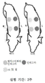
도 1a는 돼지들에 대한 상처 치료의 개략도이다. 상처는 인테그라(파란색 원)로 덮여 있었거나 10% rH TE(엘라스틴화 된 인테그라; 녹색 원)를 포함하는 인테그라 진피 템플릿 또는 4% rH TE 하이드로겔(노란색 원)을 포함하는 인테그라 진피 템플릿으로 처치가 되었다. 도 1b는 상처의 생검 및 드레싱 부위의 개략도이다.
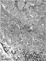
도 2는 엘라스틴화된 인테그라로 처치된 부위 중 VVG 염색된 생검 단면으로서, 섬유아세포의 존재, 새로운 콜라겐의 침착 및 엘라스틴 섬유의 존재를 도시한다.
도 3은 수술 2주 후 채취한 VVG 염색된 코어 생검 표본들에서 관찰된 진피의 다른 유형들을 비교한 것이다. a) 원래 진피; b) 정상 진피(원 내) 및 새로 형성된 진피가 반쯤씩 보이는 진피; c) 새로 형성된 진피의 모양(원 내: 호산구성 얼룩은 적고, 콜라겐 섬유는 얇고 덜 조직적으로 보이며, 정상 진피의 모양에 비해 세포는 더 많음); d) 정상 진피의 모양(원 내: 호산구성 얼룩이 더 많고, 콜라겐 섬유는 더 굵고 더 많이 조직화되고, 새로 형성된 진피에 비해 세포는 더 적은 것으로 나타남)
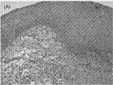
도 4는 수술 2주 후 인테그라 피부 템플릿 +4% rH TE 하이드로 겔로 처치한 상처 부위의 조직 단면이다. 정상적인 외관을 보이는 것의 진피 조직 내의 가시적인 rH TE 겔 아래에 있는 진피의 기저 쪽으로 엘라스틴 섬유들을 볼 수 있다. 비슷한 외형의 진피 조직도 rH TE 하이드로겔 내에서 보였다.
도 5는 수술 2주 후 채취한 코어 생검 샘플의 진피의 각층에서의 혈관의 수를 비교한 것이다. 동일한 돼지에서 두개골의(cranial)(두개골(cran)) 또는 미골부의(caudal)(미골부(caud)) 제어 인테그라 진피 템플릿 중 대응되는 것만 엘라스틴화된 인테그라와 비교하였다. VVG 염색된 코어 생검 샘플은 100배 배율로 검사하였고 ImageJ 소프트웨어를 사용하여 평가하였다. 현미경 사진은 진피의 여러 레벨에서 촬영되었다. 진피의 제1 레벨은(레벨 1) 피하 진피가 보이지 않을 때까지 카메라의 시야각을 움직여서 획득하였다. 다음 레벨들은 표피 쪽으로 샘플을 0.5 FOVs(현미경으로 관찰하면서) 움직여서 획득하였다. 따라서 피하 진피 위의 레벨 2는 레벨 1의 이미지로부터 0.5 FOVs(현미경으로 관찰하면서) 이격된 것이다. 진피의 각 레벨마다 3 개의 이미지들이 촬영되었다: 샘플의 양쪽 가장자리에서 하나씩 및 중간에서 하나. 현미경을 통한 각 FOV는 2.5 mm의 직경을 가지고, 카메라로 촬영된 각각의 이미지는 650 ㎛ 넓이를 가지므로, 이 방법을 통하면 진피의 다른 레벨들에서 촬영한 이미지들은 동일한 혈관을 포함하지 않을 것이 분명하였다. 혈관에 대한 기준들은: a) 루멘 크기가 10 ㎛ 이상일 것; 및 b) 루멘이 어두운 긴 핵과 함께 적어도 2개의 세포에 의해 일직선으로 배열되어야 할 것이었다. 혈관의 동일성 여부는 때때로 루멘 내 혈액세포의 존재, 및/또는 관벽의 중막 내에 평활근 세포의 존재에 의해서 명확하게 확인되었다.
도 6a는 인테그라만으로 처치된 샘플에서 진피 속으로 난 작은 표피 돌기들의 예이다. 도 6b는 엘라스틴화된 인테그라 샘플 내의 표피의 망상 융선의 예이다.
도 7은 섬유아세포와 재생 조직의 각질형성세포의 모집과 혈관 신생에 기여하는 화학주성 시그널링을 통해 상처 회복 과정에 기여하는 재조합형 인간 트로프엘라스틴이 제안된 것이다.Figure 1A is a schematic of wound treatment for pigs. The wound was covered with an Integra (blue circle) or an Integra dermis template containing a 10% rH TE (Elastinized Integra; green circle) or a 4% rH TE hydrogel (yellow circle) . 1B is a schematic view of a wound biopsy and dressing site.
Figure 2 shows the presence of fibroblasts, deposition of new collagen, and the presence of elastin fibers as a VVG stained biopsy section of the elastinized integrin treated sites.
Figure 3 compares the different types of dermis observed in VVG stained core biopsy specimens taken two weeks after surgery. a) original dermis; b) dermis with half of the normal dermis (in the circle) and newly formed dermis; c) The shape of the newly formed dermis (in the circle: less eosinophilic staining, collagen fibers appear thinner and less organized, and more cells than normal dermis); d) the shape of the normal dermis (in the circle: more eosinophilic stains, collagen fibers are thicker and more organized, and fewer cells are compared to newly formed dermis)
FIG. 4 is a tissue section of the wound area treated with Integra skin template + 4% rH TE hydrogel after 2 weeks of operation. Elastin fibers can be seen at the base of the dermis beneath the visible rH TE gel in dermal tissue of normal appearance. Similar external dermal organization was also seen in the rH TE hydrogel.
Figure 5 compares the number of vessels in each layer of the dermis of a core biopsy sample taken two weeks after surgery. Only the corresponding of the cranial (cranial) or caudal (caud) control integrand dermis templates in the same pig was compared to the elastinized integrin. VVG stained core biopsy samples were examined at 100x magnification and evaluated using ImageJ software. Microscopic photographs were taken at different levels of the dermis. The first level of the dermis was obtained by moving the camera's viewing angle until the subcutaneous dermis was not visible (level 1). The following levels were obtained by moving the sample towards the epidermis with 0.5 FOVs (observing with a microscope). Thus, level 2 above the subcutaneous dermis is spaced 0.5 FOVs (observing with a microscope) from the image of level 1. Three images were taken at each level of the dermis: one at both edges of the sample and one in the middle. Since each FOV through the microscope has a diameter of 2.5 mm and each image taken by the camera has a width of 650 μm, images taken at different levels of the dermis by this method will not contain the same blood vessels It was clear. Criteria for blood vessels are: a) lumen size greater than 10 μm; And b) the lumen should be aligned by at least two cells with a dark long nucleus. Whether the vessels are identical is sometimes evident by the presence of blood cells in the lumen and / or the presence of smooth muscle cells in the medullary canal wall.
Fig. 6A is an example of small epidermal processes in the dermis in the sample treated with the Integra alone. Figure 6b is an example of a network rim of the epidermis in an elastinized integrase sample.
FIG. 7 shows a recombinant human trophelin elastin that contributes to the wound healing process through recruitment of keratinocytes from fibroblasts and regenerated tissues and chemotactic signaling that contributes to angiogenesis.
본 발명자들은 트로프엘라스틴이 전체 두께 피부에서 상처의 재상피화를 개선한다는 것을 발견하였다.The inventors have found that tropic elastin improves wound re-epithelization in full thickness skin.
중요하게는, 본원의 실시예에 도시된 바와 같이, 발명자들은 상처를 중심으로 주변부를 형성하는 표피세포 형태의 상처 가장자리와 트로프엘라스틴을 접촉하는 것이 피부의 재상피화의 개선에 중요하다는 것을 발견하였다. 결정적으로, 트로프엘라스틴이 상처 가장자리와의 제한된 지속적 접촉을 갖는 상처 기저부로 제공되는 경우, 상처의 재상피화의 개선은 있다고 하더라도 미미하다.Importantly, as shown in the examples herein, the inventors have found that contact of tropheliastin with wound edges in the form of epidermal cells forming the periphery around the wound is important in improving skin re-epithelization. Crucially, if the trough elastin is provided to the wound base with limited persistent contact with the wound edge, the improvement in re-epithelization of the wound is minimal, if any.
가설에 구속되고자 하는 것은 아니지만, 본 발명자들은 본원에서 보여진 상처 치유의 개선을 제공하는 것은 트로프엘라스틴을 상처 가장자리에 지속적 또는 끊임없이 접촉시키는 것, 또는 상처 가장자리 부근에 적어도 끊임없이 배치하는 것이라 여긴다. 다시, 가설에 구속되고자 하는 것은 아니지만, 상처 치유 과정에서 설명된 다양한 프로테아제는 상처 가장자리에 접촉해서 배치되거나 상처 가장자리의 부근에 위치될 때 상처 치유 및 조직 재생의 다양한 과정들을 유리하게 하는 트로프엘라스틴의 단백질 가수분해 단편들을 생성할 수 있거나, 트로프엘라스틴의 단량체들의 방출을 촉진할 수 있는 것으로 여겨진다. 본원의 실시예들은 이러한 과정들이 혈관 신생, 세포 주화성, 이동과 증식 및 기질의 형성으로 이루어지고 있음을 보여준다.Without wishing to be bound by the theory, the inventors believe that providing the wound healing improvement shown here is to constantly or permanently contact the trough elastane with the wound edge, or at least to constantly place it near the wound edge. Again, it is not intended to be bound by the hypothesis, but the various proteases described in the wound healing process may be placed in contact with the wound edges, or in the vicinity of the wound edges, It is believed that they can produce hydrolyzed fragments or can facilitate the release of monomers of tropheliostin. Embodiments herein show that these processes are made by angiogenesis, cellular chemotaxis, migration and proliferation, and the formation of substrates.
상처가 기능적 표피 조직으로 봉합된다는 것은 치료와 미용이라는 두 가지 관점에서 매우 중요할 수 있기 때문에, 재상피화의 개선은 중요하다. 상처 봉합이 보이지 않는 경우, 조직 재생 과정은 조야한(substandard) 기능 및 외관을 갖는 조직의 형성으로 이어지는 조직 복원 및 섬유증에 더 가까워지는 경향이 있다는 우려가 있다. 만성 염증 및 궤양의 위험도 있다. 이러한 맥락에서, 재상피화의 범위 및 재상피화 할 시점의 관점에서 재상피화의 개선은 중요하다.Improvement of re-epithelization is important because it can be very important in terms of treatment and cosmetic that the wound is sealed with functional epidermal tissue. In the absence of wound closure, there is a concern that the tissue regeneration process tends to be closer to tissue repair and fibrosis leading to the formation of tissue with substandard function and appearance. There is also the risk of chronic inflammation and ulceration. In this context, it is important to improve re-epithelization in terms of the extent of re-epithelization and the point of time to re-epithelize.
비록 이전에는 엘라스틴 유래 펩타이드의 생체활성에 관한 체외 데이터가 제한적이었다 하더라도, 이전의 체내 상처 모델에서 VGVAPG와 같이 엘라스틴 유래 펩타이드가 본원에서 설명된 개선을 얻기에는 효과적이지 못하다는 것이 현재까지 입증되어 왔기 때문에, 상술한 결과들은 특히 놀라운 것으로 여겨진다. 예를 들어, Hashimoto 문헌을 참고할 것. 가설에 구속되고자 하는 것은 아니지만, 적어도 재상피화에 관련된 범주에서는, 본원에 설명된 상처의 구조에 따르면, 이러한 펩타이드를 처방하지 않았다는 것은 앞선 연구들에서는 본원에 기술된 개선이 발생하지 않았음을 의미하는 것으로 여겨진다.Although it has been proven to date that elastin-derived peptides, such as VGVAPG in previous body wound models, are not effective at obtaining the improvements described herein, even though in vitro data on bioactivity of elastin-derived peptides have been limited in the past , The above results are particularly surprising. For example, see the Hashimoto literature. Although not intending to be bound by the hypothesis, at least in the context of re-epithelization, according to the structure of the wound described herein, the absence of such a peptide indicates that the improvements described herein did not occur in previous studies ≪ / RTI >
따라서 일 구현예에서, 상처의 치유 방법을 제공한다. 본 방법은 상처를 가진 개인을 제공하는 단계를 포함한다. 상처는 피부 조직에 대한 임의의 부상으로부터 발생할 수 있다. 부상의 예는 화상, 열상, 찰과상, 절개, 천공 또는 파열을 포함한다.Thus, in one embodiment, a method of healing a wound is provided. The method includes providing an individual with a wound. Wounds can arise from any injury to the skin tissue. Examples of injuries include burns, lacerations, abrasions, incisions, perforations or ruptures.
일반적으로 부상은 파손, 파열 또는 표피와 진피층의 상처를 초래하는 것이다. 상처는 피하 조직, 근육 또는 뼈를 포함하는 진피 하부 조직에 상처를 초래하기도 한다. 따라서 상처는 피상적인 상처, 부분 두께 상처 또는 전체 두께 상처일 수 있다.In general, injuries result in breakage, rupture, or scarring of the epidermis and dermis. Wounds can also cause scarring in the subdermal tissue, including subcutaneous tissue, muscles, or bones. Thus, a wound may be a superficial wound, a partial thickness wound, or a full thickness wound.
일반적으로 본 발명은 육아조직의 형성 및 이와 관련된 혈관 생성, 콜라겐 침착 등의 과정을 포함하는 진피 및 표피 영역에 적용되는 재생 과정 및 재상피화에 적용된다.In general, the present invention is applied to the regeneration process and re-epithelization applied to the dermal and epidermal regions including the formation of granulation tissue and related processes such as angiogenesis and collagen deposition.
부상은 가령 수술로 인한 고의적인 것이거나 또는 가령 외상과 같은 우발적인 것일 수 있다.Injuries can be accidental, such as surgery, or accidental, such as trauma.
상처는 상처 주위에 위치하여 상처 가장자리를 형성하는 복수의 상피세포들을 포함한다. 일반적으로 부상은 상처가 형성된 영역에 걸쳐 정상적으로 표피층을 형성하는 상피조직 세포의 결여를 특징으로 하는, 조직의 손상된 영역을 형성하게 된다. 실질적으로 손상되지 않고, 부상 부위의 주변에 위치한 조직은 일반적으로 표피세포들의 정상적인 표피 층을 포함한다. 상처 가장자리를 형성하는 것은 이러한 상처에 인접한 이들 세포들이다. 일반적으로 알 수 있는 바와 같이, 상처 가장자리 또는 상처 주변부는 재상피화 과정 중에 상피조직 세포 증식의 위치가 된다.The wound includes a plurality of epithelial cells located around the wound to form wound edges. In general, the wound forms a damaged area of the tissue, characterized by a lack of epithelial tissue cells that normally form the epidermal layer over the wounded area. Substantially intact, the tissue located in the periphery of the injured area generally comprises the normal epidermal layer of epidermal cells. It is these cells adjacent to these wounds that form wound edges. As is generally known, the wound edge or wound periphery is the site of epithelial cell proliferation during the re-epithelialization process.
본 발명에 따르면, 상처 가장자리 또는 상처 주변부는 트로프엘라스틴과 상처 가장자리의 지속적인 접촉을 가능하게 하는 조건에서 트로프엘라스틴 또는 엘라스틴 유래 단편들과 접촉한다. 이는 본 발명에서 중요한 단계로 여겨진다. 본원의 실시예들에서 설명된 바와 같이, 재상피화는 상처 가장자리의 부근에 트로프엘라스틴이 접촉하거나 위치하는 곳에서 관찰되는 경향이 있다. 예를 들어, 재상피화는 더 완전하고 또는 자연적으로 구성될 수 있으며, 그리고 재상피화율은 가속화될 수 있다. 예를 들어, 트로프엘라스틴이 오직 상처 기저부와만 접촉하도록 배치될 때 이러한 특징들 중 일부는 보이지 않는다.According to the invention, the wound edge or wound periphery contacts the trough elastin or elastin derived pieces under conditions that allow for continued contact between the trough elastane and wound edges. This is regarded as an important step in the present invention. As described in the examples herein, re-epithelialisation tends to be observed where trop elastin contacts or is located in the vicinity of the wound edge. For example, re-epithelialization can be constructed more completely or naturally, and re-epithelialization rates can be accelerated. For example, some of these features are not visible when the trough elastane is positioned to contact only the wound base.
본원에 기술된 바와 같이, 상처 기저부는 일반적으로 기능적인 재생 과정에서 육아조직이 결국 형성되는, 손상으로부터 발생하는 진피 조직의 표면이다. '상처 가장자리의 부근에 위치한'이란 말은 트로프엘라스틴 또는 트로프엘라스틴의 단백질 가수분해 단편들이 상처 가장자리와 접촉을 위한 확산이 손쉽게 허용되도록 트로프엘라스틴 또는 그의 단백질 가수분해 단편들이 상처 가장자리에 아주 가깝게 위치되도록 트로프엘라스틴이 제공되는 것을 의미한다. 이러한 맥락에서, 트로프엘라스틴 또는 엘라스틴 유래 트로프엘라스틴의 단편들의 상처 기저부 위 또는 내에서만의 위치는 트로프엘라스틴이 상처 가장자리에 추가될 때 관찰될 수 있으므로 재상피화를 위해 제공되지 않는 것으로 본원에서 도시된다. 따라서, 트로프엘라스틴의 위치는 상처 가장자리 부근이므로 상처 기저부에만 트로프엘라스틴을 투여하는 것은 본 발명에 따라 보이지 않는다.As described herein, a wound base is the surface of a dermis tissue resulting from injury, where granular tissue is eventually formed in a generally functional regeneration process. The term " located near the wound edge " means that the trop elastin or protein hydrolytic fragments of the trop elastin are positioned so close to the wound edge that the spreading for contact with the wound edge is readily allowed, It means that elastin is provided. In this context, the location of the trough elastane or elastin-derived trough elastin on or within the wound base is shown herein as not being provided for re-epitaxy, as trop elastin can be observed when added to the wound edge. Therefore, since the position of the trough elastin is near the wound edge, administration of the trough elastin only to the wound base is not seen according to the present invention.
상처 가장자리에 트로프엘라스틴 또는 엘라스틴 유래 단편들의 접촉을 가능하게 하는 수많은 접근법들이 있다. 일 구현예에서, 트로프엘라스틴은 상처 가장자리와 끊임없는 접촉이 가능한 형태로 제공된다. 예를 들면, 트로프엘라스틴은 점성 또는 점착성을 갖는 겔의 형태로 제공될 수 있고, 따라서 겔, 트로프엘라스틴 또는 그 속의 단편이 상처 가장자리와 접촉을 유지하는 것을 가능하게 하도록 제공될 수 있다. 특정 겔 성분배합물의 실시예들은 WO2012068619호에서 일반적으로 논의된 것들을 포함한다.There are a number of approaches that enable the contact of trough elastin or elastin derived fragments to the wound margin. In one embodiment, the trough elastane is provided in a form capable of continuous contact with wound edges. For example, trough elastin can be provided in the form of a gel having a viscosity or tackiness, and thus can be provided to enable the gel, trough elastin or a fragment therein to maintain contact with wound edges. Examples of specific gel component formulations include those discussed generally in WO2012068619.
다른 구현예에서, 트로프엘라스틴은 상처 가장자리와 고상의 접촉이 유지되기에 적합한 드레싱, 스텐트, 장치 등과 같이 고상(따라서 트로프엘라스틴은 고상 내에 또는 고상 위에 위치하는)의 내에 또는 고상의 위에 제공된다. 예를 들어, 보철물, 증량재, 또는 지지체와 같은 고상은 겔 또는 다른 형태의 트로프엘라스틴 또는 엘라스틴 유래 펩타이드가 상처 가장자리와 접촉된 보철물 등의 선단면에 제공되는 것을 가능하게 하면서 보철물의 선단면이 상처 가장자리와 정렬되도록 배치될 수 있다. 특정한 고상 배열의 실시예들은 콜라겐과 같은 다른 결합 조직 분자와 선택적으로 전기방사를 하는 트로프엘라스틴의 전기 방사와 관련된 것들, 및 콜라겐과 같은 다른 결합 조직 분자와 공침하는 것과 관련된 것들을 포함한다.In other embodiments, the trough elastane is provided in a solid phase (and thus a trough elastin is located in or on a solid phase) such as a dressing, stent, device, etc., suitable for maintaining contact with wound edges and solid phase. For example, a solid phase, such as a prosthesis, an expandable material, or a support, allows a gel or other form of tropal elastin or elastin derived peptide to be provided on the distal end face of a prosthesis in contact with the wound edge, And may be arranged to be aligned with the edges. Particular solid-phase embodiments include those associated with electrospinning of tropheliostin that selectively electrospun with other connective tissue molecules such as collagen, and those associated with co-localization with other connective tissue molecules such as collagen.
대안적인 접근법에서, 상처 가장자리와 트로프엘라스틴의 지속적인 접촉을 가능하도록 하는 것이 트로프엘라스틴이 공급되는 조건들 그 자체이다. 예를 들어, 트로프엘라스틴은 상처 가장자리에 도포되거나 발라지는 용매로 제공될 수 있고, 그런 다음 트로프엘라스틴 또는 그의 단편을 상처의 가장자리와 접촉 상태로 둔 채 상처 가장자리로부터 용매를 증발시키기 위한 조건들이 제공된다.In an alternative approach, it is the conditions under which the trough elastin is supplied to enable continuous contact of the wound edge with trough elastin. For example, trough elastin can be provided as a solvent that is applied or spread on the wound edge, and then conditions are provided for evaporating the solvent from the wound edge while keeping the trough elastin or its fragment in contact with the edge of the wound .
트로프엘라스틴 및 엘라스틴 유래 단편은 대개 치료학적 유효량만큼 제공된다. 이는 트로프엘라스틴 또는 엘라스틴 유래 단편이 그렇게 제공되지 않는 환경에서의 재상피화가 되는 범위 또는 재상피화 시간과 비교했을 때, 재상피화의 범위 또는 재상피화를 완료하는 시간 중 하나의 맥락에서 재상피화의 개선을 이끌어낼 수 있는 임의의 양이다. 비록 상술한 피부 조직의 재생과 관련된 다양한 요인들에 의존적이지만, 일반적으로 트로프엘라스틴은 약 0.1 mg/ml 내지 250 mg/ml의 농도로 제공된다. 이 범위 내에서, 1 mg/ml, 25 mg/ml, 50 mg/ml, 100 mg/ml, 150 mg/ml 및 200 mg/ml 농도는 특히 유용할 수 있다.Trophelin elastin and elastin derived fragments are usually provided in therapeutically effective amounts. This is an improvement of re-epithelization in the context of either re-epithelization or time to complete re-epithelization as compared to the extent or re-epithelization time in which the trophelium or elastin derived fragment is not so provided It is an arbitrary amount that can be drawn. Although trowel elastin is generally provided at a concentration of about 0.1 mg / ml to 250 mg / ml, depending on various factors related to the regeneration of the dermal tissue described above. Within this range, concentrations of 1 mg / ml, 25 mg / ml, 50 mg / ml, 100 mg / ml, 150 mg / ml and 200 mg / ml may be particularly useful.
따라서, 일 구현예에서는 상처 치유 방법이 제공되며, 이 방법은:Thus, in one embodiment, a wound healing method is provided, comprising:
- 그의 주변에 위치한 복수의 상피세포를 포함하여 상처 가장자리를 형성하는 상처를 가진 개인을 제공하는 단계;Providing an individual having a wound to form a wound edge comprising a plurality of epithelial cells located in the vicinity thereof;
- 상처의 재상피화를 가능하게 하는 시간 기간 동안 트로프엘라스틴이 상처 가장자리에 지속적인 접촉을 가능하게 하는 조건에서 약 0.1 mg/ml 내지 250 mg/ml 농도의 트로프엘라스틴을 상처 가장자리에 접촉하는 단계를 포함하고; 상처의 재상피화는 상처의 치유를 가능하게 하고; 이로써 상처를 치유한다. 이 구현예에서, 트로프엘라스틴은 트로프엘라스틴이 상처 가장자리와 지속적인 접촉을 위해 상처 가장자리에 적용할 수 있는 형태로 사용되도록 제공될 수 있다. 이러한 형태는 겔일 수 있다. 바람직하게는, 이 구현예에서 트로프엘라스틴은 1 주일 미만의 기간 동안 상처 가장자리와 지속적인 접촉을 제공하며, 바람직하게는 상처 기저부와 접촉하지 않는다.- contacting the wound edge with a trough elastin at a concentration of from about 0.1 mg / ml to about 250 mg / ml under conditions that allow trough elastin to maintain constant contact with wound edges during a period of time allowing re-epithelization of the wound ; Re-epithelialization of the wound enables healing of the wound; This heals the wound. In this embodiment, the trough elastane can be provided to be used in a form that trough elastane can be applied to the wound edge for continued contact with the wound edge. This form can be a gel. Preferably, in this embodiment the trough elastane provides continuous contact with wound edges for a period of less than one week, and preferably does not contact the wound base.
본 발명에 따르면, 트로프엘라스틴 또는 엘라스틴 유래 펩타이드는 상처의 재상피화를 가능하게 하는 기간 동안 제공된다. 이 기간은 대개 상처의 속성과 조직 재생과 관련된 상술한 다른 요인들에 따라 달라진다. 상처가 외상으로 인한 경미한 급성 상처라면, 약 1 내지 2 주 미만의 기간 동안 트로프엘라스틴을 상처 가장자리에 지속적으로 접촉하도록 제공할 필요가 있다. 이 경우 일회성 트로프엘라스틴의 처방만으로 충분할 수 있다. 비록 상처의 크기와 속성에 따라 달라질 수 있지만, 수술로 인한 깨끗한 상처에 동일하게 적용할 수 있다. 상처가 더 복잡한 경우, 예를 들어 중증 외상 또는 만성 손상에서처럼, 예를 들어 진피 조직 또는 하부 조직에 실질적인 손상을 초래하는 경우, 예를 들면 상처를 일상적으로 드레싱하고 소독하는 기간에 맞춰 정렬된 투여 스케줄에 따라 트로프엘라스틴을 제공할 필요가 있다. 이러한 상황에서는 1주 내지 3주 또는 더 이상의 기간 동안 매일 트로프엘라스틴을 상처 가장자리에 추가하거나, 1주 내지 3주 또는 그 이상의 기간 동안 치료학적 유효량만큼의 트로프엘라스틴이 상처 가장자리에 지속적인 방출이 가능한 형태로 추가할 필요가 있다.According to the present invention, the trough elastin or elastin derived peptide is provided for a period that enables re-epithelization of the wound. This period usually depends on the nature of the wound and other factors mentioned above associated with tissue regeneration. If the wound is a minor acute wound due to trauma, then it is necessary to provide trough elastane for sustained contact with the wound edge for a period of less than about one to two weeks. In this case, only the prescription of one-time trough elastin may suffice. Although it can vary depending on the size and nature of the wound, it can be equally applied to a clean wound due to surgery. If the wound is more complex, for example, as in severe trauma or chronic injury, for example, if it results in substantial damage to the dermal tissue or underlying tissue, for example, an administration schedule aligned to the period of routine dressing and disinfection of the wound It is necessary to provide trough elastin according to the following formula. In this situation, daily trough elastin may be added to the wound margin for a period of one week to three weeks or longer, or a therapeutically effective amount of trophelin elastin may be continuously released on the wound edge for a period of one week to three weeks or longer You need to add it.
본 발명의 상술한 구현예는 상처의 치유로 이어지는 상처 치유 과정 동안 트로프엘라스틴이 상처 가장자리와 지속적인 접촉을 통해 발생하는 상처의 개선된 재상피화이다. 이 구현예에서, 그리고 본원의 실시예들을 통해 확립된 바와 같이, 트로프엘라스틴이 상처 기저부와 실질적인 접촉 없이도 재상피화를 개선하는 것이 가능하다.The above-described embodiment of the present invention is an improved re-epithelization of the wound that occurs during continuous contact with the wound edge of the trough elastin during a wound healing process leading to healing of the wound. In this embodiment, and as established by the embodiments herein, it is possible for the trough elastin to improve re-epitaxy without substantial contact with the wound base.
일반적으로 알 수 있는 바와 같이, 상처 기저부는 진피층 내에 일반적으로 형성되고 피하층 또는 진피층의 아래에 위치한 다른 층들까지 연장될 수 있다. 일반적으로 이해되는 상처 기저부는 손상되지 않은 정상 조직, 특히 상처 내에 위치한 진피 조직의 표면이다. 상처 기저부는 육아조직의 형성이 발생하는 상처의 일부로서 달리 정의될 수 있다. 상처 기저부는 일반적으로 표피 조직을 포함하지 않으며, 따라서 상처 기저부 자체는 본원에서 언급된 것처럼'상처 가장자리'를 포함하지 않는다. 구체적으로는, 일반적으로 알 수 있는 바와 같이, 진피층을 침투한(가령 부분 두께 또는 전체 두께로 상처 입은) 모든 피부 상처들은 상처 가장자리와 상처 기저부를 가지게 된다. 피상적 피부 상처들은 상처 가장자리를 가지지만 실질적인 상처 기저부(실제로, 피상적 상처는 단순히 노출되었을 수도 있지만, 손상되지 않은 진피 조직일 수 있다.)를 가지지 않을 것이다. 따라서, 일반적으로 알 수 있는 바와 같이 그리고 본 발명에 따르면, '상처 가장자리' 및 '상처 베드'는 두 개의 다른 개념이다.As will generally be appreciated, the wound base may be formed generally within the dermal layer and extend to other layers located below the dermal layer or dermal layer. The commonly understood wound base is the surface of undamaged normal tissue, especially the dermis tissue located within the wound. The wound base may be otherwise defined as part of the wound from which the formation of granulation tissue occurs. The wound base generally does not include epidermal tissue, and thus the wound base itself does not include a 'wound edge' as referred to herein. Specifically, as generally understood, all skin wounds infiltrating the dermal layer (e.g., wound to a partial thickness or total thickness) have wound edges and wound bases. The superficial skin wounds will have a wound edge but will not have a substantial wound base (in fact, the superficial wound may be simply exposed, but not undamaged dermal tissue). Thus, as generally understood and according to the present invention, the 'wound edge' and the 'wound bed' are two different concepts.
본원에 기재된 하나의 특정 응용예는 흉터 및 이와 관련된 섬유증 조직의 개조 또는 실질적인 제거에 있다. 일반적으로 알 수 있는 바와 같이, 흉터 조직은 조직 회복의 결과로 발생한다. 마지막 결과는 흉터 또는 섬유증이 위치하는 중요한 조직의 구조 및 기능적 양상들이 부족한 조직 구조의 형성이다. 특히, 본원에서 설명된 상처 구조와 상대적인 트로프엘라스틴 또는 엘라스틴 유래 단편의 위치를 발견하는 것, 그런 다음 조직 재생의 핵심 요소를 유도하는 것이 가능하게 되고, 그런 다음 상처를 유도하고, 또 흉터 조직에서 상처 치유과정을 유도하는 것이 가능해진다. 따라서, 다른 구현예에서, 흉터 조직을 최소화하는 방법이 제공되며, 이 방법은:One particular application described herein is the modification or substantial removal of scar and related fibrosis tissue. As is generally known, scar tissue occurs as a result of tissue repair. The end result is the formation of tissue structures that lack the structural and functional aspects of critical tissue where scar or fibrosis is located. In particular, it is possible to find the location of the trough elastin or elastin derived fragment relative to the wound structure described herein, then to induce a key element of tissue regeneration, and then to induce a wound, It becomes possible to induce a healing process. Thus, in another embodiment, a method of minimizing scar tissue is provided, comprising:
- 흉터 조직을 가진 개인을 제공하는 단계;- providing an individual with a scar tissue;
- 그의 주변에 위치한 복수의 상피세포들을 포함하여 상처 가장자리를 형성하는 상처를 흉터 조직에 형성하는 단계;- forming a scar on the scar tissue including a plurality of epithelial cells located in the periphery thereof to form a wound edge;
- 상처의 재상피화가 가능한 기간 동안 트로프엘라스틴을 상처 가장자리와 지속적인 접촉이 가능하도록 하는 조건에서 치료학적인 유효량만큼의 트로프엘라스틴을 상처 가장자리에 접촉하는 단계를 포함하고; 상처의 재상피화는 흉터 조직을 최소화하고; 이로써 흉터 조직을 최소화한다.Contacting the wound edge with a therapeutically effective amount of trophelinastine under conditions that allow trough elastin to be in constant contact with the wound edge for a period of time during which re-epithelization of the wound is possible; Re-epithelialization of the wound minimizes scar tissue; This minimizes scar tissue.
일 구현예에서, 흉터 조직을 최소화하는 것은 흉터 조직의 제거를 의미한다. 다른 구현예에서, 피부의 주어진 영역에서 흉터 조직의 체적을 최소화하거나, 흉터 조직의 과다함을 최소화함으로써 흉터 조직은 최소화된다.In one embodiment, minimizing scar tissue means removal of scar tissue. In another embodiment, scar tissue is minimized by minimizing the volume of scar tissue in a given area of skin, or by minimizing the excess of scar tissue.
상처는 당 업계에 알려진 다양한 기술에 의해 생성될 수 있다. 특히 바람직한 한 가지 방법은 흉터에 다수의 상처를 생성하도록 흉터 조직을 따라 복수의 미세 천공을 형성하는 것을 포함한다. 그 다음, 본원에 설명된 조직 재생 과정들이 가능하도록 트로프엘라스틴 또는 엘라스틴 유래 펩타이드가 상처 가장자리 또는 상처 기저부와 지속적으로 접촉되어 제공된다. Wounds can be generated by a variety of techniques known in the art. One particularly preferred method involves forming a plurality of microperforations along the scar tissue to create multiple scars on the scar. The tropal elastin or elastin derived peptide is then provided in continuous contact with the wound edge or wound base to enable the tissue regeneration processes described herein.
일 구현예에서, 트로프엘라스틴은 트로프엘라스틴의 단량체들이 가교되지 않은 형태로 제공된다.In one embodiment, the trough elastane is provided in a form in which the monomers of trough elastin are not crosslinked.
다른 구현예에서, 트로프엘라스틴은 리질옥시다제(lysyl oxidase) 또는 다른 가교 시약을 포함하지 않는 조성물의 형태로 제공된다.In another embodiment, the trough elastin is provided in the form of a composition that does not comprise a lysyl oxidase or other crosslinking reagent.
다른 구현예에서, 트로프엘라스틴은 아미노산계 산화 방지제(antioxidants)를 포함하지 않는 조성물의 형태로 제공된다.In another embodiment, trophelin elastin is provided in the form of a composition that does not include amino acid antioxidants.
일반적으로, 본 발명에 사용하기 위한 트로프엘라스틴은 재조합형 트로프엘라스틴 또는 합성 트로프엘라스틴으로 무세포 조성물로 제공된다.Generally, the trough elastin for use in the present invention is provided in a cell-free composition with recombinant tropole elastin or synthetic trophe elastin.
본원에 사용된 바와 같이, 문맥상 달리 요구하는 경우를 제외하고, "포함(comprise)"의 용어 및 가령 "포함하는(comprising)" "포함하다(comprises)" 및 "포함된(comprised)"과 같은 이 용어의 변화는 추가적인 첨가물, 성분, 정수 또는 단계를 배제하기 위한 의도가 아니다.As used herein, unless the context requires otherwise, the word " comprise "and" comprises, "" comprises," Such variations in the same term are not intended to exclude additional additives, components, integers or steps.
선행 문단에서 기재된 본 발명의 추가적인 양태 및 그 양태들의 추가적인 구현예들은 일례로 첨부된 도면을 참조하여 주어진 다음의 기재로부터 명백해질 것이다.Additional embodiments of the invention and additional embodiments of the aspects set forth in the preceding paragraph will be apparent from the following description, given by way of example only, with reference to the accompanying drawings.
명세서에 개시되고 정의된 본 발명은 언급되거나 텍스트 또는 도면으로부터 분명한 두 가지 이상의 개별 특징들로 이루어진 모든 대안적인 조합들로 확장된다는 것이 이해될 것이다. 이러한 모든 상이한 조합들은 본 발명의 다양한 대안적인 양태들을 구성한다.
It is to be understood that the invention disclosed and defined in the specification extends to all alternative combinations of two or more individual features mentioned or evident from the text or figures. All these different combinations constitute various alternative aspects of the present invention.
실시예들Examples
실시예Example 1 전체 두께 외과적 상처의 진피 재생 1 full-thickness surgical wound wound regeneration
본 연구는 rH TE를 사용하거나 사용하지 않는 인테그라 진피 템플릿의 응용예를 따른 돼지 모델에서, 전체 두께의 외과적 상처의 진피 재생에 재조합형 인간 트로프엘라스틴(rH TE)이 미치는 영향을 평가하였다. 2주 동안 재생된 진피를 분석한 결과 인테그라 진피 템플릿에 rH TE가 존재하는 것이 상처 회복 과정의 개선으로 이어진 것으로 나타났다. 개선은 섬유아세포 숫자의 증가, 상승된 콜라겐 침착, 재생된 진피 조직의 증가된 혈관 신생, 및 재생된 진피에서 검출된 엘라스틴 섬유의 증가된 레벨로 표시되었다. 이러한 이벤트들은 rH TE의 존재에 의해 상처의 상피화를 개선하는 결과를 도출하는 각질형성세포 증식의 증가를 동반하였다.This study assessed the effect of recombinant human trophelin elastin (rH TE) on dermal regeneration of full thickness surgical wound in a porcine model following the application of an integrin dyes template with or without rH TE. Analysis of the dermis regenerated for 2 weeks revealed that the presence of rH TE in the integrase dermis template resulted in the improvement of the wound healing process. The improvement was marked by an increase in the number of fibroblasts, elevated collagen deposition, increased angiogenesis of the regenerated dermis tissue, and increased levels of elastin fibers detected in the regenerated dermis. These events were accompanied by an increase in keratinocyte proliferation which resulted in the improvement of wound epithelization by the presence of rH TE.
재료 및 방법Materials and methods
시험 항목Test Items
본 연구에는 세 가지 제품을 평가하였고, 세 가지 제품은:Three products were evaluated in this study, three of which were:
- 대조: 인테그라 진피 템플릿- Contrast: Integra dermis template
- 시험 A: 10% rH TE(엘라스틴화된 인테그라)를 통합한 인테그라 진피 템플릿- Test A: Integra dermis template incorporating 10% rH TE (elastinized integra)
- 시험 B: 4% rH TE 하이드로겔에 배치된 인테그라 진피 템플릿- Test B: Integra dermis template placed on 4% rH TE hydrogel
전체 두께 돼지 모델Full Thick Pig Model
본 연구에는 두 마리의 돼지가 이용되었으며, 각자 4 곳의 상처 부위가 있고, 아래 이미지에 도시된 바와 같이 돼지의 양 측부에 두 개씩이 있었다.Two pigs were used in this study, each with four wound areas, two on each side of the pig, as shown in the image below.
각 돼지에 있어서, 일 측부의 두 개의 상처는 인테그라로 덮였고 타 측부의 두 개의 상처는 도 1a에 표시된 바와 같이 시험 A 및 B를 처치하였다.For each pig, two wounds on one side were covered with Integra and two wounds on the other side treated Tests A and B as shown in Fig.
● 0일째● Day 0
○ 위의 그림에서 표시된 바와 같이 직경 5 cm의 전체 두께의 절개된 원형 상처가 각 돼지의 상부 등에 생성되었다. ○ As shown in the figure above, an incised round wound with a total thickness of 5 cm in diameter was created on the upper part of each pig.
○ 위의 그림에서 표시된 바와 같이 각 상처에는 제어 인테그라 진피 템플릿 또는 시험 A 또는 B 중 하나를 처치하였다. ○ As shown in the figure above, each wound was treated with either the control integrase dermis template or either test A or B.
● 7일째(1주)● On the 7th day (1 week)
○ 모든 상처에 드레싱을 교환함. ○ Replace dressing on all wounds.
● 14일째(2주)● On the 14th day (2 weeks)
○ 도 1b에 도시된 바와 같이 상처 가장자리로부터 몇 mm 떨어진 각 상처 부위로부터 4 mm의 생검을 채취함. ○ A 4 mm biopsy was taken from each wound site several millimeters away from the wound edge as shown in Figure 1B.
상처 분석Wound analysis
상처 부위에서의 샘플링은 수술 및 처치로부터 2주 후에 이루어졌다. 상처 부위의 생검은 전술한 바와 같이 수행되었다. 샘플들은 섬유모세포의 침윤, 콜라겐 및 엘라스틴의 침착, 재생된 조직의 혈관 신생 및 상피의 재생을 평가하기 위해 조직 병리학 및 면역 조직 화학 분석을 실시하였다.Sampling at the wound site was performed two weeks after surgery and treatment. The wound biopsy was performed as described above. Samples were subjected to histopathology and immunohistochemistry to assess fibroblast infiltration, collagen and elastin deposition, angiogenesis of regenerated tissue, and epithelial regeneration.
결과result
인테그라 진피 템플릿의 섬유아세포 침투:Fibroblast infiltration of the integrin dermis template:
섬유아세포의 숫자는 모든 구조에서 증가하였지만 rH TE가 존재하는 곳에서 보다 상승했다. rH TE가 전체 구조를 투과한 엘라스틴화된 인테그라에 있어서 효과는 가장 뚜렷했다.The number of fibroblasts increased in all structures but increased in the presence of rH TE. The effect of rH TE on elastinized integrase, which transmitted the whole structure, was most pronounced.
콜라겐 및 엘라스틴 침착:Collagen and elastin deposition:
도 2에 도시된 바와 같이, 헤마톡실린(hematoxylin)과 에오신(eosin)(H&E) 및 Verhoeff-Van Gieson(VVG) 염색에 의해 증명된 바와 같이 향상된 섬유아세포 레벨은 rH TE의 존재 하에서 콜라겐 침착으로 이어졌다.As shown in Figure 2, enhanced fibroblast levels, as evidenced by hematoxylin and eosin (H & E) and Verhoeff-Van Gieson (VVG) staining, were detected by collagen deposition in the presence of rH TE It continued.
처치된 부위로부터 채취한 생검 단면들에서 엘라스틴의 존재(즉, rH TE가 아닌)는 생검 단면의 VVG 염색에 의해 평가되었다. 내생 엘라스틴 섬유를 진피 회복 과정 동안 재생되었던 것들로부터 정확하게 구별할 수 없었기 때문에, 처치된 각 부위로부터의 조직 단면들은 회복된 진피 조직 내에 엘라스틴 섬유가 존재 또는 부재하는지를 단순히 기록하였다. 본 연구에서 인테그라 진피 템플릿, 인테그라 진피 템플릿에 4% rH TE의 하이드로겔, 및 엘라스틴화된 인테그라를 위해 분석된 총 단면의 숫자는 각각 32, 16, 및 16개였다. 결과는 하기 표 1에 요약되어 있다.The presence of elastin (ie, not rH TE) in the biopsy sections taken from the treated site was assessed by VVG staining of the biopsy section. Tissue sections from each treated area simply recorded the presence or absence of elastin fibers in the restored dermis tissue, since the endogenous elastin fibers could not be accurately distinguished from those regenerated during the dermis repair process. In this study, the total cross-sectional numbers analyzed for the Integra dermis template, the 4% rH TE hydrogel, and the elastinized Integra were 32, 16, and 16, respectively. The results are summarized in Table 1 below.
VVG 염색된 생검 단면들의 분석 도중에 발견된 진피 조직의 예는 도 3에 제공된다. 또한, rH TE 하이드로겔에 인접한 진피 조직에 존재하는 엘라스틴 섬유의 예는 도 4에 제공된다.An example of dermal tissue found during analysis of VVG stained biopsy sections is provided in FIG. An example of elastin fibers present in the dermis tissue adjacent to the rH TE hydrogel is also provided in Fig.
재생된 진피의 혈관 신생:Regenerated dermal vascularization:
재생된 진피에서 혈관 신생의 레벨은 병리학에 의해 평가하였다. 도 5에 기재된 것처럼, 생검 단면 조직의 다른 레벨들에서의 혈관의 숫자는 피하에서 시작하여 표피쪽으로 점진적으로 이동하는 현미경으로 평가하였다. 도 5에 제시된 데이터로부터 알 수 있는 바와 같이, 인테그라 피부 템플릿(엘라스틴화된 인테그라)에서 rH TE의 존재는, 인테그라 진피 템플릿으로 처치된 부위와 비교했을 때, 재생된 조직에서 특히 표면 진피 방향으로 혈관 숫자의 증가로 귀결되었다. 유사한 경향은 4% rH TE 겔의 상부에 인테그라 진피 템플릿을 더하여 처치가 된 부위에서 나타났다.Levels of angiogenesis in the regenerated dermis were assessed by pathology. As described in FIG. 5, the number of blood vessels at different levels of the biopsy section tissue was evaluated by a microscope, starting from subcutaneously and progressively moving toward the epidermis. As can be seen from the data presented in FIG. 5, the presence of rH TE in the Integra skin template (elastinized integra), as compared to the site treated with the integrin dermis template, The result was an increase in numbers. A similar trend was observed at the treated site by adding the integrin dermis template to the top of the 4% rH TE gel.
상피 재생:Epithelial regeneration:
상처 부위에서 상피의 재생은 다른 시험 및 대조 항목들에 대한 엘라스틴화된 인테그라의 가장 눈에 띄는 장점 중 하나였다. 표 2에 상세하게 기술되고 도 6에 도시된 바와 같이, 엘라스틴화된 인테그라는 2주 만에 상처 부위가 거의 완전히 재상피화 되도록 하였고, 자연스러운 진피-상피 결합을 보여주는 망상융선의 존재를 수반했다.
Regeneration of the epithelium at the wound site was one of the most prominent advantages of elastinized Integra for other tests and control items. As detailed in Table 2 and shown in FIG. 6, the elastinized integrase allowed the wound site to almost completely re-epithelialize within two weeks and entailed the presence of a reticular ridge showing natural dermal-epithelial binding.
Treatment
결론 및 논의Conclusion and Discussion
도 7에 도시된 바와 같이, rH TE가 엘라스틴화된 인테그라에 혼입될 때 진피 회복 과정에 생물학적 자극을 제공하는 경우, 이 데이터는 모델에 의해 설명된다. 이는 섬유모세포의 침윤, 재생된 조직의 혈관 신생 및 상처 부위의 상피화의 더 큰 레벨로 이어진다. 이러한 이점은 엘라스틴화된 인테그라의 사용이 피부 이식의 필요성을 배제할 수 있다는 것을 의미한다. 트로프엘라스틴이 단구(monocytes: 혈관 신생에 기여하고 가령 섬유로 분화하는) 및 섬유아세포를 포함하여 조직 회복 과정에 관련된 세포에 화학 주성이므로, 이러한 생물학적 자극은 조직 회복 과정에 기여하는 것으로 알려진 rH TE의 성능과 일치한다[Almine 등, 2012].As shown in FIG. 7, when the rH TE is incorporated into the elastinized integrase, this data is described by the model if it provides biological stimulation to the dermal recovery process. This leads to a greater level of fibroblast infiltration, angiogenesis of the regenerated tissue and epithelialization of the wound site. This advantage means that the use of elastinized Integra can eliminate the need for skin transplantation. Since tropheliostasis is chemotactic to cells involved in tissue repair processes, including monocytes (which contribute to angiogenesis and differentiate into fibers) and fibroblasts, these biological stimuli are associated with the rH TE Consistent with performance [Almine et al., 2012].
이들 효과들이 rH TE의 존재에 의한 것이라는 확인은 증가된 섬유아세포의 침윤 및 혈관 신생에 대해 유사한 경향을 보였던 인테그라 진피 템플릿 + 4% rH TE 하이드로겔로부터 나타났다. 하이드로겔의 국소 전달에 대하여 예상된 것처럼, 이들 효과들은 rH TE 함유 겔이 도포된 더 깊은 진피에 한정되었다. 이러한 효과들은 진피의 더 깊은 층에 대해서는 일차적으로 제약되므로, 상피의 재생은 보이지 않았다; 즉, rH TE 겔은 인테그라 진피 템플릿에 의해서 피부 진피 및 상피로부터 분리되었다. 재생 진피의 이러한 생검에서 더 많은 엘라스틴 섬유를 보았다. 이는 rH TE의 지속적인 방출에 의한 것이기 쉽다. rH TE 겔은 탄성 섬유를 구성하는 재생 섬유아세포에 이용될 수 있는 트로프엘라스틴을 공급하면서, 수정되지 않고 겔로부터 점진적으로 용출되는 전체 길이의 트로프엘라스틴 단량체를 함유한다. 일차적인 인간의 피부 섬유아세포는 세포의 성장을 위한 기질로서 rH TE를 활용하고, 리실 산화효소(그의 활성이 BAPN에 의해 억제되는)에 의존적이면서 엘라스틴 섬유의 성숙(엘라스틴 섬유의 탄성도 및 형광 특성의 측정에 의해 입증된 바와 같이)을 초래하는 과정을 통해서 rH TE를 엘라스틴 섬유로 개조한다[Weiss lab, 미간행 데이터].The confirmation that these effects were due to the presence of rH TE appeared from the Integra dermis template + 4% rH TE hydrogel, which showed a similar tendency for increased fibroblast invasion and angiogenesis. As expected for local delivery of hydrogels, these effects were confined to the deeper dermis to which the rH TE containing gel was applied. These effects were primarily restricted to the deeper layers of the dermis, so the regeneration of the epithelium was not visible; That is, the rH TE gel was separated from the skin dermis and epithelium by the integrin dermis template. In this biopsy of regenerating dermis we saw more elastin fibers. This is likely due to the sustained release of rH TE. The rH TE gel contains a trough elastin monomer of full length, which is unfixed and gradually elutes from the gel, while supplying the trop elastin, which can be used in regenerated fibroblasts constituting the elastic fibers. Primary human skin fibroblasts utilize rH TE as a substrate for cell growth and are dependent on the lysyl oxidase (whose activity is inhibited by BAPN) and the maturation of elastin fibers (elasticity and fluorescence properties of elastin fibers (As demonstrated by the measurement of the rH TE) in the elastin fiber [Weiss lab, unpublished data].
결론적으로, 이러한 연구들이 적은 수의 동물을 대상으로 수행된 것임을 인식하더라도, rH TE를 인테그라 진피 템플릿에 혼입하는 것은 진피 및 상피의 재생을 실질적으로 촉진할 수 있다는 점이 나타날 수 있다.In conclusion, although it may be recognized that these studies were performed on a small number of animals, incorporation of the rH TE into the integrin dermis template may appear to substantially promote regeneration of the dermis and epithelium.
실시예Example 2: 전기 방사, 공동 침전 및 겔 기반 제제의 사용. 2: Electrospinning, co-precipitation and the use of gel-based formulations.
본 연구에는 각각 4 개의 5 cm 직경의 원형 상처 부위를 각각의 측부에 2 개씩 가진 돼지들이 사용되었다. 각각의 돼지에 관해서는, 일 측부에 있는 2 개의 상처에는 시판되고 있는 피부 템플릿 제품을 적용하였고, 타 측부에 있는 2 개의 상처에는 검사 항목 A, B, 또는 C 중의 하나가 처치되었다.In this study, pigs with two 5 cm diameter circular cuts on each side were used. For each pig, a commercially available skin template product was applied to the two wounds on one side and one of the items A, B, or C was treated on the two wounds on the other side.
● 0일째● Day 0
○ 위의 그림에서 언급한 바와 같이 직경 5 cm의 전체 두께의 4개의 절개된 원형 상처가 각 돼지의 상부 등에 생성되었다. ○ As mentioned in the picture above, four incised round wounds of a total thickness of 5 cm in diameter were produced on the upper part of each pig.
○
각 상처에는 제어 피부 템플릿 또는 시험 항목 A, B 또는 C 중의 하나를 처치하였다.
Each wound was treated with either a control skin template or one of the test items A, B or C.
● 7일째(1주)● On the 7th day (1 week)
○
모든 상처에 드레싱을 교환함.
Replace dressing on all wounds.
● 14일째(2주)● On the 14th day (2 weeks)
○ 도 1b에 도시된 바와 같이 상처 가장자리로부터 몇 mm 떨어진 각 상처 부위로부터 4 mm의 생검을 채취함. ○ A 4 mm biopsy was taken from each wound site several millimeters away from the wound edge as shown in Figure 1B.
상처 분석Wound analysis
상처 부위에서의 샘플링은 수술 및 처치로부터 2주 후에 처음 실시되었다. 상처 부위의 생검은 상술한 바와 같이 수행되었다. 샘플들은 섬유모세포의 침윤, 콜라겐 및 엘라스틴의 침착, 재생된 조직의 혈관 신생 및 상피의 재생을 평가를 위해 조직 병리학 및 면역 조직 화학 분석을 실시하였다.Sampling at the wound site was first performed two weeks after surgery and treatment. The wound biopsy was performed as described above. Samples were subjected to histopathology and immunohistochemistry for evaluation of invasion of fibroblasts, deposition of collagen and elastin, angiogenesis of regenerated tissue and regeneration of epithelium.
시험 항목 A의 준비: 전기 방사 지지체Preparation of test item A: Electrospinning support
다른 비율의 트로프엘라스틴 및 콜라겐을 1,1,1,3,3,3-헥사플루오로-2-프로판올(HFP)의 20%(w/v) 단백질 용액에 혼합하였다. 이들은 100%의 트로프엘라스틴, 80% 트로프엘라스틴과 20%의 콜라겐, 60% 트로프엘라스틴과 40%의 콜라겐, 50% 트로프엘라스틴과 50%의 콜라겐, 및 100%의 콜라겐을 포함하였다. 용액은 무딘 18 게이지 바늘이 장착된 주사기에 채워졌고, 유량은 주사기 펌프를 사용하여 3 ml h1으로 조절되었다. 바늘은 20 kV의 양의 전원에 연결되었고 집전 간격이 20 cm 이격된 30 mm 직경의 원형 브라스 집전장치에 직접 접지되었다. 전기 방사 지지체는 수용성의 환경에서 구조를 안정시키기 위해 화학적으로 가교되었다. 지지체는 개방 단계의 건조기에 넣어져, 분리된 25%(v/v) 수성 글루타르알데히드(glutaraldehyde) 용액의 증기로 가교된 뒤, 0.2 M 글리신(glycine) 용액에 밤새 침지하여 식혔다. 그런 다음 지지체를 PBS에서 반복하여 세척하였다. Rmjak-Kovacina, J. 등의 Acta Biomater. 2012 Oct; 8(10): 2714-22 참조. 이 항목은 아래의 시험 항목 B보다 훨씬 더 많이 가교된다. 이것은 세포의 침윤에 더 순응하는 경향이 있다.A different percentage of trophelin elastin and collagen were mixed in a 20% (w / v) protein solution of 1,1,1,3,3,3-hexafluoro-2-propanol (HFP). They contained 100% tropic elastin, 80% tropic elastin and 20% collagen, 60% tropic elastin and 40% collagen, 50% trophelin elastin and 50% collagen, and 100% collagen. The solution was filled into a syringe equipped with a dull 18-gauge needle, and the flow rate was adjusted to 3 ml h1 using a syringe pump. The needles were connected to a 20 kV positive power supply and directly grounded to a 30 mm diameter round brass current collector spaced 20 cm apart. The electrospinning support was chemically crosslinked to stabilize the structure in an aqueous environment. The support was placed in an open-stage dryer, bridged with a vapor of a 25% (v / v) aqueous glutaraldehyde solution, and then soaked overnight in a 0.2 M glycine solution. The support was then washed repeatedly in PBS. Rmjak-Kovacina, J. Acta Biomater. 2012 Oct; 8 (10): 2714-22. This item is much more bridged than Test B below. This tends to be more compliant with cellular infiltration.
시험 항목 B의 준비: Preparation of Test Item B: 트로프엘라스틴을Trough elastin 혼합하는 콜라겐 스펀지 Collagen sponge to blend
0.05 M 아세트산(pH 3.2) 내의 10% w/w 트로프엘라스틴과 혼합된 I 형 소 콜라겐의 백색 공침전물은 동결 건조하여 다공성 백색 막으로 변환되었다. 기공의 평균 직경의 제어는 액체 질소에 동결할 때 초기 저장온도 스냅을 조정함으로써 달성되었다. 건조한 고체를 105℃, 6 kPa의 진공에 24시간 동안 후속 노출하는 것은 콜라겐의 폴리펩타이드(polypeptide) 사슬 사이에 공유결합 가교를 유도하였다. 구조체는 0.05 M 아세트산의 0.25% 수성 글루타르알데히드 함유 조에 침지되었고, 콜라겐은 추가적인 공유결합을 겪었다. 구조체들은 탈 이온수로 24시간 동안 세정되었다. Kanematsu, A., 등 Biomaterials. 2004 Aug; 25(19): 4513-20 참조. 이는 위의 시험 항목 A보다 분해에 더 강하게 저항하는 경향이 있다.The white coprecipitate of I-form small collagen mixed with 10% w / w tropin elastin in 0.05 M acetic acid (pH 3.2) was lyophilized and converted to a porous white film. Control of the average diameter of the pores was achieved by adjusting the initial storage temperature snap when frozen in liquid nitrogen. Subsequent exposure of the dried solids to a vacuum of 6 kPa at 105 ° C for 24 hours induced covalent cross-linking between the polypeptide chains of the collagen. The structure was immersed in a 0.25% aqueous glutaraldehyde containing bath of 0.05 M acetic acid, and the collagen underwent additional covalent bonding. The structures were rinsed with deionized water for 24 hours. Kanematsu, A., et al. Biomaterials. 2004 Aug; 25 (19): 4513-20. This tends to resist more resistance to degradation than test A above.
시험 항목 C의 준비: 아래에 Preparation of Test Item C: Below 트로프엘라스틴을Trough elastin 가진 콜라겐 스펀지 Collagen sponge with
WO2012068619호에 기재된 바와 같이 전체 길이의 트로프엘라스틴은 히알루론산 겔에 필수적으로 포함되었고 사용 전에 주사기에 채워졌다. 겔을 노출된 상처 기저부의 표면에 도포한 다음, 시험 항목 B에 기재된 바와 같이 만들어지되 트로프엘라스틴이 생략된 콜라겐 스폰지로 겹쳐 두었다.As described in WO2012068619, the full length trough elastin was essentially contained in the hyaluronic acid gel and was filled into the syringe prior to use. The gel was applied to the surface of the exposed wound base and then overlaid with a collagen sponge made as described in test item B, but with trough elastin omitted.
실시예Example 3: 돼지 3: pig 니들링Needling 피부 모델에서 상피 재생의 평가 Evaluation of epithelial regeneration in skin models
돼지는 등 전체에 걸쳐 최대 10개의 2 cm x 2 cm 부위 각각에 처치를 받았다. 각각의 부위는 세 가지 치료방법 중의 한 가지를 받았으며, 세 가지 치료 방법은 다음과 같다:The pigs were treated for up to 10 2 cm x 2 cm areas over the entire back. Each site received one of three treatment methods, and the three treatment methods are as follows:
1) 부위 A: 처치되어야 할 피부 전체에 걸쳐 약 2 mm 간격마다 마이크로 코어링 니들을 사용하여 천공 상처를 받았다. 니들 처치를 받은 다음 1% 내지 5% w/v 트로프엘라스틴 단백질을 함유한 겔이 처치된 영역에 국소적으로 도포되었고 Tegaderm 드레싱에 의해 상기 겔이 잔류하고 천공된 부위들을 통과할 수 있도록 유지되었다.1) Site A: Microcore-ring needles were used to puncture every 2 mm intervals across the skin to be treated. Following needle treatment, gels containing 1% to 5% w / v trophelin protein were applied topically to the treated area and maintained by gel permeation through the remaining areas of the gel by Tegaderm dressing.
2) 부위 B: 피하 주사 바늘을 이용하여 각 천공이 피부 조직의 상부 진피 내로 1% 내지 5% w/v 트로프엘라스틴 겔 0.05 ml 내지 0.5 ml의 주입을 포함하는 천공 상처들을 받았다. 이 천공/주입은 처치되어야 할 피부의 영역에 걸쳐 대략 2 mm 간격으로 적용되었고, 상처의 드레싱이 이어졌다.2) Site B: Each puncture was given perforation wounds using a hypodermic injection needle containing 0.05 ml to 0.5 ml injection of 1% to 5% w / v trough elastin gel into the upper dermis of the skin tissue. This perforation / infusion was applied at intervals of approximately 2 mm across the area of the skin to be treated, followed by dressing of the wound.
3) 부위 C: 0.5 ml 내지 2 ml의 1% 내지 5% w/v 트로프엘라스틴 겔을 상부 진피 내에 크로스 해칭 주입 기술을 이용하여 주입을 받았고, 이어서 처치 영역에 걸쳐 대략 2 mm 간격으로 마이크로 코어링 바늘을 이용한 천공 상처를 적용하였다.3) Site C: 0.5 ml to 2 ml of 1% to 5% w / v trough elastin gel was injected into the upper dermis using a cross-hatch injection technique, followed by microcore ringing A puncture wound using a needle was applied.
사진 촬영 및 천공 생검은 0일째, 7일째, 및 28일째에 수행되었다. 샘플들은 섬유모세포의 침윤, 콜라겐 및 엘라스틴의 침착, 재생된 조직의 혈관 신생 및 상피의 재생을 평가하기 위한 조직 병리학 및 면역 조직 화학 분석을 실시하였다.Photographs and perforation biopsies were performed on days 0, 7, and 28. Samples were subjected to histopathology and immunohistochemical analysis to assess fibroblast infiltration, collagen and elastin deposition, angiogenesis of regenerated tissue, and epithelial regeneration.
실시예Example 4: 레이저를 이용한 돼지 피부로의 4: Laser to pig skin 트로프엘라스틴의Trough of elastin 전달 모델에 따른 상피 재생의 평가 Estimation of epithelial regeneration by delivery model
미소 레이저 박피는 피부에 국소 적용 약물의 전달을 지원하는 대략 3 mm 깊이의 수직 채널을 피부에 만든다. 돼지들은 등 부위에 걸쳐 최대 10개의 2 cm x 2 cm 크기의 부위를 각각 처치 받았다. 각 부위는 Haedersdal 등의 2010 (lasers Surg Med. 42(2): 113-22)에 기재된 바와 같이, 본질적으로 미소 싱글 홀 CO2 레이저로 필수 처치되었다. 레이저 처치에 이어서, 시험 부위들은 아래 중의 하나를 받았다:Micro-laser peeling makes the skin a vertical channel with a depth of about 3 mm to support the delivery of topically applied drugs to the skin. The pigs were treated with up to 10 2 cm x 2 cm areas on their backs. Each site was essentially treated with a microsatellite single CO 2 laser, as described by Haedersdal et al. 2010 (Lasers Surg Med. 42 (2): 113-22). Following laser treatment, the test sites received one of the following:
- 1% 내지 5% w/v 트로프렐라스틴을 함유한 히알루론산 겔; 또는Hyaluronic acid gel containing 1% to 5% w / v troparelast; or
- 히알루론산 겔 제어- Hyaluronic acid gel control
사진 촬영 및 천공 생검은 0일째, 7일째, 및 28일째에 수행되었다. 샘플들은 섬유모세포의 침윤, 콜라겐 및 엘라스틴의 침착, 혈관 신생 및 상피의 재생을 평가하기 위한 조직 병리학 및 면역 조직 화학 분석을 실시하였다.Photographs and perforation biopsies were performed on days 0, 7, and 28. Samples were subjected to histopathology and immunohistochemical analysis to assess fibroblast infiltration, collagen and elastin deposition, angiogenesis and epithelial regeneration.
참고Reference
Almine 등 2012. Elastin Signaling in Wound Repair. Birth Defects Research (Part C) 96:248-257.Almine etc. 2012. Elastin Signaling in Wound Repair. Birth Defects Research (Part C) 96: 248-257.
Claims (8)
상처의 재상피화를 가능하게 하는 시간 동안 상처 가장자리와 트로프엘라스틴의 지속적인 접촉을 가능하게 하는 조건에서 치료학적 유효량의 트로프엘라스틴을 상처 가장자리와 접촉하는 단계를 포함하고서,
상처의 재상피화는 상처의 치유를 가능하게 하고;
이로써 재상피화가 상처를 치유하는 것인 상처 치유 방법.
Providing an individual having a wound to form a wound edge by including a plurality of epithelial cells located around the wound; And
Contacting the wound edge with a therapeutically effective amount of trophelinastine under conditions that allow continued contact of the wound edge with trough elastin for a period of time to allow re-epithelization of the wound,
Re-epithelialization of the wound enables healing of the wound;
Wherein the re-epithelization heals the wound.
The wound healing method according to claim 1, wherein the trough elastin is provided in the form of a gel having viscosity or tackiness so that the trough elastane can be in constant contact with the wound edge.
The method of claim 1, wherein the trough elastane is provided on a distal end surface of the solid phase, the solid phase is disposed in the wound base so that the solid tip surface is aligned with the wound edge, thereby enabling the trough elastane to be in constant contact with the wound edge Wound healing method.
4. The wound healing method according to claim 3, wherein the solid phase is a dressing, a stent, a support, an increasing material or a prosthesis.
The method of claim 1, wherein the solution of trough elastin is sprayed onto the wound edge under conditions that allow evaporation of the solvent from the solution, thereby allowing the trough elastane to be in constant contact with the wound edge.
6. The wound healing method according to any one of claims 1 to 5, wherein the trough elastin is provided at the wound edge in an amount of about 0.1 mg to 250 mg / ml.
7. The wound healing method according to any one of claims 1 to 6, wherein the trough elastin is provided to continuously contact the wound edge for a period of time not exceeding about one week to two weeks.
8. The method according to any one of claims 1 to 7, wherein the trough elastin is not in contact with the wound base so that the trough elastin can be in constant contact with the wound base.
Priority Applications (1)
| Application Number | Priority Date | Filing Date | Title |
|---|---|---|---|
| KR1020217040694A KR102488446B1 (en) | 2013-08-13 | 2014-08-13 | Regeneration of damaged tissue |
Applications Claiming Priority (3)
| Application Number | Priority Date | Filing Date | Title |
|---|---|---|---|
| AU2013903092A AU2013903092A0 (en) | 2013-08-13 | Regeneration of damaged tissue | |
| AU2013903092 | 2013-08-13 | ||
| PCT/AU2014/050180 WO2015021508A1 (en) | 2013-08-13 | 2014-08-13 | Regeneration of damaged tissue |
Related Child Applications (1)
| Application Number | Title | Priority Date | Filing Date |
|---|---|---|---|
| KR1020217040694A Division KR102488446B1 (en) | 2013-08-13 | 2014-08-13 | Regeneration of damaged tissue |
Publications (1)
| Publication Number | Publication Date |
|---|---|
| KR20160041949A true KR20160041949A (en) | 2016-04-18 |
Family
ID=52467858
Family Applications (3)
| Application Number | Title | Priority Date | Filing Date |
|---|---|---|---|
| KR1020217040694A KR102488446B1 (en) | 2013-08-13 | 2014-08-13 | Regeneration of damaged tissue |
| KR1020167005263A KR20160041949A (en) | 2013-08-13 | 2014-08-13 | Regeneration of damaged tissue |
| KR1020227045816A KR20230004965A (en) | 2013-08-13 | 2014-08-13 | Regeneration of damaged tissue |
Family Applications Before (1)
| Application Number | Title | Priority Date | Filing Date |
|---|---|---|---|
| KR1020217040694A KR102488446B1 (en) | 2013-08-13 | 2014-08-13 | Regeneration of damaged tissue |
Family Applications After (1)
| Application Number | Title | Priority Date | Filing Date |
|---|---|---|---|
| KR1020227045816A KR20230004965A (en) | 2013-08-13 | 2014-08-13 | Regeneration of damaged tissue |
Country Status (12)
| Country | Link |
|---|---|
| US (3) | US20160194379A1 (en) |
| EP (2) | EP3033099B1 (en) |
| JP (3) | JP6382311B2 (en) |
| KR (3) | KR102488446B1 (en) |
| CN (2) | CN110464837A (en) |
| AU (3) | AU2014306362B2 (en) |
| BR (1) | BR112016003082A8 (en) |
| CA (1) | CA2920312C (en) |
| ES (1) | ES2965893T3 (en) |
| MX (2) | MX2016001436A (en) |
| RU (1) | RU2677637C2 (en) |
| WO (1) | WO2015021508A1 (en) |
Families Citing this family (7)
| Publication number | Priority date | Publication date | Assignee | Title |
|---|---|---|---|---|
| US9688741B2 (en) | 2012-10-23 | 2017-06-27 | Elastagen Pty Ltd | Elastic hydrogel |
| KR102239867B1 (en) | 2012-12-10 | 2021-04-13 | 앨러간 파마슈티컬스 인터내셔널 리미티드 | Scalable three-dimensional elastic construct manufacturing |
| CA2920312C (en) * | 2013-08-13 | 2022-07-19 | Elastagen Pty Ltd | Regeneration of damaged tissue |
| AU2016314775A1 (en) * | 2015-09-01 | 2018-03-22 | Allergan Pharmaceuticals International Limited | Formation of bone |
| RU2019116084A (en) * | 2016-11-04 | 2020-11-24 | Аллерган Фармасьютикалз Интернэшнл Лимитед | BIOSYNTHETIC DEVICES |
| CA3136074A1 (en) * | 2019-05-03 | 2020-11-12 | Robert Daniels | Scar prevention and/or treatment |
| US20230310552A1 (en) * | 2020-09-08 | 2023-10-05 | Protein Genomics Inc. | Biomimetic wound healing devices and related methods of treating diabetic wounds |
Family Cites Families (65)
| Publication number | Priority date | Publication date | Assignee | Title |
|---|---|---|---|---|
| US4675387A (en) | 1985-07-26 | 1987-06-23 | E. I. Du Pont De Nemours And Company | Method for extracting protein with organic acid |
| US5260203A (en) | 1986-09-02 | 1993-11-09 | Enzon, Inc. | Single polypeptide chain binding molecules |
| US4947840A (en) | 1987-08-21 | 1990-08-14 | Massachusetts Institute Of Technology | Biodegradable templates for the regeneration of tissues |
| EP0368684B2 (en) | 1988-11-11 | 2004-09-29 | Medical Research Council | Cloning immunoglobulin variable domain sequences. |
| AU645794B2 (en) | 1990-03-30 | 1994-01-27 | Shiseido Company Ltd. | Process for purifying polypeptide |
| WO1994007921A1 (en) | 1992-09-25 | 1994-04-14 | Commonwealth Scientific And Industrial Research Organisation | Target binding polypeptide |
| JP3474190B2 (en) | 1992-12-22 | 2003-12-08 | ザ ユニバーシテイ オブ シドニイ | Synthetic polynucleotide |
| US5726040A (en) | 1993-11-10 | 1998-03-10 | Ensley; Burt D. | Cosmetic compositions including tropoelastin isomorphs |
| US7001328B1 (en) * | 1994-11-15 | 2006-02-21 | Kenton W. Gregory | Method for using tropoelastin and for producing tropoelastin biomaterials |
| AUPO591797A0 (en) | 1997-03-27 | 1997-04-24 | Commonwealth Scientific And Industrial Research Organisation | High avidity polyvalent and polyspecific reagents |
| AU728480B2 (en) | 1996-08-07 | 2001-01-11 | Hospital For Sick Children, The | Self-aligning peptides derived from elastin and other fibrous proteins |
| AUPO156596A0 (en) * | 1996-08-09 | 1996-09-05 | University Of Sydney, The | Synthetic polynucleotides |
| WO1998034563A1 (en) | 1997-02-07 | 1998-08-13 | Sisters Of Providence In Oregon | Method for using tropoelastin and for producing tropoelastin biomaterials |
| AUPO811797A0 (en) | 1997-07-18 | 1997-08-14 | University Of Sydney, The | Tropoelastin derivatives |
| WO1999011196A1 (en) | 1997-09-04 | 1999-03-11 | Point Biomedical Corporation | Injectable tissue reconstruction material |
| AUPP472398A0 (en) | 1998-07-17 | 1998-08-13 | University Of Sydney, The | Protease susceptibility II |
| US20030190669A1 (en) | 1998-12-30 | 2003-10-09 | Genentech, Inc. | Secreted and transmembrane polypeptides and nucleic acids encoding the same |
| US6667051B1 (en) * | 1999-05-28 | 2003-12-23 | Kenton W. Gregory | Methods for producing elastin, and tropoelastin products for repairing and or replacing tissue |
| DK1229940T3 (en) | 1999-11-15 | 2014-08-18 | Piramal Healthcare Canada Ltd | TEMPERATURE CONTROL AND PH-DEPENDENT SELF-GELING, Aqueous BIOPOLYMER SOLUTION |
| US20050244393A1 (en) | 1999-12-22 | 2005-11-03 | Henogen S.A. | Sealant or tissue generating product |
| US6808707B2 (en) * | 2000-02-04 | 2004-10-26 | Matrix Design | Wound healing compositions and methods using tropoelastin and lysyl oxidase |
| US20040110439A1 (en) | 2001-04-20 | 2004-06-10 | Chaikof Elliot L | Native protein mimetic fibers, fiber networks and fabrics for medical use |
| US7125960B2 (en) | 2001-05-30 | 2006-10-24 | Chisso Corporation | Crosslinked elastin and process for producing the same |
| AU2002952492A0 (en) | 2002-11-06 | 2002-11-21 | Cbio Limited | Chaperonin 10 immunosuppression |
| US8038991B1 (en) | 2003-04-15 | 2011-10-18 | Abbott Cardiovascular Systems Inc. | High-viscosity hyaluronic acid compositions to treat myocardial conditions |
| US7641643B2 (en) | 2003-04-15 | 2010-01-05 | Abbott Cardiovascular Systems Inc. | Methods and compositions to treat myocardial conditions |
| FR2855969B1 (en) | 2003-06-13 | 2012-11-30 | Coletica | STIMULATION OF THE ACTIVITY OF A LYSYL OXIDASE ISOFORM TO FIGHT CERTAIN PATHOLOGIES DUE TO AN INCOMPLETE, ABSENT OR DISORGANIZED ELASTOGENESIS |
| FR2855968B1 (en) | 2003-06-13 | 2012-11-30 | Coletica | STIMULATION OF THE SYNTHESIS AND ACTIVITY OF A LYSYL OXIDASE-LIKE LOXL ISOFORM TO STIMULATE THE FORMATION OF ELASTIC FIBERS |
| US8226715B2 (en) | 2003-06-30 | 2012-07-24 | Depuy Mitek, Inc. | Scaffold for connective tissue repair |
| WO2005082386A2 (en) | 2004-02-20 | 2005-09-09 | Human Matrix Sciences, Llc | Manganese and iron based compounds for elastogenesis and connective tissue treatment |
| US7192043B1 (en) | 2004-05-25 | 2007-03-20 | Mcluen Design, Inc. | Multi-terrain wheel chair |
| US7968085B2 (en) | 2004-07-05 | 2011-06-28 | Ascendis Pharma A/S | Hydrogel formulations |
| US20060062768A1 (en) | 2004-09-23 | 2006-03-23 | Olexander Hnojewyj | Biocompatible hydrogel compositions |
| JP4381448B2 (en) | 2005-03-16 | 2009-12-09 | 富士通株式会社 | Multicast tree monitoring method and system in IP network |
| WO2006101441A2 (en) | 2005-03-24 | 2006-09-28 | Straumann Holding Ag | Method for protein purification comprising heat incubation in acetic acidic solution |
| US8828433B2 (en) | 2005-04-19 | 2014-09-09 | Advanced Cardiovascular Systems, Inc. | Hydrogel bioscaffoldings and biomedical device coatings |
| KR100785378B1 (en) | 2005-09-05 | 2007-12-14 | 주식회사 바이오레인 | Multi-layered antiadhesion barrier |
| CA2626637A1 (en) | 2005-10-19 | 2007-04-26 | Kenton W. Gregory | Method of using and producing tropoelastin and tropoelastin biomaterials |
| US7770126B2 (en) | 2006-02-10 | 2010-08-03 | Microsoft Corporation | Assisting user interface element use |
| US8518105B2 (en) | 2006-03-24 | 2013-08-27 | Abbott Cardiovascular System Inc. | Methods and apparatuses for coating a lesion |
| US20070237735A1 (en) | 2006-03-31 | 2007-10-11 | Laboratoires Dermo-Cosmetik Inc. | Anti-aging composition, kit and method of use |
| RU2421208C2 (en) * | 2006-05-11 | 2011-06-20 | Ридженикс Ас | Cell and cell extract introduction for rejuvenation |
| US20070287741A1 (en) | 2006-06-13 | 2007-12-13 | Uri Herzberg | Compositions and methods for preventing or reducing postoperative ileus and gastric stasis in mammals |
| US8846624B2 (en) | 2006-09-11 | 2014-09-30 | Emory University | Modified protein polymers |
| US7700126B2 (en) | 2006-09-29 | 2010-04-20 | Martin Kean Chong Ng | Tropoelastin-based protoelastin biomaterials |
| WO2008058323A1 (en) * | 2006-11-13 | 2008-05-22 | The University Of Sydney | Use of tropoelastin for repair or restoration of tissue |
| WO2009015372A1 (en) * | 2007-07-25 | 2009-01-29 | Dermaplus, Inc. | Photo-protective dermatological formulations and methods of using the same |
| US8455459B2 (en) | 2007-08-02 | 2013-06-04 | Medicis Pharmaceutical Corporation | Method of applying an injectable filler |
| FR2920968B1 (en) | 2007-09-14 | 2009-11-13 | Oreal | COSMETIC PROCESS FOR AESTHETIC TREATMENT AND / OR REPAIR OF SKIN |
| WO2009099570A2 (en) | 2008-02-01 | 2009-08-13 | Wake Forest University Health Sciences | Aligned scaffolding system for skeletal muscle regeneration |
| US8469961B2 (en) | 2008-03-05 | 2013-06-25 | Neville Alleyne | Methods and compositions for minimally invasive capsular augmentation of canine coxofemoral joints |
| US8940331B2 (en) | 2008-11-22 | 2015-01-27 | The Board Of Trustees Of The Leland Stanford Junior University | Hydrogels, methods of making hydrogels, methods of using hydrogels, and methods of isolating, trapping, attracting, and/or killing cancer cells |
| US8080265B2 (en) | 2009-02-20 | 2011-12-20 | Johnson & Johnson Consumer Companies, Inc. | Compositions and methods for treating signs of skin aging |
| AU2010223851B2 (en) | 2009-03-10 | 2017-03-16 | Allergan Pharmaceuticals International Limited | Injectable biomaterials |
| EP3078388B1 (en) | 2010-03-22 | 2019-02-20 | Allergan, Inc. | Cross-linked hydrogels for soft tissue augmentation |
| WO2011127478A1 (en) | 2010-04-09 | 2011-10-13 | Nanovasc, Inc. | Sleeve for graft and method |
| US8658711B2 (en) | 2010-09-29 | 2014-02-25 | Rutgers, The State University Of New Jersey | Process for the synthesis of methacrylate-derivatized type-1 collagen and derivatives thereof |
| JP2013544593A (en) | 2010-11-23 | 2013-12-19 | エラスタジェン ピーティーワイ リミテッド | Preparation and / or formulation of proteins cross-linked by polysaccharides |
| GB201021438D0 (en) | 2010-12-16 | 2011-02-02 | Imp Innovations Ltd | Layered fibrous construct |
| TR201905884T4 (en) * | 2011-09-30 | 2019-05-21 | Allergan Pharmaceuticals Int Ltd | In vivo synthesis of elastic fiber. |
| US9114128B2 (en) * | 2011-09-30 | 2015-08-25 | Protein Genomics, Inc. | Tropoelastins and uses thereof |
| US9688741B2 (en) * | 2012-10-23 | 2017-06-27 | Elastagen Pty Ltd | Elastic hydrogel |
| KR102239867B1 (en) | 2012-12-10 | 2021-04-13 | 앨러간 파마슈티컬스 인터내셔널 리미티드 | Scalable three-dimensional elastic construct manufacturing |
| CA2920312C (en) * | 2013-08-13 | 2022-07-19 | Elastagen Pty Ltd | Regeneration of damaged tissue |
| SG10201802109SA (en) | 2013-09-24 | 2018-04-27 | Elastagen Pty Ltd | Method of extracting protein |
-
2014
- 2014-08-13 CA CA2920312A patent/CA2920312C/en active Active
- 2014-08-13 AU AU2014306362A patent/AU2014306362B2/en active Active
- 2014-08-13 ES ES14836821T patent/ES2965893T3/en active Active
- 2014-08-13 KR KR1020217040694A patent/KR102488446B1/en active IP Right Grant
- 2014-08-13 JP JP2016533753A patent/JP6382311B2/en active Active
- 2014-08-13 US US14/911,401 patent/US20160194379A1/en not_active Abandoned
- 2014-08-13 KR KR1020167005263A patent/KR20160041949A/en not_active Application Discontinuation
- 2014-08-13 BR BR112016003082A patent/BR112016003082A8/en not_active Application Discontinuation
- 2014-08-13 CN CN201910782833.0A patent/CN110464837A/en active Pending
- 2014-08-13 RU RU2016104618A patent/RU2677637C2/en active
- 2014-08-13 WO PCT/AU2014/050180 patent/WO2015021508A1/en active Application Filing
- 2014-08-13 KR KR1020227045816A patent/KR20230004965A/en not_active Application Discontinuation
- 2014-08-13 CN CN201480049722.4A patent/CN105530951B/en active Active
- 2014-08-13 EP EP14836821.0A patent/EP3033099B1/en active Active
- 2014-08-13 MX MX2016001436A patent/MX2016001436A/en active IP Right Grant
- 2014-08-13 EP EP23206907.0A patent/EP4316537A3/en active Pending
-
2016
- 2016-01-29 MX MX2020011880A patent/MX2020011880A/en unknown
-
2018
- 2018-08-01 JP JP2018145202A patent/JP6639587B2/en active Active
- 2018-10-24 US US16/169,908 patent/US11084867B2/en active Active
-
2019
- 2019-07-11 AU AU2019204972A patent/AU2019204972B2/en active Active
- 2019-12-24 JP JP2019232193A patent/JP6891257B2/en active Active
-
2021
- 2021-08-09 US US17/397,870 patent/US20220098281A1/en active Pending
- 2021-10-08 AU AU2021245202A patent/AU2021245202A1/en active Pending
Also Published As
Similar Documents
| Publication | Publication Date | Title |
|---|---|---|
| JP6639587B2 (en) | Regeneration of damaged tissue | |
| Kakudo et al. | Platelet-rich plasma promotes epithelialization and angiogenesis in a splitthickness skin graft donor site | |
| Ulubayram et al. | EGF containing gelatin-based wound dressings | |
| Li et al. | Wounds: biology, pathology, and management | |
| De Vries et al. | Dermal regeneration in native non‐cross‐linked collagen sponges with different extracellular matrix molecules | |
| JP2015533798A5 (en) | ||
| Sen et al. | Anti-inflammatory effect of epidermal growth factor conjugated silk fibroin immobilized polyurethane ameliorates diabetic burn wound healing | |
| Arasteh et al. | Efficient wound healing using a synthetic nanofibrous bilayer skin substitute in murine model | |
| JP2019511468A (en) | Compositions and methods for treating chronic wounds | |
| US20200164101A1 (en) | Compositions and methods for treating wounds | |
| JP6316943B2 (en) | Modulation of heparin-binding epidermal growth factor activity for tympanic membrane healing | |
| Nath | A Clinical Study on the Efficacy of Autologous Platelet Rich Plasma in the Management of Chronic Non Healing Ulcers | |
| Insausti et al. | 4 The Human Placenta in Wound Healing | |
| Hrynyk | Enhanced burn wound healing through controlled and sustained delivery of bioactive insulin from alginate sponge dressings | |
| WO2021186181A1 (en) | Combinations, kits and methods for wound treatment | |
| Fast | Skin wound healing: Repair biology, wound, and soar treatment | |
| Lorenz et al. | 12 Wounds: Biology | |
| CHERRY et al. | HEALING PHASES | |
| Charles et al. | 19 Active Treatments for |
Legal Events
| Date | Code | Title | Description |
|---|---|---|---|
| N231 | Notification of change of applicant | ||
| AMND | Amendment | ||
| E902 | Notification of reason for refusal | ||
| AMND | Amendment | ||
| E601 | Decision to refuse application | ||
| X091 | Application refused [patent] | ||
| AMND | Amendment | ||
| X601 | Decision of rejection after re-examination | ||
| WITB | Written withdrawal of application |