KR20160009020A - Improved stability and potency of hemagglutinin - Google Patents
Improved stability and potency of hemagglutinin Download PDFInfo
- Publication number
- KR20160009020A KR20160009020A KR1020157029710A KR20157029710A KR20160009020A KR 20160009020 A KR20160009020 A KR 20160009020A KR 1020157029710 A KR1020157029710 A KR 1020157029710A KR 20157029710 A KR20157029710 A KR 20157029710A KR 20160009020 A KR20160009020 A KR 20160009020A
- Authority
- KR
- South Korea
- Prior art keywords
- rha
- protein
- see
- influenza virus
- strain
- Prior art date
Links
Images


Classifications
-
- A—HUMAN NECESSITIES
- A61—MEDICAL OR VETERINARY SCIENCE; HYGIENE
- A61K—PREPARATIONS FOR MEDICAL, DENTAL OR TOILETRY PURPOSES
- A61K39/00—Medicinal preparations containing antigens or antibodies
- A61K39/12—Viral antigens
-
- A—HUMAN NECESSITIES
- A61—MEDICAL OR VETERINARY SCIENCE; HYGIENE
- A61K—PREPARATIONS FOR MEDICAL, DENTAL OR TOILETRY PURPOSES
- A61K39/00—Medicinal preparations containing antigens or antibodies
- A61K39/12—Viral antigens
- A61K39/145—Orthomyxoviridae, e.g. influenza virus
-
- A—HUMAN NECESSITIES
- A61—MEDICAL OR VETERINARY SCIENCE; HYGIENE
- A61K—PREPARATIONS FOR MEDICAL, DENTAL OR TOILETRY PURPOSES
- A61K47/00—Medicinal preparations characterised by the non-active ingredients used, e.g. carriers or inert additives; Targeting or modifying agents chemically bound to the active ingredient
- A61K47/06—Organic compounds, e.g. natural or synthetic hydrocarbons, polyolefins, mineral oil, petrolatum or ozokerite
- A61K47/08—Organic compounds, e.g. natural or synthetic hydrocarbons, polyolefins, mineral oil, petrolatum or ozokerite containing oxygen, e.g. ethers, acetals, ketones, quinones, aldehydes, peroxides
- A61K47/12—Carboxylic acids; Salts or anhydrides thereof
-
- A—HUMAN NECESSITIES
- A61—MEDICAL OR VETERINARY SCIENCE; HYGIENE
- A61K—PREPARATIONS FOR MEDICAL, DENTAL OR TOILETRY PURPOSES
- A61K47/00—Medicinal preparations characterised by the non-active ingredients used, e.g. carriers or inert additives; Targeting or modifying agents chemically bound to the active ingredient
- A61K47/06—Organic compounds, e.g. natural or synthetic hydrocarbons, polyolefins, mineral oil, petrolatum or ozokerite
- A61K47/20—Organic compounds, e.g. natural or synthetic hydrocarbons, polyolefins, mineral oil, petrolatum or ozokerite containing sulfur, e.g. dimethyl sulfoxide [DMSO], docusate, sodium lauryl sulfate or aminosulfonic acids
-
- A—HUMAN NECESSITIES
- A61—MEDICAL OR VETERINARY SCIENCE; HYGIENE
- A61K—PREPARATIONS FOR MEDICAL, DENTAL OR TOILETRY PURPOSES
- A61K9/00—Medicinal preparations characterised by special physical form
- A61K9/0012—Galenical forms characterised by the site of application
- A61K9/0019—Injectable compositions; Intramuscular, intravenous, arterial, subcutaneous administration; Compositions to be administered through the skin in an invasive manner
-
- A—HUMAN NECESSITIES
- A61—MEDICAL OR VETERINARY SCIENCE; HYGIENE
- A61P—SPECIFIC THERAPEUTIC ACTIVITY OF CHEMICAL COMPOUNDS OR MEDICINAL PREPARATIONS
- A61P31/00—Antiinfectives, i.e. antibiotics, antiseptics, chemotherapeutics
- A61P31/12—Antivirals
- A61P31/14—Antivirals for RNA viruses
- A61P31/16—Antivirals for RNA viruses for influenza or rhinoviruses
-
- C—CHEMISTRY; METALLURGY
- C07—ORGANIC CHEMISTRY
- C07K—PEPTIDES
- C07K14/00—Peptides having more than 20 amino acids; Gastrins; Somatostatins; Melanotropins; Derivatives thereof
- C07K14/005—Peptides having more than 20 amino acids; Gastrins; Somatostatins; Melanotropins; Derivatives thereof from viruses
- C07K14/08—RNA viruses
- C07K14/11—Orthomyxoviridae, e.g. influenza virus
-
- A—HUMAN NECESSITIES
- A61—MEDICAL OR VETERINARY SCIENCE; HYGIENE
- A61K—PREPARATIONS FOR MEDICAL, DENTAL OR TOILETRY PURPOSES
- A61K47/00—Medicinal preparations characterised by the non-active ingredients used, e.g. carriers or inert additives; Targeting or modifying agents chemically bound to the active ingredient
- A61K47/06—Organic compounds, e.g. natural or synthetic hydrocarbons, polyolefins, mineral oil, petrolatum or ozokerite
- A61K47/08—Organic compounds, e.g. natural or synthetic hydrocarbons, polyolefins, mineral oil, petrolatum or ozokerite containing oxygen, e.g. ethers, acetals, ketones, quinones, aldehydes, peroxides
- A61K47/10—Alcohols; Phenols; Salts thereof, e.g. glycerol; Polyethylene glycols [PEG]; Poloxamers; PEG/POE alkyl ethers
-
- C—CHEMISTRY; METALLURGY
- C12—BIOCHEMISTRY; BEER; SPIRITS; WINE; VINEGAR; MICROBIOLOGY; ENZYMOLOGY; MUTATION OR GENETIC ENGINEERING
- C12N—MICROORGANISMS OR ENZYMES; COMPOSITIONS THEREOF; PROPAGATING, PRESERVING, OR MAINTAINING MICROORGANISMS; MUTATION OR GENETIC ENGINEERING; CULTURE MEDIA
- C12N2760/00—MICROORGANISMS OR ENZYMES; COMPOSITIONS THEREOF; PROPAGATING, PRESERVING, OR MAINTAINING MICROORGANISMS; MUTATION OR GENETIC ENGINEERING; CULTURE MEDIA ssRNA viruses negative-sense
- C12N2760/00011—Details
- C12N2760/16011—Orthomyxoviridae
- C12N2760/16111—Influenzavirus A, i.e. influenza A virus
- C12N2760/16134—Use of virus or viral component as vaccine, e.g. live-attenuated or inactivated virus, VLP, viral protein
-
- C—CHEMISTRY; METALLURGY
- C12—BIOCHEMISTRY; BEER; SPIRITS; WINE; VINEGAR; MICROBIOLOGY; ENZYMOLOGY; MUTATION OR GENETIC ENGINEERING
- C12N—MICROORGANISMS OR ENZYMES; COMPOSITIONS THEREOF; PROPAGATING, PRESERVING, OR MAINTAINING MICROORGANISMS; MUTATION OR GENETIC ENGINEERING; CULTURE MEDIA
- C12N2760/00—MICROORGANISMS OR ENZYMES; COMPOSITIONS THEREOF; PROPAGATING, PRESERVING, OR MAINTAINING MICROORGANISMS; MUTATION OR GENETIC ENGINEERING; CULTURE MEDIA ssRNA viruses negative-sense
- C12N2760/00011—Details
- C12N2760/16011—Orthomyxoviridae
- C12N2760/16111—Influenzavirus A, i.e. influenza A virus
- C12N2760/16141—Use of virus, viral particle or viral elements as a vector
Landscapes
- Health & Medical Sciences (AREA)
- Life Sciences & Earth Sciences (AREA)
- Chemical & Material Sciences (AREA)
- Virology (AREA)
- General Health & Medical Sciences (AREA)
- Medicinal Chemistry (AREA)
- Pharmacology & Pharmacy (AREA)
- Animal Behavior & Ethology (AREA)
- Public Health (AREA)
- Veterinary Medicine (AREA)
- Epidemiology (AREA)
- General Chemical & Material Sciences (AREA)
- Chemical Kinetics & Catalysis (AREA)
- Organic Chemistry (AREA)
- Oil, Petroleum & Natural Gas (AREA)
- Pulmonology (AREA)
- Engineering & Computer Science (AREA)
- Immunology (AREA)
- Mycology (AREA)
- Microbiology (AREA)
- Molecular Biology (AREA)
- Communicable Diseases (AREA)
- Biochemistry (AREA)
- Gastroenterology & Hepatology (AREA)
- Dermatology (AREA)
- Biophysics (AREA)
- Genetics & Genomics (AREA)
- Proteomics, Peptides & Aminoacids (AREA)
- Nuclear Medicine, Radiotherapy & Molecular Imaging (AREA)
- Oncology (AREA)
- Medicines Containing Antibodies Or Antigens For Use As Internal Diagnostic Agents (AREA)
- Peptides Or Proteins (AREA)
- Medicinal Preparation (AREA)
- Medicines That Contain Protein Lipid Enzymes And Other Medicines (AREA)
Abstract
본 발명은 재조합 헤마글루티닌 제형, 특히, 재조합 인플루엔자 헤마글루티닌 (rHA) 의 안정성 개선 및 효능 유지 방법에 관한 것이다. 특히, 출원인들은 시스테인 잔기를 돌연변이시킴으로써 또는 환원제 및 나트륨 시트레이트로 제형화함으로써 rHA 제형의 안정성이 유의하게 개선될 수 있다는 것을 밝혔다.The present invention relates to a recombinant hemagglutinin formulation, in particular to a method for improving the stability and efficacy of recombinant influenza hemagglutinin (rHA). In particular, applicants have found that the stability of rHA formulations can be significantly improved by mutating the cysteine residues or by formulating with a reducing agent and sodium citrate.
Description
참조에 의한 포함Include by reference
관련 출원 및 참조에 의한 포함Include by related application and reference
이 출원은 미국 특허 출원 일련 번호 13/838,796 (3 월 15 일, 2013 년에 제출됨) 및 미국 가 특허 출원 일련 번호 61/624,222 (4 월 13 일, 2012 년에 제출됨) 의 우선권 및 이익을 주장한다.This application claims priority and benefit of U.S. Patent Application Serial No. 13 / 838,796 (filed March 15, 2013) and U.S. Patent Application Serial No. 61 / 624,222 (filed April 13, 2012) I argue.
선행 출원, 및 거기에서 또는 그들의 심사 동안 인용된 모든 문헌 ("출원 인용된 문헌") 및 출원 인용된 문헌에서 인용 또는 참조된 모든 문헌, 및 본원에서 인용 또는 참조된 모든 문헌 ("본원에서 인용된 문헌"), 및 본원에서 인용된 문헌에서 인용 또는 참조된 모든 문헌은, 본원에서 또는 본원에 참조로 포함되는 임의의 문헌에서 언급된 임의의 제품에 관한 임의의 제조사의 지침, 설명서, 제품 사양, 및 제품 설명서와 함께, 본원에 참조로 포함되고, 발명의 실시에 이용될 수 있다. 더욱 구체적으로, 모든 참조된 문헌은 각각의 개별 문헌이 구체적으로 개별적으로 참조로 포함된다고 명시된 것과 동일한 정도로 참조로 포함된다.All documents cited or referenced in the prior application, and all documents cited therein or during their examination ("cited documents") and cited documents, and all references cited or referenced herein (" Quot;), and all references cited or referenced in the literature cited herein, are incorporated herein by reference in their entirety for any manufacturer's instructions, manuals, product specifications, and / or specifications relating to any product mentioned herein or in any document incorporated by reference herein. And product description, which are incorporated herein by reference and can be used in the practice of the invention. More specifically, all referenced documents are incorporated by reference to the same extent as if each individual document was specifically and individually indicated to be incorporated by reference.
발명의 분야Field of invention
본 발명은 재조합 헤마글루티닌 제형, 특히, 재조합 인플루엔자 헤마글루티닌 (rHA) 의 안정성 개선 및 효능 유지 방법에 관한 것이다.The present invention relates to a recombinant hemagglutinin formulation, in particular to a method for improving the stability and efficacy of recombinant influenza hemagglutinin (rHA).
연방 지원 설명Federal support description
본 발명은, 부분적으로, BARDA 승인 번호: HHSO100200900106C 에 의해 지원되었다. 연방 정부는 본 발명에 일정한 권리를 가질 수 있다.The present invention was supported, in part, by the BARDA approval number: HHSO100200900106C. The federal government may have certain rights in the invention.
유행성 인플루엔자는 전세계적으로 매년 발생하고 심각한 이환률 및 사망률의 원인이다. 어린이는 최고 발병률을 갖고, 공동체 내에서 인플루엔자 바이러스 전파의 큰 원인이 된다. 노인 및 기초 건강 문제를 갖는 사람들은 인플루엔자 감염으로 인한 합병증 및 입원에 관한 증가된 위험에 처해 있다.Pandemic influenza occurs annually worldwide and is a cause of serious morbidity and mortality. Children have the highest incidence and are a major cause of influenza virus transmission within communities. Elderly and people with underlying health problems are at increased risk for complications and hospitalization due to influenza infection.
인플루엔자 바이러스는 2 개의 표면 당단백질, 헤마글루티닌 (HA) 및 뉴라미니다제 (NA) 로 구성되는 고도로 다형성인 입자이다. HA 는 숙주 세포에 대한 바이러스의 부착 및 세포 내로 바이러스의 침입 동안 바이러스-세포막 융합을 매개한다. 인플루엔자 바이러스 게놈은 8 개의 단일 가닥 음성-센스 RNA 분절로 구성되며, 그 중 4 번째로 큰 분절이 HA 유전자를 인코딩한다. 인플루엔자 바이러스는 항원 차이에 기초하여 유형 A, B 및 C 로 분류된다. 인플루엔자 A 바이러스는 하위유형 또는 유형, 지리적 기원, 균주 번호, 및 단리 년도를 포함하는 명명법에 의해 기재되며, 예를 들어, A/Beijing/353/89 이다. 적어도 HA 의 하위유형 13 개 (H1-H13) 및 NA 의 하위유형 9 개 (N1-N9) 가 존재한다. 모든 하위유형은 조류에서 발견되지만, 오직 H1-H3 및 N1-N2 만 인간, 돼지 및 말에서 발견된다 (Murphy and Webster, "Orthomyxoviruses", in Virology, ed. Fields, B. N., Knipe, D. M., Chanock, R. M., 1091-1152 (Raven Press, New York, (1990)).Influenza virus is a highly polymorphic particle composed of two surface glycoproteins, hemagglutinin (HA) and neuraminidase (NA). HA mediates virus-cell membrane fusion during attachment of the virus to the host cell and invasion of the virus into the cell. The influenza virus genome consists of eight single-stranded negative-sense RNA segments, the fourth largest segment encoding the HA gene. Influenza viruses are classified into types A, B, and C based on antigen differences. Influenza A viruses are described by nomenclature including subtype or type, geographical origin, strain number, and isolation year, for example, A / Beijing / 353/89. There are at least 13 subtypes of HA (H1-H13) and 9 subtypes of NA (N1-N9). All subtypes are found in birds but only H1-H3 and N1-N2 are found in humans, pigs and horses (Murphy and Webster, "Orthomyxoviruses", in Virology, ed. Fields, BN, Knipe, DM, Chanock, RM, 1091-1152 (Raven Press, New York, (1990)).
HA 에 대한 항체는 바이러스를 중화시키고 인플루엔자 감염에 대한 자연 면역에 관한 기초를 형성한다 (Clements, "Influenza Vaccines", in Vaccines: New Approaches to Immunological Problems, ed. Ronald W. Ellis, pp. 129-150 (Butterworth-Heinemann, Stoneham, Mass. 1992)). HA 분자 내의 항원 변이는 인플루엔자의 빈번한 발생 및 면역화에 의한 감염 통제의 제한의 원인이 된다.Antibodies to HA neutralize viruses and form the basis for natural immunity against influenza infection (Clements, "Influenza Vaccines ", in Vaccines: New Approaches to Immunological Problems, ed. Ronald W. Ellis, pp. 129-150 (Butterworth-Heinemann, Stoneham, Mass. 1992). Antigen variants within the HA molecule are responsible for the frequent occurrence of influenza and limitations of infection control by immunization.
HA 의 3차원 구조 및 그것의 세포 수용체, 시알산과의 상호작용이 광범위하게 연구되었다 (Wilson, et al, "Structure of the hemagglutinin membrane glycoprotein of influenza virus at 3A.degree. resolution" Nature 289:366-378 (1981); Weis, et al, "Structure of the influenza virus hemagglutinin complexed with its receptor, sialic acid" Nature, 333:426-431 (1988); Murphy and Webster, 1990). HA 분자는 비리온으로 삼합체로서 존재한다. 각각의 HA 단량체 (HA0) 는 단일 다이설파이드 결합에 의해 연결된 2 개의 사슬, HA1 및 HA2 로 존재한다. 감염된 숙주 세포는 분자량이 약 85,000 Da 인 전구체 글리코실화된 폴리펩티드 (HA0) 를 생산하며, 이것은 생체내에서 후속적으로 HA1 및 HA2 로 절단된다.The three-dimensional structure of HA and its interaction with sialic acid has been extensively studied (Wilson, et al, "Structure of the hemagglutinin membrane glycoprotein of influenza virus at 3A. Resolution." Nature 289: 366-378 (1981); Weis, et al., "Structure of the influenza virus hemagglutinin complexed with its receptor, sialic acid" Nature, 333: 426-431 (1988); Murphy and Webster, 1990). The HA molecule exists as a trimer as a virion. Each HA monomer (HAO) is present in two chains, HA1 and HA2, connected by a single disulfide bond. The infected host cell produces a precursor glycosylated polypeptide (HAO) with a molecular weight of about 85,000 Da, which is subsequently cleaved into HA1 and HA2 in vivo.
인플루엔자 HA-특이적 중화 IgG 및 IgA 항체의 존재는 감염 및 질병에 대한 저항성과 연관된다 (Clements, 1992). 불활성화된 전체 바이러스 또는 부분적으로 정제된 (분할된 아단위) 인플루엔자 백신은 각각의 균주로부터의 HA 의 양에 표준화된다. 인플루엔자 백신은 통상적으로 인플루엔자의 3 개의 균주 각각으로부터의 7 내지 25 ㎍ 의 HA 를 포함한다.The presence of influenza HA-specific neutralizing IgG and IgA antibodies is associated with resistance to infection and disease (Clements, 1992). The inactivated whole virus or partially purified (divided subunit) influenza vaccine is normalized to the amount of HA from each strain. Influenza vaccines typically comprise 7 to 25 [mu] g HA from each of the three strains of influenza.
대부분의 허가받은 인플루엔자 백신은 2 가지 인플루엔자 A 하위유형 (H1N1 및 H3N2) 및 1 가지 인플루엔자 B 하위유형 바이러스로부터의 포르말린-불활성화된 전체 또는 화학적으로 분할된 아단위 제제로 이루어진다. 해마다 인플루엔자 계절에 앞서, 미국 식품의약국의 Vaccines and Related Biological Products Advisory Committee 는 다가오는 계절에 관한 3가 인플루엔자 백신의 조성을 권고한다. 매년 인플루엔자 계절 전에 고위험군의 백신접종은 인플루엔자의 충격을 감소시키기 위한 가장 효과적인 조치이다. 현재 이용가능한 백신의 제한은 낮은 사용률; 노인 및 어린이에서의 불량한 효능; 알 생산 (특히 알 단백질에 알레르기성인 자들의 경우); 항원 변이; 및 유해 반응을 포함한다.Most licensed influenza vaccines consist of two influenza A subtypes (H1N1 and H3N2) and one formalin-inactivated whole or chemically cleaved subunit preparation from one influenza B subtype virus. Prior to the influenza season each year, the US Food and Drug Administration's Vaccines and Related Biological Products Advisory Committee recommends the formulation of a triple influenza vaccine for the upcoming seasons. Each year, high-risk vaccination before the influenza season is the most effective measure to reduce the impact of influenza. Limitations of currently available vaccines include low utilization; Poor efficacy in elderly and children; Egg production (especially for those allergic to egg proteins); Antigenic variation; And adverse reactions.
인플루엔자 A 및 B 백신에 관한 종자 바이러스는 달걀의 요막액에서 고역가로 축적하는 자연발생적 균주이다. 대안적으로, 인플루엔자 A 성분에 관한 균주는 올바른 표면 항원 유전자를 갖는 재편성 (reassortant) 바이러스이다. 재편성 바이러스는, 바이러스 게놈의 분절형성으로 인해, 각각의 부모 균주의 특성을 갖는 것이다. 1 가지 초과의 인플루엔자 바이러스 균주가 세포를 감염시킬 때, 이들 바이러스 분절은 혼합되어 양쪽 부모로부터의 유전자의 다양한 재편성을 함유하는 자손 비리온을 창출한다.Seed viruses related to the influenza A and B vaccines are spontaneous strains accumulating in the umbilical fluid of eggs at high frequencies. Alternatively, the strain for the influenza A component is a reassortant virus with the correct surface antigen gene. Rearranged viruses have the characteristics of their respective parent strains due to the segmentation of the viral genome. When more than one influenza virus strain infects cells, these virus segments are mixed to create offspring virions that contain various rearrangements of genes from both parents.
유행성 인플루엔자 균주에서 항원 표류가 발생하므로 전체 또는 분할 인플루엔자 백신에 의한 보호는 오래가지 못하고 약해진다. 헤마글루티닌 분자 내에 아미노산 서열 변화가 있는 바이러스의 면역 선별의 결과로서 인플루엔자 바이러스는 항원 표류를 겪는다. 이상적으로, 백신 균주는 질환을 야기하는 인플루엔자 바이러스 균주와 일치한다. 그러나, 현재의 인플루엔자 백신 제조 과정은 바이러스의 증식에 의해 제한된다. 예를 들어, 모든 인플루엔자 바이러스 균주가 알 또는 포유류 세포에서 잘 복제하는 것은 아니다; 따라서 바이러스는 적응되거나 바이러스 재편성체가 구축될 것이다. 포유류 세포에서 성장된 감염된 개체로부터의 일차 단리물에 비해 알-성장된 인플루엔자 바이러스의 헤마글루티닌에서 광범위한 이질성이 발생한다 (Wang, et al, Virol. 171:275-279 (1989); Rajakumar, et al, Proc. Natl. Acad. Sci. USA 87:4154-4158 (1990)). 인플루엔자 백신의 선별 및 제조 동안 HA 내의 변화는 항원과 관련하여 구별되는 바이러스 하위집단의 혼합물을 초래할 수 있다. 그러므로 백신 내의 바이러스는 유행성 균주 내의 변이체와 상이할 수 있으며, 차선 수준의 보호를 초래한다.Because of the emergence of antigenic drift in pandemic influenza strains, protection by whole or divided influenza vaccines does not last long and is weak. As a result of immunological screening for viruses that have amino acid sequence changes in the hemagglutinin molecule, the influenza virus suffers from antigenic drift. Ideally, the vaccine strain is consistent with an influenza virus strain that causes the disease. However, current influenza vaccine manufacturing processes are limited by the proliferation of viruses. For example, not all influenza virus strains replicate well in egg or mammalian cells; Therefore, the virus will be adapted or a virus reorganization body will be built. Extensive heterogeneity occurs in the hemagglutinin of influenza viruses that are al-grown compared to primary isolates from infected individuals grown in mammalian cells (Wang, et al, Virol. 171: 275-279 (1989); Rajakumar, et al, Proc Natl Acad Sci USA 87: 4154-4158 (1990)). Changes in HA during screening and manufacturing of influenza vaccines may result in a mixture of distinct subtypes of viruses in relation to the antigen. Therefore, viruses in vaccines can differ from mutants in an epidemic strain and lead to protection at the next level.
재조합 헤마글루티닌 (rHA) 기반 인플루엔자 백신 Flublok™ (예를 들어, 미국 특허 번호 5,762,939 참조) 이 최근에 미국에서 전통적 알-유래 독감 백신에 대한 대안으로서 승인되었다. 바이러스의 복수의 균주로부터의 rHA 가 바큘로바이러스에서 발현되고, 정제되고, 특성분석되고, 최종 제형화 전에 2-8 ℃ 에서 저장되었다. 그러나, 효능의 초기 상실이 통상적으로 관찰된다. 이러한 효능 상실은 전형적으로 다른 rHA 단백질에 비해 H3 rHA 단백질의 경우에 더 크다.Recombinant hemagglutinin (rHA) -based influenza vaccine Flublok ™ (see, for example, U.S. Patent No. 5,762,939) has recently been approved in the United States as an alternative to a traditional al-flu vaccine. RHA from a plurality of strains of the virus was expressed in baculovirus, purified, characterized, and stored at 2-8 [deg.] C prior to final formulation. However, an initial loss of efficacy is routinely observed. This loss of efficacy is typically greater for H3 rHA proteins than for other rHA proteins.
더 큰 안정성을 갖는 대안적 독감 백신, 즉, 더 긴 기간 동안 효능을 유지하는 백식에 대한 필요가 존재한다.There is a need for an alternative flu vaccine with greater stability, i. E., A formula that maintains efficacy for a longer period of time.
이 출원에서 임의의 문헌의 인용 또는 확인은 그러한 문헌이 본 발명에 대한 선행 기술로서 이용가능하다는 인정이 아니다.The citation or identification of any document in this application is not an admission that such document is available as prior art to the present invention.
본 발명은 하나 이상의 시스테인 돌연변이를 포함할 수 있는 단리된, 비-자연발생적 재조합 헤마글루티닌 (rHA) 단백질에 관한 것이다. 시스테인 돌연변이(들)은 막관통 (TM) 및 사이토솔 도메인 (CT) 을 포함할 수 있는 rHA 단백질의 카르복시 말단 영역 내에 있을 수 있다.The present invention is directed to an isolated, non-naturally occurring recombinant hemagglutinin (rHA) protein that can comprise one or more cysteine mutations. The cysteine mutant (s) may be in the carboxy terminal region of the rHA protein, which may include transmembrane (TM) and cytosol domains (CT).
본 발명은 HA 의 안정성이 다이설파이드 교차 연결에 의해 감소되고, 이것이 효능 상실의 일차 메카니즘으로 보인다는 출원인들의 발견에 부분적으로 기초한다. 이 문제를 다루는 2 가지 방법이 존재한다 - 교차 연결에 관여되는 시스테인 잔기를 제거하는 돌연변이유발 또는 교차 연결 반응을 저해하는 제형.The present invention is based in part on the discovery of applicants that the stability of HA is reduced by disulfide cross-linking and this appears to be the primary mechanism of loss of efficacy. There are two ways to deal with this problem - a formulation that inhibits mutagenesis or cross-linking reactions that remove cysteine residues involved in cross-linking.
특히, 출원인들은 H3 단백질 내의 돌연변이가 그것의 안정성을 증가시키고 효능을 더 길게 유지한다는 것을 입증했다. 본 발명은 하나 이상의 시스테인 돌연변이를 포함할 수 있는 단리된, 비-자연발생적 재조합 헤마글루티닌 (rHA) 단백질에 관한 것이다. 시스테인 돌연변이(들)은 막관통 (TM) 및 사이토솔 도메인 (CT) 을 포함할 수 있는 rHA 단백질의 카르복시 말단 영역 내에 있을 수 있다. 임의의 제한에 구속되지 않으면서, 돌연변이는 면역원성 및 효능에 결정적일 수 있는 삼합체 형성을 파괴하지 않는다고 여겨진다. 게다가, 출원인들은 환원제 및 항산화제를 수반하는 제형화 접근법이 HA 의 저장 수명을 유의하게 개선할 수 있다는 것을 입증했다.In particular, applicants have demonstrated that mutations within the H3 protein increase its stability and maintain efficacy longer. The present invention is directed to an isolated, non-naturally occurring recombinant hemagglutinin (rHA) protein that can comprise one or more cysteine mutations. The cysteine mutant (s) may be in the carboxy terminal region of the rHA protein, which may include transmembrane (TM) and cytosol domains (CT). Without being bound by any limitation, it is believed that mutations do not destroy trimerization, which may be crucial for immunogenicity and efficacy. In addition, applicants have demonstrated that a formulation approach involving reducing agents and antioxidants can significantly improve the shelf life of HA.
rHA 단백질은 임의의 H3 단백질일 수 있다. H3 단백질은 Victoria, Perth, Brisbane, 또는 Wisconsin 균주로부터 단리될 수 있다. Victoria 균주는 Victoria/361/2011 균주일 수 있다. Perth 균주는 Perth/16/2009 균주일 수 있다. Brisbane 균주는 Brisbane/16/2007 균주일 수 있고, Wisconsin 균주는 A/Wisconsin/67/05 균주일 수 있다.The rHA protein may be any H3 protein. The H3 protein can be isolated from Victoria, Perth, Brisbane, or Wisconsin strains. The Victoria strain may be the Victoria / 361/2011 strain. The Perth strain may be the Perth / 16/2009 strain. The Brisbane strain may be the Brisbane / 16/2007 strain, and the Wisconsin strain may be the A / Wisconsin / 67/05 strain.
rHA 단백질은 임의의 H1 단백질일 수 있다. H1 단백질은 California 또는 Solomon 균주로부터 단리될 수 있다. California 균주는 California/07/2009 균주일 수 있고, Solomon 균주는 Solomon Is/03/2006 균주일 수 있다.The rHA protein may be any H1 protein. The H1 protein can be isolated from California or Solomon strains. The California strain may be a California / 07/2009 strain, and the Solomon strain may be a Solomon Is / 03/2006 strain.
또다른 구현예에서, rHA 단백질은 임의의 H2, H5, H7 및/또는 H9 단백질일 수 있다.In another embodiment, the rHA protein may be any H2, H5, H7 and / or H9 protein.
rHA 단백질은 임의의 B 단백질일 수 있다. B 단백질은 Brisbane, Florida, Ohio, Jiangsu 또는 Hong Kong 균주로부터 단리될 수 있다. Brisbane 균주는 Brisbane/60/2008 균주일 수 있다. Florida 균주는 Florida/04/2006 균주일 수 있고, Ohio 균주는 Ohio/01/2005 균주일 수 있고, Jiangsu 균주는 Jiangsu/10/2003 균주일 수 있고, Hong Kong 균주는 Hong Kong/330/2001 균주일 수 있다.The rHA protein may be any B protein. B protein can be isolated from Brisbane, Florida, Ohio, Jiangsu or Hong Kong strains. The Brisbane strain may be a Brisbane / 60/2008 strain. Florida strain may be a Florida / 04/2006 strain, Ohio strain may be an Ohio / 01/2005 strain, Jiangsu strain may be a Jiangsu / 10/2003 strain, Hong Kong strain may be a Hong Kong / 330/2001 strain Lt; / RTI >
본 발명은 인플루엔자 백신 내의 HA 항원(들)의 안정성 및/또는 효능을 증가시키기 위해 막관통 또는 사이토솔 시스테인 잔기가 비-시스테인 잔기로 돌연변이된 임의의 HA 단백질을 포함한다. 본 발명은 또한 임의의 본원에 공개된 단백질에 관한 뉴클레오티드 서열의 인코딩 및 발현을 포함한다. 유리하게는, 벡터는 바큘로바이러스 벡터일 수 있다. 본 발명은 또한 임의의 본원에 공개된 단백질 및/또는 임의의 본원에 공개된 단백질을 발현하는 뉴클레오티드 서열을 인코딩 및 발현하는 바큘로바이러스 벡터를 포함할 수 있는 인플루엔자 백신에 관한 것이다.The present invention includes any HA protein wherein the transmembrane or cytosolic cysteine residue has been mutated to a non-cysteine residue to increase the stability and / or potency of the HA antigen (s) in the influenza vaccine. The present invention also includes the encoding and expression of nucleotide sequences related to any of the proteins disclosed herein. Advantageously, the vector may be a baculovirus vector. The invention also relates to an influenza vaccine which can comprise a baculovirus vector encoding and expressing any of the proteins disclosed herein and / or a nucleotide sequence expressing any of the proteins disclosed herein.
본 발명은 또한 항산화제 및 저독성 환원제를 첨가하는 것을 포함할 수 있는 단백질 백신의 안정화 방법 및 그의 제형에 관한 것이다. 하나의 구현예에서, 항산화제는 시트레이트일 수 있다. 항산화제의 농도는 약 5 ㎎/㎖ 이상, 약 10 ㎎/㎖ 이상 또는 약 20 ㎎/㎖ 이상일 수 있다. 또다른 구현예에서, 환원제는 티오글리콜레이트, 예컨대 나트륨 티오글리콜레이트 또는 티오글리세롤, 예컨대 모노티오글리세롤일 수 있다. 환원제의 농도는 약 0.2 ㎎/㎖ 일 수 있다.The present invention also relates to a method of stabilizing protein vaccines which may comprise adding an antioxidant and a low toxic reducing agent, and a formulation thereof. In one embodiment, the antioxidant may be citrate. The concentration of the antioxidant may be at least about 5 mg / ml, at least about 10 mg / ml or at least about 20 mg / ml. In another embodiment, the reducing agent may be a thioglycolate such as sodium thioglycolate or thioglycerol, such as monothioglycerol. The concentration of the reducing agent may be about 0.2 mg / ml.
따라서, 본 발명의 목적은 본 발명 내에 임의의 이전에 공지된 제품, 제품 제조 과정, 또는 제품 사용 방법을 포함하지 않고, 그에 따라 출원인들이 권리를 유보하고 이로써 임의의 이전에 공지된 제품, 과정, 또는 방법의 부인을 공개하는 것이다. 추가로 주목되는 것은 본 발명이 USPTO (35 U.S.C. 112, 첫번째 단락) 또는 EPO (EPC 의 Article 83) 의 글로 쓴 설명 및 구현 요건을 만족시키지 않는 임의의 제품, 제품 제조 과정, 또는 제품 사용 방법을 본 발명의 범위 내에 포함시키는 것을 의도하지 않고, 그에 따라 출원인들이 권리를 유보하고 이로써 임의의 이전에 공지된 제품, 제품 제조 과정, 또는 제품 사용 방법의 부인을 공개하는 것이다.Accordingly, it is an object of the present invention to provide a method and apparatus that does not include any previously known products, product manufacturing processes, or methods of use of the product in the present invention, and therefore allows applicants to retain their rights and thereby obtain any previously known products, Or to disclose the disclaimer of the method. It is further noted that the present invention is not limited to any product, product manufacturing process, or method of product use that does not meet the written description and implementation requirements of the USPTO (35 USC 112, first paragraph) or EPO (EPC Article 83) It is not intended to be included within the scope of the invention, and accordingly, it is the applicants' reservation of rights and thereby disclosure of any previously known product, product manufacturing process, or disclaimer of product use.
본 공개에서, 특히 청구항 및/또는 단락에서, 용어 예컨대 "포함한다 (comprise)", "포함되는", "포함하는" 등은 미국 특허법에서 그것에게 부여된 의미를 가질 수 있다는 점이 주목되고; 예를 들어, 그들은 "포함한다 (include)", "포함되는", "포함하는" 등을 의미할 수 있고; 용어 예컨대 "로 본질적으로 이루어지는" 및 "로 본질적으로 이루어진다" 는 미국 특허법에서 그들에게 부여된 의미를 가질 수 있고, 예를 들어, 그들은 명백히 언급되지 않은 요소들을 허용하나, 선행기술에서 발견되는 또는 본 발명의 기본적 또는 신규한 특성에 영향을 미치는 요소들을 배제한다.It is noted that in this disclosure, and in particular in the claims and / or paragraphs, the terms such as " comprise, "" include, "include," For example, they may mean " include, "" include, " The terms such as "consisting essentially of" and "consisting essentially of" may have meanings assigned to them in the US patent law, for example they allow elements not explicitly mentioned, It excludes factors that affect the basic or novel characteristics of the invention.
이들 및 기타 구현예가 하기 상세한 설명으로부터 공개되거나 명백하고 그에 의해 포함된다.These and other implementations are disclosed or evident from the following detailed description and included thereby.
설명으로서 제공되고 본 발명을 기재된 특정 구현예에 제한하는 의도가 아닌 하기 상세한 설명은 첨부된 도면과 함께 최선으로 이해될 수 있다.
도 1A-1C. 표는 H3 Perth 에 관한 대표적 HA 서열 및 삼합체 구성에서 발생할 수 있는 아미노산 잔기의 모든 가능한 대칭 배향을 나타낸다. 도면은 왼쪽에 7 개 위치가 A 내지 G 로 표지된 삼합체 구성 및 오른쪽에 모든 가능한 배향을 나타낸다. 오른쪽 도면에서, 시스테인 5 개 중 3 개가 경계면에 존재하고, 한편 2 개가 다른 삼합체에 대한 다이설파이드 결합에 이용가능하다는 점에 주의한다.
도 2. H1, B 및 H3 인간 인플루엔자 균주에서 유래하는 헤마글루티닌 단백질의 서열 정렬. 아래 제시된 것은 헤마글루티닌 단백질의 막관통 (TM) 및 세포질 꼬리 (CT) 도메인의 서열 정렬이다. 시스테인 잔기가 노란색으로 강조표시되어 있다.
도 3. 하위유형: B, H1, 및 H3 에 따른 2007-2011 사이에 제조된 재조합 헤마글루티닌에 관한 평균 안정성 경향, 및 H3/Perth rHA 에 관한 2010 안정성 프로파일. 제시된 것은 2007 내지 2011 사이에 생산된 제조 뱃치에 관한 및 2010 캠페인에서 제조된 H3/Perth 의 뱃치에 관한 하위유형에 따른 시간의 함수로서의 상대 효능의 그래프이다. 2007 내지 2011 사이에 각각의 제조 캠페인에서 생산된 rHA 의 1 내지 3 개의 뱃치에 관한 상대 효능 데이타를 사용하여 각각의 하위유형에 관한 경향 선을 생성했다. 하위유형은 상이한 인플루엔자 균주에서 유래하는 복수의 rHA 단백질을 나타낸다.
도 4. H3 rHA 단백질의 순도. 정제된 H3 rHA 단백질은 1 ㎍/레인 로딩을 사용하는 환원 SDS-PAGE 겔 분석에 의해 순도 100% 를 갖는다. SDS-PAGE 에 의한 순도에 관한 연구 기준은 ≥ 85% 이다.
도 5. 야생형 H3 rHA 및 Cys 돌연변이체는 트립신에 저항성이고, 이는 rHA 단백질이 적절히 폴딩되고 삼합체임을 시사한다. 모든 H3 rHA 는 검정에 관한 연구 기준을 만족시켰고, HA1 및 HA2 에 관한 밴드가 가시적이다.
도 6. 25℃ 에서 1 개월 후에 SRID 에 의한 효능. 야생형 H3 rHA 단백질은 최고 효능 하락을 보였고, ~40% 의 상대 효능에서 안정화되었다. 5Cys H3 rHA 에 관한 상대 효능은 ~60% 에서 안정화되었다. 3Cys H3 rHA 에 관한 효능 하락은 20% 미만이었고, 2Cys H3 rHA 는 효능 상실을 보이지 않는다. 모든 3 가지 Cys H3 rHA 변이체가 제 28 일에 상대 효능 (RP) 에 관한 연구 요건을 만족시킨다. 연구 기준: 제 28 일 RP돌연변이체 rHA ≥ 제 28 일 RP야생형 rHA
도 7A. 야생형 H3 rHA 단백질 및 Cys 돌연변이체 rHA 에 관한 제 0, 7, 14 및 28 일에서의 비-환원 및 환원 SDS-PAGE 프로파일.
도 7B. 도 7A 의 비-환원 SDS-PAGE 겔을 Carestream 의 분자 영상화 소프트웨어를 사용하여 분석했다. 영상화 분석으로부터의 강도 프로파일이 연구의 제 0 일에 관해 제시되어 있다.
도 7C. 밀도측정법을 비-환원 SDS-PAGE 겔에서 각각의 시점에 각각의 H3 rHA 단백질에 관해 수행했다. 단량체성 rHA 단백질 (HA0) 및 더 높은 교차연결된 형태의 rHA 단백질 (응집) 에 관한 밴드 강도를 확인했다. 응집물 및 HA0 의 비율이 제시되어 있다.
도 8. 3Cys 및 2Cys 돌연변이체에 관한 RP-HPLC 프로파일은 비슷하지만, 야생형 및 5Cys 돌연변이체와 상이하다. 3Cys 및 2Cys rHA 는 대체로 교차연결되지 않고, 단일 피크로서 용출되며, 한편 야생형 및 5Cys rHA 는 단백질의 다양한 교차연결된 집단으로 인해 복수의 피크로서 용출된다. 교차연결된 rHA 의 집단은 증가된 소수성으로 인해 칼럼에 보유되고, 나중에 용출된다.
도 9. WT 및 돌연변이체 rHA 의 크기 배제 크로마토그래피 (SEC) 분석. SEC 에 의해, 모든 H3 rHA 단백질 용출물에 관한 보유 시간은 동일한 보유 시간이다. 2.4 - 2.6 MDa 범위의 추정된 분자량이 WT 및 돌연변이체 H3 rHA 단백질에 관해 관찰되었다. ~70kDa 의 단량체에 관한 대략적 MW 을 사용하여, 입자/로제트 당 단량체의 수는 35-38 로 추정된다.
도 10. 야생형 H3 rHA 및 3 가지 시스테인 돌연변이체 rHA 단백질의 대표적 전자 현미경관찰 (EM) 이미지. 모든 이미지는 rHA 단백질의 135,000× 배율이었다. 검은색 막대는 100 nm 를 나타낸다. rHA 단백질 샘플을 EM 분석에 앞서 25℃ 에서 대략 2 개월 동안 저장했다. 유사한 로제트 크기 및 밀도가 야생형 및 돌연변이체 H3 rHA 단백질에 관해 관찰된다.
도 11. 시차 주사 형광측정법 (DSF) 을 사용하는 H3 rHA 야생형 및 시스테인 돌연변이체에 관한 열 변성 곡선. 용융 온도 (Tm) 를 접힘이 펼쳐지면서 노출되게 되는 단백질의 소수성 부분에 관한 친화도에 따른 염료의 형광의 증가에 의해 측정한다. 형광 강도를 모든 rHA 단백질 (A) 에 관해 온도의 함수로서 도표를 그리고, 전이점이 2차 도함수 도표 (B) 에서 더욱 명백히 관찰된다. 각각의 rHA 에 관한 대표적 2차 도함수 열 변성 곡선 및 상응하는 Tm 값이 도표 C-F 에 제시되어 있다.
도 12. 토끼 항-H3 rHA 항혈청 및 양 항-H3 HA 항혈청 및 야생형 및 시스테인 돌연변이체 H3 rHA 단백질을 사용하는 적혈구응집 저해 (HI) 검정법. rHA 단백질을 4 HA 유닛/25㎕ 을 갖도록 표준화했고, 이는 역적정 (BT) 의 처음 4 개의 웰에서 응집을 초래한다. BT 종점은 열 D 와 E 사이에서 회색 실선에 의해 표시되어 있다. 표준화된 양의 각각의 rHA 를 Ab 로 표지된 칼럼에서 계열 희석된 토끼 및 양 항혈청과 혼합했다. HI 종점은 Ab 칼럼에서 회색 점선에 의해 표시되어 있다. 적혈구응집을 완전히 저해하는 항혈청의 희석률이 HI 역가이다.
도 13. H3 rHA 에 관한 자유 티올 및 자유 Cys-549 (펩티드 맵핑) 결과. 왼편에 제시된 것은 28 일 연구 동안 상이한 제형의 H3 rHA 에 관한 절대 척도에서의 (위쪽) 및 제 0 일에 상대적인 (아래쪽) 자유 티올 함량의 변화이다. 오른편에 제시된 것은 28 일 안정성 연구에서 상이한 제형 및 저장 조건에 관한 위치 549 에서의 자유 시스테인의 상실이다.
도 14. H3 rHA 에 관한 상대 효능 상실 및 상대 자유-티올 상실. 제 0 일 값에 상대적인 효능 상실 및 자유 티올 상실이 상이한 제형의 H3 rHA 에 관해 도표로 나타나 있다.
도 15. H3 rHA 에 관한 상대 효능 상실 및 상대 자유 Cys549 상실. 제 0 일 값에 상대적인 효능 상실 및 자유 Cys549 상실이 상이한 제형의 H3 rHA 에 관해 도표로 나타나 있다.
도 16 은 H1/Brisbane SRID 효능을 나타낸다. 왼쪽 패널은 미가공 효능 데이타 (± SD) 이고, 오른쪽 패널은 제 0 일에 상대적인 효능이다.
도 17 은 H3/Brisbane SRID 효능을 나타낸다. 왼쪽 패널은 미가공 효능 데이타 (± SD) 이고, 오른쪽 패널은 제 0 일에 상대적인 효능이다.
도 18 은 B/Brisbane SRID 효능을 나타낸다. 왼쪽 패널은 미가공 효능 데이타 (± SD) 이고, 오른쪽 패널은 제 0 일에 상대적인 효능이다.
도 19 는 제 0 일 효능 데이타이다.
도 20 은 가속된 조건 하에서의 효능 상실을 나타낸다. 효능 상실 (%/일) 을 21 일 동안 상대 효능 데이타 (시간의 함수로서 제 0 일 효능의 백분율) 의 선형 적합 (linear fit) 으로부터 계산했다. 따라서, 낮은 값은 더 나은 안정성을 나타내고, 높은 값은 신속한 효능 상실을 나타낸다. 위쪽 패널은 35℃ 에서 저장된 샘플로부터였고, 아래쪽 패널은 25℃ 에서 저장된 샘플로부터였다.
도 21 은 SDS-PAGE 결과를 나타낸다.
도 22 는 효능 데이타를 나타낸다 - 왼쪽 패널은 효능 (㎍/㎖) 을 나타내고, 오른쪽 패널은 이들 결과를 제 0 일 효능에 상대적으로 도표화한 것이다. 하기가 기록되어 있다: 대조군 - 0.035% Triton X-100, 농도 0.05%, 0.1%, 및 0.2% 의 Triton X-100, 및 STG-시트레이트.
도 23A-B 는 SDS-PAGE 결과를 나타낸다 - 제 0 일 (도 23A) 및 제 14 일 (도 23B) 로부터의 겔이 나타나 있다. 각각의 겔에서, 비-환원 및 환원 조건을 대조군 (0.035% Triton X-100), 0.05% Triton X-100 (T05), 0.1% Triton X-100 (T10), 0.2% Triton X-100 (T20), 및 STG-시트레이트 제형에 대해 실행했다. 왼쪽의 숫자는 표준 단백질의 분자량이고, 오른쪽 숫자는 교차연결된 올리고머: HA0 (단량체), 이합체, 삼합체 등의 크기를 나타낸다.
도 24 는 DLS 결과를 나타낸다 - 제 0 일, 제 7 일, 및 제 14 일의 대조군 및 0.2% Triton X-100 에 관한 결과가 나타나 있다.
도 25 는 log10 스케일로 도표화된 HAI 역가 결과의 도표를 나타낸다. 수평 막대는 개별 마우스에 관한 역가 결과를 나타내고, 동그라미는 각각의 군 내의 모든 8 마리의 마우스로부터 계산된 평균 역가를 나타낸다. 일부 막대는 1 마리 초과의 마우스를 나타낸다는 점에 주의한다; 예를 들어, 저용량 대조군에서, 3 마리의 마우스는 역가가 80 이었고, 3 마리의 마우스는 역가가 40 이었다.
도 26 은 HAI 및 ELISA 결과의 산점도를 나타낸다. 각각의 방법으로부터의 결과가 도표화되어 있으며, 각각의 시험 동물에서의 결과를 비교할 수 있다. 점들을 직선으로 핏팅했고, 결과적인 등식 및 R2 가 제시되어 있다.
도 27 은 H1 A/California WT 및 3Cys SDV rHA 의 비교의 비-환원 및 환원 SDS-PAGE 분석을 나타낸다. 레인 1 은 야생형 H1 rHA 를 나타내고, 레인 2 는 3Cys SDV H1 rHA 를 나타낸다.
도 28 은 H1 A/California WT 및 3Cys SDV rHA 의 비교의 RP-HPLC 분석을 나타낸다.
도 29 는 H1 A/California WT 및 3Cys SDV rHA 의 비교의 SEC-HPLC 분석을 나타낸다.
도 30 은 H1 A/California WT 및 3Cys SDV rHA 의 비교의 시차 주사 형광측정법 (DSF) 분석을 나타낸다.
도 31 은 H1 A/California WT 및 3Cys SDV rHA 의 비교의 5℃ 및 25℃ 에서의 rHA 단백질의 상대 효능을 나타낸다.
도 32 는 H1 A/California WT 및 3Cys SDV rHA 의 비교의 동적 광산란 (DLS) 에 의한 입자 크기 분석을 나타낸다.
도 33 은 B/Massachusetts WT 및 2Cys SDV rHA 의 비교의 비-환원 및 환원 SDS-PAGE 분석을 나타낸다. 레인 1 은 야생형 B rHA 를 나타내고, 레인 2 는 2Cys SDV B rHA 를 나타낸다.
도 34 는 B/Massachusetts WT 및 2Cys SDV rHA 의 비교의 RP-HPLC 분석을 나타낸다.
도 35 는 B/Massachusetts WT 및 2Cys SDV rHA 의 비교의 동적 광산란 분석에 의한 입자 크기 분석을 나타낸다.
도 36 은 B/Massachusetts WT 및 2Cys SDV rHA 의 비교의 5℃ 및 25℃ 에서 저장된 rHA 단백질의 상대 효능을 나타낸다.The following detailed description, which is provided by way of illustration and which is not intended to limit the invention to the particular embodiments described, may best be understood by reference to the accompanying drawings.
Figures 1A-1C. The table shows all possible symmetric orientations of the amino acid residues that can occur in representative HA sequences and trimer configurations for H3 Perth. The figure shows a trimer configuration labeled 7 positions on the left with A to G and all possible orientations to the right. Note that in the right figure, three out of five cysteines are present at the interface, while two are available for disulfide bonding to the other trimer.
Figure 2. Sequence alignment of hemagglutinin protein from H1, B, and H3 human influenza strains. What is presented below is the sequence alignment of the transmembrane (TM) and cytoplasmic tail (CT) domains of the hemagglutinin protein. Cysteine residues are highlighted in yellow.
Figure 3. Average stability trends for recombinant hemagglutinin produced between 2007-2011 according to subtypes: B, H1, and H3, and 2010 stability profile for H3 / Perth rHA. What is presented is a graph of relative efficacy as a function of time for the production batch produced between 2007 and 2011 and for the subtype for the batch of H3 / Perth produced in the 2010 campaign. Between 2007 and 2011, relative efficacy data for one to three batches of rHA produced in each manufacturing campaign were used to generate a trend line for each subtype. Subtypes represent a plurality of rHA proteins derived from different influenza strains.
Figure 4. Purity of H3 rHA protein. The purified H3 rHA protein has a purity of 100% by reduced SDS-PAGE gel analysis using 1 [mu] g / lane loading. The study standard for purity by SDS-PAGE is ≥ 85%.
Figure 5. Wild-type H3 rHA and Cys mutants are resistant to trypsin, suggesting that the rHA protein is properly folded and trimerized. All H3 rHA met the study criteria for assays and bands for HA1 and HA2 were visible.
Figure 6. Efficacy by SRID after 1 month at 25 ° C. Wild type H3 rHA protein showed the highest efficacy decline and stabilized at ~ 40% relative efficacy. Relative efficacy against 5Cys H3 rHA was stabilized at ~ 60%. The efficacy decline for 3Cys H3 rHA was less than 20%, and 2Cys H3 rHA showed no efficacy loss. All three Cys H3 rHA mutants meet the study requirement for relative efficacy (RP) on
7A. Non-reducing and reducing SDS-PAGE profiles on
7B. The non-reducing SDS-PAGE gel of Figure 7A was analyzed using Carestream's molecular imaging software. The intensity profile from the imaging analysis is presented for
7C. Density measurements were performed on each H3 rHA protein at each time point on a non-reducing SDS-PAGE gel. The band intensities for monomeric rHA protein (HAO) and higher cross-linked forms of rHA protein (aggregation) were determined. Aggregates and the ratio of HAO are shown.
Figure 8. RP-HPLC profiles for 3Cys and 2Cys mutants are similar, but different from wild-type and 5Cys mutants. 3Cys and 2Cys rHA are generally not cross-linked and elute as a single peak while wild-type and 5Cys rHA are eluted as multiple peaks due to various cross-linked populations of proteins. The population of cross-linked rHA is retained in the column due to increased hydrophobicity and elutes later.
Figure 9. Size exclusion chromatography (SEC) analysis of WT and mutant rHA. By SEC, the retention time for all H3 rHA protein eluates is the same retention time. Estimated molecular weights ranging from 2.4 to 2.6 MDa were observed for WT and mutant H3 rHA proteins. Using approximate MW for monomers of ~ 70 kDa, the number of monomers per particle / rosette is estimated to be 35-38.
Figure 10. Representative electron microscopy (EM) images of wild-type H3 rHA and three cysteine mutant rHA proteins. All images were 135,000 × magnification of rHA protein. The black bar represents 100 nm. rHA protein samples were stored at 25 캜 for approximately 2 months prior to EM analysis. Similar rosette sizes and densities are observed for wild-type and mutant H3 rHA proteins.
Figure 11. Thermal denaturation curves for H3 rHA wild type and cysteine mutants using differential scanning fluorescence measurement (DSF). The melting temperature (Tm) is measured by the increase in fluorescence of the dye with respect to the affinity for the hydrophobic part of the protein that is exposed as the fold is unfolded. The fluorescence intensity is plotted as a function of temperature for all rHA proteins (A) and the transition point is more clearly observed in the second derivative plot (B). Representative secondary derivative thermal deformation curves for each rHA and corresponding Tm values are given in Table CF.
Figure 12. Red blood cell agglutination inhibition (HI) assay using rabbit anti-H3 rHA antiserum and anti-H3 HA antiserum and wild type and cysteine mutant H3 rHA protein. The rHA protein was standardized to have 4 HA units / 25 [mu] l, resulting in aggregation in the first four wells of reversed titration (BT). The BT end point is indicated by the gray solid line between columns D and E. Standardized amounts of each rHA were mixed with serially diluted rabbits and sheep antiserum in Ab labeled columns. The HI endpoint is indicated by the gray dotted line in the Ab column. The dilution rate of antiserum, which completely inhibits erythrocyte aggregation, is the HI titer.
Figure 13. Free thiol and free Cys-549 (peptide mapping) results on H3 rHA. What is presented on the left is a change in (free) thiol content relative to (up) and zero (down) on the absolute scale for the H3 rHA of different formulations during the 28 day study. What is presented on the right is the loss of free cysteine at
Figure 14. Relative efficacy loss and relative freedom-thiol loss for H3 rHA. The efficacy loss and free thiol loss relative to
Figure 15. Relative loss of relative efficacy and relative freedom of H3 rHA Cys549 loss. Loss of efficacy relative to
Figure 16 shows the H1 / Brisbane SRID efficacy. The left panel is the raw efficacy data (± SD) and the right panel is the efficacy relative to day zero.
Figure 17 shows the H3 / Brisbane SRID efficacy. The left panel is the raw efficacy data (± SD) and the right panel is the efficacy relative to day zero.
Figure 18 shows the B / Brisbane SRID efficacy. The left panel is the raw efficacy data (± SD) and the right panel is the efficacy relative to day zero.
Figure 19 is
Figure 20 shows loss of efficacy under accelerated conditions. Loss of efficacy (% / day) was calculated from the linear fit of relative efficacy data (percentage of
Figure 21 shows the results of SDS-PAGE.
Figure 22 shows efficacy data - left panel shows efficacy (/ / ml) and right panel shows these results relative to
Figures 23A-B show SDS-PAGE results - the gel from day 0 (FIG. 23A) and day 14 (FIG. 23B) is shown. For each gel, the non-reducing and reducing conditions were adjusted with a control (0.035% Triton X-100), 0.05% Triton X-100 (T05), 0.1% Triton X- ), And STG-citrate formulations. The number on the left is the molecular weight of the standard protein and the number on the right indicates the size of the cross-linked oligomer: HAO (monomer), dimer, trimer and the like.
Figure 24 shows the DLS results - results for the control group on
Figure 25 shows a table of HAI titers results plotted on a log10 scale. The horizontal bars represent the potency results for the individual mice and the circles represent the average titers calculated from all 8 mice in each group. Note that some bars represent more than one mouse; For example, in the low dose control group, 3 mice had a potency of 80 and 3 mice had a potency of 40.
Figure 26 shows scatter plot of HAI and ELISA results. The results from each method are tabulated, and the results from each test animal can be compared. The points are fitted in a straight line and the resulting equation and R2 are presented.
Figure 27 shows the non-reducing and reducing SDS-PAGE analysis of a comparison of H1 A / California WT and 3Cys SDV rHA.
Figure 28 shows RP-HPLC analysis of a comparison of H1 A / California WT and 3Cys SDV rHA.
Figure 29 shows SEC-HPLC analysis of a comparison of H1A / California WT and 3Cys SDV rHA.
Figure 30 shows a differential scanning fluorescence measurement (DSF) analysis of a comparison of H1A / California WT and 3Cys SDV rHA.
Figure 31 shows the relative potency of the rHA protein at 5 < 0 > C and 25 < 0 > C for a comparison of H1 A / California WT and 3Cys SDV rHA.
Figure 32 shows particle size analysis by dynamic light scattering (DLS) of a comparison of H1 A / California WT and 3Cys SDV rHA.
Figure 33 shows the non-reducing and reducing SDS-PAGE analysis of a comparison of B / Massachusetts WT and 2Cys SDV rHA.
Figure 34 shows RP-HPLC analysis of a comparison of B / Massachusetts WT and 2Cys SDV rHA.
Figure 35 shows particle size analysis by dynamic light scattering analysis of a comparison of B / Massachusetts WT and 2Cys SDV rHA.
Figure 36 shows the relative potency of the stored rHA protein at 5 [deg.] C and 25 [deg.] C for comparison of B / Massachusetts WT and 2Cys SDV rHA.
발명의 상세한 설명DETAILED DESCRIPTION OF THE INVENTION
본 발명은 일반적으로 단백질 백신에 적용될 수 있다. 유리하게는, 단백질 백신은 인플루엔자 백신이다. 인플루엔자 백신은 헤마글루티닌 제형, 유리하게는 재조합 헤마글루티닌 제형, 특히, 재조합 인플루엔자 헤마글루티닌 (rHA) 을 포함할 수 있다. 특히 유리한 구현예에서, 인플루엔자 백신은 1가, 2가, 3가 또는 4가 백신일 수 있다. 본원에 공개된 시스테인 돌연변이와 관련하여 미국 특허 번호 5,762,939 또는 6,245,532 의 백신이 고려된다. 하나의 유리한 구현예에서, 백신은 하나 이상의 시스테인 치환 및/또는 돌연변이가 있는 재조합 rHA 를 포함할 수 있다.The present invention is generally applicable to protein vaccines. Advantageously, the protein vaccine is an influenza vaccine. The influenza vaccine may comprise a hemagglutinin formulation, advantageously a recombinant hemagglutinin formulation, particularly recombinant influenza hemagglutinin (rHA). In particularly advantageous embodiments, the influenza vaccine may be a monovalent, divalent, trivalent or tetravalent vaccine. Vaccines of U.S. Patent No. 5,762,939 or 6,245,532 are contemplated in connection with cysteine mutations disclosed herein. In one advantageous embodiment, the vaccine may comprise recombinant rHA with one or more cysteine substitutions and / or mutations.
헤마글루티닌 (HA) 분자는 많은 시스테인 아미노산을 함유한다. 출원인의 발명은, 부분적으로, 카르복시 말단에 위치하는 헤마글루티닌 분자의 막관통 및 세포질 영역 내의 시스테인과 관련된다.Hemagglutinin (HA) molecules contain many cysteine amino acids. Applicants' invention relates, in part, to membrane penetration of hemagglutinin molecules located at the carboxy terminus and cysteine within the cytoplasmic region.
HA 의 막관통 영역은 세포외 나선과 연결된 알파 나선을 형성할 것으로 예상된다. 알파 나선 막관통 도메인 (막 이중층을 관통하는 도메인) 에서 발견되는 시스테인은 다이설파이드 공유 결합에 자발적으로 참여하지 않을 것 같으며, 그 이유는 막 이중층이 비-산화 환경이기 때문이다 [Matthews, E.E., et al., Thrombopoietin receptor activation: transmembrane helix dimerization, rotation, and allosteric modulation. FASEB J, 2011. 25(7): p. 2234-44]. 마찬가지로, 세포내 시스테인은 세포 내부의 환원 환경에 노출된다. 부가적으로, 3 C-말단 시스테인은 팔미토일화될 수 있다 [Kordyukova, L.V., et al., S acylation of the hemagglutinin of influenza viruses: mass spectrometry reveals site-specific attachment of stearic acid to a transmembrane cysteine. J Virol, 2008. 82(18): p. 9288-92, Kordyukova, L.V., et al., Site-specific attachment of palmitate or stearate to cytoplasmic versus transmembrane cysteines is a common feature of viral spike proteins. Virology, 2010. 398(1): p. 49-56 및 Serebryakova, M.V., et al., Mass spectrometric sequencing and acylation character analysis of C-terminal anchoring segment from Influenza A hemagglutinin. Eur J Mass Spectrom (Chichester, Eng), 2006. 12(1): p. 51-62]. 따라서, 그들의 선천적인 폴딩된 상태에서, 인플루엔자 HA 내의 막관통 및 세포내 시스테인은 낮은 수준의 다이설파이드 교차연결을 나타낼 것으로 예상된다. 그러나, HA 의 발현 및 정제 과정에서, 이들 시스테인은 다이설파이드 교차연결을 촉진하는 화학적 환경에 노출될 수 있다.The transmembrane domain of HA is expected to form an alpha helix associated with extracellular spirals. The cysteines found in the alpha helical transmembrane domain (the domain through the membrane bilayer) are unlikely to voluntarily participate in the disulfide covalent bond because the membrane bilayer is a non-oxidizing environment [Matthews, EE, et al., Thrombopoietin receptor activation: transmembrane helix dimerization, rotation, and allosteric modulation. FASEB J, 2011. 25 (7): p. 2234-44]. Similarly, intracellular cysteine is exposed to a reducing environment inside the cell. In addition, the 3 C-terminal cysteine can be palmitoylated [Kordyukova, L.V., et al., S acylation of the hemagglutinin of influenza viruses: mass spectrometry reveals site-specific attachment of stearic acid to a transmembrane cysteine. J Virol, 2008. 82 (18): p. 9288-92, Kordyukova, L.V., et al., Site-specific attachment of palmitate or stearate to cytoplasmic versus transmembrane cysteines is a common feature of viral spike proteins. Virology, 2010. 398 (1): p. 49-56 and Serebryakova, M.V., et al., Mass spectrometric sequencing and acylation characterization of C-terminal anchoring segment from Influenzae hemagglutinin. Eur J Mass Spectrom (Chichester, Eng), 2006.12 (1): p. 51-62]. Thus, in their innate folded state, transmembrane and intracellular cysteine in influenza HA is expected to exhibit low levels of disulfide cross-linking. However, during HA expression and purification processes, these cysteines may be exposed to a chemical environment that promotes disulfide cross-linking.
단백질의 정확한 1차 서열과 관계없이, HA 분자의 막관통 영역은 적어도 삼합체 (더욱 고차인 올리고머가 또한 선천 단백질 및 출원인의 백신 둘다에 존재함) 폴드로 함께 묶이는 알파 나선을 형성할 것으로 예상된다 [Markovic, I., et al., Synchronized activation and refolding of influenza hemagglutinin in multimeric fusion machines. J Cell Biol, 2001. 155(5): p. 833-44]. 알파 나선, 막 관통 영역은 프로그램 TMHMM 에서 사용되는 것과 같은 알고리즘으로 정의될 수 있다 [Krogh, A., et al., Predicting transmembrane protein topology with a hidden Markov model: application to complete genomes. J Mol Biol, 2001. 305(3): p. 567-80, Sonnhammer, E.L., G. von Heijne, and A. Krogh, A hidden Markov model for predicting transmembrane helices in protein sequences. Proc Int Conf Intell Syst Mol Biol, 1998. 6: p. 175-82]. 세포내 부분은 알파 나선을 확장할 수 있다. 도 1 은 H3 Perth 로부터의 대표적 HA 서열 및 7 가지 가능한 대칭 알파 나선 삼합체 구성을 나타내며, 경계면 위치가 분홍색으로 강조표시되어 있다 (A 및 D). 3 또는 4 간격의 아미노산이 알파 나선의 동일한 면에서 발견될 수 있고, 그러한 위치의 시스테인은 2 개의 인접한 나선 사이에 다이설파이드 결합을 형성하여, 나선을 공유적으로 연결할 수 있다. 나선 외부에 있는 시스테인은 더욱 고차인 올리고머의 공유 교차연결에 참여할 수 있다. HA 의 막관통 및 세포질 도메인은 수식은 HA 의 전체 구조에 영향을 미치는 것으로 알려져 있으므로 [Kozerski, C., et al., Modification of the cytoplasmic domain of influenza virus hemagglutinin affects enlargement of the fusion pore. J Virol, 2000. 74(16): p. 7529-37, Melikyan, G.B., et al., Amino acid sequence requirements of the transmembrane and cytoplasmic domains of influenza virus hemagglutinin for viable membrane fusion. Mol Biol Cell, 1999. 10(6): p. 1821-36 및 Melikyan, G.B., et al., A point mutation in the transmembrane domain of the hemagglutinin of influenza virus stabilizes a hemifusion intermediate that can transit to fusion. Mol Biol Cell, 2000. 11(11): p. 3765-75], 출원인들은 다이설파이드 교차연결이 HA 분자의 전체적 안정성 및 구조 및 그에 따라 HA 백신의 효능을 변경할 수 있다고 제안한다. HA 백신의 제조 동안 발생하는 이러한 독특한 (비-생물학적) 환경은 비-선천적 교차연결이 발생하는 것을 허용하고, 여기에 제시된 출원인들의 별견은 HA 백신의 제조 및 저장 동안 환경의 제약을 극복한다.Regardless of the exact primary sequence of the protein, the transmembrane domain of the HA molecule is expected to form an alpha helix tied together with at least trimer (even higher oligomers are present in both the innate protein and the applicant's vaccine) folds [Markovic, I., et al., Synchronized activation and refolding of influenza hemagglutinin in multimeric fusion machines. J Cell Biol, 2001. 155 (5): p. 833-44]. Alpha-helix and perforation regions can be defined with the same algorithm as used in the program TMHMM [Krogh, A., et al., Predicting transmembrane protein topology with a hidden Markov model: application to complete genomes. J Mol Biol, 2001. 305 (3): p. 567-80, Sonnhammer, E. L., G. von Heijne, and A. Krogh, A hidden Markov model for predicting transmembrane helices in protein sequences. Proc Int Conf Intell Syst Mol Biol, 1998. 6: p. 175-82]. The intracellular part can expand the alpha helix. Figure 1 shows a representative HA sequence from H3 Perth and seven possible symmetrical alpha helical trimer configurations, with the interface positions highlighted in pink (A and D). An amino acid of 3 or 4 spacing can be found on the same side of the alpha helix, and the cysteine at that position can form a disulfide bond between two adjacent helixes to covalently link the helix. Cysteine outside the helix can participate in shared cross-linking of higher order oligomers. The membrane penetration and cytoplasmic domains of HA are known to affect the entire structure of HA [Kozerski, C., et al., Modification of the cytoplasmic domain of influenza virus hemagglutinin affects enlargement of the fusion pore. J Virol, 2000. 74 (16): p. 7529-37, Melikyan, G.B., et al., Amino acid sequence requirements of the transmembrane and cytoplasmic domains of influenza virus hemagglutinin for viable membrane fusion. Mol Biol Cell, 1999. 10 (6): p. 1821-36 and Melikyan, G.B., et al., A point mutation in the transmembrane domain of the hemagglutinin of influenza virus stabilizes a hemifusion intermediate that can transit to fusion. Mol Biol Cell, 2000. 11 (11): p. 3765-75], applicants propose that the disulfide cross-linking can alter the overall stability and structure of the HA molecule and thus the efficacy of the HA vaccine. This unique (non-biological) environment that occurs during the manufacture of an HA vaccine allows non-congenic cross-linking to occur and the disadvantages of the applicants presented here overcome environmental constraints during the manufacture and storage of the HA vaccine.
그러므로, 본 발명은, 부분적으로, rHA 단백질 내의 하나 이상의 시스테인 잔기를 식별하고, 하나 이상의 시스테인 잔기를 시스테인이 아니고 삼합체 형성을 파괴하지 않는 아미노산 잔기로 돌연변이시키고, 그에 의해 rHA 단백질을 안정화시키는 것을 포함할 수 있는, rHA 단백질의 안정화 방법을 포함한다. 시스테인 잔기를 식별하고 돌연변이시키고, 결과적인 돌연변이가 삼합체 형성을 파괴하지 않는 것을 확인하는 것은 통상의 기술자에게 잘 알려져 있다. 결과적인 돌연변이체 단백질은 또한 면역원성 및 효능에 관해 시험될 수 있다.Therefore, the present invention involves, in part, identifying one or more cysteine residues in the rHA protein and mutating one or more cysteine residues into an amino acid residue that is not cysteine and does not destroy trimerization, thereby stabilizing the rHA protein Lt; RTI ID = 0.0 > rHA < / RTI > protein. It is well known to those skilled in the art to identify and mutate cysteine residues and to confirm that the resulting mutations do not destroy trimerization. The resulting mutant protein can also be tested for immunogenicity and efficacy.
하나의 유리한 구현예에서, 본 발명은 항산화제 및 저독성 환원제를 첨가하는 것을 포함할 수 있는 단백질 백신의 안정화 방법에 관한 것이다.In one advantageous embodiment, the invention relates to a method of stabilizing a protein vaccine which may comprise adding an antioxidant and a low toxic reducing agent.
또다른 유리한 구현예에서, 백신은 하나 이상의 시스테인 돌연변이를 갖는 rHA 를 함유 및 발현하는 재조합 벡터를 포함할 수 있다. 특히 유리한 구현예에서, 재조합 벡터는 바큘로바이러스 벡터일 수 있다.In another advantageous embodiment, the vaccine may comprise a recombinant vector containing and expressing rHA with one or more cysteine mutations. In a particularly advantageous embodiment, the recombinant vector may be a baculovirus vector.
바큘로바이러스는 바큘로비리대 (Baculoviridae) 과에 속하는 DNA 바이러스이다. 이들 바이러스는 주로 인시목 (Lepidopteran) 종의 곤충 (나비 및 나방) 에 제한되는 좁은 숙주 범위를 갖는 것으로 알려져 있다. 원형 바큘로바이러스로 된, 바큘로바이러스 오토그라파 칼리포니카 핵 폴리헤드로시스 바이러스 (Autographa californica Nuclear Polyhedrosis Virus) (AcMNPV) 는 감수성 배양된 곤충 세포에서 효율적으로 복제한다. AcMNPV 는 약 130,000 염기쌍의 이중-나선 폐쇄된 원형 DNA 게놈을 갖고, 숙주 범위, 분자 생물학, 및 유전학에 관해 잘 특성분석되어 있다.Baculovirus is a DNA virus belonging to Baculoviridae. These viruses are known to have a narrow host range limited mainly to insects (butterflies and moths) of Lepidopteran species. Baculovirus Autographa californica Nuclear Polyhedrosis Virus (AcMNPV), a circular baculovirus, replicates efficiently in susceptible cultured insect cells. AcMNPV has about 130,000 base pairs of double-helix-cloned circular DNA genomes and is well characterized for host range, molecular biology, and genetics.
AcMNPV 를 포함하는, 많은 바큘로바이러스가 감염된 세포의 핵 내에서 대형 단백질 결정질 폐색을 형성한다. 폴리헤드린으로 언급되는, 단일 폴리펩티드가 이들 폐색체의 단백질 질량의 대략 95% 를 차지한다. 폴리헤드릭에 관한 유전자는 AcMNPV 바이러스 게놈에 단일 카피로서 존재한다. 폴리헤드린 유전자는 배양되는 세포에서의 바이러스 복제에 본질적이지 않기 때문에, 그것은 외래 유전자를 발현하도록 용이하게 수식될 수 있다. 외래 유전자 서열은 AcMNPV 유전자 내로 폴리헤드린 프로모터 서열의 바로 3' 위치에 삽입되어, 그것은 폴리헤드린 프로모터의 전사 제어 아래 놓여진다.Many baculoviruses, including AcMNPV, form large protein crystalline occlusions in the nucleus of infected cells. A single polypeptide, referred to as the polyhedrin, accounts for approximately 95% of the protein mass of these occluders. The gene for the polyhedrin exists as a single copy in the AcMNPV viral genome. Since the polyhedrin gene is not essential for viral replication in the cultured cells, it can be readily modified to express foreign genes. The foreign gene sequence is inserted into the AcMNPV gene at the 3 ' position of the polyhedrin promoter sequence, which is placed under the transcriptional control of the polyhedrin promoter.
외래 유전자를 발현하는 재조합 바큘로바이러스는 바큘로바이러스 DNA 와 관심의 유전자 서열을 함유하는 키메라 플라스미드 사이의 동종 재조합에 의해 구축된다. 재조합 바이러스는 그들의 분명한 플라크 형태에 의해 탐지되고, 동종으로 플라크-정제된다.The recombinant baculovirus expressing the foreign gene is constructed by homologous recombination between the baculovirus DNA and the chimeric plasmid containing the gene sequence of interest. Recombinant viruses are detected by their distinct plaque morphology and plaque-purified in homogeneous fashion.
바큘로바이러스는 특히 진핵생물 클로닝 및 발현 벡터로서 사용하기에 적합하다. 그들은 일반적으로 절지동물로 한정되는 그들의 좁은 숙주 범위 덕분에 안전하다. 미국 환경 보호국 (EPA) 은 해충의 방제에 관해 3 가지 바큘로바이러스 종의 사용을 승인했다. AcMNPV 는 EPA 실험 사용 허가 하에 다년간 작물에 적용되어 왔다.Baculovirus is particularly suitable for use as a eukaryotic cloning and expression vector. They are generally safe due to their narrow host range limited to arthropods. The US Environmental Protection Agency (EPA) has approved the use of three baculovirus species for pest control. AcMNPV has been applied to crops for many years under license to use EPA experiments.
유리한 구현예에서, 야생형 바큘로바이러스는 벡터, 예컨대 곤충 바큘로바이러스 오토그라파 칼리포니카 핵 폴리헤드로시스 바이러스 (AcMNPV) 이다 (Li JA, Happ B, Schetter C, Oellig C, Hauser C, Kuroda K, Knebel-Moersdorf D, Klenk HD, Doerfler W. The expression of the Autographa californica nuclear polyhedrosis virus genome in insect cells. Vet Microbiol. 1990 Jun;23(1-4):73-8).In an advantageous embodiment, the wild-type baculovirus is a vector, such as an insect baculovirus autograpakaliphonica nuclear polyhedrosis virus (AcMNPV) (Li JA, Happ B, Schetter C, Oellig C, Hauser C, Kuroda K, Knebel-Moersdorf D, Klenk HD, Doerfler W. Expression of the Autographa californica nuclear polyhedrosis virus genome in insect cells, Vet Microbiol 1990 Jun; 23 (1-4): 73-8.
미국 특허 번호 7,964,767; 7,955,793; 7,927,831; 7,527,967; 7,521,219; 7,416,890; 7,413,732; 7,393,524; 7,329,509; 7,303,882; 7,285,274; 7,261,886; 7,223,560; 7,192,933; 7,101,966; 7,070,978; 7,018,628; 6,852,507; 6,814,963; 6,806,064; 6,555,346; 6,511,832; 6,485,937; 6,472,175; 6,461,863; 6,428,960; 6,420,523; 6,403,375; 6,368,825; 6,342,216; 6,338,846; 6,326,183; 6,310,273; 6,284,455; 6,261,805; 6,245,528; 6,225,060; 6,190,862; 6,183,987; 6,168,932; 6,126,944; 6,096,304; 6,090,584; 6,087,165; 6,057,143; 6,042,843; 6,013,433; 5,985,269; 5,965,393; 5,939,285; 5,919,445; 5,891,676; 5,871,986; 5,869,336; 5,861,279; 5,858,368; 5,843,733; 5,840,541; 5,827,696; 5,824,535; 5,789,152; 5,762,939; 5,753,220; 5,750,383; 5,686,305; 5,665,349; 5,641,649; 5,639,454; 5,605,827; 5,605,792; 5,583,023; 5,571,709; 5,521,299; 5,516,657; 5,322,774; 5,290,686; 5,244,805; 5,229,293; 5,194,376; 5,186,933; 5,169,784; 5,162,222; 5,147,788; 5,110,729; 5,091,179; 5,077,214; 5,071,748; 5,011,685; 4,973,667; 4,879,236; 4,870,023 또는 4,745,051 의 바큘로바이러스 벡터가 또한 본 발명에 고려될 수 있다.U.S. Patent No. 7,964,767; 7,955,793; 7,927,831; 7,527,967; 7,521,219; 7,416,890; 7,413,732; 7,393,524; 7,329,509; 7,303,882; 7,285,274; 7,261,886; 7,223,560; 7,192,933; 7,101,966; 7,070,978; 7,018,628; 6,852,507; 6,814,963; 6,806,064; 6,555,346; 6,511,832; 6,485,937; 6,472, 175; 6,461,863; 6,428,960; 6,420,523; 6,403,375; 6,368,825; 6,342,216; 6,338,846; 6,326,183; 6,310,273; 6,284,455; 6,261,805; 6,245,528; 6,225,060; 6,190,862; 6,183,987; 6,168,932; 6,126,944; 6,096,304; 6,090,584; 6,087,165; 6,057,143; 6,042,843; 6,013,433; 5,985,269; 5,965,393; 5,939,285; 5,919,445; 5,891,676; 5,871,986; 5,869,336; 5,861,279; 5,858,368; 5,843,733; 5,840,541; 5,827,696; 5,824,535; 5,789,152; 5,762,939; 5,753,220; 5,750,383; 5,686,305; 5,665,349; 5,641,649; 5,639,454; 5,605,827; 5,605,792; 5,583,023; 5,571,709; 5,521,299; 5,516,657; 5,322,774; 5,290,686; 5,244,805; 5,229,293; 5,194,376; 5,186,933; 5,169,784; 5,162,222; 5,147,788; 5,110,729; 5,091,179; 5,077,214; 5,071,748; 5,011,685; 4,973,667; 4,879,236; Baculovirus vectors of 4,870,023 or 4,745,051 may also be considered in the present invention.
또다른 구현예에서, 벡터는 글로빈 종료자를 추가로 포함할 수 있다 (예를 들어, Mapendano CK Mol Cell. 2010 Nov 12;40(3):410-22, Brennan SO Hemoglobin. 2010;34(4):402-5, Haywood A Ann Hematol. 2010 Dec;89(12):1215-21. Epub 2010 Jun 22, Banerjee A PLoS One. 2009 Jul 9;4(7):e6193, West S Mol Cell. 2009 Feb 13;33(3):354-64, Eberle AB Nat Struct Mol Biol. 2009 Jan;16(1):49-55. Epub 2008 Dec 7, West S Mol Cell. 2008 Mar 14;29(5):600-10, Tsang JC Clin Chem. 2007 Dec;53(12):2205-9. Epub 2007 Oct 19, Yingzhong Y Gene. 2007 Nov 15;403(1-2):118-24. Epub 2007 Aug 22, Foulon K Hemoglobin. 2007;31(1):31-7, Frischknecht H Haematologica. 2007 Mar;92(3):423-4. Review, Wang J J Am Chem Soc. 2006 Jul 12;128(27):8738-9, Gromak N Mol Cell Biol. 2006 May;26(10):3986-96, West S RNA. 2006 Apr;12(4):655-65, Chan AY Clin Chem. 2006 Mar;52(3):536-7, Mo QH J Clin Pathol. 2005 Sep;58(9):923-6, Plant KE Mol Cell Biol. 2005 Apr;25(8):3276-85, Kynclova E Vnitr Lek. 1999 Mar;45(3):151-4. Czech, Zhang Z Mol Cell. 2004 Nov 19;16(4):597-607, Harteveld CL Hemoglobin. 2004 Aug;28(3):255-9, Ling J J Biol Chem. 2004 Dec 3;279(49):51704-13. Epub 2004 Oct 1, Wachtel C RNA. 2004 Nov;10(11):1740-50. Epub 2004 Sep 23, Inacio A J Biol Chem. 2004 Jul 30;279(31):32170-80. Epub 2004 May 25, Harteveld CL Am J Hematol. 2003 Oct;74(2):99-103, Skabkina OV J Biol Chem. 2003 May 16;278(20):18191-8. Epub 2003 Mar 19, Najmabadi H Haematologica. 2002 Oct;87(10):1113-4. No abstract available, Viprakasit V Hemoglobin. 2002 May;26(2):155-62, Sgourou A Br J Haematol. 2002 Aug;118(2):671-6, Moura G Yeast. 2002 Jun 30;19(9):727-33, Villemure JF J Mol Biol. 2001 Oct 5;312(5):963-74, Bozdayi AM J Clin Virol. 2001 Apr;21(1):91-101, Harteveld CL Haematologica. 2001 Jan;86(1):36-8, Romao L Blood. 2000 Oct 15;96(8):2895-901, Gorman L J Biol Chem. 2000 Nov 17;275(46):35914-9, Wang Z EMBO J. 2000 Jan 17;19(2):295-305, Razin SV J Cell Biochem. 1999 Jul 1;74(1):38-49, Dye MJ Mol Cell. 1999 Mar;3(3):371-8, Chittum HS Biochemistry. 1998 Aug 4;37(31):10866-70, Thermann R EMBO J. 1998 Jun 15;17(12):3484-94, Norman JA Vaccine. 1997 Jun;15(8):801-3, Oshima K Am J Hematol. 1996 May;52(1):39-41, Yasunaga M Intern Med. 1995 Dec;34(12):1198-200, Carter MS J Biol Chem. 1995 Dec 1;270(48):28995-9003, Kobayashi M Mol Cell Probes. 1995 Jun;9(3):175-82, Ellison J Biotechniques. 1994 Oct;17(4):742-3, 746-7, 748-53, Angeloni SV Gene. 1994 Aug 19;146(1):133-4, Schull C Nucleic Acids Res. 1994 Jun 11;22(11):1974-80, Divoky V Hum Genet. 1994 Jan;93(1):77-8, Tantravahi J Mol Cell Biol. 1993 Jan;13(1):578-87, Bailey AD J Biol Chem. 1992 Sep 15;267(26):18398-406, Winichagoon P Biochim Biophys Acta. 1992 Aug 25;1139(4):280-6, Roberts S Genes Dev. 1992 Aug;6(8):1562-74, Izban MG Genes Dev. 1992 Jul;6(7):1342-56, Lim SK Mol Cell Biol. 1992 Mar;12(3):1149-61, Safaya S Am J Hematol. 1992 Mar;39(3):188-93, Riley JH Toxicol Pathol. 1992;20(3 Pt 1):367-75, Ashfield R EMBO J. 1991 Dec;10(13):4197-207, Enriquez-Harris P EMBO J. 1991 Jul;10(7):1833-42, Wiest DK Mol Cell Biol. 1990 Nov;10(11):5782-95, Muller HP Somat Cell Mol Genet. 1990 Jul;16(4):351-60, Briggs D Nucleic Acids Res. 1989 Oct 25;17(20):8061-71, Lim S EMBO J. 1989 Sep;8(9):2613-9, Losekoot M Hum Genet. 1989 Aug;83(1):75-8, Fucharoen S J Biol Chem. 1989 May 15;264(14):7780-3, Atweh GF J Clin Invest. 1988 Aug;82(2):557-61, Logan J Proc Natl Acad Sci U S A. 1987 Dec;84(23):8306-10, Nakamura T Blood. 1987 Sep;70(3):809-13, Shehee WR J Mol Biol. 1987 Aug 20;196(4):757-67, Reines D J Mol Biol. 1987 Jul 20;196(2):299-312, Stolle CA Blood. 1987 Jul;70(1):293-300, Hess J J Mol Biol. 1985 Jul 5;184(1):7-21, Falck-Pedersen E Cell. 1985 Apr;40(4):897-905, Weintraub H. Cell. 1983 Apr;32(4):1191-203, Kinniburgh AJ Nucleic Acids Res. 1982 Sep 25;10(18):5421-7, Tuite MF Mol Cell Biol. 1982 May;2(5):490-7, Hansen JN J Biol Chem. 1982 Jan 25;257(2):1048-52, Tuite MF J Biol Chem. 1981 Jul 25;256(14):7298-304, Bienz M Nucleic Acids Res. 1980 Nov 25;8(22):5169-78, Chang JC Nature. 1979 Oct 18;281(5732):602-3, Shaw RF J Mol Evol. 1977 May 13;9(3):225-30 and Gesteland RF Cell. 1976 Mar;7(3):381-90 참조).In another embodiment, the vector may further comprise a globin terminator (see, for example, Mapendano CK Mol Cell. 2010
AcMNPV 야생형 및 재조합 바이러스는 가을 거염벌레, 스포도프테라 프루기페르다 (Spodoptera frugiperda) (인시목 (Lepidoptera); 밤나방과 (Noctuidae)) 에서 유래하는 연속 세포주를 포함하는 여러가지 곤충 세포에서 복제한다. 스포도프테라 프루기페르다 (Sf) 세포는 18 내지 24 시간의 집단 배가 시간을 갖고, 단층으로 또는 자유 현탁 배양물로 증식할 수 있다. 재조합 바큘로바이러스로부터의 단백질 집단에 바람직한 숙주 세포주는 expresSF+ (SF+)® 이다. SF+ 는 가을 거염벌레, 스포도프테라 프루기페르다 (인시목; 밤나방과) 로부터 유래하는 형질전환되지 않은, 종양형성성이 아닌 연속 세포주이다. SF+ 는 이산화탄소 보충 없이 28±2℃ 에서 증식된다. SF+ 세포에 바람직한 배양 배지는 염, 비타민, 당 및 아미노산의 단순 혼합물인 PSFM 이다. 세포 증식에 소 태아 혈청은 사용되지 않는다.AcMNPV wild-type and recombinant viruses are replicated in a variety of insect cells, including continuous cell lines derived from the autumn mite worm, Spodoptera frugiperda (Lepidoptera; Noctuidae). Spodoptera frugiperda (Sf) cells have a population doubling time of 18-24 hours and can be propagated as monolayer or as free suspension cultures. A preferred host cell line for a population of proteins from recombinant baculovirus is expres SF + (SF +) ®. SF + is an untransformed, non-tumorigenic, continuous cell line derived from the autumn mite worm, Spodoptera frugiperda (rhinorrhea). SF + is proliferated at 28 ± 2 ° C without carbon dioxide supplementation. A preferred culture medium for SF + cells is PSFM, which is a simple mixture of salts, vitamins, sugars and amino acids. Fetal serum is not used for cell proliferation.
SF+ 세포는 18-24 시간의 집단 배가 시간을 갖고, 자유 현탁 배양물로 증식된다. 스포도프테라 프루기페르다 세포는 어떠한 알려진 포유류 바이러스의 복제도 지지하는 것으로 보고되지 않았다.SF + cells have a population doubling time of 18-24 hours and are proliferated in free suspension culture. Spodoptera frugiperda cells have not been reported to support replication of any known mammalian virus.
다른 구현예에서, 숙주 세포는 곤충 세포주, 예컨대 애벌레 세포일 수 있다 (예를 들어, Fung JC et al. J Ethnopharmacol. 2011 Oct 31;138(1):201-11. Epub 2011 Sep 12, Lapointe JF et al. J Invertebr Pathol. 2011 Nov;108(3):180-93. Epub 2011 Aug 30, Micheloud GA et al. J Virol Methods. 2011 Dec;178(1-2):106-16. Epub 2011 Aug 30, Nguyen Q et al. J Virol Methods. 2011 Aug;175(2):197-205. Epub 2011 May 17, Luo K et al. J Insect Sci. 2011;11:6, Marchbank T et al. Br J Nutr. 2011 May;105(9):1303-10. Epub 2011 Jan 28, Tettamanti G et al. Methods Enzymol. 2008;451:685-709, Kim HG et al. Eur J Pharmacol. 2006 Sep 18;545(2-3):192-9. Epub 2006 Jun 28, Lynn DE In Vitro Cell Dev Biol Anim. 2006 May-Jun;42(5-6):149-52, Mao W et al. Insect Mol Biol. 2006 Apr;15(2):169-79, Erlandson MA et al. Can J Microbiol. 2006 Mar;52(3):266-71, Waterfield N et al. Cell Microbiol. 2005 Mar;7(3):373-82, McLean H et al. Insect Biochem Mol Biol. 2005 Jan;35(1):61-72, Wen Z et al. Insect Biochem Mol Biol. 2003 Sep;33(9):937-47, Miyata S et al. Infect Immun. 2003 May;71(5):2404-13, Goodman CL et al. In Vitro Cell Dev Biol Anim. 2001 Jun;37(6):374-9, Goodman CL et al. In Vitro Cell Dev Biol Anim. 2001 Jun;37(6):367-73, Yazaki K et al. J Electron Microsc (Tokyo). 2000;49(5):663-8, Maruniak JE et al. Arch Virol. 1999;144(10):1991-2006, Wittwer D et al. Cytokine. 1999 Sep;11(9):637-42, Hung CF et al. Insect Biochem Mol Biol. 1997 May;27(5):377-85, Castro ME et al. J Invertebr Pathol. 1997 Jan;69(1):40-5, Bozon V et al. J Mol Endocrinol. 1995 Jun;14(3):277-84, Jahagirdar et al. Biochem Int. 1991 Apr;23(6):1049-54, Klaiber K et al. Neuron. 1990 Aug;5(2):221-6 And Ennis TJ et al. Can J Genet Cytol. 1976 Sep;18(3):471-7 참조). 본 발명은 특히 AcMNPV 에 의한 감염에 감수성인 곤충 세포에 적용가능할 것이다.In other embodiments, the host cell can be an insect cell line, such as a larval cell (see, for example, Fung JC et al. J Ethnopharmacol. 2011 Oct 31; 138 (1): 201-11. Epub 2011
특히 유리한 구현예에서, 본 발명의 벡터는 인플루엔자 외인성 유전자를 발현한다. 인플루엔자 유전자는 헤마글루티닌, 유리하게는 재조합 헤마글루티닌, 특히, 임의의 재조합 인플루엔자 헤마글루티닌 (rHA) 을 발현할 수 있다. 특히, rHA 는 현재의 인플루엔자 백신 내로 제형화된 균주, 예컨대 H1 A/California/07/2009, H3 A/Victoria/361/2011, A/Texas/50/2012, B/Massachusetts/2/2012, A/Victoria/361/2011 및 B: B/Wisconsin/1/2010-유사 ; B/Hubei = 대안적 또는 Hubei-유사 (=B/Yamagata 계통), 또는 A/Cal/ (인플루엔자 H1/California 헤마글루티닌) 로부터 수득될 수 있다. rHA 는 또한 2 가지 B-균주, 또는 각각의 계통: B/Victoria 및 B/Yamagata 로부터의 대표를 포함할 수 있는 1가, 2가, 3가 또는 4가 백신의 일부일 수 있다. 또다른 구현예에서, rHA 는 다른 균주, 예컨대, 이에 제한되는 것은 아니나, H1, H2, H3, H5, H7 및/또는 H9 균주의 조합을 포함할 수 있는 1가, 2가, 3가 또는 4가 백신의 일부일 수 있다.In a particularly advantageous embodiment, the vector of the invention expresses an influenza exogenous gene. Influenza genes can express hemagglutinin, advantageously recombinant hemagglutinin, and in particular any recombinant influenza hemagglutinin (rHA). In particular, rHA is a strain that has been formulated into a current influenza vaccine, such as H1A / California / 07/2009, H3A / Victoria / 361/2011, A / Texas / 50/2012, / Victoria / 361/2011 and B: B / Wisconsin / 1/2010-similar; B / Hubei = alternative or Hubei-like (= B / Yamagata strain), or A / Cal / (influenza H1 / California hemaglutinin). The rHA may also be part of a mono, di, tri or tetravalent vaccine which may include two B-strains, or representatives from each strain: B / Victoria and B / Yamagata. In another embodiment, the rHA is a monovalent, divalent, trivalent, or tetravalent radical which may comprise another strain, such as, but not limited to, a combination of H1, H2, H3, H5, H7 and / May be part of the vaccine.
재조합 헤마글루티닌 항원은 AcNPV-헤마글루티닌 벡터로 감염된 스포도프테라 프루기페르다 세포에서 높은 수준으로 발현된다. 1 차 유전자 산물은 가공되지 않은, 전장 헤마글루티닌 (rHA0) 이고, 분비되지 않으나 감염된 세포의 말초 막과 연합되어 남는다. 이러한 재조합 HA0 은 N-연결된, 높은-만노스 유형 글리칸으로 글리코실화되어 있는 68,000 분자량 단백질이다. rHA0 은 번역 후에 삼합체를 형성하고, 이는 세포질 막에 축적된다는 증거가 존재한다.Recombinant hemagglutinin antigens are expressed at high levels in Spodoptera frugiperda cells infected with AcNPV-hemagglutinin vector. The primary gene product is unprocessed, full-length hemagglutinin (rHAO), which remains uncoordinated but associated with the peripheral membrane of infected cells. These recombinant HA0 are 68,000 molecular weight proteins that are glycosylated with N-linked, high-mannose type glycans. There is evidence that rHA0 forms a trimer after translation, which accumulates in the cytoplasmic membrane.
감염 후에, rHA0 은 AcNPV-헤마글루티닌 감염된 세포의 말초 막으로부터, 예를 들어, 비-변성, 비이온성 세제, 또는 기타 통상의 기술자에게 알려진, 여과 및/또는 크로마토그래피, 예컨대 친화도 또는 기타 크로마토그래피, 및 항체 결합을 포함하나 그에 제한되지 않는 곤충 세포로부터 재조합 단백질의 정제 방법으로 선택적으로 추출될 수 있다. 세제 가용성 rHA0 은, 예를 들어, 이온 교환 및 렉틴 친화도 크로마토그래피, 또는 기타 통상의 기술자에게 알려진 동등한 방법을 사용하여 추가로 정제될 수 있다.After infection, rHAO may be removed from the peripheral membrane of AcNPV-hemagglutinin infected cells, for example, by filtration and / or chromatography, such as non-denaturing, non-ionic detergents, Such as, but not limited to, chromatography, and antibody binding. Detergent solubility rHAO can be further purified using, for example, ion exchange and lectin affinity chromatography, or other equivalent methods known to those of ordinary skill in the art.
정제된 rHA0 은 등장성, 완충 용액에 재현탁된다. 세제의 제거에 뒤이어, 정제된 rHA0 은 rHA 가 기능적인 경우에 적혈구를 효율적으로 응집시킬 것이다.The purified rHAO is resuspended in an isotonic, buffered solution. Following removal of detergent, purified rHAO will efficiently aggregate red blood cells when rHA is functional.
rHA0 은 95% 이상의 순도로 정제될 수 있다. 이는 SDS-폴리아크릴아미드 겔에서 68,000 분자량의 단일 주요 폴리펩티드로서 눈에 띄게 이동한다. 정제된 재조합 HA0 항원의 4차 구조를 전자 현미경관찰, 트립신 저항성, 밀도 침강 분석, 및 적혈구 응집 능력에 의해 조사했다. 이들 데이타는 재조합 HA0 이 삼합체를 형성하고 로제트로 조립될 수 있다는 것을 보여준다.rHAO can be purified to a purity of 95% or more. It migrates noticeably as a single major polypeptide of 68,000 molecular weight in SDS-polyacrylamide gel. The quaternary structure of the purified recombinant HA0 antigen was examined by electron microscopy, trypsin resistance, density sedimentation analysis, and erythrocyte aggregation ability. These data show that recombinant HA0 can form a trimer and can be assembled into a rosette.
정제된 rHA0 의 정량적 세포 응집 능력은 항원의 롯트간 동등성 (lot-to-lot consistency) 의 척도로서 사용될 수 있다. 1 헤마글루티닌 유닛 (hemagglutinin unit) 은 적혈구, 예컨대, 이에 제한되는 것은 아니나, 닭, 기니아피그 또는 햄스터 적혈구를 이용하는 표준 헤마글루티닌 검정법에서 50% 응집을 달성하는데 요구되는 항원의 양으로서 정의된다. 비교 데이타는 정제된 rHA0 항원이 전체 인플루엔자 비리온에서 관찰된 것과 비슷한 효율로 적혈구를 응집시킨다는 것을 보여준다.Quantitative cell aggregation capacity of purified rHAO can be used as a measure of lot-to-lot consistency of antigen. 1 hemagglutinin unit is defined as the amount of antigen required to achieve 50% aggregation in standard hemagglutinin assays using erythrocytes, such as, but not limited to, chicken, guinea pig or hamster red blood cells do. The comparative data show that the purified rHA0 antigen aggregates red blood cells with an efficiency similar to that observed in whole influenza virions.
본 발명은 또한 여러 인플루엔자 균주로부터의 재조합 인플루엔자 헤마글루티닌 (rHA), 예를 들어, California 또는 Solomon 균주 (예컨대, 이에 제한되는 것은 아니나, California/07/2009 균주 또는 Solomon Is/03/2006 균주) 로부터 단리된 H1 단백질, Brisbane, Florida, Ohio, Jiangsu 또는 Hong Kong 균주 (예컨대, 이에 제한되는 것은 아니나, Brisbane/60/2008 균주, Florida/04/2006 균주, Ohio/01/2005 균주, Jiangsu/10/2003 균주 또는 Hong Kong/330/2001 균주) 로부터 단리된 B 단백질, 또는 Victoria, Perth, Bristane 또는 Wisconsin 균주 (예컨대, 이에 제한되는 것은 아니나, Victoria/361/2011 균주, Perth/16/2009 균주, Brisbane/16/2007 균주 또는 A/Wisconsin/67/05 균주) 로부터 단리된 H3 단백질을 발현할 수 있다. 본 발명은 또한 본원에 공개된 시스테인 돌연변이를 포함하는 장래의 인플루엔자 균주로부터의 돌연변이체 rHA 를 고려한다.The invention also relates to recombinant influenza hemagglutinin (rHA) from various influenza strains, such as, for example, California or Solomon strains such as, but not limited to, California / 07/2009 strains or Solomon Is / 03/2006 strains Brisbane, Florida, Ohio, Jiangsu or Hong Kong strains (such as, but not limited to, Brisbane / 60/2008 strains, Florida / 04/2006 strains, Ohio / 01/2005 strains, Jiangsu / (E.g., Victoria / 361/2011 strains, Perth / 16/2009 strains, including but not limited to strains of Victoria, Perth, Bristane or Wisconsin) , Brisbane / 16/2007 strain or A / Wisconsin / 67/05 strain). The present invention also contemplates a mutant rHA from a future influenza strain comprising a cysteine mutation disclosed herein.
유리하게는, 위에서 언급된 단백질은 하나 이상의 돌연변이를 포함한다. 특히, 하나 이상의 돌연변이는 시스테인 잔기가 또다른 잔기로 돌연변이된 것이다. 특히 유리한 구현예에서, 돌연변이는 도 2 에 강조표시된 시스테인 잔기 중 하나 이상의 돌연변이를 포함할 수 있다.Advantageously, the above-mentioned proteins comprise one or more mutations. In particular, one or more mutations are those in which the cysteine residue has been mutated to another residue. In a particularly advantageous embodiment, the mutation may comprise one or more mutations in the cysteine residue highlighted in FIG.
돌연변이 생성 방법은 통상의 기술자에게 잘 알려져 있다. 특히 유리한, 그러나 비제한적인, 구현예에서, H3 Perth rHA 단백질에서 C539A, C546A, C549A, C524A 및 C528A 돌연변이를 생성하는 프라이머는 정방향 프라이머로서의 CCTTTGCCATATCAgcTTTTTTGCTTgcTGTTGCTTTGTTGGGG 및 역방향 프라이머로서의 CCCCAACAAAGCAACAgcAAGCAAAAAAgcTGATATGGCAAAGG 를 포함할 수 있다. 또다른 유리한 구현예에서, H3 Perth rHA 단백질에서 C539A, C546A 및 C549A 돌연변이를 생성하는 프라이머는 정방향 프라이머로서의 GGGGTTCATCATGTGGGCCgcCCAAAAAGGCAACATTAGGgcCAACATTgcCATTTAAGTAAGTACCG 및 역방향 프라이머로서의 CGGTACTTACTTAAATGgcAATGTTGgcCCTAATGTTGCCTTTTTGGgcGGCCCACATGATGAACCCC 를 포함할 수 있다. 또다른 유리한 구현예에서, H3 Perth rHA 단백질에서 C524S 및 C528A 돌연변이를 생성하는 프라이머는 정방향 프라이머로서의 CCTTTGCCATATCATcTTTTTTGCTTgcTGTTGCTTTGTTGGGG 및 역방향 프라이머로서의 CCCCAACAAAGCAACAgcAAGCAAAAAAgATGATATGGCAAAGG 를 포함할 수 있다.Mutagenesis methods are well known to those of ordinary skill in the art. In a particularly advantageous but non-limiting embodiment, the primers that generate C539A, C546A, C549A, C524A and C528A mutations in the H3 Perth rHA protein may include CCTTTGCCATATCAgcTTTTTTTGCTTgcTGTTGCTTTGTTGGGG as forward primer and CCCCAACAAAGCAAAAAgcTGATATGGCAAAGG as reverse primer. In a further advantageous embodiment, the primers for generating C539A, C546A and C549A mutations in H3 Perth rHA protein may include CGGTACTTACTTAAATGgcAATGTTGgcCCTAATGTTGCCTTTTTGGgcGGCCCACATGATGAACCCC as GGGGTTCATCATGTGGGCCgcCCAAAAAGGCAACATTAGGgcCAACATTgcCATTTAAGTAAGTACCG and reverse primers as a forward primer. In another advantageous embodiment, the primers producing C524S and C528A mutations in the H3 Perth rHA protein may comprise CCTTTGCCATATCATcTTTTTTGCTTgcTGTTGCTTTGTTGGGG as forward primer and CCCCAACAAAGCAACAgcAAGCAAAAAAgATGATATGGCAAAGG as reverse primer.
또다른 구현예에서, 인플루엔자 외인성 유전자는 임의의 기타 인플루엔자 단백질을 포함할 수 있다.In another embodiment, the influenza exogenous gene may comprise any other influenza protein.
기타 인플루엔자 균주의 예는 칠면조 인플루엔자 바이러스 균주 A/Turkey/Ireland/1378/83 (H5N8) (예를 들어, Taylor et al., 1988b 참조), 칠면조 인플루엔자 바이러스 균주 A/Turkey/England/63 (H7N3) (예를 들어, Alexander et al., 1979; Rott et al., 1979; Horimoto et al., 2001 참조), 칠면조 인플루엔자 바이러스 균주 A/Turkey/England/66 (H6N2) (예를 들어, Alexander et al., 1979 참조), A/Turkey/England/69 (H7N2) (예를 들어, Alexander et al., 1979; Horimoto et al., 2001 참조), A/Turkey/Scotland/70 (H6N2) (예를 들어, Banks et al., 2000; Alexander et al., 1979 참조), 칠면조 인플루엔자 바이러스 균주 A/Turkey/England/N28/73 (H5N2) (예를 들어, Alexander et al., 1979 참조), 칠면조 인플루엔자 바이러스 균주 A/Turkey/England/110/77 (H6N2) (예를 들어, Alexander et al., 1979 참조), 칠면조 인플루엔자 바이러스 균주 A/Turkey/England/647/77 (H1N1) (예를 들어, Alexander et al., 1979; Karasin et al., 2002 참조), 칠면조 인플루엔자 바이러스 균주 A/Turkey/Ontario/7732/66 (H5N9) (예를 들어, Slemons et al., 1972; Philpott et al., 1989 참조), 칠면조 인플루엔자 바이러스 균주 A/Turkey/England/199/79 (H7N7) (예를 들어, Horimoto et al., 2001 참조), 칠면조 인플루엔자 바이러스 균주 A/Turkey/Ontario/7732/66 (H5N9) (예를 들어, Horimoto et al., 2001; Panigrahy et al., 1996 참조), 칠면조 인플루엔자 바이러스 균주 A/Turkey/Ireland/1378/85 (H5N8) (예를 들어, Horimoto et al., 2001; Walker et al., 1993 참조), 칠면조 인플루엔자 바이러스 균주 A/Turkey/England/50-92/91 (H5N1) (예를 들어, Horimoto et al., 2001; Howard et al., 2006 참조), 칠면조 인플루엔자 바이러스 균주 A/Turkey/Wisconsin/68 (H5N9), 칠면조 인플루엔자 바이러스 균주 A/Turkey/Masschusetts/65 (H6N2), 칠면조 인플루엔자 바이러스 균주 A/Turkey/Oregon/71 (H7N3), (예를 들어, Orlich et al., 1990 참조), 칠면조 인플루엔자 바이러스 균주 A/Turkey/Ontario/6228/67 (H8N4), 칠면조 인플루엔자 바이러스 균주 A/Turkey/Wisconsin/66 (H9N2) (예를 들어, Zakstel'skaia et al., 1977 참조), 칠면조 인플루엔자 바이러스 균주 A/Turkey/England/647/77 (H1N1) (예를 들어, Karasin et al., 2002; Alexander et al., 1979 참조), 칠면조 인플루엔자 바이러스 균주 A/Turkey/Ontario/6118/68 (H8N4) (예를 들어, Blok et al., 1982 참조), 칠면조 인플루엔자 바이러스 균주 A/Tur/Ger 3/91 (예를 들어, Zakay-Rones et al., 1995 참조), 칠면조 인플루엔자 바이러스 균주 A/Turkey/Minnesota/833/80 (H4N2) (예를 들어, Gubareva et al., 1997 참조) 닭 인플루엔자 바이러스 균주 A/Chicken/Indonesia/03 (H5N1), 닭 인플루엔자 바이러스 균주 A/Chicken/FPV/Rostock/1934 (예를 들어, Ohuchi et al., 1994 참조), 닭 인플루엔자 바이러스 균주 A/Chicken/Texas/298313/04 (예를 들어, Lee et al., 2005 참조), 닭 인플루엔자 바이러스 균주 A/Chicken/Texas/167280-4-/02 (예를 들어, Lee et al., 2005 참조), 닭 인플루엔자 바이러스 균주 A/Chicken/Hong Kong/220/97 (예를 들어, Perkins et al., 2001 참조), 닭 인플루엔자 바이러스 균주 A/Chicken/Italy/8/98 (예를 들어, Capua et al., 1999 참조), 닭 인플루엔자 바이러스 균주 A/Chicken/Victoria/76 (H7N7) (예를 들어, Zambon, 2001; Nestorowicz et al., 1987 참조), 닭 인플루엔자 바이러스 균주 A/Chicken/Germany/79 (H7N7) (예를 들어, Rohm et al., 1996 참조), 닭 인플루엔자 바이러스 균주 A/Chicken/Scotland/59 (H5N1) (예를 들어, Horimoto et al., 2001; De et al., 1988; Wood et al., 1993 참조), 닭 인플루엔자 바이러스 균주 A/Chicken/Pennsylvania/1370/83 (H5N2) (예를 들어, Bean et al., 1985; van der Goot et al., 2002 참조), 닭 인플루엔자 바이러스 균주 A/Chicken/Queretaro-19/95 (H5N2) (예를 들어, Horimoto et al., 2001; Garcia et al., 1998 참조), 닭 인플루엔자 바이러스 균주 A/Chicken/Queretaro-20/95 (H5N2) (예를 들어, Horimoto et al., 2001 참조), 닭 인플루엔자 바이러스 균주 A/Chicken/Hong Kong/258/97 (H5N1) (예를 들어, Horimoto et al., 2001; Webster, 1998 참조), 닭 인플루엔자 바이러스 균주 A/Chicken/Italy/1487/97 (H5N2) (예를 들어, Horimoto et al., 2001 참조), 닭 인플루엔자 바이러스 균주 A/Chicken/Leipzig/79 (H7N7) (예를 들어, Horimoto et al., 2001; Rohm et al., 1996 참조), 닭 인플루엔자 바이러스 균주 A/Chicken/Victoria/85 (H7N7) (예를 들어, Horimoto et al., 2001 참조), 닭 인플루엔자 바이러스 균주 A/Chicken/Victoria/92 (H7N3) (예를 들어, Horimoto et al., 2001 참조), 닭 인플루엔자 바이러스 균주 A/Chicken/Queensland/95 (H7N3) (예를 들어, Horimoto et al., 2001 참조), 닭 인플루엔자 바이러스 균주 A/Chicken/Pakistan/1369/95 (H7N2) (예를 들어, Horimoto et al., 2001 참조), 닭 인플루엔자 바이러스 균주 A/Chicken/Pakistan/447-4/95 (H7N3) (예를 들어, Horimoto et al., 2001 참조), 닭 인플루엔자 바이러스 균주 A/Chicken/HK/G9/97 (H9N2) (예를 들어, Leneva et al., 2001 참조), 닭 인플루엔자 바이러스 균주 A/Chicken/Nakorn-Patom/Thailand/CU-K2/2004(H5N1) (예를 들어, Anwar et al., 2006; Viseshakul et al., 2004 참조), 닭 인플루엔자 바이러스 균주 A/Chicken/Hong Kong/31.2/2002 (H5N1) (예를 들어, Anwar et al., 2006 참조), 닭 인플루엔자 바이러스 균주 A/Chicken/Vietnam/C58/04 (H5N1) (예를 들어, Anwar et al., 2006 참조), 닭 인플루엔자 바이러스 균주 A/Chicken/Vietnam/38/2004(H5N1) (예를 들어, Anwar et al., 2006 참조), 닭 인플루엔자 바이러스 균주 A/Chicken/Alabama/7395/75 (H4N8) (예를 들어, Swayne et al., 1994 참조), 닭 인플루엔자 바이러스 균주 A/Chicken/Germany/N/49 (H10N7), (예를 들어, Yamane et al., 1981 참조), 닭 인플루엔자 바이러스 균주 A/Chicken/Beijing/1/94 (H9N2) (예를 들어, Karasin et al., 2002 참조), 닭 인플루엔자 바이러스 균주 A/Chicken/Hong Kong/G23/97 (H9N2) (예를 들어, Karasin et al., 2002 참조), 닭 인플루엔자 바이러스 균주 A/Chicken/Pennsylvania/8125/83 (H5N2) (예를 들어, Karasin et al., 2002; Shortridge et al., 1998 참조), 닭 인플루엔자 바이러스 균주 A/Chicken/Hong Kong/97 (H5N1) (예를 들어, Chen et al., 2003 참조), 오리 인플루엔자 바이러스 균주 A/Duck/Anyang/AVL-1/01 (예를 들어, Tumpey et al., 2002 참조), 오리 인플루엔자 바이러스 균주 A/Duck/New York/17542-4/86 (H9N1) (예를 들어, Banks et al., 2000 참조), 오리 인플루엔자 바이러스 균주 A/Duck/Alberta/28/76 (H4N6) (예를 들어, Blok et al., 1982 참조), 오리 인플루엔자 바이러스 균주 AJDuck/Nanchang/4-165/2000 (H4N6) (예를 들어, Liu et al., 2003 참조), 오리 인플루엔자 바이러스 균주 A/Duck/Germany/49 (H10N7) (예를 들어, Blok et al., 1982 참조), 오리 인플루엔자 바이러스 균주 A/Black Duck/Australia/702/78 (H3N8) (예를 들어, Blok et al., 1982 참조), 오리 인플루엔자 바이러스 균주 A/Duck/Vietnam/11/2004 (H5N1), (예를 들어, Anwar et al., 2006 참조), 오리 인플루엔자 바이러스 균주 A/Duck/Alberta/60/76 (H12N5), (예를 들어, Baez et al., 1981 참조), 오리 인플루엔자 바이러스 균주 A/Duck/Hong Kong/196/77 (H1) (예를 들어, Karasin et al., 2002; Kanegae et al., 1994 참조), 오리 인플루엔자 바이러스 균주 A/Duck/Wisconsin/1938/80 (H1N1) (예를 들어, Karasin et al., 2002 참조), 오리 인플루엔자 바이러스 균주 A/Duck/Bavaria/2/77 (H1N1) (예를 들어, Karasin et al., 2002; Ottis et al., 1980 참조), 오리 인플루엔자 바이러스 균주 A/Duck/Bavaria/1/77 (H1N1) (예를 들어, Ottis et al., 1980 참조), 오리 인플루엔자 바이러스 균주 A/Duck/Australia/749/80 (H1N1) (예를 들어, Karasin et al., 2002 참조), 오리 인플루엔자 바이러스 균주 A/Duck/Hong Kong/Y280/97 (H9N2) (예를 들어, Karasin et al., 2002; Guan et al., 2000 참조), 오리 인플루엔자 바이러스 균주 A/Duck/Alberta/35/76 H1N1) (예를 들어, Austin et al., 1990 참조), 조류 인플루엔자 바이러스 균주 A/Mallard duck/Gurjev/263/82 (H14N5), (예를 들어, Kawaoka et al., 1990 참조), 조류 인플루엔자 바이러스 균주 A/Mallard duck/PA/10218/84 (H5N2) (예를 들어, Smirnov et al., 2000 참조), 조류 인플루엔자 바이러스 균주 A/Mallard duck/Astrakhan/244/82 (H14N6) (예를 들어, Karasin et al., 2002 참조), 거위 인플루엔자 바이러스 균주 A/Goose/Guangdong/1/96 (예를 들어, Xu et al., 1999 참조), 거위 인플루엔자 바이러스 균주 A/Goose/Leipzig/137-8/79 (H7N7) (예를 들어, Horimoto et al., 2001 참조), 거위 인플루엔자 바이러스 균주 A/Goose/Hong Kong/W222/97 (H6N7) (예를 들어, Chin et al., 2002 참조), 거위 인플루엔자 바이러스 균주 A/Goose/Leipzig/187-7/79 (H7N7) (예를 들어, Horimoto et al., 2001 참조), 거위 인플루엔자 바이러스 균주 A/Goose/Leipzig/192-7/79 (H7N7) (예를 들어, Horimoto et al., 2001 참조), 조류 인플루엔자 바이러스 균주 A/Env/HK/437-4/99 (예를 들어, Cauthen et al., 2000 참조), 조류 인플루엔자 바이러스 균주 A/Env/HK/437-6/99 (예를 들어, Cauthen et al., 2000 참조), 조류 인플루엔자 바이러스 균주 A/Env/HK/437-8/99 (예를 들어, Cauthen et al., 2000 참조), 조류 인플루엔자 바이러스 균주 A/Env/HK/437-10/99, (예를 들어, Cauthen et al., 2000 참조), 조류 인플루엔자 바이러스 균주 A/Fowl plague virus strain/Dutch/27 (H7N7) (예를 들어, Horimoto et al., 2001; Carter et al., 1982 참조), 조류 인플루엔자 바이러스 균주 A/Fowl plague virus strain/Dobson/27 (H7N7) (예를 들어, Horimoto et al., 2001 참조), 조류 인플루엔자 바이러스 균주 A/Fowl plague virus strain/Rostock/34 (H7N1) (예를 들어, Horimoto et al., 2001; Takeuchi et al., 1994 참조), 조류 인플루엔자 바이러스 균주 A/Fowl plague virus strain/Egypt/45 (H7N1) (예를 들어, Horimoto et al., 2001 참조), 조류 인플루엔자 바이러스 균주 A/Fowl plague virus strain/Weybridge (H7N7) (예를 들어, Tonew et al., 1982 참조), 조류 인플루엔자 바이러스 균주 A/Tem/South Africa/61 (H5N3) (예를 들어, Horimoto et al., 2001; Perkins et al., 2002; Walker et al., 1992 참조), 조류 인플루엔자 바이러스 균주 A/Tern/Australia/G70C/75 (H11N9) (예를 들어, Pruett et al., 1998 참조), 조류 인플루엔자 바이러스 균주 A/Quail/Vietnam/36/04(H5N1) (예를 들어, Anwar et al., 2006 참조), 조류 인플루엔자 바이러스 균주 A/Gull/Maryland/704/77 (H13N6) (예를 들어, Iamnikova et al., 1989 참조), 조류 인플루엔자 바이러스 균주 A/Black-headed gull/Sweden/5/99 (H16N3) (예를 들어, Fouchier et al., 2005 참조), 조류 인플루엔자 바이러스 균주 A/Herring gull/DE/677/88 (H2N8) (예를 들어, Saito et al., 1993 참조), 조류 인플루엔자 바이러스 균주 A/Swan/Italy/179/06 (H5N1) (예를 들어, Terregino et al., 2006 참조), 조류 인플루엔자 바이러스 균주 A/Hong Kong/156/97 (A/HK/156/97) (예를 들어, Leneva et al., 2001; Claas et al., 1998; Cauthen et al., 2000 참조), 조류 인플루엔자 바이러스 균주 A/Quail/HK/G1/97 (H9N2) (예를 들어, Leneva et al., 2001 참조), 조류 인플루엔자 바이러스 균주 A/Quail/Hong Kong/AF157/93 (H9N2) (예를 들어, Karasin et al., 2002 참조), 조류 인플루엔자 바이러스 균주 A/Teal/HK/W312/97 (H6N1) (예를 들어, Leneva et al., 2001 참조), 조류 인플루엔자 바이러스 균주 A/Shearwater/West Australia/2576/79 (H15N9) (예를 들어, Rohm et al., 1996 참조), 조류 인플루엔자 바이러스 균주 A/Shearwater/Australia/72 (H6N5) (예를 들어, Harley et al., 1990 참조), 조류 인플루엔자 바이러스 균주 A/Hong Kong/212/03 (예를 들어, Shinya et al., 2005 참조), 조류 인플루엔자 바이러스 균주 A/England/321/77 (H3N2) (예를 들어, Hauptmann et al., 1983 참조), 조류 기원의 조류 유행성 인플루엔자 A 바이러스 (예를 들어, Audsley et al., 2004 참조) 조류 H5N1 인플루엔자 바이러스, 조류 H7N1 인플루엔자 균주 (예를 들어, Foni et al., 2005 참조), 조류 H9N2 인플루엔자 바이러스 (예를 들어, Leneva et al., 2001 참조), 및 조류 인플루엔자 바이러스, 저온 적응된 (ca) 및 감온성 (ts) 마스터 도너 균주, A/Leningrad/134/17/57 (H2N2) (예를 들어, Youil et al., 2004 참조) (이들의 공개는 참조로 포함됨) 를 포함하나, 그에 제한되지 않는다.Examples of other influenza strains include turkey influenza virus strains A / Turkey / Ireland / 1378/83 (H5N8) (see for example Taylor et al., 1988b), turkey influenza virus strains A / Turkey / England / 63 (H7N3) (See for example Alexander et al., 1979; Rott et al., 1979; Horimoto et al., 2001), turkey influenza virus strain A / Turkey / England / 66 (H6N2) A / Turkey / England / 69 (H7N2) (see, for example, Alexander et al., 1979; Horimoto et al., 2001) (See for example Alexander et al., 1979), turkey influenza virus strain A / Turkey / England / N28 / 73 (H5N2) (see for example Banks et al., 2000; Alexander et al., 1979) The virus strain A / Turkey / England / 110/77 (H6N2) (see for example Alexander et al., 1979), turkey influenza virus strain A / Turkey / England / 647/77 (H1N1) et al (See, for example, Slemons et al., 1972; Philpott et al., 1989), turkey influenza virus strain A / Turkey / Ontario / 7732/66 (H5N9) Turkey influenza virus strains A / Turkey / Ontario / 7732/66 (H5N9) (see, for example, Horimoto et al. , Horimoto et al., 2001; Panigrahy et al., 1996), turkey influenza virus strains A / Turkey / Ireland / 1378/85 (H5N8) (see for example Horimoto et al., 2001; Walker et al., 1993), turkey influenza virus strains A / Turkey / England / 50-92 / 91 (H5N1) (see for example Horimoto et al., 2001; Howard et al., 2006), turkey influenza virus strain A / Turkey / Wisconsin / 68 (H5N9) Turkey influenza virus strain A / Turkey / Masschusetts / 65 (H6N2), turkey influenza virus strain A / Turkey / Oregon / 71 (H7N3), (for example Orlich et al., 1990), turkey influenza virus strain A / Turkey / Ontario / 6228/67 (H8N4), turkey influenza virus strain A / Turkey / Wisconsin / 66 (H9N2) (for example, Zakstel'skaia et al., 1977) / 647/77 (H1N1) (see for example Karasin et al., 2002; Alexander et al., 1979), turkey influenza virus strain A / T turkey influenza virus strain A / Tur / Ger 3/91 (see, for example, Zakay-Rones et al., 1995), urkey / Ontario / 6118/68 (H8N4) (see for example Blok et al., 1982) ), Chicken influenza virus strain A / Chicken / Indonesia / 03 (H5N1), chicken influenza virus strain A / Turkey / Minnesota / 833/80 (H4N2) (for example, Gubareva et al. Chicken / Texas / 298313/04 (see for example Lee et al., 2005), A / Chicken / FPV / Rostock / 1934 (see, for example, Ohuchi et al., 1994), chicken influenza virus strain A / Chicken influenza virus strain A / Chicken / Texas / 167280-4- / 02 (see for example Lee et al., 2005), chicken influenza virus strain A / Chicken / Hong Kong / 220/97 (for example, Perkins et al., 2001), chicken influenza virus strain A / Chicken / Victoria / 76 (H7N7) (see, for example, Capua et al. For example, Zambon, 2001; Chicken influenza virus strain A / Chicken / Germany / 79 (H7N7) (see for example Rohm et al., 1996), chicken influenza virus strain A / Chicken / Scotland / 59 (H5N1 (See for example Horimoto et al., 2001; De et al., 1988; Wood et al., 1993), chicken influenza virus strain A / Chicken / Pennsylvania / 1370/83 (H5N2) Chicken / Queretaro-19/95 (H5N2) (see, for example, Horimoto et al., 2001; Garcia et al., 1985; van der Goot et al., 2002), chicken influenza virus strains A / , 1998), chicken influenza virus strain A / Chicken / Queretaro-20/95 (H5N2) (see for example Horimoto et al., 2001), chicken influenza virus strain A / Chicken / Hong Kong / 258/97 Chicken / Italy / 1487/97 (H5N2) (see, for example, Horimoto et al., 2001), chicken influenza virus strains A / Chicken influenza virus strain A / Chicken / Victoria / 85 (H7N7) (see, for example, A / Chicken / Leipzig / 79 (H7N7) (see for example Horimoto et al., 2001; Rohm et al., 1996), chicken influenza virus strain A / Chicken / Victoria / 92 (H7N3) (see for example Horimoto et al., 2001), chicken influenza virus strain A / Chicken / Queensland / 95 (H7N3 (See for example Horimoto et al., 2001), chicken influenza virus strain A / Chicken / Pakistan / 1369/95 (H7N2) Chicken / HK / G9 / 97 (H9N2) (see, for example, Leneva et < RTI ID = 0.0 > (see, for example, Anwar et al., 2006), chicken influenza virus strains A / Chicken / Nakorn-Patom / Thailand / CU-K2 / 2004 (H5N1). Chicken / Vietnam / Hong Kong / 31.2 / 2002 (H5N1) (see for example Anwar et al., 2006), chicken influenza virus strain A / Chicken / Hong Kong / Chicken / Vietnam / 38/2004 (H5N1) (see, for example, Anwar et al., 2006), C58 / 04 (H5N1) (see for example Anwar et al., 2006), chicken influenza virus strain A / Chicken influenza virus strain A / Chicken / Germany / N / 49 (H10N7), (see, for example, Swayne et al., 1994), chicken influenza virus strain A / Chicken / Alabama / 7395/75 (H4N8) Chicken / Hong / 1/94 (H9N2) (see, for example, Karasin et al., 2002), chicken influenza virus strain A / Chicken / Chicken / Pennsylvania / Pennsylvania / 8125/83 (H5N2) (see, for example, Karasin et al., 2002; Shortridge et al., 1998 Charm Duck / Anyang / AVL-1/01 (see, for example, Chen et al., 2003), chicken influenza virus strain A / Chicken / Hong Kong / 97 (H5N1) Duck / New York / 17542-4 / 86 (H9N1) (see, for example, Banks et al., 2000), duck influenza virus strain A / Duck / Alberta / 28/76 (H4N6) (see, for example, Blok et al., 1982), duck influenza virus strain AJDuck / Nanchang / 4-165 / 2000 (H4N6) Duck / Germany / 49 (H10N7) (see for example Blok et al., 1982), duck influenza virus strain A / Black Duck / Australia / 702/78 (H3N8) Duck / Vietnam / 11/2004 (H5N1) (see, for example, Anwar et al., 2006), duck influenza virus strain A / Duck / Alberta / 60/76 (H12N5) (see, for example, Baez et al., 1981), duck influenza virus strain A / Duck / Hong Kong / 196/77 (H1) (e.g., Karasin et al., 2002; Duck / Bavaria / 2 (see, for example, Karasin et al., 2002), duck influenza virus strain A / Duck / Wisconsin / 1938 / Duck / Bavaria / 1/77 (H1N1) (see, for example, Ottis et al., ≪ RTI ID = 0.0 & Duck / Australia / 749/80 (H1N1) (see, for example, Karasin et al., 2002), duck influenza virus strain A / Duck / Hong Kong / Y280 / 97 Duck / Alberta / 35/76 H1N1) (see, for example, Austin et al., 1990 (see, for example, Hin et al. (See, for example, Kawaoka et al., 1990), avian influenza virus strains A / Mallard duck / PA / 10218/84 H5N2) (e.g., Smir (see, for example, Karasin et al., 2002), goat influenza virus strains A / Goose / Guangdong / 244 / Goose / Leipzig / 137-8 / 79 (H7N7) (see, for example, Horimoto et al., 2001), goose Goose / Leipzig / 187-7 / 79 (H7N7) (see, for example, Chin et al., 2002), influenza virus strains A / Goose / Hong Kong / W222 / Gois / Leipzig / 192-7 / 79 (H7N7) (see for example Horimoto et al., 2001), avian influenza virus strain A / Goose / Env / HK / 437-6 / 99 (see, for example, Cauthen et al., 2000), avian influenza virus strains A / Env / HK / 437-4 / 99 ), Avian influenza virus (See, for example, Cauthen et al., 2000), A / Env / HK / 437-8 / 99 , 2000), A / Fowl plague virus strain / Dutch / 27 (H7N7) (for example, Horimoto et al., 2001; Fowl plague virus strain / Dobson / 27 (H7N7) (see for example Horimoto et al., 2001), avian influenza virus strain A / Fowl plague virus strain / Rostock / 34 (H7N1) (see for example Horimoto et al., 2001; Takeuchi et al., 1994), A / Fowl plague virus strain / Egypt / 45 (H7N1) (See, for example, Tonew et al., 1982), avian influenza virus strain A / Tem / South Africa / 61 (see Horimoto et al., 2001), avian influenza virus strain A / Fowl plague virus strain / Weybridge (H5N3) (see for example Horimoto et al., 2001; Perkins et al., 2002; Walker et al., 1992), avian influenza virus strain A / Tern / Australia / G70C / 75 (H11N9) (See for example Anwar et al., 2006), avian influenza virus strains A / Quail / Vietnam / 36/04 (H5N1) Virus strain A / Gull / Maryland / 704/77 (H13N6) (see for example Iamnikova et al., 1989), avian influenza virus strain A / (See for example Saito et al., 1993), avian influenza virus strain A / Swan (see, for example, Fouchier et al., 2005), avian influenza virus strain A / Herring gull / (See for example Terregino et al., 2006), avian influenza virus strains A / Hong Kong / 156/97 (A / HK / 156/97) et al., 2001; Claas et al., 1998; Quail / HK / G1 / 97 (H9N2) (see for example Leneva et al., 2001), avian influenza virus strains A / Quail / Hong / A / Teal / HK / W312 / 97 (H6N1) (see, for example, Leneva et al., 2001), avian influenza virus strains A / Teal / HK / W312 / 97 (see, for example, Karasin et al. (See for example Rohm et al., 1996), avian influenza virus strains A / Shearwater / Australia / 72 (H6N5) (see, for example, Harley et al., 1990), avian influenza virus strain A / Hong Kong / 212/03 (see for example Shinya et al., 2005), avian influenza virus strain A / England / 321/77 (H3N2) For example, Hauptmann et al., 1983), avian influenza A virus of avian origin (see, for example, Audsley et al., 2004), avian H5N1 influenza virus , Avian H7N1 influenza strains (see, for example, Foni et al., 2005), avian H9N2 influenza viruses (see, for example, Leneva et al., 2001), and avian influenza viruses, (ts) master donor strains, A / Leningrad / 134/17/57 (H2N2) (see, for example, Youil et al., 2004), the disclosures of which are incorporated by reference.
본 발명의 방법에서 사용될 수 있는 기타 인플루엔자 균주는 말 인플루엔자 바이러스 (A/Equi 2 (H3N8), Newmarket 1/93) (예를 들어, Mohler et al., 2005; Nayak et al., 2005 참조), 말-2 인플루엔자 바이러스 (EIV; 하위유형 H3N8) (예를 들어, Lin et al., 2001 참조), 말-2 인플루엔자 바이러스, A/Equine/Kentucky/1/91 (H3N8) (예를 들어, Youngner et al., 2001 참조), 말 인플루엔자 바이러스 균주 A/Equine/Berlin/2/91 (H3N8) (예를 들어, Ilobi et al., 1998 참조), 말 인플루엔자 바이러스 균주 A/Equine/Cambridge/1/63 (H7N7) (예를 들어, Gibson et al., 1992 참조), 말 인플루엔자 바이러스 균주 A/Equine/Prague/1/56 (H7N7) (예를 들어, Karasin et al., 2002; Appleton et al., 1989 참조), 말 인플루엔자 바이러스 균주 A/Eq/Kentucky/98 (예를 들어, Crouch et al., 2004 참조), 말 인플루엔자 바이러스 균주 A/Equi 2 (Kentucky 81) (예를 들어, Short et al., 1986; Horner et al., 1988 참조), 말 인플루엔자 바이러스 균주 A/Equine/Kentucky/1/81 (Eq/Ky) (예를 들어, Breathnach et al., 2004 참조), 말 인플루엔자 바이러스 균주 A/Equine/Kentucky/1/81 (H3N8) (예를 들어, Olsen et al., 1997; Morley et al., 1995; Ozaki et al., 2001; Sugiura et al., 2001; Goto et al., 1993 참조), 말 인플루엔자 바이러스 균주 A/Equine/Kentucky/1/91 (H3N8) (예를 들어, Youngner et al., 2001 참조), 말 인플루엔자 바이러스 균주 A/Equine/Kentucky/1277/90 (Eq/Kentucky) (예를 들어, Webster et al., 1993 참조), 말 인플루엔자 바이러스 균주 A/Equine/Kentucky/2/91 (H3N8) (예를 들어, Donofrio et al., 1994 참조), 말 인플루엔자 바이러스 균주 A/Equine/Kentucky/79 (H3N8) (예를 들어, Donofrio et al., 1994 참조), 말 인플루엔자 바이러스 균주 A/Equine/Kentucky/81 (예를 들어, Sugiura et al., 2001 참조), 말 인플루엔자 바이러스 균주 A/Equine/Kentucky/91 (H3N8) (예를 들어, Gross et al., 1998 참조), 말 인플루엔자 바이러스 균주 A/Equine-2/Kentucky/95 (H3N8) (예를 들어, Heldens et al., 2004 참조) 및 말 인플루엔자 바이러스 균주 A/Equine-2/Kentucky/98 (예를 들어, Chambers et al., 2001 참조), 말 인플루엔자 바이러스 균주 A/Eq/Newmarket/1/77 (예를 들어, Lindstrom et al., 1998 참조), 말 인플루엔자 바이러스 균주 A/Eq/Newmarket/5/03 (예를 들어, Edlund Toulemonde et al., 2005 참조), 말 인플루엔자 바이러스 균주 A/Equi 2 (H3N8), Newmarket 1/93 (예를 들어, Mohler et al., 2005; Nayak et al., 2005 참조), 말 인플루엔자 바이러스 균주 A/Equi-2/Newmarket-1/93 (예를 들어, Heldens et al., 2002 참조), 말 인플루엔자 바이러스 균주 A/Equine/Newmarket/2/93 (예를 들어, Wattrang et al., 2003 참조), 말 인플루엔자 바이러스 균주 A/Equine/Newmarket/79 (H3N8) (예를 들어, Duhaut et al., 2000; Noble et al., 1994; Duhaut et al., 1998; Hannant et al., 1989; Hannant et al., 1989; Hannant et al., 1988; Richards et al., 1992; Heldens et al., 2004 참조), 말 인플루엔자 바이러스 균주 A/Equine/Newmarket/1/77 (H7N7) (예를 들어, Goto et al., 1993; Sugiura et al., 2001 참조) 및 말 인플루엔자 바이러스 균주 A/Equine-2/Newmarket-2/93 (예를 들어, Heldens et al., 2004 참조), 말 인플루엔자 바이러스 균주 A/Eq/Miami/63 (H3N8) (예를 들어, van Maanen et al., 2003 참조), A/Equi 1 (Prague 균주) (예를 들어, Horner et al., 1988; Short et al., 1986 참조), 말 인플루엔자 바이러스 균주 A/Equi 2 (Miami) (예를 들어, Short et al., 1986 참조), 말 인플루엔자 바이러스 균주 A/Equi-1/Prague/56 (Pr/56) (예를 들어, Heldens et al., 2002 참조), 말 인플루엔자 바이러스 균주 A/Equi-2/Suffolk/89 (Suf/89) (예를 들어, Heldens et al., 2002 참조), 말 인플루엔자 바이러스 균주 A/Equine 2/Sussex/89 (H3N8) (예를 들어, Mumford et al., 1994 참조), 말 인플루엔자 바이러스 균주 A/Equine/Sussex/89 (예를 들어, Wattrang et al., 2003 참조), 말 인플루엔자 바이러스 균주 A/Equine-2/Saskatoon/90 (예를 들어, Chambers et al., 2001 참조), 말 인플루엔자 바이러스 균주 A/Equine/Prague/1/56 (H7N7) (예를 들어, Donofrio et al., 1994; Morley et al., 1995 참조), 말 인플루엔자 바이러스 균주 A/Equine/Miami/1/63 (H3N8) (예를 들어, Morley et al., 1995; Ozaki et al., 2001; Thomson et al., 1977; Mumford et al., 1988; Donofrio et al., 1994; Mumford et al., 1983 참조), A/Aichi/2/68 (H3N2) (예를 들어, Ozaki et al., 2001 참조), 말 인플루엔자 바이러스 균주 A/Equine/Tokyo/2/71 (H3N8) (예를 들어, Goto et al., 1993 참조), 말 인플루엔자 바이러스 균주 A/Eq/LaPlata/1/88 (예를 들어, Lindstrom et al., 1998 참조), 말 인플루엔자 바이러스 균주 A/Equine/Jilin/1/89 (Eq/Jilin) (예를 들어, Webster et al., 1993 참조), 말 인플루엔자 바이러스 균주 A/Equine/Alaska/1/91 (H3N8) (예를 들어, Webster et al., 1993 참조), 말 인플루엔자 바이러스 균주 A/Equine/Saskatoon/1/91 (H3N8) (예를 들어, Morley et al., 1995 참조), 말 인플루엔자 바이러스 균주 A/Equine/Rome/5/91 (H3N8) (예를 들어, Sugiura et al., 2001 참조), 말 인플루엔자 바이러스 균주 A/Equine/La Plata/1/93 (H3N8) (예를 들어, Ozaki et al., 2001 참조), 말 인플루엔자 바이러스 균주 A/Equine/La Plata/1/93 (LP/93) (예를 들어, Sugiura et al., 2001 참조), 말 인플루엔자 바이러스 균주 A/Eq/Holland/1/95 (H3N8) (예를 들어, van Maanen et al., 2003 참조) 및 말 인플루엔자 바이러스 균주 A/Eq/Holland/2/95 (H3N8) (예를 들어, van Maanen et al., 2003 참조), 인간 인플루엔자 바이러스 A(H3N2) 단리물 (예를 들어, Abed et al., 2002 참조), 인간 인플루엔자 바이러스 A/Memphis/1/71 (H3N2) (예를 들어, Suzuki et al., 1996 참조), 인간 인플루엔자 바이러스 A/Nanchang/933/95 (H3N2) 바이러스 (예를 들어, Scholtissek et al., 2002 참조), 인간 인플루엔자 바이러스 A/PR/8/34 (H1N1) 바이러스 (예를 들어, Scholtissek et al., 2002 참조), 인간 인플루엔자 바이러스 A/Singapore/57 (H2N2) 바이러스 (예를 들어, Scholtissek et al., 2002 참조), 인플루엔자 바이러스 A (예를 들어, Chare et al., 2003 참조), 인플루엔자 바이러스 A/HK/213/03 (예를 들어, Guan et al., 2004; Anwar et al., 2006 참조), 인플루엔자 바이러스 균주 A/HK/483/97 (예를 들어, Cheung et al., 2002 참조), 인플루엔자 바이러스 균주 A/HK/486/97 (예를 들어, Cheung et al., 2002 참조), 인플루엔자 바이러스 균주 A/Thailand/5(KK-494)/2004 (H5N1) (예를 들어, Anwar et al., 2006 참조), 인플루엔자 바이러스 균주 A PR/8/34 (PR8) 바이러스 균주 (H1N1 하위유형) (예를 들어, Mantani et al., 2001 참조), 인플루엔자 바이러스 균주 A/Aichi/2/68(H3N2) (예를 들어, Miyamoto et al., 1998 참조), 인플루엔자 바이러스 균주 A/Ann Arbor/6/60 저온 적응된 바이러스 균주 (예를 들어, Treanor et al., 1994 참조), 인플루엔자 바이러스 균주 A/Beijing 32/92 (H3N2) (예를 들어, Zakay-Rones et al., 1995 참조), 인플루엔자 바이러스 균주 A/Charlottesville/31/95 (H1N1) (예를 들어, Gubareva et al., 2002 참조), 인플루엔자 바이러스 균주 A/Kawasaki/86 (H1N1) 바이러스 균주 (예를 들어, Staschke et al., 1998 참조), 인플루엔자 바이러스 균주 A/Korea/82 (H3N2) (예를 들어, Treanor et al., 1994 참조), 인플루엔자 바이러스 균주 A/Leningrad/134/57 (예를 들어, Egorov et al., 1998 참조), 인플루엔자 바이러스 균주 A/NWS/33 (H1N1) (예를 들어, Sidwell et al., 1998 참조), 인플루엔자 바이러스 균주 A/PR/8/34(H1N1) (예를 들어, Miyamoto et al., 1998 참조), 인플루엔자 바이러스 균주 A/PR8/34 (예를 들어, Nunes-Correia et al., 1999; Tree et al., 2001 참조), 인플루엔자 바이러스 균주 A/Puerto Rico (PR)/8/34 (예를 들어, Egorov et al., 1998 참조), 인플루엔자 바이러스 균주 A/Puerto Rico/8-Mount Sinai (예를 들어, Mazanec et al., 1995 참조), 인플루엔자 바이러스 균주 A/Shangdong 9/93 (H3N2) (예를 들어, Zakay-Rones et al., 1995; Sidwell et al., 1998 참조), 인플루엔자 바이러스 균주 A/Shingapol/1/57(H2N2) (예를 들어, Miyamoto et al., 1998 참조), 인플루엔자 바이러스 균주 A/Singapore 6/86 (H1N1) (예를 들어, Zakay-Rones et al., 1995 참조), 인플루엔자 바이러스 균주 A/Singapore/1/57 (H2N2) (예를 들어, Bantia et al., 1998 참조), 인플루엔자 바이러스 균주 A/Texas 36/91 (H1N1) (예를 들어, Zakay-Rones et al., 1995 참조), 인플루엔자 바이러스 균주 A/Texas/36/91 (H1N1) 바이러스 균주 (예를 들어, Gubareva et al., 2001; Halperin et al., 1998 참조), 인플루엔자 바이러스 균주 A/Texas/36/91(H1N1) (예를 들어, Hayden et al., 1994 참조), 인플루엔자 바이러스 균주 A/Udorn/72 virus infection (예를 들어, Shimizu et al., 1999 참조), 인플루엔자 바이러스 A/Victoria/3/75 (H3N2) (예를 들어, Sidwell et al., 1998 참조), 인플루엔자 바이러스 A/Virginia/88(H3N2) (예를 들어, Hayden et al., 1994 참조), 인플루엔자 바이러스 A/WSN/33 (H1N1) (예를 들어, Lu et al., 2002 참조), 인플루엔자 바이러스 A/WSN/33 (예를 들어, Gujuluva et al., 1994 참조), 인플루엔자 바이러스 B (예를 들어, Chare et al., 2003 참조), 인플루엔자 바이러스 B/Ann Arbor 1/86 (예를 들어, Zakay-Rones et al., 1995 참조), 인플루엔자 바이러스 B/Harbin/7/94 (예를 들어, Halperin et al., 1998 참조), 인플루엔자 바이러스 B/Hong Kong/5/72 (예를 들어, Sidwell et al., 1998 참조), 인플루엔자 바이러스 B/Lee/40 (예를 들어, Miyamoto et al., 1998 참조), 인플루엔자 바이러스 B/Victoria group (예를 들어, Nakagawa et al., 1999 참조), 인플루엔자 바이러스 B/Yamagata 16/88 (예를 들어, Zakay-Rones et al., 1995 참조), 인플루엔자 바이러스 B/Yamagata group (예를 들어, Nakagawa et al., 1999 참조), 인플루엔자 바이러스 B/Yamanashi/166/98 (예를 들어, Hoffmann et al., 2002 참조), 인플루엔자 바이러스 C (예를 들어, Chare et al., 2003 참조), 인플루엔자 바이러스 균주 A/Equi/2/Kildare/89 (예를 들어, Quinlivan et al., 2004 참조), 인플루엔자 바이러스 유형 B/Panama 45/90 (예를 들어, Zakay-Rones et al., 1995 참조), 생, 저온 적응된, 감온성 (ca/ts) Russian 인플루엔자 A 백신 (예를 들어, Palker et al., 2004 참조), 돼지 H1 및 H3 인플루엔자 바이러스 (예를 들어, Gambaryan et al., 2005 참조), 돼지 인플루엔자 A 바이러스 (예를 들어, Landolt et al., 2005 참조), 돼지 인플루엔자 바이러스 (SIV) (예를 들어, Clavijo et al., 2002 참조), 돼지 인플루엔자 바이러스 A/Sw/Ger 2/81 (예를 들어, Zakay-Rones et al., 1995 참조), 돼지 인플루엔자 바이러스 A/Sw/Ger 8533/91 (예를 들어, Zakay-Rones et al., 1995 참조), 돼지 인플루엔자 바이러스 균주 A/Swine/Wisconsin/125/97 (H1N1) (예를 들어, Karasin et al., 2002; Karasin et al., 2006 참조), 돼지 인플루엔자 바이러스 균주 A/Swine/Wisconsin/136/97 (H1N1) (예를 들어, Karasin et al., 2002 참조), 돼지 인플루엔자 바이러스 균주 A/Swine/Wisconsin/163/97 (H1N1) (예를 들어, Karasin et al., 2002 참조), 돼지 인플루엔자 바이러스 균주 A/Swine/Wisconsin/164/97 (H1N1) (예를 들어, Karasin et al., 2002 참조), 돼지 인플루엔자 바이러스 균주 A/Swine/Wisconsin/166/97 (H1N1) (예를 들어, Karasin et al., 2002 참조), 돼지 인플루엔자 바이러스 균주 A/Swine/Wisconsin/168/97 (H1N1) (예를 들어, Karasin et al., 2002 참조), 돼지 인플루엔자 바이러스 균주 A/Swine/Wisconsin/235/97 (H1N1) (예를 들어, Karasin et al., 2002; Olsen et al., 2000 참조), 돼지 인플루엔자 바이러스 균주 A/Swine/Wisconsin/238/97 (H1N1) (예를 들어, Karasin et al., 2002; Ayora-Talavera et al., 2005 참조), 돼지 인플루엔자 바이러스 균주 A/Swine/Wisconsin/457/98 (H1N1) (예를 들어, Karasin et al., 2002 참조), 돼지 인플루엔자 바이러스 균주 A/Swine/Wisconsin/458/98 (H1N1) (예를 들어, Karasin et al., 2002; Karasin et al., 2006 참조), 돼지 인플루엔자 바이러스 균주 A/Swine/Wisconsin/464/98 (H1N1) (예를 들어, Karasin et al., 2002; Karasin et al., 2006 참조), 돼지 인플루엔자 바이러스 균주 A/Swine/Indiana/1726/88 (H1N1) (예를 들어, Karasin et al., 2002; Macklin et al., 1998 참조), 돼지 인플루엔자 바이러스 균주 A/Swine/Indiana/9K035/99 (H1N2) (예를 들어, Karasin et al., 2002; Karasin et al., 2000 참조), 돼지 인플루엔자 바이러스 균주 A/Swine/Nebraska/1/92 (H1N1) (예를 들어, Karasin et al., 2002 참조), 돼지 인플루엔자 바이러스 균주 A/Swine/Quebec/91 (H1N1) (예를 들어, Karasin et al., 2002 참조), 돼지 인플루엔자 바이러스 균주 A/Swine/Quebec/81 (H1N1) (예를 들어, Karasin et al., 2002 참조), 돼지 인플루엔자 바이러스 균주 A/Swine/New Jersey/11/76 (H1N1) (예를 들어, Karasin et al., 2002 참조), 돼지 인플루엔자 바이러스 균주 A/Swine/Ehime/1/80 (H1N2) (예를 들어, Karasin et al., 2002; Nerome et al., 1985 참조), 돼지 인플루엔자 바이러스 균주 A/Swine/England/283902/93 (H1N1) (예를 들어, Karasin et al., 2002 참조), 돼지 인플루엔자 바이러스 균주 A/Swine/England/195852/92 (H1N1) (예를 들어, Karasin et al., 2002; Brown et al., 1993 참조), 돼지 인플루엔자 바이러스 균주 A/Swine/Germany/8533/91 (H1N1) (예를 들어, Karasin et al., 2002 참조), 돼지 인플루엔자 바이러스 균주 A/Swine/Germany/2/81 (H1N1) (예를 들어, Karasin et al., 2002 참조), 돼지 인플루엔자 바이러스 균주 A/Swine/Nebraska/209/98 (H3N2) (예를 들어, Karasin et al., 2002 참조), A/Swine/Iowa/533/99 (H3N2) (예를 들어, Karasin et al., 2002 참조), 돼지 인플루엔자 바이러스 균주 A/Swine/Iowa/569/99 (H3N2) (예를 들어, Karasin et al., 2002 참조), 돼지 인플루엔자 바이러스 균주 A/Swine/Minnesota/593/99 (H3N2) (예를 들어, Karasin et al., 2002; Ayora-Talavera et al., 2005 참조), 돼지 인플루엔자 바이러스 균주 A/Swine/Iowa/8548-1/98 (H3N2) (예를 들어, Karasin et al., 2002 참조), 돼지 인플루엔자 바이러스 균주 A/Swine/Minnesota/9088-2/98 (H3N2) (예를 들어, Karasin et al., 2002 참조), 돼지 인플루엔자 바이러스 균주 A/Swine/Texas/4199-2/98 (H3N2) (예를 들어, Karasin et al., 2002 참조), 돼지 인플루엔자 바이러스 균주 A/Swine/Ontario/41848/97 (H3N2) (예를 들어, Karasin et al., 2002 참조), 돼지 인플루엔자 바이러스 균주 A/Swine/North Carolina/35922/98 (H3N2) (예를 들어, Karasin et al., 2002 참조), /Swine/Colorado/1/77 (H3N2) (예를 들어, Karasin et al., 2002 참조), 돼지 인플루엔자 바이러스 균주 A/Swine/Hong Kong/3/76 (H3N2) (예를 들어, Karasin et al., 2002 참조), 돼지 인플루엔자 바이러스 균주 A/Swine/Hong Kong/13/77 (H3N2) (예를 들어, Karasin et al., 2002 참조), 돼지 인플루엔자 바이러스 균주 A/Swine/Nagasaki/1/90 (H1N2) (예를 들어, Karasin et al., 2002 참조), 돼지 인플루엔자 바이러스 균주 A/Swine/Nagasaki/1/89 (H1N2) (예를 들어, Karasin et al., 2002 참조), 돼지 인플루엔자 바이러스 균주 A/Swine/Wisconsin/1915/88 (H1N1) (예를 들어, Karasin et al., 2002 참조), 돼지 인플루엔자 바이러스 균주 A/Swine/Iowa/17672/88 (H1N1) (예를 들어, Karasin et al., 2002 참조), 돼지 인플루엔자 바이러스 균주 A/Swine/Tennessee/24/77 (H1N1) (예를 들어, Karasin et al., 2002 참조), 돼지 인플루엔자 바이러스 균주 A/Swine/Ontario/2/81 (H1N1) (예를 들어, Karasin et al., 2002 참조), 돼지 인플루엔자 바이러스 균주 A/Swine/Wisconsin/1/67 (H1N1) (예를 들어, Karasin et al., 2002 참조), 돼지 인플루엔자 바이러스 균주 A/Swine/Italy/1521/98 (H1N2) ( 참조예를 들어, Marozin et al., 2002), 돼지 인플루엔자 바이러스 균주 A/Swine/Italy/839/89 (H1N1) (예를 들어, Karasin et al., 2002 참조), 돼지 인플루엔자 바이러스 균주 A/Swine/Hong Kong/126/82 (H3N2) (예를 들어, Karasin et al., 2002 참조), 인플루엔자 바이러스 균주 A/Idaho/4/95 (H3N2) (예를 들어, Karasin et al., 2002 참조), 인플루엔자 바이러스 균주 A/Johannesburg/33/94 (H3N2) (예를 들어, Karasin et al., 2002; Johansson et al., 1998 참조), 인플루엔자 바이러스 균주 A/Bangkok/1/79 (H3N2) (예를 들어, Karasin et al., 2002; Nelson et al., 2001 참조), 인플루엔자 바이러스 균주 A/Udorn/72 (H3N2) (예를 들어, Karasin et al., 2002; Markoff et al., 1982 참조), 인플루엔자 바이러스 균주 A/Hokkaido/2/92 (H1N1) (예를 들어, Karasin et al., 2002 참조), 인플루엔자 바이러스 균주 A/Thailand/KAN-1/04 (예를 들어, Puthavathana et al., 2005; Amonsin et al., 2006 참조), 인플루엔자 바이러스 균주 A/England/1/53 (예를 들어, Govorkova EA, et al., 1995 참조), 인플루엔자 바이러스 균주 A/Vietnam/3046/2004 (H5N1), (예를 들어, Anwar et al., 2006 참조), 인플루엔자 바이러스 균주 A/Vietnam/1203/2004 (H5N1), (예를 들어, Anwar et al., 2006; Gao et al., 2006 참조), 인플루엔자 바이러스 균주 A/tiger/Thailand/SPB-1(H5N1), (예를 들어, Anwar et al., 2006 참조), 인플루엔자 바이러스 균주 A/Japan/305/57 (H2N2) (예를 들어, Naeve et al., 1990; Brown et al., 1982 참조), 인플루엔자 바이러스 균주 A/Adachi/2/57 (H2N2) (예를 들어, Gething et al., 1980 참조), 인플루엔자 바이러스 균주 A/Camel/Mongolia/82 (H1N1) (예를 들어, Yamnikova et al., 1993 참조), 인플루엔자 바이러스 균주 A/RI/5/57 (H2N2) (예를 들어, Elleman et al., 1982 참조), 인플루엔자 바이러스 균주 A/Whale/Maine/1/84 (H13N9) (예를 들어, Air et al., 1987 참조), 인플루엔자 바이러스 균주 A/Taiwan/1/86 (H1N1) (예를 들어, Karasin et al., 2002; Brown, 1988 참조), 인플루엔자 바이러스 균주 A/Bayern/7/95 (H1N1) (예를 들어, Karasin et al., 2002 참조), 인플루엔자 바이러스 균주 A/USSR/90/77 (H1N1) (예를 들어, Karasin et al., 2002; Iftimovici et al., 1980 참조), 인플루엔자 바이러스 균주 A/Wuhan/359/95 (H3N2) (예를 들어, Karasin et al., 2002; Hardy et al., 2001 참조), 인플루엔자 바이러스 균주 A/Hong Kong/5/83 (H3N2) (예를 들어, Karasin et al., 2002 참조), 인플루엔자 바이러스 균주 A/Memphis/8/88 (H3N2) (예를 들어, Karasin et al., 2002; Hatta et al., 2002 참조), 인플루엔자 바이러스 균주 A/Beijing/337/89 (H3N2) (예를 들어, Karasin et al., 2002 참조), 인플루엔자 바이러스 균주 A/Shanghai/6/90 (H3N2) (예를 들어, Karasin et al., 2002 참조), 인플루엔자 바이러스 균주 A/Akita/1/94 (H3N2) (예를 들어, Karasin et al., 2002 참조), 인플루엔자 바이러스 균주 A/Akita/1/95 (H3N2) (예를 들어, Karasin et al., 2002 참조), 인플루엔자 바이러스 균주 A/Memphis/6/90 (H3N2) (예를 들어, Karasin et al., 2002 참조), 인플루엔자 바이러스 균주 A/Udorn/307/72 (H3N2) ( 참조예를 들어, Karasin et al., 2002; Iuferov et al., 1984), 인플루엔자 바이러스 균주 A/Singapore/1/57 (H2N2) (예를 들어, Karasin et al., 2002; Zhukova et al., 1975 참조), 인플루엔자 바이러스 균주 A/Ohio/4/83 (H1N1) (예를 들어, Karasin et al., 2002 참조), 인플루엔자 바이러스 균주 Madin Darby Canine Kidney (MDCK)-유래 세포주 (예를 들어, Halperin et al., 2002 참조), 마우스-adapted 인플루엔자 바이러스 균주 A/Guizhou/54/89 (H3N2 하위유형) (예를 들어, Nagai et al., 1995 참조), 마우스-적응된 인플루엔자 바이러스 A/PR/8/34 (A/PR8) (예를 들어, Nagai et al., 1995 참조), 마우스-적응된 인플루엔자 바이러스 B/Ibaraki/2/85 (예를 들어, Nagai et al., 1995 참조), Russian 생 약독화 인플루엔자 백신 공여자 균주 A/Leningrad/134/17/57, A/Leningad/134/47/57 및 B/USSR/60/69 (예를 들어, Audsley et al., 2005 참조) (이들의 공개는 참조로 포함됨) 를 포함하나, 그에 제한되지 않는다.Other influenza strains that can be used in the methods of the present invention include, but are not limited to, the influenza virus A / Equi 2 (H3N8), Newmarket 1/93) (see for example Mohler et al., 2005; Nayak et al. (See, for example, Lin et al., 2001), Horse-2 influenza virus (EIV; subtype H3N8) Equine / Cambridge / 1/91 (H3N8) (see for example Ilobi et al., 1998), the equine strain of horse influenza virus A / Equine / Berlin / 63 (H7N7) (see, for example, Gibson et al., 1992), the Malian influenza virus strain A / Equine / Prague / 1/56 (H7N7) (for example Karasin et al., 2002; Appleton et al. , 1989), equine influenza virus strains A / Eq / Kentucky / 98 (see for example Crouch et al., 2004), equine influenza virus A / Equi 2 (Kentucky 81) , 1986; Horner Equine / Kentucky / 1/81 (Eq / Ky) (see for example Breathnach et al., 2004), equine / influenza virus strains A / Equine / Kentucky / 2001, Goto et al., 1993), horse influenza virus (H3N8) (see, for example, Olsen et al., 1997; Morley et al., 1995; Ozaki et al., 2001; Virus strain A / Equine / Kentucky / 1/91 (H3N8) (see for example Youngner et al., 2001), horse influenza virus strain A / Equine / Kentucky / 1277/90 (Eq / Kentucky) (See, for example, Donofrio et al., 1994), horse influenza virus strain A / Equine / Kentucky / 2/91 (H3N8) Equine / Kentucky / 81 (see, for example, Sugiura et al., 2001), horse influenza virus strains A / Equine / Kentucky / / Kentucky / 91 (H3N8) Equine-2 / Kentucky / 95 (H3N8) (see, for example, Heldens et al., 2004) and horse influenza virus strains A / Equine-2 (See for example Lindstrom et al., 1998), horse influenza virus strains A / Eq / Newmarket / 1/77 (see, for example, Chambers et al. / Eq / Newmarket / 5/03 (see for example Edlund Toulemonde et al., 2005), equine influenza virus strains A / Equi 2 (H3N8), Newmarket 1/93 (for example, Mohler et al., 2005 ; 2 / Newmarket-1/93 (see for example Heldens et al., 2002), equine influenza virus strains A / Equine / Newmarket / 2 / (See, for example, Wattrang et al., 2003), equine influenza virus strain A / Equine / Newmarket / 79 (H3N8) (see, for example, Duhaut et al., 2000; Noble et al. 1999, Heldens et al., 2004), a strain of the influenza virus A / Equine / Alzheimer ' s disease, Newmarket-1/77 (H7N7) (see, for example, Goto et al., 1993; Sugiura et al., 2001) and horse influenza virus strain A / Equine- (see, for example, van Maanen et al., 2003), A / Equi 1 (a strain of Prague) (see, for example, Horner et al., 1988; Short et al., 1986), horse influenza virus Equin-1 / Prague / 56 (Pr / 56) (see, for example, Heldens et al., 1986) 2002), equine influenza virus strains A / Equi-2 / Suffolk / 89 (Suf / 89) (see for example Heldens et al. (See, for example, Wattrang et al., 2003), horse influenza virus strains A / Equine-2 / Saskatoon / 90 (see, for example, Chambers et al., 2001), horse influenza virus strain A / Equine / Prague / 1/56 (H7N7) (see, for example, Donofrio et al., 1994; Morley et al., 1995), and the influenza virus strain A / Equine / Miami / 1/63 (H3N8) (see for example Morley et al., 1995; Ozaki et al., 2001; Thomson et al., 1977 A / Aichi / 2/68 (H3N2) (see, for example, Ozaki et al., 2001), horse influenza Virus strain A / Equine / Tokyo / 2/71 (H3N8) (see, for example, Goto et al., 1993), horse influenza virus strain A / Eq / LaPlata / 1/88 (see, for example, Lindstrom et al. , 1998), the equine influenza virus strain A / Equine / Jilin / 1/89 (Eq / Jilin) (see Webster et al. (H3N8) (see, for example, Webster et al., 1993), horse influenza virus strain A / Equine / Saskatoon / 1/91 (H3N8) (see for example Morley et al., 1995), horse influenza virus Strain A / Equine / Rome / 5/91 (H3N8) (see, for example, Sugiura et al., 2001) (See, for example, Ozaki et al., 2001), equine / La Plata / 1/93 (LP / 93) (See for example, Sugiura et al., 2001), the equine influenza virus A / Eq / Holland / 1/95 (H3N8) (see, for example, van Maanen et al. Human influenza virus A (H3N2) isolates (see, for example, Abed et al., 2002), human influenza virus A (H3N2) isolates (see, for example, van Maanen et al. Virus A / Memphis / 1/71 (H3N2) (see for example Suzuki et al., 1996), human influenza virus A / Nanchang / 933/95 (H3N2) virus (see, for example, Scholtissek et al., 2002 ), Human influenza virus A / PR / 8/34 (H1N1) virus (see, for example, Scholtissek et al., 2002), human influenza virus A / al., 20 02), influenza virus A (see, for example, Chare et al., 2003), influenza virus A / HK / 213/03 (e.g., Guan et al., 2004; Influenza virus strains A / HK / 483/97 (see for example Cheung et al., 2002), influenza virus strains A / HK / 486/97 (see, for example, Cheung et & (see, for example, Anwar et al., 2006), influenza virus strains A PR / 8/34 (PR8 / ) Virus strains (H1N1 subtype) (see for example Mantani et al., 2001), influenza virus strains A / Aichi / 2/68 (H3N2) (see for example Miyamoto et al., 1998), influenza (See, for example, Treanor et al., 1994), influenza virus strain A / Beijing 32/92 (H3N2) (see, for example, Zakay-Rones et (see, for example, Gubareva et al., 2002), influenza virus strain A / Kawasaki / 86 (H1N1) virus strain (See for example Staschke et al., 1998), influenza virus strains A / Korea / 82 (H3N2) (see for example Treanor et al., 1994), influenza virus strains A / Leningrad / 134/57 (See, for example, Egorov et al., 1998), influenza virus strain A / NWS / 33 (H1N1) (see, for example, Sidwell et al., 1998), influenza virus strain A / PR / 8 / ) (See for example Miyamoto et al., 1998), influenza virus strains A / PR8 / 34 (e.g. Nunes-Correia et al., 1999; (See, e. G., Egorov et al., 1998), influenza virus strains A / Puerto Rico / 8-Mount Sinai (See for example, Mazanec et al., 1995), influenza virus strains A / Shangdong 9/93 (H3N2) (see, for example, Zakay-Rones et al., 1995; Sidwell et al., 1998), influenza virus (See for example, Miyamoto et al., 1998), influenza virus strain A / Singapore 6/86 (H1N1) (see, for example, Zakay-Rones et al., 1995 (See, for example, Bantia et al., 1998), influenza virus strains A / Texas 36/91 (H1N1) (see, for example, Zakay-Rones (see, for example, Gubareva et al., 2001; Halperin et al., 1998), influenza virus strains A / Texas / 36/91 (H1N1) / 36/91 (H 1N1) (see for example Hayden et al., 1994), influenza virus strain A / Udorn / 72 virus infection (see for example Shimizu et al., 1999), influenza virus A / Victoria / 3/75 H3N2) (see, for example, Hayden et al., 1994), influenza virus A / WSN / 33 (H1N1) (See, for example, Lu et al., 2002), influenza virus A / WSN / 33 (see for example Gujuluva et al., 1994), influenza virus B ), Influenza virus B / Ann Arbor 1/86 (see, for example, Zakay-Rones et al., 1995), influenza virus B / Harbin / 7/94 (see, for example, Halperin et al. Influenza virus B / Lee / 40 (see, for example, Miyamoto et al., 1998), influenza virus B / Victoria (see, for example,(see for example, Nakagawa et al., 1999), influenza virus B / Yamagata 16/88 (see for example Zakay-Rones et al., 1995), influenza virus B / Yamagata group Influenza virus C (see, for example, Chare et al., 2003), influenza virus (see for example, Nakagawa et al., 1999), influenza virus B / Yamanashi / 166/98 Virus strains A / Equi / 2 / Kildare / 89 (see for example Quinlivan et al., 2004), influenza virus type B / Panama 45/90 (see, for example, Zakay-Rones et al., 1995) (See for example, Palker et al., 2004), swine H1 and H3 influenza viruses (see, for example, Gambaryan et al., 2005), cold live, cold adapted, caustic (ca / ts) Russian influenza A vaccine Swine influenza A virus (see, for example, Landolt et al., 2005), swine influenza virus (SIV) (see, for example, Clavijo et al., 2002) , Swine influenza virus A / Sw / Ger 2/81 (see for example Zakay-Rones et al., 1995), swine influenza virus A / Sw / Ger 8533/91 (for example, Zakay-Rones et al. , 1995), swine influenza virus strain A / Swine / Wisconsin / 125/97 (H1N1) (see, for example, Karasin et al., 2002; Swine / Wisconsin / 163 (see, for example, Karasin et al., 2006), swine influenza virus strains A / Swine / Wisconsin / 136/97 (H1N1) / 97 (H1N1) (see, for example, Karasin et al., 2002), swine influenza virus strain A / Swine / Wisconsin / Swine / Wisconsin / 166/97 (H1N1) (see for example Karasin et al., 2002), influenza virus strains A / Swine / Wisconsin / Swine / Wisconsin / 235/97 (H1N1) (see, for example, Karasin et al., 2002; Olsen et al., 2000), swine influenza virus strains A / Swine / Wisconsin / 457/98 (H1N1) (see, for example, Ayora-Talavera et al., 2005) ) (See, for example, Karasin et al., 2002), swine influenza virus strains A / Swine / Wisconsin / 458/98 (H1N1) , Swine influenza virus strain A / Swine / Indiana / 1726 / H1N1 (see, for example, Karasin et al., 2002; Karasin et al., 2006), swine influenza virus strain A / Swine / Wisconsin / 464 / 88 (H1N1) (see, for example, Karasin et al., 2002; Swine / Indiana / 9K035 / 99 (H1N2) (see for example, Karasin et al., 2002; Karasin et al., 2000), swine influenza virus strains Swine / Quebec / 91 (H1N1) (see, for example, Karasin et al., 2002), swine influenza virus strains A / Swine / Nebraska / 1/92 2002), swine influenza virus strains A / Swine / Quebec / 81 (H1N1) (see for example Karasin et al., 2002), swine influenza virus strains A / Swine / New Jersey / Swine / Ehime / 1/80 (H1N2) (see, for example, Karasin et al., 2002; Nerome et al., 1985), swine influenza virus strains Swine influenza virus strains A / Swine / England / 283902/92 (H1N1) (see for example, Karasin et al., 2002), swine influenza virus strains A / Swine / England / , Karasin et Swine / Germany / 8533/91 (H1N1) (see for example Karasin et al., 2002), swine influenza virus strain A / Swine / Swine / Germany / 2/81 (H3N2) (see for example Karasin et al., 2002), swine influenza virus strains A / Swine / Nebraska / 209/98 (H1N1) 2002), swine influenza virus strains A / Swine / Iowa / 569/99 (H3N2) (see, for example, , Karasin et al., 2002), swine influenza virus strains A / Swine / Minnesota / 593/99 (H3N2) (see, for example, Karasin et al., 2002; Swine / Iowa / 8548-1 / 98 (H3N2) (see, for example, Karasin et al., 2002), swine influenza virus strains A / Swine / Swine / Texas / 4199-2 / 98 (H3N2) (see, for example, Karasin et al., 2002) swine / North Carolina / 35922/97 (H3N2) (see for example Karasin et al., 2002), swine influenza virus strain A / Swine / Ontario / 41848 / Swine / Colorado / 1/77 (H3N2) (see for example Karasin et al., 2002), swine influenza virus strain A / Swine / Hong Kong / 3/76 (H3N2) (see, for example, Karasin et al., 2002) , 2002), swine influenza virus Swine / Nagasaki / 1/89 (H1N2) (see, for example, Karasin et al., 2002) Swine / Iowa / 17672/88 (H1N1) (see for example Karasin et al., 2002), swine influenza virus strains A / Swine / Wisconsin / 1915 / Swine / Tennessee / 24/77 (H1N1) (see, for example, Karasin et al., 2002), swine influenza virus (H1N1) (see for example Karasin et al., 2002) Swine / Wisconsin / 1/67 (H1N1) (see for example Karasin et al., 2002), strain A / Swine / Ontario / 2/81 (H1N1) Swine / Italy / 839/89 (H1N2) (see, for example, Marozin et al., 2002), swine influenza virus strain A / Swine / Italy / (H1N1) (e.g., K (see, for example, Karasin et al., 2002), influenza virus strains A / Idaho / 4/95 (see, for example, Arain et al., 2002), swine influenza virus strains A / Swine / Hong Kong / 126 / (H3N2) (see, for example, Karasin et al., 2002), influenza virus strain A / Johannesburg / 33/94 (H3N2) (e.g., Karasin et al., 2002; (See, for example, Johansson et al., 1998), influenza virus strains A / Bangkok / 1/79 (H3N2) (see for example Karasin et al., 2002; Nelson et al., 2001), influenza virus strains A / Udorn / (See for example Karasin et al., 2002), influenza virus strains A / Hokkaido / 2/92 (H1N1) (see for example Karasin et al., 2002; ), Influenza virus strain A / Thailand / KAN-1/04 (see, for example, Puthavathana et al., 2005; Amonsin et al., 2006) (See, for example, Anwar et al., 2006), influenza virus strains A / Vietnam / 1203/2004 (see, for example, Govorkova EA, et al., 1995), influenza virus strains A / Vietnam / 3046 / H5N1) (see, for example, Anwar et al., 2006; Gao et al., 2006), influenza virus strains A / tiger / Thailand / SPB- 2006), influenza virus strain A / Ja (see for example Gables et al., 1982), influenza virus strains A / Adachi / 2/57 (H2N2) (see, for example, Influenza virus strains A / Camel / Mongolia / 82 (H1N1) (see for example Yamnikova et al., 1993), influenza virus strains A / RI / 5/57 (H2N2) (See, for example, Air et al., 1987), influenza virus strains A / Taiwan / 1/86 (see, eg, Elleman et al., 1982), influenza virus strains A / Whale / Maine / H1N1) (see, for example, Karasin et al., 2002; Brown, 1988), influenza virus strains A / Bayern / 7/95 (H1N1) (see for example Karasin et al., 2002), influenza virus strains A / USSR / 90/77 (H1N1) (See, for example, Karasin et al., 2002; Hardy et al., 2001), influenza virus strains A / Wuhan / 359/95 (H3N2) (see, for example, Karasin et al., 2002; Iftimovici et al., 1980) , Influenza virus strain A / Hong Kong / 5/83 (H3N2) (see, for example, Karasin et al., 2002), influenza virus strain A / Memphis / Influenza virus strains A / Beijing / 337/89 (H3N2) (see for example Karasin et al., 2002), influenza virus strains A / Shanghai / 6/90 (See for example Karasin et al., 2002), influenza virus strains A / Akita / 1/94 (H3N2) (see for example Karasin et al., 2002) / 1/95 (H3N2) (for example, Karasin et Influenza virus strains A / Udorn / 307/72 (H3N2) (see, for example, U.S. Pat. (See for example Karasin et al., 2002; Zhukova et al., 1975), influenza virus strains A / Singapore / 1/57 (H2N2) (see, for example, Karasin et al., 2002; Iuferov et al., 1984) ), Influenza virus strain A / Ohio / 4/83 (H1N1) (see for example Karasin et al., 2002), influenza virus strain Madin Darby Canine Kidney (MDCK) (See, for example, Nagai et al., 1995), mouse-adapted influenza virus A / PR / 8 / (See, for example, Nagai et al., 1995), mouse-adapted influenza virus B / Ibaraki / 2/85 Influenza Vaccine donor strains A / Leningrad / 134/17/57, A / Leningad / 134/47/57 and B / USSR / 60/69 (see, for example, Audsley et al., 2005) Including, but not limited to.
본 발명은 항산화제 및 저독성 환원제를 첨가하는 것을 포함할 수 있는 단백질 백신의 안정화 방법에 관한 것이다.The present invention relates to a method for stabilizing protein vaccines which may comprise adding an antioxidant and a low toxic reducing agent.
하나의 구현예에서, 항산화제는 유리하게는 시트레이트일 수 있다. 시트레이트는 1, 2, 또는 3 개의 양성 반대이온, 또는 양이온을 갖는 염 형태일 수 있다. 양이온은 일원자 또는 다원자일 수 있다. 시트레이트에 적합한 양이온의 예는 알칼리 금속 양이온, 알칼리 토금속 양이온, 전이 금속 양이온 및 암모늄 양이온을 포함하나 그에 제한되지 않는다. 적합한 알칼리 금속 양이온의 예는 Na+, K+, Li+ 등을 포함하나 그에 제한되지 않는다. 적합한 알칼리 토금속 양이온의 예는 Ca2+, Mg2+ 등을 포함하나, 그에 제한되지 않는다. 적합한 전이 금속 양이온의 예는 Fe3+, Zn2+ 등을 포함하나 그에 제한되지 않는다. 시트레이트 내의 반대이온은 동일 또는 상이할 수 있다. 예를 들어, 시트레이트는 암모늄 (NH4 +) 양이온 및 제 2 철 (Fe3+) 양이온, 예컨대 암모늄 제 2철 시트레이트를 가질 수 있다. 시트레이트는 시트르산의 짝염기 (C3H5O(COO)3 3-), 또는 시트르산의 에스테르를 언급할 수 있다. 시트레이트는 염, 예컨대 일나트륨 시트레이트, 이나트륨 시트레이트 또는 삼나트륨 시트레이트일 수 있다. 시트레이트는 또한 식품 첨가제 E331 일 수 있다. 또다른 구현예에서, 시트레이트는 에스테르, 예컨대 트리에틸 시트레이트일 수 있다.In one embodiment, the antioxidant may advantageously be a citrate. The citrate may be in the form of a salt having 1, 2, or 3 positive counter ions, or cations. The cation may be a single atom or multiple atoms. Examples of cations suitable for citrate include, but are not limited to, alkali metal cations, alkaline earth metal cations, transition metal cations and ammonium cations. Examples of suitable alkali metal cations include, but are not limited to, Na + , K + , Li + , and the like. Examples of suitable alkaline earth metal cations include, but are not limited to, Ca 2+ , Mg 2+ , and the like. Examples of suitable transition metal cations include, but are not limited to, Fe 3+ , Zn 2+ , and the like. The counter ions in the citrate may be the same or different. For example, the citrate may have ammonium (NH 4 + ) cations and ferric iron (Fe 3+ ) cations such as ammonium ferric citrate. Citrate may refer to the base of the conjugate of citric acid (C 3 H 5 O (COO) 3 3- ), or esters of citric acid. Citrate may be a salt, such as monosodium citrate, disodium citrate or trisodium citrate. Citrate may also be food additive E331. In another embodiment, the citrate may be an ester, such as triethyl citrate.
일반적으로, 본 발명에 고려되는 항산화제는 임의의 환원제 예컨대 티올, 아스코르브산, 또는 폴리페놀 또는 임의의 그의 유도체일 수 있다. 예를 들어, 항산화제는 아스코르베이트, 토코페롤, 카로테노이드, 부틸히드록시톨루엔 (BHT), 부틸화된 히드록시아니솔 (BHA) 또는 락테이트일 수 있으나, 그에 제한되지 않는다.In general, the antioxidant contemplated in the present invention may be any reducing agent such as thiol, ascorbic acid, or polyphenol or any derivative thereof. For example, the antioxidant may be, but is not limited to, ascorbate, tocopherol, carotenoids, butylhydroxytoluene (BHT), butylated hydroxyanisole (BHA) or lactate.
티오글리콜레이트는 티오글리콜산, HSCH2CO2H 의 짝염기이다. 티오글리콜레이트는 하나 이상의 양성 반대이온, 또는 양이온을 갖는 염 형태일 수 있다. 양이온은 일원자 또는 다원자일 수 있다. 티오글리콜레이트에 적합한 양이온의 예는 알칼리 금속 양이온, 알칼리 토금속 양이온, 전이 금속 양이온 및 암모늄 (NH4 +) 양이온을 포함하나 그에 제한되지 않는다. 적합한 알칼리 금속 양이온의 예는 Na+, K+, Li+ 등을 포함하나 그에 제한되지 않는다. 적합한 알칼리 토금속 양이온의 예는 Ca2+, Mg2+ 등을 포함하나, 그에 제한되지 않는다. 적합한 전이 금속 양이온의 예는 Fe3+, Zn2+ 등을 포함하나 그에 제한되지 않는다.Thioglycolate is a conjugate base of thioglycolic acid, HSCH 2 CO 2 H. The thioglycolate may be in the form of a salt having at least one positive counterion, or cation. The cation may be a single atom or multiple atoms. Examples of suitable cations for thioglycolate include, but are not limited to, alkali metal cations, alkaline earth metal cations, transition metal cations and ammonium (NH 4 + ) cations. Examples of suitable alkali metal cations include, but are not limited to, Na + , K + , Li + , and the like. Examples of suitable alkaline earth metal cations include, but are not limited to, Ca 2+ , Mg 2+ , and the like. Examples of suitable transition metal cations include, but are not limited to, Fe 3+ , Zn 2+ , and the like.
본 발명에 고려되는 티올 환원제는 디티오트레이톨 (DTT), 디티오에리트리톨 (DTE), 시스테인, N-아세틸시스테인, 2-메르캅토에탄올, 메틸 티오글리콜레이트, 3-메르캅토-1,2-프로판디올 (모노티오글리세롤), 3-메르캅토프로피온산, 티오글리콜산, 트리티오글리세롤 (1,2,3-삼합체카프토프로판), 1,2-디티오글리세롤 (디메르캅프롤), 글루타티온, 디티오부틸아민, 티오아세트산, 메소-2,3-디메르캅토숙신산 또는 2,3-디메르캅토프로판-1-술폰산을 포함하나, 그에 제한되지 않는다.The thiol reducing agents contemplated in the present invention include dithiothreitol (DTT), dithioerythritol (DTE), cysteine, N-acetylcysteine, 2-mercaptoethanol, methylthioglycolate, 3-mercapto-1,2 (1, 2, 3-trimercaptopropanol), 1,2-dithioglycerol (dimercaptoprol), glutathione , Dithiobutylamine, thioacetic acid, meso-2,3-dimercaptosuccinic acid or 2,3-dimercaptopropane-1-sulfonic acid.
항산화제의 농도는 약 0.5 ㎎/㎖ 이상, 약 1 ㎎/㎖ 이상, 약 2 ㎎/㎖ 이상, 약 3 ㎎/㎖ 이상, 약 4 ㎎/㎖ 이상, 약 5 ㎎/㎖ 이상, 약 6 ㎎/㎖ 이상, 약 7 ㎎/㎖ 이상, 약 8 ㎎/㎖ 이상, 약 9 ㎎/㎖ 이상, 약 10 ㎎/㎖ 이상, 약 11 ㎎/㎖ 이상, 약 12 ㎎/㎖ 이상, 약 13 ㎎/㎖ 이상, 약 14 ㎎/㎖ 이상, 약 15 ㎎/㎖ 이상, 약 16 ㎎/㎖ 이상, 약 17 ㎎/㎖ 이상, 약 18 ㎎/㎖ 이상, 약 19 ㎎/㎖ 이상, 약 20 ㎎/㎖ 이상, 약 21 ㎎/㎖ 이상, 약 22 ㎎/㎖ 이상, 약 23 ㎎/㎖ 이상, 약 24 ㎎/㎖ 이상, 약 25 ㎎/㎖ 이상, 약 26 ㎎/㎖ 이상, 약 27 ㎎/㎖ 이상, 약 28 ㎎/㎖ 이상, 약 29 ㎎/㎖ 이상, 약 30 ㎎/㎖ 이상, 약 31 ㎎/㎖ 이상, 약 32 ㎎/㎖ 이상, 약 33 ㎎/㎖ 이상, 약 34 ㎎/㎖ 이상, 약 35 ㎎/㎖ 이상, 약 36 ㎎/㎖ 이상, 약 37 ㎎/㎖ 이상, 약 38 ㎎/㎖ 이상, 약 39 ㎎/㎖ 이상, 약 40 ㎎/㎖ 이상, 약 41 ㎎/㎖ 이상, 약 42 ㎎/㎖ 이상, 약 43 ㎎/㎖ 이상, 약 44 ㎎/㎖ 이상, 약 45 ㎎/㎖ 이상, 약 46 ㎎/㎖ 이상, 약 47 ㎎/㎖ 이상, 약 48 ㎎/㎖ 이상, 약 49 ㎎/㎖ 이상, 약 50 ㎎/㎖ 이상, 약 55 ㎎/㎖ 이상, 약 60 ㎎/㎖ 이상, 약 65 ㎎/㎖ 이상, 약 70 ㎎/㎖ 이상, 약 75 ㎎/㎖ 이상, 약 80 ㎎/㎖ 이상, 약 85 ㎎/㎖ 이상, 약 90 ㎎/㎖ 이상, 약 95 ㎎/㎖ 이상, 약 100 ㎎/㎖ 이상, 약 110 ㎎/㎖ 이상, 약 120 ㎎/㎖ 이상, 약 130 ㎎/㎖ 이상, 약 140 ㎎/㎖ 이상, 약 150 ㎎/㎖ 이상, 약 160 ㎎/㎖ 이상, 약 170 ㎎/㎖ 이상, 약 180 ㎎/㎖ 이상, 약 190 ㎎/㎖ 또는 약 200 ㎎/㎖ 이상일 수 있다. 유리하게는, 농도는 약 5 ㎎/㎖ 이상, 약 10 ㎎/㎖ 이상 또는 약 20 ㎎/㎖ 이상이다.The concentration of the antioxidant is at least about 0.5 mg / ml, at least about 1 mg / ml, at least about 2 mg / ml, at least about 3 mg / ml, at least about 4 mg / At least about 10 mg / ml, at least about 12 mg / ml, at least about 13 mg / ml, at least about 7 mg / ml, at least about 8 mg / About 20 mg / ml or more, about 14 mg / ml or more, about 15 mg / ml or more, about 16 mg / ml or more, about 17 mg / , At least about 25 mg / ml, at least about 26 mg / ml, at least about 27 mg / ml, at least about 20 mg / , At least about 28 mg / mL, at least about 29 mg / mL, at least about 30 mg / mL, at least about 31 mg / mL, at least about 32 mg / mL, at least about 33 mg / More than about 40 mg / ml, greater than about 41 mg / ml, greater than about 35 mg / ml, greater than about 36 mg / ml, greater than about 37 mg / 42 mg / ml At least about 45 mg / ml, at least about 46 mg / ml, at least about 47 mg / ml, at least about 48 mg / ml, at least about 49 mg / , At least about 50 mg / ml, at least about 55 mg / ml, at least about 60 mg / ml, at least about 65 mg / ml, at least about 70 mg / ml, at least about 75 mg / At least about 85 mg / ml, at least about 90 mg / ml, at least about 95 mg / ml, at least about 100 mg / ml, at least about 110 mg / ml, at least about 120 mg / More than about 140 mg / ml, not less than about 150 mg / ml, not less than about 160 mg / ml, not less than about 170 mg / ml, not less than about 180 mg / ml, about 190 mg / ml or not less than about 200 mg / Advantageously, the concentration is at least about 5 mg / ml, at least about 10 mg / ml or at least about 20 mg / ml.
또다른 구현예에서, 환원제는 유리하게는 나트륨 티오글리콜레이트 또는 모노티오글리세롤일 수 있다. 환원제는 티오글리콜산, 그의 유도체 또는 그의 염, 예컨대 칼슘 티오글리콜레이트, 나트륨 티오글리콜레이트 또는 암모늄 티오글리콜레이트일 수 있다.In another embodiment, the reducing agent may advantageously be sodium thioglycolate or monothioglycerol. The reducing agent may be thioglycolic acid, a derivative thereof or a salt thereof such as calcium thioglycolate, sodium thioglycolate or ammonium thioglycolate.
환원제의 농도는 약 0.02 ㎎/㎖, 약 0.03 ㎎/㎖, 약 ㎎/㎖, 약 0.04 ㎎/㎖, 약 0.05 ㎎/㎖, 약 0.06 ㎎/㎖, 약 0.07 ㎎/㎖, 약 0.08 ㎎/㎖, 약 0.09 ㎎/㎖, 약 0.1 ㎎/㎖, 약 0.11 ㎎/㎖, 약 0.12 ㎎/㎖, 약 0.13 ㎎/㎖, 약 ㎎/㎖, 약 0.14 ㎎/㎖, 약 0.15 ㎎/㎖, 약 0.16 ㎎/㎖, 약 0.17 ㎎/㎖, 약 0.18 ㎎/㎖, 약 0.19 ㎎/㎖, 약 0.2 ㎎/㎖, 약 0.21 ㎎/㎖, 약 0.22 ㎎/㎖, 약 0.23 ㎎/㎖, 약 ㎎/㎖, 약 0.24 ㎎/㎖, 약 0.25 ㎎/㎖, 약 0.26 ㎎/㎖, 약 0.27 ㎎/㎖, 약 0.28 ㎎/㎖, 약 0.29 ㎎/㎖, 약 0.3 ㎎/㎖, 약 0.31 ㎎/㎖, 약 0.32 ㎎/㎖, 약 0.33 ㎎/㎖, 약 ㎎/㎖, 약 0.34 ㎎/㎖, 약 0.35 ㎎/㎖, 약 0.36 ㎎/㎖, 약 0.37 ㎎/㎖, 약 0.38 ㎎/㎖, 약 0.39 ㎎/㎖, 약 0.4 ㎎/㎖, 약 0.41 ㎎/㎖, 약 0.42 ㎎/㎖, 약 0.43 ㎎/㎖, 약 ㎎/㎖, 약 0.44 ㎎/㎖, 약 0.45 ㎎/㎖, 약 0.46 ㎎/㎖, 약 0.47 ㎎/㎖, 약 0.48 ㎎/㎖, 약 0.49 ㎎/㎖ 또는 약 0.5 ㎎/㎖ 일 수 있다. 유리하게는, 농도는 약 0.2 ㎎/㎖ 이다.The concentration of the reducing agent is about 0.02 mg / ml, about 0.03 mg / ml, about 0.04 mg / ml, about 0.05 mg / ml, about 0.06 mg / , About 0.19 mg / ml, about 0.1 mg / ml, about 0.11 mg / ml, about 0.12 mg / ml, about 0.13 mg / ml, about 0.19 mg / About 0.17 mg / ml, about 0.19 mg / ml, about 0.2 mg / ml, about 0.21 mg / ml, about 0.22 mg / ml, about 0.23 mg / ml, about mg / ml About 0.24 mg / ml, about 0.25 mg / ml, about 0.26 mg / ml, about 0.27 mg / ml, about 0.28 mg / ml, about 0.29 mg / ml, about 0.3 mg / About 0.38 mg / ml, about 0.39 mg / ml, about 0.33 mg / ml, about 0.33 mg / ml, about 0.34 mg / ml, about 0.35 mg / Ml, about 0.4 mg / ml, about 0.41 mg / ml, about 0.42 mg / ml, about 0.43 mg / ml, about 0.47 mg / ml, about 0.45 mg / 0.47 mg / ml, about 0.48 mg / ml, about 0.49 mg / ml, or about 0.5 mg / ml. Advantageously, the concentration is about 0.2 mg / ml.
본 발명은 또한 세제를 첨가하는 것을 포함할 수 있는 단백질 백신의 안정화 방법에 관한 것이다.The present invention also relates to a method for stabilizing protein vaccines which may comprise adding a detergent.
하나의 구현예에서, 세제는 유리하게는 스팬, 트윈, 및/또는 Triton (예컨대, 예를 들어 이에 제한되는 것은 아니나, Triton X-100, Triton N-101, Triton 720 및/또는 Triton X-200) 일 수 있다 친수성 폴리에틸렌 옥시드 기 및 탄화수소 친지성 또는 소수성 기를 갖는 임의의 비이온성 계면활성제가 본 발명에 고려될 수 있다. 에틸렌 옥시드 및 프로필렌 옥시드의 삼블록 공중합체를 포함할 수 있는 임의의 플루로닉 세제가 또한 본 발명에 고려된다. 항산화제의 농도는 약 0.005 % (v/v) 이상, 약 0.01 % (v/v) 이상, 약 0.02 % (v/v) 이상, 약 0.03 % (v/v) 이상, 약 0.04 % (v/v) 이상, 약 0.05 % (v/v) 이상, 약 0.06 % (v/v) 이상, 약 0.07 % (v/v) 이상, 약 0.08 % (v/v) 이상, 약 0.09 % (v/v) 이상, 약 0.1 % (v/v) 이상, 약 0.11 % (v/v) 이상, 약 0.12 % (v/v) 이상, 약 0.13 % (v/v) 이상, 약 0.14 % (v/v) 이상, 약 0.15 % (v/v) 이상, 약 0.16 % (v/v) 이상, 약 0.17 % (v/v) 이상, 약 0.18 % (v/v) 이상, 약 0.19 % (v/v) 이상, 약 0.2 % (v/v) 이상, 약 0.21 % (v/v) 이상, 약 0.22 % (v/v) 이상, 약 0.23 % (v/v) 이상, 약 0.24 % (v/v) 이상, 약 0.25 % (v/v) 이상, 약 0.26 % (v/v) 이상, 약 0.27 % (v/v) 이상, 약 0.28 % (v/v) 이상, 약 0.29 % (v/v) 이상, 약 0.3 % (v/v) 이상, 약 0.31 % (v/v) 이상, 약 0.32 % (v/v) 이상, 약 0.33 % (v/v) 이상, 약 0.34 % (v/v) 이상, 약 0.35 % (v/v) 이상, 약 0.36 % (v/v) 이상, 약 0.37 % (v/v) 이상, 약 0.38 % (v/v) 이상, 약 0.39 % (v/v) 이상, 약 0.40 % (v/v) 이상, 약 0.41 % (v/v) 이상, 약 0.42 % (v/v) 이상, 약 0.43 % (v/v) 이상, 약 0.44 % (v/v) 이상, 약 0.45 % (v/v) 이상, 약 0.46 % (v/v) 이상, 약 0.47 % (v/v) 이상, 약 0.48 % (v/v) 이상, 약 0.49 % (v/v) 이상, 약 0.5 % (v/v) 이상, 약 0.55 % (v/v) 이상, 약 0.6 % (v/v) 이상, 약 0.65 % (v/v) 이상, 약 0.7 % (v/v) 이상, 약 0.75 % (v/v) 이상, 약 0.8 % (v/v) 이상, 약 0.85 % (v/v) 이상, 약 0.9 % (v/v) 이상, 약 0.95 % (v/v) 이상, 약 1 % (v/v) 이상, 약 1.1 % (v/v) 이상, 약 1.2 % (v/v) 이상, 약 1.3% (v/v) 이상, 약 1.4 g/ml 이상, 약 1.5 % (v/v) 이상, 약 1.6 % (v/v) 이상, 약 1.7 % (v/v) 이상, 약 1.8 % (v/v) 이상, 약 1.9 % (v/v) 이상 또는 약 2 % (v/v) 이상일 수 있다. 유리하게는, 농도는 약 0.05% (v/v) 이상, 약 0.1% (v/v) 이상 또는 약 0.2% (v/v) 이상이다.In one embodiment, the detergent advantageously comprises a span, tween, and / or Triton (such as, but not limited to, Triton X-100, Triton N-101, Triton 720 and / or Triton X- ). Any nonionic surfactant having a hydrophilic polyethylene oxide group and a hydrocarbon lipophilic or hydrophobic group may be considered in the present invention. Any fluroonic detergent which may comprise triblock copolymers of ethylene oxide and propylene oxide is also contemplated by the present invention. The concentration of the antioxidant may be at least about 0.005% (v / v), at least about 0.01% (v / v), at least about 0.02% (v / v), at least about 0.03% (v / (v / v), at least about 0.05% (v / v), at least about 0.06% (v / v), at least about 0.07% (v / v), at least about 0.08% (v / (v / v) or more, about 0.1% (v / v) or more, about 0.11% (v / v), about 0.12% v / v), greater than about 0.15% (v / v), greater than about 0.16% (v / v), greater than about 0.17% (v / (v / v) or higher, about 0.2% (v / v) or higher, about 0.21% (v / v), about 0.22% (v / v), at least about 0.25% (v / v), at least about 0.25% (v / v), at least about 0.26% (v / v) or higher, about 0.3% (v / v), about 0.31% (v / v), about 0.32% (v / v), about 0.33% (v / v), greater than about 0.35% (v / v), greater than about 0.36% (v / (V / v), greater than about 0.40% (v / v), about 0.40% (v / v), about 0.41% About 0.47% (v / v), about 0.45% (v / v), about 0.45% (v / v) About 0.5% (v / v), about 0.55% (v / v), about 0.6% (v / v), about 0.65% (v / v) (V / v), about 0.75% (v / v), about 0.75% (v / v), about 0.8% (V / v), about 1% (v / v), about 1.1% (v / v), about 1.2% (v / v), about 1.3% At least about 1.4% v / v, at least about 1.5% v / v, at least about 1.6% v / v, at least about 1.7% v / v / v) or greater than or equal to about 2% (v / v). Advantageously, the concentration is at least about 0.05% (v / v), at least about 0.1% (v / v) or at least about 0.2% (v / v).
본 발명의 효과는 여러 방식으로 시험될 수 있다. 단백질 분자의 화학적 분해를 탐지, 모니터링 및 특성분석하는 여러가지 분석 기술이 이용된다 (Pharm Biotechnol. 2002;13:1-25). 예를 들어, 비-환원조건 하의 나트륨 도데실 설페이트-폴리아크릴아미드 겔 전기영동 (SDS-PAGE) 은 단백질 질량 및 다이설파이드 교차연결의 큰 변화를 탐지할 수 있다. 역상 및 이온 교환 크로마토그래피 방법은 각각 산화 및 탈아미드화를 확인하는데 유용하다. 단백질 화학 분야에의 질량 분광분석법의 적용은 단백질 분자의 화학 변화의 탐지에 귀중한 것으로 증명되었다 (Free Radical Biology & Medicine. 2006;41:1507-1520 and Protein Science. 2000;9:2260-2268). 질량 분광분석법과 조합된 펩티드 맵핑은 제약 산업에서 특정 아미노산 잔기의 화학적 수식을 탐지 및 특성분석하는데 통상적으로 이용된다. 단백질은 먼저 하나 이상의 효소로 소화되어 일차 서열 내의 절단 자리에 기초하는 펩티드의 특정 세트를 생성한다. 이들 펩티드는 그 후 바로 질량 분광분석법에 의해 (즉, 매트릭스 보조 레이저 탈착 이온화-비행 시간 질량 분광분석법 (MALDI-TOF)) 또는 크로마토그래피 분리 후에 (즉, 액체-크로마토그래피- 질량 분광분석법, LC-MS) 분석될 수 있다. 펩티드의 질량 대 전하 비율 (m/z) 의 변화는 화학적 수식의 표지일 수 있고, 이는 추가로 다른 분석 기술 예컨대 탠덤 질량 분광분석법 (MS/MS) (Biotechniques. 2006;40:790-798) 에 의해 추가로 탐구될 수 있다.The effects of the present invention can be tested in various ways. A variety of analytical techniques are used to detect, monitor and characterize the chemical degradation of protein molecules (Pharm Biotechnol. 2002; 13: 1-25). For example, sodium dodecylsulfate-polyacrylamide gel electrophoresis (SDS-PAGE) under non-reducing conditions can detect large changes in protein mass and disulfide cross-linking. Reversed phase and ion exchange chromatography methods are useful for identifying oxidation and deamidation, respectively. The application of mass spectrometry to the field of protein chemistry has proven valuable in detecting chemical changes in protein molecules (Free Radical Biology & Medicine. 2006; 41: 1507-1520 and Protein Science. 2000; 9: 2260-2268). Peptide mapping in combination with mass spectrometry is commonly used in the pharmaceutical industry to detect and characterize the chemical modification of specific amino acid residues. Proteins are first digested with one or more enzymes to produce a specific set of peptides based on cleavage sites in the primary sequence. These peptides can then be analyzed directly by mass spectrometry (i.e., matrix assisted laser desorption ionization-time-of-flight mass spectrometry (MALDI-TOF)) or after chromatographic separation (i. E., Liquid- MS). Changes in the mass to charge ratio (m / z) of the peptides can be a marker of chemical modification, which can be further analyzed by other analytical techniques such as tandem mass spectrometry (MS / MS) (Biotechniques. 2006; 40: 790-798) Can be explored further.
rHA 는 단독으로 또는 다른 인플루엔자 항원과의 조합으로, 인플루엔자 백신에 관한 통상의 기술자에게 알려진 방법 및 재료를 사용하여, 제형화 및 포장될 수 있다. 바람직한 구현예에서, 2 개의 A 균주 및 1 개의 B 균주로부터의 HA 단백질이 조합되어 다가 백신을 형성한다.rHA alone or in combination with other influenza antigens may be formulated and packaged using methods and materials known to those of ordinary skill in the art of influenza vaccines. In a preferred embodiment, HA proteins from two A strains and one B strain are combined to form a multivalent vaccine.
특히 바람직한 구현예에서, HA 는 HA 단백질에 대한 면역원성 응답을 향상시키는데 효과적인 양의 아주반트와 조합된다. 이 때, 인간에서 널리 사용되는 유일한 아주반트는 알룸 (alum) (알루미늄 포스페이트 또는 알루미늄 히드록시드) 이었다. 사포닌 및 그것의 정제된 성분 Quil A, 프로인트 (Freund) 의 완전 아주반트 및 연구 및 수의학 응용물에서 사용되는 기타 아주반트는 독성을 가지며, 이는 인간 백신에서의 그들의 잠재적 용도를 제한한다. 그러나, 새로운 화학적으로 정의된 제제 예컨대 무라밀 디펩티드, 모노포스포릴 지질 A, 인지질 접합체 예컨대 Goodman-Snitkoff et al. J. Immunol. 147:410-415 (1991) 에 의해 기재되고 본원에 참조로 포함되는 것, Miller et al., J. Exp. Med. 176:1739-1744 (1992) 에 의해 기재되고 본원에 참조로 포함되는 단백질의 프로테오리포솜 내의 캡슐화, 및 단백질의 지질 소포 예컨대 NOVASOME™ 지질 소포 (Micro Vescular Systems, Inc., Nashua, N.H.) 내의 캡슐화가 또한 유용할 것이다.In a particularly preferred embodiment, the HA is combined with an effective amount of Azvant to enhance the immunogenicity response to the HA protein. At this time, the only avant used widely in humans was alum (aluminum phosphate or aluminum hydroxide). Saponins and their purified ingredients Quil A, the complete adjuvant of Freund and other adjuvants used in research and veterinary applications, are toxic and limit their potential use in human vaccines. However, new chemically defined agents such as muramyldipeptide, monophosphoryl lipid A, phospholipid conjugates such as Goodman-Snitkoff et al. J. Immunol. 147: 410-415 (1991) and incorporated herein by reference, Miller et al., J. Exp. Med. 176: 1739-1744 (1992) and incorporated herein by reference, and encapsulation of proteins in lipid vesicles such as NOVASOME (TM) lipid vesicles (Micro Vescular Systems, Inc., Nashua, NH) Will also be useful.
바람직한 구현예에서, 백신은 비경구 (즉, 근육내, 피내 또는 피하) 투여 또는 비인두 (즉, 비강내) 투여에 의한 면역화를 위한 단일 투여물로 포장된다. 효과적인 투여량은 하기 실시예에 기재된 바와 같이 확인된다. 담체는 통상적으로 보존제를 함유하거나 함유하지 않는 물 또는 완충 식염수이다. 항원은 투여시 재현탁을 위해 동결건조되거나 용액일 수 있다.In a preferred embodiment, the vaccine is packaged in a single dose for parenteral (i. E., Intramuscular, intradermal or subcutaneous) administration or immunization by nasopharyngeal (i. Effective doses are identified as described in the Examples below. The carrier is typically water or buffered saline with or without a preservative. The antigen may be lyophilized or solution for resuspension upon administration.
담체는 또한 중합체성 지연 방출 시스템일 수 있다. 합성 중합체는 특히 항원의 제어 방출을 실현하는 백신 제형에 유용하다. 이것의 이른 예는 Kreuter, J., Microcapsules and Nanoparticles in Medicine and Pharmacology, M. Donbrow (Ed). CRC Press, p. 125-148 에 의해 보고된, 소위 나노 입자를 형성하기 위한 1 마이크론 미만의 지름을 갖는 구체로의 메틸 메타크릴레이트의 중합이었다. 인플루엔자 바이러스 감염에 대한 항체 응답 뿐만 아니라 보호는 항원이 알루미늄 히드록시드와 조합으로 투여되었을 때 유의하게 더 양호했다. 기타 입자를 이용한 실험은 이들 중합체의 아주반트 효과가 입자 크기 및 소수성에 좌우된다는 것을 입증했다.The carrier may also be a polymeric delayed release system. Synthetic polymers are particularly useful in vaccine formulations to achieve controlled release of antigens. An early example of this is Kreuter, J., Microcapsules and Nanoparticles in Medicine and Pharmacology, M. Donbrow (Ed.). CRC Press, p. 125-148, the polymerization of methyl methacrylate into a sphere having a diameter of less than 1 micron to form so-called nanoparticles. Protection as well as antibody responses to influenza virus infection were significantly better when the antigen was administered in combination with aluminum hydroxide. Experiments with other particles have demonstrated that the adjuvant effect of these polymers is dependent on particle size and hydrophobicity.
마이크로캡슐화는 제어 방출을 제공하는 마이크로캡슐화된 약제의 주입에 적용되어 왔다. 다수의 인자가 마이크로캡슐화를 위한 특정 중합체의 선별에 기여한다. 중합체 합성의 재현성 및 마이크로캡슐화 과정, 마이크로캡슐화 재료 및 과정의 비용, 독성학적 프로파일, 가변 방출 동역학에 관한 요건 및 중합체 및 항원의 물리화학적 화합성은 고려되어야 하는 모든 인자이다. 유용한 중합체의 예는 키토산, 폴리카르보네이트, 폴리에스테르, 폴리우레탄, 폴리오르토에스테르 및 폴리아미드, 특히 생분해성인 것이다.Microencapsulation has been applied to the injection of microencapsulated drugs to provide controlled release. A number of factors contribute to the selection of specific polymers for microencapsulation. The reproducibility and microencapsulation process of polymer synthesis, the cost of microencapsulated materials and processes, the toxicological profile, the requirements for variable release kinetics and the physicochemical conversion of polymers and antigens are all factors to be considered. Examples of useful polymers are chitosan, polycarbonate, polyester, polyurethane, polyorthoesters and polyamides, in particular biodegradable.
약제 및 더욱 최근에 항원을 위한 담체의 빈번한 선택은 폴리 (D,L-락티드-co-글리콜리드) (PLGA) 였다. PLGA 는 침식성 봉합선, 골판 및 기타 임시 인공기관에서 의학적 사용의 긴 역사를 갖는 생분해성 중합체이며, 이들 응용물에서 임의의 독성을 나타내지 않았다. 펩티드 및 항원을 포함하는 광범위한 약제가 PLGA 마이크로캡슐 내로 제형화되었다. 일괄적 데이타가 항원의 제어 방출을 위한 PLGA 의 적응에 대해 축적되었다, 예를 들어, Eldridge, J. H., et al. Current Topics in Microbiology and Immunology. 1989, 146: 59-66 에 의해 리뷰되었다. 1 내지 10 마이크론의 PLGA 마이크로구체 내 항원의 포착은 경구 투여될 때 현저한 아주반트 효과를 갖는 것으로 밝혀졌다. PLGA 마이크로캡슐화 과정은 유중수 에멀전의 상 분리를 사용한다. 관심의 화합물을 수성 용액으로서 제조하고, PLGA 를 적합한 유기 용매 예컨대 메틸렌 클로라이드 및 에틸 아세테이트에 용해시킨다. 이들 2 개의 혼성 용액을 고속 교반에 의해 함께 에멀전화시킨다. 그 후 중합체용 비-용매를 첨가하여, 액적 부근에서 중합체의 침전을 야기하여 배아 (embryonic) 마이크로캡슐을 형성한다. 마이크로캡슐을 수집하고, 여러 가지 물질 (폴리비닐 알코올 (PVA), 겔라틴, 알기네이트, 폴리비닐피롤리돈 (PVP), 메틸 셀룰로스) 중 하나로 안정화시키고, 진공에서의 건조 또는 용매 추출에 의해 용매를 제거한다.The frequent choice of carrier for drugs and more recently antigens was poly (D, L-lactide-co-glycolide) (PLGA). PLGA is a biodegradable polymer with a long history of medical use in erosive sutures, corrugated plates, and other temporary artificial organs, and does not show any toxicity in these applications. A wide range of drugs, including peptides and antigens, have been formulated into PLGA microcapsules. Batch data have been accumulated for PLGA adaptation for controlled release of antigens, see for example Eldridge, J. H., et al. Current Topics in Microbiology and Immunology. 1989, 146: 59-66. The capture of the antigen in the PLGA microspheres of 1 to 10 microns has been found to have a pronounced antigen effect when administered orally. The PLGA microencapsulation process uses phase separation of a water-in-oil emulsion. The compound of interest is prepared as an aqueous solution and the PLGA is dissolved in a suitable organic solvent such as methylene chloride and ethyl acetate. These two mixed solutions are emulsified together by high-speed agitation. A non-solvent for the polymer is then added to cause precipitation of the polymer in the vicinity of the droplet to form embryonic microcapsules. The microcapsules are collected and stabilized with one of several materials (polyvinyl alcohol (PVA), gelatin, alginate, polyvinylpyrrolidone (PVP), methylcellulose) and dried or solvent- .
본 발명의 조성물은 주사가능 현탁액, 용액, 스프레이, 동결건조된 분말, 시럽, 엘릭시어 등일 수 있다. 임의의 적합한 형태의 조성물이 사용될 수 있다. 그러한 조성물을 제조하기 위해, 요망되는 순도를 갖는 본 발명의 단백질 제형을 하나 이상의 약학적으로 수용가능한 담체 및/또는 부형제와 혼합한다. 담체 및 부형제는 조성물의 다른 성분들과 화합성이라는 의미에서 "수용가능해야 한다". 수용가능한 담체, 부형제, 또는 안정화제는 수령자에게 이용되는 투여량 및 농도에서 비독성이고, 물, 식염수, 포스페이트 완충 식염수, 덱스트로스, 글리세롤, 에탄올, 또는 그들의 조합, 완충제 예컨대 포스페이트, 시트레이트, 및 기타 유기산; 항산화제 예를 들어 아스코르브산 및 메티오닌; 보존제 (예컨대 옥타데실디메틸벤질 암모늄 클로라이드; 헥사메토늄 클로라이드; 벤잘코늄 클로라이드, 벤제토늄 클로라이드; 페놀, 부틸 또는 벤질 알코올; 알킬 파라벤 예컨대 메틸 또는 프로필 파라벤; 카테콜; 레소르시놀; 시클로헥산올; 3-펜탄올; 및m-크레솔); 저분자량 (약 10 잔기 미만) 폴리펩티드; 단백질, 예컨대 혈청 알부민, 겔라틴, 또는 면역글로불린; 친수성 중합체 예컨대 폴리비닐피롤리돈; 아미노산 예컨대 글리신, 글루타민, 아스파라긴, 히스티딘, 아르기닌, 또는 리신; 단당류, 이당류, 및 기타 탄수화물 예를 들어 글루코스, 만노스, 또는 덱스트린; 킬레이트제 예컨대 EDTA; 당 예컨대 수크로스, 만니톨, 트레할로스 또는 소르비톨; 염-형성 반대이온 예컨대 나트륨; 금속 착물 (예를 들어, Zn-단백질 착물); 및/또는 비-이온성 계면활성제 예컨대 TWEEN™, PLURONICS™ 또는 폴리에틸렌 글리콜 (PEG) 을 포함하나, 그에 제한되지 않는다.The compositions of the present invention may be in the form of injectable suspensions, solutions, sprays, lyophilized powders, syrups, elixirs, and the like. Any suitable form of composition may be used. To prepare such a composition, the protein formulations of the invention having the desired purity are mixed with one or more pharmaceutically acceptable carriers and / or excipients. The carrier and excipient should be "acceptable" in the sense of compatibility with the other ingredients of the composition. Acceptable carrier, excipient, or stabilizing agent is nontoxic at the dosages and concentrations employed for the recipient and is selected from the group consisting of water, saline, phosphate buffered saline, dextrose, glycerol, ethanol, or a combination thereof, buffers such as phosphate, Other organic acids; Antioxidants such as ascorbic acid and methionine; Preservatives such as octadecyldimethylbenzylammonium chloride, hexamethonium chloride, benzalkonium chloride, benzethonium chloride, phenol, butyl or benzyl alcohol, alkyl parabens such as methyl or propyl paraben, catechol, resorcinol, cyclohexanol, 3-pentanol; and m-cresol); Low molecular weight (less than about 10 residues) polypeptide; Proteins, such as serum albumin, gelatin, or immunoglobulins; Hydrophilic polymers such as polyvinylpyrrolidone; Amino acids such as glycine, glutamine, asparagine, histidine, arginine, or lysine; Monosaccharides, disaccharides, and other carbohydrates such as glucose, mannose, or dextrin; Chelating agents such as EDTA; Such as sucrose, mannitol, trehalose or sorbitol; Salt-forming counterions such as sodium; Metal complexes (e. G., Zn-protein complexes); And / or non-ionic surfactants such as TWEEN (TM), PLURONICS (TM) or polyethylene glycol (PEG).
면역원성 또는 면역학적 조성물은 또한 수중유 에멀전 형태로 제형화될 수 있다. 수중유 에멀전은, 예를 들어, 경질 유동 파라핀 오일 (European Pharmacopea 유형); 이소프레노이드 오일 예컨대 스쿠알렌, EICOSANE™ 또는 테트라테트라콘탄; 알켄(들), 예를 들어, 이소부텐 또는 데센의 올리고머화로부터 초래되는 오일; 산 또는 선형 알킬 기를 함유하는 알코올의 에스테르, 예컨대 식물 오일, 에틸 올레에이트, 프로필렌 글리콜 디(카프릴레이트/카프레이트), 글리세릴 트리(카프릴레이트/카프레이트) 또는 프로필렌 글리콜 디올레에이트; 분지형 지방산 또는 알코올의 에스테르, 예를 들어, 이소스테아르산 에스테르에 기초할 수 있다. 오일은 유리하게는 유화제로 조합되어 에멀전을 형성한다. 유화제는 비이온성 계면활성제, 예컨대 소르비탄의 에스테르, 만니드 (예를 들어, 안하이드로만니톨 올레에이트), 글리세롤, 폴리글리세롤, 프로필렌 글리콜, 및 올레, 이소스테아르, 리시놀레, 또는 히드록시스테아르 산 (이들은 임의로 에톡시화됨), 및 폴리옥시프로필렌-폴리옥시에틸렌 공중합체 블록, 예컨대 Pluronic® 제품, 예를 들어, L121 일 수 있다. 아주반트는 유화제(들), 미셸-형성제, 및 오일의 혼합물 예컨대 명칭 Provax® (IDEC Pharmaceuticals, San Diego, CA) 에 상업적으로 입수가능한 것일 수 있다.Immunogenic or immunological compositions may also be formulated in the form of an oil-in-water emulsion. Oil-in-water emulsions include, for example, hard-flowing paraffin oil (European Pharmacopea type); Isoprenoid oil such as squalene, EICOSANE (TM) or tetra (tetra) tetraconate; Oils resulting from oligomerization of alkenes (s), for example isobutene or decene; Esters of alcohols containing acid or linear alkyl groups such as vegetable oils, ethyl oleate, propylene glycol di (caprylate / caprate), glyceryl tri (caprylate / caprate) or propylene glycol dioleate; Branched fatty acids or esters of alcohols, for example isostearic acid esters. The oil is advantageously combined with an emulsifier to form an emulsion. The emulsifier may be selected from the group consisting of nonionic surfactants such as esters of sorbitan, mannide (e.g., anhydro mannitol oleate), glycerol, polyglycerol, propylene glycol, and oleic, isostearic, ricinoleic, or hydroxystearic acid These may optionally be ethoxylated), and polyoxypropylene-polyoxyethylene copolymer blocks such as the Pluronic® product, for example, L121. Azvant may be commercially available in a mixture of emulsifier (s), micelle-forming agent, and oils, such as the name Provax® (IDEC Pharmaceuticals, San Diego, Calif.).
본 발명의 면역원성 조성물은 백신의 효과를 향상시키는 부가적 물질, 예컨대 습윤제 또는 유화제, 완충제, 또는 아주반트를 함유할 수 있다 (Remington's Pharmaceutical Sciences, 18th edition, Mack Publishing Company, (ed.) 1980).The immunogenic compositions of the invention may contain additional substances that enhance the effectiveness of the vaccine, such as wetting or emulsifying agents, buffers, or adjuvants (Remington's Pharmaceutical Sciences, 18th edition, Mack Publishing Company, (ed.) 1980) .
아주반트가 또한 포함될 수 있다. 아주반트는 광물 염 (예를 들어, AlK(SO4)2, AlNa(SO4)2, AlNH(SO4)2, 실리카, 알룸, Al(OH)3, Ca3(PO4)2, 카올린, 또는 카르본), 면역 자극 복합체 (ISCOM) 를 함유하거나 함유하지 않는 폴리뉴클레오티드 (예를 들어, CpG 올리고뉴클레오티드, 예컨대 Chuang, T.H. et al, (2002) J. Leuk. Biol. 71(3): 538-44; Ahmad-Nejad, P. et al (2002) Eur. J. Immunol. 32(7): 1958-68 에 기재된 것; 폴리 IC 또는 폴리 AU 산, CpG 를 함유하거나 함유하지 않는 폴리아르기닌 (기술분야에서 또한 IC31 로서 알려짐; Schellack, C. et al (2003) Proceedings of the 34th Annual Meeting of the German Society of Immunology; Lingnau, K. et al (2002) Vaccine 20(29-30): 3498-508 참조), JuvaVax™ (미국 특허 번호 6,693,086), 특정 천연 물질 (예를 들어, 미코박테리움 투베르쿨로시스 (Mycobacterium tuberculosis) 로부터의 왁스 D, 코르니에박테리움 파르붐 (Cornyebacterium parvum), 보르데텔라 페르투시스 (Bordetella pertussis), 또는 브루셀라 (Brucella) 속의 일원에서 발견되는 물질), 플라젤린 (톨-유사 수용체 5 리간드; McSorley, S.J. et al (2002) J. Immunol. 169(7): 3914-9 참조), 사포닌 예컨대 QS21, QS17, 및 QS7 (미국 특허 번호 5,057,540; 5,650,398; 6,524,584; 6,645,495), 모노포스포릴 지질 A, 특히, 3-데-O-아실화된 모노포스포릴 지질 A (3D-MPL), 이미퀴모드 (기술분야에서 IQM 로서도 알려지고 Aldara® 로서 상업적으로 입수가능함; 미국 특허 번호 4,689,338; 5,238,944; Zuber, A.K. et al (2004) 22(13-14): 1791-8), 및 CCR5 저해인자 CMPD167 (Veazey, R.S. et al (2003) J. Exp. Med. 198: 1551-1562 참조) 을 포함하나, 그에 제한되지 않는다.Ajvant can also be included. Adjuvants are mineral salts (e.g., AlK (SO 4) 2, AlNa (SO 4) 2, AlNH (SO 4) 2, silica, alum, Al (OH) 3, Ca 3 (PO 4) 2, kaolin (Eg, CpG oligonucleotides, such as Chuang, TH et al, (2002) J. Leuk. Biol. 71 (3)), with or without an immunostimulatory complex (ISCOM) Polyacids or poly AU acids, polyarginine with or without CpG (see, for example, Ahmad-Nejad, P. et al (2002) Eur. J. Immunol. 32 (7): 1958-68 (2002) Vaccine 20 (29-30): 3498-508 < RTI ID = 0.0 > ), JuvaVax ™ (US Pat. No. 6,693,086), certain natural substances (eg, wax D from Mycobacterium tuberculosis, Cornyebacterium parvu m), Bordetella pertussis, or a member of the genus Brucella), flazelin (toll-
알루미늄 히드록시드 또는 포스페이트 (알룸) 는 통상적으로 포스페이트 완충 식염수 중 0.05 내지 0.1% 용액으로 사용된다. 특히 DNA 백신과 함께, 사용될 수 있는 기타 아주반트는 콜레라 독소, 특히 CTA1-DD/ISCOMs (Mowat, A.M. et al (2001) J. Immunol. 167(6): 3398-405 참조), 폴리포스파젠 (Allcock, H.R. (1998) App. Organometallic Chem. 12(10-11): 659-666; Payne, L.G. et al (1995) Pharm. Biotechnol. 6: 473-93), 사이토카인 예컨대, 이에 제한되는 것은 아니나, IL-2, IL-4, GM-CSF, IL-12, IL-15 IGF-1, IFN-α, IFN-β, 및 IFN-γ (Boyer et al., (2002) J. Liposome Res. 121:137-142; WO01/095919), 면역조절 단백질 예컨대 CD4OL (ADX40; 예를 들어, WO03/063899 참조), 및 자연 살해 세포의 CD1a 리간드 (또한 CRONY 또는 α-갈락토실 세라미드로서 알려짐; Green, T.D. et al, (2003) J. Virol. 77(3): 2046-2055 참조), 면역자극성 융합 단백질 예컨대 면역글로불린의 Fc 조각에 융합된 IL-2 (Barouch et al., Science 290:486-492, 2000) 및 공-자극 분자 B7.1 및 B7.2 (Boyer) 이며, 이들은 모두 단백질로서 또는 본 발명의 항원을 인코딩하는 것과 동일한 발현 벡터 상의 또는 별개의 발현 벡터 상의 DNA 의 형태로 투여될 수 있다.Aluminum hydroxide or phosphate (alum) is typically used as a 0.05 to 0.1% solution in phosphate buffered saline. Other azants that may be used, particularly with DNA vaccines, include cholera toxins, particularly CTA1-DD / ISCOMs (see Mowat, AM et al (2001) J. Immunol. 167 (6): 3398-405),
면역원성 조성물은 rHA 를 원하는 작용 자리로 도입하고 그것을 적절한 제어가능한 속도로 방출하도록 디자인될 수 있다. 제어 방출 제형의 제조 방법은 기술분야에 알려져 있다. 예를 들어, 제어 방출 제제는 면역원 및/또는 면역원성 조성물을 착화 또는 흡수하는 중합체의 사용에 의해 생산될 수 있다. 제어 방출 제형은 원하는 제어 방출 특성 또는 방출 프로파일을 제공하는 것으로 알려진 적절한 거대분자 (예를 들어, 폴리에스테르, 폴리아미노산, 폴리비닐, 피롤리돈, 에틸렌비닐아세테이트, 메틸셀룰로스, 카르복시메틸셀룰로스, 또는 프로타민 설페이트) 를 사용하여 제조될 수 있다. 제어 방출 제제에 의한 작용 기간을 제어하는 또다른 가능한 방법은 활성 물질을 중합체 재료 예컨대, 예를 들어, 폴리에스테르, 폴리아미노산, 하이드로겔, 폴리락트산, 폴리글리콜산, 이들 산의 공중합체, 또는 에틸렌 비닐아세테이트 공중합체의 입자 내로 통합시키는 것이다. 대안적으로, 이들 활성 성분을 중합체 입자 내로 통합시키는 대신, 이들 재료를, 예를 들어, 코아세르베이션 기술 또는 계면 중합에 의해 제조된, 마이크로캡슐 예를 들어, 각각 히드록시메틸셀룰로스 또는 겔라틴-마이크로캡슐 및 폴리-(메틸메타크릴레이트) 마이크로캡슐 내로, 콜로이드 약물 전달 시스템 (예를 들어, 리포솜, 알부민 마이크로구체, 마이크로에멀전, 나노-입자 및 나노캡슐) 내에 또는 마크로에멀전 내에 포착시키는 것이 가능하다. 그러한 기술은 New Trends and Developments in Vaccines, Voller et al. (eds.), University Park Press, Baltimore, Md., 1978 및 Remington's Pharmaceutical Sciences, 16th edition 에 공개되어 있다.The immunogenic compositions can be designed to introduce the rHA to the desired site of action and release it at a suitably controllable rate. Methods of making controlled release formulations are known in the art. For example, controlled release formulations can be produced by the use of polymers that either immunize and / or absorb immunogenic and / or immunogenic compositions. Controlled release formulations may contain suitable macromolecules (e.g., polyesters, polyamino acids, polyvinyl, pyrrolidone, ethylene vinyl acetate, methylcellulose, carboxymethylcellulose, or protamine Sulfate). ≪ / RTI > Another possible way to control the duration of action by controlled release formulations is by mixing the active substance with a polymeric material such as a polyester, a polyamino acid, a hydrogel, a polylactic acid, a polyglycolic acid, a copolymer of these acids, RTI ID = 0.0 > vinyl acetate < / RTI > Alternatively, instead of incorporating these active ingredients into the polymer particles, these materials may be incorporated into microcapsules, e. G., Hydroxymethylcellulose or gelatin- (E.g., liposomes, albumin microspheres, microemulsions, nano-particles, and nanocapsules) or in macroemulsions into microcapsules and poly- (methylmethacrylate) microcapsules . Such techniques are described in New Trends and Developments in Vaccines, Voller et al. eds., University Park Press, Baltimore, Md., 1978, and Remington's Pharmaceutical Sciences, 16th edition.
본 발명의 방법은 예방적 백신접종으로서 질환을 방지하거나 치료적 백신접종으로서 지로한을 치료하기 위해 절절히 적용될 수 있다.The method of the present invention can be applied to prevent disease as a prophylactic vaccination or to treat Gilan as a therapeutic vaccination.
본 발명의 백신은 동물에게 단독으로 또는 면역학적 조성물의 일부로서 투여될 수 있다.The vaccine of the present invention may be administered to an animal alone or as part of an immunological composition.
기재된 인간 백신을 넘어서, 본 발명의 방법은 동물 가축을 면역화시키기 위해 사용될 수 있다. 용어 동물은 인간을 포함하는 모든 동물을 의미한다. 동물의 예는 인간, 소, 개, 고양이, 염소, 양, 말, 돼지, 칠면조, 오리, 닭 등을 포함한다. 모든 척추동물의 면역계는 유사하게 작용하므로, 기재된 적용은 모든 척추동물 시스템에서 실행될 수 있다.Beyond the human vaccines described, the methods of the invention can be used to immunize animal livestock. The term animal refers to all animals, including humans. Examples of animals include humans, cows, dogs, cats, goats, sheep, horses, pigs, turkeys, ducks, chickens and the like. Since the immune system of all vertebrates works similarly, the described application can be performed in all vertebrate animal systems.
본 발명 및 그것의 이점이 상세히 기재되었지만, 본원에서 첨부된 청구항에 정의된 본 발명의 주제 및 범위에서 벗어나지 않으면서 다양한 변화, 치환 및 변경이 만들어질 수 있다고 이해될 것이다.While the invention and its advantages have been described in detail, it will be understood that various changes, substitutions and alterations can be made herein without departing from the spirit and scope of the invention as defined in the appended claims.
본 발명은 하기 실시예에서 추가로 설명될 것이며, 실시예는 설명을 목적으로 제공되고 본 발명을 어떤 식으로든 제한하는 것이 의도되지 않는다.The invention will be further described in the following examples, which are provided for illustrative purposes and are not intended to limit the invention in any way.
실시예Example
실시예 1: H3 rHA 효능 상실의 메카니즘 - 시스테인 돌연변이유발Example 1: Mechanism of H3 rHA efficacy loss - Cysteine mutagenesis
이 실시예는 H3 rHA 에 관한 효능 상실에서 특정 Cys 잔기의 중요성을 확인하기 위해 디자인되었다. HA 서열 내의 마지막 Cys 잔기는 H3 Perth rHA 및 H3 Victoria rHA 에서의 효능 상실과 연관되었다. 그러나, H3 인간 인플루엔자 균주로부터의 HA 단백질은 H1 및 B 인간 인플루엔자 균주와 비교시 또한 막관통 도메인 (TM) 도메인 내에 2 개의 부가적 Cys 잔기를 함유한다 (도 2). HA 단백질의 TM 내의 Cys 잔기는 인간 인플루엔자 균주 중에서 보존되지 않고, 2 개의 부가적 잔기는 H3N2 균주의 TM 도메인 내에 있다.This example was designed to identify the significance of a particular Cys residue in the loss of efficacy against H3 rHA. The last Cys residues in the HA sequence were associated with loss of efficacy in H3 Perth rHA and H3 Victoria rHA. However, the HA protein from the H3 human influenza strain also contains two additional Cys residues within the transmembrane domain (TM) domain as compared to the H1 and B human influenza strains (Figure 2). The Cys residues in the TM of the HA protein are not conserved among the human influenza strains and two additional residues are within the TM domain of the H3N2 strain.
이 실시예에서, rHA H3 Perth 내의 시스테인 잔기를 세린 또는 알라닌으로 대체했다. 이 실시예를 위해 제조된 H3 A/Perth/16/2009 rHA 의 3 가지 구축물이 표 1 에 나열되어 있다.In this example, the cysteine residue in rHA H3 Perth was replaced with serine or alanine. The three constructions of the H3A / Perth / 16/2009 rHA manufactured for this example are listed in Table 1.
표 1. 시스테인 돌연변이유발 연구에서 rHA 변이체 단백질In the cysteine mutagenesis study, rHA mutant protein

구축물은 막관통 도메인 (TM) 및 세포질 꼬리 (CT) 내에 돌연변이를 포함한다. HA 단량체 내의 TM 및 CT 도메인의 시스테인 잔기는 동종삼합체에서, 및 잠재적으로 로제트 구조의 rHA 에서 서로 아주 근접한 것으로 여겨지고, 그 결과 다이설파이드 결합된 rHA 다합체를 쉽게 형성할 수 있다. 또한, CT 도메인 내의 시스테인 잔기는 곤충 세포에서 아실화되고, 이러한 수식은 단백질의 안정성에 영향을 미칠 수 있다. TM 및 CT 도메인 내의 모든 5 개의 시스테인 잔기가 구축물 1 에서 돌연변이되었고, 한편 CT 도메인 내의 3 개의 시스테인 잔기가 구축물 2 에서 돌연변이되었다. TM 도메인 내의 H3 HA 단백질에 독특한 2 개의 부가적 시스테인 잔기가 구축물 3 에서 인간 및 동물 기원 둘 모두에서 유래하는 HA 단백질에서 흔히 관찰되는 잔기로 돌연변이되었다.Constructs include mutations within the transmembrane domain (TM) and the cytoplasmic tail (CT). The cysteine residues of the TM and CT domains in the HA monomer are considered to be very close to each other in the homologous trimer, and potentially in the rHA of the rosette structure, so that a disulfide bonded rHA polymer can easily be formed. Also, the cysteine residue in the CT domain is acylated in insect cells, and such a formula can affect the stability of the protein. All five cysteine residues in the TM and CT domains were mutated in
Cys 잔기가 524 를 제외한 모든 위치에서 알라닌으로 돌연변이되었다.The Cys residue was mutated to alanine at all positions except 524.
선택된 시험이 표 2 에 제공되어 있다.Selected tests are provided in Table 2.
표 2Table 2

실시예 2: rHA 의 안정성에 대한 시스테인 잔기의 효과Example 2: Effect of cysteine residues on the stability of rHA
Flublok™ 에서 재조합 헤마글루티닌에 관한 안정성 데이타에 기초하여, 다이설파이드 매개 교차-연결이 대부분의 연령에서 증가하고, 효능 상실과 연관된다. 일반적으로, H3 rHA 단백질은 2007 내지 2011 에 생산된 뱃치 제조에 관한 실시간 안정성 데이타에 기초하여 H1 및 B rHA 단백질보다 덜 안정적으로 여겨진다 (도 3). SRID 검정법에서 그것의 신속한 효능 상실로 인해 (도 3), H3/Perth/16/2009 (H3/Perth) rHA 를 모델 단백질로서 사용하여 방법을 개발하여 안정성을 개선하고 효능 상실 메카니즘을 연구했다. 이러한 단백질의 안정성은 개선되었고, 그것의 비-교차연결된 상태가 기존 제형에 대한 시트레이트 및 나트륨 티오글리콜레이트, 환원제의 부가를 통해 보존되었다. 이들 이유로, 시스테인 잔기는 rHA 안정성에서 중요한 역할을 수행하는 것으로 여겨진다.Based on the stability data on recombinant hemagglutinin in Flublok ™, disulfide-mediated cross-linking increases in most ages and is associated with loss of efficacy. In general, the H3 rHA protein is considered less stable than the H1 and B rHA proteins based on real-time stability data for batch production produced in 2007-2011 (Figure 3). Due to its rapid loss of efficacy in the SRID assay (Fig. 3), H3 / Perth / 16/2009 (H3 / Perth) rHA was developed as a model protein to improve stability and study efficacy mechanisms. The stability of such proteins has been improved and its non-cross-linked state has been preserved through the addition of citrate and sodium thioglycolate, a reducing agent, to the conventional formulation. For these reasons, it is believed that the cysteine residues play an important role in rHA stability.
H3/Perth rHA 의 3 가지 상이한 플라스미드 DNA 구축물을 제조했다 (실시예 1 의 표 1). H3/Perth rHA 의 구축물은 특정 시스테인 잔기에 관한 코딩 영역에서 그들을 세린 또는 알라닌으로 대체하는 점 돌연변이를 함유한다. 구체적으로, 단백질의 막관통 도메인 (TM) 및 세포질 꼬리 (CT) 내의 시스테인 잔기를 표적화했다.Three different plasmid DNA constructs of H3 / Perth rHA were prepared (Table 1 of Example 1). The constructs of H3 / Perth rHA contain point mutations that replace them with serine or alanine in the coding region for a particular cysteine residue. Specifically, cysteine residues in the transmembrane domain (TM) of the protein and the cytoplasmic tail (CT) were targeted.
구축물 1 & 2: 이들 구축물은 막관통 도메인 (TM), 세포질 꼬리 (CT) 내에 돌연변이를 포함한다. HA 단량체 내의 TM 및 CT 도메인의 시스테인 잔기는 동종삼합체에서, 및 잠재적으로 로제트 구조의 rHA 에서 서로 아주 근접한 것으로 여겨지고, 그 결과 교차연결을 쉽게 형성할 수 있다. 또한, CT 도메인 내의 시스테인 잔기는 곤충 세포에서 아실화될 수 있고, 이러한 수식은 단백질의 안정성에 영향을 미칠 수 있다. TM 및 CT 도메인 내의 모든 5 개의 시스테인 잔기가 구축물 1 에서 돌연변이되고, 한편 CT 도메인 내의 3 개의 시스테인 잔기가 구축물 2 에서 돌연변이된다.
구축물 3: H3 인간 인플루엔자 균주로부터의 HA 단백질은 H1 및 B 인간 인플루엔자 균주와 비교시 TM 도메인 내에 2 개의 부가적 시스테인 잔기를 함유한다 (도 2). H3/Perth rHA 내의 이들 2 개의 부가적 시스테인 잔기 (C524 및 C528) 가 구축물 3 에서 인간 및 동물 기원 둘 모두에서 유래하는 HA 단백질에서 흔히 관찰되는 잔기로 돌연변이된다.Construct 3: HA protein from the H3 human influenza strain contains two additional cysteine residues in the TM domain as compared to the H1 and B human influenza strains (FIG. 2). These two additional cysteine residues (C524 and C528) in H3 / Perth rHA are mutated in
실시예 1 및 2 는 H3 A/Perth/16/2009 (H3 Perth) rHA 단백질의 변이체를 인코딩하는 3 가지 상이한 플라스미드 DNA 구축물을 포함한다. 플라스미드 DNA 구축물을 폴리머라제 연쇄 반응 (PCR) 에 의해 제조한다. 아미노산 잔기 변화를 뉴클레오티드(들)의 센스 돌연변이를 함유하는 2 개의 방보적 자리 지정 돌연변이유발 (SDM) 프라이머에 의해 도입한다. SDM 에 사용되는 프라이머에 관해 하기 표 3 참조. H3 Perth rHA 단백질에 관한 야생형 HA 유전자를 함유하는 전달 벡터 pPSC12 LIC 를 구축물 2 및 3 에 관한 PCR 에서 주형으로서 사용한다. 돌연변이유발된 구축물 2 플라스미드 DNA 를 구축물 1 에 관한 PCR 에서 주형으로서 사용한다.Examples 1 and 2 include three different plasmid DNA constructs encoding variants of the H3A / Perth / 16/2009 (H3 Perth) rHA protein. Plasmid DNA constructs are prepared by polymerase chain reaction (PCR). Amino acid residue changes are introduced by two SDM primer pairs that contain a sense mutation of the nucleotide (s). See Table 3 below for primers used in SDM. The transfer vector pPSC12 LIC containing the wild-type HA gene for the H3 Perth rHA protein is used as a template in PCR for
표 3. H3/Perth rHA 및 B/Brisbane rHA 변이체 단백질을 생성하는데 사용된 프라이머Table 3. Primers used to generate H3 / Perth rHA and B / Brisbane rHA mutant proteins

굵은 글씨체 및 소문자는 rHA 내에 돌연변이를 도입하도록 지정된 뉴클레오티드이다. Bold and lower-case letters are nucleotides designated to introduce mutations into the rHA.
PCR 증폭 산물은 합성된, 돌연변이유발된 플라스미드를 포함한다. PCR 반응물을 제한 엔도뉴클레아제 DpnI (그것의 인지 자리를 그것이 메틸화되어 있을 때에만 절단하는 효소) 로 처리한다. DpnI 처리는 주형 플라스미드 DNA 의 소화를 초래하며, 한편 PCR 합성된 플라스미드 DNA 는 원형 상태로 유지된다. DpnI 처리된 PCR 반응물을 그 후 사용하여 대장균을 형질전환시킨다.The PCR amplification product comprises a synthesized, mutagenized plasmid. The PCR reaction is treated with the restriction endonuclease DpnI (an enzyme whose cleavage site cleaves only when it is methylated). The DpnI treatment results in the digestion of the template plasmid DNA, while the plasmid DNA generated in the PCR is kept in a circular state. The DpnI treated PCR reaction is then used to transform E. coli.
5 내지 10 개의 플라스미드 DNA 샘플을 HA 특이적 프라이머를 사용하는 서열분석에 적용하여 오직 표적화된 아미노산 잔기(들)의 SDM 을 확인한다. 전달 플라스미드의 클로닝된 rHA 유전자 및 플랭킹 영역을 함유하는 서열을 대략 300 뉴클레오티드 간격의 프라이머를 사용하여 서열분석한다. 서열분석 반응을 MWG-Operon (Huntsville, AL) 에 의해 수행한다. 결과적인 서열 데이타를 DNASTAR, Inc. 로부터의 SeqMan 소프트웨어 (Madison, WI) 또는 VectorNTI (Invitrogen) 를 사용하여 모은다. 각각의 클론에 대한 개별 서열분석 실행으로부터의 데이타를 단일 연속 서열 내로 컴파일하고, 이것을 그 후 기준 서열 (야생형) 과 비교하여 올바른 단백질이 클론에 의해 인코딩되어 있고 원하는 돌연변이가 도입되었는지를 확인한다.Five to ten plasmid DNA samples are subjected to sequencing using HA specific primers to identify only the SDM of the target amino acid residue (s). Sequences containing the cloned rHA gene and flanking region of the transfer plasmid are sequenced using primers spaced approximately 300 nucleotides apart. Sequencing reactions are performed by MWG-Operon (Huntsville, AL). The resulting sequence data was analyzed with DNASTAR, Inc. Using SeqMan software (Madison, Wis.) Or VectorNTI (Invitrogen). The data from each sequencing run for each clone is compiled into a single contiguous sequence, which is then compared to the reference sequence (wild type) to verify that the correct protein is encoded by the clone and the desired mutation is introduced.
표 4. 클로닝에 관한 용납 기준Table 4. Tolerance criteria for cloning

바큘로바이러스 생성 및 규모확대. 재조합 바큘로바이러스를 동종 재조합 및 곤충 세포 내로의 트랜스펙션에 의해 제조한다. Master Virus Bank 로부터의 AcMNPV 바큘로바이러스 DNA 를 Bsu 36I 로 소화시켜 폴리헤드린 유전자 및 열린 해독틀 (ORF) 1629 의 일부를 제거한다. 선형화된 부모 AcMNPV DNA 및 관심의 rHA 유전자를 함유하는 pPSC12 LIC 전달 플라스미드 DNA 를 조합하고, 리포솜 트랜스펙션 시약, 1:2 몰비의 디메틸디데실암모늄 브로마이드 (DDAB) 및 1,2-디올레오일-sn-글리세로-3-포스포에탄올아민 (DOPE) 에 첨가한다. 실온에서 인큐베이션 후에, 트랜스펙션 용액을 125mL 진탕 플라스크 내에 갓 파종된 expresSF+ 세포의 배양물에 첨가한다. 트랜스펙션물을 ~5 일 동안 22-28℃ 에서 진탕하면서 인큐베이션한다. 세포 지름이 21 ㎛ 초과이면, 트랜스펙션물을 원심분리에 의해 수확하고, 상청액을 플라크 정제를 위해 단리한다.Generation and expansion of baculovirus. Recombinant baculovirus is produced by homologous recombination and transfection into insect cells. The polyhedrin gene and part of the open reading frame (ORF) 1629 are removed by digesting the AcMNPV baculovirus DNA from Master Virus Bank with Bsu 36I. The linearized parent AcMNPV DNA and the pPSC12 LIC delivery plasmid DNA containing the rHA gene of interest were combined and ligated with a liposome transfection reagent, dimethyldecylammonium bromide (DDAB) at a 1: 2 molar ratio and 1,2- glycero-3-phosphoethanolamine (DOPE). After incubation at room temperature, the transfection solution is added to the culture of freshly seeded expresSF + cells in a 125 mL shake flask. The transfectants are incubated for ~ 5 days at 22-28 C with shaking. If the cell diameter is greater than 21 [mu] m, the transfected material is harvested by centrifugation and the supernatant is isolated for plaque purification.
추가 규모확대를 위해 추가 재조합 플라크를 정제 및 단리하기 위해 트랜스펙션으로부터의 바이러스 상청액을 사용하여 곤충 세포의 단층을 감염시킨다. 조기 내지 중기-로그 기의 SF+ 세포의 단층에 트랜스펙션 상청액의 계열 희석물을 접종한다. 2×PSFM/Agarose 오버레이를 플레이트에 적용한다. 26-28℃ 에서 5-10 일 후에, 잘 단리된 재조합 플라크를 저배율 하의 현미경관찰 평가에 의해 및 폴리헤드론 유전자를 발현하는 야생형 바큘로바이러스 플라크의 대조군과의 비교에 의해 확인한다. 재조합 플라크를 아가로스로부터 수확하고, 바이러스 계대 1 (P1) 로의 규모확대를 위해 세포 배양물로 전달한다. 트랜스펙션 및 재조합 플라크 단리 단계를 하기 표 5 에 제공된 용납 기준에 따라 평가한다.To further expand the scale, virus monolayers from transfections are used to infect monolayer of insect cells to purify and isolate additional recombinant plaques. A serial dilution of the transfected supernatant is inoculated into the monolayer of SF + cells in the early to mid-log period. Apply a 2 x PSFM / Agarose overlay to the plate. After 5-10 days at 26-28 [deg.] C, well-isolated recombinant plaques are confirmed by microscopic evaluation under low magnification and by comparison with controls of wild-type baculovirus plaques expressing the polyhedron gene. Recombinant plaques are harvested from agarose and transferred to cell cultures for scaling to viral passage 1 (P1). Transfection and recombinant plaque isolation steps are assessed according to the acceptance criteria provided in Table 5 below.
표 5. 트랜스펙션 및 재조합 바큘로바이러스 식별에 관한 용납 기준Table 5. Tolerance criteria for transfection and recombinant baculovirus identification

플라크-정제된 재조합 바큘로바이러스를 무혈청 조건 하에 SF+ 세포에서 바이러스 계대 1 (P1) 에서 계대 3 으로의 증식에 의해 계대 3 (P3) 작업 바이러스 뱅크 (Working Virus Banks) (WVB) 로 증폭시킨다. 단리된 재조합 플라크를 사용하여 125mL 진탕 플라스크 내의 조기 내지 중기-로그 기에 있는 SF+ 세포의 배양물을 감염시킨다. 감염된 배양물을 5 일 이상 동안 26-28℃ 에서 진탕하면서 인큐베이션하고, 세포 밀도 및 생존활성에 관한 기준 (세포 밀도 >20㎛; 세포 생존활성 <80%) 이 만족된 후에 원심분리에 의해 수확한다. P1 바이러스를 함유하는 상청액을 사용하여 계대 2 (P2) 바이러스를 제조한다. P1 바이러스의 분취물로부터의 DNA 를 단리하고, PCR 을 사용하여 올바른 유전자 산물에 대해 시험한다. 하기 표 6 참조.The plaque-purified recombinant baculovirus is amplified by passage 3 (P3) Working Virus Banks (WVB) by proliferation from viral passage 1 (P1) to
계대 2 및 3 을 위해, SF+ 세포 배양물을 1.0×10⁶ 세포/㎖ 의 밀도로 파종하고, 26-28℃ 에서 18-24 시간 동안 인큐베이션하여 P1 또는 P2 바이러스 상청액에 의한 감염 전에 1.3-1.7×10⁶ 세포/㎖ 의 감염 세포 밀도를 달성한다. 감염된 배양물을 26-28℃ 에서 진탕하면서 인큐베이션하고, 24 시간 후에 및 P2 (세포 밀도 증가; 세포 생존활성 40-70%) 및 P3 (세포 밀도 증가; 세포 생존활성 <70%) 에 관한 기준이 만족된 후에 원심분리에 의해 수확한다. 상청액 내의 P3 바이러스를 시험하여 바이러스 적정 검정법을 사용하여 그것의 역가를 확인하고 HA 유전자 삽입을 확인한다. 하기 표 7 참조. P2 및 P3 에서의 배양물을 수확하여 수득된 세포 펠렛을 1×PBS 에 재현탁시키고, SDS-PAGE 겔 전기영동 및 웨스턴 블롯에 의해 분석하여 rHA 단백질의 발현을 확인한다. 하기 표 6 참조.For
작업 바이러스 뱅크 규모확대를 하기 표 6 에 제공된 용납 기준에 따라 평가한다.The size of the working virus bank is assessed according to the acceptance criteria provided in Table 6 below.
표 6. 작업 바이러스 뱅크 규모확대에 관한 용납 기준Table 6. Tolerance criteria for expanding the work virus bank size
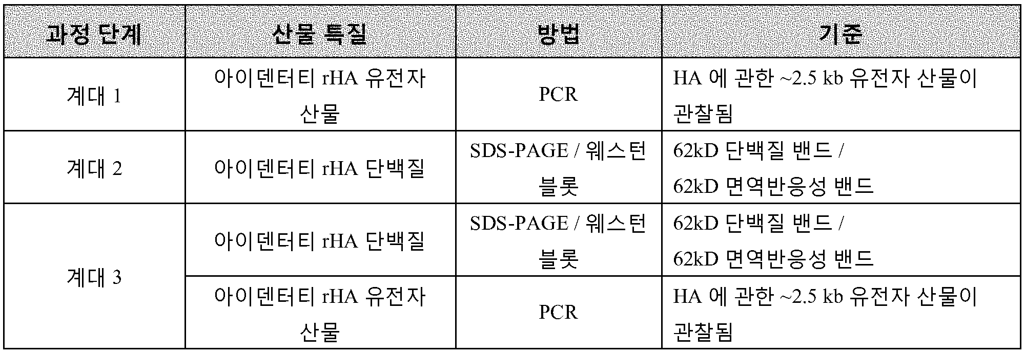
바이러스 상청액에 DMSO (10%) 를 첨가한 후에 P3 작업 바이러스 뱅크를 액체 질소에서 2 년 이상 동안 저장한다. 대안적으로, P3 작업 바이러스 뱅크를 8 주 이하 동안 2-8C 에서 저장한다.After adding DMSO (10%) to the virus supernatant, the P3 working virus bank is stored in liquid nitrogen for more than 2 years. Alternatively, the P3 task virus bank is stored at 2-8C for less than 8 weeks.
P5 규모확대 및 발효. 발효물을 P3 작업 바이러스 뱅크에서 추가 증식에 의해 생성된 P5 바이러스로 감염시킨다. P4 및 P5 바이러스를 위해, SF+ 세포 배양물을 진탕 플라스크 내에 1.0×10⁶ 세포/㎖ 의 밀도로 파종하고, 26-28℃ 에서 18-24 시간 동안 인큐베이션하여 1.3-1.7×10⁶ 세포/㎖ 의 감염 세포 밀도를 달성한다. P4 배양물을 P3 작업 바이러스 뱅크로 감염시키고, P5 를 P4 바이러스 상청액으로 감염시킨다. P4 (세포 생존활성 35% 내지 70%) 및 P5 (세포 생존활성 35% 내지 70%) 바이러스에 관한 기준을 만족시킨 후에 배양물의 원심분리에 의해 P4 및 P5 바이러스 상청액을 단리한다. 결과적인 P4 바이러스 및 P5 바이러스를 각각 2-8℃ 에서 8 및 4 주 동안 저장한다. P4 및 P5 에서의 배양물을 수확하여 수득된 세포 펠렛을 1×PBS 에 재현탁시키고, SDS-PAGE 겔 전기영동/웨스턴 블롯에 의해 분석하여 rHA 단백질의 발현을 확인한다.P5 expansion and fermentation. The fermentation product is infected with the P5 virus produced by further propagation in the P3 working virus bank. For P4 and P5 virus, SF + cell cultures seeded at a density of 1.0 × 10 6 cells / ㎖ in a shaking flask, and the incubation at 26-28 ℃ for 18 to 24 hours of 1.3-1.7 × 10 6 cells / ㎖ To achieve infectious cell density. P4 cultures are infected with the P3 working virus bank, and P5 is infected with the P4 virus supernatant. P4 and P5 virus supernatants are isolated by centrifugation of the cultures after satisfying the criteria for P4 (35% to 70% of cell viability) and P5 (35% to 70% of cell viability) virus. The resulting P4 virus and P5 virus are stored at 2-8 [deg.] C for 8 and 4 weeks, respectively. The cultures in P4 and P5 were harvested and the resulting cell pellet was resuspended in 1x PBS and analyzed by SDS-PAGE gel electrophoresis / Western blot to confirm the expression of rHA protein.
P5 로의 작업 바이러스 뱅크 규모확대 및 발효를 하기 표 7 에 제공된 용납 기준에 따라 평가한다.Working with P5 Expansion and entry of virus banks is evaluated according to the tolerance criteria provided in Table 7 below.
표 7. P5 바이러스 규모확대 및 발효에 관한 용납 기준Table 7. Size of P5 virus and tolerance standard for fermentation

이 실시예에서 야생형 및 변이체 rHA 단백질을 10L 의 작업 부피를 갖는 15L 바이오리액터에서 생산한다. SF+ 세포 배양물을 PSFM 배지 내의 SF+ 세포로 파종한다. 배양물을 26-28℃ 에서 특정 진탕 속도로 유지한다. 바이오리액터는 공기 오버레이를 갖추고 있고, 용존 산소 농도가 명시되어 있다. 배양물이 예정된 밀도에 충분한 생존활성과 함께 도달했을 때, 그것을 P5 작업 바이러스 뱅크로 감염시킨다. 발효물을 샘플채취하고, 광 현미경관찰에 의해 400× 배율에서 박테리아 또는 진균 오염에 관해 검사한다. 세포 생존활성이 40%-80% 내일 때 발효물을 수확한다.In this example, wild-type and variant rHA proteins are produced in a 15L bioreactor with a working volume of 10L. The SF + cell culture is inoculated into SF + cells in the PSFM medium. The cultures are maintained at a specific shaking rate at 26-28 < 0 > C. The bioreactor is equipped with an air overlay, and the dissolved oxygen concentration is specified. When the culture has reached a predetermined density with sufficient viability, it is infected with a P5 working virus bank. The fermented material is sampled and examined for bacterial or fungal contamination at 400x magnification by optical microscopy. The fermentation is harvested when the cell viability is within 40% -80%.
발효물을 원심분리에 의해 수확한다. 10L 발효물을 멸균 1L 병 내로 ~1L 분취물씩 펌프해 넣고, 2-8℃ 에서 Sorvall RC3C 회전 버켓 원심분리를 사용하여 원심분리한다. 세포를 펠렛화하고 수집하고, 한편 발효로부터 소비된 배지를 함유하는 상청액을 폐기한다. 펠렛을 즉시 정제하거나 또는 추가 정제 전까지 20℃ 이하에서 동결 저장한다.The fermented product is harvested by centrifugation. The 10L fermented product is pumped into ~ 1 L aliquots into a sterile 1 L bottle and centrifuged at 2-8 ° C using a Sorvall RC3C rotary bucket centrifuge. The cells are pelleted and collected, while the supernatant containing the medium consumed from the fermentation is discarded. The pellet is immediately purified or stored frozen at < RTI ID = 0.0 > 20 C < / RTI >
단백질 정제. rHA 단백질의 정제를 각각 ~4L 또는 ~10L 의 발효물로부터 수득된 세포 펠렛을 사용하여 4L 또는 10L 규모로 실시한다. 세포 펠렛을수확 후에 즉시 또는 -20℃ 에서 저장 후에 정제한다. 동결된 펠렛을 정제 전에 2-8℃ 에서 완전히 해동시킨다. 이 실시예에서 소규모 작업이 하기 각각의 정제 단계에 관해 기재되어 있다. 정제는 하기 단계를 수반한다: 추출, IEX 크로마토그래피, HIC 크로마토그래피, Q-여과, 한외여과, 제형 및 최종 여과. 과정 단계 및/또는 산물 (과정 중간체) 품질의 평가 기준이 각각의 유닛 작업에 관해 제공되어 있다.Protein purification. Purification of the rHA protein is carried out in 4 L or 10 L scales using cell pellets obtained from ~ 4 L or ~ 10 L fermentations, respectively. The cell pellet is purified immediately after harvest or after storage at -20 [deg.] C. The frozen pellet is completely thawed at < RTI ID = 0.0 > 2-8 C < / RTI > In this example, a small scale operation is described for each of the following purification steps. The purification entails the following steps: extraction, IEX chromatography, HIC chromatography, Q-filtration, ultrafiltration, formulation and final filtration. Evaluation criteria for process steps and / or product (process intermediates) quality are provided for each unit operation.
추출. 이 단계에서, rHA 단백질을 세포막으로부터 Triton X-100 계면활성제를 사용하여 가용화시키고, 추가 정제를 위해 완충제 내로 방출한다.extraction. At this stage, the rHA protein is solubilized from the cell membrane using a Triton X-100 surfactant and released into the buffer for further purification.
이 단계를 2-8℃ 에서 수행한다. 미리 냉각한 (2-8×C) Triton® X-100 추출 완충제를 원심분리에 의해 수득된 세포 펠렛에 첨가하고, 교반 막대가 있는 교반 플레이트에서 혼합한다. 최소 혼합 기간 후에, 현탁액 (미정제 추출물) 의 분취물을 샘플채취하고 원심분리한다. 상청액을 단리하고, 시험하여 출발 수율을 확인한다. 결과적인 미정제 추출물을 지체 없이 즉시 가공한다.This step is carried out at 2-8 [deg.] C. Pre-cooled (2-8xC) Triton (R) X-100 extraction buffer is added to the cell pellet obtained by centrifugation and mixed on a stirring plate with a stir bar. After the minimum mixing period, an aliquot of the suspension (crude extract) is sampled and centrifuged. The supernatant is isolated and tested to confirm the starting yield. The resulting crude extract is immediately processed without delay.
표 8. rHA 의 정제에 관한 과정 요건 - 추출Table 8. Process requirements for purification of rHA - extraction

심층 여과. 심층 여과를 수행하여 세포 잔해물 및 현탁된 고체를 제거하고, 혼탁도를 감소시킨다. 세포 잔해물 및 미립자를 함유하는 필터를 폐기하고, rHA 를 여과물 스트림으로부터 회수한다.Deep filtration. Deep filtration is performed to remove cellular debris and suspended solids, and to reduce turbidity. The filter containing the cellular debris and particulates is discarded and the rHA is recovered from the filtrate stream.
여과 단계는 PUW 로 세정되고 rHA 특이적 추출 완충제로 예비평형된 단일 렌즈모양 심층 필터를 사용한다. 여과를 22-28℃ 에서 수행하며, 한편 미정제 추출물의 혼합을 계속하여 필터 로딩 동안 세포 펠렛 잔해물의 침강을 방지한다. 과정 중간체, 심층 여과물을 다음 단계에서 즉시 가공한다.The filtration step uses a single lens shaped deep layer filter that has been pre-equilibrated with PUW and rHA-specific extraction buffer. Filtration is carried out at 22-28 [deg.] C while mixing of the crude extract is continued to prevent settling of cell pellet debris during filter loading. The process intermediate, deep filtrate, is processed immediately in the next step.
IEX 크로마토그래피. 이온 교환 (IEX) 크로마토그래피 단계는 SP BB 양이온 교환 칼럼을 사용하여 rHA 단백질을 심층 여과물 내에 포획 및 농축시킨다. 칼럼에 결합하지 않는 오염 단백질을 유체 통과 및 세정으로 제거한다.IEX Chromatography. The ion exchange (IEX) chromatography step captures and concentrates the rHA protein in the deep filtrate using a SP BB cation exchange column. The contaminating protein not bound to the column is removed by fluid passage and washing.
IEX 칼럼을 pH 및 전도성 요건이 만족될 때까지 평형시킨다. IEX - 로드를 평형된 IEX 칼럼 위에 펌핑한다. 로딩 후 및 용출 전에, 칼럼을 rHA 특이적 완충제를 사용하여 세정하여 부가적/잔류 오염물을 제거한다. rHA 를 등용매 조건 하에 나트륨 클로라이드로 칼럼으로부터 용출시키고, UV280 흡광도 피크를 수집한다. 흡광도 피크의 분취물을 시험을 위해 수집하여 IEX-용출물 중 ~65kD 단백질의 존재 및 수율을 확인한다. IEX - 용출물을 수집하고, 24 시간 미만 내에 추가로 가공한다.The IEX column is equilibrated until pH and conductivity requirements are met. IEX - Pumps the load over the balanced IEX column. After loading and prior to elution, the column is rinsed with rHA-specific buffer to remove additive / residual contaminants. The rHA is eluted from the column with sodium chloride under isocratic conditions and the UV280 absorbance peak is collected. An aliquot of the absorbance peak is collected for testing to confirm the presence and yield of ~ 65 kD protein in the IEX-eluate. The IEX-eluate is collected and further processed within 24 hours.
표 9. rHA 의 정제에 관한 과정 요건 및 산물 특질 - IEX 크로마토그래피Table 9. Process requirements and product characterization for purification of rHA - IEX chromatography

HIC 크로마토그래피. 소수성 상호작용 크로마토그래피 (HIC) 단계는 페닐 HP 크로마토그래피 칼럼을 사용하여 IEX-용출물 내의 rHA 단백질을 정제한다.HIC chromatography. The hydrophobic interaction chromatography (HIC) step purifies the rHA protein in the IEX-eluate using a phenyl HP chromatography column.
HIC 칼럼을 물로 세정하고 pH 및 전도성 요건이 만족될 때까지 평형 완충제로 평형시킨다. IEX-용출물을 동등 부피의 무세제 완충제로 희석하여 칼럼 로딩을 위해 조정하고, 계면활성제의 10% 스톡 용액을 사용하여 CHAPS 계면활성제를 첨가한다. 로딩 후에, 칼럼을 rHA 특이적 완충제로 세정하고, 통과 유체 및 세정물 내의 단백질 오염물을 폐기한다. rHA 를 용출 완충제로 용출시키고, 전체 UV280 흡수 피크를 분획으로 수집한다.The HIC column is rinsed with water and equilibrated with equilibration buffer until pH and conductivity requirements are met. The IEX-eluate is diluted with an equal volume of detergent buffer, adjusted for column loading, and CHAPS surfactant is added using a 10% stock solution of surfactant. After loading, the column is rinsed with rHA-specific buffer and the protein contaminants in the permeate fluid and wash are discarded. The rHA is eluted with elution buffer and the entire UV 280 absorption peak is collected in fractions.
용출 분획을 rHA 함량이 SDS-PAGE 에 의해 확인될 때까지 저장하고, 그 후 rHA 를 함유하는 용출 분획을 모은다. 결과적인 rHA 푸울을 HIC-용출물로 지정한다. HIC-용출물을 수집하고, 모으고, 24 시간 미만 내에 추가로 가공한다.The eluted fractions are stored until the rHA content is confirmed by SDS-PAGE, and then the eluted fractions containing rHA are collected. The resulting rHA pool is designated as the HIC-eluate. The HIC-eluate is collected, collected, and further processed within 24 hours.
표 10. rHA 의 정제에 관한 과정 요건 및 산물 특질 - HIC 크로마토그래피Table 10. Process requirements and product characterization for purification of rHA - HIC chromatography

Q 막 여과. Q 막 여과를 Pall Mustang Q 코인 필터를 사용하여 수행하여 rHA 로부터 DNA 를 제거한다.Q membrane filtration. Q membrane filtration is performed using a Pall Mustang Q coin filter to remove DNA from rHA.
Q 막 여과를 22-28℃ 에서 수행하고, 여과물을 살균하고, 세정하고, 사용을 위해 예비조건화한다. 캡슐을 pH 및 전도성 요건이 만족될 때까지 rHA 특이적 완충제로 평형시킨다. 이전 단계로부터의 HIC-용출물을 1M NaCl 의 스톡 용액을 사용하여 Q 막 여과를 위해 조건화한다. 조정된 HIC - 용출물을 Q-로드로서 언급한다. Q-로드를 펌프를 통한 캡슐을 통하여 여과하고, UV 흡수재 (280nm) 를 수집한다. UV 흡광도가 기준선으로 회복할 때까지 Q 캡슐을 rHA 완충제로 세정한다. 수집된 재료, 즉, 여과물 및 세정물을 Q-여과물로 지정한다. Q-여과물을 시험을 위해 샘플채취하여 총 단백질 농도를 확인한다. Q-여과물을 즉시 가공하거나 또는 후속 가공 전까지 2-8℃ 에서 24 시간 미만 동안 저장한다.The Q membrane filtration is carried out at 22-28 [deg.] C, the filtrate is sterilized, cleaned and pre-conditioned for use. The capsules are equilibrated with rHA-specific buffer until pH and conductivity requirements are met. The HIC-eluate from the previous step is conditioned for Q-membrane filtration using a stock solution of 1 M NaCl. The adjusted HIC-eluate is referred to as Q-load. The Q-rod is filtered through a pump-through capsule and the UV absorber (280 nm) is collected. The Q capsules are rinsed with rHA buffer until the UV absorbance recovers to baseline. The collected material, that is, the filtrate and the washed material, is designated as Q-filtrate. The Q-filtrate is sampled for testing to determine the total protein concentration. Q- Filters are processed immediately or stored at 2-8 ° C for up to 24 hours before further processing.
표 11. rHA 의 정제에 관한 산물 특질 - Q 여과Table 11. Product characterization of purification of rHA - Q filtration

한외여과. rHA 단백질의 완충제 교환에 관한 한외여과를 22-28℃ 에서 Pall Minimate 접선 유동 여과 (TFF) 캡슐, 평판 폴리에테르술폰 (PES) 막과 명목 분자량 한계 (NMWL) 50kD 를 사용하여 수행한다. 사용 전에, 필터를 PUW 로 플러싱하고, 완충제로 평형시킨다. Q-여과물을 시스템을 통해 재순환시켜 막을 추가로 조건화한다. 재순환 후에, 10-배 최소 완충제 교환을 rHA 완충제를 사용하여 일정 부피 모드로 수행한다.Ultrafiltration. Ultrafiltration for buffer exchange of rHA protein is performed using Pall Minimate Tangential Flow Filtration (TFF) capsules, a flat polyethersulfone (PES) membrane and a nominal molecular weight limit (NMWL) of 50 kD at 22-28 ° C. Prior to use, the filter is flushed with PUW and equilibrated with buffer. The Q-filtrate is recycled through the system to further condition the membrane. After recycling, 10-fold minimum buffer exchange is performed in constant volume mode using rHA buffer.
여과작용으로부터 수득된 보유물을 칭량하여 질량을 확인하고, 시험을 위해 샘플채취하여 총 단백질 농도 및 총 단백질 수율을 확인한다.The retentate obtained from the filtration action is weighed to determine the mass and a sample is taken for testing to determine total protein concentration and total protein yield.
표 12. rHA 의 정제에 관한 과정 요건 및 산물 특질 - 한외여과Table 12. Process requirements and product characteristics for purification of rHA - Ultrafiltration

제형 및 최종 여과. 이 실시예에서, 보유물 내의 정제된 rHA 의 총 단백질 농도는 400 - 600 ㎍/㎖ 여야 한다. 필요한 경우, 보유물을 TFF 에 의해 추가로 농축시키거나 여과 완충제로 희석하여 이 농도를 달성할 수 있다.Formulation and final filtration. In this example, the total protein concentration of the purified rHA in the retentate should be 400-600 占 퐂 / ml. If desired, the retentate can be further concentrated by TFF or diluted with filtration buffer to achieve this concentration.
이 실시예에서 rHA 단백질에 관한 제형은 10 mM 나트륨 포스페이트, 150 mM 나트륨 클로라이드, 0.005% Tween-20, pH 6.8 - 7.2 이다. 이 제형을 달성하기 위해, 10% Tween-20 스톡 용액을 사용하여 보유물에 Tween-20 을 최종 농도 0.005% Tween-20 로 첨가한다. 결과적인 중간체는 제형화된 보유물이다.In this example, the formulation for the rHA protein is 10 mM sodium phosphate, 150 mM sodium chloride, 0.005% Tween-20, pH 6.8 - 7.2. To achieve this formulation, Tween-20 is added to the retentate with a final concentration of 0.005% Tween-20 using a 10% Tween-20 stock solution. The resulting intermediate is a formulated retentate.
이 실시예에서 시험을 위해 1가 벌크 rHA 를 생성하기 위해, 제형화된 보유물을 동시에 0.2 ㎛ 필터를 통해 여과하고, 제형 용기로부터 바이오프로세스 용기 내로 저장을 위해 전달한다.In this example, the formulated retentate is simultaneously filtered through a 0.2 [mu] m filter and transferred from the formulation vessel to the bioprocess vessel for storage to produce a univalent bulk rHA for testing.
저장 및 안정성. rHA 단백질의 시험. 제형화 및 충전을 완료한 후에, 야생형 및 돌연변이체 rHA 단백질을 함유하는 BPC 를 22-28℃ 에서 제 28 일 가속된 안정성에 배치할 것이다. BCA, 트립신 저항성, 및 응집 검정법을 제 0 일에 수행한다. 모든 기타 시험을 각각의 시점에 수행한다. 시험을 목표 시험일의 +/- 1 일 내에 완료한다. 실험실 일정 또는 실험 관찰을 수용하도록 일정 조정을 실시할 수 있다.Storage and stability. Testing of rHA protein. After completion of the formulation and filling, the BPC containing the wild type and mutant rHA protein will be placed at 28-28 ° C accelerated stability at 22-28 ° C. BCA, trypsin resistance, and flocculation assays are performed on
용납 기준. 안정성 데이타를 야생형 rHA 단백질에 관한 순도, 수율, 안정성 프로파일, 응집 프로파일, 및 폴딩을 상응하는 rHA 변이체와 비교함으로써 평가한다. RP-HPLC 분석은 오직 정보를 위한 것이고, 야생형 및 돌연변이체 rHA 단백질에 관한 RP-HPLC 프로파일에서의 임의의 차이를 관찰한다.Tolerance standards. The stability data are evaluated by comparing purity, yield, stability profile, cohesion profile, and folding for the wild-type rHA protein to the corresponding rHA mutants. RP-HPLC analysis is for information only and observe any differences in RP-HPLC profiles for wild-type and mutant rHA proteins.
순도를 SDS-PAGE 에 의해 확인한다.Purity is confirmed by SDS-PAGE.
수율을 순도에 맞춰 조정된 BCA 에 의해 확인한다.Yield is checked by purity adjusted BCA.
안정성은 SRID 에 의해 측정된 효능에 관한 결과에 의해 시사된다. Stability is suggested by the results of the efficacy measured by SRID.
응집 및 교차-연결을 환원 및 비-환원 SDS-PAGE 겔로부터 평가한다.Aggregation and cross-linking are evaluated from reduced and non-reducing SDS-PAGE gels.
적절한 폴딩을 트립신 저항성 및 적혈구의 응집에 의해 평가한다.Appropriate folding is assessed by trypsin resistance and aggregation of red blood cells.
표 13. 정제된 야생형 및 변이체 rHA 단백질에 관한 산물 특질Table 13. Product properties of purified wild type and mutant rHA protein

실시예 3: 시스테인 돌연변이유발Example 3: Cysteine mutagenesis
클로닝 - H3 A/Perth/16/2009 rHA 단백질의 3 가지 상이한 구축물을 야생형 H3 A/Perth/16/2009 rHA 단백질과의 비교를 위해 제조한다. 이들 구축물에서, rHA 단백질의 막관통 및 세포질 꼬리 도메인 내의 특정 시스테인 잔기를 대체했다. 이들 구축물 내의 돌연변이가 아래 제시되어 있다.Cloning - H3A / Perth / 16/2009 Three different constructs of rHA protein are prepared for comparison with the wild type H3A / Perth / 16/2009 rHA protein. In these constructs, the membrane penetration of the rHA protein and the specific cysteine residues within the cytoplasmic tail domain were replaced. The mutations in these constructs are shown below.
표 14Table 14

H3 rHA 단백질에 관한 구축물, 바이러스 뱅크, 및 발효물을 제조했다. H3 rHA 단백질을 정제하고, 실시예 2 의 프로토콜에 따라 특성분석했다. H3 Perth rHA 에 관한 결과가 아래 제공되어 있다.Constructs, virus banks, and fermentations on H3 rHA protein were prepared. The H3 rHA protein was purified and characterized according to the protocol of Example 2. The results for the H3 Perth rHA are provided below.
초기 rHA 클론 스크린 - 소규모 발효물 (300mL) 을 H3 rHA 변이체에 관해 제조했고, 야생형 H3 rHA 과의 비교를 위해 출발 수율을 확인했다. 모든 H3 rHA 변이체는 하나를 제외하고 수율 기준을 만족시켰다.Initial rHA clone screen-small scale fermentation (300 mL) was prepared for H3 rHA mutants and the starting yield was checked for comparison with wild-type H3 rHA. All H3 rHA mutants met yield criteria except one.
표 15Table 15

출발 수율- 3 가지 Cys H3 rHA 변이체를 규모확대하고 (10L), 야생형 H3 rHA 와의 비교를 위해 정제했다 (4L 규모). 10L 발효 규모에서, 3 가지 Cys 변이체에 관한 출발 수율은 야생형 대조군과 본질적으로 동일하거나 그보다 더 크고, 연구 기준을 만족시킨다.Starting yield - Three Cys H3 rHA mutants were scaled up (10 L) and purified for comparison with wild type H3 rHA (4 L scale). At the 10L fermentation scale, the starting yield for the three Cys variants is essentially the same as or greater than the wild type control and meets the study criteria.
표 16Table 16

순도 - 정제된 H3 rHA 단백질은 1㎍ /레인 로딩을 사용하는 환원 SDS-PAGE 겔 분석에 의해 100% 의 순도를 갖는다. SDS-PAGE 에 의한 순도에 관한 연구 기준은 85% 이상이다 (도 4).The purity-purified H3 rHA protein has a purity of 100% by reduced SDS-PAGE gel analysis using 1 [mu] g / lane loading. The study standard for purity by SDS-PAGE is 85% or more (Fig. 4).
최종 수율 - 3 가지 Cys H3 rHA 변이체에 관한 최종, 정제된 수율은 야생형 대조군과 본질적으로 동일하거나 그보다 더 크다.Final Yield - The final, purified yield for the three Cys H3 rHA mutants is essentially the same as or greater than the wild type control.
표 17Table 17

트립신 저항성 - 야생형 H3 rHA 및 Cys 돌연변이체는 트립신 저항성을 가지며, 이는 rHA 단백질이 적절히 폴딩되고 삼합체임을 시사한다. 모든 H3 rHA 는 검정법에 관한 연구 기준을 만족시켰고, HA1 및 HA2 에 관한 밴드가 가시적이다 (도 5).Trypsin resistance - Wild type H3 rHA and Cys mutants have trypsin resistance, suggesting that the rHA protein is properly folded and trimerized. All H3 rHA met the study criteria for the assay and bands for HA1 and HA2 were visible (Figure 5).
적혈구응집 검정법 - 야생형 H3 Perth rHA 및 Cys 돌연변이체는 연구 기준을 만족시키는 적혈구응집 활성에 관해 양성이고, 이는 rHA 단백질이 적절히 폴딩되어 있음을 시사한다. 연구 기준: 적혈구의 응집에 대해 양성.Red cell coagulation assays - Wild type H3 Perth rHA and Cys mutants are positive for erythrocyte aggregation activity that meets the study criteria, suggesting that the rHA protein is properly folded. Study criteria: Positive for erythrocyte aggregation.
표 18Table 18

SRID 에 의한 효능 - 25℃ 에서 1 개월 후에, 야생형 H3 rHA 단백질은 최고 효능 하락을 보였고, ~40% 의 상대 효능에서 안정화되었다. 5Cys H3 rHA 에 관한 상대 효능은 ~60% 에서 안정화되었다. 3Cys H3 rHA 에 관한 효능 하락은 20% 미만이었고, 2Cys H3 rHA 는 효능 상실을 보이지 않는다. 모든 3 가지 Cys H3 rHA 변이체가 제 28 일에 상대 효능 (RP) 에 관한 연구 요건을 만족시킨다. (도 6)Efficacy by SRID - After 1 month at 25 ° C, the wild type H3 rHA protein showed the highest efficacy decline and stabilized at ~ 40% relative efficacy. Relative efficacy against 5Cys H3 rHA was stabilized at ~ 60%. The efficacy decline for 3Cys H3 rHA was less than 20%, and 2Cys H3 rHA showed no efficacy loss. All three Cys H3 rHA mutants meet the study requirement for relative efficacy (RP) on
SDS-PAGE - 야생형 H3 rHA 단백질 및 3 가지 상이한 Cys 변이체 rHA 에 관한 비-환원 및 환원 SDS-PAGE 프로파일이 도 7A 에 제시되어 있다. 제 0 일에, 야생형 및 5 Cys 돌연변이체에 관한 비-환원 SDS-PAGE 프로파일은 서로 비슷하지만, 모든 후속 시점에서 5 Cys 돌연변이체에 비해 야생형 rHA 에서 더 많은 rHA 교차-연결이 관찰된다. 3 Cys 및 2 Cys 돌연변이체는 제 0 일에 교차-연결이 적거나 없고, 오직 3Cys 돌연변이체에서 약간 증가한다. 연구 기준, 돌연변이체 rHA 의 응집물 밴드 강도 < 야생형 rHA 의 응집물 밴드 강도가 만족되었다.A non-reducing and reducing SDS-PAGE profile for SDS-PAGE-wild-type H3 rHA protein and three different Cys variants rHA is shown in FIG. 7A. At
비-환원 SDS-PAGE 겔을 스캔하고 분자 영상화 소프트웨어를 사용하여 분석했다. 영상화 분석으로부터의 강도 프로파일이 도 7B 에 연구 제 0 일에 관해 제시되어 있다.Non-reducing SDS-PAGE gels were scanned and analyzed using molecular imaging software. The intensity profile from the imaging analysis is presented with respect to
밀도측정법을 비-환원 SDS-PAGE 겔에서 각각의 시점에 각각의 H3 rHA 단백질에 관해 수행했다. 단량체성 rHA 단백질 (HA0) 및 더 높은 교차연결된 형태의 rHA 단백질 (응집) 에 관한 밴드 강도를 확인했다. 응집 강도 대 HA0 강도의 비율을 각각의 H3 rHA 에 관해 아래 도표로 나타냈다. 이러한 방법에 의해, rHA 교차-연결은 2Cys < 3Cys < 5Cys < 야생형 순서로 증가한다. (도 7C)Density measurements were performed on each H3 rHA protein at each time point on a non-reducing SDS-PAGE gel. The band intensities for monomeric rHA protein (HAO) and higher cross-linked forms of rHA protein (aggregation) were determined. The ratio of cohesive strength to HA0 strength is shown in the table below for each H3 rHA. By this method, rHA cross-linking increases in the order of 2Cys <3Cys <5Cys <wild type. (Fig. 7C)
3Cys 및 2Cys 돌연변이체에 관한 RP-HPLC 프로파일은 비슷하지만, 야생형 및 5Cys 돌연변이체와 상이하다 (도 8). 3Cys 및 2Cys rHA 는 대체로 교차연결되지 않고, 단일 피크로서 용출되며, 한편 야생형 및 5Cys rHA 는 단백질의 다양한 교차연결된 집단으로 인해 복수의 피크로서 용출된다. 교차연결된 rHA 의 집단은 증가된 소수성으로 인해 칼럼에 보유되고, 나중에 용출된다.The RP-HPLC profiles for 3Cys and 2Cys mutants are similar, but differ from the wild type and 5Cys mutants (FIG. 8). 3Cys and 2Cys rHA are generally not cross-linked and elute as a single peak while wild-type and 5Cys rHA are eluted as multiple peaks due to various cross-linked populations of proteins. The population of cross-linked rHA is retained in the column due to increased hydrophobicity and elutes later.
SRID-BCA 비율 - 3Cys 및 2Cys 돌연변이체는 야생형 및 5Cys 돌연변이체보다 더 높은 SRID/BCA 비율을 갖는다. 3Cys 및 2Cys H3 rHA 단백질에 관한 더 높은 비율은 이들 돌연변이체에서의 항체 친화도 변화 또는 감소된 교차-연결을 반영할 수 있다.The SRID-BCA ratio-3Cys and 2Cys mutants have a higher SRID / BCA ratio than the wild-type and 5Cys mutants. The higher rates for 3Cys and 2Cys H3 rHA proteins may reflect antibody affinity changes or reduced cross-linking in these mutants.
표 19Table 19

부가적 시험 - 동적 광 산란 (DLS), 크기 배제 크로마토그래피 (SEC), 및 전자 현미경관찰 (EM) 검정법은 프로토콜에 포함되지 않았으나, H3 rHA 단백질의 입자 크기를 특성분석하기 위해 수행했다. 시차 주사 형광측정법 (DSF) 을 또한 수행하여 H3 rHA 단백질의 열 안정성을 비교하고, 적혈구응집 저해 (HI) 검정법을 수행하여 H3 rHA 단백질의 항원성을 비교했다.Additional tests - Dynamic light scattering (DLS), size exclusion chromatography (SEC), and electron microscopy (EM) assays were not included in the protocol but were performed to characterize the particle size of the H3 rHA protein. Differential scanning fluorescence spectroscopy (DSF) was also performed to compare the thermal stability of the H3 rHA protein and the anti-red cell aggregation inhibition (HI) assay to compare the antigenicity of the H3 rHA protein.
DLS - DLS 에 의한 rHA 단백질의 입자 크기는 로제트 구조에 특징적인 범위, 30 - 50 nm 내이다. DLS 에 의한 대략적 전이 온도는 모든 H3 rHA 단백질에서 57 - 59℃ 로 매우 유사하다.The particle size of rHA protein by DLS - DLS is within the range of 30 - 50 nm, characteristic of rosette structure. The approximate transition temperature by DLS is very similar to 57 - 59 ° C for all H3 rHA proteins.
표 20Table 20
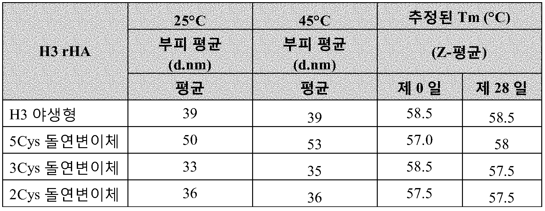
SEC - SEC 에 의해, H3 rHA 단백질은 본질적으로 동일한 보유 시간에서 용출한다. 2.4 - 2.6 MDa 범위의 추정된 분자량이 H3 rHA 단백질에 관해 관찰되었다. ~70kDa 의 단량체에 관한 대략적 MW 을 사용하여, 입자/로제트 당 단량체의 수는 35-38 로 추정된다 (도 9 참조). By SEC - SEC, the H3 rHA protein elutes at essentially the same retention time. An estimated molecular weight in the 2.4 - 2.6 MDa range was observed for the H3 rHA protein. Using approximate MW for monomers of ~ 70 kDa, the number of monomers per particle / rosette is estimated to be 35-38 (see Figure 9).
EM - 전자 현미경관찰을 야생형 및 시스테인 돌연변이체 H3 rHA 단백질에 대해 수행했다. 야생형 및 돌연변이체 rHA 단백질은 대략 30-40 nm 크기의 다합체 로제트-유사 구조를 형성한다. EM 분석에서 동일한 배율 하에 및 동일한 단백질 농도를 사용하여 로제트 입자의 밀도는 샘플들 사이에 정성적으로 유사해 보인다. 분석에 기초하여, 더욱 고차 구조는 시스테인 돌연변이유발에 의해 영향을 받지 않는다.EM-electron microscopy observations were performed on wild type and cysteine mutant H3 rHA proteins. Wild-type and mutant rHA proteins form a multimerization rosette-like structure approximately 30-40 nm in size. The density of the rosette particles under the same magnification and using the same protein concentration in the EM analysis appears qualitatively similar between the samples. On the basis of the analysis, the higher order structures are not affected by cysteine mutagenesis.
DSF - H3/Perth rHA 야생형 및 시스테인 돌연변이체 (2Cys, 3Cys, 및 5Cys) 를 25℃ 내지 99℃ 에서 분자 회전자 염료 (ProteoStat, Enzo Life Scienes) 의 존재 하에 시차 주사 형광측정법 (DSF) 으로 분석했다. 형광을 온도의 함수로서 모니터링하고, 단일, 대형 협력 언폴딩 사건이 각각의 단백질에서 관찰되었다. 데이타는 모든 H3/Perth rHA 시스테인 돌연변이체가 야생형 H3 rHA 보다 약간 더 높은 열 안정성을 가짐을 보여주며, 이는 rHA 단백질의 막관통 및/또는 세포질 영역 내의 시스테인 잔기의 돌연변이가 그들의 안정성을 향상시킬 수 있다는 주장을 지지한다.DSF-H3 / Perth rHA wild-type and cysteine mutants (2Cys, 3Cys, and 5Cys) were analyzed by differential scanning fluorescence measurement (DSF) at 25 ° C to 99 ° C in the presence of molecular rotor dyes (ProteoStat, Enzo Life Scienes) . Fluorescence was monitored as a function of temperature, and single, large co-unfolding events were observed in each protein. The data show that all H3 / Perth rHA cysteine mutants have slightly higher thermal stability than wild-type H3 rHA, suggesting that membrane penetration of rHA protein and / or mutations of cysteine residues within the cytoplasmic region may improve their stability Lt; / RTI >
표 21. DSF 를 사용하는 H3 rHA 야생형 및 Cys 돌연변이체에 관한 용융 온도Table 21. Melting temperatures for H3 rHA wild type and Cys mutants using DSF

H3 rHA 야생형 및 시스테인 돌연변이체 단백질을 항원성 연구에서 적혈구응집 저해 (HI) 시험을 사용하여 특성분석했다. 목적은 H3 항원에 항혈청과 함께 특이적으로 결합하는 rHA 단백질의 능력의 차이를 확인하는 것이었다. H3 rHA 를 4 HA 유닛/25㎕ 의 적혈구응집 역가를 갖도록 표준화했고, 이는 검정법에서 역적정 (BT) 의 처음 4 개의 웰에서 응집을 초래한다. 표준화된 양의 각각의 rHA 를 계열 희석된 항혈청과 혼합하고, 적혈구를 첨가하여 rHA 분자에 대한 항체의 특이적 항체 결합을 확인했다. H3 A/Wisconsin/15/2009-X-183 바이러스로부터의 정제된 HA 에 대해 양에서 생산된 항혈청 및 야생형 H3 A/Perth/16/2009 rHA 단백질을 사용하여 토끼에서 생산된 항혈청을 사용하여 야생형 및 돌연변이체 H3 A/Perth/16/2009 rHA 단백질을 평가했다. 양 또는 토끼 항혈청을 이용하는 검정법에서 시스테인 돌연변이체 rHA 를 사용하여 수득된 HI 역가는 야생형 H3 rHA 를 사용하여 수득된 HI 역가의 2-배 내이거나 그와 동등했다. 결과는 야생형 및 돌연변이체 H3 rHA 단백질 상의 항원 자리의 유사한 제시를 지지한다. The H3 rHA wild type and cysteine mutant proteins were characterized using the erythrocyte aggregation inhibition (HI) test in antigenicity studies. The aim was to identify differences in the ability of the rHA protein to specifically bind H3 antigens with antisera. H3 rHA was standardized to have a red blood cell agglutination titer of 4 HA units / 25 μl, which results in agglutination in the first four wells of reversed titration (BT) in the assay. Standardized amounts of each rHA were mixed with serially diluted antiserum and red blood cells were added to confirm antibody specific binding of the antibody to rHA molecules. Antisera produced in sheep and purified wild-type H3A / Perth / 16/2009 rHA protein against purified HA from H3A / Wisconsin / 15/2009-X-183 virus The mutant H3A / Perth / 16/2009 rHA protein was evaluated. The HI potency obtained using the cysteine mutant rHA in the assay using sheep or rabbit antiserum was within or equivalent to 2-fold of the HI titre obtained using the wild-type H3 rHA. The results support a similar presentation of antigenic sites on wild-type and mutant H3 rHA proteins.
실시예 4: 효능 상실 메카니즘Example 4: Mechanism of loss of efficacy
이 실시예는 모델 시스템으로서 H3 rHA 단백질을 사용하여 효능 상실 메카니즘을 확인하기 위해 확립했다. 실시간 안정성 연구를 갓 정제된 H3 A/Victoria/361/2011 (H3 Victoria) rHA 를 사용하여 수행했다.This example was established to confirm the loss of efficacy mechanism using H3 rHA protein as a model system. Real-time stability studies were performed using freshly purified H3A / Victoria / 361/2011 (H3 Victoria) rHA.
28 일 안정성 연구를 H3 A/Victoria/361/2011 (H3 Victoria) rHA 단백질의 3 가지 제형, 및 2 가지 상이한 저장 온도를 사용하여 수행했다.The 28-day stability study was performed using three formulations of H3A / Victoria / 361/2011 (H3 Victoria) rHA protein, and two different storage temperatures.
표 22. 시기 II 에서 H3 Victoria rHA 에 관한 제형 및 저장 조건.Table 22. Formulation and storage conditions for H3 Victoria rHA at time II.

*표준 제형: 10mM 나트륨 포스페이트, 150mM 나트륨 클로라이드, 0.005% Tween-20, pH 6.8-7.2.Standard formulation: 10 mM sodium phosphate, 150 mM sodium chloride, 0.005% Tween-20, pH 6.8-7.2.
** 실시예 5 및 6 를 참조.** See Examples 5 and 6.
제형을 효능에 관해 SRID 검정법으로, 자유 티올 함량에 관해 형광 기반 검정법을 사용하여, 그리고 자유 Cys 에 관해 펩티드 맵핑을 사용하여 평가했다.The formulations were evaluated by SRID assay for efficacy, fluorescence-based assay for free thiol content, and peptide mapping for free Cys.
자유 티올 함량은 2-8℃ 및 22-28℃ 에서 저장된 미리 제형화된 보유물에서 및 2-8℃ 에서 저장된 1가 벌크에서 감소한다 (도 13). 검정법은 제형 내의 STG 로부터의 간섭으로 인해 STG-시트레이트 제형으로 수행할 수 없었다. 펩티드 맵핑은 동일한 제형 내의 위치 549 에서 자유 시스테인 (NEM-표지된 시스테인) 의 상실을 보여주며, 이는 자유 티올 결과와 일치한다. 이와 대조적으로, 위치 549 에서의 자유 시스테인의 수준은 가장 안정적 제형인 STG-시트레이트 제형에서 동일하게 유지된다.The free thiol content is reduced in preformulated retentions stored at 2-8 < 0 > C and 22-28 < 0 > C and in univalent bulk stored at 2-8 [deg.] C (Fig. 13). The assay could not be performed with STG-citrate formulations due to interference from STG in the formulation. Peptide mapping shows loss of free cysteine (NEM-labeled cysteine) at
자유 티올의 수는 작고, 제 0 일에 rHA 의 분자 당 1 미만이다; 그러나, 연구 경과 동안 자유 티올 함량의 상대적 상실은 크다. 연구 마지막에, 총 초기 자유 티올 함량은 22-28℃ 에서 저장된 미리 제형화된 보유물에서 90% 만큼 감소되고, 2-8℃ 에서 저장된 미리 제형화된 보유물 및 1가 벌크 샘플에서 대략 70% 만큼 감소된다. 유사하게, 위치 549 에서 총 이용가능한 자유 시스테인, 미리 제형화된 보유물 및 1가 벌크 샘플에 관해 대략 20% 는 연구 마지막에 이들 제형에서 거의 완전히 고갈된다 (< 5%). 이와 대조적으로, 자유 Cys549 의 출발 수준은 STG-시트레이트 제형 (~30%) 에서 더 크고, 저장 동안 변하지 않는다.The number of free thiols is small and less than 1 per molecule of rHA at
펩티드 맵핑으로부터 자유 티올 및 Cys549 상실에 관한 결과를 연구에서 모든 제형에 관한 효능 상실과 상관관계가 있다. 효능 상실 속도, 및 자유 티올 상실 및 Cys549 상실의 속도는 22-28℃ 에서 저장된 미리 제형화된 보유물에서 최고이고, 그에 뒤이어 2-8℃ 에서 저장된 미리 제형화된 보유물 및 1가 벌크 샘플이었다. 제형에 관한 상대 효능 값을 도 14 에서 자유 티올 함량의 상대적 변화를 따라 도표로 나타내고, 도 15 에서 자유 Cys549 의 상대적 변화를 따라 도표로 나타낸다.There is a correlation with loss of free thiol and Cys549 from peptide mapping with loss of efficacy for all formulations in the study. The rate of loss of efficacy, and the rate of free thiol loss and loss of Cys549, were highest in preformulated retentions stored at 22-28 DEG C followed by preformed retentate and univalent bulk samples stored at 2-8 DEG C . Relative efficacy values for the formulation are plotted according to the relative change in free thiol content in FIG. 14 and plotted along the relative change in free Cys549 in FIG.
실시예 5: 시트레이트 및 STG 를 함유하는 제형Example 5: Formulation containing citrate and STG
이 실시예는 시트레이트 및 나트륨 티오글리콜레이트 (STG) 를 함유하는 유망한 제형에 초첨을 맞춰 디자인되었다. 목적은 작은 농도의 STG 를 함유하는 제형에 관한 최적 시트레이트 농도를 확인하여 시트레이트 또는 STG 단독이 제형의 안정성을 개선할 수 있는지 여부를 확인하는 것이었다. 이 연구에서 사용된 rHA 는 B/Brisbane (45-09018), H1/Brisbane (45-09012) 및 H3/Brisbane (45-09023 및 45-09025) 을 사용하는 과정 확인 롯트로부터 수득되었다. 이러한 롯트는 McPherson, KS 에서 Hospira One-2-One 에서 충전되었고, "PV2" 또는 Hospira 번호, "CM0-119" 로서 언급된다. 무균 기술 (후드 HD 016) 을 사용하여, CM0-119 약품의 400 바이알을 멸균 병 내로 모았다. 이러한 모아진 물질을 더 작게 나누고, 농축된 부형제를 첨가하여 개질하여 원하는 제형을 산출했다 (표 23).This example was designed with a focus on promising formulations containing citrate and sodium thioglycolate (STG). The objective was to ascertain the optimum citrate concentration for formulations containing small concentrations of STG to ascertain whether citrate or STG alone could improve the stability of the formulation. The rHA used in this study was obtained from process confirmation lots using B / Brisbane (45-09018), H1 / Brisbane (45-09012) and H3 / Brisbane (45-09023 and 45-09025). These lots were filled in Hospira One-2-One at McPherson, KS and are referred to as "PV2" or Hospira numbers, "CM0-119". Using aseptic technique (Hood HD 016), 400 vials of CM0-119 drug were collected into a sterile bottle. These collected materials were further divided into smaller portions and the concentrated excipients were added to modify the desired formulations (Table 23).
표 23 - 제형Table 23 - Formulation
샘플을 35℃, 25℃, 및 5℃ 로 설정하고, 통상 1 주 간격으로 설정된 풀 (pull) 에 관한 일정을 잡았다. 부가적 2-일 풀을 35℃ 샘플에 관해 일정을 잡고, 더 적은 이른 시점을 5℃ 샘플에 관해 일정을 잡고, 보증되는 경우에 예비 샘플을 긴 시점에 관해 설정했다. SRID 효능 측정의 초점은 H1/Brisbane rHA 이었으나, 또한 H3/Brisbane 및 B/Brisbane 에 관해서도 빈번한 측정을 실시했다.Samples were set at 35 占 폚, 25 占 폚, and 5 占 폚 and schedules were typically set for pulls at one week intervals. An additional two-day pool was sched- uled for a 35 ° C sample, a lesser point in time was scheduled for a 5 ° C sample and, if warranted, a preliminary sample was set for a longer time point. The focus of SRID efficacy measurement was on H1 / Brisbane rHA, but also on H3 / Brisbane and B / Brisbane.
SRID 효능 측정이 표 23-25 에 나열되어 있고, 이들 데이타는 도 16-18 에 도표로 나타나 있다.SRID efficacy measures are listed in Table 23-25, and these data are plotted in Figures 16-18.
표 24 - H1/Brisbane SRID 효능.Table 24 - H1 / Brisbane SRID efficacy.

표 25 - H3/Brisbane SRID 효능Table 25 - H3 / Brisbane SRID efficacy

표 26 - B/Brisbane SRID 효능Table 26 - B / Brisbane SRID efficacy

t=0 에서, 이들 제형의 측정된 효능은 부형제가 B/Brisbane 에 관해 SRID 에 의해 측정되는 겉보기 효능에 영향을 미치지 않았음을 시사했다. H1/Brisbane 에 관해 부형제의 작은 효과 및 H1/Brisbane 에 관해 중간 효과가 존재했다 (도 19). STG 단독을 함유하는 샘플에 관해 측정된 효능이 STG 및 시트레이트를 함유하는 샘플과 동등했다는 사실에 기초하여, 효과는 환원제의 존재로 인한 것으로 보인다. 환원제가 검정법에 직접 영향을 미치거나 또는 rHA 의 형태를 변경하여 SRID 시약과 더 잘 맞게 하는지 여부는 아직 알려져 있지 않다.At t = 0, the measured efficacy of these formulations suggested that the excipient did not affect the apparent efficacy as measured by SRID for B / Brisbane. There was a small effect of the excipient on H1 / Brisbane and an intermediate effect on H1 / Brisbane (Fig. 19). Based on the fact that the measured efficacy on a sample containing STG alone was equivalent to a sample containing STG and citrate, the effect appears to be due to the presence of a reducing agent. Whether the reducing agent directly affects the assay or changes the morphology of the rHA to better match the SRID reagent is not yet known.
모든 H1, H3, 및 B 의 경우, 안정성이 시트레이트 및 STG 에 의해 개선되었으나, 부형제 중 하나에 의해 개별적으로는 개선되지 않았다. STG 단독을 함유하는 제형은 가장 불량한 안정성을 나타냈다; 안정성 곡선 (상대 효능 시간의 함수로서) 의 기울기는 3 가지의 저장 조건에 대해 60% 초과였다 (도 20). STG 을 함유하는 시트레이트-함유 샘플에 관한 기울기는 일관되는 농도 의존성을 보이지 않았으나, 모두 제형 안정성의 유의한 개선을 반영했다. 대조군 (A) 기울기 대 시트레이트 + STG 제형의 평균 기울기의 비율은 1.6 이상 내지 4.4 이하였다.For all H1, H3, and B, stability was improved by citrate and STG, but was not individually improved by one of the excipients. Formulations containing STG alone exhibited the worst stability; The slope of the stability curve (as a function of relative potency time) was greater than 60% for the three storage conditions (Figure 20). The slope for the citrate-containing sample containing STG did not show a consistent concentration dependence, but all reflected a significant improvement in formulation stability. The ratio of the average slope of the control (A) slope vs. citrate + STG formulation was 1.6 or more to 4.4 or less.
SDS-PAGE 결과가 도 21 에 제시되어 있다. 제 0 일 데이타는 모든 제형이 초기에 등가라는 것을 보여준다. 제 21 일까지, STG 및 시트레이트 둘 모두를 함유하는 제형에서 응집이 더 적다는 것이 명백하다. 연구 마지막에 (제 52 일), 일부 응집물은 STG 및 시트레이트 둘 모두를 함유하는 제형에서 가시적으로 되었으나, 우세적 밴드는 HA0 이다. 전체적 강도가 더 낮게 보이는 이유는 명백하지 않으나, H1 에 관한 SRID 효능은 여전히 제 0 일 값의 대략 80% 였다. 이들 측정에서, 이전 연구에서와 같이, SRID 효능 상실은 비환원 SDS-PAGE 에서 관찰되는 응집물의 축적과 상관관계가 있다.The results of SDS-PAGE are shown in
이 실시예는 납 부형제를 함유하는 3가 제형에서 각각의 rHA 의 효능을 모니터링하기 위해 디자인되었다. 입자 크기 (DLS 에 의해) 및 응집 (비-환원 SDS-PAGE 에 의해) 을 또한 측정했지만, 이들 파라미터는 모든 rHA 에 대한 평균이고, 특정 균주로부터의 rHA 에 특이적이지 않다. 시험된 부형제는 시트레이트 및 나트륨 티오글리콜레이트였다. 내부 다이설파이드 환원을 최소화하기 위해, STG 를 매우 낮은 농도 (0.2 ㎎/㎖) 로 사용했다. 전체적 결론은 다음과 같다:This example was designed to monitor the efficacy of each rHA in a trivalent formulation containing a lead excipient. Although particle size (by DLS) and cohesion (by non-reducing SDS-PAGE) were also measured, these parameters are averages for all rHA and are not specific for rHA from a particular strain. The excipients tested were citrate and sodium thioglycolate. To minimize internal disulfide reduction, STG was used at a very low concentration (0.2 mg / ml). The overall conclusion is as follows:
시트레이트 및 STG 둘 모두를 함유하는 제형에서 모든 3 가지 rHA (H1/Brisbane, H3/Brisbane, 및 B/Brisbane) 에 관해 안정성이 개선되었다. 시트레이트 단독 (20 ㎎/㎖) 은 안정성을 개선하지 않는다. STG 단독은 안정성에 음성 효과를 갖는다. 시험된 최고 농도 (5 ㎎/㎖) 는 안정성에 유해 영향을 미친다 (데이타는 제시되지 않음). 더 낮은 농도는 효과적일 수도 있고 효과적이지 않을 수도 있다.Stability was improved for all three rHA (H1 / Brisbane, H3 / Brisbane, and B / Brisbane) in formulations containing both citrate and STG. Citrate alone (20 mg / ml) does not improve stability. STG alone has a negative effect on stability. The highest concentration tested (5 mg / ml) has an adverse effect on stability (data not shown). Lower concentrations may or may not be effective.
시트레이트 및 STG 둘 모두의 존재시에, rHA 의 응집은 최소였다. 응집의 정도는 제 0 일에 관찰된 값 미만으로 감소하지 않았다.In the presence of both citrate and STG, agglutination of rHA was minimal. The degree of aggregation did not decrease below the value observed at
실시예 6: 0.035% Triton X-100 을 이용하는 H3 Perth 에 관한 조기 안정성 연구Example 6: Early stability study on H3 Perth using 0.035% Triton X-100
이 실시예는 (a) 0.035% Triton X-100 로 제조하여 제형화된 H3 Perth 의 안정성을 평가하고 (b) 안정성 시험에서 H3/Wisconsin 의 롯트의 예상 외의 높은 안정성을 더욱 잘 이해하고, (c) STG-시트레이트 제형의 안정성을 고농도의 Triton X-100 을 함유하는 제형과 비교하기 위해 디자인되었다. 후향 시험은 이 롯트가 대략 0.2% 의 비정상적으로 높은 Triton X-100 농도를 갖는다는 것을 보여줬다. 이 연구에서, H3 Perth 를 Triton X-100 농도 0.035% 로 제조하여 제형화했다. 이 롯트에 Triton X-100 을 보충하여 제형 개발 연구에서 사용된 농도, 0.05%, 및 H3/Wisconsin 의 관찰된 향상된 안정성이 상승된 Triton X-100 (0.1%, 0.2%) 로 인한 것이었다는 가설을 시험하기 위해 디자인된 농도를 모방했다. 롯트에 1% 나트륨 시트레이트 및 0.02% 나트륨 티오글리콜레이트를 보충하여 또다른 제형을 제조했다.This example (a) evaluates the stability of H3 Perth formulated with 0.035% Triton X-100 and (b) better understand the unexpectedly high stability of the lot of H3 / Wisconsin in the stability test, and ) The stability of the STG-citrate formulation was designed to compare with formulations containing high concentrations of Triton X-100. Backward testing has shown that this lot has an abnormally high Triton X-100 concentration of approximately 0.2%. In this study, H3 Perth was formulated to a Triton X-100 concentration of 0.035%. The hypothesis that this lot was supplemented with Triton X-100 and the observed enhanced stability of concentration, 0.05%, and H3 / Wisconsin used in formulation development studies was due to elevated Triton X-100 (0.1%, 0.2%) I simulated the concentration designed for testing. Another formulation was prepared by supplementing the lot with 1% sodium citrate and 0.02% sodium thioglycolate.
시험된 제형은 표 27 에 나열되어 있다. Triton X-100 스톡을 초기 제형화 후에 첨가했기 때문에, 약간의 희석이 발생했으나, 최고 Triton 농도에서 오직 1.6% 였다. STG-시트레이트 제형의 희석률은 9.4% 였다. 모든 샘플을 25℃ 에서 저장했ㄷ.The formulations tested are listed in Table 27. Because the stock of Triton X-100 was added after the initial formulation, a slight dilution occurred, but only 1.6% at the highest Triton concentration. The dilution rate of the STG-citrate formulation was 9.4%. All samples were stored at 25 ° C.
표 27 - 제형Table 27 - Formulation

가속된 조건 하에 저장된 샘플에 관한 SRID 결과가 표 6 에 나열되어 있고, 도 22 에 도표로 나타나 있다. 이들 결과는 대조군 샘플 (0.35% Triton X-100) 의 경우에, 효능이 제 0 일 효능의 대략 2 분의 1 로 신속히 하락한다는 것을 보여준다. Triton X-100 의 더 높은 수준에서, 효능은 더욱 서서히 감소하고 그렇게 많이 감소하지 않는다. 0.1% 또는 0.2% Triton X-100 의 존재시의 안정성이 0.05% Triton X-100 의 존재시의 안정성보다 더 양호하나, 0.1% 및 0.2% Triton X-100 사이의 차이는 무시할 수 있다. STG-시트레이트 제형의 효능은 2-주 가속된 안정성 기간 동안 매우 적게 변화했고, 92 일 동안 원래 효능의 80% 초과로 유지되었다.The SRID results for stored samples under accelerated conditions are listed in Table 6 and plotted in FIG. These results show that, in the case of the control sample (0.35% Triton X-100), the efficacy rapidly drops to approximately one-half of the zero-day efficacy. At higher levels of Triton X-100, efficacy decreases more slowly and does not decrease so much. The stability in the presence of 0.1% or 0.2% Triton X-100 is better than the stability in the presence of 0.05% Triton X-100, but the difference between 0.1% and 0.2% Triton X-100 is negligible. The efficacy of the STG-citrate formulation varied very little during the two-week accelerated stability period and remained above 80% of the original efficacy for 92 days.
표 28 - SRID 에 따른 효능 - 모든 데이타가 ㎍/㎖ 로 나열되어 있음.Table 28 - Efficacy according to SRID - All data are listed in ㎍ / ㎖.

SDS-PAGE 결과가 도 23A-B 에 제시되어 있다. 초기 패턴은 대부분의 rHA 가 단량체 (HA0) 형태이고, 일부 교차연결된 이합체 및 삼합체가 존재함을 보여준다. 환원 겔은 본질적으로 모든 단백질이 HA0 라는 것을 시사하므로, 단백질은 다이설파이드 결합에 의해 교차연결된 것으로 보인다. 25℃ 에서 2 주 내에, 단량체성 rHA 의 양은 유의하게 감소했고, 교차연결된 이합체의 일부는 비-환원성이었다. 더 높은 농도의 Triton X-100 을 함유하는 제형은 대조군 (0.035% Triton X-100) 보다 더 적은 교차-연결을 갖는다. 시트레이트 및 STG 를 함유하는 제형 내의 다이설파이드 교차 연결은 2 주 동안 작은 변화를 보였고, 비-환원성 교차연결의 증거를 보이지 않았다.SDS-PAGE results are presented in Figures 23A-B. The initial pattern shows that most of the rHA is in the form of monomers (HAO), and some cross-linked dimers and trimesters are present. Reduced gels suggest that essentially all proteins are HA0, so proteins appear to be cross-linked by disulfide bonds. Within two weeks at 25 캜, the amount of monomeric rHA was significantly reduced and some of the cross-linked dimers were non-reducing. Formulations containing higher concentrations of Triton X-100 have less cross-linking than the control (0.035% Triton X-100). The disulfide cross-linking in the formulation containing citrate and STG showed small changes over 2 weeks and showed no evidence of non-reducing cross-linking.
도 23 의 데이타는 Triton X-100 이 H3 Perth rHA 의 안정성을 개선하지만, 0.035% Triton X-100 은 0.05% 만큼 충분한 개선을 제공하지 않는다는 것을 보여준다. 0.1% 에서, Triton X-100 은 안정성을 추가로 개선하고, 0.2% 로 추가로 증가되면 안정성에 대한 증분 개선을 제공한다. 이전 결과는 0.05, 0.08, 또는 0.15% Triton X-100 을 함유하는 제형이 유사한 안정성을 갖는 것을 보여줬으므로, 이것은 예상 밖이었다.The data in Figure 23 show that Triton X-100 improves the stability of H3 Perth rHA, but 0.035% Triton X-100 does not provide enough improvement by 0.05%. At 0.1%, Triton X-100 further improves stability and provides an incremental improvement in stability when further increased to 0.2%. Previous results have been unexpected since formulations containing 0.05, 0.08, or 0.15% Triton X-100 have shown similar stability.
제 0 일 DLS 결과는 Triton X-100 농도 증가가 평균 입자 크기 감소를 초래했음을 보여줬다. 도 24 는 14 일 연구 과정 동안 최소 차이가 존재하나, 높은 농도의 Triton X-100 의 존재는 평균 입자 크기를 유의하게 감소시켰다는 것을 보여준다.
실시예 7: rHA 의 면역원성은 STG -시트레이트 제형에 의해 영향을 받지 않음Example 7: Immunogenicity of rHA is not affected by STG-citrate formulations
이 실시예는 rHA 의 면역원성에 대한 STG 시트레이트 제형의 효과를 평가하기 위해 디자인되었다. H1 California/07/2009 를 사용하여, 2 개의 제형을 rHA 농도 120 ㎍/㎖ 로 제조했다. 대조군 제형은 Flublok 에서 사용된 제형 완충제 (10 mM 나트륨 포스페이트, 150 mM 나트륨 클로라이드, 0.005% Tween-20, pH 6.8 - 7.2) 중이었다. 두번째 제형은 0.02% 나트륨 티오글리콜레이트 (STG) 및 1% 나트륨 시트레이트를 제형에 첨가한 점을 제외하고는 동일했다. 이들 제형을 6 - 8 주령 Balb/c 마우스에 2 가지 투여량: 3 ㎍ 및 0.3 ㎍ 으로 근육내 투여했다. 3 ㎍ 투여량은 각각의 제형의 25 ㎕ 투여물로서 투여했고, 0.3 ㎍ 투여량은 각각의 제형의 1:10 희석물의 25 ㎕ 투여물로서 투여했다. 제 0 일 및 제 21 일에 마우스에 투여했다. 8 마리의 마우스를 4 개의 코호트 각각에 사용했다: 고용량 대조군, 고용량 STG, 저용량 대조군, 및 저용량 STG. 제 0 일, 제 21 일, 및 제 42 일 투여 전에 혈액 샘플을 채취했다. 혈액 샘플을 응고되게 놔두고, 그 후 원심분리하고, 결과적인 혈청을 -20℃ 에서 저장했다. 혈청 샘플을 적혈구응집 저해 (HAI) 및 ELISA 를 사용하여 항체 역가에 관해 시험했다. This example was designed to evaluate the effect of STG citrate formulation on the immunogenicity of rHA. Using H1 California / 07/2009, two formulations were prepared with an rHA concentration of 120 占 퐂 / ml. Control formulations were in the formulation buffer used in Flublok (10 mM sodium phosphate, 150 mM sodium chloride, 0.005% Tween-20, pH 6.8 - 7.2). The second formulation was the same except that 0.02% sodium thioglycolate (STG) and 1% sodium citrate were added to the formulation. These formulations were administered intramuscularly at 6-8 week old Balb / c mice at two doses: 3 ug and 0.3 ug. A 3 μg dose was administered as a 25 μl dose of each formulation, and a 0.3 μg dose was administered as a 25 μl dose of a 1:10 dilution of each formulation. On
HAI 역가가 표 29 및 도 25 에 제시되어 있다. 이들 결과는 STG-시트레이트 제형이 H1 California rHA 의 면역원성에 대해 유의한 효과를 갖지 않는다는 것을 보여준다.The HAI titers are shown in Table 29 and FIG. These results show that the STG-citrate formulation has no significant effect on the immunogenicity of H1 California rHA.
표 29 - HAI 역가 - 역가는 응집이 존재하지 않은 최고 희석물의 역으로서 열거되어 있다.Table 29 - HAI titers - reversals are listed as the inverse of the highest dilution without agglutination.
제 42 일로부터 혈청에 관해 확인된 ELISA 역가가 표 30 에 제시되어 있다. 이들 값은 각각의 마우스에 관한 데이타를 제 0 일 (비-면역화된) ELISA 응답으로 정규화시킴으로써 계산했다. 이들 결과는 STG 및 대조군 제형에 관한 ELISA 역가가 유의하게 상이하지 않다는 것을 보여준다. 도 26 은 ELISA 및 HAI 결과가 비례한다는 것을 보여준다. 2 가지 방법을 사용하여 수득된 역가가 산점도로서 도표화되어 있다. ELISA 및 HAI 결과는 STG-시트레이트 제형이 rHA 의 면역원성에 영향을 미치지 않는다는 것을 입증한다.The ELISA titers identified for sera from day 42 are presented in Table 30. These values were calculated by normalizing the data for each mouse with a zero day (non-immunized) ELISA response. These results show that the ELISA titers for STG and control formulations are not significantly different. Figure 26 shows that ELISA and HAI results are proportional. The titers obtained using the two methods are tabulated as scatter diagrams. ELISA and HAI results demonstrate that the STG-citrate formulation does not affect the immunogenicity of rHA.
표 30 - 제 0 일 기준선으로 정규화된 ELISA 역가.Table 30 - ELISA titers normalized to

실시예 8: H1 A/California/07/20009 에 관한 데이타Example 8: Data on H1 A / California / 07/20009
도 27 은 H1 A/California WT 및 3Cys SDV rHA 의 비교의 비-환원 및 환원 SDS-PAGE 분석을 나타낸다. 레인 1 은 야생형 H1 rHA 를 나타내고, 레인 2 는 3Cys SDV H1 rHA 를 나타낸다.Figure 27 shows the non-reducing and reducing SDS-PAGE analysis of a comparison of H1 A / California WT and 3Cys SDV rHA.
야생형 H1 rHA 에서 관찰된 다이설파이드 매개 교차-연결은 3Cys SDV H1 rHA 에서 5℃ 및 25℃ 둘 모두에서 3 개월 동안 저장 후에 방지된다.Disulfide mediated cross-linking observed in wild-type H1 rHA is prevented after 3 months storage at both 5 ° C and 25 ° C in 3Cys SDV H1 rHA.
도 28 은 H1 A/California WT 및 3Cys SDV rHA 의 비교의 RP-HPLC 분석을 나타낸다.Figure 28 shows RP-HPLC analysis of a comparison of H1 A / California WT and 3Cys SDV rHA.
3Cys SDV rHA 는 단일 피크로서 용출하며, 한편 야생형은 복수의 피크로 용출하며, 이는 3Cys SDV rHA 가 WT rHA 에 비해 균일함을 시사한다. 3Cys SDV 및 야생형에 관한 RP-HPLC 프로파일은 2 가지 저장 온도 모두에서 시간의 흐름에 따라 유의하게 변화하지 않는다.3Cys SDV rHA elutes as a single peak while the wild type elutes to multiple peaks suggesting that 3Cys SDV rHA is more homogeneous than WT rHA. The RP-HPLC profile for 3Cys SDV and wild type does not change significantly over time at both storage temperatures.
도 29 는 H1 A/California WT 및 3Cys SDV rHA 의 비교의 SEC-HPLC 분석을 나타낸다.Figure 29 shows SEC-HPLC analysis of a comparison of H1A / California WT and 3Cys SDV rHA.
WT 및 SDV rHA 의 크기 배제 크로마토그래피 (SEC) 분석. SEC 에 의해, H1 rHA 단백질 둘 모두 동일한 보유 시간으로 용출한다.Size exclusion chromatography (SEC) analysis of WT and SDV rHA. By SEC, both the H1 rHA protein elutes at the same retention time.
도 30 은 H1 A/California WT 및 3Cys SDV rHA 의 비교의 시차 주사 형광측정법 (DSF) 분석을 나타낸다.Figure 30 shows a differential scanning fluorescence measurement (DSF) analysis of a comparison of H1A / California WT and 3Cys SDV rHA.
시차 주사 형광측정법 (DSF) 에서 관찰된 형광 강도가 WT 및 3Cys SDV rHA 단백질 둘 모두에 관해 온도의 함수로서 도표로 나타나 있다. 전이점이 상기 2차 도함수 플롯에서 관찰된다. 제 0 일 및 5℃ 및 25℃ 에서 2 개월 후에 각각의 rHA 에 관한 대표적 2차 도함수 열 변성 도표가 제시되어 있다.Fluorescence intensities observed in differential scanning fluorescence spectroscopy (DSF) are plotted as a function of temperature for both WT and 3Cys SDV rHA proteins. A transition point is observed in the second derivative plot. Representative secondary derivative thermal deformation diagrams for each rHA are shown at
표 31: H1 A/California WT 및 3Cys SDV rHA 의 비교-- DSF 에 의한 E. 용융 온도Table 31: Comparison of H1 A / California WT and 3Cys SDV rHA - E. melting temperature by DSF

도 31 은 H1 A/California WT 및 3Cys SDV rHA 의 비교의 5℃ 및 25℃ 에서의 rHA 단백질의 상대 효능을 나타낸다.Figure 31 shows the relative potency of the rHA protein at 5 < 0 > C and 25 < 0 > C for a comparison of H1 A / California WT and 3Cys SDV rHA.
5℃ 및 25℃ 에서 1 개월 저장 후에 3Cys SDV 의 상대 효능은 야생형보다 더 높다.The relative efficacy of 3Cys SDV after storage for one month at 5 캜 and 25 캜 is higher than that of the wild type.
도 32 는 H1 A/California WT 및 3Cys SDV rHA 의 비교의 동적 광산란 (DLS) 에 의한 입자 크기 분석을 나타낸다.Figure 32 shows particle size analysis by dynamic light scattering (DLS) of a comparison of H1 A / California WT and 3Cys SDV rHA.
DLS 에 의해 확인되는 야생형 H1 rHA 로제트 및 3Cys SDV H1 rHA 로제트의 부피 평균 지름은 5℃ 및 25℃ 둘 모두에서 3 개월 동안 저장 후에 비슷하다.The volume mean diameters of wild-type H1 rHA rosette and 3Cys SDV H1 rHA rosette confirmed by DLS are similar after storage for 3 months at both 5 캜 and 25 캜.
실시예 9: B/Massachusetts/2/2012 rHA 에 관한 데이타Example 9: Data on B / Massachusetts / 2/2012 rHA
도 33 은 B/Massachusetts WT 및 2Cys SDV rHA 의 비교의 비-환원 및 환원 SDS-PAGE 분석을 나타낸다. 레인 1 은 야생형 B rHA 를 나타내고, 레인 2 는 2Cys SDV B rHA 를 나타낸다.Figure 33 shows the non-reducing and reducing SDS-PAGE analysis of a comparison of B / Massachusetts WT and 2Cys SDV rHA.
야생형 B rHA 에서 관찰된 다이설파이드 매개 교차-연결은 2Cys SDV B rHA 에서 제 0 일에 정제 직후에 방지된다. 25℃ 및 35℃ 둘 모두에서 1 개월 동안 저장 후에, 2Cys SDV 에 관한 다이설파이드 매개 교차-연결은 유사한 조건 하에 저장된 야생형에 비해 유의하게 감소된다.Disulfide-mediated cross-linking observed in wild type B rHA is prevented immediately after purification on
도 34 는 B/Massachusetts WT 및 2Cys SDV rHA 의 비교의 RP-HPLC 분석을 나타낸다.Figure 34 shows RP-HPLC analysis of a comparison of B / Massachusetts WT and 2Cys SDV rHA.
B/Massachusetts 2Cys SDV rHA 는 단일 피크로서 용출하며, 한편 야생형은 복수의 피크로서 용출하며, 이는 Cys SDV rHA 가 더욱 균일함을 시사한다. 2Cys SDV 및 야생형에 관한 RP-HPLC 프로파일은 2 가지 저장 온도 모두에서 시간의 흐름에 따라 유의하게 변화하지 않는다.B / Massachusetts 2Cys SDV rHA elutes as a single peak while the wild type elutes as a plurality of peaks suggesting that the Cys SDV rHA is more homogeneous. The RP-HPLC profile for 2Cys SDV and wild type does not change significantly over time at both storage temperatures.
도 35 는 B/Massachusetts WT 및 2Cys SDV rHA 의 비교의 동적 광산란 분석에 의한 입자 크기 분석을 나타낸다.Figure 35 shows particle size analysis by dynamic light scattering analysis of a comparison of B / Massachusetts WT and 2Cys SDV rHA.
DLS 에 의해 확인되는 야생형 B rHA 로제트 및 2Cys SDV B rHA 로제트의 부피 평균 지름은 비슷하다. 25℃ 및 35℃ 둘 모두에서 1 개월 동안 WT 및 2Cys SDV 의 저장은 로제트 지름의 약간의 증가를 초래한다.The volume average diameters of the wild type B rHA rosette and 2Cys SDV B rHA rosette identified by DLS are similar. Storage of WT and 2Cys SDV for one month at both 25 ° C and 35 ° C results in a slight increase in rosette diameter.
도 36 은 B/Massachusetts WT 및 2Cys SDV rHA 의 비교의 5℃ 및 25℃ 에서 저장된 rHA 단백질의 상대 효능을 나타낸다.Figure 36 shows the relative potency of the stored rHA protein at 5 [deg.] C and 25 [deg.] C for comparison of B / Massachusetts WT and 2Cys SDV rHA.
25℃ 및 35℃ 에서 1 개월 후에 B/Massachusetts 2Cys SDV 의 상대 효능이 야생형에 비해 개선된다.The relative efficacy of the B / Massachusetts 2Cys SDV after one month at 25 캜 and 35 캜 is improved compared to the wild type.
본 발명은 하기 번호매긴 단락에 의해 추가로 기재된다:The present invention is further described by the following numbered paragraphs:
1. 하나 이상의 시스테인 돌연변이를 포함하는 단리된, 비-자연발생적 재조합 헤마글루티닌 (rHA) 단백질.One. An isolated, non-naturally occurring recombinant hemagglutinin (rHA) protein comprising at least one cysteine mutation.
2.
단락 1 에 있어서, rHA 단백질이 H1 단백질인 단백질.2.
In
3.
단락 1 에 있어서, H1 단백질이 California 또는 Solomon 균주로부터 단리되는 단백질.3.
A protein according to
4.
단락 3 에 있어서, California 균주가 California/07/2009 균주인 단백질.4.
In
5.
단락 3 에 있어서, Solomon 균주가 Solomon Is/03/2006 균주인 단백질.5.
In
6. 단락 2-5 중 어느 하나에 있어서, 시스테인 돌연변이가 카르복시 말단 영역 내에 있는 단백질.6. The protein according to any one of paragraphs 2-5, wherein the cysteine mutation is within the carboxy terminal region.
7. 단락 2-6 중 어느 하나에 있어서, 시스테인 돌연변이가 막관통 영역 또는 사이토솔 영역 내에 있는 단백질.7. The protein according to any one of paragraphs 2-6, wherein the cysteine mutation is within the transmembrane domain or the cytosolic domain.
8.
단락 1 에 있어서, rHA 단백질이 B 단백질인 단백질.8.
In
9.
단락 8 에 있어서, B 단백질이 Brisbane, Florida, Ohio, Jiangsu 또는 Hong Kong 균주로부터 단리되는 단백질.9.
The protein according to
10.
단락 9 에 있어서, Brisbane 균주가 Brisbane/60/2008 균주인 단백질.10.
In
11.
단락 9 에 있어서, Florida 균주가 Florida/04/2006 균주인 단백질.11.
In
12.
단락 9 에 있어서, Ohio 균주가 Ohio/01/2005 균주인 단백질.12.
In
13.
단락 9 에 있어서, Jiangsu 균주가 Jiangsu/10/2003 균주인 단백질.13.
In
14.
단락 9 에 있어서, Hong Kong 균주가 Hong Kong/330/2001 균주인 단백질.14.
In
15. 단락 8-14 중 어느 하나에 있어서, 시스테인 돌연변이가 막관통 (TM) 및 사이토솔 (CT) 도메인을 포함하는 카르복시 말단 영역 내에 있는 단백질.15. The protein of any one of paragraphs 8-14, wherein the cysteine mutation is in a carboxy terminal region comprising a transmembrane (TM) and a cytosolic (CT) domain.
16.
단락 1 에 있어서, rHA 단백질이 H3 단백질인 단백질.16.
In
17.
단락 16 에 있어서, H3 단백질이 Victoria, Perth, Brisbane 또는 Wisconsin 균주로부터 단리되는 단백질.17.
In
18. 단락 17 에 있어서, Victoria 균주가 Victoria/361/2011 균주인 단백질.18. In paragraph 17, the Victoria strain is a Victoria / 361/2011 strain.
19. 단락 17 에 있어서, Perth 균주가 Perth/16/2009 균주인 단백질.19. In paragraph 17, the Perth strain is a Perth / 16/2009 strain.
20. 단락 19 에 있어서, 돌연변이가 C524S 및/또는 C528A 인 단백질.20. In paragraph 19, the protein wherein the mutation is C524S and / or C528A.
21. 단락 19 에 있어서, 돌연변이가 C524A, C528A, C539A, C546A 및/또는 C549A 인 단백질.21. In paragraph 19, the protein wherein the mutation is C524A, C528A, C539A, C546A and / or C549A.
22. 단락 19 에 있어서, 돌연변이가 C539A, C546A 및/또는 C549A 인 단백질.22. In paragraph 19, the protein wherein the mutation is C539A, C546A and / or C549A.
23. 단락 17 에 있어서, Brisbane 균주가 Brisbane/16/2007 균주인 단백질.23. In paragraph 17, the protein wherein the Brisbane strain is a Brisbane / 16/2007 strain.
24. 단락 17 에 있어서, Wisconsin 균주가 A/Wisconsin/67/05 균주인 단백질.24. In paragraph 17, the Wisconsin strain is a A / Wisconsin / 67/05 strain.
25. 단락 16-24 중 어느 하나에 있어서, 시스테인 돌연변이가 막관통 영역 내에 있는 단백질.25. The protein of any one of paragraphs 16-24, wherein the cysteine mutation is within the transmembrane domain.
26. 단락 16-24 중 어느 하나에 있어서, 시스테인 돌연변이가 카르복시 말단 영역 내에 있는 단백질.26. The protein according to any one of paragraphs 16-24, wherein the cysteine mutation is within the carboxy terminal region.
27. 단락 1-26 중 어느 하나의 단백질을 발현하는 뉴클레오티드 서열을 인코딩 및 발현하는 바큘로바이러스 벡터.27. A baculovirus vector encoding and expressing a nucleotide sequence that expresses a protein of any one of paragraphs 1-26.
28. 단락 1-26 중 어느 하나의 단백질을 포함하는 인플루엔자 백신.28. An influenza vaccine comprising any one of the paragraphs 1-26.
29. 단락 27 의 바큘로바이러스 벡터를 포함하는 인플루엔자 백신.29. An influenza vaccine comprising the baculovirus vector of paragraph 27.
30. rHA 단백질 내의 하나 이상의 시스테인 잔기를 식별하고, 하나 이상의 시스테인 잔기를 시스테인이 아니고 삼합체 형성을 파괴하지 않는 아미노산 잔기로 돌연변이시키고, 그에 의해 rHA 단백질을 안정화시키는 것을 포함하는 rHA 단백질의 안정화 방법.30. identifying one or more cysteine residues in the rHA protein and mutating one or more cysteine residues to an amino acid residue that is not cysteine but does not destroy trimerization thereby stabilizing the rHA protein.
31.
단락 30 에 있어서, 단백질이 단락 1-26 중 어느 하나의 단백질인 방법.31.
29. The method of
32. (a) 단백질, (b) 시트레이트 및 (c) 티오글리콜레이트 또는 티오글리세롤을 포함하는 안정화된 단백질 제형.32. A stabilized protein formulation comprising (a) a protein, (b) a citrate, and (c) a thioglycolate or thioglycerol.
33. 시트레이트 및 티오글리콜레이트 또는 티오글리세롤을 제형에 첨가하는 것을 포함하는 단백질 제형의 안정화 방법.33. Citrate and thioglycolate or thioglycerol to the formulation.
34.
단락 32 또는 33 에 있어서, 티오글리콜레이트가 나트륨 티오글리콜레이트인 제형 또는 방법.34.
32. The formulation or method according to
35.
단락 32 또는 33 에 있어서, 티오글리세롤이 모노티오글리세롤인 제형 또는 방법.35.
32. The formulation or method according to
36. 단락 32-35 중 어느 하나에 있어서, 시트레이트의 농도가 약 1 ㎎/㎖ 이상인 제형 또는 방법.36. 35. The formulation or method according to any one of paragraphs 32-35, wherein the concentration of citrate is at least about 1 mg / ml.
37. 단락 32-36 중 어느 하나에 있어서, 시트레이트의 농도가 약 5 ㎎/㎖ 이상인 제형 또는 방법.37. 32. The formulation or method according to any one of paragraphs 32-36, wherein the concentration of citrate is at least about 5 mg / ml.
38.
단락 32-37 중 어느 하나에 있어서, 시트레이트의 농도가 약 10 ㎎/㎖ 이상인 제형 또는 방법.38.
32. The formulation or method according to any one of
39.
단락 32-38 중 어느 하나에 있어서, 티오글리콜레이트 또는 티오글리세롤의 농도가 약 0.2 ㎎/㎖ 인 제형 또는 방법.39.
32. The formulation or method according to any one of
40. 단락 32-39 중 어느 하나에 있어서, 제형이 백신인 제형 또는 방법.40. 32. A formulation or method according to any one of paragraphs 32-39, wherein the formulation is a vaccine.
41.
단락 40 에 있어서, 백신이 인플루엔자 백신인 제형 또는 방법.41.
40. The formulation or method of
42.
단락 41 에 있어서, 인플루엔자 백신이 3가 백신인 제형 또는 방법.42.
41. The formulation or method according to
* * ** * *
본 발명의 바람직한 구현예를 상세히 기재했지만, 상기 단락에 의해 정의되는 발명은 상기 상세한 설명에 제시된 특정 세부사항에 제한되지 않고, 본 발명의 주제 또는 범위에서 벗어나지 않으면서 그것의 많은 명백한 변화가 가능하다고 이해되어야 한다.Although the preferred embodiments of the present invention have been described in detail, it should be understood that the invention defined by the paragraph is not limited to the specific details presented in the foregoing specification, and many obvious changes thereof are possible without departing from the subject matter or scope of the present invention Should be understood.
<110> Protein Sciences Corporation HOLTZ, KATHLEEN MATTHEWS, ERIN RHODES, DAVID SRIVASTAVA, INDRESH <120> IMPROVED STABILITY AND POTENCY OF HEMAGGLUTININ <130> 43102.99.2073 <140> PCT/US2014/025837 <141> 2014-03-13 <150> 13/838,796 <151> 2013-03-15 <150> 61/624,222 <151> 2012-04-13 <160> 13 <170> PatentIn version 3.5 <210> 1 <211> 44 <212> DNA <213> Artificial Sequence <220> <223> Description of Artificial Sequence: Synthetic primer <400> 1 cctttgccat atcagctttt ttgcttgctg ttgctttgtt gggg 44 <210> 2 <211> 44 <212> DNA <213> Artificial Sequence <220> <223> Description of Artificial Sequence: Synthetic primer <400> 2 ccccaacaaa gcaacagcaa gcaaaaaagc tgatatggca aagg 44 <210> 3 <211> 68 <212> DNA <213> Artificial Sequence <220> <223> Description of Artificial Sequence: Synthetic primer <400> 3 ggggttcatc atgtgggccg cccaaaaagg caacattagg gccaacattg ccatttaagt 60 aagtaccg 68 <210> 4 <211> 68 <212> DNA <213> Artificial Sequence <220> <223> Description of Artificial Sequence: Synthetic primer <400> 4 cggtacttac ttaaatggca atgttggccc taatgttgcc tttttgggcg gcccacatga 60 tgaacccc 68 <210> 5 <211> 44 <212> DNA <213> Artificial Sequence <220> <223> Description of Artificial Sequence: Synthetic primer <400> 5 cctttgccat atcatctttt ttgcttgctg ttgctttgtt gggg 44 <210> 6 <211> 44 <212> DNA <213> Artificial Sequence <220> <223> Description of Artificial Sequence: Synthetic primer <400> 6 ccccaacaaa gcaacagcaa gcaaaaaaga tgatatggca aagg 44 <210> 7 <211> 36 <212> PRT <213> Human influenza virus <400> 7 Ile Leu Trp Ile Ser Phe Ala Ile Ser Cys Phe Leu Leu Cys Val Ala 1 5 10 15 Leu Leu Gly Phe Ile Met Trp Ala Cys Gln Lys Gly Asn Ile Arg Cys 20 25 30 Asn Ile Cys Ile 35 <210> 8 <211> 37 <212> PRT <213> Human influenza virus <400> 8 Ile Leu Ala Ile Tyr Ser Thr Val Ala Ser Ser Leu Val Leu Val Val 1 5 10 15 Ser Leu Gly Ala Ile Ser Phe Trp Met Cys Ser Asn Gly Ser Leu Gln 20 25 30 Cys Arg Ile Cys Ile 35 <210> 9 <211> 37 <212> PRT <213> Human influenza virus <400> 9 Ile Leu Ala Ile Tyr Ser Thr Val Ala Ser Ser Leu Val Leu Leu Val 1 5 10 15 Ser Leu Gly Ala Ile Ser Phe Trp Met Cys Ser Asn Gly Ser Leu Gln 20 25 30 Cys Arg Ile Cys Ile 35 <210> 10 <211> 37 <212> PRT <213> Human influenza virus <400> 10 Ile Leu Leu Tyr Tyr Ser Thr Ala Ala Ser Ser Leu Ala Val Thr Leu 1 5 10 15 Met Ile Ala Ile Phe Val Val Tyr Met Val Ser Arg Asp Asn Val Ser 20 25 30 Cys Ser Ile Cys Leu 35 <210> 11 <211> 37 <212> PRT <213> Human influenza virus <400> 11 Ile Leu Leu Tyr Tyr Ser Thr Ala Ala Ser Ser Leu Ala Val Thr Leu 1 5 10 15 Met Leu Ala Ile Phe Ile Val Tyr Met Val Ser Arg Asp Asn Val Ser 20 25 30 Cys Ser Ile Cys Leu 35 <210> 12 <211> 37 <212> PRT <213> Human influenza virus <400> 12 Ile Leu Leu Tyr Tyr Ser Thr Ala Ala Ser Ser Leu Ala Val Thr Leu 1 5 10 15 Met Ile Ala Ile Phe Ile Val Tyr Met Val Ser Arg Asp Asn Val Ser 20 25 30 Cys Ser Ile Cys Leu 35 <210> 13 <211> 36 <212> PRT <213> Human influenza virus <400> 13 Ile Leu Trp Ile Ser Phe Ala Ile Ser Cys Phe Leu Leu Cys Val Ala 1 5 10 15 Leu Leu Gly Phe Ile Met Trp Ala Cys Gln Lys Gly Asn Ile Arg Cys 20 25 30 Asn Ile Cys Ile 35 <110> Protein Sciences Corporation HOLTZ, KATHLEEN MATTHEWS, ERIN RHODES, DAVID SRIVASTAVA, INDRESH <120> IMPROVED STABILITY AND POTENCY OF HEMAGGLUTININ <130> 43102.99.2073 <140> PCT / US2014 / 025837 <141> 2014-03-13 <150> 13 / 838,796 <151> 2013-03-15 <150> 61 / 624,222 <151> 2012-04-13 <160> 13 <170> PatentIn version 3.5 <210> 1 <211> 44 <212> DNA <213> Artificial Sequence <220> <223> Description of Artificial Sequence: Synthetic primer <400> 1 cctttgccat atcagctttt ttgcttgctg ttgctttgtt gggg 44 <210> 2 <211> 44 <212> DNA <213> Artificial Sequence <220> <223> Description of Artificial Sequence: Synthetic primer <400> 2 ccccaacaaa gcaacagcaa gcaaaaaagc tgatatggca aagg 44 <210> 3 <211> 68 <212> DNA <213> Artificial Sequence <220> <223> Description of Artificial Sequence: Synthetic primer <400> 3 ggggttcatc atgtgggccg cccaaaaagg caacattagg gccaacattg ccatttaagt 60 aagtaccg 68 <210> 4 <211> 68 <212> DNA <213> Artificial Sequence <220> <223> Description of Artificial Sequence: Synthetic primer <400> 4 cggtacttac ttaaatggca atgttggccc taatgttgcc tttttgggcg gcccacatga 60 tgaacccc 68 <210> 5 <211> 44 <212> DNA <213> Artificial Sequence <220> <223> Description of Artificial Sequence: Synthetic primer <400> 5 cctttgccat atcatctttt ttgcttgctg ttgctttgtt gggg 44 <210> 6 <211> 44 <212> DNA <213> Artificial Sequence <220> <223> Description of Artificial Sequence: Synthetic primer <400> 6 ccccaacaaa gcaacagcaa gcaaaaaaga tgatatggca aagg 44 <210> 7 <211> 36 <212> PRT <213> Human influenza virus <400> 7 Ile Leu Trp Ile Ser Phe Ala Ile Ser Cys Phe Leu Leu Cys Val Ala 1 5 10 15 Leu Leu Gly Phe Ile Met Trp Ala Cys Gln Lys Gly Asn Ile Arg Cys 20 25 30 Asn Ile Cys Ile 35 <210> 8 <211> 37 <212> PRT <213> Human influenza virus <400> 8 Ile Leu Ala Ile Tyr Ser Thr Val Ala Ser Ser Leu Val Leu Val Val 1 5 10 15 Ser Leu Gly Ala Ile Ser Phe Trp Met Cys Ser Asn Gly Ser Leu Gln 20 25 30 Cys Arg Ile Cys Ile 35 <210> 9 <211> 37 <212> PRT <213> Human influenza virus <400> 9 Ile Leu Ala Ile Tyr Ser Thr Val Ala Ser Ser Leu Val Leu Leu Val 1 5 10 15 Ser Leu Gly Ala Ile Ser Phe Trp Met Cys Ser Asn Gly Ser Leu Gln 20 25 30 Cys Arg Ile Cys Ile 35 <210> 10 <211> 37 <212> PRT <213> Human influenza virus <400> 10 Ile Leu Leu Tyr Tyr Ser Thr Ala Ala Ser Ser Leu Ala Val Thr Leu 1 5 10 15 Met Ile Ala Ile Phe Val Val Tyr Met Val Ser Ser Asp Asn Val Ser 20 25 30 Cys Ser Ile Cys Leu 35 <210> 11 <211> 37 <212> PRT <213> Human influenza virus <400> 11 Ile Leu Leu Tyr Tyr Ser Thr Ala Ala Ser Ser Leu Ala Val Thr Leu 1 5 10 15 Met Leu Ala Ile Phe Ile Val Tyr Met Val Ser Arg Asp Asn Val Ser 20 25 30 Cys Ser Ile Cys Leu 35 <210> 12 <211> 37 <212> PRT <213> Human influenza virus <400> 12 Ile Leu Leu Tyr Tyr Ser Thr Ala Ala Ser Ser Leu Ala Val Thr Leu 1 5 10 15 Met Ile Ala Ile Phe Ile Val Tyr Met Val Ser Arg Asp Asn Val Ser 20 25 30 Cys Ser Ile Cys Leu 35 <210> 13 <211> 36 <212> PRT <213> Human influenza virus <400> 13 Ile Leu Trp Ile Ser Phe Ala Ile Ser Cys Phe Leu Leu Cys Val Ala 1 5 10 15 Leu Leu Gly Phe Ile Met Trp Ala Cys Gln Lys Gly Asn Ile Arg Cys 20 25 30 Asn Ile Cys Ile 35
Claims (26)
Applications Claiming Priority (3)
| Application Number | Priority Date | Filing Date | Title |
|---|---|---|---|
| US13/838,796 US20130315955A1 (en) | 2012-04-13 | 2013-03-15 | Stability and potency of hemagglutinin |
| US13/838,796 | 2013-03-15 | ||
| PCT/US2014/025837 WO2014151488A1 (en) | 2013-03-15 | 2014-03-13 | Improved stability and potency of hemagglutinin |
Publications (1)
| Publication Number | Publication Date |
|---|---|
| KR20160009020A true KR20160009020A (en) | 2016-01-25 |
Family
ID=49621792
Family Applications (1)
| Application Number | Title | Priority Date | Filing Date |
|---|---|---|---|
| KR1020157029710A KR20160009020A (en) | 2013-03-15 | 2014-03-13 | Improved stability and potency of hemagglutinin |
Country Status (10)
| Country | Link |
|---|---|
| US (1) | US20130315955A1 (en) |
| EP (1) | EP2968522A4 (en) |
| JP (1) | JP2016514674A (en) |
| KR (1) | KR20160009020A (en) |
| CN (1) | CN105407918A (en) |
| AU (1) | AU2014234034A1 (en) |
| CA (1) | CA2899731A1 (en) |
| MX (1) | MX2015012263A (en) |
| PH (1) | PH12015502082A1 (en) |
| WO (1) | WO2014151488A1 (en) |
Cited By (1)
| Publication number | Priority date | Publication date | Assignee | Title |
|---|---|---|---|---|
| KR20180018393A (en) * | 2016-08-10 | 2018-02-21 | (주)셀트리온 | Stable Liquid Pharmaceutical Formulation of Anti-Influenza Virus Antibody |
Families Citing this family (6)
| Publication number | Priority date | Publication date | Assignee | Title |
|---|---|---|---|---|
| US20130315955A1 (en) * | 2012-04-13 | 2013-11-28 | Protein Sciences Corporation | Stability and potency of hemagglutinin |
| JP2019511255A (en) | 2016-01-11 | 2019-04-25 | バーンダリ,インク. | Microneedle composition and method of using the same |
| WO2018201044A1 (en) * | 2017-04-27 | 2018-11-01 | Mississippi State University | Influenza virus vaccine and method of making |
| WO2019038623A1 (en) | 2017-08-21 | 2019-02-28 | Dyadic International Inc. | Production of flu vaccine in myceliophthora thermophila |
| US11771755B2 (en) * | 2018-02-28 | 2023-10-03 | University Of Washington | Self-asssembling nanostructure vaccines |
| EP3814508A4 (en) * | 2018-06-27 | 2022-07-27 | Medicago Inc. | Influenza virus hemagglutinin mutants |
Family Cites Families (8)
| Publication number | Priority date | Publication date | Assignee | Title |
|---|---|---|---|---|
| AU682968B2 (en) * | 1994-05-20 | 1997-10-23 | Mount Sinai School Of Medicine Of The City University Of New York, The | Treatment of influenza virus infection using antivirals that inhibit acylation/palmitylation of hemagglutinin |
| MY134424A (en) * | 2001-05-30 | 2007-12-31 | Saechsisches Serumwerk | Stable influenza virus preparations with low or no amount of thiomersal |
| CN102258500B (en) * | 2009-03-26 | 2012-11-28 | 成都康华生物制品有限公司 | Oral slow release influenza vaccine and preparation method thereof |
| EP2413962A1 (en) * | 2009-03-30 | 2012-02-08 | Mount Sinai School of Medicine | Influenza virus vaccines and uses thereof |
| WO2010150982A2 (en) * | 2009-06-23 | 2010-12-29 | 한밭대학교 산학협력단 | Recombinant hemagglutinin protein having superior biocompatibility for an influenza virus vaccine, and method for preparing biodegradable plga microspheres containing same |
| EP2525818A4 (en) * | 2010-01-24 | 2013-10-02 | Biolog Mimetics Inc | Immunogenic influenza composition |
| US20130315955A1 (en) * | 2012-04-13 | 2013-11-28 | Protein Sciences Corporation | Stability and potency of hemagglutinin |
| CN102942619B (en) * | 2012-11-26 | 2014-06-04 | 肇庆大华农生物药品有限公司 | Bird flu hemagglutinin antigen protective agent and method for improving bird flu hemagglutinin stability in blastochyle |
-
2013
- 2013-03-15 US US13/838,796 patent/US20130315955A1/en not_active Abandoned
-
2014
- 2014-03-13 MX MX2015012263A patent/MX2015012263A/en unknown
- 2014-03-13 AU AU2014234034A patent/AU2014234034A1/en not_active Abandoned
- 2014-03-13 JP JP2016501980A patent/JP2016514674A/en active Pending
- 2014-03-13 WO PCT/US2014/025837 patent/WO2014151488A1/en active Application Filing
- 2014-03-13 KR KR1020157029710A patent/KR20160009020A/en not_active Application Discontinuation
- 2014-03-13 CN CN201480016033.3A patent/CN105407918A/en active Pending
- 2014-03-13 CA CA2899731A patent/CA2899731A1/en not_active Abandoned
- 2014-03-13 EP EP14768842.8A patent/EP2968522A4/en not_active Withdrawn
-
2015
- 2015-09-11 PH PH12015502082A patent/PH12015502082A1/en unknown
Cited By (1)
| Publication number | Priority date | Publication date | Assignee | Title |
|---|---|---|---|---|
| KR20180018393A (en) * | 2016-08-10 | 2018-02-21 | (주)셀트리온 | Stable Liquid Pharmaceutical Formulation of Anti-Influenza Virus Antibody |
Also Published As
| Publication number | Publication date |
|---|---|
| WO2014151488A1 (en) | 2014-09-25 |
| PH12015502082A1 (en) | 2016-01-18 |
| AU2014234034A1 (en) | 2015-08-13 |
| EP2968522A4 (en) | 2017-04-05 |
| CA2899731A1 (en) | 2014-09-25 |
| US20130315955A1 (en) | 2013-11-28 |
| MX2015012263A (en) | 2016-06-02 |
| JP2016514674A (en) | 2016-05-23 |
| EP2968522A1 (en) | 2016-01-20 |
| CN105407918A (en) | 2016-03-16 |
Similar Documents
| Publication | Publication Date | Title |
|---|---|---|
| KR20160009020A (en) | Improved stability and potency of hemagglutinin | |
| US10137190B2 (en) | Nucleic acid molecules encoding ferritin-hemagglutinin fusion proteins | |
| CN108697790B (en) | Vaccine compositions with improved stability and immunogenicity | |
| US20220288189A1 (en) | Influenza hemagglutinin proteins and methods of use thereof | |
| EP3148578B1 (en) | Stabilized influenza hemagglutinin stem region trimers and uses thereof | |
| Khurana et al. | Recombinant HA1 produced in E. coli forms functional oligomers and generates strain-specific SRID potency antibodies for pandemic influenza vaccines | |
| Boyoglu-Barnum et al. | Elicitation of broadly protective immunity to influenza by multivalent hemagglutinin nanoparticle vaccines | |
| Khurana et al. | H5 N-terminal β sheet promotes oligomerization of H7-HA1 that induces better antibody affinity maturation and enhanced protection against H7N7 and H7N9 viruses compared to inactivated influenza vaccine | |
| JP5735121B2 (en) | Recombinant hemagglutinin protein of influenza virus and vaccine containing the same | |
| US10124056B2 (en) | Alkylated influenza vaccines | |
| Sheng et al. | The position of Spy Tag/Catcher system in hepatitis B core protein particles affects the immunogenicity and stability of the synthetic vaccine | |
| KR20180036977A (en) | Influenza Efficacy Screening | |
| WO2019070955A1 (en) | Headless hemagglutin influenza vaccine | |
| CA3026094A1 (en) | Modification of engineered influenza hemagglutinin polypeptides | |
| Alharbi | Improving and characterising for influenza and COVID-19 vaccines | |
| Zhang et al. | Multi-COBRA hemagglutinin formulated with cGAMP microparticles elicit protective immune responses against influenza viruses | |
| Nuwarda et al. | An Overview of Influenza Viruses and Vaccines. Vaccines 2021, 9, 1032 | |
| AU2022311955A9 (en) | Universal vaccine for influenza virus based on tetrameric m2 protein incorporated into nanodiscs |
Legal Events
| Date | Code | Title | Description |
|---|---|---|---|
| WITN | Application deemed withdrawn, e.g. because no request for examination was filed or no examination fee was paid |