KR20140114415A - Biological markers for identifying patients for treatment with vegf antagonists - Google Patents
Biological markers for identifying patients for treatment with vegf antagonists Download PDFInfo
- Publication number
- KR20140114415A KR20140114415A KR1020147021823A KR20147021823A KR20140114415A KR 20140114415 A KR20140114415 A KR 20140114415A KR 1020147021823 A KR1020147021823 A KR 1020147021823A KR 20147021823 A KR20147021823 A KR 20147021823A KR 20140114415 A KR20140114415 A KR 20140114415A
- Authority
- KR
- South Korea
- Prior art keywords
- patient
- gene
- expression
- vegf
- antibody
- Prior art date
Links
Images


Classifications
-
- G—PHYSICS
- G16—INFORMATION AND COMMUNICATION TECHNOLOGY [ICT] SPECIALLY ADAPTED FOR SPECIFIC APPLICATION FIELDS
- G16B—BIOINFORMATICS, i.e. INFORMATION AND COMMUNICATION TECHNOLOGY [ICT] SPECIALLY ADAPTED FOR GENETIC OR PROTEIN-RELATED DATA PROCESSING IN COMPUTATIONAL MOLECULAR BIOLOGY
- G16B40/00—ICT specially adapted for biostatistics; ICT specially adapted for bioinformatics-related machine learning or data mining, e.g. knowledge discovery or pattern finding
-
- A—HUMAN NECESSITIES
- A61—MEDICAL OR VETERINARY SCIENCE; HYGIENE
- A61K—PREPARATIONS FOR MEDICAL, DENTAL OR TOILETRY PURPOSES
- A61K39/00—Medicinal preparations containing antigens or antibodies
- A61K39/395—Antibodies; Immunoglobulins; Immune serum, e.g. antilymphocytic serum
- A61K39/39533—Antibodies; Immunoglobulins; Immune serum, e.g. antilymphocytic serum against materials from animals
- A61K39/3955—Antibodies; Immunoglobulins; Immune serum, e.g. antilymphocytic serum against materials from animals against proteinaceous materials, e.g. enzymes, hormones, lymphokines
-
- A—HUMAN NECESSITIES
- A61—MEDICAL OR VETERINARY SCIENCE; HYGIENE
- A61K—PREPARATIONS FOR MEDICAL, DENTAL OR TOILETRY PURPOSES
- A61K39/00—Medicinal preparations containing antigens or antibodies
- A61K39/395—Antibodies; Immunoglobulins; Immune serum, e.g. antilymphocytic serum
- A61K39/39533—Antibodies; Immunoglobulins; Immune serum, e.g. antilymphocytic serum against materials from animals
- A61K39/39558—Antibodies; Immunoglobulins; Immune serum, e.g. antilymphocytic serum against materials from animals against tumor tissues, cells, antigens
-
- A—HUMAN NECESSITIES
- A61—MEDICAL OR VETERINARY SCIENCE; HYGIENE
- A61K—PREPARATIONS FOR MEDICAL, DENTAL OR TOILETRY PURPOSES
- A61K45/00—Medicinal preparations containing active ingredients not provided for in groups A61K31/00 - A61K41/00
- A61K45/06—Mixtures of active ingredients without chemical characterisation, e.g. antiphlogistics and cardiaca
-
- A—HUMAN NECESSITIES
- A61—MEDICAL OR VETERINARY SCIENCE; HYGIENE
- A61P—SPECIFIC THERAPEUTIC ACTIVITY OF CHEMICAL COMPOUNDS OR MEDICINAL PREPARATIONS
- A61P35/00—Antineoplastic agents
-
- C—CHEMISTRY; METALLURGY
- C07—ORGANIC CHEMISTRY
- C07K—PEPTIDES
- C07K16/00—Immunoglobulins [IGs], e.g. monoclonal or polyclonal antibodies
- C07K16/18—Immunoglobulins [IGs], e.g. monoclonal or polyclonal antibodies against material from animals or humans
- C07K16/22—Immunoglobulins [IGs], e.g. monoclonal or polyclonal antibodies against material from animals or humans against growth factors ; against growth regulators
-
- C—CHEMISTRY; METALLURGY
- C12—BIOCHEMISTRY; BEER; SPIRITS; WINE; VINEGAR; MICROBIOLOGY; ENZYMOLOGY; MUTATION OR GENETIC ENGINEERING
- C12Q—MEASURING OR TESTING PROCESSES INVOLVING ENZYMES, NUCLEIC ACIDS OR MICROORGANISMS; COMPOSITIONS OR TEST PAPERS THEREFOR; PROCESSES OF PREPARING SUCH COMPOSITIONS; CONDITION-RESPONSIVE CONTROL IN MICROBIOLOGICAL OR ENZYMOLOGICAL PROCESSES
- C12Q1/00—Measuring or testing processes involving enzymes, nucleic acids or microorganisms; Compositions therefor; Processes of preparing such compositions
- C12Q1/68—Measuring or testing processes involving enzymes, nucleic acids or microorganisms; Compositions therefor; Processes of preparing such compositions involving nucleic acids
- C12Q1/6804—Nucleic acid analysis using immunogens
-
- C—CHEMISTRY; METALLURGY
- C12—BIOCHEMISTRY; BEER; SPIRITS; WINE; VINEGAR; MICROBIOLOGY; ENZYMOLOGY; MUTATION OR GENETIC ENGINEERING
- C12Q—MEASURING OR TESTING PROCESSES INVOLVING ENZYMES, NUCLEIC ACIDS OR MICROORGANISMS; COMPOSITIONS OR TEST PAPERS THEREFOR; PROCESSES OF PREPARING SUCH COMPOSITIONS; CONDITION-RESPONSIVE CONTROL IN MICROBIOLOGICAL OR ENZYMOLOGICAL PROCESSES
- C12Q1/00—Measuring or testing processes involving enzymes, nucleic acids or microorganisms; Compositions therefor; Processes of preparing such compositions
- C12Q1/68—Measuring or testing processes involving enzymes, nucleic acids or microorganisms; Compositions therefor; Processes of preparing such compositions involving nucleic acids
- C12Q1/6813—Hybridisation assays
- C12Q1/6834—Enzymatic or biochemical coupling of nucleic acids to a solid phase
- C12Q1/6837—Enzymatic or biochemical coupling of nucleic acids to a solid phase using probe arrays or probe chips
-
- C—CHEMISTRY; METALLURGY
- C12—BIOCHEMISTRY; BEER; SPIRITS; WINE; VINEGAR; MICROBIOLOGY; ENZYMOLOGY; MUTATION OR GENETIC ENGINEERING
- C12Q—MEASURING OR TESTING PROCESSES INVOLVING ENZYMES, NUCLEIC ACIDS OR MICROORGANISMS; COMPOSITIONS OR TEST PAPERS THEREFOR; PROCESSES OF PREPARING SUCH COMPOSITIONS; CONDITION-RESPONSIVE CONTROL IN MICROBIOLOGICAL OR ENZYMOLOGICAL PROCESSES
- C12Q1/00—Measuring or testing processes involving enzymes, nucleic acids or microorganisms; Compositions therefor; Processes of preparing such compositions
- C12Q1/68—Measuring or testing processes involving enzymes, nucleic acids or microorganisms; Compositions therefor; Processes of preparing such compositions involving nucleic acids
- C12Q1/6844—Nucleic acid amplification reactions
- C12Q1/686—Polymerase chain reaction [PCR]
-
- C—CHEMISTRY; METALLURGY
- C12—BIOCHEMISTRY; BEER; SPIRITS; WINE; VINEGAR; MICROBIOLOGY; ENZYMOLOGY; MUTATION OR GENETIC ENGINEERING
- C12Q—MEASURING OR TESTING PROCESSES INVOLVING ENZYMES, NUCLEIC ACIDS OR MICROORGANISMS; COMPOSITIONS OR TEST PAPERS THEREFOR; PROCESSES OF PREPARING SUCH COMPOSITIONS; CONDITION-RESPONSIVE CONTROL IN MICROBIOLOGICAL OR ENZYMOLOGICAL PROCESSES
- C12Q1/00—Measuring or testing processes involving enzymes, nucleic acids or microorganisms; Compositions therefor; Processes of preparing such compositions
- C12Q1/68—Measuring or testing processes involving enzymes, nucleic acids or microorganisms; Compositions therefor; Processes of preparing such compositions involving nucleic acids
- C12Q1/6876—Nucleic acid products used in the analysis of nucleic acids, e.g. primers or probes
- C12Q1/6881—Nucleic acid products used in the analysis of nucleic acids, e.g. primers or probes for tissue or cell typing, e.g. human leukocyte antigen [HLA] probes
-
- C—CHEMISTRY; METALLURGY
- C12—BIOCHEMISTRY; BEER; SPIRITS; WINE; VINEGAR; MICROBIOLOGY; ENZYMOLOGY; MUTATION OR GENETIC ENGINEERING
- C12Q—MEASURING OR TESTING PROCESSES INVOLVING ENZYMES, NUCLEIC ACIDS OR MICROORGANISMS; COMPOSITIONS OR TEST PAPERS THEREFOR; PROCESSES OF PREPARING SUCH COMPOSITIONS; CONDITION-RESPONSIVE CONTROL IN MICROBIOLOGICAL OR ENZYMOLOGICAL PROCESSES
- C12Q1/00—Measuring or testing processes involving enzymes, nucleic acids or microorganisms; Compositions therefor; Processes of preparing such compositions
- C12Q1/68—Measuring or testing processes involving enzymes, nucleic acids or microorganisms; Compositions therefor; Processes of preparing such compositions involving nucleic acids
- C12Q1/6876—Nucleic acid products used in the analysis of nucleic acids, e.g. primers or probes
- C12Q1/6883—Nucleic acid products used in the analysis of nucleic acids, e.g. primers or probes for diseases caused by alterations of genetic material
- C12Q1/6886—Nucleic acid products used in the analysis of nucleic acids, e.g. primers or probes for diseases caused by alterations of genetic material for cancer
-
- G—PHYSICS
- G01—MEASURING; TESTING
- G01N—INVESTIGATING OR ANALYSING MATERIALS BY DETERMINING THEIR CHEMICAL OR PHYSICAL PROPERTIES
- G01N33/00—Investigating or analysing materials by specific methods not covered by groups G01N1/00 - G01N31/00
- G01N33/48—Biological material, e.g. blood, urine; Haemocytometers
- G01N33/50—Chemical analysis of biological material, e.g. blood, urine; Testing involving biospecific ligand binding methods; Immunological testing
- G01N33/53—Immunoassay; Biospecific binding assay; Materials therefor
- G01N33/574—Immunoassay; Biospecific binding assay; Materials therefor for cancer
-
- G—PHYSICS
- G16—INFORMATION AND COMMUNICATION TECHNOLOGY [ICT] SPECIALLY ADAPTED FOR SPECIFIC APPLICATION FIELDS
- G16C—COMPUTATIONAL CHEMISTRY; CHEMOINFORMATICS; COMPUTATIONAL MATERIALS SCIENCE
- G16C20/00—Chemoinformatics, i.e. ICT specially adapted for the handling of physicochemical or structural data of chemical particles, elements, compounds or mixtures
- G16C20/70—Machine learning, data mining or chemometrics
-
- A—HUMAN NECESSITIES
- A61—MEDICAL OR VETERINARY SCIENCE; HYGIENE
- A61K—PREPARATIONS FOR MEDICAL, DENTAL OR TOILETRY PURPOSES
- A61K39/00—Medicinal preparations containing antigens or antibodies
- A61K2039/505—Medicinal preparations containing antigens or antibodies comprising antibodies
-
- C—CHEMISTRY; METALLURGY
- C12—BIOCHEMISTRY; BEER; SPIRITS; WINE; VINEGAR; MICROBIOLOGY; ENZYMOLOGY; MUTATION OR GENETIC ENGINEERING
- C12Q—MEASURING OR TESTING PROCESSES INVOLVING ENZYMES, NUCLEIC ACIDS OR MICROORGANISMS; COMPOSITIONS OR TEST PAPERS THEREFOR; PROCESSES OF PREPARING SUCH COMPOSITIONS; CONDITION-RESPONSIVE CONTROL IN MICROBIOLOGICAL OR ENZYMOLOGICAL PROCESSES
- C12Q2600/00—Oligonucleotides characterized by their use
- C12Q2600/106—Pharmacogenomics, i.e. genetic variability in individual responses to drugs and drug metabolism
-
- C—CHEMISTRY; METALLURGY
- C12—BIOCHEMISTRY; BEER; SPIRITS; WINE; VINEGAR; MICROBIOLOGY; ENZYMOLOGY; MUTATION OR GENETIC ENGINEERING
- C12Q—MEASURING OR TESTING PROCESSES INVOLVING ENZYMES, NUCLEIC ACIDS OR MICROORGANISMS; COMPOSITIONS OR TEST PAPERS THEREFOR; PROCESSES OF PREPARING SUCH COMPOSITIONS; CONDITION-RESPONSIVE CONTROL IN MICROBIOLOGICAL OR ENZYMOLOGICAL PROCESSES
- C12Q2600/00—Oligonucleotides characterized by their use
- C12Q2600/158—Expression markers
-
- G—PHYSICS
- G01—MEASURING; TESTING
- G01N—INVESTIGATING OR ANALYSING MATERIALS BY DETERMINING THEIR CHEMICAL OR PHYSICAL PROPERTIES
- G01N2333/00—Assays involving biological materials from specific organisms or of a specific nature
- G01N2333/435—Assays involving biological materials from specific organisms or of a specific nature from animals; from humans
- G01N2333/475—Assays involving growth factors
-
- G—PHYSICS
- G01—MEASURING; TESTING
- G01N—INVESTIGATING OR ANALYSING MATERIALS BY DETERMINING THEIR CHEMICAL OR PHYSICAL PROPERTIES
- G01N2800/00—Detection or diagnosis of diseases
- G01N2800/52—Predicting or monitoring the response to treatment, e.g. for selection of therapy based on assay results in personalised medicine; Prognosis
Landscapes
- Chemical & Material Sciences (AREA)
- Health & Medical Sciences (AREA)
- Life Sciences & Earth Sciences (AREA)
- Engineering & Computer Science (AREA)
- Organic Chemistry (AREA)
- Proteomics, Peptides & Aminoacids (AREA)
- Immunology (AREA)
- General Health & Medical Sciences (AREA)
- Wood Science & Technology (AREA)
- Zoology (AREA)
- Analytical Chemistry (AREA)
- Molecular Biology (AREA)
- Genetics & Genomics (AREA)
- Microbiology (AREA)
- Biochemistry (AREA)
- Physics & Mathematics (AREA)
- Bioinformatics & Cheminformatics (AREA)
- Biotechnology (AREA)
- Biophysics (AREA)
- Pathology (AREA)
- Medicinal Chemistry (AREA)
- General Engineering & Computer Science (AREA)
- Biomedical Technology (AREA)
- Oncology (AREA)
- Urology & Nephrology (AREA)
- Hematology (AREA)
- Public Health (AREA)
- Hospice & Palliative Care (AREA)
- Chemical Kinetics & Catalysis (AREA)
- Epidemiology (AREA)
- Pharmacology & Pharmacy (AREA)
- Animal Behavior & Ethology (AREA)
- Veterinary Medicine (AREA)
- Cell Biology (AREA)
- Food Science & Technology (AREA)
- General Physics & Mathematics (AREA)
- Medical Informatics (AREA)
- Mycology (AREA)
- Artificial Intelligence (AREA)
- Databases & Information Systems (AREA)
Abstract
VEGF 길항제 요법에 대해 반응성일 가능성이 있는 환자를 확인 및 치료하기 위해 하나 이상의 바이오마커의 발현을 검출하기 위한 방법 및 조성물이 개시되어 있다. 또한, 방법에 사용하기 위한 키트 및 제조품이 개시되어 있다.Methods and compositions are disclosed for detecting the expression of one or more biomarkers to identify and treat patients who are likely to be responsive to VEGF antagonist therapy. Also disclosed are kits and articles of manufacture for use in the method.
Description
본 발명은 VEGF 길항제, 예를 들어 항-VEGF 항체로의 치료로부터 이익을 얻을 환자를 확인하는 방법에 관한 것이다.The present invention relates to a method for identifying a patient who will benefit from treatment with a VEGF antagonist, for example, an anti-VEGF antibody.
바이오마커 (예를 들어, 혈장 중의 분비된 단백질)의 발현 수준 측정은, 예를 들어 VEGF 길항제, 예컨대 항-VEGF 항체로의 치료를 비롯한 특정 요법에 반응할 환자 및 환자 집단을 확인하기 위한 유효 수단일 수 있다.Determination of the expression level of a biomarker (e. G., Secreted protein in plasma) may be accomplished using an effective means for identifying a patient and patient population to respond to a particular therapy, including, for example, treatment with a VEGF antagonist such as an anti-VEGF antibody Lt; / RTI >
어느 환자가 어느 치료에 반응할 것인지를 결정하고, 이러한 결정을 VEGF 길항제 요법 (단일 작용제로 사용되든 다른 작용제와 조합되든 관계없이)을 이용하는 환자를 위한 유효 치료 요법에 통합시키기 위한 유효 수단에 대한 필요성이 있다.The need for effective means to determine which patient will respond to which treatment and to incorporate such crystals into VEGF antagonist therapies (regardless of whether they are used as single agents or in combination with other agents) into an effective therapy for patients .
발명의 개요Summary of the Invention
본 발명은 VEGF 길항제, 예컨대 항-VEGF 항체로의 치료로부터 이익을 얻을 환자를 확인하는 방법을 제공한다. 이들 환자는 표 1 또는 2에 제시된 유전자의 발현 수준에 기반하여 확인된다.The present invention provides a method for identifying a patient who will benefit from treatment with a VEGF antagonist, such as an anti-VEGF antibody. These patients are identified based on the expression levels of the genes shown in Tables 1 or 2.
따라서, 본 발명의 한 실시양태는 (a) 환자에 대한 VEGF 길항제의 임의의 투여 전에 환자로부터 수득한 생물학적 샘플에서 표 1 또는 2에 제시된 적어도 하나의 유전자의 발현을 검출하고; (b) 적어도 하나의 유전자의 발현 수준을 적어도 하나의 유전자의 참조 발현 수준과 비교하는 것을 포함하며, 여기서 참조 수준에 대한 환자 샘플에서의 적어도 하나의 유전자의 발현 수준의 변화는 VEGF 길항제로의 치료에 반응할 가능성이 있는 환자를 확인시켜주는 것인, 환자가 VEGF 길항제로의 치료에 반응할 가능성이 있는지의 여부를 결정하는 방법을 제공한다.Thus, one embodiment of the present invention is directed to a method of detecting the expression of at least one gene in a biological sample obtained from a patient prior to any administration of a VEGF antagonist to a patient, comprising: (a) detecting expression of at least one gene as set forth in Table 1 or 2; (b) comparing the level of expression of at least one gene to the level of reference expression of at least one gene, wherein a change in the level of expression of at least one gene in a patient sample relative to a reference level is indicative of a treatment with a VEGF antagonist Which is likely to respond to treatment with a VEGF antagonist. The method of the present invention provides a method for determining whether a patient is likely to respond to treatment with a VEGF antagonist.
본 발명의 추가 실시양태는 (a) 환자에 대한 VEGF 길항제의 임의의 투여 전에 환자로부터 수득한 생물학적 샘플에서 표 1 또는 2에 제시된 적어도 하나의 유전자의 발현을 검출하고; (b) 적어도 하나의 유전자의 발현 수준을 적어도 하나의 유전자의 참조 발현 수준과 비교하는 것을 포함하며, 여기서 참조 수준에 대한 환자 샘플에서의 적어도 하나의 유전자의 발현 수준의 변화는 VEGF 길항제로의 치료에 반응할 가능성이 있는 환자를 확인시켜주는 것인, 환자에 대한 VEGF 길항제의 치료 효능을 최적화하는 방법을 제공한다.A further embodiment of the present invention is a method for detecting the expression of at least one gene in a biological sample obtained from a patient prior to any administration of a VEGF antagonist to a patient, (b) comparing the level of expression of at least one gene to the level of reference expression of at least one gene, wherein a change in the level of expression of at least one gene in a patient sample relative to a reference level is indicative of a treatment with a VEGF antagonist Wherein the method further comprises the step of administering a therapeutically effective amount of a VEGF antagonist to the patient.
상기 제시된 실시양태에 관하여, 일부 추가 실시양태에서, 환자는 VEGF 길항제에 대한 반응성에 대해 시험되는 환자 집단 내에 있고, 참조 수준은 환자 집단에서의 적어도 하나의 유전자의 중앙 발현 수준이다. 일부 실시양태에서, 환자 샘플에서의 적어도 하나의 유전자의 발현 수준의 변화는 참조 수준에 대한 증가이다. 일부 실시양태에서, 환자 샘플에서의 적어도 하나의 유전자의 발현 수준의 변화는 참조 수준에 대한 감소이다. 일부 실시양태에서, 환자로부터 수득한 생물학적 샘플에서의 적어도 하나의 유전자는 mRNA를 측정함으로써 검출된다. 일부 실시양태에서, 환자로부터 수득한 생물학적 샘플에서의 적어도 하나의 유전자의 발현은 혈장 단백질 수준을 측정함으로써 검출된다. 일부 실시양태에서, 생물학적 샘플은 종양 조직이다. 일부 실시양태에서, 방법은 환자로부터의 생물학적 샘플에서 표 1 또는 2에 제시된 적어도 제2, 제3, 제4, 또는 그 초과의 유전자의 발현을 검출하는 것을 추가로 포함한다. 일부 실시양태에서, 적어도 하나의 유전자는 Alk1, CD34, CD105, CD144, Col4a1, Col4a2, Dll4, EFNB2, EGFL7, ESM1, LAMA4, NG2, Nid2, Notch1, NRP1, NRP2, RGS5, Sema3f, TSP1, VEGFR1, VEGFR2, VEGFR3 및 VIM으로 이루어진 군으로부터 선택된다. 일부 실시양태에서, VEGF 길항제는 항-VEGF 항체, 예컨대 베바시주맙이다. 일부 실시양태에서, 환자는 혈관신생 장애를 앓고 있다. 일부 실시양태에서, 환자는 결장직장암, 유방암, 폐암, 교모세포종, 및 그의 조합으로 이루어진 군으로부터 선택된 암을 앓고 있다.With respect to the presented embodiment, in some additional embodiments, the patient is within a patient population being tested for reactivity to a VEGF antagonist, and the reference level is the central expression level of at least one gene in the patient population. In some embodiments, the change in the level of expression of at least one gene in a patient sample is an increase over a reference level. In some embodiments, the change in the level of expression of at least one gene in a patient sample is a decrease to a reference level. In some embodiments, at least one gene in a biological sample obtained from a patient is detected by measuring mRNA. In some embodiments, the expression of at least one gene in a biological sample obtained from a patient is detected by measuring plasma protein levels. In some embodiments, the biological sample is a tumor tissue. In some embodiments, the method further comprises detecting the expression of at least a second, third, fourth, or higher gene set forth in Table 1 or 2 in a biological sample from the patient. In some embodiments, at least one gene is selected from the group consisting of Alk1, CD34, CD105, CD144, Col4a1, Col4a2, Dll4, EFNB2, EGFL7, ESM1, LAMA4, NG2, Nid2, Notch1, NRP1, NRP2, RGS5, Sema3f, TSP1, VEGFR1, VEGFR2, VEGFR3, and VIM. In some embodiments, the VEGF antagonist is an anti-VEGF antibody, such as bevacizumab. In some embodiments, the patient suffers from an angiogenic disorder. In some embodiments, the patient has cancer selected from the group consisting of colorectal cancer, breast cancer, lung cancer, glioblastoma, and combinations thereof.
또한 상기 제시된 실시양태에 관하여, 방법은 (c) 참조 수준에 대한 환자 샘플에서의 적어도 하나의 유전자의 발현 수준의 변화가 검출되는 경우에, 환자의 치료를 위해 VEGF 길항제를 선택하는 것을 추가로 포함할 수 있다. 추가로, 방법은 (d) 환자에게 VEGF 길항제 (예를 들어, 항-VEGF 항체, 예컨대 베바시주맙)를 투여하는 것을 포함할 수 있다.Also with regard to the presented embodiment, the method further comprises selecting a VEGF antagonist for treatment of the patient if (c) a change in the expression level of at least one gene in the patient sample relative to the reference level is detected can do. In addition, the method can include (d) administering to the patient a VEGF antagonist (e.g., an anti-VEGF antibody, such as bevacizumab).
본 발명의 또 다른 실시양태는 (a) 환자에 대한 VEGF 길항제의 임의의 투여 전에 환자로부터 수득한 생물학적 샘플에서 표 1 또는 2에 제시된 적어도 하나의 유전자의 발현을 검출하고; (b) 적어도 하나의 유전자의 발현 수준을 적어도 하나의 유전자의 참조 발현 수준과 비교하며, 여기서 참조 수준에 대한 환자 샘플에서의 적어도 하나의 유전자의 발현 수준의 변화는 VEGF 길항제로의 치료에 반응할 가능성이 있는 환자를 확인시켜주는 것이고, (c) 환자가 VEGF 길항제로의 치료에 반응할 가능성이 있는 것으로 확인되는 경우에, 요법으로서 VEGF 길항제를 선택하거나; 또는 (d) 환자가 VEGF 길항제로의 치료에 반응할 가능성이 있는 것으로 확인되지 않는 경우에, VEGF 길항제가 아닌 요법을 선택하는 것을 포함하는, 요법에 대해 고려되는 환자 집단 내의 특정한 환자를 위한 요법을 선택하는 방법을 제공한다.Yet another embodiment of the present invention is a method of detecting the expression of at least one gene in a biological sample obtained from a patient prior to any administration of a VEGF antagonist to a patient, (b) comparing the level of expression of at least one gene to the level of reference expression of at least one gene, wherein a change in the level of expression of at least one gene in a patient sample relative to a reference level is indicative of a response to treatment with a VEGF antagonist (C) selecting a VEGF antagonist as a therapy if it is determined that the patient is likely to respond to treatment with a VEGF antagonist; Or (d) selecting a therapy that is not a VEGF antagonist, if it is not found to be likely to respond to treatment with a VEGF antagonist. Provides a method of selecting.
일부 실시양태에서, 참조 수준은 환자 집단에서의 적어도 하나의 유전자의 중앙 발현 수준이다. 일부 실시양태에서, 환자 샘플에서의 적어도 하나의 유전자의 발현 수준의 변화는 참조 수준에 대한 증가이다. 일부 실시양태에서, 환자 샘플에서의 적어도 하나의 유전자의 발현 수준의 변화는 참조 수준에 대한 감소이다. 일부 실시양태에서, 방법은 환자로부터의 생물학적 샘플에서 표 1 또는 2에 제시된 적어도 제2, 제3, 제4, 또는 그 초과의 유전자의 발현을 검출하는 것을 추가로 포함한다. 일부 실시양태에서, (d)의 요법은 항신생물제, 화학요법제, 성장 억제제, 세포독성제, 및 그의 조합으로 이루어진 군으로부터 선택된 작용제이다. 일부 실시양태에서, 방법은 (e) 환자가 VEGF 길항제로의 치료에 반응할 가능성이 있는 것으로 확인되는 경우에, 환자에게 유효량의 VEGF 길항제를 투여하는 것을 추가로 포함한다. 일부 실시양태에서, VEGF 길항제는 항-VEGF 항체, 예컨대 베바시주맙이다. 일부 실시양태에서, 방법은 적어도 제2 작용제의 유효량을 투여하는 것을 추가로 포함한다. 일부 실시양태에서, 제2 작용제는 항신생물제, 화학요법제, 성장 억제제, 세포독성제, 및 그의 조합으로 이루어진 군으로부터 선택된다.In some embodiments, the reference level is the central expression level of at least one gene in the patient population. In some embodiments, the change in the level of expression of at least one gene in a patient sample is an increase over a reference level. In some embodiments, the change in the level of expression of at least one gene in a patient sample is a decrease to a reference level. In some embodiments, the method further comprises detecting the expression of at least a second, third, fourth, or higher gene set forth in Table 1 or 2 in a biological sample from the patient. In some embodiments, the therapy of (d) is an agent selected from the group consisting of an anti-neoplastic agent, a chemotherapeutic agent, a growth inhibitory agent, a cytotoxic agent, and combinations thereof. In some embodiments, the method further comprises (e) administering to the patient an effective amount of a VEGF antagonist, if it is determined that the patient is likely to respond to treatment with a VEGF antagonist. In some embodiments, the VEGF antagonist is an anti-VEGF antibody, such as bevacizumab. In some embodiments, the method further comprises administering an effective amount of at least a second agent. In some embodiments, the second agent is selected from the group consisting of an anti-neoplastic agent, a chemotherapeutic agent, a growth inhibitory agent, a cytotoxic agent, and combinations thereof.
본 발명의 추가 실시양태는 (a) 환자에 대한 VEGF 길항제의 투여 전에 환자로부터 수득한 생물학적 샘플에서 후보 바이오마커의 발현을 검출하고; (b) 후보 바이오마커의 발현을 후보 바이오마커의 참조 발현 수준과 비교하는 것을 포함하며, 여기서 참조 수준에 대한 환자 샘플에서의 후보 바이오마커의 발현 수준의 변화는 후보 바이오마커를 VEGF 길항제로의 치료에 반응할 가능성이 있는 환자의 바이오마커로서 확인시켜주는 것인, VEGF 길항제에 대한 반응성을 결정하기 위한 바이오마커를 확인하는 방법을 제공한다. 일부 실시양태에서, 참조 수준은 VEGF 길항제에 반응할 가능성에 대해 시험되는 환자 집단에서의 적어도 하나의 유전자의 중앙 발현 수준이다. 일부 실시양태에서, 참조 수준은 이전에 환자로부터 수득한 샘플에서의 적어도 하나의 유전자의 발현 수준이다. 일부 실시양태에서, 환자는 이전에 VEGF 길항제로 치료되었고, 현재 전이를 경험하고 있다. 일부 실시양태에서, VEGF 길항제는 항-VEGF 항체, 예컨대 베바시주맙이다. 추가로, 방법은 (c) VEGF 길항제 치료에 대한 반응성을 결정하기 위한 바이오마커로 사용하기 위해, 참조에 대한 발현 수준의 변화를 갖는 후보 바이오마커를 선택하는 것을 추가로 포함할 수 있다.A further embodiment of the present invention is directed to a method of detecting a biomarker comprising: (a) detecting expression of a candidate biomarker in a biological sample obtained from a patient prior to administration of a VEGF antagonist to the patient; (b) comparing the expression of the candidate biomarker with the reference expression level of the candidate biomarker, wherein the change in the expression level of the candidate biomarker in the patient sample relative to the reference level results in the treatment of the candidate biomarker with a VEGF antagonist Wherein the biomarker is identified as a biomarker of a patient likely to respond to a VEGF antagonist. In some embodiments, the reference level is the central expression level of at least one gene in the patient population being tested for the likelihood of responding to a VEGF antagonist. In some embodiments, the reference level is the expression level of at least one gene in the sample previously obtained from the patient. In some embodiments, the patient has previously been treated with a VEGF antagonist and is currently experiencing metastasis. In some embodiments, the VEGF antagonist is an anti-VEGF antibody, such as bevacizumab. Additionally, the method may further comprise (c) selecting a candidate biomarker having a change in expression level for reference, for use as a biomarker for determining reactivity to treatment with a VEGF antagonist.
또 다른 실시양태에서, 본 발명은 (a) 환자에 대한 VEGF 길항제의 임의의 투여 전에 환자로부터 수득한 샘플에서 표 1 또는 2에 제시된 적어도 하나의 유전자 또는 상기 기재된 것과 같은 방법에 따라 확인된 바이오마커의 발현 수준을 검출하는 단계; 및 (b) 적어도 하나의 유전자 또는 바이오마커의 발현 수준을 적어도 하나의 유전자의 참조 수준과 비교하는 단계를 포함하며, 여기서 참조 수준에 대한 환자 샘플에서의 적어도 하나의 유전자의 발현 수준의 변화는 혈관신생 장애를 앓고 있는 환자를 확인시켜주는 것인, 환자에서 혈관신생 장애를 진단하는 방법을 제공한다. 이러한 방법은 (c) 참조 수준에 대한 환자 샘플에서의 적어도 하나의 유전자의 발현 수준의 변화가 검출되는 경우에, 환자의 치료를 위해 VEGF 길항제를 선택하는 것을 추가로 포함할 수 있다. 또한, 방법은 (d) 환자에게 VEGF 길항제를 투여하는 것을 추가로 포함할 수 있다. 또한, 본원에 기재된 방법은 본원에 기재된 바와 같이 환자로부터 샘플을 수득하는 단계를 포함할 수 있다. 추가로, 본원에 기재된 방법을 본원에 기재된 바와 같이 암으로 진단된 환자에 대해 수행하여, 최적 치료 요법을 결정할 수 있다.In yet another embodiment, the invention provides a method of treating a subject suffering from a condition selected from the group consisting of (a) at least one gene as set forth in Table 1 or 2 in a sample obtained from a patient prior to any administration of a VEGF antagonist to the patient, Lt; / RTI > And (b) comparing the expression level of the at least one gene or biomarker to the reference level of the at least one gene, wherein the change in the expression level of at least one gene in the patient sample relative to the reference level, Wherein the patient is diagnosed with an angiogenic disorder. The method may further comprise (c) selecting a VEGF antagonist for treatment of the patient if a change in the level of expression of at least one gene in the patient sample relative to the reference level is detected. The method may further comprise (d) administering a VEGF antagonist to the patient. The methods described herein may also include obtaining a sample from a patient as described herein. In addition, the methods described herein can be performed on patients diagnosed with cancer as described herein to determine the optimal therapeutic regimen.
상기 제시된 임의의 실시양태에서, 참조 수준에 대한 환자 샘플에서의 적어도 하나의 유전자 또는 바이오마커의 발현 수준의 변화는 하기 알고리즘에 따라 환자 샘플에 대한 VDV 서명 점수 (VDVi)를 계산함으로써 결정될 수 있다.In any of the presented embodiments, a change in the expression level of at least one gene or biomarker in a patient sample relative to a reference level can be determined by calculating a VDV signature score (VDV i ) for the patient sample according to the following algorithm .

여기서, Zg =1,i, Zg =2,i, ... Zg =n,i는 샘플 i의 각각의 유전자 또는 바이오마커 g (g=1에서 g=n까지)에 대한 발현 값의 정규화된 z-점수이고, 제1 규정 역치 미만의 VDVi는 참조 수준에 대한 감소를 나타내고, 제2 규정 역치 초과의 VDVi는 참조 수준에 대한 증가를 나타낸다. 일부 실시양태에서, 각각의 유전자 또는 바이오마커 g (g=1에서 g=n까지)에 대한 발현 값은 각각의 유전자 g (g=1에서 g=n까지)에 대한 qRT-PCR 값이다. 일부 실시양태에서, 제1 규정 역치는 -4 내지 -0.5 (예를 들어, -4, -3.5, -3, -2.5, -2, -1.5, -1 또는 -0.5)이고, 제2 규정 역치는 0.5 내지 4 (예를 들어, 0.5, 1, 1.5, 2, 2.5, 3, 3.5 또는 4)이다. 일부 실시양태에서, 제1 규정 역치는 -4 내지 -1 (예를 들어, -4, -3.5, -3, -2.5, -2, -1.5 또는 -1)이고, 제2 규정 역치는 1 내지 4 (예를 들어, 1, 1.5, 2, 2.5, 3, 3.5 또는 4)이다. 일부 실시양태에서, 제1 규정 역치는 -4 내지 -1.5 (예를 들어, -4, -3.5, -3, -2.5, -2 또는 -1.5)이고, 제2 규정 역치는 1.5 내지 4 (예를 들어, 1.5, 2, 2.5, 3, 3.5 또는 4)이다. 다른 실시양태에서, 제1 규정 역치는 -4 내지 -2 (예를 들어, -4, -3.5, -3, -2.5 또는 -2)이고, 제2 규정 역치는 2 내지 4 (예를 들어, 2, 2.5, 3, 3.5 또는 4)이다. Here, Z g = 1, i, Z g = 2, i, ... Z g = n, i is the expression value for (g = n to at g = 1) each gene or biomarker g of the sample i , A VDV i less than the first legal threshold represents a decrease to the reference level, and a VDV i greater than the second legal threshold represents an increase to the reference level. In some embodiments, the expression value for each gene or biomarker g (g = 1 to g = n) is a qRT-PCR value for each gene g (g = 1 to g = n). In some embodiments, the first regulatory threshold is -4 to -0.5 (e.g., -4, -3.5, -3, -2.5, -2, -1.5, -1 or -0.5) Is 0.5 to 4 (e.g., 0.5, 1, 1.5, 2, 2.5, 3, 3.5 or 4). In some embodiments, the first regulatory threshold is between -4 and -1 (e.g., -4, -3.5, -3, -2.5, -2, -1.5 or -1) 4 (e.g., 1, 1.5, 2, 2.5, 3, 3.5 or 4). In some embodiments, the first regulatory threshold is between -4 and -1.5 (e.g., -4, -3.5, -3, -2.5, -2 or -1.5) and the second regulatory threshold is between 1.5 and 4 For example, 1.5, 2, 2.5, 3, 3.5 or 4). In another embodiment, the first regulatory threshold is between -4 and -2 (e.g., -4, -3.5, -3, -2.5 or -2) and the second regulatory threshold is between 2 and 4 2, 2.5, 3, 3.5 or 4).
이러한 실시양태 및 다른 실시양태는 하기 상세한 설명에 의해 추가로 설명된다.These and other embodiments are further illustrated by the following detailed description.
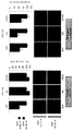
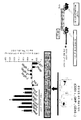
도 1a는 항-VEGF mAb로 치료후 72시간, 7일 및 14일의 뮤린 췌장 신경내분비 종양 (PNET) 세포의 미세-혈관 밀도 (MVD) 및 증식 지수 결정의 조직학적 및 그래픽 분석을 보여준다. 항-VEGF 치료 후 다양한 시간의 MECA-32 염색을 통한 종양 혈관 밀도 (좌측) 및 Ki67을 통한 증식 지수 (우측)의 조직학적 분석으로부터의 대표적인 이미지 (20X 배율). 각각의 경우에 4-6개의 종양으로부터의 정량화를 평균 +/- SEM으로서 하기 막대 그래프에 나타내었다. *P<0.05, NS = 유의하지 않음.
도 1b는 RIP-TβAg 모델에서의 종양 부담에 대한 항-VEGF 효과의 동역학을 보여주는 그래프이다. 연구에서 21일에, 항-VEGF-치료된 마우스 (적색 막대)에서의 종양 부담은 대조군-치료된 마우스 (흑색 막대)보다 유의하게 낮았지만, 14일에는 그렇지 않았다. *= p<0.05 (t-검정), n=5-8마리의 마우스/군/시점.
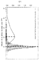
도 1c는 VEGF 길항제로의 치료에 대한 반응으로서의 VDV 유전자 발현 수준의 변화를 보여주는 그래프이다. 유전자의 발현 수준 (적색 선으로 나타냄)은 모든 유전자 (회색 히스토그램)에 대해 유의하게 감소하였다. 적색 파선은 이들 선택된 유전자에 대한 평균 변화를 나타낸다. 흑색 파선은 나머지 유전자에 대한 평균 배수 변화를 나타낸다.
도 1d는 qPCR에 의해 평가된 VEGF 길항제로의 치료에 대한 반응으로서의 유전자의 하위군에서의 유전자 발현 수준의 변화를 보여주는 그래프이다. 막대는 3개의 독립적 생물학적 복제물로부터의 평균 발현을 나타낸다. 오차 막대 = log2 표준 편차.
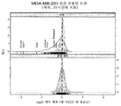
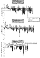
도 2a는 확립된 피하 유방 암종 종양 모델 (MDA-MB-231)에서의 VEGF 차단에 대한 VDV 유전자 반응의 변화를 보여주는 그래프 세트이다. 항-VEGF 또는 대조군 치료 24시간 후에 종양 샘플을 수집하였다. VDV 서명에서의 유전자 (적색 선)는 기질 (상부 그래프, 마우스 칩, p<0.0001)에서 모든 유전자 (회색 히스토그램으로 나타냄)에 대해 유의하게 감소하였지만, 종양 세포 (하부 그래프, 인간 칩, 유의차 없음)에서는 그렇지 않았다. 마이크로어레이 밀도 플롯에서 개별 proxVDV 전사체 배수-변화를 흑색 문자로 주석을 달았다. 각각의 치료 코호트에 대해 n = 5-10의 예를 들었다.
도 2b는 동소 (두개내) U87 교모세포종 모델에서의 VEGF 차단에 대한 VDV 유전자 반응의 변화를 보여주는 그래프 세트이다. 항-VEGF 또는 대조군 치료 13-42일 후에 종양 샘플을 수집하였다. VDV 서명에서의 유전자 (적색 선)는 기질 (상부 그래프, 마우스 칩, p<0.0105)에서 모든 유전자 (회색 히스토그램으로 나타냄)에 대해 유의하게 감소하였지만, 종양 세포 (하부 그래프, 인간 칩, 유의차 없음)에서는 그렇지 않았다. 마이크로어레이 밀도 플롯에서 개별 proxVDV 전사체 배수-변화를 흑색 문자로 주석을 달았다. 각각의 치료 코호트에 대해 n = 5-10의 예를 들었다.
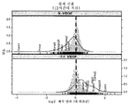
도 2c는 피부 상처에 대한 국소 항-VEGF 도포시 하향조절 (상부 그래프, p=0.0125)이 관찰되었고, 재조합 VEGF를 12시간 동안 도포하였을 때 정반대의 상향조절 (p<0.0001)이 관찰되었음을 보여주는 그래프 세트이다. 개별 proxVDV 전사체 배수-변화를 흑색 문자로 주석을 달았다.
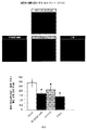
도 2d는 VEGF 신호전달이 VDV 유전자 발현을 유도하였음을 보여주는 조직학적 데이터 및 그래프를 나타낸다. MDA-MB-231 모델에서 48시간 후에 VDV 유전자 서명의 항-VEGF 하향조절 (우측 상부 그래프, p<0.0001)과는 대조적으로, 항-Dll4 치료는 대부분의 VDV 유전자의 상향조절 (우측 하부 그래프, p<0.0001)을 유발하였고, 이는 대조군 치료와 비교하여 CD31/PECAM에 대한 면역형광 염색에 의한 명백한 과다혈관화 (좌측)와 일치하였다. 개별 proxVDV 전사체 배수-변화를 흑색 문자로 주석을 달았다. 각각의 치료 코호트에 대해 n = 5-10의 예를 들었다.
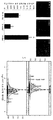
도 3은 시험관내에서 대부분의 proxVDV 유전자가 rVEGF에 의해 분명하게 상향조절되지 않았음을 보여주는 열 지도이다. HUVEC의 rVEGF 자극의 유전자 발현 분석. H (시간)는 rVEGF 자극의 길이를 나타낸다. 여기에 나타낸 열 지도는 선택된 VDV 프로브에 대한 마이크로어레이 발현 분석의 결과를 강조한다. 암청색은 최대의 상대적 하향조절을 나타내고, 암적색은 최대 정도의 전사체 상향조절을 나타낸다. 시험관내에서 ProxVDV 유전자는 rVEGF에 의해 현저하게 조절되지 않았다. 그러나, 아직 특성화되지 않은 VDV 유전자의 작은 군 (EHD3, PCHD17 및 THBD)은 HUVEC의 rVEGF 자극시 강하게 상향조절되는 것으로 보인다. 각각의 시점에 대한 발현 데이터는 3개의 독립적인 복제물로부터의 것이다.
도 4a는 proxVDV 유전자 ESM1이 종양-연관 혈관계에서 특이적으로 발현되는 생체내 VEGF 표적임을 보여주는 계내 혼성화 (ISH) 이미지이다. 상부 사진 (좌측 및 우측): 센스 올리고를 사용한 ISH 음성 대조군은 유의하지 않은 백그라운드 (비-특이적 염색)를 보여준다. 하부 사진은 항-VEGF 또는 대조군 치료된 동물로부터의 HM7 종양 절편에서의 (안티센스 올리고를 사용한 ISH에 의한) ESM1 mRNA 발현을 보여준다. 흑색 화살표는 대조군 치료된 종양 슬라이드에서의 강한 ESM1 mRNA 혈관 발현 (갈색 염색)의 여러 영역을 나타낸다 (하부 사진 슬라이드 좌측). 대조적으로, 항-VEGF 치료된 동물로부터의 종양 슬라이드에서는 ESM1이 거의 검출가능하지 않았다 (하부 사진 우측). 모든 슬라이드를 또한 헤마톡실린 에오신 (H&E)으로 대조 염색하였다.
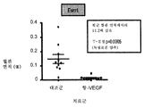
도 4b는 대조군 및 항-VEGF-치료된 동물로부터의 종양 슬라이드에서의 ESM1 (ISH) 염색의 정량화를 보여주는 그래프이다. n=10.
도 4c는 대조군 및 항-VEGF-치료된 동물로부터의 종양 슬라이드에서의 MECA32 (PLVAP) 염색의 정량화를 보여주는 그래프이다. n=10.
도 4d는 MDA-MB-231 종양에서의 VEGF 경로 억제제의 생체내 활성에 대한 증거를 보여주는 조직학적 이미지 세트 및 상응하는 정량적 그래프이다. 항-VEGF mAb, 수니티닙 또는 악시티닙 생체내 치료는 치료후 72시간에 종양에서 MVD를 효율적으로 감소시켰다. MDA-MB-231 종양을 보유하는 동물을 지시된 바와 같은 물질 및 방법으로 72시간 동안 치료한 다음, 조직학적 및 유전자 발현 분석을 위해 종양을 수집하였다. 상부 이미지: MECA-32 (PLVAP) 및 CD31 염색 (적색)을 통한 종양 혈관 밀도. 핵은 DAPI (청색)로 대조염색하였다. 이미지는 20X 배율로 획득하였다. 하부 그래프는 각각의 치료군에서의 8개의 종양으로부터의 정량화 (평균 +/- SEM으로서의 것)를 보여준다. *P<0.05.
도 4e는 생체내 VEGF 차단 또는 VEGFR-2 하류 신호전달 억제가 proxVDV 유전자의 지속적 하향조절을 유도하였음을 보여주는 그래프 세트이다. VEGF 및 VEGFR-2 억제제 (수니티닙 및 악시티닙)로의 치료 8시간 후 (하부 패널), 16시간 후 (중앙 패널) 또는 72시간 후 (상부 패널)에 수집한 400 mm3 MDA-MB-231 이종이식 종양에서의 유전자 발현의 qRT-PCR 분석. 값은 대조군 치료 평균 유전자 발현과 비교시의 VEGF/VEGFR-2 억제제에 의해 유도된 상대적 유전자 발현의 평균 log2 배수 변화를 나타낸다. 비-혈관 마커, 예컨대 E-cadh 및 CD45는 이들 억제제에 반응하여 유의하게 변화하지 않았다. 하부 패널 (치료후 8시간)은 악시티닙 및 수니티닙이 이 특정한 시점에 명백한 활성을 갖지 않았기 때문에 단지 항-VEGF 치료만을 보여준다. 유전자 발현 데이터는 각각의 치료에 대한 8개의 생물학적 복제물의 평균을 나타낸다. 오차 막대는 표준 편차를 나타낸다.
도 5a는 다중 VEGF 경로 억제제에 의한 지속적 proxVDV 하향조절을 보여주는 그래프이다. VEGF 및 VEGFR-2 억제제 (수니티닙 및 악시티닙)로의 치료 8, 16 또는 72시간 후에 수집한 MDA-MB-231 이종이식 종양에서의 유전자 발현의 분석. 값은 대조군 치료와 비교시의 VEGF/VEGFR-2 억제제에 의해 유도된 상대적 유전자 발현의 log2 배수 변화의 평균을 나타낸다. 유전자 발현 데이터는 각각의 치료에 대한 8개의 생물학적 복제물의 log2 평균을 나타낸다. 오차 막대는 표준 편차를 나타낸다.
도 5b는 돼지풀 또는 항-VEGF mAb로 치료된 MDA-MB-231 이종이식 종양으로부터 분류된 내피 세포에서의 qRT-PCR에 의한 proxVDV 유전자 발현의 정량화를 보여주는 그래프이다. 값은 3개의 복제물의 log2 배수 변화의 평균을 나타낸다. 오차 막대는 표준 편차를 나타낸다.
도 6은 새로이 FACS 단리된 종양-연관 내피 세포 (TAEC) 대 GFP-MDA-MB-231 종양 세포에서의 VDV 전사체의 생체외 농축 발현 분석에 의한 TAEC에서의 VDV 마커의 농축을 보여주는 그래프이다. TAEC 대 종양 세포에서의 상대적 유전자 발현을 선택된 유전자의 qRT-PCR에 의해 측정하였다. 시험된 모든 VDV 유전자는 TAEC에서 고도로 농축되었다 (25-200배). 대조적으로, Zeb1 mRNA 발현 (상피 마커)은 TAEC에서 감소되고, 종양 세포에서 농축되었다. 값은 TAEC를 종양 세포와 비교하였을 때의 상대적 log2 배수 유전자 농축의 평균을 나타낸다. TAEC 세포는 CD31 양성, CD45 음성 및 GFP 음성 세포로서 분류하였다. 종양 세포는 GFP 양성에 의해 분류하였다. 유전자 발현 데이터는 각각의 FACS 분류 실험에 대한 풀링된 6개의 종양의 평균을 나타낸다. qRTPCR은 3벌로 실행하였다. 오차 막대는 표준 편차를 나타낸다.
도 7은 19명의 염증성 유방암 환자로부터의 생검 샘플에서의 유전자 발현의 변화 (치료후 대 치료전)를 보여주는 그래프이다. VDV 서명에서의 유전자 (적색 선)는 모든 유전자 (회색 히스토그램)에 대해 유의하게 감소되었다 (p=0.0275).
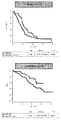
도 8a는 NO16966 시험에서 입수가능한 치료전 mRNA를 갖는 103명의 결장직장암 환자의 무진행 (상부) 및 전체 생존 (하부)을 보여주는 그래프 세트이다.
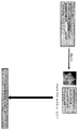
도 8b는 혈관신생 플루이딤(Fluidigm) qRT-PCR 칩을 사용하여 22개의 VDV 유전자의 유전자 발현을 정량화하기 위한 실험 개관의 개략도이다.
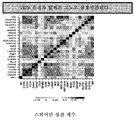
도 8c는 결장암 샘플에서의 22개의 VDV 유전자의 발현 수준의 상관관계를 보여주는 그래프이다.
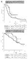
도 8d는 22-유전자 VDV 서명이 진행성 결장직장암을 앓고 있는 환자에 대한 베바시주맙 (bev) 치료의 효과를 계층화한다는 것을 보여주는 그래프 세트이다. XELOX (흑색) 또는 XELOX+베바시주맙으로 치료된 "VDV-고" (실선) 대 "VDV-저" 환자 (파선)의 무진행 (상부) 또는 전체 생존 (하부)을 나타내었다 (문헌 [Mullen et al. Cell. 147(3): 565-576, 2011]). 발현 수준에 의해 계층화된 PFS (상부) VDV 유전자 세트 효과 (상호작용 p=0.036), 및 발현 수준에 의해 계층화된 OS (하부) VDV 유전자 세트 효과 (상호작용 p=0.37).
도 9a는 보존용 임상 물질로부터 정보를 얻는데 이용된 전체 VDV 서명 "VDV" (x-축) 및 22-유전자 대표 하위세트 "VDV-22" (y-축) 사이의 일치성을 검증하는 그래프 세트이다. 전이성 1차 시험 세팅 (NO16966과 유사함)에 대해 자격을 얻을 환자로부터의 보존용 샘플을 사용하여 유병률 샘플 세트를 나타내었다. 보존용 샘플을 일루미나(Illumina) DASL 비드 어레이 상에서 전체 게놈 RNA 발현에 대해 평가하였다; NSCLC = 비소세포 폐 암종, BR = 유방암, CRC = 결장직장 암종.
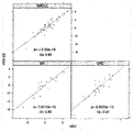
도 9b는 VEGF-A 발현 수준에 의한 NO16966 환자의 계층화를 보여주는 그래프 세트이다 (PFS (상부), 상호작용 p=0.76 및 OS (하부), 상호작용 p=0.33).
도 9c는 CD31 발현 수준에 의한 NO16966 환자의 계층화를 보여주는 그래프 세트이다 (PFS (상부) 상호작용 p=0.15 및 OS (하부), 상호작용 p=0.99).FIG. 1A shows histological and graphical analysis of microvascular density (MVD) and proliferation index determinations of murine pancreatic neuroendocrine tumors (PNET) cells at 72 hours, 7 days, and 14 days after treatment with anti-VEGF mAb. Representative images (20X magnification) from histological analysis of tumor vascular density (left) through MECA-32 staining at various times after anti-VEGF treatment and growth index (right) through Ki67. Quantification from 4-6 tumors in each case is shown in the following bar graph as mean +/- SEM. * P <0.05, NS = not significant.
Figure 1B is a graph showing the kinetics of the anti-VEGF effect on tumor burden in the RIP-T? Ag model. On day 21 of the study, tumor burden in anti-VEGF-treated mice (red bars) was significantly lower than in control-treated mice (black bars), but not on day 14. * = p < 0.05 (t-test), n = 5-8 mice / group / time.
Figure 1C is a graph showing the change in VDV gene expression level as a response to treatment with a VEGF antagonist. The level of expression of the gene (indicated by the red line) was significantly reduced for all genes (gray histogram). The red dashed line represents the mean change for these selected genes. The black dashed line represents the mean multiple change for the remaining genes.
Figure 1D is a graph showing changes in gene expression levels in subgroups of genes as responses to treatment with a VEGF antagonist as assessed by qPCR. The bars represent the mean expression from three independent biological replicates. Error bars = log 2 standard deviation.
Figure 2a is a set of graphs showing changes in VDV gene response to VEGF blockade in established subcutaneous breast carcinoma tumor model (MDA-MB-231). Tumor samples were collected 24 hours after anti-VEGF or control treatment. The gene (red line) in the VDV signature was significantly reduced for all genes (represented by gray histogram) on the substrate (top graph, mouse chip, p <0.0001), but the tumor cells (bottom graph, human chip, ). In microarray density plots, the individual proximal transcript multiplication-changes were annotated with black letters. An example of n = 5-10 was given for each treatment cohort.
Figure 2b is a set of graphs showing changes in the VDV gene response to VEGF blockade in a homozygous (intracranial) U87 glioblastoma model. Tumor samples were collected 13-42 days after anti-VEGF or control treatment. The gene (red line) in the VDV signature was significantly reduced for all genes (represented by the gray histogram) on the substrate (top graph, mouse chip, p <0.0105), but the tumor cells ). In microarray density plots, the individual proximal transcript multiplication-changes were annotated with black letters. An example of n = 5-10 was given for each treatment cohort.
Figure 2c shows a graph showing that the downward regulation (top graph, p = 0.0125) was observed when applying topical anti-VEGF to skin wounds and the opposite up regulation (p < 0.0001) was observed when the recombinant VEGF was applied for 12 hours Set. Individual proxVDV transcripts were annotated with black characters for drainage-change.
Figure 2d shows histological data and graphs showing that VEGF signaling induced VDV gene expression. In contrast to the anti-VEGF downregulation of the VDV gene signature (right top graph, p < 0.0001) after 48 hours in the MDA-MB-231 model, anti-Dll4 therapy increased uptake of most VDV genes p <0.0001), consistent with apparent overvascularization (left) by immunofluorescence staining for CD31 / PECAM compared to control treatment. Individual proxVDV transcripts were annotated with black characters for drainage-change. An example of n = 5-10 was given for each treatment cohort.
Figure 3 is a thermal map showing that most proxVDV genes in vitro have not been upregulated clearly by rVEGF. Gene expression analysis of rVEGF stimulation of HUVEC. H (time) represents the length of the rVEGF stimulus. The thermal map shown here highlights the results of microarray expression analysis on selected VDV probes. Dark blue indicates maximum relative down regulation, while dark red indicates maximum upregulation of the transcript. In vitro, the ProxVDV gene was not significantly regulated by rVEGF. However, a small group of the yet uncharacterized VDV gene (EHD3, PCHD17 and THBD) appears to be strongly upregulated upon rVEGF stimulation of HUVEC. Expression data for each time point is from three independent replicates.
FIG. 4A is an in-vitro hybridization (ISH) image showing that the proxVDV gene ESM1 is an in vivo VEGF target specifically expressed in the tumor-associated vasculature. Upper photos (left and right): ISH negative control with sense oligo shows a nonsignificant background (non-specific staining). The lower photograph shows ESM1 mRNA expression (by ISH with antisense oligo) in HM7 tumor sections from anti-VEGF or control treated animals. The black arrows indicate various regions of strong ESM1 mRNA vascular expression (brown staining) on the control treated tumor slides (bottom slide, slide left). In contrast, ESM1 was almost undetectable on tumor slides from anti-VEGF treated animals (bottom right). All slides were also counterstained with hematoxylin eosin (H & E).
Figure 4b is a graph showing the quantification of ESMl (ISH) staining on tumor slides from control and anti-VEGF-treated animals. n = 10.
4C is a graph showing quantification of MECA32 (PLVAP) staining on tumor slides from control and anti-VEGF-treated animals. n = 10.
Figure 4d is a histological image set and corresponding quantitative graph showing evidence for the in vivo activity of VEGF pathway inhibitors in MDA-MB-231 tumors. In vivo treatment with anti-VEGF mAb, sunitinib, or acitcinium resulted in an effective reduction of MVD in tumors at 72 hours after treatment. Animals bearing MDA-MB-231 tumors were treated for 72 hours with materials and methods as indicated, and tumors were collected for histological and gene expression analysis. Top image: Tumor vascular density through MECA-32 (PLVAP) and CD31 staining (red). The nuclei were contrasted with DAPI (blue). Images were acquired at 20X magnification. The lower graph shows quantification (as mean +/- SEM) from 8 tumors in each treatment group. * P < 0.05.
Figure 4E is a set of graphs showing that in vivo VEGF blockade or VEGFR-2 downstream signal transduction inhibition led to sustained downregulation of the proxVDV gene. VEGF and VEGFR-2 inhibitor (sunitinib and evil nip City) after
FIG. 5A is a graph showing continued proximal downregulation of proxVDV by multiple VEGF pathway inhibitors. FIG. Analysis of gene expression in MDA-MB-231 xenograft tumors collected after 8, 16 or 72 hours of treatment with VEGF and VEGFR-2 inhibitors (sutinitinib and acctinib). Value represents the average of the log 2 -fold change in relative gene expression induced by the VEGF / VEGFR-2 inhibitor as compared to the control treatment. Gene expression data represents the log 2 mean of eight biological replicates for each treatment. The error bars represent the standard deviation.
Figure 5b is a graph showing the quantification of proxVDV gene expression by qRT-PCR in endothelial cells classified from MDA-MB-231 xenograft tumors treated with porcine pool or anti-VEGF mAb. The value represents the average of the log 2 multiples of the three replicates. The error bars represent the standard deviation.
FIG. 6 is a graph showing the concentration of VDV markers in TAEC by in vitro enrichment expression analysis of VDV transcripts in newly isolated FACS isolated tumor-associated endothelial cells (TAEC) versus GFP-MDA-MB-231 tumor cells. Relative gene expression in TAEC large tumor cells was measured by qRT-PCR of selected genes. All VDV genes tested were highly enriched in TAEC (25-200-fold). In contrast, Zeb1 mRNA expression (epithelial markers) was reduced in TAEC and enriched in tumor cells. The value represents the mean of the relative log 2 -digested gene concentration when TAEC is compared to tumor cells. TAEC cells were classified as CD31 positive, CD45 negative and GFP negative cells. Tumor cells were classified by GFP positive. Gene expression data represent the mean of six pooled tumors for each FACS classification experiment. qRTPCR was performed in triplicate. The error bars represent the standard deviation.
7 is a graph showing changes in gene expression (before treatment vs. after treatment) in a biopsy sample from 19 patients with inflammatory breast cancer. The gene (red line) in the VDV signature was significantly reduced for all genes (gray histogram) (p = 0.0275).
Figure 8a is a set of graphs showing the progression (upper) and overall survival (lower) of 103 colorectal cancer patients with pre-treatment mRNA available in the NO16966 test.
Figure 8b is a schematic of an experimental overview for quantifying the gene expression of 22 VDV genes using an angiogenic Fluidigm qRT-PCR chip.
8C is a graph showing the correlation of expression levels of 22 VDV genes in colon cancer samples.
Figure 8d is a set of graphs showing that the 22-gene VDV signature layered the effect of bevacizumab (bev) treatment on patients with advanced colorectal cancer. (Upper) or overall survival (lower) of "VDV-high" (solid line) versus "VDV-low" patients (dashed) treated with XELOX (black) or XELOX + bevacizumab (Mullen et al., Cell 147 (3): 565-576, 2011). (Top) VDV gene set effect (interaction p = 0.036) stratified by expression level, and OS (bottom) VDV gene set effect stratified by expression level (interaction p = 0.37).
Figure 9a is a graph set that verifies consistency between the entire VDV signature "VDV" (x-axis) and the 22-gene representative subset "VDV- 22" to be. A prevalence sample set was presented using retention samples from patients eligible for metastatic primary test settings (similar to NO16966). Retention samples were evaluated for total genomic RNA expression on an Illumina DASL bead array; NSCLC = non-small cell lung carcinoma, BR = breast cancer, CRC = colorectal carcinoma.
Figure 9b is a set of graphs (PFS (top), interaction p = 0.76 and OS (bottom), interaction p = 0.33) showing the stratification of NO16966 patients by VEGF-A expression levels.
Figure 9c is a set of graphs (PFS (top) interaction p = 0.15 and OS (bottom), interaction p = 0.99) showing the stratification of NO16966 patients by CD31 expression levels.
I. 서론I. Introduction
본 발명은 VEGF 길항제, 예를 들어 항-VEGF 항체로의 치료에 대해 감수성이거나 또는 반응성인 환자를 모니터링 및/또는 확인하기 위한 방법 및 조성물을 제공한다. 본 발명은 VEGF 길항제 (예컨대, 항-VEGF 항체)로의 치료 전에 표 1 또는 2에 제시된 적어도 1, 2, 3, 4, 5, 6, 7, 8, 9, 10, 11, 12, 13, 14, 15, 16, 17, 18, 19, 20, 21, 22, 23개 또는 그 초과의 유전자(들)의 발현 수준을 결정하는 것이 VEGF 길항제, 예를 들어 항-VEGF 항체에 대해 감수성이거나 또는 반응성인 환자를 확인하는데 유용하다는 발견에 기반한다. 임의로, 이어서 VEGF 길항제 요법이 환자에 대해 선택될 수 있고, 추가로, VEGF 길항제 요법이 임의로 환자에게 투여될 수 있다.The present invention provides methods and compositions for monitoring and / or identifying patients that are susceptible or reactive to treatment with VEGF antagonists, e. G., Anti-VEGF antibodies. The present invention is also directed to a pharmaceutical composition comprising at least 1, 2, 3, 4, 5, 6, 7, 8, 9, 10, 11, 12, 13, 14 , 15, 16, 17, 18, 19, 20, 21, 22, 23 or more of the gene (s) is / are sensitive to a VEGF antagonist, such as an anti-VEGF antibody, It is based on the finding that it is useful for identifying patients with Optionally, VEGF antagonist therapy can then be selected for the patient, and further, VEGF antagonist therapy can optionally be administered to the patient.
II. 정의II. Justice
용어 "바이오마커" 및 "마커"는 DNA, RNA, 단백질, 탄수화물 또는 당지질-기반 분자 마커를 지칭하기 위해 본원에서 상호교환적으로 사용되며, 대상체 또는 환자의 샘플에서의 그의 발현 또는 존재는 표준 방법 (또는 본원에 개시된 방법)에 의해 검출될 수 있고, VEGF 길항제에 대한 포유동물 대상체의 반응성 또는 감수성을 모니터링하는데 유용하다. 이러한 바이오마커는 표 1 및 2에 제시된 유전자를 포함하나, 이에 제한되지는 않는다. 이러한 바이오마커의 발현은 VEGF 길항제에 대해 감수성이거나 또는 반응성인 환자로부터 수득한 샘플에서 참조 수준 (예를 들어, VEGF 길항제에 대한 반응성에 대해 시험되는 환자의 군/집단으로부터의 샘플에서의 바이오마커의 중앙 발현 수준; 사전에 개체로부터 미리 수득한 샘플에서의 수준; 또는 원발성 종양 세팅에서 VEGF 길항제 (예컨대, 항-VEGF 항체)로의 선행 치료를 받았고, 현재 전이를 경험하고 있을 수 있는 환자로부터의 샘플에서의 수준 포함)보다 높거나 또는 낮은 것으로 결정될 수 있다. 적어도 하나의 유전자, 예컨대 표 1 및 2에 제시된 것들의 참조 발현 수준보다 높거나 낮은 발현 수준을 갖는 개체는 VEGF 길항제로의 치료에 반응할 가능성이 있는 대상체/환자로 확인될 수 있다. 예를 들어, 참조 수준 (예컨대, 중앙 수준, 상기 언급됨)에 대해 최대 50%, 45%, 40%, 35%, 30%, 25%, 20%, 15%, 10% 또는 5% 차이나는 (즉, 더 높거나 낮은) 유전자 발현 수준을 나타내는 이러한 대상체/환자는 VEGF 길항제, 예컨대 항-VEGF 항체로의 치료에 반응할 가능성이 있는 대상체/환자로 확인될 수 있다.The terms "biomarker" and "marker" are used interchangeably herein to refer to DNA, RNA, protein, carbohydrate or glycolipid-based molecular markers and their expression or presence in a sample of a subject or patient, (Or methods disclosed herein) and are useful for monitoring the reactivity or susceptibility of a mammalian subject to a VEGF antagonist. Such biomarkers include, but are not limited to, the genes shown in Tables 1 and 2. The expression of such a biomarker may be determined by comparing the biomarker in a sample from a group of patients tested for reactivity against a VEGF antagonist at a reference level in a sample obtained from a patient susceptible or reactive to the VEGF antagonist A sample from a patient who has undergone a prior treatment with a VEGF antagonist (e.g., an anti-VEGF antibody) at a primary tumor setting and is currently experiencing a metastasis ≪ / RTI > level). Individuals having an expression level that is higher or lower than the reference expression level of at least one gene, such as those shown in Tables 1 and 2, may be identified as a subject / patient likely to respond to treatment with a VEGF antagonist. For example, a maximum of 50%, 45%, 40%, 35%, 30%, 25%, 20%, 15%, 10% or 5% Such a subject / patient exhibiting a level of gene expression (i. E. Higher or lower) may be identified as a VEGF antagonist, e. G. A subject / patient likely to respond to treatment with an anti-VEGF antibody.
용어 "샘플" 및 "생물학적 샘플"은 체액, 신체 조직 (예를 들어, 종양 조직), 세포 또는 다른 공급원을 비롯한 개체로부터 수득한 임의의 생물학적 샘플을 지칭하기 위해 상호교환적으로 사용된다. 체액은 예를 들어 림프, 혈청, 신선한 전혈, 말초혈 단핵 세포, 동결된 전혈, 혈장 (예를 들어, 신선하거나 동결된 것), 소변, 타액, 정액, 활액 및 척수액이다. 샘플은 또한 유방 조직, 신장 조직, 결장 조직, 뇌 조직, 근육 조직, 활막 조직, 피부, 모낭, 골수 및 종양 조직을 포함한다. 포유동물로부터 조직 생검 및 체액을 얻기 위한 방법은 당업계에 널리 공지되어 있다.The terms "sample" and "biological sample" are used interchangeably to refer to any biological sample obtained from an individual, including body fluids, body tissues (eg, tumor tissue), cells or other sources. Fluids are, for example, lymph, serum, fresh whole blood, peripheral blood mononuclear cells, frozen whole blood, plasma (fresh or frozen, for example), urine, saliva, semen, synovial fluid and spinal fluid. The sample also includes breast tissue, kidney tissue, colon tissue, brain tissue, muscle tissue, synovial tissue, skin, hair follicles, bone marrow, and tumor tissue. Methods for obtaining tissue biopsies and body fluids from mammals are well known in the art.
VEGF 길항제로의 치료에 대한 환자의 "유효 반응" 또는 그에 대한 환자의 "반응성" 또는 "감수성"은 VEGF 길항제, 예컨대 항-VEGF 항체로의 치료로부터 또는 그의 결과로서 혈관신생 장애의 위험이 있거나 혈관신생 장애를 앓고 있는 환자에게 부여된 임상적 또는 치료 이익을 나타낸다. 이러한 이익은 길항제 치료로부터 또는 그러한 치료의 결과로서의 세포적 또는 생물학적 반응, 완전 반응, 부분 반응, 안정적 질환 (진행 또는 재발이 없음), 또는 환자의 후기 재발이 있는 반응을 포함한다. 예를 들어, 유효 반응은 표 1 또는 2에 제시된 하나 이상의 바이오마커를 발현하지 않는 환자와 비교하여 상기 하나 이상의 바이오마커를 발현하는 것으로 진단된 환자에서 감소된 종양 크기 또는 무진행 생존일 수 있다. 유전자 바이오마커(들)의 발현은 이러한 유효 반응을 효과적으로 예측하거나 또는 높은 감수성으로 예측한다.Or " responsiveness " or "susceptibility " of a patient to treatment with a VEGF antagonist is the risk or risk of an angiogenic disorder as a result of or as a result of treatment with a VEGF antagonist such as an anti- Indicates the clinical or therapeutic benefit imparted to a patient suffering from a neoplastic disorder. Such benefits include reactions with or without a cellular or biological response, complete response, partial response, stable disease (no progression or recurrence), or late recurrence of a patient, from antagonist therapy or as a result of such treatment. For example, an efficacy response may be reduced tumor size or progression-free survival in a patient diagnosed with expressing said one or more biomarkers as compared to a patient not expressing one or more of the biomarkers listed in Tables 1 or 2. Expression of the gene biomarker (s) effectively predicts this efficacy or predicts high susceptibility.
본원에 사용된 "길항제"는 그가 결합하는 분자의 생물학적 활성을 억제하거나 또는 감소시키는 화합물 또는 작용제를 지칭한다. 길항제는 VEGF에 결합하고, 임의로는 또 다른 분자에 접합되거나 융합된 항체, 합성 또는 천연 서열 펩티드, 이뮤노어드헤신 및 소분자 길항제를 포함한다. "차단" 항체 또는 "길항제" 항체는 그가 결합하는 항원의 생물학적 활성을 억제하거나 또는 감소시키는 항체이다.As used herein, "antagonist" refers to a compound or agent that inhibits or reduces the biological activity of the molecule to which it binds. Antagonists include antibodies, synthetic or native sequence peptides, immunoadhesins, and small molecule antagonists that bind to VEGF, optionally conjugated or fused to another molecule. An "blocking" antibody or "antagonist" antibody is an antibody that inhibits or reduces the biological activity of the antigen to which it binds.
본원에 사용된 "효능제 항체"는 관심 폴리펩티드의 기능적 활성 중 하나 이상을 부분적으로 또는 전부 모방하는 항체이다.As used herein, "agonist antibody" is an antibody that partially or fully mimics one or more of the functional activities of a polypeptide of interest.
본원에서 용어 "항체"는 가장 넓은 의미로 사용되고, 구체적으로 모노클로날 항체, 폴리클로날 항체, 적어도 2개의 무손상 항체로 형성된 다중특이적 항체 (예를 들어, 이중특이적 항체) 및 원하는 생물학적 활성을 나타내는 한 항체 단편을 포함한다.The term "antibody" is used herein in its broadest sense and specifically includes monoclonal antibodies, polyclonal antibodies, multispecific antibodies (e. G., Bispecific antibodies) formed with at least two intact antibodies, Lt; RTI ID = 0.0 > activity. ≪ / RTI >
"단리된" 항체는 그의 자연 환경의 성분으로부터 확인되고 분리 및/또는 회수된 것이다. 이것의 천연 환경의 오염물 성분은 항체의 연구, 진단 또는 치료 용도를 방해하는 물질이고, 이는 효소, 호르몬 및 다른 단백질성 또는 비단백질성 용질을 포함할 수 있다. 일부 실시양태에서, 항체는 (1) 예를 들어 로우리(Lowry) 방법으로 측정시에 95 중량% 초과의 항체로, 일부 실시양태에서는 99 중량% 초과로, (2) 예를 들어 스피닝 컵 서열분석기를 이용하여 N-말단 또는 내부 아미노산 서열의 적어도 15개의 잔기를 얻기에 충분한 정도로, 또는 (3) 예를 들어 쿠마시 블루 또는 은 염색을 이용하여 환원 또는 비환원 조건 하에 SDS-PAGE에 의해 균질한 것으로 나타날 정도로 정제하였다. 단리된 항체는 재조합 세포 내의 계내 항체를 포함하는데, 이는 항체의 자연 환경의 적어도 하나의 성분이 존재하지 않을 것이기 때문이다. 그러나, 통상적으로, 단리된 항체는 적어도 1회의 정제 단계에 의해 제조될 것이다.An "isolated" antibody is one that has been identified and separated and / or recovered from a component of its natural environment. The contaminant component of its natural environment is a substance that interferes with the research, diagnostic or therapeutic uses of antibodies, which may include enzymes, hormones and other proteinaceous or nonproteinaceous solutes. In some embodiments, the antibody is (1) an antibody that is greater than 95% by weight, in some embodiments greater than 99% by weight, as determined by, for example, the Lowry method, (2) Or (3) to a degree sufficient to obtain at least 15 residues of the N-terminal or internal amino acid sequence homologous to SDS-PAGE under reducing or nonreducing conditions, for example using coumarin blue or silver stain . The isolated antibody comprises an in-vitro antibody in the recombinant cell, since at least one component of the natural environment of the antibody will not be present. Typically, however, the isolated antibody will be produced by at least one purification step.
"천연 항체"는 통상적으로 2개의 동일한 경쇄 (L) 및 2개의 동일한 중쇄 (H)로 구성된 약 150,000 달톤의 이종사량체 당단백질이다. 각각의 경쇄는 1개의 공유 디술피드 결합에 의해 중쇄에 연결되지만, 디술피드 연결의 수는 상이한 이뮤노글로불린 이소형의 중쇄마다 달라진다. 또한, 각각의 중쇄 및 경쇄는 일정하게 이격된 쇄내 디술피드 가교를 갖는다. 각각의 중쇄는 한쪽 말단에 가변 도메인 (VH)을 갖고, 그 뒤에는 수많은 불변 도메인이 존재한다. 각각의 경쇄는 한쪽 말단에 가변 도메인 (VL)을, 다른쪽 말단에 불변 도메인을 가지며; 경쇄의 불변 도메인은 중쇄의 제1 불변 도메인과 정렬되고, 경쇄 가변 도메인은 중쇄의 가변 도메인과 정렬된다. 특정한 아미노산 잔기가 경쇄 가변 도메인과 중쇄 가변 도메인 사이의 계면을 형성하는 것으로 여겨진다."Natural antibody" is a heterotetrameric glycoprotein of about 150,000 daltons, usually composed of two identical light chains (L) and two identical heavy chains (H). Each light chain is linked to the heavy chain by one covalent disulfide bond, but the number of disulfide linkages is different for each heavy chain of different immunoglobulin isoforms. In addition, each heavy and light chain has a constant discrete intramolecular disulfide bridge. Each heavy chain has a variable domain (V H ) at one end, followed by a number of constant domains. Each light chain has a variable domain (V L ) at one end and a constant domain at the other end; The constant domain of the light chain is aligned with the first constant domain of the heavy chain and the light chain variable domain is aligned with the variable domain of the heavy chain. It is believed that certain amino acid residues form the interface between the light chain variable domain and the heavy chain variable domain.
항체의 "가변 영역" 또는 "가변 도메인"은 항체의 중쇄 또는 경쇄의 아미노-말단 도메인을 지칭한다. 중쇄의 가변 도메인은 "VH"로 지칭될 수 있다. 경쇄의 가변 도메인은 "VL"로 지칭될 수 있다. 이들 도메인은 일반적으로 항체의 가장 가변적인 부분이고, 항원-결합 부위를 함유한다."Variable domain" or "variable domain" of an antibody refers to the amino-terminal domain of the heavy or light chain of the antibody. The variable domain of the heavy chain may be referred to as "VH ". The variable domain of the light chain may be referred to as "VL ". These domains are generally the most variable part of the antibody and contain antigen-binding sites.
용어 "가변"은 가변 도메인의 특정 부분이 항체마다 서열에서 광범위하게 상이하며, 각각의 특정한 항체의 그의 특정한 항원에 대한 결합 및 특이성에 이용된다는 사실을 지칭한다. 그러나, 가변성이 항체의 가변 도메인 전반에 걸쳐 고르게 분포되어 있는 것은 아니다. 이것은 경쇄 가변 도메인과 중쇄 가변 도메인 둘 다에서 초가변 영역 (HVR)이라 불리는 3개의 절편에 집중되어 있다. 가변 도메인의 보다 고도로 보존된 부분은 프레임워크 영역 (FR)이라 불린다. 천연 중쇄 및 경쇄 각각의 가변 도메인은 주로 베타-시트 배위를 채택하여 3개의 HVR에 의해 연결되어 있는 4개의 FR 영역을 포함하며, 이는 베타-시트 구조를 연결하고, 일부 경우에는 상기 베타-시트 구조의 일부를 형성하는 루프를 형성한다. 각각의 쇄에서의 HVR들은 FR 영역에 의해 함께 근접하게 위치되어 있고, 다른 쇄로부터의 HVR과 함께 항체의 항원-결합 부위의 형성에 기여한다 (문헌 [Kabat et al., Sequences of Proteins of Immunological Interest, Fifth Edition, National Institute of Health, Bethesda, MD (1991)] 참조). 불변 도메인은 항체를 항원에 결합시키는데는 직접 관여하지 않지만, 항체 의존성 세포 독성에서의 항체 참여와 같은 다양한 이펙터 기능을 나타낸다.The term "variable" refers to the fact that certain portions of the variable domain differ extensively in sequence from antibody to antibody, and that each particular antibody is used for binding and specificity to its particular antigen. However, the variability is not evenly distributed across the variable domains of the antibody. It is concentrated on three fragments called hypervariable regions (HVR) in both the light chain variable domain and the heavy chain variable domain. The more highly conserved part of the variable domain is called the framework area (FR). The variable domains of each of the natural heavy and light chains mainly comprise four FR regions connected by three HVRs adopting a beta-sheet configuration, which connects the beta-sheet structure and in some cases the beta-sheet structure To form a loop that forms a part of the loop. The HVRs in each chain are located closely together by the FR region and contribute to the formation of the antigen-binding site of the antibody along with the HVR from the other chain (Kabat et al., Sequences of Proteins of Immunological Interest , Fifth Edition, National Institute of Health, Bethesda, MD (1991)). The constant domains are not directly involved in binding the antibody to the antigen, but exhibit various effector functions such as antibody participation in antibody-dependent cytotoxicity.
임의의 척추동물 종으로부터의 항체 (이뮤노글로불린)의 "경쇄"는 불변 도메인의 아미노산 서열에 기반하여, 카파 (κ) 및 람다 (λ)로 불리는 명백하게 상이한 2가지 유형 중의 하나로 지정될 수 있다.The "light chain" of an antibody (immunoglobulin) from any vertebrate species can be assigned to one of two distinct types, called kappa (kappa) and lambda (lambda), based on the amino acid sequence of the constant domain.
중쇄 불변 도메인의 아미노산 서열에 따라, 항체 (이뮤노글로불린)는 상이한 부류로 지정될 수 있다. 5종의 주요 부류의 이뮤노글로불린: IgA, IgD, IgE, IgG 및 IgM이 존재하고, 이들 중 몇몇은 하위부류 (이소형), 예를 들어 IgG1, IgG2, IgG3, IgG4, IgA1 및 IgA2로 추가로 분류될 수 있다. 상이한 부류의 이뮤노글로불린에 상응하는 중쇄 불변 도메인은 각각 α, δ, ε, γ 및 μ로 불린다. 상이한 부류의 이뮤노글로불린의 서브유닛 구조 및 3차원 배위는 널리 공지되어 있고, 예를 들어 문헌 [Abbas et al. Cellular and Mol. Immunology, 4th ed. (W. B. Saunders, Co., 2000)]에 개괄적으로 기재되어 있다. 항체는 항체와 하나 이상의 다른 단백질 또는 펩티드의 공유 또는 비-공유 회합에 의해 형성되는, 더 큰 융합 분자의 일부일 수 있다.Depending on the amino acid sequence of the heavy chain constant domain, the antibody (immunoglobulin) can be assigned to a different class. The immunoglobulin of the major classes of five kinds: IgA, IgD, IgE, IgG and IgM are present, some of which are a subclass (isotype), such as IgG 1, IgG 2, IgG 3, IgG 4, IgA 1 and IgA 2 may be sorted further. The heavy chain constant domains corresponding to different classes of immunoglobulins are called alpha, delta, epsilon, gamma and mu, respectively. Subunit structures and three-dimensional configurations of different classes of immunoglobulins are well known and are described, for example, in Abbas et al. Cellular and Mol. Immunology, 4th ed. (WB Saunders, Co., 2000). An antibody may be part of a larger fusion molecule, which is formed by covalent or non-covalent association of the antibody with one or more other proteins or peptides.
용어 "전장 항체", "무손상 항체" 및 "전체 항체"는, 아래에 정의된 항체 단편이 아닌, 실질적으로 무손상 형태인 항체를 지칭하기 위해 본원에서 상호교환적으로 사용된다. 이 용어는 특히 Fc 영역을 함유하는 중쇄를 갖는 항체를 지칭한다.The terms "full-length antibody "," intact antibody "and" whole antibody "are used interchangeably herein to refer to antibodies that are substantially non-damaging in nature, but not antibody fragments as defined below. This term specifically refers to an antibody having a heavy chain containing an Fc region.
본원의 목적상 "네이키드 항체"는 세포독성 모이어티 또는 방사성표지에 접합되지 않은 항체이다.For purposes herein, "naked antibody" is an antibody that is not conjugated to a cytotoxic moiety or radioactive label.
"항체 단편"은 무손상 항체의 일부, 바람직하게는 그의 항원-결합 영역을 포함한다. 항체 단편의 예는 Fab, Fab', F(ab')2 및 Fv 단편; 디아바디; 선형 항체; 단일-쇄 항체 분자; 및 항체 단편으로부터 형성된 다중특이적 항체를 포함한다.An "antibody fragment" comprises a portion of an intact antibody, preferably an antigen-binding region thereof. Examples of antibody fragments include Fab, Fab ', F (ab') 2 and Fv fragments; Diabody; Linear antibodies; Single-chain antibody molecules; And multispecific antibodies formed from antibody fragments.
항체를 파파인으로 소화시키면, "Fab" 단편으로 불리는, 각각 단일 항원-결합 부위를 갖는 2개의 동일한 항원-결합 단편 및 나머지 "Fc" 단편이 생성되며, 이러한 명칭은 그의 용이하게 결정화되는 능력을 반영한 것이다. 펩신 처리는 2개의 항원-결합 부위를 갖고 여전히 항원을 가교시킬 수 있는 F(ab')2 단편을 생성한다.Digestion of the antibody with papain produces two identical antigen-binding fragments, each with a single antigen-binding site, and the remaining "Fc" fragments, referred to as "Fab " fragments, will be. Pepsin treatment produces F (ab ') 2 fragments that have two antigen-binding sites and can still cross-link the antigen.
"Fv"는 완전한 항원-결합 부위를 함유하는 최소의 항체 단편이다. 한 실시양태에서, 2-쇄 Fv 종은 단단히 비-공유 회합된 1개의 중쇄 및 1개의 경쇄 가변 도메인 이량체로 이루어진다. 단일-쇄 Fv (scFv) 종에서는, 하나의 중쇄 및 하나의 경쇄 가변 도메인이 유연한 펩티드 링커에 의해 공유 연결되어 경쇄 및 중쇄가 2-쇄 Fv 종에서의 구조와 유사한 "이량체" 구조로 회합할 수 있다. 이러한 배위에서, 각 가변 도메인의 3개의 HVR은 상호작용하여 VH-VL 이량체 표면의 항원 결합 부위를 규정한다. 집합적으로, 6개의 HVR이 항체에 항원 결합 특이성을 부여한다. 그러나, 단일 가변 도메인 (또는 항원에 특이적인 3개의 HVR만을 포함하는 Fv의 절반)일지라도 전체 결합 부위보다 친화도가 낮긴 하지만 항원을 인식하고 결합하는 능력을 갖는다."Fv" is the smallest antibody fragment containing a complete antigen-binding site. In one embodiment, the two-chain Fv species consists of one heavy chain and one light chain variable domain dimer that are tightly non-covalently associated. In single-chain Fv (scFv) species, one heavy chain and one light chain variable domain are covalently linked by a flexible peptide linker such that the light and heavy chains are associated with a "dimer" structure similar to that in the two-chain Fv species . In this configuration, the three HVRs of each variable domain interact to define the antigen binding site of the VH-VL dimer surface. Collectively, six HVRs confer antigen binding specificity to the antibody. However, even a single variable domain (or half of an Fv comprising only three HVRs specific for an antigen) has a lower affinity than the entire binding site, but has the ability to recognize and bind antigen.
Fab 단편은 중쇄 및 경쇄 가변 도메인을 함유하고, 또한 경쇄의 불변 도메인 및 중쇄의 제1 불변 도메인 (CH1)을 함유한다. Fab' 단편은 항체-힌지 영역으로부터의 하나 이상의 시스테인을 포함하는 중쇄 CH1 도메인의 카르복시 말단에 수개의 잔기가 부가되었다는 점에서 Fab 단편과 상이하다. Fab'-SH는 불변 도메인의 시스테인 잔기(들)가 유리 티올 기를 보유하는 Fab'에 대한 본원에서의 명칭이다. F(ab')2 항체 단편은 본래 그들 사이에 힌지 시스테인을 갖는 Fab' 단편의 쌍으로서 생산된다. 항체 단편의 다른 화학적 커플링이 또한 공지되어 있다.Fab fragments contain heavy and light chain variable domains and also contain a constant domain of the light chain and a first constant domain (CHl) of the heavy chain. Fab fragments differ from Fab fragments in that several residues are added to the carboxy terminus of the heavy chain CH1 domain comprising at least one cysteine from the antibody-hinge region. Fab'-SH is the designation herein for Fab 'in which the cysteine residue (s) of the constant domain retain a free thiol group. The F (ab ') 2 antibody fragment is originally produced as a pair of Fab' fragments with a hinge cysteine therebetween. Other chemical couplings of antibody fragments are also known.
"단일-쇄 Fv" 또는 "scFv" 항체 단편은 단일 폴리펩티드 쇄에 존재하는 항체의 VH 및 VL 도메인을 포함한다. 일반적으로, scFv 폴리펩티드는 scFv가 항원 결합을 위해 목적하는 구조를 형성할 수 있도록 VH 도메인 및 VL 도메인 사이에 폴리펩티드 링커를 추가로 포함한다. scFv를 검토하기 위해서는, 예를 들어 문헌 [Pluckthuen, in The Pharmacology of Mono-clonal Antibodies, vol. 113, Rosenburg and Moore eds. (Springer-Verlag, New York: 1994), pp 269-315]을 참조한다.A "single-chain Fv" or "scFv" antibody fragment comprises the VH and VL domains of an antibody present in a single polypeptide chain. Generally, scFv polypeptides further comprise a polypeptide linker between the VH and VL domains so that the scFv can form the structure of interest for antigen binding. For review of scFv, see, for example, Pluckthuen, in The Pharmacology of Monoclonal Antibodies, vol. 113, Rosenburg and Moore eds. (Springer-Verlag, New York: 1994), pp 269-315.
용어 "디아바디"는 2개의 항원-결합 부위를 갖는 항체 단편을 지칭하는데, 상기 단편은 동일한 폴리펩티드 쇄 (VH-VL) 내에서 경쇄 가변 도메인 (VL)에 연결된 중쇄 가변 도메인 (VH)을 포함한다. 동일 쇄 상의 2개의 도메인 사이에서 쌍형성을 허용하기에는 너무 짧은 링커를 사용함으로써, 상기 도메인은 또 다른 쇄의 상보적 도메인과 쌍을 형성하게 되어 2개의 항원-결합 부위를 생성하게 된다. 디아바디는 2가 또는 이중특이적일 수 있다. 디아바디는, 예를 들어 EP 404,097; WO 1993/01161; 문헌 [Hudson et al., Nat. Med. 9:129-134 (2003); 및 Hollinger et al., PNAS USA 90: 6444-6448 (1993)]에 보다 상세히 기재되어 있다. 트리아바디 및 테트라바디는 또한 문헌 [Hudson et al., Nat. Med. 9:129-134 (2003)]에 기재되어 있다.The term "diabody " refers to an antibody fragment having two antigen-binding sites, which comprises a heavy chain variable domain (VH) linked to a light chain variable domain (VL) in the same polypeptide chain (VH-VL) . By using a linker that is too short to allow pairing between two domains on the same chain, the domain will pair with the complementary domain of another chain, resulting in two antigen-binding sites. Diabodies may be bivalent or bispecific. Diabodies are described, for example, in EP 404,097; WO 1993/01161; Hudson et al., Nat. Med. 9: 129-134 (2003); And Hollinger et al., PNAS USA 90: 6444-6448 (1993). Triabodies and tetrabodies are also described in Hudson et al., Nat. Med. 9: 129-134 (2003).
본원에 사용된 용어 "모노클로날 항체"는 실질적으로 동종인 항체 집단으로부터 수득된 항체를 지칭하는데, 즉 상기 집단을 구성하는 개별 항체는 소량으로 존재할 수 있는 가능한 돌연변이, 예를 들어 자연 발생 돌연변이를 제외하고는 동일하다. 따라서, 수식어 "모노클로날"은 개별 항체들의 혼합물이 아닌 것으로서의 항체의 특징을 나타낸다. 특정 실시양태에서, 이러한 모노클로날 항체는 전형적으로 표적에 결합하는 폴리펩티드 서열을 포함하는 항체를 포함하고, 여기서 표적 결합 폴리펩티드 서열은 다수의 폴리펩티드 서열로부터 단일 표적 결합 폴리펩티드 서열을 선택하는 것을 포함하는 과정에 의해 수득하였다. 예를 들어, 선택 과정은 다수의 클론, 예컨대 하이브리도마 클론, 파지 클론 또는 재조합 DNA 클론의 풀로부터 독특한 클론을 선택하는 것일 수 있다. 선택된 표적 결합 서열을 추가로 변경시켜서, 예를 들어 표적에 대한 친화도를 개선시키고 표적 결합 서열을 인간화하고 세포 배양물 중에서의 그의 생산을 개선시키며 생체내 그의 면역원성을 감소시키고 다중특이적 항체를 생성하는 것 등을 할 수 있으며, 변경된 표적 결합 서열을 포함하는 항체 또한 본 발명의 모노클로날 항체라는 것을 이해해야 한다. 상이한 결정기 (에피토프)에 대해 지시된 상이한 항체를 전형적으로 포함하는 폴리클로날 항체 제제와는 대조적으로, 모노클로날 항체 제제의 각 모노클로날 항체는 항원 상의 단일결정기에 대해 지시된다. 모노클로날 항체 제제는 그의 특이성 이외에도, 전형적으로 다른 이뮤노글로불린에 의해 오염되지 않는다는 점에서 유리하다.As used herein, the term "monoclonal antibody" refers to an antibody obtained from a population of substantially homogeneous antibodies, i.e., the individual antibodies that make up the population may contain possible mutations that may be present in minor amounts, Is the same. Thus, the modifier "monoclonal" refers to an antibody characteristic that is not a mixture of individual antibodies. In certain embodiments, such monoclonal antibodies typically comprise an antibody comprising a polypeptide sequence that binds to a target, wherein the target binding polypeptide sequence is selected from the group consisting of a process comprising selecting a single target binding polypeptide sequence from a plurality of polypeptide sequences Lt; / RTI > For example, the selection process may be to select a unique clone from a pool of multiple clones, such as a hybridoma clone, phage clone, or recombinant DNA clone. The selected target binding sequence may be further altered, for example, to improve affinity to the target, to humanize the target binding sequence, to improve its production in cell cultures, to reduce its immunogenicity in vivo and to inhibit multispecific antibodies Or the like, and it is to be understood that the antibody comprising the modified target binding sequence is also the monoclonal antibody of the present invention. In contrast to polyclonal antibody preparations which typically comprise different antibodies directed against different determinants (epitopes), each monoclonal antibody of the monoclonal antibody preparation is directed against a single crystal of the antigen. In addition to its specificity, monoclonal antibody preparations are advantageous in that they are typically not contaminated by other immunoglobulins.
수식어 "모노클로날"은 실질적으로 동종인 항체 집단으로부터 수득된 것으로서의 항체의 특징을 나타내며, 임의의 특정한 방법을 통한 항체 생산이 필요하다는 것으로 해석되어서는 안 된다. 예를 들어, 본 발명에 따라 사용되는 모노클로날 항체는, 예를 들어 하이브리도마 방법 (예를 들어, 문헌 [Kohler and Milstein., Nature, 256:495-97 (1975); Hongo et al., Hybridoma, 14 (3): 253-260 (1995), Harlow et al., Antibodies: A Laboratory Manual, (Cold Spring Harbor Laboratory Press, 2nd ed. 1988); Hammerling et al., Monoclonal Antibodies and T-Cell Hybridomas 563-681 (Elsevier, N.Y., 1981)]), 재조합 DNA 방법 (예를 들어, 미국 특허 번호 4,816,567 참조), 파지-디스플레이 기술 (예를 들어, 문헌 [Clackson et al., Nature, 352: 624-628 (1991); Marks et al., J. Mol. Biol. 222: 581-597 (1992); Sidhu et al., J. Mol. Biol. 338(2): 299-310 (2004); Lee et al., J. Mol. Biol. 340(5): 1073-1093 (2004); Fellouse, PNAS USA 101(34): 12467-12472 (2004); 및 Lee et al., J. Immunol. Methods 284(1-2): 119-132(2004)] 참조), 및 인간 이뮤노글로불린 서열을 코딩하는 인간 이뮤노글로불린 유전자좌 또는 유전자의 일부 또는 전부를 갖는 동물에서 인간 또는 인간-유사 항체를 생산하는 기술 (예를 들어, WO 1998/24893; WO 1996/34096; WO 1996/33735; WO 1991/10741; 문헌 [Jakobovits et al., PNAS USA 90: 2551 (1993); Jakobovits et al., Nature 362: 255-258 (1993); Bruggemann et al., Year in Immunol. 7:33 (1993)]; 미국 특허 번호 5,545,807; 5,545,806; 5,569,825; 5,625,126; 5,633,425; 및 5,661,016; 문헌 [Marks et al., Bio/Technology 10: 779-783 (1992); Lonberg et al., Nature 368: 856-859 (1994); Morrison, Nature 368: 812-813 (1994); Fishwild et al., Nature Biotechnol. 14: 845-851 (1996); Neuberger, Nature Biotechnol. 14: 826 (1996); 및 Lonberg and Huszar, Intern. Rev. Immunol.13: 65-93 (1995)] 참조)을 비롯한 다양한 기술에 의해 제조될 수 있다.The modifier "monoclonal" indicates the characteristics of an antibody as being obtained from a population of substantially homogeneous antibodies and should not be construed as requiring the production of antibodies via any particular method. For example, monoclonal antibodies used in accordance with the present invention can be obtained by, for example, hybridoma methods (see, for example, Kohler and Milstein., Nature, 256: 495-97 (1975); Hongo et al. , Harwood et al., Antibodies: A Laboratory Manual, (Cold Spring Harbor Laboratory Press, 2 nd ed. 1988); Hammerling et al., Monoclonal Antibodies and T- (See e.g., Cell Hybridomas 563-681 (Elsevier, NY, 1981)), recombinant DNA methods (see, for example, U.S. Patent No. 4,816,567), phage-display techniques (e.g., Clackson et al., Nature, (1992); Sidhu et al., J. Mol. Biol. 338 (2): 299-310 (2004); Fellouse, PNAS USA 101 (34): 12467-12472 (2004); and Lee et al., J. Immunol. Methods 284 (1-2): 119-132 (2004)), and human immunoglobulin loci or genes encoding human immunoglobulin sequences Techniques for producing human or human-like antibodies in animals having some or all of them (see, for example, WO 1998/24893; WO 1996/34096; WO 1996/33735; WO 1991/10741; Jakobovits et al., PNAS USA 90: 2551 (1993); Jakobovits et al., Nature 362: 255-258 (1993); Bruggemann et al., Year in Immunol. 7: 33 (1993); U.S. Patent No. 5,545,807; 5,545,806; 5,569,825; 5,625,126; 5,633,425; And 5,661,016; Marks et al., Bio / Technology 10: 779-783 (1992); Lonberg et al., Nature 368: 856-859 (1994); Morrison, Nature 368: 812-813 (1994); Fishwild et al., Nature Biotechnol. 14: 845-851 (1996); Neuberger, Nature Biotechnol. 14: 826 (1996); And Lonberg and Huszar, Intern. Rev. Immunol. 13: 65-93 (1995)).
본원의 모노클로날 항체는 구체적으로, 중쇄 및/또는 경쇄의 일부는 특정한 종으로부터 유래되거나 또는 특정한 항체 부류 또는 하위부류에 속하는 항체에서의 상응하는 서열과 동일하거나 또는 이와 상동성이지만, 나머지 쇄(들)는 또 다른 종으로부터 유래되거나 또는 또 다른 항체 부류 또는 하위부류에 속하는 항체에서의 상응하는 서열과 동일하거나 또는 이와 상동성인 "키메라" 항체, 뿐만 아니라 목적하는 생물학적 활성을 나타내는 한 상기 항체의 단편을 포함한다 (예를 들어, 미국 특허 번호 4,816,567 및 문헌 [Morrison et al., PNAS USA 81:6851-6855 (1984)]). 키메라 항체는, 항체의 항원-결합 영역이, 예를 들어 마카크 원숭이를 관심 항원으로 면역화시켜 생산된 항체로부터 유래된 것인 프리마티즈드(PRIMATIZED)® 항체를 포함한다.The monoclonal antibodies of the present disclosure specifically include those in which a portion of the heavy and / or light chain is identical to or homologous to the corresponding sequence in an antibody derived from a particular species or belonging to a particular antibody class or subclass, Quot; chimeric "antibodies), which are identical or identical to the corresponding sequences in antibodies derived from another species or belonging to another antibody class or subclass, as well as fragments of said antibodies, as long as they exhibit the desired biological activity (See, for example, U.S. Patent No. 4,816,567 and Morrison et al., PNAS USA 81: 6851-6855 (1984)). A chimeric antibody includes a PRIMATIZED (R) antibody in which the antigen-binding region of the antibody is derived, for example, from an antibody produced by immunizing a macaque monkey with the antigen of interest.
비-인간 (예를 들어, 뮤린) 항체의 "인간화" 형태는 비-인간 이뮤노글로불린으로부터 유래된 최소 서열을 함유하는 키메라 항체이다. 한 실시양태에서, 인간화 항체는 수용자의 HVR로부터의 잔기가 원하는 특이성, 친화도 및/또는 능력을 갖는 마우스, 래트, 토끼 또는 비인간 영장류와 같은 비-인간 종 (공여자 항체)의 HVR로부터의 잔기로 대체된 인간 이뮤노글로불린 (수용자 항체)이다. 일부 경우에는, 인간 이뮤노글로불린의 FR 잔기를 상응하는 비-인간 잔기로 대체한다. 또한, 인간화 항체는 수용자 항체 또는 공여자 항체에서는 발견되지 않는 잔기를 포함할 수 있다. 이러한 변형은 항체 성능이 추가로 개선되도록 이루어질 수 있다. 일반적으로, 인간화 항체는 적어도 1개, 전형적으로는 2개의 가변 도메인을 실질적으로 모두 포함할 것이고, 여기서 모든 또는 실질적으로 모든 초가변 루프는 비-인간 이뮤노글로불린의 것에 상응하고, 모든 또는 실질적으로 모든 FR은 인간 이뮤노글로불린 서열의 것이다. 또한, 인간화 항체는 임의로 이뮤노글로불린 불변 영역 (Fc) 중 적어도 일부, 전형적으로는 인간 이뮤노글로불린의 적어도 일부를 포함할 것이다. 보다 상세한 내용은 예를 들어 문헌 [Jones et al., Nature 321:522-525 (1986); Riechmann et al., Nature 332:323-329 (1988); 및 Presta, Curr. Op. Struct. Biol. 2:593-596 (1992)]을 참조한다. 또한, 예를 들어 문헌 [Vaswani and Hamilton, Ann. Allergy, Asthma & Immunol. 1:105-115 (1998); Harris, Biochem. Soc. Transactions 23:1035-1038 (1995); Hurle and Gross, Curr. Op. Biotech. 5:428-433 (1994)]; 및 미국 특허 번호 6,982,321 및 7,087,409를 참조한다.A "humanized" form of a non-human (eg, murine) antibody is a chimeric antibody that contains a minimal sequence derived from non-human immunoglobulin. In one embodiment, the humanized antibody is a humanized antibody wherein the residue from the HVR of the recipient is a residue from an HVR of a non-human species (donor antibody) such as a mouse, rat, rabbit or non-human primate having the desired specificity, affinity and / Substituted human immunoglobulin (receptor antibody). In some cases, the FR residues of the human immunoglobulin are replaced with corresponding non-human residues. In addition, the humanized antibody may comprise a residue that is not found in the recipient antibody or in the donor antibody. Such modifications can be made to further improve antibody performance. In general, a humanized antibody will comprise substantially all of at least one, and typically two, variable domains, wherein all or substantially all hypervariable loops correspond to those of non-human immunoglobulins, and all or substantially all All FRs are of the human immunoglobulin sequence. In addition, the humanized antibody will optionally comprise at least a portion of the immunoglobulin constant region (Fc), typically at least a portion of a human immunoglobulin. More details are given in, for example, Jones et al., Nature 321: 522-525 (1986); Riechmann et al., Nature 332: 323-329 (1988); And Presta, Curr. Op. Struct. Biol. 2: 593-596 (1992). Also see, for example, Vaswani and Hamilton, Ann. Allergy, Asthma & Immunol. 1: 105-115 (1998); Harris, Biochem. Soc. Transactions 23: 1035-1038 (1995); Hurle and Gross, Curr. Op. Biotech. 5: 428-433 (1994); And U.S. Patent Nos. 6,982,321 and 7,087,409.
"인간 항체"는 인간에 의해 생산된 항체의 것에 상응하는 아미노산 서열을 보유하고/거나 본원에 개시된 바와 같은 인간 항체의 제조 기술 중 임의의 것을 이용하여 제조된 것이다. 인간 항체의 이러한 정의에서 비-인간 항원-결합 잔기를 포함하는 인간화 항체는 명확하게 배제된다. 인간 항체는 파지-디스플레이 라이브러리를 비롯한 다양한 당업계에 공지된 기술을 이용하여 생산할 수 있다. 문헌 [Hoogenboom and Winter, J. Mol. Biol., 227:381 (1991); Marks et al., J. Mol. Biol., 222:581 (1991)]. 또한, 문헌 [Cole et al., Monoclonal Antibodies and Cancer Therapy, Alan R. Liss, p. 77 (1985); Boerner et al., J. Immunol., 147(1):86-95 (1991)]에 기재된 방법도 인간 모노클로날 항체의 제조에 이용가능하다. 또한, 문헌 [Dijk and van de Winkel, Curr. Opin. Pharmacol., 5: 368-74 (2001)]을 참조한다. 인간 항체는 항원 시험접종에 반응하여 이러한 항체를 생산하도록 변형되었으나 내인성 유전자좌는 무력화시킨 트랜스제닉 동물, 예를 들어 면역화된 제노마우스에 항원을 투여하여 제조될 수 있다 (예를 들어, 제노마우스(XENOMOUSE)™ 기술에 관한 미국 특허 번호 6,075,181 및 6,150,584 참조). 또한, 예를 들어 인간 B-세포 하이브리도마 기술을 통하여 생성된 인간 항체에 관해서는 문헌 [Li et al., PNAS USA, 103:3557-3562 (2006)]을 참조한다.A "human antibody" is one that has an amino acid sequence corresponding to that of an antibody produced by a human, and / or that has been produced using any of the techniques for the manufacture of human antibodies as disclosed herein. Humanized antibodies comprising non-human antigen-binding moieties in this definition of human antibodies are expressly excluded. Human antibodies can be produced using a variety of techniques known in the art including phage-display libraries. Hoogenboom and Winter, J. Mol. Biol., 227: 381 (1991); Marks et al., J. Mol. Biol., 222: 581 (1991)). See also Cole et al., Monoclonal Antibodies and Cancer Therapy, Alan R. Liss, p. 77 (1985); Boerner et al., J. Immunol., 147 (1): 86-95 (1991) can also be used for the production of human monoclonal antibodies. See also Dijk and van de Winkel, Curr. Opin. Pharmacol., 5: 368-74 (2001). Human antibodies have been modified to produce such antibodies in response to antigen challenge inoculation, but endogenous loci can be produced by administering the antigen to transgenic animals, such as immunized genomes, that have been abolished (see, for example, XENOMOUSE (See U.S. Patent Nos. 6,075,181 and 6,150,584). See also Li et al., PNAS USA, 103: 3557-3562 (2006) for human antibodies produced through, for example, the human B-cell hybridoma technique.
본원에 사용된 용어 "초가변 영역", "HVR", 또는 "HV"는 서열 내에서 초가변적이고/거나 구조적으로 한정된 루프를 형성하는 항체-가변 도메인의 영역을 지칭한다. 일반적으로, 항체는 6개의 HVR; VH 내에 3개 (H1, H2, H3) 및 VL 내에 3개 (L1, L2, L3)를 포함한다. 천연 항체에서, H3 및 L3은 6개의 HVR 중에서 가장 높은 다양성을 나타내고, 특히 H3은 항체에 정밀한 특이성을 부여하는데 특유의 역할을 수행한다고 여겨진다. 예를 들어, 문헌 [Xu et al. Immunity 13:37-45 (2000); Johnson and Wu, Methods in Molecular Biology 248:1-25 (Lo, ed., Human Press, Totowa, NJ, 2003)]을 참조한다. 실제로, 중쇄만으로 이루어진 자연 발생 낙타류 항체는 경쇄의 부재 하에 기능적이고 안정하다. 예를 들어, 문헌 [Hamers-Casterman et al., Nature 363:446-448 (1993) 및 Sheriff et al., Nature Struct. Biol. 3:733-736 (1996)]을 참조한다.The term "hypervariable region "," HVR ", or "HV" as used herein refers to the region of the antibody-variable domain that forms a hypervariable and / or structurally defined loop within the sequence. Generally, the antibody comprises six HVRs; (Hl, H2, H3) in the VH and three (L1, L2, L3) in the VL. In natural antibodies, H3 and L3 exhibit the highest diversity among the six HVRs, and H3, in particular, is believed to play a unique role in conferring precise specificity to the antibody. See, for example, Xu et al. Immunity 13: 37-45 (2000); Johnson and Wu, Methods in Molecular Biology 248: 1-25 (Lo, ed., Human Press, Totowa, NJ, 2003). Indeed, naturally occurring camelid antibodies composed of only heavy chains are functional and stable in the absence of light chains. See, for example, Hamers-Casterman et al., Nature 363: 446-448 (1993) and Sheriff et al., Nature Struct. Biol. 3: 733-736 (1996).
다수의 HVR 설명이 사용되고 있고 본원에 포괄된다. 카바트(Kabat) 상보성 결정 영역 (CDR)인 HVR은 서열 가변성에 기반하며 가장 흔히 사용된다 (문헌 [Kabat et al., Sequences of Proteins of Immunological Interest, 5th Ed. Public Health Service, National Institutes of Health, Bethesda, MD (1991)]). 대신, 코티아(Chothia)는 구조적 루프의 위치를 지칭한다 (문헌 [Chothia and Lesk J. Mol. Biol. 196:901-917 (1987)]). AbM HVR은 카바트 CDR과 코티아 구조적 루프 사이의 절충안을 나타내고, 옥스포드 몰레큘라(Oxford Molecular)의 AbM 항체-모델링 소프트웨어에 의해 사용된다. "접촉" HVR은 이용가능한 복합체 결정 구조의 분석에 기반한다. 각각의 이러한 HVR로부터의 잔기는 하기 언급된다.A number of HVR descriptions are used and are incorporated herein. The Kabat complementarity determining region (CDR), HVR, is based on sequence variability and is most commonly used (Kabat et al., Sequences of Proteins of Immunological Interest, 5th Ed. Public Health Service, National Institutes of Health, Bethesda, MD (1991)). Instead, Chothia refers to the location of the structural loop (Chothia and Lesk J. Mol. Biol. 196: 901-917 (1987)). AbM HVR represents a compromise between the Kabat CDR and the cotyledonary loop and is used by Oxford Molecular's AbM antibody-modeling software. "Contact" HVR is based on analysis of the available complex crystal structure. The residues from each of these HVR's are mentioned below.

HVR은 하기와 같이 "연장된 HVR"을 포함할 수 있다: VL에서 24-36 또는 24-34 (L1), 46-56 또는 50-56 (L2) 및 89-97 또는 89-96 (L3), 및 VH에서 26-35 (H1), 50-65 또는 49-65 (H2) 및 93-102, 94-102, 또는 95-102 (H3). 가변-도메인 잔기는 각각의 이러한 연장된-HVR 정의에 대해 상기 문헌 [Kabat et al.]에 따라 넘버링된다.HVR may include "extended HVR" as follows: 24-36 or 24-34 (L1), 46-56 or 50-56 (L2) and 89-97 or 89-96 (L3) , And 26-35 (H1), 50-65 or 49-65 (H2) and 93-102, 94-102, or 95-102 (H3) at VH. Variable-domain residues are numbered according to the above-mentioned [Kabat et al.] For each of these extended-HVR definitions.
"프레임워크" 또는 "FR" 잔기는 본원에 정의된 HVR 잔기 이외의 가변-도메인 잔기이다.The "framework" or "FR" residue is a variable-domain residue other than the HVR residue as defined herein.
표현 "카바트에서와 같은 가변-도메인 잔기-넘버링" 또는 "카바트에서와 같은 아미노산-위치 넘버링", 및 그의 변형은 상기 문헌 [Kabat et al.]에서의 항체의 편집의 중쇄 가변 도메인 또는 경쇄 가변 도메인에 대해 사용된 넘버링 시스템을 지칭한다. 이러한 넘버링 시스템을 이용하여, 실제 선형 아미노산 서열은 가변 도메인의 FR 또는 HVR의 단축 또는 이것으로의 삽입에 상응하는 보다 적은 또는 추가의 아미노산을 함유할 수 있다. 예를 들어, 중쇄 가변 도메인은 H2의 잔기 52 후에 단일 아미노산 삽입 (카바트에 따른 잔기 52a), 및 중쇄 FR 잔기 82 후에 삽입된 잔기 (예를 들어, 카바트에 따른 잔기 82a, 82b 및 82c 등)를 포함할 수 있다. 잔기의 카바트 넘버링은 주어진 항체에 대해, 항체 서열을 "표준" 카바트 넘버링된 서열과 상동성 영역에서 정렬하여 결정할 수 있다.The expression " variable-domain residue-numbering as in Kabat "or" amino acid-position numbering as in Kabat ", and variants thereof, Quot; refers to the numbering system used for the variable domain. With such a numbering system, the actual linear amino acid sequence may contain fewer or additional amino acids corresponding to the shortening of, or insertion into, the FR or HVR of the variable domain. For example, the heavy chain variable domain comprises a single amino acid insertion (residue 52a according to Kabat) after residue 52 of H2, and a residue inserted after heavy chain FR residue 82 (e.g. residues 82a, 82b and 82c according to Kabat ). Kabat numbering of the residues can be determined for a given antibody by aligning the antibody sequences in the region of homology to the "standard " Kabat numbered sequence.
"친화도-성숙" 항체는 변경(들)을 보유하지 않는 모 항체에 비해 항원에 대한 항체의 친화도의 개선을 발생시키는, 그의 하나 이상의 HVR에서의 하나 이상의 변경을 갖는 항체이다. 한 실시양태에서, 친화도-성숙 항체는 표적 항원에 대한 나노몰 또는 심지어 피코몰의 친화도를 갖는다. 친화도-성숙 항체는 당업계에 공지된 절차에 의해 생산된다. 예를 들어, 문헌 [Marks et al., Bio/Technology 10:779-783 (1992)]은 VH- 및 VL-도메인 셔플링에 의한 친화도 성숙을 기재한다. HVR 및/또는 프레임워크 잔기의 무작위 돌연변이유발은, 예를 들어 문헌 [Barbas et al. Proc Nat. Acad. Sci. USA 91:3809-3813 (1994); Schier et al. Gene 169:147-155 (1995); Yelton et al. J. Immunol. 155:1994-2004 (1995); Jackson et al., J. Immunol. 154(7):3310-9 (1995); 및 Hawkins et al., J. Mol. Biol. 226:889-896 (1992)]에 기재되어 있다.An "affinity-matured" antibody is an antibody having one or more alterations in one or more HVR's that result in an improvement in the affinity of the antibody for the antigen relative to the parent antibody that does not possess alteration (s). In one embodiment, affinity-matured antibodies have affinity for nano-moles or even picomoles for the target antigen. Affinity-matured antibodies are produced by procedures known in the art. For example, Marks et al., Bio / Technology 10: 779-783 (1992) describe affinity maturation by VH- and VL-domain shuffling. Random mutagenesis of HVR and / or framework residues is described, for example, in Barbas et al. Proc Nat. Acad. Sci. USA 91: 3809-3813 (1994); Schier et al. Gene 169: 147-155 (1995); Yelton et al. J. Immunol. 155: 1994-2004 (1995); Jackson et al., J. Immunol. 154 (7): 3310-9 (1995); And Hawkins et al., J. Mol. Biol. 226: 889-896 (1992).
"성장-억제" 항체는 항체가 결합하는 항원을 발현하는 세포의 증식을 방지하거나 감소시키는 항체이다.A "growth-inhibiting" antibody is an antibody that prevents or reduces the proliferation of cells expressing the antigen to which the antibody binds.
"아폽토시스를 유도하는" 항체는 표준 아폽토시스 검정, 예컨대 아넥신 V의 결합, DNA의 단편화, 세포 수축, 세포질 세망의 팽창, 세포 단편화, 및/또는 막 소포 (아폽토시스체로 불림)의 형성에 의해 결정되는 프로그램화된 세포 사멸을 유도하는 항체이다.Antibodies that induce apoptosis are determined by standard apoptosis assays such as binding of annexin V, fragmentation of DNA, cellular contraction, expansion of cytoplasmic reticulum, cell fragmentation, and / or formation of membrane vesicles (called apoptotic bodies) It is an antibody that induces programmed cell death.
항체 "이펙터 기능"은 항체의 Fc 영역 (천연-서열 Fc 영역 또는 아미노산-서열-변이체 Fc 영역)에 기인하는 생물학적 활성을 지칭하고, 항체 이소형에 따라 달라진다. 항체 이펙터 기능의 예는 C1q 결합 및 보체-의존성 세포독성 (CDC); Fc-수용체 결합; 항체-의존성 세포-매개 세포독성 (ADCC); 식세포작용; 세포-표면 수용체 (예를 들어, B-세포 수용체)의 하향조절, 및 B-세포 활성화를 포함한다.Antibody "effector function" refers to the biological activity attributable to the Fc region of an antibody (native-sequence Fc region or amino acid-sequence-mutant Fc region) and depends on the antibody isotype. Examples of antibody effector functions include C1q binding and complement-dependent cytotoxicity (CDC); Fc-receptor binding; Antibody-dependent cell-mediated cytotoxicity (ADCC); Phagocytic action; Down-regulation of cell-surface receptors (e. G., B-cell receptors), and B-cell activation.
본원의 용어 "Fc 영역"은 천연-서열 Fc 영역 및 변이체 Fc 영역을 포함하는, 이뮤노글로불린 중쇄의 C-말단 영역을 정의하는데 사용된다. 이뮤노글로불린 중쇄의 Fc 영역의 경계는 달라질 수 있지만, 인간 IgG 중쇄 Fc 영역은 일반적으로 위치 Cys226 또는 Pro230의 아미노산 잔기로부터 이것의 카르복실-말단까지의 스트레치인 것으로 정의된다. Fc 영역의 C-말단 리신 (EU 넘버링 시스템에 따른 잔기 447)은, 예를 들어 항체의 생산 또는 정제 동안 또는 항체의 중쇄를 코딩하는 핵산의 재조합 조작에 의해 제거될 수 있다. 따라서, 무손상 항체의 조성물은 모든 K447 잔기가 제거된 항체 집단, K447 잔기가 제거되지 않는 항체 집단, 및 K447 잔기가 존재하는 항체와 존재하지 않는 항체들의 혼합물을 갖는 항체 집단을 포함할 수 있다.The term "Fc region" herein is used to define the C-terminal region of an immunoglobulin heavy chain, including the native-sequence Fc region and the variant Fc region. The boundaries of the Fc region of the immunoglobulin heavy chain may vary, but the human IgG heavy chain Fc region is generally defined as the stretch from its amino acid residue at position Cys226 or Pro230 to its carboxyl-terminal end. The C-terminal lysine (residue 447 according to the EU numbering system) of the Fc region can be removed, for example, during production or purification of the antibody or by recombinant manipulation of the nucleic acid encoding the heavy chain of the antibody. Thus, a composition of intact antibody may comprise an antibody population in which all K447 residues have been removed, an antibody population in which the K447 residues are not removed, and a population of antibodies in which a K447 residue is present and a mixture of non-existent antibodies.
본원에서 달리 나타내지 않는 한, 이뮤노글로불린 중쇄 내 잔기의 넘버링은 상기 문헌 [Kabat et al.]에서와 같은 EU 인덱스의 넘버링이다. "카바트에서와 같은 EU 인덱스"는 인간 IgG1 EU 항체의 잔기 넘버링을 지칭한다.Unless otherwise indicated herein, the numbering of residues in the immunoglobulin heavy chain is the numbering of the EU index, such as in Kabat et al., Supra. "EU index like in Kabat" refers to residue numbering of human IgG1 EU antibody.
"기능적 Fc 영역"은 천연-서열 Fc 영역의 "이펙터 기능"을 갖는다. 예시적인 "이펙터 기능"은 C1q 결합; CDC; Fc-수용체 결합; ADCC; 식세포작용; 세포 표면 수용체 (예를 들어, B-세포 수용체; BCR)의 하향조절 등을 포함한다. 상기 이펙터 기능은 일반적으로 Fc 영역이 결합 도메인 (예를 들어, 항체-가변 도메인)과 결합하는 것을 필요로 하고, 예를 들어 본원의 정의에서 개시된 바와 같은 다양한 검정을 사용하여 평가할 수 있다."Functional Fc region" has "effector function" of the native-sequence Fc region. An exemplary "effector function" CDC; Fc-receptor binding; ADCC; Phagocytic action; Down regulation of cell surface receptors (e. G., B-cell receptors; BCR), and the like. The effector function generally requires the Fc region to bind to a binding domain (e. G., An antibody-variable domain) and may be assessed using a variety of assays, e.g., as described in the definition herein.
"천연-서열 Fc 영역"은 자연에서 발견되는 Fc 영역의 아미노산 서열과 동일한 아미노산 서열을 포함한다. 천연-서열 인간 Fc 영역은 천연-서열 인간 IgG1 Fc 영역 (비-A 및 A 동종이형); 천연-서열 인간 IgG2 Fc 영역; 천연-서열 인간 IgG3 Fc 영역; 및 천연-서열 인간 IgG4 Fc 영역, 뿐만 아니라 이들의 자연 발생 변이체를 포함한다.A "native-sequence Fc region" includes the same amino acid sequence as the amino acid sequence of the Fc region found in nature. The native-sequence human Fc region comprises a native-sequence human IgG1 Fc region (non-A and A homologous variants); Native-sequence human IgG2 Fc region; Native-sequence human IgG3 Fc region; And native-sequence human IgG4 Fc regions, as well as naturally occurring variants thereof.
"변이체 Fc 영역"은 적어도 1개의 아미노산 변형, 바람직하게는 1개 이상의 아미노산 치환(들)을 통해 천연 서열 Fc 영역과는 상이한 아미노산 서열을 포함한다. 바람직하게는, 변이체 Fc 영역은 천연 서열 Fc 영역 또는 모 폴리펩티드의 Fc 영역과 비교하여 1개 이상의 아미노산 치환을 갖고, 예를 들어 천연 서열 Fc 영역 또는 모 폴리펩티드의 Fc 영역에서 약 1 내지 약 10개의 아미노산 치환, 바람직하게는 약 1 내지 약 5개의 아미노산 치환을 갖는다. 본원에서의 변이체 Fc 영역은 천연 서열 Fc 영역 및/또는 모 폴리펩티드의 Fc 영역과 바람직하게는 적어도 약 80%의 상동성, 가장 바람직하게는 이것과 적어도 약 90%의 상동성, 보다 바람직하게는 이것과 적어도 약 95%의 상동성을 가질 것이다.A "variant Fc region" includes an amino acid sequence that differs from the native sequence Fc region through at least one amino acid modification, preferably one or more amino acid substitution (s). Preferably, the variant Fc region has at least one amino acid substitution relative to the native sequence Fc region or the Fc region of the parent polypeptide, for example from about 1 to about 10 amino acids in the native sequence Fc region or the Fc region of the parent polypeptide Substitution, preferably from about 1 to about 5 amino acid substitutions. The variant Fc region herein is preferably at least about 80% homologous, most preferably at least about 90% homologous to the native sequence Fc region and / or the Fc region of the parent polypeptide, more preferably, Lt; RTI ID = 0.0 > 95% < / RTI >
용어 "Fc-영역-포함 항체"는 Fc 영역을 포함하는 항체를 지칭한다. Fc 영역의 C-말단 리신 (EU 넘버링 시스템에 따른 잔기 447)은, 예를 들어 항체의 정제 동안 또는 항체를 코딩하는 핵산의 재조합 조작에 의해 제거될 수 있다. 따라서, 본 발명에 따른 Fc 영역을 갖는 항체를 포함하는 조성물은 K447을 갖는 항체, 모든 K447이 제거된 항체, 또는 K447 잔기가 존재하는 항체와 존재하지 않는 항체들의 혼합물을 포함할 수 있다.The term "Fc-region-containing antibody" refers to an antibody comprising an Fc region. The C-terminal lysine (residue 447 according to the EU numbering system) of the Fc region can be removed, for example, during purification of the antibody or by recombinant manipulation of the nucleic acid encoding the antibody. Thus, a composition comprising an antibody having an Fc region according to the present invention may comprise an antibody having K447, an antibody having all of K447 removed, or a mixture of antibodies in which K447 residue is present and non-existent antibodies.
"Fc 수용체" 또는 "FcR"은 항체의 Fc 영역에 결합하는 수용체를 기재한다. 일부 실시양태에서, FcR은 천연-인간 FcR이다. 일부 실시양태에서, FcR은 IgG 항체에 결합하는 수용체 (감마 수용체)이고, FcγRI, FcγRII 및 FcγRIII 하위부류의 수용체를 포함하며, 이들은 이들 수용체의 대립유전자 변이체 및 선택적으로 스플라이싱된 형태를 포함한다. FcγRII 수용체는 FcγRIIA ("활성화 수용체") 및 FcγRIIB ("억제 수용체")를 포함하는데, 이들은 그의 세포질 도메인에서 주로 차이가 나는 유사한 아미노산 서열을 갖는다. 활성화 수용체 FcγRIIA는 그의 세포질 도메인에 면역수용체 티로신-기반 활성화 모티프 (ITAM)를 함유한다. 억제 수용체 FcγRIIB는 그의 세포질 도메인에 면역수용체 티로신-기반 억제 모티프 (ITIM)를 함유한다 (예를 들어, 문헌 [Daeron, Annu. Rev. Immunol. 15:203-234 (1997)] 참조). FcR은, 예를 들어 문헌 [Ravetch and Kinet, Annu. Rev. Immunol 9:457-92 (1991); Capel et al., Immunomethods 4:25-34 (1994); 및 de Haas et al., J. Lab. Clin. Med. 126:330-41 (1995)]에서 검토된다. 추후에 확인될 것들을 비롯한 다른 FcR이 본원에서의 용어 "FcR"에 포괄된다."Fc receptor" or "FcR" describes a receptor that binds to the Fc region of an antibody. In some embodiments, the FcR is a native-human FcR. In some embodiments, the FcR is a receptor (gamma receptor) that binds to an IgG antibody and comprises receptors of the Fc [gamma] RI, Fc [gamma] RII and Fc [gamma] RIII subclasses, which include allelic variants and optionally spliced forms of these receptors . Fc [gamma] RII receptors include Fc [gamma] RIA ("activation receptor") and FcγRIIB ("inhibitory receptor"), which have similar amino acid sequences that differ primarily in their cytoplasmic domains. The activating receptor Fc [gamma] RIIA contains an immunoreceptor tyrosine-based activation motif (ITAM) in its cytoplasmic domain. Inhibitory receptor Fc [gamma] RIIB contains an immunoreceptor tyrosine-based inhibitory motif (ITIM) in its cytoplasmic domain (see, for example, Daeron, Annu. Rev. Immunol. 15: 203-234 (1997)). FcR is described, for example, in Ravetch and Kinet, Annu. Rev. Immunol 9: 457-92 (1991); Capel et al., Immunomethods 4: 25-34 (1994); And de Haas et al., J. Lab. Clin. Med. 126: 330-41 (1995). Other FcRs, including those to be identified at a later date, are encompassed by the term "FcR" herein.
용어 "Fc 수용체" 또는 "FcR"은 또한 모체 IgG에서 태아로의 전달 (문헌 [Guyer et al., J. Immunol. 117:587 (1976) 및 Kim et al., J. Immunol. 24:249 (1994)]) 및 이뮤노글로불린의 항상성 조절을 담당하는 신생아 수용체 FcRn을 포함한다. FcRn에 대한 결합을 측정하는 방법은 공지되어 있다 (예를 들어, 문헌 [Ghetie and Ward, Immunology Today,18 (12):592-8 (1997); Ghetie et al., Nature Biotechnology, 15 (7):637-40 (1997); Hinton et al., J. Biol. Chem.,279(8):6213-6 (2004)]; WO 2004/92219 (Hinton et al.) 참조).The term "Fc receptor" or "FcR" also refers to the transfer from maternal IgG to the fetus (Guyer et al., J. Immunol. 117: 587 (1976) and Kim et al., J. Immunol. 1994)] and the neonatal receptor FcRn responsible for the homeostatic regulation of immunoglobulins. Methods for measuring binding to FcRn are known (see, for example, Ghetie and Ward, Immunology Today, 18 (12): 592-8 (1997); Ghetie et al., Nature Biotechnology, 15 (7) : Hinton et al., J. Biol. Chem., 279 (8): 6213-6 (2004)); WO 2004/92219 (Hinton et al.)).
인간 FcRn 고친화도 결합 폴리펩티드의 인간 FcRn에 대한 생체내 결합 및 혈청 반감기는, 예를 들어 인간 FcRn을 발현하는 트랜스제닉 마우스 또는 형질감염된 인간 세포주에서 또는 변이체 Fc 영역을 갖는 폴리펩티드를 투여한 영장류에서 검정할 수 있다. WO 2000/42072 (Presta)는 FcR에 대한 결합이 개선되거나 감소된 항체 변이체를 기재하고 있다. 또한, 예를 들어 문헌 [Shields et al. J. Biol. Chem. 9(2): 6591-6604 (2001)]을 참조한다.The in vivo binding and serum half-life of human FcRn high affinity binding polypeptides to human FcRn can be determined, for example, in transgenic mice expressing human FcRn or in transfected human cell lines or in primates administered with polypeptides having variant Fc regions . WO 2000/42072 (Presta) describes antibody variants with improved or decreased binding to FcR. See also, for example, Shields et al. J. Biol. Chem. 9 (2): 6591-6604 (2001).
"인간 이펙터 세포"는 하나 이상의 FcR을 발현하고 이펙터 기능을 수행하는 백혈구이다. 특정 실시양태에서, 세포는 적어도 FcγRIII를 발현하고 ADCC 이펙터 기능(들)을 수행한다. ADCC를 매개하는 인간 백혈구의 예는 말초 혈액 단핵 세포 (PBMC), 천연-킬러 (NK) 세포, 단핵구, 세포독성 T 세포 및 호중구를 포함한다. 이펙터 세포는 천연 공급원, 예를 들어 혈액으로부터 단리될 수 있다.A "human effector cell" is a white blood cell that expresses one or more FcRs and performs an effector function. In certain embodiments, the cell expresses at least Fc [gamma] RIII and performs ADCC effector function (s). Examples of human leukocytes mediating ADCC include peripheral blood mononuclear cells (PBMCs), natural-killer (NK) cells, monocytes, cytotoxic T cells and neutrophils. The effector cell may be isolated from a natural source, such as blood.
"항체-의존성 세포-매개 세포독성" 또는 "ADCC"는 특정 세포독성 세포 (예를 들어 NK 세포, 호중구 및 대식세포) 상에 존재하는 Fc 수용체 (FcR) 상에 결합된 분비된 Ig가, 이들 세포독성 이펙터 세포가 항원-보유 표적 세포에 특이적으로 결합한 후 세포독소로 표적 세포를 사멸시킬 수 있도록 하는 세포독성의 한 형태를 나타낸다. ADCC를 매개하는 1차 세포인 NK 세포는 FcγRIII만을 발현하는 반면에, 단핵구는 FcγRI, FcγRII 및 FcγRIII를 발현한다. 조혈 세포 상에서의 FcR 발현은 문헌 [Ravetch and Kinet, Annu. Rev. Immunol 9:457-92 (1991)]의 페이지 464, 표 3에 요약되어 있다. 관심 분자의 ADCC 활성을 평가하기 위해서, 미국 특허 번호 5,500,362 또는 5,821,337 또는 미국 특허 번호 6,737,056 (Presta)에 기재된 바와 같은 시험관내 ADCC 검정을 수행할 수 있다. 이러한 검정에 유용한 이펙터 세포는 PBMC 및 NK 세포를 포함한다. 대안적으로 또는 부가적으로, 관심 분자의 ADCC 활성은 생체 내에서, 예를 들어 문헌 [Clynes et al. PNAS (USA) 95:652-656 (1998)]에 개시된 바와 같은 동물 모델에서 평가할 수 있다."Antibody-dependent cell-mediated cytotoxicity" or "ADCC" means secreted Ig bound on an Fc receptor (FcR) present on certain cytotoxic cells (e.g. NK cells, neutrophils and macrophages) Cytotoxic effector cells exhibit a form of cytotoxicity that allows them to specifically kill antigen-bearing target cells and then kill target cells with cytotoxins. NK cells that are primary cells mediating ADCC express only Fc [gamma] RIIII whereas monocytes express Fc [gamma] RI, Fc [gamma] RII and Fc [gamma] RIII. FcR expression on hematopoietic cells is described in Ravetch and Kinet, Annu. Rev. Immunol 9: 457-92 (1991), page 464, Table 3. To assess the ADCC activity of a molecule of interest, in vitro ADCC assays as described in U.S. Patent No. 5,500,362 or 5,821,337 or U.S. Patent No. 6,737,056 (Presta) may be performed. Effector cells useful for such assays include PBMCs and NK cells. Alternatively or additionally, the ADCC activity of the molecule of interest can be determined in vivo, for example, by Clynes et al. PNAS (USA) 95: 652-656 (1998).
"보체-의존성 세포독성" 또는 "CDC"는 보체 존재 하에서의 표적 세포의 용해를 지칭한다. 전형적 보체 경로의 활성화는 보체계의 제1 성분 (C1q)이 동족 항원에 결합되어 있는 (적절한 하위부류의) 항체에 결합함으로써 개시된다. 보체 활성화를 평가하기 위해, 예를 들어 문헌 [Gazzano-Santoro et al., J. Immunol. Methods 202:163 (1996)]에 기재된 바와 같은 CDC 검정을 수행할 수 있다. 변경된 Fc 영역 아미노산 서열을 갖는 폴리펩티드 변이체 (변이체 Fc 영역을 갖는 폴리펩티드) 및 증가되거나 감소된 C1q 결합 능력을 갖는 폴리펩티드 변이체는, 예를 들어 미국 특허 번호 6,194,551 B1 및 WO 1999/51642에 기재되어 있다. 또한, 예를 들어 문헌 [Idusogie et al., J. Immunol. 164: 4178-4184 (2000)]을 참조한다."Complement-dependent cytotoxicity" or "CDC" refers to the dissolution of a target cell in the presence of complement. Activation of a typical complement pathway is initiated by binding to an antibody (of the appropriate subclass) to which the first component (C1q) of the complement system is conjugated to the cognate antigen. To assess complement activation, for example, see Gazzano-Santoro et al., J. Immunol. Methods 202: 163 (1996). Polypeptide variants with altered Fc region amino acid sequences (polypeptides having variant Fc regions) and polypeptide variants with increased or decreased C1q binding ability are described, for example, in U.S. Patent No. 6,194,551 B1 and WO 1999/51642. See also, for example, Idusogie et al., J. Immunol. 164: 4178-4184 (2000).
"결합 친화도"는 일반적으로, 분자 (예를 들어, 항체)의 단일 결합 부위와 그의 결합 파트너 (예를 들어, 항원) 사이의 비공유 상호작용의 전체 강도를 지칭한다. 달리 나타내지 않는 한, 본원에 사용된 "결합 친화도"는 결합 쌍의 구성원들 (예를 들어, 항체 및 항원) 사이의 1:1 상호작용을 반영하는 내인성 결합 친화도를 지칭한다. 분자 X의 그의 파트너 Y에 대한 친화도는 일반적으로 해리 상수 (Kd)로 표시될 수 있다. 친화도는 본원에 기재된 것들을 비롯하여 당업계에 공지된 통상의 방법에 의해 측정될 수 있다. 저친화도 항체는 일반적으로 항원에 서서히 결합하고 용이하게 해리되는 경향이 있는 반면, 고친화도 항체는 일반적으로 항원에 보다 신속하게 결합하고 보다 길게 결합된 상태를 유지하는 경향이 있다. 결합 친화도를 측정하는 다양한 방법이 당업계에 공지되어 있고, 본 발명의 목적상, 이들 중 임의의 방법이 이용될 수 있다. 결합 친화도 측정을 위한 구체적인 예시적 및 대표적 실시양태를 하기 기재한다."Binding affinity" generally refers to the total strength of noncovalent interactions between a single binding site of a molecule (e.g., an antibody) and its binding partner (e.g., antigen). Unless otherwise indicated, the term "binding affinity" as used herein refers to an endogenous binding affinity that reflects a 1: 1 interaction between members of a binding pair (eg, an antibody and an antigen). The affinity of the molecule X for its partner Y can generally be expressed as the dissociation constant (Kd). The affinity can be measured by conventional methods known in the art, including those described herein. Low affinity antibodies generally tend to bind slowly to an antigen and easily dissociate, while high affinity antibodies generally tend to bind to the antigen more rapidly and remain bound for longer. Various methods of measuring binding affinity are known in the art and for the purposes of the present invention, any of these methods may be used. Specific illustrative and representative embodiments for binding affinity measurement are described below.
한 실시양태에서, 본 발명에 따른 "Kd" 또는 "Kd 값"은 하기 검정에 기재되는 바와 같이 관심 항체의 Fab 버전 및 그의 항원을 사용하여 수행된 방사성 표지된 항원-결합 검정 (RIA)에 의해 측정된다. 항원에 대한 Fab의 용액-결합 친화도는 비표지된 항원의 적정 시리즈의 존재 하에 (125I)-표지된 항원의 최소 농도로 Fab를 평형화시킨 후, 항-Fab 항체-코팅된 플레이트를 사용하여 결합된 항원을 포획함으로써 측정한다 (예를 들어, 문헌 [Chen et al., J. Mol. Biol. 293:865-881 (1999)] 참조). 검정 조건을 확립하기 위해, 마이크로타이터 플레이트 (디넥스 테크놀로지스, 인크.(DYNEX Technologies, Inc.))를 50 mM 탄산나트륨 (pH 9.6) 중 5 μg/mL의 포획 항-Fab 항체 (카펠 랩스(Cappel Labs))로 밤새 코팅한 후에 PBS 중 2% (w/v) 소 혈청 알부민으로 2시간 내지 5시간 동안 실온 (대략 23℃)에서 차단한다. 비-흡착 플레이트 (눈크(Nunc) #269620)에서는 100 pM 또는 26 pM [125I]-항원을 관심 Fab의 연속 희석물과 혼합한다 (예를 들어, 문헌 [Presta et al., Cancer Res. 57:4593-4599 (1997)]의 항-VEGF 항체, Fab-12의 평가와 일치함). 이어서, 관심 Fab를 밤새 인큐베이션하지만; 평형에 도달하는 것을 확실하게 하기 위해 더 오랜 시간 (예를 들어, 약 65시간) 동안 계속 인큐베이션할 수 있다. 이후에, 혼합물을 포획 플레이트로 옮겨 실온에서 (예를 들어, 1시간 동안) 인큐베이션한다. 이어서, 용액을 제거하고, 플레이트를 PBS 중 0.1% 트윈(TWEEN)-20™ 계면활성제로 8회 세척한다. 플레이트가 건조되면, 150 μl/웰의 섬광제 (마이크로신트(MICROSCINT)-20™; 팩커드(Packard))를 첨가하고, 플레이트를 탑카운트(TOPCOUNT)™ 감마 계수기 (팩커드)에서 10분 동안 계수한다. 최대 결합의 20% 이하를 제공하는 각 Fab의 농도를 선택하여 경쟁 결합 검정에 사용한다.In one embodiment, the "Kd" or "Kd value" according to the invention is determined by a radiolabeled antigen-binding assay (RIA) performed using the Fab version of the antibody of interest and its antigen, . The solution-binding affinity of the Fab for the antigen was determined by equilibrating the Fab with a minimum concentration of ( 125 I) -labeled antigen in the presence of a titration series of unlabeled antigens and then using anti-Fab antibody- (See, for example, Chen et al., J. Mol. Biol. 293: 865-881 (1999)). Microtiter plates (Dynex Technologies, Inc.) were incubated with 5 μg / mL of capture anti-Fab antibody (Cappel®) in 50 mM sodium carbonate (pH 9.6) Labs) and then blocked with 2% (w / v) bovine serum albumin in PBS for 2 hours to 5 hours at room temperature (approximately 23 ° C). In a non-adsorptive plate (Nunc # 269620), 100 pM or 26 pM [ 125 I] -antigen is mixed with serial dilutions of the Fab of interest (see, for example, Presta et al., Cancer Res. 57 : 4593-4599 (1997)], an estimate of the anti-VEGF antibody, Fab-12). Then, the Fab of interest is incubated overnight; Can be incubated for a longer period of time (e.g., about 65 hours) to ensure that equilibrium is reached. Thereafter, the mixture is transferred to a capture plate and incubated at room temperature (for example, for 1 hour). The solution is then removed and the plate is washed 8 times with 0.1% TWEEN-20 ™ surfactant in PBS. Once the plate is dry, a 150 μl / well scintillant (MICROSCINT -20 ™; Packard) is added and the plate is counted in a TOPCOUNT ™ gamma counter (Packard) for 10 minutes . The concentration of each Fab providing less than 20% of the maximal binding is selected and used for competitive binding assays.
또 다른 실시양태에 따라, Kd 또는 Kd 값은 25℃에서 ~10 반응 단위 (RU)의 고정화된 항원 CM5 칩으로 비아코어(BIACORE)®-2000 또는 비아코어®-3000 기기 (비아코어, 인크.(BIAcore, Inc.), 뉴저지주 피스카타웨이)를 사용한 표면-플라즈몬 공명 검정으로 측정한다. 간략하게, 카르복시메틸화 덱스트란 바이오센서 칩 (CM5, 비아코어 인크.)을 공급업체의 지침에 따라 N-에틸-N'-(3-디메틸아미노프로필)-카르보디이미드 히드로클로라이드 (EDC) 및 N-히드록시숙신이미드 (NHS)로 활성화시킨다. 항원을 10 mM 아세트산나트륨 (pH 4.8)을 사용하여 5 μg/mL (약 0.2 μM)로 희석한 후에 커플링된 단백질 대략 10 반응 단위 (RU)가 달성되도록 5 μl/분의 유속으로 주입한다. 항원의 주입 후, 1 M 에탄올아민을 주입하여 미반응 기를 차단한다. 동역학 측정을 위해, Fab의 2배 연속 희석물 (0.78 nM 내지 500 nM)을 대략 25 μl/분의 유량으로 25℃에서 0.05% 트윈 20™ 계면활성제를 함유하는 PBS (PBST) 중에 주입한다. 간단한 일-대-일 랭뮤어 결합 모델 (비아코어® 평가 소프트웨어 버전 3.2)을 이용하여 회합 및 해리 센서그램을 동시에 피팅시켜 회합률 (kon) 및 해리율 (koff)을 계산한다. 평형 해리 상수 (Kd)는 koff/kon의 비로 계산한다. 예를 들어, 문헌 [Chen et al., J. Mol. Biol. 293:865-881 (1999)]을 참조한다. 상기 표면-플라즈몬 공명 검정에 의한 온-레이트(on-rate)가 106M-1s-1을 초과하는 경우, 온-레이트는 분광계, 예컨대 정지 유동 설치 분광광도계 (아비브 인스트루먼츠(Aviv Instruments)) 또는 교반 큐벳이 장착된 8000-시리즈 SLM-아민코(AMINCO)™ 분광광도계 (써모스펙트로닉(ThermoSpectronic))에서 측정할 때 증가하는 농도의 항원의 존재하에 PBS (pH 7.2) 중 20 nM의 항-항원 항체 (Fab 형태)의 25℃에서의 형광 방출 강도 (여기 = 295 nm, 방출 = 340 nm, 16 nm 통과 대역)의 증가 또는 감소를 측정하는 형광 켄칭 기술을 이용하여 결정할 수 있다.According to another embodiment, the Kd or Kd values are determined using a BIACORE®-2000 or ViaCore®-3000 instrument (Biacore, Inc., St. Louis, Mo.) with immobilized antigen CM5 chips of ~ 10 reaction units (RU) (BIAcore, Inc., Piscataway, NJ). Briefly, a carboxymethylated dextran biosensor chip (CM5, Biacore Inc.) is mixed with N-ethyl-N '- (3-dimethylaminopropyl) -carbodiimide hydrochloride (EDC) and N - < / RTI > hydroxysuccinimide (NHS). The antigen is diluted to 5 μg / mL (about 0.2 μM) using 10 mM sodium acetate (pH 4.8) and then injected at a flow rate of 5 μl / min to achieve approximately 10 reaction units (RU) of the coupled protein. After injection of the antigen, 1 M ethanolamine is injected to block the unreacted group. For kinetic measurements, two-fold serial dilutions of Fab (0.78 nM to 500 nM) are injected into PBS (PBST) containing 0.05
본 발명에 따른 "온-레이트", "회합의 비율", "회합률" 또는 "kon"은 또한 비아코어®-2000 또는 비아코어®-3000 시스템 (비아코어, 인크., 뉴저지주 피스카타웨이)을 사용하여 상기 기재된 바와 같이 결정할 수 있다.&Quot; On rate, "" ratio of association,"" association rate, "or" k on ", in accordance with the present invention may also be calculated using the ViaCore ® 2000 or ViaCore ® -3000 system (Biacore, Inc, ≪ / RTI > way), as described above.
본원에 사용된 용어 "실질적으로 유사한" 또는 "실질적으로 동일한"은 당업자가 2개의 수치값 (예를 들어, Kd 값)에 의해 측정된 생물학적 특성의 맥락 내에서 2개의 값 사이의 차이를 생물학적 및/또는 통계적 유의성이 거의 또는 전혀 없는 것으로 간주하도록 하는 상기 2개의 값 (예를 들어, 하나는 본 발명의 항체와 연관되고, 다른 하나는 참조/비교 항체와 연관됨) 사이의 충분히 높은 정도의 유사성을 나타낸다. 상기 2개의 값 사이의 차이는 참조/비교 값의 함수로서, 예를 들어 약 50% 미만, 약 40% 미만, 약 30% 미만, 약 20% 미만 및/또는 약 10% 미만이다.As used herein, the term "substantially similar" or "substantially the same" means that the difference between two values within the context of biological characteristics as measured by two numerical values (e.g., Kd values) / RTI > between the two values (e. G., One associated with the antibody of the present invention and the other associated with the reference / comparative antibody) . The difference between the two values is, for example, less than about 50%, less than about 40%, less than about 30%, less than about 20%, and / or less than about 10% as a function of the reference / comparison value.
본원에 사용된 어구 "실질적으로 감소된" 또는 "실질적으로 상이한"은 당업자가 2개의 수치값 (예를 들어, Kd 값)에 의해 측정된 생물학적 특성의 맥락 내에서 2개의 값 사이의 차이를 통계적 유의성을 갖는 것으로 간주하도록 하는 상기 2개의 값 (일반적으로 하나는 분자와 연관되고, 다른 하나는 참조/비교 분자와 연관됨) 사이의 충분히 높은 정도의 차이를 나타낸다. 상기 2개의 값 사이의 차이는 참조/비교 분자에 대한 값의 함수로서, 예를 들어 약 10% 초과, 약 20% 초과, 약 30% 초과, 약 40% 초과 및/또는 약 50% 초과이다.As used herein, the phrase "substantially reduced" or "substantially different" means that the difference between two values within the context of a biological characteristic as measured by two numerical values (e.g., Kd values) Indicating a sufficiently high degree of difference between the two values (generally one associated with the molecule and the other associated with the reference / comparison molecule) to be considered significant. The difference between the two values is, for example, greater than about 10%, greater than about 20%, greater than about 30%, greater than about 40%, and / or greater than about 50% as a function of the value for the reference / comparison molecule.
특정 실시양태에서, 본원에서 유용한 인간화 항체는 IgG Fc에 아미노산 변경을 추가로 포함하고, 야생형 IgG Fc를 갖는 항체에 비해 인간 FcRn에 대해 적어도 60배, 적어도 70배, 적어도 80배, 보다 바람직하게는 적어도 100배, 바람직하게는 적어도 125배, 보다 더 바람직하게는 적어도 150배 내지 약 170배 만큼 증가된 결합 친화도를 나타낸다.In certain embodiments, a humanized antibody useful herein further comprises an amino acid alteration in an IgG Fc and is at least 60-fold, at least 70-fold, at least 80-fold, more preferably At least 100 fold, preferably at least 125 fold, even more preferably at least 150 fold to about 170 fold.
"장애" 또는 "질환"은 본 발명의 물질/분자 또는 방법으로의 치료로부터 이익을 얻을 임의의 상태이다. 이는 만성 및 급성 장애 또는 질환 (포유동물이 해당 장애에 걸리기 쉬운 병리학적 상태 포함)을 포함한다. 본원에서 치료할 장애의 비제한적 예는 악성 및 양성 종양; 비-백혈병 및 림프성 악성 종양; 뉴런, 신경교, 성상세포, 시상하부 및 다른 선상, 대식세포, 상피, 기질 및 포배강 장애; 및 염증성, 면역학적 및 다른 혈관신생 장애를 포함한다."Disorder" or "disease" is any condition that would benefit from treatment with the substance / molecule or method of the present invention. This includes chronic and acute disorders or diseases (including pathological conditions in which mammals are susceptible to the disorder). Non-limiting examples of disorders to be treated herein include malignant and benign tumors; Non-leukemic and lymphoid malignancies; Neurons, gliomas, astrocytes, hypothalamus and other glands, macrophages, epithelium, stroma and blastodermal disorders; And inflammatory, immunological, and other angiogenic disorders.
용어 "세포 증식성 장애" 및 "증식성 장애"는 어느 정도의 비정상 세포 증식과 연관된 장애를 지칭한다. 한 실시양태에서, 세포 증식성 장애는 암이다. 한 실시양태에서, 세포 증식성 장애는 혈관신생이다.The terms " cell proliferative disorder "and" proliferative disorder "refer to disorders associated with some degree of abnormal cell proliferation. In one embodiment, the cell proliferative disorder is cancer. In one embodiment, the cell proliferative disorder is angiogenesis.
본원에 사용된 "종양"은 모든 신생물성 세포 성장 및 증식 (악성이든 또는 양성이든), 및 모든 전암성 및 암성 세포 및 조직을 지칭한다. 용어 "암", "암성", "세포 증식성 장애", "증식성 장애" 및 "종양"은 본원에 언급될 때 상호 배타적이지 않다.As used herein, "tumor" refers to all neoplastic cell growth and proliferation (whether malignant or benign), and to all pre-cancerous and cancerous cells and tissues. The terms "cancer", "cancerous", "cell proliferative disorder", "proliferative disorder" and "tumor" are not mutually exclusive when referred to herein.
용어 "암" 및 "암성"은 전형적으로 조절되지 않는 세포 증식을 특징으로 하는 포유동물에서의 생리학적 상태를 지칭하거나 기재한다. 암의 예는 암종, 림프종, 모세포종, 육종 및 백혈병을 포함하지만, 이에 제한되지는 않는다. 이러한 암의 보다 특정한 예는 편평 세포 암, 폐암 (소세포 폐암, 비소세포 폐암, 폐의 선암종 및 폐의 편평세포 암종 포함), 복막암, 간세포성 암, 위암 (위장암 포함), 췌장암, 교모세포종, 자궁경부암, 난소암, 간암, 방광암, 간세포암, 유방암, 결장암, 결장직장암, 자궁내막 또는 자궁 암종, 타액선 암종, 신장암 또는 신암, 간암, 전립선암, 외음부암, 갑상선암, 간 암종 및 각종 유형의 두경부암, 및 B-세포 림프종 (저등급/여포성 비호지킨 림프종 (NHL); 소림프구성 (SL) NHL; 중등급/여포성 NHL; 중등급 미만성 NHL; 고등급 면역모세포성 NHL; 고등급 림프모구성 NHL; 고등급 소형 비-분할 세포 NHL; 거대 종양 NHL; 외투 세포 림프종; AIDS-관련 림프종; 및 발덴스트롬 마크로글로불린혈증 포함); 만성 림프구성 백혈병 (CLL); 급성 림프모구성 백혈병 (ALL); 모발상 세포 백혈병; 만성 림프모구성 백혈병; 및 이식 후 림프구증식성 장애 (PTLD) 및 모반증과 관련된 비정상적인 혈관 증식, 부종 (예컨대, 뇌 종양과 관련된 부종), 및 메이그스 증후군을 포함한다.The terms "cancer" and "cancerous" refer to or describe physiological conditions in mammals that are typically characterized by uncontrolled cellular proliferation. Examples of cancer include, but are not limited to, carcinoma, lymphoma, blastoma, sarcoma, and leukemia. More specific examples of such cancers are squamous cell carcinoma, lung cancer (including small cell lung cancer, non-small cell lung cancer, lung adenocarcinoma and squamous cell carcinoma of the lung), peritoneal cancer, hepatocellular carcinoma, gastric cancer (including gastric cancer), pancreatic cancer, Cancer of the cervix, ovarian cancer, liver cancer, bladder cancer, hepatocellular carcinoma, breast cancer, colon cancer, colorectal cancer, endometrial or uterine carcinoma, salivary carcinoma, kidney cancer or renal cancer, liver cancer, prostate cancer, vulvar cancer, (Low grade / follicular non-Hodgkin's lymphoma (NHL), small lymphatic constituent (SL) NHL, intermediate / follicular NHL, moderate diffuse NHL, high grade immunoblastic NHL; Grade lymphocyte constituent NHL, high grade small non-segmented cell NHL, giant tumor NHL, mantle cell lymphoma, AIDS-related lymphoma, and valdystrommacro globulinemia); Chronic lymphocytic leukemia (CLL); Acute lymphoblastic leukemia (ALL); Hair cell leukemia; Chronic lymphocytic leukemia; And abnormal vascular proliferation associated with post-transplant lymphoproliferative disorders (PTLD) and macroscopicity, edema (e.g., edema associated with brain tumors), and Meigs' syndrome.
용어 "항신생물성 조성물" 또는 "항암 조성물" 또는 "항암제"는 하나 이상의 활성 치료제, 예를 들어 "항암제"를 포함하는, 암을 치료하는데 유용한 조성물을 지칭한다. 치료제 (항암제)의 예는, 예를 들어 화학요법제, 성장 억제제, 세포독성제, 방사선 요법에 사용된 작용제, 항혈관신생제, 아폽토시스제, 항튜불린제, 및 암을 치료하기 위한 기타 작용제, 예를 들어 항-HER-2 항체, 항-CD20 항체, 표피 성장 인자 수용체 (EGFR) 길항제 (예를 들어, 티로신 키나제 억제제), HER1/EGFR 억제제 (예를 들어, 에를로티닙 (타르세바(Tarceva)™)), 혈소판 유래 성장 인자 억제제 (예를 들어, 글리벡(Gleevec)™ (이마티닙 메실레이트)), COX-2 억제제 (예를 들어, 셀레콕시브), 인터페론, 시토카인, 표적 ErbB2, ErbB3, ErbB4, PDGFR-베타, BlyS, APRIL, BCMA VEGF 또는 VEGF 수용체(들), TRAIL/ Apo2, 및 기타 생물활성 및 유기 화학적 작용제 등 중의 하나 이상과 결합하는 길항제 (예를 들어, 중화 항체)를 포함하나, 이에 제한되지는 않는다. 그의 조합이 또한 본 발명에 포함된다.The term " anti-neoplastic composition "or" anti-cancer composition "or" anti-cancer agent "refers to a composition useful for treating cancer, including at least one active therapeutic agent, e.g. Examples of therapeutic agents (anticancer agents) include, for example, chemotherapeutic agents, growth inhibitors, cytotoxic agents, agents used in radiotherapy, antiangiogenic agents, apoptosis agents, EGFR inhibitors (e. G., Tyrosine kinase inhibitors), HER1 / EGFR inhibitors (e. G., Erlotinib (Tarceva < / RTI > ), A platelet derived growth factor inhibitor (e.g., Gleevec ™ (imatinib mesylate)), a COX-2 inhibitor (eg, celecoxib), interferon, cytokine, a target ErbB2, ErbB3, (E.g., a neutralizing antibody) that binds to one or more of ErbB4, PDGFR-beta, BlyS, APRIL, BCMA VEGF or VEGF receptor (s), TRAIL / Apo2, and other bioactive and organic chemical agents , But is not limited thereto. Combinations thereof are also included in the present invention.
"혈관신생 인자 또는 혈관신생제"는 혈관 발생을 자극하는, 예를 들어 혈관신생, 내피 세포 성장, 혈관 안정성, 및/또는 혈관생성 등을 증진시키는 성장 인자이다. 예를 들어, 혈관신생 인자는 VEGF 및 VEGF 부류 구성원, PlGF, PDGF 부류, 섬유아세포 성장 인자 부류 (FGF), TIE 리간드 (안지오포이에틴(Angiopoietin)), 에프린, Del-1, 섬유아세포 성장 인자: 산성 (aFGF) 및 염기성 (bFGF), 폴리스타틴, 과립구 집락 자극 인자 (G-CSF), 간세포 성장 인자 (HGF)/스캐터 인자 (SF), 인터루킨-8 (IL-8), 렙틴, 미드카인, 태반 성장 인자, 혈소판 유래 내피 세포 성장 인자 (PD-ECGF), 혈소판 유래 성장 인자, 특히 PDGF-BB 또는 PDGFR-베타, 플레이오트로핀 (PTN), 프로그래뉼린, 프로리페린, 형질전환 성장 인자-알파 (TGF-알파), 형질전환 성장 인자-베타 (TGF-베타), 종양 괴사 인자-알파 (TNF-알파), 혈관 내피 성장 인자 (VEGF)/혈관 침투성 인자 (VPF) 등을 포함하나, 이에 제한되지는 않는다. 이는 또한 상처 치유를 가속화하는 인자, 예컨대 성장 호르몬, 인슐린-유사 성장 인자-I (IGF-I), VIGF, 표피 성장 인자 (EGF), CTGF 및 그의 패밀리의 구성원, 및 TGF-알파 및 TGF-베타를 포함한다. 예를 들어, 문헌 [Klagsbrun and D'Amore, Annu. Rev. Physiol., 53:217-39 (1991); Streit and Detmar, Oncogene, 22:3172-3179 (2003); Ferrara & Alitalo, Nature Medicine 5(12):1359-1364 (1999); Tonini et al., Oncogene, 22:6549-6556 (2003) (예를 들어, 공지된 혈관신생 인자를 열거한 표 1); 및 Sato Int. J. Clin. Oncol., 8:200-206 (2003)]을 참조한다.An " angiogenic factor or angiogenesis "is a growth factor that stimulates angiogenesis, such as angiogenesis, endothelial cell growth, vascular stability, and / or angiogenesis. For example, angiogenic factors include VEGF and VEGF class members, PlGF, PDGF class, fibroblast growth factor class (FGF), TIE ligand (Angiopoietin), ephrin, Del-1, fibroblast growth Factors: aFGF and basic (bFGF), pol statin, granulocyte colony stimulating factor (G-CSF), hepatocyte growth factor (HGF) / scatter factor (SF), interleukin-8 (IL- PDGF-BB or PDGFR-beta, plastrophin (PTN), progranulin, proliferin, trait, and the like. (TGF-alpha), transforming growth factor-beta (TGF-beta), tumor necrosis factor-alpha (TNF-alpha), vascular endothelial growth factor (VEGF) / vascular permeability factor But are not limited thereto. It is also known that factors that accelerate wound healing such as growth hormone, insulin-like growth factor-I (IGF-I), VIGF, epidermal growth factor (EGF), members of CTGF and its families, and TGF- . See, for example, Klagsbrun and D'Amore, Annu. Rev. Physiol., 53: 217-39 (1991); Streit and Detmar, Oncogene, 22: 3172-3179 (2003); Ferrara & Alitalo, Nature Medicine 5 (12): 1359-1364 (1999); Tonini et al., Oncogene, 22: 6549-6556 (2003) (e.g., Table 1 listing known angiogenic factors); And Sato Int. J. Clin. Oncol., 8: 200-206 (2003).
본원에 사용된 용어 "VEGF"는 문헌 [Leung et al., Science, 246:1306 (1989), 및 Houck et al., Mol. Endocrin., 5:1806 (1991)]에 기재된 바와 같은 165-아미노산 인간 혈관 내피 세포 성장 인자 및 관련 121-, 189- 및 206- 아미노산 인간 혈관 내피 세포 성장 인자와 함께, 그의 자연 발생 대립유전자 형태 및 프로세싱된 형태를 지칭한다. 용어 "VEGF"는 또한, 비-인간 종, 예를 들어 마우스, 래트 또는 영장류로부터의 VEGF를 지칭한다. 때때로, 특정 종으로부터의 VEGF는 인간 VEGF의 경우에는 hVEGF, 뮤린 VEGF의 경우에는 mVEGF 등과 같은 용어로 표시된다. 용어 "VEGF"는 또한 165-아미노산 인간 혈관 내피 세포 성장 인자의 아미노산 8 내지 109 또는 아미노산 1 내지 109를 포함하는 폴리펩티드의 말단절단된 형태를 지칭하는데 사용된다. 임의의 이러한 형태의 VEGF에 대한 참고는 본원에서, 예를 들어 "VEGF (8-109)," "VEGF (1-109)" 또는 "VEGF165"에 의해 확인할 수 있다. "말단절단된" 천연 VEGF에 대한 아미노산 위치는 천연 VEGF 서열에 표시된 바와 같이 넘버링된다. 예를 들어, 말단절단된 천연 VEGF에서의 아미노산 위치 17 (메티오닌)은 또한 천연 VEGF에서의 위치 17 (메티오닌)이다. 말단절단된 천연 VEGF는 KDR 및 Flt-1 수용체에 대해 천연 VEGF와 대등한 결합 친화도를 갖는다. 바람직한 실시양태에 따르면, VEGF는 인간 VEGF이다.The term "VEGF ", as used herein, is defined by Leung et al., Science, 246: 1306 (1989), and Houck et al., Mol. Amino acid human vascular endothelial cell growth factor and related 121-, 189-, and 206-amino acid human vascular endothelial cell growth factors as described in US Pat. Quot; refers to the processed form. The term "VEGF" also refers to VEGF from non-human species, such as mice, rats or primates. Occasionally, VEGF from a particular species is expressed in terms such as hVEGF for human VEGF, mVEGF for murine VEGF, and the like. The term "VEGF" is also used to refer to a truncated form of a polypeptide comprising amino acids 8-109 or amino acids 1-109 of a 165-amino acid human vascular endothelial cell growth factor. References to any of these forms of VEGF can be identified herein, for example, by "VEGF (8-109),""VEGF(1-109)" or "VEGF 165 ". Amino acid positions for "truncated" native VEGF are numbered as indicated in the native VEGF sequence. For example, the amino acid position 17 (methionine) in truncated native VEGF is also position 17 (methionine) in native VEGF. End truncated native VEGF has binding affinity comparable to native VEGF for KDR and Flt-1 receptors. According to a preferred embodiment, the VEGF is human VEGF.
"VEGF 길항제"는 VEGF 활성 (VEGF 또는 하나 이상의 VEGF 수용체 또는 이를 코딩하는 핵산과의 결합성 포함)을 중화, 차단, 억제, 폐기, 감소 또는 방해할 수 있는 분자를 지칭한다. 바람직하게는, VEGF 길항제는 VEGF 또는 VEGF 수용체에 결합한다. VEGF 길항제는 항-VEGF 항체 및 그의 항원-결합 단편, VEGF 및 VEGF 수용체에 결합하고 리간드-수용체 상호작용을 차단하는 폴리펩티드 (예를 들어, 이뮤노어드헤신, 펩티바디), 항-VEGF 수용체 항체 및 VEGF 수용체 길항제, 예컨대 VEGFR 티로신 키나제의 소분자 억제제, VEGF에 결합하는 압타머, 및 엄격한 조건 하에 VEGF 또는 VEGF 수용체를 코딩하는 핵산 서열과 혼성화하는 핵산 (예를 들어, RNAi)을 포함한다. 한 바람직한 실시양태에 따르면, VEGF 길항제는 VEGF에 결합하고, 시험관 내에서 VEGF-유도된 내피 세포 증식을 억제한다. 한 바람직한 실시양태에 따르면, VEGF 길항제는 VEGF 또는 VEGF 수용체와 비-VEGF 또는 비-VEGF 수용체보다 큰 친화도로 결합한다. 한 바람직한 실시양태에 따르면, VEGF 길항제는 VEGF 또는 VEGF 수용체와 1uM 내지 1pM의 Kd로 결합한다. 또 다른 바람직한 실시양태에 따르면, VEGF 길항제는 VEGF 또는 VEGF 수용체와 500nM 내지 1pM로 결합한다."VEGF antagonist" refers to a molecule that is capable of neutralizing, blocking, inhibiting, abrogating, reducing, or interfering with VEGF activity (including binding to VEGF or one or more VEGF receptors or nucleic acids encoding the same). Preferably, the VEGF antagonist binds to the VEGF or VEGF receptor. VEGF antagonists include anti-VEGF antibodies and polypeptides (e. G., Immunoadhesins, peptibodies) that bind to antigen-binding fragments, VEGF and VEGF receptors and block ligand-receptor interactions, anti- A small molecule inhibitor of a VEGF receptor antagonist such as VEGFR tyrosine kinase, an abtamer that binds to VEGF, and a nucleic acid (e.g., RNAi) that hybridizes under stringent conditions to a nucleic acid sequence encoding a VEGF or VEGF receptor. According to one preferred embodiment, the VEGF antagonist binds to VEGF and inhibits VEGF-induced endothelial cell proliferation in vitro. According to one preferred embodiment, the VEGF antagonist binds the VEGF or VEGF receptor with greater affinity than the non-VEGF or non-VEGF receptor. According to one preferred embodiment, the VEGF antagonist binds to the VEGF or VEGF receptor with a Kd of 1 uM to 1 pM. According to another preferred embodiment, the VEGF antagonist binds to the VEGF or VEGF receptor at 500 nM to 1 pM.
바람직한 실시양태에 따르면, VEGF 길항제는 폴리펩티드, 예컨대 항체, 펩티바디, 이뮤노어드헤신, 소분자 또는 압타머로 이루어진 군으로부터 선택된다. 바람직한 실시양태에서, 항체는 항-VEGF 항체, 예를 들어 아바스틴(AVASTIN)® 항체 또는 항-VEGF 수용체 항체, 예를 들어 항-VEGFR2 또는 항-VEGFR3 항체이다. VEGF 길항제의 다른 예는 VEGF-트랩, 뮤카젠(Mucagen), PTK787, SU11248, AG-013736, Bay 439006 (소라페닙), ZD-6474, CP632, CP-547632, AZD-2171, CDP-171, SU-14813, CHIR-258, AEE-788, SB786034, BAY579352, CDP-791, EG-3306, GW-786034, RWJ-417975/CT6758 및 KRN-633을 포함한다.According to a preferred embodiment, the VEGF antagonist is selected from the group consisting of polypeptides, such as antibodies, peptibodies, immunoadhesins, small molecules or aptamers. In a preferred embodiment, the antibody is an anti-VEGF antibody, such as an AVASTIN antibody or an anti-VEGF receptor antibody, such as an anti-VEGFR2 or anti-VEGFR3 antibody. Other examples of VEGF antagonists include VEGF-trap, Mucagen, PTK787, SU11248, AG-013736, Bay 439006 (sorapenib), ZD-6474, CP632, CP-547632, AZD-2171, CDP- -14813, CHIR-258, AEE-788, SB786034, BAY579352, CDP-791, EG-3306, GW-786034, RWJ-417975 / CT6758 and KRN-633.
"항-VEGF 항체"는 VEGF와 충분한 친화도 및 특이성으로 결합하는 항체이다. 바람직하게는, 본 발명의 항-VEGF 항체는 VEGF 활성이 관여하는 질환 또는 상태를 표적화하고 방해하는 치료제로 사용될 수 있다. 항-VEGF 항체는 통상적으로 다른 VEGF 상동체, 예컨대 VEGF-B 또는 VEGF-C에도 결합하지 않을 것이고, 다른 성장 인자, 예컨대 PlGF, PDGF 또는 bFGF에도 결합하지 않을 것이다. 바람직한 항-VEGF 항체는 하이브리도마 ATCC HB 10709에 의해 생성되는 모노클로날 항-VEGF 항체 A4.6.1과 동일한 에피토프에 결합하는 모노클로날 항체이다. 보다 바람직하게는, 항-VEGF 항체는 문헌 [Presta et al. (1997) Cancer Res. 57:4593-4599]에 따라 생성된 재조합 인간화 항-VEGF 모노클로날 항체인데, 이는 베바시주맙 (BV; 아바스틴®)으로서 공지된 항체를 포함하나, 이에 제한되지는 않는다. 또 다른 실시양태에 따르면, 사용될 수 있는 항-VEGF 항체는 WO 2005/012359에 개시된 항체를 포함하나, 이에 제한되지는 않는다. 한 실시양태에 따르면, 항-VEGF 항체는 WO 2005/012359의 도 24, 25, 26, 27 및 29에 개시된 항체 중 어느 하나의 가변 중쇄 및 가변 경쇄 영역을 포함한다 (예를 들어, G6, G6-23, G6-31, G6-23.1, G6-23.2, B20, B20-4 및 B20.4.1). 또 다른 바람직한 실시양태에서, 라니비주맙으로 공지된 항-VEGF 항체는 안구 질환, 예컨대 당뇨병성 신경병증 및 AMD에 대해 투여되는 VEGF 길항제이다.An "anti-VEGF antibody" is an antibody that binds with sufficient affinity and specificity for VEGF. Preferably, the anti-VEGF antibodies of the invention can be used as therapeutic agents to target and prevent diseases or conditions involving VEGF activity. Anti-VEGF antibodies will typically not bind to other VEGF homologues, such as VEGF-B or VEGF-C, nor will they bind to other growth factors such as PlGF, PDGF or bFGF. A preferred anti-VEGF antibody is a monoclonal antibody that binds to the same epitope as the monoclonal anti-VEGF antibody A4.6.1 produced by hybridoma ATCC HB 10709. More preferably, the anti-VEGF antibody is produced by the method of Presta et al. (1997) Cancer Res. 57: 4593-4599], including, but not limited to, antibodies known as Bevacizumab (BV; Avastin®). According to another embodiment, anti-VEGF antibodies that may be used include, but are not limited to, the antibodies disclosed in WO 2005/012359. According to one embodiment, the anti-VEGF antibody comprises a variable heavy chain and a variable light chain region of any one of the antibodies disclosed in Figures 24, 25, 26, 27 and 29 of WO 2005/012359 (e.g., G6, G6 -23, G6-31, G6-23.1, G6-23.2, B20, B20-4 and B20.4.1). In another preferred embodiment, the anti-VEGF antibody known as ranibizumab is a VEGF antagonist administered to eye diseases such as diabetic neuropathy and AMD.
"rhuMAb VEGF" 또는 "아바스틴®"으로도 또한 공지된 항-VEGF 항체 "베바시주맙 (BV)"은 문헌 [Presta et al. (1997) Cancer Res. 57:4593-4599]에 따라 생성된 재조합 인간화 항-VEGF 모노클로날 항체이다. 이것은 돌연변이된 인간 IgG1 프레임워크 영역, 및 인간 VEGF의 그의 수용체에 대한 결합을 차단하는 뮤린 항-hVEGF 모노클로날 항체 A.4.6.1로부터의 항원-결합 상보성-결정 영역을 포함한다. 대다수의 프레임워크 영역을 포함하는 베바시주맙의 아미노산 서열의 대략 93%는 인간 IgG1로부터 유래되고, 서열의 약 7%는 뮤린 항체 A4.6.1로부터 유래된다. 베바시주맙은 분자량이 약 149,000 달톤이고 글리코실화된다. 기타 항-VEGF 항체는 미국 특허 번호 6884879 및 WO 2005/044853에 기재된 항체를 포함한다.The anti-VEGF antibody "bevacizumab (BV) " also known as" rhuMAb VEGF "or" Avastin® "is described in Presta et al. (1997) Cancer Res. 57: 4593-4599. ≪ / RTI > This includes a mutated human IgG1 framework region and an antigen-binding complementarity-determining region from murine anti-hVEGF monoclonal antibody A.4.6.1 that blocks binding of human VEGF to its receptor. Approximately 93% of the amino acid sequence of bevacizumab containing the majority of framework regions is derived from human IgG1, and about 7% of the sequence is derived from murine antibody A4.6.1. Bevacizumab has a molecular weight of about 149,000 daltons and is glycosylated. Other anti-VEGF antibodies include those described in U.S. Patent Nos. 6884879 and WO 2005/044853.
항-VEGF 항체 라니비주맙 또는 루센티스(LUCENTIS)® 항체 또는 rhuFab V2는 인간화, 친화도-성숙 항-인간 VEGF Fab 단편이다. 라니비주맙은 에스케리키아 콜라이(Escherichia coli) 발현 벡터 및 박테리아 발효에 있어서의 표준 재조합 기술 방법에 의해 생성된다. 라니비주맙은 글리코실화되지 않고 ~48,000 달톤의 분자량을 갖는다. WO98/45331 및 US20030190317을 참조한다.Anti-VEGF antibody Ranibizumab or LUCENTIS® antibody or rhuFab V2 is a humanized, affinity-matured anti-human VEGF Fab fragment. Rani visum is produced by Escherichia coli expression vector and standard recombinant techniques in bacterial fermentation. Rani visum is not glycosylated and has a molecular weight of ~ 48,000 Daltons. WO98 / 45331 and US20030190317.
혈관신생의 조절이상, 즉 병든 상태에서 과도한, 불충분한, 또는 달리 부적절한 새로운 혈관의 성장 (예를 들어, 혈관신생의 위치, 시기 또는 개시가 의학적 관점에서 바람직하지 않음)은 비정상적인 혈관신생으로 이어질 수 있거나, 또는 혈관신생의 조절이상은 비정상적인 혈관신생으로 이어져 병든 상태, 즉 혈관신생 장애를 유발할 수 있다. 과도한, 부적절한 또는 제어되지 않은 혈관신생은 병든 상태를 유발시키거나 병든 상태를 악화시키는데 일조하는 새로운 혈관 성장이 있는 경우에 일어난다. 새로운 혈관은 병든 조직에 양분을 공급하고, 정상 조직을 파괴하고, 암의 경우, 새로운 혈관은 종양 세포가 순환계로 벗어나서 다른 기관에 머무르게 할 수 있다 (종양 전이). 비정상적인 혈관신생과 관련된 질환 상태 (즉, 혈관신생 장애)는 비-신생물성 및 신생물성 상태 둘 모두, 예컨대 암, 특히 혈관성 고형 종양 및 전이성 종양 (결장암, 유방암, 폐암 (특히, 소세포 폐암), 뇌암 (특히 교모세포종) 또는 전립선암 포함), 바람직하지 못하거나 이상한 비대증, 관절염, 류마티스 관절염 (RA), 염증성 장 질환 또는 IBD (크론병 및 궤양성 결장염), 건선, 판상 건선, 사르코이드증, 아테롬성동맥경화증, 아테롬성동맥경화판, 당뇨병성 및 기타 증식성 망막병증, 예를 들어 미숙아 망막병증, 수정체후 섬유증식증, 신생혈관성 녹내장, 연령 관련 황반 변성, 당뇨병성 황반 부종, 각막 신생혈관형성, 각막 이식 신생혈관증식, 각막 이식편 거부, 망막/융모막 신생혈관형성, 전방각 신생혈관형성 (피부홍조), 안구 신생혈관 질환, 혈관 재협착, 동정맥 기형 (AVM), 수막종, 혈관종, 혈관섬유종, 갑상선 증식증 (그레이브스병 포함), 만성 염증, 폐 염증, 급성 폐 손상/ARDS, 패혈증, 원발성 폐고혈압, 악성 폐 삼출, 뇌 부종 (예를 들어, 급성 발작/폐쇄성 두부 손상/외상과 관련된 부종), 활막 염증, 골화성 근염, 비후성 골 형성, 골관절염 (OA), 난치성 복수, 다낭성 난소 질환, 자궁내막증, 제3 유체 공간형성 질환 (췌장염, 구획 증후군, 화상, 장 질환), 자궁 근종, 조기 분만, 만성 염증, 예컨대 IBD, 신장 동종이식편 거부, 염증성 장 질환, 신증후군, 바람직하지 못하거나 이상한 조직 덩어리 성장 (비-암), 혈우병성 관절, 비후성 반흔, 모발 성장 억제, 오슬러-웨버(Osler-Weber) 증후군, 화농성 육아종, 수정체후 섬유증식증, 경피증, 트라코마, 혈관 부착, 활막염, 피부염, 전자간증, 복수, 심낭 삼출 (예컨대 심막염과 관련된 삼출), 및 흉막 삼출을 포함한다.The regulation of angiogenesis, i.e., the growth of new blood vessels that are excessive, inadequate or otherwise inadequate in the diseased state (e.g., where the location, timing or onset of angiogenesis is undesirable from a medicinal point of view) may lead to abnormal angiogenesis Or the abnormal regulation of angiogenesis leads to abnormal angiogenesis and can lead to an anemic condition, that is, an angiogenesis disorder. Excessive, inappropriate, or uncontrolled angiogenesis occurs when there is new vascular growth that either causes an ailing condition or contributes to exacerbating an ailing condition. New blood vessels supply nutrients to diseased tissue, destroy normal tissue, and in the case of cancer, new blood vessels can cause tumor cells to escape into the circulation and stay in other organs (tumor metastasis). A disease state associated with abnormal angiogenesis (i. E. Angiogenesis disorder) includes both non-neoplastic and neoplastic conditions, such as cancer, especially vascular solid tumors and metastatic tumors (including colon, breast, lung (especially small cell lung) Arthritis, rheumatoid arthritis (RA), inflammatory bowel disease or IBD (Crohn's disease and ulcerative colitis), psoriasis, platelets psoriasis, sarcoidosis, atherosclerosis (including acute myelogenous leukemia) Atherosclerosis, atherosclerotic plaques, diabetic and other proliferative retinopathies such as retinopathy of prematurity, posterior capsular hyperplasia, neovascular glaucoma, age-related macular degeneration, diabetic macular edema, corneal neovascularization, corneal transplantation Neovascularization, corneal graft rejection, retinal / choriocapillary angiogenesis, angular angiogenesis (skin flushing), ocular neovascular disease, vascular restenosis, (Eg, AVM), meningioma, hemangioma, angiofibroma, thyroid hyperplasia (including Graves' disease), chronic inflammation, lung inflammation, acute lung injury / ARDS, sepsis, primary pulmonary hypertension, Osteoarthritis (OA), intractable ascites, polycystic ovarian disease, endometriosis, third fluid-space-forming disease (including pancreatitis, compartment syndrome), osteoarthritis Inflammatory bowel disease, nephrotic syndrome, undesirable or unusual tissue lump growth (non-cancer), hemophilic joints, hypertrophic scarring, ovarian hyperplasia, Scleroderma, hair growth inhibition, Osler-Weber syndrome, purulent granuloma, lens post-fibrosis, scleroderma, trachoma, vascular adhesion, synovitis, dermatitis, pre-eclampsia, ascites, pericardial effusion It includes related exudative), and pleural effusion.
본원에 사용된 "치료"는 치료될 개체 또는 세포의 자연적인 경과를 변경하려는 시도에서의 임상적 개입을 지칭하고, 예방을 위해 또는 임상 병리상태의 과정 도중에 수행될 수 있다. 바람직한 치료 효과는 질환의 발생 또는 재발 방지, 증상의 완화, 질환의 임의의 직접 또는 간접적인 병리적 결과의 축소, 전이의 예방, 질환 진행 속도의 감소, 질환 상태의 개선 또는 경감, 및 차도 또는 개선된 예후를 포함한다. 일부 실시양태에서, 본 발명의 항체는 질환 또는 장애의 발병을 지연시키기 위해 사용된다.As used herein, "treatment" refers to clinical intervention in an attempt to alter the natural course of a subject or cell to be treated, and may be performed for prevention or during the course of a clinical pathology condition. Preferred therapeutic effects include the prevention of the occurrence or recurrence of the disease, the alleviation of symptoms, the reduction of any direct or indirect pathological consequences of the disease, the prevention of metastasis, the reduction of the rate of disease progression, the improvement or alleviation of the disease state, . In some embodiments, an antibody of the invention is used to delay the onset of a disease or disorder.
"유효량"은 목적하는 치료 또는 예방 결과 달성에 필요한 투여량에서 필요한 기간 동안 유효한 양을 지칭한다."Effective amount" refers to an amount effective for the required period of time at the dosage required to achieve the desired therapeutic or prophylactic result.
본 발명의 물질/분자, 효능제 또는 길항제의 "치료 유효량"은 인자, 예컨대 개체의 질환 상태, 연령, 성별 및 체중, 및 개체에서 원하는 반응을 도출하는 물질/분자, 효능제 또는 길항제의 능력에 따라 달라질 수 있다. 또한 치료 유효량은 물질/분자, 효능제 또는 길항제의 치료상 유익한 효과가 임의의 독성 또는 해로운 효과를 능가하는 양이다. 용어 "치료 유효량"은 포유동물 (즉, 환자)에서 질병 또는 질환의 "치료"에 유효한 본 발명의 항체, 폴리펩티드 또는 길항제의 양을 지칭한다. 암의 경우, 약물의 치료 유효량은 암 세포 수를 감소시키고/거나; 종양 크기 또는 중량을 감소시키고/거나; 암 세포가 말초 기관 내로 침윤되는 것을 억제 (즉, 어느 정도 느리게 하고, 바람직하게는 중지)시키고/거나; 종양 전이를 억제 (즉, 어느 정도 느리게 하고, 바람직하게는 중지)시키고/거나; 종양 성장을 어느 정도 억제하고/거나; 암과 관련된 한 가지 이상의 증상을 어느 정도 경감시킬 수 있다. 약물이 암세포 성장을 억제하고/거나 존재하는 암세포를 사멸시킬 정도로, 상기 약물은 세포증식 억제성 및/또는 세포독성일 수 있다. 한 실시양태에서, 치료 유효량은 성장 억제량이다. 또 다른 실시양태에서, 치료 유효량은 환자의 생존을 연장시키는 양이다. 또 다른 실시양태에서, 치료 유효량은 환자의 무진행 생존을 개선시켜 주는 양이다.A "therapeutically effective amount" of a substance / molecule, agonist or antagonist of the present invention will depend on factors such as the disease state, age, sex and weight of the subject, and the ability of the substance / molecule, agonist or antagonist Can vary. A therapeutically effective amount is also one in which the therapeutically beneficial effect of the substance / molecule, agonist or antagonist exceeds any toxic or detrimental effect. The term "therapeutically effective amount" refers to an amount of an antibody, polypeptide, or antagonist of the invention that is effective in "treating" a disease or disorder in a mammal (i.e., a patient). In the case of cancer, the therapeutically effective amount of the drug decreases the number of cancer cells and / or; Decrease tumor size or weight and / or; Inhibiting (i.e., slowing to some extent, preferably stopping) the invasion of cancer cells into the peripheral organs; Inhibit (i.e., slow to some extent, preferably stop) tumor metastasis and / or; To some extent inhibit tumor growth; One or more of the symptoms associated with cancer can be alleviated to some extent. The drug may be cytostatic and / or cytotoxic so that the drug inhibits cancer cell growth and / or kills existing cancer cells. In one embodiment, the therapeutically effective amount is a growth inhibiting amount. In another embodiment, the therapeutically effective amount is an amount that prolongs the survival of the patient. In another embodiment, the therapeutically effective amount is an amount that improves the progression-free survival of the patient.
"예방 유효량"은 목적하는 예방 결과 달성에 필요한 투여량에서 필요한 기간 동안 유효한 양을 지칭한다. 반드시 그런 것은 아니지만 전형적으로, 예방적 용량은 질환이 발생하기에 앞서 또는 질병의 초기 단계에서 대상체에게 사용되기 때문에, 예방 유효량은 치료 유효량보다 적다."Prophylactically effective amount" refers to an amount effective for the required period of time at the dosage required to achieve the desired prophylactic result. Typically, but not necessarily, the prophylactic effective dose is less than the therapeutically effective dose, since the prophylactic dose is used in the subject prior to or at an early stage of the disease.
본원에 사용된 용어 "세포독성제"는 세포의 기능을 억제 또는 방해하고/거나 세포의 파괴를 유발하는 물질을 지칭한다. 이 용어는 방사성 동위원소 (예를 들어, At211, I131, I125, Y90, Re186, Re188, Sm153, Bi212, P32, 및 Lu의 방사성 동위원소), 화학요법제, 예를 들어 메토트렉세이트, 아드리아미신, 빈카 알칼로이드 (빈크리스틴, 빈블라스틴, 에토포시드), 독소루비신, 멜팔란, 미토마이신 C, 클로람부실, 다우노루비신 또는 다른 삽입제, 효소 및 그의 단편, 예컨대 핵산분해 효소, 항생제, 및 독소, 예컨대 소분자 독소 또는 박테리아, 진균, 식물 또는 동물 기원의 효소 활성 독소 (그의 단편 및/또는 변이체 포함), 및 하기 개시되는 다양한 항종양제 또는 항암제를 포함하도록 의도된다. 다른 세포독성제는 하기 기재되어 있다. 종양사멸제는 종양 세포의 파괴를 유발한다.The term "cytotoxic agent" as used herein refers to a substance that inhibits or prevents the function of the cell and / or causes destruction of the cell. The term is intended to include radioactive isotopes (e. G. At 211 , I 131 , I 125 , Y 90 , Re 186 , Re 188 , Sm 153 , Bi 212 , P 32 , and Lu radioisotopes) Such as methotrexate, adriamycin, vinca alkaloids (vincristine, vinblastine, etoposide), doxorubicin, melphalan, mitomycin C, chlorambucil, daunorubicin or other intercalating agents, enzymes and fragments thereof Is intended to encompass nucleic acid degrading enzymes, antibiotics, and toxins such as small molecule toxins or enzymatically active toxins of bacterial, fungal, plant or animal origin (including fragments and / or variants thereof), and various anti-tumor or anti- . Other cytotoxic agents are described below. Tumor killing agents cause destruction of tumor cells.
"화학요법제"는 암의 치료에 유용한 화학적 화합물이다. 화학요법제의 예는 알킬화제, 예컨대 티오테파 및 시톡산(CYTOXAN)® 시클로포스파미드; 알킬 술포네이트, 예컨대 부술판, 임프로술판 및 피포술판; 아지리딘, 예컨대 벤조도파, 카르보쿠온, 메투레도파 및 우레도파; 에틸렌이민 및 메틸라멜라민, 예컨대 알트레타민, 트리에틸렌멜라민, 트리에틸렌포스포르아미드, 트리에틸렌티오포스포르아미드 및 트리메틸올로멜라민; 아세토게닌 (특히 불라타신 및 불라타시논); 델타-9-테트라히드로칸나비놀 (드로나비놀, 마리놀(MARINOL)®); 베타-라파콘; 라파콜; 콜키신; 베툴린산; 캄프토테신 (합성 유사체 토포테칸 (하이캄틴(HYCAMTIN)®), CPT-11 (이리노테칸, 캄프토사르(CAMPTOSAR)®), 아세틸캄프토테신, 스코폴렉틴 및 9-아미노캄프토테신 포함); 브리오스타틴; 페메트렉세드; 칼리스타틴; CC-1065 (그의 아도젤레신, 카르젤레신 및 비젤레신 합성 유사체 포함); 포도필로톡신; 포도필린산; 테니포시드; 크립토피신 (특히 크립토피신 1 및 크립토피신 8); 돌라스타틴; 두오카르마이신 (합성 유사체, KW-2189 및 CB1-TM1 포함); 엘레우테로빈; 판크라티스타틴; TLK-286; CDP323, 경구 알파-4 인테그린 억제제; 사르코딕티인; 스폰지스타틴; 질소 머스타드, 예컨대 클로람부실, 클로르나파진, 콜로포스파미드, 에스트라무스틴, 이포스파미드, 메클로레타민, 메클로레타민 옥시드 히드로클로라이드, 멜팔란, 노벰비킨, 페네스테린, 프레드니무스틴, 트로포스파미드, 우라실 머스타드; 니트로스우레아, 예컨대 카르무스틴, 클로로조토신, 포테무스틴, 로무스틴, 니무스틴 및 라니무스틴; 항생제, 예컨대 에네디인 항생제 (예를 들어, 칼리케아미신, 특히 칼리케아미신 감마1I 및 칼리케아미신 오메가I1 (예를 들어, 문헌 [Agnew, Chem Intl. Ed. Engl., 33: 183-186 (1994)] 참조); 디네미신, 예컨대 디네미신 A; 에스페라미신; 뿐만 아니라 네오카르지노스타틴 발색단 및 관련 색소단백질 에네디인 항생제 발색단), 아클라시노마이신, 악티노마이신, 아우트라마이신, 아자세린, 블레오마이신, 칵티노마이신, 카라비신, 카르미노마이신, 카르지노필린, 크로모마이신, 닥티노마이신, 다우노루비신, 데토루비신, 6-디아조-5-옥소-L-노르류신, 아드리아마이신(ADRIAMYCIN)® 독소루비신 (모르폴리노-독소루비신, 시아노모르폴리노-독소루비신, 2-피롤리노-독소루비신 및 데옥시독소루비신 포함), 에피루비신, 에소루비신, 이다루비신, 마르셀로마이신, 미토마이신, 예컨대 미토마이신 C, 미코페놀산, 노갈라마이신, 올리보마이신, 페플로마이신, 포트피로마이신, 퓨로마이신, 쿠엘라마이신, 로도루비신, 스트렙토니그린, 스트렙토조신, 투베르시딘, 우베니멕스, 지노스타틴, 조루비신; 항대사물, 예컨대 메토트렉세이트 및 5-플루오로우라실 (5-FU); 폴산 유사체, 예컨대 데노프테린, 메토트렉세이트, 프테로프테린, 트리메트렉세이트; 퓨린 유사체, 예컨대 플루다라빈, 6-메르캅토퓨린, 티아미프린, 티오구아닌; 피리미딘 유사체, 예컨대 안시타빈, 아자시티딘, 6-아자우리딘, 카르모푸르, 시타라빈, 디데옥시우리딘, 독시플루리딘, 에노시타빈, 플록수리딘; 안드로겐, 예컨대 칼루스테론, 드로모스타놀론 프로피오네이트, 에피티오스타놀, 메피티오스탄, 테스토락톤; 항부신, 예컨대 아미노글루테티미드, 미토탄, 트릴로스탄; 폴산 보충제, 예컨대 프롤린산; 아세글라톤; 알도포스파미드 글리코시드; 아미노레불린산; 에닐우라실; 암사크린; 베스트라부실; 비산트렌; 에다트락세이트; 데포파민; 데메콜신; 디아지쿠온; 엘포르니틴; 엘리프티늄 아세테이트; 에포틸론; 에토글루시드; 갈륨 니트레이트; 히드록시우레아; 렌티난; 로니다이닌; 메이탄시노이드, 예컨대 메이탄신 및 안사미토신; 미토구아존; 미톡산트론; 모피단몰; 니트라에린; 펜토스타틴; 페나메트; 피라루비신; 로속산트론; 2-에틸히드라지드; 프로카르바진; PSK® 폴리사카라이드 복합체 (JHS 내츄럴 프로덕츠(JHS Natural Products), 오리건주 유진); 라족산; 리족신; 시조피란; 스피로게르마늄; 테누아존산; 트리아지쿠온; 2,2',2"-트리클로로트리에틸아민; 트리코테센 (특히 T-2 독소, 베라쿠린 A, 로리딘 A 및 안구이딘); 우레탄; 빈데신 (엘디신(ELDISINE)®, 필데신(FILDESIN)®); 다카르바진; 만노무스틴; 미토브로니톨; 미토락톨; 피포브로만; 가시토신; 아라비노시드 ("Ara-C"); 티오테파; 탁소이드, 예를 들어 탁솔(TAXOL)® 파클리탁셀 (브리스톨-마이어스 스큅 온콜로지(Bristol-Myers Squibb Oncology), 뉴저지주 프린스턴), 파클리탁셀의 알부민-조작된 나노입자 제제 아브락산(ABRAXANE)™ 크레모포르-무함유 (아메리칸 파마슈티칼 파트너즈(American Pharmaceutical Partners), 일리노이주 샤움버그), 및 탁소테레(TAXOTERE)® 도세탁셀 (롱-프랑 로러(Rhone-Poulenc Rorer), 프랑스 안토니); 클로란부실; 겜시타빈 (겜자르(GEMZAR)®); 6-티오구아닌; 메르캅토퓨린; 메토트렉세이트; 백금 유사체, 예컨대 시스플라틴 및 카르보플라틴; 빈블라스틴 (벨반(VELBAN)®); 백금; 에토포시드 (VP-16); 이포스파미드; 미톡산트론; 빈크리스틴 (온코빈(ONCOVIN)®); 옥살리플라틴; 류코보빈; 비노렐빈 (나벨빈(NAVELBINE)®); 노반트론; 에다트렉세이트; 다우노마이신; 아미노프테린; 이반드로네이트; 토포이소머라제 억제제 RFS 2000; 디플루오로메틸오르니틴 (DMFO); 레티노이드, 예컨대 레티노산; 카페시타빈 (젤로다(XELODA)®); 임의의 상기한 것의 제약상 허용되는 염, 산 또는 유도체; 뿐만 아니라 상기한 것 중 2종 이상의 조합물, 예컨대 시클로포스파미드, 독소루비신, 빈크리스틴 및 프레드니솔론의 조합 요법에 대한 약어 CHOP, 및 5-FU 및 류코보빈과 조합된 옥살리플라틴 (엘록사틴(ELOXATIN)™)에 의한 치료 요법에 대한 약어 FOLFOX를 포함한다. 부가적인 화학요법제는 항체 약물 접합체로서 유용한 세포독성제, 예컨대 메이탄시노이드 (예를 들어, DM1) 및 아우리스타틴, 예를 들어 MMAE 및 MMAF를 포함한다.A "chemotherapeutic agent" is a chemical compound useful in the treatment of cancer. Examples of chemotherapeutic agents include alkylating agents such as thiotepa and CYTOXAN cyclophosphamide; Alkyl sulphonates such as benzyl sulphate, impro sulphate, and iposulfan; Aziridine, such as benzodopa, carbobucone, metouredopa, and uredopa; Ethyleneimine and methyl rammelamines such as altretamine, triethylenemelamine, triethylenephosphoramide, triethylenethiophosphoramide and trimethylolromelamine; Acetogenin (especially bulatacin and bulatacinone); Delta-9-tetrahydrocannabinol (doroninol, MARINOL®); Beta-rapacon; Rafacall; Colchicine; Betulinic acid; Camptothecin (including synthetic analogue topotecan (HYCAMTIN), CPT-11 (irinotecan, CAMPTOSAR), acetylcamptothecin, scopolylectin and 9-aminocamptothecin); Bryostatin; Femetrexed; Calistatin; CC-1065 (including its adogelesin, carzelesin and non-gelsin analogs); Grape philatoxin; Grapefinal acid; Tenifocide; Cryptophycin (especially cryptophycin 1 and cryptophycin 8); Dolastatin; Doucamoimine (including synthetic analogs, KW-2189 and CB1-TM1); Eleuterobin; Pancreatistin; TLK-286; CDP323, oral alpha-4 integrin inhibitor; Sarcocticin; Sponge statin; Nitrogen mustards such as chlorambucil, chlorpavastine, colophosphamide, estramustine, ifposamide, mechlorethamine, mechlorethamine oxide hydrochloride, melphalan, novivicin, phenesterin, fred Nimustine, troposphamide, uracil mustard; Nitrous ureas such as carmustine, chlorochocosin, potemustine, lomustine, nimustine and lanimustine; Antibiotics such as enedin antibiotics (e.g., calicheamicin, especially calicheamicin gamma l and calicheamicin omega ll (see, for example, Agnew, Chem Intl. Ed. Engl., 33: 183-186 (1994)); dinemycin, such as dinemycin A; esperamicin; as well as the neo-carbinostatin chromophore and related chromoprotein enediyne antibiotic chromophore), acclinomycin, actinomycin, Azathiamine, coumarinomycin, carminophilin, chromomycin, dactinomycin, daunorubicin, detorubicin, 6-diazo-5-oxo-L-norleucine , Adriamycin (doxorubicin) (including morpholino-doxorubicin, cyanomorpholino-doxorubicin, 2-pyrrolino-doxorubicin and deoxy doxorubicin), epirubicin, esorubicin, Mitine, mitomycin, e.g. But are not limited to, mitomycin C, mycophenolic acid, nogalamycin, olibomycin, pelopromycin, papyrimycin, puromycin, coulomycin, rhodorubicin, streptonigin, streptozocin, tubercidin, Zinostatin, jorubicin; Antimetabolites such as methotrexate and 5-fluorouracil (5-FU); Folic acid analogs such as denopterin, methotrexate, proteopterin, trimetrexate; Purine analogs such as fludarabine, 6-mercaptopurine, thiamiprine, thioguanine; Pyrimidine analogs such as ancitabine, azacytidine, 6-azauridine, carmopur, cytarabine, dideoxyuridine, doxifluridine, enocitabine, fluoxuridine; Androgens such as carrousosterone, dromoglomerolone propionate, epithiostanol, meptiostane, testolactone; Anti-adrenals such as aminoglutethimide, mitotan, trilostane; Folic acid supplements, such as proline acid; Acetic acid; Aldopospermydoglycoside; Aminolevulinic acid; Enyluracil; Amsacrine; Best La Vucil; Arsenate; Edatroxate; Depopamin; Demechecine; Diaziquone; Elformin; Elifthinium acetate; Epothilone; Etoglucide; Gallium nitrate; Hydroxyurea; Lentinan; Ronidainine; Maytansinoids such as maytansine and ansamitocin; Mitoguazone; Mitoxantrone; Fur monotherapy; Nitraerine; Pentostatin; Phenamate; Pyra rubicin; Rosantanone; 2-ethylhydrazide; Procarbazine; PSK® polysaccharide complex (JHS Natural Products, Eugene, OR); Lauric acid; Liqin; Xanthopyran; Spirogermanium; Tenuazonic acid; Triazicone; 2,2 ', 2 "-trichlorotriethylamine, tricothexene (especially T-2 toxin, veracoline A, lauridin A and anionidine); urethanes; vindesine (ELDISINE ®, FILDESIN Arabinose ("Ara-C"); thiotepa; taxoids such as TAXOL < (R) > Paclitaxel (Bristol-Myers Squibb Oncology, Princeton, NJ), albumin-engineered nanoparticle preparation of paclitaxel ABRAXANE ™ cremophor-free (American Pharmceutical Partners American Pharmaceutical Partners (Schaumburg, Ill.), And TAXOTERE® docetaxel (Rhone-Poulenc Rorer, France Anthony); cloranvac; gemcitabine (GEMZAR®); 6-thioguanine, mercaptopurine, methotrexate, platinum analogs such as cisplatin and < RTI ID = 0.0 & Platinum, Vinblastin (VELBAN), Platinum, Etoposide (VP-16), Iporaspamide, Mitoxantrone, Vincristine (ONCOVIN), Oxaliplatin, Topoisomerase inhibitors RFS 2000; difluoromethylornithine (DMFO); retinoids such as, for example, retinoids, such as, for example, (XELODA), a pharmaceutically acceptable salt, acid or derivative of any of the above, as well as combinations of two or more of the foregoing, such as cyclophosphamide, doxorubicin, The abbreviation CHOP for combination therapy of vincristine and prednisolone, and FOLFOX for the treatment of oxaliplatin (ELOXATIN ™) in combination with 5-FU and leukopherin. Additional chemotherapeutic agents include antibodies A cytotoxin useful as a drug conjugate Agents, for example, a maytansinoid (e.g., DM1) and auristatin, for example, include MMAE and MMAF.
"화학요법제"는 또한 암의 성장을 촉진할 수 있는 호르몬의 효과를 조절하거나, 감소시키거나, 차단하거나 또는 억제하도록 작용하는 "항호르몬제"를 포함하며, 종종 전신성, 또는 전신 치료제의 형태로 존재한다. 이들은 그 자체가 호르몬일 수 있다. 예는 항에스트로겐 및 선택적인 에스트로겐 수용체 조절제 (SERM), 예를 들어 타목시펜 (예를 들어, 놀바덱스(NOLVADEX)® 타목시펜), 에비스타(EVISTA)® 랄록시펜, 드롤록시펜, 4-히드록시타목시펜, 트리옥시펜, 케옥시펜, LY117018, 오나프리스톤 및 파레스톤(FARESTON)® 토레미펜; 항프로게스테론; 에스트로겐 수용체 하향조절제 (ERD); 난소를 저해하거나 또는 그의 기능을 정지시키는 기능을 하는 작용제, 예를 들어 황체화 호르몬-방출 호르몬 (LHRH) 효능제, 예컨대 루프론(LUPRON)® 및 엘리가드(ELIGARD)® 류프롤리드 아세테이트, 고세렐린 아세테이트, 부세렐린 아세테이트 및 트리프테렐린; 다른 항안드로겐, 예컨대 플루타미드, 닐루타미드 및 비칼루타미드; 및 부신에서 에스트로겐 생성을 조절하는 효소 아로마타제를 억제하는 아로마타제 억제제, 예컨대 예를 들어 4(5)-이미다졸, 아미노글루테티미드, 메가세(MEGASE)® 메게스트롤 아세테이트, 아로마신(AROMASIN)® 엑세메스탄, 포르메스타니, 파드로졸, 리비소르(RIVISOR)® 보로졸, 페마라(FEMARA)® 레트로졸 및 아리미덱스(ARIMIDEX)® 아나스트로졸을 포함한다. 또한, 화학요법제의 이러한 정의는 비스포스포네이트, 예컨대 클로드로네이트 (예를 들어, 보네포스(BONEFOS)® 또는 오스탁(OSTAC)®), 디드로칼(DIDROCAL)® 에티드로네이트, NE-58095, 조메타(ZOMETA)® 졸레드론산/졸레드로네이트, 포사막스(FOSAMAX)® 알렌드로네이트, 아레디아(AREDIA)® 파미드로네이트, 스켈리드(SKELID)® 틸루드로네이트 또는 악토넬(ACTONEL)® 리세드로네이트; 뿐만 아니라 트록사시타빈 (1,3-디옥솔란 뉴클레오시드 시토신 유사체); 안티센스 올리고뉴클레오티드, 특히 이상 세포 증식에 연루된 신호전달 경로 중의 유전자의 발현을 억제하는 것, 예컨대 예를 들어 PKC-알파, Raf, H-Ras 및 표피 성장 인자 수용체 (EGF-R); 백신, 예컨대 테라토프(THERATOPE)® 백신 및 유전자 요법 백신, 예를 들어 알로벡틴(ALLOVECTIN)® 백신, 류벡틴(LEUVECTIN)® 백신 및 백시드(VAXID)® 백신; 루르토테칸(LURTOTECAN)® 토포이소머라제 1 억제제; 아바렐릭스(ABARELIX)® rmRH; 라파티닙 디토실레이트 (GW572016으로도 또한 공지되어 있는, ErbB-2 및 EGFR 이중 티로신 키나제 소분자 억제제); 및 상기한 것들 중 임의의 것의 제약상 허용되는 염, 산 또는 유도체를 포함한다."Chemotherapeutic agents" also include "antihormonal agents" that act to modulate, reduce, block, or inhibit the effects of hormones that can promote cancer growth and are often systemic or systemic Lt; / RTI > These can be hormones themselves. Examples include anti-estrogens and selective estrogen receptor modulators (SERM), such as tamoxifen (e.g. NOLVADEX) tamoxifen, EVISTA Raloxifene, Oxyphene, caroxyphene, LY117018, ONA PRESTON and FARESTON® toremic pen; Anti-progesterone; Estrogen receptor downregulator (ERD); (LHRH) agonists such as LUPRON < (R) > and ELIGARD < (R) > fluolide acetates, oseltamivir Ralin acetate, buserelin acetate and tripreteline; Other anti-androgens such as flutamide, nilutamide and bicalutamide; And aromatase inhibitors that inhibit the enzyme aromatase that regulates estrogen production in the adrenal glands such as, for example, 4 (5) -imidazoles, aminoglutethimide, MEGASE® agarastrol acetate, AROMASIN ) EXECESENTANE, FORMESTANI, PEDROSOL, RIVISOR® VOROSOL, FEMARA® LETRODOL, and ARIMIDEX® ANATOSULOSOL. This definition of a chemotherapeutic agent may also include a bisphosphonate such as claudonate (e.g., BONEFOS® or OSTAC®), DIDROCAL® ethidonate, NE-58095, ZOMETA® Zoledronic acid / Zoledronate, FOSAMAX® Alendronate, AREDIA® Pamidronate, SKELID® Tyluronate or ACTONEL® Lysine Drononite; As well as troxacytavin (1,3-dioxolane nucleoside cytosine analog); Antisense oligonucleotides, particularly those that inhibit the expression of genes in the signal transduction pathway involved in abnormal cell proliferation, such as PKC-alpha, Raf, H-Ras and epidermal growth factor receptor (EGF-R); Vaccines such as the THERATOPE® vaccine and gene therapy vaccines such as ALLOVECTIN® vaccine, LEUVECTIN® vaccine and VAXID® vaccine; LURTOTECAN ® topoisomerase 1 inhibitors; ABARELIX® rmRH; Lapatinib ditosylate (ErbB-2 and EGFR dual tyrosine kinase small molecule inhibitors also known as GW572016); And pharmaceutically acceptable salts, acids or derivatives of any of the foregoing.
본원에 사용된 경우의 "성장 억제제"는 시험관내 또는 생체내에서 세포 (예를 들어, Robo4를 발현하는 세포)의 성장 및/또는 증식을 억제하는 화합물 또는 조성물을 지칭한다. 따라서, 성장 억제제는 S기에서 Robo4-발현성 세포 비율을 상당히 감소시키는 것일 수 있다. 성장 억제제의 예는 세포 주기 진행을 (S기 이외의 다른 단계에서) 차단하는 작용제, 예컨대 G1 정지 및 M-기 정지를 유도하는 작용제를 포함한다. 통상적인 M기 차단제는 빈카 (빈크리스틴 및 빈블라스틴), 탁산 및 토포이소머라제 II 억제제, 예컨대 안트라시클린 항생제 독소루비신 ((8S-시스)-10-[(3-아미노-2,3,6-트리데옥시-α-L-릭소-헥사피라노실)옥시]-7,8,9,10-테트라히드로-6,8,11-트리히드록시-8-(히드록시아세틸)-1-메톡시-5,12-나프타센디온), 에피루비신, 다우노루비신, 에토포시드 및 블레오마이신을 포함한다. G1을 정지시키는 작용제, 예를 들어 DNA 알킬화제, 예컨대 타목시펜, 프레드니손, 다카르바진, 메클로레타민, 시스플라틴, 메토트렉세이트, 5-플루오로우라실 및 ara-C는 또한 S-기 정지로까지 이어진다. 추가의 정보는 문헌 [The Molecular Basis of Cancer, Mendelsohn and Israel, eds., Chapter 1, entitled "Cell cycle regulation, oncogenes, and antineoplastic drugs" by Murakami et al. (WB Saunders: Philadelphia, 1995)] (특히, p. 13)에서 찾아볼 수 있다. 탁산 (파클리탁셀 및 도세탁셀)은 둘 다 주목으로부터 유래된 항암 약물이다. 유럽 주목으로부터 유래된 도세탁셀 (탁소테레®, 롱-프랑 로러)은 파클리탁셀 (탁솔®, 브리스톨-마이어스 스큅)의 반합성 유사체이다. 파클리탁셀 및 도세탁셀은 튜불린 이량체로부터의 미세관 어셈블리를 촉진하고, 탈중합을 방지함으로써 미세관을 안정화시켜서 세포에서의 유사분열 억제를 유발한다.As used herein, "growth inhibitory agent" refers to a compound or composition that inhibits the growth and / or proliferation of cells (eg, cells expressing Robo4) in vitro or in vivo. Thus, growth inhibitors may be significantly reducing the proportion of Robo4-expressing cells in the S phase. Examples of growth inhibitory agents include agents that block cell cycle progression (at other stages than the S phase), such as agents that induce G1 arrest and M-group arrest. Typical M-phase blockers include vinca (vincristine and vinblastine), taxanes and topoisomerase II inhibitors such as the anthracycline antibiotic doxorubicin ((8S-cis) -10 - [(3- Trideoxy- alpha -L-Rxy-hexapyranyl) oxy] -7,8,9,10-tetrahydro-6,8,11-trihydroxy-8- (hydroxyacetyl) -1- Methoxy-5,12-naphthacenedione), epirubicin, daunorubicin, etoposide and bleomycin. Agents that stop G1, such as DNA alkylating agents such as tamoxifen, prednisone, dacarbazine, mechlorethamine, cisplatin, methotrexate, 5-fluorouracil and ara-C also lead to S-group stop. Further information can be found in Molecular Basis of Cancer, Mendelsohn and Israel, eds.,
본원에 사용된 용어 "환자"는 치료가 요망되는 모든 단일 동물, 보다 바람직하게는 포유동물 (비-인간 동물, 예를 들어 개, 고양이, 말, 토끼, 동물원 동물, 소, 돼지, 양 및 비-인간 영장류 포함)을 지칭한다. 가장 바람직하게는, 본원의 환자는 인간이다.The term "patient" as used herein refers to all single animals desired to be treated, more preferably mammals (non-human animals such as dogs, cats, horses, rabbits, zoo animals, cows, - including human primates. Most preferably, the subject is a human.
본원의 "대상체"는 혈관신생 장애의 하나 이상의 징후, 증상 또는 다른 지표를 경험하고 있거나 또는 경험한 적이 있는, 치료에 대해 적격인 환자를 비롯한 임의의 단일 인간 대상체이다. 대상체는 질환의 임의의 임상적 징후를 나타내지 않는 임상 연구 시험에 포함된 임의의 대상체, 역학적 연구에 포함된 대상체, 또는 대조군으로 이용된 대상체를 포함한다. 대상체는 이전에 VEGF 길항제로 치료된 적이 있을 수 있거나 또는 그렇게 치료되지 않았을 수 있다. 대상체는 본원의 치료를 시작할 때 사용할 제2 의약에 대해 나이브(naive)할 수 있는데, 즉 대상체는 "기준선" (즉, 본원의 치료 방법에서 제1 용량의 길항제를 투여하기 전의 설정 시점, 예컨대 치료 시작 전에 대상체를 스크리닝하는 날)에서, 예를 들어 항신생물제, 화학요법제, 성장 억제제, 세포독성제로 이전에 치료된 적이 없을 수 있다. 이러한 "나이브"한 대상체가 일반적으로, 이러한 제2 의약으로의 치료에 대한 후보인 것으로 고려된다.The term "subject" herein is any single human subject, including those patients who are experiencing or have experienced one or more signs, symptoms, or other indicators of an angiogenic disorder, and are eligible for treatment. A subject includes any subject included in a clinical study that does not exhibit any clinical signs of the disease, a subject included in epidemiological studies, or a subject used as a control. The subject may have previously been treated with a VEGF antagonist or may not have been so treated. The subject may be naive to the second medicament to be used in initiating the treatment of the present invention, i.e. the subject may be "baseline" (ie, at the time of setting prior to administration of the first dose of antagonist in the treatment methods herein, For example, on the day of screening the subject prior to initiation, for example, as an anti-neoplastic, chemotherapeutic, growth inhibitory, or cytotoxic agent. Such a "naive " subject is generally considered to be a candidate for treatment with this second medicament.
표현 "유효량"은 혈관신생 장애를 치료하는데 유효한 의약의 양을 지칭한다.The expression "effective amount" refers to the amount of a drug effective to treat an angiogenic disorder.
용어 "제약 제제"는 의약의 생물학적 활성이 유효하게 할 수 있는 형태로 존재하고, 해당 제제가 투여되는 대상체에 대해 허용가능하지 않게 독성인 추가의 성분을 전혀 함유하지 않은 멸균 제제를 지칭한다.The term "pharmaceutical formulation" refers to a sterile formulation that is present in a form in which the biological activity of the drug is effective and does not contain any additional ingredients that are not unacceptably toxic to the subject to which the formulation is administered.
"멸균" 제제는 무균이거나 또는 모든 살아있는 미생물 및 그의 포자가 없다.A "sterile" formulation is sterile or free of all living microorganisms and their spores.
"포장 삽입물"은, 적응증, 용법, 투여량, 투여, 금기, 포장된 제품과 조합되는 다른 치료 제품, 및/또는 이러한 치료 제품의 사용에 관한 경고 등에 관한 정보를 함유하는, 치료 제품의 상업용 패키지에 관례상 포함되는 지침서를 지칭하기 위해 사용된다."Package insert" is intended to encompass a commercial package of a therapeutic product, containing information regarding indications, usage, dosage, administration, contraindication, other therapeutic products in combination with the packaged product, and / Quot; is used to refer to a manual that is customarily included in.
"키트"는 적어도 하나의 시약, 예를 들어 혈관신생 장애를 치료하기 위한 의약 또는 본 발명의 바이오마커 유전자 또는 단백질을 특이적으로 검출하기 위한 프로브를 포함하는 임의의 제조품 (예를 들어, 패키지 또는 용기)이다. 제조품은 바람직하게는 본 발명의 방법을 수행하기 위한 유닛으로 프로모션되거나, 유통되거나 또는 판매된다."Kit" means any article of manufacture (e. G., A package or package) containing at least one reagent, such as a medicament for treating an angiogenic disorder, or a probe for specifically detecting a biomarker gene or protein of the invention. Containers). The article of manufacture is preferably promoted, distributed or sold as a unit for carrying out the method of the present invention.
의약(들)에 대해 반응하지 않을 목적으로, 적어도 하나의 의약을 이용한 이전의 또는 최근의 치료로부터 "임상적으로 허용되지 않는 높은 수준의 독성"을 경험한 대상체는 경험이 풍부한 임상의에 의해 유의한 것으로 고려되는, 예를 들어 심각한 감염, 울혈성 심부전, 탈수초화 (다발성 경화증으로 이어짐), 심각한 과민증, 신경병리학적 사례, 고도의 자가면역, 암, 예컨대 자궁내막암, 비-호지킨 림프종, 유방암, 전립선암, 폐암, 난소암, 또는 흑색종, 결핵 (TB) 등과 같은 그와 관련된 하나 이상의 부정적 부작용 또는 유해 사례를 경험하게 된다.A subject experiencing a "clinically unacceptable high level of toxicity" from previous or recent treatment with at least one medicament for the purpose of not responding to the medicinal product (s) For example, severe infections, congestive heart failure, dehydration (leading to multiple sclerosis), severe hypersensitivity, neuropathological cases, highly autoimmune, cancer such as endometrial cancer, non-Hodgkin's lymphoma, One or more adverse side effects or adverse events associated with breast cancer, prostate cancer, lung cancer, ovarian cancer, or melanoma, tuberculosis (TB), and the like.
"부정적 부작용의 위험 감소"는, 본원의 길항제 치료로 인한 부작용의 위험이 이전에 투여된 의약을 동일 환자 또는 또 다른 환자에게 처리하여 관찰되는 위험보다 낮은 정도로 감소된 것을 의미한다. 이러한 부작용은 독성에 관하여 상기한 것들을 포함하고, 바람직하게는 감염, 암, 심부전 또는 탈수초화이다."Reducing risk of adverse side effects" means that the risk of side effects from antagonist treatment of the present invention is reduced to a lesser degree than the risk observed by treating the previously administered medicament with the same or another patient. These side effects include those mentioned above with respect to toxicity, preferably infection, cancer, heart failure or dehydration.
"상호연관시키다" 또는 "상호연관시키는"은 임의의 방식으로 제1 분석 또는 프로토콜의 성과 및/또는 결과를 제2 분석 또는 프로토콜의 성과 및/또는 결과와 비교하는 것을 의미한다. 예를 들어, 제2 프로토콜의 수행시에 제1 분석 또는 프로토콜의 결과를 이용할 수 있고/거나 제1 분석 또는 프로토콜의 결과를 이용하여 제2 분석 또는 프로토콜을 수행해야 하는지의 여부를 결정할 수 있다. 본원에서의 다양한 실시양태와 관련하여, 분석 검정의 결과를 사용하여 VEGF 길항제, 예컨대 항-VEGF 항체를 사용한 특정 치료 요법이 수행되어야 하는지의 여부를 결정할 수 있다."Correlating" or "correlating" means comparing the performance and / or outcome of a first analysis or protocol with the performance and / or outcome of a second analysis or protocol in any manner. For example, one may use the results of the first analysis or protocol in performing the second protocol and / or determine whether a second analysis or protocol should be performed using the results of the first analysis or protocol. With respect to the various embodiments herein, the results of the assay assays can be used to determine whether a particular therapeutic regimen using a VEGF antagonist, such as an anti-VEGF antibody, should be performed.
본원에서 사용된 경우의 용어 "표지"는 시약, 예를 들어 핵산 프로브 또는 항체와 직접 또는 간접적으로 접합 또는 융합되고, 이와 접합 또는 융합되는 시약의 검출을 촉진시켜 주는 화합물 또는 조성물을 지칭한다. 표지는 그 자체가 검출가능할 수도 있고 (예를 들어, 방사성 동위원소 표지 또는 형광 표지), 또는 효소 표지의 경우에는 기질 화합물 또는 조성물의 검출가능한 화학적 변경을 촉매할 수도 있다. 상기 용어는 검출가능한 물질을 상기 프로브 또는 항체와 커플링 (즉, 물리적 연결)시킴으로써 프로브 또는 항체를 직접 표지하는 것뿐만 아니라 직접 표지되는 또 다른 시약과의 반응성에 의해 프로브 또는 항체를 간접적으로 표지하는 것도 포함한다. 간접 표지의 예는 형광 표지된 2차 항체를 사용하고 DNA 프로브를 비오틴으로 말단-표지하여 1차 항체가 형광 표지된 스트렙타비딘으로 검출될 수 있게 하여 1차 항체를 검출하는 것을 포함한다.As used herein, the term "label" refers to a compound or composition that facilitates the detection of a reagent, such as a nucleic acid probe, or a reagent that is directly or indirectly conjugated or fused with an antibody or conjugated or fused thereto. The label may itself be detectable (e.g., a radioactive isotope label or fluorescent label), or in the case of an enzyme label, a detectable chemical change in the substrate compound or composition. The term encompasses not only direct labeling of the probe or antibody by coupling (i. E., Physically linking) the detectable substance to the probe or antibody, but also indirectly labeling the probe or antibody by reactivity with another directly labeled reagent . Examples of indirect labeling include detection of the primary antibody by using a fluorescently labeled secondary antibody and end-labeling the DNA probe with biotin so that the primary antibody can be detected as fluorescence labeled streptavidin.
용어 "발현의 수준" 또는 "발현 수준"은 상호교환적으로 사용되고, 일반적으로 생물학적 샘플 중 폴리뉴클레오티드 또는 아미노산 생성물 또는 단백질의 양을 지칭한다. "발현"은 일반적으로 유전자-코딩 정보가, 세포 내에 존재하고 작동하는 구조로 전환되는 과정을 지칭한다. 따라서, 본 발명에 따르면, 유전자의 "발현"은 폴리뉴클레오티드로의 전사, 단백질로의 번역, 또는 심지어는 단백질의 번역후 변형을 지칭할 수 있다. 전사된 폴리뉴클레오티드, 번역된 단백질 또는 번역후 변형된 단백질의 단편은 또한 이들이 선택적 스플라이싱에 의해 생성된 전사체 또는 분해된 전사체로부터 유래하든지 또는 예를 들어 단백질분해에 의한 단백질의 번역후 프로세싱으로부터 유래하든지 발현되는 것으로 여겨질 것이다. "발현된 유전자"는 mRNA로서 폴리뉴클레오티드로 전사되고, 이어서 단백질로 번역되는 것, 또한 RNA로 전사되지만 단백질로 번역되지 않는 것 (예를 들어, 운반 및 리보솜 RNA)을 포함한다.The term " level of expression "or" expression level "is used interchangeably and generally refers to the amount of a polynucleotide or amino acid product or protein in a biological sample. "Expression" generally refers to the process by which gene-coding information is converted into a structure that exists and functions within the cell. Thus, according to the present invention, "expression" of a gene can refer to transcription into a polynucleotide, translation into a protein, or even post-translational modification of a protein. The fragments of the transcribed polynucleotide, translated protein or post-translationally modified protein can also be obtained either from a transcript or a degraded transcript produced by selective splicing, or from a transcribed protein, for example, Or will be considered to be expressed. An "expressed gene" is an mRNA that is transcribed into a polynucleotide, which is then translated into a protein, and which is transcribed into RNA but not translated into a protein (e.g., transporter and ribosomal RNA).
본원에 사용된 용어 "공변량"은 환자에 관한 특정 변수 또는 정보를 지칭한다. 임상적 종점은 빈번하게 회귀 모델에서 고려되며, 여기서 종점은 종속 변수를 나타내고 바이오마커는 주요 또는 표적 독립 변수 (리그레서(regressor))를 나타낸다. 임상 데이터 풀로부터의 추가의 변수가 고려된다면, 이들은 (임상적) 공변량으로 기재된다.The term "covariate " as used herein refers to a particular variable or information about the patient. Clinical endpoints are frequently considered in regression models, where endpoints represent dependent variables and biomarkers represent major or target independent variables (regressors). If additional variables from the clinical data pool are considered, they are listed as (clinical) covariates.
용어 "임상적 공변량"은 본원에서, 일반적으로 기준선에서 이용가능한 환자에 대한 모든 임상 정보를 기재하기 위해 사용된다. 이러한 임상적 공변량은 성별, 연령 등과 같은 통계학적 정보, 기타 기억 정보, 수반되는 질환, 동시 요법, 신체 검사의 결과, 수득된 통상의 실험 파라미터, 혈관신생 장애의 공지된 특성, 임상적 질환 병기분류, 사전치료의 시기 및 결과, 질환 병력, 뿐만 아니라 치료에 대한 임상 반응과 관련이 있을 수 있는 모든 유사한 정보를 포함한다.The term "clinical covariate" is used herein to describe all clinical information about a patient generally available at baseline. Such clinical covariates include, but are not limited to, statistical information such as sex, age, etc., other memory information, concomitant diseases, concurrent therapies, results of physical examinations, obtained normal experimental parameters, known characteristics of angiogenic disorders, , The timing and outcome of the pre-treatment, the disease history, as well as any similar information that may be relevant to the clinical response to treatment.
본원에 사용된 용어 "원시 분석" 또는 "미조정 분석"은 고려되는 바이오마커 외에 어떠한 추가의 임상적 공변량도 독립 요인으로서 또는 계층화 공변량으로서 회귀 모델에 사용되지 않는 회귀 분석을 지칭한다.As used herein, the term " primitive analysis "or" unadjusted analysis "refers to regression analysis in which no additional clinical covariates in addition to the biomarkers under consideration are used as independent factors or as stratified covariates in the regression model.
본원에 사용된 용어 "공변량에 의해 조정된"은 고려되는 바이오마커 외에 추가의 임상적 공변량이 독립 요인으로서 또는 계층화 공변량으로서 회귀 모델에 사용되는 회귀 분석을 지칭한다.As used herein, the term "adjusted by covariate" refers to a regression analysis in which, in addition to the biomarkers under consideration, additional clinical covariates are used as independent factors or as tiered covariates in the regression model.
본원에 사용된 용어 "단일변량"은 독립 변수로서 단지 하나의 표적 바이오마커만이 모델의 일부인 회귀 모델 또는 그래픽 접근법을 지칭한다. 이러한 단일변량 모델은 추가의 임상적 공변량과 함께 또는 이것 없이 고려될 수 있다.The term "univariate" as used herein refers to a regression model or graphical approach in which only one target biomarker is part of the model as an independent variable. This univariate model can be considered with or without additional clinical covariates.
본원에 사용된 용어 "다변량"은 독립 변수로서 하나 초과의 표적 바이오마커가 모델의 일부인 회귀 모델 또는 그래픽 접근법을 지칭한다. 이러한 다변량 모델은 추가의 임상적 공변량과 함께 또는 이것 없이 고려될 수 있다.The term "multivariate " as used herein refers to a regression model or graphical approach in which one or more target biomarkers are part of the model as independent variables. This multivariate model can be considered with or without additional clinical covariates.
III. VEGF 길항제에 대해 반응성인 환자를 확인하는 방법III. How to identify patients who are reactive to VEGF antagonists
본 발명은 VEGF 길항제 (예를 들어, 항-VEGF 항체) 요법에 대해 반응성일 가능성이 있는 환자를 확인 및/또는 모니터링하는 방법을 제공한다. 방법은 특히 환자에 대한 VEGF 길항제 (예를 들어, 항-VEGF 항체)의 투여가 효능 있을 가능성을 증가시키는데 유용하다. 방법은 환자로부터의 생물학적 샘플에서 하나 이상의 유전적 바이오마커의 발현을 검출하는 것을 포함하며, 여기서 하나 이상의 이러한 바이오마커의 발현은 환자가 VEGF 길항제, 예컨대 항-VEGF 항체에 대해 감수성이거나 또는 반응성일 것인지의 여부를 나타낸다. 보다 특히, 환자로부터의 샘플에서의 표 1 또는 2에 제시된 적어도 1, 2, 3, 4, 5, 6, 7, 8, 9, 10, 11, 12, 13, 14, 15, 16, 17, 18, 19, 20, 21, 22 또는 23개의 유전자의 발현은 환자가 VEGF 길항제, 예컨대 항-VEGF 항체에 대해 반응성이거나 또는 감수성일 것인지의 여부를 모니터링하는데 유용하다. 일부 실시양태에서, 하기 군: Alk1, CD34, CD105, CD144, Col4a1, Col4a2, Dll4, EFNB2, EGFL7, ESM1, LAMA4, NG2, Nid2, Notch1, NRP1, NRP2, RGS5, Sema3f, TSP1, VEGFR1, VEGFR2, VEGFR3 및 VIM으로부터 선택된 적어도 하나의 유전자의 발현은 환자가 VEGF 길항제, 예컨대 항-VEGF 항체에 대해 반응성이거나 또는 감수성일 것인지의 여부를 모니터링하는데 유용하다. 방법은 임의로, 환자에 대한 투여를 위한 VEGF 길항제 (예를 들어, 항-VEGF 항체, 예컨대 베바시주맙)의 선택을 추가로 포함할 수 있고, 임의로 환자에 대한 VEGF 길항제 (예를 들어, 항-VEGF 항체, 예컨대 베바시주맙)의 투여를 추가로 포함할 수 있다.The present invention provides methods for identifying and / or monitoring patients who are likely to be responsive to VEGF antagonists (e. G., Anti-VEGF antibodies) therapy. The method is particularly useful for increasing the likelihood that administration of a VEGF antagonist (e. G., An anti-VEGF antibody) to a patient would be efficacious. The method includes detecting the expression of one or more genetic biomarkers in a biological sample from the subject, wherein expression of one or more of such biomarkers is determined by determining whether the patient is sensitive or reactive with a VEGF antagonist, such as an anti-VEGF antibody . More particularly, at least 1, 2, 3, 4, 5, 6, 7, 8, 9, 10, 11, 12, 13, 14, 15, 16, 17, Expression of 18, 19, 20, 21, 22 or 23 genes is useful for monitoring whether a patient is responsive or susceptible to VEGF antagonists, such as anti-VEGF antibodies. In some embodiments, the following groups: Alk1, CD34, CD105, CD144, Col4a1, Col4a2, Dll4, EFNB2, EGFL7, ESM1, LAMA4, NG2, Nid2, Notch1, NRP1, NRP2, RGS5, Sema3f, TSP1, VEGFR1, VEGFR2, The expression of at least one gene selected from VEGFR3 and VIM is useful for monitoring whether a patient is responsive or susceptible to a VEGF antagonist, such as an anti-VEGF antibody. The method may optionally further comprise the selection of a VEGF antagonist (e.g., an anti-VEGF antibody, such as bevacizumab) for administration to a patient and optionally a VEGF antagonist (e.g., anti- 0.0 > VEGF < / RTI > antibodies, such as bevacizumab).
개시된 방법 및 검정은 환자를 치료하기에 적절하거나 또는 효과적인 요법을 평가하는데 유용한 데이터 및 정보를 얻기 위한, 편리하고, 효율적이며 잠재적으로 비용-효과적인 수단을 제공한다. 예를 들어, 환자는 VEGF 길항제로의 치료 전에 조직 샘플 (예를 들어, 종양 생검 또는 혈액 샘플)을 제공할 수 있고, 샘플은 환자의 세포가 VEGF 길항제, 예컨대 항-VEGF 항체에 대해 감수성일지의 여부를 결정하기 위해 다양한 시험관내 검정에 의해 검사될 수 있다.The disclosed methods and assays provide a convenient, efficient and potentially cost-effective means for obtaining data and information useful for evaluating therapies that are appropriate or effective for treating a patient. For example, a patient may provide a tissue sample (e.g., a tumor biopsy or blood sample) prior to treatment with a VEGF antagonist, and the sample may be a sample of a patient's cells that is susceptible to a VEGF antagonist, such as an anti-VEGF antibody ≪ RTI ID = 0.0 > and / or < / RTI >
본 발명은 VEGF 길항제, 예컨대 항-VEGF 항체에 대한 환자의 감수성 또는 반응성을 모니터링하는 방법을 제공한다. 방법은 유전자 또는 단백질 발현을 검출하는 검정 (예컨대, PCR 및 효소 면역검정) 및 적절한 활성을 검출하는 생화학적 검정을 포함하는, 다양한 검정 포맷으로 수행될 수 있다. 샘플에서의 이러한 바이오마커의 발현 또는 존재의 결정은 샘플을 제공하는 환자가 VEGF 길항제, 예컨대 항-VEGF 항체의 생물학적 효과에 대해 감수성일 것임을 예측한다. 본 발명은 환자로부터의 샘플에서의 표 1 또는 2에 제시된 적어도 1, 2, 3, 4, 5, 6, 7, 8, 9, 10, 11, 12, 13, 14, 15, 16, 17, 18, 19, 20, 21, 22, 23개 또는 그 초과의 유전자(들)의 발현에서의 변화 (즉, 증가 또는 감소)가 VEGF 길항제, 예컨대 항-VEGF 항체에 대한 이러한 환자의 관찰된 치료 효능과 상호연관된다는 것이다. 실시예 1은 표 2의 유전자의 증가된 수준이 이러한 치료 효능과 상호연관됨을 보여주며, 따라서 다양한 실시양태에서 본원에 기재된 방법으로의 이러한 수준의 검출이 본 발명에 포함된다. 다른 실시양태에서, 본 발명은 표 1 또는 표 2의 유전자의 발현의 분석을 위한 시험 패널, 예를 들어 이들 유전자 또는 그의 하위세트 (예를 들어, 표 1 또는 2에 제시된 적어도 1, 2, 3, 4, 5, 6, 7, 8, 9, 10, 11, 12, 13, 14, 15, 16, 17, 18, 19, 20, 21, 22, 23개 또는 그 초과의 유전자(들))에 특이적인 프로브를 포함하는 시험 패널을 포함한다. 이러한 시험 패널은, 예를 들어 이 분석에 사용하기 위한 마이크로칩 어레이 상의 프로브를 포함할 수 있다.The present invention provides a method for monitoring a patient's susceptibility or reactivity to a VEGF antagonist, such as an anti-VEGF antibody. The methods can be performed in a variety of assay formats, including assays to detect gene or protein expression (e. G., PCR and enzyme immunoassays) and biochemical assays to detect appropriate activity. Determination of the expression or presence of such biomarkers in a sample predicts that the patient providing the sample will be susceptible to the biological effects of a VEGF antagonist, such as an anti-VEGF antibody. The present invention also relates to a pharmaceutical composition comprising at least 1, 2, 3, 4, 5, 6, 7, 8, 9, 10, 11, 12, 13, 14, 15, 16, 17, (I. E., Increase or decrease) in the expression of 18, 19, 20, 21, 22, 23 or more genes or genes is associated with a VEGF antagonist such as the observed therapeutic efficacy . Example 1 shows that increased levels of the genes in Table 2 correlate with this therapeutic efficacy and therefore detection of this level in the methods described herein in various embodiments is included in the present invention. In another embodiment, the invention provides a test panel for the analysis of the expression of the genes of Table 1 or Table 2, for example those genes or a subset thereof (e. G. At least 1, 2, 3 , 4, 5, 6, 7, 8, 9, 10, 11, 12, 13, 14, 15, 16, 17, 18, 19, 20, 21, 22, Lt; RTI ID = 0.0 > probe. ≪ / RTI > Such a test panel may, for example, comprise a probe on a microchip array for use in this assay.
본 발명의 방법에 따르면, 특정한 개체 (예를 들어, 환자)가 VEGF 길항제로의 치료에 반응할 가능성은, 표 1 또는 2에 제시된 적어도 하나의 유전자의 발현 수준을 검출하고 유전자의 발현 수준을 참조 발현 수준과 비교함으로써 결정될 수 있다. 예를 들어, 참조 발현 수준은 VEGF 길항제에 대한 반응성에 대해 시험되는 환자의 군/집단에서의 적어도 하나의 유전자의 중앙 발현 수준일 수 있다. 일부 실시양태에서, 참조 발현 수준은 사전에 개체로부터 미리 수득한 샘플에서의 적어도 하나의 유전자의 발현 수준이다. 다른 실시양태에서, 개체는 원발성 종양 세팅에서 VEGF 길항제로의 선행 치료를 받은 환자이다. 일부 실시양태에서, 개체는 전이를 경험하고 있는 환자이다. 표 1 또는 2에 제시된 적어도 하나의 유전자의 참조 발현 수준보다 높거나 낮은 발현 수준을 갖는 개체는 VEGF 길항제로의 치료에 반응할 가능성이 있는 대상체/환자로 확인된다. 중앙값에 대해 최대 50%, 45%, 40%, 35%, 30%, 25%, 20%, 15%, 10% 또는 5% 차이나는 (즉, 더 높거나 낮은) 유전자 발현 수준을 나타내는 대상체/환자는 VEGF 길항제로의 치료에 반응할 가능성이 있는 환자로 확인된다. 유전자 발현 수준은, 당업계에 공지되고 예를 들어 문헌 [Sokal R.R. and Rholf, F.J. (1995) "Biometry: the principles and practice of statistics in biological research," W.H. Freeman and Co. New York, NY]에 기재된 방법을 이용하여 표 1 또는 2에 제시된 유전자 중 적어도 하나 또는 표 1 또는 2에 제시된 유전자의 임의의 선형 조합 (예를 들어, 평균, 가중 평균 또는 중앙값)을 이용하여 결정될 수 있다. 상기 언급된 바와 같이, 방법은 임의로, 환자에 대한 투여를 위한 VEGF 길항제 (예를 들어, 항-VEGF 항체, 예컨대 베바시주맙)의 선택을 추가로 포함할 수 있고, 임의로 환자에 대한 VEGF 길항제 (예를 들어, 항-VEGF 항체, 예컨대 베바시주맙)의 투여를 추가로 포함할 수 있다.According to the methods of the present invention, the likelihood that a particular entity (e.g., a patient) will respond to treatment with a VEGF antagonist can be determined by detecting the expression level of at least one gene shown in Tables 1 or 2, Lt; / RTI > level of expression. For example, the level of reference expression may be the level of central expression of at least one gene in a population / population of patients being tested for reactivity to a VEGF antagonist. In some embodiments, the reference expression level is the level of expression of at least one gene in a sample previously obtained from a subject in advance. In another embodiment, the subject is a subject who has received prior treatment with a VEGF antagonist at the primary tumor setting. In some embodiments, the subject is a patient experiencing metastasis. Individuals with an expression level that is higher or lower than the reference expression level of at least one gene shown in Table 1 or 2 are identified as a subject / patient likely to respond to treatment with a VEGF antagonist. (I.e., higher or lower) gene expression levels that differ by at most 50%, 45%, 40%, 35%, 30%, 25%, 20%, 15%, 10% or 5% The patient is identified as a patient likely to respond to treatment with a VEGF antagonist. Gene expression levels are known in the art and described, for example, in Sokal R.R. and Rholf, F.J. (1995) "Biometry: the principles and practice of statistics in biological research ", W.H. Freeman and Co. (E.g., average, weighted average, or median) of the genes listed in Table 1 or 2, at least one of the genes shown in Table 1 or 2, using the method described in New York, NY . As mentioned above, the method may optionally further comprise the selection of a VEGF antagonist (e.g., an anti-VEGF antibody, such as bevacizumab) for administration to a patient and optionally a VEGF antagonist For example, an anti-VEGF antibody, such as bevacizumab).
상기 기재된 임의의 방법에서, 특정한 유전자 세트의 발현이 중심 평균에 대해 집합적으로 과다발현되거나 또는 저발현되는 정도에 관한 정량적 정보를 제공하는 VDV 서명 스코어 (VDVi)를 계산할 수 있다. 예를 들어, 각각의 샘플 i에 대해 모든 VDV 유전자 (예를 들어, 표 1 또는 2 참조)를 분석하여 VDVi를 계산할 수 있으며, 이는 분석된 VDV 유전자에 걸친 z-점수의 가중 평균을 나타내고, 하기 알고리즘에 의해 주어진다.In any of the methods described above, a VDV signature score (VDV i ) can be calculated that provides quantitative information about the degree to which the expression of a particular set of genes is collectively over-expressed or underexpressed relative to the central mean. For example, VDV i can be calculated by analyzing all VDV genes (see Table 1 or 2, for example) for each sample i, which represents the weighted average of the z-scores over the analyzed VDV gene, Is given by the following algorithm.

여기서, Zg =1,i, Zg =2,i, ... Zg =n,i는 샘플 i의 각각의 유전자 또는 바이오마커 g (g=1에서 g=n까지)에 대한 발현 값의 정규화된 z-점수이고, 제1 규정 역치 미만의 VDVi는 참조 수준에 대한 감소 (예를 들어, 집합적 저발현)를 나타내고, 제2 규정 역치 초과의 VDVi는 참조 수준에 대한 증가 (예를 들어, 집합적 과다발현)를 나타낸다. 각각의 유전자 또는 바이오마커 g (g=1에서 g=n까지)에 대한 발현 값은, 예를 들어 각각의 유전자 g 또는 바이오마커 (g=1에서 g=n까지)에 대한 qRT-PCR 값일 수 있다. 제1 규정 역치는 -4 내지 -0.5 (예를 들어, -4, -3.5, -3, -2.5, -2, -1.5, -1 또는 -0.5)일 수 있고, 제2 규정 역치는 0.5 내지 4 (예를 들어, 0.5, 1, 1.5, 2, 2.5, 3, 3.5 또는 4)일 수 있다. 일부 경우에, 제1 규정 역치는 -4 내지 -1 (예를 들어, -4, -3.5, -3, -2.5, -2, -1.5 또는 -1)일 수 있고, 제2 규정 역치는 1 내지 4 (예를 들어, 1, 1.5, 2, 2.5, 3, 3.5 또는 4)일 수 있다. 다른 경우에, 제1 규정 역치는 -4 내지 -1.5 (예를 들어, -4, -3.5, -3, -2.5, -2 또는 -1.5)일 수 있고, 제2 규정 역치는 1.5 내지 4 (예를 들어, 1.5, 2, 2.5, 3, 3.5 또는 4)일 수 있다. 다르게는, 제1 규정 역치는 -4 내지 -2 (예를 들어, -4, -3.5, -3, -2.5 또는 -2)일 수 있고, 제2 규정 역치는 2 내지 4 (예를 들어, 2, 2.5, 3, 3.5 또는 4)일 수 있다. Here, Z g = 1, i, Z g = 2, i, ... Z g = n, i is the expression value for (g = n to at g = 1) each gene or biomarker g of the sample i , VDV i less than the first regulatory threshold represents a decrease (e.g., collective undiagnosed ) for the reference level, and VDV i above the second regulatory threshold represents the normalized z-score for the reference level For example, aggregative overexpression). The expression value for each gene or biomarker g (g = 1 to g = n) can be, for example, qRT-PCR value for each gene g or biomarker (g = 1 to g = n) have. The first regulatory threshold may be -4 to -0.5 (e.g., -4, -3.5, -3, -2.5, -2, -1.5, -1 or -0.5) 4, (e.g., 0.5, 1, 1.5, 2, 2.5, 3, 3.5 or 4). In some cases, the first regulatory threshold may be -4 to -1 (e.g., -4, -3.5, -3, -2.5, -2, -1.5 or -1) (E.g., 1, 1.5, 2, 2.5, 3, 3.5, or 4). In other cases, the first regulatory threshold may be -4 to -1.5 (e.g., -4, -3.5, -3, -2.5, -2 or -1.5), and the second regulatory threshold may be 1.5 to 4 For example 1.5, 2, 2.5, 3, 3.5 or 4). Alternatively, the first regulatory threshold may be -4 to -2 (e.g., -4, -3.5, -3, -2.5 or -2), and the second regulatory threshold may be 2 to 4 (e.g., 2, 2.5, 3, 3.5 or 4).
한 측면에서, 본 발명은 환자에게 임의의 VEGF 길항제를 투여하기 전에 수득한 환자로부터의 샘플에서의 표 1 또는 2에 제시된 적어도 하나의 유전자 (예를 들어, Alk1, CD34, CD105, CD144, Col4a1, Col4a2, Dll4, EFNB2, EGFL7, ESM1, LAMA4, NG2, Nid2, Notch1, NRP1, NRP2, RGS5, Sema3f, TSP1, VEGFR1, VEGFR2, VEGFR3 및 VIM 중 적어도 하나)의 발현을 바이오마커로서 평가하는 것을 포함하는, 혈관신생 장애를 앓고 있는 환자가 VEGF 길항제, 예컨대 항-VEGF 항체로의 치료에 반응할 것인지의 여부를 모니터링하는 방법을 제공한다. 참조 수준 (상기 참조)에 대한 표 1 또는 2에 제시된 적어도 하나의 유전자의 발현의 변화 (즉, 증가 또는 감소)는 환자가 VEGF 길항제, 예컨대 항-VEGF 항체로의 치료에 반응할 것임을 나타낸다.In one aspect, the invention provides a method of treating a patient suffering from a condition selected from the group consisting of at least one gene (e.g., Alk1, CD34, CD105, CD144, Col4a1, The expression of at least one of Col4a2, Dll4, EFNB2, EGFL7, ESM1, LAMA4, NG2, Nid2, Notch1, NRP1, NRP2, RGS5, Sema3f, TSP1, VEGFR1, VEGFR2, VEGFR3 and VIM as biomarkers , A method of monitoring whether a patient suffering from an angiogenic disorder will respond to treatment with a VEGF antagonist, such as an anti-VEGF antibody. A change (i.e., increase or decrease) in the expression of at least one gene shown in Table 1 or 2 for a reference level (see above) indicates that the patient will respond to treatment with a VEGF antagonist, such as an anti-VEGF antibody.
또 다른 실시양태에서, 본 발명은 VEGF 길항제, 예컨대 항-VEGF 항체에 대한 환자의 감수성 또는 반응성을 모니터링하는 방법을 제공한다. 이 방법은 환자 샘플로부터 표 1 또는 2에 제시된 적어도 1, 2, 3, 4, 5, 6, 7, 8, 9, 10, 11, 12, 13, 14, 15, 16, 17, 18, 19, 20, 21, 22, 23개 또는 그 초과의 유전자(들)의 유전자 발현을 평가하고, VEGF 길항제, 예컨대 항-VEGF 항체에 대한 환자의 감수성 또는 반응성을 예측하는 것을 포함하며, 여기서 표 1 또는 2에 제시된 적어도 1, 2, 3, 4, 5, 6, 7, 8, 9, 10, 11, 12, 13, 14, 15, 16, 17, 18, 19, 20, 21, 22, 23개 또는 그 초과의 유전자(들)의 발현의 변화 (즉, 증가 또는 감소)는 VEGF 길항제로의 유효 치료에 대한 환자의 감수성 또는 반응성과 상호연관된다. 이 방법에 따르면, 생물학적 샘플을 임의의 VEGF 길항제의 투여 전에 환자로부터 수득하고, 표 1 또는 2에 제시된 적어도 1, 2, 3, 4, 5, 6, 7, 8, 9, 10, 11, 12, 13, 14, 15, 16, 17, 18, 19, 20, 21, 22, 23개 또는 그 초과의 유전자(들)의 발현 산물이 샘플에 존재하는지의 여부를 평가하기 위한 검정에 적용시킨다. 표 1 또는 2에 제시된 적어도 1, 2, 3, 4, 5, 6, 7, 8, 9, 10, 11, 12, 13, 14, 15, 16, 17, 18, 19, 20, 21, 22, 23개 또는 그 초과의 유전자(들)의 발현이 참조 수준 (예를 들어, 상기 참조)에 대해 변화 (즉, 증가 또는 감소)되는 경우에, 환자는 VEGF 길항제, 예컨대 항-VEGF 항체로의 치료에 대해 감수성이거나 또는 반응성인 것으로 결정된다. 상기 언급된 바와 같이, 방법은 임의로, 환자에 대한 투여를 위한 VEGF 길항제 (예를 들어, 항-VEGF 항체, 예컨대 베바시주맙)의 선택을 추가로 포함할 수 있고, 임의로 환자에 대한 VEGF 길항제 (예를 들어, 항-VEGF 항체, 예컨대 베바시주맙)의 투여를 추가로 포함할 수 있다.In another embodiment, the invention provides a method of monitoring a patient's susceptibility or reactivity to a VEGF antagonist, such as an anti-VEGF antibody. The method comprises administering to a patient sample a therapeutically effective amount of at least 1, 2, 3, 4, 5, 6, 7, 8, 9, 10, 11, 12, 13, 14, 15, 16, 17, 18, 19 , Gene expression of 20, 21, 22, 23 or more genes (s) and predicting the patient's susceptibility or reactivity to a VEGF antagonist, such as an anti-VEGF antibody, 1, 2, 3, 4, 5, 6, 7, 8, 9, 10, 11, 12, 13, 14, 15, 16, 17, 18, 19, 20, 21, 22, 23 (I. E. Increase or decrease) of the expression of the gene (s) in the cell or tissue is correlated with the patient's susceptibility or responsiveness to effective treatment with a VEGF antagonist. According to this method, a biological sample is obtained from the patient prior to administration of any VEGF antagonist and is administered at least 1, 2, 3, 4, 5, 6, 7, 8, 9, 10, 11, 12 , 13, 14, 15, 16, 17, 18, 19, 20, 21, 22, 23 or more of the gene (s) are present in the sample. 1, 2, 3, 4, 5, 6, 7, 8, 9, 10, 11, 12, 13, 14, 15, 16, 17, 18, 19, 20, 21, 22 , The patient is exposed to a VEGF antagonist, such as an anti-VEGF antibody, when the expression of 23 or more genes (s) is altered (i.e., increased or decreased) It is determined to be susceptible or reactive to treatment. As mentioned above, the method may optionally further comprise the selection of a VEGF antagonist (e.g., an anti-VEGF antibody, such as bevacizumab) for administration to a patient and optionally a VEGF antagonist For example, an anti-VEGF antibody, such as bevacizumab).
특히 진단 시험 및 치료제로의 치료의 적용에 관한 의학 분야의 당업자는 생물학적 시스템이 다소 가변적이고 언제나 전체적으로 예측가능한 것이 아니기 때문에 여러가지 우수한 진단 시험 또는 치료제도 때때로 효과가 없다는 것을 인지할 것이다. 따라서, 시험 결과, 환자 상태 및 병력, 환자 고유의 경험에 기반하여 개개의 환자에게 가장 적절한 치료 과정을 결정하는 것은 궁극적으로 담당의의 판단에 달려 있다. 예를 들어, 심지어는 진단 시험 또는 다른 기준으로부터의 데이터에 기반하여 환자가 VEGF 길항제에 대해 특히 감수성인 것으로 예측되지 않는 경우라 할지라도, 특히 모든 또는 대부분의 다른 명백한 치료 옵션이 실패한 경우에, 또는 또 다른 치료가 주어졌을 때 약간의 상승작용이 예상되는 경우에 전문의가 환자를 VEGF 길항제, 예컨대 항-VEGF 항체로 치료하는 것을 선택하는 경우가 종종 있을 수 있다.Those skilled in the medical arts, particularly in the field of diagnostic testing and application of therapeutics to therapeutics, will recognize that a variety of superior diagnostic tests or therapeutics are sometimes ineffective because the biological system is somewhat variable and not always predictable at all. Therefore, it is ultimately up to the clinician to determine the most appropriate course of treatment for each individual patient based on test results, patient status, medical history, and patient-specific experience. For example, even if the patient is not expected to be particularly susceptible to VEGF antagonists based on data from diagnostic tests or other criteria, especially if all or most other obvious treatment options fail, or In cases where some synergy is expected when another treatment is given, the practitioner may often choose to treat the patient with a VEGF antagonist, such as an anti-VEGF antibody.
추가로 나타낸 실시양태에서, 본 발명은 샘플에서 발현되는 본원에서 확인된 적어도 하나의 유전자 바이오마커의 수준을 평가하고; VEGF 길항제에 의한 억제에 대한 환자의 감수성을 예측하는 것을 포함하며, 여기서 하나 이상의 이들 유전자 바이오마커의 발현 수준은 VEGF 길항제로의 치료에 대해 효과적인 반응에 대한 환자의 높은 감수성과 상호연관되는 것인, VEGF 길항제, 예컨대 항-VEGF 항체로의 치료에 대한 환자의 감수성을 예측하거나, 또는 환자가 VEGF 길항제로의 치료에 대해 효과적으로 반응할지 예측하는 방법을 제공한다.In a further illustrated embodiment, the invention provides a method for assessing the level of at least one gene biomarker identified herein that is expressed in a sample; Wherein the expression level of one or more of these gene biomarkers is correlated with a patient's high susceptibility to an effective response to treatment with a VEGF antagonist, VEGF antagonists such as anti-VEGF antibodies, or predict how the patient will respond effectively to treatment with a VEGF antagonist.
본 발명은 추가로 (a) VEGF 길항제에 대해 소정의 범위의 감수성을 나타내는 세포의 패널에서 후보 바이오마커의 발현 수준을 측정하고, (b) 세포에서 상기 후보 바이오마커의 발현 수준, 이에 대한 혈청 양성, 또는 그의 존재와 VEGF 길항제에 대한 환자의 감수성 또는 반응성 사이의 상관관계를 확인하는 것을 포함하며, 여기서 상관관계는 상기 바이오마커의 발현 수준, 혈청 양성 또는 존재가 VEGF 길항제에 의한 치료에 대한 환자의 반응성을 예측한다는 것을 나타내는 것인, 발현 수준이 VEGF 길항제, 예컨대 항-VEGF 항체에 대한 특정 환자의 감수성 또는 반응성을 예측하는 바이오마커를 확인하는 방법을 제공한다. 본 발명의 한 실시양태에서, 세포의 패널은 환자 또는 실험 동물 모델로부터 유래된 샘플로부터 제조된 샘플의 패널이다. 추가의 실시양태에서, 세포의 패널은 마우스 이종이식편의 세포주의 패널이고, 여기서 반응성은 예를 들어 반응성의 분자 마커, 예를 들어 Alk1, CD34, CD105, CD144, Col4a1, Col4a2, Dll4, EFNB2, EGFL7, ESM1, LAMA4, NG2, Nid2, Notch1, NRP1, NRP2, RGS5, Sema3f, TSP1, VEGFR1, VEGFR2, VEGFR3 및 VIM 중 적어도 하나를 모니터링하여 결정될 수 있다.The present invention further provides a method of detecting a candidate biomarker comprising: (a) measuring the expression level of a candidate biomarker in a panel of cells exhibiting a range of susceptibility to a VEGF antagonist; (b) , Or a correlation between its presence and a patient's susceptibility or reactivity to a VEGF antagonist, wherein the correlation is such that the level of expression, seropositivity or presence of the biomarker is determined by the patient ' s VEGF antagonist, e. G., An anti-VEGF antibody, that is indicative of a biomarker that predicts the susceptibility or reactivity of a particular patient to an anti-VEGF antagonist. In one embodiment of the invention, the panel of cells is a panel of samples made from a sample derived from a patient or an experimental animal model. In a further embodiment, the panel of cells is a panel of cell lines of a mouse xenograft, wherein the reactivity is determined using, for example, reactive molecular markers such as Alkl, CD34, CD105, CD144, Col4a1, Col4a2, Dll4, EFNB2, EGFL7 , ESM1, LAMA4, NG2, Nid2, Notch1, NRP1, NRP2, RGS5, Sema3f, TSP1, VEGFR1, VEGFR2, VEGFR3 and VIM.
본 발명은 또한 (a) 임의의 용량의 VEGF 길항제가 환자에게 투여되기 전에 수득한 혈관신생 장애를 앓고 있는 환자로부터의 샘플에서의 후보 바이오마커의 수준을 측정하는 것을 포함하며, 여기서 대조군에 대한 후보 바이오마커의 발현의 변화 (즉, 증가 또는 감소)는 바이오마커가 VEGF 길항제로의 혈관신생 장애의 보다 효과적인 치료에 대한 진단임을 나타내는 것인, VEGF 길항제, 예컨대 항-VEGF 항체에 대한 감수성 또는 반응성을 모니터링하는데 유용한 바이오마커를 확인하는 방법을 제공한다. 일부 실시양태에서, 바이오마커는 유전자이고, 그의 발현이 분석된다.The present invention also relates to a method of treating an angiogenic disorder, comprising: (a) determining the level of a candidate biomarker in a sample from a patient suffering from an angiogenic disorder obtained before any dose of a VEGF antagonist is administered to the patient, VEGF antagonist such as anti-VEGF antibody, wherein the change (i.e., increase or decrease) in the expression of the biomarker indicates that the biomarker is a diagnosis for a more effective treatment of angiogenesis disorders with a VEGF antagonist Provides a way to identify biomarkers useful for monitoring. In some embodiments, the biomarker is a gene and its expression is analyzed.
샘플은 혈관신생 장애를 앓고 있는 것으로 의심되거나 혈관신생 장애를 앓고 있는 것으로 진단되어 치료가 필요할 수 있는 환자로부터 또는 임의의 장애를 앓고 있는 것으로 의심되지 않는 정상적인 개체로부터 수득할 수 있다. 마커 발현을 평가하기 위해서, 환자 샘플, 예컨대 세포, 또는 이들 세포에 의해 생산된 단백질 또는 핵산을 함유하는 샘플을 본 발명의 방법에 사용할 수 있다. 본 발명의 방법에서, 바이오마커의 수준은 샘플, 바람직하게는 조직 샘플 (예를 들어, 종양 조직 샘플, 예컨대 생검)에서의 마커의 양 (예를 들어, 절대량 또는 농도)을 평가함으로써 결정될 수 있다. 또한 바이오마커의 수준은 검출가능한 수준의 바이오마커를 함유하는 체액 또는 배출물에서 평가될 수 있다. 본 발명에서 샘플로서 유용한 체액 또는 분비물은, 예를 들어 혈액, 소변, 타액, 대변, 흉막액, 림프액, 객담, 복수, 전립선액, 뇌척수액 (CSF), 또는 임의의 다른 신체 분비물 또는 이들의 유도체를 포함한다. 단어 혈액은 전혈, 혈장, 혈청, 또는 혈액의 임의의 유도체를 포함하도록 의도된다. 이러한 체액 또는 배출물 중 바이오마커의 평가는 침습적 샘플링 방법이 부적합하거나 불편한 상황에서 때때로 바람직할 수 있다. 그러나, 체액인 샘플의 경우에, 본원에서 시험되는 샘플은 바람직하게는 혈액, 활막 조직 또는 활액, 가장 바람직하게는 혈액이다.The sample may be obtained from a patient suspected of suffering from an angiogenic disorder or diagnosed as suffering from an angiogenic disorder, from a patient in need of treatment or from a normal individual not suspected of having any disorder. To assess marker expression, samples containing a patient sample, such as a cell, or a protein or nucleic acid produced by these cells, may be used in the methods of the invention. In the methods of the invention, the level of the biomarker can be determined by evaluating the amount (e.g., absolute amount or concentration) of the marker in a sample, preferably a tissue sample (e.g., a tumor tissue sample, such as a biopsy) . The level of biomarker may also be assessed in body fluids or effluents containing detectable levels of biomarkers. Body fluids or secretions useful as samples in the present invention include, for example, blood, urine, saliva, feces, pleural fluid, lymph fluid, sputum, ascites, prostate fluid, CSF or any other bodily secretions or derivatives thereof do. The word blood is intended to include whole blood, plasma, serum, or any derivative of blood. The evaluation of biomarkers in such body fluids or excretions may sometimes be desirable in situations where invasive sampling methods are unsuitable or uncomfortable. However, in the case of samples that are body fluids, the sample to be tested herein is preferably blood, synovial tissue or synovial fluid, most preferably blood.
샘플은 예컨대 동결되고/거나, 신선하고/거나, 고정 (예를 들어, 포르말린으로 고정)되고/거나, 원심분리되고/거나, 포매 (예를 들어, 파라핀에 포매)될 수 있다. 물론, 세포 샘플은 샘플 중 마커의 양을 평가하기 전에 다양한 널리 공지된 수집후 정제 및 저장 기술 (예를 들어, 핵산 및/또는 단백질 추출, 고정, 저장, 냉동, 한외여과, 농축, 증발, 원심분리 등)에 적용할 수 있다. 마찬가지로, 생검 역시 수집후 정제 및 저장 기술, 예를 들어 고정에 적용할 수 있다.The sample may be frozen and / or fresh and / or fixed (e.g., fixed in formalin) and / or centrifuged and / or embedded (e.g., embedded in paraffin). Of course, the cell sample may be subjected to various well known post-collection purification and storage techniques (e. G., Nucleic acid and / or protein extraction, fixation, storage, freezing, ultrafiltration, concentration, evaporation, centrifugation Separation, etc.). Similarly, biopsies can also be applied to post-collection purification and storage techniques, such as fixation.
상기 언급된 바와 같이, 방법은 임의로, 환자에 대한 투여를 위한 VEGF 길항제 (예를 들어, 항-VEGF 항체, 예컨대 베바시주맙)의 선택을 추가로 포함할 수 있고, 임의로 환자에 대한 VEGF 길항제 (예를 들어, 항-VEGF 항체, 예컨대 베바시주맙)의 투여를 추가로 포함할 수 있다.As mentioned above, the method may optionally further comprise the selection of a VEGF antagonist (e.g., an anti-VEGF antibody, such as bevacizumab) for administration to a patient and optionally a VEGF antagonist For example, an anti-VEGF antibody, such as bevacizumab).
A. 유전자 발현의 검출A. Detection of gene expression
본원에 기재된 유전자 바이오마커는 당업계에 공지된 임의의 방법을 사용하여 검출될 수 있다. 예를 들어, 포유동물로부터의 조직 또는 세포 샘플은 노던, 도트-블롯팅 또는 폴리머라제 연쇄 반응 (PCR) 분석, 어레이 혼성화, RNase 보호 검정, 또는 DNA SNP 칩 마이크로어레이 (상업적으로 입수가능함), 예컨대 DNA 마이크로어레이 스냅샷을 이용하여, 예를 들어 관심 유전자 바이오마커로부터의 mRNA 또는 DNA에 대해 편리하게 검정될 수 있다. 예를 들어, 실시간 PCR (RT-PCR) 검정, 예컨대 정량적 PCR 검정은 당업계에 널리 공지되어 있다. 본 발명의 예시적인 실시양태에서, 생물학적 샘플에서 관심 유전자 바이오마커로부터 mRNA를 검출하는 방법은 하나 이상의 프라이머를 사용한 역전사에 의해 샘플로부터 cDNA를 생성하고; 이렇게 생산된 cDNA를 증폭시키고; 증폭된 cDNA의 존재를 검출하는 것을 포함한다. 또한, 이러한 방법은 생물학적 샘플에서 표적 mRNA 수준의 결정 (예를 들어, "하우스키핑" 유전자, 예컨대 액틴 패밀리 구성원의 비교 대조 mRNA 서열 수준을 동시에 조사함으로써 결정됨)을 허용하는 하나 이상의 단계를 포함할 수 있다. 임의로, 증폭된 cDNA의 서열을 결정할 수 있다.The gene biomarkers described herein can be detected using any method known in the art. For example, a tissue or cell sample from a mammal can be isolated from a mammal by Northern, dot-blotting or polymerase chain reaction (PCR) analysis, array hybridization, RNase protection assays, or DNA SNP chip microarrays DNA microarray snapshots can be used to conveniently test for, for example, mRNA or DNA from a gene of interest biomarker. For example, real-time PCR (RT-PCR) assays, such as quantitative PCR assays, are well known in the art. In an exemplary embodiment of the invention, a method of detecting mRNA from a biomarker of interest in a biological sample comprises generating cDNA from the sample by reverse transcription using one or more primers; Amplify the thus produced cDNA; And detecting the presence of the amplified cDNA. This method may also include one or more steps that allow for determination of the level of the target mRNA in the biological sample (e. G., Determined by concurrently examining the "housekeeping" gene, such as the comparative mRNA sequence level of the actin family member) have. Optionally, the sequence of the amplified cDNA can be determined.
1. 핵산의 검출1. Detection of nucleic acid
한 구체적 실시양태에서, 표 1 또는 2에 제시된 유전자의 발현은 RT-PCR 기술에 의해 수행될 수 있다. PCR에 사용되는 프로브는 검출가능한 마커, 예를 들어 방사선동위원소, 형광 화합물, 생물발광 화합물, 화학발광 화합물, 금속 킬레이트화제 또는 효소로 표지될 수 있다. 이러한 프로브 및 프라이머는 샘플에서 표 1 또는 2에 제시된 발현된 유전자의 존재를 검출하는데 사용될 수 있다. 당업자가 이해하는 바와 같이, 매우 많은 상이한 프라이머 및 프로브를 본원에 제공된 서열에 기반하여 제조하고, 표 1 또는 2에 제시된 발현된 유전자의 존재 및/또는 수준을 증폭, 클로닝 및/또는 결정하는데 효과적으로 사용할 수 있다.In one specific embodiment, the expression of the genes shown in Tables 1 or 2 can be performed by RT-PCR techniques. Probes used in PCR can be labeled with detectable markers, such as radioactive isotopes, fluorescent compounds, bioluminescent compounds, chemiluminescent compounds, metal chelating agents or enzymes. Such probes and primers can be used to detect the presence of the expressed genes shown in Table 1 or 2 in the sample. As will be appreciated by those skilled in the art, a large number of different primers and probes may be prepared based on the sequences provided herein and used effectively in amplifying, cloning and / or determining the presence and / or level of the expressed genes set forth in Tables 1 or 2 .
다른 방법은 마이크로어레이 기술에 의해 조직 또는 세포 샘플에서 표 1 또는 2에 제시된 유전자 중 적어도 하나로부터의 mRNA (예를 들어, Alk1, CD34, CD105, CD144, Col4a1, Col4a2, Dll4, EFNB2, EGFL7, ESM1, LAMA4, NG2, Nid2, Notch1, NRP1, NRP2, RGS5, Sema3f, TSP1, VEGFR1, VEGFR2, VEGFR3 및 VIM mRNA)를 검사 또는 검출하는 프로토콜을 포함한다. 핵산 마이크로어레이를 이용하여, 시험 및 대조 조직 샘플로부터의 시험 및 대조 mRNA 샘플을 역전사시키고, 표지하여 cDNA 프로브를 생성한다. 이어서, 프로브를 고체 지지체 상에 고정된 핵산의 어레이에 혼성화시킨다. 어레이는 어레이의 각각의 구성원의 서열 및 위치를 알 수 있도록 구성된다. 예를 들어, 특정 질환 상태에서 발현될 잠재력을 갖는 유전자의 선택물을 고체 지지체 상에 배열할 수 있다. 특정한 어레이 구성원을 갖는 표지된 프로브의 혼성화는 프로브가 유래된 샘플이 그 유전자를 발현한다는 것을 나타낸다. 질환 조직의 차등 유전자 발현 분석은 가치있는 정보를 제공할 수 있다. 마이크로어레이 기술은 핵산 혼성화 기술 및 전산 기술을 이용하여 단일 실험 내에서 수천개의 유전자의 mRNA 발현 프로파일을 평가한다 (예를 들어, WO 2001/75166 참조). 어레이 제작 논의를 위해, 예를 들어 미국 특허 5,700,637, 미국 특허 5,445,934, 및 미국 특허 5,807,522, 문헌 [Lockart, Nature Biotechnology, 14:1675-1680 (1996); 및 Cheung et al., Nature Genetics 21(Suppl):15-19 (1999)]을 참조한다.Another method is to use mRNA from at least one of the genes listed in Tables 1 or 2 (e.g., Alk1, CD34, CD105, CD144, Col4a1, Col4a2, Dll4, EFNB2, EGFL7, ESM1 , LAMA4, NG2, Nid2, Notch1, NRP1, NRP2, RGS5, Sema3f, TSP1, VEGFRl, VEGFR2, VEGFR3 and VIM mRNA). Using nucleic acid microarrays, test and control mRNA samples from test and control tissue samples are reverse transcribed and labeled to generate cDNA probes. The probe is then hybridized to an array of nucleic acids immobilized on a solid support. The array is configured to know the sequence and location of each member of the array. For example, a selection of genes with the potential to be expressed in a particular disease state can be arranged on a solid support. Hybridization of a labeled probe with a particular array member indicates that the sample from which the probe is derived expresses the gene. Differential gene expression analysis of disease tissues can provide valuable information. Microarray technology utilizes nucleic acid hybridization and computational techniques to evaluate the mRNA expression profile of thousands of genes in a single experiment (see, for example, WO 2001/75166). For discussion of array fabrication see, for example, U.S. Patent No. 5,700,637, U.S. Patent No. 5,445,934, and U.S. Patent No. 5,807,522, Lockart, Nature Biotechnology, 14: 1675-1680 (1996); And Cheung et al., Nature Genetics 21 (Suppl): 15-19 (1999).
또한, EP 1753878에 기재된 마이크로어레이를 이용한 DNA 프로파일링 및 검출 방법을 이용할 수 있다. 이 방법은 짧은 탠덤 반복부 (STR) 분석 및 DNA 마이크로어레이를 사용하여 상이한 DNA 서열들 사이를 신속하게 확인하고 구별한다. 한 실시양태에서, 표지된 STR 표적 서열은 상보적 프로브를 보유하는 DNA 마이크로어레이에 혼성화된다. 이들 프로브는 가능한 STR의 범위를 포함하기 위해 길이가 다양하다. DNA 하이브리드의 표지된 단일 가닥 영역은 혼성화후 효소 소화를 이용하여 마이크로어레이 표면으로부터 선택적으로 제거하였다. 미지의 표적 중 반복부의 수는 마이크로어레이에 혼성화된 채로 유지되는 표적 DNA의 패턴에 기반하여 추정된다.Also, DNA profiling and detection methods using the microarray described in EP 1753878 can be used. This method quickly identifies and differentiates between different DNA sequences using short tandem repeats (STR) analysis and DNA microarrays. In one embodiment, the labeled STR target sequence is hybridized to a DNA microarray having a complementary probe. These probes vary in length to include a range of possible STRs. The labeled single-stranded regions of the DNA hybrids were selectively removed from the microarray surface using hybridization and enzyme digestion. The number of repeats in an unknown target is estimated based on the pattern of target DNA that remains hybridized to the microarray.
마이크로어레이 프로세서의 한 예는 아피메트릭스 진칩(Affymetrix GENECHIP)® 시스템으로, 이것은 상업적으로 입수가능하며 유리 표면 상에서 올리고뉴클레오티드를 직접 합성하여 제작된 어레이를 포함한다. 당업자에게 공지된 바와 같은 다른 시스템이 이용될 수도 있다.One example of a microarray processor is the Affymetrix GENECHIP® system, which is commercially available and includes arrays fabricated by directly synthesizing oligonucleotides on a glass surface. Other systems as known to those skilled in the art may also be used.
RT-PCR 또는 또 다른 PCR-기반의 방법 이외에 바이오마커의 수준을 결정하는 다른 방법은 프로테오믹스 기술, 뿐만 아니라 분자 수준에서의 환자 반응에 기반하여 혈관신생 장애를 치료하는데 필요한 개별화된 유전자 프로파일을 포함한다. 본원에서의 특정 마이크로어레이, 예를 들어 올리고뉴클레오티드 마이크로어레이 또는 cDNA 마이크로어레이는 하나 이상의 항-VEGF 항체에 대한 감수성 또는 내성과 상호연관되는 발현 프로파일을 갖는 하나 이상의 바이오마커를 포함할 수 있다. 본 발명에 이용하기 위한, 핵산을 검출하는데 이용될 수 있는 다른 방법은 고처리량 RNA 서열 발현 분석, 예컨대 RNA-기반 게놈 분석, 예컨대 예를 들어 RNASeq를 포함한다.Other methods for determining the level of biomarkers in addition to RT-PCR or other PCR-based methods include proteomics techniques as well as individualized gene profiles needed to treat angiogenic disorders based on patient response at the molecular level . A particular microarray, e. G., An oligonucleotide microarray or cDNA microarray herein, may comprise one or more biomarkers having an expression profile that is correlated with susceptibility or resistance to one or more anti-VEGF antibodies. Other methods that may be used to detect nucleic acids for use in the present invention include high throughput RNA sequence expression assays such as RNA-based genomic assays such as RNASeq.
상기 기술을 적용하는데 있어서의 지침을 제공하는 다수의 참조문헌이 입수가능하다 (문헌 [Kohler et al., Hybridoma Techniques (Cold Spring Harbor Laboratory, New York, 1980); Tijssen, Practice and Theory of Enzyme Inimunoassays (Elsevier, Amsterdam, 1985); Campbell, Monoclonal Antibody Technology (Elsevier, Amsterdam, 1984); Hurrell, Monoclonal Hybridoma Antibodies: Techniques and Applications (CRC Press, Boca Raton, FL, 1982); 및 Zola, Monoclonal Antibodies: A Manual of Techniques, pp. 147-158 (CRC Press, Inc., 1987)]). 노던 블롯 분석은 당업계에 널리 공지된 통상적인 기술이고, 예를 들어 문헌 [Molecular Cloning, a Laboratory Manual, second edition, 1989, Sambrook, Fritch, Maniatis, Cold Spring Harbor Press, 10 Skyline Drive, Plainview, NY 11803-2500]에 기재되어 있다. 유전자 및 유전자 산물의 상태를 평가하기 위한 전형적인 프로토콜은, 예를 들어 문헌 [Ausubel et al. eds., 1995, Current Protocols In Molecular Biology] (유닛 2 (노던 블롯팅), 4 (서던 블롯팅), 15 (이뮤노블롯팅) 및 18 (PCR 분석))에서 찾아볼 수 있다.A number of references are available which provide guidance in applying this technique (Kohler et al., Hybridoma Techniques (Cold Spring Harbor Laboratory, New York, 1980); Tijssen, Practice and Theory of Enzyme Inmunoassays Monoclonal Antibodies: Techniques and Applications (CRC Press, Boca Raton, FL, 1982) and Zola, Monoclonal Antibodies: A Manual of Monoclonal Antibody Technology (Elsevier, Amsterdam, 1984); Hurrell, Monoclonal Hybridoma Antibodies: Techniques and Applications Techniques, pp. 147-158 (CRC Press, Inc., 1987)). Northern blot analysis is a conventional technique well known in the art and is described for example in Molecular Cloning, a Laboratory Manual, second edition, 1989, Sambrook, Fritch, Maniatis, Cold Spring Harbor Press, 10 Skyline Drive, Plainview, NY 11803-2500. Typical protocols for assessing the status of genes and gene products are described, for example, in Ausubel et al. et al., 1995, Current Protocols In Molecular Biology, 4 (Southern blotting), 15 (Immunoblotting) and 18 (PCR analysis).
2. 단백질의 검출2. Detection of protein
예를 들어, Alk1, CD34, CD105, CD144, Col4a1, Col4a2, Dll4, EFNB2, EGFL7, ESM1, LAMA4, NG2, Nid2, Notch1, NRP1, NRP2, RGS5, Sema3f, TSP1, VEGFR1, VEGFR2, VEGFR3 및 VIM 중 적어도 하나와 같은 단백질 바이오마커의 검출에 관하여, 예를 들어 항체-기반 방법 뿐만 아니라 질량 분광분석법 및 당업계에 공지된 다른 유사한 수단을 비롯한 다양한 단백질 검정이 이용가능하다. 예를 들어, 항체-기반 방법의 경우에, 샘플을 항체-바이오마커 복합체 형성에 충분한 조건 하에서 상기 바이오마커에 특이적인 항체와 접촉시킨 후에 상기 복합체를 검출할 수 있다. 단백질 바이오마커의 존재의 검출은 수많은 방법, 예컨대 웨스턴 블롯팅 (면역침전을 병용하거나 병용하지 않음), 2차원 SDS-PAGE, 면역침전, 형광 활성화 세포 분류 (FACS), 유동 세포측정법, 및 혈장 또는 혈청을 포함하는 다양한 조직 및 샘플 검정을 위한 ELISA 절차로 수행될 수 있다. 이러한 검정 포맷을 이용한 광범위한 면역검정 기술이 이용가능하고, 예를 들어 미국 특허 번호 4,016,043, 4,424,279, 및 4,018,653을 참조한다. 이들은 비-경쟁적 유형의 단일-부위 및 2-부위 또는 "샌드위치" 검정 뿐만 아니라, 통상적인 경쟁적 결합 검정을 둘 다 포함한다. 또한, 이러한 검정은 표적 바이오마커에 대한 표지된 항체의 직접 결합을 포함한다.VEGFR2, VEGFR3, and VIM, for example, Alk1, CD34, CD105, CD144, With regard to the detection of protein biomarkers such as at least one, various protein assays are available including, for example, antibody-based methods as well as mass spectrometry and other similar means known in the art. For example, in the case of antibody-based methods, the complex can be detected after contacting the sample with an antibody specific for the biomarker under conditions sufficient to form antibody-biomarker complexes. Detection of the presence of a protein biomarker can be accomplished by a number of methods including, but not limited to, Western blotting (with or without immunoprecipitation), two-dimensional SDS-PAGE, immunoprecipitation, fluorescence activated cell sorting (FACS), flow cytometry, Can be performed by ELISA procedures for various tissue and sample assays including serum. A wide range of immunoassay techniques using this assay format are available, see, for example, U.S. Patent Nos. 4,016,043, 4,424,279, and 4,018,653. These include both non-competitive types of single-site and two-site or "sandwich" assays as well as conventional competitive binding assays. This assay also includes direct binding of the labeled antibody to the target biomarker.
샌드위치 검정은 가장 유용하고 일반적으로 이용되는 검정 중 하나이다. 샌드위치 검정 기술의 수많은 변형이 존재하고, 모두가 본 발명에 포함된다. 간략하게, 전형적인 정방향 검정에서, 표지되지 않은 항체를 고체 기판에 고정시키고, 시험할 샘플을 결합된 분자와 접촉시킨다. 항체-항원 복합체 형성에 충분한 기간 동안의 적합한 인큐베이션 기간 후, 검출가능한 신호를 생성할 수 있는 리포터 분자로 표지되고 항원에 특이적인 제2 항체를 첨가하고, 항체-항원-표지된 항체의 또 다른 복합체 형성에 충분한 시간 동안 인큐베이션한다. 임의의 미반응 물질을 세척하고, 항원의 존재를 리포터 분자에 의해 생성된 신호를 관찰하여 결정한다. 결과는 시각적인 신호의 단순 관찰에 의해 정성적일 수도 있고, 또는 공지된 양의 바이오마커를 함유하는 대조군 샘플과의 비교로 정량화할 수도 있다.Sandwich testing is one of the most useful and commonly used tests. Numerous variations of sandwich validation techniques exist, all of which are encompassed by the present invention. Briefly, in a typical forward assay, the unlabeled antibody is immobilized on a solid substrate and the sample to be tested is contacted with bound molecules. After a suitable incubation period for a sufficient period of time to form an antibody-antigen complex, a second antibody specific for the antigen, labeled with a reporter molecule capable of producing a detectable signal, is added and another complex of the antibody-antigen- Incubate for a sufficient time to form. Any unreacted material is washed and the presence of the antigen is determined by observing the signal produced by the reporter molecule. The results may be qualitative by simple observation of the visual signal, or may be quantified by comparison with a control sample containing a known amount of biomarker.
정방향 검정의 변형은 결합된 항체에 샘플 및 표지된 항체 둘 다를 동시에 첨가하는 동시 검정을 포함한다. 이러한 기술은 용이하게 명백해질 임의의 근소한 변형을 포함하여 당업자에게 공지되어 있다. 전형적인 정방향 샌드위치 검정에서, 바이오마커에 특이성을 갖는 제1 항체는 고체 표면에 공유 또는 수동 결합된다. 고체 표면은 전형적으로 유리 또는 중합체이고, 가장 일반적으로 사용되는 중합체는 셀룰로스, 폴리아크릴아미드, 나일론, 폴리스티렌, 폴리비닐 클로라이드 또는 폴리프로필렌이다. 고체 지지체는 튜브, 비드, 마이크로플레이트의 디스크의 형태, 또는 면역검정 수행에 적합한 임의의 다른 표면일 수 있다. 결합 과정은 당업계에 공지되어 있고, 일반적으로 가교 공유 결합 또는 물리적 흡착으로 이루어지고, 중합체-항체 복합체는 시험 샘플 제조시에 세척하였다. 시험되는 샘플의 분취액을 이어서 고상 복합체에 첨가하고, 항체에 존재하는 임의의 서브유닛의 결합에 적합한 조건 (예를 들어, 실온 내지 40℃, 예를 들어 25℃ 내지 32℃) 하에 충분한 시간 (예를 들어, 2-40분 또는 보다 편리할 경우 밤새) 동안 인큐베이션한다. 인큐베이션 기간 후, 항체 서브유닛 고체 상을 세척하여 건조시키고, 바이오마커의 일부에 특이적인 제2 항체와 함께 인큐베이션한다. 제2 항체는 분자 마커에 대한 제2 항체의 결합을 표시하는데 사용되는 리포터 분자에 연결된다.A modification of the forward assay involves concurrent assays simultaneously adding both the sample and the labeled antibody to the bound antibody. Such techniques are well known to those skilled in the art, including any minor variations that will be readily apparent. In a typical forward sandwich assay, a first antibody with specificity for a biomarker is covalently or passively bound to a solid surface. The solid surface is typically glass or polymer, and the most commonly used polymers are cellulose, polyacrylamide, nylon, polystyrene, polyvinyl chloride or polypropylene. The solid support may be in the form of a tube, bead, disk of microplate, or any other surface suitable for performing immunoassay. The conjugation process is well known in the art and generally consists of cross-linked covalent bonding or physical adsorption, and the polymer-antibody complex is washed at the time of preparing the test sample. An aliquot of the sample to be tested is then added to the solid complex and incubated for a sufficient period of time (for example, from room temperature to 40 占 폚, for example, 25 占 폚 to 32 占 폚) under conditions suitable for binding of any subunits present in the antibody For example, 2-40 minutes or, if convenient, overnight). After the incubation period, the antibody subunit solid phase is washed, dried and incubated with a second antibody specific for a portion of the biomarker. The second antibody is linked to the reporter molecule used to indicate binding of the second antibody to the molecular marker.
대안적 방법은 샘플 중의 표적 바이오마커를 고정시킨 후, 상기 고정된 표적을 리포터 분자로 표지될 수도 있고 표지되지 않을 수도 있는 특이적 항체에 노출시키는 것을 포함한다. 표적의 양 및 리포터 분자 신호의 강도에 따라, 항체를 사용한 직접 표지에 의해 결합된 표적을 검출할 수 있다. 다르게는, 제1 항체에 특이적인 제2 표지된 항체를 표적-제1 항체 복합체에 노출시켜 표적-제1 항체-제2 항체의 3원 복합체를 형성시킨다. 상기 복합체는 리포터 분자에 의해 방출되는 신호에 의해 검출된다. 본 명세서에서 사용된 "리포터 분자"는 그의 화학적 성질에 의해 항원 결합된 항체의 검출을 허용하는 분석적으로 확인가능한 신호를 제공하는 분자를 나타낸다. 이러한 유형의 검정에서 가장 일반적으로 사용되는 리포터 분자는 효소, 형광단 또는 방사성핵종 함유 분자 (즉, 방사성동위원소) 및 화학발광 분자이다.An alternative method involves immobilizing a target biomarker in a sample and then exposing the immobilized target to a specific antibody that may or may not be labeled with a reporter molecule. Depending on the amount of target and the intensity of the reporter molecule signal, the target bound by direct labeling with antibodies can be detected. Alternatively, a second labeled antibody specific for the first antibody is exposed to the target-first antibody complex to form a ternary complex of the target-first antibody-second antibody. The complex is detected by a signal emitted by the reporter molecule. As used herein, "reporter molecule" refers to a molecule that provides an analytically identifiable signal that allows detection of an antigen-bound antibody by its chemical nature. The most commonly used reporter molecules in this type of assay are enzymes, fluorophore or radionuclide containing molecules (ie, radioactive isotopes) and chemiluminescent molecules.
효소 면역검정의 경우에, 효소는 일반적으로 글루타르알데히드 또는 퍼아이오데이트에 의해 제2 항체에 접합된다. 그러나, 용이하게 인지되는 바와 같이, 당업자가 용이하게 이용가능한 다양한 여러가지 접합 기술이 존재한다. 통상적으로 사용되는 효소는 특히 양고추냉이 퍼옥시다제, 글루코스 옥시다제, 베타-갈락토시다제 및 알칼리성 포스파타제를 포함한다. 특이적 효소와 함께 사용될 기질은 일반적으로 상응하는 효소에 의한 가수분해시에 검출가능한 색상 변화가 생성되는 것으로서 선택된다. 적합한 효소의 예는 알칼리성 포스파타제 및 퍼옥시다제를 포함한다. 상기 언급된 발색 기질보다는 형광 생성물을 생성하는 형광 기질을 사용하는 것이 또한 가능하다. 모든 경우에서, 효소-표지된 항체를 1차 항체-분자 마커 복합체에 첨가하여 결합시킨 후에 과잉 시약을 세척한다. 이어서, 적절한 기질을 함유하는 용액을 상기 항체-항원-항체의 복합체에 첨가한다. 기질은 2차 항체에 연결된 효소와 반응하여 정성적인 가시적 신호를 생성할 것이고, 이는 샘플 내에 존재하는 바이오마커의 양을 표시하도록 일반적으로는 분광학적으로 추가로 정량화될 수 있다. 다르게는, 플루오레세인 및 로다민과 같은 형광 화합물이 이들의 결합 능력을 변경시키지 않으면서 항체에 화학적으로 커플링될 수 있다. 특정한 파장의 광을 사용한 조명으로 활성화될 때, 형광색소-표지된 항체는 광 에너지를 흡수하여 분자를 여기가능 상태로 유도한 후, 광학 현미경으로 시각적으로 검출가능한 특징적인 색상의 광을 방출한다. EIA에서와 같이, 형광 표지된 항체는 제1 항체-분자 마커 복합체에 결합하게 된다. 결합되지 않은 시약을 세척한 후에, 남아있는 3원 복합체를 적절한 파장의 광에 노출시키고, 관찰된 형광은 관심 분자 마커의 존재를 나타낸다. 면역형광 및 EIA 기술 둘 다 당업계에 널리 확립되어 있다. 그러나, 다른 리포터 분자, 예를 들어 방사성동위원소, 화학발광 또는 생물발광 분자를 사용할 수도 있다.In the case of enzyme immunoassays, the enzyme is typically conjugated to the second antibody by glutaraldehyde or periodate. However, as will be readily appreciated, there are a variety of different bonding techniques readily available to those skilled in the art. Commonly used enzymes include, in particular, horseradish peroxidase, glucose oxidase, beta-galactosidase and alkaline phosphatase. Substrates to be used with specific enzymes are generally selected such that a detectable color change is produced upon hydrolysis by the corresponding enzyme. Examples of suitable enzymes include alkaline phosphatase and peroxidase. It is also possible to use a fluorescent substrate which produces a fluorescent product rather than the above-mentioned chromogenic substrate. In all cases, the enzyme-labeled antibody is added to the primary antibody-molecular marker complex to bind and then the excess reagent is washed. A solution containing the appropriate substrate is then added to the complex of the antibody-antigen-antibody. The substrate will react with the enzyme linked to the secondary antibody to generate a qualitative visible signal which can be quantitatively further quantitated, generally spectroscopically, to indicate the amount of biomarker present in the sample. Alternatively, fluorescent compounds such as fluorescein and rhodamine may be chemically coupled to the antibody without altering their binding ability. When activated by illumination using light of a particular wavelength, the fluorescent dye-labeled antibody absorbs light energy to induce the molecule to an excitable state and then emits light of a characteristic color that is visually detectable by an optical microscope. As in EIA, the fluorescently labeled antibody binds to the first antibody-molecule marker complex. After washing the unbound reagent, the remaining ternary complex is exposed to the appropriate wavelength of light, and the fluorescence observed indicates the presence of the molecular marker of interest. Both immunofluorescence and EIA techniques are well established in the art. However, other reporter molecules, such as radioisotopes, chemiluminescent or bioluminescent molecules, may also be used.
B. 키트B. Kit
바이오마커의 검출에 사용하기 위한 키트 또는 제조품도 또한 본 발명에서 제공된다. 이러한 키트는 혈관신생 장애를 앓고 있는 대상체가 VEGF 길항제에 대해 효과적으로 반응할 것인지 결정하는데 사용될 수 있다. 이러한 키트는 방법에 사용되는 별개의 요소 중 하나를 각각 포함하는 1개 이상의 용기 수단, 예컨대 바이알, 튜브 등을 가까이 모아 수용하도록 구획화된 운반 수단을 포함할 수 있다. 예를 들어, 용기 수단 중 하나는 검출가능하게 표지된 것이거나 검출가능하게 표지될 수 있는 프로브를 포함할 수 있다. 이러한 프로브는 각각 단백질 또는 메세지에 특이적인 항체 또는 폴리뉴클레오티드일 수 있다. 키트가 표적 핵산을 검출하기 위해 핵산 혼성화를 이용하는 경우에, 키트는 또한 표적 핵산 서열의 증폭을 위한 뉴클레오티드(들)를 함유하는 용기, 및/또는 리포터 분자, 예컨대 효소, 형광 또는 방사성동위원소 표지에 결합된 리포터-수단, 예컨대 비오틴-결합 단백질, 예를 들어 아비딘 또는 스트렙타비딘을 포함하는 용기를 가질 수 있다.Kits or articles of manufacture for use in the detection of biomarkers are also provided herein. Such a kit can be used to determine if a subject suffering from an angiogenic disorder will respond effectively to a VEGF antagonist. Such kits may include one or more container means, each containing one of the distinct elements used in the method, for example a delivery means, compartmentalized to receive and store vials, tubes and the like. For example, one of the container means may comprise a probe that can be detectably labeled or detectably labeled. Such probes may each be an antibody or polynucleotide specific for a protein or message. When the kit utilizes nucleic acid hybridization to detect the target nucleic acid, the kit may also comprise a kit containing a nucleotide (s) for amplification of the target nucleic acid sequence, and / or a reporter molecule such as an enzyme, fluorescent or radioactive isotope label And may have a container comprising a combined reporter-means such as a biotin-binding protein, such as avidin or streptavidin.
이러한 키트는 전형적으로 상기 기재된 용기, 및 완충제, 희석제, 필터, 바늘, 시린지 및 사용 지침서를 갖는 포장 삽입물을 포함하는, 상업적 및 사용자 관점에서 바람직한 물질을 포함하는 1개 이상의 다른 용기를 포함할 것이다. 조성물이 특정 용도로 사용된다는 것을 나타내는 라벨이 용기 상에 존재할 수도 있고, 라벨이 생체내 또는 시험관내 사용, 예컨대 상기한 사용에 관한 지시 내용을 나타낼 수도 있다.Such kits will typically include one or more other containers comprising the above described containers and materials desired from a commercial and user standpoint, including packaging inserts with buffers, diluents, filters, needles, syringes and instructions for use. A label may be present on the container indicating that the composition is to be used for a particular purpose, and the label may indicate instructions for use in vivo or in vitro, such as for use as described above.
본 발명의 키트는 다수의 실시양태를 갖는다. 전형적인 실시양태는 용기, 상기 용기 상의 라벨, 및 상기 용기 내에 함유된 조성물을 포함하는 키트이고, 여기서 조성물은 단백질 또는 자가항체 바이오마커에 결합하는 1차 항체를 포함하고, 상기 용기 상의 라벨은 조성물이 샘플에서 이러한 단백질 또는 항체의 존재를 평가하는데 사용될 수 있음을 나타내고, 키트는 특정 샘플 유형에서 바이오마커 단백질의 존재를 평가하는데 항체를 사용하는 것에 관한 지침서를 포함한다. 키트는 샘플을 제조하고 항체를 샘플에 적용하기 위한 지침서 및 물질의 세트를 추가로 포함할 수 있다. 키트는 1차 및 2차 항체 둘 다를 포함할 수 있고, 여기서 2차 항체는 표지, 예를 들어 효소 표지에 접합된다.The kit of the present invention has a number of embodiments. A typical embodiment is a kit comprising a container, a label on the container, and a composition contained within the container, wherein the composition comprises a primary antibody that binds to a protein or autoantibody biomarker, The kit may be used to evaluate the presence of such protein or antibody in a sample and the kit includes instructions for using the antibody to assess the presence of the biomarker protein in a particular sample type. The kit may further comprise a set of instructions and materials for preparing the sample and applying the antibody to the sample. The kit may comprise both primary and secondary antibodies, wherein the secondary antibody is conjugated to a label, e.g., an enzyme label.
또 다른 실시양태는 용기, 상기 용기 상의 라벨, 및 상기 용기 내에 함유된 조성물을 포함하는 키트이고, 여기서 조성물은 엄격한 조건 하에서 표 1 또는 2에 제시된 바이오마커의 상보체에 혼성화하는 하나 이상의 폴리뉴클레오티드를 포함하고, 상기 용기 상의 라벨은 조성물이 샘플에서 표 1 또는 2에 제시된 바이오마커의 존재를 평가하는데 사용될 수 있음을 나타내고, 키트는 특정 샘플 유형에서 바이오마커 RNA 또는 DNA의 존재를 평가하는데 폴리뉴클레오티드(들)를 사용하는 것에 관한 지침서를 포함한다.Yet another embodiment is a kit comprising a container, a label on the container, and a composition contained within the container, wherein the composition comprises one or more polynucleotides that hybridize to the complement of the biomarker shown in Tables 1 or 2 under stringent conditions And the label on the container indicates that the composition can be used to assess the presence of the biomarker shown in Tables 1 or 2 in the sample and the kit can be used to assess the presence of biomarker RNA or DNA in a particular sample type using polynucleotides (S)). ≪ / RTI >
키트의 다른 임의적 성분은 하나 이상의 완충제 (예를 들어, 차단 완충제, 세척 완충제, 기질 완충제 등), 기타 시약, 예컨대 효소 표지에 의해 화학적으로 변경되는 기질 (예를 들어, 발색체), 에피토프 복구 용액, 대조 샘플 (양성 및/또는 음성 대조군), 대조 슬라이드(들) 등을 포함한다. 키트는 또한 키트를 이용하여 얻은 결과를 해석하기 위한 지침서를 포함할 수 있다.Other optional components of the kit may include one or more buffers (e. G., Blocking buffer, wash buffer, substrate buffer, etc.), other reagents such as substrates chemically modified by enzymatic labeling , Control samples (positive and / or negative controls), control slide (s), and the like. The kit may also include instructions for interpreting the results obtained using the kit.
추가의 구체적인 실시양태에서, 항체-기반 키트의 경우, 키트는 예를 들어 (1) 바이오마커 단백질에 결합하는 제1 항체 (예를 들어, 고체 지지체에 부착됨), 및 임의로는 (2) 단백질 또는 제1 항체에 결합하고 검출가능한 표지에 접합된 상이한 제2 항체를 포함할 수 있다.In a further specific embodiment, in the case of an antibody-based kit, the kit may comprise, for example: (1) a first antibody that binds to a biomarker protein (e. G. Attached to a solid support) Or a different second antibody that binds to the first antibody and is conjugated to a detectable label.
올리고뉴클레오티드-기반 키트의 경우, 키트는 예를 들어 (1) 바이오마커 단백질을 코딩하는 핵산 서열에 혼성화하는 올리고뉴클레오티드, 예를 들어 검출가능하게 표지된 올리고뉴클레오티드 또는 (2) 바이오마커 핵산 분자를 증폭시키는데 유용한 한 쌍의 프라이머를 포함할 수 있다. 키트는 또한 예를 들어 완충제, 보존제 또는 단백질 안정화제를 포함할 수 있다. 키트는 검출가능한 표지 (예를 들어, 효소 또는 기질)를 검출하는데 필요한 성분을 추가로 포함할 수 있다. 키트는 또한 검정되고 시험 샘플과 비교될 수 있는 대조 샘플 또는 일련의 대조 샘플을 함유할 수 있다. 키트의 각 성분은 개개의 용기 내에 있을 수 있고, 모든 다양한 용기는 상기 키트를 사용하여 수행한 검정의 결과를 해석하기 위한 지침서와 함께 단일 포장물 내에 있을 수 있다.In the case of oligonucleotide-based kits, the kit may comprise, for example, (1) an oligonucleotide that hybridizes to a nucleic acid sequence encoding a biomarker protein, such as a detectably labeled oligonucleotide or (2) a biomarker nucleic acid molecule A primer pair that is useful for making the primer. The kit may also include, for example, buffers, preservatives or protein stabilizers. The kit may further comprise components necessary to detect a detectable label (e.g., an enzyme or a substrate). The kit may also contain a control sample or a series of control samples that can be assayed and compared to the test sample. Each component of the kit may be in a separate container, and all of the various containers may be in a single package with instructions for interpreting the results of the assays performed using the kit.
C. 통계C. Statistics
본원에 사용된 바와 같이, 일반적인 형태의 예측 규칙은 반응 또는 비-반응을 예측하거나, 또는 보다 일반적으로는 적합하게 규정된 임상적 종점과 관련하여 이익 또는 이익의 부재를 예측하는, 임상적 공변량을 잠재적으로 포함하는 하나의 또는 다중 바이오마커의 함수의 명세로 이루어진다.As used herein, a general type of prediction rule is one that predicts a response or non-response, or more generally, a clinical covariance that predicts the absence of a benefit or benefit in relation to a suitably defined clinical endpoint. It consists of a specification of the function of one or multiple biomarkers potentially containing.
가장 단순한 형태의 예측 규칙은 공변량이 없는 단일변량 모델로 이루어지며, 여기서 예측은 컷오프(cutoff) 또는 역치로 결정된다. 이는 특정 컷오프 c 및 바이오마커 측정치 x에 대한 헤비사이드(Heaviside) 함수로 표현될 수 있고, 여기서 2원성 예측 A 또는 B가 생성되며, H (x-c)=0인 경우에는 예측 A이다. H (x-c)=1인 경우에는 예측 B이다.The simplest form of prediction rule consists of a univariate model with no covariance, where the prediction is determined by a cutoff or threshold. This can be expressed as a Heaviside function for a particular cutoff c and biomarker measure x where a binary predictor A or B is generated and is A if H (x-c) = 0. And the prediction B when H (x-c) = 1.
이것이 단일변량 바이오마커 측정치를 예측 규칙에 이용하는 가장 단순한 방식이다. 이러한 단순한 규칙이 충분한 경우에는, 효과의 방향, 즉 높거나 낮은 발현 수준이 환자에게 유익한지의 여부에 대한 단순 확인이 가능하다.This is the simplest way to use univariate biomarker measurements for prediction rules. If these simple rules are sufficient, it is possible to simply check whether the direction of the effect, i. E. Higher or lower expression levels, is beneficial to the patient.
임상적 공변량이 고려될 필요가 있고/거나 다중 바이오마커가 다변량 예측 규칙에 사용되는 경우에는 상황이 보다 복잡해질 수 있다. 하기 2가지 가설 예가 이와 관련된 문제를 예시한다:Situations can be more complicated if clinical covariates need to be considered and / or multiple biomarkers are used in multivariate predictive rules. The following two hypothetical examples illustrate the problem associated with this:
공변량 조정 (가설 예):Covariance adjustment (hypothetical example):
바이오마커 X의 경우, 높은 발현 수준이 더 좋지 않은 임상 반응과 관련이 있다는 것이 임상 실험 집단에서 발견된다 (단일변량 분석). 보다 근접한 분석은 집단 내에 2가지 유형의 임상 반응이 있다는 것을 보여주고, 이 중 첫번째 군은 두번째 군보다 더 좋지 않은 반응을 갖고, 동시에 첫번째 군에 대한 바이오마커 발현은 하나 이상의 용량의 VEGF 길항제를 투여한 후에 일반적으로 더 높다. 조정된 공변량 분석은 각각의 군에서 임상적 이익 및 임상 반응의 관련성이 역전된다는 것을 나타내고, 즉 군 내에서 보다 낮은 발현 수준이 보다 우수한 임상 반응과 연관된다는 것을 나타낸다. 전체적인 반대 효과는 예측 규칙의 일부가 방향을 역전시키기 때문에 공변량 유형 및 공변량 조정된 분석에 의해 가려진다.For biomarker X, it is found in clinical trial populations that high expression levels are associated with worse clinical responses (univariate analysis). A closer analysis shows that there are two types of clinical responses in the population, the first group having a worse response than the second group, and simultaneously the biomarker expression for the first group being administered with one or more doses of a VEGF antagonist Generally higher. Adjusted covariate analysis indicates that the association of clinical benefit and clinical response in each group is reversed, indicating that lower expression levels in the group are associated with better clinical response. The overall adverse effect is masked by covariance types and covariate adjusted analyzes because some of the prediction rules reverse direction.
다변량 예측 (가설 예):Multivariate prediction (hypothetical example):
바이오마커 X의 경우, 높은 발현 수준이 더 좋지 않은 임상 반응과 약간 연관된다는 것이 임상 실험 집단에서 발견된다 (단일변량 분석). 제2 바이오마커 Y의 경우, 유사한 관찰이 단일변량 분석으로 이루어진다. X 및 Y의 조합은 이들 바이오마커가 둘 모두 낮은 경우에는 우수한 임상 반응이 나타남을 보여준다. 이것은 이들 바이오마커가 둘 모두 약간의 컷오프 (및 헤비사이드 예측 함수 관련) 미만인 경우에 규칙에 예측 이익을 부가한다. 조합 규칙의 경우, 단순한 규칙은 더 이상 단일변량 센스로 적용되지 않고; 예를 들어 X의 낮은 발현 수준을 갖는 것이 자동적으로 보다 우수한 임상적 반응을 예측하지 않을 것이다.For biomarker X, it is found in clinical trial populations that high expression levels are somewhat related to poorer clinical responses (univariate analysis). For the second biomarker Y, a similar observation is made by univariate analysis. The combination of X and Y shows good clinical response when both of these biomarkers are low. This adds a predictive benefit to the rule if both of these biomarkers are less than a few cutoffs (and related to the heavy side prediction function). For combination rules, simple rules no longer apply as univariate sense; For example, having a low expression level of X will not automatically predict a better clinical response.
이러한 단순한 예는 공변량을 사용하거나 사용하지 않는 예측 규칙이 각 바이오마커의 단일변량 수준에 따라 판단될 수 없음을 보여준다. 다중 바이오마커의 조합 및 공변량에 의한 잠재적 조정은 단일 바이오마커에 단순한 관계를 부여하는 것을 허용하지 않는다. 특히 혈청 중의 마커 유전자는 다른 임상적 공변량을 잠재적으로 포함하는 다중 마커 예측 모델에서 사용될 수 있기 때문에, 이러한 모델 내에서 단일 마커 유전자의 유익한 효과의 방향은 단순한 방법으로는 결정될 수 없고, 단일변량 분석에서 발견되는 방향과 모순될 수 있다 (즉, 단일 마커 유전자에 대해 기재된 바와 같은 상황이 일어날 수 있음).This simple example shows that prediction rules with or without covariates can not be judged on the univariate level of each biomarker. The combination of multiple biomarkers and potential adjustments by covariation does not allow giving a single relationship to a single biomarker. Since the marker genes in serum can be used in a multi-marker prediction model potentially containing other clinical covariates, the direction of the beneficial effects of a single marker gene in this model can not be determined by a simple method, (I. E., A situation as described for a single marker gene can occur).
임상의는 VEGF 길항제의 특정한 투여량 계획의 유효성을 평가하는 것으로 당업계에 공지된 몇 가지 방법 중 어느 하나를 이용할 수 있다. 예를 들어, 생체내 영상화 (예를 들어, MRI)를 이용하여, 종양 크기를 결정하고 임의의 전이를 확인하여, 요법의 비교적 유효한 반응성을 결정할 수 있다. 투여 요법은 목적하는 최적의 반응 (예를 들어, 치료 반응)을 제공하도록 조정될 수 있다. 예를 들어, 한 용량을 투여할 수 있거나, 여러 회분으로 나누어진 용량을 시간 경과에 따라 투여할 수 있거나, 또는 치료 상황의 위급성에 의해 지시된 바에 비례하여 용량을 감소시키거나 증가시킬 수 있다.The clinician can use any of several methods known in the art to assess the effectiveness of a particular dosage regimen of a VEGF antagonist. For example, in vivo imaging (e.g., MRI) can be used to determine tumor size and identify any metastases to determine the relatively effective response of the therapy. The dosage regimen may be adjusted to provide the desired optimal response (e. G., Therapeutic response). For example, one dose may be administered, multiple doses may be administered over time, or the dose may be decreased or increased in proportion to that indicated by the urgency of the treatment situation.
당업계 통상의 기술을 갖는 전문의는 특정 길항제의 유형과 같은 인자에 따라 필요한 제약 조성물의 유효량을 용이하게 결정하고 처방할 수 있다. 예를 들어, 전문의는 바람직한 치료 효과를 달성하기 위해 요구되는 것보다 낮은 수준으로 제약 조성물에 사용되는 이러한 길항제, 예컨대 항-VEGF 항체의 용량으로 시작하고, 바람직한 효과가 달성될 때까지 투여량을 점차적으로 증가시킬 수 있다. 길항제의 주어진 용량 또는 치료 요법의 유효성은, 예를 들어 표준 효능 측정치를 이용하여 환자에서 징후 및 증상을 평가하여 결정할 수 있다.A person having ordinary skill in the art can readily determine and prescribe the effective amount of a pharmaceutical composition required according to factors such as the type of antagonist. For example, the specialist may begin with a dose of such an antagonist, such as an anti-VEGF antibody, used in a pharmaceutical composition at a level lower than that required to achieve a desired therapeutic effect, and gradually increase the dose until a desired effect is achieved . The efficacy of a given dose or therapeutic regimen of an antagonist can be determined, for example, by evaluating signs and symptoms in a patient using standard efficacy measures.
또 다른 실시양태에서, 대상체는 동일한 길항제, 예컨대 항-VEGF 항체로 2회 이상 치료된다. 따라서, 초기 및 2차 길항제 노출은 바람직하게는 동일한 길항제로 수행되고, 보다 바람직하게는 모든 길항제 노출이 동일한 길항제로 수행되는데, 즉 처음 2회의 노출, 바람직하게는 모든 노출에 대한 치료는 한 가지 유형의 VEGF 길항제, 예를 들어 VEGF에 결합하는 길항제, 예컨대 항-VEGF 항체 (예를 들어, 모두 베바시주맙을 사용함)를 사용하여 수행된다.In another embodiment, the subject is treated more than once with the same antagonist, such as anti-VEGF antibody. Thus, the initial and second antagonist exposure is preferably performed with the same antagonist, more preferably all antagonist exposures are performed with the same antagonist, i.e. treatment for the first two exposures, preferably all exposures, VEGF antagonist, for example an antagonist that binds to VEGF, such as an anti-VEGF antibody (e. G., All using bevacizumab).
본원에 열거된 본 발명의 모든 방법에서, 길항제 (예컨대, VEGF에 결합하는 항체)는 접합되지 않은 것, 예컨대 네이키드 항체일 수 있거나, 또는 예를 들어 반감기 개선과 같은 추가의 효과를 위해 또 다른 분자와 접합될 수도 있다.In all of the methods of the invention enumerated herein, the antagonist (e.g., an antibody that binds to VEGF) may be unconjugated, such as a naked antibody, or may be administered in combination with another Or may be bonded to a molecule.
본원에서 바람직한 길항제 항체는 키메라, 인간화 또는 인간 항체, 보다 바람직하게는 항-VEGF 항체, 가장 바람직하게는 베바시주맙이다.Preferred antagonist antibodies herein are chimeric, humanized or human antibodies, more preferably anti-VEGF antibodies, most preferably bevacizumab.
또 다른 실시양태에서, VEGF 길항제 (예를 들어, 항-VEGF 항체)는 대상체에 투여되는 유일한 의약이다.In another embodiment, a VEGF antagonist (e. G., An anti-VEGF antibody) is the only medicament administered to a subject.
한 실시양태에서, 길항제는 1, 2, 3 또는 4주마다 약 100 또는 400 mg의 용량으로 투여되거나, 또는 1, 2, 3 또는 4주마다 약 1, 3, 5, 10, 15 또는 20 mg/kg의 용량으로 투여되는 항-VEGF 항체이다. 상기 용량은 단일 용량으로서 또는 다중 용량 (예를 들어, 2 또는 3회의 용량)으로서 투여, 예컨대 주입될 수 있다.In one embodiment, the antagonist is administered at a dose of about 100 or 400 mg every 1, 2, 3 or 4 weeks, or about 1, 3, 5, 10, 15 or 20 mg / kg < / RTI > of an anti-VEGF antibody. The dose may be administered, for example, as a single dose or as multiple doses (e.g., two or three doses).
또 다른 측면에서, 본 발명은 VEGF 길항제 (예를 들어, 항-VEGF 항체)를 첫번째로 투여한 후에 영상화 기술, 예컨대 방사선촬영 및/또는 MRI를 이용하여 종양 크기의 감소를 측정하고, 상기 길항제를 두번째로 투여한 후에 영상화 기술, 예컨대 방사선촬영 및/또는 MRI를 이용하여 혈관신생 장애를 앓고 있는 대상체에서 종양 크기 감소를 측정하고, 첫번째와 두번째의 대상체에서의 영상화 소견을 비교하고, 스코어가 첫번째보다 두번째에서 더 낮은 경우에는 길항제를 계속 투여하는 것을 포함하는, 진단 단계 이후에 혈관신생 장애를 앓고 있는 대상체에게 VEGF 길항제 (예를 들어, 항-VEGF 항체)를 계속 투여할 것인지의 여부를 결정하는 방법을 제공한다.In another aspect, the invention provides a method of determining the reduction of tumor size using imaging techniques, such as radiography and / or MRI, after first administering a VEGF antagonist (e. G., Anti-VEGF antibody) After the second administration, the tumor size reduction is measured in a subject suffering from an angiogenesis disorder using imaging techniques such as radiography and / or MRI, and the imaging findings in the first and second subjects are compared, (E.g., anti-VEGF antibody) to a subject suffering from an angiogenesis disorder after the diagnostic step, comprising administering an antagonist in a second, lower, .
추가 실시양태에서, 반응의 수준이 혈관신생 장애를 치료하는데 유효한지를 결정하기 위해 투여 단계 이후 대상체의 치료에 대한 반응을 시험하는 단계가 치료 방법에 포함된다. 예를 들어, 투여후 영상화 (방사선촬영 및/또는 MRI) 스코어를 시험하고, 이것을 투여 이전에 수득된 기준선 영상화 결과와 비교하여 이것이 변화되었는지 및 얼마나 변화되었는지를 측정함으로써 치료가 유효한지의 여부를 결정하는 단계가 포함된다. 이러한 시험은 임의의 부분적 또는 완전한 차도의 유지를 결정하기 위해 투여 이후에 다양한 예정되거나 예정되지 않은 시간 간격으로 반복될 수 있다. 다르게는, 본원의 방법은 투여 이전에 상기 열거된 바와 같이 혈관신생 장애에 대한 하나 이상의 바이오마커 또는 증상이 존재하는지 여부를 알아보기 위해 대상체를 시험하는 단계를 포함한다.In a further embodiment, the method comprises the step of testing the response to treatment of a subject after the administration step to determine if the level of response is effective in treating an angiogenic disorder. For example, it is determined whether the treatment is valid by testing the imaging (radiographic and / or MRI) score after administration and comparing it with the baseline imaging results obtained prior to administration to determine whether it has changed and how much has changed . Such tests may be repeated at various scheduled or unscheduled time intervals after administration to determine maintenance of any partial or complete driveway. Alternatively, the methods of the invention include testing the subject to see if there is one or more biomarkers or symptoms for angiogenic disorders, as listed above, prior to administration.
본 발명의 한 실시양태에서, VEGF 길항제, 예컨대 항-VEGF 항체 이외의 다른 의약이 혈관신생 장애를 치료하기 위해 대상체에게 투여되지 않는다.In one embodiment of the invention, a VEGF antagonist, such as a medicament other than an anti-VEGF antibody, is not administered to a subject to treat an angiogenic disorder.
본원의 임의의 방법에서, VEGF 길항제는 (VEGF 길항제 (예를 들어, 항-VEGF 항체)가 제1 의약인 경우에) 유효량의 제2 의약과 조합되어 투여될 수 있다. 적합한 제2 의약은, 예를 들어 항신생물제, 화학요법제, 성장 억제제, 세포독성제, 또는 그의 조합을 포함한다.In any of the methods herein, a VEGF antagonist may be administered in combination with an effective amount of a second agent (when the VEGF antagonist (e.g., an anti-VEGF antibody) is the first agent). Suitable second medicaments include, for example, anti-neoplastic agents, chemotherapeutic agents, growth inhibitors, cytotoxic agents, or combinations thereof.
모든 이들 제2 의약은 서로 조합되어 사용될 수 있거나 또는 그 자체가 제1 의약과 함께 사용될 수 있으므로, 본원에 사용된 바와 같은 표현 "제2 의약"은 이것이 제1 의약에 추가되는 유일한 의약이라는 것을 의미하지 않는다. 따라서, 제2 의약은 단일 의약일 필요는 없고, 하나 초과의 이러한 약물로 이루어지거나 이것을 포함할 수 있다.The expression "second medicament " as used herein means that this is the only medicament added to the first medicament since all these second medicines can be used in combination with each other or can be used in combination with the first medicament itself I never do that. Thus, the second medicament need not be a single medicament, but may comprise or comprise more than one such medicament.
본원에 기재된 바와 같은 이들 제2 의약은 일반적으로 이전에 사용되던 것과 동일한 투여량 및 투여 경로로 사용되거나 또는 지금까지 사용되던 투여량의 약 1 내지 99%로 사용된다. 이러한 제2 의약이 조금이라도 사용되는 경우에, 바람직하게는 이들은 특히 제1 의약의 초기 투여 이후의 투여시에 제1 의약이 존재하지 않는 경우보다 더 적은 양으로 사용되어, 이에 의해 유발되는 부작용을 제거하거나 감소시킨다.These second medicaments as described herein are generally used in the same dosage and route of administration as previously used or in about 1 to 99% of the dose used so far. If any such second medicament is used, preferably they are used in a lesser amount, especially when the first medicament is administered after the initial administration of the first medicament, and the side effects caused thereby Remove or reduce.
본원에 기재된 재치료 방법에서, 제2 의약이 길항제 노출과 함께 유효량으로 투여되는 경우, 이것은 임의의 노출, 예를 들어 단지 1회의 노출, 또는 1회 초과의 노출과 함께 투여된다. 한 실시양태에서, 제2 의약은 초기 노출과 함께 투여된다. 또 다른 실시양태에서, 제2 의약은 초기 및 제2 노출과 함께 투여된다. 추가 실시양태에서, 제2 의약은 모든 노출과 함께 투여된다. 예컨대 스테로이드의 초기 노출 후 이러한 제2 의약의 양을 감소시키거나 제거하여 부작용을 갖는 작용제, 예컨대 프레드니손, 프레드니솔론, 메틸프레드니솔론 및 시클로포스파미드에 대한 대상체의 노출을 감소시키는 것이 바람직하다.In the retreatment method described herein, when the second medicament is administered in an effective amount with an antagonist exposure, it is administered with any exposure, such as only one exposure, or more than one exposure. In one embodiment, the second medicament is administered with an initial exposure. In another embodiment, the second medicament is administered with the initial and second exposures. In a further embodiment, the second medicament is administered with all exposures. For example, it is desirable to reduce or eliminate the amount of such second medicament after the initial exposure of the steroid to reduce exposure of the subject to agents that have side effects, such as prednisone, prednisolone, methylprednisolone, and cyclophosphamide.
제2 의약의 조합 투여는 별개의 제제 또는 단일 제약 제제를 사용한 공-투여 (동시 투여), 및 임의의 순서로의 연속 투여를 포함하고, 여기서 바람직하게는 둘 다의 (또는 모든) 활성제 (의약)가 이들의 생물학적 활성을 동시에 발휘하는 일정 기간이 존재한다.Combination administration of a second medicament includes co-administration (co-administration) with separate agents or a single pharmaceutical agent, and sequential administration in any order, wherein preferably both (or all) active agents ) Exhibit their biological activities at the same time.
본원의 길항제는 임의의 적합한 수단, 예컨대 비경구, 국소, 피하, 복강내, 폐내, 비강내 및/또는 병변내 투여에 의해 투여된다. 비경구 주입은 근육내, 정맥내 (i.v.), 동맥내, 복강내 또는 피하 투여를 포함한다. 척수강내 투여가 또한 고려된다. 또한 길항제는 펄스 주입에 의해, 예를 들어 감소되는 길항제 용량으로 적합하게 투여될 수 있다. 바람직하게는, 투여는 정맥내 또는 피하, 보다 바람직하게는 정맥내 주입(들)에 의해 이루어진다.The antagonists herein are administered by any suitable means, such as parenteral, topical, subcutaneous, intraperitoneal, intrapulmonary, intranasal, and / or intralesional administration. Parenteral infusions include intramuscular, intravenous (iv), intraarterial, intraperitoneal, or subcutaneous administration. Intrathecal administration is also contemplated. Antagonists may also be suitably administered by pulse infusion, e. G., With a reduced antagonist dose. Preferably, administration is by intravenous or subcutaneous, more preferably by intravenous infusion (s).
길항제의 다중 노출이 제공되는 경우, 각각의 노출은 동일한 또는 상이한 투여 수단을 사용하여 제공될 수 있다. 한 실시양태에서, 각각의 노출은 정맥내 투여에 의한다. 또 다른 실시양태에서, 각각의 노출은 피하 투여에 의해 제공된다. 또 다른 실시양태에서, 노출은 정맥내 및 피하 투여 둘 다에 의해 제공된다.Where multiple exposures of the antagonist are provided, each exposure may be provided using the same or different means of administration. In one embodiment, each exposure is by intravenous administration. In another embodiment, each exposure is provided by subcutaneous administration. In another embodiment, the exposure is provided by both intravenous and subcutaneous administration.
한 실시양태에서, 길항제, 예컨대 항-VEGF 항체는 정맥내 푸쉬 또는 볼루스 보다는 오히려 느린 정맥내 주입으로 투여된다. 예를 들어, 프레드니솔론 또는 메틸프레드니솔론과 같은 스테로이드 (예를 들어, 약 80-120 mg i.v., 보다 구체적으로 약 100 mg i.v.)는 항-VEGF 항체의 임의의 주입 전 약 30분에 투여된다. 항-VEGF 항체는, 예를 들어 전용 경로를 통해 주입된다.In one embodiment, antagonists, such as anti-VEGF antibodies, are administered with slow intravenous infusion rather than intravenous push or bolus. For example, steroids such as prednisolone or methylprednisolone (e.g., about 80-120 mg i.v., more specifically about 100 mg i.v.) are administered about 30 minutes before any injection of anti-VEGF antibody. The anti-VEGF antibody is injected, for example, via a dedicated pathway.
항-VEGF 항체에 대한 다중 용량 노출의 초기 용량에 대해, 또는 노출이 단지 1회분 용량만을 포함하는 경우 단일 용량에 대해서는 이러한 주입을 약 50 mg/시간의 속도로 시작하는 것이 바람직하다. 이는, 예를 들어 약 30분마다 약 50 mg/시간의 속도로 단계적으로 상승시켜 최대 약 400 mg/시간이 되도록 할 수 있다. 그러나, 대상체가 주입-관련 반응을 경험하게 되면, 주입 속도를 예를 들어 현 속도의 절반으로, 예를 들어 100 mg/시간에서 50 mg/시간으로 줄이는 것이 바람직하다. 바람직하게는, 이러한 용량의 항-VEGF 항체 (예를 들어, 약 1000-mg 총 용량)의 주입은 약 255분 (4시간 15분)에 완료된다. 임의로, 주입을 시작하기 약 30분 내지 60분 전에 대상체는 경구로 아세트아미노펜/파라세타몰 (예를 들어 약 1 g) 및 디펜히드라민 HCl (예를 들어 약 50 mg 또는 등가 용량의 유사 작용제)의 예방적 치료를 받는다.For an initial dose of multi-dose exposure to an anti-VEGF antibody, or if the exposure comprises only a single dose, it is desirable to start this injection at a rate of about 50 mg / hr for a single dose. This may be accomplished, for example, up to about 400 mg / hr by stepping up at a rate of about 50 mg / hr every about 30 minutes. However, if the subject experiences an injection-related reaction, it is desirable to reduce the infusion rate, for example, from half of the current rate, for example, 100 mg / hr to 50 mg / hr. Preferably, the injection of this dose of anti-VEGF antibody (e.g., a total dose of about 1000-mg) is completed in about 255 minutes (4 hours and 15 minutes). Optionally, about 30-60 minutes before the start of the infusion, the subject is orally treated with a combination of acetaminophen / paracetamol (e.g., about 1 g) and diphenhydramine HCl (e.g., about 50 mg or an equivalent dose of the analogous agent) I get enemy treatment.
총 노출을 달성하기 위해 항-VEGF 항체를 1회 초과 주입 (용량)으로 투여하는 경우에는, 이러한 주입 실시양태에서 제2 또는 후속 항-VEGF 항체 주입을 초기 주입보다 고속으로, 예를 들어 약 100 mg/시간으로 시작하는 것이 바람직하다. 이러한 속도는, 예를 들어 약 30분마다 약 100 mg/시간의 속도로 단계적으로 상승시켜 최대 약 400 mg/시간이 되도록 할 수 있다. 주입-관련 반응을 경험하는 대상체는 주입 속도를 현 속도의 절반으로, 예를 들어 100 mg/시간에서 50 mg/시간으로 줄이는 것이 바람직하다. 바람직하게는, 이러한 제2 또는 후속 용량의 항-VEGF 항체 (예를 들어, 약 1000-mg 총 용량)의 주입은 약 195분 (3시간 15분)에 완료된다.In the case of administration of the anti-VEGF antibody in excess of one dose (dose) to achieve total exposure, the second or subsequent anti-VEGF antibody infusion in this infusion embodiment may be administered at a higher rate than the initial infusion, mg / hour < / RTI > Such a rate can be raised stepwise, for example, at a rate of about 100 mg / hour every about 30 minutes to a maximum of about 400 mg / hour. It is desirable that the subject experiencing the infusion-related response reduce the infusion rate to half of the current rate, for example from 100 mg / hr to 50 mg / hr. Preferably, the injection of this second or subsequent dose of anti-VEGF antibody (e.g., a total dose of about 1000-mg) is completed in about 195 minutes (3
바람직한 실시양태에서, 길항제는 항-VEGF 항체이고, 약 0.4 내지 4 g의 용량으로 투여되고, 보다 바람직하게는 항체는 약 1 개월의 기간 내에 1회 내지 4회 투여 빈도로 약 0.4 내지 1.3 g의 용량으로 투여된다. 보다 더 바람직하게는, 용량은 약 500 mg 내지 1.2 그램이고, 다른 실시양태에서는 약 750 mg 내지 1.1 그램이다. 이러한 측면에서, 길항제는 바람직하게는 2 내지 3회의 용량으로 투여되고/거나 약 2 내지 3주의 기간 내에 투여된다.In a preferred embodiment, the antagonist is an anti-VEGF antibody and is administered at a dose of about 0.4 to 4 g, more preferably the antibody is administered at a dosage of about 0.4 to 1.3 g Dose. Even more preferably, the dose is from about 500 mg to 1.2 grams, and in another embodiment from about 750 mg to 1.1 grams. In this regard, the antagonist is preferably administered at a dosage of 2 to 3 doses and / or within a period of about 2 to 3 weeks.
한 실시양태에서, 대상체는 이전에 혈관신생 장애를 치료하기 위한 어떠한 약물(들)도 투여받은 적이 없다. 또 다른 실시양태에서, 대상체 또는 환자는 혈관신생 장애를 치료하기 위한 하나 이상의 의약(들)을 이전에 투여받은 적이 있다. 추가 실시양태에서, 대상체 또는 환자는 이전에 투여받았던 적어도 하나의 의약에 대해 반응하지 않았다. 대상체가 반응하지 않을 수 있는 이러한 약물은, 예를 들어 항신생물제, 화학요법제, 세포독성제 및/또는 성장 억제제를 포함한다. 보다 특히, 대상체가 반응하지 않을 수 있는 약물은 VEGF 길항제, 예컨대 항-VEGF 항체를 포함한다. 추가 측면에서, 이러한 길항제는 항체 또는 이뮤노어드헤신을 포함하여, 대상체가 이전에 비-반응성이었던 본 발명의 하나 이상의 항체 또는 이뮤노어드헤신을 사용한 재치료가 고려된다.In one embodiment, the subject has not previously received any drug (s) for treating an angiogenic disorder. In another embodiment, the subject or patient has been previously administered one or more medicament (s) to treat an angiogenic disorder. In a further embodiment, the subject or patient has not responded to at least one medication previously administered. Such drugs, for which the subject may not respond, include, for example, anti-neoplastic agents, chemotherapeutic agents, cytotoxic agents and / or growth inhibitors. More particularly, the drug to which the subject may not respond comprises a VEGF antagonist, such as an anti-VEGF antibody. In a further aspect, such antagonists include an antibody or immunoadhesin, and a retreat using one or more antibodies or immunoadhesins of the invention wherein the subject was previously non-reactive.
IV. 길항제로의 치료IV. Treatment with antagonists
길항제로의 치료에 대해 가장 반응성 또는 감수성인 환자 집단이 확인되면, 본원의 길항제를 단독으로 또는 다른 의약과 조합하여 사용한 치료는 혈관신생 장애를 개선시킨다. 예를 들어, 이러한 치료는 종양 크기의 감소 또는 무진행 생존을 유발할 수 있다. 추가로, 본원의 길항제 및 하나 이상의 제2 의약(들)의 조합물로의 치료는 바람직하게는 환자에게 상가적인, 보다 바람직하게는 상승작용적인 (또는 상가적인 것보다 더 우수한) 치료 이익을 나타낸다. 바람직하게는, 이 조합 방법에서, 제2 의약의 1회 이상의 투여와 본원의 길항제의 1회 이상의 투여 사이의 시간 간격은 약 1개월 이하, 보다 바람직하게는 약 2주 이하이다. 본원에 기재된 바와 같은 VEGF 길항제의 투여는 임의로 본 발명에 포함된다. 따라서, 추가 실시양태에서, 본 발명은 본원에 기재된 방법에 따라 이러한 치료로부터 이익을 얻을 환자인 것으로 확인되거나 확인된 바 있는 환자에서, VEGF 길항제 (예를 들어, 항-VEGF 항체, 예컨대 베바시주맙)의 투여에 의해 암 (예를 들어, 결장직장암, 유방암, 폐암 또는 교모세포종)을 치료하는 방법을 제공한다.Once the most reactive or susceptible population of patients is identified for treatment with antagonists, treatment with antagonists of the present invention, alone or in combination with other medicines, improves angiogenesis disorders. For example, such treatment may result in reduced tumor size or progression-free survival. In addition, treatment with a combination of an antagonist and one or more second medicament (s) of the present invention preferably exhibits an additive, more preferably synergistic (or better than the additive) therapeutic benefit to the patient . Preferably, in this combination method, the time interval between administration of one or more of the second agent and one or more administrations of the antagonist of the present invention is about one month or less, more preferably about two weeks or less. Administration of a VEGF antagonist as described herein is optionally included in the present invention. Thus, in a further embodiment, the invention provides a method of treating a patient suffering from, or susceptibility to, a VEGF antagonist (e. G., An anti-VEGF antibody, such as bevacizumab (For example, colorectal cancer, breast cancer, lung cancer, or glioblastoma) by administration of a compound of the present invention.
의학 분야의 당업자는, VEGF 길항제에 대한 환자의 반응가능성을 진단한 후에 상기 환자에게 치료 유효량의 VEGF 길항제를 투여하는 정확한 방식이 담당의의 판단에 따라 좌우될 것임을 인지할 것이다. 투여 방식, 예컨대 투여량, 다른 작용제와의 조합, 투여 시기 및 빈도 등은 이러한 길항제에 대한 환자의 반응가능성의 진단 뿐만 아니라 환자의 상태 및 병력에 의해 영향을 받을 수 있다. 따라서, 심지어 상대적으로 길항제에 대해 감수성이지 않을 것으로 예측된 혈관신생 장애로 진단된 환자도 여전히 길항제로의 치료로부터, 특히 다른 작용제 (길항제에 대한 환자의 반응성을 변경시킬 수 있는 작용제)와 조합된 길항제로의 치료로부터 이익을 얻을 수 있다.One of ordinary skill in the medical arts will recognize that the precise manner of administering a therapeutically effective amount of a VEGF antagonist to such a patient will depend upon the judgment of the patient after diagnosing the patient's likelihood of response to the VEGF antagonist. The mode of administration, such as dose, combination with other agents, timing and frequency of administration, etc., can be influenced by the patient's condition and medical history as well as the diagnosis of the patient's response to such antagonist. Thus, even those diagnosed with angiogenic disorders that are not predicted to be susceptible to relatively antagonists are still susceptible to treatment with antagonists, particularly antagonists in combination with other agents (agents that can alter the patient's response to antagonists) Can benefit from treatment with < RTI ID = 0.0 >
길항제를 포함하는 조성물은 양호한 의료 행위와 일치하는 방식으로 제제화되어 용량에 따라 분배되고 투여될 것이다. 이러한 맥락에서 고려할 인자는 치료할 혈관신생 장애의 특정 유형, 치료할 특정 포유동물, 개별 환자의 임상적 상태, 혈관신생 장애의 원인, 길항제의 전달 부위, 가능한 부작용, 길항제의 유형, 투여 방법, 투여 스케줄, 및 진료의에게 공지된 기타 요인을 포함한다. 투여할 길항제의 유효량은 이러한 고려사항에 의해 좌우될 것이다.Compositions comprising antagonists will be formulated and dosed and administered in a manner consistent with good medical practice. Factors to be considered in this context include the particular type of angiogenic disorder to be treated, the particular mammal to be treated, the clinical condition of the individual patient, the cause of the angiogenic disorder, the site of delivery of the antagonist, possible side effects, the type of antagonist, And other factors known to the clinician. The effective amount of the antagonist to be administered will depend on these considerations.
일반적인 제안으로, 1회 이상의 투여량에 의해 용량 당 비경구 투여되는 길항제의 유효량은 약 20 mg 내지 약 5000 mg의 범위일 것이다. 항체, 예컨대 항-VEGF 항체의 경우 예시적인 투여 요법은 1, 2, 3 또는 4주마다 100 또는 400 mg을 포함하거나, 또는 1, 2, 3 또는 4주마다 약 1, 3, 5, 10, 15 또는 20 mg/kg의 용량으로 투여된다. 상기 용량은 단일 용량으로서 또는 다중 용량 (예를 들어, 2 또는 3회의 용량)으로서 투여, 예컨대 주입될 수 있다.As a general suggestion, an effective amount of an antagonist to be parenterally administered per dose by more than one dose will range from about 20 mg to about 5000 mg. In the case of an antibody, such as an anti-VEGF antibody, exemplary dosing regimens include 100 or 400 mg every 1, 2, 3 or 4 weeks, or about 1, 3, 5, 10, 15 or 20 mg / kg. The dose may be administered, for example, as a single dose or as multiple doses (e.g., two or three doses).
그러나, 상기 언급된 바와 같이, 이들 제안된 길항제의 양은 수많은 치료적 판단에 따라 결정된다. 적절한 용량을 선택하고 스케줄을 작성하는데 있어서의 주요 인자는 상기 표시된 바와 같은 획득된 결과이다. 일부 실시양태에서, 길항제는 혈관신생 장애의 첫번째 징후, 진단, 시작 또는 발생과 가능한 한 가까운 시점에 투여된다.However, as mentioned above, the amount of these proposed antagonists is determined by a number of therapeutic considerations. The main factor in selecting the appropriate capacity and scheduling is the obtained result as indicated above. In some embodiments, the antagonist is administered as close as possible to the first indication, diagnosis, onset or occurrence of an angiogenic disorder.
길항제는 임의의 적합한 수단, 예컨대 비경구, 국소, 피하, 복강내, 폐내, 비강내, 및/또는 병변내 투여에 의해 투여된다. 비경구 주입은 근육내, 정맥내, 동맥내, 복강내 또는 피하 투여를 포함한다. 척수강내 투여가 또한 고려된다. 또한 길항제는 펄스 주입에 의해, 예를 들어 감소되는 길항제 용량으로 적합하게 투여될 수 있다. 가장 바람직하게는, 투여는 정맥내 주사에 의해 이루어진다.Antagonists are administered by any suitable means, such as parenteral, topical, subcutaneous, intraperitoneal, intrapulmonary, intranasal, and / or intralesional administration. Parenteral infusions include intramuscular, intravenous, intraarterial, intraperitoneal or subcutaneous administration. Intrathecal administration is also contemplated. Antagonists may also be suitably administered by pulse infusion, e. G., With a reduced antagonist dose. Most preferably, administration is by intravenous injection.
본원의 길항제와 함께, 상기 언급된 바와 같이, 제2 의약을 투여할 수 있다. 조합 투여는 별개의 제제 또는 단일 제약 제제를 사용한 공-투여, 및 임의의 순서로의 연속 투여를 포함하며, 여기서 바람직하게는 둘 다의 (또는 모든) 활성제가 이들의 생물학적 활성을 동시에 발휘하는 일정 기간이 존재한다.A second pharmaceutical agent may be administered, as mentioned above, together with an antagonist of the present invention. Co-administration includes co-administration with separate or single pharmaceutical agents, and sequential administration in any order, wherein preferably both (or all) active agents have a constant There is a period.
길항제를 상기 언급된 바와 같은 전통적인 경로에 의해 환자에게 투여하는 것 이외에도, 본 발명은 유전자 요법에 의한 투여를 포함한다. 길항제를 코딩하는 핵산의 이러한 투여는 "유효량의 길항제를 투여하는"이라는 표현에 의해 포괄된다. 예를 들어, 세포내 항체를 생성시키기 위한 유전자 요법의 이용에 관한 WO 1996/07321을 참조한다.In addition to administering the antagonist to the patient by a conventional route, such as those mentioned above, the present invention includes administration by gene therapy. Such administration of a nucleic acid encoding an antagonist is encompassed by the expression "administering an effective amount of an antagonist ". See, for example, WO 1996/07321 for use of gene therapy to generate intracellular antibodies.
핵산 (임의로 벡터에 함유된 것)을 생체내 및 생체외에서 환자의 세포에 도입하기 위한 2가지 주요 접근법이 존재한다. 생체내 전달의 경우, 핵산은 통상적으로 길항제가 필요한 부위에서 환자에게 직접 주사된다. 생체외 치료의 경우, 환자의 세포를 꺼내어 핵산을 상기 단리된 세포에 투입하고, 변형된 세포를 환자에게 직접, 또는 예를 들어 환자에게 이식될 다공성 막 내로 캡슐화하여 투여한다 (예를 들어, 미국 특허 번호 4,892,538 및 5,283,187 참조). 핵산을 살아있는 세포에 도입하는 다양한 기술이 이용가능하다. 이러한 기술은 핵산이 시험관내 배양된 세포 내로 전달되는지 또는 의도한 숙주의 세포로 생체내 전달되는지의 여부에 따라 달라진다. 핵산을 포유동물 세포로 시험관내 전달하는데 적합한 기술은 리포솜, 전기천공, 미세주사, 세포 융합, DEAE-덱스트란, 인산칼슘 침전 방법 등의 이용을 포함한다. 유전자의 생체외 전달을 위해 일반적으로 사용되는 벡터는 레트로바이러스이다.There are two major approaches for introducing nucleic acids (optionally contained in vectors) into cells of a patient, both in vivo and ex vivo. For in vivo delivery, the nucleic acid is usually injected directly into the patient at the site where antagonists are needed. In the case of in vitro treatment, the patient's cells are removed, the nucleic acid is injected into the isolated cells, and the modified cells are administered directly to the patient or encapsulated into a porous membrane to be implanted in the patient, for example (see, for example, 4,892,538 and 5,283,187). Various techniques for introducing nucleic acids into living cells are available. This technique depends on whether the nucleic acid is delivered into an in vitro cultured cell or in vivo into the cells of the intended host. Suitable techniques for delivering nucleic acids in vitro to mammalian cells include the use of liposomes, electroporation, microinjection, cell fusion, DEAE-dextran, calcium phosphate precipitation methods, and the like. A commonly used vector for in vitro delivery of a gene is retrovirus.
현재 바람직한 생체내 핵산 전달 기술은 바이러스 벡터 (예컨대, 아데노바이러스, 제I형 단순 헤르페스 바이러스 또는 아데노-연관 바이러스) 및 지질-기반 시스템 (유전자의 지질 매개 전달에 유용한 지질은 예를 들어 DOTMA, DOPE 및 DC-Chol임)을 사용한 형질감염을 포함한다. 일부 상황에서는, 핵산 공급원에 표적 세포에 대해 특이적인 작용제, 예를 들어 표적 세포 상의 세포 표면 막 단백질에 특이적인 항체, 표적 세포 상의 수용체에 대한 리간드 등을 제공하는 것이 바람직하다. 리포솜을 사용하는 경우, 세포내이입과 연관된 세포-표면 막 단백질에 결합하는 단백질, 예를 들어 특정한 세포 유형에 대해 주성인 캡시드 단백질 또는 그의 단편, 순환시 내재화되는 단백질에 대한 항체, 세포내 국재화를 표적화하고 세포내 반감기를 향상시키는 단백질을 표적화 및/또는 흡수를 용이하게 하는데 사용할 수 있다. 수용체-매개 세포내이입의 기술은, 예를 들어 문헌 [Wu et al., J. Biol. Chem. 262:4429-4432 (1987); 및 Wagner et al., PNAS USA 87:3410-3414 (1990)]에 기재되어 있다. 유전자-표식 및 유전자 요법 프로토콜은, 예를 들어 문헌 [Anderson et al., Science 256:808-813 (1992)] 및 WO 1993/25673에 기재되어 있다.The presently preferred in vivo nucleic acid delivery techniques include the use of viral vectors (e. G., Adenovirus, type I herpes simplex virus or adeno-associated virus) and lipid-based systems (lipids useful for lipid mediated delivery of genes such as DOTMA, DOPE, DC-Chol). In some circumstances, it is desirable to provide the nucleic acid source with an agent specific for the target cell, such as an antibody specific for a cell surface membrane protein on the target cell, a ligand for a receptor on the target cell, and the like. When using liposomes, proteins that bind to cell-surface membrane proteins associated with intracellular entry, e. G., Capsid proteins or fragments thereof, which are predominant for a particular cell type, antibodies to proteins that are internalized during circulation, And to facilitate targeting and / or absorption of proteins that enhance intracellular half-life. Techniques for receptor-mediated intracellular incorporation are described, for example, in Wu et al., J. Biol. Chem. 262: 4429-4432 (1987); And Wagner et al., PNAS USA 87: 3410-3414 (1990). Gene-tag and gene therapy protocols are described, for example, in Anderson et al., Science 256: 808-813 (1992) and WO 1993/25673.
VEGF 길항제는 조합 요법으로서의 투여 방식 또는 제약 조합 제제에서, 항암 특성을 지닌 적어도 하나의 추가의 화합물과 조합될 수 있다. 제약 조합 제제 또는 투여 요법의 적어도 하나의 추가의 화합물은 바람직하게는 VEGF 길항제 조성물에 대해 상보적 활성을 지녀, 서로 불리한 영향을 미치지 않는다.The VEGF antagonist may be combined with at least one additional compound having anticancer properties in a mode of administration as a combination therapy or in a pharmaceutical combination formulation. At least one additional compound of the pharmaceutical combination formulation or administration regimen preferably has complementary activity against the VEGF antagonist composition and does not adversely affect each other.
하나 이상의 추가의 화합물은 화학요법제, 세포독성제, 시토카인, 성장 억제제, 항호르몬제, 및 그의 조합일 수 있다. 이러한 분자는 의도된 목적에 유효한 양으로 조합되어 적합하게 존재한다. VEGF 길항제 (예를 들어, 항-VEGF 항체)를 함유하는 제약 조성물은 또한 치료 유효량의 항신생물제, 화학요법제, 성장 억제제, 세포독성제, 또는 그의 조합을 포함할 수 있다.One or more additional compounds may be a chemotherapeutic agent, a cytotoxic agent, a cytokine, a growth inhibitory agent, an antihormonal agent, and combinations thereof. Such molecules are suitably present in combination in amounts effective for their intended purpose. A pharmaceutical composition containing a VEGF antagonist (e. G., An anti-VEGF antibody) may also comprise a therapeutically effective amount of an anti-neoplastic agent, a chemotherapeutic agent, a growth inhibitor, a cytotoxic agent, or a combination thereof.
한 측면에서, 제1 화합물은 항-VEGF 항체이고, 적어도 하나의 추가의 화합물은 항-VEGF 항체 이외의 치료 항체이다. 한 실시양태에서, 적어도 하나의 추가의 화합물은 암 세포 표면 마커와 결합하는 항체이다. 한 실시양태에서, 적어도 하나의 추가의 화합물은 항-HER2 항체, 트라스투주맙 (예를 들어, 헤르셉틴(Herceptin)®, 제넨테크, 인크.(Genentech, Inc.), 캘리포니아주 사우스 샌프란시스코)이다. 한 실시양태에서, 적어도 하나의 추가의 화합물은 항-HER2 항체, 페르투주맙 (옴니타르그(Omnitarg)™, 제넨테크, 인크., 캘리포니아주 사우스 샌프란시스코, US6949245 참조)이다. 한 실시양태에서, 적어도 하나의 추가의 화합물은 항체 (네이키드 항체 또는 ADC)이고, 추가의 항체는 제2, 제3, 제4, 제5, 제6 또는 그 초과의 항체로, 이러한 (네이키드 또는 ADC로서의) 제2, 제3, 제4, 제5, 제6 또는 그 초과의 항체의 조합은 혈관신생 장애를 치료하는데 유효하다.In one aspect, the first compound is an anti-VEGF antibody and the at least one additional compound is a therapeutic antibody other than an anti-VEGF antibody. In one embodiment, the at least one additional compound is an antibody that binds to a cancer cell surface marker. In one embodiment, at least one additional compound is an anti-HER2 antibody, trastuzumab (e.g., Herceptin®, Genentech, Inc., South San Francisco, Calif.) . In one embodiment, at least one additional compound is an anti-HER2 antibody, pertuzumab (Omnitarg ™, Genentech, Inc., South San Francisco, Calif., US 6949245). In one embodiment, at least one additional compound is an antibody (naked antibody or ADC) and the additional antibody is a second, third, fourth, fifth, sixth or more antibody, The combination of a second, third, fourth, fifth, sixth or more antibody (as a Kid or ADC) is effective in treating an angiogenic disorder.
방사선 요법 및/또는 골수 및 말초 혈액 이식, 및/또는 세포독성제, 화학요법제, 또는 성장 억제제를 포함하지만 이에 제한되지는 않는 본 발명에 따른 다른 치료 요법은 VEGF-길항제 항암제의 투여를 포함할 수 있다. 이러한 한 실시양태에서, 화학요법제는 작용제 또는 작용제들의 조합물, 예컨대 예를 들어 시클로포스파미드, 히드록시다우노루비신, 아드리아마이신, 독소루비신, 빈크리스틴 (온코빈(ONCOVIN)™), 프레드니솔론, CHOP, CVP, 또는 COP, 또는 면역치료제, 예컨대 항-PSCA, 항-HER2 (예를 들어, 헤르셉틴®, 옴니타르그™)이다. 조합 요법은 동시 또는 순차 요법으로서 투여될 수 있다. 순차적으로 투여되는 경우에, 조합물은 2회 이상의 투여로 투여될 수 있다. 조합 투여는 별개의 제제 또는 단일 제약 제제를 사용한 공투여, 및 임의의 순서로의 연속 투여를 포함하고, 여기서 바람직하게는 둘 다의 (또는 모든) 활성제가 이들의 생물학적 활성을 동시에 발휘하는 일정 기간이 존재한다.Other therapeutic regimens according to the present invention, including but not limited to radiation therapy and / or bone marrow and peripheral blood transplantation, and / or cytotoxic, chemotherapeutic, or growth inhibitory agents, include administration of a VEGF-antagonist anticancer agent . In one such embodiment, the chemotherapeutic agent may be an agent or a combination of agents such as, for example, cyclophosphamide, hydroxydaunorubicin, adriamycin, doxorubicin, vincristine (ONCOVIN ™), prednisolone, CHOP, CVP, or COP, or an immunotherapeutic agent such as anti-PSCA, anti-HER2 (e.g., Herceptin®, Omnitarg ™). Combination therapy may be administered as a simultaneous or sequential therapy. When administered sequentially, the combination can be administered in two or more administrations. Combination administration includes coadministration with separate or single pharmaceutical formulations, and sequential administration in any order, wherein preferably both (or all) active agents are administered for a period of time Lt; / RTI >
한 실시양태에서, 항-VEGF 항체로의 치료는 본원에서 확인된 항암제와, 하나 이상의 화학요법제 또는 성장 억제제의 조합 투여 (상이한 화학요법제의 칵테일을 동시 투여하는 것 포함)를 포함한다. 화학요법제는 탁산 (예컨대 파클리탁셀 및 도세탁셀) 및/또는 안트라시클린 항생제를 포함한다. 상기 화학요법제의 제제 및 투여 계획은 제조사의 지시에 따라 또는 숙련의에 의해 경험적으로 결정되는 바와 같이 사용될 수 있다. 이러한 화학 요법을 위한 제제 및 투여 스케줄은 또한 문헌 ["Chemotherapy Service", (1992) Ed., M.C. Perry, Williams & Wilkins, Baltimore, Md]에 기재되어 있다.In one embodiment, treatment with an anti-VEGF antibody includes a combination administration of an anti-cancer agent identified herein with one or more chemotherapeutic agents or growth inhibitors, including simultaneous administration of cocktails of different chemotherapeutic agents. The chemotherapeutic agents include taxanes (e.g., paclitaxel and docetaxel) and / or anthracycline antibiotics. The formulation and dosage regimen of the chemotherapeutic agent may be used as determined empirically or according to the instructions of the manufacturer. Formulations and dosing schedules for such chemotherapy are also described in "Chemotherapy Service ", (1992) Ed., M.C. Perry, Williams & Wilkins, Baltimore, Md.
상기 공투여되는 작용제 중 임의의 것에 대한 적합한 투여량은 현재 사용되고 있는 투여량이며, 새로 확인된 작용제 및 다른 화학요법제 또는 치료제의 조합 작용 (상승작용)으로 인해 감량될 수 있다.A suitable dosage for any of the co-administered agents is the dose currently being used and may be reduced by the combination of the newly identified agent and other chemotherapeutic agents or therapeutic agents (synergistic).
조합 요법은 "상승작용"을 제공할 수 있고, "상승작용적"인 것으로 입증되었는데, 즉 활성 성분들을 함께 사용한 경우에 달성된 효과는 화합물을 개별적으로 사용하여 얻은 효과들의 합보다 크다. 상승작용 효과는 활성 성분들이 (1) 공동-제제화되어 투여되거나 또는 조합된 단위 투여 제제 중에서 동시에 전달되는 경우, (2) 별개의 제제로서 교대로 전달되거나 또는 동시에 전달되는 경우, 또는 (3) 일부 다른 요법으로 전달되는 경우에 달성될 수 있다. 교대 요법으로 전달되는 경우의 상승작용적 효과는 화합물들이 순차적으로 투여되거나 또는 전달되는 경우, 예를 들어 별개의 시린지에서 상이한 주사로 순차적으로 투여되거나 또는 전달되는 경우에 달성될 수 있다. 일반적으로, 교대 요법 동안에는 유효 투여량의 각 활성 성분이 순차적으로, 즉 연속적으로 투여되지만, 조합 요법에서는 유효 투여량의 2종 이상의 활성 성분이 함께 투여된다.Combination therapy can provide "synergy " and has proven to be" synergistic ", i.e., the effect achieved when the active ingredients are used together is greater than the sum of the effects obtained using the compound separately. The synergistic effect can be achieved when (1) co-formulated or administered simultaneously or in combination with a unit dosage formulation, (2) alternatively delivered as a separate formulation, or simultaneously delivered, or (3) And can be achieved when delivered to other therapies. The synergistic effect when delivered in an alternation therapy can be achieved when the compounds are sequentially administered or delivered, for example, sequentially administered or delivered with different injections in separate syringes. Generally, during an alternation therapy, each active ingredient of an effective dose is administered sequentially, i. E. Continuously, but in combination therapy two or more active ingredients of an effective dose are administered together.
질환의 예방 또는 치료를 위해, 추가의 치료제의 적절한 투여량은 치료할 질환의 유형, 항체의 유형, 질환의 중증도 및 경과, VEGF 길항제 및 추가의 작용제가 예방 목적으로 또는 치료 목적으로 투여되는지 여부, 선행 요법, 환자의 임상 병력 및 VEGF 길항제 및 추가의 작용제에 대한 반응, 및 담당의의 판단에 의존적일 것이다. VEGF 길항제 및 추가의 작용제는 1회 또는 일련의 치료에 걸쳐 환자에게 적합하게 투여된다. VEGF 길항제는 전형적으로 상기한 바와 같이 투여된다. 질환의 유형 및 중증도에 따라, 약 20 mg/m2 내지 600 mg/m2의 추가의 작용제가, 예를 들어 1회 이상의 개별 투여에 의하든지 또는 연속 주입에 의하든지, 환자에의 투여를 위한 초기 후보 투여량이다. 한가지 전형적인 일일 투여량은 상기 언급된 인자에 따라, 약 20 mg/m2, 85 mg/m2, 90 mg/m2, 125 mg/m2, 200 mg/m2, 400 mg/m2, 500 mg/m2 또는 그 초과의 범위일 수 있다. 수일 이상에 걸친 반복 투여의 경우에, 상태에 따라 질환 증상의 원하는 억제가 일어날 때까지 치료를 지속한다. 따라서, 약 20 mg/m2, 85 mg/m2, 90 mg/m2, 125 mg/m2, 200 mg/m2, 400 mg/m2, 500 mg/m2, 600 mg/m2 (또는 그의 임의의 조합) 중 하나 이상의 용량을 환자에게 투여할 수 있다. 이러한 용량은 간헐적으로, 예를 들어 매주 또는 2, 3, 4, 5 또는 6주마다 투여될 수 있다 (예를 들어 환자가 추가의 작용제를 약 2 내지 약 20회, 예를 들어 약 6회의 용량으로 제공받을 수 있도록 함). 보다 높은 초기 부하 용량을 투여한 후, 1회 이상의 보다 낮은 용량을 투여할 수 있다. 그러나, 다른 투여 요법이 유용할 수 있다. 이러한 요법의 진행은 통상의 기술 및 검정에 의해 용이하게 모니터링된다.For the prevention or treatment of the disease, the appropriate dosage of the additional therapeutic agent will depend on the type of disease to be treated, the type of antibody, the severity and course of the disease, whether the VEGF antagonist and the additional agent are administered for prophylactic or therapeutic purposes, The patient's clinical history and response to VEGF antagonists and additional agents, and the judgment of the clinician. The VEGF antagonist and the additional agonist are suitably administered to the patient through one or a series of treatments. VEGF antagonists are typically administered as described above. Depending on the type and severity of the disease, an additional agent of from about 20 mg / m 2 to 600 mg / m 2 , for example one or more individual doses, or continuous infusion, The initial candidate dose. One kinds of typical daily dose is depending on the above mentioned factors, about 20 mg / m 2, 85 mg /
V. 제약 제제V. Pharmaceutical preparation
본 발명에 따라 사용되는 길항제의 치료 제제는, 목적하는 정도의 순도를 갖는 길항제를 임의의 제약상 허용되는 담체, 부형제 또는 안정화제와 혼합하여 동결건조된 제제 또는 수용액 형태로 저장하도록 제조된다. 제제에 관한 일반적인 정보에 대해서는, 예를 들어 문헌 [Gilman et al., (eds.) (1990), The Pharmacological Bases of Therapeutics, 8th Ed., Pergamon Press; A. Gennaro (ed.), Remington's Pharmaceutical Sciences, 18th Edition, (1990), Mack Publishing Co., Eastori, Pennsylvania.; Avis et al., (eds.) (1993) Pharmaceutical Dosage Forms: Parenteral Medications Dekker, New York; Lieberman et al., (eds.) (1990) Pharmaceutical Dosage Forms: Tablets Dekker, New York; 및 Lieberman et al., (eds.) (1990), Pharmaceutical Dosage Forms: Disperse Systems Dekker, New York, Kenneth A. Walters (ed.) (2002) Dermatological and Transdermal Formulations (Drugs and the Pharmaceutical Sciences), Vol 119, Marcel Dekker]을 참조한다.Therapeutic formulations of antagonists used in accordance with the present invention are prepared to be stored in the form of a lyophilized preparation or aqueous solution by mixing antagonists having the desired degree of purity with any pharmaceutically acceptable carrier, excipient or stabilizer. For general information on formulations, see, for example, Gilman et al., (Eds.) (1990), The Pharmacological Bases of Therapeutics, 8th Ed., Pergamon Press; A. Gennaro (ed.), Remington's Pharmaceutical Sciences, 18th Edition, (1990), Mack Publishing Co., Eastori, Pennsylvania .; Avis et al., (Eds.) (1993) Pharmaceutical Dosage Forms: Parenteral Medications Dekker, New York; Lieberman et al., (Eds.) (1990) Pharmaceutical Dosage Forms: Tablets Dekker, New York; And Lieberman et al. (Eds.) (1990), Pharmaceutical Dosage Forms: Disperse Systems Dekker, New York, Kenneth A. Walters (ed.) (2002) Dermatological and Transdermal Formulations , Marcel Dekker].
허용되는 담체, 부형제 또는 안정화제는 사용된 투여량 및 농도에서 수용자에게 비독성이고, 완충제, 예컨대 포스페이트, 시트레이트 및 다른 유기 산; 아스코르브산 및 메티오닌을 포함하는 항산화제; 보존제 (예컨대, 옥타데실디메틸벤질 암모늄 클로라이드; 헥사메토늄 클로라이드; 벤즈알코늄 클로라이드, 벤제토늄 클로라이드; 페놀, 부틸 또는 벤질 알콜; 알킬 파라벤, 예컨대 메틸 또는 프로필 파라벤; 카테콜; 레조르시놀; 시클로헥산올; 3-펜탄올; 및 m-크레졸); 저분자량 (약 10개 잔기 미만) 폴리펩티드; 단백질, 예컨대 혈청 알부민, 젤라틴 또는 이뮤노글로불린; 친수성 중합체, 예컨대 폴리비닐피롤리돈; 아미노산, 예컨대 글리신, 글루타민, 아스파라긴, 히스티딘, 아르기닌 또는 리신; 모노사카라이드, 디사카라이드, 및 글루코스, 만노스 또는 덱스트린을 포함하는 다른 탄수화물; 킬레이트화제, 예컨대 EDTA; 당, 예컨대 수크로스, 만니톨, 트레할로스 또는 소르비톨; 염-형성 반대-이온, 예컨대 나트륨; 금속 착물 (예를 들어, Zn-단백질 착물); 및/또는 비-이온성 계면활성제, 예컨대 트윈™, 플루로닉스(PLURONICS)™ 또는 폴리에틸렌 글리콜 (PEG)을 포함한다.Acceptable carriers, excipients or stabilizers are non-toxic to the recipient at the dosages and concentrations employed and include buffers such as phosphate, citrate and other organic acids; Antioxidants including ascorbic acid and methionine; A preservative such as octadecyldimethylbenzylammonium chloride, hexamethonium chloride, benzalkonium chloride, benzethonium chloride, phenol, butyl or benzyl alcohol, alkyl parabens such as methyl or propyl paraben, catechol, resorcinol, cyclohexane 3-pentanol; and m-cresol); Low molecular weight (less than about 10 residues) polypeptides; Proteins such as serum albumin, gelatin or immunoglobulin; Hydrophilic polymers such as polyvinylpyrrolidone; Amino acids such as glycine, glutamine, asparagine, histidine, arginine or lysine; Monosaccharides, disaccharides, and other carbohydrates including glucose, mannose, or dextrins; Chelating agents such as EDTA; Sugars such as sucrose, mannitol, trehalose or sorbitol; Salt-forming counter-ions such as sodium; Metal complexes (e. G., Zn-protein complexes); And / or non-ionic surfactants such as Tween ™, PLURONICS ™ or polyethylene glycol (PEG).
예시적인 항-VEGF 항체 제제는 미국 특허 번호 6,884,879에 기재되어 있다. 특정 실시양태에서, 항-VEGF 항체는 1회용 바이알 내에 25 mg/mL로 제제화된다. 특정 실시양태에서, 100 mg의 항-VEGF 항체는 240 mg의 α,α-트레할로스 2수화물, 23.2 mg의 인산나트륨 (일염기성, 1수화물), 4.8 mg의 인산나트륨 (이염기성 무수), 1.6 mg의 폴리소르베이트 20, 및 주사용수, USP로 제제화된다. 특정 실시양태에서, 400 mg의 항-VEGF 항체는 960 mg의 α,α-트레할로스 2수화물, 92.8 mg의 인산나트륨 (일염기성, 1수화물), 19.2 mg의 인산나트륨 (이염기성 무수), 6.4 mg의 폴리소르베이트 20, 및 주사용수, USP로 제제화된다.Exemplary anti-VEGF antibody preparations are described in U.S. Patent No. 6,884,879. In certain embodiments, the anti-VEGF antibody is formulated at 25 mg / mL in a disposable vial. In certain embodiments, 100 mg of the anti-VEGF antibody comprises 240 mg of alpha, alpha -trehalose dihydrate, 23.2 mg of sodium phosphate (monobasic, monohydrate), 4.8 mg of sodium phosphate (dibasic anhydrous), 1.6 mg Of
피하 투여에 적합한 동결건조 제제는, 예를 들어 미국 특허 번호 6,267,958 (안댜(Andya) 등)에 기재되어 있다. 이러한 동결건조 제제는 적합한 희석제를 사용하여 높은 단백질 농도로 재구성될 수 있고, 재구성된 제제가 본원에서 치료될 포유동물에게 피하 투여될 수 있다.Suitable freeze-dried preparations for subcutaneous administration are described, for example, in U.S. Patent No. 6,267,958 (Andya et al.). Such lyophilized formulations can be reconstituted to high protein concentrations using suitable diluents and reconstituted formulations can be administered subcutaneously to the mammal to be treated herein.
길항제의 결정화된 형태도 또한 고려된다. 예를 들어, US 2002/0136719A1을 참조한다.The crystallized form of the antagonist is also contemplated. See, for example, US 2002 / 0136719A1.
본원에서의 제제는 또한 하나 초과의 활성 화합물 (상기 언급된 바와 같은 제2 의약), 바람직하게는 서로에게 불리한 영향을 미치지 않는 상보적 활성을 갖는 것을 함유할 수도 있다. 이러한 의약의 유형 및 유효량은, 예를 들어 제제 중에 존재하는 VEGF 길항제의 양 및 유형, 및 대상체의 임상적 파라미터에 따라 달라진다. 바람직한 이러한 제2 의약은 상기 언급되어 있다.The formulations herein may also contain more than one active compound (second medicament as mentioned above), preferably one having complementary activity that does not adversely affect one another. The type and effective amount of such medicament will depend, for example, on the amount and type of VEGF antagonist present in the formulation, and on the clinical parameters of the subject. Such preferred second medicaments are mentioned above.
활성 성분은 또한 예를 들어 코아세르베이션 기술 또는 계면 중합에 의해 제조된 마이크로캡슐, 예를 들어 각각 히드록시메틸셀룰로스 또는 젤라틴-마이크로캡슐 및 폴리-(메틸메타크릴레이트) 마이크로캡슐에, 콜로이드성 약물 전달 시스템 (예를 들어, 리포솜, 알부민 마이크로구체, 마이크로에멀젼, 나노입자 및 나노캡슐)에, 또는 마크로에멀젼에 포획될 수 있다. 이러한 기술은 문헌 [Remington's Pharmaceutical Sciences 16th edition, Osol, A. Ed. (1980)]에 개시되어 있다.The active ingredients may also be incorporated into microcapsules prepared, for example, by coacervation techniques or interfacial polymerization, such as hydroxymethylcellulose or gelatin-microcapsules and poly- (methylmethacrylate) microcapsules, respectively, (E. G., Liposomes, albumin microspheres, microemulsions, nanoparticles and nanocapsules), or in macroemulsions. Such techniques are described in Remington ' s Pharmaceutical Sciences 16th edition, Osol, A. Ed. (1980).
지속-방출 제제를 제조할 수 있다. 서방형 제제의 적합한 예는 항체를 함유하는 소수성 고체 중합체의 반투과성 매트릭스를 포함하고, 상기 매트릭스는 성형품, 예를 들어 필름 또는 마이크로캡슐 형태이다. 지속 방출형 매트릭스의 예는 폴리에스테르, 히드로겔 (예를 들어, 폴리(2-히드록시에틸-메타크릴레이트) 또는 폴리(비닐알콜)), 폴리락티드 (미국 특허 번호 3,773,919), L-글루탐산 및 γ 에틸-L-글루타메이트의 공중합체, 비-분해성 에틸렌-비닐 아세테이트, 분해성 락트산-글리콜산 공중합체, 예컨대 루프론 데포(LUPRON DEPOT)™ (락트산-글리콜산 공중합체 및 류프롤리드 아세테이트로 구성된 주사가능한 마이크로구체), 및 폴리-D-(-)-3-히드록시부티르산을 포함한다.Sustained-release preparations can be prepared. Suitable examples of sustained release formulations include semi-permeable matrices of hydrophobic solid polymers containing antibodies, wherein the matrix is in the form of a molded article, e.g., film or microcapsule. Examples of sustained release matrices include polyesters, hydrogels (e.g., poly (2-hydroxyethyl-methacrylate) or poly (vinyl alcohol)), polylactides (U.S. Patent No. 3,773,919), L- And copolymers of? Ethyl-L-glutamate, non-degradable ethylene-vinyl acetate, degradable lactic acid-glycolic acid copolymers such as LUPRON DEPOT ™ (lactic acid-glycolic acid copolymer and leuprolide acetate Injectable microspheres), and poly-D - (-) - 3-hydroxybutyric acid.
생체내 투여에 사용될 제제는 멸균되어야 한다. 이는 멸균 여과 막을 통한 여과에 의해 용이하게 달성된다.The formulation to be used for in vivo administration should be sterilized. This is easily accomplished by filtration through a sterile filtration membrane.
표 1.Table 1.

표 2.Table 2.

실시예Example
하기 실시예는 예시를 위해 제공되는 것이지, 청구하고 있는 본 발명을 제한하기 위한 것은 아니다.The following examples are provided for illustration and are not intended to limit the claimed invention.
실시예 1. 물질 및 방법Example 1. Materials and methods
마우스 계통 및 모델Mouse Systems and Models
본 발명자들은 엑셀릭시스, 인크.(Exelixis, Inc.)로부터 RIP-TβAg 마우스를, 하를란(Harlan)으로부터 베이지 누드 XID 마우스를 입수하였다. 동물은 제넨테크, 인크.에서 동물 실험 윤리 위원회 (IACUC)로부터의 가이드라인에 따라 하우징 및 관리하였다.We obtained RIP-T? Ag mice from Exelixis, Inc. and Beige nude XID mice from Harlan. The animals were housed and maintained in accordance with guidelines from the IACUC (Animal Experimental Ethics Committee) at Genentech, Inc.
치료 요법 및 투여Therapeutic therapy and administration
모든 투여 요법은 IACUC 가이드라인에 따라 수행하였다. 연구 동물을 매일 모니터링하고, 체중을 매주 적어도 2회 측정하였다. 생물발광 영상화에 의한 종양 성장 및 요법에 대한 반응의 모니터링을 비롯하여 두개내 종양 확립에 이용된 절차는 이전에 기재된 바 있다 (문헌 [Ozawa and James. J Vis Exp. 41: 1-5, 2010]). 항-혈관계 내피 성장 인자 (VEGF) 모노클로날 항체 B20-4.1.1, 항-돼지풀 (대조군) 및 항-Dll4를 이전에 기재된 바와 같이 제조 및 정제하고 (문헌 [Fuh et al. J Biol Chem. 281: 6625-6631, 2006]), 면역손상 마우스에서 모든 실험에 대해 복강내 (i.p.) 주사에 의해 5 또는 10mg/kg으로 매주 2회, RIP-TβAg 마우스에서 매주 1회 (5mg/kg) 투여하였다. 수니티닙을 경구 위관영양에 의해 60mg/kg으로 매일 투여하였다. 상처 치유 검정을 이전에 문헌 [Bais et al. (Cell. 141: 166-177, 2010)]에 기재된 바와 같이 수행하였다.All dosing regimens were performed according to the IACUC guidelines. Study animals were monitored daily and body weights were measured at least twice a week. Procedures used for establishing intracranial tumors, including monitoring of tumor growth and response to therapy by bioluminescent imaging, have been previously described (Ozawa and James, J Vis Exp. 41: 1-5, 2010) . Anti-vascular endothelial growth factor (VEGF) monoclonal antibody B20-4.1.1, anti-porcine pool (control) and anti-Dll4 were prepared and purified as previously described (Fuh et al. J Biol Chem (5 mg / kg) in RIP-T? Ag mice twice a week for 5 or 10 mg / kg by intraperitoneal (ip) injection for all experiments in immunocompromised mice, . Suminitinib was administered daily at 60 mg / kg by oral gavage. Wound healing assays were previously described in Bais et al. (Cell. 141: 166-177, 2010).
면역형광 염색 & 조직학적 정량화Immunofluorescence staining & histological quantification
종양-보유 RIP-TβAg 마우스로부터 절제한 전체 췌장을 4℃에서 5-10분 동안 수크로스 (30%) 중에서 인큐베이션하고, 이어서 PBS로 세척 (각각 2회 15분)하였다. 이어서, 췌장을 옵티멈 컷팅 템퍼러처(Optimum Cutting Temperature) (OCT, 사쿠라 파인텍(Sakura Finetek)) 배지를 함유하는 크리오몰드(cryomold)에 넣고, -70℃에서 유지하였다. 췌장의 절편 (6 μm)을 크리오스탯(cryostat) 기기 (라이카 마이크로시스템즈(Leica Microsystems))를 사용하여 각각의 OCT 블록으로부터 절단하고, 염색에 사용할 때까지 -70℃에서 유지하였다. 면역형광 염색을 위해, 동결 절편을 실온에서 공기-건조시키고, 차가운 아세톤으로 5-10분 동안 고정하였다. 이어서, 절편을 다시 건조시키고, PBS 중 2.5% BSA 및 5% 당나귀 혈청을 함유하는 완충제로 30-60분 동안 차단하였다. 차단 완충제 중에 희석 (제조업체의 가이드라인에 따른 희석)된 1차 항체로의 염색을 4℃에서 밤새 수행하였다. 이어서, 절편을 PBS로 세척하고, 차단 완충제 중에 1:300 희석된 2차 항체에 30-60분 동안 노출시킨 다음, PBS 중에서 다시 세척하였다. 최종적으로, 핵 시각화를 위해 4',6-디아미디노-2-페닐인돌 (DAPI, 몰레큘라 프로브스(Molecular Probes))를 함유하는 다코(DAKO) (다코)에 절편을 마운팅하였다.The entire pancreas excised from tumor-bearing RIP-T? Ag mice was incubated in sucrose (30%) for 5 to 10 minutes at 4 ° C, followed by washing with PBS (2
하기 항체를 면역형광 염색에 사용하였다: 1차 래트 항-마우스 메카-32 항체 (인비트로젠, 인크.(Invitrogen, Inc.)), 토끼 항-Ki67, 및 클론 SP6 (리서치 디아그노스틱스, 인크.(Research Diagnostics, Inc.)). 2차 항체는 알렉사-594 접합된 당나귀 항-래트 및 항-토끼 및 알렉사-488 접합된 당나귀 항-토끼 및 염소 항-닭 (인비트로젠, 인크.)을 포함하였다. 염색된 슬라이드 상의 종양을 현미경으로 확인하고, 악시오스콥(Axioskop) 및 악시오비전(Axiovision) 소프트웨어 (자이스, 인크.(Zeiss, Inc.))를 이용하여 촬영하고, 이미지 상의 조직학적 정량화를 메타모르프(Metamorph) 소프트웨어 (몰레큘라 다이나믹스(Molecular Dynamics))에서 특화된 알고리즘을 이용하여 수행하였다. 각각의 시점에서, 섬/종양 당 적어도 3-5개의 이미지 x 각각의 마우스로부터의 5개의 종양 x 3마리의 마우스 = 15개의 독립적 병변으로부터의 45-75개의 이미지/시점/치료로부터 평균 측정치를 계산하였다. 이식된 종양 모델에서의 미세-혈관 밀도 (MVD) 분석을 위해, 치료군당 6마리의 마우스로부터의 종양을 수집하고, O.C.T. 블록에 포매시켰다. 조직을 라이카 CM3050S 상에서 16-μm 두께로 초저온-절단하고, CD31 항체 (BD 바이오사이언시스(BD Biosciences))로 염색하였다. 이미지를 티슈FAXS(TissueFAXS) 소프트웨어에 의해 제어되는 자이스 악시오이미저(AxioImager) Z1 형광 현미경으로 획득하였다. 이미지 파일을 티슈스튜디오(TissueStudio) 분석 패키지 (v1.5, 데피니엔즈(Definiens))에 로딩하였다. 괴사성 조직 및 염색 허상, 예컨대 피부 조직 및 주름은 핵 염색에 기반하여 자동으로 확인 및 제외되었다. 혈관 밀도를 총 생존 종양 면적에 대한 CD31-양성 픽셀의 비로 계산하였다.The following antibodies were used for immunofluorescence staining: primary rat anti-mouse Mice-32 antibody (Invitrogen, Inc.), rabbit anti-Ki67, and clone SP6 (Research Diagnostics, (Research Diagnostics, Inc.). Secondary antibodies included Alexa-594 conjugated donkey anti-rat and anti-rabbit and Alexa-488 conjugated donkey anti-rabbit and goat anti-chicken (Invitrogen, Inc.). Tumor-like tumors on the stained slides were microscopically identified and photographed using Axioskop and Axiovision software (Zeiss, Inc.), and histological quantification of the images was performed using meta Was performed using a specialized algorithm in Metamorph software (Molecular Dynamics). At each time point, at least 3-5 images per islet /
마이크로어레이 실험Microarray experiment
전체 RNA를 하기와 같이 치료 7일 후의 대조군 및 항-VEGF-치료된 RIP-TβAg 종양으로부터 추출하였다: 0.25% 아베르틴을 체중에 따라 복강내로 주사하여 사용함으로써 마우스를 마취하였다. 복강을 개방시켜 췌장관에 접근하고 총담관을 통해 췌장을 관류시켰다. 췌장을 제조업체의 지침에 따라 희석된 리버라제(Liberase) TL (로슈(Roche)) 대략 2.5 ml로 관류시켰다. 이어서, 췌장을 복강으로부터 절제하고, 종양을 외분비 췌장으로부터 추가로 육안-절제하였다. 종양을 신선한 관류 용액 중에 현탁시키고, 5-6분 동안 37℃에서 교반한 다음, 해부 현미경 하에 재검사하고, 외분비 췌장의 임의의 남아있는 단편을 제거하였다. 이어서, 깨끗한 종양을 RNA레이터(RNAlater) 용액 (퀴아젠, 인크.(Qiagen, Inc.)) 중에서 급속-동결시켰다. 면역-결핍 마우스에서의 모든 이식 실험에 대해, 동물을 연구 간격의 종료시에 안락사시키고, 종양을 절제하고 급속 동결시켰다. 전체 RNA를 추출하고, 애질런트(Agilent) 전체 마우스 게놈 44K 어레이, 아피메트릭스 HGU133-플러스2 또는 애질런트 전체 인간 게놈 어레이를 제조업체의 지침에 따라 사용하여 마이크로어레이를 실행하였다.Total RNA was extracted from the control group and anti-VEGF-treated RIP-T?
세포 배양Cell culture
D551 (ATCC) 피부 섬유모세포 세포를 태아 소 혈청 (시그마-알드리치(Sigma-Aldrich)), 페니실린 (100 유닛/mL), 스트렙토마이신 (100 μg/mL)에 의해 보충된 M199 배지 (인비트로젠) 중에서 배양하였다. 1차 인간 제대 혈관 내피 세포 (HUVEC)를 (론자 워커스빌(Lonza Walkersville))로부터 구입하고, EGM-2 배지 (론자 워커스빌) 중에서 유지하였다. 조건 배지: D551 세포를 90% 전면생장률로 성장시키고, 배지를 EGM-2로 교체하고, 7일의 인큐베이션 후, 상청액을 수집하고, 4℃에 저장하였다.D551 (ATCC) dermal fibroblast cells were cultured in M199 medium (Invitrogen) supplemented with fetal bovine serum (Sigma-Aldrich), penicillin (100 units / mL), streptomycin (100 μg / mL) . Primary human umbilical vein endothelial cells (HUVEC) were purchased from (Lonza Walkersville) and maintained in EGM-2 medium (Lonza Walkersville). Conditioned medium: D551 cells were grown to a full growth rate of 90%, the medium was replaced with EGM-2, and after 7 days of incubation, the supernatant was collected and stored at 4 ° C.
HUVEC 형질감염 및 발아 검정HUVEC transfection and germination assay
COL4A2, NID2 및 MEST 유전자 발현 침묵화: 세포를 70% 전면생장률로 성장시키고, 다르마펙트1(DharmaFECT1)을 제조업체의 지침 (써모 사이언티픽(Thermo Scientific))에 따라 사용하여 siRNA를 형질감염시켰다. 형질감염을 위한 모든 siRNA의 최종 농도는 12.5 nM이었고, 각각의 유전자에 대한 mRNA 하향조절은 qRT-PCR에 의해 확인하였다. 형질감염 24시간 후, 세포를 트립신처리하고, 사이토덱스(Cytodex) 마이크로캐리어 비드 (시그마-알드리치)와 2,500개 비드당 1 x 10⁶개 세포의 비로 혼합하였다. 코팅을 37℃에서 4시간 동안 수행하고, 혼합물을 20분마다 손으로 진탕시켰다. 이어서, 코팅된 비드를 6-웰 접시로 옮기고, 37℃ 및 5% CO2에서 EGM-2 중에서 18-20시간 동안 정치하였다. 다음날, 코팅된 비드를 EGM-2로 세척하고, EGM-2 중 피브리노겐 (2 mg ml-1; 시그마-알드리치)의 용액 중에 용해시켰다. 24-웰 조직 배양 플레이트의 1개의 웰에서 약 200개의 HUVEC 코팅된 비드를 함유하는 용액을 0.625 U ml-1의 트롬빈 (시그마)에 첨가하였다. 8 x 10⁴개의 피부 섬유모세포 세포 (D551)를 덩어리의 상부에 플레이팅하고, 20ug/ml의 항체를 함유하는 2 ml D551 조건 배지/EGM-2 (1:3)와 함께 인큐베이션하였다. 배지를 2일마다 교체하고, 검정을 제4일에 종결하였다. 4% 파라포름알데히드 (PFA) 중에서 고정된 피브린 겔에서 실온에서 2시간 동안 면역염색한 다음, 차단 완충제 (다코)로 실온에서 4시간 동안 차단하고, 알렉사 플루오르(Alexa Fluor) 488 팔로이딘 (1:100) 및 훽스트(Hoechst) 33258 (1:1000) (인비트로젠)과 함께 4℃에서 밤새 인큐베이션하고, 이어서 영상화함으로써 HUVEC 발아를 가시화하였다. 이미지 엑스프레스 마이크로(Image Xpress Micro)를 사용하여 이미지를 캡처하고, HUVEC 발아를 메타엑스프레스(MetaXpress) 소프트웨어에서 분석하였다. 혈관 성장의 3개의 성분을 측정하였다. 전체 성장은 비드당 모든 발아의 누적 길이를 나타내고, 평균 성장은 비드당 발아 분기의 수를 나타내고, 비드당 총 프로세스는 비드의 표면에 라이닝된 세포로부터 직접 유래된 발아의 수를 계수함으로써 결정하였다. 통계적 분석을 위해, 4개의 웰을 각각의 조건에 대해 평가하였고, 각각의 실험을 3회 반복하였다.Silencing of COL4A2, NID2 and MEST gene expression: Cells were grown to 70% full growth rate and siRNAs were transfected using DharmaFECT1 according to the manufacturer's instructions (Thermo Scientific). The final concentration of all siRNA for transfection was 12.5 nM and down-regulation of mRNA for each gene was confirmed by qRT-PCR. Twenty-four hours after transfection, cells were trypsinized and mixed at a ratio of Cytodex microcarrier beads (Sigma-Aldrich) to 1 x 10⁶ cells per 2,500 beads. The coating was carried out at 37 DEG C for 4 hours, and the mixture was hand shaken every 20 minutes. Was then transferred to the coated beads into a 6-well plate was allowed to stand for 18-20 hours in EGM-2 at 37 ℃ and 5% CO 2. The next day, the coated beads were washed with EGM-2 and dissolved in a solution of fibrinogen (2 mg ml-1; Sigma-Aldrich) in EGM-2. A solution containing approximately 200 HUVEC coated beads in one well of a 24-well tissue culture plate was added to 0.625 U ml -1 of thrombin (Sigma). 8 x 10 4 dermal fibroblast cells (D551) were plated on top of the lumps and incubated with 2 ml D551 conditioned medium / EGM-2 (1: 3) containing 20 ug / ml of antibody. The medium was changed every 2 days and the assay was terminated on
정량적 유전자 발현 분석Quantitative gene expression analysis
RNA를 RNeasy 미니 키트 (퀴아젠)로 제조업체의 프로토콜에 따라 제조하였다. 500 ng의 전체 RNA를 고용량 cDNA 역전사 키트 (어플라이드 바이오시스템즈)를 사용한 역전사에 적용시켰다. 실시간 PCR을 어플라이드 바이오시스템즈 7500 기계 상에서 실행하였다. 시험된 모든 유전자에 대한 택맨(Taqman) 프로브는 어플라이드 바이오시스템즈로부터 획득하였다. 각각의 유전자의 상대적 발현 수준을 액틴으로 정규화하였다.RNA was prepared according to the manufacturer's protocol with RNeasy mini kit (quiagen). 500 ng of total RNA was applied to reverse transcription using a high capacity cDNA reverse transcription kit (Applied Biosystems). Real-time PCR was performed on an Applied Biosystems 7500 machine. Taqman probes for all genes tested were obtained from Applied Biosystems. The relative expression levels of each gene were normalized with actin.
임상 샘플에서의 서명 유전자 발현의 결정Determination of signature gene expression in clinical samples
NO16966의 XELOX 치료 아암에 등록된 환자로부터의 보존용 종양 샘플로부터의 절편을 병리학자에 의해 평가하고, FFPE RNA 단리 키트 (로슈; 환자당 7-10개의 절편)를 사용한 후속 RNA 단리를 위해 높은 종양 함량 영역을 육안-절제하였다. 표준 기술에 따른 cDNA 합성 후에, 플루이딤 바이오마크 플랫폼 상에서 제조업체의 프로토콜에 따른 qPCR 프로토콜에 의해 RNA 수준을 평가하였다.A section from a retention tumor sample from a patient enrolled in the XELOX Therapeutic arm of NO16966 was evaluated by a pathologist and analyzed for the presence of high tumors for subsequent RNA isolation using the FFPE RNA isolation kit (Roche, 7-10 segments per patient) The content area was visually-ablated. After cDNA synthesis according to standard techniques, RNA levels were assessed on the Fluidim BioMark platform by the qPCR protocol according to the manufacturer's protocol.
유전자 서명 도출 및 적용Genetic signature derivation and application
애질런트 WMG 마이크로어레이로부터의 log2 비 강도 값 및 아피메트릭스 마우스430.2 마이크로어레이로부터의 견실한 다중-어레이 평균 (RMA) 정규화 강도 (또한 로그 눈금 상의 것임)를 패키지 "바이오베이스(Biobase)"를 이용하여 발현 세트로서의 R로 불러오고, 선형 모델을 "리마(limma)" 패키지에서 함수 "lmFit" 및 "eBayes"를 사용하여 각각의 특징에 적합시켰다. 항-VEGF 치료된 샘플에서 (항-돼지풀 대조군 치료된 샘플과 비교시) 유의하게 (p < 0.05) 하향-조절된 특징을 유지하고, 엔트레즈진(EntrezGene) 식별자로 변환시켰다. 이들 유전자의 조합을 VEGF-반응성 혈관계의 대표인 것으로 하였다. 다른 실험에서 상기 VDV 유전자 발현 서명이 변하는 정도는 "리마" 패키지 (상기와 같음)를 사용하여 마이크로어레이 데이터에 선형 모델을 적합시키고, 팔콘(Falcon) 및 젠틀맨(Gentleman)의 방법 (문헌 [Bioinformatics. 23: 257-258, 2007])에 따라 서명 내의 유전자로부터의 t-통계의 평균을 계산함으로써 결정하였다.Log 2 ratio intensity values from the Agilent WMG microarray and robust multi-array mean (RMA) normalization intensity (also on a logarithmic scale) from the Affymetrix mouse 430.2 microarray using the package "Biobase" R as an expression set, and the linear model was fitted to each feature using the functions "lmFit" and "eBayes" in the "limma" package. Significantly (p < 0.05) down-regulated characteristics were retained and converted into EntrezGene identifiers in anti-VEGF treated samples (as compared to anti-swine pool control treated samples). The combination of these genes was made a representative of the VEGF-reactive vasculature. The degree to which the VDV gene expression signature is altered in other experiments is determined by fitting the linear model to the microarray data using the " Lima "package (as described above) and by the method of Falcon and Gentleman (Bioinformatics. 23: 257-258, 2007)). ≪ / RTI >
바이오마커 선택의 통계적 방법Statistical method of biomarker selection
통계 작업은 하기 단계를 포함할 수 있다:The statistical work may include the following steps:
1. 후보 바이오마커의 사전 선택1. Pre-selection of candidate biomarkers
2. 관련 임상 효능 반응 예측 공변량의 사전 선택2. Prediction of related clinical efficacy response Pre-selection of covariates
3. 단일변량 수준에서의 바이오마커 예측 함수의 선택3. Selection of biomarker prediction function at univariate level
4. 단일변량 수준에서의 임상적 공변량을 포함하는 바이오마커 예측 함수의 선택4. Selection of biomarker prediction function including clinical covariance at univariate level
5. 다변량 수준에서의 바이오마커 예측 함수의 선택5. Selection of biomarker prediction function at the multivariate level
6. 다변량 수준에서의 임상적 공변량을 포함하는 바이오마커 예측 함수의 선택6. Selection of biomarker prediction functions including clinical covariates at the multivariate level
하기 내용은 다양한 단계를 상세히 설명한다:The following details the various steps in detail:
1: 후보 바이오마커의 사전 선택1: Preselection of candidate biomarkers
후보 바이오마커의 통계적 사전 선택은 임상적 이익의 측정치와의 연관성의 강도에 따른다. 이러한 목적을 위해서, 여러가지 임상적 종점을 유도된 대용 점수, 예를 들어 TTP에 관한 임상적 이익 점수 정도에 관한 통상적 할당치 (관찰 중단을 피함)로 변환시킬 수 있다. 이러한 변환된 대용 측정치는 단순한 상관관계 분석을 위해, 예를 들어 비-모수 스피어만(Spearman) 랭크 상관관계 접근법을 통해 용이하게 사용될 수 있다. 대안은 바이오마커 측정치를 시간-대-사건 회귀 모델, 예를 들어 콕스(Cox) 비례 위험 회귀에서의 계량 공변량으로 사용하는 것이다. 바이오마커 값의 통계적 분포에 따라, 이러한 단계는 일부 사전-프로세싱, 예를 들어 분산-안정화 변환 및 적합한 스케일의 사용 또는 대안적으로 표준화 단계, 예컨대 원시 측정치 대신 백분위수의 사용을 필요로 할 수 있다. 추가의 접근법은 예를 들어 단일 환자를 기초로 한 산란점 (x-축 = 바이오마커 값, y-축 = 임상적 이익의 측정치)을 나타내어 이변량 산포도를 조사하는 것이다. 예를 들어 스플라인을 부드럽게 하여 달성되는 바와 같은 일부 비-모수 회귀선이 바이오마커와 임상적 이익의 연관성을 가시화하기에 유용할 수 있다.Statistical pre-selection of candidate biomarkers depends on the strength of association with the measure of clinical benefit. For this purpose, various clinical endpoints can be converted into derived substitution scores, e. G., Avoidance of observation interruption, relative to the degree of clinical benefit score for TTP. These converted surrogate measures can be readily used for simple correlation analysis, for example, through a non-parametric Spearman rank correlation approach. An alternative is to use the biomarker measurements as a metric covariance in a time-to-event regression model, for example, a Cox proportional hazards regression. Depending on the statistical distribution of the biomarker values, this step may require the use of some pre-processing, such as dispersion-stabilization transformations and the use of a suitable scale, or alternatively the use of percentiles instead of standardization steps, . An additional approach is to examine the bivariate scatter plot, for example, showing a single patient-based scattering point (x-axis = biomarker value, y-axis = a measure of clinical benefit). For example, some non-parametric regression lines, such as those achieved by softening the spline, may be useful for visualizing the association of the biomarker with clinical benefit.
이러한 여러가지 접근법의 목적은, 사용된 이익 측정치 중 적어도 하나에서 임상적 이익과의 일부 연관성을 보여주지만 다른 측정치에 대한 결과와 모순되지 않는 바이오마커 후보물질의 사전 선택이다. 이용가능한 대조군이 있는 경우, 상이한 측면에서 바이오마커와 임상적 이익의 연관성에서의 차이는 바이오마커(들)를 추가로 고려하기에 적절하게 하는 차별적 예측의 징후일 수 있다.The purpose of these various approaches is the preselection of biomarker candidates that show some association with clinical benefit in at least one of the benefit measures used but not inconsistent with the results for other measures. Where there is an available control, differences in the association of the biomarker with the clinical benefit in different aspects may be indicative of differential predictions that make the biomarker (s) appropriately suitable for further consideration.
2: 적절한 임상 효능 반응 예측 공변량의 사전 선택2: Prediction of appropriate clinical efficacy response Pre-selection of covariates
본원에 정의된 바와 같은 임상적 공변량의 통계적 사전 선택은 바이오마커의 사전 선택을 위한 접근법과 유사하고, 또한 임상적 이익의 측정치와의 연관성의 강도에 따른다. 따라서, 원칙적으로, 상기 1번에서 고려된 것과 동일한 방법이 적용된다. 통계적 기준에 추가하여, 임상적 경험 및 이론적 지식으로부터의 기준이 관련 임상적 공변량의 사전 선택에 적용될 수 있다.The statistical pre-selection of clinical covariates as defined herein is similar to the approach for pre-selection of biomarkers and also depends on the strength of association with the measure of clinical benefit. Therefore, in principle, the same method as that considered in No. 1 above applies. In addition to statistical criteria, criteria from clinical experience and theoretical knowledge can be applied to the pre-selection of relevant clinical covariates.
임상적 공변량의 예측 값은 바이오마커의 예측 값과 상호작용할 수 있다. 이들은 필요한 경우에는 규정된 예측 규칙에 따라 고려될 것이다.The predicted value of the clinical covariance can interact with the predicted value of the biomarker. They will be considered in accordance with prescribed rules of regulation if necessary.
3: 단일변량 수준에서의 바이오마커 예측 함수의 선택3: Selection of biomarker prediction function at univariate level
용어 "예측 함수"는 일반적인 의미에서 표적 예측을 제공하도록 조절된 수를 생성하는 바이오마커 측정치의 수치적 함수를 의미하는데 사용될 것이다.The term "prediction function" will be used to refer to a numerical function of a biomarker measurement that produces a modulated number to provide target prediction in the general sense.
단순한 예는 특정 컷오프 c 및 바이오마커 측정치 x에 대한 헤비사이드 함수의 선택이고, 여기서 2원성 예측 A 또는 B가 생성되며, H (x-c)=0인 경우에는 예측 A이다. H (x-c)=1인 경우에는 예측 B이다.A simple example is the selection of a heaviside function for a particular cutoff c and biomarker measure x, where a binary predictive A or B is generated and is A if H (x-c) = 0. And the prediction B when H (x-c) = 1.
이것이 아마도 단일변량 바이오마커 측정치를 예측 규칙에 이용하는 가장 일반적인 방식일 것이다. 상기한 바와 같이 "예측 함수"의 정의는 예측 가능성을 조사하는데 사용될 수 있는 기존의 트레이닝 데이터 세트에 대한 언급을 포함한다. 상이한 경로를 이용하여 트레이닝 세트로부터 적합한 컷오프 c를 구할 수 있다. 우선, 1번에서 언급된 부드러운 스플라인을 갖는 산포도를 이용하여 컷오프를 정할 수 있다. 대안적으로, 분포의 몇가지 백분위수, 예를 들어 중앙값 또는 사분위수를 선택할 수 있다. 컷오프는 또한 임상적 이익의 측정치에 관한 예측 능력에 따라 모든 가능한 컷오프를 조사하여 체계적으로 추출될 수 있다. 이후, 이러한 결과를 플롯팅하여 매뉴얼을 선택하거나 최적성을 위한 일부 검색 알고리즘을 이용할 수 있다. 이것은 콕스 모델을 사용한 특정 임상적 종점을 기초로 실현될 수 있으며, 여기서의 각각의 시험 컷오프에서 바이오마커는 2원성 공변량으로 사용된다. 이후, 임상적 종점에 대한 결과를 함께 고려하여 종점 둘 모두와 유사한 예측을 보여주는 컷오프를 선택할 수 있다.This is probably the most common way to use univariate biomarker measurements for prediction rules. As described above, the definition of "prediction function" includes references to existing training data sets that can be used to investigate predictability. A different path can be used to obtain the appropriate cutoff c from the training set. First, the cutoff can be determined using the scatterplot with the smooth spline mentioned in
예측 함수를 선택하는 또 다른 비통상적인 접근법은 트레이닝 세트 및 바이오마커 값 (가능하게는 변환된 값)을 공변량으로 하여 수득된 고정된 파라미터 콕스 회귀 모델을 기초로 할 수 있다. 추가의 가능성은 약간의 우도비 (또는 그의 일률적 변환)에 대한 결정을 기초로 하는 것이며, 여기서 표적 확률 밀도는 예측 상태의 분리를 위해 트레이닝 세트에서 사전 결정된다. 이후, 바이오마커는 예측 기준의 일부 함수와 연결지어진다.Another unconventional approach to selecting a prediction function may be based on a fixed parameter Cox regression model obtained by covariance of the training set and biomarker values (possibly transformed values). A further possibility is based on a determination of some likelihood ratio (or its uniform transform), where the target probability density is predetermined in the training set for separation of the predicted state. The biomarker is then associated with some function of the prediction criterion.
4: 단일변량 수준에서의 임상적 공변량을 포함하는 바이오마커 예측 함수의 선택4: Selection of biomarker prediction function including clinical covariance at univariate level
단일변량은 오직 하나의 바이오마커를 사용하는 것을 지칭한다 -- 임상적 공변량에 관해서, 이것은 다변량 모델일 수 있다. 이 접근법은 임상적 공변량을 사용하지 않는 검색과 대등하지만, 상기 방법이 관련 공변량 정보의 혼입을 허용해야 한다는 점은 예외이다. 컷오프 선택의 산포도 방법은 오직 제한된 공변량 사용만을 허용하며, 예를 들어 2원성 공변량은 플롯 내에 코딩된 색상일 수 있다. 분석이 일부 회귀 접근법에 의존적인 경우, 공변량의 사용 (또한, 이들 중 많은 것들의 동시 사용)이 일반적으로 용이해진다. 상기 3번에 기재된 콕스 모델을 기초로 하는 컷오프 검색은 공변량의 용이한 혼입을 허용하여 공변량 조절된 단일변량 컷오프 검색을 유도한다. 공변량에 의한 조절은 상기 모델에서의 공변량으로서 또는 계층화된 분석 내에 포함시켜서 수행될 수 있다.Univariate refers to the use of only one biomarker - in terms of clinical covariates, this may be a multivariate model. This approach is equivalent to a search that does not use clinical covariates, except that the method must allow for the inclusion of relevant covariate information. The scatter method of cut-off selection allows only limited covariate use, for example the binary covariance can be color coded in the plot. If the analysis is dependent on some regression approach, the use of covariates (and also the simultaneous use of many of them) is generally facilitated. The cutoff search based on the Cox model described in 3 above allows for easy incorporation of covariates, leading to a covariate controlled univariate cutoff search. Adjustment by covariance can be performed as covariance in the model or by including it within a layered analysis.
또한, 예측 함수의 다른 선택은 공변량의 혼입을 허용한다.Further, another choice of prediction function allows the incorporation of covariates.
이것은 예측 함수로서 콕스 모델을 선택하게 한다. 이것은 예를 들어 다양한 연령 군의 경우에 상이한 예측 기준을 적용한다는 것을 의미하는, 상호작용 수준에 대한 공변량의 영향을 추정하기 위한 옵션을 포함한다.This allows the Cox model to be selected as a prediction function. This includes options for estimating the effect of covariance on the interaction level, which means, for example, applying different predictive criteria for different age groups.
예측 함수의 우도비 유형의 경우, 예측 밀도는 공변량을 포함하여 추정되어야 한다. 이러한 목적상, 다변량 패턴 인식의 방법이 이용될 수도 있고, 또는 바이오마커 값이 공변량에 대한 다중 회귀에 의해 조절될 수도 있다 (밀도 추정 이전).For the likelihood type of prediction function, the predicted density should be estimated including covariance. For this purpose, a method of multivariate pattern recognition may be used, or the biomarker value may be adjusted by multiple regression on covariance (prior to density estimation).
바이오마커 (원시 측정 수준) 및 임상적 공변량을 사용하고 반응으로서 임상적 이익 측정치를 사용하는 CART 기술 (Classification and Regression Trees; 문헌 [Breiman et al. (Wadsworth, Inc.: New York, 1984)])이 이러한 목적에 사용될 수 있다. 컷오프를 조사하고, 함수의 의사결정-트리 유형은 예측을 위한 공변량을 포함하는 것으로 확인될 것이다. CART에 의해 선택된 컷오프 및 알고리즘은 흔히 최적에 가깝고, 상이한 임상적 이익 측정치를 고려하여 조합되고 단일화될 수 있다.Classification and Regression Trees (Breiman et al. (Wadsworth, Inc .: New York, 1984)) using biomarkers (primordial measurement levels) and clinical covariates and using clinical benefit measures as responses) Can be used for this purpose. The cutoff is examined, and the decision tree-type of the function will be confirmed to contain the covariance for prediction. The cutoffs and algorithms selected by CART are often near optimal and can be combined and unified taking into account different clinical benefit measures.
5: 다변량 수준에서의 바이오마커 예측 함수의 선택5: Selection of the biomarker prediction function at the multivariate level
상이한 단일변량 예측 함수 선택 내에 예측 잠재력을 유지하는 여러 바이오마커 후보물질이 존재하는 경우, 바이오마커를 조합하여, 즉 다변량 예측 함수를 고려하여 추가의 개선이 달성될 수 있다.If there are several biomarker candidates that maintain the predictive potential within a different univariate predictive function selection, further improvements may be achieved by combining the biomarkers, i. E. Taking into account the multivariate predictive function.
단순한 헤비사이드 함수 모델에 기반하여, 예를 들어 최적의 컷오프가 표시된 바이오마커 값의 이변량 산포도를 고려하여 바이오마커의 조합을 평가할 수 있다. 이후, 논리적인 "AND" 및 "OR" 연산자에 의해 여러가지 헤비사이드 함수를 조합하여 바이오마커의 조합을 달성함으로써 예측이 개선될 수 있다.Based on a simple heavyside function model, for example, biomarker combinations can be evaluated taking into account the bi-scattering of biomarker values with optimal cutoffs. Thereafter, prediction can be improved by achieving a combination of biomarkers by combining various heavy-side functions by logical "AND" and "OR"
다중 바이오마커 (원시 측정 수준) 및 반응으로서의 임상적 이익 측정치를 이용하는 CART 기술을 이러한 목적에 이용하여 바이오마커에 대한 컷오프 및 함수의 의사결정-트리 유형을 구하여 예측을 달성할 수 있다. CART에 의해 선택된 컷오프 및 알고리즘은 흔히 최적에 가깝고, 상이한 임상적 이익 측정치를 고려하여 조합되고 단일화될 수 있다.The CART technique, which uses multiple bio-markers (primitive measurement levels) and clinical benefit measures as a response, can be used for this purpose to achieve the prediction by determining the decision-tree type of the cutoff and function for the biomarker. The cutoffs and algorithms selected by CART are often near optimal and can be combined and unified taking into account different clinical benefit measures.
콕스 회귀는 상이한 수준으로 이용될 수 있다. 첫번째 방법은 2원성 방법으로 다중 바이오마커를 혼입하는 것이다 (즉, 몇가지 컷오프를 사용한 헤비사이드 함수에 기반함). 다른 옵션은 바이오마커를 계량적 방법 (적절한 변환 이후) 또는 2원성 및 계량적 접근법의 혼합 방식으로 이용하는 것이다. 전개되는 다변량 예측 함수는 상기 3번에 기재된 바와 같이 콕스 유형의 것이다.Cox regression can be used at different levels. The first method is to incorporate multiple biomarkers in a binary manner (i.e., based on a heavy-side function using several cutoffs). Another option is to use biomarkers in a quantitative way (after appropriate conversion) or in a mixture of binary and quantitative approaches. The developed multivariate predictive function is of the Cox type as described in
다변량 우도비 접근법은 실시하기 어렵지만, 다변량 예측 함수에 대한 또 다른 옵션을 제공한다.The multivariate likelihood approach is difficult to implement, but provides another option for multivariate predictive functions.
6: 다변량 수준에서의 임상적 공변량을 포함하는 바이오마커 예측 함수의 선택6: Choice of biomarker prediction function including clinical covariance at the multivariate level
적절한 임상적 공변량이 존재하는 경우, 다중 바이오마커를 다중 임상적 공변량과 조합하여 추가의 개선을 달성할 수 있다. 상이한 예측 함수 선택은 임상적 공변량을 포함하기 위한 가능성과 관련하여 평가될 것이다.Where appropriate clinical covariates are present, multiple improvements can be achieved by combining multiple biomarkers with multiple clinical covariates. Different predictive function selection will be evaluated in relation to the possibility to include clinical covariates.
바이오마커에 대한 헤비사이드 함수의 단순한 논리적 조합에 기반하여, 추가의 공변량을 트레이닝 세트에서 구한 로지스틱 회귀 모델에 기반한 예측 함수에 포함시킬 수 있다.Based on a simple logical combination of the Hebyside function for the biomarker, additional covariates can be included in the prediction function based on the logistic regression model obtained from the training set.
CART 기술 및 전개되는 의사결정-트리는 이들을 예측 알고리즘에 포함시키는 추가의 공변량과 함께 용이하게 사용될 수 있다.The CART technique and the evolving decision-tree can be readily used with additional covariance to include them in the prediction algorithm.
콕스 회귀에 기반한 모든 예측 함수는 추가의 임상적 공변량을 사용할 수 있다. 상호작용 수준에 대한 공변량의 영향을 추정하기 위한 옵션이 있으며, 이것은 예를 들어 상이한 연령 군에는 상이한 예측 기준이 적용된다는 것을 의미한다.All predictive functions based on Cox regression can use additional clinical covariates. There is an option for estimating the effect of covariance on the interaction level, which means that for example different age groups different prediction criteria apply.
다변량 우도비 접근법은 추가의 공변량 사용으로 직접 확장될 수 없다.The multivariate likelihood approach can not be directly extended with additional covariate use.
조직학적 및 임상 데이터 분석의 통계적 방법Statistical methods of histological and clinical data analysis
다양한 동물 실험으로부터의 정량적 조직학적 데이터를 마이크로소프트 엑셀(Microsoft Excel) 소프트웨어를 사용하여 플롯팅하였다. 스튜던트 t 검정을 적용시켜 관심 데이터 세트를 비교하였고, 0.05 미만의 p 값을 갖는 차이를 유의한 것으로 간주하였다. NO16966 시험의 XELOX (카페시타빈 및 옥살리플라틴)-함유 아암으로부터의 1017명의 환자 중 103개의 생검을 VDV 유전자의 유전자 발현에 대해 분석하였다. qRT-PCR 값을 하우스키핑 유전자에 의해 정규화하고, 보편적 참조 샘플과 비교하여 델타-델타 Ct 값을 유도하였다. 후속적으로, 각각의 22개의 VDV 유전자의 델타-델타 Ct 값을 Z-점수에 대해 평균 중심화 및 분산 조정하였다.Quantitative histological data from various animal experiments were plotted using Microsoft Excel software. Student's t-test was applied to compare the data sets of interest, and differences with a p-value of less than 0.05 were considered significant. Of the 1017 patients from the XELOX (capecitabine and oxaliplatin) -containing arms of the NO16966 trial, 103 biopsies were analyzed for gene expression of the VDV gene. qRT-PCR values were normalized by housekeeping genes and delta-delta Ct values were derived by comparison with universal reference samples. Subsequently, the delta-delta Ct values of each of the 22 VDV genes were averaged centrally and variably adjusted for the Z-score.
모든 VDV 유전자가 분석된 각각의 샘플 i에 대해, VDV 서명 점수 (VDVi)를 계산하였다. VDVi는 분석된 VDV 유전자에 걸친 z-점수의 가중 평균을 나타내고, 하기 알고리즘에 의해 주어진다.For each sample i in which all VDV genes were analyzed, the VDV signature score (VDV i ) was calculated. VDV i represents the weighted average of the z-scores over the analyzed VDV gene and is given by the following algorithm.

여기서, Zg ,i는 샘플 i의 유전자 g에 대한 qRT-PCR 값의 정규화된 z-점수이고, n은 이 경우에는 22이다. VDVi 값은 특정한 유전자 세트의 발현이 중심화된 평균에 비해 집합적으로 과다발현되거나 또는 저발현되는 정도에 관한 정량적 정보를 제공한다.Here, Z g, i is a value of the qRT-PCR for gene g on sample i z- score normalization, n is 22. In this case. The VDV i value provides quantitative information about the degree to which the expression of a particular set of genes is collectively overexpressed or under-expressed relative to the centralized mean.
마커 및/또는 치료 하위군 사이의 임상 결과 (PFS 및 OS)를 비교하기 위해, 로그-순위 시험 및 콕스 회귀를 이용하였고, 여기서 중앙 시간은 카플란-마이어(Kaplan-Meier) 분석에 의해 계산하였다. 모든 통계적 검정은 양측이었다.To compare the clinical results (PFS and OS) between markers and / or treatment subgroups, log-rank tests and Cox regression were used, where the median time was calculated by Kaplan-Meier analysis. All statistical tests were bilateral.
실시예 2. 항-VEGF에 의해 표적화되는 종양 혈관 구획에서 발현되는 유전자의 확인Example 2. Identification of genes expressed in tumor vascular compartments targeted by anti-VEGF
VEGF 경로 억제 활성의 직접적인 생체내 바이오마커의 확인을 향한 제1 단계로서, 본 발명자들은 췌장 신경내분비 종양 (PNET)의 확립된 트랜스제닉 뮤린 모델에서 VEGF 중화의 생물학적 결과를 특성화하였다. 고도로 혈관화된 RIP-TβAg 유전자 조작 종양 마우스 모델 (GEMM)에서, 항-VEGF 모노클로날 항체 (mAb) 치료는 항종양 효능을 가지며 전체 생존을 증가시키는 것으로 이전에 밝혀졌다 (문헌 [Singh et al. J. Pathology 227(4): 417-430, 2012]). RIP-TβAg 후기 종양의 조직학적 분석은 항-VEGF 치료가 치료후 72시간에 검출가능한 미세-혈관 밀도 (MVD)의 빠른 감소를 유발하였고, 제7일에 대략 50%의 안정기에 도달하였음을 보여주었다 (도 1a, 좌측). 종양 혈관계의 이러한 분획의 항-VEGF 유도된 전정 (이하에서 "VEGF-의존성 혈관계" 또는 VDV로 지칭함)은 후기 치료 시점에 유의하게 역전되지도 않고 증가되지도 않았다 (도 1a, 좌측). 이들 실험에서, 종양 혈관 밀도 및 증식 지수를 각각 MECA-32 염색 (적색, 좌측) 및 Ki67 염색 (적색, 우측)을 통해 조직학적으로 평가하였다. 핵을 DAPI (청색)로 대조염색하였다. 관찰된 빠른 항-혈관 효과와는 대조적으로, VEGF 중화의 간접적인 항종양 효과는 보다 천천히 진행되었다: 대조군 (항-돼지풀) 치료군과 비교시 항-VEGF 치료군의 종양 증식 지수의 감소는 제7일에는 관찰되지 않았지만 제14일에는 명백하였고 (도 1a, 우측), 결과적인 종양 부담의 감소는 단지 제21일에만 분명하였다 (도 1b). 이는 초기 시점에, 이 모델에서의 VEGF 차단의 생물학적 결과가 주로 혈관 특이적임을 시사한다.As a first step toward identification of direct in vivo biomarkers of VEGF pathway inhibitory activity, we have characterized the biological consequences of VEGF neutralization in established transgenic murine models of pancreatic neuroendocrine tumors (PNET). In a highly vascularized RIP-TβAg genetically engineered tumor mouse model (GEMM), treatment with anti-VEGF monoclonal antibody (mAb) has been previously shown to have antitumor efficacy and increase overall survival (Singh et al J. Pathology 227 (4): 417-430, 2012). Histological analysis of RIP-TβAg late tumors showed that anti-VEGF treatment resulted in a rapid decrease of detectable micro-vascular density (MVD) at 72 hours after treatment and reached a stabilization period of approximately 50% at day 7 (Fig. 1A, left). The anti-VEGF-induced vestibular (hereinafter referred to as "VEGF-dependent vasculature" or VDV) of this fraction of the tumor vasculature was not significantly reversed or increased at late treatment (Fig. 1a, left). In these experiments, tumor vascularity and proliferation index were assessed histologically via MECA-32 staining (red, left) and Ki67 staining (red, right). The nuclei were contrasted with DAPI (blue). In contrast to the observed rapid anti-vascular effects, the indirect antitumor effect of VEGF neutralization proceeded more slowly: a decrease in the tumor growth index of the anti-VEGF treatment group compared to the control (anti- (Fig. 1A, right), and the reduction in the resultant tumor burden was evident only on day 21 (Fig. 1B). This suggests that, at an early stage, the biological consequences of VEGF blockade in this model are predominantly vascular.
항-VEGF로 7일 동안 치료된 동물로부터의 전체 종양의 발현 마이크로어레이 분석은 대조군 항체로 치료된 동물로부터의 종양과 비교하여 대부분의 유전자가 발현에 있어 변하지 않았음을 보여주었다. 그러나, 유전자의 작은 집단은 유의한 (조정된 p<0.01) 전사체 존재량의 감소로 항-VEGF 치료에 반응하였다 (표 3). 흥미롭게도, 본 발명자들은 유전자 발현의 상응하는 상향조절이 없음을 관찰하였고, 이는 유전자 발현 변화가 주로 VEGF-의존성 종양-연관 내피 세포의 물리적 제거에 의해 유발된 것임을 시사한다 (도 1c 및 표 3). 하기 표 3에 도시된 바와 같이, VDV 서명의 유전자를 인간 IBC 시험에서의 항-VEGF 반응의 정도에 의해 배열하였다. 인간과 마우스 사이의 오르토로그를 앙상블 바이오마트(Ensembl Biomart)를 이용하여 맵핑하였고, 여기서 "Log2FC"는 치료전과 비교시 아바스틴 치료후 유전자의 발현의 log(2) 배수-변화를 나타낸다. 19쌍의 환자 샘플 및 뮤린 PNET를 애질런트 마이크로어레이 상에서 분석하였다. 소수의 VDV 유전자는 애질런트 마이크로어레이 상에서 인간 프로브를 갖지 않았고, 표의 마지막에 NA로 표시하였다. 또한, 일부 뮤린 VDV 유전자에 대한 프로브는 애질런트 플랫폼 상에 존재하지 않았고; 이들 유전자에 대해서는 뮤린 아피메트릭스 어레이 플랫폼 상에서의 반복 실험으로부터의 Log2Fc를 나타내었으며, 별표*로 표시하였다.Expression of whole tumors from animals treated with anti-VEGF for 7 days Microarray analysis showed that most of the genes were unchanged in expression compared to tumors from animals treated with control antibodies. However, small groups of genes responded to anti-VEGF treatment with a significant (adjusted p <0.01) reduction in transposon abundance (Table 3). Interestingly, we observed no corresponding upregulation of gene expression, suggesting that gene expression changes were primarily caused by physical removal of VEGF-dependent tumor-associated endothelial cells (Figure 1c and Table 3) . As shown in Table 3 below, the VDV signature genes were arranged by the degree of anti-VEGF response in the human IBC test. The orthologue between human and mouse was mapped using Ensembl Biomart, where "Log2FC" represents log (2) multiples of the expression of the gene after treatment with Avastin compared to before treatment. 19 pairs of patient samples and murine PNET were analyzed on an Agilent microarray. A small number of VDV genes did not have human probes on an Agilent microarray, and were labeled NA at the end of the table. Also, probes for some murine VDV genes were not present on the Agilent platform; For these genes, Log2Fc from repeated experiments on a murine apiMetrix array platform is shown and marked with an asterisk *.
차등 (감소된) 발현을 갖는 이러한 세트 내의 유전자의 특성화는 혈관 발생 (표 4)에 관련된 공지된 내피 특이적 유전자 (표 3)에 대한 농축을 밝혀내었다. 또한, 이러한 유전자 세트에 대한 발현의 중앙 배수-변화는 범-혈관 마커 CD31 및 PLVAP에서 보이는 것과 유사하였고, 또한 범-혈관 마커 PLVAP의 면역조직화학에 의해 측정된 MVD 감소의 정도와 일치하였다 (도 1a 및 1c).Characterization of genes in this set with differential (reduced) expression revealed enrichment for known endothelial-specific genes (Table 3) related to angiogenesis (Table 4). In addition, the median fold-change in expression for this set of genes was similar to that seen in pan-vessel markers CD31 and PLVAP, and was also consistent with the degree of MVD reduction measured by immunohistochemistry of pan-vessel marker PLVAP 1a and 1c).
전체 종양의 정량적 PCR (qRT-PCR)은 종양 (인슐린); 상피 (E-Cad 및 Epcam); 범-조혈 (CD45); 또는 대식세포 (CD68) 세포에 대해 특이적인 마커가 항-VEGF 치료에 의해 유의하게 변하지 않았음을 검증하는 마이크로어레이 결과를 확인시켜주었다. 또한 마이크로어레이 데이터를 확인하여, qRT-PCR은 VEGF 전사체 수준이 유의하게 변하지 않은 반면에 다수의 공지된 내피 마커는 상기 치료에 의해 하향조절되었음을 보여주었다 (도 1d).Quantitative PCR (qRT-PCR) of whole tumors revealed tumor (insulin); Epithelium (E-Cad and Epcam); Pan-hematopoietic (CD45); Or microarray results confirming that markers specific for macrophage (CD68) cells were not significantly altered by anti-VEGF treatment. Also confirming the microarray data, qRT-PCR showed that the VEGF transcript level was not significantly changed, while a number of known endothelial markers were down-regulated by the treatment (Fig. 1d).
마이크로어레이 및 qRT-PCR은 둘 다 서명에서 항-VEGF에 대한 반응으로서의 하향조절이 다른 유전자보다 더 현저한 유전자의 하위세트를 확인시켜주었고 (도 1d), 이는 서명 유전자 중 일부가 항-VEGF 치료에 대해 감수성인 혈관에서 보다 선택적으로 발현된 것일 수 있음을 시사한다. 저- 및 중간-반응 유전자 (도 1d, 각각 황색 및 적색 막대)와는 대조적으로, 특히 반응성인 유전자의 이러한 세트 (도 1d, 자주색 막대)는 단부-세포 마커 및 발달 망막 혈관신생과 관련하여 공지된 VEGF 표적을 포함한다 (문헌 [Toro et al. Blood. 116: 4025-4033, 2010; Roberts et al. Mol Cell Biol. 24: 10515-10528, 2004; Testori et al. Blood. 117: 2735-2744, 2011; Lobov et al. Blood. 117: 6728-6737, 2011]). 따라서, 본 발명자들은 이들 후자의 유전자가 VEGF 경로 억제제 활성의 후보 근위 바이오마커 (proxVDV 유전자)이고, VEGF-의존성 종양 혈관계에서 보다 선택적으로 발현되는 VEGF 표적일 가능성이 있는 것으로 상정하였다. 종합하면, 이들 결과는 VDV 유전자 발현 서명이 적어도 2개의 관련된 생물학적 과정을 반영한다고 제안한다: (i) 직접적인 VEGF 하류 신호전달 억제, 및 (ii) 생존을 위한 VEGF 신호전달에 의존성인 혈관의 후속적 상실. 상기 작업 가설에 따르면, VDV 내피 유전자는 종양-연관 내피 세포에서의 VEGF 신호전달 억제의 근위 (proxVDV) 뿐만 아니라 보다 원위의 (distVDV) 하류 대용물 마커를 포함할 가능성이 있다.Both microarray and qRT-PCR identified a subset of genes that were more signi fi cantly downregulated as a response to anti-VEGF in the signature than other genes (Fig. 1d), suggesting that some of the signature genes were not involved in anti-VEGF treatment Suggesting that it may be more selectively expressed in blood vessels that are susceptible to. In contrast to the low- and mid-reactive genes (Fig. 1d, yellow and red bars, respectively), this set of particularly reactive genes (Fig. 1d, purple bars) is known to be associated with end-cell markers and developmental retinal angiogenesis VEGF targets (Toro et al. Blood. 116: 4025-4033, 2010; Roberts et al., Mol Cell Biol. 24: 10515-10528, 2004. Testori et al. Blood 117: 2735-2744, 2011; Lobov et al Blood 117: 6728-6737, 2011). Therefore, the present inventors assumed that these latter genes are candidate proximal biomarkers (proxVDV genes) of VEGF pathway inhibitor activity and are likely to be VEGF targets that are more selectively expressed in the VEGF-dependent tumor vasculature. Taken together, these results suggest that the VDV gene expression signature reflects at least two related biological processes: (i) direct VEGF down-signaling inhibition, and (ii) subsequent effects of VEGF signaling for survival loss. According to the working hypothesis, VDV endothelium is likely to include proximal (proxVDV) inhibition of VEGF signaling inhibition in tumor-associated endothelial cells as well as more distal (distVDV) downstream substitution markers.
표 3. 인간 IBC 시험에서의 항-VEGF 반응의 정도에 의해 배열된 VDV 서명의 유전자.Table 3. Genes of VDV signature arranged by degree of anti-VEGF response in human IBC test.


표 4. VDV 서명에서 과다-표현되는 유전자 온톨로지 용어.Table 4. Gene ontology terms that are over-represented in VDV signatures.


실시예 3. VEGF 신호전달 차단에 대한 혈관 반응은 다수의 종양 모델에 걸쳐 보존된다Example 3. Vascular response to blocking VEGF signaling is conserved across multiple tumor models
VEGF 신호전달 차단에 대한 VDV 반응은 기질 특이적이며 다수의 종양 모델에 걸쳐 보존된다VDV responses to VEGF signaling blockade are substrate specific and are conserved across multiple tumor models
본 발명자들은 뮤린 후기 PNET 모델에서 확인된 VDV 전사 서명이 다른 종양 모델에서도 또한 검출가능한지의 여부를 결정하고자 하였다.We sought to determine whether the VDV transcription signature identified in the late Murine PNET model was also detectable in other tumor models.
첫째로, 본 발명자들은 확립된 (400 mm3) 피하 인간 유방 암종 종양 모델 (MDA-MB-231)로부터의 샘플에서 항-VEGF 치료에 대한 전체-종양 반응을 분석하였다. 무감독 발현 분석은 항-VEGF와 대조군 치료 샘플을 구별하는데 실패한 반면, 단기간 (즉, 24시간)의 항-VEGF 치료는 다른 모든 유전자에 대한 대부분의 VDV 유전자의 발현의 유의한 하향 이동을 유도하기에 충분하였다 (p<0.0001; 도 2a, 상부 패널). VDV 유전자가 내피 특이적이라는 가설과 일치하게, 이들 유전자의 발현 변화는 단지 뮤린 상의 프로브에 의해서만 검출되고 인간 마이크로어레이에서는 그렇지 않았으며, 이는 기질 및 종양 세포 사이의 차이에 해당한다 (도 2a). 또한, RIP-TβAg GEMM으로부터의 데이터와 일치하게, 항-VEGF로의 MDA-MB231 피하 이종이식편 유방암 종양의 치료는 범-혈관 마커 및 다른 VDV 유전자에 대해 후보 proxVDV 유전자 후보의 더 급격한 하향조절을 유도하였다.First, we analyzed the whole-tumor response to anti-VEGF treatment in a sample from an established (400 mm 3 ) subcutaneous human breast carcinoma tumor model (MDA-MB-231). Uncoordinated expression analysis failed to discriminate between anti-VEGF and control treatment samples, whereas short-term (ie, 24 hours) anti-VEGF treatment resulted in a significant downward shift of expression of most VDV genes on all other genes (P <0.0001; Fig. 2a, upper panel). Consistent with the hypothesis that the VDV gene is endothelium specific, the expression changes of these genes are detected only by probes on the murine phase and not on the human microarray, which is the difference between the substrate and the tumor cells (Fig. 2a). Also, consistent with the data from RIP-TβAg GEMM, treatment of MDA-MB231 subcutaneous xenograft breast tumor with anti-VEGF induced a more drastic downregulation of candidate proxVDV gene candidates for pan-vessel markers and other VDV genes .
본 발명자들은 제3 모델인 동소 두개내 U87 교모세포종에서의 장기간 항-VEGF 치료의 효과도 또한, 후보 proxVDV 유전자의 보다 현저한 하향조절과 함께 마우스-특이적 프로브에 의한 독점적 검출을 갖는 감소된 VDV 유전자 발현의 패턴을 생성하였음을 밝혀내었다 (도 2b). 항-VEGF 항체에 대한 반응의 비슷한 VDV 패턴이 또한 시험된 다수의 항-VEGF 치료된 인간 이종이식편 및 뮤린 종양 모델에 걸쳐 일관성 있게 관찰되었다.We also found that the effect of prolonged anti-VEGF treatment in a third model, U87 glioblastoma, in the ascending cranial nerve, is also associated with a reduced VDV gene with proprietary detection by mouse-specific probes with more pronounced downregulation of the candidate proxVDV gene Expression pattern (Fig. 2B). Similar VDV patterns of response to anti-VEGF antibodies were also consistently observed across a number of anti-VEGF-treated human xenografts and murine tumor models that were tested.
따라서, 종양 모델 및 이식 부위에 관계없이, 그리고 항체 치료의 길이에 독립적으로, VDV 서명 (proxVDV 및 distVDV 유전자로 구성됨)은 전체 종양 mRNA 샘플에서의 VEGF 경로 억제의 혈관 하류 생물학적 결과를 반영하는 유전자 발현 변화의 지속적 검출을 가능하게 한다.Thus, VDV signature (composed of proxVDV and distVDV genes), regardless of tumor model and implantation site, and independent of the length of antibody treatment, can be used for gene expression that reflects the vascular downstream biological consequences of VEGF pathway inhibition in whole tumor mRNA samples Enabling continuous detection of changes.
VEGF 신호전달은 VDV 유전자 발현을 유도한다VEGF signaling induces VDV gene expression
다음으로, 본 발명자들은 VEGF 자극이 서로 내피 VDV 유전자 발현을 증가시키는 것에 의해 미성숙 신생-혈관계의 형성을 2가지 상이한 병리학적 관점에서 표시할 수 있는 범위를 평가하였다: Dll4/Notch1 신호전달 경로의 차단에 대한 반응으로서의 상처 치유 및 증가된 종양 혈관신생.Next, we evaluated the extent to which the VEGF stimulation could display the formation of immature neovasculature by two different pathological perspectives by increasing endothelial VDV gene expression: blocking the Dll4 / Notch1 signaling pathway As wound healing and increased tumor angiogenesis as a response to.
생체내 마우스 피부-상처 검정에서, 12시간 동안의 재조합 VEGF (rVEGF)의 국소 부가는 피부 상처 부위에서의 대부분의 VDV 유전자의 발현을 증가시켰고, 반면에 항-VEGF 치료는 예상된 정반대의 효과를 가졌다 (도 2c). 종양에서 보인 반응과 일치하게, VEGF 차단 및 국소 VEGF의 효과는 proxVDV 유전자 후보에서 가장 현저하였다.In vivo mouse skin-wound assays, topical addition of recombinant VEGF (rVEGF) for 12 hours increased expression of most of the VDV genes in skin wound sites, while anti-VEGF treatment resulted in the anticipated opposite effect (Fig. 2C). Consistent with the response seen in tumors, the effect of VEGF blockade and local VEGF was most prominent in proximal V gene candidates.
Dll4/Notch1 신호전달 경로의 억제를 통해 신생-혈관화를 유도하는 결과를, MDA-MB-231 종양 보유 마우스를 항-Dll4, 항-VEGF 또는 대조군 항-돼지풀 항체로 48시간 동안 치료함으로써 평가하였다. Dll4/Notch1 경로는 부분적으로 VEGF 신호전달의 증진을 통해 비생산적 혈관신생을 증가시킨다 (문헌 [Jakobsson et al. Biochem Soc Trans. 37: 1233-1236, 2009; Ridgway et al. Nature. 444: 1083-1087, 2006; Jakobsson et al. Nat. Cell Biol. 12(10): 943-953, 2010]). 조직학적 분석은 예상한 바와 같이 항-VEGF 치료가 종양 혈관계의 혈관 전정을 유도하였음을 보여주었고, 반면에 항-Dll4로 치료된 종양은 대조군과 비교시 증가된 MVD를 보여주었다 (도 2d, 좌측). VDV 유전자의 발현은 항-VEGF 치료시 감소하고 Dll4의 차단에 반응하여 증가함으로써 일치하게 변하였다 (도 2d, 우측). 다시, 다른 모델에서 보인 바와 같이, proxVDV 유전자 후보의 발현의 변화가 다른 VDV 유전자의 것보다 명백하였다.The results of inducing neovascularization through inhibition of the Dll4 / Notch1 signaling pathway were evaluated by treating MDA-MB-231 tumor bearing mice with anti-Dll4, anti-VEGF or control anti- Respectively. The Dll4 / Notch1 pathway partially increases the production of unproductive angiogenesis through the enhancement of VEGF signaling (Jakobsson et al. Biochem Soc Trans. 37: 1233-1236, 2009; Ridgway et al. Nature 444: 1083-1087 , 2006; Jakobsson et al., Nat. Cell Biol., 12 (10): 943-953, 2010). Histological analysis showed that anti-VEGF treatment induced vascular vasculature of the tumor vasculature, as expected, whereas tumors treated with anti-Dl 4 showed increased MVD compared with the control (Fig. 2d, left ). Expression of the VDV gene was coincidentally altered by decreasing upon treatment with anti-VEGF and increasing in response to blocking of Dll4 (Fig. 2d, right). Again, as shown in the other models, changes in the expression of proximal V gene candidates were more apparent than those of the other VDV genes.
전체적으로, 이들 데이터는 대부분의 VDV 유전자가 VEGF-유도된 신생-혈관계에서 발현되고, 이들의 집합적 발현은 VEGF 생물학적 활성 뿐만 아니라 VEGF-의존성 혈관계의 상대적 존재량을 반영할 가능성이 있음을 시사한다.Overall, these data suggest that most of the VDV genes are expressed in the VEGF-induced neovasculature and that their collective expression may reflect the relative abundance of the VEGF-dependent vasculature as well as the VEGF biological activity.
VEGF 경로 활성화의 전사 효과는 시험관내 및 생체내 시스템 둘 다에서 이전에 연구된 바 있다는 것이 주목할 만하다. 그러나, 시험관내에서의 내피 세포의 rVEGF 자극이라는 전사 결과는 유전자가 정말 진정한 VEGF 생체내 표적인지를 결정하기에 충분한 정보를 제공하지 않는다. 실제로, 본 발명자들은 시험관내에서의 HUVEC의 rVEGF 자극이 본 발명자들이 확인한 바 있는 대부분의 proxVDV 유전자의 전사를 유도하지 않는다는 것을 다수의 조건 하에 밝혀내었다 (도 3). 이러한 불일치는 혈류 및 기계적 힘을 비롯한 다수의 자극의 통합자로서의 혈관계의 시스템 상 역할, 및 VEGF가 용량 의존적일 뿐만 아니라 문맥 의존적임을 고려할 때 놀라운 일이 아니다.It is noteworthy that the transcriptional effects of VEGF pathway activation have been previously studied in both in vitro and in vivo systems. However, the transcriptional consequence of rVEGF stimulation of endothelial cells in vitro does not provide enough information to determine whether the gene is indeed a true VEGF in vivo target. Indeed, the inventors have found under many conditions that rVEGF stimulation of HUVEC in vitro does not induce transcription of most proxVDV genes that the inventors have identified (FIG. 3). This discrepancy is not surprising given the systemic role of the vascular system as an integrator of a number of stimuli including blood flow and mechanical strength, and that VEGF is dose-dependent as well as context-dependent.
한편, 본원에서 확인된 유전자 (ESM1 포함) 뿐만 아니라 본 발명자들이 가능성 있는 표적으로서 확인하지 않은 유전자 (특히 Pcdh17, EHD3, PRDM1 및 THBD 포함; 도 3 참조) 중 일부는 시험관내에서 VEGF 자극에 의해 확실하게 상향조절된다. 따라서, 본원에 제시된 proxVDV 유전자의 목록은 총망라한 것이 아닐 가능성이 있으며, 본 발명자들은 현재 추가의 생체내 VEGF 표적을 확인하기 위한 노력으로 생체내 및 시험관내 데이터-세트를 통합하고 있다.On the other hand, some of the genes (including ESM1) identified herein, as well as genes that the inventors have not identified as potential targets, including Pcdh17, EHD3, PRDM1 and THBD (see FIG. 3) Lt; / RTI > Thus, the list of proxVDV genes presented herein is not likely to be exhaustive and we are now integrating in vivo and in vitro data sets in an effort to identify additional in vivo VEGF targets.
실시예 4. proxVDV 유전자는 VEGF/VEGFR-2 하류 생물활성의 생체내 근위 마커이다Example 4. The proxVDV gene is an in vivo proximal marker of VEGF / VEGFR-2 downstream biological activity
계내 혼성화 (ISH)로부터의 데이터는, Esm1이 HM7 결장 이종이식 종양으로부터의 혈관의 유의한 분획에서 고도로 발현되고, 반면에 항-VEGF 치료된 종양으로부터의 혈관에서는 거의 검출불가능하다는 것을 나타내었다 (도 4a-4c). 이는 Esm1이 진정한 proxVDV 유전자인 것과 일치한다. 그러나, ESM1은 VEGF 외에 다른 자극에 의해 조절되고 (문헌 [Scherpereel et al. Crit. Care Med. 34(2): 532-537, 2006]), ESM1은 또한 종양 세포에서 간헐적으로 발현될 수 있기 때문에, 본 발명자들은 생체내에서 FDA 승인된 VEGF/VEGFR-2 억제제에 대한 반응으로서의 VEGF 하류 신호전달 생물활성을 측정하기 위한 집합적이고 보다 특이적인 수단으로서의 추가의 proxVDV 유전자 후보를 검증하기를 원하였다. 따라서, 본 실험에서는 확립된 MDA-MB-231 종양을 보유하는 마우스를 대조군 mAb, 항-VEGF mAb (베바시주맙에 대한 대용물로서의 것), 수니티닙 (수텐트(Sutent)®; 문헌 [Escudier. Expert Rev. Anticancer Ther. 10(3): 305-317, 2010]), RTK 중에서도 특히 VEGFR-2를 표적화하는 소분자 TKI, 또는 보다 특이적인 VEGFR-2 억제제인 악시티닙 (문헌 [Kindler et al. Lancet Oncol. 12(3): 256-262, 2011; Grunwald et al. Onco. Targets Ther. 5:111-117, 2012])으로 치료하였다. 후속적으로, 분석을 위해 종양을 치료 8, 16 또는 72시간 후에 수집하였다 (도 5a, 상부 패널). 이전 관찰과 일치하게, 모든 3종의 억제제는 치료 72시간 후에 수집된 종양에서 MVD의 유의한 감소를 유도하였다 (도 4d). 유전자 발현 수준에서, 본 발명자들은 모든 경우에 proxVDV 유전자가 범-혈관 마커 Cd31 및 Plvap에 대해 보인 것보다 큰 하향조절을 보여주었음을 밝혀내었다 (도 5a). 중요하게는, Vegfa 및 비-혈관 마커 E-cad (상피) 및 Cd45 (조혈)의 발현은 시험된 어떠한 VEGF 경로 억제제에 의해서도 현저하게 영향받지 않았고 (도 4e), 이는 proxVDV 유전자 발현의 변화가 "표적화에 성공하였고", 내피 특이적임을 시사한다. 제72시간에, 수니티닙이 가장 강력한 VEGF 경로 억제제인 것으로 보인다 (도 5a).Data from in situ hybridization (ISH) indicated that Esm1 is highly expressed in a significant fraction of blood vessels from HM7 colon xenograft tumors, whereas it is almost undetectable in blood vessels from anti-VEGF treated tumors 4a-4c). This is consistent with Esm1 being a true proxVDV gene. However, since ESM1 is regulated by other stimuli besides VEGF (Scherpereel et al. Crit. Care Med. 34 (2): 532-537, 2006), ESM1 can also be expressed intermittently in tumor cells , We wanted to verify additional proxVDV gene candidates as collective and more specific means for measuring VEGF downstream signaling bioactivity as a response to in vivo FDA-approved VEGF / VEGFR-2 inhibitors. Thus, in this experiment, mice bearing established MDA-MB-231 tumors were treated with a control mAb, anti-VEGF mAb (as a substitute for bevacizumab), sunitinib (Sutent®; Escudier, Expert Rev. Anticancer Ther., 10 (3): 305-317, 2010]), small molecule TKI specifically targeting VEGFR-2 in RTK, or more specific VEGFR- Grunwald et al., Onco. Targets Ther., 5: 111-117, 2012). < / RTI > Subsequently, tumors were collected for 8, 16 or 72 hours after treatment (Figure 5a, top panel) for analysis. Consistent with previous observations, all three inhibitors induced a significant decrease in MVD in the tumors collected 72 hours after treatment (FIG. 4d). At the level of gene expression, we found that in all cases the proxVDV gene showed greater down regulation than that seen for the pan-vessel markers Cd31 and Plvap (Fig. 5a). Importantly, the expression of the Vegfa and non-vascular markers E-cad (epithelium) and Cd45 (hematopoiesis) was not significantly affected by any of the VEGF pathway inhibitors tested (Fig. 4e) Targeting was successful ", suggesting endothelial specificity. At 72 hours, sunitinib appears to be the most potent VEGF pathway inhibitor (Fig. 5A).
전사 반응의 역학은 소분자 억제제와 항-VEGF 항체 치료 사이에 상이하였다. 유전자 발현의 어떠한 유의한 변화도 치료 8시간 후에 수니티닙 또는 악시티닙에 의해 유도되지 않았다. 단지 치료후 16시간에 proxVDV 유전자 발현의 하향조절에 대한 2종의 SMI의 효과가 분명하였고, 이 효과는 치료 72시간 후에 증가하였다 (도 5a). 대조적으로, 항-VEGF mAb (복강내 투여)에 의한 proxVDV (distVDV는 그러하지 않음) 유전자의 하향조절은 투여 8시간 후에 명백하였고, 치료후 16시간까지 그의 피크에 도달하였다.The dynamics of transcriptional responses were different between small molecule inhibitors and anti-VEGF antibody therapy. No significant changes in gene expression were induced by sutinitinib or acctinib after 8 hours of treatment. The effect of two SMIs on down-regulation of proximal V gene expression was evident at 16 hours after treatment, and this effect increased after 72 hours of treatment (Fig. 5a). In contrast, the down-regulation of proxVDV (not distVDV) gene by anti-VEGF mAb (intraperitoneal administration) was evident after 8 hours of administration and reached its peak up to 16 hours after treatment.
이 proxVDV 유전자 세트가 실제로 종양-연관 내피 세포 (TAEC)에서의 VEGF 신호전달에 의해 조절되었는지의 여부를 확인하기 위해, 본 발명자들은 MDA-MB-231-GFP 종양-보유 동물을 항-VEGF 또는 대조군 항체로 치료한 다음, 형광-활성화 세포 분류 (FACS)에 의해 TAEC (Cd31+, Cd45-, GFP-)를 단리하고, 2개의 상이한 치료군 사이의 생체외 유전자 발현을 비교하였다. 본 발명자들은 시험된 모든 범-혈관 마커 및 proxVDV 유전자가 다른 분류된 세포 집단과 비교시 내피 세포 상에서 고도로 농축되었다는 것을 밝혀내었다 (도 6). proxVDV 유전자 Prnd, Esm1, Nid2, Kcne3, Apj, Apln 및 Mest는 항-VEGF 치료된 동물로부터 단리된 TAEC 세포에서 대조군에 대해 일관성 있게 하향조절되었고, 반면에 다른 VDV 유전자, 예컨대 Cd31, Plvap, Ets-1 및 Hlx는 그렇지 않았다 (도 5b). 이 데이터는 Prnd, Esm1, Nid2, Kcne3, Apln, Apj 및 Mest가 VEGF 생물활성의 근위 및 감수성 바이오마커이며, 생체내에서 종양-연관 내피 세포에서의 경로의 직접적인 억제에 대한 후보 리포터임을 확인시켜준다.To confirm whether this proxVDV gene set was indeed regulated by VEGF signaling in tumor-associated endothelial cells (TAECs), the present inventors tested MDA-MB-231-GFP tumor-bearing animals with anti-VEGF or control (Cd31 +, Cd45-, GFP-) by fluorescence-activated cell sorting (FACS) and compared the in vitro gene expression between the two different treatment groups. We have found that all pan-vessel markers and proximal viral markers tested are highly enriched on endothelial cells compared to other sorted cell populations (FIG. 6). The ProxDVV genes Prnd, Esm1, Nid2, Kcne3, Apj, Apln and Mest were consistently down-regulated against the control in isolated TAEC cells from anti-VEGF treated animals, while other VDV genes such as Cd31, Plvap, Ets- 1 and Hlx were not (Fig. 5B). This data confirms that Prnd, Esm1, Nid2, Kcne3, Apln, Apj and Mest are proximal and susceptible biomarkers of VEGF bioactivity and are candidate reporters for direct inhibition of pathway in tumor-associated endothelial cells in vivo .
실시예 5. VEGF 신호전달 차단에 대한 혈관 전사 반응은 마우스와 인간 종양 사이에 보존된다Example 5. Vascular transcription response to blocking VEGF signaling is conserved between mouse and human tumors
본 발명자들이 뮤린 모델에서 확인한 VEGF 중화에 대한 반응이 인간 종양 혈관계에서 보존되는지의 여부를 시험하기 위해, 본 발명자들은 베바시주맙 (아바스틴®) 치료된 환자로부터의 종양 생검에서 VDV 서명 발현에 대한 VEGF 차단의 효과를 연구하였다. 여기서, 본 발명자들은 단일 작용제로서 베바시주맙의 1회 용량으로 치료된 19명의 염증성 유방암 환자로부터의 매칭되는 치료전 및 치료후-21일 생검 쌍으로부터의 공개된 마이크로-어레이 데이터를 이용하였다 (문헌 [Wedam et al. Journal of Clin. Oncology. 24: 769-777, 2006]). 통상의 생물정보학 분석이 선험적으로 이들 생검에서 베바시주맙 치료에 대한 반응으로서의 대부분의 특이적 혈관 유전자 발현을 구별하는데 실패했을지라도 (문헌 [Yang et al. Clin Cancer Res. 14: 5893-5899, 2008]), VDV 유전자 세트의 인간 오르토로그의 집중 발현 분석은 베바시주맙 치료후 임상 샘플에서 치료전 생검과 비교하여 VDV 전사체의 명백한 하향 조절을 보여주었다 (도 7). 뮤린 종양 모델에서와 같이, 본 발명자들은 몇몇 proxVDV 유전자, 예컨대 ESM1, NID2, PRND, KCNE3 및 MEST의 발현이 VEGF 치료시 보다 현저하게 감소하는 것을 관찰하였다. 본 발명자들이 전임상 모델에서 확인한 VDV 유전자 세트는 따라서 임상 종양 샘플에서의 VEGF 신호전달 억제에 대한 진화적으로 보존된 혈관 반응의 검출을 가능하게 한다.In order to test whether the response to VEGF neutralization identified in the murine model is preserved in the human tumor vasculature, the present inventors have used VEGF for VDV signature expression in tumor biopsies from patients treated with bevacizumab (Avastin®) The effect of blocking was studied. Here, we used published micro-array data from matched pre-treatment and post-treatment 21 day biopsy pairs from 19 patients with inflammatory breast cancer who were treated with single dose of bevacizumab as a single agent [Wedam et al. Journal of Clin. Oncology. 24: 769-777, 2006). Although conventional bioinformatic analysis fails to distinguish most of the specific vascular gene expression as a response to bevacizumab treatment in these biopsies a priori (Yang et al. Clin Cancer Res. 14: 5893-5899, 2008 ]), The intensive expression analysis of human orthologs of the VDV gene set showed a clear downward regulation of the VDV transcript compared to the pre-treatment biopsy in clinical samples after treatment with bevacizumab (FIG. 7). As in the murine tumor model, the inventors have observed that the expression of some proxVDV genes such as ESM1, NID2, PRND, KCNE3, and MEST is significantly reduced when treated with VEGF. The set of VDV genes we have identified in preclinical models thus allows the detection of evolutionarily conserved vascular responses to inhibition of VEGF signaling in clinical tumor samples.
실시예 6. 인간 결장직장암에서의 VDV 유전자의 치료전 발현 수준은 베바시주맙에 대한 임상 반응과 상호연관된다Example 6. Pre-treatment expression levels of the VDV gene in human colon cancer were correlated with clinical response to bevacizumab
본 발명자들의 데이터는 VDV 유전자가 농축된 인간 종양 혈관이 VEGF 신호전달 억제에 대해 독보적으로 반응성임을 나타낸다. 본 발명자들은 다음으로 치료전 종양 샘플에서의 이들 마커의 상대적 농축이 실제로 항-VEGF 요법에 대한 반응성을 예측할 수 있다는 가설을 시험하였다.Our data show that VDV-enriched human tumor blood vessels are uniquely responsive to VEGF signaling inhibition. We next tested the hypothesis that the relative enrichment of these markers in tumor samples prior to treatment could actually predict reactivity to anti-VEGF therapy.
화학요법과 조합된 베바시주맙은 전이성 결장직장 암종 (CRC)을 앓는 환자에서 무진행 생존 (PFS) 및 전체 생존 (OS)을 증가시키는 것으로 이전에 밝혀졌다 (문헌 [Hurwitz et al. N Engl J Med. 350: 2335-2342, 2004]). NO16966은 환자가 옥살리플라틴-기반 화학요법 (XELOX 또는 FOLFOX-4)을 위약 또는 베바시주맙과 조합하여 투여받은 1차 전이성 CRC 시험이었고; 본 특정한 연구에서 화학요법에 대한 베바시주맙의 부가는 PFS의 1차 종점을 유의하게 개선시켰지만, OS 차이 (2차 종점)는 통계적 유의성에 도달하지 못하였다 (문헌 [Saltz et al. Journal of Clin. Oncology. 26: 2013-2019, 2008]). VDV 유전자 발현을 임상 결과와 상관시킬 수 있는지를 시험하기 위해, 본 발명자들은 XELOX-함유 아암에 등록된 103명의 환자 (바이오마커 평가가능한 하위집단)로부터의 이용가능한 치료전 보존용 종양 조직에서 VDV 유전자 발현 수준을 분석하였다. 이들 임상 샘플로부터의 RNA의 제한된 품질 및 양으로 인해, 4종의 상이한 하우스키핑 유전자 (유전자 발현 정규화를 위해), VEGF (대조군으로서) 및 하기를 포함하는 22종의 대표적인 근위 및 원위 VDV 유전자를 포함하는 이전에 설계 및 검증된 "혈관신생" 플루이딤 qRT-PCR 칩 (도 8b)에서 유전자 발현을 분석하였다: (i) 범-혈관 마커, 예컨대 CD31, CD34, 및 MVD의 지표로서의 VE-CADH; (ii) 핵심 VEGF 경로 성분 및 proxVDV 유전자, 예컨대 VEGFR-1, VEGFR-2, VEGFR-3, EPHRINB2, NRPs, ESM1, NID2, COL4a2 및 LAMA4a; (iii) 내피 신호전달 경로의 성분 및 VDV 생물학의 잠재적 조절제인 추가의 VDV 유전자, 예컨대 DLL-4, NOTCH1, ALK1 및 EGFL7 (상기 간소화된 22 VDV 유전자 서명의 검증에 대해서는 도 9a 참조).Bevacizumab in combination with chemotherapy has been previously shown to increase progression-free survival (PFS) and overall survival (OS) in patients with metastatic colorectal carcinoma (CRC) (Hurwitz et al. N Engl J Med., 350: 2335-2342, 2004). NO16966 was the first metastatic CRC trial in which the patient received oxaliplatin-based chemotherapy (XELOX or FOLFOX-4) in combination with placebo or bevacizumab; In this particular study, the addition of bevacizumab to chemotherapy significantly improved the primary endpoint of PFS, but OS differences (secondary endpoint) did not reach statistical significance (Saltz et al. Journal of Clin Oncology, 26: 2013-2019, 2008). In order to test whether VDV gene expression could be correlated with clinical outcomes, the present inventors used the VDV gene in the available pre-treatment conserved tumor tissue from 103 patients (biomarker evaluable subpopulations) enrolled in XELOX-containing arms Expression levels were analyzed. Because of the limited quality and quantity of RNA from these clinical samples, it contains 22 representative proximal and distal VDV genes, including four different housekeeping genes (for gene expression normalization), VEGF (as a control) Gene expression in a previously designed and validated "angiogenesis" Fluidim qRT-PCR chip (Fig. 8b) that was previously designed and verified: (i) VE-CADH as an indicator of pan-vessel markers such as CD31, CD34, and MVD; (ii) a core VEGF pathway component and a proxVDV gene such as VEGFR-1, VEGFR-2, VEGFR-3, EPHRINB2, NRPs, ESM1, NID2, COL4a2 and LAMA4a; (iii) additional VDV genes such as DLL-4, NOTCH1, ALK1 and EGFL7, which are components of the endothelial signal transduction pathway and potential modulators of VDV biology (see FIG. 9A for verification of the simplified 22 VDV gene signature).
도 8a는 본 연구에서 검사된 NO16966 시험으로부터의 103명의 환자에서의 OS 및 PFS의 카플란-마이어 분석을 보여준다. 이들 환자에서, 화학요법에 대한 베바시주맙의 부가는 통계적으로 유의한 PFS (HR, 0.59; 95% CI, 0.37 내지 0.93; p=0.024) 및 OS 이익 (HR, 0.45; 95% CI, 0.23 내지 0.85; p=0.015)을 제공하였다. 결장암 유병률 샘플에서의 예비 실험은 이들 22개의 VDV 유전자의 발현이 고도로 상호연관되어 있음을 나타내었기 때문에 (도 8c), 본 발명자들은 개별 유전자 또는 22개의 VDV 유전자 하위세트에 대한 중앙 발현 점수의 중앙 발현 수준을 이용하여 CRC 샘플을 각각의 경우에 "고" 또는 "저"로 분류하기로 결정하였다 (도 8b). 이어서, 본 발명자들은 임상 결과와 "고" 또는 "저" 치료전 유전자 발현 사이의 상관관계를 시험하였다. 이전에 보고된 바와 같이, 치료전 VEGF mRNA 수준 단독에 의한 치료 코호트의 계층화는 베바시주맙 치료된 환자에서 임상 결과에 대해 어떠한 차별적 효과도 보여주지 않았다 (도 9b). 또한, 단일 distVDV 유전자 (CD31)의 발현 수준에 의해 "고" 및 "저" 하위세트로 분류된 베바시주맙-치료된 환자는 PFS에서 차이를 보이지 않았고; OS 이익의 경향은 존재하였다 (도 9c). 각각의 경우에, VEGF 또는 CD31과 치료 사이의 상호작용은 어떠한 예측된 효과도 보여주지 않았다. 주목할 만한 것은, 동일한 환자 집단을 "VDV 고" 대 "VDV 저" 집단으로 계층화하였을 때 (도 8d), 효과 크기 및 유의성이 기준선 분석과 비교시 실질적으로 변경되었다는 점이다: "VDV 저" 환자 (도 8d의 점선)에서, 베바시주맙 및 화학요법의 조합은 PFS (HR, 0.88; 95% CI, 0.47 내지 1.62; p=0.67) 및 OS (HR, 0.58; 95% CI, 0.25 내지 1.33; p=0.2) 둘 다에서 화학요법 단독과 비교시 적당한 이익을 부여하였다. 대조적으로, "VDV 고" 환자 (도 8d, 실선)에서, 화학요법에 대한 베바시주맙의 부가 대 단독 화학요법은 현저하고 유의한 PFS (HR, 0.36; 95% CI, 0.17 내지 0.77; p=0.0079) 및 OS (HR, 0.31; 95% CI, 0.11 내지 0.93; p=0.036) 이익을 제공하였다. 치료와 마커 상태 사이의 상호작용은 PFS에 대해 유의한 예측 효과를 보여주었지만 (p=0.036), "VDV 고" 환자에서의 관련 위험의 관찰된 개선에도 불구하고 OS에 대해서는 통계적 유의성에 도달하지 못하였다 (p=0.37). 따라서, 이 상대적으로 작은 103-환자 샘플 세트에서, 집합적 VDV 유전자 하위세트의 보다 높은 발현은 화학요법에 대한 베바시주맙 부가에 의해 제공되는 개선된 임상 결과와 특이적으로 상호연관되며, PFS에 관하여 예측 효과를 갖는다.Figure 8a shows Kaplan-Meier analysis of OS and PFS in 103 patients from the NO16966 test examined in this study. In these patients, addition of bevacizumab to chemotherapy resulted in statistically significant PFS (HR, 0.59; 95% CI, 0.37-0.93; p = 0.024) and OS benefit (HR, 0.45; 95% CI, 0.85; p = 0.015). Because preliminary experiments in colon cancer prevalence samples showed that the expression of these 22 VDV genes was highly correlated (Figure 8c), we found that the central expression of the central expression score for individual genes or for a subset of 22 VDV genes Levels were used to classify the CRC samples as "high" or "low" in each case (FIG. We then tested the correlation between clinical outcome and gene expression before "high" or "low" treatment. As previously reported, stratification of the therapeutic cohort by pretreatment VEGF mRNA levels alone did not show any differential effect on clinical outcome in patients treated with bevacizumab (FIG. 9b). Also, patients treated with bevacizumab classified as "high" and "low" subset by expression levels of the single distVDV gene (CD31) showed no difference in PFS; There was a trend of OS profit (Fig. 9c). In each case, the interaction between VEGF or CD31 and treatment did not show any predicted effect. Notably, when the same patient population was stratified into the "VDV high" vs. "VDV low" population (FIG. 8d), the effect size and significance were substantially changed in comparison with baseline analysis: "VDV low" (HR, 0.58; 95% CI, 0.25-1.33; p < RTI ID = 0.0 > = 0.2) afforded modest benefits in comparison to chemotherapy alone. In contrast, in patients with "VDV high" (Fig. 8D, solid line), addition of versus chimeric monotherapy chemotherapy for chemotherapy resulted in a significant and significant PFS (HR, 0.36; 95% CI, 0.17-0.77; p = 0.0079) and OS (HR, 0.31; 95% CI, 0.11-0.93; p = 0.036). Interactions between treatment and marker status showed a significant predictive effect on PFS (p = 0.036) but did not reach statistical significance for OS despite the observed improvement in associated risk in patients with "VDV high" (P = 0.37), respectively. Thus, in this relatively small 103-patient sample set, higher expression of the collective VDV gene subset is specifically correlated with improved clinical results provided by the bevacizumab addition to chemotherapy, It has a predictive effect.
실시예 7. VEGF 길항제에 대한 환자 반응성 또는 감수성의 모니터링Example 7. Monitoring of patient reactivity or susceptibility to VEGF antagonists
본 실시예는 환자가 VEGF 길항제에 대해 반응성 또는 감수성일지의 여부를 모니터링하기 위한 검정을 기재한다. 샘플 (예를 들어, 혈액 또는 조직 생검)을 VEGF 길항제 (예를 들어, 항-VEGF 항체)로의 치료 전에 1명 이상의 환자로부터 사전 동의를 받고 수득하였다. DNA 및 혈청/혈장을 널리 공지된 절차에 따라 단리하였다. 샘플은 풀링하거나 또는 개별 샘플로 유지할 수 있다.This example describes an assay for monitoring whether a patient is responsive or susceptible to a VEGF antagonist. A sample (e. G., A blood or tissue biopsy) was obtained prior consent from one or more patients prior to treatment with a VEGF antagonist (e. G., Anti-VEGF antibody). DNA and serum / plasma were isolated according to well-known procedures. Samples can be pooled or maintained as individual samples.
표 1 또는 2에 제시된 적어도 하나의 유전자의 발현을, 적어도 하나의 유전자에 대한 mRNA를 측정함으로써 또는 상기 기재된 바와 같이 ELISA를 이용하여 적어도 하나의 유전자에 의해 코딩된 단백질을 검출함으로써 하기 치환과 함께 평가하였다: (1) 뮤린 유전자 (예를 들어, Nid2) 표준물을 인간 유전자 (예를 들어, Nid2) 표준물로 치환; (2) 비오티닐화 염소 항-마우스 유전자 (예를 들어, Nid2) 폴리클로날 Ab를 비오티닐화 염소 항-인간 유전자 (예를 들어, Nid2) 폴리클로날 항체로 치환; 및 (3) 0.5% BSA를 10% FBS로 치환. 샘플이 본원에 기재된 바와 같은 대조군에 대해 적어도 하나의 유전자의 발현의 적어도 2배의 증가를 나타내는 환자를, VEGF 길항제로의 치료에 대해 반응성이거나 또는 감수성인 환자로서 확인하였다.The expression of at least one gene shown in Table 1 or 2 can be assessed by measuring the mRNA for at least one gene or by detecting the protein encoded by at least one gene using an ELISA as described above, (1) replacement of a murine gene (e.g., Nid2) standard with a human gene (e.g., Nid2) standard; (2) substitution of a biotinylated goat anti-mouse gene (e.g., Nid2) polyclonal Ab with a biotinylated goat anti-human gene (e.g., Nid2) polyclonal antibody; And (3) replacing 0.5% BSA with 10% FBS. Patients whose samples exhibited at least a 2-fold increase in expression of at least one gene relative to a control as described herein were identified as patients who were responsive or susceptible to treatment with a VEGF antagonist.
상기 발명은 이해를 명확하게 하고자 하는 목적으로 예시 및 실시예로서 어느 정도 상세하게 기재되었지만, 상기 설명 및 실시예가 본 발명의 범위를 제한하는 것으로 해석되어서는 안 된다. 본원에 인용된 모든 특허, 특허 출원, 과학 참고문헌, 및 진뱅크 등록 번호의 개시내용은 각 특허, 특허 출원, 과학 참고문헌, 및 진뱅크 등록 번호가 구체적이고도 개별적으로 본원에 포함된 것과 같이 전체 목적상 그 전문이 본원에 명백하게 참조로 포함된다. 이러한 특허 출원은 구체적으로 2012년 1월 13일에 출원된 미국 가특허 출원 번호 61/586,660을 포함하며, 그로부터 본원이 이익을 청구한다.While the foregoing invention has been described in some detail by way of illustration and example for purposes of clarity of understanding, it is to be understood that the description and the examples are not to be construed as limiting the scope of the invention. The disclosures of all patents, patent applications, scientific references, and GeneBank registration numbers cited herein are incorporated by reference as if each individual patent, patent application, scientific reference, and GeneBank registration number were specifically and individually incorporated into the present application Which is hereby expressly incorporated by reference herein in its entirety for all purposes. Such a patent application specifically includes United States Patent Application Serial No. 61 / 586,660, filed January 13, 2012, the disclosure of which is incorporated herein by reference.
Claims (50)
(b) 적어도 하나의 유전자의 발현 수준을 적어도 하나의 유전자의 참조 발현 수준과 비교하는 것을 포함하며,
여기서 참조 수준에 대한 환자 샘플에서의 적어도 하나의 유전자의 발현 수준의 변화는 VEGF 길항제로의 치료에 반응할 가능성이 있는 환자를 확인시켜주는 것인,
환자가 VEGF 길항제로의 치료에 반응할 가능성이 있는지의 여부를 결정하는 방법.(a) detecting expression of at least one gene as set forth in Table 1 or 2 in a biological sample obtained from a patient prior to any administration of a VEGF antagonist to the patient;
(b) comparing the level of expression of at least one gene to the level of reference expression of at least one gene,
Wherein a change in the level of expression of at least one gene in a patient sample relative to a reference level identifies a patient likely to respond to treatment with a VEGF antagonist,
A method of determining whether a patient is likely to respond to treatment with a VEGF antagonist.
(b) 적어도 하나의 유전자의 발현 수준을 적어도 하나의 유전자의 참조 발현 수준과 비교하는 것을 포함하며,
여기서 참조 수준에 대한 환자 샘플에서의 적어도 하나의 유전자의 발현 수준의 변화는 VEGF 길항제로의 치료에 반응할 가능성이 있는 환자를 확인시켜주는 것인,
환자에 대한 VEGF 길항제의 치료 효능을 최적화하는 방법.(a) detecting expression of at least one gene as set forth in Table 1 or 2 in a biological sample obtained from a patient prior to any administration of a VEGF antagonist to the patient;
(b) comparing the level of expression of at least one gene to the level of reference expression of at least one gene,
Wherein a change in the level of expression of at least one gene in a patient sample relative to a reference level identifies a patient likely to respond to treatment with a VEGF antagonist,
A method for optimizing the therapeutic efficacy of a VEGF antagonist in a patient.
(b) 적어도 하나의 유전자의 발현 수준을 적어도 하나의 유전자의 참조 발현 수준과 비교하며,
여기서 참조 수준에 대한 환자 샘플에서의 적어도 하나의 유전자의 발현 수준의 변화는 VEGF 길항제로의 치료에 반응할 가능성이 있는 환자를 확인시켜주는 것이고,
(c) 환자가 VEGF 길항제로의 치료에 반응할 가능성이 있는 것으로 확인되는 경우에, 요법으로서 VEGF 길항제를 선택하거나; 또는
(d) 환자가 VEGF 길항제로의 치료에 반응할 가능성이 있는 것으로 확인되지 않는 경우에, VEGF 길항제가 아닌 요법을 선택하는 것
을 포함하는, 요법에 대해 고려되는 환자 집단 내의 특정한 환자를 위한 요법을 선택하는 방법.(a) detecting expression of at least one gene as set forth in Table 1 or 2 in a biological sample obtained from a patient prior to any administration of a VEGF antagonist to the patient;
(b) comparing the level of expression of at least one gene to the level of reference expression of at least one gene,
Wherein a change in the level of expression of at least one gene in a patient sample relative to a reference level identifies a patient likely to respond to treatment with a VEGF antagonist,
(c) selecting a VEGF antagonist as a therapy if it is determined that the patient is likely to respond to treatment with a VEGF antagonist; or
(d) selecting a therapy that is not a VEGF antagonist, if the patient is not found to be likely to respond to treatment with a VEGF antagonist
And selecting a therapy for a particular patient within a patient population to be considered for therapy.
(e) 환자가 VEGF 길항제로의 치료에 반응할 가능성이 있는 것으로 확인되는 경우에, 환자에게 유효량의 VEGF 길항제를 투여하는 것
을 추가로 포함하는 방법.22. The method of claim 21,
(e) administering an effective amount of a VEGF antagonist to the patient when it is determined that the patient is likely to respond to treatment with the VEGF antagonist
≪ / RTI >
(b) 후보 바이오마커의 발현을 후보 바이오마커의 참조 발현 수준과 비교하는 것을 포함하며, 여기서 참조 수준에 대한 환자 샘플에서의 후보 바이오마커의 발현 수준의 변화는 후보 바이오마커를 VEGF 길항제로의 치료에 반응할 가능성이 있는 환자의 바이오마커로서 확인시켜주는 것인,
VEGF 길항제에 대한 반응성을 결정하기 위한 바이오마커를 확인하는 방법.(a) detecting expression of a candidate biomarker in a biological sample obtained from a patient prior to administration of a VEGF antagonist to the patient;
(b) comparing the expression of the candidate biomarker with the reference expression level of the candidate biomarker, wherein the change in the expression level of the candidate biomarker in the patient sample relative to the reference level results in the treatment of the candidate biomarker with a VEGF antagonist As a biomarker of a patient who is likely to respond to the < RTI ID = 0.0 >
A method for identifying a biomarker for determining reactivity to a VEGF antagonist.
(b) 적어도 하나의 유전자 또는 바이오마커의 발현 수준을 적어도 하나의 유전자의 참조 수준과 비교하는 단계를 포함하며,
여기서 참조 수준에 대한 환자 샘플에서의 적어도 하나의 유전자의 발현 수준의 변화는 혈관신생 장애를 앓고 있는 환자를 확인시켜주는 것인,
환자에서 혈관신생 장애를 진단하는 방법.(a) detecting the expression level of at least one gene shown in Table 1 or 2 or a biomarker identified according to the method of paragraph 31 in a sample obtained from a patient before any administration of a VEGF antagonist to the patient; And
(b) comparing the expression level of the at least one gene or biomarker to the reference level of at least one gene,
Wherein alteration of the level of expression of at least one gene in a patient sample relative to a reference level identifies a patient suffering from an angiogenic disorder,
A method for diagnosing an angiogenesis disorder in a patient.

여기서, Zg =1,i, Zg =2,i, ... Zg =n,i는 샘플 i의 각각의 유전자 또는 바이오마커 g (g=1에서 g=n까지)에 대한 발현 값의 정규화된 z-점수이고,
제1 규정 역치 미만의 VDVi는 참조 수준에 대한 감소를 나타내고, 제2 규정 역치 초과의 VDVi는 참조 수준에 대한 증가를 나타낸다.43. The method according to any one of claims 1, 2, 21, 35 or 42, wherein a change in the level of expression of at least one gene or biomarker in a patient sample relative to a reference level is measured in a patient sample VDV about how that would be determined by calculating the signature score (VDV i).
Here, Z g = 1, i, Z g = 2, i, ... Z g = n, i is the expression value for (g = n to at g = 1) each gene or biomarker g of the sample i 0.0 > z-score < / RTI &
VDV i below the first legal threshold represents a decrease to the reference level and VDV i above the second legal threshold represents an increase over the reference level.
Applications Claiming Priority (3)
| Application Number | Priority Date | Filing Date | Title |
|---|---|---|---|
| US201261586660P | 2012-01-13 | 2012-01-13 | |
| US61/586,660 | 2012-01-13 | ||
| PCT/US2013/021306 WO2013106765A1 (en) | 2012-01-13 | 2013-01-11 | Biological markers for identifying patients for treatment with vegf antagonists |
Publications (1)
| Publication Number | Publication Date |
|---|---|
| KR20140114415A true KR20140114415A (en) | 2014-09-26 |
Family
ID=48781962
Family Applications (1)
| Application Number | Title | Priority Date | Filing Date |
|---|---|---|---|
| KR1020147021823A KR20140114415A (en) | 2012-01-13 | 2013-01-11 | Biological markers for identifying patients for treatment with vegf antagonists |
Country Status (16)
| Country | Link |
|---|---|
| US (2) | US20130216533A1 (en) |
| EP (1) | EP2802346B1 (en) |
| JP (2) | JP6529261B2 (en) |
| KR (1) | KR20140114415A (en) |
| CN (1) | CN104203268A (en) |
| AU (2) | AU2013207778B2 (en) |
| BR (1) | BR112014017320A2 (en) |
| CA (1) | CA2862835A1 (en) |
| HK (1) | HK1204933A1 (en) |
| IL (2) | IL233467B (en) |
| MX (1) | MX356802B (en) |
| NZ (1) | NZ627443A (en) |
| RU (1) | RU2659173C2 (en) |
| SG (1) | SG11201403927XA (en) |
| WO (1) | WO2013106765A1 (en) |
| ZA (1) | ZA201405305B (en) |
Cited By (1)
| Publication number | Priority date | Publication date | Assignee | Title |
|---|---|---|---|---|
| WO2021034091A1 (en) * | 2019-08-19 | 2021-02-25 | 재단법인 아산사회복지재단 | Method for diagnosing glioblastoma |
Families Citing this family (31)
| Publication number | Priority date | Publication date | Assignee | Title |
|---|---|---|---|---|
| MX2013014065A (en) | 2011-06-02 | 2014-06-23 | Almac Diagnostics Ltd | Molecular diagnostic test for cancer. |
| EP2925885B1 (en) | 2012-12-03 | 2020-02-05 | Almac Diagnostic Services Limited | Molecular diagnostic test for cancer |
| US10617755B2 (en) | 2013-08-30 | 2020-04-14 | Genentech, Inc. | Combination therapy for the treatment of glioblastoma |
| US10456470B2 (en) * | 2013-08-30 | 2019-10-29 | Genentech, Inc. | Diagnostic methods and compositions for treatment of glioblastoma |
| GB201409479D0 (en) * | 2014-05-28 | 2014-07-09 | Almac Diagnostics Ltd | Molecular diagnostic test for cancer |
| CN106461679B (en) | 2014-03-12 | 2018-10-09 | 西奈山伊坎医学院 | The method of renal allografts object recipient of the identification in chronic injury risk |
| US20170184602A1 (en) | 2014-04-24 | 2017-06-29 | Pfizer Inc. | Cancer treatment |
| GB201409478D0 (en) * | 2014-05-28 | 2014-07-09 | Almac Diagnostics Ltd | Pro-angiogenic signature |
| GB201409476D0 (en) * | 2014-05-28 | 2014-07-09 | Almac Diagnostics Ltd | Molecular subtype for use in prognosis |
| US11572587B2 (en) | 2014-06-26 | 2023-02-07 | Icahn School Of Medicine At Mount Sinai | Method for diagnosing subclinical and clinical acute rejection by analysis of predictive gene sets |
| WO2016011052A1 (en) * | 2014-07-14 | 2016-01-21 | Genentech, Inc. | Diagnostic methods and compositions for treatment of glioblastoma |
| AU2015328411C1 (en) * | 2014-10-06 | 2022-03-03 | Dana-Farber Cancer Institute, Inc. | Angiopoietin-2 biomarkers predictive of anti-immune checkpoint response |
| WO2016118670A1 (en) * | 2015-01-20 | 2016-07-28 | Memorial Sloan-Kettering Cancer Center | Multigene expression assay for patient stratification in resected colorectal liver metastases |
| EP3067698A1 (en) * | 2015-03-11 | 2016-09-14 | Institut d'Investigació Biomèdica de Bellvitge (IDIBELL) | Pd-ecgf as biomarker of cancer |
| EP3091084A1 (en) * | 2015-05-08 | 2016-11-09 | Université Catholique De Louvain | Methods for assessing the purity of a mesenchymal stem cells preparation |
| FI3384049T3 (en) * | 2015-12-03 | 2023-09-25 | Regeneron Pharma | Methods of associating genetic variants with a clinical outcome in patients suffering from age-related macular degeneration treated with anti-vegf |
| US9845505B2 (en) | 2016-01-29 | 2017-12-19 | BluePrint Bio, Inc. | Prediction of therapeutic response in inflammatory conditions |
| JP2019515670A (en) * | 2016-04-15 | 2019-06-13 | ジェネンテック, インコーポレイテッド | Methods for monitoring and treating cancer |
| CN109072311A (en) * | 2016-04-15 | 2018-12-21 | 豪夫迈·罗氏有限公司 | Diagnostic and therapeutic method for cancer |
| JP6799146B2 (en) | 2016-10-07 | 2020-12-09 | ベンタナ メディカル システムズ, インコーポレイテッド | Digital pathology system and related workflows to provide visualized slide-wide image analysis |
| WO2018101427A1 (en) * | 2016-12-01 | 2018-06-07 | 参天製薬株式会社 | Method for predicting efficacy of anti-vegf drug treatment for exudative age-related macular degeneration |
| TW201837467A (en) | 2017-03-01 | 2018-10-16 | 美商建南德克公司 | Diagnostic and therapeutic methods for cancer |
| JP2020525758A (en) * | 2017-06-04 | 2020-08-27 | ラパポート・ファミリー・インスティテュート・フォー・リサーチ・イン・ザ・メディカル・サイエンシーズRappaport Family Institute for Research in the Medical Sciences | Method for predicting individualized response to cancer treatment by immune checkpoint inhibitor and kit for the same |
| JP2018049015A (en) * | 2017-09-26 | 2018-03-29 | マイクロ モーション インコーポレイテッド | Flameproof electrical feed-through |
| SG11202007130RA (en) | 2018-01-26 | 2020-08-28 | Univ California | Methods and compositions for treatment of angiogenic disorders using anti-vegf agents |
| AU2019247511A1 (en) * | 2018-04-06 | 2020-10-22 | Atyr Pharma, Inc. | Compositions and methods comprising anti-NRP2 antibodies |
| BR112020019972A2 (en) | 2018-04-16 | 2021-01-05 | Icahn School Of Medicine At Mount Sinai | METHODS FOR IDENTIFYING A KIDNEY ALLOUND RECEPTOR AND FOR IDENTIFYING A KIDNEY ALLOUND RECEPTOR AT RISK OF ACUTE ALLOVERY REJECTION BEFORE TRANSPLANTATION, KIT FOR IDENTIFYING A RENAL ALLOYAST AND RENAL ALLOUND |
| WO2021108255A1 (en) | 2019-11-25 | 2021-06-03 | The Regents Of The University Of California | Long-acting vegf inhibitors for intraocular neovascularization |
| WO2023010046A1 (en) * | 2021-07-28 | 2023-02-02 | The Regents Of The University Of California | Cell-type optimization method and scanner |
| WO2023017525A1 (en) | 2021-08-11 | 2023-02-16 | OncoHost Ltd. | Predicting patient response |
| CN113834942B (en) * | 2021-11-02 | 2024-02-02 | 河北特温特生物科技发展有限公司 | Placenta growth factor quality control product and preparation method thereof |
Family Cites Families (58)
| Publication number | Priority date | Publication date | Assignee | Title |
|---|---|---|---|---|
| US3773919A (en) | 1969-10-23 | 1973-11-20 | Du Pont | Polylactide-drug mixtures |
| US4018653A (en) | 1971-10-29 | 1977-04-19 | U.S. Packaging Corporation | Instrument for the detection of Neisseria gonorrhoeae without culture |
| US4016043A (en) | 1975-09-04 | 1977-04-05 | Akzona Incorporated | Enzymatic immunological method for the determination of antigens and antibodies |
| US4424279A (en) | 1982-08-12 | 1984-01-03 | Quidel | Rapid plunger immunoassay method and apparatus |
| US4816567A (en) | 1983-04-08 | 1989-03-28 | Genentech, Inc. | Recombinant immunoglobin preparations |
| US6548640B1 (en) | 1986-03-27 | 2003-04-15 | Btg International Limited | Altered antibodies |
| IL85035A0 (en) | 1987-01-08 | 1988-06-30 | Int Genetic Eng | Polynucleotide molecule,a chimeric antibody with specificity for human b cell surface antigen,a process for the preparation and methods utilizing the same |
| US4892538A (en) | 1987-11-17 | 1990-01-09 | Brown University Research Foundation | In vivo delivery of neurotransmitters by implanted, encapsulated cells |
| US5283187A (en) | 1987-11-17 | 1994-02-01 | Brown University Research Foundation | Cell culture-containing tubular capsule produced by co-extrusion |
| US5700637A (en) | 1988-05-03 | 1997-12-23 | Isis Innovation Limited | Apparatus and method for analyzing polynucleotide sequences and method of generating oligonucleotide arrays |
| GB8823869D0 (en) | 1988-10-12 | 1988-11-16 | Medical Res Council | Production of antibodies |
| US5143854A (en) | 1989-06-07 | 1992-09-01 | Affymax Technologies N.V. | Large scale photolithographic solid phase synthesis of polypeptides and receptor binding screening thereof |
| DE3920358A1 (en) | 1989-06-22 | 1991-01-17 | Behringwerke Ag | BISPECIFIC AND OLIGO-SPECIFIC, MONO- AND OLIGOVALENT ANTI-BODY CONSTRUCTS, THEIR PRODUCTION AND USE |
| US6075181A (en) | 1990-01-12 | 2000-06-13 | Abgenix, Inc. | Human antibodies derived from immunized xenomice |
| US6150584A (en) | 1990-01-12 | 2000-11-21 | Abgenix, Inc. | Human antibodies derived from immunized xenomice |
| DE69120146T2 (en) | 1990-01-12 | 1996-12-12 | Cell Genesys Inc | GENERATION OF XENOGENIC ANTIBODIES |
| US5625126A (en) | 1990-08-29 | 1997-04-29 | Genpharm International, Inc. | Transgenic non-human animals for producing heterologous antibodies |
| US5661016A (en) | 1990-08-29 | 1997-08-26 | Genpharm International Inc. | Transgenic non-human animals capable of producing heterologous antibodies of various isotypes |
| US5633425A (en) | 1990-08-29 | 1997-05-27 | Genpharm International, Inc. | Transgenic non-human animals capable of producing heterologous antibodies |
| ES2246502T3 (en) | 1990-08-29 | 2006-02-16 | Genpharm International, Inc. | TRANSGENIC NON-HUMAN ANIMALS ABLE TO PRODUCE HETEROLOGICAL ANTIBODIES. |
| US5545806A (en) | 1990-08-29 | 1996-08-13 | Genpharm International, Inc. | Ransgenic non-human animals for producing heterologous antibodies |
| ES2206447T3 (en) | 1991-06-14 | 2004-05-16 | Genentech, Inc. | HUMANIZED ANTIBODY FOR HEREGULINE. |
| GB9114948D0 (en) | 1991-07-11 | 1991-08-28 | Pfizer Ltd | Process for preparing sertraline intermediates |
| IL105914A0 (en) | 1992-06-04 | 1993-10-20 | Univ California | Methods and compositions for in vivo gene therapy |
| US5807522A (en) | 1994-06-17 | 1998-09-15 | The Board Of Trustees Of The Leland Stanford Junior University | Methods for fabricating microarrays of biological samples |
| US5910486A (en) | 1994-09-06 | 1999-06-08 | Uab Research Foundation | Methods for modulating protein function in cells using, intracellular antibody homologues |
| ES2304786T3 (en) | 1995-04-27 | 2008-10-16 | Amgen Fremont Inc. | ANTI-IL-8 HUMAN ANTIBODIES, DERIVED FROM IMMUNIZED XENORATONES. |
| EP0823941A4 (en) | 1995-04-28 | 2001-09-19 | Abgenix Inc | Human antibodies derived from immunized xenomice |
| US6267958B1 (en) | 1995-07-27 | 2001-07-31 | Genentech, Inc. | Protein formulation |
| DK1500329T3 (en) | 1996-12-03 | 2012-07-09 | Amgen Fremont Inc | Human antibodies that specifically bind TNF-alpha |
| EP0973804B1 (en) | 1997-04-07 | 2006-12-27 | Genentech, Inc. | Anti-vegf antibodies |
| US20020032315A1 (en) | 1997-08-06 | 2002-03-14 | Manuel Baca | Anti-vegf antibodies |
| US6884879B1 (en) | 1997-04-07 | 2005-04-26 | Genentech, Inc. | Anti-VEGF antibodies |
| ATE531812T1 (en) | 1997-12-05 | 2011-11-15 | Scripps Research Inst | HUMANIZATION OF RODENT ANTIBODIES |
| ATE375365T1 (en) | 1998-04-02 | 2007-10-15 | Genentech Inc | ANTIBODIES VARIANTS AND FRAGMENTS THEREOF |
| US6194551B1 (en) | 1998-04-02 | 2001-02-27 | Genentech, Inc. | Polypeptide variants |
| US6737056B1 (en) | 1999-01-15 | 2004-05-18 | Genentech, Inc. | Polypeptide variants with altered effector function |
| HUP0104865A3 (en) | 1999-01-15 | 2004-07-28 | Genentech Inc | Polypeptide variants with altered effector function |
| US6949245B1 (en) | 1999-06-25 | 2005-09-27 | Genentech, Inc. | Humanized anti-ErbB2 antibodies and treatment with anti-ErbB2 antibodies |
| EP1276702A2 (en) | 2000-03-31 | 2003-01-22 | Genentech, Inc. | Compositions and methods for detecting and quantifying gene expression |
| KR20030074693A (en) | 2000-12-28 | 2003-09-19 | 알투스 바이올로직스 인코포레이티드 | Crystals of whole antibodies and fragments thereof and methods for making and using them |
| US7435419B2 (en) | 2002-07-19 | 2008-10-14 | Beth Israel Deaconess Medical Center | Methods of diagnosing and treating pre-eclampsia or eclampsia |
| US7217797B2 (en) | 2002-10-15 | 2007-05-15 | Pdl Biopharma, Inc. | Alteration of FcRn binding affinities or serum half-lives of antibodies by mutagenesis |
| US20050106667A1 (en) | 2003-08-01 | 2005-05-19 | Genentech, Inc | Binding polypeptides with restricted diversity sequences |
| WO2005044853A2 (en) | 2003-11-01 | 2005-05-19 | Genentech, Inc. | Anti-vegf antibodies |
| WO2005103281A2 (en) * | 2004-04-26 | 2005-11-03 | Children's Medical Center Corporation | Platelet biomarkers for the detection of disease |
| US20060008823A1 (en) | 2004-05-12 | 2006-01-12 | Kemp Jennifer T | DNA profiling and SNP detection utilizing microarrays |
| RS53387B (en) | 2006-10-04 | 2014-10-31 | Genentech, Inc. | Elisa for vegf |
| US20100092485A1 (en) | 2007-01-18 | 2010-04-15 | University Of Southern California | Genetic Markers for Predicting Responsiveness to Combination Therapy |
| EP2065399A1 (en) | 2007-11-30 | 2009-06-03 | Siemens Healthcare Diagnostics GmbH | The present invention relates to methods for prediction of the therapeutic success of breast cancer therapy |
| RU2010123381A (en) * | 2007-11-09 | 2011-12-20 | Дженентек, Инк. (Us) | METHOD AND COMPOSITIONS FOR DIAGNOSTIC USE IN CANCER PATIENTS |
| US20100029491A1 (en) * | 2008-07-11 | 2010-02-04 | Maike Schmidt | Methods and compositions for diagnostic use for tumor treatment |
| KR101596539B1 (en) | 2008-12-23 | 2016-02-22 | 제넨테크, 인크. | Methods and compositions for diagnostic use in cancer patients |
| KR101179843B1 (en) | 2009-04-08 | 2012-09-04 | 경북대학교 산학협력단 | Usage of Neuropilin2 for Diagnosis and Treatment of Gastric Cancer |
| SG10201510152RA (en) | 2009-07-13 | 2016-01-28 | Genentech Inc | Diagnostic methods and compositions for treatment of cancer |
| US20110117083A1 (en) * | 2009-08-14 | 2011-05-19 | Genentech, Inc. | Biological markers for monitoring patient response to vegf antagonists |
| AU2011205343B2 (en) * | 2010-01-12 | 2015-08-13 | Nestec S.A. | Methods for predicting response of triple-negative breast cancer to therapy |
| SG10201500479XA (en) | 2010-01-19 | 2015-03-30 | Hoffmann La Roche | Tumor Tissue Based Biomarkers For Bevacizumab Combination Therapies |
-
2013
- 2013-01-11 SG SG11201403927XA patent/SG11201403927XA/en unknown
- 2013-01-11 CN CN201380013876.3A patent/CN104203268A/en active Pending
- 2013-01-11 EP EP13736138.2A patent/EP2802346B1/en not_active Not-in-force
- 2013-01-11 AU AU2013207778A patent/AU2013207778B2/en not_active Ceased
- 2013-01-11 RU RU2014133164A patent/RU2659173C2/en not_active IP Right Cessation
- 2013-01-11 WO PCT/US2013/021306 patent/WO2013106765A1/en active Application Filing
- 2013-01-11 MX MX2014008463A patent/MX356802B/en active IP Right Grant
- 2013-01-11 KR KR1020147021823A patent/KR20140114415A/en not_active Application Discontinuation
- 2013-01-11 JP JP2014552348A patent/JP6529261B2/en not_active Expired - Fee Related
- 2013-01-11 NZ NZ627443A patent/NZ627443A/en not_active IP Right Cessation
- 2013-01-11 CA CA2862835A patent/CA2862835A1/en not_active Abandoned
- 2013-01-11 BR BR112014017320A patent/BR112014017320A2/en not_active IP Right Cessation
- 2013-03-15 US US13/837,343 patent/US20130216533A1/en not_active Abandoned
- 2013-03-15 US US13/836,535 patent/US10364466B2/en not_active Expired - Fee Related
-
2014
- 2014-06-30 IL IL233467A patent/IL233467B/en active IP Right Grant
- 2014-07-18 ZA ZA2014/05305A patent/ZA201405305B/en unknown
-
2015
- 2015-06-10 HK HK15105537.2A patent/HK1204933A1/en unknown
-
2017
- 2017-12-12 JP JP2017237944A patent/JP2018082705A/en active Pending
-
2018
- 2018-01-12 AU AU2018200270A patent/AU2018200270A1/en not_active Abandoned
- 2018-01-24 IL IL257141A patent/IL257141A/en unknown
Cited By (1)
| Publication number | Priority date | Publication date | Assignee | Title |
|---|---|---|---|---|
| WO2021034091A1 (en) * | 2019-08-19 | 2021-02-25 | 재단법인 아산사회복지재단 | Method for diagnosing glioblastoma |
Also Published As
| Publication number | Publication date |
|---|---|
| RU2014133164A (en) | 2016-03-10 |
| MX2014008463A (en) | 2014-08-27 |
| MX356802B (en) | 2018-06-13 |
| NZ627443A (en) | 2016-10-28 |
| EP2802346B1 (en) | 2018-09-12 |
| AU2018200270A1 (en) | 2018-02-01 |
| CN104203268A (en) | 2014-12-10 |
| EP2802346A1 (en) | 2014-11-19 |
| IL257141A (en) | 2018-03-29 |
| JP2015512612A (en) | 2015-04-30 |
| EP2802346A4 (en) | 2015-10-21 |
| BR112014017320A2 (en) | 2018-05-29 |
| HK1204933A1 (en) | 2015-12-11 |
| JP2018082705A (en) | 2018-05-31 |
| US20130216533A1 (en) | 2013-08-22 |
| SG11201403927XA (en) | 2014-08-28 |
| AU2013207778B2 (en) | 2017-10-12 |
| US10364466B2 (en) | 2019-07-30 |
| RU2659173C2 (en) | 2018-06-28 |
| ZA201405305B (en) | 2015-11-25 |
| AU2013207778A1 (en) | 2014-08-21 |
| IL233467B (en) | 2018-07-31 |
| JP6529261B2 (en) | 2019-06-12 |
| WO2013106765A1 (en) | 2013-07-18 |
| IL233467A0 (en) | 2014-08-31 |
| CA2862835A1 (en) | 2013-07-18 |
| US20140017233A1 (en) | 2014-01-16 |
Similar Documents
| Publication | Publication Date | Title |
|---|---|---|
| JP6529261B2 (en) | Biological markers for identifying patients for treatment with VEGF antagonists | |
| RU2666627C2 (en) | Methods of diagnosis and composition for cancer treatment | |
| AU2010273585B2 (en) | Diagnostic methods and compositions for treatment of cancer | |
| EP3038646B1 (en) | Diagnostic methods and compositions for treatment of glioblastoma | |
| US20110117083A1 (en) | Biological markers for monitoring patient response to vegf antagonists |
Legal Events
| Date | Code | Title | Description |
|---|---|---|---|
| E902 | Notification of reason for refusal | ||
| E90F | Notification of reason for final refusal | ||
| E601 | Decision to refuse application |