KR20140108417A - Health diagnosis system using image information - Google Patents
Health diagnosis system using image informationInfo
- Publication number
- KR20140108417A KR20140108417A KR1020130021012A KR20130021012A KR20140108417A KR 20140108417 A KR20140108417 A KR 20140108417A KR 1020130021012 A KR1020130021012 A KR 1020130021012A KR 20130021012 A KR20130021012 A KR 20130021012A KR 20140108417 A KR20140108417 A KR 20140108417A
- Authority
- KR
- South Korea
- Prior art keywords
- information
- shape
- size
- color
- body part
- Prior art date
Links
Images


Classifications
-
- G—PHYSICS
- G16—INFORMATION AND COMMUNICATION TECHNOLOGY [ICT] SPECIALLY ADAPTED FOR SPECIFIC APPLICATION FIELDS
- G16H—HEALTHCARE INFORMATICS, i.e. INFORMATION AND COMMUNICATION TECHNOLOGY [ICT] SPECIALLY ADAPTED FOR THE HANDLING OR PROCESSING OF MEDICAL OR HEALTHCARE DATA
- G16H30/00—ICT specially adapted for the handling or processing of medical images
- G16H30/20—ICT specially adapted for the handling or processing of medical images for handling medical images, e.g. DICOM, HL7 or PACS
-
- A—HUMAN NECESSITIES
- A61—MEDICAL OR VETERINARY SCIENCE; HYGIENE
- A61B—DIAGNOSIS; SURGERY; IDENTIFICATION
- A61B5/00—Measuring for diagnostic purposes; Identification of persons
- A61B5/103—Detecting, measuring or recording devices for testing the shape, pattern, colour, size or movement of the body or parts thereof, for diagnostic purposes
-
- A—HUMAN NECESSITIES
- A61—MEDICAL OR VETERINARY SCIENCE; HYGIENE
- A61B—DIAGNOSIS; SURGERY; IDENTIFICATION
- A61B5/00—Measuring for diagnostic purposes; Identification of persons
- A61B5/0033—Features or image-related aspects of imaging apparatus classified in A61B5/00, e.g. for MRI, optical tomography or impedance tomography apparatus; arrangements of imaging apparatus in a room
- A61B5/0036—Features or image-related aspects of imaging apparatus classified in A61B5/00, e.g. for MRI, optical tomography or impedance tomography apparatus; arrangements of imaging apparatus in a room including treatment, e.g., using an implantable medical device, ablating, ventilating
-
- G—PHYSICS
- G06—COMPUTING; CALCULATING OR COUNTING
- G06T—IMAGE DATA PROCESSING OR GENERATION, IN GENERAL
- G06T7/00—Image analysis
- G06T7/0002—Inspection of images, e.g. flaw detection
- G06T7/0012—Biomedical image inspection
-
- G—PHYSICS
- G16—INFORMATION AND COMMUNICATION TECHNOLOGY [ICT] SPECIALLY ADAPTED FOR SPECIFIC APPLICATION FIELDS
- G16H—HEALTHCARE INFORMATICS, i.e. INFORMATION AND COMMUNICATION TECHNOLOGY [ICT] SPECIALLY ADAPTED FOR THE HANDLING OR PROCESSING OF MEDICAL OR HEALTHCARE DATA
- G16H50/00—ICT specially adapted for medical diagnosis, medical simulation or medical data mining; ICT specially adapted for detecting, monitoring or modelling epidemics or pandemics
- G16H50/20—ICT specially adapted for medical diagnosis, medical simulation or medical data mining; ICT specially adapted for detecting, monitoring or modelling epidemics or pandemics for computer-aided diagnosis, e.g. based on medical expert systems
-
- H—ELECTRICITY
- H04—ELECTRIC COMMUNICATION TECHNIQUE
- H04B—TRANSMISSION
- H04B7/00—Radio transmission systems, i.e. using radiation field
- H04B7/24—Radio transmission systems, i.e. using radiation field for communication between two or more posts
-
- A—HUMAN NECESSITIES
- A61—MEDICAL OR VETERINARY SCIENCE; HYGIENE
- A61B—DIAGNOSIS; SURGERY; IDENTIFICATION
- A61B2576/00—Medical imaging apparatus involving image processing or analysis
Abstract
Description
본 발명은 영상정보를 이용한 건강 진단 시스템에 관한 것으로서, 더욱 상세하게는 스마트폰 및 태블릿 PC 등의 모바일 PC를 통해 인식한 사용자의 신체 부위별 영상정보와, 신체 부위별 검사요소를 통해 진단할 수 있는 병명 및 병의 정도를 판단하는 기준정보와 비교분석하여 진단 결과를 도출함으로써, 사용자의 건강 상태 파악 및 병리를 인식하여 진단할 수 있는 기술에 관한 것이다.The present invention relates to a health diagnosis system using image information, and more particularly, to a system for diagnosing a health diagnosis system using image information of a user's body part recognized by a mobile PC such as a smart phone and a tablet PC, The present invention relates to a technology capable of diagnosing a user's health status and diagnosing and diagnosing a pathology by deriving diagnostic results by comparing and analyzing the disease information and the reference information for determining the degree of disease.
여러 가지 이유로 인하여 일반인들의 의료기관 방문은 쉽지 않은 일이다. 보통의 경우, 문제가 발생하여, 생활의 불편함 또는 고통을 느끼게 된 후에야 의료 기관에 방문하게 된다. 그러나, 이와 같이 건강상의 문제를 일정시간 방치한 후, 의료 기관에 방문하게 되면, 사전에 의료 기관을 들려 조기 진료/조기 치료를 할 때보다 의료비가 매우 높아질 뿐만 아니라, 완치율이 낮으며, 완치까지 걸리는 시간 또한 조기 진료/치료가 이루어 질 때보다 훨씬 더 많이 걸린다. 이를 위해 개인용 의료 장비를 개인적으로 구매하기는 매우 어려울 뿐 아니라, 이와 같은 전문 지식이 필요한 장비를 개인이 사용한다는 것은 매우 어려운 일로 현실성이 부족하다. It is not easy for the public to visit medical institutions for various reasons. Usually, a problem occurs, and the patient visits the medical institution only after experiencing discomfort or suffering in life. However, if you visit a medical institution after leaving the health problems for a certain period of time in such a way, not only the medical expenses are higher than when early medical treatment / early treatment is performed by listening to medical institutions in advance, the cure rate is low, The time taken also takes much more than when early treatment / treatment is done. It is very difficult to personally purchase personal medical equipment for this purpose, and it is very difficult for individuals to use equipment requiring such expertise, which is not realistic.
최근에는 이와 같은 문제점을 극복하기 위하여, 원격 병리 진단 방법이 연구되고 있다. In recent years, remote pathological diagnosis methods have been studied to overcome such problems.
원격 병리 진단 방법과 관련해서는, 한국공개특허 제10-2002-0016289호(이하, '선행문헌')외 다수 출원 및 공개되어 있다. Regarding the method of remote pathology diagnosis, Korean Patent Laid-Open No. 10-2002-0016289 (hereinafter referred to as "precedent document") and others are filed and disclosed.
선행문헌에 따른 방법은, 병리검사를 실시할 수 없는 각 의료기관의 검사의뢰자 단말로부터 병리검사를 위한 환자 개인정보를 인터넷 상으로 병리진단센터에 전송하는 단계; 상기 검사의뢰자 단말로부터 전송된 환자 개인정보는 전송된 순서에 따라 리스트를 작성하여 검사의뢰자에게로 진단일정을 교부하는 단계; 상기 교부된 진단일정에 해당되는 검사의뢰자 단말에서는 병리검사에 필요한 환자의 영상정보 데이터를 병리진단센터로 전송하는 단계; 상기 병리진단센터의 전문의는 환자의 영상정보 데이터를 독출하여 영상표시수단 등으로 재생된 영상을 통하여 병리검사를 실시하는 단계; 상기 병리검사에 따른 전문의 소견 등이 첨부된 검사결과는 검사의뢰자의 단말로 전송하는 단계; 를 포함한다. The method according to the preceding document includes the steps of transmitting patient personal information for pathologic examination from the client terminal of each medical institution that can not conduct pathologic examination to the pathology diagnosis center via the Internet; Generating a list according to the transmitted order of the patient personal information transmitted from the inspection requester terminal and issuing a diagnosis schedule to the inspection requester; Transmitting image information data of a patient necessary for pathologic examination to a pathology diagnosis center in a test sponsor terminal corresponding to the issued diagnosis schedule; The specialist of the pathology diagnosis center reads the image information data of the patient and performs a pathology test through the image reproduced by the image display means or the like; Transmitting a test result including a specialist's findings according to the pathology test to a terminal of a test requester; .
그러나, 선행문헌은 원격지의 전문가의 병리검사에 따른 소견 등의 검사결과를 사용자에게 전송하는 것으로, 진단을 위해, 별도의 운영자나 전문지식을 갖는 관련 전문가가 매번 모니터링 해야만 하며, 병리 진단이 빠르게 이루어지지 못하는 문제점이 있었다. 또한, 신체 부위를 자동 인식하여, 신체 부위별 검사요소에 따라 분석하고, 진단 결과가 자동으로 빠르게 도출되는 기술은 현재 전무한 상태다. However, the precedent document is to transmit the test results such as the findings according to the pathologic examination of the specialist of the remote area to the user, so that a separate operator or a related expert having expert knowledge must be monitored every time for diagnosis. There was a problem that could not be supported. In addition, there is no technology that automatically recognizes the body part, analyzes it according to the inspection elements for each part of the body, and automatically generates the diagnosis result automatically.
본 발명은 상기와 같은 문제점을 감안하여 안출된 것으로, 스마트폰 및 태블릿 PC 등의 모바일 PC를 통해 인식한 사용자의 신체 부위별 영상정보와, 신체 부위별 검사요소를 통해 진단할 수 있는 병명 및 병의 정도를 판단하는 기준정보와 비교분석하여 진단 결과를 도출함으로써, 사용자의 건강 상태 파악 및 병리를 인식하여 진단하도록 함에 그 목적이 있다. Disclosure of the Invention The present invention has been made in view of the above problems, and it is an object of the present invention to provide a device and method for diagnosing a disease and a disease which can be diagnosed through image information of a user's body part recognized through a mobile PC such as a smart phone and a tablet PC, And the diagnosis result is obtained by comparing and analyzing the diagnosis result with reference information for determining the degree of the user's health condition.
이러한 기술적 과제를 달성하기 위한 본 발명은 영상정보를 이용한 건강 진단 시스템에 관한 것으로서, 사용자의 신체 부위별 영상정보와, 기본정보를 입력받는 정보 입력부; 상기 정보 입력부를 통해 입력된 영상정보를 바탕으로, 신체 부위를 객체로서 자동 탐색하여 인식하는 인식부; 상기 인식부를 통해 인식된 영상정보를 신체 부위별 검사요소에 따라 분석하고, 분석한 정보와 병리 정보 데이터베이스부에 저장된 정보와 비교분석하여 진단 결과를 도출하는 분석부; 및 상기 분석부를 통해 분석된 정보와 비교분석하여 진단 결과를 도출할 수 있도록, 신체 부위별 검사요소를 통해 진단할 수 있는 병명 및 병의 정도를 판단하는 기준정보가 저장되어 있는 병리 정보 데이터베이스부; 및 상기 정보 입력부를 통해 입력된 사용자의 신체 부위별 영상정보와, 상기 분석부를 통해 도출된 진단 결과를 저장하는 진단 결과 데이터베이스부; 를 포함한다. According to an aspect of the present invention, there is provided a health diagnostic system using image information, comprising: an information input unit for receiving image information of a user's body part and basic information; A recognition unit for automatically searching and recognizing a body part as an object based on the image information input through the information input unit; An analysis unit for analyzing the image information recognized by the recognition unit according to a body part inspection element and comparing the analyzed information with the information stored in the pathology information database unit to derive diagnosis results; A pathology information database unit storing reference information for determining a disease name and a degree of disease that can be diagnosed through a body part inspection element so that a diagnosis result can be derived by comparing the analyzed information with the analyzed information through the analysis unit; A diagnostic result database unit storing image information of a user's body part inputted through the information input unit and diagnostic results derived through the analyzing unit; .
상기와 같은 본 발명에 따르면, 스마트폰 및 태블릿 PC 등의 모바일 PC를 통해 인식한 사용자의 신체 부위별 영상정보와, 신체 부위별 검사요소를 통해 진단할 수 있는 병명 및 병의 정도를 판단하는 기준정보와 비교분석하여 진단 결과를 도출할 수 있으며, 별도의 장비 또는 센서가 필요치 않아, 많은 사람들이 손쉽게 이용할 수 있는 효과가 있다. According to the present invention, the image information of the user's body part recognized through a mobile PC such as a smart phone and a tablet PC, and a criterion for determining a disease name and degree of disease that can be diagnosed through an inspection element for each body part The diagnosis result can be derived by comparing and analyzing the information, and there is no need for a separate device or sensor, so that it can be easily used by many people.
또한 본 발명에 따르면, 인식된 영상정보를 신체 부위별 검사요소에 따라, 신체 부위의 색상, 모양, 움직임, 크기, 분비물 등을 기준으로 분석함으로써, 신체 부위별로 다양한 병리를 파악 및 진단할 수 있는 효과도 있다. According to the present invention, the recognized image information is analyzed on the basis of color, shape, motion, size, secretion, etc. of the body part according to the body part-by-body examination element, There is also an effect.
그리고 본 발명에 따르면, 인식된 영상정보를 트레이닝을 통한 기계 학습을 수행한 병리 정보와 비교분석함으로써, 별도의 운영자 또는 전문가의 모니터링 없이도 손쉽게 병리 진단을 수행할 수 있으며, 병리 진단의 정확도를 높일 수 있는 효과도 있다. According to the present invention, pathological diagnosis can be easily performed without monitoring the operator or an expert by comparing the recognized image information with the pathological information performed by the machine learning through training, thereby improving the accuracy of pathological diagnosis There is also an effect.
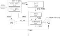
도 1 은 본 발명에 따른 영상정보를 이용한 건강 진단 시스템을 개념적으로 도시한 전체 구성도.
도 2 는 본 발명에 따른 컴퓨터 비전 분야의 객체 탐색/인식 알고리즘을 보이는 일예시도.
도 3 은 본 발명에 따른 눈의 검사요소를 보이는 일예시도.
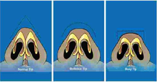
도 4 는 본 발명에 따른 코의 검사요소를 보이는 일예시도.
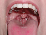
도 5 는 본 발명에 따른 입의 검사요소를 보이는 일예시도.
도 6 은 본 발명에 따른 목의 검사요소를 보이는 일예시도.
도 7 은 본 발명에 따른 흉부 및 유방의 검사요소를 보이는 일예시도.
도 8 은 본 발명에 따른 복부의 검사요소를 보이는 일예시도.
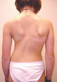
도 9 및 도 10 은 본 발명에 따른 등의 검사요소를 보이는 일예시도.
도 11 은 본 발명에 따른 둔부 및 항문의 검사요소를 보이는 일예시도.
도 12 는 본 발명에 따른 비뇨 생식기의 검사요소를 보이는 일예시도.
도 13 은 본 발명에 따른 상지의 검사요소를 보이는 일예시도.BRIEF DESCRIPTION OF THE DRAWINGS FIG. 1 is a general view schematically showing a health check system using image information according to the present invention. FIG.
FIG. 2 illustrates an object search / recognition algorithm in the field of computer vision according to the present invention.
FIG. 3 is an exemplary view showing an inspection element of an eye according to the present invention; FIG.
Fig. 4 is a view showing an examining element of a nose according to the present invention. Fig.
Figure 5 is an illustration of an inspection element of the mouth according to the present invention.
Figure 6 is an illustration showing an inspection element of the neck according to the invention.
FIG. 7 is an illustration showing an examination element of the chest and the breast according to the present invention. FIG.
FIG. 8 is an exemplary view showing an examination element of the abdomen according to the present invention; FIG.
Figures 9 and 10 illustrate examples of testing elements such as the present invention.
11 is an illustration showing an examination element of the buttocks and anus according to the present invention.
FIG. 12 is an exemplary view showing an examination element of a genitourinary organ according to the present invention. FIG.
FIG. 13 is an exemplary view showing an inspection element of the upper limb according to the present invention. FIG.
본 발명의 구체적 특징 및 이점들은 첨부도면에 의거한 다음의 상세한 설명으로 더욱 명백해질 것이다. 이에 앞서 본 발명에 관련된 공지 기능 및 그 구성에 대한 구체적인 설명이 본 발명의 요지를 불필요하게 흐릴 수 있다고 판단되는 경우에는, 그 구체적인 설명을 생략하였음에 유의해야 할 것이다.Specific features and advantages of the present invention will become more apparent from the following detailed description based on the accompanying drawings. It is to be noted that the detailed description of known functions and constructions related to the present invention is omitted when it is determined that the gist of the present invention may be unnecessarily blurred.
이하, 첨부된 도면을 참조하여 본 발명을 상세하게 설명한다. DETAILED DESCRIPTION OF THE PREFERRED EMBODIMENTS The present invention will now be described in detail with reference to the accompanying drawings.
본 발명에 따른 영상정보를 이용한 건강 진단 시스템에 관하여 도 1 내지 도 13 을 참조하여 설명하면 다음과 같다. The health diagnosis system using image information according to the present invention will be described with reference to FIG. 1 to FIG.
도 1 은 본 발명에 따른 영상정보를 이용한 건강 진단 시스템(S)을 개념적으로 도시한 전체 구성도로서, 도시된 바와 같이 정보 입력부(100), 인식부(200), 분석부(300), 병리 정보 데이터베이스부(400) 및 진단 결과 데이터베이스부(500)를 포함하여 이루어진다.1 is a block diagram schematically showing a health diagnosis system S using image information according to the present invention. As shown in the figure, an information input unit 100, a
정보 입력부(100)는 사용자의 신체 부위별 영상정보와, 기본정보를 입력받는 기능을 수행하는 바, 상기 도 1 에 도시된 바와 같이 영상정보 입력모듈(110) 및 기본정보 입력모듈(120)을 포함한다. 1, the image information input module 110 and the basic information input module 120 are connected to the information input unit 100, .
구체적으로, 영상정보 입력모듈(110)은 사용자의 신체 부위별 영상정보를 입력받는다. Specifically, the image information input module 110 receives image information of a user's body part.
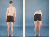
이때, 영상정보 입력모듈(110)은 사용자의 전신 영상정보를 추가로 입력받아, 신체 부위의 크기를 미리 계산해 둠으로써, 추후, 분석부(300)가 분석해야할 상처 부위의 크기를 판별하는 과정에서 이용할 수 있다. At this time, the image information input module 110 further receives the user's whole body image information, calculates the size of the body part in advance, and determines the size of the wound area to be analyzed by the analysis unit 300 in the future Can be used.
상기 신체 부위별 영상정보는, 정지영상과 동영상 형식의 영상을 포함하며, 기 저장된 정보 및 실시간으로 입력되는 정보를 바탕으로 외부와 통신하며, 이에 통신 접속이 가능한 스마트폰 및 태블릿 PC 등의 모바일 PC에 장착된 카메라를 통해 입력될 수 있다.
The image information of each body part includes a still image and a moving image format, and communicates with the outside on the basis of previously stored information and information input in real time. A mobile phone such as a smart phone or a tablet PC Or the like.
기본정보 입력모듈(120)은 사용자의 기본정보를 입력받는다. The basic information input module 120 receives basic information of the user.
여기서, 사용자의 기본정보는, 사용자의 몸무게, 연령, 성별, 직업, 식생활, 사는 곳(열대지방, 온대지방, 한대지방, 바다, 산 등), 인종(황인, 백인, 흑인, 게르만족, 켈트족, 한족 등), 생활리듬(기상, 취침, 식사시간 등), 생리주기(여성), 운동량, 흡연빈도, 음주빈도 및 과거력 등의 정보를 포함하는 것으로, 기 저장된 정보 및 실시간으로 입력되는 정보를 바탕으로 외부와 통신하며, 이에 통신 접속이 가능한 스마트폰 및 태블릿 PC 등의 모바일 PC에 장착된 입력수단을 통해 입력될 수 있다.
Here, the basic information of the user includes information on the user's body weight, age, sex, occupation, eating habits, living place (tropical region, temperate region, And information on the past and the past), and information that is stored in real time is used as the basis of the information (eg, And can be input through an input means mounted on a mobile PC such as a smart phone or a tablet PC capable of communicating therewith.
인식부(200)는 상기 정보 입력부(100)를 통해 입력된 영상정보를 바탕으로, 신체 부위를 객체로서 자동 탐색하여 인식한다. Based on the image information input through the information input unit 100, the
이때, 인식부(200)는 신체 부위의 랜드마크(land mark)로서, 얼굴(눈, 코, 입, 귀, 이마 주름, 눈썹, 등), 목(sternal notch, clavicle, thyroid cartilage(일명 Adam's apple)), 흉부(nipple), 복부(umbilicus, ASIS), 등(척추부의 만곡), 엉덩이(both gluteal sulcus, 천골의 만곡), 상지(팔꿈치의 피부구조, styloiod process of ulna, hand nail), 하지(무릎의 관절구조, medial and lateral maleola, toe nail) 등의 신체 부위를 자동 탐색 및 인식할 수 있다.At this time, the
한편, 인식부(200)는 객체로서 자동 탐색 및 인식할 수 있도록, 상기한 신체 부위를 기 설정할 수 있으며, 객체의 자동 탐색 및 인식 실패 시, 또는 사용자가 신체 부위 중 특정 위치만을 검사하고 싶은 경우, 사용자의 제어정보를 입력받아 해당 신체 부위 또는 위치를 객체로서 탐색 및 인식할 수 있다. Meanwhile, the
본 발명에서는, 사용자의 병리를 판단할 수 있는 요소들을 평가하고, 병리를 진단하기 위하여, 컴퓨터 비전(Computer Vision) 분야의 객체 탐색/인식(Object Detection/Recognition) 알고리즘을 이용하여 신체 부위 객체를 탐색하고 인식한다. In the present invention, the body part object is searched using an object detection / recognition algorithm in the field of computer vision to evaluate elements that can determine the pathology of a user and to diagnose a pathology. .
이러한 객체 탐색/인식 기술은, 현재 많은 분야에서 사용 중이다. 대표적인 예로 디지털 카메라나 스마트폰 카메라를 이용하여 촬영을 할 때, 피사체 안에서 사람의 얼굴을 찾는 일을 하거나, 더 나아가서는 사람이 웃을 때, 사진을 자동으로 촬영하는 것은 현재 사용된다. 뿐만 아니라, 문서의 사진을 분석하여 그 안에 있는 문자를 인식하는 기술도 명함인식, 스캐너 등에서 사용되고 있다. 이러한, 기술들은 사진과 같은 광학 영상정보를 이용하여 객체 탐색/인식에 필요한 특정적인 정보들을 뽑아내어 기존에 가지고 있는 트레이닝 정보를 바탕으로 분석하여 탐색해내는 방식이다. This object search / recognition technology is currently used in many fields. As a typical example, when shooting with a digital camera or a smartphone camera, it is currently used to search for a person's face in a subject, and moreover, when a person laughs, the photograph is automatically photographed. In addition, the technique of analyzing a photograph of a document and recognizing a character therein is also used in business card recognition, a scanner, and the like. These techniques extract specific information necessary for object search / recognition using optical image information such as photographs, and analyze and search based on existing training information.
일반적으로, 컴퓨터 비전 분야에서 많이 사용되는 이미지 프로세싱을 위한 프로그램 라이브러리인 OpenCV(http://opencv.org/)에서도 객체 탐색/인식을 위한 몇몇 알고리즘을 기본적으로 제공하고 있다. 그 중에서 가장 대표적인 알고리즘은 Haar Feature-based Cascade(Paul Viola and Michael J. Jones. Rapid Object Detection using a Boosted Cascade of Simple Features. IEEE CVPR, 2001, Rainer Lienhart and Jochen Maydt. An Extended Set of Haar-like Features for Rapid Object Detection. IEEE ICIP 2002, Vol. 1, pp. 900-903, Sep. 2002)이다. 이 알고리즘에서는 사진을 이루는 점(pixel)의 배열을 검출하여 아래와 같이 인식한다. 그리하여 객체로 탐색되는 영역은 도 2 의 1-(a), 2-(b) 등으로 이루어진 형태라는 것을 판별하고, 그렇게 찾아진 영역의 위치와 확대/축소 정보를 추가로 수집하게 된다. 이렇게 찾아진 정보는 기존에 가지고 있는 트레이닝 데이터와 비교하여 목표물을 찾게 된다.
In general, OpenCV (http://opencv.org/), a library for image processing that is widely used in the field of computer vision, provides some basic algorithms for object search / recognition. One of the most representative algorithms is Haar Feature-based Cascade (Paul Viola and Michael J. Jones, IEEE Cassette of Simple Features, IEEE CVPR, 2001, Rainer Lienhart and Jochen Maydt, An Extended Set of Haar-like Features IEEE Rapid Object Detection, IEEE ICIP 2002, Vol.1, pp. 900-903, Sep. 2002). In this algorithm, an array of pixels forming a picture is detected and recognized as follows. Thus, it is determined that the area to be searched by the object is of the form 1 - (a), 2 - (b), etc. in FIG. 2, and the location of the searched area and the enlargement / reduction information are further collected. This information is compared with existing training data to find the target.
분석부(300)는 상기 인식부(200)를 통해 인식된 영상정보를 신체 부위별 검사요소에 따라 분석하고, 분석한 정보와 병리 정보 데이터베이스부(400)에 저장된 정보와 비교분석하여 진단 결과를 도출하는 기능을 수행하는 바, 상기 도 1 에 도시된 바와 같이 분석모듈(310) 및 진단 결과 도출모듈(320)을 포함한다. The analysis unit 300 analyzes the image information recognized by the
구체적으로, 분석모듈(310)은 상기 인식부(200)를 통해 인식된 영상정보를 신체 부위별 검사요소에 따라, 신체 부위의 색상, 모양, 움직임, 크기, 분비물 등을 기준으로 분석한다. Specifically, the
더욱 구체적으로, 눈의 경우, 분석모듈(310)은 홍채(iris)의 색깔, 동공(pupil)의 크기 및 빛에 대한 크기변화, 공막(sclera)의 모양 및 색깔, 결막(conjunctiva)의 모양 및 색깔, 각막(conea)의 색깔, 안구의 위치, 안구 진탕(Nystagmus) 여부, 안구돌출 여부 등의 검사요소에 따라 분석한다(도 3 참조). More specifically, in the case of an eye, the
또한, 코의 경우, 분석모듈(310)은 코와 정중선의 각도, 코뼈와 중격(nasal bone and septum)의 편차(derivation; 중정선을 기준으로 휜 각도), 위와 아래의 외측 연골(upper and lower lateral cartilatage)의 크기 및 모양, 비주(columella)의 높이 및 모양, 날개(alar)의 크기 및 모양, 코의 외막(External sidewall valve)의 크기 및 모양, 콧물의 색깔 및 점도 등의 검사요소에 따라 분석한다(도 4 참조). Also, in the case of the nose, the
또한, 입의 경우, 분석모듈(310)은 치아의 모양 및 배열, 잇몸의 모양 및 색깔, 혀의 색깔, 구강 전체의 표면(구강점막)의 색깔, 구진성 병변(macular) 여부, 궤양성(ulcerative) 병변 여부, 입술의 모양 및 색깔, 입술주위 피부의 모양 및 색깔, 편도(palatine tonsil)의 크기, 모양 및 색깔, 목젓의 크기 및 위치 등의 검사요소에 따라 분석한다(도 5 참조). In addition, in the case of mouth, the
또한, 눈, 코, 입을 제외한 얼굴의 경우, 분석모듈(310)은 얼굴 피부의 주름 위치, 모공의 크기와 분포, 색깔, 여드름 여부, 얼굴의 전체적인 모양, 귓바퀴의 모양 및 대칭성, 광대뼈, 이마 및 양악의 모양, 크기 및 돌출정도, 얼굴 부종여부 등의 검사요소에 따라 분석한다. In the case of a face except for the eyes, nose, and mouth, the
또한, 목의 경우, 분석모듈(310)은 정중선을 중심으로 하여 양측의 대칭성, 쇄골의 모양 및 위치, 결후(adam's apple ; thyroid cardilage)의 위치, 갑상선의 모양, 비후 정도, 결절(nodule)의 유무, 머리의 기울임 여부(tilting) 등의 검사요소에 따라 분석한다(도 6 참조). In the case of the neck, the
또한, 흉부 및 유방의 경우, 분석모듈(310)은 유두의 크기, 모양 및 색깔, 유두 분비물의 색깔, 양상, 분비위치(일측성 또는 양측성), 여성의 유방의 크기, 모양 및 색깔, 남성의 대흉근 및 주변 구조물의 모양 등의 검사요소에 따라 분석한다(도 7 참조). In the case of the chest and the breast, the
또한, 복부의 경우, 분석모듈(310)은 복부 전체의 대칭성 및 팽만(distension) 여부, 배꼽의 모양, 크기 위치(복부전체를 기준으로 위치산정), 복직근의 모양 및 대칭성 크기, 옆구리의 모양, 크기 및 굴곡각(흔히 말하는 S-line) 등의 검사요소에 따라 분석한다(도 8 참조).In the case of the abdomen, the
또한, 등의 경우, 분석모듈(310)은 후면상(posterior view) : 척추측만(scoliosis) 여부, 등근육의 크기, 모양 및 대칭성, 측면상(lateral view): 척추후만(kyphosis) 여부, 복부 돌출의 크기, 모양 및 위치 등의 검사요소에 따라 분석한다(도 9 및 도 10 참조). In addition, the
또한, 둔부 및 항문(buttuck and anus)의 경우, 분석모듈(310)은 둔부의 크기, 모양, 대칭성 및 둔구(gluteal sulcus)의 위치, 항문륜 돌출부위의 크기, 위치(방향) 및 색깔, 항문주변 피부의 색깔, 분비물 분부부위, 돌출부위 또는 함몰부위 등의 검사요소에 따라 분석한다(도 11 참조). In the case of the buttocks and anus, the
또한, 비뇨 생식기에서, 분석모듈(310)은 남성의 경우, 귀두의 색깔, 모양, 궤양의 여부, 포경 여부, 요도의 색깔, 모양 및 분비물 등의 검사요소를 분석하며(도 12 참조), 여성의 경우, 대음순의 색깔, 크기, 궤양성 병변의 유무, 소음순의 색깔 및 모양, 질분비물의 색깔, 양 및 점도(끈적거림) 등의 검사요소에 따라 분석한다. In the genitals, the
또한, 상지(Upper extremity)에서, 분석모듈(310)은 서있는 자세에서 모양, 크기, 대칭성, 양쪽 어깨의 수평여부, 몸을 앞으로 90도 정도 숙인 상태에서 양쪽 어깨의 수평여부 등의 검사요소에 따라 어깨관절(shoulder joint)을 분석하며, 크기, 모양, 색깔 등의 검사요소에 따라 상완, 주관절, 전완, 완관절을 분석하며, 모양 및 색깔 등의 검사요소에 따라 수근관절, 수근중수간절, 지골간관절을 분석하며, 모양, 크기 및 색깔 등의 검사요소에 따라 손톱을 분석한다(도 13 참조). Also, in the upper extremity, the
또한, 하지(Lower extremity)의 경우, 분석모듈(310)은 고관절(hip joint;엉덩이 관절)의 모양, 크기 및 대칭성, 대퇴부의 크기, 모양 및 대칭성, 슬관절(knee joint;무릎)의 모양, 크기, 부종여부 및 양측의 대칭성, 종아리(calf)의 모양, 크기, 휜정도 및 혈관 돌출정도, 족관절(ankle joint; 발목)의 크기, 모양, 부종 정도, 양쪽 복사뼈(medial and lateral maleola)의 크기, 위치 및 대칭성, 발바닥 관절 및 발가락의 크기, 모양, 휜정도 및 부종여부, 발톱의 크기, 모양 및 색깔 등의 검사요소에 따라 분석한다. In the case of the lower extremity, the
그리고, 병변 부위의 경우, 분석모듈(310)은 병변의 해부학적 위치, 병변의 색깔, 병변의 크기 및 크기의 변화, 병변의 모양(가피여부, 수포성, 화농성, 손톱 및 발톱의 침습여부), 궤양성(ulcerative) 여부 등의 검사요소에 따라 분석한다.
In the case of a lesion site, the
한편, 분석모듈(310)은 움직임의 경우, 움직인 방향, 거리, 시간 등을 이용하여 분석하며, 크기의 경우, i. 사용자의 전신 영상정보를 통해 각 부위별 비례를 구하여 축척을 구하는 방법, ii. 3D 모델링을 통해 여러 개의 사진에 동일한 점들을 이용하여 분석하는 방법, iii. 촬영하는 카메라의 정보를 이용하여 계산하는 방법, iv. 환부와 동일한 거리에 척도가 될 수 있는 물체를 동시에 촬영하는 방법 등을 이용하여 분석하며, 분비물 등의 점성은, 영상에서 늘어나는 정도를 이용하여 분석한다.
On the other hand, in the case of motion, the
진단 결과 도출모듈(320)은 상기 분석모듈(310)을 통해 분석된 정보와, 병리 정보 데이터베이스부(400)에 저장된 정보와 비교분석하여 진단 결과를 도출한다. The diagnostic result derivation module 320 compares and analyzes the information analyzed through the
즉, 진단 결과 도출모듈(320)은 병리 정보 데이터베이스부(400)에 저장된 신체 부위별 검사요소를 통해 진단할 수 있는 병명 및 병의 정도를 판단하는 기준정보와 비교분석하여 진단 결과를 도출한다. That is, the diagnosis result derivation module 320 compares and analyzes the disease name and the degree of disease that can be diagnosed through the body part inspection elements stored in the pathology
참고로, 눈의 경우에서, 각막으로부터 시작해서 하얗게 변하여 동공과 홍채까지 하얗게 변하기 시작하면, 백내장(cataract)이라 진단할 수 있다. 또한, 침범 정도 및 과거 상태 비교를 통해서 악화/호전 등을 진단할 수 있다(상기 도 3 참조). For reference, in the case of the eye, it can be diagnosed as cataract if the pupil and iris begin to change to white by turning white from the cornea. In addition, deterioration / improvement can be diagnosed by comparing the degree of invasion and the past state (see FIG. 3).
코의 경우에서, 흔히 말하는 콧날(비주: columella)은 그 각도와 높이에 따라 크게 3 가지로 나뉘며, 타입에 따라 육안상의 모양차이 뿐만 아니라 비염, 충농증 등의 발생 위험도에도 차이가 있다(상기 도 4 참조).In the case of the nose, the commonly referred nose (columella) is divided into three types according to its angle and height, and there is a difference in the risk of occurrence of rhinitis, Reference).
또한, 입의 경우에서, 인간의 치아는 연령에 따라 개수와 치아의 모양이 달라진다. 이러한 것의 확인을 통해 모양이나 개수 여부를 파악하여 덧니의 발생가능성, 치아 교정의 필요성 및 치료시기를 결정할 수 있다. 또한, 편도선의 크기변화는 소아와 성인의 질환 진단에 큰 도움을 준다. 이를 테면, 편도가 한쪽만 커져 있는 경우 결핵이나 임파선 병변(쉽게 말하면 일종의 백혈병)을 의심할 수 있다. 편도가 양쪽이 모두 커져 있고 궤양상 병변이 관찰되면, 감기에 의한 2차적인 세균감염을 의심하여 항생제 처방의 기준이 된다. 그리고, 목젓의 크기와 위치는 코골이의 진단 및 치료에 이용된다. 즉, 한쪽으로 치우쳐져 있거나 목구멍 크기의 40% 이상인 경우 코골이를 유발할 수 있다고 하며, 실제로 코골이가 있는 사람은 부분 절제술을 하거나 위치교정술을 해준다(상기 도 5 에서, Soft palate; 연구개, uvula; 목젖, palatine tonsils; 구개편도(사람들이 흔히 말하는 편도선), oropharynx; 구개인두(구강의 끝이자 목이 시작되는 부분).In addition, in the case of the mouth, the number of teeth and the shape of the human teeth varies with age. By checking these things, it is possible to determine the possibility of occurrence of the gaps, the necessity of orthodontic treatment and the timing of treatment by grasping the shape or the number of teeth. In addition, changes in the size of the tonsils are very helpful in diagnosing diseases in children and adults. For example, if one side is enlarged on one side, it can be suspected of tuberculosis or lymphadenopathy (a kind of leukemia easily). When both sides of the tonsil are enlarged and an ulcerous lesion is observed, a second bacterial infection caused by a cold is suspected and prescribes antibiotic prescription. And, the size and position of roasted pickles is used for diagnosis and treatment of snoring. In other words, it is said that snoring can be induced if it is biased toward one side or more than 40% of the throat size. In fact, a person with snoring will perform partial resection or position correction (in Fig. 5, Soft palate (The end of the mouth and the neck of the mouth), the palatine tonsils, and the oropharynx.
또한, 목의 경우에서, 갑상선의 경우, 정상인 경우에는 육안으로 보이지 않으며, 손으로 만졌을 때 만져지지 않는다. 비후(hypertrophy)가 되었거나 갑상선내 결절이 발생하였을 경우, 육안으로 보이거나 손으로 만져지게 되며, 전체적으로 커져 있는 경우에는 goiter(양성 갑상선 비후증), 결절이 있는 경우 한쪽에만 있는 경우와 양쪽에 있는 경우 등에 따라 진단과 병변의 가능성이 달라진다. 소아의 경우, 머리의 기울기를 검사하여 한쪽으로 기울어져 있는 경우가 종종 있다. 많은 부모들이 이를 질병으로 인식하지 못하고 지나치게 된다. 이런 경우, 사경(toticolis)이라는 질환을 의심해볼 수 있으며, 이는 흉쇄유돌근(sternocleidomastoid muscle; SCM muscle)의 길이의 불균형에서 오는 질환으로 유아기의 발견 시 물리치료를 통해 교정이 가능하나, 유년기 이상 발견 시에는 SCM muscle 절개술을 통한 교정이 필요해진다. 흔히 할머니들이 3~4세된 아이를 목욕시키다가 발견하여 병원에 방문한다(상기 도 6 참조). In addition, in the case of the throat, in the case of the thyroid gland, it is not visible to the naked eye when it is normal, and it is not touched when touched by the hand. If hypertrophy or thyroid nodule is present, it is visible or touched by the hand. If it is enlarged as a whole, goiter (benign thyroid hypertrophy), if there is a nodule, on one side and on both sides And the likelihood of diagnosis and lesion varies. In children, it is often the case that the head slope is tilted to one side. Many parents do not recognize it as a disease and become overdone. In this case, we can suspect a disease called toticolis, which is caused by an imbalance in the length of the sternocleidomastoid muscle (SCM muscle), which can be corrected by physiotherapy when it is found in infancy, A correction using SCM muscle incision is required. Often, grandmothers bath with a child aged 3 to 4 years and find it and visit the hospital (see FIG. 6 above).
또한, 흉부 및 유방의 경우에서, 여성의 유두는 생리주기, 임신여부, 수유여부 등에 따라 모양과 색깔이 변한다. 특히, 유두와 유륜 주위의 색깔변화는 임신여부를 판별하는 간접적인 근거가 될 수 있다(1회 이상 임신 또는 유산 시에 색깔이 변화되어 있다). 여성의 유방의 경우, 유방의 피부가 오렌지 껍질 모양(orange peel sign)으로 바뀌는 경우가 있다. 이런 양상은 유방암을 의심할 수 있는 양상으로 반드시 전문의와 상담이 필요하다(상기 도 7 참조). In addition, in the case of chest and breast, the shape and color of the female papilla varies depending on the menstrual cycle, pregnancy, and whether breastfeeding. In particular, changes in color around the nipple and shoulder can be an indirect basis for determining pregnancy (color changes at least once during pregnancy or abortion). In women's breasts, the skin of the breast may turn into an orange peel sign. This aspect is a suspicious pattern of breast cancer and it is necessary to consult with a specialist (see FIG. 7 above).
또한, 복부의 경우에서, 사람 배꼽의 모양은 매우 다양하다. 그 중 간혹 배꼽탈장(umbilical hernia)이 있는 경우가 있다. 배꼽 탈장은 배꼽의 피부 바로 아래쪽까지 장(腸)이 돌출되어 있는 경우를 말하여 수술적인 치료가 필요하다. 탈장은 발견시기나 인식하는 시기가 늘어날수록 크기가 커지는 양상을 보이며 늦어질수록 수술적 치료 후 재발율이 높아진다(상기 도 8 참조).Also, in the case of the abdomen, the shape of the human navel is very diverse. There are occasional cases of umbilical hernia. The umbilical hernia refers to the case where the intestine protrudes just below the skin of the umbilicus, and surgical treatment is necessary. The hernia appears to increase in size as the time of discovery or recognition increases, and the recurrence rate increases after surgery (see FIG. 8).
또한, 등의 경우에서, 편안한 자세로 앉아있는데 남들이 볼 때 자세가 이상하거나 삐뚤어져 있는 경우가 있다. 이런 경우 척추층만증으로 의심해 볼 수 있으며 육안적인 척추소견이나 간단한 검사를 통해 진단이 가능하다(척추층만증 검사법: 앞으로 숙였을 때 양쪽 어깨높이가 다른지를 확인하는 검사)(상기 도 9 및 도 10 참조).In addition, when sitting in a comfortable posture, etc., the posture may be strange or distorted when viewed by others. In this case, it is suspected that the spinal cord is suspected and can be diagnosed through gross vertebral or simple examination (vertebral stratification test: a test to determine whether the height of both shoulders is different when the patient is leaned forward) Reference).
또한, 둔부 및 항문(buttuck and anus)의 경우에서, 항문 주위 또는 생식기 주위에 여러 개의 사마귀(warts)가 생긴 것처럼 오돌도돌하게 여러 개의 돌출상이 나타나는 경우가 있다. 이는 곤지름(condyloma acuminate, gental warts)라는 질병으로 일종의 성병이다. 또한, 항문륜 주위의 돌출부위는 위치와 크기 통증 여부에 따라 외치핵, 내치핵, 간혹 악성 신생물(malignancy, 흔히 말하는 암을 말함)등을 의심해 볼 수 있다(상기 도 11 참조).Also, in the case of the buttocks and anus, there are occasional multiple protrusions that appear as if there were multiple warts around the anus or around the genitals. It is a kind of sexually transmitted disease (condyloma acuminate, gental warts). In addition, the protruding parts around the anal canal may be suspected to be an ostium, an inner nucleus, and occasionally malignancy (refer to FIG. 11) depending on the location and size of the pain.
또한, 비뇨 생식기의 경우, 남성 생식기에 발생하는 피부질환은 성병과 일반 피부염으로 구분할 수 있으며 치료 방법도 약간 다르다. 남성생식기의 청결을 유지하지 않거나 잦은 성교의 경우 귀두포피염(balopostitis)이 발생한다. 대부분 성병으로 오인하여 치료하지 않다가 병을 키운 상태에서 내원한다. 발견 즉시 간단한 치료와 항생제 투여 및 청결유지를 통해 치료가 가능하다(상기 도 12 참조).In addition, in the case of genitourinary tract, the skin diseases occurring in male genitalia can be classified into sexually transmitted diseases and general dermatitis, and treatment methods are also slightly different. Balopostitis occurs when the male reproductive system is not kept clean or frequent intercourse occurs. Most of them are misdiagnosed as sexually transmitted diseases. Treatment is possible through simple treatment and administration of antibiotics and maintenance of cleansing immediately (see FIG. 12).
그리고, 상지(Upper extremity)의 경우, 손가락의 굴곡이나 모양변화는 류마티스 관절염(Rheumatic arthritis)의 진단에 중요한 요소이다(상기 도 13 참조).
In the case of the upper extremity, the flexion or the shape change of the finger is an important factor in the diagnosis of rheumatoid arthritis (see Fig. 13).
병리 정보 데이터베이스부(400)는 상기 분석부(300)를 통해 분석된 정보와 비교분석하여 진단 결과를 도출할 수 있도록, 신체 부위별 검사요소를 통해 진단할 수 있는 병명 및 병의 정도를 판단하는 기준정보가 저장되어 있다. 이러한 기준정보는, 실제 진단 결과가 기계 학습된 병리 정보이다. The pathology
즉, 병리 정보 데이터베이스부(400)는 트레이닝을 통한 기계 학습(machine learning)을 수행하여 병리 진단의 정확도를 높일 수 있다. That is, the pathology
이러한 기계 학습은, 상기 분석부(300)의 진단 결과 도출모듈(320)을 통해 도출되어 진단 결과 데이터베이스부(500)에 저장된 진단 결과를 입력받고, 의료 전문인을 통해 상기 진단 결과를 수정 또는 확정된 진단 결과 정보를 입력받으며, 입력된 정보를 정규화(normalize)하여 다시 저장하는 과정을 통해 이루어진다. Such a machine learning is performed by receiving diagnostic results obtained through the diagnostic result derivation module 320 of the analysis unit 300 and stored in the diagnostic
이때, 많은 사용자를 진단하여 얻은 진단 결과를 통해, 사용자의 특징별로 발병할 수 있는 질병의 정보를 더 확보할 수 있다. 이를 위해, 병리 정보 데이터베이스부(400)는 상기 정보 입력부(100)의 기본정보 입력모듈(120)을 통해 사용자의 기본정보를 입력받을 수 있다. At this time, it is possible to acquire more information about the disease that can be caused by the characteristics of the user through the diagnosis result obtained by diagnosing many users. For this purpose, the pathology
즉, 병리 정보 데이터베이스부(400)는 입력된 사용자의 기본정보를 이용하여, 성별, 나이, 직업, 및 신체 상태 등에 따라 발병할 수 있는 질병을 확률적으로 계산함으로써, 기준정보를 업데이트 함으로써, 진단 결과의 신뢰성을 높일 수 있다. 또한, 세대별, 연차별 유행할 수 있는 질병 등에 따라 오래된 데이터를 도태시키는 방식을 사용할 수 있다.
That is, the pathology
진단 결과 데이터베이스부(500)는 상기 정보 입력부(100)를 통해 입력된 사용자의 신체 부위별 영상정보와, 상기 분석부(300)를 통해 도출된 진단 결과를 저장한다. 또한, 상술한 바와 같이 진단 결과 데이터베이스부(500)는 병리 정보 데이터베이스부(400)의 기계 학습을 통한 병리 정보의 업데이트를 위해, 병리 정보 데이터베이스부(400)로 진단 결과를 제공할 수 있다. The diagnostic
이러한 진단 결과는, 사용자의 병리 상태 정보로서, 의심되는 병명의 리스트 및 확률, 의심되는 병명의 심각성 정도 및 호전 악화 성향 등에 관한 정보를 포함한다.
Such diagnosis results include, as the pathological state information of the user, information on the list and probability of the suspected disease names, the degree of seriousness of the suspected disease names, and the aggravation tendency.
본 발명은 컴퓨터로 읽을 수 있는 기록매체에 컴퓨터가 읽을 수 있는 코드로서 구현하는 것이 가능하다. 컴퓨터가 읽을 수 있는 기록매체는 컴퓨터 시스템에 의하여 읽혀질 수 있는 데이터가 저장되는 모든 종류의 기록장치를 포함한다. 컴퓨터가 읽을 수 있는 기록매체의 예로는 ROM, RAM, CD-ROM, 자기 테이프, 플라피디스크, 광데이터 저장장치 등이 있으며, 또한 인터넷을 통한 전송과 같이 캐리어 웨이브의 형태로 구현되는 것도 포함한다. 또한 컴퓨터가 읽을 수 있는 기록매체는 네트워크로 연결된 컴퓨터 시스템에 분산되어, 분산방식으로 컴퓨터가 읽을 수 있는 코드가 저장되고 실행될 수 있다.
The present invention can be embodied as computer-readable codes on a computer-readable recording medium. A computer-readable recording medium includes all kinds of recording apparatuses in which data that can be read by a computer system is stored. Examples of the computer-readable recording medium include a ROM, a RAM, a CD-ROM, a magnetic tape, a floppy disk, an optical data storage device, and the like, . The computer readable recording medium may also be distributed over a networked computer system so that computer readable code can be stored and executed in a distributed manner.
이상으로 본 발명의 기술적 사상을 예시하기 위한 바람직한 실시예와 관련하여 설명하고 도시하였지만, 본 발명은 이와 같이 도시되고 설명된 그대로의 구성 및 작용에만 국한되는 것이 아니며, 기술적 사상의 범주를 일탈함이 없이 본 발명에 대해 다수의 변경 및 수정이 가능함을 당업자들은 잘 이해할 수 있을 것이다. 따라서, 그러한 모든 적절한 변경 및 수정과 균등물들도 본 발명의 범위에 속하는 것으로 간주되어야 할 것이다. While the present invention has been particularly shown and described with reference to preferred embodiments thereof, it will be understood by those skilled in the art that various changes in form and details may be made therein without departing from the spirit and scope of the invention as defined by the appended claims. It will be appreciated by those skilled in the art that numerous changes and modifications may be made without departing from the invention. Accordingly, all such appropriate modifications and changes, and equivalents thereof, should be regarded as within the scope of the present invention.
S: 영상정보를 이용한 건강 진단 시스템
100: 정보 입력부 200: 인식부
300: 분석부 400: 병리 정보 데이터베이스부
500: 진단 결과 데이터베이스부 110: 영상정보 입력모듈
120: 기본정보 입력모듈 310: 분석모듈
320: 진단 결과 도출모듈S: Health diagnosis system using image information
100: information input unit 200:
300: analysis unit 400: pathology information database unit
500: diagnosis result database part 110: video information input module
120: basic information input module 310: analysis module
320: Diagnosis result derivation module
Claims (23)
상기 정보 입력부(100)를 통해 입력된 영상정보를 바탕으로, 신체 부위를 객체로서 자동 탐색하여 인식하는 인식부(200);
상기 인식부(200)를 통해 인식된 영상정보를 신체 부위별 검사요소에 따라 분석하고, 분석한 정보와 병리 정보 데이터베이스부(400)에 저장된 정보와 비교분석하여 진단 결과를 도출하는 분석부(300); 및
상기 분석부(300)를 통해 분석된 정보와 비교분석하여 진단 결과를 도출할 수 있도록, 신체 부위별 검사요소를 통해 진단할 수 있는 병명 및 병의 정도를 판단하는 기준정보가 저장되어 있는 병리 정보 데이터베이스부(400); 및
상기 정보 입력부(100)를 통해 입력된 사용자의 신체 부위별 영상정보와, 상기 분석부(300)를 통해 도출된 진단 결과를 저장하는 진단 결과 데이터베이스부(500); 를 포함하는 영상정보를 이용한 건강 진단 시스템.
An information input unit 100 for receiving image information of a user's body part and basic information;
A recognition unit 200 for automatically searching and recognizing a body part as an object based on the image information input through the information input unit 100;
An analysis unit 300 for analyzing the image information recognized by the recognition unit 200 according to a body part inspection element and comparing the analyzed information with the information stored in the pathology information database unit 400 to derive a diagnosis result ); And
The pathological information storing the reference information for determining the disease name and the degree of the disease that can be diagnosed through the inspection elements for each part of the body so that the diagnosis result can be derived by comparing the analyzed information with the analyzed information through the analysis unit 300 A database unit 400; And
A diagnostic result database unit 500 for storing image information of a user's body part input through the information input unit 100 and diagnostic results derived through the analysis unit 300; A health diagnosis system using image information including a medical image.
상기 정보 입력부(100)는,
사용자의 신체 부위별 영상정보를 입력받는 영상정보 입력모듈(110); 및
사용자의 기본정보를 입력받는 기본정보 입력모듈(120); 를 포함하는 것을 특징으로 하는 영상정보를 이용한 건강 진단 시스템.
The method according to claim 1,
The information input unit (100)
A video information input module 110 for receiving video information of a user's body part; And
A basic information input module 120 for receiving user basic information; And a health diagnosis system using the image information.
상기 사용자의 기본정보는,
사용자의 몸무게, 연령, 성별, 직업, 식생활, 사는 곳, 인종, 생활리듬, 생리주기, 운동량, 흡연빈도, 음주빈도 및 과거력 중, 적어도 어느 하나 이상의 정보를 포함하는 것을 특징으로 하는 영상정보를 이용한 건강 진단 시스템.
The method according to claim 1,
The basic information of the user includes:
Wherein the information includes at least one of weight, age, sex, occupation, eating habits, place of residence, race, life rhythm, menstrual cycle, exercise amount, smoking frequency, Health check system.
상기 인식부(200)는,
신체 부위의 랜드마크(land mark)로서, 눈, 코, 입, 귀, 이마 주름, 눈썹을 포함하는 얼굴, 목(sternal notch, clavicle, thyroid cartilage(일명 Adam's apple)), 흉부(nipple), 복부(umbilicus, ASIS), 등(척추부의 만곡), 엉덩이(both gluteal sulcus, 천골의 만곡), 상지(팔꿈치의 피부구조, styloiod process of ulna, hand nail), 하지(무릎의 관절구조, medial and lateral maleola, toe nail) 중, 적어도 하나 이상의 신체 부위를 자동 탐색 및 인식하는 것을 특징으로 하는 영상정보를 이용한 건강 진단 시스템.
The method according to claim 1,
The recognition unit 200,
A landmark for a body part includes a face, neck (sternal notch, clavicle, thyroid cartilage (aka Adam's apple), nipple, abdomen, (umbilicus, ASIS), back (curvature of the vertebra), buttocks (both gluteal sulcus, cervical curvature), upper limb (skin structure of the elbow, styloiod process of ulna, hand nail) wherein the at least one part of the body is automatically searched and recognized among the at least one body part, the male part, the male part and the toe part.
상기 분석부(300)는,
상기 인식부(200)를 통해 인식된 영상정보를 신체 부위별 검사요소에 따라, 신체 부위의 색상, 모양, 움직임, 크기, 분비물을 포함하는 기준으로 분석하는 분석모듈(310); 및
상기 분석모듈(310)을 통해 분석된 정보와, 병리 정보 데이터베이스부(400)에 저장된 정보와 비교분석하여 진단 결과를 도출하는 진단 결과 도출모듈(320); 을 포함하는 것을 특징으로 하는 영상정보를 이용한 건강 진단 시스템.
The method according to claim 1,
The analyzer (300)
An analysis module 310 for analyzing the image information recognized by the recognition unit 200 according to the inspection elements of the body part by reference including the color, shape, motion, size, and secretion of the body part; And
A diagnostic result derivation module 320 for comparing and analyzing the information analyzed through the analysis module 310 and the information stored in the pathology information database unit 400 to derive diagnosis results; And a health diagnosis system using the image information.
상기 분석모듈(310)은,
홍채(iris)의 색깔, 동공(pupil)의 크기 및 빛에 대한 크기변화, 공막(sclera)의 모양 및 색깔, 결막(conjunctiva)의 모양 및 색깔, 각막(conea)의 색깔, 안구의 위치, 안구 진탕(Nystagmus) 여부, 안구돌출 여부 중, 적어도 하나 이상의 검사요소에 따라 눈을 분석하는 것을 특징으로 하는 영상정보를 이용한 건강 진단 시스템.
6. The method of claim 5,
The analysis module 310,
The color of the iris, the size of the pupil, and the size of the light, the shape and color of the sclera, the shape and color of the conjunctiva, the color of the cone, Wherein the eye is analyzed according to at least one of the presence or absence of nystagmus and the presence or absence of eyeball prolapse.
상기 분석모듈(310)은,
코와 정중선의 각도, 코뼈와 중격(nasal bone and septum)의 편차(derivation; 중정선을 기준으로 휜 각도), 위와 아래의 외측 연골(upper and lower lateral cartilatage)의 크기 및 모양, 비주(columella)의 높이 및 모양, 날개(alar)의 크기 및 모양, 코의 외막(External sidewall valve)의 크기 및 모양, 콧물의 색깔 및 점도, 중 적어도 하나 이상의 검사요소에 따라 코를 분석하는 것을 특징으로 하는 영상정보를 이용한 건강 진단 시스템.
6. The method of claim 5,
The analysis module 310,
The angle of the nose and midline, the deviation of the nasal bone and septum, the size and shape of the upper and lower lateral cartilage, the size and shape of the upper and lower lateral cartilage, Characterized in that the nose is analyzed according to at least one or more of the height and shape of the alar, the size and shape of the alar, the size and shape of the external sidewall valve, the color and viscosity of the snot, Information Diagnosis System.
상기 분석모듈(310)은,
치아의 모양 및 배열, 잇몸의 모양 및 색깔, 혀의 색깔, 구강 전체의 표면(구강점막)의 색깔, 구진성 병변(macular) 여부, 궤양성(ulcerative) 병변 여부, 입술의 모양 및 색깔, 입술주위 피부의 모양 및 색깔, 편도(palatine tonsil)의 크기, 모양 및 색깔, 목젓의 크기 및 위치 중, 적어도 하나 이상의 검사요소에 따라 입을 분석하는 것을 특징으로 하는 영상정보를 이용한 건강 진단 시스템.
6. The method of claim 5,
The analysis module 310,
The shape and arrangement of the teeth, the shape and color of the gums, the color of the tongue, the color of the surface of the oral cavity (oral mucosa), the presence of macular lesions, ulcerative lesions, Wherein the mouth is analyzed according to at least one of the shape and color of the surrounding skin, the size, shape and color of the palatine tonsil, the size and position of the pickled sauce, and at least one of the inspection elements.
상기 분석모듈(310)은,
얼굴 피부의 주름 위치, 모공의 크기와 분포, 색깔, 여드름 여부, 얼굴의 전체적인 모양, 귓바퀴의 모양 및 대칭성, 광대뼈, 이마 및 양악의 모양, 크기 및 돌출정도, 얼굴 부종여부 중, 적어도 하나 이상의 검사요소에 따라 얼굴을 분석하는 것을 특징으로 하는 영상정보를 이용한 건강 진단 시스템.
6. The method of claim 5,
The analysis module 310,
At least one of at least one of the wrinkles of the face skin, the size and distribution of the pores, the color, the acne, the overall shape of the face, the shape and symmetry of the auricle, the shape, size and protrusion of the cheekbones, And analyzing the face according to the element.
상기 분석모듈(310)은,
정중선을 중심으로 하여 양측의 대칭성, 쇄골의 모양 및 위치, 결후(adam's apple ; thyroid cardilage)의 위치, 갑상선의 모양, 비후 정도, 결절(nodule)의 유무, 머리의 기울임 여부(tilting) 중, 적어도 하나 이상의 검사요소에 따라 목을 분석하는 것을 특징으로 하는 영상정보를 이용한 건강 진단 시스템.
6. The method of claim 5,
The analysis module 310,
The midline of the symmetry, the shape and location of the clavicle, the position of the man's apple (thyroid cardilage), the shape of the thyroid gland, the degree of thickening, the presence of nodules and tilting of the head And analyzing the neck according to one or more examination elements.
상기 분석모듈(310)은,
유두의 크기, 모양 및 색깔, 유두 분비물의 색깔, 양상, 분비위치(일측성 또는 양측성), 여성의 유방의 크기, 모양 및 색깔, 남성의 대흉근 및 주변 구조물의 모양 중, 적어도 하나 이상의 검사요소에 따라 흉부 및 유방을 분석하는 것을 특징으로 하는 영상정보를 이용한 건강 진단 시스템.
6. The method of claim 5,
The analysis module 310,
At least one of the size, shape and color of the nipple, color, aspect, secretion position (unilateral or bilateral) of the nipple discharge, size, shape and color of the female breast, shape of the pectoralis muscle and peripheral structures of the male, And analyzing the chest and the breast according to the image information.
상기 분석모듈(310)은,
복부 전체의 대칭성 및 팽만(distension) 여부, 배꼽의 모양, 크기 위치(복부전체를 기준으로 위치산정), 복직근의 모양 및 대칭성 크기, 옆구리의 모양, 크기 및 굴곡각 중, 적어도 하나 이상의 검사요소에 따라 복부를 분석하는 것을 특징으로 하는 영상정보를 이용한 건강 진단 시스템.
6. The method of claim 5,
The analysis module 310,
Of at least one of the symmetry and distension of the abdomen as a whole, the shape of the navel, the position of the abdomen (position calculation based on the entire abdomen), the shape and symmetry of the rectus abdominis muscle, the shape, And analyzing the abdomen according to the image information.
상기 분석모듈(310)은,
후면상(posterior view) : 척추측만(scoliosis) 여부, 등근육의 크기, 모양 및 대칭성, 측면상(lateral view): 척추후만(kyphosis) 여부, 복부 돌출의 크기, 모양 및 위치 중, 적어도 하나 이상의 검사요소에 따라 등을 분석하는 것을 특징으로 하는 영상정보를 이용한 건강 진단 시스템.
6. The method of claim 5,
The analysis module 310,
At least one of the posterior view: whether scoliosis, the size, shape and symmetry of the back muscles, the lateral view: whether it is kyphosis, the size, shape and location of the abdominal overhang And the like are analyzed according to the above-mentioned inspection factors.
상기 분석모듈(310)은,
둔부의 크기, 모양, 대칭성 및 둔구(gluteal sulcus)의 위치, 항문륜 돌출부위의 크기, 위치(방향) 및 색깔, 항문주변 피부의 색깔, 분비물 분부부위, 돌출부위 또는 함몰부위 중, 적어도 하나 이상의 검사요소에 따라 둔부 및 항문(buttuck and anus)을 분석하는 것을 특징으로 하는 영상정보를 이용한 건강 진단 시스템.
6. The method of claim 5,
The analysis module 310,
At least one of the size, shape, symmetry and position of the gluteal sulcus, the size, position (direction) and color of the anus tendon, color of the perianal skin, secretion site, And analyzing the buttocks and anus according to the examination elements.
상기 분석모듈(310)은,
귀두의 색깔, 모양, 궤양의 여부, 포경 여부, 요도의 색깔, 모양 및 분비물, 대음순의 색깔, 크기, 궤양성 병변의 유무, 소음순의 색깔 및 모양, 질분비물의 색깔, 양 및 점도(끈적거림) 중, 적어도 하나 이상의 검사요소에 따라 비뇨 생식기를 분석하는 것을 특징으로 하는 영상정보를 이용한 건강 진단 시스템.
6. The method of claim 5,
The analysis module 310,
The color and shape of the glans, the amount and the viscosity of the vaginal secretions (stickiness), the color of the glans, the presence or absence of ulcers, the color of the urethra, the color and shape of the urethra, the color and size of the labia majora, And analyzing the genitourinary organ according to at least one of the inspection elements.
상기 분석모듈(310)은,
상지(Upper extremity)에서, 서있는 자세에서 모양, 크기, 대칭성, 양쪽 어깨의 수평여부, 몸을 앞으로 숙인 상태에서 양쪽 어깨의 수평여부 중, 적어도 하나 이상의 검사요소에 따라 어깨관절(shoulder joint)을 분석하며, 크기, 모양, 색깔 중, 적어도 하나 이상의 검사요소에 따라 상완, 주관절, 전완, 완관절을 분석하며, 모양 및 색깔 중, 적어도 하나 이상의 검사요소에 따라 수근관절, 수근중수간절, 지골간관절을 분석하며, 모양, 크기 및 색깔 중, 적어도 하나 이상의 검사요소에 따라 손톱을 분석하는 것을 특징으로 하는 영상정보를 이용한 건강 진단 시스템.
6. The method of claim 5,
The analysis module 310,
In the upper extremity, the shoulder joint is analyzed according to at least one of the test elements, such as shape, size, symmetry, horizontal shoulder level, horizontal shoulder level with the body leaning forward And analyzes the upper arm, elbow, forearm, and wrist according to at least one of the size, shape, and color of the test item. The shape and color of the wrist joint, And analyzing the nail according to at least one of the shape, the size, and the color of the nail.
상기 분석모듈(310)은,
고관절(hip joint;엉덩이 관절)의 모양, 크기 및 대칭성, 대퇴부의 크기, 모양 및 대칭성, 슬관절(knee joint;무릎)의 모양, 크기, 부종여부 및 양측의 대칭성, 종아리(calf)의 모양, 크기, 휜정도 및 혈관 돌출정도, 족관절(ankle joint; 발목)의 크기, 모양, 부종 정도, 양쪽 복사뼈(medial and lateral maleola)의 크기, 위치 및 대칭성, 발바닥 관절 및 발가락의 크기, 모양, 휜정도 및 부종여부, 발톱의 크기, 모양 및 색깔 중, 적어도 하나 이상의 검사요소에 따라 하지(Lower extremity)를 분석하는 것을 특징으로 하는 영상정보를 이용한 건강 진단 시스템.
6. The method of claim 5,
The analysis module 310,
The shape, size and symmetry of the hip joint, the size, shape and symmetry of the thigh, the shape of the knee joint, the size, the swelling and the symmetry of both sides, the shape and size of the calf The size and shape of the ankle joint, the degree of edema, the size, position and symmetry of the medial and lateral maleola, the size and shape of the foot joints and toes, Wherein the lower extremity is analyzed based on at least one of the at least one of the at least one of the at least one of the at least one test item,
상기 분석모듈(310)은,
병변의 해부학적 위치, 병변의 색깔, 병변의 크기 및 크기의 변화, 병변의 모양(가피여부, 수포성, 화농성, 손톱 및 발톱의 침습여부), 궤양성(ulcerative) 여부 중, 적어도 하나 이상의 검사요소에 따라 병변 부위를 분석하는 것을 특징으로 하는 영상정보를 이용한 건강 진단 시스템.
6. The method of claim 5,
The analysis module 310,
At least one of the anatomical location of the lesion, the color of the lesion, the size and size of the lesion, the shape of the lesion (whether it is scarring, scarring, purulence, nail and claw invasion), ulcerative And analyzing the lesion site according to the element.
상기 진단 결과 도출모듈(320)은,
상기 병리 정보 데이터베이스부(400)에 저장된 신체 부위별 검사요소를 통해 진단할 수 있는 병명 및 병의 정도를 판단하는 기준정보와 비교분석하여 진단 결과를 도출하는 것을 특징으로 하는 영상정보를 이용한 건강 진단 시스템.
6. The method of claim 5,
The diagnostic result derivation module 320,
And a diagnosis result is obtained by comparing and analyzing the disease information and the criterion information for determining the degree of the disease and the degree of disease that can be diagnosed through the body part inspection elements stored in the pathology information database unit 400. [ system.
상기 병리 정보 데이터베이스부(400)는,
상기 분석부(300)를 통해 분석된 정보와 비교분석하여 진단 결과를 도출할 수 있도록, 신체 부위별 검사요소를 통해 진단할 수 있는 병명 및 병의 정도를 판단하는 기준정보가 저장되어 있는 것을 특징으로 하는 영상정보를 이용한 건강 진단 시스템.
The method according to claim 1,
The pathology information database unit 400,
And reference information for determining the disease name and degree of disease that can be diagnosed through the inspection elements for each body part is stored so that the diagnosis result can be derived by comparing the analyzed information with the analyzed information through the analysis unit 300 A health diagnosis system using image information.
상기 기준정보는,
진단 결과가 기계 학습된 병리 정보인 것을 특징으로 하는 영상정보를 이용한 건강 진단 시스템.
21. The method according to any one of claims 1, 19 and 20,
The reference information includes:
Wherein the diagnostic result is machine-learned pathology information.
상기 병리 정보 데이터베이스부(400)는,
상기 분석부(300)를 통해 도출되어 상기 진단 결과 데이터베이스부(500)에 저장된 진단 결과를 입력받고, 상기 진단 결과를 수정 또는 확정된 진단 결과 정보를 입력받으며, 입력된 정보를 정규화(normalize)하여 다시 저장하는 과정을 통해 이루어지는 기계 학습을 수행하는 것을 특징으로 하는 영상정보를 이용한 건강 진단 시스템.
21. The method of claim 1 or 20,
The pathology information database unit 400,
Receives diagnostic results stored in the diagnostic result database unit 500 through the analysis unit 300, receives diagnostic result information corrected or determined, and normalizes input information, And performing the machine learning through the process of re-storing the medical image.
상기 진단 결과는,
병리 상태 정보로서, 의심되는 병명의 리스트 및 확률, 의심되는 병명의 심각성 정도 및 호전 악화 성향 중, 적어도 어느 하나 이상의 정보를 포함하는 것을 특징으로 하는 영상정보를 이용한 건강 진단 시스템.The method according to claim 1,
As a result of the diagnosis,
Wherein the pathological condition information includes at least one of a list and a probability of a suspected disease name, a severity of a suspected disease name, and an aggravation tendency.
Priority Applications (2)
| Application Number | Priority Date | Filing Date | Title |
|---|---|---|---|
| KR1020130021012A KR20140108417A (en) | 2013-02-27 | 2013-02-27 | Health diagnosis system using image information |
| US14/192,058 US20140243651A1 (en) | 2013-02-27 | 2014-02-27 | Health diagnosis system using image information |
Applications Claiming Priority (1)
| Application Number | Priority Date | Filing Date | Title |
|---|---|---|---|
| KR1020130021012A KR20140108417A (en) | 2013-02-27 | 2013-02-27 | Health diagnosis system using image information |
Publications (1)
| Publication Number | Publication Date |
|---|---|
| KR20140108417A true KR20140108417A (en) | 2014-09-11 |
Family
ID=51388831
Family Applications (1)
| Application Number | Title | Priority Date | Filing Date |
|---|---|---|---|
| KR1020130021012A KR20140108417A (en) | 2013-02-27 | 2013-02-27 | Health diagnosis system using image information |
Country Status (2)
| Country | Link |
|---|---|
| US (1) | US20140243651A1 (en) |
| KR (1) | KR20140108417A (en) |
Cited By (30)
| Publication number | Priority date | Publication date | Assignee | Title |
|---|---|---|---|---|
| WO2018080194A1 (en) * | 2016-10-26 | 2018-05-03 | 고려대학교 산학협력단 | System and method for diagnosing oral cavity lesion |
| KR101874348B1 (en) * | 2017-11-21 | 2018-07-09 | 주식회사 뷰노 | Method for facilitating dignosis of subject based on chest posteroanterior view thereof, and apparatus using the same |
| KR101887194B1 (en) * | 2018-06-20 | 2018-08-10 | 주식회사 뷰노 | Method for facilitating dignosis of subject based on medical imagery thereof, and apparatus using the same |
| WO2019031794A1 (en) * | 2017-08-11 | 2019-02-14 | 주식회사 뷰노 | Method for generating prediction result for predicting occurrence of fatal symptoms of subject in advance and device using same |
| KR20190043494A (en) * | 2017-10-18 | 2019-04-26 | 주식회사 씨엠랩 | Systems and methods for performing complex ophthalmic tratment |
| WO2019083129A1 (en) * | 2017-10-27 | 2019-05-02 | 주식회사 뷰노 | Method for supporting reading of fundus image of subject, and device using same |
| KR20190071620A (en) * | 2017-12-14 | 2019-06-24 | 경상대학교산학협력단 | An intraoral photograph-based device to screen obstructive sleep apnea and An intraoral photograph-based method to therefore |
| KR20190081320A (en) * | 2017-12-29 | 2019-07-09 | (주)허니냅스 | Apparatus and method for predicting reduction ratio of apnea-hypopnea index before surgery to treat sleep apnea syndrome |
| KR20190081321A (en) * | 2017-12-29 | 2019-07-09 | (주)허니냅스 | Apparatus and method for predicting reduction ratio of apnea-hypopnea index before surgery to treat sleep apnea syndrome |
| WO2019151728A1 (en) * | 2018-01-30 | 2019-08-08 | 손대업 | Health management system and health management method using same |
| WO2019208848A1 (en) * | 2018-04-24 | 2019-10-31 | 한림대학교 산학협력단 | Three-dimensional eyeball movement measurement method and automatic deep learning based dizziness diagnosis system |
| WO2019231102A1 (en) * | 2018-05-31 | 2019-12-05 | 주식회사 뷰노 | Method for classifying fundus image of subject and device using same |
| WO2019240567A1 (en) * | 2018-06-11 | 2019-12-19 | 사회복지법인 삼성생명공익재단 | Anterior eye disease diagnostic system and diagnostic method using same |
| KR20200005986A (en) * | 2018-07-09 | 2020-01-17 | 주식회사 두브레인 | System and method for diagnosing cognitive impairment using face recognization |
| WO2020235940A1 (en) * | 2019-05-21 | 2020-11-26 | (의) 삼성의료재단 | Apparatus, method, and system for measuring exophthalmos using 3d depth camera |
| KR20200133923A (en) * | 2019-05-21 | 2020-12-01 | (의) 삼성의료재단 | Method and system for monitoring related disease using face perception of mobile phone |
| KR20210013217A (en) * | 2018-05-29 | 2021-02-03 | 일란 티아가이 | Electronic automatic adjustment bidet with machine learning software |
| KR102222059B1 (en) * | 2020-05-26 | 2021-03-03 | 주식회사 에프앤디파트너스 | Object position recognition diagnostic device that outputs the object position and the diagnosis result using a diagnostic device with marker |
| KR20210043748A (en) * | 2015-08-03 | 2021-04-21 | 엔제루 프레잉구 카도 가부시키가이샤 | Fraud detection system in casino |
| KR20210095821A (en) | 2019-09-04 | 2021-08-03 | 한양대학교 산학협력단 | Method and apparatus for measuring eye movement |
| KR20210098133A (en) * | 2020-01-31 | 2021-08-10 | 인하대학교 산학협력단 | System for derermining scabies based using artificial intelligence and thereof method |
| WO2021172677A1 (en) * | 2020-02-26 | 2021-09-02 | 송민구 | Image artificial intelligence-based method for diagnosing prognosis of companion animal disease |
| KR20210120408A (en) * | 2020-03-26 | 2021-10-07 | 재단법인 아산사회복지재단 | Method and Device for checking the health condition by skin reaction |
| KR20210141168A (en) * | 2020-05-15 | 2021-11-23 | 오스템임플란트 주식회사 | Patient Diagnostic Method And Apparatus For Orthodontic Patients |
| KR20220018812A (en) * | 2020-08-07 | 2022-02-15 | 주식회사 에이아이포펫 | Ophthalmic disease measuring system using a portable treminal and ophthalmic disease management method |
| KR102445752B1 (en) | 2022-07-01 | 2022-09-21 | 주식회사 룰루랩 | Method and apparatus for predicting disease through wrinkle detection |
| WO2023277548A1 (en) * | 2021-06-30 | 2023-01-05 | 주식회사 타이로스코프 | Method for acquiring side image for eye protrusion analysis, image capture device for performing same, and recording medium |
| US11663719B2 (en) | 2021-06-30 | 2023-05-30 | Thyroscope Inc. | Method for hospital visit guidance for medical treatment for active thyroid eye disease, and system for performing same |
| KR20230124195A (en) | 2022-02-18 | 2023-08-25 | 박성재 | System and Method for providing customized complex service based on facial image |
| US11741610B2 (en) | 2021-06-30 | 2023-08-29 | Thyroscope Inc. | Method for hospital visit guidance for medical treatment for active thyroid eye disease, and system for performing same |
Families Citing this family (11)
| Publication number | Priority date | Publication date | Assignee | Title |
|---|---|---|---|---|
| TWI701018B (en) * | 2015-01-29 | 2020-08-11 | 日商新力股份有限公司 | Information processing device, information processing method, and program |
| CN106236060B (en) * | 2015-06-04 | 2021-04-09 | 松下知识产权经营株式会社 | Biological information detection device |
| KR101723732B1 (en) * | 2015-06-19 | 2017-04-05 | 가톨릭대학교 산학협력단 | Method and server for managing analysis of image for medical test |
| WO2018126275A1 (en) * | 2016-12-30 | 2018-07-05 | Dirk Schneemann, LLC | Modeling and learning character traits and medical condition based on 3d facial features |
| US11259743B2 (en) | 2017-03-08 | 2022-03-01 | Strive Orthopedics, Inc. | Method for identifying human joint characteristics |
| EP3593280A4 (en) * | 2018-01-16 | 2021-03-17 | David R. Hall | Health monitoring system including privacy-ensuring obfuscated camera images |
| CN109584999A (en) * | 2018-11-27 | 2019-04-05 | 广东智源信息技术有限公司 | A kind of legal medical expert's condition of the injury identification wound Intelligent detecting method based on picture recognition |
| RU2731316C1 (en) * | 2019-12-06 | 2020-09-01 | Федеральное государственное бюджетное учреждение "Российский центр судебно-медицинской экспертизы" Министерства здравоохранения Российской Федерации (ФГБУ "РЦСМЭ" Минздрава России) | Method of determining a person's caucasoid-mongoloid affiliation by dermatoglyphic patterns of distal phalanges of fingers |
| CN111563891B (en) * | 2020-05-09 | 2023-09-26 | 吾征智能技术(北京)有限公司 | Disease prediction system based on color cognition |
| CN113450913A (en) * | 2020-08-06 | 2021-09-28 | 心医国际数字医疗系统(大连)有限公司 | Data processing device and method and electronic equipment |
| WO2023057232A1 (en) * | 2021-10-04 | 2023-04-13 | Biotronik Se & Co. Kg | System and method for supporting a patient's health control |
Family Cites Families (2)
| Publication number | Priority date | Publication date | Assignee | Title |
|---|---|---|---|---|
| KR101030430B1 (en) * | 2007-09-12 | 2011-04-20 | 주식회사 코아로직 | Apparatus and method for processing image and computer readable medium stored thereon computer executable instruction for performing the method |
| EP2690596B1 (en) * | 2012-07-24 | 2018-08-15 | Agfa Healthcare | Method, apparatus and system for automated spine labeling |
-
2013
- 2013-02-27 KR KR1020130021012A patent/KR20140108417A/en active Search and Examination
-
2014
- 2014-02-27 US US14/192,058 patent/US20140243651A1/en not_active Abandoned
Cited By (55)
| Publication number | Priority date | Publication date | Assignee | Title |
|---|---|---|---|---|
| US11527131B2 (en) | 2015-08-03 | 2022-12-13 | Angel Group Co., Ltd. | Fraud detection system in a casino |
| US11393286B2 (en) | 2015-08-03 | 2022-07-19 | Angel Group Co., Ltd. | Fraud detection system in a casino |
| US11727750B2 (en) | 2015-08-03 | 2023-08-15 | Angel Group Co., Ltd. | Fraud detection system in a casino |
| KR20220037425A (en) * | 2015-08-03 | 2022-03-24 | 엔제루 구루푸 가부시키가이샤 | Fraud detection system in casino |
| US11587398B2 (en) | 2015-08-03 | 2023-02-21 | Angel Group Co., Ltd. | Fraud detection system in a casino |
| US11393284B2 (en) | 2015-08-03 | 2022-07-19 | Angel Group Co., Ltd. | Fraud detection system in a casino |
| US11393285B2 (en) | 2015-08-03 | 2022-07-19 | Angel Group Co., Ltd. | Fraud detection system in a casino |
| US11620872B2 (en) | 2015-08-03 | 2023-04-04 | Angel Group Co., Ltd. | Fraud detection system in a casino |
| US11380161B2 (en) | 2015-08-03 | 2022-07-05 | Angel Group Co., Ltd. | Fraud detection system in a casino |
| US11386749B2 (en) | 2015-08-03 | 2022-07-12 | Angel Group Co., Ltd. | Fraud detection system in a casino |
| US11527130B2 (en) | 2015-08-03 | 2022-12-13 | Angel Group Co., Ltd. | Fraud detection system in a casino |
| KR20210043748A (en) * | 2015-08-03 | 2021-04-21 | 엔제루 프레잉구 카도 가부시키가이샤 | Fraud detection system in casino |
| US11657673B2 (en) | 2015-08-03 | 2023-05-23 | Angel Group Co., Ltd. | Fraud detection system in a casino |
| US11386748B2 (en) | 2015-08-03 | 2022-07-12 | Angel Playing Cards Co., Ltd. | Fraud detection system in a casino |
| US11657674B2 (en) | 2015-08-03 | 2023-05-23 | Angel Group Go., Ltd. | Fraud detection system in casino |
| US11741780B2 (en) | 2015-08-03 | 2023-08-29 | Angel Group Co., Ltd. | Fraud detection system in a casino |
| WO2018080194A1 (en) * | 2016-10-26 | 2018-05-03 | 고려대학교 산학협력단 | System and method for diagnosing oral cavity lesion |
| US11735317B2 (en) | 2017-08-11 | 2023-08-22 | Vuno, Inc. | Method for generating prediction result for predicting occurrence of fatal symptoms of subject in advance and device using same |
| WO2019031794A1 (en) * | 2017-08-11 | 2019-02-14 | 주식회사 뷰노 | Method for generating prediction result for predicting occurrence of fatal symptoms of subject in advance and device using same |
| KR20190043494A (en) * | 2017-10-18 | 2019-04-26 | 주식회사 씨엠랩 | Systems and methods for performing complex ophthalmic tratment |
| US11771318B2 (en) | 2017-10-27 | 2023-10-03 | Vuno, Inc. | Method for supporting reading of fundus image of subject, and device using same |
| WO2019083129A1 (en) * | 2017-10-27 | 2019-05-02 | 주식회사 뷰노 | Method for supporting reading of fundus image of subject, and device using same |
| KR101874348B1 (en) * | 2017-11-21 | 2018-07-09 | 주식회사 뷰노 | Method for facilitating dignosis of subject based on chest posteroanterior view thereof, and apparatus using the same |
| WO2019103440A1 (en) * | 2017-11-21 | 2019-05-31 | 주식회사 뷰노 | Method for supporting reading of medical image of subject and device using same |
| KR20190071620A (en) * | 2017-12-14 | 2019-06-24 | 경상대학교산학협력단 | An intraoral photograph-based device to screen obstructive sleep apnea and An intraoral photograph-based method to therefore |
| KR20190081320A (en) * | 2017-12-29 | 2019-07-09 | (주)허니냅스 | Apparatus and method for predicting reduction ratio of apnea-hypopnea index before surgery to treat sleep apnea syndrome |
| KR20190081321A (en) * | 2017-12-29 | 2019-07-09 | (주)허니냅스 | Apparatus and method for predicting reduction ratio of apnea-hypopnea index before surgery to treat sleep apnea syndrome |
| WO2019151728A1 (en) * | 2018-01-30 | 2019-08-08 | 손대업 | Health management system and health management method using same |
| WO2019208848A1 (en) * | 2018-04-24 | 2019-10-31 | 한림대학교 산학협력단 | Three-dimensional eyeball movement measurement method and automatic deep learning based dizziness diagnosis system |
| US11779257B2 (en) | 2018-04-24 | 2023-10-10 | Hallym University Technology Holdings | 3-dimensional measurement method for eye movement and fully automated deep-learning based system for vertigo diagnosis |
| KR20210013217A (en) * | 2018-05-29 | 2021-02-03 | 일란 티아가이 | Electronic automatic adjustment bidet with machine learning software |
| WO2019231102A1 (en) * | 2018-05-31 | 2019-12-05 | 주식회사 뷰노 | Method for classifying fundus image of subject and device using same |
| WO2019240567A1 (en) * | 2018-06-11 | 2019-12-19 | 사회복지법인 삼성생명공익재단 | Anterior eye disease diagnostic system and diagnostic method using same |
| KR101887194B1 (en) * | 2018-06-20 | 2018-08-10 | 주식회사 뷰노 | Method for facilitating dignosis of subject based on medical imagery thereof, and apparatus using the same |
| KR20200005986A (en) * | 2018-07-09 | 2020-01-17 | 주식회사 두브레인 | System and method for diagnosing cognitive impairment using face recognization |
| KR20200005987A (en) * | 2018-07-09 | 2020-01-17 | 주식회사 두브레인 | System and method for diagnosing cognitive impairment using touch input |
| WO2020235940A1 (en) * | 2019-05-21 | 2020-11-26 | (의) 삼성의료재단 | Apparatus, method, and system for measuring exophthalmos using 3d depth camera |
| KR20200133923A (en) * | 2019-05-21 | 2020-12-01 | (의) 삼성의료재단 | Method and system for monitoring related disease using face perception of mobile phone |
| WO2020235939A3 (en) * | 2019-05-21 | 2021-02-04 | (의) 삼성의료재단 | Method and system for monitoring related diseases by means of face recognition in mobile communication terminal |
| KR20210095821A (en) | 2019-09-04 | 2021-08-03 | 한양대학교 산학협력단 | Method and apparatus for measuring eye movement |
| KR20210098133A (en) * | 2020-01-31 | 2021-08-10 | 인하대학교 산학협력단 | System for derermining scabies based using artificial intelligence and thereof method |
| WO2021172677A1 (en) * | 2020-02-26 | 2021-09-02 | 송민구 | Image artificial intelligence-based method for diagnosing prognosis of companion animal disease |
| KR20210120408A (en) * | 2020-03-26 | 2021-10-07 | 재단법인 아산사회복지재단 | Method and Device for checking the health condition by skin reaction |
| KR20220014895A (en) * | 2020-05-15 | 2022-02-07 | 오스템임플란트 주식회사 | Patient Diagnostic Method And Apparatus For Orthodontic Patients |
| KR20210141168A (en) * | 2020-05-15 | 2021-11-23 | 오스템임플란트 주식회사 | Patient Diagnostic Method And Apparatus For Orthodontic Patients |
| KR102222059B1 (en) * | 2020-05-26 | 2021-03-03 | 주식회사 에프앤디파트너스 | Object position recognition diagnostic device that outputs the object position and the diagnosis result using a diagnostic device with marker |
| KR20220018812A (en) * | 2020-08-07 | 2022-02-15 | 주식회사 에이아이포펫 | Ophthalmic disease measuring system using a portable treminal and ophthalmic disease management method |
| US11748884B2 (en) | 2021-06-30 | 2023-09-05 | Thyroscope Inc. | Method for hospital visit guidance for medical treatment for active thyroid eye disease, and system for performing same |
| US11741610B2 (en) | 2021-06-30 | 2023-08-29 | Thyroscope Inc. | Method for hospital visit guidance for medical treatment for active thyroid eye disease, and system for performing same |
| US11717160B2 (en) | 2021-06-30 | 2023-08-08 | Thyroscope Inc. | Method and photographing device for acquiring side image for ocular proptosis degree analysis, and recording medium therefor |
| US11663719B2 (en) | 2021-06-30 | 2023-05-30 | Thyroscope Inc. | Method for hospital visit guidance for medical treatment for active thyroid eye disease, and system for performing same |
| WO2023277548A1 (en) * | 2021-06-30 | 2023-01-05 | 주식회사 타이로스코프 | Method for acquiring side image for eye protrusion analysis, image capture device for performing same, and recording medium |
| KR20230124195A (en) | 2022-02-18 | 2023-08-25 | 박성재 | System and Method for providing customized complex service based on facial image |
| KR102445752B1 (en) | 2022-07-01 | 2022-09-21 | 주식회사 룰루랩 | Method and apparatus for predicting disease through wrinkle detection |
| WO2024005542A1 (en) * | 2022-07-01 | 2024-01-04 | 주식회사 룰루랩 | Method and device for predicting disease through wrinkle detection |
Also Published As
| Publication number | Publication date |
|---|---|
| US20140243651A1 (en) | 2014-08-28 |
Similar Documents
| Publication | Publication Date | Title |
|---|---|---|
| KR20140108417A (en) | Health diagnosis system using image information | |
| US10219736B2 (en) | Methods and arrangements concerning dermatology | |
| US11779222B2 (en) | Method of and imaging system for clinical sign detection | |
| US9710920B2 (en) | Motion information processing device | |
| US20140313303A1 (en) | Longitudinal dermoscopic study employing smartphone-based image registration | |
| CN113647939B (en) | Artificial intelligence rehabilitation evaluation and training system for spinal degenerative diseases | |
| Islam et al. | Deep learning of facial depth maps for obstructive sleep apnea prediction | |
| KR101938241B1 (en) | Apparatus and method for generating injury prevention model based on machine learning, apparatus and method for preventing injury | |
| US20220148723A1 (en) | Medical examination of human body using haptics | |
| CN114999646B (en) | Newborn exercise development assessment system, method, device and storage medium | |
| US10835120B2 (en) | Extended medical test system | |
| JP2023169392A (en) | Prediction device, prediction system, control method, and control program | |
| Kaviya et al. | INTELLIGENT WEARABLE DEVICE FOR EARLY DETECTION OF MYOCARDIAL INFARCTION USING IoT | |
| CN111326240A (en) | Intelligent medical triage recommendation method based on model-free | |
| WO2022141926A1 (en) | Gastrointestinal perforation diagnosis and intervention device, and diagnosis and intervention system | |
| US20240119587A1 (en) | Prediction system, control method, and control program | |
| US20240050024A1 (en) | Methods and systems for vaginal therapeutic device fitting | |
| US20230013233A1 (en) | Image-based risk analysis of individuals in clinical settings | |
| CN113425271B (en) | Daytime operation discharge judgment method, device, equipment and storage medium | |
| KR102344659B1 (en) | Server and method for self-examination of breast cancer | |
| Javvaji et al. | Prediction Model for Obstructive Sleep Apnea from Facial Depth Maps using Transfer Learning | |
| WO2023181417A1 (en) | Imaging device, program, and method | |
| US20220387158A1 (en) | Methods and systems for vaginal therapeutic device fitting | |
| CN114617765A (en) | Domestic sauna room intelligent management system based on face identification | |
| EP3838140A1 (en) | A system for detecting tissue fluid changes |
Legal Events
| Date | Code | Title | Description |
|---|---|---|---|
| A201 | Request for examination | ||
| E902 | Notification of reason for refusal | ||
| AMND | Amendment | ||
| E601 | Decision to refuse application | ||
| AMND | Amendment |