Hereinafter, an eye refractive power measuring apparatus according to an embodiment of the present invention will be described with reference to the drawings. BRIEF DESCRIPTION OF DRAWINGS FIG. 1 is an external configuration diagram of an apparatus according to an embodiment; FIG. The measuring apparatus comprises a base 1, a face supporting unit 2 mounted on the base 1, a moving base 3 movably formed on the base 1, And a measurement unit 4 that stores the optical system to be described later. The measuring unit 4 is moved in the left and right directions (X direction), the up and down directions (Y direction) and the back and forth direction (Z direction) with respect to the eye to be examined E by the XYZ driving unit 6 formed on the moving base 3 do. The XYZ driver 6 is composed of a slide mechanism, a motor, and the like, which are formed in the directions of X, Y, and Z. The movable base 3 is moved in the X and Z directions on the base 1 by the operation of the joystick 5 and is rotated in the Y direction of the XYZ driver 6 by rotating the rotary knob 5a In the Y direction. A monitor 7 for displaying various information such as an observation image and a measurement result of the eye to be examined E and a switch unit 8 in which switches for performing various settings are disposed are formed on the movable base 3.
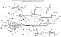
2 is a schematic configuration diagram of an optical system and a control system of the present apparatus. The measurement optical system 10 includes a projection optical system 10a for projecting a measurement index light beam on a spot on the fundus Ef of the eye E to be examined through the pupil center of the eye E to be examined, And a light receiving optical system 10b for taking out the measurement index light flux in the form of a ring through the peripheral portion of the pupil.
The projection optical system 10a includes a measurement infrared light source 11 such as an LED or an SLD disposed on the optical axis L1 of the measurement optical system 10, a relay lens 12, a hole mirror 13, A prism 15 rotated about the optical axis L1 by an objective lens 23, and an objective lens 14, and serves as a light projecting means. The measurement infrared ray light source 11 is in a positional relationship optically conjugate with the fundus Ef of the regular eye. The aperture of the hole mirror 13 is in a positional relationship optically conjugate with the pupil of the eye E to be examined. The term " conjugate " in this specification does not have to be strictly conjugate, but means that it is conjugate with accuracy required in relation to measurement accuracy.
The light receiving optical system 10b is arranged so that the objective lens 14, the prism 15 and the hole mirror 13 of the projection optical system 10a are shared and arranged on the optical axis L1 in the reflection direction of the hole mirror 13 A relay lens 16 and a total reflection mirror 17 and a light receiving diaphragm 18, a collimator lens 19 and a ring lens 20, which are arranged on the optical axis L1 in the reflection direction of the total reflection mirror 17, And an image pickup device 22 made up of an area CCD or the like. The light receiving diaphragm 18 and the image pickup device 22 are in a positional relationship optically conjugate with the fundus Ef. 3 (a) and 3 (b), the ring lens 20 includes a lens portion 20a in which a cylindrical lens is formed in a ring shape on one side of a transparent flat plate, and a ring- And a light shielding portion 20b made of a coating for light shielding, and is in a positional relationship optically conjugate with the pupil of the eye E to be examined. The output from the image pickup device 22 is input to the control unit 70 through the image memory 71. [
A reflected light from the front eye of the eye E to be examined is guided to the observation optical system E through the objective lens 14 and the eye E to be examined, Beam splitter (half mirror) 29 for guiding the light beam to the beam splitter 50 is disposed. In the present embodiment, the high-index-marking optical system 30 is used as a high-index marking means for marking a high-index mark on the inspection target. The noticed marking optical system 30 includes, for example, a visible light source 31 for setting a high-index mark disposed on an optical axis L2 coaxial with the optical axis L1 by a beam splitter 29, A projection lens 32, a projection lens 33, a half mirror 35, and an objective lens 36 for observation. The control unit 70 controls the driving unit 37 to move in the direction of the optical axis L2 so as to perform the vision of the eye E to be examined. The first noticed plate 32a used as the noticed plate 32 for measuring the dioptric power and the second noticed plate 32b used for measuring the accommodative ability of the subject's eye E .
The first notifying plate 32a and the second notifying plate 32b can be switched by the control unit 70 driving the driving unit 34. [ In the present embodiment, a stepping motor is used as an actuator in the driving unit 37, and a photo interrupter serving as a reference position is used in combination. The control unit 70 that controls the driving unit 37 can detect the position of the front board 32 on the optical axis L2 by the stepping motor and the photo interrupter. The components constituting the driving unit 37 are not limited to the present embodiment. The control unit 70 can control the movement of the notch table 32 and can detect the position on the optical axis L2. Further, in this embodiment, the driving unit 27 is used as driving means for moving the regular position of the high-precision table scheduled to be inspected. In the present embodiment, the control unit 70 is used as control means for controlling the drive unit 27 to move the regular position of the high-precision table from the far side to the near side.
The Z-direction alignment index projection optical system 45 is an optical system for projecting an alignment index for detection in the forward and backward directions (Z direction), and includes two sets of first projection optical systems 45a and 45b symmetrically arranged with an optical axis L1 therebetween, And two sets of second projection optical systems 45c and 45d having optical axes arranged at narrower angles than the first projection optical systems 45a and 45b and arranged symmetrically with the measurement optical axis L1 therebetween. The first projection optical systems 45a and 45b have point light sources 46a and 46b that emit near-infrared light and collimator lenses 47a and 47b. The first projection optical systems 45a and 45b have an infinite circle Project the indicator. On the other hand, the second projection optical systems 45c and 45d have point light sources 46c and 46d that emit near-infrared light and project an index of a finite circle to the eye to be examined E by the divergent light flux.
The observation optical system 50 includes an imaging lens 51 and a half mirror 35 which are disposed on the optical axis in the reflection direction of the half mirror 35 and which share the observation objective lens 36 and the half mirror 35 of the high- And an image pickup device 52. The imaging element 52 is in a positional relationship optically conjugate with the anterior segment of the eye E to be examined. The output from the image pickup device 52 is input to the control unit 70 and the monitor 7 via the image processing unit 77. [ An anterior ocular image of the eye to be examined E by a light source for anterior segment illumination not shown is picked up by the image pickup element 52 and displayed on the monitor 7 as a moving image. The observation optical system 50 also serves as an optical system for detecting an alignment index image (an indicator image (Ma, Mb) described later) formed on the cornea of the eye E to be examined. The positions of the alignment index images (index images Ma and Mb described later) are detected by the image processing section 77 and the control section 70. [
The image memory 71, the memory 75, the image processing unit 77, the monitor 7, the XYZ drive unit 6, the switch unit 8, and the like are connected to the control unit 70. The control unit 70 controls the entire apparatus and calculates the refractive power and the refractive power of the eye E to be examined. In the present embodiment, the memory 75 is used as a storage means.
The control unit 70 turns on the measuring infrared light source 11 based on the input of the measurement start signal from the switch unit 8 and sets the prism 15 to the driving unit 23 ). The measurement light emitted from the measurement infrared light source 11 is projected on the fundus Ef through the relay lens 12 to the beam splitter 29 and is projected on the fundus Ef, . At this time, the pupil projection image (projected light flux on the pupil) of the opening of the hole mirror 13 is eccentrically rotated at high speed by the prism 15 rotating around the optical axis L1. Further, the prism 15 rotates at a speed of two rotations during one exposure time (luminescence time) of the imaging element 22. [
The light of the point light source image formed on the funduscope Ef is reflected and scattered to project the eye to be examined E and is reflected by the total reflection mirror 17 in the prism 15 which is converged by the objective lens 14 and rotates at high speed, And converged on the aperture of the light receiving diaphragm 18 through the collimator lens 19 to become a substantially parallel light flux in the case of a regular eye and is taken out as a ring-shaped light flux by the ring lens 20, 22).
The image pickup device 22 and the image pickup device 52 of the present embodiment are two-dimensional image pickup devices, and CCD (Charge Coupled Device) image sensors are used. A CMOS (Complementary Metal Oxide Semiconductor) image sensor may be used for the two-dimensional image pickup device. The image pickup device 22 and the image pickup device 52 of the present embodiment operate in synchronization with input and output of signals. The imaging interval between the imaging element 22 and the imaging element 52 is 1/30 second and the exposure time per 1/30 second.
The operation of the apparatus having the above-described configuration will be described. The ophthalmologic force measuring apparatus of the present embodiment has a refractive power measurement mode for a cornering circle for measuring a refractive power for a normal angle of grasping and an adjustment force measurement mode for measuring an adjustment force of the eye E to be examined. First, the refractive power measurement mode for the angle of grating will be described, and then the adjustment force measurement mode will be described. Further, the refractive power measurement mode for the grasping power source is a measurement mode in which the eye chart is placed at a farther position to obtain the eye refractive power of the eye E to be examined with good precision. The adjustment force measurement mode is a measurement mode for detecting the origin and an approximate point by changing the regular distance of the chart, and obtaining the adjustment force (width) of the eye E to be examined.
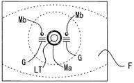
After the face of the examinee is fixed to the face supporting unit 2, an alignment index is projected onto the cornea of the eye E to be examined, and the measurement unit 4 and the eye to be examined are aligned. Before aligning the eye to be examined E, the examiner operates the switch section 8 to select the refractive power measurement mode for the angle of incidence. The control unit 70 detects an alignment state with respect to the subject's eye E based on an image pickup signal from the image pickup device 52. [ The control unit 70 obtains the alignment displacement in the X and Y directions by calculating the center position (approximately the center of the cornea) of the index image Ma. The alignment state in the Z direction is detected from the positional relationship of the four index images formed by the Z-direction alignment index projection optical system 45. [ The fitting of the alignment state in the Z direction is performed by comparing the image interval of the two endless surface images by the first projection optical systems 45a and 45b and the image interval of the finite circle surface image by the second projection optical systems 45c and 45d . In the projection of the infinite target table, even if the Z direction changes, the image interval is hardly changed. On the other hand, in the projection of the finite source target table, the image interval changes with the change in the Z direction. This characteristic can be used to determine the alignment state in the Z direction (see Japanese Patent Application Laid-Open No. 6-46999). The control unit 70 increases or decreases the number of the indicators G based on the alignment detection result in the Z direction.
The control unit 70 moves the measuring unit 4 in the XY directions based on the index image formed by the light sources 46c and 46d and detects the four index images formed by the Z direction alignment index projection optical system 45 The measuring unit 4 is moved in the Z direction. When the alignment states in the X, Y, and Z directions are within the predetermined allowable ranges, the controller 70 determines alignment completion and automatically issues a measurement start signal to perform measurement. In the case of manual measurement, after the inspector completes the alignment by operating the joystick 5 or the like, a measurement start switch (not shown) is pressed to input a measurement start signal.
When the trigger signal is output, the control unit 70 turns on the measurement infrared point light source 11 to project the measurement index to the fundus Ef. Then, the control unit 70 receives the reflected light by the image pickup element 22, and detects an index image (ring image R). At this time, the preliminary measurement is performed at first, and based on the result, the visible light source 31 and the noticed plate 32 for moving the notified table are moved in the optical axis direction, and the eye to be examined is caught by the eye E to be examined. Thereafter, this measurement is performed on the eye E to be examined.
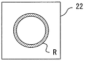
4 is a ring image picked up by the image pickup element 22 by performing measurement with the measurement start signal as a trigger. The output signal from the image pickup device 22 is stored as image data (ring image) in the image memory 71. [ In this measurement of the present embodiment, the ring image (ring image R) is continuously picked up by the image pickup element 22, and the addition / accumulation processing of the ring image is performed. A plurality of image data are stored in the image memory 71 as image data for imaging a ring image on the image pickup device 22 successively based on the number of times of the addition process one or two times and performing addition processing .
Thereafter, the control unit 70 generates the added image data by using the plurality of images stored in the image memory 71. [ The control unit 70 specifies the position of the ring image in each meridian direction based on the image data (thinning). The control unit 70 cuts the waveform of the luminance signal at a predetermined threshold value, finds the midpoint of the waveform at the cut position, the peak of the waveform of the luminance signal, the center position of the luminance signal, . Further, by suppressing the noise light superimposed on the image data by the addition process, it is possible to obtain the measurement result with good accuracy (for details, see Japanese Patent Application Laid-Open No. 2006-187482).
Next, based on the image position of the specified ring image, the control unit 70 approximates the elliptical image using the least squares method or the like. In the method of elliptic approximation, a well-known elliptic approximation expression can be used for measurement of eye refractive power or corneal shape measurement. S (spherical power), C (dioptric power), A (astigmatism axis angle), and A (astigmatic axis angle) are calculated based on the refractive error in each meridian direction from the shape of the approximated ellipse. ), And the measurement result is displayed on the monitor 7.
Next, the flow of the adjustment force measurement mode will be described using Fig. Based on the input signal from the switch unit 8, the control unit 70 detects that the inspector has changed the measurement mode from the angular power measurement mode to the adjustment force measurement mode. In the adjustment force measurement mode, the front eye image F (moving image) of the eye to be examined is displayed on the monitor 7 based on the output signal of the image sensing element 52, similarly to the refractive power measurement mode for the angle of incidence. The movement of the alignment detection and measurement unit 4 in the X, Y, and Z directions is also performed in the same manner as the refractive power measurement mode for the angular range.
7, the control section 70 controls the drive section 37 to change the control amount of the drive section 37 while moving the known table from the far side to the near side (see steps S108 to S111 in Fig. 7) ). Further, in the present embodiment, the second notifying plate 32b is used as a noticable notation, but the present invention is not limited to this, and the first notifying plate 32a may be used. In the case of changing the control amount, for example, the control unit 70 determines whether or not the movement speed of the high-speed table or the movement amount in each step during the stepwise movement of the high- Or the like.
In the present embodiment, the control unit 70 monitors the position information of the time table and the measurement result of the eye refractive power in real time while the noticed table is moved from the far side to the near side. These monitoring results are used for changing the control amount. In the monitoring, for example, the control unit 70 acquires the on-time position information and the measurement result continuously or at predetermined time intervals, and updates them as needed. Further, the control unit 70 may store the measurement result at each position in the memory 74 in association with the current position.
In the present embodiment, as shown in Fig. 7, the control unit 70 determines whether or not the control amount is changed based on the time position information of the notified table and the measurement result of the ophthalmic force at the predetermined distance, . Thereby, the follow-up state of the to-be-observed table of the eye to be examined is reflected in the control amount. For example, the controller 70 may reduce the moving speed of the high-precision table or the amount of movement at each step when the regular position of the high-precision table exceeds the allowable range with respect to the eye refractive index. The control unit 70 may also control the movement of the predictive chart so that the current position of the predictive chart does not exceed the allowable range with respect to the eye refractive index at the time of measurement at the current time. In addition, the measurement position of the eye position and the measurement result of the ophthalmic force are converted into, for example, a diopter value (D) or a distance value (m). The control unit 70 also changes the control amount of the driving unit 37 so that the normal position of the check table does not exceed the set threshold value with respect to the measurement result of the minus side measured while the check table is moved from the far- .
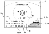
8 shows a display screen of the monitor 7 in the adjustment force measurement mode. The monitor 7 is provided with a measurement value S, C, and A measured in the refractive power measurement mode for the angular range and a refractive power minimum value Dh representing the minimum value of the refractive power (diopter) (Dp) obtained by converting the current time difference of the current time table into diopter, and a measurement elapsed time (Tacm) indicating the elapsed time from the start of measurement of the accommodation force are displayed. As described later, the diopter adjustment force (Da) obtained by subtracting the refractive power measured at the current distance from the current refraction table (Dh) from the refractivity minimum value (Dh) serving as the origin, (GLPa) representing the change in the diopter of the moving table and the diopter conversion table (GLPb) plotting the diopter of the moving distance of the moving table. The unit of the horizontal axis of the graph of the refractive power change (GLPa) and the standardized table conversion graph (GLPb) is time (second), and the unit of the vertical axis represents diopter (diopter value).
≪ Step S101 &
The control unit 70 controls the driving unit 34 to select the type of the noticed plate 32 from the first noticed plate 32a used in the mode for measuring the dioptric power for the user and the second noticed plate 32b. FIG. 6 is a second notional plate 32b used in the adjustment force measurement mode. The second notation board 32b is composed of a geometric pattern or letter composed of circles or lines. In this embodiment, the design of the notice board 32 is changed in the power measurement mode and the adjustment force measurement mode for the angle of incidence, but the present invention is not limited to this. The notation plate of the same design may be used in the refractive power measurement mode and the accommodation power measurement mode for the angle of incidence.
≪ Step S102 &
The control unit 70 controls the driving unit 37 to move the regular distance of the examination table to a position shifted by 0.5 diopter to the remote power Dn measured in the mode for measuring the power for the angle of incidence. In the adjustment force measurement mode, the regular distance of the check table is moved from the far side to the near side. An indicator is arranged at a farther side than the far-field power of the eye E measured with a good precision in the refractive power measurement mode for the angle of incidence, and during the measurement of the adjustment power from the far side to the near side, By passing the distance that is the distance for the far-field power Dn, the followability (visibility) of the high-precision chart can be ensured at the initial stage of the adjustment force measurement and the origin can be detected quickly.
≪ Step S103 &
The control unit 70 monitors the state of the start switch (not shown) disposed at the front end of the joystick 5 when the change of the notify table is completed in step S101 and the move of the notify table is completed in step S102. When the inspector detects that the start switch is pressed to start the adjustment force measurement, the controller 70 outputs a measurement start signal and proceeds to step S104. While there is no change in the start switch, the measurement is waited at step S103. Further, in the measurement of the accommodation force, since the subject's eye E needs to follow the schedule table in which the time-varying distance changes, the examiner must follow the time- It is desirable to explain to make. Therefore, unlike the refractive power measurement mode for the angle of incidence in this embodiment, the control unit 70 does not automatically start the measurement of the adjustment force even when the alignment state of the eye to be examined E and the measurement unit 4 falls within the predetermined allowable range, When the change of the notified table and the movement of the appointed time distance of the notified time of the notified table are completed, a message or an icon (icon) indicating completion of preparation is displayed on the monitor 7.
≪ Step S104 &
When the control unit 70 detects that the start switch is depressed, it determines the alignment state of the eye to be examined E and the measurement unit 4. [ When the alignment state of the eye to be examined E and the measurement section 4 is within the predetermined allowable range, the process proceeds to step S106. When the alignment state of the eye and subject measurement unit 4 is not within the predetermined allowable range, the process proceeds to step S105. In addition, the same as the alignment detection condition of the predetermined allowable range of the cornering angular power for the refractive power measurement mode.
≪ Step S105 &
In step S105, the alignment state of the subject's eye E and the measurement unit 4 is detected immediately after the start switch is pressed, and then the elapsed time during which the alignment state does not fall within the predetermined allowable range is compared with a predetermined determination time. If the elapsed time has not reached the predetermined determination time, the process returns to step S104. When the elapsed time reaches a predetermined determination time, the measurement is interrupted and the process returns to step S103. In the present embodiment, when the elapsed time that the alignment state does not fall within the predetermined allowable range continues for at least 5 seconds immediately after the start switch is pushed, an error is determined and the measurement is stopped. When the measurement is stopped and the process returns to step S103, a graphic (icon) indicating that the measurement is started is displayed by pressing the start switch again on the monitor 7. [
<Step S106>
The control unit 70 monitors the vertical synchronizing signal outputted from the image pickup device 22 and waits for the timing at which the image pickup device 22 starts a new exposure period. The control unit 70 monitors the vertical synchronization signal output from the image pickup device 22 and waits for an exposure period necessary for obtaining the power. Here, the method of measuring the refractive power (first refractive power measurement) for determining the initial on-time position of the refractive power measurement mode and the adjustment chart of the adjustment power measurement (the first refractive power measurement) and the method of measuring the refractive power measured during the measurement of the adjusting power of the adjustment power measurement mode Refractive power measurement). The exposure of the imaging element 22 necessary for obtaining the refracting power of the eye E to be examined is not necessary in the adjustment force measurement mode and it is unnecessary to perform the projection of the eye E in the adjustment force measurement mode The period (time) is set to a time shorter than the waiting time required in the power source for measuring the power for the angle of incidence. In the refractive power measurement mode for the angle of incidence, the output signal of the imaging element 22 is subjected to addition processing in order to measure the far-field power of the eye E to be examined with good precision. In the adjustment force measurement mode, Do not.
More specifically, in the refractive power measurement mode for the angle of incidence, one or two additions are performed using output signals (one image) sequentially output from the image pickup element 22 at intervals of 1/30 second. Since the two additions are performed with three images, a maximum of about 100 ms is required as the exposure time required to obtain the refracting power. On the other hand, in the adjustment force measurement mode, since the refracting power is obtained by only one image without addition, the exposure time required to obtain the refracting power is 1/30 second (about 33 ms).
Further, the refracting power of the eye E to be examined changes according to the regular distance of the watch table. (Ring image) in which the image capturing element 22 receives the plurality of components of the refractive power resulting from the change of the regular distance of the reference table is moved (changed) during the exposure period of the image capturing element 22, And the precision of the refractive power is lowered. Since the origin and the approximate point are determined based on the change in the refractive power of the eye to be examined E, the adjustment force can be obtained with the refractive power with insufficient reliability when the regular distance of the chart is changed during the exposure period of the imaging element 22. [ Therefore, the adjustment force measurement of the present embodiment measures the refractive power of the subject's eye E while moving the regular distance of the target table substantially continuously from the far side to the vicinity. The control unit 70 performs a control (that is, a control for temporarily stopping movement of the high-priority table) during the exposure period of the imaging device 22 necessary for obtaining the refracting power so as not to move the regular distance of the high-
<Step S107>
Based on the output signal of the imaging element 22 acquired in step S106, the control unit 70 obtains the refractive power of the subject's eye E at the predetermined distance in the corresponding high-priority table. The measurement of the refractive power is performed in the same manner as the measurement of the refractive power for the angle of incidence, except that the addition is not performed.
≪ Step S108 &
The control unit (70) determines the follow-up state for the noticed table of the subject's eye based on the positional information of the accurate table and the deviation of the measurement result of the eye refractive power.
More specifically, the control unit 70 subtracts the diopter value obtained in step S107 from the diopter value determined based on the regular distance of the examination table to determine the distance to the subject's eye E at the predetermined distance of the corresponding examination table, The following evaluation value DELTA D (diopter value) is obtained. In step S108, the follow evaluation value? D is compared with a predetermined condition (first condition). If the pursuit evaluation value? D is larger than minus 1 diopter, the process proceeds to step S110. If the pursuit evaluation value? D is equal to or less than minus one diopter, the process proceeds to step S109. Further, in the present embodiment, when the follow-up ability of the eye to be examined is good with respect to the regular distance of the moved high-precision table, the follow-up evaluation value? D takes a value larger than minus one diopter (e.g., minus 0.5 diopter). Further, as the followability of the eye to be examined is worse, the follow-up evaluation value? D takes a smaller value (for example, minus 2.0 diopters).
≪ Step S109 &
The control unit 70 compares the following evaluation value? D with a predetermined condition in the same manner as in step S108. However, the condition for the comparison is the second condition different from the step S109. If the follow evaluation value D is larger than minus 1.75 diopter, the process goes to step S111, and if the follow-up evaluation value D is minus 1.75 diopter or less, the process goes to step S112.
<Steps S110 and S111>
The control unit 70 changes the control amount of the high-priority table based on the determination result of the above-described follow-up state. The control unit 70 also changes the movement control of the preview table based on the current position information of the preview table and the measurement result of the eye refractive power at the predetermined distance.
More specifically, the control unit 70 moves the regular distance of the high-priority table based on the results of the determination in steps S108 and S109. In step S110, the current time distance of the noticed table is moved to the vicinity of two times. In step S111, the process moves to the vicinity by one step. In the present embodiment, when the drive unit 37 is controlled so as to move the regular distance of the high-precision table by one step, the regular distance of the high-precision table moves by a distance of 0.05 diopter in terms of diopter. The control unit 70 determines whether or not a predetermined elapsed time has elapsed from the timing at which the drive unit 37 was controlled with respect to whether or not the high-priority table is being moved. The determination as to whether or not the noticable table is being moved is not limited to this. Detecting means for detecting the amount of movement may be provided at a point where the second notifying plate 32b moves.
As described above, the control unit 70 obtains the adjustment follow-up state of the eye to be examined E from the diopter value based on the regular distance of the noticed chart and the refractive power of the eye to be examined E measured at the above- And reflects the control for varying the regular time distance of the table. In short, when the regular distance of the examination table moves from the far side (near the origin) to the vicinity, the limit of the control force of the eye E slowly appears. As a result, the followability of the eye chart of the eye E to be examined deteriorates (the difference between the diopter values coming from the regular distance and the diopter values measuring the refractive power of the eye E to be examined is increased).
The control unit 70 detects the adjustment follow-up state of the eye to be examined E from the regular distance of the notified chart and the measured refractive power. The control unit 70 controls the movement of the notified chart based on the detected follow-up state so that the examinee does not give up the adjustment sooner than the limit of the control force of the eye E to be examined. For example, when the state of the eye to be examined E is good, the control unit 70 moves the regular time distance of the noticed table large (fast). The controller 70 determines whether or not the movement of the regular distance of the notification table is small or the control of the eye to be examined E is to be followed when the control condition of the eye E to be examined deteriorates . Therefore, it is possible to measure the control force of the eye to be examined E in a short period of time while ensuring the control compliance of the eye E to be examined.
In the present embodiment, when the follow-up evaluation value? D is equal to or less than minus 1.75 diopters (second condition) in step S109, the process proceeds to step S112 to stop the regular position of the high-precision table. Here, a third condition may be formed in which the comparison result NO is determined in step S109, and the follow-up evaluation value? D is additionally determined in the section proceeding to step S112. If the follow-up evaluation value? D is smaller than minus 2 diopters as the third condition, the current position of the high-vision table is shifted to the far side by one step, and if the follow-up evaluation value? D is minus 2 diopters or more, Proceed. When the control of the eye to be examined E is clearly worse with respect to the detected control follow-up state, the control unit 70 returns the regular time distance of the check table to the opposite direction to the direction in which the moved eye has been moved, May be controlled so as to promote the follow-up of the control.
Further, in the present embodiment, when changing the regular distance of the high-priority table based on the adjustment follow-up state of the eye to be examined E, the control unit 70 changes only the moving distance at a constant speed. The control unit 70 may change the moving speed of the regular distance of the target table based on the control follow-up state of the detected eye E that is detected because the control target of the eye to be examined E changes according to the moving speed of the target table. In this embodiment, one cycle (S106 to S113) during the adjustment measurement is about 83 ms, and about 40% of one cycle is the light-receiving period of the image sensing element 22. [ During the measurement of the adjusting force, the above-described one cycle is continuously performed until the measurement is completed. Therefore, in the present embodiment, the control unit 70 performs control so as to change only the moving distance without changing the moving speed of the known table, but it is almost the same as changing the moving distance and the moving speed in one cycle And is being controlled.
≪ Step S112 &
The control unit 70 displays the measured rectilinear force Da on the monitor 7 and also draws the refractive power change graph GLPa and the high table conversion graph GLPb. In addition, the rectilinear control force Da becomes a value (diopter) obtained by subtracting the reference value Dp from the refractive power minimum value Dh. The graph of the refractive power change (GLPa) is a graph showing the change of rectus control force (Da) during the adjustment force measurement. (GLPb) is a graph showing the change of the regular distance of the check table during the adjustment force measurement. The unit of the horizontal axis of the graph of the refractive power change (GLPa) and the standardized table conversion graph (GLPb) is time (second), and the unit of the vertical axis represents diopter (diopter value). The control unit 70 implements the drawing at the position on the abscissa corresponding to the elapsed time from the start of the adjustment force measurement. That is, in this embodiment, the power variation graph GLPa and the high table conversion graph GLPb are updated each time one cycle (from step S 106 to step S 113) has elapsed from the start of measurement, and from the start of measurement to the completion of measurement, Is extended to the right side.
The refractive power change graph GLPa and the lookup table conversion graph GLPb are displayed on the screen of the adjustment force measurement mode for observing the anterior segment of the eye E to be superimposed on the anterior segment image F . In order to prevent the visibility of the front anterior image F from being disturbed, an area of 20% or less of the entire display area of the monitor 7 with respect to the foreground image F displayed on the entire display area of the monitor 7 And is disposed at the lower right position of the display area of the monitor 7. By arranging in this way, the refractive power change graph (GLPa) and the high table conversion graph (GLPb) do not overlap well with the pupil area of the eye to be examined (E) displayed on the monitor (7) as well as the iris area. Accordingly, the examiner can check the progress state of the accommodation force measurement (the follow-up state of the refractive power of the subject's eye E with respect to the regular distance of the face book) while observing the front face image of the subject's eye preferably. In addition, in the measurement according to the present embodiment, the renewal (addition of drawing) of the refractivity change graph GLPa and the look-up table conversion graph GLPb is carried out for a predetermined elapsed time (for example, 2 Once per cycle). In addition, as described later, the graph of the refractive power change GLPa changes color in the vertical direction in the drawn area.
≪ Step S113 &
The control unit 70 also stores the eye refractive power of the eye to be examined at each predetermined position in association with the memory 74. [ Here, the memory 74 also serves as holding means (peak hold means) for holding the maximum or minimum value of the refractive power during the measurement. If there is acquisition of the refracting power exceeding the maximum value or the minimum value held in the memory 74 during the measurement, the control unit 70 updates the maximum value or the minimum value held in the predetermined address of the memory 74 do. In this manner, the control unit 70 determines whether or not to terminate the measurement of the adjustment force, and ends the movement of the fixation table based on the determination result. For example, when the elapsed time from the start of the measurement exceeds 30 seconds, or when the maximum value of the refractive power during the adjustment force measurement is not changed for 6 seconds or more, Sec, it is determined that the predetermined condition is satisfied and measurement of the adjustment force is completed. If the predetermined condition is not satisfied, the routine proceeds to step S106 to continue the measurement of the adjustment force.
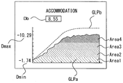
When the condition of the measurement completion is satisfied, an icon (icon) that can be changed to a screen for confirming the result of the adjustment force measurement is displayed on the monitor 7. 9 is a screen displayed when the inspector presses a result display switch (not shown) of the switch unit 8 to confirm the result of the adjustment force measurement. The adjustment result screen displays an adjustment force Db of the eye to be examined E as a measurement result, an approximate value Dmax based on the maximum value of the refractive power measured during the adjustment force measurement, an origin value based on the minimum value of the refractive power measured during the adjustment force measurement Dmin), a refractive power change graph (GLPa), and a proof sheet conversion graph (GLPb). Further, the graph of the refractive power change (GLPa) changes the color toward the vertical direction and draws it. The area of area 1 is searched (water color), the area of area 2 is green, the area of area 3 is yellow, and the area of area 4 is orange. In addition, the near boundary in which each area is touched from Area 1 to Area 4 is displayed using an intermediate color. By changing the color of the graph extending in the vertical direction in this way, the examiner can easily grasp the change of the follow-up state of the eye to be examined E during the adjustment force measurement, and it is easy to capture the maximum adjustment force even in the adjustment result screen.
In the present embodiment, the control unit 70 controls the control force Db of the eye to be examined based on the maximum value and the minimum value in the eye refractive power of the eye to be examined at each predetermined time when the regular position of the noticed table is moved away from the far- . This makes it possible to improve the measuring ability of the accommodation force.
When the inspector operates a print switch (not shown) formed on the switch unit 8 in a state in which the adjustment result screen is displayed, the control unit 70 controls the printer 78 to print the measurement result. Fig. 10 shows an example of printing when a thermal printer is used. In addition to the information PRa and PRb measured in the refractive power measurement mode for the angle of incidence on the printed sheet, an adjustment force Db of the eye to be examined E measured in the adjustment force measurement mode, an approximate value based on the maximum value of the refractive power measured during the adjustment force measurement An origin value Dmin based on the minimum value of the refractive power measured during the adjustment force measurement, and a printing power change graph PRc are printed as measurement results.
In the present embodiment, the start position of the adjustment force measurement mode is set and determined from the refraction value measured by the refractive power measurement mode for the angle of incidence, but the present invention is not limited to this. When the start button is depressed, the refractive power of the subject's eye may be measured with the same control contents as the refractive power measurement mode for the tapered person, and the predictive table may be moved to the regular position by calculating the far-end position.
In the above description, the measurement optical system for acquiring the ring pattern image by the fundus reflection light is described as an example, but the present invention is not limited to this. The present invention can be applied as long as it is a device for measuring the adjustment force of the eye to be examined E by measuring the angular power of the eye by moving the regular distance of the eye chart. For example, a measurement optical system may be used in which a spot indicator is projected on the fundus Ef of the eye E to detect the wavefront aberration in the subject, and the fundus reflection light is detected using a Shark Hartmann sensor.
In the above description, when the control amount is changed using the monitoring result, the control amount is changed when the time position information of the high-priority table and the measurement result of the ophthalmic force at the predetermined distance do not satisfy the first allowable range , But it is not limited thereto.
For example, based on the change in the measurement result of the ophthalmic force while the regular position of the noticed table is moved from the far side to the near side, the control unit 70 determines whether or not the follow-up state of the subject's eye (for example, Is good). If it is determined that the tracking state is worse, the control unit 70 may change the control amount.
More specifically, the control unit 70 may change the control amount in accordance with the measurement result of the ophthalmic force at the predetermined distance. Specifically, for example, when the amount of change of the eye refractive power per unit time decreases, it is considered that there is a change in the followability of the eye to be examined with respect to the noticed chart. Thus, the control unit 70 may change the amount of control (for example, the moving speed of the high-precision table or the moving amount of the high-precision table in each step) in accordance with the amount of change in the refractive power of the eye per unit time. Thereby, the inspector can follow the notices table. Therefore, the original adjustment force can be measured smoothly. Further, when the amount of change is increased, the amount of control is increased. When the amount of change is reduced (or when the amount of change is lost), the control amount may be reduced.
Also, the control unit 70 may change the control amount in accordance with, for example, the regular position of the high-priority table. For example, the closer the nominal distance of the notice table is to the test, the larger the adjustment load becomes, and the follow-up becomes difficult. Therefore, the control amount (for example, the moving speed of the high-speed table or the moving amount of the high-speed table in each step) may be changed according to the regular position of the high-speed table. Thereby, the inspector can follow the noticable table. Therefore, the original adjustment force can be measured smoothly. Further, when the regular time distance is far from the subject, the control amount is increased, and when the regular time distance is close to the subject, the control amount is reduced.
In the above description, when the time position information of the noticed table and the measurement result of the ophthalmic force at the predetermined distance do not satisfy the permissible range, the moving direction of the notified table is changed or the movement of the noticed table is once stopped, . In short, the control unit 70 determines whether or not the subject's follow-up with respect to movement of the target eye chart is possible based on the change in the measurement result of the ophthalmic force while the regular position of the target face is moved from the far side to the near side. Then, when it is determined that the eye to be examined can not be followed, the control unit 70 may change the moving direction of the noticed table or temporarily stop the movement of the noticed table. For example, the control unit 70 determines whether or not the eye to be examined can follow the notified chart based on whether or not the amount of change of the eye refractive power per unit time satisfies the allowable range. Then, when it is determined that the amount of change does not satisfy the allowable range, the control unit 70 may change the moving direction of the high-priority table or temporarily stop the movement of the high-priority table.
In the above description, the movement of the preview table is terminated after the elapse of the predetermined time after the change in the measurement result of the ocular refractive power is switched to the downward direction. However, the present invention is not limited to this. In short, the control unit 70 determines whether or not the subject's follow-up with respect to movement of the target eye chart is possible based on the change in the measurement result of the ophthalmic force while the regular position of the target face is moved from the far side to the near side. Then, when the state in which the eye to be examined can not be followed has continued for a predetermined time, the control unit 70 may terminate the movement of the notification table. For example, the control unit 70 determines whether or not the examinee can follow the noticed table based on whether or not the amount of change of the eye refractive power per unit time satisfies the allowable range, regardless of the change of the control amount. Then, the control unit 70 may end the movement of the high-priority table when the state in which the amount of change does not satisfy the allowable range has continued for a predetermined period of time.
In the above description, the control method of changing the control amount has been described taking as an example the case where the control force of the eye to be examined is measured based on the eye refractive power of the eye to be examined at each regular time position when the regular position of the check table is moved away from the remote , But it is not limited thereto. For example, when measuring the ophthalmic force in the vicinity of the eye to be examined, the technique of the present embodiment can be applied even when the eye chart is moved from the far side to the near side.

