KR20140063513A - Compositions and methods for improving prognosis of a human with subarachnoid hemorrhage - Google Patents
Compositions and methods for improving prognosis of a human with subarachnoid hemorrhage Download PDFInfo
- Publication number
- KR20140063513A KR20140063513A KR1020137023962A KR20137023962A KR20140063513A KR 20140063513 A KR20140063513 A KR 20140063513A KR 1020137023962 A KR1020137023962 A KR 1020137023962A KR 20137023962 A KR20137023962 A KR 20137023962A KR 20140063513 A KR20140063513 A KR 20140063513A
- Authority
- KR
- South Korea
- Prior art keywords
- less
- calcium channel
- voltage
- channel blocker
- another embodiment
- Prior art date
Links
Images


Classifications
-
- A—HUMAN NECESSITIES
- A61—MEDICAL OR VETERINARY SCIENCE; HYGIENE
- A61K—PREPARATIONS FOR MEDICAL, DENTAL OR TOILETRY PURPOSES
- A61K31/00—Medicinal preparations containing organic active ingredients
- A61K31/33—Heterocyclic compounds
- A61K31/395—Heterocyclic compounds having nitrogen as a ring hetero atom, e.g. guanethidine or rifamycins
- A61K31/435—Heterocyclic compounds having nitrogen as a ring hetero atom, e.g. guanethidine or rifamycins having six-membered rings with one nitrogen as the only ring hetero atom
- A61K31/44—Non condensed pyridines; Hydrogenated derivatives thereof
-
- A—HUMAN NECESSITIES
- A61—MEDICAL OR VETERINARY SCIENCE; HYGIENE
- A61K—PREPARATIONS FOR MEDICAL, DENTAL OR TOILETRY PURPOSES
- A61K9/00—Medicinal preparations characterised by special physical form
- A61K9/0012—Galenical forms characterised by the site of application
- A61K9/0085—Brain, e.g. brain implants; Spinal cord
-
- A—HUMAN NECESSITIES
- A61—MEDICAL OR VETERINARY SCIENCE; HYGIENE
- A61K—PREPARATIONS FOR MEDICAL, DENTAL OR TOILETRY PURPOSES
- A61K31/00—Medicinal preparations containing organic active ingredients
- A61K31/33—Heterocyclic compounds
- A61K31/395—Heterocyclic compounds having nitrogen as a ring hetero atom, e.g. guanethidine or rifamycins
- A61K31/435—Heterocyclic compounds having nitrogen as a ring hetero atom, e.g. guanethidine or rifamycins having six-membered rings with one nitrogen as the only ring hetero atom
- A61K31/44—Non condensed pyridines; Hydrogenated derivatives thereof
- A61K31/4422—1,4-Dihydropyridines, e.g. nifedipine, nicardipine
-
- A—HUMAN NECESSITIES
- A61—MEDICAL OR VETERINARY SCIENCE; HYGIENE
- A61K—PREPARATIONS FOR MEDICAL, DENTAL OR TOILETRY PURPOSES
- A61K9/00—Medicinal preparations characterised by special physical form
- A61K9/0012—Galenical forms characterised by the site of application
- A61K9/0019—Injectable compositions; Intramuscular, intravenous, arterial, subcutaneous administration; Compositions to be administered through the skin in an invasive manner
-
- A—HUMAN NECESSITIES
- A61—MEDICAL OR VETERINARY SCIENCE; HYGIENE
- A61K—PREPARATIONS FOR MEDICAL, DENTAL OR TOILETRY PURPOSES
- A61K9/00—Medicinal preparations characterised by special physical form
- A61K9/06—Ointments; Bases therefor; Other semi-solid forms, e.g. creams, sticks, gels
-
- A—HUMAN NECESSITIES
- A61—MEDICAL OR VETERINARY SCIENCE; HYGIENE
- A61K—PREPARATIONS FOR MEDICAL, DENTAL OR TOILETRY PURPOSES
- A61K9/00—Medicinal preparations characterised by special physical form
- A61K9/14—Particulate form, e.g. powders, Processes for size reducing of pure drugs or the resulting products, Pure drug nanoparticles
- A61K9/16—Agglomerates; Granulates; Microbeadlets ; Microspheres; Pellets; Solid products obtained by spray drying, spray freeze drying, spray congealing,(multiple) emulsion solvent evaporation or extraction
- A61K9/1605—Excipients; Inactive ingredients
- A61K9/1629—Organic macromolecular compounds
- A61K9/1641—Organic macromolecular compounds obtained otherwise than by reactions only involving carbon-to-carbon unsaturated bonds, e.g. polyethylene glycol, poloxamers
- A61K9/1647—Polyesters, e.g. poly(lactide-co-glycolide)
-
- A—HUMAN NECESSITIES
- A61—MEDICAL OR VETERINARY SCIENCE; HYGIENE
- A61K—PREPARATIONS FOR MEDICAL, DENTAL OR TOILETRY PURPOSES
- A61K9/00—Medicinal preparations characterised by special physical form
- A61K9/14—Particulate form, e.g. powders, Processes for size reducing of pure drugs or the resulting products, Pure drug nanoparticles
- A61K9/16—Agglomerates; Granulates; Microbeadlets ; Microspheres; Pellets; Solid products obtained by spray drying, spray freeze drying, spray congealing,(multiple) emulsion solvent evaporation or extraction
- A61K9/1605—Excipients; Inactive ingredients
- A61K9/1629—Organic macromolecular compounds
- A61K9/1652—Polysaccharides, e.g. alginate, cellulose derivatives; Cyclodextrin
-
- A—HUMAN NECESSITIES
- A61—MEDICAL OR VETERINARY SCIENCE; HYGIENE
- A61M—DEVICES FOR INTRODUCING MEDIA INTO, OR ONTO, THE BODY; DEVICES FOR TRANSDUCING BODY MEDIA OR FOR TAKING MEDIA FROM THE BODY; DEVICES FOR PRODUCING OR ENDING SLEEP OR STUPOR
- A61M25/00—Catheters; Hollow probes
- A61M25/01—Introducing, guiding, advancing, emplacing or holding catheters
-
- A—HUMAN NECESSITIES
- A61—MEDICAL OR VETERINARY SCIENCE; HYGIENE
- A61M—DEVICES FOR INTRODUCING MEDIA INTO, OR ONTO, THE BODY; DEVICES FOR TRANSDUCING BODY MEDIA OR FOR TAKING MEDIA FROM THE BODY; DEVICES FOR PRODUCING OR ENDING SLEEP OR STUPOR
- A61M5/00—Devices for bringing media into the body in a subcutaneous, intra-vascular or intramuscular way; Accessories therefor, e.g. filling or cleaning devices, arm-rests
- A61M5/14—Infusion devices, e.g. infusing by gravity; Blood infusion; Accessories therefor
- A61M5/158—Needles for infusions; Accessories therefor, e.g. for inserting infusion needles, or for holding them on the body
-
- A—HUMAN NECESSITIES
- A61—MEDICAL OR VETERINARY SCIENCE; HYGIENE
- A61P—SPECIFIC THERAPEUTIC ACTIVITY OF CHEMICAL COMPOUNDS OR MEDICINAL PREPARATIONS
- A61P25/00—Drugs for disorders of the nervous system
-
- A—HUMAN NECESSITIES
- A61—MEDICAL OR VETERINARY SCIENCE; HYGIENE
- A61P—SPECIFIC THERAPEUTIC ACTIVITY OF CHEMICAL COMPOUNDS OR MEDICINAL PREPARATIONS
- A61P25/00—Drugs for disorders of the nervous system
- A61P25/28—Drugs for disorders of the nervous system for treating neurodegenerative disorders of the central nervous system, e.g. nootropic agents, cognition enhancers, drugs for treating Alzheimer's disease or other forms of dementia
-
- A—HUMAN NECESSITIES
- A61—MEDICAL OR VETERINARY SCIENCE; HYGIENE
- A61P—SPECIFIC THERAPEUTIC ACTIVITY OF CHEMICAL COMPOUNDS OR MEDICINAL PREPARATIONS
- A61P29/00—Non-central analgesic, antipyretic or antiinflammatory agents, e.g. antirheumatic agents; Non-steroidal antiinflammatory drugs [NSAID]
-
- A—HUMAN NECESSITIES
- A61—MEDICAL OR VETERINARY SCIENCE; HYGIENE
- A61P—SPECIFIC THERAPEUTIC ACTIVITY OF CHEMICAL COMPOUNDS OR MEDICINAL PREPARATIONS
- A61P3/00—Drugs for disorders of the metabolism
-
- A—HUMAN NECESSITIES
- A61—MEDICAL OR VETERINARY SCIENCE; HYGIENE
- A61P—SPECIFIC THERAPEUTIC ACTIVITY OF CHEMICAL COMPOUNDS OR MEDICINAL PREPARATIONS
- A61P3/00—Drugs for disorders of the metabolism
- A61P3/12—Drugs for disorders of the metabolism for electrolyte homeostasis
- A61P3/14—Drugs for disorders of the metabolism for electrolyte homeostasis for calcium homeostasis
-
- A—HUMAN NECESSITIES
- A61—MEDICAL OR VETERINARY SCIENCE; HYGIENE
- A61P—SPECIFIC THERAPEUTIC ACTIVITY OF CHEMICAL COMPOUNDS OR MEDICINAL PREPARATIONS
- A61P35/00—Antineoplastic agents
-
- A—HUMAN NECESSITIES
- A61—MEDICAL OR VETERINARY SCIENCE; HYGIENE
- A61P—SPECIFIC THERAPEUTIC ACTIVITY OF CHEMICAL COMPOUNDS OR MEDICINAL PREPARATIONS
- A61P7/00—Drugs for disorders of the blood or the extracellular fluid
-
- A—HUMAN NECESSITIES
- A61—MEDICAL OR VETERINARY SCIENCE; HYGIENE
- A61P—SPECIFIC THERAPEUTIC ACTIVITY OF CHEMICAL COMPOUNDS OR MEDICINAL PREPARATIONS
- A61P7/00—Drugs for disorders of the blood or the extracellular fluid
- A61P7/02—Antithrombotic agents; Anticoagulants; Platelet aggregation inhibitors
-
- A—HUMAN NECESSITIES
- A61—MEDICAL OR VETERINARY SCIENCE; HYGIENE
- A61P—SPECIFIC THERAPEUTIC ACTIVITY OF CHEMICAL COMPOUNDS OR MEDICINAL PREPARATIONS
- A61P7/00—Drugs for disorders of the blood or the extracellular fluid
- A61P7/04—Antihaemorrhagics; Procoagulants; Haemostatic agents; Antifibrinolytic agents
-
- A—HUMAN NECESSITIES
- A61—MEDICAL OR VETERINARY SCIENCE; HYGIENE
- A61P—SPECIFIC THERAPEUTIC ACTIVITY OF CHEMICAL COMPOUNDS OR MEDICINAL PREPARATIONS
- A61P9/00—Drugs for disorders of the cardiovascular system
-
- A—HUMAN NECESSITIES
- A61—MEDICAL OR VETERINARY SCIENCE; HYGIENE
- A61P—SPECIFIC THERAPEUTIC ACTIVITY OF CHEMICAL COMPOUNDS OR MEDICINAL PREPARATIONS
- A61P9/00—Drugs for disorders of the cardiovascular system
- A61P9/08—Vasodilators for multiple indications
-
- A—HUMAN NECESSITIES
- A61—MEDICAL OR VETERINARY SCIENCE; HYGIENE
- A61P—SPECIFIC THERAPEUTIC ACTIVITY OF CHEMICAL COMPOUNDS OR MEDICINAL PREPARATIONS
- A61P9/00—Drugs for disorders of the cardiovascular system
- A61P9/10—Drugs for disorders of the cardiovascular system for treating ischaemic or atherosclerotic diseases, e.g. antianginal drugs, coronary vasodilators, drugs for myocardial infarction, retinopathy, cerebrovascula insufficiency, renal arteriosclerosis
Abstract
기재된 본 발명은 뇌 손상과 관련된 적어도 하나의 지연 합병증의 징후 또는 증상을 감소시키는, 전압-작동 칼슘 채널 차단제 및 약학적으로 허용되는 담체를 함유하는 치료적 유효량의 약학적 조성물을 뇌 내의 소정의 위치로 이식함으로써 포유동물의 뇌 손상과 관련된 지연 합병증을 치료하고, 포유동물에서 상기 뇌 손상과 관련된 지연 합병증의 예후를 개선시키기 위한 약학적 조성물, 전달 시스템 및 방법을 제공한다.The presently described invention is directed to a pharmaceutical composition comprising a therapeutically effective amount of a pharmaceutical composition comprising a voltage-activated calcium channel blocker and a pharmaceutically acceptable carrier, which reduces the symptoms or symptoms of at least one delayed complication associated with brain damage, Delivery system and method for treating delayed complications associated with brain damage in mammals and for improving the prognosis of delayed complications associated with brain damage in mammals.
Description
관련 출원의 전후 참조Before and after reference of related application
본 출원은 2011년 2월 11일에 출원된 미국 가출원 번호 61/441,695호를 우선권으로 주장하며, 이는 2007년 10월 29일에 출원된 미국 가출원 번호 60/976,902호 및 2007년 6월 11일에 출원된 미국 가출원 번호 60/943,124호를 우선권으로 주장하는 2008년 6월 11일에 출원된 미국 출원 번호 12/137,320호의 일부계속 출원이다. 상기 출원들 각각은 이들의 전체내용이 본원에 참조로서 포함된다.This application claims the benefit of U.S. Provisional Application No. 61 / 441,695, filed on February 11, 2011, which is incorporated herein by reference in its entirety, to U.S. Provisional Application No. 60 / 976,902, filed October 29, 2007, and June 11, 2007 This application is a continuation-in-part of U.S. Serial No. 12 / 137,320, filed June 11, 2008, which claims priority to U.S. Provisional Application No. 60 / 943,124. Each of these applications is incorporated herein by reference in its entirety.
발명의 분야Field of invention
기재된 본 발명은 거미막밑 출혈의 유해한 예후를 치료하기 위한 조성물, 시스템 및 방법에 관한 것이다.The disclosed invention relates to compositions, systems and methods for treating a deleterious prognosis of subarachnoid hemorrhage.
발명의 배경BACKGROUND OF THE INVENTION
인간의 뇌는 전체 체중의 단지 약 2%만 구성하나, 이는 심장박출량의 약 15%를 수용하며, 이의 산소 소비는 신체 전체의 산소 소비의 약 20%이다. 상기 값은 단위 뇌 중량 당 상응하게 높은 혈류 속도에 의해 상쇄되는 뇌의 높은 대사율 및 산소 요구를 나타낸다. 뇌순환은 내경동맥 및 척추동맥에 의해 공급된다. 뇌로의 전체 혈류는 약 750-1000 ml/분이며, 이러한 양 중 약 350 ml는 각각의 내경동맥을 통해 유동하고, 약 100-200 ml는 척추 기저 시스템을 통해 유동한다. 정맥 배출은 내경정맥 및 척추정맥에 의해 배출된다.The human brain constitutes only about 2% of the total body weight, which accounts for about 15% of heart rate, and its oxygen consumption is about 20% of the body's oxygen consumption. This value represents the high metabolic rate and oxygen demand of the brain that is counteracted by correspondingly high blood flow rates per unit brain weight. The brain circulation is supplied by the internal carotid artery and vertebral artery. The total blood flow to the brain is about 750-1000 ml / min, about 350 ml of which flows through each of the internal carotid arteries, and about 100-200 ml flows through the spinal base system. Venous drainage is evacuated by the internal jugular vein and spinal vein.
본원에서 사용되는 용어 "뇌졸중" 또는 "뇌혈관 사고"는 혈관과 관련된 질병으로부터 발생하는 보통 국소적이고 급성인 신경학적 증상 및 징후를 나타낸다. 뇌졸중은 폐쇄성(혈관의 폐쇄로 인함) 또는 출혈성(혈관으로부터의 출혈로 인함)이다. 본원에서 사용되는 용어 "허혈"은 혈관의 비정상적 협소화(협착증)에 대해 원위에서의 감소된 관류 압력이 저항 혈관의 자가조절성 확장에 의해 상쇄되지 않는 경우에 발생하는 혈액 공급 및 산소의 결핍을 나타낸다. 허혈이 충분히 중증이고 연장되는 경우, 뉴런 및 다른 세포 구성요소가 사멸하며, 이러한 상태는 "경색증"으로 언급된다.As used herein, the term "stroke" or "cerebrovascular accident" refers to the usual local and acute neurological symptoms and signs that arise from vascular-related diseases. Stroke is either obstructive (due to vascular occlusion) or hemorrhagic (due to bleeding from blood vessels). As used herein, the term "ischemia" refers to a deficiency of blood supply and oxygen that occurs when the reduced perfusion pressure on the circle is not canceled by autoregulatory dilation of the resistance vessel for abnormal narrowing of the blood vessel (stenosis) . When ischemia is severe enough and prolonged, neurons and other cellular components die, and this condition is referred to as "infarction. &Quot;
출혈은 뇌 표면(실질외(extraparenchymal)), 예를 들어, 윌리스환(circle of Willis)에서 선천성 동맥류의 파열로부터 발생하여, 거미막밑 출혈(SAH)을 야기시킬 수 있다. 출혈은 또한 실질내, 예를 들어, 오래 지속된 고혈압에 의해 손상된 혈관의 파열로부터 발생할 수 있으며, 이는 대뇌반구 내, 뇌간 내, 또는 소뇌 내에서 혈병(뇌내 혈종)을 야기시킬 수 있다. 출혈은 혀혈 또는 경색증을 수반할 수 있다. 뇌내 혈종의 종괴 효과는 인접한 뇌 조직의 혈액 공급을 손상시킬 수 있거나, 거미막밑 출혈은 대뇌 표면 혈관의 반응성 혈관연축(vasospasm)을 야기시켜, 추가의 허혈 뇌 손상을 발생시킬 수 있다. 경색 조직은 또한 이차로 출혈성이 될 수 있다. 동맥류는 종종 뇌로 파열되어 뇌내 혈종을 야기시킬 수 있고, 뇌실로 파열되어 뇌실내 출혈을 야기시킬 수 있다.Bleeding can result from subarachnoid hemorrhage (SAH), resulting from rupture of the congenital aneurysm on the brain surface (extraparenchymal), for example, in the circle of Willis. Bleeding can also result from rupture of blood vessels damaged by parenchymal, for example, long-lasting hypertension, which can lead to blood clots (intracerebral hematoma) within the cerebral hemisphere, intracerebral, or cerebellum. Bleeding may involve tingling or infarction. The mass effect of intracerebral hematoma may impair blood supply to adjacent brain tissue, or subarachnoid hemorrhage may cause reactive vasospasm of cerebral surface vessels, resulting in additional ischemic brain damage. Infarct tissue may also be secondary to hemorrhagic. An aneurysm can often rupture into the brain, causing intracerebral hematoma, and rupture into the ventricle to cause intraventricular hemorrhage.
대부분의 폐쇄성 뇌졸중은 죽상경화증 및 혈전증으로 인한 것이고, 대부분의 출혈성 뇌졸중은 고혈압 또는 동맥류와 관련되어 있으나, 상기 두 유형 중 어느 유형의 뇌졸중도 심장병, 외상, 감염, 신생물, 혈액 질환, 혈관 기형, 면역학적 장애, 및 외인성 독소를 포함하는 많은 원인으로부터 임의의 연령에서 발생할 수 있다.Most occlusive strokes are due to atherosclerosis and thrombosis, and most haemorrhagic strokes are associated with hypertension or aneurysms, but either type of stroke can cause heart disease, trauma, infection, neoplasms, blood disorders, vascular malformations, ≪ RTI ID = 0.0 > immunologic < / RTI > disorders, and exogenous toxins.
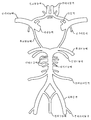
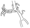
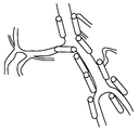
대뇌동맥Large cerebral artery
도 1 및 5는 뇌 혈관의 개략적 예시를 나타낸다. 각각의 대뇌반구는 턱의 모퉁이 아래의 일반적인 경동맥으로부터 발생하고, 경동맥 구멍(carotid foramen)을 통해 두개골로 진입하고, 해면정맥동(cavernosus sinus)(안동맥을 가지냄)을 통과하고, 경질막을 관통하고, 전대뇌동맥 및 중대뇌동맥으로 나뉘어지는 내경동맥에 의해 공급된다. 전대뇌동맥의 큰 표면 가지들은 피질 및 하전두엽(inferior frontal lobe)의 백색질, 전두엽 및 두정엽의 내측면 및 전방 뇌량(anterior corpus callosum)에 공급한다. 더 작은 관통 가지들은 변연계 구조, 미상의 머리(head of the caudate), 및 속섬유막앞다리(anterior limb of the internal capsule)를 포함하는 더 심부의 대뇌 및 간뇌에 공급한다. 중대뇌동맥의 더 큰 표면 가지들은 전두엽, 두정엽, 측두엽 및 후두엽, 및 뇌섬엽을 포함하는 피질 대부분 및 반구 궁륭부(hemisphere's convexity)의 백색질에 공급한다. 더 작은 관통 가지들은 심부 백색질 및 간뇌 구조, 예를 들어, 속섬유막뒤다리(posterior limb of the internal capsule), 피각(putamen), 외부 창백핵(outer globus pallidus), 및 미상몸통(body of the caudate)에 공급한다. 내경동맥이 해면정맥동으로부터 나타난 후, 이는 또한 전방 해마 및 미측(caudal) 수준에서 속섬유막뒤다리에 공급하는 전맥락동맥을 낸다. 각각의 척추동맥은 쇄골하동맥으로부터 나타나, 큰구멍(foramen magnum)을 통해 두개골로 진입하고, 전척수동맥 및 후하소뇌동맥을 낸다. 척추동맥은 교뇌(pons) 및 연수의 접합부에서 연결되어, 교뇌의 수준에서 전하소뇌동맥 및 내이동맥, 및 중뇌에서 위소뇌동맥을 내는 뇌기저동맥을 형성한다. 이후, 뇌기저동맥은 2개의 후대뇌동맥으로 나뉘어진다. 후대뇌동맥의 더 큰 표면 가지들은 아래가쪽 및 중간 후두엽 및 후뇌량에 공급하고, 상기 동맥의 더 작은 관통 가지들은 시상 및 시상밑핵을 포함하는 간뇌 구조 뿐만 아니라 중뇌의 일부에 공급한다(Principles of Neural Sciences, 2d Ed., Eric R. Kandel and James H. Schwartz, Elsevier Science Publishing Co., Inc., New York, pp. 854-56 (1985) 참조).Figures 1 and 5 show a schematic illustration of cerebral blood vessels. Each cerebral hemisphere originates from the common carotid artery beneath the corner of the jaw, enters the skull through the carotid foramen, passes through the cavernosus sinus (carrying the ophthalmic artery), penetrates the hard membrane , The anterior cerebral artery and the middle cerebral artery. The large surface branches of the anterior cerebral artery feed into the white matter, frontal lobes and parietal lining of the cortex and inferior frontal lobe and anterior corpus callosum. The smaller penetrating branches supply the deep brain and ganglia, including the limbic structure, the head of the caudate, and the anterior limb of the internal capsule. Larger surface branches of the middle cerebral artery supply the majority of the cortex, including the frontal lobe, parietal lobe, temporal lobe and occipital lobe, and the hemisphere's convexity. The smaller perforating branches are located in the deep white matter and jugular structures such as the posterior limb of the internal capsule, the putamen, the outer globus pallidus, and the body of the caudate . After the internal carotid artery emerges from the cavernous sinus, it also exerts an anterior coronary artery supplying the posterior hind limb limb at the anterior hippocampus and caudal level. Each vertebral artery originates from the subclavian artery, enters the skull through a foramen magnum, and exits the entire spinal cord artery and posterior calcifying cerebral artery. The vertebral artery is connected at the junction of the pons and the soft tissues to form the basilar arteries of the brachiocephalic artery and the inner ear artery at the level of the brain, and the cerebral artery from the midbrain. Subsequently, the basilar artery is divided into two posterior cerebral arteries. Larger surface branches of the posterior cerebral artery supply to the lower lateral and middle occipital lobes and posterior lobes and smaller branches of the arteries supply to the midbrain as well as the jugular structures including the sagittal and sagittal basal glands (Principles of Neural Science Ed., Eric R. Kandel and James H. Schwartz, Elsevier Science Publishing Co., Inc., New York, pp. 854-56 (1985)).
혈관 사이의 상호연결(문합)은 뇌의 혈관 공급의 일부가 손상되는 경우에 뇌를 보호한다. 문합은 뇌의 혈관 공급의 일부가 손상되는 경우에 뇌를 보호하는 혈관 사이의 상호연결이다. 윌리스환에서, 2개의 전대뇌동맥은 전교통동맥에 의해 연결되고, 후대뇌동맥은 후교통동맥에 의해 내경동맥에 연결된다. 다른 중요한 문합은 안와를 통한 안동맥과 외경동맥의 가지들 사이의 연결, 및 중간대뇌동맥, 전대뇌동맥, 및 후대뇌동맥의 가지들 사이의 뇌 표면에서의 연결을 포함한다(Principles of Neural Sciences, 2d Ed., Eric R. Kandel and James H. Schwartz, Elsevier Science Publishing Co., Inc., New York, pp. 854-56 (1985)).The interconnections between blood vessels (anastomosis) protect the brain in the event that some of the blood supply to the brain is damaged. Anastomosis is the interconnection between blood vessels that protect the brain in the event that part of the blood supply to the brain is damaged. In the Willis circle, the two proximal cerebral arteries are connected by the entire traffic artery, and the posterior cerebral artery is connected to the internal carotid artery by the posterior communicating artery. Other important anastomoses include the connection between the branches of the ophthalmic artery and the external carotid artery through the orbit and the connection at the brain surface between the branches of the middle cerebral artery, the proximal cerebral artery, and the posterior cerebral artery (Principles of Neural Sciences, 2d Ed , Eric R. Kandel and James H. Schwartz, Elsevier Science Publishing Co., Inc., New York, pp. 854-56 (1985)).
통상적으로 한 말단에 머리 및 입을 갖고, 반대 말단에 항문 및 꼬리를 종종 갖는 동물에 대해 언급하는 경우, 머리 말단은 두개 말단으로 언급되는 한편, 꼬리 말단은 미측 말단으로 언급된다. 머리 자체 내에서, 입쪽은 코의 말단을 향한 방향을 나타내고, 미측은 꼬리 방향을 나타내는데 사용된다. 중력의 당김으로부터 멀어지는 보통 위를 향해 배향되는 동물의 신체의 표면 또는 측면은 등쪽 면이고, 통상적으로 모든 다리로 보행하거나, 수영하거나, 비행하는 경우에 땅에 가장 가까운 측면인 반대 측면은 복측 면이다. 수족 또는 다른 부속기관에서, 주요 몸통에 가장 가까운 지점은 "근위"이고, 더 먼 지점은 "원위"이다. 3개의 기본적인 기준면이 동물학적 해부학에서 사용된다. "시상면"은 신체를 좌측 및 우측 부분으로 나눈다. "정중시상면"은 중간선이며, 즉, 이는 중간선 구조, 예를 들어, 척추를 통해 통과하며, 모든 다른 시상면은 이에 대해 평행하다. "관상면"은 신체를 등쪽 및 복측 부분으로 나눈다. "횡단면"은 신체를 두개 및 미측 부분으로 나눈다. 인간에 대해 언급하는 경우, 신체 및 이의 일부는 항상 신체가 직립하여 있다는 가정을 이용하여 기재된다. 머리 말단에 더 근접한 신체의 부분은 "위쪽"(동물에서 두개에 해당함)인 한편, 더 먼 신체 부분은 "아래쪽"(동물에서 미측에 해당함)이다. 신체의 앞 근처의 대상은 "전방"(동물에서 복측에 해당함)으로 언급되는 한편, 신체의 뒤 근처의 대상은 "후방"(동물에서 등쪽에 해당함)으로 언급된다. 횡단면, 축면, 또는 수평면은 아래쪽/발로부터 위쪽/머리를 분리시키는 지면과 평행한 X-Y 면이다. 관상면 또는 이마면은 후방으로부터 전방을 분리시키는 지면과 직각을 이루는 Y-Z 면이다. 시상면은 우측으로부터 좌측을 분리시키는 지면 및 관상면과 직각을 이루는 X-Z 면이다. 정중시상면은 정확하게 신체의 중간에 존재하는 특정 시상면이다.When referring generally to an animal having a head and mouth at one end and an anus and tail often at the opposite end, the head end is referred to as the distal end, while the tail end is referred to as the distal end. Within the head itself, the mouth represents the direction toward the end of the nose, and the anterior side is used to indicate the tail direction. The surface or side of the body of the animal that is normally oriented away from the pull of gravity is the dorsal surface and the opposite side that is the closest to the ground when walking, swimming, or flying all legs is the bosomal side . In a limb or other accessory organ, the closest point to the main body is "proximal" and the farther point is "distal". Three basic reference planes are used in zoological anatomy. The "sagittal plane" divides the body into left and right parts. The "median top surface" is the median line, i. E., It passes through the median line structure, e. G., The vertebrae, and all other sagittal planes are parallel to it. The "coronal plane" divides the body into dorsal and ventral parts. The "cross section" divides the body into two parts and a meandering part. When referring to humans, the body and parts thereof are always described using the assumption that the body is upright. The part of the body that is closer to the head end is "up" (two in the animal), while the farther body part is "down" (in the animal). The object near the front of the body is referred to as "front" (corresponding to the animal in the animal), while the object near the back of the body is referred to as the "rear" The cross section, axial plane, or horizontal plane is the X-Y plane parallel to the ground separating the up / head from the down / foot. The coronal plane or forehead plane is a Y-Z plane perpendicular to the plane separating the front from the rear. The sagittal plane is an X-Z plane perpendicular to the ground and the coronal plane separating the left side from the right side. The upper surface during meditation is a specific sagittal plane that is exactly in the middle of the body.
중간선 근처의 구조는 내측으로 언급되고, 동물의 측면 근처의 구조는 측면으로 언급된다. 따라서, 내측 구조는 정중시상면에 더 가깝고, 측면 구조는 정중시상면으로부터 더 멀다. 신체의 중간선 내의 구조는 정중 구조이다. 예를 들어, 인간 피검체의 코의 말단은 정중선에 존재한다.The structure near the midline is referred to as the inside, and the structure near the side of the animal is referred to as the side. Thus, the inner structure is closer to the upper surface when meditating, and the lateral structure is farther from the upper surface when meditating. The structure in the midline of the body is a median structure. For example, the nose end of a human subject is in the midline.
동측은 동일 면 상을 의미하고, 반대측은 다른 측면 상을 의미하고, 양측은 양면 상을 의미한다. 신체의 중심에 가까운 구조는 근위 또는 중심인 한편, 더 먼 구조는 원위 또는 주변이다. 예를 들어, 손은 팔의 원위 말단에 존재하는 한편, 어깨는 근위 말단에 존재한다.The east side means the same plane side, the opposite side means the other side plane, and both sides mean both side planes. The structure near the center of the body is proximal or center while the farther structure is distal or peripheral. For example, the hand is at the distal end of the arm while the shoulder is at the proximal end.
뇌척수액을 함유하는 뇌 내의 실인 뇌실은 2개의 가쪽뇌실, 1개의 셋째뇌실, 및 1개의 넷째뇌실을 포함한다. 가쪽뇌실은 대뇌반구에 존재한다. 이들은 몬로공(foramen of Monroe)을 통해 뇌의 2개의 간뇌 구조 사이에 위치된 셋째뇌실로 배출한다. 셋째뇌실은 중간뇌수도관(aqueduct of Sylvius)에 의해 넷째뇌실로 유도된다. 넷째뇌실은 뇌간과 소뇌 사이의 후두와에 존재한다. 뇌척수액은 루시카공(foramenae of Luschka) 및 마장디공(foramenae of Magendie)을 통해 넷째뇌실에서 기저수조로 배출된다. 이후, 뇌척수액은 거미막밑 수조를 통해 스며나오고, 거미막 융모를 통해 정맥 시스템으로 배출된다.The ventricular chamber in the brain containing cerebrospinal fluid contains two ventricles, one ventricle, and one ventricle. The lateral ventricle is present in the hemisphere. They emit through the foramen of Monroe into the third ventricle located between the two brain structures of the brain. The third ventricle is induced by the aqueduct of Sylvius into the fourth ventricle. The fourth ventricle is located in the larynx between the brainstem and cerebellum. The cerebrospinal fluid is discharged from the fourth ventricle through the foramenae of Luschka and foramenae of Magendie to the basin reservoir. Subsequently, the cerebrospinal fluid exudes through the submerged aquarium and is released into the venous system via the spider membranes.
혈관수축 및 혈관확장Vasoconstriction and vasodilation
본원에서 사용되는 용어 "혈관수축"은 혈관의 근육벽의 수축으로부터 발생하는 혈관의 협소화를 나타낸다. 혈관이 수축하는 경우, 혈액의 유동은 제한되거나 느려진다. 본원에서 사용되는 혈관수축의 반대인 용어 "혈관확장"은 혈관의 확장을 나타낸다. 본원에서 사용되는 용어 "혈관수축제", "혈압상승제" 또는 "승압제"는 혈관수축을 야기시키는 인자를 나타낸다. 혈관수축은 보통 혈압의 증가를 발생시키고, 가볍거나 중증의 혈관수축일 수 있다. 혈관수축은 질병, 약물, 또는 심리학적 조건으로부터 발생할 수 있다. 혈관수축을 야기시키는 약물은 카테콜아민, 항히스타민, 충혈제거제, 메틸페니데이트, 기침 및 감기 배합제, 슈도에페드린, 및 카페인을 포함하나, 이에 제한되지는 않는다.The term "vasoconstriction" as used herein refers to narrowing of blood vessels resulting from contraction of the muscle walls of the blood vessels. When blood vessels contract, the flow of blood is limited or slowed. The term "vasodilation " as opposed to vasoconstriction as used herein refers to the expansion of blood vessels. The term " vasoconstrictor agent ", "blood pressure-raising agent" or "pressure-increasing agent" as used herein refers to a factor causing vasoconstriction. Vasoconstriction usually causes an increase in blood pressure and may be mild or severe vasoconstriction. Vasoconstriction can occur from disease, medication, or psychological conditions. Drugs that cause vasoconstriction include, but are not limited to, catecholamines, antihistamines, decongestants, methylphenidate, cough and cold formulations, pseudoephedrine, and caffeine.
혈관확장제는 혈관 내의 평활근을 이완시켜 혈관을 확장시키는 약물 또는 화학물질이다. 동맥 혈관(주로 소동맥)의 확장은 혈압의 감소를 초래한다. 평활근의 이완은 세포내 칼슘 이온 농도 및 미오신 경쇄(MLC)의 인산화에 주로 좌우되는 수축에 대한 자극의 제거에 의존한다. 따라서, 혈관확장은 주로 미오신 경쇄 포스파타제의 자극 및 칼슘 공동수송체(symporter) 및 역수송체(antiporter)(세포내 구획 외부로 칼슘 이온을 펌핑함)의 유도를 포함하는, 1) 세포내 칼슘 농도의 저하, 또는 2) MLC의 탈인산화에 의해 작용한다. 교환체(exchanger)를 통한 평활근의 근세포질그물(sarcoplasmic reticulum)로의 이온의 재흡수 및 형질막을 가로지르는 이온의 배출이 또한 혈관확장을 달성하는데 도움이 된다. 상기 효과를 달성하기 위한 특정 메커니즘은 혈관확장제마다 다양하며, 내인성 및 외인성으로 그룹화될 수 있다. 본원에서 사용되는 용어 "내인성"은 내부로부터의 진행 또는 내부로부터 유래된 진행, 또는 외부적으로 야기되는 것이 아닌 유기체 내부의 환경으로부터 발생하는 진행을 나타낸다. 본원에서 사용되는 용어 "외인성"은 외부로부터 발생하거나, 외부적으로 유래되거나, 유기체 내의 환경으로부터 발생하는 것이 아니라 외부적으로 야기되는 것을 나타낸다.Vasodilators are drugs or chemicals that relax blood vessel smooth muscle and dilate blood vessels. The expansion of the arterial blood vessels (mainly the small arteries) results in a decrease in blood pressure. Smooth muscle relaxation relies on the removal of stimuli for contraction, which is mainly dependent on intracellular calcium ion concentration and phosphorylation of myosin light chain (MLC). Thus, vasodilation is primarily due to the stimulation of myosin light chain phosphatase and the induction of a calcium symporter and an antiporter (which pumps calcium ions out of the intracellular compartment) Or 2) by dephosphorylation of MLC. The reabsorption of ions into the sarcoplasmic reticulum of smooth muscle through the exchanger and the release of ions across the plasma membrane also help to achieve vasodilatation. Specific mechanisms to achieve this effect vary from vasodilator to vasodilator, and may be grouped into intrinsic and extrinsic. As used herein, the term "endogenous" refers to the progression from the interior or from the interior, or from the environment inside the organism that is not externally caused. As used herein, the term "exogenous" refers to that which originates externally, originates externally, or does not originate from the environment within the organism but is externally caused.
혈관확장은 평균 동맥압 및 심장박출량과 전체 말초 저항(TPR) 사이의 관계에 직접적으로 영향을 미친다. 심장박출량은 심장박동수(박동/분)과 일회 박출량(심장수축기 동안 배출된 혈액의 부피)을 곱함으로써 계산될 수 있다. TPR은 혈관의 길이, 혈액의 점도(적혈구용적률에 의해 결정됨), 및 혈관의 직경을 포함하나, 이에 제한되지는 않는 여러 요인에 좌우된다. 혈관 직경은 저항을 결정하는데 있어서 가장 중요한 변수이다. 심장박출량 또는 TPR에서의 증가는 평균 동맥압의 상승을 야기시킨다. 혈관확장제는 큰동맥 및 보다 작은 세동맥의 혈관 중간막 층 내의 평활근 세포의 이완을 통해 TPR 및 혈압을 감소시키는 작용을 한다.Vasodilation directly affects the relationship between mean arterial pressure and cardiac output and total peripheral resistance (TPR). Cardiac output can be calculated by multiplying the heart rate (beats / min) by the one stroke (the volume of blood excreted during systole). The TPR depends on a number of factors including, but not limited to, the length of the blood vessel, the viscosity of the blood (as determined by the hematocrit), and the diameter of the blood vessel. Vessel diameter is the most important variable in determining resistance. An increase in cardiac output or TPR causes an increase in mean arterial pressure. The vasodilator acts to reduce TPR and blood pressure through relaxation of smooth muscle cells in the vascular middle layer of the large arteries and smaller arterioles.
혈관확장은 온혈동물의 주위 환경이 고온인 경우 온혈 동물의 표면 혈관에서 발생하며, 이러한 과정은 가열된 혈액의 유동을 동물의 피부로 전환시키며, 열은 대기로 보다 용이하게 방출될 수 있다. 혈관수축은 반대의 생리학적 과정이다. 혈관확장 및 혈관수축은 내피세포에 의해 생성되는 국소 주변분비 작용제(예를 들어, 브라디키닌, 아데노신), 뿐만 아니라 유기체의 자율 신경계 및 부신에 의해 자연적으로 조절되며, 상기 자율 신경계 및 부신 둘 모두는 카테콜아민, 예를 들어, 노르에피네프린 및 에피네프린을 각각 분비한다.Vasodilation occurs in the surface blood vessels of warm-blooded animals when the environment of the warm-blooded animal is at a high temperature. This process converts the flow of heated blood into the skin of the animal, and heat can be more easily released into the atmosphere. Vasoconstriction is the opposite physiological process. Vasodilatation and vasoconstriction are naturally controlled by the autonomic and adrenal glands of the organism as well as by local peripheric agents (e.g., bradykinin, adenosine) produced by endothelial cells, and both the autonomic nervous system and the adrenal glands Secrete catecholamines, e. G., Norepinephrine and epinephrine, respectively.
혈관확장제는 고혈압과 같은 질환을 치료하는데 사용되며, 여기서 환자는 비정상적으로 높은 혈압 뿐만 아니라 협심증 및 울혈성 심부전을 가지며, 낮은 혈압을 유지시키는 것은 환자의 다른 심장 문제를 발생시킬 위험을 감소시킨다.Vasodilators are used to treat diseases such as hypertension, where the patient has an abnormally high blood pressure as well as angina and congestive heart failure, and maintaining low blood pressure reduces the risk of developing other cardiac problems in the patient.
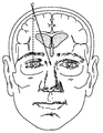
뇌실Ventricle
도 6은 뇌의 뇌실 시스템의 도면이다. 상기 시스템은 뇌 내의 일련의 공동(뇌실)이며, 이는 척수의 거미막밑 공간과 중심관 둘 모두와 연속하여 존재한다. 우측 및 좌측 가쪽뇌실, 및 중간선의 셋째뇌실 및 넷째뇌실의 4개의 뇌실이 존재한다. 2개의 가쪽내실은 대뇌 내에 위치되고, 각각 심실간 몬로공을 통해 셋째뇌실에 연결된다. 셋째뇌실은 간뇌에 위치되고, 대뇌 중간뇌수도관에 의해 넷째뇌실에 연결된다. 넷째뇌실은 후뇌에 위치되고, 이는 적어도 발생학적으로 척수의 중심관과 연속하여 존재한다. 정중구멍 또는 마장디공, 및 루시카의 좌측 및 우측 가쪽구멍(공)의 3개의 공이 거미막밑 공간에 넷째뇌실을 연결시킨다.6 is a view of the ventricular system of the brain. The system is a series of cavities (ventricles) in the brain, which are continuous with both the spider subspace and the central tube of the spinal cord. There are four ventricles of the right ventricle and the left ventricle, and the third ventricle of the median line and the fourth ventricle. Two lateral ventricles are located in the cerebral cortex, each connected to the third ventricle via a ventricular monorail. The third ventricle is located in the ganglia, and is connected to the fourth ventricle by the cerebral midbrain. The fourth ventricle is located in the hindbrain, which is at least genetically contiguous with the central tube of the spinal cord. Three holes in the right and left side holes (the ball) connect the fourth ventricle to the sub arachnoid space.
뇌에서의 In the brain CSFCSF 유동 Flow
도 7은 뇌실로부터 거미막밑 공간으로의 CSF 유동의 예시적 개념을 제시한다. 뇌척수액(CSF)은 뇌의 뇌실 시스템, 거미막밑 공간, 및 척수의 중심관을 점유하는 투명한 체액이다. CSF는 뇌실 시스템 전체에 걸쳐 발견되는 맥락얼기의 변형된 뇌실막세포에 의해 생성된다. 또한, 이는 추측상 뇌의 세포외 공간으로부터 뇌실벽 및 혈관 주위에서 또한 형성된다. CSF는 뇌실간공을 통해 가쪽뇌실로부터 셋째뇌실로 유동한다. 이후, CSF는 대뇌수도관을 통해 넷째뇌실로 유동한다. CSF는 정중 구멍 및 좌측 및 우측 가쪽구멍을 통해 거미막밑 공간에서 외부로 유동한다. 최종적으로, CSF는 거미막과립 및 거미막 융모를 통해 경막정맥동으로 재흡수된다. 거미막과립은 융모의 집합물로 구성된다. 융모는 경질막을 통해 상시상정맥동의 루멘 및 다른 정맥 구조로 향하는 거미막의 가시적 탈출이다. 과립(granulation)은 거미막밑 공간으로부터 정맥혈로의 CSF의 단방향 유동을 가능케 하는 밸브로 작용하는 것으로 보인다. 소분자, 단백질, 미생물, 및 적혈구 세포를 포함하는 CSF의 모든 성분은 유체와 함께 떠난다.Figure 7 illustrates an exemplary concept of CSF flow from the ventricle to the sub arachnoid space. CSF is a transparent body fluid that occupies the brain ventricular system of the brain, submandibular space, and the central tube of the spinal cord. CSF is produced by the modified ventricular cell of the conglomerate found throughout the ventricular system. It is also formed from the extracellular space of the brain in the brain, also around the wall of the ventricle and around the blood vessels. CSF flows from the ventricle to the third ventricle through the ventricle. The CSF then flows through the cerebral perfusate into the fourth ventricle. The CSF flows outward from the submarine space through the median hole and the left and right lateral holes. Finally, CSF is reabsorbed into the dural sinus via spider membrane granules and spider membranes. Spider membrane granules are composed of villi aggregates. The villi are visible escapes of the spider membranes through the hard membrane into the lumen of the normal sinus and other vein structures. Granulation appears to act as a valve to allow unidirectional flow of CSF from the submarinal space to venous blood. All components of CSF, including small molecules, proteins, microbes, and red blood cells, leave with the fluid.
CSF는 약 0.3-0.37 ml/분 또는 20 ml/시간 또는 500 ml/일의 속도로 생성된다. CSF 공간의 부피는 약 150 ml이며, CSF는 하루에 3.7회 턴 오버(turn over)된다.CSF is produced at a rate of about 0.3-0.37 ml / min or 20 ml / hr or 500 ml / day. The volume of the CSF space is about 150 ml, and the CSF turns over 3.7 times a day.
맥락얼기는 CSF의 화학적 안정성을 유지시키기 위해 모세관 여과 및 상피 분비 메커니즘을 이용한다. 맥락얼기를 가로지르는 모세관은 혈장 용질에 대해 자유롭게 투과성인 한편, 맥락얼기를 구성하는 상피 세포의 수준에서 장벽이 존재하며 이는 담체-매개 능동 수송을 담당한다. CSF 및 뇌의 세포외 유체는 항정 상태로 존재하며, 혈장 및 CSF는 정상 생리학적 조건하에서 삼투 평형 상태로 존재한다.Contextual circulation uses capillary filtration and epithelial secretion mechanisms to maintain the chemical stability of CSF. The capillaries across the intercourse are freely transmissive to plasma solutes, while barriers are present at the level of the epithelial cells that constitute the conglomerate, which is responsible for carrier-mediated active transport. CSF and extracellular fluid of the brain are in a steady state, and plasma and CSF are present in osmotic equilibrium under normal physiological conditions.
혈액뇌장벽Blood brain barrier
혈액뇌장벽은 뇌로의 혈액-매개 물질의 진입을 방지하고, 뉴런이 효과적으로 작동하기 위한 안정적 환경을 유지시킨다. 이는 뇌 미세혈관 내피 세포, 혈액뇌장벽의 주요 해부학적 부위, 이들의 세포간연접, 및 소포이동의 상대적 결핍의 특화된 특성으로부터 발생하며, 이는 상기 세포가 일반적 모세혈관의 세포와 상이하게 만든다. 혈액뇌장벽 혈관의 내피 세포는 또한 천공되어 있지 않으며, 대신 이들은 혈관벽을 가로지르는 확산을 차단하는 빈틈없는 접합부의 복잡한 배열에 의해 서로 연결되어 있다.The blood brain barrier prevents the entry of blood-mediated substances into the brain and maintains a stable environment for neurons to function effectively. This arises from the specialized properties of the brain microvascular endothelial cells, the major anatomical regions of the blood brain barrier, their intercellular junctions, and the relative deficiency of vesicle migration, which makes them differentiated from cells of the general capillary blood vessels. Endothelial cells of blood-brain barrier blood vessels are also not perforated, but instead they are interconnected by a complex array of tight junctions that block diffusion across the vessel wall.
거미막밑Under spider 출혈 bleeding
뇌는 연질막, 거미막, 및 경질막의 막 또는 수막의 3개의 층에 의해 싸여져 있다. 거미막밑 공간은 뇌 주위의 거미막과 연질막 사이의 영역이다. 용어 "거미막밑 출혈"("SAH"로도 언급됨)은 거미막밑 공간으로의 출혈을 나타낸다. SAH는 보통 뇌동맥류로부터 자연적으로 발생할 수 있거나, 외상으로부터 발생할 수 있다. 증상은 신속한 발생을 갖는 강한 두통(종종, "벼락 두통(thunderclap headache)"으로 언급됨), 구토, 및 변경된 의식 수준을 포함한다. 진단은 일반적으로 컴퓨터단층촬영술(CT 스캐닝), 또는 종종 요추 천자에 의해 이루어진다. 치료는 면밀한 관찰, 의약 및 초기 신경외과적 연구 및 재발 및 합병증을 예방하기 위한 치료에 의해 이루어진다.The brain is surrounded by three layers of membranes of the soft membrane, the spider membrane, and the hard membrane, or the water membrane. Spider submucosa is the area between the spider membrane and the soft membrane around the brain. The term " sub arachnoid hemorrhage "(also referred to as" SAH ") refers to hemorrhage into the spider submandibular space. SAH can usually occur naturally from the aneurysm of the cerebral artery, or may arise from trauma. Symptoms include a strong headache (often referred to as a "thunderclap headache") with rapid onset, vomiting, and altered levels of consciousness. Diagnosis is usually made by computerized tomography (CT scanning), or often by a lumbar puncture. Treatment is by close observation, medication and early neurosurgical studies and treatment to prevent recurrence and complications.
SAH는 의학적 응급상황이며, 사망 또는 초기 단계에 인지되고 치료되는 경우에도 중증 장애를 발생시킬 수 있다. 모든 SAH 환자의 절반은 치명적이며, 환자의 10-15%는 병원에 도착하기 전에 사망한다. SAH는 뇌졸중의 형태로 간주되며, 모든 뇌졸중의 1% 내지 7%를 야기시킨다. 두개내 동맥류의 파열에 의해 야기되는 경우, 출혈은 거미막밑 공간, 덜 흔하게는 뇌실내 및 뇌내 공간에서 관찰된다. SAH로 인한 출혈은 뇌 손상, 뇌 이동, 감소된 뇌관류 및 수두증을 발생시킬 수 있다. 미국에서 파열된 두개내 동맥류로부터의 SAH의 발생률은 10,000명 당 1명이며, 이는 매년 약 27,000-30,000명의 새로운 환자를 발생시키는 것으로 판단된다. 상기 파열된 동맥류는 45%의 30일 사망률을 갖는다. 또한, 생존자의 30%는 중간 내지 중증 장애를 가질 것으로 예측된다.SAH is a medical emergency and can cause serious disturbances even if it is recognized and treated at the time of death or early stages. Half of all SAH patients are fatal, and 10-15% of patients die before arriving at the hospital. SAH is considered a form of stroke and causes 1% to 7% of all strokes. When caused by rupture of an intracranial aneurysm, bleeding is observed in submandibular space, less commonly in the brain and intracerebral space. Bleeding due to SAH can lead to brain damage, brain movement, reduced cerebral perfusion and hydrocephalus. The incidence of SAH from intracranial aneurysms ruptured in the United States is 1 per 10,000, which is estimated to generate about 27,000 to 30,000 new patients each year. The ruptured aneurysm has a 30-day mortality rate of 45%. In addition, 30% of survivors are expected to have moderate to severe disabilities.
일부 연구는 SAH의 발생률이 매년 100,000명 당 평균 9.1명인 것을 나타낸다. 일본 및 핀란드로부터의 연구는 이들 국가에서 더 높은 비율(각각 100,000명 당 22.7명 및 100,000명 당 19.7명)을 나타내며, 그 이유에 대해서는 완전히 이해되어 있지 않다. 대조적으로, 남아메리카 및 중앙 아메리카는 평균적으로 100,000명 당 4.2명의 비율을 갖는다. SAH에 대한 위험이 있는 사람의 그룹은 보통 뇌졸중에 걸리는 집단보다는 어리나, 위험은 연령에 따라 더 증가한다. 어린 사람은 중년의 사람(위험비 0.1, 또는 10%)보다 SAH를 경험할 가능성이 훨씬 적다. 위험은 연령에 따라 지속적으로 증가하고, 45세 내지 55세의 사람보다 매우 노년(85세 이상)에서 60% 더 높다. SAH의 위험은 55세를 초과하는 여성에서 약 25% 더 높으며, 이는 폐경으로부터 발생하는 호르몬 변화를 잠재적으로 반영한다.Some studies indicate that the incidence of SAH is an average of 9.1 per 100,000 people each year. Studies from Japan and Finland show higher rates in these countries (22.7 per 100,000 and 19.7 per 100,000, respectively), and the reasons for this are not fully understood. By contrast, on average, South America and Central America have a ratio of 4.2 per 100,000. Groups of people at risk for SAH are usually milder than those who suffer from stroke, but the risk increases with age. Younger people are much less likely to experience SAH than middle-aged people (risk ratio 0.1, or 10%). The risk increases consistently with age and is 60% higher than in people aged 45-55 who are very young (age 85 or older). The risk of SAH is about 25% higher in women over 55 years old, potentially reflecting hormone changes resulting from menopause.
SAH에서 생존한 환자는 또한 이차 합병증의 위험이 있다. 이러한 합병증 중에서 가장 특별히는 동맥류 재출혈, 혈관조영술 뇌혈관연축 및 지연 대뇌 허혈(DCI)이 있다.Patients who survive on SAH are also at risk for secondary complications. Among these complications, reperfusion aneurysms, angiographic cerebral vasospasm and delayed cerebral ischemia (DCI) are the most common.
DCI는 국소적 신경계 장애(예를 들어, 반신불완전마비, 언어상실증, 행위상실증, 반맹, 또는 무시)의 발생, 또는 글래스고 혼수 척도(Glasgow coma scale)에서의 감소(전체 스코어 또는 이의 개별적 구성요소[안구, 어느 한 측면에서의 운동, 언어] 중 하나에서의 감소)이다. 이는 적어도 1시간 동안 지속되거나 지속되지 않을 수 있고, 동맥류 폐색 직후에 명백하지 않으며, 뇌의 임상적 평가, CT 또는 자기 공명 영상(MRI) 스캐닝, 및 적절한 실험실 연구에 의해 다른 원인에 기인될 수 없다. 뇌경색증은 DCI의 결과일 수 있으며, SAH 후 6주 이내에 뇌의 CT 또는 MRI 스캔 또는 6주 이내의 사망 전에 이루어지는 마지막 CT 또는 MRI에서의 뇌경색증의 존재로 정의되거나, 부검시에 증명되거나, 초기 동맥류 폐색 후 24 내지 48시간 후에 CT 또는 MRI 스캔 상에 존재하지 않고, 외과적 클리핑(surgical clipping) 또는 혈관내 치료와 같은 다른 원인에 기인될 수 없다. 뇌실 카테터 또는 실질내 혈종으로부터 발생하는 CT 영상에서의 저밀도는 일반적으로 DCI로부터의 뇌경색증으로 간주되지 않는다. 혈관조영술 뇌혈관연축은 방사선 시험(CT 혈관조영술[CTA], MR 혈관조영술[MRA] MRA 또는 카테터 혈관조영술[CA])의 서술이며, DCI의 원인일 수 있다. 용어 "혈관조영술 뇌혈관연축"은 거미막밑 공간으로의 출혈에 따른 뇌의 기저에서의 큰 용량의 동맥(즉, 대뇌동맥)의 협소화를 나타내며, 이는 원위 뇌 영역의 감소된 관류를 발생시킨다. 혈관조영술 혈관연축은 SAH의 결과이나, 이는 또한 거미막밑 공간 내에 혈액을 침착시키는 임의의 질환 후에 발생할 수 있다.DCI has been associated with the incidence of localized neurological disorders (eg, dysmenorrhoea paralysis, dysarthria, impaired, hemorrhoids, or neglect), or reductions in the Glasgow coma scale (overall scores or individual components thereof [ Eye, movement in one side, language]. It may not last or last for at least one hour and is not apparent immediately after aneurysm occlusion and can not be attributed to other causes by brain clinical assessment, CT or magnetic resonance imaging (MRI) scanning, and appropriate laboratory studies . Cerebral infarction may be the result of DCI and is defined as the presence of cerebral infarction on the final CT or MRI prior to a CT or MRI scan of the brain or within 6 weeks of death within 6 weeks after SAH, And is not present on CT or MRI scans after 24-48 hours and can not be attributed to other causes such as surgical clipping or endovascular treatment. The low density in CT images originating from the ventricular catheter or intimal hematoma is not generally regarded as cerebral infarction from DCI. Angiography The cerebral vasospasm is a description of a radiological examination (CT angiography [CTA], MR angiography [MRA] MRA or catheter angiography [CA]) and may be the cause of DCI. The term "angiographic cerebral vasospasm" refers to the narrowing of a large volume of arteries (i.e., the great cerebral artery) at the base of the brain following hemorrhage into the spider submandibular space, resulting in reduced perfusion of the distal brain region. Angiography Vasospasm is the result of SAH, but it can also occur after any disease that deposits blood in the sub arachnoid space.
증상Symptom
SAH의 통상적 증상은 번개 두통(수초 내지 수분에 걸쳐 발생하는 "이제까지 최악" 또는 "머리 속에서 걷어차는" 것으로 기재되는 두통)이나, 이는 모든 SAH 환자의 약 1/3 만에서만 증상이 존재한다. 상기 증상을 갖는, 의료 방법을 찾는 환자의 약 10%는 내재성 SAH를 갖는다. 환자는 또한 구토가 존재할 수 있으며, 14명 중 1명은 발작을 갖는다. 경부 경직 및 착란, 감소된 의식 수준, 또는 혼수일 수 있는 수막증의 다른 징후가 존재할 수 있다. 뇌 주위의 상승된 압력에 반응하여 안구내 출혈이 발생할 수 있다. 유리체밑(유리체막이 안구의 유리체를 둘러쌈) 및 유리체 출혈이 안저검사(fundoscopy)에서 관찰될 수 있다. 이는 터슨 증후군(Terson syndrome)(환자의 3-13%에서 발생함)으로 공지되어 있고, 더욱 중증의 SAH에서 더욱 통상적이다. 번개 두통을 갖는 환자에서, 상기 언급된 징후 중 어느 것도 출혈을 확인하거나 배제시키는데 도움이 되지 않지만, 출혈이 다른 원인에 비하여 파열된 동맥류의 결과인 경우에 발작이 더욱 흔하다. 눈돌림 신경 비정상(아래쪽 및 바깥쪽으로의 병에 걸린 안구 운동, 동일면 상에서의 눈꺼풀을 들어올릴 수 없으나, 정상적인 동공 반사)은 후교통동맥 근처에서 발생하는 동맥류로부터의 출혈을 나타낼 수 있다. 동공의 독립된 확장은 또한 두개내압 상승의 결과로서 뇌 헤르니아를 반영할 수 있다.A common symptom of SAH is a lightheaded headache ("the worst ever" or "kicked in the head" that occurs over a few seconds to a few minutes), but it is only present in about one third of all SAH patients. Approximately 10% of patients with these symptoms who seek medical care have endogenous SAH. The patient may also have vomiting, and one in 14 has seizures. There may be other signs of meningitis, which may be neck stiffness and confusion, reduced consciousness levels, or coma. Intraocular bleeding can occur in response to elevated pressure around the brain. Beneath the vitreous (surrounding the vitreous body of the eye) and vitreous hemorrhage can be observed in fundoscopy. This is known as Terson syndrome (occurring in 3-13% of patients) and is more common in more severe SAH. In a patient with a lightheaded headache, none of the above-mentioned signs help to identify or exclude bleeding, but seizures are more common when bleeding is the result of a ruptured aneurysm relative to other causes. Abnormalities of the oculomotor nerve (downward and outward diseased eye movements, elevated eyelids on the same side, but normal pupil reflexes) may indicate bleeding from an aneurysm originating near the posterior communicating artery. Independent dilation of the pupil can also reflect brain hernia as a result of intracranial pressure elevation.
신체는 출혈의 결과로서 많은 양의 아드레날린 및 유사한 호르몬을 방출하며, 이는 혈압에서의 급격한 증가를 발생시킨다. 심장은 많이 긴장하게 되고, 신경성 폐부종, 기절 심근, 심장부정맥, 심전도 변화(종종 큰 역 "대뇌" T 파(wave)를 가짐) 및 심장 정지(3%)가 출혈의 발생 후에 신속히 발생할 수 있다.The body releases large amounts of adrenaline and similar hormones as a result of bleeding, which causes a sharp increase in blood pressure. The heart becomes very tight, and neurogenic pulmonary edema, stunned myocardium, cardiac arrhythmia, changes in ECG (often with large inverted "cerebral" T waves) and cardiac arrest (3%) can occur rapidly after bleeding.
SAH는 또한 두부 손상을 입은 사람에서 발생할 수 있다. 증상은 두통, 감소된 의식 수준 또는 반신불완전마비를 포함할 수 있다. SAH는 특히 더 낮은 글래스고 혼수 척도 수준과 관련되는 경우에 두부 손상의 중증 합병증으로 간주된다.SAH can also occur in people with head injury. Symptoms may include headache, reduced consciousness levels or half-incomplete paralysis. SAH is considered to be a severe complication of head injury, particularly when associated with a lower Glasgow coma scale level.
진단Diagnosis
SAH가 의심되는 사람을 평가하기 위한 최초 단계는 의학적 개인력을 획득하고 신체 검사를 수행하는 단계이다. 번개 두통을 갖는 병원에 입원된 환자의 단지 10-25%가 SAH를 가지므로, 수막염, 편두통, 및 대뇌 정맥동 혈전증과 같은 다른 가능한 원인이 보통 동시에 고려된다. SAH보다 2배 더 흔한 뇌내출혈이 종종 SAH로 오진된다.The first step in evaluating a person with a suspected SAH is to acquire a medical manual and perform a physical examination. Since only 10-25% of patients admitted to a hospital with a lightheaded headache have SAH, other possible causes such as meningitis, migraine, and cerebral sinus thrombosis are usually considered simultaneously. Intracerebral hemorrhage, often twice as common as SAH, is often misdiagnosed as SAH.
SAH의 진단은 임상적 근거 단독으로 이루어질 수 없다. 일반적으로, 출혈을 확인하거나 배제시키기 위해 뇌의 의학적 영상화[보통 높은 민감도(특히 출혈의 발생 후 첫번째 날에서 95%를 초과하는 정확한 확인)를 갖는 컴퓨터단층촬영술(CT 스캔)]가 요구된다. CT 스캔과 비교하는 경우 수일 후에 자기공명영상(MRI 스캔)이 더 민감할 수 있다. 일반적인 CT 또는 MRI 스캔을 받은 사람에서, 뇌척수액(CSF)이 바늘을 이용하여 요추 낭(lumbar sac)으로부터 분리되는 요추 천자는 CT에서 정상인 것으로 밝혀진 그룹의 3%에서 출혈의 증거를 나타내며, 따라서, 요추 천자는 영상화가 음성인 경우에 필수적인 것으로 간주된다. CSF 샘플은 원심분리된 유체의 황색 외형인 황색변색증에 대해 시험되거나, 분광광도법을 이용하여 CSF 내의 헤모글로빈의 분해 생성물인 빌리루빈에 대해 시험된다.The diagnosis of SAH can not be made on a clinical basis alone. Generally, a medical imaging of the brain (usually a CT scan with high sensitivity, especially with an accuracy of more than 95% on the first day after the onset of bleeding) is required to confirm or rule out bleeding. Magnetic resonance imaging (MRI scans) may be more sensitive after a few days compared with CT scans. In a person who underwent a general CT or MRI scan, a lumbar puncture in which CSF is separated from the lumbar sac using a needle shows evidence of hemorrhage in 3% of the groups found to be normal in CT, The puncture is considered essential if the imaging is negative. CSF samples were tested for yellow discoloration of the yellowish appearance of the centrifuged fluid or were tested for bilirubin, the degradation product of hemoglobin in CSF, using spectrophotometry.
SAH가 확인된 후, 이의 원인이 결정될 필요가 있다. 동맥류를 확인하기 위한 CT 혈관조영술(CT 스캔에서 방사선조영제(radiocontrast)를 이용하여 혈관을 시각화시킴)이 일반적으로 첫번째 단계이나, 더욱 침습성의 카테터 혈관조영술(뇌 동맥으로 진행되는 카테터를 통해 방사선조영제를 주입함)이 최적 표준시험이나, 이는 합병증의 위험이 더 높다. 더욱 침습성의 카테터 혈관조영술은 동시에 동맥류와 같은 출혈의 근원을 제거하기 위한 계획이 있는 경우에 유용하다.After SAH is identified, its cause needs to be determined. CT angiography to visualize an aneurysm (visualization of vessels using radiocontrast in CT scans) is generally the first step, but more invasive catheter angiography (through a catheter leading to the arterial artery) Injection) is the optimal standard test, but it is more at risk of complications. More invasive catheter angiography is useful when there is a plan to remove the source of bleeding, such as an aneurysm.
원인cause
가장 흔한 자연적 SAH는 대뇌 동맥류의 파열(85%)로 인한 것이다. 대뇌 동맥류는 확장되는 뇌의 동맥벽에서의 약함이다. 이들은 윌리스환 및 이의 가지들에 위치되는 경향이 있다. SAH의 가장 많은 환자는 작은 동맥류로부터의 출혈로 인한 것인 한편, 더 큰 동맥류(더 희귀함)는 파열될 가능성이 더 높다. 자연적 SAH의 환자의 15-20%에서 첫번째 혈관조영상으로부터 동맥류가 검출되지 않는다. 혈액이 중뇌의 영역으로 제한되는 비-동맥류성 중간뇌주위 출혈이 SAH 환자의 또 다른 10%를 야기시킨다. 이들 중에서, 동맥류는 일반적으로 발견되지 않는다. 환자의 나머지 5%는 동맥에 대한 혈관염 손상, 혈관에 영향을 미치는 다른 장애, 척수 혈관의 장애, 및 다양한 종양으로의 출혈로 인한 것이다. 가장 외상성인 SAH가 두개 골절 또는 뇌내 좌상 근처에서 발생한다.The most common natural SAH is due to rupture (85%) of cerebral aneurysms. The cerebral aneurysm is a weakness in the enlarged arterial wall of the brain. They tend to be located in Willis circle and its branches. The majority of patients with SAH are due to bleeding from small aneurysms, while the larger aneurysms (the more rare) are more likely to rupture. Aneurysms are not detected from the first angiogram in 15-20% of patients with natural SAH. Non-aneurysmal middle cerebral hemorrhage, where blood is confined to the midbrain region, causes another 10% of patients with SAH. Of these, aneurysms are generally not found. The remaining 5% of patients are due to vasculitis damage to arteries, other disorders affecting blood vessels, impaired spinal cord blood vessels, and bleeding into various tumors. The most traumatic SAH occurs in the vicinity of the left upper quadrant of a fracture or brain.
분류Classification
여러 등급화 척도가 SAH에 대해 이용가능하다. 이들 척도는 이들의 결과를 갖는 환자의 특징을 회고적으로 매치시킴으로써 유도된다. 도처에서 사용되는 글래스고 혼수 척도(GCS)에 더하여, 3개의 다른 특화된 스코어가 사용된다. 모든 스코어에서, 더 높은 수는 더 나쁜 결과와 관련된다. 중증도의 첫번째 척도는 문헌[Hunt and Hess in 1968]("헌트 및 헤스 척도(Hunt and Hess scale"))에 의해 기재되었으며, 이는 환자의 임상 상태를 분류한다. 피셔 등급(Fisher Grade)은 CT 스캔에서의 SAH의 외관을 분류한다. 피셔 척도는 클라센(Claassen) 및 그의 동료에 의해 변형("클라센 척도")되었으며, 이는 SAH 크기 및 수반하는 뇌실내 출혈로부터 추가 위험을 반영한다. 세계신경외과학회(World Federation of Neurological Surgeons) 분류는 증상의 중증도를 측정하기 위해 GSC 및 국소 신경계 결손을 이용한다. 포괄적인 분류 계획이 결과를 예측하고 요법을 평가하기 위해 오길비(Ogilvy) 및 카터(Carter)에 의해 제안되었다. 오길비 시스템은 5 등급을 가지며, 이는 50을 초과하는 연령; 헌트(Hunt) 및 헤스(Hess) 등급 4 또는 5; 피셔 척도 3 또는 4; 10 mm를 초과하는 동맥류 크기; 및 25 mm 이상의 후순환계 동맥류(posterior circulation aneurysms)의 5개의 요인 각각의 존재 또는 부재에 대해 1 포인트를 할당한다.Several grading scales are available for SAH. These measures are derived by retrospectively matching the characteristics of the patient with these outcomes. In addition to the Glasgow Coma Scale (GCS) used everywhere, three different specialized scores are used. In all scores, higher numbers are associated with worse outcomes. The first scale of severity was described by Hunt and Hess in 1968 ("Hunt and Hess scale"), which classifies the clinical status of the patient. Fisher Grade classifies the appearance of SAH in a CT scan. The Fisher scale was modified by Claassen and his colleagues ("clinician scale"), reflecting additional risk from SAH size and accompanying intraventricular hemorrhage. The World Federation of Neurological Surgeons classification uses GSC and focal neurological deficits to measure severity of symptoms. A comprehensive classification scheme was proposed by Ogilvy and Carter to predict outcomes and evaluate therapy. The Ogilvy system has a grade of 5, which is older than 50; Hunt and
치료cure
SAH의 처리는 환자를 안정화시키는 일반적 수단, 출혈 근원을 제거함으로써 재출혈을 방지하는 특정 수단, 혈관연축의 예방, 및 합병증의 예방 및 치료로 구성된다.Treatment of SAH consists of general means to stabilize the patient, specific means of preventing rebleeding by removing bleeding origins, prevention of vasospasm, and prevention and treatment of complications.
일반적 수단General means
첫번째 우선사항은 환자를 안정화시키는 것이다. 억제된 의식 수준을 갖는 환자는 삽관되고 기계적으로 환기될 필요가 있을 수 있다. 혈압, 맥박, 호흡수 및 글래스고 혼수 척도가 빈번히 모니터된다. 진단이 확인된 후, 특히 상기 환자의 15%가 입원 후 1시간 이내에 추가 에피소드(재출혈)를 갖는 경우에 집중치료실에 대한 입원이 바람직할 수 있다. 영양공급이 초기의 우선사항이며, 경구 또는 코위영양관 영양공급이 비경구 경로에 비해 바람직하다. 진통(동통 조절)은 일반적으로 코데인과 같이 비-진정성 작용제로 제한되는데, 이는 진정이 정신 상태에 영향을 미쳐, 이에 따라 의식 수준을 모니터하는 능력을 방해할 수 있기 때문이다. 심정맥 혈전증은 압박 스타킹, 종아리의 간헐적 공기 압박, 또는 둘 모두로 예방된다.The first priority is to stabilize the patient. Patients with restrained levels of consciousness may need to be intubated and mechanically ventilated. Blood pressure, pulse, respiratory rate, and Glasgow coma scale are frequently monitored. Admission to the intensive care unit may be desirable after diagnosis is confirmed, especially when 15% of the patients have an additional episode (rebleeding) within one hour after admission. Nutrition is an early priority and oral or nasal feeding is preferred over the parenteral route. Pain (pain control) is generally limited to non-sedative agents such as codeine, because it can affect the mental state and thus the ability to monitor the level of consciousness. Deep vein thrombosis is prevented by compression stockings, intermittent air compression of the calves, or both.
재출혈의 예방Prevention of rebleeding
억제된 의식 수준 또는 국소적 신경학적 증상과 관련된 큰 혈종을 갖는 환자가 혈액의 긴급한 외과적 제거 및 출혈 동맥류의 폐색을 위한 후보일 수 있다. 카테터 또는 튜브가 수두증을 치료하기 위해 뇌실에 삽입될 수 있다. 나머지는 더 광범위하게 안정화되고, 경대퇴동맥 카테터 혈관조영상 또는 CT 혈관조영상을 이후에 겪는다. 처음 24시간 후, 재출혈 위험이 이후 4주에 걸쳐 약 40%가 남아있으며, 이는 상기 위험을 감소시키는데 있어서 중재적시술이 계획되어야 함을 암시한다.A patient with a large hematoma associated with a reduced level of consciousness or with local neurological symptoms may be a candidate for urgent surgical removal of the blood and occlusion of the bleeding aneurysm. A catheter or tube may be inserted into the ventricle to treat hydrocephalus. The remainder are more widely stabilized and subsequently suffer a femoral artery catheter angiogram or CT angiogram. After the first 24 hours, the risk of rebleeding remained around 40% over the next 4 weeks, suggesting that interventional procedures should be planned to reduce this risk.
재출혈은 예측하기가 어렵고, 임의의 기간에 발생할 수 있으며, 불량한 예후를 갖는다. 따라서, 재출혈을 방지하기 위한 중재적시술, 주로 파열된 동맥류의 클리핑(clipping) 또는 코일링(coiling)이 가능한 초기에 수행된다. 혈관조영술에서 뇌동맥류가 확인되는 경우, 동일한 동맥류로부터 추가의 출혈 위험을 감소시키기 위해 신경외과적 클리핑 및 혈관내 코일링의 2개의 수단이 이용가능하다. 클리핑은 동맥류의 위치를 정한 후, 동맥류의 목 부분 전체에 걸친 클립 또는 클립들의 배치를 위해 개두술(두개골의 개방)을 필요로 한다. 코일링은 큰 혈관을 통해 수행되며, 카테터가 서혜부 내의 대퇴동맥으로 삽입되고, 대동맥을 통해 뇌에 공급하는 동맥(둘 모두의 경동맥 및 둘 모두의 척추동맥)으로 전진된다. 동맥류의 위치가 정해지는 경우, 동맥류 내의 혈병의 형성 및 제거를 발생시키는 금속 코일이 전개된다. 치료가 수행되는 것에 관한 결정은 통상적으로 신경외과 의사 및 신경방사선학자를 포함하는 각 전문 분야 협력팀에 의해 이루어진다.Rebleeding is difficult to predict, may occur at any time, and has a poor prognosis. Thus, interventional procedures to prevent rebleeding, primarily clipping or coiling of ruptured aneurysms, are performed early on. When aneurysms of the cerebral arteries are identified in angiography, two means of neurosurgical clipping and intravascular coiling are available to reduce the risk of further bleeding from the same aneurysm. Clipping requires the placement of clips or clips throughout the neck of the aneurysm after placement of the aneurysm. The coil ring is performed through the large vessels, the catheter is inserted into the femoral artery in the inguinal region, and advanced to the arteries (both carotid arteries and both vertebral arteries) supplying the brain through the aorta. When the position of the aneurysm is determined, a metal coil is developed which causes the formation and removal of blood clots in the aneurysm. Decisions about the treatment being performed are usually made by a team of specialists, including neurosurgeons and neuro radiologists.
중대뇌동맥 및 이의 관련 혈관의 동맥류는 혈관조영술로 도달하기는 어렵고, 클리핑에 적용되기 쉬운 경향이 있는 한편, 뇌기저동맥 및 후대뇌동맥의 동맥류는 외과적으로 도달하기는 어렵고, 혈관내 치료기술에 대해 더욱 접근가능한 경향이 있다. 코일링의 주요 결점은 동맥류가 재발할 수 있는 가능성이며, 이러한 위험은 외과적 방법에서는 극도로 적다. 코일링을 겪은 환자는 통상적으로 동맥류의 재발이 초기에 확인되는 것을 보장하기 위해 혈관조영술 또는 다른 수단으로 수년 동안 추적조사된다.Aneurysms of the middle cerebral artery and its associated vessels are difficult to reach with angiography and tend to be applied to clipping, while aneurysms of the basilar artery and posterior cerebral arteries are difficult to reach surgically, There is a tendency to be more accessible. The main drawback of coiling is the possibility of recurrent aneurysms, which is extremely low in surgical procedures. Patients undergoing coiling are usually followed for several years by angiography or other means to ensure that recurrence of the aneurysm is initially identified.
예후Prognosis
초기 이환율 및 사망률Initial morbidity and mortality
SAH에 대한 사망률은 40% 내지 50%이다. 최초 입원, 치료 및 합병증에서 생존한 환자 중, 적어도 25%는 이들의 생활양식에서 유의한 제한을 가지며, 20% 미만은 어떤 경우에도 잔여 증상을 갖지 않는다. 혼수를 갖지 않는 작은 SAH의 진단에서의 지연(또는 편두통에 대한 갑작스러운 두통의 오해)이 불량한 결과에 기여한다. 불량한 결과에 대한 위험 요인은 보다 높은 연령, 불량한 신경학적 등급, 최초 CT 스캔에서의 보다 많은 출혈 및 보다 큰 동맥류, 후순환에서의 동맥류의 위치, 수축기 고혈압, 및 심부전, 고혈압, 간 질병 또는 이전의 SAH의 이전의 진단을 포함한다. 병원 체류 동안, 혈관연축으로부터 발생하는 지연성 허혈의 발생, 뇌내 혈종 또는 뇌실내 출혈(뇌의 뇌실로의 출혈)의 발달, 및 입원 8일째의 열의 존재가 또한 예후를 악화시킨다.The mortality rate for SAH is 40% to 50%. Of the patients who survived the initial hospitalization, treatment, and complications, at least 25% had significant limitations in their lifestyle, and less than 20% had no residual symptoms in any case. Delays in the diagnosis of small SAHs without coma (or sudden headache misunderstandings for migraine) contribute to poor results. Risk factors for adverse outcomes include higher age, poor neurological grade, more hemorrhage in the initial CT scan and larger aneurysms, location of aneurysms in postcirculation, systolic hypertension, and heart failure, hypertension, liver disease, or previous Includes previous diagnosis of SAH. During hospital stay, the development of delayed ischemia resulting from vasospasm, the development of intracerebral hemorrhage or intracerebral hemorrhage (bleeding into the brain ventricles), and the presence of heat on the eighth day of admission also exacerbate the prognosis.
완전한 카테터 혈관조영술에 의해 동맥류를 나타내지 않는 SAH는 "혈관조영상-음성 SAH"로 언급될 수 있다. 이는 동맥류로부터의 SAH보다 나은 예후를 갖지만, 이는 여전히 허혈, 재출혈 및 수두증의 위험과 관련이 있다. 그러나, 중간뇌주변 SAH(뇌의 중뇌 부분 주위의 출혈)는 매우 낮은 비율의 재출혈 또는 지연성 허혈을 가지며, 이러한 서브타입의 예후는 우수하다.SAH which does not show aneurysm by complete catheter angiography can be referred to as "angiogram-negative SAH". It has a better prognosis than SAH from aneurysms, but it is still associated with the risk of ischemia, rebleeding and hydrocephalus. However, the middle cerebral SAH (bleeding around the midbrain portion of the brain) has a very low rate of rebleeding or delayed ischemia, and the prognosis of these subtypes is excellent.
장기간 결과Long term results
신경인지 증상, 예를 들어, 피로, 기분 장애, 및 다른 관련 증상이 SAH를 갖는 사람에서 흔하다. 양호한 신경학적 회복이 이루어진 환자에서도, 불안, 우울증, 외상후 스트레스 장애 및 인지 장애가 흔하다. 60% 초과가 빈번한 두통을 보고하였다. 동맥류 SAH는 호르몬 조절 및 생성에서 중추적인 역할을 하는 뇌의 두 영역인 시상하부 및 뇌하수체의 손상을 야기시킬 수 있다. 연구는 이전에 SAH를 가진 사람의 적어도 25%가 시상하부-뇌하수체 호르몬, 예를 들어, 성장 호르몬, 프로락틴 또는 갑상선-자극 호르몬 중 하나 이상의 결핍이 발달할 수 있는 것을 나타낸다.Neurocognitive symptoms, such as fatigue, mood disorders, and other related symptoms are common in people with SAH. Anxiety, depression, post-traumatic stress disorder, and cognitive impairment are also common in patients with good neurological recovery. More than 60% reported frequent headaches. Aneurysms SAH can cause hypothalamic and pituitary damage, two areas of the brain that play a pivotal role in hormone regulation and production. Studies have previously shown that at least 25% of people with SAH can develop a deficiency of one or more of the hypothalamic-pituitary hormones, such as growth hormone, prolactin, or thyroid-stimulating hormone.
혈관연축Vasospasm
혈관조영술 뇌혈관연축은 SAH 후의 국소 허혈의 가장 흔한 원인이다. 이는 SAH를 갖는 환자에서 결과에 불리한 영향을 미치는데, 이는 SAH-관련 장애 및 사망의 23% 이하를 담당하기 때문이다. 허혈성 뇌졸중의 모든 유형 중, 혈관연축은 다소 예측가능하고, 예방가능하고, 치료가능한 점에서 독특하다(Macdonald, R.L. and Weir, B. In Cerebral Vasospasm. Academic Press, Burlington, MA, USA (2001) 참조).Angiography Brain vasospasm is the most common cause of focal ischemia after SAH. This has an adverse effect on outcome in patients with SAH, because it accounts for less than 23% of SAH-related disorders and deaths. Among all types of ischemic stroke, vasospasm is unique in that it is somewhat predictable, preventable, and treatable (Macdonald, RL and Weir, B. In Cerebral Vasospasm. Academic Press, Burlington, MA, USA ).
혈관연축은 감소된 대뇌 혈류 및 증가된 대뇌 혈관 저항을 발생시킨다. 이론으로 제한하는 것은 아니지만, 일반적으로 혈관연축은 죽상경화증, 및 외상성 두부 손상, 동맥류성 거미막밑 출혈 및 거미막밑 출혈의 다른 원인을 포함하는 다른 구조적 손상으로부터 발생하는 것과 같은 혈관에 대한 국소적 손상에 의해 야기되는 것으로 생각된다. 뇌혈관연축은 동맥류 파열 후 또는 외상성 두부 손상 후의 흔한 발생인 CSF 내의 혈액의 존재에 의해 또한 촉발될 수 있는 자연 발생 혈관수축이다. 뇌혈관연축은 궁극적으로 중단된 혈액 공급으로 인해 대뇌 허혈 및 경색의 형태의 뇌세포 손상을 발생시킬 수 있다.Vasospasm causes reduced cerebral blood flow and increased cerebral vascular resistance. Without wishing to be bound by theory, it is believed that vasospasm in general is associated with atherosclerosis and localized damage to blood vessels, such as those resulting from traumatic head injuries, subarachnoid subarachnoid hemorrhages, and other structural injuries including other causes of subarachnoid hemorrhage ≪ / RTI > Cerebrovascular spasm is a spontaneous vasoconstriction that can also be triggered by the presence of blood in the CSF, which is a common occurrence after an aneurysm rupture or traumatic head injury. Cerebrovascular spasm can eventually cause brain cell damage in the form of cerebral ischemia and infarction due to interrupted blood supply.
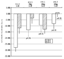
혈관조영술 혈관연축은 DCI에 기여하는 하나의 과정이다. DCI에 기여할 수 있는 다른 과정은 피질 확산 허혈(cortical spreading ischemia) 및 미세혈전색전증(microthromboemboli)이다(도 4). DCI는 적어도 상기 과정 뿐만 아니라 초기 뇌 손상으로 인한 다인성 과정이다. 피질 확산 허혈은 DCI를 야기시킬 수 있는 신규한 메커니즘으로서 SAH의 동물 모델에서 기재되었다. 이는 SAH 및 혈관조영술 혈관연축을 갖는 인간에서 검출되었다. DCI에 기여할 수 있는 또 다른 과정은 미세혈전색전증의 형성이다.Angiography Vasospasm is a process that contributes to DCI. Other processes that can contribute to DCI are cortical spreading ischemia and microthromboemboli (Figure 4). DCI is a multifactorial process due to at least the above process as well as early brain damage. Cortical spreading ischemia has been described in animal models of SAH as a novel mechanism that can cause DCI. It was detected in humans with SAH and angiographic vasospasm. Another process that can contribute to DCI is the formation of micro thromboembolism.
매년, 10,000명의 사람 중 약 1명이 동맥류 파열을 갖는다. 사망률 및 이환률은 출혈의 부피에 따라 증가하고, 이는 환자의 연령 및 건강 상태를 반영하며, 연령에 따라 동맥류가 발달할 가능성이 꾸준히 증가한다. 재출혈은 SAH의 부피에서의 증가 뿐만 아니라 뇌 및 뇌실로의 확대의 증가된 가능성으로 인해 특별히 불리하다. 동맥류 파열로부터 발생하는 대부분의 사망은 최초 출혈 또는 초기 재출혈의 영향으로 인해 병원 외부 또는 입원 직후에 발생한다. 혈관연축으로부터의 증상의 잠재적 표시는 처음 수일이 지난 후에 생존한 환자에서만 발생한다.Every year, about one in 10,000 people have ruptured aneurysms. Mortality and morbidity increase with the volume of bleeding, reflecting the age and health status of the patient and the likelihood of developing aneurysms steadily increases with age. Rebleeding is particularly disadvantageous due to the increase in the volume of SAH as well as the increased likelihood of enlargement into the brain and ventricles. Most deaths resulting from a ruptured aneurysm occur outside the hospital or shortly after admission due to the effects of initial bleeding or early rebleeding. Potential signs of symptoms from vasospasm occur only in patients who survive the first few days.
혈관연축의 발생률은 SAH의 발생률보다 적다(이는 SAH를 갖는 일부 환자만이 혈관연축이 발달하기 때문이다). 혈관연축의 발생률은 제공되는 병원에 수용되는 환자의 유형, 및 혈관연축이 진단되는 방법에 좌우될 것이다.The incidence of vasospasm is less than the incidence of SAH (because only some patients with SAH develop vasospasm). The incidence of vasospasm will depend on the type of patient accommodated in the provided hospital, and how the vasospasm is diagnosed.
제한되지 않는 용어 "혈관연축"은 보통 상기 정의된 바와 같은 혈관조영술로 결정된 동맥 협소화에 대한 언급과 함께 사용된다. 임상적 혈관연축은 가장 흔하게는 지연 대뇌 허혈(DCI)과 유의어로 사용된다. 또 다른 방식으로 사용되는 경우, 예를 들어, 증가된 중대뇌동맥 경두개 도플러 속도를 기초로 한 혈관연축의 경우, 이는 상술되어야 한다.The term "vasospasm ", which is not limited, is usually used with reference to arterial narrowing determined by angiography as defined above. Clinical vasospasm is most commonly used as synonymous with delayed cerebral ischemia (DCI). If used in another way, for example, in the case of vasospasm based on increased median cerebral artery cortical Doppler velocities, this should be elucidated.
SAH 후 4 내지 12일 후에 혈관조영술을 받는 환자의 적어도 2/3에서 다소의 혈관조영술 협소화가 발생할 것이다. 상기 DCI로부터 신경계 황폐가 발달하는 환자의 수는 환자가 모니터되는 근면성 및 예방의 효능에 따라 다양하나, 이는 약 1/3로 예측된다. 입원한 SAH 환자 중, 5 내지 10%가 혈관연축으로 인해 사망한다. 중간 등급의 SAH 후 환자와 비교하는 경우, 매우 양호한 상태의 SAH 후 환자는 혈관연축이 발달할 가능성이 덜한데, 이는 이들이 작은 부피의 SAH를 갖기 때문인 한편, 매우 불량한 조건의 SAH 후 환자는 최초 에피소드 전에 사망할 가능성이 더 높다. 출혈 에피소드에 근접하여 수행되는 컴퓨터단층촬영술(CT) 스캔에서 시각화될 수 있는 두껍고 만연된 거미막밑 혈병의 존재가 중요한 예후 인자이다. 최초 CT 스캔에서의 혈액의 부재는 재출혈의 부재하에서 혈관연축이 발생할 가능성이 매우 없는 것을 나타낸다. 혈관연축 및 결과로서 발생하는 DCI의 가능성은 혈병에 대한 노출의 기간을 감소시키는 요인에 의해 감소된다. 역으로, 혈관연축 및 DCI의 발생률은 혈병에 대한 동맥의 노출을 연장시키고, 가능하게는 다른 메커니즘에 의해 허혈을 야기시키는 항섬유소용해 약물의 사용에 의해 증가된다. 불량한 입원 임상 등급이 DCI와 관련되어 있으며, 이는 추정상 이들이 모두 SAH의 더 큰 부피를 나타내기 때문이다. 연령, 고혈압, 또는 성별과 DCI 사이의 명확한 상관관계는 확립되지 않았다. 흡연자는 더욱 혈관연축 및 DCI에 걸리기 쉬울 가능성이 있다. 혈관연축의 발달과 관련되지 않은 요인은 계절, 지형, 조영제, 및 당뇨병을 포함한다.Some angiographic narrowing will occur in at least 2/3 of the patients undergoing angiography 4 to 12 days after SAH. The number of patients with development of nervous system devastation from the DCI varies according to the patient's monitored diligence and efficacy of prevention, which is estimated at about 1/3. Of the hospitalized SAH patients, 5 to 10% die from vasospasm. Compared with patients with intermediate grade SAH, patients with very good SAH have less likelihood of developing vasospasm, because they have a small volume of SAH, whereas patients with very poor conditions after SAH have an initial episode It is more likely to die before. The presence of a thick, prevalent submaxillary blood clot that can be visualized on a computed tomography (CT) scan performed close to the bleeding episode is an important prognostic factor. The absence of blood in the initial CT scan indicates that there is little likelihood of vasospasm in the absence of rebleeding. The potential for vasospasm and consequent DCI is reduced by factors that reduce the duration of exposure to blood clots. Conversely, the incidence of vasospasm and DCI is increased by the use of anti-fibrinolytic drugs that prolong the exposure of arteries to blood clots and possibly cause ischemia by other mechanisms. Poorly hospitalized clinical grades are associated with DCI, presumably because they all represent a larger volume of SAH. No clear correlation between age, hypertension, or sex and DCI was established. Smokers are more likely to suffer from vasospasm and DCI. Factors not associated with the development of vasospasm include season, topography, contrast media, and diabetes.
혈관연축이 발달하는 환자는 그렇지 않은 환자보다 악화된다. 수술 또는 동맥류 코일링이 초기(첫날 이내 정도)에 수행되는 경우, 결과는 치료가 지연되는 경우보다 더 나은 경향이 있다. 수술이 혈관연축에 대해 피크 기간 동안 우선적으로 수행되는 경우, 결과는 일반적으로 보다 나빴다. 혈관연축은 초기 수술 또는 코일링으로부터 발생하지 않으며, 초기 수술 또는 코일링은 혈관연축이 발달하는 경우 보다 강한 치료를 허용한다. 두꺼운 혈병이 존재하는 경우, 조심스러운 제거의 시도가 이루어져야 한다. 수술 후의 잔여 혈병의 양은 DCI에 대한 예후 요인이다. 개방 수술은 환자를 견인기 압력, 정맥 희생, 일시적 클리핑 허혈, 뇌 이동, 및 동맥 손상에 노출시킨다. 연구는 수술 후에 대뇌 혈류, 산소의 국부적 대뇌 대사율, 및 산소 추출율에서의 감소를 나타내었다.Patients with vasospasm develop worse than those without vasospasm. If surgery or coiling of the aneurysm is performed early (as early as the first day), the results tend to be better than if treatment is delayed. If surgery was performed preferentially over the peak period for vasospasm, the results were generally worse. Vasospasm does not arise from initial surgery or coiling, and early surgery or coiling allows stronger treatment than when vasospasm develops. In the presence of thick blood clots, careful removal attempts should be made. The amount of residual blood clots after surgery is a prognostic factor for DCI. Open surgery exposes the patient to retractor pressure, venous sacrifice, transient clipping ischemia, brain migration, and arterial injury. The study showed a decrease in cerebral blood flow, local cerebral metabolic rate of oxygen, and oxygen extraction rate after surgery.
독립적 변수, 예를 들어, 입원 신경학적 등급, 연령 증가, 및 대량의 두개내 또는 뇌실내 출혈이 혈관연축이 아닌 결과와 보다 밀접하게 관련되어 있다. 혈관연축은 등급화된 과정이므로, 극단적인 환자만이 전신 저혈압, 심장 기능장애, 무산소증, 및 두개내 고혈압의 부재하에서 경색을 발생시킬 것으로 예상된다. 미리 존재하는 고혈압 및 노령이 또한 허혈에 대한 뇌의 취약성에 강하게 영향을 미친다. 혈관연축과 치명적 환자에서의 경색 사이의 병인학적 상관관계는 논란이 되지 않는다.Independent variables, such as inpatient neurological grade, age increase, and mass intracranial or intracerebral haemorrhage are more closely related to outcomes than vasospasm. Because vasospasm is a graded process, only extreme patients are expected to develop an infarction in the absence of systemic hypotension, cardiac dysfunction, anoxia, and intracranial hypertension. Pre-existing hypertension and aging also strongly affect brain vulnerability to ischemia. The etiologic correlation between vasospasm and infarction in fatal patients is not controversial.
혈관연축이 외과적 또는 약리학적인 혈병 제거에 의해 감소될 수 있다는 증거가 있다. DCI가 고혈압 및 혈량과다증 뿐만 아니라 칼슘 길항제에 의해 감소될 수 있음을 암시하는 데이터가 또한 존재한다. 혈관연축은 또한 기계적으로 또는 약리학적 혈관성형술에 의해 일시적으로 파기될 수 있다.There is evidence that vasospasm can be reduced by surgical or pharmacological clot removal. There is also data suggesting that DCI can be reduced by calcium antagonists as well as hypertension and blood pressure hypertrophy. Vasoconstriction can also be temporarily dismissed mechanically or by pharmacological angioplasty.
혈관연축의Vasospastic 발생률 Incidence rate
혈관조영술 혈관연축의 발생률은 SAH 후의 시간 간격에 좌우된다. 피크 발생률은 SAH 6-8일 후(범위, 3-12일)에 발생한다. SAH 후의 시간에 더하여, 혈관연축의 유병율에 영향을 미치는 다른 주요 요인은 거미막밑 출혈의 부피 및 분포이다.Angiography The incidence of vasospasm depends on the time interval after SAH. Peak incidence occurs after 6-8 days of SAH (range, 3-12 days). In addition to the post-SAH time, another major factor affecting the prevalence of vasospasm is the volume and distribution of subarachnoid hemorrhage.
혈관연축에On vasospasm 대한 예후 인자 Prognostic factor
혈관연축에 대한 예후 인자는 CT 스캔에서의 출혈; 고혈압; 해부학적 및 전신 인자; 임상 등급; 환자가 항섬유소용해제를 투여 받는지의 여부; 연령 및 성별; 흡연; 생리학적 파라미터; 및 수두증을 포함한다.Prognostic factors for vasospasm include hemorrhage in CT scans; High blood pressure; Anatomical and systemic factors; Clinical grade; Whether the patient is receiving anti-fibrinolytic agents; Age and sex; smoking; Physiological parameters; And hydrocephalus.
진단Diagnosis
혈관연축의 진단은 주로 임상 진단이다. 혈관연축은 무증상일 수 있으나, 대뇌 혈류가 허혈 역치 아래인 경우, 증상이 명백해진다. 증상은 통상적으로 아급성적으로 발달하며, 변동될 수 있다. 증상은 과도한 졸리움, 기면, 혼미, 반신불완전마비 또는 편마비, 의지상실증, 언어 장애, 시야 부족, 주시 장애, 및 뇌신경마비를 포함할 수 있다. 일부 증상은 국소화되나, 이들은 임의의 특정 병리 과정에서 진단되지 않고, 따라서, 대안적 진단, 예를 들어, 재출혈, 수두증, 및 발작이 방사선, 임상 및 실험실 평가를 이용하여 신속히 배제되어야 한다. 대뇌 혈관조영술은 대뇌동맥을 시각화시키고 연구하기 위한 최적 표준이며, 경두개 도플러 초음파촬영술이 또한 이용된다.The diagnosis of vasospasm is mainly clinical diagnosis. Vasoconstriction may be asymptomatic, but when cerebral blood flow is below the ischemic threshold, the symptoms become apparent. Symptoms usually develop subcritically and can fluctuate. Symptoms may include excessive sleepiness, numbness, discomfort, dysmenorrhea or hemiplegia, ischemia, speech disorders, lack of sight, attention disorders, and cranial nerve paralysis. Some symptoms are localized, but they are not diagnosed in any particular pathology process and therefore alternative diagnoses, such as rebleeding, hydrocephalus, and seizures, should be quickly excluded using radiological, clinical and laboratory assessments. Cerebral angiography is the optimal standard for visualizing and studying the large cerebral arteries, and transthoracic Doppler ultrasound is also used.
혈관연축의 병태생리학은 혈관 내피 및 평활근 세포 내에 구조적 변화 및 생화학적 변화를 수반할 수 있다. 거미막밑 공간 내의 혈액의 존재는 상기 변화를 개시시킬 수 있다. 또한, 혈량저하증 및 손상된 대뇌 자가조절 기능은 동시에 뇌관류를 방해할 수 있다. 상기 과정의 누적 효과는 뇌 혈류에서의 감소를 매우 심각하게 감소시켜, 경색을 초래하는 뇌 허혈을 야기시킬 수 있다. 또한, 일정 기간의 중증 수축은 대뇌동맥벽에서의 형태 변화를 초래할 수 있고, 이는 대뇌동맥벽을 혈관작용제의 지속된 존재 없이도 협소화된 채로 유지되게 만들 수 있다. 이후, 영향을 받은 동맥에 의해 공급되는 뇌의 영역은 허혈(혈액 공급에서의 제한을 의미함)을 경험할 것이다.The pathophysiology of vasospasm may involve structural and biochemical changes in vascular endothelial and smooth muscle cells. The presence of blood in the sub arachnoid space can initiate this change. Hypothyroidism and impaired cerebral self-regulation may also interfere with cerebral perfusion at the same time. The cumulative effect of this process can severely reduce the decline in cerebral blood flow, resulting in cerebral ischemia resulting in infarction. In addition, severe contractions during a certain period can lead to morphological changes in the cerebral artery wall, which can make the cerebral artery wall narrowed without sustained presence of vasoactive agents. Later, the area of the brain supplied by the affected arteries will experience ischemia (meaning a restriction in blood supply).
다른 합병증Other complications
수두증(뇌실의 확장 및 상승된 두개내압을 발생시키는 CSF의 과도한 누적을 특징으로 하는 질환)은 단기간 및 장기간 모두에서 SAH를 악화시킬 수 있고, CT 스캐닝에서 검출될 수 있다. 의식 수준이 감소되는 경우, 과도한 유체의 외과적 배수(예를 들어, 뇌실 배수 또는 션트(shunt)를 이용함)가 종종 필요하다.Hydrocephalus (a disease characterized by excessive accumulation of CSF that causes enlargement of the ventricles and elevated intracranial pressure) may exacerbate SAH in both short-term and long-term, and may be detected in CT scanning. If the level of consciousness is reduced, surgical drainage of excess fluid (e.g., using ventricular drainage or a shunt) is often needed.
혈압에서의 변동 및 전해질 장애, 뿐만 아니라 폐렴 및 심대상부전이 SAH를 갖는 입원 환자의 약 50%에서 발생하며, 예후를 악화시킬 수 있다. 이들은 징후적으로 관리된다.Fluctuations in blood pressure and electrolyte disturbances, as well as pneumonia and cardiac failure occur in about 50% of hospitalized patients with SAH and may worsen the prognosis. These are managed symptomatically.
발작은 모든 환자의 약 1/3에서 발생한다.Seizures occur in about one-third of all patients.
치료cure
경구 칼슘 채널 차단제인 니모디핀(nimodipine)은 임상 시험에서 불량한 결과의 가능성을 감소시키는 것으로 밝혀졌으나, 이는 혈관조영술에서 검출되는 혈관연축의 양을 유의하게 감소시키지 않을 수 있다. 다른 칼슘 채널 차단제 및 마그네슘 설페이트가 연구되었으나, 현재 권장되지 않는다. 니모디핀이 정맥내로 제공되는 경우에 이점을 나타내는 증거는 없다. 외상성 SAH에서, 경구 니모디핀의 효능은 의문인 채로 남아있다.Nimodipine, an oral calcium channel blocker, has been shown to reduce the likelihood of adverse outcomes in clinical trials, but it may not significantly reduce the amount of vasospasm detected in angiography. Other calcium channel blockers and magnesium sulphate have been studied, but are currently not recommended. There is no evidence of benefit when nimodipine is given intravenously. In traumatic SAH, the efficacy of oral nemodipine remains questionable.
이전에 "삼중 H" 요법으로 언급된 혈류역학 처치가 종종 혈관연축을 치료하기 위한 수단으로 사용된다. 이는 고혈압(높은 혈압), 혈량과다증(순환에서의 과다한 유체) 및 혈액희석(혈액의 가벼운 희석)의 상태를 달성하기 위해 정맥내 유체의 사용을 수반한다. 유도된 고혈압은 상기 치료의 가장 중요한 구성요소인 것으로 여겨지나, 상기 방법의 이용에 대한 증거는 결론이 나지 않았으며, 이의 이점을 입증하기 위해 충분히 큰 무작위 대조 시험이 수행된 적이 없다.Hemodynamic treatment, previously referred to as "triple H" therapy, is often used as a means to treat vasospasm. This involves the use of intravenous fluids to achieve hypertension (high blood pressure), hypertension (excess fluid in the circulation) and blood dilution (mild dilution of blood). Although induced hypertension is considered to be the most important component of the treatment, the evidence for the use of the method has not been concluded and a randomized, comparably large test has not been performed sufficiently to demonstrate its benefits.
대증 혈관연축이 의학적 치료에 반하는 경우, 혈관연축의 부위를 확인하고, 혈관확장제 약물(혈관벽을 이완시키는 약물)을 동맥에 직접 투여(약리학적 혈관성형술)하기 위해 혈관조영술이 시도될 수 있고, 기계적 혈관성형술(풍선을 이용하여 수축된 영역을 개방시킴)이 수행될 수 있다.If major vasospasm is contrary to medical treatment, angiography may be attempted to identify the site of vasospasm and to administer the vasodilator drug (drug that relaxes the vessel wall) directly into the artery (pharmacologic angioplasty) Angioplasty (opening the constricted area using a balloon) can be performed.
전압-작동 이온 채널Voltage-actuated ion channel
전압-작동 이온 채널은 막 전압에서의 변화에 반응하여 개방시키거나 폐쇄시킴으로써 세포막을 가로지르는 선택된 무기 이온의 통과를 가능케 하는 통합 막 단백질의 한 부류이다(Sands, Z. et al., "Voltage-gated ion channels," Current Biology, 15(2): R44-R47 (2005)). 이들 유형의 이온 채널은 특히 뉴런에서 중요하나, 많은 유형의 세포에서 흔한 것이다. 이들은 전압 변화를 촉발시키는 것에 반응하여 신속하고 조화된 탈분극을 가능케 하므로 흥분성 신경 및 근육 조직에서 중요한 역할을 한다. 축삭을 따라 시냅스에 위치되는 경우, 전압-작동 이온 채널은 전기 신호를 지향적으로 전파한다.Voltage-working ion channels are a class of integrated membrane proteins that allow passage of selected inorganic ions across the cell membrane by opening or closing them in response to changes in membrane voltage (Sands, Z. et al., "Voltage- gated ion channels, "Current Biology, 15 (2): R44-R47 (2005)). These types of ion channels are important in neurons, but are common in many types of cells. They play an important role in excitatory neurons and muscle tissue, allowing rapid and coordinated depolarization in response to triggering voltage changes. When located at the synapses along the axons, the voltage-actuated ion channels propagate the electrical signal in an intentional manner.
구조rescue
전압-작동 포타슘, 소듐 및 칼슘 이온 채널은 유사한 전체 구조를 갖는 것으로 생각된다(Sands, Z. et al., "Voltage-gated ion channels," Current Biology, 15(2): R44-R47 (2005)). 전압-작동 이온 채널은 일반적으로 이온이 이의 전기화학 구배에서 아래로 이동할 수 있는 중심 포어가 존재하도록 배열된 여러 서브유닛으로 구성된다. 상기 채널은 매우 이온-특이적인 경향이 있으나, 유사한 크기 및 하전 이온이 또한 이들을 통해 다소 이동할 수 있다.Voltage-actuated potassium, sodium and calcium ion channels are thought to have a similar overall structure (Sands, Z. et al., Voltage-gated ion channels, Current Biology, 15 (2): R44-R47 ). The voltage-actuated ion channel generally consists of several subunits arranged such that there is a central pore through which the ions can move downward in their electrochemical gradient. The channels tend to be very ion-specific, but similar sizes and charge ions can also move somewhat through them.
메커니즘mechanism
포타슘 채널의 결정 구조 연구(상기 구조는 해당 형질막에서 온전하게 유지되는 것으로 추정됨)는 전위차가 막 상에 도입되는 경우, 관련된 전자기장이 포타슘 채널에서 입체형태적 변화를 유도하는 것을 암시한다. 상기 입체형태적 변화는 채널, 또는 공동, 구멍이 충분히 이온 유입 또는 유출이 막을 가로질러 발생하는 것을 허용하고, 막의 전기화학 구배를 낮추게 하도록 채널 단백질의 형태를 왜곡시킨다. 이는 이후에 세포막을 탈분극화시키기에 충분한 전류를 발생시킨다.A study of the crystal structure of the potassium channel, which is presumed to remain intact in the plasma membrane, suggests that the associated electromagnetic field induces a conformational change in the potassium channel if a potential difference is introduced on the membrane. Said conformational changes distort the morphology of the channel protein to allow the channel, or cavity, hole to sufficiently generate ionic influx or flux across the membrane and to lower the electrochemical gradient of the membrane. Which in turn generates enough current to depolarize the cell membrane.
전압-작동 소듐 채널 및 칼슘 채널은 4개의 상동성 도메인을 갖는 하나의 폴리펩티드로 구성된다. 각각의 도메인은 6개의 막 스패닝 알파 헬릭스를 함유한다. 전압 감지 헬릭스인 S4는 세포 외부의 높은 양성 전하가 헬릭스를 반발시키도록 만드는 다수의 양성 전하를 가지며, 이온이 채널을 통해 유동할 수 있도록 하는 입체형태적 변화를 유도한다. 포타슘 채널은, 이들이 하나의 도메인을 각각 포함하는 4개의 독립된 폴리펩티드 사슬로 구성되는 것을 제외하고는 유사한 방식으로 작용한다. 상기 채널들의 전압-민감성 단백질 도메인("전압 센서")은 일반적으로 이의 외형으로 인해 "패들(paddle)"로 공지된, 보존된 서열인 것으로 보이는 S3b 및 S4 헬릭스로 구성되는 영역을 함유한다.The voltage-activated sodium channel and the calcium channel are composed of one polypeptide having four homologous domains. Each domain contains six membrane spanning alpha-helixes. The voltage sensing helix, S4, has a number of positive charges that cause high positive charges outside the cell to repel helix, leading to a conformational change that allows ions to flow through the channel. Potassium channels act in a similar manner, except that they consist of four independent polypeptide chains each containing one domain. The voltage-sensitive protein domain ("voltage sensor") of the channels contains a region consisting of S3b and S4 helix, which is believed to be a conserved sequence, commonly known as a "paddle "
전압-의존성 칼슘 채널Voltage-dependent calcium channel
전압-의존성 칼슘 채널(VDCC)은 막 전위 변화에 반응하여 세포로의 칼슘 진입을 조절하는 전압-작동 이온 채널의 한 그룹이다(Van Petegem F. et al., Biochemical Society Transactions, 34(5): 887-893 (2006)). 전압-의존성 칼슘 채널은 흥분성 세포(예를 들어, 근육, 신경아교세포, 뉴런 등)에서 발견된다. 생리학적 또는 휴지 막 전위에서, VDCC는 보통 닫혀있다. 이들은 탈분극화된 막 전위에서 활성화(즉, 개방)된다. 특정 VDCC의 활성화는 세포 유형에 따라 세포로의 Ca2+ 진입; 근육 수축, 뉴런의 흥분, 유전자 발현의 상향조절, 또는 호르몬의 방출 또는 신경전달물질 결과를 가능케 한다(Catterall W. A. et al., "International Union of Pharmacology. XLVIII. Nomenclature and structure-function relationships of voltage-gated calcium channels," Pharmacol. Rev., 57(4): 411-25 (2005); Yamakage M. et al, "Calcium channels--basic aspects of their structure, function and gene encoding; anesthetic action on the channels--a review," Can. J. Anaesth., 49(2): 151-64 (2002)).Voltage-dependent calcium channel (VDCC) is a group of voltage-action ion channels that regulate calcium entry into cells in response to membrane potential changes (Van Petegem F. et al., Biochemical Society Transactions, 34 (5): 887-893 (2006)). Voltage-dependent calcium channels are found in excitable cells (e. G., Muscle, glia, neurons, etc.). At physiological or resting membrane potentials, VDCC is usually closed. They are activated (i.e., opened) at the depolarized membrane potential. Activation of specific VDCCs results in Ca 2+ entry into cells depending on cell type; (Catterall WA et al., "International Union of Pharmacology. XLVIII. Nomenclature and structure-function relationships of voltage-gated Calcium channels - basic aspects of their structure, function and gene encoding, anesthetic action on the channels-- " Calcium channels, "Pharmacol. Rev., 57 (4): 411-25 a review, "Can. J. Anaesth., 49 (2): 151-64 (2002)).
전압-의존성 칼슘 채널은 α1, α2δ, β1-4, 및 γ의 여러 다양한 서브유닛의 복합체로 형성된다. 알파 서브유닛은 이온 전도 포어를 형성하는 반면, 결합된 서브유닛은 게이팅(gating)의 조절을 포함하는 여러 기능을 갖는다(Dolphin A. C. "A short history of voltage-gated calcium channels," Br. J. Pharmacol., 147 (Suppl 1): S56-62 (2006)).Voltage-dependent calcium channels are formed by complexes of various different subunits of alpha 1 , alpha 2 delta, beta 1-4 , and gamma. The alpha subunit forms an ionic conduction pore, while the coupled subunit has several functions including the control of gating (Dolphin AC "A short history of voltage-gated calcium channels," Br. J. Pharmacol ., 147 (Suppl 1): S56-62 (2006)).
αalpha 1One 서브유닛 Subunit
α1 서브유닛 포어(약 190 kDa 분자 질량)는 VDCC에서 작용하는 채널에 필요한 주요 서브유닛이며, 6개의 막횡단 α-헬릭스를 각각 함유하는 특징적인 4개의 상동성 I-IV 도메인으로 구성된다. 알파 서브유닛은 Ca2 + 선택적 포어를 형성하며, 이는 전압-감지 기구 및 약물/독소-결합 부위를 함유한다. 10개의 알파 서브유닛이 인간에서 확인되었다(Dolphin A. C. "A short history of voltage-gated calcium channels," Br. J. Pharmacol., 147 (Suppl 1): S56-62 (2006)).The alpha 1 subunit pore (approximately 190 kDa molecular mass) is the major subunit required for the channel acting at VDCC and consists of four characteristic homologous I-IV domains, each containing six transmembrane alpha -helices. The alpha subunit forms a Ca 2 + selective pore, which contains a voltage-sensing mechanism and a drug / toxin-binding site. Ten alpha subunits have been identified in humans (Dolphin AC "A short history of voltage-gated calcium channels," Br. J. Pharmacol., 147 (Suppl 1): S56-62 (2006)).
αalpha 22 δ 서브유닛delta subunit
α2δ 유전자는 α2 및 δ의 2개의 서브유닛을 엔코딩한다. 이들은 이황화결합을 통해 서로 연결되며, 170 kDa의 결합 분자량을 갖는다. α2는 a1 서브유닛과 가장 많이 상호작용하는 세포외 당화 서브유닛이다. δ 서브유닛은 형질막에서 단백질을 고정시키는 작용을 하는 짧은 세포내 부분을 갖는 단일한 막횡단 영역을 갖는다. CACNA2D1(CACNA2D1), (CACNA2D2), (CACNA2D3), 및 (CACNA2D4)의 4개의 α2δ 유전자가 존재한다. α2δ의 공동발현은 α1 서브유닛의 발현 수준을 향상시키고, 전류 진폭의 증가, 보다 신속한 활성화 및 비활성화 동역학 및 비활성화의 전압 의존성에서의 과다분극 전환을 야기시킨다. 상기 효과 중 일부는 베타 서브유닛의 부재하에서 관찰되는 반면, 다른 경우에, 베타의 공동 발현이 요구된다. α2δ-1 및 α2δ-2 서브유닛은 적어도 2개의 항경련 약물 가바펜틴(gabapentin) 및 프레가발린(pregabalin)에 대한 결합 부위이며, 상기 약물은 또한 만성 신경병변성 동통을 치료하는데 사용된다(Dolphin A. C. "A short history of voltage-gated calcium channels," Br. J. Pharmacol., 147 (Suppl 1): S56-62 (2006)).The? 2 ? gene encodes two subunits of? 2 and?. They are linked to each other through a disulfide bond and have a binding molecular weight of 170 kDa. alpha 2 is the extracellular saccharification subunit that most interacts with the a1 subunit. The [delta] subunit has a single transmembrane region with a short intracellular portion that serves to immobilize proteins in the plasma membrane. There are four α 2 δ genes, CACNA2D1 (CACNA2D1), (CACNA2D2), (CACNA2D3), and (CACNA2D4). The coexpression of [alpha] 2 [ delta] improves the expression level of the [alpha] 1 subunit and leads to an increase in current amplitude, a faster activation and inactivation kinetic and hyperpolarization transition in voltage dependence of inactivation. Some of these effects are observed in the absence of the beta subunit, while in other cases co-expression of beta is required. The α 2 δ-1 and α 2 δ-2 subunits are the binding sites for at least two anticonvulsant drugs gabapentin and pregabalin, which are also used to treat chronic neuropathic pain (Dolphin AC "A short history of voltage-gated calcium channels," Br J. Pharmacol., 147 (Suppl 1): S56-62 (2006)).
β 서브유닛? sub-unit
세포내 β 서브유닛(55 kDa)은 구아닐레이트 키나제(GK) 도메인 및 SH3(src 상동성 3) 도메인을 함유하는 세포내 막-결합 구아닐레이트 키나제(MAGUK)-유사 단백질이다. β 서브유닛의 구아닐레이트 키나제 도메인은 알파 서브유닛 I-II 세포질 루프에 결합하고, HVGCC 활성을 조절한다. CACNB1, CACNB2, CACNB3, 및 CACNB4의 β 서브유닛의 4개의 공지된 아이소형(isoform)이 존재한다(Dolphin A. C. "A short history of voltage-gated calcium channels," Br. J. Pharmacol., 147 (Suppl 1): S56-62 (2006)).The intracellular [beta] subunit (55 kDa) is an intracellular membrane-bound guanylate kinase (MAGUK) -like protein containing the guanylate kinase (GK) domain and SH3 (src homology 3) domain. The guanylate kinase domain of the? subunit binds to the alpha subunit I-II cytoplasmic loop and regulates HVGCC activity. There are four known isoforms of the? Subunit of CACNB1, CACNB2, CACNB3 and CACNB4 (Dolphin AC "A short history of voltage-gated calcium channels," Br. J. Pharmacol., 147 1): S56-62 (2006)).
이론으로 제한하는 것은 아니지만, 세포질 β 서브유닛은 최종 알파 서브유닛 입체형태를 안정화시키고, 알파 서브유닛 내의 소포체 보유 신호를 차폐하는 능력에 의해 세포 막으로 최종 알파 서브유닛을 전달하는데 있어서 중요한 역할을 하는 것으로 가정된다. 내형질 보유 브레이크(brake)는 β 서브유닛이 결합하는 경우에 차폐되는 알파 서브유닛의 I-II 루프 내에 함유된다. 따라서, β 서브유닛은 먼저 세포막에서 발현되는 알파 서브유닛의 양을 조절함으로써 전류 밀도를 조절하는 작용을 한다.Without being bound by theory, the cytoplasmic beta subunit plays an important role in stabilizing the final alpha subunit conformation and delivering the final alpha subunit to the cell membrane by its ability to block the envelope retention signal in the alpha subunit . My trait holding brake is contained within the I-II loop of the alpha subunit that is shielded when the beta subunit binds. Thus, the beta subunit first acts to regulate the current density by regulating the amount of alpha subunit expressed in the cell membrane.
상기 전위 트래피킹(potential trafficking) 역할에 더하여, β 서브유닛은 활성화 및 비활성화 동역학을 조절하고, 알파 서브유닛 포어의 활성화를 위한 전압-의존성을 과다분극화시키는 추가적인 중요한 기능을 가져, 보다 작은 탈분극화에 대해 더 많은 전류가 통과한다. β 서브유닛은 채널 전기생리학적 특성의 중요한 조정자로 작용한다. 도메인 I과 II(알파 상호작용 도메인, AIDBP) 사이의 α1 서브유닛 세포내 링커 상의 고도로 보존된 18-아미노산 영역과 β 서브유닛의 GK 도메인(알파 상호작용 도메인 결합 포켓) 상의 영역 사이의 상호작용은 β 서브유닛에 의해 발휘되는 조절 효과를 담당한다. 또한, β 서브유닛의 SH3 도메인은 또한 채널 기능에 추가된 조절 효과를 제공하며, 이는 β 서브유닛이 α1 서브유닛 포어와의 다수의 조절 상호작용을 가질 수 있는 것을 나타낸다. 알파 상호작용 도메인 서열은 소포체 보유 신호를 함유하는 것으로 보이지 않으며, 이는 I-II α1 서브유닛 링커의 다른 영역에 위치될 수 있다.In addition to the potential trafficking role, the beta subunit has additional important functions to regulate the activation and deactivation kinetics and to hyperpolarize the voltage-dependence for activation of the alpha subunit pores, resulting in smaller depolarizations More current is passed through. beta subunit serves as an important regulator of channel electrophysiological properties. The interaction between the highly conserved 18-amino acid region on the alpha 1 subunit intracellular linker between domains I and II (alpha interaction domain, AIDBP) and the GK domain (alpha interaction domain binding pocket) of the beta subunit lt; RTI ID = 0.0 > beta subunit. < / RTI > In addition, the SH3 domain of the [beta] subunit also provides an added regulatory effect on channel function, indicating that the [beta] subunit can have multiple regulatory interactions with the [alpha] 1 subunit pore. The alpha interaction domain sequence does not appear to contain an endogenous retention signal, which can be located in other regions of the I-II [alpha] 1 subunit linker.
γ 서브유닛? sub-unit
γ1 서브유닛은 골격근 VGCC 복합체와 결합되는 것으로 공지되어 있으나, 이러한 증거는 다른 서브타입의 칼슘 채널과 관련하여 결론이 내려져 있지 않다. γ1 서브유닛 당단백질(33 kDa)은 4개의 막횡단 스패닝 헬릭스로 구성된다. γ1 서브유닛은 트래피킹에 영향을 미치지 않으며, 대부분의 부분에 대해 채널 복합체를 조절하는데 요구되지 않는다. 그러나, γ2, γ3, γ4 및 γ8은 또한 CNS에서 신속한 시냅스 전달을 매개하는 글루타메이트에 대한 비-NMDA-타입 이온성 막횡단 수용체인 α-아미노-3-하이드록시-S-메틸-4-이속사졸프로피온산(AMPA) 글루타메이트 수용체와 결합된다. NMDA-타입 수용체는 NMDA (N-메틸-D-아스파테이트)가 특이적으로 결합하는 수용체이다. γ1 (CACNG1), γ2 (CACNG2), γ3 (CACNG3), γ4 (CACNG4), (CACNG5), (CACNG6), (CACNG7), 및 (CACNG8)의 감마 서브유닛에 대한 8개의 유전자가 존재한다(Chu P. J. et al., "Calcium channel gamma subunits provide insights into the evolution of this gene family," Gene, 280 (1-2): 37-48 (2002)).The? 1 subunit is known to bind to the skeletal muscle VGCC complex, but this evidence has not been concluded with respect to other subtype calcium channels. The protein (33 kDa) per gamma 1 subunit consists of four transmembrane spanning helices. The? 1 subunit does not affect trafficking and is not required to regulate the channel complex for most parts. However,
전압 의존성 칼슘 채널은 구조 및 형태에 있어서 매우 다양하다. 칼슘 채널은 이들의 약리학적 및 전기생리학적 특성에 따라 L-, N-, P/Q, T- 및 R-타입으로 분류된다. 이들 채널 서브타입은 별개의 생리학적 기능을 갖는다. 분자 클로닝은 해명된 각각의 채널의 α1 서브유닛 서열을 갖는다. α1 서브유닛은 개별적 채널에서 활성을 유발시키는데 있어서 특정 역할을 갖는다. 그럼에도 불구하고, 상기 채널 서브타입에 대한 선택적 차단제가 각각의 활성과 관련된 특정 채널을 규명하는데 요구된다. 중성 N-타입 채널은 ω-코노톡신 GVIA에 의해 차단되고, R-타입 채널은 다른 차단제 및 독소에 대해 내성이고, SNX-482에 의해 차단되고, 이는 뇌에서의 과정들과 관련될 수 있으며, 밀접하게 관련된 P/Q-타입 채널은 코-아자톡신(co-agatoxin)에 의해 차단된다. 디하이드로피리딘-민감성 L-타입 채널은 골격근, 평활근, 및 심근의 흥분-수축 커플링 및 내분비 세포에서의 호르몬 분비를 담당하며, 이는 또한 페닐알킬아민 및 벤조티아제핀에 의해 길항된다.Voltage-dependent calcium channels are very diverse in structure and form. Calcium channels are classified into L-, N-, P / Q, T- and R- types depending on their pharmacological and electrophysiological characteristics. These channel subtypes have distinct physiological functions. Molecular cloning has the [alpha] 1 subunit sequence of each channel elucidated. The [alpha] 1 subunit has a specific role in inducing activity in individual channels. Nonetheless, selective blockers for the channel subtype are required to identify the specific channel associated with each activity. Neutral N-type channels are blocked by omega -conotoxin GVIA, R-type channels are resistant to other blocking agents and toxins, and are blocked by SNX-482, which may be associated with processes in the brain, Closely related P / Q-type channels are blocked by co-agatoxin. The dihydropyridine-sensitive L-type channel is responsible for the excitatory-contraction coupling of skeletal muscles, smooth muscle, and myocardium and hormone secretion in endocrine cells, which are also antagonized by phenylalkylamines and benzothiazepines.
전압-작동 칼슘 채널의 타입Type of Voltage-Operated Calcium Channel
L-타입 칼슘 채널L-type calcium channel
L-타입 전압-작동 칼슘 채널은 평활근 세포가 탈분극화되는 경우에 개방된다. 이러한 탈분극화는 세포의 스트레칭(stretching), G 단백질-결합 수용체(GPCR)에 결합하는 효능제, 또는 자율신경계 자극에 의해 발생될 수 있다. L-타입 칼슘 채널의 개방은 세포외 Ca2 +의 유입을 야기시키고, 이후 이는 칼모듈린에 결합한다. 활성화된 칼모듈린 분자는 미오신 경쇄 키나제(MLCK)를 활성화시키고, 이는 굵은 필라멘트 내의 미오신을 인산화시킨다. 인산화된 미오신은 액틴의 얇은 필라멘트와 교차결합을 형성할 수 있고, 평활근 섬유(즉, 세포)는 활주 필라멘트 메커니즘을 통해 수축한다(Yamakage M. et al, "Calcium channels--basic aspects of their structure, function and gene encoding; anesthetic action on the channels--a review," Can. J. Anaesth., 49(2): 151-64 (2002)).L-type voltage-activated calcium channels are opened when smooth muscle cells are depolarized. Such depolarization may be caused by stretching of cells, an agonist that binds to a G protein-coupled receptor (GPCR), or autonomic nervous system stimulation. Opening of the L-type calcium channel results in the inflow of extracellular Ca < 2 + & gt ;, which then binds to calmodulin. Activated calmodulin molecules activate myosin light chain kinase (MLCK), which phosphorylates myosin in thick filaments. Phosphorylated myosin can cross-link with thin filaments of actin, and smooth muscle fibers (i. E., Cells) contract through the glide filament mechanism (Yamakage M. et al, "Calcium channels- basic aspects of their structure, Anesthetic action on the channels - a review, "Can. J. Anaesth., 49 (2): 151-64 (2002)).
L-타입 칼슘 채널은 또한 가로무늬 근육 세포, 예를 들어, 골격 및 심장 근육섬유의 t-세관에서 풍부화(enrich)된다. 평활근에서와 같이, L-타입 칼슘 채널은 이들 세포가 탈분극화되는 경우에 개방된다. 골격근에서, L-타입 칼슘 채널 및 칼슘-방출 채널(라이아노딘(ryanodine) 수용체, 또는 RYR)은 기계적으로 서로 게이팅되고, 칼슘-방출 채널은 근소포체(SR)에 위치되므로, L-타입 칼슘 채널의 개방은 RYR의 개방을 야기시킨다. 심장 근육에서, L-타입 칼슘 채널의 개방은 세포로의 칼슘의 유입을 허용한다. 칼슘은 SR 내의 칼슘 방출 채널(RYR)에 결합하여, 이들을 개방시킨다("칼슘-유도 칼슘 방출" 또는 "CICR"로 언급됨). Ca2 +는 SR로부터 방출되고, 기계적-게이팅 또는 CICR을 통해 RYR이 개방되는 방법에 상관 없이 액틴 필라멘트 상의 트로포닌 C에 결합할 수 있다. 이후, 근육은 활주 필라멘트 메커니즘을 통해 수축하여, 근섬유분절의 단축 및 근육 수축을 야기시킨다.L-type calcium channels are also enriched in transverse myocytes, for example, t-tubules of skeletal and cardiac muscle fibers. As with smooth muscle, L-type calcium channels open when these cells become depolarized. In the skeletal muscle, the L-type calcium channel and the calcium-releasing channel (ryanodine receptor, or RYR) are mechanically gating to each other and the calcium-releasing channel is located in the muscle follicle SR, Opening of the channel causes opening of the RYR. In the heart muscle, opening of the L-type calcium channel allows calcium to enter the cell. Calcium binds to and opens the calcium release channels (RYRs) in the SR (referred to as "calcium-induced calcium release" or "CICR"). Ca 2 + is released from the SR, mechanical - can bind to troponin C on the actin filaments, regardless of how the RYR is opened through gating or CICR. The muscle then contracts through the glide filament mechanism, causing shortening of muscle fiber segments and muscle contraction.
R-타입 전압 의존성 칼슘 채널R-Type Voltage Dependent Calcium Channel
R-타입 전압 의존성 칼슘 채널(VDCC)은 칼슘 유동 조절과 관련된다. R-타입 VDCC는 SAH 후에 관찰되는 감소된 대뇌 혈류에서 중요한 역할을 한다. 이론으로 제한하는 것은 아니지만, 작은 직경의 대뇌동맥 내에 위치될 수 있는 R-타입 전압-의존성 Ca2 + 채널은 전체적 및 국소적 대뇌 혈류를 조절할 수 있는데, 이는 세포내 유리 칼슘 이온의 농도가 혈관 평활근의 수축 상태를 결정하기 때문이다. 문헌[Yamakage M. et al, "Calcium channels--basic aspects of their structure, function and gene encoding; anesthetic action on the channels--a review," Can. J. Anaesth., 49(2): 151-64 (2002)]을 참조하라.R-Type Voltage Dependent Calcium Channel (VDCC) is associated with calcium flow regulation. R-type VDCC plays an important role in reduced cerebral blood flow observed after SAH. Without being bound by theory, it is believed that R-type voltage-dependent Ca < 2 + & gt ; channels, which can be located within the small diameter large cerebral artery, can regulate global and local cerebral blood flow, As shown in FIG. Calcium channels - basic aspects of their structure, function and gene encoding, anesthetic action on the channels - a review, "Can. J. Anaesth., 49 (2): 151-64 (2002).
R-타입 전압 의존성 칼슘 채널 억제제는 주요 약리학적 효과가 R-타입 전압-작동 칼슘 채널을 통한 세포로의 칼슘의 진입을 방지하거나 늦추는 것인 칼슘 진입 차단제 약물이다. 유전자 Cav2.3은 뉴런에서 발현되는 R-타입 전압-의존성 칼슘 채널의 주요 포어-형성 유닛을 엔코딩한다.An R-type voltage dependent calcium channel blocker is a calcium entry blocking agent whose main pharmacological effect is preventing or slowing the entry of calcium into cells through the R-type voltage-activated calcium channel. The gene Ca v 2.3 encodes the main pour-forming unit of the R-type voltage-dependent calcium channel expressed in neurons.
N-타입 칼슘 채널N-type calcium channel
N-타입("중성-타입"에 대해 'N') 칼슘 채널은 시냅스이전 종말에서 주로 발견되고, 신경전달물질 방출과 관련된다. 활동 전위에 의한 강한 탈분극화는 상기 채널을 개방시키고, Ca2 +의 유입을 허용하여, 소포 융합 및 저장된 신경전달물질의 방출을 개시시킨다. N-타입 채널은 ω-코노톡신에 의해 차단된다. 문헌[Yamakage M. et al, "Calcium channels--basic aspects of their structure, function and gene encoding; anesthetic action on the channels--a review," Can. J. Anaesth., 49(2): 151-64 (2002)]을 참조하라.N-type ('N' for "neutral-type") calcium channels are found mainly in pre-synaptic terminals and are associated with neurotransmitter release. Strong depolarization by action potential and open the channels, by allowing an influx of Ca + 2, and starts the release of the vesicle fusion and neurotransmitter stored. The N-type channel is blocked by? -Conotoxin. Calcium channels - basic aspects of their structure, function and gene encoding, anesthetic action on the channels - a review, "Can. J. Anaesth., 49 (2): 151-64 (2002).
P/Q-타입 칼슘 채널P / Q-type calcium channel
P-타입(소뇌 푸르킨예 세포에 대한 'P') 칼슘 채널은 시냅스이전 종말에서의 신경전달물질 방출, 및 많은 뉴런 타입에서의 뉴런 통합에서 N-타입 칼슘 채널과 유사한 역할을 한다. 이들은 또한 심장의 전기 전도 시스템의 푸르킨예 섬유에서 발견된다(Winds, R., et al., J. Physiol. (Lond.) 305: 171-95 (1980); Llinds, R. et al., Proc. Natl. Acad. Sci. U.S.A. 86 (5): 1689-93 (1989)). Q-타입 칼슘 채널 차단제는 소뇌 과립 세포에 존재하는 것으로 보인다. 이들은 높은 활성화 역치 및 비교적 낮은 동역학을 갖는다. 문헌[Yamakage M. et al, "Calcium channels--basic aspects of their structure, function and gene encoding; anesthetic action on the channels--a review," Can. J. Anaesth., 49(2): 151-64 (2002)]을 참조하라.The P-type ('P' for cerebellar Furkin's cells) calcium channel plays a similar role to the N-type calcium channel in the release of neurotransmitters at the pre-synaptic terminals and in the integration of neurons in many neuron types. They are also found in Purkinje fibers of the cardiac electrical conduction system (Winds, R., et al., J. Physiol. (Lond.) 305: 171-95 (1980); Llinds, R. et al., Proc Natl. Acad Sci USA 86 (5): 1689-93 (1989)). Q-type calcium channel blockers appear to be present in cerebellar granule cells. They have a high activation threshold and a relatively low kinetic. Calcium channels - basic aspects of their structure, function and gene encoding, anesthetic action on the channels - a review, "Can. J. Anaesth., 49 (2): 151-64 (2002).
T-타입 칼슘 채널T-type calcium channel
T-타입(일시적에 대한 'T') 칼슘 채널 차단제는 저 전압 활성화된다. 이들은 가장 흔하게는 뉴런 및 박동조율기(pacemaker) 활성을 갖는 세포 및 골세포에서 발견된다. 미베프라딜(Mibefradil)은 VDCC의 다른 타입에 비해 T-타입에 대해 약간의 선택성을 나타낸다. 문헌[Yamakage M. et al, "Calcium channels--basic aspects of their structure, function and gene encoding; anesthetic action on the channels--a review," Can. J. Anaesth., 49(2): 151-64 (2002)]을 참조하라.T-type ('T' for temporary) calcium channel blockers are activated at low voltage. They are most commonly found in cells and bone cells with neuron and pacemaker activity. Mibefradil exhibits some selectivity for T-type compared to other types of VDCC. Calcium channels - basic aspects of their structure, function and gene encoding, anesthetic action on the channels - a review, "Can. J. Anaesth., 49 (2): 151-64 (2002).
칼슘 채널의 차단제 및 억제제Blockers and inhibitors of calcium channel
칼슘 채널 차단제는 신체의 많은 흥분성 세포, 예를 들어, 심장의 근육, 혈관의 평활근 또는 뉴런 세포에 대한 효과를 갖는 약물 및 자연 물질의 한 부류이다. 칼슘 채널 차단제의 주요 작용은 혈압을 감소시키는 것이다.Calcium channel blockers are a class of drugs and natural substances that have an effect on many excitable cells of the body, for example, muscles of the heart, smooth muscle of the blood vessels or neuronal cells. The main action of calcium channel blockers is to reduce blood pressure.
대부분의 칼슘 채널 차단제는 심근의 수축력을 감소시킨다. 이는 칼슘 채널 차단제의 "음성 수축력 효과"로 공지되어 있다. 대부분의 칼슘 채널 차단제는 이들의 음성 수축력 효과로 인해 심근병증을 갖는 개체에서 바람직한 치료 선택이 아니다.Most calcium channel blockers reduce myocardial contractility. This is known as the "negative contractile effect" of calcium channel blockers. Most calcium channel blockers are not the preferred treatment option in individuals with cardiomyopathy due to their negative contractile effects.
많은 칼슘 채널 차단제는 심장의 활동 전위의 편평기(plateau phase) 동안에 칼슘 채널을 차단함으로써 심장 내에서의 전기 활성의 전도를 늦춘다. 이러한 "음성 전도영향성(negative dromotropic) 효과"는 심장박동수를 낮추고, 심장 차단(칼슘 채널 차단제의 "음성 심장박동수변동 효과"로 공지됨)을 야기시킬 수 있다. 칼슘 채널 차단제의 음성 심장박동수변동 효과는 이들을 심방 세동 또는 조동을 갖는 개체에서 심장박동수의 조절을 위한 통상적으로 사용되는 부류의 작용제로 만든다.Many calcium channel blockers slow the conduction of electrical activity in the heart by blocking the calcium channel during the plateau phase of the cardiac action potential. This "negative dromotropic effect" can lower the heart rate and cause cardiac arrest (known as the "negative heart rate variability" effect of calcium channel blockers). The negative heart rate variability effects of calcium channel blockers make them a class of commonly used agents for the control of heart rate in individuals with atrial fibrillation or co-current.
칼슘 채널 차단제는 심장 및 혈관의 근육 세포 내의 전압-작동 칼슘 채널(VGCC)에서 작용한다. 칼슘 채널을 차단함으로써, 이들은 자극되는 경우 세포에서의 칼슘 수준의 큰 증가를 방지하고, 이는 이후에 보다 덜한 근육 수축을 발생시킨다. 심장에서, 각각의 박동에 대해 이용가능한 칼슘의 감소는 심장 수축성의 감소를 발생시킨다. 혈관에서, 칼슘의 감소는 혈관 평활근의 보다 덜한 수축을 발생시키고, 이에 따라 혈관 직경을 증가시킨다. 발생된 혈관확장은 전체 말초 저항을 감소시키는 한편, 심장 수축성의 감소는 심장박출량을 감소시킨다. 혈압은 부분적으로 심장박출량 및 말초 저항에 의해 결정되므로, 혈압이 떨어진다.Calcium channel blockers work in the voltage-gated calcium channel (VGCC) within the muscle cells of the heart and blood vessels. By blocking the calcium channels, they prevent a large increase in calcium levels in the cells when stimulated, which in turn causes less muscle contraction. In the heart, the reduction in calcium available for each beating causes a reduction in cardiac contractility. In the blood vessels, a decrease in calcium causes less contraction of the vascular smooth muscle, thereby increasing blood vessel diameter. The resulting vasodilation reduces total peripheral resistance, while a reduction in cardiac contractility decreases cardiac output. The blood pressure is partially determined by the heart rate and peripheral resistance, so that the blood pressure falls.
칼슘 채널 차단제는 교감신경계로부터의 자극에 대한 심장의 반응성을 감소시키지 않는다. 혈압 조절은 교감신경계에 의해 수행(압수용체 반사를 통함)되므로, 칼슘 채널 차단제는 β-차단제보다 혈압이 더욱 효과적으로 유지되도록 한다. 그러나, 칼슘 채널 차단제는 혈압을 감소시키므로, 압수용체 반사는 종종 교감신경 활성에서의 반사성 증가를 개시시켜, 증가된 심장박동수 및 수축성을 초래한다. 혈압의 감소는 또한 혈관 평활근에서의 VDCC의 길항작용의 직접적인 효과를 반영하여 혈관확장을 초래할 가능성이 있다. β-차단제는 상기 효과들을 최소화시키기 위해 칼슘 채널 차단제와 조합될 수 있다.Calcium channel blockers do not reduce cardiac responsiveness to stimulation from the sympathetic nervous system. Since blood pressure control is carried out by the sympathetic nervous system (via pressure receptor reflex), calcium channel blockers make blood pressure more effective than beta blockers. However, calcium channel blockers reduce blood pressure, so that opi-receptor reflexes often initiate increased reflexes in sympathetic activity, resulting in increased heart rate and contractility. Decreased blood pressure is also likely to cause vasodilation, reflecting the direct effect of VDCC antagonism on vascular smooth muscle. beta -blockers can be combined with calcium channel blockers to minimize these effects.
L, N, 및 P/Q-타입의 칼슘 채널에 대한 차단제가 채널 서브타입을 구별하는데 이용된다. R-타입 칼슘 채널 서브타입에 대해, ω-아가톡신 IIIA는 이의 선택성이 다소 낮음에도 불구하고 차단 활성을 나타낸다. 이러한 펩티드는 L, N, 및 P/Q 서브타입을 포함하는 모든 높은 전압-활성화 채널에 결합한다(J. Biol. Chem., 275, 21309 (2000)). 타란튤라 히스테로크라테스 기가스(Hysterocrates gigas)로부터의 독소인 추정 R-타입(또는 클래스 α1E) 선택성 차단제 SNX-482는 3개의 이황화결합(1-4, 2-5 및 3-6 배열)을 갖는 41개의 아미노산 잔기 펩티드이다(Biochemistry, 37, 15353 (1998), Peptides 1998, 748 (1999)). 이러한 펩티드는 클래스 E 칼슘 채널(IC50=15 nM 내지 30 nM) 및 40 nM 농도에서 신경뇌하수체 신경 종말에서의 R-타입 칼슘 전류를 차단한다. R-타입(클래스 E) 칼슘 채널 차단 활성은 매우 선택적이며, K+ 및 Na+ 전류, 및 L, P/Q 및 T-타입 칼슘 전류에 대한 효과는 관찰되지 않는다. N-타입 칼슘 전류는 300 nM 내지 500 nM에서 단지 약하게 30-50% 차단된다. SNX-482에 대한 R-타입 전류의 지역적으로 상이한 민감도가 관찰되며, 신경 세포 본체, 망막 신경절 세포 및 해마 피라미드 세포의 제조물에서 R-타입 전류에 대한 유의한 효과가 발생하지 않는다. SNX-482를 이용하여, 별개의 약리학적 특성을 갖는 3개의 알파 E-칼슘 서브유닛이 소뇌 R-타입 칼슘 채널에서 인지된다(J. Neurosci., 20, 171 (2000)). 유사하게, 옥시토신의 분비가 신경뇌하수체 종말에서 R-타입 칼슘 전류에 의해 조절되는 것으로 밝혀졌으나, 바소프레신은 그렇지 않았다(J. Neurosci., 19, 9235 (1999)).Blockers for the L, N, and P / Q-type calcium channels are used to distinguish the channel subtypes. For the R-type calcium channel subtype, omega-agatoxin IIIA exhibits blocking activity despite its somewhat low selectivity. Such peptides bind to all high voltage-activation channels including L, N, and P / Q subtypes (J. Biol. Chem., 275, 21309 (2000)). The putative R-type (or class alpha 1E) selective blocker SNX-482, which is a toxin from the Hysterocrates gigas of Tarantula, contains 41 (3) disulfide linkages (1-4, 2-5 and 3-6 arrangements) Amino acid residue peptide (Biochemistry, 37, 15353 (1998),
디하이드로피리딘 칼슘 채널 차단제는 종종 전신 혈관 저항 및 동맥압을 감소시키는데 사용되나, 협심증을 치료하는데는 사용되지 않는데(만성의 안정적 협심증 뿐만 아니라 혈관연축 협심증을 치료하기 위한 표시를 갖는 암로디핀은 제외), 이는 혈관확장 및 저혈압이 반사성 빈맥을 발생시킬 수 있기 때문이다. 이러한 칼슘 채널 차단제 부류는 접미사 "-디핀"에 의해 용이하게 확인된다.Dihydropyridine calcium channel blockers are often used to reduce systemic vascular resistance and arterial pressure, but are not used to treat angina (except for chronic stable angina pectoris, as well as amlodipine with signs to treat vasospasm angina) Vasodilation and hypotension may cause reflex tachycardia. This calcium channel blocker class is readily identified by the suffix "-dipine ".
페닐알킬아민 칼슘 채널 차단제는 심근에 대해 비교적 선택성이다. 이들은 심근 산소 요구량을 감소시키고, 관상 혈관연축을 역전시킨다. 이들은 디하이드로피리딘에 비해 최소의 혈관확장 효과를 갖는다. 이들의 작용은 세포내 작용이다.Phenylalkylamine calcium channel blockers are relatively selective for myocardium. They reduce myocardial oxygen demand and reverse coronary vasospasm. They have minimal vasodilating effect compared with dihydropyridine. Their action is intracellular action.
벤조티아제핀 칼슘 채널 차단제는 혈관 칼슘 채널에 대한 이들의 선택성에 있어서 페닐알킬아민과 디하이드로피리딘 사이의 중간 부류이다. 벤조티아제핀은 이들의 심장 억제제 및 혈관확장제 작용으로 인해 디하이드로피리딘에 의해 야기되는 동일한 정도의 반사 심장 자극을 생성시키지 않고 동맥압을 감소시킬 수 있다.Benzothiazepine calcium channel blockers are an intermediate class between phenylalkylamine and dihydropyridine in their selectivity for vascular calcium channels. Benzothiazepines can reduce arterial pressure without producing the same degree of reflex cardiac stimulation caused by dihydropyridines due to their cardio inhibitory and vasodilatory action.
L-타입 VDCC 억제제는 주요 약리학적 효과가 L-타입 전압-작동 칼슘 채널을 통한 세포로의 칼슘의 진입을 방지하거나 늦추는 것인 칼슘 진입 차단 약물이다. L-타입 칼슘 채널 억제제의 예는 디하이드로피리딘 L-타입 차단제, 예를 들어, 니솔디핀(nisoldipine), 니카르디핀(nicardipine) 및 니페디핀(nifedipine), AHF(예를 들어, 4aR,9aS)-(+)-4a-아미노-1,2,3,4,4a,9a-헥사하이드로-4a14-플루오렌, HCl), 이스라디핀(isradipine)(예를 들어, 4-(4-벤조푸라자닐)-1,-4-디하이드로-2,6-디메틸-3,5-피리딘디카르복실산 메틸 1-메틸에틸 에스테르), 칼시셉틴(calciseptine)(예를 들어, 덴드로아스피스 폴리레피스 폴리레피스(Dendroaspis polylepis ploylepis)로부터의 분리물), H-Arg-Ile-Cys-Tyr-Ile-His-Lys-Ala-Ser-Leu-Pro-Arg-Ala-Thr-Lys-Thr-Cys-Val-Glu-Asn-Thr-Cys-Tyr-Lys-Met-Phe-Ile-Arg-Thr-Gln-Arg-Glu-Tyr-Ile-Ser-Glu-Arg-Gly-Cys-Gly-Cys-Pro-Thr-Ala-Met-Trp-Pro-Tyr-Gln-Thr-Glu-Cys-Cys-Lys-Gly-Asp-Arg-Cys-Asn-Lys-OH, 칼시클루딘(Calcicludine)(예를 들어, 덴드로아스피스 앙구스티셉스(Dendroaspis angusticeps)(이스턴 그린 맘바(eastern green mamba))로부터의 분리물), (H-Trp-Gln-Pro-Pro-Trp-Tyr-Cys-Lys-Glu-Pro-Val-Arg-Ile-Gly-Ser-Cys-Lys-Lys-Gln-Phe-Ser-Ser-Phe-Tyr-Phe-Lys-Trp-Thr-Ala-Lys-Lys-Cys-Leu-Pro-Phe-Leu-Phe-Ser-Gly-Cys-Gly-Gly-Asn-Ala-Asn-Arg-Phe-Gln-Thr-Ile-Gly-Glu-Cys-Arg-Lys-Lys-Cys-Leu-Gly-Lys-OH, 실니디핀(Cilnidipine)(예를 들어, 또한 FRP-8653, 디하이드로피리딘-타입 억제제), 딜란티젬(Dilantizem)(예를 들어, (2S,3S)-(+)-시스-3-아세톡시-5-(2-디메틸아미노에틸)-2,3-디하이드로-2-(4-메톡시페닐)-1,5-벤조티아제핀-4(5H)-온 하이드로클로라이드), 딜티아젬(diltiazem)(예를 들어, 벤조티아제핀-4(5H)-온, 3-(아세틸옥시)-5-[2-(디메틸아미노)에틸]-2,3-디하이드로-2-(4-메톡시페닐)--, (+)-시스-, 모노하이드로클로라이드), 펠로디핀(Felodipine)(예를 들어, 4-(2,3-디클로로페닐)-1,4-디하이드로-2,6-디메틸-3,5-피리딘카르복실산 에틸 메틸 에스테르), FS-2(예를 들어, 덴드로아스피스 폴리레피스 폴리레피스 베놈(Dendroaspis polylepis polylepis venom)으로부터의 분리물), FTX-3.3(예를 들어, 아겔레놉시스 아페르타(Agelenopsis aperta)로부터의 분리물), 네오마이신 설페이트(예를 들어, C23H46N6O13·3H2SO4), 니카르디핀(예를 들어, 1,4-디하이드로-2,6-디메틸-4-(3-니트로페닙메틸-2-[메틸(페닐메팁아미노]-3,5-피리딘디카르복실산 에틸 에스테르 하이드로클로라이드, 또한 YC-93, 니페디핀(예를 들어, 1,4-디하이드로-2,6-디메틸-4-(2-니트로페닐)-3,5-피리딘디카르복실산 디메틸 에스테르), 니모디핀(Nimodipine)(예를 들어, 4-디하이드로-2,6-디메틸-4-(3-니트로페닐)-3,5-피리딘디카르복실산 2-메톡시에틸 1-메틸에틸 에스테르) 또는 (이소프로필 2-메톡시에틸 1,4-디하이드로-2,6-디메틸-4-(m-니트로페닐)-3,5-피리딘디카르복실레이트), 니트렌디핀(Nitrendipine)(예를 들어, 1,4-디하이드로-2,6-디메틸-4-(3-니트로페닐)-3,5-피리딘디카르복실산 에틸 메틸 에스테르), S-페타신(S-Petasin)(예를 들어, (3S,4aR,5R,6R)-[2,3,4,4a,5,6,7,8-옥타하이드로-3-(2-프로페닐)-4a,5-디메틸-2-옥소-6-나프틸]Z-3'-메틸티오-1'-프로페노에이트), 플로레틴(Phloretin)(예를 들어, 2',4',6'-트리하이드록시-3-(4-하이드록시페닐)프로피오페논, 또한 3-(4-하이드록시페닐)-1-(2,4,6-트리하이드록시페닐)-1-프로파논, 또한 b-(4-하이드록시페닐)-2,4,6-트리하이드록시프로피오페논), 프로토핀(Protopine)(예를 들어, C20HI9NO5Cl), SKF-96365(예를 들어, 1-[b-[3-(4-메톡시페닐)프로폭시]-4-메톡시펜에틸]-1H-이미다졸, HCl), 테트란딘(Tetrandine)(예를 들어, 6,6',7,12-테트라메톡시-2,2'-디메틸베르바만), (.+-.)-메톡시베라파밀 또는 (+)-베라파밀(예를 들어, 54N-(3,4-디메톡시페닐에틸)메틸아미노]-2-(3,4-디메톡시페닐)-2-이소-프로필발레로니트릴 하이드로클로라이드), 및 (R)-(+)-Bay K8644(예를 들어, R-(+)-1,4-디하이드로-2,6-디메틸-5-니트로-442-(트리플루오로메틸)페닐]-3-피리딘카르복실산 메틸 에스테르)를 포함하나, 이에 제한되지는 않는다. 전술한 예는 L-타입 전압-작동 칼슘 채널에 특이적일 수 있거나, 더 광범위한 전압-작동 칼슘 채널, 예를 들어, N, P/Q, R, 및 T-타입을 억제할 수 있다.L-type VDCC inhibitors are calcium entry blocking drugs whose major pharmacological effect is preventing or slowing the entry of calcium into cells through L-type voltage-actuated calcium channels. Examples of L-type calcium channel blockers include dihydropyridine L-type blockers such as nisoldipine, nicardipine and nifedipine, AHF (e.g., 4aR, 9aS) - (+) - 4a-amino-1,2,3,4,4a, 9a-hexahydro-4a14-fluorene, HCl), isradipine (for example 4- (4- Methyl-1, 4-dihydro-2,6-dimethyl-3,5-pyridinedicarboxylic acid methyl 1-methylethyl ester), calciseptine (e.g., Dendroaspipolypispoly Le-Pro-Arg-Ala-Thr-Lys-Thr-Cys-Val (a product from Dendroaspis polylepis ploylepis), H-Arg-Ile-Cys-Tyr-Ile-His-Lys-Ala-Ser- -Glu-Asn-Thr-Cys-Tyr-Lys-Met-Phe-Ile-Arg-Thr-Gln-Arg-Glu-Tyr- Gly-Asp-Arg-Cys-Asn-Lys-OH, Calcicludine (e.g., Dendroas Dendroaspis angusticeps (Eastern green mamba (ea Gly-Ser-Cys-Lys-Lys-Pro-Trp-Gln-Pro-Trp-Tyr-Cys-Lys- Leu-Phe-Leu-Phe-Ser-Gly-Cys-Gly-Gly-Asn- Phe- Gly-Lys-Cys-Leu-Gly-Lys-OH, Cilnidipine (for example also FRP-8653 , Dihydropyridine-type inhibitor), Dilantizem (e. G., (2S, 3S) - (+) - cis-3-acetoxy-5- (2- dimethylaminoethyl) Dihydro-2- (4-methoxyphenyl) -1,5-benzothiazepine-4 (5H) -one hydrochloride), diltiazem (for example benzothiazepine- -One, 3- (acetyloxy) -5- [2- (dimethylamino) ethyl] -2,3-dihydro-2- (4- methoxyphenyl) -, Ethylidene-methylene chloride), Felodipine (for example, 4- (2,3-dichlorophenyl) -1,4-dihydro-2,6-dimethyl-3,5-pyridinecarboxylic acid ethyl methyl ester) FS-2 (e.g., (E.g., a detachment from Dendroaspis polylepis polylepis venom), FTX-3.3 (e.g., a detachment from Agelenopsis aperta), neomycin sulfate for example, C 23 H 46 N 6 O 13 · 3H2SO4), nicardipine (e.g., 1,4-dihydro-2,6-dimethyl-4- (3-nitro penip-methyl-2- [methyl ( Phenylmethyl] -3,5-pyridinedicarboxylic acid ethyl ester hydrochloride and also YC-93, nifedipine (for example, 1,4-dihydro-2,6-dimethyl- ) -3,5-pyridine dicarboxylic acid dimethyl ester), Nimodipine (for example, 4-dihydro-2,6-dimethyl-4- (3-nitrophenyl) Methoxyethyl 1-methylethyl ester) or (isopropyl 2-methoxyethyl 1,4-dihydro-2,6-dimethyl-4- (m-nitrophenyl) Pyridine dicarboxylate), Nitrendipine ( For example, 1,4-dihydro-2,6-dimethyl-4- (3-nitrophenyl) -3,5-pyridinedicarboxylic acid ethyl methyl ester), S-Petasin For example, (3S, 4aR, 5R, 6R) - [2,3,4,4a, 5,6,7,8-octahydro-3- 3-methylthio-1'-propenoate), Phloretin (for example, 2 ', 4', 6'-trihydroxy- 4-hydroxyphenyl) propiophenone, and also 3- (4-hydroxyphenyl) -1- (2,4,6-trihydroxyphenyl) ) -2,4,6-trihydroxy-propiophenone), Proto pin (Protopine) (e.g., C 20 H I9 NO 5 Cl ), SKF-96365 ( for example, 1- [b- [3 - (4-methoxyphenyl) propoxy] -4-methoxyphenethyl] -1H-imidazole, HCl), Tetrandine (for example, 6,6 ', 7,12- (+) - verapamil (e.g., 54N- (3,4-dimethoxyphenylethyl) methylamino] - 2- (R) - (+) - 1,4-dihydro-benzo [b] thiophene- -2,6-dimethyl-5-nitro-442- (trifluoromethyl) phenyl] -3-pyridinecarboxylic acid methyl ester). The foregoing examples may be specific to L-type voltage-actuated calcium channels or may inhibit a wider range of voltage-actuated calcium channels, e.g., N, P / Q, R, and T-types.
엔도텔린Endothelin
엔도텔린은 혈압 및 혈관 긴장도를 증가시키는 내피에서 주로 생성되는 혈관수축 펩티드이다. 이러한 펩티드의 패밀리는 엔도텔린-1(ET-1), 엔도텔린-2(ET-2) 및 엔도텔린-3(ET-3)를 포함한다. 이들 작은 펩티드(21개의 아미노산)는 혈관 항상성에서 중요한 역할을 갖는다. ET-1은 대부분 혈관 내피 세포에 의해 분비된다. 우세한 ET-1 아이소형은 혈관구조에서 발현되고, 가장 효능있는 혈관수축제이다. ET-1은 또한 수축력, 화학주성 및 분열촉진 특성을 갖는다. 이는 교감신경계를 자극하고, 레닌-안지오텐신-알도스테론 시스템(RAAS), 바소프레신 및 동맥 나트륨배설 펩티드에 대한 이의 효과를 통해 염 및 물 항상성에 영향을 미친다. 엔도텔린은 공지된 가장 강한 혈관수축제이며, 이는 심장, 전신 순환 및 뇌를 포함하는 여러 기관계의 혈관 질병과 관련되어 있다.Endothelin is a vasoconstrictor peptide that is predominantly produced in the endothelium that increases blood pressure and vascular tone. The family of such peptides includes endothelin-1 (ET-1), endothelin-2 (ET-2) and endothelin-3 (ET-3). These small peptides (21 amino acids) play an important role in vascular homeostasis. ET-1 is mostly secreted by vascular endothelial cells. The predominant ET-1 islet is expressed in the vascular structure and is the most potent vasoconstrictor. ET-1 also has contractile, chemotactic and cleavage-promoting properties. It stimulates the sympathetic nervous system and affects salt and water homeostasis through its effects on the renin-angiotensin-aldosterone system (RAAS), vasopressin and arterial sodium excretory peptides. Endothelin is the most potent vasoconstrictor known, and is associated with vascular diseases of various organ systems including the heart, the systemic circulation and the brain.
2개의 중요한 엔도텔린 수용체 타입 ETA 및 ETB가 존재한다. ETA 및 ETB는 별개의 약리학적 특징을 갖는다. ETA-수용체 친화성은 ET-3보다 ET-1에 대해 훨씬 높다. ETA-수용체는 혈관 평활근 세포에 위치되나, 내피 세포에는 위치되지 않는다. ETA에 대한 엔도텔린의 결합은 혈관수축 및 소듐의 보유를 증가시켜, 혈압을 증가시킨다. ETB 수용체는 주로 혈관의 내부에 라이닝된 내피 세포에 위치된다. ETB 수용체에 결합하는 엔도텔린은 나트륨 배설 증가 및 이뇨를 증가시키고, 산화질소를 방출함으로써 혈압을 낮춘다. ET-1 및 ET-3은 ETB-수용체를 동등하게 활성화시키고, 차례로 NO 및 프로스타글란딘의 생성을 통해 혈관확장을 발생시킨다. 엔도텔린-1(ET-1)은 또한 ETA-수용체 자극을 통해 혈관 평활근 수축을 야기시키고, ETB-수용체를 통해 내피 세포에서 NO 생성을 유도하는 것으로 입증되었다. 일부 ETB-수용체는 혈관 평활근에 위치됨으로써 이들은 혈관수축을 매개할 수 있다. 다수의 엔도텔린 수용체가 다양한 인자에 의해 조절된다. 안지오텐신 II 및 포르볼 에스테르는 엔도텔린 수용체를 하향조절하는 반면, 허혈 및 사이클로스포린은 엔도텔린 수용체의 수를 증가시킨다.There are two important endothelin receptor types ETA and ETB. ETA and ETB have distinct pharmacological properties. ETA-receptor affinity is much higher for ET-1 than for ET-3. ETA-receptors are located in vascular smooth muscle cells, but not in endothelial cells. Binding of endothelin to ETA increases vasoconstriction and retention of sodium, thereby increasing blood pressure. ETB receptors are mainly located in endothelial cells lined inside the blood vessels. Endothelin binding to the ETB receptor lowers blood pressure by increasing sodium excretion and increasing diuretic and releasing nitric oxide. ET-1 and ET-3 equally activate the ETB-receptor and, in turn, produce vasodilation through the production of NO and prostaglandins. Endothelin-1 (ET-1) has also been shown to induce vascular smooth muscle contraction through ETA-receptor stimulation and induce NO production in endothelial cells via the ETB-receptor. Some ETB-receptors are located in the vascular smooth muscle so that they can mediate vasoconstriction. Many endothelial receptors are regulated by a variety of factors. Angiotensin II and phorbol ester down regulate the endothelin receptor, while ischemia and cyclosporin increase the number of endothelin receptors.
다수의 펩티드 및 비펩티드 ET 길항제가 연구되었다. ETA-수용체 길항제는 A-127722(비-펩티드), ABT-627(비-펩티드), BMS 182874(비-펩티드), BQ-123(펩티드), BQ-153(펩티드), BQ-162(펩티드), BQ-485(펩티드), BQ-518(펩티드), BQ-610(펩티드), EMD-122946(비-펩티드), FR 139317(펩티드), IPI-725(펩티드), L-744453(비-펩티드), LU 127043(비-펩티드), LU 135252(비-펩티드), PABSA(비-펩티드), PD 147953(펩티드), PD 151242(펩티드), PD 155080(비-펩티드), PD 156707(비-펩티드), RO 611790(비-펩티드), SB-247083(비-펩티드), 클라조센탄(clazosentan)(비-펩티드), 아트라센탄(atrasentan)(비-펩티드), 시탁센탄 소듐(sitaxsentan sodium)(비-펩티드), TA-0201(비-펩티드), TBC 11251(비-펩티드), TTA-386(펩티드), WS-7338B(펩티드), ZD-1611(비-펩티드), 및 아스피린(aspirin)(비-펩티드)를 포함할 수 있으나, 이에 제한되지는 않는다. ETA/B-수용체 길항제는 A-182086(비-펩티드), CGS 27830(비-펩티드), CP 170687(비-펩티드), J-104132(비-펩티드), L-751281(비-펩티드), L-754142(비-펩티드), LU 224332(비-펩티드), LU 302872(비-펩티드), PD 142893(펩티드), PD 145065(펩티드), PD 160672(비-펩티드), RO-470203(보센탄(bosentan), 비-펩티드), RO 462005(비-펩티드), RO 470203(비-펩티드), SB 209670(비-펩티드), SB 217242(비-펩티드), 및 TAK-044(펩티드)를 포함할 수 있으나, 이에 제한되지는 않는다. ETB-수용체 길항제는 A-192621(비-펩티드), A-308165(비-펩티드), BQ-788(펩티드), BQ-017(펩티드), IRL 1038(펩티드), IRL 2500(펩티드), PD-161721(비-펩티드), RES 701-1(펩티드), 및 RO 468443(펩티드)를 포함할 수 있으나, 이에 제한되지는 않는다.A number of peptide and nonpeptide ET antagonists have been studied. The ETA-receptor antagonist may be selected from the group consisting of A-127722 (non-peptide), ABT-627 (non-peptide), BMS 182874 (non-peptide), BQ-123 (peptide), BQ- ), BQ-485 (peptide), BQ-518 (peptide), BQ-610 (peptide), EMD-122946 (non-peptide), FR 139317 (peptide), IPI- Peptides), PD 155042 (peptides), PD 155080 (non-peptides), PD 156707 (peptides), LU 127043 (non-peptides), LU 135252 (non-peptides), PABSA Non-peptides), RO 611790 (non-peptides), SB-247083 (non-peptides), clazosentan (non-peptides), atrasentan (non- peptides) (non-peptides), TTA-11161 (non-peptides), TTA-386 (peptides), WS-7338B (peptides), ZD-1611 But are not limited to, aspirin (non-peptides). The ETA / B-receptor antagonist may be selected from the group consisting of A-182086 (non-peptide), CGS 27830 (non-peptide), CP 170687 (non-peptide), J-104132 (non-peptide), L- (Peptides), PD 160672 (non-peptides), RO-470203 (non-peptides), LU238832 (non-peptides) (Non-peptides), SB 217242 (non-peptides), and TAK-044 (peptides). But are not limited thereto. The ETB-receptor antagonist may be selected from the group consisting of A-192621 (non-peptide), A-308165 (non-peptide), BQ-788 (peptide), BQ-017 (peptide), IRL 1038 (peptide), IRL 2500 But are not limited to, -161721 (non-peptides), RES 701-1 (peptides), and RO 468443 (peptides).
ET-1은 처음에 212개의 아미노산 펩티드(프리-프로엔도텔린-1)로 번역된다. 이는 추가로 분비 서열의 제거 후에 프로엔도텔린-1으로 전환된다. 이후, 프로엔도텔린-1은 퓨린에 의해 분해되어 생물학적으로 비활성의 전구체인 big 엔도텔린-1이 발생된다. 성숙 ET-1이 여러 엔도텔린-전환 효소(ECE) 중 하나에 의한 big 엔도텔린-1의 분해 후에 형성된다. ECE-1의 2개의 스플라이스 변이체가 존재하며, 이들은 ECE-1a 및 ECE-1b이다. 이들 각각은 기능적으로 별개의 역할 및 조직 분포를 갖는다. ECE-1a는 엔도텔린-생성 세포의 골지 네트워크에서 발현되고, big 엔도텔린-1을 분해하여 ET-1을 형성시킨다. ECE-1b는 형질막에 국소화되고, 세포외 big 엔도텔린-1을 분해한다. ECE-1a 및 ECE-1b 둘 모두는 금속단백분해효소 억제제 포스포라미돈(phosphoramidon)에 의해 억제된다. ECE는 또한 평활근 세포 내의 α-액틴 필라멘트에 위치된다. 포스포라미돈에 의한 ECE 억제는 big 엔도텔린-1에 대한 혈관수축을 완전히 차단한다. ECE 억제제는 B-90063(비-펩티드), CGS 26393(비-펩티드), CGS 26303(비-펩티드), CGS 35066(비-펩티드), 포스포라미돈(펩티드), PP-36(펩티드), SM-19712(비-펩티드), 및 TMC-66(비-펩티드)을 포함할 수 있으나, 이에 제한되지는 않는다.ET-1 is first translated into 212 amino acid peptides (pre-proendodelin-1). Which is further converted to proenodelin-1 after the elimination of the secretory sequence. Hereinafter, proendothelin-1 is degraded by purine to produce the biologically inactive precursor, big endothelin-1. Mature ET-1 is formed after the degradation of big endothelin-1 by one of several endothelin-converting enzyme (ECE). There are two splice variants of ECE-1, ECE-1a and ECE-1b. Each of these has a functionally distinct role and tissue distribution. ECE-1a is expressed in the Golgi network of endothelin-producing cells and degrades the big endothelin-1 to form ET-1. ECE-1b is localized in the plasma membrane and degrades extracellular big endothelin-1. Both ECE-1a and ECE-1b are inhibited by the metalloproteinase inhibitor phosphoramidon. ECE is also located in alpha -actin filaments within smooth muscle cells. ECE inhibition by phosphoramidon completely blocks vasoconstriction to the big endothelin-1. ECE inhibitors include but are not limited to B-90063 (non-peptide), CGS 26393 (non-peptide), CGS 26303 (non-peptide), CGS 35066 (non-peptide), phosphoramidon (peptide), PP- SM-19712 (non-peptides), and TMC-66 (non-peptides).
건강한 개체에서, 혈관수축과 혈관확장 사이의 정교한 균형이 한편으로 엔도텔린 및 다른 혈관수축제, 및 다른 한편으로 산화질소, 프로스타사이클린 및 다른 혈관확장제에 의해 유지된다. 엔도텔린 길항제는 국소적 또는 전신 혈관수축 및 세포 증식과 관련된 심장, 혈관 및 신장 질병, 예를 들어, 본태 고혈압, 폐 고혈압, 만성 심부전 및 만성 신부전의 치료에서 일정한 역할을 가질 수 있다.In healthy individuals, the elaborate balance between vasoconstriction and vasodilation is maintained, on the one hand, by endothelin and other vasoconstrictors, and, on the other hand, by nitric oxide, prostacyclin and other vasodilators. Endothelin antagonists may have a role in the treatment of cardiac, vascular and renal diseases associated with local or systemic vasoconstriction and cellular proliferation, for example, hypertension, pulmonary hypertension, chronic heart failure and chronic kidney failure.
일시적 수용체 전위 채널Transient receptor potential channel
일시적 수용체 전위(TRP) 채널 패밀리는 칼슘 채널 그룹의 한 일원이다. 이들 채널은 일시적 수용체 전위 단백질 및 이의 동족체, 바닐로이드 수용체 서브타입 I, 스트레치-억제성 비-선택적 양이온 채널, 후각, 기계감각적 채널, 인슐린-유사 성장 인자 I-조절 칼슘 채널, 및 비타민 D-반응성 아피칼(vitamin D-responsive apical), 상피 칼슘 채널(ECaC)을 포함한다. 상기 분자 각각은 적어도 700개의 아미노산 길이이고, 특정한 보존된 구조적 특징부를 공유한다. 상기 구조적 특징부 중에서 우세한 것은 다섯번째 막횡단 도메인과 여섯번째 막횡단 도메인 사이에 존재하는 추가 소수성 루프를 갖는 6개의 막횡단 도메인이다. 상기 루프는 막 삽입시에 형성되는 채널의 포어의 활성에 필수적인 것으로 생각된다. TRP 채널 단백질은 또한 하나 이상의 앙키린 도메인을 포함하며, 종종 N-말단에 프롤린-풍부 영역을 나타낸다.The transient receptor potential (TRP) channel family is a member of the calcium channel group. These channels include transient receptor potential proteins and their analogs, vanilloid receptor subtype I, stretch-inhibiting non-selective cation channels, olfactory, mechanical sensory channels, insulin-like growth factor I-regulated calcium channels, and vitamin D- Vitamin D-responsive apical, and epithelial calcium channel (ECaC). Each of these molecules is at least 700 amino acids in length and shares a particular conserved structural feature. Of these structural features, the predominant is the six transmembrane domains with an additional hydrophobic loop present between the fifth and sixth transmembrane domains. It is believed that the loop is essential for the activation of the pores of the channels formed at the time of membrane insertion. TRP channel proteins also include one or more anchorin domains, often representing a proline-rich region at the N-terminus.
일시적 수용체 전위(TRP) 양이온 채널은 혈관 평활근에 존재하며, 이는 막 스트레치와 같은 자극에 대한 평활근 탈분극화 반응과 관련된다. 유리딘 트리포스페이트(UTP)는 내향 정류를 나타내고, 신속하게 탈민감화되지 않고, Gd3+에 의해 차단되는 양이온 전류를 활성화시킴으로써 혈관 평활근의 수축 및 막 탈분극화를 발생시킨다. 정준 일시적 수용체 전위(TRPC) 단백질은 다양한 포유동물 조직에서 Ca2 + 투과성의 비-선택적 양이온 채널을 형성한다. 이러한 채널의 패밀리의 한 일원인 TRPC6의 억제는 배양된 토끼 간문맥 근육세포에서의 알파-아드레날린수용체-활성화 양이온 전류를 방지하는 것으로 보고되었다. 그러나, 대뇌 혈관 평활근에서의 TRPC6 채널의 억제는 UTP-유도성 막 탈분극화 및 혈관수축을 약화시키지 않는다. 대조적으로, TRPC6과는 달리 TRPC3는 P2Y 수용체의 UTP 활성화 후에 래트 대뇌동맥에서 관찰되는 바와 같이 효능제 유도성 탈분극화를 매개하는 것으로 밝혀졌다. 따라서, 혈관 평활근에서의 TRPC3 채널은 저항-크기조절된 대뇌동맥에서의 혈관수축에 기여하는 효능제-유도성 탈분극화를 매개한다.Transient receptor potential (TRP) cation channels are present in vascular smooth muscle, which is associated with smooth muscle depolarization responses to stimuli such as membrane stretch. Uridine free phosphate (UTP) exhibits inward rectification and is not rapidly de-sensitized, resulting in contraction and membrane depolarization of vascular smooth muscle by activating cation currents blocked by Gd3 + . The canonical transient receptor potential (TRPC) protein forms a non-selective cation channel of Ca 2 + permeability in various mammalian tissues. Inhibition of TRPC6, a member of this family of channels, has been reported to prevent alpha-adrenergic receptor-activated cationic currents in cultured rabbit portal vein muscle cells. However, inhibition of the TRPC6 channel in cerebral vascular smooth muscle does not attenuate UTP-induced membrane depolarization and vasoconstriction. In contrast, TRPC3, unlike TRPC6, was found to mediate efficacy-induced depolarization as observed in the rat cerebral artery following UTP activation of the P2Y receptor. Thus, the TRPC3 channel in the vascular smooth muscle mediates efficacy-induced depolarization contributing to vasoconstriction in the resistance-sized, large cerebral arteries.
TRP1 채널 패밀리는 신호 및 감각 전달 경로의 어레이를 매개하는 큰 그룹의 채널을 포함한다. 포유동물 TRPC 서브패밀리의 단백질은 포스포리파제 C(PLC)-결합 수용체에 대해 반응하여 활성화되는 것으로 보이는 양이온 채널을 코딩하는 적어도 7개의 유전자의 생성물이다. 추정 이온 채널 서브유닛 TRPC3, TRPC6, 및 TRPC7은 포유동물 TRPC 채널의 패밀리의 구조적으로 관련된 서브그룹을 포함한다. 상기 단백질에 의해 형성된 이온 채널은 포스포리파제 C(PLC)의 활성화된 다운스트림인 것으로 보인다. TRPC6 및 TRPC7의 PLC-의존성 활성화는 디아실글리세롤을 수반하는 것으로 밝혀졌으며, 이는 G 단백질 또는 이노시톨 1,4,5-트리포스페이트(IP3)와는 독립적이다.The TRP1 channel family includes a large group of channels that mediate the array of signal and sensory transmission paths. The proteins of the mammalian TRPC subfamily are products of at least seven genes that encode cation channels that appear to be activated in response to phospholipase C (PLC) -binding receptors. The estimated ion channel sub-units TRPC3, TRPC6, and TRPC7 comprise a structurally related subgroup of a family of mammalian TRPC channels. The ion channel formed by the protein appears to be the activated downstream of phospholipase C (PLC). PLC-dependent activation of TRPC6 and TRPC7 has been shown to involve diacylglycerol, which is independent of G protein or inositol 1,4,5-triphosphate (IP3).
TRPC 채널은 세포 유형들에서 널리 발현되고, 수용체-매개 Ca2 + 신호전달에서 중요한 역할을 할 수 있다. TRPC3 채널은 PLC-결합 수용체에 반응하여 활성화되는 Ca2 +-전도 채널인 것으로 공지되어 있다. TRPC3 채널은 세포내 이노시톨 1,4,5-트리스포스페이트 수용체(InsP3Rs)와 직접적으로 상호작용하는 것으로 밝혀졌고, 즉, 채널 활성화는 InsP3Rs로의 결합을 통해 매개된다.TRPC channels widely expressed in the cell type, receptor can play an important role in mediating Ca 2 + signaling. TRPC3 channel Ca + 2 that is activated in response to PLC- coupled receptor is known to be the conduction channel. TRPC3 channels have been shown to interact directly with intracellular inositol 1,4,5-trisphosphate receptors (InsP3Rs), i.e., channel activation is mediated via binding to InsP3Rs.
동맥 혈류를 증가시키거나, 혈관수축을 억제하거나, 혈관확장을 유도하는데 유용한 작용제는 TRP 채널을 억제하는 작용제이다. 이들 억제제는 TRP 채널 길항제인 화합물을 포함한다. 이러한 억제제는 활성 억제제 또는 TRP 채널 활성 억제제로 언급된다. 본원에서 사용되는 용어 "활성 억제제"는 TRP 채널의 활성을 방해하거나 예방하는 작용제를 나타낸다. 활성 억제제는 UTP와 같은 효능제에 결합하는 TRP 채널의 능력을 방해할 수 있다. 활성 억제제는 TRP 채널 상의 활성화 결합 부위와의 상호작용을 위한 TRP 채널의 자연 발생 활성제와 경쟁하는 작용제일 수 있다. 대안적으로, 활성 억제제는 활성화 결합 부위와 다른 부위에서 TRP 채널에 결합할 수 있으며, 그렇게 함으로서, 이는, 예를 들어, 활성화 결합 부위로 전달됨으로써 자연 활성제의 결합을 배제시키는 TRP 채널에서의 입체형태적 변화를 야기시킬 수 있다. 대안적으로, 활성 억제제는 TRP 채널의 구성요소 업스트림 또는 다운스트림을 방해할 수 있으며, 이는 TRP 채널의 활성을 방해한다. 이러한 후자 유형의 활성 억제제는 기능적 길항제로 언급된다. 활성 억제제인 TRP 채널 억제제의 비제한적 예는 가돌리늄 클로라이드, 란탄 클로라이드, SKF 96365 및 LOE-908이다.Agents useful for increasing arterial blood flow, inhibiting vasoconstriction, or inducing vasodilation are agents that inhibit TRP channels. These inhibitors include compounds that are TRP channel antagonists. Such inhibitors are referred to as activity inhibitors or TRP channel activity inhibitors. The term "activity inhibitor " as used herein refers to an agent that prevents or prevents the activity of the TRP channel. Active inhibitors can interfere with the ability of the TRP channel to bind to agonists such as UTP. The activity inhibitor may be an agent that competes with the naturally occurring activator of the TRP channel for interaction with the active binding site on the TRP channel. Alternatively, the activity inhibitor may bind to the TRP channel at a site other than the active binding site, thereby allowing it to bind to the TRP channel, for example, It can cause enemy change. Alternatively, the activity inhibitor may interfere with upstream or downstream components of the TRP channel, which interferes with the activity of the TRP channel. These latter types of active inhibitors are referred to as functional antagonists. Non-limiting examples of TRP channel inhibitors that are active inhibitors are gadolinium chloride, lanthanum chloride, SKF 96365 and LOE-908.
뇌혈관연축을 예방하거나 감소시키기 위한 현재의 치료는 이차 뇌 손상을 예방하거나 최소화시키기 위한 수단, 칼슘 채널 차단제 이용, 혈류역학 처리 및 혈관내 요법으로 이루어진다. 요법은 종종 환자에서 예방적으로 개시되며, 이는 (단계 1에서) 정상혈액량 유지, 혈압 관리, 및 경구 투여되는 L-타입 전압-작동 칼슘 채널 길항제를 포함하는 혈류역학 안정화; 및 (단계 2에서) 추가의 혈류역학 조작 또는 혈관연축 동맥으로의 혈관확장제 약물의 주입 또는 풍선을 이용한 혈관연축 동맥의 확장을 포함할 수 있다. 그러나, 상기 언급된 치료는 고비용이고, 시간 소모적이고, 부분적으로만 효과가 있다.Current treatments to prevent or reduce cerebral vasospasm consist of means to prevent or minimize secondary brain damage, use of calcium channel blockers, hemodynamic treatment, and endovascular therapy. Therapies are often prophylactically initiated in patients, which include (in step 1) hemodynamic stabilization including maintenance of normal blood volume, blood pressure management, and oral administration of L-type voltage-activated calcium channel antagonists; And (in step 2) additional hemodynamic manipulation or infusion of a vasodilator medication into the vasospasm artery or dilation of the vasoconstrictor artery using a balloon. However, the above-mentioned treatments are expensive, time consuming, and only partially effective.
35년 이상 동안, 의사들은 혈관연축을 포함하는 SAH의 유해한 결과의 발생을 예방하거나 감소시키려고 노력하였으나, 현재 작용제의 부작용 또는 효능의 결핍으로 인해 제한된 효과를 갖는다. 혈관연축의 예방 또는 지연 대뇌 허혈(DCI)로도 공지된 지연 허혈 신경학적 결손의 감소를 위한 FDA 승인된 작용제는 현재 없다. 혈관연축을 예방하기 위한 현재의 방법은 효능의 결핍 또는 안전성 문제, 주로 저혈압 또는 뇌부종으로 인해 실패하였다. 현재, 유일한 FDA-승인된 이용가능한 작용제는 혈관연축을 감소시키지 않지만 SAH 환자에서의 결과를 개선시키는 니모디핀이다.For more than 35 years, doctors have tried to prevent or reduce the incidence of adverse outcomes of SAH, including vasospasm, but now have limited effectiveness due to the side effects or lack of efficacy of the agonists. There is currently no FDA-approved agent for the prevention of vasospasm or for the reduction of delayed ischemic neurological deficits, also known as delayed cerebral ischemia (DCI). Current methods to prevent vasospasm have failed due to lack of efficacy or safety problems, mainly hypotension or cerebral edema. Currently, the only FDA-approved available agent is nimodipine, which does not reduce vasospasm but improves outcome in patients with SAH.
전압-작동 칼슘 채널 길항제는 혈관연축을 예방하고 특정한 정도로 역전시키는데 효과적일 수 있으나, 종래 치료 투여 용량은 최대 약리학적 효과를 발휘하기에는 너무 적다. 엔도텔린-수용체 길항제는 또한 혈관연축을 예방하고 특정한 정도로 역전시키는데 효과적일 수 있으나, 이러한 혈관연축의 역전 또는 예방은 혈관연축에서의 감소로 예상되는 바와 같은 결과에서의 개선을 특징짓는 것으로 해석되지는 않는다. 이론으로 제한하는 것은 아니지만, 전압-작동 칼슘 채널 차단제의 전신 전달은 혈관연축에 대한 유리한 효과를 경감시키는 부작용, 예를 들어, 보다 많은 전신 용량의 투여를 회피시키는 전신 저혈압 및 폐부종을 갖는 폐 혈관확장을 야기시킬 수 있는 것으로 가정된다. 폐에서의 혈관의 확장은 또한 폐부종 및 폐 손상을 야기시킬 수 있다.Voltage-activated calcium channel antagonists may be effective in preventing and reversing vasospasm to a certain degree, but conventional therapeutic dosing doses are too low to exert maximum pharmacological effects. Endothelin-receptor antagonists may also be effective in preventing and reversing vasospasm to a certain degree, but the reversal or prevention of vasospasm is not interpreted to characterize the improvement in outcome as expected with a decrease in vasospasm Do not. Although not wishing to be bound by theory, it is believed that systemic delivery of voltage-acted calcium channel blockers is a side effect that alleviates the beneficial effects of vasospasm, such as pulmonary vasodilation with systemic hypotension and pulmonary edema that avoids administration of more systemic doses . ≪ / RTI > Expansion of blood vessels in the lungs can also cause pulmonary edema and lung injury.
통상적인 요법은 거미막밑 출혈 후의 뇌혈관연축을 치료하는데 집중되어온 한편, 누적되는 증거는 거미막밑 출혈 치료 후의 예후를 개선시키기 위해 치료 중재적시술에 표적화되는 것이 필요한 거미막밑 출혈로부터 유래되는 추가 합병증이 존재하는 것을 암시한다. 기재된 본 발명은 이러한 방법을 제공한다.Conventional therapy has been focused on the treatment of cerebral vasospasm after subarachnoid hemorrhage, while cumulative evidence suggests that additional complications arising from subarachnoid hemorrhage that need to be targeted for therapeutic intervention to improve prognosis after subarachnoid hemorrhage It implies that it exists. The invention described provides such a method.
개요summary
한 양태에 따르면, 기재된 본 발명의 방법은 (a)(i) 전압-작동 칼슘 채널 차단제의 미립자(microparticulate) 제형; 및 임의로, (ii) 약학적으로 허용되는 담체를 포함하는 약학적 조성물을 제공하는 단계, 및 (b) 투여 부위에서 치료량의 약학적 조성물의 투여를 위한 수단에 의해 치료량의 약학적 조성물을 투여하는 단계를 포함하는, 적어도 하나의 대뇌동맥의 중단(interruption)을 포함하는 뇌 손상과 관련된 지연 합병증을 치료할 필요가 있는 포유동물에서 상기 뇌 손상과 관련된 지연 합병증을 치료하는 방법을 제공하며, 상기 치료량은 뇌 손상과 관련된 적어도 하나의 지연 합병증의 징후 또는 증상을 감소시키는데 효과적이고, 상기 적어도 하나의 지연 합병증은 지연 대뇌 허혈(DCI), 복수의 미세혈전색전증, 피질 확산 허혈(cortical spreading ischemia, CSI), 및 혈관조영술 혈관연축 중 적어도 하나이다.According to one aspect, the disclosed method of the invention comprises: (a) providing a microparticulate formulation of (i) a voltage-activated calcium channel blocker; And optionally, (ii) a pharmaceutically acceptable carrier, and (b) administering a therapeutic amount of the pharmaceutical composition by a means for administration of a therapeutic amount of the pharmaceutical composition at the site of administration. There is provided a method of treating delayed complications associated with brain damage in a mammal in need of treating delayed complications associated with brain damage comprising at least one interruption of the cerebral artery, Wherein said at least one delayed complication is selected from the group consisting of delayed cerebral ischemia (DCI), multiple microthromboembolism, cortical spreading ischemia (CSI), and at least one delayed complication associated with brain injury. And angiography is at least one of vasospasm.
상기 방법의 한 구체예에 따르면, 단계 (b)에서, 투여 수단은 외과적 주사 장치이고, 투여 부위는 뇌 손상에 의해 영향을 받은 적어도 하나의 대뇌동맥에 근접하여 위치한다. 또 다른 구체예에 따르면, 단계 (b)에서, 투여 수단은 외과적 주사 장치이고, 투여 부위는 뇌실이다. 또 다른 구체예에 따르면, 외과적 주사 장치는 바늘, 캐뉼라, 카테터, 또는 이들의 조합물이다. 또 다른 구체예에 따르면, 또 다른 구체예에 따르면, 미립자 제형은 뇌척수액에 의해 운반되어 뇌 손상에 의해 영향을 받은 대뇌동맥과 접촉된다. 또 다른 구체예에 따르면, 치료량은 뇌 손상에 의해 영향을 받은 대뇌동맥의 내부 직경을 대조군에 비해 증가시키는데 효과적이다. 또 다른 구체예에 따르면, 뇌 손상은 동맥류, 갑작스러운 외상성 두부 손상, 거미막밑 출혈(SAH), 또는 이들의 조합의 결과이다. 또 다른 구체예에 따르면, 뇌 손상은 거미막밑 출혈의 결과이다. 또 다른 구체예에 따르면, 뇌 손상과 관련된 적어도 하나의 지연 합병증은 뇌내 혈종, 뇌실내 출혈, 열, 행동 결손, 신경계 결손, 뇌경색증, 및 신경 세포 사멸 중 적어도 하나를 추가로 포함한다. 또 다른 구체예에 따르면, 행동 결손이 개선되며, 개선된 행동 결손은 식욕에서의 증가를 포함한다. 또 다른 구체예에 따르면, 신경계 결손이 개선되며, 개선된 신경계 결손은 실조 또는 불완전마비의 개선을 포함한다. 또 다른 구체예에 따르면, 약학적 조성물은 뇌 손상과 관련된 적어도 하나의 지연 합병증을 치료하는데 있어서 주로 국소화된 약리학적 효과를 발휘한다. 또 다른 구체예에 따르면, 약학적 조성물은 뇌 손상과 관련된 적어도 하나의 지연 합병증을 치료하는데 있어서 뇌 전체에 걸쳐 광범위한 약리학적 효과를 발휘한다. 또 다른 구체예에 따르면, 미립자 제형은 복수의 미세입자(microparticle)를 포함하며, 미세입자는 균일한 크기 분포를 갖고, 각각의 미세입자는 매트릭스를 포함한다. 또 다른 구체예에 따르면, 미립자 제형은 전압-작동 칼슘 채널 차단제가 주입된 복수의 미세입자를 추가로 포함한다. 또 다른 구체예에 따르면, 미세입자 제형은 미세입자의 액체 현탁액을 포함한다. 또 다른 구체예에 따르면, 뇌실은 가쪽뇌실, 셋째뇌실, 넷째뇌실, 또는 이들의 조합이다. 또 다른 구체예에 따르면, 전압-작동 칼슘 채널 차단제의 미립자 제형은 서방형 화합물을 포함한다. 또 다른 구체예에 따르면, 서방형 화합물은 중합체이다. 또 다른 구체예에 따르면, 전압-작동 칼슘 채널 차단제는 서방형 중합체 상 또는 서방형 중합체 내에 배치된다. 또 다른 구체예에 따르면, 전압-작동 칼슘 채널 차단제의 미립자 제형은 폴리 (D, L-락티드-코-글리콜리드)를 포함한다. 또 다른 구체예에 따르면, 전압-작동 칼슘 채널 차단제의 미립자 제형은 폴리(오르토에스테르)를 포함한다. 또 다른 구체예에 따르면, 전압-작동 칼슘 채널 차단제의 미립자 제형은 폴리언하이드라이드를 포함한다. 또 다른 구체예에 따르면, 전압-작동 칼슘 채널 차단제는 L-타입 전압-작동 칼슘 채널 차단제, N-타입 전압-작동 칼슘 채널 차단제, P/Q-타입 전압-작동 칼슘 채널 차단제, 또는 이들의 조합물로 구성되는 군으로부터 선택된다. 또 다른 구체예에 따르면, 전압-작동 칼슘 채널 차단제는 디하이드로피리딘 칼슘 채널 차단제이다. 또 다른 구체예에 따르면, 디하이드로피리딘 칼슘 채널 차단제는 니모디핀이다. 또 다른 구체예에 따르면, 약학적으로 허용되는 담체는 겔 화합물을 포함한다. 또 다른 구체예에 따르면, 겔 화합물은 생물분해성 하이드로겔이다. 또 다른 구체예에 따르면, 약학적 조성물은 약학적으로 허용되는 담체를 포함하지 않는다. 또 다른 구체예에 따르면, 약학적으로 허용되는 담체는 히알루론산을 포함하지 않는다. 또 다른 구체예에 따르면, 약학적으로 허용되는 담체는 히알루론산을 포함한다. 또 다른 구체예에 따르면, 약학적으로 허용되는 담체는 0% 내지 5% 범위의 히알루론산을 포함한다. 또 다른 구체예에 따르면, 약학적으로 허용되는 담체는 2.3% 미만의 히알루론산을 포함한다. 또 다른 구체예에 따르면, 약학적으로 허용되는 담체는 5% 미만의 히알루론산을 포함한다.According to one embodiment of the method, in step (b), the means of administration is a surgical injection device and the site of administration is located proximate to at least one large cerebral artery affected by brain injury. According to another embodiment, in step (b), the administration means is a surgical injection device and the administration site is the ventricle. According to another embodiment, the surgical injection device is a needle, a cannula, a catheter, or a combination thereof. According to another embodiment, according to another embodiment, the particulate formulation is delivered by cerebrospinal fluid and is contacted with the large cerebral artery affected by brain injury. According to another embodiment, the therapeutic amount is effective to increase the inner diameter of the cerebral artery affected by brain injury compared to the control. According to another embodiment, brain injury is the result of an aneurysm, sudden traumatic head injury, subarachnoid hemorrhage (SAH), or a combination thereof. According to another embodiment, brain damage is the result of subarachnoid hemorrhage. According to another embodiment, the at least one delayed complication associated with brain damage further comprises at least one of intracerebral hemorrhage, intracerebral hemorrhage, heat, behavioral deficit, neurological deficit, cerebral infarction, and neuronal cell death. According to another embodiment, behavioral deficits are ameliorated, and improved behavioral deficits include an increase in appetite. According to another embodiment, the neurological deficit is improved, and the improved neural system deficit includes improvement of impotence or incomplete paralysis. According to another embodiment, the pharmaceutical composition exerts a primarily localized pharmacological effect in treating at least one delayed complication associated with brain damage. According to another embodiment, the pharmaceutical composition exerts a broad pharmacological effect throughout the brain in treating at least one delayed complication associated with brain damage. According to another embodiment, the particulate formulation comprises a plurality of microparticles, wherein the microparticles have a uniform size distribution, and each microparticle comprises a matrix. According to another embodiment, the particulate formulation further comprises a plurality of microparticles implanted with a voltage-actuated calcium channel blocker. According to another embodiment, the microparticle formulation comprises a liquid suspension of microparticles. According to another embodiment, the ventricles are the ventricles of the dorsal ventricles, the third ventricle, the fourth ventricle, or a combination thereof. According to another embodiment, the particulate formulation of the voltage-activated calcium channel blocker comprises a sustained-release compound. According to another embodiment, the sustained-release compound is a polymer. According to another embodiment, the voltage-actuated calcium channel blocker is disposed within the sustained-release polymeric or sustained-release polymer. According to another embodiment, the particulate formulation of the voltage-activated calcium channel blocker comprises poly (D, L-lactide-co-glycolide). According to another embodiment, the particulate formulation of the voltage-activated calcium channel blocker comprises a poly (orthoester). According to another embodiment, the particulate formulation of the voltage-acted calcium channel blocker comprises a polyanhydride. According to another embodiment, the voltage-activated calcium channel blocker is selected from the group consisting of an L-type voltage-activated calcium channel blocker, an N-type voltage-activated calcium channel blocker, a P / Q- type voltage- Water. ≪ / RTI > According to another embodiment, the voltage-activated calcium channel blocker is a dihydropyridine calcium channel blocker. According to another embodiment, the dihydropyridine calcium channel blocker is nimodipine. According to another embodiment, the pharmaceutically acceptable carrier comprises a gel compound. According to another embodiment, the gel compound is a biodegradable hydrogel. According to another embodiment, the pharmaceutical composition does not comprise a pharmaceutically acceptable carrier. According to another embodiment, the pharmaceutically acceptable carrier does not comprise hyaluronic acid. According to another embodiment, the pharmaceutically acceptable carrier comprises hyaluronic acid. According to another embodiment, the pharmaceutically acceptable carrier comprises hyaluronic acid in the range of 0% to 5%. According to another embodiment, the pharmaceutically acceptable carrier comprises less than 2.3% hyaluronic acid. According to another embodiment, the pharmaceutically acceptable carrier comprises less than 5% hyaluronic acid.
또 다른 양태에 따르면, 기재된 본 발명은, (a)(i) 전압-작동 칼슘 채널 차단제의 미립자 제형, 및 임의로, (ii) 약학적으로 허용되는 담체를 포함하는 약학적 조성물, 및 (b) 투여를 위한 부위에 치료량의 약학적 조성물을 투여하기 위한 수단을 포함하는, 적어도 하나의 대뇌동맥의 중단을 포함하는 상기 뇌 손상과 관련된 지연 합병증을 치료할 필요가 있는 포유동물에서 뇌 손상과 관련된 지연 합병증을 치료하기 위한 반고체 다중입자 전달 시스템을 제공하며, 상기 치료량은 뇌 손상과 관련된 적어도 하나의 지연 합병증의 징후 또는 증상을 감소시키는데 효과적이다.According to another aspect, the invention described is a pharmaceutical composition comprising: (a) a pharmaceutical composition comprising (i) a particulate formulation of a voltage-acted calcium channel blocker, and optionally (ii) a pharmaceutically acceptable carrier; and (b) Delayed complications associated with brain damage in a mammal in need of treating delayed complications associated with said brain injury, including the interruption of at least one cerebral artery, comprising means for administering a therapeutic amount of a pharmaceutical composition to a site for administration. Wherein said amount of treatment is effective to reduce the symptoms or symptoms of at least one delayed complication associated with brain injury. ≪ RTI ID = 0.0 > A < / RTI >
한 구체예에 따르면, 적어도 하나의 지연 합병증은 지연 대뇌 허혈(DCI), 복수의 미세혈전색전증, 피질 확산 허혈(CSI), 및 혈관조영술 혈관연축 중 적어도 하나이다. 제 31항에 따른 시스템으로서, 투여 수단이 외과적 주사 장치이고, 투여 부위가 뇌 손상에 의해 영향을 받은 적어도 하나의 대뇌동맥에 근접하여 위치하는 시스템. 또 다른 구체예에 따르면, 투여 수단은 외과적 주사 장치이고, 투여 부위는 뇌실이다. 또 다른 구체예에 따르면, 외과적 주사 장치는 바늘, 캐뉼라, 카테터, 또는 이들의 조합물이다. 또 다른 구체예에 따르면, 미립자 제형은 뇌척수액에 의해 운반되어 뇌 손상에 의해 영향을 받은 대뇌동맥과 접촉될 수 있다. 또 다른 구체예에 따르면, 치료량은 뇌 손상에 의해 영향을 받은 대뇌동맥의 내부 직경을 대조군에 비해 증가시키는데 효과적이다. 또 다른 구체예에 따르면, 뇌 손상은 동맥류, 갑작스러운 외상성 두부 손상, 거미막밑 출혈(SAH), 또는 이들의 조합의 결과이다. 또 다른 구체예에 따르면, 뇌 손상은 거미막밑 출혈의 결과이다. 또 다른 구체예에 따르면, 뇌 손상과 관련된 적어도 하나의 지연 합병증은 뇌내 혈종, 뇌실내 출혈, 열, 행동 결손, 신경계 결손, 뇌경색증, 및 신경 세포 사멸 중 적어도 하나를 추가로 포함한다. 또 다른 구체예에 따르면, 행동 결손이 개선되며, 개선된 행동 결손은 식욕에서의 증가를 포함한다. 또 다른 구체예에 따르면, 신경계 결손이 개선되며, 개선된 신경계 결손은 실조 또는 불완전마비의 개선을 포함한다. 또 다른 구체예에 따르면, 약학적 조성물은 뇌 손상과 관련된 적어도 하나의 지연 합병증을 치료하는데 있어서 주로 국소화된 약리학적 효과를 발휘한다. 또 다른 구체예에 따르면, 약학적 조성물은 뇌 손상과 관련된 적어도 하나의 지연 합병증을 치료하는데 있어서 뇌 전체에 걸쳐 광범위한 약리학적 효과를 발휘한다. 또 다른 구체예에 따르면, 미립자 제형은 복수의 미세입자를 포함하며, 미세입자는 균일한 크기 분포를 갖고, 각각의 미세입자는 매트릭스를 포함한다. 또 다른 구체예에 따르면, 미립자 제형은 전압-작동 칼슘 채널 차단제가 주입된 복수의 미세입자를 추가로 포함한다. 또 다른 구체예에 따르면, 미세입자 제형은 미세입자의 액체 현탁액을 포함한다. 또 다른 구체예에 따르면, 뇌실은 가쪽뇌실, 셋째뇌실, 넷째뇌실, 또는 이들의 조합이다. 또 다른 구체예에 따르면, 전압-작동 칼슘 채널 차단제의 미립자 제형은 서방형 화합물을 포함한다. 또 다른 구체예에 따르면, 서방형 화합물은 중합체이다. 또 다른 구체예에 따르면, 전압-작동 칼슘 채널 차단제는 서방형 중합체 상 또는 서방형 중합체 내에 배치된다. 또 다른 구체예에 따르면, 전압-작동 칼슘 채널 차단제의 미립자 제형은 폴리 (D, L-락티드-코-글리콜리드)를 포함한다. 또 다른 구체예에 따르면, 전압-작동 칼슘 채널 차단제의 미립자 제형은 폴리(오르토에스테르)를 포함한다. 또 다른 구체예에 따르면, 전압-작동 칼슘 채널 차단제의 미립자 제형은 폴리언하이드라이드를 포함한다. 또 다른 구체예에 따르면, 전압-작동 칼슘 채널 차단제는 L-타입 전압-작동 칼슘 채널 차단제, N-타입 전압-작동 칼슘 채널 차단제, P/Q-타입 전압-작동 칼슘 채널 차단제, 또는 이들의 조합물로 구성되는 군으로부터 선택된다. 또 다른 구체예에 따르면, 전압-작동 칼슘 채널 차단제는 디하이드로피리딘 칼슘 채널 차단제이다. 또 다른 구체예에 따르면, 디하이드로피리딘 칼슘 채널 차단제는 니모디핀이다. 또 다른 구체예에 따르면, 약학적으로 허용되는 담체는 겔 화합물을 포함한다. 또 다른 구체예에 따르면, 겔 화합물은 생물분해성 하이드로겔이다. 또 다른 구체예에 따르면, 약학적 조성물은 약학적으로 허용되는 담체를 포함하지 않는다. 또 다른 구체예에 따르면, 약학적으로 허용되는 담체는 히알루론산을 포함하지 않는다. 또 다른 구체예에 따르면, 약학적으로 허용되는 담체는 히알루론산이다. 또 다른 구체예에 따르면, 약학적으로 허용되는 담체는 0% 내지 5% 범위의 히알루론산을 포함한다. 또 다른 구체예에 따르면, 약학적으로 허용되는 담체는 2.3% 미만의 히알루론산을 포함한다. 또 다른 구체예에 따르면, 약학적으로 허용되는 담체는 5% 미만의 히알루론산을 포함한다.According to one embodiment, the at least one delayed complication is at least one of delayed cerebral ischemia (DCI), multiple microembolic embolism, cortical diffuse ischemia (CSI), and angiographic vasospasm. 31. A system according to claim 31 wherein the administration means is a surgical injection device and the site of administration is located proximate to at least one large cerebral artery affected by brain injury. According to another embodiment, the administration means is a surgical injection device and the administration site is the ventricle. According to another embodiment, the surgical injection device is a needle, a cannula, a catheter, or a combination thereof. According to another embodiment, the particulate formulation may be contacted with the cerebral artery, which is carried by the cerebrospinal fluid and is affected by brain injury. According to another embodiment, the therapeutic amount is effective to increase the inner diameter of the cerebral artery affected by brain injury compared to the control. According to another embodiment, brain injury is the result of an aneurysm, sudden traumatic head injury, subarachnoid hemorrhage (SAH), or a combination thereof. According to another embodiment, brain damage is the result of subarachnoid hemorrhage. According to another embodiment, the at least one delayed complication associated with brain damage further comprises at least one of intracerebral hemorrhage, intracerebral hemorrhage, heat, behavioral deficit, neurological deficit, cerebral infarction, and neuronal cell death. According to another embodiment, behavioral deficits are ameliorated, and improved behavioral deficits include an increase in appetite. According to another embodiment, the neurological deficit is improved, and the improved neural system deficit includes improvement of impotence or incomplete paralysis. According to another embodiment, the pharmaceutical composition exerts a primarily localized pharmacological effect in treating at least one delayed complication associated with brain damage. According to another embodiment, the pharmaceutical composition exerts a broad pharmacological effect throughout the brain in treating at least one delayed complication associated with brain damage. According to another embodiment, the particulate formulation comprises a plurality of microparticles, wherein the microparticles have a uniform size distribution, and each microparticle comprises a matrix. According to another embodiment, the particulate formulation further comprises a plurality of microparticles implanted with a voltage-actuated calcium channel blocker. According to another embodiment, the microparticle formulation comprises a liquid suspension of microparticles. According to another embodiment, the ventricles are the ventricles of the dorsal ventricles, the third ventricle, the fourth ventricle, or a combination thereof. According to another embodiment, the particulate formulation of the voltage-activated calcium channel blocker comprises a sustained-release compound. According to another embodiment, the sustained-release compound is a polymer. According to another embodiment, the voltage-actuated calcium channel blocker is disposed within the sustained-release polymeric or sustained-release polymer. According to another embodiment, the particulate formulation of the voltage-activated calcium channel blocker comprises poly (D, L-lactide-co-glycolide). According to another embodiment, the particulate formulation of the voltage-activated calcium channel blocker comprises a poly (orthoester). According to another embodiment, the particulate formulation of the voltage-acted calcium channel blocker comprises a polyanhydride. According to another embodiment, the voltage-activated calcium channel blocker is selected from the group consisting of an L-type voltage-activated calcium channel blocker, an N-type voltage-activated calcium channel blocker, a P / Q- type voltage- Water. ≪ / RTI > According to another embodiment, the voltage-activated calcium channel blocker is a dihydropyridine calcium channel blocker. According to another embodiment, the dihydropyridine calcium channel blocker is nimodipine. According to another embodiment, the pharmaceutically acceptable carrier comprises a gel compound. According to another embodiment, the gel compound is a biodegradable hydrogel. According to another embodiment, the pharmaceutical composition does not comprise a pharmaceutically acceptable carrier. According to another embodiment, the pharmaceutically acceptable carrier does not comprise hyaluronic acid. According to another embodiment, the pharmaceutically acceptable carrier is hyaluronic acid. According to another embodiment, the pharmaceutically acceptable carrier comprises hyaluronic acid in the range of 0% to 5%. According to another embodiment, the pharmaceutically acceptable carrier comprises less than 2.3% hyaluronic acid. According to another embodiment, the pharmaceutically acceptable carrier comprises less than 5% hyaluronic acid.
도면의 간단한 설명Brief Description of Drawings
도 1은 대뇌동맥의 예시적 도면을 제시한다.BRIEF DESCRIPTION OF THE DRAWINGS Figure 1 shows an exemplary diagram of the great cerebral artery.
도 2A는 본 발명의 한 구체예에 따른 전교통동맥으로의 칼슘 채널 차단제, 엔도텔린 수용체 길항제, 또는 추정 일시적 수용체 전위 단백질 차단제 겔, 서방형 고체 또는 반고체 화합물의 적용의 예시적 도면을 제시한다. 2A shows an illustrative diagram of the application of a calcium channel blocker, an endothelin receptor antagonist, or a putative transient receptor protein blocker gel, a sustained-release solid or semi-solid compound to a pre-transarterial artery according to one embodiment of the present invention.
도 2B는 중대뇌동맥으로의 칼슘 채널 차단제, 엔도텔린 수용체 길항제, 또는 추정 일시적 수용체 전위 단백질 차단제 겔, 서방형 고체 또는 반고체 화합물의 적용의 한 구체예의 도면을 제시한다. Figure 2B presents a drawing of one embodiment of the application of a calcium channel blocker, an endothelin receptor antagonist, or a putative transient receptor protein blocker gel, a sustained-release solid or semi-solid compound to the middle cerebral artery.
도 2C는 내경동맥으로의 칼슘 채널 차단제, 엔도텔린 길항제, 또는 추정 일시적 수용체 전위 단백질 차단제 겔, 서방형 고체 또는 반고체 화합물의 적용의 한 구체예의 도면을 제시한다. Figure 2C presents a drawing of one embodiment of the application of a calcium channel blocker, an endothelin antagonist, or a putative transient receptor protein blocker gel, a sustained release solid or semi-solid compound to the internal carotid artery.
도 3은 20년 이상의 사망률에서의 50%의 감소를 나타내는 거미막밑 출혈(SAH)의 7개의 집단 기반 연구에서의 거미막밑 출혈의 결과에서의 시간적 추세를 제시한다. Figure 3 presents a temporal trend in the results of subarachnoid hemorrhage in seven population-based studies of subarachnoid hemorrhage (SAH) showing a 50% reduction in mortality rates over 20 years.
도 4A는 거미막밑 출혈 후의 예후에 대한 간단한 흐름도표를 제시한다. Figure 4A presents a simple flow chart of prognosis after subarachnoid hemorrhage.
도 4B는 거미막밑 출혈 후의 지연 합병증과 관련되는 것으로 제안된 경로의 흐름도표를 제시한다. FIG. 4B presents a flow chart of the proposed route that relates to delayed complications after subarachnoid hemorrhage.
도 5는 대뇌동맥의 예시적 도면을 제시한다(Netter FH. The CIBA Collection of Medical Illustrations: Volumes 1, Nervous System. Vol. 1. Part I. CIBA: USA. 1986. pp. 256로부터의 도면). Figure 5 shows an illustrative diagram of the great cerebral artery (Netter FH. The CIBA Collection of Medical Illustrations: Volumes 1, Nervous System Vol 1, Part I. CIBA: USA, 1986. pp. 256).
도 6은 뇌실의 예시적 도면을 제시한다(page 192, Ross LM, Lamperti ED, Taub E (eds), Schuenke M, Schulte E, Schumacher U. Thieme Atlas of Anatomy. Georg Thieme Verlag: Stuttgart. 2006. pp. 541). Figure 6 shows an exemplary illustration of the ventricle (page 192, Ross LM, Lamperti ED, Taub E (eds), Schuenke M, Schulte E, Schumacher U. Thieme Atlas of Anatomy. Georg Thieme Verlag:
도 7은 뇌실로부터 거미막밑 공간으로의 CSF 흐름의 예시적 도면을 제시한다(page 194, Ross LM, Lamperti ED, Taub E (eds), Schuenke M, Schulte E, Schumacher U. Thieme Atlas of Anatomy. Georg Thieme Verlag: Stuttgart. 2006. pp. 541). Figure 7 shows an exemplary illustration of CSF flow from the ventricle to sub arachnoid space (page 194, Ross LM, Lamperti ED, Taub E (eds), Schuenke M, Schulte E, Schumacher U. Thieme Atlas of Anatomy. Thieme Verlag: Stuttgart, 2006. pp. 541).
도 8은 뇌실내 카테터를 통한 뇌실로의 칼슘 채널 차단제, 엔도텔린 수용체 길항제, 또는 추정 TRP 단백질 차단제 미세입자 현탁액의 적용의 예시적 도면을 제시한다(Mccomb JG: Techniques of CSF diversion. In: Scott RM (ed). Hydrocephalus. Vol. 3. Williams & Wilkins: Baltimore. 1990. page 48, pp. 128로부터의 도면). Figure 8 provides an illustrative drawing of the application of a calcium channel blocker, an endothelin receptor antagonist, or a putative TRP protein blocker microparticle suspension to the ventricle via an intravenous catheter (Mccomb JG: Techniques of CSF diversion. In: Scott RM (eds. Hydrocephalus. Vol. 3. Williams & Wilkins: Baltimore, 1990. page 48, pp. 128).
도 9는 거미막밑 공간의 동맥 각각으로의 CSF 유동에 의해 운반되는 미세입자 내 또는 미세입자 상의 칼슘 채널 차단제, 엔도텔린 수용체 길항제, 또는 추정 TRP 단백질 차단제 중 적어도 하나를 포함하는 조성물의 적용을 도시하는 개략도를 제시한다(Pollay M: Cerebrospinal fluid. In: Tindall GT, Cooper PR, Barrow DL (eds). The Practice of Neurosurgery. Vol. 1. Williams & Wilkins: Baltimore. 1996. page 36, pp. 1381). Figure 9 shows the application of a composition comprising at least one of a calcium channel blocker, an endothelin receptor antagonist, or a putative TRP protein blocker in a microparticle or on a microparticle carried by a CSF flow into each of the arteries of a sub arachnoid space (Pollay M: Cerebrospinal fluid. In: Tindall GT, Cooper PR, Barrow DL (eds.), The Practice of Neurosurgery Vol 1, Williams & Wilkins: Baltimore 1996. page 36, pp. 1381).
도 10은 연구 1에서 적은 용량(10 mg)의 미립자 니모디핀 제형 + 염수 비히클("저용량"), 많은 용량(30 mg)의 미립자 니모디핀 제형 + 염수 비히클("고용량"), 및 미립자 위약 제형 + 염수 비히클("위약")의 수조내 투여에 의한 국소 치료 후의 기준선으로부터의 평균 뇌기저동맥 직경에서의 변화 퍼센트(%)를 나타내는 막대 그래프이다. Figure 10 shows the results of a single dose (10 mg) nemodipine formulation + saline vehicle ("low dose"), high dose (30 mg) particulate nimodipine formulation + saline vehicle ("high dose"), + Percent change in mean brain basal artery diameter from baseline after topical treatment with saline vehicle ("placebo").
도 11은 연구 1에서 미립자 위약 제형 + 염수 비히클("위약"), 적은 용량(10 mg)의 미립자 니모디핀 제형 + 염수 비히클("저용량"), 또는 많은 용량(30 mg)의 미립자 니모디핀 제형 + 염수 비히클("고용량")로 치료되는 거미막밑 출혈에 적용된 개에서 수득된 시간(일) 경과에 따른 행동 스코어의 플롯을 제시한다. Figure 11 depicts the results of a study of the effects of the microparticulate nemodipine formulation (30 mg) on microparticulate nemodipine formulation + saline vehicle ("low dose"), or small dose (10 mg) + Plot of behavior scores over time (days) obtained in dogs applied to subarachnoid hemorrhage treated with saline vehicle ("high dose").
도 12는 연구 1에서 미립자 위약 제형 + 염수 비히클 ("위약"), 적은 용량(10 mg)의 미립자 니모디핀 제형 + 염수 비히클("저용량") 또는 많은 용량(30 mg)의 미립자 니모디핀 제형 + 염수 비히클("고용량")로 치료되는 거미막밑 출혈에 적용된 개에서의 시간(일) 경과에 따른 혈청 약물 농도(ng/mL)의 플롯을 제시한다. 값은 평균 +/- 표준편차(그룹 당 n=2)이다. Figure 12 shows the microparticulate nemodipine formulation + particulate nimodipine formulation + saline vehicle ("low dose") or large dose (30 mg) formulation of microparticles + saline vehicle ("placebo"), A plot of serum drug concentration (ng / mL) over time (days) in dogs applied to subarachnoid hemorrhage treated with saline vehicle ("high dose") is presented. Values are mean +/- standard deviation (n = 2 per group).
도 13은 미립자 위약 제형 + 염수 비히클 (A) 또는 적은 용량(10 mg)의 미립자 니모디핀 제형 + 염수 비히클 (B)로 치료되는 거미막밑 출혈에 적용된 개의 조직병리학을 제시한다. 위약 미세입자로 처리된 개의 거미막밑 공간은 가벼운 육아종 염증을 나타내었다 (A). 미세혈전색전증이 위약, 적거나 많은 용량의 니모디핀 미세입자로 처리된 거미막밑 출혈을 갖는 개의 작은 혈관에서 관찰되었다. 위약 처리된 개의 뇌에서 정성적으로 더 많은 미세혈전색전증이 존재하였다. 도 (B)는 적은 용량의 니모디핀 미세입자로 처리된 거미막밑 출혈을 갖는 개의 작은 혈관에서의 미세혈전색전증의 예를 제시한다. Figure 13 shows dog tissue pathology applied to subarachnoid hemorrhage treated with particulate placebo + saline vehicle (A) or small dose (10 mg) particulate nimodipine formulation + saline vehicle (B). The submaxillary space of dogs treated with placebo microparticles showed mild granulomatous inflammation (A). Microthromboembolism was observed in small vessels with subarachnoid hemorrhage treated with placebo, small or large doses of nimodipine microparticles. There were qualitatively more microthromboembolic events in placebo treated dogs. Figure (B) shows an example of microthromboembolism in small blood vessels with subarachnoid hemorrhage treated with small doses of nimodipine microparticles.
도 14는 개과 모델 실험에서 사용된 절단면(sectional plane)을 제시한다. Figure 14 presents the sectional plane used in the canine model experiment.
도 15는 연구 2에서 수조내 투여에 의한 용량 1(40 mg)의 미립자 니모디핀 제형 + 히알루론산(HA) 비히클("제형 1"); 수조내 투여에 의한 용량 2(100 mg)의 미립자 니모디핀 제형 + 히알루론산(HA) 비히클("제형 1"); 뇌실내 투여에 의한 용량 2(100 mg)의 히알루론산(HA)이 없는 미립자 니모디핀 제형("제형 2"); 및 대조군(수조내 투여에 의한 미립자 위약 제형 + 히알루론산(HA) 비히클 후, 경구 니모디핀)을 이용한 처리 후의 기준선으로부터의 평균 뇌기저동맥 직경에서의 변화 퍼센트(%)를 나타내는 막대 그래프이다. FIG. 15 is a graph showing the ratio of the dose of 1 (40 mg) nemodipine formulation + hyaluronic acid (HA) vehicle ("Formulation 1 "); 2 (100 mg) particulate nimodipine formulation + hyaluronic acid (HA) vehicle ("Formulation 1") by in-vivo administration; (2) (100 mg) hyaluronic acid (HA) -free particulate nimodipine formulation by intravenous administration ("
도 16은 연구 2에서 수조내 투여에 의한 용량 1(40 mg)의 미립자 니모디핀 제형 + 히알루론산(HA) 비히클("제형 1"); 수조내 투여에 의한 용량 2(100 mg)의 미립자 니모디핀 제형 + 히알루론산(HA) 비히클("제형 1"); 뇌실내 투여에 의한 용량 2(100 mg)의 히알루론산(HA)이 없는 미립자 니모디핀 제형("제형 2"); 및 대조군(수조내 투여에 의한 미립자 위약 제형 + 히알루론산(HA) 비히클 후, 경구 니모디핀)을 이용하여 처리되는 거미막밑 출혈에 적용된 개에서의 시간(일) 경과에 따른 혈청 약물 농도(ng/mL)의 플롯을 제시한다. Figure 16 is a graph showing the results of a two-dose nimodipine formulation + hyaluronic acid (HA) vehicle ("Formulation 1 ") dose 1 (40 mg) 2 (100 mg) particulate nimodipine formulation + hyaluronic acid (HA) vehicle ("Formulation 1") by in-vivo administration; (2) (100 mg) hyaluronic acid (HA) -free particulate nimodipine formulation by intravenous administration ("
도 17은 연구 2에서 수조내 투여에 의한 용량 1(40 mg)의 미립자 니모디핀 제형 + 히알루론산(HA) 비히클("제형 1"); 수조내 투여에 의한 용량 2(100 mg)의 미립자 니모디핀 제형 + 히알루론산(HA) 비히클("제형 1"); 뇌실내 투여에 의한 용량 2(100 mg)의 히알루론산(HA)이 없는 미립자 니모디핀 제형("제형 2"); 및 대조군(수조내 투여에 의한 미립자 위약 제형 + 히알루론산(HA) 비히클 후, 경구 니모디핀)으로 처리되는 거미막밑 출혈에 적용된 개에서의 시간(일) 경과에 따른 뇌척수액(CSF) 니모디핀 농도(ng/mL)의 플롯을 제시한다. FIG. 17 is a graph showing the ratio of the dose of 1 (40 mg) nemodipine formulation + hyaluronic acid (HA) vehicle ("Formulation 1 ") by in vivo administration in
발명의 상세한 설명DETAILED DESCRIPTION OF THE INVENTION
어휘Vocabulary
본원에서 사용되는 용어 "활성"은 의도된 치료 효과를 담당하는 본 발명의 조성물의 성분, 구성요소 또는 구성물을 나타낸다.As used herein, the term "activity" refers to a component, component, or construct of a composition of the present invention that is responsible for the intended therapeutic effect.
본원에서 사용되는 용어 "길항제"는 또 다른 물질의 효과를 방해하는 물질을 나타낸다.The term "antagonist " as used herein refers to a substance that interferes with the effect of another substance.
본원에서 사용되는 용어 "투여하는"은 생체내 투여 뿐만 아니라 생체외에서 조직으로의 직접적인 투여를 포함한다. 일반적으로, 조성물은 요망되는 바와 같은 통상적인 비독성의 약학적으로 허용되는 담체, 애쥬번트, 및 비히클을 함유하는 투여 단위 제형으로 경구, 협측, 비경구, 국소, 흡입 또는 통기(즉, 입 또는 코를 통함), 또는 직장내로 전신적으로 투여될 수 있거나, 비제한적인 예로, 주사, 삽입, 이식, 국소 적용, 또는 비경구와 같은 수단에 의해 국소적으로 투여될 수 있다.The term "administering " as used herein includes both in vivo administration as well as direct administration to tissues in vitro. In general, the compositions may be formulated for oral, buccal, parenteral, topical, inhalation or aeration (i. E., Oral or parenteral) administration into dosage unit formulations containing conventional nontoxic pharmaceutically acceptable carriers, adjuvants, Nose), or rectally, or may be administered topically, by means such as, but not limited to, injection, insertion, transplantation, topical application, or parenteral administration.
본원에서 사용되는 용어 "효능제"는 완전하거나 부분적인 약리학적 반응을 유도하기 위해 수용체를 활성화시킬 수 있는 화학물질을 나타낸다. 수용체는 내인성 또는 외인성 효능제 및 길항제에 의해 활성화되거나 비활성화되어, 생물학적 반응을 자극하거나 억제할 수 있다. 생리학적 효능제는 동일한 신체 반응을 발생시키지만, 동일 수용체에 결합하지는 않는 물질이다. 특정 수용체에 대한 내인성 효능제는 수용체에 결합하고 이를 활성화시키는 신체에 의해 자연적으로 생성되는 화합물이다. 초효능제(superagonist)는 표적 수용체에 대해 내인성 효능제보다 더 큰 최대 반응, 및 이에 따라 100%를 초과하는 효율을 발생시킬 수 있는 화합물이다. 이는 내인성 효능제보다 더 효능이 있다는 것을 반드시 의미하는 것은 아니고, 오히려 이는 수용체 결합 후에 세포 내부에서 발생될 수 있는 최대의 가능한 반응의 비교이다. 완전한 효능제는 수용체에 결합하고 이를 활성화시키며, 상기 수용체에서 완전한 효능을 나타낸다. 부분적 효능제가 또한 제공된 수용체에 결합하고 이를 활성화시키나, 완전한 효능제에 비해 수용체에서 단지 부분적 효능을 갖는다. 역효능제(inverse agonist)는 수용체에 대해 효능제와 동일한 수용체 결합 부위에 결합하고, 수용체의 항시적 활성을 역전시키는 작용제이다. 역효능제는 수용체 효능제의 반대의 약리학적 효과를 발휘한다. 비가역적 효능제는 수용체가 영구적으로 활성화되는 방식으로 수용체에 영구적으로 결합하는 효능제의 한 유형이다. 이는 수용체로의 효능제의 결합이 가역적인 반면, 수용체로의 비가역적 효능제의 결합은 비가역적인 것으로 생각된다는 점에서 단순한 효능제와는 다르다. 이는 화합물이 효능제 활성의 간단한 파열 후, 수용체의 탈민감화 및 내재화를 발생시키도록 하며, 장기간의 처리는 길항제와 더욱 유사한 효과를 발생시킨다. 선택적 효능제는 한 특정 유형의 수용체에 특이적이다.The term "agonist " as used herein refers to a chemical capable of activating a receptor to induce a complete or partial pharmacological response. The receptor may be activated or deactivated by endogenous or exogenous agonists and antagonists to stimulate or inhibit the biological response. Physiological agonists are substances that produce the same physical response but do not bind to the same receptor. An endogenous agonist for a particular receptor is a compound that is naturally produced by the body that binds to and activates the receptor. A superagonist is a compound that is capable of generating a greater maximum response to the target receptor than the endogenous agonist and thus greater than 100% efficiency. It does not necessarily mean that it is more potent than the endogenous agonist, but rather it is a comparison of the maximum possible reactions that can occur within the cell after receptor binding. A complete agonist binds to and activates the receptor and exhibits full efficacy at the receptor. Partial agonists also bind to and activate the receptor provided, but have only partial efficacy at the receptor as compared to the full agonist. An inverse agonist is an agonist that binds to the same receptor binding site as the agonist to the receptor and reverses the constant activity of the receptor. Inverse agonists exert opposite pharmacological effects of receptor agonists. An irreversible agonist is a type of agonist that binds to a receptor permanently in such a way that the receptor is permanently activated. This is different from a simple agonist in that binding of the agonist to the receptor is reversible while binding of the irreversible agonist to the receptor is considered irreversible. This allows the compound to induce de-sensitization and internalization of the receptor after a brief rupture of the agonist activity, and long-term treatment produces a similar effect to the antagonist. Selective agonists are specific for one particular type of receptor.
용어 "문합" 및 "문합들"은 혈관 사이의 상호연결을 나타내기 위해 상호교환적으로 사용된다. 이들 상호연결은 뇌의 혈관 공급의 일부가 손상되는 경우에 뇌를 보호한다. 윌리스환에서, 2개의 전대뇌동맥은 전교통동맥에 의해 연결되고, 후대뇌동맥은 후교통동맥에 의해 내경동맥에 연결된다. 다른 중요한 문합들은 안와를 통한 안동맥과 외경동맥의 가지들 사이의 연결, 및 중간 대뇌동맥, 전대뇌동맥, 및 후대뇌동맥의 가지들 사이의 뇌 표면에서의 연결을 포함한다(Principles of Neural Sciences, 2d Ed., Eric R. Kandel and James H. Schwartz, Elsevier Science Publishing Co., Inc., New York, pp. 854-56 (1985)).The terms " anastomosis "and" anastomoses "are used interchangeably to denote interconnection between blood vessels. These interconnections protect the brain in the event that some of the blood supply to the brain is damaged. In the Willis circle, the two proximal cerebral arteries are connected by the entire traffic artery, and the posterior cerebral artery is connected to the internal carotid artery by the posterior communicating artery. Other important anastomoses include the connection between the branches of the ophthalmic artery and the external carotid artery through the orbit and the connection at the brain surface between the branches of the middle cerebral artery, the proximal cerebral artery, and the posterior cerebral artery (Principles of Neural Sciences, 2d Ed., Eric R. Kandel and James H. Schwartz, Elsevier Science Publishing Co., Inc., New York, pp. 854-56 (1985)).
본원에서 사용되는 용어 "혈관조영술 혈관연축"은 거미막밑 출혈 후의 환자의 약 50%에서 발생하는, 혈관조영 검사에서 검출될 수 있는 혈관 크기의 감소를 나타낸다. 다른 한편으로, 본원에서 사용되는 용어 "임상 혈관연축"은 환자의 약 30%에서 발생하는 뇌 실질로의 감소된 혈류와 관련된 착란의 증후군 및 감소된 의식 수준을 나타낸다.The term "angiography vasospasm " as used herein refers to a decrease in vessel size that can be detected in an angiographic examination, which occurs in about 50% of patients after subarachnoid hemorrhage. On the other hand, as used herein, the term "clinical vasospasm" refers to the syndrome of confusion associated with reduced blood flow to the brain substantially occurring in about 30% of patients and a reduced level of consciousness.
본원에서 사용되는 용어 "길항제"는 또 다른 물질의 효과를 방해하는 물질을 나타낸다.The term "antagonist " as used herein refers to a substance that interferes with the effect of another substance.
본원에서 사용되는 용어 "실조"는 수의 운동 동안 근육 활동을 조정하는 것에 대한 무능력을 나타낸다.As used herein, the term " ataxia " refers to an inability to modulate muscle activity during veterinary exercise.
본원에서 사용되는 용어 "생체적합성"은 살아있는 조직에 대한 임상적으로 관련된 조직 자극, 손상, 독성 반응, 또는 면역학적 반응을 야기시키지 않는 것을 나타낸다.As used herein, the term "biocompatibility" indicates that it does not cause clinically relevant tissue irritation, damage, toxic response, or immunological response to living tissue.
본원에서 사용되는 용어 "생물분해성"은 간단한 화학적 과정, 신체 효소의 작용, 또는 다른 유사한 생물학적 활성 메커니즘에 의해 시간 경과에 따라 능동적 또는 수동적으로 분해되는 물질을 나타낸다.As used herein, the term "biodegradable" refers to a substance that is actively or passively degraded over time by a simple chemical process, the action of a body enzyme, or other similar biologically active mechanisms.
본원에서 사용되는 용어 "차단제"는 또 다른 물질의 생리학적 작용을 억제하는 물질을 나타낸다.The term "blocking agent " as used herein refers to a substance that inhibits the physiological action of another substance.
본원에서 사용되는 용어 "담체"는 유기체에게 유의한 자극을 야기시키지 않고, 기재된 본 발명의 조성물의 활성 화합물의 생물학적 활성 및 특성을 파기하지 않는 물질을 기재한다. 담체는 이들이 치료되는 포유동물로의 투여에 적합하게 만들기 위해 충분히 높은 순도 및 충분히 낮은 독성이어야 한다. 담체는 비활성일 수 있거나, 이는 약학적 이점, 미용적 이점 또는 둘 모두를 가질 수 있다. 용어 "부형제", "담체" 또는 "비히클"은 본원에 기재된 약학적으로 허용되는 조성물의 제형화 및 투여에 적합한 담체 물질을 나타내기 위해 상호교환적으로 사용된다. 본원에서 유용한 담체 및 비히클은 비독성이고, 다른 성분과 상호작용하지 않는 당 분야에 공지된 임의의 상기 물질을 포함한다.As used herein, the term "carrier" describes a substance that does not cause significant irritation to an organism and does not destroy the biological activity and properties of the active compound of the described composition of the present invention. The carrier should be sufficiently high purity and sufficiently low toxicity to make them suitable for administration to the mammal being treated. The carrier may be inactive, or it may have a pharmaceutical, cosmetic or both. The term "excipient "," carrier ", or "vehicle" is used interchangeably to denote a carrier material suitable for formulation and administration of the pharmaceutically acceptable compositions described herein. Carriers and vehicles useful herein include any of the materials known in the art that are non-toxic and do not interact with other ingredients.
도 1에 제시된 바와 같이, 용어 "대뇌동맥"은 특히 전교통동맥, 중대뇌동맥, 내경동맥, 전대뇌동맥, 안동맥, 전맥락동맥, 후교통동맥, 및 뇌기저동맥, 및 척추동맥 중 적어도 하나를 나타낸다.As shown in Figure 1, the term "large cerebral artery" includes at least one of anterior communication artery, middle cerebral artery, internal carotid artery, anterior cerebral artery, ophthalmic artery, precocious artery, posterior communicating artery, .
본원에서 사용되는 용어 "뇌혈관연축"은 종종 영향을 받은 혈관의 원위의 영역에서의 감소된 관류와 관련된, 거미막밑 출혈 후의 뇌의 기저에서의 큰 용량 동맥의 협소화의 지연 발생을 나타낸다. 뇌혈관연축은 동맥류의 파열 후에 임의의 시점에서 발생할 수 있으나, 가장 흔하게는 출혈 7일 후에 피크이고, 종종 혈액이 신체에 의해 흡수되는 경우 14일 이내에 해소된다.The term "cerebral vasospasm " as used herein refers to a delayed occurrence of a narrowing of a large dose artery at the base of the brain following subarachnoid hemorrhage, often associated with reduced perfusion in the distal region of the affected vessel. Cerebrovascular spasm can occur at any time after rupture of an aneurysm, but most often is a
본원에서 사용되는 구 "근접하여"는 뇌 손상 부위 또는 뇌 손상 부위에 근접한 혈관에 대해 10 mm 미만, 9.9 mm 미만, 9.8 mm 미만, 9.7 mm 미만, 9.6 mm 미만, 9.5 mm 미만, 9.4 mm 미만, 9.3 mm 미만, 9.2 mm 미만, 9.1 mm 미만, 9.0 mm 미만, 8.9 mm 미만, 8.8 mm 미만, 8.7 mm 미만, 8.6 mm 미만, 8.5 mm 미만, 8.4 mm 미만, 8.3 mm 미만, 8.2 mm 미만, 8.1 mm 미만, 8.0 mm 미만, 7.9 mm 미만, 7.8 mm 미만, 7.7 mm 미만, 7.6 mm 미만, 7.5 mm 미만, 7.4 mm 미만, 7.3 mm 미만, 7.2 mm 미만, 7.1 mm 미만, 7.0 mm 미만, 6.9 mm 미만, 6.8 mm 미만, 6.7 mm 미만, 6.6 mm 미만, 6.5 mm 미만, 6.4 mm 미만, 6.3 mm 미만, 6.2 mm 미만, 6.1 mm 미만, 6.0 mm 미만, 5.9 mm 미만, 5.8 mm 미만, 5.7 mm 미만, 5.6 mm 미만, 5.5 mm 미만, 5.4 mm 미만, 5.3 mm 미만, 5.2 mm 미만, 5.1 mm 미만, 5.0 mm 미만, 4.9 mm 미만, 4.8 mm 미만, 4.7 mm 미만, 4.6 mm 미만, 4.5 mm 미만, 4.4 mm 미만, 4.3 mm 미만, 4.2 mm 미만, 4.1 mm 미만, 4.0 mm 미만, 3.9 mm 미만, 3.8 mm 미만, 3.7 mm 미만, 3.6 mm 미만, 3.5 mm 미만, 3.4 mm 미만, 3.3 mm 미만, 3.2 mm 미만, 3.1 mm 미만, 3.0 mm 미만, 2.9 mm 미만, 2.8 mm 미만, 2.7 mm 미만, 2.6 mm 미만, 2.5 mm 미만, 2.4 mm 미만, 2.3 mm 미만, 2.2 mm 미만, 2.1 mm 미만, 2.0 mm 미만, 1.9 mm 미만, 1.8 mm 미만, 1.7 mm 미만, 1.6 mm 미만, 1.5 mm 미만, 1.4 mm 미만, 1.3 mm 미만, 1.2 mm 미만, 1.1 mm 미만, 1.0 mm 미만, 0.9 mm 미만, 0.8 mm 미만, 0.7 mm 미만, 0.6 mm 미만, 0.5 mm 미만, 0.4 mm 미만, 0.3 mm 미만, 0.2 mm 미만, 0.1 mm 미만, 0.09 mm 미만, 0.08 mm 미만, 0.07 mm 미만, 0.06 mm 미만, 0.05 mm 미만, 0.04 mm 미만, 0.03 mm 미만, 0.02 mm 미만, 0.01 mm 미만, 0.009 mm 미만, 0.008 mm 미만, 0.007 mm 미만, 0.006 mm 미만, 0.005 mm 미만, 0.004 mm 미만, 0.003 mm 미만, 0.002 mm 미만, 0.001 mm 미만 이내의 거미막밑 공간 내를 의미한다.As used herein, the phrase "in close proximity ", as used herein, refers to a region of less than 10 mm, less than 9.9 mm, less than 9.8 mm, less than 9.7 mm, less than 9.6 mm, less than 9.5 mm, less than 9.4 mm, Less than 9.3 mm, less than 9.2 mm, less than 9.1 mm, less than 9.0 mm, less than 8.9 mm, less than 8.8 mm, less than 8.7 mm, less than 8.6 mm, less than 8.5 mm, less than 8.4 mm, less than 8.3 mm, less than 8.2 mm, less than 8.1 mm Less than 8.0 mm, less than 7.9 mm, less than 7.8 mm, less than 7.7 mm, less than 7.6 mm, less than 7.5 mm, less than 7.4 mm, less than 7.3 mm, less than 7.2 mm, less than 7.1 mm, less than 7.0 mm, Less than 6.8 mm, less than 6.7 mm, less than 6.6 mm, less than 6.5 mm, less than 6.4 mm, less than 6.3 mm, less than 6.2 mm, less than 6.1 mm, less than 6.0 mm, less than 5.9 mm, less than 5.8 mm, less than 5.7 mm, Less than 5.5 mm, less than 5.4 mm, less than 5.3 mm, less than 5.2 mm, less than 5.1 mm, less than 5.0 mm, less than 4.9 mm, less than 4.8 mm, less than 4.7 mm, less than 4.6 mm, less than 4.5 mm, Less than 4.3 mm, 4.2 mm Less than 4.1 mm, less than 4.0 mm, less than 3.9 mm, less than 3.8 mm, less than 3.7 mm, less than 3.6 mm, less than 3.5 mm, less than 3.4 mm, less than 3.3 mm, less than 3.2 mm, less than 3.1 mm, Less than 2.9 mm, less than 2.8 mm, less than 2.7 mm, less than 2.6 mm, less than 2.5 mm, less than 2.4 mm, less than 2.3 mm, less than 2.2 mm, less than 2.1 mm, less than 2.0 mm, less than 1.9 mm, less than 1.8 mm, Less than 1.6 mm, less than 1.5 mm, less than 1.4 mm, less than 1.3 mm, less than 1.2 mm, less than 1.1 mm, less than 1.0 mm, less than 0.9 mm, less than 0.8 mm, less than 0.7 mm, less than 0.6 mm, Less than 0.4 mm, less than 0.3 mm, less than 0.2 mm, less than 0.1 mm, less than 0.09 mm, less than 0.08 mm, less than 0.07 mm, less than 0.06 mm, less than 0.05 mm, less than 0.04 mm, less than 0.03 mm, Submicron spacing of less than 0.009 mm, less than 0.008 mm, less than 0.007 mm, less than 0.006 mm, less than 0.005 mm, less than 0.004 mm, less than 0.003 mm, less than 0.002 mm, less than 0.001 mm.
본원에서 사용되는 용어 "합병증"은 미리 존재하는 장애의 치료 동안 발달하는 병리학적 과정 또는 사건을 나타낸다. 거미막밑 출혈과 관련된 합병증은 혈관조영술 혈관연축, 미세혈전색전증, 및 피질 확산 서열을 포함하나, 이에 제한되지는 않는다.The term "complication" as used herein refers to a pathological process or event that develops during the treatment of a pre-existing disorder. Complications associated with subarachnoid hemorrhage include, but are not limited to, angiography vasospasm, microthromboembolism, and cortical spreading sequences.
본원에서 사용되는 용어 "질환"는 다양한 건강 상태를 나타내며, 건강한 조직 및 기관의 임의의 기초적 메커니즘 또는 장애에 의해 야기되는 장애 또는 질병, 손상, 및 촉진을 포함하는 것을 의미한다.As used herein, the term "disease" refers to a variety of health conditions, including disorders or disease, damage, and promotion caused by any underlying mechanisms or disorders of healthy tissues and organs.
본원에서 사용되는 용어 "접촉하다" 및 이의 모든 문법 형태는 접촉하거나, 가깝거나, 공간적으로 근접한 상태 또는 조건을 나타낸다.As used herein, the term " contact "and all grammatical forms thereof refer to a state of contact, proximity, or proximity to space.
용어 "조절된 방출"은 제형으로부터의 약물 방출의 방식 및 프로파일이 조절되는 임의의 약물-함유 제형을 나타내는 것을 의도한다. 이는 즉시 뿐만 아니라 비-즉시 방출 제형을 나타내며, 비-즉시 방출 제형은 지속 방출 및 지연 방출 제형을 포함하나, 이에 제한되지는 않는다.The term "controlled release" is intended to represent any drug-containing formulation in which the manner and profile of drug release from the formulation is controlled. This represents immediate as well as non-immediate release formulations, and non-immediate release formulations include, but are not limited to, sustained release and delayed release formulations.
본원에서 사용되는 용어 "피질 확산 탈분극화" 또는 "CSD"는 세포막을 가로지르는 수동 양이온 유입이 ATP-의존성 소듐 및 칼슘 펌프 활성을 초과하는 경우에 점화되는 뇌에서의 신경 종창 및 거의-완전한 신경 탈분극화의 파(wave)를 나타낸다. 양이온 유입 후에 물 유입 및 약 70%까지의 세포외 공간의 수축이 후속된다. 정상 이온 항상성이 소듐 및 칼슘 펌프 활성의 추가 보충을 통해 회복되지 않는 경우, 연장된 세포내 칼슘 파동 및 미토콘드리아 탈분극화를 통해 잠재적으로 세포 사멸을 발생시킴으로 인해 "세포독성 부종"으로 명명된 과정인 세포 종창이 유지된다. CSD는 건강한 조직에서 저항 혈관의 확장을 유도하며, 그러므로 국소 대뇌 혈류가 신경 탈분극화 단계 동안 증가한다(Dreier, J.P. et al., Brain 132: 1866-81 (2009).As used herein, the term "cortical diffusion depolarization" or "CSD" refers to a neuronal swelling and almost-complete neural depolarization in the brain that is triggered when passive cation influx across the cell membrane exceeds the ATP-dependent sodium and calcium pump activity It represents a wave of anger. Water inflow after cation inflow and contraction of the extracellular space up to about 70% follow. If normal ion homeostasis is not restored by supplemental supplementation of sodium and calcium pump activity, cells that are termed "cytotoxic edema" due to prolonged intracellular calcium wave and potentially apoptosis through mitochondrial depolarization, The swell is maintained. CSD induces the expansion of resistance vessels in healthy tissue, and therefore local cerebral blood flow increases during the neural depolarization phase (Dreier, J.P. et al., Brain 132: 1866-81 (2009).
용어 "피질 확산 허혈" 또는 "CSI", 또는 "역 혈류역학 반응"은 신경 탈분극화 단계와 커플링된 중증의 미세혈관 연축을 나타낸다. 발생된 확산 관류 결손은 신경 탈분극화[세포외 직류(DC) 전위의 연장된 음성 변화에 의해 반영됨] 및 세포내 소듐 및 칼슘 파동을 연장시킨다. 관류저하는 신경 에너지 요구 및 공급 사이의 미스매치를 발생시키기에 유의하게 충분하다 (Id.).The term "cortical diffuse ischemia" or "CSI ", or" reverse hemodynamic response "refers to severe microvascular spasm coupled with a neurodeposition phase. Diffusion perfusion defects generated extend neural depolarization (reflected by prolonged negative changes in extracellular DC (DC) potential) and intracellular sodium and calcium waves. The lowering of perfusion is significantly sufficient to generate a mismatch between the neuronal energy demand and supply (Id.).
본원에서 사용되는 용어 "지연 대뇌 허혈" 또는 "DCI"는 국소 신경학적 장애(예를 들어, 반신불완전마비, 언어상실증, 행위상실증, 반맹, 또는 무시)의 발생, 또는 글래스고 혼수 척도에서의 감소(전체 스코어 또는 이의 개별적 구성요소[안구, 어느 한 측면에서의 운동, 언어] 중 하나에서의 감소)를 나타낸다. 이는 적어도 1시간 동안 지속되거나 지속되지 않을 수 있고, 동맥류 폐색 직후에 명백하지 않으며, 뇌의 임상적 평가, CT 또는 자기 공명 영상(MRI) 스캐닝, 및 적절한 실험실 연구에 의해 다른 원인에 기인될 수 없다. 혈관조영술 뇌혈관연축은 방사선 시험(CT 혈관조영술[CTA], MR 혈관조영술[MRA] MRA 또는 카테터 혈관조영술[CA])의 서술이며, DCI의 원인일 수 있다.As used herein, the term " delayed cerebral ischemia "or" DCI "refers to the occurrence of a local neurological disorder (e.g., dysmenorrhoea palsy, dysarthria, A reduction in either the entire score or its individual components [eyeball, movement in either side, language]. It may not last or last for at least one hour and is not apparent immediately after aneurysm occlusion and can not be attributed to other causes by brain clinical assessment, CT or magnetic resonance imaging (MRI) scanning, and appropriate laboratory studies . Angiography The cerebral vasospasm is a description of a radiological examination (CT angiography [CTA], MR angiography [MRA] MRA or catheter angiography [CA]) and may be the cause of DCI.
통상적인 의미에서의 본원에서 사용되는 용어 "지연 방출"은 제형의 투여와 이로부터의 약물의 방출 사이에 시간 지연이 존재하는 약물 제형을 나타낸다. "지연 방출"은 연장된 기간에 걸친 약물의 단계적 방출을 수반하거나 수반하지 않을 수 있고, 따라서 "지속 방출"이거나 "지속 방출"이 아닐 수 있다.The term "delayed release" as used herein in its ordinary sense refers to a drug formulation in which there is a time delay between administration of the formulation and release of the drug therefrom. "Delayed release" may or may not involve stepwise release of the drug over an extended period of time and may therefore not be "sustained release" or "sustained release ".
본원에서 사용되는 용어 "광범위한 약리학적 효과"는 공간 또는 표면 전체에 걸쳐 광범위하게 확산되거나, 전파되거나, 산재되는 약리학적 효과를 나타낸다.The term "broad pharmacological effect" as used herein refers to pharmacological effects that are widely diffused, propagated, or interspersed throughout space or surface.
본원에서 사용되는 용어 "질병" 또는 "장애"는 건강의 장애 또는 비정상적 기능의 상태를 나타낸다.The term "disease" or "disorder " as used herein refers to a disorder of health or a state of abnormal function.
본원에서 사용되는 용어 "배치된"은 특정 방식으로 위치되거나, 배열되거나, 분배되는 것을 나타낸다.The term " deployed " as used herein refers to being positioned, arranged, or distributed in a particular manner.
본원에서 사용되는 용어 "약물"은 질병의 예방, 진단, 경감, 치료, 또는 치유에서 사용되는 음식이 아닌 치료제 또는 임의의 물질을 나타낸다.The term "drug" as used herein refers to a therapeutic agent or any substance that is not a food used in the prevention, diagnosis, alleviation, treatment, or cure of a disease.
용어 "유효량"은 요망되는 생물학적 효과를 실현시키는데 필요하거나 충분한 양을 나타낸다.The term "effective amount" indicates a necessary or sufficient amount to achieve the desired biological effect.
본원에서 사용되는 용어 "에멀젼"은 2개의 비혼화성 액체 담체를 조합시킴으로써 제조되는 2상 시스템을 나타내며, 상기 2상 시스템 중 하나는 나머지 상 시스템의 전체에 걸쳐 균일하게 분배되고, 가장 큰 콜로이드 입자와 동등하거나 이보다 큰 직경을 갖는 소구체로 이루어진다. 소구체 크기는 중요하며, 시스템이 최대 안정성을 달성하도록 하는 크기여야 한다. 보통, 2개의 상의 분리는 제 3의 물질인 유화제가 혼입되지 않는 한 발생할 것이다. 따라서, 기본적인 에멀젼은 적어도 3개의 구성성분, 즉, 2개의 비혼화성 액체 담체 및 유화제, 뿐만 아니라 활성 성분을 함유한다. 대부분의 에멀젼은 비-수성 상으로 수성상을 혼입시킨다(또는 수성상으로 비-수성상을 혼입시킨다). 그러나, 기본적으로 비-수성인 에멀젼, 예를 들어, 비-수성의 비혼화성 시스템 글리세린 및 올리브유의 음이온 및 양이온 계면활성제를 제조하는 것이 가능하다.As used herein, the term "emulsion" refers to a two-phase system produced by combining two immiscible liquid carriers, one of which is uniformly distributed throughout the remaining phase system, And a minor having a diameter equal to or greater than the minor diameter. The size of the small spheres is important and should be such that the system achieves maximum stability. Normally, separation of the two phases will occur unless an emulsifier, a third substance, is incorporated. Thus, a basic emulsion contains at least three components, namely two immiscible liquid carriers and emulsifiers, as well as the active ingredient. Most emulsions incorporate the aqueous phase into the non-aqueous phase (or incorporate the non-aqueous phase into the aqueous phase). However, it is basically possible to prepare anionic and cationic surfactants of non-aqueous emulsions, for example, non-aqueous, incompatible systems glycerin and olive oil.
본원에서 사용되는 용어 "유동가능한"은 스트림으로 또는 상대 위치의 연속적 변화에 의해 스트림인 것처럼 이동가능한 것을 나타낸다.As used herein, the term "flowable" refers to being transportable as a stream or as a stream by a continuous change in relative position.
본원에서 사용되는 용어 "육아종 염증"은 다핵 거대 세포 및 결합 조직을 갖거나 갖지 않는 상피모양 대식세포에 대한 규칙성의 우세를 특징으로 하는 염증 반응을 나타낸다.The term "inflammation of granulomas " as used herein refers to an inflammatory response characterized by the dominance of regularity for epithelial macrophages with or without multinuclear giant cells and connective tissue.
본원에서 사용되는 용어 "하이드로겔"은 젤라틴 모양 또는 젤리-유사 덩어리를 생성시키는데 필요한 수성 성분을 함유하는 고체, 반고체, 슈도플라스틱(pseudoplastic), 또는 플라스틱 구조를 발생시키는 물질을 나타낸다.As used herein, the term "hydrogel" refers to a solid, semi-solid, pseudoplastic, or material that produces a plastic structure containing an aqueous component necessary to produce a gelatinous or jelly-like chunk.
본원에서 사용되는 용어 "고혈압"은 높은 전신 혈압; 심장혈관 손상 또는 다른 유해한 결과를 유도할 수 있는 수준까지의 전신 혈압의 일시적 또는 지속된 상승을 나타낸다.As used herein, the term "hypertension" It represents a temporary or sustained elevation of systemic blood pressure to a level that can lead to cardiovascular injury or other adverse outcome.
본원에서 사용되는 용어 "저혈압"은 정상이하 전신 동맥 혈압; 임의의 종류의 감소된 압력 또는 긴장을 나타낸다.The term "hypotension" as used herein refers to sub-normal systemic arterial blood pressure; Any kind of reduced pressure or tension.
본원에서 사용되는 용어 "이식하는"은 물질, 조성물, 또는 장치의 조직 내의 소정의 위치로의 이식, 엠베딩 또는 삽입을 나타낸다.The term "implanted " as used herein refers to the implantation, embedding, or insertion of a substance, composition, or device into a predetermined location in tissue.
다양한 문법 형태의 본원에서 사용되는 용어 "주입하다"는 간극을 물질로 채우기 위해 전체에 걸쳐 주입되거나 스며들게 하는 것을 나타낸다.The term "injecting" as used herein, in various grammatical forms, refers to injecting or impregnating throughout the cavity to fill the gap.
본원에서 사용되는 용어 "경색"은 색전, 혈전, 기계적 요인, 또는 괴사의 육안적 영역을 발생시키는 압력으로 인한 동맥 또는 정맥 혈액 공급의 갑작스러운 부족을 나타낸다. 본원에서 사용되는 용어 "뇌경색증"은 뇌의 대뇌 영역으로의 순환 및/또는 산소 전달의 일시적 또는 영구적 손실 후의 뇌 조직의 손실을 나타낸다.As used herein, the term "infarction " refers to a sudden lack of arterial or venous blood supply due to embolization, thrombosis, mechanical factors, or pressure creating a gross area of necrosis. The term " cerebral infarction "as used herein refers to the loss of brain tissue after a temporary or permanent loss of circulation and / or oxygen delivery to the cerebral region of the brain.
본원에서 사용되는 용어 "염증"은 혈관 조직이 손상에 반응하는 생리학적 과정을 나타낸다. 예를 들어, 참조로서 본원에 포함되는 문헌[FUNDAMENTAL IMMUNOLOGY, 4th Ed., William E. Paul, ed. Lippincott-Raven Publishers, Philadelphia (1999) at 1051-1053]을 참조하라. 염증 과정 동안, 해독 및 복구와 관련된 세포가 염증 매개체에 의해 손상 부위로 이동된다. 염증은 종종 염증 부위에서의 백혈구, 특히 호중구(다형핵 세포)의 강한 침윤을 특징으로 한다. 이들 세포는 혈관벽 또는 손상되지 않은 조직에서 독성 물질을 방출함으로써 조직 손상을 촉진시킨다. 통상적으로, 염증은 급성 또는 만성 반응으로 나뉘어져 왔다.The term "inflammation" as used herein refers to a physiological process by which vascular tissue responds to injury. See, for example, FUNDAMENTAL IMMUNOLOGY, 4th Ed., William E. Paul, ed. Lippincott-Raven Publishers, Philadelphia (1999) at 1051-1053. During the inflammation process, cells involved in detoxification and repair are transferred to the site of injury by the inflammatory mediator. Inflammation is often characterized by strong infiltration of leukocytes, especially neutrophils (polymorphonuclear cells), at the site of inflammation. These cells promote tissue damage by releasing toxic substances from blood vessel walls or intact tissue. Typically, inflammation has been divided into acute or chronic reactions.
본원에서 사용되는 용어 "손상"은 물리적이거나 화학적일 수 있는 외부 작용제 또는 힘에 의해 야기되는 신체의 구조 또는 기능에 대한 손상 또는 손해를 나타낸다.As used herein, the term "damage " refers to damage or damage to the structure or function of the body caused by external agents or forces that may be physical or chemical.
본원에서 사용되는 용어 "허혈"은 혈관의 비정상적 협소화(협착증)에 대해 원위에서의 감소된 관류압이 저항 혈관의 자가조절 확장에 의해 상쇄되지 않는 경우에 발생하는 혈액 공급 및 산소의 결핍을 나타낸다.As used herein, the term "ischemia" refers to a deficiency of blood supply and oxygen that occurs when the reduced perfusion pressure on the circle is not canceled by self-regulating dilation of the resistance vessel for abnormal narrowing of the blood vessel (stenosis).
본원에서 사용되는 용어 "분리된 분자"는 실질적으로 순수하고, 이의 의도된 용도에 대해 실시가능하고 적절한 정도까지 자연적으로 또는 생체내 시스템에서 보통 발견되는 다른 물질을 함유하지 않는 분자를 나타낸다.As used herein, the term "isolated molecule" refers to a molecule that is substantially pure, operable for its intended use, and does not contain any other material that is normally found in nature or in vivo systems to an appropriate degree.
본원에서 사용되는 용어 "체내", "공간 용적", "절제 포켓(resection pocket)", "함몰", "주사 부위", "침착 부위" 또는 "이식 부위" 또는 "전달 부위"는 제한 없이 신체의 모든 조직을 포함하는 것을 의미하며, 이는 주사, 외과적 절개, 종양 또는 조직 제거, 조직 손상, 농양 형성으로부터 형성된 공간, 또는 이에 따라 이의 비제한적인 예로서 질병 또는 병리에 대한 임상 평가, 치료 또는 생리학적 반응의 작용에 의해 형성된 임의의 다른 유사한 공동, 공간, 또는 포켓을 나타낼 수 있다.As used herein, the terms "body", "spatial volume", "resection pocket", "depression", "injection site", " As used herein is intended to include all tissues of the body including, but not limited to, injections, surgical incisions, tumor or tissue removal, tissue damage, space formed from abscess formation, Or any other similar cavity, space, or pocket formed by the action of a physiological response.
본원에서 사용되는 구 "국소화된 투여"는 국소화된 약리학적 효과(즉, 특정 위치로 제한되는 약리학적 효과) 또는 광범위한 약리학적 효과(즉, 공간 또는 표면에 걸쳐 광범위하게 확산되거나, 분산되거나, 산재되는 약리학적 효과)를 발생시킬 수 있는 체내의 특정 위치 내로의 치료제의 투여를 나타낸다.As used herein, the phrase "localized administration" as used herein refers to a localized pharmacological effect (i.e., a pharmacological effect that is confined to a particular site) or a broad pharmacological effect Lt; RTI ID = 0.0 > pharmacological < / RTI > effect).
본원에서 사용되는 구 "국소화된 약리학적 효과"는 특정 위치, 장소, 영역 또는 부위에 근접하는 것과 같이 특정 위치로 제한되는 약리학적 효과를 나타낸다.As used herein, the phrase "localized pharmacological effect" refers to a pharmacological effect that is confined to a particular location, such as proximity to a particular location, location, region or site.
본원에서 사용되는 용어 "장기간" 방출은 적어도 7일, 잠재적으로는 약 30일 이하 내지 약 60일 동안 활성 성분의 치료 수준을 전달하도록 이식물이 작제되고 배열되는 것을 의미한다.The term "prolonged" release, as used herein, means that the implant is constructed and arranged to deliver a therapeutic level of the active ingredient for at least 7 days, potentially from about 30 days to about 60 days.
본원에서 사용되는 용어 "미세혈전색전증"(또는 이의 복수형태 "미세혈전색전증들")은 혈관의 폐색 또는 폐쇄를 야기시키는 혈병의 작은 단편을 나타낸다.As used herein, the term " microthromboembolism "(or its multiple forms," microthromboembolisms ") refers to small fragments of blood clots that cause occlusion or occlusion of blood vessels.
본원에서 사용되는 용어 "조절하다"는 특정 척도 또는 비율로 조절하거나, 변경시키거나, 적합화시키거나, 조정하는 것을 의미한다.As used herein, the term "modulate" refers to modulating, modifying, adapting, or adjusting a particular measure or ratio.
본원에서 사용되는 용어 "임의로"는 기재된 본 발명의 약학적 조성물이 약학적으로 허용되는 담체를 함유할 수 있거나 함유하지 않을 수 있고, 전압-작동 칼슘 채널 차단제의 미립자 제형 및 약학적으로 허용되는 담체 둘 모두를 함유하는 약학적 조성물을 포함하는 것을 의미한다.As used herein, the term "optionally" means that the described pharmaceutical compositions of the present invention may or may not contain pharmaceutically acceptable carriers, and may be formulated as particulate formulations of voltage-activated calcium channel blockers and pharmaceutically acceptable carriers Quot; is meant to include a pharmaceutical composition that contains both.
본원에서 사용되는 용어 "비경구"는, 예를 들어, 피하(즉, 피부 아래로의 주사), 근내(즉, 근육으로의 주사); 정맥내(즉, 정맥으로의 주사), 수막강내(즉, 척수 주위 또는 뇌의 거미막 아래의 공간으로의 주사), 복장내(intrasternal) 주사, 또는 주입 기술을 포함하는 위장관 외부의 주사(즉, 주사에 의한 투여)에 의한 신체로의 도입을 나타낸다. 비경구적으로 투여되는 조성물은 바늘, 예를 들어, 외과용 바늘을 이용하여 전달된다. 본원에서 사용되는 용어 "외과용 바늘"은 선택된 해부학적 구조로의 유체(즉, 유동가능한) 조성물의 전달에 적합화된 임의의 바늘을 나타낸다. 주사가능한 제조물, 예를 들어, 멸균된 주사가능한 수성 또는 유성 현탁액은 적합한 분산제 또는 습윤제 및 현탁제를 이용하여 당 분야에 공지된 바에 따라 제형화될 수 있다.The term "parenteral " as used herein includes, for example, subcutaneous (i.e., subcutaneous injection), intramuscular (i.e., injection into muscle); Intravenous injection, including intravenous (i.e., intravenous), intrathecal (i.e., injection into the spinal cord or space below the arachnoid membrane of the brain), intrasternal injection, Administration by injection). ≪ / RTI > Compositions to be administered parenterally are delivered using needles, e.g., surgical needles. The term "surgical needle " as used herein refers to any needle adapted for delivery of a fluid (i.e., flowable) composition to a selected anatomical structure. Injectable preparations, for example, sterile injectable aqueous or oleaginous suspensions may be formulated as known in the art using suitable dispersing or wetting agents and suspending agents.
본원에서 사용되는 용어 "불완전마비"는 부분적 또는 불완전한 마비를 나타낸다.The term "incomplete paralysis" as used herein refers to partial or incomplete paralysis.
본원에서 사용되는 용어 "입자" 또는 "미세입자"는 전체적으로 또는 부분적으로 본원에 기재된 바와 같은 적어도 하나의 치료제를 함유할 수 있는 극도로 작은 성분, 예를 들어, 나노입자 또는 미세입자를 나타낸다. 입자는 코팅에 의해 둘러싸인 코어 내에 치료제(들)를 함유할 수 있다. 치료제(들)는 또한 입자 전체에 걸쳐 분산될 수 있다. 치료제(들)는 또한 입자에 흡착될 수 있다. 입자는 영차 속도 방출, 일차 속도 방출, 이차 속도 방출, 지연 방출, 지속 방출, 즉시 방출 등, 및 이들의 임의의 조합을 포함하는 임의의 속도의 방출 동역학을 가질 수 있다. 입자는 치료제(들)에 더하여 침식가능한 물질, 침식가능하지 않은 물질, 생물분해성 물질, 또는 비-생물분해성 물질 또는 이들의 조합물을 포함하나, 이에 제한되지는 않는 약학 및 의약 분야에서 통상적으로 사용되는 물질 중 임의의 물질을 포함할 수 있다. 입자는 용액 또는 반고체 상태의 전압-작동 칼슘 채널 차단제를 함유하는 미세캡슐일 수 있다. 입자는 사실상 임의의 형태일 수 있다.The term " particle "or" microparticle "as used herein refers to an extremely small component, such as a nanoparticle or microparticle, which may, in whole or in part, contain at least one therapeutic agent as described herein. The particles may contain the therapeutic agent (s) in the core surrounded by the coating. The therapeutic agent (s) may also be dispersed throughout the particles. The therapeutic agent (s) may also be adsorbed to the particles. The particles may have an emission kinetics of any rate, including zero velocity release, primary velocity release, secondary velocity release, delayed release, sustained release, immediate release, and the like, and any combination thereof. Particles are typically used in the pharmaceutical and pharmaceutical arts including, but not limited to, erodable materials, non-erodible materials, biodegradable materials, or non-biodegradable materials or combinations thereof in addition to the therapeutic agent (s) ≪ / RTI > The particles may be microcapsules containing solution or semisolid voltage-operated calcium channel blockers. The particles can be virtually any shape.
본원에서 사용되는 용어 "약학적으로 허용되는 담체"는 인간 또는 다른 척추동물로의 투여에 적합한 하나 이상의 상용성 고체 또는 액체 충전제, 희석제 또는 피막형성 물질을 나타낸다. 본원에서 사용되는 용어 "담체"는 적용을 촉진시키기 위해 활성 성분이 조합되는 자연 또는 합성의 유기 또는 무기 성분을 나타낸다. 약학적 조성물의 성분은 또한 요망되는 약학적 효율을 실질적으로 손상시키는 상호작용이 존재하지 않도록 하는 방식으로 혼합될 수 있다.The term "pharmaceutically acceptable carrier" as used herein refers to one or more compatible solid or liquid fillers, diluents or film-forming materials suitable for administration to humans or other vertebrate animals. The term "carrier" as used herein refers to a natural or synthetic organic or inorganic component in which the active ingredients are combined to facilitate application. The components of the pharmaceutical composition may also be mixed in such a manner that there is no interaction that substantially degrades the desired pharmaceutical efficacy.
본원에서 사용되는 용어 "약학적 조성물"은 표적 질환 또는 질병을 예방하거나, 이들의 강도를 감소시키거나, 치유시키거나, 달리 치료하기 위해 사용되는 조성물을 나타낸다.The term "pharmaceutical composition" as used herein refers to a composition that is used to prevent, reduce, heal, or otherwise treat a target disease or disorder.
본원에서 사용되는 용어 "약리학적 효과"는 활성제에 대한 노출의 결과 또는 영향을 나타낸다.The term "pharmacological effect ", as used herein, refers to the effect or effect of exposure to an active agent.
본원에서 사용되는 구 "주로 국소화된 약리학적 효과"는 전신 투여에 비해 국소화된 투여로 적어도 수십배 내지 수천배까지 달성되는 특정 위치로 제한되는 약물의 약리학적 효과를 나타낸다.As used herein, the phrase "predominantly localized pharmacological effect" refers to the pharmacological effect of a drug confined to a specific location that is achieved by localized administration at least several tens to several thousand times over systemic administration.
본원에서 사용되는 용어 "예후"는 의학적 지식을 기초로 한 질병 또는 장애의 예상되는 미래의 원인 및 결과를 나타낸다.The term "prognosis " as used herein refers to the anticipated future causes and consequences of a disease or disorder based on medical knowledge.
본원에서 사용되는 용어 "감소시키다" 또는 "감소시키는"은 장애가 발달할 위험이 있는 개체에서의 장애의 정도, 강도, 범위, 크기, 양, 밀도, 수 또는 발생의 축소, 감소, 약화, 제한 또는 증상감소를 나타낸다.As used herein, the term "reducing" or "reducing" means reducing, decreasing, attenuating, limiting, or preventing the degree, intensity, range, size, Symptomatic reduction.
본원에서 사용되는 용어 "아급성 염증"은 호중구, 림프구, 및 때때로 대식세포 및/또는 형질 세포의 혼합물을 특징으로 하는 초기 염증 과정 이후에 통상적으로 관찰되는 조직 반응을 나타낸다.The term " subacute inflammation " as used herein refers to tissue reactions that are typically observed following an initial inflammatory process characterized by neutrophils, lymphocytes, and sometimes a mixture of macrophages and / or plasma cells.
용어 "거미막밑 출혈" 또는 "SAH"는 거미막 아래에 혈액이 모이는 질환을 나타내기 위해 본원에서 사용된다. 거미막밑 공간으로 언급되는 이러한 영역은 보통 뇌척수액을 함유한다. 거미막밑 공간 내의 혈액의 누적은 뇌졸중, 발작, 및 다른 합병증을 발생시킬 수 있다. 또한, SAH는 영구적 뇌 손상 및 뇌에서의 다수의 해로운 생화학적 사건을 야기시킬 수 있다. SAH의 원인은 뇌동맥류로부터의 출혈, 혈관 기형, 외상 및 일차 뇌내출혈로부터 거미막밑 공간으로의 확장을 포함한다. SAH의 증상은, 예를 들어, 갑작스럽고 중증인 두통, 구역 및/또는 구토, 수막 자극의 증상(예를 들어, 경부 경직, 요통, 양다리 동통), 눈부심 및 시력 변화, 및/또는 의식 상실을 포함한다. SAH는 종종 두부 손상 또는 동맥류로 공지된 혈관 결함에 대해 속발성이다. 일부 예에서, SAH는 차례로 허혈성 뇌졸중을 발생시킬 수 있는 뇌혈관연축을 유도할 수 있다. SAH의 흔한 표시는 CSF 내의 혈액의 존재이다. SAH를 갖는 피검체는 다수의 증상에 의해 확인될 수 있다. 예를 들어, 거미막밑 출혈을 갖는 피검체는 거미막밑에 보통 대량으로 혈액이 존재할 것이다. 거미막밑 출혈을 갖는 피검체는 또한 평균 동맥압에 가까운 두개내 압력, 대뇌 관류압의 하락, 또는 갑작스러운 일시적 의식 상실(종종, 고통스러운 두통이 선행함)에 의해 확인될 수 있다. 환자의 약 절반에서, 피검체에는 신체적 작업과 관련될 수 있는 중증의 두통이 존재한다. 거미막밑 출혈과 관련된 다른 증상은 구역, 구토, 기억상실, 반신불완전마비 및 언어상실증을 포함한다. SAH를 갖는 피검체는 또한 이들의 CSF 내에서의 크레아틴 키나제-BB 동종효소 활성의 존재에 의해 확인될 수 있다. 이러한 효소는 뇌에서는 풍부화되나, 일반적으로 CSF에는 존재하지 않는다. 따라서, CSF에서의 상기 효소의 존재는 뇌로부터 거미막밑 공간으로의 "누출"을 나타낸다. CSF에서의 크레아틴-키나제 BB 동종효소 활성의 검정은 문헌[Coplin et al. (Coplin et al 1999 Arch Neurol 56, 1348-1352)]에 기재되어 있다. 또한, 거미막밑 출혈의 확실한 표시인 CSF 내에 혈액이 존재하는지의 여부를 입증하기 위해 척추 천자 또는 요추 천자가 이용될 수 있다. 거미막밑 영역 내의 혈액을 확인하기 위해 두개 CT 스캔 또는 MRI가 또한 이용될 수 있다. 출혈이 발생한 지의 여부 뿐만 아니라, 출혈의 위치를 결정하기 위해 혈관조영술이 또한 이용될 수 있다. 거미막밑 출혈은 일반적으로 두개내 소낭성 동맥류의 파열 또는 뇌 내 및 뇌로 유도되는 동정맥 시스템의 기형으로부터 발생한다. 따라서, 거미막밑 출혈의 위험이 있는 피검체는 소낭성 동맥류를 갖는 피검체 뿐만 아니라 동정맥 시스템의 기형을 갖는 피검체를 포함한다. 소낭성 동맥류의 흔한 부위는 뇌기저동맥의 상부 및 뇌기저동맥과 위소뇌동맥 또는 전하소뇌동맥의 접합부이다. 거미막밑 출혈을 갖는 피검체는 육안 검사에 의해 확인될 수 있고, 여기서 둔화된 안구 운동이 뇌 손상을 나타낼 수 있다. 소낭성 동맥류를 갖는 피검체는 CT 및 MRI와 같은 통상적인 의학적 영상화 기술을 통해 확인될 수 있다. 소낭성 또는 뇌동맥류는 버섯-유사 또는 장과(berry)-유사 형태를 형성한다(종종, "목을 갖는 둥근 지붕" 형태로 언급된다).The term " subarachnoid hemorrhage "or" SAH "is used herein to denote a disease in which blood collects below the spider membrane. This area, referred to as the sub arachnoid space, usually contains cerebrospinal fluid. Accumulation of blood within the spider submandibular space can lead to strokes, seizures, and other complications. In addition, SAH can cause permanent brain damage and many harmful biochemical events in the brain. The causes of SAH include bleeding from the aneurysm, vascular anomalies, trauma, and expansion from the primary intracerebral hemorrhage to the subarachnoid space. Symptoms of SAH include, for example, sudden and severe headache, nausea and / or vomiting, symptoms of menstrual irritation (e.g., cervical stiffness, back pain, bilateral pain), glare and vision changes, and / . SAH is often secondary to vascular defects known as head injury or aneurysms. In some instances, SAH can induce cerebral vasospasm, which in turn can lead to ischemic stroke. A common indication of SAH is the presence of blood in the CSF. A subject having SAH can be identified by a number of symptoms. For example, a subject with subarachnoid hemorrhage will usually have a large amount of blood beneath the spider membrane. Subjects with subarachnoid hemorrhage may also be identified by intracranial pressure close to mean arterial pressure, declining cerebral perfusion pressure, or sudden transient loss of consciousness (often preceded by painful headaches). In about half of the patients, the subject has a severe headache that can be associated with physical work. Other symptoms associated with subarachnoid hemorrhage include nausea, vomiting, memory loss, dysmenorrhoea, and dysarthria. Subjects with SAH can also be identified by the presence of creatine kinase-BB isoenzymatic activity in their CSF. These enzymes are enriched in the brain but generally do not exist in CSF. Thus, the presence of the enzyme in CSF represents a "leak" from the brain to the sub arachnoid space. The assay of creatine-kinase BB isozyme activity in CSF is described in Coplin et al. (Coplin et al 1999 Arch Neurol 56, 1348-1352). In addition, spinal puncture or lumbar puncture may be used to establish whether blood is present in the CSF, which is a clear indication of subarachnoid hemorrhage. Two CT scans or MRI can also be used to identify blood in the subarachnoid space. Angiography can also be used to determine the location of bleeding, as well as whether bleeding has occurred. Subarachnoid hemorrhage usually arises from rupture of cystic aneurysm in the cranial nerve or from malformations of the arteriovenous system in the brain and brain. Thus, the subject at risk of subarachnoid hemorrhage includes not only subjects having cystic aneurysms but also subjects having malformations of the arteriovenous system. The common site of the cystic aneurysm is the junction of the upper basal artery of the brain and the basilar artery and the parietal or subcortical arteries. Subjects with subarachnoid hemorrhage can be identified by visual inspection, where slowed eye movements can lead to brain damage. Subjects with bovine cystic aneurysms can be identified through conventional medical imaging techniques such as CT and MRI. The cystic or cerebral aneurysms form mushroom-like or berry-like forms (often referred to as "rounded roof with neck" form).
용어 "피검체" 또는 "개체" 또는 "환자"는 인간을 포함하는 포유동물 기원의 다수의 동물 종을 나타내기 위해 상호교환적으로 사용된다.The term " subject "or" subject "or" subject "is used interchangeably to refer to a plurality of animal species of mammalian origin, including human.
본원에서 사용되는 구 "혈관연축을 갖는 피검체"는 혈관연축과 관련된 진단 마커 및 증상이 존재하는 피검체를 나타낸다. 진단 마커는 CSF 내의 혈액의 존재 및/또는 거미막밑 출혈의 최근의 병력을 포함하나, 이에 제한되지는 않는다. 혈관연축 관련 증상은 신체의 한 측면 상의 마비, 단어를 발음하거나, 말하거나 쓰여진 단어를 이해하는 능력이 없음, 및 공간적 분석을 필요로 하는 업무를 수행하는 능력이 없음을 포함하나, 이에 제한되지는 않는다. 상기 증상은 수일에 걸쳐 발달할 수 있거나, 이들은 이들의 양상에 있어서 변동될 수 있거나, 이들은 갑작스럽게 존재할 수 있다.As used herein, the phrase "subject having vasospasm" refers to a subject in which diagnostic markers and symptoms associated with vasospasm are present. Diagnostic markers include, but are not limited to, the presence of blood in the CSF and / or recent history of subarachnoid hemorrhage. Vasospasm-related symptoms include, but are not limited to, paralysis on one side of the body, no ability to pronounce words, to understand words or to understand written words, and the ability to perform tasks that require spatial analysis Do not. The symptoms may develop over several days, or they may vary in their appearance, or they may suddenly be present.
본원에서 사용되는 구 "혈관조영술 혈관연축을 갖는 피검체"는 혈관연축과 관련된 진단 마커가 존재하는 피검체를 나타낸다. 진단 마커는 CSF 내의 혈액의 존재, SAH의 최근의 병력 및/또는 카테터에서 관찰된 대뇌동맥의 루멘 직경의 감소, SAH 또는 TBI 후 1 내지 14일 후의 컴퓨터 단층촬영술 또는 자기 공명 혈관조영상을 포함하나, 이에 제한되지는 않는다. 혈관연축 관련 증상은 신체의 한 측면 상의 마비, 단어를 발음하거나, 말하거나 쓰여진 단어를 이해하는 능력이 없음, 및 공간적 분석을 필요로 하는 업무를 수행하는 능력이 없음을 포함하나, 이에 제한되지는 않는다. 상기 증상은 수일에 걸쳐 발달할 수 있거나, 이들은 이들의 양상에 있어서 변동될 수 있거나, 이들은 갑작스럽게 존재할 수 있다. 경두개 도플러 초음파가 또한, 예를 들어, 혈관조영술 혈관연축의 진행을 진단하고 모니터하는데 이용될 수 있다. CSF 내의 혈액의 존재는 CT 스캔을 이용하여 검출될 수 있다. 그러나, 혈액의 양이 CT에 의해 검출되지 않을 만큼 적은 일부 예에서, 요추 천자가 권장된다.As used herein, the phrase "subject having an angiographic vasospasm" refers to a subject in which there is a diagnostic marker associated with vasospasm. Diagnostic markers include, but are not limited to, the presence of blood in the CSF, a recent history of SAH and / or a decrease in the lumen diameter of the large cerebral artery observed in the catheter, a computed tomography or magnetic resonance angiogram after 1 to 14 days after SAH or TBI, But is not limited thereto. Vasospasm-related symptoms include, but are not limited to, paralysis on one side of the body, no ability to pronounce words, to understand words or to understand written words, and the ability to perform tasks that require spatial analysis Do not. The symptoms may develop over several days, or they may vary in their appearance, or they may suddenly be present. Slight dual-Doppler ultrasound can also be used, for example, to diagnose and monitor angiographic vasospasm progression. The presence of blood in the CSF can be detected using a CT scan. However, in some cases where the amount of blood is so small that it is not detected by CT, a lumbar puncture is recommended.
본원에서 사용되는 구 "뇌혈관연축을 갖는 피검체"는 뇌혈관연축의 증상을 갖거나, 뇌혈관연축을 갖는 것으로 진단된 피검체를 나타낸다. 뇌혈관연축의 위험이 있는 피검체는 뇌혈관연축의 발달에 대한 하나 이상의 선행 요인을 갖는 피검체이다. 선행 요인은 거미막밑 출혈의 존재를 포함하나, 이에 제한되지는 않는다. 최근에 SAH를 겪은 피검체는 최근에 SAH를 겪지 않은 피검체보다 뇌혈관연축이 발달할 위험이 현저히 높다. MR 혈관조영술, CT 혈관조영술 및 카테터 혈관조영술이 뇌혈관연축을 진단하는데 이용될 수 있다. 혈관조영술은 혈류 및/또는 동맥을 관찰하기 위해 조영제가 혈류로 도입되는 기술이다. 혈류 및/또는 동맥은 종종 통상적인 MR 스캔, CT 스캔 또는 카테터 혈관조영술을 위한 방사선 필름에서 단지 약하게 보이므로 조영제가 필요하다. 적절한 조영제는 사용되는 영상화 기술에 따라 다양할 것이다. 예를 들어, 가돌리늄은 MR 스캔에서 사용되는 조영제로서 통상적으로 사용된다. 다른 MR에 적절한 조영제는 당 분야에 공지되어 있다.As used herein, the phrase "subject having cerebral vasospasm" refers to a subject having symptoms of cerebral vasospasm or diagnosed as having cerebral vasospasm. A subject at risk for cerebral vasospasm is a subject with one or more predisposing factors for the development of cerebral vasospasm. Preceding factors include, but are not limited to, the presence of subarachnoid hemorrhage. Recently, subjects undergoing SAH have a significantly higher risk of development of cerebral vasospasm than subjects who have not undergone SAH in recent years. MR angiography, CT angiography, and catheter angiography can be used to diagnose cerebral vasospasm. Angiography is a technique in which a contrast agent is introduced into the bloodstream to observe the blood flow and / or arteries. Contrast agents are needed because blood flow and / or arteries are often only weakly visible in radiation films for conventional MR scans, CT scans or catheter angiography. Suitable contrast agents will vary depending on the imaging technique used. For example, gadolinium is commonly used as a contrast agent for use in MR scans. Suitable contrast agents for other MRs are known in the art.
본원에서 사용되는 구 "지연 대뇌 허혈" 또는 "DCI"를 갖는 피검체는 DCI와 관련된 진단 마커가 존재하는 피검체를 나타낸다. 진단 마커는 CSF 내의 혈액의 존재 및/또는 SAH의 최근의 병력 및/또는 신경계 황폐가 발작, 수두증, 증가된 두개내 압력, 감염, 두개내 출혈 또는 다른 전신 요인을 포함하나, 이에 제한되지는 않는, 진단될 수 있는 또 다른 원인으로 인한 것이 아닌 경우의 SAH 후 1 내지 14일 후의 신경계 황폐의 발달을 포함하나, 이에 제한되지는 않는다. DCI-관련 증상은 신체의 한 측면 상의 마비, 단어를 발음하거나, 말하거나 쓰여진 단어를 이해하는 능력이 없음, 및 공간적 분석을 필요로 하는 업무를 수행하는 능력이 없음을 포함하나, 이에 제한되지는 않는다. 상기 증상은 수일에 걸쳐 발달할 수 있거나, 이들은 이들의 양상에 있어서 변동될 수 있거나, 이들은 갑작스럽게 존재할 수 있다.As used herein, a subject having the old "delayed cerebral ischemia" or "DCI " refers to a subject in which there is a diagnostic marker associated with DCI. Diagnostic markers include, but are not limited to, the presence of blood in the CSF and / or recent history of SAH and / or nervous system devastation including seizures, hydrocephalus, increased intracranial pressure, infection, intracranial hemorrhage or other systemic factors , But not limited to, the development of nervous system devastation 1 to 14 days after SAH when not due to another cause to be diagnosed. DCI-related symptoms include, but are not limited to, paralysis on one side of the body, the ability to pronounce a word, the ability to speak or understand the written word, and the ability to perform tasks that require spatial analysis Do not. The symptoms may develop over several days, or they may vary in their appearance, or they may suddenly be present.
본원에서 사용되는 구 "미세혈전색전증을 갖는 피검체"는 미세혈전색전증과 관련된 진단 마커가 존재하는 피검체를 나타낸다. 진단 마커는 CSF 내의 혈액의 존재 및/또는 SAH의 최근의 병력 및/또는 신경계 황폐가 발작, 수두증, 증가된 두개내 압력, 감염, 두개내 출혈 또는 다른 전신 요인을 포함하나, 이에 제한되지는 않는, 진단될 수 있는 또 다른 원인으로 인한 것이 아닌 경우의 SAH 후 1 내지 14일 후의 신경계 황폐의 발달을 포함하나, 이에 제한되지는 않는다. 또 다른 진단 마커는 큰 전도성 대뇌동맥의 경두개 도플러 초음파에서 검출되는 색전 신호일 수 있다. 미세혈전색전증-관련 증상은 신체의 한 측면 상의 마비, 단어를 발음하거나, 말하거나 쓰여진 단어를 이해하는 능력이 없음, 및 공간적 분석을 필요로 하는 업무를 수행하는 능력이 없음을 포함하나, 이에 제한되지는 않는다. 상기 증상은 수일에 걸쳐 발달할 수 있거나, 이들은 이들의 양상에 있어서 변동될 수 있거나, 이들은 갑작스럽게 존재할 수 있다.As used herein, the phrase "subject having microthromboembolism" refers to a subject in which there is a diagnostic marker associated with microhemoretic embolism. Diagnostic markers include, but are not limited to, the presence of blood in the CSF and / or recent history of SAH and / or nervous system devastation including seizures, hydrocephalus, increased intracranial pressure, infection, intracranial hemorrhage or other systemic factors , But not limited to, the development of nervous system devastation 1 to 14 days after SAH when not due to another cause to be diagnosed. Another diagnostic marker may be an embolus signal detected on a large conductive Doppler ultrasound of the cerebral artery. Microthromboembolism-related symptoms include paralysis on one side of the body, no ability to pronounce words, to understand words or to understand written words, and to lack the ability to perform tasks that require spatial analysis. It does not. The symptoms may develop over several days, or they may vary in their appearance, or they may suddenly be present.
본원에서 사용되는 구 "피질 확산 허혈을 갖는 피검체"는 피질 확산 허혈과 관련된 진단 마커가 존재하는 피검체를 나타낸다. 진단 마커는 CSF 내의 혈액의 존재 및/또는 SAH의 최근의 병력 및/또는 신경계 황폐가 발작, 수두증, 증가된 두개내 압력, 감염, 두개내 출혈 또는 다른 전신 요인을 포함하나, 이에 제한되지는 않는, 진단될 수 있는 또 다른 원인으로 인한 것이 아닌 경우의 SAH 후 1 내지 14일 후의 신경계 황폐의 발달을 포함하나, 이에 제한되지는 않는다. 또 다른 진단 마커는 피질뇌파검사에 의해 검출되는 혈관수축을 갖는 탈분극화의 전파되는 파의 검출일 수 있다. 피질 확산 허혈-관련 증상은 신체의 한 측면 상의 마비, 단어를 발음하거나, 말하거나 쓰여진 단어를 이해하는 능력이 없음, 및 공간적 분석을 필요로 하는 업무를 수행하는 능력이 없음을 포함하나, 이에 제한되지는 않는다. 상기 증상은 수일에 걸쳐 발달할 수 있거나, 이들은 이들의 양상에 있어서 변동될 수 있거나, 이들은 갑작스럽게 존재할 수 있다.As used herein, the phrase "subject having cortical diffuse ischemia" refers to a subject in which there is a diagnostic marker associated with cortical ischemia. Diagnostic markers include, but are not limited to, the presence of blood in the CSF and / or recent history of SAH and / or nervous system devastation including seizures, hydrocephalus, increased intracranial pressure, infection, intracranial hemorrhage or other systemic factors , But not limited to, the development of nervous system devastation 1 to 14 days after SAH when not due to another cause to be diagnosed. Another diagnostic marker may be the detection of a propagating wave of depolarization with vasoconstriction detected by cortical EEG. Cortical Diffusion The ischemia-related symptoms include paralysis on one side of the body, the ability to pronounce words, the ability to understand words or written words, and the inability to perform tasks that require spatial analysis, It does not. The symptoms may develop over several days, or they may vary in their appearance, or they may suddenly be present.
DCI, 미세혈전색전증, 피질 확산 허혈, 또는 혈관조영술 혈관연축의 위험이 있는 피검체는 상기 질환의 발달에 대한 하나 이상의 선행 요인을 갖는 피검체이다. 선행 요인은 SAH의 존재를 포함하나, 이에 제한되지 않는다. 최근에 SAH를 겪은 피검체는 최근에 SAH를 겪지 않은 피검체보다 혈관조영술 혈관연축 및 DCI가 발달할 위험이 현저히 높다. MR 혈관조영술, CT 혈관조영술 및 카테터 혈관조영술이 DCI, 미세혈전색전증, 피질 확산 허혈 또는 혈관조영술 혈관연축 중 적어도 하나를 진단하는데 사용될 수 있다. 혈관조영술은 혈류 및/또는 동맥을 관찰하기 위해 조영제가 혈류로 도입되는 기술이다. 혈류 및/또는 동맥은 종종 통상적인 MR 스캔, CT 스캔 또는 카테터 혈관조영술을 위한 방사선 필름에서 단지 약하게 보이므로 조영제가 필요하다. 적절한 조영제는 사용되는 영상화 기술에 따라 다양할 것이다. 예를 들어, 가돌리늄은 MR 스캔에서 사용되는 조영제로서 통상적으로 사용된다. 다른 MR에 적절한 조영제는 당 분야에 공지되어 있다.DCI, microthromboembolism, cortical diffuse ischemia, or angiography Subjects at risk for vasospasm are subjects with one or more preceding factors for the development of the disease. The preceding factors include, but are not limited to, the presence of SAH. Recently, subjects undergoing SAH have a significantly higher risk of development of angiographic vasospasm and DCI than subjects who have not experienced SAH recently. MR angiography, CT angiography, and catheter angiography can be used to diagnose at least one of DCI, microthromboembolism, cortical diffusion ischemia, or angiographic vasospasm. Angiography is a technique in which a contrast agent is introduced into the bloodstream to observe the blood flow and / or arteries. Contrast agents are needed because blood flow and / or arteries are often only weakly visible in radiation films for conventional MR scans, CT scans or catheter angiography. Suitable contrast agents will vary depending on the imaging technique used. For example, gadolinium is commonly used as a contrast agent for use in MR scans. Suitable contrast agents for other MRs are known in the art.
본원에서 사용되는 구 "실질적으로 순수한"은 살아있는 시스템 또는 합성 동안 관련될 수 있는 물질로부터 실질적으로 분리된 치료제의 상태를 나타낸다. 일부 구체예에 따르면, 실질적으로 순수한 치료제는 적어도 70% 순수하거나, 적어도 75% 순수하거나, 적어도 80% 순수하거나, 적어도 85% 순수하거나, 적어도 90% 순수하거나, 적어도 95% 순수하거나, 적어도 96% 순수하거나, 적어도 97% 순수하거나, 적어도 98% 순수하거나, 적어도 99% 순수하다.As used herein, the phrase "substantially pure" refers to the state of a therapeutic agent substantially separated from a living system or material that may be involved during synthesis. In some embodiments, the substantially pure therapeutic is at least 70% pure, at least 75% pure, at least 80% pure, at least 85% pure, at least 90% pure, at least 95% pure, at least 96% Pure, at least 97% pure, at least 98% pure, or at least 99% pure.
용어 "지속 방출"("연장 방출"로도 언급됨)은 연장된 기간에 걸친 약물의 단계적 방출을 제공하고, 바람직하게는 필수적인 것은 아니지만 연장된 기간에 걸친 약물의 실질적으로 일정한 혈액 수준을 발생시키는 약물 제형을 나타내는 이의 통상적인 의미로 본원에서 사용된다. 대안적으로, 비경구적으로 투여되는 약물 형태의 지연 흡수는 약물을 오일 비히클에 용해시키거나 현탁시킴으로써 달성된다. 지속 방출 생물분해성 중합체의 비제한적 예는 폴리에스테르, 폴리에스테르 폴리에틸렌 글리콜 공중합체, 폴리아미노-유래 생체중합체, 폴리언하이드라이드, 폴리오르토에스테르, 폴리포스파젠, SAIB, 광중합가능한 생체중합체, 단백질 중합체, 콜라겐, 다당류, 키토산, 및 알기네이트를 포함한다.The term "sustained release" (also referred to as "extended release") provides for gradual release of the drug over an extended period of time, and is preferably, but not necessarily, Is used herein in its ordinary sense to describe a formulation. Alternatively, delayed absorption of the parenterally administered drug form is accomplished by dissolving or suspending the drug in an oil vehicle. Non-limiting examples of sustained-release biodegradable polymers include polyesters, polyester polyethylene glycol copolymers, polyamino-derived biopolymers, polyanhydrides, polyorthoesters, polyphosphazenes, SAIB, photopolymerizable biopolymers, Collagen, polysaccharides, chitosan, and alginate.
본원에서 사용되는 용어 "증후군"은 일부 질병 또는 질환을 나타내는 증상의 패턴을 나타낸다.The term "syndrome" as used herein refers to a pattern of symptoms indicative of some disease or disorder.
본원에서 사용되는 구 "전신 투여"는 전체 신체에 대한 약리학적 효과를 갖는 치료제의 투여를 나타낸다. 전신 투여는 위장관을 통한 소화관내 투여(예를 들어, 경구) 및 위장관 외부의 비경구 투여(예를 들어, 정맥내, 근내 등)를 포함한다.As used herein, the phrase "systemic administration" refers to the administration of a therapeutic agent having a pharmacological effect on the whole body. Systemic administration includes intratumoral administration (e.g., oral) through the gastrointestinal tract and parenteral administration (e.g., intravenous, intramuscular, etc.) outside the gastrointestinal tract.
활성제 중 하나 이상의 용어 "치료량" 또는 "유효량"은 의도된 치료 이익을 제공하기에 충분한 양이다. 다양한 활성 화합물 중에서 선택하고, 효능, 상대적 생체이용율, 환자 체중, 유해한 부작용의 중증도 및 바람직한 투여 방식과 같은 요인을 고려함으로써 본원에 제공된 교시내용과 조합하여, 실질적인 독성을 야기시키지 않고, 특정 피검체를 치료하는데 여전히 유효한 효과적인 예방적 또는 치료적 치료 요법이 계획될 수 있다. 사용될 수 있는 활성제의 치료적 유효량은 일반적으로 0.1 mg/kg 체중 내지 약 50 mg/kg 체중의 범위이다. 임의의 특정 적용을 위한 치료적 유효량은 치료되는 질병 또는 질환, 투여되는 특정 전압-작동 칼슘 채널 차단제, 피검체의 크기, 또는 질병 또는 질환의 중증도와 같은 요인에 따라 다양할 수 있다. 당업자는 과도한 실험을 필요로 하지 않고 특정 억제제 및/또는 다른 치료제의 유효량을 경험적으로 결정할 수 있다. 일반적으로, 최대 용량, 즉, 일부 의학적 판단에 따라 가장 많은 안전한 용량이 사용되는 것이 바람직하다. 그러나, 투여량 수준은 손상의 유형, 환자의 연령, 체중, 성별, 의학적 상태, 질환의 중증도, 투여 경로, 및 사용되는 특정 활성제를 포함하는 다양한 요인을 기초로 한다. 따라서, 투여 요법은 매우 다양할 수 있으나, 표준 방법을 이용하여 외과의사에 의해 통상적으로 결정될 수 있다. "용량" 및 "투여량"은 본원에서 상호교환적으로 사용된다.The term "therapeutically effective amount" or "effective amount" of one or more of the active agents is an amount sufficient to provide the intended therapeutic benefit. By selecting among the various active compounds and taking into account factors such as efficacy, relative bioavailability, patient body weight, severity of adverse side effects and the mode of administration desired, it is possible, without undue substantial toxicity, Effective prophylactic or therapeutic therapies still in place for treatment may be planned. A therapeutically effective amount of an active agent that can be used is generally in the range of 0.1 mg / kg body weight to about 50 mg / kg body weight. The therapeutically effective amount for any particular application may vary depending on the disease or disorder being treated, the particular voltage-acted calcium channel blocker administered, the size of the subject, or the severity of the disease or disorder. One skilled in the art can empirically determine the effective amount of a particular inhibitor and / or other therapeutic agent without the need for undue experimentation. In general, it is desirable that the maximum capacity, i. E., The most secure capacity, be used according to some medical judgment. However, dosage levels are based on a variety of factors including the type of impairment, the age, weight, sex, medical condition, severity of the disease, route of administration, and the particular active agent used. Thus, the dosage regimen can vary widely, but can be routinely determined by a surgeon using standard methods. "Dosage" and "dosage" are used interchangeably herein.
본원에서 사용되는 용어 "치료제"는 치료 효과를 제공하는 약물, 분자, 핵산, 단백질, 조성물 또는 다른 물질을 나타낸다. 용어 "치료제" 및 "활성제"는 상호교환적으로 사용된다. 활성제는 칼슘 채널 억제제, 칼슘 채널 길항제, 칼슘 채널 차단제, 일시적 수용체 전위 단백질 차단제, 또는 엔도텔린 길항제일 수 있다.The term "therapeutic agent" as used herein refers to a drug, molecule, nucleic acid, protein, composition or other substance that provides a therapeutic effect. The terms " therapeutic agent "and" active agent "are used interchangeably. The activator may be a calcium channel blocker, a calcium channel blocker, a calcium channel blocker, a transient receptor potential protein blocker, or an endothelin antagonist.
칼슘 채널 억제제, 칼슘 채널 길항제, 칼슘 채널 차단제, 일시적 수용체 전위 단백질 차단제, 및/또는 엔도텔린 길항제를 포함하는 치료제(들)이 입자 내에 제공될 수 있다.Therapeutic agent (s) may be provided in the particles, including calcium channel blockers, calcium channel blockers, calcium channel blockers, transient receptor potential protein blockers, and / or endothelin antagonists.
본원에서 사용되는 용어 "치료 성분"은 집단의 백분율에서 특정 질병 소견의 진행을 제거하거나, 감소시키거나, 예방하는 치료적 유효 투여량(즉, 투여 용량 및 빈도)을 나타낸다. 통상적으로 사용되는 치료 성분의 예는 집단의 50%에서 특정 질병 소견에 대해 치료적으로 효과적인 특정 투여량 내의 용량을 기재하는 ED50이다.The term "therapeutic component" as used herein refers to a therapeutically effective dose (i.e., dose and frequency) that eliminates, reduces, or prevents the progression of a particular disease outlook in a percentage of the population. An example of a commonly used therapeutic ingredient is the ED50 which describes the dose in a specific dose therapeutically effective for a particular disease finding in 50% of the population.
본원에서 사용되는 용어 "치료 효과"는 요망되고 이로운 것으로 판단되는 결과인 치료 결과를 나타낸다. 치료 효과는 질병 소견의 직접적 또는 간접적인 억제, 감소, 또는 제거를 포함할 수 있다. 치료 효과는 또한 질병 소견의 진행의 직접적 또는 간접적인 억제, 감소, 또는 제거를 포함할 수 있다.The term " therapeutic effect ", as used herein, refers to a therapeutic result that is a desired and beneficial result. Therapeutic effects may include direct or indirect inhibition, reduction, or elimination of disease symptoms. Therapeutic effects may also include direct or indirect inhibition, reduction, or elimination of the progression of disease symptoms.
용어 "국소적"은 적용 지점 또는 적용 지점 바로 아래의 조성물의 투여를 나타낸다. 구 "국소적으로 적용하는"은 상피 표면을 포함하는 하나 이상의 표면(들)로의 적용을 기재한다. 경피 투여와 대조적으로 국소 투여는 일반적으로 전신 효과가 아닌 국소 효과를 제공한다.The term "topical" refers to administration of the composition at or below the application site. The phrase " topically applied "describes the application to one or more surface (s) comprising an epithelial surface. In contrast to transdermal administration, topical administration generally provides local effects rather than systemic effects.
본원에서 사용되는 용어 "일시적 수용체 전위 단백질 차단제"는 다른 칼슘 채널 차단제와 구조적으로 다르고, 수용체-매개 칼슘 유입으로 인한 세포에서의 세포내 칼슘 증가를 차단하는 단백질을 나타낸다.As used herein, the term "transient receptor potential protein blocker" is structurally different from other calcium channel blockers and refers to a protein that blocks intracellular calcium increase in cells due to receptor-mediated calcium entry.
본원에서 사용되는 용어 "일시적 수용체 전위 단백질 길항제"는 다른 칼슘 채널 길항제와 구조적으로 다르고, 수용체-매개 칼슘 유입으로 인한 세포에서의 세포내 칼슘 증가를 길항시키는 단백질을 나타낸다. 일시적 수용체 전위 단백질 차단제 및 길항제는 SK&F 96365 (1-(베타-[3-(4-메톡시-페닐)프로폭시]-4-메톡시펜에틸)-1H-이미다졸 하이드로클로라이드) 및 LOE 908 (RS)-(3,4-디하이드로-6,7-디메톡시이소퀴놀린-1-감마 1)-2-페닐-N,N-디트2-(2,3,4-트리메톡시페닙에틸]아세트아미드)를 포함하나, 이에 제한되지는 않는다.The term "transient receptor potential protein antagonist" as used herein refers to a protein that is structurally distinct from other calcium channel antagonists and that antagonizes the intracellular calcium increase in cells due to receptor-mediated calcium entry. Transient receptor potential protein protein blockers and antagonists can be obtained from SK & F 96365 (1- (beta- [3- (4-methoxyphenyl) propoxy] -4- methoxyphenyl) -1H- imidazole hydrochloride) and LOE 908 (RS) - (3,4-dihydro-6,7-dimethoxyisoquinoline-1-gamma 1) Acetamide). ≪ / RTI >
용어 "치료하다" 또는 "치료하는"은 질병, 질환 또는 장애의 진행을 파기시키거나, 실질적으로 억제하거나, 둔화시키거나, 역전시키고, 질환의 임상적 또는 심미적 증상을 실질적으로 개선시키고, 질병, 질환, 또는 장애의 임상적 또는 심미적 증상의 출현을 실질적으로 예방하고, 유해하거나 성가신 증상으로부터 보호하는 것을 포함한다. "치료하는"은 추가로 (a) 장애의 중증도를 감소시키고; (b) 치료되는 장애(들)에 특징적인 증상의 발달을 제한하고; (c) 치료되는 장애(들)에 특징적인 증상의 악화를 제한하고; (d) 장애(들)을 이전에 가졌던 환자에서 장애(들)의 재발을 제한하고; (e) 장애(들)에 대해 이전에 무증상이었던 환자에서 증상의 재발을 제한하는 것 중 하나 이상을 달성하는 것을 나타낸다.The term " treating "or" treating "refers to the treatment of a disease, disorder or disorder with the ability to destroy, substantially inhibit, slow or reverse the progression of the disease, disorder or disorder, substantially improve the clinical or aesthetic symptoms of the disease, Disease, or disorder, and protecting it from harmful or annoying symptoms. "Treating" additionally (a) reduces the severity of the disorder; (b) limiting the development of symptoms characteristic of the disorder or disorders being treated; (c) limiting the deterioration of symptoms characteristic of the disorder or disorders being treated; (d) limiting the recurrence of the disorder (s) in the patient who previously had the disorder (s); (e) achieving one or more of restricting recurrence of symptoms in a patient who was previously asymptomatic for the disorder (s).
본원에서 사용되는 용어 "혈관수축"은 혈관의 근육벽의 수축으로부터 발생하는 혈관의 협소화를 나타낸다. 혈관이 수축하는 경우, 혈류는 제한되거나 느려진다.The term "vasoconstriction" as used herein refers to narrowing of blood vessels resulting from contraction of the muscle walls of the blood vessels. When blood vessels contract, blood flow is limited or slowed.
본원에서 사용되는 혈관수축의 반대인 용어 "혈관확장"은 혈관의 확장을 나타낸다. 본원에서 사용되는 용어 "혈관수축제", "혈압상승제" 또는 "승압제"는 혈관수축을 야기시키는 인자를 나타낸다.The term "vasodilation " as opposed to vasoconstriction as used herein refers to the expansion of blood vessels. The term " vasoconstrictor agent ", "blood pressure-raising agent" or "pressure-increasing agent" as used herein refers to a factor causing vasoconstriction.
본원에서 사용되는 용어 "혈관연축"은 혈류의 감소를 야기시키나, 일반적으로 전신 혈관 저항의 증가를 갖지 않는 동맥벽 내의 평활근의 수축으로부터 발생하는 대뇌동맥의 내부 직경의 감소를 나타낸다. 혈관연축은 대뇌 혈류를 감소시키고, 대뇌 혈관 저항을 증가시킨다. 이론으로 제한하는 것은 아니지만, 일반적으로 혈관연축은 죽상경화증으로부터 발생하는 것과 같은 혈관에 대한 국소 손상 및 외상성 두부 손상, 동맥류성 거미막밑 출혈 및 거미막밑 출혈의 다른 원인을 포함하는 다른 구조적 손상에 의해 야기되는 것으로 생각된다. 뇌혈관연축은 동맥류의 파열 후 또는 외상성 두부 손상 후의 흔한 발생인 CSF 내의 혈액의 존재에 의해 또한 촉발될 수 있는 자연 발생 혈관수축이다. 뇌혈관연축은 궁극적으로 중단된 혈액 공급으로 인한 대뇌 허혈 및 경색의 형태의 뇌 세포 손상을 발생시킬 수 있다. 본원에서 사용되는 용어 "뇌혈관연축"은 추가로 영향을 받은 혈관에서 원위의 영역 내의 감소된 관류와 종종 관련되는 거미막밑 출혈 후의 뇌의 기저에서의 큰 용량의 동맥의 협소화의 지연 발생을 나타낸다. 뇌혈관연축은 동맥류의 파열 후에 임의의 시점에서 발생할 수 있으나, 가장 흔하게는 출혈 7일 후에 피크이고, 종종 혈액이 신체에 의해 흡수되는 경우 14일 이내에 해소된다. 혈관조영술 혈관연축은 SAH의 결과이나, 이는 또한 거미막밑 공간 내에 혈액을 침착시키는 임의의 질환 후에 발생할 수 있다. 더욱 특히, 용어 "혈관조영술 뇌혈관연축"은 거미막밑 공간으로의 출혈 후에 뇌의 기저에서의 큰 용량의 동맥(즉, 대뇌동맥)의 협소화를 나타내고, 이는 원위의 뇌 영역의 감소된 관류를 발생시킨다.The term "vasospasm " as used herein refers to a decrease in the internal diameter of the great cerebral artery resulting from the contraction of smooth muscle in the arterial wall, which causes a decrease in blood flow but generally does not have an increase in systemic vascular resistance. Vasospasm reduces cerebral blood flow and increases cerebral vascular resistance. Although not wishing to be bound by theory, it is believed that vasospasm in general is caused by localized injury to the blood vessels such as those arising from atherosclerosis and traumatic head injury, other anomalous subarachnoid hemorrhage, and other structural damage including subarachnoid hemorrhage . Cerebrovascular spasm is a spontaneous vasoconstriction that can also be triggered by the presence of blood in the CSF, which is a common occurrence after rupture of an aneurysm or traumatic head injury. Cerebrovascular spasm can eventually lead to brain cell damage in the form of cerebral ischemia and infarction due to interrupted blood supply. The term "cerebral vasospasm " as used herein refers to a delayed occurrence of a narrowing of a large volume of arteries at the base of the brain following subarachnoid hemorrhage, which is often associated with reduced perfusion in the distal region in the further affected vessel. Cerebrovascular spasm can occur at any time after rupture of an aneurysm, but most often is a
I. 조성물 I. Composition
한 양태에서, 기재된 본 발명은 (i) 전압-작동 칼슘 채널 차단제의 미립자 제형; 및 임의로, (ii) 약학적으로 허용되는 담체를 포함하는 약학적 조성물을 제공한다.In one aspect, the present invention described comprises: (i) a particulate formulation of a voltage-actuated calcium channel blocker; And, optionally, (ii) a pharmaceutically acceptable carrier.
일부 구체예에 따르면, 약학적 조성물은 뇌 손상과 관련된 적어도 하나의 지연 합병증의 발생률 또는 중증도를 예방하거나 감소시킬 필요가 있는 포유동물에서 뇌 손상과 관련된 적어도 하나의 지연 합병증의 발생률 또는 중증도를 예방하거나 감소시킬 수 있으며, 상기 뇌 손상은 적어도 하나의 대뇌동맥의 중단을 포함한다. 일부 상기 구체예에 따르면, 적어도 하나의 지연 합병증은 지연 대뇌 허혈(DCI), 뇌내 혈종, 뇌실내 출혈, 열, 혈관조영술 혈관연축, 미세혈전색전증, 피질 확산 허혈(CSI), 행동 결손, 신경계 결손, 뇌경색증, 신경 세포 사멸, 또는 이들의 조합으로 구성되는 군으로부터 선택된다. 한 구체예에 따르면, 적어도 하나의 지연 합병증은 지연 대뇌 허혈(DCI)이다. 또 다른 구체예에 따르면, 적어도 하나의 지연 합병증은 뇌내 혈종이다. 또 다른 구체예에 따르면, 적어도 하나의 지연 합병증은 뇌실내 출혈이다. 또 다른 구체예에 따르면, 적어도 하나의 지연 합병증은 열이다. 또 다른 구체예에 따르면, 적어도 하나의 지연 합병증은 혈관조영술 혈관연축이다. 또 다른 구체예에 따르면, 적어도 하나의 지연 합병증은 피질 확산 허혈(CSI)이다. 또 다른 구체예에 따르면, 적어도 하나의 지연 합병증은 미세혈전색전증이다. 또 다른 구체예에 따르면, 적어도 하나의 지연 합병증은 행동 결손이다. 또 다른 구체예에 따르면, 적어도 하나의 지연 합병증은 신경계 결손이다. 또 다른 구체예에 따르면, 적어도 하나의 지연 합병증은 뇌경색증이다. 또 다른 구체예에 따르면, 적어도 하나의 지연 합병증은 신경 세포 사멸이다.According to some embodiments, the pharmaceutical composition may be used to prevent or reduce the incidence or severity of at least one delayed complication associated with brain damage in a mammal in need of preventing or reducing the incidence or severity of at least one delayed complication associated with brain injury And the brain damage includes at least one interruption of the great cerebral artery. According to some of the above embodiments, the at least one delayed complication is selected from the group consisting of delayed cerebral ischemia (DCI), intracerebral hemorrhage, intracerebral hemorrhage, heat, angiographic vasospasm, microthromboembolism, cortical ischemia (CSI) , Cerebral infarction, neuronal cell death, or a combination thereof. According to one embodiment, the at least one delayed complication is delayed cerebral ischemia (DCI). According to another embodiment, at least one delayed complication is intracerebral hematoma. According to another embodiment, the at least one delayed complication is intraventricular hemorrhage. According to another embodiment, the at least one delayed complication is a fever. According to another embodiment, the at least one delayed complication is angiographic vasospasm. According to another embodiment, the at least one delayed complication is cortical diffuse ischemia (CSI). According to another embodiment, the at least one delayed complication is micro thromboembolism. According to another embodiment, the at least one delayed complication is a behavioral defect. According to another embodiment, the at least one delayed complication is a neurological deficit. According to another embodiment, the at least one delayed complication is cerebral infarction. According to another embodiment, the at least one delayed complication is neuronal cell death.
일부 구체예에 따르면, 뇌 손상은 내재성 질환의 결과이다. 예시적인 내재성 질환은 동맥류, 갑작스러운 외상성 두부 손상, 거미막밑 출혈(SAH), 또는 이들의 조합을 포함하나, 이에 제한되지는 않는다. 한 구체예에 따르면, 내재성 질환은 동맥류이다. 또 다른 구체예에 따르면, 내재성 질환은 갑작스러운 외상성 두부 손상이다. 또 다른 구체예에 따르면, 내재성 질환은 거미막밑 출혈(SAH)이다. 또 다른 구체예에 따르면, 내재성 질환은 동맥류, 갑작스러운 외상성 두부 손상, 및 거미막밑 출혈(SAH)의 조합이다.According to some embodiments, brain damage is the result of endogenous disease. Exemplary intrinsic disease includes, but is not limited to, an aneurysm, sudden traumatic head injury, subarachnoid hemorrhage (SAH), or a combination thereof. According to one embodiment, endometriosis is an aneurysm. According to another embodiment, endometriosis is a sudden traumatic head injury. According to another embodiment, the endogenous disease is subarachnoid hemorrhage (SAH). According to another embodiment, endoluminal disease is a combination of an aneurysm, sudden traumatic head injury, and subarachnoid hemorrhage (SAH).
일부 구체예에 따르면, 포유동물의 전달 부위에 치료량으로 투여되는 경우 약학적 조성물은 뇌 손상과 관련된 적어도 하나의 지연 합병증의 발생률 또는 중증도를 예방하거나 감소시킬 필요가 있는 포유동물에서 뇌 손상과 관련된 적어도 하나의 지연 합병증의 발생률 또는 중증도를 예방하거나 감소시키는데 효과적이며, 상기 뇌 손상은 적어도 하나의 대뇌동맥의 중단을 포함한다. 한 구체예에 따르면, 전달 부위는 뇌실이다. 일부 구체예에 따르면, 뇌실은 가쪽뇌실, 셋째뇌실, 넷째뇌실, 또는 이들의 조합으로 구성되는 군으로부터 선택된다. 한 구체예에 따르면, 전달 부위는 거미막밑 공간이다. 한 구체예에 따르면, 전달 부위는 뇌 손상과 근접하여 존재한다. 또 다른 구체예에 따르면, 전달 부위는 뇌 손상에 의해 영향을 받는 혈관에 근접하여 존재한다. 한 구체예에 따르면, 혈관은 적어도 하나의 대뇌동맥이다. 또 다른 구체예에 따르면, 혈관은 뇌 손상에 의해 영향을 받는 적어도 하나의 대뇌동맥이다.According to some embodiments, when administered at a therapeutic dose to a delivery site of a mammal, the pharmaceutical composition is administered to a mammal in need thereof to prevent or reduce the incidence or severity of at least one delayed complication associated with brain damage, Effective to prevent or reduce the incidence or severity of one delayed complication, wherein said brain injury involves the interruption of at least one large cerebral artery. According to one embodiment, the delivery site is the ventricle. According to some embodiments, the ventricles are selected from the group consisting of the ventricles of the aorta, the third ventricle, the fourth ventricle, or a combination thereof. According to one embodiment, the transmissible portion is a submerged space. According to one embodiment, the delivery site is in close proximity to brain damage. According to another embodiment, the delivery site is located in close proximity to the vessel affected by brain injury. According to one embodiment, the blood vessel is at least one large cerebral artery. According to another embodiment, the blood vessel is at least one large cerebral artery affected by brain injury.
일부 구체예에 따르면, 전달 부위는 뇌 손상의 10 mm 이내, 10 mm 미만, 9.9 mm 미만, 9.8 mm 미만, 9.7 mm 미만, 9.6 mm 미만, 9.5 mm 미만, 9.4 mm 미만, 9.3 mm 미만, 9.2 mm 미만, 9.1 mm 미만, 9.0 mm 미만, 8.9 mm 미만, 8.8 mm 미만, 8.7 mm 미만, 8.6 mm 미만, 8.5 mm 미만, 8.4 mm 미만, 8.3 mm 미만, 8.2 mm 미만, 8.1 mm 미만, 8.0 mm 미만, 7.9 mm 미만, 7.8 mm 미만, 7.7 mm 미만, 7.6 mm 미만, 7.5 mm 미만, 7.4 mm 미만, 7.3 mm 미만, 7.2 mm 미만, 7.1 mm 미만, 7.0 mm 미만, 6.9 mm 미만, 6.8 mm 미만, 6.7 mm 미만, 6.6 mm 미만, 6.5 mm 미만, 6.4 mm 미만, 6.3 mm 미만, 6.2 mm 미만, 6.1 mm 미만, 6.0 mm 미만, 5.9 mm 미만, 5.8 mm 미만, 5.7 mm 미만, 5.6 mm 미만, 5.5 mm 미만, 5.4 mm 미만, 5.3 mm 미만, 5.2 mm 미만, 5.1 mm 미만, 5.0 mm 미만, 4.9 mm 미만, 4.8 mm 미만, 4.7 mm 미만, 4.6 mm 미만, 4.5 mm 미만, 4.4 mm 미만, 4.3 mm 미만, 4.2 mm 미만, 4.1 mm 미만, 4.0 mm 미만, 3.9 mm 미만, 3.8 mm 미만, 3.7 mm 미만, 3.6 mm 미만, 3.5 mm 미만, 3.4 mm 미만, 3.3 mm 미만, 3.2 mm 미만, 3.1 mm 미만, 3.0 mm 미만, 2.9 mm 미만, 2.8 mm 미만, 2.7 mm 미만, 2.6 mm 미만, 2.5 mm 미만, 2.4 mm 미만, 2.3 mm 미만, 2.2 mm 미만, 2.1 mm 미만, 2.0 mm 미만, 1.9 mm 미만, 1.8 mm 미만, 1.7 mm 미만, 1.6 mm 미만, 1.5 mm 미만, 1.4 mm 미만, 1.3 mm 미만, 1.2 mm 미만, 1.1 mm 미만, 1.0 mm 미만, 0.9 mm 미만, 0.8 mm 미만, 0.7 mm 미만, 0.6 mm 미만, 0.5 mm 미만, 0.4 mm 미만, 0.3 mm 미만, 0.2 mm 미만, 0.1 mm 미만, 0.09 mm 미만, 0.08 mm 미만, 0.07 mm 미만, 0.06 mm 미만, 0.05 mm 미만, 0.04 mm 미만, 0.03 mm 미만, 0.02 mm 미만, 0.01 mm 미만, 0.009 mm 미만, 0.008 mm 미만, 0.007 mm 미만, 0.006 mm 미만, 0.005 mm 미만, 0.004 mm 미만, 0.003 mm 미만, 0.002 mm 미만, 0.001 mm 미만이다.According to some embodiments, the delivery site is within 10 mm, less than 10 mm, less than 9.9 mm, less than 9.8 mm, less than 9.7 mm, less than 9.6 mm, less than 9.5 mm, less than 9.4 mm, less than 9.3 mm, Less than 9.1 mm, less than 9.0 mm, less than 8.9 mm, less than 8.8 mm, less than 8.7 mm, less than 8.6 mm, less than 8.5 mm, less than 8.4 mm, less than 8.3 mm, less than 8.2 mm, less than 8.1 mm, less than 8.0 mm, Less than 7.9 mm, less than 7.8 mm, less than 7.7 mm, less than 7.6 mm, less than 7.5 mm, less than 7.4 mm, less than 7.3 mm, less than 7.2 mm, less than 7.1 mm, less than 7.0 mm, less than 6.9 mm, less than 6.8 mm, Less than 6.6 mm, less than 6.5 mm, less than 6.4 mm, less than 6.3 mm, less than 6.2 mm, less than 6.1 mm, less than 6.0 mm, less than 5.9 mm, less than 5.8 mm, less than 5.7 mm, less than 5.6 mm, Less than 5.4 mm, less than 5.3 mm, less than 5.2 mm, less than 5.1 mm, less than 5.0 mm, less than 4.9 mm, less than 4.8 mm, less than 4.7 mm, less than 4.6 mm, less than 4.5 mm, less than 4.4 mm, less than 4.3 mm, Less than 4.1 mm, less than 4.0 mm, less than 3.9 mm Less than 3.8 mm, less than 3.7 mm, less than 3.6 mm, less than 3.5 mm, less than 3.4 mm, less than 3.3 mm, less than 3.2 mm, less than 3.1 mm, less than 3.0 mm, less than 2.9 mm, less than 2.8 mm, less than 2.7 mm, Less than 2.6 mm, less than 2.5 mm, less than 2.4 mm, less than 2.3 mm, less than 2.2 mm, less than 2.1 mm, less than 2.0 mm, less than 1.9 mm, less than 1.8 mm, less than 1.7 mm, less than 1.6 mm, less than 1.5 mm, Less than 1.3 mm, less than 1.2 mm, less than 1.1 mm, less than 1.0 mm, less than 0.9 mm, less than 0.8 mm, less than 0.7 mm, less than 0.6 mm, less than 0.5 mm, less than 0.4 mm, less than 0.3 mm, Less than 0.1 mm, less than 0.09 mm, less than 0.08 mm, less than 0.07 mm, less than 0.06 mm, less than 0.05 mm, less than 0.04 mm, less than 0.03 mm, less than 0.02 mm, less than 0.01 mm, less than 0.009 mm, less than 0.008 mm, Less than 0.006 mm, less than 0.005 mm, less than 0.004 mm, less than 0.003 mm, less than 0.002 mm, less than 0.001 mm.
일부 구체예에 따르면, 전달 부위는 뇌 손상에 의해 영향을 받은 적어도 하나의 대뇌동맥의 10 mm 이내, 10 mm 미만, 9.9 mm 미만, 9.8 mm 미만, 9.7 mm 미만, 9.6 mm 미만, 9.5 mm 미만, 9.4 mm 미만, 9.3 mm 미만, 9.2 mm 미만, 9.1 mm 미만, 9.0 mm 미만, 8.9 mm 미만, 8.8 mm 미만, 8.7 mm 미만, 8.6 mm 미만, 8.5 mm 미만, 8.4 mm 미만, 8.3 mm 미만, 8.2 mm 미만, 8.1 mm 미만, 8.0 mm 미만, 7.9 mm 미만, 7.8 mm 미만, 7.7 mm 미만, 7.6 mm 미만, 7.5 mm 미만, 7.4 mm 미만, 7.3 mm 미만, 7.2 mm 미만, 7.1 mm 미만, 7.0 mm 미만, 6.9 mm 미만, 6.8 mm 미만, 6.7 mm 미만, 6.6 mm 미만, 6.5 mm 미만, 6.4 mm 미만, 6.3 mm 미만, 6.2 mm 미만, 6.1 mm 미만, 6.0 mm 미만, 5.9 mm 미만, 5.8 mm 미만, 5.7 mm 미만, 5.6 mm 미만, 5.5 mm 미만, 5.4 mm 미만, 5.3 mm 미만, 5.2 mm 미만, 5.1 mm 미만, 5.0 mm 미만, 4.9 mm 미만, 4.8 mm 미만, 4.7 mm 미만, 4.6 mm 미만, 4.5 mm 미만, 4.4 mm 미만, 4.3 mm 미만, 4.2 mm 미만, 4.1 mm 미만, 4.0 mm 미만, 3.9 mm 미만, 3.8 mm 미만, 3.7 mm 미만, 3.6 mm 미만, 3.5 mm 미만, 3.4 mm 미만, 3.3 mm 미만, 3.2 mm 미만, 3.1 mm 미만, 3.0 mm 미만, 2.9 mm 미만, 2.8 mm 미만, 2.7 mm 미만, 2.6 mm 미만, 2.5 mm 미만, 2.4 mm 미만, 2.3 mm 미만, 2.2 mm 미만, 2.1 mm 미만, 2.0 mm 미만, 1.9 mm 미만, 1.8 mm 미만, 1.7 mm 미만, 1.6 mm 미만, 1.5 mm 미만, 1.4 mm 미만, 1.3 mm 미만, 1.2 mm 미만, 1.1 mm 미만, 1.0 mm 미만, 0.9 mm 미만, 0.8 mm 미만, 0.7 mm 미만, 0.6 mm 미만, 0.5 mm 미만, 0.4 mm 미만, 0.3 mm 미만, 0.2 mm 미만, 0.1 mm 미만, 0.09 mm 미만, 0.08 mm 미만, 0.07 mm 미만, 0.06 mm 미만, 0.05 mm 미만, 0.04 mm 미만, 0.03 mm 미만, 0.02 mm 미만, 0.01 mm 미만, 0.009 mm 미만, 0.008 mm 미만, 0.007 mm 미만, 0.006 mm 미만, 0.005 mm 미만, 0.004 mm 미만, 0.003 mm 미만, 0.002 mm 미만, 0.001 mm 미만이다.According to some embodiments, the delivery site is within 10 mm, less than 10 mm, less than 9.9 mm, less than 9.8 mm, less than 9.7 mm, less than 9.6 mm, less than 9.5 mm, Less than 9.4 mm, less than 9.3 mm, less than 9.2 mm, less than 9.1 mm, less than 9.0 mm, less than 8.9 mm, less than 8.8 mm, less than 8.7 mm, less than 8.6 mm, less than 8.5 mm, less than 8.4 mm, less than 8.3 mm, Less than 8.1 mm, less than 8.0 mm, less than 7.9 mm, less than 7.8 mm, less than 7.7 mm, less than 7.6 mm, less than 7.5 mm, less than 7.4 mm, less than 7.3 mm, less than 7.2 mm, less than 7.1 mm, less than 7.0 mm, Less than 6.9 mm, less than 6.8 mm, less than 6.7 mm, less than 6.6 mm, less than 6.5 mm, less than 6.4 mm, less than 6.3 mm, less than 6.2 mm, less than 6.1 mm, less than 6.0 mm, less than 5.9 mm, less than 5.8 mm, Less than 5.6 mm, less than 5.5 mm, less than 5.4 mm, less than 5.3 mm, less than 5.2 mm, less than 5.1 mm, less than 5.0 mm, less than 4.9 mm, less than 4.8 mm, less than 4.7 mm, less than 4.6 mm, less than 4.5 mm, Less than 4.4 mm, 4.3 mm Less than 4.2 mm, less than 4.1 mm, less than 4.0 mm, less than 3.9 mm, less than 3.8 mm, less than 3.7 mm, less than 3.6 mm, less than 3.5 mm, less than 3.4 mm, less than 3.3 mm, less than 3.2 mm, less than 3.1 mm, Less than 3.0 mm, less than 2.9 mm, less than 2.8 mm, less than 2.7 mm, less than 2.6 mm, less than 2.5 mm, less than 2.4 mm, less than 2.3 mm, less than 2.2 mm, less than 2.1 mm, less than 2.0 mm, less than 1.9 mm, Less than 1.7 mm, less than 1.6 mm, less than 1.5 mm, less than 1.4 mm, less than 1.3 mm, less than 1.2 mm, less than 1.1 mm, less than 1.0 mm, less than 0.9 mm, less than 0.8 mm, less than 0.7 mm, Less than 0.5 mm, less than 0.4 mm, less than 0.3 mm, less than 0.2 mm, less than 0.1 mm, less than 0.09 mm, less than 0.08 mm, less than 0.07 mm, less than 0.06 mm, less than 0.05 mm, less than 0.04 mm, Less than 0.01 mm, less than 0.009 mm, less than 0.008 mm, less than 0.007 mm, less than 0.006 mm, less than 0.005 mm, less than 0.004 mm, less than 0.003 mm, less than 0.002 mm, less than 0.001 mm.
일부 구체예에 따르면, 약학적 조성물은 전달 부위에 1일 내지 30일 범위의 반감기 내로 전압 작동 칼슘 채널 차단제를 방출할 수 있다. 한 구체예에 따르면, 약학적 조성물은 전달 부위에 1일의 반감기 내로 전압 작동 칼슘 채널 차단제를 방출할 수 있다. 또 다른 구체예에 따르면, 약학적 조성물은 전달 부위에 2일의 반감기 내로 전압 작동 칼슘 채널 차단제를 방출할 수 있다. 또 다른 구체예에 따르면, 약학적 조성물은 전달 부위에 3일의 반감기 내로 전압 작동 칼슘 채널 차단제를 방출할 수 있다. 또 다른 구체예에 따르면, 약학적 조성물은 전달 부위에 4일의 반감기 내로 전압 작동 칼슘 채널 차단제를 방출할 수 있다. 또 다른 구체예에 따르면, 약학적 조성물은 전달 부위에 5일의 반감기 내로 전압 작동 칼슘 채널 차단제를 방출할 수 있다. 또 다른 구체예에 따르면, 약학적 조성물은 전달 부위에 6일의 반감기 내로 전압 작동 칼슘 채널 차단제를 방출할 수 있다. 또 다른 구체예에 따르면, 약학적 조성물은 전달 부위에 7일의 반감기 내로 전압 작동 칼슘 채널 차단제를 방출할 수 있다. 또 다른 구체예에 따르면, 약학적 조성물은 전달 부위에 8일의 반감기 내로 전압 작동 칼슘 채널 차단제를 방출할 수 있다. 또 다른 구체예에 따르면, 약학적 조성물은 전달 부위에 9일의 반감기 내로 전압 작동 칼슘 채널 차단제를 방출할 수 있다. 또 다른 구체예에 따르면, 약학적 조성물은 전달 부위에 10일의 반감기 내로 전압 작동 칼슘 채널 차단제를 방출할 수 있다. 또 다른 구체예에 따르면, 약학적 조성물은 전달 부위에 15일의 반감기 내로 전압 작동 칼슘 채널 차단제를 방출할 수 있다. 또 다른 구체예에 따르면, 약학적 조성물은 전달 부위에 30일의 반감기 내로 전압 작동 칼슘 채널 차단제를 방출할 수 있다.According to some embodiments, the pharmaceutical composition can release a voltage-acted calcium channel blocker within a half-life in the range of 1 to 30 days at the delivery site. According to one embodiment, the pharmaceutical composition can release a voltage-acted calcium channel blocker within a one-day half-life to the delivery site. According to another embodiment, the pharmaceutical composition may release a voltage-acted calcium channel blocker within a two-day half-life to the delivery site. According to another embodiment, the pharmaceutical composition can release a voltage-activated calcium channel blocker within a three day half-life at the delivery site. According to another embodiment, the pharmaceutical composition can release a voltage-activated calcium channel blocker within a half-life of 4 days at the delivery site. According to another embodiment, the pharmaceutical composition can release a voltage-activated calcium channel blocker within a half-life of 5 days at the delivery site. According to another embodiment, the pharmaceutical composition may release a voltage-activated calcium channel blocker within a half-life of 6 days at the delivery site. According to another embodiment, the pharmaceutical composition can release a voltage-activated calcium channel blocker within a half-life of 7 days at the delivery site. According to another embodiment, the pharmaceutical composition may release a voltage-activated calcium channel blocker within a half-life of 8 days at the delivery site. According to another embodiment, the pharmaceutical composition can release a voltage-activated calcium channel blocker within a half-life of 9 days at the delivery site. According to another embodiment, the pharmaceutical composition may release a voltage-activated calcium channel blocker within a half-life of 10 days at the delivery site. According to another embodiment, the pharmaceutical composition can release a voltage-activated calcium channel blocker within a half-life of 15 days at the delivery site. According to another embodiment, the pharmaceutical composition may release a voltage-activated calcium channel blocker within a half-life of 30 days at the delivery site.
또 다른 구체예에 따르면, 전달 부위에서의 전압-작동 칼슘 채널 차단제의 방출은 요망되는 기간에 걸쳐 주로 국소화된 약리학적 효과를 발생시킬 수 있다. 한 구체예에 따르면, 전달 부위에서의 전압-작동 칼슘 채널 차단제의 방출은 1일 동안 주로 국소화된 약리학적 효과를 발생시킨다. 한 구체예에 따르면, 전달 부위에서의 전압-작동 칼슘 채널 차단제의 방출은 2일 동안 주로 국소화된 약리학적 효과를 발생시킨다. 한 구체예에 따르면, 전달 부위에서의 전압-작동 칼슘 채널 차단제의 방출은 3일 동안 주로 국소화된 약리학적 효과를 발생시킨다. 한 구체예에 따르면, 전달 부위에서의 전압-작동 칼슘 채널 차단제의 방출은 4일 동안 주로 국소화된 약리학적 효과를 발생시킨다. 한 구체예에 따르면, 전달 부위에서의 전압-작동 칼슘 채널 차단제의 방출은 5일 동안 주로 국소화된 약리학적 효과를 발생시킨다. 한 구체예에 따르면, 전달 부위에서의 전압-작동 칼슘 채널 차단제의 방출은 6일 동안 주로 국소화된 약리학적 효과를 발생시킨다. 한 구체예에 따르면, 전달 부위에서의 전압-작동 칼슘 채널 차단제의 방출은 7일 동안 주로 국소화된 약리학적 효과를 발생시킨다. 한 구체예에 따르면, 전달 부위에서의 전압-작동 칼슘 채널 차단제의 방출은 8일 동안 주로 국소화된 약리학적 효과를 발생시킨다. 한 구체예에 따르면, 전달 부위에서의 전압-작동 칼슘 채널 차단제의 방출은 15일 동안 주로 국소화된 약리학적 효과를 발생시킨다. 한 구체예에 따르면, 전달 부위에서의 전압-작동 칼슘 채널 차단제의 방출은 30일 동안 주로 국소화된 약리학적 효과를 발생시킨다.According to another embodiment, the release of the voltage-activated calcium channel blocker at the delivery site can result in a primarily localized pharmacological effect over a desired period of time. According to one embodiment, the release of the voltage-activated calcium channel blocker at the delivery site results in a primarily localized pharmacological effect for one day. According to one embodiment, the release of the voltage-activated calcium channel blocker at the delivery site results in a primarily localized pharmacological effect for two days. According to one embodiment, the release of the voltage-activated calcium channel blocker at the delivery site results in a primarily localized pharmacological effect for 3 days. According to one embodiment, the release of the voltage-activated calcium channel blocker at the delivery site results in a primarily localized pharmacological effect for 4 days. According to one embodiment, the release of the voltage-activated calcium channel blocker at the delivery site results in a predominantly localized pharmacological effect for 5 days. According to one embodiment, the release of the voltage-activated calcium channel blocker at the delivery site results in a primarily localized pharmacological effect for 6 days. According to one embodiment, the release of the voltage-activated calcium channel blocker at the delivery site results in a primarily localized pharmacological effect for 7 days. According to one embodiment, the release of the voltage-activated calcium channel blocker at the delivery site results in a primarily localized pharmacological effect for 8 days. According to one embodiment, the release of the voltage-activated calcium channel blocker at the delivery site results in a primarily localized pharmacological effect for 15 days. According to one embodiment, the release of the voltage-activated calcium channel blocker at the delivery site results in a predominantly localized pharmacological effect for 30 days.
또 다른 구체예에 따르면, 전달 부위에서의 전압-작동 칼슘 채널 차단제의 방출은 요망되는 기간에 걸쳐 중추신경계(CNS)를 통해 광범위한 약리학적 효과를 발생시킨다. 또 다른 구체예에 따르면, 전압-작동 칼슘 채널 차단제의 방출은 1일 동안 중추신경계(CNS)를 통해 광범위한 약리학적 효과를 발생시킬 수 있다. 또 다른 구체예에 따르면, 전압-작동 칼슘 채널 차단제의 방출은 2일 동안 중추신경계(CNS)를 통해 광범위한 약리학적 효과를 발생시킬 수 있다. 또 다른 구체예에 따르면, 전압-작동 칼슘 채널 차단제의 방출은 3일 동안 중추신경계(CNS)를 통해 광범위한 약리학적 효과를 발생시킬 수 있다. 또 다른 구체예에 따르면, 전압-작동 칼슘 채널 차단제의 방출은 4일 동안 중추신경계(CNS)를 통해 광범위한 약리학적 효과를 발생시킬 수 있다. 또 다른 구체예에 따르면, 전압-작동 칼슘 채널 차단제의 방출은 5일 동안 중추신경계(CNS)를 통해 광범위한 약리학적 효과를 발생시킬 수 있다. 또 다른 구체예에 따르면, 전압-작동 칼슘 채널 차단제의 방출은 6일 동안 중추신경계(CNS)를 통해 광범위한 약리학적 효과를 발생시킬 수 있다. 또 다른 구체예에 따르면, 전압-작동 칼슘 채널 차단제의 방출은 7일 동안 중추신경계(CNS)를 통해 광범위한 약리학적 효과를 발생시킬 수 있다. 또 다른 구체예에 따르면, 전압-작동 칼슘 채널 차단제의 방출은 8일 동안 중추신경계(CNS)를 통해 광범위한 약리학적 효과를 발생시킬 수 있다. 또 다른 구체예에 따르면, 전압-작동 칼슘 채널 차단제의 방출은 15일 동안 중추신경계(CNS)를 통해 광범위한 약리학적 효과를 발생시킬 수 있다. 또 다른 구체예에 따르면, 전압-작동 칼슘 채널 차단제의 방출은 30일 동안 중추신경계(CNS)를 통해 광범위한 약리학적 효과를 발생시킬 수 있다.According to another embodiment, the release of voltage-activated calcium channel blockers at the delivery site results in a wide range of pharmacological effects via the central nervous system (CNS) over a desired period of time. According to another embodiment, the release of the voltage-activated calcium channel blocker can produce a broad pharmacological effect via the central nervous system (CNS) for a day. According to another embodiment, the release of voltage-activated calcium channel blockers can cause a wide range of pharmacological effects via the central nervous system (CNS) for 2 days. According to another embodiment, the release of the voltage-activated calcium channel blocker can cause a wide range of pharmacological effects via the central nervous system (CNS) for 3 days. According to another embodiment, the release of voltage-activated calcium channel blockers can cause a wide range of pharmacological effects via the central nervous system (CNS) for four days. According to another embodiment, the release of voltage-activated calcium channel blockers can cause a wide range of pharmacological effects via the central nervous system (CNS) for 5 days. According to another embodiment, the release of the voltage-activated calcium channel blocker can produce a wide range of pharmacological effects via the central nervous system (CNS) for 6 days. According to another embodiment, the release of the voltage-activated calcium channel blocker can produce a wide range of pharmacological effects via the central nervous system (CNS) for 7 days. According to another embodiment, the release of the voltage-activated calcium channel blocker can cause a wide range of pharmacological effects via the central nervous system (CNS) for 8 days. According to another embodiment, the release of voltage-activated calcium channel blockers can produce a wide range of pharmacological effects via the central nervous system (CNS) for 15 days. According to another embodiment, the release of voltage-activated calcium channel blockers can cause a wide range of pharmacological effects via the central nervous system (CNS) for 30 days.
한 구체예에 따르면, 전달 부위에서의 국소화된 약리학적 효과는 뇌 손상에 의해 영향을 받은 적어도 하나의 대뇌동맥의 내부 직경이 대조군에 비해 증가되도록 하는 혈관연축의 감소이다. 한 구체예에 따르면, 약학적 조성물은 뇌 손상에 의해 영향을 받은 대뇌동맥의 내부 직경을 대조군에 비해 증가시키는데 효과적이다.According to one embodiment, the localized pharmacological effect at the delivery site is a reduction in vasospasm such that the inner diameter of at least one large cerebral artery affected by brain injury is increased relative to the control. According to one embodiment, the pharmaceutical composition is effective to increase the inner diameter of the cerebral artery affected by brain injury compared to the control.
한 구체예에 따르면, 광범위한 약리학적 효과는 전달 부위로부터 적어도 10 mm, 적어도 9.9 mm, 적어도 9.8 mm, 적어도 9.7 mm, 적어도 9.6 mm, 적어도 9.5 mm, 적어도 9.4 mm, 적어도 9.3 mm, 적어도 9.2 mm, 적어도 9.1 mm, 적어도 9.0 mm, 적어도 8.9 mm, 적어도 8.8 mm, 적어도 8.7 mm, 적어도 8.6 mm, 적어도 8.5 mm, 적어도 8.4 mm, 적어도 8.3 mm, 적어도 8.2 mm, 적어도 8.1 mm, 적어도 8.0 mm, 적어도 7.9 mm, 적어도 7.8 mm, 적어도 7.7 mm, 적어도 7.6 mm, 적어도 7.5 mm, 적어도 7.4 mm, 적어도 7.3 mm, 적어도 7.2 mm, 적어도 7.1 mm, 적어도 7.0 mm, 적어도 6.9 mm, 적어도 6.8 mm, 적어도 6.7 mm, 적어도 6.6 mm, 적어도 6.5 mm, 적어도 6.4 mm, 적어도 6.3 mm, 적어도 6.2 mm, 적어도 6.1 mm, 적어도 6.0 mm, 적어도 5.9 mm, 적어도 5.8 mm, 적어도 5.7 mm, 적어도 5.6 mm, 적어도 5.5 mm, 적어도 5.4 mm, 적어도 5.3 mm, 적어도 5.2 mm, 적어도 5.1 mm, 적어도 5.0 mm인 혈관의 내부 직경이 대조군에 비해 증가되는 혈관연축의 감소이다.According to one embodiment, a wide range of pharmacological effects is achieved by at least 10 mm, at least 9.9 mm, at least 9.8 mm, at least 9.7 mm, at least 9.6 mm, at least 9.5 mm, at least 9.4 mm, at least 9.3 mm, at least 9.2 mm, At least 9.0 mm, at least 8.9 mm, at least 8.8 mm, at least 8.7 mm, at least 8.6 mm, at least 8.5 mm, at least 8.4 mm, at least 8.3 mm, at least 8.2 mm, at least 8.1 mm, at least 8.0 mm, at least 7.9 mm at least 7.8 mm, at least 7.7 mm, at least 7.6 mm, at least 7.5 mm, at least 7.4 mm, at least 7.3 mm, at least 7.2 mm, at least 7.1 mm, at least 7.0 mm, at least 6.9 mm, at least 6.8 mm, at least 6.7 mm, At least 6.5 mm, at least 6.4 mm, at least 6.3 mm, at least 6.2 mm, at least 6.1 mm, at least 6.0 mm, at least 5.9 mm, at least 5.8 mm, at least 5.7 mm, at least 5.6 mm, at least 5.5 mm, at least 5.4 mm mm, at least 5.3 mm, at least 5.2 mm, at least 5.1 mm, at least 5.0 0.0 > mm < / RTI > in diameter compared to the control group.
전압-작동 칼슘 채널 차단제Voltage-Operated Calcium Channel Blockers
일부 구체예에 따르면, 전압-작동 채널 차단제는 L-타입 전압-작동 칼슘 채널 차단제, N-타입 전압-작동 칼슘 채널 차단제, P/Q-타입 전압-작동 칼슘 채널 차단제, 또는 이들의 조합물로 구성되는 군으로부터 선택된다.According to some embodiments, the voltage-actuated channel blocker is selected from the group consisting of an L-type voltage-activated calcium channel blocker, an N-type voltage-activated calcium channel blocker, a P / Q-type voltage-activated calcium channel blocker, ≪ / RTI >
조성물로 제형화될 수 있는 전압-작동 칼슘 채널 차단제의 비제한적인 예는 L-타입 전압-작동 칼슘 채널 차단제, N-타입 전압-작동 칼슘 채널 차단제, P/Q-타입 전압-작동 칼슘 채널 차단제, 또는 이들의 조합물을 포함하나, 이에 제한되지는 않는다.Non-limiting examples of voltage-actuated calcium channel blockers that may be formulated into compositions include, but are not limited to, L-type voltage-actuated calcium channel blockers, N-type voltage-actuated calcium channel blockers, P / Q- , ≪ / RTI > or combinations thereof.
예를 들어, L-타입 전압-작동 칼슘 채널 차단제는 디하이드로피리딘 L-타입 차단제, 예를 들어, 니솔디핀, 니카르디핀 및 니페디핀, AHF(예를 들어, 4aR,9aS)-(+)-4a-아미노-1,2,3,4,4a,9a-헥사하이드로-4aH-플루오렌, HCl), 이스라디핀(예를 들어, 4-(4-벤조푸라자닐)-1,-4-디하이드로-2,6-디메틸-3,5-피리딘디카르복실산 메틸 1-메틸에틸 에스테르), 칼시셉틴(예를 들어, 덴드로아스피스 폴리레피스 폴리레피스로부터의 분리물), H-Arg-Ile-Cys-Tyr-Ile-His-Lys-Ala-Ser-Leu-Pro-Arg-Ala-Thr-Lys-Thr-Cys-Val-Glu-Asn-Thr-Cys-Tyr-Lys-Met-Phe-Ile-Arg-Thr-Gln-Arg-Glu-Tyr-Ile-Ser-Glu-Arg-Gly-Cys-Gly-Cys-Pro-Thr-Ala-Met-Trp-Pro-Tyr-Gln-Thr-Glu-Cys-Cys-Lys-Gly-Asp-Arg-Cys-Asn-Lys-OH, 칼시클루딘(예를 들어, 덴드로아스피스 앙구스티셉스(이스턴 그린 맘바)로부터의 분리물), (H-Trp-Gln-Pro-Pro-Trp-Tyr-Cys-Lys-Glu-Pro-Val-Arg-Ile-Gly-Ser-Cys-Lys-Lys-Gln-Phe-Ser-Ser-Phe-Tyr-Phe-Lys-Trp-Thr-Ala-Lys-Lys-Cys-Leu-Pro-Phe-Leu-Phe-Ser-Gly-Cys-Gly-Gly-Asn-Ala-Asn-Arg-Phe-Gln-Thr-Ile-Gly-Glu-Cys-Arg-Lys-Lys-Cys-Leu-Gly-Lys-OH, 실니디핀(예를 들어, 또한 FRP-8653, 디하이드로피리딘-타입 억제제), 딜란티젬(예를 들어, (2S,3S)-(+)-시스-3-아세톡시-5-(2-디메틸아미노에틸)-2,3-디하이드로-2-(4-메톡시페닐)-1,5-벤조티아제핀-4(5H)-온 하이드로클로라이드), 딜티아젬(예를 들어, 벤조티아제핀-4(5H)-온, 3-(아세틸옥시)-5-[2-(디메틸아미노)에틸]-2,3-디하이드로-2-(4-메톡시페닐)-- ,(+)-시스-, 모노하이드로클로라이드), 펠로디핀(예를 들어, 4-(2,3-디클로로페닐)-1,4-디하이드로-2,6-디메틸-3,5-피리딘카르복실산 에틸 메틸 에스테르), FS-2(예를 들어, 덴드로아스피스 폴리레피스 폴리레피스 베놈으로부터의 분리물), FTX-3.3(예를 들어, 아겔레놉시스 아페르타로부터의 분리물), 네오마이신 설페이트(예를 들어, C23H46N6O13·3H2SO4), 니카르디핀(예를 들어, 1,4-디하이드로-2,6-디메틸-4-(3-니트로페닙메틸-2-[메틸(페닐메팁아미노]-3,5-피리딘디카르복실산 에틸 에스테르 하이드로클로라이드, 또한 YC-93, 니페디핀(예를 들어, 1,4-디하이드로-2,6-디메틸-4-(2-니트로페닐)-3,5-피리딘디카르복실산 디메틸 에스테르), 니모디핀(예를 들어, 4-디하이드로-2,6-디메틸-4-(3-니트로페닐)-3,5-피리딘디카르복실산 2-메톡시에틸 1-메틸에틸 에스테르) 또는 (이소프로필 2-메톡시에틸 1,4-디하이드로-2,6-디메틸-4-(m-니트로페닐)-3,5-피리딘디카르복실레이트), 니트렌디핀(예를 들어, 1,4-디하이드로-2,6-디메틸-4-(3-니트로페닐)-3,5-피리딘디카르복실산 에틸 메틸 에스테르), S-페타신(예를 들어, (3S,4aR,5R,6R)-[2,3,4,4a,5,6,7,8-옥타하이드로-3-(2-프로페닐)-4a,5-디메틸-2-옥소-6-나프틸]Z-3'-메틸티오-1'-프로페노에이트), 플로레틴(예를 들어, 2',4',6'-트리하이드록시-3-(4-하이드록시페닐)프로피오페논, 또한 3-(4-하이드록시페닐)-1-(2,4,6-트리하이드록시페닐)-1-프로파논, 또한 b-(4-하이드록시페닐)-2,4,6-트리하이드록시프로피오페논), 프로토핀(예를 들어, C20H19NO5Cl), SKF-96365(예를 들어, 1-[b-[3-(4-메톡시페닐)프로폭시]-4-메톡시펜에틸]-1H-이미다졸, HCl), 테트란딘(예를 들어, 6,6',7,12-테트라메톡시-2,2'-디메틸베르바만), (.+-.)-메톡시베라파밀 또는 (+)-페라파밀(예를 들어, 54N-(3,4-디메톡시페닐에틸)메틸아미노]-2-(3,4-디메톡시페닐)-2-이소-프로필발레로니트릴 하이드로클로라이드), 및 (R)-(+)-Bay K8644(예를 들어, R-(+)-1,4-디하이드로-2,6-디메틸-5-니트로-442-(트리플루오로메틸)페닐]-3-피리딘카르복실산 메틸 에스테르)를 포함하나, 이에 제한되지는 않는다. 전술한 예는 L-타입 전압-작동 칼슘 채널에 특이적일 수 있거나, 더 광범위한 전압-작동 칼슘 채널, 예를 들어, N, P/Q, R, 및 T-타입을 억제할 수 있다.For example, an L-type voltage-activated calcium channel blocker may be a dihydropyridine L-type blocker such as nicardipine, nicardipine and nifedipine, an AHF (e.g., 4aR, 4a-amino-1,2,3,4,4a, 9a-hexahydro-4aH-fluorene, HCl), isradipine (e.g., 4- (4- benzopurazanyl) Diethyl-2,6-dimethyl-3,5-pyridinedicarboxylic acid methyl 1-methylethyl ester), calcisceptin (e.g., a separation from Dendroaspipolypis polyrefis), H- Arg-Ile-Cys-Tyr-Ile-His-Lys-Ala-Ser-Leu-Pro-Arg-Ala-Thr-Lys-Thr-Cys-Val- Gly-Cys-Pro-Thr-Ala-Met-Trp-Pro-Tyr-Gln-Thr- Gly-Cys-Cys-Lys-Gly-Asp-Arg-Cys-Asn-Lys-OH, chalcocldudine (eg, a detached product from Dendroaspacegus stevensis (Eastern Green mamba) Gly-Ser-Cys-Lys-Lys-Gln-Phe-Ser-Ser-Pyr-Gln-Pro-Pro-Trp-Tyr-Cys-Lys-Glu-Pro-Val-Arg- Leu-Phe-Ser-Gly-Cys-Gly-Gly-Asn-Ala-Asn-Arg-Phe- Gln-Thr-Ile-Gly-Glu-Cys-Arg-Lys-Lys-Cys-Leu-Gly-Lys- OH, a sildenafil (eg, also FRP-8653, a dihydropyridine- Cis-3-acetoxy-5- (2-dimethylaminoethyl) -2,3-dihydro-2- (4-methoxyphenyl) -1 , 5-benzothiazepin-4 (5H) -one hydrochloride), diltiazem (such as benzothiazepin-4 (5H) -one, 3- (acetyloxy) -5- [2- Amino) ethyl] -2,3-dihydro-2- (4-methoxyphenyl) -, (+) - cis-, monohydrochloride), felodipine Dichlorophenyl) -1,4-dihydro-2,6-dimethyl-3,5-pyridinecarboxylic acid ethyl methyl ester), FS-2 (for example, from Dendroaspipolypospore polypis v. , FTX-3.3 (e.g., a separation from Agelenopsis atherta), neomycin sulphate Sulfates (e. G., C 23 H 46 N 6 O 13 · 3H 2 SO 4), nicardipine (e.g., 1,4-dihydro-2,6-dimethyl-4- (3-nitro-methyl penip Pyridinedicarboxylic acid ethyl ester hydrochloride, YC-93, nifedipine (for example, 1,4-dihydro-2,6-dimethyl-4 - (2-nitrophenyl) -3,5-pyridine dicarboxylic acid dimethyl ester), nimodipine (for example, 4-dihydro-2,6- Methoxyethyl 1-methylethyl ester) or (isopropyl 2-methoxyethyl 1,4-dihydro-2,6-dimethyl-4- (m-nitrophenyl) -3 , 5-pyridine dicarboxylate), nitrenedipine (for example, 1,4-dihydro-2,6-dimethyl-4- (3-nitrophenyl) (3S, 4aR, 5R, 6R) - [2,3,4,4a, 5,6,7,8-octahydro-3- (2-propenyl ) -4a, 5-dimethyl-2-oxo-6-naphthyl ] Z-3'-methylthio-1'-propenoate), phloretin (for example, 2 ', 4', 6'-trihydroxy- Further, it is also possible to use 3- (4-hydroxyphenyl) -1- (2,4,6-trihydroxyphenyl) -1-propanone and also b- (4-hydroxyphenyl) Propoxyphenone), protopine (e.g., C 20 H 19 NO 5 Cl), SKF-96365 (for example, 1- [b- [3- (4-methoxyphenyl) (6,6 ', 7,12-tetramethoxy-2,2'-dimethylverbamine), (. + - -) - methoxyverapamil or (+) - ferapamil (e.g., 54N- (3,4-dimethoxyphenylethyl) methylamino] -2- (3,4- dimethoxyphenyl) -Propyl-valeronitrile hydrochloride), and (R) - (+) - Bay K8644 (for example R - (+) - 1,4- (Trifluoromethyl) phenyl] -3-pyridinecarboxylic acid methyl ester), but are not limited to It is. The foregoing examples may be specific to L-type voltage-actuated calcium channels or may inhibit a wider range of voltage-actuated calcium channels, e.g., N, P / Q, R, and T-types.
일부 구체예에 따르면, 전압-작동 칼슘 채널 차단제는 디하이드로피리딘 칼슘 채널 차단제이다. 한 구체예에 따르면, 디하이드로피리딘 칼슘 채널 차단제는 니모디핀이다. 한 구체예에 따르면, 니모디핀은 본원에 기재된 바와 같이 제형화되는 경우 7-10일의 반감기, 및 적절한 지질 용해도를 갖는다.According to some embodiments, the voltage-activated calcium channel blocker is a dihydropyridine calcium channel blocker. According to one embodiment, the dihydropyridine calcium channel blocker is nimodipine. According to one embodiment, the nimodipine has a half life of 7-10 days when formulated as described herein, and an appropriate lipid solubility.
일부 구체예에 따르면, 전압 작동 칼슘 채널 차단제는 분리된 분자이다. 본원에서 사용되는 용어 "분리된 분자"는 실질적으로 순수하고, 이의 의도된 용도에 대해 실시가능하고 적절한 정도까지 자연적으로 또는 생체내 시스템에서 보통 발견되는 다른 물질을 함유하지 않는 분자를 나타낸다.According to some embodiments, the voltage-activated calcium channel blocker is a discrete molecule. As used herein, the term "isolated molecule" refers to a molecule that is substantially pure, operable for its intended use, and does not contain any other material that is normally found in nature or in vivo systems to an appropriate degree.
일부 구체예에 따르면, 전압 작동 칼슘 채널 차단제는 약학적 제조물 내에서 약학적으로 허용되는 담체와 혼합된다. 일부 상기 구체예에 따르면, 전압 작동 칼슘 채널 차단제는 제조물의 단지 작은 중량 백분율을 포함한다. 일부 구체예에 따르면, 전압 작동 칼슘 채널 차단제는 실질적으로 순수하다.According to some embodiments, the voltage-activated calcium channel blocker is mixed with a pharmaceutically acceptable carrier in a pharmaceutical preparation. According to some of the above embodiments, the voltage-operated calcium channel blocker comprises only a small percentage by weight of the product. According to some embodiments, the voltage-activated calcium channel blocker is substantially pure.
약학적으로 허용되는 Pharmacologically acceptable 담체carrier
일부 구체예에 따르면, 약학적 조성물은 약학적으로 허용되는 담체를 포함하지 않는다. 일부 구체예에 따르면, 약학적으로 허용되는 담체는 히알루론산을 포함하지 않는다.According to some embodiments, the pharmaceutical composition does not comprise a pharmaceutically acceptable carrier. According to some embodiments, the pharmaceutically acceptable carrier does not comprise hyaluronic acid.
한 구체예에 따르면, 약학적으로 허용되는 담체는 고체 담체 또는 부형제이다. 또 다른 구체예에 따르면, 약학적으로 허용되는 담체는 겔-상 담체 또는 부형제이다. 담체 또는 부형제의 예는 칼슘 카르보네이트, 칼슘 포스페이트, 다양한 단량체 및 중합체 당(히알루론산을 포함하나, 이로 제한되지는 않음), 전분, 셀룰로스 유도체, 젤라틴, 및 중합체를 포함하나, 이에 제한되지는 않는다. 예시적 담체는 또한 염수 비히클, 예를 들어, 인산염 완충 염수(PBS) 중 하이드록실 프로필 메틸 셀룰로스(HPMC)를 포함할 수 있다.According to one embodiment, the pharmaceutically acceptable carrier is a solid carrier or excipient. According to another embodiment, the pharmaceutically acceptable carrier is a gel-like carrier or excipient. Examples of carriers or excipients include, but are not limited to, calcium carbonate, calcium phosphate, various monomers and polymeric sugars including, but not limited to, hyaluronic acid, starch, cellulose derivatives, gelatin, Do not. Exemplary carriers may also include hydroxylpropylmethylcellulose (HPMC) in a saline vehicle, for example, phosphate buffered saline (PBS).
일부 구체예에 따르면, 약학적으로 허용되는 담체는 조성물에 점착성을 제공한다. 한 구체예에 따르면, 약학적으로 허용되는 담체는 히알루론산을 포함한다. 일부 구체예에 따르면, 약학적으로 허용되는 담체는 0% 내지 5%의 히알루론산을 포함한다. 한 구체예에 따르면, 약학적으로 허용되는 담체는 0.05% 미만의 히알루론산을 포함한다. 또 다른 구체예에 따르면, 약학적으로 허용되는 담체는 0.1% 미만의 히알루론산을 포함한다. 또 다른 구체예에 따르면, 약학적으로 허용되는 담체는 0.2% 미만의 히알루론산을 포함한다. 또 다른 구체예에 따르면, 약학적으로 허용되는 담체는 0.3% 미만의 히알루론산을 포함한다. 또 다른 구체예에 따르면, 약학적으로 허용되는 담체는 0.4% 미만의 히알루론산을 포함한다. 또 다른 구체예에 따르면, 약학적으로 허용되는 담체는 0.5% 미만의 히알루론산을 포함한다. 또 다른 구체예에 따르면, 약학적으로 허용되는 담체는 0.6% 미만의 히알루론산을 포함한다. 또 다른 구체예에 따르면, 약학적으로 허용되는 담체는 0.7% 미만의 히알루론산을 포함한다. 또 다른 구체예에 따르면, 약학적으로 허용되는 담체는 0.8% 미만의 히알루론산을 포함한다. 또 다른 구체예에 따르면, 약학적으로 허용되는 담체는 0.9% 미만의 히알루론산을 포함한다. 또 다른 구체예에 따르면, 약학적으로 허용되는 담체는 1.0% 미만의 히알루론산을 포함한다. 또 다른 구체예에 따르면, 약학적으로 허용되는 담체는 1.1% 미만의 히알루론산을 포함한다. 또 다른 구체예에 따르면, 약학적으로 허용되는 담체는 1.2% 미만의 히알루론산을 포함한다. 또 다른 구체예에 따르면, 약학적으로 허용되는 담체는 1.3% 미만의 히알루론산을 포함한다. 또 다른 구체예에 따르면, 약학적으로 허용되는 담체는 1.4% 미만의 히알루론산을 포함한다. 또 다른 구체예에 따르면, 약학적으로 허용되는 담체는 1.5% 미만의 히알루론산을 포함한다. 또 다른 구체예에 따르면, 약학적으로 허용되는 담체는 1.6% 미만의 히알루론산을 포함한다. 또 다른 구체예에 따르면, 약학적으로 허용되는 담체는 1.7% 미만의 히알루론산을 포함한다. 또 다른 구체예에 따르면, 약학적으로 허용되는 담체는 1.8% 미만의 히알루론산을 포함한다. 또 다른 구체예에 따르면, 약학적으로 허용되는 담체는 1.9% 미만의 히알루론산을 포함한다. 또 다른 구체예에 따르면, 약학적으로 허용되는 담체는 2.0% 미만의 히알루론산을 포함한다. 또 다른 구체예에 따르면, 약학적으로 허용되는 담체는 2.1% 미만의 히알루론산을 포함한다. 또 다른 구체예에 따르면, 약학적으로 허용되는 담체는 2.2% 미만의 히알루론산을 포함한다. 또 다른 구체예에 따르면, 약학적으로 허용되는 담체는 2.3% 미만의 히알루론산을 포함한다. 또 다른 구체예에 따르면, 약학적으로 허용되는 담체는 2.4% 미만의 히알루론산을 포함한다. 또 다른 구체예에 따르면, 약학적으로 허용되는 담체는 2.5% 미만의 히알루론산을 포함한다. 또 다른 구체예에 따르면, 약학적으로 허용되는 담체는 2.6% 미만의 히알루론산을 포함한다. 또 다른 구체예에 따르면, 약학적으로 허용되는 담체는 2.7% 미만의 히알루론산을 포함한다. 또 다른 구체예에 따르면, 약학적으로 허용되는 담체는 2.8% 미만의 히알루론산을 포함한다. 또 다른 구체예에 따르면, 약학적으로 허용되는 담체는 2.9% 미만의 히알루론산을 포함한다. 또 다른 구체예에 따르면, 약학적으로 허용되는 담체는 3.0% 미만의 히알루론산을 포함한다. 또 다른 구체예에 따르면, 약학적으로 허용되는 담체는 3.5% 미만의 히알루론산을 포함한다. 또 다른 구체예에 따르면, 약학적으로 허용되는 담체는 4.0% 미만의 히알루론산을 포함한다. 또 다른 구체예에 따르면, 약학적으로 허용되는 담체는 4.5% 미만의 히알루론산을 포함한다. 또 다른 구체예에 따르면, 약학적으로 허용되는 담체는 5.0% 미만의 히알루론산을 포함한다.According to some embodiments, the pharmaceutically acceptable carrier provides tackiness to the composition. According to one embodiment, the pharmaceutically acceptable carrier comprises hyaluronic acid. According to some embodiments, the pharmaceutically acceptable carrier comprises 0% to 5% of hyaluronic acid. According to one embodiment, the pharmaceutically acceptable carrier comprises less than 0.05% hyaluronic acid. According to another embodiment, the pharmaceutically acceptable carrier comprises less than 0.1% hyaluronic acid. According to another embodiment, the pharmaceutically acceptable carrier comprises less than 0.2% hyaluronic acid. According to another embodiment, the pharmaceutically acceptable carrier comprises less than 0.3% hyaluronic acid. According to another embodiment, the pharmaceutically acceptable carrier comprises less than 0.4% hyaluronic acid. According to another embodiment, the pharmaceutically acceptable carrier comprises less than 0.5% hyaluronic acid. According to another embodiment, the pharmaceutically acceptable carrier comprises less than 0.6% hyaluronic acid. According to another embodiment, the pharmaceutically acceptable carrier comprises less than 0.7% hyaluronic acid. According to another embodiment, the pharmaceutically acceptable carrier comprises less than 0.8% of hyaluronic acid. According to another embodiment, the pharmaceutically acceptable carrier comprises less than 0.9% hyaluronic acid. According to another embodiment, the pharmaceutically acceptable carrier comprises less than 1.0% of hyaluronic acid. According to another embodiment, the pharmaceutically acceptable carrier comprises less than 1.1% of hyaluronic acid. According to another embodiment, the pharmaceutically acceptable carrier comprises less than 1.2% hyaluronic acid. According to another embodiment, the pharmaceutically acceptable carrier comprises less than 1.3% hyaluronic acid. According to another embodiment, the pharmaceutically acceptable carrier comprises less than 1.4% hyaluronic acid. According to another embodiment, the pharmaceutically acceptable carrier comprises less than 1.5% of hyaluronic acid. According to another embodiment, the pharmaceutically acceptable carrier comprises less than 1.6% of hyaluronic acid. According to another embodiment, the pharmaceutically acceptable carrier comprises less than 1.7% of hyaluronic acid. According to another embodiment, the pharmaceutically acceptable carrier comprises less than 1.8% hyaluronic acid. According to another embodiment, the pharmaceutically acceptable carrier comprises less than 1.9% of hyaluronic acid. According to another embodiment, the pharmaceutically acceptable carrier comprises less than 2.0% hyaluronic acid. According to another embodiment, the pharmaceutically acceptable carrier comprises less than 2.1% of hyaluronic acid. According to another embodiment, the pharmaceutically acceptable carrier comprises less than 2.2% hyaluronic acid. According to another embodiment, the pharmaceutically acceptable carrier comprises less than 2.3% hyaluronic acid. According to another embodiment, the pharmaceutically acceptable carrier comprises less than 2.4% hyaluronic acid. According to another embodiment, the pharmaceutically acceptable carrier comprises less than 2.5% hyaluronic acid. According to another embodiment, the pharmaceutically acceptable carrier comprises less than 2.6% of hyaluronic acid. According to another embodiment, the pharmaceutically acceptable carrier comprises less than 2.7% hyaluronic acid. According to another embodiment, the pharmaceutically acceptable carrier comprises less than 2.8% hyaluronic acid. According to another embodiment, the pharmaceutically acceptable carrier comprises less than 2.9% hyaluronic acid. According to another embodiment, the pharmaceutically acceptable carrier comprises less than 3.0% hyaluronic acid. According to another embodiment, the pharmaceutically acceptable carrier comprises less than 3.5% hyaluronic acid. According to another embodiment, the pharmaceutically acceptable carrier comprises less than 4.0% hyaluronic acid. According to another embodiment, the pharmaceutically acceptable carrier comprises less than 4.5% hyaluronic acid. According to another embodiment, the pharmaceutically acceptable carrier comprises less than 5.0% hyaluronic acid.
일부 구체예에서, 약학적으로 허용되는 담체는, 임의로 지속 방출 겔로서 겔, 서방형 고체 또는 반고체 화합물을 포함하나, 이에 제한되지는 않는다. 일부 상기 구체예에서, 전압-작동 칼슘 채널 차단제는 약학적으로 허용되는 담체에 엠베딩(embedding)된다. 일부 구체예에서, 전압-작동 칼슘 채널 차단제는 약학적으로 허용되는 담체 상에 코팅된다. 코팅은 임의의 요망되는 물질, 바람직하게는 중합체 또는 다양한 중합체의 혼합물일 수 있다. 임의로, 중합체는 활성 성분의 요망되는 방출 패턴을 수득하기 위해 활성 성분과 함께 매트릭스를 형성시키기 위해 과립화 단계 동안 이용될 수 있다. 겔, 서방형 고체 또는 반고체 화합물은 요망되는 기간에 걸쳐 활성제를 방출할 수 있다. 겔, 서방형 고체 또는 반고체 화합물은, 예를 들어, 대뇌동맥과 같은 혈관에 근접하여 있으나 이에 제한되지는 않는 인간 뇌 내의 조직에 이식될 수 있다.In some embodiments, pharmaceutically acceptable carriers include, but are not limited to, gels, sustained-release solid or semi-solid compounds, optionally as sustained release gels. In some of the above embodiments, the voltage-activated calcium channel blocker is embedding in a pharmaceutically acceptable carrier. In some embodiments, the voltage-activated calcium channel blocker is coated onto a pharmaceutically acceptable carrier. The coating may be any desired material, preferably a polymer or a mixture of various polymers. Optionally, the polymer may be used during the granulation step to form a matrix with the active ingredient to obtain the desired release pattern of the active ingredient. Gels, sustained-release solid or semi-solid compounds may release the active agent over a desired period of time. Gels, sustained-release solid or semi-solid compounds may be implanted in tissues in the human brain, for example, but not limited to, blood vessels such as the cerebral arteries.
또 다른 구체예에 따르면, 약학적으로 허용되는 담체는 서방형 고체 화합물을 포함한다. 한 상기 구체예에 따르면, 전압-작동 칼슘 채널 차단제는 서방형 고체 화합물에 엠베딩되거나, 서방형 고체 화합물 상에 코팅된다. 또 다른 구체예에 따르면, 약학적으로 허용되는 담체는 전압-작동 칼슘 채널 차단제를 함유하는 서방형 미세입자를 포함한다.According to another embodiment, the pharmaceutically acceptable carrier comprises a sustained-release solid compound. According to one such embodiment, the voltage-activated calcium channel blocker is embedded in the sustained-release solid compound or coated on the sustained-release solid compound. According to another embodiment, the pharmaceutically acceptable carrier comprises sustained-release microparticles containing a voltage-activated calcium channel blocker.
또 다른 구체예에 따르면, 약학적으로 허용되는 담체는 겔 화합물, 예를 들어, 생물분해성 하이드로겔이다.According to another embodiment, the pharmaceutically acceptable carrier is a gel compound, for example a biodegradable hydrogel.
미세입자 제형Fine particle formulation
일부 구체예에 따르면, 전압-작동 칼슘 채널 차단제는 입자의 형태로 제공된다. 본원에서 사용되는 용어 "입자"는 전체적으로 또는 부분적으로 칼슘 채널 차단제를 함유할 수 있는 나노 또는 미세입자(또는 일부 예에서, 더 큰 입자)를 나타낸다. 일부 구체예에 따르면, 미립자 제형은 전압-작동 칼슘 채널 차단제가 주입된 복수의 미세입자를 포함한다. 한 구체예에 따르면, 전압-작동 칼슘 채널 차단제는 코팅에 의해 둘러싸인 입자의 코어 내에 함유된다. 또 다른 구체예에 따르면, 전압-작동 칼슘 채널 차단제는 입자의 표면 전체에 걸쳐 분산된다. 또 다른 구체예에 따르면, 전압-작동 칼슘 채널 차단제는 미세입자 상 또는 미세입자 내에 배치된다. 또 다른 구체예에 따르면, 전압-작동 칼슘 채널 차단제는 미세입자의 표면 전체에 걸쳐 배치된다. 또 다른 구체예에 따르면, 전압-작동 칼슘 채널 차단제는 입자에 흡착된다.According to some embodiments, the voltage-activated calcium channel blocker is provided in the form of particles. The term "particle " as used herein refers to nano- or microparticles (or, in some instances, larger particles) that may, in whole or in part, contain a calcium channel blocker. According to some embodiments, the particulate formulation comprises a plurality of microparticles implanted with a voltage-activated calcium channel blocker. According to one embodiment, the voltage-actuated calcium channel blocker is contained within the core of the particle surrounded by the coating. According to another embodiment, the voltage-actuated calcium channel blocker is dispersed throughout the surface of the particle. According to another embodiment, the voltage-actuated calcium channel blocker is disposed within or on the microparticle. According to another embodiment, the voltage-activated calcium channel blocker is disposed throughout the surface of the microparticles. According to another embodiment, the voltage-activated calcium channel blocker is adsorbed to the particles.
일부 상기 구체예에 따르면, 미세입자는 균일한 크기 분포의 미세입자이다. 일부 구체예에 따르면, 미세입자 크기의 균일한 분포는 미세입자를 포함하는 균일한 에멀젼을 형성시키기 위한 균질화 과정에 의해 달성된다. 일부 상기 구체예에 따르면, 각각의 미세입자는 매트릭스를 포함한다. 일부 구체예에 따르면, 매트릭스는 전압 작동 칼슘 채널 차단제를 포함한다.According to some of the above embodiments, the microparticles are microparticles of uniform size distribution. According to some embodiments, a uniform distribution of the fine particle size is achieved by a homogenization process to form a uniform emulsion comprising the fine particles. According to some of the above embodiments, each microparticle comprises a matrix. According to some embodiments, the matrix comprises a voltage-activated calcium channel blocker.
일부 구체예에 따르면, 약학적 조성물은 유동가능하다. 일부 구체예에 따르면, 약학적 조성물의 미립자 제형 성분은 유동가능하다.According to some embodiments, the pharmaceutical composition is flowable. According to some embodiments, the particulate formulation component of the pharmaceutical composition is flowable.
일부 구체예에 따르면, 입자는 영차 속도 방출, 일차 속도 방출, 이차 속도 방출, 지연 방출, 지속 방출, 즉시 방출, 및 이들의 조합으로 구성되는 군으로부터 선택된다. 입자는 치료제(들)에 더하여 침식가능한 물질, 침식가능하지 않은 물질, 생물분해성 물질, 또는 비-생물분해성 물질 또는 이들의 조합물을 포함하나, 이에 제한되지는 않는 약학 및 의약 분야에서 통상적으로 사용되는 물질 중 임의의 물질을 포함할 수 있다.According to some embodiments, the particles are selected from the group consisting of a slow rate release, a first rate release, a second rate release, a delayed release, a sustained release, an immediate release, and combinations thereof. Particles are typically used in the pharmaceutical and pharmaceutical arts including, but not limited to, erodable materials, non-erodible materials, biodegradable materials, or non-biodegradable materials or combinations thereof in addition to the therapeutic agent (s) ≪ / RTI >
일부 구체예에 따르면, 입자는 용액 또는 반고체 상태의 전압-작동 칼슘 채널 차단제를 함유하는 미세캡슐이다. 일부 구체예에 따르면, 입자는 전체적으로 또는 부분적으로 전압-작동 칼슘 채널 차단제를 함유하는 미세입자이다. 일부 구체예에 따르면, 입자는 전체적으로 또는 부분적으로 전압-작동 칼슘 채널 차단제를 함유하는 나노입자이다. 일부 구체예에 따르면, 입자는 실질적으로 임의의 형태일 수 있다.According to some embodiments, the particle is a microcapsule containing a solution or semi-solid state voltage-activated calcium channel blocker. According to some embodiments, the particles are microparticles that contain, in whole or in part, a voltage-activated calcium channel blocker. According to some embodiments, the particles are nanoparticles that contain, in whole or in part, a voltage-activated calcium channel blocker. According to some embodiments, the particles may be of substantially any shape.
일부 구체예에 따르면, 입자 크기는 약 25 ㎛ 내지 약 100 ㎛이다. 일부 구체예에 따르면, 입자 크기는 약 30 ㎛ 내지 약 80 ㎛이다. 한 구체예에 따르면, 입자 크기는 적어도 약 25 ㎛이다. 또 다른 구체예에 따르면, 입자 크기는 적어도 약 30 ㎛이다. 또 다른 구체예에 따르면, 입자 크기는 적어도 약 35 ㎛이다. 또 다른 구체예에 따르면, 입자 크기는 적어도 약 40 ㎛이다. 또 다른 구체예에 따르면, 입자 크기는 적어도 약 45 ㎛이다. 또 다른 구체예에 따르면, 입자 크기는 적어도 약 50 ㎛이다. 또 다른 구체예에 따르면, 입자 크기는 적어도 약 55 ㎛이다. 또 다른 구체예에 따르면, 입자 크기는 적어도 약 60 ㎛이다. 또 다른 구체예에 따르면, 입자 크기는 적어도 약 65 ㎛이다. 또 다른 구체예에 따르면, 입자 크기는 적어도 약 70 ㎛이다. 또 다른 구체예에 따르면, 입자 크기는 적어도 약 75 ㎛이다. 또 다른 구체예에 따르면, 입자 크기는 적어도 약 80 ㎛이다. 또 다른 구체예에 따르면, 입자 크기는 적어도 약 85 ㎛이다. 또 다른 구체예에 따르면, 입자 크기는 적어도 약 90 ㎛이다. 또 다른 구체예에 따르면, 입자 크기는 적어도 약 95 ㎛이다. 또 다른 구체예에 따르면, 입자 크기는 적어도 약 100 ㎛이다.According to some embodiments, the particle size is from about 25 microns to about 100 microns. According to some embodiments, the particle size is from about 30 microns to about 80 microns. According to one embodiment, the particle size is at least about 25 microns. According to another embodiment, the particle size is at least about 30 [mu] m. According to another embodiment, the particle size is at least about 35 [mu] m. According to another embodiment, the particle size is at least about 40 [mu] m. According to another embodiment, the particle size is at least about 45 [mu] m. According to another embodiment, the particle size is at least about 50 [mu] m. According to another embodiment, the particle size is at least about 55 [mu] m. According to another embodiment, the particle size is at least about 60 [mu] m. According to another embodiment, the particle size is at least about 65 [mu] m. According to another embodiment, the particle size is at least about 70 [mu] m. According to another embodiment, the particle size is at least about 75 [mu] m. According to another embodiment, the particle size is at least about 80 [mu] m. According to another embodiment, the particle size is at least about 85 microns. According to another embodiment, the particle size is at least about 90 [mu] m. According to another embodiment, the particle size is at least about 95 [mu] m. According to another embodiment, the particle size is at least about 100 [mu] m.
또 다른 구체예에 따르면, 전압 작동 칼슘 채널 차단제는 스트링(string) 내에 제공될 수 있다. 스트링은 코팅에 의해 둘러싸인 코어 내에 전압-작동 칼슘 채널 차단제를 함유할 수 있거나, 전압-작동 칼슘 채널 차단제는 스트링 전체에 걸쳐 분산될 수 있거나, 치료제(들)은 스트링에 흡수될 수 있다. 스트링은 영차 속도 방출, 일차 속도 방출, 이차 속도 방출, 지연 방출, 지속 방출, 즉시 방출 등, 및 이들의 임의의 조합을 포함하는 임의의 속도의 방출 동역학을 가질 수 있다. 스트링은 치료제(들)에 더하여 침식가능한 물질, 침식가능하지 않은 물질, 생물분해성 물질, 또는 비-생물분해성 물질 또는 이들의 조합물을 포함하나, 이에 제한되지는 않는 약학 및 의약 분야에서 통상적으로 사용되는 물질 중 임의의 물질을 포함할 수 있다.According to another embodiment, the voltage-activated calcium channel blocker may be provided in a string. The string may contain a voltage-actuated calcium channel blocker in the core surrounded by the coating, or the voltage-operated calcium channel blocker may be dispersed throughout the string, or the therapeutic agent (s) may be absorbed into the string. The string can have any rate of ejection kinetics including zero speed release, primary rate release, secondary rate release, delayed release, sustained release, immediate release, and the like, and any combination thereof. Strings are commonly used in the pharmaceutical and pharmaceutical arts including, but not limited to, erodable materials, non-erodible materials, biodegradable materials, or non-biodegradable materials or combinations thereof in addition to the therapeutic agent (s) ≪ / RTI >
또 다른 구체예에 따르면, 전압-작동 칼슘 채널 차단제는 적어도 하나의 시트 내에 제공될 수 있다. 시트는 코팅에 의해 둘러싸인 코어 내에 전압-작동 칼슘 채널 차단제 및 적어도 하나의 추가 치료제를 함유할 수 있거나, 전압-작동 칼슘 채널 차단제 및 적어도 하나의 추가 치료제는 시트 전체에 걸쳐 분산될 수 있거나, 전압-작동 칼슘 채널 차단제는 시트에 흡수될 수 있다. 시트는 영차 속도 방출, 일차 속도 방출, 이차 속도 방출, 지연 방출, 지속 방출, 즉시 방출 등, 및 이들의 임의의 조합을 포함하는 임의의 속도의 방출 동역학을 가질 수 있다. 시트는 전압-작동 칼슘 채널 차단제 및 적어도 하나의 추가 치료제에 더하여 침식가능한 물질, 침식가능하지 않은 물질, 생물분해성 물질, 또는 비-생물분해성 물질 또는 이들의 조합물을 포함하나, 이에 제한되지는 않는 약학 및 의약 분야에서 통상적으로 사용되는 물질 중 임의의 물질을 포함할 수 있다.According to another embodiment, the voltage-actuated calcium channel blocker may be provided in at least one sheet. The sheet may contain a voltage-operated calcium channel blocker and at least one further therapeutic agent in the core surrounded by the coating, or the voltage-operated calcium channel blocker and at least one further therapeutic agent may be dispersed throughout the sheet, The working calcium channel blocker can be absorbed into the sheet. The sheet may have an ejection kinetics of any rate, including zero speed release, primary rate release, secondary rate release, delayed release, sustained release, immediate release, and the like, and any combination thereof. The sheet may include, but is not limited to, an erodable material, a non-erodible material, a biodegradable material, or a non-biodegradable material, or combinations thereof, in addition to the voltage-activated calcium channel blocker and at least one additional therapeutic agent May include any of the materials commonly used in the pharmaceutical and medical arts.
일부 구체예에 따르면, 약학적 조성물은 보존제를 추가로 포함한다. 일부 상기 구체예에 따르면, 약학적 조성물은 단위 투여 형태로 제공된다. 예시적 단위 투여 형태는 앰풀 또는 다용량 용기를 포함하나, 이에 제한되지는 않는다.According to some embodiments, the pharmaceutical composition further comprises a preservative. According to some of the above embodiments, the pharmaceutical composition is provided in unit dosage form. Exemplary unit dosage forms include, but are not limited to, ampoules or multi-dose containers.
일부 구체예에 따르면, 미립자 제형은 미세입자의 현탁액을 포함한다. 일부 구체예에 따르면, 약학적 조성물은 추가로 현탁제, 안정화제 및 분산제 중 적어도 하나를 포함한다. 일부 상기 구체예에 따르면, 약학적 조성물은 현탁액으로 제공된다. 일부 상기 구체예에 따르면, 약학적 조성물은 용액으로 제공된다. 일부 상기 구체예에 따르면, 약학적 조성물은 에멀젼으로 제공된다.According to some embodiments, the particulate formulation comprises a suspension of microparticles. According to some embodiments, the pharmaceutical composition further comprises at least one of a suspending agent, a stabilizing agent and a dispersing agent. According to some of the above embodiments, the pharmaceutical composition is provided as a suspension. According to some of the above embodiments, the pharmaceutical composition is provided as a solution. According to some of the above embodiments, the pharmaceutical composition is provided as an emulsion.
일부 구체예에 따르면, 약학적 조성물의 제형은 수용성 형태의 전압-작동 칼슘 채널 차단제의 수용액을 포함한다. 일부 구체예에 따르면, 약학적 조성물의 제형은 전압-작동 칼슘 채널 차단제의 오일 현탁액을 포함한다. 전압-작동 칼슘 채널 차단제의 오일 현탁액은 적합한 친지질성 용매를 이용하여 제조될 수 있다. 예시적 친지질성 용매 또는 비히클은 지방 오일, 예를 들어, 참기름, 또는 합성 지방산 에스테르, 예를 들어, 에틸 올레에이트 또는 트리글리세라이드를 포함하나, 이에 제한되지는 않는다. 일부 구체예에 따르면, 약학적 조성물의 제형은 전압-작동 칼슘 채널 차단제의 수성 현탁액을 포함한다. 수성 주사 현탁액은 현탁액의 점도를 증가시키는 물질, 예를 들어, 소듐 카르복시메틸 셀룰로스, 소르비톨, 또는 덱스트란을 함유할 수 있다. 임의로, 현탁액은 또한 적합한 안정화제 또는 매우 농축된 용액의 제조를 가능케 하는 화합물의 용해도를 증가시키는 작용제를 함유할 수 있다. 대안적으로, 전압-작동 칼슘 채널 차단제는 사용전에 적합한 비히클, 예를 들어, 멸균 발열원 비함유 물을 이용한 구성을 위한 분말 형태일 수 있다.According to some embodiments, the formulation of the pharmaceutical composition comprises an aqueous solution of a voltage-active calcium channel blocker in aqueous form. According to some embodiments, the formulation of the pharmaceutical composition comprises an oil suspension of a voltage-activated calcium channel blocker. Oil suspensions of voltage-activated calcium channel blockers can be prepared using suitable lipophilic solvents. Exemplary lipophilic solvents or vehicles include, but are not limited to, fatty oils, such as sesame oil, or synthetic fatty acid esters, such as ethyl oleate or triglycerides. According to some embodiments, the formulation of the pharmaceutical composition comprises an aqueous suspension of a voltage-activated calcium channel blocker. The aqueous injection suspension may contain a substance that increases the viscosity of the suspension, for example, sodium carboxymethylcellulose, sorbitol, or dextran. Optionally, the suspension may also contain suitable stabilizers or agents that increase the solubility of the compounds to enable the preparation of highly concentrated solutions. Alternatively, the voltage-activated calcium channel blocker may be in powder form for constitution with a suitable vehicle, e. G., Sterile pyrogen-free, prior to use.
적합한 액체 또는 고체 약학적 제조물은, 예를 들어, 적절한 경우, 하나 이상의 부형제를 갖거나, 인코클리에이트화(encochleation)되거나, 현미경적 금 입자 상에 코팅되거나, 리포솜에 함유되거나, 조직으로의 이식을 위해 펠렛화되거나, 조직으로 러빙(rubbing)되는 목표 상으로 건조되는 미세피막화된 투여 형태를 포함한다. 본원에서 사용되는 용어 "미세피막화"는 논파레일(nonpareil), 펠렛, 결정, 응집물, 미세구, 또는 나노입자를 포함하나, 이에 제한되지는 않는 고체 구조를 생성시키기 위해 매우 작은 비말 또는 입자가 생체적합하거나, 생물분해성이거나, 중합성 또는 비-중합성인 물질의 연속적 필름으로 둘러싸이거나 코팅되는 과정을 나타낸다. 이러한 약학적 조성물은 또한 활성 화합물의 연장된 방출을 갖는 과립, 비드, 분말, 정제, 코팅된 정제, (미세)캡슐, 좌약, 시럽, 에멀젼, 현탁액, 크림, 점적 또는 제조물의 형태로 존재할 수 있고, 이의 제조에서 부형제 및 첨가제 및/또는 보조제, 예를 들어, 붕해제, 결합제, 코팅 작용제, 팽창 작용제(swelling agent), 윤활제, 또는 용해화제가 상기 기재된 바와 같이 통상적으로 사용된다. 약학적 조성물은 다양한 약물 전달 시스템에서의 사용에 적합하다. 약물 전달을 위한 방법의 간단한 개관을 위해, 참조로서 본원에 포함되는 문헌[Langer (1990) Science 249, 1527-1533]을 참조하라.Suitable liquid or solid pharmaceutical preparations may, for example, contain one or more excipients, as appropriate, encochleated, coated onto microscopic gold particles, contained in liposomes, Or micro-encapsulated dosage forms that are dried to the target that is rubbed into tissue. The term "microfilmation" as used herein refers to the formation of very small droplets or particles to produce a solid structure, including but not limited to nonpareil, pellets, crystals, aggregates, microspheres, Biocompatible, biodegradable, polymeric, or non-polymeric material. Such pharmaceutical compositions may also be in the form of granules, beads, powders, tablets, coated tablets, (micro) capsules, suppositories, syrups, emulsions, suspensions, creams, drops or preparations with extended release of the active compound , Excipients and additives and / or auxiliaries, such as disintegrants, binders, coating agents, swelling agents, lubricants, or solubilizing agents, are conventionally used as described above in their preparation. The pharmaceutical compositions are suitable for use in a variety of drug delivery systems. For a brief overview of methods for drug delivery, see Langer (1990) Science 249, 1527-1533, incorporated herein by reference.
미세피막화 방법Microfiltration method
미세피막화 방법 및 생성물; 에멀젼-기반 미세입자의 생성을 위한 방법; 에멀젼-기반 미세입자 및 이의 생성을 위한 방법; 조정가능한 추출 속도를 갖는 용매 추출 미세피막화; 용매 및 염을 이용한 미세피막화 방법; 미세입자를 제조하기 위한 연속적 이중 에멀젼 방법; 미세입자 특성인 중합체 블렌드로부터의 조절된 방출 시스템을 조정하기 위한 건조 방법; 다양한 비-반복 단위를 갖는 중합체를 포함하는 중합체 혼합물 및 이를 제조하거나 이용하는 방법; 및 미세입자 및 이와 함께 사용하기 위한 워크헤드 어셈블리(workhead assembly)를 제조하기 위한 에멀젼 기반 방법의 예는 미국 특허 번호 5,407,609호(제목: Microencapsulation Process and Products Thereof), 미국 출원 번호 10/553,003호(제목: Method for the production of emulsion-based microparticles), 미국 출원 번호 11/799,700호(제목: Emulsion-based microparticles and methods for the production thereof), 미국 출원 번호 12/557,946호(제목: Solvent Extraction Microencapsulation With Tunable Extraction Rates), 미국 출원 번호 12/779,138호(제목: Hyaluronic Acid (HA) Injection Vehicle), 미국 출원 번호 12/562,455호(제목: Microencapsulation Process With Solvent And Salt), 미국 출원 번호 12/338,488호(제목: Process For Preparing Microparticles Having A Low Residual Solvent Volume); 미국 출원 번호 12/692,027호(제목: Controlled Release Systems From Polymer Blends); 미국 출원 번호 12/692,020호(제목: Polymer Mixtures Comprising Polymers Having Different Non-Repeating Units And Methods For Making And Using Same); 미국 출원 번호 10/565,401호(제목: "Controlled release compositions"); 미국 출원 번호 12/692,029호(제목: "Drying Methods for Tuning Microparticle Properties); 미국 출원 번호 12/968,708호(제목: "Emulsion Based Process for Preparing Microparticles and Workhead for Use with Same); 및 미국 출원 번호 13/074,542호(제목: Composition and Methods for Improved Retention of a Pharmaceutical Composition at a Local Administration Site")에 개시되고 기재되어 있다. 상기 참고문헌 각각의 내용은 전체내용이 참조로서 본원에 포함된다.Microfiltration methods and products; A method for the production of emulsion-based microparticles; Emulsion-based microparticles and methods for their production; Solvent extraction microfiltration with an adjustable extraction rate; A method of micro-film-forming using a solvent and a salt; A continuous double emulsion method for producing fine particles; A drying method for adjusting a controlled release system from a polymer blend that is a fine particle characteristic; Polymer mixtures comprising polymers having various non-repeating units and methods of making or using them; And an example of an emulsion-based method for making a workhead assembly for use with microparticles and microporous particles is disclosed in U.S. Patent No. 5,407,609 (entitled Microencapsulation Process and Products Thereof), U.S. Serial No. 10 / 553,003 US Patent Application No. 11 / 799,700, entitled " Solvent Extraction Microencapsulation With Tunable Extraction ", entitled " Solvent Extraction Microencapsulation With < Hyaluronic Acid (HA) Injection Vehicle, US Application No. 12 / 562,455 (Title: Microencapsulation Process with Solvent And Salt), US Application No. 12 / 338,488 (Title: Process For Preparing Microparticles Having A Low Residual Solvent Volume); U.S. Application No. 12 / 692,027 entitled Controlled Release Systems From Polymer Blends; U.S. Patent Application No. 12 / 692,020 (Title: Polymer Mixtures Comprising Polymers Having Different Non-Repeating Units And Methods For Making And Using Same); U.S. Application No. 10 / 565,401 entitled "Controlled release compositions"; U.S. Application No. 12 / 962,709 entitled "Drying Methods for Tuning Microparticle Properties"; U.S. Application No. 12 / 968,708 entitled "Emulsion Based Process for Preparing Microparticles and Workhead for Use with Same"; And US Application No. 13 / 074,542 entitled " Composition and Methods for Improved Retention of a Pharmaceutical Composition at a Local Administration Site ", each of which is incorporated herein by reference in its entirety. do.
일부 구체예에 따르면, 미세입자 기술을 이용한 전압-작동 칼슘 채널 차단제의 전달은 전압-작동 칼슘 채널 차단제 및 적어도 하나의 추가 치료제를 피막화시키는 생분해성 중합 입자를 포함한다.According to some embodiments, the delivery of the voltage-actuated calcium channel blocker using microparticle technology comprises a biodegradable polymeric particle that encapsulates a voltage-activated calcium channel blocker and at least one additional therapeutic agent.
한 구체예에 따르면, 미세입자 제형은 중합체 매트릭스를 포함하며, 전압-작동 칼슘 채널 차단제가 상기 중합체 매트릭스에 주입된다. 한 구체예에 따르면, 중합체는 서방형 중합체이다. 한 구체예에 따르면, 중합체는 폴리 (D, L-락티드-코-글리콜리드)이다. 또 다른 구체예에 따르면, 중합체는 폴리(오르토에스테르)이다. 또 다른 구체예에 따르면, 중합체는 폴리(언하이드라이드)이다. 또 다른 구체예에 따르면, 중합체는 폴리락티드-폴리글리콜리드이다.According to one embodiment, the microparticle formulation comprises a polymer matrix, and a voltage-actuated calcium channel blocker is injected into the polymer matrix. According to one embodiment, the polymer is a sustained-release polymer. According to one embodiment, the polymer is poly (D, L-lactide-co-glycolide). According to another embodiment, the polymer is a poly (orthoester). According to another embodiment, the polymer is poly (anhydride). According to another embodiment, the polymer is a polylactide-polyglycolide.
비-생물분해성 및 생물분해성 중합 물질 둘 모두는 전압-작동 칼슘 채널 차단제를 전달하기 위한 입자의 제조에서 사용될 수 있다. 이러한 중합체는 자연 또는 합성 중합체일 수 있다. 중합체는 방출이 요망되는 기간을 기초로 하여 선택된다. 특히 관심있는 생체결합성 중합체는 교시내용이 본원에 포함되는 문헌[Sawhney et al in Macromolecules (1993) 26, 581-587]에 기재된 바와 같은 생분해성 하이드로겔을 포함하나, 이에 제한되지는 않는다. 예시적 생분해성 하이드로겔은 폴리히알루론산, 카세인, 젤라틴, 글루틴, 폴리언하이드라이드, 폴리아크릴산, 알기네이트, 키토산, 폴리(메틸 메타크릴레이트), 폴리(에틸 메타크릴레이트), 폴리(부틸메타크릴레이트), 폴리(이소부틸 메타크릴레이트), 폴리(헥실메타크릴레이트), 폴리(이소데실 메타크릴레이트), 폴리(라우릴 메타크릴레이트), 폴리(페닐 메타크릴레이트), 폴리(메틸 아크릴레이트), 폴리(이소프로필 아크릴레이트), 폴리(이소부틸 아크릴레이트), 및 폴리(옥타데실 아크릴레이트)를 포함하나, 이에 제한되지는 않는다. 한 구체예에 따르면, 생체결합성 중합체는 히알루론산이다. 일부 상기 구체예에서, 생체결합성 중합체는 약 2.3% 미만의 히알루론산을 포함한다.Both non-biodegradable and biodegradable polymeric materials can be used in the manufacture of particles for delivering voltage-actuated calcium channel blockers. Such polymers may be natural or synthetic polymers. The polymer is selected based on the duration over which release is desired. Biocompatible synthetic polymers of particular interest include, but are not limited to, biodegradable hydrogels as taught in Sawhney et al in Macromolecules (1993) 26, 581-587, the teachings of which are incorporated herein by reference. Exemplary biodegradable hydrogels include but are not limited to polyhyaluronic acid, casein, gelatin, gluten, polyanhydride, polyacrylic acid, alginate, chitosan, poly (methyl methacrylate), poly (ethyl methacrylate) (Methacrylate), poly (isobutyl methacrylate), poly (hexyl methacrylate), poly (isodecyl methacrylate), poly (lauryl methacrylate) But are not limited to, poly (methyl acrylate), poly (isopropyl acrylate), poly (isobutyl acrylate), and poly (octadecyl acrylate). According to one embodiment, the grafted synthetic polymer is hyaluronic acid. In some of the above embodiments, the grafted synthetic polymer comprises less than about 2.3% hyaluronic acid.
일부 구체예에 따르면, 약학적 조성물은 비경구 주사, 외과적 이식, 또는 이들의 조합을 위해 제형화된다. 일부 상기 구체예에 따르면, 약학적 조성물은 약학적으로 허용되는 멸균 수용액 또는 비수성 용액, 분산액, 현탁액 또는 에멀젼 또는 멸균의 주사가능한 용액 또는 분산액으로의 재구성을 위한 멸균 분말의 형태이다. 적합한 수성 및 비수성 담체, 희석제, 용매 또는 비히클의 예는 물, 에탄올, 디클로로메탄, 아세토니트릴, 에틸 아세테이트, 폴리올 (프로필렌 글리콜, 폴리에틸렌 글리콜, 글리세롤 등), 이들의 적합한 혼합물, 식물성 오일(예를 들어, 올리브유) 및 주사가능한 유기 에스테르, 예를 들어, 에틸 올레에이트를 포함하나, 이에 제한되지는 않는다. 적절한 유동성은, 예를 들어, 레시틴과 같은 코팅의 사용, 분산액의 경우에서 필요한 입자 크기의 유지, 및 계면활성제의 사용에 의해 유지될 수 있다. 현탁액은 추가로 현탁제, 예를 들어, 에톡실화된 이소스테아릴 알콜, 폴리옥시에틸렌 소르비톨 및 소르비탄 에스테르, 미정질 셀룰로스, 알루미늄 메타하이드록사이드, 벤토나이트, 아가-아가, 트래거캔쓰(tragacanth), 및 이들의 혼합물을 함유할 수 있다.According to some embodiments, the pharmaceutical composition is formulated for parenteral injection, surgical implantation, or a combination thereof. According to some of the above embodiments, the pharmaceutical composition is in the form of a sterile aqueous solution for reconstitution into a pharmaceutically acceptable sterile aqueous or non-aqueous solution, dispersion, suspension or emulsion or sterile injectable solution or dispersion. Examples of suitable aqueous and nonaqueous carriers, diluents, solvents or vehicles include water, ethanol, dichloromethane, acetonitrile, ethyl acetate, polyols (propylene glycol, polyethylene glycol, glycerol and the like), suitable mixtures thereof, vegetable oils For example, olive oil) and injectable organic esters such as, for example, ethyl oleate. Proper fluidity can be maintained, for example, by the use of a coating such as lecithin, by the maintenance of the required particle size in the case of dispersions, and by the use of surfactants. The suspension may further contain suspending agents, for example, ethoxylated isostearyl alcohols, polyoxyethylene sorbitol and sorbitan esters, microcrystalline cellulose, aluminum metahydroxide, bentonite, agar- agar, tragacanth, , And mixtures thereof.
일부 구체예에 따르면, 약학적 조성물은 주사가능한 데포(depot) 형태로 제형화된다. 주사가능한 데포 형태는 생물분해성 중합체 중에서 전압 작동 칼슘 채널 차단제의 미세피막화된 매트릭스를 형성시킴으로써 제조된다. 약물 대 중합체의 비 및 사용되는 특정 중합체의 특성에 따라, 약물 방출의 속도가 조절될 수 있다. 이러한 장기 작용 제형은 적합한 중합성 또는 소수성 물질(예를 들어, 허용가능한 오일 중의 에멀젼) 또는 이온 교환 수지와 함께 제형화될 수 있거나, 가용성이 부족한 유도체, 예를 들어, 가용성이 부족한 염으로 제형화될 수 있다. 생물분해성 중합체의 예는 폴리락티드-폴리글리콜리드, 폴리(오르토에스테르) 및 폴리(언하이드라이드)를 포함하나, 이에 제한되지는 않는다. 데포 주사용 제형은 또한 신체 조직과 상용성인 리포솜 또는 마이크로에멀젼 중에 약물을 엔트랩핑(entrapping)시킴으로서 제조된다.According to some embodiments, the pharmaceutical composition is formulated in an injectable depot form. The injectable depot form is prepared by forming a micro-encapsulated matrix of a voltage-acted calcium channel blocker in the biodegradable polymer. Depending on the ratio of drug to polymer and the nature of the particular polymer used, the rate of drug release can be controlled. Such long acting formulations may be formulated with suitable polymeric or hydrophobic materials (for example, emulsions in acceptable oils) or ion exchange resins, or may be formulated with a poorly soluble derivative, for example, a poorly soluble salt . Examples of biodegradable polymers include, but are not limited to, polylactide-polyglycolide, poly (orthoester), and poly (anhydride). Depot injectable formulations are also prepared by entrapping the drug in liposomes or microemulsions that are compatible with body tissues.
일부 구체예에 따르면, 전압-작동 칼슘 채널 차단제는 폴리글리콜리드(PGA) 매트릭스 중 또는 상에 주입된다. PGA는 봉합에서 사용하기 위해 개발된 선형 지방족 폴리에스테르이다. 연구들은 트리메틸렌 카르보네이트, 폴리락트산(PLA), 및 폴리카프롤락톤과 함께 형성된 PGA 공중합체를 보고하였다. 이들 공중합체 중 일부는 지속 약물 방출을 위한 미세입자로서 제형화될 수 있다.According to some embodiments, the voltage-activated calcium channel blocker is injected into or onto a polyglycolide (PGA) matrix. PGA is a linear aliphatic polyester developed for use in sutures. Studies have reported PGA copolymers formed with trimethylene carbonate, polylactic acid (PLA), and polycaprolactone. Some of these copolymers can be formulated as microparticles for sustained drug release.
일부 구체예에 따르면, 전압-작동 칼슘 채널 차단제는 폴리에스테르-폴리에틸렌 글리콜 매트릭스 내 또는 상에 주입된다. 폴리에스테르-폴리에틸렌 글리콜 화합물이 합성될 수 있고, 이들은 연성이고, 약물 전달에 사용될 수 있다.According to some embodiments, the voltage-activated calcium channel blocker is injected into or onto the polyester-polyethylene glycol matrix. Polyester-polyethylene glycol compounds can be synthesized, they are ductile and can be used for drug delivery.
일부 구체예에 따르면, 전압-작동 칼슘 채널 차단제는 폴리 (아미노)-유래 생체중합체 매트릭스 내 또는 상에 주입된다. 폴리 (아미노)-유래 생체중합체는 지방족 디아민으로서 락트산 및 리신을 함유하는 것(예를 들어, 미국 특허 5,399,665호 참조), 및 티로신-유래 폴리카르보네이트 및 폴리아크릴레이트를 포함할 수 있으나, 이에 제한되지는 않는다. 폴리카르보네이트의 변형은 에스테르의 알킬 사슬(에틸 내지 옥틸)의 길이를 변경시킬 수 있는 한편, 폴리아릴레이트의 변형은 중합체의 큰 과변이 및 중합체 특성에서의 큰 유연성을 가능케 하는 이산(예를 들어, 숙신산 내지 세바식산)의 알킬 사슬의 길이를 변경시키는 것을 추가로 포함할 수 있다.In some embodiments, the voltage-activated calcium channel blocker is injected into or onto the poly (amino) -derived biopolymer matrix. Poly (amino) -derived biopolymers may include those containing lactic acid and lysine as aliphatic diamines (see, for example, U.S. Patent No. 5,399,665), and tyrosine-derived polycarbonates and polyacrylates, But is not limited to. The modification of the polycarbonate may change the length of the alkyl chain (ethyl to octyl) of the ester while the modification of the polyarylate may be carried out with a disaccharide that provides a large degree of flexibility of the polymer, For example, succinic acid or sebacic acid) may be further modified to change the length of the alkyl chain.
일부 구체예에 따르면, 전압-작동 칼슘 채널 차단제는 폴리언하이드라이드 매트릭스 내 또는 상에 주입된다. 폴리언하이드라이드는 용융 중합에 의한 2개의 이산 분자의 탈수에 의해 제조된다(예를 들어, 미국 특허 4,757,128호 참조). 이들 중합체는 표면 침식(벌크 침식에 의해 분해되는 폴리에스테르에 비함)에 의해 분해된다. 약물의 방출은 선택된 단량체의 친수성에 의해 조절될 수 있다.According to some embodiments, the voltage-activated calcium channel blocker is injected into or onto the polyanhydride matrix. Polyanhydrides are prepared by dehydration of two discrete molecules by melt polymerization (see, for example, U.S. Patent No. 4,757,128). These polymers are degraded by surface erosion (as compared to the polyester degraded by bulk erosion). The release of the drug can be controlled by the hydrophilicity of the selected monomer.
일부 구체예에 따르면, 전압-작동 칼슘 채널 차단제는 광중합성 생체중합체 매트릭스 내 또는 상에 주입된다. 광중합성 생체중합체는 락트산/폴리에틸렌 글리콜/아크릴레이트 공중합체를 포함하나, 이에 제한되지는 않는다.According to some embodiments, the voltage-activated calcium channel blocker is injected into or onto the photopolymerizable biopolymer matrix. Photopolymerizable biopolymers include, but are not limited to, lactic acid / polyethylene glycol / acrylate copolymers.
일부 구체예에 따르면, 전압-작동 칼슘 채널 차단제는 하이드로겔 매트릭스 내 또는 상에 주입된다. 용어 "하이드로겔"은 젤라틴성 또는 젤리-유사 덩어리를 생성시키기 위해 필요한 수성 성분을 함유하는 고체, 반고체, 슈도플라스틱 또는 플라스틱 구조를 발생시키는 물질을 나타낸다. 하이드로겔은 일반적으로 친수성 중합체, 아크릴산, 아크릴아미드 및 2-하이드록시에틸메타크릴레이트(HEMA)를 포함하는 다양한 중합체를 포함한다.In some embodiments, the voltage-activated calcium channel blocker is injected into or onto the hydrogel matrix. The term "hydrogel" refers to a solid, semi-solid, pseudoplastic or plastic-forming material containing an aqueous component necessary to produce gelatinous or jelly-like chunks. Hydrogels generally include various polymers including hydrophilic polymers, acrylic acid, acrylamide and 2-hydroxyethyl methacrylate (HEMA).
일부 구체예에 따르면, 전압-작동 칼슘 채널 차단제는 자연-발생 생체중합체 매트릭스 내 또는 상에 주입된다. 자연-발생 생체중합체는 단백질 중합체, 콜라겐, 다당류, 및 광중합성 화합물을 포함하나, 이에 제한되지는 않는다.According to some embodiments, the voltage-activated calcium channel blocker is injected into or onto the naturally occurring biopolymer matrix. Naturally-occurring biopolymers include, but are not limited to, protein polymers, collagen, polysaccharides, and photopolymerizable compounds.
일부 구체예에 따르면, 전압-작동 칼슘 채널 차단제는 단백질 중합체 매트릭스 내 또는 상에 주입된다. 단백질 중합체는 자가-어셈블링 단백질 중합체, 예를 들어, 비단 피브로인, 엘라스틴, 콜라겐, 및 이들의 조합물로부터 합성되었다.According to some embodiments, the voltage-activated calcium channel blocker is injected into or onto the protein polymer matrix. Protein polymers have been synthesized from self-assembling protein polymers, for example, silk fibroin, elastin, collagen, and combinations thereof.
일부 구체예에 따르면, 전압-작동 칼슘 채널 차단제는 자연-발생 다당류 매트릭스 내 또는 상에 주입된다. 자연-발생 다당류는 키틴 및 이의 유도체, 히알루론산, 덱스트란 및 셀룰로식(cellulosic)(일반적으로, 변형 없이 생체분해성이지 않음), 및 수크로스 아세테이트 이소부티레이트(SAIB)를 포함하나, 이에 제한되지는 않는다.According to some embodiments, the voltage-activated calcium channel blocker is injected into or onto the naturally occurring polysaccharide matrix. Naturally occurring polysaccharides include, but are not limited to, chitin and its derivatives, hyaluronic acid, dextran and cellulosic (generally, not without being biodegradable without modification), and sucrose acetate isobutyrate (SAIB) .
일부 구체예에 따르면, 전압-작동 칼슘 채널 차단제는 키틴 매트릭스 내 또는 상에 주입된다. 키틴은 주로 2-아세트아미도-2-데옥시-D-글루코스 기로 구성되며, 이는 효모, 진균 및 해양 무척추동물(새우, 갑각류)에서 발견되며, 여기서 이는 외골격의 주요 성분이다. 키틴은 수용성이 아니며, 탈아세틸화된 키틴, 키토산 만이 산성 용액(예를 들어, 아세트산)에서 가용성이다. 연구들은 수용성이고, 매우 높은 분자량(2백만 달톤 초과)이고, 점탄성이고, 비독성이고, 생체적합성이고, 과산화물, 글루테르알데하이드, 글리옥살 및 다른 알데하이드 및 카르보디아미드와 가교되어 겔을 형성할 수 있는 키틴 유도체를 보고하였다.According to some embodiments, the voltage-activated calcium channel blocker is injected into or onto the chitin matrix. Chitin consists mainly of 2-acetamido-2-deoxy-D-glucose groups, which are found in yeast, fungi and marine invertebrates (shrimp, crustaceans), which is a major component of the exoskeleton. Chitin is not water-soluble, deacetylated chitin, only chitosan is soluble in an acidic solution (e.g., acetic acid). Studies have shown that water-soluble, very high molecular weight (over 2 million daltons), viscous, non-toxic, biocompatible, cross-linked with peroxides, gluteraldehyde, glyoxal and other aldehydes and carbodiamides, Chitin derivatives.
일부 구체예에 따르면, 전압-작동 칼슘 채널 차단제는 히알루론산(HA) 매트릭스 내 또는 상에 주입된다. 교대하는 글루쿠로니드 결합(glucuronidic bond) 및 글루코사미니드 결합(glucosaminidic bond)으로 구성되고, 분리되고 정제되는 포유동물 유리체액, 뇌의 세포외 기질, 윤활액, 제대 및 수탉 볏에서 발견되는 히알루론산(HA)이 또한 발효 과정에 의해 생성될 수 있다.According to some embodiments, the voltage-activated calcium channel blocker is injected into or onto a hyaluronic acid (HA) matrix. The present invention relates to a method for the treatment and prophylactic treatment of mammalian body fluids which are composed of alternating glucuronidic bonds and glucosaminidic bonds and which are isolated and purified, hyaluronic acid found in the extracellular matrix of the brain, (HA) can also be produced by the fermentation process.
일부 구체예에 따르면, 약학적 조성물은 애쥬번트를 추가로 포함한다. 예시적 애쥬번트는 보존제, 습윤제, 에멀젼화제, 및 분산제를 포함하나, 이에 제한되지는 않는다. 미생물의 작용의 예방은 다양한 항균제 및 항진균제, 예를 들어, 파라벤, 클로로부탄올, 페놀, 소르브산 등에 의해 보장될 수 있다. 등장화제, 예를 들어, 당, 소듐 클로라이드 등이 또한 포함될 수 있다. 주사용 약학적 형태의 연장 흡수는 흡수를 지연시키는 작용제, 예를 들어, 알루미늄 모노스테아레이트 및 젤라틴의 사용에 의해 발생될 수 있다.According to some embodiments, the pharmaceutical composition further comprises an adjuvant. Exemplary adjuvants include, but are not limited to, preservatives, wetting agents, emulsifying agents, and dispersing agents. Prevention of the action of microorganisms can be ensured by various antibacterial and antifungal agents, for example, parabens, chlorobutanol, phenol, sorbic acid, and the like. Isotactic agents, such as sugars, sodium chloride, and the like, may also be included. Prolonged absorption of the injectable pharmaceutical form may be brought about by the use of agents which delay absorption, for example, aluminum monostearate and gelatin.
제형은, 예를 들어, 말단 감마 방사선조사, 박테리아-보전 필터를 통한 여과에 의하거나, 사용 직전에 멸균수 또는 다른 멸균된 주사용 매질에 용해되거나 분산될 수 있는 멸균 고체 조성물 형태의 멸균 작용제를 혼입시킴으로써 멸균될 수 있다. 주사용 제조물, 예를 들어, 멸균 주사용 수성 또는 유성 현탁액이 적합한 분산제 또는 습윤제 및 현탁제를 이용하여 당 분야에 공지된 바에 따라 제형화될 수 있다. 멸균 주사용 제조물은 또한 비독성의 비경구적으로 허용되는 희석제 또는 용매 중의 멸균 주사용 용액, 현탁액 또는 에멀젼, 예를 들어, 1,3-부탄디올, 디클로로메탄, 에틸 아세테이트, 아세토니트릴 등 중 용액일 수 있다. 사용될 수 있는 허용되는 비히클 및 용매는 물, 링거액, U.S.P. 및 등장성 소듐 클로라이드 용액이다. 또한, 멸균 고정유가 통상적으로 사용되거나 용매 또는 현탁 매질로 사용된다. 이러한 목적을 위해, 합성 모노- 또는 디글리세라이드를 포함하는 임의의 순한 고정유가 사용될 수 있다. 또한, 지방산, 예를 들어, 올레산이 주사 가능 물질의 제조에서 사용된다.The formulations can be sterilized by, for example, sterilization in the form of a sterile solid composition that can be applied by, for example, end-gamma irradiation, filtration through a bacterial-preserving filter, or dissolving or dispersing in sterile water or other sterile injectable medium immediately prior to use It can be sterilized by incorporation. A injectable preparation, for example, a sterile injectable aqueous or oleaginous suspension, may be formulated as is known in the art using suitable dispersing or wetting agents and suspending agents. The sterile injectable preparation may also be a solution in a non-toxic parenterally acceptable diluent or in a sterile injectable solution, suspension or emulsion in a solvent, for example, 1,3-butanediol, dichloromethane, ethyl acetate, acetonitrile, have. Acceptable vehicles and solvents that may be used include water, Ringer's solution, U.S.P. And isotonic sodium chloride solution. In addition, sterile, fixed oils are commonly used or used as a solvent or suspending medium. For this purpose, any mild fixed oil may be used, including synthetic mono- or diglycerides. In addition, fatty acids, such as oleic acid, are used in the preparation of injectable materials.
비경구(피하, 피내, 근내, 정맥내, 동맥내, 수막강내, 뇌실내 및 관절내를 포함하나, 이에 제한되지는 않음) 투여를 위한 제형은 항산화제, 완충제, 정균제 및 제형이 의도된 수용자의 혈액과 등장성이 되도록 하는 용질을 함유할 수 있는 수성 및 비수성 멸균 주사 용액; 및 현탁제 및 증점제를 포함할 수 있는 수성 및 비수성 멸균 현탁액을 포함한다.Formulations for parenteral (including, but not limited to, subcutaneous, intradermal, intramuscular, intravenous, intraarterial, intrathecal, intraventricular, and intraarticular) administration include, but are not limited to, antioxidants, buffers, Aqueous and non-aqueous sterile injection solutions which may contain solutes which render them isotonic with the blood of the recipient; And aqueous and non-aqueous sterile suspensions which may include suspending agents and thickening agents.
또 다른 구체예에 따르면, 약학적 조성물은 전압-작동 칼슘 채널 차단제를 수 용해도를 향상시키는 중합체에 컨쥬게이션시킴으로써 제형화된다. 적합한 중합체의 예는 폴리에틸렌 글리콜, 폴리-(d-글루탐산), 폴리-(1-글루탐산), 폴리-(1-글루탐산), 폴리-(d-아스파르트산), 폴리-(1-아스파르트산), 폴리-(1-아스파르트산) 및 이들의 공중합체를 포함하나, 이에 제한되지는 않는다. 약 5,000 내지 약 100,000의 분자량, 및 약 20,000 내지 약 80,000의 분자량을 갖는 폴리글루탐산이 사용될 수 있고, 약 30,000 내지 약 60,000의 분자량을 갖는 폴리글루탐산이 또한 사용될 수 있다. 중합체는 참조로서 본원에 포함되는 미국 특허 번호 5,977,163호에 본질적으로 기재된 바와 같은 프로토콜을 이용하여 본 발명의 에포틸론의 하나 이상의 하이드록실로 에스테르 결합을 통해 컨쥬게이션된다. 특정 컨쥬게이션 부위는 본 발명의 21-하이드록시-유도체의 경우에 하이드록실 오프 탄소-21(hydroxyl off carbon-21)을 포함한다. 다른 컨쥬게이션 부위는 하이드록실 오프 탄소 3 및/또는 하이드록실 오프 탄소 7을 포함하나, 이에 제한되지는 않는다.According to another embodiment, the pharmaceutical composition is formulated by conjugation to a polymer that enhances the solubility of the voltage-activated calcium channel blocker. Examples of suitable polymers are polyethylene glycol, poly- (d-glutamic acid), poly- (1-glutamic acid), poly- (1-glutamic acid), poly- (d-aspartic acid) Poly- (1-aspartic acid), and copolymers thereof. A polyglutamic acid having a molecular weight of about 5,000 to about 100,000 and a molecular weight of about 20,000 to about 80,000 may be used and a polyglutamic acid having a molecular weight of about 30,000 to about 60,000 may also be used. Polymers are conjugated through one or more hydroxyl ester linkages of the epothilones of the present invention using protocols such as those described essentially in U. S. Patent No. 5,977, 163, herein incorporated by reference. Certain conjugation sites include hydroxyl off carbon-21 in the case of the 21-hydroxy-derivatives of the present invention. Other conjugation sites include, but are not limited to, hydroxyl-off-
적합한 완충제는 아세트산 및 염(1-2% w/v); 시트르산 및 염(1-3% w/v); 붕산 및 염(0.5-2.5% w/v); 및 인산 및 염(0.8-2% w/v)을 포함한다. 적합한 보존제는 벤즈알코늄 클로라이드(0.003-0.03% w/v); 클로로부탄올(0.3-0.9% w/v); 파라벤(0.01-0.25% w/v) 및 티메로살(0.004-0.02% w/v)을 포함한다.Suitable buffers include acetic acid and salts (1-2% w / v); Citric acid and salts (1-3% w / v); Boric acid and salts (0.5-2.5% w / v); And phosphoric acid and salts (0.8-2% w / v). Suitable preservatives include benzalkonium chloride (0.003-0.03% w / v); Chlorobutanol (0.3-0.9% w / v); Parabens (0.01-0.25% w / v) and thimerosal (0.004-0.02% w / v).
IIII . 전달 시스템. Delivery system
또 다른 양태에서, 본 발명은 뇌 손상과 관련된 지연 합병증을 치료할 필요가 있는 포유동물에서 뇌 손상과 관련된 지연 합병증을 치료하기 위한 반고체(다소 단단한 경도를 갖는 것을 의미함) 미립자 전달 시스템을 제공하며, 상기 뇌 손상은 적어도 하나의 대뇌동맥의 중단을 포함하고, 상기 시스템은 (a)(i) 전압-작동 칼슘 채널 차단제의 미립자 제형; 및 임의로, (ii) 약학적으로 허용되는 담체를 포함하는 약학적 조성물; 및 (b) 치료량의 약학적 조성물을 투여하기 위한 수단을 포함하며, 상기 치료량은 뇌 손상과 관련된 적어도 하나의 지연 합병증의 징후 또는 증상을 감소시키는데 효과적이다.In another aspect, the invention provides a semi-solid (meaning having a somewhat harder) microparticle delivery system for treating delayed complications associated with brain damage in a mammal in need of treating delayed complications associated with brain injury, Wherein said brain injury comprises at least one interruption of the great cerebral artery, said system comprising: (a) (i) a particulate formulation of a voltage-activated calcium channel blocker; And optionally, (ii) a pharmaceutically acceptable carrier; And (b) means for administering a therapeutic amount of the pharmaceutical composition, wherein the therapeutic amount is effective to reduce the symptoms or symptoms of at least one delayed complication associated with brain damage.
일부 구체예에 따르면, 반고체 미립자 전달 시스템은 뇌 손상과 관련된 적어도 하나의 지연 합병증의 발생률 또는 중증도를 예방하거나 감소시킬 필요가 있는 포유동물에서 뇌 손상과 관련된 적어도 하나의 지연 합병증의 발생률 또는 중증도를 예방하거나 감소시킬 수 있으며, 상기 뇌 손상은 적어도 하나의 대뇌동맥의 중단을 포함한다. 일부 상기 구체예에 따르면, 적어도 하나의 지연 합병증은 지연 대뇌 허혈(DCI), 뇌내 혈종, 뇌실내 출혈, 열, 혈관조영술 혈관연축, 미세혈전색전증, 피질 확산 허혈(CSI), 행동 결손, 신경계 결손, 뇌경색증, 신경 세포 사멸, 또는 이들의 조합으로 구성된 군으로부터 선택된다. 한 구체예에 따르면, 적어도 하나의 지연 합병증은 지연 대뇌 허혈(DCI)이다. 또 다른 구체예에 따르면, 적어도 하나의 지연 합병증은 뇌내 혈종이다. 또 다른 구체예에 따르면, 적어도 하나의 지연 합병증은 뇌실내 출혈이다. 또 다른 구체예에 따르면, 적어도 하나의 지연 합병증은 열이다. 또 다른 구체예에 따르면, 적어도 하나의 지연 합병증은 혈관조영술 혈관연축이다. 또 다른 구체예에 따르면, 적어도 하나의 지연 합병증은 피질 확산 허혈(CSI)이다. 또 다른 구체예에 따르면, 적어도 하나의 지연 합병증은 복수의 미세혈전색전증이다. 또 다른 구체예에 따르면, 적어도 하나의 지연 합병증은 행동 결손이다. 또 다른 구체예에 따르면, 적어도 하나의 지연 합병증은 신경계 결손이다. 또 다른 구체예에 따르면, 적어도 하나의 지연 합병증은 뇌경색증이다. 또 다른 구체예에 따르면, 적어도 하나의 지연 합병증은 신경 세포 사멸이다.According to some embodiments, the semi-solid particulate delivery system may prevent or reduce the incidence or severity of at least one delayed complication associated with brain damage in a mammal in need of preventing or reducing the incidence or severity of at least one delayed complication associated with brain injury Or the brain damage includes the interruption of at least one large cerebral artery. According to some of the above embodiments, the at least one delayed complication is selected from the group consisting of delayed cerebral ischemia (DCI), intracerebral hemorrhage, intracerebral hemorrhage, heat, angiographic vasospasm, microthromboembolism, cortical ischemia (CSI) , Cerebral infarction, neuronal apoptosis, or a combination thereof. According to one embodiment, the at least one delayed complication is delayed cerebral ischemia (DCI). According to another embodiment, at least one delayed complication is intracerebral hematoma. According to another embodiment, the at least one delayed complication is intraventricular hemorrhage. According to another embodiment, the at least one delayed complication is a fever. According to another embodiment, the at least one delayed complication is angiographic vasospasm. According to another embodiment, the at least one delayed complication is cortical diffuse ischemia (CSI). According to another embodiment, the at least one delayed complication is a multiple microembolic embolism. According to another embodiment, the at least one delayed complication is a behavioral defect. According to another embodiment, the at least one delayed complication is a neurological deficit. According to another embodiment, the at least one delayed complication is cerebral infarction. According to another embodiment, the at least one delayed complication is neuronal cell death.
일부 구체예에 따르면, 뇌 손상은 내재성 질환의 결과이다. 예시적인 내재성 질환은 동맥류, 갑작스러운 외상성 두부 손상, 거미막밑 출혈(SAH) 등을 포함하나, 이에 제한되지는 않는다. 일부 상기 구체예에 따르면, 내재성 질환은 동맥류, 갑작스러운 외상성 두부 손상, 거미막밑 출혈(SAH), 또는 이들의 조합으로 구성되는 군으로부터 선택된다. 한 구체예에 따르면, 내재성 질환은 동맥류이다. 또 다른 구체예에 따르면, 내재성 질환은 갑작스러운 외상성 두부 손상이다. 또 다른 구체예에 따르면, 내재성 질환은 거미막밑 출혈(SAH)이다.According to some embodiments, brain damage is the result of endogenous disease. Exemplary intrinsic diseases include, but are not limited to, aneurysms, sudden traumatic head injury, subarachnoid hemorrhage (SAH), and the like. According to some of the above embodiments, the endogenous disease is selected from the group consisting of an aneurysm, sudden traumatic head injury, subarachnoid hemorrhage (SAH), or a combination thereof. According to one embodiment, endometriosis is an aneurysm. According to another embodiment, endometriosis is a sudden traumatic head injury. According to another embodiment, the endogenous disease is subarachnoid hemorrhage (SAH).
일부 구체예에 따르면, 약학적 조성물은 비경구 주사 또는 외과적 이식에 의해 투여된다.According to some embodiments, the pharmaceutical composition is administered by parenteral injection or surgical implantation.
일부 구체예에 따르면, 반고체 다중입자 전달 시스템은 약학적 조성물이 전달되는 캐뉼라 또는 카테터를 포함하며, 상기 카테터는 포유동물에서 전달 부위로 삽입된다. 한 구체예에 따르면, 전달 부위는 뇌실 내 존재한다. 일부 구체예에 따르면, 뇌실은 가쪽뇌실, 셋째뇌실, 넷째뇌실, 또는 이의 조합으로 구성되는 군으로부터 선택된다. 한 구체예에 따르면, 전달 부위는 거미막밑 공간에 존재한다. 한 구체예에 따르면, 전달 부위는 뇌 손상에 근접하여 존재한다. 또 다른 구체예에 따르면, 전달 부위는 뇌 손상에 의해 영향을 받는 혈관에 근접하여 존재한다. 한 구체예에 따르면, 혈관은 적어도 하나의 대뇌동맥이다. 또 다른 구체예에 따르면, 적어도 하나의 대뇌동맥은 뇌 손상에 의해 영향을 받는다.According to some embodiments, a semi-solid multiparticulate delivery system comprises a cannula or catheter to which the pharmaceutical composition is delivered, wherein the catheter is inserted into the delivery site in the mammal. According to one embodiment, the delivery site is in the ventricle. According to some embodiments, the ventricles are selected from the group consisting of the ventricles of the dorsal ventricles, the third ventricle, the fourth ventricle, or a combination thereof. According to one embodiment, the transmissive site is in the subterranean space. According to one embodiment, the delivery site is present in close proximity to brain damage. According to another embodiment, the delivery site is located in close proximity to the vessel affected by brain injury. According to one embodiment, the blood vessel is at least one large cerebral artery. According to another embodiment, at least one large cerebral artery is affected by brain injury.
뇌실은 당 분야에 널리 공지되어 있고, 다양한 신경외과 교과서에 기재된 바와 같이 삽관되거나 도관삽입될 수 있다. 이는 뇌실 카테터 또는 드레인의 삽입 또는 뇌실창냄술(ventriculostomy)로 언급된다. 다양한 크기의 구멍이 두개골 내에서 구멍이 뚫릴 수 있고, 뇌를 덮는 외부 경질막이 절개될 수 있다. 연질막이 절개되고, 카테터(일부 다른 생체적합성, 비흡수 화합물의 실리콘 탄성체로 일반적으로 이루어진 공동 튜브)가 뇌를 통해 선택 뇌실로 삽입된다. 이는 보통 가쪽뇌실이나, 임의의 뇌실이 도관삽입될 수 있다. 카테터는 두부 내의 압력을 모니터하거나, CSF를 배출시키거나, CSF로 물질을 투여하기 위해 이용될 수 있다. 도 8은 뇌실내 카테터를 통한 뇌실로의 전압-작동 칼슘 채널 차단제 미세입자 현탁액을 포함하는 약학적 조성물의 전달의 예시적 도면을 제시한다. 도 9는 거미막밑 공간의 동맥 각각으로의 CSF 유동에 의해 수행되는 미세입자 내 또는 상의 전압-작동 칼슘 채널 차단제를 포함하는 약학적 조성물의 적용을 도시하는 개략도를 제시한다.The ventricles are well known in the art and can be intubated or catheterized as described in various neurosurgical textbooks. This is referred to as insertion of the ventricular catheter or drain or ventriculostomy. Holes of varying sizes can be punctured in the skull, and the external scleral covering the brain can be incised. The soft membrane is incised, and a catheter (a co-tube of some other biocompatibility, silicone elastomer of nonabsorbing compound) is inserted through the brain into the selected ventricles. This is usually the ventricle of the lateral ventricle, or any ventricle can be inserted into the duct. The catheter may be used to monitor the pressure in the head, to evacuate the CSF, or to administer the substance to the CSF. Figure 8 illustrates an exemplary illustration of the delivery of a pharmaceutical composition comprising a voltage-activated calcium channel blocker microparticle suspension to the ventricle via an intravenous catheter. Figure 9 presents a schematic diagram illustrating the application of a pharmaceutical composition comprising a voltage-actuated calcium channel blocker in or on the microparticles performed by CSF flow into each of the arteries of submarinal space.
일부 구체예에 따르면, 전달 부위는 뇌 손상의 10 mm 내, 10 mm 미만, 9.9 mm 미만, 9.8 mm 미만, 9.7 mm 미만, 9.6 mm 미만, 9.5 mm 미만, 9.4 mm 미만, 9.3 mm 미만, 9.2 mm 미만, 9.1 mm 미만, 9.0 mm 미만, 8.9 mm 미만, 8.8 mm 미만, 8.7 mm 미만, 8.6 mm 미만, 8.5 mm 미만, 8.4 mm 미만, 8.3 mm 미만, 8.2 mm 미만, 8.1 mm 미만, 8.0 mm 미만, 7.9 mm 미만, 7.8 mm 미만, 7.7 mm 미만, 7.6 mm 미만, 7.5 mm 미만, 7.4 mm 미만, 7.3 mm 미만, 7.2 mm 미만, 7.1 mm 미만, 7.0 mm 미만, 6.9 mm 미만, 6.8 mm 미만, 6.7 mm 미만, 6.6 mm 미만, 6.5 mm 미만, 6.4 mm 미만, 6.3 mm 미만, 6.2 mm 미만, 6.1 mm 미만, 6.0 mm 미만, 5.9 mm 미만, 5.8 mm 미만, 5.7 mm 미만, 5.6 mm 미만, 5.5 mm 미만, 5.4 mm 미만, 5.3 mm 미만, 5.2 mm 미만, 5.1 mm 미만, 5.0 mm 미만, 4.9 mm 미만, 4.8 mm 미만, 4.7 mm 미만, 4.6 mm 미만, 4.5 mm 미만, 4.4 mm 미만, 4.3 mm 미만, 4.2 mm 미만, 4.1 mm 미만, 4.0 mm 미만, 3.9 mm 미만, 3.8 mm 미만, 3.7 mm 미만, 3.6 mm 미만, 3.5 mm 미만, 3.4 mm 미만, 3.3 mm 미만, 3.2 mm 미만, 3.1 mm 미만, 3.0 mm 미만, 2.9 mm 미만, 2.8 mm 미만, 2.7 mm 미만, 2.6 mm 미만, 2.5 mm 미만, 2.4 mm 미만, 2.3 mm 미만, 2.2 mm 미만, 2.1 mm 미만, 2.0 mm 미만, 1.9 mm 미만, 1.8 mm 미만, 1.7 mm 미만, 1.6 mm 미만, 1.5 mm 미만, 1.4 mm 미만, 1.3 mm 미만, 1.2 mm 미만, 1.1 mm 미만, 1.0 mm 미만, 0.9 mm 미만, 0.8 mm 미만, 0.7 mm 미만, 0.6 mm 미만, 0.5 mm 미만, 0.4 mm 미만, 0.3 mm 미만, 0.2 mm 미만, 0.1 mm 미만, 0.09 mm 미만, 0.08 mm 미만, 0.07 mm 미만, 0.06 mm 미만, 0.05 mm 미만, 0.04 mm 미만, 0.03 mm 미만, 0.02 mm 미만, 0.01 mm 미만, 0.009 mm 미만, 0.008 mm 미만, 0.007 mm 미만, 0.006 mm 미만, 0.005 mm 미만, 0.004 mm 미만, 0.003 mm 미만, 0.002 mm 미만, 0.001 mm 미만에 존재한다.According to some embodiments, the delivery site is within 10 mm of a brain injury, less than 10 mm, less than 9.9 mm, less than 9.8 mm, less than 9.7 mm, less than 9.6 mm, less than 9.5 mm, less than 9.4 mm, less than 9.3 mm, Less than 9.1 mm, less than 9.0 mm, less than 8.9 mm, less than 8.8 mm, less than 8.7 mm, less than 8.6 mm, less than 8.5 mm, less than 8.4 mm, less than 8.3 mm, less than 8.2 mm, less than 8.1 mm, less than 8.0 mm, Less than 7.9 mm, less than 7.8 mm, less than 7.7 mm, less than 7.6 mm, less than 7.5 mm, less than 7.4 mm, less than 7.3 mm, less than 7.2 mm, less than 7.1 mm, less than 7.0 mm, less than 6.9 mm, less than 6.8 mm, Less than 6.6 mm, less than 6.5 mm, less than 6.4 mm, less than 6.3 mm, less than 6.2 mm, less than 6.1 mm, less than 6.0 mm, less than 5.9 mm, less than 5.8 mm, less than 5.7 mm, less than 5.6 mm, Less than 5.4 mm, less than 5.3 mm, less than 5.2 mm, less than 5.1 mm, less than 5.0 mm, less than 4.9 mm, less than 4.8 mm, less than 4.7 mm, less than 4.6 mm, less than 4.5 mm, less than 4.4 mm, less than 4.3 mm, Less than 4.1 mm, less than 4.0 mm, less than 3.9 mm Less than 3.8 mm, less than 3.7 mm, less than 3.6 mm, less than 3.5 mm, less than 3.4 mm, less than 3.3 mm, less than 3.2 mm, less than 3.1 mm, less than 3.0 mm, less than 2.9 mm, less than 2.8 mm, less than 2.5 mm, less than 2.4 mm, less than 2.3 mm, less than 2.2 mm, less than 2.1 mm, less than 2.0 mm, less than 1.9 mm, less than 1.8 mm, less than 1.7 mm, less than 1.6 mm, less than 1.5 mm, less than 1.4 mm Less than 1.3 mm, less than 1.2 mm, less than 1.1 mm, less than 1.0 mm, less than 0.9 mm, less than 0.8 mm, less than 0.7 mm, less than 0.6 mm, less than 0.5 mm, less than 0.4 mm, less than 0.3 mm, less than 0.09 mm, less than 0.08 mm, less than 0.07 mm, less than 0.06 mm, less than 0.05 mm, less than 0.04 mm, less than 0.03 mm, less than 0.02 mm, less than 0.01 mm, less than 0.009 mm, less than 0.008 mm, less than 0.007 mm , Less than 0.006 mm, less than 0.005 mm, less than 0.004 mm, less than 0.003 mm, less than 0.002 mm, less than 0.001 mm.
일부 구체예에 따르면, 전달 부위는 뇌 손상에 의해 영향을 받는 적어도 하나의 대뇌동맥의 10 mm 내, 10 mm 미만, 9.9 mm 미만, 9.8 mm 미만, 9.7 mm 미만, 9.6 mm 미만, 9.5 mm 미만, 9.4 mm 미만, 9.3 mm 미만, 9.2 mm 미만, 9.1 mm 미만, 9.0 mm 미만, 8.9 mm 미만, 8.8 mm 미만, 8.7 mm 미만, 8.6 mm 미만, 8.5 mm 미만, 8.4 mm 미만, 8.3 mm 미만, 8.2 mm 미만, 8.1 mm 미만, 8.0 mm 미만, 7.9 mm 미만, 7.8 mm 미만, 7.7 mm 미만, 7.6 mm 미만, 7.5 mm 미만, 7.4 mm 미만, 7.3 mm 미만, 7.2 mm 미만, 7.1 mm 미만, 7.0 mm 미만, 6.9 mm 미만, 6.8 mm 미만, 6.7 mm 미만, 6.6 mm 미만, 6.5 mm 미만, 6.4 mm 미만, 6.3 mm 미만, 6.2 mm 미만, 6.1 mm 미만, 6.0 mm 미만, 5.9 mm 미만, 5.8 mm 미만, 5.7 mm 미만, 5.6 mm 미만, 5.5 mm 미만, 5.4 mm 미만, 5.3 mm 미만, 5.2 mm 미만, 5.1 mm 미만, 5.0 mm 미만, 4.9 mm 미만, 4.8 mm 미만, 4.7 mm 미만, 4.6 mm 미만, 4.5 mm 미만, 4.4 mm 미만, 4.3 mm 미만, 4.2 mm 미만, 4.1 mm 미만, 4.0 mm 미만, 3.9 mm 미만, 3.8 mm 미만, 3.7 mm 미만, 3.6 mm 미만, 3.5 mm 미만, 3.4 mm 미만, 3.3 mm 미만, 3.2 mm 미만, 3.1 mm 미만, 3.0 mm 미만, 2.9 mm 미만, 2.8 mm 미만, 2.7 mm 미만, 2.6 mm 미만, 2.5 mm 미만, 2.4 mm 미만, 2.3 mm 미만, 2.2 mm 미만, 2.1 mm 미만, 2.0 mm 미만, 1.9 mm 미만, 1.8 mm 미만, 1.7 mm 미만, 1.6 mm 미만, 1.5 mm 미만, 1.4 mm 미만, 1.3 mm 미만, 1.2 mm 미만, 1.1 mm 미만, 1.0 mm 미만, 0.9 mm 미만, 0.8 mm 미만, 0.7 mm 미만, 0.6 mm 미만, 0.5 mm 미만, 0.4 mm 미만, 0.3 mm 미만, 0.2 mm 미만, 0.1 mm 미만, 0.09 mm 미만, 0.08 mm 미만, 0.07 mm 미만, 0.06 mm 미만, 0.05 mm 미만, 0.04 mm 미만, 0.03 mm 미만, 0.02 mm 미만, 0.01 mm 미만, 0.009 mm 미만, 0.008 mm 미만, 0.007 mm 미만, 0.006 mm 미만, 0.005 mm 미만, 0.004 mm 미만, 0.003 mm 미만, 0.002 mm 미만, 0.001 mm 미만에 존재한다.According to some embodiments, the delivery site is within 10 mm, less than 10 mm, less than 9.9 mm, less than 9.8 mm, less than 9.7 mm, less than 9.6 mm, less than 9.5 mm, Less than 9.4 mm, less than 9.3 mm, less than 9.2 mm, less than 9.1 mm, less than 9.0 mm, less than 8.9 mm, less than 8.8 mm, less than 8.7 mm, less than 8.6 mm, less than 8.5 mm, less than 8.4 mm, less than 8.3 mm, Less than 8.1 mm, less than 8.0 mm, less than 7.9 mm, less than 7.8 mm, less than 7.7 mm, less than 7.6 mm, less than 7.5 mm, less than 7.4 mm, less than 7.3 mm, less than 7.2 mm, less than 7.1 mm, less than 7.0 mm, Less than 6.9 mm, less than 6.8 mm, less than 6.7 mm, less than 6.6 mm, less than 6.5 mm, less than 6.4 mm, less than 6.3 mm, less than 6.2 mm, less than 6.1 mm, less than 6.0 mm, less than 5.9 mm, less than 5.8 mm, Less than 5.6 mm, less than 5.5 mm, less than 5.4 mm, less than 5.3 mm, less than 5.2 mm, less than 5.1 mm, less than 5.0 mm, less than 4.9 mm, less than 4.8 mm, less than 4.7 mm, less than 4.6 mm, less than 4.5 mm, Less than 4.4 mm, less than 4.3 mm Less than 4.2 mm, less than 4.1 mm, less than 4.0 mm, less than 3.9 mm, less than 3.8 mm, less than 3.7 mm, less than 3.6 mm, less than 3.5 mm, less than 3.4 mm, less than 3.3 mm, less than 3.2 mm, less than 3.1 mm, 3.0 less than 2.9 mm, less than 2.8 mm, less than 2.7 mm, less than 2.6 mm, less than 2.5 mm, less than 2.4 mm, less than 2.3 mm, less than 2.2 mm, less than 2.1 mm, less than 2.0 mm, less than 1.9 mm, less than 1.8 mm Less than 1.7 mm, less than 1.6 mm, less than 1.5 mm, less than 1.4 mm, less than 1.3 mm, less than 1.2 mm, less than 1.1 mm, less than 1.0 mm, less than 0.9 mm, less than 0.8 mm, less than 0.7 mm, less than 0.4 mm, less than 0.3 mm, less than 0.2 mm, less than 0.1 mm, less than 0.09 mm, less than 0.08 mm, less than 0.07 mm, less than 0.06 mm, less than 0.05 mm, less than 0.04 mm, less than 0.03 mm, less than 0.02 mm Less than 0.01 mm, less than 0.009 mm, less than 0.008 mm, less than 0.007 mm, less than 0.006 mm, less than 0.005 mm, less than 0.004 mm, less than 0.003 mm, less than 0.002 mm, less than 0.001 mm.
일부 구체예에 따르면, 전압-작동 칼슘 채널 차단제를 포함하는 약학적 조성물은 치료적 유효량의 전압-작동 칼슘 채널 차단제의 국소화된 방출을 수행함으로써, 내재성 질병, 장애, 질환 또는 갑작스러운 뇌 손상에 의해 야기되는 적어도 하나의 지연 합병증의 발생률 또는 중증도를 치료하거나 감소시키고, 예후를 개선시키기 위해, 뇌실로 전달된 후, 거미막밑 공간의 적어도 하나의 대뇌동맥으로의 뇌척수액(CSF)의 유동에 의해 운반될 수 있다. 한 구체예에 따르면, 전달 부위는 적어도 하나의 뇌실이다. 전압-작동 칼슘 채널 차단제가 뇌 손상의 부위로 국소적으로 전달되므로, 적어도 하나의 지연 합병증을 치료하거나 이의 중증도를 감소시키는데 필요한 투여량은 더 적어, 저혈압과 같은 보다 많은 용량의 전신 전달과 관련된 원치않는 부작용을 회피한다.According to some embodiments, a pharmaceutical composition comprising a voltage-activated calcium channel blocker is administered to a patient suffering from endogenous disease, disorder, disease, or sudden brain damage by performing a localized release of a therapeutically effective amount of a voltage- (CSF) to at least one large cerebral artery of subarachnoid space after being delivered to the ventricle to treat or reduce the incidence or severity of at least one delayed complication caused by the cerebrospinal fluid . According to one embodiment, the delivery site is at least one ventricle. Because the voltage-activated calcium channel blocker is delivered locally to the site of brain injury, the dose required to treat at least one delayed complication or decrease its severity is less, Avoid side effects that are not.
한 구체예에 따르면, 전압-작동 칼슘 채널 차단제를 포함하는 약학적 조성물은 뇌실로 카테터를 삽입하고, 카테터를 통해 약학적 조성물을 주입하여, 약학적 조성물이 카테터의 말단으로부터 뇌실로 국소적으로 방출됨으로써 전달될 수 있다.According to one embodiment, a pharmaceutical composition comprising a voltage-activated calcium channel blocker is prepared by inserting a catheter into the ventricle and injecting the pharmaceutical composition through the catheter such that the pharmaceutical composition locally releases from the distal end of the catheter into the ventricle .
또 다른 구체예에 따르면, 약학적 조성물은 단일한 볼루스 주사로 제공된다. 또 다른 구체예에 따르면, 주사는 소정의 기간 후에 반복된다. 일부 상기 구체예에 따르면, 소정의 기간은 1분 이상 내지 10일 이상의 범위일 수 있다. 예를 들어, 반복 주사는 환자의 모니터링이 환자가 혈관조영술 혈관연축 및/또는 DCI의 증거를 여전히 갖는 것을 나타내는 경우에 제공될 수 있다. CSF 순환은 뇌실의 외부에서 거미막밑 공간으로 약학적 조성물을 운반할 수 있다. CSF의 순환은 종종 SAH 후에 느려지며, 거미막밑 공간이 혈병을 함유한다. 따라서, 약학적 조성물은 혈병 내에 트랩핑될 수 있고, 이에 의해, 조성물로부터 약리학적 작용제(들)의 국소화된 방출이 존재할 것이며, 여기서 이들은 인접한 동맥 및 뇌에서 약리학적 효과를 발휘할 것이다.According to another embodiment, the pharmaceutical composition is provided in a single bolus injection. According to another embodiment, the injection is repeated after a predetermined period of time. According to some of the above embodiments, the predetermined period may range from more than one minute to more than 10 days. For example, repeated injections may be provided if the patient's monitoring indicates that the patient still has evidence of angiographic vasospasm and / or DCI. The CSF circulation can carry the pharmaceutical composition from the exterior of the ventricle to the sub arachnoid space. The circulation of CSF is often slowed down after SAH, and the sub arachnoid space contains blood clots. Thus, the pharmaceutical composition can be trapped in a blood clot, whereby there will be localized release of the pharmacological agent (s) from the composition, where they will exert pharmacological effects in the adjacent arteries and brain.
더욱 일반적으로, 본 발명의 반고체 다중입자 전달 시스템은 경구 또는 주입에 의해 전신 투여에 비해 다수의 장점을 제공한다. 예로서, 니모디핀은 이제 연속적 정맥내 주입으로 또는 2 내지 4시간 마다 제공되는 환약으로 경구적으로 투여되어야 한다. 전압-작동 칼슘 채널 차단제가 국소적으로 이들의 효과를 발휘하는 CSF 내에서 전압-작동 칼슘 채널 차단제의 농도는 높고, 혈장 농도는 전압-작동 칼슘 채널 차단제가 경구적으로 또는 정맥내로 투여되는 경우보다 낮다. 이는 신체 내에서의 덜한 효과와 함께 뇌 동맥 및/또는 뇌에서의 국소화된 약리학적 효과를 발생시킨다. 저혈압과 같은 신체내에서의 부작용이 발생할 가능성이 덜하다. 일부 약리학적 작용제는 전신 투여 후에도 혈액뇌장벽을 가로지르지 않는다. 뇌실로의 직접적인 투여는 상기 문제를 극복한다. 투여되는 전압-작동 칼슘 채널 차단제의 전체 용량은 전신적으로 투여되는 것보다 훨씬 적어, 기타 및 공지되지 않은 부작용의 위험이 낮다.More generally, the semi-solid multiparticulate delivery system of the present invention provides a number of advantages over systemic administration by oral or infusion. By way of example, nimodipine should now be administered orally by continuous intravenous infusion or by pills being provided every 2 to 4 hours. The concentration of the voltage-activated calcium channel blocker in the CSF where the voltage-activated calcium channel blocker exerts its effects locally is high and the plasma concentration is higher than when the voltage-operated calcium channel blocker is administered orally or intravenously low. This results in localized pharmacological effects in the brain arteries and / or brain, with less effect in the body. Side effects in the body, such as hypotension, are less likely to occur. Some pharmacological agents do not cross the blood brain barrier after systemic administration. Direct administration to the ventricle overcomes this problem. The total dose of the voltage-acted calcium channel blocker administered is much less than that administered systemically, and the risk of other and unknown side effects is low.
일부 구체예에 따르면, 전압-작동 칼슘 채널 차단제를 포함하는 약학적 조성물은 조절 방출 시스템에 함유된다. 약물의 효과를 연장시키기 위해, 약물의 흡수를 늦추는 것이 종종 바람직하다. 이는 불량한 수 용해도를 갖는 결정형 또는 무정형 물질의 액체 현탁액의 사용에 의해 달성될 수 있다. 이후, 약물의 흡수 속도는 차례로 결정 크기 및 결정 형태에 좌우될 수 있는 용해 속도에 좌우된다. 예를 들어, 일부 구체예에 따르면, 고점도 베이스(base) 성분, 예를 들어, 수크로스 아세테이트 이소부티레이트(SAIB)를 포함하는 SABER™ 전달 시스템이 전압-작동 칼슘 채널 차단제의 조절 방출을 제공하기 위해 이용된다(참조로서 본원에 포함되는 미국 특허 번호 5,747,058호 및 미국 특허 번호 5,968,542호 참조). 고점성 SAIB가 약물, 생체적합성 부형제 및 다른 첨가제와 함께 제형화되는 경우, 생성된 제형은 표준 주사기 및 바늘을 이용하여 용이하게 주사하기에 충분한 액체이다. SABER™ 제형의 주사 후, 부형제는 확산되어, 점성 데포를 남긴다.According to some embodiments, a pharmaceutical composition comprising a voltage-activated calcium channel blocker is contained in a controlled release system. In order to prolong the effect of the drug, it is often desirable to slow the absorption of the drug. This can be achieved by the use of liquid suspensions of crystalline or amorphous materials with poor water solubility. The rate of absorption of the drug thereafter, in turn, depends on the rate of dissolution which can depend on the crystal size and crystal morphology. For example, according to some embodiments, a SABER (TM) delivery system comprising a high viscosity base component, such as sucrose acetate isobutyrate (SAIB), is used to provide controlled release of a voltage- (See U.S. Patent No. 5,747,058 and U.S. Patent No. 5,968,542, which are incorporated herein by reference). When the high-viscosity SAIB is formulated with drugs, biocompatible excipients and other additives, the resulting formulation is a liquid sufficient for easy injection with standard syringes and needles. After injection of the SABER (TM) formulation, the excipient is diffused, leaving a viscous depot.
일부 구체예에 따르면, 조절 방출 시스템은 전달 부위에 1일 내지 30일 범위의 반감기 내로 전압 작동 칼슘 채널 차단제를 방출할 수 있다. 한 구체예에 따르면, 조절 방출 시스템은 전달 부위에 1일의 반감기 내로 전압 작동 칼슘 채널 차단제를 방출할 수 있다. 또 다른 구체예에 따르면, 조절 방출 시스템은 전달 부위에 2일의 반감기 내로 전압 작동 칼슘 채널 차단제를 방출할 수 있다. 또 다른 구체예에 따르면, 조절 방출 시스템은 전달 부위에 3일의 반감기 내로 전압 작동 칼슘 채널 차단제를 방출할 수 있다. 또 다른 구체예에 따르면, 조절 방출 시스템은 전달 부위에 4일의 반감기 내로 전압 작동 칼슘 채널 차단제를 방출할 수 있다. 또 다른 구체예에 따르면, 조절 방출 시스템은 전달 부위에 5일의 반감기 내로 전압 작동 칼슘 채널 차단제를 방출할 수 있다. 또 다른 구체예에 따르면, 조절 방출 시스템은 전달 부위에 6일의 반감기 내로 전압 작동 칼슘 채널 차단제를 방출할 수 있다. 또 다른 구체예에 따르면, 조절 방출 시스템은 전달 부위에 7일의 반감기 내로 전압 작동 칼슘 채널 차단제를 방출할 수 있다. 또 다른 구체예에 따르면, 조절 방출 시스템은 전달 부위에 8일의 반감기 내로 전압 작동 칼슘 채널 차단제를 방출할 수 있다. 또 다른 구체예에 따르면, 조절 방출 시스템은 전달 부위에 9일의 반감기 내로 전압 작동 칼슘 채널 차단제를 방출할 수 있다. 또 다른 구체예에 따르면, 조절 방출 시스템은 전달 부위에 10일의 반감기 내로 전압 작동 칼슘 채널 차단제를 방출할 수 있다. 또 다른 구체예에 따르면, 조절 방출 시스템은 전달 부위에 15일의 반감기 내로 전압 작동 칼슘 채널 차단제를 방출할 수 있다. 또 다른 구체예에 따르면, 조절 방출 시스템은 전달 부위에 30일의 반감기 내로 전압 작동 칼슘 채널 차단제를 방출할 수 있다.According to some embodiments, the controlled release system can release a voltage-activated calcium channel blocker within a half-life in the range of 1 to 30 days at the delivery site. According to one embodiment, the controlled release system is capable of releasing a voltage-activated calcium channel blocker within a one-day half-life to the delivery site. According to another embodiment, the controlled release system can release a voltage-activated calcium channel blocker within a two day half-life at the delivery site. According to another embodiment, the controlled release system is capable of releasing a voltage-activated calcium channel blocker into the delivery site within a three day half-life. According to another embodiment, the controlled release system is capable of releasing a voltage-activated calcium channel blocker into the delivery site within a four day half-life. According to another embodiment, the controlled release system can release a voltage-activated calcium channel blocker within a half-life of 5 days at the delivery site. According to another embodiment, the controlled release system can release a voltage-activated calcium channel blocker within a half-life of 6 days at the delivery site. According to another embodiment, the controlled release system can release a voltage-activated calcium channel blocker within a half-life of 7 days at the delivery site. According to another embodiment, the controlled release system can release a voltage-activated calcium channel blocker within a half-life of 8 days at the delivery site. According to another embodiment, the controlled release system can release a voltage-activated calcium channel blocker within a half-life of 9 days at the delivery site. According to another embodiment, the controlled release system can release a voltage-activated calcium channel blocker within a half-life of 10 days at the delivery site. According to another embodiment, the controlled release system can release a voltage-activated calcium channel blocker within a half-life of 15 days at the delivery site. According to another embodiment, the controlled release system can release a voltage-activated calcium channel blocker within a half-life of 30 days at the delivery site.
일부 구체예에 따르면, 조절 방출 시스템은 만성 질환의 치료에 특히 적합할 수 있는 장기간의 지속 방출 이식물을 포함한다. 본원에서 사용되는 용어 "장기간" 방출은 이식물이 적어도 7일, 바람직하게는 약 30 내지 약 60일 동안 치료적 수준의 활성 성분을 전달하도록 작제되고 배열되는 것을 의미한다. 장기간의 지속 방출 이식물은 당업자에게 널리 공지되어 있고, 이는 상기 기재된 방출 시스템 중 일부를 포함한다.According to some embodiments, the controlled release system comprises a long-term sustained release implant that may be particularly suitable for the treatment of chronic diseases. The term "prolonged" release as used herein means that the implant is constructed and arranged to deliver a therapeutic level of active ingredient for at least 7 days, preferably about 30 to about 60 days. Long term sustained release implants are well known to those skilled in the art and include some of the release systems described above.
또 다른 구체예에 따르면, 전달 부위에서의 전압-작동 칼슘 채널 차단제의 방출은 요망되는 기간에 걸쳐 주로 국소화된 약리학적 효과를 발생시킬 수 있다. 한 구체예에 따르면, 전달 부위에서의 전압-작동 칼슘 채널 차단제의 방출은 1일 동안 주로 국소화된 약리학적 효과를 발생시킨다. 한 구체예에 따르면, 전달 부위에서의 전압-작동 칼슘 채널 차단제의 방출은 2일 동안 주로 국소화된 약리학적 효과를 발생시킨다. 한 구체예에 따르면, 전달 부위에서의 전압-작동 칼슘 채널 차단제의 방출은 3일 동안 주로 국소화된 약리학적 효과를 발생시킨다. 한 구체예에 따르면, 전달 부위에서의 전압-작동 칼슘 채널 차단제의 방출은 4일 동안 주로 국소화된 약리학적 효과를 발생시킨다. 한 구체예에 따르면, 전달 부위에서의 전압-작동 칼슘 채널 차단제의 방출은 5일 동안 주로 국소화된 약리학적 효과를 발생시킨다. 한 구체예에 따르면, 전달 부위에서의 전압-작동 칼슘 채널 차단제의 방출은 6일 동안 주로 국소화된 약리학적 효과를 발생시킨다. 한 구체예에 따르면, 전달 부위에서의 전압-작동 칼슘 채널 차단제의 방출은 7일 동안 주로 국소화된 약리학적 효과를 발생시킨다. 한 구체예에 따르면, 전달 부위에서의 전압-작동 칼슘 채널 차단제의 방출은 8일 동안 주로 국소화된 약리학적 효과를 발생시킨다. 한 구체예에 따르면, 전달 부위에서의 전압-작동 칼슘 채널 차단제의 방출은 15일 동안 주로 국소화된 약리학적 효과를 발생시킨다. 한 구체예에 따르면, 전달 부위에서의 전압-작동 칼슘 채널 차단제의 방출은 30일 동안 주로 국소화된 약리학적 효과를 발생시킨다.According to another embodiment, the release of the voltage-activated calcium channel blocker at the delivery site can result in a primarily localized pharmacological effect over a desired period of time. According to one embodiment, the release of the voltage-activated calcium channel blocker at the delivery site results in a primarily localized pharmacological effect for one day. According to one embodiment, the release of the voltage-activated calcium channel blocker at the delivery site results in a primarily localized pharmacological effect for two days. According to one embodiment, the release of the voltage-activated calcium channel blocker at the delivery site results in a primarily localized pharmacological effect for 3 days. According to one embodiment, the release of the voltage-activated calcium channel blocker at the delivery site results in a primarily localized pharmacological effect for 4 days. According to one embodiment, the release of the voltage-activated calcium channel blocker at the delivery site results in a predominantly localized pharmacological effect for 5 days. According to one embodiment, the release of the voltage-activated calcium channel blocker at the delivery site results in a primarily localized pharmacological effect for 6 days. According to one embodiment, the release of the voltage-activated calcium channel blocker at the delivery site results in a primarily localized pharmacological effect for 7 days. According to one embodiment, the release of the voltage-activated calcium channel blocker at the delivery site results in a primarily localized pharmacological effect for 8 days. According to one embodiment, the release of the voltage-activated calcium channel blocker at the delivery site results in a primarily localized pharmacological effect for 15 days. According to one embodiment, the release of the voltage-activated calcium channel blocker at the delivery site results in a predominantly localized pharmacological effect for 30 days.
또 다른 구체예에 따르면, 전달 부위에서의 전압-작동 칼슘 채널 차단제의 방출은 요망되는 기간에 걸쳐 중추신경계(CNS) 전체에 걸쳐 광범위한 약리학적 효과를 발생시킨다. 또 다른 구체예에 따르면, 전압-작동 칼슘 채널 차단제의 방출은 1일 동안 중추신경계(CNS) 전체에 걸쳐 광범위한 약리학적 효과를 발생시킬 수 있다. 또 다른 구체예에 따르면, 전압-작동 칼슘 채널 차단제의 방출은 2일 동안 중추신경계(CNS) 전체에 걸쳐 광범위한 약리학적 효과를 발생시킬 수 있다. 또 다른 구체예에 따르면, 전압-작동 칼슘 채널 차단제의 방출은 3일 동안 중추신경계(CNS) 전체에 걸쳐 광범위한 약리학적 효과를 발생시킬 수 있다. 또 다른 구체예에 따르면, 전압-작동 칼슘 채널 차단제의 방출은 4일 동안 중추신경계(CNS) 전체에 걸쳐 광범위한 약리학적 효과를 발생시킬 수 있다. 또 다른 구체예에 따르면, 전압-작동 칼슘 채널 차단제의 방출은 5일 동안 중추신경계(CNS) 전체에 걸쳐 광범위한 약리학적 효과를 발생시킬 수 있다. 또 다른 구체예에 따르면, 전압-작동 칼슘 채널 차단제의 방출은 6일 동안 중추신경계(CNS) 전체에 걸쳐 광범위한 약리학적 효과를 발생시킬 수 있다. 또 다른 구체예에 따르면, 전압-작동 칼슘 채널 차단제의 방출은 7일 동안 중추신경계(CNS) 전체에 걸쳐 광범위한 약리학적 효과를 발생시킬 수 있다. 또 다른 구체예에 따르면, 전압-작동 칼슘 채널 차단제의 방출은 8일 동안 중추신경계(CNS) 전체에 걸쳐 광범위한 약리학적 효과를 발생시킬 수 있다. 또 다른 구체예에 따르면, 전압-작동 칼슘 채널 차단제의 방출은 15일 동안 중추신경계(CNS) 전체에 걸쳐 광범위한 약리학적 효과를 발생시킬 수 있다. 또 다른 구체예에 따르면, 전압-작동 칼슘 채널 차단제의 방출은 30일 동안 중추신경계(CNS) 전체에 걸쳐 광범위한 약리학적 효과를 발생시킬 수 있다.According to another embodiment, the release of the voltage-activated calcium channel blocker at the delivery site produces a wide range of pharmacological effects throughout the central nervous system (CNS) over a desired period of time. According to another embodiment, the release of voltage-activated calcium channel blockers can cause a wide range of pharmacological effects throughout the central nervous system (CNS) for a day. According to another embodiment, the release of the voltage-activated calcium channel blocker can produce a wide range of pharmacological effects throughout the central nervous system (CNS) for 2 days. According to another embodiment, the release of the voltage-activated calcium channel blocker can produce a wide range of pharmacological effects throughout the central nervous system (CNS) for 3 days. According to another embodiment, the release of the voltage-activated calcium channel blocker can produce a wide range of pharmacological effects throughout the central nervous system (CNS) for 4 days. According to another embodiment, the release of the voltage-activated calcium channel blocker can produce a wide range of pharmacological effects throughout the central nervous system (CNS) for 5 days. According to another embodiment, the release of a voltage-activated calcium channel blocker can produce a wide range of pharmacological effects throughout the central nervous system (CNS) for 6 days. According to another embodiment, the release of voltage-activated calcium channel blockers can cause a wide range of pharmacological effects throughout the central nervous system (CNS) for 7 days. According to another embodiment, the release of voltage-activated calcium channel blockers can cause a wide range of pharmacological effects throughout the central nervous system (CNS) for 8 days. According to another embodiment, the release of voltage-activated calcium channel blockers can produce a wide range of pharmacological effects throughout the central nervous system (CNS) for 15 days. According to another embodiment, the release of voltage-activated calcium channel blockers can produce a wide range of pharmacological effects throughout the central nervous system (CNS) for 30 days.
한 구체예에 따르면, 전달 부위에서의 국소화된 약리학적 효과는 뇌 손상에 의해 영향을 받는 적어도 하나의 대뇌동맥의 내부 직경이 대조군에 비해 증가되도록 하는 혈관연축의 감소이다. 한 구체예에 따르면, 약학적 조성물은 대조군에 비해 뇌 손상에 의해 영향을 받는 대뇌동맥의 내부 직경을 증가시키는데 효과적이다.According to one embodiment, the localized pharmacological effect at the delivery site is a reduction in vasospasm such that the inner diameter of at least one large cerebral artery affected by brain injury is increased relative to the control. According to one embodiment, the pharmaceutical composition is effective to increase the internal diameter of the cerebral artery affected by brain injury compared to the control.
한 구체예에 따르면, 광범위한 약리학적 효과는 전달 부위의 적어도 10 mm, 적어도 9.9 mm, 적어도 9.8 mm, 적어도 9.7 mm, 적어도 9.6 mm, 적어도 9.5 mm, 적어도 9.4 mm, 적어도 9.3 mm, 적어도 9.2 mm, 적어도 9.1 mm, 적어도 9.0 mm, 적어도 8.9 mm, 적어도 8.8 mm, 적어도 8.7 mm, 적어도 8.6 mm, 적어도 8.5 mm, 적어도 8.4 mm, 적어도 8.3 mm, 적어도 8.2 mm, 적어도 8.1 mm, 적어도 8.0 mm, 적어도 7.9 mm, 적어도 7.8 mm, 적어도 7.7 mm, 적어도 7.6 mm, 적어도 7.5 mm, 적어도 7.4 mm, 적어도 7.3 mm, 적어도 7.2 mm, 적어도 7.1 mm, 적어도 7.0 mm, 적어도 6.9 mm, 적어도 6.8 mm, 적어도 6.7 mm, 적어도 6.6 mm, 적어도 6.5 mm, 적어도 6.4 mm, 적어도 6.3 mm, 적어도 6.2 mm, 적어도 6.1 mm, 적어도 6.0 mm, 적어도 5.9 mm, 적어도 5.8 mm, 적어도 5.7 mm, 적어도 5.6 mm, 적어도 5.5 mm, 적어도 5.4 mm, 적어도 5.3 mm, 적어도 5.2 mm, 적어도 5.1 mm, 적어도 5.0 mm 이내의 혈관의 내부 직경이 대조군에 비해 증가되도록 하는 혈관연축의 감소이다.According to one embodiment, the broad pharmacological effect is achieved by at least 10 mm, at least 9.9 mm, at least 9.8 mm, at least 9.7 mm, at least 9.6 mm, at least 9.5 mm, at least 9.4 mm, at least 9.3 mm, at least 9.2 mm, At least 9.0 mm, at least 8.9 mm, at least 8.8 mm, at least 8.7 mm, at least 8.6 mm, at least 8.5 mm, at least 8.4 mm, at least 8.3 mm, at least 8.2 mm, at least 8.1 mm, at least 8.0 mm, at least 7.9 mm at least 7.8 mm, at least 7.7 mm, at least 7.6 mm, at least 7.5 mm, at least 7.4 mm, at least 7.3 mm, at least 7.2 mm, at least 7.1 mm, at least 7.0 mm, at least 6.9 mm, at least 6.8 mm, at least 6.7 mm, At least 6.5 mm, at least 6.4 mm, at least 6.3 mm, at least 6.2 mm, at least 6.1 mm, at least 6.0 mm, at least 5.9 mm, at least 5.8 mm, at least 5.7 mm, at least 5.6 mm, at least 5.5 mm, at least 5.4 mm mm, at least 5.3 mm, at least 5.2 mm, at least 5.1 mm, at least 5.0 mm Gt; vascular < / RTI >
투여 수단Administration means
한 구체예에 따르면, 투여 수단은 제형이 뇌 손상에 의해 영향을 받는 대뇌동맥에 근접한 위치에 배치되어, 치료량의 약학적 조성물이 거미막밑 공간에 전달되도록 하는 외과적 주사를 통하는 것이다(1).According to one embodiment, the means of administration is via a surgical injection, wherein the formulation is placed in close proximity to the large cerebral artery affected by brain damage, so that a therapeutic dose of the pharmaceutical composition is delivered to the subarachnoid space (1).
또 다른 구체예에 따르면, 투여 수단은 제형이 뇌 손상에 의해 영향을 받는 대뇌동맥과 접촉되도록 CSF 순환에 의해 운반되어, 치료량의 약학적 조성물이 거미막밑 공간에 전달되도록 하는, 뇌실로 국소적으로 카테터를 통하는 것이다(2).According to another embodiment, the means of administration is such that the formulation is delivered by the CSF circulation so as to contact the large cerebral artery affected by brain injury so that a therapeutic amount of the pharmaceutical composition is delivered to the subarachnoid space, Through the catheter (2).
약학적 조성물Pharmaceutical composition
전압-작동 칼슘 채널 차단제Voltage-Operated Calcium Channel Blockers
일부 구체예에 따르면, 전압-작동 채널 차단제는 L-타입 전압-작동 칼슘 채널 차단제, N-타입 전압-작동 칼슘 채널 차단제, P/Q-타입 전압-작동 칼슘 채널 차단제, 또는 이들의 조합물로 구성되는 군으로부터 선택된다.According to some embodiments, the voltage-actuated channel blocker is selected from the group consisting of an L-type voltage-activated calcium channel blocker, an N-type voltage-activated calcium channel blocker, a P / Q-type voltage-activated calcium channel blocker, ≪ / RTI >
조성물로 제형화될 수 있는 전압-작동 칼슘 채널 차단제의 비제한적 예는 L-타입 전압-작동 칼슘 채널 차단제, N-타입 전압-작동 칼슘 채널 차단제, P/Q-타입 전압-작동 칼슘 채널 차단제, 또는 이들의 조합물을 포함하나, 이에 제한되지는 않는다.Non-limiting examples of voltage-actuated calcium channel blockers that may be formulated into compositions include L-type voltage-activated calcium channel blockers, N-type voltage-activated calcium channel blockers, P / Q- ≪ / RTI > or combinations thereof.
예를 들어, L-타입 전압-작동 칼슘 채널 차단제는 디하이드로피리딘 L-타입 차단제, 예를 들어, 니솔디핀, 니카르디핀 및 니페디핀, AHF(예를 들어, 4aR,9aS)-(+)-4a-아미노-1,2,3,4,4a,9a-헥사하이드로-4aH-플루오렌, HCl), 이스라디핀(예를 들어, 4-(4-벤조푸라자닐)-1,-4-디하이드로-2,6-디메틸-3,5-피리딘디카르복실산 메틸 1-메틸에틸 에스테르), 칼시셉틴(예를 들어, 덴드로아스피스 폴리레피스 폴리레피스로부터의 분리물), H-Arg-Ile-Cys-Tyr-Ile-His-Lys-Ala-Ser-Leu-Pro-Arg-Ala-Thr-Lys-Thr-Cys-Val-Glu-Asn-Thr-Cys-Tyr-Lys-Met-Phe-Ile-Arg-Thr-Gln-Arg-Glu-Tyr-Ile-Ser-Glu-Arg-Gly-Cys-Gly-Cys-Pro-Thr-Ala-Met-Trp-Pro-Tyr-Gln-Thr-Glu-Cys-Cys-Lys-Gly-Asp-Arg-Cys-Asn-Lys-OH, 칼시클루딘(예를 들어, 덴드로아스피스 앙구스티셉스(이스턴 그린 맘바)로부터의 분리물), (H-Trp-Gln-Pro-Pro-Trp-Tyr-Cys-Lys-Glu-Pro-Val-Arg-Ile-Gly-Ser-Cys-Lys-Lys-Gln-Phe-Ser-Ser-Phe-Tyr-Phe-Lys-Trp-Thr-Ala-Lys-Lys-Cys-Leu-Pro-Phe-Leu-Phe-Ser-Gly-Cys-Gly-Gly-Asn-Ala-Asn-Arg-Phe-Gln-Thr-Ile-Gly-Glu-Cys-Arg-Lys-Lys-Cys-Leu-Gly-Lys-OH, 실니디핀(예를 들어, 또한 FRP-8653, 디하이드로피리딘-타입 억제제), 딜란티젬(예를 들어, (2S,3S)-(+)-시스-3-아세톡시-5-(2-디메틸아미노에틸)-2,3-디하이드로-2-(4-메톡시페닐)-1,5-벤조티아제핀-4(5H)-온 하이드로클로라이드), 딜티아젬(예를 들어, 벤조티아제핀-4(5H)-온, 3-(아세틸옥시)-5-[2-(디메틸아미노)에틸]-2,3-디하이드로-2-(4-메톡시페닐)-- ,(+)-시스-, 모노하이드로클로라이드), 펠로디핀(예를 들어, 4-(2,3-디클로로페닐)-1,4-디하이드로-2,6-디메틸-3,5-피리딘카르복실산 에틸 메틸 에스테르), FS-2(예를 들어, 덴드로아스피스 폴리레피스 폴리레피스 베놈으로부터의 분리물), FTX-3.3(예를 들어, 아겔레놉시스 아페르타로부터의 분리물), 네오마이신 설페이트(예를 들어, C23H46N6O13·3H2SO4), 니카르디핀(예를 들어, 1,4-디하이드로-2,6-디메틸-4-(3-니트로페닙메틸-2-[메틸(페닐메팁아미노]-3,5-피리딘디카르복실산 에틸 에스테르 하이드로클로라이드, 또한 YC-93, 니페디핀(예를 들어, 1,4-디하이드로-2,6-디메틸-4-(2-니트로페닐)-3,5-피리딘디카르복실산 디메틸 에스테르), 니모디핀(예를 들어, 4-디하이드로-2,6-디메틸-4-(3-니트로페닐)-3,5-피리딘디카르복실산 2-메톡시에틸 1-메틸에틸 에스테르) 또는 (이소프로필 2-메톡시에틸 1,4-디하이드로-2,6-디메틸-4-(m-니트로페닐)-3,5-피리딘디카르복실레이트), 니트렌디핀(예를 들어, 1,4-디하이드로-2,6-디메틸-4-(3-니트로페닐)-3,5-피리딘디카르복실산 에틸 메틸 에스테르), S-페타신(예를 들어, (3S,4aR,5R,6R)-[2,3,4,4a,5,6,7,8-옥타하이드로-3-(2-프로페닐)-4a,5-디메틸-2-옥소-6-나프틸]Z-3'-메틸티오-1'-프로페노에이트), 플로레틴(예를 들어, 2',4',6'-트리하이드록시-3-(4-하이드록시페닐)프로피오페논, 또한 3-(4-하이드록시페닐)-1-(2,4,6-트리하이드록시페닐)-1-프로파논, 또한 b-(4-하이드록시페닐)-2,4,6-트리하이드록시프로피오페논), 프로토핀(예를 들어, C20H19NO5Cl), SKF-96365(예를 들어, 1-[b-[3-(4-메톡시페닐)프로폭시]-4-메톡시펜에틸]-1H-이미다졸, HCl), 테트란딘(예를 들어, 6,6',7,12-테트라메톡시-2,2'-디메틸베르바만), (.+-.)-메톡시베라파밀 또는 (+)-베라파밀(예를 들어, 54N-(3,4-디메톡시페닐에틸)메틸아미노]-2-(3,4-디메톡시페닐)-2-이소-프로필발레로니트릴 하이드로클로라이드), 및 (R)-(+)-Bay K8644(예를 들어, R-(+)-1,4-디하이드로-2,6-디메틸-5-니트로-442-(트리플루오로메틸)페닐]-3-피리딘카르복실산 메틸 에스테르)를 포함하나, 이에 제한되지는 않는다. 전술한 예는 L-타입 전압-작동 칼슘 채널에 특이적일 수 있거나, 더 광범위한 전압-작동 칼슘 채널, 예를 들어, N, P/Q, R, 및 T-타입을 억제할 수 있다.For example, an L-type voltage-activated calcium channel blocker may be a dihydropyridine L-type blocker such as nicardipine, nicardipine and nifedipine, an AHF (e.g., 4aR, 4a-amino-1,2,3,4,4a, 9a-hexahydro-4aH-fluorene, HCl), isradipine (e.g., 4- (4- benzopurazanyl) Diethyl-2,6-dimethyl-3,5-pyridinedicarboxylic acid methyl 1-methylethyl ester), calcisceptin (e.g., a separation from Dendroaspipolypis polyrefis), H- Arg-Ile-Cys-Tyr-Ile-His-Lys-Ala-Ser-Leu-Pro-Arg-Ala-Thr-Lys-Thr-Cys-Val- Gly-Cys-Pro-Thr-Ala-Met-Trp-Pro-Tyr-Gln-Thr- Gly-Cys-Cys-Lys-Gly-Asp-Arg-Cys-Asn-Lys-OH, chalcocldudine (eg, a detached product from Dendroaspacegus stevensis (Eastern Green mamba) Gly-Ser-Cys-Lys-Lys-Gln-Phe-Ser-Ser-Pyr-Gln-Pro-Pro-Trp-Tyr-Cys-Lys-Glu-Pro-Val-Arg- Leu-Phe-Ser-Gly-Cys-Gly-Gly-Asn-Ala-Asn-Arg-Phe- Gln-Thr-Ile-Gly-Glu-Cys-Arg-Lys-Lys-Cys-Leu-Gly-Lys- OH, a sildenafil (eg, also FRP-8653, a dihydropyridine- Cis-3-acetoxy-5- (2-dimethylaminoethyl) -2,3-dihydro-2- (4-methoxyphenyl) -1 , 5-benzothiazepin-4 (5H) -one hydrochloride), diltiazem (such as benzothiazepin-4 (5H) -one, 3- (acetyloxy) -5- [2- Amino) ethyl] -2,3-dihydro-2- (4-methoxyphenyl) -, (+) - cis-, monohydrochloride), felodipine Dichlorophenyl) -1,4-dihydro-2,6-dimethyl-3,5-pyridinecarboxylic acid ethyl methyl ester), FS-2 (for example, from Dendroaspipolypospore polypis v. , FTX-3.3 (e.g., a separation from Agelenopsis atherta), neomycin sulphate Sulfates (e. G., C 23 H 46 N 6 O 13 · 3H 2 SO 4), nicardipine (e.g., 1,4-dihydro-2,6-dimethyl-4- (3-nitro-methyl penip Pyridinedicarboxylic acid ethyl ester hydrochloride, YC-93, nifedipine (for example, 1,4-dihydro-2,6-dimethyl-4 - (2-nitrophenyl) -3,5-pyridine dicarboxylic acid dimethyl ester), nimodipine (for example, 4-dihydro-2,6- Methoxyethyl 1-methylethyl ester) or (isopropyl 2-methoxyethyl 1,4-dihydro-2,6-dimethyl-4- (m-nitrophenyl) -3 , 5-pyridine dicarboxylate), nitrenedipine (for example, 1,4-dihydro-2,6-dimethyl-4- (3-nitrophenyl) (3S, 4aR, 5R, 6R) - [2,3,4,4a, 5,6,7,8-octahydro-3- (2-propenyl ) -4a, 5-dimethyl-2-oxo-6-naphthyl ] Z-3'-methylthio-1'-propenoate), phloretin (for example, 2 ', 4', 6'-trihydroxy- Further, it is also possible to use 3- (4-hydroxyphenyl) -1- (2,4,6-trihydroxyphenyl) -1-propanone and also b- (4-hydroxyphenyl) Propoxyphenone), protopine (e.g., C 20 H 19 NO 5 Cl), SKF-96365 (for example, 1- [b- [3- (4-methoxyphenyl) (6,6 ', 7,12-tetramethoxy-2,2'-dimethylverbamine), (. + - -) - methoxyverapamil or (+) - verapamil (e.g., 54N- (3,4-dimethoxyphenylethyl) methylamino] -2- (3,4- dimethoxyphenyl) (R) - (+) - 1,4-dihydro-2,6-dimethyl-5-nitro-442- ( Trifluoromethyl) phenyl] -3-pyridinecarboxylic acid methyl ester), but are not limited to It is. The foregoing examples may be specific to L-type voltage-actuated calcium channels or may inhibit a wider range of voltage-actuated calcium channels, e.g., N, P / Q, R, and T-types.
일부 구체예에 따르면, 전압-작동 칼슘 채널 차단제는 디하이드로피리딘 칼슘 채널 차단제이다. 한 구체예에 따르면, 디하이드로피리딘 칼슘 채널 차단제는 니모디핀이다. 한 구체예에 따르면, 니모디핀은 본원에 기재된 바와 같이 제형화되는 경우 7-10일의 반감기, 및 적절한 지질 용해도를 갖는다.According to some embodiments, the voltage-activated calcium channel blocker is a dihydropyridine calcium channel blocker. According to one embodiment, the dihydropyridine calcium channel blocker is nimodipine. According to one embodiment, the nimodipine has a half life of 7-10 days when formulated as described herein, and an appropriate lipid solubility.
일부 구체예에 따르면, 전압 작동 칼슘 채널 차단제는 분리된 분자이다. 본원에서 사용되는 용어 "분리된 분자"는 실질적으로 순수하고, 이의 의도된 용도에 대해 실시가능하고 적절한 정도까지 자연적으로 또는 생체내 시스템에서 보통 발견되는 다른 물질을 함유하지 않는 분자를 나타낸다.According to some embodiments, the voltage-activated calcium channel blocker is a discrete molecule. As used herein, the term "isolated molecule" refers to a molecule that is substantially pure, operable for its intended use, and does not contain any other material that is normally found in nature or in vivo systems to an appropriate degree.
일부 구체예에 따르면, 전압 작동 칼슘 채널 차단제는 약학적 제조물 내에서 약학적으로 허용되는 담체와 혼합된다. 일부 상기 구체예에 따르면, 전압 작동 칼슘 채널 차단제는 제조물의 단지 작은 중량 백분율을 포함한다. 일부 구체예에 따르면, 전압 작동 칼슘 채널 차단제는 실질적으로 순수하다.According to some embodiments, the voltage-activated calcium channel blocker is mixed with a pharmaceutically acceptable carrier in a pharmaceutical preparation. According to some of the above embodiments, the voltage-operated calcium channel blocker comprises only a small percentage by weight of the product. According to some embodiments, the voltage-activated calcium channel blocker is substantially pure.
약학적으로 허용되는 담체A pharmaceutically acceptable carrier
일부 구체예에 따르면, 약학적 조성물은 약학적으로 허용되는 담체를 포함하지 않는다. 일부 구체예에 따르면, 약학적으로 허용되는 담체는 히알루론산을 포함하지 않는다.According to some embodiments, the pharmaceutical composition does not comprise a pharmaceutically acceptable carrier. According to some embodiments, the pharmaceutically acceptable carrier does not comprise hyaluronic acid.
한 구체예에 따르면, 약학적으로 허용되는 담체는 고체 담체 또는 부형제이다. 또 다른 구체예에 따르면, 약학적으로 허용되는 담체는 겔-상 담체 또는 부형제이다. 담체 또는 부형제의 예는 칼슘 카르보네이트, 칼슘 포스페이트, 다양한 단량체 및 중합체 당(히알루론산을 포함하나, 이로 제한되지는 않음), 전분, 셀룰로스 유도체, 젤라틴, 및 중합체를 포함하나, 이에 제한되지는 않는다. 예시적 담체는 또한 염수 비히클, 예를 들어, 인산염 완충 염수(PBS) 중 하이드록실 프로필 메틸 셀룰로스(HPMC)를 포함할 수 있다.According to one embodiment, the pharmaceutically acceptable carrier is a solid carrier or excipient. According to another embodiment, the pharmaceutically acceptable carrier is a gel-like carrier or excipient. Examples of carriers or excipients include, but are not limited to, calcium carbonate, calcium phosphate, various monomers and polymeric sugars including, but not limited to, hyaluronic acid, starch, cellulose derivatives, gelatin, Do not. Exemplary carriers may also include hydroxylpropylmethylcellulose (HPMC) in a saline vehicle, for example, phosphate buffered saline (PBS).
일부 구체예에 따르면, 약학적으로 허용되는 담체는 점착성을 제공한다. 한 구체예에 따르면, 약학적으로 허용되는 담체는 히알루론산을 포함한다. 일부 구체예에 따르면, 약학적으로 허용되는 담체는 0% 내지 5%의 히알루론산을 포함한다. 한 구체예에 따르면, 약학적으로 허용되는 담체는 0.05% 미만의 히알루론산을 포함한다. 또 다른 구체예에 따르면, 약학적으로 허용되는 담체는 0.1% 미만의 히알루론산을 포함한다. 또 다른 구체예에 따르면, 약학적으로 허용되는 담체는 0.2% 미만의 히알루론산을 포함한다. 또 다른 구체예에 따르면, 약학적으로 허용되는 담체는 0.3% 미만의 히알루론산을 포함한다. 또 다른 구체예에 따르면, 약학적으로 허용되는 담체는 0.4% 미만의 히알루론산을 포함한다. 또 다른 구체예에 따르면, 약학적으로 허용되는 담체는 0.5% 미만의 히알루론산을 포함한다. 또 다른 구체예에 따르면, 약학적으로 허용되는 담체는 0.6% 미만의 히알루론산을 포함한다. 또 다른 구체예에 따르면, 약학적으로 허용되는 담체는 0.7% 미만의 히알루론산을 포함한다. 또 다른 구체예에 따르면, 약학적으로 허용되는 담체는 0.8% 미만의 히알루론산을 포함한다. 또 다른 구체예에 따르면, 약학적으로 허용되는 담체는 0.9% 미만의 히알루론산을 포함한다. 또 다른 구체예에 따르면, 약학적으로 허용되는 담체는 1.0% 미만의 히알루론산을 포함한다. 또 다른 구체예에 따르면, 약학적으로 허용되는 담체는 1.1% 미만의 히알루론산을 포함한다. 또 다른 구체예에 따르면, 약학적으로 허용되는 담체는 1.2% 미만의 히알루론산을 포함한다. 또 다른 구체예에 따르면, 약학적으로 허용되는 담체는 1.3% 미만의 히알루론산을 포함한다. 또 다른 구체예에 따르면, 약학적으로 허용되는 담체는 1.4% 미만의 히알루론산을 포함한다. 또 다른 구체예에 따르면, 약학적으로 허용되는 담체는 1.5% 미만의 히알루론산을 포함한다. 또 다른 구체예에 따르면, 약학적으로 허용되는 담체는 1.6% 미만의 히알루론산을 포함한다. 또 다른 구체예에 따르면, 약학적으로 허용되는 담체는 1.7% 미만의 히알루론산을 포함한다. 또 다른 구체예에 따르면, 약학적으로 허용되는 담체는 1.8% 미만의 히알루론산을 포함한다. 또 다른 구체예에 따르면, 약학적으로 허용되는 담체는 1.9% 미만의 히알루론산을 포함한다. 또 다른 구체예에 따르면, 약학적으로 허용되는 담체는 2.0% 미만의 히알루론산을 포함한다. 또 다른 구체예에 따르면, 약학적으로 허용되는 담체는 2.1% 미만의 히알루론산을 포함한다. 또 다른 구체예에 따르면, 약학적으로 허용되는 담체는 2.2% 미만의 히알루론산을 포함한다. 또 다른 구체예에 따르면, 약학적으로 허용되는 담체는 2.3% 미만의 히알루론산을 포함한다. 또 다른 구체예에 따르면, 약학적으로 허용되는 담체는 2.4% 미만의 히알루론산을 포함한다. 또 다른 구체예에 따르면, 약학적으로 허용되는 담체는 2.5% 미만의 히알루론산을 포함한다. 또 다른 구체예에 따르면, 약학적으로 허용되는 담체는 2.6% 미만의 히알루론산을 포함한다. 또 다른 구체예에 따르면, 약학적으로 허용되는 담체는 2.7% 미만의 히알루론산을 포함한다. 또 다른 구체예에 따르면, 약학적으로 허용되는 담체는 2.8% 미만의 히알루론산을 포함한다. 또 다른 구체예에 따르면, 약학적으로 허용되는 담체는 2.9% 미만의 히알루론산을 포함한다. 또 다른 구체예에 따르면, 약학적으로 허용되는 담체는 3.0% 미만의 히알루론산을 포함한다. 또 다른 구체예에 따르면, 약학적으로 허용되는 담체는 3.5% 미만의 히알루론산을 포함한다. 또 다른 구체예에 따르면, 약학적으로 허용되는 담체는 4.0% 미만의 히알루론산을 포함한다. 또 다른 구체예에 따르면, 약학적으로 허용되는 담체는 4.5% 미만의 히알루론산을 포함한다. 또 다른 구체예에 따르면, 약학적으로 허용되는 담체는 5.0% 미만의 히알루론산을 포함한다.According to some embodiments, the pharmaceutically acceptable carrier provides tackiness. According to one embodiment, the pharmaceutically acceptable carrier comprises hyaluronic acid. According to some embodiments, the pharmaceutically acceptable carrier comprises 0% to 5% of hyaluronic acid. According to one embodiment, the pharmaceutically acceptable carrier comprises less than 0.05% hyaluronic acid. According to another embodiment, the pharmaceutically acceptable carrier comprises less than 0.1% hyaluronic acid. According to another embodiment, the pharmaceutically acceptable carrier comprises less than 0.2% hyaluronic acid. According to another embodiment, the pharmaceutically acceptable carrier comprises less than 0.3% hyaluronic acid. According to another embodiment, the pharmaceutically acceptable carrier comprises less than 0.4% hyaluronic acid. According to another embodiment, the pharmaceutically acceptable carrier comprises less than 0.5% hyaluronic acid. According to another embodiment, the pharmaceutically acceptable carrier comprises less than 0.6% hyaluronic acid. According to another embodiment, the pharmaceutically acceptable carrier comprises less than 0.7% hyaluronic acid. According to another embodiment, the pharmaceutically acceptable carrier comprises less than 0.8% of hyaluronic acid. According to another embodiment, the pharmaceutically acceptable carrier comprises less than 0.9% hyaluronic acid. According to another embodiment, the pharmaceutically acceptable carrier comprises less than 1.0% of hyaluronic acid. According to another embodiment, the pharmaceutically acceptable carrier comprises less than 1.1% of hyaluronic acid. According to another embodiment, the pharmaceutically acceptable carrier comprises less than 1.2% hyaluronic acid. According to another embodiment, the pharmaceutically acceptable carrier comprises less than 1.3% hyaluronic acid. According to another embodiment, the pharmaceutically acceptable carrier comprises less than 1.4% hyaluronic acid. According to another embodiment, the pharmaceutically acceptable carrier comprises less than 1.5% of hyaluronic acid. According to another embodiment, the pharmaceutically acceptable carrier comprises less than 1.6% of hyaluronic acid. According to another embodiment, the pharmaceutically acceptable carrier comprises less than 1.7% of hyaluronic acid. According to another embodiment, the pharmaceutically acceptable carrier comprises less than 1.8% hyaluronic acid. According to another embodiment, the pharmaceutically acceptable carrier comprises less than 1.9% of hyaluronic acid. According to another embodiment, the pharmaceutically acceptable carrier comprises less than 2.0% hyaluronic acid. According to another embodiment, the pharmaceutically acceptable carrier comprises less than 2.1% of hyaluronic acid. According to another embodiment, the pharmaceutically acceptable carrier comprises less than 2.2% hyaluronic acid. According to another embodiment, the pharmaceutically acceptable carrier comprises less than 2.3% hyaluronic acid. According to another embodiment, the pharmaceutically acceptable carrier comprises less than 2.4% hyaluronic acid. According to another embodiment, the pharmaceutically acceptable carrier comprises less than 2.5% hyaluronic acid. According to another embodiment, the pharmaceutically acceptable carrier comprises less than 2.6% of hyaluronic acid. According to another embodiment, the pharmaceutically acceptable carrier comprises less than 2.7% hyaluronic acid. According to another embodiment, the pharmaceutically acceptable carrier comprises less than 2.8% hyaluronic acid. According to another embodiment, the pharmaceutically acceptable carrier comprises less than 2.9% hyaluronic acid. According to another embodiment, the pharmaceutically acceptable carrier comprises less than 3.0% hyaluronic acid. According to another embodiment, the pharmaceutically acceptable carrier comprises less than 3.5% hyaluronic acid. According to another embodiment, the pharmaceutically acceptable carrier comprises less than 4.0% hyaluronic acid. According to another embodiment, the pharmaceutically acceptable carrier comprises less than 4.5% hyaluronic acid. According to another embodiment, the pharmaceutically acceptable carrier comprises less than 5.0% hyaluronic acid.
일부 구체예에서, 약학적으로 허용되는 담체는, 임의로 지속 방출 겔로서 겔, 서방형 고체 또는 반고체 화합물을 포함하나, 이에 제한되지는 않는다. 일부 상기 구체예에서, 전압-작동 칼슘 채널 차단제는 약학적으로 허용되는 담체에 엠베딩된다. 일부 구체예에서, 전압-작동 칼슘 채널 차단제는 약학적으로 허용되는 담체 상에 코팅된다. 코팅은 임의의 요망되는 물질, 바람직하게는 중합체 또는 다양한 중합체의 혼합물일 수 있다. 임의로, 중합체는 활성 성분의 요망되는 방출 패턴을 수득하기 위해 활성 성분과 함께 매트릭스를 형성시키기 위해 과립화 단계 동안 이용될 수 있다. 겔, 서방형 고체 또는 반고체 화합물은 요망되는 기간에 걸쳐 활성제를 방출할 수 있다. 겔, 서방형 고체 또는 반고체 화합물은, 예를 들어, 대뇌동맥과 같은 혈관에 근접하여 있으나 이에 제한되지는 않는 인간 뇌 내의 조직에 이식될 수 있다.In some embodiments, pharmaceutically acceptable carriers include, but are not limited to, gels, sustained-release solid or semi-solid compounds, optionally as sustained release gels. In some of the above embodiments, the voltage-activated calcium channel blocker is embedded in a pharmaceutically acceptable carrier. In some embodiments, the voltage-activated calcium channel blocker is coated onto a pharmaceutically acceptable carrier. The coating may be any desired material, preferably a polymer or a mixture of various polymers. Optionally, the polymer may be used during the granulation step to form a matrix with the active ingredient to obtain the desired release pattern of the active ingredient. Gels, sustained-release solid or semi-solid compounds may release the active agent over a desired period of time. Gels, sustained-release solid or semi-solid compounds may be implanted in tissues in the human brain, for example, but not limited to, blood vessels such as the cerebral arteries.
또 다른 구체예에 따르면, 약학적으로 허용되는 담체는 서방형 고체 화합물을 포함한다. 한 상기 구체예에 따르면, 전압-작동 칼슘 채널 차단제는 서방형 고체 화합물 내에 엠베딩되거나, 서방형 고체 화합물 상에 코팅된다. 또 다른 구체예에 따르면, 약학적으로 허용되는 담체는 전압-작동 칼슘 채널 차단제를 함유하는 서방형 미세입자를 포함한다.According to another embodiment, the pharmaceutically acceptable carrier comprises a sustained-release solid compound. According to one such embodiment, the voltage-activated calcium channel blocker is embedded in the sustained-release solid compound or coated on the sustained-release solid compound. According to another embodiment, the pharmaceutically acceptable carrier comprises sustained-release microparticles containing a voltage-activated calcium channel blocker.
또 다른 구체예에 따르면, 약학적으로 허용되는 담체는 겔 화합물, 예를 들어, 생물분해성 하이드로겔이다.According to another embodiment, the pharmaceutically acceptable carrier is a gel compound, for example a biodegradable hydrogel.
미세입자 제형Fine particle formulation
일부 구체예에 따르면, 전압-작동 칼슘 채널 차단제는 입자의 형태로 제공된다. 본원에서 사용되는 용어 "입자"는 전체적으로 또는 부분적으로 칼슘 채널 차단제를 함유할 수 있는 나노 또는 미세입자(또는 일부 예에서 더 큰 입자)를 나타낸다. 일부 구체예에 따르면, 미립자 제형은 전압-작동 칼슘 채널 차단제가 주입된 복수의 미세입자를 포함한다. 한 구체예에 따르면, 전압-작동 칼슘 채널 차단제는 코팅에 의해 둘러싸인 입자의 코어 내에 함유된다. 또 다른 구체예에 따르면, 전압-작동 칼슘 채널 차단제는 입자의 표면 전체에 걸쳐 분산된다. 또 다른 구체예에 따르면, 전압-작동 칼슘 채널 차단제는 미세입자 상 또는 미세입자 내에 배치된다. 또 다른 구체예에 따르면, 전압-작동 칼슘 채널 차단제는 미세입자의 표면 전체에 걸쳐 배치된다. 또 다른 구체예에 따르면, 전압-작동 칼슘 채널 차단제는 입자에 흡착된다.According to some embodiments, the voltage-activated calcium channel blocker is provided in the form of particles. The term "particle " as used herein refers to nano- or microparticles (or, in some instances, larger particles) that may, in whole or in part, contain a calcium channel blocker. According to some embodiments, the particulate formulation comprises a plurality of microparticles implanted with a voltage-activated calcium channel blocker. According to one embodiment, the voltage-actuated calcium channel blocker is contained within the core of the particle surrounded by the coating. According to another embodiment, the voltage-actuated calcium channel blocker is dispersed throughout the surface of the particle. According to another embodiment, the voltage-actuated calcium channel blocker is disposed within or on the microparticle. According to another embodiment, the voltage-activated calcium channel blocker is disposed throughout the surface of the microparticles. According to another embodiment, the voltage-activated calcium channel blocker is adsorbed to the particles.
일부 상기 구체예에 따르면, 미세입자는 균일한 크기 분포를 갖는다. 일부 구체예에 따르면, 미세입자 크기의 균일한 분포는 미세입자를 포함하는 균일한 에멀젼을 형성시키기 위한 균질화 과정에 의해 달성된다. 일부 상기 구체예에 따르면, 각각의 미세입자는 매트릭스를 포함한다. 일부 구체예에 따르면, 매트릭스는 전압 작동 칼슘 채널 차단제를 포함한다.According to some of the above embodiments, the microparticles have a uniform size distribution. According to some embodiments, a uniform distribution of the fine particle size is achieved by a homogenization process to form a uniform emulsion comprising the fine particles. According to some of the above embodiments, each microparticle comprises a matrix. According to some embodiments, the matrix comprises a voltage-activated calcium channel blocker.
일부 구체예에 따르면, 약학적 조성물은 유동가능하다. 일부 구체예에 따르면, 약학적 조성물의 미립자 제형 성분은 유동가능하다.According to some embodiments, the pharmaceutical composition is flowable. According to some embodiments, the particulate formulation component of the pharmaceutical composition is flowable.
일부 구체예에 따르면, 입자는 영차 속도 방출, 일차 속도 방출, 이차 속도 방출, 지연 방출, 지속 방출, 즉시 방출 등, 및 이들의 조합으로 구성되는 군으로부터 선택된다. 입자는 치료제(들)에 더하여 침식가능한 물질, 침식가능하지 않은 물질, 생물분해성 물질, 또는 비-생물분해성 물질 또는 이들의 조합물을 포함하나, 이에 제한되지는 않는 약학 및 의약 분야에서 통상적으로 사용되는 물질 중 임의의 물질을 포함할 수 있다.According to some embodiments, the particles are selected from the group consisting of a slow rate release, a first rate release, a second rate release, a delayed release, a sustained release, an immediate release, etc., and combinations thereof. Particles are typically used in the pharmaceutical and pharmaceutical arts including, but not limited to, erodable materials, non-erodible materials, biodegradable materials, or non-biodegradable materials or combinations thereof in addition to the therapeutic agent (s) ≪ / RTI >
일부 구체예에 따르면, 입자는 용액 또는 반고체 상태의 전압-작동 칼슘 채널 차단제를 함유하는 미세캡슐이다. 일부 구체예에 따르면, 입자는 전체적으로 또는 부분적으로 전압-작동 칼슘 채널 차단제를 함유하는 미세입자이다. 일부 구체예에 따르면, 입자는 전체적으로 또는 부분적으로 전압-작동 칼슘 채널 차단제를 함유하는 나노입자이다. 일부 구체예에 따르면, 입자는 실질적으로 임의의 형태일 수 있다.According to some embodiments, the particle is a microcapsule containing a solution or semi-solid state voltage-activated calcium channel blocker. According to some embodiments, the particles are microparticles that contain, in whole or in part, a voltage-activated calcium channel blocker. According to some embodiments, the particles are nanoparticles that contain, in whole or in part, a voltage-activated calcium channel blocker. According to some embodiments, the particles may be of substantially any shape.
일부 구체예에 따르면, 입자 크기는 약 25 ㎛ 내지 약 100 ㎛이다. 일부 구체예에 따르면, 입자 크기는 약 30 ㎛ 내지 약 80 ㎛이다. 한 구체예에 따르면, 입자 크기는 적어도 약 25 ㎛이다. 또 다른 구체예에 따르면, 입자 크기는 적어도 약 30 ㎛이다. 또 다른 구체예에 따르면, 입자 크기는 적어도 약 35 ㎛이다. 또 다른 구체예에 따르면, 입자 크기는 적어도 약 40 ㎛이다. 또 다른 구체예에 따르면, 입자 크기는 적어도 약 45 ㎛이다. 또 다른 구체예에 따르면, 입자 크기는 적어도 약 50 ㎛이다. 또 다른 구체예에 따르면, 입자 크기는 적어도 약 55 ㎛이다. 또 다른 구체예에 따르면, 입자 크기는 적어도 약 60 ㎛이다. 또 다른 구체예에 따르면, 입자 크기는 적어도 약 65 ㎛이다. 또 다른 구체예에 따르면, 입자 크기는 적어도 약 70 ㎛이다. 또 다른 구체예에 따르면, 입자 크기는 적어도 약 75 ㎛이다. 또 다른 구체예에 따르면, 입자 크기는 적어도 약 80 ㎛이다. 또 다른 구체예에 따르면, 입자 크기는 적어도 약 85 ㎛이다. 또 다른 구체예에 따르면, 입자 크기는 적어도 약 90 ㎛이다. 또 다른 구체예에 따르면, 입자 크기는 적어도 약 95 ㎛이다. 또 다른 구체예에 따르면, 입자 크기는 적어도 약 100 ㎛이다.According to some embodiments, the particle size is from about 25 microns to about 100 microns. According to some embodiments, the particle size is from about 30 microns to about 80 microns. According to one embodiment, the particle size is at least about 25 microns. According to another embodiment, the particle size is at least about 30 [mu] m. According to another embodiment, the particle size is at least about 35 [mu] m. According to another embodiment, the particle size is at least about 40 [mu] m. According to another embodiment, the particle size is at least about 45 [mu] m. According to another embodiment, the particle size is at least about 50 [mu] m. According to another embodiment, the particle size is at least about 55 [mu] m. According to another embodiment, the particle size is at least about 60 [mu] m. According to another embodiment, the particle size is at least about 65 [mu] m. According to another embodiment, the particle size is at least about 70 [mu] m. According to another embodiment, the particle size is at least about 75 [mu] m. According to another embodiment, the particle size is at least about 80 [mu] m. According to another embodiment, the particle size is at least about 85 microns. According to another embodiment, the particle size is at least about 90 [mu] m. According to another embodiment, the particle size is at least about 95 [mu] m. According to another embodiment, the particle size is at least about 100 [mu] m.
또 다른 구체예에 따르면, 전압 작동 칼슘 채널 차단제는 스트링 내에 제공될 수 있다. 스트링은 코팅에 의해 둘러싸인 코어 내에 전압-작동 칼슘 채널 차단제를 함유할 수 있거나, 전압-작동 칼슘 채널 차단제는 스트링 전체에 걸쳐 분산될 수 있거나, 치료제(들)은 스트링에 흡수될 수 있다. 스트링은 영차 속도 방출, 일차 속도 방출, 이차 속도 방출, 지연 방출, 지속 방출, 즉시 방출 등, 및 이들의 임의의 조합을 포함하는 임의의 속도의 방출 동역학을 가질 수 있다. 스트링은 치료제(들)에 더하여 침식가능한 물질, 침식가능하지 않은 물질, 생물분해성 물질, 또는 비-생물분해성 물질 또는 이들의 조합물을 포함하나, 이에 제한되지는 않는 약학 및 의약 분야에서 통상적으로 사용되는 물질 중 임의의 물질을 포함할 수 있다.According to another embodiment, a voltage-activated calcium channel blocker may be provided in the string. The string may contain a voltage-actuated calcium channel blocker in the core surrounded by the coating, or the voltage-operated calcium channel blocker may be dispersed throughout the string, or the therapeutic agent (s) may be absorbed into the string. The string can have any rate of ejection kinetics including zero speed release, primary rate release, secondary rate release, delayed release, sustained release, immediate release, and the like, and any combination thereof. Strings are commonly used in the pharmaceutical and pharmaceutical arts including, but not limited to, erodable materials, non-erodible materials, biodegradable materials, or non-biodegradable materials or combinations thereof in addition to the therapeutic agent (s) ≪ / RTI >
또 다른 구체예에 따르면, 전압-작동 칼슘 채널 차단제는 적어도 하나의 시트 내에 제공될 수 있다. 시트는 코팅에 의해 둘러싸인 코어 내에 전압-작동 칼슘 채널 차단제 및 적어도 하나의 추가 치료제를 함유할 수 있거나, 전압-작동 칼슘 채널 차단제 및 적어도 하나의 추가 치료제는 시트 전체에 걸쳐 분산될 수 있거나, 전압-작동 칼슘 채널 차단제는 시트에 흡수될 수 있다. 시트는 영차 속도 방출, 일차 속도 방출, 이차 속도 방출, 지연 방출, 지속 방출, 즉시 방출 등, 및 이들의 임의의 조합을 포함하는 임의의 속도의 방출 동역학을 가질 수 있다. 시트는 전압-작동 칼슘 채널 차단제 및 적어도 하나의 추가 치료제에 더하여 침식가능한 물질, 침식가능하지 않은 물질, 생물분해성 물질, 또는 비-생물분해성 물질 또는 이들의 조합물을 포함하나, 이에 제한되지는 않는 약학 및 의약 분야에서 통상적으로 사용되는 물질 중 임의의 물질을 포함할 수 있다.According to another embodiment, the voltage-actuated calcium channel blocker may be provided in at least one sheet. The sheet may contain a voltage-operated calcium channel blocker and at least one further therapeutic agent in the core surrounded by the coating, or the voltage-operated calcium channel blocker and at least one further therapeutic agent may be dispersed throughout the sheet, The working calcium channel blocker can be absorbed into the sheet. The sheet may have an ejection kinetics of any rate, including zero speed release, primary rate release, secondary rate release, delayed release, sustained release, immediate release, and the like, and any combination thereof. The sheet may include, but is not limited to, an erodable material, a non-erodible material, a biodegradable material, or a non-biodegradable material, or combinations thereof, in addition to the voltage-activated calcium channel blocker and at least one additional therapeutic agent May include any of the materials commonly used in the pharmaceutical and medical arts.
일부 구체예에 따르면, 약학적 조성물은 보존제를 추가로 포함한다. 일부 상기 구체예에 따르면, 약학적 조성물은 단위 투여 형태로 제공된다. 예시적 단위 투여 형태는 앰풀 또는 다용량 용기를 포함하나, 이에 제한되지는 않는다.According to some embodiments, the pharmaceutical composition further comprises a preservative. According to some of the above embodiments, the pharmaceutical composition is provided in unit dosage form. Exemplary unit dosage forms include, but are not limited to, ampoules or multi-dose containers.
일부 구체예에 따르면, 미립자 제형은 미세입자의 현탁액을 포함한다. 일부 구체예에 따르면, 약학적 조성물은 현탁제, 안정화제 및 분산제 중 적어도 하나를 추가로 포함한다. 일부 상기 구체예에 따르면, 약학적 조성물은 현탁액으로 제공된다. 일부 상기 구체예에 따르면, 약학적 조성물은 용액으로 제공된다. 일부 상기 구체예에 따르면, 약학적 조성물은 에멀젼으로 제공된다.According to some embodiments, the particulate formulation comprises a suspension of microparticles. According to some embodiments, the pharmaceutical composition further comprises at least one of a suspending agent, a stabilizing agent and a dispersing agent. According to some of the above embodiments, the pharmaceutical composition is provided as a suspension. According to some of the above embodiments, the pharmaceutical composition is provided as a solution. According to some of the above embodiments, the pharmaceutical composition is provided as an emulsion.
일부 구체예에 따르면, 약학적 조성물의 제형은 수용성 형태의 전압-작동 칼슘 채널 차단제의 수용액을 포함한다. 일부 구체예에 따르면, 약학적 조성물의 제형은 전압-작동 칼슘 채널 차단제의 오일 현탁액을 포함한다. 전압-작동 칼슘 채널 차단제의 오일 현탁액은 적합한 친지질성 용매를 이용하여 제조될 수 있다. 예시적 친지질성 용매 또는 비히클은 지방 오일, 예를 들어, 참기름, 또는 합성 지방산 에스테르, 예를 들어, 에틸 올레에이트 또는 트리글리세라이드를 포함하나, 이에 제한되지는 않는다. 일부 구체예에 따르면, 약학적 조성물의 제형은 전압-작동 칼슘 채널 차단제의 수성 현탁액을 포함한다. 수성 주사 현탁액은 현탁액의 점도를 증가시키는 물질, 예를 들어, 소듐 카르복시메틸 셀룰로스, 소르비톨, 또는 덱스트란을 함유할 수 있다. 임의로, 현탁액은 또한 적합한 안정화제 또는 매우 농축된 용액의 제조를 가능케 하는 화합물의 용해도를 증가시키는 작용제를 함유할 수 있다. 대안적으로, 전압-작동 칼슘 채널 차단제는 사용전에 적합한 비히클, 예를 들어, 멸균 발열원 비함유 물을 이용한 구성을 위한 분말 형태일 수 있다.According to some embodiments, the formulation of the pharmaceutical composition comprises an aqueous solution of a voltage-active calcium channel blocker in aqueous form. According to some embodiments, the formulation of the pharmaceutical composition comprises an oil suspension of a voltage-activated calcium channel blocker. Oil suspensions of voltage-activated calcium channel blockers can be prepared using suitable lipophilic solvents. Exemplary lipophilic solvents or vehicles include, but are not limited to, fatty oils, such as sesame oil, or synthetic fatty acid esters, such as ethyl oleate or triglycerides. According to some embodiments, the formulation of the pharmaceutical composition comprises an aqueous suspension of a voltage-activated calcium channel blocker. The aqueous injection suspension may contain a substance that increases the viscosity of the suspension, for example, sodium carboxymethylcellulose, sorbitol, or dextran. Optionally, the suspension may also contain suitable stabilizers or agents that increase the solubility of the compounds to enable the preparation of highly concentrated solutions. Alternatively, the voltage-activated calcium channel blocker may be in powder form for constitution with a suitable vehicle, e. G., Sterile pyrogen-free, prior to use.
적합한 액체 또는 고체 약학적 제조물은, 예를 들어, 적절한 경우, 하나 이상의 부형제를 갖거나, 인코클리에이트화(encochleation)되거나, 현미경적 금 입자 상에 코팅되거나, 리포솜에 함유되거나, 조직으로의 이식을 위해 펠렛화되거나, 조직으로 러빙(rubbing)되는 목표 상으로 건조되는 미세피막화된 투여 형태를 포함한다. 본원에서 사용되는 용어 "미세피막화"는 논파레일(nonpareil), 펠렛, 결정, 응집물, 미세구, 또는 나노입자를 포함하나, 이에 제한되지는 않는 고체 구조를 생성시키기 위해 매우 작은 비말 또는 입자가 생체적합하거나, 생물분해성이거나, 중합성 또는 비-중합성인 물질의 연속적 필름으로 둘러싸이거나 코팅되는 과정을 나타낸다. 이러한 약학적 조성물은 또한 활성 화합물의 연장된 방출을 갖는 과립, 비드, 분말, 정제, 코팅된 정제, (미세)캡슐, 좌약, 시럽, 에멀젼, 현탁액, 크림, 점적 또는 제조물의 형태로 존재할 수 있고, 이의 제조에서 부형제 및 첨가제 및/또는 보조제, 예를 들어, 붕해제, 결합제, 코팅 작용제, 팽창 작용제(swelling agent), 윤활제, 또는 용해화제가 상기 기재된 바와 같이 통상적으로 사용된다. 약학적 조성물은 다양한 약물 전달 시스템에서의 사용에 적합하다. 약물 전달을 위한 방법의 간단한 개관을 위해, 참조로서 본원에 포함되는 문헌[Langer (1990) Science 249, 1527-1533]을 참조하라.Suitable liquid or solid pharmaceutical preparations may, for example, contain one or more excipients, as appropriate, encochleated, coated onto microscopic gold particles, contained in liposomes, Or micro-encapsulated dosage forms that are dried to the target that is rubbed into tissue. The term "microfilmation" as used herein refers to the formation of very small droplets or particles to produce a solid structure, including but not limited to nonpareil, pellets, crystals, aggregates, microspheres, Biocompatible, biodegradable, polymeric, or non-polymeric material. Such pharmaceutical compositions may also be in the form of granules, beads, powders, tablets, coated tablets, (micro) capsules, suppositories, syrups, emulsions, suspensions, creams, drops or preparations with extended release of the active compound , Excipients and additives and / or auxiliaries, such as disintegrants, binders, coating agents, swelling agents, lubricants, or solubilizing agents, are conventionally used as described above in their preparation. The pharmaceutical compositions are suitable for use in a variety of drug delivery systems. For a brief overview of methods for drug delivery, see Langer (1990) Science 249, 1527-1533, incorporated herein by reference.
미세피막화Micro-coating 방법 Way
미세피막화 방법 및 생성물; 에멀젼-기반 미세입자의 생성을 위한 방법; 에멀젼-기반 미세입자 및 이의 생성을 위한 방법; 조정가능한 추출 속도를 갖는 용매 추출 미세피막화; 용매 및 염을 이용한 미세피막화 방법; 미세입자를 제조하기 위한 연속적 이중 에멀젼 방법; 미세입자 특성인 중합체 블렌드로부터의 조절된 방출 시스템을 조정하기 위한 건조 방법; 다양한 비-반복 단위를 갖는 중합체를 포함하는 중합체 혼합물 및 이를 제조하거나 이용하는 방법; 및 미세입자 및 이와 함께 사용하기 위한 워크헤드 어셈블리(workhead assembly)를 제조하기 위한 에멀젼 기반 방법의 예는 미국 특허 번호 5,407,609호(제목: Microencapsulation Process and Products Thereof), 미국 출원 번호 10/553,003호(제목: Method for the production of emulsion-based microparticles), 미국 출원 번호 11/799,700호(제목: Emulsion-based microparticles and methods for the production thereof), 미국 출원 번호 12/557,946호(제목: Solvent Extraction Microencapsulation With Tunable Extraction Rates), 미국 출원 번호 12/779,138호(제목: Hyaluronic Acid (HA) Injection Vehicle), 미국 출원 번호 12/562,455호(제목: Microencapsulation Process With Solvent And Salt), 미국 출원 번호 12/338,488호(제목: Process For Preparing Microparticles Having A Low Residual Solvent Volume); 미국 출원 번호 12/692,027호(제목: Controlled Release Systems From Polymer Blends); 미국 출원 번호 12/692,020호(제목: Polymer Mixtures Comprising Polymers Having Different Non-Repeating Units And Methods For Making And Using Same); 미국 출원 번호 10/565,401호(제목: "Controlled release compositions"); 미국 출원 번호 12/692,029호(제목: "Drying Methods for Tuning Microparticle Properties); 미국 출원 번호 12/968,708호(제목: "Emulsion Based Process for Preparing Microparticles and Workhead for Use with Same); 및 미국 출원 번호 13/074,542호(제목: Composition and Methods for Improved Retention of a Pharmaceutical Composition at a Local Administration Site")에 개시되고 기재되어 있다. 상기 참고문헌 각각의 내용은 전체내용이 참조로서 본원에 포함된다.Microfiltration methods and products; A method for the production of emulsion-based microparticles; Emulsion-based microparticles and methods for their production; Solvent extraction microfiltration with an adjustable extraction rate; A method of micro-film-forming using a solvent and a salt; A continuous double emulsion method for producing fine particles; A drying method for adjusting a controlled release system from a polymer blend that is a fine particle characteristic; Polymer mixtures comprising polymers having various non-repeating units and methods of making or using them; And an example of an emulsion-based method for making a workhead assembly for use with microparticles and microporous particles is disclosed in U.S. Patent No. 5,407,609 (entitled Microencapsulation Process and Products Thereof), U.S. Serial No. 10 / 553,003 US Patent Application No. 11 / 799,700, entitled " Solvent Extraction Microencapsulation With Tunable Extraction ", entitled " Solvent Extraction Microencapsulation With < Hyaluronic Acid (HA) Injection Vehicle, US Application No. 12 / 562,455 (Title: Microencapsulation Process with Solvent And Salt), US Application No. 12 / 338,488 (Title: Process For Preparing Microparticles Having A Low Residual Solvent Volume); U.S. Application No. 12 / 692,027 entitled Controlled Release Systems From Polymer Blends; U.S. Patent Application No. 12 / 692,020 (Title: Polymer Mixtures Comprising Polymers Having Different Non-Repeating Units And Methods For Making And Using Same); U.S. Application No. 10 / 565,401 entitled "Controlled release compositions"; U.S. Application No. 12 / 962,709 entitled "Drying Methods for Tuning Microparticle Properties"; U.S. Application No. 12 / 968,708 entitled "Emulsion Based Process for Preparing Microparticles and Workhead for Use with Same"; And US Application No. 13 / 074,542 entitled " Composition and Methods for Improved Retention of a Pharmaceutical Composition at a Local Administration Site ", each of which is incorporated herein by reference in its entirety. do.
일부 구체예에 따르면, 미세입자 기술을 이용한 전압-작동 칼슘 채널 차단제의 전달은 전압-작동 칼슘 채널 차단제 및 적어도 하나의 추가 치료제를 피막화시키는 생분해성 중합 입자를 포함한다.According to some embodiments, the delivery of the voltage-actuated calcium channel blocker using microparticle technology comprises a biodegradable polymeric particle that encapsulates a voltage-activated calcium channel blocker and at least one additional therapeutic agent.
한 구체예에 따르면, 미세입자 제형은 중합체 매트릭스를 포함하며, 전압-작동 칼슘 채널 차단제가 상기 중합체 매트릭스에 주입된다. 한 구체예에 따르면, 중합체는 서방형 중합체이다. 한 구체예에 따르면, 중합체는 폴리 (D, L-락티드-코-글리콜리드)이다. 또 다른 구체예에 따르면, 중합체는 폴리(오르토에스테르)이다. 또 다른 구체예에 따르면, 중합체는 폴리(언하이드라이드)이다. 또 다른 구체예에 따르면, 중합체는 폴리락티드-폴리글리콜리드이다.According to one embodiment, the microparticle formulation comprises a polymer matrix, and a voltage-actuated calcium channel blocker is injected into the polymer matrix. According to one embodiment, the polymer is a sustained-release polymer. According to one embodiment, the polymer is poly (D, L-lactide-co-glycolide). According to another embodiment, the polymer is a poly (orthoester). According to another embodiment, the polymer is poly (anhydride). According to another embodiment, the polymer is a polylactide-polyglycolide.
비-생물분해성 및 생물분해성 중합 물질 둘 모두는 전압-작동 칼슘 채널 차단제를 전달하기 위한 입자의 제조에서 사용될 수 있다. 이러한 중합체는 자연 또는 합성 중합체일 수 있다. 중합체는 방출이 요망되는 기간을 기초로 하여 선택된다. 특히 관심있는 생체결합성 중합체는 교시내용이 본원에 포함되는 문헌[Sawhney et al in Macromolecules (1993) 26, 581-587]에 기재된 바와 같은 생분해성 하이드로겔을 포함하나, 이에 제한되지는 않는다. 예시적 생분해성 하이드로겔은 폴리히알루론산, 카세인, 젤라틴, 글루틴, 폴리언하이드라이드, 폴리아크릴산, 알기네이트, 키토산, 폴리(메틸 메타크릴레이트), 폴리(에틸 메타크릴레이트), 폴리(부틸메타크릴레이트), 폴리(이소부틸 메타크릴레이트), 폴리(헥실메타크릴레이트), 폴리(이소데실 메타크릴레이트), 폴리(라우릴 메타크릴레이트), 폴리(페닐 메타크릴레이트), 폴리(메틸 아크릴레이트), 폴리(이소프로필 아크릴레이트), 폴리(이소부틸 아크릴레이트), 및 폴리(옥타데실 아크릴레이트)를 포함하나, 이에 제한되지는 않는다. 한 구체예에 따르면, 생체결합성 중합체는 히알루론산이다. 일부 상기 구체예에서, 생체결합성 중합체는 약 2.3% 미만의 히알루론산을 포함한다.Both non-biodegradable and biodegradable polymeric materials can be used in the manufacture of particles for delivering voltage-actuated calcium channel blockers. Such polymers may be natural or synthetic polymers. The polymer is selected based on the duration over which release is desired. Biocompatible synthetic polymers of particular interest include, but are not limited to, biodegradable hydrogels as taught in Sawhney et al in Macromolecules (1993) 26, 581-587, the teachings of which are incorporated herein by reference. Exemplary biodegradable hydrogels include but are not limited to polyhyaluronic acid, casein, gelatin, gluten, polyanhydride, polyacrylic acid, alginate, chitosan, poly (methyl methacrylate), poly (ethyl methacrylate) (Methacrylate), poly (isobutyl methacrylate), poly (hexyl methacrylate), poly (isodecyl methacrylate), poly (lauryl methacrylate) But are not limited to, poly (methyl acrylate), poly (isopropyl acrylate), poly (isobutyl acrylate), and poly (octadecyl acrylate). According to one embodiment, the grafted synthetic polymer is hyaluronic acid. In some of the above embodiments, the grafted synthetic polymer comprises less than about 2.3% hyaluronic acid.
일부 구체예에 따르면, 약학적 조성물은 비경구 주사, 외과적 이식, 또는 이들의 조합을 위해 제형화된다. 일부 상기 구체예에 따르면, 약학적 조성물은 약학적으로 허용되는 멸균 수용액 또는 비수성 용액, 분산액, 현탁액 또는 에멀젼 또는 멸균의 주사가능한 용액 또는 분산액으로의 재구성을 위한 멸균 분말의 형태이다. 적합한 수성 및 비수성 담체, 희석제, 용매 또는 비히클의 예는 물, 에탄올, 디클로로메탄, 아세토니트릴, 에틸 아세테이트, 폴리올 (프로필렌 글리콜, 폴리에틸렌 글리콜, 글리세롤 등), 이들의 적합한 혼합물, 식물성 오일(예를 들어, 올리브유) 및 주사가능한 유기 에스테르, 예를 들어, 에틸 올레에이트를 포함하나, 이에 제한되지는 않는다. 적절한 유동성은, 예를 들어, 레시틴과 같은 코팅의 사용, 분산액의 경우에서 필요한 입자 크기의 유지, 및 계면활성제의 사용에 의해 유지될 수 있다. 현탁액은 추가로 현탁제, 예를 들어, 에톡실화된 이소스테아릴 알콜, 폴리옥시에틸렌 소르비톨 및 소르비탄 에스테르, 미정질 셀룰로스, 알루미늄 메타하이드록사이드, 벤토나이트, 아가-아가, 트래거캔쓰, 및 이들의 혼합물을 함유할 수 있다.According to some embodiments, the pharmaceutical composition is formulated for parenteral injection, surgical implantation, or a combination thereof. According to some of the above embodiments, the pharmaceutical composition is in the form of a sterile aqueous solution for reconstitution into a pharmaceutically acceptable sterile aqueous or non-aqueous solution, dispersion, suspension or emulsion or sterile injectable solution or dispersion. Examples of suitable aqueous and nonaqueous carriers, diluents, solvents or vehicles include water, ethanol, dichloromethane, acetonitrile, ethyl acetate, polyols (propylene glycol, polyethylene glycol, glycerol and the like), suitable mixtures thereof, vegetable oils For example, olive oil) and injectable organic esters such as, for example, ethyl oleate. Proper fluidity can be maintained, for example, by the use of a coating such as lecithin, by the maintenance of the required particle size in the case of dispersions, and by the use of surfactants. The suspension may further contain suspending agents, for example, ethoxylated isostearyl alcohols, polyoxyethylene sorbitol and sorbitan esters, microcrystalline cellulose, aluminum metahydroxide, bentonite, agar-agar, ≪ / RTI >
일부 구체예에 따르면, 약학적 조성물은 주사가능한 데포 형태로 제형화된다. 주사가능한 데포 형태는 생물분해성 중합체 중에서 전압 작동 칼슘 채널 차단제의 미세피막화된 매트릭스를 형성시킴으로써 제조된다. 약물 대 중합체의 비 및 사용되는 특정 중합체의 특성에 따라, 약물 방출의 속도가 조절될 수 있다. 이러한 장기 작용 제형은 적합한 중합성 또는 소수성 물질(예를 들어, 허용가능한 오일 중의 에멀젼) 또는 이온 교환 수지와 함께 제형화될 수 있거나, 가용성이 부족한 유도체, 예를 들어, 가용성이 부족한 염으로 제형화될 수 있다. 생물분해성 중합체의 예는 폴리락티드-폴리글리콜리드, 폴리(오르토에스테르) 및 폴리(언하이드라이드)를 포함하나, 이에 제한되지는 않는다. 데포 주사용 제형은 또한 신체 조직과 상용성인 리포솜 또는 마이크로에멀젼 중에 약물을 엔트랩핑(entrapping)시킴으로서 제조된다.In some embodiments, the pharmaceutical composition is formulated in an injectable depot form. The injectable depot form is prepared by forming a micro-encapsulated matrix of a voltage-acted calcium channel blocker in the biodegradable polymer. Depending on the ratio of drug to polymer and the nature of the particular polymer used, the rate of drug release can be controlled. Such long acting formulations may be formulated with suitable polymeric or hydrophobic materials (for example, emulsions in acceptable oils) or ion exchange resins, or may be formulated with a poorly soluble derivative, for example, a poorly soluble salt . Examples of biodegradable polymers include, but are not limited to, polylactide-polyglycolide, poly (orthoester), and poly (anhydride). Depot injectable formulations are also prepared by entrapping the drug in liposomes or microemulsions that are compatible with body tissues.
일부 구체예에 따르면, 전압-작동 칼슘 채널 차단제는 폴리글리콜리드(PGA) 매트릭스 중 또는 상에 주입된다. PGA는 봉합에서 사용하기 위해 개발된 선형 지방족 폴리에스테르이다. 연구들은 트리메틸렌 카르보네이트, 폴리락트산(PLA), 및 폴리카프롤락톤과 함께 형성된 PGA 공중합체를 보고하였다. 이들 공중합체 중 일부는 지속 약물 방출을 위한 미세입자로서 제형화될 수 있다.According to some embodiments, the voltage-activated calcium channel blocker is injected into or onto a polyglycolide (PGA) matrix. PGA is a linear aliphatic polyester developed for use in sutures. Studies have reported PGA copolymers formed with trimethylene carbonate, polylactic acid (PLA), and polycaprolactone. Some of these copolymers can be formulated as microparticles for sustained drug release.
일부 구체예에 따르면, 전압-작동 칼슘 채널 차단제는 폴리에스테르-폴리에틸렌 글리콜 매트릭스 내 또는 상에 주입된다. 폴리에스테르-폴리에틸렌 글리콜 화합물이 합성될 수 있고, 이들은 연성이고, 약물 전달에 사용될 수 있다.According to some embodiments, the voltage-activated calcium channel blocker is injected into or onto the polyester-polyethylene glycol matrix. Polyester-polyethylene glycol compounds can be synthesized, they are ductile and can be used for drug delivery.
일부 구체예에 따르면, 전압-작동 칼슘 채널 차단제는 폴리 (아미노)-유래 생체중합체 매트릭스 내 또는 상에 주입된다. 폴리 (아미노)-유래 생체중합체는 지방족 디아민으로서 락트산 및 리신을 함유하는 것(예를 들어, 미국 특허 5,399,665호 참조), 및 티로신-유래 폴리카르보네이트 및 폴리아크릴레이트를 포함할 수 있으나, 이에 제한되지는 않는다. 폴리카르보네이트의 변형은 에스테르의 알킬 사슬(에틸 내지 옥틸)의 길이를 변경시킬 수 있는 한편, 폴리아릴레이트의 변형은 중합체의 큰 과변이 및 중합체 특성에서의 큰 유연성을 가능케 하는 이산(예를 들어, 숙신산 내지 세바식산)의 알킬 사슬의 길이를 변경시키는 것을 추가로 포함할 수 있다.In some embodiments, the voltage-activated calcium channel blocker is injected into or onto the poly (amino) -derived biopolymer matrix. Poly (amino) -derived biopolymers may include those containing lactic acid and lysine as aliphatic diamines (see, for example, U.S. Patent No. 5,399,665), and tyrosine-derived polycarbonates and polyacrylates, But is not limited to. The modification of the polycarbonate may change the length of the alkyl chain (ethyl to octyl) of the ester while the modification of the polyarylate may be carried out with a disaccharide that provides a large degree of flexibility of the polymer, For example, succinic acid or sebacic acid) may be further modified to change the length of the alkyl chain.
일부 구체예에 따르면, 전압-작동 칼슘 채널 차단제는 폴리언하이드라이드 매트릭스 내 또는 상에 주입된다. 폴리언하이드라이드는 용융 중합에 의한 2개의 이산 분자의 탈수에 의해 제조된다(예를 들어, 미국 특허 4,757,128호 참조). 이들 중합체는 표면 침식(벌크 침식에 의해 분해되는 폴리에스테르에 비함)에 의해 분해된다. 약물의 방출은 선택된 단량체의 친수성에 의해 조절될 수 있다.According to some embodiments, the voltage-activated calcium channel blocker is injected into or onto the polyanhydride matrix. Polyanhydrides are prepared by dehydration of two discrete molecules by melt polymerization (see, for example, U.S. Patent No. 4,757,128). These polymers are degraded by surface erosion (as compared to the polyester degraded by bulk erosion). The release of the drug can be controlled by the hydrophilicity of the selected monomer.
일부 구체예에 따르면, 전압-작동 칼슘 채널 차단제는 광중합성 생체중합체 매트릭스 내 또는 상에 주입된다. 광중합성 생체중합체는 락트산/폴리에틸렌 글리콜/아크릴레이트 공중합체를 포함하나, 이에 제한되지는 않는다.According to some embodiments, the voltage-activated calcium channel blocker is injected into or onto the photopolymerizable biopolymer matrix. Photopolymerizable biopolymers include, but are not limited to, lactic acid / polyethylene glycol / acrylate copolymers.
일부 구체예에 따르면, 전압-작동 칼슘 채널 차단제는 하이드로겔 매트릭스 내 또는 상에 주입된다. 용어 "하이드로겔"은 젤라틴성 또는 젤리-유사 덩어리를 생성시키기 위해 필요한 수성 성분을 함유하는 고체, 반고체, 슈도플라스틱 또는 플라스틱 구조를 발생시키는 물질을 나타낸다. 하이드로겔은 일반적으로 친수성 중합체, 아크릴산, 아크릴아미드 및 2-하이드록시에틸메타크릴레이트(HEMA)를 포함하는 다양한 중합체를 포함한다.In some embodiments, the voltage-activated calcium channel blocker is injected into or onto the hydrogel matrix. The term "hydrogel" refers to a solid, semi-solid, pseudoplastic or plastic-forming material containing an aqueous component necessary to produce gelatinous or jelly-like chunks. Hydrogels generally include various polymers including hydrophilic polymers, acrylic acid, acrylamide and 2-hydroxyethyl methacrylate (HEMA).
일부 구체예에 따르면, 전압-작동 칼슘 채널 차단제는 자연-발생 생체중합체 매트릭스 내 또는 상에 주입된다. 자연-발생 생체중합체는 단백질 중합체, 콜라겐, 다당류, 및 광중합성 화합물을 포함하나, 이에 제한되지는 않는다.According to some embodiments, the voltage-activated calcium channel blocker is injected into or onto the naturally occurring biopolymer matrix. Naturally-occurring biopolymers include, but are not limited to, protein polymers, collagen, polysaccharides, and photopolymerizable compounds.
일부 구체예에 따르면, 전압-작동 칼슘 채널 차단제는 단백질 중합체 매트릭스 내 또는 상에 주입된다. 단백질 중합체는 자가-어셈블링 단백질 중합체, 예를 들어, 비단 피브로인, 엘라스틴, 콜라겐, 및 이들의 조합물로부터 합성되었다.According to some embodiments, the voltage-activated calcium channel blocker is injected into or onto the protein polymer matrix. Protein polymers have been synthesized from self-assembling protein polymers, for example, silk fibroin, elastin, collagen, and combinations thereof.
일부 구체예에 따르면, 전압-작동 칼슘 채널 차단제는 자연-발생 다당류 매트릭스 내 또는 상에 주입된다. 자연-발생 다당류는 키틴 및 이의 유도체, 히알루론산, 덱스트란 및 셀룰로식(일반적으로, 변형 없이 생체분해성이지 않음), 및 수크로스 아세테이트 이소부티레이트(SAIB)를 포함하나, 이에 제한되지는 않는다.According to some embodiments, the voltage-activated calcium channel blocker is injected into or onto the naturally occurring polysaccharide matrix. Naturally-occurring polysaccharides include, but are not limited to, chitin and its derivatives, hyaluronic acid, dextran, and cellulosic (generally, not biodegradable without modification), and sucrose acetate isobutyrate (SAIB).
일부 구체예에 따르면, 전압-작동 칼슘 채널 차단제는 키틴 매트릭스 내 또는 상에 주입된다. 키틴은 주로 2-아세트아미도-2-데옥시-D-글루코스 기로 구성되며, 이는 효모, 진균 및 해양 무척추동물(새우, 갑각류)에서 발견되며, 여기서 이는 외골격의 주요 성분이다. 키틴은 수용성이 아니며, 탈아세틸화된 키틴, 키토산 만이 산성 용액(예를 들어, 아세트산)에서 가용성이다. 연구들은 수용성이고, 매우 높은 분자량(2백만 달톤 초과)이고, 점탄성이고, 비독성이고, 생체적합성이고, 과산화물, 글루테르알데하이드, 글리옥살 및 다른 알데하이드 및 카르보디아미드와 가교되어 겔을 형성할 수 있는 키틴 유도체를 보고하였다.According to some embodiments, the voltage-activated calcium channel blocker is injected into or onto the chitin matrix. Chitin consists mainly of 2-acetamido-2-deoxy-D-glucose groups, which are found in yeast, fungi and marine invertebrates (shrimp, crustaceans), which is a major component of the exoskeleton. Chitin is not water-soluble, deacetylated chitin, only chitosan is soluble in an acidic solution (e.g., acetic acid). Studies have shown that water-soluble, very high molecular weight (over 2 million daltons), viscous, non-toxic, biocompatible, cross-linked with peroxides, gluteraldehyde, glyoxal and other aldehydes and carbodiamides, Chitin derivatives.
일부 구체예에 따르면, 전압-작동 칼슘 채널 차단제는 히알루론산(HA) 매트릭스 내 또는 상에 주입된다. 교대하는 글루쿠로니드 결합(glucuronidic bond) 및 글루코사미니드 결합(glucosaminidic bond)으로 구성되고, 분리되고 정제되는 포유동물 유리체액, 뇌의 세포외 기질, 윤활액, 제대 및 수탉 볏에서 발견되는 히알루론산(HA)이 또한 발효 과정에 의해 생성될 수 있다.According to some embodiments, the voltage-activated calcium channel blocker is injected into or onto a hyaluronic acid (HA) matrix. The present invention relates to a method for the treatment and prophylactic treatment of mammalian body fluids which are composed of alternating glucuronidic bonds and glucosaminidic bonds and which are isolated and purified, hyaluronic acid found in the extracellular matrix of the brain, (HA) can also be produced by the fermentation process.
일부 구체예에 따르면, 약학적 조성물은 애쥬번트를 추가로 포함한다. 예시적 애쥬번트는 보존제, 습윤제, 에멀젼화제, 및 분산제를 포함하나, 이에 제한되지는 않는다. 미생물의 작용의 예방은 다양한 항균제 및 항진균제, 예를 들어, 파라벤, 클로로부탄올, 페놀, 소르브산 등에 의해 보장될 수 있다. 등장화제, 예를 들어, 당, 소듐 클로라이드 등이 또한 포함될 수 있다. 주사용 약학적 형태의 연장 흡수는 흡수를 지연시키는 작용제, 예를 들어, 알루미늄 모노스테아레이트 및 젤라틴의 사용에 의해 발생될 수 있다.According to some embodiments, the pharmaceutical composition further comprises an adjuvant. Exemplary adjuvants include, but are not limited to, preservatives, wetting agents, emulsifying agents, and dispersing agents. Prevention of the action of microorganisms can be ensured by various antibacterial and antifungal agents, for example, parabens, chlorobutanol, phenol, sorbic acid, and the like. Isotactic agents, such as sugars, sodium chloride, and the like, may also be included. Prolonged absorption of the injectable pharmaceutical form may be brought about by the use of agents which delay absorption, for example, aluminum monostearate and gelatin.
제형은, 예를 들어, 말단 감마 방사선조사, 박테리아-보전 필터를 통한 여과에 의하거나, 사용 직전에 멸균수 또는 다른 멸균된 주사용 매질에 용해되거나 분산될 수 있는 멸균 고체 조성물 형태의 멸균 작용제를 혼입시킴으로써 멸균될 수 있다. 주사용 제조물, 예를 들어, 멸균 주사용 수성 또는 유성 현탁액이 적합한 분산제 또는 습윤제 및 현탁제를 이용하여 당 분야에 공지된 바에 따라 제형화될 수 있다. 멸균 주사용 제조물은 또한 비독성의 비경구적으로 허용되는 희석제 또는 용매 중의 멸균 주사용 용액, 현탁액 또는 에멀젼, 예를 들어, 1,3-부탄디올, 디클로로메탄, 에틸 아세테이트, 아세토니트릴 등 중 용액일 수 있다. 사용될 수 있는 허용되는 비히클 및 용매는 물, 링거액, U.S.P. 및 등장성 소듐 클로라이드 용액이다. 또한, 멸균 고정유가 통상적으로 사용되거나 용매 또는 현탁 매질로 사용된다. 이러한 목적을 위해, 합성 모노- 또는 디글리세라이드를 포함하는 임의의 순한 고정유가 사용될 수 있다. 또한, 지방산, 예를 들어, 올레산이 주사 가능 물질의 제조에서 사용된다.The formulations can be sterilized by, for example, sterilization in the form of a sterile solid composition that can be applied by, for example, end-gamma irradiation, filtration through a bacterial-preserving filter, or dissolving or dispersing in sterile water or other sterile injectable medium immediately prior to use It can be sterilized by incorporation. A injectable preparation, for example, a sterile injectable aqueous or oleaginous suspension, may be formulated as is known in the art using suitable dispersing or wetting agents and suspending agents. The sterile injectable preparation may also be a solution in a non-toxic parenterally acceptable diluent or in a sterile injectable solution, suspension or emulsion in a solvent, for example, 1,3-butanediol, dichloromethane, ethyl acetate, acetonitrile, have. Acceptable vehicles and solvents that may be used include water, Ringer's solution, U.S.P. And isotonic sodium chloride solution. In addition, sterile, fixed oils are commonly used or used as a solvent or suspending medium. For this purpose, any mild fixed oil may be used, including synthetic mono- or diglycerides. In addition, fatty acids, such as oleic acid, are used in the preparation of injectable materials.
비경구(피하, 피내, 근내, 정맥내, 동맥내, 수막강내, 뇌실내 및 관절내를 포함하나, 이에 제한되지는 않음) 투여를 위한 제형은 항산화제, 완충제, 정균제 및 제형이 의도된 수용자의 혈액과 등장성이 되도록 하는 용질을 함유할 수 있는 수성 및 비수성 멸균 주사 용액; 및 현탁제 및 증점제를 포함할 수 있는 수성 및 비수성 멸균 현탁액을 포함한다.Formulations for parenteral (including, but not limited to, subcutaneous, intradermal, intramuscular, intravenous, intraarterial, intrathecal, intraventricular, and intraarticular) administration include, but are not limited to, antioxidants, buffers, Aqueous and non-aqueous sterile injection solutions which may contain solutes which render them isotonic with the blood of the recipient; And aqueous and non-aqueous sterile suspensions which may include suspending agents and thickening agents.
또 다른 구체예에 따르면, 약학적 조성물은 전압-작동 칼슘 채널 차단제를 수 용해도를 향상시키는 중합체에 컨쥬게이션시킴으로써 제형화된다. 적합한 중합체의 예는 폴리에틸렌 글리콜, 폴리-(d-글루탐산), 폴리-(1-글루탐산), 폴리-(1-글루탐산), 폴리-(d-아스파르트산), 폴리-(1-아스파르트산), 폴리-(1-아스파르트산) 및 이들의 공중합체를 포함하나, 이에 제한되지는 않는다. 약 5,000 내지 약 100,000의 분자량, 및 약 20,000 내지 약 80,000의 분자량을 갖는 폴리글루탐산이 사용될 수 있고, 약 30,000 내지 약 60,000의 분자량을 갖는 폴리글루탐산이 또한 사용될 수 있다. 중합체는 참조로서 본원에 포함되는 미국 특허 번호 5,977,163호에 본질적으로 기재된 바와 같은 프로토콜을 이용하여 본 발명의 에포틸론의 하나 이상의 하이드록실로 에스테르 결합을 통해 컨쥬게이션된다. 특정 컨쥬게이션 부위는 본 발명의 21-하이드록시-유도체의 경우에 하이드록실 오프 탄소-21(hydroxyl off carbon-21)을 포함한다. 다른 컨쥬게이션 부위는 하이드록실 오프 탄소 3 및/또는 하이드록실 오프 탄소 7을 포함하나, 이에 제한되지는 않는다.According to another embodiment, the pharmaceutical composition is formulated by conjugation to a polymer that enhances the solubility of the voltage-activated calcium channel blocker. Examples of suitable polymers are polyethylene glycol, poly- (d-glutamic acid), poly- (1-glutamic acid), poly- (1-glutamic acid), poly- (d-aspartic acid) Poly- (1-aspartic acid), and copolymers thereof. A polyglutamic acid having a molecular weight of about 5,000 to about 100,000 and a molecular weight of about 20,000 to about 80,000 may be used and a polyglutamic acid having a molecular weight of about 30,000 to about 60,000 may also be used. Polymers are conjugated through one or more hydroxyl ester linkages of the epothilones of the present invention using protocols such as those described essentially in U. S. Patent No. 5,977, 163, herein incorporated by reference. Certain conjugation sites include hydroxyl off carbon-21 in the case of the 21-hydroxy-derivatives of the present invention. Other conjugation sites include, but are not limited to, hydroxyl-off-
적합한 완충제는 아세트산 및 염(1-2% w/v); 시트르산 및 염(1-3% w/v); 붕산 및 염(0.5-2.5% w/v); 및 인산 및 염(0.8-2% w/v)을 포함한다. 적합한 보존제는 벤즈알코늄 클로라이드(0.003-0.03% w/v); 클로로부탄올(0.3-0.9% w/v); 파라벤(0.01-0.25% w/v) 및 티메로살(0.004-0.02% w/v)을 포함한다.Suitable buffers include acetic acid and salts (1-2% w / v); Citric acid and salts (1-3% w / v); Boric acid and salts (0.5-2.5% w / v); And phosphoric acid and salts (0.8-2% w / v). Suitable preservatives include benzalkonium chloride (0.003-0.03% w / v); Chlorobutanol (0.3-0.9% w / v); Parabens (0.01-0.25% w / v) and thimerosal (0.004-0.02% w / v).
또 다른 구체예에 따르면, 반고체 다중입자 전달 시스템은 부분적으로 또는 전체적으로 생체적합성, 생물분해성, 점성 반고체를 포함하며, 여기서 반고체는 하이드로겔을 포함하고, 하이드로겔은 전압-작동 칼슘 채널 차단제를 포함한다. 본원에서 사용되는 용어 "하이드로겔"은 젤라틴성 또는 젤리-유사 덩어리를 생성시키기 위해 필요한 수성 성분을 함유하는 고체, 반고체, 슈도플라스틱 또는 플라스틱 구조를 발생시키는 물질을 나타낸다. 하이드로겔은 수성 환경의 존재하에서 평형 함량에 궁극적으로 도달하는 유의한 양의 H2O를 포함하고 보유한다. 한 구체예에서, 이하 GMO로 언급되는 글리세릴 모노올레에이트가 의도된 반고체 전달 시스템 또는 하이드로겔이다. 그러나, 점도/견고성과 관련하여 유사한 물리적/화학적 특성을 갖는 많은 하이드로겔, 중합체, 탄화수소 조성물 및 지방산 유도체가 반고체 전달 시스템으로 작용할 수 있다.According to another embodiment, a semi-solid multiparticulate delivery system includes a biocompatible, biodegradable, viscous semi-solid, partially or totally, wherein the semi-solid comprises a hydrogel, and the hydrogel comprises a voltage-activated calcium channel blocker . As used herein, the term "hydrogel" refers to a solid, semi-solid, pseudoplastic or plastic-forming material containing an aqueous component necessary to produce a gelatinous or jelly-like mass. The hydrogel contains and retains a significant amount of H 2 O that ultimately reaches equilibrium content in the presence of an aqueous environment. In one embodiment, glyceryl monooleate, hereinafter referred to as GMO, is an intended semi-solid delivery system or hydrogel. However, many hydrogels, polymers, hydrocarbon compositions and fatty acid derivatives that have similar physical / chemical properties with respect to viscosity / firmness may serve as semi-solid delivery systems.
한 구체예에서, 겔 시스템은 GMO를 이의 융점(40-50℃) 이상으로 가열하고, 가온된 수성-기반 완충액 또는 전해질 용액, 예를 들어, 포스페이트 완충액 또는 일반 염수를 첨가하여 이에 따라 3차원 구조를 생성시킴으로써 생성된다. 수성-기반 완충액은 다른 수용액 또는 반-극성 용매를 함유하는 조합물로 이루어질 수 있다.In one embodiment, the gel system is prepared by heating GMO to a temperature above its melting point (40-50 DEG C) and adding warmed aqueous-based buffer or electrolyte solution, such as phosphate buffer or normal saline, ≪ / RTI > The aqueous-based buffer may consist of a combination containing other aqueous solutions or semi-polar solvents.
GMO는 친지질성 물질을 혼입시키는 능력을 갖는 주로 지질-기반 하이드로겔을 제공한다. 본원에서 사용되는 용어 "친지질성"은 극성 또는 수성 환경에 비해 비-극성 환경에 대한 친화성을 우선화시키거나 가공시키는 것을 나타낸다. GMO는 친수성 화합물을 혼입시키고 전달하는 내부 수성 채널을 추가로 제공한다. 본원에서 사용되는 용어 "친수성"은 극성 물질, 예를 들어, 물에 대한 친화성을 갖는 재료 또는 물질을 나타낸다. 실온(-25℃)에서, 겔 시스템이 광범위한 점도 척도를 포함하는 다양한 상을 나타낼 수 있는 것이 인지된다.GMO provides mainly lipid-based hydrogels with the ability to incorporate lipophilic substances. The term "lipophilic " as used herein refers to prioritization or processing of affinity for a non-polar environment relative to a polar or aqueous environment. The GMO further provides an internal aqueous channel for incorporating and delivering hydrophilic compounds. The term "hydrophilic" as used herein refers to a material or substance having affinity for a polar substance, for example, water. At room temperature (-25 < 0 > C) it is recognized that the gel system can exhibit a variety of phases, including a wide range of viscosity scales.
한 구체예에서, 실온 및 생리학적 온도(약 37℃) 및 pH(약 7.4)에서의 특성으로 인해 2개의 겔 시스템 상이 이용된다. 2개의 겔 시스템 상 내에서, 첫번째 상은 약 5% 내지 약 15%의 H2O 함량 및 약 95% 내지 약 85%의 GMO 함량의 층판 상이다. 층판 상은 적절한 점성의 유체이며, 이는 용이하게 조작되고, 따라지고, 주사될 수 있다. 두번째 상은 약 15% 내지 약 40%의 H2O 함량 및 약 85%-60%의 GMO 함량으로 이루어지는 입방 상이다. 이는 약 35 중량% 내지 약 40 중량%의 평형 물 함량을 갖는다. 본원에서 사용되는 용어 "평형 물 함량"은 과량의 물의 존재하에서의 최대 물 함량을 나타낸다. 따라서, 입방 상은 약 35 중량% 내지 약 40 중량%의 물을 포함한다. 입방 상은 고도로 점성이다. 점도는 브룩필드(Brookfield) 점도계에 의해 측정시 120만 센티푸아즈(centipoise, cp)를 초과하며, 120만 cp는 브룩필드 점도계의 cup 및 bob 구성을 통해 수득가능한 점도의 최대 측정치이다. 일부 상기 구체예에서, 치료제는 지속된 연속 전달을 위한 시스템을 제공하기 위해 반고체로 혼입될 수 있다. 일부 상기 구체예에서, 치료제는 칼슘 채널 차단제이다. 일부 상기 구체예에서, 치료제는 디하이드로피리딘 칼슘 채널 차단제이다. 일부 상기 구체예에서, 치료제는 니모디핀이다. 일부 상기 구체예에서, 다른 치료제, 생물학적 활성제, 약물, 의약 및 비활성제가 다양한 방출 속도로 신체에서 국소적인 생물학적, 생리학적 또는 치료적 효과를 제공하기 위해 반고체로 혼입될 수 있다.In one embodiment, two gel system phases are used due to their properties at room and physiological temperatures (about 37 DEG C) and pH (about 7.4). Within the two gel systems phase, the first phase is a lamellar phase with an H 2 O content of about 5% to about 15% and a GMO content of about 95% to about 85%. The lamellar phase is an appropriate viscous fluid, which can be easily manipulated, followed, and injected. The second phase is a cubic phase consisting of an H 2 O content of about 15% to about 40% and a GMO content of about 85% -60%. It has an equilibrium water content of about 35% to about 40% by weight. The term "equilibrium water content" as used herein refers to the maximum water content in the presence of excess water. Thus, the cubic phase comprises from about 35% to about 40% water by weight. The cubic phase is highly viscous. Viscosity is greater than 1.2 million centipoise (cp) as measured by a Brookfield viscometer and 1.2 million cps is the maximum measure of viscosity obtainable through the cup and bob configuration of the Brookfield viscometer. In some of the above embodiments, the therapeutic agent may be incorporated into a semi-solid to provide a system for sustained continuous delivery. In some of the above embodiments, the therapeutic agent is a calcium channel blocker. In some of the above embodiments, the therapeutic agent is a dihydropyridine calcium channel blocker. In some of the above embodiments, the therapeutic agent is nimodipine. In some of the above embodiments, other therapeutic agents, biologically active agents, drugs, medicaments, and inert agents can be incorporated into the semisolids to provide local biological, physiological or therapeutic effects in the body at various rates of release.
일부 구체예에서, 반고체의 친지질성 특성이 변경되거나, 대안적으로, 반고체 내에 함유된 수성 채널이 변경되도록 하는 대안적 반고체, 변형된 제형 및 생성 방법이 이용된다. 따라서, 다양한 농도의 다양한 치료제가 다양한 속도로 반고체로부터 확산될 수 있거나, 반고체의 수성 채널을 통해 시간 경과에 따라 반고체로부터 방출될 수 있다. 수성 성분의 점도, 유동성, 표면 장력 또는 극성의 변경에 의해 반고체 일관성 또는 치료제 방출을 변경시키기 위해 친수성 물질이 이용될 수 있다. 예를 들어, 단일결합이 아닌 지방산 모이어티의 탄소 9 및 탄소 10에서의 이중결합을 제외하고는 GMO와 구조적으로 동일한 글리세릴 모노스테아레이트(GMS)는 가열 및 수성 성분의 첨가 후에 겔이 아닌 반면, GMO는 겔이다. 그러나, GMS는 계면활성제이므로, GMS는 약 20 중량%/중량까지 H2O에서 혼화성이다. 본원에서 사용되는 용어 "계면활성제"는 제한된 농도에서 H2O 중에서 혼화성일 뿐만 아니라 극성 물질인 표면 활성 작용제를 나타낸다. 가열 및 교반시, 80% H2O/20% GMS 조합은 핸드 로션과 유사한 일관성을 갖는 바를 수 있는 페이스트를 생성시킨다. 이후, 페이스트는 상기 언급된 높은 점도를 갖는 입방 상 겔을 형성시키기 위해 융해된 GMO와 조합된다.In some embodiments, alternative semi-solid, modified formulations, and production methods are employed that alter the lipophilic properties of the semi-solid or, alternatively, alter the aqueous channels contained within the semi-solid. Thus, various therapeutic agents of varying concentrations may be diffused from the semisolid at varying rates, or may be released from the semisolid over time through the aqueous channel of the semisolid. Hydrophilic materials can be used to alter the semi-solid consistency or therapeutic agent release by altering the viscosity, flowability, surface tension or polarity of the aqueous component. For example, glyceryl monostearate (GMS) structurally identical to GMO except for the double bond at carbon 9 and
또 다른 구체예에 따르면, 가수분해된 젤라틴, 예를 들어, 시판되는 Gelfoam™이 수성 성분을 변경시키는데 이용된다. 약 6.25 중량% 내지 12.50 중량% 농도의 Gelfoam™이 약 93.75 중량% 내지 87.50 중량%의 H2O 또는 또 다른 수성 기반 완충액에 각각 배치될 수 있다. 가열 및 교반시, H2O(또는 다른 수성 완충액)/GelfoamTM 조합은 진한 젤라틴성 물질을 생성시킨다. 생성된 물질은 GMO와 조합되고, 이렇게 형성된 생성물은 팽창하고, 순수한 GMO 겔 단독에 비해 덜 순응성(malleable)인 매우 점성의 반투명 겔을 형성한다.According to another embodiment, hydrolyzed gelatin, e.g. Gelfoam ™, which is commercially available, is used to modify the aqueous components. From about 6.25 wt.% To about 12.50 wt.% Of Gelfoam (TM) can be placed in about 93.75 wt.% To 87.50 wt.% Of H 2 O or another aqueous based buffer, respectively. Upon heating and stirring, the H 2 O (or other aqueous buffer) / Gelfoam ™ combination produces a thick gelatinous material. The resulting material is combined with GMO, and the product thus formed expands and forms a highly viscous, translucent gel that is less malleable than pure GMO gel alone.
또 다른 구체예에 따르면, 약물 용해화를 돕기 위해 수성 성분을 변경시키는데 폴리에틸렌 글리콜(PEG)이 이용될 수 있다. 약 0.5 중량% 내지 40 중량% 농도의 PEG(PEG 분자량에 따름)가 약 99.5 중량% 내지 60 중량%의 H2O 또는 다른 수성 기반 완충액에 배치될 수 있다. 가열 및 교반 시, H2O(또는 다른 수성 완충액)/PEG 조합이 반고체 물질에 대한 점성 액체를 발생시킨다. 생성된 물질은 GMO와 조합되고, 이에 의해 형성된 생성물은 팽창하고, 매우 점성의 겔을 형성한다.According to another embodiment, polyethylene glycol (PEG) can be used to modify the aqueous components to aid drug dissolution. PEG (depending on PEG molecular weight) concentration of about 0.5% to 40% by weight can be placed in about 99.5% to 60% by weight of H 2 O or other aqueous based buffers. Upon heating and stirring, H 2 O (or other aqueous buffer) / PEG combination generates a viscous liquid for the semi-solid material. The resulting material is combined with GMO, and the product formed thereby expands, forming a highly viscous gel.
일부 구체예에 따르면, 전압-작동 칼슘 채널 차단제는 생각되는 바로는 2상 방식으로 확산을 통해 반고체로부터 방출된다. 첫번째 상은, 예를 들어, 친지질성 막 내에 함유되어, 이로부터 수성 채널로 확산되는 친지질성 약물을 포함한다. 두번째 상은 수성 채널로부터 외부 환경으로의 약물의 확산을 포함한다. 친지질성인 경우, 약물은 이의 제안된 지질 이중층 구조 내에서 GMO 겔 내부에서 자체로 배향될 수 있다. 따라서, 약 7.5 중량%를 초과하는 약물을 GMO로 혼입시키는 것은 3차원 구조의 온전성의 손실을 야기시킴으로써, 겔 시스템은 더 이상 반고체 입방 상을 유지하지 않고, 점성의 층판 상 액체로 복귀된다. 일부 상기 구체예에서, 치료제는 니모디핀이다. 일부 상기 구체예에서, 치료제는 칼슘 채널 차단제이다. 일부 상기 구체예에서, 치료제는 일시적 수용체 전위 단백질 차단제이다. 또 다른 구체예에 따르면, 약 1 중량% 내지 약 45 중량%의 치료제가 일반적인 3차원 구조의 붕괴 없이 생리학적 온도에서 GMO 겔에 혼입된다. 결과로서, 이러한 시스템은 약물 투여량과의 유의하게 증가된 유연성의 능력을 가능케 한다. 전달 시스템은 순응성이 있으므로, 이는 신체 내의 벽, 공간, 또는 다른 틈의 윤곽에 부착하고 적합시킬 뿐만 아니라 존재하는 모든 틈을 완전히 채우기 위해, 이식 부위, 예를 들어, 대뇌동맥 또는 거미막밑 공간에 인접한 이식 부위에 전달되고 여기서 조작될 수 있다. 전달 시스템은 이식 부위 전체에 걸친 약물 분포 및 균일한 약물 전달을 보장한다. 공간, 비제한적인 예로, 거미막밑 공간 내의 전달 시스템의 전달 및 조작의 용이성은 반고체 전달 장치를 통해 촉진된다. 반고체 전달 장치는 전달 시스템의 표적화되고 조절된 전달을 촉진한다.According to some embodiments, the voltage-actuated calcium channel blocker is released from the semisolid through diffusions in the two-phase fashion as it appears. The first phase includes, for example, a lipophilic drug that is contained in a lipophilic membrane and diffuses therefrom into an aqueous channel. The second phase includes the diffusion of the drug from the aqueous channel into the external environment. When lipophilic, the drug can be self-directed within the GMO gel within its proposed lipid bilayer structure. Thus, incorporating more than about 7.5% by weight of the drug into the GMO results in loss of integrity of the three-dimensional structure, so that the gel system no longer retains the semi-solid cubic phase and returns to the viscous lamellar liquid. In some of the above embodiments, the therapeutic agent is nimodipine. In some of the above embodiments, the therapeutic agent is a calcium channel blocker. In some of the above embodiments, the therapeutic agent is a transient receptor potential protein blocker. According to another embodiment, about 1 wt.% To about 45 wt.% Of the therapeutic agent is incorporated into the GMO gel at physiological temperatures without disruption of the typical three-dimensional structure. As a result, such a system enables the ability of significantly increased flexibility with drug dosage. Because the delivery system is compliant, it can be attached to the implant site, for example, adjacent to the large cerebral artery or spider subspace, to fully fit all existing gaps, as well as attach and conform to the contours of walls, It can be delivered to the implant site and manipulated here. The delivery system ensures drug distribution and uniform drug delivery across the implant site. Space, as a non-limiting example, the ease of delivery and manipulation of the delivery system within the spider subspace is facilitated through the semi-solid delivery device. The semi-solid delivery device promotes the targeted and controlled delivery of the delivery system.
대안적으로, 본 발명은 융점 이상으로 가열된 후, 물 함량을 기초로 하여 가변성의 점도의 젤라틴성 조성물을 생성시키기 위해 가온된 수성 성분을 포함시킨, 친지질성, 친수성 또는 양친매성(amphophilic)의 고체 또는 반고체 물질을 포함하는, 치료제의 국소 전달을 위한 비히클로서 작용하는 반고체 전달 시스템을 제공한다. 치료제(들)은 반고체 시스템의 혼합 및 형성 전에 융해된 친지질성 성분 또는 수성 완충액 성분으로 혼입되고 분산된다. 젤라틴성 조성물은 이후의 배치, 또는 침착을 위해 반고체 전달 장치 내에 배치된다. 순응성인 경우, 겔 시스템은 이식 부위에서 반고체 전달 장치를 통해 용이하게 전달되고 조작되며, 여기서 이는 신체 내의 이식 부위, 공간, 또는 다른 틈의 윤곽에 부착하고 적합시킬 뿐만 아니라 존재하는 모든 틈을 완전히 채운다. 대안적으로, 생체적합성 중합성 또는 비-중합성 시스템으로 구성되는 다중입자 성분이 미세구 내에 엔트랩핑된 치료제를 갖는 미세구를 생성시키는데 이용된다. 최종 가공 방법 후, 미세구는 반고체 시스템으로 통합된 후, 반고체 전달 장치 내에 배치되어, 반고체 전달 장치로부터 이식 부위 또는 동등한 공간으로 용이하게 절달됨으로써, 치료제는 이후 약물 방출 메커니즘(들)에 의해 이로부터 방출된다.Alternatively, the present invention is directed to a composition comprising a hydrophilic, hydrophilic, or amphophilic, hydrophilic, hydrophilic, hydrophilic, hydrophilic, hydrophilic, hydrophilic, hydrophilic, hydrophilic, Of a solid or semi-solid material, which acts as a vehicle for local delivery of a therapeutic agent. The therapeutic agent (s) is incorporated and dispersed into the dissolved lipophilic or aqueous buffer components prior to mixing and formation of the semi-solid system. The gelatinous compositions are placed in a semi-solid delivery device for subsequent placement or deposition. When compliant, the gel system is easily delivered and manipulated through the semi-solid delivery device at the implant site, which not only adheres to and fits into the implant site, space, or other gaps in the body, but also fills all gaps present . Alternatively, a multiparticulate component comprised of a biocompatible polymerizable or non-polymeric system is utilized to produce microspheres having entrapped therapeutic agents in microspheres. After the final processing method, the microspheres are integrated into the semisolid system and then placed in the semisolid delivery device, which is easily delivered from the semisolid delivery device to the implant site or equivalent space so that the therapeutic agent is released therefrom by the drug release mechanism (s) do.
IIIIII . 방법. Way
또 다른 양태에서, 기재된 본 발명은 (a)(i) 전압-작동 칼슘 채널 차단제의 미립자 제형; 및 임의로, (ii) 약학적으로 허용되는 담체를 포함하는 약학적 조성물을 제공하는 단계, 및 (b) 투여 부위로 치료량의 약학적 조성물을 투여하기 위한 수단에 의해 치료량의 약학적 조성물을 투여하는 단계를 포함하는, 적어도 하나의 대뇌동맥의 중단(interruption)을 포함하는 뇌 손상과 관련된 지연 합병증을 치료할 필요가 있는 포유동물에서 상기 뇌 손상과 관련된 지연 합병증을 치료하는 방법을 제공하며, 상기 치료량은 뇌 손상과 관련된 적어도 하나의 지연 합병증의 징후 또는 증상을 감소시키는데 효과적이다.In another aspect, the present invention described comprises: (a) (i) a particulate formulation of a voltage-acted calcium channel blocker; And optionally, (ii) a pharmaceutically acceptable carrier, and (b) administering a therapeutic amount of the pharmaceutical composition by a means for administering a therapeutic amount of the pharmaceutical composition to the site of administration. There is provided a method of treating delayed complications associated with brain damage in a mammal in need of treating delayed complications associated with brain damage comprising at least one interruption of the cerebral artery, Is effective to reduce the signs or symptoms of at least one delayed complication associated with brain injury.
한 구체예에 따르면, 단계 (b)의 치료량의 약학적 조성물을 투여하는 수단은 외과적 주사 장치이고, 투여 부위는 뇌 손상에 의해 영향을 받는 대뇌동맥에 근접한 부위이다.According to one embodiment, the means for administering a therapeutic amount of the pharmaceutical composition of step (b) is a surgical injection device and the site of administration is a site adjacent to the great cerebral artery affected by brain injury.
또 다른 구체예에 따르면, 단계 (b)의 치료량의 약학적 조성물을 투여하는 수단은 카테터이고, 투여 부위는 뇌실로의 국소적인 부위여서, 조성물의 미세입자 제형 성분은 CSF 순환에 의해 운반되어 뇌 손상에 의해 영향을 받은 대뇌동맥과 접촉된다.According to another embodiment, the means for administering a therapeutic amount of the pharmaceutical composition of step (b) is a catheter and the administration site is a local site to the ventricle, so that the microparticle formulation component of the composition is delivered by CSF circulation, It is in contact with the large cerebral artery affected by the injury.
일부 상기 구체예에 따르면, 적어도 하나의 지연 합병증은 지연 대뇌 허혈(DCI), 뇌내 혈종, 뇌실내 출혈, 열, 혈관조영술 혈관연축, 미세혈전색전증, 피질 확산 허혈(CSI), 행동 결손, 신경계 결손, 뇌경색증, 신경 세포 사멸, 또는 이들의 조합으로 구성되는 군으로부터 선택된다. 한 구체예에 따르면, 적어도 하나의 지연 합병증은 지연 대뇌 허혈(DCI)이다. 또 다른 구체예에 따르면, 적어도 하나의 지연 합병증은 뇌내 혈종이다. 또 다른 구체예에 따르면, 적어도 하나의 지연 합병증은 뇌실내 출혈이다. 또 다른 구체예에 따르면, 적어도 하나의 지연 합병증은 열이다. 또 다른 구체예에 따르면, 적어도 하나의 지연 합병증은 혈관조영술 혈관연축이다. 또 다른 구체예에 따르면, 적어도 하나의 지연 합병증은 피질 확산 허혈(CSI)이다. 또 다른 구체예에 따르면, 적어도 하나의 지연 합병증은 미세혈전색전증이다. 또 다른 구체예에 따르면, 적어도 하나의 지연 합병증은 행동 결손이다. 또 다른 구체예에 따르면, 적어도 하나의 지연 합병증은 신경계 결손이다. 또 다른 구체예에 따르면, 적어도 하나의 지연 합병증은 뇌경색증이다. 또 다른 구체예에 따르면, 적어도 하나의 지연 합병증은 신경 세포 사멸이다.According to some of the above embodiments, the at least one delayed complication is selected from the group consisting of delayed cerebral ischemia (DCI), intracerebral hemorrhage, intracerebral hemorrhage, heat, angiographic vasospasm, microthromboembolism, cortical ischemia (CSI) , Cerebral infarction, neuronal cell death, or a combination thereof. According to one embodiment, the at least one delayed complication is delayed cerebral ischemia (DCI). According to another embodiment, at least one delayed complication is intracerebral hematoma. According to another embodiment, the at least one delayed complication is intraventricular hemorrhage. According to another embodiment, the at least one delayed complication is a fever. According to another embodiment, the at least one delayed complication is angiographic vasospasm. According to another embodiment, the at least one delayed complication is cortical diffuse ischemia (CSI). According to another embodiment, the at least one delayed complication is micro thromboembolism. According to another embodiment, the at least one delayed complication is a behavioral defect. According to another embodiment, the at least one delayed complication is a neurological deficit. According to another embodiment, the at least one delayed complication is cerebral infarction. According to another embodiment, the at least one delayed complication is neuronal cell death.
일부 구체예에 따르면, 뇌 손상은 내재성 질환의 결과이다. 예시적 내재성 질환은 동맥류, 갑작스러운 외상성 두부 손상, 거미막밑 출혈(SAH) 등을 포함하나, 이에 제한되지는 않는다. 일부 상기 구체예에 따르면, 내재성 질환은 동맥류, 갑작스러운 외상성 두부 손상, 거미막밑 출혈(SAH), 또는 이들의 조합으로 구성되는 군으로부터 선택된다. 한 구체예에 따르면, 내재성 질환은 동맥류이다. 또 다른 구체예에 따르면, 내재성 질환은 갑작스러운 외상성 두부 손상이다. 또 다른 구체예에 따르면, 내재성 질환은 거미막밑 출혈(SAH)이다.According to some embodiments, brain damage is the result of endogenous disease. Exemplary endogenous diseases include, but are not limited to, aneurysms, sudden traumatic head injury, subarachnoid hemorrhage (SAH), and the like. According to some of the above embodiments, the endogenous disease is selected from the group consisting of an aneurysm, sudden traumatic head injury, subarachnoid hemorrhage (SAH), or a combination thereof. According to one embodiment, endometriosis is an aneurysm. According to another embodiment, endometriosis is a sudden traumatic head injury. According to another embodiment, the endogenous disease is subarachnoid hemorrhage (SAH).
일부 구체예에 따르면, 포유동물 내의 전달 부위에 치료량으로 투여되는 경우 약학적 조성물은 뇌 손상과 관련된 적어도 하나의 지연 합병증의 발생률 또는 중증도를 예방하거나 감소시킬 필요가 있는 포유동물에서 뇌 손상과 관련된 적어도 하나의 지연 합병증의 발생률 또는 중증도를 예방하거나 감소시키는데 효과적이며, 상기 뇌 손상은 적어도 하나의 대뇌동맥의 중단을 포함한다. 한 구체예에 따르면, 전달 부위는 뇌실이다. 일부 구체예에 따르면, 뇌실은 가쪽뇌실, 셋째뇌실, 넷째뇌실, 또는 이들의 조합으로 구성되는 군으로부터 선택된다. 한 구체예에 따르면, 전달 부위는 거미막밑 공간이다. 한 구체예에 따르면, 전달 부위는 뇌 손상과 근접하여 존재한다. 또 다른 구체예에 따르면, 전달 부위는 뇌 손상에 의해 영향을 받는 혈관에 근접하여 존재한다. 한 구체예에 따르면, 혈관은 적어도 하나의 대뇌동맥이다. 또 다른 구체예에 따르면, 혈관은 뇌 손상에 의해 영향을 받는 적어도 하나의 대뇌동맥이다.According to some embodiments, when administered at a therapeutic dose in a delivery site in a mammal, the pharmaceutical composition is administered to a mammal in need thereof to prevent or reduce the incidence or severity of at least one delayed complication associated with brain damage, Effective to prevent or reduce the incidence or severity of one delayed complication, wherein said brain injury involves the interruption of at least one large cerebral artery. According to one embodiment, the delivery site is the ventricle. According to some embodiments, the ventricles are selected from the group consisting of the ventricles of the aorta, the third ventricle, the fourth ventricle, or a combination thereof. According to one embodiment, the transmissible portion is a submerged space. According to one embodiment, the delivery site is in close proximity to brain damage. According to another embodiment, the delivery site is located in close proximity to the vessel affected by brain injury. According to one embodiment, the blood vessel is at least one large cerebral artery. According to another embodiment, the blood vessel is at least one large cerebral artery affected by brain injury.
일부 구체예에 따르면, 약학적 조성물은 비경구 주사 또는 외과적 이식에 의해 투여된다.According to some embodiments, the pharmaceutical composition is administered by parenteral injection or surgical implantation.
뇌실은 당 분야에 널리 공지되어 있고, 다양한 신경외과 교과서에 기재된 바와 같이 삽관되거나 도관삽입될 수 있다. 이는 뇌실 카테터 또는 드레인의 삽입 또는 뇌실창냄술로 언급된다. 다양한 크기의 구멍이 두개골 내에서 구멍이 뚫릴 수 있고, 뇌를 덮는 외부 경질막이 절개될 수 있다. 연질막이 절개되고, 카테터(일부 다른 생체적합성, 비흡수 화합물의 실리콘 탄성체로 일반적으로 이루어진 공동 튜브)가 뇌를 통해 선택 뇌실로 삽입된다. 이는 보통 가쪽뇌실이나, 임의의 뇌실이 도관삽입될 수 있다. 카테터는 두부 내의 압력을 모니터하거나, CSF를 배출시키거나, CSF로 물질을 투여하기 위해 이용될 수 있다.The ventricles are well known in the art and can be intubated or catheterized as described in various neurosurgical textbooks. This is referred to as insertion of the ventricular catheter or drain or ventriculostomy. Holes of varying sizes can be punctured in the skull, and the external scleral covering the brain can be incised. The soft membrane is incised, and a catheter (a co-tube of some other biocompatibility, silicone elastomer of nonabsorbing compound) is inserted through the brain into the selected ventricles. This is usually the ventricle of the lateral ventricle, or any ventricle can be inserted into the duct. The catheter may be used to monitor the pressure in the head, to evacuate the CSF, or to administer the substance to the CSF.
일부 구체예에 따르면, 전달 부위는 뇌 손상의 10 mm 내, 10 mm 미만, 9.9 mm 미만, 9.8 mm 미만, 9.7 mm 미만, 9.6 mm 미만, 9.5 mm 미만, 9.4 mm 미만, 9.3 mm 미만, 9.2 mm 미만, 9.1 mm 미만, 9.0 mm 미만, 8.9 mm 미만, 8.8 mm 미만, 8.7 mm 미만, 8.6 mm 미만, 8.5 mm 미만, 8.4 mm 미만, 8.3 mm 미만, 8.2 mm 미만, 8.1 mm 미만, 8.0 mm 미만, 7.9 mm 미만, 7.8 mm 미만, 7.7 mm 미만, 7.6 mm 미만, 7.5 mm 미만, 7.4 mm 미만, 7.3 mm 미만, 7.2 mm 미만, 7.1 mm 미만, 7.0 mm 미만, 6.9 mm 미만, 6.8 mm 미만, 6.7 mm 미만, 6.6 mm 미만, 6.5 mm 미만, 6.4 mm 미만, 6.3 mm 미만, 6.2 mm 미만, 6.1 mm 미만, 6.0 mm 미만, 5.9 mm 미만, 5.8 mm 미만, 5.7 mm 미만, 5.6 mm 미만, 5.5 mm 미만, 5.4 mm 미만, 5.3 mm 미만, 5.2 mm 미만, 5.1 mm 미만, 5.0 mm 미만, 4.9 mm 미만, 4.8 mm 미만, 4.7 mm 미만, 4.6 mm 미만, 4.5 mm 미만, 4.4 mm 미만, 4.3 mm 미만, 4.2 mm 미만, 4.1 mm 미만, 4.0 mm 미만, 3.9 mm 미만, 3.8 mm 미만, 3.7 mm 미만, 3.6 mm 미만, 3.5 mm 미만, 3.4 mm 미만, 3.3 mm 미만, 3.2 mm 미만, 3.1 mm 미만, 3.0 mm 미만, 2.9 mm 미만, 2.8 mm 미만, 2.7 mm 미만, 2.6 mm 미만, 2.5 mm 미만, 2.4 mm 미만, 2.3 mm 미만, 2.2 mm 미만, 2.1 mm 미만, 2.0 mm 미만, 1.9 mm 미만, 1.8 mm 미만, 1.7 mm 미만, 1.6 mm 미만, 1.5 mm 미만, 1.4 mm 미만, 1.3 mm 미만, 1.2 mm 미만, 1.1 mm 미만, 1.0 mm 미만, 0.9 mm 미만, 0.8 mm 미만, 0.7 mm 미만, 0.6 mm 미만, 0.5 mm 미만, 0.4 mm 미만, 0.3 mm 미만, 0.2 mm 미만, 0.1 mm 미만, 0.09 mm 미만, 0.08 mm 미만, 0.07 mm 미만, 0.06 mm 미만, 0.05 mm 미만, 0.04 mm 미만, 0.03 mm 미만, 0.02 mm 미만, 0.01 mm 미만, 0.009 mm 미만, 0.008 mm 미만, 0.007 mm 미만, 0.006 mm 미만, 0.005 mm 미만, 0.004 mm 미만, 0.003 mm 미만, 0.002 mm 미만, 0.001 mm 미만이다.According to some embodiments, the delivery site is within 10 mm of a brain injury, less than 10 mm, less than 9.9 mm, less than 9.8 mm, less than 9.7 mm, less than 9.6 mm, less than 9.5 mm, less than 9.4 mm, less than 9.3 mm, Less than 9.1 mm, less than 9.0 mm, less than 8.9 mm, less than 8.8 mm, less than 8.7 mm, less than 8.6 mm, less than 8.5 mm, less than 8.4 mm, less than 8.3 mm, less than 8.2 mm, less than 8.1 mm, less than 8.0 mm, Less than 7.9 mm, less than 7.8 mm, less than 7.7 mm, less than 7.6 mm, less than 7.5 mm, less than 7.4 mm, less than 7.3 mm, less than 7.2 mm, less than 7.1 mm, less than 7.0 mm, less than 6.9 mm, less than 6.8 mm, Less than 6.6 mm, less than 6.5 mm, less than 6.4 mm, less than 6.3 mm, less than 6.2 mm, less than 6.1 mm, less than 6.0 mm, less than 5.9 mm, less than 5.8 mm, less than 5.7 mm, less than 5.6 mm, Less than 5.4 mm, less than 5.3 mm, less than 5.2 mm, less than 5.1 mm, less than 5.0 mm, less than 4.9 mm, less than 4.8 mm, less than 4.7 mm, less than 4.6 mm, less than 4.5 mm, less than 4.4 mm, less than 4.3 mm, Less than 4.1 mm, less than 4.0 mm, less than 3.9 mm Less than 3.8 mm, less than 3.7 mm, less than 3.6 mm, less than 3.5 mm, less than 3.4 mm, less than 3.3 mm, less than 3.2 mm, less than 3.1 mm, less than 3.0 mm, less than 2.9 mm, less than 2.8 mm, less than 2.5 mm, less than 2.4 mm, less than 2.3 mm, less than 2.2 mm, less than 2.1 mm, less than 2.0 mm, less than 1.9 mm, less than 1.8 mm, less than 1.7 mm, less than 1.6 mm, less than 1.5 mm, less than 1.4 mm Less than 1.3 mm, less than 1.2 mm, less than 1.1 mm, less than 1.0 mm, less than 0.9 mm, less than 0.8 mm, less than 0.7 mm, less than 0.6 mm, less than 0.5 mm, less than 0.4 mm, less than 0.3 mm, less than 0.09 mm, less than 0.08 mm, less than 0.07 mm, less than 0.06 mm, less than 0.05 mm, less than 0.04 mm, less than 0.03 mm, less than 0.02 mm, less than 0.01 mm, less than 0.009 mm, less than 0.008 mm, less than 0.007 mm , Less than 0.006 mm, less than 0.005 mm, less than 0.004 mm, less than 0.003 mm, less than 0.002 mm, less than 0.001 mm.
일부 구체예에 따르면, 전달 부위는 뇌 손상에 의해 영향을 받은 대뇌동맥의 10 mm 내, 10 mm 미만, 9.9 mm 미만, 9.8 mm 미만, 9.7 mm 미만, 9.6 mm 미만, 9.5 mm 미만, 9.4 mm 미만, 9.3 mm 미만, 9.2 mm 미만, 9.1 mm 미만, 9.0 mm 미만, 8.9 mm 미만, 8.8 mm 미만, 8.7 mm 미만, 8.6 mm 미만, 8.5 mm 미만, 8.4 mm 미만, 8.3 mm 미만, 8.2 mm 미만, 8.1 mm 미만, 8.0 mm 미만, 7.9 mm 미만, 7.8 mm 미만, 7.7 mm 미만, 7.6 mm 미만, 7.5 mm 미만, 7.4 mm 미만, 7.3 mm 미만, 7.2 mm 미만, 7.1 mm 미만, 7.0 mm 미만, 6.9 mm 미만, 6.8 mm 미만, 6.7 mm 미만, 6.6 mm 미만, 6.5 mm 미만, 6.4 mm 미만, 6.3 mm 미만, 6.2 mm 미만, 6.1 mm 미만, 6.0 mm 미만, 5.9 mm 미만, 5.8 mm 미만, 5.7 mm 미만, 5.6 mm 미만, 5.5 mm 미만, 5.4 mm 미만, 5.3 mm 미만, 5.2 mm 미만, 5.1 mm 미만, 5.0 mm 미만, 4.9 mm 미만, 4.8 mm 미만, 4.7 mm 미만, 4.6 mm 미만, 4.5 mm 미만, 4.4 mm 미만, 4.3 mm 미만, 4.2 mm 미만, 4.1 mm 미만, 4.0 mm 미만, 3.9 mm 미만, 3.8 mm 미만, 3.7 mm 미만, 3.6 mm 미만, 3.5 mm 미만, 3.4 mm 미만, 3.3 mm 미만, 3.2 mm 미만, 3.1 mm 미만, 3.0 mm 미만, 2.9 mm 미만, 2.8 mm 미만, 2.7 mm 미만, 2.6 mm 미만, 2.5 mm 미만, 2.4 mm 미만, 2.3 mm 미만, 2.2 mm 미만, 2.1 mm 미만, 2.0 mm 미만, 1.9 mm 미만, 1.8 mm 미만, 1.7 mm 미만, 1.6 mm 미만, 1.5 mm 미만, 1.4 mm 미만, 1.3 mm 미만, 1.2 mm 미만, 1.1 mm 미만, 1.0 mm 미만, 0.9 mm 미만, 0.8 mm 미만, 0.7 mm 미만, 0.6 mm 미만, 0.5 mm 미만, 0.4 mm 미만, 0.3 mm 미만, 0.2 mm 미만, 0.1 mm 미만, 0.09 mm 미만, 0.08 mm 미만, 0.07 mm 미만, 0.06 mm 미만, 0.05 mm 미만, 0.04 mm 미만, 0.03 mm 미만, 0.02 mm 미만, 0.01 mm 미만, 0.009 mm 미만, 0.008 mm 미만, 0.007 mm 미만, 0.006 mm 미만, 0.005 mm 미만, 0.004 mm 미만, 0.003 mm 미만, 0.002 mm 미만, 0.001 mm 미만이다.According to some embodiments, the delivery site is within 10 mm, less than 10 mm, less than 9.9 mm, less than 9.8 mm, less than 9.7 mm, less than 9.6 mm, less than 9.5 mm, less than 9.4 mm of the cerebral artery affected by brain injury Less than 9.3 mm, less than 9.2 mm, less than 9.1 mm, less than 9.0 mm, less than 8.9 mm, less than 8.8 mm, less than 8.7 mm, less than 8.6 mm, less than 8.5 mm, less than 8.4 mm, less than 8.3 mm, less than 8.2 mm, 8.1 less than 8.0 mm, less than 7.9 mm, less than 7.8 mm, less than 7.7 mm, less than 7.6 mm, less than 7.5 mm, less than 7.4 mm, less than 7.3 mm, less than 7.2 mm, less than 7.1 mm, less than 7.0 mm, less than 6.9 mm Less than 6.8 mm, less than 6.7 mm, less than 6.6 mm, less than 6.5 mm, less than 6.4 mm, less than 6.3 mm, less than 6.2 mm, less than 6.1 mm, less than 6.0 mm, less than 5.9 mm, less than 5.8 mm, less than 5.7 mm, less than 5.5 mm, less than 5.4 mm, less than 5.3 mm, less than 5.2 mm, less than 5.1 mm, less than 5.0 mm, less than 4.9 mm, less than 4.8 mm, less than 4.7 mm, less than 4.6 mm, less than 4.5 mm, less than 4.4 mm , Less than 4.3 mm, less than 4.2 mm, 4.1 less than 4.0 mm, less than 3.9 mm, less than 3.8 mm, less than 3.7 mm, less than 3.6 mm, less than 3.5 mm, less than 3.4 mm, less than 3.3 mm, less than 3.2 mm, less than 3.1 mm, less than 3.0 mm, less than 2.9 mm Less than 2.8 mm, less than 2.7 mm, less than 2.6 mm, less than 2.5 mm, less than 2.4 mm, less than 2.3 mm, less than 2.2 mm, less than 2.1 mm, less than 2.0 mm, less than 1.9 mm, less than 1.8 mm, less than 1.7 mm less than 1.5 mm, less than 1.4 mm, less than 1.3 mm, less than 1.2 mm, less than 1.1 mm, less than 1.0 mm, less than 0.9 mm, less than 0.8 mm, less than 0.7 mm, less than 0.6 mm, less than 0.5 mm, less than 0.4 mm Less than 0.3 mm, less than 0.2 mm, less than 0.1 mm, less than 0.09 mm, less than 0.08 mm, less than 0.07 mm, less than 0.06 mm, less than 0.05 mm, less than 0.04 mm, less than 0.03 mm, less than 0.02 mm, less than 0.008 mm, less than 0.007 mm, less than 0.006 mm, less than 0.005 mm, less than 0.004 mm, less than 0.003 mm, less than 0.002 mm, less than 0.001 mm.
일부 구체예에 따르면, 약학적 조성물은 전달 부위에 1일 내지 30일 범위의 반감기 내로 전압 작동 칼슘 채널 차단제를 방출할 수 있다. 한 구체예에 따르면, 약학적 조성물은 전달 부위에 1일의 반감기 내로 전압 작동 칼슘 채널 차단제를 방출할 수 있다. 또 다른 구체예에 따르면, 약학적 조성물은 전달 부위에 2일의 반감기 내로 전압 작동 칼슘 채널 차단제를 방출할 수 있다. 또 다른 구체예에 따르면, 약학적 조성물은 전달 부위에 3일의 반감기 내로 전압 작동 칼슘 채널 차단제를 방출할 수 있다. 또 다른 구체예에 따르면, 약학적 조성물은 전달 부위에 4일의 반감기 내로 전압 작동 칼슘 채널 차단제를 방출할 수 있다. 또 다른 구체예에 따르면, 약학적 조성물은 전달 부위에 5일의 반감기 내로 전압 작동 칼슘 채널 차단제를 방출할 수 있다. 또 다른 구체예에 따르면, 약학적 조성물은 전달 부위에 6일의 반감기 내로 전압 작동 칼슘 채널 차단제를 방출할 수 있다. 또 다른 구체예에 따르면, 약학적 조성물은 전달 부위에 7일의 반감기 내로 전압 작동 칼슘 채널 차단제를 방출할 수 있다. 또 다른 구체예에 따르면, 약학적 조성물은 전달 부위에 8일의 반감기 내로 전압 작동 칼슘 채널 차단제를 방출할 수 있다. 또 다른 구체예에 따르면, 약학적 조성물은 전달 부위에 9일의 반감기 내로 전압 작동 칼슘 채널 차단제를 방출할 수 있다. 또 다른 구체예에 따르면, 약학적 조성물은 전달 부위에 10일의 반감기 내로 전압 작동 칼슘 채널 차단제를 방출할 수 있다. 또 다른 구체예에 따르면, 약학적 조성물은 전달 부위에 15일의 반감기 내로 전압 작동 칼슘 채널 차단제를 방출할 수 있다. 또 다른 구체예에 따르면, 약학적 조성물은 전달 부위에 30일의 반감기 내로 전압 작동 칼슘 채널 차단제를 방출할 수 있다.According to some embodiments, the pharmaceutical composition can release a voltage-acted calcium channel blocker within a half-life in the range of 1 to 30 days at the delivery site. According to one embodiment, the pharmaceutical composition can release a voltage-acted calcium channel blocker within a one-day half-life to the delivery site. According to another embodiment, the pharmaceutical composition may release a voltage-acted calcium channel blocker within a two-day half-life to the delivery site. According to another embodiment, the pharmaceutical composition can release a voltage-activated calcium channel blocker within a three day half-life at the delivery site. According to another embodiment, the pharmaceutical composition can release a voltage-activated calcium channel blocker within a half-life of 4 days at the delivery site. According to another embodiment, the pharmaceutical composition can release a voltage-activated calcium channel blocker within a half-life of 5 days at the delivery site. According to another embodiment, the pharmaceutical composition may release a voltage-activated calcium channel blocker within a half-life of 6 days at the delivery site. According to another embodiment, the pharmaceutical composition can release a voltage-activated calcium channel blocker within a half-life of 7 days at the delivery site. According to another embodiment, the pharmaceutical composition may release a voltage-activated calcium channel blocker within a half-life of 8 days at the delivery site. According to another embodiment, the pharmaceutical composition can release a voltage-activated calcium channel blocker within a half-life of 9 days at the delivery site. According to another embodiment, the pharmaceutical composition may release a voltage-activated calcium channel blocker within a half-life of 10 days at the delivery site. According to another embodiment, the pharmaceutical composition can release a voltage-activated calcium channel blocker within a half-life of 15 days at the delivery site. According to another embodiment, the pharmaceutical composition may release a voltage-activated calcium channel blocker within a half-life of 30 days at the delivery site.
치료 효과Therapeutic effect
또 다른 구체예에 따르면, 손상된 뇌로의 약학적 조성물의 이식은 식욕을 개선시킬 수 있다.According to another embodiment, transplantation of the pharmaceutical composition into the injured brain can improve appetite.
또 다른 구체예에 따르면, 손상된 뇌로의 약학적 조성물의 이식은 실조 또는 불완전마비를 개선시킬 수 있다.According to another embodiment, transplantation of the pharmaceutical composition into the injured brain can improve disruption or incomplete paralysis.
또 다른 구체예에 따르면, 전달 부위에서의 전압-작동 칼슘 채널 차단제의 방출은 요망되는 기간에 걸쳐 주로 국소화된 약리학적 효과를 발생시킬 수 있다. 한 구체예에 따르면, 전달 부위에서의 전압-작동 칼슘 채널 차단제의 방출은 1일 동안 주로 국소화된 약리학적 효과를 발생시킨다. 한 구체예에 따르면, 전달 부위에서의 전압-작동 칼슘 채널 차단제의 방출은 2일 동안 주로 국소화된 약리학적 효과를 발생시킨다. 한 구체예에 따르면, 전달 부위에서의 전압-작동 칼슘 채널 차단제의 방출은 3일 동안 주로 국소화된 약리학적 효과를 발생시킨다. 한 구체예에 따르면, 전달 부위에서의 전압-작동 칼슘 채널 차단제의 방출은 4일 동안 주로 국소화된 약리학적 효과를 발생시킨다. 한 구체예에 따르면, 전달 부위에서의 전압-작동 칼슘 채널 차단제의 방출은 5일 동안 주로 국소화된 약리학적 효과를 발생시킨다. 한 구체예에 따르면, 전달 부위에서의 전압-작동 칼슘 채널 차단제의 방출은 6일 동안 주로 국소화된 약리학적 효과를 발생시킨다. 한 구체예에 따르면, 전달 부위에서의 전압-작동 칼슘 채널 차단제의 방출은 7일 동안 주로 국소화된 약리학적 효과를 발생시킨다. 한 구체예에 따르면, 전달 부위에서의 전압-작동 칼슘 채널 차단제의 방출은 8일 동안 주로 국소화된 약리학적 효과를 발생시킨다. 한 구체예에 따르면, 전달 부위에서의 전압-작동 칼슘 채널 차단제의 방출은 15일 동안 주로 국소화된 약리학적 효과를 발생시킨다. 한 구체예에 따르면, 전달 부위에서의 전압-작동 칼슘 채널 차단제의 방출은 30일 동안 주로 국소화된 약리학적 효과를 발생시킨다.According to another embodiment, the release of the voltage-activated calcium channel blocker at the delivery site can result in a primarily localized pharmacological effect over a desired period of time. According to one embodiment, the release of the voltage-activated calcium channel blocker at the delivery site results in a primarily localized pharmacological effect for one day. According to one embodiment, the release of the voltage-activated calcium channel blocker at the delivery site results in a primarily localized pharmacological effect for two days. According to one embodiment, the release of the voltage-activated calcium channel blocker at the delivery site results in a primarily localized pharmacological effect for 3 days. According to one embodiment, the release of the voltage-activated calcium channel blocker at the delivery site results in a primarily localized pharmacological effect for 4 days. According to one embodiment, the release of the voltage-activated calcium channel blocker at the delivery site results in a predominantly localized pharmacological effect for 5 days. According to one embodiment, the release of the voltage-activated calcium channel blocker at the delivery site results in a primarily localized pharmacological effect for 6 days. According to one embodiment, the release of the voltage-activated calcium channel blocker at the delivery site results in a primarily localized pharmacological effect for 7 days. According to one embodiment, the release of the voltage-activated calcium channel blocker at the delivery site results in a primarily localized pharmacological effect for 8 days. According to one embodiment, the release of the voltage-activated calcium channel blocker at the delivery site results in a primarily localized pharmacological effect for 15 days. According to one embodiment, the release of the voltage-activated calcium channel blocker at the delivery site results in a predominantly localized pharmacological effect for 30 days.
또 다른 구체예에 따르면, 전달 부위에서의 전압-작동 칼슘 채널 차단제의 방출은 요망되는 기간에 걸쳐 중추신경계(CNS) 전체에 걸쳐 광범위한 약리학적 효과를 발생시킨다. 또 다른 구체예에 따르면, 전압-작동 칼슘 채널 차단제의 방출은 1일 동안 중추신경계(CNS) 전체에 걸쳐 광범위한 약리학적 효과를 발생시킬 수 있다. 또 다른 구체예에 따르면, 전압-작동 칼슘 채널 차단제의 방출은 2일 동안 중추신경계(CNS) 전체에 걸쳐 광범위한 약리학적 효과를 발생시킬 수 있다. 또 다른 구체예에 따르면, 전압-작동 칼슘 채널 차단제의 방출은 3일 동안 중추신경계(CNS) 전체에 걸쳐 광범위한 약리학적 효과를 발생시킬 수 있다. 또 다른 구체예에 따르면, 전압-작동 칼슘 채널 차단제의 방출은 4일 동안 중추신경계(CNS) 전체에 걸쳐 광범위한 약리학적 효과를 발생시킬 수 있다. 또 다른 구체예에 따르면, 전압-작동 칼슘 채널 차단제의 방출은 5일 동안 중추신경계(CNS) 전체에 걸쳐 광범위한 약리학적 효과를 발생시킬 수 있다. 또 다른 구체예에 따르면, 전압-작동 칼슘 채널 차단제의 방출은 6일 동안 중추신경계(CNS) 전체에 걸쳐 광범위한 약리학적 효과를 발생시킬 수 있다. 또 다른 구체예에 따르면, 전압-작동 칼슘 채널 차단제의 방출은 7일 동안 중추신경계(CNS) 전체에 걸쳐 광범위한 약리학적 효과를 발생시킬 수 있다. 또 다른 구체예에 따르면, 전압-작동 칼슘 채널 차단제의 방출은 8일 동안 중추신경계(CNS) 전체에 걸쳐 광범위한 약리학적 효과를 발생시킬 수 있다. 또 다른 구체예에 따르면, 전압-작동 칼슘 채널 차단제의 방출은 15일 동안 중추신경계(CNS) 전체에 걸쳐 광범위한 약리학적 효과를 발생시킬 수 있다. 또 다른 구체예에 따르면, 전압-작동 칼슘 채널 차단제의 방출은 30일 동안 중추신경계(CNS) 전체에 걸쳐 광범위한 약리학적 효과를 발생시킬 수 있다. According to another embodiment, the release of the voltage-activated calcium channel blocker at the delivery site produces a wide range of pharmacological effects throughout the central nervous system (CNS) over a desired period of time. According to another embodiment, the release of voltage-activated calcium channel blockers can cause a wide range of pharmacological effects throughout the central nervous system (CNS) for a day. According to another embodiment, the release of the voltage-activated calcium channel blocker can produce a wide range of pharmacological effects throughout the central nervous system (CNS) for 2 days. According to another embodiment, the release of the voltage-activated calcium channel blocker can produce a wide range of pharmacological effects throughout the central nervous system (CNS) for 3 days. According to another embodiment, the release of the voltage-activated calcium channel blocker can produce a wide range of pharmacological effects throughout the central nervous system (CNS) for 4 days. According to another embodiment, the release of the voltage-activated calcium channel blocker can produce a wide range of pharmacological effects throughout the central nervous system (CNS) for 5 days. According to another embodiment, the release of a voltage-activated calcium channel blocker can produce a wide range of pharmacological effects throughout the central nervous system (CNS) for 6 days. According to another embodiment, the release of voltage-activated calcium channel blockers can cause a wide range of pharmacological effects throughout the central nervous system (CNS) for 7 days. According to another embodiment, the release of voltage-activated calcium channel blockers can cause a wide range of pharmacological effects throughout the central nervous system (CNS) for 8 days. According to another embodiment, the release of voltage-activated calcium channel blockers can produce a wide range of pharmacological effects throughout the central nervous system (CNS) for 15 days. According to another embodiment, the release of voltage-activated calcium channel blockers can produce a wide range of pharmacological effects throughout the central nervous system (CNS) for 30 days.
한 구체예에 따르면, 전달 부위에서의 국소화된 약리학적 효과는 뇌 손상에 의해 영향을 받은 적어도 하나의 대뇌동맥의 내부 직경이 대조군에 비해 증가되도록 하는 혈관연축의 감소이다. 한 구체예에 따르면, 약학적 조성물은 뇌 손상에 의해 영향을 받은 대뇌동맥의 내부 직경을 대조군에 비해 증가시키는데 효과적이다.According to one embodiment, the localized pharmacological effect at the delivery site is a reduction in vasospasm such that the inner diameter of at least one large cerebral artery affected by brain injury is increased relative to the control. According to one embodiment, the pharmaceutical composition is effective to increase the inner diameter of the cerebral artery affected by brain injury compared to the control.
한 구체예에 따르면, 광범위한 약리학적 효과는 전달 부위로부터 적어도 10 mm, 적어도 9.9 mm, 적어도 9.8 mm, 적어도 9.7 mm, 적어도 9.6 mm, 적어도 9.5 mm, 적어도 9.4 mm, 적어도 9.3 mm, 적어도 9.2 mm, 적어도 9.1 mm, 적어도 9.0 mm, 적어도 8.9 mm, 적어도 8.8 mm, 적어도 8.7 mm, 적어도 8.6 mm, 적어도 8.5 mm, 적어도 8.4 mm, 적어도 8.3 mm, 적어도 8.2 mm, 적어도 8.1 mm, 적어도 8.0 mm, 적어도 7.9 mm, 적어도 7.8 mm, 적어도 7.7 mm, 적어도 7.6 mm, 적어도 7.5 mm, 적어도 7.4 mm, 적어도 7.3 mm, 적어도 7.2 mm, 적어도 7.1 mm, 적어도 7.0 mm, 적어도 6.9 mm, 적어도 6.8 mm, 적어도 6.7 mm, 적어도 6.6 mm, 적어도 6.5 mm, 적어도 6.4 mm, 적어도 6.3 mm, 적어도 6.2 mm, 적어도 6.1 mm, 적어도 6.0 mm, 적어도 5.9 mm, 적어도 5.8 mm, 적어도 5.7 mm, 적어도 5.6 mm, 적어도 5.5 mm, 적어도 5.4 mm, 적어도 5.3 mm, 적어도 5.2 mm, 적어도 5.1 mm, 적어도 5.0 mm 내인 혈관의 내부 직경이 대조군에 비해 증가되는 혈관연축의 감소이다.According to one embodiment, a wide range of pharmacological effects is achieved by at least 10 mm, at least 9.9 mm, at least 9.8 mm, at least 9.7 mm, at least 9.6 mm, at least 9.5 mm, at least 9.4 mm, at least 9.3 mm, at least 9.2 mm, At least 9.0 mm, at least 8.9 mm, at least 8.8 mm, at least 8.7 mm, at least 8.6 mm, at least 8.5 mm, at least 8.4 mm, at least 8.3 mm, at least 8.2 mm, at least 8.1 mm, at least 8.0 mm, at least 7.9 mm at least 7.8 mm, at least 7.7 mm, at least 7.6 mm, at least 7.5 mm, at least 7.4 mm, at least 7.3 mm, at least 7.2 mm, at least 7.1 mm, at least 7.0 mm, at least 6.9 mm, at least 6.8 mm, at least 6.7 mm, At least 6.5 mm, at least 6.4 mm, at least 6.3 mm, at least 6.2 mm, at least 6.1 mm, at least 6.0 mm, at least 5.9 mm, at least 5.8 mm, at least 5.7 mm, at least 5.6 mm, at least 5.5 mm, at least 5.4 mm mm, at least 5.3 mm, at least 5.2 mm, at least 5.1 mm, at least 5.0 mm < / RTI > of the inner diameter of the blood vessel compared to the control.
일부 구체예에 따르면, 약학적 조성물은 뇌의 가쪽뇌실, 셋째뇌실, 또는 넷째뇌실, 또는 거미막밑 수조 중 하나로 삽입되는 카테터 또는 튜브를 통해 뇌실로 주입된다.According to some embodiments, the pharmaceutical composition is injected into the ventricle via a catheter or tube inserted into one of the ventricles of the brain, the third ventricle, or the fourth ventricle, or submerged bath.
전압-작동 칼슘 채널 차단제Voltage-Operated Calcium Channel Blockers
일부 구체예에 따르면, 전압-작동 채널 차단제는 L-타입 전압-작동 칼슘 채널 차단제, N-타입 전압-작동 칼슘 채널 차단제, P/Q-타입 전압-작동 칼슘 채널 차단제, 또는 이들의 조합물로 구성되는 군으로부터 선택된다.According to some embodiments, the voltage-actuated channel blocker is selected from the group consisting of an L-type voltage-activated calcium channel blocker, an N-type voltage-activated calcium channel blocker, a P / Q-type voltage-activated calcium channel blocker, ≪ / RTI >
조성물로 제형화될 수 있는 전압-작동 칼슘 채널 차단제의 비제한적인 예는 L-타입 전압-작동 칼슘 채널 차단제, N-타입 전압-작동 칼슘 채널 차단제, P/Q-타입 전압-작동 칼슘 채널 차단제, 또는 이들의 조합물을 포함하나, 이에 제한되지는 않는다.Non-limiting examples of voltage-actuated calcium channel blockers that may be formulated into compositions include, but are not limited to, L-type voltage-actuated calcium channel blockers, N-type voltage-actuated calcium channel blockers, P / Q- , ≪ / RTI > or combinations thereof.
예를 들어, L-타입 전압-작동 칼슘 채널 차단제는 디하이드로피리딘 L-타입 차단제, 예를 들어, 니솔디핀, 니카르디핀 및 니페디핀, AHF(예를 들어, 4aR,9aS)-(+)-4a-아미노-1,2,3,4,4a,9a-헥사하이드로-4aH-플루오렌, HCl), 이스라디핀(예를 들어, 4-(4-벤조푸라자닐)-1,-4-디하이드로-2,6-디메틸-3,5-피리딘디카르복실산 메틸 1-메틸에틸 에스테르), 칼시셉틴(예를 들어, 덴드로아스피스 폴리레피스 폴리레피스로부터의 분리물), H-Arg-Ile-Cys-Tyr-Ile-His-Lys-Ala-Ser-Leu-Pro-Arg-Ala-Thr-Lys-Thr-Cys-Val-Glu-Asn-Thr-Cys-Tyr-Lys-Met-Phe-Ile-Arg-Thr-Gln-Arg-Glu-Tyr-Ile-Ser-Glu-Arg-Gly-Cys-Gly-Cys-Pro-Thr-Ala-Met-Trp-Pro-Tyr-Gln-Thr-Glu-Cys-Cys-Lys-Gly-Asp-Arg-Cys-Asn-Lys-OH, 칼시클루딘(예를 들어, 덴드로아스피스 앙구스티셉스(이스턴 그린 맘바)로부터의 분리물), (H-Trp-Gln-Pro-Pro-Trp-Tyr-Cys-Lys-Glu-Pro-Val-Arg-Ile-Gly-Ser-Cys-Lys-Lys-Gln-Phe-Ser-Ser-Phe-Tyr-Phe-Lys-Trp-Thr-Ala-Lys-Lys-Cys-Leu-Pro-Phe-Leu-Phe-Ser-Gly-Cys-Gly-Gly-Asn-Ala-Asn-Arg-Phe-Gln-Thr-Ile-Gly-Glu-Cys-Arg-Lys-Lys-Cys-Leu-Gly-Lys-OH, 실니디핀(예를 들어, 또한 FRP-8653, 디하이드로피리딘-타입 억제제), 딜란티젬(예를 들어, (2S,3S)-(+)-시스-3-아세톡시-5-(2-디메틸아미노에틸)-2,3-디하이드로-2-(4-메톡시페닐)-1,5-벤조티아제핀-4(5H)-온 하이드로클로라이드), 딜티아젬(예를 들어, 벤조티아제핀-4(5H)-온, 3-(아세틸옥시)-5-[2-(디메틸아미노)에틸]-2,3-디하이드로-2-(4-메톡시페닐)-- ,(+)-시스-, 모노하이드로클로라이드), 펠로디핀(예를 들어, 4-(2,3-디클로로페닐)-1,4-디하이드로-2,6-디메틸-3,5-피리딘카르복실산 에틸 메틸 에스테르), FS-2(예를 들어, 덴드로아스피스 폴리레피스 폴리레피스 베놈으로부터의 분리물), FTX-3.3(예를 들어, 아겔레놉시스 아페르타로부터의 분리물), 네오마이신 설페이트(예를 들어, C23H46N6O13·3H2SO4), 니카르디핀(예를 들어, 1,4-디하이드로-2,6-디메틸-4-(3-니트로페닙메틸-2-[메틸(페닐메팁아미노]-3,5-피리딘디카르복실산 에틸 에스테르 하이드로클로라이드, 또한 YC-93, 니페디핀(예를 들어, 1,4-디하이드로-2,6-디메틸-4-(2-니트로페닐)-3,5-피리딘디카르복실산 디메틸 에스테르), 니모디핀(예를 들어, 4-디하이드로-2,6-디메틸-4-(3-니트로페닐)-3,5-피리딘디카르복실산 2-메톡시에틸 1-메틸에틸 에스테르) 또는 (이소프로필 2-메톡시에틸 1,4-디하이드로-2,6-디메틸-4-(m-니트로페닐)-3,5-피리딘디카르복실레이트), 니트렌디핀(예를 들어, 1,4-디하이드로-2,6-디메틸-4-(3-니트로페닐)-3,5-피리딘디카르복실산 에틸 메틸 에스테르), S-페타신(예를 들어, (3S,4aR,5R,6R)-[2,3,4,4a,5,6,7,8-옥타하이드로-3-(2-프로페닐)-4a,5-디메틸-2-옥소-6-나프틸]Z-3'-메틸티오-1'-프로페노에이트), 플로레틴(예를 들어, 2',4',6'-트리하이드록시-3-(4-하이드록시페닐)프로피오페논, 또한 3-(4-하이드록시페닐)-1-(2,4,6-트리하이드록시페닐)-1-프로파논, 또한 b-(4-하이드록시페닐)-2,4,6-트리하이드록시프로피오페논), 프로토핀(예를 들어, C20H19NO5Cl), SKF-96365(예를 들어, 1-[b-[3-(4-메톡시페닐)프로폭시]-4-메톡시펜에닐]-1H-이미다졸, HCl), 테트란딘(예를 들어, 6,6',7,12-테트라메톡시-2,2'-디메틸베르바만), (.+-.)-메톡시베라파밀 또는 (+)-베라파밀(예를 들어, 54N-(3,4-디메톡시페닐에틸)메틸아미노]-2-(3,4-디메톡시페닐)-2-이소-프로필발레로니트릴 하이드로클로라이드), 및 (R)-(+)-Bay K8644(예를 들어, R-(+)-1,4-디하이드로-2,6-디메틸-5-니트로-442-(트리플루오로메틸)페닐]-3-피리딘카르복실산 메틸 에스테르)를 포함하나, 이에 제한되지는 않는다. 전술한 예는 L-타입 전압-작동 칼슘 채널에 특이적일 수 있거나, 더 광범위한 전압-작동 칼슘 채널, 예를 들어, N, P/Q, R, 및 T-타입을 억제할 수 있다.For example, an L-type voltage-activated calcium channel blocker may be a dihydropyridine L-type blocker such as nicardipine, nicardipine and nifedipine, an AHF (e.g., 4aR, 4a-amino-1,2,3,4,4a, 9a-hexahydro-4aH-fluorene, HCl), isradipine (e.g., 4- (4- benzopurazanyl) Diethyl-2,6-dimethyl-3,5-pyridinedicarboxylic acid methyl 1-methylethyl ester), calcisceptin (e.g., a separation from Dendroaspipolypis polyrefis), H- Arg-Ile-Cys-Tyr-Ile-His-Lys-Ala-Ser-Leu-Pro-Arg-Ala-Thr-Lys-Thr-Cys-Val- Gly-Cys-Pro-Thr-Ala-Met-Trp-Pro-Tyr-Gln-Thr- Gly-Cys-Cys-Lys-Gly-Asp-Arg-Cys-Asn-Lys-OH, chalcocldudine (eg, a detached product from Dendroaspacegus stevensis (Eastern Green mamba) Gly-Ser-Cys-Lys-Lys-Gln-Phe-Ser-Ser-Pyr-Gln-Pro-Pro-Trp-Tyr-Cys-Lys-Glu-Pro-Val-Arg- Leu-Phe-Ser-Gly-Cys-Gly-Gly-Asn-Ala-Asn-Arg-Phe- Gln-Thr-Ile-Gly-Glu-Cys-Arg-Lys-Lys-Cys-Leu-Gly-Lys- OH, a sildenafil (eg, also FRP-8653, a dihydropyridine- Cis-3-acetoxy-5- (2-dimethylaminoethyl) -2,3-dihydro-2- (4-methoxyphenyl) -1 , 5-benzothiazepin-4 (5H) -one hydrochloride), diltiazem (such as benzothiazepin-4 (5H) -one, 3- (acetyloxy) -5- [2- Amino) ethyl] -2,3-dihydro-2- (4-methoxyphenyl) -, (+) - cis-, monohydrochloride), felodipine Dichlorophenyl) -1,4-dihydro-2,6-dimethyl-3,5-pyridinecarboxylic acid ethyl methyl ester), FS-2 (for example, from Dendroaspipolypospore polypis v. , FTX-3.3 (e.g., a separation from Agelenopsis atherta), neomycin sulphate Sulfates (e. G., C 23 H 46 N 6 O 13 · 3H 2 SO 4), nicardipine (e.g., 1,4-dihydro-2,6-dimethyl-4- (3-nitro-methyl penip Pyridinedicarboxylic acid ethyl ester hydrochloride, YC-93, nifedipine (for example, 1,4-dihydro-2,6-dimethyl-4 - (2-nitrophenyl) -3,5-pyridine dicarboxylic acid dimethyl ester), nimodipine (for example, 4-dihydro-2,6- Methoxyethyl 1-methylethyl ester) or (isopropyl 2-methoxyethyl 1,4-dihydro-2,6-dimethyl-4- (m-nitrophenyl) -3 , 5-pyridine dicarboxylate), nitrenedipine (for example, 1,4-dihydro-2,6-dimethyl-4- (3-nitrophenyl) (3S, 4aR, 5R, 6R) - [2,3,4,4a, 5,6,7,8-octahydro-3- (2-propenyl ) -4a, 5-dimethyl-2-oxo-6-naphthyl ] Z-3'-methylthio-1'-propenoate), phloretin (for example, 2 ', 4', 6'-trihydroxy- Further, it is also possible to use 3- (4-hydroxyphenyl) -1- (2,4,6-trihydroxyphenyl) -1-propanone and also b- (4-hydroxyphenyl) Propoxyphenone), protopine (e.g., C 20 H 19 NO 5 Cl), SKF-96365 (for example, 1- [b- [3- (4-methoxyphenyl) -Methoxyphenenyl] -1H-imidazole, HCl), tetrandine (for example, 6,6 ', 7,12-tetramethoxy-2,2'-dimethylverbamine), (. + -.) - methoxyverapamil or (+) - verapamil (e.g., 54N- (3,4-dimethoxyphenylethyl) methylamino] -2- (3,4- dimethoxyphenyl) -Propyl-valeronitrile hydrochloride), and (R) - (+) - Bay K8644 (for example R - (+) - 1,4- (Trifluoromethyl) phenyl] -3-pyridinecarboxylic acid methyl ester), but are not limited to It is. The foregoing examples may be specific to L-type voltage-actuated calcium channels or may inhibit a wider range of voltage-actuated calcium channels, e.g., N, P / Q, R, and T-types.
일부 구체예에 따르면, 전압-작동 칼슘 채널 차단제는 디하이드로피리딘 칼슘 채널 차단제이다. 한 구체예에 따르면, 디하이드로피리딘 칼슘 채널 차단제는 니모디핀이다. 한 구체예에 따르면, 니모디핀은 본원에 기재된 바와 같이 제형화되는 경우 7-10일의 반감기, 및 적절한 지질 용해도를 갖는다.According to some embodiments, the voltage-activated calcium channel blocker is a dihydropyridine calcium channel blocker. According to one embodiment, the dihydropyridine calcium channel blocker is nimodipine. According to one embodiment, the nimodipine has a half life of 7-10 days when formulated as described herein, and an appropriate lipid solubility.
일부 구체예에 따르면, 전압 작동 칼슘 채널 차단제는 분리된 분자이다. 본원에서 사용되는 용어 "분리된 분자"는 실질적으로 순수하고, 이의 의도된 용도에 대해 실시가능하고 적절한 정도까지 자연적으로 또는 생체내 시스템에서 보통 발견되는 다른 물질을 함유하지 않는 분자를 나타낸다.According to some embodiments, the voltage-activated calcium channel blocker is a discrete molecule. As used herein, the term "isolated molecule" refers to a molecule that is substantially pure, operable for its intended use, and does not contain any other material that is normally found in nature or in vivo systems to an appropriate degree.
일부 구체예에 따르면, 전압 작동 칼슘 채널 차단제는 약학적 제조물 내에서 약학적으로 허용되는 담체와 혼합된다. 일부 상기 구체예에 따르면, 전압 작동 칼슘 채널 차단제는 제조물의 단지 작은 중량 백분율을 포함한다. 일부 구체예에 따르면, 전압 작동 칼슘 채널 차단제는 실질적으로 순수하다.According to some embodiments, the voltage-activated calcium channel blocker is mixed with a pharmaceutically acceptable carrier in a pharmaceutical preparation. According to some of the above embodiments, the voltage-operated calcium channel blocker comprises only a small percentage by weight of the product. According to some embodiments, the voltage-activated calcium channel blocker is substantially pure.
약학적으로 허용되는 Pharmacologically acceptable 담체carrier
일부 구체예에 따르면, 약학적 조성물은 약학적으로 허용되는 담체를 포함하지 않는다. 일부 구체예에 따르면, 약학적으로 허용되는 담체는 히알루론산을 포함하지 않는다.According to some embodiments, the pharmaceutical composition does not comprise a pharmaceutically acceptable carrier. According to some embodiments, the pharmaceutically acceptable carrier does not comprise hyaluronic acid.
한 구체예에 따르면, 약학적으로 허용되는 담체는 고체 담체 또는 부형제이다. 또 다른 구체예에 따르면, 약학적으로 허용되는 담체는 겔-상 담체 또는 부형제이다. 담체 또는 부형제의 예는 칼슘 카르보네이트, 칼슘 포스페이트, 다양한 단량체 및 중합체 당(히알루론산을 포함하나, 이로 제한되지는 않음), 전분, 셀룰로스 유도체, 젤라틴, 및 중합체를 포함하나, 이에 제한되지는 않는다. 예시적 담체는 또한 염수 비히클, 예를 들어, 인산염 완충 염수(PBS) 중 하이드록실 프로필 메틸 셀룰로스(HPMC)를 포함할 수 있다.According to one embodiment, the pharmaceutically acceptable carrier is a solid carrier or excipient. According to another embodiment, the pharmaceutically acceptable carrier is a gel-like carrier or excipient. Examples of carriers or excipients include, but are not limited to, calcium carbonate, calcium phosphate, various monomers and polymeric sugars including, but not limited to, hyaluronic acid, starch, cellulose derivatives, gelatin, Do not. Exemplary carriers may also include hydroxylpropylmethylcellulose (HPMC) in a saline vehicle, for example, phosphate buffered saline (PBS).
일부 구체예에 따르면, 약학적으로 허용되는 담체는 점착성을 제공한다. 한 구체예에 따르면, 약학적으로 허용되는 담체는 히알루론산을 포함한다. 일부 구체예에 따르면, 약학적으로 허용되는 담체는 0% 내지 5%의 히알루론산을 포함한다. 한 구체예에 따르면, 약학적으로 허용되는 담체는 0.05% 미만의 히알루론산을 포함한다. 또 다른 구체예에 따르면, 약학적으로 허용되는 담체는 0.1% 미만의 히알루론산을 포함한다. 또 다른 구체예에 따르면, 약학적으로 허용되는 담체는 0.2% 미만의 히알루론산을 포함한다. 또 다른 구체예에 따르면, 약학적으로 허용되는 담체는 0.3% 미만의 히알루론산을 포함한다. 또 다른 구체예에 따르면, 약학적으로 허용되는 담체는 0.4% 미만의 히알루론산을 포함한다. 또 다른 구체예에 따르면, 약학적으로 허용되는 담체는 0.5% 미만의 히알루론산을 포함한다. 또 다른 구체예에 따르면, 약학적으로 허용되는 담체는 0.6% 미만의 히알루론산을 포함한다. 또 다른 구체예에 따르면, 약학적으로 허용되는 담체는 0.7% 미만의 히알루론산을 포함한다. 또 다른 구체예에 따르면, 약학적으로 허용되는 담체는 0.8% 미만의 히알루론산을 포함한다. 또 다른 구체예에 따르면, 약학적으로 허용되는 담체는 0.9% 미만의 히알루론산을 포함한다. 또 다른 구체예에 따르면, 약학적으로 허용되는 담체는 1.0% 미만의 히알루론산을 포함한다. 또 다른 구체예에 따르면, 약학적으로 허용되는 담체는 1.1% 미만의 히알루론산을 포함한다. 또 다른 구체예에 따르면, 약학적으로 허용되는 담체는 1.2% 미만의 히알루론산을 포함한다. 또 다른 구체예에 따르면, 약학적으로 허용되는 담체는 1.3% 미만의 히알루론산을 포함한다. 또 다른 구체예에 따르면, 약학적으로 허용되는 담체는 1.4% 미만의 히알루론산을 포함한다. 또 다른 구체예에 따르면, 약학적으로 허용되는 담체는 1.5% 미만의 히알루론산을 포함한다. 또 다른 구체예에 따르면, 약학적으로 허용되는 담체는 1.6% 미만의 히알루론산을 포함한다. 또 다른 구체예에 따르면, 약학적으로 허용되는 담체는 1.7% 미만의 히알루론산을 포함한다. 또 다른 구체예에 따르면, 약학적으로 허용되는 담체는 1.8% 미만의 히알루론산을 포함한다. 또 다른 구체예에 따르면, 약학적으로 허용되는 담체는 1.9% 미만의 히알루론산을 포함한다. 또 다른 구체예에 따르면, 약학적으로 허용되는 담체는 2.0% 미만의 히알루론산을 포함한다. 또 다른 구체예에 따르면, 약학적으로 허용되는 담체는 2.1% 미만의 히알루론산을 포함한다. 또 다른 구체예에 따르면, 약학적으로 허용되는 담체는 2.2% 미만의 히알루론산을 포함한다. 또 다른 구체예에 따르면, 약학적으로 허용되는 담체는 2.3% 미만의 히알루론산을 포함한다. 또 다른 구체예에 따르면, 약학적으로 허용되는 담체는 2.4% 미만의 히알루론산을 포함한다. 또 다른 구체예에 따르면, 약학적으로 허용되는 담체는 2.5% 미만의 히알루론산을 포함한다. 또 다른 구체예에 따르면, 약학적으로 허용되는 담체는 2.6% 미만의 히알루론산을 포함한다. 또 다른 구체예에 따르면, 약학적으로 허용되는 담체는 2.7% 미만의 히알루론산을 포함한다. 또 다른 구체예에 따르면, 약학적으로 허용되는 담체는 2.8% 미만의 히알루론산을 포함한다. 또 다른 구체예에 따르면, 약학적으로 허용되는 담체는 2.9% 미만의 히알루론산을 포함한다. 또 다른 구체예에 따르면, 약학적으로 허용되는 담체는 3.0% 미만의 히알루론산을 포함한다. 또 다른 구체예에 따르면, 약학적으로 허용되는 담체는 3.5% 미만의 히알루론산을 포함한다. 또 다른 구체예에 따르면, 약학적으로 허용되는 담체는 4.0% 미만의 히알루론산을 포함한다. 또 다른 구체예에 따르면, 약학적으로 허용되는 담체는 4.5% 미만의 히알루론산을 포함한다. 또 다른 구체예에 따르면, 약학적으로 허용되는 담체는 5.0% 미만의 히알루론산을 포함한다.According to some embodiments, the pharmaceutically acceptable carrier provides tackiness. According to one embodiment, the pharmaceutically acceptable carrier comprises hyaluronic acid. According to some embodiments, the pharmaceutically acceptable carrier comprises 0% to 5% of hyaluronic acid. According to one embodiment, the pharmaceutically acceptable carrier comprises less than 0.05% hyaluronic acid. According to another embodiment, the pharmaceutically acceptable carrier comprises less than 0.1% hyaluronic acid. According to another embodiment, the pharmaceutically acceptable carrier comprises less than 0.2% hyaluronic acid. According to another embodiment, the pharmaceutically acceptable carrier comprises less than 0.3% hyaluronic acid. According to another embodiment, the pharmaceutically acceptable carrier comprises less than 0.4% hyaluronic acid. According to another embodiment, the pharmaceutically acceptable carrier comprises less than 0.5% hyaluronic acid. According to another embodiment, the pharmaceutically acceptable carrier comprises less than 0.6% hyaluronic acid. According to another embodiment, the pharmaceutically acceptable carrier comprises less than 0.7% hyaluronic acid. According to another embodiment, the pharmaceutically acceptable carrier comprises less than 0.8% of hyaluronic acid. According to another embodiment, the pharmaceutically acceptable carrier comprises less than 0.9% hyaluronic acid. According to another embodiment, the pharmaceutically acceptable carrier comprises less than 1.0% of hyaluronic acid. According to another embodiment, the pharmaceutically acceptable carrier comprises less than 1.1% of hyaluronic acid. According to another embodiment, the pharmaceutically acceptable carrier comprises less than 1.2% hyaluronic acid. According to another embodiment, the pharmaceutically acceptable carrier comprises less than 1.3% hyaluronic acid. According to another embodiment, the pharmaceutically acceptable carrier comprises less than 1.4% hyaluronic acid. According to another embodiment, the pharmaceutically acceptable carrier comprises less than 1.5% of hyaluronic acid. According to another embodiment, the pharmaceutically acceptable carrier comprises less than 1.6% of hyaluronic acid. According to another embodiment, the pharmaceutically acceptable carrier comprises less than 1.7% of hyaluronic acid. According to another embodiment, the pharmaceutically acceptable carrier comprises less than 1.8% hyaluronic acid. According to another embodiment, the pharmaceutically acceptable carrier comprises less than 1.9% of hyaluronic acid. According to another embodiment, the pharmaceutically acceptable carrier comprises less than 2.0% hyaluronic acid. According to another embodiment, the pharmaceutically acceptable carrier comprises less than 2.1% of hyaluronic acid. According to another embodiment, the pharmaceutically acceptable carrier comprises less than 2.2% hyaluronic acid. According to another embodiment, the pharmaceutically acceptable carrier comprises less than 2.3% hyaluronic acid. According to another embodiment, the pharmaceutically acceptable carrier comprises less than 2.4% hyaluronic acid. According to another embodiment, the pharmaceutically acceptable carrier comprises less than 2.5% hyaluronic acid. According to another embodiment, the pharmaceutically acceptable carrier comprises less than 2.6% of hyaluronic acid. According to another embodiment, the pharmaceutically acceptable carrier comprises less than 2.7% hyaluronic acid. According to another embodiment, the pharmaceutically acceptable carrier comprises less than 2.8% hyaluronic acid. According to another embodiment, the pharmaceutically acceptable carrier comprises less than 2.9% hyaluronic acid. According to another embodiment, the pharmaceutically acceptable carrier comprises less than 3.0% hyaluronic acid. According to another embodiment, the pharmaceutically acceptable carrier comprises less than 3.5% hyaluronic acid. According to another embodiment, the pharmaceutically acceptable carrier comprises less than 4.0% hyaluronic acid. According to another embodiment, the pharmaceutically acceptable carrier comprises less than 4.5% hyaluronic acid. According to another embodiment, the pharmaceutically acceptable carrier comprises less than 5.0% hyaluronic acid.
일부 구체예에서, 약학적으로 허용되는 담체는, 임의로 지속 방출 겔로서 겔, 서방형 고체 또는 반고체 화합물을 포함하나, 이에 제한되지는 않는다. 일부 상기 구체예에서, 전압-작동 칼슘 채널 차단제는 약학적으로 허용되는 담체에 엠베딩된다. 일부 구체예에서, 전압-작동 칼슘 채널 차단제는 약학적으로 허용되는 담체 상에 코팅된다. 코팅은 임의의 요망되는 물질, 바람직하게는 중합체 또는 다양한 중합체의 혼합물일 수 있다. 임의로, 중합체는 활성 성분의 요망되는 방출 패턴을 수득하기 위해 활성 성분과 함께 매트릭스를 형성시키기 위해 과립화 단계 동안 이용될 수 있다. 겔, 서방형 고체 또는 반고체 화합물은 요망되는 기간에 걸쳐 활성제를 방출할 수 있다. 겔, 서방형 고체 또는 반고체 화합물은, 예를 들어, 대뇌동맥과 같은 혈관에 근접하여 있으나 이에 제한되지는 않는 인간 뇌 내의 조직에 이식될 수 있다.In some embodiments, pharmaceutically acceptable carriers include, but are not limited to, gels, sustained-release solid or semi-solid compounds, optionally as sustained release gels. In some of the above embodiments, the voltage-activated calcium channel blocker is embedded in a pharmaceutically acceptable carrier. In some embodiments, the voltage-activated calcium channel blocker is coated onto a pharmaceutically acceptable carrier. The coating may be any desired material, preferably a polymer or a mixture of various polymers. Optionally, the polymer may be used during the granulation step to form a matrix with the active ingredient to obtain the desired release pattern of the active ingredient. Gels, sustained-release solid or semi-solid compounds may release the active agent over a desired period of time. Gels, sustained-release solid or semi-solid compounds may be implanted in tissues in the human brain, for example, but not limited to, blood vessels such as the cerebral arteries.
또 다른 구체예에 따르면, 약학적으로 허용되는 담체는 서방형 고체 화합물을 포함한다. 한 상기 구체예에 따르면, 전압-작동 칼슘 채널 차단제는 서방형 고체 화합물에 엠베딩되거나, 서방형 고체 화합물 상에 코팅된다. 또 다른 구체예에 따르면, 약학적으로 허용되는 담체는 전압-작동 칼슘 채널 차단제를 함유하는 서방형 미세입자를 포함한다.According to another embodiment, the pharmaceutically acceptable carrier comprises a sustained-release solid compound. According to one such embodiment, the voltage-activated calcium channel blocker is embedded in the sustained-release solid compound or coated on the sustained-release solid compound. According to another embodiment, the pharmaceutically acceptable carrier comprises sustained-release microparticles containing a voltage-activated calcium channel blocker.
또 다른 구체예에 따르면, 약학적으로 허용되는 담체는 겔 화합물, 예를 들어, 생물분해성 하이드로겔이다.According to another embodiment, the pharmaceutically acceptable carrier is a gel compound, for example a biodegradable hydrogel.
미세입자 제형Fine particle formulation
일부 구체예에 따르면, 전압-작동 칼슘 채널 차단제는 입자의 형태로 제공된다. 본원에서 사용되는 용어 "입자"는 전체적으로 또는 부분적으로 칼슘 채널 차단제를 함유할 수 있는 나노 또는 미세입자(또는 일부 예에서, 더 큰 입자)를 나타낸다. 일부 구체예에 따르면, 미립자 제형은 전압-작동 칼슘 채널 차단제가 주입된 복수의 미세입자를 포함한다. 한 구체예에 따르면, 전압-작동 칼슘 채널 차단제는 코팅에 의해 둘러싸인 입자의 코어 내에 함유된다. 또 다른 구체예에 따르면, 전압-작동 칼슘 채널 차단제는 입자의 표면 전체에 걸쳐 분산된다. 또 다른 구체예에 따르면, 전압-작동 칼슘 채널 차단제는 미세입자 상 또는 미세입자 내에 배치된다. 또 다른 구체예에 따르면, 전압-작동 칼슘 채널 차단제는 미세입자의 표면 전체에 걸쳐 배치된다. 또 다른 구체예에 따르면, 전압-작동 칼슘 채널 차단제는 입자에 흡착된다.According to some embodiments, the voltage-activated calcium channel blocker is provided in the form of particles. The term "particle " as used herein refers to nano- or microparticles (or, in some instances, larger particles) that may, in whole or in part, contain a calcium channel blocker. According to some embodiments, the particulate formulation comprises a plurality of microparticles implanted with a voltage-activated calcium channel blocker. According to one embodiment, the voltage-actuated calcium channel blocker is contained within the core of the particle surrounded by the coating. According to another embodiment, the voltage-actuated calcium channel blocker is dispersed throughout the surface of the particle. According to another embodiment, the voltage-actuated calcium channel blocker is disposed within or on the microparticle. According to another embodiment, the voltage-activated calcium channel blocker is disposed throughout the surface of the microparticles. According to another embodiment, the voltage-activated calcium channel blocker is adsorbed to the particles.
일부 상기 구체예에 따르면, 미세입자는 균일한 크기 분포를 갖는다. 일부 구체예에 따르면, 미세입자 크기의 균일한 분포는 미세입자를 포함하는 균일한 에멀젼을 형성시키기 위한 균질화 과정에 의해 달성된다. 일부 상기 구체예에 따르면, 각각의 미세입자는 매트릭스를 포함한다. 일부 구체예에 따르면, 매트릭스는 전압 작동 칼슘 채널 차단제를 포함한다.According to some of the above embodiments, the microparticles have a uniform size distribution. According to some embodiments, a uniform distribution of the fine particle size is achieved by a homogenization process to form a uniform emulsion comprising the fine particles. According to some of the above embodiments, each microparticle comprises a matrix. According to some embodiments, the matrix comprises a voltage-activated calcium channel blocker.
일부 구체예에 따르면, 약학적 조성물은 유동가능하다. 일부 구체예에 따르면, 약학적 조성물의 미립자 제형 성분은 유동가능하다.According to some embodiments, the pharmaceutical composition is flowable. According to some embodiments, the particulate formulation component of the pharmaceutical composition is flowable.
일부 구체예에 따르면, 입자는 영차 속도 방출, 일차 속도 방출, 이차 속도 방출, 지연 방출, 지속 방출, 즉시 방출, 및 이들의 조합으로 구성되는 군으로부터 선택된다. 입자는 치료제(들)에 더하여 침식가능한 물질, 침식가능하지 않은 물질, 생물분해성 물질, 또는 비-생물분해성 물질 또는 이들의 조합물을 포함하나, 이에 제한되지는 않는 약학 및 의약 분야에서 통상적으로 사용되는 물질 중 임의의 물질을 포함할 수 있다.According to some embodiments, the particles are selected from the group consisting of a slow rate release, a first rate release, a second rate release, a delayed release, a sustained release, an immediate release, and combinations thereof. Particles are typically used in the pharmaceutical and pharmaceutical arts including, but not limited to, erodable materials, non-erodible materials, biodegradable materials, or non-biodegradable materials or combinations thereof in addition to the therapeutic agent (s) ≪ / RTI >
일부 구체예에 따르면, 입자는 용액 또는 반고체 상태의 전압-작동 칼슘 채널 차단제를 함유하는 미세캡슐이다. 일부 구체예에 따르면, 입자는 전체적으로 또는 부분적으로 전압-작동 칼슘 채널 차단제를 함유하는 미세입자이다. 일부 구체예에 따르면, 입자는 전체적으로 또는 부분적으로 전압-작동 칼슘 채널 차단제를 함유하는 나노입자이다. 일부 구체예에 따르면, 입자는 실질적으로 임의의 형태일 수 있다.According to some embodiments, the particle is a microcapsule containing a solution or semi-solid state voltage-activated calcium channel blocker. According to some embodiments, the particles are microparticles that contain, in whole or in part, a voltage-activated calcium channel blocker. According to some embodiments, the particles are nanoparticles that contain, in whole or in part, a voltage-activated calcium channel blocker. According to some embodiments, the particles may be of substantially any shape.
일부 구체예에 따르면, 입자 크기는 약 25 ㎛ 내지 약 100 ㎛이다. 일부 구체예에 따르면, 입자 크기는 약 30 ㎛ 내지 약 80 ㎛이다. 한 구체예에 따르면, 입자 크기는 적어도 약 25 ㎛이다. 또 다른 구체예에 따르면, 입자 크기는 적어도 약 30 ㎛이다. 또 다른 구체예에 따르면, 입자 크기는 적어도 약 35 ㎛이다. 또 다른 구체예에 따르면, 입자 크기는 적어도 약 40 ㎛이다. 또 다른 구체예에 따르면, 입자 크기는 적어도 약 45 ㎛이다. 또 다른 구체예에 따르면, 입자 크기는 적어도 약 50 ㎛이다. 또 다른 구체예에 따르면, 입자 크기는 적어도 약 55 ㎛이다. 또 다른 구체예에 따르면, 입자 크기는 적어도 약 60 ㎛이다. 또 다른 구체예에 따르면, 입자 크기는 적어도 약 65 ㎛이다. 또 다른 구체예에 따르면, 입자 크기는 적어도 약 70 ㎛이다. 또 다른 구체예에 따르면, 입자 크기는 적어도 약 75 ㎛이다. 또 다른 구체예에 따르면, 입자 크기는 적어도 약 80 ㎛이다. 또 다른 구체예에 따르면, 입자 크기는 적어도 약 85 ㎛이다. 또 다른 구체예에 따르면, 입자 크기는 적어도 약 90 ㎛이다. 또 다른 구체예에 따르면, 입자 크기는 적어도 약 95 ㎛이다. 또 다른 구체예에 따르면, 입자 크기는 적어도 약 100 ㎛이다.According to some embodiments, the particle size is from about 25 microns to about 100 microns. According to some embodiments, the particle size is from about 30 microns to about 80 microns. According to one embodiment, the particle size is at least about 25 microns. According to another embodiment, the particle size is at least about 30 [mu] m. According to another embodiment, the particle size is at least about 35 [mu] m. According to another embodiment, the particle size is at least about 40 [mu] m. According to another embodiment, the particle size is at least about 45 [mu] m. According to another embodiment, the particle size is at least about 50 [mu] m. According to another embodiment, the particle size is at least about 55 [mu] m. According to another embodiment, the particle size is at least about 60 [mu] m. According to another embodiment, the particle size is at least about 65 [mu] m. According to another embodiment, the particle size is at least about 70 [mu] m. According to another embodiment, the particle size is at least about 75 [mu] m. According to another embodiment, the particle size is at least about 80 [mu] m. According to another embodiment, the particle size is at least about 85 microns. According to another embodiment, the particle size is at least about 90 [mu] m. According to another embodiment, the particle size is at least about 95 [mu] m. According to another embodiment, the particle size is at least about 100 [mu] m.
또 다른 구체예에 따르면, 전압 작동 칼슘 채널 차단제는 스트링 내에 제공될 수 있다. 스트링은 코팅에 의해 둘러싸인 코어 내에 전압-작동 칼슘 채널 차단제를 함유할 수 있거나, 전압-작동 칼슘 채널 차단제는 스트링 전체에 걸쳐 분산될 수 있거나, 치료제(들)은 스트링에 흡수될 수 있다. 스트링은 영차 속도 방출, 일차 속도 방출, 이차 속도 방출, 지연 방출, 지속 방출, 즉시 방출 등, 및 이들의 임의의 조합을 포함하는 임의의 속도의 방출 동역학을 가질 수 있다. 스트링은 치료제(들)에 더하여 침식가능한 물질, 침식가능하지 않은 물질, 생물분해성 물질, 또는 비-생물분해성 물질 또는 이들의 조합물을 포함하나, 이에 제한되지는 않는 약학 및 의약 분야에서 통상적으로 사용되는 물질 중 임의의 물질을 포함할 수 있다.According to another embodiment, a voltage-activated calcium channel blocker may be provided in the string. The string may contain a voltage-actuated calcium channel blocker in the core surrounded by the coating, or the voltage-operated calcium channel blocker may be dispersed throughout the string, or the therapeutic agent (s) may be absorbed into the string. The string can have any rate of ejection kinetics including zero speed release, primary rate release, secondary rate release, delayed release, sustained release, immediate release, and the like, and any combination thereof. Strings are commonly used in the pharmaceutical and pharmaceutical arts including, but not limited to, erodable materials, non-erodible materials, biodegradable materials, or non-biodegradable materials or combinations thereof in addition to the therapeutic agent (s) ≪ / RTI >
또 다른 구체예에 따르면, 전압-작동 칼슘 채널 차단제는 적어도 하나의 시트 내에 제공될 수 있다. 시트는 코팅에 의해 둘러싸인 코어 내에 전압-작동 칼슘 채널 차단제 및 적어도 하나의 추가 치료제를 함유할 수 있거나, 전압-작동 칼슘 채널 차단제 및 적어도 하나의 추가 치료제는 시트 전체에 걸쳐 분산될 수 있거나, 전압-작동 칼슘 채널 차단제는 시트에 흡수될 수 있다. 시트는 영차 속도 방출, 일차 속도 방출, 이차 속도 방출, 지연 방출, 지속 방출, 즉시 방출 등, 및 이들의 임의의 조합을 포함하는 임의의 속도의 방출 동역학을 가질 수 있다. 시트는 전압-작동 칼슘 채널 차단제 및 적어도 하나의 추가 치료제에 더하여 침식가능한 물질, 침식가능하지 않은 물질, 생물분해성 물질, 또는 비-생물분해성 물질 또는 이들의 조합물을 포함하나, 이에 제한되지는 않는 약학 및 의약 분야에서 통상적으로 사용되는 물질 중 임의의 물질을 포함할 수 있다.According to another embodiment, the voltage-actuated calcium channel blocker may be provided in at least one sheet. The sheet may contain a voltage-operated calcium channel blocker and at least one further therapeutic agent in the core surrounded by the coating, or the voltage-operated calcium channel blocker and at least one further therapeutic agent may be dispersed throughout the sheet, The working calcium channel blocker can be absorbed into the sheet. The sheet may have an ejection kinetics of any rate, including zero speed release, primary rate release, secondary rate release, delayed release, sustained release, immediate release, and the like, and any combination thereof. The sheet may include, but is not limited to, an erodable material, a non-erodible material, a biodegradable material, or a non-biodegradable material, or combinations thereof, in addition to the voltage-activated calcium channel blocker and at least one additional therapeutic agent May include any of the materials commonly used in the pharmaceutical and medical arts.
일부 구체예에 따르면, 약학적 조성물은 보존제를 추가로 포함한다. 일부 상기 구체예에 따르면, 약학적 조성물은 단위 투여 형태로 제공된다. 예시적 단위 투여 형태는 앰풀 또는 다용량 용기를 포함하나, 이에 제한되지는 않는다.According to some embodiments, the pharmaceutical composition further comprises a preservative. According to some of the above embodiments, the pharmaceutical composition is provided in unit dosage form. Exemplary unit dosage forms include, but are not limited to, ampoules or multi-dose containers.
일부 구체예에 따르면, 미립자 제형은 미세입자의 현탁액을 포함한다. 일부 구체예에 따르면, 약학적 조성물은 추가로 현탁제, 안정화제 및 분산제 중 적어도 하나를 포함한다. 일부 상기 구체예에 따르면, 약학적 조성물은 현탁액으로 제공된다. 일부 상기 구체예에 따르면, 약학적 조성물은 용액으로 제공된다. 일부 상기 구체예에 따르면, 약학적 조성물은 에멀젼으로 제공된다.According to some embodiments, the particulate formulation comprises a suspension of microparticles. According to some embodiments, the pharmaceutical composition further comprises at least one of a suspending agent, a stabilizing agent and a dispersing agent. According to some of the above embodiments, the pharmaceutical composition is provided as a suspension. According to some of the above embodiments, the pharmaceutical composition is provided as a solution. According to some of the above embodiments, the pharmaceutical composition is provided as an emulsion.
일부 구체예에 따르면, 약학적 조성물의 제형은 수용성 형태의 전압-작동 칼슘 채널 차단제의 수용액을 포함한다. 일부 구체예에 따르면, 약학적 조성물의 제형은 전압-작동 칼슘 채널 차단제의 오일 현탁액을 포함한다. 전압-작동 칼슘 채널 차단제의 오일 현탁액은 적합한 친지질성 용매를 이용하여 제조될 수 있다. 예시적 친지질성 용매 또는 비히클은 지방 오일, 예를 들어, 참기름, 또는 합성 지방산 에스테르, 예를 들어, 에틸 올레에이트 또는 트리글리세라이드를 포함하나, 이에 제한되지는 않는다. 일부 구체예에 따르면, 약학적 조성물의 제형은 전압-작동 칼슘 채널 차단제의 수성 현탁액을 포함한다. 수성 주사 현탁액은 현탁액의 점도를 증가시키는 물질, 예를 들어, 소듐 카르복시메틸 셀룰로스, 소르비톨, 또는 덱스트란을 함유할 수 있다. 임의로, 현탁액은 또한 적합한 안정화제 또는 매우 농축된 용액의 제조를 가능케 하는 화합물의 용해도를 증가시키는 작용제를 함유할 수 있다. 대안적으로, 전압-작동 칼슘 채널 차단제는 사용전에 적합한 비히클, 예를 들어, 멸균 발열원 비함유 물을 이용한 구성을 위한 분말 형태일 수 있다.According to some embodiments, the formulation of the pharmaceutical composition comprises an aqueous solution of a voltage-active calcium channel blocker in aqueous form. According to some embodiments, the formulation of the pharmaceutical composition comprises an oil suspension of a voltage-activated calcium channel blocker. Oil suspensions of voltage-activated calcium channel blockers can be prepared using suitable lipophilic solvents. Exemplary lipophilic solvents or vehicles include, but are not limited to, fatty oils, such as sesame oil, or synthetic fatty acid esters, such as ethyl oleate or triglycerides. According to some embodiments, the formulation of the pharmaceutical composition comprises an aqueous suspension of a voltage-activated calcium channel blocker. The aqueous injection suspension may contain a substance that increases the viscosity of the suspension, for example, sodium carboxymethylcellulose, sorbitol, or dextran. Optionally, the suspension may also contain suitable stabilizers or agents that increase the solubility of the compounds to enable the preparation of highly concentrated solutions. Alternatively, the voltage-activated calcium channel blocker may be in powder form for constitution with a suitable vehicle, e. G., Sterile pyrogen-free, prior to use.
적합한 액체 또는 고체 약학적 제조물은, 예를 들어, 적절한 경우, 하나 이상의 부형제를 갖거나, 인코클리에이트화(encochleation)되거나, 현미경적 금 입자 상에 코팅되거나, 리포솜에 함유되거나, 조직으로의 이식을 위해 펠렛화되거나, 조직으로 러빙(rubbing)되는 목표 상으로 건조되는 미세피막화된 투여 형태를 포함한다. 본원에서 사용되는 용어 "미세피막화"는 논파레일(nonpareil), 펠렛, 결정, 응집물, 미세구, 또는 나노입자를 포함하나, 이에 제한되지는 않는 고체 구조를 생성시키기 위해 매우 작은 비말 또는 입자가 생체적합하거나, 생물분해성이거나, 중합성 또는 비-중합성인 물질의 연속적 필름으로 둘러싸이거나 코팅되는 과정을 나타낸다. 이러한 약학적 조성물은 또한 활성 화합물의 연장된 방출을 갖는 과립, 비드, 분말, 정제, 코팅된 정제, (미세)캡슐, 좌약, 시럽, 에멀젼, 현탁액, 크림, 점적 또는 제조물의 형태로 존재할 수 있고, 이의 제조에서 부형제 및 첨가제 및/또는 보조제, 예를 들어, 붕해제, 결합제, 코팅 작용제, 팽창 작용제(swelling agent), 윤활제, 또는 용해화제가 상기 기재된 바와 같이 통상적으로 사용된다. 약학적 조성물은 다양한 약물 전달 시스템에서의 사용에 적합하다. 약물 전달을 위한 방법의 간단한 개관을 위해, 참조로서 본원에 포함되는 문헌[Langer (1990) Science 249, 1527-1533]을 참조하라.Suitable liquid or solid pharmaceutical preparations may, for example, contain one or more excipients, as appropriate, encochleated, coated onto microscopic gold particles, contained in liposomes, Or micro-encapsulated dosage forms that are dried to the target that is rubbed into tissue. The term "microfilmation" as used herein refers to the formation of very small droplets or particles to produce a solid structure, including but not limited to nonpareil, pellets, crystals, aggregates, microspheres, Biocompatible, biodegradable, polymeric, or non-polymeric material. Such pharmaceutical compositions may also be in the form of granules, beads, powders, tablets, coated tablets, (micro) capsules, suppositories, syrups, emulsions, suspensions, creams, drops or preparations with extended release of the active compound , Excipients and additives and / or auxiliaries, such as disintegrants, binders, coating agents, swelling agents, lubricants, or solubilizing agents, are conventionally used as described above in their preparation. The pharmaceutical compositions are suitable for use in a variety of drug delivery systems. For a brief overview of methods for drug delivery, see Langer (1990) Science 249, 1527-1533, incorporated herein by reference.
미세피막화Micro-coating 방법 Way
미세피막화 방법 및 생성물; 에멀젼-기반 미세입자의 생성을 위한 방법; 에멀젼-기반 미세입자 및 이의 생성을 위한 방법; 조정가능한 추출 속도를 갖는 용매 추출 미세피막화; 용매 및 염을 이용한 미세피막화 방법; 미세입자를 제조하기 위한 연속적 이중 에멀젼 방법; 미세입자 특성인 중합체 블렌드로부터의 조절된 방출 시스템을 조정하기 위한 건조 방법; 다양한 비-반복 단위를 갖는 중합체를 포함하는 중합체 혼합물 및 이를 제조하거나 이용하는 방법; 및 미세입자 및 이와 함께 사용하기 위한 워크헤드 어셈블리(workhead assembly)를 제조하기 위한 에멀젼 기반 방법의 예는 미국 특허 번호 5,407,609호(제목: Microencapsulation Process and Products Thereof), 미국 출원 번호 10/553,003호(제목: Method for the production of emulsion-based microparticles), 미국 출원 번호 11/799,700호(제목: Emulsion-based microparticles and methods for the production thereof), 미국 출원 번호 12/557,946호(제목: Solvent Extraction Microencapsulation With Tunable Extraction Rates), 미국 출원 번호 12/779,138호(제목: Hyaluronic Acid (HA) Injection Vehicle), 미국 출원 번호 12/562,455호(제목: Microencapsulation Process With Solvent And Salt), 미국 출원 번호 12/338,488호(제목: Process For Preparing Microparticles Having A Low Residual Solvent Volume); 미국 출원 번호 12/692,027호(제목: Controlled Release Systems From Polymer Blends); 미국 출원 번호 12/692,020호(제목: Polymer Mixtures Comprising Polymers Having Different Non-Repeating Units And Methods For Making And Using Same); 미국 출원 번호 10/565,401호(제목: "Controlled release compositions"); 미국 출원 번호 12/692,029호(제목: "Drying Methods for Tuning Microparticle Properties); 미국 출원 번호 12/968,708호(제목: "Emulsion Based Process for Preparing Microparticles and Workhead for Use with Same); 및 미국 출원 번호 13/074,542호(제목: Composition and Methods for Improved Retention of a Pharmaceutical Composition at a Local Administration Site")에 개시되고 기재되어 있다. 상기 참고문헌 각각의 내용은 전체내용이 참조로서 본원에 포함된다.Microfiltration methods and products; A method for the production of emulsion-based microparticles; Emulsion-based microparticles and methods for their production; Solvent extraction microfiltration with an adjustable extraction rate; A method of micro-film-forming using a solvent and a salt; A continuous double emulsion method for producing fine particles; A drying method for adjusting a controlled release system from a polymer blend that is a fine particle characteristic; Polymer mixtures comprising polymers having various non-repeating units and methods of making or using them; And an example of an emulsion-based method for making a workhead assembly for use with microparticles and microporous particles is disclosed in U.S. Patent No. 5,407,609 (entitled Microencapsulation Process and Products Thereof), U.S. Serial No. 10 / 553,003 US Patent Application No. 11 / 799,700, entitled " Solvent Extraction Microencapsulation With Tunable Extraction ", entitled " Solvent Extraction Microencapsulation With < Hyaluronic Acid (HA) Injection Vehicle, US Application No. 12 / 562,455 (Title: Microencapsulation Process with Solvent And Salt), US Application No. 12 / 338,488 (Title: Process For Preparing Microparticles Having A Low Residual Solvent Volume); U.S. Application No. 12 / 692,027 entitled Controlled Release Systems From Polymer Blends; U.S. Patent Application No. 12 / 692,020 (Title: Polymer Mixtures Comprising Polymers Having Different Non-Repeating Units And Methods For Making And Using Same); U.S. Application No. 10 / 565,401 entitled "Controlled release compositions"; U.S. Application No. 12 / 962,709 entitled "Drying Methods for Tuning Microparticle Properties"; U.S. Application No. 12 / 968,708 entitled "Emulsion Based Process for Preparing Microparticles and Workhead for Use with Same"; And US Application No. 13 / 074,542 entitled " Composition and Methods for Improved Retention of a Pharmaceutical Composition at a Local Administration Site ", each of which is incorporated herein by reference in its entirety. do.
일부 구체예에 따르면, 미세입자 기술을 이용한 전압-작동 칼슘 채널 차단제의 전달은 전압-작동 칼슘 채널 차단제 및 적어도 하나의 추가 치료제를 피막화시키는 생분해성 중합 입자를 포함한다.According to some embodiments, the delivery of the voltage-actuated calcium channel blocker using microparticle technology comprises a biodegradable polymeric particle that encapsulates a voltage-activated calcium channel blocker and at least one additional therapeutic agent.
한 구체예에 따르면, 미세입자 제형은 중합체 매트릭스를 포함하며, 전압-작동 칼슘 채널 차단제가 상기 중합체 매트릭스에 주입된다. 한 구체예에 따르면, 중합체는 서방형 중합체이다. 한 구체예에 따르면, 중합체는 폴리 (D, L-락티드-코-글리콜리드)이다. 또 다른 구체예에 따르면, 중합체는 폴리(오르토에스테르)이다. 또 다른 구체예에 따르면, 중합체는 폴리(언하이드라이드)이다. 또 다른 구체예에 따르면, 중합체는 폴리락티드-폴리글리콜리드이다.According to one embodiment, the microparticle formulation comprises a polymer matrix, and a voltage-actuated calcium channel blocker is injected into the polymer matrix. According to one embodiment, the polymer is a sustained-release polymer. According to one embodiment, the polymer is poly (D, L-lactide-co-glycolide). According to another embodiment, the polymer is a poly (orthoester). According to another embodiment, the polymer is poly (anhydride). According to another embodiment, the polymer is a polylactide-polyglycolide.
비-생물분해성 및 생물분해성 중합 물질 둘 모두는 전압-작동 칼슘 채널 차단제를 전달하기 위한 입자의 제조에서 사용될 수 있다. 이러한 중합체는 자연 또는 합성 중합체일 수 있다. 중합체는 방출이 요망되는 기간을 기초로 하여 선택된다. 특히 관심있는 생체결합성 중합체는 교시내용이 본원에 포함되는 문헌[Sawhney et al in Macromolecules (1993) 26, 581-587]에 기재된 바와 같은 생분해성 하이드로겔을 포함하나, 이에 제한되지는 않는다. 예시적 생분해성 하이드로겔은 폴리히알루론산, 카세인, 젤라틴, 글루틴, 폴리언하이드라이드, 폴리아크릴산, 알기네이트, 키토산, 폴리(메틸 메타크릴레이트), 폴리(에틸 메타크릴레이트), 폴리(부틸메타크릴레이트), 폴리(이소부틸 메타크릴레이트), 폴리(헥실메타크릴레이트), 폴리(이소데실 메타크릴레이트), 폴리(라우릴 메타크릴레이트), 폴리(페닐 메타크릴레이트), 폴리(메틸 아크릴레이트), 폴리(이소프로필 아크릴레이트), 폴리(이소부틸 아크릴레이트), 및 폴리(옥타데실 아크릴레이트)를 포함하나, 이에 제한되지는 않는다. 한 구체예에 따르면, 생체결합성 중합체는 히알루론산이다. 일부 상기 구체예에서, 생체결합성 중합체는 약 2.3% 미만의 히알루론산을 포함한다.Both non-biodegradable and biodegradable polymeric materials can be used in the manufacture of particles for delivering voltage-actuated calcium channel blockers. Such polymers may be natural or synthetic polymers. The polymer is selected based on the duration over which release is desired. Biocompatible synthetic polymers of particular interest include, but are not limited to, biodegradable hydrogels as taught in Sawhney et al in Macromolecules (1993) 26, 581-587, the teachings of which are incorporated herein by reference. Exemplary biodegradable hydrogels include but are not limited to polyhyaluronic acid, casein, gelatin, gluten, polyanhydride, polyacrylic acid, alginate, chitosan, poly (methyl methacrylate), poly (ethyl methacrylate) (Methacrylate), poly (isobutyl methacrylate), poly (hexyl methacrylate), poly (isodecyl methacrylate), poly (lauryl methacrylate) But are not limited to, poly (methyl acrylate), poly (isopropyl acrylate), poly (isobutyl acrylate), and poly (octadecyl acrylate). According to one embodiment, the grafted synthetic polymer is hyaluronic acid. In some of the above embodiments, the grafted synthetic polymer comprises less than about 2.3% hyaluronic acid.
일부 구체예에 따르면, 약학적 조성물은 비경구 주사, 외과적 이식, 또는 이들의 조합을 위해 제형화된다. 일부 상기 구체예에 따르면, 약학적 조성물은 약학적으로 허용되는 멸균 수용액 또는 비수성 용액, 분산액, 현탁액 또는 에멀젼 또는 멸균의 주사가능한 용액 또는 분산액으로의 재구성을 위한 멸균 분말의 형태이다. 적합한 수성 및 비수성 담체, 희석제, 용매 또는 비히클의 예는 물, 에탄올, 디클로로메탄, 아세토니트릴, 에틸 아세테이트, 폴리올 (프로필렌 글리콜, 폴리에틸렌 글리콜, 글리세롤 등), 이들의 적합한 혼합물, 식물성 오일(예를 들어, 올리브유) 및 주사가능한 유기 에스테르, 예를 들어, 에틸 올레에이트를 포함하나, 이에 제한되지는 않는다. 적절한 유동성은, 예를 들어, 레시틴과 같은 코팅의 사용, 분산액의 경우에서 필요한 입자 크기의 유지, 및 계면활성제의 사용에 의해 유지될 수 있다. 현탁액은 추가로 현탁제, 예를 들어, 에톡실화된 이소스테아릴 알콜, 폴리옥시에틸렌 소르비톨 및 소르비탄 에스테르, 미정질 셀룰로스, 알루미늄 메타하이드록사이드, 벤토나이트, 아가-아가, 트래거캔쓰, 및 이들의 혼합물을 함유할 수 있다.According to some embodiments, the pharmaceutical composition is formulated for parenteral injection, surgical implantation, or a combination thereof. According to some of the above embodiments, the pharmaceutical composition is in the form of a sterile aqueous solution for reconstitution into a pharmaceutically acceptable sterile aqueous or non-aqueous solution, dispersion, suspension or emulsion or sterile injectable solution or dispersion. Examples of suitable aqueous and nonaqueous carriers, diluents, solvents or vehicles include water, ethanol, dichloromethane, acetonitrile, ethyl acetate, polyols (propylene glycol, polyethylene glycol, glycerol and the like), suitable mixtures thereof, vegetable oils For example, olive oil) and injectable organic esters such as, for example, ethyl oleate. Proper fluidity can be maintained, for example, by the use of a coating such as lecithin, by the maintenance of the required particle size in the case of dispersions, and by the use of surfactants. The suspension may further contain suspending agents, for example, ethoxylated isostearyl alcohols, polyoxyethylene sorbitol and sorbitan esters, microcrystalline cellulose, aluminum metahydroxide, bentonite, agar-agar, ≪ / RTI >
일부 구체예에 따르면, 약학적 조성물은 주사가능한 데포(depot) 형태로 제형화된다. 주사가능한 데포 형태는 생물분해성 중합체 중에서 전압 작동 칼슘 채널 차단제의 미세피막화된 매트릭스를 형성시킴으로써 제조된다. 약물 대 중합체의 비 및 사용되는 특정 중합체의 특성에 따라, 약물 방출의 속도가 조절될 수 있다. 이러한 장기 작용 제형은 적합한 중합성 또는 소수성 물질(예를 들어, 허용가능한 오일 중의 에멀젼) 또는 이온 교환 수지와 함께 제형화될 수 있거나, 가용성이 부족한 유도체, 예를 들어, 가용성이 부족한 염으로 제형화될 수 있다. 생물분해성 중합체의 예는 폴리락티드-폴리글리콜리드, 폴리(오르토에스테르) 및 폴리(언하이드라이드)를 포함하나, 이에 제한되지는 않는다. 데포 주사용 제형은 또한 신체 조직과 상용성인 리포솜 또는 마이크로에멀젼 중에 약물을 엔트랩핑(entrapping)시킴으로서 제조된다.According to some embodiments, the pharmaceutical composition is formulated in an injectable depot form. The injectable depot form is prepared by forming a micro-encapsulated matrix of a voltage-acted calcium channel blocker in the biodegradable polymer. Depending on the ratio of drug to polymer and the nature of the particular polymer used, the rate of drug release can be controlled. Such long acting formulations may be formulated with suitable polymeric or hydrophobic materials (for example, emulsions in acceptable oils) or ion exchange resins, or may be formulated with a poorly soluble derivative, for example, a poorly soluble salt . Examples of biodegradable polymers include, but are not limited to, polylactide-polyglycolide, poly (orthoester), and poly (anhydride). Depot injectable formulations are also prepared by entrapping the drug in liposomes or microemulsions that are compatible with body tissues.
일부 구체예에 따르면, 전압-작동 칼슘 채널 차단제는 폴리글리콜리드(PGA) 매트릭스 중 또는 상에 주입된다. PGA는 봉합에서 사용하기 위해 개발된 선형 지방족 폴리에스테르이다. 연구들은 트리메틸렌 카르보네이트, 폴리락트산(PLA), 및 폴리카프롤락톤과 함께 형성된 PGA 공중합체를 보고하였다. 이들 공중합체 중 일부는 지속 약물 방출을 위한 미세입자로서 제형화될 수 있다.According to some embodiments, the voltage-activated calcium channel blocker is injected into or onto a polyglycolide (PGA) matrix. PGA is a linear aliphatic polyester developed for use in sutures. Studies have reported PGA copolymers formed with trimethylene carbonate, polylactic acid (PLA), and polycaprolactone. Some of these copolymers can be formulated as microparticles for sustained drug release.
일부 구체예에 따르면, 전압-작동 칼슘 채널 차단제는 폴리에스테르-폴리에틸렌 글리콜 매트릭스 내 또는 상에 주입된다. 폴리에스테르-폴리에틸렌 글리콜 화합물이 합성될 수 있고, 이들은 연성이고, 약물 전달에 사용될 수 있다.According to some embodiments, the voltage-activated calcium channel blocker is injected into or onto the polyester-polyethylene glycol matrix. Polyester-polyethylene glycol compounds can be synthesized, they are ductile and can be used for drug delivery.
일부 구체예에 따르면, 전압-작동 칼슘 채널 차단제는 폴리 (아미노)-유래 생체중합체 매트릭스 내 또는 상에 주입된다. 폴리 (아미노)-유래 생체중합체는 지방족 디아민으로서 락트산 및 리신을 함유하는 것(예를 들어, 미국 특허 5,399,665호 참조), 및 티로신-유래 폴리카르보네이트 및 폴리아크릴레이트를 포함할 수 있으나, 이에 제한되지는 않는다. 폴리카르보네이트의 변형은 에스테르의 알킬 사슬(에틸 내지 옥틸)의 길이를 변경시킬 수 있는 한편, 폴리아릴레이트의 변형은 중합체의 큰 과변이 및 중합체 특성에서의 큰 유연성을 가능케 하는 이산(예를 들어, 숙신산 내지 세바식산)의 알킬 사슬의 길이를 변경시키는 것을 추가로 포함할 수 있다.In some embodiments, the voltage-activated calcium channel blocker is injected into or onto the poly (amino) -derived biopolymer matrix. Poly (amino) -derived biopolymers may include those containing lactic acid and lysine as aliphatic diamines (see, for example, U.S. Patent No. 5,399,665), and tyrosine-derived polycarbonates and polyacrylates, But is not limited to. The modification of the polycarbonate may change the length of the alkyl chain (ethyl to octyl) of the ester while the modification of the polyarylate may be carried out with a disaccharide that provides a large degree of flexibility of the polymer, For example, succinic acid or sebacic acid) may be further modified to change the length of the alkyl chain.
일부 구체예에 따르면, 전압-작동 칼슘 채널 차단제는 폴리언하이드라이드 매트릭스 내 또는 상에 주입된다. 폴리언하이드라이드는 용융 중합에 의한 2개의 이산 분자의 탈수에 의해 제조된다(예를 들어, 미국 특허 4,757,128호 참조). 이들 중합체는 표면 침식(벌크 침식에 의해 분해되는 폴리에스테르에 비함)에 의해 분해된다. 약물의 방출은 선택된 단량체의 친수성에 의해 조절될 수 있다.According to some embodiments, the voltage-activated calcium channel blocker is injected into or onto the polyanhydride matrix. Polyanhydrides are prepared by dehydration of two discrete molecules by melt polymerization (see, for example, U.S. Patent No. 4,757,128). These polymers are degraded by surface erosion (as compared to the polyester degraded by bulk erosion). The release of the drug can be controlled by the hydrophilicity of the selected monomer.
일부 구체예에 따르면, 전압-작동 칼슘 채널 차단제는 광중합성 생체중합체 매트릭스 내 또는 상에 주입된다. 광중합성 생체중합체는 락트산/폴리에틸렌 글리콜/아크릴레이트 공중합체를 포함하나, 이에 제한되지는 않는다.According to some embodiments, the voltage-activated calcium channel blocker is injected into or onto the photopolymerizable biopolymer matrix. Photopolymerizable biopolymers include, but are not limited to, lactic acid / polyethylene glycol / acrylate copolymers.
일부 구체예에 따르면, 전압-작동 칼슘 채널 차단제는 하이드로겔 매트릭스 내 또는 상에 주입된다. 용어 "하이드로겔"은 젤라틴성 또는 젤리-유사 덩어리를 생성시키기 위해 필요한 수성 성분을 함유하는 고체, 반고체, 슈도플라스틱 또는 플라스틱 구조를 발생시키는 물질을 나타낸다. 하이드로겔은 일반적으로 친수성 중합체, 아크릴산, 아크릴아미드 및 2-하이드록시에틸메타크릴레이트(HEMA)를 포함하는 다양한 중합체를 포함한다.In some embodiments, the voltage-activated calcium channel blocker is injected into or onto the hydrogel matrix. The term "hydrogel" refers to a solid, semi-solid, pseudoplastic or plastic-forming material containing an aqueous component necessary to produce gelatinous or jelly-like chunks. Hydrogels generally include various polymers including hydrophilic polymers, acrylic acid, acrylamide and 2-hydroxyethyl methacrylate (HEMA).
일부 구체예에 따르면, 전압-작동 칼슘 채널 차단제는 자연-발생 생체중합체 매트릭스 내 또는 상에 주입된다. 자연-발생 생체중합체는 단백질 중합체, 콜라겐, 다당류, 및 광중합성 화합물을 포함하나, 이에 제한되지는 않는다.According to some embodiments, the voltage-activated calcium channel blocker is injected into or onto the naturally occurring biopolymer matrix. Naturally-occurring biopolymers include, but are not limited to, protein polymers, collagen, polysaccharides, and photopolymerizable compounds.
일부 구체예에 따르면, 전압-작동 칼슘 채널 차단제는 단백질 중합체 매트릭스 내 또는 상에 주입된다. 단백질 중합체는 자가-어셈블링 단백질 중합체, 예를 들어, 비단 피브로인, 엘라스틴, 콜라겐, 및 이들의 조합물로부터 합성되었다.According to some embodiments, the voltage-activated calcium channel blocker is injected into or onto the protein polymer matrix. Protein polymers have been synthesized from self-assembling protein polymers, for example, silk fibroin, elastin, collagen, and combinations thereof.
일부 구체예에 따르면, 전압-작동 칼슘 채널 차단제는 자연-발생 다당류 매트릭스 내 또는 상에 주입된다. 자연-발생 다당류는 키틴 및 이의 유도체, 히알루론산, 덱스트란 및 셀룰로식(일반적으로, 변형 없이 생체분해성이지 않음), 및 수크로스 아세테이트 이소부티레이트(SAIB)를 포함하나, 이에 제한되지는 않는다.According to some embodiments, the voltage-activated calcium channel blocker is injected into or onto the naturally occurring polysaccharide matrix. Naturally-occurring polysaccharides include, but are not limited to, chitin and its derivatives, hyaluronic acid, dextran, and cellulosic (generally, not biodegradable without modification), and sucrose acetate isobutyrate (SAIB).
일부 구체예에 따르면, 전압-작동 칼슘 채널 차단제는 키틴 매트릭스 내 또는 상에 주입된다. 키틴은 주로 2-아세트아미도-2-데옥시-D-글루코스 기로 구성되며, 이는 효모, 진균 및 해양 무척추동물(새우, 갑각류)에서 발견되며, 여기서 이는 외골격의 주요 성분이다. 키틴은 수용성이 아니며, 탈아세틸화된 키틴, 키토산 만이 산성 용액(예를 들어, 아세트산)에서 가용성이다. 연구들은 수용성이고, 매우 높은 분자량(2백만 달톤 초과)이고, 점탄성이고, 비독성이고, 생체적합성이고, 과산화물, 글루테르알데하이드, 글리옥살 및 다른 알데하이드 및 카르보디아미드와 가교되어 겔을 형성할 수 있는 키틴 유도체를 보고하였다.According to some embodiments, the voltage-activated calcium channel blocker is injected into or onto the chitin matrix. Chitin consists mainly of 2-acetamido-2-deoxy-D-glucose groups, which are found in yeast, fungi and marine invertebrates (shrimp, crustaceans), which is a major component of the exoskeleton. Chitin is not water-soluble, deacetylated chitin, only chitosan is soluble in an acidic solution (e.g., acetic acid). Studies have shown that water-soluble, very high molecular weight (over 2 million daltons), viscous, non-toxic, biocompatible, cross-linked with peroxides, gluteraldehyde, glyoxal and other aldehydes and carbodiamides, Chitin derivatives.
일부 구체예에 따르면, 전압-작동 칼슘 채널 차단제는 히알루론산(HA) 매트릭스 내 또는 상에 주입된다. 교대하는 글루쿠로니드 결합(glucuronidic bond) 및 글루코사미니드 결합(glucosaminidic bond)으로 구성되고, 분리되고 정제되는 포유동물 유리체액, 뇌의 세포외 기질, 윤활액, 제대 및 수탉 볏에서 발견되는 히알루론산(HA)이 또한 발효 과정에 의해 생성될 수 있다.According to some embodiments, the voltage-activated calcium channel blocker is injected into or onto a hyaluronic acid (HA) matrix. The present invention relates to a method for the treatment and prophylactic treatment of mammalian body fluids which are composed of alternating glucuronidic bonds and glucosaminidic bonds and which are isolated and purified, hyaluronic acid found in the extracellular matrix of the brain, (HA) can also be produced by the fermentation process.
일부 구체예에 따르면, 약학적 조성물은 애쥬번트를 추가로 포함한다. 예시적 애쥬번트는 보존제, 습윤제, 에멀젼화제, 및 분산제를 포함하나, 이에 제한되지는 않는다. 미생물의 작용의 예방은 다양한 항균제 및 항진균제, 예를 들어, 파라벤, 클로로부탄올, 페놀, 소르브산 등에 의해 보장될 수 있다. 등장화제, 예를 들어, 당, 소듐 클로라이드 등이 또한 포함될 수 있다. 주사용 약학적 형태의 연장 흡수는 흡수를 지연시키는 작용제, 예를 들어, 알루미늄 모노스테아레이트 및 젤라틴의 사용에 의해 발생될 수 있다.According to some embodiments, the pharmaceutical composition further comprises an adjuvant. Exemplary adjuvants include, but are not limited to, preservatives, wetting agents, emulsifying agents, and dispersing agents. Prevention of the action of microorganisms can be ensured by various antibacterial and antifungal agents, for example, parabens, chlorobutanol, phenol, sorbic acid, and the like. Isotactic agents, such as sugars, sodium chloride, and the like, may also be included. Prolonged absorption of the injectable pharmaceutical form may be brought about by the use of agents which delay absorption, for example, aluminum monostearate and gelatin.
제형은, 예를 들어, 말단 감마 방사선조사, 박테리아-보전 필터를 통한 여과에 의하거나, 사용 직전에 멸균수 또는 다른 멸균된 주사용 매질에 용해되거나 분산될 수 있는 멸균 고체 조성물 형태의 멸균 작용제를 혼입시킴으로써 멸균될 수 있다. 주사용 제조물, 예를 들어, 멸균 주사용 수성 또는 유성 현탁액이 적합한 분산제 또는 습윤제 및 현탁제를 이용하여 당 분야에 공지된 바에 따라 제형화될 수 있다. 멸균 주사용 제조물은 또한 비독성의 비경구적으로 허용되는 희석제 또는 용매 중의 멸균 주사용 용액, 현탁액 또는 에멀젼, 예를 들어, 1,3-부탄디올, 디클로로메탄, 에틸 아세테이트, 아세토니트릴 등 중 용액일 수 있다. 사용될 수 있는 허용되는 비히클 및 용매는 물, 링거액, U.S.P. 및 등장성 소듐 클로라이드 용액이다. 또한, 멸균 고정유가 통상적으로 사용되거나 용매 또는 현탁 매질로 사용된다. 이러한 목적을 위해, 합성 모노- 또는 디글리세라이드를 포함하는 임의의 순한 고정유가 사용될 수 있다. 또한, 지방산, 예를 들어, 올레산이 주사 가능 물질의 제조에서 사용된다.The formulations can be sterilized by, for example, sterilization in the form of a sterile solid composition that can be applied by, for example, end-gamma irradiation, filtration through a bacterial-preserving filter, or dissolving or dispersing in sterile water or other sterile injectable medium immediately prior to use It can be sterilized by incorporation. A injectable preparation, for example, a sterile injectable aqueous or oleaginous suspension, may be formulated as is known in the art using suitable dispersing or wetting agents and suspending agents. The sterile injectable preparation may also be a solution in a non-toxic parenterally acceptable diluent or in a sterile injectable solution, suspension or emulsion in a solvent, for example, 1,3-butanediol, dichloromethane, ethyl acetate, acetonitrile, have. Acceptable vehicles and solvents that may be used include water, Ringer's solution, U.S.P. And isotonic sodium chloride solution. In addition, sterile, fixed oils are commonly used or used as a solvent or suspending medium. For this purpose, any mild fixed oil may be used, including synthetic mono- or diglycerides. In addition, fatty acids, such as oleic acid, are used in the preparation of injectable materials.
비경구(피하, 피내, 근내, 정맥내, 동맥내, 수막강내, 뇌실내 및 관절내를 포함하나, 이에 제한되지는 않음) 투여를 위한 제형은 항산화제, 완충제, 정균제 및 제형이 의도된 수용자의 혈액과 등장성이 되도록 하는 용질을 함유할 수 있는 수성 및 비수성 멸균 주사 용액; 및 현탁제 및 증점제를 포함할 수 있는 수성 및 비수성 멸균 현탁액을 포함한다.Formulations for parenteral (including, but not limited to, subcutaneous, intradermal, intramuscular, intravenous, intraarterial, intrathecal, intraventricular, and intraarticular) administration include, but are not limited to, antioxidants, buffers, Aqueous and non-aqueous sterile injection solutions which may contain solutes which render them isotonic with the blood of the recipient; And aqueous and non-aqueous sterile suspensions which may include suspending agents and thickening agents.
또 다른 구체예에 따르면, 약학적 조성물은 전압-작동 칼슘 채널 차단제를 수 용해도를 향상시키는 중합체에 컨쥬게이션시킴으로써 제형화된다. 적합한 중합체의 예는 폴리에틸렌 글리콜, 폴리-(d-글루탐산), 폴리-(1-글루탐산), 폴리-(1-글루탐산), 폴리-(d-아스파르트산), 폴리-(1-아스파르트산), 폴리-(1-아스파르트산) 및 이들의 공중합체를 포함하나, 이에 제한되지는 않는다. 약 5,000 내지 약 100,000의 분자량, 및 약 20,000 내지 약 80,000의 분자량을 갖는 폴리글루탐산이 사용될 수 있고, 약 30,000 내지 약 60,000의 분자량을 갖는 폴리글루탐산이 또한 사용될 수 있다. 중합체는 참조로서 본원에 포함되는 미국 특허 번호 5,977,163호에 본질적으로 기재된 바와 같은 프로토콜을 이용하여 본 발명의 에포틸론의 하나 이상의 하이드록실로 에스테르 결합을 통해 컨쥬게이션된다. 특정 컨쥬게이션 부위는 본 발명의 21-하이드록시-유도체의 경우에 하이드록실 오프 탄소-21(hydroxyl off carbon-21)을 포함한다. 다른 컨쥬게이션 부위는 하이드록실 오프 탄소 3 및/또는 하이드록실 오프 탄소 7을 포함하나, 이에 제한되지는 않는다.According to another embodiment, the pharmaceutical composition is formulated by conjugation to a polymer that enhances the solubility of the voltage-activated calcium channel blocker. Examples of suitable polymers are polyethylene glycol, poly- (d-glutamic acid), poly- (1-glutamic acid), poly- (1-glutamic acid), poly- (d-aspartic acid) Poly- (1-aspartic acid), and copolymers thereof. A polyglutamic acid having a molecular weight of about 5,000 to about 100,000 and a molecular weight of about 20,000 to about 80,000 may be used and a polyglutamic acid having a molecular weight of about 30,000 to about 60,000 may also be used. Polymers are conjugated through one or more hydroxyl ester linkages of the epothilones of the present invention using protocols such as those described essentially in U. S. Patent No. 5,977, 163, herein incorporated by reference. Certain conjugation sites include hydroxyl off carbon-21 in the case of the 21-hydroxy-derivatives of the present invention. Other conjugation sites include, but are not limited to, hydroxyl-off-
적합한 완충제는 아세트산 및 염(1-2% w/v); 시트르산 및 염(1-3% w/v); 붕산 및 염(0.5-2.5% w/v); 및 인산 및 염(0.8-2% w/v)을 포함한다. 적합한 보존제는 벤즈알코늄 클로라이드(0.003-0.03% w/v); 클로로부탄올(0.3-0.9% w/v); 파라벤(0.01-0.25% w/v) 및 티메로살(0.004-0.02% w/v)을 포함한다.Suitable buffers include acetic acid and salts (1-2% w / v); Citric acid and salts (1-3% w / v); Boric acid and salts (0.5-2.5% w / v); And phosphoric acid and salts (0.8-2% w / v). Suitable preservatives include benzalkonium chloride (0.003-0.03% w / v); Chlorobutanol (0.3-0.9% w / v); Parabens (0.01-0.25% w / v) and thimerosal (0.004-0.02% w / v).
본원에서 사용되는 바와 같은 단수 형태는 문맥이 달리 명백히 지정하지 않는 한 복수의 지시대상물을 포함한다. 예를 들어, "폴리펩티드"에 대한 언급은 하나 이상의 폴리펩티드를 의미한다.The singular forms as used herein include plural referents unless the context clearly dictates otherwise. For example, reference to "polypeptide" means one or more polypeptides.
일정 범위의 값이 제공되는 경우, 상기 범위의 상한과 하한 사이의 문맥이 명백히 달리 지정하지 않는 한 하한의 단위의 1/10까지의 각각의 사이에 존재하는 값 및 상기 언급된 범위 내의 임의의 다른 언급되거나 사이에 존재하는 값이 본 발명에 포함됨이 이해된다. 언급된 범위에서 임의의 특별히 배제된 한계에 종속되는, 보다 작은 범위 내에 독립적으로 포함될 수 있는 상기 보다 작은 범위의 상한 및 하한이 또한 본 발명에 포함된다. 언급된 범위가 한계 중 하나 또는 둘 모두를 포함하는 경우, 상기 포함된 둘 모두의 한계 중 어느 하나를 배제하는 범위가 또한 본 발명에 포함된다.Where a range of values is provided, a value between each of up to one tenth of a unit of the lower limit unless the context between the upper and lower limits of the range expressly indicates otherwise, and any other within the above- It is understood that values mentioned or intervening are included in the present invention. The upper and lower limits of the smaller ranges that can be independently included in a smaller range, depending on any specifically excluded limit in the stated range, are also encompassed by the present invention. Where the stated range includes one or both of the limits, ranges excluding either of the limits of both included are also included in the present invention.
달리 정의되지 않는 한, 본원에서 사용되는 모든 기술 및 과학 용어는 본 발명이 속하는 분야의 당업자에 의해 통상적으로 이해되는 것과 동일한 의미를 갖는다. 본원에 기재된 것과 유사하거나 동등한 임의의 방법 및 물질이 또한 본 발명의 실시 또는 시험에서 사용될 수 있으나, 바람직한 방법 및 물질이 이제 기재된다. 본원에 언급된 모든 간행물은 이러한 간행물이 인용되는 것과 관련하여 방법 및/또는 물질을 개시하고 기재하기 위한 참조로서 본원에 포함된다.Unless defined otherwise, all technical and scientific terms used herein have the same meaning as commonly understood by one of ordinary skill in the art to which this invention belongs. Although any methods and materials similar or equivalent to those described herein can also be used in the practice or testing of the present invention, the preferred methods and materials are now described. All publications mentioned herein are incorporated herein by reference for disclosing and describing methods and / or materials in connection with which such publications are cited.
본원에 논의된 간행물은 본 출원의 출원일 전에 이들의 개시내용에 대해 단독으로 제공된다. 본원에서는 본 발명이 종래 발명에 의해 상기 간행물보다 앞설 자격이 없음을 시인하는 것으로 해석되지 않아야 한다. 추가로, 제공되는 간행물의 날짜는 실제 간행물 날짜와 상이할 수 있으며, 이는 독립적으로 확인될 필요가 있을 수 있다.The publications discussed herein are provided solely for their disclosure prior to the filing date of the present application. The present application should not be construed as an admission that the present invention is not entitled to be prior art by the prior art. In addition, the date of the publication provided may be different from the actual publication date, which may need to be independently verified.
실시예Example
하기 실시예는 당업자에게 본 발명을 제조하고 이용하는 방법의 완전한 개시 및 기재를 제공하기 위해 제시되며, 발명자들이 이들의 발명으로 간주하는 것의 범위를 제한하거나, 하기 실험이 수행된 전부이거나 유일한 실험인 것을 나타내는 것을 의도하지 않는다. 사용된 수(예를 들어, 양, 온도 등)와 관련하여 정확성을 보장하기 위해 노력하였으나, 일부 실험 오차 및 편차가 감수되어야 한다. 달리 지정하지 않는 한, 부(part)는 중량부이고, 분자량은 중량 평균 분자량이고, 온도는 섭씨 온도이고, 압력은 대기압 또는 대기압 근처이다.The following examples are presented to provide those of ordinary skill in the art with a complete disclosure and description of how to make and use the present invention and are not intended to limit the scope of what the inventors regard as their invention, I do not intend to show. Efforts have been made to ensure accuracy with respect to numbers used (eg, quantity, temperature, etc.), but some experimental errors and deviations should be assumed. Unless otherwise specified, parts are parts by weight, molecular weights are weight average molecular weights, temperatures are in degrees centigrade, and pressures are near atmospheric or atmospheric pressure.
실시예Example 1. 파일럿 연구 1 - 1. Pilot Study 1 - 거미막밑Under spider 출혈( bleeding( SAHSAH ) 개 모델에서의 ) In the model 뇌혈관연축에On cerebral vasospasm 대한 About 니모디핀Nimodipine 제형의 효과 Effect of formulation
물질 및 방법Materials and methods
제형Formulation
균일한 미세입자 크기 분포를 포함하는 미립자 니모디핀 제형의 시험 제형을 니모디핀의 존재하에서 중합체 용액(예를 들어, 50-50 글리콜리드-락티드 블렌드)과 용매를 조합시킴으로써 제조하였다. 혼합물을 계면활성제 함유 수용액에 첨가하여 에멀젼을 형성시켰고, 용매를 추출하여 유동가능한 미립자 니모디핀 제형을 생성시켰다. 최초 약물 로드(load)는 65%, 즉, 65% 니모디핀 및 35% 중합체였다. 평균 입자 크기는 약 52μ였다.A test formulation of a particulate nimodipine formulation containing a uniform microparticle size distribution was prepared by combining a solvent with a polymer solution (e.g., 50-50 glycolide-lactide blend) in the presence of nimodipine. The mixture was added to a surfactant-containing aqueous solution to form an emulsion and the solvent was extracted to produce a flowable particulate nimodipine formulation. The initial drug load was 65%, i. E., 65% nimodipine and 35% polymer. The average particle size was about 52 microns.
미립자 니모디핀 제형을 약학적 담체와 조합시켜 기재된 본 발명의 약학적 조성물을 형성시켰다. 전달 수단이 외과적 주사 장치이고, 전달 부위가 거미막밑 공간 내의 대뇌동맥에 근접하여 존재하는 경우, 비히클(예를 들어, 염수(인산염 완충 염수(PBS) 중 하이드록실 프로필 메틸 셀룰로스(HPMC))을 미립자 니모디핀 제형과 혼합한다. 위약 제형은 니모디핀 없이 비히클을 더하여 생성된 미립자를 함유하였다.The particulate nimodipine formulations were combined with a pharmaceutical carrier to form the described pharmaceutical compositions of the present invention. If the delivery means is a surgical injection device and the delivery site is proximate to the cerebral artery in the sub arachnoid space, the vehicle (e.g., saline (hydroxylpropyl methylcellulose (HPMC) in phosphate buffered saline And mixed with the particulate nimodipine formulation. The placebo formulation contained the resulting particulate by adding vehicle without nimodipine.
처리 군Treated group
전체 6마리의 수컷 개를 하기 표 1에 제시된 바와 같이 연구에 할당하였다.All six male dogs were assigned to the study as shown in Table 1 below.
표 1. 처리 군 할당Table 1. Treatment group assignments

투여administration
대조군(미립자 위약 제형) 및 시험 항목(저용량의 미립자 니모디핀 제형 또는 고용량의 미립자 니모디핀 제형)을 대수조(cisterna magna)(소뇌의 꼬리면과 연수의 등면 사이의 비대화된 거미막밑 공간)로의 주사를 통해 1일에서의 외과수술 동안 1회 투여하였다. 처리되는 군에 대한 용량 수준은 0.25 mL(미립자 위약 제형), 0.17 mL 또는 0.18 mL(저용량의 미립자 니모디핀 제형), 또는 0.46 mL(고용량의 미립자 니모디핀 제형)의 고정된 용량 부피에서의 10 mg 또는 30 mg이었다. 제공되는 주사기에 저용량의 미립자 니모디핀 제형 및 고용량의 미립자 니모디핀 제형의 16 및 40 mg 각각을 로딩하였고, 이는 전달 시스템에서의 사장 용적(dead volume)을 채우는데 필요한 과충전을 고려한 것이었다. 물질이 재구성/주사 절차에 따라 투여됨에 따라, 전달된 용량은 약 10 mg 및 30 mg이었다. 대조군에는 처리군과 동일한 방식으로 대조군 항목(미립자 위약 제형)을 투여하였다.Injections of control (particulate placebo formulations) and test items (low dose particulate nimodipine formulations or high dose particulate formulations of nimodipine) into cisterna magna (the enlarged spider submandibular space between the cerebellar tail and the dorsal surface of the soft tissue) , And a single dose during the first day of surgery. The dose level for the treated group was determined to be 10 mg at a fixed volume volume of 0.25 mL (particulate placebo formulation), 0.17 mL or 0.18 mL (low dose particulate nimodipine formulation), or 0.46 mL (high dose particulate nimodipine formulation) Or 30 mg. The provided syringe was loaded with 16 mg and 40 mg, respectively, of a low dose nemodipine formulation and a high dose nemodipine formulation, taking into account the overcharging required to fill the dead volume in the delivery system. As the material was administered according to the reconstitution / injection procedure, the delivered dose was about 10 mg and 30 mg. The control group was administered the control item (microparticidal formulation) in the same manner as the treatment group.
재구성/주사를 위해, 희석제를 포함하는 주사기를 연결기를 통해 미립자 니모디핀 제형을 포함하는 주사기에 부착시켰다. 플런저(plunger)를 순환시켜 미립자 제형으로 비히클을 끌어당긴다. 이후, 생성된 약학적 조성물이 좌측 주사기로 밀어 넣어지고, 연결기로부터 분리된다. 전달을 위해, 조성물은 외과용 바늘을 통해 주사가능하거나, 임의의 적절한 크기의 캐뉼라 또는 카테터와 조립되거나 이들을 통해 주사될 수 있다.For reconstitution / injection, a syringe containing a diluent was attached via a connector to a syringe containing a particulate nimodipine formulation. Circulate the plunger to draw the vehicle into a particulate formulation. The resulting pharmaceutical composition is then pushed into the left syringe and separated from the connector. For delivery, the composition may be injectable through a surgical needle, or may be injected through, or assembled with, any suitable size cannula or catheter.
외과적 절차Surgical procedure
1일에서, 개의 체중을 재고, 기준선 혈액을 수거하고, 혈압, 체온, 심박 산소, 및 혈액 가스를 외과적 절차를 통해 모니터하였다. 대뇌 혈관조영술을 하나의 척추동맥을 통해 수행하였다. 이미지를 모든 혈관조영상에 대해 동일한 노출 인자 및 배율을 이용하여 포착하였다. 내부 배율 표준을 모든 혈관조영상에 포함시켰다.On day 1, dogs were weighed, baseline blood was collected, and blood pressure, body temperature, heart rate oxygen, and blood gases were monitored through surgical procedures. Cerebral angiography was performed through one vertebral artery. Images were captured using the same exposure factors and magnification for all angiograms. Internal scaling standards were included in all angiograms.
혈관조영술 후, 동물을 엎드린 자세로 돌리고, 18 게이지 척추 바늘을 이용하여 대수조를 경피 천공하였다. 0.3 mL/kg CSF의 표적화 부피를 자연적으로 배출시키고, 이후 0.5 mL/kg의 헤파린화되지 않은 신선한 자가 동맥 혈액을 대퇴 카테터로부터 회수하고, 약 5 mL/분의 속도로 대수조에 주사하였다. 혈액 부피의 약 1/2을 주사한 후, 위약 또는 니모디핀 제형을 약 5 mL/분의 속도로 투여하였다. 미립자 위약 또는 미립자 니모디핀 제형(저용량 및 고용량)의 투여 완료시, 나머지 혈액을 주사하였다. 바늘을 주사 직후에 회수하였다. 동물을 수조 혈액 주사 동안 머리를 30° 아래로 기울였고, 주사 완료 후 15분 동안 상기 위치를 유지시켰다. 이후, 동물을 뒤로 눕히고, 대퇴부 카테터를 제거하고, 대퇴 동맥을 결찰시켰다. 절개를 표준 방식으로 닫았다.After angiography, the animals were turned into a prone position and perforated with an 18-gauge spinal needle. The target volume of 0.3 mL / kg CSF was naturally vented, then 0.5 mL / kg of fresh, non-heparinized autologous blood was withdrawn from the femoral catheter and injected into the algebra at a rate of about 5 mL / min. After about one-half of the blood volume was injected, placebo or nimodipine formulations were administered at a rate of about 5 mL / min. Upon completion of administration of the particulate or particulate nimodipine formulations (low and high dose), the remaining blood was injected. Needles were collected immediately after injection. The animals were tilted 30 ° below the head during a tank blood draw and kept in position for 15 minutes after completion of the injection. The animals were then placed back, the thigh catheter removed, and the femoral artery ligated. The incision was closed in a standard manner.
3일에서, 개를 전신 마취 하에 두고, 수조 혈액 주사를 반복하였다. 8일 및 15일에서, 동물을 마취시키고, 혈관조영술 및 대수조로부터의 CSF의 제거를 반복하였다. 15일에서의 혈관조영술 후, 동물은 마취로부터 회복되지 않았다. 이들을 마취하에서 안락사시키고, 인산염-완충 염수 및 이후 중성 완충 포르말린으로 관류시키고, 뇌를 상기 기재된 바와 같은 조직학 분석에 적용시켰다.On
1, 8, 및 15일에서 뇌기저동맥의 직경을 비교함으로써 혈관연축을 평가하였다. 혈관조영술 데이터를 동물군에 대해 맹검된 4명의 평가자에 의해 독립적으로 분석하였다. 각각의 동물에 대해 5개의 평균을 낸 루멘 직경을 이후에 평균을 내어 각 시점에서 각 동물에 대한 평균 루멘 직경을 획득하였다. 개별적 혈관연축 퍼센트를 하기 식 (1)을 이용하여 8일 및 15일에 대한 각각의 동물에 대해 결정하였다:Vasospasm was assessed by comparing the diameters of the basilar arteries at

8일 및 15일에 대한 평균 혈관연축 퍼센트를 또한 각 군에 대해 결정하였다. 도 10은 저용량(10 mg) 니모디핀 제형, 고용량(30 mg) 제형, 및 위약을 이용한 거미막밑 공간의 대수조에서의 처리 후의 기준선으로부터의 평균 뇌기저동맥 직경에서의 변화 퍼센트(%)를 제시한다. 하기 표 2에는 혈관연축 퍼센트에 대한 평균, 표준오차, 중간 및 표준편차가 요약되어 있다.The mean percentage of vasospasm for
표 2: Table 2: 혈관조영상의Angiographic 개관으로부터의 Outline 혈관연축Vasospasm 퍼센트 데이터의 요약 Summary of percent data

8일에서, 평균 뇌기저동맥 직경은 기준선과 비교시 대조군 동물에서 24% 감소하였다. 저용량 니모디핀 미세입자로 처리된 동물은 기준선과 비교시 뇌기저동맥 직경에서 -9.9%의 평균 감소를 가졌다. 고용량의 니모디핀 미세입자로 처리된 동물은 기준선과 비교시 1.2%의 평균 뇌기저동맥 직경 증가를 가졌다.At
15일에서, 대조군 동물(위약-처리)에 대한 평균 뇌기저동맥 직경 감소는 기준선과 비교시 -17.3%였다. 저용량의 니모디핀 미세입자로 처리된 동물은 기준선과 비교시 뇌기저동맥 직경에서 -24%의 평균 감소를 가졌다. 고용량의 니모디핀 미세입자로 처리된 동물은 기준선과 비교시 0.7%의 평균 뇌기저동맥 직경 증가를 가졌다. 연구된 동물의 적은 수로 인해 통계 분석은 수행하지 않았다.At
본 실시예는, (1) 8일에서, 뇌기저동맥의 협소화가 대조군에서 가장 높았고, 그 다음으로 저용량 군이었고, 고용량 군에서 가장 낮았으며; (2) 15일에서, 뇌기저동맥의 협소화가 대조군에서 가장 높았고, 그 다음으로 저용량 군이었고, 고용량 군에서 가장 낮은 것을 나타낸다. 연구에서의 동물의 적은 수로 인해, 15일에서의 저용량 군에서의 뇌기저동맥의 변화는 예상된 통계적 변이성 내였다.In this example, (1) at 8 days, narrowing of the basilar artery was highest in the control group, then in the low dose group, and lowest in the high dose group; (2) At 15 days, the narrowing of the basilar artery was the highest in the control group, followed by the low dose group and the lowest in the high dose group. Due to the small number of animals in the study, changes in the basilar artery in the low dose group at 15 days were within the expected statistical variability.
연구 절차와 관련된 것으로 간주된 임상적 발견은 감소된 활동 및 식욕부진의 관찰로 제한되었다. 감소된 활동은 연구 첫번째 주 동안 6마리의 동물 모두에서 인지되었고, 두번째 주 동안 위약 처리된 동물 1마리 및 저용량 니모디핀 미립자 처리된 동물 1마리에서 인지되었다. 식욕부진은 첫번째 주 동안 6마리 동물 중 5마리, 및 두번째 주 동안 6마리 동물 중 4마리에서 인지되었다. 상기 발견은 모든 용량 군으로부터의 동물에서 존재하였고, 따라서 연구 절차와 관련된 것으로 간주되었다.Clinical findings considered to be related to the study procedure were limited to observation of decreased activity and anorexia. Reduced activity was recognized in all six animals during the first week of study, and in one placebo-treated animal and low-dose nimodipine microparticle-treated animals for the second week. Anorexia was found in 5 out of 6 animals during the first week and in 4 out of 6 animals during the second week. The findings were present in animals from all dose groups and were therefore considered to be related to the study procedure.
행동 관찰Behavior observation
이환률, 사망률, 손상, 및 음식 및 물의 이용도에 대한 관찰을 모든 동물에 대해 매일 2회 수행하였다. 무작위화 전 및 연구 동안 매주 체중을 측정하고 기록하였다. 직원 수의사 예비 검사에 의해 모든 동물에 대해 전체 신체검사를 수행하였다.Observations of morbidity, mortality, impairment, and utilization of food and water were performed twice daily for all animals. Weights were recorded and recorded weekly prior to randomization and during the study. A full physical examination was performed on all animals by an employee veterinary surveillance.
연구에 등록된 각각의 동물에 대해 매일을 기초로 하여 직원 수의사가 행동 관찰을 수행하였다. 각각의 동물의 행동을 직원 수의사가 매일 검사하였다. 식욕, 활동 및 신경 결함의 행동 부류에 속하는 행동이 하기 표 3-5에 따라 행동 스코어로 제공되었다.An employee veterinarian performed behavioral observations on a daily basis for each animal enrolled in the study. Each animal's behavior was examined daily by an employee veterinarian. Behaviors belonging to the behavioral category of appetite, activity, and neural defects were provided as behavioral scores according to Table 3-5 below.
표 3은 식욕에 대해 제공된 행동 스코어를 제공한다.Table 3 provides the behavior scores provided for the appetite.
표 3. 식욕에 대한 행동 스코어Table 3. Behavioral scores on appetite

표 4는 활동에 대한 행동 스코어를 제공한다.Table 4 provides behavioral scores for activities.
표 4. 활동에 대한 행동 스코어Table 4. Behavioral Scores for Activities

표 5는 신경 결함에 대한 행동 스코어를 제공한다. 스코어링된 신경 결함은 실조 또는 불완전마비로 인한 보행하는 능력이었다.Table 5 provides behavioral scores for nerve defects. Scored neural defects were the ability to walk due to impaired or incomplete paralysis.
표 5. 신경 결함에 대한 행동 스코어Table 5. Behavioral scores on neural defects

도 11은 위약, 저용량(10 mg)의 미립자 니모디핀 제형, 또는 고용량(30 mg)의 미립자 니모디핀 제형으로 처리되는, 거미막밑 출혈에 적용된 개의 평균 행동 스코어의 플롯을 제시한다.Figure 11 presents a plot of the average behavior scores of the dogs applied to subarachnoid hemorrhage treated with placebo, a low dose (10 mg) microparticulate nemodifine formulation, or a high dose (30 mg) microparticle nemodifine formulation.
식욕 또는 활동에서 일관적이거나 현저한 변화가 없었고, 신경 기능에서 변화가 없었다.There was no consistent or significant change in appetite or activity, and there was no change in nerve function.
연구 절차나 위약 또는 니모디핀 미세입자를 이용한 처리는 체중에서의 어떠한 실질적 변화와 관련되지 않았다. 니모디핀 미세입자 용량과 위약 사이의 혈액학 파라미터에서 명백한 차이가 없었다.Treatment with either the study procedure or placebo or nimodipine microparticles was not associated with any substantial change in body weight. There was no apparent difference in hematology parameters between nimodipine microparticle dose and placebo.
혈청 분석Serum analysis
니모디핀에 대한 혈청 샘플의 분석은 3일에서 가장 높은 농도를 나타내었고, 15일에서 검출가능한 수준의 니모디핀이 여전히 존재하였다(도 7, 표 6). 하기 표 6에는 위약, 저용량(10 mg) 미립자 니모디핀 제형 또는 고용량(30 mg) 미립자 니모디핀 제형으로 처리되는 경우, 거미막밑 출혈에 적용된 개에서의 혈청 약물 농도(ng/mL)가 나열되어 있다. 도 12는 위약, 저용량(10 mg) 미립자 니모디핀 제형 또는 고용량(30 mg) 미립자 니모디핀 제형으로 처리되는 경우, 거미막밑 출혈에 적용된 개에서의 시간 경과에 따른 혈청 약물 농도(ng/mL)의 플롯을 제시한다.Analysis of the serum samples for nimodipine showed the highest concentration at 3 days, and there was still detectable levels of nimodipine at 15 days (Figure 7, Table 6). The following table 6 lists serum drug concentrations (ng / mL) in dogs applied to subarachnoid hemorrhage when treated with placebo, low dose (10 mg) microparticulate nemodifine formulations or high dose (30 mg) microparticle nemodifene formulations . Figure 12 shows the effect of serum drug concentration (ng / mL) over time in dogs applied to subarachnoid hemorrhage when treated with placebo, low dose (10 mg) particulate nimodipine formulation or high dose (30 mg) particulate nimodipine formulation Present the plot.
니모디핀의 혈청 농도는 고용량 니모디핀 미세입자로 처리된 동물에서 더 높았다. 니모디핀은 위약 동물에서 어느 시점에서도 검출되지 않았다.Serum concentrations of nimodipine were higher in animals treated with high-dose nimodipine microparticles. Nimodipine was not detected at any time in placebo animals.
표 6. 혈청 약물 농도(Table 6. Serum drug concentration ( ngng // mLmL ))

뇌척수액(Cerebrospinal fluid ( CSFCSF ) 분석) analysis
CSF 샘플의 분석은 저용량의 니모디핀 미립자의 투여 후 3 및 8일에서 지속된 높은 농도의 니모디핀을 발견하였고, 15일에서 낮은 농도가 존재하였다. 하기 표 7에는 위약, 저용량(10 mg) 미립자 니모디핀 제형 또는 고용량(30 mg) 미립자 니모디핀 제형으로 처리되는 경우, 거미막밑 출혈에 적용된 개로부터의 CSF 내의 약물 농도(ng/mL)가 나열되어 있다.Analysis of the CSF sample found high concentrations of nimodipine lasting 3 and 8 days after administration of low doses of nimodipine microparticles, with low concentrations at 15 days. The following table 7 lists drug concentrations (ng / mL) in CSF from dogs applied to subarachnoid hemorrhage when treated with placebo, low dose (10 mg) microparticulate nimodipine formulations or high dose (30 mg) microparticle nimodipine formulations have.
CSF 니모디핀 농도는 저용량 및 고용량의 니모디핀 미세입자의 투여를 이용하여 혈청 농도보다 유의하게 더 높았고, 검출가능한 농도가 15일에 여전히 존재하였다. 고용량 3일 샘플 중 하나는 정량 한계 초과(>500 ng/mL)하였고, 추가 샘플의 부족으로 인해 재시험이 불가능하였다. 적은 샘플 수로 인해 통계적 유의성이 결정될 수 없었다. 니모디핀은 위약 동물에서 어떠한 시점에서도 검출되지 않았다.CSF nimodipine concentrations were significantly higher than serum concentrations using administration of low dose and high dose nimodipine microparticles, and detectable concentrations were still present at
표 7. 각각의 처리 군에 대한 Table 7. Results for each treatment group CSFCSF 니모디핀Nimodipine 농도( density( ngng // mLmL ))

현미경 분석Microscope analysis
도 13은 (A) 위약으로 처리되는 경우 및 (B) 저용량 미립자 니모디핀 제형으로 처리되는 경우의, 거미막밑 출혈(SAH)에 적용된 개의 조직병리학을 제시한다. 도 14는 개과 모델 실험에서 사용되는 절단면을 제시한다. 유일한 현미경 발견은 저용량 미립자 니모디핀 제형 군으로부터의 둘 모두의 동물 및 고용량 미립자 니모디핀 제형 군으로부터의 2마리의 동물 중 한 마리의 동물에서 교뇌 및/또는 연수의 거미막밑 공간 내의 최소 내지 가벼운 육아종 염증으로 이루어졌다. 염증은 외래 물질을 포식한 거대세포의 응집을 특징으로 하였다. 최소 아급성 염증 또는 림프구성 혈관주위 침윤물이 또한 군 전체에 걸쳐 소수의 동물에서 인지되었다. 림프구성 혈관주위 침윤물은 육아성 염증과 밀접하게 관련이 있었다.13 presents dog histopathology applied to subarachnoid hemorrhage (SAH) when (A) treated with placebo and (B) when treated with low dose particulate nimodipine formulations. Figure 14 shows the cross-section used in the canine model experiment. The only microscopic findings were minimal or mild granulomatous inflammation in the spider submucosa of the brain and / or soft tissues in both animals from the low dose particulate nimodipine formulation group and in one of the two animals from the high dose particulate nimodipine formulation group Respectively. Inflammation was characterized by aggregation of macrophages predating foreign substances. Minimal subacute inflammation or lymphoid periventricular infiltrates were also found in a small number of animals throughout the group. Peripheral infiltration of lymphoid components was closely related to granulomatous inflammation.
위약 군으로부터의 2마리의 동물 중 한 마리 및 저용량 니모디핀 미세입자 군으로부터의 둘 모두의 동물에서 최소 내지 가벼운 변성이 인지되었다. 변성은 교뇌 및/또는 연수의 배쪽 부분에 존재하였고, 출혈로 부분적으로 채워진 속이 빈 공간, 증식하는 작은 혈관, 및 증가된 수의 신경아교세포/별아교세포를 특징으로 하였다. 포말 공포성 세포가 종종 존재하였다. 축삭 팽창/변성이 인접한 뇌 조직에 존재하였다. 이러한 발견은 주사 절차와 관련된 것으로 간주되었고, 주사된 조성물의 효과는 아니었다.Minimal to mild degeneration was noted in both animals from one of the two animals from the placebo group and from the low dose nimodipine microparticle group. Metamorphism was present in the lower part of the brain and / or in the soft tissue, characterized by hollow spaces partially filled with hemorrhage, proliferating small blood vessels, and an increased number of glia / astrocytes. Foam phobic cells were often present. Axonal expansion / degeneration was present in adjacent brain tissue. This finding was considered to be related to the injection procedure and not the effect of the injected composition.
수막 출혈 및/또는 섬유증식이 시험된 대부분의 동물에서 인지되었고, 이는 부검 및/또는 주사 절차와 관련되었을 가능성이 크다.Menstrual bleeding and / or fibroplasia has been recognized in most tested animals and is likely associated with autopsy and / or injection procedures.
현미경 연구는 거미막밑 공간 내에서 최소 내지 가벼운 육아종 염증, 모든 처리군, 즉, 위약, 저용량(10 mg) 미립자 니모디핀 제형 및 고용량(30 mg) 미립자 니모디핀 제형에서 최소 내지 가벼운 변성 및 수막 출혈 및/또는 섬유증식을 나타내었다.Microscopic studies showed minimal to mild granulomatous inflammation in the spider submandibular space, minimal to mild degeneration and menstrual bleeding in all treatment groups, i.e. placebo, low dose (10 mg) microparticle nemodifine formulations and high dose (30 mg) / Or fibroblast proliferation.
실시예Example 2. 연구 2 - 2. Research 2 - 거미막밑Under spider 출혈( bleeding( SAHSAH ) 개 모델에서의 ) In the model 뇌혈관연축에On cerebral vasospasm 대한 About 니모디핀Nimodipine 제형의 효과 Effect of formulation
물질 및 방법Materials and methods
제형Formulation
균일한 미세입자 크기 분포를 포함하는 미립자 니모디핀 제형의 시험 제형을 니모디핀의 존재하에서 중합체 용액(예를 들어, 50-50 글리콜리드-락티드 블렌드)과 용매를 조합시킴으로써 제조하였다. 혼합물을 계면활성제 함유 수용액에 첨가하여 에멀젼을 형성시켰고, 용매를 추출하여 유동가능한 미립자 니모디핀 제형을 생성시켰다. 최초 약물 로드는 65%, 즉, 65% 니모디핀 및 35% 중합체였다. 평균 입자 크기는 약 52μ였다.A test formulation of a particulate nimodipine formulation containing a uniform microparticle size distribution was prepared by combining a solvent with a polymer solution (e.g., 50-50 glycolide-lactide blend) in the presence of nimodipine. The mixture was added to a surfactant-containing aqueous solution to form an emulsion and the solvent was extracted to produce a flowable particulate nimodipine formulation. The initial drug load was 65%, i. E., 65% nimodipine and 35% polymer. The average particle size was about 52 microns.
미립자 니모디핀 제형을 약학적 담체와 조합시켜 기재된 본 발명의 약학적 조성물을 형성시켰다. 전달 수단이 외과적 주사 장치이고, 전달 부위가 거미막밑 공간 내의 대뇌동맥에 근접하여 존재하는 경우, 점착성을 제공하는 비히클(예를 들어, 히알루론산)을 미립자 니모디핀 제형과 혼합하였다("니모디핀 제형 1"). 미립자 니모디핀 제형 2("니모디핀 제형 2")에 대해, 약학적으로 허용되는 담체, 점착성을 제공하는 비히클을 약학적으로 허용되는 담체로 사용하지 않았다. 위약 제형은 미립자 + 비히클을 함유하였으나, 니모디핀은 함유하지 않았다.The particulate nimodipine formulations were combined with a pharmaceutical carrier to form the described pharmaceutical compositions of the present invention. When the delivery means is a surgical injection device and the delivery site is in proximity to the large cerebral artery in the sub arachnoid space, a tackifier-providing vehicle (e. G. Hyaluronic acid) is mixed with the particulate nimodipine formulation Formulation 1 "). For particulate nimodipine Formulation 2 ("
처리 군Treated group
전체 30 마리의 개를 하기 표 8에 제공된 바와 같이 연구에 할당하였다.A total of 30 dogs were assigned to the study as provided in Table 8 below.
표 8. 처리 군 할당Table 8. Treatment group assignments

투여administration
미립자 니모디핀 제형(제형 1)을 거미막밑 공간의 대수조 내에 외과적 주사에 의해 비히클(예를 들어, 히알루론산)과 함께 처리군 2 및 3에 투여하였다. 미립자 니모디핀 제형(제형 2)을 뇌실로의 카테터를 통한 주사기(14 게이지 내지 18 게이지)에 의해 비히클 없이 처리군 4에 투여하였다. 미립자 위약 제형을 거미막밑 공간의 대수조 내에 외과적 주사에 의해 비히클(예를 들어, 히알루론산)과 함께 처리군 1(경구 대조군)에 투여하였다. 1일에서의 미립자 위약 제형의 투여 후, 처리군 1(경구 대조군)에 21일까지 하루 당 6회 경구 니모디핀 캡슐(0.86mg/kg)을 투여하였다. 위약 대조군에 거미막밑 공간의 대수조 내에 외과적 주사에 의해 비히클(예를 들어, 히알루론산)과 함께 미립자 위약 제형을 또한 투여하였다(데이터는 제시하지 않음). 처리군 2, 3 및 4에 대해 전달된 용량 수준은 상기 표 8에 제시된 바와 같다. 주사기에 전달 시스템에서의 사장 용적을 채우는데 필요한 과충전을 고려하여 약학적 조성물을 로딩하였다. 전달된 용량은 약 40 mg(용량 1) 및 100 mg(용량 2)이었다. 경구 대조군 및 위약군에 처리군과 동일한 방식으로 대조 항목을 투여하였다.The particulate nimodipine formulation (Formulation 1) was administered to
재구성/주사를 위해, 희석제를 포함하는 주사기를 연결기를 통해 미립자 니모디핀 제형을 포함하는 주사기에 부착시켰다. 처리군 1, 2, 및 3의 경우, 플런저를 순환시켜 미립자 제형으로 비히클을 끌어당겼다. 이후, 생성된 약학적 조성물이 좌측 주사기로 밀어 넣어지고, 연결기로부터 분리된다. 전달을 위해, 조성물은 외과용 바늘을 통해 주사가능하거나, 임의의 적절한 크기의 캐뉼라 또는 카테터와 조립되거나 이들을 통해 주사될 수 있다.For reconstitution / injection, a syringe containing a diluent was attached via a connector to a syringe containing a particulate nimodipine formulation. For
외과적 절차Surgical procedure
1일에서, 개의 체중을 재고, 기준선 혈액을 수거하고, 혈압, 체온, 심박 산소, 및 혈액 가스를 외과적 절차를 통해 모니터하였다. 대뇌 혈관조영술을 하나의 척추동맥을 통해 수행하였다. 이미지를 모든 혈관조영상에 대해 동일한 노출 인자 및 배율을 이용하여 포착하였다. 내부 배율 표준을 모든 혈관조영상에 포함시켰다.On day 1, dogs were weighed, baseline blood was collected, and blood pressure, body temperature, heart rate oxygen, and blood gases were monitored through surgical procedures. Cerebral angiography was performed through one vertebral artery. Images were captured using the same exposure factors and magnification for all angiograms. Internal scaling standards were included in all angiograms.
치료군 1, 2, 및 3의 경우, 혈관조영술 후, 동물을 엎드린 자세로 돌리고, 14 게이지 척추 바늘을 이용하여 대수조를 경피 천공하였다. 0.3 mL/kg CSF의 표적화 부피를 자연적으로 배출시키고, 이후 0.5 mL/kg의 헤파린화되지 않은 신선한 자가 동맥 혈액을 대퇴 카테터로부터 회수하고, 약 5 mL/분의 속도로 대수조에 주사하였다. 혈액 부피의 약 1/2을 주사한 후, 미립자 위약 제형 또는 미립자 니모디핀 제형을 약 5 mL/분의 속도로 투여하였다. 위약 또는 니모디핀 제형(용량 1 및 용량 2)의 투여 완료시, 나머지 혈액을 주사하였다. 바늘을 주사 직후에 회수하였다. 동물을 수조 혈액 주사 동안 머리를 30° 아래로 기울였고, 주사 완료 후 15분 동안 상기 위치를 유지시켰다. 이후, 동물을 뒤로 눕히고, 대퇴부 카테터를 제거하고, 대퇴 동맥을 결찰시켰다. 절개를 표준 방식으로 닫았다.For
처리군 4의 경우, 미립자 니모디핀 제형을 5 mL/분의 속도로 카테터(14 게이지)를 통해 단일 주사로서 가쪽뇌실로 투여하였다.For treatment group 4, the particulate nimodipine formulation was administered via the catheter (14 gauge) at a rate of 5 mL / min into the ventricle as a single injection.
3일에서, 개를 전신 마취 하에 두고, 수조 혈액 주사(처리군 1, 2, 및 3의 경우) 또는 뇌실내 투여(처리군 4의 경우)를 반복하였다. 8일 및 15일에서, 동물을 마취시키고, 혈관조영술 및 대수조로부터의 CSF의 제거를 반복하였다. 15일에서의 혈관조영술 후, 동물은 마취로부터 회복되지 않았다. 이들을 마취하에서 안락사시키고, 인산염-완충 염수 및 이후 중성 완충 포르말린으로 관류시키고, 뇌를 상기 기재된 바와 같은 조직학 분석에 적용시켰다.On
1, 8, 및 15일에서 뇌기저동맥의 직경을 비교함으로써 혈관연축을 평가하였다. 혈관조영술 데이터를 동물군에 대해 맹검된 2명의 평가자에 의해 독립적으로 분석하였다. 각각의 동물에 대해 5개의 평균을 낸 루멘 직경을 이후에 평균을 내어 각 시점에서 각 동물에 대한 평균 루멘 직경을 획득하였다. 개별적 혈관연축 퍼센트를 하기 식 (1)을 이용하여 8일 및 15일에 대한 각각의 동물에 대해 결정하였다:Vasospasm was assessed by comparing the diameters of the basilar arteries at

8일 및 15일에 대한 평균 혈관연축 퍼센트를 또한 각 군에 대해 결정하였다. 도 15는 니모디핀 제형 1, 용량 1(40 mg), 니모디핀 제형 1, 용량 2(100 mg), 및 경구 대조군을 이용한 거미막밑 공간의 대수조에서의 처리 후, 뿐만 아니라 뇌실로의 니모디핀 제형 2 용량 2(100 mg)의 투여 후의 기준선으로부터의 평균 뇌기저동맥 직경에서의 변화 퍼센트(%)를 제시한다. 하기 표 9에는 혈관연축 퍼센트에 대한 평균, 표준오차, 중간 및 표준편차가 요약되어 있다.The mean percentage of vasospasm for
표 9. Table 9. 혈관조영상의Angiographic 개관으로부터의 Outline 혈관연축Vasospasm 퍼센트 데이터의 요약 Summary of percent data

본 실시예는 8일에서 경구 니모디핀으로 처리된 대조군 동물이 기준선에 비해 뇌기저동맥 직경에서 가장 높은 감소를 가졌고, 이어서 뇌실내 전달을 통한 40 mg 용량의 제형 1로 처리된 동물, 이어서 100 mg 용량의 제형 1로 처리된 동물, 및 이어서 100mg 용량의 제형 2로 처리된 동물의 순서인 것을 나타낸다. 뇌실내 전달을 통해 100mg 용량의 제형 2로 처리된 동물은 뇌기저동맥 직경에서 가장 낮은 감소를 나타내었다. 3개 모두의 제형은 대조군과 비교하는 경우 8일에서 통계적으로 유의하였다. 15일에서, 대조군 및 제형 1 100 mg 용량은 뇌기저동맥 직경에서 유사한 감소를 가졌고, 40 mg 용량의 제형 1은 감소가 낮았고, 100 mg 용량의 제형 2는 기준선에 비해 뇌기저동맥 직경의 감소가 없거나 약간의 확장을 가졌다. 15일에서, 100 mg 용량의 제형 2만이 대조군에 비해 통계적으로 유의하였다. 본 실시예는 3개 모두의 제형이 7일에서 경구 니모디핀 대조군에 비해 혈관연축을 감소시킬 수 있었고, 제형 2 100 mg 용량이 15일에서 경구 니모디핀 대조군에 비해 혈관연축을 감소시킬 수 있었던 것을 나타내었다.This example shows that the control animals treated with oral nemodipine at 8 days had the highest reduction in brain basal artery diameter compared to baseline and then treated with 40 mg dose of Formulation 1 via intravenous delivery followed by 100 mg ≪ / RTI > dose of Formulation 1, followed by a dose of
행동 관찰(연구 2)Behavioral Observation (Study 2)
이환률, 사망률, 손상, 및 음식 및 물의 이용도에 대한 관찰을 모든 동물에 대해 매일 2회 수행하였다. 무작위화 전 및 연구 동안 매주 체중을 측정하고 기록하였다. 직원 수의사 예비 검사에 의해 모든 동물에 대해 전체 신체검사를 수행하였다.Observations of morbidity, mortality, impairment, and utilization of food and water were performed twice daily for all animals. Weights were recorded and recorded weekly prior to randomization and during the study. A full physical examination was performed on all animals by an employee veterinary surveillance.
연구에 등록된 각각의 동물에 대해 매일을 기초로 하여 직원 수의사가 행동 관찰을 수행하였다. 각각의 동물의 행동을 직원 수의사가 매일 검사하였다. 식욕, 활동 및 신경 결함의 행동 부류에 속하는 행동이 상기 표 3-5에 따라 행동 스코어로 제공되었다.An employee veterinarian performed behavioral observations on a daily basis for each animal enrolled in the study. Each animal's behavior was examined daily by an employee veterinarian. Behaviors belonging to the behavioral category of appetite, activity, and nerve defects were provided as behavioral scores according to Table 3-5 above.
식욕 또는 활동에서 일관적이거나 현저한 변화가 없었고, 신경 기능에서 변화가 없었다.There was no consistent or significant change in appetite or activity, and there was no change in nerve function.
혈청 분석(연구 2)Serum analysis (Study 2)
하기 표 10a-10g에는 니모디핀 제형 1, 용량 1(40 mg), 니모디핀 제형 1, 용량 2(100 mg), 및 경구 대조군으로 처리되는 경우 뿐만 아니라 뇌실로의 니모디핀 제형 2 용량 2(100 mg)의 투여 후의, 거미막밑 출혈에 적용된 개에서의 혈청 약물 농도(ng/mL)가 나열되어 있다. 도 16은 니모디핀 제형 1, 용량 1(40 mg), 니모디핀 제형 1, 용량 2(100 mg), 및 경구 대조군으로 처리되는 경우 뿐만 아니라, 뇌실로의 니모디핀 제형 2 용량 2(100 mg)의 투여 후의, 거미막밑 출혈에 적용된 개에서의 시간 경과에 따른 혈청 약물 농도(ng/mL)의 플롯을 제시한다.The following Tables 10a-10g illustrate the use of nimodipine Formulation 1, dose 1 (40 mg), nimodipine Formulation 1, dose 2 (100 mg) (ng / mL) in dogs applied to subarachnoid hemorrhage following administration of the drug (mg / mg). Figure 16 shows the effect of the
도 16 및 표 10a-10g는 40 mg 용량 및 100 mg 용량의 제형 1 둘 모두가 3일에서 피크 혈장 농도를 나타냈고, 100 mg 용량의 제형 2가 4일에서 피크 혈장 농도를 나타냈고, 대조군 경구 니모디핀이 7일에서 피크 혈장 농도를 갖는 것을 나타낸다. 40 mg의 제형 1은 가장 낮은 피크 농도(8.3 ng/mL)를 가진 한편, 100mg의 제형 1, 100 mg의 제형 2, 및 경구 니모디핀을 이용한 대조군은 모두 대략 동등한 피크 농도(각각, 13.6 ng/mL, 14.6 ng/mL, 및 14.6 ng/mL)를 가졌다. 본 실시예는 모든 제형이 경구 니모디핀을 갖는 대조군에서 관찰된 최대 혈장 농도의 혈장 농도 또는 이 미만의 혈장 농도를 갖는 것을 나타내었다.Figure 16 and Tables 10a-10g show that both the 40 mg dose and the 100 mg dose of Formulation 1 showed peak plasma concentrations at 3 days, 100 mg dose of
표 10. 혈청 약물 농도(Table 10. Serum drug concentration ( ngng // mLmL )(연구 2)) (Study 2)


뇌척수액(Cerebrospinal fluid ( CSFCSF ) 분석(연구 2)) Analysis (Study 2)
하기 표 11a-11b에는 니모디핀 제형 1, 용량 1(40 mg), 니모디핀 제형 1, 용량 2(100 mg), 및 경구 대조군으로 처리되는 경우, 뿐만 아니라 뇌실로의 니모디핀 제형 2 용량 2(100 mg)의 투여 후의, 거미막밑 출혈에 적용된 개에서의 CSF 약물 농도(ng/mL)가 나열되어 있다. 도 17은 니모디핀 제형 1, 용량 1(40 mg), 니모디핀 제형 1, 용량 2(100 mg), 및 경구 대조군으로 처리되는 경우, 뿐만 아니라 뇌실로의 니모디핀 제형 2 용량 2(100 mg)의 투여 후의, 거미막밑 출혈에 적용된 개에서의 시간 경과에 따른 CSF 약물 농도(ng/mL)의 플롯을 제시한다.The following Tables 11a-11b illustrate the effects of the
도 17 및 표 11a-11b는 경구 니모디핀을 갖는 대조군, 제형 0 mg 용량, 제형 1 100 mg 용량, 및 제형 2 100 mg 용량 모두가 3일에서 피크 CSF 니모디핀 농도를 가진 것을 제시한다. 경구 니모디핀을 갖는 대조군은 1.6 ng/mL의 피크 CSF 니모디핀 농도를 가졌고, 제형 1 40 mg 용량은 1763.5 ng/mL의 피크 CSF 니모디핀 농도를 가졌고, 제형 1 100 mg 용량은 3028.5 ng/mL의 피크 CSF 니모디핀 농도를 가졌고, 제형 2 100 mg 용량은 1896.5 ng/mL의 피크 CSF 니모디핀 농도를 가졌다. 경구 니모디핀을 갖는 대조군은 8일까지 검출가능한 CSF 농도를 가졌고, 제형 1 40 mg 및 100 mg 용량은 35일까지 검출가능한 CSF 농도를 가졌고, 제형 2 100 mg 용량은 42일까지 검출가능한 CSF 농도를 가졌다. 본 실시예는 제형 1 및 2 둘 모두가 경구 니모디핀을 갖는 대조군보다 유의하게 높은 CSF 니모디핀 농도를 가질 수 있었고, 보다 긴 기간 동안 상기 농도를 유지할 수 있었음을 나타내었다. 본 실시예는 제형 1 및 제형 2 둘 모두가 대조군 경구 니모디핀으로 관찰되는 최대 수준의 혈장 니모디핀 농도 또는 이 미만의 혈장 니모디핀 농도를 유지하면서 높은 CSF 니모디핀 농도를 발생시킬 수 있었던 것을 나타낸다. 본 실시예는 국소적(수조내 또는 뇌실내 투여에 의함)으로 투여되는 경우 미립자 니모디핀 제형으로 달성된 CSF 니모디핀 농도의 높은 수준이 거미막밑 출혈을 갖는 개에서 혈관연축을 감소시킬 수 있는 것을 나타낸다.Figure 17 and Tables 11a-11b show that the control group with oral nimodipine, 0 mg dose of formulation, 100 mg dose of formulation 1, and 100 mg dose of
표 11. 각각의 Table 11. Each 처리군에In the treatment group 대한 About CSFCSF 니모디핀Nimodipine 농도( density( ngng // mLmL )(연구 2)) (Study 2)


동등부[0030]
본 발명은 이의 특정 구체예를 참조로 하여 기재되었으나, 다양한 변화가 이루어질 수 있고, 동등부가 본 발명의 진정한 사상 및 범위를 벗어남이 없이 치환될 수 있음이 당업자에 의해 이해되어야 한다. 또한, 특정 상황, 물질, 물질의 조성, 방법, 방법 단계 또는 단계들을 채택하기 위해 본 발명의 객관적 사상 및 범위에 대한 많은 변형이 이루어질 수 있다. 모든 이러한 변형은 하기 첨부되는 청구항의 범위 내이다.While the invention has been described with reference to specific embodiments thereof, it should be understood by those skilled in the art that various changes may be made and equivalents may be substituted without departing from the true spirit and scope of the invention. In addition, many modifications may be made to the objective spirit and scope of the invention to adapt a particular situation, material, composition of matter, method, method step or steps. All such modifications are within the scope of the following claims.
Claims (68)
상기 치료량이 뇌 손상과 관련된 적어도 하나의 지연 합병증의 징후 또는 증상을 감소시키는데 효과적이고,
상기 적어도 하나의 지연 합병증이 지연 대뇌 허혈(delayed cerebral ischemia, DCI), 복수의 미세혈전색전증(microthromboemboli), 피질 확산 허혈(cortical spreading ischemia, CSI), 및 혈관조영술 혈관연축(angiographic vasospasm) 중 적어도 하나인, 방법.(a) (i) a particulate formulation of a voltage-gated calcium channel blocker; And optionally, (ii) a pharmaceutically acceptable carrier, and (b) administering a therapeutic amount of the pharmaceutical composition by a means for administering a therapeutic amount of the pharmaceutical composition to the site of administration. CLAIMS What is claimed is: 1. A method of treating delayed complications associated with brain damage in a mammal in need of treating delayed complications associated with brain damage, including at least one interruption of the cerebral artery,
Wherein said therapeutic amount is effective to reduce the symptoms or symptoms of at least one delayed complication associated with brain damage,
Wherein the at least one delayed complication is at least one of delayed cerebral ischemia (DCI), multiple microthromboemboli, cortical spreading ischemia (CSI), and angiographic vasospasm In method.
(b) 투여를 위한 부위에 치료량의 약학적 조성물을 투여하기 위한 수단을 포함하는,
적어도 하나의 대뇌동맥의 중단을 포함하는 뇌 손상과 관련된 지연 합병증을 치료할 필요가 있는 포유동물에서 상기 뇌 손상과 관련된 지연 합병증을 치료하기 위한 반고체 다중입자 전달 시스템으로서,
상기 치료량이 뇌 손상과 관련된 적어도 하나의 지연 합병증의 징후 또는 증상을 감소시키는데 효과적인, 반고체 다중입자 전달 시스템.(a) a pharmaceutical composition comprising (i) a particulate formulation of a voltage-activated calcium channel blocker, and optionally, (ii) a pharmaceutically acceptable carrier; and
(b) means for administering a therapeutic amount of the pharmaceutical composition to the site for administration.
A semi-solid multiparticulate delivery system for treating delayed complications associated with brain damage in a mammal in need of treating delayed complications associated with brain damage, including the interruption of at least one cerebral artery,
Wherein said therapeutic amount is effective to reduce the symptoms or symptoms of at least one delayed complication associated with brain damage.
Applications Claiming Priority (3)
| Application Number | Priority Date | Filing Date | Title |
|---|---|---|---|
| US201161441695P | 2011-02-11 | 2011-02-11 | |
| US61/441,695 | 2011-02-11 | ||
| PCT/US2012/024893 WO2012109664A1 (en) | 2011-02-11 | 2012-02-13 | Compositions and methods for improving prognosis of a human with subarachnoid hemorrhage |
Publications (1)
| Publication Number | Publication Date |
|---|---|
| KR20140063513A true KR20140063513A (en) | 2014-05-27 |
Family
ID=46638990
Family Applications (1)
| Application Number | Title | Priority Date | Filing Date |
|---|---|---|---|
| KR1020137023962A KR20140063513A (en) | 2011-02-11 | 2012-02-13 | Compositions and methods for improving prognosis of a human with subarachnoid hemorrhage |
Country Status (17)
| Country | Link |
|---|---|
| US (1) | US20120164226A1 (en) |
| EP (2) | EP3222284A1 (en) |
| JP (1) | JP6146814B2 (en) |
| KR (1) | KR20140063513A (en) |
| CN (3) | CN107115270A (en) |
| AU (1) | AU2012214137B2 (en) |
| BR (1) | BR112013020512A2 (en) |
| DK (1) | DK2672983T3 (en) |
| ES (1) | ES2646720T3 (en) |
| GB (1) | GB2501664B (en) |
| HU (1) | HUE035226T2 (en) |
| IL (1) | IL227861B (en) |
| PL (1) | PL2672983T3 (en) |
| PT (1) | PT2672983T (en) |
| RU (1) | RU2597791C2 (en) |
| SG (1) | SG192662A1 (en) |
| WO (1) | WO2012109664A1 (en) |
Families Citing this family (3)
| Publication number | Priority date | Publication date | Assignee | Title |
|---|---|---|---|---|
| US10092524B2 (en) * | 2008-06-11 | 2018-10-09 | Edge Therapeutics, Inc. | Compositions and their use to treat complications of aneurysmal subarachnoid hemorrhage |
| US9399019B2 (en) * | 2012-05-09 | 2016-07-26 | Evonik Corporation | Polymorph compositions, methods of making, and uses thereof |
| BR112019027300B1 (en) * | 2017-06-29 | 2023-05-02 | Regeneron Pharmaceuticals, Inc | METHOD FOR PREPARING A PHARMACEUTICAL PRODUCT AND PHARMACEUTICAL PRODUCT |
Family Cites Families (18)
| Publication number | Priority date | Publication date | Assignee | Title |
|---|---|---|---|---|
| US4757128A (en) | 1986-08-01 | 1988-07-12 | Massachusetts Institute Of Technology | High molecular weight polyanhydride and preparation thereof |
| EP0471036B2 (en) | 1989-05-04 | 2004-06-23 | Southern Research Institute | Encapsulation process |
| IT1243390B (en) * | 1990-11-22 | 1994-06-10 | Vectorpharma Int | PHARMACEUTICAL COMPOSITIONS IN THE FORM OF PARTICLES SUITABLE FOR THE CONTROLLED RELEASE OF PHARMACOLOGICALLY ACTIVE SUBSTANCES AND PROCEDURE FOR THEIR PREPARATION. |
| US5399665A (en) | 1992-11-05 | 1995-03-21 | Massachusetts Institute Of Technology | Biodegradable polymers for cell transplantation |
| US5747058A (en) | 1995-06-07 | 1998-05-05 | Southern Biosystems, Inc. | High viscosity liquid controlled delivery system |
| US5968542A (en) | 1995-06-07 | 1999-10-19 | Southern Biosystems, Inc. | High viscosity liquid controlled delivery system as a device |
| JP2909418B2 (en) * | 1995-09-18 | 1999-06-23 | 株式会社資生堂 | Delayed release microsphere of drug |
| NZ332234A (en) | 1996-03-12 | 2000-06-23 | Pg Txl Company Lp | Water soluble paclitaxel prodrugs formed by conjugating paclitaxel or docetaxel with a polyglutamic acid polymer and use for treating cancer |
| US6123956A (en) * | 1997-07-10 | 2000-09-26 | Keith Baker | Methods for universally distributing therapeutic agents to the brain |
| US20050214227A1 (en) * | 1999-03-08 | 2005-09-29 | Powderject Research Limited | Microparticle formulations for sustained-release of bioactive compounds |
| US20040235801A1 (en) * | 2001-10-25 | 2004-11-25 | Jean-Pierre Julien | Therapy for stroke |
| ATE487470T1 (en) * | 2002-09-11 | 2010-11-15 | Elan Pharma Int Ltd | GEL-STABILIZED ACTIVE COMPOSITIONS IN NANOPARTICLE SIZE |
| EP1575562B1 (en) * | 2002-11-26 | 2016-10-05 | Seacoast Neurosciene, Inc. | Buoyant polymer particles delivering therapeutic agents |
| WO2005020933A2 (en) * | 2003-09-02 | 2005-03-10 | University Of South Florida | Nanoparticles for drug-delivery |
| WO2006002365A2 (en) * | 2004-06-24 | 2006-01-05 | Angiotech International Ag | Microparticles with high loadings of a bioactive agent |
| AU2006318447A1 (en) * | 2005-11-21 | 2007-05-31 | The Board Of Trustees Of The University Of Alabama For And On Behalf Of The University Of Alabama | Methods of using small molecule compounds for neuroprotection |
| EP2164497B1 (en) * | 2007-06-11 | 2018-08-15 | Edge Therapeutics Inc. | A drug delivery system for the prevention of cerebral vasospasm |
| US20100008968A1 (en) * | 2008-06-26 | 2010-01-14 | Lampe John W | Method for treating cardiovascular diseases using rho kinase inhibitor compounds |
-
2012
- 2012-02-13 BR BR112013020512A patent/BR112013020512A2/en not_active IP Right Cessation
- 2012-02-13 PL PL12744708T patent/PL2672983T3/en unknown
- 2012-02-13 CN CN201710373877.9A patent/CN107115270A/en active Pending
- 2012-02-13 US US13/372,264 patent/US20120164226A1/en not_active Abandoned
- 2012-02-13 RU RU2013141558/15A patent/RU2597791C2/en not_active IP Right Cessation
- 2012-02-13 KR KR1020137023962A patent/KR20140063513A/en not_active Application Discontinuation
- 2012-02-13 DK DK12744708.4T patent/DK2672983T3/en active
- 2012-02-13 EP EP17166614.2A patent/EP3222284A1/en not_active Withdrawn
- 2012-02-13 JP JP2013553640A patent/JP6146814B2/en not_active Expired - Fee Related
- 2012-02-13 PT PT127447084T patent/PT2672983T/en unknown
- 2012-02-13 ES ES12744708.4T patent/ES2646720T3/en active Active
- 2012-02-13 CN CN201280013759.2A patent/CN103582489A/en active Pending
- 2012-02-13 AU AU2012214137A patent/AU2012214137B2/en not_active Ceased
- 2012-02-13 GB GB1315577.5A patent/GB2501664B/en not_active Expired - Fee Related
- 2012-02-13 WO PCT/US2012/024893 patent/WO2012109664A1/en active Application Filing
- 2012-02-13 EP EP12744708.4A patent/EP2672983B1/en not_active Not-in-force
- 2012-02-13 HU HUE12744708A patent/HUE035226T2/en unknown
- 2012-02-13 CN CN201610320098.8A patent/CN105997890A/en active Pending
- 2012-02-13 SG SG2013060058A patent/SG192662A1/en unknown
-
2013
- 2013-08-08 IL IL227861A patent/IL227861B/en not_active IP Right Cessation
Also Published As
| Publication number | Publication date |
|---|---|
| JP2014509318A (en) | 2014-04-17 |
| EP3222284A1 (en) | 2017-09-27 |
| RU2597791C2 (en) | 2016-09-20 |
| CN103582489A (en) | 2014-02-12 |
| JP6146814B2 (en) | 2017-06-14 |
| RU2013141558A (en) | 2015-03-20 |
| AU2012214137B2 (en) | 2016-10-06 |
| GB2501664A (en) | 2013-10-30 |
| IL227861A0 (en) | 2013-09-30 |
| ES2646720T3 (en) | 2017-12-15 |
| PL2672983T3 (en) | 2017-10-31 |
| AU2012214137A1 (en) | 2013-09-26 |
| PT2672983T (en) | 2017-07-25 |
| HUE035226T2 (en) | 2018-05-02 |
| BR112013020512A2 (en) | 2016-09-06 |
| IL227861B (en) | 2018-10-31 |
| SG192662A1 (en) | 2013-09-30 |
| CN107115270A (en) | 2017-09-01 |
| WO2012109664A1 (en) | 2012-08-16 |
| NZ613970A (en) | 2015-08-28 |
| CN105997890A (en) | 2016-10-12 |
| DK2672983T3 (en) | 2017-08-07 |
| US20120164226A1 (en) | 2012-06-28 |
| GB201315577D0 (en) | 2013-10-16 |
| EP2672983A1 (en) | 2013-12-18 |
| EP2672983B1 (en) | 2017-04-19 |
| EP2672983A4 (en) | 2015-06-24 |
| GB2501664B (en) | 2017-09-27 |
Similar Documents
| Publication | Publication Date | Title |
|---|---|---|
| JP6336899B2 (en) | Intraventricular drug delivery system to improve outcome after brain injury affecting cerebral blood flow | |
| EP2164497B1 (en) | A drug delivery system for the prevention of cerebral vasospasm | |
| KR101902319B1 (en) | Compositions and their use to treat complications of aneurysmal subarachnoid hemorrhage | |
| KR20140063513A (en) | Compositions and methods for improving prognosis of a human with subarachnoid hemorrhage | |
| CA2826890A1 (en) | Compositions and methods for improving prognosis of a human with subarachnoid hemorrhage | |
| NZ613970B2 (en) | Compositions and methods for improving prognosis of a human with subarachnoid hemorrhage | |
| NZ727856B2 (en) | Intraventricular drug delivery system for improving outcome after a brain injury affecting cerebral blood flow | |
| NZ714032B2 (en) | Intraventricular drug delivery system for improving outcome after a brain injury affecting cerebral blood flow | |
| NZ616458B2 (en) | Intraventricular drug delivery system for improving outcome after a brain injury affecting cerebral blood flow |
Legal Events
| Date | Code | Title | Description |
|---|---|---|---|
| A201 | Request for examination | ||
| E902 | Notification of reason for refusal | ||
| E601 | Decision to refuse application |