KR20140051182A - Compositions and methods for efficacious and safe delivery of sirna using specific chitosan-based nanocomplexes - Google Patents
Compositions and methods for efficacious and safe delivery of sirna using specific chitosan-based nanocomplexes Download PDFInfo
- Publication number
- KR20140051182A KR20140051182A KR1020137034355A KR20137034355A KR20140051182A KR 20140051182 A KR20140051182 A KR 20140051182A KR 1020137034355 A KR1020137034355 A KR 1020137034355A KR 20137034355 A KR20137034355 A KR 20137034355A KR 20140051182 A KR20140051182 A KR 20140051182A
- Authority
- KR
- South Korea
- Prior art keywords
- cancer
- chitosan
- rna
- nucleic acid
- acid sequence
- Prior art date
Links
Images


Classifications
-
- A—HUMAN NECESSITIES
- A61—MEDICAL OR VETERINARY SCIENCE; HYGIENE
- A61K—PREPARATIONS FOR MEDICAL, DENTAL OR TOILETRY PURPOSES
- A61K47/00—Medicinal preparations characterised by the non-active ingredients used, e.g. carriers or inert additives; Targeting or modifying agents chemically bound to the active ingredient
- A61K47/30—Macromolecular organic or inorganic compounds, e.g. inorganic polyphosphates
- A61K47/36—Polysaccharides; Derivatives thereof, e.g. gums, starch, alginate, dextrin, hyaluronic acid, chitosan, inulin, agar or pectin
-
- C—CHEMISTRY; METALLURGY
- C12—BIOCHEMISTRY; BEER; SPIRITS; WINE; VINEGAR; MICROBIOLOGY; ENZYMOLOGY; MUTATION OR GENETIC ENGINEERING
- C12N—MICROORGANISMS OR ENZYMES; COMPOSITIONS THEREOF; PROPAGATING, PRESERVING, OR MAINTAINING MICROORGANISMS; MUTATION OR GENETIC ENGINEERING; CULTURE MEDIA
- C12N15/00—Mutation or genetic engineering; DNA or RNA concerning genetic engineering, vectors, e.g. plasmids, or their isolation, preparation or purification; Use of hosts therefor
- C12N15/09—Recombinant DNA-technology
- C12N15/11—DNA or RNA fragments; Modified forms thereof; Non-coding nucleic acids having a biological activity
- C12N15/113—Non-coding nucleic acids modulating the expression of genes, e.g. antisense oligonucleotides; Antisense DNA or RNA; Triplex- forming oligonucleotides; Catalytic nucleic acids, e.g. ribozymes; Nucleic acids used in co-suppression or gene silencing
- C12N15/1138—Non-coding nucleic acids modulating the expression of genes, e.g. antisense oligonucleotides; Antisense DNA or RNA; Triplex- forming oligonucleotides; Catalytic nucleic acids, e.g. ribozymes; Nucleic acids used in co-suppression or gene silencing against receptors or cell surface proteins
-
- A—HUMAN NECESSITIES
- A61—MEDICAL OR VETERINARY SCIENCE; HYGIENE
- A61K—PREPARATIONS FOR MEDICAL, DENTAL OR TOILETRY PURPOSES
- A61K31/00—Medicinal preparations containing organic active ingredients
- A61K31/33—Heterocyclic compounds
- A61K31/395—Heterocyclic compounds having nitrogen as a ring hetero atom, e.g. guanethidine or rifamycins
- A61K31/40—Heterocyclic compounds having nitrogen as a ring hetero atom, e.g. guanethidine or rifamycins having five-membered rings with one nitrogen as the only ring hetero atom, e.g. sulpiride, succinimide, tolmetin, buflomedil
- A61K31/403—Heterocyclic compounds having nitrogen as a ring hetero atom, e.g. guanethidine or rifamycins having five-membered rings with one nitrogen as the only ring hetero atom, e.g. sulpiride, succinimide, tolmetin, buflomedil condensed with carbocyclic rings, e.g. carbazole
-
- A—HUMAN NECESSITIES
- A61—MEDICAL OR VETERINARY SCIENCE; HYGIENE
- A61K—PREPARATIONS FOR MEDICAL, DENTAL OR TOILETRY PURPOSES
- A61K31/00—Medicinal preparations containing organic active ingredients
- A61K31/33—Heterocyclic compounds
- A61K31/395—Heterocyclic compounds having nitrogen as a ring hetero atom, e.g. guanethidine or rifamycins
- A61K31/495—Heterocyclic compounds having nitrogen as a ring hetero atom, e.g. guanethidine or rifamycins having six-membered rings with two or more nitrogen atoms as the only ring heteroatoms, e.g. piperazine or tetrazines
- A61K31/4985—Pyrazines or piperazines ortho- or peri-condensed with heterocyclic ring systems
-
- A—HUMAN NECESSITIES
- A61—MEDICAL OR VETERINARY SCIENCE; HYGIENE
- A61K—PREPARATIONS FOR MEDICAL, DENTAL OR TOILETRY PURPOSES
- A61K31/00—Medicinal preparations containing organic active ingredients
- A61K31/64—Sulfonylureas, e.g. glibenclamide, tolbutamide, chlorpropamide
-
- A—HUMAN NECESSITIES
- A61—MEDICAL OR VETERINARY SCIENCE; HYGIENE
- A61K—PREPARATIONS FOR MEDICAL, DENTAL OR TOILETRY PURPOSES
- A61K31/00—Medicinal preparations containing organic active ingredients
- A61K31/70—Carbohydrates; Sugars; Derivatives thereof
- A61K31/7088—Compounds having three or more nucleosides or nucleotides
- A61K31/713—Double-stranded nucleic acids or oligonucleotides
-
- A—HUMAN NECESSITIES
- A61—MEDICAL OR VETERINARY SCIENCE; HYGIENE
- A61K—PREPARATIONS FOR MEDICAL, DENTAL OR TOILETRY PURPOSES
- A61K38/00—Medicinal preparations containing peptides
- A61K38/005—Enzyme inhibitors
-
- A—HUMAN NECESSITIES
- A61—MEDICAL OR VETERINARY SCIENCE; HYGIENE
- A61K—PREPARATIONS FOR MEDICAL, DENTAL OR TOILETRY PURPOSES
- A61K38/00—Medicinal preparations containing peptides
- A61K38/16—Peptides having more than 20 amino acids; Gastrins; Somatostatins; Melanotropins; Derivatives thereof
- A61K38/17—Peptides having more than 20 amino acids; Gastrins; Somatostatins; Melanotropins; Derivatives thereof from animals; from humans
- A61K38/22—Hormones
- A61K38/28—Insulins
-
- A—HUMAN NECESSITIES
- A61—MEDICAL OR VETERINARY SCIENCE; HYGIENE
- A61K—PREPARATIONS FOR MEDICAL, DENTAL OR TOILETRY PURPOSES
- A61K45/00—Medicinal preparations containing active ingredients not provided for in groups A61K31/00 - A61K41/00
- A61K45/06—Mixtures of active ingredients without chemical characterisation, e.g. antiphlogistics and cardiaca
-
- A—HUMAN NECESSITIES
- A61—MEDICAL OR VETERINARY SCIENCE; HYGIENE
- A61K—PREPARATIONS FOR MEDICAL, DENTAL OR TOILETRY PURPOSES
- A61K47/00—Medicinal preparations characterised by the non-active ingredients used, e.g. carriers or inert additives; Targeting or modifying agents chemically bound to the active ingredient
- A61K47/50—Medicinal preparations characterised by the non-active ingredients used, e.g. carriers or inert additives; Targeting or modifying agents chemically bound to the active ingredient the non-active ingredient being chemically bound to the active ingredient, e.g. polymer-drug conjugates
-
- A—HUMAN NECESSITIES
- A61—MEDICAL OR VETERINARY SCIENCE; HYGIENE
- A61K—PREPARATIONS FOR MEDICAL, DENTAL OR TOILETRY PURPOSES
- A61K48/00—Medicinal preparations containing genetic material which is inserted into cells of the living body to treat genetic diseases; Gene therapy
-
- A—HUMAN NECESSITIES
- A61—MEDICAL OR VETERINARY SCIENCE; HYGIENE
- A61K—PREPARATIONS FOR MEDICAL, DENTAL OR TOILETRY PURPOSES
- A61K9/00—Medicinal preparations characterised by special physical form
- A61K9/48—Preparations in capsules, e.g. of gelatin, of chocolate
- A61K9/50—Microcapsules having a gas, liquid or semi-solid filling; Solid microparticles or pellets surrounded by a distinct coating layer, e.g. coated microspheres, coated drug crystals
- A61K9/51—Nanocapsules; Nanoparticles
- A61K9/5107—Excipients; Inactive ingredients
- A61K9/513—Organic macromolecular compounds; Dendrimers
- A61K9/5161—Polysaccharides, e.g. alginate, chitosan, cellulose derivatives; Cyclodextrin
-
- A—HUMAN NECESSITIES
- A61—MEDICAL OR VETERINARY SCIENCE; HYGIENE
- A61P—SPECIFIC THERAPEUTIC ACTIVITY OF CHEMICAL COMPOUNDS OR MEDICINAL PREPARATIONS
- A61P13/00—Drugs for disorders of the urinary system
- A61P13/12—Drugs for disorders of the urinary system of the kidneys
-
- A—HUMAN NECESSITIES
- A61—MEDICAL OR VETERINARY SCIENCE; HYGIENE
- A61P—SPECIFIC THERAPEUTIC ACTIVITY OF CHEMICAL COMPOUNDS OR MEDICINAL PREPARATIONS
- A61P17/00—Drugs for dermatological disorders
- A61P17/02—Drugs for dermatological disorders for treating wounds, ulcers, burns, scars, keloids, or the like
-
- A—HUMAN NECESSITIES
- A61—MEDICAL OR VETERINARY SCIENCE; HYGIENE
- A61P—SPECIFIC THERAPEUTIC ACTIVITY OF CHEMICAL COMPOUNDS OR MEDICINAL PREPARATIONS
- A61P19/00—Drugs for skeletal disorders
- A61P19/04—Drugs for skeletal disorders for non-specific disorders of the connective tissue
-
- A—HUMAN NECESSITIES
- A61—MEDICAL OR VETERINARY SCIENCE; HYGIENE
- A61P—SPECIFIC THERAPEUTIC ACTIVITY OF CHEMICAL COMPOUNDS OR MEDICINAL PREPARATIONS
- A61P25/00—Drugs for disorders of the nervous system
-
- A—HUMAN NECESSITIES
- A61—MEDICAL OR VETERINARY SCIENCE; HYGIENE
- A61P—SPECIFIC THERAPEUTIC ACTIVITY OF CHEMICAL COMPOUNDS OR MEDICINAL PREPARATIONS
- A61P25/00—Drugs for disorders of the nervous system
- A61P25/02—Drugs for disorders of the nervous system for peripheral neuropathies
-
- A—HUMAN NECESSITIES
- A61—MEDICAL OR VETERINARY SCIENCE; HYGIENE
- A61P—SPECIFIC THERAPEUTIC ACTIVITY OF CHEMICAL COMPOUNDS OR MEDICINAL PREPARATIONS
- A61P27/00—Drugs for disorders of the senses
- A61P27/02—Ophthalmic agents
-
- A—HUMAN NECESSITIES
- A61—MEDICAL OR VETERINARY SCIENCE; HYGIENE
- A61P—SPECIFIC THERAPEUTIC ACTIVITY OF CHEMICAL COMPOUNDS OR MEDICINAL PREPARATIONS
- A61P3/00—Drugs for disorders of the metabolism
-
- A—HUMAN NECESSITIES
- A61—MEDICAL OR VETERINARY SCIENCE; HYGIENE
- A61P—SPECIFIC THERAPEUTIC ACTIVITY OF CHEMICAL COMPOUNDS OR MEDICINAL PREPARATIONS
- A61P3/00—Drugs for disorders of the metabolism
- A61P3/04—Anorexiants; Antiobesity agents
-
- A—HUMAN NECESSITIES
- A61—MEDICAL OR VETERINARY SCIENCE; HYGIENE
- A61P—SPECIFIC THERAPEUTIC ACTIVITY OF CHEMICAL COMPOUNDS OR MEDICINAL PREPARATIONS
- A61P3/00—Drugs for disorders of the metabolism
- A61P3/06—Antihyperlipidemics
-
- A—HUMAN NECESSITIES
- A61—MEDICAL OR VETERINARY SCIENCE; HYGIENE
- A61P—SPECIFIC THERAPEUTIC ACTIVITY OF CHEMICAL COMPOUNDS OR MEDICINAL PREPARATIONS
- A61P3/00—Drugs for disorders of the metabolism
- A61P3/08—Drugs for disorders of the metabolism for glucose homeostasis
- A61P3/10—Drugs for disorders of the metabolism for glucose homeostasis for hyperglycaemia, e.g. antidiabetics
-
- A—HUMAN NECESSITIES
- A61—MEDICAL OR VETERINARY SCIENCE; HYGIENE
- A61P—SPECIFIC THERAPEUTIC ACTIVITY OF CHEMICAL COMPOUNDS OR MEDICINAL PREPARATIONS
- A61P35/00—Antineoplastic agents
-
- A—HUMAN NECESSITIES
- A61—MEDICAL OR VETERINARY SCIENCE; HYGIENE
- A61P—SPECIFIC THERAPEUTIC ACTIVITY OF CHEMICAL COMPOUNDS OR MEDICINAL PREPARATIONS
- A61P35/00—Antineoplastic agents
- A61P35/04—Antineoplastic agents specific for metastasis
-
- A—HUMAN NECESSITIES
- A61—MEDICAL OR VETERINARY SCIENCE; HYGIENE
- A61P—SPECIFIC THERAPEUTIC ACTIVITY OF CHEMICAL COMPOUNDS OR MEDICINAL PREPARATIONS
- A61P37/00—Drugs for immunological or allergic disorders
-
- A—HUMAN NECESSITIES
- A61—MEDICAL OR VETERINARY SCIENCE; HYGIENE
- A61P—SPECIFIC THERAPEUTIC ACTIVITY OF CHEMICAL COMPOUNDS OR MEDICINAL PREPARATIONS
- A61P43/00—Drugs for specific purposes, not provided for in groups A61P1/00-A61P41/00
-
- A—HUMAN NECESSITIES
- A61—MEDICAL OR VETERINARY SCIENCE; HYGIENE
- A61P—SPECIFIC THERAPEUTIC ACTIVITY OF CHEMICAL COMPOUNDS OR MEDICINAL PREPARATIONS
- A61P5/00—Drugs for disorders of the endocrine system
- A61P5/48—Drugs for disorders of the endocrine system of the pancreatic hormones
- A61P5/50—Drugs for disorders of the endocrine system of the pancreatic hormones for increasing or potentiating the activity of insulin
-
- A—HUMAN NECESSITIES
- A61—MEDICAL OR VETERINARY SCIENCE; HYGIENE
- A61P—SPECIFIC THERAPEUTIC ACTIVITY OF CHEMICAL COMPOUNDS OR MEDICINAL PREPARATIONS
- A61P9/00—Drugs for disorders of the cardiovascular system
-
- A—HUMAN NECESSITIES
- A61—MEDICAL OR VETERINARY SCIENCE; HYGIENE
- A61P—SPECIFIC THERAPEUTIC ACTIVITY OF CHEMICAL COMPOUNDS OR MEDICINAL PREPARATIONS
- A61P9/00—Drugs for disorders of the cardiovascular system
- A61P9/10—Drugs for disorders of the cardiovascular system for treating ischaemic or atherosclerotic diseases, e.g. antianginal drugs, coronary vasodilators, drugs for myocardial infarction, retinopathy, cerebrovascula insufficiency, renal arteriosclerosis
-
- A—HUMAN NECESSITIES
- A61—MEDICAL OR VETERINARY SCIENCE; HYGIENE
- A61P—SPECIFIC THERAPEUTIC ACTIVITY OF CHEMICAL COMPOUNDS OR MEDICINAL PREPARATIONS
- A61P9/00—Drugs for disorders of the cardiovascular system
- A61P9/12—Antihypertensives
-
- C—CHEMISTRY; METALLURGY
- C12—BIOCHEMISTRY; BEER; SPIRITS; WINE; VINEGAR; MICROBIOLOGY; ENZYMOLOGY; MUTATION OR GENETIC ENGINEERING
- C12N—MICROORGANISMS OR ENZYMES; COMPOSITIONS THEREOF; PROPAGATING, PRESERVING, OR MAINTAINING MICROORGANISMS; MUTATION OR GENETIC ENGINEERING; CULTURE MEDIA
- C12N15/00—Mutation or genetic engineering; DNA or RNA concerning genetic engineering, vectors, e.g. plasmids, or their isolation, preparation or purification; Use of hosts therefor
- C12N15/09—Recombinant DNA-technology
- C12N15/11—DNA or RNA fragments; Modified forms thereof; Non-coding nucleic acids having a biological activity
- C12N15/111—General methods applicable to biologically active non-coding nucleic acids
-
- C—CHEMISTRY; METALLURGY
- C12—BIOCHEMISTRY; BEER; SPIRITS; WINE; VINEGAR; MICROBIOLOGY; ENZYMOLOGY; MUTATION OR GENETIC ENGINEERING
- C12N—MICROORGANISMS OR ENZYMES; COMPOSITIONS THEREOF; PROPAGATING, PRESERVING, OR MAINTAINING MICROORGANISMS; MUTATION OR GENETIC ENGINEERING; CULTURE MEDIA
- C12N15/00—Mutation or genetic engineering; DNA or RNA concerning genetic engineering, vectors, e.g. plasmids, or their isolation, preparation or purification; Use of hosts therefor
- C12N15/09—Recombinant DNA-technology
- C12N15/11—DNA or RNA fragments; Modified forms thereof; Non-coding nucleic acids having a biological activity
- C12N15/113—Non-coding nucleic acids modulating the expression of genes, e.g. antisense oligonucleotides; Antisense DNA or RNA; Triplex- forming oligonucleotides; Catalytic nucleic acids, e.g. ribozymes; Nucleic acids used in co-suppression or gene silencing
- C12N15/1137—Non-coding nucleic acids modulating the expression of genes, e.g. antisense oligonucleotides; Antisense DNA or RNA; Triplex- forming oligonucleotides; Catalytic nucleic acids, e.g. ribozymes; Nucleic acids used in co-suppression or gene silencing against enzymes
-
- C—CHEMISTRY; METALLURGY
- C12—BIOCHEMISTRY; BEER; SPIRITS; WINE; VINEGAR; MICROBIOLOGY; ENZYMOLOGY; MUTATION OR GENETIC ENGINEERING
- C12N—MICROORGANISMS OR ENZYMES; COMPOSITIONS THEREOF; PROPAGATING, PRESERVING, OR MAINTAINING MICROORGANISMS; MUTATION OR GENETIC ENGINEERING; CULTURE MEDIA
- C12N15/00—Mutation or genetic engineering; DNA or RNA concerning genetic engineering, vectors, e.g. plasmids, or their isolation, preparation or purification; Use of hosts therefor
- C12N15/09—Recombinant DNA-technology
- C12N15/87—Introduction of foreign genetic material using processes not otherwise provided for, e.g. co-transformation
-
- A—HUMAN NECESSITIES
- A61—MEDICAL OR VETERINARY SCIENCE; HYGIENE
- A61K—PREPARATIONS FOR MEDICAL, DENTAL OR TOILETRY PURPOSES
- A61K2300/00—Mixtures or combinations of active ingredients, wherein at least one active ingredient is fully defined in groups A61K31/00 - A61K41/00
-
- C—CHEMISTRY; METALLURGY
- C12—BIOCHEMISTRY; BEER; SPIRITS; WINE; VINEGAR; MICROBIOLOGY; ENZYMOLOGY; MUTATION OR GENETIC ENGINEERING
- C12N—MICROORGANISMS OR ENZYMES; COMPOSITIONS THEREOF; PROPAGATING, PRESERVING, OR MAINTAINING MICROORGANISMS; MUTATION OR GENETIC ENGINEERING; CULTURE MEDIA
- C12N2310/00—Structure or type of the nucleic acid
- C12N2310/10—Type of nucleic acid
- C12N2310/14—Type of nucleic acid interfering N.A.
-
- C—CHEMISTRY; METALLURGY
- C12—BIOCHEMISTRY; BEER; SPIRITS; WINE; VINEGAR; MICROBIOLOGY; ENZYMOLOGY; MUTATION OR GENETIC ENGINEERING
- C12N—MICROORGANISMS OR ENZYMES; COMPOSITIONS THEREOF; PROPAGATING, PRESERVING, OR MAINTAINING MICROORGANISMS; MUTATION OR GENETIC ENGINEERING; CULTURE MEDIA
- C12N2310/00—Structure or type of the nucleic acid
- C12N2310/50—Physical structure
- C12N2310/53—Physical structure partially self-complementary or closed
- C12N2310/531—Stem-loop; Hairpin
-
- C—CHEMISTRY; METALLURGY
- C12—BIOCHEMISTRY; BEER; SPIRITS; WINE; VINEGAR; MICROBIOLOGY; ENZYMOLOGY; MUTATION OR GENETIC ENGINEERING
- C12N—MICROORGANISMS OR ENZYMES; COMPOSITIONS THEREOF; PROPAGATING, PRESERVING, OR MAINTAINING MICROORGANISMS; MUTATION OR GENETIC ENGINEERING; CULTURE MEDIA
- C12N2320/00—Applications; Uses
- C12N2320/30—Special therapeutic applications
- C12N2320/31—Combination therapy
-
- C—CHEMISTRY; METALLURGY
- C12—BIOCHEMISTRY; BEER; SPIRITS; WINE; VINEGAR; MICROBIOLOGY; ENZYMOLOGY; MUTATION OR GENETIC ENGINEERING
- C12N—MICROORGANISMS OR ENZYMES; COMPOSITIONS THEREOF; PROPAGATING, PRESERVING, OR MAINTAINING MICROORGANISMS; MUTATION OR GENETIC ENGINEERING; CULTURE MEDIA
- C12N2320/00—Applications; Uses
- C12N2320/30—Special therapeutic applications
- C12N2320/32—Special delivery means, e.g. tissue-specific
Abstract
본 발명은, 키토산을 사용하는 특정한 비-바이러스성 전달 시스템 제형을 통해 시험관내 및 생체내 둘 다로, 치료학적 RNAi-유도성 핵산을 세포에 효과적으로 전달하기 위한 조성물 및 방법을 개시한다. 특히, 상기 조성물은 하기 물리-화학적 특성: 수-평균 분자량이 5 kDa 내지 200 kDa이고, 탈아세틸화율(degree of deacetylation)이 80% 내지 95%인 특정한 키토산 및 핵산을 포함하며, 핵산 포스페이트에 대한 키토산 아민의 비율은 20 미만이다.The present invention discloses compositions and methods for effectively delivering therapeutic RNA-inductive nucleic acids to cells both in vitro and in vivo through specific non-viral delivery system formulations using chitosan. In particular, the composition comprises specific chitosan and nucleic acid having the following physical-chemical properties: water-average molecular weight of 5 kDa to 200 kDa and degree of deacetylation of 80% to 95% The ratio of chitosanamine is less than 20.
Description
관련 출원들에 대한 교차-참조Cross-reference to related applications
본 출원은 2011년 5월 24일에 출원된 미국 가출원 번호 제61/489,306 호 및 2011년 5월 24일에 출원된 미국 가출원 번호 제61/489,302 호를 우선권으로 주장하며, 이들은 그 전체가 원용에 의해 본 명세서에 포함된다.This application claims priority to U.S. Provisional Application No. 61 / 489,306, filed May 24, 2011, and U.S. Provisional Application Serial No. 61 / 489,302, filed May 24, 2011, all of which are incorporated herein by reference in their entirety Which is incorporated herein by reference.
기술분야Technical field
본 발명은 특정한 키토산을 기재로 하는 나노복합체를 사용해 치료학적 RNAi-유도성 핵산을 효과적으로 전달하기 위한 조성물 및 방법에 관한 것이다.The present invention relates to compositions and methods for effectively delivering therapeutic RNA-inductive nucleic acids using specific chitosan-based nanocomposites.
siRNA (짧은 간섭 RNA)에 의한 유전자 침묵화(gene silencing)는 생물학에서 개발 중에 있는 분야로서, 치료적 잠재력을 가진 새로운 전사-후 유전자 침묵화 전략으로 진화되었다. 인간 게놈의 서열분석 및 질환들의 분자적인 원인들에 대한 이해를 토대로, 병원성 유전자를 자유자재로 턴오프(trun off)시킬 수 있다는 가능성은 당뇨병, 죽상동맥경화증 및 암과 같이 매우 다양한 임상적 질환들을 치료하기 위한 매력적인 방법이다. siRNA를 이용함으로써, 질환에 기여하는 인간 게놈상의 유전자들은 사실상 모두 조절가능해지게 되어, 약물을 개발할 수 있는 기회를 열어 준다. 국소 투여되는 siRNA는 이미 1상 임상 시험에 돌입한 반면, siRNA를 성공적으로 전신 전달하는 방법은 아직 임상전 개발 단계에 머물러 있다.Gene silencing by siRNA (short interfering RNA) has evolved into a new post-translational gene silencing strategy with therapeutic potential, a field under development in biology. Based on the sequence analysis of the human genome and understanding of the molecular causes of diseases, the possibility of truncating the pathogenicity gene freely is associated with a wide variety of clinical disorders, such as diabetes, atherosclerosis and cancer It is an attractive way to treat. By using siRNA, the genes on the human genome that contribute to the disease are virtually all regulatable, opening up the opportunity to develop drugs. While topically applied siRNAs have already entered Phase I clinical trials, successful methods of delivering siRNAs are still in preclinical development.
IIII 형 당뇨병Type diabetes
II형 당뇨병 (T2DM)은 다양한 병적 증상을 가진 진행성 대사 장애로서, 종종 지질 대사 장애 및 당 대사 장애와 관련이 있다 (Bell et al., 2001, Nature, 414:788-791 ). II형 당뇨병은 근육, 지방 조직 및 간과 같은 주변 조직에서 인슐린 작용에 대해 내성을 가지는 것을 특징으로 한다. 이는 또한, 췌장의 β-세포의 인슐린 분비 능력이 점차 저하되는 것을 특징으로 한다. 당뇨병의 장기적인 영향은, 이의 혈관 합병증; 미세혈관 합병증, 망막병증, 신경병증 및 신증(nephropathy)으로 인한 것이다. 거대혈관 합병증 또한, II형 당뇨병과 연관이 있으며, 심혈관 및 뇌혈관 합병증을 포함한다.Type 2 diabetes (T2DM) is a progressive metabolic disorder with a variety of pathological conditions, often associated with lipid metabolism disorders and glucose metabolism disorders (Bell et al., 2001, Nature, 414: 788-791). Type II diabetes is characterized by resistance to insulin action in surrounding tissues such as muscle, adipose tissue and liver. It is also characterized by a gradual decline in the insulin secretory capacity of the? -Cells of the pancreas. Long-term effects of diabetes include, but are not limited to, vascular complications of the disease; Microvascular complications, retinopathy, neuropathy and nephropathy. Macrovascular complications are also associated with type II diabetes and include cardiovascular and cerebrovascular complications.
현재로서 알려진 항-당뇨병 약물의 주 클래스(class)는 하기와 같다. 비구아나이드(biguanide)는 간의 포도당 생성을 저해하고, 소장에서의 흡수를 감소시키며, 주변의 포도당 흡수를 증대시킴으로써, 혈중 포도당 조절을 돕는 약물 클래스이다. 이러한 클래스로는, 포도당 및 혈중 트리글리세라이드 농도를 둘 다 저하시키는 약물인 메트포르민(metformin)을 포함한다. 설포닐우레아는 췌장의 β-세포로부터 내인성 인슐린의 방출을 자극함으로써, II형 당뇨병을 조절 또는 제어하는 데 일조하는 약물의 클래스이다. 이러한 클래스로는, 특히 톨부타마이드(tolbutamide), 톨라자마이드(tolazamide), 글리속세파이드(glisoxepide), 글리미페이드(glimipeide) 및 글리보무라이드(glibomuride)를 포함한다. 글리코시다제 저해제는 췌장 세포로부터 인슐린 방출을 자극하여, 혈당 농도를 저하시키며, 이로는 레파글리나이드(repaglinide) 및 나테글리나이드(nateglinide)를 포함한다.The main class of anti-diabetic drugs currently known is as follows. Biguanide is a class of drugs that help control blood glucose by inhibiting hepatic glucose production, reducing absorption in the small intestine, and increasing peripheral glucose uptake. These classes include metformin, a drug that reduces both glucose and blood triglyceride levels. Sulfonylureas are a class of drugs that help control or control type II diabetes by stimulating the release of endogenous insulin from the pancreatic [beta] -cells. These classes include, in particular, tolbutamide, tolazamide, glisoxepide, glimipeide and glibomuride. Glycosidase inhibitors stimulate insulin release from pancreatic cells and lower blood glucose levels, including repaglinide and nateglinide.
아쉽게도, 이들 치료 요법들은, 심지어 조합해서 사용되는 경우에도, 안전성, 관용성(tolerability), 체중 증가, 부종 및 위장 비관용성(gastrointestinal intolerance)에 의해 종종 제약을 받는다 (Drucker et al., 2010, Nat Rev Drug Discov, 9:267-268; Nauck et al., 2009, Diabetes Care, 32:84-90; Ng et al., 2010, Prim Care Diabetes, 4:61 -63; Truitt et al., 2010, Curr Med Res Opin, 26: 1321-1331; 및 Wajcberg and Tavaria, 2009, Expert Opin Pharmacother, 10: 135-142). 또한, 질병이 진행되고 β-세포의 기능이 쇠퇴함에 따라, 현행 치료법의 효능은 저하된다 (Turner et al., 1999, JAMA, 281 :2005-2012).Unfortunately, these therapies, even when used in combination, are often restricted by safety, tolerability, weight gain, edema and gastrointestinal intolerance (Drucker et al., 2010, Nat Rev 2009, Diabetes Care, 32: 84-90, Ng et al., 2010, Prim Care Diabetes, 4: 61-63; Truitt et al., 2010, Curr Med Res Opin, 26: 1321-1331; and Wajcberg and Tavaria, 2009, Expert Opin Pharmacother, 10: 135-142). In addition, as disease progresses and β-cell function declines, efficacy of current therapies decreases (Turner et al., 1999, JAMA, 281: 2005-2012).
인크레틴 효과의 발견은, 부작용을 최소화한 채 T2DM을 조절할 수 있는 치료제 중 일 클래스를 사용하는 새로운 치료 방안을 제공하였다. 인크레틴 효과는, 인슐린 분비의 자극을 통해 식후 혈중 포도당 농도를 조절하는 글루카곤 유사 펩타이드 1 (GLP-1)에 의해 주로 매개된다. GLP-1은 또한, 동물 모델에서 나타난 바와 같이, 위 배출(gastric emptying)의 지연, 중추 신경계에 대한 영향을 통한 포만감의 촉진, β-세포 성장의 촉진, 및 β-세포의 세포자멸사의 저해와 같은, 간접적인 효과를 가지기도 한다 (Nauck et al., 2002, J Clin Endocrinol Metab, 87: 1239-1246; 및 Creutzfeldt et al., 1996, Diabetes Care, 19:580-586). 그러나, 임상에서 GLP-1의 잠재성은, 유비퀴틴성 세린 프로테아제 다이펩티딜 펩티다제 IV (DPP-IV)에 의해 빠르게 분해됨으로 인해, 방해를 받았다. DPP-IV가 GLP-1의 N-말단 영역에서 His:Ala:Glu 서열을 절단한다는 것을 발견함으로써, DPP-IV에 대해 내성인 GLP-1 유사체 및 DPP-IV 저해제를 개발할 수 있었다.The discovery of the incretin effect has provided a new treatment option using a class of therapeutic agents that can modulate T2DM with minimal side effects. The incretin effect is mediated primarily by glucagon-like peptide 1 (GLP-1), which regulates postprandial blood glucose concentration through stimulation of insulin secretion. GLP-1 has also been shown to inhibit the proliferation of gastric emptying, promote satiety through its effects on the central nervous system, promote β-cell growth, and inhibit apoptosis of β-cells as shown in animal models (Nauck et al., 2002, J Clin Endocrinol Metab, 87: 1239-1246; and Creutzfeldt et al., 1996, Diabetes Care, 19: 580-586). However, the potential of GLP-1 in clinical practice has been hampered by the rapid degradation by ubiquitin-serine protease dipeptidyl peptidase IV (DPP-IV). It was possible to develop GLP-1 analogs and DPP-IV inhibitors resistant to DPP-IV by discovering that DPP-IV cleaves the His: Ala: Glu sequence in the N-terminal region of GLP-1.
DPP-IV 저해제는 다이펩티딜 펩티다제 IV의 단백분해 활성을 저해하는 새로운 클래스의 약물이다. DPP-IV의 단백분해 활성은 인크레틴로서 알려진 당조절성 펩타이드의 혈중 농도를 낮춘다. 따라서, 다이펩티딜 펩티다제 IV의 저해는 이들 인크레틴, 특히 글루카곤 유사 펩타이드 1 (GLP-1)의 작용을 강화시킨다. 이들 저해제로는, 시타글립틴(sitagliptin), 빌다글립틴(vildagliptin) 및 삭사글립틴(saxagliptin)을 포함하며, 매일 1회 경구 투여된다.DPP-IV inhibitors are a new class of drugs that inhibit the proteolytic activity of dipeptidyl peptidase IV. The proteolytic activity of DPP-IV lowers the plasma concentration of tonic peptides known as incretins. Thus, inhibition of dipeptidyl peptidase IV enhances the action of these incretins, particularly glucagon-like peptide 1 (GLP-1). These inhibitors include sitagliptin, vildagliptin, and saxagliptin, which are orally administered once daily.
죽상동맥경화증Atherosclerosis
죽상동맥경화증은 동맥에 죽상동맥경화성 플라크가 형성되어 생기는 만성 질환이다. 죽상동맥경화증은 관상동맥 심장 질환, 급성 관상동맥 증후군 및 협심증과 같은 다수의 심혈관 질환을 나타낸다 (Lloyd-Jones et al., 2010, Circulation, 121 :e46-e215). 미국에서, 2010년 동안 죽상동맥경화증에 대해 예측한 경제적 비용은 5030억 US 달러로, 주로 직접적인 의료 비용 및 간접적인 생산 비용으로 인한 것이다 (Lloyd-Jones et al., 2010, Circulation, 121 :948-954). 죽상동맥경화증을 야기하는 원인적인 인자들에 대해 알려진 바는 없지만, 이 질환의 발병에 있어서 이상지질혈증, 고지질혈증 및 염증의 역할이 크다는 것을 제안하는 증거들은 늘어나고 있다 (Hanson et al., 2006, Nat Rev Immunol, 6:508-519; Montecucco and Mach, 2008, Clin Interv Aging, 3:341 -349). 현재, 죽상동맥경화증 및 관련 병적 상태 - 심혈관 질환 (CVD) - 로 인한 질병률 및 사망률의 감소는 주로, 3-하이드록시-3-메틸글루타릴 조효소 A (HMG-CoA) 리덕타제 저해제를 임상에서 공격적으로 사용함 - 보편적으로 스타틴-기재의 치료법이라고 지칭함 - 으로 인한 것이다 (Vermissen et al., 2008, BMJ, 337:a2423). 이들 치료법은 저밀도 지질단백질 콜레스테롤 (LDL-C)을 감소시킨다. 개입 연구들에서, 지질 저하 치료제를 투여한 경우, CVD 질병률 및 사망률의 위험도가 감소한 것으로 나타났다. 부가적으로는, 질병률/사망률의 감소 및 LDL-C 저하는 로그-선형 관계인 것으로 나타나 있다 (Law et al., 1994, BMJ, 308:367-372).Atherosclerosis is a chronic disease caused by the formation of atherosclerotic plaque in the arteries. Atherosclerosis represents a number of cardiovascular diseases such as coronary heart disease, acute coronary syndrome and angina (Lloyd-Jones et al., 2010, Circulation, 121: e46-e215). In the United States, the economic cost predicted for atherosclerosis during 2010 is US $ 503 billion, mainly due to direct medical costs and indirect production costs (Lloyd-Jones et al., 2010, Circulation, 121: 948- 954). Although there are no known causative factors causing atherosclerosis, there is increasing evidence suggesting that the role of dyslipidemia, hyperlipidemia, and inflammation in the development of this disease is large (Hanson et al., 2006 , Nat Rev Immunol, 6: 508-519; Montecucco and Mach, 2008, Clin Interv Aging, 3: 341-349). Currently, the reduction of morbidity and mortality due to atherosclerosis and related pathological conditions-cardiovascular disease (CVD) -is primarily due to the fact that 3-hydroxy-3-methylglutaryl coenzyme A (HMG-CoA) Aggressive use - commonly referred to as statin-based therapy (Vermissen et al., 2008, BMJ, 337: a2423). These therapies reduce low-density lipoprotein cholesterol (LDL-C). In intervention studies, the risk of CVD morbidity and mortality was reduced when lipid-lowering drugs were administered. Additionally, reduced morbidity / mortality and LDL-C lowering have been shown to be log-linear relationships (Law et al., 1994, BMJ, 308: 367-372).
LDL-C를 저하시켜 죽상동맥경화증을 감소시키는 대안적인 방법은, 간으로부터 초저밀도 지질단백질 (VLDL)이 분비되는 것을 저해하거나 또는 차단하는 것이다. 이러한 저해는 아포지질단백질 B (ApoB)를 표적으로 함으로써 달성될 수 있는데, ApoB가 VLDL 분비에 필요하기 때문이다 (Rutledge et al., 2010, Cell Biol, 88:251 -267). ApoB는 인간에서는 주로 간세포 및 장세포에 의해 발현된다.An alternative method of lowering LDL-C and reducing atherosclerosis is to inhibit or block the secretion of very low density lipoprotein (VLDL) from the liver. This inhibition can be achieved by targeting apolipoprotein B (ApoB), since ApoB is required for VLDL secretion (Rutledge et al., 2010, Cell Biol, 88: 251-267). ApoB is expressed mainly in hepatocytes and intestinal cells in humans.
인간에서, ApoB 유전자는 2번 염색체 (2q)에 위치하고, 43kb에 걸쳐 이어진다(span). ApoB mRNA는 28개의 인트론과 29개의 엑손으로 이루어져 있으며, 반감기가 16시간인 것을 특징으로 한다 (Ludwig et al., 1987, DNA, 6:363-372; Scott, 1989, Curr Opin Cell Biol, 1 : 1 141 -1 147). ApoB mRNA의 번역으로, 아미노산 갯수가 4,536개이고 겉보기 분자량(apparent molecular weight)이 517-550 kDa인 단백질이 수득되며, 이는 가장 큰 단량체성 단백질 중 하나이다. 죽상동맥경화증 및 관련 CVD에 대한 대체 요법으로서의 ApoB 저해의 중요성은, ApoB가 이의 β-시트 도메인을 통해 인지질, 콜레스테롤 및 콜레스테릴 에스테르와 같은 지질과 물리적으로 반응하여, 간에서는 큰 지질단백질 입자, 즉, VLDL을, 장에서는 유미입자(cholymicron)를 형성하는 능력에 기인한다 (Rutledge et al., 2010, Biochem Cell Biol, 88:251-267을 리뷰).In humans, the ApoB gene is located on chromosome 2 (2q) and spans over 43 kb. ApoB mRNA is composed of 28 introns and 29 exons and has a half-life of 16 hours (Ludwig et al., 1987, DNA, 6: 363-372; Scott, 1989, Curr Opin Cell Biol, 1 141 -1 147). The translation of ApoB mRNA yields a protein with 4,536 amino acids and an apparent molecular weight of 517-550 kDa, which is one of the largest monomeric proteins. The importance of ApoB inhibition as an alternative therapy for atherosclerosis and related CVD is that ApoB physically reacts with its lipids such as phospholipids, cholesterol and cholesteryl esters via its? -Sheet domain to produce large lipid protein particles, (VLDL), and the ability to form cholymicron in the intestine (Rutledge et al., 2010, Biochem Cell Biol, 88: 251-267).
암cancer
전형적인 암 치료법은, 1종 또는 2종 이상의 화학치료 약물의 사용을 포함한다. 이들 치료법은, 이들 약물의 비-특이성으로 인한 독성 및 심각한 부작용과 관련이 있다. 화학요법과 관련된 또 다른 큰 문제점은 시간이 지나면서 생기게 되는 내화학성이다. 예를 들어, 화학요법에 대한 내성은, 유방암 관리와 관련된 큰 문제점들 중 하나이다.Typical cancer therapies include the use of one or more chemotherapeutic drugs. These therapies are associated with toxicity and serious side effects due to non-specificity of these drugs. Another major problem associated with chemotherapy is the chemical resistance that occurs over time. For example, resistance to chemotherapy is one of the major problems associated with breast cancer management.
암 세포는 1종 이상의 화학치료제에 대해 내성을 획득하기 위해 다수의 메커니즘들을 이용한다. 약물 내성의 주 메커니즘으로는, (1) 가용성 약물의 세포내 흡수의 저하, (2) 원하는 세포 손상을 야기하는 약물의 능력을 변화시키는, 세포의 유전적 및 표현형적 변화, 및 (3) 다중약물 내성 (MDR)을 초래하는, 세포-표면 수송체에 의한 약물의 유출의 증가를 포함한다. 이러한 모든 경우에서, 단일 화학치료 물질에 대한 내성은 항상, 다른 화학치료제에 대한 광범위한 약물 내성 패턴과 관련이 있다.Cancer cells utilize a number of mechanisms to obtain resistance to one or more chemotherapeutic agents. The main mechanisms of drug resistance include (1) a decrease in intracellular uptake of the soluble drug, (2) genetic and phenotypic changes in the cell that alter the ability of the drug to cause the desired cell damage, and (3) Surface transporters, leading to drug resistance (MDR). In all of these cases, tolerance to a single chemotherapeutic agent is always associated with a broader pattern of drug resistance to other chemotherapeutic agents.
가장 보편적이면서 연구되었던 내성 메커니즘 중 하나는, 약물이 작용 부위에 도달하기도 전에 수송체 단백질이 약물을 세포 밖으로 펌프질함으로써 세포내 약물 농도가 감소하며, 이로 인해 세포는 약물-유도성 세포 사멸을 겪지 않으면서 저농도의 약물에 적응하게 되는 것이다. 이들 수송체의 대부분은 ATP-결합 카세트 막관통 단백질 슈퍼패밀리에 속한다.One of the most common and studied resistance mechanisms is that before the drug reaches the site of action, the transporter protein pumps the drug out of the cell, resulting in a decrease in intracellular drug concentration, which causes the cell not to undergo drug-induced cell death It is adapted to low concentration drugs. Most of these transporters belong to the ATP-binding cassette transmembrane protein superfamily.
현재까지, 인간에서는 48개의 ABC 유전자 (ATP-결합 카세트 패밀리의 유전자)가 동정되었다. 유방암에서, 현재까지 보고된 사실상 모든 MDR 내성은 하기 중 하나와 밀접한 관계가 있다: p-당단백질 (P-gp), 다중약물 내성-관련 단백질 (MRP), 및 유방암 내성 단백질 (BCRP).To date, 48 ABC genes (genes of the ATP-binding cassette family) have been identified in humans. In breast cancer, virtually all MDR resistance reported so far is closely related to one of the following: p-glycoprotein (P-gp), multidrug resistance-related protein (MRP), and breast cancer resistant protein (BCRP).
P-gp는 여러 가지 암 조직에서 약물의 ATP-의존성 유출과 관련 있는 가장 보편적인 단백질이다. 한때, P-gp의 과발현이 포유류의 종양 세포에서 MDR을 부여할 수 있는 유일한 단백질인 것으로 여겨졌었다. 유방암에서, 화학요법으로 치료된 환자 중 52%는 치료법으로 인해 이들의 P-gp가 상향조절되었다. P-gp를 코딩하는 유전자를 ABCB1 (mdr1)이라고 명명하며, 이는 7번 염색체에서 q21.12 위치에 존재한다. ABCB1은 28개의 엑손으로 이루어져 있으며, 이의 산물은 1.2 kb mRNA이다. P-gp의 단백질 서열을 분석하면, 2개의 세포질외 도메인이 존재하며, 각각은 6개의 추정상(putative) 막관통 세그먼트, 및 ATP-결합 칸센서스 모티프(ATP-binding consensus motif)를 포함한다.P-gp is the most common protein involved in ATP-dependent efflux of drugs in various cancer tissues. At one time, overexpression of P-gp was thought to be the only protein that could confer MDR in mammalian tumor cells. In breast cancer, 52% of patients treated with chemotherapy had their P-gp upregulated due to the treatment. The gene coding for P-gp is called ABCB1 (mdr1), which is located at position q21.12 on chromosome 7. ABCB1 consists of 28 exons, the product of which is 1.2 kb mRNA. Analysis of the protein sequence of P-gp revealed two extracellular domains, each containing six putative transmembrane segments and an ATP-binding consensus motif.
더욱이, 게놈의 고유성(genomic integrity) 및 안정성의 유지와 관련된 대상(interesting) 효소 중 한 클래스는 DNA 헬리카제이다. 이들 단백질은, 손상되었거나 또는 잘못 짝지어진 DNA에 대해 복구 머시너리(repair machinery)가 접근할 수 있도록 게놈의 이중 가닥을 푸는 ATP 의존성 메커니즘에 의해, DNA 복제, 복구, 재조합 및 전사에서 중요한 역할을 한다. Moreover, one class of interesting enzymes involved in maintaining the genomic integrity and stability of the genome is the DNA helicase. These proteins play an important role in DNA replication, repair, recombination and transcription by ATP-dependent mechanisms that break down the double-stranded genome so that repair machinery can access the damaged or mispaired DNA .
예를 들어, 헬라카제 중 RecQ 패밀리는 재조합, 복구 및 홀리데이 접합(Holliday junction) 형성에서 중요한 역할을 하는 것으로 나타나 있다. 보다 최근에는, 이들 헬리카제는 전사-후 유전자 침묵 과정과 연관 있는 것으로 나타나 있다 (Cogoni and Macino, 1999, Science, 286:2342-2344). 이러한 과정에서, 헬리카제는, 혼성화 및 침묵 메커니즘을 개시하기 전에, 이중 가닥으로 된 DNA를 분리하는 데 필요하다. 이 패밀리의 단백질의 다른 역할들도 제시되어 있다. 예를 들어, RecQL1은, 2가지 하이브리드 스크리닝에서 나타난 바와 같이 핵 위치화 신호로서 작용하는 QIP1 및 QIP2 단백질 둘 다와 상호작용하기 때문에, 핵 단백질 수송에서 역할을 하는 것으로 여겨진다 (Seki et al., 1997, 234:48-53).For example, the RecQ family of helacases has been shown to play an important role in recombination, repair and Holliday junction formation. More recently, these helicases have been shown to be involved in the post-transcriptional silencing process (Cogoni and Macino, 1999, Science, 286: 2342-2344). In this process, helicase is required to separate the double stranded DNA before initiating the hybridization and silencing mechanisms. Other roles of the protein in this family are also presented. For example, RecQL1 is believed to play a role in nuclear protein transport because it interacts with both the QIP1 and QIP2 proteins, which act as nuclear lociization signals, as shown in the two hybrid screenings (Seki et al., 1997 , 234: 48-53).
RecQ 패밀리는 5가지의 구성원으로 이루어져 있으며, 카르복시-말단 기 또는 아미노-말단 기를 추가로 포함하는지의 여부에 따라, 2개의 군으로 분류될 수 있다. 이들 유전자의 돌연변이는 암 뿐만 아니라 다른 생리학적 비정상의 발생률을 증가시킨다 (Karow et al., 2000, Curr Opin Genet Dev, 10:32-38; Kawabe et al., 2000, Oncogene, 19:4767-4772). 이러한 비정상은 블룸증후군 (Blooms syndrome, BLM), 베르너 증후군(Werner's syndrome, WRN) 및 로스문트-톰슨 증후군(Rothmund-Thompson syndrome, RecQ4)을 포함한다. 인간 RecQL1 유전자는 동정되는 이 패밀리 중 제1 인간 구성원이었으며, 에스케리키아. 콜라이 (E. coli ) DNA 헬리카제, RecQ와 광범위한 상동성을 가지는 것으로 나타났으며, 염색체 12p11에 존재한다 (Puranam and Blackshear, 1994, J Biol Chem, 269:29838-29845; Puranam et al., 1995, Genomics, 26:595-598).The RecQ family consists of five members and can be divided into two groups depending on whether they additionally contain carboxy-terminal or amino-terminal groups. Mutations in these genes increase the incidence of cancer as well as other physiological abnormalities (Karow et al., 2000, Curr Opin Genet Dev, 10: 32-38; Kawabe et al., 2000, Oncogene, 19: 4767-4772 ). These abnormalities include Blooms syndrome (BLM), Werner's syndrome (WRN), and Rothmund-Thompson syndrome (RecQ4). The human RecQL1 gene was the first human member of this identified family, Escherichia. Escherichia coli (E. coli) was found to have a DNA helicase, RecQ with extensive homology, exists on chromosome 12p11 (Puranam and Blackshear, 1994, J Biol Chem, 269:. 29838-29845; Puranam et al, 1995 , Genomics, 26: 595-598).
AsPC1, A549 및 LS174T와 같은 암세포에서 RecQL1의 과발현은, 이들 암세포에서 높은 재조합률을 보상하여 세포자살을 방지하기 위해 구동되는 것으로 생각된다 (Futami et al., 2008, Cancer Sci, 99:71 -80). 이들 세포주 또는 쥣과 이종이식 모델에서, 특정한 siRNA를 사용한 RecQL1 유전자 침묵은 암세포 사멸을 증가시키고 종양 크기를 감소시킨다 (Futami et al., 2008, Cancer Sci, 99:71 -80). Overexpression of RecQL1 in cancer cells such as AsPC1, A549 and LS174T is thought to be driven to compensate for high recombination rates in these cancer cells to prevent apoptosis (Futami et al., 2008, Cancer Sci, 99: 71-80 ). In these cell lines or xenograft models, the RecQL1 gene silencing using specific siRNAs increases cancer cell death and reduces tumor size (Futami et al., 2008, Cancer Sci, 99: 71-80).
항상성의 안정성 및 기능성의 고유성 유지에 관여하는 효소들의 또 다른 클래스는 RNA 헬리카제이다. 이들 효소는 "헬리카제 도메인"의 중심에 위치하는 것을 특징으로 하며, 8개의 보존된 모티프로 이루어진다. 이들 모티프를 토대로, RNA 헬리카제는 패밀리들로 분류된다. 이들 보존된 모티프는 NTP 가수분해 및 RNA 풀림 기능을 수행하는 데 필요하다 (Linder et al., 2001, Trends Biochem Sci., 26:339-341 ; Tanner and Linder, 2001, Mol Cell, 8:251 -262). RNA 헬리카제와 관련이 있는 또 다른 기능은 RNA-단백질 상호작용의 분열(disruption)이다 (Jankowsky et al., 2001, Science, 291 : 121-125). 이들 효소는 NTPase 및 헬리카제 활성을 둘 다 조절할 수 있는 분자 복합체들의 구성원이다 (Silverman et al., 2003, Gene, 312: 1 -16). 이들 헬리카제의 본질적인(intrinsic) 특징은, RNA 2차 구조의 조정이 스플라이싱과 같은 단계를 조절하기 때문에, 전사-후 현상 (Balvay et al., 1993, Bioessays, 15: 165-169) 및 번역에서 중요한 역할을 한다 (van der Velden and Thomas, 1999, Int J Biochem Cell Biol, 31 :87-106).Another class of enzymes involved in the maintenance of homeostasis stability and the uniqueness of functionality is the RNA helicase. These enzymes are characterized by being located at the center of the "helicase domain " and consist of eight conserved motifs. Based on these motifs, RNA helicases are classified as families. These conserved motifs are required to perform NTP hydrolysis and RNA annealing functions (Linder et al., 2001, Trends Biochem Sci., 26: 339-341; Tanner and Linder, 2001, Mol Cell, 8: 262). Another function associated with RNA helicase is the disruption of RNA-protein interactions (Jankowsky et al., 2001, Science, 291: 121-125). These enzymes are members of molecular complexes that can regulate both NTPase and helicase activity (Silverman et al., 2003, Gene, 312: 1-6). The intrinsic character of these helicases is that the transcriptional post-event (Balvay et al., 1993, Bioessays, 15: 165-169) and (Van der Velden and Thomas, 1999, Int J Biochem Cell Biol, 31: 87-106).
RNA 헬리카제와 같은 RNA 프로세싱 분자의 이상조절(dysregulation)은 인간에서 병리학적 상태 및 암 발병과 연관이 있다. 인간의 병리학적 상태와 연관이 있는 이들 헬리카제의 예로는, 특히 DDX1/5/6/9/10 및 DHX32를 포함한다 (Abdelhaleem, 2004, Anticancer Res, 2004, 24:3951-3953; Abdelhaleem, 2004, Biocim Biophys Acta, 1704:37-46). 이들 헬리카제는 특징적인 DEAD 박스 도메인을 포함하며, 대부분의 암에서 상향조절된다 (Abdelhaleem, 2004, Anticancer Res, 2004, 24:3951-3953; Abdelhaleem, 2004, Biocim Biophys Acta, 1704:37-46).The dysregulation of RNA processing molecules such as RNA helicase is associated with pathological conditions and cancer incidence in humans. Examples of these helicases associated with human pathological conditions include DDX1 / 5/6/9/10 and DHX32 in particular (Abdelhaleem, 2004, Anticancer Res, 2004, 24: 3951-3953; Abdelhaleem, 2004 , Biocim Biophys Acta, 1704: 37-46). These helicases include the characteristic DEAD box domain and are upregulated in most cancers (Abdelhaleem, 2004, Anticancer Res, 2004, 24: 3951-3953; Abdelhaleem, 2004, Biocim Biophys Acta, 1704: 37-46) .
현재에도, 생체내에서 siRNA 전달을 유지함으로써, 대체 요법을 제공하는 것이 계속적으로 요구되고 있다. 특히, II형 당뇨병, 죽상동맥경화증 및 암을 치료하기 위한 대안 방법을 제공되는 것이 매우 바람직할 것이다.Presently, there is a continuing need to provide alternative therapies by maintaining siRNA delivery in vivo. In particular, it would be highly desirable to provide an alternative method for treating type II diabetes, atherosclerosis and cancer.
본 발명의 일 목적은 키토산 및 RNA-유도성 핵산 서열을 포함하는 조성물을 제공하는 것으로서, 상기 키토산의 분자량 (Mn)은 5 kDa 내지 200 kDa이며, 탈아세틸화율 (DDA)은 80% 내지 95%이고, 상기 핵산 포스페이트에 대한 상기 키토산 아민의 비율 (N:P)은 20 미만이다.One object of the present invention is to provide a composition comprising chitosan and an RNA-inducible nucleic acid sequence wherein the molecular weight (Mn) of the chitosan is from 5 kDa to 200 kDa and the deacetylation rate (DDA) is from 80% to 95% , And the ratio of the chitosan amine to the nucleic acid phosphate (N: P) is less than 20.
본 발명의 또 다른 목적은 환자에서 당뇨병, 죽상동맥경화증 또는 암 및/또는 관련 증상을 치료하기 위해, 본원에서 기술되는 조성물을 제공하는 것이다.It is another object of the present invention to provide a composition as described herein for treating diabetes, atherosclerosis or cancer and / or related symptoms in a patient.
본 발명에 따라, 키토산 및 RNA-유도성 핵산 서열을 산성 배지에서 혼합하는 단계를 포함하는, 당뇨병, 죽상동맥경화증 또는 암 및/또는 관련 증상을 치료하기 위한 조성물을 제조하는 방법을 제공하며, 상기 키토산의 분자량 (Mn)은 5 kDa 내지 200 kDa이며, 탈아세틸화율 (DDA)은 80% 내지 95%이고, 상기 핵산 포스페이트에 대한 상기 키토산 아민의 비율 (N:P)은 20 미만이다.According to the present invention there is provided a method for the manufacture of a composition for the treatment of diabetes, atherosclerosis or cancer and / or related conditions, comprising the step of mixing the chitosan and RNA-inducible nucleic acid sequences in an acidic medium, The chitosan has a molecular weight (Mn) of 5 kDa to 200 kDa, a deacetylation rate (DDA) of 80% to 95%, and a ratio of the chitosan amine to the nucleic acid phosphate (N: P)
본 발명에 따라, 환자에서 당뇨병, 죽상동맥경화증 또는 암 및/또는 관련 증상을 치료하기 위한; 또는 환자에서 당뇨병, 죽상동맥경화증 또는 암 및/또는 관련 증상을 치료하기 위한 약제의 제조에 있어서, 본원에서 기술되는 조성물의 용도를 제공한다.According to the present invention there is provided a method for treating diabetes, atherosclerosis or cancer and / or related symptoms in a patient; Or for the manufacture of a medicament for the treatment of diabetes, atherosclerosis or cancer and / or related conditions in a patient.
본 발명의 일 목적은 환자에서 암을 치료하거나 또는 내화학성을 역행시키거나 또는 둘 다를 위해, 본원에서 기술되는 조성물을 제공하는 것이다. 본 발명에 따라, 암을 치료하거나, 또는 전형적인 화학요법에 내화학성인 암을 감수성 상태로 만들거나, 또는 둘 다를 위한 조성물을 제조하는 방법을 제공한다.It is an object of the present invention to provide a composition as described herein for treating cancer, reversing chemical resistance, or both in a patient. In accordance with the present invention, there is provided a method of treating cancer, or making a chemically resistant cancer susceptible to a typical chemotherapy, or both.
본 발명의 또 다른 목적은, 환자에게 본원에서 기술되는 조성물, 특히 키토산 및 RNA-유도성 핵산 서열을 포함하는 조성물을 유효량으로 투여하는 단계를 포함하는, 환자에서 당뇨병, 죽상동맥경화증 또는 암 및/또는 관련 증상을 치료하는 방법을 제공하며, 여기서, 상기 키토산의 분자량 (Mn)은 5 kDa 내지 200 kDa이며, 탈아세틸화율 (DDA)은 80% 내지 95%이고, 상기 핵산 포스페이트에 대한 상기 키토산 아민의 비율 (N:P)은 20 미만이다.It is yet another object of the present invention to provide a method of treating a patient suffering from diabetes, atherosclerosis or cancer and / or atherosclerosis, comprising administering to the patient an effective amount of a composition as described herein, particularly a composition comprising a chitosan and an RNA- Wherein the chitosan has a molecular weight (Mn) of 5 kDa to 200 kDa and a deacetylation rate (DDA) of 80% to 95%, wherein the chitosan amine to the nucleic acid phosphate (N: P) is less than 20.
또한, 본원에서 기술되는 조성물을 세포와 접촉시키는 단계를 포함하는, 핵산 서열을 세포에 전달하는 방법도 제공한다.Also provided is a method of delivering a nucleic acid sequence to a cell, comprising contacting the composition described herein with the cell.
일 실시 양태에서, 키토산의 분자량은 5 kDa 내지 15 kDa이며, DDA는 90% 내지 95%이며, N:P 비율은 2 내지 10이고; 바람직하게는 키토산의 분자량은 10 kDa이며, DDA는 92%이며, N:P 비율은 5이다.In one embodiment, the molecular weight of the chitosan is from 5 kDa to 15 kDa, the DDA is from 90% to 95%, the N: P ratio is from 2 to 10; Preferably, the molecular weight of the chitosan is 10 kDa, the DDA is 92%, and the N: P ratio is 5.
다른 실시 양태에서, 키토산의 분자량은 10 kDa, 40 kDa, 80 kDa, 150 kDa 또는 200 kDa이다.In another embodiment, the molecular weight of the chitosan is 10 kDa, 40 kDa, 80 kDa, 150 kDa or 200 kDa.
또 다른 실시 양태에서, 키토산은 아세틸기의 블록 분포(block distribution) 또는 화학적 개질을 포함한다.In another embodiment, the chitosan comprises a block distribution or chemical modification of the acetyl group.
다른 실시 양태에서, 키토산의 다분산성(polydispersity)은 1.0 내지 7.0이다.In another embodiment, the polydispersity of the chitosan is from 1.0 to 7.0.
다른 실시 양태에서, RNA-유도성 핵산 서열은 10개 내지 50개의 뉴클레오타이드로 이루어진, 이중 가닥의 선형 데옥시리보핵산 서열이며; RNA-유도성 핵산 서열은 10개 내지 50개의 뉴클레오타이드로 이루어진, 이중 가닥의 선형 리보핵산 서열이며; RNA-유도성 핵산 서열은 헤어핀 구조의 데옥시리보핵산 또는 리보핵산 서열이며; 및/또는 RNA-유도성 핵산 서열은 짧은 간섭 RNA, 짧은 헤어핀 RNA 또는 RNAi-유도성 벡터이다.In another embodiment, the RNA-derived nucleic acid sequence is a double-stranded linear deoxyribonucleic acid sequence consisting of 10 to 50 nucleotides; The RNA-inducible nucleic acid sequence is a double stranded linear ribonucleic acid sequence consisting of 10 to 50 nucleotides; The RNA-inducible nucleic acid sequence is a deoxyribonucleic acid or ribonucleic acid sequence of a hairpin structure; And / or the RNA-derived nucleic acid sequence is a short interfering RNA, short hairpin RNA or RNA-inductive vector.
또 다른 실시 양태에서, RNAi-유도성 핵산 서열은 당(sugar) 백본, 포스페이트 백본 및/또는 뉴클레오타이드 염기 고리에서 화학적으로 개질된다.In another embodiment, the RNA-inductive nucleic acid sequence is chemically modified in a sugar backbone, a phosphate backbone and / or a nucleotide base ring.
바람직하게는, RNA-유도성 핵산 서열은 II형 당뇨병, 죽상동맥경화증 또는 암의 발병과 관련 있는 유전자; 예를 들어, 종양 발병, 전이 또는 내화학성의 유도나 획득과 관련 있는 유전자, 당조절 단백질 또는 아테롬원성(atherogenic) 단백질; 예를 들어, 인크레틴 분해 효소(incretin degrading enzyme); 예를 들어, 다이펩티딜펩티다제-IV (DPP-IV); 예를 들어, 아포지질단백질 B (ApoB), 아포지질단백질 E (ApoE), 아포지질단백질 B 100 (ApoB 100), 아포지질단백질 B 48 (ApoB 48), 호중구 젤라티나제-연관 리포칼린 (Neutrophil gelatinase-associated lipocalin, NGAL), 매트릭스 메탈로프로테아제-9 (MMP-9), 또는 콜레스테릴 에스테르 전달 단백질 (CETP)을 표적으로 한다.Preferably, the RNA-inducible nucleic acid sequence is a gene associated with type II diabetes, atherosclerosis or the development of cancer; For example, a gene, trophic protein, or atherogenic protein that is associated with the onset or acquisition of tumorigenesis, metastasis, or resistance to chemosis; For example, incretin degrading enzyme; For example, dipeptidyl peptidase-IV (DPP-IV); (ApoB), Apolipoprotein E (ApoE), Apolipoprotein B 100 (ApoB 100), Apolipoprotein B 48 (ApoB 48), Neutrophil gelatinase-associated lipocalin (Neutrophil gelatinase-associated lipocalin, NGAL), matrix metalloprotease-9 (MMP-9), or cholesteryl ester transfer protein (CETP).
또 다른 실시 양태에서, RNAi-유도성 핵산 서열은 헬리카제 단백질, RNA 헬리카제, P68, DDX5, DDX32, DDX1, Akt, PKB, ABC 수송체의 구성원, MDR1, MRP, RAS 단백질 패밀리에 속하는 구성원, SRC, HER2, EGFR, Abl, 또는 Raf를 표적으로 한다.In another embodiment, the RNA-inductive nucleic acid sequence is selected from the group consisting of helicase protein, RNA helicase, P68, DDX5, DDX32, DDX1, Akt, PKB, members of the ABC transporter, MDR1, MRP, members of the RAS protein family, SRC, HER2, EGFR, Abl, or Raf.
또 다른 실시 양태에서, 헬리카제 단백질은 RecQL1 DNA 헬리카제와 같은 헬리카제의 RecQ 패밀리에 속하는 구성원이다. 부가적으로는, RNAi-유도성 핵산 서열은 MDR1을 표적으로 한다.In another embodiment, the helicase protein is a member of the RecQ family of helicases such as RecQL1 DNA helicase. Additionally, the RNA-inductive nucleic acid sequence targets MDR1.
또 다른 실시 양태에서, 당뇨병 관련 증상은 인슐린 의존성 당뇨병 (I형 당뇨병), 인슐린 비-의존성 당뇨병 (II형 당뇨병), 인슐린 내성, 고인슐린혈증, 당뇨병-유도성 고혈압, 비만, 혈관 손상, 안 손상, 신장 손상, 신경 손상, 자율신경계 손상, 피부 손상, 결합조직 손상, 및 면역계 손상이다.In another embodiment, the diabetes related condition is selected from the group consisting of insulin-dependent diabetes mellitus (type I diabetes), non-insulin dependent diabetes mellitus (type II diabetes), insulin resistance, hyperinsulinemia, diabetes-induced hypertension, obesity, , Kidney damage, nerve damage, autonomic nervous system injury, skin damage, connective tissue damage, and immune system damage.
다른 실시 양태에서, 죽상동맥경화증 관련 증상은 관상동맥심장질환, 급성 관상동맥 증후군 또는 협심증과 같은 심혈관 질환이다.In another embodiment, the atherosclerosis related symptom is a cardiovascular disease such as coronary heart disease, acute coronary syndrome or angina.
또 다른 실시 양태에서, 상기 조성물은 혈장내 ApoB 농도를 낮추며; GLP-1 생체이용률을 높이고; 환자에서 포도당 대사 조절을 강화하며; 환자에서 혈중 포도당 농도를 낮추고; 환자에서 콜레스테롤 농도를 낮추며; 환자에서 저밀도 지질단백질 농도를 낮추고; 및/또는 환자에서 체중 증가를 감소시킨다.In another embodiment, the composition lowers ApoB concentration in plasma; Increase GLP-1 bioavailability; Enhances regulation of glucose metabolism in patients; Lowering blood glucose levels in patients; Lowering the cholesterol concentration in the patient; Lowering low density lipoprotein concentrations in patients; And / or reduce weight gain in the patient.
다른 실시 양태에서, 상기 조성물은 혈장내 ApoB 농도를 35% 이상 낮추며, LDL/VLDL 콜레스테롤 농도를 20% 이상 감소시킨다. In another embodiment, the composition reduces the plasma ApoB concentration by 35% or more and reduces the LDL / VLDL cholesterol concentration by 20% or more.
또 다른 실시 양태에서, 상기 조성물은 피하 투여, 근육내 투여, 정맥내 투여, 피내 투여, 유방내 투여, 복강내 투여, 경구 투여, 또는 위장 투여용으로 제형된다.In another embodiment, the composition is formulated for subcutaneous, intramuscular, intravenous, intradermal, intramammary, intraperitoneal, oral, or gastrointestinal administration.
특정한 실시 양태에서, 상기 조성물은 1 mg/kg의 투여량으로 주입되도록 제형된다.In certain embodiments, the composition is formulated to be infused at a dose of 1 mg / kg.
또 다른 실시 양태에서, 본원에서 기술되는 조성물은 인슐린, 글루코시다제 저해제, 설포닐우레아, DPP-IV 저해제 또는 혈당강하 화합물을 포함할 수 있다.In another embodiment, the compositions described herein can include insulin, a glucosidase inhibitor, a sulfonylurea, a DPP-IV inhibitor or a hypoglycemic compound.
본원에서 기술되는 조성물은 또한, 적절한 전달 시약, 인슐린 또는 혈당강하 화합물과 동시에 투여하도록 제형될 수 있으며; 이러한 적절한 전달제는 Mirus Transit TKO® 친지성 시약, lipofectin®, lipofectamine™, cellfectin®, 폴리양이온(polycation) 또는 리포좀이거나; 또는 이러한 혈당강하 화합물은 메트포르민(metformin), 아카보스(acarbose), 아세토헥사마이드(acetohexamide), 글리메피라이드(glimepiride), 톨라자마이드(tolazamide), 글리피자이드(glipizide), 글리부라이드(glyburide), 톨부타마이드(tolbutamide), 클로르프로파마이드(chlorpropamide), 티아졸리딘다이온(thiazolidinedione), 알파 글루코시다제 저해제, 바이구아닌다이엔(biguanindiene) 유도체, 트로글리타존(troglitazone), 또는 이들의 혼합물이거나; 또는 이러한 설포닐우레아는 톨부타마이드, 톨라자마이드, 글리속세파이드, 글리미페이드 또는 글리보무라이드이며; 이러한 DPP-IV 저해제는 시타글립틴, 빌다글립틴 및 삭사글립틴이다.The compositions described herein may also be formulated for simultaneous administration with a suitable delivery reagent, insulin or hypoglycemic compound; Such suitable delivery agents may be Mirus Transit TKO ® lipophilic reagents, lipofectin ® , lipofectamine ™, cellfectin ® , polycation or liposomes; Or such a hypoglycemic compound may be selected from the group consisting of metformin, acarbose, acetohexamide, glimepiride, tolazamide, glipizide, glyburide, A tolbutamide, a chlorpropamide, a thiazolidinedione, an alpha glucosidase inhibitor, a biguanindiene derivative, a troglitazone, or a mixture thereof; Or such sulfonylureas are tolbutamide, tolazamide, glycinephosphate, glimepiride or glycomide; Such DPP-IV inhibitors are sitagliptin, valdagliptin and saxagliptin.
일 실시 양태에서, 암은 유방암, 신경교종, 대장암, 폐암, 소세포 폐암, 위암, 간암, 혈액암, 뼈암, 췌장암, 피부암, 두경부암, 피부 또는 안내 흑색종, 자궁육종, 난소암, 직장암 또는 결장직장암, 항문암, 결장암, 난관암종, 자궁내막암종, 자궁경부암, 외음부암, 편평세포암종, 질암종, 호지킨 질환, 비-호지킨 림프종, 식도암, 소장암, 내분비암, 갑상선암, 부갑상선암, 부신암, 연조직 종양, 요도암, 음경암, 전립선암, 만성 또는 급성 백혈병, 림프구성 백혈병, 방광암, 신장암, 수뇨관암, 신장 세포암종, 신우암종, CNS 종양, 신경교종, 별아세포교종, 다형성아교모세포종, 원발성 CNS 림프종, 골수 종양, 뇌간 신경교종, 뇌하수체 선종, 포도막 흑색종, 고환암, 구강암, 인두암, 췌모세포종, 백혈병, 신경모세포종, 망막모세포종, 신경교종, 횡문근모세포종 또는 육종이다.In one embodiment, the cancer is selected from the group consisting of breast cancer, glioma, colorectal cancer, lung cancer, small cell lung cancer, stomach cancer, liver cancer, blood cancer, bone cancer, pancreatic cancer, skin cancer, head and neck cancer, skin or guinea melanoma, uterine sarcoma, Hodgkin's disease, non-Hodgkin's lymphoma, esophageal cancer, small bowel cancer, endocrine cancer, thyroid cancer, parathyroid cancer, colon cancer, colon cancer, colon cancer, colon cancer, ovarian cancer, endometrial carcinoma, uterine cancer, vulvar cancer, squamous cell carcinoma Renal cell carcinoma, renal cell carcinoma, CNS tumor, glioma, stellate glioma, adenocarcinoma, adenocarcinoma, adenocarcinoma, adenocarcinoma, adenocarcinoma, adenocarcinoma, adenocarcinoma, soft tissue tumor, urethral cancer, penile cancer, prostate cancer, chronic or acute leukemia, lymphocytic leukemia, Neuroblastoma, rhabdomyoloblastoma, neuroblastoma, papillary carcinoma, papillary carcinoma, pancreatic cancer, pancreaticoblastoma, leukemia, neuroblastoma, retinoblastoma, A bell.
또 다른 실시 양태에서, 상기 조성물은 적절한 전달 시약 및 항암 화합물 중 하나 이상과 동시에 투여하도록 제형된다.In another embodiment, the composition is formulated to be administered concurrently with one or more of the appropriate delivery reagents and anticancer compounds.
적절한 전달제는 Mirus Transit TKO® 친지성 시약, Lipofectin®, Lipofectamine™, Cellfectin®, 폴리양이온 또는 리포좀일 수 있다.Suitable delivery agents may be Mirus Transit TKO ® lipophilic reagents, Lipofectin ® , Lipofectamine ™, Cellfectin ® , polycations or liposomes.
상기 조성물은 적절한 항암 치료법을 수행하는 동안에 동시에 투여하도록 제형되며, 이러한 항암 치료법은 수술적 처치, 화학요법, 호르몬 요법 및 국소적인 방사선요법(localization radiation) 중 하나 이상이다.The composition is formulated to be administered concurrently during the course of an appropriate chemotherapeutic regimen, wherein the chemotherapeutic regimen is at least one of surgical treatment, chemotherapy, hormonal therapy, and localization radiation.
바람직한 실시 양태에서, 상기 조성물은 투여 시, 간 독성 및 염증을 유도하지 않는다.In a preferred embodiment, the composition does not induce liver toxicity and inflammation upon administration.
본원에서 기술되는 조성물은 pH가 5 내지 7.1로 다양한 형질감염 배지를 추가로 포함할 수 있으며; 건조된 분말로서 제형될 수 있고; 및/또는 수성 배지에서는 입자상 현탁액이다.The compositions described herein may additionally comprise a variety of transfection media ranging in pH from 5 to 7.1; Can be formulated as a dried powder; And / or a particulate suspension in an aqueous medium.
또 다른 실시 양태에서, 키토산을 염산에서 용해시킨 후, RNA-유도성 핵산 서열과 혼합한다.In another embodiment, the chitosan is dissolved in hydrochloric acid and then mixed with an RNA-inducible nucleic acid sequence.
바람직하게는, 상기 키토산을, 비율이 1:1인 글루코사민:HCl에서 용해시킨다.Preferably, the chitosan is dissolved in glucosamine: HCl in a ratio of 1: 1.
또 다른 실시 양태에서, 키토산을 RNA-유도성 핵산 서열과 혼합하면, 크기가 200 nm 미만, 바람직하게는 45 nm 내지 156 nm인 구형 나노입자가 생성된다.In another embodiment, chitosan is mixed with an RNA-inducible nucleic acid sequence to produce spherical nanoparticles of size less than 200 nm, preferably 45 nm to 156 nm.
일 실시 양태에서, 상기 세포는 1차 배양세포(primary cell), 형질전환된 세포, 또는 불멸화된(immortalized) 세포이다.In one embodiment, the cell is a primary cell, a transformed cell, or an immortalized cell.
또 다른 실시 양태에서, 상기 키토산을 염산에서 용해시킨 후, RNAi-유도성 핵산 서열과 혼합한다.In another embodiment, the chitosan is dissolved in hydrochloric acid and then mixed with an RNA-inductive nucleic acid sequence.
또 다른 실시 양태에서, 상기 키토산의 Mn은 10 kDa이며, DDA는 80% 내지 92%이고, 핵산 포스페이트에 대한 키토산 아민의 비율 (N:P)은 5 내지 10이다.In another embodiment, the Mn of the chitosan is 10 kDa, the DDA is 80% to 92%, and the ratio of the chitosan amine to the nucleic acid phosphate (N: P) is 5 to 10.
이에, 첨부하는 도면을 참조로 할 것이다.
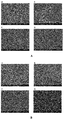
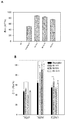
도 1A는 구형 키토산/ dsODN 나노입자의 환경적인 주사 전자 현미경 (ESEM) 이미지, 및 (A) 92-10-5 키토산/dsODN-DPP-IV 나노입자, (B) 80-80-5 키토산/dsODN-DPP-IV 나노입자, (C) 80-10-10 키토산/dsODN-DPP-IV 나노입자, (D) 92-10-5 키토산/dsODN-ApoB 나노입자, (E) 80-80-5 키토산/dsODN-ApoB 나노입자 및 (F) 80-10-10 키토산/dsODN-ApoB 나노입자의 입자 크기 분포도를 예시한 것이며; 도 1 B는 구형 키토산/dsODN 나노입자의 환경적인 주사 전자 현미경 (ESEM) 이미지, 및 (A) 92-10-5 키토산/dsODN-RecQL1 나노입자, (B) 80-40-5 키토산/dsODN-RecQL1 나노입자, 및 (C) 80-10-10 키토산/dsODN-RecQL1 나노입자의 입자 크기 분포도를 예시한 것이다.
도 2A는 구형 키토산/siRNA 나노입자의 환경적인 주사 전자 현미경 (ESEM) 이미지, 및 (A) 80-10-5 키토산/siRNA-ApoB 나노입자, (B) 80-40-5 키토산/siRNA-ApoB 나노입자, (C) 92-10-5 키토산/siRNA-ApoB 나노입자 및 (D) 92-40-5 키토산/siRNA-ApoB 나노입자의 입자 크기 분포도를 예시한 것이며; 도 2B는 구형 키토산/siRNA 나노입자의 환경적인 주사 전자 현미경 (ESEM) 이미지, 및 (A) 80-10-5 키토산/siRNA-MDR1 나노입자, (B) 80-200-5 키토산/siRNA-MDR1 나노입자, (C) 92-10-5 키토산/siRNA-MDR1 나노입자 및 (D) 92-150-5 키토산/siRNA-MDR1 나노입자의 입자 크기 분포도를 예시한 것이다.
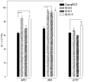
도 3A는 서로 다른 pH값에서 서로 다른 시간 동안 인큐베이션한, N:P 비율이 다양한 키토산/dsODN 나노입자의 폴리아크릴아마이드 겔 전기영동을 사진촬영한 것이다. (A) dsODN-DPP-IV 및 (B) dsODN-ApoB와 복합체를 이룬 키토산 92-10을 pH6.5 (MES) 및 pH 8 (TAE)에서 0.5h, 4h 및 20h 동안 인큐베이션하였으며; 도 3B는 서로 다른 pH값에서 서로 다른 시간 동안 인큐베이션한, N:P 비율이 다양한 키토산/dsODN 나노입자의 폴리아크릴아마이드 겔 전기영동을 나타낸 것이다. dsODN-RecQL1과 복합체를 이룬 키토산 92-10을 pH6.5 (MES) 및 pH 8 (TAE)에서 0.5h, 4h 및 20h 동안 인큐베이션하였다. 나노입자가 전술한 조건에서 안정하지 않은 경우, siRNA 모방 dsODN이 겔에서 방출되어 이동한다.
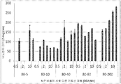
도 4A는 pH 6.5에서 키토산/siRNA 나노입자 안정성을 나타낸 막대 그래프로, 여러가지 DDA와 MW의 키토산 제형들을 3종의 다른 항-ApoB siRNA 서열들 (siApoB1, siApoB2 및 siApoB3)과 N:P 비율 5 및 10으로 복합체를 형성시키고, 20시간 동안 인큐베이션한 다음, 나노입자 형성 후, 핵산 정량화를 위해 사용하는 RNA 인터칼레이팅 염료인 Ribogreen™을 각 샘플에 첨가하여, 복합체를 형성하지 못한 RNA 비율을 측정하였으며, 높은 형광 값은 입자의 해리 및 불안정성을 나타낸내며; 도 4B는 나노입자 크기에 대한 MW의 영향을 나타낸 막대그래프로서, 92% DDA의 여러가지 MW 키토산을 항-RecQL1 siRNA와 여러가지 N:P 비율로 복합체를 형성하였고; 도 4C는 나노입자 크기에 대한 MW의 영향을 나타낸 막대 그래프로서, DDA가 80%인 다양한 MW의 키토산을 항-RecQL1 siRNA와 다양한 N:P 비율로 복합체화하였고; 도 4D는 나노입자 크기에 대한 MW의 영향을 나타낸 막대 그래프이다. DDA가 72%인 다양한 MW의 키토산을 항-RecQL1 siRNA와 다양한 N:P 비율로 복합체를 형성시켰으며; 도 4E는 나노입자 크기에 대한 RecQL1 siRNA 농도의 영향과 다이나믹 광 산란에 의해 측정한 나노입자 크기에 대한 염의 영향을 나타낸 막대 그래프로서, DDA가 92%이고 분자량이 10인 키토산을 항-RecQL1 siRNA의 농도를 증가시키면서 N:P 비율 5로 복합체를 형성시켰다.
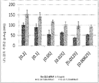
도 5는 여러가지 pH에서의 나노입자 안정성에 대한 DDA, MW 및 N:P 비율의 효과를 나타낸 것으로, 낮은 형광 강도는 입자의 안정성을 의미한다. 다양한 DDA, MW의 키토산을 항-MDR1 siRNA와 다양한 N:P 비율로 복합체를 형성시켜, 나노입자를 제조하였다. 나노입자를 여러가지 pH에서 인큐베이션하고, Ribogreen™ 분석으로 siRNA 방출을 측정하였다.
도 6A는 키토산/dsODN 나노입자들, (A) 키토산 (92-10-5 또는 80-10-10)과 dsODN-DPP-IV의 복합체, (B) DNAse I로 분해한 후 남아있는 dsODN-DPP-IV, (C) 키토산과 dsODN-ApoB의 복합체 (92-10-5 또는 80-10-10), (D) DNAse I로 분해한 후 남아있는 dsODN-ApoB에 대한 뉴클레아제 보호 분석의 결과를 나타낸 것으로, 모든 분해물들은 대조군으로 처리한 샘플의 신호 강도 (즉, 0U DNAse I = 100% 세기)를 이용하여 분석하였으며; 도 6B는 키토산/dsODN 나노입자들: (A) 키토산과 dsODN-RecQL1의 복합체 (92-10-5, 80-40-5 또는 80-10-10), 및 (B) DNAse I로 분해한 후 남아있는 dsODN-RecQL1의 뉴클레아제 보호 분석 결과를 나타낸 것으로, 모든 분해물들은 대조군으로 처리한 샘플의 신호 강도 (즉, 0U DNAse I = 100% 세기)를 이용하여 분석하였다.
도 7A는 수종의 세포주에 형질감염한 후 24시간 째의 dsODN /나노입자 세포 흡수: (A) HepG2 세포주에서의 키토산 (92-10-5, 80-80-5 또는 80-10-10)/5'-6FAM 표지된 dsODN DPP-IV의 흡수; 및 (B) HepG2, HEK293 및 RAW264.7 세포에서의 키토산 (92-10-5, 80-80-5 또는 80-10-10)/5'-6FAM 표지된 dsODN-ApoB의 흡수를 나타낸 막대 그래프로서, DharmaFECT® #1과 4는 흡수 양성 대조군으로 사용하였으며; 도 7B는 수종의 세포주에 형질감염한 후 24시간 째의 dsODN /나노입자 세포 흡수, 즉, AsPC1, LS174T 및 A549 세포에서의 키토산 (92-10-5, 80-40-5 또는 80-10-10)/5'-6FAM 표지된 dsODN RecQL1의 흡수를 나타낸 막대 그래프로서, DharmaFECT™ #1은 흡수 양성 대조군으로 사용하였다.
도 8은 키토산/dsODN-DPP-IV 나노입자로 형질감염된 (A) HepG2, (B) Caco-2 및 (C) HT-29 세포주, 키토산/dsODN-ApoB 나노입자로 형질감염된 (D) HepG2, (E) HEK293 및 (F) RAW264.7 세포주에서, 형질감염 24시간 후 키토산/siRNA 나노입자의 흡수를 보여주는 공초점 사진을 나타낸 것이다. 키토산 92-10 (DDA, Mn)을 로다민 (붉은 색)으로 표지하고, dsODN은 6FAM (녹색)으로 5' 표지하였다. 키토산 92-10을 siRNA와 N:P 비율 5로 복합체를 형성시켰다. 촬영하기 전에, 세포 막을 막 고정성 양친성 염료인 CellMask™ (하늘색)으로 염색하여, 내재화된 나노입자와 막에 결합된 나노입자를 구분하였다. 나타낸 사진에서 dsODN은 녹색으로, 키토산은 붉은 색으로, 막은 하늘색으로, 전이 DIC는 회색으로 각각 구분되는 채널로 표시되며, 좌측 하단 사분위에 겹친 사진을 나타낸다.
도 9는 형질감염 후 24시간 후 키토산/siRNA 나노입자 흡수를 보여주는 공초점 사진을 나타낸 것이다. LS174T 세포주에 키토산/siRNA-RecQL1 나노입자를 형질감염시켰다. 형질감염 24시간 후 사진을 촬영하였다. 키토산 92-10 (DDA, Mn)은 로다민 (붉은 색)으로 표지하였으며, siRNA는 6FAM (녹색)으로 5' 표지되었다. 키토산 92-10을 siRNA-RecQL1과 N:P 비율 5로 복합체를 형성시켰다. 세포 막을 이미지 촬영 전에 CellMask™ (하늘색)로 염색하였다. siRNA는 녹색으로, 키토산은 붉은 색으로, 막은 하늘색으로, 전이 DIC는 회색으로 각각 구분되는 채널로 표시되며, 좌측 하단 사분위에 겹친 사진을 나타낸다.
도 10은 형질감염 후 24시간 후 키토산/siRNA 나노입자 흡수를 보여주는 공초점 사진을 나타낸 것이다. MCF-7 MDR 세포주에 키토산/siRNA-MDR1 나노입자를 형질감염시켰다. 형질감염 24시간 후 사진을 촬영하였다. 키토산 92-10 (DDA, Mn)은 로다민 (붉은 색)으로 표지하였으며, siRNA는 Cy3 (녹색)으로 5' 표지되었다. 키토산 92-10 (A) 키토산 80-10 (B) 및 키토산 80-200 (C)을 siRNA-cy3과 N:P 비율 5로 복합체를 형성시켰다. 세포 막을 이미지 촬영 전에 CellMask™ (하늘색)로 염색하였다. siRNA는 녹색으로, 키토산은 붉은 색으로, 막은 하늘색으로, 전이 DIC는 회색으로 각각 구분되는 채널로 표시되며, 좌측 하단 사분위에 겹친 사진을 나타낸다.
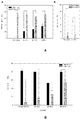
도 11A는 특정 세포주에서의 DPP-IV 및 ApoB 유전자 발현 저해에 대한 실시간 PCR (qPCR) 분석 결과를 나타낸 막대 그래프로서, HepG2 세포에, (A) 키토산 (92-10-5, 80-80-5 및 80-10-10/siRNA-DPP-IV); (B) 키토산 (92-10-5/siRNA-ApoB) 나노입자를 형질감염시키고, 형질감염 세포와 비-형질감염 세포를 ΔΔCT 방법으로 비교하여 저해율을 구하였으며; 도 11B는 특정 세포주에서의 RecQL1 유전자 발현 저해에 대한 실시간 PCR (qPCR) 분석 결과를 나타낸 막대 그래프로서, LS174T 세포에, 키토산 (92-10-5, 80-80-5 및 80-10-10/siRNA-RecQL1) 나노입자를 형질감염시키고, 형질감염 세포와 비-형질감염 세포를 ΔΔCT 방법으로 비교하여 저해율을 구하였다.
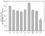
도 12는 3종의 DPP-IV 발현 세포주들에서의 DPP-IV 효소 활성을 나타낸 막대 그래프이다. DPP-IV 저해율은 siRNA-모의 형질감염 세포와 비교하여 결정하였다. 값은 평균 ± s.d.로 표시되며; n=4 /그룹이다. *p < 0.05, ** p < 0.01.
도 13은 혈장내 ApoB 수준에 대한 키토산/siRNA 투여 효과를 나타낸 막대 그래프이다. 단백질 수준은 각 처리군에 대해 ELISA로 측정하였다. 막대들과 에러 바는 무처리 죽상동맥경화증 그룹인 Dα에 상대적인 평균 단백질 수준을 나타낸다. 그룹 Dμ는 정상 저지방식을 식이한 정상 음성 대조군이다.
도 14는 키토산/siRNA 투여 이후의 치료학적 LDL/VLDL 콜레스티롤 저하를 보여주는 막대 그래프이다. LDL/VLDL 콜레스티롤 수준은 안락사 당일에 취한 샘플을 대상으로 정량적인 색도계 ELISA 키트로 측정하였다. 막대들과 에러 바는 무처리 죽상동맥경화증 그룹인 Dα에 상대적인 평균 단백질 수준을 나타낸다. 그룹 Dμ는 정상 저지방식을 식이한 정상 음성 대조군이다.
도 15는 치료학적 나노복합체 (TNC)를 처리한 동물 간에서의 간 콜레스테롤 액적 감소를 나타낸 것이다. 간내 콜레스테롤 축적에 대한 키토산/siRNA 투여 효과를 보여주는, (A) C1-1, (B) C2-1, (C) C3-1, (D) C4-1, (E) C5-1, (F) Dα-2일, (G) Dα-3, (H) Dβ-1 및 (I) Dμ-1 마우스의, 파라핀으로 고정하고 헤마톡실린-에오신으로 염색한 간 조직 절편들. 화살 표시 (->)는 콜레스테롤 액적 축적을 표시한다. Dα 그룹은 무처리 죽상동맥경화증 그룹이고, 그룹 Dμ는 정상 저지방식을 식이한 정상 음성 대조군이다.
도 16은 TNC 처리한 동물 간에서의 염증 재흡수 (inflammation resorption)를 나타낸 것이다. 키토산/siRNA 투여와 관련된 염증 반응의 재흡수 또는 죽상동맥경화증 진행을 보여주는, (A) C1-1, (B) C2-1, (C) C3-1, (D) C4-1, (E) C5-1, (F) Dα-2일, (G) Dα-3, (H) Dβ-1 및 (I) Dμ-1 마우스의, 파라핀으로 고정하고 사프라닌-O/패스트-그린/철-헤마톡실린으로 염색한 간 조직 절편들. 원 (O)과 화살 표시 (->)는 림프 침윤을 표시한다.
도 17은 전체 동물 그룹들의 매주 체중 측정치 (g)를 나타낸 막대 그래프이다. 동물들은 모두 각 주의 첫번째 날에, 각 키토산/siRNA 투여 전에 체중을 측정하였다. 저지방 정상 대조군 Dμ와 비교하여, 4주간 계속적인 체중 증가가 TNC 처리에 의해 기보적으로 영향을 받지 않는 고지방식이를 먹는 모든 동물들에서 관찰되었다.
도 18은 주당 체중 증가율을 나타낸 막대 그래프이다. 동물들은 모두 각 주의 첫번째 날에, 각 키토산/siRNA 투여 전에 체중을 측정하였다. 체중 증가는 동물의 체중과 전 주에 기록한 체중 간의 상대적인 차이이다 [(tn -1 - tn)/tn -1)]. 이 도면은 1차 TNC 투여한 후 즉각적인 체중 증가 또는 감소를 보여준다.Reference will now be made to the accompanying drawings.
Figure 1A shows an environmental scanning electron microscopy (SEM) image of spherical chitosan / dsODN nanoparticles and (A) 92-10-5 chitosan / dsODN-DPP-IV nanoparticles, (B) 80-80-5 chitosan / dsODN DPP-IV nanoparticles, (C) 80-10-10 chitosan / dsODN-DPP-IV nanoparticles, (D) 92-10-5 chitosan / dsODN- ApoB nanoparticles, (E) 80-80-5 chitosan / dsODN-ApoB nanoparticles and (F) 80-10-10 chitosan / dsODN-ApoB nanoparticles; Figure 1B shows an environmental scanning electron microscopy (SEM) image of spherical chitosan / dsODN nanoparticles and (A) 92-10-5 chitosan / dsODN-RecQL1 nanoparticles, (B) 80-40-5 chitosan / dsODN- RecQL1 nanoparticles, and (C ) 80-10-10 chitosan / dsODN-RecQL1 nanoparticles.
Figure 2A shows an environmental scanning electron microscopy (SEM) image of spherical chitosan / siRNA nanoparticles and (A) 80-10-5 chitosan / siRNA-ApoB nanoparticles, (B) 80-40-5 chitosan / siRNA- Nanoparticles, (C) 92-10-5 chitosan / siRNA-ApoB nanoparticles and (D) 92-40-5 chitosan / siRNA-ApoB nanoparticles; Figure 2B is an environmental scanning electron microscopy (SEM) image of spherical chitosan / siRNA nanoparticles and (A) 80-10-5 chitosan / siRNA-MDR1 nanoparticles, (B) 80-200-5 chitosan / siRNA- MDR1 Nanoparticles, (C) 92-10-5 chitosan / siRNA-MDR1 nanoparticles, and (D) 92-150-5 chitosan / siRNA-MDR1 nanoparticles.
Figure 3A is a photograph of polyacrylamide gel electrophoresis of chitosan / dsODN nanoparticles with different N: P ratios incubated at different pH values for different times. Chitosan 92-10 complexed with (A) dsODN-DPP-IV and (B) dsODN-ApoB was incubated at pH 6.5 (MES) and pH 8 (TAE) for 0.5 h, 4 h and 20 h; Figure 3B shows polyacrylamide gel electrophoresis of chitosan / dsODN nanoparticles with various N: P ratios incubated at different pH values for different times. Chitosan 92-10 complexed with dsODN-RecQL1 was incubated at pH 6.5 (MES) and pH 8 (TAE) for 0.5 h, 4 h and 20 h. If the nanoparticles are not stable under the conditions described above, the siRNA mimetic dsODN is released from the gel and migrates.
FIG. 4A is a bar graph depicting the stability of chitosan / siRNA nanoparticles at pH 6.5, showing that various chitosan formulations of DDA and MW were prepared with three different anti-ApoB siRNA sequences (siApoB1, siApoB2 and siApoB3) 10, incubated for 20 hours, and then the RNA intercalating dye, Ribogreen ™, used for nucleic acid quantification after nanoparticle formation, was added to each sample to determine the percentage of uncomplexed RNA , High fluorescence values indicate dissociation and instability of the particles; 4B is a bar graph depicting the effect of MW on nanoparticle size. Various MW chitosans of 92% DDA were complexed with anti-RecQL1 siRNA at various N: P ratios; Figure 4C is a bar graph depicting the effect of MW on nanoparticle size, in which various MW chitosans with 80% DDA were complexed with anti-RecQL1 siRNA at various N: P ratios; 4D is a bar graph showing the effect of MW on nanoparticle size. Various MW chitosans with a DDA of 72% were complexed with anti-RecQL1 siRNA at various N: P ratios; FIG. 4E is a bar graph showing the effect of salt on the nanocrystal size and the effect of salt on the nanoparticle size measured by dynamic light scattering. The chitosan with 92% DDA and a molecular weight of 10 was used as an anti-RecQL1 siRNA The complex was formed at an N: P ratio of 5 with increasing concentration.
Figure 5 shows the effect of DDA, MW and N: P ratios on nanoparticle stability at various pHs, with low fluorescence intensity indicating particle stability. Various DDA and MW chitosan complexes were formed with anti-MDR1 siRNA at various N: P ratios to produce nanoparticles. Nanoparticles were incubated at various pHs and siRNA release was measured by Ribogreen ™ analysis.
FIG. 6A is a graph showing the results of a reaction between chitosan / dsODN nanoparticles, (A) a complex of chitosan (92-10-5 or 80-10-10) with dsODN-DPP-IV, (B) -IV, (C) the complex of chitosan and dsODN-ApoB (92-10-5 or 80-10-10), (D) the result of nuclease protection analysis of residual dsODN-ApoB after digestion with DNAse I With all of the degradants analyzed using the signal intensity of the sample treated with the control (i.e., 0 U DNAse I = 100% intensity); FIG. 6B shows a schematic view of a chitosan / dsODN nanoparticles after (A) a complex of chitosan and dsODN-RecQL1 (92-10-5, 80-40-5 or 80-10-10) and (B) DNAse I The results of nuclease protection analysis of the remaining dsODN-RecQL1 were analyzed using the signal intensity (i.e., 0 U DNAse I = 100% strength) of the samples treated with the control.
(A) Hepatocyte growth factor (HepG2 ) cell chitosan (92-10-5, 80-80-5, or 80-10-10) / cells at 24 hours after transfection into several cell lines. Absorption of 5'-6FAM labeled dsODN DPP-IV; And (B) a histogram showing the absorption of chitosan (92-10-5, 80-80-5 or 80-10-10) / 5'-6FAM labeled dsODN-ApoB in HepG2, HEK293 and RAW264.7 cells , DharmaFECT ® # 1 and 4 were used as a positive control for absorption; Figure 7B shows the dsODN / nanoparticle cell uptake at 24 hours after transfection into several cell lines, i.e., chitosan (92-10-5, 80-40-5 or 80-10- 10) / 5'-6FAM labeled dsODN RecQL1. DharmaFECT ™ # 1 was used as a positive control for absorption.
FIG. 8 shows (A) HepG2, (B) Caco-2 and (C) HT-29 cell lines transfected with chitosan / dsODN-DPP-IV nanoparticles, (D) HepG2 transfected with chitosan / dsODN- (E) HEK293 and (F) RAW 264.7 cell lines, showing absorption of chitosan / siRNA nanoparticles after 24 hours of transfection. Chitosan 92-10 (DDA, Mn) was labeled with rhodamine (red) and dsODN was labeled 5 'with 6FAM (green). Chitosan 92-10 was complexed with siRNA at an N: P ratio of 5. Prior to imaging, cell membranes were stained with CellMask ™ (light blue), a rigid amphipathic dye, to separate internalized nanoparticles and membrane bound nanoparticles. In the picture, dsODN is shown as green, chitosan as red, membrane as sky blue, and transition DIC as gray, respectively, showing overlapping pictures on the lower left quadrant.
Figure 9 shows a confocal photograph showing chitosan / siRNA nanoparticle uptake after 24 hours of transfection. The LS174T cell line was transfected with chitosan / siRNA-RecQL1 nanoparticles. Photographs were taken 24 hours after transfection. Chitosan 92-10 (DDA, Mn) was labeled with rhodamine (red) and siRNA was labeled 5 'with 6FAM (green). Chitosan 92-10 was complexed with siRNA-RecQL1 and N: P ratio of 5. Cell membranes were stained with CellMask ™ (light blue) before imaging. The siRNA is shown in green, chitosan in red, membrane in sky blue, and transition DIC in gray, and shows a superimposed picture in the lower left quadrant.
Figure 10 shows confocal images showing chitosan / siRNA nanoparticle uptake after 24 hours of transfection. The MCF-7 MDR cell line was transfected with chitosan / siRNA-MDR1 nanoparticles. Photographs were taken 24 hours after transfection. Chitosan 92-10 (DDA, Mn) was labeled with rhodamine (red) and siRNA was labeled 5 'with Cy3 (green). Chitosan 92-10 (A) Chitosan 80-10 (B) and chitosan 80-200 (C) were complexed with siRNA-cy3 at an N: P ratio of 5. Cell membranes were stained with CellMask ™ (light blue) before imaging. The siRNA is shown in green, chitosan in red, membrane in sky blue, and transition DIC in gray, and shows a superimposed picture in the lower left quadrant.
11A is a bar graph showing the results of real-time PCR (qPCR) analysis on inhibition of DPP-IV and ApoB gene expression in a specific cell line. HepG2 cells were treated with (A) chitosan (92-10-5, 80-80-5 And 80-10-10 / siRNA-DPP-IV); (B) Chitosan (92-10-5 / siRNA-ApoB) nanoparticles were transfected and the inhibition rates were determined by comparing the transfected and non-transfected cells with the ΔΔCT method; 11B is a bar graph showing the results of real-time PCR (qPCR) analysis on the inhibition of RecQL1 gene expression in a specific cell line. LS174T cells were treated with chitosan (92-10-5, 80-80-5, siRNA-RecQL1) nanoparticles, and the inhibition rate was calculated by comparing the transfected cells with the non-transfected cells by the DELTA DELTA CT method.
Figure 12 is a bar graph showing DPP-IV enzyme activity in three DPP-IV expressing cell lines. DPP-IV inhibition rates were determined relative to siRNA-mock transfected cells. Values are expressed as mean ± sd; n = 4 / group. * p < 0.05, ** p < 0.01.
Figure 13 is a bar graph depicting the effect of chitosan / siRNA administration on plasma levels of ApoB. Protein levels were measured by ELISA for each treatment group. Bars and error bars represent the average protein level relative to the untreated atherosclerotic group Dα. Group Dμ is a normal negative control group in which the normal blocking system is dietary.
Figure 14 is a bar graph showing the therapeutic LDL / VLDL cholesterol degradation after administration of chitosan / siRNA. LDL / VLDL cholesterol levels were measured by quantitative colorimetric ELISA kits on samples taken on euthanasia days. Bars and error bars represent the average protein level relative to the untreated atherosclerotic group Dα. Group Dμ is a normal negative control group in which the normal blocking system is dietary.
Figure 15 shows the reduction of liver cholesterol droplets in animals treated with therapeutic nanocomposites (TNC). (A) C1-1, (B) C2-1, (C) C3-1, (D) C4-1, (E) C5-1, (F ) which show the effect of chitosan / siRNA administration on intrahepatic cholesterol accumulation ) Dα-2 days, (G) Dα-3, (H) Dβ-1 and (I) Dμ-1 mice fixed with paraffin and stained with hematoxylin-eosin. Arrows (->) indicate cholesterol droplet accumulation. The Dα group is the untreated atherosclerotic group, and the group Dμ is the normal negative control group in which the normal blocking method is dietary.
Figure 16 shows inflammation resorption in TNC-treated animals. (A) C1-1, (B) C2-1, (C) C3-1, (D) C4-1, (E) C5-1, (F) Dα-2 days, (G) Dα-3, (H ) Dβ-1 and (I) Dμ-1 mice were fixed with paraffin and saffranin-O / fast-green / - Liver tissue sections stained with hematoxylin. Circles (O) and arrows (->) indicate lymph infiltration.
Figure 17 is a bar graph showing weekly body weight measurements (g) of whole animal groups. Animals were weighed before each chitosan / siRNA dose on the first day of each week. Compared with the low - fat normal control Dμ, continuous weight gain for 4 weeks was observed in all animals fed a high fat diet that was not negatively affected by TNC treatment.
18 is a bar graph showing weight gain per week. Animals were weighed before each chitosan / siRNA dose on the first day of each week. The weight gain is the relative difference between the body weight of the animal and the weight recorded in the previous week [(t n -1 - t n ) / t n -1 )]. This figure shows immediate weight gain or reduction after primary TNC administration.
본 발명에 따라, 짧은 간섭 RNA (siRNA), 짧은 헤어핀 RNA (shRNA), 및 RNAi-유도성 벡터 (즉, 세포 내에 존재하여 siRNA 또는 shRNA를 생성하는 벡터)와 같은 RNAi 유도성 물질(entities)을 인간과 같은 포유류의 세포, 조직 및 장기에 효율적으로 전달하기 위한, 비-바이러스 벡터의 신규하고 특정한 조성물을 제공한다. 특히, 본 발명은 특정한 키토산 : 핵산 비율을 가진 RNAi 유도성 물질을 포함하는, 특정한 평균 분자량 (Mn) 및 탈아세틸화율 (DDA) 범위를 가진 키토산 조성물을 제공한다. In accordance with the present invention, RNAi inducible entities such as short interfering RNA (siRNA), short hairpin RNA (shRNA), and RNAi-inducible vectors (i. E., Those that are present in the cell and produce siRNA or shRNA) To provide novel and specific compositions of non-viral vectors for efficient delivery to cells, tissues and organs of mammals such as humans. In particular, the present invention provides chitosan compositions with specific average molecular weight (Mn) and deacetylation (DDA) ranges, including RNAi inducing materials with specific chitosan: nucleic acid ratios.
따라서, 표적 전사체의 과도한 발현 또는 부적절한 발현; 또는 표적 전사체에 의해 코딩되는 폴리펩타이드의 부적절하거나 또는 과도한 활성과 연관된 질환 또는 증상을 치료하거나 또는 예방하기 위한 조성물 및 방법을 제공한다. Thus, overexpression or inappropriate expression of a target transcript; Or to treat or prevent a disease or condition associated with inappropriate or excessive activity of a polypeptide encoded by a target transcript or a polypeptide encoded by the target transcript.
본원에서 제공하는 조성물은, 증상들의 출현 전이나 도중 또는 그 후의 적절한 시간대에서, 이러한 증상을 앓을 위험이 있거나 또는 이미 앓고 있는 대상에게, 본원 개시 조성물을 사용하여 RNAi 유도성 물질을 투여함으로써, 증상을 완화하는 데 사용될 수 있다.The composition provided herein can be administered to a subject already at risk of or at risk of suffering from such symptoms before, during, or after the appearance of the symptoms, by administering an RNAi inducing agent using the composition described herein Can be used to mitigate.
본 발명의 조성물 및 방법은 전사체의 기능을 연구하고, 상기 전사체에 의해 코딩되는 폴리펩타이드가 존재하지 않거나 또는 이의 활성이 저하된 경우, 세포 또는 유기체에 대한 서로 다른 화합물의 효과를 연구하는 것처럼 다양한 목적에 사용될 수 있지만, 이로 한정되지 않는다. 더욱이, 상기 조성물 및 상기 방법은 II형 당뇨병 및 이와 관련된 병리학적 상태, 죽상동맥경화증 및 이와 관련된 병리학적 상태, 및 암에 대한 임상적인 치료법에서 사용될 수 있다. 구체적으로는, 상기 조성물 및 상기 방법은, 당뇨병 치료를 위해 인크레틴 분해 효소 (DPP-IV) 또는 임의의 당조절 단백질의 저해에 사용되거나, 또는 죽상동맥경화증 치료를 위해 ApoB 유전자 또는 임의의 아테롬원성 단백질 (즉, ApoE)의 저해에 사용되거나, 또는 암 치료를 위해 RecQL1 DNA 헬리카제 또는 DDX5 - p68- RNA 헬리카제의 발현을 하향조절하는 데 사용될 수 있지만, 이로 한정되지는 않는다.The compositions and methods of the present invention are designed to study the function of the transcript and to study the effect of different compounds on the cell or organism when the polypeptide encoded by the transcript is absent or its activity is decreased May be used for various purposes, but is not limited thereto. Moreover, the compositions and methods can be used in the clinical treatment of type II diabetes and related pathological conditions, atherosclerosis and related pathological conditions, and cancer. Specifically, the compositions and methods can be used for the inhibition of incretinase (DPP-IV) or any tonic protein for the treatment of diabetes, or for the treatment of atherosclerosis with the ApoB gene or any atherogenic protein (I. E., ApoE) or may be used to down-regulate the expression of RecQL1 DNA helicase or DDX5-p68-RNA helicase for cancer treatment, but is not limited thereto.
특히, 본 발명은 예를 들어, 헬리카제 과발현 종양에 대한 직접적인 치료, 또는 일시적인 의약을 위한 방사선민감화 물질로서의, 본원 발명의 조성물과 함께 이러한 핵산의 용도에 관한 것이다. 더욱이, 본원 발명의 조성물 및 방법은 방사선치료, 수술, 호르몬 치료 또는 종래의 화학요법과 같은 다른 치료법과 함께 사용될 수 있다. 본 발명은 추가로, 방사선요법의 증강, 또는 다른 치료법과 함께 사용되기 위한 조성물 및 방법을 제공한다.In particular, the present invention relates to the use of such a nucleic acid in conjunction with a composition of the present invention, for example, as a direct treatment of helicase overexpressing tumors, or as a radiation sensitizing agent for transient medicinal products. Moreover, the compositions and methods of the present invention may be used in conjunction with other therapies such as radiation therapy, surgery, hormone therapy or conventional chemotherapy. The present invention further provides compositions and methods for use in conjunction with radiotherapy enhancement, or other therapies.
본원에서 개시되는 조성물은 물리화학적 특성이 하기와 같은 RNAi 유도성 핵산 및 키토산을 포함한다: N:P 비율이 25 미만이며, 키토산의 수 평균 분자량 (Mn)이 5 kDa 내지 200 kDa이며 탈아세틸화율이 80% DDA 내지 95% DDA임. 본 발명은,이의 조성물 및 방법이, 서로 다른 세포주에 효과적으로 감염되며 시판의 리포플렉스(lipoplex)와 유사한 유전자 침묵을 유도하는 효능을 가지고 있음을 나타내고 있으며, 여기서 형질감염 효능은 일부의 경우에, 뚜렷한 세포독성 없이, mRNA 농도 80% 및 세포 흡수 95%에 도달하였다.The compositions disclosed herein comprise an RNAi-inducing nucleic acid and chitosan whose physico-chemical properties are as follows: N: P ratio less than 25, number average molecular weight (Mn) of chitosan from 5 kDa to 200 kDa and deacetylation rate This is 80% DDA to 95% DDA. The present invention shows that the compositions and methods are effective in effectively infecting different cell lines and inducing gene silencing similar to commercial lipoplex, wherein the transfection efficacy is, in some cases, Without cytotoxicity, mRNA concentration reached 80% and cell uptake was 95%.
RNA 간섭 (RNAi)은, 이중가닥 RNA가 메신저 RNA와 같은 세포성 전사체의 서열 특이적인 분해를 지시하는 과정이다 (Sharp, 2001, Genes Dev, 15:485-490; Vance and Vaucheret, 2001, Science, 292:2277-2280). 이러한 현상은 처음에 씨. 엘레강스( C. elegans)에서 발견되었다 (Fire et al., 1998, Nature, 391 :806-811). 자연적으로 존재하는 RNAi는 21-25개의 뉴클레오타이드를 가진 짧은 이중 가닥으로 된 절편에 의해 매개되며, 이를 작은 간섭 RNA라고 지칭한다. 이들 siRNA는, 다이서(dicer)라고 하는 dsRNA-특이적인 엔도뉴클레아제에 의해, 긴 이중 가닥으로 된 RNA (dsRNA)를 21개 염기쌍의 작은 간섭 RNA (siRNA) - 2개의 뉴클레오타이드 3' 오버행(overhang)의 측면에 19개 염기쌍으로 된 이중가닥 영역인 코어 영역으로 이루어짐 - 로 절단하는 과정을 통해 생성된다 (Bernstein et al., 2001, Nature, 409:363-366). 그런 다음, siRNA는 RNA-유도성 침묵 복합체 (RISC)에 병합되고, RISC가 상보성 서열을 가진 표적 mRNA를 인지하도록 하여, 특정한 전사체가 절단되게 한다.RNA interference (RNAi) is a process by which double-stranded RNA directs sequence-specific degradation of cellular transcripts such as messenger RNA (Sharp, 2001, Genes Dev, 15: 485-490; Vance and Vaucheret, 2001, Science , 292: 2277-2280). This phenomenon is the first time Mr.. Elegance was found (C. elegans) (Fire et al , 1998, Nature, 391:. 806-811). Naturally occurring RNAi is mediated by short double-stranded fragments with 21-25 nucleotides, which are referred to as small interfering RNAs. These siRNAs can be made by dsRNA-specific endonuclease, a long double stranded RNA (dsRNA), with 21 base pairs of small interfering RNA (siRNA) - 2 nucleotides 3 'overhangs (Bernstein et al., 2001, Nature, 409: 363-366), which consists of a core region that is a double stranded region of 19 base pairs on the side of the overhang. The siRNA is then incorporated into an RNA-induced silencing complex (RISC), allowing the RISC to recognize a target mRNA with a complementary sequence so that a particular transcript is cleaved.
이후, 얼마 지나지 않아, 합성의 21개 뉴클레오타이드 RNA 듀플렉스 (siRNA)를 도입함으로써 포유류 세포에서 RNAi를 유도할 수 있으므로, 긴 dsRNA에 다이서를 매개로 한 프로세싱을 할 필요가 없는 것으로 밝혀졌기 때문에, RNAi는 임상적인 용도에서 큰 잠재력을 가진 것으로 인식되었다 (Elbashir et al., 2001, Nature, 41 1 :494-498).Sooner or later, RNAi could be induced in mammalian cells by introducing synthetic 21-nucleotide RNA duplexes (siRNAs), so RNAi was found to not require dicer mediated processing of long dsRNAs (Elbashir et al., 2001, Nature, 41 1: 494-498).
예를 들어, ApoB의 발현을 표적으로 하여 이를 감소시킴으로써 VLDL의 과도한 형성을 방지하여, 유기체에서 이들 아테롬원성 물질이 축적을 감소시킬 수 있다 (Soutschek et al., 2004, Nature, 432: 173-178). 인간이 아닌 영장류에서 서열 특이적인 siRNA를 이용하여 mRNA 단계에서 ApoB를 표적화하면, 처리 후 24시간째에 dApoB 단백질, 혈청 콜레스테롤 및 저밀도 지질단백질 농도가 크게 감소하여 나타났다 (Zimmermann et al., 2006, Nature, 441 : 1 1 1 -1 14). 지질을 기재로 하는 나노입자 (SNALP-siRNA)를 사용하는 이러한 치료법의 치료 효과는 가장 높은 siRNA 농도에서 11일간 지속되었으며, 이는 siRNA 치료의 즉각적이면서도 강력하고 오래 지속되는 생물학적 효과를 나타내는 것이다. 아쉽게도, 이들 지질 기재의 벡터는, 혈청 내 아스파테이트 아미노트랜스퍼라제 (AST) 및 알라닌 아미노트랜스퍼라제 (ALT) 농도의 상승이 간세포 괴사를 나타내는 것으로 알 수 있듯이, 높은 수준의 간 독성을 야기하였다 (Zimmermann et al., 2006, Nature, 441 : 1 1 1 -1 14). 따라서, 이러한 보고들이 죽상동맥경화증 및 CVD 치료를 위한 표적으로서 ApoB가 중요함을 언급하고는 있지만, 전신적인 ApoB를 안전하고도 효과적으로 감소시키는 데는 현재의 siRNA 전달 시스템이 부적절하다고도 강조하고 있다.For example, by targeting and diminishing the expression of ApoB, it is possible to prevent accumulation of these atherogenic substances in organisms by preventing excessive formation of VLDL (Soutschek et al., 2004, Nature, 432: 173-178 ). Targeting ApoB at the mRNA stage using sequence-specific siRNAs in non-human primates resulted in a significant reduction in dApoB protein, serum cholesterol, and low density lipoprotein concentrations at 24 hours after treatment (Zimmermann et al., 2006, Nature , 441: 1 1 1 -1 14). The therapeutic effect of this therapy using lipid-based nanoparticles (SNALP-siRNA) lasted 11 days at the highest siRNA concentration, indicating immediate, potent and long lasting biological effects of siRNA therapy. Unfortunately, the vectors of these lipid substrates have resulted in a high level of liver toxicity, as can be seen by elevated levels of aspartate aminotransferase (AST) and alanine aminotransferase (ALT) in the serum indicating hepatocyte necrosis (Zimmermann et al., 2006, Nature, 441: 11-1-14). Thus, although these reports indicate that ApoB is important as a target for atherosclerosis and CVD therapies, it emphasizes that current siRNA delivery systems are inadequate to safely and effectively reduce systemic ApoB.
합성되는 작은 간섭 RNA 형태의 RNAi를 직접적으로 전달하면, 계속해서 문제가 되는데, 세포내 표적화 및 흡수가 불량해지고, 세포내 및/또는 세포외 뉴클레아제 분해 (즉, RNAse)로 인해 반감기가 짧으며, 뿐만 아니라 혈액 안정성이 한정되고 독성이 야기된다 (Stein, 1996, Trends Biotechnol, 14: 147-149; Urban-Klein et al., 2004, Gene Therapy, 1-6; Katas and Alpar, 2006, J Control Release, 1 15:216-225). 그 결과, RNAi를 임상적 치료제로 번역하는 것은, 이들 문제점을 해결할 것을 요구하고 있다. RNAi는, 형질감염과 같은 방법에 의해 세포 내로 도입되었을 때, 여러 가지 많은 종류의 서로 다른 세포에서 작동하는 것으로 나타나 있다. 그러나, 형질감염 효율은 작은 간섭 RNA 분자를 포함하는 전달 비히클에 좌우한다. 벡터로 지칭되는 전달 비히클은 siRNA를 축합하고, 보호하며, 표적 세포로 전달할 수 있어야 한다. 일단 표적 주변에 존재하게 되면, 비-바이러스 벡터는 세포내 흡수를 촉진하고, 리소좀 고립(lysosomal sequestration)을 피하고, 이들의 내용물을 방출하여, 원하는 생물학적 효과를 얻을 수 있도록 해야 한다.Direct delivery of RNAi in the form of small interfering RNAs, which are synthesized, continues to be problematic because of poor cell targeting and uptake and shortened half-life due to intracellular and / or extracellular nuclease degradation (i. E., RNAse) (Stein, 1996, Trends Biotechnol, 14: 147-149; Urban-Klein et al., 2004, Gene Therapy, 1-6; Katas and Alpar, 2006, J Control Release, 15: 216-225). As a result, translating RNAi into a clinical therapeutic agent requires resolving these problems. RNAi, when introduced into cells by methods such as transfection, is shown to work in many different cell types. However, transfection efficiency is dependent on the delivery vehicle comprising small interfering RNA molecules. A delivery vehicle, referred to as a vector, should be able to condense, protect, and deliver the siRNA to the target cell. Once present in the vicinity of the target, the non-viral vector should facilitate intracellular absorption, avoid lysosomal sequestration, release their contents, and obtain the desired biological effect.
합성 siRNA를 화학적으로 개질한 경우, 뉴클레아제 분해에 대한 내성이 수득되고 혈액 안정성이 개선되었다. 예를 들어, 특정한 리보스(ribose)의 C2 위치에서 2'-0-메틸로 치환시키거나 또는 포스포로티오에이트(phosphorothioate) 연결을 선택적으로 첨가하면, 활성에 영향을 주지 않으면서도 siRNA의 뉴클레아제 내성을 증가시킬 수 있다 (Corey, 2007, J Clin Invest, 1 17:3615-3622; Whitehead et al., 2009, Nat Rev Drug Discov, 8: 129-138; Judge et al., 2006, Mol Ther, 13:494-505). 그렇지만, 일부 화학적 개질은 세포독성을 증가시키며 표적화 효과를 저하시키고, mRNA 혼성화를 감소시킬 수 있다 (Weyermann et al., 2005, Eur J Pharm Biopharm, 59:431 -438; Amarzguioui et al., 2003, Nucleic Acids Res, 31 :589-595). 화학적 개질을 통해 수득되는 개선에도 불구하고, siRNA 반감기, 형질감염 효율, 세포성 표적화 및 흡수를 증가시키는 것은 여전히 효과적인 전달에 대한 걸림돌로 존재한다. 따라서, 화학적으로 비개질된/개질된 siRNA를 보호하고 이를 표적 세포로 전달할 수 있는 패키징 시스템이 필요하다. 그러나, 형질감염 효율은 작은 간섭 RNA 분자를 포함하는 전달 비히클에 좌우한다. 벡터로 지칭되는 전달 비히클은 siRNA를 축합하고, 보호하며, 표적 세포로 전달할 수 있어야 한다. 일단 표적 주변에 존재하게 되면, 비-바이러스 벡터는 세포내 흡수를 촉진하고, 리소좀 고립을 피하고, 이들의 내용물을 방출하여, 원하는 생물학적 효과를 얻을 수 있도록 해야 한다. 이러한 비-바이러스 벡터는 시험관내 및 생체내에서 테스트되어, siRNA가 임상적인 실현가능성을 갖출 수 있는지 나타낸다. 그렇지만, 큰 단점들은 이러한 비-바이러스 벡터와 관련이 있다. 낮은 형질감염 효율, 혈청 안정성, 응집 및 독성은, 임상에서 강력하고도 무독성의 약물 전달 툴로서 비-바이러스 벡터가 상용화될 수 있기 전에 해결해야 할, 주 장해물로 남아 있다. 비-바이러스 벡터의 주 클래스는 하기에 나타나 있다:When the synthetic siRNA was chemically modified, resistance to nuclease degradation was obtained and blood stability was improved. For example, substitution of 2'-O-methyl at the C2 position of a particular ribose or the optional addition of a phosphorothioate linkage can result in a nuclease of the siRNA without affecting activity 2006, Mol Ther, < RTI ID = 0.0 > 17: 3615-3622; < / RTI > Whitehead et al., 2009, Nat Rev Drug Discov, 8: 129-138; 13: 494-505). However, some chemical modifications may increase cytotoxicity, reduce targeting effects, and reduce mRNA hybridization (Weyermann et al., 2005, Eur J Pharm Biopharm, 59: 431-438; Amarzguioui et al., 2003, Nucleic Acids Res, 31: 589-595). Despite the improvements obtained through chemical modification, increasing siRNA half-life, transfection efficiency, cellular targeting and absorption remains a stumbling block to effective delivery. Thus, there is a need for a packaging system that can protect chemically unmodified / modified siRNAs and deliver them to target cells. However, transfection efficiency is dependent on the delivery vehicle comprising small interfering RNA molecules. A delivery vehicle, referred to as a vector, should be able to condense, protect, and deliver the siRNA to the target cell. Once present in the vicinity of the target, the non-viral vector should promote intracellular absorption, avoid lysosome isolation, release their contents, and obtain the desired biological effect. Such non-viral vectors may be tested in vitro and in vivo to demonstrate that the siRNA can be made clinically feasible. However, major drawbacks are associated with such non-viral vectors. Low transfection efficiency, serum stability, aggregation and toxicity remains a major obstacle to be addressed before non-viral vectors can be commercialized as a clinically powerful and non-toxic drug delivery tool. The main classes of non-viral vectors are shown below:
칼슘 calcium 포스페이트Phosphate
본 벡터의 주 단점은 한정된 효율과, 뉴클레아제에 의해 분해되지 않도록 핵산을 보호하지 못한다는 것이다. 핵산을 보호하는 능력이 개선되었음에도 불구하고, 이의 형질감염 효율은 여전히 낮아서, 생체내에서 효과적으로 사용할 수는 없다.The main disadvantage of this vector is its limited efficiency and its inability to protect nucleic acids from degradation by nuclease. Despite improved ability to protect nucleic acids, their transfection efficiency is still low and can not be used effectively in vivo.
양이온성Cationic 지질 Lipid
양이온성 지질은 정전기적 상호작용을 통해 핵산과 복합체를 이루어서, 결국 다중 층상(multi lamellar)의 지질-핵산 복합체 (리포플렉스)를 형성한다. 리포좀 제형은 통상, 양이온성 지질 및 DOPE (다이올레오일포스파티딜에탄올아민)와 같은 중성 지질을 포함한다. 중성 지질은 리포좀 제형의 안정성에 기여하며, 막 융합을 촉진할 뿐만 아니라, 엔도좀을 불안정화시켜서 리소좀의 탈출(lysozomal escape)에 기여한다. 리포플렉스는 핵산을 배양된 세포에 전달하는 가장 효과적인 방법들 중 하나이다. 이들의 형질감염 효율에도 불구하고, 리포플렉스는 배양된 세포에서 관찰되고 여러 가지 생체내 발견에서 확인할 수 있듯이 독성이다. 이러한 독성은 복합체 내 핵산에 대한 양이온성 지질의 전하 비율(charge ratio) 및 투여되는 용량과도 밀접한 관련이 있다. 리포플렉스와 관련된 독성을 저하시키기 위해 보다 생물융화성인 제형이 시험 및 테스트되고 있다. 독성 저하는 주로, 다른 중합체를 이용한 그래프팅(grafting) 또는 양이온성 중합체의 전체 전하를 감소시킴으로써 이루어진다.Cationic lipids form complexes with nucleic acids through electrostatic interactions, thus forming a multi-lamellar lipid-nucleic acid complex (lipoflex). Liposomal formulations typically include neutral lipids such as cationic lipids and DOPE (dioleoyl phosphatidylethanolamine). Neutral lipids contribute to the stability of liposomal formulations and not only promote membrane fusion, but also destabilize endosomes and contribute to the lysozomal escape. Lipoflex is one of the most effective methods of delivering nucleic acids to cultured cells. Despite their transfection efficiency, lipoplex is observed in cultured cells and is toxic, as can be seen from various in vivo findings. This toxicity is also closely related to the charge ratio of the cationic lipids to the nucleic acids in the complex and the dose administered. More bioavailable formulations have been tested and tested to reduce the toxicity associated with lipoplex. The reduction in toxicity is mainly achieved by grafting with other polymers or by reducing the total charge of the cationic polymer.
양이온성Cationic 중합체 polymer
양이온성 중합체는, 반대로 하전된 폴리양이온 및 폴리음이온 화학종 (즉, 핵산) 간의 상호작용을 통해 nm 크기(nanometric size)의 나노입자를 형성한다. 이들 나노입자는 핵산을 둘러싸서, 뉴클레아제에 의한 카고 분해(cargo degradation)를 방지한다 (Romoren et al., 2003, Int J Pharm, 261 : 115-127). 다수의 천연 및 합성 양이온성 중합체가 유전자 전달 또는 침묵을 위한 비히클로서 사용되어 왔다. 양이온성 중합체를 사용하는 이들 나노입자 중 상당수는 리포플레스에 비해, 우수한 형질감염 효율 및 더 낮은 혈청 감수성을 가지고 있다. 자연적으로 존재하는 폴리양이온 중에는 특히, 히스톤, 양이온화된 인간 혈청 알부민 및 키토산, 아미노다당류와 같은 단백질이 존재한다.The cationic polymer forms nanoparticles of nanometric size through interaction between the oppositely charged polycations and polyanion species (i.e., nucleic acids). These nanoparticles surround the nucleic acid and prevent cargo degradation by nuclease (Romoren et al., 2003, Int J Pharm, 261: 115-127). A number of natural and synthetic cationic polymers have been used as vehicles for gene delivery or silencing. Many of these nanoparticles using cationic polymers have superior transfection efficiency and lower serum sensitivity than lipoplexes. Among naturally occurring polycations, histones, cationized human serum albumin and chitosan, proteins such as aminopolysaccharides are present.
합성 폴리양이온의 군에는, 폴리-L-리신 (PLL), 폴리-L-오르니틴 뿐만 아니라 폴리에틸렌이민 (PEI), 폴리프로필렌이민 및 폴리아미도아민 덴다이머(dendimer)와 같은 폴리아민이 포함된다.Groups of synthetic polycations include polyamines such as poly-L-lysine (PLL), poly-L-ornithine as well as polyethyleneimine (PEI), polypropyleneimine and polyamidoamine dimer.
폴리플렉스(polyplex)의 이점은, 리포좀은 이의 형성에 다중 지질 구성분을 필요로 하지만 이와는 대조적으로, 폴리플렉스는 이의 형성에 다중 폴리양이온의 상호작용을 필요로 하지 않으며, 따라서 폴리플렉스의 거시적인 특성을 조절하기가 더 용이하다는 점이다. 또한, 폴리양이온의 또 다른 주 이점은 이들의 블록 구조로서, 직접적인 화학적 개질을 통해 보다 높은 효율 또는 특정한 세포 표적화를 달성할 수 있다. 그러나, 이러한 이점들에도 불구하고, 전하 밀도가 높은 나노입자가 보다 독성인 것으로 나타났기 때문에, 많은 양이온성 중합체들은 이들의 높은 표면 전하 밀도로 인해 독성인 것으로 밝혀졌다. 아울러, 중합체의 전하 밀도가 전하의 총 양보다는 세포독성에 보다 중요한 역할을 하는 것으로 보고된 바 있다. 독성은 분자량 의존적일 수도 있는데, PEI의 세포독성이 분자량에 따라 선형으로 증가하기 때문이다. 더욱이, 리소좀에 PEI와 같은 비-분해성 중합체가 축적되는 것은 리소좀 격리라고도 하는 현상으로서, 독성을 야기하는 부가적인 기여현상일 수 있다.The advantage of polyplex is that the liposomes require multiple lipid components to form it, but in contrast, the polyplex does not require the interaction of multiple poly-cations to form it, and therefore the macroscopic It is easier to control the characteristics. In addition, another major advantage of polycations is their block structure, which can achieve higher efficiency or specific cell targeting through direct chemical modification. However, despite these advantages, many cationic polymers have been found to be toxic due to their high surface charge density, as nanoparticles with high charge density have been shown to be more toxic. In addition, it has been reported that the charge density of the polymer plays a more important role in cytotoxicity than the total amount of charge. Toxicity may be molecular weight-dependent, as the cytotoxicity of PEI increases linearly with molecular weight. Furthermore, the accumulation of non-degradable polymers such as PEI in lysosomes is a phenomenon, also referred to as lysosome sequestration, which may be an additional contributory phenomenon that causes toxicity.
키토산은, 키틴의 알칼리 탈아세틸화로부터 유래되는 β-1,4 글리코시드 결합에 의해 연결된 N-아세틸-글루코사민 단량체와 글루코사민의 천연 중합체이다. 키토산 분자량 및 탈아세틸화율은 이의 생물학적 및 물리화학적 특성을 나타낸다. 예를 들어, 키토산 생물분해성은 아세틸기의 양 및 분포에 의해 영향을 받는다. 블록 분포보다는 이들 기의 부재 또는 무작위로 인해, 분해율이 매우 낮아진다.Chitosan is a natural polymer of glucosamine and N-acetyl-glucosamine monomer linked by? -1,4 glycoside linkages derived from alkaline deacetylation of chitin. The molecular weight and deacetylation rate of chitosan indicate its biological and physicochemical properties. For example, chitosan biodegradability is affected by the amount and distribution of acetyl groups. Due to the absence or randomness of these groups rather than the block distribution, the degradation rate is very low.
키토산은 생물융합성, 생물분해성, 점막접착 특성, 항균/항진균 활성 및 매우 낮은 독성을 비롯하여 유익한 특성을 많이 가지고 있다. 따라서, 이는 약학 및생물의약 분야의 관심을 끌었으며, 핵산 패키징 및 축합을 위해 가장 광범위하게 사용되는 비-바이러스 벡터들 중 하나가 되었다.Chitosan has many beneficial properties, including biofusion, biodegradability, mucoadhesive properties, antimicrobial / antifungal activity and very low toxicity. Thus, it has attracted attention in the pharmaceutical and biopharmaceutical fields and has become one of the most widely used non-viral vectors for nucleic acid packaging and condensation.
몇 가지 연구들은, 키토산-플라스미드 DNA 나노입자의 흡수, 나노입자 트래피킹(trafficking) 및 서로 다른 세포주에서의 형질감염 효율에 대한, 키토산 분자량 및 탈아세틸화율 (DDA)의 효과를 나타낸 바 있다. Huang 등은 A549 세포에 대해 이 물질을 언급하였다 (2005, J Control Release, 106:391-406). 그러나, 이 연구는 7가지의 제형 (10, 17, 48, 98 및 213 kDa의 키토산 및 88% DDA; 213 kDa 키노산 및 61% 및 46% DDA)만을 사용하여, 평균 분자량 (Mn) 및 DDA가 pDNA의 형질감염 효율에 미치는 효과를 연구하였으며, 플라스미드 내 수천개의 염기쌍과 비교해 그 크기가 전형적으로 21 bp인 훨씬 작은 siRNA에 대해서는 언급하고 있지 않다. 이들은, Mn 및 DDA의 감소가 플라스미드의 형질감염 효율을 저하시킴을 발견하였다. 그러나, 이들 2가지 파라미터 간의 관계는 훨씬 더 복잡하며, 최적의 안정성을 달성하기 위해서는 키토산 Mn과 DDA 간의 균형을 잘 이룰 필요가 있다. 이들이 복잡한 관계를 설명할 수 없었던 것은 이들이 사용한 제형의 수가 한정적이었기 때문이다. 더욱이, 한 번에 1가지 파라미터만 변화시켰기 때문에, 형질감염 배지의 pH 및 키토산-대-DNA 비율 (N:P)과 관련하여, Mn과 DDA 간의 커플링 효과를 볼 수 없었다. 플라스미드-키토산 폴리플렉스에 대한 이러한 복잡한 관계를 설명하고 있는 또 다른 연구는 Lavertu 등에 의해 수행되었다 (2006, Biomaterials, 27:4815-4824). 이들은 연구에서, 몇 가지 개별적인 DDA 농도에 대해 분자량을 다르게 하였으며, 키토산-대-DNA 비율 (N/P) 및/또는 형질감염 배지의 pH도 조사하였다. 이러한 최적화를 통해 이 연구는, HEK293 세포에서, 광범위하게 사용되는 시판의 리포좀 (Lipofectamine™ 및 FuGene™)과 유사할 정도로 높은 형질감염 효율을 달성할 수 있음을 나타내었다.Several studies have shown the effect of chitosan molecular weight and deacetylation (DDA) on the absorption of chitosan-plasmid DNA nanoparticles, nanoparticle trafficking and transfection efficiency in different cell lines. Huang et al refer to this material for A549 cells (2005, J Control Release, 106: 391-406). However, this study used only seven formulations (10, 17, 48, 98 and 213 kDa chitosan and 88% DDA; 213 kDa chinoic acid and 61% and 46% DDA) On the transfection efficiency of pDNA and did not mention much smaller siRNAs whose size is typically 21 bp compared to thousands of base pairs in the plasmid. They found that the reduction of Mn and DDA decreased the transfection efficiency of the plasmid. However, the relationship between these two parameters is much more complex, and a balance between chitosan Mn and DDA is needed to achieve optimal stability. The reason why they could not explain the complicated relationship is that the number of formulations they used was limited. Furthermore, since only one parameter was changed at a time, the coupling effect between Mn and DDA was not observed in relation to the pH of the transfection medium and the chitosan-to-DNA ratio (N: P). Another study explaining this complex relationship to plasmid-chitosan polyplexes was performed by Lavertu et al. (2006, Biomaterials, 27: 4815-4824). In their study, they have different molecular weights for several individual DDA concentrations and also examined the chitosan-to-DNA ratio (N / P) and / or the pH of the transfection medium. Through this optimization, this study showed that in HEK293 cells, high transfection efficiencies could be achieved, similar to commercially available liposomes (Lipofectamine ™ and FuGene ™) which are widely used.
키토산의 DNA 결합 능력/친화성은, 이의 탈아세틸화율이 사슬을 따라 보다 높은 전하 밀도를 형성하도록 증가할 때 따라서 증가하며, pDNA와 밀접하게 결합하여 나노입자를 형성한다 (Ma et al., 2009, Biomacromolecules, 10: 1490-1499). 따라서, DDA가 매우 낮은 키토산은 DNA에 효율적으로 결합할 수 없으며, 물리적으로 안정한 복합체를 형성할 수 없어서 세포를 형질감염시킬 수 없다 (Koping-Hoggard et al., 2003, J Gene Med, 5: 130-141 ). 전술한 바와 같이, DDA는 생물분해성에도 큰 영향을 미치는데, DDA가 높으면 분해되기가 어렵다. 이런 점에서, Koping-Hoggard 등에 의한 최근의 연구 (2001, Gene Ther, 8: 1108-1121)는, 분자량이 높은 키토산을 기재로 하는 복합체가 엔도좀을 탈출하는 것은 키토산의 효소적 분해에 의존하며, DDA가 높은 키토산에서는 덜 용이하게 발생할 것이라고 언급하였다. 분해로 인해 생성되는 절편은 엔도좀의 삼투성을 증가시키며 막을 파열시킬 것으로 가정된다. 따라서, 탈아세틸화율이 높은 키토산 (거의 100% DDA)의 경우, 분해성 저하로 인해 엔도좀 탈출이 감소할 수 있다.The DNA binding ability / affinity of chitosan increases accordingly as its deacetylation rate increases to form a higher charge density along the chain, forming tightly bound pDNA and nanoparticles (Ma et al., 2009, Biomacromolecules, 10: 1490-1499). Thus, chitosan with a very low DDA can not efficiently bind to DNA, and can not form a physically stable complex, which can not transfect cells (Koping-Hoggard et al., 2003, J Gene Med, 5: 130 -141). As described above, DDA has a great influence on biodegradability. When DDA is high, it is difficult to decompose. In this respect, a recent study by Koping-Hoggard et al. (2001, Gene Ther, 8: 1108-1121) found that complexes based on high molecular weight chitosan escape endosomes rely on enzymatic degradation of chitosan , And that DDA is less likely to occur in higher chitosan. It is assumed that the intercept produced by the degradation increases the permeability of the endosome and ruptures the membrane. Therefore, in the case of chitosan having a high deacetylation rate (almost 100% DDA), endosomal escape can be reduced due to degradation.
키토산 Mn이 핵산 결합 능력에 미치는 영향은 몇 가지 연구들에서 조사된 적이 있다. 반대로 하전된 거대분자들 간의 결합 친화성은 각 분자의 원자가에 크게 의존하며, 원자가가 낮으면 결합이 약하게만 이루어진다 (Danielsen et al., 2004, Biomacromolecules, 5:928-936). 분자량이 더 작고 사슬이 더 짧은 사슬에 대한 키토산 원자가의 감소는, DNA에 대한 이의 친화성을 감소시키는 것으로 나타났다 (Ma et al., 2009, Biomacromolecules, 10: 1490-1499). 복합체의 안정성이 높은 것이 효소의 공격에 대해 복합체를 세포외에서 보호하기에 바람직하지만, MacLaughlin 등 (1998, J Control Release, 56:259-272)은, 고분자량의 키토산은, 이들이 일단 세포 내에 있게 되면 분해될 수 없기 때문에, 세포를 형질감염시키기는 과도하게 안정한 복합체를 형성할 수 있음을 제안하였다. 아울러, Lavertu 등 (2006, Biomaterials, 27:4815-4824)은, Mn이 세포성 흡수에 있어서 주 인자인 것으로 보이진 않지만, 핵산 결합 친화성 및 세포내 방출에서 역할을 하는 것으로 보인다고 나타내었다. 등온적정열량계에 의한 결합 친화성에 대한 직접적인 평가 (Ma et al., 2009, Biomacromolecules, 10: 1490-1499), 및 폴리플렉스 트래피킹과 분해에 대한 살아 있는 세포내 영상 (Thibault et al., 2010, Mol Ther, 18: 1787-1795)은, 이들의 해석, 및 핵산과 키토산 간의 결합의 안정성이 균형을 잘 이룰 필요가 있음을 추가로 뒷받침하였다.The effect of chitosan Mn on nucleic acid binding capacity has been investigated in several studies. Conversely, the binding affinities between charged macromolecules are highly dependent on the valence of each molecule, while lower valencies result in only weak binding (Danielsen et al., 2004, Biomacromolecules, 5: 928-936). A decrease in chitosan valency for a chain with a smaller molecular weight and a shorter chain has been shown to reduce its affinity for DNA (Ma et al., 2009, Biomacromolecules, 10: 1490-1499). MacLaughlin et al. (1998, J Control Release, 56: 259-272) teaches that high molecular weight chitosans, once they are in a cell, Suggesting that it is possible to form an overly stable complex to transfect cells because it can not be degraded. In addition, Lavertu et al. (2006, Biomaterials, 27: 4815-4824) showed that Mn appears to play a role in nucleic acid binding affinity and intracellular release, though it does not appear to be a major factor in cellular uptake. Direct observation of binding affinity by isothermal titration calorimetry (Ma et al., 2009, Biomacromolecules, 10: 1490-1499), and live intracellular imaging of polyplex trafficking and degradation (Thibault et al. Mol Ther., 18: 1787-1795) further supported their interpretation and the stability of the linkage between nucleic acid and chitosan needs to be balanced.
포스페이트에 대한 아민의 비율은 DNA 결합 및 나노입자 형성에 중요한 역할을 하는 것으로 밝혀진 바 있다. 예를 들어, N:P 비율의 증가는 DNA에 대한 키토산의 결합을 증강시킨다. 동일한 DDA의 경우, 분자량이 낮은 키토산은 플라스미드 DNA에 완전하게 결합하기 위해서는 더 높은 N:P 비율을 필요로 한다. 유사하게는, 동일한 Mn에서, 더 낮은 DDA는 플라스미드 DNA에 완전하게 결합하기 위해 더 높은 N:P 비율을 필요로 한다 (Koping-Hoggard, 2003, J Gene Med, 5: 130-141 ; Kiang et al., 2004, Biomaterials, 25:5293-5301). pH는 형질감염 효율에 중요한 역할을 하는 것으로 나타나 있다. Lavertu 등 (2006, Biomaterials, 27:4815-4824)은, 약산성 배지에서 복합체가 보다 안정하며, 형질감염 효율이 증가한다고 나타내었다. 이는, pH 감소가 키토산 양성자화를 증가시켜서 결국 폴리플렉스 (제타 퍼텐셜(zeta potential))에 양전하를 증가시키며, DNA에 대한 키토산의 결합 친화성을 증가시킨다는 사실로써 설명할 수 있다. 키토산 제형 파라미터 (DDA, Mn, N:P 및 pH)의 조합 효과는 Lavertu 등에 의해 시험관내에서 플라스미드 DNA 전달을 대신하였다 (2006, Biomaterials, 27:4815-4824). 이들은 흥미롭게도, 트랜스유전자의 최대 발현이, 높은 DDA/낮은 Mn에서 낮은 DDA/높은 Mn으로 사선을 따라 진행되는 DDA:Mn 값에서 발생한다는 것을 발견하였다 (Lavertu et al., 2006, Biomaterials, 27:4815-4824). 따라서, DDA를 증가/감소시키는 경우, Mn도 따라서 감소/증가시켜서, 최대의 형질감염을 유지해야 한다.The ratio of amine to phosphate has been found to play an important role in DNA binding and nanoparticle formation. For example, an increase in the N: P ratio augments the binding of chitosan to DNA. For the same DDA, low molecular weight chitosan requires a higher N: P ratio for complete binding to the plasmid DNA. Similarly, in the same Mn, the lower DDA requires a higher N: P ratio to completely bind to the plasmid DNA (Koping-Hoggard, 2003, J Gene Med, 5: 130-141; Kiang et al , ≪ / RTI > 2004, Biomaterials, 25: 5293-5301). pH has been shown to play an important role in transfection efficiency. Lavertu et al. (2006, Biomaterials, 27: 4815-4824) showed that the complex is more stable in weakly acidic medium and the transfection efficiency is increased. This can be explained by the fact that the decrease in pH increases the chitosan protonation, eventually increasing the positive charge at polyplex (zeta potential) and increasing the binding affinity of chitosan to DNA. The combined effect of chitosan formulation parameters (DDA, Mn, N: P and pH) replaces plasmid DNA delivery in vitro by Lavertu et al. (2006, Biomaterials, 27: 4815-4824). Interestingly, they found that maximal expression of the transgene occurs at a DDA: Mn value that follows the oblique line from high DDA / low Mn to low DDA / high Mn (Lavertu et al., 2006, Biomaterials, 27: 4815-4824). Therefore, when increasing / decreasing the DDA, Mn should also decrease / increase accordingly to maintain maximal transfection.
전술한 바와 같이, pH는 형질감염 효율에서 중요한 역할을 한다. Lavertu 등 (2006, Biomaterials, 27:4815-4824)은, 높은 pH에서는 키토산이 중성화되어 키토산 전하 밀도를 감소시키기 때문에, pH 증가는, 가장 효율적인 제형을 위한 Mn을 더 높은 Mn으로 향하는 플라스미드 DNA로 대체하는 것을 보여주었다. 한편, 해당 DDA에 대해, 5: 1에서 10: 1로 N:P 비율이 변하면, 가장 효율적인 제형을 위한 Mn을 더 낮은 Mn으로 향하는 제형으로 대체하며, 이는 아마도 키토산 농도의 증가가 가지는 안정화 효과 때문일 것이다. 따라서, 형질감염 효율 및 보다 효율적이고 안정한 키토산-DNA 제형의 개발에 있어서, 이들 서로 다른 제형 파라미터가 가지는 중요성을 알 수 있다.As mentioned above, pH plays an important role in the transfection efficiency. Since Lavertu et al. (2006, Biomaterials, 27: 4815-4824) neutralizes chitosan at high pH and decreases the chitosan charge density, the pH increase replaces Mn for the most efficient formulation with plasmid DNA directed to the higher Mn . On the other hand, if the N: P ratio changes from 5: 1 to 10: 1 for the DDA, the most effective formulation for Mn is replaced by a formulation directed to the lower Mn, presumably due to the stabilizing effect of increased chitosan concentration will be. Thus, the importance of these different formulation parameters can be seen in the development of transfection efficiency and more efficient and stable chitosan-DNA formulations.
pDNA와 siRNA 간의 구조적인 차이는, 효과적인 전달에 필요한 최적의 파라미터인 나노입자 복잡성/안정성에 영향을 미치는 것으로 보인다. 키토산은 시험관내 및 생체내 모두에서 siRNA의 전달에 사용된다 (de Fougerolles et al., 2007, Nat Rev Drug Discov, 6:443-453; Howard et al., 2006, Mol Ther, 14:476-484; Katas and Alpar, 2006, J Control Release, 1 15:216-225; Zimmermann et al., 2006, Nature, 441 : 111-114; 및 Liu et al., 2007, Biomaterials, 28: 1280-1288). 그러나, siRNA 전달을 위한 최적의 물리-화학적 파라미터를 동정하기 위한 시도들에도 불구하고, 실험적인 차이로 인해 상기 문헌에서는 결론을 내릴만한 결과를 관찰하지 못하였다. 예를 들어, siRNA의 나노입자 형성, 안정성, 및 카고 보호는, 생리학적 환경을 나타내지 않는 pH 7.9에서 이루어진다. 이 pH에서, 키토산은, 이의 겉보기 pKa가 6.5에 근접하기 때문에, 대체로 탈양성자화되며, 따라서 siRAN 카고에 효율적으로 결합할 수 없다. 이들 조건 하에 복합체 형성을 시험하였기 때문에, 여러 가지 그룹들은 높은 N:P 비율을 이용함으로써, 키토산 pKa보다 더 높은 pH에서 키토산이 siRNA에 대해 불량하게 결합하는 것을 보상하였다. 이들 높은 pH 값 (즉, 7.9)의 사용은, 중요한 디자인 에러를 나타내며, 이들 연구자들이 나노입자 복합체를 형성하고, 안정성 및 카고 보호를 달성하기 위해 높은 N:P 비율을 사용하게 되는 실험적인 간극의 원인이 된다. 아쉽게도, 과량의 키토산은 형질감염 효율에 경쟁적으로 영향을 미치며, 다중의 비-특이적인 효과를 발생시키고, 독성을 증가시켜, 잘못된 결론을 도출시킬 수 있다.Structural differences between pDNA and siRNA appear to affect nanoparticle complexity / stability, the optimal parameter for effective delivery. Chitosan is used for delivery of siRNA both in vitro and in vivo (de Fougerolles et al., 2007, Nat Rev Drug Discov, 6: 443-453; Howard et al., 2006, Mol Ther., 14: 476-484 , 2006, Nature, 441: 111-114; and Liu et al., 2007, Biomaterials, 28: 1280-1288). However, despite attempts to identify optimal physico-chemical parameters for siRNA delivery, experimental results have not been conclusive in the literature. For example, nanoparticle formation, stability, and cargo protection of siRNAs occurs at pH 7.9, which does not exhibit a physiological environment. At this pH, chitosan is generally deprotonated, because its apparent pKa is close to 6.5, and therefore can not bind efficiently to siRNA cargo. Because complex formation was tested under these conditions, various groups compensated for poor binding of chitosan to siRNA at higher pH than chitosan pKa by using a high N: P ratio. The use of these high pH values (i.e., 7.9) represents an important design error, and the experimental gap that these researchers use to form a nanoparticle complex and use a high N: P ratio to achieve stability and cargo protection It causes. Unfortunately, excess chitosan competitively influences the efficiency of transfection, generating multiple non-specific effects, increasing toxicity, and misleading conclusions.
예를 들어, 중간값의 DDA (80%) 및 높은 Mn (64-170 kDa)은 siRNA의 전달에 있어서, 저 분자량의 키토산 (10 kDa)보다 더 효율적인 것으로 나타났다 (Katas et al., 2006, J Control Release, 1 15:216-225; 및 Liu et al., 2007, Biomaterials, 28: 1280-1288). 그러나, 이들 고 분자량의 키토산은 독성인 것으로 밝혀졌다 (Howard et al., 2006, Mol Ther, 14:476-484; 및 Richardson et al., 1999, Int J Pharm, 178:231-243). 부가적으로는, 키토산/siRNA 나노입자의 복합체 형성, 다른 물리-화학적 특징 및 형질감염 효율을 평가하는 모든 선행의 보고들에서, 제형은 매우 높은 N:P 비율 (N:P >25)에서만 효율적인 것으로 결론내렸다 (Howard et al., 2006, Mol Ther, 14:476-484; Katas et al., 2006, J Control Release, 115:216-225; Liu et al., 2007, Biomaterials, 28: 1280-1288). 보고서에서, 이들은, 과량의 키토산 중 대부분이 실제로 가용성이며, 나노입자의 구조 성분이 되지 않음을 인지하지 못하였다 (Ma et al., 2010, Biomacromolecules, 11:549-554). N:P 비율 (N:P > 25)이 매우 높은 이러한 제형은 실제로 상당한 문제점들을 나타내었는데, 이로는 가용성 키토산 중 상당량이 응집되며, 비-특이적인 독성 효과를 가지기 때문에 투여량이 제한된다는 것을 포함하였다.For example, intermediate values of DDA (80%) and high Mn (64-170 kDa) have been shown to be more efficient in delivering siRNAs than low molecular weight chitosan (10 kDa) (Katas et al., 2006, J Control Release, 1 15: 216-225; and Liu et al., 2007, Biomaterials, 28: 1280-1288). However, these high molecular weight chitosan have been found to be toxic (Howard et al., 2006, Mol Ther., 14: 476-484; and Richardson et al., 1999, Int J Pharm, 178: 231-243). Additionally, in all previous reports evaluating complex formation, other physico-chemical characteristics and transfection efficiency of chitosan / siRNA nanoparticles, the formulations were only effective at very high N: P ratio (N: P> 25) , 2006, J Control Release, 115: 216-225; Liu et al., 2007, Biomaterials, 28: 1280- 1288). In the report, they did not realize that most of the excess chitosan was actually soluble and not a structural component of nanoparticles (Ma et al., 2010, Biomacromolecules, 11: 549-554). This formulation with a very high N: P ratio (N: P > 25) actually presented considerable problems, including that a significant amount of soluble chitosan aggregates and has a dose limited because of its non-specific toxic effect .
나노입자의 물리화학적 특징을 조사하기 위해, 키토산 pKa 부근 뿐만 아니라 생리학적 pH 부근의 적절한 pH 조건을 본원에서 사용한 경우, 본 개시내용에서 언급한 바와 같이, 효율적인 나노입자 전달 비히클을 제조하는 데 있어서 그렇게 높은 N:P가 필요하지 않았다 (도 3).In order to investigate the physicochemical characteristics of nanoparticles, when appropriate pH conditions near the physiological pH as well as near the chitosan pKa are used here, as mentioned in the present disclosure, in order to produce an efficient nanoparticle delivery vehicle High N: P was not required (Figure 3).
키토산은, 비내, 구강, 복강내, 및 근육내 경로를 비롯하여 서로 다른 투여 경로를 통해 약리학적 활성 화합물을 전달하는 데에 사용되었다. 키토산/인슐린은 래트 및 양에서 비내 경로를 통해 투여되었다. 이들 제형은 분자량이 10 kDa 이상인 수용성 키토산을 사용하였으며, 탈아세틸화율에 대한 설명은 없었다 (Ilium, 1996, Danbiosyst UK Limited, United States, vol. 5554388; 1998, Danbiosyst UK Limited, United States, vol. 5744166).Chitosan has been used to deliver pharmacologically active compounds through different routes of administration, including intranasal, oral, intraperitoneal, and intramuscular routes. Chitosan / insulin was administered via the intranasal route in rats and sheep. These formulations used water-soluble chitosan having a molecular weight of 10 kDa or more and no explanation for the deacetylation rate (Ilium, 1996, Danbiosyst UK Limited, United States, 5554388; 1998, Danbiosyst UK Limited, United States, ).
키토산은 가용성 제형을 비내 투여하여 마우스를 면역화시키기 위한 보강제로서도 사용되어 왔다 (US 특허 출원 공개 제2003/0039665 호). 이들 제형은, Mn 범위가 10-500 kDa인 키토산 글루타메이트를 포함하였으며, 탈아세틸화율은 50-90%였다.Chitosan has also been used as a adjuvant to immunize mice by intranasal administration of soluble formulations (US Patent Application Publication 2003/0039665). These formulations contained chitosan glutamate with a Mn range of 10-500 kDa and a deacetylation rate of 50-90%.
키토산은 또한, 플라스미드 DNA부터 siRNA까지 다양한 핵산을 시험관내 및 생체내에서 전달하는 데에도 사용되어 왔다. 안내(ocular) (Nakamura et al., 2004, Mol Vis, 10:703-711) 및 폐의 표적들 (Howard et al., 2006, Mol Ther, 14:476-484)을 치료하거나, 또는 국소 또는 전신 전달에 의해 신경계 (Kumar et al., 2006, Plos Medicine, 3:505-514), 간 (Soutschek et al., 2004, Nature, 432: 173-178), 종양 (Grzelinski et al., 2006, Hum Gen Ther, 17:751-766) 및 기타 장기들에 전달하기 위해, siRNA를 여러 가지 전달 비히클과 함께 이용한 생체내 연구들에 관한 예들이 40가지가 넘게 보고된 바 있다 (de Fougerolles et al., 2007, Nat Rev Drug Discov, 6:443-453). 일례로, 관절염 쥣과 모델에서 항염증 치료를 위해, 복강내 대식세포에서 키토산/siRNA 나노입자를 매개로 하여 TNF-α를 녹다운시키는 것이 나타나 있다 (Howard et al., 2006, Mol Ther, 14:476-484).Chitosan has also been used to deliver a variety of nucleic acids, from plasmid DNA to siRNA, in vitro and in vivo. (Howard et al., 2006, Mol Ther., 14: 476-484), or to treat locally or locally 2006, Plos Medicine, 3: 505-514), liver (Soutschek et al., 2004, Nature, 432: 173-178), tumors (Grzelinski et al., 2006, Hum Gen Ther, 17: 751-766) and other organs, more than 40 examples of in vivo studies using siRNAs with various delivery vehicles have been reported (de Fougerolles et al. , 2007, Nat Rev Drug Discov, 6: 443-453). For example, it has been shown that TNF-α is knocked down by chitosan / siRNA nanoparticles in intraperitoneal macrophages for anti-inflammatory therapy in arthritis models and models (Howard et al., 2006, Mol Ther, 14: 476-484).
몇몇 연구에서는, 시험관내 및 생체내에서 siRNA를 전달하는 키토산의 능력을 조사하였다. Katas 등 (2006, J Control Release, 115:216-225)은 2가지 서로 다른 형태의 키토산 염 (CS-HCl 및 CS-글루타메이트)을 사용하였으며, 형질감염 효율에 대한 키토산 파라미터의 영향을 조사한 결과 DDA는 84%였다. 4개의 서로 다른 고분자량의 키토산 (470 kDa, 270 kDa, 160 kDa 및 110 kDa)을 사용하였으며, 그 결과, 키토산 농도를 25 μg/ml (1.25: 1)에서 300 μg/ml (15: 1)로 증가시키면, 나노입자 크기가 대략 150 nm에서 450 nm로 증가하는 것을 발견하였다 (Katas et al., 2006, J Control Release, 1 15:216-225).In some studies, the ability of chitosan to deliver siRNA in vitro and in vivo was investigated. Two different types of chitosan salts (CS-HCl and CS-glutamate) were used and the effect of chitosan parameters on the transfection efficiency was investigated. As a result, DDA Was 84%. As a result, the concentration of chitosan was increased from 25 μg / ml (1.25: 1) to 300 μg / ml (15: 1) using four different high molecular weight chitosan (470 kDa, 270 kDa, 160 kDa and 110 kDa) (Katas et al., 2006, J Control Release, 1 15: 216-225). ≪ / RTI >
더욱이, 키토산-글루타메이트는 키토산-HCl보다 더 작은 나노입자를 제공한다는 것이 이들의 연구에서 나타났다. Katas 등 (2006, J Control Release, 1 15:216-225)은, 그들의 실험 조건 하에서, siRNA가 키토산에 완전히 결합하는 것은 N:P 비율이 100: 1 이상인 경우에서만 발생하였으며, 키토산이 매우 과량으로 존재하는 경우, 키토산 중 거의 >95%가 가용성이었으며 siRNA와 복합체를 형성하지 않았다고 보고하였다 (Ma et al., 2010, Biomacromolecules, 11:549-554). 이러한 대량의 초과량 내지 중간 정도의 DDA (84%)를 가진 키토산은 생체내에서 염증을 지속적으로 유발하며, 면역 부작용을 증가시키는 것으로 예상된다 (Jean et al., 2009, Gene Ther, 16: 1097-1 1 10). 이들의 연구에서, 분자량이 470 kDa인 키토산 글루타메이트는 이의 저분자량 또는 키토산 하이드로클로라이드와 비교해, 시험관내에서 형질감염 후 24시간째에 유전자 침묵 효과를 최고로 나타내었다 (Katas et al., 2006, J Control Release, 1 15:216-225). 평균 분자 질량이 470 kDa인 키토산 글루타메이트를 이온성 겔화시키면, 단순한 복합체에 의해 형성된 키토산-siRNA 나노입자 (51% mRNA 녹다운)보다 더 높은 침묵 효율 (82% mRNA 녹다운)을 나타내었다 (Katas et al., 2006, J Control Release, 115:216-225).Moreover, in these studies, chitosan-glutamate provided nanoparticles smaller than chitosan-HCl. Under their experimental conditions, the complete binding of siRNA to chitosan occurred only when the N: P ratio was greater than 100: 1, and chitosan was found to be highly abundant When present, almost> 95% of the chitosan was soluble and did not complex with siRNA (Ma et al., 2010, Biomacromolecules, 11: 549-554). Chitosan with such a large excess of moderate DDA (84%) is expected to continue to cause inflammation in vivo and to increase immune side effects (Jean et al., 2009, Gene Ther, 16: 1097 -1 1 10). In these studies, chitosan glutamate with a molecular weight of 470 kDa showed the highest gene silencing effect at 24 hours after transfection in vitro compared to its low molecular weight or chitosan hydrochloride (Katas et al., 2006, J Control Release, 15: 216-225). Ionic gelation of chitosan glutamate with an average molecular mass of 470 kDa resulted in higher silencing efficiency (82% mRNA knockdown) than chitosan-siRNA nanoparticles formed by a simple complex (51% mRNA knockdown) (Katas et al. , 2006, J Control Release, 115: 216-225).
Howard 등이 이끈 또 다른 그룹 (2006, Mol Ther, 14:476-484)은, 유전자도입(transgenic) EGFP 마우스 모델에서 비내 투여 경로를 통해 키토산-siRNA 나노입자를 전달하였다. 이들은 연구를 위해, 84% DDA 및 114 kDa인 키토산을 4가지의 서로 다른 N:P 비율 (N:P = 6, 33, 71 및 285)로 사용하였다. N:P 비율이 더 높으면, 250 μg/ml의 저농도의 키토산에서 더 작은 크기의 나노입자 (N:P 6 = 223.6 nm vs N:P 33 = 181.6 nm)가 생성되었다 (Howard et al., 2006, Mol Ther, 14:476-484). 더 높은 키토산 농도 (1 mg/ml)에서도 동일한 패턴이 나타났는데, DDA가 84%, Mn이 114 및 N:P 비율이 33인 키토산 나노입자의 평균 직경은 328 nm였으며 제형 84-114-285의 경우에는 139 nm였다 (Howard et al., 2006, Mol Ther, 14:476-484).Another group led by Howard et al. (2006, Mol Ther., 14: 476-484) delivered chitosan-siRNA nanoparticles via the intranasal route in a transgenic EGFP mouse model. They used 84% DDA and 114 kDa chitosan as four different N: P ratios (N: P = 6, 33, 71 and 285) for the study. At higher N: P ratios, smaller nanoparticles (N: P 6 = 223.6 nm vs N: P 33 = 181.6 nm) were generated in low concentrations of chitosan at 250 μg / ml (Howard et al., 2006 , Mol Ther., 14: 476-484). The same pattern also appeared at higher chitosan concentration (1 mg / ml). The average diameter of the chitosan nanoparticles with DDA of 84%, Mn of 114 and N: P ratio of 33 was 328 nm and Formulations 84-114-285 Gt; 139 nm < / RTI > (Howard et al., 2006, Mol Ther., 14: 476-484).
이들의 예비 시험관내 연구에서, 나노입자 크기는 N:P 비율에 의존하며, 더 낮은 N:P 비율에서 크기가 증가하는 것으로 나타났으며, 이는 높은 N:P 비율이 필요함을 제안한다. 이러한 발견은, 키토산-siRNA의 복잡성 및 안정성을 평가하는 경우 pH의 중요한 역할을 나타낸, 본원 하기에서 제시하는 발견들과는 모순되는 것이다. 이들의 발견을 토대로, 세포 흡수 및 침묵 효율은 NIH 3T3 및 H1299 세포주에서 각각 36과 57의 높은 N:P 비율에서 측정되었다. N:P 비율이 36으로 높은 키토산 제형을 사용하여, EGFP 안정한 세포주의 침묵 효율을 연구하였다. 침묵 효율은 H1299 및 원발성 복강 마우스 대식세포에서 각각 77.9% 및 86.9%였다. N:P 36에서 키토산 제형 84-114의 생체내 침묵 효율은, 미처리 대조군과 비교해 5일 동안 매일 30 ㎍ siRNA를 주입한 후 EGFP 유전자도입 마우스 모델에서 43%의 침묵 효율을 달성하였다 (Howard et al., 2006, Mol Ther, 14:476-484).In their preliminary in vitro studies, nanoparticle size was dependent on the N: P ratio and increased in size at the lower N: P ratio, suggesting a high N: P ratio. This finding contradicts the discoveries presented herein below, which showed an important role of pH in assessing the complexity and stability of chitosan-siRNA. Based on their findings, cell uptake and silencing efficiencies were measured at high N: P ratios of 36 and 57 in the NIH 3T3 and H1299 cell lines, respectively. A chitosan formulation with a N: P ratio of 36 was used to study the silencing efficiency of EGFP stable cell lines. The silencing efficiencies were 77.9% and 86.9% in H1299 and primary abdominal mouse macrophages, respectively. In vivo silencing efficiency of chitosan Formulations 84-114 at N: P36 achieved a silencing efficiency of 43% in the EGFP transgenic mouse model after 30 μg siRNA injected daily for 5 days compared to the untreated control (Howard et al , 2006, Mol Ther, 14: 476-484).
Howard 등에 의한 또 다른 생체내 연구 (2009, Mol Ther, 17: 162-168)에서, TNF-α mRNA를 표적으로 하는 27개 염기쌍의 siRNA를 N:P 비율 63에서 키토산 84-114과 복합체를 형성하여, 콜라겐 유도성 관절염 (CIA) 마우스 모델에 주입하였다. 이들의 제형은, TNF-α 혈장 농도로 측정 시, 43%의 침묵을 달성하였다.In another in vivo study by Howard et al. (2009, Mol Ther., 17: 162-168), 27 base pairs of siRNA targeting TNF-α mRNA were complexed with chitosan 84-114 at an N: P ratio of 63 And injected into a collagen-induced arthritis (CIA) mouse model. These formulations achieved a silence of 43% as measured by TNF-alpha plasma concentration.
Ji 등 (2009, Nanotechnology, 20:405103)은, 75% 내지 85%범위의 DDA에서 190 kDa 및 310 kDa 키토산이 siRNA에 대한 적절한 전달 비히클임을 제안하였다. 전술한 연구들과 유사하게, Ji 등은 Lovo 세포에서 FHL2 종양유전자의 녹다운을 실험하기 위해 N:P 비율이 50으로 높은 키토산 제형을 사용하였다. 이들의 제형은 mRNA 녹다운을 69%로 달성하였다.Ji et al. (2009, Nanotechnology, 20: 405103) suggested that 190 kDa and 310 kDa chitosan in DDA ranging from 75% to 85% are appropriate delivery vehicles for siRNA. Similar to the previous studies, Ji et al. Used chitosan formulations with a high N: P ratio of 50 to test the knockdown of the FHL2 oncogene in Lovo cells. These formulations achieved an mRNA knockdown of 69%.
키토산을 이용한 siRNA의 전달을 위한 최적의 파라미터를 확인하기 위해, Liu 등 (2007, Biomaterials, 28: 1280-1288)은 DDA, Mn 및 N:P 비율이 서로 다른 키토산을 시험하였으며, 효율적인 침묵을 위해서는 N:P 비율 > 25가 필요하다고 언급하였다. 이들은 또한, N:P 50에서 제조한 저분자량 키토산-siRNA (10 kDa) 제형이 H1299 인간 폐암종 세포에서 내인성 EGFP를 녹다운시키지 않는 반면, 80%의 DDA와 더 높은 Mn (64.8-170 kDa)에서 제조한 키토산 제형은 45% 내지 65%로 더 높은 수준의 유전자 침묵을 나타냄을 발견하였다. 최고의 유전자 침묵 효율 (80%)은, Mn이 각각 114 kDa 및 170 kDa이며, DDA가 84%이고, N:P 비율이 150으로 최고인 키토산/siRNA 나노입자를 사용하여 달성되었으며, 이는 직경이 약 200 nm인 나노입자의 안정한 형성에 관한 이들의 조사와 일치하였다. 부가적으로, Liu 등 (2007, Biomaterials, 28: 1280-1288)은, N:P 비율 50에서 항-EGFP siRNA와 복합체를 이룬 95% DDA 및 9 kDa 키토산은 동적 광산란 (DLS)에 의한 측정 시, 3500 nm로 바람직하지 못하게 너무 큰 크기를 가진다는 것을 발견하였다. 더욱이, 이들은, 이러한 특정한 제형이 pH 7.9 - 본원에서는 인공적인 입자 해체를 초래하는 것으로 보임 - 의 염기성 pH에서 수행한 안정성 시험용의 겔 지연 분석법에 따르면, N:P 비율 50에서 siRNA와 복합체를 형성하지 못하였음을 나타내었다. 또한, 이러한 특정한 제형은, 음성의 미처리 대조군과 비교 시, EGFP 녹다운을 나타내지 못하였다.Liu et al. (2007, Biomaterials, 28: 1280-1288) tested chitosan with different DDA, Mn and N: P ratios to determine optimal parameters for delivery of siRNA using chitosan, N: P ratio > 25. They also found that the low molecular weight chitosan-siRNA (10 kDa) formulations prepared at N: P 50 do not knock down endogenous EGFP in H1299 human lung cancer cell lines, while at 80% DDA and higher Mn (64.8-170 kDa) The prepared chitosan formulations showed a higher level of gene silencing, ranging from 45% to 65%. The best gene silencing efficiency (80%) was achieved using chitosan / siRNA nanoparticles with Mn of 114 kDa and 170 kDa respectively, DDA of 84% and N: P ratio of 150, nm nanoparticles on the surface of the nanoparticles. In addition, Liu et al. (2007, Biomaterials, 28: 1280-1288) found that 95% DDA and 9 kDa chitosan complexed with anti-EGFP siRNA at N: P ratio of 50 were detected by dynamic light scattering , And 3500 nm, which is undesirably too large. Furthermore, they do not complex with the siRNA at an N: P ratio of 50, according to the gel retardation assay for stability testing performed at basic pH of this particular formulation at pH 7.9 - here it appears to result in artificial particle disassembly It was not possible. In addition, this particular formulation did not exhibit EGFP knockdown compared to the untreated controls of the negative.
다른 발명자들에 의해 발견된 전술한 결과들은, 80% 내지 95%의 DDA에서 분자량이 광범위한 (5 kDa 내지 200 kDa) 키토산을 사용하여, 중간 내지 낮은 N:P 비율 (25 미만, 바람직하게는 5)에서도 키토산-siRNA 나노입자가 생성될 수 있다고 하는 본원에서 제시하는 새로운 발견들과 대조적이며, 이들 나노입자는 기존에 보고된 시스템들에 비해, 높은 수준의 유전자 침묵, 양호한 안정성 및 작은 크기 범위를 달성한다.The above results, discovered by other inventors, show that a medium to low N: P ratio (less than 25, preferably less than 5, preferably less than 25, ) In contrast to the new discoveries presented here that chitosan-siRNA nanoparticles can also be produced, and that these nanoparticles have a high level of gene silencing, good stability and a small size range .
키토산으로 코팅된 폴리(아이소헥실 사이노아크릴레이트) (PIHCA) 나노입자는 또한, 이종이식된 공격성 유방암 모델에서 항-RhoA siRNA 물질을 정맥내 전달하는 데에도 사용된 바 있다 (Pille et al., 2006, Hum Gen Ther, 17: 1019-1026). 키토산-코팅된-PIHCA-항-RhoA siRNA 나노입자를 투여하면, 암세포에서 과발현되는 RhoA를 녹다운시킴으로써 암의 공격성을 생체내에서 상당히 감소시켰다. Zhang 등은, Balb/c 모델에서 호흡기 세포융합 바이러스 (Respiratory Syncitial Virus, RSV) 감염을 예방 및 치료하기 위해, 폐조직의 NS1 단백질을 표적으로 하는 siRNA를 드노보(de novo) 발현시키고자, 키토산으로부터 유래된 제형인 나노유전자 042를 연구하였다 (Zhang et al., 2005, Nat Med, 1 1 :56-62). Zhang 등은 shRNA를 기재로 하는 플라스미드를 사용하였으며, NS1 유전자의 효율적인 유전자 침묵, 및 저하된 바이러스 역가 로드(load)를 이용한 RSV 감염의 약화를 생체내에서 관찰하였다. 나노유전자 042는 전형적인 고분자량 키토산과 비교해 형질감염 효율을 더 높게 나타내었으며 염증을 거의 유도하지 않았다 (Zhang et al., 2005, Nat Med, 11:56-62). 그러나, 나노유전자 042의 분자량은 언급한 참조문헌에서는 개시되어 있지 않다.Poly (isohexyl cyanoacrylate) (PIHCA) nanoparticles coated with chitosan have also been used for intravenous delivery of anti-RhoA siRNA materials in xenografted aggressive breast cancer models (Pille et al. 2006, Hum Gen Ther, 17: 1019-1026). Administration of chitosan-coated-PIHCA-anti-RhoA siRNA nanoparticles significantly reduced cancer aggressiveness in vivo by knocking down RhoA overexpressed in cancer cells. In order to prevent and treat Respiratory Syncitial Virus (RSV) infection in the Balb / c model, Zhang et al. Proposed de novo expression of siRNA targeting the NS1 protein in lung tissue. (Zhang et al., 2005, Nat Med, 1 1: 56-62). Zhang et al. Used a shRNA-based plasmid and observed in vivo the efficient gene silencing of the NS1 gene and the attenuation of RSV infection using a reduced virus load. Nano-gene 042 showed higher transfection efficiency and little inflammation compared to typical high molecular weight chitosan (Zhang et al., 2005, Nat Med, 11: 56-62). However, the molecular weight of the nano-gene 042 is not disclosed in the reference mentioned above.
본 발명의 목적을 위해, C57BL/6 (C57BL/6NCrl) 마우스 모델을 서로 다른 실시 양태를 수행하는 데 사용한다. C57BL/6 마우스 모델은 Charles River and Research Diets에 의해 양육되었다. For purposes of the present invention, the C57BL / 6 (C57BL / 6NCrl) mouse model is used to perform different embodiments. C57BL / 6 mouse models were raised by Charles River and Research Diets.
C57BL/6 마우스 모델은, 지방이 풍부한 식이요법 (D12492)으로 처리 시 비만이 될 수 있는데, 지방이 풍부한 식이요법을 받은 후 2주째에, 마른(lean) 대조군과 비교 시, 가시적인 체중 증가가 있을 수 있다. C57BL/6 마우스 모델은 고지방 식이요법 동안에 순환중인 LDL 콜레스테롤의 농도를 연구하기 위한 다목적 연구 및 고지질혈증 연구에서 사용된다 (Soutschek et al., 2004, Nature, 432: 173-178; Crooke et al., 2005, J Lipid Res, 46:872-884; Bose et al., 2008, J Nutr, 138: 1677-1683). 지방이 풍부한 식이요법 (D12492)은 지방이 단지 10 kcal%만 함유된 대조군 식이요법 D12450B보다 지방이 6배가 넘는다. 또한, 지방이 풍부한 식이요법 D12492는 대조군 식이요법 D12450B의 경우 18 (mg)/kg인 콜레스테롤을 300.8 (mg)/kg만큼 함유한다. 그리하여, 이렇게 고지방 먹이로 영양공급을 하는 경우, 간에서는 LDL이 제거되는 한편 동맥에서는 LDL이 축적되는 불안정성이 야기되며, 이는 C57BL/6 마우스 모델에서 죽상동맥경화증을 발병시킨다.The C57BL / 6 mouse model can be obese when treated with a fat-rich diet (D12492). At 2 weeks after the fat-rich diet, there is a visible weight gain compared with the lean control Can be. The C57BL / 6 mouse model is used in multipurpose studies and hyperlipidemia studies to study the concentration of circulating LDL cholesterol during high fat diets (Soutschek et al., 2004, Nature, 432: 173-178; Crooke et al. , 2005, J Lipid Res, 46: 872-884; Bose et al., 2008, J Nutr, 138: 1677-1683). The fat-rich diet (D12492) is 6 times more fat than the control diet D12450B, which contains only 10 kcal% fat. In addition, the fat-rich diet D12492 contains 300.8 (mg) / kg of cholesterol, which is 18 mg / kg for the control diet D12450B. Thus, feeding in this high fat diet causes LDL accumulation in the liver while LDL is eliminated in the liver, leading to atherosclerosis in the C57BL / 6 mouse model.
본원에서 기술한 조성물은 siRNA와 조합 시, 시판의 리포좀 DharmaFECT™과 유사한 시험관내 형질감염 효율을 달성하므로, 효과적인 유전자 전달 벡터인 것으로 발견되었으며, 이는 본원 하기에서도 나타나 있다. 더욱이, 상기 조성물은 유사한 침묵, 및 siRNA를 세포에 전달하는데 있어서 유사한 효율을 달성할 뿐만 아니라, 독성을 낮추기도 한다.The compositions described herein were found to be effective gene transfer vectors, in combination with siRNA, achieving in vitro transfection efficiency similar to the commercially available liposome DharmaFECT ™, which is also disclosed herein. Moreover, the compositions not only achieve similar silencing and similar efficiencies in delivering siRNA to cells, but also lower toxicity.
키토산/dsODN 나노입자를 사용한 흡수 효율은, 시중에서 사용되는 리포플렉스 (DharmaFECT™)와 유사하거나 또는 더 높은 수준으로 달성되었으며, 세포 유형 간에 상대적인 편차가 유사하였다 (도 7A 및 7B). 더욱이, 이들 결과는 후술하는 다초점형광현미경 데이터 (도 8)와 일치하는데, 이의 이미지는 모든 세포주에 대해 키토산 및 dsODN의 세포 분포를 나타내며, FACS의 정량적 데이터에 대한 정성적인 상호관계를 나타낸다. 본원에서, 다중의 세포주에 서로 다른 siRNA를 감염시키고 효율적으로 전달하는, 본 발명의 제형의 능력이 나타나 있다 (예를 들어, 도 7A 및 8을 참조).Absorption efficiencies using chitosan / dsODN nanoparticles were achieved at similar or higher levels than the commercially available DharmaFECT (TM), with similar relative deviations between cell types (FIGS. 7A and 7B). Moreover, these results are in agreement with the multifocal fluorescence microscopy data (FIG. 8) described below, which shows the cell distribution of chitosan and dsODN for all cell lines and represents a qualitative correlation to the quantitative data of FACS. Herein, the ability of the formulation of the present invention to infect and efficiently deliver different siRNAs to multiple cell lines is shown (see, for example, FIGS. 7A and 8).
본원에서 개시하는 결과들은, 당업계에서 기존에 사용한 비율보다 훨씬 낮은 N:P 비율에서도 siRNA를 효율적으로 전달하고 특정 유전자를 녹다운시키는, 전술한 키토산-기재의 제형의 효능을 명백히 나타내고 있다. 일반적으로, 본원에서 사용되는 낮은 N:P 비율의 키토산 제형은 모두, 유전자 침묵을 높은 수준으로 달성하였다.The results disclosed herein clearly demonstrate the efficacy of the chitosan-based formulations described above, which efficiently deliver siRNA and knock down specific genes at even lower N: P ratios than is conventionally used in the art. Generally, all of the low N: P ratio chitosan formulations used herein achieve high levels of gene silencing.
그 결과는, 사용되는 키토산 제형 (80-10-5, 80-40-5, 92-10-5, 92-40-5, 80-10-10, 80-80-5, 92-150-5 및 80-200-5), 및 siRNA에 수행되는 화학적 개질의 정도에 따라, 평균 직경이 45-156 nm 범위 (표 2)인 구형 나노입자 (도 1 및 2)가 나타난다. 나노입자 크기의 통계적인 차이는, dsODN과 비-개질된 siRNA-ApoB (Seq1, SEQ ID NO:5), 및 키토산과 복합체를 이룬 중간 정도로 개질된 siRNA-ApoB (Seq2, SEQ ID NO:6 및 SEQ ID NO:7) 간에서 관찰하였다. 반면, 완전히 개질된 siRNA 서열은 서로 다른 키토산과 복합체를 이루는 경우, 더 큰 나노입자를 제공하였다.The results showed that the chitosan formulations used (80-10-5, 80-40-5, 92-10-5, 92-40-5, 80-10-10, 80-80-5, 92-150-5 And 80-200-5) and spherical nanoparticles (Figures 1 and 2) with an average diameter in the range of 45-156 nm (Table 2), depending on the degree of chemical modification performed on the siRNA. The statistical difference in nanoparticle size was found between the moderately modified siRNA-ApoB (Seq2, SEQ ID NO: 6 and SEQ ID NO: 6) complexed with dsODN and non-modified siRNA-ApoB (Seq1, SEQ ID NO: SEQ ID NO: 7). On the other hand, fully modified siRNA sequences provided larger nanoparticles when complexed with different chitosan.
본원에서 기술하는 특정한 제형으로 수득한 결과는 동적 광산란 방법에서 수득한 결과와 일치하며 (표 2), 이는 본원에서 기술하는 조성물 및 방법의 양호성(robustness)을 나타낸다. 더욱이, 형성된 나노입자는 200 nm 미만의 재현가능한 크기를 제공하며, 이로 인해 신장에서 소거되지 않아, 생체내 형질감염 효율이개선되고 순환중인 나노입자의 반감기가 증가할 수 있다. The results obtained with the specific formulations described herein are consistent with the results obtained in the dynamic light scattering method (Table 2), which represents the robustness of the compositions and methods described herein. Moreover, the formed nanoparticles provide a reproducible size of less than 200 nm, which is not erased in the kidney, thus improving the in vivo transfection efficiency and increasing the half-life of circulating nanoparticles.
키토산/siRNA 안정성은 형광 기재의 분석법인 Ribogreen 분석법™으로 평가할 수 있으며, 이는 복합체의 탈안정화 후 방출된 siRNA를 정량화한다. 그 결과는, N:P 비율이 5 내지 10인 키토산/siRNA 나노입자가 pH 6.5에서 20시간 이하 동안 안정하였음을 나타낸다. 키토산 80-10-5는, 다른 제형과 비교 시, 안정성을 나타낸다. 키토산 80-10에 대해 N:P 비율을 증가시키면 나노입자 안정성이 개선되었다. 키토산 80-10을 제외하고, N:P 비율을 5 초과로 증가시키는 경우에는 나노입자 안정성이 증가하지 않았다 (예를 들어 도 4A 참조).The stability of chitosan / siRNA can be assessed by the Ribogreen assay, a fluorescence-based assay, which quantifies the released siRNA after de-stabilization of the complex. The results show that chitosan / siRNA nanoparticles with an N: P ratio of 5 to 10 were stable for less than 20 hours at pH 6.5. Chitosan 80-10-5 exhibits stability when compared to other formulations. Increasing the N: P ratio for chitosan 80-10 improved nanoparticle stability. The nanoparticle stability did not increase when the N: P ratio was increased above 5, except for chitosan 80-10 (see, e.g., Figure 4A).
본원에서 기술하는 제형은, 뚜렷한 세포독성이 없이 시판의 리포좀 DharmaFECT™과 유사한 수준의 유전자 침묵을 달성할 수 있는 것으로 나타난다. 본원에서 개시하는 결과들은, 다른 발명자들이 기존에 사용한 것 (N:P > 20)보다 훨씬 낮은 N:P 비율 (N:P=5)에서도 siRNA를 효율적으로 전달하고 특정한 유전자를 녹다운시키는, 키토산-기재의 제형의 효능을 명백히 나타내고 있다 (예를 들어 도 11A 및 11B 참조). 일반적으로, 본 발명의 낮은 N:P 비율의 키토산 제형은 모두 유전자 침묵을 높은 수준으로 달성하였으며, 이는 FACS 데이터를 뒷받침하고 있다 (예를 들어, 도 7A 및 7B 참조). 저분자량 (10 kDa) 및 높은 DDA (92%) 키토산이 가장 효율적이며 (도 11 및 12), 크기가 보다 작은 (도 4B) 경향이 발견되었으며, 이는 NP 비율 5에서의 제형이 특히 최적임을 제안한다.The formulations described herein appear to be capable of achieving a similar level of gene silencing to the commercially available liposome DharmaFECT ™ without pronounced cytotoxicity. The results disclosed herein demonstrate that chitosan-conjugated siRNAs that efficiently deliver siRNA and knock down specific genes even at N: P ratios (N: P = 5) much lower than those used by other inventors (N: (E. G., See FIGS. ≪ RTI ID = 0.0 > 11A < / RTI > and 11B). Generally, the low N: P ratio chitosan formulations of the present invention all achieved high levels of gene silencing, which supports FACS data (see, e.g., FIGS. 7A and 7B). It has been found that low molecular weight (10 kDa) and high DDA (92%) chitosan are the most efficient (Figures 11 and 12) and smaller in size (Figure 4B), suggesting that formulation at NP ratio 5 is particularly optimal do.
죽상동맥경화증 치료용의, 본원에서 기술하는 조성물은 양성의 미처리 대조군 (하기에서 Dα라고 지칭함)과 비교해, 생체내 혈장내 ApoB 농도를 대략 30% 감소시켰다고 기술되어 있다 (도 13). 또한, 이러한 감소는 비-죽상동맥경화증 동물군인 음성 대조군과 유사한 ApoB 혈청 농도를 제공하였으며, 따라서 치료적 범위에 포함된다고 언급하고 있다. 또한, 본 명세서에서, 죽상동맥경화증 치료용의, 본원에서 기술하는 조성물은 뚜렷한 독성 없이 LDL-콜레스테롤을 20% 감소시켰다고 나타나 있다 (도 14). 또한, siRNA (TNCs)를 포함하며 키토산을 기재로 하는 치료적 나노복합체는, 혈청에서의 정상 ALT/AST 농도로 알 수 있듯이 간 독성을 나타내지 않았다고도 언급되어 있다.The composition described herein for the treatment of atherosclerosis has been described to reduce the in-vivo plasma ApoB concentration by approximately 30% (FIG. 13), as compared to a positive, untreated control (hereinafter referred to as D?). It is also stated that this reduction has provided an ApoB serum concentration similar to that of the non-atherosclerotic animal group negative control, and thus falls within the therapeutic range. Also, in the present specification, the composition described herein for the treatment of atherosclerosis is shown to reduce LDL-cholesterol by 20% without pronounced toxicity (Fig. 14). It has also been noted that therapeutic nanocomposites containing siRNAs (TNCs) and based on chitosan did not exhibit liver toxicity, as can be seen from normal ALT / AST levels in serum.
TNC 치료는 주입 후 3주째에 간에서 콜레스테롤 축적에 대해 치료적 효과를 가지며, TNC가 처리된 동물의 간에서 콜레스테롤의 축적은 상당히 감소하였음을 추가로 언급하고 있다 (도 15). 유사하게는, 키토산을 기재로 하는 TNC는 일시적으로 면역 세포가 간으로 침윤되게 유도하며, 이는 본원의 다른 실시 양태에서 언급한 바와 같이 독성이 없이 신속하게 재흡수되었다 (도 16). 간 독성의 결핍, 및 침윤된 면역 세포의 신속한 재흡수는, 주입되는 투여량을 증가시켜 혈장내 ApoB 및 LDL-C의 감소를 더 크게 달성할 수 있는 가능성을 나타내었다.TNC treatment has a therapeutic effect on cholesterol accumulation in the liver at 3 weeks after injection and furthermore, the accumulation of cholesterol in the liver of TNC-treated animals is significantly reduced (Fig. 15). Similarly, chitosan-based TNCs induce transiently immune cell invasion into the liver, which was rapidly reabsorbed without toxicity as mentioned in other embodiments herein (Fig. 16). The lack of hepatotoxicity and the rapid reabsorption of infiltrated immune cells showed the possibility of increasing doses injected to achieve greater reductions in plasma ApoB and LDL-C.
더욱이, ApoB를 표적으로 하는 키토산이 없는 네이키드(naked) siRNA는 강한 염증 반응을 유도하여, 비복합체화된 형태인 경우 이들의 투여량 및 치료적 용도로서의 잠재성을 제한하였다. 혈장내 ApoB 농도를 35% 감소시키는 능력과 함께, 1 mg/kg 항-ApoB siRNA의 시험 용량에서 TNC로 처리한 동물에서의 독성/염증의 결핍은, 최대 관용 투여량(maximal tolerated dose, MTD)을 측정하고 혈장내 ApoB의 감소를 더 크게 달성하기 위한, 투여량 반응 연구에서의 이들의 중요성 및 잠재적인 용도를 나타낸다.Moreover, ApoB-targeted naked siRNAs without chitosan induced a strong inflammatory response, limiting their dose and potential for therapeutic use in the uncomplexed form. The lack of toxicity / inflammation in animals treated with TNC at a test dose of 1 mg / kg anti-ApoB siRNA, along with the ability to reduce plasma ApoB concentration by 35%, resulted in maximal tolerated dose (MTD) And their significance and potential use in dose response studies to achieve greater reductions in plasma ApoB.
TNC-처리한 동물은 제3회 및 마지막 주입 후 8주 이상 동안 혈장내 ApoB 농도를 감소시켰다고 나타나 있다. 낮은 N:P 키토산-기재의 TNC의 경우, 혈장내 ApoB 농도의 감소는 C1 동물군에서 마지막 주입 후에 7주가 넘게 유지되었으며 (도 13 및 16), 뚜렷한 염증 또는 간 독성은 존재하지 않았다. 이들 결과는, TNC 치료의 특별히 약속할 만한 지속성(longevity) 및 효과적인 서방성 방출 특성을 나타낸다.TNC-treated animals have been shown to reduce plasma levels of ApoB for more than 8 weeks after the third and last infusion. In the case of low N: P chitosan-based TNC, the decrease in plasma ApoB concentration was maintained over 7 weeks after the last infusion in the C1 animal group (Figures 13 and 16), with no apparent inflammation or liver toxicity. These results show particularly promising longevity and effective sustained release properties of TNC treatment.
따라서, 본원에서, 본원에서 기술하는 낮은 N:P 키토산 ApoB siRNA TNC는 1 mg/kg의 주입 투여량에서 혈장내 ApoB 농도를 약 35% 감소시켰으며 LDL/VLDL 콜레스테롤 감소에서는 약 20% 감소시켰다고 개시하고 있다 (도 13 및 14). ApoBsiRNA에 대한 리포좀 전달 시스템을 사용하여 공개되었으며 기존에 주장된 성공적인 결과는, 유사하거나 또는 더 큰 ApoB/LDL-VLDL 콜레스테롤 감소를 달성하기 위해서는 더 높은 용량을 필요로 하였으며 이러한 용량은 간 독성 및 ALT와 AST 농도 증가와 관련이 있기 때문에, 그에 반해 이들 결과는 효과적인 치료 결과가 수득되었음을 제안한다 (Zimmermann et al., 2006, Nature, 111-114; Soutschek et al., 2004, Nature 432: 173-178). 예를 들어, 지질 제형과 함께 5 mg kg-1의 siRNA (SNALP)를 사용하면, 혈장내 ApoB 농도를 73% 감소시켰으며 (Zimmermann et al., 2006, Nature, 111-114); 이러한 5배 더 높은 주입 농도는 본 발명의 결과와 비교해 ApoB 감소를 2.5배 더 크게 달성하였다. 더욱이, Merck Inc.사에 의해 개발된 제2 생성 지질 LNP-OCD (LNP201)를 사용하는 Ldlr -/+, Cetp -/+ 마우스 모델에서 ApoB를 표적으로 하는 siRNA를 사용하면, 3 mg kg-1에서 LDL이 약 70% 감소하였다 (Tadin-Strapps et al., 2011, J Lipid Res, 52: 1084-1097). 부가적으로는, 50 mg kg-1의 네이키드 콜레스테롤 개질된 siRNA가, 사용되는 siRNA 서열에 따라, 혈장내 ApoB 농도를 68% 및 31% 감소시키는데 필요하였다 (Soutschek et al., 2004, Nature, 173-178). 부가적으로는, 이들 연구는 규칙적으로 먹이를 제공한 정상 C57BL/6 마우스 (마른 대조군)에서 수행하였으며, 대조적으로 동봉된 연구에서는 연구 완료 시까지 C57BL/6 마우스에 고 지방 식이요법으로 사육하여 죽상동맥경화증을 시뮬레이션하였다. Thus, the low N: P chitosan ApoB siRNA TNCs described herein reduced ApoB levels in the plasma by about 35% at an infusion dose of 1 mg / kg and decreased about 20% with LDL / VLDL cholesterol reduction (Figs. 13 and 14). Previously asserted successful results using a liposome delivery system for ApoB siRNA required higher doses to achieve a similar or greater ApoB / LDL-VLDL cholesterol reduction, and these doses were associated with liver toxicity and ALT (Zimmermann et al., 2006, Nature, 111-114; Soutschek et al., 2004, Nature 432: 173-178), since these results are associated with increased AST concentrations . For example, the use of 5 mg kg -1 siRNA (SNALP) with lipid formulations reduced plasma levels of ApoB by 73% (Zimmermann et al., 2006, Nature, 111-114); This 5-fold higher injection concentration achieved a 2.5-fold greater reduction in ApoB compared to the results of the present invention. Furthermore, Ldlr using a second generation lipid LNP-OCD (LNP201) developed by Merck Co. Inc. - / +, Cetp - Using siRNA to an ApoB targeting in / + Mouse Model, 3 mg kg -1 (Tadin-Strapps et al., 2011, J Lipid Res, 52: 1084-1097). Additionally, a 50 mg kg -1 naked cholesterol modified siRNA was required to reduce plasma levels of ApoB by 68% and 31%, depending on the siRNA sequence used (Soutschek et al., 2004, Nature, 173-178). In addition, these studies were performed in normal C57BL / 6 mice (dry control) that were fed regularly, whereas in the enclosed study, C57BL / 6 mice were fed high fat diet until completion of the study, Atherosclerosis was simulated.
더욱이, 치료 후 6주 내지 8주 후에 ApoB 혈장 감소를 55%로 달성하기 위해 항-ApoB 안티센스 올리고뉴클레오타이드 (AOS) ISIS-147764 - 현재 임상 III상 시도 중에 있음 - 를 복강내 투여하려면, 고 지방 식이요법의 C57BL/6에 매주 2회씩 25 mg kg-1 이상 투여할 필요가 있었다. 부가적으로는, Crooke 등은, 6주 내지 8주 동안 매주 2회씩 50 mg kg- 1를 투여한 후, 혈장 콜레스테롤이 정상으로 복구된다고 보고하였다 (Crooke et al., 2005, J Lipid Res, 46:872-884). 콜레스테롤 혈장 감소에 대한 ISIS-147764의 효과는 치료 4주째에 관찰되었다 (50 mg kg-1/주).Furthermore, intraperitoneal administration of anti-ApoB antisense oligonucleotide (AOS) ISIS-147764-currently in Phase III trials to achieve ApoB plasma reduction of 55% after 6 to 8 weeks of treatment- It was necessary to administer more than 25 mg kg -1 twice a week to the C57BL / 6 regimen. Additionally, Crooke et al. Reported that plasma cholesterol recovered to normal after administration of 50 mg kg - 1 twice weekly for 6 to 8 weeks (Crooke et al., 2005, J Lipid Res, 46 : 872-884). The effect of ISIS-147764 on cholesterol plasma reduction was observed at 4 weeks of treatment (50 mg kg -1 / week).
본원에서 기술하는 조성물 및 방법은 선행 기술과 비교해 상대적으로 낮은 투여량 (1 mg kg-1)을 사용한 경우에도 ApoB 감소의 효율을 명백히 나타내고 있다. 부가적으로는, 본 명세서에서, 본 개시내용 및 개시된 TNC를 사용하여 투여량을 증가시킬 경우, ApoB 감소가 항상 투여량-의존성인 것으로 나타나기 때문에, ApoB 및 LDL/VLDL-C 감소를 증대시킬 것이 명백하다 (Zimmermann et al., 2006, Nature, 441 : 1 1 1 -1 14; Soutschek et al., 2004, Nature, 432: 173-178; Crooke et al., 2005, J Lipid Res, 46:872-884; 및 Crooke, 2005, Expert Opin Biol Ther, 5:907-917).The compositions and methods described herein clearly demonstrate the efficiency of ApoB reduction even with relatively low doses (1 mg kg -1 ) compared to the prior art. Additionally, in the present specification, increasing the dose using the present disclosure and the disclosed TNCs would increase ApoB and LDL / VLDL-C reduction, since ApoB reduction is always shown to be dose-dependent 2004, Nature, 432: 173-178; Crooke et al., 2005, J Lipid Res, 46: 872 And Crooke, 2005, Expert Opin Biol Ther, 5: 907-917).
본 발명은 당뇨병 및 관련 증상 및 증후들의 치료 방법을 제공한다. 이러한 당뇨병 및 관련 증상으로는, 인슐린 의존성 당뇨병 (I형 당뇨병), 인슐린 비-의존성 당뇨병 (II형 당뇨병), 인슐린 내성, 고인슐린혈증, 및 당뇨병-유도성 고혈압을 포함한다. 다른 당뇨병-관련 증상으로는, 비만 및 혈관, 눈, 신장, 신경, 자율신경계, 피부, 결합 조직, 및 면역계 손상을 포함한다. 본원에서 기술되는 조성물은 단독으로, 또는 인슐린 및/또는 혈당강하 화합물과 함께 사용될 수 있다.The present invention provides a method of treating diabetes and related symptoms and symptoms. Such diabetes and related symptoms include insulin-dependent diabetes mellitus (type I diabetes), non-insulin dependent diabetes mellitus (type II diabetes), insulin resistance, hyperinsulinemia, and diabetes-induced hypertension. Other diabetes-related symptoms include obesity and blood vessels, eyes, kidney, nerves, autonomic nervous system, skin, connective tissue, and immune system damage. The compositions described herein may be used alone or in combination with insulin and / or a hypoglycemic compound.
본 발명은 암 치료 방법을 제공한다. 이러한 암으로는, 유방암, 신경교종, 대장암, 폐암, 소세포 폐암, 위암, 간암, 혈액암, 뼈암, 췌장암, 피부암, 두경부암, 피부 또는 안내 흑색종, 자궁육종, 난소암, 직장암 또는 결장직장암, 항문암, 결장암, 난관암종, 자궁내막암종, 자궁경부암, 외음부암, 편평세포암종, 질암종, 호지킨 질환, 비-호지킨 림프종, 식도암, 소장암, 내분비암, 갑상선암, 부갑상선암, 부신암, 연조직 종양, 요도암, 음경암, 전립선암, 만성 또는 급성 백혈병, 림프구성 백혈병, 방광암, 신장암, 수뇨관암, 신장 세포암종, 신우암종, CNS 종양, 신경교종, 별아세포교종, 다형성아교모세포종, 원발성 CNS 림프종, 골수 종양, 뇌간 신경교종, 뇌하수체 선종, 포도막 흑색종, 고환암, 구강암, 인두암, 췌모세포종, 백혈병, 신경모세포종, 망막모세포종, 신경교종, 횡문근모세포종 및 육종을 포함한다.The present invention provides a method for treating cancer. Such cancers include breast cancer, glioma, colorectal cancer, lung cancer, small cell lung cancer, gastric cancer, liver cancer, blood cancer, bone cancer, pancreatic cancer, skin cancer, head and neck cancer, skin or guide melanoma, uterine sarcoma, ovarian cancer, rectal cancer, Hodgkin's disease, non-Hodgkin's lymphoma, esophageal cancer, small intestine cancer, endocrine cancer, thyroid cancer, pituitary adenocarcinoma, papillary thyroid carcinoma, papillary thyroid carcinoma, papillary thyroid carcinoma Renal cell carcinoma, renal cell carcinoma, CNS tumor, glioma, astrocytic glioma, polymorphic glioma, renal cell carcinoma, renal cell carcinoma, renal cell carcinoma, renal cell carcinoma, renal cell carcinoma, renal cell carcinoma, prostatic cancer, prostatic cancer, chronic or acute leukemia. Pancreaticoblastoma, leukemia, neuroblastoma, retinoblastoma, glioma, rhabdomyoloblastoma, and sarcoma. The present invention also relates to a method of treating a patient suffering from a cancer selected from the group consisting of a malignant neoplasm, a benign CNS lymphoma, a primary CNS lymphoma, .
MDR을 피하는 일 방법은, P-gp의 수송 활성을 저해하는 P-gp 조정제 또는 역행제(reversal agent) 화합물을 사용하는 것이다. 그러나, 이들의 화학치료제와의 약동학적 상호작용 및 독성은 임상에서 이들의 용도를 제한한다. 다른 예로, P-gp의 발현은 RNA 간섭 (RNAi)에 의해 저해될 수 있다. 화학적 조절제와는 달리, 이러한 기술은 P-gp를 하향조절하고 내성을 역행시킬 수 있는 보다 특정한 방법을 제공할 수 있다.One way to avoid MDR is to use a P-gp modulator or a reversal agent compound that inhibits the transport activity of P-gp. However, their pharmacokinetic interactions and toxicity with chemotherapeutic agents limit their use in clinical practice. In another example, the expression of P-gp may be inhibited by RNA interference (RNAi). Unlike chemical modulators, this technique can provide a more specific way to down-regulate P-gp and reverse resistance.
siRNA 또는 shRNA를 사용하는 여러 가지 연구들은 다중약물 내성 표현형을 극복하기 위한, RNAi의 잠재적인 용도를 언급하고 있다. 최초의 연구는 2003년에 공개된 것으로, RNAi를 매개로 하여 p-gp를 저해함으로써 내성이 역행되는 원리에 관한 증거를 제시하고 있다 (Nieth et al., 2003, FEBS letters 545(2-3): 144-150) 및 (Wu et al., 2003, Cancer research 63(7): 1515). 두 연구는 모두 서로 다른 세포 모델에서 다중약물 내성 표현형을 조정하기 위해 siRNA를 사용하는 일시적인 방법을 사용하였다. Hao 등은, 200 nM의 siRNA를 사용하여, 매우 내성인 MDR 세포주에 대해 MCF-7/ADR 및 A2780 Dx5에서 p-gp 수준을 65%까지 억제할 수 있었다. 더욱이, 이들은, MDR1을 표적으로 하는 siRNA가 p-gp 수송가능 약물 (독소루비신(Doxorubicin))에 대한 내성을 역행시켰지만, 비(non) P-gp 기질인 하이드록시우레아에 대한 감수성에는 영향을 미치지 않았다고 제시하였다. 이들 데이터는, siRNA에 의해 매개되는, P-gp 발현의 침묵이 특이적임을 제안한다. 그러나, 거의 90%의 가장 확연한 MDR 역행은, 더 낮은 농도의 siRNA (100 nM)를 사용하였음에도 불구하고, 췌장암종 유래의 세포주 (EPP85-181 RDB) 및 위암종 세포 (EPG 85-257RDB)에서 이루어졌다 (Nieth et al., 2003, FEBS letters 545(2-3): 144-150). 최근, Donmez 등 (2011, Biomedicine and Pharmacotherapy 65(2):85-89)은, 농도가 20 nM로 낮지만, 독소루비신-내성인 MCF-7 세포에서 MDR1의 유전자 침묵 활성이 89%라고 밝혔다. 이들 데이터는, RNAi의 효능이 siRNA 서열-의존성일 뿐만 아니라 세포주 의존성일 수 있음을 나타낸다.Several studies using siRNAs or shRNAs have addressed the potential use of RNAi to overcome multiple drug resistance phenotypes. The first study, published in 2003, provides evidence for the principle that resistance is reversed by inhibiting p-gp via RNAi (Nieth et al., 2003, FEBS letters 545 (2-3) : 144-150) and (Wu et al., 2003, Cancer research 63 (7): 1515). Both studies used transient methods using siRNAs to modulate multiple drug resistance phenotypes in different cell models. Using 200 nM siRNA, Hao et al. Were able to inhibit p-gp levels by up to 65% in MCF-7 / ADR and A2780 Dx5 against highly resistant MDR cell lines. Moreover, they demonstrated that siRNA targeting MDR1 reversed resistance to the p-gp transportable drug (Doxorubicin) but did not affect the susceptibility to hydroxyurea, a non-P-gp substrate Respectively. These data suggest that the silencing of P-gp expression mediated by siRNA is specific. However, the most significant MDR regression of almost 90% was achieved in pancreatic cancer-derived cell lines (EPP85-181 RDB) and gastric carcinoma cells (EPG 85-257RDB), despite the use of lower concentrations of siRNA (100 nM) (Nieth et al., 2003, FEBS letters 545 (2-3): 144-150). Recently, Donmez et al. (2011, Biomedicine and Pharmacotherapy 65 (2): 85-89) reported that MDR1 gene silencing activity was 89% in doxorubicin-resistant MCF-7 cells, although the concentration was as low as 20 nM. These data indicate that the efficacy of RNAi may be cell-line dependent as well as siRNA sequence-dependent.
siRNA 외에도, 적절한 antiMDR1/P-gp shRNA 발현 벡터를 사용하여, MDR 표현형을 조정하였다. 일 연구에서, shRNA 발현은, 파클리탁셀-내성인 SKOV-3TR 및 OVCAR8TR 난소암 세포주에서 MDR1/P-gp를 하향조절하는 데 있어서, siRNA와 유사한 효능을 가졌다 (Duan et al., 2004, Molecular Cancer Therapeutics 3(7):833). 더욱이, Stege 등 (2004, Cancer Gene Therapy 11 (11):699-706)은, shRNA-발현 벡터 (psiRNA/MDR-A)를, 약물에 대해 매우 내성인 인간 위암종 세포주 EPG85-257RDB에 도입함으로써, P-gp 발현을 완전히 역행시켰다고 보고하였다. 유사하게는, Yague 등 (2004, Gene Therapy 11 (14): 1170-1174)은, shRNA-발현 벡터 pSUPER을 도입한 경우, K562 백혈병 세포에서 독소루비신 내성이 완전히 역행되는 것을 관찰하였다. 동일한 방법을 이용하여, Shi 등 (2006, Cancer biology & Therapy 5(1):39-47)은, 인간 표피암종 세포주 (KBv200)에서 새로운 MDR1-siRNA 발현 카세트 및 EGFP 발현 유전자를 발현하는 shRNA의 내인성 발현에 의해 유도되는 기능 및 MDR1/P-gp 유전자 발현의 안정한 하향조절을 나타내었다.In addition to siRNA, the appropriate antiMDR1 / P-gp shRNA expression vector was used to adjust the MDR phenotype. In one study, shRNA expression had similar potency as siRNA in down-regulating MDR1 / P-gp in paclitaxel-resistant SKOV-3TR and OVCAR8TR ovarian cancer cell lines (Duan et al., 2004, Molecular Cancer Therapeutics 3 (7): 833). Furthermore, Stege et al. (2004, Cancer Gene Therapy 11 (11): 699-706) reported that by introducing an shRNA-expression vector (psiRNA / MDR-A) into a human gastric cancer cell line EPG85-257RDB , And completely reverse P-gp expression. Similarly, Yague et al. (2004, Gene Therapy 11 (14): 1170-1174) observed complete reversal of doxorubicin resistance in K562 leukemia cells when the shRNA-expression vector pSUPER was introduced. Using the same method, Shi et al. (2006, Cancer biology & Therapy 5 (1): 39-47) reported that a new MDR1-siRNA expression cassette and a shRNA expressing EGFP expression gene in human epidermal carcinoma cell line (KBv200) Expression-induced function and stable downward regulation of MDR1 / P-gp gene expression.
전술한 연구들 모두에서, 2가지의 시판의 리포좀인 리포펙타민 2000 (Li et al., 2006, European journal of pharmacology, 536(1):93-97) 및 (Donmez, Y. and U. Gunduz, 201 1 , Biomedicine & Pharmacotherapy 65(2):85-89), 및 올리고펙타민 (Nieth et al., 2003, FEBS letters 545(2-3): 144-150; Wu et al., 2003, Cancer research 63(7): 1515; Stierle et al., 2005, Biochemical pharmacology 70(10): 1424-1430; 및 Stierle et al., 2007, Biochimie 89(8): 1033-1036)을 사용하였다. 현재까지, 키토산은 MDR1 유전자를 표적으로 하는 shRNA 코딩 플라스미드의 전달에 사용되어 왔다. 이 연구에서는, 나노입자는 복합 코아세르베이션(complex coacervation)에 의해 형성되었다 (Yang et al., 2009, J Huazhong Univ Sci Technolog Med Sci. Apr;29(2):239-42). 이 연구에서 보고된 mRNA의 최대 감소는 52.6%였으며, 파클리탁셀의 내화학성의 시간 의존적 역행은 61.3% 이하였다. 항-P-gp siRNA의 전달을 위해 키토산을 사용하는 것은 현재까지 보고된 바가 없다.In both of the foregoing studies, two commercially available liposomes, Lipofectamine 2000 (Li et al., 2006, European journal of pharmacology, 536 (1): 93-97) and (Donmez, Y. and U. Gunduz , 201 1, Biomedicine & Pharmacotherapy 65 (2): 85-89), and oligopectamine (Nieth et al., 2003, FEBS letters 545 (2-3): 144-150; Wu et al., 2003, Cancer Stierle et al., 2005, Biochemical pharmacology 70 (10): 1424-1430; and Stierle et al., 2007, Biochimie 89 (8): 1033-1036). To date, chitosan has been used for the delivery of shRNA coding plasmids targeting the MDR1 gene. In this study, nanoparticles were formed by complex coacervation (Yang et al., 2009, J Huazhong Univ Sci Technolog Med Sci. Apr; 29 (2): 239-42). The maximum reduction of mRNA reported in this study was 52.6%, and the time-dependent regression of the chemical resistance of paclitaxel was less than 61.3%. The use of chitosan for the delivery of anti-P-gp siRNA has not been reported to date.
본원에서 기술되는 조성물은 단독으로 또는 다른 항암 화합물들과 조합하여 사용될 수 있으며, 이로는 액시비신(Acivicin); 아클라루비신(Aclarubicin)); 아코다졸 하이드로클로라이드(Acodazole Hydrochloride)); 아크로닌(Acronine); 아도젤레신(Adozelesin); 알데스루킨(Aldesleukin); 알트레타민(Altretamine)); 암보마이신(Ambomycin); 아메탄트론 아세테이트(Ametantrone Acetate); 아미노글루테티미드(Aminoglutethimide); 암사크린(Amsacrine); 아나스트로졸(Anastrozole); 안트라마이신(Anthramycin); 아스파라기나제(Asparaginase); 아스페를린(Asperlin); 아자시티딘(Azacitidine); 아제테파(Azetepa); 아조토마이신(Azotomycin); 바티마스타트(Batimastat); 벤조데파(Benzodepa); 비칼루타미드(Bicalutamide); 비산트렌 하이드로클로라이드(Bisantrene Hydrochloride); 비스나피드 다이메실레이트(Bisnafide Dimesylate); 비젤레신(Bizelesin); 블레오마이신 설페이트(Bleomycin Sulfate); 브레퀴나르 소듐(Brequinar Sodium); 브로피리민(Bropir이민); 부술판(Busulfan); 칵티노마이신(Cactinomycin); 칼루스테론(Calusterone); 카라세마이드(Caracemide); 카르베티머(Carbetimer); 카르보플라틴(Carboplatin); 카르무스틴(Carmustine); 카루비신 하이드로클로라이드(Carubicin Hydrochloride); 카르젤레신(Carzelesin); 세데핀골(Cedefingol); 클로람부실(Chlorambucil); 시롤레마이신(Cirolemycin); 시스플라틴(Cisplatin); 클라드리빈(Cladribine); 크리스나톨 메실레이트(Crisnatol Mesylate); 사이클로포스파미드(Cyclophosphamide); 시타라빈(Cytarabine); 다카르바진(Dacarbazine); 닥티노마이신(Dactinomycin); 다우노루비신 하이드로클로라이드(Daunorubicin Hydrochloride); 데시타빈(Decitabine); 덱소르마플라틴(Dexormaplatin); 데자구아닌(Dezaguanine); 데자구아닌 메실레이트(Dezaguanine Mesylate); 다이아지쿠온(다이aziquone); 도세탁셀(Docetaxel); 독소루비신(Doxorubicin); 독소루비신 하이드로클로라이드(Doxorubicin Hydrochloride); 드롤록시펜(Droloxifene); 드롤록시펜 시트레이트(Droloxifene Citrate); 드로모스탈로논 프로피오네이트(Dromostanolone Propionate)); 듀아조마이신(Duazomycin); 에다트렉세이트(Edatrexate); 에플오르니틴 하이드로클로라이드(Eflornithine Hydrochloride); 엘사미트루신(Elsamitrucin); 엔로플라틴(Enloplatin); 엔프로메이트(Enpromate); 에피프로피딘(Epipropidine); 에피루비신 하이드로클로라이드(Epirubicin Hydrochloride); 에르불로졸(Erbulozole); 에소루비신 하이드로클로라이드(Esorubicin Hydrochloride); 에스트라무스틴(Estramustine); 에스트라무스틴 포스페이트 소듐(Estramustine phosphate Sodium); 에타니다졸(Etanidazole); 에토포시드(Etoposide); 에토포시드 포스페이트(Etoposide phosphate); 에토프린(Etoprine); 프라드로졸 하이드로클로라이드(Fadrozole Hydrochloride); 파라자빈(Fazarabine); 펜레티니드(Fenretinide); 플록수리딘(Floxuridine); 플루다라빈 포스페이트(Fludarabine phosphate); 플루오로우라실(Fluorouracil); 플루로시타빈(Flurocitabine); 포스퀴돈(Fosquidone); 포스트리에신 소듐(Fostriecin Sodium); 겜시타빈(Gemcitabine); 겜시타빈 하이드로클로라이드(Gemcitabine Hydrochloride); 하이드록시우레아); 이다루비신 하이드로클로라이드(Idarubicin Hydrochloride); 이포스파미드(Ifosfamide); 일모포신(llmofosine); 인터페론 α-2a); 인터페론 α-2b); 인터페론 α-n1); 인터페론 α-n3); 인터페론 β-1a); 인터페론 γ-1b); 이프로플라틴(Iproplatin); 이리노테칸 하이드로클로라이드(Irinotecan Hydrochloride); 란레오티드 아세테이트(Lanreotide Acetate); 레트로졸(Letrozole); 레우프롤리드 아세테이트(Leuprolide Acetate); 리아로졸 하이드로클로라이드(Liarozole Hydrochloride); 로메트렉솔 소듐(Lometrexol Sodium); 로무스틴(Lomustine); 로속산트론 하이드로클로라이드(Losoxantrone Hydrochloride); 마소프로콜(Masoprocol); 메이탄신(Maytansine); 메클로레타민 하이드로클로라이드(Mechloreth아민 Hydrochloride); 메게스트롤 아세테이트(Megestrol Acetate); 멜렌게스트롤 아세테이트(Melengestrol Acetate); 멜팔란(Melphalan); 메노가릴(Menogaril); 머캅토퓨린(Mercaptopurine); 메토트렉세이트(Methotrexate); 메토트렉세이트 소듐(Methotrexate Sodium); 메토프린(Metoprine); 메투레데파(Meturedepa); 미틴도마이드(Mitindomide); 미토카신(Mitocarcin); 미토크로민(Mitocromin); 미토길린(Mitogillin); 미토말신(Mitomalcin); 미토마이신(Mitomycin); 미토스페르(Mitosper); 미토탄(Mitotane); 미톡산트론 하이드로클로라이드(Mitoxantrone Hydrochloride); 마이코페놀산(Mycophenolic Acid); 노코다졸(Nocodazole); 노갈라마이신(Nogalamycin); 오르마플라틴(Ormaplatin); 옥시수란(Oxisuran); 파클리탁셀(Paclitaxel); 페가스파가제(Pegaspargase); 페릴리오마이신(Peliomycin); 펜타무스틴(Pentamustine); 펩플로마이신 설페이트(Peplomycin Sulfate); 페르포스파미드(Perfosfamide); 피포브로만(Pipobroman); 피포술판(Piposulfan); 피록산트론 하이드로클로라이드(Piroxantrone Hydrochloride); 플리카마이신(Plicamycin); 플로메스탄(Plomestane); 포르피머 소듐(Porfimer Sodium); 포르피로마이신(Porfiromycin); 프레드니무스틴(Prednimustine;. 프로카바진 하이드로클로라이드(Procarbazine Hydrochloride); 퓨로마이신(Puromycin); 퓨로마이신 하이드로클로라이드(Puromycin Hydrochloride); 피라조퓨린(Pyrazofurin); 리보프린(Riboprine); 로글레티미드(Rogletimide); 사핀골(Safingol); 사핀골 하이드로클로라이드(Safingol Hydrochloride); 세뮤스틴(Semustine); 심트라젠(Simtrazene); 스파포세이트 소듐(Sparfosate Sodium); 스파소마이신(Sparsomycin); 스피로게르마늄 하이드로클로라이드(Spirogermanium Hydrochloride); 스피로뮤스틴(Spiromustine); 스피로프랄틴(Spiroplatin); 스트렙토니그린(Streptonigrin;. 스트렙토조신(Streptozocin); 술로페누르(Sulofenur); 탈리소마이신(Talisomycin); 탁솔(Taxol); 탁소테레(Taxotere); 테코갈란 소듐(Tecogalan Sodium); 테가푸르(Tegafur); 텔록산트론 하이드로클로라이드(Teloxantrone Hydrochloride); 테모포르핀(Temoporfin); 테니포사이드(Teniposide); 테록시론(Teroxirone); 테스톨락톤(Testolactone); 티아미프린(Thiamiprine); 티오구아닌(Thioguanine); 티오테파(Thiotepa); 티아조푸린(Tiazofurin); 티라파자민(Tirapazamine); 토포테칸 하이드로클로라이드(Topotecan Hydrochloride); 토레미펜 시트레이트(Toremifene Citrate); 트레스톨론 아세테이트(Trestolone Acetate); 트리시리빈 포스페이트(Triciribine phosphate); 트리메트렉세이트(Trimetrexate); 트리메트렉세이트 글루쿠로네이트(Trimetrexate Glucuronate); 트립토렐린(Triptorelin); 투불로졸 하이드로클로라이드(Tubulozole Hydrochloride); 우라실 머스타드(Uracil Mustard); 유레데파(Uredepa); 바프레오타이드(Vapreotide); 베르네포르핀(Verteporfin); 빈블라스틴 설페이트(Vinblastine Sulfate); 빈크리스틴 설페이트(Vincristine Sulfate); 빈데신(Vindesine); 빈데신 설페이트(Vindesine Sulfate); 비네피딘 설페이트(Vinepidine Sulfate); 빈글리신네이트 설페이트(Vinglycinate Sulfate); 빈레우로신 설페이트(Vinleurosine Sulfate); 비노렐빈 타르트레이트(Vinorelbine Tartrate); 빈로시딘 설페이트(Vinrosidine Sulfate); 빈졸리딘 설페이트(Vinzolidine Sulfate); 보로졸(Vorozole); 제니플라틴(Zeniplatin); 지노스타틴(Zinostatin); 또는 조로루비신 하이드로클로라이드(Zorubicin Hydrochloride)가 포함된다.The compositions described herein can be used alone or in combination with other anti-cancer compounds, including Acivicin; Aclarubicin); Acodazole Hydrochloride); Acronine; Adozelesin; Aldesleukin; Altretamine); Ambomycin; Ametantrone Acetate; Aminoglutethimide; Amsacrine; Anastrozole; Anthramycin; Asparaginase; Asperlin; Azacitidine; Azetepa; Azotomycin; Batimastat; Benzodepa; Bicalutamide; Bisantrene Hydrochloride; Bisnafide dimesylate; Bizelesin; Bleomycin Sulfate; Brequinar Sodium; Bropirimin (Bropir immigrant); Busulfan; Cactinomycin; Calusterone; Caracemide; Carbmeter; Carboplatin; Carmustine; Carubicin Hydrochloride; Carzelesin; Cedefingol; Chlorambucil; Cirolemycin; Cisplatin; Cladribine; Crisnatol Mesylate; Cyclophosphamide; Cytarabine; Dacarbazine; Dactinomycin; Daunorubicin Hydrochloride; Decitabine; Dexormaplatin; Dezaguanine; Dezaguanine Mesylate; Diaziquone; Docetaxel; Doxorubicin; Doxorubicin Hydrochloride; Droloxifene; Droloxifene Citrate; Dromostanolone Propionate); Duazomycin; Edatrexate; Eflornithine Hydrochloride; Elsamitrucin; Enloplatin; Enpromate; Epipropidine; Epirubicin Hydrochloride; Erbulozole; Esorubicin Hydrochloride; Estramustine; Estramustine phosphate Sodium; Etanidazole; Etoposide; Etoposide phosphate; Etoprine; Fadrozole Hydrochloride; Fazarabine; Fenretinide; Floxuridine; Fludarabine phosphate; Fluorouracil; Flurocitabine; Fosquidone; Fostriecin Sodium; Gemcitabine; Gemcitabine Hydrochloride; Hydroxyurea); Idarubicin Hydrochloride; Ifosfamide; Llmofosine; Interferon alpha-2a); Interferon alpha-2b); Interferon alpha-n1); Interferon alpha-n3); Interferon beta-1a); Interferon gamma-1b); Iproplatin; Irinotecan Hydrochloride; Lanreotide Acetate; Letrozole; Leuprolide Acetate; Liarozole Hydrochloride; Lometoxol Sodium; Lomustine; Losoxantrone Hydrochloride; Masoprocol; Maytansine; Mechlorethamine Hydrochloride; Megestrol Acetate; Melengestrol Acetate; Melphalan; Menogaril; Mercaptopurine; Methotrexate; Methotrexate Sodium; Metoprine; Meturedepa; Mitindomide; Mitocarcin; Mitocromin; Mitogillin; Mitomalcin; Mitomycin; Mitosper; Mitotane; Mitoxantrone Hydrochloride; Mycophenolic Acid; Nocodazole; Nogalamycin; Ormaplatin; Oxisuran; Paclitaxel; Pegaspargase; Peliomycin; Pentamustine; Peplomycin Sulfate; Perfosfamide; Pipobroman; Piposulfan; Piroxantrone Hydrochloride; Plicamycin; Plomestane; Porfimer Sodium; Porphiromycin; Prednimustine; Procarbazine Hydrochloride; Puromycin; Puromycin Hydrochloride; Pyrazofurin; Riboprine; Rogletimide, ); Safingol; Safingol Hydrochloride; Semustine; Simtrazene; Sparfosate Sodium; Sparsomycin; Spirogermanium Hydrochloride < RTI ID = 0.0 & Spirogermanium Hydrochloride; Spiromustine; Spiroplatin; Streptonigrin; Streptozocin; Sulofenur; Talisomycin; Taxol, ; Taxotere; Tecogalan Sodium; Tegafur; Teloxantrone Hydrochloride; Temoporfin; < RTI ID = 0.0 > The present invention relates to a method for the treatment and / or prophylaxis of a disease or condition selected from the group consisting of: Tetanus, Teniposide, Teroxirone; Testolactone; Thiamiprine; Thioguanine; Thiotepa; Tiazofurin; Tirapazamine; ; Topotecan Hydrochloride; Toremifene Citrate; Trestolone Acetate; Triciribine phosphate; Trimetrexate; Trimetrexate Glucose; Trimetrexate Glucuronate; Tryptorelin; Tubulozole Hydrochloride; Uracil Mustard; Uredepa; Vapreotide; Verteporfin; Vinblastine Sulfate; Vincristine Sulfate; Vindesine; Vindesine Sulfate; Vinepidine Sulfate; Vinglycinate Sulfate; Vinleurosine Sulfate; Vinorelbine Tartrate; Vinrosidine Sulfate; Vinzolidine Sulfate; Vorozole; Zeniplatin; Zinostatin; Or Zorubicin Hydrochloride.
다른 항암 약물로는, 20-에피(epi)-1,25 다이하이드록시비타민 D3; 5-에티닐우라실; 아비라테론(abiraterone); 아클라루비신(aclarubicin); 아실풀벤(acylfulvene); 아데시페놀(adecypenol); 아도젤레신(adozelesin); 알데스루킨(aldesleukin); ALL-TK 길항제; 알트레타민(altretamine); 암바무스틴(ambamustine); 아미독스(amidox); 아미포스틴(amifostine); 아미노레불린산(aminolevulinic acid); 암루비신(amrubicin); 암사크린(amsacrine); 아나그렐리드(anagrelide); 아나스트로졸(anastrozole); 안드로그라폴리드(andrographolide); 혈관신생 저해제; 길항제 D; 길항제 G; 안타렐릭스(antarelix); 항-도살라이징 형태형성(anti-dorsalizing morphogenetic) 단백질-1; 항안드로겐, 전립선 암종; 항에스트로겐; 항네오플라스톤; 안티센스 올리고뉴클레오타이드; 아피디콜린 글리시네이트; 아폽토시스 유전자 조절인자; 아폽토시스 조절인자; 아푸린산(apurinic acid); 아라-CDP-DL-PTBA; 아르기닌 데아미나아제; 아술라크린(asulacrine); 아타메스탄 (atamestane); 아트리무스틴(atrimustine); 악시나스타틴(axinastatin) 1; 악시나스타틴 2; 악시나스타틴 3; 아자세트론(azasetron); 아자톡신(azatoxin); 아자티로신(azatyrosine); 바카틴(baccatin) III 유도체; 발라놀(balanol); 바티마스탯(batimastat); BCR/ABL 길항제; 벤조클로린(benzochlorin); 벤조일스타우로스포린 (benzoylstaurosporine); 베타 락탐 유도체; 베타-알레틴(beta-alethine); 베타클라마이신(betaclamycin) B; 베툴린산(betulinic acid); bFGF 저해제; 비칼루타미드(bicalutamide); 비산트렌(bisantrene); 비스아지리디닐스퍼민(bisaziridinylspermine); 비스나피드(bisnafide); 비스트라텐(bistratene) A; 비젤레신(bizelesin); 브레플레이트(breflate); 브로피리민(bropirimine); 부도티탄(budotitane); 부티오닌 설폭시민(buthionine sulfoximine); 칼시포트리올(calcipotriol); 칼포스틴(calphostin) C; 캄프토테신(camptothecin) 유도체; 카나리폭스(canarypox) IL-2; 카페시타빈(capecitabine); 카복사미드-아미노-트라이아졸; 카복시아미도트라이아졸; CaRest M3; CARN 700; 연골 유래의 저해제; 카르젤레신(carzelesin); 카세인 키나제 저해제 (ICOS); 카스타노스퍼민(castanospermine); 세크로핀(cecropin) B; 세트로렐릭스(cetrorelix); 클로를른(chlorlns); 클로로퀴녹살린 설폰아미드(chloroquinoxaline sulfonamide); 시카프로스트(cicaprost); 시스-포르피린; 클라드리빈(cladribine); 클로미펜(clomifene) 유사체; 클로트리마졸(clotrimazole); 콜리스마이신(collismycin) A; 콜리스마이신 B; 콤브레타스타틴(combretastatin) A4; 콤브레타스타틴 유사체; 코나게닌(conagenin); 크람베스시딘(crambescidin) 816; 크리스나톨(crisnatol); 크립토파이신(cryptophycin) 8; 크립토파이신 A 유도체; 쿠라신(curacin) A; 사이클로펜타안트라퀴논(cyclopentanthraquinone); 사이클로플라탐(cycloplatam); 시페마이신(cypemycin); 시타라빈 옥토스페이트(cytarabine ocfosfate); 세포용해 인자; 사이토스타틴(cytostatin); 다클릭시맙(dacliximab); 데시타빈(decitabine); 데하이드로디뎀닌(dehydrodidemnin) B; 데슬로렐린(deslorelin); 덱사메타손(dexamethasone); 덱시포스파미드(dexifosfamide); 덱스라족산(dexrazoxane); 덱스베라파밀(dexverapamil); 디아지쿠온(diaziquone); 디뎀닌(didemnin) B; 디독스(didox); 다이에틸노르스퍼민(diethylnorspermine); 다이하이드로-5-아자시티딘; 9-다이옥사마이신; 다이페닐 스파이로무스틴(spiromustine); 도코사놀(docosanol); 돌라세트론(dolasetron); 독시플루리딘(doxifluridine); 드롤록시펜(droloxifene); 드로나비놀(dronabinol); 두오카르마이신(duocarmycin) SA; 엡셀렌(ebselen); 에코무스틴(ecomustine); 에델포신(edelfosine); 에드레콜로맙(edrecolomab); 에플로르니틴(eflornithine); 엘레멘(elemene); 에미테퓨르(emitefur); 에피루비신(epirubicin); 에프리스테리드(epristeride); 에스트라무스틴(estramustine) 유사체; 에스트로겐 작용제; 에스트로겐 길항제; 에타니다졸(etanidazole); 에토포시드(etoposide) 포스페이트; 엑세메스탄(exemestane); 파드로졸(fadrozole); 파자라빈(fazarabine); 펜레티니드(fenretinide); 필그라스팀(filgrastim); 피나스테리드(finasteride); 플라보피리돌(flavopiridol); 플레젤라스틴(flezelastine); 플루아스테론(fluasterone); 플루다라빈(fludarabine); 플루오로다우노루니신(fluorodaunorunicin) 하이드로클로라이드; 포르페니멕스(forfenimex); 포르메스탄(formestane); 포스트리에신(fostriecin); 포테무스틴(fotemustine); 가돌리늄 텍사피린(gadolinium texaphyrin); 갈륨 니트레이트; 갈로시타빈(galocitabine); 가니렐릭스(ganirelix); 젤라티나아제(gelatinase) 저해제; 젬시타빈(gemcitabine); 글루타티온 저해제; 헵술팜(hepsulfam); 헤레귤린(heregulin); 헥사메틸렌 비스아세트아미드; 하이페리신(hypericin); 이반드론산(ibandronic acid); 이다루비신(idarubicin); 이독시펜(idoxifene); 이드라만톤(idramantone); 일모포신(ilmofosine); 일로마스탯(ilomastat); 이미다조아크리돈(imidazoacridone); 이미퀴모드(imiquimod); 면역자극 펩티드; 인슐린 유사 성장 인자-1 수용체 저해제; 인터페론 작용제; 인터페론; 인터루킨; 이오벤구안(iobenguane); 요오도독소루비신(iododoxorubicin); 이포메아놀(ipomeanol), 4-; 이로플라크트(iroplact); 이르소글라딘(irsogladine); 이소벤가졸(isobengazole); 이소호모할리콘드린(isohomohalicondrin) B; 이타세트론(itasetron); 자스플라키놀리드(jasplakinolide); 카할랄리드(kahalalide) F; 라멜라린(lamellarin)-N 트라이아세테이트; 란레오티드(lanreotide); 레이나마이신(leinamycin); 레노그라스팀(lenograstim); 렌티난 설페이트(lentinan sulfate); 렙톨스타틴(leptolstatin); 레트로졸(letrozole); 백혈병 억제 인자; 백혈구 알파 인터페론; 루프롤리드(leuprolide)+에스트로겐+프로게스테론; 루프로렐린(leuprorelin); 레바미솔(levamisole); 리아로졸(liarozole); 선형 폴리아민 유사체; 친지질성 이당류 펩티드;친지질성 백금 화합물; 리소클리나미드(lissoclinamide) 7; 로바플라틴(lobaplatin); 롬브리신(lombricine); 로메트렉솔(lometrexol); 로니다민(lonidamine); 로속산트론(losoxantrone); 로바스타틴(lovastatin); 록소리빈(loxoribine); 루르토테칸(lurtotecan); 루테튬 텍사피린(lutetium texaphyrin); 리소필린(lysofylline); 용해 펩티드; 마이탄신(maitansine); 만노스타틴(mannostatin) A; 마리마스탯(marimastat); 마소프로콜(masoprocol); 마스핀(maspin); 마트릴리신(matrilysin) 저해제; 기질 메탈로프로테인아제 저해제; 메노가릴(menogaril); 메르바론(merbarone); 메테렐린(meterelin); 메티오니나아제; 메토클로프라미드(metoclopramide); MIF 저해제; 미페프리스톤(mifepristone); 밀테포신(miltefosine); 미리모스팀(mirimostim); 미스매치된 이중가닥 RNA; 미토구아존(mitoguazone); 미토락톨(mitolactol); 미토마이신(mitomycin) 유사체; 미토나피드(mitonafide); 모토톡신 섬유모세포 성장 인자-사포린(saporin); 미톡산트론(mitoxantrone); 모파로텐(mofarotene); 몰그라모스팀(molgramostim); 모노클로날 항체, 인간 융모막 성선자극 호르몬; 모노포스포릴 지질 A+미오박테리아(myobacterium) 세포벽 sk; 모피다몰(mopidamol); 다중약물 내성 유전자 저해제;다발성 종양 저해제 1-기반 치료제; 머스터드 항암제; 미카퍼옥사이드(mycaperoxide) B; 미코박테리아 세포벽 추출물; 미리아포론(myriaporone); N-아세틸디날린(acetyldinaline); N-치환된 벤즈아미드; 나파렐린(nafarelin); 나그레스팁(nagrestip); 날록손(naloxone)+펜타조신(pentazocine); 나파빈(napavin); 나프테르핀(naphterpin); 나르토그라스팀(nartograstim); 네다플라틴(nedaplatin); 네모루비신(nemorubicin); 네리드론산(neridronic acid); 중성 엔도펩티다아제; 닐루타미드(nilutamide); 니사마이신(nisamycin); 산화질소 조절 인자; 니트록사이드 항산화제; 니트룰린(nitrullyn); O6-벤질구아닌; 옥트레오티드(octreotide); 오키세논(okicenone); 올리고뉴클레오타이드; 오나프리스톤(onapristone); 온단세트론(ondansetron); 온단세트론; 오라신(oracin); 경구 시토카인 유도제; 오르마플라틴(ormaplatin); 오사테론(osaterone); 옥살리플라틴(oxaliplatin); 옥사우노마이신(oxaunomycin); 팔라우아민(palauamine); 팔미토일리족신(palmitoylrhizoxin);파미드론산(pamidronic acid); 파낙시트라이올(panaxytriol); 파노미펜(panomifene); 파라박틴(parabactin);파젤리프틴(pazelliptine); 페가스파르가아제(pegaspargase); 펠데신(peldesine); 펜토산 폴리설페이트 나트륨; 펜토스타틴(pentostatin); 펜트로졸(pentrozole); 퍼플루브론(perflubron); 퍼포스파미드(perfosfamide); 페릴릴 알코올(perillyl alcohol); 페나지노마이신(phenazinomycin); 페닐아세테이트(phenylacetate); 포스파타아제 저해제; 피시바닐(picibanil); 필로카르핀(pilocarpine) 하이드로클로라이드;피라루비신(pirarubicin); 피리트렉심(piritrexim); 플라세틴(placetin) A; 플라세틴 B; 플라스미노겐 활성인자 저해제; 백금 착물; 백금 화합물; 백금-트라이아민 착물; 포르피머(porfimer) 나트륨; 포르피로마이신(porfiromycin); 프레드니손(prednisone); 프로필 비스-아크리돈(bis-acridone); 프로스타글란딘 J2; 프로테아솜(proteasome) 저해제; 단백질 A-기반 면역 조절인자; 단백질 키나제 C 저해제; 단백질 키나제 C 저해제, 미세조류(microalgal); 단백질 티로신 포스파타아제 저해제; 푸린 누클레오시드 포스포릴라아제 저해제;푸르푸린(purpurin); 피라졸로아크리딘(pyrazoloacridine); 피리독실화된 헤모글로빈 폴리옥시에틸렌 컨쥬게이트; raf 길항제; 랄티트렉세드(raltitrexed); 라모세트론(ramosetron); ras 파르네실 단백질 트랜스페라아제 저해제; ras 저해제;ras-GAP 저해제; 메틸이 제거된 (demethylated) 레텔립틴(retelliptine); 레늄(rhenium) Re 186 에티드로네이트(etidronate); 리족신(rhizoxin); 리보자임; RII 레틴아미드; 로글레티미드(rogletimide); 로히투카인(rohitukine); 로무르티드(romurtide); 로퀴니멕스(roquinimex); 루비기논(rubiginone) B1; 루복실(ruboxyl); 사핑골(safingol); 사인토핀(saintopin); SarCNU; 사르코파이톨(sarcophytol) A; 사르그라모스팀(sargramostim); Sdi 1 모방체(mimetic); 세무스틴(semustine); 노화 유래의 저해제 1; 센스 올리고뉴클레오타이드; 신호전달 저해제; 신호전달 조절인자; 단일 사슬 항원 결합단백질; 시조피란(sizofiran); 소부족산(sobuzoxane); 나트륨 보로캅테이트(sodium borocaptate); 나트륨 페닐아세테이트; 솔베롤(solverol); 소마토메딘(somatomedin) 결합 단백질; 소네르민(sonermin); 스파르포스산(sparfosic acid); 스피카마이신(spicamycin) D; 스파이로무스틴(spiromustine); 스플레노펜틴(splenopentin); 스폰지스타틴(spongistatin) 1; 스쿠알라민(squalamine); 줄기세포 저해제; 줄기세포 분열 저해제; 스티피아미드(stipiamide); 스트로멜리신(stromelysin)저해제; 술피노신(sulfinosine); 과활성의 혈관작용 장 펩티드 길항제; 수라디스타(suradista); 수라민(suramin); 스와인소닌(swainsonine); 합성 글리코사미노글리칸; 탈리무스틴(tallimustine); 타목시펜 메티오다이드(tamoxifen methiodide); 타우로무스틴(tauromustine); 타자로텐(tazarotene); 테코갈란(tecogalan) 나트륨; 테가퓨르(tegafur); 텔루라피릴륨(tellurapyrylium); 텔로머라아제 저해제; 테모포르핀(temoporfin); 테모졸로미드(temozolomide); 테니포시드(teniposide); 테트라클로로데카옥사이드(tetrachlorodecaoxide); 테트라조민(tetrazomine); 탈리블라스틴(thaliblastine); 티오코랄린(thiocoraline); 트롬보포이어틴(thrombopoietin); 트롬보포이어틴 모방체; 티말파신(thymalfasin); 티모포이어틴(thymopoietin) 수용체 작용제; 티모트리난(thymotrinan); 갑상선 자극 호르몬; 주석 에틸 에티오푸르푸린(etiopurpurin); 티라파자민(tirapazamine); 티타노센 바이클로라이드(titanocene bichloride); 톱센틴(topsentin); 토레미펜(toremifene); 전능성 줄기세포 인자; 번역 저해제; 트레티노인(tretinoin); 트라이아세틸우리딘(triacetyluridine); 트리시리빈(triciribine); 트리메트렉세이트; 트립토렐린(triptorelin); 트로피세트론(tropisetron); 투로스테라이드(turosteride); 티로신 키나제 저해제; 티르포스틴(tyrphostin); UBC 저해제; 유베니멕스(ubenimex); 비뇨생식동 유래의 성장 억제 인자; 우로키나제 수용체 길항제; 바프레오티드(vapreotide); 바리올린(variolin) B; 벡터 시스템, 적혈구 유전자 요법; 벨라레솔(velaresol); 베라민(veramine); 베르딘(verdin); 베르테포르핀(verteporfin); 비노렐빈(vinorelbine); 빈크살틴(vinxaltine); 비탁신(vitaxin); 보로졸(vorozole); 자노테론(zanoterone); 제니플라틴(zeniplatin); 질라스코르브(zilascorb); 및 지노스타틴 스티말라머(zinostatin stimalamer)를 포함한다. Other anti-cancer drugs include 20-epi-1, 25 dihydroxyvitamin D3; 5-ethynyluracil; Abiraterone; Aclarubicin; Acylfulvene; Adecypenol; Adozelesin; Aldesleukin; ALL-TK antagonists; Altretamine; Ambamustine; Amidox; Amifostine; Aminolevulinic acid; Amrubicin; Amsacrine; Anagrelide; Anastrozole; Andrographolide; Angiogenesis inhibitors; Antagonist D; Antagonist G; Antarelix; Anti-dorsalizing morphogenetic protein-1; Antiandrogen, prostate carcinoma; Antiestrogen; Enneoplaston; Antisense oligonucleotides; Ampicillin glycinate; Apoptosis gene regulatory factor; Apoptosis regulator; Apurinic acid; ARA-CDP-DL-PTBA; Arginine deaminase; Asulacrine; Atamestane; Atrimustine; Axinastatin 1; Acuminastatin 2; Acuminastatin 3; Azasetron; Azatoxin; Azatyrosine; Baccatin III derivatives; Balanol; Batimastat; BCR / ABL antagonists; Benzochlorin; Benzoylstaurosporine; Betalactam derivatives; Beta-alethine; Betaclamycin B; Betulinic acid; bFGF inhibitors; Bicalutamide; Bisantrene; Bisaziridinylspermine; Bisnafide; Bistratene A; Bizelesin; Breflate; Bropirimine; Budotitane; Buthionine sulfoximine; Calcipotriol; Calphostin C; Camptothecin derivatives; Canarypox IL-2; Capecitabine; Carboxamide-amino-triazole; Carboxyamidotriazole; CaRest M3; CARN 700; Cartilage-derived inhibitors; Carzelesin; Casein kinase inhibitor (ICOS); Castanospermine; Cecropin B; Cetrorelix; Chlorlns; Chloroquinoxaline sulfonamide; Cicaprost; Cis-porphyrin; Cladribine; Clomifene analogs; Clotrimazole; Collismycin A; Collymycin B; Combretastatin A4; Combretastatin analogs; Conagenin; Crambescidin 816; Crisnatol; Cryptophycin 8; Cryptophycin A derivatives; Curacin A; Cyclopentanthraquinone; Cycloplatam; Cypemycin; Cytarabine ocfosfate; Cell lysis factors; Cytostatin; Dacliximab; Decitabine; Dehydrodidemnin B; Deslorelin; Dexamethasone; Dexifosfamide; Dexrazoxane; Dexverapamil; Diaziquone; Didemnin B; Didox; Diethylnorspermine; Dihydro-5-azacytidine; 9-dioxamycin; Diphenyl spiromustine; Docosanol; Dolasetron; Doxifluridine; Droloxifene; Dronabinol; Duocarmycin SA; Ebselen; Ecomustine; Edelfosine; Edrecolomab; Eflornithine; Elemene; Emitefur; Epirubicin; Epristeride; Estramustine analogs; Estrogen agonists; Estrogen antagonists; Etanidazole; Etoposide phosphate; Exemestane; Fadrozole; Fazarabine; Fenretinide; Filgrastim; Finasteride; Flavopiridol; Flezelastine; Fluasterone; Fludarabine; Fluorodaunorunicin hydrochloride; Forfenimex; Formestane; Fostriecin; Fotemustine; Gadolinium texaphyrin; Gallium nitrate; Galocitabine; Ganirelix; Gelatinase inhibitors; Gemcitabine; Glutathione inhibitors; Hepsulfam; Heregulin; Hexamethylene bisacetamide; Hypericin; Ibandronic acid; Idarubicin; Idoxifene; Idramantone; Ilmofosine; Ilomastat; Imidazoacridone; Imiquimod; Immunostimulatory peptides; Insulin-like growth factor-1 receptor inhibitors; Interferon agonists; Interferon; Interleukin; Iobenguane; Iododoxorubicin; Ipomeanol, 4-; Iroplact; Irsogladine; Isobengazole; Isohomohalicondrin B; Itasetron; Jasplakinolide; Kahalalide F; Lamellarin-N triacetate; Lanreotide; Leinamycin; Lenograstim; Lentinan sulfate; Leptolstatin; Letrozole; Leukemia inhibitory factor; Leukocyte alpha interferon; Leuprolide + estrogen + progesterone; Leuprorelin; Levamisole; Liarozole; Linear polyamine analogs; Lipophilic disaccharide peptides; lipophilic platinum compounds; Lissoclinamide 7; Lobaplatin; Lombricine; Lometoxol; Lonidamine; Losoxantrone; Lovastatin; Loxoribine; Lurtotecan; Lutetium texaphyrin; Lysofylline; Dissolution peptides; Maitansine; Mannostatin A; Marimastat; Masoprocol; Maspin; Matrilysin inhibitors; Substrate metalloproteinase inhibitors; Menogaril; Merbarone; Metelelin; Methionine; Metoclopramide; MIF inhibitors; Mifepristone; Miltefosine; Mirimostim; Mismatched double stranded RNA; Mitoguazone; Mitolactol; Mitomycin analogs; Mitonafide; Mototoxin fibroblast growth factor-saporin; Mitoxantrone; Mofarotene; Molgramostim team; Monoclonal antibody, human chorionic gonadotropin; Monophosphoryl lipid A + myobacterium cell wall sk; Mopidamol; Multiple drug resistance gene inhibitors, multiple tumor suppressor 1- based therapies; Mustard anticancer agent; Mycaperoxide B; Mycobacterial cell wall extract; Pre-myriaporone; N-acetyldinaline; N-substituted benzamides; Nafarelin; Nagrestip; Naloxone + pentazocine; Napavin; Naphterpin; Nartograstim; Nedaplatin; Nemorubicin; Neridronic acid; Neutral endopeptidase; Nilutamide; Nisamycin; Nitric oxide modulating factor; Nitrosocide antioxidants; Nitrullyn; O6-benzylguanine; Octreotide; Okicenone; Oligonucleotides; Onapristone; Ondansetron; Ondansetron; Oracin; Oral cytokine inducer; Ormaplatin; Osaterone; Oxaliplatin; Oxaunomycin; Palauamine; Palmitoylrhizoxin; pamidronic acid; Panaxytriol; Panomifene; Parabactin; pazelliptine; Pegaspargase; Peldesine; Sodium pentosan polysulfate; Pentostatin; Pentrozole; Perflubron; Perfosfamide; Perillyl alcohol; Phenazinomycin; Phenylacetate; Phosphatase inhibitors; Picibanil; Pilocarpine hydrochloride; pyrarubicin; Piritrexim; Placetin A; Plasetin B; Plasminogen activator inhibitors; Platinum complex; Platinum compounds; Platinum-triamine complex; Sodium porfimer; Porfiromycin; Prednisone; Bis-acridone; Prostaglandin J2; Proteasome inhibitors; Protein A-based immunoregulatory factors; Protein kinase C inhibitors; Protein kinase C inhibitors, microalgae; Protein tyrosine phosphatase inhibitors; Purin nucleoside phosphorylase inhibitors; purpurin; Pyrazoloacridine; A pyridoxylated hemoglobin polyoxyethylene conjugate; raf antagonists; Raltitrexed; Ramosetron; rasparnesyl protein transferase inhibitors; ras inhibitors; ras-GAP inhibitors; Demethylated retelliptine; Rhenium Re 186 etidronate; Rhizoxin; Ribozyme; RII retinamide; Rogletimide; Rohitukine; Romurtide; Roquinimex; Rubiginone B1; Ruboxyl; Safingol; Saintopin; SarCNU; Sarcophytol A; Sargramostim team; Sdi 1 mimetic; Semustine; An aging-derived inhibitor 1; Sense oligonucleotides; Signal transduction inhibitors; Signal transduction regulators; Single chain antigen binding protein; Sizofiran; Sobuzoxane; Sodium borocaptate; Sodium phenylacetate; Solverol; Somatomedin binding protein; Sonermin; Sparfosic acid; Spicamycin D; Spiromustine; Splenopentin; Spongistatin 1; Squalamine; Stem cell inhibitors; Stem cell division inhibitors; Stipiamide; Stromelysin inhibitors; Sulfinosine; And active vasoactive intestinal peptide antagonists; Suradista; Suramin; Swainsonine; Synthetic glycosaminoglycan; Tallimustine; Tamoxifen methiodide; Tauromustine; Tazarotene; Tecogalan sodium; Tegafur; Tellurapyrylium; Telomerase inhibitors; Temoporfin; Temozolomide; Teniposide; Tetrachlorodecaoxide; Tetrazomine; Thaliblastine; Thiocoraline; Thrombopoietin; Trombopoietin mimetics; Thymalfasin; Thymopoietin receptor agonists; Thymotrinan; Thyroid stimulating hormone; Tin ethyl etiopurpurin; Tirapazamine; Titanocene bichloride; Topsentin; Toremifene; Allogenic stem cell factor; Translation inhibitors; Tretinoin; Triacetyluridine; Triciribine; Trimetrexate; Tryptorelin; Tropisetron; Turosteride; Tyrosine kinase inhibitors; Tyrphostin; UBC inhibitors; Ubenimex; Growth inhibitory factor derived from urographic copper; Urokinase receptor antagonists; Vapreotide; Valiolin B; Vector system, red blood cell gene therapy; Velaresol; Veramine; Verdin; Verteporfin; Vinorelbine; Vinxaltine; Vitaxin; Vorozole; Zanoterone; Zeniplatin; Zilascorb; And zinostatin stimalamer.
항암 보조 강화 화학물로는, 트리사이클릭 항우울약 (예를 들어, 이미프라민(imipramine), 데시프라민(desipramine), 아미트립틸린(amitryptyline), 클로미프라민(clomipramine), 트리미프라민(trimipramine), 독세핀(doxepin), 노르트립틸린(nortriptyline), 프로트립틸린(protriptyline), 아목사핀(amoxapine) 및 마프로틸린(maprotiline)); 비-트리사이클릭 항우울약 (예를 들어, 세르트랄린(sertraline), 트라조돈(trazodone) 및 시탈로프람(citalopram)); Ca++ 길항제 (예를 들어, 베라파밀(verapamil), 니페디핀(nifedipine), 니트렌디핀(nitrendipine) 및 카로베린(caroverine)); 칼모듈린 저해제 (예를 들어, 프레닐아민(prenylamine), 트리플루오로페라진(trifluoroperazine) 및 클로미프라민); 암포테리신 B(Amphotericin B); 트리파라놀(Triparanol) 유사체 (예를 들어, 타목시펜(tamoxifen)); 항부정맥약 (예를 들어, 퀴니딘(quinidine)); 항고혈압약 (예를 들어, 레세르핀(reserpine)); 티올 제거제(Thiol depleter) (예를 들어, 부티오닌(buthionine) 및 설폭시민(sulfoximine)), 및 다중 약물 내성 감소 화합물, 예컨대 크레마포르 EL(Cremaphor EL)을 포함한다.The chemotherapeutic assistant chemicals include, but are not limited to, tricyclic antidepressants (eg, imipramine, desipramine, amitryptyline, clomipramine, Trimipramine, doxepin, nortriptyline, protriptyline, amoxapine, and maprotiline); Non-tricyclic antidepressant drugs (e.g., sertraline, trazodone and citalopram); Ca ++ antagonists (e. G., Verapamil, nifedipine, nitrendipine and caroverine); Calmodulin inhibitors (e. G., Prenylamine, trifluoroperazine and clomipramine); Amphotericin B; Triparanol analogs (e. G., Tamoxifen); Antiarrhythmic drugs (e. G., Quinidine); Antihypertensive agents (e. G., Reserpine); Thiol depleters (e.g., buthionine and sulfoximine), and multidrug resistance-reducing compounds such as Cremaphor EL.
본 발명의 목적을 위해 병용 치료법에서 유용한 다른 화합물들로는, 항증식 화합물, 피리트렉심 이세티오네이트(Piritrexim Isethionate); 항전립선 비대 화합물, 시토글루사이드(Sitogluside); 양성 전립선 비대 치료 화합물, 탐술로신 하이드로클로라이드(Tamsulosin Hydrochloride); 전립선 성장 저해제, 펜토몬(Pentomone); 방사성 화합물, 예컨대, 피브리노겐(Fibrinogen) I 125, 플루데옥시글루코스 F 18, 플루로도파(Fluorodopa) F 18, 인슐린 I 125, 인슐린 I 131, 로벤구안(lobenguane) I 123, 로디파마이드 소듐(lodipamide Sodium) I 131, 로도안티피린(lodoantipyrine) I 131, 로도콜레스테롤 I 131, 로도히푸레이트 소듐(lodohippurate Sodium) I 123, 로도히푸레이트 소듐 I 125, 로도히푸레이트 소듐 I 131, 로도피라세트(lodopyracet) I 125, 로도피라세트 I 131, 로페타민 하이드로클로라이드(lofetamine Hydrochloride) I 123, 로메틴(lomethin) I 125, 로메틴 I 131, 로탈라메이트 소듐(lothalamate Sodium) I 125, 로탈라메이트 소듐 I 131, 로타이로신(lotyrosine) I 131, 리오타이로닌(Liothyronine) I 125, 리오타이로닌 I 131, 메리소프롤 아세테이트(Merisoprol Acetate) Hg 197, 메리소프롤 아세테이트 Hg 203, 메리소프롤 Hg 197, 셀레노메티오닌(Selenomethionine) Se 75, 테크네튬(Technetium) Tc 99m 안티몬 트리설파이드 콜로이드, 테크네튬 Tc 99m 비시세이트(Bicisate), 테크네튬 Tc 99m 다이소페닌(Disofenin), 테크네튬 Tc 99m 에티드로네이트(Etidronate), 테크네튬 Tc 99m 엑사메타짐(Exametazime), 테크네튬 Tc 99m 퓨리포스민(Furifosmin), 테크네튬 Tc 99m 글루셉테이트(Gluceptate), 테크네튬 Tc 99m 리도페닌(Lidofenin), 테크네튬 Tc 99m 메브로페닌(Mebrofenin), 테크네튬 Tc 99m 메드로네이트(Medronate), 테크네튬 Tc 99m 메드로네이트 다이소듐, 테크네튬 Tc 99m 메르티아타이드(Mertiatide), 테크네튬 Tc 99m 옥시드로네이트(Oxidronate), 테크네튬 Tc 99m 펜테테이트(Pentetate), 테크네튬 Tc 99m 펜테테이트 칼슘 트리소듐, 테크네튬 Tc 99m 세스타미비(Sestamibi), 테크네튬 Tc 99m 시보록심(Siboroxime), 테크네튬 Tc 99m 숙시머(Succimer), 테크네튬 Tc 99m 설퍼 콜로이드(Sulfur Colloid), 테크네튬 Tc 99m 테보록심(Teboroxime), 테크네튬 Tc 99m 테트로포스민(Tetrofosmin), 테크네튬 Tc 99m 티아타이드(Tiatide), 타이록신(Thyroxine) I 125, 타이록신 I 131, 톨포비돈(Tolpovidone) I 131, 트리올레인(Triolein) I 125 및 트리올레인 I 131을 포함한다.Other compounds useful in the combination therapy for the purposes of the present invention include antiproliferative compounds, Piritrexim Isethionate; Anti-hypertrophic compound, cytogluside; Benign prostatic hypertrophy treatment compound, Tamsulosin Hydrochloride; Prostate growth inhibitor, pentomone; Fluorodopa F 18, insulin I 125, insulin I 131, lobenguane I 123, lodipamide (sodium chloride), and the like are known as radioactive compounds such as Fibrinogen I 125, FluDeoxyglucose F 18, Fluorodopa F 18, Sodium I 131, lodoantipyrine I 131, roda cholesterol I 131, lodohippurate sodium I 123, rhodophyllate sodium I 125, rhodophyllate sodium I 131, lodopyracet I 125, Rhodopyrate I 131, lofetamine Hydrochloride I 123, lomethin I 125, Lomethin I 131, lothalamate Sodium I 125, rotamate sodium 131 , Lotyrosine I 131, Liothyronine I 125, riotilinin I 131, Merisoprol Acetate Hg 197, Marysophyl acetate Hg 203, Marysophyll Hg 197, Seleno Methionine (Selenomethionine) Se 75, Te Technetium Tc 99m Antimony trisulfide colloid, Technetium Tc 99m Bicisate, Technetium Tc 99m Disopenin, Technetium Tc 99m Etidronate, Technetium Tc 99m Exametazime, , Technetium Tc 99m Furifosmin, Technetium Tc 99m Gluceptate, Technetium Tc 99m Lidofenin, Technetium Tc 99m Mebrofenin, Technetium Tc 99m Medronate, Technetium Tc 99m Medronate Disodium, Technetium Tc 99m Mertiatide, Technetium Tc 99m Oxidronate, Technetium Tc 99m Pentetate, Technetium Tc 99m Pentetate Calcium trisodium, Technetium Tc 99m Sestamibi, Technetium Tc 99m Siboroxime, Technetium Tc 99m Succimer, Technetium Tc 99m Sulfur Colloid, Technetium Tc 99m Teboroxime, Technetium Tc 99m Tetrofosmin, Technetium Tc 99m Tiatide, Thyroxine I 125, Thalloxin I 131, Tolpovidone I 131, Triolein ( Triolein I 125 and triolein I 131.
본원에서, "치료" 및 "~을 치료하는"은, 당뇨병 및 관련 증상과 징후들을 예방, 저해, 및 완화하는 것을 포함한다. 치료는 본원에서 기술되는 조성물을 치료적 유효량으로 투여함으로써 수행할 수 있다. 다른 경우들에서, 치료는 인슐린과 본원에서 기술되는 조성물의 조합을 동시에 치료적 유효량으로 투여함으로써 수행할 수 있다. 보다 다른 경우들에서, 치료는, 치료해야 할 당뇨병 및 관련 증상이 II형 당뇨병, 인슐린 내성, 고인슐린혈증, 당뇨병-유도성 고혈압, 비만, 또는 혈관, 눈, 신장, 신경, 자율신경계, 피부, 결합 조직, 또는 면역계 손상인 경우, 혈당강하 화합물과 본원에서 기술되는 조성물의 조합을 동시에 치료적 유효량으로 투여함으로써 수행할 수 있다.As used herein, "treating" and "treating" include preventing, inhibiting, and alleviating diabetes and related symptoms and symptoms. The treatment may be carried out by administering a therapeutically effective amount of the composition described herein. In other instances, the treatment may be performed by administering a therapeutically effective amount of a combination of insulin and a composition described herein at the same time. In other instances, the treatment is based on the fact that the diabetes and related symptoms to be treated are related to type 2 diabetes, insulin resistance, hyperinsulinemia, diabetes-induced hypertension, obesity, or blood vessels, Joint tissue, or immune system damage, by administering a therapeutically effective amount of a combination of a hypoglycemic compound and a composition described herein.
화학적 개질을 가진 키토산의 예로는, (i) 키틴 및/또는 키토산에 공유 결합할 수 있거나, 또는 핵산 또는 올리고뉴클레오타이드와 복합체를 이루는 키토산-기재의 화합물에 이온 결합 또는 소수성 결합되는 특정한 또는 비-특정한 세포 표적화 모이어티, 및 (ii) 물리적, 화학적, 또는 생리학적 특성을 변경시키도록 작용하는 키틴 및 키토산의 개질 또는 여러 가지 유도체를 가진, 키토산-기재의 화합물이다. 이러한 개질된 키토산의 예는, 특정한 또는 비-특정한 표적화 리간드를 가진 키토산-기재의 화합물, 막 투과제, 아세포성 위치화 구성분, 엔도좀분해성 (분해성) 제제, 핵 위치화 신호, 콜로이드 안정화제, 혈액 중 순환 반감기의 증가를 촉진하는 제제, 및 염, O-아세틸화 및 N-아세틸화된 유도체와 같은 화학적 유도체이다. 키토산의 화학적 개질을 위한 일부 위치들로는, C2(NH-CO-CH3 또는 NH2), C3(OH), 또는 C6(CH2OH)을 포함한다.Examples of chitosan having a chemical modification include (i) a specific or non-specific binding site that is covalently bonded to chitin and / or chitosan, or is ionically or hydrophobically bound to a chitosan-based compound that is complexed with a nucleic acid or oligonucleotide Cell targeting moieties, and (ii) chitin and chitosan-based compounds with modifications or various derivatives of chitin that act to alter physical, chemical, or physiological properties. Examples of such modified chitosans include, but are not limited to, chitosan-based compounds with specific or non-specific targeting ligands, membrane targeting, bacterial targeting components, endosome degrading (degradable) , Agents that promote increased circulating half-life in the blood, and chemical derivatives such as salts, O-acetylated and N-acetylated derivatives. Some positions for chemical modification of chitosan include C 2 (NH-CO-CH 3 or NH 2 ), C 3 (OH), or C 6 (CH 2 OH).
본원에서 기술되는 조성물은 조절 방출 특성이 효과적인, 적절한 약물 전달 시스템이다. 본 발명의 조성물은, Mirus Transit TKO® 친지성 시약, Lipofectin®, Lipofectamine™, Cellfectin®, 폴리양이온 (예를 들어, 폴리리신) 또는 리포좀과 같은, 그러나 이로 한정되지 않는, 적절한 전달 시약의 동시투여와 같이, 공지된 병용 요법으로 투여될 수 있다.The compositions described herein are suitable drug delivery systems for which controlled release properties are effective. The compositions of the present invention, Mirus Transit TKO ® lipophilic reagent, Lipofectin ®, Lipofectamine ™, Cellfectin ®, polycations (e.g., polylysine), or such as a liposome, but not limited to, co-administration of a suitable delivery reagent , ≪ / RTI >
본원에서, "동시 투여" 및 "동시에 투여하는"은, 본원에서 기술되는 조성물과, 인슐린 및/또는 혈당강하 화합물을, 약학 조성물과 같은 혼합물 형태로, 또는 연속해서, 동시에 또는 따로 투여되는 분리된 약학 조성물과 같은 분리된 제형으로서, 동시에 투여하는 것을 포함한다.As used herein, "concurrent administration" and "concurrent administration" refer to administration of a composition described herein and insulin and / or a hypoglycemic compound, either in the form of a mixture such as a pharmaceutical composition, As separate formulations, such as pharmaceutical compositions.
적절한 혈당강하 화합물로는, 예를 들어, 메트포르민, 아카보스, 아세토헥사마이드, 글리메피라이드, 톨라자마이드, 글리피자이드, 글리부라이드, 톨부타마이드, 클로르프로파마이드, 티아졸리딘다이온, 알파 글루코시다제 저해제, 바이구아닌다이엔 유도체, 및 트로글리타존, 및 이들의 혼합물을 포함한다.Suitable hypoglycemic compounds include, for example, metformin, acarbose, acetohexamide, glymefilide, tolazamide, glypizide, glyburide, tolbutamide, chlorpropamide, thiazolidinedione, alpha Glucosidase inhibitors, biguanidine derivatives, and troglitazone, and mixtures thereof.
본원에서 기술되는 조성물의 투여는 피하, 근육내, 피내, 유방내, 정맥내를 포함하는 비경구 투여, 및 당업계에 공지된 다른 투여 방법일 수 있다.Administration of the compositions described herein may be subcutaneous, intramuscular, intradermal, intramammary, including parenteral administration, and other administration methods known in the art.
본 발명은 하기 실시예를 참조로, 보다 쉽게 이해할 것이다.The present invention will be more readily understood with reference to the following examples.
실시예Example I I
키토산/ Chitosan / dsODNdsODN 또는 or siRNAsiRNA 기반의 나노입자 제형의 제조 -Based nanoparticle formulation
초순수 키토산 샘플을 품질 제어 제조 공정을 이용하여 생산하여, 단백질, 박테리아 내독소, 독성 금속, 무기 및 유기 불순물 등의 오염물질들을 제거하였다. 모든 키토산은 박테리아 내독소를 50EU/g 미만으로 함유하였다. 키토산은 탈아세틸화율이 92% 및 80%인 것을 선택하였다 (표 1). 이들 키토산은 이종적인 탈아세틸화(heterogeneous deacetylation)를 통해 제조하여, 아세틸기가 무작위 분포되지 않고 블럭으로 분포되게 하였다. 키토산은 기존에 개시된 바와 같이 아질산을 이용하여 화학적으로 분해하여 (Lavertu et al., 2006, Biomaterials, 27:4815-4824; Lavertu et al., 2003, J Pharmaceutical and Biomedical analysis, 32: 1 149-1 158), 특정한 분자량, 즉 10 kDa, 40 kDa 및 80 kDa을 수득하였으며, 이들 중 전자 2개는 DDA가 92% 및 80%이고, 후자는 DDA가 80%이었다 (표 1).Ultrapure water chitosan samples were produced using a quality control manufacturing process to remove contaminants such as proteins, bacterial endotoxins, toxic metals, inorganic and organic impurities. All chitosan contained less than 50 EU / g of bacterial endotoxin. Chitosan was selected to have deacetylation rates of 92% and 80% (Table 1). These chitosan were prepared by heterogeneous deacetylation, allowing acetyl groups to be distributed in blocks rather than randomly distributed. Chitosan is chemically degraded using nitrous acid as previously disclosed (Lavertu et al., 2006, Biomaterials, 27: 4815-4824; Lavertu et al., 2003, J Pharmaceutical and Biomedical analysis, 32: 158), specific molecular weights of 10 kDa, 40 kDa and 80 kDa were obtained, of which 2 electrons were 92% and 80% of DDA and the latter 80% of DDA (Table 1).
표 1Table 1
키토산 탈아세틸화율 (DDA), 평균 분자량 (Mn), 다분산 지수 (PDI)Chitosan deacetylation (DDA), average molecular weight (Mn), polydispersity index (PDI)


DPP-IV 유전자를 타겟팅하는 소형 간섭 RNA는 Dharmacon (Thermo scientific, Dharmacon RNAi Technologies, USA) 사로부터 구입하였다. 이 siRNA 센스 및 안티센스 가닥은 뉴클레오티드 2개 (UU)가 3' 오버행이 되도록 합성하였다. 후보물질들은, DPP-IV 서열을 타겟팅하는 4가지 서열들 (DPP-IV Seq1 : CACUCUAACUGAUUACUUA, 서열번호 1 ; DPP-IV Seq2: UAGCAUAUGCCCAAUUUAA, 서열번호 2; DPP-IV Seq 3: CAAGUUGAGUACCUCCUUA, 서열번호 3; DPP-IV Seq 4: UAUAGUAGCUAGCUUUGAU, 서열번호 4)의 풀로 구성된다. ApoB를 타겟팅하는 siRNA 서열은 Dharmacon 사에서 2-ACE RNA 화학법으로 주문 합성하였다 (ApoB Seq1 : GUCAUCACACUGAAUACCAAU, (안티센스 가닥들은 뉴클레오티드 2개(AC)가 3' 오버행이도록 합성함), 서열번호 5; ApoB Seq 2 (센스): 5' CUC UCA CAU ACA AUU GAA AdTdT 3', 서열번호 7; ApoB seq 2 (안티센스) 5' UUU CAA UUG UAU GUG AGA GUUoUoU 3' (oU-oU) = 2'-0-메틸-우리딘 오버행, 서열번호 6; ApoB Seq3 (센스): GGAAUCuuAuAuuuGAUCcA*A, 서열번호 8; ApoB Seq3 (안티센스): uuGGAUcAAAuAuAAGAuUCc*c*U, 서열번호 9; 2'0-메틸 변형된 뉴클레오타이드들은 소문자로 표시하며, 포스포로티오에이트 연결은 별표 표시됨). 이들 서열들은 Soutschek, et al. (2004, Nature, 432: 173-178), Zimmermann et al. (2006, Nature, 441 : 111-114) 및 Strapps et al. (2010, Nucleic Acids Research, Vol. 38, No. 14)에 의해 공개되었다.Small interfering RNAs targeting the DPP-IV gene were purchased from Dharmacon (Thermo scientific, Dharmacon RNAi Technologies, USA). This siRNA sense and antisense strand was synthesized so that two nucleotides (UU) were 3 'overhangs. Candidate materials include four sequences (DPP-IV Seq1: CACUCUAACUGAUUACUUA, DPP-IV Seq2: UAGCAUAUGCCCAAUUUAA, SEQ ID NO: 2, DPP-IV Seq3: CAAGUUGAGUACCUCCUUA, SEQ ID NO: 3) targeting the DPP- DPP-IV Seq 4: UAUAGUAGCUAGCUUUGAU, SEQ ID NO: 4). The siRNA sequences targeting ApoB were custom synthesized by 2-ACE RNA chemistry at Dharmacon (ApoB Seq1: GUCAUCACACUGAAUACCAAU, (antisense strands are synthesized so that two nucleotides (AC) are 3 'overhangs), SEQ ID NO: 5, ApoB Seq 2 (sense): 5 'CUC UCA CAU ACA AUU GAA AdTdT 3', SEQ ID NO: 7, ApoB seq 2 (antisense) 5 'UUU CAA UUG UAU GUG AGA GUUoUoU 3' Methyl-uridine overhang, SEQ ID NO: 6, ApoB Seq3 (sense): GGAAUCuuAuAuuuGAUCcA * A, SEQ ID NO: 8, ApoB Seq3 (antisense): uuGGAUcAAAuAuAAGAuUCc * c * U, , And the phosphorothioate link is starred). These sequences are described in Soutschek, et al. (2004, Nature, 432: 173-178), Zimmermann et al. (2006, Nature, 441: 111-114) and Strapps et al. (2010, Nucleic Acids Research, Vol. 38, No. 14).
RecQL1을 타겟팅하는 siRNA 서열은 Dharmacon 사에서 2-ACE RNA 화학법으로 주문 합성하였다 (Seq1 : 5'-GUUCAGACCACUUCAGCUUdTdT-3', 서열번호 10). 이 서열은 Futami et al. (2008, Cancer Sci, 99:71-80; 2008, Cancer Sci, 99: 1227-1236)에서 공개되었다. MDR1을 타겟팅하는 서열들은 Dharmacon 사에서 미리 합성한 것을 구입하였으며, 이는 회사 카다로그에 제품 번호 M-003868-02-0010으로 시판되고 있다. 후보물질들은, MDR1 서열을 타겟팅하는 4개의 siRNA: Seq 1 (센스): 5' GCUGAUCUAUGCAUCUUAUUU 3', 서열번호 11; Seq 1 (안티센스) 5'AUAAGAUGCAUAGAUCAGCUU 3'; 서열번호 12; Seq 2 (센스): 5'GACCAUAAAUGUAAGGUUUUU 3', 서열번호 13; Seq 2 (안티센스): 5' AAACCUUACAUUUAUGGUCUU 3', 서열번호 14; Seq 3 (센스): 5' GAAACUGCCUCAUAAAUUUUU 3', 서열번호 15; Seq 3 (안티센스): AAAUUUAUGAGGCAGUUUCUU 3', 서열번호 16; Seq 4 (센스): 5'UCGAGUCACUGCCUAAUAAUU3', 서열번호 17; Seq 4 (안티센스): 5'UUAUUAGGCAGUGACUCGAUU 3', 서열번호 18로 구성된다.The siRNA sequence targeting RecQL1 was synthesized by 2-ACE RNA chemistry from Dharmacon (Seq1: 5'-GUUCAGACCACUUCAGCUUdTdT-3 ', SEQ ID NO: 10). This sequence is described by Futami et al. (2008, Cancer Sci, 99: 71-80; 2008, Cancer Sci, 99: 1227-1236). Sequences targeting MDR1 were pre-synthesized from Dharmacon, which is available under the number M-003868-02-0010 in the company catalog. Candidate materials include four siRNAs targeting the MDR1 sequence: Seq 1 (sense): 5 'GCUGAUCUAUGCAUCUUAUUU 3', SEQ ID NO: 11; Seq 1 (antisense) 5'AUAAGAUGCAUAGAUCAGCUU 3 '; SEQ ID NO: 12; Seq 2 (sense): 5'GACCAUAAAUGUAAGGUUUUU 3 ', SEQ ID NO: 13; Seq 2 (antisense): 5 'AAACCUUACAUUUUAUGGUCUU 3', SEQ ID NO: 14; Seq 3 (sense): 5 'GAAACUGCCUCAUAAAUUUUU 3', SEQ ID NO: 15; Seq 3 (antisense): AAAUUUAUGAGGCAGUUUCUU 3 ', SEQ ID NO: 16; Seq 4 (sense): 5'UCGAGUCACUGCCUAAUAAUU3 ', SEQ ID NO: 17; Seq 4 (antisense): 5'UUAUUAGGCAGUGACUCGAUU 3 ', SEQ ID NO: 18.
dsODN 서열들은 포스포르아미디트 화학법 (Integrated DNA Technologies, Inc)으로 합성하였으며, 나노입자 안정성과 핵산 보호 분석에 사용하였다. 유세포 측정 분석을 위해, 6-카르복시플루오레세인 (6FAM)으로 5' 표지된 dsODN을 사용하였다 (Integrated DNA technologies, USA).The dsODN sequences were synthesized by the phosphoramidite chemistry (Integrated DNA Technologies, Inc) and used for nanoparticle stability and nucleic acid protection analysis. For flow cytometric analysis, 5'-labeled dsODN with 6-carboxyfluorescein (6FAM) was used (Integrated DNA technologies, USA).
본원에 제시된 키토산 나노입자의 물리-화학적 특정화를 위해 사용되는 dsODN의 논리적 근거는 이의 siRNA 모방 특징이다. 이러한 모방 특징은 siRNA와 dsODN 간의 구조적 측면에서의 유사성이 요인이다 (이중 가닥 구조, 길리 (21mer) 및 뉴클레오타이드 오버행). 아울러, 이들 벡본의 포스페이트 잔기의 수와 스페이싱이 동일하기 때문에, siRNA와 dsODN 간에 전하 밀도도 비슷하다. siRNA와 dsODN의 차이는, dsODN 서열에서 우라실이 티민 (U -> T)으로 치환되어 있고, dsODN 당 벡본의 데옥시리보실화가 존재한다는 것이다. dsODN 서열은 포스포르아미디트 화학법으로 합성하였으며 (Integrated DNA Technologies, Inc), 크기 및 제타 전위 측정, 나노입자 안정성 및 뉴클레아제 보호 분석에 사용하였다. 공초점 검경과 유세포 측정 분석을 위해, 6-카르복시플루오레세인 (6FAM)으로 5' 표지된 dsODN을 사용하였다 (Integrated DNA technologies, USA).The rationale for the dsODN used for the physico-chemical characterization of the chitosan nanoparticles presented herein is its siRNA mimetic feature. This mimic feature is a structural similarity between siRNA and dsODN (double stranded structure, gully (21mer) and nucleotide overhang). In addition, the charge density between siRNA and dsODN is similar because of the same spacing as the number of phosphate residues in these becbons. The difference between siRNA and dsODN is that uracil thymine (U - > T) is substituted in the dsODN sequence and deoxyribosylation of becbone is per dsODN. The dsODN sequence was synthesized by phosphoramidite chemistry (Integrated DNA Technologies, Inc) and used for size and zeta potential measurements, nanoparticle stability and nuclease protection analysis. For confocal and flow cytometry analyzes, 5'-labeled dsODN with 6-carboxyfluorescein (6FAM) was used (Integrated DNA technologies, USA).
특정한 Mn과 DDA를 가진 키토산을 글루코사민 : HCl 비율 1:1의 최종 농도 5 mg/mL로 이용하여 염산에 0.5% (w/v)로 회전식 믹서에서 밤새 용해하였다. 그런 후, 멸균 여과한 용액을 탈이온수로 희석하여, 원하는 비율 (N:P)의 아민 (키토산 탈아세틸화된 기) : 포스페이트 (dsODN 또는 siRNA 핵산)를 수득하였다. 다음으로, 희석한 키토산 용액 100 ㎕에 dsODN 또는 siRNA 100 ㎕을 각각 0.05 ㎍/㎕ 농도로 신속하게 혼합 (파이펫팅)함으로써, 나노입자 (92-10-5, 92-150-5, 80-40-5, 80-10-10, 80-10-5, 80-200-5 및 80-80-5)를 준비하였으며; 0.33 ㎍/㎕ 농도의 dsODN는 안정성 및 뉴클레아제 보호 분석에 사용하였고, 0.1 ㎍/㎕ 농도는 DLS 및 ESEM에 사용하였다. 나노입자들은 사용전 30분간 실온에서 인큐베이션하였다.Chitosan with specific Mn and DDA was dissolved in hydrochloric acid at 0.5% (w / v) overnight in a rotary mixer with a final concentration of 5 mg / mL glucosamine: HCl ratio 1: 1. The sterile filtered solution was then diluted with deionized water to give the desired ratio (N: P) of amine (chitosan deacetylated group): phosphate (dsODN or siRNA nucleic acid). Next, 100 μl of the diluted chitosan solution was rapidly mixed (pipetted) with 100 μl of dsODN or siRNA at a concentration of 0.05 μg / μl to obtain nanoparticles (92-10-5, 92-150-5, 80-40 -5, 80-10-10, 80-10-5, 80-200-5 and 80-80-5) were prepared; A concentration of 0.33 μg / μl dsODN was used for stability and nuclease protection analysis, and a concentration of 0.1 μg / μl was used for DLS and ESEM. The nanoparticles were incubated at room temperature for 30 minutes before use.
실시예Example IIII
형질감염 실험Transfection experiment
시험관내 형질감염을 위해, 포도당 고함유성-둘베코의 변형된 이글스 배지 (DMEM-HG)에 0.976 g/L의 MES 및 0.84 g/L의 중탄산나트륨 (NaHC03) pH 6.5을 첨가하여, 준비하였다. 소태아 혈청 (FBS) 무첨가 형질감염 배지를 5% C02 인큐베이터에서 37℃에서 밤새 평형화하고, 형질감염 직전에 멸균 HCl (1N)을 사용하여 37℃에서 pH를 6.5로 조정하였다. 0.05 ㎍/㎕ (3,704 nM)의 siRNA 용액 100 ㎕를, siRNA와 키토산의 1:1 비율 (v/v) 복합체 형성 (복잡성)에 사용하였다. 복합체 형성 후, siRNA 농도는 0.025 ㎍/㎕ (1,852 nM)이 되었으며, 나노입자를 DMEM-HG 배지가 든 고스트 플레이트에서 웰 당 siRNA 100 nM (10 pmol/웰)에 해당되는 최종 농도 0.00135 ㎍/㎕로 인큐베이션하였다. 24웰 플레이트에서 수행되는 dsODN 형질감염을 위해, 키토산/dsODN 나노입자를 사용하기 30분 전에 상기와 같이 준비하였다. 최종 농도 0.05 ㎍/㎕ (3,717 nM)의 dsODN 용액 100 ㎕를, 1:1 (v/v) 비율로 키토산과 dsODN의 복합체 형성에 사용하였다. 복합체 형성 후, siRNA 농도는 0.025 ㎍/㎕ (1,858 nM)이 되었고, 나노입자를 DMEM-HG 배지가 든 고스트 플레이트에서 웰 당 dsODN 600 nM (60 pmol/웰)에 해당되는 최종 농도 0.00135 ㎍/㎕로 인큐베이션하였다. FACS에 사용한 dsODN과 siRNA의 분자량에 있어 약간의 차이는 dsODN의 6FAM 표지로 인한 것이다. 나노입자가 37℃ 및 5% C02에서 10분간 평형화하였다. 세포 위의 배지를 흡입 제거하고, dsODN 또는 siRNA 기반의 나노입자를 포함하는 평형화된 형질감염 배지를 웰 당 500 ㎕ (24웰 플레이트) 또는 100 ㎕ (96웰 플레이트)로 최종 농도 웰 당 10%까지 보충하였다. 세포는 형질감염 24시간 후 분석할 때까지 키토산/siRNA 나노입자와 함께 인큐베이션하였다. DharmaFECT™ 을 양성 대조군으로 사용하였으며, 무처리 세포와 복합체-비형성 siRNA 처리 세포를 음성 대조군으로 사용하였다.For in vitro transfection, glucose howler oil - both by the the modified Eagles medium (DMEM-HG) 0.976 g / L of MES and 0.84 g / L sodium bicarbonate (NaHC0 3) of Beko was added to pH 6.5, was prepared . Bovine fetal serum (FBS) -free transfection media was equilibrated overnight at 37 ° C in a 5% CO 2 incubator and the pH was adjusted to 6.5 at 37 ° C using sterile HCl (1N) immediately prior to transfection. 100 μl of 0.05 μg / μl (3,704 nM) siRNA solution was used for 1: 1 ratio (v / v) complex formation (complexity) of siRNA and chitosan. After formation of the complex, the siRNA concentration was 0.025 μg / μl (1,852 nM) and the nanoparticles were added to a final concentration of 0.00135 μg / μl corresponding to 100 nM siRNA per well (10 pmol / well) in a ghost plate with DMEM- Lt; / RTI > For dsODN transfection performed in 24 well plates, chitosan / dsODN nanoparticles were prepared as described above 30 minutes before use. 100 μl of a final concentration of 0.05 μg / μl (3,717 nM) of dsODN solution was used to form a complex of chitosan and dsODN at a ratio of 1: 1 (v / v). After formation of the complex, the siRNA concentration was 0.025 / / ((1,858 nM) and the nanoparticles were added to a final concentration of 0.00135 / / 에 corresponding to 600 nM dSODN (60 pmol / well) per well in ghost plates with DMEM- Lt; / RTI > A slight difference in the molecular weight of the dsODN and siRNA used in the FACS is due to the 6FAM labeling of dsODN. Nanoparticles were equilibrated at 37 ℃ and 5% C0 2 10 minutes. The medium on the cells was aspirated off and equilibrated transfection media containing either dsODN or siRNA based nanoparticles was added to the final concentration of wells up to 10% per well (500 [mu] l (24 well plate) or 100 [mu] Respectively. Cells were incubated with chitosan / siRNA nanoparticles until analysis 24 hours after transfection. DharmaFECT ™ was used as a positive control, and untreated cells and complex-unformed siRNA treated cells were used as negative control.
시판되는 리포좀, DharmaFECT™ (Dharmacon RNAi Technologies, Lafayette, CO, USA)를 모든 테스트 세포주들에서 형질감염 효율의 양성 대조군으로 사용하였다. DharmaFECT™/dsODN (유세포 측정 및 공초점 검경) 또는 DharmaFECT™/siRNA (qPCR) 리포플렉스(lipoplexe) (1:2 [w/v] 비율)를 제조사의 프로토콜에 따라 제조하였다.A commercially available liposome, DharmaFECT ™ (Dharmacon RNAi Technologies, Lafayette, CO, USA), was used as a positive control for the transfection efficiency in all test cell lines. DharmaFECT ™ / dsODN (flow cytometry and confocal microscopy) or DharmaFECT ™ / siRNA (qPCR) lipoplexe (1: 2 [w / v] ratio) were prepared according to the manufacturer's protocol.
시험관내 형질감염에는, 미국 유전자 은행 (ATCC, Manassas, VA)에서 구입한, HEK293, HepG2 (ApoB 및 DPP-IV), HT-29 (DPP-IV), Caco-2 (DPP-IV), Raw264.7 (ApoB), A549, LS174T 및 AsPC1 세포주들을 사용하였다. MCF7-MDR 세포주는 Hamid Morjani (Paris, France) 박사로부터 선물받았다. 세포는 최소 필수 배지 (HepG2), McCoys (HT-29), 중탄소나트륨 1.85 g/L (HEK293) 또는 1.5g/L (RAW264.7)이 첨가된 둘베코 최소 필수 배지 고 포도당 배지 (HEK293 및 RAW264.7), (LS174T), F12K (A549), RPMI-1640 (MCF-7 MDR) 및 10% FBS (Cedarlane Laboratories, Burlington, ON) 첨가된 RPMI-1640 (AsPC1)에서, 37℃ 및 5% C02에서 배양하였다. HepG2 세포에는 8% FBS를 보충하였다. 형질감염을 위해, 세포를 96웰 또는 24웰 배양 플레이트 (Corning, NY, USA)에 접종하여, 형질감염 당일 ~50% 내지 ~70%의 컨플루언스로 수득하였다.In vitro transfection was carried out in the presence of HEK293, HepG2 (ApoB and DPP-IV), HT-29 (DPP-IV), Caco-2 (DPP-IV), Raw264 .7 (ApoB), A549, LS174T and AsPC1 cell lines were used. The MCF7-MDR cell line was obtained from Dr. Hamid Morjani (Paris, France). Cells were cultured in Dulbecco's minimal essential medium high glucose medium (HEK 293) supplemented with minimal essential medium (HepG2), McCoys (HT-29), 1.85 g / L sodium bicarbonate (HEK293) or 1.5 g / (AsPC1) supplemented with 10% FBS (Cedarlane Laboratories, Burlington, ON) at 37 < 0 > C and 5% in the C0 2 it was cultured. HepG2 cells were supplemented with 8% FBS. For transfection, cells were seeded into 96 well or 24 well culture plates (Corning, NY, USA) to obtain confluence of ~ 50% to ~ 70% on the day of transfection.
실시예Example IIIIII
RNARNA 추출 및 유전자 발현 분석 Extraction and gene expression analysis
총 RNA 추출은 Machery-Nagel 사의 NucleoSpin® RNA XS 키트를 이용하여 수행하였다. 세포 용혈은, 2 ㎕ TCEP가 첨가된 RA1 세포용혈 완충액 100 ㎕와 스크렙토마이세스 그리세우스 키토사나제를 각 웰에 첨가함으로써, 수행하였다 (Alameh et al., 2010, Int J Nanomedecine, 5:473-481). 샘플의 DNAse 처리는, 용리 전 RA3 완충액과 샘플을 인큐베이션할 때 수행하였다. RNA 정량화 및 품질(완전성) 평가는 Agilent Bioanalyzer 2100을 이용하여 수행하였다. 7.5에 해당되는 RNA 완전성 숫자 (RIN: RNA Integrity Number)는 qPCR 분석의 허용가능한 역치로서 간주되었다.Total RNA extraction was performed using the NucleoSpin ® RNA XS kit from Machery-Nagel. Cell hemolysis was performed by adding 100 [mu] l RA1 cell hemolysis buffer plus 2 [mu] l TCEP and Scleetomyces griseus chitosanase to each well (Alameh et al., 2010, Int J Nanomedecine, 5: 473-481). DNAse treatment of the samples was performed when the samples were incubated with pre-elution RA3 buffer. RNA quantification and quality (integrity) assays were performed using an Agilent Bioanalyzer 2100. The RNA Integrity Number (RIN) corresponding to 7.5 was considered as an acceptable threshold for qPCR analysis.
총 RNA의 역전사를 제1 가닥 cDNA 전사 키트 (Roche, Laval, CA)를 사용하여 수행하였다. RNA/샘플 총 0.5 - 1 ㎍을, 올리고dT 프라이머를 이용한 제조사의 프로토콜에 따른 역전사 반응에 사용하였다. 키토산/siRNA 처리한 세포의 유전자 정량화는 ABI PRISM® 7900HT 서열 검출 시스템을 사용하여 수행하였다. 모든 반응들은 3세트로 진행하였고, Ct의 평균 값을 정량화에 이용하였다. 유전자 발현 수준은 Roche™ 사의 Universal Probe Library® (UPL)을 이용한 분석으로 측정하였다. 한편, 내인성 대조군 (TBP, HPRT)에 대한 유전자 발현 수준은 이미-검정된 TaqMan® 유전자 발현 분석을 이용하여 측정하였다. 타겟 유전자들의 상대적인 정량은 ΔΔCT 방법으로 결정하였다. 간략하게는, 타겟 유전자들의 Ct (역치 사이클) 값들을 내인성 대조군 유전자 (내인성 대조군)에 대해 표준화하고 (ΔCT = Ct타겟 - Ct내인성 대조군), 캘리브레이터와 비교하였다: ΔΔCT = ΔCt샘플 - ΔCt캘리브레이터. 상대적인 발현 (RQ)을 서열 검출 시스템 (SDS) 2.2.2 소프트웨어 (Applied Biosystems)로 계산하였으며, 식은 RQ = 2-ΔΔ CT이다.Reverse transcription of total RNA was performed using a first strand cDNA transfer kit (Roche, Laval, Calif.). A total of 0.5-1 μg of RNA / sample was used for reverse transcription according to the manufacturer's protocol using an oligo dT primer. Gene Quantification of chitosan / siRNA treated cells was performed using the ABI PRISM ® 7900HT Sequence Detection System. All reactions proceeded in triplicate and the mean value of Ct was used for quantification. Gene expression levels were measured by analysis using Roche ™ Universal Probe Library ® (UPL). On the other hand, gene expression levels for the endogenous control (TBP, HPRT) are already - was measured using a black TaqMan ® gene expression analysis. The relative quantification of target genes was determined by the ΔΔCT method. Briefly, the Ct (threshold cycle) values of the target genes were normalized to the endogenous control gene (endogenous control) (ΔCT = Ct target - Ct endogenous control ) and compared to the calibrator: ΔΔCT = ΔCt sample - ΔCt calibrator . It was calculated the relative expression (RQ) to the Sequence Detection System (SDS) 2.2.2 software (Applied Biosystems), an expression RQ = 2 -ΔΔ CT.
실시예Example IVIV
나노입자 분석Nanoparticle analysis
키토산/dsODN과 키토산/siRNA 복합체의 크기를, Malvern Zetasizer Nano ZS®를 이용하여 25℃ 137 ° 각도에서의 동적 광산란으로 측정하였다. 샘플들은 계산에서 순수 물의 굴절지수와 점성을 이용하여 3번 측정하였다. 제타 전위는 동일한 장치를 이용한 25℃에서 레이저 도플러 속도 측정과 물의 유전 상수를 이용하여 또한 3번 측정하였다. 크기 측정을 위해서는, 밀도 평균 직경(intensity averaged diameter)으로 보고된 바와 같이, 키토산 50 ㎕를 dsODN 또는 siRNA 50 ㎕와 혼합한 다음, 10 mM NaCL을 이용하여 최종 500 ㎕를 만들었다. 제타 측정을 위해, 나노입자를 10 mM NaCl 500 ㎕로 1:2로 희석하였다. 키토산/dsODN 나노입자의 모든 제형들은 DLS 측정시 45-156 nm 범위였다. 키토산/siRNA 나노입자의 평균 직경은, siRNA 서열 1 (서열번호 5) 및 2 (서열번호 6 및 서열번호 7) (표 2)에 복합체를 형성한 경우, DLS로 측정시 55-105 nm 범위였다. siRNA 서열 3 (서열번호 8 및 서열번호 9), 일부 변형된, 키토산-siRNA 나노입자의 평균 직경은 104-130 nm 범위였다 (표 2). 나노입자의 크기 상의 통계학적 차이는, dsODN와 비-변형된 siRNA-ApoB (서열 1 ; 서열번호 5), 및 키토산과 복합체를 형성한, 일부 변형된 siRNA-ApoB (서열 2; 서열번호 6 및 서열번호 7) 간에, 관찰되지 않았다. 그러나, 완전히 변형된 siRNA 서열은, 다른 키토산들과 복합체를 형성하였을 때, 더 큰 나노입자를 만들었다. 키토산/dsODN 및 키토산/siRNA 나노입자는 Mn이 증가할 수록 높은 크기 값을 나타내었다. 이들 특정 제형들에서 DDA를 비교하였을 때, 통계학적으로 유의한 차이는 관찰되지 않았다. 예상한대로, 모든 제형들에서의 과잉의 키토산은 표 2의 제타 전위로 확인되는 바와 같이 양으로 하전된 나노입자의 형성으로 나타났으며, 여기서 DLS는 크기 및 제타 전위 측정을 가능하지만, ESEM은 크기만 측정가능하였다.The size of chitosan / dsODN and chitosan / siRNA complexes was measured by dynamic light scattering at 137 ° angle at 25 ° C using a Malvern Zetasizer Nano ZS ® . The samples were measured three times using the refractive index and viscosity of pure water in the calculations. The zeta potential was also measured three times using a laser Doppler velocity measurement and water dielectric constant at 25 ° C using the same apparatus. For size measurements, 50 [mu] l of chitosan was mixed with 50 [mu] l of dsODN or siRNA, as reported for an intensity average diameter, and 500 [mu] l was made using 10 mM NaCl. For zeta determination, the nanoparticles were diluted 1: 2 with 500 uL of 10 mM NaCl. All formulations of chitosan / dsODN nanoparticles ranged from 45 to 156 nm in DLS measurements. The average diameter of the chitosan / siRNA nanoparticles ranged from 55-105 nm as measured by DLS when complexes were formed in siRNA Sequence 1 (SEQ ID NO: 5) and 2 (SEQ ID NO: 6 and SEQ ID NO: 7) . siRNA SEQ ID NO: 3 (SEQ ID NO: 8 and SEQ ID NO: 9), some modified chitosan-siRNA nanoparticles had an average diameter in the range of 104-130 nm (Table 2). The statistical differences in the size of the nanoparticles are shown in Fig. 1 (SEQ ID NOS: 6 and 7), with siRNA-ApoB (SEQ ID NO: 5) SEQ ID NO: 7). However, fully modified siRNA sequences produced larger nanoparticles when complexed with other chitosans. Chitosan / dsODN and chitosan / siRNA nanoparticles showed higher size as Mn increased. No statistically significant differences were observed when comparing DDAs in these specific formulations. As expected, the excess chitosan in all formulations appeared to form positively charged nanoparticles as identified by the zeta potential in Table 2, where DLS allows size and zeta potential measurements, .
표 2Table 2
키토산 제형들: 80-10-5, 80- 10-10, 80-40-5, 80-200-5, 92-10-5, 92-150에서, siRNA-RecQL1 또는 siRNA-MDR1으로; 키토산 제형: 80-10-5, 80-10-10, 80-40-5 80-80-5, 92-10-5, 92-40-5에서, siRNA-DPP-IV, ODN-ApoB 또는 siRNA- ApoB로 형성된 나노입자들의, 밀도에 의한 평균 크기 및 제타 전위, 및 표준 편차Chitosan Formulations: with siRNA-RecQL1 or siRNA-MDR1 at 80-10-5, 80-10-10, 80-40-5, 80-200-5, 92-10-5, 92-150; Chitosan Formulations: siRNA-DPP-IV, ODN-ApoB or siRNA at 80-10-5, 80-10-10, 80-40-5 80-80-5, 92-10-5, The average size and zeta potential of the nanoparticles formed with ApoB by density, and the standard deviation

상기와 같이 제조한 나노입자들을 환경 주사 전자 현미경 (ESEM, Quanta 200 FEG, FEI Company Hillsboro, OR, USA)을 이용하여 이미지를 촬영하였다. 나노입자가 형성된 후, 기존에 개시된 바와 같이 (Lavertu et al., 2003, J Pharm Biomed anal, 32: 1149-1158), TNC를 실리콘 물 기질 위에 분무하고, 금으로 스퍼터-코팅하였다. ESEM 현미경에 의해 형성된 고진공에서 20 kV에서 관찰하였다. 평균 입자 크기 (+/- 표준 편차)를 현미경 XT Docu 소프트웨어 (XT Docu, FEI Co)를 이용하여 각 분획에 대해 적어도 6번의 다른 장에서 150개 이상 입자들의 직경을 측정하여, 결정하였다. 크기 측정의 견고성(robustness)은 ESEM 이미지 분석 크기 측정을 DLS 크기 데이타와 비교함으로써 분석하였다.The nanoparticles thus prepared were photographed using an environmental scanning electron microscope (ESEM, Quanta 200 FEG, FEI Company Hillsboro, OR, USA). After the nanoparticles were formed, TNC was sprayed onto the silicon water substrate and sputter-coated with gold as previously disclosed (Lavertu et al., 2003, J Pharm Biomed anal, 32: 1149-1158). It was observed at 20 kV in a high vacuum formed by ESEM microscope. The mean particle size (+/- standard deviation) was determined by measuring diameters of 150 or more particles in at least 6 different fields for each fraction using a microscope XT Docu software (FEI Co). The robustness of size measurements was analyzed by comparing ESEM image analysis size measurements with DLS size data.
결과들은, 사용된 키토산 제형 (표 2, ESEM)에 따라 45-156 nm 평균 직경 분포를 가진 구형 형태의 나노입자들을 보여준다 (도 1A, 1B, 2A 및 2B). 본원에 기술된 특정 제형으로 수득한 결과는, 다이나믹 광 산란 결과 (표 2)와 일치하므로, 이는 본원에 기술된 조성물과 방법의 견고성을 시사한다. 아울러, 형성된 나노입자들은 200 nm 미만의 재현가능한 크기로 제조되므로, 신장 클리어런스 (renal clearance)를 방지할 수 있어, 따라서 생체내 형질감염 효능이 개선되고, 나노입자의 순환성 수명이 증가된다.The results show nanoparticles in spherical form with an average diameter distribution of 45-156 nm according to the chitosan formulation used (Table 2, ESEM) (Figs. 1A, 1B, 2A and 2B). The results obtained with the specific formulations described herein are consistent with the dynamic light scattering results (Table 2), suggesting the robustness of the compositions and methods described herein. In addition, since the nanoparticles formed are produced in reproducible sizes of less than 200 nm, renal clearance can be prevented, thus improving the in vivo transfection efficiency and increasing the circulating lifetime of the nanoparticles.
키토산/dsODN 나노입자 및 키토산/siRNA 나노입자의 형성과 안정성을 여러가지 방법들을 이용하여 pH 6.5와 8에서 최대 20시간 테스트하였다. 키토산/dsODN 나노입자는, 약 산성 pH (pH 6.5)에서 N:P 비율 > 2으로 형성되었으며, 20시간까지 안정적이었다 (도 3A 및 3B). 나노입자가 형성된 후 4시간째에, N:P 비율 1 이상에서 검출가능한 dsODN는 관찰되지 않은 반면, pH 8에서는 동일한 N:P 비율에서 dsODN의 방출이 관찰되었다. 더 길게, 20시간 노출시키면, ApoB dsODN의 경우 N:P 비율 2에서 dsODN이 방출되었지만, N:P 비율이 더 높을 경우 (N:P 10)에는 나노입자 안정성을 유지할 수 있었다. pH 8일 경우, N:P 비율 10인 동일한 비율에서, 일부 dsODN 방출이 관찰되었다. 본원에 기술된 특정한 키토산 제형은 N:P 비율이 2 보다 높을 때 (N:P>2) 최소 20시간 동안 나노입자 안정성이 보장된다. 키토산/siRNA 안정성을 Ribogreen assay™, 형광계 분석을 이용하여 평가하여, 복합체 탈안정화 이후의 해리되는 siRNA를 정량하였다. 그 결과, N:P 비율이 5 및 10인 키토산/siRNA 나노입자는 pH 6.5에서 최대 20시간 동안 안정적인 것으로 확인되었다. 키토산 80-10-5은 다른 제형들에 비해 가장 낮은 안정성을 나타내었다. 키토산 80-10에서 N:P 비율을 높이면 나노입자 안정성이 개선되었다. 키토산 80-10을 제외하고는, N:P 비율을 5 보다 높게 높이면, 데이타에 의해 입증되는 바와 같이, 나노입자의 안정성 증가는 나타나지 않았다 (도 4A 및 5). 즉, 낮은 N:P 비율에서는 나노입자가 불안정적이며, 복합체 형성 효율이 최상이 아니었다. 중성의 pH에서, 나노입자들은 N:P의 비율이 2-5일 때 안정적이었다. 좀더 염기성인 pH 8에서는, 나노인자들은 불안정적이었는 바, 안정성 증가를 위해서는 N:P 비율이 높아야 하며 분자량이 커야한다는 것을 명확하게 보여준다.The formation and stability of chitosan / dsODN nanoparticles and chitosan / siRNA nanoparticles were tested at pH 6.5 and 8 for up to 20 hours using various methods. The chitosan / dsODN nanoparticles were formed with a N: P ratio> 2 at weakly acidic pH (pH 6.5) and were stable for up to 20 hours (FIGS. 3A and 3B). At 4 hours after formation of the nanoparticles, no detectable dsODN at N: P ratio 1 or more was observed, whereas at pH 8, the release of dsODN was observed at the same N: P ratio. Longer, 20-hour exposure resulted in dsODN release at N: P ratio 2 for ApoB dsODN but nanoparticle stability at higher N: P ratio (N: P 10). At pH 8, some dsODN release was observed at the same ratio of N: P ratio of 10. Certain chitosan formulations described herein are guaranteed nanoparticle stability for a minimum of 20 hours when the N: P ratio is higher than 2 (N: P > 2). The stability of the chitosan / siRNA was assessed using the Ribogreen assay ™, fluorescence spectroscopy, and the dissociated siRNA after complex destabilization was quantitated. As a result, chitosan / siRNA nanoparticles with N: P ratio of 5 and 10 were found to be stable at pH 6.5 for up to 20 hours. Chitosan 80-10-5 showed the lowest stability compared to other formulations. Increasing the N: P ratio in chitosan 80-10 improved nanoparticle stability. With the exception of chitosan 80-10, increasing the N: P ratio above 5 did not increase the stability of the nanoparticles as evidenced by the data (FIGS. 4A and 5). That is, nanoparticles are unstable at low N: P ratios and composite formation efficiency is not the best. At neutral pH, the nanoparticles were stable when the ratio of N: P was 2-5. At a more basic pH of 8, nanocrystals were unstable and clearly show that the N: P ratio should be high and the molecular weight must be large for increased stability.
키토산 파라미터 (DDA, MW 및 N:P 비율)의 효과를 예를 들어 항-RecQL1 siRNA를 이용하여 실험하였다. 명확한 분자량 효과는 키토산 MW를 증가시켰을 때의 나노입자 크기 증가로 명확해졌다 (도 4B, 4C 및 4D). DDA는 나노입자 크기에 대한 효과는 매우 낮았다. N:P 비율은 N:P 비율 증가에 따른 나노입자 크기 증가로 나노입자 크기에 영향을 미치는 것으로 보인다.The effect of chitosan parameters (DDA, MW and N: P ratio) was tested, for example, using anti-RecQL1 siRNA. The clear molecular weight effect was evident as the nanoparticle size increased with increasing chitosan MW (FIGS. 4B, 4C and 4D). The effect of DDA on nanoparticle size was very low. The N: P ratio appears to affect nanoparticle size with increasing nanoparticle size with increasing N: P ratio.
나노입자 크기에 대한 siRNA 농도 효과를 실험하였다. 실험 결과, siRNA 농도 증가에 따른 나노인자 크기 증가가 확인되었다 (도 4E).The effect of siRNA concentration on nanoparticle size was examined. As a result of the experiment, an increase in the size of the nano-particles was confirmed by increasing the siRNA concentration (Fig. 4E).
낮은 N:P 비율에서 키토산이 dsODN 서열을 보호하는 능력을 DNAse I 보호 분석을 이용하여 평가하였다. 키토산/dsODN 나노입자 (6 ㎕)를 20 mM MES, 1 mM MgCl2 및 0, 0.5, 1, 2, 5 또는 10 유닛 농도의 DNAse I이 포함된 완충액 (pH 6.5) 중에 인큐베이션하였다. 샘플을 37℃에서 30분간 인큐베이션하였다. 여기에 EDTA (50 mM) 2 ㎕를 첨가하여 반응을 중지시킨 다음 15분간 72℃로 가열하였다. 그런 후, 샘플을 겔 전기영동으로 분석하였다. 그 결과, siRNA를 보호하여 이중 가닥의 올리고뉴클레오타이드를 모방하는 제형들의 능력이 확인되었다 (도 6A 및 6B). 효소 절단물들을 대조군 처리한 샘플의 신호 세기를 이용하여 분석하였다 (즉, 0U DNAse I = 100% 강도). 보호는 상당하였으며, DNA 1 ㎍ 당 DNAse I 1 유닛을 이용하였을 때 복합체의 대략 70%의 보호가 확인된 반면, 음성 대조군은 DNA 1 ㎍ 당 DNAse I 0.5 유닛을 사용하였을 때 완전히 분해되었다. 보호는, DNAse I 농도를 DNA ㎍ 당 5 유닛으로까지 증가시킬 때까지 유효하게 유지된다.The ability of chitosan to protect the dsODN sequence at low N: P ratios was assessed using DNAse I protection assay. Chitosan / dsODN the nanoparticles (6 ㎕) were incubated in 20 mM MES, 1 mM MgCl 2, and 0, 0.5, 1, 2, 5 or 10, the buffer contains a concentration of the units DNAse I (pH 6.5). The sample was incubated at 37 DEG C for 30 minutes. To this was added 2 [mu] l EDTA (50 mM) to stop the reaction and then heated to 72 [deg.] C for 15 minutes. The samples were then analyzed by gel electrophoresis. As a result, the ability of formulations to protect siRNA and mimic double stranded oligonucleotides was identified (FIGS. 6A and 6B). Enzyme digests were analyzed using the signal intensity of the control treated samples (ie, 0 U DNAse I = 100% intensity). The protection was significant and approximately 70% protection of the complex was observed when DNAse I 1 unit per 1 μg DNA was used, whereas negative control was completely degraded when DNAse I 0.5 units per 1 μg of DNA was used. Protection remains in effect until the DNAse I concentration is increased to 5 units per μg of DNA.
여러가지 DDA, Mn 및 N:P 비율의 RecQL1, DPP-IV 및 ApoB dsODN 나노입자들의 세포 흡수를, 형질감염된 세포에 키노사나제를 처리하여 기존에 개시된 바와 같이 (Alameh et al., 2010, Int J Nanomedicine, 5:473-481) 막에 결합된 나노입자와 관련된 모든 가능한 경향들을 줄인 후, 형광표지된 dsODN을 FACS 분석을 평가하였다. 흥미롭게도, dsODN/키토산 나노입자에 대해 수득한 결과들은 효과적인 흡수의 세포주 의존성을 시사한다. 키토산 나노입자들의 세포주 의존적인 흡수는 이전 연구에서 다른 엔도사이토시스 경로와 관련있었다 (Bishop, 1997, Rev Med Virol, 7: 199-209; Huang et al., 2002, Pharm Res, 19: 1488-1494). FACS 결과는, 일반적으로, 이들 dsODN을 이용한 세포 흡수가 제형들 간에 차이가 없는 것으로 나타났다 (도 7A 및 7B). 본원에 제시된 조성물을 이용한 흡수율은 RecQL1이 (LS174T, A549 및 AsPC1 세포주에서) 80% - 98%, ApoB가 (HEK293, HepG2 및 RAW264.7 세포주에서) 55% - 80%이었다. HepG2 세포주에서 DPP-IV dsODN 나노복합체의 흡수율은 73% - 99%로, 다른 제형들 (92-10-5, 80-10-10 및 80-80-5)과 통계적인 차이가 없었다. 키토산/dsODN 나노입자를 이용한 흡수율은 통상적으로 사용되는 리포플렉스 (DharmaFECT™)와 비슷하거나 또는 이 보다 높은 수준으로 달성되었으며, 세포 유형 간에 상대적인 변동성은 비슷하였다 (도 7A 및 7B). 아울러, 이들 결과는 아래 기술한 공초점 현미경 검경 데이타와 일치하였는데 (도 8 - 10), 데이타의 사진들은 모든 세포주들에서의 키토산과 dsDON의 세포내 분포를 나타낸 것으로, 이는 FACS 정량 데이타와의 정성적인 상관성을 보여준다.Cellular uptake of various DDA, Mn and N: P ratios of RecQL1, DPP-IV and ApoB dsODN nanoparticles was measured by treating the transfected cells with kinosanase (Alameh et al., 2010, Int J Nanomedicine, 5: 473-481). All possible trends associated with nanoparticles bound to the membrane were reduced, and fluorescence labeled dsODNs were evaluated for FACS analysis. Interestingly, the results obtained for dsODN / chitosan nanoparticles suggest cell-line dependence of effective absorption. Cell-dependent uptake of chitosan nanoparticles was associated with other endocytosis pathways in previous studies (Bishop, 1997, Rev Med Virol, 7: 199-209; Huang et al., 2002, Pharm Res, 19: 1488-1494 ). FACS results generally showed that there was no difference in the cell uptake using these dsODNs between the formulations (Figs. 7A and 7B). Absorption rates using the compositions presented herein were 80% -98% for RecQL1 (in LS174T, A549 and AsPC1 cell lines) and 55% -80% for ApoB (in HEK293, HepG2 and RAW264.7 cell lines). Absorption of DPP-IV dsODN nanocomposite in HepG2 cell line was 73% -99%, and there was no statistical difference with other formulations (92-10-5, 80-10-10 and 80-80-5). Absorption rates with chitosan / dsODN nanoparticles were achieved at levels similar to or higher than those of the commonly used DharmaFECT (TM) and similar relative variability between cell types (FIGS. 7A and 7B). In addition, these results were consistent with the confocal microscopy spectroscopic data described below (Figures 8-10). The data photographs show the intracellular distribution of chitosan and dsDON in all cell lines, .
공초점 현미경 검경은 본원에 기술된 여러가지 세포주들 (LS174T,MCF-7 MDR, HEK293, HepG2, Caco-2 및 RAW264.7)로의 입자 흡수 및 내재화를 평가하기 위해 사용하였다. 키토산은 로다민으로 표지하고, RecQL1-siRNA, DDP-IV-dsODN 및 ApoB-dsODN은 플루오레세인으로 표지하였다. MCF-7 MDR 나노입자 평가를 위해, Cy3로 표지된 siRNA를 사용하였다. 표지 공정을 수행한 후, 상기 기술된 방법을 이용하여 키토산-로다민 및 siRNA 모방 dsODN과 1:1 부피비로 혼합하여, 나노입자를 제조하였다. 그 결과, 본원에 기술된 제형들이 세포 안으로 효과적으로 내재화되며, 형질감염 24시간 후 siRNA 또는 dsDON을 최대로 방출하는 것으로 나타났다. 첨부한 결과는, siRNA 또는 dsODN 및 키토산이 24시간째 공동-위치되지 않는 다는 것을 나타내는 것으로, 이는 siRNA 또는 dsODN 카고가 형질감염 24시간 후에 완전히 방출된다는 것을 입증해준다. 아울러, 대부분의 형질감염된 세포에서 확인된 siRNA 또는 dsODN의 확산 염색 패턴은 탈출한 엔도사이토시스 소낭을 가진 복합체를 나타내는 것으로 (도 8-10), 이는 키토산-플라스미드 DNA 나노입자를 이용한 이전의 살아있는 세포 이미지 촬영 작업과 일치하였다 (Thibault et al., 2010, Mol Ther, 18: 1787-1795). 시간 추이 실험에서는, 입자의 내재화가 형질감염 후 1시간 이내에 시작되며, 느린 방출 속도를 나타내며 형질감염 후 24시간째에 최대에 도달하는 것으로 확인되었다.Confocal microscopy specimens were used to assess particle uptake and internalization into the various cell lines described herein (LS174T, MCF-7 MDR, HEK293, HepG2, Caco-2 and RAW264.7). Chitosan was labeled with rhodamine and RecQL1-siRNA, DDP-IV-dsODN and ApoB-dsODN were labeled with fluorescein. For evaluation of MCF-7 MDR nanoparticles, Cy3-labeled siRNAs were used. After the labeling process was performed, the chitosan-rhodamine and siRNA mimetic dsODN were mixed in a 1: 1 volume ratio using the above-described method to prepare nanoparticles. As a result, the formulations described herein were found to be effectively internalized into cells and to release siRNA or dsDON maximally 24 hours after transfection. The attached results indicate that siRNA or dsODN and chitosan are not co-located for 24 hours, demonstrating that siRNA or dsODN cargo is completely released after 24 hours of transfection. In addition, the diffuse staining pattern of siRNA or dsODN identified in most transfected cells represents a complex with escaped endocytosis vesicles (Fig. 8-10), indicating that previous living cells using chitosan- plasmid DNA nanoparticles (Thibault et al., 2010, Mol Ther, 18: 1787-1795). In the time course experiment, it was confirmed that the particle internalization started within one hour after transfection, exhibited a slow release rate and reached a maximum at 24 hours after transfection.
상기 기술한 결과들은, 여러가지 dsODN 및 siRNA를 복수의 세포주들로 형질감염시키고 효과적으로 전달할 수 있는, 본원에 기술된 제형의 역량을 보여준다 (도 8 - 11).The results described above demonstrate the ability of the formulations described herein to transfect and effectively deliver various dsODNs and siRNAs to a plurality of cell lines (Figs. 8-11).
실시예Example V V
생체외In vitro siRNAsiRNA 전달 및 유전자 발현 저해 Delivery and inhibition of gene expression
키토산 특정 제형들 (92-10-5, 80-40-5, 80-10-10 및 80-80-5)을, 여러가지 세포주들에서, siRNA 전달과 후속적인 유전자 발현 (RecQL1 mRNAs, DPP-IV, 또는 ApoB mRNAs) 저해를 평가하였다. 그 결과, RecQL1, DPP-IV 및 ApoB 코딩 mRNA들은 정량적인 실시간 PCR로 측정하였을 때 2배 이상 하향-조절되었다 (도 11A 및 11B). 이들 결과는, 본원에 기술된 제형들이 알라마르 블루 분석을 이용하여 관찰된 바와 같이 어떠한 명백한 세포독성을 나타내지 않고도, 시판 리포솜 DhamaFECT™에 상응하는 수준의 유전자 침묵화를 달성할 수 있음을 입증해준다.Chitosan-specific formulations (92-10-5, 80-40-5, 80-10-10 and 80-80-5) were tested in various cell lines for siRNA delivery and subsequent gene expression (RecQL1 mRNAs, DPP-IV , Or ApoB mRNAs). As a result, RecQL1, DPP-IV and ApoB coding mRNAs were down-regulated more than 2-fold when measured by quantitative real-time PCR (FIGS. 11A and 11B). These results demonstrate that the formulations described herein can achieve gene silencing at a level corresponding to commercially available liposome DhamaFECT ™, without exhibiting any apparent cytotoxicity, as observed using Aramarang blue assay.
보다 구체적으로, LS174T 세포에서의 RecQL1 mRNA 저해와 관련하여, 키토산 92-10-5는, 본 설명에서 양성 대조군으로 사용한 현재의 가장 표준적인 방법인 시판 제형 (~80%)과 비슷하게, 높은 수준의 침묵화 (~80%)를 나타내었다. 제형 80-40-5와 80-10-10 역시 유의한 침묵화를 유도하였지만, 92-10-5 보다는 수준이 낮았으며, 특히 제형 80-10-10의 경우 비-특이적인 가짜 침묵화가 증가되었다 (도 11B). 본원에 기술된 결과들은, 기술한 키토산-기반의 제형이 siRNA를 효과적으로 전달하고, 기존에 다른게 사용된 것 (N:P >20) 보다 훨씬 낮은 N:P (N:P=5) 수준에서 특정 유전자를 낫다운시킨다는 것을 명확하게 보여준다. 일반적으로, 본 발명의 N:P 비율이 낮은 키토산 제형들 모두 FACS 데이타로 입증되는 높은 수준의 침묵화를 달성하였다 (도 7B).More specifically, with respect to RecQL1 mRNA inhibition in LS174T cells, chitosan 92-10-5 is similar to the current standard method of marketing (~ 80%) used as a positive control in this description, Silencing (~ 80%). Formulations 80-40-5 and 80-10-10 also induced significant silencing, but levels were lower than 92-10-5, especially non-specific fake silencing in Forms 80-10-10 (Fig. 11B). The results described herein demonstrate that the described chitosan-based formulations effectively deliver siRNAs and can be used at a much lower N: P (N: P = 5) level than previously used (N: P> 20) It clearly shows that it makes the gene better. Generally, all of the low N: P ratio chitosan formulations of the invention achieved high levels of silencing as evidenced by FACS data (FIG. 7B).
DPP-IV 또는 ApoB mRNA의 메신저 RNA 수준 (mRNA)에서의 70% 유전자 침묵화는 N:P 비율이 5인 키토산 92-10으로 구성된 특정 제형으로 이용하여 달성할 수 있는 것으로 확인되었다 (도 11A). 그러나, 메신저 수준에서의 70% 저해는DPP-IV의 효소 활성 50% 감소로 해석된다 (도 12). 효소적 수준에서의 이러한 저해는 시판 리포플렉스 DharmaFECT™을 이용하였을 때 달성되는 수준에 상응한다.It has been shown that 70% gene silencing at the messenger RNA level (mRNA) of DPP-IV or ApoB mRNA can be achieved using certain formulations of chitosan 92-10 with an N: P ratio of 5 (Figure 11A) . However, a 70% inhibition at the messenger level translates into a 50% decrease in the enzyme activity of DPP-IV (FIG. 12). This inhibition at the enzymatic level corresponds to the level achieved when using the commercially available lipoplex DharmaFECT (TM).
실시예Example VIVI
키토산/Chitosan / siRNAsiRNA 나노입자의 Of nanoparticles 생체내In vivo 효능 분석 Efficacy analysis
siRNA-ApoB 나노입자의 생체내 효능을 C57BL/6 마우스 모델에서 평가하였다. 각 치료 용법을 위해, 동물 4마리 (n= 4, 단 Dα는 n=2, C1 그룹은 n=3)에 ApoB 유전자를 타겟팅하는 siRNA 1 mg kg-1을 주사하였다. ApoB 유전자를 타겟팅하는 siRNA 1 mg kg-1을 최종 부피 0.2 ml(주사 부피)로 저분자량 키토산 (LMW-CS)과 복합체를 형성시켰다. 예를 들어, 39 g의 마우스에게, 39 ㎍ siRNA - 투여량 1 mg kg-1에 대해 계산함 -을, 0.5 ㎍/㎕(37,037 nM)의 siRNA 78 ㎕를 1:1 비율로 키토산 92-10-5과 복합체를 형성시킨 후에, 투여하였다. 총 부피 156 ㎕를 투여하였다. 복합체 형성 후 siRNA 농도는 0.25㎍/㎕ (18,518 nM)이 되었다. 구체적으로, ApoB 유전자를 타겟팅하는 siRNA를 N:P 비율 5 (N:P 5)로 키토산 제형 92-10 (DDA, Mn)과 복합체를 형성시켰다. 총괄적으로, 5개의 그룹 (C1 - C5; n=4/그룹)에 표 3의 스케줄에 따라 여러 시간대에 TNC를 처리하였으며, 표 3에는 다양한 C57BL/6 마우스 그룹 (n=4 동물/그룹)에 항-ApoB siRNA 투여량 1 mg kg- 1으로 키토산/siRNA-ApoB 나노입자를 정맥내 주사하는 스케줄 관련 데이타가 기재되어 있다. 각 날짜는 주중 주사하거나 안락사한 날만을 표시한다. 모든 마우스에 3주간 주당 1회로 TNC 92-10-5 (Mn-DDA-N:P)를 주사하였고, Dα 그룹의 마우스 2마리만 TNC 92-10-5를 1번 주사하였으며, 2일 후 안락사시켜, 치료 반응의 신속도를 조사하였다. 이들 마우스 2마리만 제외하고는, 다른 모든 마우스들은 2011년 1월 마지막 주에 안락사시켰다. Dα 그룹은 무처리 죽상동맥경화증 대조군으로 사용하였고, Dμ는 정상 저지방식을 섭취한 음성 대조군으로 사용하였다. Dβ 그룹은 키토산없이 siRNA를 전달하기 위한 음성 대조군으로서, 복합체를 형성시키지 않은 네이키드 siRNA를 주사하였다. 본 실험에 사용한 동물의 수는 32마리였다.The in vivo efficacy of siRNA-ApoB nanoparticles was evaluated in a C57BL / 6 mouse model. For each therapeutic use, siRNA 1 mg kg -1 , which targets the ApoB gene, was injected into four animals (n = 4, n = 2 for n = 2 and n = 3 for group C1). 1 mg kg -1 of siRNA targeting the ApoB gene was complexed with low molecular weight chitosan (LMW-CS) in a final volume of 0.2 ml (injection volume). For example, 39 g of mice were calculated for 39 ug siRNA - dose of 1 mg kg -1 , and 78 0.5 of 0.5 / / (37,037 nM) siRNA were mixed with chitosan 92-10 -5 < / RTI > A total volume of 156 [mu] l was administered. After complex formation, the siRNA concentration was 0.25 μg / μl (18,518 nM). Specifically, siRNAs targeting the ApoB gene were complexed with chitosan Formulation 92-10 (DDA, Mn) with an N: P ratio of 5 (N: P 5). Overall, five groups (C1-C5; n = 4 / group) were treated with TNC at various time intervals according to the schedule of Table 3, and Table 3 shows the effect of various groups of C57BL / 6 mice (n = 4 animals / group) Schedule-related data for intravenous injection of chitosan / siRNA-ApoB nanoparticles at an anti-ApoB siRNA dosage of 1 mg kg - 1 are described. Each date displays only weekly injections or euthanized dates. All mice were injected with TNC 92-10-5 (Mn-DDA-N: P) for one week per week for 3 weeks, TNC 92-10-5 was injected only 2 mice of Dα group, And the rapidity of the therapeutic response was investigated. With the exception of two mice, all other mice were euthanized in the last week of January 2011. The Dα group was used as a control group for untreated atherosclerosis, and Dμ was used as a negative control group that ingested a normal blocking system. The D [beta] group was injected with a naked siRNA that did not form a complex as a negative control for siRNA delivery without chitosan. The number of animals used in this experiment was 32.
표 3Table 3
동물 실험 스케줄 Animal test schedule

모든 동물들은 몬트리얼 동물 윤리 위원회 (CDEA)로부터 요청받은 바와 같이 실험 2주 동안 순응시켰다. 순응 2주를 경과한 후, 동물을 안락사시키는 당일인 실험 종료시까지, 고지방 식사 - D12492 - 를 Dα 양성 대조군 (무처리 군, n=4), Dβ 네이키드 siRNA 처리군 (n=4)을 비롯한 모든 처리군들에게 제공하였다 (표 3). Dμ 그룹 (n=4)에는 규정식 - D12450B - 을 제공하였으며, 정상 음성 대조군 (마른 그룹)으로서 이용하였다. 모든 처리 동물에게는 3주간 주 1회씩 주사하였다 (표 3). C 그룹의 동물들 전부 저 N:P의 키토산 제형 92-10-5를 이용하여 ApoB siRNA 1 mg kg-1을 주사하였다. 마지막 3번의 매주 주사는 C1, C2, C3, C4, C5 그룹들을 안락사시키기 7, 6, 5, 4 및 3주째에 수행하여, 치료 시간 추이를 조사하였다. 양성 대조군인 죽상동맥경화증 Dα 동물 4마리 중 2마리에게는 치료 개시를 확인하기 위해 안락사 2일 전에 상기 제형을 주사하고, 나머지 2마리는 무처리하였다. D 그룹은 복합체를 형성시키지 않은 네이키드 ApoBsiRNA를 1 mg kg-1로 처리하고, 정상 저지방식 그룹인 Dμ에게는 처리하지 않았다 (표 3에 상세히 기술됨).All animals were acclimated for two weeks as requested by the Montreal Animal Ethics Committee (CDEA). After 2 weeks of adaptation, the high fat diet-D12492- was treated with Dα positive control (n = 4), Dβ neuritic siRNA treatment (n = 4) (Table 3). Dμ group (n = 4) was provided with diet - D12450B - and used as a normal negative control (dry group). All treated animals were injected once a week for 3 weeks (Table 3). All of the animals in group C were injected with 1 mg kg -1 of ApoB siRNA using low-N: P chitosan formulation 92-10-5. The last three weekly injections were performed at 7, 6, 5, 4, and 3 weeks of C1, C2, C3, C4, and C5 groups to euthanize, and the treatment time trends were examined. Two of four atherosclerotic Dα animals positive controls were injected with the formulation two days before euthanasia to confirm treatment initiation, and the remaining two were treated without treatment. D group treated naikid ApoB siRNA with no complex formation at 1 mg kg -1 and not with normal blocking group Dμ (described in detail in Table 3).
실험 스케줄을 진행하는 동안에, 2주에 1번 사혈(phlebotomy)을 수행하였으며, 동물의 체중은 실험 종료할 때까지 TNC 주사하기 전에 주당 1회 측정하였다. 실험 스케줄 종료 시, 모든 동물들을 희생시킨 다음 (표 3), 간 및 장과 같은 장기들을 분석을 위해 취하였다.During the course of the experiment, phlebotomy was performed once every two weeks, and body weights were measured once a week prior to TNC injection until the end of the experiment. At the end of the experiment schedule, all animals were sacrificed (Table 3) and organs such as liver and bowel were taken for analysis.
혈액, 생화학, 혈청 및 조직 부석을 모든 동물을 대상으로 수행하였다. 예를 들어, 혈청에 대한 혈액 및 생화학적 분석은 캐나다 몬트리올 VitaTech에서 수행하였다. 혈청내 ApoB 감소 정량은 항-ApoB ELISA를 이용하여 수행하였고, LDL/VLDL 콜레스테롤 정량은 색도계 분석으로 수행하였다. 간 조직 절편의 염색은 헤마톡실린-에오틴 염색으로 수행하여, 지방 액포 (fat vacuole)를 가시화하였다. 간으로의 면역 세포 침윤을 평가하기 위해, 파라핀으로 포매한 조직 절편들은 사프라닌-O/패스트-그린/철-헤마톡실린으로 염색하였다.Blood, biochemistry, serum and tissue pits were performed on all animals. For example, blood and biochemical assays for serum were performed at VitaTech, Montreal, Canada. ApoB reduction in serum was quantified using anti-ApoB ELISA and LDL / VLDL cholesterol quantification was performed by colorimetric analysis. Staining of liver tissue sections was performed with hematoxylin-eotinin staining to visualize fat vacuole. To evaluate immune cell infiltration into the liver, tissue sections embedded in paraffin were stained with sapranin-O / fast-green / iron-hematoxylin.
전체 동물들의 혈액 및 생화학적 분석은 안락사 당일 혈청 채집 후에 수행하였다. 간 손상에 대한 민감한 표시자 2종인 알라닌 아미노트래스퍼라제 (ALT) 및 아스파르테이트 아미노트랜스퍼라제 (AST)를 추리 동물과 무처리 동물에서 정량하였다. 처리군 (C5)과 양성 대조군 (Dα) 간의 혈장내 ALT와 ASL 농도 비교에서는 어떠한 유의한 차이도 나타나지 않았는데, 이는 저 N:P 키토산-ApoB siRNA TNC를 이용한 치료가 간 독성 효과가 없다는 것을 의미한다 (표 4).Blood and biochemical analysis of whole animals was performed after serum collection on euthanasia. Two sensitive markers of liver damage, alanine aminotransferase (ALT) and aspartate aminotransferase (AST), were quantified in both inbred and untreated animals. There was no significant difference in plasma ALT and ASL concentrations between the treated (C5) and positive control (Dα), indicating that treatment with low N: P chitosan-ApoB siRNA TNC had no liver toxic effects (Table 4).
아울러, 결과에 따르면, 혈청 알부민 수준이 처리군과 무처리군 둘다에서 정상이었는데, 이는 정상적인 간 기능을 의미한다. 그러나, siRNA-ApoB 처리한 동물에서의 총 콜레스테롤 정량에서 양성 대조군과 비슷한 수준의 강력한 혈청내 수준 상승이 나타났으며 (표 4), 여기서 C5-2는 키토산/siARN-ApoB 나노입자를 투여받은 것인데 반해, Dα-3는 죽상동맥경화증 진행을 위한 양성 대조군이었다. 혈액 분석에서는 필요한 혈청의 양이 많고 동물 1마리만 희생시키면 되었기에, 그룹 당 동물 한마리만 혈액 분석에 사용하였다.In addition, according to the results, serum albumin levels were normal in both treated and untreated groups, indicating normal liver function. However, total cholesterol levels in the siRNA-ApoB-treated animals showed a strong increase in serum levels similar to those in the positive control (Table 4), where C5-2 was administered chitosan / siARN-ApoB nanoparticles In contrast, Dα-3 was a positive control for the progression of atherosclerosis. In blood analysis, the amount of serum needed was large and only one animal was sacrificed, so only one animal per group was used for blood analysis.
표 4Table 4
처리 마우스 (C5-2) 및 무처리 마우스 (Dα-3)의 혈액학적 특정화Hematological specification of treated mice (C5-2) and untreated mice (D? -3)

이들 결과를 종합하면, N:P 비율이 낮은 키토산 기반의 siRNA 나노입자는 어떠한 간 손상도 유발하지 않으므로, 안전한 것으로 확인된다.Taken together, these results indicate that chitosan-based siRNA nanoparticles with a low N: P ratio do not cause any liver damage and thus are safe.
㎍/ml의 혈장내 아포지질단백질 B 농도 수준을 항-ApoB 시판 ELISA 키트 (Uscn Life science Inc., China)를 이용하여 분석하였다. 혈장내 ApoB 농도 측정값은 테스트한 그룹과 대조군에 따라 597 ㎍/mL - 1,433 ㎍/mL로 다양하였다. 수득되는 결과들에 따르면, 모든 처리군들은 양성 죽상동맥경화증 대조군인 Dα에 비해 ~35% 감소된 혈장내 ApoB 수준을 나타내어, 정상적인 음성 대조군 (Dμ)와 비슷한 수준에 도달하였다 (도 13). Dα-2일 그룹에서는 TNC 주사 후 신속한 침묵화 현상을 의미하는 주사 후 2일 만에 비슷한 수준의 감소를 나타내었다.The level of apolipoprotein B in plasma was analyzed using an anti-ApB commercial ELISA kit (Uscn Life science Inc., China). Plasma ApoB levels ranged from 597 ㎍ / mL to 1,433 ㎍ / mL depending on the test group and the control group. According to the results obtained, all treated groups showed ~ 35% reduced plasma levels of ApoB compared to the positive atherosclerotic control Dα, reaching levels similar to the normal negative control (Dμ) (FIG. 13). In the Dα-2 day group, similar reduction was observed within 2 days after injection, which means rapid silencing after TNC injection.
ApoB 수준은 복합체화되지 않은 siRNA (대조군; Dβ-1)를 투여받은 동물에서 35%까지 감소되었다. 이러한 치료 양상 (Dβ-1)이 TNC 처리 양상처럼 혈장내 ApoB 감소에 비슷하게 유효하였지만 (도 13), 간에서 강한 염증 반응이 발생하여 (도 16H), 효과적이고 치료학적인 침묵화/ApoB 혈장내 감소를 달성하기 위한 이의 투여는 제한적이다. 추가적으로, 결과에 따르면, N:P 비율이 낮은 키토산 기반의 TNC의 경우, 혈장내 ApoB 수준 감소는 C1 동물군에게 마지막 주사한 지 7주째 이상까지, 어떠한 명확한 염증이나 간 독성 없이 유지되는 것으로 나타났다 (도 13). 이러한 결과는 특히 기대되는 TNC 치료 지속성과 효과적인 조절 방출 특징을 시사해준다.ApoB levels were reduced by 35% in animals receiving uncomplexed siRNA (control; D? -L). Although this therapeutic mode (D? -1) was similarly effective in reducing plasma ApoB as in the TNC treatment mode (Figure 13), a strong inflammatory response occurred in the liver (Figure 16H), resulting in an effective, therapeutic silencing / reduction in ApoB plasma The administration thereof is limited. Additionally, results show that for chitosan-based TNCs with a low N: P ratio, plasma levels of ApoB decline were maintained in C1 animals without any significant inflammation or hepatotoxicity until more than 7 weeks after the last injection 13). These results suggest particularly promising TNC therapeutic persistence and effective controlled release characteristics.
Dβ-1과 C1-C5 그룹들 간의 독성/염증 프로파일 비교를 통해, 명확한 독성/염증 프로파일이 관찮되지 않은 바, 이들 특정한 LMW-TNC들을 이용하는 것이 네이키트 siRNA 보다 이로운 것으로 나타났다 (도 16 및 표 4).Comparison of toxicity / inflammation profiles between D? -L and C1-C5 groups revealed that using these specific LMW-TNCs was advantageous over naikit siRNAs, as no clear toxicity / inflammation profile was noted (Figure 16 and Table 4 ).
LDL/VLDL 콜레스테롤 농도를 시판되는 정량적인 색도계 검출 키트 (BioAssay systems, USA)를 이용하여 측정하였다. 본원의 결과는, 처리 동물이 양성 대조군 (Dα)에 비해 LDL/VLDL이 ~20% 감소된 것으로 나타났다 (도 14). 흥미롭게도, 그룹 C5는 ApoB 감소가 관찰되었음에도 불구하고, 무처리군에 비해 VLDL/LDL의 농도가 더 높은 것으로 나타났으며 (도 13); 다른 그룹들과 비슷한 감소는 ApoB 및VLDL/LDL 혈장내 농도 둘다의 동시적인 감소를 나타낸다. 네이키드 siRNA 처리 동물과 TNC 처리 동물 간의 비교를 통해, ApoB 감소가 비슷했던 이전의 결과와 부합되는 LDL/VLDL 콜레스테롤 농도 감소가 확인되었다 (도 13 및 14).LDL / VLDL cholesterol concentrations were measured using a commercially available quantitative colorimetric detection kit (BioAssay systems, USA). The results of the present study showed that the treated animals had ~ 20% reduction in LDL / VLDL compared to the positive control (Dα) (Figure 14). Interestingly, group C5 showed a higher concentration of VLDL / LDL compared to the untreated group, even though ApoB reduction was observed (Figure 13); Similar reductions in other groups indicate a simultaneous decrease in both ApoB and VLDL / LDL plasma concentrations. Comparisons between naked siRNA treated animals and TNC treated animals confirmed a reduction in LDL / VLDL cholesterol concentrations consistent with previous results with similar ApoB reduction (FIGS. 13 and 14).
피라핀에 고정하고 헤마토실린-에오신 염색한 간 조직 절편에 대한 조직 분석을 실시한 결과, TNC 처리 동물이 양성 대조군 Dα 보다 콜레스테롤 축적이 적은 것으로 확인되었다. TNC 처리군, C3 및 Dβ의 간 조직 단편들은, 저지방식이를 먹인 정상 음성 대조군 Dμ와 비슷하게 낮은 수준으로 콜레스테롤이 축적된 것으로 확인되었다 (도 15). 반면에, C4, C5 및 Dα2 그룹들에서는 양성 대조군 Dα와 비슷한 지방간이 확인되었고 (도 15), C1 및 C2에서는 중간 수준의 지방간이 확인되었다. 요컨대, 결과들에 따르면, TNC가 ApoB 저해 및 LDL/VLDL 감소를 통해 간내 과도한 콜레스테롤 축적을 예방할 수 있으며, 그에 따라 C1, C2 및 C3 그룹들에서 간에서 콜레스테롤을 담즙으로 변환시킬 수 있는 것으로 확인된다. 그룹 C4와 C5에서 관찰되는 결과는, TNC를 처리하기 이전의 과도한 콜레스테롤 축적이 원인인 것으로 보인다. 이들 결과는 죽상동맥경화증 치료에 키토산 기반의 TNC의 효능을 입증해준다.Tissue analysis of hepatocyte - eosin - stained liver tissue sections fixed on pyraphemin showed that the TNC - treated animals had less cholesterol accumulation than the positive control group. The liver tissue fractions of the TNC-treated group, C3 and Dβ, were found to have accumulated cholesterol at a low level similar to the normal negative control Dμ fed with low fat diets (FIG. 15). On the other hand, fatty acids similar to the positive control Dα were found in the C4, C5 and Dα2 groups (FIG. 15), and intermediate levels of fatty liver were found in C1 and C2. In summary, the results show that TNC can prevent excessive cholesterol accumulation in the liver by inhibiting ApoB and decreasing LDL / VLDL, thereby converting cholesterol to bile in the liver in the C1, C2, and C3 groups . The results observed in groups C4 and C5 appear to be due to excessive cholesterol accumulation prior to TNC treatment. These results demonstrate the efficacy of chitosan-based TNCs in the treatment of atherosclerosis.
파라핀에 고정하고 사프라닌-O/패스트-그린/철-헤마톡실린에 염색한 간 조직 절편에 대한 조직 분석에서는, 키토산 기반의 TNC가 네이키드 ApoB siRNA 처리에 비해 염증 반응을 감소시키는 것으로 나타났다 (도 16). 그 결과, C5 그룹에서는 아테롬성 대조군 보다 림프구성 세포의 침윤율이 더 높게 나타나, 염증이 간내 키토산 누적이 원인임을 의미한다 (도 16). 그러나, C4, C3, C3 및 C1 그룹들에서, 간 조직 분석 결과, 시간 의존적인 염증 재흡수 (resorption of inflammation)가 나타났다 (도 16). 아울러, Dα-2일과 양성 무처리 대조군 Dα-3을 비교한 바, 림프구성 세포 침윤에 대한 키토산의 효과는 시간 의존적이었다 (도 16, F 및 G). 처리 수주 이내의 나노입자 의존적인 염증은 재흡수 될 때까지 약 3주간 유지되는 것으로 추정된다.Tissue analysis of hepatocyte sections stained with paraffin and stained with sapranin-O / fast-green / iron-hematoxylin showed that chitosan-based TNCs reduced the inflammatory response compared to naked ApoB siRNA treatment (Fig. 16). As a result, the infiltration rate of lymphocytic cells was higher in the C5 group than in the atheromatous control group, indicating that inflammation was caused by accumulation of chitosan in the liver (FIG. 16). However, in the C4, C3, C3 and C1 groups, liver tissue analysis revealed time-dependent resorption of inflammation (Figure 16). In addition, comparing the Dα-2 day with the positive control untreated control group Dα-3, the effect of chitosan on lymphocytic infiltration was time-dependent (FIGS. 16, F and G). It is estimated that the nanoparticle-dependent inflammation within the treatment period is maintained for about 3 weeks until it is reabsorbed.
도 15 및 16을 비교해 보면, 키토산 기반의 나노입자가 ALT/ASL 프로파일에 의해 입증된 바와 같이 간 온전성을 파괴하지 않으면서도 간내 콜레스테롤 축적을 방지하는 효과를 확인해 볼 수 있다. 아울러, 도 13과 14를 비교해 보면 치료 지속성이 정확히 확인되며, 따라서 키토산 매개의 느린 방출에 대한 본 발명자들의 발견 사실들이 재확인된다.Comparing FIGS. 15 and 16, it can be seen that chitosan-based nanoparticles prevent cholesterol accumulation in the liver without destroying hepatic integrity, as evidenced by the ALT / ASL profile. In addition, comparing FIGS. 13 and 14 precisely confirms the therapeutic persistence and thus reaffirms our discovery of the slow release of chitosan-mediated release.
체중 증가에 대한 치료 효과를 본 실험을 실행하는 동안 주당 1회 각 동물/그룹의 체중을 측정하여 평가하였다. 그 결과, 치료는 체증 증가에 영향을 미치지 않는 것으로 나타났다 (도 17). 그러나, 체중 증가는 1차 TNC 투여 후 일주일 동안 느려지는 것에 주목하게 되었다. 예를 들어, 그룹 C4와 C5는 각각 실험 3주 및 4주째에 1차 주사를 투여받는데, 그룹 C4는 체중이 안정화되었고, 그룹 C5는 체중이 감소되었다. 이러한 효과는 또한 비슷한 수준으로 C2와 C3 그룹들에서도 나타났다 (도 17). 실제, C5의 평균 체중은 2019년 12월 28일 1차 주사할 때까지 실험 개시일부터 전체 그룹들과 비교하여 체중(평균 체중)이 급격하게 증가하였다. 조사 효과는 2011년 1월 4일 (5주째)에 나타났는데, 이때 C5의 체중 증가 속도가 도 18에서와 매우 비슷하게 감소되었다.Treatment effects on body weight gain were assessed by measuring the body weight of each animal / group once per week during this experiment. As a result, the treatment did not appear to affect the increase in the gestation (Fig. 17). However, it was noticed that the weight gain was slowed by one week after the first TNC administration. For example, groups C4 and C5 received a primary injection at weeks 3 and 4, respectively, with group C4 being stabilized and group C5 being weight-reduced. This effect was also present in C2 and C3 groups at similar levels (Fig. 17). In fact, the average body weight of C5 increased sharply from the beginning of the experiment until the first injection on Dec. 28, 2019, when compared to all groups. The survey effect occurred on January 4, 2011 (5th week), at which time the weight gain rate of C5 was reduced to be very similar to that of FIG.
본 발명은 구체적인 구현예를 들어 기술되어 있지만, 본 발명은 추가적으로 변형될 수 있으며, 본 출원은 본 발명이 속하는 기술 분야의 공지되거나 통례적인 관행에 포함되며 아래 첨부된 청구항의 범위에 속하는 바와 같이, 본 발명의 내용으로 시작되는 것을 비롯한, 본 발명에 대한 모든 변형, 활용 또는 개작들을 망라하는 것으로 이해될 것이다.While the present invention has been described in terms of specific embodiments, it is to be understood that the invention is not limited to the disclosed embodiments, but, on the contrary, is intended to cover various modifications and equivalent arrangements included within the spirit and scope of the appended claims. It will be understood to cover all variations, uses, or adaptations of the invention, including those beginning with the present disclosure.
SEQUENCE LISTING <110> MERZOUKI, Abderrazzak BUSCHMANN, Michael D. <120> Compositions and methods for efficacious and safe delivery of siRNA using specific chitosan-based nanocomplexes <130> 05015490-58PCT <150> 61/489,306 <151> 2011-05-24 <150> 61/489,302 <151> 2011-05-24 <160> 18 <170> FastSEQ for Windows Version 4.0 <210> 1 <211> 19 <212> RNA <213> Artificial Sequence <220> <223> DPP-IV sequence Seq1 <400> 1 cacucuaacu gauuacuua 19 <210> 2 <211> 19 <212> RNA <213> Artificial Sequence <220> <223> DPP-IV sequence Seq2 <400> 2 uagcauaugc ccaauuuaa 19 <210> 3 <211> 19 <212> RNA <213> Artificial Sequence <220> <223> DPP-IV sequence Seq3 <400> 3 caaguugagu accuccuua 19 <210> 4 <211> 19 <212> RNA <213> Artificial Sequence <220> <223> DPP-IV sequence Seq4 <400> 4 uauaguagcu agcuuugau 19 <210> 5 <211> 21 <212> RNA <213> Artificial Sequence <220> <223> ApoB sequence Seq1 <400> 5 gucaucacac ugaauaccaa u 21 <210> 6 <211> 23 <212> RNA <213> Artificial Sequence <220> <223> ApoB seq 2 (antisense) <220> <221> modified_base <222> (21)...(22) <223> um <400> 6 uuucaauugu augugagagu uuu 23 <210> 7 <211> 21 <212> RNA <213> Artificial Sequence <220> <223> ApoB seq 2 (sense) <220> <221> modified_base <222> (20)...(21) <223> n = deoxythymidines <400> 7 cucucacaua caauugaaan n 21 <210> 8 <211> 21 <212> RNA <213> Artificial Sequence <220> <223> ApoB Seq3 (sense) <220> <221> modified_base <222> 19 <223> cm <220> <221> modified_base <222> 7, 8, 10, 12, 13, 14 <223> um <220> <223> phosphorothioate linkage between positon 18 and 19 <400> 8 ggaaucuuau auuugaucca a 21 <210> 9 <211> 23 <212> RNA <213> Artificial Sequence <220> <223> ApoB Seq3 (antisense): <220> <221> modified_base <222> 1, 2, 11, 13, 18 <223> um <220> <221> modified_base <222> 7, 21, 22 <223> cm <220> <223> phosphorothioate linkage between positon 21 and 23 <400> 9 uuggaucaaa uauaagauuc ccu 23 <210> 10 <211> 21 <212> RNA <213> Artificial Sequence <220> <223> RecQL1 targeting siRNA sequence (Seq1) <220> <221> modified_base <222> (20)...(21) <223> n = deoxythymidines <400> 10 guucagacca cuucagcuun n 21 <210> 11 <211> 21 <212> RNA <213> Artificial Sequence <220> <223> MDR1 Seq 1 (sense) <400> 11 gcugaucuau gcaucuuauu u 21 <210> 12 <211> 21 <212> RNA <213> Artificial Sequence <220> <223> MDR1 Seq 1 (antisense) <400> 12 auaagaugca uagaucagcu u 21 <210> 13 <211> 21 <212> RNA <213> Artificial Sequence <220> <223> MDR1 Seq 2 (sense) <400> 13 gaccauaaau guaagguuuu u 21 <210> 14 <211> 21 <212> RNA <213> Artificial Sequence <220> <223> MDR1 Seq 2 (antisense) <400> 14 aaaccuuaca uuuauggucu u 21 <210> 15 <211> 21 <212> RNA <213> Artificial Sequence <220> <223> MDR1 Seq 3 (sense) <400> 15 gaaacugccu cauaaauuuu u 21 <210> 16 <211> 21 <212> RNA <213> Artificial Sequence <220> <223> MDR1 Seq 3 (antisense) <400> 16 aaauuuauga ggcaguuucu u 21 <210> 17 <211> 21 <212> RNA <213> Artificial Sequence <220> <223> MDR1 Seq 4 (sense) <400> 17 ucgagucacu gccuaauaau u 21 <210> 18 <211> 21 <212> RNA <213> Artificial Sequence <220> <223> MDR1 Seq 4 (antisense) <400> 18 uuauuaggca gugacucgau u 21 SEQUENCE LISTING <110> MERZOUKI, Abderrazzak BUSCHMANN, Michael D. <120> Compositions and methods for efficacious and safe delivery of siRNA using specific chitosan-based nanocomplexes <130> 05015490-58PCT <150> 61 / 489,306 <151> 2011-05-24 <150> 61 / 489,302 <151> 2011-05-24 <160> 18 <170> FastSEQ for Windows Version 4.0 <210> 1 <211> 19 <212> RNA <213> Artificial Sequence <220> <223> DPP-IV sequence Seq1 <400> 1 cacucuaacu gauuacuua 19 <210> 2 <211> 19 <212> RNA <213> Artificial Sequence <220> <223> DPP-IV sequence Seq2 <400> 2 uagcauaugc ccaauuuaa 19 <210> 3 <211> 19 <212> RNA <213> Artificial Sequence <220> <223> DPP-IV sequence Seq3 <400> 3 caaguugagu accuccuua 19 <210> 4 <211> 19 <212> RNA <213> Artificial Sequence <220> <223> DPP-IV sequence Seq4 <400> 4 uauaguagcu agcuuugau 19 <210> 5 <211> 21 <212> RNA <213> Artificial Sequence <220> <223> ApoB sequence Seq1 <400> 5 gucaucacac ugaauaccaa u 21 <210> 6 <211> 23 <212> RNA <213> Artificial Sequence <220> <223> ApoB seq 2 (antisense) <220> <221> modified_base ≪ 222 > (21) <223> um <400> 6 uuucaauugu augugagagu uuu 23 <210> 7 <211> 21 <212> RNA <213> Artificial Sequence <220> <223> ApoB seq 2 (sense) <220> <221> modified_base ≪ 222 > (20) <223> n = deoxythymidines <400> 7 cucucacaua caauugaaan n 21 <210> 8 <211> 21 <212> RNA <213> Artificial Sequence <220> <223> ApoB Seq3 (sense) <220> <221> modified_base <222> 19 <223> cm <220> <221> modified_base <222> 7, 8, 10, 12, 13, 14 <223> um <220> <223> phosphorothioate linkage between positon 18 and 19 <400> 8 ggaaucuuau auuugaucca a 21 <210> 9 <211> 23 <212> RNA <213> Artificial Sequence <220> <223> ApoB Seq3 (antisense): <220> <221> modified_base <222> 1, 2, 11, 13, 18 <223> um <220> <221> modified_base <222> 7, 21, 22 <223> cm <220> <223> phosphorothioate linkage between positon 21 and 23 <400> 9 uuggaucaaa uauaagauuc ccu 23 <210> 10 <211> 21 <212> RNA <213> Artificial Sequence <220> RecQL1 targeting siRNA sequence (Seq1) <220> <221> modified_base ≪ 222 > (20) <223> n = deoxythymidines <400> 10 guucagacca cuucagcuun n 21 <210> 11 <211> 21 <212> RNA <213> Artificial Sequence <220> <223> MDR1 Seq 1 (sense) <400> 11 gcugaucuau gcaucuuauu u 21 <210> 12 <211> 21 <212> RNA <213> Artificial Sequence <220> <223> MDR1 Seq 1 (antisense) <400> 12 auaagaugca uagaucagcu u 21 <210> 13 <211> 21 <212> RNA <213> Artificial Sequence <220> <223> MDR1 Seq2 (sense) <400> 13 gaccauaaau guaagguuuu u 21 <210> 14 <211> 21 <212> RNA <213> Artificial Sequence <220> <223> MDR1 Seq 2 (antisense) <400> 14 aaaccuuaca uuuauggucu u 21 <210> 15 <211> 21 <212> RNA <213> Artificial Sequence <220> <223> MDR1 Seq3 (sense) <400> 15 gaaacugccu cauaaauuuu u 21 <210> 16 <211> 21 <212> RNA <213> Artificial Sequence <220> <223> MDR1 Seq 3 (antisense) <400> 16 aaauuuauga ggcaguuucu u 21 <210> 17 <211> 21 <212> RNA <213> Artificial Sequence <220> <223> MDR1 Seq 4 (sense) <400> 17 ucgagucacu gccuaauaau u 21 <210> 18 <211> 21 <212> RNA <213> Artificial Sequence <220> <223> MDR1 Seq 4 (antisense) <400> 18 uuauuaggca gugacucgau u 21
Claims (96)
키토산, 및 상기 유전자에 대한 RNAi-유도성 핵산 서열을 포함하며,
상기 키토산의 분자량 (Mn)은 5 kDa 내지 200 kDa이며, 탈아세틸화율 (degree of deacetylation, DDA)은 80% 내지 95%이고,
상기 핵산 포스페이트에 대한 상기 키토산 아민의 비율 (N:P)은 20 미만인, 조성물.A composition for inhibiting gene expression in vivo,
Chitosan, and an RNA-inductive nucleic acid sequence for said gene,
The molecular weight (Mn) of the chitosan is 5 kDa to 200 kDa and the degree of deacetylation (DDA) is 80% to 95%
Wherein the ratio of the chitosan amine to the nucleic acid phosphate (N: P) is less than 20.
상기 키토산의 분자량이 5 kDa 내지 15 kDa이며,
상기 DDA가 90% 내지 95%이고,
상기 N:P 비율이 2 내지 10인 것을 특징으로 하는, 조성물.The method according to claim 1,
Wherein the molecular weight of the chitosan is from 5 kDa to 15 kDa,
Wherein the DDA is 90% to 95%
Wherein the N: P ratio is between 2 and 10.
상기 키토산의 분자량이 10 kDa이며,
상기 DDA가 92%이고,
상기 N:P 비율이 5인 것을 특징으로 하는, 조성물.3. The method according to claim 1 or 2,
The molecular weight of the chitosan is 10 kDa,
The DDA was 92%
Wherein the N: P ratio is 5.
상기 키토산의 분자량이 10 kDa, 40 kDa, 80 kDa, 150 kDa 또는 200 kDa인 것을 특징으로 하는, 조성물.The method according to claim 1,
Wherein the molecular weight of the chitosan is 10 kDa, 40 kDa, 80 kDa, 150 kDa or 200 kDa.
상기 키토산이 아세틸기의 블록 분포(block distribution) 또는 화학적 개질을 포함하는 것을 특징으로 하는, 조성물.5. The method according to any one of claims 1 to 4,
Wherein the chitosan comprises a block distribution or chemical modification of the acetyl group.
상기 키토산의 다분산성(polydispersity)이 1.0 내지 7.0인 것을 특징으로 하는, 조성물.6. The method according to any one of claims 1 to 5,
Characterized in that the polydispersity of the chitosan is between 1.0 and 7.0.
상기 RNAi-유도성 핵산 서열이 10개 내지 50개의 뉴클레오타이드로 이루어진, 이중 가닥의 선형 데옥시리보핵산 또는 리보핵산 서열인 것을 특징으로 하는, 조성물.7. The method according to any one of claims 1 to 6,
Wherein the RNA-inductive nucleic acid sequence is a double-stranded linear deoxyribonucleic acid or ribonucleic acid sequence consisting of 10 to 50 nucleotides.
상기 RNAi-유도성 핵산 서열이 헤어핀 구조의 데옥시리보핵산 또는 리보핵산 서열인 것을 특징으로 하는, 조성물.8. The method according to any one of claims 1 to 7,
Wherein the RNA-inductive nucleic acid sequence is a deoxyribonucleic acid or ribonucleic acid sequence of a hairpin structure.
상기 RNAi-유도성 핵산 서열이 당(sugar) 백본, 포스페이트 백본 및/또는 뉴클레오타이드 염기 고리에서 화학적으로 개질되는 것을 특징으로 하는, 조성물. 9. The method according to any one of claims 1 to 8,
Wherein the RNA-inductive nucleic acid sequence is chemically modified in a sugar backbone, a phosphate backbone and / or a nucleotide base ring.
상기 RNAi-유도성 핵산 서열이 짧은 간섭 RNA, 짧은 헤어핀 RNA 또는 RNAi-유도성 벡터인 것을 특징으로 하는, 조성물.10. The method according to any one of claims 1 to 9,
Wherein the RNA-inductive nucleic acid sequence is a short interfering RNA, short hairpin RNA or RNA-inductive vector.
상기 RNAi-유도성 핵산 서열이 II형 당뇨병, 죽상동맥경화증 또는 암의 발병과 관련 있는 유전자들을 표적으로 하는 것을 특징으로 하는, 조성물.11. The method according to any one of claims 1 to 10,
Wherein the RNA-inductive nucleic acid sequence targets genes associated with type II diabetes, atherosclerosis or cancer.
상기 RNAi-유도성 핵산 서열이 종양 발병, 전이 또는 내화학성의 유도와 관련 있는 유전자를 표적으로 하는 것을 특징으로 하는, 조성물.11. The method according to any one of claims 1 to 10,
Wherein the RNA-inducible nucleic acid sequence targets a gene associated with induction of tumorigenesis, metastasis or resistance to chemosis.
상기 RNAi-유도성 핵산 서열이 당조절 단백질을 표적으로 하는 것을 특징으로 하는, 조성물.12. The method according to any one of claims 1 to 11,
Wherein said RNA-inductive nucleic acid sequence targets a sugar chain protein.
상기 당조절 단백질이 인크레틴 분해 효소(incretin degrading enzyme)인 것을 특징으로 하는, 조성물.14. The method of claim 13,
Wherein the sugar chain protein is an incretin degrading enzyme.
상기 인크레틴 분해 효소가 다이펩티딜펩티다제-IV (DPP-IV)인 것을 특징으로 하는, 조성물.14. The method of claim 13,
Wherein said incretinase is dipeptidyl peptidase-IV (DPP-IV).
상기 RNAi-유도성 핵산 서열이 아테롬원성(atherogenic) 단백질을 표적으로 하는 것을 특징으로 하는, 조성물.12. The method according to any one of claims 1 to 11,
Wherein the RNA-inductive nucleic acid sequence targets an atherogenic protein.
상기 아테롬원성 단백질이 아포지질단백질 B (ApoB), 아포지질단백질 E (ApoE), 아포지질단백질 B 100 (ApoB 100), 아포지질단백질 B 48 (ApoB 48), 호중구 젤라티나제-연관 리포칼린 (Neutrophil gelatinase-assoicated lipocalin, NGAL), 매트릭스 메탈로프로테아제-9 (MMP-9), 또는 콜레스테릴 에스테르 전달 단백질 (CETP)인 것을 특징으로 하는, 조성물.17. The method of claim 16,
Wherein said atherogenic protein is selected from the group consisting of Apolipoprotein B (ApoB), Apolipoprotein E (ApoE), Apolipoprotein B 100 (ApoB 100), Apolipoprotein B 48 (ApoB 48), neutrophil gelatinase- Neutrophil gelatinase-assoicated lipocalin, NGAL), matrix metalloprotease-9 (MMP-9), or cholesteryl ester transfer protein (CETP).
상기 RNAi-유도성 핵산 서열이 헬리카제 단백질, RNA 헬리카제, P68, DDX5, DDX32, DDX1, Akt, PKB, ABC 수송체의 구성원, MDR1, MRP, RAS 단백질 패밀리에 속하는 구성원, SRC, HER2, EGFR, Abl, 또는 Raf를 표적으로 하는 것을 특징으로 하는, 조성물. 13. The method according to any one of claims 1 to 12,
Wherein said RNA-inductive nucleic acid sequence is selected from the group consisting of helicase protein, RNA helicase, P68, DDX5, DDX32, DDX1, Akt, PKB, members of the ABC transporter, MDR1, MRP, members of the RAS protein family, SRC, HER2, EGFR , ≪ / RTI > Abl, or Raf.
상기 헬리카제 단백질이 헬리카제의 RecQ 패밀리에 속하는 구성원인 것을 특징으로 하는, 조성물.19. The method of claim 18,
Wherein the helicase protein is a member of the RecQ family of helicases.
상기 헬리카제 단백질이 RecQL1 DNA 헬리카제인 것을 특징으로 하는, 조성물.20. The method according to claim 18 or 19,
Wherein the helicase protein is RecQL1 DNA helicase.
상기 RNAi-유도성 핵산 서열이 MDR1을 표적으로 하는 것을 특징으로 하는, 조성물.19. The method of claim 18,
Wherein said RNA-inductive nucleic acid sequence targets MDR1.
상기 당뇨병 관련 증상이, 인슐린 의존성 당뇨병 (I형 당뇨병), 인슐린 비-의존성 당뇨병 (II형 당뇨병), 인슐린 내성, 고인슐린혈증, 당뇨병-유도성 고혈압, 비만, 혈관 손상, 안 손상(damage to eyes), 신장 손상, 신경 손상, 자율신경계 손상, 피부 손상, 결합조직 손상, 및 면역계 손상인 것을 특징으로 하는, 조성물.23. The method of claim 22,
Wherein said diabetes-related symptoms are selected from the group consisting of insulin-dependent diabetes mellitus (type I diabetes), non-insulin dependent diabetes mellitus (type II diabetes), insulin resistance, hyperinsulinemia, diabetes-induced hypertension, obesity, ), Kidney damage, nerve damage, autonomic nervous system injury, skin damage, connective tissue damage, and immune system damage.
상기 죽상동맥경화증 관련 증상이 심혈관 질환인 것을 특징으로 하는, 조성물.23. The method of claim 22,
Wherein said atherosclerosis-related symptom is cardiovascular disease.
상기 심혈관 질환이 관상동맥심장질환, 급성 관상동맥 증후군 또는 협심증인 것을 특징으로 하는, 조성물.25. The method of claim 24,
Wherein said cardiovascular disease is coronary heart disease, acute coronary syndrome or angina.
상기 조성물이 혈장내 ApoB 농도(ApoB plasma level)를 낮추는 것을 특징으로 하는, 조성물.26. The method according to any one of claims 22 to 25,
Characterized in that the composition lowers the ApoB plasma level in plasma.
상기 조성물이 GLP-1 생체이용률을 높이는 것을 특징으로 하는, 조성물.26. The method according to any one of claims 22 to 25,
RTI ID = 0.0 > GLP-1 < / RTI > bioavailability.
상기 조성물이 상기 환자에서 포도당 대사 조절을 추가로 강화하는 것을 특징으로 하는, 조성물.28. The method according to any one of claims 22 to 27,
Wherein said composition further enhances glucose metabolism regulation in said patient.
상기 조성물이 상기 환자에서 혈중 포도당 농도를 추가로 낮추는 것을 특징으로 하는, 조성물.29. The method according to any one of claims 22 to 28,
Wherein said composition further reduces blood glucose concentration in said patient.
상기 조성물이 환자에서 콜레스테롤 농도를 추가로 낮추는 것을 특징으로 하는, 조성물.30. The method according to any one of claims 22 to 29,
Wherein the composition further reduces the concentration of cholesterol in the patient.
상기 조성물이 상기 환자에서 저밀도 지질단백질 농도를 추가로 낮추는 것을 특징으로 하는, 조성물.31. The method according to any one of claims 22 to 30,
Wherein said composition further lowers the low density lipoprotein concentration in said patient.
상기 조성물이 상기 환자에서 체중 증가를 추가로 감소시키는 것을 특징으로 하는, 조성물.32. The method according to any one of claims 22 to 31,
Wherein said composition further reduces weight gain in said patient.
상기 조성물이 혈장내 ApoB 농도를 35% 이상 낮추며, LDL/VLDL 콜레스테롤 농도를 20% 이상 감소시키는 것을 특징으로 하는, 조성물.33. The method according to any one of claims 22 to 32,
Wherein said composition reduces plasma ApoB concentration by 35% or more and reduces LDL / VLDL cholesterol concentration by 20% or more.
인슐린, 글루코시다제 저해제, 설포닐우레아, DPP-IV 저해제 또는 혈당강하 화합물을 추가로 포함하는 것을 특징으로 하는, 조성물.33. The method according to any one of claims 22 to 32,
An insulin, a glucosidase inhibitor, a sulfonylurea, a DPP-IV inhibitor or a hypoglycemic compound.
적절한 전달 시약, 인슐린 또는 혈당강하 화합물과 동시에 투여하도록 제형되는 것을 특징으로 하는, 조성물.35. The method according to any one of claims 22 to 34,
Wherein the composition is formulated to be administered concurrently with an appropriate delivery reagent, insulin or hypoglycemic compound.
상기 적절한 전달제가 Mirus Transit TKO® 친지성 시약, lipofectin®, lipofectamine™, cellfectin®, 폴리양이온(polycations) 또는 리포좀인 것을 특징으로 하는, 조성물.36. The method of claim 35,
The proper transfer agent Mirus Transit TKO ® lipophilic reagent, lipofectin ®, lipofectamine ™, ® cellfectin, polycations (polycations) or liposomes is that characterized the composition.
상기 혈당강하 화합물이 메트포르민(metformin), 아카보스(acarbose), 아세토헥사마이드(acetohexamide), 글리메피라이드(glimepiride), 톨라자마이드(tolazamide), 글리피자이드(glipizide), 글리부라이드(glyburide), 톨부타마이드(tolbutamide), 클로르프로파마이드(chlorpropamide), 티아졸리딘다이온(thiazolidinedione), 알파 글루코시다제 저해제, 바이구아닌다이엔(biguanindiene) 유도체, 트로글리타존(troglitazone), 또는 이들의 혼합물인 것을 특징으로 하는, 조성물.36. The method of claim 35,
Wherein said hypoglycemic compound is selected from the group consisting of metformin, acarbose, acetohexamide, glimepiride, tolazamide, glipizide, glyburide, A tolbutamide, chlorpropamide, thiazolidinedione, an alpha glucosidase inhibitor, a biguanindiene derivative, troglitazone, or a mixture thereof. ≪ / RTI >
상기 설포닐우레아가 톨부타마이드(tolbutamide), 톨라자마이드, 글리속세파이드(glisoxepide), 글리미페이드(glimipeide) 또는 글리보무라이드(glibomuride)인 것을 특징으로 하는, 조성물. 35. The method of claim 34,
Characterized in that the sulfonylureas are tolbutamide, tolazamide, glisoxepide, glimipeide or glibomuride. ≪ Desc / Clms Page number 13 >
상기 DPP-IV 저해제가 시타글립틴(sitagliptin), 빌다글립틴(vildagliptin) 또는 삭사글립틴(saxagliptin)인 것을 특징으로 하는, 조성물.35. The method of claim 34,
Wherein the DPP-IV inhibitor is sitagliptin, vildagliptin, or saxagliptin.
상기 암이 유방암, 신경교종, 대장암, 폐암, 소세포 폐암, 위암, 간암, 혈액암, 뼈암, 췌장암, 피부암, 두경부암, 피부 또는 안내 흑색종(cutaneous or intraocular melanoma), 자궁육종, 난소암, 직장암 또는 결장직장암(rectal or colorectal cancer), 항문암, 결장암, 난관암종(fallopian tube carcinoma), 자궁내막암종(endometrial carcinoma), 자궁경부암, 외음부암, 편평세포암종, 질암종, 호지킨 질환(Hodgkin's disease), 비-호지킨 림프종, 식도암, 소장암, 내분비암, 갑상선암, 부갑상선암, 부신암, 연조직 종양(soft tissue tumor), 요도암, 음경암, 전립선암, 만성 또는 급성 백혈병, 림프구성 백혈병, 방광암, 신장암, 수뇨관암, 신장 세포암종, 신우암종, CNS 종양, 신경교종, 별아세포교종, 다형성아교모세포종, 원발성 CNS 림프종, 골수 종양, 뇌간 신경교종, 뇌하수체 선종, 포도막 흑색종, 고환암, 구강암, 인두암, 췌모세포종(pdiatric neoplasms), 백혈병, 신경모세포종, 망막모세포종, 신경교종, 횡문근모세포종(rhabdomyoblastoma) 또는 육종인 것을 특징으로 하는, 조성물.23. The method of claim 22,
Wherein said cancer is selected from the group consisting of breast cancer, glioma, colorectal cancer, lung cancer, small cell lung cancer, stomach cancer, liver cancer, blood cancer, bone cancer, pancreatic cancer, skin cancer, head and neck cancer, cutaneous or intraocular melanoma, Rectal or colorectal cancer, anal cancer, colon cancer, fallopian tube carcinoma, endometrial carcinoma, cervical cancer, vulvar cancer, squamous cell carcinoma, vaginal carcinoma, Hodgkin's disease cancer, prostate cancer, chronic or acute leukemia, lymphoid leukemia, leukemia, lymphoma, lymphoma, non-Hodgkin's lymphoma, esophageal cancer, small bowel cancer, endocrine cancer, thyroid cancer, pituitary cancer, adrenal cancer, soft tissue tumor, Neuroblastoma, glioblastoma, astrocytic glioma, polymorphic glioblastoma, primary CNS lymphoma, bone marrow tumor, brainstem glioma, pituitary adenoma, uveitic melanoma, testicular cancer, ovarian cancer, renal cancer, renal cell carcinoma, Gangam, pharyngeal cancer, pancreatic blastoma (pdiatric neoplasms), leukemia, neuroblastoma, retinoblastoma, glioma, rhabdomyosarcoma blastoma (rhabdomyoblastoma) or six kinds of things, characterized in, the composition.
적절한 전달 시약 및 항암 화합물 중 하나 이상과 동시에 투여하도록 제형되는 것을 특징으로 하는, 조성물.41. The method according to claim 22 or 40,
Wherein the composition is formulated to be administered concurrently with one or more of the appropriate delivery reagents and anticancer compounds.
상기 적절한 전달제가 Mirus Transit TKO® 친지성 시약, Lipofectin®, Lipofectamine™, Cellfectin®, 폴리양이온 또는 리포좀인 것을 특징으로 하는, 조성물.42. The method of claim 41,
The proper transfer agent Mirus Transit TKO ® lipophilic reagent, Lipofectin ®, Lipofectamine ™, Cellfectin ®,, composition characterized in that the polycation or liposomes.
적절한 항암 치료법을 수행하는 동안에 동시에 투여하도록 제형되는 것을 특징으로 하는, 조성물.43. The method according to any one of claims 22, 41 or 42,
Wherein the composition is formulated to be administered concurrently while performing appropriate chemotherapy.
상기 항암 치료법이 수술적 처치, 화학요법, 호르몬 요법 및 국소적인 방사선요법(localization radiation) 중 하나 이상인 것을 특징으로 하는, 조성물.44. The method of claim 43,
Wherein said chemotherapeutic regimen is at least one of surgical treatment, chemotherapy, hormone therapy and localization radiation.
상기 조성물이 1 mg/kg의 용량으로 주입하도록 제형되는 것을 특징으로 하는, 조성물.45. The method according to any one of claims 1 to 44,
Wherein said composition is formulated to be infused at a dose of 1 mg / kg.
상기 조성물이 투여 시, 간 독성을 유도하지 않는 것을 특징으로 하는, 조성물.46. The method according to any one of claims 1 to 45,
Wherein the composition does not induce liver toxicity upon administration.
상기 세포가 1차 배양세포(primary cell), 형질전환된 세포, 또는 불멸화된(immortalized) 세포인 것을 특징으로 하는, 방법.49. The method of claim 47,
Wherein the cell is a primary cell, a transformed cell, or an immortalized cell.
산성 배지에서 키토산 및 RNAi-유도성 핵산 서열을 혼합하는 단계를 포함하며,
상기 키토산의 분자량 (Mn)은 5 kDa 내지 200 kDa이며, 탈아세틸화율 (DDA)은 80% 내지 95%이고,
상기 핵산 포스페이트에 대한 상기 키토산 아민의 비율 (N:P)은 20 미만인, 방법.A method for preparing a composition for treating diabetes, atherosclerosis or cancer,
Mixing the chitosan and the RNA-inductive nucleic acid sequence in an acidic medium,
The molecular weight (Mn) of the chitosan is 5 kDa to 200 kDa, the deacetylation rate (DDA) is 80% to 95%
Wherein the ratio of the chitosan amine to the nucleic acid phosphate (N: P) is less than 20.
상기 키토산을 염산에서 용해시킨 후, RNAi-유도성 핵산 서열과 혼합하는 것을 특징으로 하는, 방법.50. The method of claim 49,
Characterized in that the chitosan is dissolved in hydrochloric acid and then mixed with an RNA-inductive nucleic acid sequence.
상기 키토산을, 비율이 1:1인 글루코사민: HCl에서 용해시키는 것을 특징으로 하는, 방법.51. The method of claim 50,
Characterized in that the chitosan is dissolved in glucosamine: HCl in a ratio of 1: 1.
상기 키토산의 Mn이 10 kDa이며,
상기 DDA가 80% 내지 92%이고,
상기 핵산 포스페이트에 대한 상기 키토산 아민의 비율 (N:P)이 5 내지 10인 것을 특징으로 하는, 방법.52. The method according to any one of claims 49 to 51,
The Mn of the chitosan is 10 kDa,
The DDA is 80% to 92%
Wherein the ratio (N: P) of said chitosan amine to said nucleic acid phosphate is between 5 and 10.
키토산과 RNAi-유도성 핵산 서열의 혼합으로, 크기가 200 nm 미만인 구형 나노입자가 생성되는 것을 특징으로 하는, 방법.53. The method according to any one of claims 49 to 52,
Characterized in that by mixing the chitosan and the RNA-inductive nucleic acid sequence, spherical nanoparticles of size less than 200 nm are produced.
상기 나노입자의 크기가 45 nm 내지 156 nm인 것을 특징으로 하는, 방법.54. The method of claim 53,
Characterized in that the size of the nanoparticles is from 45 nm to 156 nm.
환자에게, 키토산 및 RNA-유도성 핵산 서열을 포함하는 조성물을 유효량으로 투여하는 단계를 포함하며,
상기 키토산의 분자량 (Mn)은 5 kDa 내지 200 kDa이며, 탈아세틸화율 (DDA)은 80% 내지 95%이고,
상기 핵산 포스페이트에 대한 상기 키토산 아민의 비율 (N:P)은 20 미만인, 방법.A method of treating diabetes, atherosclerosis or cancer in a patient,
Comprising administering to the patient an effective amount of a composition comprising chitosan and an RNA-inducible nucleic acid sequence,
The molecular weight (Mn) of the chitosan is 5 kDa to 200 kDa, the deacetylation rate (DDA) is 80% to 95%
Wherein the ratio of the chitosan amine to the nucleic acid phosphate (N: P) is less than 20.
상기 키토산의 분자량이 5 kDa 내지 15 kDa이며,
상기 DDA가 90% 내지 95%이고,
상기 N:P 비율이 2 내지 10인 것을 특징으로 하는, 방법.56. The method of claim 55,
Wherein the molecular weight of the chitosan is from 5 kDa to 15 kDa,
Wherein the DDA is 90% to 95%
Wherein the N: P ratio is between 2 and 10.
상기 키토산의 분자량이 10 kDa이며,
상기 DDA가 92%이고,
상기 N:P 비율이 5인 것을 특징으로 하는, 방법.56. The method of claim 55,
The molecular weight of the chitosan is 10 kDa,
The DDA was 92%
Wherein the N: P ratio is 5.
상기 키토산의 분자량이 10 kDa, 40 kDa, 80 kDa, 150 kDa 또는 200 kDa인 것을 특징으로 하는, 방법.56. The method of claim 55,
Wherein the molecular weight of the chitosan is 10 kDa, 40 kDa, 80 kDa, 150 kDa or 200 kDa.
상기 키토산이 아세틸기의 블록 분포 또는 화학적 개질을 포함하는 것을 특징으로 하는, 방법.56. The method of claim 55,
Wherein said chitosan comprises a block distribution or chemical modification of an acetyl group.
상기 키토산의 다분산성이 1.0 내지 7.0인 것을 특징으로 하는, 방법.56. The method of claim 55,
Wherein the polydispersity of the chitosan is 1.0 to 7.0.
상기 RNAi-유도성 핵산 서열이 10개 내지 50개의 뉴클레오타이드로 이루어진, 이중 가닥의 선형 데옥시리보핵산 또는 리보핵산 서열인 것인 것을 특징으로 하는, 방법.56. The method of claim 55,
Wherein the RNA-inductive nucleic acid sequence is a double-stranded linear deoxyribonucleic acid or ribonucleic acid sequence consisting of 10 to 50 nucleotides.
상기 RNAi-유도성 핵산 서열이 헤어핀 구조의 데옥시리보핵산 또는 리보핵산 서열인 것을 특징으로 하는, 방법.56. The method of claim 55,
Wherein the RNA-inductive nucleic acid sequence is a deoxyribonucleic acid or ribonucleic acid sequence of a hairpin structure.
상기 RNAi-유도성 핵산 서열이 당 백본, 포스페이트 백본 및/또는 뉴클레오타이드 염기 고리에서 화학적으로 개질되는 것을 특징으로 하는, 방법. 56. The method of claim 55,
Wherein the RNA-inductive nucleic acid sequence is chemically modified in the subject backbone, phosphate backbone and / or nucleotide base ring.
상기 RNAi-유도성 핵산 서열이 짧은 간섭 RNA, 짧은 헤어핀 RNA 또는 RNAi-유도성 벡터인 것을 특징으로 하는, 방법.56. The method of claim 55,
Wherein said RNA-inductive nucleic acid sequence is short interfering RNA, short hairpin RNA or RNA-inductive vector.
상기 RNAi-유도성 핵산 서열이 II형 당뇨병, 죽상동맥경화증 또는 암의 발병과 관련 있는 유전자를 표적으로 하는 것을 특징으로 하는, 방법.56. The method of claim 55,
Characterized in that the RNA-inductive nucleic acid sequence targets a gene associated with type II diabetes, atherosclerosis or cancer.
상기 RNAi-유도성 핵산 서열이 종양 발병, 전이 또는 내화학성의 유도와 관련 있는 유전자를 표적으로 하는 것을 특징으로 하는, 방법.56. The method of claim 55,
Wherein the RNA-inductive nucleic acid sequence targets a gene that is associated with induction of tumorigenesis, metastasis or resistance to chemosis.
상기 RNAi-유도성 핵산 서열이 당조절 단백질을 표적으로 하는 것을 특징으로 하는, 방법.56. The method of claim 55,
Wherein the RNA-inductive nucleic acid sequence targets a sugar chain protein.
상기 당조절 단백질이 인크레틴 분해 효소인 것을 특징으로 하는, 방법.68. The method of claim 67,
Characterized in that the free sugar protein is incretinolytic enzyme.
상기 인크레틴 분해 효소가 다이펩티딜펩티다제-IV (DPP-IV)인 것을 특징으로 하는, 방법.69. The method of claim 68,
Wherein said incretinase is dipeptidyl peptidase-IV (DPP-IV).
상기 RNAi-유도성 핵산 서열이 아테롬원성 단백질을 표적으로 하는 것을 특징으로 하는, 방법.56. The method of claim 55,
Wherein said RNA-inductive nucleic acid sequence targets an atherogenic protein.
상기 아테롬원성 단백질이 아포지질단백질 B (ApoB), 아포지질단백질 E (ApoE), 아포지질단백질 B 100 (ApoB 100), 아포지질단백질 B 48 (ApoB 48), 호중구 젤라티나제-연관 리포칼린 (NGAL), 매트릭스 메탈로프로테아제-9 (MMP-9), 또는 콜레스테릴 에스테르 전달 단백질 (CETP)인 것을 특징으로 하는, 방법.71. The method of claim 70,
Wherein said atherogenic protein is selected from the group consisting of Apolipoprotein B (ApoB), Apolipoprotein E (ApoE), Apolipoprotein B 100 (ApoB 100), Apolipoprotein B 48 (ApoB 48), neutrophil gelatinase- NGAL), matrix metalloprotease-9 (MMP-9), or cholesteryl ester transfer protein (CETP).
상기 RNAi-유도성 핵산 서열이 헬리카제 단백질, RNA 헬리카제, P68, DDX5, DDX32, DDX1, Akt, PKB, ABC 수송체의 구성원, MDR1, MRP, RAS 단백질 패밀리에 속하는 구성원, SRC, HER2, EGFR, Abl, 또는 Raf를 표적으로 하는 것을 특징으로 하는, 방법. 56. The method of claim 55,
Wherein said RNA-inductive nucleic acid sequence is selected from the group consisting of helicase protein, RNA helicase, P68, DDX5, DDX32, DDX1, Akt, PKB, members of the ABC transporter, MDR1, MRP, members of the RAS protein family, SRC, HER2, EGFR , Abl, or Raf. ≪ / RTI >
상기 헬리카제 단백질이 헬리카제의 RecQ 패밀리에 속하는 구성원인 것을 특징으로 하는, 방법.73. The method of claim 72,
Wherein the helicase protein is a member of the RecQ family of helicases.
상기 헬리카제 단백질이 RecQL1 DNA 헬리카제인 것을 특징으로 하는, 방법.73. The method of claim 72,
Wherein the helicase protein is RecQL1 DNA helicase.
상기 RNAi-유도성 핵산 서열이 MDR1을 표적으로 하는 것을 특징으로 하는, 방법.56. The method of claim 55,
Wherein the RNA-inductive nucleic acid sequence targets MDR1.
상기 조성물이 혈장내 ApoB 농도를 낮추는 것을 특징으로 하는, 방법.56. The method of claim 55,
RTI ID = 0.0 > ApoB < / RTI > concentration in plasma.
상기 조성물이 GLP-1 생체이용률을 높이는 것을 특징으로 하는, 방법.56. The method of claim 55,
RTI ID = 0.0 > GLP-1 < / RTI > bioavailability.
상기 조성물이 상기 환자에서 포도당 대사 조절을 추가로 강화하는 것을 특징으로 하는, 방법. 56. The method of claim 55,
Wherein said composition further enhances glucose metabolism regulation in said patient.
상기 조성물이 상기 환자에서 혈중 포도당 농도를 추가로 낮추는 것을 특징으로 하는, 방법.56. The method of claim 55,
RTI ID = 0.0 > 1, < / RTI > wherein said composition further lowers blood glucose concentration in said patient.
상기 조성물이 환자에서 콜레스테롤 농도를 추가로 낮추는 것을 특징으로 하는, 방법.56. The method of claim 55,
Characterized in that the composition further lowers the cholesterol concentration in the patient.
상기 조성물이 상기 환자에서 저밀도 지질단백질 농도를 추가로 낮추는 것을 특징으로 하는, 방법.56. The method of claim 55,
Wherein said composition further lowers the low density lipoprotein concentration in said patient.
상기 조성물이 상기 환자에서 체중 증가를 추가로 감소시키는 것을 특징으로 하는, 방법.56. The method of claim 55,
Wherein said composition further reduces weight gain in said patient.
상기 조성물이 혈장내 ApoB 농도를 35% 이상 낮추며, LDL/VLDL 콜레스테롤 농도를 20% 이상 감소시키는 것을 특징으로 하는, 방법.56. The method of claim 55,
Wherein said composition reduces plasma ApoB concentration by 35% or more and reduces LDL / VLDL cholesterol concentration by 20% or more.
상기 조성물이 인슐린, 글루코시다제 저해제, 설포닐우레아, DPP-IV 저해제 또는 혈당강하 화합물을 추가로 포함하는 것을 특징으로 하는, 방법.56. The method of claim 55,
Wherein the composition further comprises an insulin, a glucosidase inhibitor, a sulfonylurea, a DPP-IV inhibitor or a hypoglycemic compound.
상기 조성물이 적절한 전달 시약, 인슐린 또는 혈당강하 화합물과 동시에 투여되는 것을 특징으로 하는, 방법.56. The method of claim 55,
Wherein said composition is administered concurrently with a suitable delivery reagent, insulin or hypoglycemic compound.
상기 적절한 전달제가 Mirus Transit TKO® 친지성 시약, lipofectin®, lipofectamine™, cellfectin®, 폴리양이온 또는 리포좀인 것을 특징으로 하는, 방법.92. The method of claim 85,
How the appropriate transfer agent Mirus Transit TKO ® lipophilic reagent, lipofectin ®, lipofectamine ™, cellfectin ®, characterized in that the polycation or liposomes.
상기 혈당강하 화합물이 메트포르민, 아카보스, 아세토헥사마이드, 글리메피라이드, 톨라자마이드, 글리피자이드, 글리부라이드, 톨부타마이드, 클로르프로파마이드, 티아졸리딘다이온, 알파 글루코시다제 저해제, 바이구아닌다이엔 유도체, 트로글리타존, 또는 이들의 혼합물인 것을 특징으로 하는, 방법.92. The method of claim 85,
Wherein said hypoglycemic compound is selected from the group consisting of metformin, acarbose, acetohexamide, glimepiride, tolazamide, glypizide, glyburide, tolbutamide, chlorpropamide, thiazolidinedione, alpha glucosidase inhibitor, A guanidene derivative, a troglitazone, or a mixture thereof.
상기 설포닐우레아가 톨부타마이드, 톨라자마이드, 글리속세파이드, 글리미페이드 또는 글리보무라이드인 것을 특징으로 하는, 방법. 85. The method of claim 84,
Wherein said sulfonylurea is tolbutamide, tolazamide, glycinephosphate, glimepiride or glycomide.
상기 DPP-IV 저해제가 시타글립틴, 빌다글립틴 또는 삭사글립틴인 것을 특징으로 하는, 방법.35. The method of claim 34,
Wherein the DPP-IV inhibitor is cytargulin, vilagliptin or saxagliptin.
상기 암이 유방암, 신경교종, 대장암, 폐암, 소세포 폐암, 위암, 간암, 혈액암, 뼈암, 췌장암, 피부암, 두경부암, 피부 또는 안내 흑색종, 자궁육종, 난소암, 직장암 또는 결장직장암, 항문암, 결장암, 난관암종, 자궁내막암종, 자궁경부암, 외음부암, 편평세포암종, 질암종, 호지킨 질환, 비-호지킨 림프종, 식도암, 소장암, 내분비암, 갑상선암, 부갑상선암, 부신암, 연조직 종양, 요도암, 음경암, 전립선암, 만성 또는 급성 백혈병, 림프구성 백혈병, 방광암, 신장암, 수뇨관암, 신장 세포암종, 신우암종, CNS 종양, 신경교종, 별아세포교종, 다형성아교모세포종, 원발성 CNS 림프종, 골수 종양, 뇌간 신경교종, 뇌하수체 선종, 포도막 흑색종, 고환암, 구강암, 인두암, 췌모세포종, 백혈병, 신경모세포종, 망막모세포종, 신경교종, 횡문근모세포종 또는 육종인 것을 특징으로 하는, 방법.56. The method of claim 55,
Wherein said cancer is selected from the group consisting of breast cancer, glioma, colorectal cancer, lung cancer, small cell lung cancer, gastric cancer, liver cancer, blood cancer, bone cancer, pancreatic cancer, skin cancer, head and neck cancer, skin or guide melanoma, uterine sarcoma, ovarian cancer, The present invention relates to a method of treating a cancer selected from the group consisting of cancer, colon cancer, tubal adenocarcinoma, endometrial carcinoma, cervical cancer, vulvar cancer, squamous cell carcinoma, vaginal carcinoma, Hodgkin's disease, non- Hodgkin lymphoma, Neoplasms, glioblastomas, glioblastomas, glioblastomas, renal cell carcinomas, renal cell carcinomas, renal cell carcinomas, CNS tumors, glioma, astrocytic glioblastoma, glioblastoma multiforme, The present invention relates to a method of treating a cancer selected from the group consisting of primary CNS lymphoma, bone marrow tumor, brainstem glioma, pituitary adenoma, uveit melanoma, testicular cancer, oral cancer, parenchymal cancer, pancreaticoblastoma, leukemia, neuroblastoma, retinoblastoma, glioma, How to do it.
상기 조성물이 적절한 전달 시약 및 항암 화합물 중 하나 이상과 동시에 투여되는 것을 특징으로 하는, 방법.56. The method of claim 55,
Wherein said composition is administered concurrently with at least one of a suitable delivery reagent and an anti-cancer compound.
상기 적절한 전달제가 Mirus Transit TKO® 친지성 시약, Lipofectin®, Lipofect아민™, Cellfectin®, 폴리양이온 또는 리포좀인 것을 특징으로 하는, 방법.42. The method of claim 41,
Characterized in that said suitable carrier is a Mirus Transit TKO ® lipophilic reagent, Lipofectin ® , Lipofectamine ™, Cellfectin ® , polycation or liposome.
상기 조성물이 적절한 항암 치료법을 수행하는 동안에 동시에 투여되는 것을 특징으로 하는, 방법.56. The method of claim 55,
Characterized in that the composition is administered concurrently while performing appropriate chemotherapy.
상기 항암 치료법이 수술적 처치, 화학요법, 호르몬 요법 및 국소적인 방사선요법 중 하나 이상인 것을 특징으로 하는, 방법.44. The method of claim 43,
Wherein said chemotherapeutic treatment is at least one of surgical treatment, chemotherapy, hormone therapy and topical radiation therapy.
상기 조성물이 1 mg/kg의 용량으로 주입되는 것을 특징으로 하는, 방법.56. The method of claim 55,
Wherein said composition is injected at a dose of 1 mg / kg.
상기 조성물이 투여 시, 간 독성을 유도하지 않는 것을 특징으로 하는, 방법.56. The method of claim 55,
Wherein said composition does not induce liver toxicity upon administration.
Applications Claiming Priority (5)
| Application Number | Priority Date | Filing Date | Title |
|---|---|---|---|
| US201161489302P | 2011-05-24 | 2011-05-24 | |
| US201161489306P | 2011-05-24 | 2011-05-24 | |
| US61/489,302 | 2011-05-24 | ||
| US61/489,306 | 2011-05-24 | ||
| PCT/CA2012/050342 WO2012159215A1 (en) | 2011-05-24 | 2012-05-24 | Compositions and methods for efficacious and safe delivery of sirna using specific chitosan-based nanocomplexes |
Publications (1)
| Publication Number | Publication Date |
|---|---|
| KR20140051182A true KR20140051182A (en) | 2014-04-30 |
Family
ID=47216494
Family Applications (1)
| Application Number | Title | Priority Date | Filing Date |
|---|---|---|---|
| KR1020137034355A KR20140051182A (en) | 2011-05-24 | 2012-05-24 | Compositions and methods for efficacious and safe delivery of sirna using specific chitosan-based nanocomplexes |
Country Status (9)
| Country | Link |
|---|---|
| US (3) | US20150087689A1 (en) |
| EP (1) | EP2714089A4 (en) |
| JP (1) | JP2014518875A (en) |
| KR (1) | KR20140051182A (en) |
| CN (1) | CN103889457A (en) |
| AU (1) | AU2012260399A1 (en) |
| CA (1) | CA2835147A1 (en) |
| HK (1) | HK1199215A1 (en) |
| WO (1) | WO2012159215A1 (en) |
Families Citing this family (14)
| Publication number | Priority date | Publication date | Assignee | Title |
|---|---|---|---|---|
| EP2121048B9 (en) | 2007-02-19 | 2016-02-24 | Marine Polymer Technologies, Inc. | Hemostatic compositions and therapeutic regimens |
| WO2013130882A1 (en) * | 2012-02-28 | 2013-09-06 | Fred Hutchinson Cancer Research Center | Compositions and methods for treating cancer |
| KR101601035B1 (en) | 2013-02-28 | 2016-03-08 | 주식회사 종근당 | Composition for gene delivery comprising chitosan and liquid crystal formation material |
| WO2014148620A1 (en) * | 2013-03-21 | 2014-09-25 | 国立大学法人東京医科歯科大学 | Double-stranded nucleic acid binder, said binder—double-stranded nucleic acid complex, pharmaceutical composition containing said complex, and production method for said complex |
| AU2014280796A1 (en) * | 2013-06-10 | 2016-01-28 | Polyvalor, Societe En Commandite | Freeze-dried polyelectrolyte complexes that maintain size and biological activity |
| WO2016138132A1 (en) * | 2015-02-25 | 2016-09-01 | The Trustees Of Columbia University In The City Of New York | Dipeptidyl peptidase-iv(dpp4) inhibitors, methods and compositions for suppressing adipose tissue inflammation |
| US10646579B2 (en) | 2015-11-04 | 2020-05-12 | Tme Therapeutics Inc. | Complex comprising RNAi molecule and N-acetylated chitosan |
| MX2018013122A (en) | 2016-04-26 | 2019-03-28 | Viaqua Therapeutics Ltd | Compositions and methods for treating viral infections in shrimps. |
| CN108117585B (en) * | 2018-01-16 | 2021-06-01 | 合肥诺为尔基因科技服务有限公司 | Polypeptide for promoting breast cancer cell apoptosis by targeted introduction of siRNA |
| US11911481B2 (en) | 2018-05-21 | 2024-02-27 | United States Government As Represented By The Department Of Veterans Affairs | Compositions and methods for treating RSV-infections |
| EP3586851A1 (en) * | 2018-06-27 | 2020-01-01 | Nextraresearch S.r.l. | Compositions for oral administration of pentosan polysulfate and chitosan in form of nanoparticles with improved intestinal absorption |
| CN110396524B (en) * | 2019-05-31 | 2023-05-02 | 海南大学 | RNAi nano-particles for mosquitoes, preparation method and application |
| WO2022185326A1 (en) * | 2021-03-03 | 2022-09-09 | Amitava Mazumder | Antiviral, anti sars-cov-2, anti h1n3, antibacterial and antimicrobial compositions and methods of preparation thereof |
| CN115748011A (en) * | 2022-11-16 | 2023-03-07 | 中国科学技术大学 | Preparation method of fluorescent chitosan fiber for detecting miRNA-21 and method for detecting miRNA-21 |
Family Cites Families (9)
| Publication number | Priority date | Publication date | Assignee | Title |
|---|---|---|---|---|
| AU2002213925A1 (en) * | 2000-09-18 | 2002-03-26 | Osteometer Biotech As | Use of glp-1 and flp-2 peptides for treatment of bone disorders |
| US20030171310A1 (en) * | 2001-02-23 | 2003-09-11 | Isis Pharmaceuticals Inc. | Antisense modulation of RECQL expression |
| US20050153914A1 (en) * | 2001-05-18 | 2005-07-14 | Sirna Therapeutics, Inc. | RNA interference mediated inhibition of MDR P-glycoprotein gene expression using short interfering nucleic acid (siNA) |
| AU2004220556B2 (en) * | 2003-03-07 | 2009-05-07 | Alnylam Pharmaceuticals, Inc. | Therapeutic compositions |
| WO2006054625A1 (en) | 2004-11-19 | 2006-05-26 | Genecare Research Institute Co., Ltd. | Cancer-cell-specific cytostatic agent |
| WO2007059605A1 (en) * | 2005-11-04 | 2007-05-31 | Bio Syntech Canada Inc. | Composition and method for efficient delivery of nucleic acids to cells using chitosan |
| WO2007107162A2 (en) * | 2006-03-23 | 2007-09-27 | Santaris Pharma A/S | Small internally segmented interfering rna |
| NZ571965A (en) * | 2006-03-30 | 2012-02-24 | Engene Inc | Chitosan-based nanoparticles and methods for transfecting gut cells in vivo |
| EP2295045A1 (en) * | 2006-07-07 | 2011-03-16 | Aarhus Universitet | Nanoparticles for nucleic acid delivery |
-
2012
- 2012-05-24 CA CA2835147A patent/CA2835147A1/en not_active Abandoned
- 2012-05-24 JP JP2014511695A patent/JP2014518875A/en active Pending
- 2012-05-24 US US14/086,156 patent/US20150087689A1/en not_active Abandoned
- 2012-05-24 AU AU2012260399A patent/AU2012260399A1/en not_active Abandoned
- 2012-05-24 CN CN201280036662.3A patent/CN103889457A/en active Pending
- 2012-05-24 EP EP12788754.5A patent/EP2714089A4/en not_active Withdrawn
- 2012-05-24 WO PCT/CA2012/050342 patent/WO2012159215A1/en active Application Filing
- 2012-05-24 KR KR1020137034355A patent/KR20140051182A/en not_active Application Discontinuation
-
2014
- 2014-12-23 HK HK14112843.8A patent/HK1199215A1/en unknown
-
2015
- 2015-12-01 US US14/955,806 patent/US9809821B2/en not_active Expired - Fee Related
-
2017
- 2017-09-12 US US15/701,921 patent/US20180037895A1/en not_active Abandoned
Also Published As
| Publication number | Publication date |
|---|---|
| US20160152988A1 (en) | 2016-06-02 |
| EP2714089A1 (en) | 2014-04-09 |
| WO2012159215A1 (en) | 2012-11-29 |
| CN103889457A (en) | 2014-06-25 |
| CA2835147A1 (en) | 2012-11-29 |
| JP2014518875A (en) | 2014-08-07 |
| AU2012260399A1 (en) | 2014-01-16 |
| US9809821B2 (en) | 2017-11-07 |
| US20150087689A1 (en) | 2015-03-26 |
| US20180037895A1 (en) | 2018-02-08 |
| EP2714089A4 (en) | 2014-11-26 |
| HK1199215A1 (en) | 2015-06-26 |
Similar Documents
| Publication | Publication Date | Title |
|---|---|---|
| US9809821B2 (en) | Compositions and methods for efficacious and safe delivery of siRNA using specific chitosan-based nanocomplexes | |
| Liu et al. | Tumor associated macrophage-targeted microRNA delivery with dual-responsive polypeptide nanovectors for anti-cancer therapy | |
| US11230713B2 (en) | Oligonucleotide compounds for targeting huntingtin mRNA | |
| CN109476718B (en) | Combination of MRNA encoding immunomodulatory polypeptides and uses thereof | |
| US9006191B2 (en) | Silencing of polo-like kinase expression using interfering RNA | |
| US20210369862A1 (en) | Therapeutic nanoparticles and methods of use thereof | |
| EP2436399B1 (en) | Polymeric nano-particles for siRNA delivery using charge interaction and covalent bonding | |
| JP5667043B2 (en) | Compositions and methods for delivering inhibitory oligonucleotides | |
| KR20220144831A (en) | Compositions and methods for organ protective expression and modulation of encoding ribonucleic acids | |
| WO2019204733A1 (en) | tRNA/pre-miRNA COMPOSITIONS AND METHODS FOR TREATING HEPATOCELLULAR CARCINOMA | |
| Pan et al. | Modified ASO conjugates encapsulated with cytidinyl/cationic lipids exhibit more potent and longer-lasting anti-HCC effects | |
| US20130123336A1 (en) | Polyplexes of Hydrophobically-Modified siRNA for Delivery of siRNA | |
| Yin et al. | Asymmetric siRNA targeting the bcl‑2 gene inhibits the proliferation of cancer cells in vitro and in vivo | |
| WO2010021389A1 (en) | Nucleic acid capable of inhibiting expression of bcl-2 protein | |
| EP3991755A1 (en) | Rna and nucleic acid carrier including same | |
| Cömez et al. | Effects of chitosan nanoparticles with long synthetic siRNAs targeting VEGF in triple-negative breast cancer cells | |
| WO2023107920A1 (en) | Anisamide-containing lipids and compositions and methods of use thereof | |
| WO2023215796A2 (en) | Siloxane-based lipids, lipid nanoparticle compositions comprising the same, and methods of use thereof for targeted delivery | |
| Griñán et al. | Conditioning Medicine | |
| Li | RNA interference for improving the outcome of islet transplantation and polymeric micelle-based nanomedicine for cancer therapy |
Legal Events
| Date | Code | Title | Description |
|---|---|---|---|
| WITN | Application deemed withdrawn, e.g. because no request for examination was filed or no examination fee was paid |