KR20140051162A - Dialysis like therapeutic(dlt) device - Google Patents
Dialysis like therapeutic(dlt) device Download PDFInfo
- Publication number
- KR20140051162A KR20140051162A KR1020137028612A KR20137028612A KR20140051162A KR 20140051162 A KR20140051162 A KR 20140051162A KR 1020137028612 A KR1020137028612 A KR 1020137028612A KR 20137028612 A KR20137028612 A KR 20137028612A KR 20140051162 A KR20140051162 A KR 20140051162A
- Authority
- KR
- South Korea
- Prior art keywords
- fluid
- source
- channel
- collection
- target component
- Prior art date
Links
Images


Classifications
-
- A—HUMAN NECESSITIES
- A61—MEDICAL OR VETERINARY SCIENCE; HYGIENE
- A61M—DEVICES FOR INTRODUCING MEDIA INTO, OR ONTO, THE BODY; DEVICES FOR TRANSDUCING BODY MEDIA OR FOR TAKING MEDIA FROM THE BODY; DEVICES FOR PRODUCING OR ENDING SLEEP OR STUPOR
- A61M1/00—Suction or pumping devices for medical purposes; Devices for carrying-off, for treatment of, or for carrying-over, body-liquids; Drainage systems
- A61M1/36—Other treatment of blood in a by-pass of the natural circulatory system, e.g. temperature adaptation, irradiation ; Extra-corporeal blood circuits
-
- A—HUMAN NECESSITIES
- A61—MEDICAL OR VETERINARY SCIENCE; HYGIENE
- A61M—DEVICES FOR INTRODUCING MEDIA INTO, OR ONTO, THE BODY; DEVICES FOR TRANSDUCING BODY MEDIA OR FOR TAKING MEDIA FROM THE BODY; DEVICES FOR PRODUCING OR ENDING SLEEP OR STUPOR
- A61M1/00—Suction or pumping devices for medical purposes; Devices for carrying-off, for treatment of, or for carrying-over, body-liquids; Drainage systems
- A61M1/14—Dialysis systems; Artificial kidneys; Blood oxygenators ; Reciprocating systems for treatment of body fluids, e.g. single needle systems for hemofiltration or pheresis
-
- A—HUMAN NECESSITIES
- A61—MEDICAL OR VETERINARY SCIENCE; HYGIENE
- A61M—DEVICES FOR INTRODUCING MEDIA INTO, OR ONTO, THE BODY; DEVICES FOR TRANSDUCING BODY MEDIA OR FOR TAKING MEDIA FROM THE BODY; DEVICES FOR PRODUCING OR ENDING SLEEP OR STUPOR
- A61M1/00—Suction or pumping devices for medical purposes; Devices for carrying-off, for treatment of, or for carrying-over, body-liquids; Drainage systems
- A61M1/36—Other treatment of blood in a by-pass of the natural circulatory system, e.g. temperature adaptation, irradiation ; Extra-corporeal blood circuits
- A61M1/3601—Extra-corporeal circuits in which the blood fluid passes more than once through the treatment unit
- A61M1/3603—Extra-corporeal circuits in which the blood fluid passes more than once through the treatment unit in the same direction
-
- A—HUMAN NECESSITIES
- A61—MEDICAL OR VETERINARY SCIENCE; HYGIENE
- A61M—DEVICES FOR INTRODUCING MEDIA INTO, OR ONTO, THE BODY; DEVICES FOR TRANSDUCING BODY MEDIA OR FOR TAKING MEDIA FROM THE BODY; DEVICES FOR PRODUCING OR ENDING SLEEP OR STUPOR
- A61M1/00—Suction or pumping devices for medical purposes; Devices for carrying-off, for treatment of, or for carrying-over, body-liquids; Drainage systems
- A61M1/36—Other treatment of blood in a by-pass of the natural circulatory system, e.g. temperature adaptation, irradiation ; Extra-corporeal blood circuits
- A61M1/3618—Magnetic separation
-
- B—PERFORMING OPERATIONS; TRANSPORTING
- B01—PHYSICAL OR CHEMICAL PROCESSES OR APPARATUS IN GENERAL
- B01L—CHEMICAL OR PHYSICAL LABORATORY APPARATUS FOR GENERAL USE
- B01L3/00—Containers or dishes for laboratory use, e.g. laboratory glassware; Droppers
- B01L3/50—Containers for the purpose of retaining a material to be analysed, e.g. test tubes
- B01L3/502—Containers for the purpose of retaining a material to be analysed, e.g. test tubes with fluid transport, e.g. in multi-compartment structures
- B01L3/5027—Containers for the purpose of retaining a material to be analysed, e.g. test tubes with fluid transport, e.g. in multi-compartment structures by integrated microfluidic structures, i.e. dimensions of channels and chambers are such that surface tension forces are important, e.g. lab-on-a-chip
- B01L3/50273—Containers for the purpose of retaining a material to be analysed, e.g. test tubes with fluid transport, e.g. in multi-compartment structures by integrated microfluidic structures, i.e. dimensions of channels and chambers are such that surface tension forces are important, e.g. lab-on-a-chip characterised by the means or forces applied to move the fluids
-
- B—PERFORMING OPERATIONS; TRANSPORTING
- B01—PHYSICAL OR CHEMICAL PROCESSES OR APPARATUS IN GENERAL
- B01L—CHEMICAL OR PHYSICAL LABORATORY APPARATUS FOR GENERAL USE
- B01L3/00—Containers or dishes for laboratory use, e.g. laboratory glassware; Droppers
- B01L3/50—Containers for the purpose of retaining a material to be analysed, e.g. test tubes
- B01L3/502—Containers for the purpose of retaining a material to be analysed, e.g. test tubes with fluid transport, e.g. in multi-compartment structures
- B01L3/5027—Containers for the purpose of retaining a material to be analysed, e.g. test tubes with fluid transport, e.g. in multi-compartment structures by integrated microfluidic structures, i.e. dimensions of channels and chambers are such that surface tension forces are important, e.g. lab-on-a-chip
- B01L3/502761—Containers for the purpose of retaining a material to be analysed, e.g. test tubes with fluid transport, e.g. in multi-compartment structures by integrated microfluidic structures, i.e. dimensions of channels and chambers are such that surface tension forces are important, e.g. lab-on-a-chip specially adapted for handling suspended solids or molecules independently from the bulk fluid flow, e.g. for trapping or sorting beads, for physically stretching molecules
-
- B—PERFORMING OPERATIONS; TRANSPORTING
- B01—PHYSICAL OR CHEMICAL PROCESSES OR APPARATUS IN GENERAL
- B01L—CHEMICAL OR PHYSICAL LABORATORY APPARATUS FOR GENERAL USE
- B01L3/00—Containers or dishes for laboratory use, e.g. laboratory glassware; Droppers
- B01L3/56—Labware specially adapted for transferring fluids
-
- B—PERFORMING OPERATIONS; TRANSPORTING
- B03—SEPARATION OF SOLID MATERIALS USING LIQUIDS OR USING PNEUMATIC TABLES OR JIGS; MAGNETIC OR ELECTROSTATIC SEPARATION OF SOLID MATERIALS FROM SOLID MATERIALS OR FLUIDS; SEPARATION BY HIGH-VOLTAGE ELECTRIC FIELDS
- B03C—MAGNETIC OR ELECTROSTATIC SEPARATION OF SOLID MATERIALS FROM SOLID MATERIALS OR FLUIDS; SEPARATION BY HIGH-VOLTAGE ELECTRIC FIELDS
- B03C1/00—Magnetic separation
- B03C1/002—High gradient magnetic separation
-
- B—PERFORMING OPERATIONS; TRANSPORTING
- B03—SEPARATION OF SOLID MATERIALS USING LIQUIDS OR USING PNEUMATIC TABLES OR JIGS; MAGNETIC OR ELECTROSTATIC SEPARATION OF SOLID MATERIALS FROM SOLID MATERIALS OR FLUIDS; SEPARATION BY HIGH-VOLTAGE ELECTRIC FIELDS
- B03C—MAGNETIC OR ELECTROSTATIC SEPARATION OF SOLID MATERIALS FROM SOLID MATERIALS OR FLUIDS; SEPARATION BY HIGH-VOLTAGE ELECTRIC FIELDS
- B03C1/00—Magnetic separation
- B03C1/005—Pretreatment specially adapted for magnetic separation
- B03C1/01—Pretreatment specially adapted for magnetic separation by addition of magnetic adjuvants
-
- B—PERFORMING OPERATIONS; TRANSPORTING
- B03—SEPARATION OF SOLID MATERIALS USING LIQUIDS OR USING PNEUMATIC TABLES OR JIGS; MAGNETIC OR ELECTROSTATIC SEPARATION OF SOLID MATERIALS FROM SOLID MATERIALS OR FLUIDS; SEPARATION BY HIGH-VOLTAGE ELECTRIC FIELDS
- B03C—MAGNETIC OR ELECTROSTATIC SEPARATION OF SOLID MATERIALS FROM SOLID MATERIALS OR FLUIDS; SEPARATION BY HIGH-VOLTAGE ELECTRIC FIELDS
- B03C1/00—Magnetic separation
- B03C1/02—Magnetic separation acting directly on the substance being separated
- B03C1/025—High gradient magnetic separators
- B03C1/031—Component parts; Auxiliary operations
- B03C1/033—Component parts; Auxiliary operations characterised by the magnetic circuit
- B03C1/0332—Component parts; Auxiliary operations characterised by the magnetic circuit using permanent magnets
-
- B—PERFORMING OPERATIONS; TRANSPORTING
- B03—SEPARATION OF SOLID MATERIALS USING LIQUIDS OR USING PNEUMATIC TABLES OR JIGS; MAGNETIC OR ELECTROSTATIC SEPARATION OF SOLID MATERIALS FROM SOLID MATERIALS OR FLUIDS; SEPARATION BY HIGH-VOLTAGE ELECTRIC FIELDS
- B03C—MAGNETIC OR ELECTROSTATIC SEPARATION OF SOLID MATERIALS FROM SOLID MATERIALS OR FLUIDS; SEPARATION BY HIGH-VOLTAGE ELECTRIC FIELDS
- B03C1/00—Magnetic separation
- B03C1/02—Magnetic separation acting directly on the substance being separated
- B03C1/025—High gradient magnetic separators
- B03C1/031—Component parts; Auxiliary operations
- B03C1/033—Component parts; Auxiliary operations characterised by the magnetic circuit
- B03C1/0335—Component parts; Auxiliary operations characterised by the magnetic circuit using coils
-
- B—PERFORMING OPERATIONS; TRANSPORTING
- B03—SEPARATION OF SOLID MATERIALS USING LIQUIDS OR USING PNEUMATIC TABLES OR JIGS; MAGNETIC OR ELECTROSTATIC SEPARATION OF SOLID MATERIALS FROM SOLID MATERIALS OR FLUIDS; SEPARATION BY HIGH-VOLTAGE ELECTRIC FIELDS
- B03C—MAGNETIC OR ELECTROSTATIC SEPARATION OF SOLID MATERIALS FROM SOLID MATERIALS OR FLUIDS; SEPARATION BY HIGH-VOLTAGE ELECTRIC FIELDS
- B03C1/00—Magnetic separation
- B03C1/02—Magnetic separation acting directly on the substance being separated
- B03C1/28—Magnetic plugs and dipsticks
- B03C1/288—Magnetic plugs and dipsticks disposed at the outer circumference of a recipient
-
- B—PERFORMING OPERATIONS; TRANSPORTING
- B81—MICROSTRUCTURAL TECHNOLOGY
- B81B—MICROSTRUCTURAL DEVICES OR SYSTEMS, e.g. MICROMECHANICAL DEVICES
- B81B1/00—Devices without movable or flexible elements, e.g. microcapillary devices
-
- C—CHEMISTRY; METALLURGY
- C12—BIOCHEMISTRY; BEER; SPIRITS; WINE; VINEGAR; MICROBIOLOGY; ENZYMOLOGY; MUTATION OR GENETIC ENGINEERING
- C12Q—MEASURING OR TESTING PROCESSES INVOLVING ENZYMES, NUCLEIC ACIDS OR MICROORGANISMS; COMPOSITIONS OR TEST PAPERS THEREFOR; PROCESSES OF PREPARING SUCH COMPOSITIONS; CONDITION-RESPONSIVE CONTROL IN MICROBIOLOGICAL OR ENZYMOLOGICAL PROCESSES
- C12Q1/00—Measuring or testing processes involving enzymes, nucleic acids or microorganisms; Compositions therefor; Processes of preparing such compositions
- C12Q1/02—Measuring or testing processes involving enzymes, nucleic acids or microorganisms; Compositions therefor; Processes of preparing such compositions involving viable microorganisms
- C12Q1/04—Determining presence or kind of microorganism; Use of selective media for testing antibiotics or bacteriocides; Compositions containing a chemical indicator therefor
-
- G—PHYSICS
- G01—MEASURING; TESTING
- G01N—INVESTIGATING OR ANALYSING MATERIALS BY DETERMINING THEIR CHEMICAL OR PHYSICAL PROPERTIES
- G01N35/00—Automatic analysis not limited to methods or materials provided for in any single one of groups G01N1/00 - G01N33/00; Handling materials therefor
- G01N35/08—Automatic analysis not limited to methods or materials provided for in any single one of groups G01N1/00 - G01N33/00; Handling materials therefor using a stream of discrete samples flowing along a tube system, e.g. flow injection analysis
-
- B—PERFORMING OPERATIONS; TRANSPORTING
- B01—PHYSICAL OR CHEMICAL PROCESSES OR APPARATUS IN GENERAL
- B01L—CHEMICAL OR PHYSICAL LABORATORY APPARATUS FOR GENERAL USE
- B01L2200/00—Solutions for specific problems relating to chemical or physical laboratory apparatus
- B01L2200/06—Fluid handling related problems
- B01L2200/0647—Handling flowable solids, e.g. microscopic beads, cells, particles
- B01L2200/0652—Sorting or classification of particles or molecules
-
- B—PERFORMING OPERATIONS; TRANSPORTING
- B01—PHYSICAL OR CHEMICAL PROCESSES OR APPARATUS IN GENERAL
- B01L—CHEMICAL OR PHYSICAL LABORATORY APPARATUS FOR GENERAL USE
- B01L2300/00—Additional constructional details
- B01L2300/08—Geometry, shape and general structure
- B01L2300/0861—Configuration of multiple channels and/or chambers in a single devices
- B01L2300/0864—Configuration of multiple channels and/or chambers in a single devices comprising only one inlet and multiple receiving wells, e.g. for separation, splitting
-
- B—PERFORMING OPERATIONS; TRANSPORTING
- B01—PHYSICAL OR CHEMICAL PROCESSES OR APPARATUS IN GENERAL
- B01L—CHEMICAL OR PHYSICAL LABORATORY APPARATUS FOR GENERAL USE
- B01L2300/00—Additional constructional details
- B01L2300/08—Geometry, shape and general structure
- B01L2300/0861—Configuration of multiple channels and/or chambers in a single devices
- B01L2300/0867—Multiple inlets and one sample wells, e.g. mixing, dilution
-
- B—PERFORMING OPERATIONS; TRANSPORTING
- B01—PHYSICAL OR CHEMICAL PROCESSES OR APPARATUS IN GENERAL
- B01L—CHEMICAL OR PHYSICAL LABORATORY APPARATUS FOR GENERAL USE
- B01L2300/00—Additional constructional details
- B01L2300/08—Geometry, shape and general structure
- B01L2300/0887—Laminated structure
-
- B—PERFORMING OPERATIONS; TRANSPORTING
- B01—PHYSICAL OR CHEMICAL PROCESSES OR APPARATUS IN GENERAL
- B01L—CHEMICAL OR PHYSICAL LABORATORY APPARATUS FOR GENERAL USE
- B01L2400/00—Moving or stopping fluids
- B01L2400/04—Moving fluids with specific forces or mechanical means
- B01L2400/0403—Moving fluids with specific forces or mechanical means specific forces
- B01L2400/043—Moving fluids with specific forces or mechanical means specific forces magnetic forces
-
- B—PERFORMING OPERATIONS; TRANSPORTING
- B01—PHYSICAL OR CHEMICAL PROCESSES OR APPARATUS IN GENERAL
- B01L—CHEMICAL OR PHYSICAL LABORATORY APPARATUS FOR GENERAL USE
- B01L2400/00—Moving or stopping fluids
- B01L2400/04—Moving fluids with specific forces or mechanical means
- B01L2400/0475—Moving fluids with specific forces or mechanical means specific mechanical means and fluid pressure
- B01L2400/0487—Moving fluids with specific forces or mechanical means specific mechanical means and fluid pressure fluid pressure, pneumatics
-
- B—PERFORMING OPERATIONS; TRANSPORTING
- B01—PHYSICAL OR CHEMICAL PROCESSES OR APPARATUS IN GENERAL
- B01L—CHEMICAL OR PHYSICAL LABORATORY APPARATUS FOR GENERAL USE
- B01L3/00—Containers or dishes for laboratory use, e.g. laboratory glassware; Droppers
- B01L3/50—Containers for the purpose of retaining a material to be analysed, e.g. test tubes
- B01L3/502—Containers for the purpose of retaining a material to be analysed, e.g. test tubes with fluid transport, e.g. in multi-compartment structures
- B01L3/5027—Containers for the purpose of retaining a material to be analysed, e.g. test tubes with fluid transport, e.g. in multi-compartment structures by integrated microfluidic structures, i.e. dimensions of channels and chambers are such that surface tension forces are important, e.g. lab-on-a-chip
- B01L3/502715—Containers for the purpose of retaining a material to be analysed, e.g. test tubes with fluid transport, e.g. in multi-compartment structures by integrated microfluidic structures, i.e. dimensions of channels and chambers are such that surface tension forces are important, e.g. lab-on-a-chip characterised by interfacing components, e.g. fluidic, electrical, optical or mechanical interfaces
-
- B—PERFORMING OPERATIONS; TRANSPORTING
- B01—PHYSICAL OR CHEMICAL PROCESSES OR APPARATUS IN GENERAL
- B01L—CHEMICAL OR PHYSICAL LABORATORY APPARATUS FOR GENERAL USE
- B01L7/00—Heating or cooling apparatus; Heat insulating devices
- B01L7/52—Heating or cooling apparatus; Heat insulating devices with provision for submitting samples to a predetermined sequence of different temperatures, e.g. for treating nucleic acid samples
- B01L7/525—Heating or cooling apparatus; Heat insulating devices with provision for submitting samples to a predetermined sequence of different temperatures, e.g. for treating nucleic acid samples with physical movement of samples between temperature zones
-
- B—PERFORMING OPERATIONS; TRANSPORTING
- B03—SEPARATION OF SOLID MATERIALS USING LIQUIDS OR USING PNEUMATIC TABLES OR JIGS; MAGNETIC OR ELECTROSTATIC SEPARATION OF SOLID MATERIALS FROM SOLID MATERIALS OR FLUIDS; SEPARATION BY HIGH-VOLTAGE ELECTRIC FIELDS
- B03C—MAGNETIC OR ELECTROSTATIC SEPARATION OF SOLID MATERIALS FROM SOLID MATERIALS OR FLUIDS; SEPARATION BY HIGH-VOLTAGE ELECTRIC FIELDS
- B03C2201/00—Details of magnetic or electrostatic separation
- B03C2201/26—Details of magnetic or electrostatic separation for use in medical applications
-
- Y—GENERAL TAGGING OF NEW TECHNOLOGICAL DEVELOPMENTS; GENERAL TAGGING OF CROSS-SECTIONAL TECHNOLOGIES SPANNING OVER SEVERAL SECTIONS OF THE IPC; TECHNICAL SUBJECTS COVERED BY FORMER USPC CROSS-REFERENCE ART COLLECTIONS [XRACs] AND DIGESTS
- Y02—TECHNOLOGIES OR APPLICATIONS FOR MITIGATION OR ADAPTATION AGAINST CLIMATE CHANGE
- Y02A—TECHNOLOGIES FOR ADAPTATION TO CLIMATE CHANGE
- Y02A50/00—TECHNOLOGIES FOR ADAPTATION TO CLIMATE CHANGE in human health protection, e.g. against extreme weather
- Y02A50/30—Against vector-borne diseases, e.g. mosquito-borne, fly-borne, tick-borne or waterborne diseases whose impact is exacerbated by climate change
Landscapes
- Health & Medical Sciences (AREA)
- Chemical & Material Sciences (AREA)
- General Health & Medical Sciences (AREA)
- Heart & Thoracic Surgery (AREA)
- Hematology (AREA)
- Vascular Medicine (AREA)
- Life Sciences & Earth Sciences (AREA)
- Engineering & Computer Science (AREA)
- Analytical Chemistry (AREA)
- Clinical Laboratory Science (AREA)
- Chemical Kinetics & Catalysis (AREA)
- Anesthesiology (AREA)
- Animal Behavior & Ethology (AREA)
- Biomedical Technology (AREA)
- Public Health (AREA)
- Veterinary Medicine (AREA)
- Dispersion Chemistry (AREA)
- Cardiology (AREA)
- Physics & Mathematics (AREA)
- Organic Chemistry (AREA)
- Proteomics, Peptides & Aminoacids (AREA)
- Zoology (AREA)
- Wood Science & Technology (AREA)
- Biochemistry (AREA)
- Fluid Mechanics (AREA)
- Immunology (AREA)
- Molecular Biology (AREA)
- Genetics & Genomics (AREA)
- Microbiology (AREA)
- Biotechnology (AREA)
- Toxicology (AREA)
- Bioinformatics & Cheminformatics (AREA)
- General Engineering & Computer Science (AREA)
- Biophysics (AREA)
- General Physics & Mathematics (AREA)
- Urology & Nephrology (AREA)
- Emergency Medicine (AREA)
- Pathology (AREA)
- Computer Hardware Design (AREA)
- Microelectronics & Electronic Packaging (AREA)
Abstract
투석 유사 치료(dialysis like therapeutic, DLT) 장치가 제공된다. DLT 장치는 하나 이상의 전달 채널에 의해 적어도 하나의 수집 채널에 연결된 적어도 하나의 소스 채널을 포함한다. 이들 채널의 유체 접촉 표면은 미끄러운 액체가 주입된 다공성 표면(slippery liquid-infused porous surface, SLIPS)과 같은 방오(anti-fouling) 표면일 수 있다. 유체는 이들 채널을 통해 높은 유속으로 유동될 수 있다. 소스 유체의 표적 성분은 자성이거나 또는 친화성 분자를 사용하여 자성 입자에 결합될 수 있다. 자기적으로 결합된 표적 성분을 함유하는 소스 유체가 마이크로유체 장치의 소스 채널을 통해 펌핑될 수 있다. 자기장 구배가 소스 채널 내의 소스 유체에 적용되어, 자기적으로 결합된 표적 성분이 전달 채널을 통해 수집 채널 내로 이동되게 할 수 있다. 수집 채널은 수집 유체를 포함하여 표적 성분을 수집 채널로부터 플러싱할 수 있다. 표적 성분은 검출 및 진단을 위해 후속으로 분석될 수 있다. 마이크로유체 장치의 소스 채널 및 수집 채널은 각각 비장 세동맥 및 세정맥과 유사하며; 전달 채널은 비장의 굴모양 혈관(vascular sinusoid)을 모방하는데, 여기에는 옵소닌화 입자가 보류되어 있다. 이렇게, 본 장치는 유체학과 자기학을 조합함으로써 투석 유사 치료 장치로서 작용한다.A dialysis like therapeutic (DLT) device is provided. The DLT device includes at least one source channel coupled to at least one acquisition channel by at least one delivery channel. The fluid-contacting surface of these channels may be an anti-fouling surface such as a slippery liquid-infused porous surface (SLIPS). Fluids can flow through these channels at high flow rates. The target component of the source fluid may be magnetic or may be bonded to magnetic particles using affinity molecules. A source fluid containing the magnetically coupled target component may be pumped through the source channel of the microfluidic device. A magnetic field gradient may be applied to the source fluid in the source channel such that the magnetically coupled target component is moved into the collection channel through the transfer channel. The collection channel may include a collection fluid to flush the target component from the collection channel. The target component may be subsequently analyzed for detection and diagnosis. The source channel and the collection channel of the microfluidic device are similar to the splenic arterioles and vein, respectively; The delivery channel mimics the vascular sinusoids of the spleen, in which opsonized particles are retained. Thus, the device acts as a dialysis-like therapy device by combining fluidology and magnetics.

Description
관련 출원에 대한 상호참조Cross-reference to related application
본 출원은 2011년 4월 1일에 출원된 미국 가출원 제61/470,987호에 대하여 35 U.S.C. §119(e) 하에서 이득을 주장하며, 이의 내용은 본 명세서에 그 전문이 참고로 포함된다.This application claims the benefit of U.S. Provisional Application No. 61 / 470,987, filed April 1, 2011, at 35 U.S.C. Benefit under §119 (e), the contents of which are incorporated herein by reference in their entirety.
본 발명은 미국 방위고등연구계획국(Defense Advanced Research Projects Agency, DARPA)에 의해 허여된 승인 번호 N66001-11-1-4180 및 미국 국방부(Department of Defense)에 의해 허여된 승인 번호 W81XWH-07-2-0011 하에 정부 지원으로 이루어졌다. 정부는 본 발명에 대해 소정의 권리를 갖는다.
The present invention relates to the use of an approved number N66001-11-1-4180 granted by the US Defense Advanced Research Projects Agency (DARPA) and an approval number W81XWH-07-2-4180 issued by the US Department of Defense, It was done with the government support under. The government has certain rights to the invention.
본 발명은 일반적으로 마이크로채널을 갖는 마이크로유체 장치 및 그의 사용 및 제조 방법에 관한 것이다.The present invention relates generally to microfluidic devices having microchannels, and their use and methods of manufacture.
패혈증은 현장에서 감염된 병사들뿐만 아니라 최첨단 병원 집중 치료실 (ICU)에 있는 환자들의 중대한 사망요인이기도 하는데, 그 이유는 혈중 미생물 부하가 흔히 심지어는 가장 강력한 기존 항생제 치료조차도 무기력하게 하여 다계통 부전 및 사망을 발생시키기 때문이다.Sepsis is also a major cause of death for patients in the state-of-the-art Hospital Intensive Care Unit (ICU), as well as infected soldiers in the field, because blood microbial loads often make even the most potent antibiotic treatment difficult, .
혈액여과 또는 혈액흡착 시스템과 같은 대부분의 DLT는 패혈증에서의 다계통 부전에 기여할 수 있는 작은 용질, 및 때로는 더 큰 순환 독소, 항체 및 염증성 매개체를 제거하기 위해 반투과성 여과막을 사용한다. 그러나, 이들 방법은 대부분의 병원체(예: 몇몇 작은 바이러스 이외의 것)를 분리할 수 없으며, 항미생물 면역 단백질 및 사이토카인의 제거는 감염에 대한 몸의 자연적인 방어 반응을 방해한다. 이 응용을 위해 탐구된 다른 기술은 병원체-특이적 리간드(예: 항체, 렉틴)로 코팅된 카테터 또는 중공 섬유를 사용하여 병원체를 혈액 밖으로 끌어내지만, 병원체의 국소적 결합 및 응집은 혈류를 교란시켜 응고 및 혈병 형성을 야기할 수 있으며, 이는 치명적일 수 있다. 리간드-코팅된 표면 및 반투과성 막은 또한 결합된 혈장 성분, 혈청 단백질, 또는 세균성 바이오필름에 의한 "오염(foul)"이 일어나게 될 수 있다. 또한, 이들 시스템의 용량은 또한 노출된 표면적에 의해 제한된다. 또 다른 주요 제한은 리간드의 협소하고 특이적인 결합인데, 이는 흔히 단지 한 유형의 병원체 또는 병원체 부류만을 인식한다.Most DLTs, such as blood filtration or blood adsorption systems, use semi-permeable filtration membranes to remove small solutes, and sometimes larger circulating toxins, antibodies, and inflammatory mediators that can contribute to multi-system dysfunction in sepsis. However, these methods can not isolate most pathogens (other than some small viruses), and removal of antimicrobial immune proteins and cytokines hinders the body's natural defense response to infection. Another technique explored for this application uses a catheter or hollow fiber coated with a pathogen-specific ligand (eg, antibody, lectin) to pull the pathogen out of the blood, but the local binding and aggregation of the pathogen disturbs the bloodstream Which may lead to clotting and blood clot formation, which can be fatal. The ligand-coated surface and the semi-permeable membrane can also be "fouled" by the associated plasma components, serum proteins, or bacterial biofilms. In addition, the capacity of these systems is also limited by the exposed surface area. Another major limitation is the narrow and specific binding of ligands, which often only recognize one type of pathogen or pathogen class.
따라서, 당업계에는 이러한 문제를 해결할 수 있는, 말초 혈관 내로 삽입되어, 정상 혈액 세포, 단백질, 유체 또는 전해질을 제거하지 않고서 감염성 병원체의 혈액을 신속하게 청소할 수 있는 체외 투석-유사 치료(DLT) 장치에 대한 필요성이 있다. 본 발명은 그러한 투석-유사 치료 장치를 제공한다.Accordingly, there is a need in the art for an extracorporeal-like-therapy (DLT) device that is capable of addressing this problem and which can be inserted into peripheral blood vessels to rapidly clean infectious agent blood without removing normal blood cells, proteins, fluids or electrolytes There is a need for The present invention provides such a dialysis-like treatment apparatus.
개요summary
본 명세서에는 소스 마이크로채널 내에서 유동하는 소스 유체(예: 혈액)로부터, 소스 유체 내의 다른 성분들을 제거하거나 변경시키지 않고서, 표적 성분(예: 병원체)의 분리 및 제거를 용이하게 할 수 있는 마이크로유체 장치가 개시된다. 유체는 액체 또는 기체일 수 있다. 표적 성분은 자성이거나 또는 유동하는 소스 유체에 도입된 자성 입자에 결합될 수 있는 임의의 미립자, 분자 또는 세포 물질일 수 있다.Is used herein to refer to a microfluidic device capable of facilitating separation and removal of target components (eg, pathogens) from a source fluid (eg, blood) flowing in the source microchannel without removing or altering other components in the source fluid The device is initiated. The fluid may be a liquid or a gas. The target component may be any particulate, molecular or cellular material that is magnetic or capable of binding to magnetic particles introduced into the flowing source fluid.
소스 마이크로채널(들)은 하나 이상의 전달 채널에 의해 수집 마이크로채널(들)에 연결될 수 있다. 소스 마이크로채널(들) 및 수집 마이크로채널(들)은 전달 채널(들)에 의해 분리될 수 있으며, 소스 마이크로채널(들) 및 수집 채널(들)은 임의의 배향으로, 즉 동일 평면 상에서 수평으로, 동일 평면 상에서 수직으로, 또는 둘 사이의 임의의 각도로 배열될 수 있다. 수집 채널(들) 내에서 유동하는 수집 유체는 그 내부에 배열되어 마이크로유체 장치로부터 표적 성분을 플러싱(flushing)하는 데 사용될 수 있다. 자성 표적 성분 또는 표적 성분에 결합된 자성 입자를 전달 채널 내로 그리고 수집 채널(들) (여기서, 이들은 수집 유체 내에 운반되어갈 수 있음) 내로 끌어당기기 위하여, 하나 이상의 자석 또는 자기 소스(magnetic source)가 수집 채널(들)에 인접하여 위치될 수 있거나, 또는 외부 자기장 구배가 적용될 수 있다. 자석 또는 자기장 구배 소스는, 자기장 구배가 표적 성분 또는 표적 성분에 결합된 자성 입자를 전달 채널 및 수집 채널 내로 끌어들일 수는 있지만, 표적 성분 또는 표적 성분에 결합된 자성 입자가 수집 채널 내에 머무르게 하여 수집 유체의 유동에 의한 플러싱이 불가능할 정도로 강하지는 않도록 수집 채널(들)에 대해 상대적으로 위치될 수 있다. 통상의 기술자가 이해하는 바와 같이, 채널에 대한 자석 또는 자기장 구배의 소스(전자석의 경우)의 상대적인 위치는 하기 중 임의의 것 또는 이들 모두의 함수로서 결정될 수 있다: 자기장 및 자기장 구배의 강도, 자성 입자의 자기 특성, 표적 성분 및/또는 자성 입자의 크기, 채널의 크기 및/또는 형상, 또는 사용되는 유체의 속도 및/또는 점도.The source microchannel (s) may be coupled to the acquisition microchannel (s) by one or more delivery channels. The source microchannel (s) and acquisition microchannel (s) can be separated by the transfer channel (s), and the source microchannel (s) and acquisition channel (s) can be arranged in any orientation, , Vertically on the same plane, or at any angle between the two. The collection fluid flowing in the collection channel (s) may be arranged therein to be used to flush the target component from the microfluidic device. One or more magnets or magnetic sources may be used to attract magnetic particles coupled to the magnetic target component or target component into the transfer channel and into the collection channel (s) (where they may be carried within the collection fluid) May be located adjacent to the collection channel (s), or an external magnetic field gradient may be applied. The magnetic or magnetic field gradient source may allow magnetic particles coupled to the target component or the target component to remain within the collection channel, although magnetic field gradients may attract magnetic particles bound to the target component or target component into the transfer channel and collection channel, (S) so as not to be so strong that flushing by the flow of the flushing channel (s) is not possible. As will be appreciated by a person skilled in the art, the relative position of the source of magnet or magnetic field gradient (in the case of an electromagnet) to the channel can be determined as a function of any or all of the following: magnitude of the magnetic field and magnetic field gradient, The magnetic properties of the particles, the size of the target component and / or the magnetic particles, the size and / or shape of the channels, or the velocity and / or viscosity of the fluid used.
표적 성분을 함유하는 수집 유체는 표적 성분을 분석하기 위해 추가로 처리될 수 있다. 표적 성분을 함유하는 수집 유체는 저장소 내에 수집될 수 있으며, 확인, 진단 등에서의 사용을 위해 표적 성분을 분석하기 위하여 배치(batch) 기술, 예컨대 면역염색, 배양, 폴리머라제 연쇄 반응(polymerase chain reaction, PCR), 질량 분석 및 항생제 감수성 시험이 사용될 수 있다. 대안적으로 또는 추가적으로, 표적 성분을 함유하는 수집 유체는 표적 성분이 수집 유체와 함께 유동함에 따라 이들을 처리할 수 있는 인라인 또는 온-칩(on-chip) 진단 또는 분석 장치 내로 안내될 수 있다. 표적 성분은 자석이거나 자성 입자에 결합되기 때문에, 인라인 또는 온-칩 분석을 위한 표적 성분을 수집하거나 또는 검출 또는 분석을 위한 다른 장치로 표적 성분을 안내하는 데 자기장 구배가 사용될 수 있다.The collection fluid containing the target component may be further processed to analyze the target component. The collection fluid containing the target component may be collected in a reservoir and may be subjected to a batch technique such as immuno staining, culture, polymerase chain reaction (PCR) to analyze the target component for use in identification, PCR), mass spectrometry and antibiotic susceptibility tests can be used. Alternatively or additionally, the collection fluid containing the target component may be directed into an in-line or on-chip diagnostic or analytical device capable of processing them as the target component flows with the collection fluid. Since the target component is either a magnet or bonded to a magnetic particle, a magnetic field gradient may be used to collect the target component for in-line or on-chip analysis or to guide the target component to another device for detection or analysis.
작동 중에, 소스 유체는 소스 채널 내로 펌핑될 수 있으며, 소스 유체가 소스 채널을 통해 유동함에 따라 자기장 구배가 소스 유체에 적용될 수 있다. 펌핑은 동력 또는 수동 펌프, 원심력 또는 구심력을 사용하여 달성될 수 있다. 자기장은 소스 채널을 통해 유동하는 소스 유체에 의해 운반되는 표적 성분에 대해 추가적인 힘을 적용하여 자성 표적 성분 또는 자기적으로 결합된 표적 성분이 전달 채널 내로 이동되게 하고 결국에는 수집 채널 내로 끌어들여지게 되게 하기 위하여 유체 유동의 방향에 대해 수직인 방향으로 적용될 수 있다. 일부 실시형태에서, 수집 채널은 소스 채널에 대해 평행하게 연장되지만, 수집 채널은 소스 채널에 대해 횡방향으로 배열될 수 있다.During operation, the source fluid may be pumped into the source channel and a magnetic field gradient may be applied to the source fluid as the source fluid flows through the source channel. Pumping can be accomplished using a power or manual pump, centrifugal force or centripetal force. The magnetic field is applied by applying an additional force on the target component carried by the source fluid flowing through the source channel such that the magnetic target component or magnetically coupled target component is moved into the transfer channel and eventually into the collection channel In a direction perpendicular to the direction of the fluid flow. In some embodiments, the collection channel extends parallel to the source channel, but the collection channel can be arranged transverse to the source channel.
본 발명에 따라, 자기장 구배는 자성 입자 또는 자성 표적 성분에 대해 인력 또는 척력을 적용하여 그들이 전달 채널 내로 유입되게 할 수 있다.In accordance with the present invention, magnetic field gradients may apply attraction or repulsion to magnetic particles or magnetic target components to cause them to enter the transfer channel.
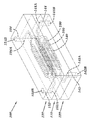
첨부된 도면은 본 발명의 예시적인 실시형태를 도시하고 본 발명의 상기 언급된 특징 및 다른 특징 및 그들을 달성하는 방식을 보여준다. 도면에서는:
도 1은 본 발명의 일 실시형태에 따른 마이크로유체 장치의 도면을 도시한다.
도 2는 본 발명의 일 실시형태에 따른 마이크로유체 장치의 중심 몸체의 도면을 도시한다.
도 3a 및 도 3b는 본 발명에 따른 마이크로유체 장치의 다양한 예시적인 분지화 구성을 도시한다.
도 4는 본 발명의 일 실시형태에 따른 마이크로유체 장치의 단면도를 도시한다.
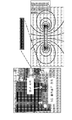
도 5a 내지 도 5c는 상이한 자석 구성의 영향을 도시한다. 도 5a는 단일 막대 자석이 설치된 도킹 스테이션 내로 삽입된 폴리설폰 DLT 장치의 사진을 도시한다. 도 5b는 6개의 고정 자석(함께 조립됨)으로 이루어진 자기 셋업(magnetic setup)의 개선된 설계를 도시한다. 도 5c의 유한 요소법 자석(finite element method magnet, FEMM)에 의하면, 자속 밀도 구배는 도 5b의 자기 셋업의 구성에서, 특히 자석의 중앙에서 유의하게 증강된 것으로 밝혀졌다(Δ 대 ·). 자기 셋업의 이러한 개선된 구성은 DLT 장치의 전체에 걸쳐 극히 증강된 자기장 구배(단일 자석보다 수천배가 더 큼)를 이용할 수 있게 한다.
도 6은 알루미늄으로 제작된 중심 몸체를 나타낸 사진이다.
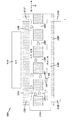
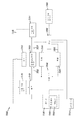
도 7은 일 실시형태에 따른 전체 시스템의 블록 다이어그램을 도시한다.
도 8a는 시린지 믹서의 다양한 모습을 도시한다.
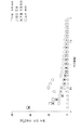
도 8b는 도 8a에 도시된 시린지 믹서의 사용에 의한 C. 알비칸스(C. albicans)의 결합 효율을 나타낸 선 그래프이다.
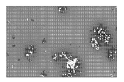
도 9a는 전혈 중의 개별 C. 알비칸스 진균에 특이적으로 결합하는 자성 항체(magnetic antibody) 옵소닌의 고배율 시야를 도시한다.
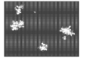
도 9b는 다수의 진균 병원체를 큰 자성 집괴(magnetic clump)와 결합시키는 자성 만노스 결합성 렉틴(mannose binding lectin, MBL) 옵소닌의 더 낮은 배율의 시야를 도시한다.
도 9c는 GFP-표지 E. 콜리(E. coli) 세균에 결합하는 MBL 옵소닌의 더 낮은 배율의 시야를 도시한다.
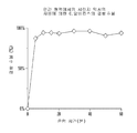
도 9d는 최대 80 ㎖/hr의 유속에서 100%에 근접한 병원체 청소 효율(clearance efficiency)이 얻어질 수 있다는 것을 보여준다.
도 10a 및 도 10b는 도킹 스테이션의 개략도를 도시한다.
도 11은 실제의 자기장의 실험 측정치와 비교하여, 본 명세서에 기재된 마이크로유체 장치 내의 자성 비드의 수집을 위해 설계된 자속 집속기의 컴퓨터 시뮬레이션의 결과를 도시한다.
도 12a 내지 도 12c는 미끄러운 액체가 주입된 다공성 표면(slippery liquid-infused porous surface, SLIPS)의 모습을 도시한다. 저배율(도 12a) 및 고배율(도 12b)에서의 한 어레이의 마이크로기둥(1 ㎛ 직경 x 2 ㎛ 공간)는, 거친 표면을 매끄럽게 하는 생체적합성 오일로 공간을 침윤시킴으로써 혈액 반발성 표면을 생성할 수 있다(도 12c).
도 13a 및 도 13b는 신선한 헤파린 미처리 인간 혈액이 통상적인 유리, PDMS, 및 테플론(Teflon) (PTFE) 표면 상에서는 급속히 혈병을 형성하지만, 생체적합성 오일로 함침된 나노구조화된 테플론(오일-침윤된 PTFE) 표면 상에서는 그렇지 않다는 것을 보여준다.
도 14의 A는 연동 펌프를 사용하여 투석 유사 치료(DLT) 시스템을 통해 혈액을 순환시키기 위한 실험 셋업을 도시한다. 혈액은 배큐테이너(Vacutainer) 튜브로부터 연동 펌프를 통해 폴리설폰 DLT 장치로 유동한다.
도 14의 B 및 도 14의 C는 헤파린 처리 인간 전혈을 2시간 동안 100 및 200 ㎖/h로 장치를 통해 진행시킨 후에, PBS 완충액을 5분 동안 유동시킴으로써 장치를 세척하였으며, 2시간 동안 두 유속 모두[(도 14의 B, 100 ㎖/h) 및 (도 14의 C, 200 ㎖/h)]에서 혈병이 발견되지 않았다는 것을 보여준다.
도 14의 D는 헤파린 미처리 인간 혈액을 순환시킬 경우, 혈액을 2시간 동안 100 ㎖/h로 유동했을 때 채널 내에 큰 혈병 및 혈괴를 형성하였다는 것을 보여준다.
도 15의 A 및 도 15의 B는 병렬로 연결된 2개의 DLT 장치가 혈액의 처리량을 최대 836 ㎖/h까지 대폭 증가시킬 수 있다는 것을 보여준다. 2개의 DLT 장치를 도킹 스테이션의 상부 및 하부 슬롯에 삽입하고, 2개의 출구로부터 수집된 혈액을 분석하여 혈액 내로 혼합된(spiked) C. 알비칸스의 단리 효율을 결정하였다.
도 16a는 장치 설계 및 병원체 분리의 개선을 보여주는 선 그래프 및 막대 그래프이다. 칸디다 알비칸스(Candida albicans) 병원체를 MBL-결합된 1 마이크로미터 비드에 사전결합시키고, 헤파린 항응고처리 인간 혈액 내로 혼합하였다. 선 그래프는 QPR1로 제시된 MBL-결합된 1 마이크로미터 비드 및 이전 설계를 기반으로 한 3층 폴리설폰 장치에 의한 데이터를 보여준다. 막대 그래프는 MBL-fp1 (FcMBL: 만노스 결합성 렉틴에 융합된 IgG Fc) 코팅된 자성 비드 및 새로운 라미네이팅된 장치/다중 자석 셋업에 의한 데이터를 보여준다. 새로운 설계에 의해서는 360 ㎖/hr의 유속에서 병원체의 >99%를 제거한 반면, 이전 설계에 의해서는 단리 효율이 360 ㎖/hr에서 36%로 떨어졌다.
도 16b는 장치 설계 및 병원체 분리의 개선을 보여준다. 사진: 다수의 자석을 갖는 라미네이팅된 DLT 장치의 예시적인 셋업. 선 그래프: 칸디다 알비칸스 병원체를 MBL-결합된 1 마이크로미터 비드에 사전결합하고 헤파린 항응고 인간 혈액 내로 혼합하였다. 이전 설계를 기반으로 한 3층 장치로부터의 데이터를 병렬로 실행되는 새로운 라미네이팅된 장치의 2개의 카세트와 비교하였다. 새로운 설계에 의해서는 836 ㎖/hr의 유속에서 병원체의 >85%를 제거한 반면, 이전 설계에 의해서는 단리 효율이 360 ㎖/hr에서 36%로 떨어졌다.
도 17a는 자성 비드를 연속적으로 관(tubing) 내의 혈액 내로 첨가하기 위한 시린지 펌프 및 인라인 믹서와 일체화된 DLT 시스템의 개략도이다. 인라인 믹서 전체에 걸쳐 첨가된 자성 비드와 혼합된 혈액 샘플은 DLT 장치 내로 유동하고, 이어서 자기적으로 표지된 병원체를 혈액으로부터 제거하고, 이어서 정화된 혈액이 출구를 통해 유출되는데, 이때 출구는 래트 패혈증 모델의 대퇴부 카테터에 연결될 수 있다.
도 17b는 혈액으로부터의 병원체 청소/분리를 위한 마이크로유체 장치를 사용하기 위한 "간소화된 동물" 모델을 도시한다. 일회용 인라인 믹서(오메가 엔지니어링 인크.(OMEGA Engineering Inc.))를 사용하여 MBLfp1 비드를 혼합된 C. 알비칸스를 함유한 혈액 내로 도입하였다. 이 간소화된 동물 모델에서는, DLT 장치를 통해 10 ㎖/hr의 유속으로 혈액으로부터 칸디다의 88%를 청소하였다.
도 18은 버블 포집 장치의 사진이다. 이 장치는 급속히 상방으로 이동하는 공기 버블의 부력에 의해 관을 통해 유입되는 모든 버블을 제거하며, 버블을 함유하지 않는 액체 용액은 장치를 통해 유동한다. 과량의 큰 공기 버블은 3방향 밸브로부터 제거될 수 있다.
도 19는 4개의 폴리설폰 플라스틱 층으로부터 제작된 마이크로유체 장치의 개략도를 도시한다. 이 장치는 2개의 수집 채널들 사이에 위치된 소스 채널을 포함한다.
도 20은 다수의 마이크로유체 장치들을 병렬로 다중화하여 고처리량(> 1.25 L/hr) 유동 능력(flow capability)을 갖는 생체모방 비장 장치를 생성한 것을 나타낸 개략도를 도시한다.BRIEF DESCRIPTION OF THE DRAWINGS The accompanying drawings illustrate exemplary embodiments of the invention and, together with the above-mentioned and other features of the invention, and how they are accomplished. In the drawing:
1 shows a diagram of a microfluidic device according to an embodiment of the present invention.
2 shows a view of a central body of a microfluidic device according to an embodiment of the invention.
Figures 3a and 3b illustrate various exemplary branching configurations of a microfluidic device in accordance with the present invention.
4 shows a cross-sectional view of a microfluidic device according to an embodiment of the invention.
Figures 5A-5C illustrate the effect of different magnet configurations. Figure 5a shows a photograph of a polysulfone DLT device inserted into a docking station equipped with a single bar magnet. Figure 5b shows an improved design of a magnetic setup consisting of six stationary magnets (assembled together). According to the finite element method magnet (FEMM) of Fig. 5c, the magnetic flux density gradient was found to be significantly enhanced in the configuration of the magnetic setup of Fig. 5b, especially at the center of the magnets. This improved configuration of the magnetic setup makes it possible to utilize an extremely enhanced magnetic field gradient (several thousand times larger than a single magnet) throughout the DLT device.
6 is a photograph showing a central body made of aluminum.
7 shows a block diagram of an overall system in accordance with one embodiment.
8A shows various aspects of a syringe mixer.
FIG. 8B is a line graph showing the binding efficiency of C. albicans by use of the syringe mixer shown in FIG. 8A. FIG.
Figure 9a shows a high magnification view of a magnetic antibody opsonin that specifically binds to individual C. albicans fungi in whole blood.
Figure 9b shows a lower magnification view of magnetic mannose binding lectin (MBL) opsonin that combines multiple fungal pathogens with a large magnetic clump.
Figure 9c shows a view of the lower scale of the opsonic MBL binding to GFP- labeled E. coli (E. coli) bacteria.
Figure 9d shows that pathogen clearance efficiency close to 100% can be obtained at a flow rate of up to 80 ml / hr.
10A and 10B show a schematic view of a docking station.
Figure 11 shows the results of a computer simulation of a flux concentrator designed for collection of magnetic beads in the microfluidic device described herein in comparison to experimental measurements of actual magnetic fields.
Figures 12a-12c illustrate the appearance of a slippery liquid-infused porous surface (SLIPS). Micropores (1 탆 diameter x 2 탆 space) of an array at low magnification (Figure 12a) and high magnification (Figure 12b) can create a blood-repellent surface by infiltrating space with a biocompatible oil that smoothes the rough surface (Fig. 12C).
Figures 13a and 13b show that fresh heparin untreated human blood rapidly forms blood clots on conventional glass, PDMS, and Teflon (PTFE) surfaces, while nanostructured teflon impregnated with biocompatible oils (oil-impregnated PTFE ) Surface.
Figure 14A shows an experimental setup for circulating blood through a dialysis similar therapy (DLT) system using a peristaltic pump. Blood flows from the Vacutainer tube through the peristaltic pump to the polysulfone DLT device.
14B and 14C show that the device was washed by moving heparinized human whole blood through the device at 100 and 200 mL / h for 2 hours, then washing the PBS buffer for 5 minutes, (B , 100 ml / h in Figure 14) and (C, 200 ml / h in Figure 14).
Fig. 14D shows that, when circulating heparin untreated human blood, large blood clots and blood clots were formed in the channel when the blood was flown at 100 ml / h for 2 hours.
FIGS. 15A and 15B show that two DLT devices connected in parallel can significantly increase the throughput of blood up to 836 ml / h. Two DLT devices were inserted into the upper and lower slots of the docking station and the blood collected from the two outlets was analyzed to determine the isolation efficiency of spiked C. albicans into the blood.
16A is a line graph and bar graph showing improvements in device design and pathogen isolation. Candida albicans (Candida albicans pathogens were pre-bound to MBL-bound 1 micrometer beads and mixed into heparin anticoagulated human blood. The line graph shows the data by the MBL-coupled 1 micrometer bead presented as QPR1 and the 3 layer polysulfone device based on the previous design. The bar graph shows data by magnetic bead coated with MBL-fpl (FcMBL: IgG Fc fused to mannose binding lectin) and a new laminated device / multiple magnet setup. The new design removed> 99% of the pathogen at a flow rate of 360 ml / hr, whereas the isolation efficiency dropped to 36% from 360 ml / hr by the previous design.
Figure 16b shows improvements in device design and pathogen isolation. Photo: An exemplary setup of a laminated DLT device with multiple magnets. Line graph: Candida albicans pathogen was pre-bound to MBL-bound 1 micrometer beads and mixed into heparin anticoagulated human blood. The data from the three-layer device based on the previous design was compared with two cassettes of a new laminated device running in parallel. The new design removed> 85% of the pathogen at a flow rate of 836 ml / hr whereas the isolation efficiency dropped to 36% from 360 ml / hr by the previous design.
17A is a schematic diagram of a DLT system integrated with a syringe pump and an inline mixer to continuously add magnetic beads into the blood within the tubing. A blood sample mixed with magnetic beads added throughout the inline mixer flows into the DLT device and then removes the magnetically labeled pathogen from the blood and then the purified blood flows out through the outlet, May be connected to the thigh catheter of the model.
Figure 17B shows a "simplified animal" model for using a microfluidic device for pathogen clearing / separation from the blood. MBLfp1 beads were introduced into blood containing mixed C. albicans using a disposable inline mixer (OMEGA Engineering Inc.). In this simplified animal model, 88% of the candida were cleared from the blood at a flow rate of 10 ml / hr through the DLT device.
18 is a photograph of the bubble collecting device. This device removes all bubbles flowing through the tube by the buoyancy of air bubbles moving up rapidly, and the liquid solution which does not contain bubbles flows through the device. Excessive large air bubbles can be removed from the three-way valve.
Figure 19 shows a schematic view of a microfluidic device made from four polysulfone plastic layers. The apparatus includes a source channel located between two acquisition channels.
Figure 20 shows a schematic diagram showing the generation of a biomimetic sputtering device with high throughput (> 1.25 L / hr) flow capability by multiplexing multiple microfluidic devices in parallel.
예시적인 실시형태의 설명Description of Exemplary Embodiments
본 명세서에는 소스 채널 내에서 유동하는 소스 유체로부터, 소스 유체 내의 다른 성분들을 제거하거나 변경시키지 않고서, 표적 성분의 분리 및 제거를 용이하게 할 수 있는 유체 장치가 개시된다.A fluidic device is disclosed herein that can facilitate separation and removal of target components from a source fluid flowing in a source channel without removing or altering other components in the source fluid.
유체는 액체 또는 기체일 수 있다. 표적 성분은 자성이거나 또는 유동하는 유체에 도입된 자성 입자에 결합될 수 있는 임의의 미립자, 분자 또는 세포 물질일 수 있다. 시스템의 처리량 및 효율을 개선하기 위하여 다수의 유체 장치가 직렬로 및/또는 병렬로 함께 결합될 수 있다. 표적 성분은 수집 유체 내에 수집되며, 이 수집 유체는 표적 성분을 분석하기 위해 추가로 처리될 수 있다. 표적 성분을 함유하는 수집 유체는 저장소 내에 수집될 수 있으며, 확인, 진단 등에서의 사용을 위해 표적 성분을 분석하기 위하여 배치 기술, 예컨대 면역염색, 면역검정, 배양, 폴리머라제 연쇄 반응(PCR), 질량 분석, 및 항생제 감수성 시험이 사용될 수 있다. 대안적으로, 표적 성분을 함유하는 수집 유체는 표적 성분이 수집 유체와 함께 유동함에 따라 이들을 처리할 수 있는 인라인 또는 온-칩 진단 또는 분석 장치 내로 안내될 수 있다. 표적 성분은 자석이거나 자성 입자에 결합되기 때문에, 인라인 또는 온-칩 분석을 위한 표적 성분을 수집하거나 또는 검출 또는 분석을 위한 다른 장치로 표적 성분을 안내하는 데 자기장 구배가 사용될 수 있다.The fluid may be a liquid or a gas. The target component may be any particulate, molecular or cellular material that may be magnetic or capable of binding to magnetic particles introduced into the flowing fluid. A number of fluid devices may be coupled together in series and / or in parallel to improve the throughput and efficiency of the system. The target component is collected in a collection fluid, which can be further processed to analyze the target component. The collection fluid containing the target component may be collected in a reservoir and may be subjected to various techniques such as, for example, immunoassay, immunoassay, culture, polymerase chain reaction (PCR), mass spectrometry Analysis, and antibiotic susceptibility tests may be used. Alternatively, the collection fluid containing the target component may be directed into an in-line or on-chip diagnostic or analytical device capable of processing them as the target component flows with the collection fluid. Since the target component is either a magnet or bonded to a magnetic particle, a magnetic field gradient may be used to collect the target component for in-line or on-chip analysis or to guide the target component to another device for detection or analysis.
도 1은 본 발명의 일 실시형태에 따른 마이크로유체 장치(100)를 도시한다. 도 1에 도시된 마이크로유체 장치(100)는 직사각형 몸체를 포함할 수 있지만, 다른 형상이 또한 사용될 수 있다(예: 원형, 타원형, 사다리꼴, 다각형 등). 도 1에 도시된 바와 같이, 마이크로유체 장치는 중심 몸체(110) (도 2에 더 상세히 도시됨) 및 외부 라미네이팅 층(120, 130)을 포함할 수 있다. 중심 몸체(110)는 라미네이팅 층(120)과 접촉된 제1 외부 표면(112) 및 라미네이팅 층(130)과 접촉된 제2 외부 표면(114)을 포함한다. 표면(112, 114)는 중심 몸체(110)의 대향하는 표면일 수 있다. 라미네이팅 층(120, 130)은 의료 등급 접착제에 의해 중심 몸체의 표면에 접합될 수 있다.1 shows a
도 2에 도시된 바와 같이, 중심 몸체(110)의 표면(112)은 하나 이상의 입구(142A)와 하나 이상의 출구(144A) 사이에 연장되는 하나 이상의 소스 유체 채널(140)을 포함할 수 있다. 도 1에 도시된 바와 같이, 하나 이상의 입구(142A)는 라미네이팅 층(120)의 외부 표면(122) 상에 있는 개구부(142B)로부터 연장된 입구 포트(142)와 연통될 수 있다. 하나 이상의 입구(144A)는 라미네이팅 층(120)의 외부 표면(122) 상에 있는 개구부(144B)로부터 연장된 입구 포트(144)와 연통될 수 있다. 입구 포트(142) 및 출구 포트(144)는 소스 유체 채널(140)에 대해 수직으로(즉, z-방향을 따라) 배향된 것으로 도시되어 있지만, 소스 유체 채널(140)에 대해 임의의 각도(일직선상 포함)로 배향될 수 있다. 표적 성분을 함유하는 소스 유체가 하나 이상의 입구 포트(142)를 통해 소스 채널(140) 내로 유입되고, 하나 이상의 출구 포트(144)를 통해 마이크로유체 장치(100)로부터 빠져나간다.2, the
수집 채널(150)은 소스 채널(140)에 대해 평행하게 연장된 것으로 도시되어 있지만, 일부 실시형태에서, 수집 채널(150)은 소스 채널(140)에 대해 수직으로(또는 각을 이루어서) 연장될 수 있다. 이들은 수평으로 또는 수직으로 배열될 수 있다.Collecting
소스 유체 채널(140)은 도 2에 도시된 바와 같이 중심 몸체(110)의 길이(예: y-방향)를 따라 연장될 수 있다. 소스 채널(140)은 임의의 다각형, 비-다각형, 원형, 또는 난형 단면의 것일 수 있다. 일부 실시형태에서, 소스 채널(140)은 단면이 직사각형일 수 있다. 개별 소스 유체 채널(140)의 단면 치수는, 표적 성분을 자기장에 더 효과적으로 노출시키고 끌어당겨진 표적 성분을 전달 채널(160)을 향해 안내하도록 설계될 수 있다. 일 실시형태에서, 소스 유체 채널(140)은 자기장에 대한 노출 면적을 최대화하기 위하여 편평한 기하구조를 가질 수 있다. 게다가, 소스 유체 채널(140)은 소스 유체가 소스 채널(140)을 통과함에 따라 소스 유체의 유속을 느리게 하여 자기적으로 결합된 표적 성분의 개수를 최대화하여 전달 채널(160) 내로 이동하도록 설계될 수 있다.The source
도 2에 도시된 바와 같이, 중심 몸체(110)의 표면(114)은 하나 이상의 입구(152A)와 하나 이상의 출구(154A) 사이에 연장되는 하나 이상의 수집 유체 채널(150)을 포함할 수 있다. 도 1에 도시된 바와 같이, 하나 이상의 입구(152A)는 라미네이팅 층(130)의 외부 표면(132) 상에 있는 개구부(152B)로부터 연장된 입구 포트(152)와 연통될 수 있다. 하나 이상의 입구(154A)는 라미네이팅 층(130)의 외부 표면(132) 상에 있는 개구부(154B)로부터 연장된 입구 포트(154)와 연통될 수 있다. 입구 포트(152) 및 출구 포트(154)는 수집 유체 채널(150)에 대해 수직으로(즉, z-방향을 따라) 배향된 것으로 도시되어 있지만, 수집 유체 채널(150)에 대해 임의의 각도(일직선상 포함)로 배향될 수 있다. 수집 유체가 하나 이상의 입구 포트(132)를 통해 수집 채널(130) 내로 유입되고, 하나 이상의 출구 포트(134)를 통해 마이크로유체 장치(100)로부터 빠져나간다.As shown in FIG. 2, the
소스 채널(140)과 마찬가지로, 수집 채널(150)은 임의의 다각형, 비-다각형, 원형, 또는 난형 단면의 것일 수 있다. 그러나, 각각의 소스 채널(140) 및 수집 채널(150)의 단면은 독립적으로 선택된다는 것이 이해되어야 한다. 따라서, 모든 소스 채널(140) 및 수집 채널(150)의 단면은 동일하거나, 전부 상이하거나, 또는 동일한 것과 상이한 것의 임의의 조합일 수 있다. 일부 실시형태에서, 수집 채널(140)은 단면이 직사각형일 수 있다.Like the
도 2에 도시된 바와 같이, 중심 몸체(110)는 소스 채널을(140) 수집 채널(150)과 연결하는 하나 이상의 전달 채널(160)을 포함할 수 있다. 전달 채널(160)이 소스 채널(140) 및 수집 채널(150)에 대해 실질적으로 수직으로 배향된 것으로 도시되어 있지만, 전달 채널(160)은 소스 채널(140)에 대해 다양한 각도(예: 1 내지 90도, 여기서 0도는 소스 채널(140) 내에서의 유동의 방향에 상응함, 도 3 참조)로 배향될 수 있다. 일부 실시형태에서, 전달 채널(160)은 수집 채널(150) 및 소스 채널(140)에 대해 실질적으로 수직으로 배향될 수 있다. 이러한 수직 구성은 수집 채널(150) 내에서 유동하는 수집 유체가 전달 채널(들)(160) 내의 유체와 비교하여 더 낮은 정압(static pressure)을 가져서 전달 채널(들) (160) 내의 자성 비드 및 결합된 표적 성분이 수집 유체 내로 끌어들여지게 될 것이라는 베르누이 원리(Bernoulli principle)를 활용할 수 있다.As shown in FIG. 2, the
전달 채널(160)은 임의의 다각형, 비-다각형, 원형, 또는 난형 단면의 것일 수 있다. 일부 실시형태에서, 전달 채널은 단면이 직사각형일 수 있다. 전달 채널(160)은 표적 성분, 예를 들어 자성 입자 결합된 표적 성분을 소스 채널(140)로부터 수송하여 결국에는 수집 채널(150)을 통해 마이크로유체 장치(100)로부터 플러싱되게 하는 역할을 한다. 자성 입자에 결합된 표적 성분은, 자성 입자를 수송 채널(160) 내로 축출하는 외부 자기력을 적용함으로써, 소스 채널(140) 내에서 유동하는 소스 유체의 남아 있는 성분으로부터 분리될 수 있다. 전달 채널(160)은 90도 코너를 갖는 것으로 도시되어 있지만, 다른 코너 각도 및 형상, 예컨대 90보다 더 높거나 낮은 각도 또는 둥근 코너가 또한 이용될 수 있다. 전달 채널들 사이의 간격이 또한 원하는 대로 조정될 수 있다. 예를 들어, 전달 채널은 약 10 ㎛ 내지 약 5 mm 정도 이격될 수 있다. 일부 실시형태에서, 전달 채널은 약 100 ㎛ 내지 약 500 ㎛ 정도 이격될 수 있다.The
소스 유체 채널(140) 및 수집 유체 채널(150)뿐만 아니라 전달 채널(160)의 개수, 크기, 형상, 배향 및 간격은 원하는 시스템 성능 및 효율에 따라 변동될 수 있다.The number, size, shape, orientation, and spacing of the source
소스 유체 채널(140) 및 수집 유체 채널(150)은 독립적으로 길이가 약 l mm 내지 약 10 cm이고, 폭이 약 0.1 mm 내지 약 10 mm이고, 깊이가 약 0.1 mm 내지 약 2 mm일 수 있다. 일부 실시형태에서, 소스 채널(140) 및 수집 채널(150)은 동일한 치수, 즉 동일한 길이, 폭, 및 깊이를 갖는다.The source
일 바람직한 실시형태에서, 소스 유체를 수송하기 위한 소스 채널(140)은 2 cm 길이 x 2 mm 폭 x 0.16 mm 높이일 수 있다.In one preferred embodiment, the
일부 실시형태에서, 수집 유체를 수송하기 위한 수집 채널(150)은 독립적으로 2 cm 길이 x 2 mm 폭 x 0.16 mm 높이일 수 있다. In some embodiments, the
일부 실시형태에서, 전달 채널(160)은 단면 치수가 약 1 mm x 200 ㎛ 내지 약 10 mm x 1 mm이다. 일부 실시형태에서, 전달 채널(160)은 단면 치수가 약 100 ㎛ (두께) x 100 ㎛ (폭) 내지 약 1 mm x 400 ㎛이다.In some embodiments, the
도 1에 도시된 바와 같이, 중심 몸체(110)의 외부 표면(112, 114)은 라미네이팅 층(120, 130)으로 각각 라미네이팅되어 밀봉 및 밀폐된 채널 세트를 형성할 수 있으며, 이러한 채널 세트는 유체가 누설 없이 또는 그대로 장치 사이에서 이동할 수 있게 한다. 중심 몸체(110)와 접촉된, 라미네이팅 층(120)의 표면은 소스 유체 채널(140), 입구(142A), 또는 출구(144A)의 일부를 포함할 수 있으며, 즉 소스 유체 채널(140), 입구(142A), 또는 출구(144A)의 일부분이 라미네이팅 층(120) 내에 있다. 대안적으로, 라미네이팅 층(112)은 소스 유체 채널(140), 입구(142A), 또는 출구(144A)의 일부를 포함하지 않으며, 즉 소스 유체 채널(140), 입구(142A), 또는 출구(144A)는 완전히 중심 몸체 내에 있다.As shown in Figure 1, the
유사하게, 중심 몸체(110)와 접촉된, 라미네이팅 층(130)의 표면은 수집 유체 채널(150), 입구(152A), 또는 출구(154A)의 일부를 포함할 수 있으며, 즉 수집 유체 채널(150), 입구(152A), 또는 출구(154A)의 일부분이 라미네이팅 층(130) 내에 있다. 대안적으로, 라미네이팅 층(130)은 소스 유체 채널(150), 입구(152A), 또는 출구(154A)의 일부를 포함하지 않으며, 즉 소스 유체 채널(150), 입구(152A), 또는 출구(154A)는 완전히 중심 몸체 내에 있다.Similarly, the surface of the
또한, 마이크로채널 조립체들 중 하나 이상뿐만 아니라 전체 장치의 구성은 다른 설계를 가질 수 있으며, 도면에 도시된 것으로 제한되어서는 안 된다는 것을 유의해야 한다. 추가로, 채널 조립체 내의 채널들이 원형 단면을 갖는 것으로 도시될 수 있기는 하지만, 이들 채널은 정사각형, 직사각형, 난형, 다각형 등을 포함하지만 이로 한정되지는 않는 다른 단면 형상을 가질 수 있거나, 또는 마이크로매칭 기술을 이용하여 생성될 수 있는 바와 같이 길이를 따라 치수 및 형상이 변동되는 채널들을 가질 수 있다.It should also be noted that the configuration of the entire device as well as one or more of the microchannel assemblies may have different designs and should not be limited to those shown in the figures. Additionally, although the channels in the channel assembly may be shown having a circular cross-section, these channels may have other cross-sectional shapes including, but not limited to, square, rectangular, oval, polygonal, Lt; RTI ID = 0.0 > and / or < / RTI >
도 1 및 도 2에 도시된 바와 같이, 소스 유체 채널(140)뿐만 아니라 수집 유체 채널(150)은 그들의 각각의 입구 포트로부터 개별 분지들로 분지될 수 있으며, 소스 유체 채널(140) 및 수집 채널(150)의 개별 분지들은 그들의 각각의 출구 포트로 수렴된다. 도 1 및 도 2에 4개의 분지가 도시되어 있기는 하지만, 임의의 개수의 분지, 심지어는 1개의 분지가 사용될 수 있다. 예를 들어, 본 발명에 따라, 도 3a는 각각이 수집 채널 및 소스 채널인 16개의 분지를 예시하고, 도 3b는 각각이 수집 채널 및 소스 채널인 32개의 분지를 예시한다. 통상의 기술자가 이해하는 바와 같이, 분지의 개수는 시스템의 원하는 성능 및 효율의 함수로서 선택될 수 있다.1 and 2, the source
소스 유체 채널(140) 및 수집 유체 채널(150)은 서로 거울대칭을 이루며, 동일하거나 유사한 분지형 구성을 가질 수 있다. 게다가, 소스 채널(140)의 각각의 개별 분지 및 수집 채널(150)의 상응하는 분지는 이들을 연결시키는 적어도 하나의 전달 채널(160)을 포함할 수 있다.The source
소스 채널(140) 및 수집 채널(150)은 서로에 대해 실질적으로 평행할 수 있다. 소스 채널(140)과 수집 채널(150) 사이의 간격은 약 5 ㎛ 내지 약 10 mm의 범위일 수 있다. 일부 실시형태에서, 소스 채널(140)과 수집 채널(150) 사이의 간격은 약 10 ㎛ 내지 500 ㎛의 범위일 수 있다.The
도 4는 본 발명에 따른 마이크로유체 장치의 단면도를 예시한다. 도 4에 도시된 바와 같이, 소스 유체가 입구 포트(142)를 통해 소스 채널(140)로 진입하며, 여기서 소스 유체(화살표로 나타냄)는 소스 채널(140)을 통해 장치(100)를 통과하고 출구 포트(144)를 통해 장치(100)를 빠져나간다.4 illustrates a cross-sectional view of a microfluidic device according to the present invention. 4, the source fluid enters the
소스 유체는 세균 및 효모, 암/종양 세포 또는 요망되는 표적 성분(예컨대, 줄기 세포, 태아 세포, 사이토카인 또는 항체)를 포함한 표적 성분(99), 예컨대 병원체를 함유하는 소스 유체일 수 있다. 이들 표적 성분(99)은 마이크로유체 장치(100)로 진입되기 전에 소정의 표적 성분(99)에 부착되도록 컨디셔닝되거나 개질된 자성 입자(98)와 혼합될 수 있다.The source fluid may be a source fluid containing a
유동하는 소스 유체로부터 표적 성분(99)을 포획하기 위하여, 하나 이상의 자기 소스(410), 예컨대 네오디뮴 자석이 마이크로유체 장치(100)의 수집 채널(150)에 인접하여 위치될 수 있다. 다른 유형의 자석이 사용될 수 있으며, 이에 따라 네오디뮴으로 제한되지 않는다는 것을 유의해야 한다. 예를 들어, 자석(들)은 사마륨 코발트, 페라이트, 알니코 등으로 제조될 수 있거나, 또는 내부 또는 외부 전자석을 사용하여 자기장 구배를 발생시킬 수 있다. 도 4에 도시된 바와 같이, 자석(410)은 전달 채널(160) 위로 수직으로 위치되어, 자석(310)에 의해 적용된 자기장 구배가 자성 비드(98)를 끌어당겨서 자성 비드(98)가 자석(310)을 향해 이동되게 된다. 구체적으로는, 자석(410)으로부터의 자기장 구배는 소스 유체 내의 자기적으로 결합된 표적 성분(99)이 전달 채널(160)을 통해 수집 채널(150) 내로 이동되게 한다. 이들 성분은 수집 유체가 그곳을 통해 플러싱될 때 제거되고 수집될 수 있다. 본 발명의 일부 실시형태에서, 자기적으로 결합된 표적 성분(99)은 전달 채널(160) 내로 이동되고 거기에 침강되어(settle), 플러싱 작동에 의해 수집 채널(150) 내로 끌어들여질 수 있다. 소스 유체 및 수집 유체가 마이크로유체 장치(100) 내에서 동일한 방향으로 유동하는 것으로 도시되어 있기는 하지만, 소스 유체 및 수집 유체는 마이크로유체 장치(100) 내에서 반대 방향으로 유동할 수 있다는 것을 유의해야 한다.One or more
도 4에 도시된 바와 같이, 수집 유체가 입구 포트(152)를 통해 수집 유체 채널(150)로 진입하고, 출구 포트(154)를 향해 수집 유체 채널(110)을 통과한다. 입구 포트(106A, 106B)는 동일한 입구 포트일 수 있으며, 출구 포트(108A, 108B)는 동일한 출구 포트일 수 있다.4, the collection fluid enters the
수집 채널(150), 및 바람직하게는 포트(152, 154)는 수집 유체로 최대 용량까지 충전된다는 것을 유의해야 한다. 그러나, 일부 실시형태에서, 수집 유체는 수집 채널(150)을 통해 계속적으로 유동하지 않으며, 대신에 수집 채널(150)을 통해 간헐적으로 또는 주기적으로 유동되는데, 후자에서는 수집 유체가 유동하는 간격 및 수집 유체가 정상 상태이거나 더 느린 속도로 유동하는 간격이 있다. 수집 유체가 연속적으로 유동하는 것이 아니라 수집 채널(150) 내에 정체되게 할 수 있기 때문에, 전달 채널로 진입하는 자기적으로 결합된 표적 성분은 장치를 빠져나가지 않고서 일정 시간 동안 이들 전달 채널(160) 내에 보류되게 될 수 있다.It should be noted that the
일단 수집 유체가 유동하기 시작하여, 수집 채널(150) 내에서 정체 상태로부터 유동 상태로 변화되면, 전달 채널(160) 내에 남아 있는 자기적으로 결합된 표적 성분은 수집 채널(150) 내로 끌어들여질 수 있는데, 이는 비장의 굴(sinus)로부터 폐기물을 운반해가는 림프액의 주기적 유동과 유사하다. 수집 채널 내의 유동하는 수집 유체는 전달 채널에 비하여 더 낮은 정압(static pressure)을 가질 수 있어서 전달 채널 내에 존재하는 자성 비드 및 결합된 표적 성분이 수집 유체 스트림 내로 유입되게 될 수 있다. 이러한 소정의 압력 또는 유동 차는, 수집 유체가 "플러싱" 작동 동안 수집 채널(150)을 통해 유동할 때 생성될 수 있으며, 여기서 플러싱 작동은 원하는 지속시간을 갖도록 제어될 수 있다. 플러싱 작동의 지속시간을 제어함으로써, 수집 채널(150) 내로 전달되는 소스 유체의 양이 또한 제어될 수 있다.Once the collecting fluid begins to flow and changes from congestion to flow within the collecting
마이크로유체 장치는 표적 성분 또는 병원체 축적을 검출하는 하나 이상의 광학적 또는 임피던스 마이크로전자 센서를 포함할 수 있는데, 이때 이러한 센서는 마이크로유체 장치 내에 일체화된다. 마이크로유체 장치는 피드백 루프를 포함시킬 수 있는데, 여기서는 센서가 제어기 및/또는 하나 이상의 펌프와 통신하여 수집 유체의 유동(예: 시작/중단 지속시간, 유속 등)을 자동으로 제어한다. 게다가, 마이크로유체 장치의 외부에 하나 이상의 자성 비드 트랩이 도 1의 시스템에 사용될 수 있는데, 이는 소스 유체가 소스로 반환되거나 소스 유체 수집기에 투입되기 전에 다른 메커니즘에 의해 청소되지 않은 임의의 남아 있는 입자를 제거하기 위하여 사용된다. 마이크로유체 장치는 수집 채널 및/또는 소스 유체 채널의 입구 및/또는 출구에서 하나 이상의 밸브를 포함할 수 있다. 마이크로유체 장치는 전달 채널로 진입하거나 이것을 빠져나가는 자기적으로 결합된 표적 성분의 유동을 제어하도록 전달 채널에서 하나 이상의 밸브를 포함할 수 있다.The microfluidic device may comprise one or more optical or impedance microelectronic sensors for detecting target components or pathogen accumulation, wherein such sensors are integrated within the microfluidic device. The microfluidic device may include a feedback loop, where the sensor communicates with the controller and / or one or more pumps to automatically control the flow of collected fluid (e.g., start / stop duration, flow rate, etc.). In addition, one or more magnetic bead traps external to the microfluidic device may be used in the system of FIG. 1 because any residual particles that are not cleaned by other mechanisms before the source fluid is returned to the source or introduced into the source fluid collector . ≪ / RTI > The microfluidic device may include one or more valves at the inlet and / or outlet of the collection channel and / or the source fluid channel. The microfluidic device may include one or more valves in the delivery channel to control the flow of magnetically coupled target components entering or exiting the delivery channel.
고처리량을 제공하기 위하여, 2개 이상의 마이크로유체 장치가 다중화 시스템으로 함께 다중화될 수 있다. 예를 들어, 1개, 2개, 3개, 4개, 5개, 6개, 7개, 8개, 9개, 10개, 11개, 12개, 13개, 14개, 15개 또는 그 초과의 마이크로유체 장치가 함께 연결될 수 있다. 다중화 시스템에서, 마이크로유체 장치는 정화 효율 또는 처리 유속(throughput flow rate)을 각각 최대화하기 위하여 직렬로 또는 병렬로 함께 연결될 수 있다.To provide high throughput, two or more microfluidic devices may be multiplexed together into a multiplexing system. For example, one, two, three, four, five, six, seven, eight, nine, ten, eleven, twelve, thirteen, fourteen, fifteen, Excess microfluidic devices can be connected together. In a multiplexing system, the microfluidic device may be connected together in series or in parallel to maximize purification efficiency or throughput flow rate, respectively.
병렬 연결의 경우, 각각의 장치의 소스 입구는 동일한 소스 유체 소스에 연결될 수 있고, 소스 출구는 동일한 소스 유체 수집기에 연결될 수 있다. 직렬 연결의 경우, 하나의 마이크로유체 장치의 소스 출구는 제2 장치의 소스 입구에 연결될 수 있다. 게다가, 다중화 시스템 내의 마이크로유체 장치들은 2개의 마이크로유체 장치가 자기 소스를 공유할 수 있도록 배치될 수 있다.In the case of a parallel connection, the source inlets of each device can be connected to the same source fluid source, and the source outlets can be connected to the same source fluid collector. In the case of a series connection, the source outlet of one microfluidic device may be connected to the source inlet of the second device. In addition, microfluidic devices in a multiplexing system can be arranged such that two microfluidic devices can share a magnetic source.
다중화 시스템에서, 다수의 마이크로유체 장치들은 스페이서를 사용하여 함께 연결될 수 있다. 스페이서는 마이크로유체 장치와 동일한 재료로 제작될 수 있다. 스페이서는 개별 마이크로유체 장치들 사이에 자석의 삽입을 위한 갭을 제공할 수 있으며, 개별 마이크로유체 장치의 소스 채널과 수집 채널 포트를 상호연결하기 위한 구멍을 포함할 수 있다. 소스 유체가 생물학적 유체(예: 혈액)일 때, 다중화 시스템의 말단 마이크로유체 장치는 표준 혈액 및 식염수 커넥터를 갖는 접합된 블록을 포함할 수 있다. 다중화 장치는 세정되고, 멸균되고, 무균 백 내로 삽입되어 사용 직전에 개봉될 수 있다. 채널 기하구조, 장치당 채널의 개수, 및 다중화 시스템당 장치의 개수는 원하는 소스 유체(예: 혈액), 유동 용량뿐만 아니라 병원체 분리 효율을 만족시키도록 최적화될 수 있다.In a multiplexing system, a plurality of microfluidic devices may be connected together using spacers. The spacer may be made of the same material as the microfluidic device. The spacer may provide a gap for insertion of the magnet between the individual microfluidic devices and may include apertures for interconnecting the source channel and the collection channel port of the individual microfluidic device. When the source fluid is a biological fluid (e.g., blood), the end microfluidic device of the multiplexing system may comprise a junction block with standard blood and saline connectors. The multiplexer can be cleaned, sterilized, inserted into an aseptic bag and opened just before use. The channel geometry, the number of channels per device, and the number of devices per multiplexing system can be optimized to meet desired source fluids (e.g., blood), flow capacity as well as pathogen isolation efficiency.
소스 유체가 혈액일 때, 마이크로유체 장치의 소스 채널 및 수집 채널은 각각 비장 세동맥 및 세정맥과 유사하며; 전달 채널은 비장의 굴모양 혈관(vascular sinusoid)을 모방하는데, 여기에는 유동이 간헐적이고 옵소닌화 입자가 보류되어 있으며; 캐리어 유체 채널은 림프액을 모방하는데, 이 림프액은 결국에는 옵소닌화 입자를 청소한다. 도 20은 다수의 마이크로유체 장치를 병렬로 다중화하여 고처리량(> 1.25 L/hr) 유동 능력을 갖는 생체모방 비장 장치를 생성한 것을 나타낸 개략도를 도시한다.When the source fluid is blood, the source channel and collection channel of the microfluidic device are similar to the splenic arterioles and vein, respectively; The delivery channel mimics the vascular sinusoid of the spleen, where the flow is intermittent and the opsonized particles are retained; The carrier fluid channel imitates the lymph fluid, which eventually cleans the opsonized particles. Figure 20 shows a schematic diagram showing the generation of a biomimetic sputtering device with high throughput (> 1.25 L / hr) flow capability by multiplexing multiple microfluidic devices in parallel.
마이크로유체 장치의 처리량을 추가로 증가시키기 위하여, 마이크로유체 장치는 2개의 수집 채널들 사이에 위치된 소스 유체 채널을 포함할 수 있다. 소스 유체 채널은 하나 이상의 전달 채널에 의해 2개의 수집 채널 각각에 연결될 수 있다. 예를 들어, 단일 수집 유체 채널과 정렬된 단일 소스 유체 채널로부터 2 x 0.16 mm의 채널 단면을 갖는 16-채널 PDMS 마이크로유체 장치를 사용하여 최대 80 ㎖/hr의 유속으로 전혈로부터 모든 비드-결합된 진균성 병원체의 95% 초과를 분리하였다. 단면을 2 x 0.32 mm로 2배로 하고 2개의 수집 채널(하나는 소스 채널의 위쪽에 있고 하나는 소스 채널의 아래쪽에 있음)을 사용함으로써, 유사한 청소 효율이 최대 유속 -1600 ㎖/hr에서 얻어질 수 있다. 도 19는 어떻게 마이크로유체 장치가 2개의 수집 채널들 사이에 위치된 소스 채널을 포함하는 4개의 폴리설폰 플라스틱 층으로 구성될 수 있는지를 도시한다. 혈액 및 식염수와 같은 유체는, 표면 상에 마이크로밀링된(micromilled) 오목한 채널 특징부를 갖는 "플라스틱-층(plastic-layer)들" 사이에 형성된 "유체-층(fluidic-layer)" 내로 유입된다. 혈액-유체-채널(blood-fluidic-channel), 즉 소스 채널은 플라스틱-층(2, 3)들 사이에 형성된다. 플라스틱 층(1, 2뿐만 아니라 3, 4)은 혈액-유체-층의 위쪽 및 아래쪽에 식염수-유체-층, 즉 수집 채널을 형성한다.To further increase the throughput of the microfluidic device, the microfluidic device may include a source fluid channel located between the two collection channels. The source fluid channel may be connected to each of the two collection channels by one or more delivery channels. For example, using a 16-channel PDMS microfluidic device with a channel cross-section of 2 x 0.16 mm from a single source fluid channel aligned with a single acquisition fluid channel, all bead-bound More than 95% of the fungal pathogens were isolated. A similar cleaning efficiency can be obtained at a maximum flow rate of 1600 ml / hr by doubling the cross section to 2 x 0.32 mm and using two collection channels (one at the top of the source channel and one at the bottom of the source channel) . Figure 19 illustrates how a microfluidic device can be composed of four polysulfone plastic layers including source channels positioned between two collection channels. Fluids such as blood and saline flow into a "fluidic-layer " formed between" plastic-layers "having micromilled concave channel features on the surface. Blood-fluidic-channels, or source channels, are formed between the plastic-
혈소판이 활성화되고 혈병 형성을 유도하는 위험을 최소화하기 위하여, 채널의 형상은 살아있는 고유량 혈관(예: 작은 동물의 대동맥)의 형상을 모방하고 이에 따라 전단(shear)을 최소화하도록 신중하게 선택될 수 있다. 채널 기하구조 및 유속은 비뉴턴 유체 역학의 컴퓨터 시뮬레이션(앤시스(ANSYS)의 플루엔트(Fluent) 및 CFX 소프트웨어 패키지)을 통해 채널 전체를 통한 전단 교란을 최소화하도록 최적화될 수 있다. 혼합 및 혈액 손실 또는 희석을 최소화하기 위해 혈액과 식염수 사이의 멀티페이스 시뮬레이션이 사용될 수 있다. 비개질된 기계가공된 표면이 헤파린의 존재하에 혈병 형성을 유도한다면, 이들은 방오 표면을 제공하도록 물리적으로 또는 화학적으로 개질될 수 있다(화학적 증기 폴리싱, 플라즈마 처리, 나노패턴화 등).In order to minimize the risk of platelet activation and inducing blood clot formation, the shape of the channel can be carefully selected to mimic the shape of living high-flow blood vessels (e.g., the small animal's aorta) and thereby minimize shear have. The channel geometry and flow rates can be optimized to minimize shear disturbance across the channel through non-Newtonian fluid dynamics computer simulations (ANSYS Fluent and CFX software packages). Multi-face simulations between blood and saline can be used to minimize mixing and blood loss or dilution. If the unmodified machined surface induces blood clot formation in the presence of heparin, they may be physically or chemically modified to provide an antifouling surface (chemical vapor polishing, plasma treatment, nanopatterning, etc.).
다른 채널 고려사항에는 자기장의 급속한 붕괴 도달(이는 채널 깊이를 제한할 수 있음), 폭의 증가에 따른 채널의 구조적 완전성의 저하, 및 채널 치수의 감소에 따른 전단 응력의 증가가 포함된다.Other channel considerations include a rapid collapse of the magnetic field (which may limit the channel depth), a decrease in the structural integrity of the channel as the width increases, and an increase in shear stress as channel dimensions decrease.
합성 표면 상에서의 혈병 형성은 의학 분야에서 오랫동안에 걸쳐 만연된 문제인데, 이는 단백질 흡수에 의해 표면 상에서 개시되며, 이러한 단백질 흡수는 혈소판 점착 및 활성화를 촉진시킬 뿐만 아니라, 트롬빈의 방출을 촉진시키는데, 이는 피브린 혈병 형성을 활성화한다. 따라서, 마이크로유체 장치의 유체 접촉 표면, 예를 들어 장치를 소스 또는 수집기에 연결하는 채널 또는 관(tubing) 또는 카테터는 분해에 저항하거나 유동 및 작동을 용이하게 하기 위하여 코팅 또는 처리될 수 있다. 예를 들어, 소스 유체 채널, 수집 채널, 전달 채널, 또는 이들 채널을 유체 소스에 연결하는 관 또는 카테터의 유체 접촉 표면은 방오 표면일 수 있다.Cloning on synthetic surfaces is a long-standing problem in the medical field, which is initiated on the surface by protein absorption, which not only promotes platelet adhesion and activation, but also promotes the release of thrombin, Activates fibrin clot formation. Thus, the fluid contacting surface of a microfluidic device, for example a channel or tubing or catheter connecting the device to a source or collector, can be coated or treated to resist degradation or to facilitate flow and operation. For example, the fluid-contacting surface of a source fluid channel, a collection channel, a delivery channel, or a tube or catheter connecting these channels to a fluid source may be an antifouling surface.
옹(Wong) 등의 문헌[Nature, 2011, 477: 443-447] (이의 내용은 본 명세서에 참고로 포함됨)은 본 명세서에 기재된 마이크로유체 장치에 사용될 수 있는 방오 표면을 기술한다. 옹 등에 기술된 바와 같이, 방오 표면은 저표면 에너지의 화학적으로 불활성인 퍼플루오르화 오일의 침윤층에 의해 분리된 한 어레이의 나노- 및 마이크로-구조물이 사용될 수 있는데, 이때 플루오르화 오일은 표면 구조물의 특징부에 의해 정위치에 유지된다(도 12).Wong et al . Nature , 2011, 477: 443-447, the contents of which are incorporated herein by reference, describe an antifouling surface that can be used in the microfluidic device described herein. As described above, an antifouling surface can be an array of nano- and micro-structures separated by a layer of infiltration of a chemically inert perfluorinated oil of low surface energy, wherein the fluorinated oil is a surface structure (Fig. 12).
이들의 조합은 다공성 구조물이 저에너지 액체를 정위치에 유지하기 때문에 표면 상에 물리적으로 매끄러운 윤활 필름을 생성할 수 있다. 이러한 얇은 윤활 필름은 표면 불균일성을 최소화하고, 보류력을 감소시키고, 표면을 따른 액체 이동성을 향상시키는데, 이는 세포의 지질 이중층과 다르지 않다. 따라서, 표면과의 접촉은 최소한으로 이루어지고, 액체는 고도로 이동성인 상태로 유지된다. 윤활 필름은, 예를 들어 문헌[Wenzel, R.N. Ind . Eng . Chem. 1936, 28: 988-994] 및 [Courbin, L., et al Nature Materials, 2007, 6: 661-664]에 기술된 바와 같이 다공성 물질에 의해 유도된 액체 흡수(imbibing) 과정에 의해 생성될 수 있다. 다공성 물질의 물리적 조도(roughness)는 윤활 유체의 습윤(wetting)을 유도할 뿐만 아니라, 이는 또한 표면에 대한 윤활 유체의 점착을 위한 추가의 표면적을 제공할 수 있다.These combinations can create a physically smooth lubricant film on the surface because the porous structure keeps the low-energy liquid in place. This thin lubricating film minimizes surface unevenness, reduces retention, and improves liquid mobility along the surface, which is not different from the lipid bilayer of the cell. Thus, contact with the surface is minimized and the liquid remains highly mobile. Lubricating films are described, for example, in Wenzel, RN Ind . Eng . Chem . 1936, 28: 988-994 and Courbin, L., et get Nature Materials , 2007, 6: 661-664). The physical roughness of the porous material not only induces wetting of the lubricating fluid, but it may also provide an additional surface area for adhesion of the lubricating fluid to the surface.
"액체-유사" 표면은 신선한 헤파린 미처리 인간 혈액과 접촉될 때 혈소판의 점착 및 피브린 혈병 형성을 방지하는 데 극히 효과적일 수 있다. 도 13a에서 알 수 있는 바와 같이, 신선한 헤파린 미처리 인간 전혈(0.75 ㎖)은, 퍼플루오르화 오일(플루오리너트(Flluorinert) FC-70, 쓰리엠 코포레이션(3M Corp.))로 함침된 마이크로구조화된 PTFE(테플론(Teflon); 1 ㎛ 기공 크기)로 구성된 기재에서는 비드를 형성하고 이로부터 미끄러져 나간 반면, 이는 대조의 매끄러운 PTFE뿐만 아니라 유리에서는 신속하게 응고되고 이것에 점착하였다.A "liquid-like" surface can be extremely effective in preventing platelet adhesion and fibrin clot formation when contacted with fresh heparin untreated human blood. As can be seen in Figure 13a, fresh heparin untreated human whole blood (0.75 ml) was prepared from micro-structured PTFE impregnated with perfluorinated oil (Flluorinert FC-70, 3M Corp.) (Teflon; 1 μm pore size) formed and peeled off from the beads, which rapidly solidified and adhered to the smooth PTFE as well as the glass in the control.
따라서, 이러한 특성은 이 종류에서는 최초를 나타내는데, 그 이유는 다른 인공 표면은 연장된 기간 동안 활성화 및 혈전증을 방지할 수 없기 때문이다. 이들 항응고 표면은 혈액 성분의 점착 및 혈병 형성을 제어하는 새로운 방법을 제공한다. 게다가, 이들 항응고 표면은 응고를 생성하지 않고서 마이크로유체 장치를 통한 혈류를 지지할 수 있다. 따라서, 혈액 내로의 또는 마이크로유체 장치 내에의 항응고제의 첨가에 대한 필요성이 감소될 수 있다. "액체-유사" 표면은 미끄러운 액체가 주입된 다공성 표면(SLIPS)으로도 지칭된다.Thus, this characteristic is the first in this class because other artificial surfaces can not prevent activation and thrombosis for extended periods of time. These anticoagulated surfaces provide a new way to control the adhesion and blood clot formation of blood components. In addition, these anticoagulation surfaces can support blood flow through the microfluidic device without producing clotting. Thus, the need for the addition of an anticoagulant into the blood or into the microfluidic device can be reduced. A "liquid-like" surface is also referred to as a slippery liquid injected porous surface (SLIPS).
이미 혈액 적합성에 대해 FDA 승인된 테플론 또는 폴리설폰과 같은 중합체에 패턴화된 교차하는 벽 및 기둥과 같은 소수성의 융기된 표면 구조물의 어레이를 마이크로미터 규모로 생성하기 위하여 마이크로성형 기술이 이용될 수 있다. 침윤 액체는 다수의 상이한 액체, 예컨대 FDA-승인 폴리플루오로알콕시(PFA)로부터 선택될 수 있다. 제작된 항응고 표면은 매끈하며, 혈액을 포함한 다양한 액체를 반발할 수 있다. 침윤 액체를 국한시키기(confine) 위하거나 혈액 성분 및 혈병의 부착에 저항하는 유효성을 결정하기 위하여, 상이한 특징부 크기 및 다공도를 갖는 광범위한 표면 구조물이 이용될 수 있다. 규소 기판에서의 나노구조화된 기둥의 어레이는 반도체 가공 방법 및 기술의 정확성을 활용하도록 제작될 수 있다. 기둥 어레이 기판은 폴리설폰 또는 PDMS와 같은 FDA-승인 재료에서 복제품을 제조하기 위한 마스터로서 사용될 수 있다. 특징부 크기는 수백 나노미터 내지 마이크로미터의 범위(예: 100 내지 1000 nm)일 수 있으며, 이때 어스펙트비는 약 1:1 내지 약 10:1이다. 전기화학적 침착(deposition)을 사용하여 금속 마이크로유체 장치의 유체 접촉 표면 상에 인시츄(in situ) 다공성 나노-섬유질 구조물이 생성될 수 있다. 다양한 몰폴로지 및 다공도의 생체적합성 폴리피롤 나노구조물의 인시츄 합성은 당업계에 공지되어 있다. 예를 들어, 2010년 7월 19일에 출원된 미국 가특허 출원 제61/353,505호 및 문헌[Kim, P. et al ., Nano Letters, in press (2011)]을 참조한다.Microforming techniques can be used to create micrometer scale arrays of hydrophobic raised surface structures such as crossed walls and columns patterned on polymers such as Teflon or polysulfone that have already been FDA approved for blood compatibility . Infiltration liquids may be selected from a number of different liquids, such as FDA-approved polyfluoroalkoxy (PFA). The prepared anticoagulant surface is smooth and can repel various liquids including blood. A wide variety of surface features with different feature sizes and porosities can be used to confine the infiltrating liquid or to determine its effectiveness in resisting adhesion of blood components and blood clots. Arrays of nanostructured pillars in a silicon substrate can be fabricated to take advantage of the accuracy of semiconductor processing methods and techniques. The pillar array substrate can be used as a master for making replicas in FDA-approved materials such as polysulfone or PDMS. The feature size can range from a few hundred nanometers to micrometers (e.g., 100-1000 nm), wherein the aspect ratio is from about 1: 1 to about 10: 1. Electrochemical deposition is used to form an in situ ( in) surface on the fluid contact surface of a metal microfluidic device. situ porous nano-fibrous structures can be produced. In situ synthesis of various morphologies and porosity biocompatible polypyrrole nanostructures is well known in the art. See, for example, U.S. Provisional Patent Application No. 61 / 353,505, filed July 19, 2010, and Kim, P. et al . , Nano Letters , in press (2011).
이들 구조물은 상이한 윤활 액체의 최적의 습윤 및 점착력을 결정하는 데 이용될 수 있다. 폴리플루오르화 화합물의 부류로부터 다수의 상이한 오일이 이용될 수 있다. 이들 후보대상은 그들의 혈병 형성 방지 성능, 생리학적 조건 하에서의 화학적 안정성, 및 장치의 표면으로부터의 침출(leaching) 수준에 기초하여 선택될 수 있다. 예를 들어, 생물의학적 응용에서의 사용에 대해 승인된 화합물(예: 혈액 대체물, MRI 조영제 등)이 사용될 수 있다. 일부 실시형태에서, PFC 퍼플루브론 또는 퍼플루오로옥틸브로마이드(알리안스 파마슈티칼(Alliance Pharmaceutical))이 이용될 수 있다.These structures can be used to determine optimal wetting and adhesion of different lubricating liquids. A number of different oils may be used from the family of polyfluorinated compounds. These candidate subjects can be selected based on their anti-clotting ability, chemical stability under physiological conditions, and leaching level from the surface of the device. For example, compounds approved for use in biomedical applications (eg, blood substitutes, MRI contrast agents, etc.) may be used. In some embodiments, PFC perfluoroburon or perfluorooctyl bromide (Alliance Pharmaceutical) can be used.
표면은 혈액에 대한 노출 후에, 표면 특성화 기술, 예컨대 형광 및 주사 전자 현미경법(SEM)을 사용하여 혈소판 또는 피브린 점착의 증거를 찾기 위하여 분석될 수 있다. 폴리플루오르화 화합물은 다양한 용매 중에서 난용성을 나타내는데, 이는 모니터링에 대한 어떠한 난관을 야기시킬 수 있다. 이러한 난관을 극복하기 위하여, 분석은 플루오르화 용매 중으로의 추출과, 이에 이어서 행해지는 크로마토그래피, 질량 분석, 및 19F-NNMR의 조합을 수반할 수 있다.The surface may be analyzed after exposure to blood to find evidence of platelet or fibrin glue using surface characterization techniques such as fluorescence and scanning electron microscopy (SEM). Polyfluorinated compounds exhibit poor solubility in a variety of solvents, which can cause some difficulties in monitoring. To overcome this difficulty, the analysis can involve extraction into a fluorinated solvent followed by a combination of chromatography, mass spectrometry, and 19 F-NNMR.
고혈류량의 존재하에 이들 표면의 유효성 및 안정성을 시험한 후에, 유체 침출의 어떠한 영향도 최소화하기 위해 구조적 설계(즉, 기둥-간격, 기공 크기 등)가 추가로 최적화될 수 있다. 생물학적 유체와 접촉된 비오염성 표면의 장기간 성능으로 변환될 수 있는 데이터를 획득하기 위하여, 체온보다 더 높은 온도에서의 광범위한 가속 침출 시험이 수행될 수 있다. 이들 화합물 중 다수는 비독성인 것으로 보고되어 있지만, 필요에 따라서는 선택된 함침 유체의 필요한 독물학적 스크리닝이 수행될 수 있다.After testing the effectiveness and stability of these surfaces in the presence of high blood flow, the structural design (i.e., column-spacing, pore size, etc.) can be further optimized to minimize any effect of fluid leaching. In order to obtain data that can be converted to long term performance of the non-staining surface in contact with the biological fluid, extensive accelerated leaching tests at temperatures higher than body temperature can be performed. Although many of these compounds have been reported to be non-toxic, the necessary toxicological screening of the selected impregnation fluid can be performed if desired.
일부 실시형태에서, 마이크로유체 장치의 유체 접촉 표면, 예를 들어 채널, 관 또는 카테터는 항응고제로 코팅될 수 있다. 예시적인 항응고제에는 헤파린, 헤파린 대체물, 살리실산, D-페닐알라닐-L-프롤릴-L-아르기닌 클로로메틸 케톤(PPACK), 히루딘, 안크로드(ANCROD)® (사독(snake venom), 비프로낙스(VIPRONAX)®), 조직 플라스미노겐 활성제(tPA), 유로키나제, 스트렙토키나제, 플라스민, 프로트롬빈감소성 항응고제(prothrombopenic anticoagulant), 혈소판 포스포디에스테라제 억제제, 덱스트란, 트롬빈 길항제/억제제, 에틸렌 디아민 테트라아세트산(EDTA), 산 시트레이트 덱스트로스(acid citrate dextrose, ACD), 시트르산나트륨, 시트레이트 포스페이트 덱스트로스(citrate phosphate dextrose, CPD), 불화나트륨, 옥살산나트륨, 옥살산칼륨, 옥살산리튬, 요오도아세트산나트륨, 요오도아세트산리튬 및 이들의 혼합물이 포함되지만 이로 한정되는 것은 아니다. In some embodiments, the fluid contacting surface of a microfluidic device, e.g., a channel, a tube or a catheter, may be coated with an anticoagulant. Exemplary anticoagulants include heparin, heparin substitutes, salicylic acid, D-phenylalanyl-L-prolyl-L-arginine chloromethyl ketone (PPACK), Hirudin, ANCROD (snake venom) Antihistamines, anticholinergics, antioxidants, antioxidants, antioxidants, antioxidants, antioxidants, antioxidants, antioxidants, antioxidants, antioxidants, antioxidants, antioxidants, antioxidants, (EDTA), acid citrate dextrose (ACD), sodium citrate, citrate phosphate dextrose (CPD), sodium fluoride, sodium oxalate, potassium oxalate, lithium oxalate, iodine But are not limited to, sodium acetate, lithium iodoacetate, and mixtures thereof.
적합한 헤파린성 항응고제에는 천연, 합성, 또는 생합성 소스로부터의 헤파린 또는 그의 활성 단편 및 분획이 포함된다. 헤파린 및 헤파린 대체물의 예에는 헤파린 칼슘, 예컨대 칼시파린; 저분자량 헤파린, 예컨대 에녹사파린 및 로베녹스; 헤파린 나트륨, 예컨대 헤파린, 리포-헤핀, 리쿠아에민 나트륨, 및 판헤프린; 헤파린 나트륨 디하이드로에르고타민 메실레이트; 리튬 헤파린; 및 암모늄 헤파린이 포함되지만 이로 한정되는 것은 아니다.Suitable heparin anticoagulants include heparin or active fragments and fractions thereof from natural, synthetic, or biosynthetic sources. Examples of heparin and heparin substitutes include heparin calcium, such as calciphalin; Low molecular weight heparins, such as enoxaparin and robe nox; Heparin sodium, such as heparin, lipo-hefen, richeemmin sodium, and panghelin; Heparin sodium dihydroergotamine mesylate; Lithium heparin; ≪ / RTI > and ammonium heparin.
적합한 프로트롬빈감소성 항응고제에는 아니신디온, 디쿠마롤, 와파린 나트륨 등이 포함되지만 이로 한정되는 것은 아니다.Suitable prothrombin-reducing anticoagulants include, but are not limited to, anthione, dicumarol, warfarin sodium, and the like.
본 명세서에 기재된 방법에 사용하기에 적합한 포스포디에스테라제 억제제의 예에는 아나그렐리드, 디피리다몰, 펜톡시필린, 및 테오필린이 포함되지만 이로 한정되는 것은 아니다.Examples of phosphodiesterase inhibitors suitable for use in the methods described herein include, but are not limited to, anagrelide, dipyridamole, pentoxyfilin, and theophylline.
적합한 덱스트란에는 덱스트란 70, 예컨대 히스코(HYSKON)™ (코퍼서지컬, 인크.(CooperSurgical, Inc.), 미국 코네티컷주 셸턴 소재) 및 마크로덱스(MACRODEX)™ (파마링크, 인크.(Pharmalink, Inc.), 스웨덴 우플란스 베스비 소재), 및 덱스트란 75, 예컨대 젠트란(GENTRAN)™ 75(박스터 헬스케어 코포레이션(Baxter Healthcare Corporation))가 포함되지만 이로 한정되는 것은 아니다.Suitable dextran includes Dextran 70 such as HYSKON ™ (CooperSurgical, Inc., Shelton, Conn., USA) and MACRODEX ™ (Pharmalink, , Inc., Uffrans Bessie, Sweden), and Dextran 75, such as GENTRAN 75 (Baxter Healthcare Corporation).
적합한 트롬빈 길항제에는 히루딘, 비발리루딘, 레피루딘, 데시루딘, 아르가트로반, 멜라가트란, 지멜라가트란 및 다비가트란이 포함되지만 이로 한정되는 것은 아니다.Suitable thrombin antagonists include, but are not limited to, hirudin, bivalirudin, repirudin, decirudin, argatroban, melagatran, gimelagatran and darbigatran.
본 발명에 사용되는 항응고제에는 또한 인자 Xa 억제제, 인자 Ha 억제제, 및 이들의 혼합물이 포함될 수 있다. 다양한 직접 인자 Xa 억제제가 당업계에 공지되어 있으며, 이에는 문헌[Hirsh and Weitz, Lancet, 93:203-241, (1999)]; [Nagahara et al. Drugs of the Future, 20: 564-566, (1995)]; [Pinto et al, 44: 566-578, (2001)]; [Pruitt et al, Biorg. Med. Chem. Lett., 10: 685-689, (1000)]; [Quan et al, J. Med. Chem. 42: 2752-2759, (1999)]; [Sato et al, Eur. J. Pharmacol, 347: 231 -236, (1998)]; [Wong et al, J. Pharmacol. Exp. Therapy, 292:351-357, (1000)]에 기재된 것들이 포함된다. 예시적인 인자 Xa 억제제에는 DX-9065a, RPR-120844, BX-807834 및 SEL 시리즈 Xa 억제제가 포함되지만 이로 한정되는 것은 아니다. DX-9065a는 합성 비펩티드 프로판산 유도체인 571 D 선택적 인자 Xa 억제제이다. 이는 나노몰 범위에서의 일정한 억제와 함께 경쟁적인 방식으로 인자 Xa를 직접적으로 억제한다. 예를 들어, 문헌[Herbert et al, J. Pharmacol. Exp. Ther. 276:1030-1038 (1996)] 및 [Nagahara et al, Eur. J. Med. Chem. 30(suppl):140s-143s (1995)]을 참조한다. 비펩티드 합성 인자 Xa 억제제로서, RPR-120844(롱-프랑 로라(Rhone-Poulenc Rorer))는 중심 주형(central template)으로서 3-(S)-아미노-2-피롤리디논을 혼입시킨(incorporate) 신규한 억제제의 시리즈 중 하나이다. 신규한 인자 Xa 억제제의 SEL 시리즈(SEL1915, SEL-2219, SEL-2489, SEL-2711: 셀렉티드)는 조합 화학(combinatorial chemistry)에 의해 제조된 L-아미노산을 기재로 한 펜타펩티드이다. 이들은 인자 Xa 및 pM 범위에서의 효력에 대해 고도로 선택적이다.Anticoagulants used in the present invention may also include factor Xa inhibitors, factor Ha inhibitors, and mixtures thereof. A variety of direct factor Xa inhibitors are known in the art, including Hirsh and Weitz, Lancet, 93: 203-241, (1999); [Nagahara et al. Drugs of the Future, 20: 564-566, (1995); [Pinto et al, 44: 566-578, (2001)]; [Pruitt et al, Biorg. Med. Chem. Lett., 10: 685-689, (1000); [Quan et al, J. Med. Chem. 42: 2752-2759, (1999); [Sato et al, Eur. J. Pharmacol., 347: 231-236, (1998); [Wong et al, J. Pharmacol. Exp. Therapy, 292: 351-357, (1000). Exemplary factor Xa inhibitors include, but are not limited to, DX-9065a, RPR-120844, BX-807834 and SEL series Xa inhibitors. DX-9065a is a synthetic non-peptidic propanoic acid derivative, 571 D selective factor Xa inhibitor. This directly suppresses factor Xa in a competitive manner with constant inhibition in the nanomolar range. See, e.g., Herbert et al, J. Pharmacol. Exp. Ther. 276: 1030-1038 (1996) and Nagahara et al, Eur. J. Med. Chem. 30 (suppl): 140s-143s (1995). As a non-peptide synthesis factor Xa inhibitor, RPR-120844 (Rhone-Poulenc Rorer) incorporates 3- (S) -amino-2-pyrrolidinone as a central template, Is one of a series of novel inhibitors. The SEL series (SEL1915, SEL-2219, SEL-2489, SEL-2711: SELECTED) of the novel factor Xa inhibitors are pentapeptides based on L-amino acids produced by combinatorial chemistry. They are highly selective for the effect in the factor Xa and pM ranges.
인자 Ha 억제제에는 DUP714, 히루로그, 히루딘, 멜라가트란 및 이들의 조합이 포함된다. 멜라가트란(Melagatran)은 문헌[Hirsh and Weitz, Lancet, 93:203-241, (1999)] 및 [Fareed et al. Current Opinion in Cardiovascular, pulmonary and renal investigational drugs, 1:40-55, (1999)]에 기술된 전구약물인 지멜라가트란(ximelagatran)의 활성 형태이다.Factor Ha inhibitors include DUP714, hirulog, hirudin, melagatran, and combinations thereof. Melagatran is described by Hirsh and Weitz, Lancet, 93: 203-241, (1999) and Fareed et al. Is an active form of ximelagatran, a prodrug described in Current Opinion in Cardiovascular, pulmonary and renal investigational drugs, 1: 40-55, (1999).
영구 자석 또는 전자석을 사용하여 소스 채널을 향해 지향되는 자기장 구배를 발생시킬 수 있으며, 그럼으로써 강한 자기장 구배는 자기적으로 결합된 표적 성분, 예컨대 세포, 분자, 및/또는 병원체를 소스 유체로부터 전달 채널 내로, 그리고 선택적으로는 수집 채널 내로 이동되도록 지향시킨다(direct). 자기장 구배를 형상화 및/또는 집속시키기 위한 전자석뿐만 아니라 관련 플레이트의 예는 공개된 미국 특허 출원 제2009-호에 개시되어 있는데, 예를 들어 네오디뮴 자석이며, 마이크로유체 장치(100)의 수집 채널(150)에 인접하여 위치될 수 있다. 다른 유형의 자석이 사용될 수 있으며 이에 따라 네오디뮴으로 한정되지 않는다는 것을 유의해야 한다.Permanent magnets or electromagnets can be used to generate a magnetic field gradient that is directed toward the source channel so that a strong magnetic field gradient can be generated from the magnetically coupled target components, such as cells, molecules, and / And, optionally, into the collection channel. Examples of related plates as well as electromagnets for shaping and / or focusing a magnetic field gradient are disclosed in published U. S. Patent Application 2009- < RTI ID = 0.0 > eg, neodymium magnets, As shown in FIG. It should be noted that other types of magnets may be used and are thus not limited to neodymium.
소스 유체로부터의 자성 비드의 완전한 제거를 보장하는 자기 구배 구성이 생성될 수 있다. 임의적인 자기장 내에서의 비드 궤도 및 유체 유동은 시뮬레이션을 사용하여 예측될 수 있으며, 이는 적합한 장치 구성을 찾는 것을 가능하게 할 수 있다. 예를 들어, 도 11은 실제의 자기장의 실험 측정치와 비교하여, 본 명세서에 기재된 마이크로유체 장치 내의 자성 비드의 수집을 위해 설계된 자속 집속기의 컴퓨터 시뮬레이션의 결과를 도시한다. 알 수 있는 바와 같이, 시뮬레이션 결과는 실제의 데이터와 일치하였다. 따라서, 시뮬레이션은 최적의 분리 효율을 위한 장치 구성을 찾는 데 사용될 수 있다.A magnetic gradient configuration can be created that ensures complete removal of magnetic beads from the source fluid. The bead trajectory and fluid flow in an arbitrary magnetic field can be predicted using simulations, which can make it possible to find suitable device configurations. For example, Figure 11 shows the results of a computer simulation of a flux concentrator designed for collection of magnetic beads in the microfluidic device described herein in comparison to experimental measurements of actual magnetic fields. As can be seen, the simulation results were consistent with the actual data. Thus, the simulation can be used to find the device configuration for optimal separation efficiency.
본 발명자들은 자기 소스의 기하구조를 변경시킴으로써 자기장 구배가 개선될 수 있다는 것을 알아내었다. 도 5a 내지 도 5c에 도시된 바와 같이, 수집 채널을 따라 다수의 더 작은 자석을 위치시키도록 제공할 경우, 이는 수집 채널에 인접한 단일 자석을 사용하는 것에 비하여 상대적으로 자속 밀도 구배를 약 103배 증가시킬 수 있다. 따라서, 일부 실시형태에서, 2개 이상(예: 2개, 3개, 4개, 5개, 6개, 7개, 8개, 9개, 10개, 11개, 12개, 13개, 14개, 15개 또는 그 초과)의 자석이 수집 채널에 인접하여 위치될 수 있다. 예를 들어, 수집 채널은 2개 이상(예: 2개, 3개, 4개, 5개, 6개, 7개, 8개, 9개, 10개, 11개, 12개, 13개, 14개, 15개 또는 그 초과)의 인접한 섹션으로 세분화될 수 있으며, 이때 각각의 섹션에는 그 자체의 자기 소스가 공급된다.The present inventors have found that magnetic field gradients can be improved by altering the geometry of magnetic sources. As shown in FIGS. 5A-5C, when providing a number of smaller magnets along the collecting channel, this results in a relative magnetic flux density gradient of about 10 3 times . Thus, in some embodiments, two or more (e.g., two, three, four, five, six, seven, eight, nine, ten, eleven, twelve, thirteen, 15, or more) magnets may be located adjacent the collection channel. For example, the acquisition channel may comprise two or more (e.g., two, three, four, five, six, seven, eight, nine, ten, eleven, twelve, thirteen, 15, or more) adjacent sections, each section being supplied with its own magnetic source.
수집 채널에 인접한 자석은 2개 이상(예: 2개, 3개, 4개, 5개, 6개, 7개, 8개, 9개, 10개, 11개, 12개, 13개, 14개, 15개 또는 그 초과)의 자석의 적층체일 수 있다. 따라서, 일부 실시형태에서, 2개 이상(예: 2개, 3개, 4개, 5개, 6개, 7개, 8개, 9개, 10개, 11개, 12개, 13개, 14개, 15개 또는 그 초과)의 자석이 수집 채널에 인접하여 위치될 수 있으며, 여기서 이들 자석 중 적어도 하나(예: 1개, 2개, 3개, 4개, 5개, 6개, 7개, 8개, 9개, 10개, 11개, 12개, 13개, 14개, 15개 또는 그 초과, 모두 포함)는 2개 이상(예: 2개, 3개, 4개, 5개, 6개, 7개, 8개, 9개, 10개, 11개, 12개, 13개, 14개, 15개 또는 그 초과)의 자석의 적층체이다.There are two or more magnets adjacent to the acquisition channel (eg, two, three, four, five, six, seven, eight, nine, ten, eleven, twelve, thirteen, , 15 or more) magnets. Thus, in some embodiments, two or more (e.g., two, three, four, five, six, seven, eight, nine, ten, eleven, twelve, thirteen, (E.g., 1, 2, 3, 4, 5, 6, 7, or more) magnets may be located adjacent to the collection channel, (E.g., 2, 3, 4, 5, 8, 9, 10, 11, 12, 13, 14, Six, seven, eight, nine, ten, eleven, twelve, thirteen, fourteen, fifteen or more magnets.
일부 실시형태에서, 자기 소스는 단일 자석일 수 있다. 일부 실시형태에서, 자기 소스는 함께 적층된 복수의 자석일 수 있다. 예를 들어, 자기 소스는 치수가 4" x 1" x 1/8"인 단일 NdFeB N42 자석일 수 있다. 일부 실시형태에서, 자기 소스는 함께 적층된 2개 이상의 NdFeB N42 자석, 예를 들어 치수가 2" x 1/4" x 1/8"이고 두께를 통해 자화된 NdFeB N42 자석일 수 있다.In some embodiments, the magnetic source may be a single magnet. In some embodiments, the magnetic source may be a plurality of magnets stacked together. For example, the magnetic source may be a single NdFeB N42 magnet having a dimension of 4 "x 1" x 1/8 ". In some embodiments, the magnetic source may include two or more NdFeB N42 magnets stacked together, May be a NdFeB N42 magnet magnetized through a thickness of 2 "x 1/4" x 1/8 ".
일부 실시형태에서, 자기 소스는 1500회 권선수, 47개 솔레노이드, 및 C자형 강 코어(steel core)로 제작된 전자석일 수 있지만, 다른 자석 설계도 사용될 수 있다. 자기장 집속기(이 또한 고투자율 강으로부터 기계가공됨)는 3 mm로 이격된 2개 이상의 개별 융기부(ridge) (1 x 1 x 20 mm; w x h x l)를 가지며, 자석의 상부면에 부착될 수 있다. 융기부의 상부 표면과 자석의 대향면 사이의 전체 에어 갭(air gap)은 5.7 mm일 수 있다. 집중기의 전자기장 강도는 테슬러미터(Teslameter; 에프.더블유. 벨(F.W. Bell) 5080)를 사용하여 측정될 수 있으며, 전자기장 구배는 융기부의 표면에 수직한 0.25 mm의 거리에서 전자기장 강도의 변화를 측정함으로써 정량화될 수 있다.In some embodiments, the magnetic source may be an electromagnet made of 1500 turns, 47 solenoids, and a C-shaped steel core, although other magnet designs may also be used. The magnetic field concentrator (also machined from high permeability steel) has two or more individual ridges (1 x 1 x 20 mm; wxhxl) spaced 3 mm apart and can be attached to the top surface of the magnet have. The total air gap between the upper surface of the ridge and the opposing face of the magnet may be 5.7 mm. The electromagnetic field strength of the concentrator can be measured using a Teslameter (FW Bell 5080) and the electromagnetic field gradient is used to determine the change in the field strength at a distance of 0.25 mm perpendicular to the surface of the ridge Can be quantified.
소스 채널에 적용되는 자기장 구배를 형상화 및/또는 집속하기 위하여 각각의 채널의 전체 길이 위로 직접 연결되는(run) 표면 융기부를 갖는 별개의 자기장 구배 집속기 층이 사용될 수 있다. 이 자기장 집속기는 장치 몸체 내부에 배치되지 않기 때문에, 다수의 채널들이 단일 장치 내부에 조밀하게 어레이되어 처리량을 증가시킬 수 있다. 일부 실시형태에서, 다수의 자기장 구배 집속기가 삽입된 다수의 장치들을 수직으로 적층함으로써 추가의 다중화가 달성될 수 있는데, 이때 이들 자기장 구배 집속기는 단일 전자석 하우징 내부의 각각의 마이크로유체 장치 몸체 사이에 배치된다.A separate magnetic field gradient concentrator layer may be used having surface ridges that run directly over the entire length of each channel to shape and / or focus the magnetic field gradient applied to the source channel. Because the magnetic field concentrator is not located within the device body, multiple channels can be densely arrayed within a single device to increase throughput. In some embodiments, additional multiplexing can be achieved by vertically laminating multiple devices with multiple magnetic field gradient focusing devices inserted therein, wherein these magnetic field gradient concentrators are located between each microfluidic device body within a single electromagnet housing .
수집 채널을 통한 수집 유체의 주기적 유동은 전달 채널 내의 자기적으로 결합된 표적 성분이 수집 유체 내로 유입되게 할 수 있으며, 그럼으로써 이어서, 표적 세포를 장치로부터 플러싱함으로써 이것이 제거되고 수집될 수 있다. 다중화는 각각의 장치 내의 채널의 개수를 증가시킴으로써, 그리고 다수의 장치들을 병렬 및/또는 직렬 구성으로 적층함으로써 달성될 수 있다.The periodic flow of the collection fluid through the collection channel may cause magnetically coupled target components within the delivery channel to flow into the collection fluid so that it can be removed and collected by flushing the target cells from the device. Multiplexing can be achieved by increasing the number of channels in each device, and by stacking multiple devices in a parallel and / or serial configuration.
유체 및 장치 특성화에 따라, 소스 유체 및 수집 유체는 마이크로유체 장치를 통해 약 1 ㎖/hr 내지 약 2000 ㎖/hr 범위의 속도로 유동할 수 있다. 유사하게, 수집 유체는 또한 마이크로유체 장치를 통해 약 1 ㎖/hr 내지 약 2000 ㎖/hr 범위의 속도로 유동할 수 있다.Depending on fluid and device characterization, the source fluid and the collection fluid may flow through the microfluidic device at a rate ranging from about 1 ml / hr to about 2000 ml / hr. Similarly, the collection fluid may also flow through the microfluidic device at a rate in the range of about 1 ml / hr to about 2000 ml / hr.
일부 실시형태에서, 소스 유체는 마이크로유체 장치를 통해 약 5 ㎖/hr 내지 약 1000 ㎖/hr 범위의 속도로 유동할 수 있다.In some embodiments, the source fluid may flow through the microfluidic device at a rate ranging from about 5 ml / hr to about 1000 ml / hr.
일부 실시형태에서, 소스 유체는 대상체의 정맥 혈류 속도와 실질적으로 유사한 유속으로 유동할 수 있다.In some embodiments, the source fluid can flow at a flow rate that is substantially similar to the venous blood flow velocity of the subject.
소스 유체가 혈액일 경우, 마이크로유체 장치는 방오 표면을 포함시킴으로써 혈소판 활성화 또는 혈병 형성 없이 2시간 이상 동안 100 ㎖/hr의 혈류를 지지할 수 있다. 일부 실시형태에서, 마이크로유체 장치는 혈소판 활성화 또는 혈병 형성 없이 8시간 동안 500 ㎖/hr의 혈류를 지지할 수 있다. 일부 실시형태에서, 마이크로유체 장치는 12시간 이상 동안 1000 ㎖/hr의 혈류를 지지할 수 있다. 일부 실시형태에서, 마이크로유체 장치는 24시간 이상 동안 1250 ㎖/hr의 혈류를 지지할 수 있다. 일부 실시형태에서, 마이크로유체 장치는 24시간 이상 동안 1500 ㎖/hr의 혈류를 지지할 수 있다.When the source fluid is blood, the microfluidic device can support 100 ml / hr of blood flow for more than 2 hours without platelet activation or blood clot formation by including an antifouling surface. In some embodiments, the microfluidic device can support 500 ml / hr blood flow for 8 hours without platelet activation or blood clot formation. In some embodiments, the microfluidic device can support 1000 ml / hr of blood flow for more than 12 hours. In some embodiments, the microfluidic device can support 1250 ml / hr of blood flow for more than 24 hours. In some embodiments, the microfluidic device can support 1500 ml / hr of blood flow for more than 24 hours.
2개 이상의 마이크로유체 장치를 병렬로 연결함으로써 높은 유속이 얻어질 수 있다. 예를 들어, 2개의 마이크로유체 장치를 병렬로 연결함으로써 800 ㎖/hr 초과의 유속이 얻어질 수 있다. 3개 이상의 마이크로유체 장치를 병렬로 연결함으로써 1250 ㎖/hr의 유속이 얻어질 수 있다. 이러한 추산치는 단면이 2 mm x 0.16 mm인 채널을 기반으로 한다. 생리학적으로 적절한(physiologically relevant) 혈류는 작은 동물의 맥동 혈액 펌프(이스마테크(Ismatech))를 사용하여 평가될 수 있는데, 이는 위스 연구소(Wyss Institute)에서 입수가능하며 최대 1.2 L/hr의 유동을 제공할 수 있다(더 큰 동물에 대해서는 더 큰 유속을 갖는 모델이 또한 이용가능하다). 예를 들어, 혈액은 5 ㎖/hr 내지 30 ㎖/h 범위의 유속으로 래트 패혈증 모델(300 g의 위스타(Wistar) 수컷 래트)에 연결된 DLT 장치를 통해 유동될 수 있다. 더 고차의 포유류, 예컨대 사람의 경우에는, 지속적 정정맥 회로(continuous veno-venous circuit)에 대해 500 ㎖/hr 내지 2000 ㎖/hr 범위의 유속이 사용될 수 있다. 동정맥루(arterivenous fistula)를 사용하는 투석형 유동 회로와 관련하여 사용될 경우, 1 L/hr 초과의 속도가 얻어질 수 있다. 최적의 유속은 동물의 대퇴 정맥/동맥에서의 생리학적으로 허용되는 혈류에 기초하여 결정될 수 있다.By connecting two or more microfluidic devices in parallel, a high flow rate can be obtained. For example, a flow rate of more than 800 ml / hr can be obtained by connecting two microfluidic devices in parallel. By connecting three or more microfluidic devices in parallel, a flow rate of 1250 ml / hr can be obtained. These estimates are based on a channel having a cross-section of 2 mm x 0.16 mm. Physiologically relevant blood flow can be assessed using a small animal's pulsatile blood pump (Ismatech), which is available from the Wyss Institute and has a flow rate of up to 1.2 L / hr (For larger animals, models with larger flow rates are also available). For example, blood can be flowed through a DLT device connected to a rat septicemia model (300 g Wistar male rats) at a flow rate ranging from 5 ml / hr to 30 ml / h. For higher order mammals, such as humans, flow rates in the range of 500 ml / hr to 2000 ml / hr may be used for continuous veno-venous circuits. When used in connection with a dialysis flow circuit using an arterivenous fistula, a rate of more than 1 L / hr can be obtained. The optimal flow rate can be determined based on physiologically acceptable blood flow in the femoral vein / artery of the animal.
본 명세서에 기재된 장치는 생체적합성 재료로 제작될 수 있다. 본 명세서에 사용되는 바와 같이, 용어 "생체적합성 재료"는, 대상체의 생물학적 조직 내로 삽입되거나 그에 인접하여 배치될 때 시간이 경과함에 따라 유의한 면역 반응 또는 유해한 조직 반응(예: 독성 반응 또는 유의한 자극)을 유도하지 않고 현저하게 열화되지 않거나, 또는 그것이 혈액과 접촉될 때 혈병 형성 또는 응고를 유도하지 않는 임의의 중합체 재료를 말한다. 적합한 생체적합성 재료에는 폴리이미드, 폴리(에틸렌 글리콜), 폴리비닐 알코올, 폴리에틸렌이민, 및 폴리비닐아민, 폴리아크릴레이트, 폴리아미드, 폴리에스테르, 폴리카르보네이트, 및 폴리스티렌의 유도체 및 공중합체가 포함된다. 장치는 단일 유형의 재료 또는 상이한 유형의 재료들의 조합으로 제작될 수 있다.The devices described herein can be made from biocompatible materials. As used herein, the term "biocompatible material" refers to a material that, when placed into or adjacent to a biological tissue of a subject, undergoes a significant immune response or deleterious tissue response (e.g., Refers to any polymeric material that does not induce significant deterioration or induce blood clot formation or coagulation when it is contacted with blood. Suitable biocompatible materials include polyimides, poly (ethylene glycols), polyvinyl alcohols, polyethyleneimines, and derivatives and copolymers of polyvinylamine, polyacrylates, polyamides, polyesters, polycarbonates, and polystyrenes do. The device can be made of a single type of material or a combination of different types of materials.
일부 실시형태에서, 장치는 알루미늄, 폴리디메틸실록산, 폴리이미드, 폴리에틸렌 테레프탈레이트, 폴리메틸메타크릴레이트, 폴리우레탄, 폴리비닐클로라이드, 폴리스티렌 폴리설폰, 폴리카르보네이트, 폴리메틸펜텐, 폴리프로필렌, 폴리비닐리딘 플루오라이드, 폴리실리콘(polysilicon), 폴리테트라플루오로에틸렌, 폴리설폰, 아크릴로니트릴 부타디엔 스티렌, 폴리아크릴로니트릴, 폴리부타디엔, 폴리(부틸렌 테레프탈레이트), 폴리(에테르 설폰), 폴리(에테르 에테르 케톤), 폴리(에틸렌 글리콜), 스티렌-아크릴로니트릴 수지, 폴리(트리메틸렌 테레프탈레이트), 폴리비닐 부티랄, 폴리비닐리덴디플루오라이드, 폴리(비닐 피롤리돈), 및 이들의 임의의 조합으로 이루어진 군으로부터 선택된 재료로 제작된다.In some embodiments, the apparatus is made of a material selected from the group consisting of aluminum, polydimethylsiloxane, polyimide, polyethylene terephthalate, polymethylmethacrylate, polyurethane, polyvinyl chloride, polystyrene polysulfone, polycarbonate, polymethylpentene, Poly (butylene terephthalate), poly (ether sulfone), poly (ethylene terephthalate), polyvinylidene fluoride, polyvinylidene fluoride, polysilicon, polytetrafluoroethylene, polysulfone, acrylonitrile butadiene styrene, polyacrylonitrile, polybutadiene, (Ethylene glycol), styrene-acrylonitrile resin, poly (trimethylene terephthalate), polyvinyl butyral, polyvinylidene difluoride, poly (vinylpyrrolidone) And a combination of the above.
일부 실시형태에서, 장치는 시스템에 사용되는 유체에 적합한 재료로 제작될 수 있다. 본 명세서에 기재된 플라스틱은 많은 유체와 함께 사용될 수 있지만, 일부 재료는 고도로 산성이거나 알칼리성인 유체가 사용될 때 분해될 수 있으며, 소스 유체로부터의 표적 성분의 제거가 소스 유체의 조성 및 특성을 변화시킬 수 있다고 인식된다. 이들 실시형태에서는, 비자성 금속 및 다른 재료, 예컨대 스테인리스 강, 티타늄, 백금, 합금, 세라믹 및 유리가 사용될 수 있다.In some embodiments, the apparatus can be made from a material suitable for the fluid used in the system. Although the plastics described herein can be used with many fluids, some materials can be decomposed when a highly acidic or alkaline fluid is used, and removal of the target component from the source fluid can change the composition and properties of the source fluid . In these embodiments, non-magnetic metals and other materials such as stainless steel, titanium, platinum, alloys, ceramics and glass may be used.
일부 실시형태에서, 장치는 알루미늄으로 제작될 수 있다.In some embodiments, the device may be fabricated from aluminum.
일부 실시형태에서, 장치는 FDA-승인 재료로 제작될 수 있다.In some embodiments, the device can be made from FDA-approved materials.
일부 실시형태에서, 소스 채널, 전달 채널 및 수집 채널에 상이한 재료를 사용하는 것이 바람직할 수 있다.In some embodiments, it may be desirable to use different materials for the source channel, the transfer channel, and the acquisition channel.
열가소성 혈액 적합성 재료, 예컨대 FDA-승인 폴리설폰 중합체가 이용될 수 있는데, 이는 마이크로유체 장치의 강성을 증가시켜 그의 다중화 및 대량 생산을 더 용이하게 한다. 열가소성 시트에서의 소스 채널, 수집 채널, 및 전달 채널은 1 ㎛ 분해능을 갖는 5축 마이크롤루션(Microlution) 5100-S 마이크로밀링 머신을 사용하여 형성될 수 있다. 대안적으로, 대량 복제 기술, 예컨대 핫 엠보싱(hot embossing) 또는 사출 성형이 이용될 수 있다.Thermoplastic hematopoietic materials such as FDA-approved polysulfone polymers may be used, which increases the rigidity of the microfluidic device, thereby facilitating its multiplexing and mass production. The source channel, collection channel, and transfer channel in the thermoplastic sheet can be formed using a 5-axis Microlution 5100-S micro-milling machine with a resolution of 1 [mu] m. Alternatively, bulk replication techniques such as hot embossing or injection molding may be used.
마이크로유체 장치는 마이크로성형된 생체적합성 재료의 2개 이상의 개별 층을 접합함으로써 제작될 수 있다. 예를 들어, 소스 유체 채널 및 수집 유체 채널을 포함하는 중심 몸체가 먼저 제작될 수 있다. 이어서, 적절한 라미네이팅 층이 제작된 중심 몸체에 접합될 수 있다.The microfluidic device can be made by joining two or more individual layers of a microformed biocompatible material. For example, a center body comprising a source fluid channel and a collection fluid channel may be fabricated first. A suitable laminating layer can then be bonded to the fabricated central body.
개별 층은 동일한 재료 또는 상이한 재료로 제작될 수 있다. 예를 들어, 장치의 라미네이팅 층들 중 하나 이상은 장치의 중심 몸체에 사용된 것과 상이한 재료의 것일 수 있다. 예를 들어, 자기 소스와 접촉되거나 그 바로 옆에 있는 장치의 라미네이트 층은 장치의 나머지 부분과 상이한 재료로 제조될 수 있다. 그러한 층은 중합체 박막일 수 있다. 이는 자기 소스와 표적 성분에 결합된 자성 비드가 유동하는 소스 채널 사이의 거리를 감소시킬 수 있다. 일부 실시형태에서, 라미네이팅 층은 폴리프로필렌, 폴리에스테르, 폴리우레탄, 2축 배향 폴리프로필렌(BOPP), 아크릴, 또는 이들의 임의의 조합으로 제조될 수 있다.The individual layers can be made of the same material or different materials. For example, one or more of the laminating layers of the device may be of a different material than that used for the central body of the device. For example, the laminate layer of the device in contact with or next to the magnetic source may be made of a different material than the rest of the device. Such a layer may be a polymer thin film. This can reduce the distance between the magnetic source and the source channel through which the magnetic beads bound to the target component flow. In some embodiments, the laminating layer may be made of polypropylene, polyester, polyurethane, biaxially oriented polypropylene (BOPP), acrylic, or any combination thereof.
라미네이팅 층은 임의의 두께의 것일 수 있다. 그러나, 본 발명자들은 더 얇은 라미네이팅 층이 더 우수한 분리 효율을 허용한다는 것을 알아내었다. 따라서, 일부 실시형태에서, 라미네이팅 층은 두께가 약 0.01 mm 내지 약 10 mm의 범위일 수 있다. 일 실시형태에서, 라미네이팅 층은 두께가 약 0.1 mm이다.The laminating layer may be of any thickness. However, the present inventors have found that a thinner laminating layer allows better separation efficiency. Thus, in some embodiments, the laminating layer may range in thickness from about 0.01 mm to about 10 mm. In one embodiment, the laminating layer has a thickness of about 0.1 mm.
항응고성 SLIP 표면을 얻기 위한 마이크로유체 장치는 일련의 물리화학적 공정들에 의해 처리되는데, 이때 이러한 공정들은 고온 및 기계적 응력에 대한 내성을 필요로 하는 극한 조건에서 가동된다. 따라서, 마이크로유체 장치는 SLIP 표면을 제작하는 데 사용되는 극한 조건을 견뎌낼 수 있는 재료로 제작될 수 있다. 따라서, 일부 실시형태에서, 마이크로유체 장치의 중심 몸체는 알루미늄으로 제작될 수 있다. 중심 몸체에 알루미늄을 사용할 경우, 이는 마이크로유체 장치 채널 상에 SLIPS 표면을 제작하는 데 있어 더 많은 선택지를 가능하게 한다. 알루미늄은 화학적 증착, 화학적 세정 공정, 고온에서의 중합체 침착을 포함한 많은 표면 개질 공정을 견뎌낼 수 있는 능력 및 용이한 제작을 제공한다. 도 6은 알루미늄으로 제작된 중심 몸체를 도시한다.Microfluidic devices for obtaining anticoagulant SLIP surfaces are treated by a series of physico-chemical processes, where these processes operate in extreme conditions that require resistance to high temperatures and mechanical stresses. Thus, the microfluidic device can be made of a material that can withstand the extreme conditions used to make the SLIP surface. Thus, in some embodiments, the central body of the microfluidic device may be made of aluminum. When aluminum is used in the central body, this allows for more options for making SLIPS surfaces on microfluidic channel. Aluminum offers the ability to withstand many surface modification processes, including chemical vapor deposition, chemical cleaning processes, polymer deposition at high temperatures, and ease of fabrication. Figure 6 shows a central body made of aluminum.
도 7은 본 명세서에 기재된 마이크로유체 장치(702)를 포함시킨 전체 시스템의 블록 다이어그램을 예시한다. 특히, 시스템(700)은 하나 이상의 마이크로유체 장치(702)를 포함할 수 있다. 하나의 장치(702)만이 도 7에 도시되어 있기는 하지만, 하나 초과의 장치(700)가 시스템의 일부로서 이용될 수 있다는 것을 유의해야 하는데, 이러한 시스템에서 다수의 마이크로유체 장치(702)는 직렬로 및/또는 병렬로 서로 연결될 수 있다. 대안적으로, 다수의 마이크로유체 장치(702)가 시스템에 사용될 수 있으며, 그럼으로써 각각의 마이크로유체 장치(702)는 하나 이상의 유체 소스(들) (704)과 하나 이상의 유체 수집기(들) (708) 사이에서 따로따로 또는 개별적으로 연결될 수 있다.FIG. 7 illustrates a block diagram of an overall system incorporating the
도 7의 시스템은 하나 이상의 소스 유체 소스(704)를 포함할 수 있고, 소스 유체를 마이크로유체 장치(702)로 펌핑하도록 구성될 수 있다. 유체 소스(704)는 인간 또는 동물일 수 있으며, 여기서 혈액 및/또는 다른 생물학적 유체가 인간 또는 동물로부터 직접 채취된다. 유체 소스(704)는 또한 비생물학적 유체의 소스, 예컨대 오염된 급수, 액화된 식품 소스, 또는 미립자 또는 성분의 제거로부터 이득을 얻을 수 있는 임의의 유체(액체 또는 기체)일 수 있다. 이에는, 예를 들어 물로부터의 오염물의 제거, 석유계 윤활제의 세정 및 연소 배기 가스로부터의 미립자 방출의 제거가 포함될 수 있다.The system of FIG. 7 may include one or more source
일부 실시형태에서, 마이크로유체 장치(702)로 진입하기 전에 자성 입자를 주입하고 이를 소스 유체와 혼합하기 위해 혼합 구성요소(709), 예컨대 저전단 믹서 또는 자기 교반기가 사용될 수 있다. 예를 들어, 자성 입자를 소스 유체와 혼합하기 위해 저전단 믹서가 사용될 수 있다. 일회용 인라인 믹서(이는 중합체 관 내에 나선형 배플(baffle)을 갖는 일련의 혼합 부재를 포함함)가 오메가 엔지니어링 인크.(OMEGA Engineering Inc., 미국 코네티컷주 소재; 카탈로그 번호 FMX8213 및 FMX8214)로부터 획득될 수 있다.In some embodiments, a
일부 실시형태에서, 믹서는 나선형 인라인 믹서이다. 일부 실시형태에서, 믹서는 시린지 믹서(도 8a)이다. 시린지 믹서는 응고를 유도하지 않고서 5분 미만에 병원체에 대한 입자의 결합이 90%를 얻도록, 펌핑하는 동안 전혈 중의 표적 성분(예: 병원체)에 대한 자성 입자의 결합을 가속시킬 수 있다(도 8b). 그 결과, 병원체-특이적 항체로 코팅된 자성 비드를 사용하여, 인간 전혈에서의 병원체 청소 효율이, 35 ㎖/hr 초과의 유속에서는 95%에 가깝게, 그리고 70 ㎖/hr 초과의 유속에서는 거의 80%로 달성될 수 있다. 자성 MBL-옵소닌은 항체-코팅된 비드와 비교하여 진균 또는 E. 콜리에 결합될 때 더 많은 병원체에 결합하고 더 큰 자성 비드-세포 클러스터를 생성하기 때문에, 최대 80 ㎖/hr의 유속에서 100%에 가까운 훨씬 더 큰 병원체 청소 효율이 얻어질 수 있다(도 9a 내지 도 9d).In some embodiments, the mixer is a helical inline mixer. In some embodiments, the mixer is a syringe mixer (Figure 8A). The syringe mixer can accelerate the binding of magnetic particles to target components (eg, pathogens) in the whole blood during pumping, so that the binding of the particles to the pathogen is less than 90%, in less than 5 minutes, without inducing clotting 8b). As a result, using magnetic beads coated with a pathogen-specific antibody, the efficiency of pathogen cleansing in human whole blood was close to 95% at a flow rate of more than 35 ml / hr and nearly 80% at a flow rate exceeding 70 ml / % ≪ / RTI > Since the magnetic MBL-opsonin binds to more pathogens and produces larger magnetic bead-cell clusters when bound to fungi or E. coli as compared to antibody-coated beads, Much greater pathogen clearing efficiency can be achieved, close to% (Figures 9a-9d).
높은 속도로의 연속적인 소스 유체 유동을 유지하면서 소스 유체(예: 혈액) 내의 표적 성분(예: 병원체)에 대한 효율적인 비드 결합을 달성하기 위하여, 2개 이상의 시린지 믹서가 체크-밸브에 의해 연결될 수 있으며, 이들은 단일 왕복동식 시린지 펌프 상에 장착될 수 있다. 제1 시린지는 혈액을 비드와 혼합하며, 제2 시린지는 마지막 혼합된 배치(batch)를 분배하고(dispense) 이 사이클은 연속적으로 반복된다. 예를 들어, 원하는 유속이 100 ㎖/hr (=1.67 ㎖/min)이고 혼합 기간이 10분이라면, 각각의 시린지는 각각의 사이클에 대해 16.7 ㎖의 혈액을 끌어들이도록 설정될 수 있다. 한 가지 이점은 유속 및 인큐베이션 시간이 시린지 믹서 내에서 따로따로 조정될 수 있으며, 각각의 왕복동식 시린지 펌프로서 최대 4x60 ㎖ 시린지(각각의 10분 사이클에 대해 240 ㎖ 용량)를 처리할 수 있다는 것이다. 병렬로 연결된 다수의 셋업에 의해, 1440 ㎖/hr의 연속 유속이 생성될 수 있다. 게다가, 옵소닌 코팅된 비드는 자기적으로 수집된 후에 재이용될 수 있어서, 이들은 단일 장치에 의해 연속적인 병원체 포획 능력을 제공하도록 재순환되게 할 수 있다. 이를 달성하기 위하여, 가공된 MBL이 사용될 수 있거나, 또는 2 ㎛ 트랙-에칭된 멤브레인을 가로지르는 유동 여과를 사용하여 미결합 자성 입자가 병원체 결합된 자성 입자로부터 수집될 수 있는데; 이 크기의 기공을 통과하는 미결합 비드가 재사용될 수 있다.Two or more syringe mixers may be connected by a check valve to achieve efficient bead coupling to a target component (e.g., a pathogen) in a source fluid (e.g., blood) while maintaining a continuous source fluid flow at a high rate , Which can be mounted on a single reciprocating syringe pump. The first syringe mixes the blood with the beads, the second syringe dispenses the last mixed batch, and the cycle is repeated continuously. For example, if the desired flow rate is 100 ml / hr (= 1.67 ml / min) and the mixing period is 10 minutes, each syringe may be set to draw 16.7 ml of blood for each cycle. One advantage is that the flow rate and incubation time can be adjusted separately within the syringe mixer and can handle up to 4x60 ml syringes (240 ml capacity for each 10 minute cycle) as each reciprocating syringe pump. With multiple setups connected in parallel, a continuous flow rate of 1440 ml / hr can be generated. In addition, the opsonin coated beads can be reused after being magnetically collected, so that they can be recycled by a single device to provide continuous pathogen trapping capability. To achieve this, the processed MBL can be used, or unbound magnetic particles can be collected from pathogen-bound magnetic particles using flow filtration across a 2 占 퐉 track-etched membrane; Unbound beads passing through pores of this size can be reused.
자성 입자는 최적화된 속도로 믹서(709) 내로 계속적으로 주입될 수 있다. 이 스테이지에서, 자성 입자는 소스 유체 내의 표적 성분에 선택적으로 결합하고 단지 이들 표적 성분에 대해서만 자기 이동성(magnetic mobility)을 부여할 것이다. 소스 유체가 믹서(709)로부터 마이크로유체 장치(702) 내로 유동함에 따라, 마이크로유체 채널의 낮은 어스펙트비는 소스 유체의 기하구조를 효과적으로 편평화하여 자기장 구배에 대한 노출 면적을 최대화할 뿐만 아니라, 자기적으로 결합된 병원체가 이동하여, 수집 채널로 향하는 도중의 전달 채널에 도달하기까지의 거리를 최소화한다. 전달 채널 및 소스 유체 채널(들)은 수집 유체, 예컨대 식염수로 사전-충전될 수 있지만, 다른 적합한 유체, 예컨대 본 명세서에 기재된 수집 유체가 또한 사용될 수 있다.The magnetic particles can be injected continuously into the
도 7에 도시된 바와 같이, 하나 이상의 펌프(706)가 마이크로유체 장치(702)에 연결되어 유체가 마이크로유체 장치(702)를 통해 유동되게 할 수 있다. 펌프(706)가 마이크로유체 장치(702)로부터 하류측에 도시되어 있기는 하지만, 펌프(706)는 추가적으로/대안적으로 마이크로유체 장치(702)로부터 상류측에 위치될 수 있다는 것을 유의해야 한다. 일 실시형태에서, 펌프(706)는 하나 이상의 소스 유체 수집기(708)에 연결될 수 있는데, 여기서는 유출 유체의 일부 또는 전부가 수집되고 저장된다.As shown in FIG. 7, one or more pumps 706 may be connected to the
소스 유체가 생물학적 유체인 일 실시형태에서, 마이크로유체 장치(702)를 통과하는 생물학적 유체는 생물학적 유체가 채취된 인간 또는 동물로 반환될 수 있다. 추가적으로 또는 대안적으로, 펌프(706)는 (라인(705)을 통해) 유체 소스(704)에 연결될 수 있으며, 그럼으로써 유출되는 유체는 유체 소스(104)로 재순환되어 마이크로유체 장치(702)에 의해 처리될 수 있다. 펌프(706)는 전자식 자동-제어 펌프 또는 수동 작동식 펌프일 수 있다. 대안적으로, 유체 소스는 중력이, 펌프의 도움을 받거나 받지 않고서, 소스 유체를 마이크로유체 장치(702)를 통해 밀어내게 할 수 있도록 들어올려질 수 있다. 마이크로유체 시스템(700)은 마이크로유체 장치(702)의 입구 및/또는 출구에 연결된 하나 이상의 유동 밸브(703, 707)를 포함하여, 소스 유체의 유동이, 예를 들어 수집 유체가 수집 채널을 통해 유동하는 시간 동안 중단되게 할 수 있다.In one embodiment where the source fluid is a biological fluid, the biological fluid passing through the
도 7에 도시된 바와 같이, 하나 이상의 공기 버블 트랩(726)이 마이크로유체 장치(702)에 연결되어, 유체 라인 내의 임의의 공기 버블이 마이크로유체 장치(702)를 통해 유동하는 유체로부터 포획 또는 제거되게 할 수 있다. 트랩(726)이 마이크로유체 장치(702)로부터 하류측에 도시되어 있기는 하지만, 트랩(726)은 추가적으로/대안적으로 마이크로유체 장치(702)로부터 상류측에 위치될 수 있다는 것을 유의해야 한다. 일 실시형태에서, 트랩(726)은 소스 유체 수집기(708)에 연결될 수 있는데, 여기서는 유출 유체의 일부 또는 전부가 수집되고 저장된다.7, one or more air bubble traps 726 may be connected to the
일 실시형태에서, 마이크로유체 장치(702)는 또한 하나 이상의 수집 유체 소스(710)에 연결될 수 있는데, 이때 수집 유체 소스(710)는 수집 유체를 마이크로유체 장치(702)에 공급하는 것이다. 일 실시형태에서, 하나 이상의 펌프(712)가 수집 유체 소스(710)에 연결되어 수집 유체를 마이크로유체 장치(702)에 공급할 수 있다. 펌프(706)와 마찬가지로, 도 7에 도시된 바와 같이, 하나 이상의 펌프(72)가 추가적으로/대안적으로 상류측 대신에 마이크로유체 장치(702)로부터 하류측에 위치될 수 있다는 것을 유의해야 한다. 또한, 펌프(712)는 선택적이며, 수집 유체를 마이크로유체 장치(702)를 통해 수집 유체 수집기(114) 또는 인라인 분석 또는 검출 장치로 밀어넣기 위해 시린지 또는 다른 적절한 장치(또는 중력)이 사용될 수 있다는 것을 유의해야 한다.In one embodiment, the
일 실시형태에서, 마이크로유체 장치(702)는 수집 유체 수집기(714)에 연결될 수 있으며, 그럼으로써 유출되는 수집 유체가 수집기(714) 내에 저장된다. 추가적으로 또는 대안적으로, 수집기(714)는 (라인(715)을 통해) 수집 유체 소스(710)에 연결될 수 있으며, 그럼으로써 유출되는 수집 유체는 수집 유체 소스(710)로 반환되어 마이크로유체 장치(702)를 통해 재순환될 수 있다. 수집 유체를 수집 유체 소스(710)로 반환시키기 전에, 수집 유체는, 예를 들어 여과하거나 자기적 분리 기술을 사용함으로써 처리되어 자기적으로 결합된 표적 성분을 제거할 수 있다.In one embodiment, the
도 7에 도시된 바와 같이, 하나 이상의 자기 소스(716)가 마이크로유체 장치(702) 부근에 위치될 수 있다. 자기 소스(716)는 본 명세서에 논의된 바와 같이, 소스 유체 내의 표적 성분에 부착된 자성 입자를 제거하는 데 도움이 된다.As shown in FIG. 7, one or more
시스템(700)은 또한 시스템 내의 구성요소들 중 하나 이상에 결합된 하나 이상의 제어기(718)를 포함할 수 있다. 제어기(718)는 바람직하게는 하나 이상의 프로세서(720) 및 하나 이상의 로컬/원격 저장 메모리(722)를 포함한다. 시스템의 작동을 제어하고 사용자에게 실시간으로 결과, 작동 및/또는 성능 데이터를 표시하도록 사용자 인터페이스를 제공하기 위해 디스플레이(724)가 제어기(718)에 결합될 수 있다. 제어기(718)는 선택적으로 펌프(706) 및/또는 펌프(712)에 연결되어, 개별적으로 또는 집합적으로 이들 구성요소의 작동 파라미터, 예컨대 유속 및/또는 마이크로유체 장치(702)의 안과 밖에서의 각각의 유체의 유동의 개시 및 종료를 제어할 수 있다. 선택적으로, 제어기(718)는 유체 소스(704, 710), 밸브(703, 707), 믹서 구성요소(709) 및/또는 수집기(708, 714)에 연결되어, 이들 구성요소 내의 밸브를 작동시키고/시키거나 각각의 유체 또는 자성 비드를 시스템 내에서 제어된 방식으로 선택적으로 분배시킬 수 있다. 선택적으로, 제어기(718)는 하나 이상의 자기 소스(716)에 연결되어, 자기 소스(716)에 공급되는 전원, 전압 및/또는 전류를 선택적으로 제어하여 자기장 구배를 제어 및 조정하여, 마이크로유체 장치(702)의 성능을 제어하도록 할 수 있다. 또한, 제어기(718)는 마이크로유체 장치(702)에 대해 원하는 거리에서 자기장 구배의 힘 수준(force level)을 선택적으로 위치시키고 제어하여, 마이크로유체 장치(702)의 채널에 적용되는 자기장 구배를 선택적으로 제어하는 것이 가능하다. 도시되어 있지는 않지만, 제어기(718)는 마이크로유체 장치(702) 및/또는 시스템(700) 내 다른 구성요소 내에 있는 다양한 센서에 연결되어, 시스템(700) 내에서 이동하는 유체 및/또는 표적 성분의 거동 및 상호작용을 모니터링하고 분석할 수 있다. 제어기(718)는 시스템(700)의 작동을 제어하기 위해 펌프, 밸브 및 센서에 연결된 소프트웨어 및 하드웨어 인터페이스를 포함하는 개인용 컴퓨터일 수 있다. 대안적으로, 제어기(718)는 시스템(700)을 제어하기 위해 펌프, 밸브 및 센서와 인터페이스하도록 전용 소프트웨어를 사용하여 특이적으로 설계 또는 프로그래밍된 전용 마이크로 제어기일 수 있다. 도 7에 도시된 시스템은 예시적이며, 본 명세서의 발명 개념으로부터 벗어나지 않고서 추가 구성요소, 다른 구성요소 또는 더 적은 구성요소가 사용될 수 있다는 것을 유의해야 한다.The
일부 실시형태에서, 시스템(700)은, 축적된 표적 성분을 제거하기 위해 수집 채널(150) 내의 유동을 어떻게 제어할지를 결정하기 위하여, 전달 채널(714)을 통해 수집 채널(150) 내로의 표적 성분의 이동을 모니터링하는 센서를 포함할 수 있다. 센서는, 전달 채널 또는 수집 채널을 통해 센서 상에 투영되는 광을 표적 성분이 차단함에 따라 표적 성분의 축적을 검출하거나 표적 성분에 의해 반사되는 광을 검출하는 하나 이상의 광학 센서일 수 있다. 광학 검출기는 단순 포토다이오드 또는 더 복잡한 영상화 장치, 예컨대 CCD 기반 카메라일 수 있다. 소정량의 표적 성분이 전달 채널 또는 수집 채널 내에 축적되었다는 것을 센서가 검출할 때, 센서로부터 제어기로의 신호는, 제어기가 수집 채널 내의 유동을 변화(예: 증가)되게 하거나, 플러싱 작동을 개시되게 할 수 있다. 동시에, 제어기는 펌프(106)를 중단시키고/시키거나 밸브(703, 707)를 작동시켜 소스 채널(140)을 통한 소스 유체의 유동을 중단 또는 감소시킬 수 있다.In some embodiments, the
본 명세서에 기재된 마이크로유체 장치 및 시스템은 설계 및 제작의 간소화, 매우 높은 유량의 처리량, 더 높은 처리 효율, 및 최소한의 혈액 변경(예: 혈병, 손실, 희석)을 나타낸다. 이러한 간소한 설계는 또한 2개의 유체의 복잡한 제어 및 인접한 층류 스트림들 사이의 안정한 경계의 유지에 대한 필요성을 제거하고, 다중화를 간소화한다. 이는 제조 및 조립하는 데 덜 비용이 들고 더 간소하며, 지속적 신장 대체 요법(continuous renal replacement therapy, CRRT), 체외막 산소화(extracorporeal membrane oxygenation, ECMO), 및 지속적 정정맥 혈액여과(continuous veno-venous hemofiltration, CVVH)에 사용되는 것들과 같은 기존의 혈액 여과 생물의학 장치 내로 일체화될 유사하거나 향상된 능력을 나타낼 것 같다.The microfluidic devices and systems described herein exhibit simplified design and fabrication, very high flow throughput, higher throughput efficiency, and minimal blood changes (e.g., blood clots, loss, dilution). This simple design also eliminates the need for complex control of the two fluids and maintenance of a stable boundary between adjacent laminar flow streams, and simplifies multiplexing. This is less costly and simpler to manufacture and assemble, and is associated with continuous renal replacement therapy (CRRT), extracorporeal membrane oxygenation (ECMO), and continuous veno-venous hemofiltration , ≪ / RTI > CVVH). ≪ RTI ID = 0.0 >
마이크로유체 장치(702) 및 자석(716)는 하우징, 즉 장치 하우징 내에 위치될 수 있다. 장치 하우징은 다수의 마이크로유체 장치들 및 자기 소스들을 연결하고 물리적으로 조립하는 데 사용될 수 있다. 하우징은 마이크로유체 장치들 및 자기 소스들의 1개 이상(예: 1개, 2개, 3개, 4개, 5개, 6개, 7개, 8개, 9개, 10개, 11개, 12개, 13개, 14개, 15개 또는 그 초과)의 세트를 수용할 수 있는 확장성(scalable) 조립체를 가질 수 있다. 예를 들어, 교번하는 극을 갖는 개별 영구 자석(예컨대, NIB 자석)이, 이들 자석의 일부가 히트 싱크(heat sink)에서의 핀(fin)처럼 노출된 상태로 남겨질 수 있도록 하여 하우징 내에 고정될 수 있다. 이들 자성 핀은, 다중화된 마이크로유체 장치들 사이에 끼워맞추고 양쪽 면 상의 자성 입자 결합된 표적 성분의 분리를 가능하게 하기 위해 적당하게 간격을 둘 수 있다.The
하우징은 임의의 비자성 재료로 제조될 수 있다. 예를 들어, 하우징은 알루미늄, 플라스틱, 플라스틱(예: 다를린 플라스틱(Darlin plastic)) 등으로 제조될 수 있다. 도 10a 및 도 10b는 도킹 스테이션의 개략도를 도시한다.The housing may be made of any non-magnetic material. For example, the housing may be made of aluminum, plastic, plastic (e.g., Darlin plastic), and the like. 10A and 10B show a schematic view of a docking station.
본 명세서에 사용되는 바와 같이, 용어 "소스 유체"는 표적 성분을 포함하는 임의의 유동성 물질을 말한다. 이론에 의해 구애되고자 함이 없이, 소스 유체는 액체(예: 수성 또는 비수성), 초임계 유체, 기체, 용액, 현탁액 등일 수 있다.As used herein, the term "source fluid" refers to any fluid material including the target component. Without wishing to be bound by theory, the source fluid may be a liquid (e.g., aqueous or non-aqueous), a supercritical fluid, a gas, a solution, a suspension, and the like.
일부 실시형태에서, 소스 유체는 생물학적 유체이다. 용어 "생물학적 유체" 및 "생물유체"는 본 명세서에서 상호교환가능하게 사용되며, 용액, 현탁액, 분산액, 및 겔을 포함한 생물학적 기원의 수성 유체를 말하며, 이에 따라 비용해된 미립자 물질을 함유할 수 있거나 그렇지 않을 수 있다. 예시적인 생물학적 유체에는 혈액(전혈, 혈장, 제대혈 및 혈청), 젖분비 생성물(예: 젖), 양수, 복막액, 객담, 타액, 소변, 정액, 뇌척수액, 기관지 흡인물, 땀(perspiration), 점액, 액화된 대변, 활액(synovial fluid), 림프액, 눈물, 기관 흡인물, 및 이들의 분획이 포함되지만 이로 한정되는 것은 아니다.In some embodiments, the source fluid is a biological fluid. The terms "biological fluid" and "biofluid" are used interchangeably herein and refer to an aqueous fluid of biological origin, including solutions, suspensions, dispersions, and gels, Or not. Exemplary biological fluids include, but are not limited to, blood (whole blood, plasma, umbilical cord blood and serum), lactation products (such as milk), amniotic fluid, peritoneal fluid, sputum, saliva, urine, semen, cerebrospinal fluid, bronchial aspirate, perspiration, , Liquefied feces, synovial fluid, lymph, tears, aspirates, and fractions thereof.
한 군의 생물학적 유체의 또 다른 예는, 예를 들어, 원핵생물(예: 세균) 및 진핵생물(예: 동물 세포, 식물 세포, 효모, 진균)을 포함한 단세포 또는 다세포 유기체의 세포 배양액(배양 또는 발효에 의해 얻어진 것들을 포함하며, 그의 분획을 포함함)이다.Another example of a group of biological fluids is a cell culture of a single cell or multicellular organism, including, for example, prokaryotes (e.g., bacteria) and eukaryotes (e.g., animal cells, plant cells, yeast, fungi) Including those obtained by fermentation, including fractions thereof.
한 군의 생물학적 유체의 또 다른 예는 세포 용해액(그의 분획 포함)이다. 예를 들어, 세포(예컨대, 적혈구, 백혈구, 배양 세포)가 수거되고 용해되어 세포 용해물(예: 생물학적 유체)을 얻을 수 있으며, 이로부터 관심 대상의 분자(예: 헤모글로빈, 인터페론, T-세포 성장 인자, 인터루킨)가 본 발명의 도움으로 분리될 수 있다.Another example of a group of biological fluids is the cell lysate (including its fraction). For example, cells (e. G., Erythrocytes, leukocytes, cultured cells) can be harvested and lysed to obtain cell lysates (e. G., Biological fluids) from which molecules of interest (e.g., hemoglobin, interferon, Growth factors, interleukins) can be isolated with the aid of the present invention.
한 군의 생물학적 유체의 또 다른 예는 배양 배지액(그의 분획 포함)이다. 예를 들어, 생물학적 생성물(예: 배양 배지 내에서 배양된 세포에 의해 분비되는 단백질)을 포함하는 배양 배지가 수집되고, 관심 대상의 분자가 본 발명의 도움으로 그로부터 분리될 수 있다.Another example of a group of biological fluids is the culture medium solution (including its fraction). For example, a culture medium containing a biological product (e. G., A protein secreted by cultured cells in a culture medium) is collected and molecules of interest can be isolated therefrom with the aid of the present invention.
일부 실시형태에서, 소스 유체는 비생물학적 유체이다. 본 명세서에 사용되는 바와 같이, 용어 "비생물학적 유체"는 본 명세서에 정의된 바와 같은 용어로서의 생물학적 유체가 아닌 임의의 수성, 비수성 또는 기체상 샘플을 말한다. 예시적인 비생물학적 유체에는 물, 해수, 염수, 유기 용매, 예컨대 알코올(예: 메탄올, 에탄올, 이소프로필 알코올, 부탄올 등...), 식염수 용액, 당용액, 탄수화물 용액, 지질 용액, 핵산 용액, 탄화수소(예: 액체 탄화수소), 산, 가솔린, 석유, 액화 샘플(예: 액화 식품), 기체(예: 산소, CO2, 공기, 질소, 또는 불활성 기체), 및 이들의 임의의 혼합물이 포함되지만 이로 한정되는 것은 아니다.In some embodiments, the source fluid is a non-biological fluid. As used herein, the term "abiotic fluid" refers to any aqueous, non-aqueous, or gaseous sample that is not a biological fluid as the term is defined herein. Exemplary abiotic fluids include, but are not limited to, water, seawater, saline, organic solvents such as alcohols (e.g., methanol, ethanol, isopropyl alcohol, butanol, etc.), saline solutions, sugar solutions, carbohydrate solutions, But are not limited to, hydrocarbons such as liquid hydrocarbons, acids, gasoline, petroleum, liquefied samples such as liquefied food, gases such as oxygen, CO 2 , air, nitrogen, or inert gases, It is not limited thereto.
일부 실시형태에서, 소스 유체는, 생물의학적 및 분자 생물학 응용과 같은 것을 위한 실험실 또는 임상 환경에서 사용되는, 배지 또는 시약 용액이다. 본 명세서에 사용되는 바와 같이, 용어 "배지"는 세포 생존성을 유지하고 증식을 지지하는 영양소를 함유하는, 세포 집단을 배양하거나(예: "배양 배지"), 또는 조직 또는 세포 집단을 유지하기 위한 배지를 말한다. 세포 배양 배지는 하기 중 임의의 것을 적절한 조합으로 함유할 수 있다: 염(들), 완충액(들), 아미노산, 글루코스 또는 다른 당(들), 항생제, 혈청 또는 혈청 대체물, 및 다른 성분, 예컨대 펩티드 성장 인자 등. 특정 세포 유형에 대해 통상적으로 사용되는 세포 배양 배지는 당업계에 공지되어 있다. 이러한 배지에는 세포가 이미 첨가된 배지, 즉 진행 중인 세포 배양 실험으로부터 얻어진 배지, 또는 다른 실시형태에서는, 세포 첨가 전의 전의 배지를 포함할 수 있다.In some embodiments, the source fluid is a medium or reagent solution used in a laboratory or clinical setting, such as for biomedical and molecular biology applications. As used herein, the term "medium" is intended to encompass a culture of cells (e.g., "culture medium") that maintains cell viability and contains nutrients that support proliferation, . The cell culture medium may contain any of the following in appropriate combinations: salt (s), buffer (s), amino acid, glucose or other sugar (s), antibiotics, serum or serum substitutes, and other components such as peptides Growth factors. Cell culture media routinely used for a particular cell type are known in the art. Such a medium may include a medium in which cells have already been added, that is, a medium obtained from an ongoing cell culture experiment, or in another embodiment, a medium before the addition of the cells.
본 명세서에 사용되는 바와 같이, 용어 "시약"은 생물의학적 및 분자 생물학 응용을 위한 실험실 또는 임상 환경에서 사용되는 임의의 용액을 말한다. 시약에는 식염수 용액, PBS 용액, 완충 용액, 예컨대 인산염 완충액, EDTA, Tris 용액 등이 포함되지만 이로 한정되는 것은 아니다. 시약 용액은 다른 시약 용액을 생성하는 데 사용될 수 있다. 예를 들어, Tris 용액과 EDTA 용액을 특정비로 배합하여, 분자 생물학 응용에서 사용하기 위한 "TE" 시약을 생성한다.As used herein, the term "reagent" refers to any solution used in a laboratory or clinical setting for biomedical and molecular biology applications. Reagents include, but are not limited to, saline solution, PBS solution, buffer solution such as phosphate buffer, EDTA, Tris solution and the like. The reagent solution can be used to produce another reagent solution. For example, a Tris solution and an EDTA solution are blended in a specific ratio to produce a "TE" reagent for use in molecular biology applications.
소스 유체는 마이크로채널을 통해 임의의 원하는 유속으로 유동할 수 있다. 예를 들어, 소스 유체는 소스 채널을 통해 1 ㎖/hr 내지 2000 ㎖/hr의 속도로 유동할 수 있다.The source fluid can flow at any desired flow rate through the microchannel. For example, the source fluid may flow through the source channel at a rate of 1 ml / hr to 2000 ml / hr.
본 명세서에 사용되는 바와 같이, 용어 "수집 유체"는 표적 성분 자성 입자 복합체를 수집하는 데 사용될 수 있는 임의의 유동성 물질을 말한다. 소스 유체와 마찬가지로, 수집 유체 또한 액체(예: 수성 또는 비수성), 초임계 유체, 기체, 용액, 현탁액 등일 수 있다.As used herein, the term "collection fluid" refers to any fluid material that can be used to collect target component magnetic particle composites. As with the source fluid, the collection fluid can also be a liquid (e.g., aqueous or non-aqueous), supercritical fluid, gas, solution, suspension,
수집 유체의 선정은 특정 응용 및 소스 유체에 좌우된다. 일반적으로, 수집 유체는 소스 유체 및/또는 표적 성분-자성 입자 복합체에 적합하도록 선정된다. 본 명세서에 사용되는 바와 같이, 소스 유체와의 적합성은 수집 유체가 유사한 밀도, Cp, 엔탈피, 내부 에너지, 점도, 줄-톰슨 계수(Joule-Thomson coefficient), 비부피(specific volume), Cv, 엔트로피, 열 전도도, 등장성 및/또는 소스 유체에 대한 표면 장력을 갖는다는 것을 의미한다. 일부 실시형태에서, 수집 유체는 소스 유체와 혼화성이다. 일부 다른 실시형태에서, 수집 유체는 소스 유체와 비혼화성이다.The choice of collection fluid depends on the particular application and source fluid. Generally, the collection fluid is selected to be suitable for the source fluid and / or the target component-magnetic particle composite. As used herein, compatibility with a source fluid is determined by the fact that the collection fluid has a similar density, C p , enthalpy, internal energy, viscosity, Joule-Thomson coefficient, specific volume, C v , Entropy, thermal conductivity, isotacticity and / or surface tension to the source fluid. In some embodiments, the collection fluid is miscible with the source fluid. In some other embodiments, the collection fluid is incompatible with the source fluid.
본 발명에 따르면, 수집 유체는 소스 유체 및 정화 공정에 적합한 유체일 수 있다. 따라서, 수집 유체는 소스 유체 중에 혼합될 때 소스 유체를 오염시키지 않을 임의의 유체일 수 있다. 일부 실시형태에서, 수집 유체는 소스 유체와 동일하거나 유사한 조성물일 수 있다. 예를 들어, 소스 유체가 생물유체인 경우, 적합한 수집 유체는 등장성 식염수 용액, 혈청(예컨대, 소태아 혈청)을 함유하는 식염수 용액, 생리학적 염 용액, 완충액, 세포 배양 배지 등과 같은 것이다. 일반적으로, 수집 유체는 세포에 대한 삼투압 손실 및 확산적 물질 전달을 최소화하기 위하여 생물유체와 비교하여 등장성이어야 한다. 수집 유체가 적절한 작동을 위한 소스 유체의 점도와 일치할 필요는 없기는 하지만, 유사한 점도는 전단 혼합을 최소화할 수 있다. 소스 유체가 생물학적 유체일 때, 수집 유체는 일반적으로 비독성 유체이다. 특히 인간 환자에 관련된 치료학적 응용에는 생체적합성 또는 주사가능한 용액이 바람직하다. 일부 실시형태에서, 수집 유체는 생물학적 유체, 생체적합성 유체 또는 생물학적 유체 대체물이다.According to the present invention, the collection fluid may be a source fluid and a fluid suitable for the purification process. Thus, the collecting fluid may be any fluid that will not contaminate the source fluid when mixed into the source fluid. In some embodiments, the collection fluid may be the same or similar composition as the source fluid. For example, where the source fluid is a biological fluid, suitable collection fluids are isotonic saline solution, saline solution containing serum (e.g., fetal bovine serum), physiological saline solution, buffer, cell culture medium, Generally, the collection fluid must be isotonic compared to the biofluid to minimize osmotic pressure loss and diffusive mass transfer to the cell. A similar viscosity can minimize shear mixing, although the collection fluid need not match the viscosity of the source fluid for proper operation. When the source fluid is a biological fluid, the collecting fluid is generally a non-toxic fluid. Biocompatible or injectable solutions are preferred for therapeutic applications particularly in human patients. In some embodiments, the collection fluid is a biological fluid, a biocompatible fluid, or a biological fluid substitute.
본 명세서에 사용되는 바와 같이, 용어 "생체적합성 유체"는 대상체의 체내로 주입하기에 적절한 임의의 유체를 말하며, 이에는 생리 식염수 및 그의 덜 진한 유도체, 링거 락테이트, 및 고장성 정질 용액(hypertonic crystalloid solution); 혈액 및 혈액의 분획(혈장, 혈소판, 알부민 및 한랭침전물(cryoprecipitate) 포함); 혈액 대체물(헤타스타치(hetastarch), 중합 헤모글로빈, 퍼플루오로카본 포함); 리포신(LIPOSYN) (정맥내 공급에 사용되는 지질 에멀젼); 식염수 또는 멸균수로 재구성된 혈액 또는 혈청 성분, 및 이들의 조합이 포함된다.As used herein, the term "biocompatible fluid" refers to any fluid suitable for injection into the body of a subject, including physiological saline and its less concentrated derivatives, Ringer lactate, and hypertonic crystalloid solution; Fractions of blood and blood (including plasma, platelets, albumin and cryoprecipitate); Blood substitutes (including hetastarch, polymeric hemoglobin, perfluorocarbons); LIPOSYN (lipid emulsion used for intravenous feeding); Saline or reconstituted blood or serum components with sterile water, and combinations thereof.
일부 실시형태에서, 수집 유체에는 생물학적 유체, 생리학적으로 허용되는 유체, 생체적합성 유체, 물, 유기 용매, 예컨대 알코올(예: 메탄올, 에탄올, 이소프로필 알코올, 부탄올 등...), 식염수 용액(예: 등장성 식염수 용액), 당용액, 탄화수소(예: 액체 탄화수소), 산, 및 이들의 혼합물로 이루어진 군으로부터 선택된 하나 이상의 유체가 포함된다. 일부 실시형태에서, 수집 유체는 표적 성분을 함유하지 않는 소스 유체이다. 일부 실시형태에서, 수집 유체는 기체, 예컨대 산소, CO2, 공기, 질소, 또는 불활성 기체이다.In some embodiments, the collection fluid includes a biological fluid, a physiologically acceptable fluid, a biocompatible fluid, water, an organic solvent such as an alcohol such as methanol, ethanol, isopropyl alcohol, butanol, At least one fluid selected from the group consisting of isotonic saline solutions (e.g., isotonic saline solution), sugar solutions, hydrocarbons (e.g., liquid hydrocarbons), acids, and mixtures thereof. In some embodiments, the acquisition fluid is a source fluid that does not contain a target component. In some embodiments, the collection fluid is a gas, such as oxygen, CO 2 , air, nitrogen, or an inert gas.
일부 실시형태에서, 수집 유체는 식염수이거나, 식염수로부터 형성된다.In some embodiments, the collection fluid is saline or formed from saline.
수집 유체는 소스 유체와 비교하여 동일하거나 상이한 유속으로 유동할 수 있다. 예를 들어, 수집 유체는 수집 채널(150)을 통해 1 ㎖/hr 내지 1000L/hr의 속도로 유동할 수 있다. 게다가, 마이크로유체 장치(100) 내의 수집 유체에 적용되는 압력은 소스 유체의 혼합 또는 손실을 방지하도록 제어될 수 있다. 예를 들어, 수집 유체는, 수집 유체가 전달 채널(160)로 진입하여 소스 유체와 혼합되는 것을 방지하기 위해, 소스 유체보다 더 낮은 압력으로 유지될 수 있다. 대안적으로, 수집 유체(이는 소스 유체에 적합함)는 소스 유체보다 더 높은 압력으로 유지되어, 일부 수집 유체가 전달 유체(160)로 진입하여 수집 채널(150) 내로의 소스 유체의 진입 및 손실을 방지하게 할 수 있다. 일 실시형태에서, 그리고 하기에 추가로 기재된 바와 같이, 수집 유체의 유동은 유동 상태와 정체 상태 또는 거의 정체인 상태 사이에서 순환될 수 있다. 예를 들어, 수집 유체는 정상 또는 정체 상태이고 표적 성분이 수집 채널(150) 및/또는 전달 채널(160) 내에 축적되기에 충분한 기간 동안 상대적으로 높은 압력을 유지할 수 있으며, 정해진 양의 표적 성분이 (예를 들어, 시간 또는 부피의 함수로서) 축적되었을 때, 수집 유체는 동일한 압력에서 유동 상태로 순환되어 표적 성분을 플러싱하고 수집 채널(150)을 더 청정한 수집 유체로 대체할 수 있으며, 이때 이는 남아 있는 소스 유체를 변경시키지 않고서 행해진다. 주기적인 플러싱 작동은 수집 채널(150) 내의 압력을 낮추어서 전달 채널 내의 유체를 수집 채널(150) 내로 끌어들여 표적 성분의 플러싱을 용이하게 할 수 있다. 플러싱 작동 동안, 전달 채널(160) 및/또는 수집 채널(150) 내로의 소스 유체의 손실을 최소화하거나 방지하기 위하여 소스 유체는 중단되거나, 정체 상태이거나, 또는 거의 정체 상태일 수 있다.The collection fluid may flow at the same or different flow rate as the source fluid. For example, the collection fluid may flow through the
자성 입자는 임의의 크기 또는 형상의 것일 수 있다. 예를 들어, 자성 입자는 구형, 막대형, 타원형, 원통형, 원반형 등일 수 있다. 일부 실시형태에서, 실질적으로 구형인 형상을 갖는 자성 입자가 사용될 수 있다. 화학적 응집 및 비특이적 결합을 최소화하기 위해 규정된 표면 화학의 입자가 사용될 수 있다.The magnetic particles may be of any size or shape. For example, the magnetic particles can be spherical, rod-shaped, elliptical, cylindrical, disc-shaped, and the like. In some embodiments, magnetic particles having a substantially spherical shape may be used. Particles of defined surface chemistry may be used to minimize chemical agglomeration and non-specific binding.
본 명세서에 사용되는 바와 같이, 용어 "자성 입자"는 자기장 구배에 의해 끌어당겨지거나 반발되거나 또는 비제로 자화율을 갖는 나노- 또는 마이크로-규모 입자를 말한다. 용어 "자성 입자"는 또한 친화성 분자와 접합된 자성 입자를 포함한다. 자성 입자는 상자성 또는 초상자성 자성 입자일 수 있다. 일부 실시형태에서, 자성 입자는 초상자성일 수 있다. 자성 입자는 본 명세서에서 비드로도 지칭될 수 있다.As used herein, the term "magnetic particle" refers to nano- or micro-scale particles that are attracted, repelled, or have a non-zero magnetic susceptibility by a magnetic field gradient. The term "magnetic particle" also includes magnetic particles conjugated with affinity molecules. The magnetic particles may be paramagnetic or superparamagnetic magnetic particles. In some embodiments, the magnetic particles may be eutectic. Magnetic particles may also be referred to herein as beads.
일부 실시형태에서, 철에 대한 노출로부터 표적 성분을 보호하기 위해 중합체 셸을 갖는 자성 입자가 사용될 수 있다. 예를 들어, 철에 대한 노출로부터 표적 세포를 보호하기 위해 중합체 코팅된 자성 입자가 사용될 수 있다. 일부 실시형태에서, 자성 입자 또는 비드는, 소스 유체에 대해 바람직하지 않은 변화를 일으키지 않도록 하기 위해, 사용되는 유체에 적합하도록 선택될 수 있다. 예를 들어, 생물학적 유체에 대해, 자성 입자는 잘 알려진 생체적합성 재료로 제조될 수 있다.In some embodiments, magnetic particles with polymer shells can be used to protect the target component from exposure to iron. For example, polymer coated magnetic particles may be used to protect the target cells from exposure to iron. In some embodiments, the magnetic particles or beads may be selected to be suitable for the fluid used to prevent undesirable changes to the source fluid. For example, for biological fluids, magnetic particles can be made of well-known biocompatible materials.
자성 입자의 크기는 1 nm 내지 1 mm의 범위일 있다. 예를 들어, 자성 입자의 크기는 약 250 nm 내지 약 250 ㎛일 수 있다. 일부 실시형태에서, 자성 입자의 크기는 약 0.1 ㎛ 내지 약 50 ㎛일 수 있다. 일부 실시형태에서, 자성 입자의 크기는 약 0.1 ㎛ 내지 약 10 ㎛일 수 있다. 일부 실시형태에서, 자성 입자의 크기는 약 50 nm 내지 약 5 ㎛일 수 있다. 일부 실시형태에서, 자성 입자의 크기는 약 100 nm 내지 약 1 ㎛일 수 있다. 일부 실시형태에서, 자성 입자의 크기는 약 1 ㎛일 수 있다. 일부 실시형태에서, 자성 입자의 크기는 약 114 nm일 수 있다. 일부 실시형태에서, 자성 비드의 크기는 약 50 nm, 2.8 ㎛ 또는 약 4.5 μ일 수 있다.The size of the magnetic particles may range from 1 nm to 1 mm. For example, the size of the magnetic particles may be from about 250 nm to about 250 [mu] m. In some embodiments, the size of the magnetic particles may be from about 0.1 microns to about 50 microns. In some embodiments, the size of the magnetic particles may be from about 0.1 microns to about 10 microns. In some embodiments, the size of the magnetic particles may be from about 50 nm to about 5 탆. In some embodiments, the size of the magnetic particles may be from about 100 nm to about 1 탆. In some embodiments, the size of the magnetic particles may be about 1 [mu] m. In some embodiments, the size of the magnetic particles may be about 114 nm. In some embodiments, the size of the magnetic beads can be about 50 nm, 2.8 mu m, or about 4.5 mu.
본 발명자들은 또한 상이한 표적 성분(예: 병원체)이 상이한 크기의 자성 입자에 상이한 효율로 결합된다는 것을 알아내었다. 따라서, 상이한 크기의 자성 입자가 함께 사용될 수 있다. 이는 표적 성분을 자성 입자에 대한 표적 성분의 결합을 향상시키거나 소스 유체로부터의 상이한 표적 성분의 분리를 가능하게 할 수 있다.The present inventors have also found that different target components (e.g., pathogens) bind to magnetic particles of different sizes with different efficiencies. Thus, magnetic particles of different sizes can be used together. This may allow the target component to improve binding of the target component to the magnetic particles or to allow separation of the different target components from the source fluid.
일부 실시형태에서, 자성 입자는 자성 나노-입자 또는 자성 마이크로입자일 수 있다. 자성 나노입자는 자기장을 사용하여 조작될 수 있는 한 부류의 나노입자이다. 그러한 입자는 일반적으로 자성 원소, 예컨대 철, 니켈 및 코발트 및 이들의 화학적 화합물로 이루어진다. 자성 나노-입자는 잘 알려져 있으며, 이들의 제조 방법은 당업계에, 예를 들어 미국 특허 제6,878,445호; 제5,543,158호; 제5,578,325호; 제6,676,729호; 제6,045,925호 및 제7,462,446호, 및 미국 특허 공개 제2005/0025971호; 제2005/0200438호; 제2005/0201941호; 제2005/0271745호; 제2006/0228551호; 제2006/0233712호; 제2007/01666232호 및 제2007/0264199호에 기술되어 있으며, 이들 모두의 내용은 그 전문이 본 명세서에 참고로 포함된다.In some embodiments, the magnetic particles may be magnetic nano-particles or magnetic microparticles. Magnetic nanoparticles are a class of nanoparticles that can be manipulated using magnetic fields. Such particles generally consist of magnetic elements such as iron, nickel and cobalt and their chemical compounds. Magnetic nano-particles are well known, and their preparation methods are described in the art, for example in U.S. Patent Nos. 6,878,445; 5,543,158; 5,578,325; 6,676,729; 6,045,925 and 7,462,446, and U.S. Patent Publication 2005/0025971; 2005/0200438; 2005/0201941; 2005/0271745; 2006/0228551; 2006/0233712; 2007/01666232 and 2007/0264199, the contents of both of which are incorporated herein by reference in their entirety.
자성 입자는 친화성 분자에 결합될 수 있는 작용기를 갖거나 갖지 않은 상태로 용이하게 그리고 널리 구매가능하다. 적합한 초상자성 입자가, 예컨대 미국 뉴욕주 레이크 석세스 소재의 다이날 인크.(Dynal Inc.); 미국 매사추세츠주 케임브리지 소재의 퍼셉티브 다이아그노스틱스, 인크.(PerSeptive Diagnostics, Inc.); 미국 캘리포니아주 칼스배드 소재의 인비트로겐 코포레이션(Invitrogen Corp.); 미국 캘리포니아주 산 리앤드로 소재의 코텍스 바이오켐 인크.(Cortex Biochem Inc.); 및 미국 인디애나주 피셔스 소재의 뱅스 래버러토리즈(Bangs Laboratories)로부터 구매가능하다. 자성 비드 또는 입자는 또한 밀테니 바이오텍(Miltenyi Biotech) (50 nm 자성 나노입자), 및 인비트로겐(2.8 ㎛ 또는 4.5 ㎛ 자성 마이크로비드)로부터 입수가능하다. 일부 실시형태에서, 자성 입자는 다이날 자성 비드, 예컨대 마이온 다이나비즈(MyOne Dynabeads)이다.Magnetic particles are readily and widely available with or without functional groups that can be attached to affinity molecules. Suitable superparamagnetic particles include, for example, Dynal Inc. of Lake Superior, New York, USA; PerSeptive Diagnostics, Inc., Cambridge, Mass., USA; Invitrogen Corp., Carlsbad, CA; Cortex Biochem Inc., San Leandro, CA, USA; And Bangs Laboratories, Fishers, Indiana, USA. Magnetic beads or particles are also available from Miltenyi Biotech (50 nm magnetic nanoparticles), and from Invitrogen (2.8 占 퐉 or 4.5 占 퐉 magnetic microbeads). In some embodiments, the magnetic particle is a dynal magnetic bead, such as MyOne Dynabeads.
자성 입자의 표면은 표적 성분과 선택적으로 결합되는 결합성 분자를 포함하도록 작용화될 수 있다. 이들 결합성 분자는 본 명세서에서 친화성 분자로도 지칭된다. 결합성 분자는 각각의 자성 입자에 공유 결합 또는 비공유 결합(예: 입자의 표면 상에의 분자의 흡착)될 수 있다. 결합성 분자는 접근가능한 표적 성분의 임의의 부분에 결합될 수 있도록 선택될 수 있다. 예를 들어, 결합성 분자는 표면 상의 접근가능한 병원체의 임의의 항원, 예를 들어 표면 항원에 결합되도록 선택될 수 있다.The surface of the magnetic particles can be functionalized to include a binding molecule that is selectively bound to the target component. These binding molecules are also referred to herein as affinity molecules. The binding molecule can be covalently or noncovalently bound to each magnetic particle (e.g., adsorption of molecules onto the surface of the particle). The binding molecule can be selected such that it can bind to any part of the accessible target component. For example, the binding molecule can be selected to bind to any antigen of the accessible pathogen on the surface, e. G. Surface antigen.
본 명세서에 사용되는 바와 같이, 용어 "결합성 분자" 또는 "친화성 분자"는 표적 성분에 결합될 수 있는 임의의 분자를 말한다. 친화성 분자의 대표적인 예에는 항체, 항체의 일부, 항체의 항원 결합성 단편, 항원, 옵소닌, 렉틴, 단백질, 펩티드, 핵산(DNA, RNA, PNA, 및 이들의 혼합물인 핵산이거나 뉴클레오티드 유도체 또는 유사체를 포함하는 핵산); 수용체 분자, 예컨대 인슐린 수용체; 수용체에 대한 리간드(예: 인슐린 수용체에 대해 인슐린); 및 또 다른 분자에 대해 친화성을 갖는 생물학적 분자, 화학적 분자 또는 다른 분자, 예컨대 비오틴 및 아비딘이 포함되지만 이로 한정되는 것은 아니다. 결합성 분자는 전체의 천연 발생 분자를 포함할 필요는 없으며, 예를 들어 항체의 Fab 단편과 같이 천연 또는 비천연 발생 분자의 일부, 단편 또는 소단위(subunit)만으로 이루어질 수 있다. 결합성 분자는 검출될 수 있는 마커를 추가로 포함할 수 있다.As used herein, the term " binding molecule "or" affinity molecule "refers to any molecule that can be bound to a target component. Representative examples of affinity molecules include antibodies, antibody fragments, antigen-binding fragments of antibodies, antigens, opsonins, lectins, proteins, peptides, nucleic acids (DNA, RNA, PNAs and mixtures thereof, ≪ / RTI > Receptor molecules, such as insulin receptors; Ligands to the receptor (e.g., insulin for the insulin receptor); And biological molecules, chemical molecules or other molecules that have affinity for another molecule, such as biotin and avidin. The binding molecule does not need to include the entire naturally occurring molecule, but may consist of only a portion, fragment or subunit of a natural or non-naturally occurring molecule, such as a Fab fragment of an antibody. The binding molecule may further comprise a marker that can be detected.
일부 실시형태에서, 친화성 분자는 옵소닌 또는 그의 단편을 포함할 수 있다. 본 명세서에 사용되는 바와 같이, 용어 "옵소닌"은 식세포작용 과정을 위한 결합 인핸서로서 작용하는, 미생물 또는 병원체의 표면에 결합되거나 부착될 수 있는 천연 발생 및 합성 분자를 말한다. 본 명세서에 기재된 가공된 분자에 사용될 수 있는 옵소닌의 예에는 비트로넥틴, 피브로넥틴, 보체 성분, 예컨대 Clq(그의 성분 폴리펩티드 사슬 A, B 및 C 중 임의의 것 포함), 보체 단편, 예컨대 C3d, C3b 및 C4b, 만노스 결합성 단백질, 교착소(conglutinin), 계면활성 단백질 A 및 D, C-반응성 단백질(C-reactive protein, CRP), 알파2-마크로글로불린, 및 면역글로불린, 예를 들어 면역글로불린의 Fc 부분이 포함되지만 이로 한정되는 것은 아니다.In some embodiments, the affinity molecule may comprise an opsonin or fragment thereof. As used herein, the term "opsonin " refers to naturally occurring and synthetic molecules that can be attached or attached to the surface of a microorganism or pathogen, which acts as a binding enhancer for phagocytosis. Examples of opsonins that may be used in the engineered molecules described herein include bitonectin, fibronectin, complement components such as Clq (including any of its component polypeptide chains A, B, and C), complement fragments such as C3d, C3b And C4b, mannose binding proteins, conglutinins, surfactant proteins A and D, C-reactive protein (CRP), alpha2-macroglobulin, and immunoglobulins such as immunoglobulins But are not limited to, the Fc portion.
일부 실시형태에서, 친화성 분자는 탄수화물 인식 도메인 또는 그의 탄수화물 인식 부분을 포함한다. 본 명세서에 사용되는 바와 같이, 용어 "탄수화물 인식 도메인"은 병원체의 표면 상의 탄수화물에 적어도 일부가 결합될 수 있는 영역을 말한다.In some embodiments, the affinity molecule comprises a carbohydrate recognition domain or a carbohydrate recognition moiety thereof. As used herein, the term "carbohydrate recognition domain" refers to a region that is at least partially capable of binding to carbohydrates on the surface of the pathogen.
일부 실시형태에서, 친화성 분자는 렉틴 또는 그의 탄수화물 인식 또는 결합성 단편 또는 부분을 포함한다. 본 명세서에 사용되는 바와 같이, 용어 "렉틴"은 사카라이드(즉, 탄수화물)와 특이적으로 상호작용하는 임의의 분자(천연 단백질 또는 유전자적으로 변형된 단백질 포함)를 말한다. 본 명세서에 사용되는 바와 같이, 용어 "렉틴"은 또한 원하는 탄수화물 결합 특이성을 갖는 임의의 종(식물, 동물, 곤충 및 미생물을 포함하지만 이로 한정되지는 않음)으로부터 유도된 렉틴을 말할 수 있다. 식물 렉틴의 예에는 콩과(Leguminosae) 렉틴 패밀리, 예컨대 ConA, 대두 응집소, 및 렌틸 렉틴이 포함되지만 이로 한정되는 것은 아니다. 식물 렉틴의 다른 예는 렉틴의 벼과(Gramineae) 및 가지과(Solanaceae)이다. 동물 렉틴의 예에는 S형 렉틴, C형 렉틴, P형 렉틴, 및 I형 렉틴, 및 갈렉틴의 주요 군의 임의의 공지된 렉틴이 포함되지만 이로 한정되는 것은 아니다. 일부 실시형태에서, 탄수화물 인식 도메인은 C형 렉틴, 또는 그의 단편으로부터 유도될 수 있다.In some embodiments, the affinity molecule comprises lectin or a carbohydrate recognition or binding fragment or portion thereof. As used herein, the term "lectin" refers to any molecule (including a native protein or a genetically modified protein) that specifically interacts with a saccharide (i.e., a carbohydrate). As used herein, the term "lectin" can also refer to a lectin derived from any species (including, but not limited to, plants, animals, insects and microorganisms) having the desired carbohydrate binding specificity. Examples of plant lectins include, but are not limited to, Leguminosae lectin family, such as ConA, soybean agglutinin, and lentil lectin. Other examples of the plant lectin is a Poaceae (Gramineae) and Solanaceae (Solanaceae) of the lectin. Examples of animal lectins include, but are not limited to, S-type lectins, C-type lectins, P-type lectins, and I-type lectins, and any known lectin of a major group of galectins. In some embodiments, the carbohydrate recognition domain can be derived from C-type lectin, or fragments thereof.
콜렉틴은 콜라겐 함유 C형 렉틴의 수퍼패밀리에 속하는 가용성 패턴 인식 수용체(pattern recognition receptor, PRR)이다. 예시적인 콜렉틴에는 제한 없이, 만난 결합성 렉틴(MBL) 또는 만노스 결합성 단백질, 계면활성 단백질 A (SP-A), 계면활성 단백질 D (SP-D), 콜렉틴 리버 1 (CL-L1), 콜렉틴 플라센타 1 (CL-P1), 교착소, 43 kDa의 콜렉틴(CL-43), 46 kDa의 콜렉틴(CL-46), 및 이들의 단편이 포함된다.Collagen is a soluble pattern recognition receptor (PRR) belonging to the superfamily of collagen-containing C-type lectins. Exemplary colectins include, but are not limited to, humanized binding lectin (MBL) or mannose binding protein, surfactant protein A (SP-A), surface active protein D (SP- D), collagen 1 (CL- , Collagen placenta 1 (CL-P1), deadlock, 43 kDa collagen (CL-43), 46 kDa collagen (CL-46), and fragments thereof.
일부 실시형태에서, 친화성 분자는 탄수화물 결합성 단백질의 완전한 아미노산 서열을 포함한다.In some embodiments, the affinity molecule comprises a complete amino acid sequence of a carbohydrate binding protein.
일반적으로, 본 기술분야에서 인식된 임의의 재조합 탄수화물 결합성 단백질 또는 탄수화물 인식 도메인이 친화성 분자에 사용될 수 있다. 예를 들어, 재조합 만노스 결합성 렉틴(미국 특허 제5,270,199호; 제6,846,649호; 및 미국 특허 출원 제2004/0,229,212호에 개시된 것들이 예시될 수 있지만 이로 한정되는 것은 아니며, 이들 모두의 내용은 본 명세서에 참고로 포함됨)이 친화성 분자를 작제하는 데 사용될 수 있다.In general, any recombinant carbohydrate binding protein or carbohydrate recognition domain recognized in the art may be used in the affinity molecule. For example, recombinant mannose binding lectins (including those disclosed in U.S. Patent Nos. 5,270,199; 6,846,649; and U.S. Patent Application No. 2004 / 0,229,212 can be exemplified, but are not limited to, Incorporated herein by reference) can be used to construct affinity molecules.
일부 실시형태에서, 친화성 분자는 만노스 결합성 렉틴(MBL) 또는 그의 탄수화물 결합성 단편 또는 부분을 포함한다. 만노스 결합성 단백질(MBP)이라고도 불리는 만노스 결합성 렉틴은 광범위한 미생물 또는 병원체(바이러스, 세균, 진균, 원생동물)의 표면 상의 탄수화물에 결합함으로써 선천성 면역 반응에서 역할을 할 수 있는 칼슘-의존성 혈청 단백질이며, 여기서 이는 보체 시스템을 활성화할 수 있다. MBL은 또한 식세포에 의한 인식 및 섭취를 용이하게 하기 위해 병원체의 표면을 태깅함으로써 직접 옵소닌으로서의 역할을 하고 병원체의 결합 및 흡수를 매개할 수 있다.In some embodiments, the affinity molecule comprises mannose binding lectin (MBL) or a carbohydrate-binding fragment or portion thereof. Mannose-binding lectins, also called mannose binding proteins (MBPs), are calcium-dependent serum proteins that can play a role in the innate immune response by binding to carbohydrates on the surface of a wide range of microorganisms or pathogens (viruses, bacteria, fungi, protozoa) , Where it can activate the complement system. MBL also acts as direct opsonin by tagging the surface of the pathogen to facilitate recognition and ingestion by phagocytes and can mediate the binding and absorption of pathogens.
일부 실시형태에서, 친화성 분자는 2011년 1월 19일에 출원된 PCT 출원 PCT/US2011/021603호 및 2011년 1월 18일에 출원된 미국 가출원 제61/508,957호(이들 둘 모두의 내용은 본 명세서에 참고로 포함됨)에 기술된 MBL 또는 MBL의 가공된 형태(FcMBL: 만노스 결합성 렉틴에 융합된 IgG Fc, 또는 Akt-FcMBL: 서열 AKT(알라닌, 리신, 트레오닌)의 N-말단 아미노산 트리펩티드와 함께 만노스 결합성 렉틴에 융합된 IgG Fc)를 포함한다. MBL 및 가공된 MBL에 대한 아미노산 서열은 다음과 같다:In some embodiments, affinity molecules are described in PCT Application No. PCT / US2011 / 021603, filed January 19, 2011, and U.S. Provisional Application No. 61 / 508,957, filed January 18, 2011 (FcMBL: IgG Fc fused to mannose binding lectin, or Akt-Fc MBL: N-terminal amino acid of the sequence AKT (alanine, lysine, threonine) IgG Fc fused to mannose binding lectin with a peptide). The amino acid sequences for MBL and engineered MBL are as follows:
(i) MBL 전체 길이(서열번호 1): MSLFPSLPLL LLSMVAASYS ETVTCEDAQK TCPAVIACSS PGINGFPGKD GRDGTKGEKG EPGQGLRGLQ GPPGKLGPPG NPGPSGSPGP KGQKGDPGKS PDGDSSLAAS ERKALQTEMA RIKKWLTFSL GKQVGNKFFL TNGEIMTFEK VKALCVKFQA SVATPRNAAE NGAIQNLIKE EAFLGITDEK TEGQFVDLTG NRLTYTNWNE GEPNNAGSDE DCVLLLKNGQ WNDVPCSTSH LAVCEFPI (I) full-length MBL (SEQ ID NO: 1): MSLFPSLPLL LLSMVAASYS ETVTCEDAQK TCPAVIACSS PGINGFPGKD GRDGTKGEKG EPGQGLRGLQ GPPGKLGPPG NPGPSGSPGP KGQKGDPGKS PDGDSSLAAS ERKALQTEMA RIKKWLTFSL GKQVGNKFFL TNGEIMTFEK VKALCVKFQA SVATPRNAAE NGAIQNLIKE EAFLGITDEK TEGQFVDLTG NRLTYTNWNE GEPNNAGSDE DCVLLLKNGQ WNDVPCSTSH LAVCEFPI
(ii) 시그날 서열을 갖지 않는 MBL(서열번호 2): ETVTCEDAQK TCPAVIACSS PGINGFPGKD GRDGTKGEKG EPGQGLRGLQ GPPGKLGPPG NPGPSGSPGP KGQKGDPGKS PDGDSSLAAS ERKALQTEMA RIKKWLTFSL GKQVGNKFFL TNGEIMTFEK VKALCVKFQA SVATPRNAAE NGAIQNLIKE EAFLGITDEK TEGQFVDLTG NRLTYTNWNE GEPNNAGSDE DCVLLLKNGQ WNDVPCSTSH LAVCEFPI(Ii) MBL does not have a signal sequence (SEQ ID NO: 2): ETVTCEDAQK TCPAVIACSS PGINGFPGKD GRDGTKGEKG EPGQGLRGLQ GPPGKLGPPG NPGPSGSPGP KGQKGDPGKS PDGDSSLAAS ERKALQTEMA RIKKWLTFSL GKQVGNKFFL TNGEIMTFEK VKALCVKFQA SVATPRNAAE NGAIQNLIKE EAFLGITDEK TEGQFVDLTG NRLTYTNWNE GEPNNAGSDE DCVLLLKNGQ WNDVPCSTSH LAVCEFPI
(iii) 절단된(truncated) MBL(서열번호 3): AASERKALQT EMARIKKWLT FSLGKQVGNK FFLTNGEIMT FEKVKALCVK FQASVATPRN AAENGAIQNL IKEEAFLGIT DEKTEGQFVD LTGNRLTYTN WNEGEPNNAG SDEDCVLLLK NGQWNDVPCS TSHLAVCEFP I(iii) Truncated MBL (SEQ ID NO: 3): AASERKALQT EMARIKKWLT FSLGKQVGNK FFLTNGEIMT FEKVKALCVK FQASVATPRN AAENGAIQNL IKEEAFLGIT DEKTEGQFVD LTGNRLTYTN WNEGEPNNAG SDEDCVLLLK NGQWNDVPCS TSHLAVCEFP I
(iv) MBL의 탄수화물 인식 도메인(CRD) (서열번호 4): VGNKFFLTNG EIMTFEKVKA LCVKFQASVA TPRNAAENGA IQNLIKEEAF LGITDEKTEG QFVDLTGNRL TYTNWNEGEP NNAGSDEDCV LLLKNGQWND VPCSTSHLAV CEFPI(iv) carbohydrate recognition domain (CRD) of MBL (SEQ ID NO: 4): VGNKFFLTNG EIMTFEKVKA LCVKFQASVA TPRNAAENGA IQNLIKEEAF LGITDEKTEG QFVDLTGNRL TYTNWNEGEP NNAGSDEDCV LLLKNGQWND VPCSTSHLAV CEFPI
(v) 넥부(neck) + MBL의 탄수화물 인식 도메인(서열번호 5): PDGDSSLAAS ERKALQTEMA RIKKWLTFSL GKQVGNKFFL TNGEIMTFEK VKALCVKFQA SVATPRNAAE NGAIQNLIKE EAFLGITDEK TEGQFVDLTG NRLTYTNWNE GEPNNAGSDE DCVLLLKNGQ WNDVPCSTSH LAVCEFPI(v) carbohydrate recognition domain of neck + MBL (SEQ ID NO: 5): PDGDSSLAAS ERKALQTEMA RIKKWLTFSL GKQVGNKFFL TNGEIMTFEK VKALCVKFQA SVATPRNAAE NGAIQNLIKE EAFLGITDEK TEGQFVDLTG NRLTYTNWNE GEPNNAGSDE DCVLLLKNGQ WNDVPCSTSH LAVCEFPI
(vi) FcMBL.81(서열번호 6): EPKSSDKTHT CPPCPAPELL GGPSVFLFPP KPKDTLMISR TPEVTCVVVD VSHEDPEVKFNWYVDGVEVH NAKTKPREEQ YNSTYRVVSV LTVLHQDWLN GKEYKCKVSN KALPAPIEKT ISKAKGQPRE PQVYTLPPSR DELTKNQVSL TCLVKGFYPS DIAVEWESNG QPENNYKTTPPVLDSDGSFF LYSKLTVDKS RWQQGNVFSC SVMHEALHNH YTQKSLSLSP GAPDGDSSLAASERKALQTE MARIKKWLTF SLGKQVGNKF FLTNGEIMTF EKVKALCVKF QASVATPRNA AENGAIQNLI KEEAFLGITD EKTEGQFVDL TGNRLTYTNW NEGEPNNAGS DEDCVLLLKN GQWNDVPCST SHLAVCEFPI(Vi) FcMBL.81 (SEQ ID NO: 6): EPKSSDKTHT CPPCPAPELL GGPSVFLFPP KPKDTLMISR TPEVTCVVVD VSHEDPEVKFNWYVDGVEVH NAKTKPREEQ YNSTYRVVSV LTVLHQDWLN GKEYKCKVSN KALPAPIEKT ISKAKGQPRE PQVYTLPPSR DELTKNQVSL TCLVKGFYPS DIAVEWESNG QPENNYKTTPPVLDSDGSFF LYSKLTVDKS RWQQGNVFSC SVMHEALHNH YTQKSLSLSP GAPDGDSSLAASERKALQTE MARIKKWLTF SLGKQVGNKF FLTNGEIMTF EKVKALCVKF QASVATPRNA AENGAIQNLI KEEAFLGITD EKTEGQFVDL TGNRLTYTNW NEGEPNNAGS DEDCVLLLKN GQWNDVPCST SHLAVCEFPI
(vii) Akt-FcMBL(서열번호 7): AKTEPKSSDKTHT CPPCPAPELL GGPSVFLFPP KPKDTLMISR TPEVTCVVVD VSHEDPEVKF NWYVDGVEVH NAKTKPREEQ YNSTYRVVSV LTVLHQDWLN GKEYKCKVSN KALPAPIEKT ISKAKGQPRE PQVYTLPPSR DELTKNQVSL TCLVKGFYPS DIAVEWESNG QPENNYKTTP PVLDSDGSFF LYSKLTVDKS RWQQGNVFSC SVMHEALHNH YTQKSLSLSP GAPDGDSSLA ASERKALQTE MARIKKWLTF SLGKQVGNKF FLTNGEIMTF EKVKALCVKF QASVATPRNA AENGAIQNLI KEEAFLGITD EKTEGQFVDL TGNRLTYTNW NEGEPNNAGS DEDCVLLLKN GQWNDVPCST SHLAVCEFPI(Vii) Akt-FcMBL (SEQ ID NO: 7): AKTEPKSSDKTHT CPPCPAPELL GGPSVFLFPP KPKDTLMISR TPEVTCVVVD VSHEDPEVKF NWYVDGVEVH NAKTKPREEQ YNSTYRVVSV LTVLHQDWLN GKEYKCKVSN KALPAPIEKT ISKAKGQPRE PQVYTLPPSR DELTKNQVSL TCLVKGFYPS DIAVEWESNG QPENNYKTTP PVLDSDGSFF LYSKLTVDKS RWQQGNVFSC SVMHEALHNH YTQKSLSLSP GAPDGDSSLA ASERKALQTE MARIKKWLTF SLGKQVGNKF FLTNGEIMTF EKVKALCVKF QASVATPRNA AENGAIQNLI KEEAFLGITD EKTEGQFVDL TGNRLTYTNW NEGEPNNAGS DEDCVLLLKN GQWNDVPCST SHLAVCEFPI
(viii) FcMBL.111(서열번호 8): EPKSSDKTHT CPPCPAPELL GGPSVFLFPP KPKDTLMISR TPEVTCVVVD VSHEDPEVKF NWYVDGVEVH NAKTKPREEQ YNSTYRVVSV LTVLHQDWLN GKEYKCKVSN KALPAPIEKT ISKAKGQPRE PQVYTLPPSR DELTKNQVSL TCLVKGFYPS DIAVEWESNG QPENNYKTTP PVLDSDGSFF LYSKLTVDKS RWQQGNVFSC SVMHEALHNH YTQKSLSLSP GATSKQVGNKF FLTNGEIMTF EKVKALCVKF QASVATPRNA AENGAIQNLI KEEAFLGITD EKTEGQFVDL TGNRLTYTNW NEGEPNNAGS DEDCVLLLKN GQWNDVPCST SHLAVCEFPI(Viii) FcMBL.111 (SEQ ID NO: 8): EPKSSDKTHT CPPCPAPELL GGPSVFLFPP KPKDTLMISR TPEVTCVVVD VSHEDPEVKF NWYVDGVEVH NAKTKPREEQ YNSTYRVVSV LTVLHQDWLN GKEYKCKVSN KALPAPIEKT ISKAKGQPRE PQVYTLPPSR DELTKNQVSL TCLVKGFYPS DIAVEWESNG QPENNYKTTP PVLDSDGSFF LYSKLTVDKS RWQQGNVFSC SVMHEALHNH YTQKSLSLSP GATSKQVGNKF FLTNGEIMTF EKVKALCVKF QASVATPRNA AENGAIQNLI KEEAFLGITD EKTEGQFVDL TGNRLTYTNW NEGEPNNAGS DEDCVLLLKN GQWNDVPCST SHLAVCEFPI
일부 실시형태에서, 미생물-표적화 분자는 서열번호 1 내지 서열번호 8로부터 선택된 아미노산 서열을 포함한다.In some embodiments, the microbe-targeting molecule comprises an amino acid sequence selected from SEQ ID NO: 1 to SEQ ID NO: 8.
렉틴 또는 그의 변형된 버전을 포함하는 친화성 분자는 광역 스펙트럼 병원체 결합성 분자로서 작용할 수 있다. 따라서, 병원체를 포획 또는 분리하기 위해, 광역 스펙트럼 병원체 결합성 분자로서 렉틴(예: MBL 및 MBL의 유전공학적으로 가공된 변형체(FcMBL 및 Akt-FcMBL))을 이용하는 장치 및 방법은 병원체를 확인하지 않고도 수행될 수 있다.Affinity molecules, including lectins or modified versions thereof, can act as broad-spectrum, pathogen-binding molecules. Thus, devices and methods that utilize lectins (e.g., genetically engineered variants of MBL and MBL (FcMBL and Akt-FcMBL) as broad-spectrum pathogen binding molecules to capture or isolate pathogens can be used without identifying pathogens .
MBL의 자연 다중결합가(natural multivalency)를 회복시킨 옵소닌 코팅된 자성 비드(직경 1 ㎛)를 사용하여 시험관내에서(in vitro) 수행된 병원체 결합 연구에서, MBL의 천연 형태 및 가공된 형태 둘 모두는, 식염수 용액, 혈청 대체물(혈청 알부민을 함유하는 식염수) 또는 전혈로부터 자기적으로 단리될 때 C. 알비칸스, P. 아에루기노사, B. 수브틸리스(B. subtilis), E. 콜리, B. 세노세파시아(B. cenocepacia), 클레브시엘라(Klebsiella), S. 에피데르미디스(S. epidermidis)를 포함한 유사한 광범위의 살아있는 병원체에 결합되는 것으로 확인되었다. 진균성 병원체(C. 알비칸스)를 사용하여, 본 발명자들은 혈청 대체물에서 단지 10분의 결합 후에 97.5 ± 3.2%의 단리 효율을 달성할 수 있었다.Use of natural MBL multi valence (natural multivalency) opsonic coated magnetic beads (diameter: 1 ㎛) was recovered in the in vitro (in In vitro pathogen binding studies, both natural and processed forms of MBL have been shown to be useful in the treatment of C. albicans, P. aeruginosa, and the like, when magnetically isolated from saline solution, serum substitute (saline containing serum albumin) or whole blood. B. subtilis , E. coli, B. cenocepacia , Klebsiella , S. epidermidis , B. subtilis , B. aeruginosa, B. subtilis , E. coli, B. cenocepacia , Klebsiella , S. epidermidis , To a similar wide range of live pathogens. Using fungal pathogens (C. albicans), we were able to achieve isolation efficiencies of 97.5 ± 3.2% after only 10 minutes of binding in the serum substitute.
가공된 MBL (FcMBL 또는 AKT-FcMBL)은 일시적 트랜스펙션에 의해 293F 세포에서 생성될 수 있다. CHO-K1 세포에서의 안정한 발현 시스템을 개발하여 대량의 시약(>10 pg/세포/일: ~1 gm/L)을 제공할 수 있다. 클론을 선택한 후에, 생산성에 대한 항-Fc ELISA, 효력에 대한 만난 결합, 및 순도 및 조립(assembly)에 대한 HPLC-SEC 및 SDS-PAGE를 포함한 다수의 검정에서 벤치마크 가공된 MBL(일시적 발현에 의해 생성됨)에 대하여 단백질 생성물이 시험될 수 있다. 일단 약 1 gm의 가공된 MBL이 생성되면, 가공된 MBL을 생성하는 안정한 클론이 이 옵소닌을 제조하는 데 사용될 수 있다.Processed MBL (FcMBL or AKT-FcMBL) can be generated in 293F cells by transient transfection. A stable expression system in CHO-K1 cells could be developed to provide large quantities of reagents (> 10 pg / cell / day: ~ 1 gm / L). After selection of the clones, the benchmarked MBL (transient expression) was determined in a number of assays, including anti-Fc ELISA for productivity, conjugation to potency, and HPLC-SEC and SDS-PAGE for purity and assembly ). ≪ / RTI > Once about 1 gm of processed MBL is produced, a stable clone that produces processed MBL can be used to make this opsonin.
MBL이 광역 스펙트럼 결합을 갖기는 하지만, 현재 MBL에 의한 인식을 피하는 다수의 병원성 미생물(예: 캡슐화된 그람 양성 세균, 예컨대 S. 아우레우스(S. aureus) 및 S. 뉴모니아(S. pneumonia), 뿐만 아니라 E. 페칼리스(E. faecalis ) 및 H1N 바이러스)이 있다. 포괄적인 병원체 단리 마이크로유체 장치 능력을 달성하기 위하여, MBL의 만노스 결합 부위의 지식(문헌[Chang et al., J. Mol. Biol., 1994, 5: 241(1): 125-127])이 활용될 수 있으며, 병원체 결합의 MBL의 스펙트럼을 증가시키기 위해 지향 진화 기술(directed evolution technologies)과 함께 돌연변이가 사용될 수 있다. 파지 상에 디스플레이된 MBL의 탄수화물 결합 구역을 갖는 옵소닌 디스플레이 라이브러리가 형성되고, 천연 MBL에 의해 인식되지 않는 다양한 병원체로부터의 상이한 표면 표적을 사용하여 단기간에 많은 라운드의 양성 및 음성 스크리닝과 조합될 수 있다. 파지는 세균에서 발현되기 때문에, 위스 연구소(Wyss Institute)의 조지 처치(George Church)에 의해 최근에 개발된 다중화된 자동화 게놈 가공(Multiplexed Automated Genome Engineerig, MAGE) 기술이 MBL을 암호화하는 파지 DNA의 서열을 신속하게 변형시키는 데 사용될 수 있다. MAGE는 자동화 재조합-기반 유전자 가공 접근법을 이용하여, 살아있는 세포에서의 수천개의 특이적 염색체상의 위치를 고효율로 신속하게 변경시켜, 하루에 최대 43억의 상이한 게놈 변이체를 발생시키는 능력을 제공한다. 이는 결합된 병원체를 방출하도록 선택적으로 유도될 수 있는 MBL 옵소닌의 생성을 가능하게 하여, 옵소닌-코팅된 비드가 병원체 단리의 반복된 라운드를 위해 마이크로유체 장치로 다시 재순환될 수 있도록 할 수 있다. 천연 MBL에 의해 인식되지 않는 병원성 미생물(또는 Fc 융합 단백질로서 발현되는 이들 병원체로부터의 항원)의 패널을 사용하는 선택 기술이 병원체의 더 광역의 스펙트럼에 결합되는 가공된 MBL의 변형된 버전을 확인하는 데 사용될 수 있다. 자기적으로-태그된 병원체 또는 독소와 함께 풀 다운 검정(pull down assay)을 사용하여, 결합된 단백질에 대해 스크리닝할 수 있다. 게다가, MBL을 IgG보다는 IgM에 융합시킴으로써 병원체 결합의 결합활성(avidity)이 증가될 수 있으며, 이들 가공된 리간드는 다중결합가를 최적화하기 위해 상이한 비드 코팅 밀도로 시험될 수 있다.Although MBL has broad spectrum binding, a number of pathogenic microbes currently avoiding recognition by MBL (such as encapsulated Gram-positive bacteria such as S. aureus and S. pneumoniae) . pneumonia), as well as the E. faecalis (E. faecalis) and H1N virus). In order to achieve a comprehensive pathogenic microfluidic device capability, the knowledge of the mannose binding site of MBL (Chang et al., J. Mol. Biol., 1994, 5: 241 (1): 125-127) And mutations can be used in conjunction with directed evolution technologies to increase the spectrum of MBL of pathogen binding. An opsonin display library with a carbohydrate binding site of MBL displayed on phage can be formed and combined with many rounds of positive and negative screening in a short period of time using different surface targets from various pathogens not recognized by native MBL have. Because the phage is expressed in bacteria, the Multiplexed Automated Genome Engineerig (MAGE) technique recently developed by George Church of the Wyss Institute has been applied to the sequence of phage DNA encoding MBL Can be used to rapidly deform the substrate. Using an automated recombinant-based gene processing approach, MAGE provides the ability to rapidly and rapidly alter the location of thousands of specific chromosomes on living cells with up to 4.3 billion different genomic variants per day. This allows the production of MBL opsonin that can be selectively induced to release the bound pathogen so that the opsonin-coated bead can be recycled back to the microfluidic device for repeated rounds of pathogen isolation . Selection techniques using panels of pathogenic microorganisms not recognized by native MBL (or antigens from these pathogens expressed as Fc fusion proteins) identify variant versions of engineered MBL that bind to a broader spectrum of pathogens Can be used. Can be screened for bound proteins using a pull down assay with magnetically-tagged pathogens or toxins. In addition, the avidity of pathogen binding can be increased by fusing MBL to IgM rather than IgG, and these processed ligands can be tested with different bead coating densities to optimize multiple binding affinities.
핵산 기반 결합성 분자에는 압타머(aptamer)가 포함된다. 본 명세서에 사용되는 바와 같이, 용어 "압타머"는 왓슨-크릭 염기쌍 형성(Watson-Crick base pairing) 또는 삼중쇄(triplex) 형성 이외의 기전에 의해, 선택된 비-올리고뉴클레오티드 분자 또는 분자들의 집단을 특이적으로 인식할 수 있는 일본쇄, 부분 일본쇄, 부분 이본쇄 또는 이본쇄 뉴클레오티드 서열을 의미한다. 압타머에는 제한 없이, 뉴클레오티드, 리보뉴클레오티드, 데옥시리보뉴클레오티드, 뉴클레오티드 유사체, 개질된 뉴클레오티드 및 골격 개질을 포함하는 뉴클레오티드, 분기점(branchpoint) 및 비뉴클레오티드 잔기, 그룹 또는 브리지(bridge)를 포함하는 규정된 서열 세그먼트 및 서열이 포함될 수 있다. 분자에 결합되기 위한 압타머의 선택 방법은 당업계에 널리 공지되어 있으며, 통상의 기술자가 용이하게 접근가능하다. 압타머를 포함하는 올리고뉴클레오티드는, 예를 들어 약 1개의 뉴클레오티드 내지 약 100개의 뉴클레오티드, 약 5개의 뉴클레오티드 내지 약 50개의 뉴클레오티드, 또는 약 10개의 뉴클레오티드 내지 약 25개의 뉴클레오티드의 임의의 길이의 것일 수 있다. 일반적으로, 핵산 골격(scaffold)에 대한 하이브리드화에 있어서의 더 긴 올리고뉴클레오티드는 가공된 미생물 표면-결합 도메인과 기질 사이에 더 강한 결합 강도를 발생시킬 수 있다.A nucleic acid-based binding molecule includes an aptamer. As used herein, the term " platamer "refers to a group of non-oligonucleotide molecules or molecules selected by a mechanism other than Watson-Crick base pairing or triplex formation Quot; means a nucleotide sequence that is specifically recognizable, a single-stranded double-stranded, double-stranded or double-stranded nucleotide sequence. Abtamers include, without limitation, nucleotides, branchpoints and non-nucleotide residues, groups or bridges, including nucleotides, ribonucleotides, deoxyribonucleotides, nucleotide analogs, modified nucleotides and framework modifications. Sequence segments and sequences may be included. Methods for selecting platemers for binding to molecules are well known in the art and are readily accessible to those of ordinary skill in the art. The oligonucleotide comprising abtamer may be of any length, for example from about 1 nucleotide to about 100 nucleotides, from about 5 nucleotides to about 50 nucleotides, or from about 10 nucleotides to about 25 nucleotides . Generally, longer oligonucleotides in hybridization to a nucleic acid scaffold can generate a stronger bond strength between the engineered microbial surface-binding domain and the substrate.
본 명세서에 기재된 태양의 일부 실시형태에서, 결합성 분자는 다클론 및/또는 단일클론 항체 및 이들의 항원-결합성 유도체 또는 단편일 수 있다. 잘 알려진 항원 결합성 단편에는, 예를 들어 단일 도메인 항체(dAbs; 이는 단일 VL 또는 VH 항체 도메인으로 본질적으로 이루어짐), Fv 단편(일본쇄 Fv 단편(scFv) 포함), Fab 단편, 및 F(ab')2 단편이 포함된다. 그러한 항체 분자의 작제 방법은 당업계에 익히 공지되어 있다. 따라서, 본 명세서에 사용되는 바와 같이, 용어 "항체"는 온전한 면역글로불린을 말하거나 또는 Fc(결정화가능 단편) 영역 또는 Fc 영역의 FcRn 결합성 단편을 갖는 단일클론 또는 다클론 항원-결합성 단편을 말한다. 항원-결합성 단편은 재조합 DNA 기술에 의해 또는 온전한 항체의 효소적 또는 화학적 절단에 의해 생성될 수 있다. "항원-결합성 단편"에는, 특히 Fab, Fab', F(ab')2, Fv, dAb, 및 상보성 결정 영역(complementarity determining region, CDR) 단편, 일본쇄 항체(scFv), 단일 도메인 항체, 키메라 항체, 디아바디(diabody) 및 폴리펩티드에 대해 특이적 항원 결합을 부여하기에 충분한 면역글로불린의 적어도 일부를 함유하는 폴리펩티드가 포함된다. 용어 Fab, Fc, pFc', F(ab') 2 및 Fv는 표준 면역학적 의미로 사용된다(문헌[Klein, Immunology (John Wiley, New York, N.Y., 1982)]; [Clark, W. R. (1986) The Experimental Foundations of Modern Immunology (Wiley & Sons, Inc., New York)]; [Roitt, I. (1991) Essential Immunology, 7th Ed., (Blackwell Scientific Publications, Oxford)]). 다양한 항원에 대해 특이적인 항체 또는 항원-결합성 단편은 알앤디 시스템즈(R&D Systems), 비디 바이오사이언시즈(BD Biosciences), 이-바이오사이언시즈(e-Biosciences) 및 밀테니와 같은 판매업체로부터 구매가능하거나, 또는 통상의 기술자에게 공지된 방법에 의해 이들 세포-표면 마커에 대해 발생될 수 있다.In some embodiments of the aspects described herein, the binding molecules may be polyclonal and / or monoclonal antibodies and antigen-binding derivatives or fragments thereof. Well known antigen binding fragments include, for example, single domain antibodies (dAbs; consisting essentially of a single VL or VH antibody domain), Fv fragments (including the flanking Fv fragment (scFv)), Fab fragments, and F ') 2 fragments. Methods for constructing such antibody molecules are well known in the art. Thus, as used herein, the term "antibody" refers to a whole immunoglobulin or a monoclonal or polyclonal antigen-binding fragment having an Fc (crystallizable fragment) region or an FcRn binding fragment of an Fc region It says. Antigen-binding fragments can be produced by recombinant DNA techniques or by enzymatic or chemical cleavage of intact antibodies. "Antigen-binding fragments" include, in particular, Fab, Fab ', F (ab') 2, Fv, dAb and complementarity determining region (CDR) fragments, single chain antibodies (scFv) Chimeric antibodies, diabodies, and polypeptides that contain at least a portion of an immunoglobulin sufficient to confer specific antigen binding to the polypeptide. The term Fab, Fc, pFc ', F (ab') 2 and Fv are used in the standard immunological sense (Klein, Immunology (John Wiley, New York, NY, 1982); Clark, [Roitt, I. (1991) Essential Immunology, 7th Ed., (Blackwell Scientific Publications, Oxford)]. Antibodies or antigen-binding fragments specific for various antigens can be purchased from vendors such as R & D Systems, BD Biosciences, e-Biosciences and Miltenyi Or may be generated for these cell-surface markers by methods known to those of ordinary skill in the art.
일부 실시형태에서, 결합성 분자는 세포-표면 마커 또는 세포-표면 분자와 결합될 수 있다. 일부 추가의 실시형태에서, 결합성 분자는 세포-표면 마커와 결합되지만, 그러한 세포-표면 마커에 의해 매개된 하류측 시그날 전달 사건의 개시를 일으키지는 않는다. 세포-표면 분자에 특이적인 결합성 분자에는 항체 또는 그의 단편, 천연 또는 재조합 리간드, 소분자, 핵산 및 그의 유사체, 세포내 항체(intrabody), 압타머, 렉틴, 및 다른 단백질 또는 펩티드가 포함되지만 이로 한정되는 것은 아니다.In some embodiments, the binding molecule can be associated with a cell-surface marker or cell-surface molecule. In some additional embodiments, the binding molecule is associated with a cell-surface marker, but does not initiate the downstream signaling event mediated by such cell-surface marker. Binding molecules specific for cell-surface molecules include but are not limited to antibodies or fragments thereof, natural or recombinant ligands, small molecules, nucleic acids and analogs thereof, intrabodies, platamers, lectins, and other proteins or peptides. It is not.
본 명세서에 사용되는 "세포-표면 마커"는 세포의 외부 표면 상의 임의의 분자를 말한다. 통상적으로 세포-표면 상에서 발견되지 않는 몇몇 분자는 재조합 기술에 의해 가공되어 세포의 표면 상에 발현될 수 있다. 포유류 세포 상에 존재하는 많은 천연 발생 세포-표면 마커는 "CD" 또는 "분화 집단(cluster of differentiation)" 분자라 칭한다. 세포-표면 마커는 흔히 항체가 결합될 수 있는 항원 결정기를 제공한다.As used herein, "cell-surface marker" refers to any molecule on the outer surface of a cell. Some molecules that are not normally found on the cell-surface can be processed by recombinant techniques and expressed on the surface of the cells. Many naturally occurring cell-surface markers present on mammalian cells are referred to as "CD" or "cluster of differentiation" Cell-surface markers often provide an antigenic determinant to which an antibody can bind.
따라서, 본 명세서에 정의된 바와 같이, "세포-표면 마커에 특이적인 결합성 분자"는 선택적으로 그 세포-표면 마커와 반응하거나 그것에 결합될 수 있지만, 또 다른 세포-표면 마커 또는 항원에 대해서는 거의 또는 전혀 검출불가능한 반응성을 갖는 임의의 분자를 말한다. 이론에 의해 구애되고자 함이 없이, 세포-표면 마커에 특이적인 친화성 분자는 일반적으로 마커의 독특한 구조적 특징을 인식한다. 본 명세서에 기재된 태양의 일부 실시형태에서, 세포-표면 마커에 특이적인 바람직한 친화성 분자는 다클론 및/또는 단일클론 항체 및 이들의 항원-결합성 유도체 또는 단편이다.Thus, as defined herein, a "cell-surface marker-specific binding molecule" can optionally react with or bind to its cell-surface markers, while for other cell-surface markers or antigens, Or any molecule that has no detectable reactivity at all. Without wishing to be bound by theory, affinity molecules specific for cell-surface markers generally recognize the unique structural features of the markers. In some embodiments of the aspects described herein, preferred affinity molecules specific for cell-surface markers are polyclonal and / or monoclonal antibodies and antigen-binding derivatives or fragments thereof.
결합성 분자는 통상의 기술자에게 공지된 다양한 방법 중 임의의 것을 사용하여 자성 입자에 접합될(conjugated) 수 있다. 친화성 분자는 자성 입자에 공유적 또는 비공유적으로 결합(coupled) 또는 접합될(conjugated) 수 있다. 친화성 분자와 자성 입자 사이의 공유 결합은 링커(linker)에 의해 매개될 수 있다. 친화성 분자와 자성 입자 사이의 비공유 결합은 이온 상호작용, 반데르 발스 상호작용, 쌍극자-쌍극자 상호작용, 수소 결합, 정전기 상호작용, 및/또는 형상 인식 상호작용에 기반할 수 있다.The binding molecule may be conjugated to the magnetic particle using any of a variety of methods known to those of ordinary skill in the art. Affinity molecules may be covalently or non-covalently coupled or conjugated to magnetic particles. The covalent bond between the affinity molecule and the magnetic particle can be mediated by a linker. Non-covalent bonds between affinity molecules and magnetic particles may be based on ionic interactions, van der Waals interactions, dipole-dipole interactions, hydrogen bonds, electrostatic interactions, and / or shape recognition interactions.
본 명세서에 사용되는 바와 같이, 용어 "링커"는 화합물의 두 부분을 연결하는 유기 부분(organic moiety)을 의미한다. 링커는 통상적으로 직접 결합 또는 원자(예컨대, 산소 또는 황), 단위(예컨대, NH, C(O), C(O)NH, SO, SO2, SO2NH) 또는 원자들의 사슬(예컨대, 치환 또는 비치환된 C1-C6 알킬, 치환 또는 비치환된 C2-C6 알케닐, 치환 또는 비치환된 C2-C6 알키닐, 치환 또는 비치환된 C6-C12 아릴, 치환 또는 비치환된 C5-C12 헤테로아릴, 치환 또는 비치환된 C5-C12 헤테로사이클릴, 치환 또는 비치환된 C3-C12 사이클로알킬)이 포함되며, 여기서 하나 이상의 메틸렌에는 O, S, S(O), SO2, NH, C(O)가 개재되거나 하나 이상의 메틸렌은 이로써 종료될 수 있다.As used herein, the term "linker " means an organic moiety linking two parts of a compound. The linker typically comprises a direct bond or an atom (e.g., oxygen or sulfur), a unit (e.g., NH, C (O), C (O) NH, SO, SO 2 , SO 2 NH) Or unsubstituted C 1 -C 6 alkyl, substituted or unsubstituted C 2 -C 6 alkenyl, substituted or unsubstituted C 2 -C 6 alkynyl, substituted or unsubstituted C 6 -C 12 Aryl, substituted or unsubstituted C 5 -C 12 It involves a heteroaryl group, a substituted or unsubstituted C 5 -C 12 heterocyclyl, substituted or unsubstituted C 3 -C 12 cycloalkyl), wherein one or more methylene include O, S, S (O), SO 2 , NH, C (O), or one or more of the methylenes may be terminated therefrom.
일부 실시형태에서, 결합성 분자는 결합 분자 쌍(coupling molecule pair)의 사용에 의해 자성 입자에 결합된다. 본 명세서에 사용되는 바와 같이, 용어 "결합 분자 쌍"은 서로에 대해 특이적으로 결합하는 제1 분자와 제2 분자의 쌍을 말한다. 결합 쌍의 한 구성원은 친화성 분자와 접합되며, 제2 구성원은 자성 입자와 접합된다. 본 명세서에 사용되는 바와 같이, 용어 "특이적 결합"은 결합 쌍의 제1 구성원이 다른 분자보다 결합 쌍의 제2 구성원에 대해 더 큰 친화성 및 특이성으로 결합하는 것을 말한다.In some embodiments, the binding molecule is bound to the magnetic particle by use of a coupling molecule pair. As used herein, the term "binding molecule pair" refers to a pair of a first molecule and a second molecule that specifically bind to each other. One member of the binding pair is conjugated to the affinity molecule and the second member is conjugated to the magnetic particle. As used herein, the term "specific binding" refers to the binding of a first member of a binding pair to a second member of a binding pair with greater affinity and specificity than another molecule.
예시적인 결합 쌍에는 상응하는 항체 또는 그의 결합 부분 또는 단편과 조합된 임의의 합텐성 또는 항원성 화합물(예: 디곡시제닌 및 항-디곡시제닌; 마우스 면역글로불린 및 염소 항-마우스 면역글로불린), 및 비면역학적 결합 쌍(예: 비오틴-아비딘, 비오틴-스트렙트아비딘, 호르몬(예: 티록신 및 코르티솔-호르몬 결합성 단백질), 수용체-수용체 작용제, 수용체-수용체 길항제(예: 아세틸콜린 수용체-아세틸콜린 또는 그의 유사체), IgG-단백질 A, 렉틴-탄수화물, 효소-효소 보조인자, 효소-효소 억제제, 및 핵산 이중쇄를 형성할 수 있는 상보적 올리고뉴클레오티드 쌍) 등이 포함된다. 결합 쌍은 또한 음으로 하전된 제1 분자 및 양으로 하전된 제2 분자를 포함할 수 있다.Exemplary binding pairs include any haptenic or antigenic compounds (e.g., digoxigenin and anti-digoxigenin; mouse immunoglobulins and goat anti-mouse immunoglobulins) in combination with corresponding antibodies or binding portions or fragments thereof, Receptor antagonists such as acetylcholine receptor-acetylcholine (e. G., Acetylcholine receptor-acetylcholine) and antagonistic binding pairs (e. G., Biotin-avidin, biotin-streptavidin, hormones such as thyroxine and cortisol-hormone binding proteins) Or an analogue thereof), an IgG-protein A, a lectin-carbohydrate, an enzyme-enzyme cofactor, an enzyme-enzyme inhibitor, and a complementary oligonucleotide pair capable of forming a nucleic acid duplex). The binding pair may also comprise a negatively charged first molecule and a positively charged second molecule.
결합 분자 쌍과의 접합을 사용하는 하나의 비제한적인 예는 비오틴-샌드위치 방법(biotin-sandwich method)이다. 예를 들어, 문헌[Davis et al., 103 PNAS 8155 (2006)]을 참조한다. 함께 접합하고자 하는 2개의 분자를 비오티닐화하고, 이어서 4가 스트렙트아비딘을 사용하여 함께 접합한다. 게다가, 비오티닐화를 위한 수용체 펩티드(acceptor peptide; AP로 지칭됨)의 15-아미노산 서열에 펩티드가 결합될 수 있다[문헌(Chen et al., 2 Nat. Methods 99 (2005))]. 이 수용체 펩티드 서열은 E. 콜리 효소 비오틴 리가제(BirA; Id.)에 의한 부위-특이적 비오티닐화를 가능하게 한다. 가공된 미생물 표면-결합 도메인이 고체 기질과의 접합을 위해 유사하게 비오티닐화될 수 있다. 많은 시판용 키트가 또한 단백질의 비오티닐화를 위해 이용가능하다. 고체 표면에 대한 접합을 위한 또 다른 예는 PLP-매개된 생물접합을 사용하는 것일 것이다. 예를 들어, 문헌[Witus et al., 132 JACS 16812 (2010)]을 참조한다.One non-limiting example of using conjugation with a binding molecule pair is the biotin-sandwich method. See, for example, Davis et al., 103 PNAS 8155 (2006). The two molecules that are to be conjugated together are biotinylated, and then conjugated together using 4-valent streptavidin. In addition, peptides can be bound to the 15-amino acid sequence of an acceptor peptide (called AP) for biotinylation (Chen et al., 2 Nat. Methods 99 (2005)). This receptor peptide sequence enables site-specific biotinylation by E. coli enzyme biotin ligase (BirA; Id.). The engineered microbial surface-binding domain can be similarly biotinylated for conjugation with solid substrates. Many commercial kits are also available for biotinylation of proteins. Another example for conjugation to a solid surface would be to use PLP-mediated bioadhesion. See, for example, Witus et al., 132 JACS 16812 (2010).
일부 경우에, 표적 성분은 친화성 결합 쌍의 한 구성원을 포함한다. 그러한 경우에, 결합 쌍의 제2 구성원이 친화성 분자로서 자성 입자에 접합될 수 있다.In some cases, the target component comprises one member of an affinity binding pair. In such cases, a second member of the binding pair may be conjugated to the magnetic particles as an affinity molecule.
일부 실시형태에서, 자성 입자는 둘 이상의 상이한 친화성 분자로 작용화된다. 둘 이상의 상이한 친화성 분자는 동일한 표적 성분 또는 상이한 표적 성분을 표적으로 할 수 있다. 예를 들어, 자성 입자는 항체 및 렉틴으로 작용화되어 다수의 표면 항원 또는 세포-표면 마커를 동시에 표적으로 할 수 있다. 또 다른 예에서, 자성 입자는 상이한 세포 상의 표면 항원 또는 세포-표면 마커를 표적으로 하는 항체로, 또는 폭넓은 다양한 병원체 상의 표면 마커를 인식하는 만노스 결합성 렉틴과 같은 렉틴으로 작용화될 수 있다.In some embodiments, the magnetic particles are functionalized with two or more different affinity molecules. Two or more different affinity molecules may target the same target component or different target components. For example, magnetic particles can be functionalized with antibodies and lectins to target multiple surface antigens or cell-surface markers simultaneously. In yet another example, the magnetic particles can be functionalized with antibodies targeting target surface antigens or cell-surface markers on different cells, or lectins such as mannose binding lectins that recognize surface markers on a wide variety of pathogens.
일부 실시형태에서, 결합성/친화성 분자는 표적 세포의 표면 상의 수용체에 결합되는 리간드이다. 그러한 리간드는 천연 발생 분자, 그의 단편 또는 합성 분자 또는 그의 단편일 수 있다. 일부 실시형태에서, 리간드는 표적 세포와의 결합을 위해 선택된 비천연 분자이다. 세포 결합성 비천연 리간드를 선택하기 위한 고처리량 방법은 당업계에 공지되어 있으며, 통상의 기술자가 용이하게 이용가능하다. 예를 들어, 문헌[Anderson, et al., Biomaterial microarrays: rapid, microscale screening of polymer-cell interaction. Biomaterials (2005) 26:4892-4897]; [Anderson, et al., Nanoliter-scale synthesis of arrayed biomaterials and application to human embryonic stem cells. Nature Biotechnology (2004) 22:863-866]; [Orner, et al., Arrays for the combinatorial exploration of cell adhesion. Journal of the American Chemical Society (2004) 126:10808-10809]; [Falsey, et al., Peptide and small molecule microarray for high throughput cell adhesion and functional assays. Bioconjugate Chemistry (2001) 12:346-353]; [Liu, et al., Biomacromolecules (2001) 2(2): 362-368]; 및 [Taurniare, et al., Chem . Comm . (2006): 2118-2120]을 참조한다.In some embodiments, the affinity / affinity molecule is a ligand that binds to a receptor on the surface of the target cell. Such ligands can be naturally occurring molecules, fragments thereof or synthetic molecules or fragments thereof. In some embodiments, the ligand is a non-natural molecule selected for binding to the target cell. High throughput methods for selecting cell-binding non-natural ligands are well known in the art and are readily available to those of ordinary skill in the art. See, for example, Anderson, et al., Biomaterial microarrays: rapid, microscale screening of polymer-cell interaction. Biomaterials (2005) 26: 4892-4897; [Anderson, et al., Nanoliter-scale synthesis of arrayed biomaterials and application to human embryonic stem cells. Nature Biotechnology ( 2004) 22: 863-866; [Orner, et al., Arrays for the combinatorial exploration of cell adhesion. J ournal of the American Chemical Society (2004) 126: 10808-10809; [Falsey, et al., Peptide and small molecule microarrays for high throughput cell adhesion and functional assays. Bioconjugate Chemistry (2001) 12: 346-353; [Liu, et al., Biomacromolecules (2001) 2 (2): 362-368); And [Taurniare, et al., Chem . Comm . (2006): 2118-2120.
일부 실시형태에서, 결합성 분자 및/또는 자성 입자는 표지(label), 예컨대 형광 표지 또는 비오틴 표지를 사용하여 접합될 수 있다. 표지를 사용하여 접합될 경우, 결합성 분자 및 자성 입자는 각각 "표지된 결합성 분자" 및 "표지된 자성 입자"로 지칭된다. 일부 실시형태에서, 결합성 분자 및 자성 입자는 둘 모두 독립적으로 표지, 예컨대 형광 표지 또는 비오틴 표지를 사용하여 접합된다. 이론에 의해 구애되고자 함이 없이, 그러한 표지화는 소스 유체 내의 표적 성분에 선택적으로 결합되는 방법의 효율 및/또는 유효성을 용이하게 트래킹할 수 있게 한다. 예를 들어, 다중-형광 표지화는 자유 자성 입자, 자유 표적 성분 및 자성 입자-표적 성분 복합체를 구별하는 데 사용될 수 있다.In some embodiments, binding molecules and / or magnetic particles can be conjugated using labels, such as fluorescent labels or biotin labels. When conjugated using a label, the binding molecule and the magnetic particle are referred to as "labeled binding molecule" and "labeled magnetic particle", respectively. In some embodiments, the binding molecules and the magnetic particles are both independently attached using a label, such as a fluorescent label or a biotin label. Without wishing to be bound by theory, such labeling makes it possible to easily track the efficiency and / or effectiveness of a method selectively binding to a target component in a source fluid. For example, multi-fluorescent labeling can be used to distinguish free magnetic particles, free target components and magnetic particle-target component complexes.
본 명세서에 사용되는 바와 같이, 용어 "표지"는 표적의 존재를 나타내는 검출가능한 시그날을 생성할 수 있는 조성물을 말한다. 적합한 표지에는 형광 분자, 방사성 동위원소, 뉴클레오티드 발색단, 효소, 기질, 화학발광 부분, 자성 입자, 생물발광 부분 등이 포함된다. 그렇기 때문에, 표지는 본 명세서에 기재된 방법 및 장치에서 사용될 수 있는 분광학적, 광화학적, 생화학적, 면역화학적, 전기적, 광학적 또는 화학적 수단에 의해 검출가능한 임의의 조성물이다. 예를 들어, 결합성 분자 및/또는 자성 입자는 또한 c-Myc, HA, VSV-G, HSV, FLAG, V5, 또는 HIS와 같은 검출가능한 태그로 표지화될 수 있는데, 이는 그 표지에 특이적인 항체, 예를 들어 항-c-Myc 항체를 사용하여 검출될 수 있다.As used herein, the term "marker" refers to a composition capable of producing a detectable signal indicative of the presence of a target. Suitable labels include fluorescent molecules, radioactive isotopes, nucleotide chromophores, enzymes, substrates, chemiluminescent moieties, magnetic particles, bioluminescent moieties, and the like. As such, the label is any composition detectable by spectroscopic, photochemical, biochemical, immunochemical, electrical, optical or chemical means that may be used in the methods and apparatus described herein. For example, binding molecules and / or magnetic particles may also be labeled with a detectable tag such as c-Myc, HA, VSV-G, HSV, FLAG, V5, or HIS, , E. G., Anti-c-Myc antibodies.
예시적인 형광 표지에는 하이드록시쿠마린, 석신이미딜 에스테르, 아미노쿠마린, 석신이미딜 에스테르, 메톡시쿠마린, 석신이미딜 에스테르, 캐스케이드 블루, 하이드라지드, 퍼시픽 블루, 말레이미드, 퍼시픽 오렌지, 루시퍼 옐로, NBD, NBD-X, R-피코에리트린(PE), PE-Cy5 접합체(사이크롬(Cychrome), R670, 트리-컬러(Tri-Color), 퀀텀 레드), PE-Cy7 접합체, 레드 613, PE-텍사스 레드, PerCP, 페리디닌 엽록소 단백질, TruRed(PerCP-Cy5.5 접합체), FluorX, 플루오레세인이소티오시아네이트(FITC), BODIPY-FL, TRITC, X-로다민(XRITC), 리사민(Lissamine) 로다민 B, 텍사스 레드, 알로피코시아닌(APC), APC-Cy7 접합체, 알렉사 플루오르(Alexa Fluor) 350, 알렉사 플루오르 405, 알렉사 플루오르 430, 알렉사 플루오르 488, 알렉사 플루오르 500, 알렉사 플루오르 514, 알렉사 플루오르 532, 알렉사 플루오르 546, 알렉사 플루오르 555, 알렉사 플루오르 568, 알렉사 플루오르 594, 알렉사 플루오르 610, 알렉사 플루오르 633, 알렉사 플루오르 647, 알렉사 플루오르 660, 알렉사 플루오르 680, 알렉사 플루오르 700, 알렉사 플루오르 750, 알렉사 플루오르 790, Cy2, Cy3, Cy3B, Cy3.5, Cy5, Cy5.5 또는 Cy7이 포함되지만 이로 한정되는 것은 아니다.Exemplary fluorescent labels include, but are not limited to, hydroxy coumarins, succinimidyl esters, aminocoumarins, succinimidyl esters, methoxycoumarins, succinimidyl esters, cascade blue, hydrazide, pacific blue, maleimide, (PE), PE-Cy5 conjugate (Cychrome, R670, Tri-Color, Quantum Red), PE-Cy7 conjugate, Red 613, PE -Texas Red, PerCP, Peridinin Chlorophyll Protein, TruRed (PerCP-Cy5.5 Conjugate), FluorX, Fluorescence Isothiocyanate (FITC), BODIPY-FL, TRITC, X- Rhodamine APC-Cy7 conjugate,
자성 입자의 표적 성분에 대한 결합도는, 자기장이 적용될 때, 결합된 표적 성분이 이동될 그러한 결합도이다. 표적 성분과의 자성 입자의 결합은 친화성 분자, 즉 표적 성분에 결합되는 자성 입자의 표면 상의 친화성 분자를 통해 매개된다는 것이 이해되어야 한다. 표적 성분에 대한 자성 입자의 결합은 통상의 기술자에게 공지된 방법 또는 검정, 예컨대 리간드 결합 반응속도론적 검정(ligand binding kinetic assay) 및 포화 검정(saturation assay)을 사용하여 결정될 수 있다. 예를 들어, 표적 성분 및 자성 입자의 결합 반응속도는 결합도를 최적화하기 위하여 배치(batch) 조건 하에서 조사될 수 있다. 또 다른 예에서, 표적 성분에 결합되는 데 필요한 자성 입자의 양은 배치 조건 하에서 자성 입자 대 표적 성분의 비를 변동시킴으로써 확인될 수 있다. 결합 효율은 임의의 반응속도 관계, 예컨대 1차 관계를 따를 수 있다. 일부 실시형태에서, 결합 효율은 랭뮤어 흡착 모델(Langmuir adsorption model)을 따른다.The degree of coupling of the magnetic particles to the target component is such a degree of coupling that when the magnetic field is applied, the bonded target component is moved. It is to be understood that the binding of the magnetic particles to the target component is mediated through the affinity molecule on the surface of the affinity molecule, i.e., the magnetic particle bound to the target component. Binding of the magnetic particles to the target component can be determined using methods known to the ordinarily skilled artisan or assays, such as ligand binding kinetic assays and saturation assays. For example, the binding kinetics of the target component and the magnetic particles can be investigated under batch conditions to optimize the degree of binding. In another example, the amount of magnetic particles required to bind to the target component can be determined by varying the ratio of magnetic particles to target component under the conditions of the placement. The coupling efficiency may follow any reaction rate relationship, e.g., a linear relationship. In some embodiments, the coupling efficiency follows a Langmuir adsorption model.
본 명세서에 기재된 마이크로유체 장치의 분리 효율은, 마이크로유체 장치에 대해 용이하게 적응가능하고 당업계에 공지된 방법을 사용하여 결정될 수 있다. 예를 들어, 소스 유체 중에 재현탁하기 전에, 친화성 분자 및 표적 성분과 접합된 자성 입자를 적절한 배지 내에서 사전-인큐베이션하여 최대 결합을 가능하게 한다. 전자석 전류의 변동이 분리 효율에 미치는 영향은, 예를 들어 PBS 중에 현탁된 표적 성분-자성 입자 복합체를 사용하여 분석될 수 있다. 수집 유체의 점도가 혈액과 같은 생물학적 유체와의 그의 유체역학적 상호작용에 어떻게 영향을 주는지를 시험하기 위하여, 점도를 변동시키는 데 의료 등급의 덱스트란(40 kDa, 시그마(Sigma))이 사용될 수 있다. 예를 들어, 덱스트란을 5, 10 및 20%로 PBS 중에 용해시켜 실온에서의 점도가 2, 3, 11 센티푸아즈인 용액을 생성할 수 있다. 소스 입구, 소스 출구, 및 소스 채널로부터 샘플이 수집되고, 유세포 측정(flow cytometry)에 의해 분석되어 자성 입자 및 입자 결합된 표적 성분의 분리 효율을 평가할 수 있다. 효율은 다음과 같이 계산될 수 있다: 효율 = 1 - X소스 출구/X소스 입구. 소스 유체에 적절한 마커를 사용하여 소스 유체 손실을 정량화할 수 있다. 예를 들어, 적혈구의 OD600을 측정함으로써 혈액 손실을 정량화할 수 있다(손실 = OD수집 출구/OD소스 출구).The separation efficiency of the microfluidic device described herein is readily adaptable to microfluidic devices and can be determined using methods known in the art. For example, before resuspending in a source fluid, the affinity molecule and the magnetic particles conjugated to the target component are pre-incubated in a suitable medium to enable maximum binding. The effect of variations in electromagnet current on separation efficiency can be analyzed, for example, using a target component-magnetic particle composite suspended in PBS. Medical grade dextran (40 kDa, Sigma) can be used to vary the viscosity to test how the viscosity of the collection fluid affects its hydrodynamic interaction with biological fluids such as blood . For example, dextran can be dissolved in PBS at 5, 10, and 20% to yield a solution having a viscosity of 2, 3, 11 centipoise at room temperature. Samples can be collected from the source inlet, the source outlet, and the source channel and analyzed by flow cytometry to evaluate the efficiency of separation of magnetic particles and particle bound target components. Efficiency can be calculated as: Efficiency = 1 - X Source Outlet / X Source Inlet . Source fluid losses can be quantified using appropriate markers for the source fluid. For example, blood loss can be quantified by measuring OD600 of red blood cells (loss = OD collection outlet / OD source exit ).
자성 입자를 표적 성분에 결합시키기 위한 최적 시간은 사용되는 장치 또는 방법의 세부사항에 따라 변동될 수 있다. 자성 입자를 표적 성분에 결합시키기 위한 최적의 혼합 및/또는 인큐베이션 시간은 통상의 기술자에게 잘 알려진 반응속도론적 검정을 사용하여 결정될 수 있다. 예를 들어, 반응속도론적 검정은 사용되는 장치 또는 방법의 세부사항, 예컨대 부피, 농도, 혼합이 수행되는 방법 및 장소 등을 모방하는 조건 하에서 수행될 수 있다. 표적 성분에 대한 자성 입자의 결합 속도는 별개의 마이크로유체 혼합 채널들 내에서 혼합을 수행함으로써 증가될 수 있다.The optimum time for coupling the magnetic particles to the target component may vary depending on the details of the device or method used. Optimal mixing and / or incubation times for binding magnetic particles to the target component can be determined using kinetic assays well known to those of ordinary skill in the art. For example, kinetic assays may be performed under conditions that mimic details of the device or method used, such as volume, concentration, method and location in which mixing is performed, and the like. The binding rate of the magnetic particles to the target component can be increased by performing mixing in separate microfluidic mixing channels.
본 명세서에 사용되는 바와 같이, 용어 "표적 성분"은 소스 유체로부터 여과 또는 분리하고자 하는 임의의 분자, 세포 또는 미립자를 말한다. 표적 세포 성분의 대표적인 예에는 포유류 세포, 바이러스, 세균, 진균, 효모, 원생동물, 미생물, 기생충 등이 포함되지만 이로 한정되는 것은 아니다. 표적 분자의 대표적인 예에는 호르몬, 사이토카인, 단백질, 펩티드, 프리온, 렉틴, 올리고뉴클레오티드, 오염성 분자 및 입자, 분자적 및 화학적 독소, 엑소솜 등이 포함되지만 이로 한정되는 것은 아니다. 표적 성분은 또한 비생물학적 유체에서 발견되는 오염물, 예컨대 물 또는 석유 제품 내의 병원체 또는 납을 포함한다. 기생충에는 원생동물문(phylum Protozoa), 편형동물문(phylum Platyhelminthes), 대형동물문(phylum Aschelminthes), 구두동물문(phylum Acanthocephala), 및 절지동물문(phylum Arthropoda)에 속하는 유기체가 포함된다.As used herein, the term "target component" refers to any molecule, cell, or particle that is to be filtered or separated from a source fluid. Representative examples of target cell components include, but are not limited to, mammalian cells, viruses, bacteria, fungi, yeast, protozoa, microorganisms, parasites and the like. Representative examples of target molecules include, but are not limited to, hormones, cytokines, proteins, peptides, prions, lectins, oligonucleotides, contaminating molecules and particles, molecular and chemical toxins, exosomes and the like. Target components also include contaminants found in non-biological fluids, such as pathogens or lead in water or petroleum products. Parasites include organisms belonging to the phylum Protozoa, phylum Platyhelminthes, phylum Aschelminthes, phylum Acanthocephala, and phylum Arthropoda.
본 명세서에 사용되는 바와 같이, 용어 "분자적 독소"는 독소가 제시된 숙주 내에서 유해한, 유독한 또는 해로운 영향의 발생을 일으키거나 개시하는 유기체에 의해 생성되는 화합물을 말한다. 그러한 유해한 조건에는 열, 구역, 설사, 체중 감소, 신경학적 장애, 신장 장애, 출혈 등이 포함될 수 있다. 독소에는 세균성 독소, 예컨대 콜레라 독소 및 E. 콜리의 열에 불안정한 독소 및 열에 안정한 독소, 클로스트리듐 디피실레(Clostridium difficile)의 A 및 B형 독소, 에어로리신(aerolysin), 용혈소 등; 원생동물, 예컨대 지아르디아(Giardia)에 의해 생성되는 독소; 진균에 의해 생성되는 독소 등이 포함되지만 이로 한정되는 것은 아니다. 이 용어에는 외독소, 즉 세포외 생성물로서 유기체에 의해 분비되는 독소, 및 장독소, 즉 유기체의 장 내에 존재하는 독소가 포함된다.As used herein, the term "molecular toxin" refers to a compound produced by an organism that causes or initiates the development of toxic, toxic or deleterious effects in the host to which the toxin is presented. Such adverse conditions may include fever, nausea, diarrhea, weight loss, neurological disorders, kidney disorders, bleeding, and the like. Toxins include bacterial toxins such as cholera toxin and thermolabile toxins and thermostable toxins of E. coli, A and B type toxins of Clostridium difficile , aerolysin, hemolysin and the like; Toxins produced by protozoans such as Giardia; Toxins produced by fungi, and the like. The term includes exotoxins, toxins secreted by the organism as extracellular products, and enterotoxins, i.e. toxins present in the intestines of the organism.
일부 실시형태에서, 표적 성분은 살아있는 세포 또는 죽은 세포(원핵 세포 및 진핵 세포, 이에는 포유류 세포 포함), 바이러스, 세균, 진균, 효모, 원생동물, 미생물, 기생충 등으로 이루어진 군으로부터 선택된 생체입자/병원체이다. 본 명세서에 사용되는 바와 같이, 병원체는 질환을 일으키는 임의의 유기체 또는 미생물이다.In some embodiments, the target component is selected from the group consisting of live cells or dead cells (including prokaryotic and eukaryotic cells, including mammalian cells), viruses, bacteria, fungi, yeast, protozoa, microorganisms, parasites, Pathogen. As used herein, a pathogen is any organism or microorganism that causes the disease.
예시적인 포유류 세포에는 줄기 세포, 암 세포, 전구 세포, 면역 세포, 혈액 세포, 태아 세포 등이 포함되지만 이로 한정되는 것은 아니다.Exemplary mammalian cells include, but are not limited to, stem cells, cancer cells, progenitor cells, immune cells, blood cells, fetal cells, and the like.
예시적인 진균(fungi) 및 효모에는 크립토콕쿠스 네오포르만스(Cryptococcus neoformans), 칸디다 알비칸스(Candida albicans), 칸디다 트로피칼리스(Candida tropicalis), 칸디다 스텔라토이데아(Candida stellatoidea), 칸디다 글라브라타(Candida glabrata), 칸디다 크루세이(Candida krusei), 칸디다 파라프실로시스(Candida parapsilosis), 칸디다 귈리에르몬디(Candida guilliermondii), 칸디다 비스와나티(Candida viswanathii), 칸디다 루시타니아에(Candida lusitaniae), 로도토룰라 무실라기노사(Rhodotorula mucilaginosa), 아스페르길루스 푸미가투스(Aspergillus fumigatus), 아스페르길루스 플라부스(Aspergillus flavus), 아스페르길루스 클라바투스(Aspergillus clavatus), 크립토콕쿠스 네오포르만스(Cryptococcus neoformans), 크립토콕쿠스 라우렌티(Cryptococcus laurentii), 크립토콕쿠스 알비두스(Cryptococcus albidus), 크립토콕쿠스 가티(Cryptococcus gattii), 히스토플라스마 캅술라툼(Histoplasma capsulatum), 뉴모시스티스 지로베시(Pneumocystis jirovecii) (또는 뉴모시스티스 카리니(Pneumocystis carinii)), 스타키보트리스 카르타룸(Stachybotrys chartarum), 및 이들의 임의의 조합이 포함되지만 이로 한정되는 것은 아니다.Exemplary fungi (fungi) and the yeast Cryptococcus neoformans (Cryptococcus neoformans), Candida albicans (Candida albicans ), Candida tropicallis ( Candida tropicalis ), Candida stella toidae ( Candida stellatoidea , Candida glabrata , Candida krusei ), Candida paraflopsis ( Candida parapsilosis , Candida guilliermondii , Candida viswanathii , Candida lusitaniae ), Rhodotorula mucilaginosa), Aspergillus fumigatus (Aspergillus fumigatus , Aspergillus flavus), Aspergillus climb tooth bar (Aspergillus clavatus), Cryptococcus neoformans (Cryptococcus neoformans), crypto-cock kusu Lau lentivirus (Cryptococcus laurentii , Cryptococcus albidus , Cryptococcus gattii , Histoplasma < RTI ID = 0.0 > capsulatum , < / RTI > Pneumocystis jirovecii ) (or Pneumocystis carinii ), Stachybotrys chartarum , and any combination thereof.
예시적인 세균에는 탄저균, 캄필로박터, 콜레라균, 디프테리아균, 장독소원성 E. 콜리(E. coli), 지아르디아(giardia), 임균, 헬리코박터 필로리(Helicobacter pylori), B형 헤모필루스 인플루엔자(Hemophilus influenza B), 비피막형 헤모필루스 인플루엔자(Hemophilus influenza non-typable), 수막구균, 백일해균, 폐렴구균, 살모넬라, 이질균, 연쇄구균 B, A군 연쇄구균, 파상풍균, 비브리오 콜레라에(Vibrio cholerae), 예르시니아, 포도상구균, 슈도모나스(Pseudomonas) 종, 클로스트리디아(Clostridia) 종, 미코박테리움 투베르쿨로시스(Mycobacterium tuberculosis), 미코박테리움 레프라에(Mycobacterium leprae), 리스테리아 모노사이토게네스(Listeria monocytogenes), 살모넬라 티피(Salmonella typhi), 시겔라 디센테리아에(Shigella dysenteriae), 예르시니아 페스티스(Yersinia pestis), 브루셀라(Brucella) 종, 레기오넬라 뉴모필라(Legionella pneumophila), 리케차에(Rickettsiae), 클라미디아(Chlamydia), 클로스트리디움 페르프린겐스(Clostridium perfringens), 클로스트리디움 보툴리눔(Clostridium botulinum), 스타필로콕쿠스 아우레우스(Staphylococcus aureus), 트레포네마 팔리둠(Treponema pallidum), 헤모필루스 인플루엔자에(Haemophilus influenzae), 트렙토네마 팔리둠(Treptonema pallidum), 클레브시엘라 뉴모니아에(Klebsiella pneumoniae), 슈도모나스 아에루기노사(Pseudomonas aeruginosa), 크립토스포리디움 파르붐(Cryptosporidium parvum), 스트렙토콕쿠스 뉴모니아에(Streptococcus pneumoniae), 보르데텔라 페르투시스(Bordetella pertussis), 네이세리아 메닌기티데스(Neisseria meningitides), 및 이들의 임의의 조합이 포함되지만 이로 한정되는 것은 아니다.Exemplary germs include, but are not limited to, anthrax, campylobacter, cholera, diphtheria, enterotoxigenic E. coli , giardia, gonorrhea, Helicobacter pylori ), type B Haemophilus influenzae ( Hemophilus influenza B), non-film Haemophilus influenzae type (Hemophilus influenza non-typable), Meningococcal, Pertussis bacteria, Streptococcus pneumoniae, Salmonella, Shigella, Streptococcus B, group A streptococci, (Vibrio a tetanus, Vibrio cholerae cholerae), Yersinia, Staphylococcus, Pseudomonas (Pseudomonas) species, Clostridia (Clostridia) species, Mycobacterium tuberculosis cool-to-Bell (Mycobacterium to tuberculosis), Mycobacterium rail infrastructure (Mycobacterium leprae), Listeria monocytogenes to Ness (Listeria monocytogenes , Salmonella typhi , Shigella dysenteriae , Yersinia pestis , Brucella species, Legionella pneumophila), a Rickettsia (Rickettsiae), chlamydia (Chlamydia), Clostridium FER printer Regensburg (Clostridium perfringens), Clostridium botulinum (Clostridium botulinum , Staphylococcus aureus , Treponema spp. pallidum), the Haemophilus influenzae (Haemophilus influenzae), bit repto nematic sold Doom (Treptonema pallidum , Klebsiella pneumoniae , Pseudomonas aeruginosa, aeruginosa), Cryptosporidium Parque boom (Cryptosporidium ah parvum), Streptococcus pneumoniae Syracuse Hancock (Streptococcus pneumoniae), Bordetella-to-Peer Systems (Bordetella pertussis ), Neisseria meningitides meningitides , and any combination thereof.
예시적인 기생충에는 엔타모에바 히스톨리티카(Entamoeba histolytica); 플라스모디움(Plasmodium) 종, 레이시마니아(Leishmania) 종, 톡소플라스모시스(Toxoplasmosis), 헬민트스(Helminths ), 및 이들의 임의의 조합이 포함되지만 이로 한정되는 것은 아니다.Exemplary parasites include Entamoeba < RTI ID = 0.0 > histolytica ); Plasmodium species, Leishmania (Leishmania) species, Toxoplasma Plastic SUMO system (Toxoplasmosis), HEL mint's (Helminths), and contains a any combination thereof, but is not limited thereto.
예시적인 바이러스에는 HIV-1, HIV-2, 간염 바이러스(B형 및 C형 간염 포함), 에볼라(Ebola) 바이러스, 웨스트 나일(West Nile) 바이러스, 및 헤르페스 바이러스, 예컨대 HSV-2, 아데노바이러스, 뎅기 혈청형 1 내지 4형, 에볼라, 엔테로바이러스, 1형 또는 2형 단순 헤르페스 바이러스, 인플루엔자, 일본 말뇌염(Japanese equine encephalitis), 노르워크(Norwalk), 파필로마 바이러스, 파르보바이러스 B19, 루벨라, 루베올라, 박시니아, 바리셀라, 사이토메갈로바이러스, 엡스테인-바르(Epstein-Barr) 바이러스, 6형 인간 헤르페스 바이러스, 7형 인간 헤르페스 바이러스, 8형 인간 헤르페스 바이러스, 마마 바이러스, 수포성 구내염(Vesicular stomatitis) 바이러스, A형 간염 바이러스, B형 간염 바이러스, C형 간염 바이러스, D형 간염 바이러스, E형 간염 바이러스, 폴리오바이러스, 리노바이러스, 코로나바이러스, A형 인플루엔자 바이러스, B형 인플루엔자 바이러스, 홍역 바이러스, 폴리오마바이러스, 인간 파필로마 바이러스, 호흡기 세포융합(Respiratory syncytial) 바이러스, 아데노바이러스, 콕삭키 바이러스, 뎅기 바이러스, 멈프스 바이러스, 라비에스 바이러스, 라우스 육종 바이러스, 황열병 바이러스, 에볼라 바이러스, 마르부르크 바이러스, 라사열 바이러스, 동부 말뇌염 바이러스, 일본 뇌염 바이러스, 세인트 루이스 뇌염 바이러스, 머레이 계곡열(Murray Valley fever) 바이러스, 웨스트 나일 바이러스, 리프트 계곡열(Rift Valley fever) 바이러스, A형 로타바이러스, B형 로타바이러스, C형 로타바이러스, 신드비스 바이러스, 1형 인간 T-세포 백혈병 바이러스, 한타바이러스, 루벨라 바이러스, 원숭이 면역결핍 바이러스, 및 이들의 임의의 조합이 포함되지만 이로 한정되는 것은 아니다.Exemplary viruses include HIV-1, HIV-2, hepatitis viruses (including hepatitis B and C), Ebola virus, West Nile virus and herpes viruses such as HSV-2, adenovirus,
비생물학적 유체에서 발견되는 예시적인 오염물에는 미생물(예: 크립토스포리듐, 지아르디아 람블리아(Giardia lamblia), 세균, 레지오넬라, 대장균군(Coliform), 바이러스, 진균), 브롬산염, 아염소산염, 할로아세트산, 트리할로메탄, 클로라민, 염소, 이산화염소, 안티몬, 비소, 수은(무기), 질산염, 아질산염, 셀렌, 탈륨, 아크릴아미드, 알라클로르, 아트라진, 벤젠, 벤조(a)피렌(PAH), 카르보푸란, 탄소, 에트라클로라이드(etrachloride), 클로르단, 클로로벤젠, 2,4-D, 달라폰, 1,2-디브로모-3-클로로프로판(DBCP), o-디클로로벤젠, p-디클로로벤젠, 1,2-디클로로에탄, 1,1-디클로로에틸렌, 시스-1,2-디클로로에틸렌, 트랜스-1,2-디클로로에틸렌, 디클로로메탄, 1,2-디클로로프로판, 디(2-에틸헥실) 아디페이트, 디(2-에틸헥실) 프탈레이트, 디노세브(Dinoseb), 디옥신(2,3,7,8-TCDD), 디콰트(Diquat), 엔도탈(Endothall), 엔드린(Endrin), 에피클로로히드린, 에틸벤젠, 에틸렌 디브로마이드, 글리포세이트, 헵타클로르, 헵타클로르 에폭사이드, 헥사클로로벤젠, 헥사클로로사이클로펜타디엔, 납, 린단(Lindane), 메톡시클로르, 옥사밀(Oxamyl)(바이데이트(Vydate)), 폴리염화 화합물, 바이페닐(PCB), 펜타클로로페놀, 피클로람(Picloram), 시마진(Simazine), 스티렌, 테트라클로로에틸렌, 톨루엔, 톡사펜(Toxaphene), 2,4,5-TP(실벡스(Silvex)), 1,2,4-트리클로로벤젠, 1,1,1-트리클로로에탄, 1,1,2-트리클로로에탄, 트리클로로에틸렌, 비닐 클로라이드, 및 자일렌이 포함될 수 있지만 이로 한정되는 것은 아니다.Exemplary contaminants that are found in non-biological fluids include microbial (eg Cryptosporidium lithium, jiahreu Dia Ram assembly Ah (Giardia chlorine, chlorine dioxide, antimony, arsenic, mercury (inorganic), nitrate, nitrite (chlorine), bromide, chlorite, chlorate, chlorine, chlorine, bromine, lamblia ), bacteria, Legionella, coliforms, viruses and fungi) , Selenium, thallium, acrylamide, allachlor, atrazine, benzene, benzo (a) pyrene (PAH), carbofuran, carbon, etrachloride, chlorodan, chlorobenzene, 2,4-D, 1,2-dibromo-3-chloropropane (DBCP), o-dichlorobenzene, p-dichlorobenzene, 1,2-dichloroethane, 1,1-dichloroethylene, cis- Dichloroethylene, dichloromethane, 1,2-dichloropropane, di (2-ethylhexyl) adipate, di (2-ethylhexyl) phthalate, dinoseb, dioxin 3,7,8-TCDD), Diquat, Endothall, Endrin, epichlorohydrin, ethylbenzene, ethylene dibromide, glyphosate, hepta Chlorine, heptachlor epoxide, hexachlorobenzene, hexachlorocyclopentadiene, lead, Lindane, methoxychlor, oxamyl (Vydate), polychlorinated compounds, biphenyl (PCB) , Pentachlorophenol, Picloram, Simazine, styrene, tetrachlorethylene, toluene, Toxaphene, 2,4,5-TP (Silvex), 1,2 , 4-trichlorobenzene, 1,1,1-trichloroethane, 1,1,2-trichloroethane, trichlorethylene, vinyl chloride, and xylene.
장치를 위한 예시적인 용도Exemplary Uses for Devices
본 명세서에 기재된 장치, 시스템, 및 방법은 다양한 응용을 위한 신규한 이점을 제공하는데, 이러한 이점에는 치료학적 응용(예: 생물여과, 독소 청소, 병원체 청소, 사이토킨 또는 면역 조절자의 제거), 여과, 풍부화(enrichment), 정제, 진단 등이 포함되지만 이로 한정되는 것은 아니다.The devices, systems, and methods described herein provide novel advantages for a variety of applications, including therapeutic applications (eg, biofiltration, toxin cleaning, pathogen cleansing, removal of cytokines or immunomodulators) But are not limited to, enrichment, purification, diagnosis, and the like.
일부 실시형태에서, 본 명세서에 기재된 장치, 시스템, 및 방법은 소스 유체로부터 표적 성분을 선택적으로 분리하는 데 사용된다. 비제한적인 예로, 본 명세서에 제공된 장치, 시스템, 및 방법은 치료를 필요로 하는 대상체를 치료함에 있어서 생물학적 유체로부터 세포, 생체입자, 병원체, 분자 및/또는 독소를 분리하는 데 사용될 수 있다.In some embodiments, the devices, systems, and methods described herein are used to selectively separate target components from a source fluid. By way of non-limiting example, the devices, systems, and methods provided herein may be used to separate cells, biological particles, pathogens, molecules, and / or toxins from biological fluids in treating a subject in need of treatment.
분리된 표적 성분은 진단, 배양, 감수성 시험, 약물 내성 시험, 병원체 형 결정(typing) 또는 아형 결정(sub-typing), PCR, NMR, 질량 분석, IR 분광법, 면역염색, 및 면역검정을 포함하지만 이로 한정되지는 않는 임의의 목적을 위해 이용될 수 있다. 병원체의 확인 및 형 결정은 감염성 질환의 임상 관리에서 중요하다. 미생물의 정확한 정체는 질환 상태를 건강한 상태로부터 구분하는 데 사용될 뿐만 아니라, 어떠한 항생제 또는 다른 항미생물 치료제가 치료에 가장 적합한지를 결정하는 데 기본이 된다. 따라서, 대상체의 혈액으로부터 분리된 병원체는 병원체 형 결정 및 아형 결정에 사용될 수 있다. 병원체 형 결정의 방법은 당업계에 익히 공지되어 있으며, 이에는 다양한 표현형 특징, 예컨대 성장 특성; 색; 세포 또는 콜로니 몰폴로지; 항생제 감수성; 염색; 냄새; 및 특이적 항체와의 반응성, 및 분자법, 예컨대 DNA 또는 RNA에 대한 특이적 핵산 프로브의 하이브리드화에 의한 유전자형 결정(genotyping); 게놈 시퀀싱; RFLP; 및 PCR 지문분석(fingerprinting)을 사용하는 것이 포함된다.Separated target components include, but are not limited to, diagnostic, culture, susceptibility testing, drug resistance testing, pathogen typing or sub-typing, PCR, NMR, mass spectrometry, IR spectroscopy, May be used for any purpose that is not limited thereto. Identification and morphogenesis of pathogens is important in the clinical management of infectious diseases. Accurate stagnation of microorganisms is fundamental to determining which antibiotic or other antimicrobial agent is best suited to treatment, as well as being used to distinguish disease states from healthy conditions. Thus, pathogens isolated from the blood of a subject can be used for pathogen-type crystals and subtype crystals. Methods of pathogen-type determination are well known in the art and include various phenotypic characteristics, such as growth characteristics; color; Cell or colony morphology; Antibiotic susceptibility; dyeing; smell; Reactivity with a specific antibody, and genotyping by molecular methods such as hybridization of a specific nucleic acid probe to DNA or RNA; Genome sequencing; RFLP; And PCR fingerprinting.
PCR 지문분석에서, PCR에 의해 생성된 단편의 크기가 식별자로서 사용된다. 이러한 유형의 검정에서, 프라이머는 가변 개수의 연쇄 반복 서열(variable numbers of tandem repeated sequence; 진핵생물에서 VNTRs로 지칭됨)를 포함하는 영역을 표적으로 한다. 반복의 개수, 및 이에 따라 PCR 앰플리콘(amplicon)의 길이는 주어진 병원체의 특성일 수 있으며, 단일 반응에서의 이들 좌(locus)의 수 개의 공증폭(co-amplification)은 특이적이고 재현가능한 지문을 생성할 수 있으며, 이는 근연종(closely related species) 사이의 구별을 가능하게 한다. 유기체들이 매우 근연인 경우, 증폭의 표적은 크기 차이를 나타낼 수 없으며, 증폭된 세그먼트는 더 정확한 확인을 달성하기 위하여 추가로 탐침되어야 한다. 이는 서열-특이적 연결 사건을 위한 템플릿으로서 PCR 단편의 내부를 사용함으로써 달성될 수 있다.In PCR fingerprint analysis, the size of the fragment generated by PCR is used as an identifier. In this type of assay, primers target regions that contain variable numbers of tandem repeated sequences (referred to as VNTRs in eukaryotes). The number of repetitions, and thus the length of the PCR amplicon, may be a property of a given pathogen, and several co-amplifications of these locus in a single response may result in a specific, reproducible fingerprint , Which makes it possible to distinguish between closely related species. If the organisms are very close, the target of the amplification can not show a size difference, and the amplified segment should be further probed to achieve a more accurate confirmation. This can be accomplished by using the interior of the PCR fragment as a template for sequence-specific linking events.
본 명세서에 기재된 방법, 시스템, 및 장치는 또한 감염된 대상체 내에 존재하는 병원체의 상이한 하위집단 또한 상이한 병원체들의 조합이 있는지를 결정하는 데 사용될 수 있다. 병원체의 아형을 신속하게 결정하는 능력은 단일 개체에서의 다수의 형에 의한 감염으로부터, 그리고 상이한 병원체 아형에 의한 감염으로부터의 임상 결과의 비교를 가능하게 할 수 있다. 많은 경우에, 병원체 아형은 특이적 약물에 의한 치료의 차별적인 효능과 관련되어 왔다. 예를 들어, HCV 형은 인터페론의 치료의 차별적인 효능과 관련되어 왔다. 병원체 아형 형에 대한 감염된 개체의 사전-스크리닝은 임상의가 더 정확한 진단을 하게 하고, 비용이 많이 들지만 성과없는 약물 치료를 피하게 할 수 있다.The methods, systems, and devices described herein may also be used to determine whether different subpopulations of pathogens present in an infected subject are also combinations of different pathogens. The ability to rapidly determine the subtypes of pathogens can enable comparison of clinical outcomes from infection by multiple types in a single entity and from infection by different pathogen subtypes. In many cases, the subtype of the pathogen has been associated with the differential efficacy of treatment with a specific drug. For example, the HCV type has been associated with the differential efficacy of interferon treatment. Pre-screening of infected individuals for a subtype of the pathogen can lead to a more accurate diagnosis of the clinician, and avoid costly but impractical drug treatments.
본 명세서에 사용되는 바와 같이, 표적 성분의 제거 또는 분리는 표적 성분의 양이 소스 유체에서 5%, 10%, 20%, 30%, 40%, 50%, 60%, 70%, 75%, 80%, 85%, 90%, 95%, 100% (완전히 감소) 이상 감소된다는 것을 의미한다.As used herein, the removal or separation of a target component is carried out in such a way that the amount of target component is less than 5%, 10%, 20%, 30%, 40%, 50%, 60%, 70% 80%, 85%, 90%, 95%, 100% (fully reduced).
혈액으로부터의 병원체 청소Cleanse pathogen from blood
일부 실시형태에서, 본 명세서에 제공된 장치, 시스템, 및 방법은 패혈증 관련 표적 성분의 제거를 필요로 하는 대상체의 혈액으로부터 패혈증 관련 표적 성분을 제거하는 데 사용된다. 본 명세서에 사용되는 바와 같이, 패혈증 관련 표적 성분은 대상체에서의 패혈증의 발생에 기여할 수 있는 임의의 분자 또는 생체입자를 말한다.In some embodiments, the devices, systems, and methods provided herein are used to remove sepsis-associated target components from the blood of a subject in need of removal of the sepsis-associated target component. As used herein, a sepsis-associated target component refers to any molecule or biological particle capable of contributing to the occurrence of sepsis in a subject.
본 명세서에 사용되는 바와 같이, "패혈증"은 전신성 미생물 감염에 대한 신체 또는 대상체의 반응을 말한다. 패혈증은 면역손상 환자의 사망의 주요 원인이며, 미국에서 연간 200,000 초과의 사망자수의 원인이 된다. 패혈증의 발병은 급속히 성장하는 감염제가 혈액을 포화시키고 대상체의 면역학적 청소 기전을 압도할 때 일어난다. 대부분의 기존의 치료는 효과적이지 않으며, 대상체는 혈병 형성, 관저하, 쇼크, 및 다기관 부전으로 인해 사망할 수 있다.As used herein, "sepsis" refers to the response of a body or an object to a systemic microbial infection. Sepsis is the leading cause of death in immunocompromised patients and accounts for more than 200,000 deaths annually in the United States. The onset of sepsis occurs when a rapidly growing infectious agent saturates blood and overwhelms the body's immunological clearance mechanism. Most conventional treatments are not effective, and subjects may die from blood clots, tubal degeneration, shock, and multiple organ failure.
일부 실시형태에서, 본 명세서에 제공된 장치, 시스템, 및 방법은 치료를 필요로 하는 대상체를 치료하는 데 종래의 치료법과 조합하여 사용된다. 예를 들어, 본 명세서에 제공된 장치, 시스템, 및 방법은 패혈증 치료를 위한 종래의 치료법, 예컨대 살진균제와 함께 사용된다. 또 다른 예에서, 본 명세서에 기재된 장치, 시스템, 및 방법은 암을 갖는 대상체를 치료하는 데 사용된다. 본 방법은 대상체로부터 획득된 생물학적 유체로부터 암 세포를 제거하는 단계, 및 추가의 치료를 제공하는 단계를 포함하는데, 이러한 추가의 치료에는 화학요법, 방사선 요법, 스테로이드, 골수 이식, 줄기 세포 이식, 성장 인자 투여, ATRA(all-trans-retinoic acid, 올-트랜스-레티노산) 투여, 히스타민 이염산염(세플렌(Ceplene)) 투여, 인터루킨-2(프로루킨(Proleukin)) 투여, 젬투주맙 오조가마이신(마일로타그(Mylotarg)) 투여, 클로파라빈 투여, 파르네실 트랜스퍼라제 억제제 투여, 데시타빈 투여, MDR1(다중약물-내성 단백질)의 억제제 투여, 삼산화비소 투여, 리툭시맙 투여, 시타라빈(ara-C) 투여, 안트라사이클린 투여(예컨대, 다우노루비신 또는 이다루비신), 이마티닙 투여, 다사타닙 투여, 닐로티닙 투여, 푸린 유사체(예컨대, 플루다라빈) 투여, 알렘투주맙(항-CD52) 투여, (사이클로포스파미드와 함께 플루다라빈), 플루다라빈 투여, 사이클로포스파미드 투여, 독소루비신 투여, 빈크리스틴 투여, 프레드니솔론 투여, 레날리도미드 투여, 플라보피리돌 투여, 또는 이들의 임의의 조합이 포함되지만 이로 한정되는 것은 아니다. 일부 실시형태에서, 본 명세서에 제공된 장치, 시스템, 및 방법은 대상체에게 임의의 다른 치료법을 제공하지 않고서 치료를 필요로 하는 대상체를 치료하는 데 사용된다. 예를 들어, 본 명세서에 제공된 장치, 시스템, 및 방법은 치료, 병원체 및/또는 독소 청소를 필요로 하는 대상의 패혈증 치료, 생물학적 유체로부터의 병원체 및/또는 독소 청소에 사용된다.In some embodiments, the devices, systems, and methods provided herein are used in combination with conventional therapies to treat a subject in need of treatment. For example, the devices, systems, and methods provided herein are used in conjunction with conventional therapies for treatment of sepsis, such as fungicides. In another example, the devices, systems, and methods described herein are used to treat a subject having cancer. The method includes the steps of removing cancer cells from the biological fluid obtained from the subject, and providing further treatment, wherein the further treatment includes chemotherapy, radiation therapy, steroids, bone marrow transplantation, stem cell transplantation, growth (All-trans-retinoic acid, ATRA), histamine dihydrochloride (Ceplene), interleukin-2 (Proleukin), gemtuzumab ozogamicin (Mylotarg), clofarabine administration, parnesyl transferase inhibitor administration, decitabine administration, MDR1 (multidrug-resistant protein) inhibitor administration, arsenic trioxide administration, rituximab administration, cytarabine (Eg, anti-CD52), anthracycline (eg, daunorubicin or dirubicin), imatinib, dasatanib, neilotinib, purin analogs ) In addition, the present invention relates to the use of a compound of formula (I) or a pharmaceutically acceptable salt thereof (fludarabine with cyclophosphamide), fludarabine, cyclophosphamide, doxorubicin, vincristine, prednisolone, But are not limited to, any combination thereof. In some embodiments, the devices, systems, and methods provided herein are used to treat a subject in need of treatment without providing any other treatment to the subject. For example, the devices, systems, and methods provided herein are used to treat sepsis in subjects in need of treatment, pathogen and / or toxin cleansing, pathogens and / or toxins from biological fluids.
일부 실시형태에서, 본 명세서에 기재된 장치, 시스템, 및 방법은 소스 유체로부터 표적 성분을 정화 또는 풍부화하는 데 사용된다. 예를 들어, 본 명세서에 기재된 장치, 시스템, 및 방법은 화학 반응의 생성물 또는 세포 배양에서 생성된 분자를 정화하는 데 사용될 수 있다.In some embodiments, the devices, systems, and methods described herein are used to purify or enrich a target component from a source fluid. For example, the devices, systems, and methods described herein may be used to purify products of chemical reactions or molecules generated in cell culture.
본 발명자들은 병원체 청소에 대한 마이크로유체 장치의 생체내(in vivo) 시험을 이미 수행한 바 있다. 병원체 청소에 대한 마이크로유체 장치의 생체내 시험을 진균성 병원체로 정맥 주사된 래빗에서 시험하였다. 마이크로유체 시스템을 통한 연속 혈액 관(12 ㎖/hr) 30분 후에도 래빗은 마이크로유체 장치를 잘 견디었다. 건강한 비장이 정맥 주사 수분 후에 대부분의 미생물을 여과하는 것을 감소시키기 위하여, 더 생리학적으로 적절한 패혈증 동물 모델이 사용될 수 있다. 예를 들어, 광역 스펙트럼 옵소닌을 사용하여 마이크로유체 장치의 효능을 결정하거나 입증하기 위하여 래트 복강내 패혈증 모델(문헌[Weinstein et al., Infect. Immun., 1974, 10(6): 1250-1255])이 확립될 수 있다. 이 모델은 앤드류 온더동크 박사(Dr. Andrew Onderdonk)가 개발하였으며(문헌[Onderdonk et al., Infect. Immun., 1974, 10(6): 1255-1259]), 1979년 이래로 모든 주요 항생제의 승인에 사용되어 왔다.The present inventors have found that in vivo of microfluidic devices for pathogen cleaning ( in vivo tests have already been performed. In vivo testing of microfluidic devices for pathogen clearance was tested in an intravenous rabbit with a fungal pathogen. Continuous blood tubes (12 ml / hr) through the microfluidic system Rabbits survived the microfluidic device even after 30 minutes. In order to reduce the ability of healthy spleen to filter most microorganisms after intravenous infusion, more physiologically appropriate sepsis animal models may be used. For example, to determine or prove the efficacy of microfluidic devices using broad spectrum spectral osone, a rat intraperitoneal sepsis model (Weinstein et al., Infect. Immun., 1974, 10 (6): 1250-1255 ]) Can be established. This model was developed by Dr. Andrew Onderdonk (Onderdonk et al., Infect. Immun., 1974, 10 (6): 1255-1259), and has been approved since 1979 for all major antibiotics .
한 마리의 래트로부터의 맹장 내용물의 접종물, 또는 세균성 또는 진균성 미생물의 알려진 배양액을 또 다른 래트의 복강 내로 삽입함으로써 파종성 패혈증을 일으킨다. 이 맹장 접종물은 복잡하며, 조건(facultative) 유기체(예: E. 콜리, 엔테로콕쿠스(Enteroccoccus), 스텝토콕쿠스(Steptococcus), 및 스타필로콕쿠스(Staphyloccocus))뿐만 아니라, 절대 혐기성균(예: 박테로이데스(Bacteroides), 프레보텔라(Prevotella), 클로스트리듐(Clostridium), 및 푸소박테륨(Fusobacterium))의 혼합물을 함유한다. 래트에서 일어나는 감염 과정은 총상, 칼에 의한 상처와 같은 대장에 대한 외상, 외상 후의 장 파열, 및 결장 수술 동안의 사고성 복막 오염 후에 인간에서 일어나게 될 것과 유사하다.Injection of the cecum contents from one rat, or a known culture of bacterial or fungal microorganisms, is injected into the abdominal cavity of another rat to cause disseminated septicemia. This appendicitis is complex and is associated with a number of pathogenic organisms (eg, E. coli, Enteroccoccus , Steptococcus , and Staphylococcus ) as well as absolute anaerobic bacteria Examples: Bacteroides , Prevotella , Clostridium , and Fusobacterium ). ≪ / RTI > Infection processes that occur in rats are similar to those that occur in humans following trauma to the colon, such as gunshot wounds, trauma to the trauma, and accidental peritoneal contamination during colon surgery.
마이크로유체 장치의 시험은 MBL 코팅된 자성 비드를 사용하여 래트 모델에서 수행될 수 있다. 감염성 병원체의 삽입 후 시간 경과에 따라 동물로부터 채취된 혈액 샘플에서 병원체 개수를 정량화할 수 있으며, 이들 샘플에서 미생물 검출이 가능하고나서 24시간 후에 혈액 정화 연구가 개시될 수 있다. 카테터를 래트의 2개의 대퇴 정맥 내로 외과적으로 넣을 수 있고, 헤파린 처리된 혈액을 혈액 주입 펌프를 사용하여 생체모방 비장 장치를 통해 재순환할 수 있으며(유속 < 100 ㎖/hr); 건강한 공여자 래트로부터의 적합성 혈액을 사용하여 회로를 프라이밍할 수 있다. 혈액 정화 효율은 장치를 통해 3시간(이는 래트의 전체 혈액 부피가 시스템을 여러 번 통과하기에 충분한 시간임) 동안 혈액을 통과시킨 후에 결정될 수 있으며, 또한 동물 생존은 뒤이은 5일에 걸쳐 측정될 수 있다.Testing of microfluidic devices can be performed in a rat model using MBL coated magnetic beads. The number of pathogens can be quantified in the blood samples taken from the animal over time after the insertion of the infectious agent, and the blood purification studies can be initiated 24 hours after the microbial detection in these samples is possible. The catheter can be surgically inserted into the two femoral veins of the rat and the heparinized blood can be recirculated through the biopsy spill device (flow rate < 100 ml / hr) using a blood infusion pump; Conformance from healthy donor rats Blood can be used to prime the circuit. Blood purification efficiency can be determined after passing the blood through the device for 3 hours (which is sufficient time for the entire blood volume of the rat to pass through the system several times), and animal survival is measured over the following 5 days .
따라서, 견고하고, 휴대가능하며, 고속으로 연속 유동을 취급할 수 있고, 아픈 대상체, 환자, 또는 군인의 말초 혈관 내에 용이하게 삽입되어, 감염원을 먼저 확인해야 할 필요 없이 혈액 매개 감염성(blood-borne) 병원체를 제거하는 혈액 정화 장치가 본 명세서에 제공된다.Thus, it is possible to handle robust, portable, high-speed, continuous flow and be easily inserted into the peripheral blood vessels of a sick subject, patient, or soldier, without having to first identify the infectious agent, ) ≪ / RTI > pathogen is provided herein.
소스 유체로부터 희귀 세포 집단의 단리 및 Isolation of a population of rare cells from the source fluid and 풍부화Enrichment
본 발명의 일부 태양에서, 본 명세서에 기재된 방법, 장치, 및 시스템은 소스 유체로부터 희귀 세포 집단, 예컨대 줄기 세포, 전구 세포, 암 세포, 또는 태아 세포 집단을 단리 및 풍부화하는 데 사용될 수 있다. 환자의 전체 혈액 부피가 장치를 통해 순환될 수 있기 때문에, 이 방법을 사용하여 저빈도 집단이 확인될 수 있다. 그러한 세포 집단은 소스 유체 내에 존재하는 작은 분율의 세포를 나타낼 수 있으며, 달리 단리하거나 풍부화하는 것이 어려울 수 있다.In some aspects of the invention, the methods, apparatus, and systems described herein can be used to isolate and enrich a population of rare cells from a source fluid, such as stem cells, progenitor cells, cancer cells, or fetal cell populations. This method can be used to identify low frequency populations because the entire blood volume of the patient can be cycled through the device. Such a population of cells may represent a small fraction of cells present in the source fluid and may be otherwise difficult to isolate or enrich.
희귀 세포 집단이 단리되거나 풍부화될 수 있는 소스 유체는 그러한 세포가 존재할 수 있는 임의의 유체 샘플일 수 있다. 일부 실시형태에서, 소스 유체는 유체 형태로 천연적으로 발견되는 생물학적 샘플, 예컨대 전혈, 혈장, 혈청, 양수, 제대혈, 림프액, 뇌척수액, 소변, 객담, 흉수(pleural fluid), 눈물, 모유, 유두 흡인물, 및 타액이다. 다른 실시형태에서, 생물유체 샘플은 희귀 세포 집단이 단리되거나 풍부화될 수 있는 고체 또는 반고체 조직, 기관, 또는 다른 생물학적 샘플로부터 제조된 유체 샘플이다. 그러한 실시형태에서, 단일-세포 집단이 조직 또는 기관으로부터 제조되고, 본 명세서에 기재된 방법 및 장치에서의 사용을 위해 완충액, 예컨대 혈청을 함유하는 식염수 용액 중에 재현탁될 수 있다. 그러한 단일-세포 현탁액은 슬라이드, 효소 처리, 또는 조직 분해기(tissue dissociator)를 사용하는 수동 방법과 같은 통상의 기술자에게 공지된 임의의 방법을 사용하여 제조될 수 있다. 본 명세서에 기재된 방법 및 장치에서의 사용을 위해 단일-세포 현탁액이 제조될 수 있는 조직 및 기관에는 골수, 흉선, 대변, 피부 절편, 비장 조직, 췌장 조직, 심장 조직, 폐 조직, 지방 조직, 결합 조직, 상피하 조직, 상피 조직, 간 조직, 신장 조직, 자궁 조직, 호흡 조직, 위장 조직, 비뇨생식관 조직 및 암 조직이 포함되지만 이로 한정되는 것은 아니다.Source fluids from which a population of rare cells can be isolated or enriched can be any fluid sample from which such cells may be present. In some embodiments, the source fluid is a biological sample that is naturally found in fluid form, such as whole blood, plasma, serum, amniotic fluid, cord blood, lymph fluid, cerebrospinal fluid, urine, sputum, pleural fluid, tears, Person, and saliva. In another embodiment, the biofluid sample is a fluid sample made from a solid or semi-solid tissue, organ, or other biological sample from which a population of rare cells can be isolated or enriched. In such an embodiment, a single-cell population is produced from a tissue or organ and can be resuspended in a saline solution containing a buffer, such as serum, for use in the methods and apparatus described herein. Such single-cell suspensions may be prepared using any method known to one of ordinary skill in the art, such as slides, enzymatic treatments, or manual methods using tissue dissociators. Tissue and organs in which a single-cell suspension can be prepared for use in the methods and devices described herein include, but are not limited to, bone marrow, thymus, feces, skin slices, spleen tissue, pancreatic tissue, heart tissue, lung tissue, But are not limited to, tissue, subcutaneous tissue, epithelial tissue, liver tissue, kidney tissue, uterine tissue, respiratory tissue, gastric tissue, urogenital tissue and cancer tissue.
본 태양의 하나 이상의 실시형태에서, 희귀 세포 집단에 특이적인 하나 이상의 마커, 예컨대 세포-표면 마커에 의해, 본 명세서에 기재된 방법, 장치, 및 시스템을 사용한 단리 및 풍부화를 위해, 희귀 세포 집단, 예컨대 줄기 세포 집단이 확인될 수 있다. 따라서, 그러한 실시형태에서, 희귀 세포 집단 상이나 내에 존재하는 마커 중 하나 이상에 특이적인 결합성 분자에 결합되거나 접합된 자성 입자가 사용될 수 있다. 일부 실시형태에서, 친화성 분자는 마커에 특이적인 항체 또는 항원-결합성 단편이다. 일부 실시형태에서, 희귀 세포 집단 상이나 내에서 발견되는 하나 이상의 마커에 특이적인 하나 이상의 친화성 분자가 자성 입자에 접합된다. 예를 들어, 하나의 자성 입자가 다수의 상이한 친화성 분자에 접합될 수 있으며, 여기서 각각의 친화성 분자는 희귀 세포 집단과 관련된 상이한 마커에 특이적이다. 또 다른 예에서는, 자성 입자들의 조합이 사용되는데, 여기서는 각각의 자성 입자가 단일 세포 마커에 특이적인 친화성 분자에 접합되거나 결합되며, 그러한 입자들의 조합이 희귀 세포 집단을 단리하거나 풍부화하는 데 사용된다. 하나 이상의 실시형태에서, 희귀 세포 집단은 줄기 세포 또는 전구 세포 집단이다.In one or more embodiments of the present invention, for isolation and enrichment using the methods, apparatus, and systems described herein by one or more markers specific to a population of rare cells, such as cell-surface markers, Stem cell populations can be identified. Thus, in such embodiments, magnetic particles bonded or conjugated to binding molecules specific for one or more of the markers present within or within the population of rare cells may be used. In some embodiments, the affinity molecule is an antibody or antigen-binding fragment specific for a marker. In some embodiments, one or more affinity molecules specific for one or more of the markers found within or within the rare cell population are conjugated to the magnetic particles. For example, one magnetic particle can be conjugated to a plurality of different affinity molecules, wherein each affinity molecule is specific to a different marker associated with a population of rare cells. In another example, a combination of magnetic particles is used wherein each magnetic particle is conjugated or conjugated to an affinity molecule specific for a single cell marker, and a combination of such particles is used to isolate or enrich a population of rare cells . In one or more embodiments, the population of rare cells is a stem cell or a population of precursor cells.
예시적인 세포 마커에는 하기의 마커 중 하나 이상이 포함될 수 있지만 이로 한정되는 것은 아니다: c-Myc, CCR4, CD15 (SSEA-1, 루이스(Lewis) X), CD24, CD29 (인테그린 β1), CD30, CD49f (인테그린 α6), CD9, CDw338 (ABCG2), E-카드헤린(E-Cadherin), 나노그(Nanog), Oct3/4, Smad2/3, So72, SSEA-3, SSEA-4, STAT3 (pS727), STAT3 (pY705), STAT3, TRA-1-60, TRA-1-81, CD117 (SCF R, c-kit), CD15 (SSEA-1, 루이스 X), VASA (DDX4), CD72, 사이토케라틴 7, Trop-2, GFAP, S100B, 네스틴(Nestin), 노치1(Notch1), CD271 (p75, NGFR/NTR), CD49d (인테그린 α4), CD57 (HNK-1), MASH1, 뉴로제닌(Neurogenin) 3, CD146 (MCAM, MUC18), CD15s (시알릴 루이스 x), CD184 (CXCR4), CD54 (ICAM-1), CD81 (TAPA-1), CD95 (Fas/APO-1), CDw338 (ABCG2), Ki-67, 노긴(Noggin), So71, So72, 비멘틴, α-사이누클레인 (pY125), α-사이누클레인, CD112, CD56 (NCAM), CD90 (Thy-1), CD90.1 (Thy-1.1), CD90.2 (Thy-1.2), ChAT, 콘탁틴(Contactin), 더블코르틴, GABA A 수용체, Gad65, GAP-43 (뉴로모둘린), GluR 델타 2, GluR2, GluR5/6/7, 글루타민 합성효소, 재기드1(Jagged1), MAP2 (a+b), MAP2B, mGluR1 알파, mGluR1, N-카드헤린, 뉴로필라멘트 NF-H, 뉴로필라멘트 NF-M, 뉴로필린-2, 니카스트린(Nicastrin), P-당단백질, p150 글루드(Glued), Pax-5, PSD-95, 세로토닌 수용체 5-HT 2AR, 세로토닌 수용체 5-HT 2BR, SMN, 시냅신 I, 시냅토피신, 시냅토태그민, 신택신, Tau, TrkB, 투비(Tubby), 티로신 하이드록실라제, 비멘틴, CD140a (PDGFR α), CD44, CD44H (Pgp-1, H-CAM), CRABP2, 피브로넥틴, Sca-1 (Ly6A/E), β-카테닌, GATA4, HNF-1β (TCF-2), N-카드헤린, HNF-1α, Tat-SF1, CD49f (인테그린 α6), Gad67, 뉴로필린-2, CD72, CD31 (PECAM1), CD325 (M-카드헤린), CD34 (뮤코시알린, gp 105-120), NF-YA, CD102, CD105 (엔도글린), CD106 (VCAM-1), CD109, CD112, CD116 (GM-CSF 수용체), CD117 (SCF R, c-kit), CD120a (TNF 수용체 I형), CD120b (TNF 수용체 II형), CD121a (IL-1 수용체, I형/p80), CD124 (IL-4 수용체 α), CD141 (트롬보모둘린), CD144 (VE-카드헤린), CD146 (MCAM, MUC18), CD147 (뉴로텔린), CD14, CD151, CD152 (CTLA-4), CD157, CD166 (ALCAM), CD18 (인테그린 β2 사슬, CR3/CR4), CD192 (CCR2), CD201 (EPCR), CD202b (TIE2) (pY1102), CD202b (TIE2) (pY992), CD202b (TIE2), CD209, CD209a (CIRE, DC-SIGN), CD252 (OX-40 리간드), CD253 (TRAIL), CD262 (TRAIL-R2, DR5), CD325 (M-카드헤린), CD36, CD45 (백혈구 공통 항원, Ly-5), CD45R (B220), CD49d (인테그린 α4), CD49e (인테그린 α5), CD49f (인테그린 α6), CD54 (ICAM-1), CD56 (NCAM), CD62E (E-셀렉틴), CD62L (L-셀렉틴), CD62P (P-셀렉틴), CDw93 (C1qRp), Flk-1 (KDR, VEGF-R2, Ly-73), HIF-1α, IP-10, α-악티닌, 아넥신 VI, 카베올린-2, 카베올린-3, CD66, CD66c, 코넥신-43, 데스민, 마이오제닌, N-카드헤린, CD325 (E-카드헤린), CD10, CD124 (IL-4 수용체 α), CD127 (IL-7 수용체 α), CD38, HLA-DR, 말단 트랜스퍼라제 (TdT), CD41, CD61 (인테그린 β3), CD11c, CD13, CD114 (G-CSF 수용체), CD71 (트랜스페린 수용체), PU.1, TER-119/적혈구 세포 (Ly-76), CaM 키나제 IV, CD164, CD201 (EPCR), CDw338 (ABCG2), CDw93 (C1qRp), MRP1, Notch1, P-당단백질, WASP (Wiskott-Aldrich Syndrome Protein, 비스코트-알드리히 증후군 단백질), Acrp30 (아디포넥틴), CD151, β-에놀라제 (ENO-3), 액틴, CD146 (MCAM, MUC18), MyoD, IGFBP-3, CD271 (p75, NGFR/NTR), CD73 (엑토-5'-뉴클레오티다제), 및 TAZ. Exemplary cell markers may include but are not limited to one or more of the following markers: c-Myc, CCR4, CD15 (SSEA-1, Lewis X), CD24, CD29 (integrin? CD49f (integrin a6), CD9, CDw338 (ABCG2), E-cadherin, Nanog, Oct3 / 4, Smad2 / 3, So72, SSEA-3, SSEA- ), STAT3 (pY705), STAT3, TRA-1-60, TRA-1-81, CD117 (SCFR, c-kit), CD15 (SSEA-1, Lewis X), VASA 7, Trop-2, GFAP, S100B, Nestin, Notch1, CD271 (p75, NGFR / NTR), CD49d (integrin a4), CD57 (HNK- 1), MASH1, Neurogenin ), CD95 (Fas / APO-1), CDw338 (ABCG2), CD146 (MCAM, MUC18), CD15s (Sialyl Lewis x), CD184 (CXCR4), CD54 , Ki-67, Noggin, So71, So72, Vimentin,? -Synuclein (pY125),? -Synuclein, CD112, CD56 (NCAM), CD90 (Thy- Thy-1.1), CD90.2 (Thy-1.2), ChAT, Contactin, Double Cortin, G MAPA, MAP2B, mGluR1 alpha, GluR2, GluR2, GluR5 / 6/7, glutamine synthetase, Jagged1, MAP2 (a + b), ABA A receptor, Gad65, GAP-43 (neuromodulin) N-carderine, neurofilament NF-H, neurofilament NF-M, neuropilin-2, nicastrin, P-glycoprotein, p150 Glued, Pax-5, PSD-95 , Serotonin receptor 5-HT 2AR, serotonin receptor 5-HT 2BR, SMN, synapsin I, synaptophysin, synaptotagmin, synaptic, Tau, TrkB, Tubby, tyrosine hydroxylase, , CD140a (PDGFR alpha), CD44, CD44H (Pgp-1, H-CAM), CRABP2, fibronectin, Sca-1 (Ly6A / E), beta -catenin, GATA4, HNF- Gad67, neurofilin-2, CD72, CD31 (PECAM1), CD325 (M-carderine), CD34 (mucosialin, gp 105-120), HNF-1 alpha, Tat-SF1, CD49f (NF-YA), CD102, CD105 (endoglin), CD106 (VCAM-1), CD109, CD112, CD116 (GM- CSF receptor), CD117 (SCFR, c- kit), CD120a CD120b (TNF receptor type II) , CD121a (IL-1 receptor, type I / p80), CD124 (IL-4 receptor alpha), CD141 (trombomodulin), CD144 (VE- carderine), CD146 (MCAM, MUC18) ), CD151, CD152 (CTLA-4), CD157, CD166 (ALCAM), CD18 (integrin? 2 chain, CR3 / CR4), CD192 (CCR2), CD201 (EPCR), CD202b (TIE2) (pY1102) (TIE2) (pY992), CD202b (TIE2), CD209, CD209a (CIRE, DC-SIGN), CD252 (OX-40 ligand), CD253 (TRAIL), CD262 (Integrin alpha 5), CD49f (integrin alpha 6), CD54 (ICAM-1), CD56 (NCAM) , CD62E (E-selectin), CD62L (L-selectin), CD62P (P-selectin), CDw93 (C1qRp), Flk-1 (KDR, VEGF-R2, Ly-73), HIF- CD665, CD66c, connexin-43, desmin, myogenin, N-carderine, CD325 (E-carderine), CD10, CD124 (IL-4 receptor a), CD127 (IL-7 receptor a), CD38, HLA-DR, (TdT), CD41, CD61 (integrin? 3), CD11c, CD13, CD114 (G-CSF receptor), CD71 (transferrin receptor), PU.1, TER-119 / red blood cells Ly- (Wisconsin-Aldrich Syndrome Protein, Wiscott-Aldrich Syndrome Protein), Acrp30 (Adiponectin), CD151, CD204 (EPCR), CDw338 (ABCG2), CDw93 (ENO-3), actin, CD146 (MCAM, MUC18), MyoD, IGFBP-3, CD271 (p75, NGFR / NTR), CD73 (ecto-5'-nucleotidase), and TAZ.
본 명세서에 사용되는 바와 같이, 용어 "단리하다" 및 "단리 방법"은 표적 성분이 소스 유체로부터 제거되는 공정을 말한다. 세포의 단리에 관련하여, 용어 "단리하다" 및 "단리 방법"은 세포 또는 세포 집단이 원래 발견되었던 대상체 또는 유체 샘플로부터, 또는 그러한 세포 또는 세포들의 자손으로부터 제거되는 공정을 말한다. 본 명세서에 사용되는 바와 같이, 단리된 세포 집단에 대하여 용어 "단리된 집단"은 소스 유체로부터 제거 및 분리된 세포 집단, 또는 그러한 샘플에서 발견되는 혼합 또는 불균일 세포 집단을 말한다. 그러한 혼합 집단에는, 예를 들어 단리된 혈액으로부터 획득된 말초 혈액 단핵 세포의 집단, 또는 조직 샘플의 세포 현탁액, 예컨대 비장으로부터 제조된 단일-세포 현탁액이 포함된다. 하나 이상의 실시형태에서, 단리된 집단은 세포가 단리되었거나 풍부화된 불균일 집단과 비교하여 실질적으로 순수한 세포 집단이다. 이 태양 및 본 명세서에 기재된 모든 태양의 하나 이상의 실시형태에서, 단리된 집단은 단리된 전구 세포 집단이다. 하나 이상의 실시형태에서, 단리된 세포 또는 세포 집단, 예컨대 전구 세포 집단은 단리된 세포 집단 또는 실질적으로 순수한 세포 집단 내의 세포의 수를 추가로 확대시키기 위하여, 예를 들어 성장 인자 또는 사이토카인의 존재하에, 시험관내에서 추가로 배양된다. 그러한 배양은 통상의 기술자에게 공지된 임의의 방법을 사용하여 수행될 수 있다. 하나 이상의 실시형태에서, 본 명세서에 개시된 방법에 의해 획득된 단리되거나 또는 실질적으로 순수한 전구 세포 집단은 나중에 제2 대상체 내로 도입되거나, 또는 세포 집단이 원래 단리되었던 대상체 내로 재도입된다(예: 동종 이식).As used herein, the terms "isolate" and "isolation method" refer to a process wherein a target component is removed from a source fluid. With regard to isolation of cells, the terms "isolation" and "isolation method" refer to a process in which a cell or cell population is removed from a subject or fluid sample from which it was originally discovered or from the progeny of such cells or cells. As used herein, the term "isolated population " for an isolated cell population refers to a population of cells removed from and separated from the source fluid, or a group of mixed or heterogeneous cells found in such a sample. Such mixed populations include, for example, a population of peripheral blood mononuclear cells obtained from isolated blood, or a cell suspension of a tissue sample, such as a single-cell suspension prepared from a spleen. In one or more embodiments, the isolated population is a substantially pure population of cells compared to a heterogeneous population in which the cells are isolated or enriched. In one or more embodiments of this and all of the aspects described herein, the isolated population is an isolated population of precursor cells. In one or more embodiments, isolated cells or cell populations, such as a population of progenitor cells, are used to further expand the number of cells in an isolated or substantially pure cell population, for example in the presence of growth factors or cytokines , And further cultured in vitro. Such culturing can be carried out using any method known to the ordinarily skilled artisan. In one or more embodiments, an isolated or substantially pure progenitor cell population obtained by the methods disclosed herein is subsequently introduced into a second subject, or the cell population is reintroduced into the originally isolated subject (e.g., allogeneic ).
본 명세서에 사용되는 바와 같이, 특정 세포 집단에 관하여 "실질적으로 순수한"이라는 용어는 전체 세포 집단을 구성하는 세포에 대하여 약 75% 이상, 약 80% 이상, 약 85% 이상, 약 90% 이상, 약 95% 이상, 약 98% 이상, 또는 약 99% 이상이 순수한 세포 집단을 말한다. 다시 말해서, 본 명세서에 개시된 방법을 사용하여 단리된 전구 세포의 집단에 관하여 "실질적으로 순수한" 또는 "본질적으로 정제된"이라는 용어는 본 명세서에서의 용어에 의해 정의된 전구 세포가 아닌 세포를 약 25% 미만, 약 20% 미만, 약 15% 미만, 약 10% 미만, 약 9% 미만, 약 8% 미만, 약 7% 미만, 약 6% 미만, 약 5% 미만, 약 4% 미만, 약 4% 미만, 약 3% 미만, 약 2% 미만, 약 1% 미만, 또는 1% 미만 함유하는 전구 세포의 집단을 말한다.As used herein, the term "substantially pure" in reference to a particular cell population refers to a cell population comprising at least about 75%, at least about 80%, at least about 85%, at least about 90% About 95% or more, about 98% or more, or about 99% or more. In other words, the term "substantially pure" or "essentially purified" with respect to a population of isolated progenitor cells using the methods disclosed herein refers to cells that are not progenitor cells as defined by the term herein, Less than about 25%, less than about 20%, less than about 15%, less than about 10%, less than about 9%, less than about 8%, less than about 7%, less than about 6%, less than about 5% , Less than 4%, less than about 3%, less than about 2%, less than about 1%, or less than 1%.
일부 실시형태에서, 본 명세서에 기재된 방법, 시스템, 및 장치를 사용하여 희귀 세포 집단이 풍부화된다. 용어 "풍부화" 또는 "풍부화된"은 본 명세서에서 상호교환가능하게 사용되며, 한 유형의 세포, 예컨대 전구 세포의 수율(분율)이 출발 생물유체 샘플, 예컨대 배양액 또는 인간 전혈에서의 그 유형의 세포의 분율에 비하여, 15% 이상, 20% 이상, 25% 이상, 30% 이상, 35% 이상, 40% 이상, 45% 이상, 50% 이상, 55% 이상, 60% 이상, 65% 이상, 70% 이상, 또는 75% 이상 증가된다는 것을 의미한다.In some embodiments, a population of rare cells is enriched using the methods, systems, and devices described herein. The term "enriched" or "enriched" is used interchangeably herein to mean that the yield (fraction) of one type of cell, e.g., progenitor cells, At least 30%, at least 35%, at least 40%, at least 45%, at least 50%, at least 55%, at least 60%, at least 65%, at least 70% Or more, or 75% or more.
소스 유체로부터의 암 세포의 제거Removal of cancer cells from the source fluid
본 명세서에 기재된 방법, 시스템, 및 장치는 또한 암 치료를 위한 치료법, 예컨대 암(예컨대, 혈액 악성종양)이 걸릴 위험에 처했거나 암에 걸린 환자 또는 대상체로부터 획득된 소스 유체에 존재하는 암 세포의 제거, 또는 다른 기관 부위로부터의 전이 세포의 제거에 사용하는 데 신규한 이점을 제공할 수 있다. 하나 이상의 실시형태에서, 암 세포는 ALL, B-CLL, CML, AML 암 세포, 또는 유방, 폐, 신장, 뇌, 척수, 간, 비장, 혈액, 기관지, 중추 신경계, 자궁경부, 결장, 직장 및 충수, 대장, 소장, 방광, 고환, 난소, 골반, 림프절, 식도, 자궁, 담도, 췌장, 담낭, 포도막, 망막, 상부 호흡소화관(예: 입술, 구강(입), 비강, 부비동, 인두, 및 후두), 난소, 부갑상선, 송과선, 뇌하수체, 전립선, 결합 조직, 골격근, 타액선, 갑상선, 흉선, 요도, 또는 외음으로부터의 암 세포이다.The methods, systems, and apparatus described herein may also be used to treat cancer therapy, such as cancer cells (e. G., Hematologic malignancies) present in a source fluid obtained from a subject at risk of, Removal, or removal of metastatic cells from other organ sites. In one or more embodiments, the cancer cells are selected from the group consisting of ALL, B-CLL, CML, AML cancer cells, or breast, lung, kidney, brain, spinal cord, liver, spleen, blood, bronchial system, cervix, (Eg, lips, mouth, nasal passages, sinuses, pharynx, and nasopharynx), such as appendix, intestine, small intestine, bladder, testis, ovary, pelvis, lymph node, esophagus, uterus, biliary tract, pancreas, gallbladder, Pituitary, prostate, connective tissue, skeletal muscle, salivary gland, thyroid, thymus, urethra, or vulva.
본 명세서에 사용되는 바와 같이, "혈액 악성종양"은 혈액, 골수, 및 림프절에 영향을 주는 암의 유형을 말한다. 이들 3개는 면역 시스템을 통해 밀접하게 관련되어 있기 때문에, 이들 3개 중 하나에 영향을 주는 질환은 흔히 다른 것에도 역시 영향을 주게 될 것이다: 림프종은 기술적으로 림프절의 질환이기는 하지만, 이는 흔히 골수로 확산되어 혈액에 영향을 주며, 때때로 파라단백질을 생성시킨다.As used herein, "hematologic malignancy" refers to the type of cancer that affects blood, bone marrow, and lymph nodes. Because these three are closely linked through the immune system, diseases that affect one of these three will often also affect others: lymphoma is technically a disease of the lymph nodes, To affect blood, and sometimes to paraprotein.
혈액 악성종양은 골수 세포주 및 림프 세포주의 2개의 주요 혈액 세포 계통 중 어느 하나로부터 유도될 수 있다. 골수 세포주는 통상적으로 과립구, 적혈구, 혈소판, 대식세포 및 비만 세포를 생성하고; 림프 세포주는 B, T, NK 및 혈장 세포를 생성한다. 림프종, 림프성 백혈병, 및 골수종은 림프구계(lymphoid line)로부터 발생되는 이상(condition)인 반면, 급성 및 만성 골수성 백혈병, 골수이형성 증후군 및 골수증식성 질환은 골수 기원의 암 세포를 수반한다.Hematological malignancies can be derived from either of two major blood cell lines, bone marrow cell line and lymphoid cell line. Bone marrow cell lines usually produce granulocytes, red blood cells, platelets, macrophages and mast cells; Lymphoid cells produce B, T, NK and plasma cells. Lymphoma, lymphoid leukemia, and myeloma are conditions arising from the lymphoid line, while acute and chronic myelogenous leukemia, myelodysplastic syndrome and myeloproliferative diseases involve cancer cells of bone marrow origin.
본 태양의 일부 실시형태에서, ALL, B-CLL, CML 또는 AML과 같은 암에 걸렸거나 그러한 암에 걸릴 위험에 처한 대상체는 본 명세서에 기재된 방법, 장치, 및 시스템을 사용하여 치료된다. 그러한 실시형태에서, 본 명세서에 기재된 방법, 장치, 및 시스템은 암에 걸렸거나 암에 걸릴 위험에 처한 대상체로부터 획득된 소스 유체로부터 암 세포를 제거하는 데 사용된다. 일부 실시형태에서, 소스 유체는 대상체로부터 획득된 혈액 또는 골수와 같은 생물학적 유체이다.In some embodiments of this aspect, subjects at risk of or at risk of developing cancer such as ALL, B-CLL, CML, or AML are treated using the methods, apparatus, and systems described herein. In such an embodiment, the methods, apparatus, and systems described herein are used to remove cancer cells from a source fluid obtained from a subject who has had cancer or is at risk of developing cancer. In some embodiments, the source fluid is a biological fluid, such as blood or bone marrow, obtained from a subject.
일부 실시형태에서, 대상체로부터 획득된 소스 유체로부터 암 세포를 제거하기 위하여, 암 세포 집단에 특이적인, 하나 이상의 마커, 예컨대 세포-표면 마커에 특이적인 결합성 분자가 사용된다. 따라서, 그러한 실시형태에서, 암 세포 집단 상이나 내에 존재하는 마커 중 하나 이상에 특이적인 결합성 분자에 결합되거나 접합된 자성 입자가 사용될 수 있다. 일부 실시형태에서, 결합성 분자는 암세포 집단 상이나 내에 존재하는 마커에 특이적인 항체 또는 항원-결합성 단편이다. 예를 들어, 일부 실시형태에서, 단지 CLL 세포 상에만 존재하는 B 세포 경쇄(light chain)에 특이적인 단일클론 항체가 자성 입자에 결합되거나 접합될 수 있으며, 그러한 접합된 자성 입자는 본 명세서에 기재된 방법, 장치, 및 시스템을 사용하여 CLL을 갖는 대상체로부터의 유체 샘플과 접촉되어 CLL 세포를 제거할 수 있다.In some embodiments, one or more markers, such as a cell-surface marker specific binding molecule, specific to a cancer cell population are used to remove cancer cells from the source fluid obtained from the subject. Thus, in such embodiments, magnetic particles bonded or conjugated to binding molecules specific for one or more of the markers present within or within the cancer cell population may be used. In some embodiments, the binding molecule is an antibody or antigen-binding fragment specific for a marker present on or within the cancer cell population. For example, in some embodiments, monoclonal antibodies specific for B cell light chains that are only present on CLL cells can be attached or conjugated to magnetic particles, Methods, devices, and systems may be used to contact a fluid sample from a subject having a CLL to remove CLL cells.
일부 실시형태에서, 암세포 집단 상이나 내에서 발견되는 하나 이상의 마커에 특이적인 하나 이상의 결합성 분자가 자성 입자에 접합된다. 예를 들어, 하나의 자성 입자가 다수의 상이한 친화성 분자에 접합될 수 있으며, 여기서 각각의 결합성 분자는 암 세포 집단과 관련된 상이한 마커에 특이적이다. 또 다른 예에서는, 자성 입자들의 조합이 사용되는데, 여기서는 각각의 자성 입자가 한 유형의 결합성 분자, 예컨대 암 세포 표면 마커에 특이적인 항체에 접합되거나 결합되며, 그러한 입자들의 조합이 암 세포 집단을 단리하거나 풍부화하는 데 사용된다.In some embodiments, one or more binding molecules specific for one or more markers found on or within the cancer cell population are conjugated to the magnetic particles. For example, one magnetic particle can be conjugated to a number of different affinity molecules, wherein each binding molecule is specific to a different marker associated with a cancer cell population. In another example, a combination of magnetic particles is used wherein each magnetic particle is conjugated or conjugated to an antibody that is specific for one type of binding molecule, such as a cancer cell surface marker, It is used to isolate or enrich.
예시적인 암 마커의 예에는 CD19, CD20, CD22, CD33, CD52, 단형(monotypic) 표면 IgM, CD10, Bcl-6, CD79a, CD5, CD23, 및 말단 데옥시트랜스퍼라제(TdT)가 포함되지만 이로 한정되는 것은 아니다. 암 세포(예컨대, 백혈병)에 대응하여 증가되거나 암 세포에 독특한 것으로 확인되는 임의의 추가의 마커가 또한 본 명세서에 기재된 방법, 장치, 및 시스템의 범주 내에 포함된다.Examples of exemplary cancer markers include, but are not limited to, CD19, CD20, CD22, CD33, CD52, monotypic surface IgM, CD10, Bcl-6, CD79a, CD5, CD23, and terminal deoxytransferase (TdT) It is not. Any additional markers identified corresponding to cancer cells (e. G., Leukemia) or found to be unique to cancer cells are also within the scope of the methods, apparatus, and systems described herein.
본 명세서에 기재된 방법, 장치, 및 시스템의 범주 내에서 유용한 다른 암 항원에는, 예를 들어 PSA, Her-2, Mic-1, CEA, PSMA, 미니-MUC, MUC-1, HER2 수용체, 면역글로불린, 미로(labyrinthine), SCP-1, NY-ESO-1, SSX-2, N-말단 차단된 가용성 사이토케라틴, 43 kD 인간 암 항원, PRAT, TUAN, Lb 항원, 암태아성 항원, 폴리아데닐레이트 폴리머라제, p53, mdm-2, p21, CA15-3, 종양단백질(oncoprotein) 18/스타스민, 및 인간 과립형 칼리크레인), 흑색종 항원 등이 포함된다.Other cancer antigens useful within the scope of the methods, devices, and systems described herein include, for example, PSA, Her-2, Mic-1, CEA, PSMA, Mini-MUC, MUC-1, HER2 receptor, Endotoxin soluble cytokaratin, 43 kD human cancer antigen, PRAT, TUAN, Lb antigen, cancer antigen, polyadenylate polymer, N-terminally blocked cytokine, SCR-1, NY-ESO-1, SSX- P53, mdm-2, p21, CA15-3, oncoprotein 18 / stasmin, and human granule type calicrain), melanoma antigen, and the like.
본 명세서에 기재된 태양의 다른 실시형태에서, 방법 및 시스템은 암에 걸렸거나 암에 걸릴 위험에 처한 대상체로부터 획득된 소스 유체로부터 표적 암 세포를 제거하는 단계를 포함하며, 제거된 암 세포에 유전자 분석을 수행하여 암의 원인 또는 성질을 확인하는 단계를 추가로 포함한다. 그러한 확인은 향상된 치료 방식 및 효능을 가능하게 할 수 있다. 이론에 의해 구애되고자 함이 없이, 이는 추가로 본 명세서에 기재된 방법, 장치, 및 시스템이 개인화된 의학 치료에 사용될 수 있게 한다. 예를 들어, 제거된 세포에 대한 그러한 유전자 분석은 AML 소인(predisposition)에 관여하는 인과적 염색체 전좌 사건 중 어느 것이 대상체의 AML을 일으키고 있는지를 확인하는 데, 예컨대 그 전좌가 염색체 10과 11 사이에서 일어나고 있다는 것을 확인하는 데 사용될 수 있다.In another embodiment of the invention described herein, a method and system comprises removing target cancer cells from a source fluid obtained from a subject at risk of developing cancer or at risk of developing cancer, To identify the cause or nature of the cancer. Such confirmation may enable improved treatment modalities and efficacy. Without wishing to be bound by theory, this further allows the methods, apparatus, and systems described herein to be used in personalized medical therapy. For example, such gene analysis for removed cells confirms which of the causal chromosomal translocation events involved in AML predisposition is causing the AML of the subject, for example, if the translocation occurs between chromosomes 10 and 11 It can be used to confirm that it is happening.
본 명세서에 사용되는 바와 같이, "암"은 주위 조직을 침습하고 새로운 신체 부위로 전이하는 경향이 있는 종양 세포의 증식을 특징으로 하는 다양한 악성 종양 중 임의의 것을 말하며, 이는 그러한 악성 종양 성장을 특징으로 하는 병리학적 이상(pathological condition)으로도 지칭된다. 혈관은 신체 내의 어딘가 다른 곳으로 전이 및 확산되도록 도관을 제공한다. 전이 부위에 도달하면, 이어서 암 세포는 새로운 혈액 공급 네트워크를 확립하는 데 착수한다. 본 명세서에 개시된 방법에서는 암 치료를 받는 대상이 포함되는데, 이때 암에는 항문, 방광, 담관, 골, 뇌, 유방, 자궁경부, 결장/직장, 자궁내막, 식도, 눈, 담낭, 두경부, 간, 신장, 후두, 폐, 종격(흉부), 입, 난소, 췌장, 음경, 전립선, 피부, 소장, 위, 척수, 꼬리뼈, 고환, 갑상선 및 자궁에서 발견되는 것들과 같은 모든 유형의 암종 및 육종이 포함되지만 이로 한정되는 것은 아니다. 암종의 유형에는 유두종/암종, 융모막암종, 내배엽동종양, 기형종, 선종/선암종, 흑색종, 섬유종, 지방종, 평활근종, 횡문근종, 중피종, 혈관종, 골종, 연골종, 신경교종, 림프종/백혈병, 편평상피 암종, 소세포 암종, 대세포 미분화 암종, 기저 세포 암종 및 비부비동 미분화 암종이 포함된다. 육종의 유형에는 연조직 육종, 예컨대 포상 연부 육종, 혈관육종, 피부섬유육종, 데스모이드 종양, 결합조직형성 소원형 세포 종양, 골외성 연골육종, 골외성 골육종, 섬유육종, 혈관주위세포종, 혈관육종, 카포시 육종(Kaposi's sarcoma), 평활근육종, 지방육종, 림프관육종, 림프육종, 악성 섬유 조직구종, 신경섬유육종, 횡문근육종, 활막 육종, 및 아스킨 종양(Askin's tumor), 유잉 육종(Ewing's sarcoma; 원시 신경외배엽성 종양), 악성 혈관내피종, 악성 신경초종, 골육종, 및 연골육종이 포함된다.As used herein, "cancer" refers to any of a variety of malignant tumors that are characterized by the proliferation of tumor cells that invade surrounding tissues and tend to migrate to new body regions, which characterizes such malignant tumor growth And also referred to as pathological conditions. The blood vessels provide conduits for metastasizing and spreading elsewhere in the body. Once the metastatic site is reached, the cancer cells then begin to establish a new blood supply network. The methods disclosed herein include those subject to cancer therapy wherein the cancer is selected from the group consisting of anus, bladder, bile duct, bone, brain, breast, cervix, colon / rectum, endometrium, esophagus, eye, gallbladder, Includes all types of carcinomas and sarcomas such as those found in the kidneys, larynx, lungs, mediastinum, mouth, ovaries, pancreas, penis, prostate, skin, small intestine, stomach, spinal cord, tail bone, testicles, thyroid and uterus But is not limited thereto. Types of carcinoma include papilloma / carcinoma, choriocarcinoma, endodermal sinus tumor, teratoma, adenoma / adenocarcinoma, melanoma, fibroma, lipoma, leiomyoma, rhabdomyoma, mesothelioma, hemangioma, osteoma, chondroma, glioma, lymphoma / leukemia, Epithelial carcinoma, small cell carcinoma, large cell undifferentiated carcinoma, basal cell carcinoma, and non-sinusoidal carcinoma. Types of sarcoma include, but are not limited to, soft tissue sarcoma such as soft palmar sarcoma, angiosarcoma, dermatofibrosarcoma, desmoid tumor, connective tissue metaplasia, osteochondral sarcoma, osteosarcoma osteosarcoma, fibrous sarcoma, Kaposi's sarcoma, leiomyosarcoma, liposarcoma, lymphatic sarcoma, lymphoid sarcoma, malignant fibrous histiocytoma, nerve fibrosarcoma, rhabdomyosarcoma, synovial sarcoma, and Askin's tumor, Ewing's sarcoma. Primitive neuroectodermal tumors), malignant vascular endothelial, malignant schwannomas, osteosarcoma, and chondrosarcoma.
본 명세서에 기재된 방법, 장치 및 시스템은 또한 치료법(방사선 요법 또는 화학적 요법)에 대한 암 환자의 환자 특유 및 일반적 반응을 결정하는 데 유용하다. 예를 들어, 치료 계획의 개시 전과 후에 대상체로부터의 순환 종양 세포가 단리되고 분석될 수 있다. 본 명세서에 기재된 방법, 장치 및 시스템은 또한 암 병기분류(cancer staging) 및/또는 악성 종양의 조기 진단을 결정하는 데 사용될 수 있다. 예를 들어, 자성 입자는 자유 입자 및 세포 결합된 입자의 용이한 검출을 위하여 표지로 태그될 수 있다. 분리된 세포는 또한 병기 특이적 마커에 대해 분석될 수 있다. 암의 병기는 암이 얼마만큼 확대되었는지에 대한 설명자(통상 번호 I 내지 IV)이다. 병기는 흔히 종양의 크기, 종양이 얼마나 깊게 침투했는지, 종양이 인접한 기관을 침습하였는지의 여부, (만약 전이되었다면) 종양이 얼마나 많은 림프절에 전이되었는지, 그리고 종양이 원위 기관까지 확대되었는지의 여부를 고려한다. 암의 병기분류는 중요한데, 그 이유는 진단시의 병기가 생존의 가장 강력한 예측인자이고, 치료가 흔히 병기에 기초하여 변경되기 때문이다. 치료가 병기에 직접 관련되기 때문에 올바른 병기분류가 중요하다. 올바르지 않은 병기분류는 부적절한 치료, 및 환자 생존성의 실질적인 감소로 이어질 수 있다. 하나의 세포의 간과는 미스태깅(mistagging)을 의미하며 암의 심각한 예기치 않은 확대로 이어질 수 있다.The methods, devices, and systems described herein are also useful for determining the patient-specific and general response of a cancer patient to a therapy (radiotherapy or chemotherapy). For example, circulating tumor cells from a subject can be isolated and analyzed before and after the initiation of a treatment plan. The methods, devices and systems described herein can also be used to determine cancer staging and / or early diagnosis of malignant tumors. For example, magnetic particles can be tagged as labels for easy detection of free particles and cell bound particles. Isolated cells may also be assayed for stage specific markers. Cancer's staging is a descriptor (usually numbers I to IV) as to how much the cancer has expanded. The stage often considers the size of the tumor, how deep the tumor has penetrated, whether the tumor has invaded adjacent organs, how many tumors have spread to the lymph nodes (if metastases), and whether the tumor has spread to the distal organ do. The stage of cancer is important because the stage at diagnosis is the strongest predictor of survival and the treatment is often based on stage. Because treatment is directly related to the stage, proper stage classification is important. Incorrect staging can lead to inadequate treatment, and a substantial reduction in patient survivability. The neglect of one cell means mistagging and can lead to a serious and unexpected enlargement of cancer.
본 명세서에 사용되는 바와 같이, 용어 "치료하다" 또는 "치료" 또는 "치료하는"은 치료학적 치료 및 예방적 또는 방지적 조치(여기서의 목적은 질환의 발생을 예방 또는 감속시키는 것임) 둘 모두를 말한다. 예에 의해 한정되고자 함이 없이, 질환이 암일 경우, 예를 들어 종양 발생, 암의 확대의 감속, 또는 부적절한 증식 또는 세포량(cell mass)과 관련된 이상, 질환 또는 장애 중 적어도 하나(예를 들어, 암)의 효과 또는 증상의 감소가 치료로 여겨질 것이다. 치료가 일반적으로 "유효하다"고 하면, 이는 그 용어가 본 명세서에 정의된 바와 같이, 하나 이상의 증상 또는 임상 마커가 감소되는 경우이다.The term "treating" or "treating" or "treating ", as used herein, refers to both therapeutic treatment and prophylactic or informed measures, where the purpose is to prevent or slow the onset of the disease . By way of example and not limitation, it is contemplated that the disease can be at least one of an abnormality, disease or disorder associated with, for example, tumorigenesis, slowing of cancer enlargement, or improper proliferation or cell mass, , Cancer) or a reduction in symptoms will be considered as treatment. If treatment is generally "valid ", it is the case that the term is reduced, as defined herein, by one or more symptoms or clinical markers.
대안적으로, 질환의 진행이 감소되거나 중단되는 경우 치료는 "유효하다". 즉, "치료"는 증상 또는 마커의 개선뿐만 아니라, 치료의 부재하에 예측될 수 있는 증상의 진행 또는 악화를 중지시키거나 또는 적어도 감속시키는 것을 포함한다. 유익하거나 원하는 임상 결과에는 하나 이상의 증상(들)의 경감, 질환 정도의 감소, 질환의 안정화된(즉, 악화되지 않는) 상태, 질환 진행의 지연 또는 감속, 질환 상태의 개선 또는 완화(palliation), 및 관해(remission)(부분적이든 전체적이든 관계없이)가 포함되지만 이로 한정되는 것은 아니며, 이때 이들은 검출가능하든 검출불가능하든 관계없다. 또한, "치료"는 치료를 받지 않을 경우의 예측되는 생존과 비교하여 생존을 연장시키는 것을 의미할 수 있다. 치료를 필요로 하는 대상체에는 이미 암으로 진단받은 대상체뿐만 아니라, 전이로 인해 2차 종양을 발생시킬 것 같은 대상체도 포함된다.Alternatively, the treatment is "valid" if the progression of the disease is reduced or discontinued. That is, "treatment" includes not only the improvement of symptoms or markers, but also the halting or at least decelerating the progression or deterioration of symptoms which may be predicted in the absence of treatment. Beneficial or desired clinical results may include a reduction in one or more symptoms (s), a decrease in the severity of the disease, a stabilized (i.e., non-aggravated) condition of the disease, a delay or slowing of disease progression, an improvement or palliation of the disease state, And remission (whether partly or wholly), whether or not they are detectable or undetectable. In addition, "treatment" may mean prolonging survival as compared to predicted survival in the absence of treatment. Subjects in need of treatment include those already diagnosed with cancer, as well as those who are likely to develop secondary tumors due to metastasis.
일부 태양에서, 본 명세서에 기재된 방법, 장치, 및 시스템은 소스 유체 내의 표적 성분의 존재를 분석하는 데 및 검출하는 데 사용될 수 있다. 소스 유체로부터 분리한 후에, 표적 성분은 그러한 표적 성분의 검출을 위한 당업계에 공지된 임의의 방법을 사용하여 분석될 수 있다. 예를 들어, 표적 성분은 표지, 예컨대 염료, 항체, 표적 성분과 결합되고 용이하게 검출가능한 분자, 또는 표적 성분과 결합되고 표지와 접합되는 분자로 태그될 수 있다. 대안적으로, 광학 기술(예: 현미경법, 위상 대조 영상화 등)과 같은 다른 방법이 표적 성분의 검출에 사용될 수 있다.In some aspects, the methods, apparatus, and systems described herein may be used to analyze and detect the presence of a target component in a source fluid. After separation from the source fluid, the target component may be analyzed using any method known in the art for the detection of such target components. For example, the target component can be tagged with a label, such as a dye, an antibody, a molecule that is bound to and readily detectable with the target component, or a molecule that binds to the target component and binds to the target component. Alternatively, other methods, such as optical techniques (e.g., microscopy, phase contrast imaging, etc.) may be used to detect the target component.
수집 유체는, 수집 유체가 여전히 수집 마이크로채널 내에 있는 동안에 분석될 수 있거나, 또는 수집 유체의 일부가 제거되고 그 제거된 일부가 표적 성분의 존재에 대해 분석될 수 있다. 일부 실시형태에서, 수집 유체로부터의 자성 입자가 수집 유체로부터 분리되고, 결합된 표적 성분의 존재에 대해 분석될 수 있다. 일부 실시형태에서, 수집 채널의 출구 포트는 인라인 또는 온-칩 진단 장치에 연결되어 표적 성분을 분석하는 데 사용될 수 있다. 이 실시형태에서, 인라인 또는 온-칩 진단 장치는 자기적으로 결합된 표적 성분에 인라인 분석 및 시험을 수행하기 위하여, 그리고 예를 들어 상대적으로 작은 부피의 생물유체 내에서의 저농도의 병원체의 검출을 제공하기 위하여, 자기적으로 결합된 표적 성분의 이동을 제어하는 데 자기장 구배를 사용할 수 있다. 예를 들어, 수집 유체로부터 자기적으로 결합된 표적 성분을 분리 또는 단리하는 데 자기장 구배를 사용하고, 이어서 염료, 항체, 비표지된 광학적 또는 고상(solid-state) 검출 기술 중 하나 이상을 사용하여 분석될 수 있다.The collection fluid may be analyzed while the collection fluid is still in the collection microchannel, or a portion of the collection fluid may be removed and the removed portion may be analyzed for the presence of the target component. In some embodiments, magnetic particles from the collection fluid are separated from the collection fluid and analyzed for the presence of the bound target component. In some embodiments, the outlet port of the collection channel may be coupled to an in-line or on-chip diagnostic device and used to analyze the target component. In this embodiment, an in-line or on-chip diagnostic device can be used to perform inline analysis and testing on magnetically coupled target components and to detect, for example, low concentrations of pathogens in a relatively small volume of biofluid A magnetic field gradient can be used to control the movement of the magnetically coupled target component. For example, a magnetic field gradient may be used to separate or isolate magnetically coupled target components from the collection fluid, and then using one or more of dye, antibody, unlabeled optical or solid-state detection techniques Can be analyzed.
알루미늄으로 제작된 중심 몸체를 포함하는 마이크로유체 장치의 일 실시형태를 사용하여, 본 발명자들은 418 ㎖/h에서 ~90%의 단리 효율로 혈액으로부터 1 ㎛ 자성 비드 결합된 C. 알비칸스를 단리할 수 있었다. 추가적으로, 2개의 마이크로유체 장치를 병렬로 사용하여, 본 발명자들은 418 ㎖/h에서 85% 초과의 단리 효율로 혈액으로부터 1 ㎛ WT-MBL 자성 비드 결합된 C. 알비칸스를 단리할 수 있었다.Using one embodiment of a microfluidic device comprising a central body made of aluminum, the inventors have isolated a 1 탆 magnetic bead-bound C. albicans from blood with an isolation efficiency of ~ 90% at 418 ml / h I could. In addition, using two microfluidic devices in parallel, we were able to isolate 1 쨉 m WT-MBL magnetic bead-bound C. albicans from blood with an isolation efficiency of greater than 85% at 418 ml / h.
본 명세서에 기재된 태양의 하나 이상의 실시형태에서, 본 발명의 다중화된 장치는, 혈액 응고를 유도하거나 다른 혈액 세포 또는 분자 성분들의 상당한 손실을 일으키지 않고서, 전혈로부터 살아있는 진균성 병원체의 정화가 85% 초과로 가능하였다. 일부 그러한 실시형태에서, 전혈은 836 ㎖의 속도로 유동할 수 있다. 이러한 결과는 본 명세서에 기재된 신규한 다중화된 마이크로유체-마이크로자기 세포 분리 설계가 표적 성분 분리 효율을 유지하면서 훨씬 더 높은 부피의 처리량을 제공하며, 이에 따라 혈액 정화와 같은 임상 응용에 있어서의 그들의 가치를 확인시켜 준다는 것을 명백히 입증한다.In one or more embodiments of the aspects described herein, the multiplexed device of the present invention is capable of removing more than 85% of the living fungal pathogen clearance from whole blood, without inducing blood clotting or causing significant loss of other blood cells or molecular components Respectively. In some such embodiments, whole blood may flow at a rate of 836 ml. These results demonstrate that the novel multiplexed microfluidic-microemagnetic cell separation designs described herein provide much higher throughputs while maintaining target component separation efficiencies, and thus their value in clinical applications such as blood purification Of the population.
마이크로유체-마이크로자기 세포 분리기에 대한 이전의 설계를 능가하는 본 발명의 설계의 혁신은, 본 설계는 입자를 제거하기 위하여, (a) 수집 유체(예: 식염수)의 계속적으로 유동하는 제2 스트림, 또는 (b) 2개의 층류 스트림들 사이의 안정한 경계의 유지((a) 및 (b)는 US 제2009-0078614호 및 US 제2009-0220932호에 이미 기술된 마이크로유체 장치에서 중심적 요소임) 어느 것도 사용하지 않는다는 것을 포함한다. 따라서, 본 발명의 시스템은 그의 간소성 및 견고성이 개선되며; 또한, 혈액과 식염수 용액 사이의 유체역학의 불균형으로 인해 혈액이 손실되거나 희석될 수 없다. 이러한 생체모방 설계는 비장의 굴을 모방하는데, 여기서는 혈류 속도가 상대적으로 느리고 간헐적이며, 옵소닌화 병원체가 보류되어 있다. 이어서, 수집 유체 내의 식염수를 사용하여 "굴"(sinus)을 주기적으로 플러싱하는데, 이는 림프 여포를 통한 림프액 및 노폐물의 침투 유동(percolating flow)을 모방한다.The design innovation of the present invention surpasses the previous design for microfluidic-microfluidic cell separators, in that the design has the advantage that (a) the second flowing stream of collecting fluid (e.g. saline) , Or (b) the maintenance of a stable boundary between two laminar flow streams ((a) and (b) are central elements in the microfluidic device already described in US 2009-0078614 and US 2009-0220932) And does not use any of them. Thus, the system of the present invention improves its cohesion and firmness; In addition, blood can not be lost or diluted due to the disparity in fluid dynamics between the blood and the saline solution. This biomimetic design imitates the oyster's oyster, where the blood flow velocity is relatively slow and intermittent, and the opsonization pathogen is retained. The sinuses in the collection fluid are then periodically flushed with saline in the collection fluid, which mimics the percolating flow of lymph and waste through the lymphatic follicles.
유체 세정Fluid cleaning
도 20은 본 명세서에 기재된 마이크로유체 장치를 사용하여 유체를 처리하여 자성 비드에 결합된 표적 성분을 제거하는 방법의 플로우 차트를 도시한다. 도 20에 도시된 바와 같이, 2002에서, 수집 유체는 수집 채널 내로 펌핑되고, 전달 채널 및 소스 채널의 일부 또는 전부를 충전시킬 수 있다. 2004에서, 소스 유체는 자성 비드와 혼합 등에 의해 배합될 수 있다. 자성 비드는 소스 유체 내의 표적 성분이 자성 비드에 결합될 수 있게 하는 친화성 코팅을 포함할 수 있다. 2006에서, 전원을 전자석에 적용하거나 소스 채널에 대해 소정 위치에 영구 자석을 위치시키는 등에 의해 자기장 구배를 소스 채널에 적용할 수 있다. 2008에서, 소스 유체는 소스 채널 내를 통해 펌핑되어, 자성 비드(및 거기에 결합된 임의의 표적 성분)를 자기장 구배에 노출시킨다. 2010에서, 자성 비드 및 표적 성분은 전달 채널을 통해 수집 채널로 이동한다. 2012에서, 시스템은 규정량의 자성 비드가 수집 채널 내에 축적되었고 수집 채널이 플러싱될 필요가 있는지의 여부를 결정하기 위해 체크한다. 이는 소정 부피의 소스 유체 유동 후에 또는 소정 기간 후에 행해질 수 있거나, 또는 센서로부터의 시그날에 기초하여, 수집 유체가 수집 채널 내로 유입되게 하여, 수집 채널을 플러싱하여 자성 비드를 수집 채널 밖으로 방출되게 할 수 있다. 플러싱 공정 동안, 소스 유체 유동은 플러싱 공정의 지속기간 동안 감소 또는 중단될 수 있다. 충분한 자성 비드가 수집 채널 내에 축적되지 않았다면, 이 공정은 2008로 반환되고, 소스 유체는 소스 채널 내로 계속 유입된다.20 shows a flowchart of a method of treating a fluid using the microfluidic device described herein to remove a target component bound to a magnetic bead. As shown in FIG. 20, at 2002, the collection fluid is pumped into the collection channel and can fill some or all of the transfer channel and source channel. In 2004, the source fluid may be formulated by mixing with magnetic beads or the like. The magnetic bead may comprise an affinity coating that allows the target component in the source fluid to bind to the magnetic bead. In 2006, a magnetic field gradient can be applied to the source channel by applying a power source to the electromagnet or by placing a permanent magnet at a predetermined position relative to the source channel. In 2008, the source fluid is pumped through the source channel to expose the magnetic beads (and any target components bound thereto) to a magnetic field gradient. In 2010, the magnetic beads and the target component travel to the collection channel through the delivery channel. At 2012, the system checks to see if a specified amount of magnetic beads has accumulated in the collection channel and the collection channel needs to be flushed. This may be done after a predetermined volume of source fluid flow or after a predetermined period of time, or may cause the collection fluid to flow into the collection channel, based on a signal from the sensor, to flush the collection channel to release the magnetic bead out of the collection channel have. During the flushing process, the source fluid flow may decrease or cease during the duration of the flushing process. If sufficient magnetic beads have not accumulated in the collection channel, the process returns to 2008 and the source fluid continues to flow into the source channel.
일반적으로, 본 방법은, 먼저 소스 유체를 마이크로유체 장치 내의 소스 유체 채널을 통해 통과시키는 단계(여기서, 소스 유체는 표적 성분에 부착된 자성 입자를 함유함); 수집 유채 채널이 하나 이상의 개별적인 전달 채널을 통해 소스 유체 채널과 연통되게 하도록 수집 유체를 마이크로유체 장치 내의 수집 유체 채널 내에 넣는 단계; 및 자기장 구배가, 자성 입자 및 자성 입자 결합된 표적 성분이 소스 유체 채널로부터 적어도 하나의 개별적인 전달 채널을 통해 수집 유체 채널 내로 이동되게 하도록, 자기장 구배를 소스 유체에 적용하는 단계를 포함한다.Generally, the method comprises passing a source fluid first through a source fluid channel in a microfluidic device, wherein the source fluid contains magnetic particles attached to the target component; Placing a collection fluid in the collection fluid channel in the microfluidic device such that the collection oil channel is in communication with the source fluid channel through at least one individual delivery channel; And applying a magnetic field gradient to the source fluid such that the magnetic field gradient causes the magnetic and magnetic particle-coupled target components to be moved from the source fluid channel through the at least one individual transfer channel into the collection fluid channel.
친화성/결합성 분자 코팅된 자성 입자는 소스 유체가 소스 유체 채널에 공급되기 전에 소스 유체 내로 첨가될 수 있다. 일부 실시형태에서, 소스 유체(예: 혈액)의 연속 유동을 유지하면서 더 긴 비드-병원체 인큐베이션 기간을 가능하게 하기 위하여 반배치식 혼합 공정이 제공된다. 그러한 공정은 또한 종래의 지속적 정정맥 혈액여과 유닛 내로의 일체화를 가능하게 하며, 이때 이러한 유닛은 혈액농축기(hemaconcentrator), 온혈기(blood warmer) 및 산소화 기술을 사용한다. 일부 추가의 실시형태에서는, 정화된 생물학적 유체가 생물학적 시스템(예컨대, 패혈증 환자)으로 반환되기 전에 모든 남아 있는 자성 입자를 제거하 위해, 초고효율 자기 트랩과 같은 추가의 안전 장치(safety feature)가 또한 본 명세서에 기재된 장치에 부가된다.The affinity / binding molecule coated magnetic particles may be added into the source fluid before the source fluid is supplied to the source fluid channel. In some embodiments, a semi-batch mixing process is provided to enable a longer bead-pathogen incubation period while maintaining a continuous flow of the source fluid (e.g., blood). Such a process also allows integration into conventional continuous venous blood filtration units, where such units use a hemaconcentrator, a blood warmer, and an oxygenation technique. In some further embodiments, additional safety features, such as ultra-high efficiency magnetic traps, may also be used to remove all remaining magnetic particles before the purified biological fluid is returned to the biological system (e.g., a sepsis patient) Is added to the apparatus described herein.
원하는 표적 성분의 제거 후에, "정화된" 소스 유체 및/또는 표적 성분을 함유하는 수집 유체는 검출 또는 분석과 같은 추가의 처리를 위해 이송될 수 있다. 본 발명의 일부 실시형태에서, 정화된 유체는 소스로 반환될 수 있다. 생물학적 유체의 경우에, 정화된 생물학적 유체는 원래의 생물학적 시스템으로 반환될 수 있거나, 또는 또 다른 대상체나 배양 배지, 생물학적 스캐폴드(biological scaffold), 생물반응기 등으로 반환될 수 있다. 일부 실시형태에서, 정화된 생물학적 유체가 원래의 생물학적 시스템으로 반환되기 전에, 정화된 생물학적 유체에 후처리, 예를 들어 추가 처리, 여과 또는 (혈액) 가온 공정을 수행할 것이 요망될 수 있다. 추가로, 원한다면, "정화된" 소스 유체의 적어도 일부는 소스 유체 채널 내로 다시 재순환될 수 있다.After removal of the desired target component, the "purified" source fluid and / or the collection fluid containing the target component may be transferred for further processing, such as detection or analysis. In some embodiments of the present invention, the purified fluid may be returned to the source. In the case of biological fluids, the purified biological fluid may be returned to the original biological system or returned to another object or culture medium, biological scaffold, bioreactor, and the like. In some embodiments, it may be desirable to perform a post-treatment, such as further treatment, filtration or (blood) warming, on the purified biological fluid before the purified biological fluid is returned to the original biological system. Additionally, if desired, at least a portion of the "purified" source fluid may be recycled back into the source fluid channel.
또한, 수집 유체로부터 수집 유체 및 자성 입자의 적어도 일부를 수집할 수 있다. 자성 입자는 임의의 자성 입자가 표적 성분을 함유하는지의 여부를 검출하기 전에 수집 유체로부터 분리될 수 있다. 분리된 자성 입자를 분석하여 자성 입자에 부착된 표적 성분의 양을 정량화할 수 있다.Also, at least a portion of the collection fluid and the magnetic particles may be collected from the collection fluid. The magnetic particles can be separated from the collection fluid before detecting whether any magnetic particles contain the target component. The separated magnetic particles can be analyzed to quantify the amount of target component attached to the magnetic particles.
이 방법은 선택된 양의 시간 동안 유동을 개시하는 단계를 추가로 포함할 수 있으며, 여기서 수집 유체 내의 자성 입자는 마이크로유체 장치로부터 제거된다. 수집 유체의 통과는 수집 유체를 불규칙적 또는 주기적인 간격으로 수집 유체 채널을 통해 간헐적으로 통과시키는 단계를 추가로 포함할 수 있다.The method may further include initiating a flow for a selected amount of time, wherein the magnetic particles in the collection fluid are removed from the microfluidic device. The passage of the collection fluid may further include passing the collection fluid intermittently through the collection fluid channel at irregular or periodic intervals.
이 태양의 하나 이상의 실시형태에서, 소스 유체는 혈액, 제대혈, 혈청, 혈장, 소변, 액화된 대변 샘플, 뇌척수액, 양수, 림프, 점액, 눈문, 기관 흡인물, 객담, 식염수, 완충액, 생리학적 염 용액 또는 세포 배양 배지를 포함하는 군 내의 하나 이상으로부터 선택된다.In one or more embodiments of this aspect, the source fluid is selected from the group consisting of blood, umbilical cord blood, serum, plasma, urine, liquefied fecal samples, cerebrospinal fluid, amniotic fluid, lymph, mucus, eye openings, aspirates, sputum, Solution or cell culture medium.
이 태양의 하나 이상의 실시형태에서, 수집 유체는 등장성 식염수이다. In one or more embodiments of this aspect, the collection fluid is isotonic saline.
이 태양의 하나 이상의 실시형태에서, 표적 성분은 병원체, 줄기 세포, 암 세포, 태아 세포, 혈액 세포 또는 면역 세포, 사이토카인, 호르몬, 항체, 혈액 단백질, 또는 분자적 또는 화학적 독소로 이루어진 군으로부터 선택된다.In one or more embodiments of this aspect, the target component is selected from the group consisting of a pathogen, a stem cell, a cancer cell, a fetal cell, a blood cell or an immune cell, a cytokine, a hormone, an antibody, a blood protein, or a molecular or chemical toxin do.
본 명세서에 개시된 다양한 태양은 번호가 매겨진 하기의 단락 중 하나 이상에 의해 기재될 수 있다:The various aspects disclosed herein may be described by one or more of the following numbered paragraphs:
1. 마이크로유체 장치로서,1. A microfluidic device,
(i) a. 제1 외부 표면 상에 존재하고 소스 입구와 소스 출구 사이에 연결된 소스 채널;(i) a. A source channel present on the first outer surface and connected between the source inlet and the source outlet;
b. 제2 외부 표면 상에 존재하고 수집 입구와 수집 출구 사이에 연결된 수집 채널; 및b. A collection channel present on the second outer surface and connected between the collection inlet and the collection outlet; And
c. 상기 소스 채널과 상기 수집 채널을 연결하는 적어도 하나의 전달 채널c. At least one transfer channel connecting the source channel and the acquisition channel
을 포함하는 중심 몸체(central body);A central body including a central body;
(ii) 상기 중심 몸체의 제1 외부 표면과 접촉된 제1 라미네이팅 층으로서, (ii) a first laminating layer in contact with a first outer surface of the central body,
상기 소스 입구는 제1 라미네이트 층의 외부 표면 상의 소스 입구 포트와 연통하고, 상기 소스 출구는 제1 라미네이트 층의 외부 표면 상의 소스 출구 포트와 연통하고, 상기 제1 라미네이팅 층 및 상기 중심 몸체의 제1 외부 표면은 상기 소스 채널을 정의하는 것인 제1 라미네이트 층;The source inlet being in communication with a source inlet port on the outer surface of the first laminate layer and the source outlet being in communication with a source outlet port on the outer surface of the first laminate layer and the first laminating layer and the first A first laminate layer defining an outer surface defining the source channel;
(iii) 상기 중심 몸체의 제2 외부 표면과 접촉된 제2 라미네이팅 층으로서, (iii) a second laminating layer in contact with a second outer surface of the central body,
상기 수집 입구는 제2 라미네이트 층의 외부 표면 상의 수집 입구 포트와 연통하고, 상기 수집 출구는 제2 라미네이트 층의 외부 표면 상의 수집 출구 포트와 연통하고, 상기 제2 라미네이팅 층 및 상기 중심 몸체의 제2 외부 표면은 상기 수집 채널을 정의하는 것인 제2 라미네이트 층; 및Wherein the collection inlet is in communication with a collection inlet port on the outer surface of the second laminate layer and the collection outlet is in communication with a collection outlet port on the outer surface of the second laminate layer and the second laminating layer and the second A second laminate layer wherein the outer surface defines said collection channel; And
(iv) 상기 수집 채널에 인접하여 배치되고, 상기 소스 채널 내에서 유동하는 유체에 자기장 구배를 적용하여 상기 소스 채널 내의 표적 성분이 상기 적어도 하나의 전달 채널 또는 상기 수집 채널 내로 이동되게 하도록 구성된 하나 이상의 자기장 구배 소스(iv) at least one of the at least one transfer channel or the collection channel configured to apply a magnetic field gradient to the fluid flowing in the source channel to move the target component in the source channel into the at least one transfer channel or the collection channel, Magnetic field gradient source
를 포함하는 마이크로유체 장치.And a microfluidic device.
2. 단락 1에 따른 마이크로유체 장치로서,2. A microfluidic device according to
(i) 상기 소스 입구 포트에 연결되고 소스 유체를 상기 소스 채널에 전달하기 위한 유체 소스로서,(i) a fluid source coupled to the source inlet port and for delivering a source fluid to the source channel,
상기 소스 유체는 상기 소스 유체로부터 제거하고자 하는 표적 성분을 포함하는 것인 유체 소스; 및Wherein the source fluid comprises a target component to be removed from the source fluid; And
(ii) 상기 수집 입구 포트에 연결되고, 수집 유체를 상기 수집 채널에 전달하여 상기 수집 채널 및 상기 적어도 하나의 전달 채널을 충전하기 위한 수집 유체 소스(ii) a collection fluid source, connected to the collection inlet port, for transferring collection fluid to the collection channel to fill the collection channel and the at least one transfer channel,
를 추가로 포함하는, 마이크로유체 장치.Further comprising a microfluidic device.
3. 단락 1 또는 단락 2에 따른 마이크로유체 장치로서,3. A microfluidic device according to
상기 소스 채널, 상기 수집 채널, 또는 상기 적어도 하나의 전달 채널 중 적어도 하나의 유체 접촉 표면은 항응고 표면인, 마이크로유체 장치.Wherein the fluid contact surface of at least one of the source channel, the collection channel, or the at least one transfer channel is an anticoagulation surface.
4. 단락 3에 따른 마이크로유체 장치로서,4. A microfluidic device according to
상기 유체 접촉 표면은 미끄러운 액체가 주입된 다공성 표면(slippery liquid-infused porous surface, SLIPS)인, 마이크로유체 장치.Wherein the fluid contacting surface is a slippery liquid-infused porous surface (SLIPS).
5. 단락 3 또는 단락 4에 따른 마이크로유체 장치로서,5. Microfluidic devices according to
상기 유체 접촉 표면은 항응고제로 코팅된, 마이크로유체 장치.Wherein the fluid contacting surface is coated with an anticoagulant.
6. 단락 1 내지 단락 5 중 어느 한 단락에 따른 마이크로유체 장치로서,6. A microfluidic device according to any one of
상기 제1 라미네이팅 층의 두께가 약 0.01 mm 내지 약 10 mm인, 마이크로유체 장치.Wherein the thickness of the first laminating layer is from about 0.01 mm to about 10 mm.
7. 단락 6에 따른 마이크로유체 장치로서,7. A microfluidic device according to paragraph 6,
상기 제1 라미네이팅 층의 두께가 약 0.07 mm 내지 약 0.1 mm인, 마이크로유체 장치.Wherein the thickness of the first laminating layer is from about 0.07 mm to about 0.1 mm.
8. 단락 1 내지 단락 7 중 어느 한 단락에 따른 마이크로유체 장치로서, 8. A microfluidic device according to any one of
상기 제2 라미네이팅 층의 두께가 약 0.01 mm 내지 약 10 mm인, 마이크로유체 장치.Wherein the thickness of the second laminating layer is from about 0.01 mm to about 10 mm.
9. 단락 6에 따른 마이크로유체 장치로서,9. A microfluidic device according to paragraph 6,
상기 제2 라미네이팅 층의 두께가 약 0.07 mm 내지 약 0.1 mm인, 마이크로유체 장치.Wherein the thickness of the second laminating layer is from about 0.07 mm to about 0.1 mm.
10. 단락 1 내지 단락 9 중 어느 한 단락에 따른 마이크로유체 장치로서,10. Microfluidic device according to any one of
상기 소스 입구에 연결되고 복수의 자성 입자를 상기 소스 유체에 전달하도록 구성된 인라인 믹서 장치를 추가로 포함하는, 마이크로유체 장치.And an inline mixer device coupled to the source inlet and configured to deliver a plurality of magnetic particles to the source fluid.
11. 단락 1 내지 단락 10 중 어느 한 단락에 따른 마이크로유체 장치로서,11. A microfluidic device according to any one of
a. 상기 소스 입구; 또는 a. The source inlet; or
b. 상기 소스 출구b. The source outlet
에 직접적 또는 간접적으로 연결된 인라인 버블 포집 장치를 추가로 포함하는, 마이크로유체 장치.Further comprising an inline bubble collecting device connected directly or indirectly to the microfluidic device.
12. 단락 1 내지 단락 11 중 어느 한 단락에 따른 마이크로유체 장치로서, 12. Microfluidic device according to any one of
상기 소스 채널과 상기 수집 채널 사이의 거리가 약 10 ㎛ 내지 약 10 mm인, 마이크로유체 장치.Wherein the distance between the source channel and the collection channel is between about 10 microns and about 10 mm.
13. 단락 12에 따른 마이크로유체 장치로서,13. Microfluidic device according to paragraph 12,
상기 소스 채널과 상기 수집 채널 사이의 거리가 약 500 ㎛인, 마이크로유체 장치.Wherein the distance between the source channel and the collection channel is about 500 [mu] m.
14. 단락 1 내지 단락 13 중 어느 한 단락에 따른 마이크로유체 장치로서, 14. Microfluidic device according to any one of
상기 소스 채널 및 상기 수집 채널은 독립적으로 길이가 약 l mm 내지 약 10 cm이고, 폭이 약 0.1 mm 내지 약 100 mm이고, 깊이가 약 0.1 mm 내지 약 20 mm인, 마이크로유체 장치.Wherein the source channel and the collection channel are independently from about 1 mm to about 10 cm in length, from about 0.1 mm to about 100 mm in width, and from about 0.1 mm to about 20 mm in depth.
15. 단락 1 내지 단락 14 중 어느 한 단락에 따른 마이크로유체 장치로서,15. A microfluidic device according to any one of
상기 소스 채널과 상기 수집 채널은 실질적으로 유사한 치수를 갖는, 마이크로유체 장치. Wherein the source channel and the collection channel have substantially similar dimensions.
16. 단락1 내지 단락 15 중 어느 한 단락에 따른 마이크로유체 장치로서, 16. Microfluidic device according to any one of
상기 소스 채널은 길이가 약 25 mm이고, 폭이 약 2 mm이고, 깊이가 약 0.6 mm인, 마이크로유체 장치.The source channel having a length of about 25 mm, a width of about 2 mm, and a depth of about 0.6 mm.
17. 단락 1 내지 단락 16 중 어느 한 단락에 따른 마이크로유체 장치로서,17. Microfluidic device according to any one of
상기 수집 채널은 길이가 약 25 mm이고, 폭이 약 2 mm이고, 깊이가 약 0.6 mm인, 마이크로유체 장치.The collection channel having a length of about 25 mm, a width of about 2 mm, and a depth of about 0.6 mm.
18. 단락 1 내지 단락 17 중 어느 한 단락에 따른 마이크로유체 장치로서,18. Microfluidic device according to any one of
상기 적어도 하나의 전달 채널은 단면 치수가 약 200 ㎛ x 10 mm 내지 약 1 mm x 100 mm인, 마이크로유체 장치.Wherein the at least one transfer channel has a cross-sectional dimension of from about 200 [mu] m x 10 mm to about 1 mm x 100 mm.
19. 단락 18에 따른 마이크로유체 장치로서,19. The microfluidic device according to paragraph 18,
상기 적어도 하나의 전달 채널은 단면 치수가 약 400 ㎛ x 2 mm인, 마이크로유체 장치.Wherein the at least one transfer channel has a cross-sectional dimension of about 400 [mu]
20. 단락 1 내지 단락 19 중 어느 한 단락에 따른 마이크로유체 장치로서, 20. A microfluidic device according to any one of
상기 전달 채널들 사이의 간격이 약 10 ㎛ 내지 약 5 mm인, 마이크로유체 장치.Wherein the spacing between the transfer channels is between about 10 microns and about 5 mm.
21. 단락 20에 따른 마이크로유체 장치로서, 21. A microfluidic device according to
상기 전달 채널들 사이의 간격이 약 3 mm인, 마이크로유체 장치.Wherein the spacing between the transfer channels is about 3 mm.
22. 단락 1 내지 단락 21 중 어느 한 단락에 따른 마이크로유체 장치로서, 22. Microfluidic device according to any one of
상기 장치는 길이가 약 2 cm 내지 약 100 cm이고, 폭이 약 2 cm 내지 약 100 cm이고, 깊이가 약 2 cm 내지 약 100 cm인, 마이크로유체 장치.Wherein the apparatus is from about 2 cm to about 100 cm in length, from about 2 cm to about 100 cm in width, and from about 2 cm to about 100 cm in depth.
23. 단락 1 내지 단락 22 중 어느 한 단락에 따른 마이크로유체 장치로서, 23. Microfluidic device according to any one of
상기 장치는 길이가 약 128 mm이고, 폭이 약 57 mm이고, 깊이가 약 2 mm인, 마이크로유체 장치.The apparatus is about 128 mm long, about 57 mm wide, and about 2 mm deep.
24. 단락 1 내지 단락 23 중 어느 한 단락에 따른 마이크로유체 장치로서,24. Microfluidic device according to any one of
상기 장치는 길이가 약 128 mm이고, 폭이 약 57 mm이고, 깊이가 약 2 mm이고; 상기 소스 채널은 길이가 약 25 mm이고, 폭이 약 2 mm이고, 깊이가 약 0.6 mm이고; 상기 수집 채널은 길이가 약 25 mm이고, 폭이 약 2 mm이고, 깊이가 약 0.6 mm이고; 상기 적어도 하나의 전달 채널은 단면 치수가 약 400 ㎛ x 2 mm이고; 전달 채널들 사이의 간격은 약 3 mm인, 마이크로유체 장치.The device is about 128 mm long, about 57 mm wide, about 2 mm deep; The source channel is about 25 mm in length, about 2 mm in width, about 0.6 mm in depth; The collection channel is about 25 mm in length, about 2 mm in width, about 0.6 mm in depth; The at least one transfer channel has a cross-sectional dimension of about 400 [mu]
25. 단락 1 내지 단락 24 중 어느 한 단락에 따른 마이크로유체 장치로서,25. Microfluidic device according to any one of
상기 전달 채널 중 적어도 하나는 상기 소스 채널에 대해 90도 미만의 각도로 배향되는, 마이크로유체 장치.Wherein at least one of the delivery channels is oriented at an angle of less than 90 degrees with respect to the source channel.
26. 단락 1 내지 단락 25 중 어느 한 단락에 따른 마이크로유체 장치로서, 26. Microfluidic device according to any one of
상기 중심 몸체, 상기 제1 라미네이팅 층, 또는 상기 제2 라미네이팅 층은 생체적합성 재료로 제작되는, 마이크로유체 장치.Wherein the central body, the first laminating layer, or the second laminating layer is made of a biocompatible material.
27. 단락 1 내지 단락 26 중 어느 한 단락에 따른 마이크로유체 장치로서, 27. Microfluidic device according to any one of
상기 중심 몸체, 상기 제1 라미네이팅 층, 또는 상기 제2 라미네이팅 층은 FDA 승인 혈액 적합성 재료로 제작되는, 마이크로유체 장치.Wherein the central body, the first laminating layer, or the second laminating layer is fabricated from an FDA approved blood compliance material.
28. 단락 1 내지 단락 27 중 어느 한 단락에 따른 마이크로유체 장치로서,28. Microfluidic device according to any one of
상기 중심 몸체, 상기 제1 라미네이팅 층, 또는 상기 제2 라미네이팅 층은 알루미늄, 폴리디메틸실록산, 폴리이미드, 폴리에틸렌 테레프탈레이트, 폴리메틸메타크릴레이트, 폴리우레탄, 폴리비닐클로라이드, 폴리스티렌 폴리설폰, 폴리카르보네이트, 폴리메틸펜텐, 폴리프로필렌, 폴리비닐리딘 플루오라이드, 폴리실리콘(polysilicon), 폴리테트라플루오로에틸렌, 폴리설폰, 아크릴로니트릴 부타디엔 스티렌, 폴리아크릴로니트릴, 폴리부타디엔, 폴리(부틸렌 테레프탈레이트), 폴리(에테르 설폰), 폴리(에테르 에테르 케톤), 폴리(에틸렌 글리콜), 스티렌-아크릴로니트릴 수지, 폴리(트리메틸렌 테레프탈레이트), 폴리비닐 부티랄, 폴리비닐리덴디플루오라이드, 폴리(비닐 피롤리돈), 스테인리스 강, 티타늄, 백금, 합금, 세라믹 및 유리, 비자성 금속, 및 이들의 임의의 조합으로 이루어진 군으로부터 선택된 재료로 제작되는, 마이크로유체 장치.Wherein the central body, the first laminating layer, or the second laminating layer comprises at least one of aluminum, polydimethylsiloxane, polyimide, polyethylene terephthalate, polymethylmethacrylate, polyurethane, polyvinyl chloride, polystyrene polysulfone, Polyvinylidene fluoride, polysilicon, polytetrafluoroethylene, polysulfone, acrylonitrile butadiene styrene, polyacrylonitrile, polybutadiene, poly (butylene terephthalate), poly ), Poly (ether sulfone), poly (ether ether ketone), poly (ethylene glycol), styrene-acrylonitrile resin, poly (trimethylene terephthalate), polyvinyl butyral, polyvinylidene difluoride, poly Vinyl pyrrolidone), stainless steel, titanium, platinum, alloys, ceramics and glass, non-magnetic metals, Wherein the microfluidic device is fabricated from a material selected from the group consisting of any combination.
29. 단락 1 내지 단락 28 중 어느 한 단락에 따른 마이크로유체 장치로서, 29. Microfluidic device according to any one of
상기 자기장 구배는 상기 소스 채널 내의 표적 성분이 상기 적어도 하나의 수집 채널 내로 이동되게 하기에 충분한, 마이크로유체 장치.Wherein the magnetic field gradient is sufficient to cause a target component in the source channel to be moved into the at least one collection channel.
30. 단락 1 내지 단락 29 중 어느 한 단락에 따른 마이크로유체 장치로서,30. A microfluidic device according to any one of
상기 소스 유체는 혈액, 혈장, 혈청, 젖분비 생성물(lactation product), 젖, 양수, 복막액, 객담, 타액, 소변, 정액, 뇌척수액, 기관지 흡인물, 땀(perspiration), 점액, 액화된 대변 샘플, 활액(synovial fluid), 림프액, 눈물, 기관 흡인물, 및 이들의 임의의 혼합물로 이루어진 군으로부터 선택된 생물학적 유체인, 마이크로유체 장치.The source fluid may be a blood sample, blood plasma, serum, lactation product, milk, amniotic fluid, peritoneal fluid, sputum, saliva, urine, semen, cerebrospinal fluid, bronchial aspirate, perspiration, Wherein the fluid is a biological fluid selected from the group consisting of synovial fluid, lymph fluid, tears, entrainer, and any mixture thereof.
31. 단락 1 내지 단락 30 중 어느 한 단락에 따른 마이크로유체 장치로서, 31. Microfluidic device according to any one of
상기 소스 유체는 물, 유기 용매, 식염수 용액, 당용액, 탄수화물 용액, 지질 용액, 핵산 용액, 탄화수소, 산, 가솔린, 석유, 액화 식품, 기체, 및 이들의 임의의 혼합물로 이루어진 군으로부터 선택된 비생물학적 유체인, 마이크로유체 장치.Wherein the source fluid is selected from the group consisting of water, organic solvent, saline solution, sugar solution, carbohydrate solution, lipid solution, nucleic acid solution, hydrocarbon, acid, gasoline, petroleum, liquefied food, gas, Fluid.
32. 단락 1 내지 단락 31 중 어느 한 단락에 따른 마이크로유체 장치로서,32. Microfluidic device according to any one of
상기 수집 유체는 물, 유기 용매, 식염수 용액, 당용액, 탄수화물 용액, 지질 용액, 핵산 용액, 탄화수소, 산, 가솔린, 석유, 액화 식품, 기체, 및 이들의 임의의 혼합물로 이루어진 군으로부터 선택되는, 마이크로유체 장치.Wherein the collection fluid is selected from the group consisting of water, organic solvent, saline solution, sugar solution, carbohydrate solution, lipid solution, nucleic acid solution, hydrocarbon, acid, gasoline, petroleum, liquefied food, gas, Microfluidic device.
33. 단락 32에 따른 마이크로유체 장치로서,33. The microfluidic device according to paragraph 32,
상기 수집 유체는 등장성 식염수, 생물학적 유체, 생체적합성 유체 또는 생물학적 유체 대체물인, 마이크로유체 장치. Wherein the collection fluid is isotonic saline, a biological fluid, a biocompatible fluid or a biological fluid replacement.
34. 단락 1 내지 단락 33 중 어느 한 단락에 따른 마이크로유체 장치로서,34. Microfluidic device according to any one of
상기 수집 출구에 연결되고 상기 수집 유체 내의 상기 표적 성분을 분석하도록 구성된 인라인 진단 장치를 추가로 포함하는, 마이크로유체 장치.Further comprising an inline diagnostic device coupled to the collection outlet and configured to analyze the target component in the collection fluid.
35. 단락 34에 따른 마이크로유체 장치로서,35. The microfluidic device according to paragraph 34,
상기 인라인 진단 장치는, 수집 챔버에 인접하고 상기 수집 유체 내의 상기 표적 성분이 상기 수집 챔버 내에 수집되게 하도록 구성된 자기장 구배 소스를 포함하는, 마이크로유체 장치.Wherein the inline diagnostic device comprises a magnetic field gradient source adjacent the collection chamber and configured to cause the target component in the collection fluid to be collected in the collection chamber.
36. 단락 1 내지 단락 35 중 어느 한 단락에 따른 마이크로유체 장치로서,36. Microfluidic device according to any one of
a. 상기 소스 유체는 상기 소스 채널을 통해 1 ㎖/hr 내지 2000 ㎖/hr의 속도로 유동하고;a. The source fluid flows through the source channel at a rate of 1 ml / hr to 2000 ml / hr;
b. 상기 수집 유체는 상기 수집 채널을 통해 1 ㎖/hr 내지 2000 ㎖/hr의 속도로 유동하는, 마이크로유체 장치.b. Wherein the collection fluid flows through the collection channel at a rate of from 1 ml / hr to 2000 ml / hr.
37. 단락 1 내지 단락 36 중 어느 한 단락에 따른 마이크로유체 장치로서, 37. Microfluidic device according to any one of
상기 표적 성분은 자기장 구배에 의해 끌어당겨지거나 반발되는, 마이크로유체 장치.Wherein the target component is attracted or repelled by a magnetic field gradient.
38. 단락 1 내지 단락 37 중 어느 한 단락에 따른 마이크로유체 장치로서, 38. Microfluidic device according to any one of
상기 표적 성분은 자기장 구배에 의해 끌어당겨지거나 반발되는 입자에 결합되는, 마이크로유체 장치.Wherein the target component is bound to particles that are attracted or repelled by a magnetic field gradient.
39. 단락 1 내지 단락 38 중 어느 한 단락에 따른 마이크로유체 장치로서,39. Microfluidic device according to any one of
상기 표적 성분은 자기장 구배에 의해 끌어당겨지거나 반발되는 입자에 결합된 결합성/친화성 분자(binding/affinity molecule)에 결합되는, 마이크로유체 장치.Wherein the target component is bound to a binding / affinity molecule bound to a particle that is attracted or repelled by a magnetic field gradient.
40. 단락 39에 따른 마이크로유체 장치로서, 40. The microfluidic device according to paragraph 39,
상기 결합성/친화성 분자는 항체, 항원, 단백질, 펩티드, 핵산, 수용체 분자, 수용체를 위한 리간드, 렉틴, 탄수화물, 지질, 친화성 결합 쌍의 한 구성원, 및 이들의 임의의 조합으로 이루어진 군으로부터 선택되는, 마이크로유체 장치.Wherein the binding / affinity molecule is selected from the group consisting of an antibody, an antigen, a protein, a peptide, a nucleic acid, a receptor molecule, a ligand for a receptor, a lectin, a carbohydrate, a lipid, a member of an affinity binding pair, Wherein the microfluidic device is selected.
41. 단락 39 또는 단락 40에 따른 마이크로유체 장치로서,41. The microfluidic device according to
상기 결합성/친화성 분자는 MBL (mannose binding lectin, 만노스 결합성 렉틴), FcMBL (만노스 결합성 렉틴에 융합된 IgG Fc), AKT-FcMBL (서열 AKT(알라닌, 리신, 트레오닌)의 N-말단 아미노산 트리펩티드와 함께 만노스 결합성 렉틴에 융합된 IgG Fc), 및 이들의 임의의 조합으로 이루어진 군으로부터 선택되는, 마이크로유체 장치.The binding / affinity molecule is selected from the group consisting of MBL (mannose binding lectin), FcMBL (IgG Fc fused to mannose binding lectin), AKT-FcMBL (N-terminal of alanine, lysine, threonine) IgG Fc fused to a mannose binding lectin together with an amino acid tripeptide), and any combination thereof.
42. 단락 39 내지 단락 41 중 어느 한 단락에 따른 마이크로유체 장치로서, 42. Microfluidic device according to any one of paragraphs 39 to 41,
상기 결합성/친화성 분자는 서열번호(SEQ ID NO.) 1, 서열번호 2, 서열번호 3, 서열번호 4, 서열번호 5, 서열번호 6, 서열번호 6, 서열번호 7, 서열번호 8, 및 이들의 임의의 조합으로부터 선택된 아미노산 서열을 포함하는, 마이크로유체 장치.The binding / affinity molecule comprises a sequence selected from the group consisting of SEQ ID NO. 1, SEQ ID NO. 2, SEQ ID NO. 3, SEQ ID NO. 4, SEQ ID NO. 5, SEQ ID NO. 6, SEQ ID NO. 6, SEQ ID NO. And an amino acid sequence selected from any combination thereof.
43. 단락 38 내지 단락 42 중 어느 한 단락에 따른 마이크로유체 장치로서, 43. Microfluidic device according to any one of paragraphs 38 to 42,
상기 입자는 상자성인, 마이크로유체 장치.The particle is a box-like, microfluidic device.
44. 단락 38 내지 단락 43 중 어느 한 단락에 따른 마이크로유체 장치로서, 44. Microfluidic device according to any one of paragraphs 38 to 43,
상기 입자의 크기가 0.1 nm 내지 500 ㎛의 범위인, 마이크로유체 장치.Wherein the particle size is in the range of 0.1 nm to 500 μm.
45. 단락 38 내지 단락 44 중 어느 한 단락에 따른 마이크로유체 장치로서,45. Microfluidic device according to any one of paragraphs 38 to 44,
상기 입자는 구형, 막대형, 타원형, 원통형, 또는 원반형인, 마이크로유체 장치.Wherein the particles are spherical, rod-shaped, elliptical, cylindrical, or disc-shaped.
46. 단락 1 내지 단락 45 중 어느 한 단락에 따른 마이크로유체 장치로서, 46. Microfluidic device according to any one of
상기 표적 성분은 살아있는 세포 또는 죽은 세포(원핵 세포 또는 진핵 세포), 바이러스, 세균, 진균, 효모, 원생동물, 미생물, 기생충 등으로 이루어진 군으로부터 선택된 생체입자/병원체인, 마이크로유체 장치.Wherein the target component is a living particle / pathogen selected from the group consisting of living cells or dead cells (prokaryotic or eukaryotic cells), viruses, bacteria, fungi, yeast, protozoa, microorganisms, parasites and the like.
47. 단락 46에 따른 마이크로유체 장치로서, 47. The microfluidic device according to paragraph 46,
상기 표적 성분은The target component
a. 크립토콕쿠스 네오포르만스(Cryptococcus neoformans), 칸디다 알비칸스(Candida albicans), 칸디다 트로피칼리스(Candida tropicalis), 칸디다 스텔라토이데아(Candida stellatoidea), 칸디다 글라브라타(Candida glabrata), 칸디다 크루세이(Candida krusei), 칸디다 파라프실로시스(Candida parapsilosis), 칸디다 귈리에르몬디(Candida guilliermondii), 칸디다 비스와나티(Candida viswanathii), 칸디다 루시타니아에(Candida lusitaniae), 로도토룰라 무실라기노사(Rhodotorula mucilaginosa), 아스페르길루스 푸미가투스(Aspergillus fumigatus), 아스페르길루스 플라부스(Aspergillus flavus), 아스페르길루스 클라바투스(Aspergillus clavatus), 크립토콕쿠스 네오포르만스(Cryptococcus neoformans), 크립토콕쿠스 라우렌티(Cryptococcus laurentii), 크립토콕쿠스 알비두스(Cryptococcus albidus), 크립토콕쿠스 가티(Cryptococcus gattii), 히스토플라스마 캅술라툼(Histoplasma capsulatum), 뉴모시스티스 지로베시(Pneumocystis jirovecii) (또는 뉴모시스티스 카리니(Pneumocystis carinii)), 스타키보트리스 카르타룸(Stachybotrys chartarum), 및 이들의 임의의 조합으로 이루어진 군으로부터 선택된 진균 또는 효모;a. Cryptococcus neoformans), Candida albicans (Candida albicans ), Candida tropicallis ( Candida tropicalis ), Candida stella toidae ( Candida stellatoidea , Candida glabrata , Candida krusei , Candida parapsilosis), Candida Rie gwil hormone di (Candida guilliermondii), Candida and bis Recanati (Candida viswanathii , Candida lusitaniae , Rhodotorula < RTI ID = 0.0 > mucilaginosa), Aspergillus fumigatus (Aspergillus fumigatus , Aspergillus flavus), Aspergillus climb tooth bar (Aspergillus clavatus), Cryptococcus neoformans (Cryptococcus neoformans , Cryptococcus laurentii , Cryptococcus albidus , Cryptococcus gattii , Histoplasma capsulatum , Pneumocystis jirovecii ) (or Pneumocystis carinii ), Stachybotrys chartarum ), and any combination thereof;
b. 탄저균, 캄필로박터, 콜레라균, 디프테리아균, 장독소원성 E. 콜리(E. coli), 지아르디아(giardia), 임균, 헬리코박터 필로리(Helicobacter pylori), B형 헤모필루스 인플루엔자(Hemophilus influenza B), 비피막형 헤모필루스 인플루엔자(Hemophilus influenza non-typable), 수막구균, 백일해균, 폐렴구균, 살모넬라, 이질균, 연쇄구균 B, A군 연쇄구균, 파상풍균, 비브리오 콜레라에(Vibrio cholerae), 예르시니아, 포도상구균, 슈도모나스(Pseudomonas) 종, 클로스트리디아(Clostridia) 종, 미코박테리움 투베르쿨로시스(Mycobacterium tuberculosis), 미코박테리움 레프라에(Mycobacterium leprae), 리스테리아 모노사이토게네스(Listeria monocytogenes), 살모넬라 티피(Salmonella typhi), 시겔라 디센테리아에(Shigella dysenteriae), 예르시니아 페스티스(Yersinia pestis), 브루셀라(Brucella) 종, 레기오넬라 뉴모필라(Legionella pneumophila), 리케차에(Rickettsiae), 클라미디아(Chlamydia), 클로스트리디움 페르프린겐스(Clostridium perfringens), 클로스트리디움 보툴리눔(Clostridium botulinum), 스타필로콕쿠스 아우레우스(Staphylococcus aureus), 트레포네마 팔리둠(Treponema pallidum), 헤모필루스 인플루엔자에(Haemophilus influenzae), 트렙토네마 팔리둠(Treptonema pallidum), 클레브시엘라 뉴모니아에(Klebsiella pneumoniae), 슈도모나스 아에루기노사(Pseudomonas aeruginosa), 크립토스포리디움 파르붐(Cryptosporidium parvum), 스트렙토콕쿠스 뉴모니아에(Streptococcus pneumoniae), 보르데텔라 페르투시스(Bordetella pertussis), 네이세리아 메닌기티데스(Neisseria meningitides), 및 이들의 임의의 조합으로 이루어진 군으로부터 선택된 세균;b. Anthrax, campylobacter, cholera, diphtheria, intestinal E. coli , giardia, gonorrhea, Helicobacter pylori ), type B Haemophilus influenzae ( Hemophilus influenza B), non-film Haemophilus influenzae type (Hemophilus influenza non-typable), Meningococcal, Pertussis bacteria, Streptococcus pneumoniae, Salmonella, Shigella, Streptococcus B, group A streptococci, (Vibrio a tetanus, Vibrio cholerae cholerae), Yersinia, Staphylococcus, Pseudomonas (Pseudomonas) species, Clostridia (Clostridia) species, Mycobacterium tuberculosis cool-to-Bell (Mycobacterium to tuberculosis), Mycobacterium rail infrastructure (Mycobacterium leprae), Listeria monocytogenes to Ness (Listeria monocytogenes , Salmonella typhi , Shigella dysenteriae , Yersinia pestis , Brucella species, Legionella pneumophila), a Rickettsia (Rickettsiae), chlamydia (Chlamydia), Clostridium FER printer Regensburg (Clostridium perfringens), Clostridium botulinum (Clostridium botulinum , Staphylococcus aureus , Treponema spp. pallidum), the Haemophilus influenzae (Haemophilus influenzae), bit repto nematic sold Doom (Treptonema pallidum , Klebsiella pneumoniae , Pseudomonas aeruginosa, aeruginosa), Cryptosporidium Parque boom (Cryptosporidium ah parvum), Streptococcus pneumoniae Syracuse Hancock (Streptococcus pneumoniae), Bordetella-to-Peer Systems (Bordetella pertussis ), Neisseria meningitides meningitides , and any combination thereof;
c. 엔타모에바 히스톨리티카(Entamoeba histolytica); 플라스모디움(Plasmodium) 종, 레이시마니아(Leishmania) 종, 톡소플라스모시스(Toxoplasmosis), 헬민트스(Helminths ), 및 이들의 임의의 조합으로 이루어진 군으로부터 선택된 기생충;c. Entamoeba histolytica ); Plasmodium species, Parasites selected from Leishmania (Leishmania) species, Toxoplasma Plastic SUMO system (Toxoplasmosis), HEL mint's (Helminths), and the group consisting of any combination thereof;
d. HIV-1, HIV-2, 간염 바이러스(B형 및 C형 간염 포함), 에볼라(Ebola) 바이러스, 웨스트 나일(West Nile) 바이러스, 및 헤르페스 바이러스, 예컨대 HSV-2, 아데노바이러스, 뎅기 혈청형 1 내지 4형, 에볼라, 엔테로바이러스, 1형 또는 2형 단순 헤르페스 바이러스, 인플루엔자, 일본 말뇌염(Japanese equine encephalitis), 노르워크(Norwalk), 파필로마 바이러스, 파르보바이러스 B19, 루벨라, 루베올라, 박시니아, 바리셀라, 사이토메갈로바이러스, 엡스테인-바르(Epstein-Barr) 바이러스, 6형 인간 헤르페스 바이러스, 7형 인간 헤르페스 바이러스, 8형 인간 헤르페스 바이러스, 마마 바이러스, 수포성 구내염(Vesicular stomatitis) 바이러스, A형 간염 바이러스, B형 간염 바이러스, C형 간염 바이러스, D형 간염 바이러스, E형 간염 바이러스, 폴리오바이러스, 리노바이러스, 코로나바이러스, A형 인플루엔자 바이러스, B형 인플루엔자 바이러스, 홍역 바이러스, 폴리오마바이러스, 인간 파필로마 바이러스, 호흡기 세포융합(Respiratory syncytial) 바이러스, 아데노바이러스, 콕삭키 바이러스, 뎅기 바이러스, 멈프스 바이러스, 라비에스 바이러스, 라우스 육종 바이러스, 황열병 바이러스, 에볼라 바이러스, 마르부르크 바이러스, 라사열 바이러스, 동부 말뇌염 바이러스, 일본 뇌염 바이러스, 세인트 루이스 뇌염 바이러스, 머레이 계곡열(Murray Valley fever) 바이러스, 웨스트 나일 바이러스, 리프트 계곡열(Rift Valley fever) 바이러스, A형 로타바이러스, B형 로타바이러스, C형 로타바이러스, 신드비스 바이러스, 1형 인간 T-세포 백혈병 바이러스, 한타바이러스, 루벨라 바이러스, 원숭이 면역결핍 바이러스, 및 이들의 임의의 조합으로 이루어진 군으로부터 선택된 바이러스; 또는d. HIV-1, HIV-2, hepatitis virus (including hepatitis B and C), Ebola virus, West Nile virus and herpes viruses such as HSV-2, adenovirus,
e. (a) 내지 (d)의 임의의 조합e. Any combination of (a) - (d)
인, 마이크로유체 장치./ RTI >
48. 단락 46에 따른 마이크로유체 장치로서,48. The microfluidic device according to paragraph 46,
상기 표적 성분은 줄기 세포, 암 세포, 전구 세포, 면역 세포, 혈액 세포, 태아 세포 등으로 이루어진 군으로부터 선택된 세포인, 마이크로유체 장치.Wherein the target component is a cell selected from the group consisting of a stem cell, a cancer cell, a progenitor cell, an immune cell, a blood cell, a fetal cell and the like.
49. 단락 1 내지 단락 48 중 어느 한 단락에 따른 마이크로유체 장치로서, 49. Microfluidic device according to any one of
상기 표적 성분은 호르몬, 사이토카인, 단백질, 펩티드, 프리온, 렉틴, 올리고뉴클레오티드, 분자적 또는 화학적 독소, 및 이들의 임의의 조합으로 이루어진 군으로부터 선택되는, 마이크로유체 장치.Wherein the target component is selected from the group consisting of hormones, cytokines, proteins, peptides, prions, lectins, oligonucleotides, molecular or chemical toxins, and any combination thereof.
50. 시스템으로서,50. A system,
(i) 단락 1 내지 단락 49 중 어느 한 단락에 따른 마이크로유체 장치;(i) a microfluidic device according to any one of
(ii) 상기 소스 채널에 연결되고 소스 유체를 상기 소스 채널에 전달하는 유체 소스로서,(ii) a fluid source coupled to the source channel and communicating a source fluid to the source channel,
상기 소스 유체는 상기 소스 유체로부터 제거하고자 하는 표적 성분을 포함하는 것인 유체 소스;Wherein the source fluid comprises a target component to be removed from the source fluid;
(iii) 상기 소스 채널에 연결되고 상기 소스 유체를 상기 소스 채널 내로 펌핑하도록 구성된 소스 펌프;(iii) a source pump coupled to the source channel and configured to pump the source fluid into the source channel;
(iv) 상기 소스 채널 및 상기 유체 소스에 연결되고 상기 소스 유체를 자성 입자와 혼합하도록 구성된 소스 믹서; (iv) a source mixer coupled to the source channel and the fluid source and configured to mix the source fluid with magnetic particles;
(v) 상기 수집 입구에 연결되고, 수집 유체를 상기 제1 수집 채널에 전달하고 상기 표적 성분을 상기 적어도 하나의 전달 채널로부터 상기 수집 채널 내로 끌어들여 상기 표적 성분을 상기 수집 채널로부터 플러싱(flushing)하도록 구성된 수집 유체 소스;(v) connected to the collection inlet, for delivering a collection fluid to the first collection channel and dragging the target component from the at least one transfer channel into the collection channel to flush the target component from the collection channel, ≪ / RTI >
(vi) 상기 수집 입구 및 상기 수집 유체 소스에 연결되고 상기 수집 유체를 상기 수집 채널 내로 펌핑하도록 구성된 수집 펌프;(vi) a collection pump connected to the collection inlet and the collection fluid source and configured to pump the collection fluid into the collection channel;
(vii) 프로세서 및 관련 메모리를 가지며,(vii) having a processor and associated memory,
a. 상기 소스 펌프에 결합되어 상기 소스 채널을 통한 소스 유체의 유동을 제어하고,a. A source pump coupled to the source pump to control flow of the source fluid through the source channel,
b. 상기 수집 펌프에 결합되어 상기 수집 채널을 통한 상기 수집 유체의 유동을 제어하는, b. And a controller coupled to the collection pump to control the flow of the collection fluid through the collection channel,
제어기Controller
를 포함하는 시스템./ RTI >
51. 단락 50에 따른 시스템으로서, 51. A system according to
상기 수집 출구에 연결되고 상기 수집 유체 내의 상기 표적 성분을 분석하도록 구성된 인라인 진단 장치를 추가로 포함하는, 시스템.Further comprising an inline diagnostic device coupled to the collection outlet and configured to analyze the target component in the collection fluid.
52. 단락 51에 따른 시스템으로서, 52. A system according to paragraph 51,
상기 인라인 진단 장치는, 상기 수집 챔버에 인접하고 상기 제1 수집 유체 내의 상기 표적 성분이 상기 수집 챔버 내에 수집되게 하도록 구성된 자기장 구배 소스를 포함하는, 시스템.Wherein the inline diagnostic apparatus comprises a magnetic field gradient source adjacent the collection chamber and configured to cause the target component in the first collection fluid to be collected in the collection chamber.
53. 단락 51항 또는 단락 52에 따른 시스템으로서, 53. A system according to paragraph 51 or 52,
상기 인라인 진단 장치는 상기 표적 성분을 분석하기 위하여 염료, 항체, 비표지 광학 기술, 또는 고상 검출 기술 중 하나 이상을 사용하는, 시스템.Wherein the inline diagnostic device uses one or more of a dye, an antibody, an unlabeled optical technique, or a solid state detection technique to analyze the target component.
54. 단락 50 내지 단락 53 중 어느 한 단락에 따른 시스템으로서, 54. A system according to any one of
상기 자기장 구배는 상기 소스 채널 내의 표적 성분이 상기 수집 채널 내로 이동되게 하기에 충분한, 시스템.Wherein the magnetic field gradient is sufficient to cause a target component in the source channel to be moved into the collection channel.
55. 소스 유체의 정화(clensing) 방법으로서,55. A method of clensing a source fluid,
i. 단락 1 내지 단락 50 중 어느 한 단락에 따른 마이크로유체 장치를 제공하는 단계;i. Providing a microfluidic device according to any one of
ii. 소스 유체가 상기 소스 채널을 통해 유동되게 하는 단계로서, 상기 소스 유체는 상기 소스 유체로부터 제거/분리하고자 하는 표적 성분을 포함하는 것인 단계;ii. Causing a source fluid to flow through the source channel, the source fluid comprising a target component to be removed / separated from the source fluid;
iii. 상기 수집 채널 내에 수집 유체를 제공하는 단계;iii. Providing a collection fluid in the collection channel;
iv. 상기 소스 채널 내의 상기 소스 유체에 자기장 구배를 적용함으로써, 상기 표적 성분이 상기 적어도 하나의 전달 채널 중 하나 내로 이동되는 단계iv. Applying a magnetic field gradient to the source fluid in the source channel such that the target component is moved into one of the at least one transfer channel
를 포함하는 방법.≪ / RTI >
56. 단락 55에 따른 방법으로서,56. The method according to paragraph 55,
상기 수집 유체가 상기 수집 채널을 통해 유동되게 하고, 상기 수집 유체 내의 상기 표적 성분이 상기 수집 채널로부터 제거되는 단계를 추가로 포함하는, 방법.Further comprising causing the collection fluid to flow through the collection channel and removing the target component in the collection fluid from the collection channel.
57. 단락 55 또는 단락 56에 따른 방법으로서, 57. The method according to paragraph 55 or 56,
상기 수집 유체가 상기 수집 채널을 통해 연속적으로 유동되게 하고, 상기 수집 유체 내의 상기 표적 성분이 상기 수집 채널로부터 제거되는 단계를 추가로 포함하는, 방법.Further comprising causing the collection fluid to flow continuously through the collection channel and wherein the target component in the collection fluid is removed from the collection channel.
58. 단락 56 또는 단락 57에 따른 방법으로서, 58. The method according to paragraph 56 or 57,
상기 수집 유체가 상기 수집 채널을 통해 주기적 간격으로 유동되게 하고, 상기 수집 유체 내의 상기 표적 성분이 상기 수집 채널로부터 제거되는 단계를 추가로 포함하는, 방법.Further comprising causing the collection fluid to flow at regular intervals through the collection channel and removing the target component in the collection fluid from the collection channel.
59. 단락 55 내지 단락 58 중 어느 한 단락에 따른 방법으로서, 59. A method according to any one of paragraphs 55 to 58,
상기 소스 유체는 혈액, 혈장, 혈청, 젖분비 생성물, 젖, 양수, 복막액, 객담, 타액, 소변, 정액, 뇌척수액, 기관지 흡인물, 땀, 점액, 액화된 대변 샘플, 활액(synovial fluid), 림프액, 눈물, 기관 흡인물, 및 이들의 임의의 혼합물로 이루어진 군으로부터 선택된 생물학적 유체인, 방법.Wherein the source fluid is selected from the group consisting of blood, plasma, serum, milk product, milk, amniotic fluid, peritoneal fluid, sputum, saliva, urine, semen, cerebrospinal fluid, bronchial aspirate, sweat, mucus, liquefied fecal sample, synovial fluid, Wherein the biological fluid is selected from the group consisting of lymphatic fluid, tears, entrainer, and any mixture thereof.
60. 단락 55 내지 단락 58 중 어느 한 단락에 따른 방법으로서,60. A method according to any one of paragraphs 55 to 58,
상기 소스 유체는 물, 유기 용매, 식염수 용액, 당용액, 탄수화물 용액, 지질 용액, 핵산 용액, 탄화수소, 산, 가솔린, 석유, 액화 식품, 기체, 및 이들의 임의의 혼합물로 이루어진 군으로부터 선택된 비생물학적 유체인, 방법.Wherein the source fluid is selected from the group consisting of water, organic solvent, saline solution, sugar solution, carbohydrate solution, lipid solution, nucleic acid solution, hydrocarbon, acid, gasoline, petroleum, liquefied food, gas, / RTI >
61. 단락 55 내지 단락 60 중 어느 한 단락에 따른 방법으로서,61. A method according to any one of paragraphs 55 to 60,
상기 수집 유체는 물, 유기 용매, 식염수 용액, 당용액, 탄수화물 용액, 지질 용액, 핵산 용액, 탄화수소, 산, 가솔린, 석유, 액화 식품, 기체, 및 이들의 임의의 혼합물로 이루어진 군으로부터 선택되는, 방법.Wherein the collection fluid is selected from the group consisting of water, organic solvent, saline solution, sugar solution, carbohydrate solution, lipid solution, nucleic acid solution, hydrocarbon, acid, gasoline, petroleum, liquefied food, gas, Way.
62. 단락 55항 내지 단락 61 중 어느 한 단락에 따른 방법으로서,62. The method according to any one of paragraphs 55 to 61,
상기 수집 유체는 등장성 식염수, 생물학적 유체, 생체적합성 유체 또는 생물학적 유체 대체물인, 방법. Wherein the collection fluid is isotonic saline, a biological fluid, a biocompatible fluid or a biological fluid replacement.
63. 단락 55 내지 단락 62 중 어느 한 단락에 따른 방법으로서, 63. A method according to any one of paragraphs 55 to 62,
상기 표적 성분은 자기장 구배에 의해 끌어당겨지거나 반발되는, 방법.Wherein the target component is pulled or repelled by a magnetic field gradient.
64. 단락 55 내지 단락 63 중 어느 한 단락에 따른 방법으로서,64. A method according to any one of paragraphs 55 to 63,
상기 표적 성분은 자기장 구배에 의해 끌어당겨지거나 반발되는 입자에 결합되는, 방법.Wherein the target component is bound to a particle that is attracted or repelled by a magnetic field gradient.
65. 단락 55 내지 단락 64 중 어느 한 단락에 따른 방법으로서,65. A method according to any one of paragraphs 55 to 64,
상기 표적 성분은 자기장 구배에 의해 끌어당겨지거나 반발되는 입자에 결합된 결합성/친화성 분자에 결합되는, 방법.Wherein the target component is bound to a binding / affinity molecule bound to a particle that is attracted or repelled by a magnetic field gradient.
66. 단락 65에 따른 방법으로서, 66. The method according to paragraph 65,
상기 결합성/친화성 분자는 항체, 항원, 단백질, 펩티드, 핵산, 수용체 분자, 수용체를 위한 리간드, 렉틴, 탄수화물, 지질, 친화성 결합 쌍의 한 구성원, 및 이들의 임의의 조합으로 이루어진 군으로부터 선택되는, 방법.Wherein the binding / affinity molecule is selected from the group consisting of an antibody, an antigen, a protein, a peptide, a nucleic acid, a receptor molecule, a ligand for a receptor, a lectin, a carbohydrate, a lipid, a member of an affinity binding pair, Selected.
67. 단락 65 또는 단락 66에 따른 방법으로서,67. The method according to paragraph 65 or 66,
상기 결합성/친화성 분자는 MBL (만노스 결합성 렉틴), FcMBL (만노스 결합성 렉틴에 융합된 IgG Fc), AKT-FcMBL (서열 AKT(알라닌, 리신, 트레오닌)의 N-말단 아미노산 트리펩티드와 함께 만노스 결합성 렉틴에 융합된 IgG Fc), 및 이들의 임의의 조합으로 이루어진 군으로부터 선택되는, 방법.The binding / affinity molecule is selected from the group consisting of MBL (mannose binding lectin), FcMBL (IgG Fc fused to mannose binding lectin), AKT-FcMBL (N-terminal amino acid tripeptide of alanine, lysine, threonine) IgG Fc fused to mannose binding lectin together), and any combination thereof.
68. 단락 65 내지 단락 67 중 어느 한 단락에 따른 방법으로서, 68. A method according to any one of paragraphs 65 to 67,
상기 결합성/친화성 분자는 서열번호 1, 서열번호 2, 서열번호 3, 서열번호 4, 서열번호 5, 서열번호 6, 서열번호 6, 서열번호 7, 서열번호 8, 및 이들의 임의의 조합으로부터 선택된 아미노산 서열을 포함하는, 방법.Wherein the binding / affinity molecule is selected from the group consisting of SEQ ID NOS: 1, 2, 3, 4, 5, 6, 8, 7, ≪ / RTI >
69. 단락 64 내지 단락 68 중 어느 한 단락에 따른 방법으로서, 69. A method according to any one of paragraphs 64 to 68,
상기 입자는 상자성인, 방법.Said particles being boxlike.
70. 단락 64 내지 단락 69 중 어느 한 단락에 따른 방법으로서, 70. A method according to any one of paragraphs 64 to 69,
상기 입자의 크기가 0.1 nm 내지 1 mm의 범위인, 방법.Wherein the particle size is in the range of 0.1 nm to 1 mm.
71. 단락 64 내지 단락 70 중 어느 한 단락에 따른 방법으로서,71. A method according to any one of paragraphs 64 to 70,
상기 입자는 구형, 막대형, 타원형, 원통형, 또는 원반형인, 방법.Wherein the particles are spherical, rod-shaped, elliptical, cylindrical, or disc-shaped.
72. 단락 55 내지 단락 71 중 어느 한 단락에 따른 방법으로서, 72. A method according to any one of paragraphs 55 to 71,
상기 표적 성분은 살아있는 세포 또는 죽은 세포(원핵 세포 또는 진핵 세포), 바이러스, 세균, 진균, 효모, 원생동물, 미생물, 기생충 등으로 이루어진 군으로부터 선택된 생체입자/병원체인, 방법.Wherein the target component is a living particle / pathogen selected from the group consisting of living cells or dead cells (prokaryotic or eukaryotic cells), viruses, bacteria, fungi, yeast, protozoa, microorganisms, parasites and the like.
73. 단락 72에 따른 방법으로서, 상기 표적 성분은73. The method according to paragraph 72, wherein said target component
a. 크립토콕쿠스 네오포르만스(Cryptococcus neoformans), 칸디다 알비칸스(Candida albicans), 칸디다 트로피칼리스(Candida tropicalis), 칸디다 스텔라토이데아(Candida stellatoidea), 칸디다 글라브라타(Candida glabrata), 칸디다 크루세이(Candida krusei), 칸디다 파라프실로시스(Candida parapsilosis), 칸디다 귈리에르몬디(Candida guilliermondii), 칸디다 비스와나티(Candida viswanathii), 칸디다 루시타니아에(Candida lusitaniae), 로도토룰라 무실라기노사(Rhodotorula mucilaginosa), 아스페르길루스 푸미가투스(Aspergillus fumigatus), 아스페르길루스 플라부스(Aspergillus flavus), 아스페르길루스 클라바투스(Aspergillus clavatus), 크립토콕쿠스 네오포르만스(Cryptococcus neoformans), 크립토콕쿠스 라우렌티(Cryptococcus laurentii), 크립토콕쿠스 알비두스(Cryptococcus albidus), 크립토콕쿠스 가티(Cryptococcus gattii), 히스토플라스마 캅술라툼(Histoplasma capsulatum), 뉴모시스티스 지로베시(Pneumocystis jirovecii) (또는 뉴모시스티스 카리니(Pneumocystis carinii)), 스타키보트리스 카르타룸(Stachybotrys chartarum), 및 이들의 임의의 조합으로 이루어진 군으로부터 선택된 진균 또는 효모;a. Cryptococcus neoformans), Candida albicans (Candida albicans ), Candida tropicallis ( Candida tropicalis ), Candida stella toidae ( Candida stellatoidea , Candida glabrata , Candida krusei , Candida parapsilosis), Candida Rie gwil hormone di (Candida guilliermondii), Candida and bis Recanati (Candida viswanathii , Candida lusitaniae , Rhodotorula < RTI ID = 0.0 > mucilaginosa), Aspergillus fumigatus (Aspergillus fumigatus , Aspergillus flavus), Aspergillus climb tooth bar (Aspergillus clavatus , Cryptococcus neoformans , Cryptococcus laurentii , Cryptococcus albidus , Cryptococcus gattii , Histoplasma capsulatum , Pneumocystis jirovecii ) (or Pneumocystis carinii ), Stachybotrys chartarum ), and any combination thereof;
b. 탄저균, 캄필로박터, 콜레라균, 디프테리아균, 장독소원성 E. 콜리(E. coli), 지아르디아(giardia), 임균, 헬리코박터 필로리(Helicobacter pylori), B형 헤모필루스 인플루엔자(Hemophilus influenza B), 비피막형 헤모필루스 인플루엔자(Hemophilus influenza non-typable), 수막구균, 백일해균, 폐렴구균, 살모넬라, 이질균, 연쇄구균 B, A군 연쇄구균, 파상풍균, 비브리오 콜레라에(Vibrio cholerae), 예르시니아, 포도상구균, 슈도모나스(Pseudomonas) 종, 클로스트리디아(Clostridia) 종, 미코박테리움 투베르쿨로시스(Mycobacterium tuberculosis), 미코박테리움 레프라에(Mycobacterium leprae), 리스테리아 모노사이토게네스(Listeria monocytogenes), 살모넬라 티피(Salmonella typhi), 시겔라 디센테리아에(Shigella dysenteriae), 예르시니아 페스티스(Yersinia pestis), 브루셀라(Brucella) 종, 레기오넬라 뉴모필라(Legionella pneumophila), 리케차에(Rickettsiae), 클라미디아(Chlamydia), 클로스트리디움 페르프린겐스(Clostridium perfringens), 클로스트리디움 보툴리눔(Clostridium botulinum), 스타필로콕쿠스 아우레우스(Staphylococcus aureus), 트레포네마 팔리둠(Treponema pallidum), 헤모필루스 인플루엔자에(Haemophilus influenzae), 트렙토네마 팔리둠(Treptonema pallidum), 클레브시엘라 뉴모니아에(Klebsiella pneumoniae), 슈도모나스 아에루기노사(Pseudomonas aeruginosa), 크립토스포리디움 파르붐(Cryptosporidium parvum), 스트렙토콕쿠스 뉴모니아에(Streptococcus pneumoniae), 보르데텔라 페르투시스(Bordetella pertussis), 네이세리아 메닌기티데스(Neisseria meningitides), 및 이들의 임의의 조합으로 이루어진 군으로부터 선택된 세균;b. Anthrax, campylobacter, cholera, diphtheria, intestinal E. coli , giardia, gonorrhea, Helicobacter pylori ), type B Haemophilus influenzae ( Hemophilus influenza B), non-film Haemophilus influenzae type (Hemophilus influenza non-typable), Meningococcal, Pertussis bacteria, Streptococcus pneumoniae, Salmonella, Shigella, Streptococcus B, group A streptococci, (Vibrio a tetanus, Vibrio cholerae cholerae), Yersinia, Staphylococcus, Pseudomonas (Pseudomonas) species, Clostridia (Clostridia) species, Mycobacterium tuberculosis cool-to-Bell (Mycobacterium to tuberculosis), Mycobacterium rail infrastructure (Mycobacterium leprae), Listeria monocytogenes to Ness (Listeria monocytogenes , Salmonella typhi , Shigella dysenteriae , Yersinia pestis , Brucella species, Legionella pneumophila), a Rickettsia (Rickettsiae), chlamydia (Chlamydia), Clostridium FER printer Regensburg (Clostridium perfringens , Clostridium botulinum , Staphylococcus ( Staphylococcus aureus) aureus , Treponema pallidum ), Haemophilus influenzae ( Haemophilus influenzae , Treptonema < / RTI > pallidum , Klebsiella pneumoniae , Pseudomonas aeruginosa, aeruginosa), Cryptosporidium Parque boom (Cryptosporidium ah parvum), Streptococcus pneumoniae Syracuse Hancock (Streptococcus pneumoniae), Bordetella-to-Peer Systems (Bordetella pertussis ), Neisseria meningitides meningitides , and any combination thereof;
c. 엔타모에바 히스톨리티카(Entamoeba histolytica); 플라스모디움(Plasmodium) 종, 레이시마니아(Leishmania) 종, 톡소플라스모시스(Toxoplasmosis), 헬민트스(Helminths ), 및 이들의 임의의 조합으로 이루어진 군으로부터 선택된 기생충;c. Entamoeba histolytica ); Plasmodium species, Parasites selected from Leishmania (Leishmania) species, Toxoplasma Plastic SUMO system (Toxoplasmosis), HEL mint's (Helminths), and the group consisting of any combination thereof;
d. HIV-1, HIV-2, 간염 바이러스(B형 및 C형 간염 포함), 에볼라(Ebola) 바이러스, 웨스트 나일(West Nile) 바이러스, 및 헤르페스 바이러스, 예컨대 HSV-2, 아데노바이러스, 뎅기 혈청형 1 내지 4형, 에볼라, 엔테로바이러스, 1형 또는 2형 단순 헤르페스 바이러스, 인플루엔자, 일본 말뇌염(Japanese equine encephalitis), 노르워크(Norwalk), 파필로마 바이러스, 파르보바이러스 B19, 루벨라, 루베올라, 박시니아, 바리셀라, 사이토메갈로바이러스, 엡스테인-바르(Epstein-Barr) 바이러스, 6형 인간 헤르페스 바이러스, 7형 인간 헤르페스 바이러스, 8형 인간 헤르페스 바이러스, 마마 바이러스, 수포성 구내염(Vesicular stomatitis) 바이러스, A형 간염 바이러스, B형 간염 바이러스, C형 간염 바이러스, D형 간염 바이러스, E형 간염 바이러스, 폴리오바이러스, 리노바이러스, 코로나바이러스, A형 인플루엔자 바이러스, B형 인플루엔자 바이러스, 홍역 바이러스, 폴리오마바이러스, 인간 파필로마 바이러스, 호흡기 세포융합(Respiratory syncytial) 바이러스, 아데노바이러스, 콕삭키 바이러스, 뎅기 바이러스, 멈프스 바이러스, 라비에스 바이러스, 라우스 육종 바이러스, 황열병 바이러스, 에볼라 바이러스, 마르부르크 바이러스, 라사열 바이러스, 동부 말뇌염 바이러스, 일본 뇌염 바이러스, 세인트 루이스 뇌염 바이러스, 머레이 계곡열(Murray Valley fever) 바이러스, 웨스트 나일 바이러스, 리프트 계곡열(Rift Valley fever) 바이러스, A형 로타바이러스, B형 로타바이러스, C형 로타바이러스, 신드비스 바이러스, 1형 인간 T-세포 백혈병 바이러스, 한타바이러스, 루벨라 바이러스, 원숭이 면역결핍 바이러스, 및 이들의 임의의 조합으로 이루어진 군으로부터 선택된 바이러스; 또는d. HIV-1, HIV-2, hepatitis virus (including hepatitis B and C), Ebola virus, West Nile virus and herpes viruses such as HSV-2, adenovirus,
e. (a) 내지 (d)의 임의의 조합e. Any combination of (a) - (d)
인, 방법.In method.
74. 단락 72에 따른 방법으로서,74. The method according to paragraph 72,
상기 표적 성분은 줄기 세포, 암 세포, 전구 세포, 면역 세포, 혈액 세포, 태아 세포 등으로 이루어진 군으로부터 선택된 세포인, 방법.Wherein the target component is a cell selected from the group consisting of a stem cell, a cancer cell, a precursor cell, an immune cell, a blood cell, a fetal cell and the like.
75. 단락 55 내지 단락 71 중 어느 한 단락에 따른 방법으로서, 75. A method according to any one of paragraphs 55 to 71,
상기 표적 성분은 호르몬, 사이토카인, 단백질, 펩티드, 프리온, 렉틴, 올리고뉴클레오티드, 분자적 또는 화학적 독소, 엑소솜, 및 이들의 임의의 조합으로 이루어진 군으로부터 선택되는, 방법.Wherein the target component is selected from the group consisting of hormones, cytokines, proteins, peptides, prions, lectins, oligonucleotides, molecular or chemical toxins, exosomes, and any combination thereof.
76. 단락 64 내지 단락 75 중 어느 한 단락에 따른 방법으로서, 76. A method according to any one of paragraphs 64 to 75,
상기 소스 채널을 통한 상기 소스 유체의 유동을 개시하기 전에 상기 입자를 상기 소스 유체 내로 첨가하는 단계를 추가로 포함하는, 방법.Further comprising adding the particles into the source fluid prior to initiating flow of the source fluid through the source channel.
77. 단락 64 내지 단락 75 중 어느 한 단락에 따른 방법으로서, 77. The method according to any one of paragraphs 64 to 75,
상기 소스 채널을 통한 상기 소스 유체의 유동을 개시한 후에 상기 입자를 상기 소스 유체 내로 첨가하는 것을 추가로 포함하는, 방법.Further comprising adding the particles into the source fluid after initiating flow of the source fluid through the source channel.
78. 단락 55 내지 단락 77 중 어느 한 단락에 따른 방법으로서, 78. A method according to any one of paragraphs 55 to 77,
상기 수집 채널로부터 상기 수집 유체의 적어도 일부를 수집하는 단계를 추가로 포함하는, 방법.Further comprising collecting at least a portion of the collection fluid from the collection channel.
79. 단락 55 내지 단락 78 중 어느 한 단락에 따른 방법으로서, 79. A method according to any one of paragraphs 55 to 78,
표적 성분의 추가의 분리를 위하여 상기 소스 채널을 통한 제2 통과를 위해 상기 소스 유체의 일부를 재순환시키는 단계를 추가로 포함하는, 방법.Further comprising recirculating a portion of the source fluid for a second pass through the source channel for further separation of the target component.
80. 단락 55 내지 단락 79 중 어느 한 단락에 따른 방법으로서,80. A method according to any one of paragraphs 55 to 79,
상기 표적 성분의 10% 이상이 상기 소스 유체로부터 제거되는, 방법.Wherein at least 10% of the target component is removed from the source fluid.
81. 단락 55 내지 단락 80 중 어느 한 단락에 따른 방법으로서, 81. A method according to any one of paragraphs 55 to 80,
상기 소스 유체는 상기 소스 채널을 통해 1 ㎖/hr 내지 2000 ㎖/hr의 속도로 유동하는, 방법.Wherein the source fluid flows through the source channel at a rate of 1 ml / hr to 2000 ml / hr.
82. 단락 55 내지 단락 81 중 어느 한 단락에 따른 방법으로서, 82. A method according to any one of paragraphs 55 to 81,
상기 수집 유체는 상기 수집 채널을 통해 1 ㎖/hr 내지 2000 ㎖/hr의 속도로 유동하는, 방법.Wherein the collection fluid flows through the collection channel at a rate of from 1 ml / hr to 2000 ml / hr.
83. 단락 55 내지 단락 82 중 어느 한 단락에 따른 방법으로서,83. A method according to any one of paragraphs 55 to 82,
상기 수집 채널을 통한 유속은 간헐적인, 방법. Wherein the flow rate through the collection channel is intermittent.
84. 단락 83에 따른 방법으로서,84. The method according to paragraph 83,
상기 수집 유체의 유동은, 소정 부피의 소스 유체가 상기 소스 채널을 통과할 때까지는 꺼지고(off), 이어서 상기 수집 유체의 유동이 소정 유속으로 소정 시간 동안 켜지는(on), 방법. Wherein the flow of collection fluid is turned off until a predetermined volume of source fluid has passed through the source channel and then the flow of collection fluid is turned on for a predetermined time at a predetermined flow rate.
85. 단락 84에 따른 방법으로서, 85. As a method according to paragraph 84,
상기 소스 채널을 통한 유동은 상기 수집 유체가 상기 수집 채널을 통해 유동하는 동안 중단되는, 방법.Wherein flow through the source channel is interrupted while the collection fluid is flowing through the collection channel.
86. 단락 55 내지 단락 85 중 어느 한 단락에 따른 방법으로서,86. A method according to any one of paragraphs 55 to 85,
수집 유체 수집기 내에 상기 표적 성분을 함유하는 상기 수집 유체를 수집하는 단계, 상기 수집 유체 수집기로부터 적어도 하나의 표적 성분을 제거하는 단계, 및 면역-염색, 배양, PCR, 질량 분석 및 항생제 감수성 시험을 포함한 군으로부터의 공정 중 하나 이상을 사용하여 상기 제거된 표적 성분을 분석하는 단계를 추가로 포함하는, 방법.Collecting the collection fluid containing the target component in a collection fluid collector, removing at least one target component from the collection fluid collector, and detecting the presence of at least one target component, including immuno-staining, culture, PCR, mass spectrometry and antibiotic susceptibility testing. Further comprising analyzing the removed target component using one or more of the processes from the group.
87. 단락 55 내지 단락 86 중 어느 한 단락에 따른 방법으로서,87. A method according to any one of paragraphs 55 to 86,
상기 수집 출구에 연결되고 상기 수집 유체 내의 상기 표적 성분을 분석하도록 구성된 인라인 진단 장치를 제공하는 단계를 추가로 포함하는, 방법.Further comprising providing an in-line diagnostic device coupled to the collection outlet and configured to analyze the target component in the collection fluid.
88. 단락 87에 따른 방법으로서, 88. The method according to paragraph 87,
상기 인라인 진단 장치는, 수집 챔버에 인접하고 상기 수집 유체 내의 상기 표적 성분이 상기 수집 챔버 내에 수집되게 하도록 구성된 자기장 구배 소스를 포함하는, 방법.Wherein the inline diagnostic device comprises a magnetic field gradient source adjacent the collection chamber and configured to cause the target component in the collection fluid to be collected in the collection chamber.
일부 선택된 정의Some Selected Definitions
달리 언급되지 않거나 문맥으로부터 암시되지 않는 한, 하기의 용어 및 어구는 하기에 제공되는 의미를 포함한다. 달리 명시적으로 언급되지 않거나 문맥으로부터 명백하지 않는 한, 하기의 용어 및 어구는 그 용어 또는 어구가 속한 기술분야에서 획득한 의미를 배제하지 않는다. 이러한 정의는 본 명세서에 기재된 태양의 특정 실시형태를 설명하는 데 도움이 되기 위해 제공되며, 청구된 발명을 제한하고자 하는 것은 아닌데, 그 이유는 본 발명의 범주는 특허청구범위에 의해서만 제한되기 때문이다. 추가로, 달리 문맥에 의해 요구되지 않는 한, 단수 용어는 복수형을 포함할 것이며, 복수 용어는 단수형을 포함할 것이다.Unless otherwise stated or to the contrary, the following terms and phrases include the meanings provided hereinafter. Unless expressly stated otherwise or clear from the context, the following terms and phrases do not exclude meanings obtained in the technical field to which the term or phrase belongs. This definition is provided to aid in describing particular embodiments of the invention described herein and is not intended to limit the claimed invention because the scope of the invention is limited only by the claims . In addition, unless otherwise required by context, singular terms shall include plural forms, and plural terms shall include singular forms.
본 명세서에 사용되는 바와 같이, 용어 "포함하는" 또는 "포함하다"는 조성물, 방법, 및 이들의 각각의 구성요소(들)에 관련하여, 이들이 본 발명에 유용하지만, 명시되지 않은 요소가 유용하든 그렇지 않든 간에 이들의 포함에 여전히 개방되어 있음을 말하고자 할 때 사용된다.As used herein, the term " comprises "or" comprising "refers to a composition, method, and respective element (s) thereof, Whether or not they are still open to inclusion.
본 명세서에 사용되는 바와 같이, 용어 "로 본질적으로 이루어진"은 주어진 실시형태에 필요한 요소들을 말한다. 이 용어는 본 발명의 그러한 실시형태의 기본적이고 신규하거나 기능적인 특성(들)에 실질적으로 영향을 주지 않는 추가 요소의 존재를 허용한다.As used herein, the term "consisting essentially of" refers to elements necessary for a given embodiment. This term allows the presence of additional elements that do not materially affect the basic, novel or functional characteristic (s) of such embodiments of the invention.
용어 "로 이루어진"은 본 명세서에 기재된 조성물, 방법, 및 이들의 각각의 구성요소가 실시형태의 설명에서 언급되지 않은 임의의 요소를 배제하는 것을 말한다.The term "consisting of" refers to the compositions, methods, and each of the components described herein exclude any element not mentioned in the description of the embodiments.
작동 실시예에서나 달리 지시된 곳 이외에, 본 명세서에 사용되는 성분 또는 반응 조건의 양을 표현하는 모든 수치는 모든 경우에 용어 "약"에 의해 수식된 것으로 이해되어야 한다. 용어 "약"은 백분율과 함께 사용될 때 언급된 값의 ±5% 를 의미할 수 있다. 예를 들어, 약 100은 95 내지 105를 의미한다.It should be understood that all numerical values expressing quantities of ingredients or reaction conditions used herein, other than in the operating examples or otherwise indicated, are in all cases modified by the term "about ". The term "about" when used with a percentage may mean +/- 5% of the stated value. For example, about 100 means 95 to 105.
단수 용어("a," "an," 및 "the")는 문맥이 달리 명확히 나타내지 않는 한 복수 지시대상을 포함한다. 유사하게, 단어 "또는"은 문맥이 달리 명확히 지시하지 않는 한 "및"을 포함하고자 한다. 따라서, 예를 들어, "방법"에 대한 언급은 본 명세서에 기재되고/되거나 본 개시내용 등을 읽을 때 통상의 기술자에게 명백해질 유형의 하나 이상의 방법, 및/또는 단계를 포함한다.The singular terms "a," "an," and "the" include plural referents unless the context clearly dictates otherwise. Similarly, the word "or" includes "and" unless the context clearly dictates otherwise. Thus, for example, reference to "method " includes one or more methods and / or steps of the type described and / or apparent to those of ordinary skill in the art upon reading this disclosure and the like.
본 명세서에 기재된 것과 유사하거나 등가인 방법 및 재료가 본 개시내용의 실시 또는 시험에 사용될 수 있기는 하지만, 적합한 방법 및 재료가 하기에 설명된다. 용어 "포함한다"는 "함유한다"를 의미한다. 약어 "예(e.g.)"는 라틴어 'exempli gratia'로부터 유래되며, 본 명세서에서 비제한적인 예를 지시하는 데 사용된다. 따라서, 약어 "예"는 용어 "예를 들어(for example)"와 동의어이다.Although methods and materials similar or equivalent to those described herein can be used in the practice or testing of this disclosure, suitable methods and materials are described below. The term " comprising "means" containing ". The abbreviation "example" is derived from Latin 'exempli gratia' and is used herein to indicate non-limiting examples. Thus, the abbreviation "example" is synonymous with the term " for example ".
본 명세서에 사용되는 바와 같이, "대상체"는 인간 또는 동물을 의미한다. 통상 동물은 영장류, 설치류, 가축 또는 수렵 동물과 같은 척추동물이다. 영장류에는 침팬지, 사이노몰거스 원숭이, 거미 원숭이, 및 마카크(예: 붉은털원숭이(Rhesus))가 포함된다. 설치류에는 마우스, 래트, 마멋, 흰담비, 래빗 및 햄스터가 포함된다. 가축 및 수렵 동물에는 소, 말, 돼지, 사슴, 들소, 버팔로, 고양잇과 종(예: 애완용 고양이), 갯과 종(예: 개, 여우, 늑대), 조류 종(예: 닭, 에뮤, 타조), 및 물고기(예: 송어, 메기 및 연어)가 포함된다. 환자 또는 대상체에는 전술된 것의 임의의 하위세트, 예를 들어 상기 모두가 포함되지만, 그러나 인간, 영장류 또는 설치류와 같은 하나 이상의 집단 또는 종은 배제시킨다. 본 명세서에 기재된 태양의 특정 실시형태에서, 대상체는 포유류, 예를 들어 영장류, 예를 들어 사람이다. 용어 "환자" 및 "대상체"는 본 명세서에서 상호교환가능하게 사용된다.As used herein, "subject" means human or animal. Usually, the animal is a vertebrate such as a primate, rodent, livestock or hunting animal. Primates include chimpanzees, cynomolgus monkeys, spider monkeys, and macaques (eg rhesus monkeys (Rhesus)). Rodents include mice, rats, guinea pigs, ferrets, rabbits and hamsters. Livestock and hunting animals include but are not limited to cows, horses, pigs, deer, bison, buffalo, cats and dogs (eg pet cats), nests and species (eg dogs, foxes, wolves) Ostriches), and fish (such as trout, catfish and salmon). The patient or subject includes any subset of the foregoing, for example all of the above, but excludes one or more populations or species such as humans, primates, or rodents. In certain embodiments of the aspects described herein, the subject is a mammal, such as a primate, e.g., a human. The terms "patient" and "subject" are used interchangeably herein.
일부 실시형태에서, 대상체는 포유류이다. 포유류는 인간, 인간 이외의 영장류, 마우스, 래트, 래빗, 개, 고양이, 말, 또는 소일 수 있지만, 이들 예로 한정되는 것은 아니다. 인간 이외의 포유류는 유리하게는 장애의 동물 모델을 나타내는 대상체로서 사용될 수 있다.In some embodiments, the subject is a mammal. Mammals may be humans, non-human primates, mice, rats, rabbits, dogs, cats, horses, or bees, but are not limited to these examples. Non-human mammals can advantageously be used as subjects to represent animal models of disorders.
대상체는 본 명세서에 기재된 임의의 미생물 또는 병원체에 의해 야기된 질환 또는 장애로 이미 진단받았거나 이러한 질환 또는 장애를 앓고 있거나 갖는 것으로 확인된 대상체일 수 있다. 단지 예로서, 대상체는 패혈증, 염증성 질환, 또는 감염으로 진단될 수 있다.The subject may be a subject already diagnosed with or identified as having or having a disease or disorder caused by any microorganism or pathogen described herein. By way of example only, the subject may be diagnosed as sepsis, inflammatory disease, or infection.
하기의 실시예는 본 발명의 일부 실시형태 및 태양을 예시한다. 본 발명의 사상 또는 범주를 변경시키지 않고서 다양한 변경, 부가, 치환 등이 수행될 수 있으며, 그러한 변경 및 변형은 하기의 특허청구범위에 정의된 본 발명의 범주 내에 포함된다는 것이 관련 기술의 숙련자에게 명백할 것이다. 하기의 실시예는 어떠한 식으로든 본 발명을 제한하지 않는다.The following examples illustrate some embodiments and aspects of the present invention. It will be apparent to those skilled in the art that various changes, additions, substitutions, and so on may be made without departing from the spirit or scope of the present invention, and that such modifications and variations are included within the scope of the present invention as defined in the following claims. something to do. The following examples do not limit the invention in any way.
실시예Example
실시예 1: 고유량 마이크로유체역학Example 1: High Flow Microfluidics
FDA-승인 혈액 적합성 재료인 폴리설폰으로 마이크로유체 장치를 제작하였다. 이 장치에 한쪽 면이 접착제로 커버된 광학적으로 투명한 필름을 라미네이팅하였다. 사전에, 본 발명자들은 최대 360 ㎖/h의 높은 유속에서의 장치의 능력을 조사하였지만; 그러나, 혈액은 단시간 동안 주입하였다. 따라서, 본 발명자들은 건강한 인간 공여자로부터 수집된 헤파린 처리 인간 전혈을 2시간 동안 100 및 200 ㎖/h의 유속으로 순환시켰다(도 14). 장치를 통해 혈액을 순환시킨 후에, 채널 내에 남아 있는 혈액을 PBS 완충액으로 세척하였다. 장치 내에서 전단 응력에 의한 혈병은 형성되지 않았다. 그러나, 장치를 통해 2시간 동안 헤파린 미처리 인간 전혈을 순환시켰을 때, 본 발명자들은 채널 표면에 부착된 수 개의 큰 혈병을 발견하였다. 항응고 표면(예: SLIPS)을 장치에 적용함으로써 이 문제를 해결할 수 있다.Microfluidic devices were fabricated with polysulfone, an FDA-approved blood-compatible material. The device was laminated with an optically clear film with one side covered with adhesive. In advance, we have investigated the ability of the device at high flow rates up to 360 ml / h; However, blood was injected for a short time. Thus, we cycled heparin-treated human whole blood collected from healthy human donors at a flow rate of 100 and 200 ml / h for 2 hours (Figure 14). After circulating blood through the device, the blood remaining in the channel was washed with PBS buffer. Blood clots due to shear stress were not formed in the apparatus. However, when circulating heparin untreated human whole blood through the device for 2 hours, we found several large blood clots attached to the channel surface. This problem can be solved by applying an anticoagulant surface (eg SLIPS) to the device.
더욱이, 본 발명자들은 2개의 폴리설폰계 마이크로유체 장치를 병렬로 연결하여 처리량을 대폭 증가시켰다(총계 836 ㎖/h, 각각의 장치에서 418 ㎖/h). 본 발명자들은 병렬로 연결된 2개의 마이크로유체 장치 내로 혈액(CPDA-1이 첨가됨)이 분기된다는 것을 성공적으로 입증하였는데, 여기서 2개의 장치 사이의 유량에서의 차이는 총계 836 ㎖/h의 유속에서 5% 미만인 것으로 결정되었다. (도 15의 A 및 도 15의 B). 이는 본 발명의 마이크로유체 장치가 패혈증 환자의 큰 혈액 부피의 처리 및 정화에 사용될 수 있도록 다수의 마이크로유체 장치를 병렬로 일체화할 수 있다는 것을 나타낸다.Furthermore, we connected two polysulfone-based microfluidic devices in parallel to increase the throughput significantly (total 836 ml / h, 418 ml / h in each device). The inventors have successfully demonstrated that blood (CPDA-1 added) is diverted into two microfluidic devices connected in parallel, wherein the difference in flow rate between the two devices is 5 < RTI ID = 0.0 > % ≪ / RTI > (Fig. 15A and Fig. 15B). This shows that the microfluidic device of the present invention can be integrated in parallel so that the microfluidic device of the present invention can be used for treatment and purification of large blood volume in sepsis patients.
항응고성 SLIP 표면을 얻기 위한 마이크로유체 장치는 일련의 물리화학적 공정들에 의해 처리되는데, 이때 이러한 공정들은 고온 및 기계적 응력에 대한 내성을 필요로 하는 극한 조건에서 가동된다. 따라서, 본 발명자들은 또한 알루미늄을 사용하여 마이크로유체 장치를 제조하였다(도 6). 알루미늄은 화학적 증착, 화학적 세정 공정, 고온에서의 중합체 침착을 포함한 많은 표면 개질 공정을 견뎌낼 수 있는 능력 및 용이한 제작을 제공한다. 또한, 알루미늄 장치에 광학적으로 투명한 필름을 라미네이팅하고, 이어서 본 발명자들은 이 장치를 통해 5분 동안 418 ㎖/h로 인간 혈액(1 단위의 CPDA-1을 첨가함)을 주입하였다. 이 데이터는 알루미늄 DLT 장치가 높은 유속(단일 장치에서 418 ㎖/h)에서도 단시간 동안 혈병 형성을 일으키지 않았다는 것을 보여주었다.Microfluidic devices for obtaining anticoagulant SLIP surfaces are treated by a series of physico-chemical processes, where these processes operate in extreme conditions that require resistance to high temperatures and mechanical stresses. Thus, the present inventors also fabricated a microfluidic device using aluminum (Fig. 6). Aluminum offers the ability to withstand many surface modification processes, including chemical vapor deposition, chemical cleaning processes, polymer deposition at high temperatures, and ease of fabrication. Further, an optically transparent film was laminated to the aluminum apparatus, and then we injected human blood (adding 1 unit of CPDA-1) through this apparatus at 418 ml / h for 5 minutes. This data showed that aluminum DLT devices did not cause blood clot formation for a short time even at high flow rates (418 ml / h in a single device).
실시예 2: 패혈증 동물 모델 Example 2: An animal model of sepsis
본 발명자들은 이전의 마이크로유체 장치의 설계에 대하여 1 ㎛ MBL 접합된 자성 비드에 결합된 병원체의 단리 효율을 향상시키도록 개선하였다. 장치를 가로지르는 높은 자속 밀도 구배를 활용하여 자성 비드 결합된 병원체를 끌어당기기 위하여, 본 발명자들은 상부 및 하부의 폴리설폰 층을 한쪽 면이 접착제로 코팅된 중합체 박막으로 대체하였는데, 이는 고정 자석과 자성 비드 결합된 병원체가 유동하는 하부에 있는 혈액 채널 사이의 거리를 감소시킨다. 자속 밀도 구배는 자석으로부터의 거리가 증가함에 따라 대폭 감소하기 때문에, 이러한 개선된 제작 방법은 자석 표면 바로 부근에서 극히 강한 자력을 이용할 수 있게 한다. 더욱이, 자석 주위의 자기장을 평가하기 위한 컴퓨터 시뮬레이션 연구에 의하면, 자석들의 기하구조를 변경시킴으로써 자력을 개선할 수 있다는 것이 더 정확히 밝혀졌다. 도 5a 내지 도 5c에 도시된 바와 같이, 새로운 설계에서의 자속 밀도 구배는 이전의 자석 셋업보다 대략적으로 최대 ~103배 더 큰 것으로 평가되었다. 이 이론적 평가는 2개의 실험 셋업, 즉 단일 자석(4" x 1" x 1/8", NdFeB N42) 및 조립된 자석들(2" x 1/4" x 1/8", NdFeB N42, 두께를 통해 자화됨)로부터 얻어진 단리 효율을 비교함으로써 입증되었다.The inventors have improved on the design of previous microfluidic devices to improve isolation efficiency of pathogen bound to 1 mu m MBL conjugated magnetic beads. To attract magnetic bead-coupled pathogens utilizing a high magnetic flux density gradient across the device, we replaced the upper and lower polysulfone layers with an adhesive-coated polymer film on one side, Reduces the distance between the blood channels at the bottom where the bead-bound pathogen flows. Since the magnetic flux density gradient drastically decreases as the distance from the magnet increases, this improved manufacturing method makes it possible to utilize extremely strong magnetic force in the immediate vicinity of the magnet surface. Moreover, a computer simulation study for evaluating the magnetic field around the magnet has revealed that it is possible to improve the magnetic force by changing the geometry of the magnets. As shown in Figs. 5A-5C, the magnetic flux density gradient in the new design was estimated to be about a maximum of ~ 10 3 times greater than the previous magnet setup. This theoretical evaluation is based on two experimental setups: a single magnet (4 "x 1" x 1/8 ", NdFeB N42) and assembled magnets (2" x 1/4 "x 1/8"Lt; RTI ID = 0.0 > (e. G., Magnetized via < / RTI >
더욱이, 본 발명자들은 마이크로유체 장치 내의 전달 채널의 형상을 변경시켰는데, 이때 전달 채널을 통해 자성 비드 결합된 병원체가 자력에 의해 끌어당겨지고 소스 채널로부터 수집 채널 내로 끌어들여진다. 이전 설계에서, 자성 비드 결합된 병원체는 원형 관통-구멍들의 어레이들 사이에서 채널 벽에 막혔을(stuck)a막혔을 가능성이 높은데, 이는 단리된 병원체를 회수할 수 없게 한다. 따라서, 본 발명자들은 전달 채널의 형상을 변경시켰다. 본 발명자들은 단면 2 mm x 400 ㎛의 전달 채널 또는 그 채널의 중앙에 슬릿(각각의 채널 내에 29개의 슬릿, 장치 내에 16개의 분지된-채널)을 제조하여, 모든 자성 비드 및 비드 결합된 병원체가 이들 슬릿을 통해 식염수 채널 내로 끌어당겨질 수 있고 비드-결합된 병원체가 DLT 장치의 벽에 막히지 않도록 보장하였다. 이러한 새로운 특징은 또한 자기적으로 단리된 병원체가 혈액 정화 후에 회수될 수 있게 하였다.Furthermore, we have changed the shape of the delivery channel in the microfluidic device, through which the magnetic bead-coupled pathogen is pulled by the magnetic force and drawn into the collection channel from the source channel. In previous designs, magnetic bead-coupled pathogens are likely to be stuck in the channel wall between arrays of circular through-holes, which makes it impossible to recover the isolated pathogen. Therefore, the present inventors changed the shape of the transmission channel. The present inventors have produced slits (29 slits in each channel, 16 branch-channels in the device) at the center of the delivery channel or channel of a 2 mm x 400 탆 cross-section, so that all magnetic beads and bead- These slits can be pulled into the saline channel and ensured that the bead-bound pathogen is not clogged in the wall of the DLT device. This new feature also allows magnetically isolated pathogens to be recovered after blood purification.
본 발명자들은 장치로부터 자성 비드-결합된 병원체를 수집하고, 이어서 이들을 감자 덱스트로스 평판 상에 플레이팅함으로써, DLT 장치 내에서 단리된 병원체의 개수를 정량하였다. 이들 결과에 의하면, 단리된 병원체를 DLT 장치로부터 수집할 수 있다는 것이 밝혀졌다. 대조적으로, 원형 전달 채널을 갖는 이전 장치는 단리된 병원체를 수집 채널로부터 회수할 수 없었는데, 이는 비드-결합된 병원체가 장치 내의 더 하부의 혈액 채널 네트워크의 벽에 막힌(stuck) 것에 기인될 가능성이 높다. 슬릿들을 갖는 이러한 개선된 설계는 패혈증 환자의 혈액으로부터 포획된 병원체의 정량적 및 정성적 분석을 수행할 수 있게 하며, 추가로 이는 부작용을 피할 수 있는 더 적절한 항생제를 사용하여 패혈증 환자를 치료하도록 부가 정보를 임상의에게 제공한다.The inventors quantified the number of isolated pathogens in the DLT device by collecting magnetic bead-bound pathogens from the device and then plating them on a potato dextrose plate. These results show that isolated pathogens can be harvested from the DLT device. In contrast, prior devices with circular delivery channels could not recover the isolated pathogen from the collection channel, which is likely to result from stucking the bead-coupled pathogen in the wall of the lower blood channel network in the device high. This improved design with slits allows quantitative and qualitative analysis of the entrapped pathogen from the blood of patients with sepsis, and additionally provides additional information to treat patients with sepsis using more appropriate antibiotics to avoid side effects To the clinician.
이들 개선된 설계를 모두 함께 조합했을 경우, 이는 도 16에 도시된 바와 같이 상당히 개선된 단리 능력 및 증가된 처리량으로 이어졌다. 본 발명자들은 본 장치의 새로운 설계의 단리 효율을 정량화하였다. 각각이 1 ㎛ akt Fc MBL 비드 및 1 ㎛ 야생형 MBL 비드에 결합된 C. 알비칸스를 인간 혈액(CPDA-1) 내로 혼합하고, 본 발명의 개선된 DLT 장치를 사용하여 418 ㎖/h에서도 90% 초과의 효율로 혈액으로부터 제거하였다. 실시예 1에 논의된 바와 같이, 병렬로 연결된 2개의 장치는 836 ㎖/h의 유속에서도 비교할 만한 결과(85%의 단리 효율)를 생성하였는데, 여기서 본 발명자들은 1 ㎛ WT-MBL 자성 비드 결합된 C. 알비칸스를 인간 혈액(CPDA-1) 내로 혼합하였다. 병렬로 운행한 2개의 DLT 장치는 유사한 단리 결과(도 15에서 상부 DLT 장치로부터 84.9%, 그리고 하부에 있는 DLT 장치로부터 85.6%)를 생성하였는데, 이는 혈액이 각각의 DLT 장치 내로 동등하게 분포되었는지를 크로스-체크한다. 더욱이, 증강된 자력을 이용하는 이러한 개선된 설계는 추가로 자성 나노입자(114 nm 직경)를 사용한 세균의 효율적인 단리가 그들을 더 효율적으로 포획할 수 있게 한다. 대조 실험으로서, 본 발명자들은 적용된 자기장 없이 DLT 장치를 통해 1 ㎛ 자성 비드 결합된 C. 알비칸스를 함유하는 혈액을 유동시켰으며, 병원체 분리는 관찰되지 않았다.Combining all of these improved designs together resulted in significantly improved isolation capability and increased throughput as shown in FIG. The present inventors have quantified the isolation efficiency of the new design of the device. C. albicans, each conjugated to 1 mu m act Fc MBL beads and 1 mu m wild type MBL beads, were mixed into human blood (CPDA-1) and incubated at 90% Lt; RTI ID = 0.0 > efficiency. ≪ / RTI > As discussed in Example 1, the two devices connected in parallel produced comparable results (85% isolation efficiency) even at a flow rate of 836 ml / h, wherein we used a 1 쨉 m WT-MBL magnetic bead- C. The albinans were mixed into human blood (CPDA-1). Two parallel DLT devices produced similar isolation results (84.9% from the top DLT device in FIG. 15 and 85.6% from the underlying DLT device), indicating whether the blood was equally distributed within each DLT device Cross-check. Moreover, this improved design using enhanced magnetic forces further allows efficient isolation of bacteria using magnetic nanoparticles (114 nm diameter) to capture them more efficiently. As a control experiment, we flowed blood containing 1 탆 magnetic bead-bound C. albicans through a DLT device without applied magnetic field, and pathogen segregation was not observed.
게다가, 본 발명자들은 또한, 자유 병원체를 함유하는 혈액으로부터 병원체 제거 효율을 결정하기 위하여, 인라인 믹서를 DLT 관 내로 일체화하였는데, 이는 패혈증 혈액 정화의 보다 현실적인 실험 조건을 모방한다(도 17). 고점성 용액의 혼합용으로 개발된 일회용 인라인 믹서(오메가 엔지니어링 인크, 미국 코네티컷주 소재)는 중합체 관 내에 나선형 배플을 갖는 일련의 혼합 부재로 이루어진다. 자성 비드(1 ㎛ akt Fc MBL, 3.5x10⁸ 비드/㎖)를 7.1 ㎕/min의 유속으로 관 내로 도입하고(여기서는 혼합된 C. 알비칸스를 함유하는 혈액이 유동함), 이어서 혈액 및 자성 비드를 연동 펌프와 DLT 장치 사이에 배치된 인라인 믹서 내에서 함께 혼합하였다. 수컷 위스타 래트의 대퇴 정맥 내의 혈액 유속(18 ㎖/h)을 기술한 이전 연구에 기초하여 이 조건에서의 DLT 시스템의 유속(10 ㎖/h)을 가정하고, 이 래트 패혈증 모델에 대해 DLT 시스템을 작동하여, 체외 DLT 시스템이 혈액을 순환시킬 수 있는 최적의 유속을 추가로 밝혀낼 수 있다. 주어진 조건(10 ㎖/h, 50 cm 길이 관)으로, 혈액 샘플(CPDA-1, 5 mM CaCl2, 혼합된 C. 알비칸스)을 비드와 ~5분 동안 혼합하고, DLT 시스템을 통해 유동하였으며, 이어서 혼합된 C. 알비칸스의 ~88%를 혈액으로부터 청소하였다.In addition, the present inventors have also integrated the inline mixer into the DLT tube to determine the efficiency of pathogen removal from blood containing free pathogens, which mimics more realistic experimental conditions of sepsis blood purification (FIG. 17). A disposable inline mixer (Omega Engineering Inc., Connecticut, USA), developed for mixing high viscosity solutions, consists of a series of mixing members with a helical baffle within the polymer tube. Magnetic beads (1 mu m akt Fc MBL, 3.5x10 8 beads / ml) were introduced into the tubes at a flow rate of 7.1 l / min (here the blood containing mixed C. albicans flowed), followed by blood and magnetic beads Were mixed together in an inline mixer placed between a peristaltic pump and a DLT device. Based on previous studies describing the blood flow (18 ml / h) in the femoral vein of male Wistar rats, it is assumed that the flow rate of the DLT system (10 ml / h) at this condition and the DLT system To further elucidate the optimal flow rate at which the in vitro DLT system can circulate blood. Blood samples (CPDA-1, 5 mM CaCl 2 , mixed C. albicans) were mixed with the beads for ~ 5 min under the given conditions (10 ml / h, 50 cm tube length) and flowed through the DLT system , Followed by ~ 88% of the mixed C. albicans was cleared from the blood.
마지막으로, 실시예 1에 기재된 바와 같이, DLT 장치 채널 네트워크에 대해 SLIPS 표면을 형성하기 위한 더 많은 선택지를 탐색하기 위하여 본 발명자들은 또한 알루미늄으로부터 DLT 장치를 제조하였다. 알루미늄 DLT 장치는 폴리설폰 DLT 장치와 동일한 설계 파라미터를 갖는다. 본 발명자들은 알루미늄 DLT 장치가 418 ㎖/h에서 비교할 만한 단리 효율(~90%)로 혈액으로부터 1 ㎛ 자성 비드 결합된 C. 알비칸스를 단리할 수 있다는 것을 확인하였다.Finally, to explore more options for forming a SLIPS surface for a DLT device channel network, as described in Example 1, the inventors have also fabricated DLT devices from aluminum. Aluminum DLT devices have the same design parameters as polysulfone DLT devices. The present inventors have confirmed that the aluminum DLT device can isolate 1 탆 magnetic bead-bound C. albicans from blood with a comparable isolation efficiency (~ 90%) at 418 ml / h.
실시예 3: 래트 패혈증 모델Example 3: Rat sepsis model
본 발명자들은 마이크로유체 시스템을 래트 패혈증 모델에 맞추기 위하여 마이크로유체 장치 및 관 셋업을 변경하였다. 래트 내의 작은 혈액 부피는 정질 용액으로 프라이밍하기 위한 장치 및 관의 부피의 감소를 가능하게 하여 래트 내의 혈액의 희석 효과를 최소화하였다. 장치의 개선된 설계는 1.2 ㎖의 혈액 채널 네트워크 및 1 ㎖의 관을 갖는 반면, 이전 장치는 2.5 ㎖가 혈액 채널 네트워크의 프라이밍을 가능하게 하였다. 더욱이, 혈액 스트림 내의 공기 버블은 생체내 모델에서 치명적인 공기 색전증(air embolism)을 야기시킬 수 있기 때문에, 본 발명자들은 또한 버블 포집 장치(#25014, www.restek.com)를 DLT 시스템과 함께 일체화하여(도 18) 마이크로유체 시스템 내의 공기 버블을 완전히 제거하였다. 관 내에서 우연히 생성된 공기 버블은 완전히 제거될 수 있다. 과량의 공기 버블이 관을 통해 유입되는 경우에는, 버블 포집 장치에 앞서 3방향 밸브를 통해 그러한 버블을 제거할 수 있다.The present inventors modified the microfluidic device and tube setup to fit the microfluidic system to the rat sepsis model. The small blood volume in the rat allowed to reduce the volume of the apparatus and tube for priming with a steady solution, thereby minimizing the dilution effect of blood in the rat. The improved design of the device has a 1.2-ml blood channel network and a 1-ml tube, while the previous device allowed for 2.5-ml priming of the blood channel network. Moreover, since air bubbles in the blood stream can cause fatal air embolism in the in vivo model, we also integrate the bubble collection device (# 25014, www.restek.com) with the DLT system (Fig. 18) completely removed the air bubbles in the microfluidic system. The air bubbles accidentally generated in the pipe can be completely removed. When excess air bubbles are introduced through the pipe, such bubbles can be removed through a three-way valve prior to the bubble collector.
다른 실시형태가 본 발명의 범주 및 사상 내에 있다. 예를 들어, 소프트웨어의 성질로 인해, 상기에 기재된 기능은 소프트웨어, 하드웨어, 펌웨어, 하드와이어링, 또는 이들의 임의의 조합을 사용하여 구현될 수 있다. 또한, 기능을 구현하는 특징부가, 기능의 일부가 상이한 물리적 위치에서 구현되도록 분포되는 것을 포함하여 물리적으로 다양한 위치에 위치될 수 있다.Other embodiments are within the scope and spirit of the present invention. For example, due to the nature of the software, the functions described above may be implemented using software, hardware, firmware, hardwiring, or any combination thereof. Also, features implementing the functionality may be physically located at various locations, including those that are distributed such that some of the functionality is implemented at different physical locations.
이미 지시되어 있지 않은 범위까지, 본 명세서에 기재되고 예시된 다양한 실시형태 중 임의의 하나는 본 명세서에 개시된 다른 실시형태들 중 임의의 것에서 보여준 특징부를 포함시키도록 추가로 변경될 수 있다는 것이 통상의 기술자에게 이해될 것이다.To the extent not already indicated, it is contemplated that any of the various embodiments described and illustrated herein may be further modified to include the features shown in any of the other embodiments disclosed herein Lt; / RTI >
확인된 모든 특허 및 다른 간행물은, 예를 들어 본 발명과 함께 사용될 수 있는 그러한 간행물에서 기술된 방법론을 설명하고 개시하려는 목적으로 본 명세서에 참고로 명시적으로 포함된다. 이들 간행물은 오로지 본 출원의 출원일 전의 그들의 개시 때문에 제공된다. 이에 관하여 어떤 것도, 본 발명자들이 종래 발명에 의해 또는 임의의 다른 이유로 그러한 개시보다 선행되지 않음을 인정하는 것으로 해석되어서는 안 된다. 이들 문헌의 내용에 관한 표현 및 날짜에 관한 모든 진술은 출원인이 이용가능한 정보에 기초하며, 이들 문헌의 내용 또는 날짜의 정확함에 대해 어떠한 것도 인정하는 것으로 여겨지지 않는다.All identified patents and other publications are expressly incorporated herein by reference for the purpose of describing and disclosing the methodology described in such publications that may be used in conjunction with the present invention. These publications are provided solely for their disclosure prior to the filing date of the present application. Nothing in this regard should be construed as an admission that the inventors are not prior to such disclosure by way of prior invention or for any other reason. All statements regarding the content of these documents and dates are based on information available to the applicant and are not considered to be admissible in any way as to the accuracy of the contents or dates of these documents.
SEQUENCE LISTING <110> PRESIDENT AND FELLOWS OF HARVARD COLLEGE CHILDREN'S MEDICAL CENTER CORPORATION <120> DIALYSIS LIKE THERAPEUTIC (DLT) DEVICE <130> 002806-070281-PCT <140> PCT/US2012/031864 <141> 2012-04-02 <150> 61/470,987 <151> 2011-04-01 <160> 8 <170> PatentIn version 3.5 <210> 1 <211> 248 <212> PRT <213> Homo sapiens <400> 1 Met Ser Leu Phe Pro Ser Leu Pro Leu Leu Leu Leu Ser Met Val Ala 1 5 10 15 Ala Ser Tyr Ser Glu Thr Val Thr Cys Glu Asp Ala Gln Lys Thr Cys 20 25 30 Pro Ala Val Ile Ala Cys Ser Ser Pro Gly Ile Asn Gly Phe Pro Gly 35 40 45 Lys Asp Gly Arg Asp Gly Thr Lys Gly Glu Lys Gly Glu Pro Gly Gln 50 55 60 Gly Leu Arg Gly Leu Gln Gly Pro Pro Gly Lys Leu Gly Pro Pro Gly 65 70 75 80 Asn Pro Gly Pro Ser Gly Ser Pro Gly Pro Lys Gly Gln Lys Gly Asp 85 90 95 Pro Gly Lys Ser Pro Asp Gly Asp Ser Ser Leu Ala Ala Ser Glu Arg 100 105 110 Lys Ala Leu Gln Thr Glu Met Ala Arg Ile Lys Lys Trp Leu Thr Phe 115 120 125 Ser Leu Gly Lys Gln Val Gly Asn Lys Phe Phe Leu Thr Asn Gly Glu 130 135 140 Ile Met Thr Phe Glu Lys Val Lys Ala Leu Cys Val Lys Phe Gln Ala 145 150 155 160 Ser Val Ala Thr Pro Arg Asn Ala Ala Glu Asn Gly Ala Ile Gln Asn 165 170 175 Leu Ile Lys Glu Glu Ala Phe Leu Gly Ile Thr Asp Glu Lys Thr Glu 180 185 190 Gly Gln Phe Val Asp Leu Thr Gly Asn Arg Leu Thr Tyr Thr Asn Trp 195 200 205 Asn Glu Gly Glu Pro Asn Asn Ala Gly Ser Asp Glu Asp Cys Val Leu 210 215 220 Leu Leu Lys Asn Gly Gln Trp Asn Asp Val Pro Cys Ser Thr Ser His 225 230 235 240 Leu Ala Val Cys Glu Phe Pro Ile 245 <210> 2 <211> 228 <212> PRT <213> Homo sapiens <400> 2 Glu Thr Val Thr Cys Glu Asp Ala Gln Lys Thr Cys Pro Ala Val Ile 1 5 10 15 Ala Cys Ser Ser Pro Gly Ile Asn Gly Phe Pro Gly Lys Asp Gly Arg 20 25 30 Asp Gly Thr Lys Gly Glu Lys Gly Glu Pro Gly Gln Gly Leu Arg Gly 35 40 45 Leu Gln Gly Pro Pro Gly Lys Leu Gly Pro Pro Gly Asn Pro Gly Pro 50 55 60 Ser Gly Ser Pro Gly Pro Lys Gly Gln Lys Gly Asp Pro Gly Lys Ser 65 70 75 80 Pro Asp Gly Asp Ser Ser Leu Ala Ala Ser Glu Arg Lys Ala Leu Gln 85 90 95 Thr Glu Met Ala Arg Ile Lys Lys Trp Leu Thr Phe Ser Leu Gly Lys 100 105 110 Gln Val Gly Asn Lys Phe Phe Leu Thr Asn Gly Glu Ile Met Thr Phe 115 120 125 Glu Lys Val Lys Ala Leu Cys Val Lys Phe Gln Ala Ser Val Ala Thr 130 135 140 Pro Arg Asn Ala Ala Glu Asn Gly Ala Ile Gln Asn Leu Ile Lys Glu 145 150 155 160 Glu Ala Phe Leu Gly Ile Thr Asp Glu Lys Thr Glu Gly Gln Phe Val 165 170 175 Asp Leu Thr Gly Asn Arg Leu Thr Tyr Thr Asn Trp Asn Glu Gly Glu 180 185 190 Pro Asn Asn Ala Gly Ser Asp Glu Asp Cys Val Leu Leu Leu Lys Asn 195 200 205 Gly Gln Trp Asn Asp Val Pro Cys Ser Thr Ser His Leu Ala Val Cys 210 215 220 Glu Phe Pro Ile 225 <210> 3 <211> 141 <212> PRT <213> Homo sapiens <400> 3 Ala Ala Ser Glu Arg Lys Ala Leu Gln Thr Glu Met Ala Arg Ile Lys 1 5 10 15 Lys Trp Leu Thr Phe Ser Leu Gly Lys Gln Val Gly Asn Lys Phe Phe 20 25 30 Leu Thr Asn Gly Glu Ile Met Thr Phe Glu Lys Val Lys Ala Leu Cys 35 40 45 Val Lys Phe Gln Ala Ser Val Ala Thr Pro Arg Asn Ala Ala Glu Asn 50 55 60 Gly Ala Ile Gln Asn Leu Ile Lys Glu Glu Ala Phe Leu Gly Ile Thr 65 70 75 80 Asp Glu Lys Thr Glu Gly Gln Phe Val Asp Leu Thr Gly Asn Arg Leu 85 90 95 Thr Tyr Thr Asn Trp Asn Glu Gly Glu Pro Asn Asn Ala Gly Ser Asp 100 105 110 Glu Asp Cys Val Leu Leu Leu Lys Asn Gly Gln Trp Asn Asp Val Pro 115 120 125 Cys Ser Thr Ser His Leu Ala Val Cys Glu Phe Pro Ile 130 135 140 <210> 4 <211> 115 <212> PRT <213> Homo sapiens <400> 4 Val Gly Asn Lys Phe Phe Leu Thr Asn Gly Glu Ile Met Thr Phe Glu 1 5 10 15 Lys Val Lys Ala Leu Cys Val Lys Phe Gln Ala Ser Val Ala Thr Pro 20 25 30 Arg Asn Ala Ala Glu Asn Gly Ala Ile Gln Asn Leu Ile Lys Glu Glu 35 40 45 Ala Phe Leu Gly Ile Thr Asp Glu Lys Thr Glu Gly Gln Phe Val Asp 50 55 60 Leu Thr Gly Asn Arg Leu Thr Tyr Thr Asn Trp Asn Glu Gly Glu Pro 65 70 75 80 Asn Asn Ala Gly Ser Asp Glu Asp Cys Val Leu Leu Leu Lys Asn Gly 85 90 95 Gln Trp Asn Asp Val Pro Cys Ser Thr Ser His Leu Ala Val Cys Glu 100 105 110 Phe Pro Ile 115 <210> 5 <211> 148 <212> PRT <213> Homo sapiens <400> 5 Pro Asp Gly Asp Ser Ser Leu Ala Ala Ser Glu Arg Lys Ala Leu Gln 1 5 10 15 Thr Glu Met Ala Arg Ile Lys Lys Trp Leu Thr Phe Ser Leu Gly Lys 20 25 30 Gln Val Gly Asn Lys Phe Phe Leu Thr Asn Gly Glu Ile Met Thr Phe 35 40 45 Glu Lys Val Lys Ala Leu Cys Val Lys Phe Gln Ala Ser Val Ala Thr 50 55 60 Pro Arg Asn Ala Ala Glu Asn Gly Ala Ile Gln Asn Leu Ile Lys Glu 65 70 75 80 Glu Ala Phe Leu Gly Ile Thr Asp Glu Lys Thr Glu Gly Gln Phe Val 85 90 95 Asp Leu Thr Gly Asn Arg Leu Thr Tyr Thr Asn Trp Asn Glu Gly Glu 100 105 110 Pro Asn Asn Ala Gly Ser Asp Glu Asp Cys Val Leu Leu Leu Lys Asn 115 120 125 Gly Gln Trp Asn Asp Val Pro Cys Ser Thr Ser His Leu Ala Val Cys 130 135 140 Glu Phe Pro Ile 145 <210> 6 <211> 380 <212> PRT <213> Artificial Sequence <220> <223> Description of Artificial Sequence: Synthetic polypeptide <400> 6 Glu Pro Lys Ser Ser Asp Lys Thr His Thr Cys Pro Pro Cys Pro Ala 1 5 10 15 Pro Glu Leu Leu Gly Gly Pro Ser Val Phe Leu Phe Pro Pro Lys Pro 20 25 30 Lys Asp Thr Leu Met Ile Ser Arg Thr Pro Glu Val Thr Cys Val Val 35 40 45 Val Asp Val Ser His Glu Asp Pro Glu Val Lys Phe Asn Trp Tyr Val 50 55 60 Asp Gly Val Glu Val His Asn Ala Lys Thr Lys Pro Arg Glu Glu Gln 65 70 75 80 Tyr Asn Ser Thr Tyr Arg Val Val Ser Val Leu Thr Val Leu His Gln 85 90 95 Asp Trp Leu Asn Gly Lys Glu Tyr Lys Cys Lys Val Ser Asn Lys Ala 100 105 110 Leu Pro Ala Pro Ile Glu Lys Thr Ile Ser Lys Ala Lys Gly Gln Pro 115 120 125 Arg Glu Pro Gln Val Tyr Thr Leu Pro Pro Ser Arg Asp Glu Leu Thr 130 135 140 Lys Asn Gln Val Ser Leu Thr Cys Leu Val Lys Gly Phe Tyr Pro Ser 145 150 155 160 Asp Ile Ala Val Glu Trp Glu Ser Asn Gly Gln Pro Glu Asn Asn Tyr 165 170 175 Lys Thr Thr Pro Pro Val Leu Asp Ser Asp Gly Ser Phe Phe Leu Tyr 180 185 190 Ser Lys Leu Thr Val Asp Lys Ser Arg Trp Gln Gln Gly Asn Val Phe 195 200 205 Ser Cys Ser Val Met His Glu Ala Leu His Asn His Tyr Thr Gln Lys 210 215 220 Ser Leu Ser Leu Ser Pro Gly Ala Pro Asp Gly Asp Ser Ser Leu Ala 225 230 235 240 Ala Ser Glu Arg Lys Ala Leu Gln Thr Glu Met Ala Arg Ile Lys Lys 245 250 255 Trp Leu Thr Phe Ser Leu Gly Lys Gln Val Gly Asn Lys Phe Phe Leu 260 265 270 Thr Asn Gly Glu Ile Met Thr Phe Glu Lys Val Lys Ala Leu Cys Val 275 280 285 Lys Phe Gln Ala Ser Val Ala Thr Pro Arg Asn Ala Ala Glu Asn Gly 290 295 300 Ala Ile Gln Asn Leu Ile Lys Glu Glu Ala Phe Leu Gly Ile Thr Asp 305 310 315 320 Glu Lys Thr Glu Gly Gln Phe Val Asp Leu Thr Gly Asn Arg Leu Thr 325 330 335 Tyr Thr Asn Trp Asn Glu Gly Glu Pro Asn Asn Ala Gly Ser Asp Glu 340 345 350 Asp Cys Val Leu Leu Leu Lys Asn Gly Gln Trp Asn Asp Val Pro Cys 355 360 365 Ser Thr Ser His Leu Ala Val Cys Glu Phe Pro Ile 370 375 380 <210> 7 <211> 383 <212> PRT <213> Artificial Sequence <220> <223> Description of Artificial Sequence: Synthetic polypeptide <400> 7 Ala Lys Thr Glu Pro Lys Ser Ser Asp Lys Thr His Thr Cys Pro Pro 1 5 10 15 Cys Pro Ala Pro Glu Leu Leu Gly Gly Pro Ser Val Phe Leu Phe Pro 20 25 30 Pro Lys Pro Lys Asp Thr Leu Met Ile Ser Arg Thr Pro Glu Val Thr 35 40 45 Cys Val Val Val Asp Val Ser His Glu Asp Pro Glu Val Lys Phe Asn 50 55 60 Trp Tyr Val Asp Gly Val Glu Val His Asn Ala Lys Thr Lys Pro Arg 65 70 75 80 Glu Glu Gln Tyr Asn Ser Thr Tyr Arg Val Val Ser Val Leu Thr Val 85 90 95 Leu His Gln Asp Trp Leu Asn Gly Lys Glu Tyr Lys Cys Lys Val Ser 100 105 110 Asn Lys Ala Leu Pro Ala Pro Ile Glu Lys Thr Ile Ser Lys Ala Lys 115 120 125 Gly Gln Pro Arg Glu Pro Gln Val Tyr Thr Leu Pro Pro Ser Arg Asp 130 135 140 Glu Leu Thr Lys Asn Gln Val Ser Leu Thr Cys Leu Val Lys Gly Phe 145 150 155 160 Tyr Pro Ser Asp Ile Ala Val Glu Trp Glu Ser Asn Gly Gln Pro Glu 165 170 175 Asn Asn Tyr Lys Thr Thr Pro Pro Val Leu Asp Ser Asp Gly Ser Phe 180 185 190 Phe Leu Tyr Ser Lys Leu Thr Val Asp Lys Ser Arg Trp Gln Gln Gly 195 200 205 Asn Val Phe Ser Cys Ser Val Met His Glu Ala Leu His Asn His Tyr 210 215 220 Thr Gln Lys Ser Leu Ser Leu Ser Pro Gly Ala Pro Asp Gly Asp Ser 225 230 235 240 Ser Leu Ala Ala Ser Glu Arg Lys Ala Leu Gln Thr Glu Met Ala Arg 245 250 255 Ile Lys Lys Trp Leu Thr Phe Ser Leu Gly Lys Gln Val Gly Asn Lys 260 265 270 Phe Phe Leu Thr Asn Gly Glu Ile Met Thr Phe Glu Lys Val Lys Ala 275 280 285 Leu Cys Val Lys Phe Gln Ala Ser Val Ala Thr Pro Arg Asn Ala Ala 290 295 300 Glu Asn Gly Ala Ile Gln Asn Leu Ile Lys Glu Glu Ala Phe Leu Gly 305 310 315 320 Ile Thr Asp Glu Lys Thr Glu Gly Gln Phe Val Asp Leu Thr Gly Asn 325 330 335 Arg Leu Thr Tyr Thr Asn Trp Asn Glu Gly Glu Pro Asn Asn Ala Gly 340 345 350 Ser Asp Glu Asp Cys Val Leu Leu Leu Lys Asn Gly Gln Trp Asn Asp 355 360 365 Val Pro Cys Ser Thr Ser His Leu Ala Val Cys Glu Phe Pro Ile 370 375 380 <210> 8 <211> 351 <212> PRT <213> Artificial Sequence <220> <223> Description of Artificial Sequence: Synthetic polypeptide <400> 8 Glu Pro Lys Ser Ser Asp Lys Thr His Thr Cys Pro Pro Cys Pro Ala 1 5 10 15 Pro Glu Leu Leu Gly Gly Pro Ser Val Phe Leu Phe Pro Pro Lys Pro 20 25 30 Lys Asp Thr Leu Met Ile Ser Arg Thr Pro Glu Val Thr Cys Val Val 35 40 45 Val Asp Val Ser His Glu Asp Pro Glu Val Lys Phe Asn Trp Tyr Val 50 55 60 Asp Gly Val Glu Val His Asn Ala Lys Thr Lys Pro Arg Glu Glu Gln 65 70 75 80 Tyr Asn Ser Thr Tyr Arg Val Val Ser Val Leu Thr Val Leu His Gln 85 90 95 Asp Trp Leu Asn Gly Lys Glu Tyr Lys Cys Lys Val Ser Asn Lys Ala 100 105 110 Leu Pro Ala Pro Ile Glu Lys Thr Ile Ser Lys Ala Lys Gly Gln Pro 115 120 125 Arg Glu Pro Gln Val Tyr Thr Leu Pro Pro Ser Arg Asp Glu Leu Thr 130 135 140 Lys Asn Gln Val Ser Leu Thr Cys Leu Val Lys Gly Phe Tyr Pro Ser 145 150 155 160 Asp Ile Ala Val Glu Trp Glu Ser Asn Gly Gln Pro Glu Asn Asn Tyr 165 170 175 Lys Thr Thr Pro Pro Val Leu Asp Ser Asp Gly Ser Phe Phe Leu Tyr 180 185 190 Ser Lys Leu Thr Val Asp Lys Ser Arg Trp Gln Gln Gly Asn Val Phe 195 200 205 Ser Cys Ser Val Met His Glu Ala Leu His Asn His Tyr Thr Gln Lys 210 215 220 Ser Leu Ser Leu Ser Pro Gly Ala Thr Ser Lys Gln Val Gly Asn Lys 225 230 235 240 Phe Phe Leu Thr Asn Gly Glu Ile Met Thr Phe Glu Lys Val Lys Ala 245 250 255 Leu Cys Val Lys Phe Gln Ala Ser Val Ala Thr Pro Arg Asn Ala Ala 260 265 270 Glu Asn Gly Ala Ile Gln Asn Leu Ile Lys Glu Glu Ala Phe Leu Gly 275 280 285 Ile Thr Asp Glu Lys Thr Glu Gly Gln Phe Val Asp Leu Thr Gly Asn 290 295 300 Arg Leu Thr Tyr Thr Asn Trp Asn Glu Gly Glu Pro Asn Asn Ala Gly 305 310 315 320 Ser Asp Glu Asp Cys Val Leu Leu Leu Lys Asn Gly Gln Trp Asn Asp 325 330 335 Val Pro Cys Ser Thr Ser His Leu Ala Val Cys Glu Phe Pro Ile 340 345 350 SEQUENCE LISTING <110> PRESIDENT AND FELLOWS OF HARVARD COLLEGE CHILDREN'S MEDICAL CENTER CORPORATION <120> DIALYSIS LIKE THERAPEUTIC (DLT) DEVICE <130> 002806-070281-PCT <140> PCT / US2012 / 031864 <141> 2012-04-02 ≪ 150 > 61 / 470,987 <151> 2011-04-01 <160> 8 <170> PatentIn version 3.5 <210> 1 <211> 248 <212> PRT <213> Homo sapiens <400> 1 Met Ser Leu Phe Pro Ser Leu Pro Leu Leu Leu Leu Ser Met Val Ala 1 5 10 15 Ala Ser Tyr Ser Glu Thr Val Thr Cys Glu Asp Ala Gln Lys Thr Cys 20 25 30 Pro Ala Val Ile Ala Cys Ser Ser Pro Gly Ile Asn Gly Phe Pro Gly 35 40 45 Lys Asp Gly Arg Asp Gly Thr Lys Gly Glu Lys Gly Glu Pro Gly Gln 50 55 60 Gly Leu Arg Gly Leu Gln Gly Pro Pro Gly Lys Leu Gly Pro Pro Gly 65 70 75 80 Asn Pro Gly Pro Ser Gly Ser Pro Gly Pro Lys Gly Gln Lys Gly Asp 85 90 95 Pro Gly Lys Ser Pro Asp Gly Asp Ser Ser Leu Ala Ala Ser Glu Arg 100 105 110 Lys Ala Leu Gln Thr Glu Met Ala Arg Ile Lys Lys Trp Leu Thr Phe 115 120 125 Ser Leu Gly Lys Gln Val Gly Asn Lys Phe Phe Leu Thr Asn Gly Glu 130 135 140 Ile Met Thr Phe Glu Lys Val Lys Ala Leu Cys Val Lys Phe Gln Ala 145 150 155 160 Ser Val Ala Thr Pro Arg Asn Ala Gla Asn Gly Ala Ile Gln Asn 165 170 175 Leu Ile Lys Glu Glu Ala Phe Leu Gly Ile Thr Asp Glu Lys Thr Glu 180 185 190 Gly Gln Phe Val Asp Leu Thr Gly Asn Arg Leu Thr Tyr Thr Asn Trp 195 200 205 Asn Glu Gly Glu Pro Asn Asn Ala Gly Ser Asp Glu Asp Cys Val Leu 210 215 220 Leu Leu Lys Asn Gly Gln Trp Asn Asp Val Pro Cys Ser Thr Ser His 225 230 235 240 Leu Ala Val Cys Glu Phe Pro Ile 245 <210> 2 <211> 228 <212> PRT <213> Homo sapiens <400> 2 Glu Thr Val Thr Cys Glu Asp Ala Gln Lys Thr Cys Pro Ala Val Ile 1 5 10 15 Ala Cys Ser Ser Gly Ile Asn Gly Phe Pro Gly Lys Asp Gly Arg 20 25 30 Asp Gly Thr Lys Gly Glu Lys Gly Gly Pro Gly Gln Gly Leu Arg Gly 35 40 45 Leu Gln Gly Pro Pro Gly Lys Leu Gly Pro Pro Gly Asn Pro Gly Pro 50 55 60 Ser Gly Ser Pro Gly Pro Lys Gly Gln Lys Gly Asp Pro Gly Lys Ser 65 70 75 80 Pro Asp Gly Asp Ser Ser Leu Ala Ala Ser Glu Arg Lys Ala Leu Gln 85 90 95 Thr Glu Met Ala Arg Ile Lys Lys Trp Leu Thr Phe Ser Leu Gly Lys 100 105 110 Gln Val Gly Asn Lys Phe Phe Leu Thr Asn Gly Glu Ile Met Thr Phe 115 120 125 Glu Lys Val Lys Ala Leu Cys Val Lys Phe Gln Ala Ser Val Ala Thr 130 135 140 Pro Arg Asn Ala Ala Glu Asn Gly Ala Ile Gln Asn Leu Ile Lys Glu 145 150 155 160 Glu Ala Phe Leu Gly Ile Thr Asp Glu Lys Thr Glu Gly Gln Phe Val 165 170 175 Asp Leu Thr Gly Asn Arg Leu Thr Tyr Thr Asn Trp Asn Glu Gly Glu 180 185 190 Pro Asn Asn Ala Gly Ser Asp Glu Asp Cys Val Leu Leu Leu Lys Asn 195 200 205 Gly Gln Trp Asn Asp Val Pro Cys Ser Thr Ser His Leu Ala Val Cys 210 215 220 Glu Phe Pro Ile 225 <210> 3 <211> 141 <212> PRT <213> Homo sapiens <400> 3 Ala Ala Ser Glu Arg Lys Ala Leu Gln Thr Glu Met Ala Arg Ile Lys 1 5 10 15 Lys Trp Leu Thr Phe Ser Leu Gly Lys Gln Val Gly Asn Lys Phe Phe 20 25 30 Leu Thr Asn Gly Glu Ile Met Thr Phe Glu Lys Val Lys Ala Leu Cys 35 40 45 Val Lys Phe Gln Ala Ser Val Ala Thr Pro Arg Asn Ala Ala Glu Asn 50 55 60 Gly Ala Ile Gln Asn Leu Ile Lys Glu Glu Ala Phe Leu Gly Ile Thr 65 70 75 80 Asp Glu Lys Thr Glu Gly Gln Phe Val Asp Leu Thr Gly Asn Arg Leu 85 90 95 Thr Tyr Thr Asn Trp Asn Glu Gly Glu Pro Asn Asn Ala Gly Ser Asp 100 105 110 Glu Asp Cys Val Leu Leu Leu Lys Asn Gly Gln Trp Asn Asp Val Pro 115 120 125 Cys Ser Thr Ser His Leu Ala Val Cys Glu Phe Pro Ile 130 135 140 <210> 4 <211> 115 <212> PRT <213> Homo sapiens <400> 4 Val Gly Asn Lys Phe Phe Leu Thr Asn Gly Glu Ile Met Thr Phe Glu 1 5 10 15 Lys Val Lys Ala Leu Cys Val Lys Phe Gln Ala Ser Val Ala Thr Pro 20 25 30 Arg Asn Ala Ala Glu Asn Gly Ala Ile Gln Asn Leu Ile Lys Glu Glu 35 40 45 Ala Phe Leu Gly Ile Thr Asp Glu Lys Thr Glu Gly Gln Phe Val Asp 50 55 60 Leu Thr Gly Asn Arg Leu Thr Tyr Thr Asn Trp Asn Glu Gly Glu Pro 65 70 75 80 Asn Asn Ala Gly Ser Asp Glu Asp Cys Val Leu Leu Leu Lys Asn Gly 85 90 95 Gln Trp Asn Asp Val Pro Cys Ser Thr Ser His Leu Ala Val Cys Glu 100 105 110 Phe Pro Ile 115 <210> 5 <211> 148 <212> PRT <213> Homo sapiens <400> 5 Pro Asp Gly Asp Ser Ser Leu Ala Ala Ser Glu Arg Lys Ala Leu Gln 1 5 10 15 Thr Glu Met Ala Arg Ile Lys Lys Trp Leu Thr Phe Ser Leu Gly Lys 20 25 30 Gln Val Gly Asn Lys Phe Phe Leu Thr Asn Gly Glu Ile Met Thr Phe 35 40 45 Glu Lys Val Lys Ala Leu Cys Val Lys Phe Gln Ala Ser Val Ala Thr 50 55 60 Pro Arg Asn Ala Ala Glu Asn Gly Ala Ile Gln Asn Leu Ile Lys Glu 65 70 75 80 Glu Ala Phe Leu Gly Ile Thr Asp Glu Lys Thr Glu Gly Gln Phe Val 85 90 95 Asp Leu Thr Gly Asn Arg Leu Thr Tyr Thr Asn Trp Asn Glu Gly Glu 100 105 110 Pro Asn Asn Ala Gly Ser Asp Glu Asp Cys Val Leu Leu Leu Lys Asn 115 120 125 Gly Gln Trp Asn Asp Val Pro Cys Ser Thr Ser His Leu Ala Val Cys 130 135 140 Glu Phe Pro Ile 145 <210> 6 <211> 380 <212> PRT <213> Artificial Sequence <220> <223> Description of Artificial Sequence: Synthetic polypeptide <400> 6 Glu Pro Lys Ser Ser Asp Lys Thr His Thr Cys Pro Pro Cys Pro Ala 1 5 10 15 Pro Glu Leu Leu Gly Gly Pro Ser Val Phe Leu Phe Pro Pro Lys Pro 20 25 30 Lys Asp Thr Leu Met Ile Ser Arg Thr Pro Glu Val Thr Cys Val Val 35 40 45 Val Asp Val Ser His Glu Asp Pro Glu Val Lys Phe Asn Trp Tyr Val 50 55 60 Asp Gly Val Glu Val His Asn Ala Lys Thr Lys Pro Arg Glu Glu Gln 65 70 75 80 Tyr Asn Ser Thr Tyr Arg Val Val Ser Val Leu Thr Val Leu His Gln 85 90 95 Asp Trp Leu Asn Gly Lys Glu Tyr Lys Cys Lys Val Ser Asn Lys Ala 100 105 110 Leu Pro Ala Pro Ile Glu Lys Thr Ile Ser Lys Ala Lys Gly Gln Pro 115 120 125 Arg Glu Pro Gln Val Tyr Thr Leu Pro Pro Ser Arg Asp Glu Leu Thr 130 135 140 Lys Asn Gln Val Ser Leu Thr Cys Leu Val Lys Gly Phe Tyr Pro Ser 145 150 155 160 Asp Ile Ala Val Glu Trp Glu Ser Asn Gly Gln Pro Glu Asn Asn Tyr 165 170 175 Lys Thr Pro Pro Val Leu Asp Ser Asp Gly Ser Phe Phe Leu Tyr 180 185 190 Ser Lys Leu Thr Val Asp Lys Ser Arg Trp Gln Gln Gly Asn Val Phe 195 200 205 Ser Cys Ser Val Met His Glu Ala Leu His Asn His Tyr Thr Gln Lys 210 215 220 Ser Leu Ser Leu Ser Pro Gly Ala Pro Asp Gly Asp Ser Ser Leu Ala 225 230 235 240 Ala Ser Glu Arg Lys Ala Leu Gln Thr Glu Met Ala Arg Ile Lys Lys 245 250 255 Trp Leu Thr Phe Ser Leu Gly Lys Gln Val Gly Asn Lys Phe Phe Leu 260 265 270 Thr Asn Gly Glu Ile Met Thr Phe Glu Lys Val Lys Ala Leu Cys Val 275 280 285 Lys Phe Gln Ala Ser Val Ala Thr Pro Arg Asn Ala Ala Glu Asn Gly 290 295 300 Ala Ile Gln Asn Leu Ile Lys Glu Glu Ala Phe Leu Gly Ile Thr Asp 305 310 315 320 Glu Lys Thr Glu Gly Gln Phe Val Asp Leu Thr Gly Asn Arg Leu Thr 325 330 335 Tyr Thr Asn Trp Asn Glu Gly Glu Pro Asn Asn Ala Gly Ser Asp Glu 340 345 350 Asp Cys Val Leu Leu Leu Lys Asn Gly Gln Trp Asn Asp Val Pro Cys 355 360 365 Ser Thr Ser His Leu Ala Val Cys Glu Phe Pro Ile 370 375 380 <210> 7 <211> 383 <212> PRT <213> Artificial Sequence <220> <223> Description of Artificial Sequence: Synthetic polypeptide <400> 7 Ala Lys Thr Glu Pro Lys Ser Ser Asp Lys Thr His Thr Cys Pro Pro 1 5 10 15 Cys Pro Ala Pro Glu Leu Leu Gly Gly Pro Ser Val Phe Leu Phe Pro 20 25 30 Pro Lys Pro Lys Asp Thr Leu Met Ile Ser Arg Thr Pro Glu Val Thr 35 40 45 Cys Val Val Val Asp Val Ser His Glu Asp Pro Glu Val Lys Phe Asn 50 55 60 Trp Tyr Val Asp Gly Val Glu Val His Asn Ala Lys Thr Lys Pro Arg 65 70 75 80 Glu Glu Gln Tyr Asn Ser Thr Tyr Arg Val Val Ser Val Leu Thr Val 85 90 95 Leu His Gln Asp Trp Leu Asn Gly Lys Glu Tyr Lys Cys Lys Val Ser 100 105 110 Asn Lys Ala Leu Pro Ala Pro Ile Glu Lys Thr Ile Ser Lys Ala Lys 115 120 125 Gly Gln Pro Arg Glu Pro Gln Val Tyr Thr Leu Pro Pro Ser Arg Asp 130 135 140 Glu Leu Thr Lys Asn Gln Val Ser Leu Thr Cys Leu Val Lys Gly Phe 145 150 155 160 Tyr Pro Ser Asp Ile Ala Val Glu Trp Glu Ser Asn Gly Gln Pro Glu 165 170 175 Asn Asn Tyr Lys Thr Thr Pro Pro Val Leu Asp Ser Asp Gly Ser Phe 180 185 190 Phe Leu Tyr Ser Lys Leu Thr Val Asp Lys Ser Arg Trp Gln Gln Gly 195 200 205 Asn Val Phe Ser Cys Ser Val Met His Glu Ala Leu His Asn His Tyr 210 215 220 Thr Gln Lys Ser Leu Ser Leu Ser Pro Gly Ala Pro Asp Gly Asp Ser 225 230 235 240 Ser Leu Ala Ala Ser Glu Arg Lys Ala Leu Gln Thr Glu Met Ala Arg 245 250 255 Ile Lys Lys Trp Leu Thr Phe Ser Leu Gly Lys Gln Val Gly Asn Lys 260 265 270 Phe Phe Leu Thr Asn Gly Glu Ile Met Thr Phe Glu Lys Val Lys Ala 275 280 285 Leu Cys Val Lys Phe Gln Ala Ser Val Ala Thr Pro Arg Asn Ala Ala 290 295 300 Glu Asn Gly Ala Ile Gln Asn Leu Ile Lys Glu Glu Ala Phe Leu Gly 305 310 315 320 Ile Thr Asp Glu Lys Thr Glu Gly Gln Phe Val Asp Leu Thr Gly Asn 325 330 335 Arg Leu Thr Tyr Thr Asn Trp Asn Glu Gly Glu Pro Asn Asn Ala Gly 340 345 350 Ser Asp Glu Asp Cys Val Leu Leu Leu Lys Asn Gly Gln Trp Asn Asp 355 360 365 Val Pro Cys Ser Thr Ser His Leu Ala Val Cys Glu Phe Pro Ile 370 375 380 <210> 8 <211> 351 <212> PRT <213> Artificial Sequence <220> <223> Description of Artificial Sequence: Synthetic polypeptide <400> 8 Glu Pro Lys Ser Ser Asp Lys Thr His Thr Cys Pro Pro Cys Pro Ala 1 5 10 15 Pro Glu Leu Leu Gly Gly Pro Ser Val Phe Leu Phe Pro Pro Lys Pro 20 25 30 Lys Asp Thr Leu Met Ile Ser Arg Thr Pro Glu Val Thr Cys Val Val 35 40 45 Val Asp Val Ser His Glu Asp Pro Glu Val Lys Phe Asn Trp Tyr Val 50 55 60 Asp Gly Val Glu Val His Asn Ala Lys Thr Lys Pro Arg Glu Glu Gln 65 70 75 80 Tyr Asn Ser Thr Tyr Arg Val Val Ser Val Leu Thr Val Leu His Gln 85 90 95 Asp Trp Leu Asn Gly Lys Glu Tyr Lys Cys Lys Val Ser Asn Lys Ala 100 105 110 Leu Pro Ala Pro Ile Glu Lys Thr Ile Ser Lys Ala Lys Gly Gln Pro 115 120 125 Arg Glu Pro Gln Val Tyr Thr Leu Pro Pro Ser Arg Asp Glu Leu Thr 130 135 140 Lys Asn Gln Val Ser Leu Thr Cys Leu Val Lys Gly Phe Tyr Pro Ser 145 150 155 160 Asp Ile Ala Val Glu Trp Glu Ser Asn Gly Gln Pro Glu Asn Asn Tyr 165 170 175 Lys Thr Pro Pro Val Leu Asp Ser Asp Gly Ser Phe Phe Leu Tyr 180 185 190 Ser Lys Leu Thr Val Asp Lys Ser Arg Trp Gln Gln Gly Asn Val Phe 195 200 205 Ser Cys Ser Val Met His Glu Ala Leu His Asn His Tyr Thr Gln Lys 210 215 220 Ser Leu Ser Leu Ser Pro Gly Ala Thr Ser Lys Gln Val Gly Asn Lys 225 230 235 240 Phe Phe Leu Thr Asn Gly Glu Ile Met Thr Phe Glu Lys Val Lys Ala 245 250 255 Leu Cys Val Lys Phe Gln Ala Ser Val Ala Thr Pro Arg Asn Ala Ala 260 265 270 Glu Asn Gly Ala Ile Gln Asn Leu Ile Lys Glu Glu Ala Phe Leu Gly 275 280 285 Ile Thr Asp Glu Lys Thr Glu Gly Gln Phe Val Asp Leu Thr Gly Asn 290 295 300 Arg Leu Thr Tyr Thr Asn Trp Asn Glu Gly Glu Pro Asn Asn Ala Gly 305 310 315 320 Ser Asp Glu Asp Cys Val Leu Leu Leu Lys Asn Gly Gln Trp Asn Asp 325 330 335 Val Pro Cys Ser Thr Ser His Leu Ala Val Cys Glu Phe Pro Ile 340 345 350
Claims (88)
(i) a. 제1 외부 표면 상에 존재하고 소스 입구와 소스 출구 사이에 연결된 소스 채널;
b. 제2 외부 표면 상에 존재하고 수집 입구와 수집 출구 사이에 연결된 수집 채널; 및
c. 상기 소스 채널과 상기 수집 채널을 연결하는 적어도 하나의 전달 채널
을 포함하는 중심 몸체(central body);
(ii) 상기 중심 몸체의 제1 외부 표면과 접촉된 제1 라미네이팅 층으로서,
상기 소스 입구는 제1 라미네이트 층의 외부 표면 상의 소스 입구 포트와 연통하고, 상기 소스 출구는 제1 라미네이트 층의 외부 표면 상의 소스 출구 포트와 연통하고, 상기 제1 라미네이팅 층 및 상기 중심 몸체의 제1 외부 표면은 상기 소스 채널을 정의하는 것인 제1 라미네이트 층;
(iii) 상기 중심 몸체의 제2 외부 표면과 접촉된 제2 라미네이팅 층으로서,
상기 수집 입구는 제2 라미네이트 층의 외부 표면 상의 수집 입구 포트와 연통하고, 상기 수집 출구는 제2 라미네이트 층의 외부 표면 상의 수집 출구 포트와 연통하고, 상기 제2 라미네이팅 층 및 상기 중심 몸체의 제2 외부 표면은 상기 수집 채널을 정의하는 것인 제2 라미네이트 층; 및
(iv) 상기 수집 채널에 인접하여 배치되고, 상기 소스 채널 내에서 유동하는 유체에 자기장 구배를 적용하여 상기 소스 채널 내의 표적 성분이 상기 적어도 하나의 전달 채널 또는 상기 수집 채널 내로 이동되게 하도록 구성된 하나 이상의 자기장 구배 소스
를 포함하는 마이크로유체 장치.As a microfluidic device,
(i) a. A source channel present on the first outer surface and connected between the source inlet and the source outlet;
b. A collection channel present on the second outer surface and connected between the collection inlet and the collection outlet; And
c. At least one transfer channel connecting the source channel and the acquisition channel
A central body including a central body;
(ii) a first laminating layer in contact with a first outer surface of the central body,
The source inlet being in communication with a source inlet port on the outer surface of the first laminate layer and the source outlet being in communication with a source outlet port on the outer surface of the first laminate layer and the first laminating layer and the first A first laminate layer defining an outer surface defining the source channel;
(iii) a second laminating layer in contact with a second outer surface of the central body,
Wherein the collection inlet is in communication with a collection inlet port on the outer surface of the second laminate layer and the collection outlet is in communication with a collection outlet port on the outer surface of the second laminate layer and the second laminating layer and the second A second laminate layer wherein the outer surface defines said collection channel; And
(iv) at least one of the at least one transfer channel or the collection channel configured to apply a magnetic field gradient to the fluid flowing in the source channel to move the target component in the source channel into the at least one transfer channel or the collection channel, Magnetic field gradient source
And a microfluidic device.
(i) 상기 소스 입구 포트에 연결되고 소스 유체를 상기 소스 채널에 전달하기 위한 유체 소스로서,
상기 소스 유체는 상기 소스 유체로부터 제거하고자 하는 표적 성분을 포함하는 것인 유체 소스; 및
(ii) 상기 수집 입구 포트에 연결되고, 수집 유체를 상기 수집 채널에 전달하여 상기 수집 채널 및 상기 적어도 하나의 전달 채널을 충전하기 위한 수집 유체 소스
를 추가로 포함하는, 마이크로유체 장치.The method according to claim 1,
(i) a fluid source coupled to the source inlet port and for delivering a source fluid to the source channel,
Wherein the source fluid comprises a target component to be removed from the source fluid; And
(ii) a collection fluid source, connected to the collection inlet port, for transferring collection fluid to the collection channel to fill the collection channel and the at least one transfer channel,
Further comprising a microfluidic device.
상기 소스 채널, 상기 수집 채널, 또는 상기 적어도 하나의 전달 채널 중 적어도 하나의 유체 접촉 표면은 항응고 표면인, 마이크로유체 장치.3. The method according to claim 1 or 2,
Wherein the fluid contact surface of at least one of the source channel, the collection channel, or the at least one transfer channel is an anticoagulation surface.
상기 유체 접촉 표면은 미끄러운 액체가 주입된 다공성 표면(slippery liquid-infused porous surface, SLIPS)인, 마이크로유체 장치.The method of claim 3,
Wherein the fluid contacting surface is a slippery liquid-infused porous surface (SLIPS).
상기 유체 접촉 표면은 항응고제로 코팅된, 마이크로유체 장치.The method according to claim 3 or 4,
Wherein the fluid contacting surface is coated with an anticoagulant.
상기 제1 라미네이팅 층의 두께가 약 0.01 mm 내지 약 10 mm인, 마이크로유체 장치.6. The method according to any one of claims 1 to 5,
Wherein the thickness of the first laminating layer is from about 0.01 mm to about 10 mm.
상기 제1 라미네이팅 층의 두께가 약 0.07 mm 내지 약 0.1 mm인, 마이크로유체 장치.The method according to claim 6,
Wherein the thickness of the first laminating layer is from about 0.07 mm to about 0.1 mm.
상기 제2 라미네이팅 층의 두께가 약 0.01 mm 내지 약 10 mm인, 마이크로유체 장치.8. The method according to any one of claims 1 to 7,
Wherein the thickness of the second laminating layer is from about 0.01 mm to about 10 mm.
상기 제2 라미네이팅 층의 두께가 약 0.07 mm 내지 약 0.1 mm인, 마이크로유체 장치.The method according to claim 6,
Wherein the thickness of the second laminating layer is from about 0.07 mm to about 0.1 mm.
상기 소스 입구에 연결되고 복수의 자성 입자를 상기 소스 유체에 전달하도록 구성된 인라인 믹서 장치를 추가로 포함하는, 마이크로유체 장치.10. The method according to any one of claims 1 to 9,
And an inline mixer device coupled to the source inlet and configured to deliver a plurality of magnetic particles to the source fluid.
a. 상기 소스 입구; 또는
b. 상기 소스 출구
에 직접적 또는 간접적으로 연결된 인라인 버블 포집 장치를 추가로 포함하는, 마이크로유체 장치.11. The method according to any one of claims 1 to 10,
a. The source inlet; or
b. The source outlet
Further comprising an inline bubble collecting device connected directly or indirectly to the microfluidic device.
상기 소스 채널과 상기 수집 채널 사이의 거리가 약 10 ㎛ 내지 약 10 mm인, 마이크로유체 장치.12. The method according to any one of claims 1 to 11,
Wherein the distance between the source channel and the collection channel is between about 10 microns and about 10 mm.
상기 소스 채널과 상기 수집 채널 사이의 거리가 약 500 ㎛인, 마이크로유체 장치.13. The method of claim 12,
Wherein the distance between the source channel and the collection channel is about 500 [mu] m.
상기 소스 채널 및 상기 수집 채널은 독립적으로 길이가 약 l mm 내지 약 10 cm이고, 폭이 약 0.1 mm 내지 약 100 mm이고, 깊이가 약 0.1 mm 내지 약 20 mm인, 마이크로유체 장치.14. The method according to any one of claims 1 to 13,
Wherein the source channel and the collection channel are independently from about 1 mm to about 10 cm in length, from about 0.1 mm to about 100 mm in width, and from about 0.1 mm to about 20 mm in depth.
상기 소스 채널과 상기 수집 채널은 실질적으로 유사한 치수를 갖는, 마이크로유체 장치.15. The method according to any one of claims 1 to 14,
Wherein the source channel and the collection channel have substantially similar dimensions.
상기 소스 채널은 길이가 약 25 mm이고, 폭이 약 2 mm이고, 깊이가 약 0.6 mm인, 마이크로유체 장치.16. The method according to any one of claims 1 to 15,
The source channel having a length of about 25 mm, a width of about 2 mm, and a depth of about 0.6 mm.
상기 수집 채널은 길이가 약 25 mm이고, 폭이 약 2 mm이고, 깊이가 약 0.6 mm인, 마이크로유체 장치.17. The method according to any one of claims 1 to 16,
The collection channel having a length of about 25 mm, a width of about 2 mm, and a depth of about 0.6 mm.
상기 적어도 하나의 전달 채널은 단면 치수가 약 200 ㎛ x 10 mm 내지 약 1 mm x 100 mm인, 마이크로유체 장치.18. The method according to any one of claims 1 to 17,
Wherein the at least one transfer channel has a cross-sectional dimension of from about 200 [mu] m x 10 mm to about 1 mm x 100 mm.
상기 적어도 하나의 전달 채널은 단면 치수가 약 400 ㎛ x 2 mm인, 마이크로유체 장치.19. The method of claim 18,
Wherein the at least one transfer channel has a cross-sectional dimension of about 400 [mu] m x 2 mm.
상기 전달 채널들 사이의 간격이 약 10 ㎛ 내지 약 5 mm인, 마이크로유체 장치.20. The method according to any one of claims 1 to 19,
Wherein the spacing between the transfer channels is between about 10 microns and about 5 mm.
상기 전달 채널들 사이의 간격이 약 3 mm인, 마이크로유체 장치.21. The method of claim 20,
Wherein the spacing between the transfer channels is about 3 mm.
상기 장치는 길이가 약 2 cm 내지 약 100 cm이고, 폭이 약 2 cm 내지 약 100 cm이고, 깊이가 약 2 cm 내지 약 100 cm인, 마이크로유체 장치.22. The method according to any one of claims 1 to 21,
Wherein the apparatus is from about 2 cm to about 100 cm in length, from about 2 cm to about 100 cm in width, and from about 2 cm to about 100 cm in depth.
상기 장치는 길이가 약 128 mm이고, 폭이 약 57 mm이고, 깊이가 약 2 mm인, 마이크로유체 장치.23. The method according to any one of claims 1 to 22,
The apparatus is about 128 mm long, about 57 mm wide, and about 2 mm deep.
상기 장치는 길이가 약 128 mm이고, 폭이 약 57 mm이고, 깊이가 약 2 mm이고; 상기 소스 채널은 길이가 약 25 mm이고, 폭이 약 2 mm이고, 깊이가 약 0.6 mm이고; 상기 수집 채널은 길이가 약 25 mm이고, 폭이 약 2 mm이고, 깊이가 약 0.6 mm이고; 상기 적어도 하나의 전달 채널은 단면 치수가 약 400 ㎛ x 2 mm이고; 전달 채널들 사이의 간격은 약 3 mm인, 마이크로유체 장치.24. The method according to any one of claims 1 to 23,
The device is about 128 mm long, about 57 mm wide, about 2 mm deep; The source channel is about 25 mm in length, about 2 mm in width, about 0.6 mm in depth; The collection channel is about 25 mm in length, about 2 mm in width, about 0.6 mm in depth; The at least one transfer channel has a cross-sectional dimension of about 400 [mu] m x 2 mm; The spacing between the transfer channels is about 3 mm.
상기 전달 채널 중 적어도 하나는 상기 소스 채널에 대해 90도 미만의 각도로 배향되는, 마이크로유체 장치.25. The method according to any one of claims 1 to 24,
Wherein at least one of the delivery channels is oriented at an angle of less than 90 degrees with respect to the source channel.
상기 중심 몸체, 상기 제1 라미네이팅 층, 또는 상기 제2 라미네이팅 층은 생체적합성 재료로 제작되는, 마이크로유체 장치.26. The method according to any one of claims 1 to 25,
Wherein the central body, the first laminating layer, or the second laminating layer is made of a biocompatible material.
상기 중심 몸체, 상기 제1 라미네이팅 층, 또는 상기 제2 라미네이팅 층은 FDA 승인 혈액 적합성 재료로 제작되는, 마이크로유체 장치.27. The method according to any one of claims 1 to 26,
Wherein the central body, the first laminating layer, or the second laminating layer is fabricated from an FDA approved blood compliance material.
상기 중심 몸체, 상기 제1 라미네이팅 층, 또는 상기 제2 라미네이팅 층은 알루미늄, 폴리디메틸실록산, 폴리이미드, 폴리에틸렌 테레프탈레이트, 폴리메틸메타크릴레이트, 폴리우레탄, 폴리비닐클로라이드, 폴리스티렌 폴리설폰, 폴리카르보네이트, 폴리메틸펜텐, 폴리프로필렌, 폴리비닐리딘 플루오라이드, 폴리실리콘(polysilicon), 폴리테트라플루오로에틸렌, 폴리설폰, 아크릴로니트릴 부타디엔 스티렌, 폴리아크릴로니트릴, 폴리부타디엔, 폴리(부틸렌 테레프탈레이트), 폴리(에테르 설폰), 폴리(에테르 에테르 케톤), 폴리(에틸렌 글리콜), 스티렌-아크릴로니트릴 수지, 폴리(트리메틸렌 테레프탈레이트), 폴리비닐 부티랄, 폴리비닐리덴디플루오라이드, 폴리(비닐 피롤리돈), 스테인리스 강, 티타늄, 백금, 합금, 세라믹 및 유리, 비자성 금속, 및 이들의 임의의 조합으로 이루어진 군으로부터 선택된 재료로 제작되는, 마이크로유체 장치.28. The method according to any one of claims 1 to 27,
Wherein the central body, the first laminating layer, or the second laminating layer comprises at least one of aluminum, polydimethylsiloxane, polyimide, polyethylene terephthalate, polymethylmethacrylate, polyurethane, polyvinyl chloride, polystyrene polysulfone, Polyvinylidene fluoride, polysilicon, polytetrafluoroethylene, polysulfone, acrylonitrile butadiene styrene, polyacrylonitrile, polybutadiene, poly (butylene terephthalate), poly ), Poly (ether sulfone), poly (ether ether ketone), poly (ethylene glycol), styrene-acrylonitrile resin, poly (trimethylene terephthalate), polyvinyl butyral, polyvinylidene difluoride, poly Vinyl pyrrolidone), stainless steel, titanium, platinum, alloys, ceramics and glass, non-magnetic metals, Wherein the microfluidic device is fabricated from a material selected from the group consisting of any combination.
상기 자기장 구배는 상기 소스 채널 내의 표적 성분이 상기 적어도 하나의 수집 채널 내로 이동되게 하기에 충분한, 마이크로유체 장치.29. The method according to any one of claims 1 to 28,
Wherein the magnetic field gradient is sufficient to cause a target component in the source channel to be moved into the at least one collection channel.
상기 소스 유체는 혈액, 혈장, 혈청, 젖분비 생성물(lactation product), 젖, 양수, 복막액, 객담, 타액, 소변, 정액, 뇌척수액, 기관지 흡인물, 땀(perspiration), 점액, 액화된 대변 샘플, 활액(synovial fluid), 림프액, 눈물, 기관 흡인물, 및 이들의 임의의 혼합물로 이루어진 군으로부터 선택된 생물학적 유체인, 마이크로유체 장치.30. The method according to any one of claims 1 to 29,
The source fluid may be a blood sample, blood plasma, serum, lactation product, milk, amniotic fluid, peritoneal fluid, sputum, saliva, urine, semen, cerebrospinal fluid, bronchial aspirate, perspiration, Wherein the fluid is a biological fluid selected from the group consisting of synovial fluid, lymph fluid, tears, entrainer, and any mixture thereof.
상기 소스 유체는 물, 유기 용매, 식염수 용액, 당용액, 탄수화물 용액, 지질 용액, 핵산 용액, 탄화수소, 산, 가솔린, 석유, 액화 식품, 기체, 및 이들의 임의의 혼합물로 이루어진 군으로부터 선택된 비생물학적 유체인, 마이크로유체 장치.31. The method according to any one of claims 1 to 30,
Wherein the source fluid is selected from the group consisting of water, organic solvent, saline solution, sugar solution, carbohydrate solution, lipid solution, nucleic acid solution, hydrocarbon, acid, gasoline, petroleum, liquefied food, gas, Fluid.
상기 수집 유체는 물, 유기 용매, 식염수 용액, 당용액, 탄수화물 용액, 지질 용액, 핵산 용액, 탄화수소, 산, 가솔린, 석유, 액화 식품, 기체, 및 이들의 임의의 혼합물로 이루어진 군으로부터 선택되는, 마이크로유체 장치.32. The method according to any one of claims 1 to 31,
Wherein the collection fluid is selected from the group consisting of water, organic solvent, saline solution, sugar solution, carbohydrate solution, lipid solution, nucleic acid solution, hydrocarbon, acid, gasoline, petroleum, liquefied food, gas, Microfluidic device.
상기 수집 유체는 등장성 식염수, 생물학적 유체, 생체적합성 유체 또는 생물학적 유체 대체물인, 마이크로유체 장치. 33. The method of claim 32,
Wherein the collection fluid is isotonic saline, a biological fluid, a biocompatible fluid or a biological fluid replacement.
상기 수집 출구에 연결되고 상기 수집 유체 내의 상기 표적 성분을 분석하도록 구성된 인라인 진단 장치를 추가로 포함하는, 마이크로유체 장치.34. The method according to any one of claims 1 to 33,
Further comprising an inline diagnostic device coupled to the collection outlet and configured to analyze the target component in the collection fluid.
상기 인라인 진단 장치는, 수집 챔버에 인접하고 상기 수집 유체 내의 상기 표적 성분이 상기 수집 챔버 내에 수집되게 하도록 구성된 자기장 구배 소스를 포함하는, 마이크로유체 장치.35. The method of claim 34,
Wherein the inline diagnostic device comprises a magnetic field gradient source adjacent the collection chamber and configured to cause the target component in the collection fluid to be collected in the collection chamber.
a. 상기 소스 유체는 상기 소스 채널을 통해 1 ㎖/hr 내지 2000 ㎖/hr의 속도로 유동하고;
b. 상기 수집 유체는 상기 수집 채널을 통해 1 ㎖/hr 내지 2000 ㎖/hr의 속도로 유동하는, 마이크로유체 장치.37. The method according to any one of claims 1 to 35,
a. The source fluid flows through the source channel at a rate of 1 ml / hr to 2000 ml / hr;
b. Wherein the collection fluid flows through the collection channel at a rate of from 1 ml / hr to 2000 ml / hr.
상기 표적 성분은 자기장 구배에 의해 끌어당겨지거나 반발되는, 마이크로유체 장치.37. The method according to any one of claims 1 to 36,
Wherein the target component is attracted or repelled by a magnetic field gradient.
상기 표적 성분은 자기장 구배에 의해 끌어당겨지거나 반발되는 입자에 결합되는, 마이크로유체 장치.37. The method according to any one of claims 1 to 37,
Wherein the target component is bound to particles that are attracted or repelled by a magnetic field gradient.
상기 표적 성분은 자기장 구배에 의해 끌어당겨지거나 반발되는 입자에 결합된 결합성/친화성 분자(binding/affinity molecule)에 결합되는, 마이크로유체 장치.39. The method according to any one of claims 1 to 38,
Wherein the target component is bound to a binding / affinity molecule bound to a particle that is attracted or repelled by a magnetic field gradient.
상기 결합성/친화성 분자는 항체, 항원, 단백질, 펩티드, 핵산, 수용체 분자, 수용체를 위한 리간드, 렉틴, 탄수화물, 지질, 친화성 결합 쌍의 한 구성원, 및 이들의 임의의 조합으로 이루어진 군으로부터 선택되는, 마이크로유체 장치.40. The method of claim 39,
Wherein the binding / affinity molecule is selected from the group consisting of an antibody, an antigen, a protein, a peptide, a nucleic acid, a receptor molecule, a ligand for a receptor, a lectin, a carbohydrate, a lipid, a member of an affinity binding pair, Wherein the microfluidic device is selected.
상기 결합성/친화성 분자는 MBL (mannose binding lectin, 만노스 결합성 렉틴), FcMBL (만노스 결합성 렉틴에 융합된 IgG Fc), AKT-FcMBL (서열 AKT(알라닌, 리신, 트레오닌)의 N-말단 아미노산 트리펩티드와 함께 만노스 결합성 렉틴에 융합된 IgG Fc), 및 이들의 임의의 조합으로 이루어진 군으로부터 선택되는, 마이크로유체 장치.41. The method according to claim 39 or 40,
The binding / affinity molecule is selected from the group consisting of MBL (mannose binding lectin), FcMBL (IgG Fc fused to mannose binding lectin), AKT-FcMBL (N-terminal of alanine, lysine, threonine) IgG Fc fused to a mannose binding lectin together with an amino acid tripeptide), and any combination thereof.
상기 결합성/친화성 분자는 서열번호(SEQ ID NO.) 1, 서열번호 2, 서열번호 3, 서열번호 4, 서열번호 5, 서열번호 6, 서열번호 6, 서열번호 7, 서열번호 8, 및 이들의 임의의 조합으로부터 선택된 아미노산 서열을 포함하는, 마이크로유체 장치.42. The method according to any one of claims 39 to 41,
The binding / affinity molecule comprises a sequence selected from the group consisting of SEQ ID NO. 1, SEQ ID NO. 2, SEQ ID NO. 3, SEQ ID NO. 4, SEQ ID NO. 5, SEQ ID NO. 6, SEQ ID NO. 6, SEQ ID NO. And an amino acid sequence selected from any combination thereof.
상기 입자는 상자성인, 마이크로유체 장치.43. The method according to any one of claims 38 to 42,
The particle is a box-like, microfluidic device.
상기 입자의 크기가 0.1 nm 내지 500 ㎛의 범위인, 마이크로유체 장치.44. The method according to any one of claims 38 to 43,
Wherein the particle size is in the range of 0.1 nm to 500 μm.
상기 입자는 구형, 막대형, 타원형, 원통형, 또는 원반형인, 마이크로유체 장치.45. The method according to any one of claims 38 to 44,
Wherein the particles are spherical, rod-shaped, elliptical, cylindrical, or disc-shaped.
상기 표적 성분은 살아있는 세포 또는 죽은 세포(원핵 세포 또는 진핵 세포), 바이러스, 세균, 진균, 효모, 원생동물, 미생물, 기생충 등으로 이루어진 군으로부터 선택된 생체입자/병원체인, 마이크로유체 장치.46. The method according to any one of claims 1 to 45,
Wherein the target component is a living particle / pathogen selected from the group consisting of living cells or dead cells (prokaryotic or eukaryotic cells), viruses, bacteria, fungi, yeast, protozoa, microorganisms, parasites and the like.
상기 표적 성분은
a. 크립토콕쿠스 네오포르만스(Cryptococcus neoformans), 칸디다 알비칸스(Candida albicans), 칸디다 트로피칼리스(Candida tropicalis), 칸디다 스텔라토이데아(Candida stellatoidea), 칸디다 글라브라타(Candida glabrata), 칸디다 크루세이(Candida krusei), 칸디다 파라프실로시스(Candida parapsilosis), 칸디다 귈리에르몬디(Candida guilliermondii), 칸디다 비스와나티(Candida viswanathii), 칸디다 루시타니아에(Candida lusitaniae), 로도토룰라 무실라기노사(Rhodotorula mucilaginosa), 아스페르길루스 푸미가투스(Aspergillus fumigatus), 아스페르길루스 플라부스(Aspergillus flavus), 아스페르길루스 클라바투스(Aspergillus clavatus), 크립토콕쿠스 네오포르만스(Cryptococcus neoformans), 크립토콕쿠스 라우렌티(Cryptococcus laurentii), 크립토콕쿠스 알비두스(Cryptococcus albidus), 크립토콕쿠스 가티(Cryptococcus gattii), 히스토플라스마 캅술라툼(Histoplasma capsulatum), 뉴모시스티스 지로베시(Pneumocystis jirovecii) (또는 뉴모시스티스 카리니(Pneumocystis carinii)), 스타키보트리스 카르타룸(Stachybotrys chartarum), 및 이들의 임의의 조합으로 이루어진 군으로부터 선택된 진균 또는 효모;
b. 탄저균, 캄필로박터, 콜레라균, 디프테리아균, 장독소원성 E. 콜리(E. coli), 지아르디아(giardia), 임균, 헬리코박터 필로리(Helicobacter pylori), B형 헤모필루스 인플루엔자(Hemophilus influenza B), 비피막형 헤모필루스 인플루엔자(Hemophilus influenza non-typable), 수막구균, 백일해균, 폐렴구균, 살모넬라, 이질균, 연쇄구균 B, A군 연쇄구균, 파상풍균, 비브리오 콜레라에(Vibrio cholerae), 예르시니아, 포도상구균, 슈도모나스(Pseudomonas) 종, 클로스트리디아(Clostridia) 종, 미코박테리움 투베르쿨로시스(Mycobacterium tuberculosis), 미코박테리움 레프라에(Mycobacterium leprae), 리스테리아 모노사이토게네스(Listeria monocytogenes), 살모넬라 티피(Salmonella typhi), 시겔라 디센테리아에(Shigella dysenteriae), 예르시니아 페스티스(Yersinia pestis), 브루셀라(Brucella) 종, 레기오넬라 뉴모필라(Legionella pneumophila), 리케차에(Rickettsiae), 클라미디아(Chlamydia), 클로스트리디움 페르프린겐스(Clostridium perfringens), 클로스트리디움 보툴리눔(Clostridium botulinum), 스타필로콕쿠스 아우레우스(Staphylococcus aureus), 트레포네마 팔리둠(Treponema pallidum), 헤모필루스 인플루엔자에(Haemophilus influenzae), 트렙토네마 팔리둠(Treptonema pallidum), 클레브시엘라 뉴모니아에(Klebsiella pneumoniae), 슈도모나스 아에루기노사(Pseudomonas aeruginosa), 크립토스포리디움 파르붐(Cryptosporidium parvum), 스트렙토콕쿠스 뉴모니아에(Streptococcus pneumoniae), 보르데텔라 페르투시스(Bordetella pertussis), 네이세리아 메닌기티데스(Neisseria meningitides), 및 이들의 임의의 조합으로 이루어진 군으로부터 선택된 세균;
c. 엔타모에바 히스톨리티카(Entamoeba histolytica); 플라스모디움(Plasmodium) 종, 레이시마니아(Leishmania) 종, 톡소플라스모시스(Toxoplasmosis), 헬민트스(Helminths ), 및 이들의 임의의 조합으로 이루어진 군으로부터 선택된 기생충;
d. HIV-1, HIV-2, 간염 바이러스(B형 및 C형 간염 포함), 에볼라(Ebola) 바이러스, 웨스트 나일(West Nile) 바이러스, 및 헤르페스 바이러스, 예컨대 HSV-2, 아데노바이러스, 뎅기 혈청형 1 내지 4형, 에볼라, 엔테로바이러스, 1형 또는 2형 단순 헤르페스 바이러스, 인플루엔자, 일본 말뇌염(Japanese equine encephalitis), 노르워크(Norwalk), 파필로마 바이러스, 파르보바이러스 B19, 루벨라, 루베올라, 박시니아, 바리셀라, 사이토메갈로바이러스, 엡스테인-바르(Epstein-Barr) 바이러스, 6형 인간 헤르페스 바이러스, 7형 인간 헤르페스 바이러스, 8형 인간 헤르페스 바이러스, 마마 바이러스, 수포성 구내염(Vesicular stomatitis) 바이러스, A형 간염 바이러스, B형 간염 바이러스, C형 간염 바이러스, D형 간염 바이러스, E형 간염 바이러스, 폴리오바이러스, 리노바이러스, 코로나바이러스, A형 인플루엔자 바이러스, B형 인플루엔자 바이러스, 홍역 바이러스, 폴리오마바이러스, 인간 파필로마 바이러스, 호흡기 세포융합(Respiratory syncytial) 바이러스, 아데노바이러스, 콕삭키 바이러스, 뎅기 바이러스, 멈프스 바이러스, 라비에스 바이러스, 라우스 육종 바이러스, 황열병 바이러스, 에볼라 바이러스, 마르부르크 바이러스, 라사열 바이러스, 동부 말뇌염 바이러스, 일본 뇌염 바이러스, 세인트 루이스 뇌염 바이러스, 머레이 계곡열(Murray Valley fever) 바이러스, 웨스트 나일 바이러스, 리프트 계곡열(Rift Valley fever) 바이러스, A형 로타바이러스, B형 로타바이러스, C형 로타바이러스, 신드비스 바이러스, 1형 인간 T-세포 백혈병 바이러스, 한타바이러스, 루벨라 바이러스, 원숭이 면역결핍 바이러스, 및 이들의 임의의 조합으로 이루어진 군으로부터 선택된 바이러스; 또는
e. (a) 내지 (d)의 임의의 조합
인, 마이크로유체 장치.47. The method of claim 46,
The target component
a. Cryptococcus neoformans), Candida albicans (Candida albicans ), Candida tropicallis ( Candida tropicalis ), Candida stella toidae ( Candida stellatoidea , Candida glabrata , Candida krusei , Candida parapsilosis), Candida Rie gwil hormone di (Candida guilliermondii), Candida and bis Recanati (Candida viswanathii , Candida lusitaniae , Rhodotorula < RTI ID = 0.0 > mucilaginosa), Aspergillus fumigatus (Aspergillus fumigatus , Aspergillus flavus), Aspergillus climb tooth bar (Aspergillus clavatus , Cryptococcus neoformans , Cryptococcus laurentii , Cryptococcus albidus , Cryptococcus gattii , Histoplasma capsulatum , Pneumocystis jirovecii ) (or Pneumocystis carinii ), Stachybotrys chartarum ), and any combination thereof;
b. Anthrax, campylobacter, cholera, diphtheria, intestinal E. coli , giardia, gonorrhea, Helicobacter pylori ), type B Haemophilus influenzae ( Hemophilus influenza B), non-film Haemophilus influenzae type (Hemophilus influenza non-typable), Meningococcal, Pertussis bacteria, Streptococcus pneumoniae, Salmonella, Shigella, Streptococcus B, group A streptococci, (Vibrio a tetanus, Vibrio cholerae cholerae), Yersinia, Staphylococcus, Pseudomonas (Pseudomonas) species, Clostridia (Clostridia) species, Mycobacterium tuberculosis cool-to-Bell (Mycobacterium to tuberculosis), Mycobacterium rail infrastructure (Mycobacterium leprae), Listeria monocytogenes to Ness (Listeria monocytogenes , Salmonella typhi , Shigella dysenteriae , Yersinia pestis , Brucella species, Legionella pneumophila), a Rickettsia (Rickettsiae), chlamydia (Chlamydia), Clostridium FER printer Regensburg (Clostridium perfringens), Clostridium botulinum (Clostridium botulinum , Staphylococcus aureus , Treponema spp. pallidum), the Haemophilus influenzae (Haemophilus influenzae), bit repto nematic sold Doom (Treptonema pallidum , Klebsiella pneumoniae , Pseudomonas aeruginosa, aeruginosa), Cryptosporidium Parque boom (Cryptosporidium ah parvum), Streptococcus pneumoniae Syracuse Hancock (Streptococcus pneumoniae), Bordetella-to-Peer Systems (Bordetella pertussis ), Neisseria meningitides meningitides , and any combination thereof;
c. Entamoeba histolytica ); Plasmodium species, Parasites selected from Leishmania (Leishmania) species, Toxoplasma Plastic SUMO system (Toxoplasmosis), HEL mint's (Helminths), and the group consisting of any combination thereof;
d. HIV-1, HIV-2, hepatitis virus (including hepatitis B and C), Ebola virus, West Nile virus and herpes viruses such as HSV-2, adenovirus, dengue serotype 1 Or herpes simplex virus type 4, ebola, enterovirus, type 1 or type 2 herpes simplex virus, influenza, Japanese equine encephalitis, Norwalk, papilloma virus, parvovirus B19, lubella, Epstein-Barr virus, 6-type human herpesvirus, 7-type human herpesvirus, 8-type human herpesvirus, mamavirus, Vesicular stomatitis virus , Hepatitis A virus, hepatitis B virus, hepatitis C virus, hepatitis D virus, hepatitis E virus, poliovirus, linovirus, coronavirus, A virus, a pollen virus, a human papillomavirus, a respiratory syncytial virus, an adenovirus, a cocksuck virus, a dengue virus, a mumps virus, Viruses, yellow fever viruses, Ebola viruses, Marburg viruses, Lassa fever viruses, Eastern encephalitis viruses, Japanese encephalitis viruses, St Louis encephalitis viruses, Murray Valley fever viruses, West Nile viruses, Rift Valley fever ) Virus, type A rotavirus, type B rotavirus, type C rotavirus, Sindbis virus, type 1 human T-cell leukemia virus, Hanta virus, Lubelavirus, monkey immunodeficiency virus, and any combination thereof Selected from the group consisting of Russ; or
e. Any combination of (a) - (d)
/ RTI >
상기 표적 성분은 줄기 세포, 암 세포, 전구 세포, 면역 세포, 혈액 세포, 태아 세포 등으로 이루어진 군으로부터 선택된 세포인, 마이크로유체 장치.47. The method of claim 46,
Wherein the target component is a cell selected from the group consisting of a stem cell, a cancer cell, a progenitor cell, an immune cell, a blood cell, a fetal cell and the like.
상기 표적 성분은 호르몬, 사이토카인, 단백질, 펩티드, 프리온, 렉틴, 올리고뉴클레오티드, 분자적 또는 화학적 독소, 및 이들의 임의의 조합으로 이루어진 군으로부터 선택되는, 마이크로유체 장치.49. The method according to any one of claims 1 to 48,
Wherein the target component is selected from the group consisting of hormones, cytokines, proteins, peptides, prions, lectins, oligonucleotides, molecular or chemical toxins, and any combination thereof.
(i) 제1항 내지 제49항 중 어느 한 항에 따른 마이크로유체 장치;
(ii) 상기 소스 채널에 연결되고 소스 유체를 상기 소스 채널에 전달하는 유체 소스로서,
상기 소스 유체는 상기 소스 유체로부터 제거하고자 하는 표적 성분을 포함하는 것인 유체 소스;
(iii) 상기 소스 채널에 연결되고 상기 소스 유체를 상기 소스 채널 내로 펌핑하도록 구성된 소스 펌프;
(iv) 상기 소스 채널 및 상기 유체 소스에 연결되고 상기 소스 유체를 자성 입자와 혼합하도록 구성된 소스 믹서;
(v) 상기 수집 입구에 연결되고, 수집 유체를 상기 제1 수집 채널에 전달하고 상기 표적 성분을 상기 적어도 하나의 전달 채널로부터 상기 수집 채널 내로 끌어들여 상기 표적 성분을 상기 수집 채널로부터 플러싱(flushing)하도록 구성된 수집 유체 소스;
(vi) 상기 수집 입구 및 상기 수집 유체 소스에 연결되고 상기 수집 유체를 상기 수집 채널 내로 펌핑하도록 구성된 수집 펌프;
(vii) 프로세서 및 관련 메모리를 가지며,
a. 상기 소스 펌프에 결합되어 상기 소스 채널을 통한 소스 유체의 유동을 제어하고,
b. 상기 수집 펌프에 결합되어 상기 수집 채널을 통한 상기 수집 유체의 유동을 제어하는,
제어기
를 포함하는 시스템.As a system,
(i) a microfluidic device according to any one of claims 1 to 49;
(ii) a fluid source coupled to the source channel and communicating a source fluid to the source channel,
Wherein the source fluid comprises a target component to be removed from the source fluid;
(iii) a source pump coupled to the source channel and configured to pump the source fluid into the source channel;
(iv) a source mixer coupled to the source channel and the fluid source and configured to mix the source fluid with magnetic particles;
(v) connected to the collection inlet, for delivering a collection fluid to the first collection channel and dragging the target component from the at least one transfer channel into the collection channel to flush the target component from the collection channel, ≪ / RTI >
(vi) a collection pump connected to the collection inlet and the collection fluid source and configured to pump the collection fluid into the collection channel;
(vii) having a processor and associated memory,
a. A source pump coupled to the source pump to control flow of the source fluid through the source channel,
b. And a controller coupled to the collection pump to control the flow of the collection fluid through the collection channel,
Controller
/ RTI >
상기 수집 출구에 연결되고 상기 수집 유체 내의 상기 표적 성분을 분석하도록 구성된 인라인 진단 장치를 추가로 포함하는, 시스템.51. The method of claim 50,
Further comprising an inline diagnostic device coupled to the collection outlet and configured to analyze the target component in the collection fluid.
상기 인라인 진단 장치는, 상기 수집 챔버에 인접하고 상기 제1 수집 유체 내의 상기 표적 성분이 상기 수집 챔버 내에 수집되게 하도록 구성된 자기장 구배 소스를 포함하는, 시스템.52. The method of claim 51,
Wherein the inline diagnostic apparatus comprises a magnetic field gradient source adjacent the collection chamber and configured to cause the target component in the first collection fluid to be collected in the collection chamber.
상기 인라인 진단 장치는 상기 표적 성분을 분석하기 위하여 염료, 항체, 비표지 광학 기술, 또는 고상 검출 기술 중 하나 이상을 사용하는, 시스템.54. The method of claim 51 or 52,
Wherein the inline diagnostic device uses one or more of a dye, an antibody, an unlabeled optical technique, or a solid state detection technique to analyze the target component.
상기 자기장 구배는 상기 소스 채널 내의 표적 성분이 상기 수집 채널 내로 이동되게 하기에 충분한, 시스템.55. The method according to any one of claims 50-53,
Wherein the magnetic field gradient is sufficient to cause a target component in the source channel to be moved into the collection channel.
i. 제1항 내지 제50항 중 어느 한 항에 따른 마이크로유체 장치를 제공하는 단계;
ii. 소스 유체가 상기 소스 채널을 통해 유동되게 하는 단계로서, 상기 소스 유체는 상기 소스 유체로부터 제거/분리하고자 하는 표적 성분을 포함하는 것인 단계;
iii. 상기 수집 채널 내에 수집 유체를 제공하는 단계;
iv. 상기 소스 채널 내의 상기 소스 유체에 자기장 구배를 적용함으로써, 상기 표적 성분이 상기 적어도 하나의 전달 채널 중 하나 내로 이동되는 단계
를 포함하는 방법.As a clensing method for a source fluid,
i. Providing a microfluidic device according to any one of claims 1 to 50;
ii. Causing a source fluid to flow through the source channel, the source fluid comprising a target component to be removed / separated from the source fluid;
iii. Providing a collection fluid in the collection channel;
iv. Applying a magnetic field gradient to the source fluid in the source channel such that the target component is moved into one of the at least one transfer channel
≪ / RTI >
상기 수집 유체가 상기 수집 채널을 통해 유동되게 하고, 상기 수집 유체 내의 상기 표적 성분이 상기 수집 채널로부터 제거되는 단계를 추가로 포함하는, 방법.56. The method of claim 55,
Further comprising causing the collection fluid to flow through the collection channel and removing the target component in the collection fluid from the collection channel.
상기 수집 유체가 상기 수집 채널을 통해 연속적으로 유동되게 하고, 상기 수집 유체 내의 상기 표적 성분이 상기 수집 채널로부터 제거되는 단계를 추가로 포함하는, 방법.56. The method of claim 55 or 56,
Further comprising causing the collection fluid to flow continuously through the collection channel and wherein the target component in the collection fluid is removed from the collection channel.
상기 수집 유체가 상기 수집 채널을 통해 주기적 간격으로 유동되게 하고, 상기 수집 유체 내의 상기 표적 성분이 상기 수집 채널로부터 제거되는 단계를 추가로 포함하는, 방법.57. The method of claim 56 or 57,
Further comprising causing the collection fluid to flow at regular intervals through the collection channel and removing the target component in the collection fluid from the collection channel.
상기 소스 유체는 혈액, 혈장, 혈청, 젖분비 생성물, 젖, 양수, 복막액, 객담, 타액, 소변, 정액, 뇌척수액, 기관지 흡인물, 땀, 점액, 액화된 대변 샘플, 활액(synovial fluid), 림프액, 눈물, 기관 흡인물, 및 이들의 임의의 혼합물로 이루어진 군으로부터 선택된 생물학적 유체인, 방법.62. The method according to any one of claims 55 to 58,
Wherein the source fluid is selected from the group consisting of blood, plasma, serum, milk product, milk, amniotic fluid, peritoneal fluid, sputum, saliva, urine, semen, cerebrospinal fluid, bronchial aspirate, sweat, mucus, liquefied fecal sample, synovial fluid, Wherein the biological fluid is selected from the group consisting of lymphatic fluid, tears, entrainer, and any mixture thereof.
상기 소스 유체는 물, 유기 용매, 식염수 용액, 당용액, 탄수화물 용액, 지질 용액, 핵산 용액, 탄화수소, 산, 가솔린, 석유, 액화 식품, 기체, 및 이들의 임의의 혼합물로 이루어진 군으로부터 선택된 비생물학적 유체인, 방법.62. The method according to any one of claims 55 to 58,
Wherein the source fluid is selected from the group consisting of water, organic solvent, saline solution, sugar solution, carbohydrate solution, lipid solution, nucleic acid solution, hydrocarbon, acid, gasoline, petroleum, liquefied food, gas, / RTI >
상기 수집 유체는 물, 유기 용매, 식염수 용액, 당용액, 탄수화물 용액, 지질 용액, 핵산 용액, 탄화수소, 산, 가솔린, 석유, 액화 식품, 기체, 및 이들의 임의의 혼합물로 이루어진 군으로부터 선택되는, 방법.A method according to any one of claims 55 to 60,
Wherein the collection fluid is selected from the group consisting of water, organic solvent, saline solution, sugar solution, carbohydrate solution, lipid solution, nucleic acid solution, hydrocarbon, acid, gasoline, petroleum, liquefied food, gas, Way.
상기 수집 유체는 등장성 식염수, 생물학적 유체, 생체적합성 유체 또는 생물학적 유체 대체물인, 방법. 62. The method according to any one of claims 55 to 61,
Wherein the collection fluid is isotonic saline, a biological fluid, a biocompatible fluid or a biological fluid replacement.
상기 표적 성분은 자기장 구배에 의해 끌어당겨지거나 반발되는, 방법.62. The method according to any one of claims 55 to 62,
Wherein the target component is pulled or repelled by a magnetic field gradient.
상기 표적 성분은 자기장 구배에 의해 끌어당겨지거나 반발되는 입자에 결합되는, 방법.63. The method according to any one of claims 55 to 63,
Wherein the target component is bound to a particle that is attracted or repelled by a magnetic field gradient.
상기 표적 성분은 자기장 구배에 의해 끌어당겨지거나 반발되는 입자에 결합된 결합성/친화성 분자에 결합되는, 방법.65. The method according to any one of claims 55 to 64,
Wherein the target component is bound to a binding / affinity molecule bound to a particle that is attracted or repelled by a magnetic field gradient.
상기 결합성/친화성 분자는 항체, 항원, 단백질, 펩티드, 핵산, 수용체 분자, 수용체를 위한 리간드, 렉틴, 탄수화물, 지질, 친화성 결합 쌍의 한 구성원, 및 이들의 임의의 조합으로 이루어진 군으로부터 선택되는, 방법.66. The method of claim 65,
Wherein the binding / affinity molecule is selected from the group consisting of an antibody, an antigen, a protein, a peptide, a nucleic acid, a receptor molecule, a ligand for a receptor, a lectin, a carbohydrate, a lipid, a member of an affinity binding pair, Selected.
상기 결합성/친화성 분자는 MBL (만노스 결합성 렉틴), FcMBL (만노스 결합성 렉틴에 융합된 IgG Fc), AKT-FcMBL (서열 AKT(알라닌, 리신, 트레오닌)의 N-말단 아미노산 트리펩티드와 함께 만노스 결합성 렉틴에 융합된 IgG Fc), 및 이들의 임의의 조합으로 이루어진 군으로부터 선택되는, 방법.66. The method of claim 65 or 66,
The binding / affinity molecule is selected from the group consisting of MBL (mannose binding lectin), FcMBL (IgG Fc fused to mannose binding lectin), AKT-FcMBL (N-terminal amino acid tripeptide of alanine, lysine, threonine) IgG Fc fused to mannose binding lectin together), and any combination thereof.
상기 결합성/친화성 분자는 서열번호 1, 서열번호 2, 서열번호 3, 서열번호 4, 서열번호 5, 서열번호 6, 서열번호 6, 서열번호 7, 서열번호 8, 및 이들의 임의의 조합으로부터 선택된 아미노산 서열을 포함하는, 방법.67. The method according to any one of claims 65 to 67,
Wherein the binding / affinity molecule is selected from the group consisting of SEQ ID NOS: 1, 2, 3, 4, 5, 6, 8, 7, ≪ / RTI >
상기 입자는 상자성인, 방법.69. The method according to any one of claims 64 to 68,
Said particles being boxlike.
상기 입자의 크기가 0.1 nm 내지 1 mm의 범위인, 방법.71. The method according to any one of claims 64 to 69,
Wherein the particle size is in the range of 0.1 nm to 1 mm.
상기 입자는 구형, 막대형, 타원형, 원통형, 또는 원반형인, 방법.A method according to any one of claims 64 to 70,
Wherein the particles are spherical, rod-shaped, elliptical, cylindrical, or disc-shaped.
상기 표적 성분은 살아있는 세포 또는 죽은 세포(원핵 세포 또는 진핵 세포), 바이러스, 세균, 진균, 효모, 원생동물, 미생물, 기생충 등으로 이루어진 군으로부터 선택된 생체입자/병원체인, 방법.72. The method according to any one of claims 55 to 71,
Wherein the target component is a living particle / pathogen selected from the group consisting of living cells or dead cells (prokaryotic or eukaryotic cells), viruses, bacteria, fungi, yeast, protozoa, microorganisms, parasites and the like.
a. 크립토콕쿠스 네오포르만스(Cryptococcus neoformans), 칸디다 알비칸스(Candida albicans), 칸디다 트로피칼리스(Candida tropicalis), 칸디다 스텔라토이데아(Candida stellatoidea), 칸디다 글라브라타(Candida glabrata), 칸디다 크루세이(Candida krusei), 칸디다 파라프실로시스(Candida parapsilosis), 칸디다 귈리에르몬디(Candida guilliermondii), 칸디다 비스와나티(Candida viswanathii), 칸디다 루시타니아에(Candida lusitaniae), 로도토룰라 무실라기노사(Rhodotorula mucilaginosa), 아스페르길루스 푸미가투스(Aspergillus fumigatus), 아스페르길루스 플라부스(Aspergillus flavus), 아스페르길루스 클라바투스(Aspergillus clavatus), 크립토콕쿠스 네오포르만스(Cryptococcus neoformans), 크립토콕쿠스 라우렌티(Cryptococcus laurentii), 크립토콕쿠스 알비두스(Cryptococcus albidus), 크립토콕쿠스 가티(Cryptococcus gattii), 히스토플라스마 캅술라툼(Histoplasma capsulatum), 뉴모시스티스 지로베시(Pneumocystis jirovecii) (또는 뉴모시스티스 카리니(Pneumocystis carinii)), 스타키보트리스 카르타룸(Stachybotrys chartarum), 및 이들의 임의의 조합으로 이루어진 군으로부터 선택된 진균 또는 효모;
b. 탄저균, 캄필로박터, 콜레라균, 디프테리아균, 장독소원성 E. 콜리(E. coli), 지아르디아(giardia), 임균, 헬리코박터 필로리(Helicobacter pylori), B형 헤모필루스 인플루엔자(Hemophilus influenza B), 비피막형 헤모필루스 인플루엔자(Hemophilus influenza non-typable), 수막구균, 백일해균, 폐렴구균, 살모넬라, 이질균, 연쇄구균 B, A군 연쇄구균, 파상풍균, 비브리오 콜레라에(Vibrio cholerae), 예르시니아, 포도상구균, 슈도모나스(Pseudomonas) 종, 클로스트리디아(Clostridia) 종, 미코박테리움 투베르쿨로시스(Mycobacterium tuberculosis), 미코박테리움 레프라에(Mycobacterium leprae), 리스테리아 모노사이토게네스(Listeria monocytogenes), 살모넬라 티피(Salmonella typhi), 시겔라 디센테리아에(Shigella dysenteriae), 예르시니아 페스티스(Yersinia pestis), 브루셀라(Brucella) 종, 레기오넬라 뉴모필라(Legionella pneumophila), 리케차에(Rickettsiae), 클라미디아(Chlamydia), 클로스트리디움 페르프린겐스(Clostridium perfringens), 클로스트리디움 보툴리눔(Clostridium botulinum), 스타필로콕쿠스 아우레우스(Staphylococcus aureus), 트레포네마 팔리둠(Treponema pallidum), 헤모필루스 인플루엔자에(Haemophilus influenzae), 트렙토네마 팔리둠(Treptonema pallidum), 클레브시엘라 뉴모니아에(Klebsiella pneumoniae), 슈도모나스 아에루기노사(Pseudomonas aeruginosa), 크립토스포리디움 파르붐(Cryptosporidium parvum), 스트렙토콕쿠스 뉴모니아에(Streptococcus pneumoniae), 보르데텔라 페르투시스(Bordetella pertussis), 네이세리아 메닌기티데스(Neisseria meningitides), 및 이들의 임의의 조합으로 이루어진 군으로부터 선택된 세균;
c. 엔타모에바 히스톨리티카(Entamoeba histolytica); 플라스모디움(Plasmodium) 종, 레이시마니아(Leishmania) 종, 톡소플라스모시스(Toxoplasmosis), 헬민트스(Helminths ), 및 이들의 임의의 조합으로 이루어진 군으로부터 선택된 기생충;
d. HIV-1, HIV-2, 간염 바이러스(B형 및 C형 간염 포함), 에볼라(Ebola) 바이러스, 웨스트 나일(West Nile) 바이러스, 및 헤르페스 바이러스, 예컨대 HSV-2, 아데노바이러스, 뎅기 혈청형 1 내지 4형, 에볼라, 엔테로바이러스, 1형 또는 2형 단순 헤르페스 바이러스, 인플루엔자, 일본 말뇌염(Japanese equine encephalitis), 노르워크(Norwalk), 파필로마 바이러스, 파르보바이러스 B19, 루벨라, 루베올라, 박시니아, 바리셀라, 사이토메갈로바이러스, 엡스테인-바르(Epstein-Barr) 바이러스, 6형 인간 헤르페스 바이러스, 7형 인간 헤르페스 바이러스, 8형 인간 헤르페스 바이러스, 마마 바이러스, 수포성 구내염(Vesicular stomatitis) 바이러스, A형 간염 바이러스, B형 간염 바이러스, C형 간염 바이러스, D형 간염 바이러스, E형 간염 바이러스, 폴리오바이러스, 리노바이러스, 코로나바이러스, A형 인플루엔자 바이러스, B형 인플루엔자 바이러스, 홍역 바이러스, 폴리오마바이러스, 인간 파필로마 바이러스, 호흡기 세포융합(Respiratory syncytial) 바이러스, 아데노바이러스, 콕삭키 바이러스, 뎅기 바이러스, 멈프스 바이러스, 라비에스 바이러스, 라우스 육종 바이러스, 황열병 바이러스, 에볼라 바이러스, 마르부르크 바이러스, 라사열 바이러스, 동부 말뇌염 바이러스, 일본 뇌염 바이러스, 세인트 루이스 뇌염 바이러스, 머레이 계곡열(Murray Valley fever) 바이러스, 웨스트 나일 바이러스, 리프트 계곡열(Rift Valley fever) 바이러스, A형 로타바이러스, B형 로타바이러스, C형 로타바이러스, 신드비스 바이러스, 1형 인간 T-세포 백혈병 바이러스, 한타바이러스, 루벨라 바이러스, 원숭이 면역결핍 바이러스, 및 이들의 임의의 조합으로 이루어진 군으로부터 선택된 바이러스; 또는
e. (a) 내지 (d)의 임의의 조합
인, 방법.73. The method of claim 72, wherein the target component comprises
a. Cryptococcus neoformans), Candida albicans (Candida albicans ), Candida tropicallis ( Candida tropicalis ), Candida stella toidae ( Candida stellatoidea , Candida glabrata , Candida krusei , Candida parapsilosis), Candida Rie gwil hormone di (Candida guilliermondii), Candida and bis Recanati (Candida viswanathii , Candida lusitaniae , Rhodotorula < RTI ID = 0.0 > mucilaginosa), Aspergillus fumigatus (Aspergillus fumigatus , Aspergillus flavus), Aspergillus climb tooth bar (Aspergillus clavatus , Cryptococcus neoformans , Cryptococcus laurentii , Cryptococcus albidus , Cryptococcus gattii , Histoplasma capsulatum , Pneumocystis jirovecii ) (or Pneumocystis carinii ), Stachybotrys chartarum ), and any combination thereof;
b. Anthrax, campylobacter, cholera, diphtheria, intestinal E. coli , giardia, gonorrhea, Helicobacter pylori ), type B Haemophilus influenzae ( Hemophilus influenza B), non-film Haemophilus influenzae type (Hemophilus influenza non-typable), Meningococcal, Pertussis bacteria, Streptococcus pneumoniae, Salmonella, Shigella, Streptococcus B, group A streptococci, (Vibrio a tetanus, Vibrio cholerae cholerae), Yersinia, Staphylococcus, Pseudomonas (Pseudomonas) species, Clostridia (Clostridia) species, Mycobacterium tuberculosis cool-to-Bell (Mycobacterium to tuberculosis), Mycobacterium rail infrastructure (Mycobacterium leprae), Listeria monocytogenes to Ness (Listeria monocytogenes , Salmonella typhi , Shigella dysenteriae , Yersinia pestis , Brucella species, Legionella pneumophila), a Rickettsia (Rickettsiae), chlamydia (Chlamydia), Clostridium FER printer Regensburg (Clostridium perfringens), Clostridium botulinum (Clostridium botulinum , Staphylococcus aureus , Treponema spp. pallidum), the Haemophilus influenzae (Haemophilus influenzae), bit repto nematic sold Doom (Treptonema pallidum , Klebsiella pneumoniae , Pseudomonas aeruginosa, aeruginosa), Cryptosporidium Parque boom (Cryptosporidium ah parvum), Streptococcus pneumoniae Syracuse Hancock (Streptococcus pneumoniae), Bordetella-to-Peer Systems (Bordetella pertussis ), Neisseria meningitides meningitides , and any combination thereof;
c. Entamoeba histolytica ); Plasmodium species, Parasites selected from Leishmania (Leishmania) species, Toxoplasma Plastic SUMO system (Toxoplasmosis), HEL mint's (Helminths), and the group consisting of any combination thereof;
d. HIV-1, HIV-2, hepatitis virus (including hepatitis B and C), Ebola virus, West Nile virus and herpes viruses such as HSV-2, adenovirus, dengue serotype 1 Or herpes simplex virus type 4, ebola, enterovirus, type 1 or type 2 herpes simplex virus, influenza, Japanese equine encephalitis, Norwalk, papilloma virus, parvovirus B19, lubella, Epstein-Barr virus, 6-type human herpesvirus, 7-type human herpesvirus, 8-type human herpesvirus, mamavirus, Vesicular stomatitis virus , Hepatitis A virus, hepatitis B virus, hepatitis C virus, hepatitis D virus, hepatitis E virus, poliovirus, linovirus, coronavirus, A virus, a pollen virus, a human papillomavirus, a respiratory syncytial virus, an adenovirus, a cocksuck virus, a dengue virus, a mumps virus, Viruses, yellow fever viruses, Ebola viruses, Marburg viruses, Latha fever viruses, Eastern encephalitis viruses, Japanese encephalitis viruses, St Louis encephalitis viruses, Murray Valley fever viruses, West Nile viruses, Rift Valley fever ) Virus, type A rotavirus, type B rotavirus, type C rotavirus, Sindbis virus, type 1 human T-cell leukemia virus, Hanta virus, Lubelavirus, monkey immunodeficiency virus, and any combination thereof Selected from the group consisting of Russ; or
e. Any combination of (a) - (d)
In method.
상기 표적 성분은 줄기 세포, 암 세포, 전구 세포, 면역 세포, 혈액 세포, 태아 세포 등으로 이루어진 군으로부터 선택된 세포인, 방법.73. The method of claim 72,
Wherein the target component is a cell selected from the group consisting of a stem cell, a cancer cell, a precursor cell, an immune cell, a blood cell, a fetal cell and the like.
상기 표적 성분은 호르몬, 사이토카인, 단백질, 펩티드, 프리온, 렉틴, 올리고뉴클레오티드, 분자적 또는 화학적 독소, 엑소솜, 및 이들의 임의의 조합으로 이루어진 군으로부터 선택되는, 방법.72. The method according to any one of claims 55 to 71,
Wherein the target component is selected from the group consisting of hormones, cytokines, proteins, peptides, prions, lectins, oligonucleotides, molecular or chemical toxins, exosomes, and any combination thereof.
상기 소스 채널을 통한 상기 소스 유체의 유동을 개시하기 전에 상기 입자를 상기 소스 유체 내로 첨가하는 단계를 추가로 포함하는, 방법.The method according to any one of claims 64 to 75,
Further comprising adding the particles into the source fluid prior to initiating flow of the source fluid through the source channel.
상기 소스 채널을 통한 상기 소스 유체의 유동을 개시한 후에 상기 입자를 상기 소스 유체 내로 첨가하는 것을 추가로 포함하는, 방법.The method according to any one of claims 64 to 75,
Further comprising adding the particles into the source fluid after initiating flow of the source fluid through the source channel.
상기 수집 채널로부터 상기 수집 유체의 적어도 일부를 수집하는 단계를 추가로 포함하는, 방법.78. The method according to any one of claims 55 to 77,
Further comprising collecting at least a portion of the collection fluid from the collection channel.
표적 성분의 추가의 분리를 위하여 상기 소스 채널을 통한 제2 통과를 위해 상기 소스 유체의 일부를 재순환시키는 단계를 추가로 포함하는, 방법.79. The apparatus of any one of claims 55 to 78,
Further comprising recirculating a portion of the source fluid for a second pass through the source channel for further separation of the target component.
상기 표적 성분의 10% 이상이 상기 소스 유체로부터 제거되는, 방법.79. The method according to any one of claims 55 to 79,
Wherein at least 10% of the target component is removed from the source fluid.
상기 소스 유체는 상기 소스 채널을 통해 1 ㎖/hr 내지 2000 ㎖/hr의 속도로 유동하는, 방법.79. A method according to any one of claims 55 to 80,
Wherein the source fluid flows through the source channel at a rate of 1 ml / hr to 2000 ml / hr.
상기 수집 유체는 상기 수집 채널을 통해 1 ㎖/hr 내지 2000 ㎖/hr의 속도로 유동하는, 방법.83. The method according to any one of claims 55 to 81,
Wherein the collection fluid flows through the collection channel at a rate of from 1 ml / hr to 2000 ml / hr.
상기 수집 채널을 통한 유속은 간헐적인, 방법. A method according to any one of claims 55 to 82,
Wherein the flow rate through the collection channel is intermittent.
상기 수집 유체의 유동은, 소정 부피의 소스 유체가 상기 소스 채널을 통과할 때까지는 꺼지고(off), 이어서 상기 수집 유체의 유동이 소정 유속으로 소정 시간 동안 켜지는(on), 방법. 85. The method of claim 83,
Wherein the flow of collection fluid is turned off until a predetermined volume of source fluid has passed through the source channel and then the flow of collection fluid is turned on for a predetermined time at a predetermined flow rate.
상기 소스 채널을 통한 유동은 상기 수집 유체가 상기 수집 채널을 통해 유동하는 동안 중단되는, 방법.85. The method of claim 84,
Wherein flow through the source channel is interrupted while the collection fluid is flowing through the collection channel.
수집 유체 수집기 내에 상기 표적 성분을 함유하는 상기 수집 유체를 수집하는 단계, 상기 수집 유체 수집기로부터 적어도 하나의 표적 성분을 제거하는 단계, 및 면역-염색, 배양, PCR, 질량 분석 및 항생제 감수성 시험을 포함한 군으로부터의 공정 중 하나 이상을 사용하여 상기 제거된 표적 성분을 분석하는 단계를 추가로 포함하는, 방법.RTI ID = 0.0 > 85 < / RTI > to < RTI ID =
Collecting the collection fluid containing the target component in a collection fluid collector, removing at least one target component from the collection fluid collector, and detecting the presence of at least one target component, including immuno-staining, culture, PCR, mass spectrometry and antibiotic susceptibility testing. Further comprising analyzing the removed target component using one or more of the processes from the group.
상기 수집 출구에 연결되고 상기 수집 유체 내의 상기 표적 성분을 분석하도록 구성된 인라인 진단 장치를 제공하는 단계를 추가로 포함하는, 방법.87. The method according to any one of claims 55 to 86,
Further comprising providing an in-line diagnostic device coupled to the collection outlet and configured to analyze the target component in the collection fluid.
상기 인라인 진단 장치는,
수집 챔버에 인접하고 상기 수집 유체 내의 상기 표적 성분이 상기 수집 챔버 내에 수집되게 하도록 구성된 자기장 구배 소스를 포함하는, 방법.88. The method of claim 87,
The inline diagnostic apparatus includes:
And a magnetic field gradient source adjacent the collection chamber and configured to cause the target component in the collection fluid to be collected in the collection chamber.
Applications Claiming Priority (3)
| Application Number | Priority Date | Filing Date | Title |
|---|---|---|---|
| US201161470987P | 2011-04-01 | 2011-04-01 | |
| US61/470,987 | 2011-04-01 | ||
| PCT/US2012/031864 WO2012135834A2 (en) | 2011-04-01 | 2012-04-02 | Dialysis like therapeutic (dlt) device |
Publications (1)
| Publication Number | Publication Date |
|---|---|
| KR20140051162A true KR20140051162A (en) | 2014-04-30 |
Family
ID=46932437
Family Applications (1)
| Application Number | Title | Priority Date | Filing Date |
|---|---|---|---|
| KR1020137028612A KR20140051162A (en) | 2011-04-01 | 2012-04-02 | Dialysis like therapeutic(dlt) device |
Country Status (11)
| Country | Link |
|---|---|
| US (1) | US20140220617A1 (en) |
| EP (1) | EP2694970A4 (en) |
| JP (1) | JP2014514060A (en) |
| KR (1) | KR20140051162A (en) |
| CN (1) | CN103907025A (en) |
| AU (1) | AU2012236128A1 (en) |
| CA (1) | CA2831857A1 (en) |
| IL (1) | IL228566A0 (en) |
| RU (1) | RU2013148071A (en) |
| SG (1) | SG194437A1 (en) |
| WO (1) | WO2012135834A2 (en) |
Cited By (1)
| Publication number | Priority date | Publication date | Assignee | Title |
|---|---|---|---|---|
| KR20190142555A (en) * | 2018-06-18 | 2019-12-27 | 울산과학기술원 | Method for detecting microorganisms using multiple probe hybridization |
Families Citing this family (73)
| Publication number | Priority date | Publication date | Assignee | Title |
|---|---|---|---|---|
| AU2011207626B2 (en) | 2010-01-19 | 2015-06-18 | President And Fellows Of Harvard College | Engineered opsonin for pathogen detection and treatment |
| EP3081937B1 (en) | 2011-07-18 | 2019-11-13 | President and Fellows of Harvard College | Engineered microbe-targeting molecules and uses thereof |
| US9103754B2 (en) | 2011-08-01 | 2015-08-11 | Denovo Sciences, Inc. | Cell capture system and method of use |
| AU2013225823A1 (en) * | 2012-02-29 | 2014-09-18 | President And Fellows Of Harvard College | Rapid antibiotic susceptibility testing |
| WO2014121394A1 (en) * | 2013-02-06 | 2014-08-14 | Scott Tsai | Magnetic nanoparticle assay devices and methods |
| US9707562B2 (en) * | 2013-03-13 | 2017-07-18 | Denovo Sciences, Inc. | System for capturing and analyzing cells |
| US10551379B2 (en) | 2013-03-15 | 2020-02-04 | President And Fellows Of Harvard College | Methods and compositions for improving detection and/or capture of a target entity |
| CN103196906B (en) * | 2013-03-29 | 2015-06-03 | 众爱生河北生物科技有限公司 | Method for detecting specificity of candida albicans in clinical specimen |
| AU2014268603B2 (en) | 2013-05-21 | 2018-03-22 | President And Fellows Of Harvard College | Engineered heme-binding compositions and uses thereof |
| US10245355B2 (en) | 2013-07-10 | 2019-04-02 | President And Fellows Of Harvard College | Modification of surfaces for fluid and solid repellency |
| WO2015006751A1 (en) | 2013-07-12 | 2015-01-15 | President And Fellows Of Harvard College | Systems and methods for cell culture device interconnection and fluidic device interconnection |
| EP3022314B1 (en) | 2013-07-15 | 2021-05-19 | President and Fellows of Harvard College | Assays for antimicrobial activity and applications thereof |
| EP3024582A4 (en) | 2013-07-22 | 2017-03-08 | President and Fellows of Harvard College | Microfluidic cartridge assembly |
| EP3083658B1 (en) | 2013-12-18 | 2019-05-08 | President and Fellows of Harvard College | Crp capture/detection of gram positive bacteria |
| WO2015138032A2 (en) | 2013-12-20 | 2015-09-17 | President And Fellows Of Harvard College | Organomimetic devices and methods of use and manufacturing thereof |
| JP6115979B2 (en) * | 2014-04-03 | 2017-04-19 | 学校法人東京女子医科大学 | Method for detecting expression of SMN protein |
| US9901722B2 (en) | 2014-06-01 | 2018-02-27 | White Swell Medical Ltd | System and method for treatment of pulmonary edema |
| WO2015200797A2 (en) | 2014-06-26 | 2015-12-30 | The Trustees Of The University Of Pennsylvania | Devices and methods for alleviating lymphatic system congestion |
| MX2017003723A (en) * | 2014-09-22 | 2017-06-30 | Exthera Medical Corp | Wearable hemoperfusion device. |
| EP3201627B1 (en) | 2014-10-03 | 2020-02-12 | Rite Taste, LLC | System for detecting gluten in food samples |
| US10782285B2 (en) | 2014-10-03 | 2020-09-22 | Rite Taste, LLC | Device and method for chemical analysis |
| CN104548235B (en) * | 2014-10-08 | 2018-01-19 | 广西医科大学 | A kind of circulating tumor cell separates digitization system |
| ES2962033T3 (en) * | 2014-10-14 | 2024-03-14 | Becton Dickinson Co | Blood sample management using open cell foam |
| US10357780B2 (en) | 2014-10-27 | 2019-07-23 | President And Fellows Of Harvard College | Magnetic capture of a target from a fluid |
| US20160136201A1 (en) * | 2014-11-18 | 2016-05-19 | Ross A. Deardorff | Method of medically treating ebola and other organisms |
| AU2015380619B2 (en) | 2015-01-30 | 2020-05-21 | Hewlett-Packard Development Company, L.P. | Microfluidic chip for coagulation sensing |
| EP3294369B1 (en) | 2015-05-11 | 2021-09-01 | White Swell Medical Ltd | Systems for reducing pressure at an outflow of a duct |
| US10064653B2 (en) | 2015-06-08 | 2018-09-04 | The Board Of Trustees Of The Leland Stanford Junior University | Intravascular magnetic wire for detection, retrieval or elimination of disease-associated biomarkers and toxins |
| US10435457B2 (en) | 2015-08-06 | 2019-10-08 | President And Fellows Of Harvard College | Microbe-binding molecules and uses thereof |
| US20180251810A1 (en) * | 2015-09-11 | 2018-09-06 | Bacteria Detection Ltd | Methods for isolating microbial cells from a blood sample |
| WO2017053630A1 (en) * | 2015-09-22 | 2017-03-30 | Purdue Research Foundation | Centrifuge-free isolation and detection of rare cells |
| CN113960303A (en) * | 2015-10-02 | 2022-01-21 | 小利兰·斯坦福大学托管委员会 | Sorting of biological and non-biological components using magnetic suspension |
| CN105233892B (en) * | 2015-10-26 | 2018-03-06 | 深圳华迈兴微医疗科技有限公司 | Magnetic microparticle chemiluminescence double layer micro fluidic chip for whole blood sample detection |
| CN105195242B (en) * | 2015-10-26 | 2017-10-31 | 深圳华迈兴微医疗科技有限公司 | The magnetic microparticle chemiluminescence micro-fluidic chip that a kind of c reactive protein is quantitatively detected |
| CN105195243B (en) * | 2015-10-26 | 2017-10-31 | 深圳华迈兴微医疗科技有限公司 | The magnetic microparticle chemiluminescence micro-fluidic chip that a kind of myoglobins is quantitatively detected |
| CN108430634B (en) * | 2015-11-30 | 2021-04-20 | 快速定量微生物学股份有限公司 | Microfluidic device, assembly and method for extracting particles from a sample |
| US10557042B2 (en) * | 2016-03-28 | 2020-02-11 | Wisconsin Alumni Research Foundation | Slippery liquid-infused porous surfaces that prevent microbial surface fouling |
| WO2018017120A1 (en) * | 2016-07-22 | 2018-01-25 | Hewlett-Packard Development Company, L.P. | Microfluidic devices |
| CN109803766A (en) * | 2016-09-16 | 2019-05-24 | 西澳大利亚大学 | Magnetic tools and the method for collecting magnetic-particle using it |
| US10697871B2 (en) | 2016-10-07 | 2020-06-30 | Massachusetts Institute Of Technology | Particle isolation/enrichment using continuous closed-loop micro-fluidics |
| CN106563517A (en) * | 2016-10-26 | 2017-04-19 | 杭州霆科生物科技有限公司 | Micro-fluidic chip and detection system for detecting formaldehyde and pH value of textile |
| CA3042153A1 (en) | 2016-11-01 | 2018-05-11 | White Swell Medical Ltd | Systems and methods for treatment of fluid overload |
| CA3054771A1 (en) | 2017-03-02 | 2018-09-07 | White Swell Medical Ltd | Systems and methods for reducing pressure at an outflow of a duct |
| EP3600178B1 (en) | 2017-03-19 | 2024-05-01 | White Swell Medical Ltd | Devices for reducing pressure |
| US11209416B2 (en) | 2017-07-28 | 2021-12-28 | Graphene-Dx, Inc. | Device and method for chemical analysis |
| EP3684436A1 (en) | 2017-09-22 | 2020-07-29 | Becton, Dickinson and Company | 4% trisodium citrate solution for use as a catheter lock solution |
| EP3467309B1 (en) * | 2017-10-06 | 2020-02-26 | The Automation Partnership (Cambridge) Limited | Device and methods for improving and evaluating stability of pumped protein solutions in bioprocessing systems |
| US11351536B2 (en) * | 2017-10-31 | 2022-06-07 | The Penn State Research Foundation | Biochemical analysis system |
| WO2019148167A1 (en) | 2018-01-29 | 2019-08-01 | Rite Taste, LLC | Methods and devices for detection of pathogens |
| WO2019229635A1 (en) * | 2018-05-26 | 2019-12-05 | Centre For Cellular And Molecular Platforms | A microfluidic cartridge, a kit and an assay |
| WO2020018005A1 (en) * | 2018-07-17 | 2020-01-23 | Limited Liability Company "Gero" | Devices, methods, compositions and systems for the treatment of aging and age- related disorders |
| WO2020061552A1 (en) * | 2018-09-21 | 2020-03-26 | Eccrine Systems, Inc. | Biofluid sensing devices with integrated charge separation buffering |
| US10981950B2 (en) * | 2019-01-31 | 2021-04-20 | Joanna Pezzini | Therapeutic protein flow kit for a continuous purification system |
| CN110628624B (en) * | 2019-02-01 | 2022-07-15 | 传鸣(宁波)化学科技有限公司 | Magnetic microorganism capturing material and microorganism capturing method |
| US11717652B2 (en) | 2019-02-26 | 2023-08-08 | White Swell Medical Ltd | Devices and methods for treating edema |
| US11931560B2 (en) | 2019-02-26 | 2024-03-19 | White Swell Medical Ltd | Devices and methods for treating edema |
| US11793996B2 (en) | 2019-02-26 | 2023-10-24 | White Swell Medical Ltd | Devices and methods for treating edema |
| US11724095B2 (en) | 2019-02-26 | 2023-08-15 | White Swell Medical Ltd | Devices and methods for treating edema |
| US11660426B2 (en) | 2019-02-26 | 2023-05-30 | White Swell Medical Ltd | Devices and methods for treating edema |
| US11578322B2 (en) | 2019-05-07 | 2023-02-14 | Bio-Rad Laboratories, Inc. | System and method for automated single cell processing |
| CN112547143B (en) * | 2019-09-26 | 2022-07-22 | 京东方科技集团股份有限公司 | Micro-fluidic chip and blood cell detection device |
| JP2022550403A (en) * | 2019-09-30 | 2022-12-01 | ヘモチューン アーゲー | Assembly for extracorporeal treatment of body fluids |
| WO2021243000A1 (en) * | 2020-05-28 | 2021-12-02 | Minnetronix Neuro, Inc. | Filtering systems and filtering manifolds for use with filtering systems |
| US11825845B2 (en) | 2020-07-30 | 2023-11-28 | Wisconsin Alumni Research Foundation | Slippery liquid-infused porous surfaces that release hydrophilic and hydrophobic agents |
| US20220072047A1 (en) * | 2020-09-04 | 2022-03-10 | Unist(Ulsan National Institute Of Science And Technology) | Magnetic immuno-particle and use thereof |
| US20240019435A1 (en) * | 2020-09-25 | 2024-01-18 | Miraki Innovation Think Tank Llc | Compositions, devices and methods comprising microbe-targeting molecules for diagnosing and treating infectious disease |
| CN112280648A (en) * | 2020-09-30 | 2021-01-29 | 苏州莱博睿思生物科技有限公司 | Method for separating cells by using cell separation device |
| EP3978119A1 (en) * | 2020-09-30 | 2022-04-06 | Levitas, Inc. | Particle separator system, materials, and methods of use |
| WO2022072674A1 (en) * | 2020-10-01 | 2022-04-07 | Miraki Innovation Think Tank Llc | Compositions, devices and methods for identifying, treating and preventing infectious disease using microbe-targeting molecules |
| CN112354575B (en) * | 2020-11-18 | 2022-04-01 | 珠海市康利莱医疗器械有限公司 | Production device and process suitable for heparin anticoagulant tube |
| CN112957913B (en) * | 2021-03-10 | 2022-08-19 | 中国科学院生态环境研究中心 | Microfluidic filter device and preparation method and application thereof |
| WO2022208506A1 (en) * | 2021-04-03 | 2022-10-06 | Ramot At Tel-Aviv University Ltd. | Bio-ferrographic system and methods of use |
| EP4265330A1 (en) * | 2022-04-22 | 2023-10-25 | BIC Violex Single Member S.A. | Testing container |
Family Cites Families (10)
| Publication number | Priority date | Publication date | Assignee | Title |
|---|---|---|---|---|
| DE69709377T2 (en) * | 1996-09-04 | 2002-08-14 | Scandinavian Micro Biodevices | MICROFLOWING SYSTEM FOR PARTICLE ANALYSIS AND SEPARATION |
| CN102746404B (en) * | 2004-11-12 | 2016-01-20 | 赞科股份有限公司 | To FcRn in conjunction with reformed Fc variant |
| WO2007005973A2 (en) * | 2005-07-01 | 2007-01-11 | Honeywell International, Inc. | A microfluidic card for rbc analysis |
| WO2007044642A2 (en) * | 2005-10-06 | 2007-04-19 | President And Fellows Of Harvard College And Children's Medical Center Corporation | Device and method for combined microfluidic-micromagnetic separation of material in continuous flow |
| US8551333B2 (en) * | 2007-04-05 | 2013-10-08 | The Regents Of The University Of California | Particle-based microfluidic device for providing high magnetic field gradients |
| US8292083B2 (en) * | 2007-04-19 | 2012-10-23 | The Charles Stark Draper Laboratory, Inc. | Method and apparatus for separating particles, cells, molecules and particulates |
| DE102007020655A1 (en) * | 2007-04-30 | 2008-11-06 | Fraunhofer-Gesellschaft zur Förderung der angewandten Forschung e.V. | Method for producing thin layers and corresponding layer |
| EP2361302A4 (en) * | 2008-11-05 | 2013-10-16 | Nanopoint Inc | Active microfluidic system for in vitro culture |
| WO2010123594A2 (en) * | 2009-01-15 | 2010-10-28 | Children's Medical Center Corporation | Device for filtration of fluids there through and accompanying method |
| AU2011207626B2 (en) * | 2010-01-19 | 2015-06-18 | President And Fellows Of Harvard College | Engineered opsonin for pathogen detection and treatment |
-
2012
- 2012-04-02 KR KR1020137028612A patent/KR20140051162A/en not_active Application Discontinuation
- 2012-04-02 AU AU2012236128A patent/AU2012236128A1/en not_active Abandoned
- 2012-04-02 CA CA2831857A patent/CA2831857A1/en not_active Abandoned
- 2012-04-02 JP JP2014502907A patent/JP2014514060A/en active Pending
- 2012-04-02 CN CN201280026629.2A patent/CN103907025A/en active Pending
- 2012-04-02 SG SG2013072228A patent/SG194437A1/en unknown
- 2012-04-02 WO PCT/US2012/031864 patent/WO2012135834A2/en active Application Filing
- 2012-04-02 RU RU2013148071/15A patent/RU2013148071A/en unknown
- 2012-04-02 US US14/007,738 patent/US20140220617A1/en not_active Abandoned
- 2012-04-02 EP EP12765386.3A patent/EP2694970A4/en not_active Withdrawn
-
2013
- 2013-09-29 IL IL228566A patent/IL228566A0/en unknown
Cited By (1)
| Publication number | Priority date | Publication date | Assignee | Title |
|---|---|---|---|---|
| KR20190142555A (en) * | 2018-06-18 | 2019-12-27 | 울산과학기술원 | Method for detecting microorganisms using multiple probe hybridization |
Also Published As
| Publication number | Publication date |
|---|---|
| RU2013148071A (en) | 2015-05-10 |
| EP2694970A2 (en) | 2014-02-12 |
| AU2012236128A1 (en) | 2013-10-31 |
| EP2694970A4 (en) | 2014-10-15 |
| SG194437A1 (en) | 2013-12-30 |
| US20140220617A1 (en) | 2014-08-07 |
| CA2831857A1 (en) | 2012-10-04 |
| WO2012135834A2 (en) | 2012-10-04 |
| IL228566A0 (en) | 2013-12-31 |
| CN103907025A (en) | 2014-07-02 |
| JP2014514060A (en) | 2014-06-19 |
| WO2012135834A3 (en) | 2013-05-16 |
Similar Documents
| Publication | Publication Date | Title |
|---|---|---|
| KR20140051162A (en) | Dialysis like therapeutic(dlt) device | |
| US9156037B2 (en) | Microfluidic device and uses thereof | |
| JP7361070B2 (en) | Flow-through paramagnetic particle-based cell separation and paramagnetic particle removal | |
| AU2013286593B2 (en) | Methods and compositions for separating or enriching cells | |
| Yu et al. | Microfluidic blood cell sorting: now and beyond | |
| US8986944B2 (en) | Methods and compositions for separating rare cells from fluid samples | |
| JP2019527536A (en) | Acoustic separation for bioprocessing | |
| EP2156879A2 (en) | Methods, compositions and automated systems for separating rare cells from fluid samples | |
| US20160237397A1 (en) | Methods and devices for breaking cell aggregation and separating or enriching cells | |
| WO2013126774A2 (en) | Microfluidic devices for capture of target species | |
| WO2018191534A1 (en) | Methods, compositions, and devices for separating and/or enriching cells | |
| CN109416314A (en) | Mthods, systems and devices for concentrated granular | |
| JP6059718B2 (en) | Treatment of biological sample components | |
| Oshiro et al. | Label-Free Separation of Living Bacteria and Eukaryotic Cells by a Novel Microfluidic Dielectrophoresis Integrated Chip System |
Legal Events
| Date | Code | Title | Description |
|---|---|---|---|
| WITN | Application deemed withdrawn, e.g. because no request for examination was filed or no examination fee was paid |