KR20130108996A - Method of engrafting cells from solid tissues - Google Patents
Method of engrafting cells from solid tissues Download PDFInfo
- Publication number
- KR20130108996A KR20130108996A KR1020127032025A KR20127032025A KR20130108996A KR 20130108996 A KR20130108996 A KR 20130108996A KR 1020127032025 A KR1020127032025 A KR 1020127032025A KR 20127032025 A KR20127032025 A KR 20127032025A KR 20130108996 A KR20130108996 A KR 20130108996A
- Authority
- KR
- South Korea
- Prior art keywords
- cells
- organ
- cell
- diseased
- liver
- Prior art date
Links
Images


Classifications
-
- C—CHEMISTRY; METALLURGY
- C12—BIOCHEMISTRY; BEER; SPIRITS; WINE; VINEGAR; MICROBIOLOGY; ENZYMOLOGY; MUTATION OR GENETIC ENGINEERING
- C12N—MICROORGANISMS OR ENZYMES; COMPOSITIONS THEREOF; PROPAGATING, PRESERVING, OR MAINTAINING MICROORGANISMS; MUTATION OR GENETIC ENGINEERING; CULTURE MEDIA
- C12N5/00—Undifferentiated human, animal or plant cells, e.g. cell lines; Tissues; Cultivation or maintenance thereof; Culture media therefor
- C12N5/06—Animal cells or tissues; Human cells or tissues
-
- A—HUMAN NECESSITIES
- A61—MEDICAL OR VETERINARY SCIENCE; HYGIENE
- A61K—PREPARATIONS FOR MEDICAL, DENTAL OR TOILETRY PURPOSES
- A61K31/00—Medicinal preparations containing organic active ingredients
- A61K31/70—Carbohydrates; Sugars; Derivatives thereof
- A61K31/715—Polysaccharides, i.e. having more than five saccharide radicals attached to each other by glycosidic linkages; Derivatives thereof, e.g. ethers, esters
- A61K31/734—Alginic acid
-
- A—HUMAN NECESSITIES
- A61—MEDICAL OR VETERINARY SCIENCE; HYGIENE
- A61K—PREPARATIONS FOR MEDICAL, DENTAL OR TOILETRY PURPOSES
- A61K35/00—Medicinal preparations containing materials or reaction products thereof with undetermined constitution
- A61K35/12—Materials from mammals; Compositions comprising non-specified tissues or cells; Compositions comprising non-embryonic stem cells; Genetically modified cells
- A61K35/37—Digestive system
- A61K35/39—Pancreas; Islets of Langerhans
-
- A—HUMAN NECESSITIES
- A61—MEDICAL OR VETERINARY SCIENCE; HYGIENE
- A61K—PREPARATIONS FOR MEDICAL, DENTAL OR TOILETRY PURPOSES
- A61K35/00—Medicinal preparations containing materials or reaction products thereof with undetermined constitution
- A61K35/12—Materials from mammals; Compositions comprising non-specified tissues or cells; Compositions comprising non-embryonic stem cells; Genetically modified cells
- A61K35/37—Digestive system
- A61K35/407—Liver; Hepatocytes
-
- A—HUMAN NECESSITIES
- A61—MEDICAL OR VETERINARY SCIENCE; HYGIENE
- A61K—PREPARATIONS FOR MEDICAL, DENTAL OR TOILETRY PURPOSES
- A61K35/00—Medicinal preparations containing materials or reaction products thereof with undetermined constitution
- A61K35/12—Materials from mammals; Compositions comprising non-specified tissues or cells; Compositions comprising non-embryonic stem cells; Genetically modified cells
- A61K35/42—Respiratory system, e.g. lungs, bronchi or lung cells
-
- A—HUMAN NECESSITIES
- A61—MEDICAL OR VETERINARY SCIENCE; HYGIENE
- A61K—PREPARATIONS FOR MEDICAL, DENTAL OR TOILETRY PURPOSES
- A61K47/00—Medicinal preparations characterised by the non-active ingredients used, e.g. carriers or inert additives; Targeting or modifying agents chemically bound to the active ingredient
- A61K47/30—Macromolecular organic or inorganic compounds, e.g. inorganic polyphosphates
- A61K47/34—Macromolecular compounds obtained otherwise than by reactions only involving carbon-to-carbon unsaturated bonds, e.g. polyesters, polyamino acids, polysiloxanes, polyphosphazines, copolymers of polyalkylene glycol or poloxamers
-
- A—HUMAN NECESSITIES
- A61—MEDICAL OR VETERINARY SCIENCE; HYGIENE
- A61K—PREPARATIONS FOR MEDICAL, DENTAL OR TOILETRY PURPOSES
- A61K47/00—Medicinal preparations characterised by the non-active ingredients used, e.g. carriers or inert additives; Targeting or modifying agents chemically bound to the active ingredient
- A61K47/30—Macromolecular organic or inorganic compounds, e.g. inorganic polyphosphates
- A61K47/36—Polysaccharides; Derivatives thereof, e.g. gums, starch, alginate, dextrin, hyaluronic acid, chitosan, inulin, agar or pectin
-
- A—HUMAN NECESSITIES
- A61—MEDICAL OR VETERINARY SCIENCE; HYGIENE
- A61K—PREPARATIONS FOR MEDICAL, DENTAL OR TOILETRY PURPOSES
- A61K9/00—Medicinal preparations characterised by special physical form
- A61K9/0012—Galenical forms characterised by the site of application
- A61K9/0019—Injectable compositions; Intramuscular, intravenous, arterial, subcutaneous administration; Compositions to be administered through the skin in an invasive manner
- A61K9/0024—Solid, semi-solid or solidifying implants, which are implanted or injected in body tissue
-
- A—HUMAN NECESSITIES
- A61—MEDICAL OR VETERINARY SCIENCE; HYGIENE
- A61L—METHODS OR APPARATUS FOR STERILISING MATERIALS OR OBJECTS IN GENERAL; DISINFECTION, STERILISATION OR DEODORISATION OF AIR; CHEMICAL ASPECTS OF BANDAGES, DRESSINGS, ABSORBENT PADS OR SURGICAL ARTICLES; MATERIALS FOR BANDAGES, DRESSINGS, ABSORBENT PADS OR SURGICAL ARTICLES
- A61L27/00—Materials for grafts or prostheses or for coating grafts or prostheses
- A61L27/36—Materials for grafts or prostheses or for coating grafts or prostheses containing ingredients of undetermined constitution or reaction products thereof, e.g. transplant tissue, natural bone, extracellular matrix
- A61L27/38—Materials for grafts or prostheses or for coating grafts or prostheses containing ingredients of undetermined constitution or reaction products thereof, e.g. transplant tissue, natural bone, extracellular matrix containing added animal cells
-
- A—HUMAN NECESSITIES
- A61—MEDICAL OR VETERINARY SCIENCE; HYGIENE
- A61L—METHODS OR APPARATUS FOR STERILISING MATERIALS OR OBJECTS IN GENERAL; DISINFECTION, STERILISATION OR DEODORISATION OF AIR; CHEMICAL ASPECTS OF BANDAGES, DRESSINGS, ABSORBENT PADS OR SURGICAL ARTICLES; MATERIALS FOR BANDAGES, DRESSINGS, ABSORBENT PADS OR SURGICAL ARTICLES
- A61L27/00—Materials for grafts or prostheses or for coating grafts or prostheses
- A61L27/50—Materials characterised by their function or physical properties, e.g. injectable or lubricating compositions, shape-memory materials, surface modified materials
- A61L27/52—Hydrogels or hydrocolloids
-
- A—HUMAN NECESSITIES
- A61—MEDICAL OR VETERINARY SCIENCE; HYGIENE
- A61L—METHODS OR APPARATUS FOR STERILISING MATERIALS OR OBJECTS IN GENERAL; DISINFECTION, STERILISATION OR DEODORISATION OF AIR; CHEMICAL ASPECTS OF BANDAGES, DRESSINGS, ABSORBENT PADS OR SURGICAL ARTICLES; MATERIALS FOR BANDAGES, DRESSINGS, ABSORBENT PADS OR SURGICAL ARTICLES
- A61L27/00—Materials for grafts or prostheses or for coating grafts or prostheses
- A61L27/50—Materials characterised by their function or physical properties, e.g. injectable or lubricating compositions, shape-memory materials, surface modified materials
- A61L27/54—Biologically active materials, e.g. therapeutic substances
-
- A—HUMAN NECESSITIES
- A61—MEDICAL OR VETERINARY SCIENCE; HYGIENE
- A61M—DEVICES FOR INTRODUCING MEDIA INTO, OR ONTO, THE BODY; DEVICES FOR TRANSDUCING BODY MEDIA OR FOR TAKING MEDIA FROM THE BODY; DEVICES FOR PRODUCING OR ENDING SLEEP OR STUPOR
- A61M37/00—Other apparatus for introducing media into the body; Percutany, i.e. introducing medicines into the body by diffusion through the skin
-
- A—HUMAN NECESSITIES
- A61—MEDICAL OR VETERINARY SCIENCE; HYGIENE
- A61P—SPECIFIC THERAPEUTIC ACTIVITY OF CHEMICAL COMPOUNDS OR MEDICINAL PREPARATIONS
- A61P1/00—Drugs for disorders of the alimentary tract or the digestive system
-
- A—HUMAN NECESSITIES
- A61—MEDICAL OR VETERINARY SCIENCE; HYGIENE
- A61P—SPECIFIC THERAPEUTIC ACTIVITY OF CHEMICAL COMPOUNDS OR MEDICINAL PREPARATIONS
- A61P1/00—Drugs for disorders of the alimentary tract or the digestive system
- A61P1/16—Drugs for disorders of the alimentary tract or the digestive system for liver or gallbladder disorders, e.g. hepatoprotective agents, cholagogues, litholytics
-
- A—HUMAN NECESSITIES
- A61—MEDICAL OR VETERINARY SCIENCE; HYGIENE
- A61P—SPECIFIC THERAPEUTIC ACTIVITY OF CHEMICAL COMPOUNDS OR MEDICINAL PREPARATIONS
- A61P43/00—Drugs for specific purposes, not provided for in groups A61P1/00-A61P41/00
-
- C—CHEMISTRY; METALLURGY
- C12—BIOCHEMISTRY; BEER; SPIRITS; WINE; VINEGAR; MICROBIOLOGY; ENZYMOLOGY; MUTATION OR GENETIC ENGINEERING
- C12N—MICROORGANISMS OR ENZYMES; COMPOSITIONS THEREOF; PROPAGATING, PRESERVING, OR MAINTAINING MICROORGANISMS; MUTATION OR GENETIC ENGINEERING; CULTURE MEDIA
- C12N5/00—Undifferentiated human, animal or plant cells, e.g. cell lines; Tissues; Cultivation or maintenance thereof; Culture media therefor
- C12N5/06—Animal cells or tissues; Human cells or tissues
- C12N5/0602—Vertebrate cells
- C12N5/067—Hepatocytes
- C12N5/0672—Stem cells; Progenitor cells; Precursor cells; Oval cells
-
- A—HUMAN NECESSITIES
- A61—MEDICAL OR VETERINARY SCIENCE; HYGIENE
- A61K—PREPARATIONS FOR MEDICAL, DENTAL OR TOILETRY PURPOSES
- A61K2300/00—Mixtures or combinations of active ingredients, wherein at least one active ingredient is fully defined in groups A61K31/00 - A61K41/00
-
- A—HUMAN NECESSITIES
- A61—MEDICAL OR VETERINARY SCIENCE; HYGIENE
- A61K—PREPARATIONS FOR MEDICAL, DENTAL OR TOILETRY PURPOSES
- A61K38/00—Medicinal preparations containing peptides
-
- A—HUMAN NECESSITIES
- A61—MEDICAL OR VETERINARY SCIENCE; HYGIENE
- A61L—METHODS OR APPARATUS FOR STERILISING MATERIALS OR OBJECTS IN GENERAL; DISINFECTION, STERILISATION OR DEODORISATION OF AIR; CHEMICAL ASPECTS OF BANDAGES, DRESSINGS, ABSORBENT PADS OR SURGICAL ARTICLES; MATERIALS FOR BANDAGES, DRESSINGS, ABSORBENT PADS OR SURGICAL ARTICLES
- A61L2300/00—Biologically active materials used in bandages, wound dressings, absorbent pads or medical devices
- A61L2300/40—Biologically active materials used in bandages, wound dressings, absorbent pads or medical devices characterised by a specific therapeutic activity or mode of action
- A61L2300/412—Tissue-regenerating or healing or proliferative agents
- A61L2300/414—Growth factors
-
- A—HUMAN NECESSITIES
- A61—MEDICAL OR VETERINARY SCIENCE; HYGIENE
- A61L—METHODS OR APPARATUS FOR STERILISING MATERIALS OR OBJECTS IN GENERAL; DISINFECTION, STERILISATION OR DEODORISATION OF AIR; CHEMICAL ASPECTS OF BANDAGES, DRESSINGS, ABSORBENT PADS OR SURGICAL ARTICLES; MATERIALS FOR BANDAGES, DRESSINGS, ABSORBENT PADS OR SURGICAL ARTICLES
- A61L2430/00—Materials or treatment for tissue regeneration
- A61L2430/28—Materials or treatment for tissue regeneration for liver reconstruction
-
- C—CHEMISTRY; METALLURGY
- C12—BIOCHEMISTRY; BEER; SPIRITS; WINE; VINEGAR; MICROBIOLOGY; ENZYMOLOGY; MUTATION OR GENETIC ENGINEERING
- C12N—MICROORGANISMS OR ENZYMES; COMPOSITIONS THEREOF; PROPAGATING, PRESERVING, OR MAINTAINING MICROORGANISMS; MUTATION OR GENETIC ENGINEERING; CULTURE MEDIA
- C12N2533/00—Supports or coatings for cell culture, characterised by material
- C12N2533/70—Polysaccharides
- C12N2533/80—Hyaluronan
-
- C—CHEMISTRY; METALLURGY
- C12—BIOCHEMISTRY; BEER; SPIRITS; WINE; VINEGAR; MICROBIOLOGY; ENZYMOLOGY; MUTATION OR GENETIC ENGINEERING
- C12N—MICROORGANISMS OR ENZYMES; COMPOSITIONS THEREOF; PROPAGATING, PRESERVING, OR MAINTAINING MICROORGANISMS; MUTATION OR GENETIC ENGINEERING; CULTURE MEDIA
- C12N2537/00—Supports and/or coatings for cell culture characterised by physical or chemical treatment
- C12N2537/10—Cross-linking
-
- Y—GENERAL TAGGING OF NEW TECHNOLOGICAL DEVELOPMENTS; GENERAL TAGGING OF CROSS-SECTIONAL TECHNOLOGIES SPANNING OVER SEVERAL SECTIONS OF THE IPC; TECHNICAL SUBJECTS COVERED BY FORMER USPC CROSS-REFERENCE ART COLLECTIONS [XRACs] AND DIGESTS
- Y02—TECHNOLOGIES OR APPLICATIONS FOR MITIGATION OR ADAPTATION AGAINST CLIMATE CHANGE
- Y02A—TECHNOLOGIES FOR ADAPTATION TO CLIMATE CHANGE
- Y02A50/00—TECHNOLOGIES FOR ADAPTATION TO CLIMATE CHANGE in human health protection, e.g. against extreme weather
- Y02A50/30—Against vector-borne diseases, e.g. mosquito-borne, fly-borne, tick-borne or waterborne diseases whose impact is exacerbated by climate change
Abstract
질병에 걸리거나 또는 기능 장애가 있는 장기를 치료하거나, 질병에 걸린 상태의 모델 시스템을 확립하는 방법이 제공된다. 질병에 걸린 장기를 치료하기 위해서, 상기 방법은 접목을 형성하도록 이식 동안 빠르게 불용이 되도록 할 수 있는 세포외 매트릭스 성분, 신호 분자, 영양 배지, 겔-형성 생체 적합 물질과 혼합된 질병에 걸리거나 또는 기능 장애가 있는 장기에 건강한 조직의 세포를 접목하는 것과 관련된다. 이러한 방법으로, 상기 접목은, 성공적으로 접목하도록 세포를 이식시키고, 확장시킨(expand) 후 질병에 걸리거나 또는 기능 장애가 있는 기관의 부분 또는 전체를 재건(rebuild)하는 최소개의 성분을 갖는 본래 미세 환경의 복잡성을 모방한다(the graft mimics the complexity of the native microenvironment with a minimum number of components that allow transplantation of cells to successfully engraft, expand and then rebuild part or the entirety of the diseased or dysfunctional organ). 질병 모델을 확립하기 위한 접목 방법을 사용하는 경우에는, 질병에 걸리 세포는 생체 적합 물질 및 실험 호스트에 이식될 수 있다.Methods are provided for treating a diseased or dysfunctional organ or for establishing a model system for a diseased condition. In order to treat diseased organs, the method is diseased or mixed with extracellular matrix components, signal molecules, nutrient media, gel-forming biocompatible materials that can be quickly insoluble during transplantation to form grafts or It involves the incorporation of cells from healthy tissue into organs with dysfunction. In this way, the graft is an intrinsic microenvironment with minimal components that transplant, expand, and then rebuild parts or all of the diseased or dysfunctional organs to transplant cells successfully for grafts. (The graft mimics the complexity of the native microenvironment with a minimum number of components that allow transplantation of cells to successfully engraft, expand and then rebuild part or the entirety of the diseased or dysfunctional organ). When using a grafting method to establish a disease model, diseased cells can be transplanted into biocompatible materials and experimental hosts.
Description
(관련 출원의 상호 참조)(Cross reference of related application)
본 출원은 2010년 5월 7일에 출원된 미국 가출원 61/332,441로부터 우선권을 주장하고, 이 내용은 전체가 참조로서 인용된다.
This application claims priority from US provisional application 61 / 332,441, filed May 7, 2010, which is incorporated by reference in its entirety.
본 발명은 일반적으로 조직 접목의 분야에 관한 것이다. 더욱 구체적으로, 본 발명은 세포의 접목 방법 및 조성물에 관한 것이다.
The present invention generally relates to the field of tissue grafts. More specifically, the present invention relates to methods and compositions for grafting cells.
세포 이식 요법의 최근 방법은, 조혈 요법 후에 만들어진 전략인, 혈관 경로를 통해 공여 세포를 호스트로 도입하는 것이다. 그러나, 조혈 세포는 서스펜션에서 진화되고, 특정 표적 조직에 귀소하는 것을 돕는 내재된 특성을 갖기 때문에, 조혈 세포 요법은 상대적으로 쉽게 행해진다. 따라서, 조혈 세포 소집단의 이식에 대한 수많은 연구는 고형 기관, 예컨대 피부 또는 내부 장기 (예컨대, 간, 폐, 심장)로부터의 세포 이식과 거의 관련이 없었다. 사실, 고형 기관으로부터의 세포가 혈관 경로를 통해 이식되는 경우, 비효율적인 접목, 세포의 낮은 생존율, 및 생명을 위협하는 색전의 형성되는 경향 때문에 이러한 효과는 약화된다. 이런 이유로, 이식에 대한 다른 접근이 시도된다 해도 그럴 것이기 때문에, 대부분의 고형 기관의 질병은 여전히 성공적으로 치료되지 못한다.
A recent method of cell transplantation therapy is to introduce donor cells into the host via the vascular pathway, a strategy made after hematopoietic therapy. However, hematopoietic cell therapies are relatively easy because hematopoietic cells evolve in the suspension and have inherent properties that aid in homing to specific target tissues. Thus, numerous studies on the transplantation of hematopoietic cell subpopulations have little to do with cell transplantation from solid organs such as skin or internal organs (eg liver, lung, heart). In fact, when cells from solid organs are implanted through the vascular pathway, this effect is weakened due to inefficient grafting, low survival of cells, and the tendency of life-threatening embolism to form. For this reason, most solid organ diseases are still not successfully cured, as will be the case with other approaches to transplantation.
따라서, 본 발명은 이용 가능한 다양한 방법을 사용하여 프로토콜을 이식함으로써 고형 기관으로부터 세포를 이식하는 방법을 나타낸다.
Accordingly, the present invention represents a method of transplanting cells from a solid organ by implanting a protocol using various methods available.
본 발명의 일 실시형태에 있어서, 질병에 걸리거나 또는 기능 장애가 있는 상태의 장기를 갖는 피험체에, 장기의 세포를 접목시키는 방법이 제공된다. 상기 방법은 (a) 공여체로부터 장기의 분리된 세포를 얻는 단계; (b) 상기 세포를 세포외 매트릭스 성분으로 이루어진 생체 적합 물질에 임베딩하고, 임의적으로 영양 성분 및/또는 신호 분자 (성장 인자, 사이토킨, 호르몬)을 혼합하는 단계, 및 c) 상기 세포를 표적 장기에 도입하고, 세포의 혼합물 및 생체 적합 물질을 생체 내에서 장기 내부 또는 장기 표면 또는 둘다에서 겔화하거나 고형화하는 단계를 포함한다. 상기 장기는 간, 담도계(biliary tree), 췌장, 폐, 장, 갑상선, 전립선, 유방, 자궁,, 또는 심장일 수 있다. 적합한 신호 분자는 성장 인자 및 사이토킨이고, 예컨대 표피 생장 인자(epidermal growth factor, EGF), 간세포 생장 인자(hepatocyte growth factor, HGF), 기질 세포-유래 성장 인자(stromal cell-derived growth factor, SGF), 레티노이드 (예컨대, 비타민 A), 섬유아세포 성장 촉진 인자(fibroblast growth factor) (FGF, 예컨대 FGF2, FGF10), 혈관 내피 성장 인자(vascular endothelial cell growth factor, VEGF), 인슐린 유사 성장 인자(insulin like growth factor) I (IGF-I), 인슐린 유사 성장 인자 II (IGF-II), 온코스타틴-M(oncostatin-M), 백혈병 억제 인자(leukemia inhibitory factor, LIF), 트랜스페린, 인슐린, 글루코코르티코이드, (예컨대 하이드로코르티손), 성장 호르몬, 임의의 뇌하수체 호르몬(예컨대, 여포자극호르몬 (FSH)), 에스트로겐, 안드로겐, 및 갑상선 호르몬 (예컨대, T3 또는 T4)을 포함할 수 있다.
In one embodiment of the present invention, a method of grafting cells of an organ to a subject having an organ in a diseased or dysfunctional state is provided. The method comprises the steps of (a) obtaining an isolated cell of an organ from a donor; (b) embedding the cells in a biocompatible material consisting of extracellular matrix components and optionally mixing the nutrient components and / or signal molecules (growth factors, cytokines, hormones), and c) bringing the cells into the target organs Introducing and gelling or solidifying the mixture of cells and the biocompatible material in vivo within the organ or on the organ surface or both. The organ may be the liver, biliary tree, pancreas, lung, intestine, thyroid, prostate, breast, uterus, or heart. Suitable signal molecules are growth factors and cytokines such as epidermal growth factor (EGF), hepatocyte growth factor (HGF), stromal cell-derived growth factor (SGF), Retinoids (eg vitamin A), fibroblast growth factor (FGFs such as FGF2, FGF10), vascular endothelial cell growth factor (VEGF), insulin like growth factor ) I (IGF-I), insulin-like growth factor II (IGF-II), oncostatin-M, leukemia inhibitory factor (LIF), transferrin, insulin, glucocorticoid, (eg hydro Cortisone), growth hormone, any pituitary hormone (eg follicle stimulating hormone (FSH)), estrogens, androgens, and thyroid hormones (eg T3 or T4).
질병에 걸리거나 또는 기능 장애가 있는 장기를 치료하기 위해서, 세포의 공여체는 수여자(동족이식편, allograft)가 제외될 수 있고, 또는 질병에 걸리거나 또는 기능 장애가 있는 상태의 장기를 갖는 피험체(자가조직, autologous)일 수 있지만, 단 질병에 걸리지 않거나 또는 기능 장애가 없는 부분으로부터 정상 세포가 얻어진다. 질병을 연구하기 위한 모델 시스템을 확립하기 위해서, 공여체 세포는 질병을 갖고, 실험 호스트의 정상 조직에 이식될 것일 수 있다.
To treat a diseased or dysfunctional organ, the donor of the cell may be excluded from the recipient (allograft), or a subject with an organ with a diseased or dysfunctional state (self Tissue, which may be autologous, but only from a disease free or dysfunctional region. To establish a model system for studying disease, donor cells may have disease and may be transplanted into normal tissues of an experimental host.
세포는 줄기 세포, 성숙 세포, 혈관모세포(mature cells), 내피 조직(angioblasts), 중간엽 줄기 세포(mesenchymal stem cells)(임의의 소스로부터, from any source), 성세포, 섬유아세포(fibroblasts) 또는 이들의 혼합물을 포함할 수 있다. 또한, 생체 적합 물질은 콜라겐, 접합 분자(adhesion molecule)(라미닌, 피브로넥틴, 니도겐(nidogen)), 엘라스틴, 프로테오글리칸(proteoglycan), 히알루로난(hyaluronan), 글리코사미노글리칸 쇄, 키토산, 알지네이트, 및 합성 생분해성 및 생체 적합성 폴리머를 포함할 수 있다. 히알루로난은 바람직한 물질 중 하나이다.
The cells may be stem cells, mature cells, mature cells, endothelial tissues, mesenchymal stem cells (from any source), sex cells, fibroblasts or these It may include a mixture of. In addition, biocompatible materials include collagen, adhesion molecules (laminine, fibronectin, nidogen), elastin, proteoglycan, hyaluronan, glycosaminoglycan chain, chitosan, alginate And synthetic biodegradable and biocompatible polymers. Hyaluronan is one of the preferred substances.
상기 장기의 분리된 세포는, 호스트에 세포를 도입하기 전에 생체 적합 물질 내, 생체 밖에서(ex vivo ) 고형화될 수 있고, 또는 대안으로, 유동체(fluid substance)로서 주사되고 생체 내에서(in vivo ) 고형화될 수 있다. 바람직하게는, 세포는 질병에 걸리거나 또는 기능 장애가 있는 조직에 또는 부근에 도입되고, 주사, 생분해성 커버링(biodegradable covering), 또는 스펀지를 통해 도입될 수 있다.
The isolated cells of the organ may be in vivo or ex vivo ( ex. in vivo ) or, alternatively, injected as a fluid substance and in in vivo ) . Preferably, the cells are introduced into or near a diseased or dysfunctional tissue and may be introduced via injection, biodegradable covering, or sponge.
본 발명의 다른 실시형태에 있어서, 질병에 걸리거나 또는 기능 장애가 있는 상태의 장기로부터 고통 받는 피험체의 장기 조직을 치료하는 방법이 제공된다. 상기 방법은 (a) 공여체로부터 장기의 정상 세포의 현탁액을 얻는 단계; (b) 상기 세포와 하나 이상의 생체 적합 물질을 혼합하는 단계; (c) 임의적으로, 상기 세포 현탁액과 신호 분자(성장 인자, 사이토킨), 추가적인 세포, 또는 이들의 조합을 혼합하는 단계; (d) 피험체에게 (b)의 혼합물을 도입하는 단계로, 상기 혼합물은 불용성이되고, 생체 내 장기 상에 또는 내부에 접목을 형성하는, 도입 단계를 포함한다(wherein the mixture becomes insoluble and forms a graft onto or into the internal organ in vivo).
In another embodiment of the present invention, a method of treating organ tissue of a subject suffering from an organ suffering from a disease or dysfunction is provided. The method comprises the steps of (a) obtaining a suspension of normal cells of an organ from a donor; (b) mixing the cell with one or more biocompatible materials; (c) optionally, mixing the cell suspension with a signal molecule (growth factor, cytokine), additional cells, or a combination thereof; (d) introducing the mixture of (b) to the subject, where the mixture becomes insoluble and comprises an introduction step of grafting on or within an organ in vivo. a graft onto or into the internal organ in vivo).
본 발명의 또 다른 실시형태에 있어서, 장기의 세포를 표적 장기의 표면 상에, 내부 부분에, 또는 둘다에 로컬라이징하는 방법이 제공되고, 상기 방법은 하나 이상의 하이드로겔-형성 전구체의 용액 및 장기의 세포를 포함하는 제제를, 유효량의 가교제의 존재 하에, 생체 내 표적 장기의 표면 상에, 내부 부분에 또는 둘다에 도입하는 단계를 포함하고, 제제는 표적 장기의 표면 상에, 내부 부분에 또는 둘다에 있는 세포를 포함하는 하이드로겔을 형성한다. 상기 혼합물은 영양 배지, 세포외 매트릭스 분자, 및 신호 분자를 더 포함할 수 있다. 고형화된 혼합물, 예컨대 하이드로겔은 표적 장기의 표면, 내부 또는 둘다에 접목을 제공한다.
In another embodiment of the present invention, a method is provided for localizing cells of an organ on the surface, internal portion, or both of a target organ, wherein the method comprises a solution of one or more hydrogel-forming precursors and an organ. Incorporating the agent comprising the cells on the surface of the target organ in vivo, on the inner portion, or both, in the presence of an effective amount of a crosslinking agent, wherein the agent is on the surface of the target organ, on the inner portion, or both. To form a hydrogel comprising cells in. The mixture may further comprise a nutrient medium, extracellular matrix molecules, and signal molecules. Solidified mixtures, such as hydrogels, provide grafting to the surface, inside or both of the target organ.
상기 세포는 간, 췌장, 담도계, 폐, 갑상선, 장, 유방, 전립선, 자궁, 뼈, 또는 신장일 수 있는 표적 장기의 표면 상에/내부에 적어도 12시간, 적어도 24시간, 적어도 약 48시간 또는 적어도 약 72시간 동안 로컬라이즈될 수 있다. 환자의 치료에 있어서, 장기의 공여체 세포는 질병에 걸린 세포(예컨대 종양 또는 암세포)가 아니어야 한다. 그러나, 질병에 걸린 세포는 질병의 실험 모델 시스템을 확립하려고 하는 경우에 접목에 고려될 수 있다.
The cells may be at least 12 hours, at least 24 hours, at least about 48 hours or on or within the surface of a target organ, which may be the liver, pancreas, biliary tract, lung, thyroid, intestine, breast, prostate, uterus, bone, or kidney. It may be localized for at least about 72 hours. In the treatment of a patient, the donor cells of the organ should not be diseased cells (eg tumor or cancer cells). However, diseased cells can be considered for grafting when attempting to establish an experimental model system of disease.
하이드로겔, 또는 병행 불용성 복합체(parallel insoluble complex)를 형성할 수 있는 생체 적합 물질은 글리코사미노글리칸(glycosaminoglycan), 프로테오글리칸(proteoglycan), 콜라겐, 라미닌, 니도겐, 히알루로난, 티올-변형된 소듐 히알루로네이트(thiol-modified sodium hyaluronate), 이들의 변질된 형태(예컨대, 젤라틴), 또는 이들의 조합을 포함할 수 있다. 고형화의 트리거(trigger)는 겔화될 수 있는 것의 겔화 또는 매트릭스 성분의 가교를 이끌어내는 임의의 요소일 수 있다. 가교제는 폴리에틸렌글리콜 디아크릴레이트 또는 이들의 디설파이드-함유 유도체를 포함할 수 있다. 바람직하게는, 세포 및 생체 적합 물질의 불용성 복합체는 점도(viscosity)가 약 0.1 내지 약 100 kPa, 바람직하게는 약 1 내지 약 10 kPa, 더욱 바람직하게는 약 2 내지 약 4 kPa이고, 가장 바람직하게는 강도(stiffness)가 약 11 내지 약 3500 Pa이다.
Biocompatible materials capable of forming hydrogels or parallel insoluble complexes are glycosaminoglycans, proteoglycans, collagen, laminin, nidogens, hyaluronans, thiol-modified compounds. Sodium hyaluronate, modified forms thereof (eg, gelatin), or combinations thereof. The trigger of solidification may be any element that leads to gelation of the gelable or crosslinking of the matrix component. The crosslinking agent may comprise polyethyleneglycol diacrylate or disulfide-containing derivatives thereof. Preferably, the insoluble complex of cells and biocompatible materials has a viscosity of about 0.1 to about 100 kPa, preferably about 1 to about 10 kPa, more preferably about 2 to about 4 kPa, most preferably Has a stiffness of about 11 to about 3500 Pa.
본 발명의 또 다른 실시형태에 있어서, (a) 분리된 세포를 얻는 단계; (b) 상기 세포와 겔-형성 생체 적합 물질, 임의적으로 하나 이상의 등장성 영양 배지, 신호 분자(사이토킨, 성장 인자, 호르몬), 및 세포외 매트릭스 성분(예컨대, 히알루로난)을 혼합하는 단계; 및 -90℃ 또는 -180℃에서 저장될 세포 혼합물을 동결하는 단계를 포함하는, 세포를 동결 보존하는 방법이 제공된다. 상기 등장성 배지는 CS10 (biolife) 또는 등가의 등장성 동결 보존 완충액일 수 있다. 상기 신호 분자는 적합할 수 있다. 신호 분자는 성장 인자 및 사이토킨이고, 예컨대 표피 생장 인자(epidermal growth factor, EGF), 간세포 생장 인자(hepatocyte growth factor, HGF), 기질 세포-유래 성장 인자(stromal cell-derived growth factor, SGF), 레티노이드 (예컨대, 비타민 A), 섬유아세포 성장 촉진 인자(fibroblast growth factor) (FGF, 예컨대 FGF2, FGF10), 혈관 내피 성장 인자(vascular endothelial cell growth factor, VEGF), 인슐린 유사 성장 인자(insulin like growth factor) I (IGF-I), 인슐린 유사 성장 인자 II (IGF-II), 온코스타틴-M(oncostatin-M), 백혈병 억제 인자(leukemia inhibitory factor, LIF), 트랜스페린, 인슐린, 글루코코르티코이드, (예컨대 하이드로코르티손), 성장 호르몬, 임의의 뇌하수체 호르몬(예컨대, 여포자극호르몬 (FSH)), 에스트로겐, 안드로겐, 및 갑상선 호르몬 (예컨대, T3 또는 T4)을 포함할 수 있다. 세포외 매트릭스 성분은 글리코사미노글시아나(glycosaminoglcyanas), 히알루로난, 콜라겐, 접합 분자 (라미닌, 피브로넥틴), 프로테오글리칸, 키토산, 알지네이트, 및 합성, 생분해성 및 생체 적합성 폴리머, 또는 이들의 조합일 수 있다.
In another embodiment of the invention, (a) obtaining an isolated cell; (b) mixing the cell with a gel-forming biocompatible material, optionally one or more isotonic nutrient media, signal molecules (cytokines, growth factors, hormones), and extracellular matrix components (eg, hyaluronan); And freezing the cell mixture to be stored at -90 ° C or -180 ° C. The isotonic medium may be CS10 (biolife) or an equivalent isotonic cryopreservation buffer. The signal molecule may be suitable. Signal molecules are growth factors and cytokines such as epidermal growth factor (EGF), hepatocyte growth factor (HGF), stromal cell-derived growth factor (SGF), retinoids (Eg, vitamin A), fibroblast growth factor (FGFs such as FGF2, FGF10), vascular endothelial cell growth factor (VEGF), insulin like growth factor I (IGF-I), insulin-like growth factor II (IGF-II), oncostatin-M, leukemia inhibitory factor (LIF), transferrin, insulin, glucocorticoids (such as hydrocortisone ), Growth hormone, any pituitary hormone (eg follicle stimulating hormone (FSH)), estrogens, androgens, and thyroid hormones (eg T3 or T4). The extracellular matrix components are glycosaminoglcyanas, hyaluronan, collagen, conjugated molecules (laminin, fibronectin), proteoglycans, chitosan, alginate, and synthetic, biodegradable and biocompatible polymers, or combinations thereof Can be.
세포 및 생체 적합 물질의 혼합물을 동결 보존하기 위해, 혼합물은 (i) 디메틸 설폭씨드(DMSO), 글리세롤, 에틸렌글리콜, 에틸렌디올에타렌디올(ethylenediolethalenediol), 1,2-프로파엔디올(propaendiol), 2,-3 부텐디올, 포름아미드, N-메틸포름아미드, 3-메톡시-1,2-프로판디올 단독으로, 및 이들의 조합으로 이루어진 군으로부터 선택되는 동해방지제(cryoprotectant) 및/또는 (ii) 당, 글리신, 알라닌, 폴리비닐피롤리돈, 피루베이트, 아포토시스 억제제(apoptosis inhibitor), 칼슘, 락토비오네이트(lactobionate), 라피노스(raffinose), 덱사메타손(dexamethasone), 환원된 소듐 이온(reduced sodium ion), 콜린(choline), 산화방지제, 호르몬, 또는 이들의 조합으로 이루어진 군으로부터 선택되는 첨가제와 더 혼합될 수 있다. 상기 당은 트레할로오스, 프룩토오스, 글루코오스, 또는 이들의 조합이고, 상기 산화방지제는 비타민 E, 비타민 A, 베타-카로틴, 또는 이들의 조합일 수 있다.
In order to cryopreserve the mixture of cells and biomaterials, the mixture is prepared by (i) dimethyl sulfoxide (DMSO), glycerol, ethylene glycol, ethylenediolethalenediol, 1,2-propaendiol And cryoprotectant selected from the group consisting of 2, -3 butenediol, formamide, N-methylformamide, 3-methoxy-1,2-propanediol alone, and combinations thereof ii) sugars, glycine, alanine, polyvinylpyrrolidone, pyruvate, apoptosis inhibitors, calcium, lactobionate, raffinose, dexamethasone, reduced sodium ions and an additive selected from the group consisting of ions, choline, antioxidants, hormones, or a combination thereof. The sugar is trehalose, fructose, glucose, or a combination thereof, and the antioxidant may be vitamin E, vitamin A, beta-carotene, or a combination thereof.
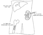
도 1은 다양한 표적 조직에 세포를 접목하는 본 발명에 따른 방법의 도식이다. 이러한 방법은, 이식 가능한 접목, 주사 가능한 접목, 및 표적 기관의 표면에 부착될 수 있는 접목을 포함한다 ("밴드에이드 접목(bandaid graft)").
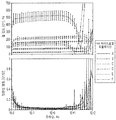
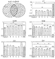
도 2는 Kubota의 배지로 제조된 히알루로난에 대한 유동학 측정(rheological measurement)(KM-HAs)을 제공한다. a) 외력에 따른 변형 반응 지연의 측정인, 점탄성 댐핑(viscoelastic damping) │G"/G'│은 시험할 각 포뮬레이션(formulation)에 대한 주파수 범위가 0.1 Hz -10 Hz 내인 무시해도 될 정도인 반면에, KM-HAs, 기계적 겔 강도의 측정의 전단 탄성률(shear modulus) |G*|는 일정하게 유지되고; 에러 바(error bar): 시험할 각 주파수에서 측정의 95% 신뢰 구간. b) KM-HAs는 전단 유동화(shear thinning), 즉 실험적 0.6 l/s-60 l/s 전단율 범위 [0.1 Hz-10 Hz 주파수(forcing frequency)]에서 주파수가 증가하면서 점도는 감소하는 것을 보여준다; 상한 및 하한: power law 모델계 95% 신뢰 구간 (Cox-Merz rule assumption, 0.3 1/s - 30 1/s 전단율 범위 [0.05 Hz - 5 Hz 주파수]에서 모든 포뮬레이션에 대해 R2 > 0.993 ). 기재된 포뮬레이션에 대해서만 행해진 유동학 측정은 표 3에 나타냈다.
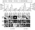
도 3은 KM-HAs에서 인간 간 줄기 세포 (hHpSCs)의 크기, 형태학 및 증식을 나타냈다. hHpSCs 군집은 3차원 배열을 획득하고, a) KM-HAs에서 씨딩할 때에 회전 타원체 같은 응집(spheroid-like agglomeration)(아래 왼쪽, bottom left) 또는 폴딩(folding)(중간, 위 오른쪽(middle, top right))을 보여준다 [이미지 프레임: 900㎛ × 1200㎛]. hHpSC-seeded KM-HAs의 조직학적 부분에 대한 공초점 주사 현미경(Confocal microscopy)은 배양 1주 후, 실질 세포 중에서 b) 약 7㎛, 또는 c) 10-15㎛ 까지의 셀 크기를 갖는, 혼합된 세포 형태 유전자형을 나타냈다 [DAPI 대비 염색제로부터 청색인 세포 핵, b)와 c)에 대해 적색인 EpCAM, b) CD44 또는 c) CDH1에 녹색; 이미지 프레임 b) 및 c): 150㎛ × 150㎛; b) 및 c)에 백색 하이라이트: 15㎛ × 15㎛]. AlamarBlue metabolic reduction에 의해 측정된 KM-HAs에서 hHpSCs의 생존도는, 배양 1주 내내 1.6% CMHA-S 및 0.4% PEGDA를 갖는 KM-HA 하이드로겔에서의 기능적 회복 및 증식을 나타냈다(포뮬레이션 E, 표 3); 24시간 배양 후 AlamarBlue reduction 측정, 씨딩 후(post-seeding) 2-3일에 측정에 있어서 정상화되고(normalized); 보고되는 데이터는 평균 ± 표준편차임.
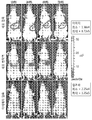
도 4는 배양 1주 후 KM-HA-seeded hHpSCs에서 분화 마커의 단백질 발현이 제공된다. hHpSCs의 군집은 KM-HAs 특성에 따라 달라지는 번역 레벨에서 hHpSCs 분화 마커에 대한 발현의 분화 레벨을 나타냈다. 인간 AFP의 대사 분비율은 KM-HA 포뮬레이션에 mRNA 발현과 연관성이 있다. NCAM 발현은 모든 KM-HAs에서 포지티브하지만, CD44 발현은, CMHA-S 함량이 1.2% 이하인 KM-HAs에서 가장 풍부하다(richest)(기재된 포뮬레이션 A, B, C, D; 표 3). CDH1 발현은 |G*| < 200 Pa인 KM-HA 하이드로겔에 포지티브하고, |G*| > 200 Pa인 KM-HA 하이드로겔에 네거티브하다. 인간 AFP 분비율의 데이터는 평균±표준편차로 보고된다. EpCAM, NCAM, CD44 및 CDH1에 대한 면역조직화학 스테이닝은 15-20㎛ 부분에서 행해지고(~2 내지 3 hHpSCs 두께; hHpSC 직경: 5-7㎛), 형광 현미경 검사로 영상화된다 [이미지 프레임: 100㎛ × 100㎛]. KM-HA 포뮬레이션은 강도가 증가하는 것과 관련하여 나열했다 (|G*| = 25 Pa : A, |G*| = 73 Pa : B, |G*| = 140 Pa : E, |G*| = 165 Pa : C, |G*| = 220 Pa : D, 및 |G*| = 520 Pa : F).
도 5는 배양 1주 후 KM-HA-grown hHpSCs에서 간 전구 마커(hepatic progenitor marker)에 대해 qRT-PCR로 유전자 발현 레벨을 제공한다. hHpSCs 및 이들의 직계 후손 hHBs (간-특이적(hepatic-specific) AFP, EpCAM, NCAM, CD44 및 CDH1)의 마커의 mRNA 발현 레벨 사이의 비교에서는, KM-HA-grown hHpSCs가 1주 동안 패시브 배양액(passive culture)에서 복사 레벨에서의 hHB 특성을 먼저 얻는다는 것을 보여준다. hHpSCs 및 CD44의 새롭게 분리된 hHBs에서 발현 범위는 비교할만하다; 잔여 마커에 대한 발현 레벨은 통계적으로 분명하고, EpCAM은 대략 2-폴드 감소, CDH1은 3-폴드 감소, NCAM 변동 없음, AFP는 hHpSCs가 hHBs로 분화될 때 풍부해짐. 모든 KM-HAs에서는, AFP, NCAM 및 CDH1에 대한 seeded hHpSCs의 평균 발현 레벨은 hHB 범위 쪽으로 hHpSC 범위 외부로 이동되지만, EpCAM 발현은 배양 1주 후 내내 풍부해진다. KM-HA 포뮬레이션은 강도가 증가하는 것과 관련하여 나열했다 (|G*| = 25 Pa : A, |G*| = 73 Pa : B, |G*| = 140 Pa : E, |G*| = 165 Pa : C, |G*| = 220 Pa : D, 및 |G*| = 520 Pa : F). 발현 레벨(평균±표준편차)은 GAPDH에 관하여 정상화되었다. 기재된 KM-HA 포뮬레이션의 측정 (표 3)은 유의성을 위해 hHpSC 군집 (녹색) 및 새롭게 분리된 hHBs (적색)과 비교했다 (학생의 t-test).
도 6은 기재된 동결보존 및 해동법의 일 실시형태의 도식이다.
도 7은 히알루로난과 접목된 대 세포 현탁액으로서 주입된 루시페린-생성 세포에 의해 형성된 형광 신호의 생체 내 실시간 이미지로부터의 결과를 나타낸다 (Figure 7 shows the results from in vivo real time imaging of luminescent signal produced by luciferin-producing cells both grafted with hyaluronans versus injected as a cell suspension.).
도 8은 건강한 및 CCl4 간 손상 모델에서 접목된 대 세포 현탁액에서 이식 후 7일에 인간 혈청 알부민을 제공한다 (Figure 8 provides serum human albumin at day 7 post-transplantation in grafted versus cell suspension in both healthy and CCl4 liver injury models.).
도 9는 간 줄기 세포 유전자형 마커의 유전자 발현을 나타냈다. 발현 레벨은 GAPDH 발현으로 정상화되고, 폴드 변화는 군집에서 최초 발현으로 정상화되었다. *는 실험 조건과 최초 군집 발현 사이의 p<0.05% 유의성을 나타낸다. **는 실험 조건과 최초 군집 발현 사이의 p<0.05% 유의성 뿐만 아니라 2개의 실험 조건 사이의 유의성 발현을 나타낸다.
도 10은 시간에 따른 간 기능의 기능적 분석으로부터 데이터를 제공한다. 레벨에 대해서 시간에 따른 3차원 히알루로난 배양에서 A) 알부민, B) 트랜스페린, 및 C) 요소는 세포마다 정상화되었다 (A) albumin, B) Transferrin, and C) Urea in three-dimensional hyaluronan culture over time for levels are normalized per cell).
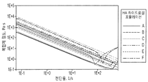
도 11은 KM-HAs의 기계적 특성으로부터 데이터를 제공한다. a) KM-HAs의 강도는 통제 가능하고, CMHA-S 및 PEGDA 함량에 따라 달라진다. 평균 전단 탄성율 |G*|는 power-law 행태를 따라 CMHA-S 및 PEGDA 함량을 증가시키면서 증가하므로, 최초 하이드로겔 혼합 중 KM-HAs의 최종 기계적 특성의 직접 제어를 제공하고(thus providing direct control of the final mechanical properties of KM-HAs during the initial hydrogel mixing); 표 3에 나타낸 기재된 포뮬레이션에 대해서만 유동학 측정이 행해졌다. 에러 바: 주파수 0.05 Hz -5 Hz에서 측정에 대해 ±1 표준편차. b) KM-HAs에서 확산(Diffusion). FRAP (70 kDa 플루오레세인-표지된 덱스트란, fluorescein-labeled dextran)로 KM-HAs에서 확산도의 측정은 Kubota 배지 단독과 상당히 다르지 않고; 확산 측정은 표 3에 나타낸 모든 포뮬레이션에 대해 행해졌다. 에러 바: 측정의 95% 신뢰 구간.
도 12는 KM-HAs로 hHpSCs seeded에 의해 인간 AFP, 알부민 및 요소의 분비를 보여준다. KM-HAs에서 hHpSCs의 군집은 배양 배지(KM)에서 발견되는 인간 AFP 및 알부민의 농도가 증가하는 일부 간 기능과, 씨딩 후 7일(day 7 post-seeding) 요소 합성의 평형을 보여준다. 인간 AFP, 인간 알부민 및 요소의 대사 분비율은, 1.6% CMHA-S 및 0.4% PEGDA를 갖는 KM-HAs에서 AFP, 알부민 및 감소된 요소 합성에 대한 최소 비율로서, KM-HA 포뮬레이션 사이에서 씨딩 7일 후에 구별된다 (The metabolic secretion rates of human AFP, human albumin and urea are distinctive by day 7 post-seeding amongst KM-HA formulations, with minimum rates for AFP, albumin and decreased urea synthesis in KM-HAs with 1.6% CMHA-S and 0.4% PEGDA)(포뮬레이션 E, 표 3). 좌측 컬럼: 각각 기재된 포뮬레이션에 대해 24시간 배양 후 매일 수집된 배양 배지에서의 대사 농도 (표 3). 우측 컬럼: 24시간 배양 후, 배양 배지에서 hHpSC 군집 당 대사 질량 분비율; 배지에서 총 대사 질량은 AlamarBlue reduction으로 생존도 분석에 의해 산출된 각각의 인터벌에서 기능적 hHpSC 군집의 수로 정상화되었다 (샘플 당 씨드된 군집의 대략적 수:12). 모든 데이터는 평균±표준편차로 보고되었다.
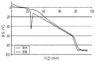
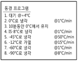
도 13은 결빙 프로그램 통제율은 내부 얼음 손상을 방지하는 액체-얼음 상 엔트로피를 최소화하고, 반복 가능한 동결을 가능하게 한다는 것을 보여준다 (Figure 13 shows that controlled rate freezing program minimizes liquid-ice phase entropy preventing internal ice damage and allows for repeatable freezing.). A) 그래프는 샘플 온도에 대한 챔버 온도를 보여준다 (10% DMSO). B) 동결 프로그램 비율은 Cryomed 1010 시스템에 사용된다.
도 14는 (A) 해동 후 동결보존된 태아 간 세포의 세포 생존도 및 (B) 새로운 샘플로 정상화되는, 각 조건에 대한 배양 3주 후 군집수(Colony counts)를 제공한다. 결과는 평균±평균의 표준편차로 보고되었다. KM= 10% DMSO 및 10% FBS를 함유하는 Kubotas Medium. CS10=cryostor, CS10+sup=KM 보충물을 함유하는 cryostor10. 0.05% 및 0.10%는 각 샘플에 보충된 HA%를 말한다.
도 15는 GAPDH 발현으로 정상화된 상대적인 mRNA 발현을 보여준다. 평균±평균의 표준편차. 새로운 샘플에 대한 유의성 *p>0.05(Significance *p>0.05 to Fresh samples). KM= 10% DMSO 및 10% FBS를 함유하는 Kubotas Medium. CS10=cryostor, CS10+sup=KM 보충물을 함유하는 cryostor10. 0.05% 및 0.10%는 각 샘플에 보충된 HA%를 말한다.1 is a schematic of a method according to the invention for grafting cells to various target tissues. Such methods include implantable grafts, injectable grafts, and grafts that can be attached to the surface of a target organ ("bandaid graft").
FIG. 2 provides rheological measurements (KM-HAs) for hyaluronan prepared with Kubota's medium. a) Viscoelastic damping, G "/ G ', which is a measure of the deformation response delay with external forces, is negligible in that the frequency range for each formulation to be tested is within 0.1 Hz -10 Hz. On the other hand, the shear modulus | G * | of KM-HAs, the measurement of mechanical gel strength, remains constant; error bar: 95% confidence interval of the measurement at each frequency to be tested b) KM-HAs show shear thinning, ie, viscosity decreases with increasing frequency in the experimental 0.6 l / s-60 l / s shear rate range [0.1 Hz-10 Hz forcing frequency]; And lower limit: 95% confidence interval in the power law model system (R2> 0.993 for all formulations in the Cox-Merz rule assumption, 0.3 1 / s-30 1 / s shear rate range [0.05 Hz-5 Hz frequency]). The rheology measurements made only for the simulations are shown in Table 3.
3 shows the size, morphology and proliferation of human liver stem cells (hHpSCs) in KM-HAs. The hHpSCs cluster acquires a three-dimensional array and a) spheroid-like agglomeration (bottom left) or folding (middle, top right) when seeding in KM-HAs. right)) [Image frame: 900 μm × 1200 μm]. Confocal microscopy of the histological portion of hHpSC-seeded KM-HAs is mixed after 1 week of culture with b) about 7 μm, or c) cell size up to 10-15 μm in parenchymal cells. Cell morphology genotypes [cell nuclei that are blue from the DAPI counterstain, b) and E) red for c), b) green on c44 or c) CDH1; Image frames b) and c): 150 μm × 150 μm; white highlights in b) and c): 15 μm × 15 μm]. Survival of hHpSCs in KM-HAs measured by AlamarBlue metabolic reduction showed functional recovery and proliferation in KM-HA hydrogels with 1.6% CMHA-S and 0.4% PEGDA throughout the week of culture (Formulation E, Table 3); AlamarBlue reduction measurements after 24 h incubation, normalized in measurements 2-3 days post-seeding; Data reported are mean ± standard deviation.
4 provides protein expression of differentiation markers in KM-HA-seeded hHpSCs after 1 week of culture. The cluster of hHpSCs showed differentiation levels of expression for hHpSCs differentiation markers at translation levels that depend on KM-HAs properties. Metabolic secretion rate of human AFP is associated with mRNA expression in KM-HA formulations. NCAM expression is positive in all KM-HAs, while CD44 expression is most abundant in KM-HAs with a CMHA-S content of 1.2% or less (Formulations A, B, C, D described; Table 3). CDH1 expression was determined by | G * | Positive for KM-HA hydrogel <200 Pa, and | G * | Negative for KM-HA hydrogels> 200 Pa. Data on human AFP secretion is reported as mean ± standard deviation. Immunohistochemical staining for EpCAM, NCAM, CD44 and CDH1 was performed at 15-20 μm portions (˜2 to 3 hHpSCs thickness; hHpSC diameter: 5-7 μm) and imaged by fluorescence microscopy [image frame: 100 Μm × 100 μm]. KM-HA formulations are listed in relation to increasing strength (| G * | = 25 Pa: A, | G * | = 73 Pa: B, | G * | = 140 Pa: E, | G * | = 165 Pa: C, | G * | = 220 Pa: D, and | G * | = 520 Pa: F).
5 provides gene expression levels with qRT-PCR for hepatic progenitor markers in KM-HA-grown
6 is a schematic of one embodiment of the cryopreservation and thawing methods described.
7 shows the results from in vivo real-time images of fluorescence signals formed by luciferin-producing cells injected as large cell suspensions grafted with hyaluronan ( Figure 7 shows the results from in vivo real time imaging of luminescent signal produced by luciferin-producing cells both grafted with hyaluronans versus injected as a cell suspension.).
Figure 8 after implantation in a large cell suspension was combined in the injury model between healthy and CCl4 provide human serum albumin in 4 days (Figure 8 provides serum human albumin at
9 shows gene expression of liver stem cell genotype markers. Expression levels were normalized to GAPDH expression, and fold changes normalized to initial expression in the population. * Indicates p <0.05% significance between experimental conditions and initial population expression. ** indicates significant expression between two experimental conditions as well as p <0.05% significance between experimental conditions and initial population expression.
10 provides data from functional analysis of liver function over time. A) albumin, B) transferrin, and C) urea were normalized from cell to cell in three-dimensional hyaluronan culture over time for levels (A) albumin, B) Transferrin, and C) Urea in three-dimensional hyaluronan culture over time for levels are normalized per cell).
11 provides data from the mechanical properties of KM-HAs. a) The strength of KM-HAs is controllable and depends on the CMHA-S and PEGDA content. The average shear modulus | G * | increases with increasing CMHA-S and PEGDA content along the power-law behavior, providing direct control of the final mechanical properties of KM-HAs during initial hydrogel mixing. the final mechanical properties of KM-HAs during the initial hydrogel mixing); Rheological measurements were performed only for the formulations described in Table 3. Error bar: ± 1 standard deviation for measurements at frequency 0.05 Hz -5 Hz. b) Diffusion in KM-HAs. The measurement of diffusivity in KM-HAs with FRAP (70 kDa fluorescein-labeled dextran, fluorescein-labeled dextran) was not significantly different from Kubota medium alone; Diffusion measurements were made for all formulations shown in Table 3. Error bar: 95% confidence interval of the measurement.
12 shows secretion of human AFP, albumin and urea by hHpSCs seeded with KM-HAs. The cluster of hHpSCs in KM-HAs shows some hepatic function with increased concentrations of human AFP and albumin found in culture medium (KM) and the equilibrium of
Figure 13 shows that the frozen program control rate minimizes liquid-ice phase entropy that prevents internal ice damage and enables repeatable freezing ( Figure 13 shows that controlled rate freezing program minimizes liquid-ice phase entropy preventing internal ice). damage and allows for repeatable freezing.). A) The graph shows chamber temperature versus sample temperature (10% DMSO). B) Freeze program rates are used for Cryomed 1010 systems.
FIG. 14 provides colony counts after 3 weeks of culture for each condition (A) cell viability of cryopreserved fetal liver cells after thawing and (B) normalized with new samples. The results were reported as the standard deviation of the mean ± mean. KM = Kubotas Medium containing 10% DMSO and 10% FBS. Cryostor10 containing CS10 = cryostor, CS10 + sup = KM supplement. 0.05% and 0.10% refer to the% of HA supplemented to each sample.
15 shows relative mRNA expression normalized to GAPDH expression. Standard Deviation of Mean ± Mean. Significance * p> 0.05 to Fresh samples. KM = Kubotas Medium containing 10% DMSO and 10% FBS. Cryostor10 containing CS10 = cryostor, CS10 + sup = KM supplement. 0.05% and 0.10% refer to the% of HA supplemented to each sample.
현재, 고형 기관으로부터 유래된 세포와 관련된 세포 이식은 일반적으로 혈관 경로를 통해 수행되고, 그 결과는 일반적으로 성숙 세포 약 20-30%, 줄기 세포 5% 미만의 비효율적인 접목의 압도적인 증거를 제공한다. 분화적 접목은, 간에서 줄기 세포가 작고 (일반적으로 10㎛ 미만), 성숙 세포가 큰 (일반적으로 > 18㎛) 사이즈에 기인한 것이다. 우리의 연구는 이 관측을 확인해주었다. 우리의 연구에서, 예컨대 인간 간 줄기 세포 (hHpSCs) 및 간모세포(hHBs)는 비장(spleen)으로 세포를 주사함으로써 면역 시스템이 손상된 쥐로 주사되었다. 비장은 간과 직접적으로 연결되기 때문에, 세포는 접목될 것으로 예상되는 간으로 흘러간다(flowed into). 그러나, 대부분의 세포는 접목 전에 죽거나, 의도하는 표적 이외의 조직 (이소성 부위(ectopic site))에 머무른다.
Currently, cell transplantation involving cells derived from solid organs is generally performed through vascular pathways, and the results generally provide overwhelming evidence of inefficient grafting of about 20-30% of mature cells and less than 5% of stem cells. do. Differentiated grafting is due to the size of stem cells small in the liver (typically less than 10 μm) and large mature cells (generally> 18 μm). Our study confirmed this observation. In our study, for example, human liver stem cells (hHpSCs) and hepatoblasts (hHBs) were injected into mice with compromised immune systems by injecting cells into the spleen. Since the spleen is directly connected to the liver, the cells flow into the liver that is expected to graft. However, most cells die before grafting or stay in tissues other than the intended target (ectopic site).
세포가 그 목적지에 적합하게 도달했을 경우에도, 세포의 완전히 기능적인 것(fully functional ones)으로의 전환은, 혈관화(vascularization)의 부족, 성장의 부족 (성숙 세포를 이식하는 경우에), 성숙 세포가 사용되고 장기간 면역 억제를 필요하게 할 경우에 세포의 높은 면역 특성에 의해 저해된다. 다른 장애물로는 임상 등급의 소싱(sourcing of clinical grade), 고급 세포(high-quality cells) 및 동결 보존으로의 곤란성에 기인한 새롭게 분리된 세포를 사용할 필요성(need to use freshly isolated cells due to difficulties with cryopreservation)을 포함한다.
Even when a cell has reached its destination appropriately, the transition of the cell to fully functional ones may be due to lack of vascularization, lack of growth (if transplanting mature cells), When cells are used and require prolonged immune suppression, they are inhibited by the high immune properties of the cells. Other obstacles include necessity to use freshly isolated cells due to difficulties with sourcing of clinical grade, high-quality cells and cryopreservation. cryopreservation).
비효율성 및 공지된 곤란성 이외에, 혈관 경로를 통한 고형 기관으로부터의 세포의 이식은 위험하다. 고형 기관으로부터의 세포는, 세포를 서로 빠르게 결합하고 응집을 강화하는 표면 분자를 갖는다 (세포 접합 분자, 밀착 연접 단백질(tight junction protein)). 이러한 군집 현상은 생명을 위협하는 폐 색전을 야기할 수 있다.
In addition to inefficiencies and known difficulties, the transplantation of cells from solid organs through the vascular pathway is dangerous. Cells from solid organs have surface molecules that quickly bind cells together and enhance aggregation (cell junction molecules, tight junction proteins). This clustering can lead to life-threatening pulmonary embolism.
일부 이들의 장애물 및 문제를 다루기 위해서, 본 발명은 필요한 증식 및 접목을 증진시키기 위해 질병에 걸린 조직에 로컬라이징 될 수 있는 스캐폴드 상에 또는 스캐폴드 내에 군(aggregates)으로서 이식된 세포의 전달(delivery)을 포함하는 접목 기술에 관한 것이다. 따라서, 본 발명은 이식할 세포 형태 뿐만 아니라, 가장 효율적이고 성공적인 이식 요법을 위한 접목 방법 및 적절한 생체 적합 물질과 조합인 세포 형태를 고려할 수 있다. 본 발명의 접목 기술은, 환자에의 치료적 용도로 변환 가능하고, 질병에 걸리거나 기능 장애가 있는 조직을 재구성하도록 재생적 약을 위한 대안적 치료를 제공한다.
To address some of these obstacles and problems, the present invention delivers cells implanted as aggregates on or within scaffolds that can be localized to diseased tissue to promote the necessary proliferation and grafting. It relates to a graft technique, including). Thus, the present invention contemplates not only the cell type to be transplanted, but also the cell type which is combined with the appropriate biocompatible material and grafting method for the most efficient and successful transplantation therapy. The graft technology of the present invention provides an alternative treatment for regenerative medicine to reconstruct tissue that is convertible for therapeutic use in a patient and that is diseased or dysfunctional.
세포 소싱(Cell sourcing CellCell SourcingSourcing ))
본 발명에 따라서, 바람직한 세포 군집(cell population)은, 질병에 걸리거나 기능 장애로 고통받지 않는 조직 및/또는 세포를 의미하는 "정상의(normal)", "건강한(healthy)" 조직 및/또는 세포를 갖는 공여체로부터 직접 얻어질 수 있다. 물론, 이러한 세포 군집은 질병에 걸리거나 기능 장애가 있는 기관으로 고통 받는 사람으로부터, 이러한 상태가 아닌 기관의 부분으로부터 얻을 수 있다(such a cell population may be obtained from a person suffering from an organ with disease or dysfunction, albeit from a portion of the organ that is not in such a condition). 세포는, 나이에 상관없이 태아, 신생아, 소아 및 성인 조직을 포함하는 적절한 포유류 조직으로부터 얻을 수 있다. 질병 상태의 실험 모델이 확립되는 경우에, 적절한 실험 호스트로 이식될 접목에 질병에 걸린 세포를 사용할 수 있다.
In accordance with the present invention, preferred cell populations are " normal "," healthy " tissues and / or cells meaning tissues and / or cells that are not diseased or suffering from dysfunction. Can be obtained directly from donors with cells. Of course, such a cell population may be obtained from a person suffering from a diseased or dysfunctional organ, or from a part of an organ that is not in this condition. , albeit from a portion of the organ that is not in such a condition). The cells can be obtained from appropriate mammalian tissues, including fetal, newborn, pediatric and adult tissues, regardless of age. If experimental models of disease state are established, diseased cells can be used for grafting to the appropriate experimental host.
더욱 구체적으로, 세포는 치료적 필요에 기초하여 "혈통-단계(lineage -staged)" 개체군으로부터 다양한 요법에 공급될 수 있다. 예컨대, 늦은 혈통 세포(late lineage cell)에 의해서만 제공되는 기능을 빠르게 얻을 필요가 있는 경우에, 또는 수신자(recipient)가, 예컨대 간염 C 또는 유두종 바이러스로 일어나는 줄기 세포 및/또는 전구세포를 우선적으로 감염시키는 혈통 의존 바이러스(lineage-dependent virus)를 갖는 경우에, 이후 단계(later-stage) "성숙(mature)" 세포는 바람직할 수 있다. 아무튼, "전구세포(progenitor)"는 개별 조직의 임의의 혈통 단계(lineage stages)를 확립하는데 사용될 수 있다.
More specifically, cells can be supplied to various therapies from a “lineage-staged” population based on therapeutic needs. For example, if it is necessary to quickly obtain a function provided only by late line line cells, or a recipient preferentially infects stem and / or progenitor cells resulting from eg hepatitis C or papilloma virus. Later-stage "mature" cells may be desirable if they have lineage-dependent viruses. In any event, "progenitors" can be used to establish any lineage stages of an individual tissue.
혈통-단계 간 세포 군집 및 이들의 분리 방법에 있어서, US 특허출원 nos. 11/560,049 및 12/213100를 참조하고, 이 내용은 전체가 참조로 인용된다. 간단히, 간내에 있는 적어도 8개의 성숙된 혈통 단계가 있다. 아래에 이들에 대한 단계 및 간단한 설명이 제공되었다:
For lineage-stage liver cell populations and methods for their separation, see US patent application nos. 11 / 560,049 and 12/213100, the contents of which are incorporated by reference in their entirety. In brief, there are at least eight mature lineage stages in the liver. Below are the steps and a brief description of them:
혈통 단계( Lineage Stage ) 1: 인간 간 줄기 세포(hHpSCs)는 태아 및 신생아의 간의 관판(ductal plate) 및 소아 및 성인의 간에서 헤링의 캐널(canals of Hering) 내에 위치하는 다분화능 세포(multipotent cell)이다. 이들 세포는 일반적으로 직경이 7-10㎛이고, 세포질 비에 대해 많은 핵을 가지고 있다. 이들은 허혈에 내성이 있고, 심장 수축 사망 후 48 이상의 시간 동안 사체의 간에서 발견될 수 있고, 성숙 세포로 분화할 수 있는 hHpSCs의 군집을 형성한다. 이들 세포는 모든 연령의 공여체의 간의 실질 조직의 약 0.5-2%를 구성한다.
Lineage stage ( Lineage Stage 1 : Human liver stem cells (hHpSCs) are ductal plates of fetal and newborn livers and multipotent cells located in the canals of Hering in the liver of children and adults. These cells are generally 7-10 μm in diameter and have many nuclei for the cytoplasmic ratio. They are resistant to ischemia and can be found in the liver of cadaver for at least 48 hours after cardiac contraction death and form a cluster of hHpSCs that can differentiate into mature cells. These cells make up about 0.5-2% of the parenchymal tissue of the liver of donors of all ages.
혈통 단계 2: 간모세포(hHBs)는 hHpSCs의 직계 후손이고, 간의 개연성 있는 전이 증폭 세포(liver's probable transit amplifying cells)이다. 이들은 바로 줄기 세포 니치의 외부에 위치한다(They are located just outside the stem cell niche proper). 이들 세포는, 생체 내에서(in vivo ), 많은 양의 세포질을 가져 크고(10-12㎛), 태아 및 신생아의 간에서 실질 조직 및 소아 및 성인의 간에서 헤링의 캐널의 부분 또는 거의 끝부분(near the ends of or adjacent to the canals of Hering in pediatric and adult livers)의 도처에서 발견된다. 나이에 따라, 간모세포의 수는 출생 후 간의 실질 세포의 <0.01% 로 감소한다. 세포의 이들 군집은, 재생 단계 동안 특히 소정의 질병, 예컨대 간경변증과 관련된 것들이 증식하는 것을 보여준다. 간모 세포는 간세포(hepatocyte)(H) 또는 담관 상피세포(biliary epithelia)(B)라고도 하는 담관세포(cholangiocyte)로 성숙한다:
Lineage Stage 2 : Hepatoblasts (hHBs) are direct descendants of hHpSCs and are liver's probable transit amplifying cells. They are located just outside the stem cell niche proper. These cells, in vivo ( in in vivo ), having large amounts of cytoplasm (10-12 μm), parenchymal tissue in the liver of fetuses and newborns, and the canal of the herring in the liver of children and adults (near the ends of or adjacent to the Canals of Hering in pediatric and adult livers. With age, the number of hepatoblasts decreases to <0.01% of parenchymal cells of the liver after birth. These populations of cells show that during the regeneration phase, especially those associated with certain diseases, such as cirrhosis, proliferate. Hepatoblasts mature into cholangiocytes, also called hepatocytes (H) or biliary epithelia (B):
혈통 단계 3H 및 3B: 수임 (단분화능) 간세포 (Committed (unipotent) hepatocytic) (3H) 및 담관세포 전구세포-담관 전구세포 (3B)는 간 내에서 발견된다. 이러한 단분화능 전구체는 오직 하나의 성인 세포형을 발생하고, 일부 줄기 세포 유전자를 더 이상 발현하지 않지만 (예컨대, CD133/1, Hedgehog proteins (Sonic/Indian)의 발현 레벨이 낮거나 없음), 태아 조직 세포에 전형적인 유전자를 발현한다.
Pedigree stages 3H and 3B : Committed (unipotent) hepatocytic (3H) and bile duct progenitor-biliary duct progenitor cells (3B) are found in the liver. These monopotent precursors generate only one adult cell type and no longer express some stem cell genes (eg, low or no expression level of CD133 / 1, Hedgehog proteins (Sonic / Indian)), but fetal tissue Express genes typical of cells.
혈통 단계 4H 및 4B: 문맥주위 성인 실질세포(Periportal adult parenchymal cell)는 상대적으로 작은 간세포(4H) 및 간내 담관 상피 세포 (4B)를 포함한다. 간세포는 이배체(diploid)이고, 직경이 약 18 ㎛이고, PEPCK, connexins 26 및 32와 같은 글루코오스신생합성(gluconeogenesis)과 관련된 다중 인자/효소를 발현한다.
Pedigree stages 4H and 4B : Periportal adult parenchymal cells comprise relatively small hepatocytes (4H) and intrahepatic bile duct epithelial cells (4B). Hepatocytes are diploid, about 18 μm in diameter and express multiple factors / enzymes associated with glucose synthesis, such as PEPCK, connexins 26 and 32.
이 단계의 담관세포(4B)는 이배체이고, 직경이 6-7㎛이고, 헤닝의 캐널 부분을 따라 늘어서고, 아쿠아포린 1 및 4, MDR1, 세크레틴 리셉터(secretin receptor)를 포함하지만, CL-/HC03- 교환기(exchanger) 또는 소마토스타틴 리셉터(somatostatin receptor)를 포함하지 않는 다양한 유전자를 발현한다.
The bile duct cells (4B) at this stage are diploid, 6-7 μm in diameter, lined along the canning portion of hening, and contain
혈통 단계 5H 및 5B: 이 단계의 세포는 둘다 이배체인 상대적으로 큰 간세포(5H) 및 담관세포(5B)를 포함한다. 간세포의 크기는 직경이 약 22-25 ㎛이고, midacinar 영역 내에서 발견된다. midacinar 간세포는 높은 수준의 알부민 및 티로신 아미노트랜스퍼라아제 (TAT)를 발현했고; 특히 단백질로서 트랜스페린을 발현한다는 특징이 있다 (그에 반해서 혈통 단계 1-4는 mRNA로서만 발현함).
Pedigree stages 5H and 5B : The cells of this stage comprise relatively large hepatocytes (5H) and bile duct cells (5B), both diploid. The size of hepatocytes is about 22-25 μm in diameter and is found in the midacinar region. midacinar hepatocytes expressed high levels of albumin and tyrosine aminotransferase (TAT); In particular, it expresses transferrin as a protein (in contrast, lineage steps 1-4 express only as mRNA).
혈통 단계 5B: 담관세포는 직경이 약 14㎛이고, 소엽내관(intralobular duct) 내에 위치하고, CFTR, 세크레틴 리셉터, 소마토스타틴 리셉터, MDR1 및 MDR3, CL-/HC03- 교환기를 발현한다.
Pedigree stage 5B : Bile duct cells are about 14 μm in diameter and are located in the intralobular duct and express CFTR, secretin receptor, somatostatin receptor, MDR1 and MDR3, CL − / HC03 − exchangers.
혈통 단계 6H: 단계 6의 이배체 주심 간세포(diploid pericentral hepatocytes)는 배양액에서 군집을 형성할 수 있지만, 증식할 용량이 제한되고, 서브 배양할 용량이 필수적으로 없다. 이러한 퍼센트는 나이에 따라 감소한다 (사배체 주심 세포의 퍼센트의 증가와 병행함). 알부민, TAT 및 트랜스페린 이외에도, 이들은 많은 P450s, 예컨대 P450-3A, 글루타민 합성 효소(GT), 헤파린 프로테오글리칸(heparin proteoglycan), 및 요소 형성과 관련된 유전자도 강력히 발현한다.
Pedigree stage 6H : Diploid pericentral hepatocytes in
혈통 단계 7H: 이 단계는 더 이상 완전한 셀 분할을 겪을 수 없는 사배엽 주심 실질 세포(tetraploid pericentral parenchymal cell)를 포함한다. 이들은 DNA 합성을 겪을 수 있지만, 세포질 분열의 용량은 한정된다. 이들은 매우 큰 세포이고 (직경이 >30 m), 혈통 단계 5-6에 나타나는 고레벨의 유전자를 발현한다.
Pedigree Stage 7H : This stage includes tetraploid pericentral parenchymal cells that can no longer undergo complete cell division. They may undergo DNA synthesis, but the capacity of cytoplasmic division is limited. They are very large cells (> 30 m in diameter) and express high levels of genes that appear in lineage stages 5-6.
혈통 단계 8: 사멸 세포(Apoptotic Cell): 아포토시스의 다양한 마커를 발현하고, DNA 단편화(DNA fragmentation)를 설명한다.
Lineage Step 8 : Apoptotic Cells: Express various markers of apoptosis and describe DNA fragmentation.
질병에 걸리거나 또는 기능 장애가 있는 장기 자체의 기능을 제공하도록 요구되는 세포 이외에, 접목(the graft)은 바람직하게는 모든 조직의 세포의 토대(cellular foundation), 상피-배엽간 세포 관계(epithelial-mesenchymal cell relationship)를 포함하는 세포의 범주를 바람직하게 모방하는 추가적인 세포 성분을 포함한다. 상피-배엽간 세포 관계는 매 성숙 혈통 단계에서 분명하다. 상피 줄기 세포는 간충직 줄기 세포와 파트너가 되고, 이들은 조직 내에서 다양한 성체 세포로 성숙하기 때문에 이들의 성숙은 서로 조절된다. 이 둘의 상호작용은 가용성 신호 (예컨대, 성장 인자) 및 세포외 매트릭스 성분을 포함하는 파라크린 신호(paracrine signal)에 의해 조절된다.
In addition to cells that are required to provide the function of the diseased or dysfunctional organ itself, the graft is preferably an epithelial-mesenchymal, cellular foundation of all tissue cells. additional cellular components that preferably mimic the categories of cells that comprise the cell relationship. Epithelial-ectoderm cellular relationships are evident at every mature lineage stage. Epithelial stem cells partner with mesenchymal stem cells, and because they mature into various adult cells in tissues, their maturation is regulated with each other. The interaction between the two is regulated by paracrine signals, including soluble signals (eg, growth factors) and extracellular matrix components.
간에서, 예컨대 간 줄기 세포 (HpSCs)는 간세포(hepatocyte) 및 담관세포(cholangiocytes)를 생성한다. HpSCs의 간충직 파트너(mesenchymal partner)는 혈관 아세포(angioblast)이다. 혈관 아세포는 내피 세포 전구체와 간 성상 세포 전구체, 간내 혈통 단계 2 실질 조직의 간충직 세포 파트너, 간모세포 (HBs)를 생성한다는 것을 나타내는 증거가 있다(There is evidence to indicate that angioblasts give rise to both endothelial cell precursors and to hepatic stellate cell precursors, the mesenchymal cell partners for intrahepatic lineage stage 2 parenchyma, the hepatoblasts (HBs)). 내피 세포 전구체는, 간세포의 혈통 단계의 간충직 파트너가 되는 내피가 될 다음 혈통 단계에서 성숙된다. 상기 성상 세포 전구체 세포는 성상 세포를 생성한 후, 기질 세포, 그 후 근섬유아세포, 담관 세포의 간충직 세포 파트너를 생성한다(The stellate cell precursors cells give rise to stellate cells, and then to stromal cells, and then to myofibroblasts, the mesenchymal cellspartners for cholangiocytes).
In the liver, for example, liver stem cells (HpSCs) produce hepatocytes and cholangiocytes. The mesenchymal partner of HpSCs is angioblasts. There is evidence to indicate that angioblasts give rise to both endothelial cells. precursors and to hepatic stellate cell precursors, the mesenchymal cell partners for
간 생성(hepatogenesis)이라고도 하는 간의 형성은 심장과 관련된 배아의 간충직에서 혈관 아세포로부터의 신호를 통해 조절된다. 간 성장의 초기 단계 동안, 섬유아세포 증식인자(fibroblast growth factors, FGFs)가 심장 전 중배엽(pre-cardiac mesoderm)으로부터 분비될 때, 뼈 형태 형성 단백질 (BMPs)이 간충직(mesenchyme)으로부터 전달된다. 이들 새롭게 구체화된 간 세포는 그 후 박리되어 간충직 주변으로 이동하고, 내피와 기질의 전구체와 상호 작용한다(These newly specified hepatic cells then break away and migrate into the surrounding mesenchyme and interact with precursors to both endothelia and stroma). 간충직 세포는 성장 내내 간 세포와 접촉하고 있다.
Liver formation, also called hepatogenesis, is regulated through signals from vasoblasts in the mesenchymal lobe of the heart-associated embryo. During the early stages of liver growth, bone form forming proteins (BMPs) are delivered from mesenchyme when fibroblast growth factors (FGFs) are secreted from the pre-cardiac mesoderm. These newly specified hepatic cells then break away and migrate into the surrounding mesenchyme and interact with precursors to both endothelia and stroma ). Mesenchymal cells are in contact with liver cells throughout growth.
인간 간 줄기 세포 (hHpSCs)는 생존을 위해 간충직 세포와의 접촉이 필요하다. 이들은 자가 복제할 것이고, 혈관 아세포의 피더에서 배양되는 경우 hHpSCs로서 남아있다(They will self-replicate, that is remain as hHpSCs when on feeders of angioblasts). 이들은 간 성상 세포의 피더에서 배양되는 경우, 간모세포로 혈통 제한된다(They lineage restrict to hepatoblasts, if cultured on feeders of hepatic stellate cells). 이들은 성숙한 내피에서 배양되는 경우 성체 간세포로 성숙되고, 성숙한 기질 (예컨대, 성숙한 성상 세포 또는 근섬유모세포)에서 배양되는 경우 담관세포(cholangiocyte)로 성숙된다. 피더에 의한 줄기 세포의 운명의 조절은, 혈통 내에서 각각의 상피-배엽간 상호작용에서 생성된 파라크린 신호의 정밀한 조합에 기인하여 보여준다.
Human liver stem cells (hHpSCs) require contact with mesenchymal cells for survival. They will self-replicate, that is remain as hHpSCs when on feeders of angioblasts. They are lineage restrict to hepatoblasts, if cultured on feeders of hepatic stellate cells when cultured in feeders of hepatic stellate cells. They mature into adult hepatocytes when cultured on mature endothelial cells, and cholangiocytes when cultured on mature substrates (eg, mature stellate cells or myofibroblasts). The regulation of the fate of stem cells by the feeder is shown due to the precise combination of paracrine signals generated at each epithelial-dermal interaction in the lineage.
본 발명의 일 실시형태에 따라서, 접목을 최적화하기 위해서, 분리된 세포 군집은 공지의 파라크린 신호(known paracrine signal)(이하에 설명함) 및 "본래의(native)" 상피-배엽간 파트너(epithelial-mesenchymal partne)와 필요에 따라 조합된다. 따라서, 접목은 내피 줄기 세포(epithelial stem cell), 본래의 간충직 파트너(native mesenchymal partner), 혈관 아세포와 함께 혼합되는 간 줄기 세포를 포함한다. 전이 증폭 세포 니치 접목을 위해서(For a transit amplifying cell niche graft), 간모세포는 간 성상 세포 및 내피 세포 전구체와 파트너가 될 수 있다. 일부 접목에 있어서, 두 세트의 혼합을 만들 수 있다: 호스트 조직 내에 간 세포의 확립을 최적화하기 위해, 간 줄기 세포, 간모세포, 혈관 아세포, 내피 세포 전구체, 간 성상 세포 전구체 세포. 세포가 씨딩되는 곳으로의 접목의 미세 환경은 파라크린 신호, 매트릭스 및 가용성 신호로 이루어질 수 있고, 이는 접목에 사용되는 적합한 단계에서 제조된다(The microenvironment of the graft into which the cells are seeded will be comprised of the paracrine signals, matrix and soluble signals, that are produced at the relevant lineage stages used for the graft).
According to one embodiment of the present invention, in order to optimize grafting, the isolated cell populations are known paracrine signals (described below) and "native" epithelial-ectoderm partners ( epithelial-mesenchymal partne) and as required. Thus, grafts include epithelial stem cells, native mesenchymal partners, and liver stem cells mixed with vascular blast cells. For a transit amplifying cell niche graft, hepatoblasts can partner with hepatic stellate cells and endothelial cell precursors. In some grafts, two sets of blends can be made: hepatic stem cells, hepatoblasts, vascular blast cells, endothelial cell precursors, hepatic stellate cell precursor cells to optimize the establishment of liver cells in host tissues. The microenvironment of grafting to where the cells are seeded can consist of paracrine signals, matrices and soluble signals, which are prepared at the appropriate stages used for grafting. of the paracrine signals, matrix and soluble signals, that are produced at the relevant lineage stages used for the graft).
또한, 접목은 질병에 걸린 상태를 관리하기 위해 맞춰질 수 있다. 예컨대, 초기 단계에서 감염시킨 후 호스트 세포와 대등하게 성숙하는 혈통 의존 바이러스(lineage dependent viruses) (예컨대, 소정의 간염 바이러스)의 효과를 최소화하기 위해, 바이러스 감염의 복제를 허용하지 않는 다음 혈통 단계 (예컨대, 간세포 및 이들의 본래의 파트너, 내피(sinusoidal endothelial cells))의 접목을 준비할 수 있다. 접목은, 실험 동물 모델에서 표적 장기에 이식되는 접목에서 질병에 걸린 세포를 사용함으로써 질병 모델을 확립하기 위해 사용될 수 있다.
Grafts can also be tailored to manage diseased conditions. For example, to minimize the effects of lineage dependent viruses (eg, certain hepatitis viruses) that mature equally with host cells after infection at an early stage, the next lineage stage that does not allow replication of the viral infection ( For example, grafting of hepatocytes and their original partner, sinusoidal endothelial cells can be prepared. Grafts can be used to establish disease models by using diseased cells in grafting implanted to target organs in experimental animal models.
모델로서 간 세포 요법을 사용하는, 줄기 세포 접목의 예로는, 간 줄기 세포, 혈관 아세포 및 간 성상 세포 전구체를 포함할 수 있다. 그에 반해서, "본래의(native)" 간 세포의 접목은, 성숙한 성상 세포인, 간세포, 성숙한 내피 세포 및 주위세포(pericytes)를 포함할 수 있다. 간의 상피-배엽간 세포 관계에 대한 논의에 있어서, US 특허 출원 no. 11/753,326를 참조하고, 이 내용은 전체가 참조로 인용되었다.
Examples of stem cell grafting, using liver cell therapy as a model, can include hepatic stem cells, hemangioblasts and hepatic stellate cell precursors. In contrast, grafting of "native" liver cells may include hepatocytes, mature endothelial cells and pericytes, which are mature stellate cells. In a discussion of epithelial-ectoderm cellular relationships in liver, see US patent application no. 11 / 753,326, which is incorporated by reference in its entirety.
혈관 신생의 문제는 모든 접목에 중요하므로, 혈관 신생에 도움이 되는 위치에 이식되어야 한다 (예컨대, 간). 대부분의 질병에 걸린 상태에 있어서, 줄기 세포 접목은, 이들의 증식 가능성, 모든 성체 세포 형태로 성숙해지는 이들의 능력, 허혈에 대한 이들의 내성, 사체 조직으로부터 이들의 소싱을 가능하게 하는 점, 및 만약에 있다면 이들의 최소 면역원성을 고려해볼 때, 바람직하다(For most disease conditions, stem cell grafts are preferred, given their expansion potential, their ability to mature into all of the adult cell types, their tolerance for ischemia, enabling their sourcing from cadaveric tissue, and their minimal, if any, immunogenicity).
The problem of angiogenesis is important for all grafts, so it should be implanted in a location that aids in angiogenesis (eg liver). In most diseased states, stem cell grafting allows for their proliferation, their ability to mature into all adult cell forms, their resistance to ischemia, their sourcing from cadaveric tissue, and For most disease conditions, stem cell grafts are preferred, given their expansion potential, their ability to mature into all of the adult cell types, their tolerance for ischemia, enabling their sourcing from cadaveric tissue, and their minimal, if any, immunogenicity).
접목 소재 ( Grafting Materials ) Grafting material (Grafting Materials )
본 발명에 따른 겔-형성 생체 적합 물질의 용도는, 접목 및 재생 단계의 성공을 도와주는 세포 지지체 및 신호의 스캐폴드(a scaffold for cell support and signals that assist in the success of the grafting and regenerative processes)를 제공한다. 장기의 고형 기관의 조직이 일정한 리모델링을 겪기 때문에, 해리성 세포는 적절한 환경 조건 하에서 본래의 구조를 개조하는 경향이 있다. 세포는 하나 이상의 영양 배지(예컨대, RPM 1640), 신호 분자(예컨대, 인슐린, 트랜스페린, VEGF) 및 하나 이상의 세포외 매트릭스 성분(예컨대, 히알루로난, 콜라겐, 니도겐, 프로테오글리칸)과 혼합될 수 있다.
The use of a gel-forming biocompatible material according to the present invention is a scaffold for cell support and signals that assist in the success of the grafting and regenerative processes To provide. Because the tissues of the organs of the organs undergo some remodeling, dissociative cells tend to modify their original structure under appropriate environmental conditions. The cells may be mixed with one or more nutrient media (eg RPM 1640), signal molecules (eg insulin, transferrin, VEGF) and one or more extracellular matrix components (eg hyaluronan, collagen, nidogen, proteoglycans). .
모든 조직에 있어서, 파라크린 신호는 가용성(미리아드 성장 인자(myriad growth factor) 및 호르몬) 및 불용성(세포외 매트릭스(ECM) 신호)을 포함한다. 가용성 및 (불용성) 매트릭스 요소 사이의 시너지 효과는 이식된 세포에 의한 성장 및 분화 반응을 지시할 수 있다. 매트릭스 성분은 접합, 생존, 세포 형태(또한 세포 골격의 조직), 및 특정 세포외 신호에 대한 반응을 세포에 준비시키는 필수 세포 표면 리셉터의 안정화의 주요 결정 요인이다.
In all tissues, paracrine signals include soluble (myriad growth factor and hormones) and insoluble (extracellular matrix (ECM) signals). Synergistic effects between soluble and (insoluble) matrix elements can direct the growth and differentiation reactions by the transplanted cells. Matrix components are key determinants of conjugation, survival, cell morphology (also tissue of the cytoskeleton), and stabilization of essential cell surface receptors that prepare cells for responses to specific extracellular signals.
ECM은 세포 형태학, 성장 및 세포 유전자 발현을 조절하는 것으로 알려져 있다. 생체 내에서 그것과 유사한 조직-특이 화학은 정제된 ECM 성분을 사용함으로써 생체 밖에서 행해질 수 있다(Tissue-specific chemistries similar to that in vivo may be achieved ex vivo by using purified ECM components). 대다수의 이들은 상업적으로 이용 가능하고, 생체 내에서 그것을 모방하는 세포 행태에 도움이 된다(Many of these are available commercially and are conducive to cell behavior mimicking that in vivo ).
ECM is known to regulate cell morphology, growth and cellular gene expression. Tissue-specific chemistries similar to that in vivo may be achieved ex , using tissue-specific chemistry similar to that in vivo. in vivo by using purified ECM components). Many of these are available commercially and are conducive to cell behavior mimicking that in vivo ) .
적합한 매트릭스 성분은 콜라겐, 접합 분자(예컨대, 세포 접합 분자 (CAMs), 밀착 연접(tight junction) (카드헤린, cadherins), 기본 접합 분자 (라미닌, 히브로넥틴), 간극 연접 단백질(gap junction protein) (콘넥신, connexins), 엘라스틴, 및 프로테오글리칸 (PGs) 및 글리코사미노글리칸 (GAGs)을 형성하는 황산화 카보하이드레이트(sulfated carbohydrates)를 포함한다. 이들 범주 각각은 분자의 속(genus)으로 정의된다. 예컨대, 존재하는 적어도 25 콜라겐 형태가 있고, 각각은 본래 유전자에 의해, 특유의 조절 및 기능을 갖고 인코딩된다(there are at least 25 collagen types present, each one encoded by distinct genes and with unique regulation and functions). 추가적인 생체 적합 물질로는 무기, 키토산과 알지네이트와 같은 천연 물질 뿐만 아니라 다양한 합성, 생분해성 및 생체 적합성 폴리머를 포함한다. 이들 물질은, 열 겔화(thermal gelation), 광 가교(photo cross-linking), 또는 화학적 가교 또는 물질의 불용성을 끌어내는 미세 환경으로의 노출 (예컨대 고염(high salt))을 포함하는 방법을 통해 종종 고형화된다(예컨대, 겔 또는 불용성 물질로 만듬). 그러나, 각 방법에 있어서는, 세포 손상을 고려할 필요가 있다(예컨대 과도한 온도 범위, UV 노출). 생체 적합 물질, 구체적으로 히알루로난 하이드로겔의 용도의 상세한 설명에 대해서는, US 특허 출원 no. 12/073,420를 참조하고, 이 내용은 전체가 여기에 참조로 인용되었다.
Suitable matrix components include collagen, junction molecules (eg, cell junction molecules (CAMs), tight junctions (cadherin, cadherins), basic junction molecules (laminine, hybronectin), gap junction proteins. (Connexins, connexins), elastin, and sulfated carbohydrates that form proteoglycans (PGs) and glycosaminoglycans (GAGs), each of which is defined as a genus of molecules. For example, there are at least 25 collagen types present, each one encoded by distinct genes and with unique regulation and Additional biocompatible materials include a variety of synthetic, biodegradable and biocompatible polymers, as well as inorganic, natural materials such as chitosan and alginate. Materials are often solidified through methods that include thermal gelation, photo cross-linking, or exposure to microenvironments that elicit chemical crosslinking or insolubility of the material (eg, high salts). However, in each method, cell damage needs to be taken into account (eg excessive temperature ranges, UV exposure) of biocompatible materials, specifically hyaluronan hydrogels. For details, see US patent application no. 12 / 073,420, which is hereby incorporated by reference in its entirety.
매트릭스 성분의 특정 선택은, 예컨대 줄기 세포 구획과 관련되어 발견되는 성분으로부터 늦은 혈통 단계 세포와 관련되어 발견되는 성분까지의 전이(transition)가 생체 내에서 변화도(gradient)에 의해 가이드될 수 있다. 접목 생체 적합 물질은 바람직하게는 접목에 바람직한 특정 혈통 단계의 매트릭스 화학을 모방한다(The graft biomaterials preferably mimic the matrix chemistry of the particular lineage stages desired for the graft). 매트릭스 성분의 선택된 혼합물의 효능은 정제된 매트릭스 성분 및 가용성 신호를 사용하여 생체 밖에서 연구될 수 있고, 이들 다수는 제조 품질 관리 기준(GMP) 프로토콜에 따라 상업적으로 이용 가능하다. 접목에 선택된 생체 적합 물질은, 바람직하게는 성공적 이식을 위한 세포에 요구되는 적절한 성장 및 분화 반응을 이끌어낸다.
Certain selections of matrix components can be guided by gradients in vivo, for example, from a component found in association with stem cell compartments to a component found in association with late lineage stage cells. The graft biomaterials preferably mimic the matrix chemistry of the particular lineage stages desired for the graft. The efficacy of selected mixtures of matrix components can be studied in vitro using purified matrix components and soluble signals, many of which are commercially available according to manufacturing quality control criteria (GMP) protocols. Biocompatible materials selected for grafting preferably elicit the appropriate growth and differentiation reactions required for cells for successful transplantation.
간 기관에 관하여, 간 실질 세포, 및 줄기 세포의 외부 및 세포 니치를 증폭하는 전이와 관련된 매트릭스 화학은 디세강에 존재하고, 상기 영역은 실질 조직과 내피 또는 다른 형태의 간충직 세포 사이에 위치한다(Concerning the liver organ, the matrix chemistry associated with liver parenchymal cells, and outside of the stem cell and transit amplifying cell niches, is present in the Space of Disse, the area located between the parenchyma and the endothelia or other forms of mesenchymal cells). 간의 다른 영역 내에서 세포 성숙의 변화 이외에, 매트릭스 화학의 변화가 관측되었다. 구역(zone) 1에서 문맥주위로 매트릭스 화학(The matrix chemistry periportally in zone 1)은 태아의 간에서 발견된 것과 유사하고, 타입 III 및 타입 IV 콜라겐, 히알루로난 (HA), 라미닌, 및 콘드로이딘 설페이트 프로테오글리칸의 형태로 이루어진다. 이 구역은, 타입 I, 콜라겐, 피브로넥틴, 및 특정 형태의 헤파린 및 헤파란 설페이트 프로테오글리칸을 함유하는 주심 구역 3에서 다양한 매트릭스 화학으로 전이한다(This zone transitions to a different matrix chemistry in the pericentral zone 3, containing type I collagen, fibronectin, and unique forms of heparin and heparan sulfate proteoglycans).
With respect to the liver organs, matrix chemistry associated with hepatic parenchymal cells and metastases that amplify the external and cellular niches of stem cells is present in the Dissegang, and the region is located between parenchymal tissue and endothelial or other forms of mesothelial cells ( Concerning the liver organ, the matrix chemistry associated with liver parenchymal cells, and outside of the stem cell and transit amplifying cell niches, is present in the Space of Disse, the area located between the parenchyma and the endothelia or other forms of mesenchymal cells) . In addition to changes in cell maturation within other regions of the liver, changes in matrix chemistry were observed. The matrix chemistry periportally in
간의 줄기 세포 니치는 부분적으로 특징이 있고, 알파 6-베타 4 인테그린, 타입 III 콜라겐 및 특정 형태의 최소 황산화 콘드로이딘 설페이트 프로테오글리칸 (CS-PGs)과 결합하는 라미닌 형태(예컨대, 라미닌 5), 히알루로난을 포함하는 것이 발견되었다. 이러한 니치에서 타입 IV 콜라겐의 양은 제한되고, 타입 I 콜라겐은 없다.
Liver stem cell niches are partially characterized and form laminin (eg laminin 5) that binds alpha 6-
니치 매트릭스 화학은, 전이 증폭 세포 구획과 관련되고, 타입 IV 콜라겐, 다른 인테그린과 결합된 라미닌 형태 (αβ1), 높은 황산화도를 갖는 CS-PGs 형태를 포함하는 GAGs 및 PGs 형태, 데르마탄 설페이트-PGs, 및 헤파란 설페이트-PGs의 특정 형태 (HS-PGS)로 이루어진 것으로 전이한다(This niche matrix chemistry transitions to that associated with the transit amplifying cell compartment and is comprised of type IV collagen, forms of laminin that bind to other integrins (αβ1) , and forms of GAGs and PGs that include forms of CS-PGs with higher sulfation, dermatan sulfate-PGs, and to specific forms of heparan sulfate-PGs (HS-PGS)).
Niche matrix chemistry involves GAGs and PGs forms, dermatan sulfate-PGs, including type IV collagen, laminin form (αβ1) bound to other integrins, CS-PGs form with high degree of sulfation, associated with metastatic amplification cell compartments This niche matrix chemistry transitions to that associated with the transit amplifying cell compartment and is comprised of type IV collagen, forms of laminin that bind to other integrins (αβ1), and forms of GAGs and PGs that include forms of CS-PGs with higher sulfation, dermatan sulfate-PGs, and to specific forms of heparan sulfate-PGs (HS-PGS)).
전이 증폭 세포 구획은 다음 혈통 단계로 전이되고, 각 이어지는 단계에서(with each successive stage), 매트릭스 화학은 더욱 안정해지고(예컨대, 더욱 안정한 콜라겐), 덜 변화하고(turns over less), 더욱 황산화된 형태의 GAGs 및 PGs를 함유한다. 대부분의 성숙 세포는 헤파린-PGs (HP-PGs), 즉 미리아드 단백질이 매트릭스에 결합하고, GAGs에서 별개의, 특정 황산화 패턴과 결합을 통해 안정하게 고정될 수 있는 형태와 관련된다(The most mature cells are associated with forms of heparin-PGs (HP-PGs), meaning that myriad proteins (e.g., growth factors and hormones, coagulation proteins, various enzymes) can bind to the matrix and be held stably there via binding to the discrete and specific sulfation patterns in the GAGs). 따라서, 매트릭스 화학은, 높은 전환 및 최소 황산화와 관련된 불안정한 매트릭스 화학을 갖는 줄기 세포 니치에서의 그 시작점으로부터 (따라서, 세포 근처에 안정한 방법으로 신호를 최소 결합함) 황산화의 양을 증가시키는 안정한 매트릭스 화학으로(따라서, 세포 근처에 있고, 신호 결합 레벨이 높음) 전이된다(the matrix chemistry transitions from its start point in the stem cell niche having labile matrix chemistry associated with high turnover and minimal sulfation (and therefore minimal binding of signals in a stable fashion near to the cells) to stable matrix chemistries with increasing amounts of sulfation (and therefore higher and higher levels of signal binding and held near to the cells)).
The transition amplifying cell compartment is transferred to the next lineage stage, with each successive stage, the matrix chemistry becomes more stable (e.g., more stable collagen), turns over less, and more sulfated Form of GAGs and PGs. Most mature cells are associated with heparin-PGs (HP-PGs), or forms in which myriad proteins bind to the matrix and can be stably anchored through binding to distinct, specific sulfated patterns in GAGs (The most mature cells are associated with forms of heparin-PGs (HP-PGs), meaning that myriad proteins ( eg , growth factors and hormones, coagulation proteins, various enzymes) can bind to the matrix and be held stably there via binding to the discrete and specific sulfation patterns in the GAGs). Thus, matrix chemistry is stable to increase the amount of sulfation from its starting point in stem cell niches (and thus minimally bind the signal in a stable manner near the cell) with high conversion and unstable matrix chemistry associated with minimal sulfation. The matrix chemistry transitions from its start point in the stem cell niche having labile matrix chemistry associated with high turnover and minimal sulfation (and therefore minimal binding of signals in a stable fashion near to the cells) to stable matrix chemistries with increasing amounts of sulfation (and therefore higher and higher levels of signal binding and held near to the cells)).
이런 이유로, 본 발명은 매트릭스 분자의 화학은 성숙 단계, 호스트 연령, 및 질병 상태로 변화하는 것을 고려한다. 적절한 물질로의 접목은 조직에 이식되는 세포의 접목을 최적화시키고, 이소성 부위로 세포가 분산되는 것을 방지하고, 색전에 의한 폐색 문제를 최소화하고, 가능하면 빨리 조직 내에 세포를 통합시키는 능력을 향상시켜야 한다. 또한, 접목 내에서 인자들(factors)은 면역원성 문제를 최소화시키기 위해 선택될 수 있다.
For this reason, the present invention contemplates that the chemistry of the matrix molecule changes with maturity, host age, and disease state. Grafting with the appropriate material should optimize the graft of cells transplanted into the tissue, prevent the cells from dispersing into ectopic sites, minimize the embolism problem and improve the ability to integrate cells into the tissue as soon as possible. do. In addition, factors within the graft can be selected to minimize immunogenicity problems.
인간의 간의 경우에, 세포는 무혈청 조건 하에서 배양될 수 있다. 인간 간 줄기 세포 또는 간세포 (hHpSC 또는 hHB)는 자체로, 또는 혈관 아세포/내피 세포 전구체 및 성상 세포 전구체 세포와의 조합으로 접목될 수 있다. 세포는 배지 (HA-M)를 함유하는 티올화 및 화학적으로 변형된 HA (CMHA-S, 또는 Glycosil, Glycosan BioSystems, Salt Lake City, UT) 및 KM (쿠보타 배지)에서 현탁되고, 한쌍의 주사기의 세트 중 하나의 주사기에 로드될 수 있다. 다른 주사기는 KM (또는 생체 적합 물질의 불용성을 끌어내도록 요구되는 조건에서)에 제조된 가교제, 예컨대 폴리(에틸렌글리콜)디아크릴레이트 또는 PEGDA로 로드될 수 있다. 2개의 주사기는, 2개의 루어락 연결로 플레어 되는 니들에 의해(by a needle that flares into two luer lock connections) 연결된다. 따라서, 하이드로겔 및 가교제에서 세포는, 주사 시 겔로의 CMHA-S의 빠른 가교를 가능하게 하도록 하나의 니들을 통해 나타날 수 있다 (또는 다른 방법에 의해 생체 적합 물질의 불용성)(the cells in hydrogel and the cross-linker can emerge through one needle to allow for rapid cross-linking of the CMHA-S into a gel upon injection (or insolubility of the biomaterials by alternate means)).
In the case of human livers, cells can be cultured under serum free conditions. Human liver stem cells or hepatocytes (hHpSC or hHB) can be grafted on their own or in combination with angioblast / endothelial precursor and astrocytic precursor cells. Cells are suspended in thiolated and chemically modified HA (CMHA-S, or Glycosil, Glycosan BioSystems, Salt Lake City, UT) and KM (Kubota Medium) containing medium (HA-M), It can be loaded into one syringe of the set. Other syringes may be loaded with crosslinking agents such as poly (ethyleneglycol) diacrylate or PEGDA prepared in KM (or under conditions required to elicit insolubility of the biocompatible material). The two syringes are connected by a needle that flares into two luer lock connections. Thus, in hydrogels and crosslinkers, cells may appear through one needle (or insoluble of biocompatible material by other methods) to enable rapid crosslinking of CMHA-S into the gel upon injection. the cross-linker can emerge through one needle to allow for rapid cross-linking of the CMHA-S into a gel upon injection (or insolubility of the biomaterials by alternate means)).
CMHA-S 및 가교제 중 세포 현탁액은 주머니(pouch)를 형성하기 위해 장막 조직을 사용하여 간에 이식되거나 또는 직접 주사될 수 있다. 또는, 세포는, 공기 중에서 밤새 현탁액을 정치시키고, 연하고 점성이 있는 하이드로겔로 가교하는 디설파이드 결합을 야기함으로써, PEGDA 가교제를 사용하지 않고 글리코실(Glycosil) 내에 캡슐화될 수 있다. 또한, 다른 티올-변형 마크로모노머(thiol-modified macromonomer), 예컨대 젤라틴-DTPH, 헤파린-DTPH, 콘드로이틴 설페이트-DTPH는 생체 내에서 특정 니치의 매트릭스 화학을 모방하는 공유 네트워크를 제공하도록 첨가될 수 있다. 다른 징후에 있어서, 시스테인 또는 티올 잔기를 함유하는 폴리펩티드는 특정 폴리펩티드 신호가 하이드로겔에 통합될 수 있도록, 글리코실에 PEGDA를 첨가하기 전에 PEGDA와 연결될 수 있다. 또는, 임의의 폴리펩티드, 성장 인자 또는 매트릭스 성분, 예컨대 콜라겐, 라미닌, 비트로넥틴, 피브로넥틴, 등의 아형(isoform)은 하이드로겔에서 중요한 폴리펩티드 성분이 수동적 포획되도록, 가교 전에 글리코실 및 세포 용액에 첨가될 수 있다.
Cell suspensions in CMHA-S and crosslinking agents can be implanted or directly injected into the liver using serous tissue to form a pouch. Alternatively, cells can be encapsulated in Glycosil without the use of PEGDA crosslinkers by allowing the suspension to stand in air overnight and causing disulfide bonds to crosslink into a soft, viscous hydrogel. In addition, other thiol-modified macromonomers such as gelatin-DTPH, heparin-DTPH, chondroitin sulfate-DTPH may be added to provide a covalent network that mimics the matrix chemistry of a particular niche in vivo. In another indication, a polypeptide containing a cysteine or thiol residue may be linked with PEGDA prior to adding PEGDA to glycosyl, such that a specific polypeptide signal can be incorporated into the hydrogel. Alternatively, any polypeptide, growth factor or matrix component such as collagen, laminin, vitronectin, fibronectin, etc., isoforms may be added to glycosyl and cell solutions prior to crosslinking to allow passive capture of important polypeptide components in the hydrogel. Can be.
히알루로난: 히알루로난 (HAs)은 탄수화물의 6가지 큰 글리코사미노글리칸 (GAG) 패밀리의 일원이고, 모두는 우론산 및 아미노당 [1-3]의 폴리머이다. 다른 패밀리는 콘드로이틴 설페이트(chondroitin sulfates) (CS, [글루쿠론산-갈락토사민(glucuronic acid-galactosamine)]X), 데르마탄 설페이트(dermatan sulfates) (DS, 더욱 황산화된 [글루쿠론산-갈락토사민]X), 헤파란 설페이트(heparan sulfates) (HS, [글루쿠론산-글루코사민(glucuronic acid-glucosamine)]X), 헤파린(heparins) (HP, 더욱 황산화된 [글루론산-글루코사민(gluronic acid-glucosamine)]X) 및 카라탄 설페이트(keratan sulfates) (KS, [갈락토오스-N-아세틸글루코사민(galactose-N-acetylglucosamine)]X)를 포함한다.
Hyaluronan : Hyaluronan (HAs) is a member of the six large glycosaminoglycans (GAG) family of carbohydrates, all of which are polymers of uronic acid and amino sugars [1-3]. Other families include chondroitin sulfates (CS, [glucuronic acid-galactosamine] X ), dermatan sulfates (DS, more sulfated [glucuronic acid-gal] Lactosamine] X ), heparan sulfates (HS, [glucuronic acid-glucosamine] X ), heparins (HP, more sulfated [gluronic acid-gluronic] acid-glucosamine) X ) and caratan sulfates (KS, [galactose-N-acetylglucosamine] X ).
HAs는 β1-4, β1-3 결합에 의해 연결되는 글루코사민 및 글루론산의 이당류 단위로 이루어진다. 생물학적으로, 중합성 글리칸은 무려 20,000 이상이나 되는 이당류 단위로 몇 백의 선형 반복으로 이루어진다. HAs는 일반적으로 혈청 중에 100,000 Da에서, 윤활액에서(in synovial fluid) 무려 2,000,000, 탯줄 및 유리체에서(in umbilical cords and the vitreous) 무려 8,000,000의 범위의 분자 질량을 갖는다. 이들의 높은 음전하 밀도(negative charge density) 때문에, HA는 물을 끌어당기는 양이온을 끌어들인다(HA attracts positive ions, drawing in water). 이러한 수화(hydration)는 HA가 매우 압축된 로드(very compressive loads)를 지지하도록 한다. HAs는 모든 조직 및 체액에 위치하고, 연한 연결 조직에 가장 풍부하고, 자연수 적재량은 조직 형태와 기능의 영향을 포함하여 다른 역할로의 추측에 적합하다(HAs are located in all tissues and body fluids, and most abundant in soft connective tissue, and the natural water carrying capacity lends itself to speculation to other roles including influences of tissue form and function). 이것은 세포 표면 및 세포 내, 세포외 매트릭스에서 발견되었다.
HAs consist of disaccharide units of glucosamine and gluronic acid that are linked by β1-4 and β1-3 bonds. Biologically, polymerizable glycans consist of hundreds of linear repeats in units of over 20,000 disaccharides. HAs generally have molecular masses ranging from 100,000 Da in serum, up to 2,000,000 in synovial fluid, and 8,000,000 in umbilical cords and the vitreous. Because of their high negative charge density, HA attracts positive ions (drawing in water). This hydration allows the HA to support very compressive loads. HAs are located in all tissues and body fluids, are most abundant in soft connective tissue, and natural water loads are suitable for guessing other roles, including the influence of tissue morphology and function (HAs are located in all tissues and body fluids, and most). abundant in soft connective tissue, and the natural water carrying capacity lends itself to speculation to other roles including influences of tissue form and function). It was found on the cell surface and in the intracellular, extracellular matrix.
HA 화학의 본래 형태는 다양하다. 가장 일반적인 변화는 쇄의 길이이다. 일부는 길어진 탄수화물 쇄를 갖는 것에 기인하여 고분자량이고 (예컨대, 가금의 새의 콕스콤브에서 및 탯줄에서의 것(those in the coxcomb of gallinaceous birds and in umbilical cords)), 다른 것들은 짧은 쇄를 갖는 것에 기인하여 저분자량이다 (예컨대 박테리아 배양으로부터(from bacterial cultures)). HAs의 쇄의 길이는 유도되는 생물학적 기능에 주요 역할을 한다. 저분자량 HA (3.5x10⁴ kDa 미만)는 매트릭스 전환과 관련되고, 조직 내 염증과 관련되는 것으로 보이는 사이토킨 활성을 유도할 수 있다. 고분자량(2 X 10⁵ kDa 이상)은 세포 증식을 억제할 수 있다. HA 단편이 작은, 1-4 kDa에서는 신생 혈관 생성을 증가시키는 것으로 보인다.
The original form of HA chemistry varies. The most common change is the length of the chain. Some are high molecular weight due to having long carbohydrate chains (e.g., in the coxcomb of gallinaceous birds and in umbilical cords), while others have short chains. Due to low molecular weight (eg from bacterial cultures). The length of the chain of HAs plays a major role in the biological function that is induced. Low molecular weight HA (<3.5 × 10 4 kDa) can induce cytokine activity that is associated with matrix conversion and seems to be associated with inflammation in tissues. High molecular weights (2 × 10 5 kDa or more) can inhibit cell proliferation. Small HA fragments, 1-4 kDa, appear to increase angiogenesis.
본래 형태의 HA는 바람직한 특성을 도입하기 위해 변형된다 (예컨대, 티올이 새로운 형태의 가교 또는 다른 매트릭스 성분이나 호르몬의 결합에 사용되게 하도록 티올기를 갖도록 HAs를 변형). 또한, 본래 발생하는 가교 형태(예컨대, 산소에 의해 조절되는 것), 및 본래의 HAs와 소정의 시약 (예컨대, 아킬레이팅제)으로 변형된 HAs의 처리나 상기 기재된 바와 같이, 이들을 가교의 특정 형태(예컨대, 티올-변형 HAs에서 디설파이드 브릿지 형성)에 관대하게 하는 변형된 HAs의 확립에 의해 인공적으로 도입되는 다른 것들이 있다(there are forms of cross-linking that occur in nature (e.g., regulated by oxygen) and yet others that have been introduced artificially by treatment of native and modified HAs with certain reagents (e.g., akylating agents) or, as noted above, establishment of modified HAs that make them permissive to unique forms of cross-linking (e.g., disulfide bridge formation in the thiol-modified HAs)).
HA in its original form is modified to introduce desirable properties (eg, modifying HAs to have thiol groups such that thiols are used for new forms of crosslinking or binding of other matrix components or hormones). In addition, the naturally occurring crosslinking forms (eg, those regulated by oxygen), and the treatment of HAs modified with the original HAs and certain reagents (eg, chelating agents) or as described above, are specific forms of crosslinking. (There are forms of cross-linking that occur in nature ( eg , regulated by oxygen) and by the establishment of modified HAs that are tolerant to, for example, disulfide bridge formation in thiol-modified HAs). yet others that have been introduced artificially by treatment of native and modified HAs with certain reagents ( eg , akylating agents) or, as noted above, establishment of modified HAs that make them permissive to unique forms of cross-linking ( eg , disulfide bridge formation in the thiol-modified HAs)).
본 발명에 따라서, 티올-변형된 HAs 및 이들에 사용되는 제자리에서의(in situ) 중합 가능 기술은 바람직하다. 이러한 기술은 CMHA-S 또는 글리코실로도 알려진 티올화된 카르복시메틸화 HA의 디설파이드 가교를 포함한다. 디설파이드 또는 PEGDA 가교는 매우 큰 분자 크기의 하이드로겔을 생성하기 때문에, 생체 내 연구에서, 저분자량, 예컨대 70-250 kDa를 갖는 HA가 사용될 수 있다. 티올-반응성 연결기, 폴리에틸렌글리콜 디아크릴레이트 (PEGDA) 가교제는 세포 캡슐화와 생체 내 주사에 적합하다. 혼합된 글리코실-PEGDA 물질은 공유 반응을 통해 가교되고, 곧(in a matter of minutes) 생체 적합하고, 세포 성장 및 증식을 가능하게 한다.
According to the present invention, thiol-modified HAs and in situ polymerizable techniques used therein are preferred. Such techniques include disulfide crosslinking of thiolated carboxymethylated HA, also known as CMHA-S or glycosyl. Since disulfide or PEGDA crosslinking produces very large molecular size hydrogels, in vivo studies, HA with low molecular weight, such as 70-250 kDa, can be used. Thiol-reactive linkers, polyethyleneglycol diacrylate (PEGDA) crosslinkers are suitable for cell encapsulation and in vivo injection. Mixed glycosyl-PEGDA materials are crosslinked via covalent reactions, are biocompatible and enable cell growth and proliferation in a matter of minutes.
하이드로겔 물질, 글리코실은 생체 내에서 줄기 세포의 조직 기술에 도움이 되는 겔 특성을 고려한다. 글리코실은 유타주 솔트 레이크 시티의 Glycosan Biosciences로부터 이용 가능한 sECM (semi-synthetic extracellular matrix) 기술의 일부이다. Extracel 및 HyStem 상품 라인의 다양한 제품은 시판된다. 이들 물질을 생체 적합하고, 생분해성이고, 비면역성이다.
The hydrogel material, glycosyl, considers gel properties that aid in tissue technology of stem cells in vivo. Glycosyl is part of the semi-synthetic extracellular matrix (sECM) technology available from Glycosan Biosciences, Salt Lake City, Utah. A variety of Extracel and HyStem product lines are available. These materials are biocompatible, biodegradable and non-immune.
또한, 글리코실 및 Extralink는 조직 기술 용도용으로 다른 ECM 물질과 쉽게 혼합될 수 있다. HA는 스트렙토미세스 균주(Streptomyces strains) (예컨대, 젠자임(Genzyme), 라이프코어(LifeCore), 노바매트릭스(NovaMatrix) 등)를 사용하는 박테리아 발효 또는 ISO 9001:2000 방법(노보자임에 독특한)에서 호스트로서 바실러스 서브틸리스(Bacillus subtilis)를 사용하는 박테리아 발효법에 선호되는 많은 공급처로부터 얻을 수 있다.
In addition, glycosyl and Extralink can be easily mixed with other ECM materials for tissue technology use. HA is a host in bacterial fermentation using Streptomyces strains (e.g., Genzyme, LifeCore, NovaMatrix, etc.) or in an ISO 9001: 2000 method (unique to Novozymes). It can be obtained from many sources which are preferred for bacterial fermentation using Bacillus subtilis .
셀 군집의 이상적인 비율은 생체 내 및 조직의 세포 현탁액에서 발견되는 것을 모사해야 한다. 세포의 혼합은 필요한 혈관 신생의 개선에 부수적으로 따르는 성체 세포 타입의 유지 및/또는 전구체 세포의 성장을 가능하게 한다(A mix of cells allows for maturation of progenitor cells and/or maintenance of the adult cell types concomitant with the development of requisite vascularization). 이렇게 하여, 다중 매트릭스 성분 및 가용성 신호 인자를 포함하고, 특정 성숙 혈통 단계에서 간충직 세포 및 내피 세포에 의해 제조되는 파라크린 신호의 특정 세트로 이루어진 특정 미세환경 니치를 모방하도록 고안된 복합체에 염기로서 히알루로난을 사용한 복합체 미세 환경이 수행된다(a composite microenvironment using hyaluronans as a base for a complex containing multiple matrix components and soluble signaling factors and designed to mimic specific microenvironmental niches comprised of specific sets of paracrine signals produced by an epithelial cell and a mesenchymal cell at a specific maturational lineage stage is achieved). 이하는 실시예이다:
Ideal proportions of cell populations should mimic those found in cell suspensions in vivo and in tissues. A mix of cells allows for maturation of progenitor cells and / or maintenance of the adult cell types concomitant with the development of requisite vascularization). In this way, hyaluronic acid as a base in a complex comprising multiple matrix components and soluble signaling factors and designed to mimic a specific microenvironmental niche consisting of a specific set of paracrine signals produced by mesenchymal and endothelial cells at certain maturity lineage stages. (A composite microenvironment using hyaluronans as a base for a complex containing multiple matrix components and soluble signaling factors and designed to mimic specific microenvironmental niches comprised of specific sets of paracrine signals produced by an epithelial cell and a mesenchymal cell at a specific maturational lineage stage is achieved). The following is an example:


간 내에 줄기 세포 니치의 미세환경은 간 줄기 세포와 혈관 아세포 사이에 파라크린 신호로 이루어진다. 이는 히알루로난, 타입 III 콜라겐, 특정 형태의 라미닌(예컨대 라미닌 5), 거의 황산화되지 않고, 간 전구체로 발전된 배지인, 쿠보타 배지의 것과 같거나 근접한 가용성 신호/배지 조성물을 갖는 특정 형태의 콘드로이틴 설페이트 프로테오글리칸 (CS-PG)으로 이루어진다(It is comprised of hyaluronans, type III collagen, specific forms of laminin (e.g., laminin 5), a unique form of chondroitin sulfate proteoglycan (CS-PG) that has almost no sulfation and a soluble signal/medium composition close to or exactly that of "Kubota's Medium", a medium developed for hepatic progenitors). 효과는 줄기 세포 인자, 백혈병 억제 인자(leukemia inhibitory factor, LIF), 및/또는 소정의 인터루킨 (예컨대, IL 6, IL11 및 TGF-β1)을 갖는 보충물에 의해 관찰될 수 있지만, 다른 인자들은 엄격히 요구되지 않는다. CS-PG의 줄기 세포 니치는 아직 이용 가능하지 않다.
The microenvironment of stem cell niches in the liver consists of paracrine signals between liver stem cells and hemangioblasts. It is hyaluronan, type III collagen, a specific form of laminin (such as laminin 5), a specific form of chondroitin with a soluble signal / medium composition similar to or close to that of Kubota medium, a medium that is hardly sulfated and developed as a liver precursor. It is comprised of hyaluronans, type III collagen, specific forms of laminin ( eg , laminin 5), a unique form of chondroitin sulfate proteoglycan (CS-PG) that has almost no sulfation and a soluble signal / medium composition close to or exactly that of "Kubota's Medium", a medium developed for hepatic progenitors). The effect can be observed by a supplement with stem cell factor, leukemia inhibitory factor (LIF), and / or certain interleukins (eg,
간에서 전이 증폭 세포 미세 환경은 형태상으로 간모세포와 간 성상 세포의 사이에 있다. 이러한 미세환경의 성분은 히알루로난, 타입 IV 콜라겐, β1 인테그린과 결합하는 특정 형태의 라미닌, 더욱 황산화된 CS-PGs, 헤파란 설페이트-프로테오글리칸 형태 (HS-PGs), 및 표피 성장 인자 (epidermal growth factor, EGF), 간세포 성장 인자 (hepatocyte growth factor, HGF), 기질 세포 유래된 성장 인자 (SGF), 및 레티노이드 (예컨대, 비타민 A)로 더 보충된 쿠보타 배지를 포함하는 가용성 신호(soluble signal)를 포함한다.
Transition-amplifying cell microenvironments in the liver are morphologically between hepatic stem cells and hepatic stellate cells. Components of these microenvironments include hyaluronan, type IV collagen, certain forms of laminin that bind to β1 integrins, more sulfated CS-PGs, heparan sulfate-proteoglycan forms (HS-PGs), and epidermal growth factors. soluble signal including Kubota medium further supplemented with growth factor (EGF), hepatocyte growth factor (HGF), stromal cell derived growth factor (SGF), and retinoids (eg, vitamin A) It includes.
접목 방법(Grafting method ( GraftingGrafting MethodsMethods ))
조직 형태에 따라서, 적절한 접목 방법이 선택될 수 있다. 접목이 질병에 걸리거나 유실된 조직 (예컨대 뼈)을 대체할 조직에 있어서는, 이식 가능한 접목(implantable graft)이 적합하다. 선택된 방법에 따라, 적절한 생체 적합 물질은 상기 방법을 시행하기 위해 선택될 수 있다(depending on the chosen method, appropriate biomaterials may be chosen to compliment the method). 다양한 방법이 요구될 것이다. 예컨대, 뼈의 실시예에서, 고형 매트릭스는 세포가 매트릭스로 필요한 성장 인자로 씨딩되고, 배양된 후, 환자에 이식되도록 한다. 도 1.
Depending on the tissue type, an appropriate grafting method can be selected. Implantable grafts are suitable for tissues where the graft will replace diseased or lost tissue (such as bone). Depending on the method chosen, an appropriate biocompatible material may be selected to implement the method, appropriate biomaterials may be chosen to compliment the method. Various methods will be required. For example, in embodiments of bone, the solid matrix allows the cells to be seeded with the necessary growth factors into the matrix, cultured and then implanted in the patient. Figure 1.
이식 가능한 접목(Injectable graft)은 임의의 결손 형태 또는 공간 (예컨대 손상된 장기 또는 조직)을 채울 수 있다는 이점이 있다. 이 방법에 따라서, 세포는 공동-배양(co-cultured)되고, 다양한 가교 방법을 사용하여 제자리에서 고형화되는 겔화 가능한 생체 적합 물질에 임베드되는(embedded in gelable biomaterials) 세포 현탁액에 주입된다. 혼합물은 호스트 조직 또는 기관(예컨대, 간)에 직접 주입; 기관 캡슐, 기관 또는 조직을 감싸는 임의의 멤브레인 하에 주입; 주머니를 형성하도록 자체를 장막의 일부에 폴딩(folding) 및 이를 글루잉(gluing)함으로써 형성된 주머니로 주입; 또는 기관의 표면에 다른 물질(예컨대 거미줄)을 부착하기 위해 수술용 접착체를 사용함으로써 주머니를 형성하고 이것에 상기 혼합물을 주입할 수 있다.
Injectable grafts have the advantage of being able to fill in any defect form or space (such as damaged organs or tissues). According to this method, cells are co-cultured and injected into cell suspensions embedded in gelable biomaterials that are solidified in place using various crosslinking methods. The mixture can be injected directly into a host tissue or organ (eg liver); Injection under any membrane surrounding an organ capsule, organ or tissue; Injecting itself into a pocket formed by folding and gluing it to a portion of the membrane to form a pocket; Or by using a surgical adhesive to attach another substance (such as a spider's web) to the surface of an organ, a bag may be formed and the mixture injected therein.
직접 주입은 간의 글리손 캡슐 하에, 다중 부위에서 실질 조직으로의 주입으로 이루어질 수 있지만, 간 조직에 손상을 야기할 수 있는 하이드로겔로부터 정수압을 피하기 위해 가능한 한 거의 없다(Direct injection can consist of injection under a liver? Glisson capsule and into the parenchyma at multiple sites, but as few as possible to avoid hydrostatic pressure from the hydrogel that may cause damage to the liver tissue). 간으로 간 줄기 세포 니치 접목의 주입은 상기 기재된 바와 같이 이중으로 된 주사기를 사용하여 행해진다. 간단히, 세포-매트릭스-배지 혼합물은 가교제 PEGDA를 함유하는 다른 주사기에 니들을 연결한 주사기의 일측으로 로드된다. 혼합물은 간으로 직접적으로 25 게이지 니들을 통해 주입되고, 하이드로겔을 형성하기 위해 즉시 가교될 수 있다. 가교 반응은 가교제의 농도에 따라서 몇분에서 10-20분의 시간 내에 일어나기 때문에, pH 7.4에서 PEGDA를 갖는 CMHA-S의 용도(use)는 생체 내에서 주입 뿐만 아니라 세포 캡슐화를 가능하게 한다.
Direct injection can consist of injection into the parenchymal tissue at multiple sites under a liver Gison capsule, but it is rarely possible to avoid hydrostatic pressure from hydrogels that can damage liver tissue (Direct injection can consist of injection under a liver? Glisson capsule and into the parenchyma at multiple sites, but as few as possible to avoid hydrostatic pressure from the hydrogel that may cause damage to the liver tissue). Infusion of liver stem cell niche grafting into the liver is done using a double syringe as described above. Briefly, the cell-matrix-medium mixture is loaded to one side of a syringe connecting the needle to another syringe containing the crosslinker PEGDA. The mixture is injected directly through the 25 gauge needle into the liver and can be crosslinked immediately to form a hydrogel. Since the crosslinking reaction takes place within a few minutes to 10-20 minutes depending on the concentration of the crosslinking agent, the use of CMHA-S with PEGDA at pH 7.4 allows for cell encapsulation as well as in vivo injection.
무기, 키토산, 알지네이트, 히아루론산, 피브린, 젤라틴과 같은 천연 물질 뿐만 아니라 합성 폴리머는 주입용 생체 적합 물질로서 충분할 수 있다. 이들 물질은 종종 열 겔화(thermal gelation), 광 가교(photo cross-linking), 또는 화학적 가교를 포함하는 방법을 통해 고형화된다. 또한, 세포 현탁액은 가용성 신호 또는 특정 매트릭스 성분으로 보충될 수 있다. 이들 접목은 표적 부위로 상대적으로 쉽게 주입될 수 있기 때문에 외과적인 수술이 필요하지 않고(또는 최소이고), 비용, 환자의 불편함, 감염의 위험, 및 흉터의 생성을 줄인다. 또한, CMHA는 생체 적합성을 유지하면서 장기간 지속되는 효과에 기인하여 조직 기술용 주입 가능한 물질로 사용될 수 있다. 또한, 가교 방법은, 물질의 생체 적합성, 및 이를 매력적인 주입 가능한 물질로 만드는 재생된 증식 부위 또는 줄기/전구체에서 이의 존재를 유지한다(Cross-linking methods also maintain the material biocompatibility, and its presence in extensive areas of regenerative or stem/progenitor niches make it an attractive injectable material).
Synthetic polymers as well as natural materials such as inorganic, chitosan, alginate, hyaluronic acid, fibrin, gelatin may be sufficient as biocompatible materials for injection. These materials are often solidified through methods including thermal gelation, photo cross-linking, or chemical crosslinking. In addition, the cell suspension may be supplemented with soluble signals or specific matrix components. These grafts can be relatively easily injected into the target site, so no surgical procedure is required (or minimal) and reduces costs, patient discomfort, risk of infection, and scar formation. In addition, CMHA can be used as an injectable material for tissue technology due to its long lasting effect while maintaining biocompatibility. Crosslinking methods also maintain the material biocompatibility, and its presence in extensive areas of the biocompatibility of the material and its regenerated proliferation site or stem / precursor which makes it an attractive injectable material. of regenerative or stem / progenitor niches make it an attractive injectable material).
일부 실시형태에 있어서, 접목은 기관 또는 조직의 표면 상에 위치하도록 고안될 수 있고, 이러한 경우 접목은 생체 적합성 및 생분해성 커버링되는 ("밴드 에이드(band aid)") 자리에 고정될 것이다. 일부 복부 기관에 있어서, 이러한 커버링은 자가조직일 수 있다. 예컨대, 간의 표면 상에 간 세포 (예컨대, 간 전구체)의 접목은 주사 주머니를 형성하기 위해 호스트 장막(host omentum)을 사용함으로써 행해질 수 있다. 장막은 복강(abdominal cavity) 내에 그 위치로부터 들어올려지고, 이식 물질에 주머니를 형성하기 위해 수술용 접착제 (예컨대, 피브린 글루(fibrin glue), 더마본드(dermabond))를 사용하여 간에 부착된다. 이중으로 된 주사기는 간의 외부의 주머니 안에 매트릭스 물질을 주사하는데 다시 사용될 수 있다.
In some embodiments, the graft can be designed to be located on the surface of an organ or tissue, in which case the graft will be secured in place of a biocompatible and biodegradable covered (“band aid”). In some abdominal organs, such covering may be autologous. For example, grafting of liver cells (eg, liver precursors) on the surface of the liver can be done by using a host omentum to form an injection bag. The tabernacle is lifted from its location in the abdominal cavity and attached to the liver using surgical adhesives (eg fibrin glue, dermabond) to form a pocket in the implant material. The double syringe can be used again to inject the matrix material into the pockets outside of the liver.
또한, 접목은 표적 조직과 별도로, 장막 주머니 내에 형성될 수 있다. 예컨대, 표적 조직 상에 또는 내부에 이식 기관을 접목하는 대신에, 이소성 부위에 접목 방법을 사용할 수 있다. 접목은 장막 주머니에 확립될 수 있고, 이러한 주머니는 피브린 글루 (또는 동등한 것)에 의해 형성될 수 있다. 이러한 접근은, 호스트 간이 너무 흉터가 났거나 자체로 조직으로의 접목을 차단할 수 있는 다른 파라미터를 갖는 경우에, 간 접목에 특히 적합할 수 있다. 다른 예는, 혈관 공급을 접근할 수 있도록 하는 주요 필요 조건을 갖는 내분비 세포(endocrine cell)(예컨대 췌도(islets))의 접목이다. 내분비 세포, 예컨대 췌도의 접목은 장막 주머니로 제조될 수 있다.
Grafts may also be formed in the envelope pocket, separate from the target tissue. For example, instead of grafting transplant organs onto or within target tissues, methods of grafting to ectopic sites can be used. Grafts can be established in the veil pocket, which can be formed by fibrin glue (or equivalent). This approach may be particularly suitable for liver grafting if the host liver is too scarred or has other parameters that can block the grafting into tissue itself. Another example is the grafting of endocrine cells (such as islets) with the main requirements for accessing the vascular supply. Grafting of endocrine cells, such as pancreatic islets, can be made into the envelope membrane.
본 발명자들은, KM-HA 하이드로겔의 점탄성 특성 및 점성은 CMHA-S 및 PEGDA 함량에 따라 달라질 수 있다는 것을 알았다. 예컨대, KM-HA 하이드로겔은 완전한 탄성 거동 (도 2a) 및 전단 유동화를 보여주면서, 넓은 주파수 범위에서 일정한 강도를 유지하고, 이들의 점도는 주파수가 증가하면서 감소한다 (도 2b). 이들 KM-HA 하이드로겔은 완충 증류수와 혼합되는 경우에 다양한 PEGDA 및 CMHA-S 농도로 11 내지 3500 Pa 범위의 전단 탄성 계수를 얻을 수 있지만, 이 값은 쿠보타 배지와 같은 다양한 기초 배지(basal medium)를 사용함으로써 조절될 수 있다 (도 2 및 도 11).
The inventors have found that the viscoelastic properties and viscosity of KM-HA hydrogels may vary depending on the CMHA-S and PEGDA content. For example, KM-HA hydrogels maintain complete strength over a wide frequency range, showing complete elastic behavior (FIG. 2A) and shear fluidization, and their viscosity decreases with increasing frequency (FIG. 2B). These KM-HA hydrogels can obtain shear modulus in the range of 11 to 3500 Pa at various PEGDA and CMHA-S concentrations when mixed with buffered distilled water, but this value can vary from various basal mediums such as Kubota medium. Can be adjusted by using (FIGS. 2 and 11).
이식될 세포가 씨딩될 곳으로의 ECM의 기계적 특성은, 시그널링(signaling), 트랜스포트(transport), 및 세포신호변환(mechanotransduction)으로 집합적으로 알려진 메카니즘을 사용하여 기계력에 대응하는 세포의 능력에 엄청난 효과를 가질 수 있다. 예컨대, 인간 간 전구체, 예컨대 간 줄기 세포는, 완충된 증류수에 혼합되는 경우에 다양한 PEGDA 및 CMHA-S 농도로 전단 탄성 계수가 11 내지 3500 Pa의 범위를 갖는 스티프한 HA 하이드로겔과 같은, 기계적으로 리지드한 접목으로 씨딩되는 경우에 구별될 수 있다(human hepatic progenitors, such a hepatic stem cells, can differentiate when seeded in mechanically rigid grafts, such as within stiff HA hydrogels having a yield shear moduli ranging from 11 to 3500 Pa with different PEGDA and CMHA-S concentrations when mixed in buffered distilled water) (도 2).
The mechanical properties of the ECM to where the cells to be transplanted are seeded depend on the cell's ability to respond to mechanical forces using mechanisms collectively known as signaling, transport, and mechanotransduction. It can have a tremendous effect. For example, human liver precursors, such as liver stem cells, are mechanically, such as stiff HA hydrogels having a shear modulus in the range of 11 to 3500 Pa at various PEGDA and CMHA-S concentrations when mixed in buffered distilled water. Human hepatic progenitors, such a hepatic stem cells, can differentiate when seeded in mechanically rigid grafts, such as within stiff HA hydrogels having a yield shear moduli ranging from 11 to 3500 Pa with different PEGDA and CMHA-S concentrations when mixed in buffered distilled water) (FIG. 2).
간 줄기 세포 군집은 이들을 호스팅하는 KM-HA 하이드로겔의 조성물에 따라 분명한 대사 활성을 갖는다. 완전한 분비(secretion)는 배양액 전체에 간 기능의 지표(AFP, 알부민 및 요소)용 KM-HA 포뮬레이션에 비교할 만하다; 그러나, 대사 효능과 결부된 완전한 분비는 HA 농도에 따라 달라지는 선택 방법을 묘사한다(Absolute secretion is comparable across KM-HA formulations for indicators of hepatic function (AFP, albumin and urea) throughout culture; however, absolute secretion coupled with metabolic efficiency depicts a selection process that depends on the HA content). (도 12). 이 방법에 있어서, 분비율은 CMHA-S 함량이 1.2% 미만인 KM-HA 하이드로겔에 대한 대사 압박(metabolic duress) 하에서 증가하지만; 반면에 분비율은 더 많은 CMHA-S (1.6%) 및 높은 대사 기능을 갖는 KM-HA 하이드로겔에서 상대적으로 낮거나 - 또는 포뮬레이션 E에서와 같이 생존도가 증가한다 (도 3d). hHpSCs 및 hHBs는 다양한 대사 능력을 보여주기 때문에, KM-HA 하이드로겔은 간 전구체의 증식 또는 분화를 위해 선택할 수 있다.
Liver stem cell populations have distinct metabolic activity depending on the composition of KM-HA hydrogels that host them. Complete secretion is comparable to KM-HA formulations for indicators of liver function (AFP, albumin and urea) throughout the culture; Absolute secretion is comparable across KM-HA formulations for indicators of hepatic function (AFP, albumin and urea) throughout culture; however, absolute secretion coupled with metabolic efficiency depicts a selection process that depends on the HA content). (FIG. 12). In this method, the secretion rate is increased under metabolic duress for KM-HA hydrogels with a CMHA-S content of less than 1.2%; While the secretion rate is relatively low in KM-HA hydrogels with more CMHA-S (1.6%) and higher metabolic function-or increased survival as in Formulation E (FIG. 3D). Since hHpSCs and hHBs show varying metabolic capacity, KM-HA hydrogels can be selected for the proliferation or differentiation of liver precursors.
간 전구체에서 분화 마커의 발현 분석은, 플라스틱 플레이트에서 hHpSC 군집에 대한 확립된 레벨을 넘어 증가된 전체 유전자 발현 (도 5); 뿐만 아니라 외부 세포의 선단에 있는 표면에, 외부 경계 쪽으로 군집에 이질성 NCAM 발현 (도 4)에 의한 증거로서, 분화는 KM-HA 하이드로겔 내에서 일어난다는 것을 확인했다. CD44는 mRNA 발현 레벨에서 hHpSCs 및 hHBs 상에 발현되는 것이 발견되었다 (도 5). NCAM와 달리, CMHA-S 함량이 1.2% 이하에서 KM-HA 하이드로겔에 더 많은 CD44 발현이 관측되었다 (도 4).
Analysis of expression of differentiation markers in hepatic progenitors showed increased total gene expression over established levels for hHpSC populations in plastic plates (FIG. 5); As well as evidence by heterogeneous NCAM expression (FIG. 4) on the surface at the tip of the outer cells and in the colonies towards the outer boundaries, it was confirmed that differentiation occurs in KM-HA hydrogels. CD44 was found to be expressed on hHpSCs and hHBs at the mRNA expression level (FIG. 5). Unlike NCAM, more CD44 expression was observed in KM-HA hydrogels at a CMHA-S content of 1.2% or less (FIG. 4).
mRNA 발현 레벨은 KM-HA 하이드로겔의 강도에 따라 달리지고 (도 5), 강도에 대한 이러한 의존성은 2 요법(regime)을 정의한다 (유전자 발현이 강도가 증가하면서 감소하는 접목 강성률이 낮은 것, 접목 강성률이 |G*| > 200 Pa로 높은 유전자 발현 회복 중 하나(one at low graft rigidities where gene expression decreases with increasing stiffness, and one of gene expression recovery at high graft rigidities with |G*| > 200 Pa)). 효과는 E-카드헤린에 더욱 급격하고: 단백질 발현은, 연한 하이드로겔의 레벨과 일치하는 강한 mRNA 발현 레벨에도 불구하고 |G*| = 200 Pa 부근의 분기점 이전에 없고, E-카드헤린의 단백질 발현이 있다 (도 4). 따라서, 외부 기계력에 직접 노출되는 세포는 군집의 외부 표면에서 세포 주변에 신호를 소통할 수 있다(The cells that are directly exposed to external mechanical forces are thus thought able to communicate the signal to adjacent cells at the external surface of the colony).
mRNA expression levels vary depending on the strength of the KM-HA hydrogel (FIG. 5), and this dependence on strength defines two regimes (low grafting stiffness, where gene expression decreases with increasing strength, One at low graft rigidities where gene expression decreases with increasing stiffness, and one of gene expression recovery at high graft rigidities with | G * |> 200 Pa ). The effect is more pronounced for E-cadherin: protein expression is | G * | despite strong mRNA expression levels consistent with the levels of light hydrogels. = Before the bifurcation around 200 Pa, and there is protein expression of E-cadherin (FIG. 4). Thus, cells that are directly exposed to external mechanical forces are thus thought able to communicate the signal to adjacent cells at the external surface of the colony).
이렇게 하여, E-카드헤린 발현의 전이 통제가 환경적 강도에 따라 달라지는 것을 보여줌으로써, 이들의 기질의 강도에 집합적으로 적용되는 능력을 사용하는 hHpSCs에서의 신호 메카니즘은 링크될 수 있다(signaling mechanisms in hHpSCs with their ability to collectively adapt to the stiffness of their substrate can be linked). 따라서, hHpSCs에서 유전자-단백질 전환 방법은 강도-의존 분기점 기준이 가해진다.
In this way, signaling mechanisms in hHpSCs that use the ability to collectively apply the strength of their substrates can be linked by demonstrating that the control of metastasis of E-cadherin expression varies with environmental intensity. in hHpSCs with their ability to collectively adapt to the stiffness of their substrate can be linked). Thus, gene-protein conversion methods in hHpSCs are subject to intensity-dependent branching criteria.
KM-HA 하이드로겔에서 배양되는 hHpSC 군집의 유전자 발현의 변화는 3D 환경 내에서 점진적인 분화를 제안한다(suggest). 특히, 본 배양 모델에서의 분화는 생체 화학 보충물 없이 일어날 수 있다. 이 결과는, 각종 KM-HA 하이드로겔에 임배드된 hHpSCs가 정치 배양(static culture) 1주 내에 중간 hHB 혈통으로 분화를 보여준다는 것을 나타낸다.
Changes in gene expression of hHpSC populations cultured on KM-HA hydrogels suggest gradual differentiation within the 3D environment. In particular, differentiation in this culture model can occur without a biochemical supplement. This result indicates that hHpSCs embedded in various KM-HA hydrogels show differentiation into intermediate hHB lineage within 1 week of static culture.
동결보존(Cryopreservation ( CryopreservationCryopreservation ))
본 발명의 다른 실시형태에 있어서, HA 겔은 해동시 우수한 보존 및 생존도를 얻기 위한 종래의 동결 보존법이 사용될 수 있다. 이 방법의 개요는 도 6에 나타낸다. 이론에 얽매이지 않고, HA를 포함시키는 것은, 해동후 기능의 보존 및 세포의 배양을 가능하게 하는 시뮬레이팅 접합 메카니즘 (예컨대, 인테그린 β1의 발현)에 의해 보존력을 개선한다고 알려진다. 바람직하게, HA 농도는 0.01 내지 1 중량 퍼센트, 더욱 바람직하게는 0.5 내지 0.10%의 범위이다.
In another embodiment of the present invention, HA gels can be used with conventional cryopreservation methods to achieve good preservation and viability upon thawing. An overview of this method is shown in FIG. 6. Without being bound by theory, the inclusion of HA is known to improve preservation by simulating conjugation mechanisms (eg, expression of integrin β1) that allow preservation of post-thaw function and culture of cells. Preferably, the HA concentration is in the range of 0.01 to 1 weight percent, more preferably 0.5 to 0.10%.
달리 언급하지 않으면, 여기에 사용되는 모든 기술적, 과학적 용어는, 본 발명이 속하는 기술의 당업자에 의해 일반적으로 이해되는 것과 동일한 의미이다. 여기에 언급되는 모든 공보, 특허출원, 특허, 및 다른 참조 문헌은 전체가 여기에 참조로 인용된다. 충돌이 있는 경우에, 정의를 포함하는 본 명세서는 제어될 것이다. 또한, 물질, 방법, 및 예는 설명만을 위한 것이고, 그것을 제한하지 않는다.
Unless stated otherwise, all technical and scientific terms used herein have the same meaning as commonly understood by one of ordinary skill in the art to which this invention belongs. All publications, patent applications, patents, and other references mentioned herein are incorporated by reference in their entirety. In case of conflict, the present specification, including definitions, will control. In addition, the materials, methods, and examples are illustrative only and not intended to be limiting.
본 발명은 이하 실례가 되는 실시예로 자세히 설명될 것이다; 그러나 본 발명의 범위를 이하 구체화된 실시형태로 제한하거나, 제한하려고 하는 것은 아니다.
The invention will be described in detail by way of the following illustrative examples; However, the scope of the present invention is not limited or intended to be limited to the embodiments specified below.
실시예Example 1 One
마우스 간 전구 세포(Mouse hepatic progenitor cells)를, 보고된 프로토콜(reported protocols)에 따라 호스트 C57/BL6 마우스(4-5 주)로부터 분리시켰다. "접목(grafting)" 연구를 위해, GFP 리포터(reporter)를 간 전구 세포 내로 도입시켰다. 상기 세포는 히알루로난 (HA) 하이드로겔(hyaluronan hydrogels)과 함께 혼합된 다음에, HA 를 피험체 마우스 내로 도입시키기 전에 폴리 (에틸렌 글리콜)-디아크릴레이트[Poly (Ethylene Glycol)-Diacrylate, PEG-DA]와 가교시켰다. 도입/이식(introduction/transplantation)을 위해, 마우스를 케타민(90-120mg/kg) 및 자이라진(xylazine)(10mg/kg)으로 마취시키고, 이들의 복부를 열었다. 그리고 난 다음에 HA와 함께 또는 HA 없이, 상기 세포를 전면 간 로브(front liver lobe) 내로 서서히 주입시켰다(injected). 절개 부위(incision site)를 봉하고(closed), 동물에게 48 hrs 동안 매 12 hrs 마다 0.1.mg/kg 부프레노르핀(buprenorphine)를 주었다. 48 hrs 후에, 동물을 안락사시키고, 조직을 제거하고, 고정시키고(fixed), 조직구조를 위해 박편을 만들었다(sectioned for histology).
Mouse hepatic progenitor cells were isolated from host C57 / BL6 mice (4-5 weeks) according to reported protocols. For the "grafting" study, a GFP reporter was introduced into hepatic progenitor cells. The cells were mixed with hyaluronan (HA) hydrogels and then poly (ethylene glycol) -diacrylate [Poly (Ethylene Glycol) -Diacrylate, PEG before HA was introduced into the subject mouse. -DA]. For introduction / transplantation, mice were anesthetized with ketamine (90-120 mg / kg) and xylazine (10 mg / kg) and their stomachs opened. The cells were then slowly injected into the front liver lobe, with or without HA. The incision site was closed and the animals were given 0.1.mg/kg buprenorphine every 12 hrs for 48 hrs. After 48 hrs, animals were euthanized, tissue removed, fixed and sectioned for histology.
쥣과 모델(murine models) 내에서 세포 로컬리제이션(cell localization)을 결정하기 위해, "대조(control)" 간 전구 세포를, 50 POI 에서 루시페라아제-발현 아데노바이러스 벡터(luciferase-expressing adenoviral vector)와 함께 37 ℃에서 4 hrs 동안 감염시켰다. 생존 수술(Survival surgery)을 상기에 기재된 바와 같이 수행하였고, HA와 함께 또는 HA 없이, 세포(1-1.5E6)를 간 로브(liver lobe) 내로 직접적으로 주입시켰다(injected). 이미징(imaging) 전에, 마우스에, 이식된 세포(transplanted cells)에 의해 발광성 신호(luminescent signal)을 생산하는 루시페린(luciferin)을 피하 내로 주입시켰다. IVIS 키네틱 옵티컬 이미저(IVIS Kinetic optical imager)를 사용하여, 마우스 내의 세포의 로컬리제이션을 측정하였다.
To determine cell localization in murine models, "control" hepatic progenitor cells were combined with a luciferase-expressing adenoviral vector at 50 POI. Infected together at 37 ° C. for 4 hrs. Survival surgery was performed as described above and cells (1-1.5E6) were injected directly into the liver lobe with or without HA. Prior to imaging, mice were injected subcutaneously with luciferin, which produces a luminescent signal by means of transplanted cells. Localization of cells in mice was measured using an IVIS Kinetic optical imager.
결과result
24 hrs에서, HA 접목(HA grafting) 없이 주입된 "대조(control)" 세포는 간 및 폐 둘 다에서 발견되었다. 그러나, 72 hrs에서, 대부분의 세포는 간에 남아 있는 오직 몇몇의 인식 가능한 세포와 함께 발견되지 않는다. 이와 대조적으로, 본 발명에 따른 접목된 세포는 24 및 72 hrs 둘 다에서 간 내에 성공적으로 통합된 세포의 그룹(a group of cells)으로서 관찰되고, 2 주 후에도 여전히 존재하였다. 이러한 줄기 세포 니치 접목을 통해 이식된 세포는 또한 간 조직에 대부분 독점적으로 국한되는 것으로 보이고(Cells transplanted via this stem cell niche graft were also seen to localize almost exclusively to liver tissue), 임의로 추출된 조직학적 샘플(randomized histological samples)에서의 검정(assays)에 의해 그 밖의 조직에서 발견되지 않았다(도 7).
At 24 hrs, "control" cells injected without HA grafting were found in both liver and lung. However, at 72 hrs, most cells are not found with only a few recognizable cells remaining in the liver. In contrast, the grafted cells according to the invention were observed as a group of cells successfully integrated into the liver at both 24 and 72 hrs and still existed after 2 weeks. Cells transplanted via this stem cell niche graft were also seen to localize almost exclusively to liver tissue, and randomly extracted histological samples It was not found in other tissues by assays in randomized histological samples ( FIG. 7 ).
실시예Example 2 2
인간 간 전구 세포를, 보고된 프로토콜(reported protocols)에 따라 태아 간 조직(fetal liver tissue)(16-20 주)로부터 분리시켰다. 루시페라아제-발현 아데노바이러스 벡터를 간 전구 세포 내로 도입시켰다. 상기 세포를 티올-변형된 카르복실메틸 HA(thiol-modified carboxymethyl HA)(CMHA-S)와 함께 혼합시킨 다음에, 피험체 마우스 내로 도입되기 전에 가교제 폴리 (에틸렌 글리콜)-디아크릴레이트(PEG-DA)의 존재 하에서 혼합시켰다[The cells were then mixed with thiol-modified carboxymethyl HA (CMHA-S) and in the presence of the crosslinker Poly (Ethylene Glycol)-Diacrylate (PEG-DA) prior to introduction into a subject mouse]. 보다 명확하게, 상기 하이드로겔은, 2.0 % 용액(중량/부피)을 제공하도록 KM 에서 HA 건조 시약(HA dry reagents)을 용해시킴으로써 구성되고(constructed), 상기 가교제(crosslinker)를, 4.0 % 중량/부피 용액을 제공하도록 KM 에 용해시켰다. 샘플이 완전히 용해되도록 37 ℃ 워터 배스(water bath)에 배양하도록 두었다. 콜라겐 III 및 라미닌(laminin)을 1.0 mg/ml의 농도로 제조하고, 1 : 4 비율로 가교제/하이드로겔과 함께 혼합하였다.
Human liver progenitor cells were isolated from fetal liver tissue (16-20 weeks) according to reported protocols. Luciferase-expressing adenovirus vectors were introduced into hepatic progenitor cells. The cells were mixed with thiol-modified carboxymethyl HA (CMHA-S) and then cross-linked poly (ethylene glycol) -diacrylate (PEG-) before being introduced into subject mice. The cells were then mixed with thiol-modified carboxymethyl HA (CMHA-S) and in the presence of the crosslinker Poly (Ethylene Glycol) -Diacrylate (PEG-DA) prior to introduction into a subject mouse ]. More specifically, the hydrogel is constructed by dissolving HA dry reagents in KM to provide a 2.0% solution (weight / volume), and the crosslinker is 4.0% weight / It was dissolved in KM to provide a volume solution. Samples were allowed to incubate in a 37 ° C. water bath to ensure complete dissolution. Collagen III and laminin were prepared at a concentration of 1.0 mg / ml and mixed with crosslinker / hydrogel in a 1: 4 ratio.
도입/이식을 위해, 마우스를 케타민(90-120 mg/kg) 및 자이라진(xylazine)(10 mg/kg)으로 마취시키고, 이들의 복부를 열었다. 그리고 난 다음에 HA와 함께 또는 HA 없이, 상기 세포를 전면 간 로브(front liver lobe) 내로 서서히 주입시켰다. 절개 부위를 봉하고, 동물에게 48 hrs 동안 매 12 hrs 마다 0.1.mg/kg 부프레노르핀(buprenorphine)를 주었다. 48 hrs 후에, 동물을 안락사시키고, 조직을 제거하고, 고정시키고(fixed), 조직학을 위해 박편을 만들었다(sectioned for histology). 간 손상 모델에 대해, 사염화탄소(CCl4)의 한 번 투여량(one-time dose)은 0.6 ul/g으로 IP 투여하였다. 48 hrs 후에, 동물을 안락사시키고, 조직을 제거하고, 고정시키고(fixed), 조직 구조를 위해 박편을 만들었다(sectioned for histology).
For introduction / transplantation, mice were anesthetized with ketamine (90-120 mg / kg) and xylazine (10 mg / kg) and their stomachs opened. The cells were then slowly injected into the front liver lobe, with or without HA. The incision site was sealed and animals were given 0.1.mg/kg buprenorphine every 12 hrs for 48 hrs. After 48 hrs, animals were euthanized, tissue removed, fixed and sectioned for histology. For the liver injury model, the one-time dose of carbon tetrachloride (CCl 4 ) was IP administered at 0.6 ul / g. After 48 hrs, animals were euthanized, tissue removed, fixed and sectioned for histology.
쥣과 모델 내에서 세포 로컬리제이션을 결정하기 위해, "대조(control)" 간 전구 세포를, 50 POI 에서 루시페라아제-발현 아데노바이러스 벡터와 함께 37 ℃에서 4 hrs 동안 감염시켰다. 생존 수술을 상기에 기재된 바와 같이 수행하였고, 세포(1-1.5E6)를 HA와 함께 또는 HA 없이 간 로브(liver lobe) 내로 직접적으로 주입시켰다. 이미징(imaging) 전에, 마우스에 이식된 세포에 의해 발광성 신호를 생산하는 루시페린 K 염(luciferin K salt)(150 mg/kg)을 IP 주입시켰다. IVIS 키네틱 옵티컬 이미저(IVIS Kinetic optical imager)를 사용하여, 마우스 내의 세포의 로컬리제이션을 10 내지 15 분 후에 측정하였다(도 7).
To determine cell localization in the murine model, "control" hepatic progenitor cells were infected with luciferase-expressing adenovirus vectors at 50 POI for 4 hrs at 37 ° C. Survival surgery was performed as described above, and cells (1-1.5E6) were injected directly into the liver lobe with or without HA. Prior to imaging, luciferin K salt (150 mg / kg), which produces luminescent signals by cells transplanted into mice, was IP injected. Using an IVIS Kinetic optical imager, localization of cells in mice was measured after 10-15 minutes ( FIG. 7 ).
7 일에 마우스 혈청에서 분비된 인간 알부민(secreted human albumin)의 농도 레벨은 이식된 인간 간 전구 세포의 기능을 측정하기 위해 평가된다. 알부민 생산은 450 nm에서 비색 흡수력(colorimetric absorbance) 및 겨자무과산화효소-접합된 플루오로프로브(horseradish peroxidase-conjugated fluoroprobes)와 함께 ELISA 로 측정된다(도 8). 7 일에, 조직 샘플은 마우스로부터 제거되고, 4 % PFA에 2일 고정시키고, 70 % 에탄올에 저장시켰다. 5 ㎛ 두께 절편(thick sections)을 조직학적 검사를 위해 염색하였다.
Concentration levels of secreted human albumin secreted from mouse serum on
결과result
7일에, 혈액 샘플을 재취하고, 조직을 제거하고, 조직 구조를 위해 고정시켰다. 혈청 알부민에서의 약간의 증가는 건강한 모델(healthy model)에 비해 손상된 모델(injury model)에서 관찰되었고, HA-접목 방법(HA-grafting methods)은 또한 HA가 결핍된 세포 현탁액으로부터의 결과와 비교한 경우 증가를 나타내었다(도 8).
On
CCl4-처리된 마우스로부터의 조직을 인간 알부민에 대해 염색시켰다. HA를 사용한 접목 방법을 통해 이식된 세포는 호스트 세포 내에 이식된 세포의 그룹화되고 유지된 큰 세포 매스로 발견되었다(Cells transplanted via grafting methods using HA were found grouped and maintained large cell masses of transplanted cells within the host cells). 그러나, 세포 현탁액을 통해 이식된 세포는 간을 통해 분산된 작은 집합(small aggregates)을 야기한다.
Tissues from CCl 4 -treated mice were stained for human albumin. Cells transplanted via grafting methods using HA were found grouped and maintained large cell masses of transplanted cells within the host cells). However, cells transplanted through the cell suspension result in small aggregates dispersed throughout the liver.
실시예Example 3 3
인간 췌장 전구 세포(Human pancreatic progenitor cells)를 췌장 조직으로부터 분리시켰다. 루시페라아제-발현 아데노바이러스 벡터를 상기 전구 세포 내로 도입시켰다. 상기 세포를, 실시예 2에 기재된 바와 같이, 티올-변형된 카르복실메틸 HA(CMHA-S)와 함께 혼합시킨 다음에, 가교제 폴리 (에틸렌 글리콜)-디아크릴레이트(PEG-DA)의 존재 하에서 혼합시킨다.
Human pancreatic progenitor cells were isolated from pancreatic tissue. Luciferase-expressing adenovirus vectors were introduced into the precursor cells. The cells were mixed with thiol-modified carboxymethyl HA (CMHA-S), as described in Example 2, and then in the presence of the crosslinker poly (ethylene glycol) -diacrylate (PEG-DA). Mix.
도입/이식을 위해, 마우스를 케타민(90-120 mg/kg) 및 자이라진(xylazine)(10 mg/kg)으로 마취시키고, 이들의 복부를 열었다. 그리고 난 다음에 HA와 함께 또는 HA 없이, 상기 세포를 전면 간 로브(front liver lobe) 내로 서서히 주입시켰다(injected). 절개 부위(incision site)를 봉하고(closed), 동물에게 48 hrs 동안 매 12 hrs 마다 0.1.mg/kg 부프레노르핀(buprenorphine)를 주었다. 48 hrs 후에, 동물을 안락사시키고, 조직을 제거하고, 고정시키고, 조직 구조를 위해 박편을 만들었다.
For introduction / transplantation, mice were anesthetized with ketamine (90-120 mg / kg) and xylazine (10 mg / kg) and their stomachs opened. The cells were then slowly injected into the front liver lobe, with or without HA. The incision site was closed and the animals were given 0.1.mg/kg buprenorphine every 12 hrs for 48 hrs. After 48 hrs, animals were euthanized, tissue removed, fixed and sliced for tissue structure.
쥣과 모델 내에서 세포 로컬리제이션을 결정하기 위해, "대조(control)" 전구 세포를, 50 POI 에서 루시페라아제-발현 아데노바이러스 벡터와 함께 37 ℃에서 4 hrs 동안 감염시켰다. 생존 수술을 상기에 기재된 바와 같이 수행하였고, HA와 함께 또는 HA 없이, 세포(1-1.5E6)를 췌장 내로 직접적으로 주입시켰다. 이미징(imaging) 전에, 마우스에 이식된 세포에 의해 발광성 신호를 생산하는 루시페린 K 염(150 mg/kg)을 IP 주입시켰다. IVIS 키네틱 옵티컬 이미저(IVIS Kinetic optical imager)를 사용하여, 마우스 내의 세포의 로컬리제이션을 10 내지 15 분 후에 측정하였다.
To determine cell localization in the murine model, "control" progenitor cells were infected with luciferase-expressing adenovirus vectors at 50 POI for 4 hrs at 37 ° C. Survival surgery was performed as described above and cells (1-1.5E6) were injected directly into the pancreas, with or without HA. Prior to imaging, luciferin K salt (150 mg / kg), which produces luminescent signals by cells transplanted into mice, was injected with IP. Using IVIS Kinetic optical imager, localization of cells in mice was measured after 10-15 minutes.
결과result
24 hrs에, HA 접목(HA grafting) 없이 주입된 "대조" 세포는 그 밖의 기관 중에서 췌장에서 발견되었다. 그러나, 72 hrs에서 대부분의 세포는 췌장에 남아 있는 오직 몇몇의 인식 가능한 세포와 함께 위치될 수 없다(At 72 hrs, however, most cells can not be located with only a few identifiable cells remaining in the pancreas). 이와 대조적으로, 본 발명에 따른 접목된 세포(grafted cells)는 24 및 72 hrs 둘 다에서 췌장 내에 성공적으로 통합된 세포의 그룹(a group of cells)으로서 관찰되고, 2 주 후에도 여전히 존재하였다.
At 24 hrs, "control" cells injected without HA grafting were found in the pancreas among other organs. However, at 72 hrs, most cells cannot be located with only a few identifiable cells remaining in the pancreas. In contrast, the grafted cells according to the invention were observed as a group of cells successfully integrated into the pancreas at both 24 and 72 hrs and still existed after 2 weeks.
실시예Example 4 4
하이드로겔(hydrogel)에 공급한 간 줄기 세포(hepatic stem cells)의 기능 및 생존 능력(viability)을 평가하기 위해 연구를 실시하였다. 생존 능력은 Molecular Probes Calcein AM 살아있는 세포 생존 능력 키트(live cell viability kit)(Molecular Probes, Eugene Oregon)를 사용하여 배양물(cultures)에서 평가되었다. 멤브레인-침투하는 칼세인 AM(Membrane-permeant calcein AM)은, 세포질 녹색 형광(cytoplasmic green fluorescence)을 수득하기 위해 살아있는 세포(live cells)에서 에스테라아제(esterases)에 의해 분열된다. 배양 배지에서 분비된, 알부민, 트랜스페린(transferrin) 및 요소(urea)의 농도 레벨은 배양의 1 주일 동안에 측정되었다. 간단히, 배지 상청액(media supernatant)을 수집하고, 분석할 때까지 -20 ℃에서 냉동 저장하였다. 알부민 생산은, 인간 알부민 ELISA 정량 세트(human albumin ELISA quantitation sets)를 사용하여 ELISA에 의해 측정되었다. 요소 생산은 혈액 요소성 질소 비색 시약(blood urea nitrogen colorimetric reagents)을 사용하여 분석하였다. 모든 검정은 사이토플루오르 Spectramax 250 다중-웰 플레이트 리더기(cytofluor Spectramax 250 multi-well plate reader)와 함께 개별적으로 측정되었다.
A study was conducted to evaluate the function and viability of hepatic stem cells supplied to hydrogels. Viability was assessed in cultures using the Molecular Probes Calcein AM live cell viability kit (Molecular Probes, Eugene Oregon). Membrane-permeant calcein AM (membrane-infiltrating) is cleaved by esterases in live cells to obtain cytoplasmic green fluorescence. The concentration levels of albumin, transferrin and urea secreted in the culture medium were measured during one week of culture. Briefly, media supernatants were collected and stored frozen at −20 ° C. until analysis. Albumin production was measured by ELISA using human albumin ELISA quantitation sets. Urea production was analyzed using blood urea nitrogen colorimetric reagents. All assays were measured individually with a cytofluor Spectramax 250 multi-well plate reader.
결과result
상기 결과는 도 9 및 도 10 에 제공되어 있다. 배양의 3 주 후에, 세포를 유전적 발현(genetic expression)을 위해 분석하였다. mRNA 발현의 레벨은 GAPDH 로 정상화된다. 모든 측정은, 히알루로난 하이드로겔(hyaluronan hydrogels)에서 3-차원 배양 전에 초기의 간 줄기 세포 콜로니(initial hepatic stem cell colonies)와 비교하여 폴드 변화(fold changes)로서 나타낸다. 히알루로난 하이드로겔 배양 조건 둘 다[HA 및 HA + 콜라겐III + 라미닌(laminin)]에서, 초기의 콜로니 발현과 비교한 경우, EpCAM(7.72±1.42, 9.04±1.82) 및 알부민(5.57±0.73, 4.84±0.84)에서 현저하게 증가되었다. 둘 다의 조건(0.55±0.11, 0.17±0.03)에서 간모세포 분화 마커 AFP(hepatoblast differentiating marker AFP)에서 또한 현저하게 감소되었다. 게다가, HA+CIII+Lam 조건은 베이직 HA 배양(basic HA culture)과 비교한 경우, AFP 발현에서 현저한 감소를 나타내었다.
The results are provided in FIGS. 9 and 10 . After 3 weeks of culture, cells were analyzed for genetic expression. Levels of mRNA expression are normalized to GAPDH. All measurements are expressed as fold changes compared to initial hepatic stem cell colonies prior to 3-dimensional culture in hyaluronan hydrogels. In both hyaluronan hydrogel culture conditions [HA and HA + collagen III + laminin], EpCAM (7.72 ± 1.42, 9.04 ± 1.82) and albumin (5.57 ± 0.73, 4.84 ± 0.84). Both conditions were also significantly reduced in hepatoblast differentiating marker AFP (0.55 ± 0.11, 0.17 ± 0.03). In addition, HA + CIII + Lam conditions showed a significant decrease in AFP expression when compared to basic HA culture.
실시예Example 5 5
혈청-유리 배지(serum-free medium)에서 배양된 임배딩된 hHpSCs(embedded hHpSCs)에서 다양한 농도의 HA 및 PEGDA와 함께 HA 하이드로겔의 역학적 성질(mechanical properties)의 효과를 평가하였다. 사용된 제형은 하기 표 3 에 요약되어 있다:
The effects of the mechanical properties of HA hydrogels with varying concentrations of HA and PEGDA in embedded hHpSCs cultured in serum-free medium were evaluated. The formulations used are summarized in Table 3 below:

각각의 제형에 대한 최종의 KM-HA 하이드로겔 조성물은, 4 : 1 비율로 티올-변형된 카르복시메틸 HA(CMHA-S) 및 폴리(에틸렌 글리콜)-비스-아크릴레이트(PEGDA) 용액을 혼합함으로써 획득된다. 특정한 농도의 CMHA-S 및 PEGDA 건조 시약을 CMHA-S 및 PEGDA의 특정한 농도에서 pH 7.4 로 KM 에 각기 혼합시키고, 건조 시약의 용해를 향상시키기 위해 37 ℃에서 30 분 동안 항온시켰다. 최대의 하이드로겔 교차-결합은, 5 % CO2/공기 혼합(air mix) 및 37 ℃에서 항온기(incubator)에서 멸균 조건(sterile conditions) 하에서 1 시간 동안 추가적인 배양 없이 발생한다. 그 후에, 하이드로겔을 2.5 ml의 HK 배지에 공급시키고, 테스트 전에 밤새 배양시켰다.
The final KM-HA hydrogel composition for each formulation was prepared by mixing thiol-modified carboxymethyl HA (CMHA-S) and poly (ethylene glycol) -bis-acrylate (PEGDA) solutions in a 4: 1 ratio. Obtained. Particular concentrations of CMHA-S and PEGDA dry reagents were respectively mixed in the KM to pH 7.4 at specific concentrations of CMHA-S and PEGDA and incubated at 37 ° C. for 30 minutes to improve dissolution of the dry reagents. Maximum hydrogel cross-linking occurs without additional incubation for 1 hour under sterile conditions in a 5
확산성 검정(diffusivity assays)을 위해, 하이드로겔 제형을 볼텍싱(vortexing)으로 균질화시키고, ~ 1 mm 두께로 평판시켰다(plated). 하이드로겔을, 혼합 후에 최대한의 교차-결합(maximum cross-linking)을 가능하게 하도록, 5 % CO2/공기 혼합 및 37℃에서 항온기(incubator)에서 멸균 조건 하에서 1 시간 동안 추가적인 배지 없이 배양시켰다. 그리고 난 다음에, 샘플을, 테스트 전에 밤새 배양 동안 샘플 내로 분산을 가능하게 하는, 2.5 mg/ml(0.036 mM) 플루오레세인(fluorescein)-접합된 70-kDa 덱스트란 분자를 공급한 동량의 KM으로 보충하였다.
For diffusivity assays, hydrogel formulations were homogenized by vortexing and plated to ˜1 mm thick. The hydrogels were incubated without additional medium for 1 hour under sterile conditions in a 5% CO 2 / air mix and incubator at 37 ° C. to allow for maximum cross-linking after mixing. The sample is then fed with the same amount of KM supplied with 2.5 mg / ml (0.036 mM) fluorescein-conjugated 70-kDa dextran molecule, which allows dispersion into the sample during incubation overnight before testing. Supplemented with.
HA 하이드로겔의 확산계수(Diffusion coefficients)는 광표백 (FRAP) 시스템(photobleaching system) 후에 형광 회복(fluorescence recovery)을 사용하여 측정되었다. "인-웰(In-well)" 테스팅(testing)은, D70-보충된 KM의 사전 흡인(aspiration) 없이 이미징 목적(imaging purposes)을 위해 실온에서 평형 후에 샘플에서 실시하였다. 5 가지의 개별적인 30-초 광표백 스폿의 전체(A total of 5 individual 30-second photobleaching spots)[13.5-mW 458/488 nm 여기 아르곤 레이저(excitation Argon laser), 블런치드 지오메트리(bleached geometry): 35-um 직경 써클(diameter circle)]는 샘플 당 테스트되고, 단일 단일방향성 스캔 예비-표백 이미지(a single unidirectional scan pre-bleaching image), 광표백 후의 즉시의 단일 단일방향성 스캔 이미지, 및 4.0-초 지연 간격 후에(4.0-second delay intervals afterwards) 28 단일 단일방향성 스캔 시간-시리즈 이미지[256 x 256 픽셀 프레임 사이즈(pixels frame size), 0.9 um/픽셀 해상도(pixel resolution)]는 단일 채널(single channel)을 통한 후 처리(post-processing)를 통해 획득된다.
Diffusion coefficients of HA hydrogels were measured using fluorescence recovery after a photobleaching system. "In-well" testing was performed on samples after equilibration at room temperature for imaging purposes without prior aspiration of D70-supplemented KM. A total of 5 individual 30-second photobleaching spots [13.5-mW 458/488 nm excitation Argon laser, bleached geometry: 35 -um diameter circle] is tested per sample, a single unidirectional scan pre-bleaching image, an instant single unidirectional scan image after photobleaching, and a 4.0-second delay interval After this (4.0-second delay intervals afterwards) 28 single unidirectional scan time-series images (256 x 256 pixels frame size, 0.9 um / pixel resolution) are transmitted over a single channel. Obtained through post-processing.
결과result
강도(Stiffness), 점탄성(viscoelastic properties) 및 KM-HA 하이드로겔의 점성은 CMHA-S 및 PEGDA 함량에 따른다. KM-HA 하이드로겔은 완전한 탄성거동(perfectly elastic behavior)을 나타내면서 넓은 집중 빈도 범위(a broad forcing frequency range)에 걸쳐 일정한 강도(constant stiffness)를 유지하고, 증가된 집중 빈도와 함께 이들의 감소된 점성으로서(as their viscosity decreased with increasing forcing frequency), 전단 유동화(shear thinning)를 나타낸다. CMHA-S 및 PEGDA의 함량은 KM-HA 하이드로겔의 역학적 성질을 조절한다(도 11a). 이와 대조적으로, KM-HA 하이드로겔의 확산 성질(diffusion properties)은, 이들이 쿠보타 배지만의 확산 성질과 비교할 만하기 때문에 최적이다(도 11b).
Stiffness, viscoelastic properties, and viscosity of KM-HA hydrogels depend on the CMHA-S and PEGDA content. KM-HA hydrogels exhibit a perfectly elastic behavior while maintaining constant stiffness over a broad forcing frequency range and their reduced viscosity with increased concentration frequency. As their viscosity decreased with increasing forcing frequency, and shear thinning. The content of CMHA-S and PEGDA modulate the mechanical properties of KM-HA hydrogels ( FIG. 11A ). In contrast, the diffusion properties of KM-HA hydrogels are optimal because they are comparable to the diffusion properties of Kubota medium alone ( FIG. 11B ).
간 줄기세포 콜로니(Hepatic stem cell colonies)는 KM-HA 하이드로겔과 함께 혼합되고, 회전 타원체-유사 구조(spheroid-like structures)에 대한 집합체(agglomeration)를 위해 이들의 평평한 입체배치(flat configurations) 또는 복합 3D 구조로 접어지는 것, 분화의 징후 둘 다를 포기하기 시작했다(began to abandon their flat configurations in favor of agglomeration to spheroid-like structures or folding into complex 3D structures, both signs of differentiation). 배양의 1 주일 후에, 세포 형태(cell morphology)는 다양해지고, 몇몇의 세포는 hHBs의 특징인, 크기에서 약 15 um으로 확장된다. CDH1, CD44 및 EpCAM과 같은 hHBs 및 hHpSCs에 대한 세포 표면 마커에 대한 항체와 함께 면역염색(Immunostaining)은 분화(differentiation)를 확인하였다.
Hepatic stem cell colonies are mixed with KM-HA hydrogels and their flat configurations for agglomeration on spheroid-like structures or Began to abandon their flat configurations in favor of agglomeration to spheroid-like structures or folding into complex 3D structures, both signs of differentiation. After one week of incubation, cell morphology is diversified and some cells expand to about 15 um in size, which is characteristic of hHBs. Immunostaining with differentiation against cell surface markers against hHBs and hHpSCs such as CDH1, CD44 and EpCAM confirmed differentiation.
배양을 통해, KM-HA 하이드로겔에 대한 모두 테스트된 조성물에서 hHpSCs는, 요소 합성(urea synthesis)이 7 일에 모든 KM-HA 하이드로겔에서 비교할 만한 레벨로 평형을 이루면서, 증가된 농도에서 AFP 및 알부민을 분비되었다(도 12). 배양의 1 주일 후에, KM-HA 하이드로겔 내에 씨드된(seeded) hHpSC 콜로니 세포에서의 EpCAM의 mRNA 발현의 레벨은, 2D-성장된 hHpSC 콜리니 또는 갓 분리된 hHBs(freshly isolated hHBs)의 것보다 현저하게 더 높았다. KM-HA 하이드로겔에서의 hHpSCs에 대한 NCAM, AFP 및 E-카데린(cadherin)(CDH1)의 mRNA 발현의 레벨은 또한, 2D-성장된 hHpSC 콜로니의 것과 현저하게 다르다(도 5).
Through cultivation, hHpSCs in all tested compositions for KM-HA hydrogels, AFP and at increased concentrations, with urea synthesis equilibrating to comparable levels on all KM-HA hydrogels on
hHpSCs (NCAM, AFP, CDH1)에 대한 분화 마커 및 hHpSCs and hHBs (CD44, EpCAM)에 공통된 마커의 유전자 발현의 정량적인 측정은, |G*| < 200 Pa에 대한 증가된 KM-HA 하이드로겔 강도와 함께 점진적인 감소 및 그 뒤의 회복을 나타낸다(도 5). 모든 하이드로겔 제형(hydrogel formulations)으로부터 세포는 EpCAM, NCAM 및 CD44 단백질을 발현한다; 그러나 CD44 는 1.2% CMHA-S 또는 미만과 함께 KM-HA 제형에서 풍부한 것으로 나타낸 반면에, NCAM은 모든 KM-HA 하이드로겔에서 풍부하게 남아 있다(도 4).
Quantitative measurements of gene expression of differentiation markers for hHpSCs (NCAM, AFP, CDH1) and markers common to hHpSCs and hHBs (CD44, EpCAM), were | G * | A gradual decrease followed by recovery with increased KM-HA hydrogel strength for <200 Pa ( FIG. 5 ). Cells from all hydrogel formulations express EpCAM, NCAM and CD44 proteins; However, CD44 was shown to be abundant in KM-HA formulations with 1.2% CMHA-S or less, whereas NCAM remained abundant in all KM-HA hydrogels ( FIG. 4 ).
실시예Example 6 6
세포의 배양 및 해동 후 기능의 보존(preservation of functions post-thawing)을 가능하게 할 수 있는 접합 메카니즘(adhesion mechanism)의 보존을 향상시키는 HA의 효과를 평가하였다. 갓 분리된 hHpSCs 및 간모세포(hepatoblasts)는 태아 간(fetal livers)에서 분리되고, 0.5 또는 0.10 % 히알루로난(HA)와 함께 또는 없이, 수많은 상이한 동결보존 완충용액(cryopreservation buffers) 중의 하나에서 저온 보존된다(cryopreserved). 보다 명확하게, 샘플은, 10 % DMSO 또는 CryoStorTM-CS10(Biolife Solutions), 및 0, 0.05 또는 0.10 중량% HA 하이드로겔로 보충된 배양 배지를 포함하는 동결보존 용액에 2x 10⁶ 세포/ml로 동결된다. 상기 세포는, 도 13에 나타낸 바와 같이 조절된 방식(controlled manner)으로 가교되지 않는 HA 에서 냉동되기 전에, 4 ℃에서 10 min 동안 동결보존 용액에서 평형되는 것을 가능하게 하였다.
The effect of HA on improving the preservation of the adhesion mechanism, which could enable preservation of functions post-thawing, was cultivated. Freshly isolated hHpSCs and hepatoblasts are isolated from fetal livers and cold in one of many different cryopreservation buffers, with or without 0.5 or 0.10% hyaluronan (HA). Cryopreserved. More specifically, samples were frozen at 2 × 10 6 cells / ml in cryopreservation solution containing 10% DMSO or CryoStor ™ -CS10 (Biolife Solutions), and culture medium supplemented with 0, 0.05 or 0.10 wt.% HA hydrogels. do. The cells made it possible to equilibrate in cryopreservation solution at 4 ° C. for 10 min before freezing in HA which was not crosslinked in a controlled manner as shown in FIG . 13 .
해동된 때에(Upon thawing), 상기 세포를, 줄기 세포 부착을 가능하게 하도록 1 ug/cm2으로 콜라겐 III로 코팅된 조직 배양 플레이트 내에 평판시켰다(plated).
When thawed (Upon thawing), the cells were plated in collagen III coated tissue culture plates at 1 ug / cm 2 to enable stem cell adhesion.
결과result
테스트된 모든 완충용액은 해동된 때에 높은 생존 능력(80-90 %)을 산출하였다(도 14). 그러나, HA 으로의 보충은, 배양되고 조직 배양 표면에 부착하고 배양되기 위해 상기 보존된 세포의 능력에서의 상당한 향상을 나타내었다. 관찰된 최상의 결과는, 작은 양의 히알루로난(0.05 또는 0.10 %)으로 보충된 CS10 등장 배지(CS10 isotonic medium)에서의 저온 보존된 세포에 대한 것이다. 발견된 것은,연구 및 잠재적인 치료 적용 둘 다에서 줄기 세포 보관(stem cell banking)을 위한 보다 효율적인 방법을 제공하는, 혈청-유리 조건(serum-free condition) 하에서의 갓 분리된 인간 간 전구 세포의 냉동보존에서의 개선된 방법을 나타낸다.
All buffers tested yielded high viability (80-90%) when thawed ( FIG. 14 ). However, supplementation with HA showed a significant improvement in the ability of the preserved cells to be cultured, adhered to the tissue culture surface, and cultured. The best results observed are for cryopreserved cells in CS10 isotonic medium supplemented with small amounts of hyaluronan (0.05 or 0.10%). What has been found is the freezing of freshly isolated human liver progenitor cells under serum-free conditions, providing a more efficient method for stem cell banking in both research and potential therapeutic applications. An improved method of preservation is shown.
세포-세포 및 세포-매트릭스 부착 인자(cell-matrix adhesion factors)의 발현을 측정하였다. 저온 보관된 샘플에서의 세포 부착 분자의 유전의 발현 프로파일의 요약은 도 15 에 나타낼 수 있다. 인테그린β1의 가장 높은 발현은 CS10 + 0.05 % HA(0.130±0.028, n=28)에서 냉동된 샘플에서 나타났다. 이는 신선한 샘플(fresh samples)(0.069±0.007, n=24, p<0.01)에서 나타낸 발현과 비교하였을 때, 현저하게 상이하다. 또한, CS10 + 0.1 % HA(0.049±0.006, n=20) 및 CS10 + 0.05 % HA(0.064±0.003, n=16)에서 냉동된 세포에서의 CDH-1(E-카데린) 발현은, 신선한 샘플(0.037±.005, n=36, p<0.05)과 비교한 경우 발현에서 현저한 증가를 나타내었다.
Expression of cell-cell and cell-matrix adhesion factors was measured. A summary of the expression profile of the heredity of cell adhesion molecules in cold stored samples can be shown in FIG. 15 . The highest expression of integrin β1 was seen in samples frozen at CS10 + 0.05% HA (0.130 ± 0.028, n = 28). This is significantly different when compared to the expression shown in fresh samples (0.069 ± 0.007, n = 24, p <0.01). In addition, CDH-1 (E-cadherin) expression in frozen cells at CS10 + 0.1% HA (0.049 ± 0.006, n = 20) and CS10 + 0.05% HA (0.064 ± 0.003, n = 16) was fresh. When compared to the sample (0.037 ± .005, n = 36, p <0.05) showed a significant increase in expression.
본 발명은 이의 특정한 실시형태와 관련하여 기재되어 있으면서, 이는 추가적인 변경을 할 수 있고, 이러한 적용은 하기의 발명의 어떠한 변화, 사용 또는 변경을 포함하려는 의도임을, 이해할 수 있을 것이다. 보통, 본 발명의 원칙 및 본 발명에 존재하는 본 분야 내의 알려지거나 또는 관례적인 실행에서 접근한 것은 본 발명으로부터의 이러한 출발을 포함하고, 상기에 나타내고 첨부된 청구의 범위에서 나타낸 필수적인 특징에 적용될 수도 있는 것을 포함한다.
While the invention has been described in connection with specific embodiments thereof, it will be understood that further modifications may be made and that such application is intended to cover any changes, uses or variations of the invention below. In general, access to known or customary practice within the principles and principles of the present invention that exist in the present invention may include such departures from the present invention and may be applied to essential features indicated above and indicated in the appended claims. It includes what is there.
Claims (40)
a. 공여체로부터 장기의 정상 세포를 얻는 단계;
b. 혼합물을 형성하도록 상기 정상 세포와 하나 이상의 겔-형성 생체 적합 물질을 혼합하는 단계;
c. 임의적으로, 상기 혼합물과, 영양 배지, 신호 분자, 세포외 매트릭스 단백질, 또는 이들의 조합을 혼합하는 단계;
d. 피험체에게 단계 (b)의 혼합물을 도입하는 단계로,
단계 (d)에서 도입된 세포의 실질적인 부분은 생체 내 상기 장기의 적어도 일부분 상에 또는 내부에 위치하게 되는, 도입 단계;
를 포함하는, 방법.
As a method of grafting cells of an organ to a subject having an organ with a diseased or dysfunctional condition:
a. Obtaining normal cells of an organ from a donor;
b. Mixing the normal cells with one or more gel-forming biocompatible materials to form a mixture;
c. Optionally, mixing the mixture with a nutrient medium, a signal molecule, an extracellular matrix protein, or a combination thereof;
d. Introducing the mixture of step (b) to the subject,
A substantial portion of the cells introduced in step (d) is located on or within at least a portion of the organ in vivo;
/ RTI >
상기 정상 세포는 줄기 세포, 수임 전구 세포, 또는 성숙 세포인, 방법.
The method of claim 1,
The normal cell is a stem cell, a committed progenitor cell, or a mature cell.
상기 장기는 간, 폐, 소화관(gut), 장, 심장, 신장, 담도계(biliary tree), 갑상선, 흉선, 갑상선, 뇌, 또는 췌장인, 방법.
The method of claim 1,
Wherein said organ is liver, lung, gut, intestine, heart, kidney, biliary tree, thyroid, thymus, thyroid, brain, or pancreas.
상기 장기는 간인, 방법.
The method of claim 3,
Wherein said organ is a liver.
상기 세포의 현탁액은 간 줄기 세포, 간모세포(hepatoblast), 담관 상피의 수임 전구 세포 또는 간세포, 또는 성숙 간세포 또는 담관의 상피세포인, 방법.
The method of claim 3,
The suspension of cells is hepatic stem cells, hepatoblasts, committed progenitor cells or hepatocytes of bile duct epithelium, or mature hepatocytes or epithelial cells of bile duct.
상기 공여체는 질병에 걸리거나 또는 기능 장애가 있는 상태의 장기를 갖는 피험체이고,
상기 정상 세포는 질병에 걸리지 않거나 또는 기능 장애가 없는 장기의 일부분으로부터 얻어진 것인, 방법.
The method of claim 1,
The donor is a subject having an organ with a diseased or dysfunctional state,
Wherein said normal cell is obtained from a portion of an organ that is disease free or dysfunctional.
상기 공여체는 비자가조직의 공여체인, 방법.
The method of claim 1,
And the donor is a donor of the organization.
상기 공여체는 태아, 신생아, 아동 또는 성인인, 방법.
The method of claim 1,
And the donor is a fetus, newborn, child or adult.
상기 추가적인 세포는 혈관모세포(angioblast), 내피 세포(endothelial cell), 성 세포 전구체(stellate cell precursor), 성 세포, 기질 세포(stromal cell), 상피 줄기 세포, 성숙 실질 세포(mature parenchymal cell) 또는 이들의 조합을 포함하는, 방법.
The method of claim 1,
The additional cells are angioblasts, endothelial cells, stellate cell precursors, sex cells, stromal cells, epithelial stem cells, mature parenchymal cells or these Comprising a combination of;
상기 하나 이상의 생체 적합 물질은 콜라겐, 라미닌, 접합 분자(adhesion molecule), 프로테오글리칸(proteoglycan), 히알루로난(hyaluronan), 글리코사미노글리칸 쇄, 키토산, 알지네이트, 및 합성 생분해성 및 생체 적합성 폴리머, 또는 이들의 조합을 포함하는, 방법.
The method of claim 1,
The one or more biocompatible materials include collagen, laminin, adhesion molecules, proteoglycans, hyaluronans, glycosaminoglycan chains, chitosan, alginates, and synthetic biodegradable and biocompatible polymers, Or a combination thereof.
상기 신호 분자는 섬유아세포 성장 촉진 인자(fibroblast growth factor), 간세포 생장 인자(hepatocyte growth factor), 표피 생장 인자(epidermal growth factor), VEGF (vascular endothelial cell growth factor), 인슐린 유사 성장 인자(insulin like growth factor) I, 인슐린 유사 성장 인자 II (IGF-II), 온코스타틴-M(oncostatin-M), LIF(leukemia inhibitory factor), 인터루킨(interleukin), TGF-β(transforming growth factor-β), HGF, 트랜스페린, 인슐린, 트랜스페린/fe, 트리-요오드티로닌, T3, 글루카곤, 글루코코르티코이드, 성장 호르몬, 에스트로겐, 안드로겐, 갑상선 호르몬, 및 이들의 조합을 포함하는, 방법.
The method of claim 1,
The signal molecule is fibroblast growth factor, hepatocyte growth factor, epidermal growth factor, VEGF (vascular endothelial cell growth factor), insulin like growth factor (insulin like growth) factor) I, insulin-like growth factor II (IGF-II), oncostatin-M, leukemia inhibitory factor (LIF), interleukin, transforming growth factor-β (TGF-β), HGF, Transferrin, insulin, transferrin / fe, tri-iodine tyrone, T3, glucagon, glucocorticoid, growth hormone, estrogen, androgen, thyroid hormone, and combinations thereof.
상기 인터루킨은 / IL-6, IL-11, IL-13, 또는 이들의 조합인, 방법.
12. The method of claim 11,
The interleukin is / IL-6, IL-11, IL-13, or a combination thereof.
상기 세포는 무혈청 배지에서 배양된 것인, 방법.
The method of claim 1,
The cells are cultured in serum free medium.
상기 배지는 인슐린, 트랜스페린, 지질, 칼슘, 아연 및 셀레늄을 포함하는, 방법.
The method of claim 13,
Wherein the medium comprises insulin, transferrin, lipid, calcium, zinc and selenium.
상기 장기의 세포의 현탁액은, 피험체에게 세포를 도입하기 전에, 생체 밖 생체 적합 물질 내에서 고형화되는(the suspension of cells of the internal organ are solidified ex vivo within the biomaterials prior to introducing the cells into the subject), 방법.
The method of claim 1,
The suspension of cells of the organ is solidified in the ex vivo biomaterial prior to introducing the cells into the subject. vivo within the biomaterials prior to introducing the cells into the subject), method.
상기 세포의 현탁액은 질병에 걸리거나 또는 기능 장애가 있는 조직 또는 부근에 도입되는, 방법.
The method of claim 1,
The suspension of cells is introduced into or near a diseased or dysfunctional tissue.
상기 세포의 현탁액은 조직에 직접 도입되는, 방법.
17. The method of claim 16,
The suspension of cells is introduced directly into the tissue.
상기 조직은 장막 또는 간인, 방법.
18. The method of claim 17,
And said tissue is a tabernacle or liver.
상기 세포의 현탁액은 주사, 생분해성 커버링(biodegradable covering), 또는 스펀지를 통해 도입되는, 방법.
The method of claim 1,
The suspension of cells is introduced via injection, biodegradable covering, or sponge.
a. 공여체로부터 장기의 정상 세포의 현탁액을 얻는 단계;
b. 상기 세포 현탁액과 하나 이상의 생체 적합 물질을 혼합하는 단계;
c. 임의적으로, 상기 세포 현탁액과, 성장 인자, 사이토킨, 추가적인 세포, 또는 이들의 조합을 혼합하는 단계;
d. 피험체에게 단계 (b) 또는 (c)의 현탁액을 도입하는 단계로,
단계 (d)에서 도입되는 세포의 실질적인 부분은 생체 내 장기의 적어도 일부분 상에 또는 내부에 위치하게 되는, 도입 단계;
를 포함하는, 방법.
As a method of treating a tissue of an organ in a diseased or dysfunctional state of a subject:
a. Obtaining a suspension of the organ's normal cells from the donor;
b. Mixing the cell suspension with one or more biocompatible materials;
c. Optionally, mixing the cell suspension with growth factors, cytokines, additional cells, or a combination thereof;
d. Introducing the suspension of step (b) or (c) to the subject,
A substantial portion of the cells introduced in step (d) is located on or within at least a portion of the organ in vivo;
/ RTI >
a. 이식할 세포를 얻는 단계;
b. 혼합물을 형성하기 위해, 상기 세포와 겔-형성 생체 적합 물질을 혼합하는 단계;
c. 임의적으로, 상기 혼합물과, 하나 이상의 등장성 영양 배지, 신호 분자, 세포외 매트릭스 성분을 혼합하는 단계;
d. 상기 혼합물을 동결하는 단계;
를 포함하는, 방법.
As a method of cryopreservation of cells:
a. Obtaining cells for transplantation;
b. Mixing the cells with a gel-forming biocompatible material to form a mixture;
c. Optionally, mixing the mixture with one or more isotonic nutrient media, signal molecules, extracellular matrix components;
d. Freezing the mixture;
/ RTI >
상기 하나 이상의 생체 적합 물질로는 콜라겐, 라미닌, 접합 분자(adhesion molecule), 프로테오글리칸(proteoglycan), 히알루로난(hyaluronan), 글리코사미노글리칸 쇄, 키토산, 알지네이트, 및 합성 생분해성 및 생체 적합성 폴리머, 또는 이들의 조합을 포함하는, 방법.
The method of claim 1,
The one or more biocompatible materials include collagen, laminin, adhesion molecules, proteoglycans, hyaluronans, glycosaminoglycan chains, chitosan, alginates, and synthetic biodegradable and biocompatible polymers. , Or combinations thereof.
상기 생체 적합 물질로는 히알루로난을 포함하는, 방법.
The method of claim 22,
The biocompatible material includes hyaluronan.
상기 세포 현탁액은 디메틸 설폭씨드(DMSO), 글리세롤, 에틸렌글리콜, 에타에디올(ethaediol), 1,2-프로파엔디올(propaendiol), 2,-3 부텐디올, 포름아미드, N-메틸포름아미드, 3-메톡시-1,2-프로판디올 단독으로, 및 이들의 조합으로 이루어진 군으로부터 선택되는 동해방지제(cryoprotectant)와 더 혼합되는, 방법.
The method of claim 21,
The cell suspension is dimethyl sulfoxide (DMSO), glycerol, ethylene glycol, ethaediol, 1,2-propaendiol, 2, -3 butenediol, formamide, N-methylformamide And 3-methoxy-1,2-propanediol alone, and combinations thereof with a cryoprotectant selected from the group consisting of a combination thereof.
상기 세포 현탁액은 당, 글리신, 알라닌, 폴리비닐피롤리돈, 피루베이트, 아포토시스 억제제(apoptosis inhibitor), 칼슘, 락토비오네이트(lactobionate), 라피노스(raffinose), 덱사메타손(dexamethasone), 환원된 소듐 이온(reduced sodium ion), 콜린(choline), 산화방지제, 호르몬, 또는 이들의 조합과 더 혼합되는, 방법.
The method of claim 21,
The cell suspension comprises sugar, glycine, alanine, polyvinylpyrrolidone, pyruvate, apoptosis inhibitor, calcium, lactobionate, raffinose, dexamethasone, reduced sodium ions ( reduced sodium ion, choline, antioxidant, hormone, or a combination thereof.
상기 당은 트레할로오스, 프룩토오스, 글루코오스, 또는 이들의 조합인, 방법.
26. The method of claim 25,
Wherein said sugar is trehalose, fructose, glucose, or a combination thereof.
상기 산화방지제는 비타민 E, 비타민 A, 베타-카로틴, 또는 이들의 조합인, 방법.
26. The method of claim 25,
Wherein the antioxidant is vitamin E, vitamin A, beta-carotene, or a combination thereof.
상기 접목의 성분은 25 내지 520 Pa 범위의 전단 탄성 계수를 갖는 것인, 조직 접목.
As a tissue graft comprising cells mixed with one or more biocompatible materials to form a graft:
And the component of the graft has a shear modulus of elasticity ranging from 25 to 520 Pa.
하나 이상의 하이드로겔-형성 전구체의 용액 및 장기의 세포를 포함하는 제제를, 유효량의 가교제의 존재 하에, 생체 내 표적 장기의 표면 상에, 내부 부분에 또는 둘다에 도입하는 단계를 포함하고,
제제는 표적 장기의 표면 상에, 내부 부분에 또는 둘다에 있는 세포를 포함하는 하이드로겔을 형성하는, 방법.
By localizing cells of an organ on the surface of the target organ, in an internal part, or both:
Introducing a preparation comprising a solution of one or more hydrogel-forming precursors and cells of an organ, on the surface of a target organ in vivo, in an internal portion or both, in the presence of an effective amount of a crosslinking agent,
The agent forms a hydrogel comprising cells on the surface of the target organ, in the inner portion, or both.
상기 장기의 세포는 표적 장기의 표면 상에, 내부 부분에 또는 둘다에서, 적어도 12시간, 적어도 24시간, 또는 적어도 약 48시간 또는 적어도 72시간 동안 로컬라이즈되는, 방법.
30. The method of claim 29,
Wherein the cells of the organ are localized on the surface of the target organ, in the internal part or both, for at least 12 hours, at least 24 hours, or at least about 48 hours or at least 72 hours.
상기 장기의 세포는 종양 또는 암 세포 또는 질병에 걸린 세포가 아닌 것인, 방법.
30. The method of claim 29,
The cells of said organ are not tumor or cancer cells or diseased cells.
상기 장기의 세포는 정상 세포, 종양 또는 암세포, 또는 바이러스, 박테리아, 말라리아로 이루어진 군으로부터 선택되는 병원체로 감염된 세포인, 방법.
30. The method of claim 29,
The cells of the organ are normal cells, tumors or cancer cells, or cells infected with a pathogen selected from the group consisting of viruses, bacteria, malaria.
상기 하나 이상의 하이드로겔-형성 전구체는 글리코사미노글리칸, 히알루로난, 프로테오글리칸드(proteoglycand), 젤라틴, 콜라겐, 라미닌, 다른 접합 단백질, 식물체-유도 매트릭스 성분, 이들의 변질된 형태, 또는 이들의 조합을 포함하는, 방법.
30. The method of claim 29,
The one or more hydrogel-forming precursors are glycosaminoglycans, hyaluronan, proteoglycand, gelatin, collagen, laminin, other conjugated proteins, plant-derived matrix components, altered forms thereof, or their Comprising a combination.
상기 하나 이상의 하이드로겔-형성 전구체는 티올-변형된 소듐 히아루로네이트(thiol-modified sodium hyaluronate) 및 티올-변형된 젤라틴(thiol-modified gelatin)으로 이루어진 것인, 방법.
30. The method of claim 29,
Wherein said at least one hydrogel-forming precursor consists of thiol-modified sodium hyaluronate and thiol-modified gelatin.
상기 가교제는 폴리에틸렌글리콜 디아크릴레이트 또는 이들의 디설파이드-함유 유도체를 포함하는, 방법.
30. The method of claim 29,
The crosslinker comprises polyethyleneglycol diacrylate or a disulfide-containing derivative thereof.
상기 하이드로겔은 약 0.1 내지 약 100 kPa, 바람직하게는 약 1 내지 약 10 kPa, 더욱 바람직하게는 약 2 내지 약 4 kPa의 점도를 갖는 것인, 방법.
30. The method of claim 29,
Wherein said hydrogel has a viscosity of about 0.1 to about 100 kPa, preferably about 1 to about 10 kPa, more preferably about 2 to about 4 kPa.
상기 제제는, 유효량의 가교제의 존재 하에, 표적 장기의 표면 상에 형성되는 포켓(pocket)으로 도입되는 것인, 방법.
30. The method of claim 29,
Wherein the agent is introduced into a pocket formed on the surface of the target organ in the presence of an effective amount of a crosslinking agent.
상기 포켓은 장막, 거미줄(spider silk), 및/또는 곤충 실크로 제조되는, 방법.
39. The method of claim 37,
Wherein the pocket is made of a veil, spider silk, and / or insect silk.
상기 표적 기관은 간, 췌장, 담도계, 갑상선, 장, 폐, 전립샘, 유방, 뇌, 자궁, 뼈 또는 신장으로 이루어진 군으로부터 선택되는 것인, 방법.
30. The method of claim 29,
The target organ is selected from the group consisting of liver, pancreas, biliary tract, thyroid gland, intestine, lung, prostate, breast, brain, uterus, bone or kidney.
상기 하이드로겔은 제자리에서(in situ ) 형성되는 것인, 방법.
30. The method of claim 29,
The hydrogel is in place ( in situ ) formed.
Priority Applications (1)
| Application Number | Priority Date | Filing Date | Title |
|---|---|---|---|
| KR1020197033328A KR102289168B1 (en) | 2010-05-07 | 2011-05-06 | Method of engrafting cells from solid tissues |
Applications Claiming Priority (3)
| Application Number | Priority Date | Filing Date | Title |
|---|---|---|---|
| US33244110P | 2010-05-07 | 2010-05-07 | |
| US61/332,441 | 2010-05-07 | ||
| PCT/US2011/035498 WO2011140428A1 (en) | 2010-05-07 | 2011-05-06 | Method of engrafting cells from solid tissues |
Related Child Applications (2)
| Application Number | Title | Priority Date | Filing Date |
|---|---|---|---|
| KR1020197033328A Division KR102289168B1 (en) | 2010-05-07 | 2011-05-06 | Method of engrafting cells from solid tissues |
| KR1020197002829A Division KR102230864B1 (en) | 2010-05-07 | 2011-05-06 | Method of engrafting cells from solid tissues |
Publications (1)
| Publication Number | Publication Date |
|---|---|
| KR20130108996A true KR20130108996A (en) | 2013-10-07 |
Family
ID=44902076
Family Applications (4)
| Application Number | Title | Priority Date | Filing Date |
|---|---|---|---|
| KR1020217024885A KR102493613B1 (en) | 2010-05-07 | 2011-05-06 | Method of engrafting cells from solid tissues |
| KR1020197002829A KR102230864B1 (en) | 2010-05-07 | 2011-05-06 | Method of engrafting cells from solid tissues |
| KR1020127032025A KR20130108996A (en) | 2010-05-07 | 2011-05-06 | Method of engrafting cells from solid tissues |
| KR1020197033328A KR102289168B1 (en) | 2010-05-07 | 2011-05-06 | Method of engrafting cells from solid tissues |
Family Applications Before (2)
| Application Number | Title | Priority Date | Filing Date |
|---|---|---|---|
| KR1020217024885A KR102493613B1 (en) | 2010-05-07 | 2011-05-06 | Method of engrafting cells from solid tissues |
| KR1020197002829A KR102230864B1 (en) | 2010-05-07 | 2011-05-06 | Method of engrafting cells from solid tissues |
Family Applications After (1)
| Application Number | Title | Priority Date | Filing Date |
|---|---|---|---|
| KR1020197033328A KR102289168B1 (en) | 2010-05-07 | 2011-05-06 | Method of engrafting cells from solid tissues |
Country Status (18)
| Country | Link |
|---|---|
| US (1) | US20110274666A1 (en) |
| EP (1) | EP2566567A4 (en) |
| JP (4) | JP6177687B2 (en) |
| KR (4) | KR102493613B1 (en) |
| CN (2) | CN102985130A (en) |
| AR (1) | AR081906A1 (en) |
| AU (1) | AU2011247984B2 (en) |
| BR (1) | BR112012028265B1 (en) |
| CA (2) | CA2798458C (en) |
| CL (1) | CL2012003085A1 (en) |
| CR (1) | CR20120603A (en) |
| IL (3) | IL222857A (en) |
| MX (3) | MX359678B (en) |
| NZ (2) | NZ630598A (en) |
| RU (1) | RU2736955C2 (en) |
| SG (2) | SG10201503582YA (en) |
| TW (3) | TWI687518B (en) |
| WO (1) | WO2011140428A1 (en) |
Families Citing this family (17)
| Publication number | Priority date | Publication date | Assignee | Title |
|---|---|---|---|---|
| US10865383B2 (en) | 2011-07-12 | 2020-12-15 | Lineage Cell Therapeutics, Inc. | Methods and formulations for orthopedic cell therapy |
| US8961617B2 (en) * | 2012-03-08 | 2015-02-24 | Liventa Bioscience, Inc. | Amnion and chorion constructs and uses thereof in abdominal surgery |
| US9533013B2 (en) | 2013-03-13 | 2017-01-03 | University Of North Carolina At Chapel Hill | Method of treating pancreatic and liver conditions by endoscopic-mediated (or laparoscopic-mediated) transplantation of stem cells into/onto bile duct walls of particular regions of the biliary tree |
| EP2886644A1 (en) * | 2013-12-20 | 2015-06-24 | Kallistem | Process for implementing in vitro spermatogenesis and associated device |
| US10765705B2 (en) | 2014-11-24 | 2020-09-08 | Prime Merger Sub, Llc | Visco-supplement compositions, and methods of use thereof |
| WO2016118950A1 (en) * | 2015-01-23 | 2016-07-28 | Everon Biosciences, Inc. | Induction of cellular senescence for tissue therapies |
| CN106148266A (en) * | 2015-04-20 | 2016-11-23 | 烟台赛泽生物技术有限公司 | A kind of immunocyte culture medium and the additive of this culture medium |
| JP2016209303A (en) * | 2015-05-08 | 2016-12-15 | レジエンス株式会社 | Formulation for reproducing hepatic tissue |
| US20170369849A1 (en) * | 2016-05-11 | 2017-12-28 | The University Of North Carolina At Chapel Hill | Compositions and methods for bioengineered tissues |
| US11160271B2 (en) | 2016-09-30 | 2021-11-02 | Taipei Veterans General Hospital | Anti-freezing agent for protecting biological tissue during freezing treatment and preparation method therefor |
| CN110913691A (en) * | 2017-04-06 | 2020-03-24 | 北卡罗来纳大学教堂山分校 | Low temperature preservation method |
| EP3638310A4 (en) * | 2017-06-12 | 2021-04-14 | The University Of North Carolina At Chapel Hill | Patch graft compositions for cell engraftment |
| JP2020050625A (en) * | 2018-09-27 | 2020-04-02 | 株式会社北里コーポレーション | Ovarian tissue transplantation method and follicle activation method |
| JP7350505B2 (en) * | 2019-02-15 | 2023-09-26 | イビデン株式会社 | Solidified bodies containing biological samples |
| WO2023042875A1 (en) * | 2021-09-16 | 2023-03-23 | 富士フイルム株式会社 | Gel formation kit and use thereof |
| WO2023127872A1 (en) * | 2021-12-28 | 2023-07-06 | 株式会社ポル・メド・テック | Method for producing transplant material, and transplant material |
| WO2024043306A1 (en) * | 2022-08-24 | 2024-02-29 | 株式会社ヴィータ | Composition for fluorescent labeling, fluorescent probe, injection agent, syringe filling, medical apparatus, medical fiber material, method for producing composition for fluorescent labeling, and method for producing medical fiber material |
Family Cites Families (47)
| Publication number | Priority date | Publication date | Assignee | Title |
|---|---|---|---|---|
| US6231881B1 (en) * | 1992-02-24 | 2001-05-15 | Anton-Lewis Usala | Medium and matrix for long-term proliferation of cells |
| US20040138329A1 (en) * | 1992-04-20 | 2004-07-15 | Board Of Regents Of The University Of Texas System | Gels for encapsulation of biological materials |
| EP0655910A4 (en) * | 1992-07-29 | 1996-02-28 | Univ Washington | Use of pouch for implantation of living cells. |
| US5709854A (en) * | 1993-04-30 | 1998-01-20 | Massachusetts Institute Of Technology | Tissue formation by injecting a cell-polymeric solution that gels in vivo |
| US5681587A (en) * | 1995-10-06 | 1997-10-28 | Desmos, Inc. | Growth of adult pancreatic islet cells |
| US6482231B1 (en) * | 1995-11-20 | 2002-11-19 | Giovanni Abatangelo | Biological material for the repair of connective tissue defects comprising mesenchymal stem cells and hyaluronic acid derivative |
| WO1997045533A1 (en) * | 1996-05-28 | 1997-12-04 | The Regents Of The University Of Michigan | Engineering oral tissues |
| US6224893B1 (en) * | 1997-04-11 | 2001-05-01 | Massachusetts Institute Of Technology | Semi-interpenetrating or interpenetrating polymer networks for drug delivery and tissue engineering |
| JP3014343B2 (en) * | 1997-06-06 | 2000-02-28 | 科学技術振興事業団 | Hepatocyte culture method |
| IT1293484B1 (en) * | 1997-06-11 | 1999-03-01 | Fidia Advanced Biopolymers Srl | BIOLOGICAL MATERIAL INCLUDING AN EFFICIENT CELL CULTURE AND A BIOCOMPATIBLE AND BIODEGRADABLE THREE-DIMENSIONAL MATRIX |
| US6129911A (en) * | 1998-07-10 | 2000-10-10 | Rhode Island Hospital, A Lifespan Partner | Liver stem cell |
| DE60040782D1 (en) * | 1999-01-19 | 2008-12-24 | Univ North Carolina | HUMAN LIVER PROVIDER CELLS |
| US6592623B1 (en) * | 1999-08-31 | 2003-07-15 | Virginia Commonwealth University Intellectual Property Foundation | Engineered muscle |
| US6365385B1 (en) * | 1999-03-22 | 2002-04-02 | Duke University | Methods of culturing and encapsulating pancreatic islet cells |
| AU772484B2 (en) * | 1999-04-30 | 2004-04-29 | Massachusetts General Hospital | Fabrication of vascularized tissue using microfabricated two-dimensional molds |
| AU6092900A (en) * | 1999-07-14 | 2001-02-05 | Board Of Trustees Of The Leland Stanford Junior University | Animals comprising human hepatocellular tissue |
| US6960576B2 (en) | 1999-09-13 | 2005-11-01 | Bristol-Myers Squibb Pharma Company | Hydroxyalkanoylaminolactams and related structures as inhibitors of Aβ protein production |
| RU2165263C1 (en) * | 2000-06-23 | 2001-04-20 | Зыбин Дмитрий Владимирович | Method of treatment of patients with diabetes mellitus |
| US7291673B2 (en) * | 2000-06-02 | 2007-11-06 | Eidgenossiche Technische Hochschule Zurich | Conjugate addition reactions for the controlled delivery of pharmaceutically active compounds |
| US6435190B1 (en) * | 2000-06-14 | 2002-08-20 | Syde A. Taheri | Autogenous cell patch cardio myoplasty and advanced muscle graft implantation system |
| MY130475A (en) * | 2000-08-25 | 2007-06-29 | Contura As | Polyacrylamide hydrogel and its use as an endoprosthesis |
| ATE407975T1 (en) * | 2000-11-28 | 2008-09-15 | Genzyme Corp | POLYMERIC FORMULATIONS THAT INCREASE THE VISCOSITY OF POLYALKYLENE GLYCOL |
| US20030077566A1 (en) * | 2001-09-10 | 2003-04-24 | Palasz Andrzej T. | Method for cryopreserving mammalian cells and tissues |
| EP1539282A2 (en) * | 2002-08-06 | 2005-06-15 | GenVec, Inc. | Improved injection system |
| US7319035B2 (en) * | 2002-10-17 | 2008-01-15 | Vbi Technologies, L.L.C. | Biological scaffolding material |
| JP2006525006A (en) * | 2003-05-08 | 2006-11-09 | セルアーティス アーベー | Cryopreservation method of stem cells derived from human blastocysts using the closed straw vitrification method |
| WO2005021730A2 (en) * | 2003-09-02 | 2005-03-10 | University Of North Carolina At Chapel Hill | Biodegradable polymer-ligand conjugates and their uses in isolation of cellular subpopulations and in cryopreservation, culture and transplantation of cells |
| CA2539002A1 (en) * | 2003-09-19 | 2005-03-31 | Newsouth Innovations Pty Limited | Method for isolating hepatocytes |
| WO2005056763A2 (en) * | 2003-12-04 | 2005-06-23 | University Of Utah Research Foundation | Process and formulation to improve viability of stored cells and tissue |
| US7884185B2 (en) * | 2004-07-28 | 2011-02-08 | University Of Delaware | Hydrogels and uses thereof |
| US8828433B2 (en) * | 2005-04-19 | 2014-09-09 | Advanced Cardiovascular Systems, Inc. | Hydrogel bioscaffoldings and biomedical device coatings |
| WO2007059501A2 (en) * | 2005-11-16 | 2007-05-24 | University Of North Carolina At Chapel Hill | Extracellular matrix components for expansion or differentiation of hepatic progenitors |
| CA2653905A1 (en) * | 2006-05-30 | 2007-12-06 | Julio De La Fuente Martinez | Procedure for the preparation of unilamellar vesicles for cryopreservation and the culture of stem cells and embryos |
| TW200817019A (en) * | 2006-07-10 | 2008-04-16 | Univ Columbia | De novo formation and regeneration of vascularized tissue from tissue progenitor cells and vascular progenitor cells |
| CA2663722C (en) * | 2006-09-21 | 2014-12-09 | Purdue Research Foundation | Collagen preparation and method of isolation |
| WO2008101215A1 (en) * | 2007-02-16 | 2008-08-21 | Varian Medical Systems Technologies, Inc. | Preparative regimen for engraftment, growth and differentiation of non-hematopoeitic cells in vivo |
| EP2132303A2 (en) * | 2007-03-06 | 2009-12-16 | University Of North Carolina At Chapel Hill | Complexes of hyaluronans, other matrix components, hormones and growth factors for maintenance, expansion and/or differentiation of cells |
| US20080311045A1 (en) * | 2007-06-06 | 2008-12-18 | Biovaluation & Analysis, Inc. | Polymersomes for Use in Acoustically Mediated Intracellular Drug Delivery in vivo |
| IN2010KN00157A (en) * | 2007-06-15 | 2015-08-28 | Univ North Carolina | |
| GB0713079D0 (en) * | 2007-07-05 | 2007-08-15 | Ucl Business Plc | biomaterial scaffolds with defined stiffness |
| US8242076B2 (en) * | 2007-08-15 | 2012-08-14 | Ramot At Tel-Aviv University Ltd. | Polypeptides, matrices, hydrogels and methods of using same for tissue regeneration and repair |
| CA2696698A1 (en) * | 2007-08-20 | 2009-02-26 | Do-Coop Technologies Ltd. | Enriched nanostructure composition |
| WO2009067601A1 (en) * | 2007-11-20 | 2009-05-28 | Pioneer Surgical Orthobiologics, Inc. | Cryopreservation of cells using cross-linked bioactive hydrogel matrix particles |
| CN101618235A (en) * | 2008-07-01 | 2010-01-06 | 中国人民解放军军事医学科学院基础医学研究所 | Injectable myocardial tissue engineering product applying OPF-based hydrogel as liquid scaffold |
| EP2341953B1 (en) * | 2008-09-04 | 2018-11-21 | The General Hospital Corporation | Hydrogels for vocal cord and soft tissue augmentation and repair |
| US20100092533A1 (en) * | 2008-10-15 | 2010-04-15 | Joshua Stopek | Bioabsorbable Surgical Composition |
| EP2221362A1 (en) * | 2009-02-19 | 2010-08-25 | Naturin GmbH & Co | Method for the cryopreservation of cells, artificial cell constructs or three-dimensional complex tissues assemblies |
-
2011
- 2011-05-06 KR KR1020217024885A patent/KR102493613B1/en active IP Right Grant
- 2011-05-06 CA CA2798458A patent/CA2798458C/en active Active
- 2011-05-06 KR KR1020197002829A patent/KR102230864B1/en active IP Right Grant
- 2011-05-06 EP EP11778410.8A patent/EP2566567A4/en active Pending
- 2011-05-06 CN CN2011800335141A patent/CN102985130A/en active Pending
- 2011-05-06 KR KR1020127032025A patent/KR20130108996A/en active Application Filing
- 2011-05-06 CN CN202010091523.7A patent/CN111281890A/en active Pending
- 2011-05-06 US US13/102,939 patent/US20110274666A1/en active Pending
- 2011-05-06 TW TW106139014A patent/TWI687518B/en not_active IP Right Cessation
- 2011-05-06 TW TW108133407A patent/TW202006136A/en unknown
- 2011-05-06 MX MX2012012836A patent/MX359678B/en active IP Right Grant
- 2011-05-06 WO PCT/US2011/035498 patent/WO2011140428A1/en active Application Filing
- 2011-05-06 BR BR112012028265-4A patent/BR112012028265B1/en active IP Right Grant
- 2011-05-06 AR ARP110101582A patent/AR081906A1/en not_active Application Discontinuation
- 2011-05-06 AU AU2011247984A patent/AU2011247984B2/en active Active
- 2011-05-06 TW TW100115984A patent/TW201202424A/en unknown
- 2011-05-06 NZ NZ630598A patent/NZ630598A/en not_active IP Right Cessation
- 2011-05-06 NZ NZ603913A patent/NZ603913A/en not_active IP Right Cessation
- 2011-05-06 SG SG10201503582YA patent/SG10201503582YA/en unknown
- 2011-05-06 SG SG2012081444A patent/SG185425A1/en unknown
- 2011-05-06 KR KR1020197033328A patent/KR102289168B1/en active IP Right Grant
- 2011-05-06 RU RU2015155133A patent/RU2736955C2/en active
- 2011-05-06 CA CA3146949A patent/CA3146949A1/en active Pending
- 2011-05-06 JP JP2013509287A patent/JP6177687B2/en active Active
-
2012
- 2012-11-05 MX MX2019009549A patent/MX2019009549A/en unknown
- 2012-11-05 MX MX2020002474A patent/MX2020002474A/en unknown
- 2012-11-05 CL CL2012003085A patent/CL2012003085A1/en unknown
- 2012-11-05 IL IL222857A patent/IL222857A/en active IP Right Grant
- 2012-11-29 CR CR20120603A patent/CR20120603A/en unknown
-
2016
- 2016-05-08 IL IL245535A patent/IL245535B/en active IP Right Grant
-
2017
- 2017-03-15 JP JP2017050041A patent/JP6891010B2/en active Active
-
2019
- 2019-06-19 JP JP2019113597A patent/JP6987811B2/en active Active
-
2020
- 2020-09-07 IL IL277184A patent/IL277184A/en unknown
-
2021
- 2021-11-30 JP JP2021194048A patent/JP7358441B2/en active Active
Also Published As
Similar Documents
| Publication | Publication Date | Title |
|---|---|---|
| KR102230864B1 (en) | Method of engrafting cells from solid tissues | |
| US9750770B2 (en) | Method of treating pancreatic and liver conditions by endoscopic-mediated (or laparoscopic-mediated) transplantation of stem cells into/onto bile duct walls of particular regions of the biliary tree | |
| AU2019271995B2 (en) | Method of engrafting cells from solid tissues | |
| RU2574364C2 (en) | Method for transplanting cells from dense tissues | |
| BR122021002798B1 (en) | CRYOPRESERVATION METHOD OF CELLS |
Legal Events
| Date | Code | Title | Description |
|---|---|---|---|
| E902 | Notification of reason for refusal | ||
| E90F | Notification of reason for final refusal | ||
| A107 | Divisional application of patent | ||
| E601 | Decision to refuse application | ||
| E601 | Decision to refuse application | ||
| E801 | Decision on dismissal of amendment | ||
| A107 | Divisional application of patent |