KR20120038520A - Opthalmic solution for protecting internal structures of the eyeball against uv-a rays or for the treatment of keratoconus with a trans-epithelial cross-linking technique - Google Patents
Opthalmic solution for protecting internal structures of the eyeball against uv-a rays or for the treatment of keratoconus with a trans-epithelial cross-linking technique Download PDFInfo
- Publication number
- KR20120038520A KR20120038520A KR1020127004681A KR20127004681A KR20120038520A KR 20120038520 A KR20120038520 A KR 20120038520A KR 1020127004681 A KR1020127004681 A KR 1020127004681A KR 20127004681 A KR20127004681 A KR 20127004681A KR 20120038520 A KR20120038520 A KR 20120038520A
- Authority
- KR
- South Korea
- Prior art keywords
- acid
- riboflavin
- concentration
- epithelial cells
- solution
- Prior art date
Links
- 238000004132 cross linking Methods 0.000 title claims abstract description 47
- 201000002287 Keratoconus Diseases 0.000 title claims abstract description 23
- 238000000034 method Methods 0.000 title claims abstract description 21
- 238000011282 treatment Methods 0.000 title description 47
- 210000005252 bulbus oculi Anatomy 0.000 title 1
- 210000002919 epithelial cell Anatomy 0.000 claims abstract description 100
- DHMQDGOQFOQNFH-UHFFFAOYSA-N Glycine Chemical compound NCC(O)=O DHMQDGOQFOQNFH-UHFFFAOYSA-N 0.000 claims abstract description 40
- ROHFNLRQFUQHCH-YFKPBYRVSA-N L-leucine Chemical compound CC(C)C[C@H](N)C(O)=O ROHFNLRQFUQHCH-YFKPBYRVSA-N 0.000 claims abstract description 36
- ACTIUHUUMQJHFO-UPTCCGCDSA-N coenzyme Q10 Chemical compound COC1=C(OC)C(=O)C(C\C=C(/C)CC\C=C(/C)CC\C=C(/C)CC\C=C(/C)CC\C=C(/C)CC\C=C(/C)CC\C=C(/C)CC\C=C(/C)CC\C=C(/C)CCC=C(C)C)=C(C)C1=O ACTIUHUUMQJHFO-UPTCCGCDSA-N 0.000 claims abstract description 23
- 239000002997 ophthalmic solution Substances 0.000 claims abstract description 23
- 239000004471 Glycine Substances 0.000 claims abstract description 21
- ONIBWKKTOPOVIA-BYPYZUCNSA-N L-Proline Chemical compound OC(=O)[C@@H]1CCCN1 ONIBWKKTOPOVIA-BYPYZUCNSA-N 0.000 claims abstract description 21
- 229960002429 proline Drugs 0.000 claims abstract description 21
- 229930182821 L-proline Natural products 0.000 claims abstract description 20
- 235000020776 essential amino acid Nutrition 0.000 claims abstract description 20
- 239000003797 essential amino acid Substances 0.000 claims abstract description 20
- ODKSFYDXXFIFQN-BYPYZUCNSA-N L-arginine Chemical compound OC(=O)[C@@H](N)CCCN=C(N)N ODKSFYDXXFIFQN-BYPYZUCNSA-N 0.000 claims abstract description 18
- 229930064664 L-arginine Natural products 0.000 claims abstract description 18
- 235000014852 L-arginine Nutrition 0.000 claims abstract description 18
- BVHLGVCQOALMSV-JEDNCBNOSA-N L-lysine hydrochloride Chemical compound Cl.NCCCC[C@H](N)C(O)=O BVHLGVCQOALMSV-JEDNCBNOSA-N 0.000 claims abstract description 18
- 229960005337 lysine hydrochloride Drugs 0.000 claims abstract description 18
- 235000017471 coenzyme Q10 Nutrition 0.000 claims abstract description 17
- 150000001875 compounds Chemical class 0.000 claims abstract description 17
- 229940054534 ophthalmic solution Drugs 0.000 claims abstract description 15
- 229960002449 glycine Drugs 0.000 claims abstract description 12
- 101000990902 Homo sapiens Matrix metalloproteinase-9 Proteins 0.000 claims abstract description 10
- 102100030412 Matrix metalloproteinase-9 Human genes 0.000 claims abstract description 10
- 238000004519 manufacturing process Methods 0.000 claims abstract description 8
- 230000004936 stimulating effect Effects 0.000 claims abstract description 7
- AUNGANRZJHBGPY-SCRDCRAPSA-N Riboflavin Chemical compound OC[C@@H](O)[C@@H](O)[C@@H](O)CN1C=2C=C(C)C(C)=CC=2N=C2C1=NC(=O)NC2=O AUNGANRZJHBGPY-SCRDCRAPSA-N 0.000 claims description 120
- AUNGANRZJHBGPY-UHFFFAOYSA-N D-Lyxoflavin Natural products OCC(O)C(O)C(O)CN1C=2C=C(C)C(C)=CC=2N=C2C1=NC(=O)NC2=O AUNGANRZJHBGPY-UHFFFAOYSA-N 0.000 claims description 60
- 229960002477 riboflavin Drugs 0.000 claims description 60
- 235000019192 riboflavin Nutrition 0.000 claims description 60
- 239000002151 riboflavin Substances 0.000 claims description 60
- 239000000243 solution Substances 0.000 claims description 58
- 229920002307 Dextran Polymers 0.000 claims description 40
- GVJHHUAWPYXKBD-UHFFFAOYSA-N (±)-α-Tocopherol Chemical compound OC1=C(C)C(C)=C2OC(CCCC(C)CCCC(C)CCCC(C)C)(C)CCC2=C1C GVJHHUAWPYXKBD-UHFFFAOYSA-N 0.000 claims description 24
- 239000004395 L-leucine Substances 0.000 claims description 18
- 229960003136 leucine Drugs 0.000 claims description 18
- 235000019454 L-leucine Nutrition 0.000 claims description 17
- -1 cinnaxicam Chemical compound 0.000 claims description 15
- 239000011709 vitamin E Substances 0.000 claims description 13
- 229940046009 vitamin E Drugs 0.000 claims description 13
- 229930003427 Vitamin E Natural products 0.000 claims description 12
- WIGCFUFOHFEKBI-UHFFFAOYSA-N gamma-tocopherol Natural products CC(C)CCCC(C)CCCC(C)CCCC1CCC2C(C)C(O)C(C)C(C)C2O1 WIGCFUFOHFEKBI-UHFFFAOYSA-N 0.000 claims description 12
- 235000019165 vitamin E Nutrition 0.000 claims description 12
- CGIGDMFJXJATDK-UHFFFAOYSA-N indomethacin Chemical compound CC1=C(CC(O)=O)C2=CC(OC)=CC=C2N1C(=O)C1=CC=C(Cl)C=C1 CGIGDMFJXJATDK-UHFFFAOYSA-N 0.000 claims description 11
- 230000035515 penetration Effects 0.000 claims description 9
- 239000003889 eye drop Substances 0.000 claims description 7
- 229940021182 non-steroidal anti-inflammatory drug Drugs 0.000 claims description 7
- HEFNNWSXXWATRW-UHFFFAOYSA-N Ibuprofen Chemical compound CC(C)CC1=CC=C(C(C)C(O)=O)C=C1 HEFNNWSXXWATRW-UHFFFAOYSA-N 0.000 claims description 6
- SBDNJUWAMKYJOX-UHFFFAOYSA-N Meclofenamic Acid Chemical compound CC1=CC=C(Cl)C(NC=2C(=CC=CC=2)C(O)=O)=C1Cl SBDNJUWAMKYJOX-UHFFFAOYSA-N 0.000 claims description 6
- JZFPYUNJRRFVQU-UHFFFAOYSA-N Niflumic acid Chemical compound OC(=O)C1=CC=CN=C1NC1=CC=CC(C(F)(F)F)=C1 JZFPYUNJRRFVQU-UHFFFAOYSA-N 0.000 claims description 6
- RZEKVGVHFLEQIL-UHFFFAOYSA-N celecoxib Chemical compound C1=CC(C)=CC=C1C1=CC(C(F)(F)F)=NN1C1=CC=C(S(N)(=O)=O)C=C1 RZEKVGVHFLEQIL-UHFFFAOYSA-N 0.000 claims description 6
- DCOPUUMXTXDBNB-UHFFFAOYSA-N diclofenac Chemical compound OC(=O)CC1=CC=CC=C1NC1=C(Cl)C=CC=C1Cl DCOPUUMXTXDBNB-UHFFFAOYSA-N 0.000 claims description 6
- HUPFGZXOMWLGNK-UHFFFAOYSA-N diflunisal Chemical compound C1=C(O)C(C(=O)O)=CC(C=2C(=CC(F)=CC=2)F)=C1 HUPFGZXOMWLGNK-UHFFFAOYSA-N 0.000 claims description 6
- MNJVRJDLRVPLFE-UHFFFAOYSA-N etoricoxib Chemical compound C1=NC(C)=CC=C1C1=NC=C(Cl)C=C1C1=CC=C(S(C)(=O)=O)C=C1 MNJVRJDLRVPLFE-UHFFFAOYSA-N 0.000 claims description 6
- LPEPZBJOKDYZAD-UHFFFAOYSA-N flufenamic acid Chemical compound OC(=O)C1=CC=CC=C1NC1=CC=CC(C(F)(F)F)=C1 LPEPZBJOKDYZAD-UHFFFAOYSA-N 0.000 claims description 6
- TZBJGXHYKVUXJN-UHFFFAOYSA-N genistein Natural products C1=CC(O)=CC=C1C1=COC2=CC(O)=CC(O)=C2C1=O TZBJGXHYKVUXJN-UHFFFAOYSA-N 0.000 claims description 6
- KHPKQFYUPIUARC-UHFFFAOYSA-N lumiracoxib Chemical compound OC(=O)CC1=CC(C)=CC=C1NC1=C(F)C=CC=C1Cl KHPKQFYUPIUARC-UHFFFAOYSA-N 0.000 claims description 6
- CMWTZPSULFXXJA-VIFPVBQESA-N naproxen Chemical compound C1=C([C@H](C)C(O)=O)C=CC2=CC(OC)=CC=C21 CMWTZPSULFXXJA-VIFPVBQESA-N 0.000 claims description 6
- HYWYRSMBCFDLJT-UHFFFAOYSA-N nimesulide Chemical compound CS(=O)(=O)NC1=CC=C([N+]([O-])=O)C=C1OC1=CC=CC=C1 HYWYRSMBCFDLJT-UHFFFAOYSA-N 0.000 claims description 6
- OFPXSFXSNFPTHF-UHFFFAOYSA-N oxaprozin Chemical compound O1C(CCC(=O)O)=NC(C=2C=CC=CC=2)=C1C1=CC=CC=C1 OFPXSFXSNFPTHF-UHFFFAOYSA-N 0.000 claims description 6
- TZRHLKRLEZJVIJ-UHFFFAOYSA-N parecoxib Chemical compound C1=CC(S(=O)(=O)NC(=O)CC)=CC=C1C1=C(C)ON=C1C1=CC=CC=C1 TZRHLKRLEZJVIJ-UHFFFAOYSA-N 0.000 claims description 6
- 230000001225 therapeutic effect Effects 0.000 claims description 6
- 108090000695 Cytokines Proteins 0.000 claims description 5
- 102000004127 Cytokines Human genes 0.000 claims description 5
- CMWTZPSULFXXJA-UHFFFAOYSA-N Naproxen Natural products C1=C(C(C)C(O)=O)C=CC2=CC(OC)=CC=C21 CMWTZPSULFXXJA-UHFFFAOYSA-N 0.000 claims description 5
- AOBORMOPSGHCAX-UHFFFAOYSA-N Tocophersolan Chemical compound OCCOC(=O)CCC(=O)OC1=C(C)C(C)=C2OC(CCCC(C)CCCC(C)CCCC(C)C)(C)CCC2=C1C AOBORMOPSGHCAX-UHFFFAOYSA-N 0.000 claims description 5
- 229960000590 celecoxib Drugs 0.000 claims description 5
- 229960001259 diclofenac Drugs 0.000 claims description 5
- 229960000616 diflunisal Drugs 0.000 claims description 5
- 229940120889 dipyrone Drugs 0.000 claims description 5
- 229960004945 etoricoxib Drugs 0.000 claims description 5
- 229960004369 flufenamic acid Drugs 0.000 claims description 5
- 235000006539 genistein Nutrition 0.000 claims description 5
- 229940045109 genistein Drugs 0.000 claims description 5
- ZCOLJUOHXJRHDI-CMWLGVBASA-N genistein 7-O-beta-D-glucoside Chemical compound O[C@@H]1[C@@H](O)[C@H](O)[C@@H](CO)O[C@H]1OC1=CC(O)=C2C(=O)C(C=3C=CC(O)=CC=3)=COC2=C1 ZCOLJUOHXJRHDI-CMWLGVBASA-N 0.000 claims description 5
- CYWFCPPBTWOZSF-UHFFFAOYSA-N ibufenac Chemical compound CC(C)CC1=CC=C(CC(O)=O)C=C1 CYWFCPPBTWOZSF-UHFFFAOYSA-N 0.000 claims description 5
- 229950009183 ibufenac Drugs 0.000 claims description 5
- 229960001680 ibuprofen Drugs 0.000 claims description 5
- 229960000905 indomethacin Drugs 0.000 claims description 5
- OZWKMVRBQXNZKK-UHFFFAOYSA-N ketorolac Chemical compound OC(=O)C1CCN2C1=CC=C2C(=O)C1=CC=CC=C1 OZWKMVRBQXNZKK-UHFFFAOYSA-N 0.000 claims description 5
- 229960004752 ketorolac Drugs 0.000 claims description 5
- 229960000994 lumiracoxib Drugs 0.000 claims description 5
- 229960003803 meclofenamic acid Drugs 0.000 claims description 5
- LVWZTYCIRDMTEY-UHFFFAOYSA-N metamizole Chemical compound O=C1C(N(CS(O)(=O)=O)C)=C(C)N(C)N1C1=CC=CC=C1 LVWZTYCIRDMTEY-UHFFFAOYSA-N 0.000 claims description 5
- 229960002009 naproxen Drugs 0.000 claims description 5
- 229960000916 niflumic acid Drugs 0.000 claims description 5
- 229960000965 nimesulide Drugs 0.000 claims description 5
- 229960002739 oxaprozin Drugs 0.000 claims description 5
- 229960004662 parecoxib Drugs 0.000 claims description 5
- 239000003075 phytoestrogen Substances 0.000 claims description 5
- 229940113116 polyethylene glycol 1000 Drugs 0.000 claims description 5
- RZJQGNCSTQAWON-UHFFFAOYSA-N rofecoxib Chemical compound C1=CC(S(=O)(=O)C)=CC=C1C1=C(C=2C=CC=CC=2)C(=O)OC1 RZJQGNCSTQAWON-UHFFFAOYSA-N 0.000 claims description 5
- KDYFGRWQOYBRFD-UHFFFAOYSA-L succinate(2-) Chemical compound [O-]C(=O)CCC([O-])=O KDYFGRWQOYBRFD-UHFFFAOYSA-L 0.000 claims description 5
- 229960000894 sulindac Drugs 0.000 claims description 5
- MLKXDPUZXIRXEP-MFOYZWKCSA-N sulindac Chemical compound CC1=C(CC(O)=O)C2=CC(F)=CC=C2\C1=C/C1=CC=C(S(C)=O)C=C1 MLKXDPUZXIRXEP-MFOYZWKCSA-N 0.000 claims description 5
- LNPDTQAFDNKSHK-UHFFFAOYSA-N valdecoxib Chemical compound CC=1ON=C(C=2C=CC=CC=2)C=1C1=CC=C(S(N)(=O)=O)C=C1 LNPDTQAFDNKSHK-UHFFFAOYSA-N 0.000 claims description 5
- SYCHUQUJURZQMO-UHFFFAOYSA-N 4-hydroxy-2-methyl-1,1-dioxo-n-(1,3-thiazol-2-yl)-1$l^{6},2-benzothiazine-3-carboxamide Chemical compound OC=1C2=CC=CC=C2S(=O)(=O)N(C)C=1C(=O)NC1=NC=CS1 SYCHUQUJURZQMO-UHFFFAOYSA-N 0.000 claims description 4
- 239000002253 acid Substances 0.000 claims description 4
- 229960003464 mefenamic acid Drugs 0.000 claims description 4
- 229960000371 rofecoxib Drugs 0.000 claims description 4
- 229950005175 sudoxicam Drugs 0.000 claims description 4
- 229960002004 valdecoxib Drugs 0.000 claims description 4
- 239000003623 enhancer Substances 0.000 claims description 3
- QYSPLQLAKJAUJT-UHFFFAOYSA-N piroxicam Chemical compound OC=1C2=CC=CC=C2S(=O)(=O)N(C)C=1C(=O)NC1=CC=CC=N1 QYSPLQLAKJAUJT-UHFFFAOYSA-N 0.000 claims description 3
- YEZNLOUZAIOMLT-UHFFFAOYSA-N tolfenamic acid Chemical compound CC1=C(Cl)C=CC=C1NC1=CC=CC=C1C(O)=O YEZNLOUZAIOMLT-UHFFFAOYSA-N 0.000 claims description 3
- VEXZGXHMUGYJMC-UHFFFAOYSA-M Chloride anion Chemical compound [Cl-] VEXZGXHMUGYJMC-UHFFFAOYSA-M 0.000 claims description 2
- KDXKERNSBIXSRK-YFKPBYRVSA-N L-lysine Chemical compound NCCCC[C@H](N)C(O)=O KDXKERNSBIXSRK-YFKPBYRVSA-N 0.000 claims description 2
- KDXKERNSBIXSRK-UHFFFAOYSA-N Lysine Natural products NCCCCC(N)C(O)=O KDXKERNSBIXSRK-UHFFFAOYSA-N 0.000 claims description 2
- 239000004472 Lysine Substances 0.000 claims description 2
- ZZUFCTLCJUWOSV-UHFFFAOYSA-N furosemide Chemical compound C1=C(Cl)C(S(=O)(=O)N)=CC(C(O)=O)=C1NCC1=CC=CO1 ZZUFCTLCJUWOSV-UHFFFAOYSA-N 0.000 claims description 2
- 229960003646 lysine Drugs 0.000 claims description 2
- BSYNRYMUTXBXSQ-FOQJRBATSA-N 59096-14-9 Chemical compound CC(=O)OC1=CC=CC=C1[14C](O)=O BSYNRYMUTXBXSQ-FOQJRBATSA-N 0.000 claims 4
- 229960002202 lornoxicam Drugs 0.000 claims 4
- OXROWJKCGCOJDO-JLHYYAGUSA-N lornoxicam Chemical compound O=C1C=2SC(Cl)=CC=2S(=O)(=O)N(C)\C1=C(\O)NC1=CC=CC=N1 OXROWJKCGCOJDO-JLHYYAGUSA-N 0.000 claims 4
- PJJGZPJJTHBVMX-UHFFFAOYSA-N 5,7-Dihydroxyisoflavone Chemical compound C=1C(O)=CC(O)=C(C2=O)C=1OC=C2C1=CC=CC=C1 PJJGZPJJTHBVMX-UHFFFAOYSA-N 0.000 claims 3
- DKYWVDODHFEZIM-UHFFFAOYSA-N ketoprofen Chemical compound OC(=O)C(C)C1=CC=CC(C(=O)C=2C=CC=CC=2)=C1 DKYWVDODHFEZIM-UHFFFAOYSA-N 0.000 claims 3
- 229960000991 ketoprofen Drugs 0.000 claims 3
- 238000002360 preparation method Methods 0.000 claims 2
- 229960002905 tolfenamic acid Drugs 0.000 claims 2
- 102000010750 Metalloproteins Human genes 0.000 claims 1
- 108010063312 Metalloproteins Proteins 0.000 claims 1
- 229940100655 ophthalmic gel Drugs 0.000 claims 1
- 229920002523 polyethylene Glycol 1000 Polymers 0.000 claims 1
- UYLUJGRCKKSWHS-UHFFFAOYSA-N prop-1-en-1-one Chemical compound CC=C=O UYLUJGRCKKSWHS-UHFFFAOYSA-N 0.000 claims 1
- ROHFNLRQFUQHCH-UHFFFAOYSA-N Leucine Natural products CC(C)CC(N)C(O)=O ROHFNLRQFUQHCH-UHFFFAOYSA-N 0.000 abstract 1
- 210000004087 cornea Anatomy 0.000 description 73
- 239000000203 mixture Substances 0.000 description 53
- 238000012360 testing method Methods 0.000 description 39
- 239000012086 standard solution Substances 0.000 description 29
- 239000000126 substance Substances 0.000 description 17
- 239000000463 material Substances 0.000 description 16
- 210000000110 microvilli Anatomy 0.000 description 15
- QTBSBXVTEAMEQO-UHFFFAOYSA-N Acetic acid Chemical compound CC(O)=O QTBSBXVTEAMEQO-UHFFFAOYSA-N 0.000 description 14
- MWUXSHHQAYIFBG-UHFFFAOYSA-N Nitric oxide Chemical compound O=[N] MWUXSHHQAYIFBG-UHFFFAOYSA-N 0.000 description 14
- 229960000686 benzalkonium chloride Drugs 0.000 description 13
- CADWTSSKOVRVJC-UHFFFAOYSA-N benzyl(dimethyl)azanium;chloride Chemical compound [Cl-].C[NH+](C)CC1=CC=CC=C1 CADWTSSKOVRVJC-UHFFFAOYSA-N 0.000 description 13
- 238000001000 micrograph Methods 0.000 description 10
- 238000005259 measurement Methods 0.000 description 9
- 210000001508 eye Anatomy 0.000 description 8
- 239000012085 test solution Substances 0.000 description 8
- 210000001519 tissue Anatomy 0.000 description 8
- 102000008186 Collagen Human genes 0.000 description 7
- 108010035532 Collagen Proteins 0.000 description 7
- 229920002385 Sodium hyaluronate Polymers 0.000 description 7
- 210000004027 cell Anatomy 0.000 description 7
- 229920001436 collagen Polymers 0.000 description 7
- 238000011156 evaluation Methods 0.000 description 7
- 229940010747 sodium hyaluronate Drugs 0.000 description 7
- YWIVKILSMZOHHF-QJZPQSOGSA-N sodium;(2s,3s,4s,5r,6r)-6-[(2s,3r,4r,5s,6r)-3-acetamido-2-[(2s,3s,4r,5r,6r)-6-[(2r,3r,4r,5s,6r)-3-acetamido-2,5-dihydroxy-6-(hydroxymethyl)oxan-4-yl]oxy-2-carboxy-4,5-dihydroxyoxan-3-yl]oxy-5-hydroxy-6-(hydroxymethyl)oxan-4-yl]oxy-3,4,5-trihydroxyoxane-2- Chemical compound [Na+].CC(=O)N[C@H]1[C@H](O)O[C@H](CO)[C@@H](O)[C@@H]1O[C@H]1[C@H](O)[C@@H](O)[C@H](O[C@H]2[C@@H]([C@@H](O[C@H]3[C@@H]([C@@H](O)[C@H](O)[C@H](O3)C(O)=O)O)[C@H](O)[C@@H](CO)O2)NC(C)=O)[C@@H](C(O)=O)O1 YWIVKILSMZOHHF-QJZPQSOGSA-N 0.000 description 7
- 238000010561 standard procedure Methods 0.000 description 7
- 230000000007 visual effect Effects 0.000 description 7
- 229920001285 xanthan gum Polymers 0.000 description 7
- 229940082509 xanthan gum Drugs 0.000 description 7
- 235000010493 xanthan gum Nutrition 0.000 description 7
- 239000000230 xanthan gum Substances 0.000 description 7
- 238000002474 experimental method Methods 0.000 description 6
- 229960004747 ubidecarenone Drugs 0.000 description 6
- 230000008595 infiltration Effects 0.000 description 5
- 238000001764 infiltration Methods 0.000 description 5
- 230000005855 radiation Effects 0.000 description 5
- 238000002560 therapeutic procedure Methods 0.000 description 5
- 239000003855 balanced salt solution Substances 0.000 description 4
- 238000002073 fluorescence micrograph Methods 0.000 description 4
- 210000003976 gap junction Anatomy 0.000 description 4
- 238000002156 mixing Methods 0.000 description 4
- 239000000546 pharmaceutical excipient Substances 0.000 description 4
- 238000002054 transplantation Methods 0.000 description 4
- 238000010521 absorption reaction Methods 0.000 description 3
- 229940024606 amino acid Drugs 0.000 description 3
- 235000001014 amino acid Nutrition 0.000 description 3
- 150000001413 amino acids Chemical class 0.000 description 3
- 239000000969 carrier Substances 0.000 description 3
- 230000001086 cytosolic effect Effects 0.000 description 3
- 230000006378 damage Effects 0.000 description 3
- 239000012895 dilution Substances 0.000 description 3
- 238000010790 dilution Methods 0.000 description 3
- 201000010099 disease Diseases 0.000 description 3
- 208000037265 diseases, disorders, signs and symptoms Diseases 0.000 description 3
- 230000000694 effects Effects 0.000 description 3
- 210000005081 epithelial layer Anatomy 0.000 description 3
- 229940012356 eye drops Drugs 0.000 description 3
- 238000000338 in vitro Methods 0.000 description 3
- 238000011534 incubation Methods 0.000 description 3
- 239000003961 penetration enhancing agent Substances 0.000 description 3
- 230000035699 permeability Effects 0.000 description 3
- 238000004321 preservation Methods 0.000 description 3
- 230000008439 repair process Effects 0.000 description 3
- 210000004872 soft tissue Anatomy 0.000 description 3
- 238000001356 surgical procedure Methods 0.000 description 3
- 208000024891 symptom Diseases 0.000 description 3
- 239000004475 Arginine Substances 0.000 description 2
- 208000002177 Cataract Diseases 0.000 description 2
- ODKSFYDXXFIFQN-BYPYZUCNSA-P L-argininium(2+) Chemical compound NC(=[NH2+])NCCC[C@H]([NH3+])C(O)=O ODKSFYDXXFIFQN-BYPYZUCNSA-P 0.000 description 2
- 102000015728 Mucins Human genes 0.000 description 2
- 108010063954 Mucins Proteins 0.000 description 2
- 238000004458 analytical method Methods 0.000 description 2
- ODKSFYDXXFIFQN-UHFFFAOYSA-N arginine Natural products OC(=O)C(N)CCCNC(N)=N ODKSFYDXXFIFQN-UHFFFAOYSA-N 0.000 description 2
- 235000009697 arginine Nutrition 0.000 description 2
- 210000004045 bowman membrane Anatomy 0.000 description 2
- 210000000170 cell membrane Anatomy 0.000 description 2
- 210000003855 cell nucleus Anatomy 0.000 description 2
- 229940114081 cinnamate Drugs 0.000 description 2
- 210000001339 epidermal cell Anatomy 0.000 description 2
- 210000002615 epidermis Anatomy 0.000 description 2
- 210000000981 epithelium Anatomy 0.000 description 2
- 239000000499 gel Substances 0.000 description 2
- 238000007654 immersion Methods 0.000 description 2
- HYYBABOKPJLUIN-UHFFFAOYSA-N mefenamic acid Chemical compound CC1=CC=CC(NC=2C(=CC=CC=2)C(O)=O)=C1C HYYBABOKPJLUIN-UHFFFAOYSA-N 0.000 description 2
- 125000000325 methylidene group Chemical group [H]C([H])=* 0.000 description 2
- 239000000041 non-steroidal anti-inflammatory agent Substances 0.000 description 2
- 210000004940 nucleus Anatomy 0.000 description 2
- 238000004621 scanning probe microscopy Methods 0.000 description 2
- 238000001228 spectrum Methods 0.000 description 2
- 238000005728 strengthening Methods 0.000 description 2
- 230000002195 synergetic effect Effects 0.000 description 2
- 238000011200 topical administration Methods 0.000 description 2
- WBYWAXJHAXSJNI-VOTSOKGWSA-M trans-cinnamate Chemical compound [O-]C(=O)\C=C\C1=CC=CC=C1 WBYWAXJHAXSJNI-VOTSOKGWSA-M 0.000 description 2
- 230000003313 weakening effect Effects 0.000 description 2
- MTCFGRXMJLQNBG-REOHCLBHSA-N (2S)-2-Amino-3-hydroxypropansäure Chemical compound OC[C@H](N)C(O)=O MTCFGRXMJLQNBG-REOHCLBHSA-N 0.000 description 1
- QZHGGBLUDJDFMD-UHFFFAOYSA-N 1,1-dioxo-2-(1,3-thiazol-2-yl)-1lambda6,2-benzothiazine-3-carboxamide Chemical compound S1C(=NC=C1)N1S(C2=C(C=C1C(=O)N)C=CC=C2)(=O)=O QZHGGBLUDJDFMD-UHFFFAOYSA-N 0.000 description 1
- MKSOKVBIZCGUOW-UHFFFAOYSA-N 2-(3-benzoylphenyl)propanoic acid;prop-1-en-1-one Chemical compound CC=C=O.OC(=O)C(C)C1=CC=CC(C(=O)C=2C=CC=CC=2)=C1 MKSOKVBIZCGUOW-UHFFFAOYSA-N 0.000 description 1
- LSVICRMDTZSTDC-UHFFFAOYSA-N 2-acetyloxybenzoic acid Chemical compound CC(=O)OC1=CC=CC=C1C(O)=O.CC(=O)OC1=CC=CC=C1C(O)=O LSVICRMDTZSTDC-UHFFFAOYSA-N 0.000 description 1
- 125000004105 2-pyridyl group Chemical group N1=C([*])C([H])=C([H])C([H])=C1[H] 0.000 description 1
- 238000012935 Averaging Methods 0.000 description 1
- ACTIUHUUMQJHFO-UHFFFAOYSA-N Coenzym Q10 Natural products COC1=C(OC)C(=O)C(CC=C(C)CCC=C(C)CCC=C(C)CCC=C(C)CCC=C(C)CCC=C(C)CCC=C(C)CCC=C(C)CCC=C(C)CCC=C(C)C)=C(C)C1=O ACTIUHUUMQJHFO-UHFFFAOYSA-N 0.000 description 1
- 206010061788 Corneal infection Diseases 0.000 description 1
- 102000004190 Enzymes Human genes 0.000 description 1
- 108090000790 Enzymes Proteins 0.000 description 1
- 108010001103 Glutathione oxidase Proteins 0.000 description 1
- 229920002306 Glycocalyx Polymers 0.000 description 1
- DGAQECJNVWCQMB-PUAWFVPOSA-M Ilexoside XXIX Chemical compound C[C@@H]1CC[C@@]2(CC[C@@]3(C(=CC[C@H]4[C@]3(CC[C@@H]5[C@@]4(CC[C@@H](C5(C)C)OS(=O)(=O)[O-])C)C)[C@@H]2[C@]1(C)O)C)C(=O)O[C@H]6[C@@H]([C@H]([C@@H]([C@H](O6)CO)O)O)O.[Na+] DGAQECJNVWCQMB-PUAWFVPOSA-M 0.000 description 1
- 206010061218 Inflammation Diseases 0.000 description 1
- XUJNEKJLAYXESH-REOHCLBHSA-N L-Cysteine Chemical compound SC[C@H](N)C(O)=O XUJNEKJLAYXESH-REOHCLBHSA-N 0.000 description 1
- ZDXPYRJPNDTMRX-VKHMYHEASA-N L-glutamine Chemical compound OC(=O)[C@@H](N)CCC(N)=O ZDXPYRJPNDTMRX-VKHMYHEASA-N 0.000 description 1
- HNDVDQJCIGZPNO-YFKPBYRVSA-N L-histidine Chemical compound OC(=O)[C@@H](N)CC1=CN=CN1 HNDVDQJCIGZPNO-YFKPBYRVSA-N 0.000 description 1
- OUYCCCASQSFEME-QMMMGPOBSA-N L-tyrosine Chemical compound OC(=O)[C@@H](N)CC1=CC=C(O)C=C1 OUYCCCASQSFEME-QMMMGPOBSA-N 0.000 description 1
- 206010025421 Macule Diseases 0.000 description 1
- ZRVUJXDFFKFLMG-UHFFFAOYSA-N Meloxicam Chemical compound OC=1C2=CC=CC=C2S(=O)(=O)N(C)C=1C(=O)NC1=NC=C(C)S1 ZRVUJXDFFKFLMG-UHFFFAOYSA-N 0.000 description 1
- 102000005741 Metalloproteases Human genes 0.000 description 1
- 108010006035 Metalloproteases Proteins 0.000 description 1
- AFVFQIVMOAPDHO-UHFFFAOYSA-N Methanesulfonic acid Chemical compound CS(O)(=O)=O AFVFQIVMOAPDHO-UHFFFAOYSA-N 0.000 description 1
- 241001024304 Mino Species 0.000 description 1
- 206010067268 Post procedural infection Diseases 0.000 description 1
- ONIBWKKTOPOVIA-UHFFFAOYSA-N Proline Natural products OC(=O)C1CCCN1 ONIBWKKTOPOVIA-UHFFFAOYSA-N 0.000 description 1
- MTCFGRXMJLQNBG-UHFFFAOYSA-N Serine Natural products OCC(N)C(O)=O MTCFGRXMJLQNBG-UHFFFAOYSA-N 0.000 description 1
- 230000002159 abnormal effect Effects 0.000 description 1
- 230000009471 action Effects 0.000 description 1
- 239000003732 agents acting on the eye Substances 0.000 description 1
- 230000002776 aggregation Effects 0.000 description 1
- 238000004220 aggregation Methods 0.000 description 1
- 229940035676 analgesics Drugs 0.000 description 1
- 238000004873 anchoring Methods 0.000 description 1
- 239000000730 antalgic agent Substances 0.000 description 1
- 239000003242 anti bacterial agent Substances 0.000 description 1
- 230000003110 anti-inflammatory effect Effects 0.000 description 1
- 239000003429 antifungal agent Substances 0.000 description 1
- 229940121375 antifungal agent Drugs 0.000 description 1
- 239000007864 aqueous solution Substances 0.000 description 1
- FEJKLNWAOXSSNR-UHFFFAOYSA-N benorilate Chemical compound C1=CC(NC(=O)C)=CC=C1OC(=O)C1=CC=CC=C1OC(C)=O FEJKLNWAOXSSNR-UHFFFAOYSA-N 0.000 description 1
- 125000000649 benzylidene group Chemical group [H]C(=[*])C1=C([H])C([H])=C([H])C([H])=C1[H] 0.000 description 1
- PUXBGTOOZJQSKH-UHFFFAOYSA-N carprofen Chemical compound C1=C(Cl)C=C2C3=CC=C(C(C(O)=O)C)C=C3NC2=C1 PUXBGTOOZJQSKH-UHFFFAOYSA-N 0.000 description 1
- 239000003795 chemical substances by application Substances 0.000 description 1
- 229940110767 coenzyme Q10 Drugs 0.000 description 1
- 239000003086 colorant Substances 0.000 description 1
- 230000000052 comparative effect Effects 0.000 description 1
- 238000012790 confirmation Methods 0.000 description 1
- 210000003683 corneal stroma Anatomy 0.000 description 1
- 235000018417 cysteine Nutrition 0.000 description 1
- XUJNEKJLAYXESH-UHFFFAOYSA-N cysteine Natural products SCC(N)C(O)=O XUJNEKJLAYXESH-UHFFFAOYSA-N 0.000 description 1
- 230000002380 cytological effect Effects 0.000 description 1
- NWTXOMPTJFNKJP-UHFFFAOYSA-N deca-5,7,9-trien-4-one Chemical compound CCCC(=O)C=CC=CC=C NWTXOMPTJFNKJP-UHFFFAOYSA-N 0.000 description 1
- 238000010586 diagram Methods 0.000 description 1
- 238000007865 diluting Methods 0.000 description 1
- 239000003085 diluting agent Substances 0.000 description 1
- OEHFRZLKGRKFAS-UHFFFAOYSA-N droxicam Chemical compound C12=CC=CC=C2S(=O)(=O)N(C)C(C2=O)=C1OC(=O)N2C1=CC=CC=N1 OEHFRZLKGRKFAS-UHFFFAOYSA-N 0.000 description 1
- 238000012377 drug delivery Methods 0.000 description 1
- 230000008030 elimination Effects 0.000 description 1
- 238000003379 elimination reaction Methods 0.000 description 1
- 239000000839 emulsion Substances 0.000 description 1
- 210000002889 endothelial cell Anatomy 0.000 description 1
- 230000003511 endothelial effect Effects 0.000 description 1
- NNYBQONXHNTVIJ-UHFFFAOYSA-N etodolac Chemical compound C1COC(CC)(CC(O)=O)C2=C1C(C=CC=C1CC)=C1N2 NNYBQONXHNTVIJ-UHFFFAOYSA-N 0.000 description 1
- RDJGLLICXDHJDY-UHFFFAOYSA-N fenoprofen Chemical compound OC(=O)C(C)C1=CC=CC(OC=2C=CC=CC=2)=C1 RDJGLLICXDHJDY-UHFFFAOYSA-N 0.000 description 1
- 239000000835 fiber Substances 0.000 description 1
- 239000000945 filler Substances 0.000 description 1
- 238000000799 fluorescence microscopy Methods 0.000 description 1
- SYTBZMRGLBWNTM-UHFFFAOYSA-N flurbiprofen Chemical compound FC1=CC(C(C(O)=O)C)=CC=C1C1=CC=CC=C1 SYTBZMRGLBWNTM-UHFFFAOYSA-N 0.000 description 1
- 230000004907 flux Effects 0.000 description 1
- ZDXPYRJPNDTMRX-UHFFFAOYSA-N glutamine Natural products OC(=O)C(N)CCC(N)=O ZDXPYRJPNDTMRX-UHFFFAOYSA-N 0.000 description 1
- 210000004517 glycocalyx Anatomy 0.000 description 1
- 230000009931 harmful effect Effects 0.000 description 1
- HNDVDQJCIGZPNO-UHFFFAOYSA-N histidine Natural products OC(=O)C(N)CC1=CN=CN1 HNDVDQJCIGZPNO-UHFFFAOYSA-N 0.000 description 1
- 125000002887 hydroxy group Chemical group [H]O* 0.000 description 1
- 208000015181 infectious disease Diseases 0.000 description 1
- 230000004054 inflammatory process Effects 0.000 description 1
- 238000011835 investigation Methods 0.000 description 1
- 230000007257 malfunction Effects 0.000 description 1
- 210000004379 membrane Anatomy 0.000 description 1
- 239000012528 membrane Substances 0.000 description 1
- 229910052751 metal Inorganic materials 0.000 description 1
- 239000002184 metal Substances 0.000 description 1
- 125000000250 methylamino group Chemical group [H]N(*)C([H])([H])[H] 0.000 description 1
- 238000000386 microscopy Methods 0.000 description 1
- 230000000877 morphologic effect Effects 0.000 description 1
- 210000005036 nerve Anatomy 0.000 description 1
- 229940023490 ophthalmic product Drugs 0.000 description 1
- 230000008520 organization Effects 0.000 description 1
- 230000001575 pathological effect Effects 0.000 description 1
- 230000007170 pathology Effects 0.000 description 1
- 230000002093 peripheral effect Effects 0.000 description 1
- 150000002978 peroxides Chemical class 0.000 description 1
- 230000002980 postoperative effect Effects 0.000 description 1
- 239000002243 precursor Substances 0.000 description 1
- 239000003755 preservative agent Substances 0.000 description 1
- 230000000069 prophylactic effect Effects 0.000 description 1
- 230000008263 repair mechanism Effects 0.000 description 1
- 238000004626 scanning electron microscopy Methods 0.000 description 1
- 229910052708 sodium Inorganic materials 0.000 description 1
- 239000011734 sodium Substances 0.000 description 1
- 238000000638 solvent extraction Methods 0.000 description 1
- 230000003637 steroidlike Effects 0.000 description 1
- LZNWYQJJBLGYLT-UHFFFAOYSA-N tenoxicam Chemical compound OC=1C=2SC=CC=2S(=O)(=O)N(C)C=1C(=O)NC1=CC=CC=N1 LZNWYQJJBLGYLT-UHFFFAOYSA-N 0.000 description 1
- 125000002298 terpene group Chemical group 0.000 description 1
- LENZDBCJOHFCAS-UHFFFAOYSA-N tris Chemical compound OCC(N)(CO)CO LENZDBCJOHFCAS-UHFFFAOYSA-N 0.000 description 1
- OUYCCCASQSFEME-UHFFFAOYSA-N tyrosine Natural products OC(=O)C(N)CC1=CC=C(O)C=C1 OUYCCCASQSFEME-UHFFFAOYSA-N 0.000 description 1
- 238000009281 ultraviolet germicidal irradiation Methods 0.000 description 1
Images


Classifications
-
- A—HUMAN NECESSITIES
- A61—MEDICAL OR VETERINARY SCIENCE; HYGIENE
- A61K—PREPARATIONS FOR MEDICAL, DENTAL OR TOILETRY PURPOSES
- A61K9/00—Medicinal preparations characterised by special physical form
- A61K9/08—Solutions
-
- A—HUMAN NECESSITIES
- A61—MEDICAL OR VETERINARY SCIENCE; HYGIENE
- A61K—PREPARATIONS FOR MEDICAL, DENTAL OR TOILETRY PURPOSES
- A61K31/00—Medicinal preparations containing organic active ingredients
- A61K31/185—Acids; Anhydrides, halides or salts thereof, e.g. sulfur acids, imidic, hydrazonic or hydroximic acids
- A61K31/19—Carboxylic acids, e.g. valproic acid
- A61K31/20—Carboxylic acids, e.g. valproic acid having a carboxyl group bound to a chain of seven or more carbon atoms, e.g. stearic, palmitic, arachidic acids
- A61K31/203—Retinoic acids ; Salts thereof
-
- A—HUMAN NECESSITIES
- A61—MEDICAL OR VETERINARY SCIENCE; HYGIENE
- A61K—PREPARATIONS FOR MEDICAL, DENTAL OR TOILETRY PURPOSES
- A61K31/00—Medicinal preparations containing organic active ingredients
- A61K31/33—Heterocyclic compounds
- A61K31/395—Heterocyclic compounds having nitrogen as a ring hetero atom, e.g. guanethidine or rifamycins
- A61K31/435—Heterocyclic compounds having nitrogen as a ring hetero atom, e.g. guanethidine or rifamycins having six-membered rings with one nitrogen as the only ring hetero atom
- A61K31/44—Non condensed pyridines; Hydrogenated derivatives thereof
- A61K31/4415—Pyridoxine, i.e. Vitamin B6
-
- A—HUMAN NECESSITIES
- A61—MEDICAL OR VETERINARY SCIENCE; HYGIENE
- A61K—PREPARATIONS FOR MEDICAL, DENTAL OR TOILETRY PURPOSES
- A61K31/00—Medicinal preparations containing organic active ingredients
- A61K31/33—Heterocyclic compounds
- A61K31/395—Heterocyclic compounds having nitrogen as a ring hetero atom, e.g. guanethidine or rifamycins
- A61K31/495—Heterocyclic compounds having nitrogen as a ring hetero atom, e.g. guanethidine or rifamycins having six-membered rings with two or more nitrogen atoms as the only ring heteroatoms, e.g. piperazine or tetrazines
- A61K31/505—Pyrimidines; Hydrogenated pyrimidines, e.g. trimethoprim
- A61K31/506—Pyrimidines; Hydrogenated pyrimidines, e.g. trimethoprim not condensed and containing further heterocyclic rings
- A61K31/51—Thiamines, e.g. vitamin B1
-
- A—HUMAN NECESSITIES
- A61—MEDICAL OR VETERINARY SCIENCE; HYGIENE
- A61K—PREPARATIONS FOR MEDICAL, DENTAL OR TOILETRY PURPOSES
- A61K31/00—Medicinal preparations containing organic active ingredients
- A61K31/33—Heterocyclic compounds
- A61K31/395—Heterocyclic compounds having nitrogen as a ring hetero atom, e.g. guanethidine or rifamycins
- A61K31/495—Heterocyclic compounds having nitrogen as a ring hetero atom, e.g. guanethidine or rifamycins having six-membered rings with two or more nitrogen atoms as the only ring heteroatoms, e.g. piperazine or tetrazines
- A61K31/505—Pyrimidines; Hydrogenated pyrimidines, e.g. trimethoprim
- A61K31/519—Pyrimidines; Hydrogenated pyrimidines, e.g. trimethoprim ortho- or peri-condensed with heterocyclic rings
- A61K31/525—Isoalloxazines, e.g. riboflavins, vitamin B2
-
- A—HUMAN NECESSITIES
- A61—MEDICAL OR VETERINARY SCIENCE; HYGIENE
- A61P—SPECIFIC THERAPEUTIC ACTIVITY OF CHEMICAL COMPOUNDS OR MEDICINAL PREPARATIONS
- A61P27/00—Drugs for disorders of the senses
- A61P27/02—Ophthalmic agents
Abstract
안구의 내부 구조를 UV-A 선으로부터 보호하기 위한 또는 상피세포를 통한 교차결합 수법에 의해 원추각막을 치료하기 위한, 필수 및 조건부 필수 아미노산, 코엔자임 Q, L-프롤린, 글리신, 리신 히드로클로라이드, L-로이신, L-아르기닌 및 메탈로프로테이나제 MMP9의 생산을 자극하기 위한 화합물로 이루어진 군에서 선택된 적어도 1개의 화합물을 함유하는 안과 용액. Essential and conditional essential amino acids, coenzyme Q, L-proline, glycine, lysine hydrochloride, L, to protect the internal structure of the eye from the UV-A line or to treat the keratoconus by cross-linking techniques through epithelial cells. Ophthalmic solution containing at least one compound selected from the group consisting of leucine, L-arginine and compounds for stimulating the production of metalloproteinase MMP9.
Description
본 발명은 일반적으로 원추각막(keratoconus)을 치료하기 위한 조성물 및 수법에 관한 것이고, 더욱 자세하게는 UV-A로부터 안구의 내부 구조를 보호하기 위하여 또는 각막 교차결합 치료에 사용하기에 적합한 신규 용액에 관한 것이다. FIELD OF THE INVENTION The present invention generally relates to compositions and techniques for treating keratoconus, and more particularly to novel solutions suitable for use in corneal crosslinking therapy or to protect the internal structure of the eye from UV-A. will be.
책[18]은 안과 용액을 투여하여 흡수하는 수법과 그 문제점을 개략적으로 제공한다. Book [18] provides an overview of the solution and its problems with the absorption and administration of ophthalmic solutions.
리보플라빈(비타민 B2)을 갖는 각막 교차결합(C3)은 간단히 리보플라빈-C3이라 불리며, 원추각막 및 각막 확장증에 걸린 환자를 치료하기 위한 혁신적인 수법이며 또 각막 조직을 강화하기 위하여 리보플라빈을 투여하고 UV 조사(UV-A)하는 것으로 이루어진다[1] [2]. Corneal crosslinking (C3) with riboflavin (vitamin B2) is simply called riboflavin-C3, an innovative method for treating patients with keratoconus and corneal dilator, and is administered with riboflavin and UV irradiation to strengthen corneal tissue. UV-A) [1] [2].
교차결합 치료는 비교적 간단하다: 리보플라빈을 눈에 스며들게 하고 각막에 적절한 투여량의 UV-A 선을 5분 동안 조사한다; 상기 과정은 UV-A 선에 대한 총 노출이 30분간 지속되도록 6회 반복한다. Crosslinking treatment is relatively simple: infiltrate riboflavin and irradiate the cornea with the appropriate dose of UV-A line for 5 minutes; The procedure is repeated six times so that the total exposure to UV-A radiation lasts 30 minutes.
교차결합 치료 적합성을 확립하기 위해 고려되어야 하는 가장 중요한 임상적 변수는 각막 두께로서, 400 미크론보다 작지 않아야 한다. The most important clinical variable to be considered to establish crosslinking therapeutic suitability is corneal thickness, which should not be less than 400 microns.
원추각막의 이러한 보존적 치료의 목적은 각막 이식의 필요성을 지연시키거나 소망스럽게는 제거하고 또 환자의 시각 성능을 개선하여 삶의 질을 향상시키기 위한 것이다[6] [7]. The purpose of this conservative treatment of the keratoconus is to improve the quality of life by delaying or hopefully eliminating the need for corneal transplantation and improving the visual performance of the patient [6] [7].
상기 교차결합 수법은, 구성되는 콜라겐 라멜라의 감소된 응집에 기인한 각막 연조직의 비정상적 이완으로 인한 각막의 점진적 약화를 특징으로 하는 질병인, 원추각막을 치료하기 위하여 사용되어 왔다. UV-A 선 및 리보플라빈을 사용하는 것에 의해, 인접하는 각막 콜라겐 분자 사이에 새로운 결합이 생성되고 또 치료된 각막은 더 두껍고 더 딱딱해진다[3]. 각막은 연조직의 두께에서 다층의 콜라겐 섬유를 갖는다; 이들 중 콜라겐의 다양한 층을 결합하는 소위 교차결합이라 불리는 횡단 결합은 각막 딱딱함(stiffness)에 결정적으로 기여한다. 각막 교차결합 치료의 목적은 이들 다수의 횡단 결합의 생성을 통하여 각막 조직의 강인함의 정도를 증가시키기 위한 것이다. The crosslinking technique has been used to treat keratoconus, a disease characterized by gradual weakening of the cornea due to abnormal relaxation of corneal soft tissue due to reduced aggregation of the collagen lamellae that is made up. By using UV-A rays and riboflavin, new bonds are created between adjacent corneal collagen molecules and the treated cornea becomes thicker and harder [3]. The cornea has multiple collagen fibers in the thickness of the soft tissue; Of these, the so-called crosslinking, which binds the various layers of collagen, is crucially contributing to corneal stiffness. The purpose of corneal crosslinking therapy is to increase the degree of toughness of corneal tissue through the generation of these multiple crosslinks.
탈상피화된(disepithelized) 각막 상에 리보플라빈을 국소 투여하여 약 200μm 침투되게 하는 것과 UV-A에 의한 리보플라빈 분자의 조사는 유리 라디칼의 생성과 더불어 리보플라빈 분자의 화학적 균형 상실을 결정한다. 리보플라빈 분자는 불안정해지고 또 2개의 콜라겐 피브릴과의 결합에 의해 그 자체를 안정화한다. 일련의 생화학적 "브릿지"가 콜라겐 피브릴 중에 형성(즉, 교차결합)되어 각막의 일반적 강화를 유발한다[3]. Topical administration of riboflavin to the depipitized cornea to allow penetration of about 200 μm and irradiation of riboflavin molecules by UV-A determines the loss of chemical balance of the riboflavin molecules with the production of free radicals. Riboflavin molecules become unstable and stabilize themselves by binding to two collagen fibrils. A series of biochemical "bridges" are formed (ie, crosslinked) in collagen fibrils leading to general strengthening of the cornea [3].
실제로, 상기 치료는 각막의 최외층(즉, 각막 상피세포)을 제거한 후에 실시한다. 원추각막 및 각막 확장증의 교차결합 치료(C3-R)를 실시하는 이 방식은 기본적인 스트로마(stroma) 중의 리보플라빈-덱스트란 0.1%의 표준 용액(예를 들어 상표명 RICROLIN™으로 SOOFT ITALIA S.r.l.에 의해 시판되는 용액)의 선호하는 침투를 위해 각막 상피세포의 예비적 제거를 고려하며 또 상기 치료는 이들 조건하에서 표준화되어 있다. 이 수법의 지지자에 따르면, 상피세포 층의 제거는 각막 스트로마 내부에서 리보플라빈 용액의 최고로 가능한 흡수와 상기 치료법의 최대 효능을 확실히 하는데 필수적일 것이다. Indeed, the treatment is performed after removal of the outermost layer of cornea (ie, corneal epithelial cells). This mode of conducting crosslinking treatment (C3-R) of keratoconus and corneal dilatation is a standard solution of 0.1% riboflavin-dextran in basic stroma (e.g., sold by SOOFT ITALIA Srl under the trade name RICROLIN ™. Preliminary removal of corneal epithelial cells is considered for the preferred penetration of solution) and the treatment is standardized under these conditions. According to supporters of this technique, removal of the epithelial cell layer will be necessary to ensure the best possible uptake of riboflavin solution inside the corneal strom and the maximum efficacy of the therapy.
불행히도, 각막 상피세포의 제거는 상기 치료를 한 날 및 바로 연속되는 날에 안구의 따끔거림이나 화상을 초래할 수 있고 또 일시적으로 흐릿하게 할 수 있다; 이들 증상은 아주 잘 알려져 있고 또 각막 상피세포가 회복되지 않는 동안은 지속될 것이므로 C3-R 이후의 연속하는 날 중에, 비-스테로이드성 항염증성(NSAID) 점안액을 사용하여, 눈물 충전물과 진통제를 기본으로 한 점안액을 사용하여, 또 각막 위에 치료용 콘택트 렌즈를 적용하는 것에 의해 흔히 치료된다[4-7]. Unfortunately, removal of corneal epithelial cells can lead to eye tingling or burns on the day of the treatment and immediately following it and can cause temporary blurring; These symptoms are very well known and will persist as corneal epithelial cells do not recover, so during consecutive days after C3-R, non-steroidal anti-inflammatory (NSAID) eye drops are used, based on tear fillers and analgesics. It is often treated by using one eye drop and by applying a therapeutic contact lens over the cornea [4-7].
일부 저자들은 각막 상피 세포의 예비적 제거 없이 표준 방법을 적용하여 C3-R 치료를 실시할 수 있고, 또 그러한 치료는 관찰된 임상 데이터가 나타내는 바와 같이 효과적이고 안전할 것이라고 주장한다. 이 수법에 따르면, 상기 치료는 각막 상피세포의 예비적 제거(탈상피화) 없이 실시되어야 한다. 그 목적은 제1 방법의 고유한 상피세포 제거로 인하여 환자가 질병을 경험하는 것을 피하여, 외래 환경(in an ambulatory)에서 치료를 실시하고 또 특히 각막의 기본적인 층의 연속적 노출과 더불어 상피세포 제거를 고려하는 치료에 고유한 수술후 감염의 우려를 피하는 것이다. 이 치료를 실시하는 상기 방식의 지지자들은 스트로마 UV-A 선에 의한 조사 이전에 리보플라빈의 더 좋은 흡수를 허용하도록 더 긴 시간 간격을 두고 눈에 리보플라빈을 적용하는 것을 제안하였다[8]. Some authors claim that C3-R treatment can be performed using standard methods without preliminary removal of corneal epithelial cells, and that such treatment will be effective and safe, as observed clinical data indicate. According to this technique, the treatment should be carried out without preliminary removal of corneal epithelial cells (de-epithelialization). The aim is to avoid the patient experiencing the disease due to the inherent epithelial cell removal of the first method, to treat it in an ambulatory environment, and in particular to achieve epithelial cell removal, with continuous exposure of the underlying layers of the cornea. This avoids the risk of postoperative infections inherent in the treatment under consideration. Proponents of this mode of conducting this treatment proposed applying riboflavin to the eye at longer time intervals to allow for better absorption of riboflavin prior to irradiation with stromal UV-A radiation [8].
일반적으로 원추각막 및 각막 확장증의 교차결합에 의한 치료에 있어서 상피세포를 제거하거나 또는 제거하지 않는 것이 관련되는 한, 대조적인 의견이 문헌에 보고되어 있다. In contrast, contrary opinions have been reported in the literature, as far as it relates to the removal or no elimination of epithelial cells in the treatment by crosslinking of keratoconus and corneal dilator.
C3-R 치료는 스트로마에 리보플라빈이 잘 침투하도록 각막 상피세포를 제거한 후에 연구되고 실시되었다. 상기 저자가 아는 한, 각막 상피세포를 제거하거나 또는 제거하지 않는 것에 의해 각막 스트로마에서 리보플라빈이 침투하지는 여부 및 리보플라빈이 어느 정도 침투하는지를 결정하는 것에 대한 어떠한 연구도 문헌에 제시되어 있지 않다[9]. C3-R treatment was studied and carried out after removal of corneal epithelial cells to allow for better penetration of riboflavin into the stroma. To the best of the authors' knowledge, no studies on determining whether riboflavin penetrates and to what extent riboflavin penetrates corneal stroms by removing or not removing corneal epithelial cells have been presented [9].
예비적 탈상피화하지 않고 교차결합을 실시하는 것은, 예비적 탈상피화 없이 리보플라빈이 상피세포를 통과하지 않을 것이고 또 상피세포를 제거하지 않고서 리보플라빈-덱스트란 0.1%의 표준 용액이 각막 스트로마에서 효과적으로 통과할지 여부 및 어느 정도 통과하는지에 대해 또 상피세포를 통한 방식에서 UV-A 선에 의한 치료가 각막 상피세포를 제거한 후에 실시된 것과 동일하게 효과적일지 여부에 대해 아직껏 예시된 바 없다고 주장하는 다수의 저자들에 의해 비판을 받아왔다. Performing crosslinking without preliminary de-epithelialization will ensure that riboflavin will not pass through epithelial cells without preliminary de-epithelialization and that a standard solution of 0.1% of riboflavin-dextran will effectively pass through the corneal stroma without removing epithelial cells. Numerous authors argue that there has not yet been exemplified whether or not to pass and whether treatment by UV-A radiation in the manner through the epithelial cells will be equally effective as that performed after the removal of the corneal epithelial cells. Has been criticized by.
각막 상피세포를 제거하지 않고 교차결합 수법에 의해 원추각막을 치료하기 위한 효과적인 물질을 제공하기 위한 시도로서, 스푀를(Spoerl) 박사는 상피세포의 투과성을 증가시키기 위하여 벤잘코늄 클로라이드 사용을 제안하였고[17] 또 피넬리 박사는 리보플라빈과 혼합된 장력작용제(tensioactive)의 사용을 제안하였다. In an attempt to provide an effective material for treating keratoconus by cross-linking without removing corneal epithelial cells, Dr. Sporl proposed the use of benzalkonium chloride to increase the permeability of epithelial cells [ 17] In addition, Pinelli suggested the use of a tensioactive agent mixed with riboflavin.
이탈리아 특허출원번호 MI2007A002162호[16]는 상피세포를 통한 교차결합 수법으로 원추각막을 치료하기 위한, 리보플라빈 및 벤잘코늄 클로라이드를 함유하는 신규 용액을 개시한다. Italian Patent Application No. MI2007A002162 [16] discloses a novel solution containing riboflavin and benzalkonium chloride for treating keratoconus with a crosslinking technique through epithelial cells.
결과가 이후에 설명된, 인간 각막에 대한 본 출원인에 의해 실시된 실험은 피넬리 박사[16]에 의해 제안된 표준 용액 또는 조성물을 사용하여 실시된 제2 수법이 각막 상피세포의 제거로 인한 문제를 극복할 수 없을 것이라는 결론에 도달했는데, 상기 표준 용액 또는 조성물이 파괴되어, 노출된 기본 층에 수선 메카니즘의 감염 및 변형 우려를 남기기 때문이다. Experiments conducted by the Applicant on human cornea, the results of which are described later, indicate that the second technique, performed using the standard solution or composition proposed by Dr. Pinelli [16], is a problem due to the removal of corneal epithelial cells. The conclusion was that the standard solution or composition would be destroyed, leaving the underlying layer exposed to infection and deformation concerns with repair mechanisms.
비교적 단시간에 각막 상피세포를 교차할 수 있고 또 수술후 성가신 증상의 원인인 각막 상피세포의 손상을 유발하지 않는 각막 교차결합을 실시하기 위한, 리보플라빈을 함유하는 조성물을 갖는 것이 바람직하다. It is desirable to have a composition containing riboflavin for performing corneal crosslinking that can cross corneal epithelial cells in a relatively short time and that does not cause damage to corneal epithelial cells, which is a cause of postoperative annoying symptoms.
요약 summary
본 출원인은 각막 상피세포의 예비적 제거에 의해 또는 예비적 제거 없이 리보플라빈 단독 또는 기타 물질과 조합되어 얼마나 인간 각막을 통하여 침투하는지를 결정하고 UV-A 선에 의한 연속적인 치료의 효능과 안전성을 결정하는 것을 목적으로 한다. Applicants determine how penetrates through human corneas with or without riboflavin alone or in combination with other substances with or without preliminary removal of corneal epithelial cells and determine the efficacy and safety of continuous treatment with UV-A rays. For the purpose of
필수 및 조건부 필수(아르기닌, 시스테인, 글리신, 글루타민, 히스티딘, 프롤린, 세린 및 티로신과 같은) 아미노산, 코엔자임 Q, 비타민 E, L-프롤린, 글리신, 리신 히드로클로라이드, L-로이신, L-아르기닌 및 이하에 더욱 자세하게 확인되고, 리보플라빈을 투여하기에, 특히 각막 상피세포를 통하여 리보플라빈-덱스트란의 표준 용액을 투여하기에 적합한 안과 용액에서 담체("투과 향상제")로서 효과적으로 사용될 수 있는 메탈로프로테이나제 MMP9의 생산을 자극하기 위한 화합물로 구성된 군으로부터 선택된 몇 개의 유용한 물질이 확인되어 있다. 예를 들어 점안액 형태 또는 겔 형태 또는 수용액 또는 에멀젼 형태 또는 치료적 콘택트 렌즈 상에 적용되는 것과 같이 시판될 수 있는 얻어진 안과 용액은 상피세포를 통한 교차결합 수법에 의해 원추각막 치료에 사용될 수 있으므로 각막 상피세포를 보존한다. Essential and conditional essential (such as arginine, cysteine, glycine, glutamine, histidine, proline, serine and tyrosine) amino acids, coenzyme Q, vitamin E, L-proline, glycine, lysine hydrochloride, L-leucine, L-arginine and below Metalloproteinases, which are identified in more detail in Fig. 3 and can be effectively used as carriers ("permeability enhancers") in ophthalmic solutions for administering riboflavin, in particular for the administration of standard solutions of riboflavin-dextran through corneal epithelial cells. Several useful substances selected from the group consisting of compounds for stimulating the production of MMP9 have been identified. The obtained ophthalmic solution, which may be commercially available, for example as applied in the form of eye drops or gels or in the form of aqueous solutions or emulsions or therapeutic contact lenses, can be used for the treatment of keratoconus by means of crosslinking via epithelial cells. Preserve the cells.
안과 용액은 결국에는 예를 들어 아세트산과 같은 부형제를 함유할 수 있거나, 또는 상술한 물질들은 리보플라빈과 혼합되기 전에 아세트산에 의해 처리되거나 또는 다른 부형제와 처리될 수 있다. The ophthalmic solution may eventually contain an excipient such as, for example, acetic acid, or the aforementioned materials may be treated with acetic acid or with other excipients before mixing with riboflavin.
본 발명은 또한 UV-A 선으로부터 안구의 내부 구조를 보호하기 위한 또는 상피세포를 통한 교차결합 수법으로 원추 각막을 치료하기 위한 리보플라빈을 함유하는 안과 용액 및 각막 상피세포를 통하여 리보플라빈을 투여하기에 적합한 조성물로서 리보플라빈 및 담체("투과 향상제")를 함유하는 상대적 안과 용액을 제조하기 위한, 필수 및 조건부 필수 아미노산, 코엔자임 Q, 비타민 E, L-프롤린, 글리신, 리신 히드로클로라이드, L-로이신, L-아르기닌 및 이후에 더욱 자세하게 확인된 메탈로프로테이나제 MMP9의 생산을 자극하기 위한 화합물로 이루어진 군으로부터 선택된 적어도 1개 물질의 용도를 제안한다. The invention is also suitable for administering riboflavin through ophthalmic solutions and corneal epithelial cells containing riboflavin for protecting the inner structure of the eye from the UV-A line or for treating conical cornea with crosslinking techniques through epithelial cells. Essential and conditional essential amino acids, coenzyme Q, vitamin E, L-proline, glycine, lysine hydrochloride, L-leucine, L- for preparing a relative ophthalmic solution containing riboflavin and a carrier ("permeation enhancer") as a composition We propose the use of at least one substance selected from the group consisting of arginine and a compound for stimulating the production of metalloproteinase MMP9, which is then identified in more detail.
본 발명은 리보플라빈 용액에 상기 확인된 담체 중의 적어도 1개를 부가하는 것으로 이루어지는 안과 용액을 제조하는 방법을 제안한다. The present invention proposes a method for preparing an ophthalmic solution comprising adding at least one of the carriers identified above to a riboflavin solution.
담체로서 제안된 각각의 물질은, 예시적 실시양태의 기재를 확실히 하기 위하여 나타낸 범위에서 선택된 농도로 리보플라빈을 함유하는 용액에, 단독으로 부가되거나 또는 다른 제안된 담체와 조합되어 부가될 수 있다. Each material proposed as a carrier may be added alone or in combination with other proposed carriers to a solution containing riboflavin at a concentration selected from the indicated ranges to ensure the description of exemplary embodiments.
본 발명은 첨부된 특허청구범위에 정의된다. The invention is defined in the appended claims.
도 1은 상피세포를 통한 방식으로 적용한 후 각막을 통한 리보플라빈 용액 0.1%의 통과를 평가하기 위해 채용된 육안 평가, 형광측정, 비색측정 평가를 도시한다.
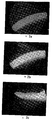
도 2a는 상피세포를 통한 방식으로 제4 시험 조성물이 적용된지 15분 후에 각막 부분의 형광 사진을 도시한다.
도 2b는 상피세포를 통한 방식으로 제4 시험 조성물이 적용된지 30분 후에 각막 부분의 형광 사진을 도시한다.
도 2c는 상기 제4 조성물의 통과로 인한 강력한 형광 및 상기 처리 후의 조직의 상대적 강직성이 관찰될 수 있는 제4 신규 용액을 사용한 상피세포를 통한 교차결합에 의해 처리된 각막 부분의 형광 영상을 도시한다.
도 3a는 표준 용액을 사용하여 상피세포를 통한 교차결합에 의해 처리된 각막의 굴절 정도를 도시한다.
도 3b는 신규의 제4 시험 용액을 사용하여 상피세포를 통한 교차결합에 의해 처리된 각막의 굴절 정도를 도시한다.
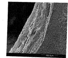
도 4는 원추각막에 걸린 각막 부분에서 라멜라(lamellae)를 도시하는 주사 현미경도이다.
도 5는 도 4의 각막의 확대도를 도시하는 주사 현미경도이다.
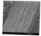
도 6은 제4의 신규한 시험 조성물을 사용하여 상피세포를 통한 교차결합이 실시된 후 원추각막에 걸린 각막 부분에서 라멜라를 도시하는 주사 현미경도이다.
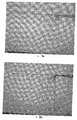
도 7은 정상 각막에서 미세융모(microvilli) 및 상피세포의 표피층의 형태를 도시하는 주사 현미경도이다.
도 8은 리보플라빈-덱스트란 0.1%의 표준 용액이 상피세포를 통한 방식으로 적용된 후 표준 선량의 UV-A 선으로 처리된 각막의 주사 현미경도이다.
도 9는 제4의 신규 시험 조성물이 상피세포를 통한 방식으로 적용된 후 표준 선량의 UV-A로 처리된 각막의 주사 현미경도이다.
도 10은 상피세포를 통한 방식으로 생리학적 용액이 적용된 후 표준 선량의 UV-A로 처리된 각막의 주사 현미경도이다. 1 shows visual assessment, fluorescence measurement, colorimetric evaluation employed to assess the passage of 0.1% of the riboflavin solution through the cornea after application in a manner through epithelial cells.
FIG. 2A shows a fluorescence picture of the corneal portion 15 minutes after the fourth test composition was applied in a manner through epithelial cells.
2B shows a fluorescence picture of the corneal portion 30 minutes after the fourth test composition was applied in a manner through epithelial cells.
FIG. 2C shows a fluorescence image of the corneal portion treated by crosslinking through epithelial cells using a fourth novel solution where strong fluorescence due to passage of the fourth composition and the relative stiffness of the tissue after the treatment can be observed. .
3A shows the degree of refraction of the cornea treated by crosslinking through epithelial cells using standard solutions.
3B shows the degree of refraction of the cornea treated by crosslinking through epithelial cells using a new fourth test solution.
FIG. 4 is a scanning micrograph showing lamellae in the corneal portion of the keratoconus.
FIG. 5 is a scanning microscope diagram showing an enlarged view of the cornea of FIG. 4. FIG.
FIG. 6 is a scanning microscopy showing lamellae in the corneal portion of the keratoconus after crosslinking through epithelial cells using a fourth novel test composition.
FIG. 7 is a scanning micrograph showing the morphology of the epidermal layer of microvilli and epithelial cells in normal cornea.
FIG. 8 is a scanning micrograph of the cornea treated with standard doses of UV-A radiation after 0.1% standard solution of riboflavin-dextran was applied in a manner via epithelial cells.
9 is a scanning micrograph of a cornea treated with a standard dose of UV-A after a fourth novel test composition was applied in a manner through epithelial cells.
10 is a scanning micrograph of the cornea treated with standard doses of UV-A after the physiological solution was applied in a manner through epithelial cells.
예시적 실시형태의 설명 Description of Exemplary Embodiments
모든 시험은 이식 실험에서 고려되는 합의 후 및 윤리 위원회의 허가에 따라서 Azienda Ospedaliera Napoli 1 - Banca Occhi ("Eye Bank") - Regione Campania - Ospedale dei Pellegrini 출신의 기증자의 인간 각막에서 실시되었다 - 도켓 번호 0009304/2009 - 결정번호 1269. All tests were conducted on human corneas of donors from Azienda Ospedaliera Napoli 1-Banca Occhi ("Eye Bank")-Regione Campania-Ospedale dei Pellegrini, after consensus considered in the transplantation experiment and with the permission of the Ethics Committee-Dock No. 0009304 / 2009-decision 1269.
상피세포의 예비적 제거 없는 완전한 인간 각막을 통한 시험 조성물의 침투를 관찰하였으며, 상기 각막의 두께는 500 내지 600 미크론이이고, 상기 조성물은 리보플라빈-덱스트란의 표준 용액, [16] 및 [17]에 제시된 리보플라빈-덱스트란 + 벤잘코늄 클로라이드로 제조된 조성물, 및 리보플라빈을 비타민 E, 코엔자임 Q, L-프롤린, 글리신, 리신 히드로클로라이드, L-로이신을 포함하는 군으로부터 선택된 적어도 1개 물질을 특정 농도로 혼합하여 얻은 신규 시험 조성물이다. Penetration of the test composition through the complete human cornea without preliminary removal of epithelial cells was observed, the cornea thickness being between 500 and 600 microns, the composition being a standard solution of riboflavin-dextran, [16] and [17]. A composition made from riboflavin-dextran + benzalkonium chloride as shown in the following, and at least one substance selected from the group comprising riboflavin, including vitamin E, coenzyme Q, L-proline, glycine, lysine hydrochloride, L-leucine It is a new test composition obtained by mixing.
신규 시험 조성물을 실현하기 위해 사용된 물질의 농도는 하기 범위로 포함된다: The concentration of material used to realize the new test composition is included in the following ranges:
비타민 E: 0.0001 mg % ml 내지 2000 mg % ml의 농도. 더욱 바람직한 실시형태에 따르면, 농도는 0.01 mg % 내지 1500 mg % ml의 범위이다. 더욱 바람직한 실시형태에 따르면, 농도는 10 mg % ml 내지 1000 mg % ml이다. 더욱 바람직한 실시형태에 따르면, 농도는 약 500 mg % ml이다; Vitamin E : concentration of 0.0001 mg% ml to 2000 mg% ml. According to a more preferred embodiment, the concentration ranges from 0.01 mg% to 1500 mg% ml. According to a more preferred embodiment, the concentration is from 10 mg% ml to 1000 mg% ml. According to a more preferred embodiment, the concentration is about 500 mg% ml;
비타민 Q: 0.0001 mg % ml 내지 2000 mg % ml의 농도. 더욱 바람직한 실시형태에 따르면, 농도는 0.01 mg % 내지 1500 mg % ml 범위이다. 더욱 바람직한 실시형태에 따르면, 농도는 1 mg % ml 내지 1000 mg % ml 범위이다. 더욱 바람직한 실시형태에 따르면, 농도는 약 100 mg % ml이다; Vitamin Q : concentration of 0.0001 mg% ml to 2000 mg% ml. According to a more preferred embodiment, the concentration ranges from 0.01 mg% to 1500 mg% ml. According to a more preferred embodiment, the concentration ranges from 1 mg% ml to 1000 mg% ml. According to a more preferred embodiment, the concentration is about 100 mg% ml;
L-프롤린: 0.0001 mg % ml 내지 2000 mg % ml의 농도. 더욱 바람직한 실시형태에 따르면, 농도는 0.001 mg % 내지 100 mg % ml 범위이다. 더욱 바람직한 실시형태에 따르면, 농도는 0.005 mg % ml 내지 10 mg % ml 범위이다. 더욱 바람직한 실시형태에 따르면, 농도는 0.01 mg % ml 내지 1 mg % ml 범위이다. 더욱 바람직한 실시형태에 따르면, 농도는 약 0.1 mg % ml이다; L-proline : concentration from 0.0001 mg% ml to 2000 mg% ml. According to a more preferred embodiment, the concentration ranges from 0.001 mg% to 100 mg% ml. According to a more preferred embodiment, the concentration ranges from 0.005 mg% ml to 10 mg% ml. According to a more preferred embodiment, the concentration ranges from 0.01 mg% ml to 1 mg% ml. According to a more preferred embodiment, the concentration is about 0.1 mg% ml;
글리신: 0.0001 mg % ml 내지 2000 mg % ml의 농도. 더욱 바람직한 실시형태에 따르면, 농도는 0.001 mg % 내지 100 mg % ml 범위이다. 더욱 바람직한 실시형태에 따르면, 농도는 0.005 mg % ml 내지 10 mg % ml 범위이다. 더욱 바람직한 실시형태에 따르면, 농도는 0.01 mg % ml 내지 1 mg % ml 범위이다. 더욱 바람직한 실시형태에 따르면, 농도는 약 0.1 mg % ml이다; Glycine : concentration from 0.0001 mg% ml to 2000 mg% ml. According to a more preferred embodiment, the concentration ranges from 0.001 mg% to 100 mg% ml. According to a more preferred embodiment, the concentration ranges from 0.005 mg% ml to 10 mg% ml. According to a more preferred embodiment, the concentration ranges from 0.01 mg% ml to 1 mg% ml. According to a more preferred embodiment, the concentration is about 0.1 mg% ml;
리신 히드로클로라이드: 0.0001 mg % ml 내지 2000 mg % ml의 농도. 더욱 바람직한 실시형태에 따르면, 농도는 0.001 mg % 내지 100 mg % ml 범위이다. 더욱 바람직한 실시형태에 따르면, 농도는 0.005 mg % ml 내지 10 mg % ml 범위이다. 더욱 바람직한 실시형태에 따르면, 농도는 0.01 mg % ml 내지 1 mg % ml 범위이다. 더욱 바람직한 실시형태에 따르면, 농도는 약 0.05 mg % ml이다; Lysine hydrochloride : concentration from 0.0001 mg% ml to 2000 mg% ml. According to a more preferred embodiment, the concentration ranges from 0.001 mg% to 100 mg% ml. According to a more preferred embodiment, the concentration ranges from 0.005 mg% ml to 10 mg% ml. According to a more preferred embodiment, the concentration ranges from 0.01 mg% ml to 1 mg% ml. According to a more preferred embodiment, the concentration is about 0.05 mg% ml;
L-로이신: 0.0001 mg % ml 내지 2000 mg % ml의 농도. 더욱 바람직한 실시형태에 따르면, 농도는 0.001 mg % 내지 100 mg % ml 범위이다. 더욱 바람직한 실시형태에 따르면, 농도는 0.005 mg % ml 내지 10 mg % ml 범위이다. 더욱 바람직한 실시형태에 따르면, 농도는 0.01 mg % ml 내지 1 mg % ml 범위이다. 더욱 바람직한 실시형태에 따르면, 농도는 약 0.08 mg % ml이다. L-Leucine : concentration from 0.0001 mg% ml to 2000 mg% ml. According to a more preferred embodiment, the concentration ranges from 0.001 mg% to 100 mg% ml. According to a more preferred embodiment, the concentration ranges from 0.005 mg% ml to 10 mg% ml. According to a more preferred embodiment, the concentration ranges from 0.01 mg% ml to 1 mg% ml. According to a more preferred embodiment, the concentration is about 0.08 mg% ml.
상피세포를 통한 교차결합에 의해 원추각막의 치료하기에 또는 UV-A 선으로부터 안구를 보호하기에 적합한 신규 용액은, 지시된 범위로 포함된 농도의 상술한 물질 중의 하나 또는 복수 개를 혼합하여 리보플라빈을 함유하는 용액, 예를 들어 0.0001% 내지 0.5% 범위에서 선택된 농도의 리보플라빈-덱스트란 용액이다. 더욱 바람직한 실시형태에 따르면, 리보플라빈-덱스트란 농도는 0.001% 내지 0.4% 범위이다. 더욱 바람직한 실시형태에 따르면, 상기 농도는 0.005% 내지 0.3% 범위이다. 더욱 바람직한 실시형태에 따르면, 상기 농도는 약 0.01% 내지 0.2% 범위이다. 더욱 바람직한 실시형태에 따르면, 상기 농도는 약 0.1% 이다. A novel solution suitable for the treatment of the keratoconus by cross-linking through epithelial cells or for protecting the eye from the UV-A line, is prepared by mixing one or a plurality of the above-mentioned substances in the concentrations included in the indicated ranges with riboflavin. Solution, for example a riboflavin-dextran solution at a concentration selected from 0.0001% to 0.5%. According to a more preferred embodiment, the riboflavin-dextran concentration is in the range of 0.001% to 0.4%. According to a more preferred embodiment, said concentration is in the range of 0.005% to 0.3%. According to a more preferred embodiment, the concentration ranges from about 0.01% to 0.2%. According to a more preferred embodiment, the concentration is about 0.1%.
상기 시험의 결과는 상기 확인된 군의 물질 각각이 리보플라빈 및 특히 표준 리보플라빈-덱스트란 용액의 각막 상피세포를 통한 바람직한 침투에 적합하다는 것과 UV-A 선으로부터 각막을 보호하는데 적합하다는 것을 나타내었다. The results of the test indicated that each of the identified groups of substances is suitable for the desired penetration through corneal epithelial cells of riboflavin and in particular standard riboflavin-dextran solution and is suitable for protecting the cornea from UV-A rays.
이식에 사용할 수 없는 이유로 안구 은행으로부터 폐기된 시험에 고려되는 각막은 적절한 용액에 유지되었고 또 시험 전에 이들은 광학 현미경 및 내피 세포의 조사에 의해 다시 평가되었다. The corneas contemplated for testing discarded from the eye bank for reasons not available for transplantation were maintained in the appropriate solution and prior to testing they were again assessed by light microscopy and irradiation of endothelial cells.
[10-15]에 제시된 바에 따르면, 500 내지 600 미크론 두께를 갖고 양호한 내피세포 모자이크를 갖는 양호한 투명성을 갖는 각막 만이 사용되어 왔다. As shown in [10-15], only corneas with good transparency with 500-600 micron thickness and good endothelial mosaic have been used.
히알우론산 나트륨 + 크산탄 검 0.4 ml의 소정 용액을 함유하는 실린더 챔버를 닫도록 각막이 자리잡고 있다. 실린더 박스와 동일한 직경을 갖는 방수 밀봉 금속 고리는 상기 각막 표면 상에 적용되었다. 이어 형광 물질(리보플라빈)을 함유하는 시험할 조성물은 각막 위에 적용하였다. 다양한 예에서 박스 내의 용액의 형광을 측정하는 것에 의해, 상기 신규 용액이 각막에 침투하는 양 및 그를 위해 얼마나 오래 상기 용액을 취하는지를 결정할 수 있다. The cornea is positioned to close the cylinder chamber containing 0.4 ml of a solution of sodium hyaluronate + xanthan gum. A waterproof sealed metal ring having the same diameter as the cylinder box was applied on the corneal surface. The composition to be tested containing the fluorescent material (riboflavin) was then applied onto the cornea. In various examples, by measuring the fluorescence of the solution in the box, it is possible to determine the amount of penetration of the new solution into the cornea and how long the solution takes for it.
상피세포를 통한 방식으로 리보플라빈을 투여하기 위한 조성물 및 리보플라빈을 상기 나타낸 물질 중의 적어도 하나와 혼합하는 것에 의해 얻은 조성물에서 담체로서 나타낸 물질의 효능은, 예시적 목적이지만 비제한적 목적으로만 이용되는, 이하의 시험예를 통하여 기재된다. The efficacy of a composition for administering riboflavin in a manner via epithelial cells and of a substance indicated as a carrier in a composition obtained by mixing riboflavin with at least one of the substances indicated above is used for illustrative purposes only, but not for non-limiting purposes. It is described through the test example of.
간단히 나타내기 위하여, 각막을 이하의 조성물로 처리하는 것에 의해 얻은 시험 결과 만을 보고하였다: For simplicity, only the test results obtained by treating the cornea with the following compositions are reported:
1) 리보플라빈-덱스트란 0.1%의 표준 용액; 1) a standard solution of 0.1% riboflavin-dextran;
2) 리보플라빈-덱스트란 0.1%의 표준 용액 + [16]에 따른 벤잘코늄 클로라이드 0.01%; 2) a standard solution of riboflavin-dextran 0.1% + 0.01% benzalkonium chloride according to [16];
3) 리보플라빈-덱스트란 0.1% + 500 mg % ml 농도의 비타민 E TPGS (D-알파-토코페릴 폴리에틸렌 글리콜 1000 숙시네이트)의 제1 신규 시험 조성물; 3) a first novel test composition of vitamin E TPGS (D-alpha-tocopheryl polyethylene glycol 1000 succinate) at a concentration of 0.1% + 500 mg% ml of riboflavin-dextran;
4) 리보플라빈-덱스트란 0.1% + 비타민 Q 100 mg % ml의 제2 신규 시험 조성물; 4) a second novel test composition of Riboflavin-dextran 0.1% + 100 mg% ml of vitamin Q;
5) 리보플라빈-덱스트란 0.1% + L-프롤린 0.1 mg % + 글리신 0.1 mg %, 리신 히드로클로라이드 0.05 mg % + L-로이신 0.08 mg %의 제3 신규 시험 조성물; 5) a third new test composition of 0.1% riboflavin-dextran + 0.1 mg% L-proline + 0.1 mg% glycine, 0.05 mg% lysine hydrochloride + 0.08 mg% L-leucine;
6) 리보플라빈-덱스트란 0.1 % + 비타민 E (D-알파-토코페릴 폴리에틸렌 글리콜 1000 숙시네이트) 500 mg % ml + 비타민 Q 100 mg % ml + L-프롤린 0.1 mg % + 글리신 0.1 mg % + 리신 히드로클로라이드 0.05 mg % + L-로이신 0.08 mg %의 제4 신규 시험 조성물. 6) Riboflavin-dextran 0.1% + Vitamin E (D-alpha-tocopheryl polyethylene glycol 1000 succinate) 500 mg% ml +
상기 언급된 6개의 조성물 각각은 선택된 각막의 표면 상에 상기 기재한 바와 같이 도포되어 위치하며 또 각막 스트로마의 침지는 처리된 각막 아래의 용기 내부에 위치하는 히알우론산 나트륨 + 크산탄 검 0.4 ml의 용액 중에 형광 물질의 존재와 함께 15분 후 및 30분 후에 평가하였다. 각막 스트로마에서 리보플라빈 침투의 평가는 각막을 구획을 나누고 또 형광 현미경으로 연속 평가하는 것에 의해 실시되었다. Each of the six compositions mentioned above is applied and positioned as described above on the surface of the selected cornea and the immersion of the corneal strom in a solution of 0.4 ml of sodium hyaluronate + xanthan gum located inside the container below the treated cornea. After 15 minutes and after 30 minutes with the presence of fluorescent material. Evaluation of riboflavin infiltration in corneal stroms was performed by partitioning the cornea and subsequent evaluation with fluorescence microscopy.
각막을 통한 통과를 나타내는 히알우론산 나트륨 + 크산탄 검 0.4 ml의 용액 내의 리보플라빈의 존재는, 도 1에 도시된 바와 같이 육안 측정 및 형광측정을 이용하여 정성적으로 뿐만아니라 비색측정을 이용하여 정량적으로 평가하였다. 각 컬러 샘플 근처의 2개 숫자는 각각 도시된 컬러를 유발하는 리보플라빈의 표준 용액 부분의 수 및 크산탄 검 및 히알우론산 나트륨의 용액 부분의 수를 각각 나타낸다. 참조 측정은 리보플라빈-덱스트란 0.1%를 다음 비율(단위/ml) 50/0, 40/10, 30/20, 20/30, 10/40, 0/50의 크산탄 검 + 히알우론산 나트륨으로 희석한 희석물을 제조하는 것에 의해 정의되었다. 단위/ml의 정의된 값에 상응하는 육안 측정 및 형광 측정을 준비하고 또 각 희석율에 대하여 10에서 0까지의 점수를 할당하였다. 비색 측정은 희석제로서 선택된 물질의 비색 스펙트럼에 상응하는 리보플라빈의 부재시 20%에 상응하는 황색 %의 최소 값을 고려한다. The presence of riboflavin in 0.4 ml of sodium hyaluronate + xanthan gum showing passage through the cornea is quantitatively assessed not only qualitatively using visual and fluorescence but also colorimetric measurements as shown in FIG. It was. The two numbers near each color sample represent the number of standard solution portions of riboflavin and the number of solution portions of xanthan gum and sodium hyaluronate, respectively, which lead to the color shown. Reference measurement was performed by diluting 0.1% of riboflavin-dextran with 50/0, 40/10, 30/20, 20/30, 10/40, 0/50 xanthan gum + sodium hyaluronate in the following ratios (units / ml): Defined by preparing dilutions. Visual and fluorescence measurements corresponding to defined values in units / ml were prepared and assigned a score from 10 to 0 for each dilution rate. Colorimetric measurements take into account the minimum value of yellow% corresponding to 20% in the absence of riboflavin corresponding to the colorimetric spectrum of the material selected as diluent.
육안 측정에 의한 평가는 예비정의된 샘플을 사용한 시험에 의해 얻은 샘플의 직접적 대조에 의해 또 디지털 사진 수법에 의해 표준 조명 조건하에서 실시되었다. 형광측정 평가는 디지털 포토카메라를 구비한 형광주사 현미경을 사용하여 암소에서 실시되었다. 육안 측정 및 형광 측정에 의한 평가에 대한 점수는 제3 심사관에 의해, 2개 방법으로 얻은 값의 평균을 내는 것에 의해 실시하였다. Evaluation by visual measurement was carried out under standard lighting conditions by direct control of the samples obtained by tests with pre-defined samples and by digital photography. Fluorometric evaluation was performed in the dark using a fluorescence scanning microscope equipped with a digital photo camera. The score for the evaluation by visual measurement and fluorescence measurement was performed by averaging the values obtained by the two methods by the third examiner.
비색측정 평가는 실린더 챔버 내부(및 따라서 각막 아래)에서 실험의 끝에 존재하는 물질을 투명 백에 삽입하고 또 컴퓨터 분석을 이용하여, 미리-정의된 희석물을 고화질로 스캐닝하고 또 소프트웨어 프로그램 Photoshop™ 7.0 및 모노크롬 필터를 사용하여 황색 %를 평가하는 것에 의해 실시되었다. 상기 수법을 이용함으로써 도 1에 도시된 바와 같이 실험 샘플에서 검출된 황색 %를 표준 리보플라빈 0.1% 용액의 단위/ml로 표시된 정확하게 측정된 농도 값과 비교할 수 있었다. The colorimetric evaluation involves inserting the material present at the end of the experiment into the transparent bag inside the cylinder chamber (and thus below the cornea) and using computer analysis to scan the pre-defined dilution in high quality and the software program Photoshop ™ 7.0 And evaluating yellow% using a monochrome filter. Using this technique, the yellow percentage detected in the experimental sample, as shown in FIG. 1, can be compared with the accurately measured concentration value expressed in units / ml of standard riboflavin 0.1% solution.
도 2a는 제4 신규 용액이 적용된지 15분 후 상피세포를 통한 교차결합에 의해 처리된 각막 부분의 형광 영상이고, 도 2b는 제4 신규 용액이 적용된지 30분 후 상피세포를 통한 교차결합에 의해 처리된 각막 부분의 형광 영상이며, 도 2c는 제4 신규 용액에 의해 상피세포를 통한 교차결합에 의해 처리된 각막 부분의 형광 영상이다. 마지막 도면에서 리보플라빈이 전체 각막에 침투하였다는 것과 조직이 교차결합 치료 후 더 강인해졌다는 것을 알 수 있다. FIG. 2A is a fluorescence image of the corneal portion treated by crosslinking through epithelial cells 15 minutes after the fourth new solution was applied, and FIG. 2B is a crosslinking through epithelial cells 30 minutes after the fourth new solution was applied. Fluorescence image of the corneal portion processed by FIG. 2C is a fluorescence image of the corneal portion treated by crosslinking through epithelial cells by a fourth novel solution. In the last figure it can be seen that the riboflavin penetrated the entire cornea and the tissue became stronger after crosslinking treatment.
실시된 시험은 다음을 나타내었다: The tests conducted showed the following:
a) 리보플라빈-덱스트란 0.1%의 표준 용액을 상피세포를 통한 방식으로 적용한 지 15분 후, 각막 스트로마는 부분적으로 침지되고 또 형광 용액은 용기 내의 물질 중에서 검출되지 않으며, 비색측정 스펙트럼은 히알우론산 나트륨 + 크산탄 검 0.4 ml의 용액과 포개질 수 있었다(도 1에서와 같은 점수 0 및 20% 이하의 황색 %); a) 15 minutes after application of 0.1% standard solution of riboflavin-dextran in an epithelial manner, the corneal strom is partially submerged and the fluorescent solution is not detected in the material in the container and the colorimetric spectrum is sodium hyaluronate + It could be superimposed with 0.4 ml of xanthan gum solution (
b) 리보플라빈-덱스트란 0.1%의 표준 용액을 상피세포를 통한 방식으로 적용한 지 30분 후, 각막 스트로마는 형광 용액으로 완전히 침지되며; 용기 내의 히알우론산 나트륨 + 크산탄 검 0.4 ml의 용액 중의 형광은 검출될 수 있고, 도 1의 점수는 2-3이고 또 황색 %는 상술한 컴퓨터를 이용한 수법에 의해 75%-80% 범위로 결정되었다; b) 30 minutes after application of 0.1% standard solution of riboflavin-dextran in an epithelial manner, the corneal strom is completely immersed in fluorescent solution; Fluorescence in a solution of 0.4 ml of sodium hyaluronate + xanthan gum in the container can be detected, the score of FIG. 1 is 2-3 and the yellow% was determined in the range of 75% -80% by the computer-aided method described above. ;
c) 상피세포를 통한 방식으로 500 mg % ml 농도의 리보플라빈-덱스트란 0.1% + 비타민 E TPGS (D-알파-토코페릴 폴리에틸렌글리콜 1000 숙시네이트)의 제1 신규 시험 용액을 적용한지 15분 후, 각막은 완전히 침지되며 또 형광 용액은 용기 내에 존재한다(도 1의 점수 2-3, 황색 %는 72-76%); c) 15 minutes after application of the first new test solution of 500%% ml of riboflavin-dextran at 0.1% + vitamin E TPGS (D-alpha-tocopheryl polyethyleneglycol 1000 succinate) in a manner via epithelial cells, The cornea is fully immersed and the fluorescent solution is present in the vessel (score 2-3 in FIG. 1, 72-76% yellow%);
d) 상기 제1 신규 시험 용액을 적용한 지 30분 후, 각막의 모든 층은 완전히 침지되며 또 용기 내에는 고농도의 리보플라빈이 있으며, 이는 상피세포 표면과의 접촉으로 생성물 자체에 대한 각막 조직의 양호한 투과성을 나타낸다(도 1의 점수 3-4, 79-84%의 황색 %); d) 30 minutes after the application of the first new test solution, all layers of the cornea are completely immersed and there is a high concentration of riboflavin in the container, which is in good contact with the surface of the epithelial cell and the good permeability of the corneal tissue to the product itself. (Yellow 3% of score 1-4, 79-84%) in FIG. 1;
e) [16]에서 제안된 조성물 및 각각 벤잘코늄 클로라이드 0.01%; 비타민 Q 100 mg % ml; L-프롤린 0.1 mg %, 글리신 0.1 mg %, 리신 히드로클로라이드 0.05 mg % 및 L-로이신 0.08 mg %를 함유하는 제2 및 제3 신규 시험 용액은, 동일한 조건하에서, 표준 용액(리보플라빈-덱스트란 0.1%) 단독을 사용하여 얻어진 상응하는 결과에 비하여 양적으로나 침투 속도 면에서 더 우수한 리보플라빈 침투를 나타내었다(15분 후 도 1의 점수 3-4 및 30분 후 점수 4-6, 15분 후 70-79%의 황색 % 및 30분 후 78-86%의 황색 %); e) the composition proposed in [16] and 0.01% benzalkonium chloride, respectively; 100 mg% ml of vitamin Q; The second and third novel test solutions containing 0.1 mg% L-proline, 0.1 mg% glycine, 0.05 mg% lysine hydrochloride and 0.08 mg% L-leucine, under the same conditions, were subjected to standard solutions (riboflavin-dextran 0.1 %) Showed better riboflavin infiltration in terms of penetration rate, both quantitatively and in comparison to the corresponding results obtained using alone (Scores 3-4 in FIG. 1 after 15 minutes and scores 4-6 after 30 minutes, 70- after 15 minutes). 79% yellow% and 78-86% yellow% after 30 minutes);
f) 제4 신규 시험 조성물은 시험된 다른 모든 조성물에 비하여 훨씬 더 우수한 결과를 나타내었다. 상피세포 표면에 상기 제품을 적용한지 15분 후 및 30분 후에 용기 내의 형광 물질을 형광측정 방식 및 컴퓨터를 이용한 분석으로 검출된 착색제의 농도는 현저히 향상되었다(15분 후 도 1의 점수 5-6, 88-91%의 황색 %; 30분 후 점수 6-7, 90% 초과의 황색 %); 제4 신규 시험 용액을 상피세포를 통하여 적용한 후 얻어진 각막 아래에 위치한 용액 내의 형광 물질의 더 높은 농도는, 특히 동일 시간 간격 후 용기 내에 존재하는 용액 중에서도 검출될 수 없는 표준 용액을 사용하여 얻은 결과와 비교하면, 특히 15분 후에 현저하다. 이것은 함께 혼합될 때 각막 상피세포를 통한 리보플라빈의 통과를 선호하는 투과 향상제 간의 상승효과가 존재한다고 가정하면 설명될 것이다. f) The fourth new test composition showed much better results than all other compositions tested. After 15 and 30 minutes of application of the product to the surface of epithelial cells, the concentration of the colorant detected by fluorescence measurement and computer analysis of the fluorescent material in the container was significantly improved (Score 5-6 in FIG. 1 after 15 minutes). , 88-91% yellow%; score 6-7 after 30 minutes, yellow% more than 90%); Higher concentrations of fluorescent material in the solution located beneath the cornea obtained after application of the fourth novel test solution through epithelial cells were obtained using standard solutions that could not be detected among the solutions present in the container, especially after the same time interval. In comparison, especially after 15 minutes. This will be explained assuming that there is a synergy between permeation enhancers that favor the passage of riboflavin through corneal epithelial cells when mixed together.
예시된 결과는 적어도 하기 물질이 단독으로 또는 이들과 조합하거나, 아세트산과 같은 부형제와 상술한 범위에서 선택된 농도로 조합되면, 표준 리보플라빈-덱스트란 용액에 의해 필요한 시간 간격에 비하여 훨씬 짧은 시간 간격으로 또 연속적인 교차결합 치료에 충분한 양으로 각막 상피세포를 통한 리보플라빈 침투를 촉진시킴을 나타낸다: The results exemplified are at least in shorter time intervals than the time intervals required by standard riboflavin-dextran solutions, provided that at least the following substances, alone or in combination with them, or in combination with excipients such as acetic acid at a concentration selected from the above ranges Promotes riboflavin infiltration through corneal epithelial cells in an amount sufficient for continuous crosslinking treatment:
- 비타민 EVitamin E

R1 = CH3 또는 H R1 = CH 3 or H
R2 = CH3 또는 H R2 = CH 3 or H
R3 = CH3; 단순히 예를 들어, 비타민 E TPGS (D-알파-토코페릴 폴리에틸렌글리콜 1000 숙시네이트)가 예시될 수 있다; R 3 = CH 3 ; Simply for example, vitamin E TPGS (D-alpha-tocopheryl polyethyleneglycol 1000 succinate) can be exemplified;
- 코엔자임 QCoenzyme Q
산화된 형태의
반-퀴논(half-quinonic) 형태의Half-quinonic form

환원된 형태의 
코엔자임 Q의 이소프레노이드 단위의 수; 예를 들어, 코엔자임 Q10이 인용될 수 있다; Number of isoprenoid units of coenzyme Q; For example, coenzyme Q10 may be cited;
- L-프롤린L-proline

- 글리신-Glycine

- 리신- Lee Sin

또는 리신 히드로클로라이드; Or lysine hydrochloride;
- L-로이신-L-leucine

상술한 모든 화합물과 리보플라빈의 조합은 각막 조직을 통과하여 신속하게 각막 조직에 침투하는 제품의 농도 면에서 더 우수한 결과를 나타내는 점에서 예상치 못한 상승효과를 나타내었다. The combination of all of the above compounds with riboflavin showed an unexpected synergistic effect in that it shows better results in terms of the concentration of product that penetrates the corneal tissue quickly through the corneal tissue.
본 출원인들은 표준 수순에 따라서 제4 신규 시험 조성물을 적용한 다음 3 mW/cm2의 UV-A 조사를 함으로써 인간 각막에 대한 상피세포를 통한 교차결합 치료의 효능을 시험관내에서 시험하였다. 시험된 각막은 앞의 실험에서와 같이 각막을 적절한 베어링에 고정한 다음 리보플라빈-덱스트란 0.1%의 표준 용액 및 제4 신규 시험 조성물을 각 각막의 상피세포 표면 상에 30분간 적용하는 것에 의해(즉 상피세포를 통한 방식으로 투여) 제조하였다. 연속적으로, 표준 UV-A 조사를 30분간 실시하였고, 이때 상기 단계는 5분간의 단계로 세분하여, 각 용액을 각막 표면 위에 재투여하는 것을 선행하였다. 각 실험의 말기에, 각막의 강직성 정도는 다음과 같이 평가하였다: 각 각막은 수평으로 정렬되어 유지된 각막 겸자를 이용하여 2 mm 길이에 대해 말단 부분에 의해 유지되고 또 상기 수평선에 대하여 각막의 대향 단부에 의해 형성된 각도를 측정하였다. Applicants tested in vitro the efficacy of crosslinking treatment through epithelial cells on human cornea by applying a fourth novel test composition according to standard procedures followed by UV-A irradiation of 3 mW / cm 2 . The cornea tested was fixed by anchoring the cornea to the appropriate bearing as in the previous experiment and then applying a standard solution of riboflavin-dextran 0.1% and a fourth new test composition on the epithelial cell surface of each cornea (ie, epithelium). Administration in a manner via cells). Subsequently, standard UV-A irradiation was performed for 30 minutes, where the step was subdivided into 5 minute steps, followed by re-administration of each solution onto the corneal surface. At the end of each experiment, the degree of stiffness of the cornea was assessed as follows: each cornea was held by the distal end about 2 mm in length with the corneal forceps kept horizontally aligned and opposite of the cornea to the horizontal line. The angle formed by the end was measured.
도 3a는 리보플라빈-덱스트란 0.1%의 표준 용액을 사용하여 실시한 상피세포를 통한 교차결합 치료 후 각막을 도시하며, 약 40°정도 아래로 굽어지며 또 도 3b는 제4 신규 시험 조성물을 사용하고 또 각막 상피세포를 미리 제거하지 않고 실시한 교차결합 치료 후의 다른 각막을 도시한다. 이들 2개 도면을 비교하는 것에 의해, 제4 신규 시험 조성물을 사용하여 실시한 상피세포를 통한 교차결합 치료가 바람직한 바와 같이 오직 25°정도 아래로 굽어져, 각막을 강화시키는 것이 분명하다. FIG. 3a shows the cornea after crosslinking treatment through epithelial cells performed using a standard solution of riboflavin-dextran 0.1%, bent down by about 40 ° and FIG. 3b using a fourth novel test composition Another cornea after crosslinking treatment performed without prior removal of corneal epithelial cells is shown. By comparing these two figures, it is evident that cross-linking treatment via epithelial cells conducted using a fourth novel test composition bends only 25 ° below as desired, thereby strengthening the cornea.
제4 신규 시험 조성물을 사용하여 상피세포를 통한 교차결합 치료 효능의 다른 확인으로서, 원추각막에 걸린 환자들에 속하고, 구멍을 뚫어 각막이식 처리받은 인간 각막 단부 상에서 시험관내에서 이 처리를 실시하였다. 이들 경우에서 파괴되는 대신 환자로부터 외식된(explanted) 각막은 디아테르미(diathermy)에 관련한 수술 시간을 피하여, 각막 층의 평면을 보존하도록(전체 두께 이식) 주의하면서 실험실에서 사용되었다. 각막 윤부(corneal limbs)는 적절한 베어링 상에 고정하였다. 리보플라빈-덱스트란 0.1%의 표준 용액을 제1 각막 상에 적용하고, 또 제4 신규 시험 용액은 제2 각막 상에 적용하며, 상피세포를 제거하지 않고 각막 표면 상에 상기 용액들을 30분간 적용하였다. 이어, 표준 340 nm UV-A 처리는 3mW/cm2의 파워로 제4 신규 시험 용액을 사용하여 처리된 각막에 대해서만 30분간, 각각 5분간의 단계로 세분하여 각막의 표면 상에 조성물을 재투여를 한 다음 실시하는 방식으로 수행하였다. 적절한 지지체 상에 조겅된 원추각막에 걸린 다른 단부는 각막 상에 리보플라빈-덱스트란 0.1%의 표준 용액을 적용할 뿐 UV-A 후속 조사를 하지 않고 처리하였다. 실험의 말기에, 각막 연조직의 조사는 주사 전자 현미경을 이용하여 실시하였다. As another confirmation of the efficacy of crosslinking treatment through epithelial cells using a fourth novel test composition, this treatment was performed in vitro on human corneal ends that were perforated with corneal keratoplasty and perforated. . In these cases, instead of being destroyed, the cornea explanted from the patient was used in the laboratory, taking care to preserve the plane of the corneal layer (full thickness transplantation), avoiding the surgical time associated with the diathermy. Corneal limbs were fixed on appropriate bearings. A standard solution of 0.1% of riboflavin-dextran was applied on the first cornea, a fourth new test solution was applied on the second cornea, and the solutions were applied on the cornea surface for 30 minutes without removing epithelial cells. . The standard 340 nm UV-A treatment was then subdivided in 30 minutes, 5 minutes each for only the cornea treated with the fourth novel test solution at a power of 3 mW / cm 2 to re-administer the composition on the surface of the cornea. Then it was carried out in a manner to be carried out. The other end of the cone cornea formed on an appropriate support was treated with 0.1% standard solution of riboflavin-dextran on the cornea without UV-A subsequent irradiation. At the end of the experiment, corneal soft tissues were examined using a scanning electron microscope.
도 4는 치료되지 않은 원추각막 환자로부터 얻은 각막 부분의 라멜라가 주사 현미경에서 어떻게 나타나는지를 도시한다. 도 5는 도 4의 확대도이다. 이들 2개 도면은 UV-A 조사 없이 리보플라빈-덱스트란 0.1%의 표준 용액을 적용한 각막의 단부에서 각막 라멜라의 약화를 도시한다. 4 shows how lamellae of the corneal portion obtained from untreated keratoconus patients appear on a scanning microscope. 5 is an enlarged view of FIG. 4. These two figures show the weakening of the corneal lamellar at the end of the cornea with a standard solution of riboflavin-dextran 0.1% without UV-A irradiation.
도 6은 원추각막에 걸린 각막 부분의 라멜라가 신규 제4 시험 용액을 사용한 상피세포를 통한 교차결합 치료 후 어떻게 나타나는지를 도시하는 주사 현미경도이다. 표준 수순에 따라서 30분 동안 상피세포를 통한 방식으로 투여된 제4 신규 용액으로 처리되고 또 UV-A 선으로 조사된 원추각막의 단부는 고도로 조밀하게 분포되고 촘촘한 각막 라멜라를 제공하며, 이는 새로운 생화학적 교차결합이 형성되었음을 나타낸다. FIG. 6 is a scanning micrograph showing how lamellae in the corneal portion of the keratoconus appears after crosslinking treatment through epithelial cells using a new fourth test solution. The ends of the keratoconus treated with a fourth novel solution administered in a manner via epithelial cells for 30 minutes according to standard procedures and irradiated with UV-A rays provide a highly dense and dense corneal lamellae, which is a new biochemical Indicates that a crosslink was formed.
제1, 제2 및 제3 신규 시험 조성물을 사용하여도 양호한 결과를 얻었다. Good results were also obtained using the first, second and third novel test compositions.
신규 시험 조성물들을 사용하여 얻어진 결과는, 상피세포가 UV-A 선을 차폐할 것이기 때문에 상피세포를 통한 방식으로 실시될 때 교차결합 치료의 효능의 가상적 감소가 있을 것이라는 일부 저자들에 의해 제기된 반대를 극복한다. 상피세포를 통한 교차결합 치료의 효능의 감소 가설은, 여기서 제안된 물질의 적어도 하나를 담체(투과 향상제)로 하여 리보플라빈에 부가하고 표준 수순에 따라서 조사를 실시할 때 얻어진 조직학적 샘플에 의해 증명된 바와 같이, 잘못된 것이다. The results obtained using the new test compositions are against the claims raised by some authors that there will be a hypothetical decrease in the efficacy of crosslinking treatment when administered in a manner via epithelial cells because epithelial cells will shield the UV-A rays. To overcome. The hypothesis of reduced efficacy of crosslinking treatment through epithelial cells is demonstrated by histological samples obtained when adding at least one of the proposed substances to the riboflavin as a carrier (permeation enhancer) and performing the irradiation according to standard procedures. As is wrong.
또한, 각막의 상피세포 층의 형태학적 통합성 여부 및 상피세포의 표면 상에 존재하는 미세융모에 대하여 상피세포를 통한 방식으로 교차결합 치료 효능의 비교 연구는 주사 전자 현미경(7500x)을 이용하여 실시하였다. 이것은 [16]에서 제안된 바와 같이 리보플라빈-덱스트란 0.1%의 표준 용액 및 표준 조성물 + 벤잘코늄 클로라이드 0.01%에 대하여 각막 상피세포 상에서 상피세포를 통한 방식으로 제4 신규 시험 조성물을 사용하여 실시한 교차 결합 치료의 용인성을 평가하기 위해 실시되었다. In addition, a comparative study of the efficacy of crosslinking treatment in a manner via epithelial cells on the morphological integrity of the epithelial cell layer of the cornea and on the microvilli present on the surface of the epithelial cells was carried out using a scanning electron microscope (7500x). It was. This was done using a fourth novel test composition in a manner via epithelial cells on corneal epithelial cells for a standard solution of 0.1% of riboflavin-dextran and 0.01% of benzalkonium chloride as suggested in [16]. To evaluate the tolerability of treatment.
이 시험은 상피세포가 UV-A 플럭스에 의해 조사된 최초의 유기 구조이고 또 이들 방사선의 흡수로 인하여 손상을 입을 수 있었기 때문에 실시되었다. 입수가능한 어떠한 문서도 이러한 특징을 고려한 바 없으며, 관련 데이터도 문헌에서 입수할 수 없다. This test was conducted because the epithelial cells were the first organic structures irradiated by UV-A flux and could be damaged by the absorption of these radiations. No document available takes this feature into account, and relevant data are not available in the literature.
상피세포의 활력을 평가하기 위한 가장 신뢰성있는 것으로 간주되는 변수는 임프레션(impression) 수법에 의한 세포학적 조사, 특히 각막의 표피 상피세포 층의 미세융모의 전자 현미경에 의한 조사이다. 고농도의 뮤신 트랜스멤브레인 및 양호한 글리코칼릭스(glycocalyx)를 함유하는 완전한 표피 세포 요소의 막의 엑스트로플렉션(extroplexion)(미세융모)의 존재는 각막앞 (pre-corneal) 눈물 필름의 깊은 층을 구성하는 자유 뮤신을 적절한 방식으로 결합시킨다. 대조적으로, 미세융모의 병리적 손실은 안구 표면에 대한 안구 표면의 어려운 부착 및 각막앞 눈물 필름 자체의 기능상실에 의해 유도된 상피세포 현상 및 그에 따른 염증을 결정한다. The parameters considered to be the most reliable for assessing the vitality of epithelial cells are cytological investigations by the impression technique, in particular electron microscopic examination of the microvilli of the epidermal epithelial cell layer of the cornea. The presence of extraplexion (microvilli) of the membrane of the complete epidermal cell element containing high concentrations of mucin transmembrane and good glycocalyx constitutes the deep layer of the pre-corneal tear film. To bind free mucin in an appropriate manner. In contrast, pathological loss of microvilli determines epithelial cell phenomena induced by difficult attachment of the ocular surface to the ocular surface and malfunction of the pre-corneal tear film itself and thus inflammation.
미세융모의 형태는 시험관내에서 표준 선량에 따라 UV-A를 상피세포를 통하여 처리한 후 주사 전자 현미경에 의해 평가하였다. 500 내지 600 미크론 두께의 인간 각막은 BSS(balanced salt solution), 리보플라빈-덱스트란 0.1%의 표준 용액, 표준 용액 + 벤잘코늄 클로라이드 0.01% 및 제4 신규 시험 조성물과 각각 30분간 배양처리되었다. Morphology of microvilli was evaluated by scanning electron microscopy after treatment with UV-A through epithelial cells according to standard dose in vitro. Human corneas of 500-600 microns thick were incubated for 30 minutes each with a balanced salt solution (BSS), a standard solution of riboflavin-dextran 0.1%, standard solution + 0.01% benzalkonium chloride, and a fourth new test composition.
예비적으로, 어떠한 처리도 받지 않은 각막의 미세융모의 형태 및 각막 상피세포의 표피층의 형태도 상피세포와 이들의 미세융모가 광화학적 처리를 받지 않을 때 어떻게 나타나야 하는지에 대해 연구하였다. Preliminarily, we studied how the shape of the microvilli of the cornea and the epidermal layer of the corneal epithelial cells without any treatment should appear when the epithelial cells and their microvilli are not subjected to photochemical treatment.
도 7은 정상 각막에서 상피세포의 미세융모와 상피세포의 표피 층의 형태를 도시하는 주사현미경 사진을 도시하고, 또 도 8은 표준 수순에 따라 리보플라빈-덱스트란 0.1%의 표준 용액을 적용한 후 UV-A로 처리된 각막의 주사 현미경 사진을 도시한다. 도 7과 비교함으로써, 모든 상피세포 층이 손실되고 보우만(Bowman) 막이 벗겨진 것이 분명하다. FIG. 7 shows a scanning micrograph showing the morphology of epidermal cells of epithelial cells and epithelial cells in normal cornea, and FIG. 8 shows UV after applying a standard solution of riboflavin-dextran 0.1% according to standard procedures. A scanning micrograph of the cornea treated with -A is shown. By comparison with FIG. 7, it is clear that all epithelial cell layers are lost and the Bowman membrane is peeled off.
[16]에서 제시된 바와 같이 표준 용액 + 벤잘코늄 클로라이드 0.01%에 의해 구성된 조성물로 처리된 각막에서도 유사한 상황이 생긴다. A similar situation occurs in the cornea treated with the composition composed of standard solution + benzalkonium chloride 0.01% as shown in [16].
도 9는 제4 신규 시험 조성물에 의해 처리된 후 표준 선량의 UV-A로 처리된 각막의 주사 현미경 사진을 도시한다. 도 7 및 도 8과 대조함으로써, 상피세포 층, 세포 핵 및 갭-접합부의 보존을 확인할 수 있다. 또한, 잔존하는 미세융모가 형태적으로 완전하고 또 깊은 세포질 세포막은 위험한 상태는 아니지만 미세융모의 밀도의 현저한 감소가 존재한다. 제1, 제2 및 제3 신규 시험 조성물을 사용하여도 유사한 결과가 얻어졌다. 9 shows a scanning micrograph of the cornea treated with a standard dose of UV-A after treatment with a fourth novel test composition. By contrast with FIGS. 7 and 8, preservation of epithelial cell layers, cell nuclei and gap-junctions can be confirmed. In addition, there is a significant decrease in the density of the microvilli, although the remaining microvilli are morphologically complete and deep cytoplasmic membranes are not in danger. Similar results were obtained with the first, second and third novel test compositions.
도 10은 대조를 위하여 오직 생리학적 용액을 사용하여 표준 선량의 UV-A로 처리된 각막의 주사 현미경 사진을 도시한다. 상피세포 층은 파괴되고, 다수의 세포는 자신의 세포질 핵이 손실되고 또 거의 전부의 갭-접합부(gap-junctions) 및 미세융모가 손실된다. 얻어진 결과는 다음과 같이 요약할 수 있다: FIG. 10 shows scanning micrographs of corneas treated with standard doses of UV-A using only physiological solutions for control. The epithelial cell layer is destroyed, and many cells lose their cytoplasmic nuclei and almost all of the gap-junctions and microvilli. The results obtained can be summarized as follows:
1) 리보플라빈-덱스트란 0.1%의 표준 용액 또는 표준 용액 + 벤잘코늄 클로라이드 0.01%를 함유하는 조성물과 함께 표준 수순에 따라 예비적 배양하고 UV-A 처리된 각막은 모든 상피세포 층의 전체적 손실을 거치며 또 그 결과 보우만 막이 벗겨진다(도 8). 실제로, 벤잘코늄 클로라이드가 부가된 표준 용액 만을 사용하여, 상피세포는 UV-A 조사에 의해 파괴된다. 이것은 상피세포를 제거하지 않고 표준 용액 또는 조성물 리보플라빈-덱스트란 + 벤잘코늄 클로라이드를 사용하여 실시된 교차결합 치료가 환자로 하여금 상기 처리 후, 상피세포의 외과적 제거에 의해 유발되는 성가신 증상을 피하지 못할 것이라는 생각으로 유도한다; 1) Pre-cultured according to standard procedures with a composition containing 0.1% standard solution of riboflavin-dextran or 0.01% standard solution + benzalkonium chloride, and the UV-A treated cornea undergoes a total loss of all epithelial layer As a result, the Bowman's membrane is peeled off (Fig. 8). In practice, using only standard solutions with added benzalkonium chloride, epithelial cells are destroyed by UV-A irradiation. This does not remove the epithelial cells and crosslinking treatments performed using standard solution or composition riboflavin-dextran + benzalkonium chloride do not allow the patient to avoid the annoying symptoms caused by surgical removal of the epithelial cells after the treatment. Leads to the idea that it will not;
2) 리보플라빈-덱스트란 및 상술한 것 중에서 선택된 적어도 1개의 담체를 포함하는 조성물과 함께 배양한 후, 특히 제4 신규 시험 조성물과 배양한 후 표준 선량의 UV-A로 처리된 각막은 자신의 상피세포 층, 세포 핵 및 갭-접합부를 유지하였다. 미세융모 밀도의 현저한 감소가 눈에 띄었지만, 나머지 미세융모는 형태학적으로 완전하였다. 각막의 깊은 세포질 세포 막은 완전하게 나타난다; 2) The cornea treated with a standard dose of UV-A after incubation with a composition comprising riboflavin-dextran and at least one carrier selected from above, in particular after incubation with a fourth novel test composition, has its own epithelium. The cell layer, cell nucleus and gap-junctions were maintained. A significant decrease in microvilli density was noticeable, but the remaining microvilli were morphologically complete. Deep cytoplasmic cell membrane of cornea appears completely;
3) BSS(균형을 이룬 염 용액)과 함께 배양된 후 표준 선량의 UV-A로 처리된 각막은 상피세포 층의 파괴, 세포질 핵을 갖는 다수 세포의 손실 및 거의 대부분의 갭-접합부 및 미세융모의 손실을 나타내었다. 3) The cornea treated with standard dose of UV-A after incubation with BSS (Balanced Salt Solution) results in the destruction of epithelial layer, loss of multiple cells with cytoplasmic nucleus and almost all gap-junctions and microvilli Showed a loss of.
제1, 제2 및 제3 신규 시험 조성물을 사용하여 처리된 각막에 대하여 다수의 주사 현미경 영상을 찍었지만, 이들은 제4 신규 시험 조성물을 사용하여 얻은 것과 실질적으로 동일하게 보이기 때문에 여기서는 보고되지 않았다. A number of scanning microscopy images were taken of corneas treated using the first, second and third new test compositions, but were not reported here because they look substantially the same as those obtained using the fourth new test composition.
이들 결과는 적어도 비타민 E, 비타민 Q 또는 코엔자임 Q; L-프롤린, 글리신, 리신 히드로클로라이드 및 L-로이신과 같은 시험된 아미노산이 각막 상피세포를 보호하고 또 각막 상피세포를 통한 리보플라빈의 침투를 선호한다고 추론할 수 있게 한다. 본 명세서에 제안된 신규 용액 중에서, 제4 조성물은 각막 상피세포의 침투 시간 및 각막 상피세포의 보존 측면에서 모두 가장 우수한 성능을 나타내었다. These results include at least vitamin E, vitamin Q or coenzyme Q; It is possible to infer that tested amino acids such as L-proline, glycine, lysine hydrochloride and L-leucine protect corneal epithelial cells and prefer the penetration of riboflavin through corneal epithelial cells. Among the novel solutions proposed herein, the fourth composition exhibited the best performance both in terms of infiltration time of corneal epithelial cells and preservation of corneal epithelial cells.
제안된 물질이 이들 우수한 결과를 얻을 수 있게 하는 이유 및 제4 시험 조성물에 사용된 물질이 상승효과를 나타내는 이유에 대한 설명은 아직 완전히 분명하지 않다. 본 발명을 어떠한 이론에 제한하지 않고, 본 출원인들은 제4 신규 시험 조성물로 처리된 후 검출된 각막 상피세포에 대한 감소된 손상은 비타민 E, 예를 들어 비타민 E-TPGS의 세포수선(cytorepairing), 및/또는 용액 내에 적어도 필수 또는 조건부 필수 아미노산의 존재에 아마도 기인할 수 있을 것이라 제안한다. 아마도 비타민 E는 상피세포의 수선 작용에 관여된 효소인 글루타치온 옥시다제 및 퍼옥사이드 디스뮤타제에 대하여 작용할 것이고, 필수 또는 조건부 필수 아미노산은 세포수선 작용을 가질 것이며 또 교차 결합 효과를 아마도 선호할 것이다. 또한, 제4 신규 용액에 의한 조직의 더 많은 침지는 아마도 교차결합 효과의 증가뿐만 아니라 UV-A 선의 유해 효과로부터 각막 상피세포 층의 더 우수한 보존을 결정한다. The reasons why the proposed materials allow these good results to be obtained and why the materials used in the fourth test composition exhibit synergistic effects are not yet completely clear. Without limiting the present invention to any theory, Applicants note that reduced damage to corneal epithelial cells detected after treatment with a fourth novel test composition may result in cytorepairing of vitamin E, for example vitamin E-TPGS, And / or possibly due to the presence of at least essential or conditionally essential amino acids in solution. Perhaps vitamin E will act on glutathione oxidase and peroxide dismutase, enzymes involved in the repair of epithelial cells, and essential or conditionally essential amino acids will have cell repair and will probably prefer a crosslinking effect. In addition, more immersion of tissue with the fourth novel solution probably determines better preservation of the corneal epithelial layer from the harmful effects of UV-A rays as well as an increase in the crosslinking effect.
리보플라빈-덱스트란 및 본 명세서에 제시된 것들중에서 선택된 적어도 1개의 담체(침투 향상제)의 신규 용액을 사용하여, 예를 들어 각막 상피세포를 예방적으로 제거하지 않고도 원추각막의 치료를 위한 교차결합 치료를 실시할 수 있다. 각막 상피세포가 제거되지 않는 사실로 이하의 것을 피한다: Using a novel solution of riboflavin-dextran and at least one carrier (penetration enhancer) selected from those set forth herein, crosslinking therapy for the treatment of the keratoconus, for example, without the prophylactic removal of corneal epithelial cells, is carried out. It can be carried out. The fact that corneal epithelial cells are not removed prevents:
1) 표준 수순에 따라 교차결합 치료 후 제1일에 전형적으로 생기는 따끔거림에 기인한 질병; 1) a disease due to tingling typically occurring on day 1 after crosslinking treatment in accordance with standard procedures;
2) 처리한 후 치료적 콘택트 렌즈를 적용할 필요; 및 무엇보다도2) the need to apply therapeutic contact lenses after treatment; And above all
3) 각막 상피세포의 제거로 인하여 수술 후 각막 감염의 위험. 3) Risk of corneal infection after surgery due to removal of corneal epithelial cells.
또한, 각막에 대한 외과적 작용이 회피되므로, 수술실 및 수술용 현미경을 필요로 하지 않고 외래 환경(in an ambulatory)에서 치료를 실시할 수 있다. In addition, since surgical action on the cornea is avoided, treatment can be carried out in an an ambulatory without the need of an operating room and a surgical microscope.
상기 신규 용액은 점안액 또는 겔 형태로 투여될 수 있거나 또는 특히 여름 동안 태양 광선에 노출되기 전에 치료적 콘택트 렌즈 상에 적용되어 태양광에 의한 리보플라빈의 조사에 기인한 천연적 교차결합의 효과를 향상시킬 수 있다.The novel solutions may be administered in the form of eye drops or gels or may be applied on therapeutic contact lenses, especially before being exposed to sunlight during the summer, to enhance the effects of natural crosslinking due to irradiation of riboflavin by sunlight. Can be.
상기 신규 용액은 또한 안구의 내부 구조를 UV-A로부터 보호할 수 있으므로 예를 들어 태양광에 오랜 시간 동안 노출될 우려가 있는 사람에서 황막 황반으로부터 보호하거나 및/또는 백내장을 예방하기 위해 사용될 수 있다.The new solution can also protect the internal structure of the eye from UV-A and thus can be used to protect from the yellow macula and / or prevent cataracts, for example, in people who may be exposed to sunlight for long periods of time. .
상기 신규 용액을 비-단일 투여량 용기에 가능한한 오랫동안 보존하기 위하여, 0.0001% 내지 0.02% 농도, 바람직하게는 약 0.01%과 동일한 농도로 벤잘코늄 클로라이드를 부가할 수 있다. 상기 신규 용액이 단일-투여량의 일회용 용기에 생산되어 시판되어야 한다면, 벤잘코늄 클로라이드의 부가는 필수적이지 않다. 상기 조성물에는 보존제, 항균제, 항진균제, 부형제(예를 들어, 아세트산) 및 안정한 멸균 안과 용액을 제조하기 위해 및/또는 이들의 동화를 선호하기 위해 안과 분야에서 일반적으로 사용되는 물질이 더 부가될 수 있다. In order to preserve the new solution in a non-single dose container as long as possible, benzalkonium chloride may be added at a concentration equal to 0.0001% to 0.02%, preferably about 0.01%. If the new solution is to be produced and marketed in a single-dose disposable container, the addition of benzalkonium chloride is not necessary. The composition may further be added with preservatives, antibacterial agents, antifungal agents, excipients (e.g. acetic acid) and substances commonly used in the ophthalmology field to prepare stable sterile ophthalmic solutions and / or to favor assimilation thereof. .
전술한 바와 같이, 본 출원인들은 비타민 Q, L-프롤린, 글리신, 리신 히드로클로라이드, L-로이신 중에서 선택된 적어도 1개의 물질을 리보플라빈에 부가하는 것에 의해 얻은 조성물의 양호한 성능은, 마지막에 언급한 물질이 메탈로프로테이나제 MMP9의 증가를 통하여 세포수선 기능을 가질 필수 또는 조건부 필수 아미노산이라는 사실에 부분적으로 기인한다고 믿고 있다. 이는 리보플라빈 용액, 예를 들어 표준 리보플라빈-덱스트란 용액에, 메탈로프로테이나제 MMP9를 증가시키는 적어도 1개의 물질을 0.00001% 내지 0.5%의 농도로 부가하는 것에 의해 제1, 제2, 제3 및 제4 신규 시험 조성물에 대해 얻어진 결과와 유사한 결과를 얻을 수 있다는 추론으로 유도한다. As mentioned above, Applicants have found that the good performance of the composition obtained by adding at least one substance selected from vitamin Q, L-proline, glycine, lysine hydrochloride, L-leucine to riboflavin, It is believed that the increase in metalloproteinase MMP9 is due in part to the fact that it is an essential or conditionally essential amino acid that will have cell repair function. This is accomplished by adding at least one substance that increases the metalloproteinase MMP9 to a riboflavin solution, such as a standard riboflavin-dextran solution, at a concentration of 0.00001% to 0.5%. It is derived from the inference that results similar to those obtained for the fourth novel test composition can be obtained.
메탈로프로테이나제 MMP9를 증가시키기 위한 상기 물질 중에서, 제니슈타인(5,7-디히드록시-3-(4-히드록시페닐)크로멘-4-온), 피토에스트로겐, 사이토카인 및 소위 NSAID(비스테로이드성 항염증성 약물)을 언급할만 가치가 있다. Among the above substances for increasing metalloproteinase MMP9, genistein (5,7-dihydroxy-3- (4-hydroxyphenyl) chromen-4-one), phytoestrogens, cytokines and so-called NSAIDs It is worth mentioning (nonsteroidal anti-inflammatory drugs).
리보플라빈 안과 용액을 제조하기 위해 사용되도록 적응된 NSAID 중에서, 다음을 언급할 가치가 있다: 아세틸살리실산(2-아세톡시벤조산), 플루페나믹산(2-{[3-(트리플루오로메틸)페닐]아미노}벤조산), 메클로페나믹산(2-[(2,6-디클로로-3-메틸페닐)아미노]벤조산), 메페나믹산 (2-(2,3-디메틸페닐)아미노벤조산), 니플루믹산 (2-{[3-(트리플루오로메틸)페닐]아미노}니코틴산), 톨페나믹산 (2-[(3-클로로-2-메틸페닐)아미노]벤조산), 베노릴레이트 (4-(아세틸아미노)페닐 2-(아세틸옥시)벤조에이트), 카르프로펜((RS)-2-(6-클로로-9H-카바졸-2-일)프로판산), 셀레콕시브 (4-[5-(4-메틸페닐)-3-(트리플루오로메틸)피라졸-1-일]벤젠설폰아미드), 신녹시캄 (피록시캄 신나메이트 또는 [9-메틸-10,10-디옥소-8-(피리딘-2-일카바모일)-10$l^{6}-티아-9-아자비시클로[4.4.0]데카-l,3,5,7-테트라엔-7-일] (E)-3-페닐프로프-2-에노에이트;피록시캄 신나메이트), 디플루니살(2',4'-디플루오로-4-히드록시비페닐-3-카복시산), 디클로페낙 (2-[2-[(2,6-디클로로페닐)아미노]페닐]아세트산), 드록시캄(2H,5H-1,3-옥사지노(5,6-c)(1,2)벤조티아진-2,4(3H)-디온, 5-메틸-3-(2-피리디닐)-, 6,6-디옥사이드) 에토도락 ((RS)-2-(1,8-디에틸-4,9-디히드로-3H-피라노[3,4-b]인돌-1-일)아세트산), 에토리콕시브 (5-클로로-6'-메틸-3-[4-(메틸설포닐)페닐]-2,3'-비피리딘), 페노프로펜 (2-(3-페녹시페닐)프로판산), 풀루르비프로펜 ((RS)-2-(2-플루오로비페닐-4-일)프로판산), 이부페낙(4-이소부틸페닐 아세트산), 이부프로펜 ((RS)-2-(4-(2-메틸프로필)페닐)프로판산), 인도메타신 (2-{1-[(4-클로로페닐)카보닐]-5-메톡시-2-메틸-1H-인돌-3-일}아세트산), 케토프로펜 ((RS)-2-(3-벤조일페닐)프로판산), 케토로락 ((±)-5-벤조일-2,3-디히드로-1H-피롤리진-1-카복시산, 2-아미노-2-(히드록시메틸)-1,3-프로판디올), 로르녹시캄 ((3E)-6-클로로-3-[히드록시(피리딘-2-일아미노)메틸렌]-2-메틸-2,3-디히드로-4H-티에노[2,3-e] [1,2]티아진-4-온 1,1-디옥사이드), 루미라콕시브 ({2-[(2-클로로-6-플루오로페닐)아미노]-5-메틸페닐}아세트산), 멜옥시캄 (4-히드록시-2-메틸-N-(5-메틸-2-티아졸릴)-2H-1,2-벤조티아진-3-카복사미드-1,1-디옥사이드), 메타미졸 (소듐 [(2,3-디히드로-1,5-디메틸-3-옥소-2-페닐-1H-피라졸-4-일) 메틸아미노] 메탄설포네이트), 나프록센 ((+)-(S)-2-(6-메톡시나프탈렌-2-일)프로판산), 니메술리드 (N-(4-니트로-2-페녹시페닐)메탄설폰아미드), 옥사프로진 (3-(4,5-디페닐-1,3-옥사졸-2-일)프로판산)파레콕시브 (N-{[4-(5-메틸-3-페닐이소옥사졸-4-일)페닐]설포닐}프로판아미드), 피록시캄 ((8E)-8-[히드록시-(피리딘-2-일아미노)메틸리덴]-9-메틸- 10,10-디옥소-10λ6-티아-9-아자비시클로[4.4.0]데카-1,3,5-트리엔-7-온), 로페콕시브 (4-(4-메틸설포닐페닐)-3-페닐-5H-푸란-2-온), 술린닥 ({(1Z)-5-플루오로-2-메틸-1-[4-(메틸설피닐)벤질리덴]-1H-인덴-3-일}아세트산), 수독시캄 (4-히드록시-2-메틸-N(2)-티아졸릴-2H-1,2-벤조티아진-3-카복사미드 1,1-디옥사이드), 테녹시캄 ((3E)-3-[히드록시(피리딘-2-일아미노)메틸렌]-2-메틸-2,3-디히드로-4H-티에노[2,3-e] [1,2]티아진-4-온 1,1-디옥사이드), 발데콕시브 (4-(5-메틸-3-페닐이소옥사졸-4-일)벤젠설폰아미드). Of the NSAIDs adapted to be used to prepare riboflavin ophthalmic solutions, it is worth mentioning: acetylsalicylic acid (2-acetoxybenzoic acid), flufenamic acid (2-{[3- (trifluoromethyl) phenyl] Amino} benzoic acid), meclofenamic acid (2-[(2,6-dichloro-3-methylphenyl) amino] benzoic acid), mefenamic acid (2- (2,3-dimethylphenyl) aminobenzoic acid), niflumic acid (2-{[3- (trifluoromethyl) phenyl] amino} nicotinic acid), tolvenamic acid (2-[(3-chloro-2-methylphenyl) amino] benzoic acid), benolylate (4- (acetylamino ) Phenyl 2- (acetyloxy) benzoate), carpropene ((RS) -2- (6-chloro-9H-carbazol-2-yl) propanoic acid), celecoxib (4- [5- ( 4-methylphenyl) -3- (trifluoromethyl) pyrazol-1-yl] benzenesulfonamide), cynoxycam (pyoxycampin cinnamate or [9-methyl-10,10-dioxo-8- ( Pyridin-2-ylcarbamoyl) -10 $ l ^ {6} -thia-9-azabicyclo [4.4.0] deca-l, 3,5,7-tetraene-7- (E) -3-phenylprop-2-enoate; pyoxycampin cinnamate), diflunisal (2 ', 4'-difluoro-4-hydroxybiphenyl-3-carboxylic acid), Diclofenac (2- [2-[(2,6-dichlorophenyl) amino] phenyl] acetic acid), Doxycamp (2H, 5H-1,3-oxazino (5,6-c) (1,2) benzo Thiazine-2,4 (3H) -dione, 5-methyl-3- (2-pyridinyl)-, 6,6-dioxide) etodorac ((RS) -2- (1,8-diethyl-4 , 9-dihydro-3H-pyrano [3,4-b] indol-1-yl) acetic acid), etoricoxib (5-chloro-6'-methyl-3- [4- (methylsulfonyl) Phenyl] -2,3'-bipyridine), phenopropene (2- (3-phenoxyphenyl) propanoic acid), pullulbipropene ((RS) -2- (2-fluorobiphenyl-4 -Yl) propanoic acid), ibufenac (4-isobutylphenyl acetic acid), ibuprofen ((RS) -2- (4- (2-methylpropyl) phenyl) propanoic acid), indomethacin (2- {1 -[(4-chlorophenyl) carbonyl] -5-methoxy-2-methyl-1H-indol-3-yl} acetic acid), ketopropene ((RS) -2- (3-benzoylphenyl) propanoic acid ), Ketorolac ((±) -5-benzoyl-2,3- Hydro-1H-pyrrolidin-1-carboxylic acid, 2-amino-2- (hydroxymethyl) -1,3-propanediol), loroxycam ((3E) -6-chloro-3- [hydroxy Roxy (pyridin-2-ylamino) methylene] -2-methyl-2,3-dihydro-4H-thieno [2,3-e] [1,2] thiazin-4-one 1,1-dioxide ), Lumiracoxib ({2-[(2-chloro-6-fluorophenyl) amino] -5-methylphenyl} acetic acid), meloxycamp (4-hydroxy-2-methyl-N- (5-methyl -2-thiazolyl) -2H-1,2-benzothiazine-3-carboxamide-1,1-dioxide), metamizol (sodium [(2,3-dihydro-1,5-dimethyl-3) -Oxo-2-phenyl-1H-pyrazol-4-yl) methylamino] methanesulfonate), naproxen ((+)-(S) -2- (6-methoxynaphthalen-2-yl) propanoic acid) Nimesulide (N- (4-nitro-2-phenoxyphenyl) methanesulfonamide), oxaprozin (3- (4,5-diphenyl-1,3-oxazol-2-yl) propanoic acid ) Parecoxib (N-{[4- (5-methyl-3-phenylisoxazol-4-yl) phenyl] sulfonyl} propanamide), Pyroxycam ((8E) -8- [hydroxy- ( Pyridin-2-yl Mino) methylidene] -9-methyl-10,10- dioxo -10λ 6-thiazol-9-azabicyclo [4.4.0] deca-1,3,5-triene-7-one), rofecoxib (4- (4-methylsulfonylphenyl) -3-phenyl-5H-furan-2-one), sulindac ({(1Z) -5-fluoro-2-methyl-1- [4- (methylsulphi Nil) benzylidene] -1H-inden-3-yl} acetic acid), sudoxicam (4-hydroxy-2-methyl-N (2) -thiazolyl-2H-1,2-benzothiazine-3- Carboxamide 1,1-dioxide), tenoxycam ((3E) -3- [hydroxy (pyridin-2-ylamino) methylene] -2-methyl-2,3-dihydro-4H-thieno [ 2,3-e] [1,2] thiazin-4-one 1,1-dioxide), valdecoxib (4- (5-methyl-3-phenylisoxazol-4-yl) benzenesulfonamide).
또한, 본 출원인들은 리보플라빈 용액, 예를 들어 리보플라빈-덱스트란의 용액에, 적어도 필수 또는 조건부 필수 아미노산을 부가하는 것에 의해 제1, 제2, 제3 및 제4 신규 시험 조성물의 결과와 유사한 결과를 얻을 수 있을 것으로 간주한다. Applicants also find results similar to those of the first, second, third and fourth novel test compositions by adding at least essential or conditionally essential amino acids to a riboflavin solution, for example a solution of riboflavin-dextran. It is assumed that it can be obtained.
특히 유용한 물질 중에서, 가장 유망한 것은 L-아르기닌으로 보인다. 신규 용액을 얻기 위한 상술한 물질 중의 1 또는 그 이상 이외에 리보플라빈 용액, 예를 들어 표준 리보플라빈-덱스트란 용액과 혼합될 수 있다. 이들 모든 물질이 아직 시험되지 않았다 하더라도, 모든 필수 또는 조건부 필수 아미노산은 각막 상피세포를 통하여 리보플라빈의 침투를 용이하기 위해 타당하게 사용될 수 있고, 이렇게 하여 리보플라빈에 의해 흡수된 UV-A 선으로부터 또 태양광으로부터 각막을 보호한다고 본 출원인들이 간주하는 것은 합리적인 것으로 보인다. 특히, L-아르기닌을 산화질소의 아미노산 전구체인 L-아르기닌이 말초 수준에서 산화질소(NO)를 전달할 수 있다고 가정하는 것이 합리적이기 때문에 상기 제안된 물질과 같은 타당한 담체로 간주하는 것이 합리적일 것으로 보인다. 각막 상피세포 상에 NO가 용해된 물질을 국소 투여하는 것에 의해 원추각막 환자를 치료하기 위해 산화질소(NO)의 사용은 NO의 불량한 용해도에 의해 방해된다. Of the particularly useful substances, the most promising ones appear to be L-arginine. In addition to one or more of the aforementioned materials for obtaining a novel solution, it may be mixed with a riboflavin solution, for example a standard riboflavin-dextran solution. Although all these substances have not yet been tested, all essential or conditionally essential amino acids can be reasonably used to facilitate the infiltration of riboflavin through corneal epithelial cells, thus allowing further sunlight from the UV-A rays absorbed by riboflavin. Applicants deemed to protect the cornea from appearing to be reasonable. In particular, it would be reasonable to consider L-arginine as a suitable carrier such as the above proposed material because it is reasonable to assume that L-arginine, the amino acid precursor of nitric oxide, can deliver nitric oxide (NO) at the peripheral level. . The use of nitric oxide (NO) to treat keratoconus patients by topical administration of NO dissolved material on corneal epithelial cells is hampered by poor solubility of NO.
L-아르기닌 및 기타 필수 아미노산이 교차결합 치료에 사용되는 UV-A 선으로부터 상피세포의 미세융모를 얼마나 보호하는지는 아직 실험적으로 시험되어야 한다. 기타 필수 또는 조건부 필수 아미노산 및 특히 L-아르기닌이 예상한 바와 같이 상피세포를 통한 교차결합 치료를 허용하는 한편, 각막 상피세포를 보존하는 안과 용액을 실현하는데 유용한 것으로 증명되면, 본 출원인은 리보플라빈 용액, 예를 들어 표준 리보플라빈-덱스트란 용액에 0.00001% 내지 0.5% 농도로 부가되는 것이 합리적일 것이라 간주할 것이다. The extent to which L-arginine and other essential amino acids protect the microvilli of epithelial cells from the UV-A line used for crosslinking treatment has yet to be tested experimentally. If other essential or conditionally essential amino acids and in particular L-arginine have been shown to be useful in realizing ophthalmic solutions that preserve corneal epithelial cells while allowing cross-linking treatment through epithelial cells as expected, Applicants will be able to obtain riboflavin solutions, For example, it would be reasonable to add to the standard riboflavin-dextran solution at a concentration of 0.00001% to 0.5%.
더욱 바람직한 실시형태에 따르면, L-아르기닌 또는 다른 필수 또는 조건부 필수 아미노산의 농도는 0.001% 내지 0.4% 범위이다. 더욱 바람직한 실시형태에 따르면, 상기 농도는 0.005% 내지 0.3% 범위이다. 더욱 바람직한 실시형태에 따르면, 상기 농도는 0.01% 내지 0.2% 범위이다. 더욱 바람직한 실시형태에 따르면, L-아르기닌 또는 다른 필수 또는 조건부 필수 아미노산의 농도는 약 0.1%이다. According to a more preferred embodiment, the concentration of L-arginine or other essential or conditionally essential amino acids ranges from 0.001% to 0.4%. According to a more preferred embodiment, said concentration is in the range of 0.005% to 0.3%. According to a more preferred embodiment said concentration is in the range of 0.01% to 0.2%. According to a more preferred embodiment, the concentration of L-arginine or other essential or conditionally essential amino acids is about 0.1%.
선택적으로, L-아르기닌 및/또는 다른 필수 또는 조건부 필수 아미노산을 제안된 신규 용액의 어느 하나에 부가할 수 있다.Optionally, L-arginine and / or other essential or conditionally essential amino acids can be added to any of the proposed novel solutions.
신규 안과 용액의 투여량은 진단된 병리 및 그의 심각성에 따라 다르다. 직설적으로, 상기 투여량은 매일 1개 점안액 내지 매시간당 최대의 1개 점안액 범위일 수 있다. The dosage of the new ophthalmic solution depends on the pathology diagnosed and its severity. Intuitively, the dosage may range from one eye drop daily to a maximum of one eye drop per hour.
첨부된 특허청구범위는 본 설명의 통합적 부분이며 또 본 명세서에 포함된다. 동일 출원인 명칭으로 출원된 우선권 이탈리아 특허출원 VA2009A000052호 및 VA2010A000044호의 전체 내용은 참고문헌으로 포함된다. The appended claims are an integral part of the description and are incorporated herein. The entire contents of priority Italian patent applications VA2009A000052 and VA2010A000044 filed under the same applicant name are incorporated by reference.
참고문헌: references:
1. Seiler T., Spoerl E., Huhle M., Kamouna A. Conservative therapy of keratoconus by enhancement of collagen cross-links. Invest Ophthalmol. Vis. Sci. 1996;37:S1017. Seiler T., Spoerl E., Huhle M., Kamouna A. Conservative therapy of keratoconus by enhancement of collagen cross-links. Invest Ophthalmol. Vis. Sci. 1996; 37: S1017.
2. Seiler T., Quurke AW. Iatrogenic keratectasia after LASIK in a case of forme fruste keratoconus. J Cat Refract Surg. 1998;24: 1007-1009. 2. Seiler T., Quurke AW. Iatrogenic keratectasia after LASIK in a case of forme fruste keratoconus. J Cat Refract Surg. 1998; 24: 1007-1009.
3. Spoerl E., Huhle M., Seiler T. "Induction of cross-links in corneal tissue", Exp Eye Res. 1998;66:97-103. 3. Spoerl E., Huhle M., Seiler T. "Induction of cross-links in corneal tissue", Exp Eye Res. 1998; 66: 97-103.
4. Mazzotta C, Traversi C, Baiocchi S., Sergio P., Caporossi T., Caporossi A. "Conservative treatment of keratoconus by riboflavin-uva-induced cross-linking of corneal collagen: qualitative investigation" Eur J Ophthalmol. 2006; 16:530-5. 4. Mazzotta C, Traversi C, Baiocchi S., Sergio P., Caporossi T., Caporossi A. "Conservative treatment of keratoconus by riboflavin-uva-induced cross-linking of corneal collagen: qualitative investigation" Eur J Ophthalmol. 2006; 16: 530-5.
5. Caporossi A., Baiocchi S., Mazzotta C, Traversi C, Caporossi T. "Parasurgical therapy for keratoconus by riboflavin-ultraviolet type A rays induced cross-linking of corneal collagen: preliminary refractive results in an Italian study" J Cataract Refract Surg. 2006;32:837-45. 5.Caporossi A., Baiocchi S., Mazzotta C, Traversi C, Caporossi T. "Parasurgical therapy for keratoconus by riboflavin-ultraviolet type A rays induced cross-linking of corneal collagen: preliminary refractive results in an Italian study" J Cataract Refract Surg. 2006; 32: 837-45.
6. Spoerl E., Seiler T. "Techniques for stiffening the cornea" J Refract Surg. 1999; 15:711- 713. 6. Spoerl E., Seiler T. "Techniques for stiffening the cornea" J Refract Surg. 1999; 15: 711-713.
7. Hagele G., Boxer Wachler BS. "Corneal Collagen Crosslinking with Riboflavin (C3-R) for corneal stabilization" Presented at the International Congress of Corneal Cross Linking (CCL). December 9-10, 2005. Zurich, Switzerland. 7. Hagele G., Boxer Wachler BS. "Corneal Collagen Crosslinking with Riboflavin (C3-R) for corneal stabilization" Presented at the International Congress of Corneal Cross Linking (CCL). December 9-10, 2005. Zurich, Switzerland.
8. Pinelli, R. "C3-Riboflavin for the treatment of keratoconus" J Cataract & Refractive Surgery Today Europe. 2006; 1:49-50. 8. Pinelli, R. "C3-Riboflavin for the treatment of keratoconus" J Cataract & Refractive Surgery Today Europe. 2006; 1: 49-50.
9. Pinelli R. "Eyeword", 2007;5:34-40. 9. Pinelli R. "Eyeword", 2007; 5: 34-40.
10. Linda J. Mueller, Elisabeth Pels, Gijs F. J. M. Vrensen: "The specific architecture of the anterior stroma accounts for maintenance of corneal curvature". Br J Ophthalmol 2001;85:437-443 (April). 10. Linda J. Mueller, Elisabeth Pels, Gijs F. J. M. Vrensen: "The specific architecture of the anterior stroma accounts for maintenance of corneal curvature". Br J Ophthalmol 2001; 85: 437-443 (April).
11. Mau T. Tranl, Robert N. Lausch2 and John E. Oakes: "Substance P Differentially Stimulates IL-8 Synthesis in Human Corneal Epithelial Cells" Investigative Ophthalmology and Visual Science. 2000;41:3871-3877. 11. Mau T. Tranl,
12. LJ. Muller, L. Pels and GF. Vrensen: "Novel aspects of the ultrastructural organization of human corneal keratocytes" Investigative Ophthalmology & Visual Science, VoI 36, 2557-2567. 12. LJ. Muller, L. Pels and GF. Vrensen: "Novel aspects of the ultrastructural organization of human corneal keratocytes" Investigative Ophthalmology & Visual Science, VoI 36, 2557-2567.
13. G. Perrella, P. Brusini, R. Spelat, P. Hossain, A. Hopkinson, H. S. Dua: "Expression of haematopoietic stem cell markers, CD 133 and CD34 on human corneal keratocytes" British Journal of Ophthalmology 2007; 91:94-99. 13. G. Perrella, P. Brusini, R. Spelat, P. Hossain, A. Hopkinson, H. S. Dua: "Expression of haematopoietic stem cell markers, CD 133 and CD34 on human corneal keratocytes" British Journal of Ophthalmology 2007; 91: 94-99.
14. Tadashi Senoo, and Nancy C. Joyce: "Cell Cycle Kinetics in Corneal Endothelium from Old and Young Donors" Investigative Ophthalmology and Visual Science. 2000; 41:660-667. 14. Tadashi Senoo, and Nancy C. Joyce: "Cell Cycle Kinetics in Corneal Endothelium from Old and Young Donors" Investigative Ophthalmology and Visual Science. 2000; 41: 660-667.
15. LJ. Muller, L. Pels and GF. Vrensen: "Ultrastructural organization of human corneal nerves" Investigative Ophthalmology & Visual Science, VoI 37, 476-488. 15. LJ. Muller, L. Pels and GF. Vrensen: "Ultrastructural organization of human corneal nerves" Investigative Ophthalmology & Visual Science, VoI 37, 476-488.
16. Italian patent application No. MI2007A002162, 14 Nov. 2007, R. Pinelli, "Collirio per il trattamento del cheratocono con tecnica cross-linking trans-epiteliale". 16. Italian patent application No. MI2007A002162, 14 Nov. 2007, R. Pinelli, "Collirio per il trattamento del cheratocono con tecnica cross-linking trans-epiteliale".
17. R. Pinelli, A. J. Kannellopoulos, B. S. B. Wachler, E. Spoerl, A. Ertan, S. L. Trokel, "C3- Riboflavin treatments: Where did we come from? Where are we now?", Cataract & Refractive Surgery Today Europe, Summer 2007. 17.R. Pinelli, AJ Kannellopoulos, BSB Wachler, E. Spoerl, A. Ertan, SL Trokel, "C3- Riboflavin treatments: Where did we come from? Where are we now?", Cataract & Refractive Surgery Today Europe, Summer 2007.
18. Ashim K. Mitra, "Ophthalmic Drug Delivery Systems", Second Edition: Revised And Expanded, Marcell Dekker Inc., NY, 2003. 18. Ashim K. Mitra, "Ophthalmic Drug Delivery Systems", Second Edition: Revised And Expanded, Marcell Dekker Inc., NY, 2003.
Claims (10)
- 0.0001 mg % ml 내지 2000 mg % ml 범위에서 선택된 농도의 비타민 E;
- 0.0001 mg % ml 내지 2000 mg % ml 범위에서 선택된 농도의 코엔자임 Q;
- 0.0001 mg % ml 내지 2000 mg % ml 범위에서 선택된 농도의 L-프롤린;
- 0.0001 mg % ml 내지 2000 mg % ml 범위에서 선택된 농도의 글리신;
- 0.0001 mg % ml 내지 2000 mg % ml 범위에서 선택된 농도의 리신 히드로클로라이드;
- 0.0001 mg % ml 내지 2000 mg % ml 범위에서 선택된 농도의 L-로이신;
- 0.00001% 내지 0.5% 범위에서 선택된 농도의 L-아르기닌 또는 다른 필수 또는 조건부 필수 아미노산. 3. An ophthalmic solution according to claim 1 or 2 comprising a riboflavin-dextran solution at a concentration selected from 0.0001% to 0.5% and at least one of the following compounds:
Vitamin E at a concentration selected from 0.0001 mg% ml to 2000 mg% ml;
Coenzyme Q at a concentration selected from 0.0001 mg% ml to 2000 mg% ml;
L-proline at a concentration selected from 0.0001 mg% ml to 2000 mg% ml;
Glycine at a concentration selected from 0.0001 mg% ml to 2000 mg% ml;
Lysine hydrochloride at a concentration selected from 0.0001 mg% ml to 2000 mg% ml;
L-leucine at a concentration selected from 0.0001 mg% ml to 2000 mg% ml;
L-arginine or other essential or conditionally essential amino acid at a concentration selected from 0.00001% to 0.5%.
- 약 500 mg % ml 농도의 비타민 E;
- 약 100 mg % ml 농도의 코엔자임 Q;
- 약 0.1 mg % ml 농도의 L-프롤린;
- 약 0.1 mg % ml 농도의 글리신;
- 약 0.05 mg % ml 농도의 리신 히드로클로라이드;
- 약 0.08 mg % ml 농도의 L-로이신;
- 약 0.1% 농도의 L-아르기닌 또는 다른 필수 또는 조건부 필수 아미노산.The ophthalmic solution of claim 1 comprising a riboflavin-dextran solution at a concentration of about 0.1% and at least one of the following compounds:
-Vitamin E at a concentration of about 500 mg% ml;
Coenzyme Q at a concentration of about 100 mg% ml;
L-proline at a concentration of about 0.1 mg% ml;
Glycine at a concentration of about 0.1 mg% ml;
Lysine hydrochloride at a concentration of about 0.05 mg% ml;
L-leucine at a concentration of about 0.08 mg% ml;
L-arginine or other essential or conditionally essential amino acid at a concentration of about 0.1%.
As penetration enhancers for the preparation of ophthalmic solutions containing riboflavin, essential and conditionally essential amino acids, coenzyme Q, L-proline, glycine, lysine hydrochloride, L-leucine, L-arginine and genistein, phytoestrogens, cytokines, And acetylsalicylic acid, flufenamic acid, meclofenamic acid, mefenamic acid, niflumic acid, tolpenamic acid, benolylate, carpropene, celecoxib, cinnaxicam, diflunisal, diclofenac, doxycampa , Etodorak, etoricoxib, phenopropene, pullulbipropene, ibufenac, ibuprofen, indomethacin, ketoprofen, ketorolac, lornoxicam, lumiracoxib, meloxycamp Such as nonsteroidal anti-inflammatory drugs, such as metamizol, naproxen, nimesulide, oxaprozin, parecoxib, pyroxicam, rofecoxib, sulindac, sudoxicam, tenoxycam, valdecoxib Of metalloprotein MMP9 Use of at least one compound selected from the group consisting of compounds for stimulating production.
Applications Claiming Priority (4)
| Application Number | Priority Date | Filing Date | Title |
|---|---|---|---|
| IT000052A ITVA20090052A1 (en) | 2009-07-27 | 2009-07-27 | COMPOSITION FOR THE TREATMENT OF KERATOCONUS WITH TRANS-EPITHELIAL CROSS-LINKING TECHNIQUE OR TO PROTECT INTERNAL STRUCTURES OF THE OCULAR GLOBE FROM UV-A RAYS |
| ITVA2009A000052 | 2009-07-27 | ||
| ITVA2010A000044 | 2010-05-20 | ||
| IT000044A ITVA20100044A1 (en) | 2010-05-20 | 2010-05-20 | OPHTHALMIC SOLUTION TO PROTECT INTERNAL STRUCTURES OF THE OCULAR GLOBE FROM UV-A RAYS OR FOR THE TREATMENT OF KERATOCONO WITH TRANS-EPITHELIAL CROSS-LINKING TECHNIQUE |
Publications (1)
| Publication Number | Publication Date |
|---|---|
| KR20120038520A true KR20120038520A (en) | 2012-04-23 |
Family
ID=42575807
Family Applications (1)
| Application Number | Title | Priority Date | Filing Date |
|---|---|---|---|
| KR1020127004681A KR20120038520A (en) | 2009-07-27 | 2010-07-23 | Opthalmic solution for protecting internal structures of the eyeball against uv-a rays or for the treatment of keratoconus with a trans-epithelial cross-linking technique |
Country Status (13)
| Country | Link |
|---|---|
| US (1) | US9192594B2 (en) |
| EP (1) | EP2459186B1 (en) |
| JP (1) | JP2013500302A (en) |
| KR (1) | KR20120038520A (en) |
| CN (1) | CN102470120A (en) |
| AU (1) | AU2010277668A1 (en) |
| BR (1) | BR112012001998B8 (en) |
| CA (1) | CA2805511C (en) |
| ES (1) | ES2441227T3 (en) |
| PT (1) | PT2459186E (en) |
| RU (1) | RU2540481C2 (en) |
| WO (1) | WO2011012557A1 (en) |
| ZA (1) | ZA201201122B (en) |
Families Citing this family (14)
| Publication number | Priority date | Publication date | Assignee | Title |
|---|---|---|---|---|
| US9622911B2 (en) | 2010-09-30 | 2017-04-18 | Cxl Ophthalmics, Llc | Ophthalmic treatment device, system, and method of use |
| US8668727B2 (en) | 2011-08-23 | 2014-03-11 | Anthony Natale | Systems and methods for treating pathogenic infection |
| US9023092B2 (en) | 2011-08-23 | 2015-05-05 | Anthony Natale | Endoscopes enhanced with pathogenic treatment |
| CN102429767B (en) * | 2011-08-25 | 2016-03-09 | 广东福地新视野光电技术有限公司 | Laser cornea hot forming surgery system |
| ITMI20120355A1 (en) * | 2012-03-07 | 2013-09-08 | Ciro Caruso | COLORING SUBSTANCE FOR OPHTHALMOLOGICAL DIAGNOSTIC USE |
| US9555111B2 (en) | 2012-03-29 | 2017-01-31 | Cxl Ophthalmics, Llc | Ocular cross-linking system and method for sealing corneal wounds |
| EP2830637A4 (en) | 2012-03-29 | 2016-03-16 | Cxl Ophthalmics Llc | Compositions and methods for treating or preventing diseases associated with oxidative stress |
| EP2830627A4 (en) | 2012-03-29 | 2015-10-14 | Cxl Ophthalmics Llc | Ocular treatment solutions, delivery devices and delivery augmentation methods |
| ITRM20130248A1 (en) * | 2013-04-24 | 2014-10-25 | Medivis S R L | FORMULATIONS OF RIBOFLAVINA FOR TRANSEPITELIAL CROSS-LINKING. |
| RU2683651C2 (en) * | 2013-06-18 | 2019-04-01 | Иван Дмитриевич Захаров | Medicine for treatment of keraticonus and other degenerative diseases of cornea and pharmaceutical agents based thereon |
| US9931199B2 (en) * | 2014-05-05 | 2018-04-03 | Roberto Gustavo ALBERTAZZI | Methods and apparatus for treating keratoconus |
| RU2556791C1 (en) * | 2014-06-09 | 2015-07-20 | федеральное государственное бюджетное учреждение "Межотраслевой научно-технический комплекс "Микрохирургия глаза" имени академика С.Н. Федорова" Министерства здравоохранения Российской Федерации | Method of treating keratoconus |
| EP3324973B1 (en) * | 2015-07-21 | 2020-06-03 | Avedro, Inc. | Treament of an eye with a photosensitizer |
| WO2018156593A1 (en) * | 2017-02-21 | 2018-08-30 | Avedro, Inc. | Formulations for eye treatments |
Family Cites Families (12)
| Publication number | Priority date | Publication date | Assignee | Title |
|---|---|---|---|---|
| CN1106669A (en) | 1994-10-10 | 1995-08-16 | 华中理工大学 | Eye drops for prevention and cure of cataract |
| US5780446A (en) | 1996-07-09 | 1998-07-14 | Baylor College Of Medicine | Formulations of vesicant drugs and methods of use thereof |
| ITMI981586A1 (en) * | 1998-07-10 | 2000-01-10 | Giorgio Panin | VITAMIN AND ITS ESTERS FOR USE IN TOPICAL TREATMENT OF MUCOSA DISORDERS |
| US6291519B1 (en) * | 1998-10-14 | 2001-09-18 | Novartis Ag | Method of preventing damage to eye tissue |
| IT1307281B1 (en) * | 1999-11-25 | 2001-10-30 | Simonelli Giuseppe | USE OF UBICHINONE Q10 FOR LOCAL TREATMENT AND PREVENTION OF OPHTHALMOLOGICAL DYPATALOGIES SECONDARY TO PHOTOREFRACTIVE THERAPY, |
| US6441050B1 (en) * | 2000-08-29 | 2002-08-27 | Raj K. Chopra | Palatable oral coenzyme Q liquid |
| RU2201213C1 (en) * | 2001-07-04 | 2003-03-27 | Общество с ограниченной ответственностью "Научно-экспериментальное производство Микрохирургия глаза" | Ophthalmic drop "karnosin" |
| JP5147156B2 (en) * | 2003-08-08 | 2013-02-20 | ロート製薬株式会社 | Corneal disorder inhibitor |
| GB0404693D0 (en) * | 2004-03-02 | 2004-04-07 | Univ London | Pharmaceutical preparations for the treatment of ocular surface and other disorders |
| JP4604515B2 (en) | 2004-03-05 | 2011-01-05 | 大正製薬株式会社 | Ophthalmic agent |
| ITMI20052036A1 (en) * | 2005-10-26 | 2007-04-27 | Professional Dietetics Srl | PHARMACEUTICAL COMPOSITIONS OPHTHALMIC BASED ON AMINO ACIDS AND SODIUM HYALURONATE |
| ITRM20070356A1 (en) | 2007-06-26 | 2008-12-27 | Sooft Italia Srl | STERILE OCULAR SOLUTION DISPOSABLE AND ITS PRODUCTION PROCEDURE FOR THE CORNEAL CROSS LINKING OF THE KERATOCONO |
-
2010
- 2010-07-23 BR BR112012001998A patent/BR112012001998B8/en active IP Right Grant
- 2010-07-23 US US13/387,668 patent/US9192594B2/en active Active
- 2010-07-23 JP JP2012522123A patent/JP2013500302A/en active Pending
- 2010-07-23 ES ES10737873.9T patent/ES2441227T3/en active Active
- 2010-07-23 PT PT107378739T patent/PT2459186E/en unknown
- 2010-07-23 AU AU2010277668A patent/AU2010277668A1/en not_active Abandoned
- 2010-07-23 CN CN2010800328001A patent/CN102470120A/en active Pending
- 2010-07-23 CA CA2805511A patent/CA2805511C/en active Active
- 2010-07-23 WO PCT/EP2010/060752 patent/WO2011012557A1/en active Application Filing
- 2010-07-23 RU RU2012106178/15A patent/RU2540481C2/en active
- 2010-07-23 EP EP10737873.9A patent/EP2459186B1/en active Active
- 2010-07-23 KR KR1020127004681A patent/KR20120038520A/en not_active Application Discontinuation
-
2012
- 2012-02-15 ZA ZA2012/01122A patent/ZA201201122B/en unknown
Also Published As
| Publication number | Publication date |
|---|---|
| EP2459186B1 (en) | 2013-10-09 |
| AU2010277668A1 (en) | 2012-03-01 |
| BR112012001998B8 (en) | 2021-05-25 |
| US20120121567A1 (en) | 2012-05-17 |
| BR112012001998B1 (en) | 2020-12-01 |
| PT2459186E (en) | 2013-12-24 |
| RU2540481C2 (en) | 2015-02-10 |
| CA2805511A1 (en) | 2011-02-03 |
| WO2011012557A1 (en) | 2011-02-03 |
| RU2012106178A (en) | 2013-09-10 |
| EP2459186A1 (en) | 2012-06-06 |
| BR112012001998A2 (en) | 2017-01-31 |
| ZA201201122B (en) | 2013-05-29 |
| JP2013500302A (en) | 2013-01-07 |
| ES2441227T3 (en) | 2014-02-03 |
| CN102470120A (en) | 2012-05-23 |
| CA2805511C (en) | 2017-04-11 |
| US9192594B2 (en) | 2015-11-24 |
| AU2010277668A8 (en) | 2012-04-19 |
Similar Documents
| Publication | Publication Date | Title |
|---|---|---|
| KR20120038520A (en) | Opthalmic solution for protecting internal structures of the eyeball against uv-a rays or for the treatment of keratoconus with a trans-epithelial cross-linking technique | |
| O’Brart | Corneal collagen cross-linking: a review | |
| BRPI0910368B1 (en) | use of edta associated with tromethamine and / or one or more photointensifier (s) and ophthalmic composition | |
| KR20090082392A (en) | Photochemical therapy to affect mechanical and/or chemical properties of body tissue | |
| ES2728071T3 (en) | Ophthalmological compositions and their use | |
| BR112013017875B1 (en) | OPHTHALMIC COMPOSITION | |
| US20170143653A1 (en) | Use of sulindac for protecting retinal pigment epithelial cells against oxidative stress | |
| RU2723339C2 (en) | Hydrophilic gel for local delivery of 5-aminolevulinic acid | |
| JP2016501931A (en) | Compositions and methods for reducing oxidative damage | |
| TW200305398A (en) | Use of ROM production and release inhibitors to treat and prevent intraocular damage | |
| Boulton | Ageing of the retina and retinal pigment epithelium | |
| CN111683682A (en) | Drug for preventing or treating ophthalmic diseases accompanied by intraocular angiogenesis and/or intraocular vascular hyperpermeability | |
| Yu et al. | Safety of femtosecond laser‐assisted cataract surgery: assessment of aqueous humour and lens capsule | |
| Nassaralla et al. | Corneal thickness changes during corneal collagen cross-linking with UV-A irradiation and hypo-osmolar riboflavin in thin corneas | |
| JP2008029346A (en) | Prophylactic or therapeutic agent for diabetic maculopathy | |
| EP4262809A1 (en) | Mydriatic compositions and methods for fabricating thereof | |
| WO2017126708A1 (en) | Hydrogen molecule-containing prophylactic or therapeutic agent for oxidative stress damages in internal eye surgeries | |
| Tassignon et al. | On devices for creating a continuous curvilinear capsulorhexis | |
| ITVA20100044A1 (en) | OPHTHALMIC SOLUTION TO PROTECT INTERNAL STRUCTURES OF THE OCULAR GLOBE FROM UV-A RAYS OR FOR THE TREATMENT OF KERATOCONO WITH TRANS-EPITHELIAL CROSS-LINKING TECHNIQUE | |
| Meier-Gibbons et al. | Aqueous Humor Dynamics and Its Influence on Glaucoma | |
| Fang et al. | Polydopamine based photothermal/photodynamic synchronous coating modified intraocular lens for efficient and safer posterior capsule opacification prevention | |
| RU2288677C2 (en) | Method for photodynamic therapy of inflammatory diseases in anterior ocular segment | |
| Salah Eldine | Corneal Collagen Crosslinking with Riboflavin and Ultraviolet-A Irradiation in the Management of Progressive Ectatic Corneal Disorders. Review Article | |
| RU2440159C1 (en) | Method of slowing down proliferation processes in injured retina | |
| BR102017026328A2 (en) | METHOD OF RETICULATION OF CORNEAL COLLAGEN WITH GENIPINE FOR APPLICATION IN THE TREATMENT OF CERATOCONE |
Legal Events
| Date | Code | Title | Description |
|---|---|---|---|
| WITN | Application deemed withdrawn, e.g. because no request for examination was filed or no examination fee was paid |