KR102364249B1 - Anti-RYK antibodies and methods of use thereof - Google Patents
Anti-RYK antibodies and methods of use thereof Download PDFInfo
- Publication number
- KR102364249B1 KR102364249B1 KR1020187030913A KR20187030913A KR102364249B1 KR 102364249 B1 KR102364249 B1 KR 102364249B1 KR 1020187030913 A KR1020187030913 A KR 1020187030913A KR 20187030913 A KR20187030913 A KR 20187030913A KR 102364249 B1 KR102364249 B1 KR 102364249B1
- Authority
- KR
- South Korea
- Prior art keywords
- delete delete
- ryk
- antibody
- antigen
- binding
- Prior art date
Links
Images


Classifications
-
- C—CHEMISTRY; METALLURGY
- C07—ORGANIC CHEMISTRY
- C07K—PEPTIDES
- C07K16/00—Immunoglobulins [IGs], e.g. monoclonal or polyclonal antibodies
- C07K16/18—Immunoglobulins [IGs], e.g. monoclonal or polyclonal antibodies against material from animals or humans
- C07K16/28—Immunoglobulins [IGs], e.g. monoclonal or polyclonal antibodies against material from animals or humans against receptors, cell surface antigens or cell surface determinants
- C07K16/2863—Immunoglobulins [IGs], e.g. monoclonal or polyclonal antibodies against material from animals or humans against receptors, cell surface antigens or cell surface determinants against receptors for growth factors, growth regulators
-
- A—HUMAN NECESSITIES
- A01—AGRICULTURE; FORESTRY; ANIMAL HUSBANDRY; HUNTING; TRAPPING; FISHING
- A01K—ANIMAL HUSBANDRY; CARE OF BIRDS, FISHES, INSECTS; FISHING; REARING OR BREEDING ANIMALS, NOT OTHERWISE PROVIDED FOR; NEW BREEDS OF ANIMALS
- A01K67/00—Rearing or breeding animals, not otherwise provided for; New breeds of animals
- A01K67/027—New breeds of vertebrates
- A01K67/0275—Genetically modified vertebrates, e.g. transgenic
- A01K67/0276—Knockout animals
-
- A—HUMAN NECESSITIES
- A61—MEDICAL OR VETERINARY SCIENCE; HYGIENE
- A61K—PREPARATIONS FOR MEDICAL, DENTAL OR TOILETRY PURPOSES
- A61K35/00—Medicinal preparations containing materials or reaction products thereof with undetermined constitution
- A61K35/12—Materials from mammals; Compositions comprising non-specified tissues or cells; Compositions comprising non-embryonic stem cells; Genetically modified cells
- A61K35/48—Reproductive organs
- A61K35/54—Ovaries; Ova; Ovules; Embryos; Foetal cells; Germ cells
- A61K35/545—Embryonic stem cells; Pluripotent stem cells; Induced pluripotent stem cells; Uncharacterised stem cells
-
- A—HUMAN NECESSITIES
- A61—MEDICAL OR VETERINARY SCIENCE; HYGIENE
- A61P—SPECIFIC THERAPEUTIC ACTIVITY OF CHEMICAL COMPOUNDS OR MEDICINAL PREPARATIONS
- A61P25/00—Drugs for disorders of the nervous system
-
- A—HUMAN NECESSITIES
- A61—MEDICAL OR VETERINARY SCIENCE; HYGIENE
- A61P—SPECIFIC THERAPEUTIC ACTIVITY OF CHEMICAL COMPOUNDS OR MEDICINAL PREPARATIONS
- A61P25/00—Drugs for disorders of the nervous system
- A61P25/28—Drugs for disorders of the nervous system for treating neurodegenerative disorders of the central nervous system, e.g. nootropic agents, cognition enhancers, drugs for treating Alzheimer's disease or other forms of dementia
-
- C—CHEMISTRY; METALLURGY
- C07—ORGANIC CHEMISTRY
- C07K—PEPTIDES
- C07K16/00—Immunoglobulins [IGs], e.g. monoclonal or polyclonal antibodies
-
- C—CHEMISTRY; METALLURGY
- C07—ORGANIC CHEMISTRY
- C07K—PEPTIDES
- C07K16/00—Immunoglobulins [IGs], e.g. monoclonal or polyclonal antibodies
- C07K16/18—Immunoglobulins [IGs], e.g. monoclonal or polyclonal antibodies against material from animals or humans
- C07K16/22—Immunoglobulins [IGs], e.g. monoclonal or polyclonal antibodies against material from animals or humans against growth factors ; against growth regulators
-
- C—CHEMISTRY; METALLURGY
- C07—ORGANIC CHEMISTRY
- C07K—PEPTIDES
- C07K16/00—Immunoglobulins [IGs], e.g. monoclonal or polyclonal antibodies
- C07K16/18—Immunoglobulins [IGs], e.g. monoclonal or polyclonal antibodies against material from animals or humans
- C07K16/28—Immunoglobulins [IGs], e.g. monoclonal or polyclonal antibodies against material from animals or humans against receptors, cell surface antigens or cell surface determinants
-
- C—CHEMISTRY; METALLURGY
- C12—BIOCHEMISTRY; BEER; SPIRITS; WINE; VINEGAR; MICROBIOLOGY; ENZYMOLOGY; MUTATION OR GENETIC ENGINEERING
- C12N—MICROORGANISMS OR ENZYMES; COMPOSITIONS THEREOF; PROPAGATING, PRESERVING, OR MAINTAINING MICROORGANISMS; MUTATION OR GENETIC ENGINEERING; CULTURE MEDIA
- C12N15/00—Mutation or genetic engineering; DNA or RNA concerning genetic engineering, vectors, e.g. plasmids, or their isolation, preparation or purification; Use of hosts therefor
- C12N15/09—Recombinant DNA-technology
- C12N15/63—Introduction of foreign genetic material using vectors; Vectors; Use of hosts therefor; Regulation of expression
- C12N15/65—Introduction of foreign genetic material using vectors; Vectors; Use of hosts therefor; Regulation of expression using markers
-
- C—CHEMISTRY; METALLURGY
- C12—BIOCHEMISTRY; BEER; SPIRITS; WINE; VINEGAR; MICROBIOLOGY; ENZYMOLOGY; MUTATION OR GENETIC ENGINEERING
- C12N—MICROORGANISMS OR ENZYMES; COMPOSITIONS THEREOF; PROPAGATING, PRESERVING, OR MAINTAINING MICROORGANISMS; MUTATION OR GENETIC ENGINEERING; CULTURE MEDIA
- C12N15/00—Mutation or genetic engineering; DNA or RNA concerning genetic engineering, vectors, e.g. plasmids, or their isolation, preparation or purification; Use of hosts therefor
- C12N15/09—Recombinant DNA-technology
- C12N15/63—Introduction of foreign genetic material using vectors; Vectors; Use of hosts therefor; Regulation of expression
- C12N15/79—Vectors or expression systems specially adapted for eukaryotic hosts
- C12N15/85—Vectors or expression systems specially adapted for eukaryotic hosts for animal cells
- C12N15/8509—Vectors or expression systems specially adapted for eukaryotic hosts for animal cells for producing genetically modified animals, e.g. transgenic
-
- G—PHYSICS
- G01—MEASURING; TESTING
- G01N—INVESTIGATING OR ANALYSING MATERIALS BY DETERMINING THEIR CHEMICAL OR PHYSICAL PROPERTIES
- G01N33/00—Investigating or analysing materials by specific methods not covered by groups G01N1/00 - G01N31/00
- G01N33/48—Biological material, e.g. blood, urine; Haemocytometers
- G01N33/50—Chemical analysis of biological material, e.g. blood, urine; Testing involving biospecific ligand binding methods; Immunological testing
- G01N33/5005—Chemical analysis of biological material, e.g. blood, urine; Testing involving biospecific ligand binding methods; Immunological testing involving human or animal cells
- G01N33/5008—Chemical analysis of biological material, e.g. blood, urine; Testing involving biospecific ligand binding methods; Immunological testing involving human or animal cells for testing or evaluating the effect of chemical or biological compounds, e.g. drugs, cosmetics
- G01N33/502—Chemical analysis of biological material, e.g. blood, urine; Testing involving biospecific ligand binding methods; Immunological testing involving human or animal cells for testing or evaluating the effect of chemical or biological compounds, e.g. drugs, cosmetics for testing non-proliferative effects
- G01N33/5023—Chemical analysis of biological material, e.g. blood, urine; Testing involving biospecific ligand binding methods; Immunological testing involving human or animal cells for testing or evaluating the effect of chemical or biological compounds, e.g. drugs, cosmetics for testing non-proliferative effects on expression patterns
-
- G—PHYSICS
- G01—MEASURING; TESTING
- G01N—INVESTIGATING OR ANALYSING MATERIALS BY DETERMINING THEIR CHEMICAL OR PHYSICAL PROPERTIES
- G01N33/00—Investigating or analysing materials by specific methods not covered by groups G01N1/00 - G01N31/00
- G01N33/48—Biological material, e.g. blood, urine; Haemocytometers
- G01N33/50—Chemical analysis of biological material, e.g. blood, urine; Testing involving biospecific ligand binding methods; Immunological testing
- G01N33/5005—Chemical analysis of biological material, e.g. blood, urine; Testing involving biospecific ligand binding methods; Immunological testing involving human or animal cells
- G01N33/5008—Chemical analysis of biological material, e.g. blood, urine; Testing involving biospecific ligand binding methods; Immunological testing involving human or animal cells for testing or evaluating the effect of chemical or biological compounds, e.g. drugs, cosmetics
- G01N33/5082—Supracellular entities, e.g. tissue, organisms
- G01N33/5088—Supracellular entities, e.g. tissue, organisms of vertebrates
-
- A—HUMAN NECESSITIES
- A01—AGRICULTURE; FORESTRY; ANIMAL HUSBANDRY; HUNTING; TRAPPING; FISHING
- A01K—ANIMAL HUSBANDRY; CARE OF BIRDS, FISHES, INSECTS; FISHING; REARING OR BREEDING ANIMALS, NOT OTHERWISE PROVIDED FOR; NEW BREEDS OF ANIMALS
- A01K2217/00—Genetically modified animals
- A01K2217/07—Animals genetically altered by homologous recombination
- A01K2217/075—Animals genetically altered by homologous recombination inducing loss of function, i.e. knock out
-
- A—HUMAN NECESSITIES
- A01—AGRICULTURE; FORESTRY; ANIMAL HUSBANDRY; HUNTING; TRAPPING; FISHING
- A01K—ANIMAL HUSBANDRY; CARE OF BIRDS, FISHES, INSECTS; FISHING; REARING OR BREEDING ANIMALS, NOT OTHERWISE PROVIDED FOR; NEW BREEDS OF ANIMALS
- A01K2227/00—Animals characterised by species
- A01K2227/10—Mammal
- A01K2227/105—Murine
-
- A—HUMAN NECESSITIES
- A01—AGRICULTURE; FORESTRY; ANIMAL HUSBANDRY; HUNTING; TRAPPING; FISHING
- A01K—ANIMAL HUSBANDRY; CARE OF BIRDS, FISHES, INSECTS; FISHING; REARING OR BREEDING ANIMALS, NOT OTHERWISE PROVIDED FOR; NEW BREEDS OF ANIMALS
- A01K2267/00—Animals characterised by purpose
- A01K2267/01—Animal expressing industrially exogenous proteins
-
- A—HUMAN NECESSITIES
- A01—AGRICULTURE; FORESTRY; ANIMAL HUSBANDRY; HUNTING; TRAPPING; FISHING
- A01K—ANIMAL HUSBANDRY; CARE OF BIRDS, FISHES, INSECTS; FISHING; REARING OR BREEDING ANIMALS, NOT OTHERWISE PROVIDED FOR; NEW BREEDS OF ANIMALS
- A01K2267/00—Animals characterised by purpose
- A01K2267/03—Animal model, e.g. for test or diseases
- A01K2267/0306—Animal model for genetic diseases
- A01K2267/0318—Animal model for neurodegenerative disease, e.g. non- Alzheimer's
-
- A—HUMAN NECESSITIES
- A01—AGRICULTURE; FORESTRY; ANIMAL HUSBANDRY; HUNTING; TRAPPING; FISHING
- A01K—ANIMAL HUSBANDRY; CARE OF BIRDS, FISHES, INSECTS; FISHING; REARING OR BREEDING ANIMALS, NOT OTHERWISE PROVIDED FOR; NEW BREEDS OF ANIMALS
- A01K2267/00—Animals characterised by purpose
- A01K2267/03—Animal model, e.g. for test or diseases
- A01K2267/035—Animal model for multifactorial diseases
- A01K2267/0356—Animal model for processes and diseases of the central nervous system, e.g. stress, learning, schizophrenia, pain, epilepsy
-
- A—HUMAN NECESSITIES
- A61—MEDICAL OR VETERINARY SCIENCE; HYGIENE
- A61K—PREPARATIONS FOR MEDICAL, DENTAL OR TOILETRY PURPOSES
- A61K39/00—Medicinal preparations containing antigens or antibodies
- A61K2039/505—Medicinal preparations containing antigens or antibodies comprising antibodies
-
- C—CHEMISTRY; METALLURGY
- C07—ORGANIC CHEMISTRY
- C07K—PEPTIDES
- C07K2317/00—Immunoglobulins specific features
- C07K2317/70—Immunoglobulins specific features characterized by effect upon binding to a cell or to an antigen
- C07K2317/76—Antagonist effect on antigen, e.g. neutralization or inhibition of binding
-
- C—CHEMISTRY; METALLURGY
- C12—BIOCHEMISTRY; BEER; SPIRITS; WINE; VINEGAR; MICROBIOLOGY; ENZYMOLOGY; MUTATION OR GENETIC ENGINEERING
- C12N—MICROORGANISMS OR ENZYMES; COMPOSITIONS THEREOF; PROPAGATING, PRESERVING, OR MAINTAINING MICROORGANISMS; MUTATION OR GENETIC ENGINEERING; CULTURE MEDIA
- C12N15/00—Mutation or genetic engineering; DNA or RNA concerning genetic engineering, vectors, e.g. plasmids, or their isolation, preparation or purification; Use of hosts therefor
- C12N15/09—Recombinant DNA-technology
- C12N15/63—Introduction of foreign genetic material using vectors; Vectors; Use of hosts therefor; Regulation of expression
- C12N15/79—Vectors or expression systems specially adapted for eukaryotic hosts
- C12N15/85—Vectors or expression systems specially adapted for eukaryotic hosts for animal cells
- C12N15/8509—Vectors or expression systems specially adapted for eukaryotic hosts for animal cells for producing genetically modified animals, e.g. transgenic
- C12N2015/8518—Vectors or expression systems specially adapted for eukaryotic hosts for animal cells for producing genetically modified animals, e.g. transgenic expressing industrially exogenous proteins, e.g. for pharmaceutical use, human insulin, blood factors, immunoglobulins, pseudoparticles
-
- C—CHEMISTRY; METALLURGY
- C12—BIOCHEMISTRY; BEER; SPIRITS; WINE; VINEGAR; MICROBIOLOGY; ENZYMOLOGY; MUTATION OR GENETIC ENGINEERING
- C12N—MICROORGANISMS OR ENZYMES; COMPOSITIONS THEREOF; PROPAGATING, PRESERVING, OR MAINTAINING MICROORGANISMS; MUTATION OR GENETIC ENGINEERING; CULTURE MEDIA
- C12N2800/00—Nucleic acids vectors
- C12N2800/30—Vector systems comprising sequences for excision in presence of a recombinase, e.g. loxP or FRT
-
- C—CHEMISTRY; METALLURGY
- C12—BIOCHEMISTRY; BEER; SPIRITS; WINE; VINEGAR; MICROBIOLOGY; ENZYMOLOGY; MUTATION OR GENETIC ENGINEERING
- C12Y—ENZYMES
- C12Y207/00—Transferases transferring phosphorus-containing groups (2.7)
- C12Y207/02—Phosphotransferases with a carboxy group as acceptor (2.7.2)
- C12Y207/02003—Phosphoglycerate kinase (2.7.2.3)
-
- G—PHYSICS
- G01—MEASURING; TESTING
- G01N—INVESTIGATING OR ANALYSING MATERIALS BY DETERMINING THEIR CHEMICAL OR PHYSICAL PROPERTIES
- G01N2333/00—Assays involving biological materials from specific organisms or of a specific nature
- G01N2333/90—Enzymes; Proenzymes
- G01N2333/91—Transferases (2.)
- G01N2333/912—Transferases (2.) transferring phosphorus containing groups, e.g. kinases (2.7)
Abstract
뉴런의 변성을 억제하는 방법, 신경계/신경변성 질환을 치료하는 방법, 뉴런의 방향성 성장을 조정하는 방법, 그리고 Wnt 및 Ryk의 상호작용을 방해하는 방법이 본원에 제공된다. 또한 Wnt의 결합 도메인에 특이적으로 결합하는 분리된 항-Ryk 항체 및 항체 단편도 제공된다.Provided herein are methods of inhibiting degeneration of neurons, methods of treating neurological/neurodegenerative diseases, methods of modulating directional growth of neurons, and methods of interfering with the interaction of Wnt and Ryk. Also provided are isolated anti-Ryk antibodies and antibody fragments that specifically bind to the binding domain of Wnt.
Description
관련 출원(들)에 관한 상호 참조CROSS-REFERENCE TO RELATED APPLICATION(S)
본 출원은 35 U.S.C. § 119(e) 하에서, 전체 내용이 본원에 참조로 인용되어 있는 미국 특허출원 제62/314,025호(2016년 3월 28일 출원)의 우선권 이익을 주장한다.This application is filed under 35 U.S.C. Under § 119(e), priority is claimed in U.S. Patent Application No. 62/314,025, filed March 28, 2016, which is incorporated herein by reference in its entirety.
승인 정보Approval information
본 발명은 미국국립보건원에 의해 수여된 승인 번호 NS047484 및 NS081738 하에 정부의 지원으로 이루어졌다. 미합중국 정부는 본 발명에 대한 임의의 권리들을 가진다.This invention was made with government support under Grant Nos. NS047484 and NS081738 awarded by the National Institutes of Health. The United States Government has any rights in this invention.
서열 목록sequence list
본 출원은 ASCII 형식으로 전자 제출되었고, 본원에 전체가 참조로 첨부되어 있는 서열 목록을 포함한다. 2017년 3월 15일 생성된 상기 ASCII의 복사본은 파일명 20378-201389_SL.txt라 명명되었고, 그 크기는 14,104 바이트이다.This application was filed electronically in ASCII format and includes a Sequence Listing, which is incorporated herein by reference in its entirety. The ASCII copy created on March 15, 2017 was named file name 20378-201389_SL.txt, and its size was 14,104 bytes.
본 발명의 분야Field of the Invention
본 발명은, 일반적으로 항체, 더욱 구체적으로는 Wnt의 결합 도메인에 특이적으로 결합하여 Wnt-Ryk 신호전달을 억제하는 항-Ryk 항체 또는 항체 단편과 이의 용도에 관한 것이다.The present invention relates generally to antibodies, and more particularly to anti-Ryk antibodies or antibody fragments for inhibiting Wnt-Ryk signaling by specifically binding to the binding domain of Wnt and uses thereof.
중추신경계(CNS)는 상향의 감각 신경 경로와 하향의 운동 신경 경로 또는 조절 경로에 의해 연결된다. CNS에서 체성 감각 신경 경로는 뇌 중심부로 상향하고, 신체의 동작을 제어하는 운동 신경 경로는 뇌에서 척수로 하향한다. 말초신경계와는 달리, 중추신경계 축삭돌기, 예컨대 척수 축삭돌기의 손상은 회복이 불가능하여, 신경 기능의 영구적 손상, 예컨대 마비를 유발한다. 척수는 중추신경계에서 중요한 기능을 한다. 이러한 기능의 하나는, 신체와 뇌의 소통을 허용하는 것이다. 척수 내 신경 섬유는 메시지를 뇌로 운반하고 뇌에서 신체 다른 부분에 운반한다. 일반적으로 신체로부터 유래한 감각 정보는 척수를 따라 이동하여 뇌에 이르고, 뇌의 지시, 예컨대 운동 명령은 다시 뇌에서부터 척수를 따라 이동한다.The central nervous system (CNS) is connected by upstream sensory pathways and downstream motor or regulatory pathways. In the CNS, somatosensory pathways ascend to the brain center, and motor pathways that control body movements descend from the brain to the spinal cord. Unlike the peripheral nervous system, damage to central nervous system axons, such as spinal axons, is irreversible, resulting in permanent damage to nerve function, such as paralysis. The spinal cord plays an important role in the central nervous system. One of these functions is to allow communication between the body and the brain. Nerve fibers in the spinal cord carry messages to and from the brain to other parts of the body. In general, sensory information originating from the body travels along the spinal cord to the brain, and instructions from the brain, such as motor commands, again travel from the brain along the spinal cord.
"척수 손상"이란 용어는, 척추관 내 뉴런의 임의의 손상을 지칭한다. 척수 손상은 척주(vertebral column) 또는 척수 자체의 질환 또는 외상으로 말미암아 발생할 수 있다. 대부분의 척수 손상은 뼈의 골절을 유발하는 척주 외상, 또는 골주(bony column)가 전치되어 척수의 비틀림을 동반하는 인대 파열로 인해 발생한다. 대다수의 목뼈 골절과 등뼈 골절 또는 척추 골절은 그 어떠한 척수 손상을 유발하지는 않지만; 척추 외상이 발생한 사례의 10% 내지 14%에서는 척수 손상을 초래할 정도의 중증도를 보이는 손상이 발생한다. 사고 현장에서 사망하는 사람들을 제외한, 연간 척수 손상(SCI) 발생 건수는 미국의 경우 인구 100만명 당 대략 40건에 달하거나, 또는 매년 대략 11,000여건이 새로 발생하고 있는 것으로 추산된다. SCI를 입었지만 오늘날까지 생존하고 있는 미국인의 수는 인구 100만명당 721명 내지 906명인 것으로 추산된다. 이는, 183,000명 내지 230,000명에 해당하는 수이다. 척수 손상 환자에 대한 치료 선택권은 제한적이다. SCI 환자들은 종종 중증의 영구적 장애를 안고 살아가게 된다.The term "spinal cord injury" refers to any damage to neurons in the spinal canal. Spinal cord injuries can result from disease or trauma to the vertebral column or the spinal cord itself. Most spinal cord injuries are caused by spinal trauma that causes bone fractures, or ligament ruptures accompanied by torsion of the spinal cord due to dislocation of the bony column. The majority of cervical and spine fractures or vertebral fractures do not cause any spinal cord injury; In 10% to 14% of cases of spinal trauma, injuries that are severe enough to result in damage to the spinal cord occur. Excluding those who die at the scene, it is estimated that the annual number of spinal cord injury (SCI) cases in the United States is about 40 per million people, or about 11,000 new cases each year. It is estimated that between 721 and 906 Americans per million people living with SCI are still alive today. This is a number corresponding to 183,000 to 230,000 people. Treatment options for patients with spinal cord injury are limited. People with SCI often live with severe and permanent disabilities.
최근 수년간, 초기 발생에서의 형태형성물질로 더 잘 알려진 Wnt가 척추동물 및 무척추동물 둘 다에서 신경계 배선이 이루어지는 동안 보존되는 축삭돌기 안내(axon guidance) 분자임을 암시하는 증거들이 점점 많이 나오고 있다(Zou, Y. 2004. Trends Neurosci 27:528-32; Fradkin, et al. 2005. J Neurosci 25:10376-8; Zou & Lyuksyutova. 2007. Curr Opin Neurobiol 17:22-8; Salinas, et al. 2008. Annu Rev Neurosci 31:339-358). Wnt는 수용체 군 3 개, 즉 프리즐드(Frizzled), Ryk 및 ROR2와 결합하는 분비형 당단백질이다(Gordon & Nusse. 2006. J Biol Chem 281:22429-33; Logan & Nusse. 2004. Annu Rev Cell Dev Biol 20:781-810). Wnt과 단백질은 척수의 A-P 축을 따라 이동하고, 발생중 망막 시개 사이(retinotectal)를 뚫고 나가며 지형도 맵(topographic map)을 형성하는데 필수적인 안내 신호로서, 척수 손상 후 성인의 CNS 축삭돌기 재생을 조절하는데 중요한 역할을 할 수 있음이 확인되었다(Lyuksyutova et al. 2003. Science 302:1984-8; Liu, et al. 2005. Nat Neurosci 8:1151-9; Schmitt et al. 2006. Nature 439:31-7; Wolf et al. 2008. J Neurosci 28:3456-67; Liu et al. 2008. The Journal of Neuroscience 28:8376-8382). In recent years, more and more evidence has emerged suggesting that Wnt, better known as a morphogen in early development, is an axon guidance molecule that is conserved during nervous system wiring in both vertebrates and invertebrates (Zou). , Y. 2004. Trends Neurosci 27:528-32; Fradkin, et al. 2005. J Neurosci 25:10376-8; Zou & Lyuksyutova. 2007. Curr Opin Neurobiol 17:22-8; Salinas, et al. 2008. Annu Rev Neurosci 31:339-358). Wnt is a secreted glycoprotein that binds to three receptor families: Frizzled, Ryk and ROR2 (Gordon & Nusse. 2006. J Biol Chem 281:22429-33; Logan & Nusse. 2004. Annu Rev Cell Dev Biol 20:781-810). Wnt and proteins are essential guiding signals that move along the AP axis of the spinal cord, penetrate the retinal sheath during development, and form a topographic map, and are important for regulating CNS axon regeneration in adults after spinal cord injury. has been shown to play a role (Lyuksyutova et al. 2003. Science 302:1984-8; Liu, et al. 2005. Nat Neurosci 8:1151-9; Schmitt et al. 2006. Nature 439:31-7; Wolf et al. 2008. J Neurosci 28:3456-67; Liu et al. 2008. The Journal of Neuroscience 28:8376-8382).
등쪽 척수로부터 기원하는 교련 축삭돌기는 처음에 등배축을 따라 전진하여 배쪽 중심선 저판을 향해 성장한다. 교련 축삭돌기의 배쪽 방향 성장은, 반발 신호(repulsive cue), 등쪽 중심선으로부터 발산되는 BMP, 지붕판 및 유인 신호(attractive cue), 네트린-1 및 소닉 헤지호그(저판에서 분비)에 의해 안내된다(Zou et al. 2007. Curr Opin Neurobiol 17:22-8). 일단 이러한 교련 축삭돌기가 중심선을 횡단하게 되면, 이 축삭돌기는 중심선의 유인 물질에 대한 반응성을 잃게 되고, 저판과 이에 인접하여 존재하는 배쪽 회백질로부터 발산되는 화학기피물질, 슬릿(Slit) 및 세마포린(Semaphorin)에 대한 감수성을 획득하게 되며, 이로써 축삭돌기는 강제로 자체의 경도 궤적으로 90° 방향을 틀게 된다(Zou et al. 2000. Cell 102:363-75). 설치류 교련 축삭돌기의 등쪽 개체군은 모두 전방을 향하고, 뇌쪽으로 전진한다. 전방으로의 회향에는 Wnt-프리즐드 신호전달이 필요하다. Wnt4, Wnt7b, Wnt7a 및 Wnt5a를 비롯한 몇몇 Wnt는 배쪽 중심선에 있는 척수를 따라 전후방으로 감소하는 구배를 이루며 발현되어, 중심선을 횡단하여 전방으로 향하게 된, 횡단후 교련 축삭돌기를 유인한다. 분비형 프리즐드 관련 단백질(sFRP)인 Wnt 억제제가 첨가되거나 Wnt4-분비 세포 응집물이 "다 알려져 있는(open-book)" 외식편 배양액 중에 존재하게 됨으로써 Wnt 구배가 망가지면, 교련 축삭돌기는 중심선 횡단후에 특이적인 A-P 무작위 성장을 보이게 된다. 프리즐드 3 돌연변이 배아에 있어서, 척수 교련 축삭돌기는 생체 내에서 A-P 방향성을 잃는다(Lyuksyutova et al. 2003. Science 302:1984-8).The commissural axons originating from the dorsal spinal cord initially advance along the dorsal axis and grow toward the ventral midline base plate. The ventral growth of the commissure axon is guided by a repulsive cue, a BMP emanating from the dorsal midline, a roof plate and an attractive cue, netrin-1 and sonic hedgehog (secreted from the base plate). (Zou et al. 2007. Curr Opin Neurobiol 17:22-8). Once these commissural axons cross the midline, the axon loses reactivity to the midline attractants, and chemical repellants, slits and semaphorins, emanating from the base plate and adjacent ventral gray matter. Sensitivity to (Semaphorin) is acquired, thereby forcing the axon to turn 90° to its longitudinal trajectory (Zou et al. 2000. Cell 102:363-75). The dorsal populations of rodent commissural axons all point anteriorly, advancing toward the brain. Forward deflection requires Wnt-frizzled signaling. Several Wnts, including Wnt4, Wnt7b, Wnt7a and Wnt5a, are expressed in a decreasing gradient anteriorly and posteriorly along the spinal cord at the ventral midline to attract post-transverse commissure axons that are directed forward across the midline. When the Wnt gradient is disrupted by the addition of a Wnt inhibitor, a secreted frizzled-associated protein (sFRP), or by the presence of Wnt4-secreting cell aggregates in an “open-book” explant culture, the commissure axon retracts after midline traversal. It will show specific AP random growth. In frizzled 3 mutant embryos, spinal commissure axons lose A-P orientation in vivo (Lyuksyutova et al. 2003. Science 302:1984-8).
드로소필라(Drosophila) 중심선 축삭돌기 경로탐색 연구는, 독립적으로 DWnt5가 화학기피물질로서, "디레일드(Derailed)"라 지칭되는 수용체를 통하여 교련 축삭돌기 하위세트를 밀어내는 것을 보여주었다(Yoshikawa et al. 2003. Nature 422:583-8). 디레일드, 즉 Ryk의 척추동물 상동체는 또한 반발성 Wnt 수용체이기도 하며, Wnt1 및 Wnt5a의 차등 발현으로 조성되는 전방(고)-후방(저) Wnt 구배는 척수 내 피질척수로 축삭돌기의 후방 성장에 필요하다(Liu et al. 2005. Nat Neurosci 8:1151-9). 그러므로 Wnt는 유인 및 반발 안내 기전에 의해 척수에 있어서 상향 및 하향 축삭돌기 둘 다의 A-P 안내를 제어한다. Wnt-Ryk 신호전달은 또한 포유동물 전뇌에서 반발 기전에 의해 뇌들보의 경로탐색을 조절하는 것으로 확인되었다(Keeble et al. 2006. J Neurosci 26:5840-8). 씨.엘레간스(C. elegans)를 대상으로 한 연구들은, Wnt 신호전달이 신경아세포의 이동과 다수의 축삭돌기의 경로탐색의 전후방 방향성을 제어함을 보여주었다(Pan et al. 2006. Dev Cell 10:367-77; Hilliard et al. 2006. Dev Cell 10:379-90; Prasad & Clark. 2006. Development 133:1757-66). 그러므로 A-P 안내 기전은 동물계에서 고도로 보존된 것으로 보인다(Zou. 2006. Neuron 49:787-9).A Drosophila midline axon pathway exploration study independently showed that DWnt5, as a chemotherapeutic agent, repels a subset of the commissure axon through a receptor termed "Derailed" (Yoshikawa et al. 2003. Nature 422:583-8). The derailleur, the vertebrate homologue of Ryk, is also a repulsive Wnt receptor, and the anterior (high)-posterior (low) Wnt gradient created by differential expression of Wnt1 and Wnt5a is responsible for the posterior growth of axons into the corticospinal cord in the spinal cord. required (Liu et al. 2005. Nat Neurosci 8:1151-9). Therefore, Wnt controls AP guidance of both upward and downward axons in the spinal cord by an attractive and repulsive guiding mechanism. Wnt-Ryk signaling has also been shown to regulate cingulate pathways by a repulsive mechanism in the mammalian forebrain (Keeble et al. 2006. J Neurosci 26:5840-8). Studies on C. elegans have shown that Wnt signaling controls the anterior-posterior direction of neuroblast migration and multiple axon pathways (Pan et al. 2006. Dev Cell) 10:367-77; Hilliard et al. 2006. Dev Cell 10:379-90; Prasad & Clark. 2006. Development 133:1757-66). Therefore, the mechanism of AP guidance appears to be highly conserved in the animal kingdom (Zou. 2006. Neuron 49:787-9).
축삭돌기 경로탐색에 있어서의 Wnt의 역할에 더하여, Wnt3은 또한 망막 시개 사이 계에서의 지형도 맵핑시 위치 신호의 역할을 함과 동시에, 망막절세포 축삭돌기에 대한 측방향 맵핑력(mapping force)으로서의 역할도 하고, 시개에서 에프린 B1 구배에 의해 형성되는 내측 방향 외력에 맞서기도 한다(Schmitt et al. 2006. Nature 439:31-7). Wnt3은 시개에서 내측(고) → 측면(저) 구배를 이루며 발현된다. Ryk는 망막절세포에서 등배(D-V) 증가 구배를 이루며 발현된다. 그러므로 더 많은 배쪽 RGC 축삭돌기 분지가 Wnt3에 의해 더욱 강력하게 밀리고, Wnt3-Ryk 신호전달은 간질성 분지들이 측면 시개를 향하여 성장하도록 유도한다. 이러는 동안, 에프린 B1은 동일 정도의 방식으로 발현되고, EphB는 배쪽 RGC에서보다 더 높은 수준으로 발현된다. EphB는 에프린 B1으로의 유인을 매개한다. 그러므로 에프린 B1에 의해 더 많은 배쪽 RGC 축삭돌기 분지가 더 많이 내측 시개쪽으로 유인된다. 내측 맵핑력(에프린 B1) 및 측면 맵핑력(Wnt3) 간에 균형을 맞추는 작업은, RGC 축삭돌기가 올바른 지형도상 위치에서 끝나는 것을 보장한다. 눈에 띄는 점은, Wnt-프리즐드 신호전달은 또한 드로소필라 시계에 있어서 적절한 등배 시개 맵핑에 필요하다는 점이다(Sato et al. 2006. Nat Neurosci 9:67-75; Zou & Lyuksyutova. 2007. Curr Opin Neurobiol 17:22-8). 그러므로 Wnt는 D-V 축을 따라 보존된 지형도 맵핑 신호이다. In addition to the role of Wnt in axonal pathway navigation, Wnt3 also plays a role as a position signal in mapping topographical maps in the retinal intertwinial system, and at the same time as a lateral mapping force for retinoblast cell axons. It also plays a role, and also counteracts the inward external force formed by the ephrin B1 gradient in the opening (Schmitt et al. 2006. Nature 439:31-7). Wnt3 is expressed in a medial (high) → lateral (low) gradient in the cistern. Ryk is expressed in a dorsal (D-V) increasing gradient in retinal ganglion cells. Therefore, more ventral RGC axonal branches are pushed more strongly by Wnt3, and Wnt3-Ryk signaling induces interstitial branches to grow towards the lateral fissure. During this time, Ephrin B1 is expressed in the same manner and EphB is expressed at a higher level than in the ventral RGC. EphB mediates attraction to Ephrin B1. Therefore, more ventral RGC axon branches are attracted towards the medial septum by Ephrin B1. Balancing the medial mapping force (ephrin B1) and the lateral mapping force (Wnt3) ensures that the RGC axon ends in the correct topographical location. Remarkably, Wnt-frizzled signaling is also required for proper isotropic trigger mapping in the Drosophila clock (Sato et al. 2006. Nat Neurosci 9:67-75; Zou & Lyuksyutova. 2007. Curr Opin Neurobiol 17:22-8). Therefore, Wnt is a conserved topographic mapping signal along the D-V axis.
발생중인 척수의 교련 축삭돌기는, 중심선 저판 및 상의판 세포에 의해 각각 분비되는 화학유인물질들(네트린-1 및 소닉 헤지호그(Shh))과 화학기피물질(골 형성 단백질(BMP))의 협업에 의하여 배쪽 중심선으로 안내된다. 일단 이러한 축삭돌기가 저판에 도달하면, 축삭돌기는 저판 유래 화학유인물질에 대한 반응에 응답하지 않고, 저판 세포와 주변의 배쪽 회백질에 의해서 발현되는 화학기피 신호, 예컨대 3군 세마포린의 일원(Sema3B 및 Sema3F) 및 슬릿과 단백질에 대하여 반응성이 된다(Serafini, T., et al. Cell 78, 409-424, 1994; Kennedy, T. E., et al. Cell 78, 425-435, 1994; Serafini, T., et al. Cell 87, 1001-1014, 1996; Zou, Y., et al. Cell 102, 363-375, 2000; Charron, F., et al. Cell 113, 11-23, 2003; Long, H., et al. Neuron 42, 213-223, 2004). 뉴로필린-2 돌연변이 배아는, 심각한 안내 결함, 예컨대 중심선에의 교착, 척수의 대측 면으로의 오버슈팅(overshooting), 그리고 전후방 축을 따른 무작위 전진을 보였다(Zou, Y., et al. Cell 102, 363-375, 2000). The commissural axons of the developing spinal cord contain chemoattractants (netrin-1 and sonic hedgehog (Shh)) and chemical repellants (bone morphogenetic protein (BMP)) secreted by the cells of the midline base plate and upper plate, respectively. Guided by the ventral midline by collaboration. Once these axons reach the bottom plate, the axons do not respond to the response to the bottom plate-derived chemoattractants, and chemical avoidance signals expressed by the bottom plate cells and the surrounding ventral gray matter, such as a member of
SCI 이후 뉴런의 성장 및 재생 조정 인자의 동정은, 이러한 쇠약성 병태 환자의 새로운 치료 방식에 적용될 수 있었다. 뉴런 성장 및 재생 조정 인자의 동정은 또한 뉴런의 기능 장애를 수반하는 다른 장애, 예컨대 신경계/신경변성 질환 또는 장애 환자를 치료하는데 적용될 수 있었다. 척수에 대한 손상 후 A-P 축을 따라 축삭돌기 성장을 촉진할 수 있는 제제는 종종 SCI와 관련하여 발생하는 영구 마비를 방지하는 것을 돕기 위해 적용될 수 있다. 그러므로 SCI의 더 우수한 치료법이 필요하고, 뉴런 성장 및 재생 조정 인자에 대한 더 많은 이해는 이와 같은 신경계/신경변성 질환 또는 장애의 개선된 치료 방법을 달성할 수 있을 것이다.Identification of neuronal growth and regeneration modulators after SCI could be applied to novel treatment modalities for patients with this debilitating condition. Identification of neuronal growth and regeneration modulators could also be applied to treating patients with other disorders involving neuronal dysfunction, such as neurological/neurodegenous diseases or disorders. Agents capable of promoting axon growth along the A-P axis after injury to the spinal cord may be applied to help prevent permanent paralysis, which often occurs associated with SCI. Therefore, there is a need for a better treatment for SCI, and a better understanding of neuronal growth and regeneration modulators may lead to improved treatment methods for these neurological/neurodegenous diseases or disorders.
본 발명은, Wnt의 결합 도메인에 특이적으로 결합하는 항-Ryk 항체 또는 항체 단편이 Wnt-Ryk 신호전달을 억제한다는 발견을 바탕으로 한다. 즉 항-Ryk 항체 또는 항체 단편은 척수가 손상되었을 때 포유동물 뉴런의 방향성 성장을 조정하는데 뿐 아니라, 뉴런의 변성을 억제하고, 신경변성 질환을 치료하는데 사용될 수 있다. The present invention is based on the discovery that an anti-Ryk antibody or antibody fragment that specifically binds to the binding domain of Wnt inhibits Wnt-Ryk signaling. That is, the anti-Ryk antibody or antibody fragment can be used not only to modulate the directional growth of mammalian neurons when the spinal cord is injured, but also to inhibit neuronal degeneration and treat neurodegenerative diseases.
그러므로 일 양태에서, 본 발명은 Wnt의 결합 도메인에 특이적으로 결합하거나, 또는 기준 항체 또는 항체 단편이 그러하듯이 Wnt 상 에피토프와 동일한 에피토프에 특이적으로 결합하거나, 기준 항체 또는 항체 단편과 Wnt와의 특이적 결합에 대해 상호 경쟁하는, 분리된 항-Ryk 항체 또는 항체 단편을 제공한다. 다양한 구현예들에서, 본 항체 또는 항체 단편 및/또는 기준 항체 또는 항체 단편은 서열 번호 5 내지 7, 또는 서열 번호 5, 11 및 12에 제시된 CDR 서열들을 포함하는 중쇄 가변 영역; 및/또는 서열 번호 1 내지 3, 또는 서열 번호 9, 2 및 3에 제시된 CDR 서열들을 포함하는 경쇄 가변 영역을 포함한다. 다양한 구현예들에서, 항체 또는 항체 단편은 Wnt의 90번 ~ 183번 아미노산 잔기 내 에피토프와 특이적으로 결합한다. 다양한 구현예들에서, 항체 또는 항체 단편의 중쇄 가변 영역은 서열 번호 8 또는 13에 대해 적어도 85%의 서열 동일성, 적어도 90%의 서열 동일성, 적어도 95%의 서열 동일성 또는 적어도 99%의 서열 동일성을 포함하는 아미노산 서열을 포함한다. 다양한 구현예들에서, 항체 또는 항체 단편의 경쇄 가변 영역은 서열 번호 4 또는 10에 대해 적어도 85%의 서열 동일성, 적어도 90%의 서열 동일성, 적어도 95%의 서열 동일성 또는 적어도 99%의 서열 동일성을 포함하는 아미노산 서열을 포함한다. 다양한 구현예들에서, 항체 또는 항체 단편은 약학적으로 허용 가능한 담체나 부형제 중에 제제화된다. Thus, in one aspect, the invention specifically binds to the binding domain of Wnt, or specifically binds to the same epitope as an epitope on Wnt as a reference antibody or antibody fragment does, or binds a reference antibody or antibody fragment with Wnt. Isolated anti-Ryk antibodies or antibody fragments that compete with each other for specific binding are provided. In various embodiments, the present antibody or antibody fragment and/or reference antibody or antibody fragment comprises a heavy chain variable region comprising the CDR sequences set forth in SEQ ID NOs: 5-7, or SEQ ID NOs: 5, 11 and 12; and/or a light chain variable region comprising the CDR sequences set forth in SEQ ID NOs: 1-3, or SEQ ID NOs: 9, 2 and 3. In various embodiments, the antibody or antibody fragment specifically binds to an epitope within amino acid residues 90-183 of Wnt. In various embodiments, the heavy chain variable region of the antibody or antibody fragment has at least 85% sequence identity, at least 90% sequence identity, at least 95% sequence identity, or at least 99% sequence identity to SEQ ID NO: 8 or 13. comprising an amino acid sequence comprising In various embodiments, the light chain variable region of the antibody or antibody fragment has at least 85% sequence identity, at least 90% sequence identity, at least 95% sequence identity, or at least 99% sequence identity to SEQ ID NO: 4 or 10. comprising an amino acid sequence comprising In various embodiments, the antibody or antibody fragment is formulated in a pharmaceutically acceptable carrier or excipient.
다른 양태에서, 본 발명은 본원에 기술된 분리 항체 또는 항체 단편을 암호화하는 핵산 서열을 제공한다. 또한 핵산 서열을 포함하는 벡터, 예컨대 발현 벡터도 제공된다. 또한 벡터를 포함하는 숙주 세포, 예컨대 포유동물 숙주 세포도 제공된다.In another aspect, the invention provides a nucleic acid sequence encoding an isolated antibody or antibody fragment described herein. Also provided are vectors comprising nucleic acid sequences, such as expression vectors. Also provided are host cells comprising the vectors, such as mammalian host cells.
다른 양태에서, 본 발명은 치료제, 예컨대 세포독소 또는 방사성 동위원소와 결합된, 분리 항-Ryk 항체 또는 항체 단편의 면역접합체를 제공한다. 다른 양태에서, 본 발명은, 분리된 항-Ryk 항체 또는 항체단편과는 상이한 결합 특이성을 가지는, 제2의 기능성 모이어티에 결합된 분리 항-Ryk 항체 또는 항체 단편을 포함하는 이중 특이적 분자, 예컨대 이중 특이적 항체를 제공한다.In another aspect, the invention provides an immunoconjugate of an isolated anti-Ryk antibody or antibody fragment bound to a therapeutic agent, such as a cytotoxin or radioactive isotope. In another embodiment, the present invention provides a bispecific molecule comprising an isolated anti-Ryk antibody or antibody fragment bound to a second functional moiety, which has a different binding specificity than the isolated anti-Ryk antibody or antibody fragment, such as Bispecific antibodies are provided.
다른 양태에서, 본 발명은 Wnt 및 Ryk의 상호작용을 방해하는 방법을 제공한다. 본 방법은, 본원에 기술된 분리 항체 또는 항체 단편과 Wnt 및 Ryk를 포함하는 시료를 접촉시킴으로써, Wnt 및 Ryk의 상호작용을 방해하는 것을 포함한다.In another aspect, the invention provides a method of disrupting the interaction of Wnt and Ryk. The method comprises disrupting the interaction of Wnt and Ryk by contacting an isolated antibody or antibody fragment described herein with a sample comprising Wnt and Ryk.
다른 양태에서, 본 발명은 뉴런의 변성을 억제하는 방법을 제공한다. 본 방법은, 본원에 기술된 분리 항체 또는 항체 단편과 뉴런을 접촉시켜, 뉴런의 변성을 억제하는 것을 포함한다. 다양한 구현예들에서, 뉴런 축삭돌기의 변성이 억제되거나, 뉴런의 세포체 변성이 억제된다. 다양한 구현예들에서, 축삭돌기는 척수 교련 축삭돌기, 상위 운동 뉴런 축삭돌기 또는 중추신경계 축삭돌기이다. 다양한 구현예들에서, 뉴런은 손상된 척수 뉴런, 감각 뉴런, 운동 뉴런, 소뇌 과립 뉴런, 배근 신경절 뉴런, 피질 뉴런, 교감 뉴런 또는 해마 뉴런이다. 다양한 구현예들에서, 뉴런은 신경 이식편(nerve graft) 또는 신경 이식절편(nerve transplant)의 일부를 형성한다. 다양한 구현예들에서, 뉴런은 생체 외 또는 시험관 내에 존재한다. 다양한 구현예들에서, 신경 이식편 또는 신경 이식절편은 유기체, 예컨대 포유동물이나 인간의 일부를 형성한다.In another aspect, the present invention provides a method of inhibiting degeneration of a neuron. The method comprises contacting a neuron with an isolated antibody or antibody fragment described herein to inhibit degeneration of the neuron. In various embodiments, degeneration of a neuronal axon is inhibited, or cell body degeneration of a neuron is inhibited. In various embodiments, the axon is a spinal commissure axon, an upper motor neuron axon, or a central nervous system axon. In various embodiments, the neuron is an injured spinal cord neuron, a sensory neuron, a motor neuron, a cerebellar granule neuron, a dorsal root ganglion neuron, a cortical neuron, a sympathetic neuron, or a hippocampal neuron. In various embodiments, the neuron forms part of a nerve graft or nerve transplant. In various embodiments, the neuron is ex vivo or in vitro. In various embodiments, a nerve graft or nerve graft forms part of an organism, such as a mammal or human.
다른 양태에서, 본 발명은 신경계 질환 또는 장애, 예컨대 신경변성 질환 또는 장애가 발병하였거나 이의 발병 위험이 있는 대상체에서, 이러한 신경계 질환 또는 장애, 예컨대 신경변성 질환 또는 장애를 치료하는 방법을 제공한다. 본 방법은, 본원에 기술된 분리 항체 또는 항체 단편을 대상체에 투여함으로써, 대상체 내에서 신경계 질환 또는 장애, 예컨대 신경변성 질환 또는 장애를 치료하는 것을 포함한다. 다양한 구현예들에서, 신경변성 질환은 근위축성 측삭경화증, 알츠하이머병 또는 파킨슨병이다.In another aspect, the present invention provides a method of treating a neurological disease or disorder, such as a neurodegenerative disease or disorder, in a subject having, or at risk of developing, such a neurological disease or disorder, such as a neurodegenerative disease or disorder. The methods comprise treating a neurological disease or disorder, such as a neurodegenerative disease or disorder, in a subject by administering to the subject an isolated antibody or antibody fragment described herein. In various embodiments, the neurodegenerative disease is amyotrophic lateral sclerosis, Alzheimer's disease, or Parkinson's disease.
다른 양태에서, 본 발명은 포유동물 뉴런의 방향성 성장을 조정하기 위한 방법을 제공한다. 본 방법은 본원에 기술된 분리 항체 또는 항체 단편을 뉴런과 접촉시킴으로써, 뉴런, 척수 교련 축삭돌기, 상위 운동 뉴런 축삭돌기 또는 중추신경계 축삭돌기의 방향성 성장을 조정하는 것을 포함한다. 다양한 구현예들에서, 뉴런은 손상된 척수 뉴런, 감각 뉴런, 운동 뉴런, 소뇌 과립 뉴런, 배근 신경절 뉴런, 피질 뉴런, 교감 뉴런 또는 해마 뉴런이다. 다양한 구현예들에서, 뉴런은 신경 이식편 또는 신경 이식절편의 일부를 형성한다. 다양한 구현예들에서, 뉴런은 생체 외 또는 시험관 내에 존재한다. 다양한 구현예들에서, 뉴런의 방향성 성장은 뉴런의 재생을 촉진한다.In another aspect, the present invention provides a method for modulating the directional growth of a mammalian neuron. The method comprises modulating the directional growth of a neuron, a spinal commissure axon, an upper motor neuron axon, or a central nervous system axon by contacting an isolated antibody or antibody fragment described herein with a neuron. In various embodiments, the neuron is an injured spinal cord neuron, a sensory neuron, a motor neuron, a cerebellar granule neuron, a dorsal root ganglion neuron, a cortical neuron, a sympathetic neuron, or a hippocampal neuron. In various embodiments, the neuron forms part of a nerve graft or nerve graft. In various embodiments, the neuron is in vitro or in vitro. In various embodiments, the directional growth of a neuron promotes regeneration of the neuron.
다른 양태에서, 본 발명은, 게놈이 Ryk 유전자의 이형접합성 또는 동형접합성 결실, 비활성화 또는 녹아웃(knock-out)을 포함하는 유전자이식 비인간 포유동물, 예컨대 마우스를 제공한다. 다양한 구현예들에서, 마우스는 프리즐드 3-/- Ryk+/- 표현형을 가진다. 다양한 구현예들에서, 마우스는 Ryk 유전자의 조건부 파괴, 예컨대 Ryk 유전자의 피질척수로(CST) 특이적 파괴를 포함한다. 다양한 구현예들에서, 파괴된 Ryk 유전자는 재조합 Ryk 대립형질, 선별 마커, 이 선별 마커에 측접하는 frt 부위, 대립형질 일부에 측접하는 loxP 부위를 포함한다. 마커는 PGK Neo일 수 있고, loxP 부위는 대립형질의 3번 ~ 6번 엑손에 측접할 수 있다. 그러므로 본 발명은 또한 유전자이식 비인간 포유동물로부터 유래된 분리 세포를 제공한다. 또한, 게놈이 Ryk 유전자의 이형접합성 또는 동형접합성 결실, 비활성화 또는 녹아웃을 포함하는 유전자이식 비인간 포유동물을 제조하기 위한 벡터도 제공된다. 예시적 벡터는 Ryk 유전자의 일부를 포함할 수 있는데, 여기서 이 Ryk 유전자의 3번 ~ 6번 엑손은 3' 및 5'쪽에 loxP 부위가 측접하고 있으며, 6번 엑손과 5'쪽 loxP 부위 사이에는 선별 마커가 있으며, 이 선별 마커에는 frt 부위가 측접하고 있다.In another aspect, the invention provides a transgenic non-human mammal, such as a mouse, wherein the genome comprises a heterozygous or homozygous deletion, inactivation or knock-out of a Ryk gene. In various embodiments, the mouse has a frizzled 3 −/− Ryk +/- phenotype. In various embodiments, the mouse comprises a conditional disruption of the Ryk gene, such as a corticospinal tract (CST) specific disruption of the Ryk gene. In various embodiments, the disrupted Ryk gene comprises a recombinant Ryk allele, a selectable marker, an frt site flanked by the selectable marker, and a loxP site flanking a portion of the allele. The marker may be PGK Neo, and the loxP site may be flanked by exons 3-6 of the allele. Therefore, the present invention also provides an isolated cell derived from a transgenic non-human mammal. Also provided are vectors for making a transgenic non-human mammal whose genome comprises a heterozygous or homozygous deletion, inactivation or knockout of a Ryk gene. An exemplary vector may include a portion of a Ryk gene, wherein
다른 양태에서, 본 발명은 Ryk 유전자의 조건부 파괴, 예컨대 Ryk 유전자 내 CST 표적화 파괴를 포함하는 녹아웃 마우스를 제조하는 방법을 제공한다. 이 방법은, 전술된 벡터로 마우스 배아 줄기(ES) 세포 개체군을 형질감염시키는 단계, 상기 선별마커를 발현하는 형질감염 ES 세포를 선별하는 단계, 상기 형질감염된 ES 세포를 상기 마우스 조상의 배아에 도입하는 단계, 생식계열에 조건부 녹아웃 구조체를 가지는 키메라 마우스가 생산되는 기간까지 상기 배아의 발생을 허용하는 단계, 상기 키메라 마우스를 육종하여 조건부 파괴 가능 Ryk 유전자를 가지는 이형접합성 마우스를 제조하는 단계, 그리고 상기 이형접합성 마우스와, 피질척수 축삭돌기에서만 tdTomato 발현을 방지하는, loxP 측접 종결 카세트를 함유하는 마우스를 교배하여, Ryk 유전자의 조건부 파괴, 예컨대 Ryk 유전자 내 CST 특이적 파괴를 보이는 마우스를 제조하는 단계를 포함한다.In another aspect, the present invention provides a method of making a knockout mouse comprising a conditional disruption of the Ryk gene, such as a CST-targeted disruption in the Ryk gene. The method comprises the steps of transfecting a mouse embryonic stem (ES) cell population with the vector described above, selecting the transfected ES cells expressing the selectable marker, and introducing the transfected ES cells into an embryo of the mouse progenitor. allowing the development of the embryo until a period in which a chimeric mouse having a conditional knockout construct in the germline is produced, breeding the chimeric mouse to produce a heterozygous mouse having the conditionally disruptable Ryk gene, and the crossing a heterozygous mouse with a mouse containing a loxP flank termination cassette that prevents tdTomato expression only in corticospinal axons to produce mice that exhibit conditional disruption of the Ryk gene, such as a CST-specific disruption in the Ryk gene; include
다른 양태에서, 본 발명은 비정형 단백질 키나아제 C(aPKC) 또는 MARK2의 양, 수준 및/또는 활성을 조정하는 제제를 스크리닝하는 방법을 제공한다. 예를 들어 신경계 질환 또는 장애를 치료하기 위한 치료제를 스크리닝하는 스크리닝 방법이 본원에 제공되어 있다. 본 방법은, 본원에 기술된 바와 같은 유전자이식 비인간 포유동물에 시험 제제를 투여하는 단계, 및 유전자이식 비인간 포유동물의 질환 유관 조직 적어도 하나에서 비정형 단백질 키나아제 C(aPKC) 또는 MARK2 단백질의 양, aPKC 또는 MARK2 활성 수준, 또는 aPKC 또는 MARK2의 수준 중 적어도 하나에 대한 이 시험 제제의 효과를 평가하는 단계를 포함하는데, 다만 시험 제제를 투여받지 않은, 유사한 유전자이식 비인간 포유동물의 질환 유관 조직 적어도 하나에서에 비하여 감소한 aPKC 단백질 양, 증가한 MARK2 단백질 양, 감소한 aPKC 활성 수준, 증가한 MARK2 활성 수준, 감소한 aPKC 수준 또는 증가한 MARK2 수준 중 적어도 하나는, 해당 시험 제제가 신경계 질환 또는 장애를 위한 치료제임을 나타낸다.In another aspect, the invention provides methods of screening for agents that modulate the amount, level and/or activity of atypical protein kinase C (aPKC) or MARK2. Provided herein are screening methods for, for example, screening for a therapeutic agent for treating a neurological disease or disorder. The method comprises the steps of administering a test agent to a transgenic non-human mammal as described herein, and an amount of atypical protein kinase C (aPKC) or MARK2 protein, aPKC, in at least one disease-associated tissue of the transgenic non-human mammal. or assessing the effect of the test agent on at least one of the level of MARK2 activity, or the level of aPKC or MARK2, provided that in at least one disease-associated tissue of a similar transgenic non-human mammal to which the test agent has not been administered. At least one of decreased aPKC protein amount, increased MARK2 protein amount, decreased aPKC activity level, increased MARK2 activity level, decreased aPKC level, or increased MARK2 level compared to , indicates that the test agent is a therapeutic agent for a neurological disease or disorder.
도 1a ~ 1f는, aPKC 억제가 신경돌기 변성 및 뉴런의 세포자멸을 유도함을 사진 및 그래프로 보여주는 도해이다. 도 1a는, 내인성 aPKC가 피질 뉴런 세포체(E16.5) 내(화살표 전체) 그리고 SMI-312+ 축삭돌기(화살표 머리)에 국소화되어 있음을 보여주는 것이다. 도 1b ~ 1f는, 대뇌 피질 뉴런 내에 PKCζ-WT, PKCζ-KD 또는 PKCζ-T410A이 과발현된 것을 보여준다(pCIG2-EGFP는 대조군으로 사용됨). 도 1b는, 재조합 PKCζ-WT 단백질이 뉴런 세포체(화살표 전체) 및 돌기(화살표 머리) 둘 다에 국소화되어 있으며, PKCζ-KD 및 PKCζ-T410A 단백질은 신경돌기에 존재하지 않음을 보여주는 것이다. 도 1c는, PKCζ-WT 형질감염 뉴런 내 비변형 신경돌기(매끈한 화살표)와, EGFP에 대해 표지화된 PKCζ-KD 형질감염 세포 내 분절화된 신경돌기(점선 화살표)를 보여주는, 더 높은 배율의 영상을 나타내는 것이다. 도 1d는, 지정 플라스미드로 형질감염된 뉴런 내 aCasp3 면역염색을 보여주는 것이다. 도 1e는, EGFP+ 뉴런의 총 수에 비하여 계수된, 변성 신경돌기를 가지는 형질감염 뉴런(B의 점선 화살표)의 수를 대조군 %로 표시하여 보여주고 있다. 도 1f는, EGFP+ 뉴런의 총 수에 비하여 계수된, 세포자멸성 형질감염 뉴런(aCasp3+ EGFP+)의 수를 보여주고 있다. 데이터는 평균 ±SEM(도 1e에 있어서는 실험횟수 n = 5회이고, 도 1f에 있어서는 실험횟수 n = 3회임)으로 표시된다(* p<0.05, ** p<0.01, Bonferroni 사후 검정 수반 ANOVA). 축척 바: 200 μm(도 1b), 100 μm(도 1d), 50 μm(도 1a 및 도 1c).
도 2a ~ 2h는, aPKC 억제가 뉴런 세포체 사멸에 앞서 일어나는 급속한 축삭돌기 변성을 유도함을 사진 및 그래프로 보여주는 도해이다. 도 2a 및 2b는, 10 μM 미리스토일화 aPKC PS로 2 시간 동안 처리된 E16.5 대뇌 피질 뉴런에 일어나는 축삭돌기 변성을 보여주는 것이다. 변성 축삭돌기를 가지는 뉴런의 비율은 뉴런(SMI-312+ 세포) 총수에 비하여 계수되었다. 매끈한 화살표는, 비변형 축삭돌기를 가지는 뉴런을 나타내고, 점선 화살표는, 비드 수반 축삭돌기(beading axon)를 가지는 뉴런을 나타내며, 화살표 머리는, 축삭돌기를 가지지 않는 뉴런을 나타낸다. 도 2c 및 2d는, aPKC PS가 용량 의존적 방식으로(도 2c), 그리고 시간 의존적 방식으로(도 2d) 축삭돌기 분절화를 유도함을 보여주는 것이다. 도 2e는, aPKC PS 처리가 PKCζ-T410의 인산화를 감소시켰음을 보여주는 것이다. 세포 배양액은 고정되어, 항-포스포-PKCζ-T410 항체로 면역표지화되었다. P-PKCζ-T410의 면역반응성은, 물(대조군) 또는 10 μM aPKC PS와 함께 2 시간 동안 항온처리된 뉴런 세포 배양액 중 뉴런 세포체(화살표)를 대상으로 측정되었다. 도 2f ~ 2i는, aPKC 억제에 의하여 유도된 축삭돌기 변성이 세포체 사멸보다 앞서 일어남을 보여주는 것이다. 피질 뉴런은 물(대조군) 또는 10 μM aPKC PS와 함께 2 시간, 4 시간 또는 24 시간 동안 항온처리되어 고정되어, SMI-312 및 TUNEL에 대해 이중 표지화되었거나(도 2f), 또는 카스파아제-3를 활성화하였다. 매끈한 화살표는, 대조군 뉴런과, aPKC PS로 처리된 뉴런 중 TUNEL에 대해 표지화된, 사멸 뉴런 세포체를 나타낸다. 점선 화살표는, TUNEL에 대해 표지화되지 않은, 비드 수반 축삭돌기를 가지는 뉴런을 나타낸다. 그래프는, SMI-312+ 뉴런 중 aPKC PS 처리 2 시간, 4 시간 또는 24 시간 후 계수된, 변성 축삭돌기를 가지는 뉴런의 비율(도 2g)과, 사멸한 뉴런의 비율(TUNEL+(도 2h) 또는 aCasp3+(도 2i))을 나타낸다. 데이터는 평균 ± SEM(실험횟수 n = 3회)으로 표시된다(* p<0.05, ** p<0.01, ***p<0.001). 도 2b ~ 2d: Bonferroni 사후 검정 수반 ANOVA; 도 2e 및 2g ~ 2i: 비 양쪽성 Student t 검정. 축척 바: 50 μm(도 2a 및 2f), 25 μm(도 2e).
도 3a ~ 3i는, aPKC 억제가 MARK2 활성 및 Tau 인산화 조절을 통해 미세소관을 탈안정화함을 사진과 그래프로 보여주는 도해이다. 도 3a 및 3b는, aPKC에 의해 유도되는 축삭돌기 변성이 탁솔로 미세소관을 안정화하였을 때 부분적으로 방지됨을 보여주는 것이다. 피질 뉴런은, 7 μM aPKC PS로 처리되기 전과, 이것으로 2 시간 동안 처리되었을 때 10 μM 탁솔과 함께 2 시간 동안 항온처리되었다. 세포는 고정되어 SMI-312 항체로 면역표지화되었다. 비변형 축삭돌기를 가지는 뉴런의 비율은 SMI-312+ 뉴런 중에서 계수되었다(도 3b). 매끈한 화살표는, 비변형 축삭돌기를 가지는 뉴런을 나타낸다. 점선 화살표는, 비드 수반 축삭돌기를 가지는 뉴런을 나타낸다. 화살표 머리는, 축삭돌기를 가지지 않는 뉴런을 나타낸다. 도 3c ~ 3f는, 물(대조군) 또는 10 μM aPKC와 함께 2 시간 동안 항온처리된 E16.5 대뇌 피질 세포 배양액으로부터 얻어진, 단백질 용해물로 수행된 웨스턴 블럿팅의 결과들을 보여주는 것이다. 포스포-단백질 수준은 농도계측 분석법에 의해 평가되어, 상응하는 총 단백질 및 액틴 또는 GAPDH에 대해 정규화되었다(도 3c ~ 3e). Glu-튜불린 단백질 수준은 GAPDH에 대해 정규화되었다(도 3f). 레인 1 ~ 3 및 레인 4 ~ 6은, 각각 대조군과 aPKC PS에 대하여 3회씩 독립적으로 반복 수행되었을 때의 결과들을 나타낸다. 도 3g ~ 3i는, Tau-S262A 과발현이 PKCζ-KD 과발현에 의해 유도되는 신경돌기 변성으로부터 뉴런을 보호함을 나타내는 것이다. 3일 동안 배양된 후, 지정 플라스미드로 형질 감염된 E16.5 피질 세포는 형질감염 후 2일에 고정되었으며, EGFP 및 aCasp3에 대해 면역표지화되어, DAPI로 대비염색되었다. 도 3h는 EGFP+ 뉴런 총 수에 비하여 계수된, 변성 신경돌기를 가지는 형질감염 뉴런의 수를 보여주는 것이다. 도 3i는, EGFP+ 뉴런 총 수에 비하여 계수된, 세포자멸성 형질감염 뉴런(aCasp3+ EGFP+)의 수를 보여주는 것이다. 데이터는 평균±SEM으로 표시된다. 도 3b ~ 3f: 실험횟수 n = 3회; 도 3h 및 3i: 실험횟수 n = 4회. * p<0.05, ** p<0.01, *** p<0.001, 도 3b ~ 3f: 비 양쪽성 Student t 검정. 도 3h, 3i: Bonferroni 사후 검정 수반 ANOVA, 축척 바: 50 μm(도 3a) 및 100 μm(도 3g).
도 4a ~ 4e는, aPKC 억제가 JNK-cJun 신호전달 경로를 활성화함을 사진 및 그래프로 보여주는 도해이다. 도 4a ~ 4c는, aPKC 억제가 이루어짐에 따라 JNK 인산화가 증가함을 보여주는 것이다. 3일 동안 배양된 후 1 시간 또는 2 시간 동안 물(대조군) 또는 10 μM aPKC PS와 함께 항온처리된 E16.5 피질 뉴런으로부터 얻어진 단백질 용해물이 사용되어 웨스턴 블럿팅이 수행되었다. 도 4b 및 도 4c는, 농도계측 분석에 의해 평가된 포스포-단백질 수준을 보여주는 것이다. 포스포-단백질 수준은 상응하는 총 단백질과 GAPDH에 대해 정규화되었다. 도 4d 및 4e는, 물(대조군) 또는 10 μM aPKC PS와 함께 1 시간 동안 항온처리된 피질 뉴런 배양액이 P-JNK T183/T185(도 4d) 또는 P-c-Jun S63(도 4e)에 대해 면역표지화되었을 때를 보여주고 있다. 화살표는 대조군 뉴런 내에서보다 aPKC PS 처리된 뉴런 내에서 면역반응성이 증가하였음을 나타낸다. 데이터는 평균 ± SEM을 나타낸다. 군 당 3회 실험 실시, * p<0.05, ** p<0.01, 비 양쪽성 Student t 검정. 축척 바: 100 μm.
도 5a ~ 5f는, Ryk가 aPKC 억제에 의해 유도된 축삭돌기 변성을 촉진함을 그림과 그래프로 보여주는 것이다. 도 5a 및 5b는, Ryk KO 마우스 피질 뉴런 세포 배양액 중 aPKC 활성이 증가함을 보여주는 것이다. 시험관 내에서 3일 동안 배양된 E16.5 Ryk 야생형, 이형접합성 및 KO 배아로부터 얻어진 피질 뉴런 유래 단백질 용해물로 웨스턴 블럿팅이 수행되었다. P-PKCζ-T410의 수준은 농도계측 분석에 의해 평가되었고, 총 PKCζ 및 GAPDH에 대해 정규화되었다. 도 5c 및 5d는, 모노클로날 Ryk 항체가 변성을 억제함을 보여주는 것이다. E16.5 피질 뉴런은, 7 μM aPKC PS 처리 전과 2 시간 동안 처리시 모노클로날 마우스 항-Ryk 항체(50 μg/ml 또는 100μg/ml) 또는 정상 마우스 IgG (100 μg/ml)와 함께 2 시간 동안 항온처리되었다. 세포는 고정되어, SMI-312에 대해 면역표지화되었다. 도 5d는, 변성 축삭돌기를 가지는 SMI-312+ 뉴런의 비율을 보여주는 것이다. 도 5e 및 5f는, Ryk 녹아웃 마우스에서 변성이 감소하였음을 보여주는 것이다. 각각 E16.5 WT(Ryk+/+) 및 Ryk KO 배아(Ryk-/-)로부터 각각 분리된 피질 뉴런들은 7 μM aPKC PS와 함께 2 시간 동안 항온처리되었다. 세포는 고정되어, SMI-312에 대해 면역표지화되었다. 도 5f는, aPKC PS 또는 물(대조군)로 처리된 Ryk+/+ 및 Ryk-/- 뉴런 중 변성 축삭돌기를 가지는 SMI-312+ 뉴런의 비율을 보여주는 것이다. 매끈한 화살표는, 비변형 축삭돌기를 가지는 뉴런을 나타내고, 점선 화살표는 비드 수반 축삭돌기를 가지는 뉴런을 나타내며, 화살표 머리는 축삭돌기를 가지지 않는 뉴런을 나타낸다. 데이터는 평균 ± SEM으로 표시된다(도 5a: Ryk+/+: 배아 갯수 n=3개, Ryk+/- 배아 갯수 n=3개, Ryk-/-: 배아 갯수 n=4개; D: 실험횟수 n=5회; 도 5f: Ryk+/+: 배아 갯수 n=4개, Ryk-/-: 배아 갯수 n=6개). * p<0.05, ** p<0.001, *** p<0.001; 도 5b 및 5f: 비 양쪽성 Student t 검정, 도 5d: Bonferroni 사후 검정 수반 ANOVA. 축적 바: 100 μm.
도 6a ~ 6e는, 뉴런 세포 사멸에 있어서 Ryk 및 프리즐드 3 간 유전적 상호작용을 사진 및 그래프로 보여주는 것이다. 도 6a는, 후뇌량팽대(RSP) 피질의 국소화를 보여주는 저 배율 WT 뇌 절편(Ryk+/+)을 보여준다, 도 6b 및 6c는, aCasp3+ 세포 수가 Ryk+/+ 마우스 내에 비하여 Ryk-/- 배아 내에서 감소하였음을 보여주는 것이다. 도 6d 및 6e는, aCasp3+ 세포 수가 프리즐드 3+/+ 및 프리즐드 3+/- 마우스 내에서에 비하여 프리즐드 3-/- 배아 RSP 내에서 증가하였음을 보여주는 것이다. Ryk 녹다운(knock-down)은 프리즐드 3KO 마우스의 RSP 세포 사멸을 감소시킨다. 데이터는 평균 ± SEM으로 표시된다(Ryk+/+: 배아 갯수 n=6개, Ryk+/-: 배아 갯수 n=4개, Ryk-/-: 배아 갯수 n=5개, 프리즐드 3+/+: 배아 갯수 n=4개, 프리즐드 3+/-: 배아 갯수 n=6개, 프리즐드 3-/-: 배아 갯수 n=8개, 프리즐드 3-/- Ryk+/-: 배아 갯수 n=5개). * p<0.05, ** p<0.01, ** p<0.001. 도 6c: Student t 검정, 도 6e: Bonferroni 사후 검정 수반 ANOVA. HIPP: 해마, LV: 측뇌실, RSP: 후뇌량팽대 피질. 축척 바: 500 μm(도 6a), 200 μm(도 6b 및 도 6d).
도 7a 및 7b는, 축삭돌기 무결성과 뉴런 세포 생존에 대한 aPKC 작용 모델을 그림으로 보여주는 도해이다. 도 7a는, 정상 조건에서 aPKC가 발현되어 활성을 가질 때, aPKC가 T585에서의 인산화를 통해 MARK2 활성을 억제함을 보여주는 것이다. Tau의 인산화 정도가 낮을 때, Tau는 축삭돌기 무결성과 뉴런 세포 생존을 유지시키며 미세소관에 결합하여 이를 안정화한다. 도 7b는, aPKC 키나아제 활성이 감소되거나 억제될 때, MARK2 활성은 증가하여, S262상 자체의 미세소관 결합 도메인에서 Tau를 인산화함을 보여주는 것이다. 과인산화된 Tau는 미세소관으로부터 탈락되는데, 이는 미세소관의 불안정화를 초래한다. 미세소관의 파괴는 스트레스 키나아제 JNK/SAPK 경로를 활성화하고, 이로써 축삭돌기 변성과 뉴런 세포 사멸이 유도된다.
도 8a ~ 8f는, 조건부 Ryk 결실이 척수 손상시로부터의 운동 기능 회복을 향상시킨다는 것을 그림 및 그래프로 보여주는 도해이다. 도 8a는, 양측 경추 S 구간(CS) 등쪽 기둥 병변에 관한 실험상의 세부사항을 시간의 흐름에 따라 개략적으로 보여주는 것이다. 도 8b 및 8c는, 조건부 Ryk 대립형질의 발생을 보여주는 것이다. 도 8b는, 3번 ~ 6번 엑손이 loxP 부위들과 측접하고 있음을 보여주는 것이다. 도 8c는, 출생 당일 AAV2/1 시냅신 Cre로 감염된 마우스로부터 추출된 운동 피질의 웨스턴 블럿(출생후 7일) 결과를 보여주는 것이다. 전장 블럿이 도 23a 및 23b에 제시되어 있다. 도 8d 및 8e는, 변별적 전지 근육 군들에 대한 운동 뉴런 풀(pool)과 관련하여, CS 병변의 수준을 구간 별로 보여주는 개략도를 나타낸다(문헌(McKenna, Prusky, and Whishaw, 2000)을 참고로 다시 제작됨). 도 8f는, 전지를 뻗어 먹이 펠릿을 능숙하게 회수하는 과제에 대한 행동기능검사가 양측 운동 피질에 조건부 Ryk 결실이 일어난 후 향상된 기능 회복을 보여줌을 나타낸다(n=2S 마리 마우스(대조군), 21 마리의 한배 새끼 중 17마리 마우스(Ryk cKO) 대상, 반복측정 ANOVA, P=0.0003, F(1,40)=16.0102)). 데이터는 평균 ± s.e.m.으로 표시하였다.
도 9a ~ 9h는, 조건부 Ryk 결실이 척수 손상 후 피질척수 축삭돌기 발아(sprouting)를 향상시킨다는 것을 사진과 그래프로 보여주는 도해이다. 도 9a 및 9b는, 손상 부위 중앙 GFAP 성상아교 염색 부분 위에 포개었을 때 140 μm 간격으로 이격되어 있는, 연속 시상 동결절편 8개로부터 얻어진 tdTomato 표지화 CST 축삭돌기들의 대표 영상들이다[1회 실험, 군당 12 마리 마우스, 11마리 한배 새끼 유래; 나침반은 등쪽(D), 배쪽(V), 문측(R) 및 미측(C)을 나타냄]. 조건부 Ryk 결실이 일어난 마우스는 대조군 마우스에 비하여, 병변에 대해 문측 및 미측 두 방향에서의 측지화 수준이 더 높았다(일측 꼬리 t 검정, *P<O.OS). 도 9c 및 9d는, 도 9a와 도 9b 각각에 보인, 박스 안에 들어있는 영역들(병변 부위에 대해 미측으로 1.5 mm)의 단일 공초점 평면을 더 높은 배율로 보여주는 것이다. 도 9e는, 등쪽 기둥(군당 마우스 12 마리, 일측 꼬리 t 검정, P=0.12, t(21)=1.198) 및 회백질에 있어서 대조군에 비하여, 병변에 대해 문측 3 mm에 걸쳐 tdTomato 표지화된 축삭돌기(피라미드로 표지화에 대해 정규화됨)의 총합을 보여주는 것이다. 조건부 Ryk 결실이 발생한 마우스는 대조군 마우스보다 병변에 대해 문측 및 미측 두 방향에서의 측지화 수준이 더 높았다(군당 마우스 12 마리, 일측 꼬리 t 검정, *P<O.OS: 문측 P=0.0499 t(19)=1.730, 미측 P=0.0397 t(19)=1.855). 도 9f ~ 9h는, 등쪽 기둥(도 9f) 또는 척수 회백질(도 9g 및 9h) 내 피질척수 축삭돌기 분포를 보여주는 것이다[축삭돌기 지수(axon index)는 총 8개의 시상 척수 동결 절편에 있어 0.411 μm마다의 역치 픽셀을 횡단 피라미드로의 역치 픽셀로 나눈 값임]. 도 9h는, 도 9g의 문측 측지를 확대한 도면이다. CS 손상 부위는 0 μm이고, 문측 방향은 음수로, 미측 방향은 양수로 표시된다. 도 9e의 데이터는 4분위수간 범위와 함께 중앙값으로 제시된다. 도 9f ~ 9h의 데이터는 평균 ± s.e.m.으로 제시된다.
도 10a ~ 10d는, CS 등쪽 기둥 병변 뒤 피질척수 연결성 변화를 그림 및 그래프로 보여주는 도해이다. 도 10a 및 10b에는, 피질척수 축삭돌기의 내측-측면 분포가, 조건부 Ryk 결실 이후에 주 등쪽 피질척수로(제I 영역 및 제II 영역)에 대해 근위에 있는 측지가 가장 많이 증가함을 보여준다고 제시되어 있다(군당 12 마리 마우스, 일측 꼬리 t 검정, *P<O.OS: 문측 II P=0.0225 t(19)=2.146, 미측 II P=0.0295 t(21)=1.996, 미측 I P=0.0059 t(17)=2.819). 도 10c는, 대조군 마우스 및 조건부 Ryk 결실 마우스가 CS 손상 부위에 대해 문측 600 μm에서 시냅스전 밀집체(vGlut1과 tdTomato 표지화 피질척수 축삭돌기가 동일 위치에 있는 것)를 보였음을 나타내고 있다(실험 1회 수행, 군당 9 마리 마우스). 도 10d는, CS 손상 부위에 대해 문측 600 μm에서의 피질척수 신경분포에 관한 내측-측면 분포를 보여주는 것이다. 모든 데이터는 4분위수간 범위와 함께 중앙값으로 제시된다.
도 11a ~ 11f는, 경추 3 구간 속발 손상이, 회복 향상을 막는 것을 그림 및 그래프로 보여주는 도해이다. 도 11a는, 양측 CS 등쪽 기둥 병변이 회복된 후 속발된 C3 병변 실험에 관한 실험상 세부사항을 시간대별로 개략적으로 나타낸 것이다. 도 11b는 도 10c ~ 10e에서 보인, 증가한 시냅스전 밀집체 수준을 넘어서는 속발 C3 손상의 개략도이다. 도 11c는, 전지를 뻗어 먹이 펠릿을 능숙하게 회수하는 과제에 대한 행동수행능력을 보여주는 것으로서, 이는 조건부 Ryk 결실 후 속발된 C3 등쪽 기둥 병변으로 인하여 회복이 향상되지 않았음을 보여준다(실험 1회 수행, n = 8 마리(허위 대조군), 7 마리(대조군 C3), 6 마리(허위 Ryk cKO & C3) 마우스, 한배 새끼 14 마리 마우스 대상, ANOVA P=0.0102 F(3)=4.7432, Bonferroni 교정 t 검정, *P<0.05: 1. 허위 Ryk cKO v. 허위 대조군 P=0.0106, 2. 허위 Ryk cKO v. Ryk cKO C3 P=0.0092). 도 11d ~ 11f는, 속발된 C3 등쪽 기둥 병변이 조건부 Ryk 결실이 일어난 마우스에서 측지화 수준이 향상되는 것을 막았음을 보여주는 것이다. 도 11d는, 손상 부위 중앙의 GFAP 성상아교 염색 부분 위에 포개었을 때 140 μm 간격으로 이격되어 있는, 연속 시상 동결절편 8개로부터 얻어진 tdTomato 표지화 CST 축삭돌기들의 대표 영상들이다(실험 횟수 1회, 대조군 7 마리, Ryk cKO 마우스 6 마리). 도 11e 및 11f는, 등쪽 기둥(도 11e) 또는 척수 회백질(도 11f) 내 피질척수 축삭돌기의 분포를 보여주는 것이다(축삭돌기 지수는 전술된 바와 같음). C3 손상 부위는 0 μm이고, 문측 방향은 음수로, 미측 방향은 양수로 표시된다. 도 11c의 데이터는 4분위수간 범위와 함께 중앙값으로 제시되고, 도 11e 및 11f의 데이터는 평균 ± s.e.m.으로 제시된다.
도 12a ~ 12i는, 모노클로날 Ryk 항체 주입이 척수 손상시 기능 회복을 촉진함을 그림 및 그래프로 보여주는 도해이다. 도 12a는, 척수강 내 카테터에 의한 항체 주입을 보여주는 개략도이다. 도 12b는, 전지를 뻗어 먹이 펠릿을 능숙하게 받아먹는 과제에 대한 행동수행능력이, Ryk 모노클로날 항체를 손상시로부터 시작하여 28일 동안 주입한 래트에서 향상된 회복을 보임을 나타낸다[래트 6 마리(IgG 대조군), 래트 5 마리(Ryk 모노클로날), 반복측정 ANOVA, P=0.0354, F(1,9)=6.113). 도 12c는, 이동형 격자 횡단 과제(locomotor grid crossing task)에 대한 행동 수행능력이 Ryk 모노클로날 항체 주입에 영향을 받지 않았음을 보여주는 것이다. 도 12d는, Ryk 모노클로날 항체가, 형질감염된 COS-7 세포에서 발현되는 전장 Ryk 단백질을 인지함을 웨스턴블럿팅과 면역세포화학적 방법에 의해 보여주는 것이다. 도 12e ~ 12h는, Ryk 모노클로날 항체가 주입된 래트 내 BDA 표지화된 피질척수 축삭돌기에서가 대조군 마우스(IgG 주입 래트) 내 BDA 표지화된 피질척수 축삭돌기에서보다 측지화 수준이 더 높았음을 보여주는 것이다. 도 12e 및 12f는, 손상 부위 중앙 GFAP 시상 및 NG2 염색 부분 위에 포개었을 때 280 μm 간격으로 이격되어 있는, 연속 시상 동결절편 6개로부터 얻어진 BDA 표지화 CST 축삭돌기들의 영상들이다. 도 12g 및 12h는, 등쪽 기둥(도 12g) 또는 척수 회백질(도 12h) 내 피질척수 축삭돌기 분포를 보여주는 것이다[축삭돌기 지수는 총 6개의 시상 척수 동결 절편에 있어 0.741 μm마다의 역치 픽셀을 횡단 피라미드로의 역치 픽셀로 나눈 값임]. CS 손상 부위는 0 μm이고, 문측 방향은 음수로, 미측 방향은 양수로 표시된다. 도 12i는, 대조군을 기준으로 5 mm에 걸쳐 존재하는 축삭돌기 측지의 총 합을 정규화한 결과를 보여주는 것이다. Ryk 모노클로날 항체가 주입된 래트는, 대조군 마우스(IgG 주입 래트)에 비하여 문측 및 미측 둘 다의 방향에서 측지화 수준이 더 높았다[6 마리 래트(IgG 대조군), 5 마리 래트(Ryk 모노클로날), 일측 꼬리 t 검정 585 *P<0.05: 문측 P = 0.0446 t(6)=2.000, 미측 P=0.0196 t(6)=2.594). 도 12b, 12c, 12g 및 12h의 데이터는 평균 ± s.e.m.으로 제시되고, 도 12i의 데이터는 4분위수간 범위와 함께 중앙값으로 제시된다.
도 13a ~13c는, 척수 손상으로부터 회복이 이루어지는 동안의 피질 맵 재구성을 그래프로 보여주는 도해이다. 도 13a는, 양측 CS 등쪽 기둥 병변이 발생한 후 주간 행동기능검사를 통해 이루어진 광유전학 맵핑에 관한 실험 세부사항을 시간의 흐름에 따라 개략적으로 보여주는 것이다. 도 13b는, CS 등쪽 기둥 병변이 발생하기 전, 발생하고 나서 3일, 4주 및 8주 경과시, 브레그마(*)를 기준으로 한, 발꿈치 굴근 및 신근의 활성화에 관한 지형도 표상이다. 데이터는, 각 구역에서 유발된 동작에 반응하는 마우스의 총 수로 제시되었는데, 연한 색은 다수의 마우스가 소정 구역에서 반응성임을 나타낸다. 각각의 틱 표시(tic mark)는 300 μm를 나타낸다. 도 13c는, 후발 C3 등쪽 기둥 병변이 리모델링된 회로를 파괴하는 한편, 후속 피라미드로절제술(pyramidotomy)은 일측 유발 운동 출력을 없앰을 보여준다. 이 둘 다 손상 후 3일 경과시 측정되었다.
도 14a ~ 14f는, 전지 운동 맵(forelimb motor map) 표상이 예전에 병이 진행되다가 중단된 후지 피질 구역 내부의 상황을 그래프와 그림으로 보여주는 도해이다. 도 14a는, 매주 훈련을 받은 마우스에서 척수 손상시로부터 재구성한 피질 맵으로서, 손상 후 맵의 중심 이동이 어떻게 일어나는지를 보여주는 것이다. 마커의 크기는 소정 근육군의 유발 운동 동작을 보인 마우스 %에 비례한다. 도 14b 및 14c는, 발꿈치 신근 운동 맵이 원래 후지 표상이 점유하고 있던 피질에 대해 미측 및 내측으로 이동함을 보여주는 것이다. 도 14d 및 14e는, 조건부 Ryk 결실이 발생한 마우스가 CS 병변 발생후 4 주 경과시 발꿈치 굴근 활성화에 기여한 비율이 더 높았고(도 14d), 반대로 신근 활성화에 기여한 운동 피질의 비율은 더 낮았음(도 14e)을 보여주는 것이다[(n= 10 마리(대조군) 11 마리(Ryk cKO) 마우스, 일측 꼬리 t 검정 *P<0.05: 발꿈치 구부림 P=0.0347 t(19)=1.925, 발꿈치 폄 P=0.0460 t(16)= -1.791, 데이터는 평균± s.e.m.으로 표시됨). 도 14f는, 축삭돌기의 가소성에 의해 매개되는 이소성 피질 운동 영역 점증에 대한 모델이다. 등쪽 기둥 손상이 발생한 후 손상 구간 위에 있는 전지 영역의 즉각적인 확장은 아마도 운동 피질 내 측면 연결성에 의해 매개되는 듯하다. 조건부 Ryk 결실 후 증가한 축삭돌기 가소성과 연결성은 아마도 전지 운동 피질의 새로운 이소성 구역의 형성을 유도할 것이다.
도 15a ~ 15d는, 피질 맵 재구성 및 척수 손상으로부터의 기능 회복은 재활 훈련에 의존적임을 그래프로 보여주는 도해이다. 도 15a는, 손상 후 8주 경과시 단지 마지막으로 행하여진 행동기능검사를 통해 이루어진 광유전학적 맵핑에 관한 실험 세부사항을 시간의 흐름에 따라 개략적으로 보여주는 것이다. 도 15b는, CS 등쪽 기둥 병변이 발생하기 전, 발생하고 나서 3일, 4주 및 8주 경과시 주간 행동기능검사가 수행되지 않았을 때 브레그마(*)를 기준으로 한, 발꿈치 굴근 및 신근의 활성화에 관한 지형도 표상이다. 도 15c는, CS 등쪽 기둥 병변 발생후 8 주 후에 주간 행동기능검사가 행하여진 마우스(Ryk cKO 마우스 및 대조군 마우스 둘 다)가, 8 주차(n = 10 마리(대조군, 주간 검사), 11 마리(Ryk cKO, 주간 검사), 5 마리(대조군 및 Ryk cKO 마우스, 8주차에 단지 검사만 행하여짐)에 단지 검사만 행하여진 마우스들보다 수행능력이 더 좋았음을 보여주는 것이다[ANOVA P = 0.0037 F(3) = 5.7157, Bonferroni 교정 t 검정 *P<0.05: 1. Ryk cKO 주간 행동기능검사 v. 8 주차, 검사만 행하여짐 P = 0.0277, 2. 대조군, 주간 행동기능검사 v. 8 주차, 검사만 행하여짐 P = 0.0346, 데이터는 4분위수간 범위와 함께 중앙값으로 제시]. 도 15d는, 주간 행동기능검사가 행하여진 동물(검정색 X)에 있어서, 손상이나 유전자형과는 상관없이 발목의 동작과 전지의 능숙한 뻗기 수행능력(reach performance) 간에는 강한 상관관계가 있었음을 보여주는 것이다[n=84회 측정(4 개 시점, 21 마리 마우스), 2변량 피어슨(Pearson) 상관관계 P, P<0.0001 p = 0.665)]. 연한 파랑색은 밀도를 나타내는 타원이다(a=0.95). 주간 행동기능검사가 행하여지지 않은 마우스는 빨강색으로 보이고 있다.
도 16은, 전지의 수행능력에 관한 비디오 기록 중 대표 연속 프레임을 사진으로 보여주는 도해이다. 이 대표 프레임은 C5 등쪽 기둥 병변 발생후 13주(C3 허위 수술후 1 주일)의 것으로서, 전지의 능숙하게 움켜잡는 능력이 회복되었음을 보여준다. 훑는 동작은 사료 펠릿을 플랫폼(검정색 부분)과 사육장 본체 사이의 간격이나, 사육장의 철사 프레임 바닥 밑으로 떨어뜨릴 것이기 때문에, 성공적인 뻗기 동작이 되려면 움켜잡는 동작이 취하여져야 한다.
도 17a 및 17b는, 래트에 있어 Ryk 모노클로날 항체의 주입이 이루어졌을 때를 그림으로 보여주는 도해이다. 도 17a는, Ryk 모노클로날 항체의 특이성을 보여준다. 전장 블럿이 도 23a 및 23b에 제시되어 있다. 도 17b는, 래트에 있어서 양측 C5 등쪽 기둥 병변이 발생한 후 Ryk 모노클로날 항체의 주입이 이루어지는 실험 세부사항을 시간대별로 개략적으로 나타낸 것이다.
도 18은, Ryk 모노클로날 항체가 주입된 후 손상 부위에 대해 미측으로 축삭돌기 측지화가 증가하였음을 그래프로 보여주는 도해이다. 28일 동안 Ryk 모노클로날 항체를 주입받은 래트는 대조군 IgG를 주입받은 래트보다 병변에 대해 미측으로의 측지화 수준이 더 높았다[n = 6 마리(IgG 대조군 투여) 5 마리(Ryk 모노클로날 항체 투여) 래트, 일측 꼬리 t 검정 *P = 0.0196 P = 0.0196 t(6) = 2.594, 데이터는 평균 ± s.e.m.으로 제시됨). 손상 부위는 0 μm이고, 미측 방향은 양수로 표시된다. 축삭돌기 지수는 시상 척수의 역치 픽셀을 횡단 피라미드로의 역치 픽셀로 나눈 값이다.
도 19는, 광유전학적 맵핑의 예를 그림으로 보여주는 도해이다. 근육 동작을 유발시키기 위해, 일측 두개골 윈도우가 삽입된 진정 마우스는 광섬유 케이블에 의해 470nm LED로 자극되었다. C5 등쪽 기둥 병변 발생 전, 그리고 발생후 3일의 동물(1 마리) 운동 맵에 관한 예 2 가지가 제시되어 있다.
도 20은, 주간 훈련 및 조건부 Ryk 결실에 반응하여 초래된 것으로서, 특성규명이 된 운동 출력의 시간에 따른 변화가 운동 피질을 차지하는 비율을 그래프로 보여주는 도해이다.
도 21은, 피질 맵핑 실험에 사용된 마우스가 능숙하게 전지를 뻗는 능력을 회복하였음을 그래프로 보여주는 도해이다. 능숙하게 전지를 뻗는 과제에 대한 행동기능검사는, 조건부 Ryk 결실이 일어난 후 대측성 운동 피질에 있어서 향상된 회복을 보인다[C5 병변 발생 1 ~ 8주 후, n = 10 마리(대조군) 11 마리(Ryk cKO) 마우스, 반복측정 ANOVA P=0.0304 F(1,19)=5.472]. 속발 C3은 조건부 Ryk 결실이 일어난 후에 향상된 회복을 보이지 않는 반면에[n = 9 마리(대조군) 8 마리(Ryk cKO)], 일측 피라미드로 피라미드로절제술(n = 8 마리(대조군) 7 마리(Ryk cKO))은, 마우스가 과제를 수행하는 능력을 완전히 없앴다. 데이터는 평균 ± s.e.m.으로 제시되어 있다.
도 22는, 특성규명된 운동 출력이 운동 피질을 차지하는 비율은 손상 후 훈련이 이루어지지 않았을 때 상대적으로 안정적임을 그래프로 보여주는 도해이다.
도 23a 및 23b는, 도 8c 및 도 17a의 전장 웨스턴 블럿 결과를 사진으로 보여주는 도해이다. 도 23a는, 도 17a의 Ryk 모노클로날 항체의 특이성을 보여주는 것이다. 도 23b는, 도 8c와 같이 출생 당일 AAV2/1 시냅신 Cre가 투여되었던 마우스의 출생후 7일 경과시 운동 피질 추출물에 관한 웨스턴 블럿팅 결과를 보여주는 것이다. 우측 레인 2개는 2개의 별도 Ryk KO 배아 마우스 피질로부터 유래한 E18.5 피질이 대조군으로서 로딩된 레인들이다. GAPDH는 동일 블럿에서 로딩 대조군(loading control)이다.1A to 1F are diagrams showing photographs and graphs showing that aPKC inhibition induces neurite degeneration and apoptosis of neurons. 1A shows that endogenous aPKC is localized within the cortical neuronal cell body (E16.5) (all arrows) and in the SMI-312+ axon (head of the arrow). 1B to 1F show overexpression of PKCζ-WT, PKCζ-KD or PKCζ-T410A in cortical neurons (pCIG2-EGFP was used as a control). Figure 1b shows that recombinant PKCζ-WT protein is localized to both neuronal cell bodies (all arrows) and projections (arrow heads), and PKCζ-KD and PKCζ-T410A proteins are not present in neurites. 1C is a higher magnification image showing unmodified neurites in PKCζ-WT transfected neurons (smooth arrows) and segmented neurites in PKCζ-KD transfected cells labeled for EGFP (dashed arrows). it will indicate 1D shows aCasp3 immunostaining in neurons transfected with the designated plasmid. 1E shows the number of transfected neurons with degenerative neurites (dotted arrow in B), counted as a percentage of control, compared to the total number of EGFP+ neurons. 1F shows the number of apoptotic transfected neurons (aCasp3+ EGFP+), counted relative to the total number of EGFP+ neurons. Data are expressed as mean ± SEM (number of experiments n = 5 in Fig. 1e and n = 3 experiments in Fig. 1f) (* p<0.05, ** p<0.01, ANOVA with Bonferroni post hoc test) . Scale bars: 200 μm ( FIG. 1B ), 100 μm ( FIG. 1D ), 50 μm ( FIGS. 1A and 1C ).
Figures 2a to 2h are schematic diagrams and photographs showing that aPKC inhibition induces rapid axonal degeneration that precedes neuronal cell body death. 2A and 2B show axonal degeneration occurring in E16.5 cortical neurons treated with 10 μM myristoylated aPKC PS for 2 h. The proportion of neurons with degenerative axons was counted relative to the total number of neurons (SMI-312+ cells). Smooth arrows indicate neurons with unmodified axons, dashed arrows indicate neurons with beading axons, and arrow heads indicate neurons without axons. Figures 2c and 2d show that aPKC PS induces axonal segmentation in a dose-dependent manner (Fig. 2c) and in a time-dependent manner (Fig. 2d). Figure 2e shows that aPKC PS treatment reduced the phosphorylation of PKCζ-T410. Cell cultures were fixed and immunolabeled with anti-phospho-PKCζ-T410 antibody. The immunoreactivity of P-PKCζ-T410 was measured in neuronal cell bodies (arrows) in water (control) or neuronal cell cultures incubated with 10 μM aPKC PS for 2 h. 2f to 2i show that axonal degeneration induced by aPKC inhibition occurs prior to cell body death. Cortical neurons were fixed by incubation for 2 h, 4 h or 24 h with water (control) or 10 μM aPKC PS, doubly labeled for SMI-312 and TUNEL (Fig. 2f), or caspase-3 activated. Smooth arrows indicate control neurons and apoptotic neuronal cell bodies, labeled for TUNEL in neurons treated with aPKC PS. Dashed arrows indicate neurons with bead-carrying axons that are not labeled for TUNEL. The graph shows the proportion of neurons with degenerative axons (Fig. 2g) and the proportion of dead neurons (TUNEL+ (Fig. 2h) or aCasp3+ (Fig. 2i)). Data are expressed as mean ± SEM (number of experiments n = 3) (* p<0.05, ** p<0.01, ***p <0.001). 2B-2D: ANOVA with Bonferroni's post hoc test; Figures 2e and 2g-2i: non-positive Student's t test. Scale bars: 50 μm ( FIGS. 2A and 2F ), 25 μm ( FIG. 2E ).
3A to 3I are diagrams showing photos and graphs that aPKC inhibition destabilizes microtubules through the regulation of MARK2 activity and Tau phosphorylation. 3A and 3B show that aPKC-induced axonal degeneration was partially prevented when microtubules were stabilized with Taxol. Cortical neurons were incubated with 10 μM Taxol for 2 hours before and when treated with 7 μM aPKC PS for 2 hours. Cells were fixed and immunolabeled with SMI-312 antibody. The proportion of neurons with unmodified axons was counted among SMI-312+ neurons (Fig. 3b). Smooth arrows indicate neurons with unmodified axons. Dotted arrows indicate neurons with bead-carrying axons. Arrow heads indicate neurons that do not have axons. 3c-3f show the results of western blotting performed with protein lysates obtained from E16.5 cortical cell cultures incubated with water (control) or 10 μM aPKC for 2 h. Phospho-protein levels were assessed by densitometry assays and normalized to the corresponding total protein and actin or GAPDH ( FIGS. 3C-3E ). Glu-tubulin protein levels were normalized to GAPDH (Fig. 3f).
Figures 4a-4e are diagrams showing photographs and graphs showing that aPKC inhibition activates the JNK-cJun signaling pathway. 4a to 4c show that JNK phosphorylation increases as aPKC inhibition is achieved. Western blotting was performed using protein lysates obtained from E16.5 cortical neurons incubated with water (control) or 10 μM aPKC PS for 1 h or 2 h after incubation for 3 days. 4B and 4C show phospho-protein levels assessed by densitometry analysis. Phospho-protein levels were normalized to the corresponding total protein and GAPDH. Figures 4D and 4E show that cortical neuron cultures incubated with water (control) or 10 μM aPKC PS for 1 h were immunolabeled against P-JNK T183/T185 (Figure 4D) or Pc-Jun S63 (Figure 4E). It shows when Arrows indicate increased immunoreactivity in aPKC PS treated neurons than in control neurons. Data represent mean ± SEM. Three experiments per group, *p<0.05, **p<0.01, non-positive Student's t test. Scale bar: 100 μm.
5a to 5f are diagrams and graphs showing that Ryk promotes axonal degeneration induced by aPKC inhibition. 5A and 5B show that aPKC activity is increased in Ryk KO mouse cortical neuron cell culture. Western blotting was performed with cortical neuron-derived protein lysates obtained from E16.5 Ryk wild-type, heterozygous and KO embryos cultured for 3 days in vitro. Levels of P-PKCζ-T410 were assessed by densitometry analysis and normalized to total PKCζ and GAPDH. 5c and 5d show that monoclonal Ryk antibody inhibits denaturation. E16.5 cortical neurons were treated with monoclonal mouse anti-Ryk antibody (50 μg/ml or 100 μg/ml) or normal mouse IgG (100 μg/ml) for 2 hours before 7 μM aPKC PS treatment and for 2 hours. was incubated during Cells were fixed and immunolabeled for SMI-312. Figure 5d shows the proportion of SMI-312+ neurons with degenerative axons. Figures 5e and 5f show reduced degeneration in Ryk knockout mice. Cortical neurons isolated from E16.5 WT (Ryk +/+ ) and Ryk KO embryos (Ryk −/- ), respectively, were incubated with 7 μM aPKC PS for 2 h. Cells were fixed and immunolabeled for SMI-312. 5f shows the proportion of SMI-312+ neurons with degenerative axons among Ryk +/+ and Ryk −/- neurons treated with aPKC PS or water (control). Smooth arrows indicate neurons with unmodified axons, dashed arrows indicate neurons with bead-carrying axons, and arrow heads indicate neurons without axons. Data are presented as mean ± SEM (Fig. 5a: Ryk +/+ : number of embryos n=3, Ryk +/- number of embryos n=3, Ryk −/- : number of embryos n=4; D: experiment Number of times n=5 times; Fig. 5f: Ryk +/+ : number of embryos n=4, Ryk −/- : number of embryos n=6). *p<0.05, **p<0.001, ***p<0.001; Figures 5b and 5f: non-positive Student's t test, Figure 5d: ANOVA with Bonferroni's post hoc test. Scale bar: 100 μm.
6A-6E are photographs and graphs showing the genetic interaction between Ryk and
7A and 7B are schematic diagrams illustrating aPKC action model on axon integrity and neuronal cell survival. Fig. 7a shows that when aPKC is expressed and has activity under normal conditions, aPKC inhibits MARK2 activity through phosphorylation at T585. When tau phosphorylation is low, tau binds to and stabilizes microtubules, maintaining axon integrity and neuronal cell survival. Figure 7b shows that when aPKC kinase activity is reduced or inhibited, MARK2 activity is increased, thereby phosphorylating Tau in the microtubule binding domain of S262 itself. The hyperphosphorylated Tau is eliminated from the microtubule, which leads to the destabilization of the microtubule. Disruption of microtubules activates the stress kinase JNK/SAPK pathway, which leads to axonal degeneration and neuronal cell death.
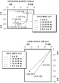
8A-8F are pictorial and graphical diagrams showing that conditional Ryk deletion enhances motor function recovery from spinal cord injury. Fig. 8a schematically shows experimental details over time for a bilateral cervical S segment (CS) dorsal column lesion. 8b and 8c show conditional This shows the generation of the Ryk allele. Figure 8b shows that
9A-9H are diagrams and photographs showing that conditional Ryk deletion enhances corticospinal axon spouting after spinal cord injury. 9A and 9B are representative images of tdTomato-labeled CST axons obtained from 8 consecutive sagittal cryosections spaced 140 μm apart when superimposed on the central GFAP astrocyte-stained portion of the injury site [one experiment, 12 per group. Mice, from 11 littermates; Compass indicates dorsal (D), ventral (V), portal (R) and tail (C)]. Mice with conditional Ryk deletion had higher levels of geodetic in the temporal and caudal directions for lesions compared to control mice (one-tailed t test, *P<O.OS). 9c and 9d show higher magnifications of a single confocal plane of the boxed regions (1.5 mm caudal to the lesion site) shown in FIGS. 9a and 9b, respectively. 9E shows tdTomato-labeled axons over 3 mm lateral to the lesion compared to controls in the dorsal column (12 mice per group, uni-tailed t test, P=0.12, t(21)=1.198) and gray matter ( Normalized for labeling as a pyramid). Mice that developed a conditional Ryk deletion had higher levels of geodetic in both the lateral and caudal directions for the lesion than control mice (12 mice per group, unilateral tail t test, *P<O.OS: temporal P=0.0499 t ( 19)=1.730, caudal P=0.0397 t(19)=1.855). Figures 9f to 9h show the distribution of corticospinal axons in the dorsal column (Figure 9f) or spinal gray matter (Figures 9g and 9h) [axon index is 0.411 μm in a total of 8 sagittal frozen sections Threshold pixels for each divided by the threshold pixels into the transverse pyramid]. Fig. 9H is an enlarged view of the side geodesic of Fig. 9G. The CS injury site is 0 μm, and the lateral direction is indicated by negative numbers, and the caudal direction is indicated by positive numbers. Data in FIG. 9E are presented as medians with interquartile ranges. Data in FIGS. 9F-9H are presented as mean ± sem.
10a to 10d are diagrams showing the changes in corticospinal connectivity after a CS dorsal column lesion in pictures and graphs. 10a and 10b, the medial-lateral distribution of corticospinal axons, conditional It is shown that, after Ryk deletion, the proximal geodesic to the main dorsal corticospinal tract (regions I and II) shows the greatest increase (12 mice per group, one-tailed t-test, *P<O. OS: caudal II P=0.0225 t(19)=2.146, caudal II P=0.0295 t(21)=1.996, caudal IP=0.0059 t(17)=2.819). 10c, control mice and conditional Ryk It shows that the deletion mice exhibited presynaptic clusters (vGlut1 and tdTomato-labeled corticospinal axons co-located) at 600 μm posterior to the CS injury site (one experiment performed, 9 mice per group). FIG. 10D shows medial-lateral distributions of corticospinal innervation at 600 μm on the lateral side of the CS injury site. All data are presented as medians with interquartile ranges.
11a to 11f are diagrams and graphs showing that secondary injuries in the
12a to 12i are diagrams and diagrams showing that monoclonal Ryk antibody injection promotes functional recovery during spinal cord injury. 12A is a schematic diagram showing antibody injection by an intrathecal catheter. 12B shows that performance on the task of extending cells and skillfully receiving food pellets showed improved recovery in rats injected with Ryk monoclonal antibody for 28 days starting at the time of injury [6 rats] (IgG control), 5 rats (Ryk monoclonal), repeated ANOVA, P=0.0354, F(1,9)=6.113). Figure 12c shows that the behavioral performance on the locomotor grid crossing task was not affected by Ryk monoclonal antibody injection. Figure 12d shows that Ryk monoclonal antibody recognizes the full-length Ryk protein expressed in transfected COS-7 cells by Western blotting and immunocytochemical methods. 12E-12H show that BDA-labeled corticospinal axons in Ryk monoclonal antibody-injected rats had higher levels of geodesicization than in BDA-labeled corticospinal axons in control mice (IgG-injected rats). it will show 12E and 12F are images of BDA-labeled CST axons obtained from 6 consecutive sagittal cryosections spaced at 280 μm intervals when superimposed on the central GFAP thalamus and NG2-stained regions of the injury site. 12G and 12H show the distribution of corticospinal axons in the dorsal column ( FIG. 12G ) or the spinal gray matter ( FIG. 12H ) [Axon indices traverse a threshold pixel every 0.741 μm for a total of 6 sagittal frozen sections. Threshold to pyramid divided by pixels]. The CS injury site is 0 μm, and the lateral direction is indicated by negative numbers, and the caudal direction is indicated by positive numbers. Figure 12i shows the results of normalizing the total sum of axonal protrusions that exist over 5 mm based on the control group. Rats injected with Ryk monoclonal antibody had higher levels of geodesic in both lateral and caudal directions compared to control mice (IgG injected rats) [6 rats (IgG control), 5 rats (Ryk monoclonal) day), one-tailed t-test 585 *P<0.05: gated P = 0.0446 t(6)=2.000, caudal P=0.0196 t(6)=2.594). Data in Figures 12b, 12c, 12g and 12h are presented as mean ± sem and data in Figure 12i are presented as median with interquartile ranges.
13A-13C are diagrams graphically illustrating cortical map reconstruction during recovery from spinal cord injury. 13A schematically shows the experimental details of the optogenetic mapping performed through the weekly behavioral function test over time after bilateral CS dorsal column lesions occurred. 13B is a topographic representation of the activation of the heel flexor and extensor muscles, based on bregma (*), at 3 days, 4 weeks, and 8 weeks after the onset of CS dorsal column lesions. Data are presented as the total number of mice responding to evoked motions in each zone, with a light color indicating that the majority of mice are responsive in a given zone. Each tick mark represents 300 μm. 13C shows that the late C3 dorsal column lesion disrupts the remodeled circuit, while subsequent pyramidotomy abrogates the unilateral evoked motor output. Both were measured 3 days after injury.
14A to 14F are diagrams showing graphically and pictorially the situation inside the posterior cortical region where forelimb motor map representation was previously stopped during disease progression. 14A is a cortical map reconstructed from the time of spinal cord injury in mice trained weekly, showing how the center shift of the map occurs after injury. The size of the marker is proportional to the percentage of mice exhibiting the evoked motor action of a given muscle group. 14B and 14C show that the heel extensor motor map moves caudally and medial to the cortex originally occupied by the hindlimb representation. 14d and 14e show that mice with conditional Ryk deletion had a higher rate of contribution to heel flexor activation at 4 weeks after CS lesion development (Fig. 14d) and, conversely, a lower proportion of motor cortex contributing to extensor activation (Fig. 14d). 14e) [(n=10 (control) 11 ( Ryk cKO) mice, one-tailed t test *P<0.05: heel flexion P=0.0347 t(19)=1.925, heel extension P=0.0460 t( 16) = -1.791, data are presented as mean ± sem). 14f is a model for ectopic cortical motor area augmentation mediated by axon plasticity. After dorsal column injury, the immediate expansion of the battery region above the injury zone is probably mediated by lateral connectivity in the motor cortex. Increased axonal plasticity and connectivity after conditional Ryk deletion may lead to the formation of new ectopic regions of the anteromotor cortex.
15A-15D are diagrams graphically showing that cortical map reconstruction and functional recovery from spinal cord injury are dependent on rehabilitation training. Fig. 15a schematically shows over time experimental details regarding optogenetic mapping made through only the last behavioral function test performed at 8 weeks post-injury. Figure 15b shows the heel flexor and extensor muscles based on bregma (*) when the weekly behavioral function test was not performed at 3 days, 4 weeks, and 8 weeks after the occurrence of CS dorsal column lesions. It is a topographic representation of activation. Fig. 15c shows that mice (both Ryk cKO mice and control mice) that were subjected to weekly behavioral function tests 8 weeks after the onset of CS dorsal column lesions, at week 8 (n = 10 mice (control group, weekly test), 11 mice ( Ryk cKO, weekly test), 5 (control and Ryk cKO mice, test only at week 8) performed better than test only mice [ANOVA P = 0.0037 F ( 3) = 5.7157, Bonferroni-corrected t test *P<0.05: 1. Ryk cKO weekly behavioral function test v.
FIG. 16 is a diagram illustrating representative continuous frames in a video recording on battery performance. This representative frame is from 13 weeks after the onset of the C5 dorsal column lesion (1 week after the C3 sham surgery), showing that the cell's ability to grasp well has been restored. Since the sweep will drop the feed pellets into the gap between the platform (black area) and the cage body, or under the floor of the cage's wire frame, a grabbing action must be taken to be a successful stretch action.
17A and 17B are schematic diagrams illustrating when Ryk monoclonal antibody was injected in rats. 17A shows the specificity of Ryk monoclonal antibodies. Full-length blots are shown in FIGS. 23A and 23B . Fig. 17b schematically shows the details of an experiment in which Ryk monoclonal antibody is injected after bilateral C5 dorsal column lesions have occurred in rats over time.
Figure 18 is a diagram showing the increase in axonal branching caudally to the injured site after Ryk monoclonal antibody was injected graphically. Rats injected with Ryk monoclonal antibody for 28 days had a higher level of caudally to the lesion than rats injected with control IgG [n = 6 mice (IgG control dose) 5 mice (Ryk monoclonal antibody) dosing) rat, one-tailed t test *P = 0.0196 P = 0.0196 t(6) = 2.594, data are presented as mean ± sem). The injury site is 0 μm, and the caudal direction is indicated by positive numbers. The axon index is the threshold pixel of the sagittal spinal cord divided by the threshold pixel to the transverse pyramid.
19 is a schematic diagram illustrating an example of optogenetic mapping. To elicit muscle action, sedated mice with a unilateral cranial window inserted were stimulated with a 470 nm LED by means of a fiber optic cable. Two examples are presented of an animal (1 animal) motor map before and 3 days after the onset of the C5 dorsal column lesion.
20 shows weekly training and conditional Ryk A graphical representation of the proportion of the motor cortex occupied by time-dependent changes in characterized motor output, resulting in response to deletion.
FIG. 21 is a diagram showing graphically that the mice used in the cortical mapping experiment recovered the ability to skillfully extend the cell. Behavioral function tests for the skillful limb extension task showed improved recovery in the contralateral motor cortex after conditional Ryk deletion [1 to 8 weeks after the onset of the C5 lesion, n = 10 (control) 11 rats ( Ryk ). cKO) mice, repeated ANOVA P=0.0304 F(1,19)=5.472]. Secondary C3 did not show improved recovery after conditional Ryk deletion occurred [n = 9 (control) 8 mice ( Ryk cKO)], whereas unilateral pyramidal pyramidal resection (n = 8 mice (control) 7 mice ( Ryk )) cKO)) completely abolished the ability of mice to perform tasks. Data are presented as mean ± sem.
22 is a graphical illustration of the proportion of characterized motor output occupying the motor cortex being relatively stable in the absence of training after injury.
23A and 23B are diagrams showing photos of the full-length Western blot results of FIGS. 8C and 17A . Figure 23a shows the specificity of the Ryk monoclonal antibody of Figure 17a. Figure 23b shows the results of Western blotting on the
본 발명은, Wnt의 결합 도메인과 특이적으로 결합하는 항-Ryk 항체 또는 항체 단편이 Wnt-Ryk 신호전달을 억제한다는 발견을 바탕으로 한다. 즉, 본 발명은 항-Ryk 항체 또는 항체 단편을 이용하여 뉴런 변성 및 뉴런 안내를 조정하기 위한 방법을 제공한다. 그러므로 항-Ryk 항체 또는 항체 단편은, 신경계 질환 또는 장애, 예컨대 신경변성 질환 또는 장애를 앓고 있거나 발병 위험이 있는 대상체에 있어서 이 신경계 질환 또는 장애, 예컨대 신경변성 질환 또는 장애를 치료하고/치료하거나 대상체에서 척수 손상(SCI)을 치료하기 위해 사용될 수 있다. The present invention is based on the discovery that an anti-Ryk antibody or antibody fragment that specifically binds to the binding domain of Wnt inhibits Wnt-Ryk signaling. That is, the present invention provides a method for modulating neuronal degeneration and neuronal guidance using an anti-Ryk antibody or antibody fragment. Thus, an anti-Ryk antibody or antibody fragment can be used to treat and/or treat a neurological disease or disorder, such as a neurodegenerative disease or disorder, in a subject suffering from or at risk of developing the neurological disease or disorder, such as a neurodegenerative disease or disorder. It can be used to treat spinal cord injury (SCI) in
본 발명의 조성물과 방법을 기술하기에 앞서, 본 발명은 기술된 특정 조성물, 방법 및 실험 조건에 제한되지 않는 고로, 조성물, 방법 및 조건은 다양할 수 있음이 이해되어야 한다. 또한 본 발명의 범위는 오로지 첨부된 청구범위에 의해서만 한정될 것이므로, 본원에 사용된 용어는 단지 특정 구현예들을 기술하기 위한 것이지, 본 발명을 제한하고자 하는 의도가 아님도 이해되어야 한다. Prior to describing the compositions and methods of the present invention, it is to be understood that the present invention is not limited to the particular compositions, methods, and experimental conditions described, as such compositions, methods, and conditions may vary. It is also to be understood that the terminology used herein is for the purpose of describing particular embodiments only, and is not intended to limit the invention, as the scope of the present invention will be limited only by the appended claims.
본 명세서와, 이에 첨부된 청구범위에 사용된 바와 같이, 단수 형태를 나타내는 용어 "하나", "하나의" 및 "본"은 내용 중 명확히 달리 명시되어 있지 않는 한 복수의 대상물을 포함한다. 그러므로, 예를 들어 "본 방법"에 대한 대상물은 본 개시내용 등이 읽어질 때 당 업자에게 명확해질, 본원에 기술된 하나 이상의 방법, 그리고/또는 본원에 기술된 유형의 단계들을 포함한다. As used in this specification and in the claims appended hereto, the terms "a," "an," and "the present" in the singular include the plural objects unless the context clearly dictates otherwise. Thus, for example, subject matter to “the method” includes one or more methods described herein, and/or steps of the type described herein, which will become apparent to those skilled in the art when this disclosure or the like is read.
"~를 포함하는(including)", "~를 함유하는" 또는 "~에 의해 특징지어지는"과 호환되어 사용되는, "~를 포함하는(comprising)"이란 용어는, 포괄적이거나 개방적인 표현으로서, 추가적이거나 언급되지 않은 요소 또는 방법의 단계를 배제하지 않는다. "~로 이루어진"이란 용어는, 청구항에 지정되지 않은 임의의 요소, 단계 또는 성분을 배제한다. "~로 본질적으로 이루어진"이란 어구는, 청구범위를, 지정된 재료나 단계, 그리고 청구된 발명의 기본적이고 신규한 특성들에 실질적으로 영향을 미치지 않는 것들로 제한한다. 본 발명은, 이러한 어구들 각각의 범위에 상응하는 본 발명의 조성물과 방법에 대한 구현예들을 고려한다. 그러므로 언급된 요소들 또는 단계들을 포함하는 조성물 또는 방법은, 해당 조성물 또는 방법이 이와 같은 요소들 또는 단계들로 본질적으로 이루어지거나, 이와 같은 요소들 또는 단계들로 이루어진, 특정 구현예들을 고려한다.The term "comprising", used interchangeably with "including," "containing," or "characterized by," is an inclusive or open-ended , does not exclude additional or unmentioned elements or method steps. The term “consisting of” excludes any element, step or component not specified in a claim. The phrase “consisting essentially of” limits the claims to the specified materials or steps and those that do not materially affect the basic and novel characteristics of the claimed invention. The present invention contemplates embodiments of the compositions and methods of the present invention that fall within the scope of each of these phrases. Thus, a composition or method comprising the recited elements or steps contemplates particular embodiments wherein the composition or method consists essentially of, or consists of, such elements or steps.
달리 정의되지 않는 한, 본원에 사용된 모든 기술 용어와 과학 용어는, 본 발명이 속하는 분야의 통상의 기술자에 의해 일반적으로 이해되는 의미와 동일한 의미를 가진다. 본원에 기술된 방법 및 재료와 동일한 임의의 방법 및 재료가 본 발명을 실시 또는 시험하는데 사용될 수 있지만, 바람직한 방법 및 재료가 여기에 기술되어 있다. Unless defined otherwise, all technical and scientific terms used herein have the same meaning as commonly understood by one of ordinary skill in the art to which this invention belongs. Although any methods and materials equivalent to those described herein can be used in the practice or testing of the present invention, the preferred methods and materials are described herein.
본원에 사용된 바와 같은 "치료"또는 "치료하는 것" 또는 "증상을 완화하는 것" 또는 "완화하는 것"이란 용어들은 본원에서 호환되어 사용되고 있다. 이러한 용어들은, 유리하거나 원하는 결과, 예컨대 치료적 이익 및/또는 예방적 이익(이에 한정되는 것은 아님)을 달성하기 위한 접근법을 지칭한다. "치료적 이익"이란, 치료중인 기저 장애를 근절 또는 완화하는 것을 의미한다. 또한 치료적 이익은 기저 장애와 연관된 생리적 증상들 중 하나 이상을 근절 또는 완화하여, 어떤 환자가 여전히 기저 장애를 앓고 있을 수 있음에도 불구 해당 환자에서 개선이 관찰되게 함으로써 달성된다. 예방적 이익을 위해 본 조성물은, 비록 특정 질환의 진단이 내려지지 않을 수 있더라도, 해당 질환이 발생할 위험이 있는 환자, 또는 해당 질환의 생리적 증상들 중 하나 이상을 보고하는 환자에게 투여될 수 있다. 치료는, 질환을 예방하는 것, 즉 질환이 유도되기 전 방어적 조성물(protective composition)을 투여함으로써 해당 질환의 임상 증상들이 발생하지 않도록 만드는 것; 질환을 억제하는 것, 즉 유도 반응 후이되, 질환의 임상 발생 또는 재발을 보이기 전 방어적 조성물을 투여함으로써 해당 질환의 임상 증상들이 발생하지 않도록 만드는 것; 질환을 억제하는 것, 즉 질환의 초기 발생 후 방어적 조성물을 투여함으로써 임상 징후의 발생을 정지시키는 것; 질환의 재발생을 예방하는 것, 그리고/또는 질환을 경감시키는 것, 즉 질환의 초기 발생 후 방어적 조성물을 투여함으로써 임상 증상의 퇴행을 초래하는 것을 포함한다. As used herein, the terms “treatment” or “treating” or “alleviating a symptom” or “alleviating” are used interchangeably herein. These terms refer to approaches for achieving beneficial or desired results, such as, but not limited to, a therapeutic benefit and/or a prophylactic benefit. By “therapeutic benefit” is meant eradicating or ameliorating the underlying disorder being treated. A therapeutic benefit is also achieved by eradicating or alleviating one or more of the physiological symptoms associated with the underlying disorder, such that an improvement is observed in a patient who may still be afflicted with the underlying disorder. For prophylactic benefit, the composition may be administered to a patient at risk of developing the disease, or a patient reporting one or more of the physiological symptoms of the disease, although a diagnosis of a particular disease may not be made. Treatment includes preventing the disease, ie, making the clinical symptoms of the disease not occur by administering a protective composition before the disease is induced; inhibiting the disease, i.e., making the clinical symptoms of the disease not occur by administering a protective composition after an induction response but before clinical occurrence or recurrence of the disease; inhibiting the disease, ie, arresting the development of clinical signs by administering a protective composition after the initial onset of the disease; preventing recurrence of the disease, and/or alleviating the disease, ie, resulting in regression of clinical symptoms by administering a protective composition after the initial onset of the disease.
"유효량" 또는 "치료적 유효량"이란 용어는, 원하는 생물학적 결과를 유도하는데 충분한, 활성 제제의 양을 지칭한다. 이 같은 결과는, 질환의 징후, 증상 또는 원인을 완화시키는 것, 또는 기타 생물계에 요망되는 임의의 변경일 수 있다. 본원에서 "치료적 유효량"이란 용어는, 어떤 기간에 걸쳐 발병 부위에 반복적으로 적용될 때 해당 질환의 병태에 실질적인 개선을 달성하는, 제제의 임의의 양을 나타내는데 사용되고 있다. 상기 양은, 치료중인 병태, 해당 병태의 진행 단계, 그리고 적용된 제제의 유형과 농도에 따라 달라질 것이다. 임의의 주어진 사례에 있어서 적당량은, 당 업자가 용이하게 알 것이거나, 통상의 실험을 통해 결정될 수 있을 것이다. The term "effective amount" or "therapeutically effective amount" refers to an amount of an active agent sufficient to induce a desired biological result. Such an outcome may be alleviating the signs, symptoms or causes of a disease, or any other alteration desired in the biological system. The term “therapeutically effective amount” is used herein to denote any amount of an agent that, when applied repeatedly to the site of disease over a period of time, achieves a substantial improvement in the condition of the disease in question. The amount will vary depending on the condition being treated, the stage of progression of the condition, and the type and concentration of the agent applied. An appropriate amount for any given case will be readily apparent to one of ordinary skill in the art, or may be determined through routine experimentation.
"약학적으로 허용 가능한 염"이란 용어는, 당 분야에 널리 공지된 다양한 유기 및 무기 반대 이온들로부터 유래한 염을 지칭하고, 단지 예를 들자면 나트륨, 칼륨, 칼슘, 마그네슘, 암모늄, 테트라알킬암모늄 등을 포함하고; 분자가 염기 작용기를 함유하면 유기 산 또는 무기 산의 염, 예컨대 염화수소산염, 브롬화수소산염, 주석산염, 메실레이트, 아세트산염, 말레산염 및 옥살산염 등을 포함한다.The term “pharmaceutically acceptable salts” refers to salts derived from a variety of organic and inorganic counterions well known in the art and includes, by way of example only, sodium, potassium, calcium, magnesium, ammonium, tetraalkylammonium and the like; If the molecule contains a basic functional group, it includes salts of organic or inorganic acids, such as hydrochloride, hydrobromide, tartrate, mesylate, acetate, maleate and oxalate salts.
"대상체", 개체" 또는 "환자"는 본원에서 호환되어 사용되고 있는데, 즉 척추동물, 바람직하게 포유동물, 더욱 바람직하게 인간을 지칭한다. 포유동물은 쥣과 동물, 원숭이류, 인간, 가축, 경기용 동물 및 반려 동물을 포함하나, 이에 한정되는 것은 아니다. 시험관 내에서 얻어졌거나, 또는 시험관 내에서 배양된 조직, 세포 및 생물학적 독립체의 자손도 포함된다. "Subject", individual" or "patient" are used interchangeably herein, i.e. refer to a vertebrate, preferably a mammal, more preferably a human. Mammal is a murine, apes, human, livestock, game Animals and companion animals include, but are not limited to, progeny of tissues, cells and biological entities obtained or cultured in vitro.
본원에 사용된 바와 같은 "촉진하다"또는 "증가시키다" 또는 "촉진하는 것" 또는 "증가시키는 것"은 본원에서 호환되어 사용되고 있다. 이러한 용어들은, 처리된 세포(조직 또는 대상체)에서 측정된 매개변수(예컨대 활성, 발현, 신호 전달, 뉴런 변성)가, 미처리 세포(조직 또는 대상체)에서 측정된 상기 매개변수에 비하여 증가하는 경우를 지칭한다. 비교는 또한 처리 전과 후의 사이에 동일 세포 또는 조직 또는 대상체를 대상으로 이루어질 수 있다. 증가는 검출되기 충분한 정도이다. 몇몇 구현예들에서, 처리된 세포에서의 증가는 미처리 세포에서에 비하여 적어도 약 10%, 20%, 30%, 40%, 50%, 60%, 70%, 80%, 90%, 1배, 2배, 3배, 4배 또는 이 이상인 경우이다.As used herein, “promoting” or “increasing” or “facilitating” or “increasing” are used interchangeably herein. These terms refer to instances in which a parameter (eg, activity, expression, signal transduction, neuronal degeneration) measured in a treated cell (tissue or subject) increases compared to the parameter measured in an untreated cell (tissue or subject). refers to Comparisons can also be made of the same cell or tissue or subject between before and after treatment. The increase is sufficient to detect. In some embodiments, the increase in treated cells is at least about 10%, 20%, 30%, 40%, 50%, 60%, 70%, 80%, 90%, 1-fold, compared to untreated cells, 2 times, 3 times, 4 times, or more.
본원에 사용된 바와 같은 "억제하다", "예방하다", "감소시키다", "억제하는 것" 또는 "감소시키는 것"은 본원에서 호환되어 사용된다. 이 용어들은, 처리된 세포(조직 또는 대상체)에서 측정된 매개변수(예컨대 활성, 발현, 신호 전달, 뉴런 변성)가, 미처리 세포(조직 또는 대상체)에서 측정된 상기 매개변수에 비하여 감소하는 경우를 지칭한다. 비교는 또한 처리 전과 후의 사이에 동일 세포 또는 조직 또는 대상체를 대상으로 이루어질 수 있다. 감소는 검출되기 충분한 정도이다. 몇몇 구현예들에서, 처리된 세포에서의 감소는 미처리 세포에서에 비하여 적어도 약 10%, 20%, 30%, 40%, 50%, 60%, 70%, 80%, 90%, 이거나, 완전히 억제되는 경우이다. 몇몇 구현예들에서 처리된 세포에서 측정된 매개변수는 미처리 세포에서 측정된 매개변수에 비하여 검출 불가능하다(즉 완전히 억제된다). As used herein, “inhibit”, “prevent”, “reduce”, “inhibiting” or “reducing” are used interchangeably herein. These terms refer to cases in which a parameter (eg, activity, expression, signal transduction, neuronal degeneration) measured in a treated cell (tissue or subject) decreases compared to the parameter measured in an untreated cell (tissue or subject). refers to Comparisons can also be made of the same cell or tissue or subject between before and after treatment. The reduction is of a sufficient degree to be detected. In some embodiments, the reduction in treated cells is at least about 10%, 20%, 30%, 40%, 50%, 60%, 70%, 80%, 90%, or completely compared to in untreated cells. in case it is suppressed. In some embodiments a parameter measured in treated cells is undetectable (ie completely inhibited) compared to a parameter measured in untreated cells.
생물 활성 제제와 관련하여 "선택적 억제" 또는 "선택적으로 억제하다"란 용어는, 표적을 벗어난(off-target) 신호전달 활성에 비하여 표적 신호전달 활성을 우선적으로 감소시키는, 어떤 제제의 능력을 지칭한다. The term "selective inhibition" or "selectively inhibit" in the context of a biologically active agent refers to the ability of an agent to preferentially decrease target signaling activity relative to off-target signaling activity do.
"폴리펩티드", "펩티드" 및 "단백질"이란 용어는, 아미노산 잔기들의 중합체를 지칭하는 것으로서, 본원에서 호환되어 사용된다. 본 용어는, 하나 이상의 아미노산 잔기가 상응하는 자연 발생 아미노산의 인공 화학 모의체인 아미노산 중합체 뿐 아니라, 자연 발생 아미노산 중합체 및 비자연 발생 아미노산 중합체에도 적용된다.The terms “polypeptide,” “peptide,” and “protein” refer to a polymer of amino acid residues and are used interchangeably herein. The term applies to amino acid polymers in which one or more amino acid residues are artificial chemical mimetics of the corresponding naturally occurring amino acid, as well as to naturally occurring amino acid polymers and non-naturally occurring amino acid polymers.
"아미노산"이란 용어는, 자연 발생 및 합성 아미노산뿐 아니라, 자연 발생 아미노산의 작용 방식과 유사한 방식으로 작용하는 아미노산 유사체 및 아미노산 모의체를 지칭한다. 자연 발생 아미노산으로서는 유전자 암호에 의해 암호화되는 것들뿐 아니라, 추후 변형된 아미노산들, 예컨대 하이드록시프롤린, γ-카복시글루타메이트 및 O-포스포세린이 있다. 아미노산 유사체란, 기본 화학 구조가 자연 발생 아미노산의 기본 화학 구조(즉 α 탄소가 수소, 카복실기, 아미노기 및 R기와 결합되어 있는 구조)와 동일한 화합물을 지칭하며, 예컨대 호모세린, 노르루신, 메티오닌 설폭시드, 메티오닌 메틸 설포늄이 있다. 이러한 유사체는 변형된 R기를 가지거나(예컨대 노르루신) 또는 변형된 펩티드 골격을 가지지만, 자연 발생 아미노산의 기본 화학 구조와 동일한 기본 화학 구조를 보유한다. 자연 암호화된 아미노산으로서는 20개의 통상적인 아미노산(알라닌, 아르기닌, 아스파라긴, 아스파르트산, 시스테인, 글루타민, 글루탐산, 글리신, 히스티딘, 이소루신, 루신, 리신, 메티오닌, 페닐알라닌, 프롤린, 세린, 트레오닌, 트립토판, 티로신 및 발린)과 피롤리신 및 셀레노시스테인이 있다. The term “amino acid” refers to naturally occurring and synthetic amino acids, as well as amino acid analogs and amino acid mimetics that function in a manner similar to that of naturally occurring amino acids. Naturally occurring amino acids include those encoded by the genetic code, as well as later modified amino acids such as hydroxyproline, γ-carboxyglutamate and O-phosphoserine. An amino acid analog refers to a compound whose basic chemical structure is identical to that of a naturally occurring amino acid (i.e., a structure in which the α carbon is bonded to hydrogen, carboxyl, amino and R groups), such as homoserine, norleucine, methionine sulfoxide Seed, methionine methyl sulfonium. Such analogs have modified R groups (eg, norleucine) or modified peptide backbones, but retain the same basic chemical structure as that of a naturally occurring amino acid. The naturally encoded amino acids include the 20 common amino acids (alanine, arginine, asparagine, aspartic acid, cysteine, glutamine, glutamic acid, glycine, histidine, isoleucine, leucine, lysine, methionine, phenylalanine, proline, serine, threonine, tryptophan, tyrosine). and valine) and pyrrolysine and selenocysteine.
"보존적으로 변형된 변이체"는 아미노산 서열과 핵산 서열 둘 다에 적용된다. 특정 핵산 서열과 관련하여, 보존적으로 변형된 변이체란, 동일하거나 본질적으로 동일한 아미노산 서열을 암호화하는 핵산, 또는 (핵산이 아미노산 서열을 암호화하지 않는 경우에는) 본질적으로 동일한 서열을 지칭한다. 유전자 암호의 축퇴성으로 말미암아, 기능상 동일한 다수의 핵산은 소정의 임의 단백질을 암호화한다. 예를 들어 코돈 GCA, GCC, GCG 및 GCU는 모두 아미노산 알라닌을 암호화한다. 그러므로 알라닌이 어떤 코돈에 의해 지정되는 모든 위치에서, 해당 코돈은 암호화되는 폴리펩티드를 변경시키지 않으면서, 기술된 상응 코돈들 중 임의의 것으로 변경될 수 있다. 이러한 핵산 변이는 보존적으로 변형된 변이의 일종인 "침묵 변이"이다. 폴리펩티드를 암호화하는 본원의 모든 핵산 서열은 또한 발생 가능한 핵산의 모든 침묵 변이를 기술하는 것이다. 당 업자는, (보통 메티오닌의 유일한 코돈인 AUG와, 보통 트립토판의 유일한 코돈인 TGG를 제외하고) 핵산 내 각각의 코돈은 기능상 동일한 분자를 생성하도록 변형될 수 있음을 인지할 것이다. 그러므로 폴리펩티드를 암호화하는 핵산의 각 침묵 변이는 기술된 각 서열 내에 내포되어 있다."Conservatively modified variants" apply to both amino acid sequences and nucleic acid sequences. With respect to a particular nucleic acid sequence, conservatively modified variants refer to a nucleic acid that encodes the same or essentially identical amino acid sequence, or (where the nucleic acid does not encode an amino acid sequence) essentially identical sequences. Due to the degeneracy of the genetic code, many functionally identical nucleic acids encode any given protein. For example, the codons GCA, GCC, GCG and GCU all encode the amino acid alanine. Therefore, at any position where an alanine is designated by any codon, that codon can be altered to any of the corresponding codons described without altering the encoded polypeptide. Such nucleic acid mutations are "silent mutations", which are a kind of conservatively modified mutations. Any nucleic acid sequence herein that encodes a polypeptide also describes any silent variation of the nucleic acid that may occur. One of ordinary skill in the art will recognize that each codon in a nucleic acid (with the exception of AUG, which is usually the only codon for methionine, and TGG, which is usually the only codon for tryptophan) can be modified to produce a functionally identical molecule. Therefore, each silent variation of the nucleic acid encoding the polypeptide is implied within each sequence described.
아미노산 서열에 관하여, 당 업자는, 암호화된 서열 내 단일 아미노산 또는 낮은 비율의 아미노산을 변경, 부가 또는 결실시키는 개별 치환, 결실 또는 부가가 "보존적으로 변형된 변이"이면, 이러한 변이는 어떤 아미노산의, 화학적으로 유사한 아미노산으로의 치환을 초래하는 것임을 인지할 것이다. 기능상 유사한 아미노산들을 제공하는 보존적 치환 표는 당 분야에 널리 공지되어 있다. 이처럼 보존적으로 변형된 변이체는 본 발명에 추가적인 것으로서, 본 발명의 다형성 변이체, 종간 상동체 및 대립형질을 배제하지 않는다. With respect to amino acid sequences, those skilled in the art will recognize that if an individual substitution, deletion or addition that alters, adds or deletes a single amino acid or a small proportion of amino acids in the encoded sequence is a "conservatively modified mutation", then such mutation is the , resulting in a substitution with a chemically similar amino acid. Conservative substitution tables providing functionally similar amino acids are well known in the art. Such conservatively modified variants are additional to the present invention and do not exclude polymorphic variants, interspecies homologues and alleles of the present invention.
하기 8개의 군은 각각 서로에 대해 보존적 치환인 아미노산들을 포함한다. The following eight groups each contain amino acids that are conservative substitutions for each other.
1) 알라닌(A), 글리신(G);1) alanine (A), glycine (G);
2) 아스파르트산(D), 글루탐산(E);2) aspartic acid (D), glutamic acid (E);
3) 아스파라긴(N), 글루타민(Q);3) asparagine (N), glutamine (Q);
4) 아르기닌(R), 리신(K);4) arginine (R), lysine (K);
5) 이소루신(I), 루신(L), 메티오닌(M), 발린(V);5) isoleucine (I), leucine (L), methionine (M), valine (V);
6) 페닐알라닌(F), 티로신(Y), 트립토판(W);6) phenylalanine (F), tyrosine (Y), tryptophan (W);
7) 세린(S), 트레오닌(T); 그리고7) Serine (S), Threonine (T); and
8) 시스테인(C), 메티오닌(M).8) Cysteine (C), Methionine (M).
(예컨대 문헌(Creighton, Proteins (1984)) 참조)(See eg Creighton, Proteins (1984))
보존적 치환은 염기성 대 염기성, 산성 대 산성, 극성 대 극성 등과 같은 치환을 포함할 수 있다. 이와 같이 구성되는 아미노산 세트들은 구조상의 이유로 보존적일 수 있을 것이다. 이러한 세트들은 벤 도표의 형태로 기술될 수 있다(Livingstone C. D. and Barton G. J. (1993) "Protein sequence alignments: a strategy for the hierarchical analysis of residue conservation" Comput. Appl Biosci. 9: 745-756; Taylor W. R. (1986) "The classification of amino acid conservation" J. Theor. Biol. 119; 205-218). 보존적 치환은, 예를 들어 일반적으로 받아들여지고 있는 아미노산의 벤(Venn) 도표 분류를 기술하고 있는 이하 표에 따라서 이루어질 수 있다.Conservative substitutions may include substitutions such as basic to basic, acidic to acidic, polar to polar, and the like. The amino acid sets thus constituted may be conservative for structural reasons. These sets can be described in the form of Venn diagrams (Livingstone CD and Barton GJ (1993) "Protein sequence alignments: a strategy for the hierarchical analysis of residue conservation" Comput. Appl Biosci. 9: 745-756; Taylor WR ( 1986) "The classification of amino acid conservation" J. Theor. Biol. 119; 205-218). Conservative substitutions can be made, for example, according to the table below, which describes the generally accepted Venn chart classification of amino acids.

"서열 동일성 %"는 최적으로 정렬된 2개의 서열들을 비교 윈도우에 대해 비교함으로써 측정되는데, 이 경우 두 서열의 최적의 정렬을 위해서는 비교 윈도우에 있는 폴리뉴클레오티드 서열의 일부는, 부가 또는 결실을 포함하지 않는 기준 서열(예컨대 본 발명의 폴리펩티드)에 비하여 부가 또는 결실(즉 갭)을 포함할 수 있다. %는, 두 서열에 동일 핵산 염기 또는 아미노산 잔기가 발생한 위치의 수를 측정하여 매칭된 위치들의 수를 구하고, 매칭된 위치들의 수를 비교 윈도우 내 위치의 총 수로 나눈 다음, 그 결과를 100으로 곱하여 서열 동일성 %를 구함으로써 산정된다. "% sequence identity" is determined by comparing two optimally aligned sequences against a comparison window, in which case, for optimal alignment of the two sequences, the portion of the polynucleotide sequence in the comparison window does not contain additions or deletions. It may contain additions or deletions (ie gaps) compared to a reference sequence that does not (eg, a polypeptide of the invention). % is obtained by determining the number of positions in which the same nucleic acid base or amino acid residue occurs in two sequences to obtain the number of matched positions, dividing the number of matched positions by the total number of positions in the comparison window, and multiplying the result by 100 Calculated by finding % sequence identity.
2개 이상의 핵산 또는 폴리펩티드 서열에 관한 내용 중 "동일한" 또는 "동일성"%란 용어는, 동일한 서열인 2개 이상의 서열 또는 종속서열을 지칭한다. 2개의 서열이, 비교 윈도우 또는 하기 서열 비교 알고리즘들 중 하나가 사용되어 측정됨으로써 지정된 영역에 대해 비교되어 최대 상응성에 따라 정렬될 때, 또는 수동식 정렬 및 육안검사에 의해 비교 및 정렬될 때, 이 두 서열이 동일한 아미노산 잔기 또는 뉴클레오티드를 지정된 %만큼 가진다면(즉 지정된 영역에 대해, 또는 영역이 지정되지 않는다면 전체 서열에 대해 60%의 동일성, 임의로는 65%, 70%, 75%, 80%, 85%, 90% 또는 95%의 동일성을 가진다면) 해당 두 서열은 "실질적으로 동일한" 것이다. 본 발명은 본원에 각각 예시된 폴리펩티드와 실질적으로 동일한 폴리펩티드 뿐 아니라, 이의 용도, 예컨대 신경계 질환 또는 장애, 예컨대 신경변성 질환 또는 장애의 치료 또는 예방, 및/또는 SCI 치료를 위한 용도(이에 한정되는 것은 아님)도 제공한다. 임의로 길이가 적어도 약 50개 뉴클레오티드인 영역, 더욱 바람직하게 길이가 100개 내지 500개, 또는 1000개 이상의 뉴클레오티드인 영역, 또는 길이가 기준 서열 전체 길이인 영역에 걸쳐 동일성이 존재한다.In the context of two or more nucleic acid or polypeptide sequences, the term "identical" or "% identity" refers to two or more sequences or subsequences that are the same sequence. When two sequences are compared for a designated region as determined using a comparison window or one of the following sequence comparison algorithms and aligned for maximum correspondence, or when compared and aligned by manual alignment and visual inspection, the two sequences If the sequences have a specified percentage of identical amino acid residues or nucleotides (i.e. 60% identity to the designated region, or to the entire sequence if no region is specified, optionally 65%, 70%, 75%, 80%, 85 The two sequences are "substantially identical" if they have %, 90% or 95% identity). The present invention provides polypeptides substantially identical to the polypeptides each exemplified herein, as well as uses thereof, including, but not limited to, the treatment or prophylaxis of a neurological disease or disorder, such as a neurodegenerative disease or disorder, and/or the treatment of SCI. not) is also provided. The identity optionally exists over a region that is at least about 50 nucleotides in length, more preferably between 100 and 500, or at least 1000 nucleotides in length, or a region that is the entire length of the reference sequence in length.
서열 비교를 위하여, 통상적으로 하나의 서열은 기준 서열, 즉 시험 서열의 비교 대상으로서의 역할을 한다. 서열 비교 알고리즘이 사용될 때, 시험 서열 및 기준 서열이 컴퓨터에 입력되고, 필요하다면 종속서열 좌표가 지정되며, 서열 알고리즘 프로그램 매개변수가 지정된다. 디폴트 프로그램 매개변수가 사용될 수 있거나, 대안적으로 매개변수가 지정될 수 있다. 그 다음 서열 비교 알고리즘은 프로그램의 매개변수들을 바탕으로 기준 서열에 대한 시험 서열의 서열 동일성 %를 산출한다. For sequence comparison, usually one sequence serves as a reference sequence, ie, the test sequence to which it is compared. When a sequence comparison algorithm is used, a test sequence and a reference sequence are entered into a computer, dependent sequence coordinates are designated if necessary, and sequence algorithm program parameters are designated. Default program parameters may be used, or alternatively parameters may be specified. The sequence comparison algorithm then calculates the percent sequence identity of the test sequence to the reference sequence based on the parameters of the program.
본원에 사용된 바와 같은 "비교 윈도우"는, 20개 내지 600개, 일반적으로 약 50개 내지 약 200개, 더욱 일반적으로 약 100개 내지 약 150개로 이루어진 군으로부터 선택된 갯수들 중 임의의 갯수만큼의 연속 위치로 이루어진 분절에 대한 기준을 포함하는데, 이 경우 한 서열은, 두 서열이 최적으로 정렬된 후 동일한 갯수만큼의 연속 위치로 이루어진 기준 서열과 비교될 수 있다. 비교용 서열을 정렬하는 방법은 당 분야에 널리 공지되어 있다. 비교용 서열의 최적 정렬은, 예컨대 국소 상동성 알고리즘(Smith and Waterman (1970) Adv. Appl. Math. 2:482c), 상동성 정렬 알고리즘(Needleman and Wunsch (1970) J. Mol. Biol. 48:443), 유사성 검색 방법(Pearson and Lipman(1988) Proc.Nat'l.Acad.Sci. USA85:2444); 이러한 알고리즘들의 전산화 수행(GAP, BESTFIT, FASTA 및 TFASTA (Wisconsin Genetics Software Package, Genetics Computer Group, 575 Science Dr., Madison, Wis.)), 또는 수동식 정렬 및 육안검사(예컨대 Ausubel et al., Current Protocols in Molecular Biology (1995 증보판) 참조)에 의해 수행될 수 있다.A “comparison window,” as used herein, includes any number selected from the group consisting of 20 to 600, typically about 50 to about 200, and more typically about 100 to about 150. It includes a reference for a segment of contiguous positions, in which case a sequence can be compared to a reference sequence of the same number of contiguous positions after the two sequences are optimally aligned. Methods for aligning comparative sequences are well known in the art. Optimal alignments of comparative sequences are, for example, local homology algorithms (Smith and Waterman (1970) Adv. Appl. Math. 2:482c), homology alignment algorithms (Needleman and Wunsch (1970) J. Mol. Biol. 48: 443), similarity search methods (Pearson and Lipman (1988) Proc. Nat'l. Acad. Sci. USA 85:2444); Computational performance of these algorithms (GAP, BESTFIT, FASTA and TFASTA (Wisconsin Genetics Software Package, Genetics Computer Group, 575 Science Dr., Madison, Wis.)), or manual alignment and visual inspection (eg Ausubel et al., Current Protocols) in Molecular Biology (see 1995 Supplement)).
서열 동일성 % 및 서열 유사성 %를 측정하는데 적합한 알고리즘의 예 두 가지로서는 BLAST 및 BLAST 2.0 알고리즘이 있는데, 이에 관하여는 문헌[Altschul et al. (1977) Nuc. Acids Res. 25:3389-3402, 및 Altschul et al. (1990) J. Mol. Biol. 215:403-410]에 각각 기술되어 있다. BLAST 분석을 수행하기 위한 소프트웨어는 국립생물정보센터로부터 공공연하게 입수 가능하다. 이 알고리즘은, 첫 번째로, 데이터베이스 서열 중 동일한 길이의 워드(word)와 정렬될 때 매칭되거나 일부 양의 값인 역치 스코어 T를 만족시키는, 조회 서열 내 길이 W인 짧은 워드를 동정함으로써 고 스코어 서열 쌍(HSP)을 동정하는 단계를 수반한다. T는, 인접 워드 스코어 역치라 지칭된다(Altschul et al., 상동). 이와 같이 처음에 출현하는 인접 워드 히트(word hit)는 이를 포함하는, 더 긴 HSP를 찾기 위한 검색을 개시하는 시드(seed)로서의 역할을 한다. 워드 히트는, 누적 정렬 스코어가 증가할 수 있는 한, 각각의 서열을 따라서 양 방향으로 연장된다. 누적 스코어는, 뉴클레오티드의 경우 매개변수 M(매칭되는 잔기들 쌍에 대한 보상 스코어; 항상 0을 초과함) 및 N(미스매칭 잔기들에 대한 페널티 스코어; 항상 0 미만임)을 사용하여 산정된다. 아미노산 서열에 대한 스코어링 매트릭스는 누적 스코어를 산정하기 위해 사용된다. 워트 히트의 각 방향으로의 연장은, 누적 정렬 스코어가 자체의 최대 달성 값으로부터 X의 양만큼 감소할 때; 하나 이상의 "-"스코어인 잔기 정렬이 축적됨으로 말미암아 누적 스코어가 0 또는 그 이하로 내려갈 때; 아니면 두 서열 중 어느 하나의 말단에 도달할 때 중지된다. BLAST 알고리즘 매개변수 W, T 및 X는 정렬의 감도와 속도를 결정짓는다. (뉴클레오티드 서열용) BLASTN 프로그램은 워드 길이(W) 11, 예상치(E) 10, M = 5, N = -4를 디폴트 값으로 사용하여, 두 서열 가닥을 비교한다. 아미노산 서열용인 BLASTP 프로그램은 워드 길이 3 및 예상치(E) 10을 디폴트 값으로 사용하고, BLOSUM62 스코어 결정 매트릭스(Henikoff and Henikoff (1989) Proc. Natl. Acad. Sci. USA 89:10915 참조)는 정렬값(B) 50, 예상치(E) 10, M=5, N=-4를 디폴트 값으로 사용하여, 두 서열 가닥을 비교한다.Two examples of algorithms suitable for determining % sequence identity and % sequence similarity are the BLAST and BLAST 2.0 algorithms, as described in Altschul et al. (1977) Nuc. Acids Res. 25:3389-3402, and Altschul et al. (1990) J. Mol. Biol. 215:403-410, respectively. Software for performing BLAST analysis is publicly available from the National Center for Biological Information. This algorithm firstly identifies pairs of high score sequences by identifying short words of length W in a query sequence that, when aligned with words of the same length in the database sequence, either match or satisfy some positive threshold score T. (HSP) is identified. T is referred to as the adjacent word score threshold (Altschul et al., supra). This first appearing adjacent word hit serves as a seed to initiate a search to find the longer HSP that contains it. Word hits extend in both directions along each sequence as long as the cumulative alignment score can be increased. The cumulative score is calculated using the parameters M (reward score for matching pairs of residues; always greater than zero) and N (penalty score for mismatching residues; always less than zero) for nucleotides. A scoring matrix for the amino acid sequence is used to calculate the cumulative score. Extension of the wort hit in each direction occurs when the cumulative alignment score decreases by an amount X from its maximum achieved value; when the cumulative score goes below zero or less due to accumulation of one or more "-"-scored residue alignments; Otherwise, it stops when reaching the end of either sequence. The BLAST algorithm parameters W, T and X determine the sensitivity and speed of the alignment. (for nucleotide sequences) The BLASTN program compares two sequence strands, using word length (W) 11, expected (E) 10, M = 5, N = -4 as default values. The BLASTP program for amino acid sequences uses a word length of 3 and an expected (E) of 10 as default values, and the BLOSUM62 score determination matrix (see Henikoff and Henikoff (1989) Proc. Natl. Acad. Sci. USA 89:10915) is an alignment value. Two sequence strands are compared, using (B) 50, expected (E) 10, M=5, N=-4 as default values.
BLAST 알고리즘은 또한 두 서열 간 유사성에 관한 통계 분석을 수행한다(예컨대 문헌(Karlin and Altschul (1993) Proc. Natl. Acad. Sci. USA 90:5873-5787) 참조). BLAST 알고리즘에 의해 제공되는 유사성에 관한 한 척도는 최소 확률 합으로서(P(N)), 이는 두 뉴클레오티드 서열 또는 두 아미노산 서열간 매칭이 우연히 일어날 확률의 표시를 제공한다. 예를 들어 만일 기준 핵산에 대한 시험 핵산 비교시 최소 확률 합이 약 0.2 미만, 더욱 바람직하게 약 0.01 미만, 가장 바람직하게 약 0.001 미만이면, 해당 핵산은 기준 서열과 유사한 것으로 간주된다. The BLAST algorithm also performs statistical analysis of the similarity between two sequences (see, eg, Karlin and Altschul (1993) Proc. Natl. Acad. Sci. USA 90:5873-5787). One measure of similarity provided by the BLAST algorithm is the minimum probability sum (P(N)), which provides an indication of the probability that a match between two nucleotide sequences or two amino acid sequences will occur by chance. For example, a nucleic acid is considered similar to a reference sequence if the minimum sum of probabilities when comparing a test nucleic acid to a reference nucleic acid is less than about 0.2, more preferably less than about 0.01, and most preferably less than about 0.001.
"핵산"이란, 단일 가닥 또는 이중 가닥 형태의 데옥시리보뉴클레오티드 또는 리보뉴클레오티드 및 이의 중합체와, 이의 상보체를 지칭한다. 이 용어는, 공지의 뉴클레오티드 유사체 또는 변형된 골격 잔기 또는 결합을 포함하는 핵산으로서, 합성된 것, 자연 발생되는 것, 그리고 비자연 발생되는 것을 포함하고, 기준 핵산의 결합 특성과 유사한 결합 특성을 가지며, 기준 뉴클레오티드가 대사되는 방식과 유사한 방식으로 대사되는 핵산을 포함한다. 이러한 유사체의 예로서는, 포스포로티오에이트, 포스포라미데이트, 메틸포스포네이트, 키랄-메탈 포스포네이트, 2-O-메틸리보뉴클레오티드, 펩티드-핵산(PNA)를 포함하나, 이에 한정되는 것은 아니다. "Nucleic acid" refers to deoxyribonucleotides or ribonucleotides and polymers thereof in single-stranded or double-stranded form and their complements. The term includes known nucleotide analogues or nucleic acids containing modified backbone residues or linkages, synthetic, naturally occurring, and non-naturally occurring, having binding properties similar to those of a reference nucleic acid and having binding properties similar to those of a reference nucleic acid. , including nucleic acids that are metabolized in a manner similar to the manner in which a reference nucleotide is metabolized. Examples of such analogs include, but are not limited to, phosphorothioates, phosphoramidates, methylphosphonates, chiral-metal phosphonates, 2-O-methylribonucleotides, peptide-nucleic acids (PNAs). .
본원에 사용된 바와 같은 단백질의"지배적 음성 돌연변이체"란 용어는, 야생형 활성을 가지지 않고, 동일 단백질의 야생형이 또한 발현되는 세포 내에서 발현될 때 야생형 단백질을 지배하며, 기질, 리간드 등에 대해 이 야생형 단백질과 효과적으로 경쟁함으로써 야생형 분자의 활성을 억제하는, 돌연변이체 폴리펩티드 또는 핵산을 지칭한다. 지배적 음성 돌연변이체는 야생형 단백질과 실질적으로 유사한(즉 적어도 약 75%, 약 80%, 약 85%, 약 90%, 약 95% 유사한) 아미노산 서열을 가지는 폴리펩티드일 수 있다. 지배적 음성 돌연변이체는 또한 야생형 단백질의 단편, 예컨대 야생형 단백질의 C 도메인을 포함하는 폴리펩티드일 수 있다. 지배적 음성 돌연변이체는 야생형 단백질의 절단된 형태일 수 있다. As used herein, the term "dominantly negative mutant" of a protein does not have wild-type activity, dominates the wild-type protein when expressed in a cell in which the wild-type of the same protein is also expressed, and has this effect on substrates, ligands, etc. Refers to a mutant polypeptide or nucleic acid that inhibits the activity of a wild-type molecule by effectively competing with the wild-type protein. A predominantly negative mutant may be a polypeptide having an amino acid sequence that is substantially similar (ie, at least about 75%, about 80%, about 85%, about 90%, about 95% similar) to the wild-type protein. A dominant negative mutant may also be a fragment of a wild-type protein, such as a polypeptide comprising the C domain of a wild-type protein. The dominant negative mutant may be a truncated form of the wild-type protein.
마우스 모델mouse model
본원에 사용된 바와 같은 "유전자이식 유기체"란, 외인성 DNA가 도입된 동물로서, 여전히 배아기에 머물러있는 동물을 지칭한다. 대부분의 경우에 있어, 유전자이식 접근법은, 예컨대 전체 전사 단위의 게놈으로의 도입, 또는 기존 세포내 유전자의 상향 조절이나 하향 조절, 또는 돌연변이에 의한 게놈의 특이적 변형을 목표로 한다. 이러한 과정들 중 임의의 것의 표적화 특징은, 유전자이식 기술을, 생식계열에 무작위 돌연변이를 제공하는 실험 방법, 예컨대 화학 돌연변이원을 투여하는 방법, 또는 이온화 용액으로 처리하는 방법보다 돋보이게 만든다. 유전자이식 유기체는 유전자 녹아웃이 발생한 유기체를 포함할 수 있거나, 또는 유전적 돌연변이의 유도를 초래할 수 있다. As used herein, a "transgenic organism" refers to an animal into which exogenous DNA has been introduced and which is still in the embryonic stage. In most cases, transgenic approaches target specific modifications of the genome, such as by introduction of entire transcription units into the genome, or up- or down-regulation of existing intracellular genes, or mutations. The targeting nature of any of these procedures makes transgenic technology stand out over experimental methods that provide random mutations in the germline, such as administration of chemical mutagens, or treatment with ionizing solutions. A transgenic organism may include an organism in which a gene knockout has occurred, or may result in the induction of a genetic mutation.
"유전자 녹아웃"이란, 내인성 DNA 서열에 의해 암호화되는 단백질 발현이 세포 내에서 부분적으로나 완전히 억제되는 것을 지칭한다. "녹아웃"은 단백질을 암호화하는 유전자 전체 또는 일부의 표적화된 결실에 의해 발생될 수 있다. 대안적으로 유전자이식 유기체는 배아 줄기 세포 내 기능성 단배질의 표적화된 돌연변이에 의해 수득될 수 있다. 결과적으로 결실 또는 돌연변이는, 동물 전신에 있는 세포로서, 단백질이 정상적으로 발현되거나, 정상/야생형 단백질의 생물 기능과 상이한 생물 기능을 가지는 돌연변이 단백질의 발현이 초래되는, 임의의 세포 내에서 단백질의 발현을 방지 또는 감소시킬 수 있다. "Gene knockout" refers to partial or complete inhibition of expression of a protein encoded by an endogenous DNA sequence in a cell. A “knockout” may be caused by a targeted deletion of all or part of a gene encoding a protein. Alternatively, transgenic organisms can be obtained by targeted mutation of functional proteins in embryonic stem cells. As a result, deletion or mutation results in the expression of a protein in any cell in the body of the animal that results in the expression of the mutant protein as a cell in which the protein is normally expressed or has a biological function different from that of the normal/wild-type protein. can be prevented or reduced.
"녹아웃 동물" 및 "유전자이식 동물"이란 용어는, 소정의 유전자가 표적화 벡터와의 재조합에 의해 억제 또는 돌연변이된, 유전자이식 동물을 지칭한다. 이 용어는 모든 자손 세대를 포함하도록 의도됨이 강조될 것이다. 그러므로 조상 동물(founder animal)과 F1, F2, F3 등 이의 모든 자손이 포함된다. The terms “knockout animal” and “transgenic animal” refer to a transgenic animal in which a given gene has been repressed or mutated by recombination with a targeting vector. It will be emphasized that this term is intended to include all progeny generations. Therefore, it includes the founder animal and all its descendants, such as F1, F2, F3, etc.
본원에 사용된 바와 같은 "조건부 녹아웃" 또는 "cKO"란 어구는, 비인간 유전자이식 포유동물, 예컨대 마우스를 기술하는데 사용될 때, 임의의 조직에 특정 유전자의 녹아웃을 포함하는 마우스를 지칭한다. 유전자 조작된 cKO 마우스를 제조하는 것은, 특정 DNA 서열들, 예컨대 녹아웃 구조체/벡터를 마우스 DNA에 삽입하는 것을 수반한다. 삽입된 서열은, 마우스에는 보통 존재하지 않는 2개의 DNA 특이 효소, 즉 frt 재조합효소(플립파아제(flippase)로도 공지되어 있음) 및 Cre 재조합효소에 의해 인지된다. Cre 재조합효소 인지 부위는 loxP 부위라 일컬어지고, 플립파아제 인지 부위는 frt 부위라 일컬어진다. 이들 효소 각각은 자체의 인지 부위에 측접하고 있는 DNA 서열을 절단하여 제거한다. 만일 관심 유전자의 기능성 DNA 서열이 제거된다면, 유전자 기능의 파괴가 초래될 수 있다. 더욱이, 선별 마커 유전자는 마우스에 삽입되는데, 이 선별 유전자의 도입은 Cre 재조합 또는 플립파아제 인지 부위를 포함하는 마우스 배아 세포(줄기 세포)의 선별을 허용한다. 이로써 제조된 마우스가 조건부 녹아웃 마우스이다. The phrase "conditional knockout" or "cKO" as used herein, when used to describe a non-human transgenic mammal, such as a mouse, refers to a mouse comprising a knockout of a particular gene in any tissue. Producing genetically engineered cKO mice involves inserting specific DNA sequences, such as a knockout construct/vector, into the mouse DNA. The inserted sequence is recognized by two DNA-specific enzymes that are not normally present in mice: frt recombinase (also known as flippase) and Cre recombinase. The Cre recombinase recognition site is referred to as the loxP site and the flippase recognition site is referred to as the frt site. Each of these enzymes cleaves and removes the DNA sequence flanking its recognition site. If the functional DNA sequence of the gene of interest is removed, disruption of gene function may result. Moreover, a selectable marker gene is inserted into the mouse, and the introduction of this selectable gene allows for the selection of mouse embryonic cells (stem cells) containing Cre recombination or flippase recognition sites. The mice thus prepared are conditional knockout mice.
녹아웃 구조체는 핵산 서열, 예컨대 DNA 구조체로서, 이는 세포에 도입될 때 세포 내에서 내인성 DNA에 의해 암호화되는 폴리펩티드 또는 단백질 발현의 (부분적 또는 완전한) 억제를 초래한다. 예시적 녹아웃 구조체가 본원에 제공되어 있다. 이 구조체는 Ryk 유전자의 3번 엑손에 대해 5'쪽에 loxP 부위를, 6번 엑손에 대해 3'쪽에 선별 마커 카세트를, 그리고 선별 마커 카세트에 대해 3'쪽에 loxP 부위를 포함한다. 선별 마커 카세트는 선별 마커에 대해 5'쪽과 3'쪽에 frt 부위들을 포함하고, 3'쪽 frt 부위와 선별 마커 유전자 사이에 존재한다. 적합한 선별 마커로서는 네오마이신, 퓨로마이신 및 하이그로마이신을 포함하나, 이에 한정되는 것은 아니다. A knockout construct is a nucleic acid sequence, such as a DNA construct, which, when introduced into the cell, results in (partial or complete) inhibition of expression of a polypeptide or protein encoded by endogenous DNA in the cell. Exemplary knockout constructs are provided herein. This construct contains a loxP site at the 5' side to
하나 이상의 유전자이식 구조체 및/또는 하나 초과의 이식유전자 발현 구조체를 함유하는 동물은 몇 가지 방법 중 임의의 방법으로 제조될 수 있다. 예시적 제조 방법은, 각각 원하는 이식유전자 표현형들 중 하나를 함유하는 일련의 동물들을 제조하는 방법이다. 이러한 동물은 일련의 교배, 역교배 및 선별을 통하여 서로 육종될 수 있으며, 궁극적으로는 원하는 이식유전자 형질 및/또는 발현 구조체 모두를 함유하는 하나의 동물이 생산되는데, 이 경우 이 동물은 구조체(들) 및/또는 이식유전자(들)가 존재한다는 점을 제외하고는 야생형과 (유전적으로 동일한) 유사유전자형이다.Animals containing one or more transgenic constructs and/or more than one transgene expression construct can be prepared by any of several methods. An exemplary manufacturing method is one in which a series of animals are prepared, each containing one of the desired transgenic phenotypes. Such animals can be bred to each other through a series of crosses, backcrossings and selections, ultimately resulting in one animal containing all of the desired transgenic trait and/or expression construct, in which case the construct(s) ) and/or pseudogenotype (genetically identical) to wildtype, except that the transgene(s) are present.
배아 줄기(ES) 세포는 통상적으로 발생중인 배야의 생식계열에 통합되어 이의 일부가 됨으로써 이식유전자의 생식계열 전달을 달성하는 자체의 능력에 입각하여 선별된다. 그러므로 이와 같이 할 수 있는 ES 세포주라면 그 어떠한 것도 본원에 사용되기 적합하다. ES 세포는 당 업자에게 널리 공지된 방법, 예컨대 문헌(Doetschman et al. (1985) J. Embryol. Exp. Mol. Biol. 87:27-45)에 기술된 방법이 사용됨으로써 생산되어 유지된다. 임의의 ES 세포주가 사용될 수 있긴 하지만, 선택된 세포주는 통상 세포가 발생중인 배아의 생식계열에 통합되어 이의 일부가 됨으로써, 이식유전자/녹아웃 구조체의 생식계열 전달을 달성하는 능력을 바탕으로 선별된다. 그러므로 이러한 능력을 가질 것이라 생각되는 임의의 ES 세포주가 본원에 사용되기 적합하다. 통상 ES 세포 생산에 사용되는 하나의 마우스 계통은 129J 계통이다. 다른 ES 세포주는 마우스 세포주 D3(미국모식균배양수집소, 카탈로그 번호 CKL 1934)이다. 또 다른 ES 세포주는 WW6 세포주이다(Ioffe et al. (1995) PNAS 92:7357-7361). 세포는, 당업자에게 잘 알려져 있는 방법, 예컨대 문헌(Robertson, Teratocarcinomas and Embryonic Stem Cells: A Practical Approach, E. J. Robertson, ed. IRL Press, Washington, D.C. (1987)); Bradley et al. (1986), Current Topics in Devel. Biol. 20:357-371); 및 Hogan et al., Manipulating the Mouse Embryo: A Laboratory Manual, Cold Spring Harbor Laboratory Press, Cold Spring Harbor, N.Y. (1986))에 제시된 방법을 이용하여 녹아웃 구조체 삽입을 위해 배양 및 제조된다. Embryonic stem (ES) cells are typically selected for their ability to achieve germline transmission of a transgene by integrating and becoming part of the germline of the developing embryo. Therefore, any ES cell line capable of doing so is suitable for use herein. ES cells are produced and maintained using methods well known to those skilled in the art, such as those described in Doetschman et al. (1985) J. Embryol. Exp. Mol. Biol. 87:27-45. Although any ES cell line can be used, the selected cell line is usually selected based on its ability to integrate into and become part of the germline of the developing embryo, thereby achieving germline transfer of the transgene/knockout construct. Therefore, any ES cell line contemplated to have this ability is suitable for use herein. One mouse strain commonly used for ES cell production is the 129J strain. Another ES cell line is the mouse cell line D3 (American Type Culture Collection, catalog number CKL 1934). Another ES cell line is the WW6 cell line (Ioffe et al. (1995) PNAS 92:7357-7361). Cells can be prepared by methods well known to those skilled in the art, such as Robertson, Teratocarcinomas and Embryonic Stem Cells: A Practical Approach, E. J. Robertson, ed. IRL Press, Washington, D.C. (1987); Bradley et al. (1986), Current Topics in Devel. Biol. 20:357-371); and Hogan et al., Manipulating the Mouse Embryo: A Laboratory Manual, Cold Spring Harbor Laboratory Press, Cold Spring Harbor, N.Y. (1986)) were cultured and prepared for inserting knockout constructs.
녹아웃 구조체의 ES 세포로의 도입은, 당 분야에 널리 공지된 다양한 방법, 예컨대 전기천공법, 미세주입법 및 인산칼슘 처리법을 사용하여 달성될 수 있다. DNA 서열의 도입을 위해, 선택된 삽입 방법에 적당한 조건 하에서 녹아웃 구조체 DNA가 ES 세포에 첨가된다. 만일 세포가 전기천공된다면, ES 세포 및 구조체 DNA는, 제조자의 사용 지침에 따라 전기천공 장치(전기천공기)를 사용하여 전기 펄스에 노출된다. 전기천공 후, 세포는 적합한 항온처리 조건 하에서 회수될 수 있다. 그 다음, 세포는 녹아웃 구조체의 존재에 대해 스크리닝된다. 이식유전자(상동성 재조합체)를 함유하는 세포에 대한 스크리닝은 다양한 방법을 사용하여 수행될 수 있다. 예를 들어 본원에 기술된 바와 같이, 세포는 필요에 따라 자체 내 DNA가, 특이적 프로브가 사용되는 중합효소연쇄반응(PCR)을 통한 스크리닝에 이용될 수 있도록 만들기 위해 처리될 수 있다. Introduction of the knockout construct into ES cells can be accomplished using a variety of methods well known in the art, such as electroporation, microinjection, and calcium phosphate treatment. For the introduction of the DNA sequence, the knockout construct DNA is added to the ES cells under conditions suitable for the selected insertion method. If cells are electroporated, ES cells and construct DNA are exposed to electric pulses using an electroporation apparatus (electroporator) according to the manufacturer's instructions for use. After electroporation, cells can be harvested under suitable incubation conditions. The cells are then screened for the presence of the knockout construct. Screening for cells containing a transgene (homologous recombinant) can be performed using a variety of methods. For example, as described herein, cells can be treated as needed to make their DNA available for screening via polymerase chain reaction (PCR) in which specific probes are used.
일단 적당한 ES 세포가 동정되면, 이 세포는 표준적 방법이 사용되어 배아에 도입된다, 세포는, 예를 들어 미세주입법이 사용되어 도입될 수 있다. ES 세포의 통합이 일어나도록 만들기 위하여, 예컨대 임신한 모체의 자궁 관류를 통해 발생의 적절한 단계에 있는 배아가 수득된다. 예를 들어 발생 3일 ~ 4일차 마우스 배아가 수득되면, 마이크로피펫을 사용하여 여기에 ES 세포가 주입될 수 있다. ES 세포를 배아에 도입한 후, 배아는 가임신 암컷 마우스의 자궁에 도입된다. 가임신 단계는 성공적인 이식의 기회를 높이기 위해 선택된다. 마우스의 경우 가임신 2일 ~ 3일차 암컷이 적당하다. Once suitable ES cells have been identified, the cells are introduced into the embryo using standard methods. The cells can be introduced using, for example, microinjection. In order to allow integration of the ES cells to occur, an embryo at an appropriate stage of development is obtained, for example, through uterine perfusion of a pregnant mother. For example, once a 3rd to 4th day developmental mouse embryo is obtained, ES cells can be injected into it using a micropipette. After introduction of ES cells into embryos, embryos are introduced into the uterus of pseudopregnant female mice. The gestational stage is chosen to increase the chances of a successful transplant. For mice, females on the 2nd to 3rd day of fertility are suitable.
ES 세포의 이식된 배아로의 성공적인 통합은 키메라라 칭하여지는 자손을 생산해낸다. 돌연변이 대립형질의 생식계열 전달을 이룰 수 있는 키메라는 표준적 방법에 의해 동정된다. 키메라는 육종되고, 이로부터 생산된 자손은 원하는 변경(예컨대 변형 재조합 Ryk 대립형질)의 존재여부에 대해 스크리닝된다. 스크리닝은, 예를 들어 공지의 방법(예컨대 서던 분석, 닷 블럿 분석, PCR 분석)을 사용함으로써 이식유전자에 대해 평가하기 위해 자손으로부터 DNA(예컨대 꼬리 DNA)를 수득함으로써 이루어질 수 있거나, 아니면 털 색깔을 바탕으로 이루어질 수 있다. 이식유전자 발현은 또한 (예컨대 대체 구조체가 발현되는지 여부를 확인하기 위하여) 공지의 방법, 예컨대 노던 분석 또는 PCR 분석에 의해 평가될 수 있다. 자손 DNA(예컨대 꼬리 DNA)의 서던 잡종화 또는 PCR 분석은, 원하는 유전자형을 동정하기 위해 행하여질 수 있다. 완전 ES 세포 유래 유전자이식 비인간 유기체를 수득하는데 적합한 기술은, 본원에 참조로 인용되어 있는 WO 98/06834에 기술되어 있다. Successful integration of ES cells into the transplanted embryo produces progeny termed chimeras. Chimeras capable of achieving germline transmission of the mutant allele are identified by standard methods. Chimeras are bred and the progeny produced therefrom are screened for the presence of the desired alteration (eg, modified recombinant Ryk allele). Screening can be done by obtaining DNA (such as tail DNA) from the progeny for evaluation for a transgene, for example by using known methods (such as Southern analysis, dot blot analysis, PCR analysis), or by determining the hair color can be done based on Transgene expression can also be assessed by known methods, such as Northern analysis or PCR analysis (eg, to ascertain whether a replacement construct is expressed). Southern hybridization or PCR analysis of progeny DNA (eg, tail DNA) can be done to identify the desired genotype. Suitable techniques for obtaining transgenic non-human organisms derived from fully ES cells are described in WO 98/06834, which is incorporated herein by reference.
다양한 구현예들에서, 본원에 개시된 cKO 마우스는 적어도 3개의 요소들, 즉 (1) 표적 유전자의 중요 부분에 측접하는 효소 특이적 인지 부위 적어도 2개; (2) 선별 마커, 예컨대 네오마이신(이에 한정되는 것은 아님)을 암호화하는 유전자; 그리고 (3) 특정의 마우스 계통과의 육종시 선별 마커 유전자의 용이한 제거를 도모하기 위한, 선별 마커 유전자에 측접하는 효소 특이적 인지 부위 적어도 2개를 포함한다. 비제한적 예에 있어서, 표적 유전자의 3번 ~ 6번 엑손은 중요 일부로서 지정되었다. 일 구현예에서, 표적 유전자의 중요 일부에 측접하는 효소 특이적 인지 부위는 loxP 부위이다. 다른 구현예에서, 선별 마커 유전자에 측접하는 효소 특이적 인지 부위는 frt 부위이다. In various embodiments, the cKO mice disclosed herein have at least three elements: (1) at least two enzyme-specific recognition sites flanked by a critical portion of the target gene; (2) a gene encoding a selectable marker, such as, but not limited to, neomycin; and (3) at least two enzyme-specific recognition sites flanking the selectable marker gene to facilitate removal of the selectable marker gene during breeding with a specific mouse lineage. In a non-limiting example, exons 3-6 of the target gene have been designated as critical portions. In one embodiment, the enzyme specific recognition site flanked by a significant portion of the target gene is a loxP site. In another embodiment, the enzyme specific recognition site flanking the selectable marker gene is the frt site.
상기 언급된 바와 같이, 전술된 "녹아웃" 및/또는 "녹인(knock-in)" 구조체의 상동성 재조합은 때로는 드물게 발생하므로, 이러한 구조체는, 그것이 결실에 대해 표적화된 유전자에 영향을 미치지 않고, 변경이 의도되지 않았던 또 다른 유전자를 파괴하기 위해 잠재적으로 재조합될 수 있는 경우, 게놈의 무작위 영역에 비상동적으로 삽입될 수 있다. 이와 같은 비상동성 재조합 현상은, 전술된 표적화 벡터를 변형하여, 이 벡터의 양 말단 중 어느 한쪽에 음성 선별 마커가 측접하도록 만들어, (특히 당 분야에 널리 공지된 조직 배양 배지로서, 예컨대 갠시클로비르와 같은 약물을 함유하는 적당한 조직 배양 배지 중 발현 세포주 내에서 이 약물에 대해 선별될 수 있는 폴리펩티드 생성물, 티미딘 키나아제 유전자, 디프테리아 독소 유전자를 사용함으로써) 선별될 수 있다. 음성 선별 마커를 포함하는 결과의 표적화 벡터와, 게놈 사이의 비상동성 재조합은, 보통 음성 선별 마커 유전자들 중 어느 하나 또는 둘 다의 안정적인 통합을 초래하므로, 비상동성 재조합이 진행된 세포는 적당한 선별 배지(예컨대 갠시클로비르와 같은 약물을 함유하는 배지) 중에서의 성장 여부에 의해 선별될 수 있다. 양성 선별마커와 음성 선별 마커에 대한 동시 선별은, 돌연변이되도록 의도된 유전자상 어떤 위치에서 구조체가 상동적으로 재조합된 클론의 엄청난 증량을 초래할 것이다. 결과의 줄기 세포주 내 표적화된 유전자 위치에 예측되는 염색체 변경의 발생 여부는, 당 업자에게 널리 공지된 서던 블럿 분석 기술에 의해 확인될 수 있다. 대안적으로 PCR이 사용될 수 있다. As noted above, homologous recombination of the aforementioned "knockout" and/or "knock-in" constructs sometimes occurs infrequently, so that such constructs do not affect the gene they are targeted for deletion; If the alteration could potentially recombine to disrupt another gene for which it was not intended, it could be inserted non-homologously into a random region of the genome. This heterologous recombination phenomenon is achieved by modifying the targeting vector described above, flanked by a negative selection marker at either end of the vector (particularly as a tissue culture medium well known in the art, such as ganciclovir, by using a polypeptide product, thymidine kinase gene, diphtheria toxin gene that can be screened for this drug in an expression cell line in a suitable tissue culture medium containing the drug, such as Since heterologous recombination between the resulting targeting vector containing the negative selectable marker and the genome usually results in stable integration of either or both of the negative selectable marker genes, cells that have undergone heterologous recombination are treated with an appropriate selection medium ( For example, it can be selected by growth in a medium containing a drug such as ganciclovir). Simultaneous selection for positive and negative selection markers will result in a massive increase in clones that have homologously recombined constructs at any position in the gene that is intended to be mutated. The occurrence of a predicted chromosomal alteration at a targeted gene location in the resulting stem cell line can be confirmed by Southern blot analysis techniques well known to those skilled in the art. Alternatively PCR may be used.
유전자이식 동물을 제조하는 다른 방법들도 일반적으로 공지되어 있다. 예를 들어 문헌(Manipulating the Mouse Embryo, (Cold Spring Harbor Laboratory Press, Cold Spring Harbor, N.Y., 1986)을 참조한다. 재조합효소 의존적 유전자이식 유기체는 또한 표적 서열을 삽입하기 위해, 예컨대 상동성 재조합에 의해 생산될 수 있으며, 그 결과 Ryk 유전자 비활성화의 조직 특이적 및/또는 일시적 제어가 재조합효소 서열에 의해 제어될 수 있다. Other methods of making transgenic animals are also generally known. See, eg, Manipulating the Mouse Embryo, (Cold Spring Harbor Laboratory Press, Cold Spring Harbor, NY, 1986). Recombinase-dependent transgenic organisms can also be used to insert target sequences, such as by homologous recombination. can be produced, whereby tissue-specific and/or transient control of Ryk gene inactivation can be controlled by the recombinase sequence.
그러므로 일 양태에서, 본 발명은, 게놈이 Ryk 유전자의 이형접합성 또는 동형접합성 결실, 비활성화 또는 녹아웃을 포함하는 유전자이식 비인간 포유동물, 예컨대 마우스와, 이를 제조하는 방법을 제공한다. 다양한 구현예들에서, 마우스는 표현형 프리즐드 3-/- Ryk+/-를 가진다. 다양한 구현예들에서, 마우스는 Ryk 유전자의 피질척수로(CST) 특이적 파괴를 포함한다. 다양한 구현예들에서, 파괴된 Ryk 유전자는 재조합 Ryk 대립형질, 선별 마커, 선별 마커에 측접하는 frt 부위, 그리고 대립형질의 일부에 측접하는 loxP 부위를 포함한다. 마커는 PGK Neo일 수 있고, loxP 부위는 대립형질의 3번 ~ 6번 엑손에 측접할 수 있다. 유전자이식 비인간 포유동물로부터 유래하는 분리 세포도 또한 제공된다. Thus, in one aspect, the present invention provides a transgenic non-human mammal, such as a mouse, wherein the genome comprises a heterozygous or homozygous deletion, inactivation or knockout of a Ryk gene, and methods of making the same. In various embodiments, the mouse has the phenotype frizzled 3 −/− Ryk +/- . In various embodiments, the mouse comprises a corticospinal tract (CST) specific disruption of the Ryk gene. In various embodiments, a disrupted Ryk gene comprises a recombinant Ryk allele, a selectable marker, an frt site flanking the selectable marker, and a loxP site flanking a portion of the allele. The marker may be PGK Neo, and the loxP site may be flanked by exons 3-6 of the allele. Isolated cells from a transgenic non-human mammal are also provided.
뉴런neurons
본원에 사용된 바와 같은, "뉴런"이란 용어는, 뉴런, 이의 일부 또는 일부들(예컨대 뉴런 세포체, 축삭돌기 또는 수상돌기)를 포함한다. 본원에 사용된 바와 같은 "뉴런"이란 용어는, 중심 세포체 또는 신경세포체와, 2가지 유형의 확장부 또는 돌출부, 즉 일반적으로 대부분의 뉴런 신호를 세포체로 전달해주는 수상돌기, 일반적으로 대부분의 뉴런 신호를 세포체에서 효과기 세포(예컨대 표적 뉴런 또는 근육)로 운반해주는 축삭돌기를 포함하는 신경계 세포를 말한다. 뉴런은 조직 및 장기로부터 받은 정보를 중추신경계(구심성 또는 감각 뉴런)로 운반하고, 중추신경계에서 받은 신호를 효과기 세포(원심성 또는 운동 뉴런)으로 전달한다. 연합 뉴런이라고 명명되는 기타 뉴런들은 중추신경계(뇌 및 척주) 내 뉴런들을 연결한다. 본 발명에 따른 치료 및 방법의 대상이 될 수 있는 뉴런의 유형에 관한 임의의 특정 예들로서는, 소뇌 과립 뉴런, 배근신경절 뉴런 및 피질 뉴런을 포함한다.As used herein, the term “neuron” includes a neuron, a portion or portions thereof (eg, a neuronal cell body, axon or dendrite). As used herein, the term “neuron” refers to two types of extensions or projections, a central cell body or neuronal body, and a dendrite that usually carries most neuronal signals to the cell body, usually most neuronal signals. Refers to a cell of the nervous system containing an axon that transports from the cell body to an effector cell (eg, a target neuron or muscle). Neurons carry information from tissues and organs to the central nervous system (afferent or sensory neurons), and transmit signals from the central nervous system to effector cells (efferent or motor neurons). Other neurons, called associative neurons, connect neurons in the central nervous system (brain and spinal column). Certain specific examples of the types of neurons that may be the subject of treatment and methods according to the present invention include cerebellar granular neurons, dorsal root ganglion neurons and cortical neurons.
"뉴런 변성"이란 용어는 광범위하게 사용되는 용어로서, 뉴런 세포에 일어나는 임의의 병리학적 변화, 예컨대 뉴런 세포의 사멸 또는 소실, 세포 사멸 이전의 임의의 변화, 그리고 뉴런 세포의 활성 또는 기능의 임의의 감소 또는 상실(이에 한정되는 것은 아님)을 지칭한다. 병리학적 변화는 자발적일 수 있거나, 아니면 임의의 현상에 의해 유도될 수 있는데, 예를 들어 세포자멸과 연관된 병리학적 변화를 포함할 수 있다. 뉴런은 임의의 뉴런, 예컨대 감각, 교감, 부교감 또는 장관 뉴런, 예컨대 배근신경절 뉴런, 운동 뉴런 및 중추 뉴런, 예컨대 척수로부터 유래하는 뉴런(이에 한정되는 것은 아님)일 수 있다. 뉴런 변성 또는 세포 소실은, 다양한 신경계 질환 또는 장애, 예컨대 신경변성 질환 또는 장애의 특징을 이룬다. 몇몇 구현예들에서, 뉴런은 감각 뉴런이다. 몇몇 구현예들에서, 뉴런은 운동 뉴런이다. 몇몇 구현예들에서, 뉴런은 손상된 척수 뉴런이다.The term "neuronal degeneration" is a broadly used term, as any pathological change that occurs in a neuronal cell, such as death or loss of a neuronal cell, any change prior to apoptosis, and any change in the activity or function of a neuronal cell. refers to, but not limited to, reduction or loss. Pathological changes may be spontaneous or may be induced by any phenomenon, including, for example, pathological changes associated with apoptosis. Neurons may be any neuron, such as, but not limited to, sensory, sympathetic, parasympathetic or enteric neurons, such as dorsal root ganglion neurons, motor neurons, and central neurons, such as neurons derived from the spinal cord. Neuronal degeneration or cell loss characterizes various neurological diseases or disorders, such as neurodegenerative diseases or disorders. In some embodiments, the neuron is a sensory neuron. In some embodiments, the neuron is a motor neuron. In some embodiments, the neuron is an injured spinal cord neuron.
몇몇 구현예들에서, 변성은 뉴런의 일부, 예컨대 뉴런 세포체, 축삭돌기 또는 신경돌기에서 일어난다. 그러므로 변성은 뉴런의 변성된 일부나 일부들에서 억제될 수 있다. 몇몇 구현예들에서, 뉴런 축삭돌기 변성은 억제된다. 몇몇 구현예들에서, 뉴런 세포체 변성은 억제된다. 축삭돌기는 임의의 뉴런의 축삭돌기일 수 있다. 예를 들어 몇몇 구현예들에서, 축삭돌기는 척수 교련 축삭돌기 또는 상위 운동 뉴런 축삭돌기 또는 중추신경계 축삭돌기이다. In some embodiments, degeneration occurs in a portion of a neuron, such as a neuronal cell body, an axon, or a neurite. Therefore, degeneration can be inhibited in the degenerated part or parts of the neuron. In some embodiments, neuronal axonal degeneration is inhibited. In some embodiments, neuronal cell body degeneration is inhibited. The axon may be the axon of any neuron. For example, in some embodiments, the axon is a spinal commissure axon or an upper motor neuron axon or a central nervous system axon.
본원에 기술된 바와 같이, 개시된 방법은 생체 내에서 (예컨대 신경변성 질환, 신경성 장애 또는 신경계 손상 치료시) 수행될 수 있다. 본 방법은 또한 시험관 내 또는 생체 외에서 (예컨대 뉴런 기능에 관한 실험실 연구 및 신경 이식편 또는 이식절편 치료시) 수행될 수 있다. 그러므로 몇몇 구현예들에서, 뉴런은 신경 이식편 또는 신경 이식절편의 일부를 형성한다. 몇몇 구현예들에서, 뉴런은 생체 외 또는 시험관 내의 것이다. 몇몇 구현예들에서, 신경 이식편 또는 신경 이식절편은 유기체, 인간 또는 비인간(예컨대 포유동물, 영장류, 래트, 마우스, 토끼, 소, 개, 고양이, 돼지 등)의 일부를 형성한다. As described herein, the disclosed methods can be performed in vivo (eg, in the treatment of neurodegenerative diseases, neurological disorders or nervous system injuries). The method can also be performed in vitro or ex vivo (eg, in laboratory studies of neuronal function and in treating nerve grafts or grafts). Thus, in some embodiments, the neuron forms part of a nerve graft or nerve graft. In some embodiments, the neuron is ex vivo or in vitro. In some embodiments, the nerve graft or nerve graft forms part of an organism, human or non-human (eg, mammal, primate, rat, mouse, rabbit, cow, dog, cat, pig, etc.).
축삭돌기 변성axonal degeneration
축삭돌기 변성은 다수의 신경성 및 신경변성 질환/장애 및 외상성 손상에 있어 공통된 특징이다. 연구는, 축삭돌기 변성이 뉴런의 세포체 사멸과는 독립적으로, 그리고 사멸전에 발생할 수 있음을 제시하고 있다. 그러나 축삭돌기 변성 및 방어의 근간을 이루는 분자 기전과 세포 기전은 여전히 불명확하다. 활성화되는 변성 경로 또는 축삭돌기의 병적 측면이 진행되는 동안 비활성화되는 방어 경로는 축삭돌기의 무결성을 보존하고, 재생을 향상시키는 특정 치료제의 개발을 도울 것이다. Axonal degeneration is a common feature in many neurological and neurodegenerative diseases/disorders and traumatic injuries. Studies suggest that axonal degeneration can occur independently of and before apoptosis of the neuron's cell body. However, the molecular and cellular mechanisms underlying axonal degeneration and defense are still unclear. A degenerative pathway that is activated or a defensive pathway that is deactivated during axonal pathology will help develop specific therapeutics that preserve axonal integrity and enhance regeneration.
신경계가 발달되는 중에 축삭돌기는 자체의 성장을 촉진하는 세포 외 신호뿐 아니라 자체의 성장을 억제하는 세포 외 신호에도 반응을 한다. 몇몇 세포 외 신호는 고 농도일 때 축삭돌기로 하여금 이를 지향하며 성장하도록 유인하고, 다른 세포 외 신호는 고 농도일 때 축삭돌기가 그 신호로부터 멀리 밀려나도록 만든다. 이러한 반대의 축삭돌기 반응들을 조절하는 신호전달 경로는 축삭돌기의 연장과 제거에 중대한 영향을 미치지만, 성숙한 축삭돌기에 있어서 신호전달 경로의 작용에 관하여는 특성규명이 잘 되어있지 않다. 연구는, 축삭돌기 안내 분자가 신경계/신경변성 장애, 예컨대 근위축성측삭경화증(ALS)에서 어떤 역할을 할 수 있음을 제안하고 있을 뿐이다. During the development of the nervous system, axons respond to extracellular signals that promote their own growth as well as extracellular signals that inhibit their own growth. Some extracellular signals, at high concentrations, induce an axon to grow toward it, while other extracellular signals, at high concentrations, cause the axon to be pushed away from it. Signaling pathways regulating these opposing axonal responses have a significant impact on axon extension and removal, but the action of signaling pathways in mature axons has not been well characterized. Research is only suggesting that axon guide molecules may have a role in neurological/neurodegenerative disorders such as amyotrophic lateral sclerosis (ALS).
비정형 단백질 키나아제 C(aPKC), 예컨대 PKCζ 및 PKCι/λ는 다수의 세포내 과정, 예컨대 세포 분극화 및 생존에 있어서 중요한 역할을 담당하고 있다. 뉴런에 있어서, aPKC는 세포의 극성(7 ~ 13), 신경돌기 분화(11 ~ 13) 및 축삭돌기 안내(14, 15)에 수반되었다. aPKC는 Wnt-프리즐드 3 신호전달을 통한 교련 축삭돌기의 전후방 축삭돌기 안내와, Wnt로의 축삭돌기 유인을 매개한다(14, 15). aPKC와 Par6 및 Par3의 상호작용에도 불구, aPKC는, 미세소관 연관 단백질 Tau 인산화 및 미세소관 조립에 대한 미세소관 친화성 조절 키나아제 MARK2의 활성을 조절함으로써, 해마 뉴런의 축삭돌기 열거(axon specification)에 소용된다(9). 뉴런 분극 중, aPKC는 Wnt 신호전달을 매개하는 디시벨드(Dishevelled)(Dvl)에 의해 조절된다(11). aPKC는 또한 다수의 상이한 세포 유형, 예컨대 신경 세포에서 전생존(prosurvival) 신호전달에 수반되었다(16 ~ 23). 그러나, 뉴런 내 aPKC의 전생존 기능의 근간을 이루는 기전들은 규명된 바 없다. 본 발명은, 지배적 음성 구조체 또는 미리스토일화된 비정형 PKC 유사기질이 사용되어 aPKC가 억제되면 축삭돌기 변성과 뉴런 세포자멸이 촉진되었음을 입증하였다. 이러한 생화학적 연구는, aPKC 억제가 MARK2 활성 및 Tau 인산화 증가를 통한 미세소관의 탈안정화를 유도하고, 이로 말미암아 축삭돌기 변성이 초래되며, 궁극적으로는 JNK-cJun 경로의 활성화를 통한 뉴런 세포체 사멸을 초래함을 보여주었다. 이는, aPKC/Par6/Par3가 뉴런 극성에 어떻게 수반되는지와, 축삭돌기 연장을 어떻게 촉진하는지에 대한 신호전달 기전과 동일하다(9). 종합해보면, 이 결과들은, 초기 축삭돌기 발생 중 축삭돌기가 연장되는 것과, 일단 생성된 축삭돌기가 미세소관 조립 및 안정성의 촉진 및 증진에 의해 유지되는 것 둘 다에 aPKC가 필요함을 말해준다. Atypical protein kinases C (aPKC), such as PKCζ and PKCι/λ, play important roles in a number of intracellular processes, such as cell polarization and survival. In neurons, aPKC was involved in cell polarity (7-13), neurite differentiation (11-13) and axon guidance (14, 15). aPKC mediates anterior and posterior axon guidance of commissural axons through Wnt-frizzled 3 signaling and axonal attraction to Wnt (14, 15). Despite the interaction of aPKC with Par6 and Par3, aPKC is involved in the axon specification of hippocampal neurons by regulating the activity of the microtubule-associated protein Tau phosphorylation and the microtubule affinity regulating kinase MARK2 for microtubule assembly. use (9). During neuronal polarization, aPKC is regulated by Disheveled (Dvl), which mediates Wnt signaling (11). aPKC was also involved in prosurvival signaling in a number of different cell types, such as neurons (16-23). However, the mechanisms underlying the survival function of aPKC in neurons have not been elucidated. The present invention demonstrated that axonal degeneration and neuronal apoptosis were promoted when aPKC was inhibited using a dominant-negative construct or a myristoylated atypical PKC-like matrix. These biochemical studies have shown that aPKC inhibition induces destabilization of microtubules through increased MARK2 activity and Tau phosphorylation, which results in axonal degeneration, and ultimately apoptosis through activation of the JNK-cJun pathway. has been shown to cause This is identical to the signaling mechanism of how aPKC/Par6/Par3 is involved in neuronal polarity and promotes axon extension (9). Taken together, these results suggest that aPKC is required for both axon elongation during early axon development and for once generated axons are maintained by promoting and enhancing microtubule assembly and stability.
Ryk는 Wnt와 결합하는 비정형 수용체 티로신 키나아제이다. 흥미롭게도, Ryk는 발생중 축삭돌기 반발을 매개하고(24 ~ 27), 외상성 손상을 입은 성인에 있어 축삭돌기의 가소성을 억제한다(28 ~ 33). 손상된 등쪽 척수에의 Ryk 신호전달을 차단하는 것은 축삭돌기의 수축을 막아주고, 축삭돌기의 재성장을 촉진할 수 있다(32). 최근, Ryk 발현이 ALS 마우스 모델(ALS 진행의 초기 단계)에서 배쪽 척수의 운동 뉴런과 축삭돌기에서 증가하였음이 발견되었는데, 이는 Ryk가 ALS에서의 신경변성을 촉발하는 초기 현상에 수반될 수 있음을 암시하는 것이다(6). 이 연구에서, Ryk가 aPKC를 조절하는지 여부가 제일 먼저 시험되었다. 본원에 기술된 발견들은, Ryk KO 뉴런에서 aPKC 활성이 증가하였음을 보여주는데, 이는 Ryk가 보통 aPKC를 억제할 수 있음을 암시하는 것이다. 이는 Ryk 및 aPKC 간에 상호작용을 한다는 새로운 발견으로서, Ryk가 어떻게 축삭돌기 반발을 매개하는지를 이해하는 것과도 관련이 있을 수 있다. Ryk is an atypical receptor tyrosine kinase that binds to Wnt. Interestingly, Ryk mediates axonal repulsion during development (24-27) and inhibits axonal plasticity in traumatized adults (28-33). Blocking Ryk signaling to the damaged dorsal spinal cord can prevent axonal contraction and promote axonal regrowth (32). Recently, it was found that Ryk expression was increased in motor neurons and axons of the ventral spinal cord in an ALS mouse model (an early stage of ALS progression), suggesting that Ryk may be involved in an early event triggering neurodegeneration in ALS. implied (6). In this study, it was first tested whether Ryk modulates aPKC. The findings described herein show increased aPKC activity in Ryk KO neurons, suggesting that Ryk can usually inhibit aPKC. This is a novel finding of an interaction between Ryk and aPKC, which may also be relevant to understanding how Ryk mediates axon repulsion.
그 다음, Ryk 신호전달의 차단은 aPKC 억제에 의해 유도되는 축삭돌기 변성을 막아줄 수 있는지 여부가 측정되었으며, Ryk 녹아웃(KO) 마우스에서의 뉴런 사멸이 분석되었다. 이러한 연구는, 특이 항체 또는 Ryk KO에 의하여 Wnt/Ryk 신호전달이 억제되면, aPKC 유도성 축삭돌기 변성이 감소하였음을 말해준다. Ryk 및 프리즐드 3 KO 마우스는 둘 다 출생시 패사하므로, 출생전 뉴런 사멸이 진행되는 뇌 구역이 탐구되었다. 피질 구역이 뉴런 사멸(신피질과 해마 사이에 위치하는 E18.5, 즉 후뇌랭팽대 피질(RSP)의 aCasp3 염색에 의해 확인 가능)에 대한 명백한 증거를 보인다는 것이 발견되었다. Then, it was measured whether blockade of Ryk signaling could prevent axonal degeneration induced by aPKC inhibition, and neuronal death in Ryk knockout (KO) mice was analyzed. These studies indicate that inhibition of Wnt/Ryk signaling by specific antibodies or Ryk KO reduced aPKC-induced axonal degeneration. As both Ryk and frizzled 3 KO mice die at birth, brain regions that undergo prenatal neuronal death were explored. It was found that cortical regions show clear evidence of neuronal death (identifiable by aCasp3 staining of E18.5, ie, the posterior cingulate cortex (RSP) located between the neocortex and the hippocampus).
변성을 촉진함에 있어서 Ryk의 제안된 역할과 일치하게, 세포자멸은 E18.5 Ryk KO에서 감소하였다. 게다가 세포자멸은 프리즐드 3 KO 배아의 경우 E18.5의 RSP에서 상당히 증가하였고, 이러한 증가는 Ryk+/- 프리즐드 3-/- 배아에서 유의미하게 감소하였음이 발견되었는데, 이는 프리즐드 3과 Ryk 간 유전적 상호작용을 규명해 주는 것이다. 마우스 게놈 내에는 10개의 프리즐드가 존재함이 주목되어야 할 것이다. 오로지 RSP만이 aCasp3 면역반응성의 분명한 증가를 보였다는 사실은, 이 단계에서 피질 뉴런 방어에 프리즐드 과 일원들 간에 기능의 유의미한 가외성이 있음을 암시한다. 뇌의 다른 구역은 후반부, 예컨대 신생개체 단계에서 증가한 세포 사멸을 보일 것임은 배제될 수 없다. 프리즐드 3 KO 배아에 있어서, 오로지 피질의 RSP 영역만이 사멸의 엄청난 증가(5배)를 보였는데, 이 점은 이 구역이 출생전 가장 취약함을 추가로 암시한다. 흥미롭게도, Ryk가 절반으로 감소되었을 때, 사멸의 증가는 유의미하게 감소하였다. Consistent with the proposed role of Ryk in promoting degeneration, apoptosis was reduced at E18.5 Ryk KO. Moreover, it was found that apoptosis was significantly increased in RSP at E18.5 for
상반된 방식으로 aPKC를 조절하는 상반된 Wnt 수용체 2개 사이의 이러한 유전적 상호작용은, 또한 뉴런 생존을 조절함에 있어 Wnt의 복잡한 기능과 aPKC의 중요한 역할의 바탕을 이룬다. 이는 또한 초기 축삭돌기 증식 후 척수 운동 축삭돌기 생존에 프리즐드 3이 필요함을 보여주는 최근의 연구와도 일치한다(52). 또 다른 최근의 연구는 또한 평면적 세포 극성 신호전달 경로가 축삭돌기 안내를 매개하고, aPKC는 성장 원뿔 안내에 있어 평면적 세포 극성 신호전달의 증폭에 수반됨을 보여준다(53, 15). 연구는, Ryk가 평면적 세포 극성 신호전달을 억제함을 보여준다(54, 55). 여기서 데이터는, Ryk 및 aPKC가 축삭돌기 및 뉴런 생존을 매개함에 있어 상반된 역할을 한다는 것을 보여준다. 그러므로 이러한 발견들은, 신경계가 신중하게 신경 회로를 조립하는데 이용하는 복잡한 분자적 신호전달 경로, 및 신경계/신경변성 장애의 근간을 이룰 수 있는 신경 회로 파괴를 해결하는데 도움을 줄 것이다. This genetic interaction between two opposing Wnt receptors that regulate aPKC in opposing ways also underlies the complex function of Wnt and the important role of aPKC in regulating neuronal survival. This is also consistent with a recent study showing that
뉴런 변성을 억제하는 방법How to Inhibit Neuronal Degeneration
그러므로 본 발명은, 뉴런과 제제를 접촉시켜 뉴런의 변성을 억제함으로써 신경 세포의 성장을 조정하기 위한 방법 및 이를 위한 조성물을 제공한다. 다양한 구현예들에서, 제제는 Wnt 신호전달 경로에 영향을 미치는 Wnt의 결합 도메인에 특이적으로 결합하는 항-Ryk 모노클로날 항체 또는 항체 단편일 수 있다. 이러한 방법과 조성물은, 신경의 성장과 재생이 유익한 다양한 치료 환경에서 사용될 수 있다. 예를 들어 Wnt 신호전달 경로에 영향을 미치는 항-Ryk 항체 또는 항체 단편은 SCI 환자의 손상된 뉴런의 축삭돌기 성장을 A-P 축을 따라 자극하는데 사용될 수 있다. Wnt가 뇌 내 몇몇 영역에서 발현되고, Wnt 신호전달 경로의 성분들도 또한 다른 중추신경계 뉴런의 축삭돌기에 존재함도 또한 발견되었으므로, 본원에 기술된 항-Ryk 항체 또는 항체 단편은 중추신경계 축삭돌기의 성장과 방향성 안내를 조정하는데 사용될 수 있음은 있을 수 있는 일이다. Therefore, the present invention provides a method and a composition for modulating the growth of a neuron by inhibiting degeneration of the neuron by contacting the neuron with the agent and a composition therefor. In various embodiments, the agent can be an anti-Ryk monoclonal antibody or antibody fragment that specifically binds to the binding domain of Wnt that affects the Wnt signaling pathway. Such methods and compositions can be used in a variety of therapeutic settings where nerve growth and regeneration are beneficial. For example, anti-Ryk antibodies or antibody fragments that affect the Wnt signaling pathway can be used to stimulate axonal outgrowth of injured neurons in SCI patients along the A-P axis. As it has also been found that Wnt is expressed in several regions in the brain, and that components of the Wnt signaling pathway are also present in the axons of other central nervous system neurons, the anti-Ryk antibodies or antibody fragments described herein can be used in central nervous system axons. It is possible that it can be used to coordinate the growth and directional guidance of
몇몇 구현예들에서, 본원에 기술된 바와 같은 방법은, 뉴런 대조 개체군 변성, 또는 뉴런 대조 개체군에 비하여, 뉴런 개체군 변성, 또는 뉴런 개체군을 이루는 뉴런의 축삭돌기 또는 세포체 또는 수상돌기 변성의 적어도 10%의 감소(예컨대 적어도 15%, 20%, 25%, 30%, 35%, 40%, 45%, 50%, 55%, 60%, 65%, 70%, 75%, 80%, 85%, 90%, 또는 심지어 100%의 감소)를 달성한다. 몇몇 구현예들에서, 본원에 기술된 방법은, 본원에 기술된 제제들 중 한 가지 이상이 투여되지 않은 대상체에서 변성되는 뉴런(또는 뉴런체, 이의 축삭돌기 또는 수상돌기) 수에 비하여, 대상체에서 변성되는 뉴런(또는 뉴런체, 이의 축삭돌기 또는 수상돌기)의 수의 적어도 10%의 감소(예컨대 적어도 15%, 20%, 25%, 30%, 35%, 40%, 45%, 50%, 55%, 60%, 65%, 70%, 75%, 80%, 85%, 90%, 또는 심지어 100%의 감소)를 달성한다. 몇몇 구현예들에서, 본원에 기술된 방법은 신경계/신경변성 질환 또는 장애 및/또는 병태의 증상들 한 가지 이상(예컨대, 1, 2, 3, 4, 5, 6, 7, 8 또는 9 가지)의 적어도 10%의 감소(예컨대 적어도 15%, 20%, 25%, 30%, 35%, 40%, 45%, 50%, 55%, 60%, 65%, 70%, 75%, 80%, 85%, 90%, 또는 심지어 100%의 감소)를 달성한다. 몇몇 구현예들에서, 본원에 기술된 방법은 신경계/신경변성 질환 또는 장애 및/또는 병태의 발생 가능성의 적어도 10%의 감소(예컨대 적어도 15%, 20%, 25%, 30%, 35%, 40%, 45%, 50%, 55%, 60%, 65%, 70%, 75%, 80%, 85%, 90%, 또는 심지어 100%의 감소)를 달성한다.In some embodiments, a method as described herein comprises at least 10% of a neuronal control population degeneration, or a neuronal population degeneration, or an axon or cell body or dendrite degeneration of a neuron comprising a neuronal population, as compared to a neuronal control population degeneration. reduction of (such as at least 15%, 20%, 25%, 30%, 35%, 40%, 45%, 50%, 55%, 60%, 65%, 70%, 75%, 80%, 85%, reduction of 90%, or even 100%). In some embodiments, the methods described herein can be used in a subject compared to the number of degenerate neurons (or neuronal bodies, axons or dendrites thereof) in a subject to which one or more of the agents described herein have not been administered. a decrease of at least 10% (such as at least 15%, 20%, 25%, 30%, 35%, 40%, 45%, 50%, reduction of 55%, 60%, 65%, 70%, 75%, 80%, 85%, 90%, or even 100%). In some embodiments, the methods described herein provide for one or more (eg, 1, 2, 3, 4, 5, 6, 7, 8 or 9 symptoms of a neurological/neurodegenerative disease or disorder and/or condition). ) of at least 10% (such as at least 15%, 20%, 25%, 30%, 35%, 40%, 45%, 50%, 55%, 60%, 65%, 70%, 75%, 80 %, 85%, 90%, or even 100%). In some embodiments, the methods described herein reduce the likelihood of developing a neurological/neurodegenerative disease or disorder and/or condition by at least 10% (such as at least 15%, 20%, 25%, 30%, 35%, reduction of 40%, 45%, 50%, 55%, 60%, 65%, 70%, 75%, 80%, 85%, 90%, or even 100%).
뉴런 변성을 억제하는 방법으로서는 시험관 내, 생체 내 및/또는 생체 외 방법들을 포함한다. 몇몇 구현예들에서, 본 방법은 생체 내에서 수행되는데, 즉 뉴런 변성을 억제하는 제제가 대상체에 투여된다. 몇몇 구현예들에서, 본 방법은 생체 외에서 수행되는데, 즉 치료될 뉴런은 대상체 내에서 신경 이식편 또는 신경 이식절편의 일부를 형성한다. 몇몇 구현예들에서, 본 방법은 시험관 내에서 수행된다.Methods for inhibiting neuronal degeneration include in vitro, in vivo and/or ex vivo methods. In some embodiments, the method is performed in vivo, ie, an agent that inhibits neuronal degeneration is administered to the subject. In some embodiments, the method is performed ex vivo, ie, the neuron to be treated forms a nerve graft or part of a nerve graft in the subject. In some embodiments, the method is performed in vitro.
뉴런 변성을 억제하는 방법은, 신경계/신경변성 질환 또는 장애가 발병하였거나, 새로운 신경계/신경변성 질환 또는 장애가 발병할 위험이 있는 것으로 새로 진단된 환자에 있어 뉴런 변성을 억제하거나 막는데 사용될 수 있다. 다른 한편, 뉴런 변성을 억제하는 방법은 또한 이미 신경계/신경변성 질환 또는 장애를 앓고 있거나 또는 이의 증상을 보이는 환자에서 추가의 뉴런 변성을 억제하거나 막는데 사용될 수 있다. 뉴런 변성을 막는 것은, 뉴런 변성을 감소 또는 억제하는 것을 포함하는데, 이는 뉴런 변성의 완전한 억제 또는 부분적 억제에 의해 특징지어질 수 있다. 이는, 예를 들어 신경 기능의 분석을 통해 평가될 수 있다. The method of inhibiting neuronal degeneration can be used to inhibit or prevent neuronal degeneration in a patient who has developed or is newly diagnosed with a neurological/neurodegenerative disease or disorder or at risk of developing a new neurological/neurodegenerative disease or disorder. On the other hand, the method of inhibiting neuronal degeneration can also be used to inhibit or prevent further neuronal degeneration in a patient already suffering from or showing symptoms of a nervous system/neurodegenerative disease or disorder. Preventing neuronal degeneration includes reducing or inhibiting neuronal degeneration, which may be characterized by complete or partial inhibition of neuronal degeneration. This can be assessed, for example, through analysis of neural function.
척수 손상(SCI)Spinal Cord Injury (SCI)
대다수의 척수 손상 환자들이 불완전한 병변, 즉 척수 조직의 부분들이 손상되지 않은채 남아있는 병변을 가진다. 손상된 성인의 척수, 특히 신경교 흉터의 강력한 억제 환경, 그리고 성인 축삭돌기의 감소한 성장 잠재성으로 말미암아, 원래의 연결관계는 보통 복구되지 않는다. 그럼에도 불구, 복잡한 회로는 재활 훈련에 의한 기능 회복이 다양한 수준으로 달성되도록 리모델링을 수행할 수 있다. The majority of patients with spinal cord injury have an incomplete lesion, ie, a lesion in which portions of the spinal cord tissue remain intact. Due to the strong inhibitory environment of the damaged adult spinal cord, particularly glial scarring, and the reduced growth potential of adult axons, the original connections are usually not restored. Nevertheless, complex circuits can undergo remodeling so that functional recovery by rehabilitation training is achieved at various levels.
척수 손상 후 피질 척수 운동계의 기능 회복은, 수의 운동 제어능의 회복에 필수적이므로 근본적으로 중요하다. 그러나 설치류에 있어서 피질척수로(CST)의 역할은 더욱 한정적이어서, 이동과 후지 사용능에 미미한 효과만을 보일 뿐이다. 그럼에도 불구, CST는 능숙한 전지 운동 제어에 중요하다. 주 CST의 등쪽 기둥 병변이 발생한 후 미세 운동 능력은 상실되는데, 이 경우 자발적 회복의 정도는 가변적이다. Restoring the function of the corticospinal motor system after spinal cord injury is fundamentally important as it is essential for the restoration of voluntary motor control. However, the role of the corticospinal tract (CST) in rodents is more limited, showing only negligible effects on mobility and hindquarters. Nevertheless, CST is important for competent omnimotor control. After the development of the dorsal column lesion of the main CST, micromotor ability is lost, in which case the degree of spontaneous recovery is variable.
신경 회로들이 손상 후 어떻게 재구성되어 기능을 다시 얻는지를 이해하기 위해, 기능 분석, 해부학적 분석 및 행동능력 분석이 수행되었다. 본 발명은, 운동 피질이 리맵핑(remapping)하여, 더 이상 후지를 위해 사용되지 않던 피질 구역이 등쪽 기둥 병변 발생후 기능 회복을 달성하기 위해 전지를 제어하도록 점증되고, 이러한 재구성은 지속적인 훈련을 필요로 함을 입증한다. 본 발명은 또한 Ryk, 즉 축삭돌기 안내 신호인 Wnt에 대한 수용체의 제거는 재활 훈련과 함께, 더욱 큰 CST 축삭돌기 가소성과 피질 회로 리모델링을 초래하여, 최대치의 기능 회복을 달성하도록 유도함을 보여주고 있다. 조건부 Ryk 녹아웃 손상 마우스에서 관찰되는, 피질 제어 맵에 있어 더 완만하고 지속적인 변화들은, Wnt-Ryk 신호전달이 이루어지지 않는 중에 발생하는 척수 및 피질 회로 내 연결성의 증가한 변화로 말미암을 수 있을 것이다. Wnt-Ryk 신호전달은 발생중인 시각계에 있어 지형도 맵 생성을 제어한다는 것이 이미 발견되었다. 본 발명은, 성인기에 척수 손상이 발생하였을 때 운동 피질 리맵핑을 제어함에 있어 Wnt-Ryk 신호전달의 새로운 기능을 규명하고 있다. To understand how neural circuits are reconstructed and regain function after injury, functional analysis, anatomical analysis, and behavioral analysis were performed. In the present invention, the motor cortex is remapping, so that areas of the cortex that are no longer used for hind limbs are augmented to control the cells to achieve functional recovery after the occurrence of a dorsal column lesion, and this reconstruction requires continuous training. prove that The present invention also shows that removal of Ryk, a receptor for Wnt, an axon guidance signal, leads to greater CST axon plasticity and cortical circuit remodeling, together with rehabilitation training, to achieve maximal functional recovery. . The more modest and persistent changes in cortical control maps observed in conditional Ryk knockout-injured mice may be due to increased changes in connectivity in spinal and cortical circuits that occur during the absence of Wnt-Ryk signaling. It has already been found that Wnt-Ryk signaling controls topographic map generation in the developing visual system. The present invention is to elucidate a novel function of Wnt-Ryk signaling in controlling motor cortex remapping when spinal cord injury occurs in adulthood.
이전 연구는, 발생중 축삭돌기 안내를 조절하는 Wnt 신호전달이 성인 척수에 손상이 발생하였을 때 축삭돌기 가소성에 중대한 영향을 미치는 것을 입증한 바 있다. 운동 출력 맵은 척수 손상 후 특성규명되었으므로, 전지 운동 맵은 손상이 발생한 인접 영역들로 확대된다. 굴근 제어 구역의, 피질 영역쪽으로 미측 및 내측으로의 확장은 원래 후지 동작에 관여하는 것이 관찰되었다(도 7a, 7b, 13a 및 13b). 이러한 변화는 전형적인 것일 수 있는데, 그 이유는 발목 굴근 표상이 되돌아옴에 따라 내측으로의 이동을 보이기 때문이다(이는 영장류에서 발가락 표상에 대해 관찰되는 이동과 유사함)(도 7a, 13a 및 13b).Previous studies have demonstrated that Wnt signaling, which regulates axon guidance during development, has a significant effect on axon plasticity when damage occurs in the adult spinal cord. Since the motor output map was characterized after spinal cord injury, the omnimotor map is extended to adjacent areas where the injury occurred. Expansion of the flexor control zone caudally and medial towards the cortical region was originally observed to be involved in hindlimb motion (Figures 7a, 7b, 13a and 13b). This change may be typical, as the ankle flexor representation shows a medial shift as it returns (which is similar to the shift observed for the toe representation in primates) (Figures 7a, 13a and 13b).
뉴런에 있어서 Wnt-Ryk 신호전달의 기능을 구체적으로 시험하기 위해, 반발성 Wnt 수용체를 암호화하는 Ryk의 조건부 대립형질이 제조되었으며, 운동 피질 특이적 녹아웃이 수행된 후, 경추 5 구간(CS)에 있는 등쪽 기둥에 병변이 가하여졌다. 조건부 Ryk 녹아웃이 이루어진 마우스는 등쪽 기둥 병변 발생 12주차에 전지를 능숙하게 뻗는 과제 수행능력을, 야생형 대조군 마우스에서는 단지 손상 전 최고 수준의 60 ± 5%까지 회복하였던 것에 비하여, 손상 전 최고 수준의 81 ± 7%까지 회복하였다. 제2 등쪽 기둥 병변(C3)은 대조군 수준으로의 기능 회복을 저하시키므로, 이러한 추가적 회복은 병변(C3-CS)에 대해 바로 문측에 존재하는 주 CST 분절에 의존한다. 해부학적 분석은, CS 손상 부위의 위아래에 있는 CST의 유의미하게 증가한 측지 발아와, 이러한 축삭돌기 싹의 시냅스전 반점을 보여주었다. To specifically test the function of Wnt-Ryk signaling in neurons, a conditional allele of Ryk , encoding a repulsive Wnt receptor, was prepared, and after motor cortex-specific knockout was performed, the cervical segment 5 (CS) The lesion was applied to the dorsal column. Mice with conditional Ryk knockout recovered the ability to proficiently extend the cell at 12 weeks of development of the dorsal column lesion, and only recovered to 60 ± 5% of the pre-injury level in wild-type control mice, compared with the highest level of 81 before injury. recovery to ± 7%. Since the second dorsal column lesion (C3) degrades recovery of function to control levels, this additional recovery is dependent on the major CST segment present immediately posterior to the lesion (C3-CS). Anatomical analysis revealed significantly increased lateral sprouting of CST above and below the site of CS injury, and presynaptic spots of these axonal shoots.
광유전학적 접근법이 사용되어 운동 피질의 출력 맵이 모니터링되었다. CS 등쪽 기둥 병변 발생 직후 전지 발꿈치 구부리기는 더 넓은 피질 구역에 의해 활성화될 수 있게 된 반면, 전지 펴기는 불가능하였음이 발견되었다. 시간이 경과함에 따라, 전지 구부리기를 활성화하는 구역은 다시 원래 크기로 감소하였고, 후지를 활성화하는데 사용되는 새로운 구역이 전지 펴기를 활성화하도록 점증되었다. C3에 제2 병변이 발생한 후에는 전지 구부리기 제어능이 상실되었지만 새로 얻어진 전지 펴기 제어능은 거의 영향을 받지 않았다. Ryk cKO에서 이러한 변화는 더욱 완만하며 지속적으로 일어났다. 마지막으로 주간 행동기능검사를 수행하지 않았던 마우스는 능숙한 전지 회복을 단지 제한적으로 보였는데, 이 경우 수행능력은 손상 후 1주차에 검사된 마우스의 수행능력과 유사하였다. 주간 검사가 행하여지지 않았을 때에는 조건부 Ryk 결실과 무관하게 피질 운동 맵의 개선도 또한 저하되었는데, 이는 표적화된 가소성의 중요성이 강조되는 대목이다.An optogenetic approach was used to monitor output maps of the motor cortex. It was found that shortly after the onset of the CS dorsal column lesion, anterior heel flexion could be activated by a wider cortical area, whereas battery extension was not possible. Over time, the zone that activates battery bending decreased back to its original size, and the new zone used to activate the fuji increased to activate battery stretching. After the onset of the second lesion in C3, control of cell bending was lost, but the newly obtained control of cell stretching was hardly affected. In the Ryk cKO, these changes were more gradual and continuous. Finally, mice that did not perform the weekly behavioral function test showed only limited proficient battery recovery, and in this case, the performance was similar to that of the mice tested 1 week after the injury. When the weekly test was not performed, the improvement of the cortical motor map was also reduced regardless of the conditional Ryk deletion, emphasizing the importance of targeted plasticity.
운동 출력 맵의 변화는 수 년간 척수 손상 환자에서 알려져 있지만, 신경 회로 기전은 여전히 알려져 있지 않다. 척수 손상 후 1주일만에, 손상되지 않은 후지 횡단 전진 피질 척수 뉴런이 경추 척수에서 발아되었음이 알려져 있다. 그러나 단지 3일 후에 관찰되었던 운동 맵의 손상 구간 위로의 초기 확장은 새로운 연결성 패턴의 확립과 발아로 말미암은 것 같지는 않으며; 오히려 이론에 국한되지 않는다면, 이는 피질 내에서의 측면 억제능의 상실로 말미암은 것 같다. 그러나 척수 손상 후 4주차까지 피질 맵은 경추 척수에서의 리모델링된 피질 척수 회로에 대한 출력을 반영할 것 같다. Ryk 결실로 말미암아 더 많아진 CST 축삭돌기 측지 수는 손상 부위와 멀리 떨어져 있는 운동 단위와의 연결을 약간 증가시켰지만, 손상부위에 대해 문측의 운동 단위와의 연결은 안정적으로 증가시켰음이 관찰되었다(도 2h 및 3d). 그러므로 손상 구간 위에서의 연결성 변화는 피질 맵 변화의 주요 원인일 수 있었다(도 7f). 예를 들어 손상 후 4주차 이두박근의 초기 확장과 이후 8주차에서의 감소는, 보통 경추 척수의 전지 운동 단위로 뻗어있는 CST 측지들의 초기 발아와, 이후의 Hebb 경쟁을 통한 가지치기(pruning)로 말미암을 수 있다. 삼두박근 제어를 위한 후지 피질 구역의 점증은, 원래 후지를 제어했던 피질 척수 뉴런으로부터 경추 척수의 전지 운동 단위를 향해 전진하여 있던 새로운 연결로 말미암을 수 있다. 이러한 싹은 운동 단위와 직접 접촉할 수 있거나, 또는 척수 고유의 뉴런을 이용하여 릴레이(relay)를 이룰 수 있다. 피질척수 뉴런에 있어 Ryk의 조건부 결실은 측지 싹들을 증가시키므로 더 많은 척수 회로를 점증시킬 것인데, 이 과정은 능숙한 수의 전지 제어능을 더 많이 회복시키는 것의 기반이 될 것 같다. 척수를 대상으로 한 항체 주입 실험의 결과들은, 경추 척수에서의 회로 리모델링이 기능 회복을 촉진하는데 충분함을 암시한다. 그러나, 1차 운동 피질 내 연결성 변화는 또한 전체 회로의 리모델링에 기여한다는 것은 이치에 맞는 듯하다. Changes in motor output maps have been known in spinal cord injury patients for many years, but the neural circuit mechanisms are still unknown. It is known that one week after spinal cord injury, intact transposterior forward cortical spinal neurons sprouted from the cervical spinal cord. However, it is unlikely that the initial extension of the motor map over the injury zone observed after only 3 days resulted from the establishment and germination of new connectivity patterns; Rather, without wishing to be bound by theory, this is likely due to a loss of lateral inhibitory capacity within the cortex. However, up to 4 weeks after spinal cord injury, cortical maps are likely to reflect output to remodeled cortical-spinal circuits in the cervical spinal cord. It was observed that the higher number of CST axon geodes due to Ryk deletion slightly increased the connection between the injured site and distant motor units, but stably increased the connection with the motor units on the parietal side to the injured site (Fig. 2h). and 3d). Therefore, the connectivity change over the injury interval could be the main cause of the cortical map change (Fig. 7f). For example, an initial extension of the biceps muscle at 4 weeks post-injury and a decrease at 8 weeks thereafter, usually indicated by initial germination of the CST branchings that extend as omnimotor units of the cervical spinal cord, and subsequent pruning through Hebb competition. can be obscure The augmentation of the dorsal cortical area for triceps control may result from new connections advancing from cortical spinal neurons that originally controlled the hindlimbs to the omnimotor units of the cervical spinal cord. These buds may be in direct contact with motor units, or they may be relayed using neurons intrinsic to the spinal cord. Conditional deletion of Ryk in corticospinal neurons would augment more spinal circuits by increasing branching buds, a process that is likely to underlie more restoration of proficient cellular control. The results of antibody injection experiments in the spinal cord suggest that circuit remodeling in the cervical spinal cord is sufficient to promote functional recovery. However, it seems reasonable that changes in connectivity within the primary motor cortex also contribute to the remodeling of whole circuits.
기타 하향 경로들도 또한 미세한 운동 제어에 수반되며, 능숙한 전기 뻗기 과제에 대한 CST 입력 손실을 부분적으로 보상해줄 수 있다. 또한 피라미드로의 불완전한 병변을 가지는 동물은 보상적 전지 동작을 통해 능숙한 전지 뻗기에 대하여 비손상 대조군 동물과 유사한 성공률을 보이는 것으로 확인되었는데, 이는 손상으로 변형이 발생할 경우 손상되지 않았던 소량의 CST가 능숙한 전지 뻗기 과제 수행능력을 완전히 회복할 수 있음을 말해주는 것이다. Ryk cKO 마우스 중 59% 및 Ryk 모노클로날 항체 주입 동물 100%에 있어서, 능숙한 전지 뻗기의 손상 전 최고 수준 또는 손상 전 최고 수준 이상으로의 회복이 관찰되었다. 제2의 C3 병변은 회복을 향상시키지 못하므로, 이러한 완전한 회복은 병변에 대해 문측에서의 새로운 CST 연결관계를 필요로 함이 분명하다. 본원에 기술된 마우스 모델을 대상으로 한 피라미드로절제술은 뻗기 행동과 움켜잡기 행동을 완전히 불가능하게 하는 것으로 보아, CST가 능숙한 전지 뻗기의 제어를 매개하는 유일한 성분이 아닐 때, 이 CST는 회복된 기능을 위해 필요하다. 다른 관의 보상성 가소성은 운동 제어에 있어서 영장류보다 CST를 덜 의존하는 설치류에서 제한된 회복을 유도하는 것으로 보아, 이러한 결과는 적어도 약간의 CST 기능을 복구시키는 것이 손상 후 운동 제어능의 회복에 있어 중요한 구성요소임을 말해준다.Other downward pathways are also involved in fine motor control and may partially compensate for CST input loss on the proficient electrical stretch task. In addition, animals with incomplete lesion to the pyramid were found to have similar success rates to intact control animals for proficient cell stretching through compensatory cell motion, indicating that small amounts of uninjured CST-proficient cells when deformed by injury occurred. This indicates that the ability to perform a stretch task can be fully restored. In 59% of Ryk cKO mice and 100% of Ryk monoclonal antibody-injected animals, a recovery of competent cell extension to the peak level before or above the peak level before injury was observed. Since secondary C3 lesions do not enhance recovery, it is clear that this complete recovery requires a new CST linkage at the gate to the lesion. Pyramidectomy in the mouse model described herein results in complete disabling of reaching and grabbing behavior, indicating that when CST is not the only component mediating control of proficient extensor stretching, this CST is responsible for restored function. is needed for Compensatory plasticity in other ducts is seen to induce limited recovery in rodents that are less dependent on CST than primates for motor control, suggesting that restoring at least some CST function is important for recovery of motor control after injury. It says that it is a component.
본 발명은 또한, 병변 발생후 Ryk 기능을 차단하면 개선된 기능 회복이 초래되는 것으로 보아 Ryk 모노클로날 항체가 치료 도구가 될 수 있음을 입증한다. 본원을 통하여 전지의 최대의 회복은, 전지 기능에 대해 표적화된 가소성(지속적인 뻗기 및 움켜잡기 훈련)과 분자수준의 조작을 통합함으로써 달성될 수 있음이 확인되었다. 그러므로 다른 기능의 표적화된 가소성과 분자수준의 조작을 통합하는 것은 다른 운동 기능 또는 감각 기능의 회복을 허용할 수 있을 것으로 예상된다. 대다수의 환자들은 회복에 대한 발판을 제공하는 불완전 척수 손상을 보인다. 본 발명은, 회로의 가소성을 촉진하는 것이 불완전 척수 손상 후 기능을 최대로 회복시키기 위한 것으로 촉망받는 접근법임을 기술하고 있다. The present invention also demonstrates that Ryk monoclonal antibodies can be a therapeutic tool since blocking Ryk function after lesion development results in improved functional recovery. It has been confirmed throughout this application that maximal recovery of cells can be achieved by integrating targeted plasticity (constant stretching and grabbing training) and molecular manipulation of cell function. Therefore, it is expected that integrating the targeted plasticity and molecular-level manipulation of other functions may allow recovery of other motor or sensory functions. The majority of patients present with an incomplete spinal cord injury that provides a foothold for recovery. The present invention describes that promoting circuit plasticity is a promising approach for maximal restoration of function after incomplete spinal cord injury.
Wnt 펩티드Wnt peptide
Wnt는 다양한 유기체의 발생에 있어서 어떤 역할을 수행하는 분비형 시스테인 풍부 당화 단백질이다. 다수의 다양한 종들이 다수의 보존된 Wnt 유전자를 가지는 것으로 보아, Wnt는 다양한 발생 과정과 생리적 과정에서 작용하는 것으로 생각된다(McMahon, 1992; Nusse and Varmus, 1992). Wnt 성장 인자 과는 Wnt1, Wnt2, Wnt2b, Wnt3, Wnt3a, Wnt4, Wnt5a, Wnt5b, Wnt6, Wnt7a, Wnt7b, Wnt8, Wnt8b, Wnt9a, Wnt9b, Wnt10a, Wnt10b, Wnt11 및 Wnt16를 포함하여 포유동물에서 동정된 적어도 19개의 유전자를 포함한다. 유사한 수만큼의 Wnt 유전자가 다른 척추동물 종에도 존재한다(예컨대 본원에 전체가 참조로 인용되어 있는 미국 특허출원공보 제2011/0065645호 참조). 물론 추가의 Wnt가 앞으로 발견 및/또는 특성규명될 수 있고, 당 업자는 본 발명의 내용에서 이러한 Wnt 임의의 것을 이용할 수 있을 것이다. 또한, 당 업자는 본원의 교시사항을 이용함으로써 본 발명의 내용 중 임의의 종의 Wnt를 수득하여 이용할 수 있을 것이다. Wnt is a secreted cysteine-rich glycosylated protein that plays a role in the development of various organisms. Given that many diverse species have many conserved Wnt genes, Wnt is thought to act in various developmental and physiological processes (McMahon, 1992; Nusse and Varmus, 1992). The Wnt growth factor family includes those identified in Wnt1, Wnt2, Wnt2b, Wnt3, Wnt3a, Wnt4, Wnt5a, Wnt5b, Wnt6, Wnt7a, Wnt7b, Wnt8, Wnt8b, Wnt9a, Wnt9b, Wnt10a, Wnt10b, Wnt11 and Wnt16 mammals contains at least 19 genes. A similar number of Wnt genes are present in other vertebrate species (see, eg, US Patent Application Publication No. 2011/0065645, incorporated herein by reference in its entirety). Of course, additional Wnts may be discovered and/or characterized in the future, and one of ordinary skill in the art will be able to use any of these Wnts in the context of the present invention. In addition, one of ordinary skill in the art would be able to obtain and use Wnt of any species in the context of the present invention by using the teachings herein.
항-Ryk 항체anti-Ryk antibody
본 발명의 항체는 임의의 적합한 수단, 예컨대 비경구, 피하, 복막내, 폐내, 비내, 그리고 국소 처치를 원한다면 병변내 투여에 의해 투여될 수 있다. 비경구 주입은 근육내, 정맥내, 동맥내, 복막내 및 피하 투여를 포함한다. 또한 항체는 펄스 주입, 구체적으로 항체의 용량을 감량해 나가는 펄스 주입에 의해 투여될 수 있다. 투여는, 부분적으로는 투여가 간단한지 아니면 장기간에 걸쳐 이루어질지 여부에 따라서 임의의 적합한 경로, 예컨대 주사, 예컨대 정맥내 또는 피하 주사에 의해 이루어질 수 있다. Antibodies of the invention may be administered by any suitable means, such as parenteral, subcutaneous, intraperitoneal, intrapulmonary, intranasal, and intralesional administration if topical treatment is desired. Parenteral infusions include intramuscular, intravenous, intraarterial, intraperitoneal and subcutaneous administration. The antibody may also be administered by pulse infusion, specifically, a pulse infusion in which the dose of the antibody is reduced. Administration may be by any suitable route, such as by injection, such as intravenous or subcutaneous injection, depending in part on whether the administration is brief or over an extended period of time.
본원에 사용된 바와 같은 "항체"란 용어는, 최광의로 사용되어, 폴리클로날 및 모노클로날 항체뿐 아니라, 이러한 항체의 항원 결합 단편도 포함한다. 항체는 부분적으로, 항원, 구체적으로 항원의 에피토프 하나 이상과 특이적으로 결합하는 것을 특징으로 한다. "특이적으로 결합하다" 또는 "특이적 결합 활성"등과 같은 용어는, 항체와 관련하여 사용될 때, 항체와 특정 에피토프의 상호작용 해리상수가 적어도 약 1 x 10⁻⁶ M, 일반적으로 적어도 약 1 x 10⁻⁷ M, 보통 적어도 약 1 x 10⁻⁸ M, 그리고 특히 적어도 약 1 x 10⁻⁹ M 또는 1 x 10⁻¹⁰ M 또는 이 이하임을 의미한다. 그러므로 특이적 결합 활성을 보유하는 항체의 Fab, F(ab')2, Fd 및 Fv 단편은 항체의 정의에 포함된다. As used herein, the term "antibody" is used in its broadest sense to include polyclonal and monoclonal antibodies, as well as antigen-binding fragments of such antibodies. An antibody is characterized in that it specifically binds, in part, an antigen, in particular one or more epitopes of the antigen. Terms such as "specifically binds" or "specific binding activity", when used in reference to an antibody, have a dissociation constant of interaction between the antibody and a particular epitope of at least about 1 x 10 -6 M, typically at least about 1 x 10 -7 M, usually at least about 1 x 10 -8 M, and especially at least about 1 x 10 -9 M or 1 x 10 -10 M or less. Therefore, Fab, F(ab′) 2 , Fd and Fv fragments of an antibody that retain specific binding activity are included in the definition of an antibody.
본원에 사용된 바와 같은 "항체"란 용어는, 자연 발생 항체뿐 아니라, 비자연 발생 항체, 예컨대 단일 사슬 항체, 키메라, 이기능성 및 인간화 항체, 그리고 이것들의 항원 결합 단편을 포함한다. 이러한 비자연 발생 항체는 고체상 펩티드 합성법을 이용하여 구성될 수 있거나, 재조합에 의해 제조될 수 있거나, 또는, 예를 들어 가변 중쇄 및 가변 경쇄로 이루어진 조합 라이브러리를 스크리닝함으로써 수득될 수 있다(본원에 참조로서 인용되어 있는 문헌(Huse et al., Science 246:1275-1281, 1989) 참조). 예를 들어 키메라, 인간화, CDR 이식, 단일 사슬 및 이기능성 항체를 제조하는 이러한 방법과 기타 방법은 널리 공지되어 있다(Winter and Harris, Immunol . Today 14:243-246, 1993; Ward et al., Nature 341:544-546, 1989; Harlow and Lane, Antibodies: A laboratory manual (Cold Spring Harbor Laboratory Press, 1999); Hilyard et al., Protein Engineering: A practical approach (IRL Press 1992); Borrabeck, Antibody Engineering, 2d ed. (Oxford University Press 1995); 상기 문헌들은 각각 본원에 참조로 인용되어 있음). 또한, 변형 또는 유도체화된 항체, 또는 항체의 항원 결합 단편, 예컨대 페그화(폴리에틸렌 글리콜 변형)된 항체가 본 발명의 방법에 유용할 수 있다.The term "antibody" as used herein includes naturally occurring antibodies, as well as non-naturally occurring antibodies, such as single chain antibodies, chimeric, bifunctional and humanized antibodies, and antigen-binding fragments thereof. Such non-naturally occurring antibodies may be constructed using solid phase peptide synthesis, may be produced recombinantly, or may be obtained, for example, by screening a combinatorial library of variable heavy and variable light chains (see herein). (See Huse et al., Science 246:1275-1281, 1989), which is incorporated herein by reference. These and other methods of making, for example, chimeric, humanized, CDR grafted, single chain and bifunctional antibodies are well known (Winter and Harris, Immunol . Today 14:243-246, 1993; Ward et al., Nature 341:544-546, 1989; Harlow and Lane, Antibodies: A laboratory manual (Cold Spring Harbor Laboratory Press, 1999); Hilard et al., Protein Engineering: A practical approach (IRL Press 1992); Borrabeck, Antibody Engineering , 2d ed. (Oxford University Press 1995); Each of these documents is incorporated herein by reference). In addition, modified or derivatized antibodies, or antigen binding fragments of antibodies, such as pegylated (polyethylene glycol modified) antibodies may be useful in the methods of the invention.
항체는 당 분야에 널리 공지된 다양한 방법이 사용되어, 항 표적 폴리펩티드 활성에 대해 시험될 수 있다. 원하는 특이성을 가지는 항체를 동정하기 위한 스크리닝에 다양한 기술, 예컨대 다양한 면역검정법, 예컨대 효소 결합 면역흡착 검정법(ELISA), 예컨대 직접 및 리간드 포착 ELISA, 방사성면역검정법(RIA), 면역블럿팅 및 형광 활성화 세포 분류법(FACS)이 사용될 수 있다. 확립된 특이성을 가지는 폴리클로날 항체 또는 모노클로날 항체 중 어느 하나를 사용하는 경쟁적 결합 또는 면역방사측정 검정법에 관한 다수의 프로토콜이 당 분야에 널리 공지되어 있다. 이러한 면역검정법은, 통상 표적 폴리펩티드와 특이적 항체 사이에 복합체가 형성되는 것을 확인하는 것을 수반한다. 2 부위 모노클로날 기반 면역검정법으로서, 표적 폴리펩티드상 2개의 비방해성 에피토프(non-interfering epitope)에 대해 반응성인 모노클로날 항체를 이용하는 면역검정법이 바람직하지만, 다른 검정법, 예컨대 경쟁적 결합 검정법도 또한 사용될 수 있다, 예컨대 문헌(Maddox et al, 1983, J. Exp. Med. 158:1211)을 참조한다. Antibodies can be tested for anti-target polypeptide activity using a variety of methods well known in the art. Various techniques for screening to identify antibodies with the desired specificity include various immunoassays, such as enzyme-linked immunosorbent assays (ELISAs), such as direct and ligand capture ELISAs, radioimmunoassays (RIA), immunoblotting and fluorescence activated cells. Taxonomy (FACS) may be used. A number of protocols for competitive binding or immunoradiometric assays using either polyclonal or monoclonal antibodies with established specificity are well known in the art. Such immunoassays usually involve confirming that a complex is formed between a target polypeptide and a specific antibody. As a two site monoclonal based immunoassay, immunoassays using monoclonal antibodies reactive against two non-interfering epitopes on a target polypeptide are preferred, although other assays such as competitive binding assays may also be used. see, eg, Maddox et al, 1983, J. Exp. Med. 158:1211.
본 발명에 사용되는 항체의 결합 표적이 존재하는 위치가 항체의 제조 및 투여시 고려될 수 있다. 결합 표적이 세포내 분자이면, 본 발명의 임의의 구현예들은 결합 표적이 존재하는 세포에 도입될 항체 또는 이의 항원 결합 단편을 제공한다. 일 구현예에서, 본 발명의 항체는 인트라바디로서 세포 내 발현될 수 있다. 본원에 사용된 바와 같은 "인트라바디"란 용어는, 문헌(Marasco, Gene Therapy 4:11-15, 1997; Kontermann, Methods 34:163-170, 2004; 미국 특허 제6,004,940호 및 동 제6,329,173호; 미국 특허출원공보 제 2003/0104402호 및 PCT 국제특허출원공개공보 WO 03/077945)에 기술된 바와 같이, 세포 내에서 발현되고, 표적 분자에 임의로 결합할 수 있는 항체 또는 이의 항원 결합 단편을 지칭한다. 인트라바디의 세포 내 발현은, 원하는 항체 또는 이의 항원 결합부를 암호화하는 핵산(해당 항체 또는 항원 결합 단편을 암호화하는 유전자와 보통 연관된 야생형 분비 신호 및 선도 서열이 결여된 핵산)을 표적 세포에 도입함으로써 달성된다. 핵산을 세포에 도입하는 임의의 표준적 방법, 예컨대 미세주입법, 유전자총 주입법, 전기천공법, 인산칼슘 침전법, 리포좀, 및 관심 핵산을 운반하는 레트로바이러스, 아데노바이러스, 아데노 연관 바이러스 및 백시니아 벡터에 의한 형질감염(이에 한정되는 것은 아님)이 사용될 수 있다. The position at which the binding target of the antibody used in the present invention exists may be considered when preparing and administering the antibody. If the binding target is an intracellular molecule, any of the embodiments of the invention provide an antibody or antigen binding fragment thereof to be introduced into a cell in which the binding target is present. In one embodiment, the antibody of the invention may be expressed intracellularly as an intrabody. As used herein, the term "intrabody" is used in the literature (Marasco, Gene Therapy 4:11-15, 1997; Kontermann, Methods 34:163-170, 2004; U.S. Patent Nos. 6,004,940 and 6,329,173; Refers to an antibody or antigen-binding fragment thereof that is expressed in a cell and is capable of optionally binding to a target molecule, as described in US Patent Application Publication No. 2003/0104402 and PCT International Patent Application Publication No. WO 03/077945). . Intracellular expression of an intrabody is achieved by introducing a nucleic acid encoding a desired antibody or antigen-binding portion thereof (a nucleic acid lacking the wild-type secretion signal and leader sequences normally associated with the gene encoding the antibody or antigen-binding fragment) into the target cell. do. Any standard method for introducing nucleic acids into cells, such as microinjection, gene gun injection, electroporation, calcium phosphate precipitation, liposomes, and retroviruses, adenoviruses, adeno-associated viruses and vaccinia vectors that deliver the nucleic acid of interest. Transfection by (but not limited to) can be used.
다른 구현예에서, 내재화 항체(internalizing antibody)가 제공된다. 항체는 항체의 세포로의 전달을 향상시키는 임의의 특징들을 보유할 수 있거나, 또는 이러한 특징들을 보유하도록 변형될 수 있다. 이를 달성하기 위한 기술이 당 분야에 공지되어 있다. 예를 들어 항체의 양이온화는 항체의 세포로의 흡수를 촉진하는 것으로 공지되어 있다(예를 들어 미국 특허 제6,703,019호 참조). 리포펙션(lipofection) 또는 리포좀도 또한 항체의 세포로의 전달에 사용될 수 있다. 항체 단편이 사용되는 경우, 가장 작은 억제 단편으로서, 표적 단백질의 결합 도메인에 특이적으로 결합하는 단편이 일반적으로 유리하다. 예를 들어 항체의 가변 영역 서열을 기반으로, 펩티드 분자가 표적 단백질 서열과의 결합능을 보유하도록 디자인될 수 있다. 이러한 펩티드는 화학적으로 합성될 수 있으며/있거나 재조합 DNA 기술에 의해 제조될 수 있다(예를 들어 문헌(Marasco et al., Proc. Natl. Acad. Sci. U.S.A. 90:7889-7893, 1993) 참조).In another embodiment, an internalizing antibody is provided. An antibody may possess any characteristics that enhance delivery of the antibody into cells, or may be modified to possess such characteristics. Techniques for achieving this are known in the art. For example, cationization of an antibody is known to promote uptake of the antibody into cells (see, eg, US Pat. No. 6,703,019). Lipofection or liposomes can also be used for delivery of antibodies into cells. When antibody fragments are used, as the smallest inhibitory fragment, a fragment that specifically binds to the binding domain of the target protein is generally advantageous. For example, based on the variable region sequence of an antibody, a peptide molecule can be designed to retain the ability to bind to a target protein sequence. Such peptides may be chemically synthesized and/or prepared by recombinant DNA techniques (see, eg, Marasco et al., Proc. Natl. Acad. Sci. USA 90:7889-7893, 1993). .
조정 인자 폴리펩티드의 표적 세포로의 도입은 당 분야에 공지된 방법에 의해 향상될 수 있다. 예를 들어 임의의 서열, 예컨대 HIV Tat 유래 서열 또는 안테나페디아(Antennapedia) 호메오도메인 단백질 유래 서열은 이종 단백질의 세포막 건너로의 효율적 흡수를 지휘할 수 있다(예를 들어 문헌(Chen et al., Proc. Natl. Acad. Sci. U.S.A. 96:4325-4329, 1999) 참조). Introduction of a modulatory factor polypeptide into a target cell can be enhanced by methods known in the art. For example, any sequence, such as a sequence from HIV Tat or a sequence from the Antennapedia homeodomain protein, can direct the efficient uptake of a heterologous protein across the cell membrane (see, e.g., Chen et al., Proc. Natl. Acad. Sci. USA 96:4325-4329, 1999).
결합 표적이 뇌 내에 존재하면, 본 발명의 임의의 구현예들은 혈액뇌 장벽을 횡단하는 항체 또는 이의 항원 결합 단편을 제공한다. 임의의 신경계/신경변성 질환은 혈액뇌 장벽의 투과성 증가와 연관되어 있으므로, 항체 또는 항원 결합 단편은 뇌에 용이하게 도입될 수 있다. 혈액뇌 장벽이 비변형 상태로 남을 때, 당 분야에 공지된 몇몇 접근법, 예컨대 물리적 방법, 지질 기반 방법, 그리고 수용체 및 통로 기반 방법(이에 한정되는 것은 아님)이 분자들을 혈액뇌 장벽 너머로 운반하기 위해 존재한다. When the binding target is present in the brain, any of the embodiments of the invention provide an antibody or antigen binding fragment thereof that crosses the blood brain barrier. As any neurological/neurodegenerative disease is associated with increased permeability of the blood-brain barrier, the antibody or antigen-binding fragment can be readily introduced into the brain. When the blood-brain barrier remains unmodified, several approaches known in the art, including but not limited to physical methods, lipid-based methods, and receptor and pathway-based methods, are used to transport molecules across the blood-brain barrier. exist.
항체 또는 항원 결합 단편을 혈액뇌 장벽 너머로 운반하는 물리적 방법으로는, 혈액뇌 장벽을 완전히 우회하는 방법 또는 혈액뇌 장벽에 구멍을 만드는 방법을 포함하나, 이에 한정되는 것은 아니다. 우회 방법으로서는 뇌에 직접 주사하는 방법(예컨대 문헌(Papanastassiou et al., Gene Therapy 9:398-406, 2002) 참조), 간질성 주입/대류 증진 전달 방법(예컨대 문헌(Bobo et al., Proc. Natl. Acad. Sci. U.S.A. 91:2076-2080, 1994) 참조), 그리고 전달 디바이스를 뇌에 이식하는 방법(예컨대 문헌(Gill et al., Nature Med. 9:589-595, 2003; and Gliadel Wafers??, Guildford Pharmaceutical) 참조)을 포함하나, 이에 한정되는 것은 아니다. 장벽에 구멍을 만드는 방법으로서는 초음파 처리(예컨대 미국 특허출원 공보 제2002/0038086호 참조), 삼투압 이용 방법(예컨대 저장성 만니톨 투여(Neuwelt, E. A., Implication of the Blood-Brain Barrier and its Manipulation, Volumes 1 and 2, Plenum Press, N.Y., 1989)), 예컨대 브래디키닌이나 투과제 A-7에 의한 투과처리(예컨대 미국 특허 제5,112,596호, 동 제5,268,164호, 동 제5,506,206호 및 동 제5,686,416호 참조), 그리고 항체 또는 항원 결합 단편을 암호화하는 유전자를 함유하는 벡터로 혈액뇌 장벽을 가로지르는 뉴런을 형질감염시키는 방법(예컨대 미국 특허출원공보 제2003/0083299호 참조)을 포함하나, 이에 한정되는 것은 아니다. Physical methods of transporting an antibody or antigen-binding fragment across the blood-brain barrier include, but are not limited to, completely bypassing the blood-brain barrier or making a hole in the blood-brain barrier. As bypass methods, direct injection into the brain (see, eg, Papanastassiou et al., Gene Therapy 9:398-406, 2002), interstitial infusion/convective-enhanced delivery methods (eg, Bobo et al., Proc. Natl. Acad. Sci. USA 91:2076-2080, 1994), and methods of implanting a delivery device into the brain (eg, Gill et al., Nature Med. 9:589-595, 2003; and Gliadel Wafers) ??, Guildford Pharmaceutical)), but is not limited thereto. Methods for making a hole in the intestinal wall include sonication (see, for example, US Patent Application Publication No. 2002/0038086), a method using osmotic pressure (eg, hypotonic mannitol administration (Neuwelt, EA, Implication of the Blood-Brain Barrier and its Manipulation,
항체 또는 항원 결합 단편을 혈액뇌 장벽 너머로 운반하는 지질 기반 방법으로서는 혈액뇌 장벽의 혈관 내피상 수용체와 결합하는 항체 결합 단편과 커플링된 리포좀 내부에 항체 또는 항원 결합 단편을 캡슐화하는 방법(예컨대 미국 특허출원공보 제2002/0025313호 참조) 및 저밀도 지단백질 입자로 항체 또는 항원 결합 단편을 코팅하는 방법(예컨대 미국 특허출원공보 제2004/0204354호 참조)또는 아포지단백질 E를 사용하는 방법(예컨대 미국 특허출원공보 제2004/0131692호 참조)을 포함하나, 이에 한정되는 것은 아니다. A lipid-based method for transporting an antibody or antigen-binding fragment across the blood-brain barrier includes encapsulating the antibody or antigen-binding fragment inside liposomes coupled with an antibody-binding fragment that binds to a vascular endothelial receptor of the blood-brain barrier (eg, U.S. Pat. See Application Publication No. 2002/0025313) and a method of coating an antibody or antigen-binding fragment with low-density lipoprotein particles (see, eg, US Patent Application Publication No. 2004/0204354) or a method of using apolipoprotein E (eg, US Patent Application Publication No.). 2004/0131692), but is not limited thereto.
수용체 및 통로를 기반으로 항체 또는 항원 결합 단편을 혈액뇌 장벽 너머로 운반하는 방법으로서는, 글루코코르티코이드 차단제를 사용하여 혈액뇌 장벽의 투과성을 증가시키는 방법(예컨대 미국 특허출원공보 제2002/0065259호, 동 제2003/0162695로 및 동 제2005/0124533호 참조); 칼륨 통로를 활성화하는 방법(예컨대 미국 특허출원공보 제2005/0089473호 참조), ABC 약물 수송체를 억제하는 방법(예컨대 미국 특허출원공보 제2003/0073713호 참조); 항체를 트랜스페린으로 코팅하고, 하나 이상의 트랜스페린 수용체의 활성을 조정하는 방법(예컨대 미국 특허출원공보 제2003/0129186호 참조), 그리고 항체를 양이온화하는 방법(예컨대 미국 특허 제5,004,697호 참조)을 포함하나, 이에 한정되는 것은 아니다.As a method for transporting an antibody or antigen-binding fragment across the blood-brain barrier based on receptors and pathways, a method for increasing the permeability of the blood-brain barrier using a glucocorticoid blocker (eg, U.S. Patent Application Publication No. 2002/0065259, the same) 2003/0162695 and 2005/0124533); methods of activating potassium channels (see, eg, US Patent Application Publication No. 2005/0089473), methods of inhibiting ABC drug transporters (see, eg, US Patent Application Publication No. 2003/0073713); coating the antibody with transferrin, modulating the activity of one or more transferrin receptors (see, e.g., U.S. Patent Application Publication No. 2003/0129186), and methods of cationizing the antibody (see, e.g., U.S. Pat. No. 5,004,697); , but is not limited thereto.
본 발명의 방법에 사용되는 항체 조성물은 우수한 의료 실무에 부합하는 방식으로 제제화, 투약 및 투여된다. 이 내용에서 고려될 요인들로서는 치료중인 특정 장애, 치료중인 특정 포유동물, 개별 환자의 임상 병태, 장애의 요인, 제제의 전달 부위, 투여 방법, 투여 계획, 그리고 의료 실무자들에게 공지된 기타 요인들을 포함한다. 항체는 미지의 장애를 예방 또는 치료하는데 현재 사용되고 있는 제제 1가지 이상과 함께 제제화될 필요는 없으나, 임의로 그러할 수도 있다. 이러한 기타 제제의 유효량은 제제 중에 존재하는 본 발명의 항체의 양, 장애 또는 치료 유형, 그리고 상기 논의된 기타 요인들에 따라 달라진다. 이러한 기타 제제는 일반적으로 동일 투여량으로, 본원에 기술된 바와 같은 투여 방법에 따라 사용되거나, 또는 본원에 기술된 투여량의 약 1% 내지 약 99% 또는 임의의 투여량으로, 실험에 의해/임상상 적당한 것으로 확인된 임의의 경로를 통해 사용된다. The antibody compositions used in the methods of the present invention are formulated, dosed, and administered in a manner consistent with good medical practice. Factors to be considered in this context include the particular disorder being treated, the particular mammal being treated, the clinical condition of the individual patient, the factor of the disorder, the site of delivery of the agent, the method of administration, the regimen of administration, and other factors known to medical practitioners. include The antibody need not be, but may optionally be formulated with one or more agents currently used to prevent or treat the disorder of interest. The effective amount of such other agents will depend on the amount of the antibody of the invention present in the formulation, the type of disorder or treatment, and other factors discussed above. Such other agents may be used according to the methods of administration as described herein, generally at the same dosages, or as experimentally/ It is used via any route identified as clinically appropriate.
(항체가 단독으로 사용될 때, 아니면 다른 제제와 병용될 때) 어떤 질환을 예방 또는 치료하기 적당한 항체의 투여량은 치료될 질환의 유형, 항체의 유형, 해당 질환의 중증도와 경과, 항체가 예방적 목적으로 투여되는지 아니면 치료적 목적으로 투여되는지 여부, 이전에 행하여진 치료법, 환자의 임상 병력 및 항체에 대한 반응성, 그리고 참여 전문의의 판단에 따라 달라질 것이다. 항체는 한 번에 또는 일련의 치료 과정중에 환자에게 적합하게 투여된다. 질환의 유형과 중증도에 따라, 예를 들어 투여가 1회 이상의 개별 투여에 의하는지, 아니면 연속 주입에 의하는지 여부에 따라 항체 약 1 μg/kg 내지 약 15 mg/kg(예컨대 0.1 mg/kg 내지 10 mg/kg)만큼이 환자에 투여될 초회 후보 투여량이 될 수 있다. 통상의 1일 1회 투여량은 상기 언급된 요인들에 따라 약 1 μg/kg 내지 약 100 mg/kg 또는 이 이상의 범위일 수 있다. 수일 또는 그 이상의 기간에 걸쳐 반복 투여가 이루어지기 위해서는 병태에 따라, 일반적으로 질환 증상들이 원하는 정도로 억제될 때까지 치료가 지속될 것이다. 항체의 한 가지 예시적 투여량은 약 0.05 mg/kg 내지 약 10 mg/kg의 범위일 것이다. 그러므로 약 0.5 mg/kg, 약 2.0 mg/kg, 약 4.0 mg/kg 또는 약 10 mg/kg(아니면 이의 임의의 조합) 중 한 가지 이상의 용량이 환자에게 투여될 수 있다. 이러한 용량은 간헐적으로, 예컨대 매주 또는 3주에 한 번(예컨대 환자가 항체를 약 2회 내지 약 20회 용량만큼 투여받도록, 아니면, 예를 들어 환자가 항체를 약 6회 용량만큼 투여받도록) 투여될 수 있다. 초회 부하 용량이 크면, 이후 이보다 더 적은 용량이 1회 이상 투여될 수 있다. 예시적 투여 계획으로서는, 초회 부하 용량 약 4 mg/kg 투여 후, 매주 유지 용량 약 2 mg/kg으로 항체를 투여하는 것을 포함한다. 그러나 다른 투여 계획이 유용할 수도 있다. 이러한 치료법의 진행 과정은 종래의 기술과 검정법에 의해 용이하게 모니터링된다. (When the antibody is used alone or in combination with other agents) The appropriate dosage of the antibody to prevent or treat a disease depends on the type of disease to be treated, the type of antibody, the severity and course of the disease, and whether the antibody is prophylactic. Whether administered for a therapeutic or therapeutic purpose will depend on the prior therapy administered, the patient's clinical history and responsiveness to the antibody, and the judgment of the attending physician. The antibody is suitably administered to the patient at one time or during a course of treatment. Depending on the type and severity of the disease, for example, about 1 μg/kg to about 15 mg/kg of antibody (such as 0.1 mg/kg to 10 mg/kg) may be the initial candidate dose to be administered to a patient. A typical once-daily dosage may range from about 1 μg/kg to about 100 mg/kg or more, depending on the factors mentioned above. Depending on the condition, treatment will generally be continued until the disease symptoms are suppressed to the desired degree, in order to achieve repeated administration over a period of several days or longer. One exemplary dosage of the antibody would be in the range of about 0.05 mg/kg to about 10 mg/kg. Thus, one or more doses of about 0.5 mg/kg, about 2.0 mg/kg, about 4.0 mg/kg, or about 10 mg/kg (or any combination thereof) may be administered to the patient. Such doses may be administered intermittently, such as weekly or once every 3 weeks (eg, such that the patient receives about 2 to about 20 doses of the antibody, or, eg, such that the patient receives about 6 doses of the antibody) can be If the initial loading dose is large, then smaller doses may be administered one or more times. An exemplary dosing regimen includes administration of an initial loading dose of about 4 mg/kg followed by administration of the antibody at a weekly maintenance dose of about 2 mg/kg. However, other dosing regimens may be useful. The progress of such therapy is easily monitored by conventional techniques and assays.
몇몇 구현예들에서, 상이한 항체 영역들이 이황화 결합에 의해 서로 연결되어 있는 4개의 아미노산 사슬(길이가 더 긴 중쇄 2개 및 짧은 경쇄 2개)를 포함하는 IgG를 기준으로 예시된다. 중쇄 및 경쇄는 각각 불변 영역과 가변 영역을 포함한다. 중쇄는 중쇄 가변 영역과 중쇄 불변 영역으로 이루어져 있다. 경쇄는 경쇄 가변 영역과 경쇄 불변 영역으로 이루어져 있다. 다양한 구현예들에서, 항원 특이성에 관여하는 가변 영역 내에는 3개의 초가변 영역이 존재한다. 다양한 구현예들에서, 초가변 영역은 상보성 결정 영역(CDR)이라 칭하여지고, 이는 프레임워크 영역(FW)이라 칭하여지는 더 보존된 측접 영역들 사이에 끼어들어가 있다. 다양한 구현예들에서, 중쇄 및 경쇄의 가변 영역은 항원과 상호작용하는 결합 도메인을 포함한다.In some embodiments, different antibody regions are exemplified relative to an IgG comprising four amino acid chains (two longer heavy chains and two shorter light chains) linked to each other by disulfide bonds. The heavy and light chains each comprise a constant region and a variable region. The heavy chain consists of a heavy chain variable region and a heavy chain constant region. The light chain consists of a light chain variable region and a light chain constant region. In various embodiments, there are three hypervariable regions within the variable region involved in antigen specificity. In various embodiments, the hypervariable region is referred to as a complementarity determining region (CDR), which is sandwiched between more conserved flanking regions referred to as a framework region (FW). In various embodiments, the variable regions of the heavy and light chains comprise binding domains that interact with an antigen.
그러므로 일 양태에서, 본 발명은 Wnt-Ryk 신호전달을 억제하는 항-Ryk 항체 및 이의 기능성 단편을 제공한다. 다양한 구현예들에서, 항체는 Wnt의 결합 도메인에 특이적으로 결합하여 Wnt-Ryk 신호전달을 억제하는, 분리된 모노클로날 항체이다. 본 발명의 항체의 핵심 영역들에 관한 서열 데이터는 이하 표 2 및 표 3에 제시되어 있다:Therefore, in one aspect, the present invention provides anti-Ryk antibodies and functional fragments thereof that inhibit Wnt-Ryk signaling. In various embodiments, the antibody is an isolated monoclonal antibody that specifically binds to the binding domain of Wnt and inhibits Wnt-Ryk signaling. Sequence data for key regions of the antibodies of the invention are presented in Tables 2 and 3 below:


그러므로 본 발명은 Wnt 결합 도메인에 특이적으로 결합하여 Wnt-Ryk 신호전달을 억제하는 항체를 제공한다. 그러므로 다양한 구현예들에서, 본 발명의 항-Ryk 항체는 본원에 기술된 헝체 가변 영역, 즉 Wnt 결합 도메인과의 결합에 특이적인 항체 가변 영역 내 CDR 세트와 동일하거나 실질적으로 동일한(예컨대 본원에 기술된 것들에 대해 적어도 약 80% 동일하거나, 적어도 약 85% 동일하거나, 적어도 약 90% 동일하거나, 적어도 약 95% 동일하거나, 또는 적어도 99% 동일한) 결합 도메인을 가지는, 임의의 폴리펩티드 또는 단백질을 포함한다. 구체적으로 항체는 서열 번호 1 내지 3, 또는 서열 번호 9, 2 및 3에 제시된 CDR 서열을 가지는 경쇄 가변 영역; 그리고 서열 번호 5 내지 7, 또는 서열 번호 5, 11 및 12에 제시된 CDR 서열을 가지는 중쇄 가변 영역을 가진다. Therefore, the present invention provides an antibody that specifically binds to the Wnt binding domain and inhibits Wnt-Ryk signaling. Thus, in various embodiments, an anti-Ryk antibody of the invention is identical to or substantially identical to the set of CDRs in an antibody variable region specific for binding to a structural variable region described herein, ie, a Wnt binding domain (eg, as described herein). at least about 80% identical, at least about 85% identical, at least about 90% identical, at least about 95% identical, or at least 99% identical to those do. Specifically, the antibody comprises a light chain variable region having the CDR sequences set forth in SEQ ID NOs: 1 to 3, or SEQ ID NOs: 9, 2 and 3; and a heavy chain variable region having the CDR sequences set forth in SEQ ID NOs: 5-7, or SEQ ID NOs: 5, 11 and 12.
다양한 구현예들에서, 경쇄 가변 영역은 서열 번호 4 또는 10에 대한 적어도 85%의 서열 동일성을 보이는 아미노산 서열, 임의로 서열 번호 4 또는 10에 대한 적어도 90%의 서열 동일성을 보이는 아미노산 서열을 가진다. 추가의 구현예들에서, 경쇄 가변 영역은 서열 번호 4 또는 10에 대한 적어도 95%의 서열 동일성을 가지는 아미노산 서열, 임의로 서열 번호 4 또는 10에 대한 적어도 99%의 서열 동일성을 가지는 아미노산 서열을 가진다. 추가의 구현예들에서, 항-Ryk 항체 Ab5.5는 서열 번호 4에 제시된 아미노산 서열을 가지는 경쇄 가변 영역을 가진다. 추가의 구현예들에서, 항-Ryk 항체 Ab11.4는 서열 번호 10에 제시된 아미노산 서열을 보이는 경쇄 가변 영역을 가진다. In various embodiments, the light chain variable region has an amino acid sequence exhibiting at least 85% sequence identity to SEQ ID NO: 4 or 10, optionally an amino acid sequence exhibiting at least 90% sequence identity to SEQ ID NO: 4 or 10. In further embodiments, the light chain variable region has an amino acid sequence having at least 95% sequence identity to SEQ ID NO: 4 or 10, optionally an amino acid sequence having at least 99% sequence identity to SEQ ID NO: 4 or 10. In further embodiments, the anti-Ryk antibody Ab5.5 has a light chain variable region having the amino acid sequence set forth in SEQ ID NO:4. In further embodiments, the anti-Ryk antibody Ab11.4 has a light chain variable region exhibiting the amino acid sequence set forth in SEQ ID NO:10.
다양한 구현예들에서, 중쇄 가변 영역은 서열 번호 8 또는 13에 대한 적어도 85%의 서열 동일성을 보이는 아미노산 서열, 임의로 서열 번호 8 또는 13에 대한 적어도 90%의 서열 동일성을 보이는 아미노산 서열을 가진다. 추가의 구현예들에서, 중쇄 가변 영역은 서열 번호 8 또는 13에 대한 적어도 95%의 서열 동일성을 보이는 아미노산 서열, 임의로 서열 번호 8 또는 13에 대한 적어도 99%의 서열 동일성을 보이는 아미노산 서열을 가진다. 추가의 구현예들에서, 항-Ryk 항체 Ab5.5는 서열 번호 8에 제시된 아미노산 서열을 가지는 중쇄 가변 영역을 가진다. 추가의 구현예들에서, 항-Ryk 항체 Ab11.4는 서열 번호 13에 제시된 아미노산 서열을 가지는 중쇄 가변 영역을 가진다.In various embodiments, the heavy chain variable region has an amino acid sequence exhibiting at least 85% sequence identity to SEQ ID NO: 8 or 13, optionally an amino acid sequence exhibiting at least 90% sequence identity to SEQ ID NO: 8 or 13. In further embodiments, the heavy chain variable region has an amino acid sequence exhibiting at least 95% sequence identity to SEQ ID NO: 8 or 13, optionally an amino acid sequence exhibiting at least 99% sequence identity to SEQ ID NO: 8 or 13. In further embodiments, the anti-Ryk antibody Ab5.5 has a heavy chain variable region having the amino acid sequence set forth in SEQ ID NO:8. In further embodiments, the anti-Ryk antibody Ab11.4 has a heavy chain variable region having the amino acid sequence set forth in SEQ ID NO:13.
중쇄 가변 영역과 경쇄 가변 영역이, 다른 경쇄 가변 영역 및 다른 중쇄 가변 영역과 각각 합하여질 수 있을 때, Wnt 결합 도메인과의 특이적 결합이 Wnt-Ryk 신호전달 결합을 억제하도록 유지될 수 있는 한, 몇몇 구현예들에서, 항-Ryk 항체는 서열 번호 8에 제시된 아미노산 서열에 대한 적어도 85%의 서열 동일성을 가지는 중쇄 가변 영역, 즉 서열 번호 5 ~ 7에 제시된 CDR 서열 3개를 추가로 가지는 중쇄 가변 영역; 및 서열 번호 4에 제시된 아미노산 서열에 대한 적어도 85%의 서열 동일성을 가지는 경쇄 가변 영역, 즉 서열 번호 1 ~ 3에 제시된 CDR 서열 3개를 또한 가지는 경쇄 가변 영역을 포함한다. 몇몇 구현예들에서, 항-Ryk 항체는 서열 번호 13에 제시된 아미노산 서열에 대한 적어도 85%의 서열 동일성을 가지는 중쇄 가변 영역, 즉 서열 번호 5, 11 및 12에 제시된 CDR 서열 3개를 추가로 가지는 중쇄 가변 영역; 및 서열 번호 10에 제시된 아미노산 서열에 대한 적어도 85%의 서열 동일성을 가지는 경쇄 가변 영역, 즉 서열 번호 9, 2 및 3에 제시된 CDR 서열 3개를 또한 가지는 경쇄 가변 영역을 포함한다.When the heavy chain variable region and the light chain variable region can be combined with the other light chain variable region and the other heavy chain variable region, respectively, as long as specific binding to the Wnt binding domain can be maintained to inhibit Wnt-Ryk signaling binding, In some embodiments, the anti-Ryk antibody comprises a heavy chain variable region having at least 85% sequence identity to the amino acid sequence set forth in SEQ ID NO:8, i.e., a heavy chain variable region further having three CDR sequences set forth in SEQ ID NO:5-7. area; and a light chain variable region having at least 85% sequence identity to the amino acid sequence set forth in SEQ ID NO:4, ie, a light chain variable region also having the three CDR sequences set forth in SEQ ID NO:1-3. In some embodiments, the anti-Ryk antibody further has a heavy chain variable region having at least 85% sequence identity to the amino acid sequence set forth in SEQ ID NO: 13, i.e., the three CDR sequences set forth in SEQ ID NO: 5, 11 and 12 heavy chain variable region; and a light chain variable region having at least 85% sequence identity to the amino acid sequence set forth in SEQ ID NO: 10, i.e., a light chain variable region also having the three CDR sequences set forth in SEQ ID NO: 9, 2 and 3.
추가의 구현예들에서, 중쇄 가변 영역은 서열 번호 8의 아미노산 서열에 대해 적어도 90%의 서열 동일성을 가지고, 경쇄 가변 영역은 서열 번호 4의 아미노산 서열에 대해 적어도 90%의 서열 동일성을 가진다. 또 다른 구현예들에서, 중쇄 가변 영역은 서열 번호 8의 아미노산 서열에 대해 적어도 95%의 서열 동일성을 가지고, 경쇄 가변 영역은 서열 번호 4의 아미노산 서열에 대해 적어도 95%의 서열 동일성을 가진다. 또 다른 구현예들에서, 중쇄 가변 영역은 서열 번호 8의 아미노산 서열에 대해 적어도 99%의 서열 동일성을 가지고, 경쇄 가변 영역은 서열 번호 4의 아미노산 서열에 대해 적어도 99%의 서열 동일성을 가진다. 항-Ryk 항체 AB5.5에 있어서, 중쇄 가변 영역은 서열 번호 8의 아미노산 서열을 가지고, 경쇄 가변 영역은 서열 번호 4의 아미노산 서열을 가진다. In further embodiments, the heavy chain variable region has at least 90% sequence identity to the amino acid sequence of SEQ ID NO:8 and the light chain variable region has at least 90% sequence identity to the amino acid sequence of SEQ ID NO:4. In still other embodiments, the heavy chain variable region has at least 95% sequence identity to the amino acid sequence of SEQ ID NO:8 and the light chain variable region has at least 95% sequence identity to the amino acid sequence of SEQ ID NO:4. In still other embodiments, the heavy chain variable region has at least 99% sequence identity to the amino acid sequence of SEQ ID NO:8, and the light chain variable region has at least 99% sequence identity to the amino acid sequence of SEQ ID NO:4. The anti-Ryk antibody AB5.5, wherein the heavy chain variable region has the amino acid sequence of SEQ ID NO:8 and the light chain variable region has the amino acid sequence of SEQ ID NO:4.
추가의 구현예들에서, 중쇄 가변 영역은 서열 번호 13의 아미노산 서열에 대해 적어도 90%의 서열 동일성을 가지고, 경쇄 가변 영역은 서열 번호 10의 아미노산 서열에 대해 적어도 90%의 서열 동일성을 가진다. 또 다른 구현예들에서, 중쇄 가변 영역은 서열 번호 13의 아미노산 서열에 대해 적어도 95%의 서열 동일성을 가지고, 경쇄 가변 영역은 서열 번호 10의 아미노산 서열에 대해 적어도 95%의 서열 동일성을 가진다. 또 다른 구현예들에서, 중쇄 가변 영역은 서열 번호 13의 아미노산 서열에 대해 적어도 99%의 서열 동일성을 가지고, 경쇄 가변 영역은 서열 번호 10의 아미노산 서열에 대해 적어도 99%의 서열 동일성을 가진다. 항-Ryk 항체 AB11.4에 있어서, 중쇄 가변 영역은 서열 번호 13의 아미노산 서열을 가지고, 경쇄 가변 영역은 서열 번호 10의 아미노산 서열을 가진다. In further embodiments, the heavy chain variable region has at least 90% sequence identity to the amino acid sequence of SEQ ID NO: 13 and the light chain variable region has at least 90% sequence identity to the amino acid sequence of SEQ ID NO: 10. In still other embodiments, the heavy chain variable region has at least 95% sequence identity to the amino acid sequence of SEQ ID NO: 13 and the light chain variable region has at least 95% sequence identity to the amino acid sequence of SEQ ID NO: 10. In still other embodiments, the heavy chain variable region has at least 99% sequence identity to the amino acid sequence of SEQ ID NO: 13 and the light chain variable region has at least 99% sequence identity to the amino acid sequence of SEQ ID NO: 10. The anti-Ryk antibody AB11.4, wherein the heavy chain variable region has the amino acid sequence of SEQ ID NO: 13 and the light chain variable region has the amino acid sequence of SEQ ID NO: 10.
상기 명시된 바와 같이, 본 발명은 본 항체의 CDR 주변 서열에 어떤 서열 동일성% 이내의 서열 동일성%를 보이는 변형예를 포함한다. 2개의 아미노산 서열 또는 폴리뉴클레오티드 서열과 관련하여 "서열 동일성%"란 용어는, 서열들이 최적으로 정렬되었을 때 두 서열내 동일한 잔기들의 %를 지칭한다. 그러므로 80%의 아미노산 서열 동일성이란, 최적으로 정렬된 폴리뉴클레오티드 서열 2개에 있는 아미노산 80%가 동일하다는 의미이다. 이와 유사하게, 85% 아미노산 서열 동일성이란, 최적으로 정렬된 폴리펩티드 서열 2개에 있는 아미노산 85%가 동일함을 의미하고; 90%, 95% 및 99% 아미노산 서열 동일성이란, 최적으로 정렬된 폴리펩티드 서열 2개에 있는 아미노산 90%, 95% 및 99%가 각각 동일함을 의미한다.As noted above, the present invention includes variants which exhibit percent sequence identity within a certain percent sequence identity to sequences surrounding the CDRs of the present antibody. The term "% sequence identity" in the context of two amino acid sequences or polynucleotide sequences refers to the percentage of residues that are identical in both sequences when the sequences are optimally aligned. Therefore, 80% amino acid sequence identity means that 80% of the amino acids in the two optimally aligned polynucleotide sequences are identical. Similarly, 85% amino acid sequence identity means that 85% of the amino acids in two optimally aligned polypeptide sequences are identical; By 90%, 95% and 99% amino acid sequence identity, it is meant that 90%, 95% and 99% of the amino acids in the two optimally aligned polypeptide sequences are respectively identical.
중쇄 및 경쇄 가변 영역 내 모든 아미노산 잔기들이 Wnt 결합 도메인에서의 Ryk 결합에 필요한 것은 아니므로, 서열 동일성에 있어서의 변형예가 CDR의 외부에서 허용된다. 특히 프레임워크 영역과 같은 CDR 외부 영역은, Ryk 결합능을 상실하지 않으면서 돌연변이될 수 있다. 또한, 프레임워크 영역은 추가로 돌연변이될 수 있으므로, 상이한 종의 처리에 대해 CDR이 조정될 때 서열에 차이가 생길 수 있다. 돌연변이 또는 변형은, 하기 명명법, 즉 치환된 아미노산 잔기(들) 위치(#)를 이용함으로써 기술될 수 있다. 이러한 명명법에 따라, 예를 들어 어떤 위치에서 일어난 글리신 잔기에서 알라닌 잔기로의 치환은 A#G로 표시될 수 있으며, 이 경우 #는 위치를 나타낸다. 소정 위치에 있는 아미노산 잔기가 2개 이상의 대안 아미노산 잔기로 될 때, 이 잔기들은 "," 또는 "/"에 의해 구분되어 표시된다. 예를 들어 알라닌의 글리신 또는 글루탐산으로의 치환은 #G/E 또는 #G, #E으로 표시될 수 있다. 동일 위치에서의 알라닌 결실은 Ala#* 또는 A#* 또는 *#Ala 또는 *#A로 표시될 수 있는데, 여기서 #는 아미노산 위치를 나타낸다. 다수의 돌연변이들은 "+" 표시 또는 "/"로 구분되어 표시된다. 예를 들어 여러 위치들(각각 "#"으로 표시될 때)에 발생한 돌연변이 2개로서, 글리신 및 세린에서 알라닌 및 글루탐산으로의 치환은 각각 A#G+E#S 또는 A#G/E#S으로 표시된다. 소정 위치 #에 있는 아미노산 잔기가 2개 이상의 대안 아미노산 잔기로 치환될 때, 이 잔기들은 "," 또는 "/"에 의해 구분되어 표시된다. 예를 들어 위치 #에 있는 알라닌의 글리신 또는 글루탐산으로의 치환은 A#G,E 또는 A#G/E 또는 A#G, A#E로 표시된다. 변형에 적합한 위치 #가 임의의 특정 변형에 대한 제안 없이 본원에서 확인될 때, 임의의 아미노산 잔기가 해당 위치에 현존하는 아미노산 잔기 대신 치환될 수 있음이 이해되어야 한다. 따라서, 예를 들어 위치 #에 있는 알라닌의 변형이 예시되었지만 특정되지는 않았을 때, 알라닌은 임의의 다른 아미노산 잔기(즉 R, N, D, C, Q, E, G, H, I, L, K, M, F, P, S, T, W, Y 및 V 중 임의의 하나) 대신에 결실 또는 치환될 수 있음이 이해되어야 한다.Since not all amino acid residues in the heavy and light chain variable regions are required for Ryk binding in the Wnt binding domain, variations in sequence identity are permitted outside of the CDRs. In particular, regions outside the CDRs, such as framework regions, can be mutated without loss of Ryk binding capacity. In addition, framework regions may be further mutated, resulting in sequence differences when CDRs are adjusted for treatment of different species. Mutations or modifications may be described by using the following nomenclature: substituted amino acid residue(s) position (#). According to this nomenclature, for example, a glycine residue to an alanine residue substitution at a position may be denoted by A#G, in which case # denotes the position. When an amino acid residue at a given position results in two or more alternative amino acid residues, these residues are indicated by a "," or "/" separated. For example, substitution of alanine with glycine or glutamic acid may be represented by #G/E or #G, #E. Alanine deletions at the same position may be denoted by Ala#* or A#* or *#Ala or *#A, where # represents the amino acid position. Multiple mutations are indicated by a "+" sign or separated by a "/". For example, with two mutations occurring at several positions (each indicated by a "#"), the substitutions of glycine and serine to alanine and glutamic acid are A#G+E#S or A#G/E#S, respectively. is displayed as When an amino acid residue at position # is substituted with two or more alternative amino acid residues, these residues are indicated by a "," or "/" separated. For example, a substitution of an alanine at position # with a glycine or glutamic acid is denoted by A#G,E or A#G/E or A#G, A#E. When position # suitable for modification is identified herein without suggestion of any particular modification, it should be understood that any amino acid residue may be substituted for the amino acid residue present at that position. Thus, for example, when a modification of an alanine at position # is illustrated but not specified, an alanine can be associated with any other amino acid residue (i.e., R, N, D, C, Q, E, G, H, I, L, It should be understood that any one of K, M, F, P, S, T, W, Y and V) may be deleted or substituted.
서열 번호 1 ~ 13을 다시 언급하자면, 본 발명은 항-Ryk 결합 활성을 가지는 CDR 영역들의 세트로서 정의될 수 있지만; 바람직한 구현예들에서 CDR 펩티드들(각각은 단일 CDR에 상응함)은 연결되어, Wnt 결합 도메인과의 특이적 결합을 위한 가변 영역을 가지는 항-Ryk 항체를 형성하게 된다. 또한 항체 가변 영역은, 예를 들어 전체 항체, 항체 단편(예컨대 F(ab), F(ab')2, scFv, 미니바디 및 테트라바디 등), 또는 항체나 항체 단편의 재조합 유도체에 존재할 수 있다. 몇몇 양태들에서, 항체 가변 영역은 재조합 유도체에 존재한다. 재조합 유도체의 예들로서는, 단일 사슬 항체, 다이아바디, 트리아바디, 테트라바디 및 미니항체를 포함한다. 몇몇 구현예들에서, 항-Ryk 항체는 또한 동일하거나 상이한 에피토프를 인지하는 가변 영역 하나 이상을 포함한다. Referring again to SEQ ID NOs: 1-13, the present invention may be defined as a set of CDR regions having anti-Ryk binding activity; In preferred embodiments the CDR peptides (each corresponding to a single CDR) are joined to form an anti-Ryk antibody with variable regions for specific binding to the Wnt binding domain. Antibody variable regions can also be, for example, whole antibodies, antibody fragments (eg F(ab), F(ab') 2 , scFvs, minibodies and tetrabodies), or recombinant derivatives of antibodies or antibody fragments. In some embodiments, the antibody variable region is present in a recombinant derivative. Examples of recombinant derivatives include single chain antibodies, diabodies, triabodies, tetrabodies and miniantibodies. In some embodiments, an anti-Ryk antibody also comprises one or more variable regions that recognize the same or different epitopes.
다양한 구현예들에서, 본 발명의 항-Ryk 항체는 추가의 성분, 예컨대 유용하고/유용하거나 추가적인 활성을 제공하거나 제공하는 것을 돕는 가변 영역 또는 추가의 가변 영역 이외의 성분들(이에 한정되는 것은 아님)을 포함할 수 있다. 유용한 활성은, 예를 들어 항체 효과기 기능, 예컨대 항체 의존적 세포성 세포독성, 식세포작용, 보체 의존적 세포독성, 그리고 반감기/제거율을 포함한다. 몇몇 구현예들에서, 항체 효과기 기능은 상이한 숙주 성분들, 예컨대 Fcγ 수용체, 신생아 Fc 수용체(FcRn) 및 C1q에 의해 매개된다. 다양한 구현예들에서, 상이한 유형의 항체 성분 또는 변경이 효과기 기능을 향상시키기 위해 사용된다. 유용한 성분 또는 변경의 예들로서는, 비푸코실화 올리고당체 사용, 증가한 안정성을 가지도록 변경된 아미노산, FcRn과의 결합이 향상된 아미노산, Fcγ 수용체와의 결합이 향상된 아미노산 변경, 그리고 Fcγ 수용체와의 결합 친화성이 감소한 아미노산 변경을 포함한다. In various embodiments, an anti-Ryk antibody of the invention may contain additional components, such as, but not limited to, a variable region or an additional variable region that is useful and/or provides or aids in providing an additional activity. ) may be included. Useful activities include, for example, antibody effector functions such as antibody dependent cellular cytotoxicity, phagocytosis, complement dependent cytotoxicity, and half-life/clearance rates. In some embodiments, antibody effector function is mediated by different host components, such as the Fcγ receptor, the neonatal Fc receptor (FcRn) and Clq. In various embodiments, different types of antibody components or alterations are used to enhance effector function. Examples of useful components or modifications include the use of afucosylated oligosaccharides, amino acids modified to have increased stability, amino acids with improved binding to FcRn, amino acid modifications with improved binding to Fcγ receptors, and binding affinity to Fcγ receptors. This includes reduced amino acid alterations.
다양한 구현예들에서, 본 발명의 항-Ryk 항체는 단백질의 물리화학적 특성들을 변경하여 약리학적 이점을 제공하는 추가의 성분을 포함할 수 있다. 예를 들어 몇몇 구현예들에서, 어떤 분자에 폴리에틸렌 글리콜("PEG")이 부착되면, 해당 분자가 치료제로서 사용될 때 이 분자의 독성은 감소하고 효능을 증가하여 안전성이 개선된다. 물리화학적 변경으로서는 함께 진행될 때 치료제의 전신 체류를 연장시키는 효과를 발휘할 수 있는 형태, 정전기적 결합 및 소수성 변화를 포함하나, 이에 한정되는 것은 아니다. 또한, PEG 모이어티를 부착하여 항-Ryk 항체 또는 이의 기능성 단편의 분자량을 증가시킴으로써 얻어지는 약리학적 이점들로는, 연장된 순환 수명(circulating life), 증가한 안정성 및 숙주 프로테아제로부터의 향상된 보호를 포함할 수 있다. PEG 부착은 또한 세포 수용체에 대한 치료제 모이어티의 결합 친화성에 영향을 미칠 수 있다. PEG는 반복 단위(-O―CH2-CH2-)로 이루어져 있으면서, 400 내지 15,000 이상으로 다양한 분자량의 중합체를 형성하는 비이온성 중합체이다(예컨대 분자량 400,000 이하인 PEG 중합체가 시판되고 있다). PEG 또는 PEG의 장쇄 중합체를 통합하는 방법은 당 분야에 널리 공지되어 있다(예를 들어 내용이 본원에 참조로 인용되어 있는 문헌(Veronese, F. M., et al., Drug Disc. Today 10: 1451-8 (2005); Greenwald, R. B., et al., Adv. Drug Deliv. Rev. 55: 217-50 (2003); Roberts, M. J., et al., Adv. Drug Deliv. Rev., 54: 459-76 (2002))에 기술됨). 중합체를 접합하는 또 다른 방법들로서, 당 분야에 공지된 방법들도 또한 본 발명에 사용될 수 있다. In various embodiments, an anti-Ryk antibody of the invention may contain additional components that provide pharmacological benefits by altering the physicochemical properties of the protein. For example, in some embodiments, the attachment of polyethylene glycol (“PEG”) to a molecule improves safety by reducing toxicity and increasing efficacy of the molecule when used as a therapeutic agent. Physicochemical alterations include, but are not limited to, morphological, electrostatic binding and hydrophobic alterations that, when taken together, can exert the effect of prolonging the systemic residence of a therapeutic agent. In addition, pharmacological advantages obtained by increasing the molecular weight of an anti-Ryk antibody or functional fragment thereof by attaching a PEG moiety may include extended circulating life, increased stability, and improved protection from host proteases. . PEG attachment can also affect the binding affinity of a therapeutic moiety to a cellular receptor. PEG is a nonionic polymer composed of repeating units (—O—CH 2 —CH 2 —) and forming polymers of various molecular weights from 400 to 15,000 or more (eg, PEG polymers having a molecular weight of 400,000 or less are commercially available). Methods for incorporating PEG or long chain polymers of PEG are well known in the art (see, eg, Veronese, FM, et al., Drug Disc. Today 10: 1451-8, the contents of which are incorporated herein by reference). (2005); Greenwald, RB, et al., Adv. Drug Deliv. Rev. 55: 217-50 (2003); Roberts, MJ, et al., Adv. Drug Deliv. Rev., 54: 459-76 ( 2002))). As other methods of bonding polymers, methods known in the art may also be used in the present invention.
그러므로 항-Ryk 항체 또는 이의 기능성 단편은 다른 기능성 분자, 예컨대 다른 펩티드 또는 단백질(예컨대 Fab 단편)로 유도체화될 수 있거나, 이것과 결합할 수 있거나, 또는 이것과 공동 발현될 수 있다. 예를 들어 항체는 (예컨대 화학 커플링, 유전적 융합, 비공유 결합 등에 의해) 하나 이상의 다른 분자 독립체, 예컨대 다른 항체(예컨대 이중 특이적 또는 다중 특이적 항체 제조를 위함), 세포독소, 세포성 리간드 또는 항원(예컨대 면역접합체, 예컨대 면역독소를 제조하기 위함)에 기능상 결합될 수 있다. 항체는 또한 다른 치료제 모이어티, 예컨대 방사성동위원소, 소분자 항암 약물, 소염제 또는 면역억제제에 결합될 수 있다. 그러므로 본 발명은 매우 다양한 항체 접합체, 이중 특이적 및 다중 특이적 분자, 그리고 융합 단백질(본원에 기술된 바와 같이 이것들은 모두 Wnt 결합 도메인에 특이적으로 결합하거나, 또는 Wnt 상 에피토프와 동일한 에피토프에 특이적으로 결합할 수 있거나, 또는 기준 항체 또는 항체 단편과 Wnt와의 특이적 결합에 대하여 상호 경쟁함)을 포함한다. Thus, an anti-Ryk antibody or functional fragment thereof may be derivatized with, bound to, or co-expressed with other functional molecules, such as other peptides or proteins (eg Fab fragments). For example, an antibody (eg, by chemical coupling, genetic fusion, non-covalent association, etc.) may be produced by one or more other molecular entities, such as other antibodies (eg, for the production of bispecific or multispecific antibodies), cytotoxins, cellular It may be functionally bound to a ligand or antigen (eg, to prepare an immunoconjugate, such as an immunotoxin). Antibodies may also be bound to other therapeutic moieties, such as radioisotopes, small molecule anticancer drugs, anti-inflammatory agents, or immunosuppressive agents. The present invention therefore provides a wide variety of antibody conjugates, bispecific and multispecific molecules, and fusion proteins, all of which, as described herein, specifically bind to the Wnt binding domain, or to the same epitope as the epitope on Wnt. or which competes with a reference antibody or antibody fragment for specific binding to Wnt).
핵산nucleic acid
항-Ryk 폴리펩티드 서열을 암호화하는 핵산 서열로서, 서열 번호 1 ~ 13 중 임의의 것을 포함하는 핵산 서열도 또한 제공된다. 항-Ryk 항체를 암호화하는 재조합 핵산이, 항-Ryk 항체에 대한 공장으로서 효과적으로 사용되는 숙주 세포 내 발현에 특히 유용하다. 다양한 구현예들에서, 어떤 핵산이 표준적 기술, 예컨대 알칼리/SDS 처리, CsCl 밴딩(banding), 컬럼 크로마토그래피, 아가로스 겔 전기영동법 및 기타 당 분야에 널리 공지된 방법에 의해 다른 세포성 성분이나 다른 오염물질(예컨대 세포 내 존재하는 다른 핵산 또는 단백질)로부터 정제될 때, 해당 핵산은 분리되었다고 한다. 예를 들어 문헌(F. Ausubel, et al., ed. (1987) Current Protocols in Molecular Biology, Greene Publishing and Wiley Interscience, New York)을 참조한다. 다양한 구현예들에서, 핵산, 예를 들어 DNA 또는 RNA는 인트론 서열들을 포함할 수 있거나 포함하지 않을 수 있다. 바람직한 구현예에서, 핵산은 cDNA 분자이다. 다양한 구현예들에서, 재조합 핵산은 숙주 세포 게놈과는 독립되어 자체적으로 존재하거나 숙주 세포 게놈의 일부로서 존재하는, 항-Ryk 항체를 암호화하는 재조합 유전자를 제공한다. Also provided is a nucleic acid sequence encoding an anti-Ryk polypeptide sequence, the nucleic acid sequence comprising any of SEQ ID NOs: 1-13. Recombinant nucleic acids encoding anti-Ryk antibodies are particularly useful for expression in host cells that are effectively used as factories for anti-Ryk antibodies. In various embodiments, a nucleic acid can be transformed into other cellular components by standard techniques, such as alkali/SDS treatment, CsCl banding, column chromatography, agarose gel electrophoresis, and other methods well known in the art. When purified from other contaminants (eg, other nucleic acids or proteins present in cells), the nucleic acid is said to be isolated. See, eg, F. Ausubel, et al., ed. (1987) Current Protocols in Molecular Biology, Greene Publishing and Wiley Interscience, New York. In various embodiments, a nucleic acid, eg, DNA or RNA, may or may not contain intron sequences. In a preferred embodiment, the nucleic acid is a cDNA molecule. In various embodiments, the recombinant nucleic acid provides a recombinant gene encoding an anti-Ryk antibody that exists on its own independent of the host cell genome or as part of the host cell genome.
몇몇 구현예들에서, 재조합 유전자는 단백질 발현에 대한 조절 요소와 함께 단백질을 암호화하는 핵산을 함유한다. 일반적으로 재조합 유전자 내에 존재하는 조절 요소는 전사 촉진인자(promoter), 리보좀 결합 부위, 종결인자(terminator), 그리고 임의로 존재하는 작동인자(operator)를 포함한다. 촉진인자는, RNA 중합효소로 하여금 DNA에 결합하게 하여 RNA 합성을 개시하도록 지휘하는 DNA 서열로서 정의된다. 항체 연관 인트론도 또한 존재할 수 있다. 아미노산 2개를 제외한 모든 아미노산에 대한 유전자 암호의 축퇴성으로 말미암아, 하나를 초과하는 코돈이 특정 아미노산을 암호화한다. 이로써 합성 DNA의 뉴클레오티드 서열이 본원에 개시된 뉴클레오티드 서열과 유의미하게 상이하지만 여전히 어떤 단백질을 암호화하는 경우, 해당 단백질을 암호화하는 합성 DNA의 구성이 가능하게 된다. 이러한 합성 DNA는 본 발명의 범위 내에 있도록 의도된다. In some embodiments, the recombinant gene contains a nucleic acid encoding the protein along with regulatory elements for protein expression. In general, regulatory elements present in a recombinant gene include a transcriptional promoter, a ribosome binding site, a terminator, and optionally an operator. A promoter is defined as a DNA sequence that directs RNA polymerase to bind to DNA and initiate RNA synthesis. Antibody-associated introns may also be present. Due to the degeneracy of the genetic code for all but two amino acids, more than one codon encodes a particular amino acid. This allows construction of synthetic DNA encoding a protein if the nucleotide sequence of the synthetic DNA differs significantly from the nucleotide sequence disclosed herein but still encodes a protein. Such synthetic DNA is intended to be within the scope of the present invention.
질환disease
본원에 기술된 항-Ryk 항체 또는 항체 단편은 뉴런(예컨대 축삭돌기) 변성을 억제하기 위한 방법에 사용될 수 있다. 그러므로 이러한 항체 또는 항체 단편은, 예를 들어 (i) 신경계 장애(예컨대 신경계/신경변성 질환 또는 장애), (ii) 신경계 외부의 질환, 병태 또는 이에 대해 1차 효과를 보이는 치료법에 속발성인 신경계 병태, (iii) 물리적, 기계적 또는 화학적 외상에 의해 유발되는 신경계 손상, (iv) 통증, (v) 눈 관련 신경변성 (vi) 기억 상실, 그리고 (vii) 정신 질환의 치료법에 유용하다. 이러한 질환, 병태 및 손상 몇몇에 관한 비제한적 예들이 이하에 제공되어 있다.The anti-Ryk antibodies or antibody fragments described herein can be used in methods for inhibiting neuronal (eg, axonal) degeneration. Thus, such antibodies or antibody fragments may be administered, for example, to (i) a nervous system disorder (such as a neurological/neurodegenerative disease or disorder), (ii) a disease, condition outside the nervous system, or a neurological condition secondary to therapy having a primary effect thereon. , (iii) damage to the nervous system caused by physical, mechanical or chemical trauma, (iv) pain, (v) eye-related neurodegeneration (vi) memory loss, and (vii) psychiatric disorders. Non-limiting examples of some of these diseases, conditions and injuries are provided below.
본 발명에 따라 예방 또는 치료될 수 있는 신경계/신경변성 질환 및 병태의 예들로서는, 근위축성측삭경화증(ALS), 삼차신경통, 설인신경통, 벨 마비(Bell's Palsy), 중증근무력증, 근위축증, 진행성 근위축증, 원발측삭경화증(PLS), 가성구마비, 진행성구마비, 척수성근위추증, 진행성 구마비, 유전성 근위축증, 무척추동물 추간판증후군(예컨대 헤르니아, 파열 및 탈출 추간판 증후군), 경추증, 신경총 장애, 흉곽출구증후군, 말초신경병증, 포르피린증, 경도인지장애, 알츠하이머병, 헌팅톤병, 파킨슨병, 파킨슨병 동반 질환(Parkinson' s-plus diseases)(예컨대 다계통위축증, 진행성 핵상 마비 및 피질기저변성), 루이체 동반 치매, 전두측두엽치매, 탈수초질환(예컨대 길랑-바레 증후군 및 다발성 경화증), 샤르토-마리-투스병(CMT; 유전성 운동 및 감각 신경병증(HMSN), 유전성 감각운동 산경병증(HSMN), 그리고 비골근위축이라고도 공지됨), 프리온 질환(예컨대 크로이펠츠-야콥병, 게르스트만-슈트로이슬러-샤잉커 증후군(GSS), 치명적 가족성 불면증(FFI), 소 해면상뇌병증(BSE, 일반적으로는 광우병이라 공지됨), 픽병, 뇌전증 및 AIDS-치매 복합병증(HIV 치매, HIV 뇌병증 및 HIV 연관 치매라고도 공지됨)을 포함한다. Examples of neurological/neurodegenerative diseases and conditions that can be prevented or treated according to the present invention include amyotrophic lateral sclerosis (ALS), trigeminal neuralgia, glossopharyngeal neuralgia, Bell's Palsy, myasthenia gravis, muscular atrophy, progressive muscular atrophy. , primary lateral sclerosis (PLS), pseudoglobal palsy, progressive hip palsy, spinal muscular spondylosis, progressive hip palsy, hereditary muscular atrophy, invertebrate disc syndrome (such as hernia, ruptured and herniated disc syndrome), cervical spondylosis, plexus disorder, Thoracic outlet syndrome, peripheral neuropathy, porphyria, mild cognitive impairment, Alzheimer's disease, Huntington's disease, Parkinson's disease, Parkinson's-plus diseases (such as multiple system atrophy, progressive supranuclear palsy and corticobasal degeneration), Dementia with Lewy body, frontotemporal dementia, demyelinating diseases (such as Guillain-Barré syndrome and multiple sclerosis), Charlotte-Marie-Tooth disease (CMT; hereditary motor and sensory neuropathy (HMSN), hereditary sensorimotor acidosis (HSMN)) , and peroneal muscle atrophy), prion diseases (such as Creutzfeldts-Jakob disease, Gerstmann-Streusler-Scheinker syndrome (GSS), fatal familial insomnia (FFI), bovine spongiform encephalopathy (BSE, usually known as mad cow disease), Pick's disease, epilepsy, and AIDS-dementia complex (also known as HIV dementia, HIV encephalopathy and HIV-associated dementia).
본 발명의 방법은 또한 눈 관련 신경변성 및 관련 질환과 병태, 예컨대 녹내장, 격자각막이상증, 망막색소변성증, 노인성 황반변성(AMD), 습성 또는 건성 AMD 연관 광수용체 변성, 기타 망막변성, 시신경 드루젠, 시신경병증 및 시신경염의 예방 및 치료에 사용될 수 있다. 본 발명에 따라 예방 또는 치료될 수 있는 상이한 유형의 녹내장에 관한 비제한적 예들로서는 원발성 녹내장(원발성 개방각 녹내장, 만성 개방각 녹내장, 만성 단순 녹내장 및 단순 녹내장이라고도 공지됨), 저안압 녹내장, 원발성 폐쇄각 녹내장(원발성 폐쇄각 녹내장, 협전방각 녹내장, 동공차단녹내장 및 급성 울혈성녹내장이라고도 공지됨), 급성 폐쇄각 녹내장, 만성 폐쇄각 녹내장, 간헐적 폐쇄각 녹내장, 만성 개방각폐쇄 녹내장, 색소 녹내장, 비늘 녹내장(의사박리 녹내장 또는 캡슐레아 녹내장(glaucoma capsulare)이라고도 공지됨), 발육이상 녹내장(예컨대 원발성 선탄 녹내장 및 영아 녹내장), 속발성 녹내장(예컨대 염증성 녹내장(예컨대 포도막염 및 훅스 이색홍체섬모체염)), 수정체성 녹내장(예컨대 성숙 백내장 동반 폐쇄각 녹내장, 수정체낭 파열에 속발되는 수정체과민 녹내장, 수정체 독성 섬유주대 봉쇄로 인한 수정체용해 녹내장 및 수정체 부분찰구), 안구내 출혈에 속발하는 녹내장(예컨대 전방출혈 및 용혈녹내장; 적혈구붕괴성 녹내장이라고도 공지됨), 외상성 녹내장(예컨대 전방각후퇴녹내장, 전방각에 대한 외상성 후퇴, 수술후 녹내장, 무수정체동공차단 및 섬모체차단 녹내장), 신생혈관녹내장, 약물 유도성 녹내장(예컨대 코르티코스테로이드 유도성 녹내장 및 알파-키모트립신 녹내장), 독성 녹내장 및 안구내 종양과 연관된 녹내장, 망막박리, 화학물질에 의한 눈의 중증 화상 및 홍체 위축을 포함한다.The methods of the present invention also include eye related neurodegeneration and related diseases and conditions, such as glaucoma, trellis keratosis, retinitis pigmentosa, age-related macular degeneration (AMD), wet or dry AMD-associated photoreceptor degeneration, other retinal degenerations, optic nerve drusen. , for the prevention and treatment of optic neuropathy and optic neuritis. Non-limiting examples of the different types of glaucoma that can be prevented or treated according to the present invention include primary glaucoma (also known as primary open angle glaucoma, chronic open angle glaucoma, chronic simple glaucoma and simple glaucoma), hypotension glaucoma, primary angle closure glaucoma (also known as primary angle closure glaucoma, narrow angle closure glaucoma, pupil block glaucoma and acute congestive glaucoma), acute angle closure glaucoma, chronic angle closed glaucoma, intermittent angle closure glaucoma, chronic open angle closure glaucoma, pigmented glaucoma, scaly glaucoma (also known as pseudo-ablation glaucoma or capsular glaucoma), dysplasia glaucoma (such as primary acinar glaucoma and infantile glaucoma), secondary glaucoma (such as inflammatory glaucoma (such as uveitis and Hookes heterocystic iris)) , lens glaucoma (such as angle-closure glaucoma with mature cataract, hyperlens glaucoma secondary to capsular bag rupture, phacolytic glaucoma due to toxic trabecular trabecular blockade, and partial lens abrasion), glaucoma secondary to intraocular hemorrhage (eg anterior hemorrhage) and hemolytic glaucoma; also known as erythropoietic glaucoma), traumatic glaucoma (such as anterior angle retraction glaucoma, traumatic retraction of the anterior angle, postoperative glaucoma, aphasia pupillary block and ciliary block glaucoma), neovascular glaucoma, drug-induced glaucoma ( corticosteroid induced glaucoma and alpha-chymotrypsin glaucoma), toxic glaucoma and glaucoma associated with intraocular tumors, retinal detachment, severe chemical burns of the eye and iris atrophy.
신경계 외부에 1차적 영향을 미치는 임의의 질환 및 병태는, 본 발명의 방법에 따라 치료될 수 있는 신경계의 손상을 유발할 수 있다. 이러한 병태의 예들로서는, 예를 들어 당뇨병, 암, AIDS, 간염, 신장기능부전, 콜로라도진드기열, 디프테리아, HIV 감염, 나병, 라임병, 결절성동맥주위염, 류머티즘성 관절염, 사르코이드증, 쇼그렌증후군, 매독, 전신홍반성루프스 및 아밀로이드증에 의해 유발되는 말초 신경병증 및 신경통을 포함한다. Any disease and condition that has a primary effect outside the nervous system can cause damage to the nervous system that can be treated according to the methods of the present invention. Examples of such conditions include, for example, diabetes, cancer, AIDS, hepatitis, renal insufficiency, Colorado tick fever, diphtheria, HIV infection, leprosy, Lyme disease, periarteritis nodosa, rheumatoid arthritis, sarcoidosis, Sjogren's syndrome. , peripheral neuropathy and neuralgia caused by syphilis, systemic lupus erythematosus and amyloidosis.
또한, 본 발명의 방법은 독성 화합물, 예컨대 중금속(예컨대 납, 비소 및 수은) 및 공업용제 뿐 아니라, 약물, 예컨대 화학요법제(예컨대 빈크리스틴 및 시스플라틴), 답손, HIV 의약(예컨대 지도부딘, 디다노신, 스타부딘, 잘시타빈, 리토나비르 및 암프레나비르), 콜레스테롤 저하 약물(예컨대 로바스타틴, 인다파미드 및 겜피브로질), 심장 또는 혈압 의약(예컨대 아미오다론, 히드랄라진, 퍼헥실린) 및 메트로니다졸에의 노출에 의하여 유발되는 신경 손상, 예컨대 말초신경병증의 치료에 사용될 수 있다. In addition, the methods of the present invention include toxic compounds such as heavy metals (such as lead, arsenic and mercury) and industrial agents, as well as drugs such as chemotherapeutic agents (such as vincristine and cisplatin), dapsone, HIV medications (such as zidovudine, didanosine) , stavudine, zalcitabine, ritonavir and amprenavir), cholesterol-lowering drugs (such as lovastatin, indapamide and gemfibrozil), heart or blood pressure medications (such as amiodarone, hydralazine, perhexylline) and metronidazole It can be used in the treatment of nerve damage caused by exposure to, for example, peripheral neuropathy.
본 발명의 방법은 또한 물리적, 기계적 또는 화학적 외상에 의해 유발되는 신경계 손상을 치료하는데 사용될 수 있다. 그러므로 본 방법은 (예컨대 회상, 상처, 수술 및 사고와 연관된) 물리적 손상, 허혈, 오랜 기간에 걸친 저온에의 노출(예컨대 동상)에 의해 유발되는 말초신경손상 뿐 아니라, 예컨대 뇌졸중이나 두개골내 출혈(예컨대 대뇌출혈)로 말미암은 중추신경계손상의 치료에 사용될 수 있다. The methods of the present invention may also be used to treat damage to the nervous system caused by physical, mechanical or chemical trauma. Therefore, the method can be used to treat peripheral nerve damage caused by physical injury (eg associated with recall, wounds, surgery and accidents), ischemia, prolonged exposure to low temperatures (eg frostbite), as well as peripheral nerve damage induced by, for example, stroke or intracranial hemorrhage ( For example, it can be used in the treatment of central nervous system damage resulting from cerebral hemorrhage).
또한 본 발명의 방법은 기억 상실, 예컨대 노인성 기억 상실을 예방 또는 치료하는데 사용될 수 있다. 상실될 수 있는 기억으로서, 본 발명에 따라 치료될 수 있는 기억의 유형으로서는 일화적 기억, 의미 기억, 단기 기억 및 장기 기억을 포함한다. 본 발명에 따라 치료될 수 있는, 기억 상실과 연관된 질환 및 병태의 예들로서는 경도인지장애, 알츠하이머병, 파킨슨병, 헌팅톤병, 화학요법, 스트레스, 뇌졸중 및 외상성 뇌 손상(예컨대 뇌진탕)을 포함한다. The methods of the present invention may also be used to prevent or treat memory loss, such as senile memory loss. As memory that can be lost, types of memory that can be treated according to the present invention include episodic memory, semantic memory, short-term memory and long-term memory. Examples of diseases and conditions associated with memory loss that can be treated in accordance with the present invention include mild cognitive impairment, Alzheimer's disease, Parkinson's disease, Huntington's disease, chemotherapy, stress, stroke and traumatic brain injury (such as concussion). .
본 발명의 방법은 또한, 예를 들어 조현병, 망상장애, 조현정동장애, 정신분열형장애, 공유정신병적장애, 정신증, 편집성성격장애, 분열성성격장애, 경계성성격장애, 반사회적성격장애, 자기애성격장애, 강박장애, 섬망, 치매, 기분장애, 양극성장애, 우울증, 후유장애, 공황장애, 광장공포증, 사회불안장애, 외상후스트레스성장애, 불안장애 및 충동조절장애(예컨대 병적도벽, 병적도박, 방화벽 및 발모광)를 포함하는 정신과 장애의 치료에도 사용될 수 있다.The methods of the present invention may also include, for example, schizophrenia, delusional disorder, schizoaffective disorder, schizophrenic disorder, shared psychotic disorder, psychosis, paranoid personality disorder, schizotypal personality disorder, borderline personality disorder, antisocial personality disorder, Narcissistic personality disorder, obsessive compulsive disorder, delirium, dementia, mood disorder, bipolar disorder, depression, sequelae disorder, panic disorder, agoraphobia, social anxiety disorder, post-traumatic stress disorder, anxiety disorder and impulse control disorder (e.g., pathological kleptomania, pathology) It may also be used in the treatment of psychiatric disorders, including gambling, firewalls, and baldness.
전술된 생체 내 방법 이외에도, 본 발명의 방법은 신경 이식편 또는 신경 이식절편의 맥락에서 도움이 될 수 있는, 생체 외 신경 치료에 사용될 수 있다. 그러므로 본원에 제공된 화합물은 시험관 내에서 신경 세포를 배양하는데 사용되는 배양 배지의 한 성분으로서 유용할 수 있다.In addition to the in vivo methods described above, the methods of the present invention may be used for ex vivo nerve treatment, which may be helpful in the context of nerve grafts or nerve grafts. Therefore, the compounds provided herein may be useful as a component of a culture medium used to culture neuronal cells in vitro.
본원에 기술된 항체 또는 항체 단편은 임의로 서로 간에, 또는 관련 질환이나 병태를 치료하는데 유용한 것으로 공지된 다른 제제와 합하여질 수 있거나 또는 함께 투여될 수 있다. 그러므로, 예를 들어 ALS 치료에 있어서, 화합물은 릴루졸(릴루텍), 미노사이클린, 인슐린 유사 성장 인자 1(IGF-I) 및/또는 메틸코발라민과 함께 투여될 수 있다. 다른 예에 있어서, 파킨슨병 치료시 억제제는 L-도파, 도파민 효현제(예컨대 브로모크립틴, 퍼골리드, 프라미펙솔, 로피니롤, 카베르골린, 아포모르핀 및 리수라이드), 도파 데카르복실라아제 억제제(예컨대 레보도파, 벤세라지드 및 카르비도파) 및/또는 MAO-B 억제제(예컨대 셀레길린 및 라사길린)와 함께 투여될 수 있다. 추가의 예에서, 알츠하이머병 치료시 억제제는 아세틸콜린에스테라아제 억제제(예컨대 도네페질, 갈란타민 및 리바스티그민) 및/또는 NMDA 수용체 길항제(예컨대 메만틴)과 함께 투여될 수 있다. 병행 치료법은, 당 업자들에게 적당한 것으로 판단되는 바에 따라, 동일하거나 상이한 경로에 의한 동시 투여 또는 연속 투여를 수반할 수 있다. 본 발명은 또한 본원에 기술된 바와 같은 약학 조성물과, 이의 조합을 포함하는 키트를 포함한다.The antibodies or antibody fragments described herein may optionally be combined with or administered with each other or with other agents known to be useful for treating related diseases or conditions. Thus, for example, in the treatment of ALS, the compound may be administered in combination with riluzole (rilutec), minocycline, insulin-like growth factor 1 (IGF-I) and/or methylcobalamin. In another example, in treating Parkinson's disease, the inhibitor is L-dopa, a dopamine agonist (such as bromocriptine, pergolide, pramipexole, ropinirole, cabergoline, apomorphine and lisuride), dopa decarboxyla ase inhibitors (such as levodopa, benserazide and carbidopa) and/or MAO-B inhibitors (such as selegiline and rasagiline). In a further example, in the treatment of Alzheimer's disease, the inhibitor may be administered in combination with an acetylcholinesterase inhibitor (eg donepezil, galantamine and rivastigmine) and/or an NMDA receptor antagonist (eg memantine). Combination therapy may involve simultaneous administration or sequential administration by the same or different routes, as deemed appropriate to one of ordinary skill in the art. The invention also includes kits comprising a pharmaceutical composition as described herein, and combinations thereof.
본 발명의 내용 중, "접촉시키다" 또는 "접촉시키는 것"은, 화합물이, 그것이 뉴런의 성장을 매개, 조정 또는 억제할 수 있는 위치에 제공되는 임의의 방식을 의미하도록 정의된다. "접촉시키는 것"은, 확산성 또는 비확산성 물질을 뉴런이나 뉴런에 인접한 구역에 주사하는 것을 포함한다. "접촉시키는 것"은, 화합물을 암호화하는 핵산이 발현되어 어떤 화합물을 만들고, 이 화합물이 뉴런에 작용할 수 있게 되는 방식으로 뉴런이나 비뉴런 세포에 해당 화합물을 암호화하는 핵산을 제공하는 것 또는 이에 가까운 곳에 제공하는 것을 포함할 수 있다. 당 업자는 본 명세서의 교시사항에 따라 뉴런을 물질들과 임의의 방식으로 접촉시킬 수 있을 것이다. In the context of the present invention, "contacting" or "contacting" is defined to mean any manner in which a compound is provided at a location capable of mediating, modulating or inhibiting the growth of a neuron. "Contacting" includes injecting a diffusive or non-diffusing substance into a neuron or an area adjacent to a neuron. "Contacting" means providing, or close to, a nucleic acid encoding a compound to a neuron or non-neuronal cell in such a way that the nucleic acid encoding the compound is expressed to make a compound, and the compound is capable of acting on the neuron. This may include providing One of ordinary skill in the art would be able to contact a neuron with substances in any manner in accordance with the teachings herein.
임의의 구현예들에서, 뉴런의 성장을 조정하기 위한 방법은 뉴런의 성장을 자극하기 위한 방법, 손상된 뉴런을 재생시키기 위한 방법, 또는 전후방 축을 따라서 뉴런의 성장을 안내하기 위한 방법일 수 있다. 다른 구현예들에서, 뉴런의 성장을 조정하기 위한 방법은 뉴런의 축삭돌기 성장을 척수와 뇌 사이에서 어떤 방향을 지향하게 하기 위한 방법으로 추가 정의된다.In certain embodiments, a method for modulating the growth of a neuron can be a method for stimulating the growth of a neuron, a method for regenerating a damaged neuron, or a method for guiding the growth of a neuron along an anterior-posterior axis. In other embodiments, a method for modulating growth of a neuron is further defined as a method for directing axonal growth of a neuron in a direction between the spinal cord and the brain.
임의의 구현예들에서, 뉴런은 Wnt 신호전달 경로에 영향을 미치는 Wnt 결합 도메인에 특이적으로 결합하는 항-Ryk 모노클로날 항체 또는 항체 단편과 접촉되고, Wnt 신호전달 경로에 영향을 미치는 Wnt 결합 도메인에 특이적으로 결합하는 항-Ryk 모노클로날 항체 또는 항체 단편의 구배에 뉴런을 노출시키는 단계를 추가로 수반할 수 있다. 구배는 척수 내에 조성될 수 있다(예컨대 척수 내 전방에서 후방쪽으로의 구배는 감소할 수 있음). 다른 구현예들에서, 뉴런을 구배에 노출시키는 것은, 전후방 축을 따라 뉴런의 축삭돌기가 어떤 방향을 지향하여 성장하는 것을 자극하는 것을 수반한다. 축삭돌기 성장의 임의의 방향은 본 발명에 의해 고려된다. 임의의 구현예들에서, 예컨대 상향 체성감각 경로에 있어서, 예컨대 뉴런의 성장시 축삭돌기 성장은 척수로부터 뇌를 향한다. 다른 구현예들에서, 예컨대 하향 운동 경로 또는 기타 조절 경로에 있어서, 예컨대 뉴런의 성장시 축삭돌기 성장은 뇌로부터 척수를 향한다. 추가의 구현예들에서, 축삭돌기 성장은 척수시상 경로를 따라 향한다.In certain embodiments, the neuron is contacted with an anti-Ryk monoclonal antibody or antibody fragment that specifically binds to a Wnt binding domain that affects the Wnt signaling pathway, and Wnt binding that affects the Wnt signaling pathway exposing the neuron to a gradient of an anti-Ryk monoclonal antibody or antibody fragment that specifically binds to the domain. A gradient may be established in the spinal cord (eg, an anterior to posterior gradient in the spinal cord may decrease). In other embodiments, exposing the neuron to the gradient involves stimulating the neuron's axon to grow oriented along the anterior-posterior axis. Any direction of axon growth is contemplated by the present invention. In certain embodiments, such as in an upward somatosensory pathway, axonal growth is directed from the spinal cord to the brain, such as upon growth of a neuron. In other embodiments, such as in a downward motor pathway or other regulatory pathway, axonal growth is directed from the brain to the spinal cord, such as upon growth of a neuron. In further embodiments, axon growth is directed along the spinal sagittal pathway.
본 발명은 또한 (a) Wnt 신호전달 경로에 영향을 미치는 Wnt 결합 도메인에 특이적으로 결합하는 항-Ryk 항체 또는 항체 단편을 포함하는 조성물; 및 대상체로의 전달에 적합한 약학 제제를 제공하는 단계; 및 (b) 이 조성물을 대상체에 투여하는 단계를 포함하는, 대상체에서 뉴런의 성장을 조정하는 방법을 포함한다. 본 발명의 뉴런 성장을 조정하기 위한 방법은 상기 논의된 바와 같은 임의의 공지된 방법에 의해 뉴런 성장을 측정하는 것을 고려한다. 예를 들어 뉴런 성장을 조정하는 방법은 대상체에서 뉴런의 성장 및 재생을 촉진하는 방법, 대상체에서 축삭돌기 성장 및 재생을 촉진하는 방법, 또는 대상체에서 축삭돌기 성장을 어떤 방향을 지향하며 촉진시키는 방법으로 정의될 수 있다. 어떤 방향을 지향하는 축삭돌기 성장은 전후방 축을 따라, 예컨대 척수로부터 뇌의 방향으로, 또는 뇌로부터 척수의 방향으로 이루어질 수 있다. The invention also provides a composition comprising (a) an anti-Ryk antibody or antibody fragment that specifically binds to a Wnt binding domain that affects the Wnt signaling pathway; and providing a pharmaceutical formulation suitable for delivery to a subject; and (b) administering the composition to the subject. The methods for modulating neuronal growth of the present invention contemplate measuring neuronal growth by any known method as discussed above. For example, a method of modulating neuronal growth may include a method of promoting growth and regeneration of neurons in a subject, a method of promoting axon growth and regeneration in a subject, or a method of oriented and promoting axon growth in a subject. can be defined. Axon growth oriented in any direction can occur along an anterior-posterior axis, such as from the spinal cord to the brain, or from the brain to the spinal cord.
다른 양태에서, 본 발명은 생리적으로 허용 가능한 담체 또는 부형제와 항체 또는 면역원성 펩티드 단편을 혼합함으로써 대상체 투여용으로 제조될 수 있는, 본 발명의 항체 또는 항체 단편을 포함하는 조성물을 제공한다. 이러한 담체는 적용되는 투여량과 농도로 투여받는 대상체에 무독성일 것이다. 보통 이러한 조성물의 제조는, 특정 항체와, 염수, 완충제, 항산화제, 예컨대 아스코르브산, 저분자량(약 10개 미만의 잔기로 이루어진) 폴리펩티드, 단백질, 아미노산, 탄수화물, 예컨대 글루코스 또는 덱스트란, 또는 킬레이트화제, 예컨대 EDTA, 글루타티온, 그리고 기타 안정화제와 부형제를 혼합하는 단계를 수반한다. 이러한 조성물은 현탁액, 에멀전 또는 동결건조된 형태를 할 수 있으며, 조성물이 원하는 응용예에 사용되기 적합하게 제조되어 승인된다는 조건 하에 제제화된다. In another aspect, the invention provides a composition comprising an antibody or antibody fragment of the invention, which may be prepared for administration to a subject by admixing the antibody or immunogenic peptide fragment with a physiologically acceptable carrier or excipient. Such carriers will be nontoxic to the subject being administered at the dosages and concentrations applied. Usually the preparation of such compositions consists of a specific antibody, saline, buffers, antioxidants such as ascorbic acid, low molecular weight (less than about 10 residues) polypeptides, proteins, amino acids, carbohydrates such as glucose or dextran, or chelates. It involves mixing an excipient with an agent such as EDTA, glutathione, and other stabilizing agents. Such compositions may be in suspension, emulsion or lyophilized form, and are formulated under the conditions that they are approved and prepared for use in the desired application.
생리적 허용 담체 또는 부형제는, 본 발명의 면역원성 펩티드 또는 폴리뉴클레오티드와 합하여졌을 때, 성분들이 생물 활성을 유지하도록 허용하고, 대상체의 면역계와의 반응을 원치않게 망치지 않는 임의의 물질일 수 있다. 그 예로서는 표준의 생리적 허용 담체, 예컨대 인산염 완충 염용액, 물, 에멀전, 예컨대 오일/물 에멀전, 그리고 다양한 유형의 습윤제를 포함하나, 이에 한정되는 것은 아니다. 에어로졸이나 비경구 투여에 바람직한 희석제로서는 인산염 완충 염수 또는 생리 식염수(0.9%)가 있다. 이러한 담체를 포함하는 조성물은 널리 공지된 종래의 방법에 의해 제제화된다(예를 들어 문헌(Remington's Pharmaceutical Sciences, Chapter 43, 14th Ed., Mack Publishing Co., Easton PA 18042, USA) 참조).A physiologically acceptable carrier or excipient can be any substance that, when combined with an immunogenic peptide or polynucleotide of the invention, permits the components to retain biological activity and does not undesirably interfere with the response of the subject with the immune system. Examples include, but are not limited to, standard physiologically acceptable carriers such as phosphate buffered salt solutions, water, emulsions such as oil/water emulsions, and various types of wetting agents. Preferred diluents for aerosol or parenteral administration are phosphate buffered saline or physiological saline (0.9%). Compositions comprising such carriers are formulated by well-known conventional methods (see, for example, Remington's Pharmaceutical Sciences, Chapter 43, 14th Ed., Mack Publishing Co., Easton PA 18042, USA).
대상체에의 투여를 위하여 펩티드 또는 이를 암호화하는 폴리뉴클레오티드는, 일반적으로 조성물로서 제제화된다. 그러므로 본 발명은, 일반적으로 본 발명의 펩티드 또는 폴리뉴클레오티드 이외에, 이 펩티드 또는 폴리뉴클레오티드가 투여를 위해 편리하게 제제화될 수 있도록 만드는 담체를 함유하는 조성물을 제공한다. 예를 들어 담체는 수용액, 예컨대 생리적 완충 염수 또는 기타 용매나 비히클, 예컨대 글리콜, 글리세롤, 오일, 예컨대 올리브 오일 또는 주사용 유기 에스테르일 수 있다. 담체는 또한, 예를 들어 펩티드 또는 이를 암호화하는 폴리뉴클레오티드를 안정화하거나 이의 흡수를 증가시키는 작용을 하는, 생리적 허용 화합물을 포함할 수 있다. 생리적 허용 화합물로서는, 예를 들어 탄수화물, 예컨대 글루코스, 수크로스 또는 덱스트란, 항산화제, 예컨대 아스코르브산 또는 글루타티온, 킬레이트화제, 저분자량 단백질 또는 기타 안정화제 또는 부형제를 포함한다. 이와 유사하게, 본 발명의 방법을 수행하기 위한 목적으로 배양액 중에서 처리된 세포, 예컨대 활액 단핵세포 또는 수지상세포 등이 대상체에 투여된다면, 이러한 세포도 또한 조성물 중에 제제화될 수 있다. For administration to a subject, the peptide, or polynucleotide encoding the same, is generally formulated as a composition. The present invention therefore provides compositions containing, in general, in addition to the peptides or polynucleotides of the present invention, a carrier which renders the peptides or polynucleotides conveniently formulated for administration. For example, the carrier may be an aqueous solution such as physiologically buffered saline or other solvent or vehicle such as glycol, glycerol, an oil such as olive oil or an organic ester for injection. Carriers may also include physiologically acceptable compounds, which act to, for example, stabilize the peptide or polynucleotide encoding it or increase its absorption. Physiologically acceptable compounds include, for example, carbohydrates such as glucose, sucrose or dextran, antioxidants such as ascorbic acid or glutathione, chelating agents, low molecular weight proteins or other stabilizing agents or excipients. Similarly, if cells treated in culture for the purpose of carrying out the method of the present invention, such as synovial mononuclear cells or dendritic cells, etc., are administered to a subject, these cells may also be formulated in the composition.
숙련된 임상의는 담체 또는 부형제, 예컨대 생리적 허용 화합물의 선택은, 예를 들어 펩티드 또는 이를 암호화하는 폴리뉴클레오티드가 투여될 방식뿐 아니라, 조성물의 투여 경로에 의존함을 인지할 것이다. 본 조성물이 면역화 조건 하에서(즉 백신으로서) 투여되는 경우, 이 조성물은, 일반적으로 근육내, 피내 또는 피하 투여되지만, 비경구, 예컨대 정맥 내 투여될 수도 있으며, 또한 주사, 삽관 또는 기타 당 분야에 공지된 방법에 의해서도 투여될 수 있다. 원하는 정도의 면역계 조정이 관용되는 경우, 본 조성물은, 바람직하게 경구 투여되거나, 또는 상기와 같이 투여될 수 있다. The skilled clinician will appreciate that the choice of carrier or excipient, such as a physiologically acceptable compound, will depend upon, for example, the manner in which the peptide or polynucleotide encoding the same will be administered, as well as the route of administration of the composition. When the composition is administered under immunization conditions (ie as a vaccine), the composition is usually administered intramuscularly, intradermally or subcutaneously, but may also be administered parenterally, such as intravenously, and may also be administered by injection, intubation, or otherwise in the art. It can also be administered by a known method. If the desired degree of immune system modulation is tolerated, the composition is preferably administered orally, or may be administered as above.
스크리닝 방법Screening method
다른 양태에서, 본 발명은 신경계 질환 또는 장애를 치료하기 위한 치료제/시험 제제/후보 제제에 대해 스크리닝하는 방법을 제공한다. 이 방법은 시험 제제를 본원에 기술된 유전자이식 비인간 포유동물에 투여하는 단계, 그리고 비정형 단백질 키나아제 C(aPKC) 또는 MARK2 단백질의 양, aPKC 또는 MARK2 활성의 수준, 또는 유전자 이식 비인간 포유동물의 질환 유관 조직 적어도 하나에 있어서의 aPKC 또는 MARK2의 수준 중 적어도 하나에 대한 시험 제제의 효과를 평가하는 단계를 포함하되, 다만 시험 제제를 투여받지 않은 유사한 유전자이식 비인간 포유동물의 질환 유관 조직 적어도 하나에서에 비하여, 질환 유관 조직 적어도 하나에서 감소한 aPKC 단백질 양, 증가한 MARK2 단백질 양, 감소한 aPKC 활성 수준, 증가한 MARK2 활성 수준, 감소한 aPKC 수준 또는 증가한 MARK2 수준 중 적어도 하나는, 해당 시험 제제가 신경계 질환이나 장애에 대한 치료제임을 나타낸다. In another aspect, the present invention provides a method of screening for a therapeutic agent/test agent/candidate agent for treating a neurological disease or disorder. The method comprises the steps of administering a test agent to a transgenic non-human mammal described herein, and the amount of atypical protein kinase C (aPKC) or MARK2 protein, the level of aPKC or MARK2 activity, or an association with a disease in the transgenic non-human mammal. assessing the effect of the test agent on at least one of the levels of aPKC or MARK2 in at least one tissue, except as compared to at least one disease-associated tissue of a similar transgenic non-human mammal that has not received the test agent , at least one of decreased aPKC protein amount, increased MARK2 protein amount, decreased aPKC activity level, increased MARK2 activity level, decreased aPKC level, or increased MARK2 level in at least one disease-associated tissue indicates that the test agent is a therapeutic agent for a neurological disease or disorder indicates that
"시험 제제" 또는 "후보 제제"란, 본원에 기술된 검정법들 중 한 가지 이상에서 스크리닝되는 제제를 지칭한다. 이 제제는 실질적으로 임의의 화학적 화합물일 수 있다. 이는 단일의 분리된 화합물로서 존재할 수 있거나, 또는 화학적(예컨대 조합) 라이브러리의 일원일 수 있다. 일 구현예에서, 시험 제제는 소형의 유기 분자이다. "소형의 유기 분자"란 용어는, 그 크기가, 제약업계에서 일반적으로 사용되는 유기 분자의 크기와 거의 동일한 분자를 지칭한다. "Test agent" or "candidate agent" refers to an agent that is screened in one or more of the assays described herein. The agent can be virtually any chemical compound. It may exist as a single isolated compound, or it may be a member of a chemical (eg combinatorial) library. In one embodiment, the test agent is a small organic molecule. The term "small organic molecule" refers to a molecule whose size is approximately the same as that of an organic molecule commonly used in the pharmaceutical industry.
통상적으로 유용한 특성을 가지는 새로운 화학적 독립체는, 몇몇 원하는 특성이나 활성을 가지는 화학적 화합물("선도 화합물(lead compound)"이라 칭하여짐)을 동정하여, 이 선도 화합물의 변이체를 만들고, 이 변이체 화합물의 특성과 활성을 평가함으로써 제조된다. New chemical entities with useful properties typically identify chemical compounds (referred to as "lead compounds") that possess some desired property or activity, create variants of the lead compound, and produce It is prepared by evaluating properties and activity.
신경계 질환이나 장애를 치료하는 잠재성을 가지는 치료제로서 동정된 임의의 제제는, 한 가지 이상의 부수적 성분들을 이루는 약학적으로 허용 가능한 담체와 결합될 수 있다. "약학적으로 허용 가능한"이란 용어는, 이것이 담체와 관련하여 사용될 때, 담체, 희석제 또는 부형제가 제제의 다른 성분들과 양립가능하여야 하고, 투여받은 대상체에 유해하지 않아야 함을 의미한다.Any agent identified as a therapeutic agent having the potential to treat a neurological disease or disorder may be combined with a pharmaceutically acceptable carrier which constitutes one or more accessory ingredients. The term “pharmaceutically acceptable,” when used in reference to a carrier, means that the carrier, diluent or excipient must be compatible with the other ingredients of the formulation and not deleterious to the subject to which it is administered.
약학적으로 허용 가능한 담체로서, 대상체에 투여될 제제를 제제화하기 유용한 담체는 당 분야에 널리 공지되어 있으며, 예를 들어 수용액, 예컨대 물 또는 생리적 완충 염수 또는 기타 용매 또는 비히클, 예컨대 글리콜, 글리세롤, 오일, 예컨대 올리브 오일 또는 주사용 유기 에스테르를 포함한다. 약학적으로 허용 가능한 담체는, 예를 들어 접합체를 안정화하거나 접합체의 흡수를 증가시키는 작용을 하는 생리적 허용 화합물을 함유할 수 있다. 이러한 생리적 허용 화합물로서는, 예를 들어 탄수화물, 예컨대 글루코스, 수크로스 또는 덱스트란, 항산화제, 예컨대 아스코르브산 또는 글루타티온, 킬레이트화제, 저분자량 단백질 또는 기타 안정화제 또는 부형제를 포함한다. 당 업자는, 약학적 허용 담체, 예컨대 생리적 허용 화합물의 선택은, 예를 들어 치료제의 물리화학적 특징과 조성물의 투여 경로(예를 들어 경구, 비내 또는 당 분야에 공지된 임의의 기타 방법)에 따라 달라짐을 알 것이다. 본 약학 조성물은 또한 제2의 (또는 그 이상의) 화합물(들), 예컨대 진단 시약, 영양 물질, 독소 또는 치료제, 예를 들어 암 화학요법제 및/또는 비타민(들)을 함유할 수 있다. As pharmaceutically acceptable carriers, carriers useful for formulating agents to be administered to a subject are well known in the art and include, for example, aqueous solutions such as water or physiologically buffered saline or other solvents or vehicles such as glycols, glycerol, oils. , such as olive oil or injectable organic esters. A pharmaceutically acceptable carrier may contain, for example, a physiologically acceptable compound which acts to stabilize the conjugate or increase absorption of the conjugate. Such physiologically acceptable compounds include, for example, carbohydrates such as glucose, sucrose or dextran, antioxidants such as ascorbic acid or glutathione, chelating agents, low molecular weight proteins or other stabilizing agents or excipients. One of ordinary skill in the art will appreciate that the choice of a pharmaceutically acceptable carrier, such as a physiologically acceptable compound, will depend, for example, on the physicochemical characteristics of the therapeutic agent and the route of administration of the composition (eg, oral, intranasal, or any other method known in the art). you'll see it's different The pharmaceutical composition may also contain a second (or more) compound(s), such as a diagnostic agent, nutritional substance, toxin or therapeutic agent, such as a cancer chemotherapeutic agent and/or vitamin(s).
본 발명의 방법을 수행함에 있어 투여될 화합물 또는 조성물의 총량은 대상체에 단일 용량(볼루스 또는 비교적 짧은 기간에 걸친 주입)으로 투여될 수 있거나, 또는 분할 치료 프로토콜(즉 다수의 용량들을 오랜 기간에 걸쳐 투여)을 이용하여 투여될 수 있다. 당 업자는, 대상체에 있어 혈액 손실을 치료하는데 사용되는 혈장 증량제의 양은, 다수의 요인들, 예컨대 대상체의 나이와 전체적인 건강상태뿐 아니라, 투여 경로와 행하여질 치료 횟수에 따라서 달라짐을 알 것이다. 이러한 인자들을 놓고 보았을 때, 당 업자는 필요에 따라 구체적인 용량을 조정할 것이다. 일반적으로 약학 조성물의 제제화, 투여 경로 및 투여 횟수는 처음에 제I기 및 제II기 임상 시험을 통해 결정된다. In practicing the methods of the present invention, the total amount of the compound or composition to be administered may be administered to the subject in a single dose (bolus or infusion over a relatively short period), or in a split treatment protocol (i.e., multiple doses administered over an extended period of time). administration) can be administered. One of ordinary skill in the art will appreciate that the amount of plasma bulking agent used to treat blood loss in a subject will depend on a number of factors, such as the age and general health of the subject, as well as the route of administration and the number of treatments to be performed. Given these factors, one skilled in the art will adjust the specific dosage as needed. In general, the formulation, route of administration, and frequency of administration of a pharmaceutical composition are initially determined through Phase I and Phase II clinical trials.
이하 실시예들은 본 발명을 예시하기 위한 것이지 한정하고자 하는 것은 아니다. The following examples are intended to illustrate the present invention, not to limit it.
실시예Example 1 One
방법method
동물 - 교배적기의 임신한 CD1 마우스 래트는 Charles River로부터 구입하였다. Ryk 결핍 마우스는 Steven Stacker (Ludwig Institute for Cancer Research, Melbourne, VIC, Australia)로부터 입수하였다(50). 프리즐드 3 결핍 마우스는 Jeremy Nathans(John Hopkins University, Baltimore, MD)로부터 입수하였다(51). 유전자형을 표준 PCR에 의해 확인하였다. 교미전(vaginal plug) 확인일을 E0.5로 간주하였다. 동물을 표준 실험 먹이와 물이 자유 공급되는 온도 제어실에 수용하였다. Animals - mating pregnant CD1 mice Rats were purchased from Charles River. Ryk deficient mice were obtained from Steven Stacker (Ludwig Institute for Cancer Research, Melbourne, VIC, Australia) (50). Frizzled 3 deficient mice were obtained from Jeremy Nathans (John Hopkins University, Baltimore, MD) (51). Genotype was confirmed by standard PCR. The date of confirmation of vaginal plug was regarded as E0.5. Animals were housed in a temperature controlled room with standard laboratory food and water ad libitum.
항체, 시약 및 플라스미드 구조체 - 미리스토일화된 aPKC 및 cPKC의 유사기질들(각각 #BML-P-219 및 #BML-P20)을 Enzo Life Sciences-Biomol로부터 구입하였다. Antibodies, reagents and plasmid constructs —myristoylated aPKC and cPKC analogs (#BML-P-219 and #BML-P20, respectively) were purchased from Enzo Life Sciences-Biomol.
모노클로날 마우스 Ryk 항체를, 말토스 결합 단백질과 융합된 Ryk 세포막외 도메인(90번 ~ 183번 아미노산)에 대해 생성하였다. Monoclonal mouse Ryk antibodies were generated against the Ryk extracellular domain (amino acids 90-183) fused to maltose binding protein.
본 연구에 사용되는 시판 1차 항체는 P-PKCζ T410(Santa Cruz sc-101778, 토끼, 1:1000), 총 PKCζ(Santa Cruz C-20 sc-216, 토끼, 1:2000), P-MARK2 T595(Abcam, 토끼, 1:1000), 총 MARK2(Abcam, 염소, 1:1000), SMI-312(Covance, 마우스, 1:1000), MAP2 (Abcam, 토끼, 1:1500), βIII-튜불린(Covance, 마우스, 1:1000), 로다민-팔로이딘(Invitrogen, 1:100), P-Tau S262(Invitrogen, 토끼, 1:1000), Tau(Tau5, Millipore, 마우스, 1:2000(세포 배양용), 1:4000(웨스턴 블럿팅용)), P-JNK/SAPK T183/T185(Cell Signaling, 토끼, 1:1000), 총 JNK/SAPK(Cell Signaling, 1:1000), P-c-Jun S63(Cell Signaling, 토끼, 1:1000), 총 c-Jun(Cell Signaling, 토끼, 1:1000), GAPDH(Millipore-Chemicon, 마우스, 1:50,000), 액틴(마우스, 1:5000), 활성화된 카스파제-3(aCasp3, Cell signaling, 토끼, 1:1000(세포 배양용), 1:500(조직 절편용)) 및 CTIP2(Abcam, 래트, 1:500)를 포함하였다.Commercially available primary antibodies used in this study were P-PKCζ T410 (Santa Cruz sc-101778, rabbit, 1:1000), total PKCζ (Santa Cruz C-20 sc-216, rabbit, 1:2000), P-MARK2 T595 (Abcam, rabbit, 1:1000), total MARK2 (Abcam, goat, 1:1000), SMI-312 (Covance, mouse, 1:1000), MAP2 (Abcam, rabbit, 1:1500), βIII-Tu Boulin (Covance, mouse, 1:1000), rhodamine-phalloidin (Invitrogen, 1:100), P-Tau S262 (Invitrogen, rabbit, 1:1000), Tau (Tau5, Millipore, mouse, 1:2000 ( For cell culture), 1:4000 (for western blotting)), P-JNK/SAPK T183/T185 (Cell Signaling, rabbit, 1:1000), total JNK/SAPK (Cell Signaling, 1:1000), Pc-Jun S63 (Cell Signaling, rabbit, 1:1000), total c-Jun (Cell Signaling, rabbit, 1:1000), GAPDH (Millipore-Chemicon, mouse, 1:50,000), actin (mouse, 1:5000), activation caspase-3 (aCasp3, Cell signaling, rabbit, 1:1000 (for cell culture), 1:500 (for tissue section)) and CTIP2 (Abcam, rat, 1:500).
PKCζ-WT, PKCζ-KD(K281W) 및 PKCζ-T410A 플라스미드 구조체의 cDNA(56)를 pCIG2-EGFP 플라스미드에 클로닝하였다(14). pCIG2-Tau-WT 및 pCIG2-Tau-S262A 플라스미드는 Georges Mairet-Coello(The Scripps Research Institute, La Jolla, CA)에 의해 제공되었다(57).The cDNAs (56) of the PKCζ-WT, PKCζ-KD (K281W) and PKCζ-T410A plasmid constructs were cloned into the pCIG2-EGFP plasmid (14). The pCIG2-Tau-WT and pCIG2-Tau-S262A plasmids were provided by Georges Mairet-Coello (The Scripps Research Institute, La Jolla, CA) (57).
대뇌 피질 세포 배양 - 임신한 E16.5 마우스 암컷을 CO2 질식 및 두개골 탈구에 의해 안락사시켰다. 자궁각으로부터 배아를 분리하였다. 배아의 머리로부터 피부, 두개골 및 뇌척수막을 분리하였다. 피질을, L-15 배지(Sigma) 중에서 절개하여, 0.025% 트립신/0.221 mM EDTA(Mediatech)으로 20분 동안 분해한 후, 트립신 작용을 억제하도록 5분 동안 L-15 배지 중에 마련한 5% 말 혈청 중에서 항온처리하였다. 피질을 기계로 해체한 후, 세포를 24 웰 평판 내 폴리-D-리신(33.3μg/ml, Millipore) 및 라미닌(3.3 μg/ml, Invitrogen) 코팅된 유리 커버슬립(12 mm) 상에 500개 세포/mm2가 되도록 도말하거나, 또는 폴리-D-리신(0.1 mg/ml) 코팅된 6웰 평판(35 mm 디쉬) 상에 1050개 세포/mm2가 되도록 도말하였다. 세포를, 글루타맥스(Gibco) 함유 B27(Gibco), 40 mM 글루코스, 100U/ml 페니실린 및 1 mg/ml 스트렙토마이신(Mediatech)이 보충된 Neurobasal(Gibco) 배지 중에서 항온처리하였다. 배양액을 가습 5% CO2/공기 항온처리기(37℃) 내에 유지시켰다. Cortical Cell Culture - Pregnant female E16.5 mice were euthanized by CO 2 asphyxiation and cranial dislocation. Embryos were isolated from the uterine horn. The skin, skull and meninges were isolated from the head of the embryo. Cortex was dissected in L-15 medium (Sigma), digested with 0.025% trypsin/0.221 mM EDTA (Mediatech) for 20 minutes, and then 5% horse serum prepared in L-15 medium for 5 minutes to inhibit trypsin action. was incubated in After mechanical dissociation of the cortex, cells were seeded 500 onto poly-D-lysine (33.3 μg/ml, Millipore) and laminin (3.3 μg/ml, Invitrogen) coated glass coverslips (12 mm) in 24-well plates. Cells/mm 2 were plated or plated at 1050 cells/mm 2 on 6-well plates (35 mm dishes) coated with poly-D-lysine (0.1 mg/ml). Cells were incubated in Neurobasal (Gibco) medium supplemented with glutamax (Gibco) containing B27 (Gibco), 40 mM glucose, 100 U/ml penicillin and 1 mg/ml streptomycin (Mediatech). The culture was maintained in a humidified 5% CO 2 /air incubator (37° C.).
마그네토펙션(m agnetofection) - 마그네토펙션을 위해, E17.5 배아 유래 피질을, DNAse I(100 mg/ml, Sigma)이 보충된 파페인(Worthington) 중에서 해체한 다음(20분, 37℃), 3회 세척하고 나서, 도말 배지 중에서 수동으로 분쇄하였다. 세포를, 폴리-D-리신(1 mg/ml, Sigma)으로 코팅된 12 mm 유리 바닥 디쉬에 565개/mm2 가 되도록 도말하였다. 세포를 2.5% 소 태아 혈청(Gemini), B27(1X), L-글루타민(2 mM) 및 페니실린(2.5U/ml)-스트렙토마이신(2.5 mg/ml)(Invitrogen)이 보충된 신경기본배지(neurobasal medium) 중에 배양하였다. 제조자의 지침에 따라서, 그리고 이미 기술된 바와 같이(57), NeuroMag(OZ Bioscience)이 사용되는 마그네토펙션에 의해 DIV3에서 뉴런을 형질감염시켰다. 요약하면, cDNA 2 μg을 NeuroMag과 함께 실온에서 신경기본배지 중 15분 동안 항온처리한 다음, 혼합물을 배양 세포에 적가하였다. 배양액을 자석 위에 20분 동안 올려놓아 형질감염시켰다. 공동 형질감염을 1:1 비율(w/w)로 수행하였다. m agnetofection —For magnetofection, the cortex from E17.5 embryos was dissociated in Worthington supplemented with DNAse I (100 mg/ml, Sigma) (20 min, 37° C.); After three washes, it was manually triturated in smear medium. Cells were plated at 565 cells/mm 2 in 12 mm glass bottom dishes coated with poly-D-lysine (1 mg/ml, Sigma). Cells were cultured in neural basal medium supplemented with 2.5% fetal bovine serum (Gemini), B27 (1X), L-glutamine (2 mM) and penicillin (2.5 U/ml)-streptomycin (2.5 mg/ml) (Invitrogen) (Invitrogen). neurobasal medium). Neurons were transfected at DIV3 by magnetofection using NeuroMag (OZ Bioscience) according to the manufacturer's instructions and as previously described (57). Briefly, 2 μg of cDNA was incubated with NeuroMag for 15 min in neural basal medium at room temperature, and then the mixture was added dropwise to the cultured cells. The culture was placed on a magnet for 20 minutes to transfect. Co-transfection was performed in a 1:1 ratio (w/w).
면역조직화학, 면역세포화학 및 TUNEL 검정법 - 세포 배양액을 30분 동안 4% PFA로 고정시켰다. 배아의 뇌를 냉각 PBS 중에서 절개한 후, 4% PFA 중에서 2 시간 동안 고정시킨 다음, 30% 수크로스 중에서 밤새도록 항온처리하여 동결보존을 도모하였다. 뇌를 시판중인 매립 배지(Tissue-Tek, Sakura Finetek) 중에 매립한 다음, 드라이아이스상에서 급속 동결시켜, 저온유지-마이크로톰(Leica)을 사용하여 두께 20 μm의 왕관 모양으로 절편화하였다. 절편을 Superfrost Plus 슬라이드(Thermo Fisher Scientific)상에 올려놓은 후, -20℃에 보관하였다. 세포 또는 절편을 0.3% 트리톤 X-100 함유 PBS(PBS-T) 중에서 10분 동안 투과처리하고 나서, 1차 항체와 함께 4℃에서 밤새도록 항온처리하였다. 모노클로날 항체를 PBS-T 중에 희석하였고, 폴리클로날 항체를 10% 락토단백질, 1% 소 혈청 알부민 함유 PBS-T 용액 중에 희석하였다. Alexa Fluor 588(1:500-1:1000), 594(1:500-1:1000) 또는 649(1:200-1:500) 접합 2차 항체(Jackson ImmunoReasearch)를 사용하여 염색 결과를 가시화하였다. CTIP2 면역검출을 위해, 절편을, 90℃ ~ 95℃에서 10분 동안 10 mM 시트르산염 완충제(pH=6) 중에서 수행되는 항원 회수 과정으로 넘겼다. 절편을 DAPI(Thermo Fisher Scientific)로 대비염색하였다. 제조자의 지침에 따라 현장 세포 사멸 검출 키트 TMR 레드(Roche)를 사용하여 TUNEL을 수행하였다. Immunohistochemistry, Immunocytochemistry and TUNEL Assay - Cell cultures were fixed with 4% PFA for 30 min. Embryonic brains were dissected in cold PBS, fixed in 4% PFA for 2 hours, and then incubated overnight in 30% sucrose to achieve cryopreservation. Brains were embedded in a commercially available embedding medium (Tissue-Tek, Sakura Finetek), then rapidly frozen on dry ice, and sectioned in a 20 μm-thick crown shape using a cryostat-microtome (Leica). Sections were placed on Superfrost Plus slides (Thermo Fisher Scientific) and stored at -20°C. Cells or sections were permeabilized in PBS containing 0.3% Triton X-100 (PBS-T) for 10 min and then incubated with primary antibody overnight at 4°C. Monoclonal antibodies were diluted in PBS-T and polyclonal antibodies were diluted in PBS-T solution containing 10% lactoprotein and 1% bovine serum albumin. Staining results were visualized using Alexa Fluor 588 (1:500-1:1000), 594 (1:500-1:1000) or 649 (1:200-1:500) conjugated secondary antibodies (Jackson ImmunoReasearch). . For CTIP2 immunodetection, sections were subjected to antigen retrieval procedure performed in 10 mM citrate buffer (pH=6) at 90°C-95°C for 10 min. Sections were counterstained with DAPI (Thermo Fisher Scientific). TUNEL was performed using the In situ Cell Death Detection Kit TMR Red (Roche) according to the manufacturer's instructions.
웨스턴 블럿팅 - 6웰 평판 중에서 배양된 피질 세포를 PBS(pH 7.4)로 2회 세척한 후, 고무 찰자를 사용하여 긁어낸 다음, 20 μM Tris-HCl(pH7.6), 150 μM NaCl, 0.1% SDS, 1% 트리톤 X-100, 프로테아제 억제제(cOmplete 미니정제, Roche Applied Science), 포스파타아제 억제제(PhosSTOP, Roche Applied Science)를 함유하는 용해 완충제 중에서 용해하였다. SDS PAGE에 의해 단백질 20 마이크로그램을 분리한 다음, 전기에 의해 폴리비닐리덴 디플루오라이드 막(Bio-Rad)에 옮겼다. 이 막을 Tris-완충 염수 용액 중 2% BSA 또는 5% 무지방 분유 및 Tween 20(10 mM Tris-HCl pH 7.4, 150 mM NaCl, 0.05% Tween 20) 함유 차단 완충제로 차단하고 나서, 차단 완충제 중에 희석된 1차 항체와 함께 4℃에서 밤새도록 항온처리하였다. HRP 접합 2차 항체(1:5000)와의 항온처리를 실온에서 1 시간 동안 수행하고 나서, 화학발광법(ECL)을 사용하여 가시화를 수행하였다. western Blotting - Cortical cells cultured in 6-well plates were washed twice with PBS (pH 7.4), then scraped using a rubber swab, followed by 20 µM Tris-HCl (pH7.6), 150 µM NaCl, 0.1% Dissolved in lysis buffer containing SDS, 1% Triton X-100, protease inhibitor (cOmplete minitablets, Roche Applied Science), phosphatase inhibitor (PhosSTOP, Roche Applied Science). 20 micrograms of protein were separated by SDS PAGE and then transferred to polyvinylidene difluoride membrane (Bio-Rad) by electrophoresis. This membrane is blocked with blocking buffer containing 2% BSA or 5% nonfat dry milk and Tween 20 (10 mM Tris-HCl pH 7.4, 150 mM NaCl, 0.05% Tween 20) in Tris-buffered saline solution, then diluted in blocking buffer Incubated overnight at 4°C with the primary antibody. Incubation with HRP-conjugated secondary antibody (1:5000) was performed at room temperature for 1 h, followed by visualization using chemiluminescence (ECL).
영상 획득 및 분석 - LSM 획득 소프트웨어가 탑재된 전도형 Zeiss LSM510 공초점 현미경(Carl Zeiss Microscopy)상에서 모든 영상을 획득하였다. 세포 배양액 중 P-PKCζ T410 면역반응성을 정량하기 위하여, 노출 시간을, 최대의 면역형광신호를 얻기 위한 포화 수준하에 있도록 조정한 다음, 상이한 시료들 간에 유지시켰다. FiJi-ImageJ 소프트웨어(NIH)를 사용하여 뉴런 세포체 내 평균 면역형광도 세기 수준을 측정하였다. 적어도 3회의 독립된 실험들로부터 얻어진 세포 배양액의 비중첩 필드 10개와, 적어도 3마리의 동물(생체 내 실험용 동물)로부터 얻어진 불연속 절편 4개에 대해 모든 정량을 수행하였다. Image Acquisition and Analysis - All images were acquired on a conduction Zeiss LSM510 confocal microscope (Carl Zeiss Microscopy) equipped with LSM acquisition software. To quantify P-PKCζ T410 immunoreactivity in cell culture, the exposure time was adjusted to be under the saturation level for maximal immunofluorescence signal and then maintained between different samples. Mean immunofluorescence intensity levels in neuronal cell bodies were measured using FiJi-ImageJ software (NIH). All quantifications were performed on 10 non-overlapping fields of cell culture from at least 3 independent experiments and 4 discrete sections from at least 3 animals (in vivo laboratory animals).
통계 분석 - GraphPad Instat 3.05(GraphPad Software)를 사용하여 Bonferroni 사후검정 또는 Student t 검정수반 ANOVA를 이용함으로써 통계 분석을 수행하였다. 모든 데이터는 평균 또는 % ±s.e.m.으로 표시하였다(*p<0.05, **p<0.01, ***p<0.001). Statistical analysis - Statistical analysis was performed using Bonferroni post-test or ANOVA with Student's t test using GraphPad Instat 3.05 (GraphPad Software). All data were expressed as mean or %±sem (*p<0.05, **p<0.01, ***p<0.001).
실시예Example 2 2
aPKC 억제는 축삭돌기 변성과 뉴런 세포자멸을 촉진한다aPKC inhibition promotes axonal degeneration and neuronal apoptosis
대뇌 피질 뉴런의 생존에 있어서 aPKC의 발현과 역할을 관찰하기 위해 배양계를 확립하였다. E16.5 피질 세포를 3일 동안 시험관 내(DIV3) 배양하였으며, 범 축삭돌기 마커인 SMI-312와 수상돌기 마커인 MAP2에 대해 이중 면역표지화하였다. 이 단계에서, 대부분의 뉴런은 수상돌기로부터 생성될, 축삭돌기와 소수의 짧은 MAP2+ 돌기에 상응하는, SMI-312에 대하여 표지화된 긴 돌기를 보인다. 이 단계에서 분극 뉴런의 비율을 평가하기 위하여, SMI-312+ 뉴런의 총수에 대한, SMI-312에 대해 표지화되고 MAP2에 대해서는 표지화되지 않은 긴 돌기를 자체의 원위 부에 가지는 뉴런의 수를 계수하였다. SMI-312+ 뉴런 중 84.2 ± 3.45%가 이 배양 조건에서 분극되었음이 발견되었다. 그 다음, PKCζ, PKCλ/ι 및 PKMζ를 비롯한 aPKC 과의 모든 이소폼들을 인지하는 항체를 사용하여 피질 뉴런에서의 aPKC 발현을 조사하였다(34). 관찰 결과는, aPKC 면역표지화가 뉴런 세포체와 SMI-312+ 축삭돌기에 국소화되었음을 보여주었다(도 1a).A culture system was established to observe the expression and role of aPKC in the survival of cortical neurons. E16.5 cortical cells were cultured in vitro (DIV3) for 3 days and double immunolabeled against SMI-312, a pan-axon marker, and MAP2, a dendrite marker. At this stage, most neurons show long projections labeled for SMI-312, corresponding to axons and a few short MAP2+ projections, which will be generated from dendrites. To evaluate the proportion of polarized neurons at this stage, the number of neurons having long processes labeled for SMI-312 and unlabeled for MAP2 in their distal portion, relative to the total number of SMI-312+ neurons, were counted. . It was found that 84.2 ± 3.45% of SMI-312+ neurons were polarized in this culture condition. Then, aPKC expression in cortical neurons was investigated using antibodies recognizing all isoforms of the aPKC family, including PKCζ, PKCλ/ι and PKMζ (34). The observations showed that aPKC immunolabeling was localized to neuronal cell bodies and SMI-312+ axons (Fig. 1a).
뉴런 생존에 있어서 aPKC의 역할을 조사하기 위하여, 피질 뉴런 배양액에 키나아제 결핍 PKCζ(PKCζ-KD) 또는 비인산화 돌연변이체(PKCζ-T410A)를 형질감염시킴으로써 aPKC 활성을 억제시켰다. PKCζ-KD 및 PKCζ-T410A 형질감염 뉴런의 대규모로 분절화된 신경돌기를 통하여 확인되는 바와 같이(도 1b 및 1c), 형질감염 2일 후 이러한 구조체에 의한 aPKC 억제는, 신경돌기 변성을 유도하였다(도 1b, 1c 및 1e). PKCζ-KD 및 KCζ-T410A 과발현은 또한, aCasp3+ 뉴런 비율의 증가를 통해 확인되는 바와 같이, 뉴런 세포자멸을 유도하였다(도 2d 및 2f). PKCζ-WT 단백질의 과발현은 신경돌기 무결성 또는 뉴런 생존에 영향을 미치지 않았다(도 1b ~ 1f). 대규모 축삭돌기 분절화(massive axon fragmentation)는, 축삭돌기가 처음에는 광범위하게 성장하다가 한꺼번에 갑자기 변성되었음을 암시하며, 이는 aPKC가 축삭돌기 유지에 필요함을 암시한다. 흥미로운 점은, 과발현된 PKCζ는 뉴런 세포체와 신경돌기 둘다에 국소화되었지만, 과발현된 PKCζ-KD 및 PKCζ-T410A 돌연변이 단백질은 돌기에는 존재하지 않게되고, 세포체에만 제한적으로 존재하게 되었다는 점인데(도 1b), 이는 PKCζ 활성이 자체의 국소화 및/또는 단백질 안정성을 조절할 수 있음을 암시한다.To investigate the role of aPKC in neuronal survival, aPKC activity was inhibited by transfecting cortical neuronal cultures with kinase-deficient PKCζ (PKCζ-KD) or a non-phosphorylated mutant (PKCζ-T410A). APKC inhibition by these
생화학 분석을 가능하게 하는 독립된 방법을 통해 축삭돌기 생존에 있어서의 aPKC 역할을 시험하기 위하여, aPKC에 특이적인 세포 투과성 미리스토일화 유사기질을 사용하였다(14). 대조군으로서는 종래의 PKC에 특이적인 미리스토일화 유사기질(14)을 사용하였다. aPKC 및 cPKC 억제제의 IC50은 각각 10 μM 및 8 μM이었으므로, 출발 용량을 10 μM로 적용하였다. SMI-312+ 축삭돌기의 비드 수반(beading) 양상에 의해 확인되는 바와 같이, aPKC 유사기질 처리 후 2시간 경과시에, cPKC 억제제가 아닌 aPKC 억제제가 상기 용량일 때 피질 뉴런 내 급속한 축삭돌기 변성을 촉진하였다(도 2a 및 2b). 게다가 aPKC 유사기질은 용량 의존적 방식으로(도 2c), 그리고 시간 의존적 방식으로(도 2d) 축삭돌기 변성을 촉진하였는데, 즉 용량 5 μM일 때 효능을 보이기 시작하여 용량 10 μM로 증량된 후 1시간만에 축삭돌기 변성을 촉진하였다. aPKC 유사기질의 효능을 인증하기 위하여, 10 μM aPKC PS 처리 2 시간 후 뉴런 세포체 내 P-PKCζ-T410의 면역반응성을 측정하였으며, 이 경우 P-PKCζ-T410의 면역반응성은 유의미하게 감소하였음이 확인되었는데(도 2e), 이는 aPKC 유사기질이 aPKC의 (자가)인산화 활성을 감소시킴을 보여주는 것이다.To test the role of aPKC in axon survival via an independent method that allows for biochemical analysis, a cell-permeable myristoylation mimic specific for aPKC was used (14). As a control, a conventional myristoyl-like substrate (14) specific for PKC was used. The IC 50 of the aPKC and cPKC inhibitors were 10 μM and 8 μM, respectively, so a starting dose of 10 μM was applied. As confirmed by the beading pattern of SMI-312+ axons, 2 hours after treatment with aPKC pseudosubstrate, the aPKC inhibitor, not the cPKC inhibitor, caused rapid axonal degeneration in cortical neurons at the above dose. was promoted ( FIGS. 2A and 2B ). In addition, aPKC-like substrates promoted axonal degeneration in a dose-dependent manner (Fig. 2c) and in a time-dependent manner (Fig. 2d), i.e., they began to show efficacy at a dose of 5 μM and increased to a dose of 10
aPKC 유사기질에 의해 유도되는 축삭돌기 변성은 1 시간만에 발생하였으며(도 2d), 경시적 분석은 대부분의 뉴런이 변성된 축삭돌기를 보이고, 4 시간이 지날때까지 축삭돌기는 아예 없어졌음을 밝혀냈다(도 2f 및 2g). 축삭돌기 변성과 아울러, 뉴런 세포체 사멸도 TUNEL 및 활성화된 카스파아제-3 염색(2시간, 4시간 및 24시간)을 통해 측정되었다. 축삭돌기의 유사-전체성(quasi-totality)이 이미 변성되었을 때(도 2g), SMI312+ 뉴런에서의 세포 사멸은 aPKC 유사기질과의 항온처리 4시간만에 시작되었는데(도 2h), 이는 aPKC 신호전달이 차단되면 역행사 기전을 촉발함을 암시한다. 흥미로운 점은, 4 시간 후 뉴런은 aCasp3를 보이기 시작하였지만, 모든 뉴런은 24시간이 될 때까지 활성화된 카스파아제-3를 발현하지 않았던 반면에(도 2i), 뉴런 모두는 24시간이 될 때까지 TUNEL+였다는 점인데(도 2h), 이는 aPKC 억제가 카스파아제-3 의존적 세포 사멸 경로 및 카스파아제-3 독립적 세포 사멸 경로 둘 다를 촉발할 수 있음을 암시한다.Axonal degeneration induced by aPKC-like matrix occurred within 1 hour (Fig. 2d), and temporal analysis showed that most neurons showed degenerated axons, and that the axons disappeared completely by 4 hours. was found ( FIGS. 2F and 2G ). In addition to axonal degeneration, neuronal cell body death was also measured by TUNEL and activated caspase-3 staining (2 h, 4 h and 24 h). When the axon quasi-totality had already been degenerated (Fig. 2g), apoptosis in SMI312+ neurons was initiated after 4 h of incubation with the aPKC quasi-substrate (Fig. 2h), indicating that aPKC signaling Blockage of transmission suggests triggering a retroactive mechanism. Interestingly, after 4 h the neurons started showing aCasp3, but all neurons did not express activated caspase-3 until 24 h (Fig. 2i), whereas all neurons did not until 24 h. TUNEL+ ( FIG. 2H ), suggesting that aPKC inhibition could trigger both caspase-3 dependent and caspase-3 independent apoptosis pathways.
실시예 3Example 3
aPKCaPKC 억제는 suppression MARK2MARK2 활성과 Tau 인산화를 조절함으로써 By regulating activity and Tau phosphorylation 미세소관을microtubules 탈안정화한다destabilize
미세소관 붕괴는 축삭돌기 변성 과정 중 초기에 발생하는 세포 내 현상으로서, 이후에 축삭돌기 비드 수반 및 신경섬유 분절화가 이어진다(1, 35-37). aPKC 억제는 미세소관을 탈안정화함으로써 급속한 축삭돌기 변성을 촉진할 수 있다는 가설이 세워졌다. 이 가설을 시험하기 위하여, aPKC 유시기질 처리 2시간 전 또는 처리 중에 피질 뉴런 세포 배양액을 우선 탁솔(미세소관 안정화제)과 함께 예비 항온처리하였다(38). aPKC 억제에 의해 유도되는 축삭돌기 변성은, 뉴런 세포 배양액이 탁솔로 전처리되었을 때 감소하였다(도 3a 및 3b). 뉴런 세포골격의 상이한 구획들에 특이적인 마커(미세소관에 대해서는 Tau, 신경섬유에 대해서는 SMI-312, 그리고 F-액틴 섬유에 대해서는 로다민-팔로이딘)가 사용될 때, 미세소관 및 신경섬유(액틴 섬유는 제외)는 aPKC 유사기질로 처리된 뉴런의 축삭돌기에서 파괴되었음이 공초점 현미경을 통해 확인되었다. 이러한 관찰은, aPKC가 미세소관 안정성에 필요하며, aPKC 억제는 미세소관과 신경섬유 둘 다의 파괴를 촉진하고, 궁극적으로는 축삭돌기 변성을 초래함을 암시한다.Microtubule disruption is an intracellular phenomenon that occurs early during axonal degeneration, followed by axonal bead entrainment and nerve fiber segmentation (1, 35-37). It was hypothesized that aPKC inhibition could promote rapid axonal degeneration by destabilizing microtubules. To test this hypothesis, cortical neuron cell cultures were first pre-incubated with Taxol (a microtubule stabilizer) 2 h before or during aPKC organostromal treatment (38). Axonal degeneration induced by aPKC inhibition was reduced when neuronal cell cultures were pretreated with Taxol ( FIGS. 3A and 3B ). When markers specific for the different compartments of the neuronal cytoskeleton (Tau for microtubules, SMI-312 for nerve fibers, and rhodamine-phaloidin for F-actin fibers) are used, microtubules and nerve fibers (actin fibers) were destroyed in the axons of neurons treated with aPKC-like matrix through confocal microscopy. These observations suggest that aPKC is required for microtubule stability, and that aPKC inhibition promotes destruction of both microtubules and nerve fibers, ultimately leading to axonal degeneration.
aPKC 억제에 의해 유도되는 미세소관 탈안정화에 수반되는 신호전달 경로를 더 분석하였다. 우선, aPKC (자가)인산화 활성을 차단함에 있어서 aPKC 유사기질의 효능을 웨스턴 블럿팅으로 확인하였다. aPKC 유사기질은 항온처리 2시간 후에 P-PKCζ T410 수준을 감소시켰는데(도 3c), 이는 면역세포화학분석 결과와 일치하였다(도 2e). 그 다음, MARK2에 초점을 맞추었는데, 그 이유는, (1) 이 MARK2가 원래 미세소관 해체를 촉발시키는 것으로 확인된 키나아제의 MARK/Par1 과에 속하고(39), 및 (2) T595상 aPKC에 의해 인산화될 때 억제되기 때문이다(40). aPKC 유사기질 처리는 2시간 경과시 T595상에서 MARK2의 인산화를 감소시켰음이 확인되었는데(도 3d), 이는 aPKC가 억제되었을 때 MARK2 활성이 증가하였음을 보여주는 것이다. 더욱이 미세소관 결합 도메인 중 S262에서의 Tau 인산화(도 3e)는 2시간 경과시 증가하였던 반면에, Glu-튜불린에 의해 확인되는 바에 의하면 안정한 미세소관은 감소하였다(도 3f). 이러한 관찰 결과들은, MARK 과 키나아제가 Tau를 비롯한 MAP를 인산화함으로써 미세소관을 탈안정화함을 보여주는 이전 연구들의 결과와 일치하였다(9, 39, 41).The signaling pathways involved in microtubule destabilization induced by aPKC inhibition were further analyzed. First, the efficacy of aPKC-like substrates in blocking aPKC (auto)phosphorylation activity was confirmed by Western blotting. The aPKC-like substrate decreased the level of P-PKCζ T410 after 2 hours of incubation (Fig. 3c), which was consistent with the results of immunocytochemical analysis (Fig. 2e). We then focused on MARK2 because (1) it belongs to the MARK/Par1 family of kinases originally identified to trigger microtubule disassembly (39), and (2) aPKC on T595. This is because it is inhibited when phosphorylated by (40). It was confirmed that aPKC-like substrate treatment reduced the phosphorylation of MARK2 on T595 after 2 hours (Fig. 3d), indicating that MARK2 activity was increased when aPKC was inhibited. Moreover, Tau phosphorylation at S262 in the microtubule binding domain ( FIG. 3e ) was increased over 2 hours, whereas stable microtubules were decreased as confirmed by Glu-tubulin ( FIG. 3f ). These observations were consistent with the results of previous studies showing that MARK and kinase destabilize microtubules by phosphorylating MAP including Tau (9, 39, 41).
aPKC 억제에 의해 유도된 축삭돌기 변성에 있어 Tau 인산화의 역할을 더 시험하기 위하여, 피질 뉴런을 PKCζ-KD 및 Tau-WT 또는 S262가 비 인산화된 돌연변이체 Tau(Tau-S262A) 플라스미드 구조체로 공동형질감염시켰다. Tau-S262A가 아닌 Tau-WT의 과발현은, PKCζ-KD와 유사한 정도로 축삭돌기 변성과 뉴런 세포자멸을 유도하였다(도 3g ~ 3i). PKCζ-KD와, Tau-WT가 아닌 Tau-S262A의 공동 형질감염은 PKCζ-KD 과발현에 의하여 유도되는 축삭돌기 변성과 뉴런 세포 사멸을 막았는데(도 3g ~ 3i), 이는 PKCζ-KD에 의해 유도되는 축삭돌기 변성이 S262에서의 Tau 인산화에 의해 매개됨을 암시한다. To further test the role of Tau phosphorylation in axonal degeneration induced by aPKC inhibition, cortical neurons were cotransfected with PKCζ-KD and Tau-WT or S262 unphosphorylated mutant Tau (Tau-S262A) plasmid constructs. infected. Overexpression of Tau-WT, but not Tau-S262A, induced axonal degeneration and neuronal apoptosis to a degree similar to that of PKCζ-KD ( FIGS. 3G to 3I ). Co-transfection of PKCζ-KD with Tau-S262A but not Tau-WT prevented axonal degeneration and neuronal apoptosis induced by PKCζ-KD overexpression (Figs. 3g to 3i), which was induced by PKCζ-KD. This suggests that axonal degeneration is mediated by Tau phosphorylation at S262.
실시예 4Example 4
aPKC 억제는 JNK-cJun 신호전달 경로를 활성화한다aPKC inhibition activates the JNK-cJun signaling pathway
JNK/SAPK는, 중추신경계 및 말초신경계에서의 다양한 생리적 및 병리적 과정에 관여하는 MAPK의 하위 과이다. 구체적으로 JNK는 몇몇 병리적 병태에서 스트레스와 손상에 대응하여 뉴런 세포 사멸을 매개한다(42, 43). JNK는 또한 축삭돌기 변성을 유도 및 역행하고, 척수 손상 후 운동능의 회복을 제한하는 것으로 보인다(44). 그러므로 JNK가, aPKC 억제에 의해 유도되는 축삭돌기 변성에 수반될 수 있는지 여부를 확인하고자 하였다. 웨스턴 블럿팅 실험은, aPKC PS에 의해 유도된 aPKC 억제가 T183/T185에서의 JNK 인산화 증가를 유도함을 규명하였다. 1시간 경과시 p54 단백질 및 p46 단백질 이소폼 둘 다에 인산화 증가가 초래되었다(도 4a 및 4b). 전사 인자 c-Jun의 S63에서의 인산화는 1시간 및 2시간 경과시 증가하였는데(도 4a 및 4c), 이는 aPKC 활성이 억제될 때 JNK가 직접 작용하는 기질인 c-Jun(45)이 급속하게 활성화됨을 나타낸다. P-JNK-T183/T185 및 P-cJun-S63의 증가는, 뉴런 핵에서 면역세포화학방법에 의해 확인되었다(도 4d 및 4e). 이러한 결과는, aPKC 억제가 세포사멸 JNK/c-Jun 경로를 활성화함으로써 축삭돌기 변성 및 뉴런 세포 사멸을 촉진할 수 있음을 암시한다.JNK/SAPKs are a subfamily of MAPKs that are involved in various physiological and pathological processes in the central and peripheral nervous systems. Specifically, JNK mediates neuronal cell death in response to stress and injury in several pathological conditions (42, 43). JNK also induces and retrogrades axonal degeneration and appears to limit recovery of motor function after spinal cord injury (44). Therefore, we tried to determine whether JNK may be involved in axonal degeneration induced by aPKC inhibition. Western blotting experiments revealed that aPKC inhibition induced by aPKC PS induced an increase in JNK phosphorylation at T183/T185. At 1 hour, phosphorylation was increased in both the p54 protein and the p46 protein isoforms ( FIGS. 4A and 4B ). Phosphorylation at S63 of the transcription factor c-Jun increased at 1 hour and 2 hours ( FIGS. 4A and 4C ), which indicates that when aPKC activity is suppressed, c-Jun(45), a substrate on which JNK acts directly, is rapidly released. Indicates that it is activated. Increases in P-JNK-T183/T185 and P-cJun-S63 were confirmed by immunocytochemistry in neuronal nuclei ( FIGS. 4d and 4e ). These results suggest that aPKC inhibition may promote axonal degeneration and neuronal cell death by activating the apoptotic JNK/c-Jun pathway.
실시예 5Example 5
Ryk는 aPKC 억제에 의해 유도되는 축삭돌기 변성을 촉진한다Ryk promotes axonal degeneration induced by aPKC inhibition.
이전의 연구들은, aPKC가 Wnt-프리즐드 3 매개 축삭돌기 성장 및 유인에 필요한 키나아제이며, 또한 프리즐드 3의 엔도시토시스를 증가시킴으로써 평면적 세포 극성 신호전달을 증폭시키는, 양의 피드백 기능을 발휘함을 보여주었다(46). Ryk는 반발성 Wnt 수용체로서, 평면적 세포 극성 신호전달을 억제하므로, Ryk가 aPKC를 조절하는지 여부에 대해 시험하였다. 웨스턴 블럿팅에 의해 측정된 P-aPKC-T410 수준은, 3일 동안 배양되었던 Ryk KO 피질 뉴런에서 증가하였는데(도 5a 및 5b), 이는 Ryk가 정상적으로 aPKC 활성을 억제하였음을 암시한다.Previous studies have shown that aPKC is a kinase required for Wnt-frizzled 3 mediated axon growth and attraction, and also exerts a positive feedback function, amplifying planar cell polarity signaling by increasing endocytosis of frizzled 3 showed (46). Since Ryk is a repulsive Wnt receptor and inhibits planar cell polarity signaling, we tested whether Ryk modulates aPKC. P-aPKC-T410 levels measured by Western blotting were increased in Ryk KO cortical neurons cultured for 3 days ( FIGS. 5A and 5B ), suggesting that Ryk normally inhibited aPKC activity.
임의의 Wnt의 발현과 Ryk 수용체 발현이 척수 손상 후 다시 유도되고, Wnt/Ryk 신호전달이 차단되면 손상 부위로부터 피질척수 축삭돌기가 수축되는 것을 감소시키는 것이 이미 확인되었다(32). 이러한 최근 연구들은, Ryk 발현이 마우스 ALS(이 질환 진행 초기 단계) 모델의 척수 내 운동 뉴런 및 축삭돌기에서 증가하였음을 보여주었는데, 이는 Ryk가 ALS에 있어서 신경퇴행의 초기 단계에 수반됨을 암시한다(47). 그러므로 Ryk도 또한 축삭돌기 변성에 수반될 수 있다는 가설이 세워졌으며, Wnt/Ryk 신호전달의 차단이 aPKC 억제에 의해 유도되는 변성으로부터 피질 축삭돌기를 보호할 수 있었는지 여부가 시험되었다.It has already been confirmed that arbitrary Wnt expression and Ryk receptor expression are induced again after spinal cord injury, and that blocking Wnt/Ryk signaling reduces the contraction of corticospinal axons from the site of injury (32). These recent studies showed that Ryk expression was increased in motor neurons and axons in the spinal cord in a mouse ALS (early stage of disease progression) model, suggesting that Ryk is involved in the early stages of neurodegeneration in ALS ( 47). Therefore, it was hypothesized that Ryk may also be involved in axonal degeneration, and whether blockade of Wnt/Ryk signaling could protect cortical axons from degeneration induced by aPKC inhibition was tested.
Ryk 신호전달을 차단하기 위해, 마우스 Ryk의 세포막외 도메인에 대해 생성된 마우스 모노클로날 항체를 (미들랜드 볼티모어에 소재하는 John Hopkins University의 Alex Kolodkin 실험실에 의해) 제조하였다. 이 항체의 특이성을 시험하기 위해, 마우스 Ryk 발현 벡터가 형질감염된 HEK 세포 유래 단백질 추출물을 대상으로 웨스턴 블럿팅 분석을 수행하였다. 대략 70 KDa에서 신호가 검출되었는데, 이는 Ryk 단백질의 예상 크기이다. 유사한 크기의 옅은 밴드가 E14.5 배아 야생형 마우스 조직 내부에서 확인되었지만, Ryk KO 조직에서는 확인되지 않았는데, 이는 항체가 Ryk에 특이적임을 암시하는 것이다. aPKC PS 처리 2시간 전 그리고 aPKC PS 처리가 2시간 동안 행하여진, 피질 뉴런 세포 배양액과 (정상 마우스 IgG가 아닌) 마우스 Ryk 항체의 예비 항온처리는 축삭돌기 변성을 부분적으로 차단하였다(도 5c 및 5d). aPKC 억제에 의해 유도된 축삭돌기 변성은 또한 Ryk KO 마우스 뉴런 세포 배양액에서 분석되었다. aPKC PS에 의해 촉발된 축삭돌기 변성은 WT 뉴런에 비하여 Ryk KO 피질 뉴런에서 감소하였다(도 5e 및 5f).To block Ryk signaling, a mouse monoclonal antibody raised against the extracellular domain of mouse Ryk was prepared (by Alex Kolodkin's laboratory at John Hopkins University, Baltimore, Midland). To test the specificity of this antibody, western blotting analysis was performed on protein extracts derived from HEK cells transfected with a mouse Ryk expression vector. A signal was detected at approximately 70 KDa, which is the expected size of the Ryk protein. A pale band of similar size was identified inside the E14.5 embryonic wild-type mouse tissue, but not in the Ryk KO tissue, suggesting that the antibody is specific for Ryk. Pre-incubation of cortical neuron cell culture with mouse Ryk antibody (not normal mouse IgG) 2 h before aPKC PS treatment and 2 h of aPKC PS treatment partially blocked axonal degeneration ( FIGS. 5c and 5d ). ). Axonal degeneration induced by aPKC inhibition was also analyzed in Ryk KO mouse neuronal cell culture. Axonal degeneration triggered by aPKC PS was reduced in Ryk KO cortical neurons compared to WT neurons ( FIGS. 5E and 5F ).
실시예 6Example 6
뉴런 세포 사멸은, 발생중인 Ryk KO 마우스의 후뇌량팽대 피질에서 감소한다Neuronal apoptosis is reduced in the ampulospondyl cortex of developing Ryk KO mice.
시험관 내 결과들은, Ryk가 뉴런 세포 사멸에 수반될 수 있음을 암시한다. 이전의 연구들은, Ryk가 등피질, 해마 및 선조체를 포함하여, 발생중인 설치류의 전뇌에서 발현됨을 보여주었다(24, 48, 49). aCasp3 면역염색을 이용하여 E18.5 Ryk 결핍 배아의 전뇌 중 몇몇 영역에서의 세포자멸을 관찰하였다. Ryk KO 마우스는, 구개열을 포함한 두개 안면의 비정상성을 보이며 출생하자마자 패사하였으므로(50), 모든 분석은 출생되기 전 임신 후기에 E18.5 배아에 대해 수행하였다. E18.5에서 Ryk-/- 뇌의 전체 아키텍쳐(architecture)는 대체로 정상으로 보였으며, Ryk-/- 배아 및 Ryk+/- 배아 간에 식별이 불가하였다(27). WT 배아에서도 적었던 aCasp3+ 세포의 수는 E18.5 RykKO 마우스 피질의 제한된 구역, 즉 RSP(대상피질의 후방 부에 상응하는 구역)에서 45%까지 감소하였다(도 6a ~ 6c). 그러나 Ryk KO 배아의 등쪽 측면 기질, 해마 및 선조체에서의 세포자멸에는 변화가 없었는데, 이는 Ryk가 발생중인 피질의 특정 영역에서의 세포 사멸을 조절할 수 있음을 암시한다. In vitro results suggest that Ryk may be involved in neuronal cell death. Previous studies have shown that Ryk is expressed in the developing rodent forebrain, including the dorsal cortex, hippocampus and striatum (24, 48, 49). Apoptosis was observed in several regions of the forebrain of E18.5 Ryk-deficient embryos using aCasp3 immunostaining. Ryk KO mice showed craniofacial abnormalities including cleft palate and died immediately after birth (50), so all analyzes were performed on E18.5 embryos in late gestation before birth. At E18.5, the overall architecture of the Ryk −/− brain appeared largely normal, with no discrimination between Ryk −/− and Ryk +/- embryos (27). The number of aCasp3+ cells, which was also small in WT embryos, was reduced by 45% in a restricted area of the E18.5 RykKO mouse cortex, that is, the RSP (the area corresponding to the posterior part of the cingulate cortex) ( FIGS. 6a to 6c ). However, there was no change in apoptosis in the dorsal lateral stroma, hippocampus and striatum of Ryk KO embryos, suggesting that Ryk may regulate apoptosis in specific regions of the developing cortex.
실시예 7 Example 7
RykRyk 이형접합성은 Heterozygosity is 프리즐드frizzled 3 KO 배아의 후 After 3 KO embryos 뇌량팽대corpus callosum 피질에서 뉴런 세포 사멸을 감소시킨다 Reduces neuronal cell death in the cortex
이전의 연구들은, Wnt 수용체 프리즐드 3이 결핍된 마우스 배아 선조체에서의 광범위한 세포 사멸과, 이 프리즐드 3은 발생중인 선조체 및 등피질에서 많이 발현되었음을 보여주었다(51). 프리즐드 3 KO 마우스의, RSP를 포함한 전뇌의 기타 영역 및 선조체에서의 세포 사멸을 관찰하였다. 이러한 관찰은, 선조체에서 세포자멸이 크게 증가하였음을 확인시켜주었다. 흥미로운 점은, 프리즐드 3+/+ 마우스 및 프리즐드 3+/- 마우스에 비하여 E18.5 프리즐드 3-/- 배아의 RSP에서 세포자멸이 5배까지 증가하였음이 확인되었다는 점이다(도 6d 및 6e). 신경 전구체 마커 네스틴과 초기 뉴런 마커 βIII-튜불린을 사용한 이중 면역표지화는, aCasp3+ 면역표지화가 βIII-튜불린+ 뉴런에 국소화되었음을 보여주었다. 더욱 정확하게 말하면, 배아 발생중 피질 V 층의 뉴런에 발현되는 CTIP2와 aCasp3가 공동으로 국소화되었다. 그러나, 피질 및 해마의 등쪽 측면 부 세포자멸은 프리즐드 3+/+ 배아와 E18.5 프리즐드 3-/- 배아 간에 유의미하게 차이나지 않았다.Previous studies have shown extensive cell death in mouse embryonic striatum deficient in the Wnt receptor frizzled 3, and that frizzled 3 is highly expressed in the developing striatum and dorsal cortex (51). Cell death in the striatum and other regions of the forebrain including the RSP was observed in
세포 사멸은 프리즐드 3 KO 배아 및 Ryk KO 배아의 RSP에서 정반대의 방식으로 조절되었으므로, 프리즐드 3+/- 마우스를 Ryk+/- 마우스와 교배하였다. 이러한 교배로부터 생산된 이중 이형접합성 마우스를 프리즐드 3+/- 마우스와 교배시켜(6세대), 프리즐드 3/Ryk 마우스 계통을 프리즐드 3 계통(Black6)에 도입시켰다. 그 다음, 프리즐드 3+/- Ryk+/- 암컷을 프리즐드 3+/- Ryk+/- 수컷과 교배시켰다. 7 마리의 한배 새끼로부터 얻어진 52개 이상의 배아는 E18.5에 프리즐드 3 및 Ryk에 대해 이중 녹아웃된 그 어떠한 배아로도 만들어지지 않았는데, 이는 프리즐드 3-/- Ryk-/- 배아가 발생 단계에서 생존할 수 없음을 암시한다. 그러므로 프리즐드 3-/- Ryk+/- 배아를 대상으로 분석을 수행하였는데, 이때 Ryk 발현을 50%까지 녹다운시키면, 프리즐드 3-/- Ryk+/+ 동물의 RSP에 비하여 프리즐드 3-/- Ryk+/- 배아의 RSP에서, 활성화된 카스파아제 3+ 세포의 수가 30%까지 감소한 것으로 확인되는 바와 같이, E18.5의 프리즐드 3 KO 배아의 RSP에서의 세포자멸이 감소하였음이 확인되었다(도 6d 및 6e).Since cell death was regulated in the opposite manner in RSP of frizzled 3 KO embryos and Ryk KO embryos, frizzled 3 +/- mice were crossed with Ryk +/- mice. Double heterozygous mice produced from these crosses were crossed with frizzled 3 +/- mice (6th generation), introducing a frizzled 3/Ryk mouse line into a frizzled 3 line (Black6). Frizzled 3 +/- Ryk +/- females were then crossed with frizzled 3 +/- Ryk +/- males. More than 52 embryos from 7 littermates did not yield any embryos that were double knocked out for
실시예 8Example 8
추가 방법how to add
동물들을 12시간 동안의 명기/암기 하에 두고, 명기인 아침 시간 내내 행동기능분석을 수행하였다. 윈도우 이식후 한 개체만 단독으로 가두어 두었던, 두개골 윈도우를 가지는 마우스를 제외하고, 마우스와 래트 둘 다를 무리지어 수용하였다. 군 표본의 크기는 예전 연구와 전력분석을 기반으로 선택하였다(25% 효과크기, a=0.05, 639 ~=0.2, 전력 80%). 마우스 한 마리는, 표지화된 피질척수 축삭돌기가 손상 구간과 그 아래에 존재하는, 불완전 CS 병변을 보임으로 말미암아 배제하였으며, 마우스 또 한 마리는, 손상 후 행동 과제를 수행하려는 시도도 하지 않았던 관계로 배제하였다. 모든 절차와 방법(수술 절차, 행동 검정, 조직 처리, 면역 염색, 영상 분석, COS-7 형질감염 및 피질 맵핑)을, 유전자형 또는 처리군에 대해 맹검화된 연구자들에 의해 수행하도록 하였다.Animals were placed under light/light for 12 hours, and behavioral function analysis was performed throughout the morning hour, which is light. Both mice and rats were housed in groups, with the exception of mice with cranial windows, where only one subject was confined alone after window implantation. The size of the group sample was selected based on previous studies and power analysis (25% effect size, a=0.05, 639 ~=0.2, power 80%). One mouse was excluded because it showed an incomplete CS lesion with labeled corticospinal axons at and below the injury zone, and the other mouse did not even attempt to perform a behavioral task after injury. excluded. All procedures and methods (surgical procedures, behavioral assays, tissue processing, immunostaining, image analysis, COS-7 transfection and cortical mapping) were performed by investigators blinded to genotype or treatment group.
유전자이식 마우스의 제조 - loxP-측접 3 ~ 6번 엑손 뿐 아니라, PKG-neo 선별 카세트를 함유하는 표적 벡터로 ES 세포를 형질감염시켰다. 서던 블럿 및 PCR을 통해 표적 벡터의 통합 여부에 대하여 세포를 스크리닝하였다. 그 다음, 키메라 마우스를 제작하였다. Ryk Cko 마우스를, tdTomato 발현을 막아주는 loxP 측접 종결 카세트를 함유하는 Ai14 B6.Cg 마우스와 교배하고(The Jackson Laboratory, Bar Harbor, ME, RRID:IMSR_JAX:007914), C57BL/6J와 역교배하였다(6세대). 피질 맵핑 실험을 위하여, Ryk cKO::tdTomato Ai14 마우스를, Thy1 촉진인자 뒤에서 통로 로돕신(Chr2)을 발현하는 마우스(B6.Cg-Tg(Thy1-COP4/EYFP)18Gfng/J)(The Jackson Laboratory)와 교배하였다. Preparation of transgenic mice —ES cells were transfected with a target vector containing the loxP-flanked
수술 절차 - 피질 AAV 주사: 성체 암컷 C57BL/6J 마우스(6.1 ± 0.1 주령)를, 발가락과 꼬리를 꼬집었을 때 반응을 보이지 않을 때까지 이소플루란으로 깊이 마취시킨 후, 두부의 어느 구역의 털을 밀어 포비돈-요오드로 소독한 다음 절개하였다. 뇌를 운동 피질 위까지 양측으로 덜어낸 후, 자가보상성 AAV2/6 Cre-HA(Salk Institute for Biological Studies Gene Transfer, Targeting and Therapeutics Core, La Jolla, CA)(게놈 복사체 1.49x10¹¹개/ml)를, 36ga NanoFil 바늘(World Precision Instruments Inc., Sarasota, FL)을 사용하여 뇌 반구당 10개의 부위에 주사하였다(부위당 250 nl). Surgical Procedure - Cortical AAV Injection: Adult female C57BL/6J mice (6.1 ± 0.1 weeks of age) were deeply anesthetized with isoflurane until there was no response to pinching of the toes and tail, then hairs in any area of the head were removed. After sterilization with povidone-iodine, incisions were made. After the brain was removed bilaterally to the top of the motor cortex, the self-compensatory AAV2/6 Cre-HA (Salk Institute for Biological Studies Gene Transfer, Targeting and Therapeutics Core, La Jolla, CA) (1.49x10 11 genomic copies/ml) was injected at 10 sites per brain hemisphere (250 nl per site) using a 36ga NanoFil needle (World Precision Instruments Inc., Sarasota, FL).
두개골 윈도우: 성체 암컷 C57BL/6J 마우스(7.4 ± 0.2주령)를 이소플루란으로 깊이 마취시켰다. 두부 위 피부를 벗겨낸 후, 주로 사용하는 전지에 대측방향으로 존재하는 운동 피질 주변의 뇌를 덜어낸 후, 운동 피질 위의 뇌를 제거하고, 상기와 같이 10개의 부위에 자가보상성 AAV2/6 Cre-HA를 주사하였다. 노출된 피질을 5 mm #1 둥근 유리 커버슬립(Warner Instruments, Hamden, CT)으로 덮고, VetBond(3M, St. Paul, MN)로 고정시켰다. 노출된 뇌를 치과용 그립 시멘트(Dentsply, York, PA)로 덮었다. Cranial window : Adult female C57BL/6J mice (7.4±0.2 weeks old) were deeply anesthetized with isoflurane. After peeling off the skin on the head, remove the brain around the motor cortex contralateral to the main battery, remove the brain on the motor cortex, and apply the self-compensatory AAV2/6 to 10 sites as above Cre-HA was injected. The exposed cortex was covered with a 5
CS 및 C3 등쪽 기둥 병변(마우스): 마우스를 이소플루란으로 깊이 마취시키고, 척수 CS(또는 C3) 구간을 대상으로 척추후궁절제술을 실시하여 이를 노출시킨 다음, 등쪽 기둥에는 Vannas 스프링 가위(Fine Science Tools, Foster City, CA)를 사용하여 깊이 1 mm의 병변을 가하였다. 등쪽 근육조직을, 4-0 실크 봉합사로 봉합하고 나서, 피부를 상처용 클립(wound clip)으로 닫았다. 마우스를 무작위로 C3 병변군 또는 C3 허위병변군(척추후궁절제술만 행한 부위) 군에 대해 무작위로 선별하였다. CS and C3 dorsal column lesions (mouse): The mouse was deeply anesthetized with isoflurane, and the CS (or C3) section of the spinal cord was subjected to laminectomy to expose it, and then the dorsal column was cut with Vannas spring scissors (Fine Science). Tools, Foster City, CA) were used to apply the lesion to a depth of 1 mm. The dorsal musculature was sutured with 4-0 silk sutures and the skin closed with wound clips. Mice were randomly selected for either the C3 lesion group or the C3 false lesion group (sites where only laminectomy was performed).
피라미드로 절제술: 마우스를 케타민(120 mg/kg)과 자일라진(12 mg/kg)으로 깊이 마취시키고 나서, 절개를 수행하였으며, 이때 배쪽 근육조직을 옆으로 밀어 피라미드로를 노출시켰다. 이미 기술된 바와 같이, 경막을 열고 개두술을 행한 방향과 동측에 있는 피라미드에 15°마이크로메스(Electron Microscopy Sciences, Hatfield, PA)를 사용하여 병변을 가하였다.Pyramidal resection: Mice were deeply anesthetized with ketamine (120 mg/kg) and xylazine (12 mg/kg), and then an incision was made to expose the pyramidal furnace by pushing the ventral muscle tissue laterally. As previously described, the dura was opened and the lesion was applied to the pyramid ipsilateral to the direction in which the craniotomy was performed using a 15° microscopy (Electron Microscopy Sciences, Hatfield, PA).
CS 등쪽 기둥 병변( 래트 ): 성체 암컷 Fischer 344 래트(120 g ~ 135 g)를 케타민 칵테일 2 ml/kg(1 ml당 케타민 25 mg, 1 ml당 자일라진 1.3 mg, 그리고 1 ml당 아세프로마진 0.25 mg)으로 깊이 마취시켰다. 척수 CS 구간을 척추후궁절제술로 노출시킨 다음, 등쪽 뿔 위 경막에 천공을 낸 다음, Scouten 와이어-나이프(David Kopf Instruments, Tujunga, CA)를 2회 통과시켜 등쪽 기둥에 병변을 가하였다. 등쪽 근육조직을 4-0 실크 봉합사로 봉합한 다음, 피부를 상처 클립으로 덮었다. 폴리에틸렌제 척추강내 카테터(Durect Corp., Cupertino, CA)를 인공 뇌척수액 중 마우스 IgG 또는 Ryk 모노클로날 IgG(Ryk의 세포막외 도메인에 대해 생성된 클론 25.5.5, 90번 ~ 183번 아미노산 범위, Johns Hopkins Monoclonal Antibody Core, Baltimore, MD)(1 mg/ml) 중 어느 하나로 미리 채워둔 다음, 봉합사를 숨뇌 큰수조(magna cisterna)를 관통하여 경추 척수에 도달하게 하고 나서, 이 카테터를 4-0 실크 봉합사로 고정한 후, 200μl 마우스 IgG 또는 Ryk IgG 중 어느 하나로 채워둔 모델 2004 삼투압 미니펌프(Durect Corp.)에 부착하였다. 마우스 IgG 또는 Ryk 모노클로날 IgG 처리를 위해 래트를 무작위로 선별하였다. 28일 후 삼투압 미니펌프를 꺼내었다. CS 척수 손상 후 16주차에 멸균 인산염 완충 염수 중 10% wt/vol 10,000 MW의 바이오틴화 덱스트란 아민(BDA)을 래트에 양측으로 주사하였고(뇌 반구당 20개의 부위(부위당 250 nl); 2주 후에 동물을 안락사시켰다. CS dorsal column lesion ( rat ) : adult female Fischer 344 rats (120 g to 135 g) were treated with
피질 맵핑: 마우스를, 발가락과 꼬리를 꼬집었을 때 여전히 반응을 보일 정도로 케타민(100 mg/kg)과 자일라진(10 mg/kg) 혼합물로 가볍게 마취시켰으며, 케타민/자일라진 혼합물로 자극이 이루어지는 과정 중에 이 상태를 유지시켰다. 마우스를 정위 틀(David Kopf Instruments)에 넣어 고정시키고 나서, 정위 위치의 팔에 고정시킨 광섬유 케이블과 캐뉼러(지름 = 200 μm)를 사용하여 두개골이나 치과용 시멘트로 닫혀있지 않은 운동 피질 지역(최다 165개 부위)을 브레그마를 기준으로 자극하였다. 470 nm의 광선 펄스 3회로 자극을 준 다음, 단일 채널 LED 드라이버(Thorlabs, Newton, NJ)로부터 발생한 1 Hz를 250분 동안 지속적으로 가하였다. 3회 연속 펄스가 가하여지는 동안 움직임이 감지될 때까지 자극의 세기를 50 mA에서 1000 mA로 증가시켰다. 1000 mA에서 유발 동작을 보이지 않은 부위들에 "미반응"이라고 스코어를 매겼다. 오로지 대측 전지 동작만에 스코어가 매겨졌는데; 이전에 래트에 대해 기술된 바와 같이, 경우에 따라서 더 미약한 동측 움직임이 관찰되기도 하였다. Cortical mapping : Mice were lightly anesthetized with a mixture of ketamine (100 mg/kg) and xylazine (10 mg/kg) to such an extent that they still responded to pinching of the toes and tail, followed by stimulation with the ketamine/xylazine mixture. This state was maintained during the process. Mice were immobilized in a stereotaxic frame (David Kopf Instruments), and motor cortical areas not closed with cranial or dental cement (maximum of 165 sites) were stimulated relative to bregma. After stimulation with 3 light pulses of 470 nm, 1 Hz generated from a single-channel LED driver (Thorlabs, Newton, NJ) was continuously applied for 250 min. During three consecutive pulses, the intensity of stimulation was increased from 50 mA to 1000 mA until motion was detected. Sites that did not exhibit a evoked action at 1000 mA were scored as "unresponsive". Only contralateral cell action was scored; As previously described for rats, in some cases weaker ipsilateral movements were observed.
출생 당일 새끼에 있어서의 Ryk 녹아웃: 36ga NanoFil 바늘(World Precision Instruments Inc.)을 사용하여, 새끼들의 운동 피질내 2개 부위에 AAV2/1-시냅신-Cre(Penn Vector Core, Philadelphia, PA)(1.99x10¹³개 게놈 복사체/ml) 0.5 μl를 일측 주사하였다. 7일 후 마우스를 안락사시키고 나서, 운동 피질을 분리한 다음, 용해 완충제(20 mM Tris HCl, pH 7.5, 150 mM NaCl, 1 mM EDTA, 1 mM EGTA, 10 mM NaF, 10 mM β-글리세로포스페이트, 1 mM Na3VO4, 0.5% wt/vol 도데실황산나트륨, 1% vol/vol TritonX-100 및 완전 프로테아제 억제제 칵테일(Roche, Indianapolis, IN)) 중에서 균질화시켰다. 웨스턴 블럿팅에 의해 단백질을 분석하였다(40 μg/웰). 웨스턴 블럿팅용 항체로서 마우스 항-Ryk(20 μg/ml)(Johns Hopkins Monoclonal Antibody Core), GAP DH(1:1000)(EMO Millipore, Billerica, MA, catalog #MAB374, RRID:AB_2107445)를 사용하였다.on the day of birth in the offspring Ryk knockout : AAV2/1-synapsin-Cre (Penn Vector Core, Philadelphia, PA) (1.99x10 13 genomic copies) at two sites in the motor cortex of pups using a 36ga NanoFil needle (World Precision Instruments Inc.) /ml) 0.5 μl was injected unilaterally. After 7 days the mice were euthanized, the motor cortex was isolated, and lysis buffer (20 mM Tris HCl, pH 7.5, 150 mM NaCl, 1 mM EDTA, 1 mM EGTA, 10 mM NaF, 10 mM β-glycerophosphate) , 1 mM Na 3 VO 4 , 0.5% wt/vol sodium dodecyl sulfate, 1% vol/vol TritonX-100 and a complete protease inhibitor cocktail (Roche, Indianapolis, IN)). Proteins were analyzed by Western blotting (40 μg/well). As antibodies for Western blotting, mouse anti-Ryk (20 μg/ml) (Johns Hopkins Monoclonal Antibody Core), GAP DH (1:1000) (EMO Millipore, Billerica, MA, catalog #MAB374, RRID:AB_2107445) was used.
안락사 및 조직 처리: 케타민 칵테일로 동물을 깊이 마취시킨 다음, 얼음 냉각 PBS → PBS 중 4% wt/vol 파라포름알데히드로 심장을 경유하여 관류를 수행하였으며, 뇌와 척주를 4℃에서 밤새도록 4% wt/vol 파라포름알데히드 중에 사후 고정하여 두었다. PBS 중 30% wt/vol 수크로즈 중에 조직을 동결보존하여 두었다. 마우스 척수와 뇌간, 그리고 래트의 뇌간을 저온유지장치(Leica, Buffalo Grove, IL)에서 20 μm로 절편화한 다음(시상 척수, 가로 뇌간), Superfrost Plus 슬라이드(Fisher Scientific, Pittsburgh, PA) 상에 바로 올려 두었다. 래트의 척수를 시상을 따라 40 μm의 두께로 절편화한 다음, 유리 부유성 절편으로 수집하였다. 절편들을 PBS로 3회 세척한 다음, 1 시간 동안 0.25% 트리톤-X100(PBST) 및 5% 당나귀 혈청과 함께 PBS 중에서 차단시킨 후, 4℃에서 밤새도록 PBST 및 5% 당나귀 혈청 중 1차 항체와 함께 항온처리하였다. 그 다음 날, 절편을 3회 세척한 다음, Alexa Fluor 접합 2차 항체(Life Technologies, Grand Island, NY; Jackson ImmunoResearch, West Grove, PA)와 함께 실온에서 2.5 시간 동안 항온처리하고 나서, DAPI(1 μg/ml)(Sigma-Aldrich, St. Louis, MO)로 대비염색한 후, 마지막으로 PBS 중에서 3회 세척하였다. 형광 면역조직화학적방법에 사용된 항체는 다음과 같았다: 토끼 항-dsRed(1:1000)(Clontech Laboratories Inc., Mountain View, CA, 카탈로그 #632496, RRID:AB_10013483), 모노클로날 G-A-5 항-GFAP(1:200)(Sigma-Aldrich, 카탈로그 #G3893, RRID:AB_2314539), 기니아피그 항-vGlut1(1:1000)(EMO Millipore, 카탈로그 #AB5905, RRID:AB_2301751) 및 토끼 항-GFAP(1:750)(Dako, Carpinteria, CA, catalog #Z0334, RRID:AB_ 10013382). Euthanasia and tissue treatment : Animals were deeply anesthetized with ketamine cocktail, then perfusion was performed via heart with ice-cold PBS → 4% wt/vol paraformaldehyde in PBS, brain and
COS-7 세포 형질감염: COS-7 세포를 FuGene6(Roche)를 사용하여 pcDNA4-Ryk로 형질감염시켜, 전장 Ryk를 발현시켰다. 세포를 얼음 냉각 4% wt/vol 파라포름알데히드(PBS 중)으로 30분 동안 고정시키거나(면역조직화학분석용), 또는 용해 완충제로 용해하였다(웨스턴 블럿팅용). COS-7 cell transfection : COS-7 cells were transfected with pcDNA4-Ryk using FuGene6 (Roche) to express full-length Ryk. Cells were fixed with ice-cold 4% wt/vol paraformaldehyde (in PBS) for 30 min (for immunohistochemical analysis) or lysed with lysis buffer (for western blotting).
영상 획득 및 분석: LSM 획득 소프트웨어가 탑재된 전도형 Zeiss LSM510 공초점 현미경(Carl Zeiss Microscopy, LLC, Thornwood, NY) 상에서 영상을 획득하였다. ImageJ(NIH, Bethesda, MD)를 이용하여 역치 영상들을 대상으로 영상 밀도 정량을 수행하였다. 연구자들을, 모든 분석의 대상인 실험 군에 대해 맹검화시켰다. 축삭돌기 지수(Axon index)는, 마우스에 있어서는 140 μm의 간격으로 이격된 총 8개의 연속 시상 척수 동결절편 내 0.411 μm마다의 총 역치 픽셀을 문(obex)의 어떤 구간에 있는 피라미드의 가로 절편의 역치 픽셀로 나눈 값이고, 래트에 있어서는 280 μm의 간격으로 이격된 6개의 총 연속 시상 척수 동결절편 내 0.741 μm마다의 총 역치 픽셀을 문의 어떤 구간에 있는 피라미드의 가로 절편의 역치 픽셀로 나눈 값이다. 40 μm 간격의 시상 절편 7개 모두에 대한 병변 부피를, StereoInvestigator(MBF Bioscience, Williston, VT)의 Cavalieri 추산 툴을 사용하여 산정하였다. tdTomato 및 vGlut1 동일위치확인을 위하여, tdTomato 정량에 사용되었던 총 8개의 연속 시상 동결절편 내 CS 병변에 대하여 문측으로 600 μm 떨어져 있는, 폭 210 μm의 영역에 걸쳐 회백질 내 모든 축삭돌기를 정량하였다. 동물군 둘 다 상기 영역에서 축삭돌기의 측지 밀도가 가장 높았음(도 8g)과, 이 영역이 원래의 CS 병변 및 속발된 C3 병변 사이에 위치하였음이 관찰되었던 관계로, 이처럼 600 μm 떨어져 있는 지역을 선택하였다. Image acquisition and analysis : Images were acquired on a conduction Zeiss LSM510 confocal microscope (Carl Zeiss Microscopy, LLC, Thornwood, NY) equipped with LSM acquisition software. Image density quantification was performed on threshold images using ImageJ (NIH, Bethesda, MD). Investigators were blinded to the experimental group that was the subject of all analyses. The axon index was calculated as a total threshold pixel every 0.411 μm in a total of 8 consecutive sagittal spinal frozen sections spaced at a distance of 140 μm in mice as the value of the transverse slice of the pyramid in any section of the obex. It is divided by the threshold pixels, and in rats, the total threshold pixels every 0.741 μm in 6 total consecutive sagittal spinal frozen sections spaced at 280 μm in rats divided by the threshold pixels of the transverse slices of the pyramid at any section of the portal. . Lesion volumes for all seven sagittal sections spaced 40 μm apart were calculated using the Cavalieri estimation tool of the StereoInvestigator (MBF Bioscience, Williston, VT). For tdTomato and vGlut1 colocalization, all axons in the gray matter were quantified over an area of 210 μm in width, 600 μm apart from the lateral side of CS lesions in a total of 8 consecutive sagittal cryosections used for tdTomato quantification. This
행동기능검사: 양측 척수 손상을 입히기 전 2주의 기간에 걸쳐 모든 동물들을 훈련시켜 전지를 뻗는 동작을 능숙하게 하도록 만들었다. 훈련 중, 그리고 이후 손상된 다음 주간 훈련 전 24 시간 동안 동물들에게 먹이를 제한 급식으로 주었다. 수직의 틈을 통해 동물의 전지가 아크릴제 챔버 정면에 닿게 만들었으며, 작은 갭을 두어 그 위를 통해 보상 펠릿을 회수하도록 하였다. 세션당 마우스가 전지를 뻗는 동작을 25회 수행하도록 하였으며, 보상으로 수크로스 정제(20 mg)(TestDiet, St. Louis, MO)를 공급하였다. 세션당 래트가 전지를 뻗는 동작을 50회 수행하도록 하였으며, 보상으로 수크로스 펠릿(45 mg)(Bio-Serv, Flemington, NJ)을 공급하였다. 성공적 회수율을, 회수하여 먹은 펠릿 갯수를 펠릿에 앞발이 닿은 횟수로 나누어 산정하였다. 유전자형 또는 실험상 처리에 대해 맹검화된 독립적인 연구자 2명이 매주 2회씩 전지 뻗기 동작을 훈련시켰으며; 별도로 매긴 스코어 2개를 평균내었다. 손상 후 8주차에 회복 기간 중 주간 훈련되지 않은 군의 마우스만을 대상으로 2회 검사하였다. 전지 뻗기 동작 이외에도, 래트를 대상으로는 매주 한 번 격자 횡단 과제 수행능에 대해 검사하였는데, 이때 전지를 제자리에서 딛는 동작에 관하여는, 1인치의 동일한 간격으로 이격된 와이어 격자들로 이루어진 구역 총 60인치에 걸쳐 3회 통과시 전지로 딛는 총 횟수에 대한 %로서 산정하였다. Behavioral Functional Test : All animals were trained to become proficient in the movement of extending the limbs over a period of two weeks prior to bilateral spinal cord injury. Animals were fed on a restricted diet during training and for 24 h prior to training for the following week following the injury. The animal's cells were brought into contact with the front of the acrylic chamber through a vertical gap, and a small gap was provided to allow the compensation pellets to be retrieved through the top. The mice were allowed to perform the
통계: JMP 9 소프트웨어(SAS Institute, Cary, NC)를 사용하여 본문 내용에 명시된 통계 검정을 수행하였다. 반발성 Wnt 신호전달 억제는 척수 손상 후 하향 피질척수 및 상향 등쪽 기둥 감각 축삭돌기의 가소성과 발아를 유도함이 예전에 입증된 바 있다. Ryk cKO 또는 Ryk 모노클로날 항체가 축삭돌기 발아를 향상시킨다는 가설을 다룸에 있어, 이 축삭돌기 발아의 증가를 일측 꼬리 t 검정에 의해 검정하였다(도 8e, 10b, 12i 및 18). 동일한 간격으로 행하여진 다수회의 측정을 통한 종방향 행동기능연구를 검증하기 위하여, 반복측정 ANOVA를 이용하였다(도 8f, 12b 및 21). 연속적인 매개변수 데이터를 보이는 다수의 군들을 검정함에 있어서, 적당한 사후 검정 비교를 위해 사후 Bonferroni 교정이 이루어지는 ANOVA가 이용되었다(도 11c 및 15c). 전지의 기능과 피질 맵 사이의 관계를 확인하기 위해 이변량 상관계수 산정을 수행하였다(도 15d). 연속 데이터를 매개변수 검정에 의해 검정하였으며, 이때 데이터는 정상 분포되는 것으로 가정하였으나, 이는 정식으로 검정하는 것이 아니었다. Statistical : Statistical tests specified in the text were performed using
실시예 9Example 9
Ryk cKORyk cKO 는 SCI후 미세 운동 제어능의 회복을 향상시킨다improves the recovery of fine motor control after SCI.
마우스를, 뻗기 및 움켜잡기 과제에 대해 훈련시킨 다음, CS 등쪽 기둥 척수 병변을 가하였다[등쪽 회백질, 측면 백질 및 전체 배쪽 척수는 손상시키지 않은 채 남긴, 부분 척수 손상 모델(도 8a 및 8d)]. 등쪽 기둥 병변을 가한 직후, 마우스는 전지의 뻗기 및 움켜잡기 기능을 상실하였다(도 8f). 훈련이 계속 이루어짐에 따라 신경 회로의 재배열로 인해 당 펠릿 회수 성공률이 회복되었다. CST는 손상 후에도 확고한 측지 발아를 수행하였으며, 새로운 싹들 중 일부는 새로운 기능 회로(functional circuit)에 관여하는 것으로 여겨짐이 확인되었다. 그러나 축삭돌기 발아는 축삭돌기 가소성을 제한하는 분자 신호에 의해 억제되었다.Mice were trained on the reaching and grabbing task and then subjected to CS dorsal columnar spinal cord lesions (partial spinal cord injury model ( FIGS. 8A and 8D ), leaving the dorsal gray matter, lateral white matter, and the entire ventral spinal cord intact)] . Immediately after application of the dorsal column lesion, the mice lost the ability to extend and grasp the cells ( FIG. 8f ). As training continued, the sugar pellet recovery success rate was restored due to rearrangement of the neural circuitry. CST performed robust geodesic germination even after injury, and it was confirmed that some of the new shoots are thought to be involved in a new functional circuit. However, axonal germination was inhibited by molecular signals that limit axonal plasticity.
Wnt 당단백질 과의 일원은 식물유전적으로 보존된 축삭돌기 안내 분자로서, 발생중 상향 감각 축삭돌기와 하향 CST 축삭돌기 둘 다의 문측-미측 축을 따른 성장을 지휘하는 분자이다. 발생중인 CST 뉴런의 Wnt 반발을 매개하는 반발성 Wnt 수용체 Ryk는 정상의 성체 운동 피질 및 CST 뉴런에서 발현되지 않았거나, 또는 현장 잡종화 또는 면역조직화학적 방법에 의해 검출될 정도로 극히 낮은 수준으로 발현되었다. 척수 손상은, 손상된 CST에서 Ryk mRNA 및 단백질의 발현을 재유도하였다. 확산성 Wnt 억제제 및 Ryk에 대한 기능 차단 항체가 주사될 경우, Wnt-Ryk 신호전달 억제는 손상 후 감각 축삭돌기와 운동 축삭돌기 둘 다의 가소성을 향상시켰음이 확인되었다. 그러나 Ryk 항체 또는 Wnt 억제제는 CST 축삭돌기 그 자체보다는 환경, 예컨대 병변 주위 신경교세포에 영향을 미침으로써 효과를 발휘할 수 있었다.Members of the Wnt glycoprotein family are phytogenetically conserved axon guide molecules that direct growth along the lateral-caudal axis of both upward sensory and downward CST axons during development. The repulsive Wnt receptor Ryk, which mediates Wnt repulsion of developing CST neurons, was either not expressed in normal adult motor cortex and CST neurons, or was expressed at extremely low levels detectable by in situ hybridization or immunohistochemical methods. Spinal cord injury re-induced expression of Ryk mRNA and protein in the injured CST. When a diffuse Wnt inhibitor and a function-blocking antibody to Ryk were injected, it was confirmed that inhibition of Wnt-Ryk signaling enhanced the plasticity of both sensory and motor axons after injury. However, Ryk antibodies or Wnt inhibitors could exert their effects by affecting the environment, such as perilesional glial cells, rather than the CST axon itself.
뉴런에 있어서 Ryk의 역할을 구체적으로 시험하기 위해, 조건부 Ryk 대립형질(cKO)을 만들었고, 그러한 마우스를, tdTomato 발현을 막는 loxP 측접 종결 카세트를 함유하는 Ai14 86.Cg 마우스와 교배하여, 바이러스 형질도입후 재조합된 피질척수 축삭돌기를 특이적으로 표지화하였다(도 8b 및 8c). Ryk cKO::tdTomato Ai14 마우스 1차 운동 피질에, 거대세포바이러스(CMV) 제어 하에 Cre 재조합효소를 발현하는 아데노 연관 바이러스(AAV)를 주사하고, 피질척수 회로 리모델링의 향상 여부를 평가하였다(도 8a). 손상은 Ryk 발현을 유도하지 않을 것이므로, Cre 발현에 충분한 시간이 보장되도록 CS 등쪽 기둥 병변 발생 평균 2.3주 전에 성체 운동 피질에 AAV-Cre를 주사하였다. 12주의 기간에 걸친 전지 뻗기능에 의해 평가되는 바와 같이, CST에서의 Ryk 결실은 능숙한 전지 기능 회복을 유의미하게 향상시켰음이 확인되었다(반복 측정 ANOVA P<0.005, 도 8f 및 도 16). Ryk 결실의 효과가 초기에 관찰되었는데, 이때 검사의 초기 세션에는 더 우수한 수행능력을 지향하는 성향을 보였다. 이는 일관되게 관찰되었는데, 예전에 Ryk 항체가 주입된 동물에 있어 손상 후 5주 경과시 입증된 바와 같이 축삭돌기와 측지의 감소한 수축으로 말미암을 수 있다. 조건부 Ryk 결실 후 마우스는, 대조군 마우스가 단지 손상 전 성공률 최고치의 60 ± 5%만큼의 회복을 보인 것과는 달리, 손상 전 성공률 최고치의 81 ± 7%까지의 회복을 보였다.To specifically test the role of Ryk in neurons, a conditional Ryk allele (cKO) was made and such mice were transduced with viruses by crossing them with Ai14 86.Cg mice containing a loxP flanking termination cassette that blocks tdTomato expression. Afterwards, the recombined corticospinal axons were specifically labeled ( FIGS. 8b and 8c ). In the primary motor cortex of Ryk cKO::tdTomato Ai14 mice, adeno-associated virus (AAV) expressing Cre recombinase under cytomegalovirus (CMV) control was injected, and the improvement of corticospinal circuit remodeling was evaluated (Fig. 8a). ). As injury would not induce Ryk expression, AAV-Cre was injected into the adult motor cortex an average of 2.3 weeks before the onset of CS dorsal column lesions to ensure sufficient time for Cre expression. Ryk in CST, as assessed by cell stretching over a period of 12 weeks It was confirmed that the deletion significantly improved the proficient cell function recovery (repeated measures ANOVA P<0.005, FIGS. 8F and 16 ). The effect of Ryk deletion was observed early, with a tendency toward better performance in the initial session of the test. This has been consistently observed, which may be due to reduced contractions of the axons and laterals, as demonstrated 5 weeks after injury in animals previously injected with Ryk antibody. Mice after conditional Ryk deletion showed a recovery of up to 81 ± 7% of the pre-injury peak success rate, as opposed to the control mice only showing a recovery of 60 ± 5% of the pre-injury peak.
실시예Example 10 10
RykRyk cKOcKO 는Is SCI 이후 CST 측지 발아를 CST geodesic germination after SCI 향상시킨다improve
우선, Ryk Cko 마우스에서 개선된 기능 회복의 근간이 되는 기전을 다루기 위하여, 경추 척수의 측지 싹을 따라서 CST 측지와 시냅스 밀도를 분석하였다. 조건부 Ryk 결실은, 손상된 CST의 축삭돌기 재사멸(die-back)을 유의미하게 감소시키지는 못하였지만(일측 꼬리 t 검정 P=0.12), 손상 후 12주차에 CS 손상 부위에 대해 문측 및 미측 두 방향에 있는 척수 회백질 내 CST 측지 수의 유의미한 증가를 유도하였음이 확인되었다(일측 꼬리 t 검정 P<0.05, 도 9e). 축삭돌기 측지 수가 더 많다는 점 이외에, 피질 피라미드 뉴런에 Ryk가 조건부로 결실된 마우스는 손상 부위에 대해 문측으로 600 μm되는 곳에서 확인된 피질척수 축삭돌기 측지에, 시냅스전 소포 글루타메이트 운반체 1(vGlut1)-표지화 점을 보였는데, 이는 조건부 Ryk 결실 후 기능의 연결성이 향상되었음을 암시한다(도 10c 및 10d).first of all, To address the mechanisms underlying the improved functional recovery in Ryk Cko mice, CST geodesic and synaptic densities were analyzed along the geodesic shoots of the cervical spinal cord. Conditional Ryk deletion did not significantly reduce axonal die-back of injured CST (one-tailed t test P=0.12), but at 12 weeks post-injury, both posterior and caudal to the site of CS injury. It was confirmed that it induced a significant increase in the number of CST geodesic in the gray matter of the spinal cord (one-tailed t test P<0.05, FIG. 9e ). In addition to having a higher number of axonal proximal neurons, mice conditionally deficient in Ryk in cortical pyramidal neurons had presynaptic vesicle glutamate transporter 1 (vGlut1) in the corticospinal axonal proximal identified at 600 μm posterior to the site of injury. -labeling points, suggesting improved connectivity of function after conditional Ryk deletion ( FIGS. 10c and 10d ).
CST 축삭돌기 대다수는 등쪽 기둥 내에 존재하며, CS 등쪽 기둥 손상에 의해 병변이 가하여진다. CST 축삭돌기의 희귀 부 성분은 CS 병변 패러다임에서 손상되지 않은 채로 유지되며 측면 기둥(측면 CST) 내 척수를 따라 하향하는 것으로서, 기능 회복에 기여할 수 있다(도 8d). 이를 시험하기 위해, 우선 CS 병변에 문측 및 미측 양 방향으로 있는 척수 내 측삭돌기 측지의 분포를 특성규명하였다. 조건부 Ryk 결실 마우스에서 회백질 전반의 축삭돌기 측지 밀도 증가가 관찰되었고, 이 경우 더 내측, 즉 주요 등쪽 기둥 피질척수로 가까이에서 밀도가 가장 높았는데(도 10a 및 10b), 이 점은 증가한 분지는 Ryk 결실에 따른 등쪽 기둥 CST로부터 말미암을 수 있음을 암시한다. 연구 대상이었던 마우스들 중 그 어느 개체에서도 CS 병변에 대해 미측인 등쪽 기둥 CST 내에 축삭돌기가 존재하지 않았다.The majority of CST axons reside within the dorsal column, and the lesion is inflicted by damage to the CS dorsal column. A rare minor component of the CST axon remains intact in the CS lesion paradigm and descends along the spinal cord in the lateral column (lateral CST), which may contribute to functional recovery (Fig. 8d). To test this, we first characterized the distribution of lateral lateral branching in the spinal cord in both the lateral and caudal directions of the CS lesion. An increase in axonal geodesic density throughout the gray matter was observed in conditionally Ryk -deleted mice, with the highest density in this case more medial, i.e., closer to the main dorsal columnar corticospinal cortex (Figs . This suggests that it may originate from the dorsal column CST following deletion. There were no axons in the dorsal column CST caudal to the CS lesion in any of the mice studied.
행동기능 회복에 대한 등쪽 기둥 CST의 기여를 더 평가하기 위하여, C3 구간(원래의 CS 손상 부위에 대해 문측으로 1.15 ± 0.07mm)에서의 CS 손상 후 12주차에 이 동물에 제2 등쪽 기둥 병변을 가하였다(도 11a 및 11b). 이 병변에서도 측면 CST에는 역시 손상이 가하여지지 않은채 보존되었다. 마우스가 척수 충격의 즉각반사저하 상태로부터 회복될 수 있도록 하는 것을 1주 지연시킨 후, 행동기능검사를 개시하였다. C3에서의 속발 손상은, 대조군 마우스에서 보였던 부분 회복의 단지 중간 정도만을 보이는 조건부 Ryk 결실 마우스에서 관찰되었던, 향상된 기능 회복을 보이지 않았다(도 11c). 이는, 등쪽 기둥 CST로부터 유래하는 축삭돌기 싹이 실제로 조건부 Ryk 녹아웃에서 향상된 기능 회복에 관여함을 암시한다. 이 점은 또한 손상이 가하여지지 않은채 보존된 측면 CST 축삭돌기가, 등쪽 기둥 피질척수로와는 독립적으로 기본 수준의 기능 회복을 보이는데 단지 약간의 기여를 할 수 있음을 암시한다. C3 병변 발생후 2주차에서의 축삭돌기 분포 정량화는, 속발 C3 손상이 손상 부위 두 군데 간에 다수의 축삭돌기 측지를 없앴고, 이로 말미암아 리모델링된 피질척수 회로가 파괴되었음을 확인시켜주었다(도 11d ~ 11f).To further evaluate the contribution of the dorsal column CST to the recovery of behavioral function, this animal was subjected to a second dorsal column lesion at 12 weeks after CS injury in the C3 section (1.15 ± 0.07 mm lateral to the site of the original CS injury). was added ( FIGS. 11A and 11B ). In this lesion, the lateral CST was also preserved undamaged. After one week delay in allowing the mice to recover from the hyporeflex condition of the spinal cord shock, behavioral function tests were started. Secondary injury at C3 did not show the enhanced functional recovery observed in conditionally Ryk -deleted mice showing only an intermediate degree of the partial recovery seen in control mice ( FIG. 11C ). This suggests that axon buds derived from the dorsal column CST are indeed involved in enhanced functional recovery in conditional Ryk knockout. This also suggests that intact lateral CST axons may make only a small contribution to basal-level restoration of function independent of the dorsal columnar corticospinal tract. Quantification of axon distribution at 2 weeks after C3 lesion development confirmed that secondary C3 injury abolished multiple axonal branching between the two injured sites, thereby destroying the remodeled corticospinal circuit (Figs. 11d to 11f). ).
실시예Example 11 11
모노클로날monoclonal RykRyk 항체는 기능 회복을 Antibodies restore function 촉진한다promote
척수 손상 후 손상된 척수에서의 Wnt-Ryk 신호전달 축의 억제가, CST 리모델링을 증가시키고, 행동기능 회복을 향상시키기 충분한지 여부를 검사하기 위해, Wnt 결합 도메인의 절반부(90 ~ 183번 아미노산 범위)를 항원으로 사용하여 새로운 모노클로날 Ryk 항체를 제조한 다음, 이를 척수 손상 직후의 성체 래트에 주입하였다(도 12a, 17a 및 17b). 동일한 영역을 사용하여 기능 차단 폴리클로날 항체를 미리 제조하여 두었다. 와이어-나이프로 CS 등쪽 기둥 병변을 가한 후, 4주 동안 삼투압 미니펌프를 통해 Ryk 항체를 주입하였더니, 전지 뻗기 과제 수행시 능숙한 전지 기능 회복을 촉진하였는데, 이때 IgG 대조군이 주입된 래트가 손상 전 최고 수준의 단지 절반만큼만 회복하였던 것과는 비교되게, Ryk 항체가 주입된 모든 래트는 손상 전 최고 수준으로 회복하였다(도 12b). 래트는 처리군에 상관없이 유사한 전지 딛기 실패 수준을 보였던 것으로 보아, Ryk 항체는 이동형 격자 횡단 과제에 있어 회복을 향상시키지 못하였다(도 12c). 피질척수 축삭돌기를 바이오틴화 덱스트란 아민(BDA)으로 표지화하여 운동 피질에 주사하였다. 손상 전 조건부 Ryk 결실과 일관되게, 손상 당시 Ryk 모노클로날 항체 주입은 손상 구간에 대해 문측 및 미측 양 방향으로 피질척수 축삭돌기 측지들을 증가시켰다(일측 꼬리 t 검정 P<0.05, 도 12e ~ 12i 및 도 18). 래트에 Ryk 항체를 주입한 후 측지 싹이 증가한 정도는, 마우스에서 Ryk 발현이 되지 않을 때의 CST 축삭돌기에서 관찰되었던 정도와 유사하였다(도 9e 및 12i). 이러한 결과들은 또한, 기능 회복이 척수손상 후 Ryk 신호전달 기능을 차단함으로써 촉진될 수 있었던 것으로 보아, Ryk 신호전달이 실현가능한 치료 표적임을 암시한다.To examine whether inhibition of the Wnt-Ryk signaling axis in the injured spinal cord after spinal cord injury is sufficient to increase CST remodeling and improve behavioral function recovery, half of the Wnt binding domain (range 90-183 amino acids) was used as an antigen to prepare a new monoclonal Ryk antibody, and then it was injected into adult rats immediately after spinal cord injury ( FIGS. 12A , 17A and 17B ). A function-blocking polyclonal antibody was prepared in advance using the same region. After inflicting CS dorsal column lesions with a wire-knife, Ryk antibody was injected through an osmotic minipump for 4 weeks. All rats injected with Ryk antibody recovered to the highest level before injury, compared to only half the peak level recovered ( FIG. 12B ). Ryk antibody did not improve recovery in the mobile grid traversing task, as the rats showed a similar level of failure of cell stepping regardless of the treatment group ( FIG. 12c ). Corticospinal axons were labeled with biotinylated dextran amine (BDA) and injected into the motor cortex. Consistent with the conditional Ryk deletion prior to injury, injection of Ryk monoclonal antibody at the time of injury increased corticospinal axonal branching in both parietal and caudal directions for the injury zone (unilateral tail t test P<0.05, FIGS. 12E-12I and 18). After injection of the Ryk antibody into the rat, the degree of increased branching shoot was similar to that observed in the CST axon when Ryk was not expressed in mice ( FIGS. 9e and 12i ). These results also suggest that functional recovery could be promoted by blocking Ryk signaling function after spinal cord injury, suggesting that Ryk signaling is a feasible therapeutic target.
실시예Example 12 12
회복 중 피질 맵 재구성Cortical map reconstruction during recovery
1차 운동 피질이, 리모델링된 척수 제어능을 다시 얻는 회로 기전을 다루기 위하여, 광유전학적 접근법을 사용하여 피질 출력을 모니터링하였다. 설치류와 영장류에서의 피질내 전기 자극뿐 아니라, 인간에서의 경두개골 자성 자극을 이용하여 피질 운동 맵이 연구된 바 있다. 최근 광유전학적 도구의 발달은, 최소의 침습적 방식으로 특정 신경 개체군을 자극하는 것을 허용한다. 구체적으로, 광활성화된 비선택성 양이온 통로 통로로돕신-2(ChR2)의 Thy1 촉진인자(Thy1 - ChR2) 제어 하에서의 발현은, 운동 피질 내 V층 전진형 뉴런의 선택적 활성화를 허용한다. 손상 후 유발 운동 출력의 반복된 광유전학적 맵핑을 통한 운동 맵 변화를 관찰하기 위해, Thy1-ChR2 마우스에서 주로 사용하는 전지에 대해 대측 방향으로 일측 두개골절제술을 수행하였다(도 19). 대측 방향 피질은 전지 훈련에 반응하여 운동 가소성을 보였으므로, 두개골절제 후 주로 사용하는 전지에 대해 대측 방향인 운동 피질에 AAV-Cre를 일측 방향으로 주사하였으며; 기능 회복을 지지하는 CST 측지 가소성을, Ryk가 결실된 CST 축삭돌기에서 특이적으로 유도시켰다. 진정된 마우스에서 유발 대측성 운동 출력을 관찰함으로써 운동 맵 출력을 평가하였다.To address the circuit mechanism by which the primary motor cortex regains remodeled spinal cord control, an optogenetic approach was used to monitor cortical output. Cortical motor maps have been studied using transcranial magnetic stimulation in humans as well as intracortical electrical stimulation in rodents and primates. Recent developments in optogenetic tools allow to stimulate specific neuronal populations in a minimally invasive manner. Specifically, expression of photoactivated non-selective cationic channel rhodopsin-2 (ChR2) under Thy1 promoter ( Thy1 - ChR2 ) control allows selective activation of layer V forward neurons in the motor cortex. In order to observe the change in the movement map through repeated optogenetic mapping of the evoked movement output after injury, a unilateral craniectomy was performed in the contralateral direction on the cells mainly used in Thy1-ChR2 mice (FIG. 19). Since the contralateral cortex showed motor plasticity in response to omni-training, AAV-Cre was unilaterally injected into the motor cortex, which is the contralateral direction, to the omniforms mainly used after craniectomy; CST geodesic plasticity supporting functional recovery was specifically induced in Ryk -depleted CST axons. Motor map output was evaluated by observing the evoked contralateral motor output in sedated mice.
마우스에서의 척수 손상 직후 피질 운동 출력의 대규모 리맵핑(massive remapping)이 관찰되었다. CS 손상 후 바로(3일) 병변 구간 또는 그 아래에 있는 다리 근육에 대한 운동 표상의 전체 구역은 감소되었거나 사라져버렸다(도 12c). 역으로 운동 맵은 손상 부위의 위에 운동 뉴런이 있는 근육 군에 대해 확장되었으며, 가장 눈에 띄는 점은 발꿈치 구부리기가 상완이두근과 상완근에 의해 매개되었다는 점이다(도 13b, 14e 및 20c). 지속적 전지 뻗기 및 움켜잡기 훈련을 통해 피질 맵은 척수 병변 발생후 두달에 걸쳐 완만한 변화를 보였다(도 20). CS 척수 손상 후 행동기능의 회복은 손상 후 4주차 내지 8주차에 일정해졌는데(도 8f 및 21), 이때 손상 후 수행능력 최고치의 90%에 도달하는데 걸리는 시간의 중앙값은 조건부 Ryk 결실이 일어난 마우스의 경우는 5주, 대조군 마우스의 경우는 6주였다. 그러므로 피질 운동 맵을, 손상 후 각각 4주차와 8주차에 최고의 회복을 보이기 전과 보인 후에 검사하였다. 손상 후 4주차에 전지 신근(이두박근)의 활성화 또는 전지 굴근(삼두박근)의 활성화에 할당된 운동 피질의 비율에는 유의미한 차이가 관찰되었는데, Ryk 결실 마우스는 감소한 신근 맵을 발판삼아 더 큰 굴근 운동 맵을 보였다(P<0.05, 일측 꼬리 t 검정, 도 14e 및 14f). 4주차에 일어난 발꿈치 신근 구역의, 원래 굴근이 차지하고 있던 영역으로의 확장은 행동기능 회복과 역의 상관관계에 있었다(n=21 마리 마우스: 10 마리(대조군), 11 마리(Ryk cKO), Spearman p = -0.5766, P=0.0062). Ryk 결실 마우스는 손상 후 8주차까지 대조군과 유사한 신근 및 굴근 운동 맵 패턴을 보였지만, 모든 발꿈치 동작(굴근 및 신근)이 차지하는 전체 구역은 Ryk 결실 마우스에서가 유의미하게 더 컸다(일측 꼬리 t 검정, P=0.0480 t(14)=1.79). 또한, 손상 후 8주차에, 대조군 마우스 10%가 발목 굴근 표상이 손상 전 최대 크기로 되돌아갔던(또는 이를 초과하였던) 것에 비하여, Ryk 결실 마우스는 64%가 발목 굴근 표상이 손상 전 최대 크기로 되돌아갔다(또는 이 크기를 초과하였다)(Wilcoxon 순위 합 P=0.0136 χ2=6.086, 도 20c). 발목 굴근 제어 회복은, 손상 후 8주차에 전지 뻗기 수행능력의 개선과 상관되어 있었다(Spearman p = 0.4555, P=0.0380). 본 실험 과정 전반에 걸쳐, 손상 또는 유전자형과는 상관없이, 발목의 동작과 능숙한 전지 뻗기 수행능력의 상관관계는 강하였다(피어슨 p = 0.665, P<0.0001, 도 15d).A massive remapping of cortical motor output was observed immediately after spinal cord injury in mice. Immediately (3 days) after CS injury, the total area of motor representation for the leg muscles at or below the lesion zone was reduced or disappeared ( FIG. 12C ). Conversely, the motor maps were expanded for muscle groups with motor neurons above the injury site, most notably that heel flexion was mediated by the biceps brachii and brachii (Figs. 13b, 14e, and 20c). Through continuous stretching and grabbing training, the cortical map showed moderate changes over two months after the onset of the spinal cord lesion ( FIG. 20 ). The recovery of behavioral function after CS spinal cord injury was constant at 4 to 8 weeks post injury ( FIGS. 8f and 21 ), where the median time to reach 90% of the post-injury peak performance was conditional. Ryk deletion mice were 5 weeks old and control mice were 6 weeks old. Therefore, cortical motor maps were examined before and after showing the best recovery at 4 and 8 weeks after injury, respectively. At 4 weeks post-injury, significant differences were observed in the proportion of motor cortex allocated to activation of the anterior extensor (biceps) or extensor flexor (triceps) activation, with Ryk-deficient mice using the reduced extensor map as a scaffolding for larger flexor motor maps. (P<0.05, one-tailed t-test, FIGS. 14E and 14F ). Expansion of the heel extensor area to the area occupied by the original flexor at
리맵핑된 피질 출력을 더 특성규명하기 위하여, CS 손상 후 8주차에 마우스의 C3(신근 운동 단위 구간에 대해 문측)에 제2의 등쪽 기둥 병변을 가하였다(도 11b). C3 손상은 모든 마우스에서 굴근 운동 맵을 유의미하게 감소시켰지만, 놀랍게도 회복된 신근 운동 맵에는 거의 영향을 미치지 않았음이 확인되었는데, 이는 굴근 제어가 C3에 대해 문측인 연결부 또는 측면 피질척수로를 시발점으로 이루어짐을 암시한다(도 13d). 중요한 점은, 후속 일측 피라미드로절제술은 피질 자극에 대한 일측 전지 반응과, 마우스가 전지 뻗기 과제를 수행하는 능력을 없앴다는 점이다(도 13d 및 21). 비록 다른 척수상부 경로의 가소성, 예컨대 적핵척수로 또는 망상척수로의 가소성도 또한 기능 회복에 기여할 수 있긴 하지만, 이와 같은 일측 피라미드로절제술의 효과는, 능숙한 전지의 수의 동작 회복에 1차 운동 피질과 경추 척수 간 직접적인 연결이 필수임을 암시한다. To further characterize the remapped cortical output, a second dorsal column lesion was applied to C3 (parolateral to extensor motor unit section) of
실시예Example 13 13
피질 재구성은 재활 Cortical Reconstruction is Rehabilitation 훈련을 필요로 한다need training
실험 과정 내내 반복적으로 행하여진, 능숙한 전지 뻗기에 대한 검사는 본질적으로, 척수 손상시 운동능 회복과 피질 재구성을 촉진할 수 있는 재활 훈련 패러다임을 구성한다. Ryk 결실만으로 매개되는, 유도성 축삭돌기 가소성이 기능 회복 촉진에 필요하였는지 여부를 확인하기 위해서, 손상 후 주간 행동기능검사가 진행되지 않았던 다른 마우스 코호트를 대상으로 CS 손상 후 8주차에 능숙한 전지 뻗기 수행능력 회복을 검사하였다(도 15a). 주간 행동기능검사가 진행되지 않았던 마우스는, 손상 후 1주차에 검사된 마우스의 수행능력과 유사한 수행능력을 보이면서, 능숙한 전지 기능 회복을 오로지 제한되게만 보였다(도 15c 및 도 8f). 조건부 Ryk 결실에 상관없이, 주간 검사가 이루어지지 않았을 때에는 피질 운동 맵의 개선도 이루어지지 않았다(도 15b 및 22).The examination of proficient extensor stretch, performed repeatedly throughout the course of the experiment, essentially constitutes a rehabilitation training paradigm that can promote motor recovery and cortical reconstruction during spinal cord injury. To determine whether inducible axonal plasticity, mediated only by Ryk deletion, was necessary to promote functional recovery, proficient electric stretching was performed at 8 weeks after CS injury in another cohort of mice that did not undergo a weekly post-injury behavioral function test. Capacity recovery was examined ( FIG. 15A ). Mice that were not subjected to the weekly behavioral function test showed only limited recovery of proficient cell function while showing performance similar to that of mice tested 1 week after injury ( FIGS. 15C and 8F ). Irrespective of the conditional Ryk deletion, there was no improvement in cortical motor maps when no weekly testing was done ( FIGS. 15B and 22 ).


비록 본 발명은 상기 실시예들을 참조로 기술되긴 하였지만, 개질 및 변형도 본 발명의 사상과 범위에 포함됨이 이해될 것이다. 그러므로 본 발명은 오로지 하기 청구범위에 의해서만 제한된다. Although the present invention has been described with reference to the above embodiments, it will be understood that modifications and variations are included within the spirit and scope of the present invention. Therefore, the invention is limited only by the following claims.
<110> THE REGENTS OF THE UNIVERSITY OF CALIFORNIA <120> ANTI-RYK ANTIBODIES AND METHODS OF USING THE SAME <130> 20378-201389 <150> 62/314,025 <151> 2016-03-28 <160> 13 <170> PatentIn version 3.5 <210> 1 <211> 6 <212> PRT <213> Artificial Sequence <220> <223> Description of Artificial Sequence: Synthetic peptide <400> 1 Gln Asp Ile Asn Ser Tyr 1 5 <210> 2 <211> 3 <212> PRT <213> Artificial Sequence <220> <223> Description of Artificial Sequence: Synthetic peptide <400> 2 Arg Ala Asn 1 <210> 3 <211> 9 <212> PRT <213> Artificial Sequence <220> <223> Description of Artificial Sequence: Synthetic peptide <400> 3 Leu Gln Tyr Asp Glu Phe Pro Leu Thr 1 5 <210> 4 <211> 214 <212> PRT <213> Artificial Sequence <220> <223> Description of Artificial Sequence: Synthetic polypeptide <400> 4 Asp Ile Lys Met Thr Gln Ser Pro Ser Ser Met Tyr Ala Ser Leu Gly 1 5 10 15 Glu Arg Val Thr Ile Thr Cys Lys Ala Ser Gln Asp Ile Asn Ser Tyr 20 25 30 Leu Ser Trp Ile Gln Gln Lys Pro Gly Lys Ser Pro Lys Thr Leu Ile 35 40 45 Tyr Arg Ala Asn Arg Leu Val Asp Gly Val Pro Ser Arg Phe Ser Gly 50 55 60 Ser Gly Ser Gly Gln Asp Tyr Ser Leu Thr Ile Ser Ser Leu Glu Tyr 65 70 75 80 Glu Asp Met Gly Ile Tyr Tyr Cys Leu Gln Tyr Asp Glu Phe Pro Leu 85 90 95 Thr Phe Gly Ala Gly Thr Lys Leu Glu Leu Lys Arg Ala Asp Ala Ala 100 105 110 Pro Thr Val Ser Ile Phe Pro Pro Ser Ser Glu Gln Leu Thr Ser Gly 115 120 125 Gly Ala Ser Val Val Cys Phe Leu Asn Asn Phe Tyr Pro Lys Asp Ile 130 135 140 Asn Val Lys Trp Lys Ile Asp Gly Ser Glu Arg Gln Asn Gly Val Leu 145 150 155 160 Asn Ser Trp Thr Asp Gln Asp Ser Lys Asp Ser Thr Tyr Ser Met Ser 165 170 175 Ser Thr Leu Thr Leu Thr Lys Asp Glu Tyr Glu Arg His Asn Ser Tyr 180 185 190 Thr Cys Glu Ala Thr His Lys Thr Ser Thr Ser Pro Ile Val Lys Ser 195 200 205 Phe Asn Arg Asn Glu Cys 210 <210> 5 <211> 8 <212> PRT <213> Artificial Sequence <220> <223> Description of Artificial Sequence: Synthetic peptide <400> 5 Gly Phe Thr Phe Ser Ser Tyr Thr 1 5 <210> 6 <211> 8 <212> PRT <213> Artificial Sequence <220> <223> Description of Artificial Sequence: Synthetic peptide <400> 6 Ile Ser Asn Gly Gly Gly Gly Thr 1 5 <210> 7 <211> 20 <212> PRT <213> Artificial Sequence <220> <223> Description of Artificial Sequence: Synthetic peptide <400> 7 His Gly Asp Asn Gly Asp Tyr Trp Gly His Gly Ser Thr Leu Thr Val 1 5 10 15 Ser Ser Ala Lys 20 <210> 8 <211> 440 <212> PRT <213> Artificial Sequence <220> <223> Description of Artificial Sequence: Synthetic polypeptide <400> 8 Glu Val Lys Leu Val Glu Ser Gly Gly Asp Leu Val Gln Pro Gly Gly 1 5 10 15 Ser Leu Lys Leu Ser Cys Ala Ala Ser Gly Phe Thr Phe Ser Ser Tyr 20 25 30 Thr Met Ser Trp Ile Arg Gln Thr Pro Glu Lys Arg Leu Glu Trp Val 35 40 45 Ala Tyr Ile Ser Asn Gly Gly Gly Gly Thr Tyr Tyr Pro Asp Thr Val 50 55 60 Lys Gly Arg Phe Thr Ile Ser Arg Asp Asn Ala Lys Asn Thr Leu Tyr 65 70 75 80 Leu Gln Met Asn Ser Leu Lys Ser Glu Asp Thr Ala Met Tyr Tyr Cys 85 90 95 Thr Arg His Gly Asp Asn Gly Asp Tyr Trp Gly His Gly Ser Thr Leu 100 105 110 Thr Val Ser Ser Ala Lys Thr Thr Pro Pro Ser Val Tyr Pro Leu Ala 115 120 125 Pro Gly Ser Ala Ala Gln Thr Asn Ser Met Val Thr Leu Gly Cys Leu 130 135 140 Val Lys Gly Tyr Phe Pro Glu Pro Val Thr Val Thr Trp Asn Ser Gly 145 150 155 160 Ser Leu Ser Ser Gly Val His Thr Phe Pro Ala Val Leu Gln Ser Asp 165 170 175 Leu Tyr Thr Leu Ser Ser Ser Val Thr Val Pro Ser Ser Thr Trp Pro 180 185 190 Ser Glu Thr Val Thr Cys Asn Val Ala His Pro Ala Ser Ser Thr Lys 195 200 205 Val Asp Lys Lys Ile Val Pro Arg Asp Cys Gly Cys Lys Pro Cys Ile 210 215 220 Cys Thr Val Pro Glu Val Ser Ser Val Phe Ile Phe Pro Pro Lys Pro 225 230 235 240 Lys Asp Val Leu Thr Ile Thr Leu Thr Pro Lys Val Thr Cys Val Val 245 250 255 Val Asp Ile Ser Lys Asp Asp Pro Glu Val Gln Phe Ser Trp Phe Val 260 265 270 Asp Asp Val Glu Val His Thr Ala Gln Thr Gln Pro Arg Glu Glu Gln 275 280 285 Phe Asn Ser Thr Phe Arg Ser Val Ser Glu Leu Pro Ile Met His Gln 290 295 300 Asp Trp Leu Asn Gly Lys Glu Phe Lys Cys Arg Val Asn Ser Ala Ala 305 310 315 320 Phe Pro Ala Pro Ile Glu Lys Thr Ile Ser Lys Thr Lys Gly Arg Pro 325 330 335 Lys Ala Pro Gln Val Tyr Thr Ile Pro Pro Pro Lys Glu Gln Met Ala 340 345 350 Lys Asp Lys Val Ser Leu Thr Cys Met Ile Thr Asp Phe Phe Pro Glu 355 360 365 Asp Ile Thr Val Glu Trp Gln Trp Asn Gly Gln Pro Ala Glu Asn Tyr 370 375 380 Lys Asn Thr Gln Pro Ile Met Asp Thr Asp Gly Ser Tyr Phe Val Tyr 385 390 395 400 Ser Lys Leu Asn Val Gln Lys Ser Asn Trp Glu Ala Gly Asn Thr Phe 405 410 415 Thr Cys Ser Val Leu His Glu Gly Leu His Asn His His Thr Glu Lys 420 425 430 Ser Leu Ser His Ser Pro Gly Lys 435 440 <210> 9 <211> 6 <212> PRT <213> Artificial Sequence <220> <223> Description of Artificial Sequence: Synthetic peptide <400> 9 Gln Asp Ile Asn Arg Tyr 1 5 <210> 10 <211> 214 <212> PRT <213> Artificial Sequence <220> <223> Description of Artificial Sequence: Synthetic polypeptide <400> 10 Asp Ile Lys Met Thr Gln Ser Pro Ser Ser Met Tyr Ala Ser Leu Gly 1 5 10 15 Glu Arg Val Thr Ile Thr Cys Lys Ala Ser Gln Asp Ile Asn Arg Tyr 20 25 30 Leu Ser Trp Phe Gln Gln Lys Pro Gly Lys Ser Pro Glu Thr Leu Ile 35 40 45 Tyr Arg Ala Asn Arg Leu Val Asp Gly Val Pro Ser Arg Phe Ser Gly 50 55 60 Ser Gly Ser Gly Gln Asp Tyr Ser Leu Thr Ile Ser Ser Leu Glu Tyr 65 70 75 80 Glu Asp Met Gly Ile Tyr Tyr Cys Leu Gln Tyr Asp Glu Phe Pro Leu 85 90 95 Thr Phe Gly Ala Gly Thr Lys Leu Glu Leu Lys Arg Ala Asp Ala Ala 100 105 110 Pro Thr Val Ser Ile Phe Pro Pro Ser Ser Glu Gln Leu Thr Ser Gly 115 120 125 Gly Ala Ser Val Val Cys Phe Leu Asn Asn Phe Tyr Pro Lys Asp Ile 130 135 140 Asn Val Lys Trp Lys Ile Asp Gly Ser Glu Arg Gln Asn Gly Val Leu 145 150 155 160 Asn Ser Trp Thr Asp Gln Asp Ser Lys Asp Ser Thr Tyr Ser Met Ser 165 170 175 Ser Thr Leu Thr Leu Thr Lys Asp Glu Tyr Glu Arg His Asn Ser Tyr 180 185 190 Thr Cys Glu Ala Thr His Lys Thr Ser Thr Ser Pro Ile Val Lys Ser 195 200 205 Phe Asn Arg Asn Glu Cys 210 <210> 11 <211> 8 <212> PRT <213> Artificial Sequence <220> <223> Description of Artificial Sequence: Synthetic peptide <400> 11 Ile Ser Thr Gly Gly Gly Ser Thr 1 5 <210> 12 <211> 19 <212> PRT <213> Artificial Sequence <220> <223> Description of Artificial Sequence: Synthetic peptide <400> 12 His Gly Glu Phe Asn Tyr Trp Gly Gln Gly Thr Leu Val Thr Val Ser 1 5 10 15 Ala Ala Lys <210> 13 <211> 439 <212> PRT <213> Artificial Sequence <220> <223> Description of Artificial Sequence: Synthetic polypeptide <400> 13 Glu Val Lys Leu Val Glu Ser Gly Gly Gly Leu Val Gln Pro Gly Gly 1 5 10 15 Ser Leu Lys Leu Ser Cys Ala Ala Ser Gly Phe Thr Phe Ser Ser Tyr 20 25 30 Thr Met Ser Trp Val Arg Gln Thr Pro Glu Lys Arg Leu Glu Trp Val 35 40 45 Ala Tyr Ile Ser Thr Gly Gly Gly Ser Thr Tyr Tyr Pro Asp Thr Val 50 55 60 Lys Gly Arg Phe Thr Ile Ser Arg Asp Asn Ala Lys Asn Thr Leu Tyr 65 70 75 80 Leu Gln Met Ser Ser Leu Lys Ser Glu Asp Thr Ala Met Tyr Tyr Cys 85 90 95 Ala Arg His Gly Glu Phe Asn Tyr Trp Gly Gln Gly Thr Leu Val Thr 100 105 110 Val Ser Ala Ala Lys Thr Thr Pro Pro Ser Val Tyr Pro Leu Ala Pro 115 120 125 Gly Ser Ala Ala Gln Thr Asn Ser Met Val Thr Leu Gly Cys Leu Val 130 135 140 Lys Gly Tyr Phe Pro Glu Pro Val Thr Val Thr Trp Asn Ser Gly Ser 145 150 155 160 Leu Ser Ser Gly Val His Thr Phe Pro Ala Val Leu Gln Ser Asp Leu 165 170 175 Tyr Thr Leu Ser Ser Ser Val Thr Val Pro Ser Ser Thr Trp Pro Ser 180 185 190 Glu Thr Val Thr Cys Asn Val Ala His Pro Ala Ser Ser Thr Lys Val 195 200 205 Asp Lys Lys Ile Val Pro Arg Asp Cys Gly Cys Lys Pro Cys Ile Cys 210 215 220 Thr Val Pro Glu Val Ser Ser Val Phe Ile Phe Pro Pro Lys Pro Lys 225 230 235 240 Asp Val Leu Thr Ile Thr Leu Thr Pro Lys Val Thr Cys Val Val Val 245 250 255 Asp Ile Ser Lys Asp Asp Pro Glu Val Gln Phe Ser Trp Phe Val Asp 260 265 270 Asp Val Glu Val His Thr Ala Gln Thr Gln Pro Arg Glu Glu Gln Phe 275 280 285 Asn Ser Thr Phe Arg Ser Val Ser Glu Leu Pro Ile Met His Gln Asp 290 295 300 Trp Leu Asn Gly Lys Glu Phe Lys Cys Arg Val Asn Ser Ala Ala Phe 305 310 315 320 Pro Ala Pro Ile Glu Lys Thr Ile Ser Lys Thr Lys Gly Arg Pro Lys 325 330 335 Ala Pro Gln Val Tyr Thr Ile Pro Pro Pro Lys Glu Gln Met Ala Lys 340 345 350 Asp Lys Val Ser Leu Thr Cys Met Ile Thr Asp Phe Phe Pro Glu Asp 355 360 365 Ile Thr Val Glu Trp Gln Trp Asn Gly Gln Pro Ala Glu Asn Tyr Lys 370 375 380 Asn Thr Gln Pro Ile Met Asp Thr Asp Gly Ser Tyr Phe Val Tyr Ser 385 390 395 400 Lys Leu Asn Val Gln Lys Ser Asn Trp Glu Ala Gly Asn Thr Phe Thr 405 410 415 Cys Ser Val Leu His Glu Gly Leu His Asn His His Thr Glu Lys Ser 420 425 430 Leu Ser His Ser Pro Gly Lys 435 <110> THE REGENTS OF THE UNIVERSITY OF CALIFORNIA <120> ANTI-RYK ANTIBODIES AND METHODS OF USING THE SAME <130> 20378-201389 <150> 62/314,025 <151> 2016-03-28 <160> 13 <170> PatentIn version 3.5 <210> 1 <211> 6 <212> PRT <213> Artificial Sequence <220> <223> Description of Artificial Sequence: Synthetic peptide <400> 1 Gln Asp Ile Asn Ser Tyr 1 5 <210> 2 <211> 3 <212> PRT <213> Artificial Sequence <220> <223> Description of Artificial Sequence: Synthetic peptide <400> 2 Arg Ala Asn One <210> 3 <211> 9 <212> PRT <213> Artificial Sequence <220> <223> Description of Artificial Sequence: Synthetic peptide <400> 3 Leu Gln Tyr Asp Glu Phe Pro Leu Thr 1 5 <210> 4 <211> 214 <212> PRT <213> Artificial Sequence <220> <223> Description of Artificial Sequence: Synthetic polypeptide <400> 4 Asp Ile Lys Met Thr Gln Ser Pro Ser Ser Met Tyr Ala Ser Leu Gly 1 5 10 15 Glu Arg Val Thr Ile Thr Cys Lys Ala Ser Gln Asp Ile Asn Ser Tyr 20 25 30 Leu Ser Trp Ile Gln Gln Lys Pro Gly Lys Ser Pro Lys Thr Leu Ile 35 40 45 Tyr Arg Ala Asn Arg Leu Val Asp Gly Val Pro Ser Arg Phe Ser Gly 50 55 60 Ser Gly Ser Gly Gln Asp Tyr Ser Leu Thr Ile Ser Ser Leu Glu Tyr 65 70 75 80 Glu Asp Met Gly Ile Tyr Tyr Cys Leu Gln Tyr Asp Glu Phe Pro Leu 85 90 95 Thr Phe Gly Ala Gly Thr Lys Leu Glu Leu Lys Arg Ala Asp Ala Ala 100 105 110 Pro Thr Val Ser Ile Phe Pro Pro Ser Ser Glu Gln Leu Thr Ser Gly 115 120 125 Gly Ala Ser Val Val Cys Phe Leu Asn Asn Phe Tyr Pro Lys Asp Ile 130 135 140 Asn Val Lys Trp Lys Ile Asp Gly Ser Glu Arg Gln Asn Gly Val Leu 145 150 155 160 Asn Ser Trp Thr Asp Gln Asp Ser Lys Asp Ser Thr Tyr Ser Met Ser 165 170 175 Ser Thr Leu Thr Leu Thr Lys Asp Glu Tyr Glu Arg His Asn Ser Tyr 180 185 190 Thr Cys Glu Ala Thr His Lys Thr Ser Thr Ser Pro Ile Val Lys Ser 195 200 205 Phe Asn Arg Asn Glu Cys 210 <210> 5 <211> 8 <212> PRT <213> Artificial Sequence <220> <223> Description of Artificial Sequence: Synthetic peptide <400> 5 Gly Phe Thr Phe Ser Ser Tyr Thr 1 5 <210> 6 <211> 8 <212> PRT <213> Artificial Sequence <220> <223> Description of Artificial Sequence: Synthetic peptide <400> 6 Ile Ser Asn Gly Gly Gly Gly Thr 1 5 <210> 7 <211> 20 <212> PRT <213> Artificial Sequence <220> <223> Description of Artificial Sequence: Synthetic peptide <400> 7 His Gly Asp Asn Gly Asp Tyr Trp Gly His Gly Ser Thr Leu Thr Val 1 5 10 15 Ser Ser Ala Lys 20 <210> 8 <211> 440 <212> PRT <213> Artificial Sequence <220> <223> Description of Artificial Sequence: Synthetic polypeptide <400> 8 Glu Val Lys Leu Val Glu Ser Gly Gly Asp Leu Val Gln Pro Gly Gly 1 5 10 15 Ser Leu Lys Leu Ser Cys Ala Ala Ser Gly Phe Thr Phe Ser Ser Tyr 20 25 30 Thr Met Ser Trp Ile Arg Gln Thr Pro Glu Lys Arg Leu Glu Trp Val 35 40 45 Ala Tyr Ile Ser Asn Gly Gly Gly Gly Thr Tyr Tyr Pro Asp Thr Val 50 55 60 Lys Gly Arg Phe Thr Ile Ser Arg Asp Asn Ala Lys Asn Thr Leu Tyr 65 70 75 80 Leu Gln Met Asn Ser Leu Lys Ser Glu Asp Thr Ala Met Tyr Tyr Cys 85 90 95 Thr Arg His Gly Asp Asn Gly Asp Tyr Trp Gly His Gly Ser Thr Leu 100 105 110 Thr Val Ser Ser Ala Lys Thr Thr Pro Pro Ser Val Tyr Pro Leu Ala 115 120 125 Pro Gly Ser Ala Ala Gln Thr Asn Ser Met Val Thr Leu Gly Cys Leu 130 135 140 Val Lys Gly Tyr Phe Pro Glu Pro Val Thr Val Thr Trp Asn Ser Gly 145 150 155 160 Ser Leu Ser Ser Gly Val His Thr Phe Pro Ala Val Leu Gln Ser Asp 165 170 175 Leu Tyr Thr Leu Ser Ser Ser Val Thr Val Pro Ser Ser Thr Trp Pro 180 185 190 Ser Glu Thr Val Thr Cys Asn Val Ala His Pro Ala Ser Ser Thr Lys 195 200 205 Val Asp Lys Lys Ile Val Pro Arg Asp Cys Gly Cys Lys Pro Cys Ile 210 215 220 Cys Thr Val Pro Glu Val Ser Ser Val Phe Ile Phe Pro Pro Lys Pro 225 230 235 240 Lys Asp Val Leu Thr Ile Thr Leu Thr Pro Lys Val Thr Cys Val Val 245 250 255 Val Asp Ile Ser Lys Asp Asp Pro Glu Val Gln Phe Ser Trp Phe Val 260 265 270 Asp Asp Val Glu Val His Thr Ala Gln Thr Gln Pro Arg Glu Glu Gln 275 280 285 Phe Asn Ser Thr Phe Arg Ser Val Ser Glu Leu Pro Ile Met His Gln 290 295 300 Asp Trp Leu Asn Gly Lys Glu Phe Lys Cys Arg Val Asn Ser Ala Ala 305 310 315 320 Phe Pro Ala Pro Ile Glu Lys Thr Ile Ser Lys Thr Lys Gly Arg Pro 325 330 335 Lys Ala Pro Gln Val Tyr Thr Ile Pro Pro Lys Glu Gln Met Ala 340 345 350 Lys Asp Lys Val Ser Leu Thr Cys Met Ile Thr Asp Phe Phe Pro Glu 355 360 365 Asp Ile Thr Val Glu Trp Gln Trp Asn Gly Gln Pro Ala Glu Asn Tyr 370 375 380 Lys Asn Thr Gln Pro Ile Met Asp Thr Asp Gly Ser Tyr Phe Val Tyr 385 390 395 400 Ser Lys Leu Asn Val Gln Lys Ser Asn Trp Glu Ala Gly Asn Thr Phe 405 410 415 Thr Cys Ser Val Leu His Glu Gly Leu His Asn His His Thr Glu Lys 420 425 430 Ser Leu Ser His Ser Pro Gly Lys 435 440 <210> 9 <211> 6 <212> PRT <213> Artificial Sequence <220> <223> Description of Artificial Sequence: Synthetic peptide <400> 9 Gln Asp Ile Asn Arg Tyr 1 5 <210> 10 <211> 214 <212> PRT <213> Artificial Sequence <220> <223> Description of Artificial Sequence: Synthetic polypeptide <400> 10 Asp Ile Lys Met Thr Gln Ser Pro Ser Ser Met Tyr Ala Ser Leu Gly 1 5 10 15 Glu Arg Val Thr Ile Thr Cys Lys Ala Ser Gln Asp Ile Asn Arg Tyr 20 25 30 Leu Ser Trp Phe Gln Gln Lys Pro Gly Lys Ser Pro Glu Thr Leu Ile 35 40 45 Tyr Arg Ala Asn Arg Leu Val Asp Gly Val Pro Ser Arg Phe Ser Gly 50 55 60 Ser Gly Ser Gly Gln Asp Tyr Ser Leu Thr Ile Ser Ser Leu Glu Tyr 65 70 75 80 Glu Asp Met Gly Ile Tyr Tyr Cys Leu Gln Tyr Asp Glu Phe Pro Leu 85 90 95 Thr Phe Gly Ala Gly Thr Lys Leu Glu Leu Lys Arg Ala Asp Ala Ala 100 105 110 Pro Thr Val Ser Ile Phe Pro Pro Ser Ser Glu Gln Leu Thr Ser Gly 115 120 125 Gly Ala Ser Val Val Cys Phe Leu Asn Asn Phe Tyr Pro Lys Asp Ile 130 135 140 Asn Val Lys Trp Lys Ile Asp Gly Ser Glu Arg Gln Asn Gly Val Leu 145 150 155 160 Asn Ser Trp Thr Asp Gln Asp Ser Lys Asp Ser Thr Tyr Ser Met Ser 165 170 175 Ser Thr Leu Thr Leu Thr Lys Asp Glu Tyr Glu Arg His Asn Ser Tyr 180 185 190 Thr Cys Glu Ala Thr His Lys Thr Ser Thr Ser Pro Ile Val Lys Ser 195 200 205 Phe Asn Arg Asn Glu Cys 210 <210> 11 <211> 8 <212> PRT <213> Artificial Sequence <220> <223> Description of Artificial Sequence: Synthetic peptide <400> 11 Ile Ser Thr Gly Gly Gly Ser Thr 1 5 <210> 12 <211> 19 <212> PRT <213> Artificial Sequence <220> <223> Description of Artificial Sequence: Synthetic peptide <400> 12 His Gly Glu Phe Asn Tyr Trp Gly Gin Gly Thr Leu Val Thr Val Ser 1 5 10 15 Ala Ala Lys <210> 13 <211> 439 <212> PRT <213> Artificial Sequence <220> <223> Description of Artificial Sequence: Synthetic polypeptide <400> 13 Glu Val Lys Leu Val Glu Ser Gly Gly Gly Leu Val Gln Pro Gly Gly 1 5 10 15 Ser Leu Lys Leu Ser Cys Ala Ala Ser Gly Phe Thr Phe Ser Ser Tyr 20 25 30 Thr Met Ser Trp Val Arg Gln Thr Pro Glu Lys Arg Leu Glu Trp Val 35 40 45 Ala Tyr Ile Ser Thr Gly Gly Gly Ser Thr Tyr Tyr Pro Asp Thr Val 50 55 60 Lys Gly Arg Phe Thr Ile Ser Arg Asp Asn Ala Lys Asn Thr Leu Tyr 65 70 75 80 Leu Gln Met Ser Ser Leu Lys Ser Glu Asp Thr Ala Met Tyr Tyr Cys 85 90 95 Ala Arg His Gly Glu Phe Asn Tyr Trp Gly Gln Gly Thr Leu Val Thr 100 105 110 Val Ser Ala Ala Lys Thr Thr Pro Pro Ser Val Tyr Pro Leu Ala Pro 115 120 125 Gly Ser Ala Ala Gln Thr Asn Ser Met Val Thr Leu Gly Cys Leu Val 130 135 140 Lys Gly Tyr Phe Pro Glu Pro Val Thr Val Thr Trp Asn Ser Gly Ser 145 150 155 160 Leu Ser Ser Gly Val His Thr Phe Pro Ala Val Leu Gln Ser Asp Leu 165 170 175 Tyr Thr Leu Ser Ser Ser Val Thr Val Pro Ser Ser Thr Trp Pro Ser 180 185 190 Glu Thr Val Thr Cys Asn Val Ala His Pro Ala Ser Ser Thr Lys Val 195 200 205 Asp Lys Lys Ile Val Pro Arg Asp Cys Gly Cys Lys Pro Cys Ile Cys 210 215 220 Thr Val Pro Glu Val Ser Ser Val Phe Ile Phe Pro Lys Pro Lys 225 230 235 240 Asp Val Leu Thr Ile Thr Leu Thr Pro Lys Val Thr Cys Val Val Val 245 250 255 Asp Ile Ser Lys Asp Asp Pro Glu Val Gln Phe Ser Trp Phe Val Asp 260 265 270 Asp Val Glu Val His Thr Ala Gln Thr Gln Pro Arg Glu Glu Gln Phe 275 280 285 Asn Ser Thr Phe Arg Ser Val Ser Glu Leu Pro Ile Met His Gln Asp 290 295 300 Trp Leu Asn Gly Lys Glu Phe Lys Cys Arg Val Asn Ser Ala Ala Phe 305 310 315 320 Pro Ala Pro Ile Glu Lys Thr Ile Ser Lys Thr Lys Gly Arg Pro Lys 325 330 335 Ala Pro Gln Val Tyr Thr Ile Pro Pro Lys Glu Gln Met Ala Lys 340 345 350 Asp Lys Val Ser Leu Thr Cys Met Ile Thr Asp Phe Phe Pro Glu Asp 355 360 365 Ile Thr Val Glu Trp Gln Trp Asn Gly Gln Pro Ala Glu Asn Tyr Lys 370 375 380 Asn Thr Gln Pro Ile Met Asp Thr Asp Gly Ser Tyr Phe Val Tyr Ser 385 390 395 400 Lys Leu Asn Val Gln Lys Ser Asn Trp Glu Ala Gly Asn Thr Phe Thr 405 410 415 Cys Ser Val Leu His Glu Gly Leu His Asn His His Thr Glu Lys Ser 420 425 430 Leu Ser His Ser Pro Gly Lys 435
Claims (75)
(b) 기준 항체 또는 항원-결합 항체 단편과 같이 Ryk 상의 Wnt-결합 도메인 상의 동일한 에피토프 또는 Ryk의 90번 ~ 183번 아미노산인, Ryk의 세포막외 도메인의 영역 내 동일한 에피토프와 특이적으로 결합하거나, 또는 기준 항체 또는 항원-결합 항체 단편과 Ryk 상의 Wnt-결합 도메인 또는 Ryk의 90번 ~ 183번 아미노산인, Ryk의 세포막외 도메인의 영역 내 동일한 에피토프와의 특이적 결합에 대하여 상호 경쟁하되, 상기 기준 항체 또는 항원-결합 항체 단편은 CDR1, CDR2 및 CDR3 의 순서로 서열 번호 5 내지 7, 또는 서열 번호 5, 11 및 12에 제시된 CDR 서열들을 포함하는 중쇄 가변 영역; 및 CDR1, CDR2 및 CDR3 의 순서로 서열 번호 1 내지 3, 또는 서열 번호 9, 2 및 3에 제시된 CDR 서열들을 포함하는 경쇄 가변 영역을 포함하는,
분리된 항-Ryk 항체 또는 항원-결합 항체 단편,
여기서, 상기 항-Ryk 항체 또는 항원-결합 항체 단편은 모노클로날 항체 또는 항체 단편임.(a) specifically binds to the Wnt-binding domain on Ryk or specifically binds to an epitope in the region of the extracellular domain of Ryk, which is amino acids 90 to 183 of Ryk, in the order of CDR1, CDR2 and CDR3 a heavy chain variable region comprising the CDR sequences set forth in SEQ ID NOs: 5-7, or SEQ ID NOs: 5, 11 and 12; and a light chain variable region comprising the CDR sequences set forth in SEQ ID NOs: 1 to 3, or SEQ ID NOs: 9, 2 and 3 in the order of CDR1, CDR2 and CDR3; or
(b) specifically binds to the same epitope on the Wnt-binding domain on Ryk as the reference antibody or antigen-binding antibody fragment or the same epitope in the region of the extracellular domain of Ryk, which is amino acids 90 to 183 of Ryk, or a reference antibody or antigen-binding antibody fragment and the Wnt-binding domain on Ryk or amino acids 90 to 183 of Ryk, competing with each other for specific binding to the same epitope in the region of the extracellular domain of Ryk, wherein the reference The antibody or antigen-binding antibody fragment comprises a heavy chain variable region comprising the CDR sequences set forth in SEQ ID NOs: 5 to 7, or SEQ ID NOs: 5, 11 and 12 in the order of CDR1, CDR2 and CDR3; and a light chain variable region comprising the CDR sequences set forth in SEQ ID NOs: 1 to 3, or SEQ ID NOs: 9, 2 and 3 in the order of CDR1, CDR2 and CDR3;
an isolated anti-Ryk antibody or antigen-binding antibody fragment,
wherein the anti-Ryk antibody or antigen-binding antibody fragment is a monoclonal antibody or antibody fragment.
b) 서열 번호 4에 제시된 아미노산 서열을 포함하는 경쇄 가변 영역
을 포함하는, Ryk 상의 Wnt-결합 도메인에 특이적으로 결합하거나 또는 Ryk의 90번 ~ 183번 아미노산인, Ryk의 세포막외 도메인의 영역 내 에피토프와 특이적으로 결합하는 분리된 항체 또는 항원-결합 항체 단편.a) a heavy chain variable region comprising the amino acid sequence set forth in SEQ ID NO:8; and
b) a light chain variable region comprising the amino acid sequence set forth in SEQ ID NO:4
An isolated antibody or antigen-binding antibody that specifically binds to the Wnt-binding domain on Ryk or specifically binds to an epitope in the region of the extracellular domain of Ryk, which is amino acids 90 to 183 of Ryk, comprising snippet.
b) 서열 번호 10에 제시된 아미노산 서열을 포함하는 경쇄 가변 영역
을 포함하는, Ryk 상의 Wnt-결합 도메인에 특이적으로 결합하거나 또는 Ryk의 90번 ~ 183번 아미노산인, Ryk의 세포막외 도메인의 영역 내 에피토프와 특이적으로 결합하는 분리된 항체 또는 항원-결합 항체 단편.a) a heavy chain variable region comprising the amino acid sequence set forth in SEQ ID NO: 13; and
b) a light chain variable region comprising the amino acid sequence set forth in SEQ ID NO:10
An isolated antibody or antigen-binding antibody that specifically binds to the Wnt-binding domain on Ryk or specifically binds to an epitope in the region of the extracellular domain of Ryk, which is amino acids 90 to 183 of Ryk, comprising snippet.
Applications Claiming Priority (3)
| Application Number | Priority Date | Filing Date | Title |
|---|---|---|---|
| US201662314025P | 2016-03-28 | 2016-03-28 | |
| US62/314,025 | 2016-03-28 | ||
| PCT/US2017/024494 WO2017172733A1 (en) | 2016-03-28 | 2017-03-28 | Anti-ryk antibodies and methods of using the same |
Publications (2)
| Publication Number | Publication Date |
|---|---|
| KR20190003521A KR20190003521A (en) | 2019-01-09 |
| KR102364249B1 true KR102364249B1 (en) | 2022-02-17 |
Family
ID=59966402
Family Applications (1)
| Application Number | Title | Priority Date | Filing Date |
|---|---|---|---|
| KR1020187030913A KR102364249B1 (en) | 2016-03-28 | 2017-03-28 | Anti-RYK antibodies and methods of use thereof |
Country Status (8)
| Country | Link |
|---|---|
| US (2) | US10899839B2 (en) |
| EP (1) | EP3436476A4 (en) |
| JP (2) | JP7366388B2 (en) |
| KR (1) | KR102364249B1 (en) |
| CN (1) | CN109563153B (en) |
| AU (2) | AU2017245099B2 (en) |
| CA (1) | CA3039910A1 (en) |
| WO (1) | WO2017172733A1 (en) |
Families Citing this family (11)
| Publication number | Priority date | Publication date | Assignee | Title |
|---|---|---|---|---|
| AU2015343037B2 (en) | 2014-11-05 | 2019-01-17 | Voyager Therapeutics, Inc. | AADC polynucleotides for the treatment of parkinson's disease |
| SG11201703281RA (en) | 2014-11-14 | 2017-05-30 | Voyager Therapeutics Inc | Compositions and methods of treating amyotrophic lateral sclerosis (als) |
| ES2878451T3 (en) | 2014-11-14 | 2021-11-18 | Voyager Therapeutics Inc | Modulating polynucleotides |
| SG11201809699XA (en) | 2016-05-18 | 2018-12-28 | Voyager Therapeutics Inc | Modulatory polynucleotides |
| CA3061652A1 (en) | 2017-05-05 | 2018-11-08 | Voyager Therapeutics, Inc. | Compositions and methods of treating amyotrophic lateral sclerosis (als) |
| JOP20190269A1 (en) | 2017-06-15 | 2019-11-20 | Voyager Therapeutics Inc | Aadc polynucleotides for the treatment of parkinson's disease |
| JP7325075B2 (en) * | 2018-09-21 | 2023-08-14 | 学校法人 埼玉医科大学 | For screening at least one agent selected from the group consisting of agents for increasing or decreasing the number of frizzled 3-expressing cells, agents for preventing or treating diabetes, agents for preventing or treating insulinoma, and agents for promoting or suppressing insulin secretion Screening Agent, Screening Kit, and Screening Method |
| AU2020307538A1 (en) * | 2019-06-28 | 2022-02-10 | The Regents Of The University Of California | Methods and compositions for treating Alzheimer's disease |
| AU2022331476A1 (en) * | 2021-08-18 | 2024-02-22 | The Regents Of The University Of California | Anti-related-to-receptor tyrosine kinase (ryk) antibodies and uses thereof |
| CN117224689B (en) * | 2023-11-16 | 2024-02-23 | 上海复宏汉霖生物技术股份有限公司 | Use of a combination of an anti-HER 2 antibody and a chemotherapeutic agent for the treatment of gastric cancer |
| CN117447601B (en) * | 2023-12-22 | 2024-03-08 | 北京索莱宝科技有限公司 | Antibodies to porcine IgM, antibody compositions and uses thereof |
Citations (2)
| Publication number | Priority date | Publication date | Assignee | Title |
|---|---|---|---|---|
| US20150152181A1 (en) | 2012-05-07 | 2015-06-04 | The Trustees Of Dartmouth College | Anti-b7-h6 antibody, fusion proteins, and methods of using the same |
| US20160053022A1 (en) | 2013-04-03 | 2016-02-25 | Peter Maccallum Cancer Institute | Antibodies against human ryk and uses therefor |
Family Cites Families (7)
| Publication number | Priority date | Publication date | Assignee | Title |
|---|---|---|---|---|
| CA2584859C (en) | 2004-10-25 | 2017-11-07 | Merck & Co., Inc. | Anti-addl antibodies and uses thereof |
| AR061170A1 (en) * | 2006-06-02 | 2008-08-06 | Aveo Pharmaceuticals Inc | PROTEINS THAT JOIN THE HEPATOCITE GROWTH FACTOR (HGF) |
| CA2661042C (en) * | 2006-08-18 | 2012-12-11 | Armagen Technologies, Inc. | Agents for blood-brain barrier delivery |
| HUE033063T2 (en) * | 2010-05-14 | 2017-11-28 | Abbvie Inc | Il-1 binding proteins |
| US9266959B2 (en) * | 2012-10-23 | 2016-02-23 | Oncomed Pharmaceuticals, Inc. | Methods of treating neuroendocrine tumors using frizzled-binding agents |
| CA2904337A1 (en) * | 2013-03-14 | 2014-10-02 | Amgen Inc. | Chrdl-1 antigen binding proteins and methods of treatment |
| TWI681969B (en) * | 2014-01-23 | 2020-01-11 | 美商再生元醫藥公司 | Human antibodies to pd-1 |
-
2017
- 2017-03-28 CA CA3039910A patent/CA3039910A1/en active Pending
- 2017-03-28 EP EP17776441.2A patent/EP3436476A4/en active Pending
- 2017-03-28 CN CN201780028161.3A patent/CN109563153B/en active Active
- 2017-03-28 US US16/086,958 patent/US10899839B2/en active Active
- 2017-03-28 JP JP2018550808A patent/JP7366388B2/en active Active
- 2017-03-28 KR KR1020187030913A patent/KR102364249B1/en active IP Right Grant
- 2017-03-28 AU AU2017245099A patent/AU2017245099B2/en active Active
- 2017-03-28 WO PCT/US2017/024494 patent/WO2017172733A1/en active Application Filing
-
2020
- 2020-12-18 US US17/127,913 patent/US20210198371A1/en active Pending
-
2021
- 2021-05-31 AU AU2021203542A patent/AU2021203542A1/en active Pending
-
2023
- 2023-06-21 JP JP2023101322A patent/JP2023130369A/en active Pending
Patent Citations (2)
| Publication number | Priority date | Publication date | Assignee | Title |
|---|---|---|---|---|
| US20150152181A1 (en) | 2012-05-07 | 2015-06-04 | The Trustees Of Dartmouth College | Anti-b7-h6 antibody, fusion proteins, and methods of using the same |
| US20160053022A1 (en) | 2013-04-03 | 2016-02-25 | Peter Maccallum Cancer Institute | Antibodies against human ryk and uses therefor |
Non-Patent Citations (1)
| Title |
|---|
| Journal of Neuroscience, 28권, 33호, 8376-8382페이지(2008.08.13.)* |
Also Published As
| Publication number | Publication date |
|---|---|
| US20210198371A1 (en) | 2021-07-01 |
| JP2019520788A (en) | 2019-07-25 |
| EP3436476A4 (en) | 2020-04-29 |
| CN109563153B (en) | 2022-09-02 |
| JP2023130369A (en) | 2023-09-20 |
| CN109563153A (en) | 2019-04-02 |
| US20190119386A1 (en) | 2019-04-25 |
| AU2021203542A1 (en) | 2021-07-01 |
| US10899839B2 (en) | 2021-01-26 |
| CA3039910A1 (en) | 2017-10-05 |
| AU2017245099B2 (en) | 2021-07-29 |
| WO2017172733A1 (en) | 2017-10-05 |
| JP7366388B2 (en) | 2023-10-23 |
| KR20190003521A (en) | 2019-01-09 |
| EP3436476A1 (en) | 2019-02-06 |
| AU2017245099A1 (en) | 2018-11-15 |
Similar Documents
| Publication | Publication Date | Title |
|---|---|---|
| KR102364249B1 (en) | Anti-RYK antibodies and methods of use thereof | |
| García-Caballero et al. | The deubiquitinating enzyme USP5 modulates neuropathic and inflammatory pain by enhancing Cav3. 2 channel activity | |
| Dutton et al. | Preferential inactivation of Scn1a in parvalbumin interneurons increases seizure susceptibility | |
| Geiselhöringer et al. | IRAG is essential for relaxation of receptor‐triggered smooth muscle contraction by cGMP kinase | |
| JP4404923B2 (en) | Vertebrate fetal pattern-inducible hedgehog-like protein | |
| JP6590828B2 (en) | Combination of modified receptor vector and its exogenous agonist in the treatment of seizures | |
| JP2010207239A (en) | Nogo RECEPTOR ANTAGONIST | |
| US20220220173A1 (en) | Tdp-43 mitochondrial localization inhibitor for the treatment of neurogenerative disease | |
| US20180110829A1 (en) | Tafa4 compounds and uses thereof for treating pain | |
| JP2019520788A5 (en) | ||
| JP2007501612A (en) | Nogo receptor antagonist | |
| Mancini et al. | Mice harbouring a SCA28 patient mutation in AFG3L2 develop late-onset ataxia associated with enhanced mitochondrial proteotoxicity | |
| JP2013529181A (en) | Compositions and methods for treating neurological disorders | |
| Meves et al. | Oligodendrocytic but not neuronal Nogo restricts corticospinal axon sprouting after CNS injury | |
| US11458129B2 (en) | Neurokinin antagonists and uses thereof | |
| KR102129179B1 (en) | Composition for alzheimer's disease treatment or prevention containing recombinant protein for alzheimer's disease treatment or prevention | |
| KR20220025810A (en) | Methods and compositions for treating Smith-Magenis syndrome | |
| dos Santos Carvalho | Morpho-functional impact of Vangl2 on hippocampus development | |
| KR20200080554A (en) | Composition comprising ebp50 for peripheral nerve repair | |
| Rolf | Functional analysis of the cell adhesion molecules L1, CHL1 and NCAM in vivo | |
| Sharma | Efficacy of a novel neuronal death inhibitor in Alzheimer’s disease models | |
| Shin | Trafficking mechanism of H channels in CA1 pyramidal neurons and its implication in the pathophysiology of temporal lobe epilepsy |
Legal Events
| Date | Code | Title | Description |
|---|---|---|---|
| E902 | Notification of reason for refusal | ||
| E701 | Decision to grant or registration of patent right | ||
| GRNT | Written decision to grant |