JP7366388B2 - Anti-Ryk antibody and its use - Google Patents
Anti-Ryk antibody and its use Download PDFInfo
- Publication number
- JP7366388B2 JP7366388B2 JP2018550808A JP2018550808A JP7366388B2 JP 7366388 B2 JP7366388 B2 JP 7366388B2 JP 2018550808 A JP2018550808 A JP 2018550808A JP 2018550808 A JP2018550808 A JP 2018550808A JP 7366388 B2 JP7366388 B2 JP 7366388B2
- Authority
- JP
- Japan
- Prior art keywords
- ryk
- antibody
- neurons
- injury
- neuron
- Prior art date
- Legal status (The legal status is an assumption and is not a legal conclusion. Google has not performed a legal analysis and makes no representation as to the accuracy of the status listed.)
- Active
Links
- 210000002569 neuron Anatomy 0.000 claims description 201
- 238000000034 method Methods 0.000 claims description 158
- 210000003050 axon Anatomy 0.000 claims description 121
- 108010021625 Immunoglobulin Fragments Proteins 0.000 claims description 94
- 102000008394 Immunoglobulin Fragments Human genes 0.000 claims description 94
- 125000003275 alpha amino acid group Chemical group 0.000 claims description 76
- 108050003627 Wnt Proteins 0.000 claims description 65
- 102000013814 Wnt Human genes 0.000 claims description 63
- 230000027455 binding Effects 0.000 claims description 63
- 230000007850 degeneration Effects 0.000 claims description 62
- 208000012902 Nervous system disease Diseases 0.000 claims description 54
- 238000011282 treatment Methods 0.000 claims description 47
- 150000001413 amino acids Chemical class 0.000 claims description 42
- 150000007523 nucleic acids Chemical class 0.000 claims description 41
- 208000025966 Neurological disease Diseases 0.000 claims description 39
- 230000012010 growth Effects 0.000 claims description 36
- 102000039446 nucleic acids Human genes 0.000 claims description 31
- 108020004707 nucleic acids Proteins 0.000 claims description 31
- 239000003814 drug Substances 0.000 claims description 28
- 230000002401 inhibitory effect Effects 0.000 claims description 21
- 210000003169 central nervous system Anatomy 0.000 claims description 20
- 210000002161 motor neuron Anatomy 0.000 claims description 17
- 230000003993 interaction Effects 0.000 claims description 13
- 239000000546 pharmaceutical excipient Substances 0.000 claims description 10
- 239000003937 drug carrier Substances 0.000 claims description 7
- 239000008194 pharmaceutical composition Substances 0.000 claims description 7
- 230000002452 interceptive effect Effects 0.000 claims description 6
- 102100035360 Cerebellar degeneration-related antigen 1 Human genes 0.000 claims 4
- 101100364671 Mus musculus Ryk gene Proteins 0.000 description 227
- 101100364669 Caenorhabditis elegans lin-18 gene Proteins 0.000 description 223
- 210000004027 cell Anatomy 0.000 description 140
- 101710082924 Atypical protein kinase C Proteins 0.000 description 125
- 241000699670 Mus sp. Species 0.000 description 106
- 208000014674 injury Diseases 0.000 description 105
- 208000027418 Wounds and injury Diseases 0.000 description 101
- 230000003376 axonal effect Effects 0.000 description 100
- 230000006378 damage Effects 0.000 description 95
- 108090000623 proteins and genes Proteins 0.000 description 68
- 210000002804 pyramidal tract Anatomy 0.000 description 64
- 241000699666 Mus <mouse, genus> Species 0.000 description 63
- 238000011084 recovery Methods 0.000 description 62
- 210000003194 forelimb Anatomy 0.000 description 60
- 210000000278 spinal cord Anatomy 0.000 description 60
- 208000037265 diseases, disorders, signs and symptoms Diseases 0.000 description 55
- 230000000694 effects Effects 0.000 description 53
- 208000020431 spinal cord injury Diseases 0.000 description 51
- 238000012360 testing method Methods 0.000 description 48
- 230000004770 neurodegeneration Effects 0.000 description 46
- 102000004169 proteins and genes Human genes 0.000 description 44
- 238000012217 deletion Methods 0.000 description 43
- 230000037430 deletion Effects 0.000 description 43
- 102100021262 Frizzled-3 Human genes 0.000 description 42
- 101710140944 Frizzled-3 Proteins 0.000 description 42
- 229940024606 amino acid Drugs 0.000 description 41
- 235000001014 amino acid Nutrition 0.000 description 41
- 210000002257 embryonic structure Anatomy 0.000 description 40
- 235000018102 proteins Nutrition 0.000 description 40
- 108090000765 processed proteins & peptides Proteins 0.000 description 38
- 208000015122 neurodegenerative disease Diseases 0.000 description 37
- 230000009261 transgenic effect Effects 0.000 description 37
- 201000010099 disease Diseases 0.000 description 36
- 239000000203 mixture Substances 0.000 description 36
- 230000001054 cortical effect Effects 0.000 description 35
- 241001465754 Metazoa Species 0.000 description 34
- 230000011664 signaling Effects 0.000 description 34
- 241000700159 Rattus Species 0.000 description 33
- 239000003795 chemical substances by application Substances 0.000 description 33
- 239000003550 marker Substances 0.000 description 33
- 230000001965 increasing effect Effects 0.000 description 32
- 210000001519 tissue Anatomy 0.000 description 32
- 101001059454 Homo sapiens Serine/threonine-protein kinase MARK2 Proteins 0.000 description 30
- 102100028904 Serine/threonine-protein kinase MARK2 Human genes 0.000 description 30
- 230000006870 function Effects 0.000 description 29
- 230000014509 gene expression Effects 0.000 description 29
- 230000005764 inhibitory process Effects 0.000 description 29
- 210000004556 brain Anatomy 0.000 description 27
- 102000004196 processed proteins & peptides Human genes 0.000 description 27
- 208000010412 Glaucoma Diseases 0.000 description 26
- 210000003618 cortical neuron Anatomy 0.000 description 25
- 229920001184 polypeptide Polymers 0.000 description 25
- 230000003542 behavioural effect Effects 0.000 description 24
- 210000000337 motor cortex Anatomy 0.000 description 22
- 230000001537 neural effect Effects 0.000 description 22
- 239000000427 antigen Substances 0.000 description 21
- 108091007433 antigens Proteins 0.000 description 21
- 102000036639 antigens Human genes 0.000 description 21
- 230000002594 corticospinal effect Effects 0.000 description 21
- 230000003442 weekly effect Effects 0.000 description 21
- 102000029749 Microtubule Human genes 0.000 description 20
- 108091022875 Microtubule Proteins 0.000 description 20
- 238000002474 experimental method Methods 0.000 description 20
- 210000004688 microtubule Anatomy 0.000 description 20
- 210000005056 cell body Anatomy 0.000 description 19
- 208000035475 disorder Diseases 0.000 description 19
- 239000013598 vector Substances 0.000 description 19
- 102100024952 Protein CBFA2T1 Human genes 0.000 description 18
- 150000001875 compounds Chemical class 0.000 description 18
- 239000012634 fragment Substances 0.000 description 18
- 238000006366 phosphorylation reaction Methods 0.000 description 17
- 238000012549 training Methods 0.000 description 17
- 108020004414 DNA Proteins 0.000 description 16
- 206010069689 Spinal column injury Diseases 0.000 description 16
- 238000000692 Student's t-test Methods 0.000 description 16
- 125000000539 amino acid group Chemical group 0.000 description 16
- 238000002347 injection Methods 0.000 description 16
- 239000007924 injection Substances 0.000 description 16
- 230000003902 lesion Effects 0.000 description 16
- 230000001575 pathological effect Effects 0.000 description 16
- 230000026731 phosphorylation Effects 0.000 description 16
- 230000002829 reductive effect Effects 0.000 description 16
- 108700028369 Alleles Proteins 0.000 description 15
- 102100040243 Microtubule-associated protein tau Human genes 0.000 description 15
- 230000001976 improved effect Effects 0.000 description 15
- 230000033001 locomotion Effects 0.000 description 15
- 210000001161 mammalian embryo Anatomy 0.000 description 15
- 210000000653 nervous system Anatomy 0.000 description 15
- 238000000540 analysis of variance Methods 0.000 description 14
- 238000010586 diagram Methods 0.000 description 14
- 238000012353 t test Methods 0.000 description 14
- 108010026424 tau Proteins Proteins 0.000 description 14
- 108091028043 Nucleic acid sequence Proteins 0.000 description 13
- 238000004113 cell culture Methods 0.000 description 13
- 230000018109 developmental process Effects 0.000 description 13
- 238000013507 mapping Methods 0.000 description 13
- 239000002953 phosphate buffered saline Substances 0.000 description 13
- 241000283973 Oryctolagus cuniculus Species 0.000 description 12
- 238000004458 analytical method Methods 0.000 description 12
- 230000008499 blood brain barrier function Effects 0.000 description 12
- 210000001218 blood-brain barrier Anatomy 0.000 description 12
- 238000011161 development Methods 0.000 description 12
- 238000000338 in vitro Methods 0.000 description 12
- 210000002241 neurite Anatomy 0.000 description 12
- 230000007514 neuronal growth Effects 0.000 description 12
- 108010054624 red fluorescent protein Proteins 0.000 description 12
- 238000001262 western blot Methods 0.000 description 12
- 241000124008 Mammalia Species 0.000 description 11
- 206010002026 amyotrophic lateral sclerosis Diseases 0.000 description 11
- 230000015572 biosynthetic process Effects 0.000 description 11
- 229940079593 drug Drugs 0.000 description 11
- 230000016273 neuron death Effects 0.000 description 11
- 230000037361 pathway Effects 0.000 description 11
- 230000008569 process Effects 0.000 description 11
- 239000000126 substance Substances 0.000 description 11
- 108010055717 JNK Mitogen-Activated Protein Kinases Proteins 0.000 description 10
- 102000019145 JUN kinase activity proteins Human genes 0.000 description 10
- FAPWRFPIFSIZLT-UHFFFAOYSA-M Sodium chloride Chemical compound [Na+].[Cl-] FAPWRFPIFSIZLT-UHFFFAOYSA-M 0.000 description 10
- 235000004279 alanine Nutrition 0.000 description 10
- 230000000903 blocking effect Effects 0.000 description 10
- 230000030833 cell death Effects 0.000 description 10
- 210000004884 grey matter Anatomy 0.000 description 10
- 239000003112 inhibitor Substances 0.000 description 10
- 230000004048 modification Effects 0.000 description 10
- 238000012986 modification Methods 0.000 description 10
- 210000003205 muscle Anatomy 0.000 description 10
- 230000000926 neurological effect Effects 0.000 description 10
- 229920000642 polymer Polymers 0.000 description 10
- 230000001105 regulatory effect Effects 0.000 description 10
- 238000012216 screening Methods 0.000 description 10
- 238000006467 substitution reaction Methods 0.000 description 10
- 208000024891 symptom Diseases 0.000 description 10
- 229940124597 therapeutic agent Drugs 0.000 description 10
- XLYOFNOQVPJJNP-UHFFFAOYSA-N water Substances O XLYOFNOQVPJJNP-UHFFFAOYSA-N 0.000 description 10
- -1 Frizzleds Proteins 0.000 description 9
- DHMQDGOQFOQNFH-UHFFFAOYSA-N Glycine Chemical compound NCC(O)=O DHMQDGOQFOQNFH-UHFFFAOYSA-N 0.000 description 9
- 230000006907 apoptotic process Effects 0.000 description 9
- 230000003247 decreasing effect Effects 0.000 description 9
- 230000002068 genetic effect Effects 0.000 description 9
- 230000035772 mutation Effects 0.000 description 9
- 210000005036 nerve Anatomy 0.000 description 9
- 239000008188 pellet Substances 0.000 description 9
- 229920001223 polyethylene glycol Polymers 0.000 description 9
- 238000011069 regeneration method Methods 0.000 description 9
- 230000001225 therapeutic effect Effects 0.000 description 9
- 201000002862 Angle-Closure Glaucoma Diseases 0.000 description 8
- 108020004705 Codon Proteins 0.000 description 8
- 108010047041 Complementarity Determining Regions Proteins 0.000 description 8
- 229960003767 alanine Drugs 0.000 description 8
- 238000013459 approach Methods 0.000 description 8
- 230000002950 deficient Effects 0.000 description 8
- 108010048367 enhanced green fluorescent protein Proteins 0.000 description 8
- 210000004602 germ cell Anatomy 0.000 description 8
- 210000003141 lower extremity Anatomy 0.000 description 8
- 239000002609 medium Substances 0.000 description 8
- 230000002018 overexpression Effects 0.000 description 8
- 108091033319 polynucleotide Proteins 0.000 description 8
- 102000040430 polynucleotide Human genes 0.000 description 8
- 239000002157 polynucleotide Substances 0.000 description 8
- 230000008521 reorganization Effects 0.000 description 8
- 230000019491 signal transduction Effects 0.000 description 8
- WQZGKKKJIJFFOK-GASJEMHNSA-N Glucose Natural products OC[C@H]1OC(O)[C@H](O)[C@@H](O)[C@@H]1O WQZGKKKJIJFFOK-GASJEMHNSA-N 0.000 description 7
- QNAYBMKLOCPYGJ-REOHCLBHSA-N L-alanine Chemical compound C[C@H](N)C(O)=O QNAYBMKLOCPYGJ-REOHCLBHSA-N 0.000 description 7
- 108010047118 Wnt Receptors Proteins 0.000 description 7
- 230000004913 activation Effects 0.000 description 7
- 230000004009 axon guidance Effects 0.000 description 7
- 230000008901 benefit Effects 0.000 description 7
- 230000007423 decrease Effects 0.000 description 7
- 238000009826 distribution Methods 0.000 description 7
- 230000007246 mechanism Effects 0.000 description 7
- 230000001404 mediated effect Effects 0.000 description 7
- 125000003729 nucleotide group Chemical group 0.000 description 7
- 239000013612 plasmid Substances 0.000 description 7
- 230000001737 promoting effect Effects 0.000 description 7
- 102000005962 receptors Human genes 0.000 description 7
- 108020003175 receptors Proteins 0.000 description 7
- 210000001044 sensory neuron Anatomy 0.000 description 7
- 210000003625 skull Anatomy 0.000 description 7
- CIWBSHSKHKDKBQ-JLAZNSOCSA-N Ascorbic acid Chemical compound OC[C@H](O)[C@H]1OC(=O)C(O)=C1O CIWBSHSKHKDKBQ-JLAZNSOCSA-N 0.000 description 6
- 238000010152 Bonferroni least significant difference Methods 0.000 description 6
- 102000003952 Caspase 3 Human genes 0.000 description 6
- 108090000397 Caspase 3 Proteins 0.000 description 6
- 102000004190 Enzymes Human genes 0.000 description 6
- 108090000790 Enzymes Proteins 0.000 description 6
- 108700024394 Exon Proteins 0.000 description 6
- 102100031181 Glyceraldehyde-3-phosphate dehydrogenase Human genes 0.000 description 6
- PEDCQBHIVMGVHV-UHFFFAOYSA-N Glycerine Chemical compound OCC(O)CO PEDCQBHIVMGVHV-UHFFFAOYSA-N 0.000 description 6
- YQEZLKZALYSWHR-UHFFFAOYSA-N Ketamine Chemical compound C=1C=CC=C(Cl)C=1C1(NC)CCCCC1=O YQEZLKZALYSWHR-UHFFFAOYSA-N 0.000 description 6
- 108700019146 Transgenes Proteins 0.000 description 6
- 230000004156 Wnt signaling pathway Effects 0.000 description 6
- WQZGKKKJIJFFOK-VFUOTHLCSA-N beta-D-glucose Chemical compound OC[C@H]1O[C@@H](O)[C@H](O)[C@@H](O)[C@@H]1O WQZGKKKJIJFFOK-VFUOTHLCSA-N 0.000 description 6
- 230000002146 bilateral effect Effects 0.000 description 6
- 230000002490 cerebral effect Effects 0.000 description 6
- 230000000875 corresponding effect Effects 0.000 description 6
- 230000034994 death Effects 0.000 description 6
- 210000001787 dendrite Anatomy 0.000 description 6
- 230000001419 dependent effect Effects 0.000 description 6
- 229940088598 enzyme Drugs 0.000 description 6
- RWSXRVCMGQZWBV-WDSKDSINSA-N glutathione Chemical compound OC(=O)[C@@H](N)CCC(=O)N[C@@H](CS)C(=O)NCC(O)=O RWSXRVCMGQZWBV-WDSKDSINSA-N 0.000 description 6
- 108020004445 glyceraldehyde-3-phosphate dehydrogenase Proteins 0.000 description 6
- 238000002744 homologous recombination Methods 0.000 description 6
- 238000001727 in vivo Methods 0.000 description 6
- 230000010354 integration Effects 0.000 description 6
- 229960003299 ketamine Drugs 0.000 description 6
- 210000001577 neostriatum Anatomy 0.000 description 6
- 239000002773 nucleotide Substances 0.000 description 6
- 230000009467 reduction Effects 0.000 description 6
- 230000008929 regeneration Effects 0.000 description 6
- 238000007634 remodeling Methods 0.000 description 6
- 230000009870 specific binding Effects 0.000 description 6
- 238000010186 staining Methods 0.000 description 6
- 230000008685 targeting Effects 0.000 description 6
- 238000001890 transfection Methods 0.000 description 6
- 241000702423 Adeno-associated virus - 2 Species 0.000 description 5
- 208000024827 Alzheimer disease Diseases 0.000 description 5
- 102100021259 Frizzled-1 Human genes 0.000 description 5
- WHUUTDBJXJRKMK-UHFFFAOYSA-N Glutamic acid Natural products OC(=O)C(N)CCC(O)=O WHUUTDBJXJRKMK-UHFFFAOYSA-N 0.000 description 5
- 239000004471 Glycine Substances 0.000 description 5
- WHUUTDBJXJRKMK-VKHMYHEASA-N L-glutamic acid Chemical compound OC(=O)[C@@H](N)CCC(O)=O WHUUTDBJXJRKMK-VKHMYHEASA-N 0.000 description 5
- FFEARJCKVFRZRR-BYPYZUCNSA-N L-methionine Chemical compound CSCC[C@H](N)C(O)=O FFEARJCKVFRZRR-BYPYZUCNSA-N 0.000 description 5
- 208000018737 Parkinson disease Diseases 0.000 description 5
- 241000283984 Rodentia Species 0.000 description 5
- 229930006000 Sucrose Natural products 0.000 description 5
- CZMRCDWAGMRECN-UGDNZRGBSA-N Sucrose Chemical compound O[C@H]1[C@H](O)[C@@H](CO)O[C@@]1(CO)O[C@@H]1[C@H](O)[C@@H](O)[C@H](O)[C@@H](CO)O1 CZMRCDWAGMRECN-UGDNZRGBSA-N 0.000 description 5
- 102000052549 Wnt-3 Human genes 0.000 description 5
- 108700020985 Wnt-3 Proteins 0.000 description 5
- 230000001174 ascending effect Effects 0.000 description 5
- 238000003556 assay Methods 0.000 description 5
- 230000005754 cellular signaling Effects 0.000 description 5
- 238000004891 communication Methods 0.000 description 5
- 239000012636 effector Substances 0.000 description 5
- 238000009472 formulation Methods 0.000 description 5
- 235000013922 glutamic acid Nutrition 0.000 description 5
- 229960002989 glutamic acid Drugs 0.000 description 5
- 239000004220 glutamic acid Substances 0.000 description 5
- 229960002449 glycine Drugs 0.000 description 5
- 210000001320 hippocampus Anatomy 0.000 description 5
- 230000006801 homologous recombination Effects 0.000 description 5
- 230000002779 inactivation Effects 0.000 description 5
- 238000011534 incubation Methods 0.000 description 5
- 238000001802 infusion Methods 0.000 description 5
- 229930182817 methionine Natural products 0.000 description 5
- 229960004452 methionine Drugs 0.000 description 5
- 230000009223 neuronal apoptosis Effects 0.000 description 5
- 230000006576 neuronal survival Effects 0.000 description 5
- 238000003752 polymerase chain reaction Methods 0.000 description 5
- 230000001681 protective effect Effects 0.000 description 5
- 238000011002 quantification Methods 0.000 description 5
- 230000004044 response Effects 0.000 description 5
- 239000000523 sample Substances 0.000 description 5
- 210000003594 spinal ganglia Anatomy 0.000 description 5
- 230000000638 stimulation Effects 0.000 description 5
- 239000005720 sucrose Substances 0.000 description 5
- RCINICONZNJXQF-MZXODVADSA-N taxol Chemical compound O([C@@H]1[C@@]2(C[C@@H](C(C)=C(C2(C)C)[C@H](C([C@]2(C)[C@@H](O)C[C@H]3OC[C@]3([C@H]21)OC(C)=O)=O)OC(=O)C)OC(=O)[C@H](O)[C@@H](NC(=O)C=1C=CC=CC=1)C=1C=CC=CC=1)O)C(=O)C1=CC=CC=C1 RCINICONZNJXQF-MZXODVADSA-N 0.000 description 5
- 230000008733 trauma Effects 0.000 description 5
- 101000979001 Homo sapiens Methionine aminopeptidase 2 Proteins 0.000 description 4
- 101000969087 Homo sapiens Microtubule-associated protein 2 Proteins 0.000 description 4
- 101000950669 Homo sapiens Mitogen-activated protein kinase 9 Proteins 0.000 description 4
- 101001050288 Homo sapiens Transcription factor Jun Proteins 0.000 description 4
- 102100023174 Methionine aminopeptidase 2 Human genes 0.000 description 4
- 102100037809 Mitogen-activated protein kinase 9 Human genes 0.000 description 4
- 229930012538 Paclitaxel Natural products 0.000 description 4
- 108091000080 Phosphotransferase Proteins 0.000 description 4
- 241000288906 Primates Species 0.000 description 4
- 208000020339 Spinal injury Diseases 0.000 description 4
- 241000251539 Vertebrata <Metazoa> Species 0.000 description 4
- 125000003295 alanine group Chemical group N[C@@H](C)C(=O)* 0.000 description 4
- 238000009395 breeding Methods 0.000 description 4
- 230000001488 breeding effect Effects 0.000 description 4
- 230000001413 cellular effect Effects 0.000 description 4
- 210000003710 cerebral cortex Anatomy 0.000 description 4
- 230000001186 cumulative effect Effects 0.000 description 4
- 238000004520 electroporation Methods 0.000 description 4
- 230000000763 evoking effect Effects 0.000 description 4
- 235000013305 food Nutrition 0.000 description 4
- 210000002592 gangliocyte Anatomy 0.000 description 4
- 239000008103 glucose Substances 0.000 description 4
- 239000008187 granular material Substances 0.000 description 4
- 210000004295 hippocampal neuron Anatomy 0.000 description 4
- 229940127121 immunoconjugate Drugs 0.000 description 4
- 238000003365 immunocytochemistry Methods 0.000 description 4
- 230000006872 improvement Effects 0.000 description 4
- 238000011813 knockout mouse model Methods 0.000 description 4
- 230000003137 locomotive effect Effects 0.000 description 4
- 230000007659 motor function Effects 0.000 description 4
- 238000010172 mouse model Methods 0.000 description 4
- 210000005044 neurofilament Anatomy 0.000 description 4
- 230000009689 neuronal regeneration Effects 0.000 description 4
- 230000003204 osmotic effect Effects 0.000 description 4
- 229960001592 paclitaxel Drugs 0.000 description 4
- 230000036961 partial effect Effects 0.000 description 4
- 208000033808 peripheral neuropathy Diseases 0.000 description 4
- 102000020233 phosphotransferase Human genes 0.000 description 4
- 230000003518 presynaptic effect Effects 0.000 description 4
- 210000000976 primary motor cortex Anatomy 0.000 description 4
- 210000004129 prosencephalon Anatomy 0.000 description 4
- 230000001953 sensory effect Effects 0.000 description 4
- 239000011780 sodium chloride Substances 0.000 description 4
- 241000894007 species Species 0.000 description 4
- 238000010561 standard procedure Methods 0.000 description 4
- 235000000346 sugar Nutrition 0.000 description 4
- 238000001356 surgical procedure Methods 0.000 description 4
- 230000004083 survival effect Effects 0.000 description 4
- 230000002889 sympathetic effect Effects 0.000 description 4
- 238000012762 unpaired Student’s t-test Methods 0.000 description 4
- 210000000707 wrist Anatomy 0.000 description 4
- BPICBUSOMSTKRF-UHFFFAOYSA-N xylazine Chemical compound CC1=CC=CC(C)=C1NC1=NCCCS1 BPICBUSOMSTKRF-UHFFFAOYSA-N 0.000 description 4
- 229960001600 xylazine Drugs 0.000 description 4
- MTCFGRXMJLQNBG-REOHCLBHSA-N (2S)-2-Amino-3-hydroxypropansäure Chemical compound OC[C@H](N)C(O)=O MTCFGRXMJLQNBG-REOHCLBHSA-N 0.000 description 3
- FWBHETKCLVMNFS-UHFFFAOYSA-N 4',6-Diamino-2-phenylindol Chemical compound C1=CC(C(=N)N)=CC=C1C1=CC2=CC=C(C(N)=N)C=C2N1 FWBHETKCLVMNFS-UHFFFAOYSA-N 0.000 description 3
- 102000007469 Actins Human genes 0.000 description 3
- 108010085238 Actins Proteins 0.000 description 3
- 102100022983 B-cell lymphoma/leukemia 11B Human genes 0.000 description 3
- CURLTUGMZLYLDI-UHFFFAOYSA-N Carbon dioxide Chemical compound O=C=O CURLTUGMZLYLDI-UHFFFAOYSA-N 0.000 description 3
- 108091006146 Channels Proteins 0.000 description 3
- 208000010693 Charcot-Marie-Tooth Disease Diseases 0.000 description 3
- 108010051219 Cre recombinase Proteins 0.000 description 3
- 101710112752 Cytotoxin Proteins 0.000 description 3
- KDXKERNSBIXSRK-RXMQYKEDSA-N D-lysine Chemical compound NCCCC[C@@H](N)C(O)=O KDXKERNSBIXSRK-RXMQYKEDSA-N 0.000 description 3
- 206010012289 Dementia Diseases 0.000 description 3
- KCXVZYZYPLLWCC-UHFFFAOYSA-N EDTA Chemical compound OC(=O)CN(CC(O)=O)CCN(CC(O)=O)CC(O)=O KCXVZYZYPLLWCC-UHFFFAOYSA-N 0.000 description 3
- 238000002965 ELISA Methods 0.000 description 3
- 102100039289 Glial fibrillary acidic protein Human genes 0.000 description 3
- 101710193519 Glial fibrillary acidic protein Proteins 0.000 description 3
- 108010024636 Glutathione Proteins 0.000 description 3
- 101000903697 Homo sapiens B-cell lymphoma/leukemia 11B Proteins 0.000 description 3
- 108010073807 IgG Receptors Proteins 0.000 description 3
- 102000009490 IgG Receptors Human genes 0.000 description 3
- 102100026120 IgG receptor FcRn large subunit p51 Human genes 0.000 description 3
- PIWKPBJCKXDKJR-UHFFFAOYSA-N Isoflurane Chemical compound FC(F)OC(Cl)C(F)(F)F PIWKPBJCKXDKJR-UHFFFAOYSA-N 0.000 description 3
- XUJNEKJLAYXESH-REOHCLBHSA-N L-Cysteine Chemical compound SC[C@H](N)C(O)=O XUJNEKJLAYXESH-REOHCLBHSA-N 0.000 description 3
- ZDXPYRJPNDTMRX-VKHMYHEASA-N L-glutamine Chemical compound OC(=O)[C@@H](N)CCC(N)=O ZDXPYRJPNDTMRX-VKHMYHEASA-N 0.000 description 3
- QIVBCDIJIAJPQS-VIFPVBQESA-N L-tryptophane Chemical compound C1=CC=C2C(C[C@H](N)C(O)=O)=CNC2=C1 QIVBCDIJIAJPQS-VIFPVBQESA-N 0.000 description 3
- 206010030348 Open-Angle Glaucoma Diseases 0.000 description 3
- 238000010222 PCR analysis Methods 0.000 description 3
- 108700043304 PKC-3 Proteins 0.000 description 3
- 229930040373 Paraformaldehyde Natural products 0.000 description 3
- 208000028017 Psychotic disease Diseases 0.000 description 3
- 108010091086 Recombinases Proteins 0.000 description 3
- 102000018120 Recombinases Human genes 0.000 description 3
- MTCFGRXMJLQNBG-UHFFFAOYSA-N Serine Natural products OCC(N)C(O)=O MTCFGRXMJLQNBG-UHFFFAOYSA-N 0.000 description 3
- 101001010097 Shigella phage SfV Bactoprenol-linked glucose translocase Proteins 0.000 description 3
- DBMJMQXJHONAFJ-UHFFFAOYSA-M Sodium laurylsulphate Chemical compound [Na+].CCCCCCCCCCCCOS([O-])(=O)=O DBMJMQXJHONAFJ-UHFFFAOYSA-M 0.000 description 3
- 238000002105 Southern blotting Methods 0.000 description 3
- 102100023132 Transcription factor Jun Human genes 0.000 description 3
- 229920004890 Triton X-100 Polymers 0.000 description 3
- QIVBCDIJIAJPQS-UHFFFAOYSA-N Tryptophan Natural products C1=CC=C2C(CC(N)C(O)=O)=CNC2=C1 QIVBCDIJIAJPQS-UHFFFAOYSA-N 0.000 description 3
- 101150010310 WNT-4 gene Proteins 0.000 description 3
- 101150109862 WNT-5A gene Proteins 0.000 description 3
- 102000052548 Wnt-4 Human genes 0.000 description 3
- 108700020984 Wnt-4 Proteins 0.000 description 3
- 102000043366 Wnt-5a Human genes 0.000 description 3
- 108700020483 Wnt-5a Proteins 0.000 description 3
- 206010052428 Wound Diseases 0.000 description 3
- 230000003213 activating effect Effects 0.000 description 3
- 238000007792 addition Methods 0.000 description 3
- 239000003963 antioxidant agent Substances 0.000 description 3
- 235000006708 antioxidants Nutrition 0.000 description 3
- 229960005070 ascorbic acid Drugs 0.000 description 3
- 235000010323 ascorbic acid Nutrition 0.000 description 3
- 239000011668 ascorbic acid Substances 0.000 description 3
- 210000001130 astrocyte Anatomy 0.000 description 3
- 210000000133 brain stem Anatomy 0.000 description 3
- 239000000872 buffer Substances 0.000 description 3
- 235000011089 carbon dioxide Nutrition 0.000 description 3
- 239000000969 carrier Substances 0.000 description 3
- 239000002738 chelating agent Substances 0.000 description 3
- 230000003609 chemorepellent Effects 0.000 description 3
- 239000002838 chemorepellent Substances 0.000 description 3
- 238000007428 craniotomy Methods 0.000 description 3
- XUJNEKJLAYXESH-UHFFFAOYSA-N cysteine Natural products SCC(N)C(O)=O XUJNEKJLAYXESH-UHFFFAOYSA-N 0.000 description 3
- 229960002433 cysteine Drugs 0.000 description 3
- 235000018417 cysteine Nutrition 0.000 description 3
- 231100000599 cytotoxic agent Toxicity 0.000 description 3
- 239000002619 cytotoxin Substances 0.000 description 3
- 230000006735 deficit Effects 0.000 description 3
- 238000010217 densitometric analysis Methods 0.000 description 3
- 239000008121 dextrose Substances 0.000 description 3
- LOKCTEFSRHRXRJ-UHFFFAOYSA-I dipotassium trisodium dihydrogen phosphate hydrogen phosphate dichloride Chemical compound P(=O)(O)(O)[O-].[K+].P(=O)(O)([O-])[O-].[Na+].[Na+].[Cl-].[K+].[Cl-].[Na+] LOKCTEFSRHRXRJ-UHFFFAOYSA-I 0.000 description 3
- 239000013604 expression vector Substances 0.000 description 3
- 239000000284 extract Substances 0.000 description 3
- 238000013467 fragmentation Methods 0.000 description 3
- 238000006062 fragmentation reaction Methods 0.000 description 3
- 239000011521 glass Substances 0.000 description 3
- 210000005046 glial fibrillary acidic protein Anatomy 0.000 description 3
- 229960002743 glutamine Drugs 0.000 description 3
- 229960003180 glutathione Drugs 0.000 description 3
- 238000003018 immunoassay Methods 0.000 description 3
- 238000003364 immunohistochemistry Methods 0.000 description 3
- 238000012744 immunostaining Methods 0.000 description 3
- 238000002513 implantation Methods 0.000 description 3
- 239000004615 ingredient Substances 0.000 description 3
- 229960002725 isoflurane Drugs 0.000 description 3
- 238000002684 laminectomy Methods 0.000 description 3
- 239000003446 ligand Substances 0.000 description 3
- 238000011068 loading method Methods 0.000 description 3
- 238000012423 maintenance Methods 0.000 description 3
- 238000004519 manufacturing process Methods 0.000 description 3
- 239000000463 material Substances 0.000 description 3
- 230000008897 memory decline Effects 0.000 description 3
- 238000000520 microinjection Methods 0.000 description 3
- 239000003921 oil Substances 0.000 description 3
- 235000019198 oils Nutrition 0.000 description 3
- 229920002866 paraformaldehyde Polymers 0.000 description 3
- 231100000915 pathological change Toxicity 0.000 description 3
- 230000036285 pathological change Effects 0.000 description 3
- 238000002360 preparation method Methods 0.000 description 3
- 230000002265 prevention Effects 0.000 description 3
- 238000012545 processing Methods 0.000 description 3
- 230000006798 recombination Effects 0.000 description 3
- 238000005215 recombination Methods 0.000 description 3
- 238000011160 research Methods 0.000 description 3
- 150000003839 salts Chemical class 0.000 description 3
- 229960001153 serine Drugs 0.000 description 3
- 210000002966 serum Anatomy 0.000 description 3
- 239000000243 solution Substances 0.000 description 3
- 239000003381 stabilizer Substances 0.000 description 3
- 238000007619 statistical method Methods 0.000 description 3
- 230000001629 suppression Effects 0.000 description 3
- 208000011580 syndromic disease Diseases 0.000 description 3
- 238000011830 transgenic mouse model Methods 0.000 description 3
- 229960004799 tryptophan Drugs 0.000 description 3
- QKNYBSVHEMOAJP-UHFFFAOYSA-N 2-amino-2-(hydroxymethyl)propane-1,3-diol;hydron;chloride Chemical compound Cl.OCC(N)(CO)CO QKNYBSVHEMOAJP-UHFFFAOYSA-N 0.000 description 2
- 239000012099 Alexa Fluor family Substances 0.000 description 2
- 102100034452 Alternative prion protein Human genes 0.000 description 2
- 239000004475 Arginine Substances 0.000 description 2
- DCXYFEDJOCDNAF-UHFFFAOYSA-N Asparagine Natural products OC(=O)C(N)CC(N)=O DCXYFEDJOCDNAF-UHFFFAOYSA-N 0.000 description 2
- 241000283690 Bos taurus Species 0.000 description 2
- 108091003079 Bovine Serum Albumin Proteins 0.000 description 2
- 101100115215 Caenorhabditis elegans cul-2 gene Proteins 0.000 description 2
- 101100407060 Caenorhabditis elegans par-6 gene Proteins 0.000 description 2
- 201000006867 Charcot-Marie-Tooth disease type 4 Diseases 0.000 description 2
- 229940123150 Chelating agent Drugs 0.000 description 2
- 241000701022 Cytomegalovirus Species 0.000 description 2
- 241000702421 Dependoparvovirus Species 0.000 description 2
- 229920002307 Dextran Polymers 0.000 description 2
- 206010061818 Disease progression Diseases 0.000 description 2
- 208000026331 Disruptive, Impulse Control, and Conduct disease Diseases 0.000 description 2
- 241001269524 Dura Species 0.000 description 2
- 241000283074 Equus asinus Species 0.000 description 2
- LYCAIKOWRPUZTN-UHFFFAOYSA-N Ethylene glycol Chemical compound OCCO LYCAIKOWRPUZTN-UHFFFAOYSA-N 0.000 description 2
- 206010074026 Exfoliation glaucoma Diseases 0.000 description 2
- 101150031828 F2RL2 gene Proteins 0.000 description 2
- 201000011240 Frontotemporal dementia Diseases 0.000 description 2
- 208000003736 Gerstmann-Straussler-Scheinker Disease Diseases 0.000 description 2
- 206010072075 Gerstmann-Straussler-Scheinker syndrome Diseases 0.000 description 2
- DHCLVCXQIBBOPH-UHFFFAOYSA-N Glycerol 2-phosphate Chemical compound OCC(CO)OP(O)(O)=O DHCLVCXQIBBOPH-UHFFFAOYSA-N 0.000 description 2
- 108090000288 Glycoproteins Proteins 0.000 description 2
- 102000003886 Glycoproteins Human genes 0.000 description 2
- 102000003693 Hedgehog Proteins Human genes 0.000 description 2
- 108090000031 Hedgehog Proteins Proteins 0.000 description 2
- 208000006411 Hereditary Sensory and Motor Neuropathy Diseases 0.000 description 2
- RPTUSVTUFVMDQK-UHFFFAOYSA-N Hidralazin Chemical compound C1=CC=C2C(NN)=NN=CC2=C1 RPTUSVTUFVMDQK-UHFFFAOYSA-N 0.000 description 2
- 241000282412 Homo Species 0.000 description 2
- 208000023105 Huntington disease Diseases 0.000 description 2
- WTDRDQBEARUVNC-LURJTMIESA-N L-DOPA Chemical compound OC(=O)[C@@H](N)CC1=CC=C(O)C(O)=C1 WTDRDQBEARUVNC-LURJTMIESA-N 0.000 description 2
- WTDRDQBEARUVNC-UHFFFAOYSA-N L-Dopa Natural products OC(=O)C(N)CC1=CC=C(O)C(O)=C1 WTDRDQBEARUVNC-UHFFFAOYSA-N 0.000 description 2
- ODKSFYDXXFIFQN-BYPYZUCNSA-P L-argininium(2+) Chemical compound NC(=[NH2+])NCCC[C@H]([NH3+])C(O)=O ODKSFYDXXFIFQN-BYPYZUCNSA-P 0.000 description 2
- DCXYFEDJOCDNAF-REOHCLBHSA-N L-asparagine Chemical compound OC(=O)[C@@H](N)CC(N)=O DCXYFEDJOCDNAF-REOHCLBHSA-N 0.000 description 2
- CKLJMWTZIZZHCS-REOHCLBHSA-N L-aspartic acid Chemical compound OC(=O)[C@@H](N)CC(O)=O CKLJMWTZIZZHCS-REOHCLBHSA-N 0.000 description 2
- AGPKZVBTJJNPAG-WHFBIAKZSA-N L-isoleucine Chemical compound CC[C@H](C)[C@H](N)C(O)=O AGPKZVBTJJNPAG-WHFBIAKZSA-N 0.000 description 2
- ROHFNLRQFUQHCH-YFKPBYRVSA-N L-leucine Chemical compound CC(C)C[C@H](N)C(O)=O ROHFNLRQFUQHCH-YFKPBYRVSA-N 0.000 description 2
- KDXKERNSBIXSRK-YFKPBYRVSA-N L-lysine Chemical compound NCCCC[C@H](N)C(O)=O KDXKERNSBIXSRK-YFKPBYRVSA-N 0.000 description 2
- LRQKBLKVPFOOQJ-YFKPBYRVSA-N L-norleucine Chemical compound CCCC[C@H]([NH3+])C([O-])=O LRQKBLKVPFOOQJ-YFKPBYRVSA-N 0.000 description 2
- COLNVLDHVKWLRT-QMMMGPOBSA-N L-phenylalanine Chemical compound OC(=O)[C@@H](N)CC1=CC=CC=C1 COLNVLDHVKWLRT-QMMMGPOBSA-N 0.000 description 2
- AYFVYJQAPQTCCC-GBXIJSLDSA-N L-threonine Chemical compound C[C@@H](O)[C@H](N)C(O)=O AYFVYJQAPQTCCC-GBXIJSLDSA-N 0.000 description 2
- OUYCCCASQSFEME-QMMMGPOBSA-N L-tyrosine Chemical compound OC(=O)[C@@H](N)CC1=CC=C(O)C=C1 OUYCCCASQSFEME-QMMMGPOBSA-N 0.000 description 2
- KZSNJWFQEVHDMF-BYPYZUCNSA-N L-valine Chemical compound CC(C)[C@H](N)C(O)=O KZSNJWFQEVHDMF-BYPYZUCNSA-N 0.000 description 2
- ROHFNLRQFUQHCH-UHFFFAOYSA-N Leucine Natural products CC(C)CC(N)C(O)=O ROHFNLRQFUQHCH-UHFFFAOYSA-N 0.000 description 2
- KDXKERNSBIXSRK-UHFFFAOYSA-N Lysine Natural products NCCCCC(N)C(O)=O KDXKERNSBIXSRK-UHFFFAOYSA-N 0.000 description 2
- 239000004472 Lysine Substances 0.000 description 2
- 108010021466 Mutant Proteins Proteins 0.000 description 2
- 102000008300 Mutant Proteins Human genes 0.000 description 2
- 229930193140 Neomycin Natural products 0.000 description 2
- 206010028980 Neoplasm Diseases 0.000 description 2
- 208000028389 Nerve injury Diseases 0.000 description 2
- 108010074223 Netrin-1 Proteins 0.000 description 2
- 102000009065 Netrin-1 Human genes 0.000 description 2
- 102000008763 Neurofilament Proteins Human genes 0.000 description 2
- 108010088373 Neurofilament Proteins Proteins 0.000 description 2
- 206010033799 Paralysis Diseases 0.000 description 2
- 101150027732 Pard3 gene Proteins 0.000 description 2
- 229930182555 Penicillin Natural products 0.000 description 2
- JGSARLDLIJGVTE-MBNYWOFBSA-N Penicillin G Chemical compound N([C@H]1[C@H]2SC([C@@H](N2C1=O)C(O)=O)(C)C)C(=O)CC1=CC=CC=C1 JGSARLDLIJGVTE-MBNYWOFBSA-N 0.000 description 2
- 108091093037 Peptide nucleic acid Proteins 0.000 description 2
- 239000002202 Polyethylene glycol Substances 0.000 description 2
- 229920001213 Polysorbate 20 Polymers 0.000 description 2
- 208000032319 Primary lateral sclerosis Diseases 0.000 description 2
- 108091000054 Prion Proteins 0.000 description 2
- 239000004365 Protease Substances 0.000 description 2
- 108010029485 Protein Isoforms Proteins 0.000 description 2
- 102000001708 Protein Isoforms Human genes 0.000 description 2
- 108090000315 Protein Kinase C Proteins 0.000 description 2
- 102000003923 Protein Kinase C Human genes 0.000 description 2
- 206010037520 Pupillary block Diseases 0.000 description 2
- 102000014105 Semaphorin Human genes 0.000 description 2
- 108050003978 Semaphorin Proteins 0.000 description 2
- 201000004283 Shwachman-Diamond syndrome Diseases 0.000 description 2
- 102000001435 Synapsin Human genes 0.000 description 2
- 108050009621 Synapsin Proteins 0.000 description 2
- 101150052863 THY1 gene Proteins 0.000 description 2
- AYFVYJQAPQTCCC-UHFFFAOYSA-N Threonine Natural products CC(O)C(N)C(O)=O AYFVYJQAPQTCCC-UHFFFAOYSA-N 0.000 description 2
- 239000004473 Threonine Substances 0.000 description 2
- 239000013504 Triton X-100 Substances 0.000 description 2
- 102000004142 Trypsin Human genes 0.000 description 2
- 108090000631 Trypsin Proteins 0.000 description 2
- 206010046298 Upper motor neurone lesion Diseases 0.000 description 2
- KZSNJWFQEVHDMF-UHFFFAOYSA-N Valine Natural products CC(C)C(N)C(O)=O KZSNJWFQEVHDMF-UHFFFAOYSA-N 0.000 description 2
- 102000052547 Wnt-1 Human genes 0.000 description 2
- 108700020987 Wnt-1 Proteins 0.000 description 2
- 238000010521 absorption reaction Methods 0.000 description 2
- 239000002253 acid Substances 0.000 description 2
- 230000009471 action Effects 0.000 description 2
- 239000013543 active substance Substances 0.000 description 2
- 206010064930 age-related macular degeneration Diseases 0.000 description 2
- 150000001294 alanine derivatives Chemical class 0.000 description 2
- 210000002159 anterior chamber Anatomy 0.000 description 2
- 239000002246 antineoplastic agent Substances 0.000 description 2
- 230000001640 apoptogenic effect Effects 0.000 description 2
- 239000007864 aqueous solution Substances 0.000 description 2
- 229960003121 arginine Drugs 0.000 description 2
- ODKSFYDXXFIFQN-UHFFFAOYSA-N arginine Natural products OC(=O)C(N)CCCNC(N)=N ODKSFYDXXFIFQN-UHFFFAOYSA-N 0.000 description 2
- 235000009697 arginine Nutrition 0.000 description 2
- 229960001230 asparagine Drugs 0.000 description 2
- 235000009582 asparagine Nutrition 0.000 description 2
- 229960005261 aspartic acid Drugs 0.000 description 2
- 235000003704 aspartic acid Nutrition 0.000 description 2
- 230000010442 axonal sprouting Effects 0.000 description 2
- 230000009286 beneficial effect Effects 0.000 description 2
- OQFSQFPPLPISGP-UHFFFAOYSA-N beta-carboxyaspartic acid Natural products OC(=O)C(N)C(C(O)=O)C(O)=O OQFSQFPPLPISGP-UHFFFAOYSA-N 0.000 description 2
- 230000001588 bifunctional effect Effects 0.000 description 2
- 230000005540 biological transmission Effects 0.000 description 2
- 239000007975 buffered saline Substances 0.000 description 2
- AIYUHDOJVYHVIT-UHFFFAOYSA-M caesium chloride Chemical compound [Cl-].[Cs+] AIYUHDOJVYHVIT-UHFFFAOYSA-M 0.000 description 2
- 239000001506 calcium phosphate Substances 0.000 description 2
- 229910000389 calcium phosphate Inorganic materials 0.000 description 2
- 235000011010 calcium phosphates Nutrition 0.000 description 2
- 239000003153 chemical reaction reagent Substances 0.000 description 2
- 239000002975 chemoattractant Substances 0.000 description 2
- 230000008045 co-localization Effects 0.000 description 2
- 208000010877 cognitive disease Diseases 0.000 description 2
- 230000024391 collateral sprouting Effects 0.000 description 2
- 230000001447 compensatory effect Effects 0.000 description 2
- 238000012875 competitive assay Methods 0.000 description 2
- 239000002299 complementary DNA Substances 0.000 description 2
- 230000002596 correlated effect Effects 0.000 description 2
- 210000004748 cultured cell Anatomy 0.000 description 2
- 238000012258 culturing Methods 0.000 description 2
- 230000000368 destabilizing effect Effects 0.000 description 2
- 238000001514 detection method Methods 0.000 description 2
- 239000003085 diluting agent Substances 0.000 description 2
- 230000005750 disease progression Effects 0.000 description 2
- 238000010494 dissociation reaction Methods 0.000 description 2
- 230000005593 dissociations Effects 0.000 description 2
- ADEBPBSSDYVVLD-UHFFFAOYSA-N donepezil Chemical compound O=C1C=2C=C(OC)C(OC)=CC=2CC1CC(CC1)CCN1CC1=CC=CC=C1 ADEBPBSSDYVVLD-UHFFFAOYSA-N 0.000 description 2
- 210000001671 embryonic stem cell Anatomy 0.000 description 2
- 239000000839 emulsion Substances 0.000 description 2
- 201000006061 fatal familial insomnia Diseases 0.000 description 2
- 238000013230 female C57BL/6J mice Methods 0.000 description 2
- 239000000835 fiber Substances 0.000 description 2
- 238000001943 fluorescence-activated cell sorting Methods 0.000 description 2
- ASUTZQLVASHGKV-JDFRZJQESA-N galanthamine Chemical compound O1C(=C23)C(OC)=CC=C2CN(C)CC[C@]23[C@@H]1C[C@@H](O)C=C2 ASUTZQLVASHGKV-JDFRZJQESA-N 0.000 description 2
- IRSCQMHQWWYFCW-UHFFFAOYSA-N ganciclovir Chemical compound O=C1NC(N)=NC2=C1N=CN2COC(CO)CO IRSCQMHQWWYFCW-UHFFFAOYSA-N 0.000 description 2
- 229960002963 ganciclovir Drugs 0.000 description 2
- 238000003209 gene knockout Methods 0.000 description 2
- 238000001415 gene therapy Methods 0.000 description 2
- ZDXPYRJPNDTMRX-UHFFFAOYSA-N glutamine Natural products OC(=O)C(N)CCC(N)=O ZDXPYRJPNDTMRX-UHFFFAOYSA-N 0.000 description 2
- 235000004554 glutamine Nutrition 0.000 description 2
- 125000003630 glycyl group Chemical group [H]N([H])C([H])([H])C(*)=O 0.000 description 2
- 230000036541 health Effects 0.000 description 2
- 208000021995 hereditary motor and sensory neuropathy Diseases 0.000 description 2
- 210000000987 immune system Anatomy 0.000 description 2
- 238000010166 immunofluorescence Methods 0.000 description 2
- 230000002163 immunogen Effects 0.000 description 2
- 230000001771 impaired effect Effects 0.000 description 2
- 230000006698 induction Effects 0.000 description 2
- 238000003780 insertion Methods 0.000 description 2
- 230000037431 insertion Effects 0.000 description 2
- 230000003834 intracellular effect Effects 0.000 description 2
- 238000007912 intraperitoneal administration Methods 0.000 description 2
- 238000007913 intrathecal administration Methods 0.000 description 2
- 230000003447 ipsilateral effect Effects 0.000 description 2
- 229960000310 isoleucine Drugs 0.000 description 2
- AGPKZVBTJJNPAG-UHFFFAOYSA-N isoleucine Natural products CCC(C)C(N)C(O)=O AGPKZVBTJJNPAG-UHFFFAOYSA-N 0.000 description 2
- 201000010901 lateral sclerosis Diseases 0.000 description 2
- 150000002611 lead compounds Chemical class 0.000 description 2
- 229960003136 leucine Drugs 0.000 description 2
- 230000000670 limiting effect Effects 0.000 description 2
- 150000002632 lipids Chemical class 0.000 description 2
- 239000002502 liposome Substances 0.000 description 2
- 230000004807 localization Effects 0.000 description 2
- 230000007774 longterm Effects 0.000 description 2
- 229960003646 lysine Drugs 0.000 description 2
- 239000012139 lysis buffer Substances 0.000 description 2
- 208000002780 macular degeneration Diseases 0.000 description 2
- 239000011159 matrix material Substances 0.000 description 2
- 238000005259 measurement Methods 0.000 description 2
- 238000002483 medication Methods 0.000 description 2
- 239000012528 membrane Substances 0.000 description 2
- 230000015654 memory Effects 0.000 description 2
- 238000000386 microscopy Methods 0.000 description 2
- 208000027061 mild cognitive impairment Diseases 0.000 description 2
- 208000005264 motor neuron disease Diseases 0.000 description 2
- 201000005545 motor peripheral neuropathy Diseases 0.000 description 2
- 229960004927 neomycin Drugs 0.000 description 2
- 230000008764 nerve damage Effects 0.000 description 2
- 239000004006 olive oil Substances 0.000 description 2
- 235000008390 olive oil Nutrition 0.000 description 2
- 150000002895 organic esters Chemical class 0.000 description 2
- 229940049954 penicillin Drugs 0.000 description 2
- 210000001428 peripheral nervous system Anatomy 0.000 description 2
- 230000035699 permeability Effects 0.000 description 2
- 208000022821 personality disease Diseases 0.000 description 2
- 230000000144 pharmacologic effect Effects 0.000 description 2
- 229960005190 phenylalanine Drugs 0.000 description 2
- COLNVLDHVKWLRT-UHFFFAOYSA-N phenylalanine Natural products OC(=O)C(N)CC1=CC=CC=C1 COLNVLDHVKWLRT-UHFFFAOYSA-N 0.000 description 2
- 238000000053 physical method Methods 0.000 description 2
- 230000035790 physiological processes and functions Effects 0.000 description 2
- 239000000256 polyoxyethylene sorbitan monolaurate Substances 0.000 description 2
- 235000010486 polyoxyethylene sorbitan monolaurate Nutrition 0.000 description 2
- 230000020775 positive regulation of microtubule depolymerization Effects 0.000 description 2
- 238000011533 pre-incubation Methods 0.000 description 2
- 208000037920 primary disease Diseases 0.000 description 2
- 230000009290 primary effect Effects 0.000 description 2
- 201000006366 primary open angle glaucoma Diseases 0.000 description 2
- 201000002241 progressive bulbar palsy Diseases 0.000 description 2
- 230000000069 prophylactic effect Effects 0.000 description 2
- 239000003531 protein hydrolysate Substances 0.000 description 2
- 208000020016 psychiatric disease Diseases 0.000 description 2
- RXWNCPJZOCPEPQ-NVWDDTSBSA-N puromycin Chemical compound C1=CC(OC)=CC=C1C[C@H](N)C(=O)N[C@H]1[C@@H](O)[C@H](N2C3=NC=NC(=C3N=C2)N(C)C)O[C@@H]1CO RXWNCPJZOCPEPQ-NVWDDTSBSA-N 0.000 description 2
- 238000003127 radioimmunoassay Methods 0.000 description 2
- 230000007115 recruitment Effects 0.000 description 2
- 230000004043 responsiveness Effects 0.000 description 2
- 210000003994 retinal ganglion cell Anatomy 0.000 description 2
- 108700038288 rhodamine-phalloidin Proteins 0.000 description 2
- 208000022610 schizoaffective disease Diseases 0.000 description 2
- 201000000980 schizophrenia Diseases 0.000 description 2
- 230000035945 sensitivity Effects 0.000 description 2
- 230000007781 signaling event Effects 0.000 description 2
- 239000002904 solvent Substances 0.000 description 2
- 230000009159 somatosensory pathway Effects 0.000 description 2
- 208000002320 spinal muscular atrophy Diseases 0.000 description 2
- 230000002269 spontaneous effect Effects 0.000 description 2
- 210000000130 stem cell Anatomy 0.000 description 2
- 230000004936 stimulating effect Effects 0.000 description 2
- UCSJYZPVAKXKNQ-HZYVHMACSA-N streptomycin Chemical compound CN[C@H]1[C@H](O)[C@@H](O)[C@H](CO)O[C@H]1O[C@@H]1[C@](C=O)(O)[C@H](C)O[C@H]1O[C@@H]1[C@@H](NC(N)=N)[C@H](O)[C@@H](NC(N)=N)[C@H](O)[C@H]1O UCSJYZPVAKXKNQ-HZYVHMACSA-N 0.000 description 2
- 229960005322 streptomycin Drugs 0.000 description 2
- 238000007920 subcutaneous administration Methods 0.000 description 2
- 150000008163 sugars Chemical class 0.000 description 2
- 210000003863 superior colliculi Anatomy 0.000 description 2
- 230000002459 sustained effect Effects 0.000 description 2
- 229960002898 threonine Drugs 0.000 description 2
- 210000003371 toe Anatomy 0.000 description 2
- 230000008736 traumatic injury Effects 0.000 description 2
- QORWJWZARLRLPR-UHFFFAOYSA-H tricalcium bis(phosphate) Chemical compound [Ca+2].[Ca+2].[Ca+2].[O-]P([O-])([O-])=O.[O-]P([O-])([O-])=O QORWJWZARLRLPR-UHFFFAOYSA-H 0.000 description 2
- 239000012588 trypsin Substances 0.000 description 2
- 229960004441 tyrosine Drugs 0.000 description 2
- OUYCCCASQSFEME-UHFFFAOYSA-N tyrosine Natural products OC(=O)C(N)CC1=CC=C(O)C=C1 OUYCCCASQSFEME-UHFFFAOYSA-N 0.000 description 2
- 210000004291 uterus Anatomy 0.000 description 2
- 229960004295 valine Drugs 0.000 description 2
- 239000004474 valine Substances 0.000 description 2
- 239000003981 vehicle Substances 0.000 description 2
- 230000000007 visual effect Effects 0.000 description 2
- 238000011179 visual inspection Methods 0.000 description 2
- 230000002747 voluntary effect Effects 0.000 description 2
- FDKWRPBBCBCIGA-REOHCLBHSA-N (2r)-2-azaniumyl-3-$l^{1}-selanylpropanoate Chemical compound [Se]C[C@H](N)C(O)=O FDKWRPBBCBCIGA-REOHCLBHSA-N 0.000 description 1
- 108091032973 (ribonucleotides)n+m Proteins 0.000 description 1
- UKAUYVFTDYCKQA-UHFFFAOYSA-N -2-Amino-4-hydroxybutanoic acid Natural products OC(=O)C(N)CCO UKAUYVFTDYCKQA-UHFFFAOYSA-N 0.000 description 1
- CPKVUHPKYQGHMW-UHFFFAOYSA-N 1-ethenylpyrrolidin-2-one;molecular iodine Chemical compound II.C=CN1CCCC1=O CPKVUHPKYQGHMW-UHFFFAOYSA-N 0.000 description 1
- 208000030507 AIDS Diseases 0.000 description 1
- 206010065040 AIDS dementia complex Diseases 0.000 description 1
- QTBSBXVTEAMEQO-UHFFFAOYSA-M Acetate Chemical compound CC([O-])=O QTBSBXVTEAMEQO-UHFFFAOYSA-M 0.000 description 1
- 208000008811 Agoraphobia Diseases 0.000 description 1
- HJCMDXDYPOUFDY-WHFBIAKZSA-N Ala-Gln Chemical compound C[C@H](N)C(=O)N[C@H](C(O)=O)CCC(N)=O HJCMDXDYPOUFDY-WHFBIAKZSA-N 0.000 description 1
- 102100028116 Amine oxidase [flavin-containing] B Human genes 0.000 description 1
- ITPDYQOUSLNIHG-UHFFFAOYSA-N Amiodarone hydrochloride Chemical compound [Cl-].CCCCC=1OC2=CC=CC=C2C=1C(=O)C1=CC(I)=C(OCC[NH+](CC)CC)C(I)=C1 ITPDYQOUSLNIHG-UHFFFAOYSA-N 0.000 description 1
- QGZKDVFQNNGYKY-UHFFFAOYSA-O Ammonium Chemical compound [NH4+] QGZKDVFQNNGYKY-UHFFFAOYSA-O 0.000 description 1
- 208000000044 Amnesia Diseases 0.000 description 1
- 108700031308 Antennapedia Homeodomain Proteins 0.000 description 1
- 208000019901 Anxiety disease Diseases 0.000 description 1
- 102100029470 Apolipoprotein E Human genes 0.000 description 1
- 101710095339 Apolipoprotein E Proteins 0.000 description 1
- 206010003497 Asphyxia Diseases 0.000 description 1
- 208000025940 Back injury Diseases 0.000 description 1
- 208000006373 Bell palsy Diseases 0.000 description 1
- 208000020925 Bipolar disease Diseases 0.000 description 1
- 102000007350 Bone Morphogenetic Proteins Human genes 0.000 description 1
- 108010007726 Bone Morphogenetic Proteins Proteins 0.000 description 1
- 101800004538 Bradykinin Proteins 0.000 description 1
- 208000014644 Brain disease Diseases 0.000 description 1
- KORNTPPJEAJQIU-KJXAQDMKSA-N Cabaser Chemical compound C1=CC([C@H]2C[C@H](CN(CC=C)[C@@H]2C2)C(=O)N(CCCN(C)C)C(=O)NCC)=C3C2=CNC3=C1 KORNTPPJEAJQIU-KJXAQDMKSA-N 0.000 description 1
- OYPRJOBELJOOCE-UHFFFAOYSA-N Calcium Chemical compound [Ca] OYPRJOBELJOOCE-UHFFFAOYSA-N 0.000 description 1
- 241000283707 Capra Species 0.000 description 1
- OKTJSMMVPCPJKN-UHFFFAOYSA-N Carbon Chemical compound [C] OKTJSMMVPCPJKN-UHFFFAOYSA-N 0.000 description 1
- DLGOEMSEDOSKAD-UHFFFAOYSA-N Carmustine Chemical compound ClCCNC(=O)N(N=O)CCCl DLGOEMSEDOSKAD-UHFFFAOYSA-N 0.000 description 1
- 108010078791 Carrier Proteins Proteins 0.000 description 1
- 108091005462 Cation channels Proteins 0.000 description 1
- 241000700199 Cavia porcellus Species 0.000 description 1
- 241000282693 Cercopithecidae Species 0.000 description 1
- 206010008111 Cerebral haemorrhage Diseases 0.000 description 1
- 108010035848 Channelrhodopsins Proteins 0.000 description 1
- 201000008992 Charcot-Marie-Tooth disease type 1B Diseases 0.000 description 1
- 208000009043 Chemical Burns Diseases 0.000 description 1
- 208000009802 Colorado tick fever Diseases 0.000 description 1
- 108020004635 Complementary DNA Proteins 0.000 description 1
- 206010010305 Confusional state Diseases 0.000 description 1
- 206010018325 Congenital glaucomas Diseases 0.000 description 1
- 241000237970 Conus <genus> Species 0.000 description 1
- 101000746263 Conus leopardus Conotoxin Lp5.1 Proteins 0.000 description 1
- 208000011990 Corticobasal Degeneration Diseases 0.000 description 1
- 208000020406 Creutzfeldt Jacob disease Diseases 0.000 description 1
- 208000003407 Creutzfeldt-Jakob Syndrome Diseases 0.000 description 1
- 208000010859 Creutzfeldt-Jakob disease Diseases 0.000 description 1
- 229920001651 Cyanoacrylate Polymers 0.000 description 1
- FBPFZTCFMRRESA-KVTDHHQDSA-N D-Mannitol Chemical compound OC[C@@H](O)[C@@H](O)[C@H](O)[C@H](O)CO FBPFZTCFMRRESA-KVTDHHQDSA-N 0.000 description 1
- FDKWRPBBCBCIGA-UWTATZPHSA-N D-Selenocysteine Natural products [Se]C[C@@H](N)C(O)=O FDKWRPBBCBCIGA-UWTATZPHSA-N 0.000 description 1
- 102000004163 DNA-directed RNA polymerases Human genes 0.000 description 1
- 108090000626 DNA-directed RNA polymerases Proteins 0.000 description 1
- MQJKPEGWNLWLTK-UHFFFAOYSA-N Dapsone Chemical compound C1=CC(N)=CC=C1S(=O)(=O)C1=CC=C(N)C=C1 MQJKPEGWNLWLTK-UHFFFAOYSA-N 0.000 description 1
- 208000024254 Delusional disease Diseases 0.000 description 1
- 208000016192 Demyelinating disease Diseases 0.000 description 1
- FEWJPZIEWOKRBE-JCYAYHJZSA-N Dextrotartaric acid Chemical compound OC(=O)[C@H](O)[C@@H](O)C(O)=O FEWJPZIEWOKRBE-JCYAYHJZSA-N 0.000 description 1
- BXZVVICBKDXVGW-NKWVEPMBSA-N Didanosine Chemical compound O1[C@H](CO)CC[C@@H]1N1C(NC=NC2=O)=C2N=C1 BXZVVICBKDXVGW-NKWVEPMBSA-N 0.000 description 1
- 108010053187 Diphtheria Toxin Proteins 0.000 description 1
- 102000017944 Dishevelled Human genes 0.000 description 1
- 108050007016 Dishevelled Proteins 0.000 description 1
- 241000255581 Drosophila <fruit fly, genus> Species 0.000 description 1
- 108700029145 Drosophila mid Proteins 0.000 description 1
- 208000032274 Encephalopathy Diseases 0.000 description 1
- YQYJSBFKSSDGFO-UHFFFAOYSA-N Epihygromycin Natural products OC1C(O)C(C(=O)C)OC1OC(C(=C1)O)=CC=C1C=C(C)C(=O)NC1C(O)C(O)C2OCOC2C1O YQYJSBFKSSDGFO-UHFFFAOYSA-N 0.000 description 1
- 208000031969 Eye Hemorrhage Diseases 0.000 description 1
- 101150097844 F2r gene Proteins 0.000 description 1
- 108010008177 Fd immunoglobulins Proteins 0.000 description 1
- 241000282326 Felis catus Species 0.000 description 1
- 208000001836 Firesetting Behavior Diseases 0.000 description 1
- 206010017076 Fracture Diseases 0.000 description 1
- 208000001034 Frostbite Diseases 0.000 description 1
- 102000027587 GPCRs class F Human genes 0.000 description 1
- 108091008884 GPCRs class F Proteins 0.000 description 1
- 208000001613 Gambling Diseases 0.000 description 1
- HEMJJKBWTPKOJG-UHFFFAOYSA-N Gemfibrozil Chemical compound CC1=CC=C(C)C(OCCCC(C)(C)C(O)=O)=C1 HEMJJKBWTPKOJG-UHFFFAOYSA-N 0.000 description 1
- 206010061431 Glial scar Diseases 0.000 description 1
- 206010018341 Gliosis Diseases 0.000 description 1
- 208000021965 Glossopharyngeal Nerve disease Diseases 0.000 description 1
- 102000034575 Glutamate transporters Human genes 0.000 description 1
- 108091006151 Glutamate transporters Proteins 0.000 description 1
- 208000035895 Guillain-Barré syndrome Diseases 0.000 description 1
- QXZGBUJJYSLZLT-UHFFFAOYSA-N H-Arg-Pro-Pro-Gly-Phe-Ser-Pro-Phe-Arg-OH Natural products NC(N)=NCCCC(N)C(=O)N1CCCC1C(=O)N1C(C(=O)NCC(=O)NC(CC=2C=CC=CC=2)C(=O)NC(CO)C(=O)N2C(CCC2)C(=O)NC(CC=2C=CC=CC=2)C(=O)NC(CCCN=C(N)N)C(O)=O)CCC1 QXZGBUJJYSLZLT-UHFFFAOYSA-N 0.000 description 1
- 208000031886 HIV Infections Diseases 0.000 description 1
- 208000037357 HIV infectious disease Diseases 0.000 description 1
- 206010018985 Haemorrhage intracranial Diseases 0.000 description 1
- 208000032843 Hemorrhage Diseases 0.000 description 1
- 101000599951 Homo sapiens Insulin-like growth factor I Proteins 0.000 description 1
- 101001103036 Homo sapiens Nuclear receptor ROR-alpha Proteins 0.000 description 1
- 101001103033 Homo sapiens Tyrosine-protein kinase transmembrane receptor ROR2 Proteins 0.000 description 1
- VEXZGXHMUGYJMC-UHFFFAOYSA-N Hydrochloric acid Chemical compound Cl VEXZGXHMUGYJMC-UHFFFAOYSA-N 0.000 description 1
- CPELXLSAUQHCOX-UHFFFAOYSA-N Hydrogen bromide Chemical compound Br CPELXLSAUQHCOX-UHFFFAOYSA-N 0.000 description 1
- PMMYEEVYMWASQN-DMTCNVIQSA-N Hydroxyproline Chemical compound O[C@H]1CN[C@H](C(O)=O)C1 PMMYEEVYMWASQN-DMTCNVIQSA-N 0.000 description 1
- 206010020751 Hypersensitivity Diseases 0.000 description 1
- DGAQECJNVWCQMB-PUAWFVPOSA-M Ilexoside XXIX Chemical compound C[C@@H]1CC[C@@]2(CC[C@@]3(C(=CC[C@H]4[C@]3(CC[C@@H]5[C@@]4(CC[C@@H](C5(C)C)OS(=O)(=O)[O-])C)C)[C@@H]2[C@]1(C)O)C)C(=O)O[C@H]6[C@@H]([C@H]([C@@H]([C@H](O6)CO)O)O)O.[Na+] DGAQECJNVWCQMB-PUAWFVPOSA-M 0.000 description 1
- 108010054477 Immunoglobulin Fab Fragments Proteins 0.000 description 1
- 102000001706 Immunoglobulin Fab Fragments Human genes 0.000 description 1
- 108010067060 Immunoglobulin Variable Region Proteins 0.000 description 1
- 102000017727 Immunoglobulin Variable Region Human genes 0.000 description 1
- 208000030990 Impulse-control disease Diseases 0.000 description 1
- 108090000723 Insulin-Like Growth Factor I Proteins 0.000 description 1
- 102000004218 Insulin-Like Growth Factor I Human genes 0.000 description 1
- 102100037852 Insulin-like growth factor I Human genes 0.000 description 1
- 208000003618 Intervertebral Disc Displacement Diseases 0.000 description 1
- 208000008574 Intracranial Hemorrhages Diseases 0.000 description 1
- 108091092195 Intron Proteins 0.000 description 1
- 206010022941 Iridocyclitis Diseases 0.000 description 1
- 206010022948 Iris atrophy Diseases 0.000 description 1
- 102100035792 Kininogen-1 Human genes 0.000 description 1
- ONIBWKKTOPOVIA-BYPYZUCNSA-N L-Proline Chemical compound OC(=O)[C@@H]1CCCN1 ONIBWKKTOPOVIA-BYPYZUCNSA-N 0.000 description 1
- 229930182816 L-glutamine Natural products 0.000 description 1
- HNDVDQJCIGZPNO-YFKPBYRVSA-N L-histidine Chemical compound OC(=O)[C@@H](N)CC1=CN=CN1 HNDVDQJCIGZPNO-YFKPBYRVSA-N 0.000 description 1
- UKAUYVFTDYCKQA-VKHMYHEASA-N L-homoserine Chemical compound OC(=O)[C@@H](N)CCO UKAUYVFTDYCKQA-VKHMYHEASA-N 0.000 description 1
- QEFRNWWLZKMPFJ-ZXPFJRLXSA-N L-methionine (R)-S-oxide Chemical compound C[S@@](=O)CC[C@H]([NH3+])C([O-])=O QEFRNWWLZKMPFJ-ZXPFJRLXSA-N 0.000 description 1
- QEFRNWWLZKMPFJ-UHFFFAOYSA-N L-methionine sulphoxide Natural products CS(=O)CCC(N)C(O)=O QEFRNWWLZKMPFJ-UHFFFAOYSA-N 0.000 description 1
- ZFOMKMMPBOQKMC-KXUCPTDWSA-N L-pyrrolysine Chemical compound C[C@@H]1CC=N[C@H]1C(=O)NCCCC[C@H]([NH3+])C([O-])=O ZFOMKMMPBOQKMC-KXUCPTDWSA-N 0.000 description 1
- 102000007330 LDL Lipoproteins Human genes 0.000 description 1
- 108010007622 LDL Lipoproteins Proteins 0.000 description 1
- 206010024229 Leprosy Diseases 0.000 description 1
- 208000009829 Lewy Body Disease Diseases 0.000 description 1
- 201000002832 Lewy body dementia Diseases 0.000 description 1
- 208000016604 Lyme disease Diseases 0.000 description 1
- 108091054455 MAP kinase family Proteins 0.000 description 1
- 102000043136 MAP kinase family Human genes 0.000 description 1
- FYYHWMGAXLPEAU-UHFFFAOYSA-N Magnesium Chemical compound [Mg] FYYHWMGAXLPEAU-UHFFFAOYSA-N 0.000 description 1
- 101710175625 Maltose/maltodextrin-binding periplasmic protein Proteins 0.000 description 1
- 229930195725 Mannitol Natural products 0.000 description 1
- 208000026139 Memory disease Diseases 0.000 description 1
- AFVFQIVMOAPDHO-UHFFFAOYSA-N Methanesulfonic acid Chemical compound CS(O)(=O)=O AFVFQIVMOAPDHO-UHFFFAOYSA-N 0.000 description 1
- 229940119336 Microtubule stabilizer Drugs 0.000 description 1
- 101710115937 Microtubule-associated protein tau Proteins 0.000 description 1
- 102000014171 Milk Proteins Human genes 0.000 description 1
- 108010011756 Milk Proteins Proteins 0.000 description 1
- 206010049567 Miller Fisher syndrome Diseases 0.000 description 1
- PCZOHLXUXFIOCF-UHFFFAOYSA-N Monacolin X Natural products C12C(OC(=O)C(C)CC)CC(C)C=C2C=CC(C)C1CCC1CC(O)CC(=O)O1 PCZOHLXUXFIOCF-UHFFFAOYSA-N 0.000 description 1
- 108010062431 Monoamine oxidase Proteins 0.000 description 1
- 208000019022 Mood disease Diseases 0.000 description 1
- 208000001089 Multiple system atrophy Diseases 0.000 description 1
- 241000699660 Mus musculus Species 0.000 description 1
- 229940127523 NMDA Receptor Antagonists Drugs 0.000 description 1
- 229910020700 Na3VO4 Inorganic materials 0.000 description 1
- 208000027120 Narcissistic personality disease Diseases 0.000 description 1
- 102000008730 Nestin Human genes 0.000 description 1
- 108010088225 Nestin Proteins 0.000 description 1
- 102100028492 Neuropilin-2 Human genes 0.000 description 1
- 108090000770 Neuropilin-2 Proteins 0.000 description 1
- 206010067013 Normal tension glaucoma Diseases 0.000 description 1
- 108091005461 Nucleic proteins Proteins 0.000 description 1
- 208000021384 Obsessive-Compulsive disease Diseases 0.000 description 1
- 208000003435 Optic Neuritis Diseases 0.000 description 1
- MUBZPKHOEPUJKR-UHFFFAOYSA-N Oxalic acid Chemical compound OC(=O)C(O)=O MUBZPKHOEPUJKR-UHFFFAOYSA-N 0.000 description 1
- 208000002193 Pain Diseases 0.000 description 1
- 108090000526 Papain Proteins 0.000 description 1
- 208000037273 Pathologic Processes Diseases 0.000 description 1
- 206010034158 Pathological gambling Diseases 0.000 description 1
- 108091005804 Peptidases Proteins 0.000 description 1
- 102000035195 Peptidases Human genes 0.000 description 1
- 102000007079 Peptide Fragments Human genes 0.000 description 1
- 108010033276 Peptide Fragments Proteins 0.000 description 1
- 241000009328 Perro Species 0.000 description 1
- 206010057249 Phagocytosis Diseases 0.000 description 1
- 108010089430 Phosphoproteins Proteins 0.000 description 1
- 102000007982 Phosphoproteins Human genes 0.000 description 1
- 102000004160 Phosphoric Monoester Hydrolases Human genes 0.000 description 1
- 108090000608 Phosphoric Monoester Hydrolases Proteins 0.000 description 1
- 208000000609 Pick Disease of the Brain Diseases 0.000 description 1
- 206010035015 Pigmentary glaucoma Diseases 0.000 description 1
- 206010035226 Plasma cell myeloma Diseases 0.000 description 1
- 239000004698 Polyethylene Substances 0.000 description 1
- 102100024184 Polymerase delta-interacting protein 3 Human genes 0.000 description 1
- 241000097929 Porphyria Species 0.000 description 1
- 208000010642 Porphyrias Diseases 0.000 description 1
- ZLMJMSJWJFRBEC-UHFFFAOYSA-N Potassium Chemical compound [K] ZLMJMSJWJFRBEC-UHFFFAOYSA-N 0.000 description 1
- 102000004257 Potassium Channel Human genes 0.000 description 1
- 229920000153 Povidone-iodine Polymers 0.000 description 1
- 208000024777 Prion disease Diseases 0.000 description 1
- ONIBWKKTOPOVIA-UHFFFAOYSA-N Proline Natural products OC(=O)C1CCCN1 ONIBWKKTOPOVIA-UHFFFAOYSA-N 0.000 description 1
- 230000006819 RNA synthesis Effects 0.000 description 1
- 108091008552 RYK receptors Proteins 0.000 description 1
- 108020004511 Recombinant DNA Proteins 0.000 description 1
- 208000031074 Reinjury Diseases 0.000 description 1
- 201000007737 Retinal degeneration Diseases 0.000 description 1
- 206010038848 Retinal detachment Diseases 0.000 description 1
- 208000007014 Retinitis pigmentosa Diseases 0.000 description 1
- BKRGVLQUQGGVSM-KBXCAEBGSA-N Revanil Chemical compound C1=CC(C=2[C@H](N(C)C[C@H](C=2)NC(=O)N(CC)CC)C2)=C3C2=CNC3=C1 BKRGVLQUQGGVSM-KBXCAEBGSA-N 0.000 description 1
- 108091028664 Ribonucleotide Proteins 0.000 description 1
- FTALBRSUTCGOEG-UHFFFAOYSA-N Riluzole Chemical compound C1=C(OC(F)(F)F)C=C2SC(N)=NC2=C1 FTALBRSUTCGOEG-UHFFFAOYSA-N 0.000 description 1
- NCDNCNXCDXHOMX-UHFFFAOYSA-N Ritonavir Natural products C=1C=CC=CC=1CC(NC(=O)OCC=1SC=NC=1)C(O)CC(CC=1C=CC=CC=1)NC(=O)C(C(C)C)NC(=O)N(C)CC1=CSC(C(C)C)=N1 NCDNCNXCDXHOMX-UHFFFAOYSA-N 0.000 description 1
- XSVMFMHYUFZWBK-NSHDSACASA-N Rivastigmine Chemical compound CCN(C)C(=O)OC1=CC=CC([C@H](C)N(C)C)=C1 XSVMFMHYUFZWBK-NSHDSACASA-N 0.000 description 1
- 102100030053 Secreted frizzled-related protein 3 Human genes 0.000 description 1
- 206010039897 Sedation Diseases 0.000 description 1
- 208000019568 Shared Paranoid disease Diseases 0.000 description 1
- 208000028810 Shared psychotic disease Diseases 0.000 description 1
- 208000021386 Sjogren Syndrome Diseases 0.000 description 1
- 206010041250 Social phobia Diseases 0.000 description 1
- 206010041591 Spinal osteoarthritis Diseases 0.000 description 1
- 206010049679 Spinal shock Diseases 0.000 description 1
- XNKLLVCARDGLGL-JGVFFNPUSA-N Stavudine Chemical compound O=C1NC(=O)C(C)=CN1[C@H]1C=C[C@@H](CO)O1 XNKLLVCARDGLGL-JGVFFNPUSA-N 0.000 description 1
- 208000013200 Stress disease Diseases 0.000 description 1
- 241000282898 Sus scrofa Species 0.000 description 1
- 238000012288 TUNEL assay Methods 0.000 description 1
- RYYWUUFWQRZTIU-UHFFFAOYSA-N Thiophosphoric acid Chemical class OP(O)(S)=O RYYWUUFWQRZTIU-UHFFFAOYSA-N 0.000 description 1
- 102000006601 Thymidine Kinase Human genes 0.000 description 1
- 108020004440 Thymidine kinase Proteins 0.000 description 1
- 208000030886 Traumatic Brain injury Diseases 0.000 description 1
- 239000007983 Tris buffer Substances 0.000 description 1
- 102100039616 Tyrosine-protein kinase transmembrane receptor ROR2 Human genes 0.000 description 1
- 206010046851 Uveitis Diseases 0.000 description 1
- 208000036826 VIIth nerve paralysis Diseases 0.000 description 1
- 206010046865 Vaccinia virus infection Diseases 0.000 description 1
- 241001255741 Vanna Species 0.000 description 1
- 208000018756 Variant Creutzfeldt-Jakob disease Diseases 0.000 description 1
- 241000282485 Vulpes vulpes Species 0.000 description 1
- 108010020277 WD repeat containing planar cell polarity effector Proteins 0.000 description 1
- 101150019524 WNT2 gene Proteins 0.000 description 1
- 208000000208 Wet Macular Degeneration Diseases 0.000 description 1
- 102000006757 Wnt Receptors Human genes 0.000 description 1
- 102000052556 Wnt-2 Human genes 0.000 description 1
- 108700020986 Wnt-2 Proteins 0.000 description 1
- 101100485097 Xenopus laevis wnt11b gene Proteins 0.000 description 1
- WREGKURFCTUGRC-POYBYMJQSA-N Zalcitabine Chemical compound O=C1N=C(N)C=CN1[C@@H]1O[C@H](CO)CC1 WREGKURFCTUGRC-POYBYMJQSA-N 0.000 description 1
- 238000002679 ablation Methods 0.000 description 1
- 230000002159 abnormal effect Effects 0.000 description 1
- 230000005856 abnormality Effects 0.000 description 1
- NOSIYYJFMPDDSA-UHFFFAOYSA-N acepromazine Chemical compound C1=C(C(C)=O)C=C2N(CCCN(C)C)C3=CC=CC=C3SC2=C1 NOSIYYJFMPDDSA-UHFFFAOYSA-N 0.000 description 1
- 229960005054 acepromazine Drugs 0.000 description 1
- 150000007513 acids Chemical class 0.000 description 1
- NIXOWILDQLNWCW-UHFFFAOYSA-N acrylic acid group Chemical group C(C=C)(=O)O NIXOWILDQLNWCW-UHFFFAOYSA-N 0.000 description 1
- 201000001326 acute closed-angle glaucoma Diseases 0.000 description 1
- 230000001154 acute effect Effects 0.000 description 1
- 230000002730 additional effect Effects 0.000 description 1
- 239000000443 aerosol Substances 0.000 description 1
- 210000003766 afferent neuron Anatomy 0.000 description 1
- 238000000246 agarose gel electrophoresis Methods 0.000 description 1
- 208000026935 allergic disease Diseases 0.000 description 1
- 210000000975 allocortex Anatomy 0.000 description 1
- 230000000735 allogeneic effect Effects 0.000 description 1
- 108010027597 alpha-chymotrypsin Proteins 0.000 description 1
- 230000004075 alteration Effects 0.000 description 1
- 125000003277 amino group Chemical group 0.000 description 1
- 229960005260 amiodarone Drugs 0.000 description 1
- 229960001830 amprenavir Drugs 0.000 description 1
- YMARZQAQMVYCKC-OEMFJLHTSA-N amprenavir Chemical compound C([C@@H]([C@H](O)CN(CC(C)C)S(=O)(=O)C=1C=CC(N)=CC=1)NC(=O)O[C@@H]1COCC1)C1=CC=CC=C1 YMARZQAQMVYCKC-OEMFJLHTSA-N 0.000 description 1
- 201000004612 anterior uveitis Diseases 0.000 description 1
- 230000003466 anti-cipated effect Effects 0.000 description 1
- 239000002260 anti-inflammatory agent Substances 0.000 description 1
- 229940121363 anti-inflammatory agent Drugs 0.000 description 1
- 229940041181 antineoplastic drug Drugs 0.000 description 1
- 230000003078 antioxidant effect Effects 0.000 description 1
- 208000024823 antisocial personality disease Diseases 0.000 description 1
- 229960004046 apomorphine Drugs 0.000 description 1
- VMWNQDUVQKEIOC-CYBMUJFWSA-N apomorphine Chemical compound C([C@H]1N(C)CC2)C3=CC=C(O)C(O)=C3C3=C1C2=CC=C3 VMWNQDUVQKEIOC-CYBMUJFWSA-N 0.000 description 1
- 229910052785 arsenic Inorganic materials 0.000 description 1
- RQNWIZPPADIBDY-UHFFFAOYSA-N arsenic atom Chemical compound [As] RQNWIZPPADIBDY-UHFFFAOYSA-N 0.000 description 1
- 230000002238 attenuated effect Effects 0.000 description 1
- 239000005667 attractant Substances 0.000 description 1
- 230000006399 behavior Effects 0.000 description 1
- 238000009227 behaviour therapy Methods 0.000 description 1
- 238000005452 bending Methods 0.000 description 1
- BNQDCRGUHNALGH-UHFFFAOYSA-N benserazide Chemical compound OCC(N)C(=O)NNCC1=CC=C(O)C(O)=C1O BNQDCRGUHNALGH-UHFFFAOYSA-N 0.000 description 1
- 229960000911 benserazide Drugs 0.000 description 1
- 238000012742 biochemical analysis Methods 0.000 description 1
- 238000010876 biochemical test Methods 0.000 description 1
- 230000004071 biological effect Effects 0.000 description 1
- 230000008827 biological function Effects 0.000 description 1
- 210000004369 blood Anatomy 0.000 description 1
- 239000008280 blood Substances 0.000 description 1
- 230000036772 blood pressure Effects 0.000 description 1
- 229940112869 bone morphogenetic protein Drugs 0.000 description 1
- 208000030963 borderline personality disease Diseases 0.000 description 1
- 229940098773 bovine serum albumin Drugs 0.000 description 1
- 208000005881 bovine spongiform encephalopathy Diseases 0.000 description 1
- QXZGBUJJYSLZLT-FDISYFBBSA-N bradykinin Chemical compound NC(=N)NCCC[C@H](N)C(=O)N1CCC[C@H]1C(=O)N1[C@H](C(=O)NCC(=O)N[C@@H](CC=2C=CC=CC=2)C(=O)N[C@@H](CO)C(=O)N2[C@@H](CCC2)C(=O)N[C@@H](CC=2C=CC=CC=2)C(=O)N[C@@H](CCCNC(N)=N)C(O)=O)CCC1 QXZGBUJJYSLZLT-FDISYFBBSA-N 0.000 description 1
- 229960002802 bromocriptine Drugs 0.000 description 1
- OZVBMTJYIDMWIL-AYFBDAFISA-N bromocriptine Chemical compound C1=CC(C=2[C@H](N(C)C[C@@H](C=2)C(=O)N[C@]2(C(=O)N3[C@H](C(N4CCC[C@H]4[C@]3(O)O2)=O)CC(C)C)C(C)C)C2)=C3C2=C(Br)NC3=C1 OZVBMTJYIDMWIL-AYFBDAFISA-N 0.000 description 1
- 229960004596 cabergoline Drugs 0.000 description 1
- 239000011575 calcium Substances 0.000 description 1
- 229910052791 calcium Inorganic materials 0.000 description 1
- 201000011510 cancer Diseases 0.000 description 1
- 239000002775 capsule Substances 0.000 description 1
- 229960004205 carbidopa Drugs 0.000 description 1
- TZFNLOMSOLWIDK-JTQLQIEISA-N carbidopa (anhydrous) Chemical compound NN[C@@](C(O)=O)(C)CC1=CC=C(O)C(O)=C1 TZFNLOMSOLWIDK-JTQLQIEISA-N 0.000 description 1
- 150000001720 carbohydrates Chemical class 0.000 description 1
- 229910052799 carbon Inorganic materials 0.000 description 1
- 125000003178 carboxy group Chemical group [H]OC(*)=O 0.000 description 1
- UHBYWPGGCSDKFX-UHFFFAOYSA-N carboxyglutamic acid Chemical compound OC(=O)C(N)CC(C(O)=O)C(O)=O UHBYWPGGCSDKFX-UHFFFAOYSA-N 0.000 description 1
- 230000006721 cell death pathway Effects 0.000 description 1
- 230000006727 cell loss Effects 0.000 description 1
- 210000000170 cell membrane Anatomy 0.000 description 1
- 230000007248 cellular mechanism Effects 0.000 description 1
- 230000033077 cellular process Effects 0.000 description 1
- 230000004700 cellular uptake Effects 0.000 description 1
- 239000004568 cement Substances 0.000 description 1
- 210000001175 cerebrospinal fluid Anatomy 0.000 description 1
- 208000036319 cervical spondylosis Diseases 0.000 description 1
- 230000008859 change Effects 0.000 description 1
- 238000012412 chemical coupling Methods 0.000 description 1
- 150000005829 chemical entities Chemical class 0.000 description 1
- 239000002962 chemical mutagen Substances 0.000 description 1
- 238000006243 chemical reaction Methods 0.000 description 1
- 238000002512 chemotherapy Methods 0.000 description 1
- 239000000544 cholinesterase inhibitor Substances 0.000 description 1
- 230000002759 chromosomal effect Effects 0.000 description 1
- 201000005682 chronic closed-angle glaucoma Diseases 0.000 description 1
- 230000001684 chronic effect Effects 0.000 description 1
- 230000001886 ciliary effect Effects 0.000 description 1
- DQLATGHUWYMOKM-UHFFFAOYSA-L cisplatin Chemical compound N[Pt](N)(Cl)Cl DQLATGHUWYMOKM-UHFFFAOYSA-L 0.000 description 1
- 229960004316 cisplatin Drugs 0.000 description 1
- 210000003703 cisterna magna Anatomy 0.000 description 1
- 239000007979 citrate buffer Substances 0.000 description 1
- 239000003086 colorant Substances 0.000 description 1
- 238000004440 column chromatography Methods 0.000 description 1
- 238000002648 combination therapy Methods 0.000 description 1
- 230000000295 complement effect Effects 0.000 description 1
- 230000004540 complement-dependent cytotoxicity Effects 0.000 description 1
- 238000012790 confirmation Methods 0.000 description 1
- 238000004624 confocal microscopy Methods 0.000 description 1
- 230000021615 conjugation Effects 0.000 description 1
- 238000010276 construction Methods 0.000 description 1
- 239000000356 contaminant Substances 0.000 description 1
- 230000008602 contraction Effects 0.000 description 1
- 230000001276 controlling effect Effects 0.000 description 1
- 238000007796 conventional method Methods 0.000 description 1
- 210000000877 corpus callosum Anatomy 0.000 description 1
- 238000012937 correction Methods 0.000 description 1
- 210000004292 cytoskeleton Anatomy 0.000 description 1
- 231100000135 cytotoxicity Toxicity 0.000 description 1
- 230000003013 cytotoxicity Effects 0.000 description 1
- 229960000860 dapsone Drugs 0.000 description 1
- 230000007547 defect Effects 0.000 description 1
- 230000003412 degenerative effect Effects 0.000 description 1
- 238000004925 denaturation Methods 0.000 description 1
- 230000036425 denaturation Effects 0.000 description 1
- 210000004443 dendritic cell Anatomy 0.000 description 1
- 239000003479 dental cement Substances 0.000 description 1
- 239000005547 deoxyribonucleotide Substances 0.000 description 1
- 125000002637 deoxyribonucleotide group Chemical group 0.000 description 1
- 206010012601 diabetes mellitus Diseases 0.000 description 1
- 238000003745 diagnosis Methods 0.000 description 1
- 229960002656 didanosine Drugs 0.000 description 1
- 230000004069 differentiation Effects 0.000 description 1
- 206010013023 diphtheria Diseases 0.000 description 1
- 229940042399 direct acting antivirals protease inhibitors Drugs 0.000 description 1
- PMMYEEVYMWASQN-UHFFFAOYSA-N dl-hydroxyproline Natural products OC1C[NH2+]C(C([O-])=O)C1 PMMYEEVYMWASQN-UHFFFAOYSA-N 0.000 description 1
- 229960003530 donepezil Drugs 0.000 description 1
- 239000000534 dopa decarboxylase inhibitor Substances 0.000 description 1
- 229940052760 dopamine agonists Drugs 0.000 description 1
- 239000003136 dopamine receptor stimulating agent Substances 0.000 description 1
- 231100000673 dose–response relationship Toxicity 0.000 description 1
- 230000002222 downregulating effect Effects 0.000 description 1
- 208000011325 dry age related macular degeneration Diseases 0.000 description 1
- 210000000268 efferent neuron Anatomy 0.000 description 1
- 238000001493 electron microscopy Methods 0.000 description 1
- 230000013020 embryo development Effects 0.000 description 1
- JJJFUHOGVZWXNQ-UHFFFAOYSA-N enbucrilate Chemical compound CCCCOC(=O)C(=C)C#N JJJFUHOGVZWXNQ-UHFFFAOYSA-N 0.000 description 1
- 238000005538 encapsulation Methods 0.000 description 1
- 230000012202 endocytosis Effects 0.000 description 1
- 210000003989 endothelium vascular Anatomy 0.000 description 1
- 238000005516 engineering process Methods 0.000 description 1
- 210000000105 enteric nervous system Anatomy 0.000 description 1
- 206010015037 epilepsy Diseases 0.000 description 1
- 230000001073 episodic memory Effects 0.000 description 1
- 230000008029 eradication Effects 0.000 description 1
- 230000001545 erythrolytic effect Effects 0.000 description 1
- 230000034964 establishment of cell polarity Effects 0.000 description 1
- DEFVIWRASFVYLL-UHFFFAOYSA-N ethylene glycol bis(2-aminoethyl)tetraacetic acid Chemical compound OC(=O)CN(CC(O)=O)CCOCCOCCN(CC(O)=O)CC(O)=O DEFVIWRASFVYLL-UHFFFAOYSA-N 0.000 description 1
- 201000004949 exfoliation syndrome Diseases 0.000 description 1
- 210000003722 extracellular fluid Anatomy 0.000 description 1
- 210000003414 extremity Anatomy 0.000 description 1
- 230000001815 facial effect Effects 0.000 description 1
- 235000013861 fat-free Nutrition 0.000 description 1
- 239000012091 fetal bovine serum Substances 0.000 description 1
- 238000007667 floating Methods 0.000 description 1
- 210000002683 foot Anatomy 0.000 description 1
- 230000004927 fusion Effects 0.000 description 1
- 108020001507 fusion proteins Proteins 0.000 description 1
- 102000037865 fusion proteins Human genes 0.000 description 1
- 229960003980 galantamine Drugs 0.000 description 1
- ASUTZQLVASHGKV-UHFFFAOYSA-N galanthamine hydrochloride Natural products O1C(=C23)C(OC)=CC=C2CN(C)CCC23C1CC(O)C=C2 ASUTZQLVASHGKV-UHFFFAOYSA-N 0.000 description 1
- 229960003627 gemfibrozil Drugs 0.000 description 1
- 229940084910 gliadel Drugs 0.000 description 1
- 201000005442 glossopharyngeal neuralgia Diseases 0.000 description 1
- 239000003862 glucocorticoid Substances 0.000 description 1
- 150000002334 glycols Chemical class 0.000 description 1
- 108091005608 glycosylated proteins Proteins 0.000 description 1
- 102000035122 glycosylated proteins Human genes 0.000 description 1
- 230000005484 gravity Effects 0.000 description 1
- 244000144993 groups of animals Species 0.000 description 1
- 239000003102 growth factor Substances 0.000 description 1
- 239000001963 growth medium Substances 0.000 description 1
- 210000004326 gyrus cinguli Anatomy 0.000 description 1
- 230000009931 harmful effect Effects 0.000 description 1
- 210000003128 head Anatomy 0.000 description 1
- 229910001385 heavy metal Inorganic materials 0.000 description 1
- 230000002949 hemolytic effect Effects 0.000 description 1
- 208000006454 hepatitis Diseases 0.000 description 1
- 231100000283 hepatitis Toxicity 0.000 description 1
- XQFCONVZHYBBOH-UHFFFAOYSA-N hippeastidine Chemical compound C1C2=CC(OC)=C(OC)C(O)=C2C23CCC(OC)CC3N1CC2 XQFCONVZHYBBOH-UHFFFAOYSA-N 0.000 description 1
- 229960002885 histidine Drugs 0.000 description 1
- HNDVDQJCIGZPNO-UHFFFAOYSA-N histidine Natural products OC(=O)C(N)CC1=CN=CN1 HNDVDQJCIGZPNO-UHFFFAOYSA-N 0.000 description 1
- 208000033519 human immunodeficiency virus infectious disease Diseases 0.000 description 1
- 229960002474 hydralazine Drugs 0.000 description 1
- 150000002431 hydrogen Chemical class 0.000 description 1
- 229910052739 hydrogen Inorganic materials 0.000 description 1
- 239000001257 hydrogen Substances 0.000 description 1
- WGCNASOHLSPBMP-UHFFFAOYSA-N hydroxyacetaldehyde Natural products OCC=O WGCNASOHLSPBMP-UHFFFAOYSA-N 0.000 description 1
- 229960002591 hydroxyproline Drugs 0.000 description 1
- 230000009610 hypersensitivity Effects 0.000 description 1
- 238000010191 image analysis Methods 0.000 description 1
- 230000003053 immunization Effects 0.000 description 1
- 238000003119 immunoblot Methods 0.000 description 1
- 229960003444 immunosuppressant agent Drugs 0.000 description 1
- 239000003018 immunosuppressive agent Substances 0.000 description 1
- 230000002637 immunotoxin Effects 0.000 description 1
- 239000002596 immunotoxin Substances 0.000 description 1
- 229940051026 immunotoxin Drugs 0.000 description 1
- 231100000608 immunotoxin Toxicity 0.000 description 1
- 238000007901 in situ hybridization Methods 0.000 description 1
- 238000011065 in-situ storage Methods 0.000 description 1
- NDDAHWYSQHTHNT-UHFFFAOYSA-N indapamide Chemical compound CC1CC2=CC=CC=C2N1NC(=O)C1=CC=C(Cl)C(S(N)(=O)=O)=C1 NDDAHWYSQHTHNT-UHFFFAOYSA-N 0.000 description 1
- 229960004569 indapamide Drugs 0.000 description 1
- 239000004434 industrial solvent Substances 0.000 description 1
- 230000002757 inflammatory effect Effects 0.000 description 1
- 230000000977 initiatory effect Effects 0.000 description 1
- 230000030214 innervation Effects 0.000 description 1
- 238000012966 insertion method Methods 0.000 description 1
- 210000001153 interneuron Anatomy 0.000 description 1
- 201000002834 interval angle-closure glaucoma Diseases 0.000 description 1
- 238000001361 intraarterial administration Methods 0.000 description 1
- 238000007918 intramuscular administration Methods 0.000 description 1
- 238000001990 intravenous administration Methods 0.000 description 1
- 238000010253 intravenous injection Methods 0.000 description 1
- 208000028867 ischemia Diseases 0.000 description 1
- 206010023461 kleptomania Diseases 0.000 description 1
- 238000002372 labelling Methods 0.000 description 1
- 230000023886 lateral inhibition Effects 0.000 description 1
- 210000003140 lateral ventricle Anatomy 0.000 description 1
- 201000000245 lens subluxation Diseases 0.000 description 1
- 229960004502 levodopa Drugs 0.000 description 1
- 210000003041 ligament Anatomy 0.000 description 1
- 238000001638 lipofection Methods 0.000 description 1
- 229960003587 lisuride Drugs 0.000 description 1
- 230000007787 long-term memory Effects 0.000 description 1
- PCZOHLXUXFIOCF-BXMDZJJMSA-N lovastatin Chemical compound C([C@H]1[C@@H](C)C=CC2=C[C@H](C)C[C@@H]([C@H]12)OC(=O)[C@@H](C)CC)C[C@@H]1C[C@@H](O)CC(=O)O1 PCZOHLXUXFIOCF-BXMDZJJMSA-N 0.000 description 1
- 229960004844 lovastatin Drugs 0.000 description 1
- QLJODMDSTUBWDW-UHFFFAOYSA-N lovastatin hydroxy acid Natural products C1=CC(C)C(CCC(O)CC(O)CC(O)=O)C2C(OC(=O)C(C)CC)CC(C)C=C21 QLJODMDSTUBWDW-UHFFFAOYSA-N 0.000 description 1
- 201000002978 low tension glaucoma Diseases 0.000 description 1
- 239000006166 lysate Substances 0.000 description 1
- 239000011777 magnesium Substances 0.000 description 1
- 229910052749 magnesium Inorganic materials 0.000 description 1
- 230000014759 maintenance of location Effects 0.000 description 1
- VZCYOOQTPOCHFL-UPHRSURJSA-N maleic acid Chemical compound OC(=O)\C=C/C(O)=O VZCYOOQTPOCHFL-UPHRSURJSA-N 0.000 description 1
- 239000000594 mannitol Substances 0.000 description 1
- 235000010355 mannitol Nutrition 0.000 description 1
- 201000004673 mature cataract Diseases 0.000 description 1
- BUGYDGFZZOZRHP-UHFFFAOYSA-N memantine Chemical compound C1C(C2)CC3(C)CC1(C)CC2(N)C3 BUGYDGFZZOZRHP-UHFFFAOYSA-N 0.000 description 1
- 229960004640 memantine Drugs 0.000 description 1
- 230000006984 memory degeneration Effects 0.000 description 1
- 208000023060 memory loss Diseases 0.000 description 1
- 210000002418 meninge Anatomy 0.000 description 1
- QSHDDOUJBYECFT-UHFFFAOYSA-N mercury Chemical compound [Hg] QSHDDOUJBYECFT-UHFFFAOYSA-N 0.000 description 1
- 229910052753 mercury Inorganic materials 0.000 description 1
- 108020004999 messenger RNA Proteins 0.000 description 1
- JEWJRMKHSMTXPP-BYFNXCQMSA-M methylcobalamin Chemical compound C[Co+]N([C@]1([H])[C@H](CC(N)=O)[C@]\2(CCC(=O)NC[C@H](C)OP(O)(=O)OC3[C@H]([C@H](O[C@@H]3CO)N3C4=CC(C)=C(C)C=C4N=C3)O)C)C/2=C(C)\C([C@H](C/2(C)C)CCC(N)=O)=N\C\2=C\C([C@H]([C@@]/2(CC(N)=O)C)CCC(N)=O)=N\C\2=C(C)/C2=N[C@]1(C)[C@@](C)(CC(N)=O)[C@@H]2CCC(N)=O JEWJRMKHSMTXPP-BYFNXCQMSA-M 0.000 description 1
- 235000007672 methylcobalamin Nutrition 0.000 description 1
- 239000011585 methylcobalamin Substances 0.000 description 1
- YACKEPLHDIMKIO-UHFFFAOYSA-N methylphosphonic acid Chemical class CP(O)(O)=O YACKEPLHDIMKIO-UHFFFAOYSA-N 0.000 description 1
- LSDPWZHWYPCBBB-UHFFFAOYSA-O methylsulfide anion Chemical compound [SH2+]C LSDPWZHWYPCBBB-UHFFFAOYSA-O 0.000 description 1
- 229960000282 metronidazole Drugs 0.000 description 1
- VAOCPAMSLUNLGC-UHFFFAOYSA-N metronidazole Chemical compound CC1=NC=C([N+]([O-])=O)N1CCO VAOCPAMSLUNLGC-UHFFFAOYSA-N 0.000 description 1
- 210000003632 microfilament Anatomy 0.000 description 1
- 230000005012 migration Effects 0.000 description 1
- 238000013508 migration Methods 0.000 description 1
- 235000013336 milk Nutrition 0.000 description 1
- 239000008267 milk Substances 0.000 description 1
- 210000004080 milk Anatomy 0.000 description 1
- 235000021239 milk protein Nutrition 0.000 description 1
- 150000007522 mineralic acids Chemical class 0.000 description 1
- 239000008185 minitablet Substances 0.000 description 1
- 229960004023 minocycline Drugs 0.000 description 1
- DYKFCLLONBREIL-KVUCHLLUSA-N minocycline Chemical compound C([C@H]1C2)C3=C(N(C)C)C=CC(O)=C3C(=O)C1=C(O)[C@@]1(O)[C@@H]2[C@H](N(C)C)C(O)=C(C(N)=O)C1=O DYKFCLLONBREIL-KVUCHLLUSA-N 0.000 description 1
- 238000002156 mixing Methods 0.000 description 1
- 238000009448 modified atmosphere packaging Methods 0.000 description 1
- 108091005601 modified peptides Proteins 0.000 description 1
- 230000009456 molecular mechanism Effects 0.000 description 1
- 235000019837 monoammonium phosphate Nutrition 0.000 description 1
- 210000005087 mononuclear cell Anatomy 0.000 description 1
- 229940126619 mouse monoclonal antibody Drugs 0.000 description 1
- 201000006417 multiple sclerosis Diseases 0.000 description 1
- 201000000585 muscular atrophy Diseases 0.000 description 1
- 201000006938 muscular dystrophy Diseases 0.000 description 1
- 238000002703 mutagenesis Methods 0.000 description 1
- 231100000350 mutagenesis Toxicity 0.000 description 1
- 206010028417 myasthenia gravis Diseases 0.000 description 1
- 230000032405 negative regulation of neuron apoptotic process Effects 0.000 description 1
- 210000000478 neocortex Anatomy 0.000 description 1
- 108010068617 neonatal Fc receptor Proteins 0.000 description 1
- 201000003142 neovascular glaucoma Diseases 0.000 description 1
- 210000004126 nerve fiber Anatomy 0.000 description 1
- 210000005055 nestin Anatomy 0.000 description 1
- 210000003061 neural cell Anatomy 0.000 description 1
- 208000004296 neuralgia Diseases 0.000 description 1
- 210000003757 neuroblast Anatomy 0.000 description 1
- 210000004498 neuroglial cell Anatomy 0.000 description 1
- 230000006764 neuronal dysfunction Effects 0.000 description 1
- 230000003955 neuronal function Effects 0.000 description 1
- 201000001119 neuropathy Diseases 0.000 description 1
- 230000007823 neuropathy Effects 0.000 description 1
- 231100000252 nontoxic Toxicity 0.000 description 1
- 230000003000 nontoxic effect Effects 0.000 description 1
- 235000016709 nutrition Nutrition 0.000 description 1
- 229920001542 oligosaccharide Polymers 0.000 description 1
- 150000002482 oligosaccharides Chemical class 0.000 description 1
- 208000033672 open angle E glaucoma 1 Diseases 0.000 description 1
- 208000020911 optic nerve disease Diseases 0.000 description 1
- 210000000056 organ Anatomy 0.000 description 1
- 150000007524 organic acids Chemical class 0.000 description 1
- 210000003254 palate Anatomy 0.000 description 1
- 208000019906 panic disease Diseases 0.000 description 1
- 229940055729 papain Drugs 0.000 description 1
- 235000019834 papain Nutrition 0.000 description 1
- 208000024817 paranoid personality disease Diseases 0.000 description 1
- 208000002851 paranoid schizophrenia Diseases 0.000 description 1
- 210000005034 parasympathetic neuron Anatomy 0.000 description 1
- 238000007911 parenteral administration Methods 0.000 description 1
- 239000002245 particle Substances 0.000 description 1
- 230000009054 pathological process Effects 0.000 description 1
- 230000035515 penetration Effects 0.000 description 1
- 239000000137 peptide hydrolase inhibitor Substances 0.000 description 1
- 238000010647 peptide synthesis reaction Methods 0.000 description 1
- 230000010412 perfusion Effects 0.000 description 1
- 229960004851 pergolide Drugs 0.000 description 1
- YEHCICAEULNIGD-MZMPZRCHSA-N pergolide Chemical compound C1=CC([C@H]2C[C@@H](CSC)CN([C@@H]2C2)CCC)=C3C2=CNC3=C1 YEHCICAEULNIGD-MZMPZRCHSA-N 0.000 description 1
- CYXKNKQEMFBLER-UHFFFAOYSA-N perhexiline Chemical compound C1CCCNC1CC(C1CCCCC1)C1CCCCC1 CYXKNKQEMFBLER-UHFFFAOYSA-N 0.000 description 1
- 229960000989 perhexiline Drugs 0.000 description 1
- 210000000578 peripheral nerve Anatomy 0.000 description 1
- 208000027232 peripheral nervous system disease Diseases 0.000 description 1
- 230000008823 permeabilization Effects 0.000 description 1
- 230000002085 persistent effect Effects 0.000 description 1
- 230000008782 phagocytosis Effects 0.000 description 1
- 239000000825 pharmaceutical preparation Substances 0.000 description 1
- 239000012071 phase Substances 0.000 description 1
- 238000009520 phase I clinical trial Methods 0.000 description 1
- 238000009521 phase II clinical trial Methods 0.000 description 1
- 150000008298 phosphoramidates Chemical class 0.000 description 1
- 229910052698 phosphorus Inorganic materials 0.000 description 1
- 230000000865 phosphorylative effect Effects 0.000 description 1
- BZQFBWGGLXLEPQ-REOHCLBHSA-N phosphoserine Chemical compound OC(=O)[C@@H](N)COP(O)(O)=O BZQFBWGGLXLEPQ-REOHCLBHSA-N 0.000 description 1
- 108091008695 photoreceptors Proteins 0.000 description 1
- 239000003058 plasma substitute Substances 0.000 description 1
- 238000007747 plating Methods 0.000 description 1
- 230000010287 polarization Effects 0.000 description 1
- 201000006292 polyarteritis nodosa Diseases 0.000 description 1
- 229920000573 polyethylene Polymers 0.000 description 1
- 229920002981 polyvinylidene fluoride Polymers 0.000 description 1
- 208000028173 post-traumatic stress disease Diseases 0.000 description 1
- 230000002980 postoperative effect Effects 0.000 description 1
- 239000011591 potassium Substances 0.000 description 1
- 229910052700 potassium Inorganic materials 0.000 description 1
- 108020001213 potassium channel Proteins 0.000 description 1
- 229960001621 povidone-iodine Drugs 0.000 description 1
- 229960003089 pramipexole Drugs 0.000 description 1
- FASDKYOPVNHBLU-ZETCQYMHSA-N pramipexole Chemical compound C1[C@@H](NCCC)CCC2=C1SC(N)=N2 FASDKYOPVNHBLU-ZETCQYMHSA-N 0.000 description 1
- 238000001556 precipitation Methods 0.000 description 1
- 239000002243 precursor Substances 0.000 description 1
- 230000035935 pregnancy Effects 0.000 description 1
- 201000004616 primary angle-closure glaucoma Diseases 0.000 description 1
- 201000006672 primary congenital glaucoma Diseases 0.000 description 1
- 208000022256 primary systemic amyloidosis Diseases 0.000 description 1
- 230000001686 pro-survival effect Effects 0.000 description 1
- 230000007757 pro-survival signaling Effects 0.000 description 1
- 239000000047 product Substances 0.000 description 1
- 201000008752 progressive muscular atrophy Diseases 0.000 description 1
- 201000002212 progressive supranuclear palsy Diseases 0.000 description 1
- 210000001176 projection neuron Anatomy 0.000 description 1
- 229960002429 proline Drugs 0.000 description 1
- 230000006577 protective pathway Effects 0.000 description 1
- 238000001273 protein sequence alignment Methods 0.000 description 1
- 238000013138 pruning Methods 0.000 description 1
- 201000000196 pseudobulbar palsy Diseases 0.000 description 1
- 229950010131 puromycin Drugs 0.000 description 1
- 201000004645 pyromania Diseases 0.000 description 1
- 230000002285 radioactive effect Effects 0.000 description 1
- RUOKEQAAGRXIBM-GFCCVEGCSA-N rasagiline Chemical compound C1=CC=C2[C@H](NCC#C)CCC2=C1 RUOKEQAAGRXIBM-GFCCVEGCSA-N 0.000 description 1
- 229960000245 rasagiline Drugs 0.000 description 1
- 108091008598 receptor tyrosine kinases Proteins 0.000 description 1
- 102000027426 receptor tyrosine kinases Human genes 0.000 description 1
- 230000001172 regenerating effect Effects 0.000 description 1
- 230000008085 renal dysfunction Effects 0.000 description 1
- 230000001846 repelling effect Effects 0.000 description 1
- 230000008458 response to injury Effects 0.000 description 1
- 230000003938 response to stress Effects 0.000 description 1
- 230000004258 retinal degeneration Effects 0.000 description 1
- 230000004264 retinal detachment Effects 0.000 description 1
- 230000002207 retinal effect Effects 0.000 description 1
- 206010039073 rheumatoid arthritis Diseases 0.000 description 1
- 239000002336 ribonucleotide Substances 0.000 description 1
- 125000002652 ribonucleotide group Chemical group 0.000 description 1
- 229960004181 riluzole Drugs 0.000 description 1
- 229960000311 ritonavir Drugs 0.000 description 1
- NCDNCNXCDXHOMX-XGKFQTDJSA-N ritonavir Chemical compound N([C@@H](C(C)C)C(=O)N[C@H](C[C@H](O)[C@H](CC=1C=CC=CC=1)NC(=O)OCC=1SC=NC=1)CC=1C=CC=CC=1)C(=O)N(C)CC1=CSC(C(C)C)=N1 NCDNCNXCDXHOMX-XGKFQTDJSA-N 0.000 description 1
- 229960004136 rivastigmine Drugs 0.000 description 1
- 229960001879 ropinirole Drugs 0.000 description 1
- UHSKFQJFRQCDBE-UHFFFAOYSA-N ropinirole Chemical compound CCCN(CCC)CCC1=CC=CC2=C1CC(=O)N2 UHSKFQJFRQCDBE-UHFFFAOYSA-N 0.000 description 1
- 201000000306 sarcoidosis Diseases 0.000 description 1
- 230000003248 secreting effect Effects 0.000 description 1
- 230000028327 secretion Effects 0.000 description 1
- 230000036280 sedation Effects 0.000 description 1
- 239000006152 selective media Substances 0.000 description 1
- MEZLKOACVSPNER-GFCCVEGCSA-N selegiline Chemical compound C#CCN(C)[C@H](C)CC1=CC=CC=C1 MEZLKOACVSPNER-GFCCVEGCSA-N 0.000 description 1
- 229960003946 selegiline Drugs 0.000 description 1
- 235000016491 selenocysteine Nutrition 0.000 description 1
- ZKZBPNGNEQAJSX-UHFFFAOYSA-N selenocysteine Natural products [SeH]CC(N)C(O)=O ZKZBPNGNEQAJSX-UHFFFAOYSA-N 0.000 description 1
- 229940055619 selenocysteine Drugs 0.000 description 1
- 230000037152 sensory function Effects 0.000 description 1
- 230000009155 sensory pathway Effects 0.000 description 1
- 238000002864 sequence alignment Methods 0.000 description 1
- 230000006403 short-term memory Effects 0.000 description 1
- 230000007727 signaling mechanism Effects 0.000 description 1
- 230000037432 silent mutation Effects 0.000 description 1
- 238000011524 similarity measure Methods 0.000 description 1
- 210000003491 skin Anatomy 0.000 description 1
- 150000003384 small molecules Chemical class 0.000 description 1
- 239000011734 sodium Substances 0.000 description 1
- 229910052708 sodium Inorganic materials 0.000 description 1
- 238000002415 sodium dodecyl sulfate polyacrylamide gel electrophoresis Methods 0.000 description 1
- 239000007790 solid phase Substances 0.000 description 1
- 206010041569 spinal fracture Diseases 0.000 description 1
- 208000005801 spondylosis Diseases 0.000 description 1
- 230000007480 spreading Effects 0.000 description 1
- 238000003892 spreading Methods 0.000 description 1
- 230000000087 stabilizing effect Effects 0.000 description 1
- 238000000528 statistical test Methods 0.000 description 1
- 229960001203 stavudine Drugs 0.000 description 1
- 201000005428 steroid-induced glaucoma Diseases 0.000 description 1
- 238000010254 subcutaneous injection Methods 0.000 description 1
- 239000007929 subcutaneous injection Substances 0.000 description 1
- 239000000758 substrate Substances 0.000 description 1
- 239000013589 supplement Substances 0.000 description 1
- 238000010408 sweeping Methods 0.000 description 1
- 230000000946 synaptic effect Effects 0.000 description 1
- 210000001179 synovial fluid Anatomy 0.000 description 1
- 208000006379 syphilis Diseases 0.000 description 1
- 230000009885 systemic effect Effects 0.000 description 1
- 201000000596 systemic lupus erythematosus Diseases 0.000 description 1
- 239000003826 tablet Substances 0.000 description 1
- 229940095064 tartrate Drugs 0.000 description 1
- 238000012731 temporal analysis Methods 0.000 description 1
- 230000002123 temporal effect Effects 0.000 description 1
- 208000001608 teratocarcinoma Diseases 0.000 description 1
- 235000021195 test diet Nutrition 0.000 description 1
- 125000005207 tetraalkylammonium group Chemical group 0.000 description 1
- 210000000115 thoracic cavity Anatomy 0.000 description 1
- 230000036962 time dependent Effects 0.000 description 1
- 238000000700 time series analysis Methods 0.000 description 1
- 239000003104 tissue culture media Substances 0.000 description 1
- 231100000331 toxic Toxicity 0.000 description 1
- 231100000167 toxic agent Toxicity 0.000 description 1
- 230000002588 toxic effect Effects 0.000 description 1
- 230000001988 toxicity Effects 0.000 description 1
- 231100000419 toxicity Toxicity 0.000 description 1
- 239000003053 toxin Substances 0.000 description 1
- 231100000765 toxin Toxicity 0.000 description 1
- 108700012359 toxins Proteins 0.000 description 1
- 238000002627 tracheal intubation Methods 0.000 description 1
- FGMPLJWBKKVCDB-UHFFFAOYSA-N trans-L-hydroxy-proline Natural products ON1CCCC1C(O)=O FGMPLJWBKKVCDB-UHFFFAOYSA-N 0.000 description 1
- VZCYOOQTPOCHFL-UHFFFAOYSA-N trans-butenedioic acid Natural products OC(=O)C=CC(O)=O VZCYOOQTPOCHFL-UHFFFAOYSA-N 0.000 description 1
- 238000011491 transcranial magnetic stimulation Methods 0.000 description 1
- 238000013518 transcription Methods 0.000 description 1
- 230000035897 transcription Effects 0.000 description 1
- 230000002103 transcriptional effect Effects 0.000 description 1
- 238000010361 transduction Methods 0.000 description 1
- 230000026683 transduction Effects 0.000 description 1
- 238000012546 transfer Methods 0.000 description 1
- 230000009529 traumatic brain injury Effects 0.000 description 1
- 230000000472 traumatic effect Effects 0.000 description 1
- 201000006397 traumatic glaucoma Diseases 0.000 description 1
- 208000002271 trichotillomania Diseases 0.000 description 1
- 206010044652 trigeminal neuralgia Diseases 0.000 description 1
- LENZDBCJOHFCAS-UHFFFAOYSA-N tris Chemical compound OCC(N)(CO)CO LENZDBCJOHFCAS-UHFFFAOYSA-N 0.000 description 1
- 239000003656 tris buffered saline Substances 0.000 description 1
- IHIXIJGXTJIKRB-UHFFFAOYSA-N trisodium vanadate Chemical compound [Na+].[Na+].[Na+].[O-][V]([O-])([O-])=O IHIXIJGXTJIKRB-UHFFFAOYSA-N 0.000 description 1
- GPRLSGONYQIRFK-MNYXATJNSA-N triton Chemical compound [3H+] GPRLSGONYQIRFK-MNYXATJNSA-N 0.000 description 1
- 229910052721 tungsten Inorganic materials 0.000 description 1
- 238000002604 ultrasonography Methods 0.000 description 1
- 241000701161 unidentified adenovirus Species 0.000 description 1
- 241001430294 unidentified retrovirus Species 0.000 description 1
- 210000001364 upper extremity Anatomy 0.000 description 1
- 229960005486 vaccine Drugs 0.000 description 1
- 208000007089 vaccinia Diseases 0.000 description 1
- 229910052720 vanadium Inorganic materials 0.000 description 1
- OGWKCGZFUXNPDA-XQKSVPLYSA-N vincristine Chemical compound C([N@]1C[C@@H](C[C@]2(C(=O)OC)C=3C(=CC4=C([C@]56[C@H]([C@@]([C@H](OC(C)=O)[C@]7(CC)C=CCN([C@H]67)CC5)(O)C(=O)OC)N4C=O)C=3)OC)C[C@@](C1)(O)CC)CC1=C2NC2=CC=CC=C12 OGWKCGZFUXNPDA-XQKSVPLYSA-N 0.000 description 1
- 229960004528 vincristine Drugs 0.000 description 1
- OGWKCGZFUXNPDA-UHFFFAOYSA-N vincristine Natural products C1C(CC)(O)CC(CC2(C(=O)OC)C=3C(=CC4=C(C56C(C(C(OC(C)=O)C7(CC)C=CCN(C67)CC5)(O)C(=O)OC)N4C=O)C=3)OC)CN1CCC1=C2NC2=CC=CC=C12 OGWKCGZFUXNPDA-UHFFFAOYSA-N 0.000 description 1
- 230000003612 virological effect Effects 0.000 description 1
- 238000012800 visualization Methods 0.000 description 1
- 239000011782 vitamin Substances 0.000 description 1
- 235000013343 vitamin Nutrition 0.000 description 1
- 229940088594 vitamin Drugs 0.000 description 1
- 229930003231 vitamin Natural products 0.000 description 1
- 150000003722 vitamin derivatives Chemical class 0.000 description 1
- 230000010390 voluntary motor control Effects 0.000 description 1
- 235000012431 wafers Nutrition 0.000 description 1
- 239000000080 wetting agent Substances 0.000 description 1
- 210000004885 white matter Anatomy 0.000 description 1
- 229910052727 yttrium Inorganic materials 0.000 description 1
- 229960000523 zalcitabine Drugs 0.000 description 1
- 229960002555 zidovudine Drugs 0.000 description 1
- HBOMLICNUCNMMY-XLPZGREQSA-N zidovudine Chemical compound O=C1NC(=O)C(C)=CN1[C@@H]1O[C@H](CO)[C@@H](N=[N+]=[N-])C1 HBOMLICNUCNMMY-XLPZGREQSA-N 0.000 description 1
Images


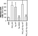


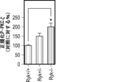


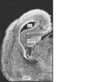


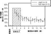


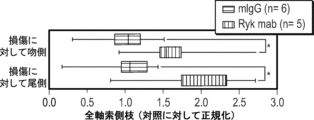


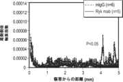


Classifications
-
- C—CHEMISTRY; METALLURGY
- C07—ORGANIC CHEMISTRY
- C07K—PEPTIDES
- C07K16/00—Immunoglobulins [IGs], e.g. monoclonal or polyclonal antibodies
- C07K16/18—Immunoglobulins [IGs], e.g. monoclonal or polyclonal antibodies against material from animals or humans
- C07K16/28—Immunoglobulins [IGs], e.g. monoclonal or polyclonal antibodies against material from animals or humans against receptors, cell surface antigens or cell surface determinants
- C07K16/2863—Immunoglobulins [IGs], e.g. monoclonal or polyclonal antibodies against material from animals or humans against receptors, cell surface antigens or cell surface determinants against receptors for growth factors, growth regulators
-
- A—HUMAN NECESSITIES
- A01—AGRICULTURE; FORESTRY; ANIMAL HUSBANDRY; HUNTING; TRAPPING; FISHING
- A01K—ANIMAL HUSBANDRY; AVICULTURE; APICULTURE; PISCICULTURE; FISHING; REARING OR BREEDING ANIMALS, NOT OTHERWISE PROVIDED FOR; NEW BREEDS OF ANIMALS
- A01K67/00—Rearing or breeding animals, not otherwise provided for; New or modified breeds of animals
- A01K67/027—New or modified breeds of vertebrates
- A01K67/0275—Genetically modified vertebrates, e.g. transgenic
- A01K67/0276—Knock-out vertebrates
-
- A—HUMAN NECESSITIES
- A61—MEDICAL OR VETERINARY SCIENCE; HYGIENE
- A61K—PREPARATIONS FOR MEDICAL, DENTAL OR TOILETRY PURPOSES
- A61K35/00—Medicinal preparations containing materials or reaction products thereof with undetermined constitution
- A61K35/12—Materials from mammals; Compositions comprising non-specified tissues or cells; Compositions comprising non-embryonic stem cells; Genetically modified cells
- A61K35/48—Reproductive organs
- A61K35/54—Ovaries; Ova; Ovules; Embryos; Foetal cells; Germ cells
- A61K35/545—Embryonic stem cells; Pluripotent stem cells; Induced pluripotent stem cells; Uncharacterised stem cells
-
- A—HUMAN NECESSITIES
- A61—MEDICAL OR VETERINARY SCIENCE; HYGIENE
- A61P—SPECIFIC THERAPEUTIC ACTIVITY OF CHEMICAL COMPOUNDS OR MEDICINAL PREPARATIONS
- A61P25/00—Drugs for disorders of the nervous system
-
- A—HUMAN NECESSITIES
- A61—MEDICAL OR VETERINARY SCIENCE; HYGIENE
- A61P—SPECIFIC THERAPEUTIC ACTIVITY OF CHEMICAL COMPOUNDS OR MEDICINAL PREPARATIONS
- A61P25/00—Drugs for disorders of the nervous system
- A61P25/28—Drugs for disorders of the nervous system for treating neurodegenerative disorders of the central nervous system, e.g. nootropic agents, cognition enhancers, drugs for treating Alzheimer's disease or other forms of dementia
-
- C—CHEMISTRY; METALLURGY
- C07—ORGANIC CHEMISTRY
- C07K—PEPTIDES
- C07K16/00—Immunoglobulins [IGs], e.g. monoclonal or polyclonal antibodies
-
- C—CHEMISTRY; METALLURGY
- C07—ORGANIC CHEMISTRY
- C07K—PEPTIDES
- C07K16/00—Immunoglobulins [IGs], e.g. monoclonal or polyclonal antibodies
- C07K16/18—Immunoglobulins [IGs], e.g. monoclonal or polyclonal antibodies against material from animals or humans
- C07K16/22—Immunoglobulins [IGs], e.g. monoclonal or polyclonal antibodies against material from animals or humans against growth factors ; against growth regulators
-
- C—CHEMISTRY; METALLURGY
- C07—ORGANIC CHEMISTRY
- C07K—PEPTIDES
- C07K16/00—Immunoglobulins [IGs], e.g. monoclonal or polyclonal antibodies
- C07K16/18—Immunoglobulins [IGs], e.g. monoclonal or polyclonal antibodies against material from animals or humans
- C07K16/28—Immunoglobulins [IGs], e.g. monoclonal or polyclonal antibodies against material from animals or humans against receptors, cell surface antigens or cell surface determinants
-
- C—CHEMISTRY; METALLURGY
- C12—BIOCHEMISTRY; BEER; SPIRITS; WINE; VINEGAR; MICROBIOLOGY; ENZYMOLOGY; MUTATION OR GENETIC ENGINEERING
- C12N—MICROORGANISMS OR ENZYMES; COMPOSITIONS THEREOF; PROPAGATING, PRESERVING, OR MAINTAINING MICROORGANISMS; MUTATION OR GENETIC ENGINEERING; CULTURE MEDIA
- C12N15/00—Mutation or genetic engineering; DNA or RNA concerning genetic engineering, vectors, e.g. plasmids, or their isolation, preparation or purification; Use of hosts therefor
- C12N15/09—Recombinant DNA-technology
- C12N15/63—Introduction of foreign genetic material using vectors; Vectors; Use of hosts therefor; Regulation of expression
- C12N15/65—Introduction of foreign genetic material using vectors; Vectors; Use of hosts therefor; Regulation of expression using markers
-
- C—CHEMISTRY; METALLURGY
- C12—BIOCHEMISTRY; BEER; SPIRITS; WINE; VINEGAR; MICROBIOLOGY; ENZYMOLOGY; MUTATION OR GENETIC ENGINEERING
- C12N—MICROORGANISMS OR ENZYMES; COMPOSITIONS THEREOF; PROPAGATING, PRESERVING, OR MAINTAINING MICROORGANISMS; MUTATION OR GENETIC ENGINEERING; CULTURE MEDIA
- C12N15/00—Mutation or genetic engineering; DNA or RNA concerning genetic engineering, vectors, e.g. plasmids, or their isolation, preparation or purification; Use of hosts therefor
- C12N15/09—Recombinant DNA-technology
- C12N15/63—Introduction of foreign genetic material using vectors; Vectors; Use of hosts therefor; Regulation of expression
- C12N15/79—Vectors or expression systems specially adapted for eukaryotic hosts
- C12N15/85—Vectors or expression systems specially adapted for eukaryotic hosts for animal cells
- C12N15/8509—Vectors or expression systems specially adapted for eukaryotic hosts for animal cells for producing genetically modified animals, e.g. transgenic
-
- G—PHYSICS
- G01—MEASURING; TESTING
- G01N—INVESTIGATING OR ANALYSING MATERIALS BY DETERMINING THEIR CHEMICAL OR PHYSICAL PROPERTIES
- G01N33/00—Investigating or analysing materials by specific methods not covered by groups G01N1/00 - G01N31/00
- G01N33/48—Biological material, e.g. blood, urine; Haemocytometers
- G01N33/50—Chemical analysis of biological material, e.g. blood, urine; Testing involving biospecific ligand binding methods; Immunological testing
- G01N33/5005—Chemical analysis of biological material, e.g. blood, urine; Testing involving biospecific ligand binding methods; Immunological testing involving human or animal cells
- G01N33/5008—Chemical analysis of biological material, e.g. blood, urine; Testing involving biospecific ligand binding methods; Immunological testing involving human or animal cells for testing or evaluating the effect of chemical or biological compounds, e.g. drugs, cosmetics
- G01N33/502—Chemical analysis of biological material, e.g. blood, urine; Testing involving biospecific ligand binding methods; Immunological testing involving human or animal cells for testing or evaluating the effect of chemical or biological compounds, e.g. drugs, cosmetics for testing non-proliferative effects
- G01N33/5023—Chemical analysis of biological material, e.g. blood, urine; Testing involving biospecific ligand binding methods; Immunological testing involving human or animal cells for testing or evaluating the effect of chemical or biological compounds, e.g. drugs, cosmetics for testing non-proliferative effects on expression patterns
-
- G—PHYSICS
- G01—MEASURING; TESTING
- G01N—INVESTIGATING OR ANALYSING MATERIALS BY DETERMINING THEIR CHEMICAL OR PHYSICAL PROPERTIES
- G01N33/00—Investigating or analysing materials by specific methods not covered by groups G01N1/00 - G01N31/00
- G01N33/48—Biological material, e.g. blood, urine; Haemocytometers
- G01N33/50—Chemical analysis of biological material, e.g. blood, urine; Testing involving biospecific ligand binding methods; Immunological testing
- G01N33/5005—Chemical analysis of biological material, e.g. blood, urine; Testing involving biospecific ligand binding methods; Immunological testing involving human or animal cells
- G01N33/5008—Chemical analysis of biological material, e.g. blood, urine; Testing involving biospecific ligand binding methods; Immunological testing involving human or animal cells for testing or evaluating the effect of chemical or biological compounds, e.g. drugs, cosmetics
- G01N33/5082—Supracellular entities, e.g. tissue, organisms
- G01N33/5088—Supracellular entities, e.g. tissue, organisms of vertebrates
-
- A—HUMAN NECESSITIES
- A01—AGRICULTURE; FORESTRY; ANIMAL HUSBANDRY; HUNTING; TRAPPING; FISHING
- A01K—ANIMAL HUSBANDRY; AVICULTURE; APICULTURE; PISCICULTURE; FISHING; REARING OR BREEDING ANIMALS, NOT OTHERWISE PROVIDED FOR; NEW BREEDS OF ANIMALS
- A01K2217/00—Genetically modified animals
- A01K2217/07—Animals genetically altered by homologous recombination
- A01K2217/075—Animals genetically altered by homologous recombination inducing loss of function, i.e. knock out
-
- A—HUMAN NECESSITIES
- A01—AGRICULTURE; FORESTRY; ANIMAL HUSBANDRY; HUNTING; TRAPPING; FISHING
- A01K—ANIMAL HUSBANDRY; AVICULTURE; APICULTURE; PISCICULTURE; FISHING; REARING OR BREEDING ANIMALS, NOT OTHERWISE PROVIDED FOR; NEW BREEDS OF ANIMALS
- A01K2227/00—Animals characterised by species
- A01K2227/10—Mammal
- A01K2227/105—Murine
-
- A—HUMAN NECESSITIES
- A01—AGRICULTURE; FORESTRY; ANIMAL HUSBANDRY; HUNTING; TRAPPING; FISHING
- A01K—ANIMAL HUSBANDRY; AVICULTURE; APICULTURE; PISCICULTURE; FISHING; REARING OR BREEDING ANIMALS, NOT OTHERWISE PROVIDED FOR; NEW BREEDS OF ANIMALS
- A01K2267/00—Animals characterised by purpose
- A01K2267/01—Animal expressing industrially exogenous proteins
-
- A—HUMAN NECESSITIES
- A01—AGRICULTURE; FORESTRY; ANIMAL HUSBANDRY; HUNTING; TRAPPING; FISHING
- A01K—ANIMAL HUSBANDRY; AVICULTURE; APICULTURE; PISCICULTURE; FISHING; REARING OR BREEDING ANIMALS, NOT OTHERWISE PROVIDED FOR; NEW BREEDS OF ANIMALS
- A01K2267/00—Animals characterised by purpose
- A01K2267/03—Animal model, e.g. for test or diseases
- A01K2267/0306—Animal model for genetic diseases
- A01K2267/0318—Animal model for neurodegenerative disease, e.g. non- Alzheimer's
-
- A—HUMAN NECESSITIES
- A01—AGRICULTURE; FORESTRY; ANIMAL HUSBANDRY; HUNTING; TRAPPING; FISHING
- A01K—ANIMAL HUSBANDRY; AVICULTURE; APICULTURE; PISCICULTURE; FISHING; REARING OR BREEDING ANIMALS, NOT OTHERWISE PROVIDED FOR; NEW BREEDS OF ANIMALS
- A01K2267/00—Animals characterised by purpose
- A01K2267/03—Animal model, e.g. for test or diseases
- A01K2267/035—Animal model for multifactorial diseases
- A01K2267/0356—Animal model for processes and diseases of the central nervous system, e.g. stress, learning, schizophrenia, pain, epilepsy
-
- A—HUMAN NECESSITIES
- A61—MEDICAL OR VETERINARY SCIENCE; HYGIENE
- A61K—PREPARATIONS FOR MEDICAL, DENTAL OR TOILETRY PURPOSES
- A61K39/00—Medicinal preparations containing antigens or antibodies
- A61K2039/505—Medicinal preparations containing antigens or antibodies comprising antibodies
-
- C—CHEMISTRY; METALLURGY
- C07—ORGANIC CHEMISTRY
- C07K—PEPTIDES
- C07K2317/00—Immunoglobulins specific features
- C07K2317/70—Immunoglobulins specific features characterized by effect upon binding to a cell or to an antigen
- C07K2317/76—Antagonist effect on antigen, e.g. neutralization or inhibition of binding
-
- C—CHEMISTRY; METALLURGY
- C12—BIOCHEMISTRY; BEER; SPIRITS; WINE; VINEGAR; MICROBIOLOGY; ENZYMOLOGY; MUTATION OR GENETIC ENGINEERING
- C12N—MICROORGANISMS OR ENZYMES; COMPOSITIONS THEREOF; PROPAGATING, PRESERVING, OR MAINTAINING MICROORGANISMS; MUTATION OR GENETIC ENGINEERING; CULTURE MEDIA
- C12N15/00—Mutation or genetic engineering; DNA or RNA concerning genetic engineering, vectors, e.g. plasmids, or their isolation, preparation or purification; Use of hosts therefor
- C12N15/09—Recombinant DNA-technology
- C12N15/63—Introduction of foreign genetic material using vectors; Vectors; Use of hosts therefor; Regulation of expression
- C12N15/79—Vectors or expression systems specially adapted for eukaryotic hosts
- C12N15/85—Vectors or expression systems specially adapted for eukaryotic hosts for animal cells
- C12N15/8509—Vectors or expression systems specially adapted for eukaryotic hosts for animal cells for producing genetically modified animals, e.g. transgenic
- C12N2015/8518—Vectors or expression systems specially adapted for eukaryotic hosts for animal cells for producing genetically modified animals, e.g. transgenic expressing industrially exogenous proteins, e.g. for pharmaceutical use, human insulin, blood factors, immunoglobulins, pseudoparticles
-
- C—CHEMISTRY; METALLURGY
- C12—BIOCHEMISTRY; BEER; SPIRITS; WINE; VINEGAR; MICROBIOLOGY; ENZYMOLOGY; MUTATION OR GENETIC ENGINEERING
- C12N—MICROORGANISMS OR ENZYMES; COMPOSITIONS THEREOF; PROPAGATING, PRESERVING, OR MAINTAINING MICROORGANISMS; MUTATION OR GENETIC ENGINEERING; CULTURE MEDIA
- C12N2800/00—Nucleic acids vectors
- C12N2800/30—Vector systems comprising sequences for excision in presence of a recombinase, e.g. loxP or FRT
-
- C—CHEMISTRY; METALLURGY
- C12—BIOCHEMISTRY; BEER; SPIRITS; WINE; VINEGAR; MICROBIOLOGY; ENZYMOLOGY; MUTATION OR GENETIC ENGINEERING
- C12Y—ENZYMES
- C12Y207/00—Transferases transferring phosphorus-containing groups (2.7)
- C12Y207/02—Phosphotransferases with a carboxy group as acceptor (2.7.2)
- C12Y207/02003—Phosphoglycerate kinase (2.7.2.3)
-
- G—PHYSICS
- G01—MEASURING; TESTING
- G01N—INVESTIGATING OR ANALYSING MATERIALS BY DETERMINING THEIR CHEMICAL OR PHYSICAL PROPERTIES
- G01N2333/00—Assays involving biological materials from specific organisms or of a specific nature
- G01N2333/90—Enzymes; Proenzymes
- G01N2333/91—Transferases (2.)
- G01N2333/912—Transferases (2.) transferring phosphorus containing groups, e.g. kinases (2.7)
Landscapes
- Health & Medical Sciences (AREA)
- Life Sciences & Earth Sciences (AREA)
- Chemical & Material Sciences (AREA)
- Engineering & Computer Science (AREA)
- Organic Chemistry (AREA)
- Immunology (AREA)
- General Health & Medical Sciences (AREA)
- Biomedical Technology (AREA)
- Genetics & Genomics (AREA)
- Medicinal Chemistry (AREA)
- Molecular Biology (AREA)
- Biochemistry (AREA)
- Bioinformatics & Cheminformatics (AREA)
- Biophysics (AREA)
- Biotechnology (AREA)
- Proteomics, Peptides & Aminoacids (AREA)
- Zoology (AREA)
- Veterinary Medicine (AREA)
- Cell Biology (AREA)
- Animal Behavior & Ethology (AREA)
- Neurology (AREA)
- Neurosurgery (AREA)
- Urology & Nephrology (AREA)
- Environmental Sciences (AREA)
- Hematology (AREA)
- Pharmacology & Pharmacy (AREA)
- Wood Science & Technology (AREA)
- Public Health (AREA)
- General Engineering & Computer Science (AREA)
- Physics & Mathematics (AREA)
- Microbiology (AREA)
- Developmental Biology & Embryology (AREA)
- Nuclear Medicine, Radiotherapy & Molecular Imaging (AREA)
- Chemical Kinetics & Catalysis (AREA)
- General Chemical & Material Sciences (AREA)
- Reproductive Health (AREA)
- General Physics & Mathematics (AREA)
- Animal Husbandry (AREA)
- Food Science & Technology (AREA)
- Toxicology (AREA)
Description
関連出願の相互参照
本出願は、2016年3月28日に出願された米国特許仮出願第62/314,025号に対する35 U.S.C.§119(e)に基づく優先権の恩典を主張し、この仮出願の開示内容は参照により本明細書に組み入れられる。
CROSS-REFERENCE TO RELATED APPLICATIONS This application claims the benefit of priority under 35 USC § 119(e) to U.S. Provisional Application No. 62/314,025, filed March 28, 2016, and The disclosure is incorporated herein by reference.
助成金情報
本発明は、米国立衛生研究所によって付与された助成金番号NS047484およびNS081738での政府による補助を伴ってなされた。米国政府は、本発明において一定の権利を有する。
Grant Information This invention was made with government support under Grant Numbers NS047484 and NS081738 awarded by the National Institutes of Health. The United States Government has certain rights in this invention.
配列表
本出願は配列表を含み、配列表はASCII形式で電子形式により提出されており、参照によりその全体が本明細書に組み入れられる。前記のASCIIコピーは、2017年3月15日に作成され、名称は20378-201389_SL.txtであり、サイズは14,104バイトである。
SEQUENCE LISTING This application contains a Sequence Listing, which is submitted in electronic form in ASCII format and is incorporated herein by reference in its entirety. The above ASCII copy was created on March 15, 2017, is named 20378-201389_SL.txt, and is 14,104 bytes in size.
発明の分野
本発明は、一般的には抗体に関し、より詳しくは、Wnt-Rykシグナル伝達を阻害するためのWntの結合ドメインに特異的に結合する抗Ryk抗体または抗体断片の使用に関する。
FIELD OF THE INVENTION The present invention relates generally to antibodies, and more particularly to the use of anti-Ryk antibodies or antibody fragments that specifically bind to the binding domain of Wnt to inhibit Wnt-Ryk signaling.
背景情報
中枢神経系(CNS)は、上行性の感覚経路および下行性の運動経路または調節経路に接続されている。CNSでは、体性感覚経路が脳中枢へと上行し、身体の動きをコントロールする運動経路は脳から脊髄へと下行する。末梢神経系とは異なり、中枢神経系の軸索、例えば脊髄内の軸索に対するダメージは修復が不可能であり、神経機能の永久的な障害、例えば麻痺が引き起こされ得る。脊髄は中枢神経系において重要な機能を果たしている。かかる機能の1つは、体と脳の連絡を可能にすることである。脊髄内の神経線維が脳へ、および脳から他の身体部分へ情報を伝達する。一般に、身体からの感覚情報は脊髄を通って脳まで伝わり、脳からの指令、例えば運動指令が脳から脊髄を通って伝わる。
Background Information The central nervous system (CNS) is connected to ascending sensory pathways and descending motor or regulatory pathways. In the CNS, somatosensory pathways ascend to brain centers, and motor pathways that control body movement descend from the brain to the spinal cord. Unlike the peripheral nervous system, damage to central nervous system axons, such as those within the spinal cord, is irreparable and can cause permanent impairment of nerve function, such as paralysis. The spinal cord plays important functions in the central nervous system. One such function is to allow communication between the body and the brain. Nerve fibers in the spinal cord transmit information to the brain and from the brain to other parts of the body. Generally, sensory information from the body is transmitted to the brain through the spinal cord, and commands from the brain, such as motor commands, are transmitted from the brain through the spinal cord.
「脊髄損傷」という用語は、脊柱管内のニューロンの任意の損傷をいう。脊髄損傷は、背骨または脊髄自体に対する外傷または疾患のいずれかにより起こり得る。ほとんどの脊髄損傷は、背骨に対する外傷により骨折が引き起こされたか、または背骨のずれによって靭帯が裂け、脊髄の圧迫が生じた結果である。頸部損傷および腰背部損傷の大半または脊椎骨折では、なんら脊髄のダメージは引き起こされない;しかしながら、脊椎外傷が起こっている症例の10~14%では、このダメージは非常に重症であり、脊髄に対するダメージがもたらされる。脊髄損傷(SCI)の年間発生数(事故現場で死亡した人を含めない)は米国の人口100万人あたりおよそ40例であるか、または毎年の新患がおよそ11,000例であると推計されている。米国において、現在生存していてSCIを有する人の数は人口100万人あたり721~906人であると推計されている。これは183,000~230,000人に相当する。脊髄損傷を有する患者に対する治療選択肢は限られている。多くの場合、SCIを有する患者は、重度で永久的に身体障害のままである。 The term "spinal cord injury" refers to any damage to neurons within the spinal canal. Spinal cord injuries can result from either trauma or disease to the spine or the spinal cord itself. Most spinal cord injuries are the result of trauma to the spine causing fractures or misalignment of the spine that tears ligaments and compresses the spinal cord. Most neck and lumbar back injuries or vertebral fractures do not cause any spinal cord damage; however, in 10-14% of cases where spinal trauma occurs, this damage is so severe that it causes damage to the spinal cord. damage is caused. The annual incidence of spinal cord injury (SCI) (not including those who die at the scene of an accident) is estimated to be approximately 40 cases per million people in the United States, or approximately 11,000 new cases each year. There is. In the United States, the number of people living with SCI is estimated to be between 721 and 906 per million people. This equates to between 183,000 and 230,000 people. Treatment options for patients with spinal cord injuries are limited. Patients with SCI often remain severely and permanently disabled.
近年、発生初期におけるモルフォゲンとしての方がよく知られているWntが、脊椎動物および無脊椎動物の両方において、神経系の接続時の保存された軸索ガイダンス分子であることを示唆する証拠が増えている(Zou, Y. 2004. Trends Neurosci 27:528-32(非特許文献1); Fradkin, et al. 2005. J Neurosci 25:10376-8(非特許文献2); Zou & Lyuksyutova. 2007. Curr Opin Neurobiol 17:22-8(非特許文献3); Salinas, et al. 2008. Annu Rev Neurosci 31 :339-358(非特許文献4))。Wntは分泌型糖タンパク質であり、3つのクラスの受容体Frizzleds、RykおよびROR2に結合する(Gordon & Nusse. 2006. J Biol Chem 281 :22429-33(非特許文献5); Logan & Nusse. 2004. Annu Rev Cell Dev Biol 20:781 -810(非特許文献6))。Wntファミリータンパク質は、発生時の脊髄のA-P軸に沿った網膜視蓋の伸長におけるトポグラフィックマップ形成の必須のガイダンス合図であり、脊髄損傷後の成人CNSの軸索再生の調節において重要な役割を果たし得ることが示されている(Lyuksyutova et al. 2003. Science 302: 1984-8(非特許文献7); Liu, et al. 2005. Nat Neurosci 8: 1151-9(非特許文献8); Schmitt et al. 2006. Nature 439:31 -7(非特許文献9); Wolf et al. 2008. J Neurosci 28:3456-67(非特許文献10); Liu et al. 2008. The Journal of Neuroscience 28: 8376-8382(非特許文献11))。 In recent years, increasing evidence suggests that Wnts, better known as morphogens during early development, are conserved axonal guidance molecules during nervous system connections in both vertebrates and invertebrates. Zou & Lyuksyutova. 2007. Curr Opin Neurobiol 17:22-8 (Non-Patent Document 3); Salinas, et al. 2008. Annu Rev Neurosci 31 :339-358 (Non-Patent Document 4)). Wnts are secreted glycoproteins that bind to three classes of receptors, Frizzleds, Ryk, and ROR2 (Gordon & Nusse. 2006. J Biol Chem 281 :22429-33; Logan & Nusse. 2004 Annu Rev Cell Dev Biol 20:781 -810 (Non-Patent Document 6)). Wnt family proteins are essential guidance cues for topographic map formation in retinotectal elongation along the A-P axis of the spinal cord during development and play an important role in regulating axonal regeneration in the adult CNS after spinal cord injury. (Lyuksyutova et al. 2003. Science 302: 1984-8 (Non-patent document 7); Liu, et al. 2005. Nat Neurosci 8: 1151-9 (Non-patent document 8); Schmitt et al. 2006. Nature 439:31 -7 (Non-patent document 9); Wolf et al. 2008. J Neurosci 28:3456-67 (Non-patent document 10); Liu et al. 2008. The Journal of Neuroscience 28: 8376-8382 (Non-Patent Document 11)).
背側脊髄から派生する交連軸索がまず、背側-腹側の軸に沿って伸長し、腹側正中線の底板に向かって成長する。交連性軸索の腹側に指向される成長は反発性合図によってガイドされ、BMPが背側正中線の蓋板から広がり、誘引合図ネトリン-1およびソニックヘッジホッグが底板から分泌される(Zou et al. 2007. Curr Opin Neurobiol 17:22-8(非特許文献3))。このような交連性軸索が正中線で交差したら、これらは正中線誘引物質に対する応答性を失い、化学忌避物質スリットおよびセマフォリンに対する感受性を獲得し、底板および近隣の腹側の灰白質から広がり、これらを縦の軌道(longitudinal trajectory)内に90°曲げる(Zou et al. 2000. Cell 102:363-75(非特許文献12))。齧歯類の交連性軸索の背側の集団はすべて前方に曲がり、脳に向かって伸長している。このように前方に曲がるのにWnt-Frizzledシグナル伝達が必要とされる。いくつかのWnt、例えばWnt4、Wnt7b、Wnt7aおよびWnt5aが前後軸方向に漸減勾配で腹側正中線の脊髄に沿って発現されており、正中線と交差して前方に曲がった交差後の交連性軸索を誘引する。Wnt勾配をWnt阻害薬である分泌型Frizzled関連タンパク質(sFRP)の添加によって、または「オープンブック(open-book)」移植片培養状態のWnt4分泌細胞凝集塊を配置することにより破壊すると、交連性軸索は正中線交差後に特異的A-P無作為化成長を示す。Frizzled3変異型の胚では、脊髄交連神経軸索はインビボでA-P指向性を失う(Lyuksyutova et al. 2003. Science 302: 1984-8(非特許文献7))。 Commissural axons derived from the dorsal spinal cord first extend along the dorsal-ventral axis and grow towards the floor plate in the ventral midline. Ventrally directed growth of commissural axons is guided by repulsive cues, with BMPs spreading from the dorsal midline tectal plate and attractive cues Netrin-1 and Sonic Hedgehog being secreted from the basal plate (Zou et al. al. 2007. Curr Opin Neurobiol 17:22-8 (Non-Patent Document 3)). Once such commissural axons cross at the midline, they lose responsiveness to midline attractants, acquire sensitivity to the chemorepellents Slit and semaphorins, and spread from the floor plate and adjacent ventral gray matter. , bending them by 90° into a longitudinal trajectory (Zou et al. 2000. Cell 102:363-75). The dorsal population of rodent commissural axons all curve forward and extend toward the brain. Wnt-Frizzled signaling is required for this forward bend. Several Wnts, such as Wnt4, Wnt7b, Wnt7a and Wnt5a, are expressed along the ventral midline spinal cord in an anteroposteriorly axially decreasing gradient, crossing the midline to the anteriorly curved postcrossing commissure. Attracts axons. Disrupting the Wnt gradient by the addition of the Wnt inhibitor secreted Frizzled-related protein (sFRP) or by placing Wnt4-secreting cell aggregates in “open-book” explant culture conditions Axons show specific A-P randomized growth after midline crossing. In Frizzled3 mutant embryos, spinal commissural nerve axons lose A-P orientation in vivo (Lyuksyutova et al. 2003. Science 302: 1984-8 (Non-Patent Document 7)).
ショウジョウバエの正中線の軸索の経路探索における研究では、独立して、DWnt5が化学忌避物質であり、Derailedと称される受容体によって交連性軸索のサブセットを忌避することが示された(Yoshikawa et al. 2003. Nature 422:583-8(非特許文献13))。Derailedの脊椎動物ホモログであるRykもまた反発性Wnt受容体であり、Wnt1とWnt5aの差次的発現によって作られる前方-高/後方-低のWnt勾配が脊髄内の皮質脊髄路の軸索の後方成長に必要とされる(Liu et al. 2005. Nat Neurosci 8: 1151-9(非特許文献8))。したがって、Wntは、脊髄内の上行性軸索と下行性軸索両方のA-Pガイダンスを、誘引性および反発性ガイダンス機構によって制御する。Wnt-Rykシグナル伝達もまた、哺乳動物の前脳の脳梁の経路探索を反発性機構によって調節することが見出された(Keeble et al. 2006. J Neurosci 26:5840-8(非特許文献14))。C.エレガンス(C. elegans)での研究により、Wntシグナル伝達によっていくつかの軸索の経路探索の前後軸方向の指向性および神経芽細胞の遊走が制御されることが示された(Pan et al. 2006. Dev Cell 10:367-77(非特許文献15); Hilliard et al. 2006. Dev Cell 10:379-90(非特許文献16); Prasad & Clark. 2006. Development 133: 1757-66(非特許文献17))。したがって、A-Pガイダンス機構は、動物界において高度に保存されているようである(Zou. 2006. Neuron 49:787-9(非特許文献18))。 Studies in Drosophila midline axon pathfinding independently showed that DWnt5 is a chemorepellent, repelling a subset of commissural axons through a receptor termed Derailed (Yoshikawa et al. 2003. Nature 422:583-8 (Non-Patent Document 13)). Ryk, the vertebrate homologue of Derailed, is also a repulsive Wnt receptor, and an anterior-high/posterior-low Wnt gradient created by differential expression of Wnt1 and Wnt5a induces axonal control in the corticospinal tract in the spinal cord. Required for backward growth (Liu et al. 2005. Nat Neurosci 8: 1151-9 (Non-Patent Document 8)). Thus, Wnts control A-P guidance of both ascending and descending axons within the spinal cord through attractive and repulsive guidance mechanisms. Wnt-Ryk signaling was also found to regulate pathfinding in the mammalian forebrain callosum by a repulsive mechanism (Keeble et al. 2006. J Neurosci 26:5840-8). 14)). Studies in C. elegans have shown that Wnt signaling controls the anterior-posterior axial orientation of some axon pathfinding and neuroblast migration (Pan et al. al. 2006. Dev Cell 10:367-77 (non-patent document 15); Hilliard et al. 2006. Dev Cell 10:379-90 (non-patent document 16); Prasad & Clark. 2006. Development 133: 1757-66 (Non-patent document 17)). Therefore, the A-P guidance mechanism appears to be highly conserved in the animal kingdom (Zou. 2006. Neuron 49:787-9).
軸索の経路探索におけるWntの役割に加えて、Wnt3はまた、網膜視蓋系におけるトポグラフィックマッピングのための位置的合図であり、網膜神経節細胞の軸索に対する外側指向性マッピング力として作用し、視蓋内のephrinB1の勾配によって生じる内側指向性の力に対抗する(Schmitt et al. 2006. Nature 439:31-7(非特許文献9))。Wnt3は、視蓋において内側-高から外側-低に向かう勾配で発現される。Rykは、網膜神経節細胞において背側-腹側(D-V)に漸増勾配で発現される。したがって、より腹側のRGCの軸索分岐ほどより強くWnt3によって忌避され、Wnt3-Rykシグナル伝達によって介在性(interstitial)分岐が外側中脳視蓋に向かって成長することが駆動される。その一方で、ephrinB1は同じ等級の様式で発現され、EphBは腹側のRGCにおいて高レベルで発現される。EphBは、ephrinB1に対する誘引を媒介する。したがって、より腹側のRGCの軸索分岐ほど、より多くephinB1によって内側中脳視蓋に向かって誘引される。内側(ephrinB1)と外側(Wnt3)のマッピング力の均衡作用により、RGCの軸索が正しいトポグラフィック位置で終結することが確実になる。注目すべきことに、Wnt-Frizzledシグナル伝達はまた、ショウジョウバエの視覚系における適正な背側-腹側の網膜部位のマッピングにも必要とされる(Sato et al. 2006. Nat Neurosci 9:67-75(非特許文献19); Zou & Lyuksyutova. 2007. Curr Opin Neurobiol 17:22-8(非特許文献3))。したがって、Wntは、D-V軸に沿って保存されるトポグラフィックマッピング合図である。 In addition to the role of Wnt in axon pathfinding, Wnt3 is also a positional cue for topographic mapping in the retinotectorial system and acts as an outward-directed mapping force for axons of retinal ganglion cells. , countering the inwardly directed forces generated by the ephrinB1 gradient within the optic tectum (Schmitt et al. 2006. Nature 439:31-7). Wnt3 is expressed in a medial-high to lateral-low gradient in the optic tectum. Ryk is expressed in a dorsal-ventral (D-V) increasing gradient in retinal ganglion cells. Therefore, more ventral RGC axonal branches are more strongly repelled by Wnt3, and Wnt3-Ryk signaling drives interstitial branches to grow toward the lateral mesencephalic tectum. On the other hand, ephrinB1 is expressed in the same graded manner and EphB is expressed at high levels in ventral RGCs. EphB mediates attraction to ephrinB1. Therefore, the more ventral RGC axonal branches are attracted by ephinB1 toward the medial mesencephalic tectum. The balanced action of medial (ephrinB1) and lateral (Wnt3) mapping forces ensures that RGC axons terminate in the correct topographic location. Notably, Wnt-Frizzled signaling is also required for proper dorsal-ventral retinal site mapping in the Drosophila visual system (Sato et al. 2006. Nat Neurosci 9:67- 75 (Non-Patent Document 19); Zou & Lyuksyutova. 2007. Curr Opin Neurobiol 17:22-8 (Non-Patent Document 3)). Therefore, Wnts are topographic mapping cues that are conserved along the D-V axis.
発生中の脊髄の交連軸索は、腹側正中線の方に、それぞれ正中線の底板細胞と蓋板細胞によって分泌される化学誘引物質(ネトリン-1およびソニックヘッジホッグ(Shh))と化学忌避物質(骨形成タンパク質(BMP))の協働によってガイドされる。この軸索は、底板に達したら、底板からの化学誘引物質に対する自身の応答性をスイッチオフし、同様に底板細胞によって、および周囲の腹側の灰白質によって発現される化学反発性合図、例えば、クラス3セマフォリンの構成員(Sema3BおよびSema3F)ならびにスリットファミリータンパク質に対して応答性になる(Serafini, T., et al. Cell 78, 409-424, 1994(非特許文献20); Kennedy, T. E., et al. Cell 78, 425-435, 1994(非特許文献21); Serafini, T., et al. Cell 87, 1001-1014, 1996(非特許文献22); Zou, Y., et al. Cell 102, 363-375, 2000(非特許文献23); Charron, F., et al. Cell 113, 11-23, 2003(非特許文献24); Long, H., et al. Neuron 42, 213-223, 2004(非特許文献25))。ニューロピリン-2変異型の胚は、重度のガイダンス欠陥を示し、例えば正中線に入ったり、脊髄の反対側に出たり、前後軸に沿ってランダムに伸長したりした(Zou, Y., et al. Cell 102, 363-375, 2000(非特許文献23))。
Commissural axons in the developing spinal cord are directed toward the ventral midline by chemoattractants (netrin-1 and sonic hedgehog (Shh)) and chemorepellents secreted by midline floor plate cells and tectal plate cells, respectively. It is guided by the cooperation of substances (bone morphogenetic proteins (BMPs)). Once this axon reaches the floor plate, it switches off its responsiveness to chemoattractants from the floor plate, as well as chemorepulsive cues expressed by floor plate cells and by the surrounding ventral gray matter, e.g. , are responsive to
SCI後のニューロンの成長および再生のモジュレータの同定は、この衰弱性の病的状態を有する患者の新たな治療形態に応用することができよう。また、ニューロンの成長および再生のモジュレータの同定は、ニューロンの機能障害を伴う他の障害、例えば神経性/神経変性性の疾患または障害を有する患者の処置に応用することもできよう。脊髄に対する損傷後にA-P軸に沿った軸索の成長を促進させ得る作用物質は、多くの場合でSCIに伴う永久的な麻痺の抑制を補助するために適用され得る。したがって、より良好なSCIの処置の必要性が存在しており、ニューロンの成長および再生のモジュレータのさらに大きな理解によって、かかる神経性/神経変性性の疾患または障害の改善された処置方法がもたらされる可能性がある。 Identification of modulators of neuronal growth and regeneration after SCI could be applied to new forms of treatment for patients with this debilitating pathological condition. Identification of modulators of neuronal growth and regeneration could also have application in the treatment of patients with other disorders involving neuronal dysfunction, such as neurological/neurodegenerative diseases or disorders. Agents that can promote axonal growth along the A-P axis after injury to the spinal cord can be applied to help suppress the permanent paralysis that often accompanies SCI. Therefore, there is a need for better treatment of SCI, and a greater understanding of modulators of neuronal growth and regeneration will lead to improved treatment methods for such neurological/neurodegenerative diseases or disorders. there is a possibility.
本発明は、Wntの結合ドメインに特異的に結合する抗Ryk抗体または抗体断片がWnt-Rykシグナル伝達を阻害するという所見に基づいている。そのため、該抗Ryk抗体または抗体断片は、脊髄が損傷している場合の哺乳動物のニューロンの指向的成長をモジュレートするため、ならびにニューロンの変性を抑止するため、および神経変性疾患を処置するために使用され得る。 The present invention is based on the finding that anti-Ryk antibodies or antibody fragments that specifically bind to the binding domain of Wnt inhibit Wnt-Ryk signaling. Therefore, the anti-Ryk antibody or antibody fragment can be used to modulate the directed growth of neurons in mammals when the spinal cord is injured, as well as to prevent neuronal degeneration and to treat neurodegenerative diseases. can be used for.
したがって、一局面において、本発明は、Wntの結合ドメインに特異的に結合するか、あるいは、Wnt上の、参照抗体もしくは参照抗体断片が結合するのと同じエピトープに特異的に結合するか、またはWntに対する特異的結合について参照抗体もしくは参照抗体断片と交差競合(cross-compete)する単離された抗Ryk抗体または抗体断片を提供する。種々の態様において、該抗体もしくは抗体断片および/または該参照抗体もしくは参照抗体断片は、SEQ ID NO:5~7もしくはSEQ ID NO:5、11および12に示すCDR配列を含む重鎖可変領域;ならびに/またはSEQ ID NO:1~3もしくはSEQ ID NO:9、2および3に示すCDR配列を含む軽鎖可変領域を含む。種々の態様において、該抗体または抗体断片はWntの90~183位のアミノ酸残基内のエピトープに特異的に結合する。種々の態様において、該抗体または抗体断片の重鎖可変領域は、SEQ ID NO:8または13に対する少なくとも85%の配列同一性、少なくとも90%の配列同一性、少なくとも95%の配列同一性または少なくとも99%の配列同一性を含むアミノ酸配列を含む。種々の態様において、該抗体または抗体断片の軽鎖可変領域は、SEQ ID NO:4または10に対する少なくとも85%の配列同一性、少なくとも90%の配列同一性、少なくとも95%の配列同一性または少なくとも99%の配列同一性を含むアミノ酸配列を含む。種々の態様において、該抗体または抗体断片は薬学的に許容される担体または賦形剤中において製剤化される。 Thus, in one aspect, the present invention specifically binds to the binding domain of Wnt, or specifically binds to the same epitope on Wnt that the reference antibody or reference antibody fragment binds, or An isolated anti-Ryk antibody or antibody fragment is provided that cross-competes with a reference antibody or antibody fragment for specific binding to Wnt. In various embodiments, the antibody or antibody fragment and/or the reference antibody or reference antibody fragment comprises a heavy chain variable region comprising the CDR sequences set forth in SEQ ID NO: 5-7 or SEQ ID NO: 5, 11 and 12; and/or a light chain variable region comprising the CDR sequences shown in SEQ ID NO: 1-3 or SEQ ID NO: 9, 2 and 3. In various embodiments, the antibody or antibody fragment specifically binds to an epitope within amino acid residues 90-183 of Wnt. In various embodiments, the heavy chain variable region of the antibody or antibody fragment has at least 85% sequence identity, at least 90% sequence identity, at least 95% sequence identity, or at least Contains amino acid sequences that contain 99% sequence identity. In various embodiments, the light chain variable region of the antibody or antibody fragment has at least 85% sequence identity, at least 90% sequence identity, at least 95% sequence identity, or at least Contains amino acid sequences that contain 99% sequence identity. In various embodiments, the antibody or antibody fragment is formulated in a pharmaceutically acceptable carrier or excipient.
別の局面において、本発明は、本明細書に記載の単離された抗体または抗体断片をコードしている核酸配列を提供する。また、該核酸配列を含むベクター、例えば発現ベクターも提供する。また、該ベクターを含む宿主細胞、例えば哺乳動物宿主細胞も提供する。 In another aspect, the invention provides nucleic acid sequences encoding isolated antibodies or antibody fragments described herein. Also provided are vectors, such as expression vectors, containing the nucleic acid sequences. Also provided are host cells, such as mammalian host cells, containing the vectors.
別の局面において、本発明は、治療剤、例えば細胞毒素または放射性同位体と連結された該単離された抗Ryk抗体または抗体断片のイムノコンジュゲートを提供する。別の局面において、本発明は、該単離された抗Ryk抗体または抗体断片とは異なる結合特異性を有する第2の機能性部分と連結された該単離された抗Ryk抗体または抗体断片を含む二重特異性分子、例えば二重特異性抗体を提供する。 In another aspect, the invention provides an immunoconjugate of the isolated anti-Ryk antibody or antibody fragment linked to a therapeutic agent, such as a cytotoxin or a radioactive isotope. In another aspect, the invention provides the isolated anti-Ryk antibody or antibody fragment linked to a second functional moiety having a different binding specificity than the isolated anti-Ryk antibody or antibody fragment. Bispecific molecules, such as bispecific antibodies, are provided.
別の局面において、本発明は、WntとRykの相互作用に干渉する方法を提供する。該方法は、WntとRykを含む試料を本明細書に記載の単離された抗体または抗体断片と接触させ、それによりWntとRykの相互作用に干渉することを含む。 In another aspect, the invention provides a method of interfering with the interaction of Wnt and Ryk. The method includes contacting a sample containing Wnt and Ryk with an isolated antibody or antibody fragment described herein, thereby interfering with the interaction of Wnt and Ryk.
別の局面において、本発明は、ニューロンの変性を抑止する方法を提供する。該方法は、ニューロンを本明細書に記載の単離された抗体または抗体断片と接触させ、それにより該ニューロンの変性を抑止することを含む。種々の態様において、ニューロンの軸索の変性が抑止されるか、またはニューロンの細胞体の変性が抑止される。種々の態様において、軸索は脊髄交連神経軸索、上位運動ニューロンの軸索または中枢神経系の軸索である。種々の態様において、ニューロンは、損傷した脊髄のニューロン、感覚ニューロン、運動ニューロン、小脳顆粒ニューロン、後根神経節ニューロン、皮質ニューロン、交換神経ニューロンまたは海馬ニューロンである。種々の態様において、ニューロンは神経移植片または神経移植組織の一部を形成する。種々の態様において、ニューロンはエクスビボまたはインビトロである。種々の態様において、神経移植片または神経移植組織は生物体、例えば哺乳動物またはヒトの一部を形成する。 In another aspect, the invention provides a method of inhibiting neuronal degeneration. The method includes contacting a neuron with an isolated antibody or antibody fragment described herein, thereby inhibiting degeneration of the neuron. In various embodiments, degeneration of a neuron's axon is inhibited or degeneration of a neuron's cell body is inhibited. In various embodiments, the axon is a spinal commissure axon, an upper motor neuron axon, or a central nervous system axon. In various embodiments, the neuron is an injured spinal cord neuron, a sensory neuron, a motor neuron, a cerebellar granule neuron, a dorsal root ganglion neuron, a cortical neuron, a sympathetic neuron, or a hippocampal neuron. In various embodiments, the neurons form part of a neural graft or tissue. In various embodiments, the neurons are ex vivo or in vitro. In various embodiments, the neural graft or tissue forms part of an organism, such as a mammal or a human.
別の局面において、本発明は、神経性の疾患もしくは障害、例えば神経変性性の疾患もしくは障害を有するか、または該疾患もしくは障害を発症するリスクがある対象において、神経性の疾患または障害、例えば神経変性性の疾患または障害を処置する方法を提供する。該方法は、該対象に本明細書に記載の単離された抗体または抗体断片を投与し、それにより該対象の神経性の疾患または障害、例えば神経変性性の疾患または障害を処置することを含む。種々の態様において、神経変性疾患は筋萎縮性側索硬化症、アルツハイマー病またはパーキンソン病である。 In another aspect, the invention provides a method for treating a neurological disease or disorder, e.g., in a subject having or at risk of developing a neurodegenerative disease or disorder, e.g. Methods of treating neurodegenerative diseases or disorders are provided. The method comprises administering to the subject an isolated antibody or antibody fragment described herein, thereby treating a neurological disease or disorder, such as a neurodegenerative disease or disorder, in the subject. include. In various embodiments, the neurodegenerative disease is amyotrophic lateral sclerosis, Alzheimer's disease or Parkinson's disease.
別の局面において、本発明は、哺乳動物のニューロンの指向的成長をモジュレートするための方法を提供する。該方法は、ニューロンを本明細書に記載の単離された抗体または抗体断片と接触させ、それにより該ニューロン、脊髄交連神経軸索、上位運動ニューロンの軸索または中枢神経系の軸索の指向的成長をモジュレートすることを含む。種々の態様において、ニューロンは、損傷した脊髄のニューロン、感覚ニューロン、運動ニューロン、小脳顆粒ニューロン、後根神経節ニューロン、皮質ニューロン、交換神経ニューロンまたは海馬ニューロンである。種々の態様において、ニューロンは神経移植片または神経移植組織の一部を形成する。種々の態様において、ニューロンはエクスビボまたはインビトロである。種々の態様において、ニューロンの指向的成長がニューロンの再生を助長する。 In another aspect, the invention provides a method for modulating directional growth of neurons in a mammal. The method comprises contacting a neuron with an isolated antibody or antibody fragment described herein, thereby directing the neuron, spinal commissural axons, upper motor neuron axons, or central nervous system axons. including modulating growth. In various embodiments, the neuron is an injured spinal cord neuron, a sensory neuron, a motor neuron, a cerebellar granule neuron, a dorsal root ganglion neuron, a cortical neuron, a sympathetic neuron, or a hippocampal neuron. In various embodiments, the neurons form part of a neural graft or tissue. In various embodiments, the neurons are ex vivo or in vitro. In various embodiments, directed growth of neurons facilitates neuron regeneration.
別の局面において、本発明は、そのゲノムがRyk遺伝子のヘテロ接合性またはホモ接合性の欠失、不活性化またはノックアウトを含むトランスジェニック非ヒト哺乳動物、例えばマウスを提供する。種々の態様において、マウスは表現型Frizzled3-/- Ryk+/-を有する。種々の態様においてマウスは、Ryk遺伝子の条件付き破壊、例えば、Ryk遺伝子の皮質脊髄路(CST)特異的破壊を含む。種々の態様において、破壊されたRyk遺伝子は、組換えRykアレル、選択可能マーカー、該選択可能マーカーに隣接しているfrt部位、および該アレルの一部分に隣接しているloxP部位を含む。該マーカーはPGK Neoであり得、loxP部位は該アレルの3~6位のエキソンに隣接していてもよい。したがって、本発明ではまた、該トランスジェニック非ヒト哺乳動物に由来する単離された細胞も提供する。また、そのゲノムがRyk遺伝子のヘテロ接合性またはホモ接合性の欠失、不活性化またはノックアウトを含むトランスジェニック非ヒト哺乳動物を作製するためのベクターも提供する。例示的なベクターには、Ryk遺伝子の一部分であって、該Ryk遺伝子の3~6位のエキソンが3’loxP部位および5’loxP部位に隣接されている、該Ryk遺伝子の一部分と、6位のエキソンと該5’loxP部位との間にある選択可能マーカーと、該選択可能マーカーに隣接しているfrt部位とが含まれ得る。 In another aspect, the invention provides a transgenic non-human mammal, such as a mouse, whose genome comprises a heterozygous or homozygous deletion, inactivation or knockout of the Ryk gene. In various embodiments, the mouse has a Frizzled3 −/− Ryk +/− phenotype. In various embodiments, the mouse comprises a conditional disruption of the Ryk gene, eg, a corticospinal tract (CST)-specific disruption of the Ryk gene. In various embodiments, the disrupted Ryk gene includes a recombinant Ryk allele, a selectable marker, a frt site flanking the selectable marker, and a loxP site flanking a portion of the allele. The marker may be PGK Neo, and the loxP site may flank exons 3-6 of the allele. Accordingly, the invention also provides isolated cells derived from said transgenic non-human mammal. Also provided are vectors for producing transgenic non-human mammals whose genomes contain a heterozygous or homozygous deletion, inactivation or knockout of the Ryk gene. Exemplary vectors include a portion of the Ryk gene, wherein exons 3-6 of the Ryk gene are flanked by 3'loxP and 5'loxP sites; and an frt site adjacent to the selectable marker.
別の局面において、本発明は、Ryk遺伝子の条件付き破壊、例えばRyk遺伝子内にCSTを標的とする破壊を有するノックアウトマウスの作製方法を提供する。該方法は、上記のベクターをマウス胚性幹(ES)細胞集団にトランスフェクトすること、前記選択可能マーカーを発現しているトランスフェクトされたES細胞を選択すること、前記トランスフェクトされたES細胞を前記マウスの祖先胚内に導入すること、前記胚を満期まで発育させ、その生殖細胞系内にコンディショナルノックアウト構築物を有するキメラマウスを作製すること、前記キメラマウスを交配し、条件付きで破壊可能なRyk遺伝子を有するヘテロ接合体マウスを作製すること、および前記ヘテロ接合体マウスを、皮質脊髄軸索においてのみtdTomato発現を抑制するloxP隣接停止カセットを含むマウスと交配し、Ryk遺伝子の条件付き破壊、例えばRyk遺伝子内にCST特異的破壊を有するマウスを作製することを含む。 In another aspect, the invention provides a method for producing a knockout mouse having a conditional disruption of the Ryk gene, such as a CST-targeted disruption within the Ryk gene. The method comprises: transfecting a population of mouse embryonic stem (ES) cells with the vector described above; selecting transfected ES cells expressing said selectable marker; into an ancestral embryo of said mouse, allowing said embryo to develop to term and producing a chimeric mouse having a conditional knockout construct in its germline, breeding said chimeric mouse and conditionally destroying it. generating a heterozygous mouse with a possible Ryk gene, and crossing said heterozygous mouse with a mouse containing a loxP-flanked stop cassette that suppresses tdTomato expression only in corticospinal axons, resulting in a conditional expression of the Ryk gene. disruptions, such as creating mice with CST-specific disruptions in the Ryk gene.
別の局面において、本発明は、非定型プロテインキナーゼC(aPKC)またはMARK2の量、レベルおよび/または活性をモジュレートする作用物質のスクリーニング方法を提供する。例えば、本明細書において提供するのは、神経性の疾患または障害を処置するための治療剤のスクリーニングのスクリーニング方法である。該方法は、被験剤を本明細書に記載のトランスジェニック非ヒト哺乳動物に投与すること、および非定型プロテインキナーゼC(aPKC)もしくはMARK2タンパク質の量、aPKCもしくはMARK2の活性レベルまたはaPKCもしくはMARK2レベルのうちの少なくとも1つに対する該被験剤の効果を、該トランスジェニック非ヒト哺乳動物の少なくとも1つの疾患関連組織において評価することを含み、ここで、該被験剤を投与されていない同様のトランスジェニック非ヒト哺乳動物と比べたときの少なくとも1つの疾患関連組織における、aPKCタンパク質の量の減少、MARK2タンパク質の量の増加、aPKC活性レベルの低下、MARK2活性レベルの上昇、aPKCレベルの低下またはMARK2レベルの上昇のうちの少なくとも1つは、該被験剤が該神経性の疾患または障害の治療に役立つことを示す。
[本発明1001]
(a)Wntの結合ドメインに特異的に結合し、SEQ ID NO:5~7もしくはSEQ ID NO:5、11および12に示すCDR配列を含む重鎖可変領域;ならびに/またはSEQ ID NO:1~3もしくはSEQ ID NO:9、2および3に示すCDR配列を含む軽鎖可変領域を含むか;または
(b)Wnt上の、参照抗体もしくは参照抗体断片が結合するのと同じエピトープに特異的に結合するか、またはWntに対する特異的結合について参照抗体もしくは参照抗体断片と交差競合し、該参照抗体または参照抗体断片がSEQ ID NO:5~7もしくはSEQ ID NO:5、11および12に示すCDR配列を含む重鎖可変領域;ならびに/またはSEQ ID NO:1~3もしくはSEQ ID NO:9、2および3に示すCDR配列を含む軽鎖可変領域を含む、
単離された抗Ryk抗体または抗体断片。
[本発明1002]
Wntに対するRykの結合を阻害または低減する、本発明1001の単離された抗Ryk抗体または抗体断片。
[本発明1003]
Wntの90~183位のアミノ酸残基内のエピトープに特異的に結合する、本発明1001の単離された抗Ryk抗体または抗体断片。
[本発明1004]
前記重鎖可変領域が、SEQ ID NO:8に対する少なくとも85%の配列同一性、任意でSEQ ID NO:8に対する少なくとも90%の配列同一性を含むアミノ酸配列を含む、本発明1001の単離された抗体または抗体断片。
[本発明1005]
前記重鎖可変領域が、SEQ ID NO:8に対する少なくとも95%の配列同一性、任意でSEQ ID NO:8に対する少なくとも99%の配列同一性を含むアミノ酸配列を含む、本発明1001の単離された抗体または抗体断片。
[本発明1006]
前記重鎖可変領域が、SEQ ID NO:8に示すアミノ酸配列を含む、本発明1001の単離された抗体または抗体断片。
[本発明1007]
前記重鎖可変領域が、SEQ ID NO:13に対する少なくとも85%の配列同一性、任意でSEQ ID NO:13に対する少なくとも90%の配列同一性を含むアミノ酸配列を含む、本発明1001の単離された抗体または抗体断片。
[本発明1008]
前記重鎖可変領域が、SEQ ID NO:13に対する少なくとも95%の配列同一性、任意でSEQ ID NO:13に対する少なくとも99%の配列同一性を含むアミノ酸配列を含む、本発明1001の単離された抗体または抗体断片。
[本発明1009]
前記重鎖可変領域が、SEQ ID NO:13に示すアミノ酸配列を含む、本発明1001の単離された抗体または抗体断片。
[本発明1010]
前記軽鎖可変領域が、SEQ ID NO:4に対する少なくとも85%の配列同一性、任意でSEQ ID NO:4に対する少なくとも90%の配列同一性を含むアミノ酸配列を含む、本発明1001の単離された抗体または抗体断片。
[本発明1011]
前記軽鎖可変領域が、SEQ ID NO:4に対する少なくとも95%の配列同一性、任意でSEQ ID NO:4に対する少なくとも99%の配列同一性を含むアミノ酸配列を含む、本発明1001の単離された抗体または抗体断片。
[本発明1012]
前記軽鎖可変領域が、SEQ ID NO:4に示すアミノ酸配列を含む、本発明1001の単離された抗体または抗体断片。
[本発明1013]
軽鎖可変領域が、SEQ ID NO:10に対する少なくとも85%の配列同一性、任意でSEQ ID NO:10に対する少なくとも90%の配列同一性を含むアミノ酸配列を含む、本発明1001の単離された抗体または抗体断片。
[本発明1014]
前記軽鎖可変領域が、SEQ ID NO:10に対する少なくとも95%の配列同一性、任意でSEQ ID NO:10に対する少なくとも99%の配列同一性を含むアミノ酸配列を含む、本発明1001の単離された抗体または抗体断片。
[本発明1015]
前記軽鎖可変領域が、SEQ ID NO:10に示すアミノ酸配列を含む、本発明1001の単離された抗体または抗体断片。
[本発明1016]
Wntの結合ドメインに特異的に結合する単離された抗体または抗体断片であって、以下を含む、該単離された抗体または抗体断片:
a)SEQ ID NO:8に示すアミノ酸配列に対する少なくとも85%の配列同一性を含む重鎖可変領域であって、SEQ ID NO:5~7に示す3つのCDR配列を含む重鎖可変領域;および/または
b)SEQ ID NO:4に示すアミノ酸配列に対する少なくとも85%の配列同一性を含む軽鎖領域可変領域であって、SEQ ID NO:1~3に示す3つのCDR配列を含む軽鎖可変領域。
[本発明1017]
前記重鎖可変領域が、SEQ ID NO:8のアミノ酸配列に対する少なくとも90%の配列同一性を含み、前記軽鎖可変領域が、SEQ ID NO:4のアミノ酸配列に対する少なくとも90%の配列同一性を含む、本発明1016の単離された抗体または抗体断片。
[本発明1018]
前記重鎖可変領域が、SEQ ID NO:8のアミノ酸配列に対する少なくとも95%の配列同一性を含み、前記軽鎖可変領域が、SEQ ID NO:4のアミノ酸配列に対する少なくとも95%の配列同一性を含む、本発明1016の単離された抗体または抗体断片。
[本発明1019]
前記重鎖可変領域が、SEQ ID NO:8のアミノ酸配に対する少なくとも99%の配列同一性を含み、前記軽鎖可変領域が、SEQ ID NO:4のアミノ酸配列に対する少なくとも99%の配列同一性を含む、本発明1016の単離された抗体または抗体断片。
[本発明1020]
前記重鎖可変領域が、SEQ ID NO:8のアミノ酸配列を含み、前記軽鎖可変領域が、SEQ ID NO:4のアミノ酸配列を含む、本発明1016の単離された抗体または抗体断片。
[本発明1021]
Wntの結合ドメインに特異的に結合する単離された抗体または抗体断片であって、以下を含む、該単離された抗体または抗体断片:
a)SEQ ID NO:13に示すアミノ酸配列に対する少なくとも85%の配列同一性を含む重鎖可変領域であって、SEQ ID NO:5、11および12に示す3つのCDR配列を含む重鎖可変領域;ならびに/または
b)SEQ ID NO:10に示すアミノ酸配列に対する少なくとも85%の配列同一性を含む軽鎖領域可変領域であって、SEQ ID NO:9、2および3に示す3つのCDR配列を含む軽鎖可変領域。
[本発明1022]
前記重鎖可変領域が、SEQ ID NO:13のアミノ酸配列に対する少なくとも90%の配列同一性を含み、前記軽鎖可変領域が、SEQ ID NO:10のアミノ酸配列に対する少なくとも90%の配列同一性を含む、本発明1021の単離された抗体または抗体断片。
[本発明1023]
前記重鎖可変領域が、SEQ ID NO:13のアミノ酸配列に対する少なくとも95%の配列同一性を含み、前記軽鎖可変領域が、SEQ ID NO:10のアミノ酸配列に対する少なくとも95%の配列同一性を含む、本発明1021の単離された抗体または抗体断片。
[本発明1024]
前記重鎖可変領域が、SEQ ID NO:13のアミノ酸配列に対する少なくとも99%の配列同一性を含み、前記軽鎖可変領域が、SEQ ID NO:10のアミノ酸配列に対する少なくとも99%の配列同一性を含む、本発明1021の単離された抗体または抗体断片。
[本発明1025]
前記重鎖可変領域が、SEQ ID NO:13のアミノ酸配列を含み、前記軽鎖可変領域が、SEQ ID NO:10のアミノ酸配列を含む、本発明1021の単離された抗体または抗体断片。
[本発明1026]
有効量の本発明1001~1025のいずれかの抗体または抗体断片および薬学的に許容される担体または賦形剤を含む、薬学的組成物。
[本発明1027]
本発明1001~1025のいずれかの単離された抗体または抗体断片をコードしている核酸配列。
[本発明1028]
本発明1027の核酸配列を含むベクター。
[本発明1029]
発現ベクターである、本発明1028のベクター。
[本発明1030]
本発明1028のベクターを含む、宿主細胞。
[本発明1031]
哺乳動物宿主細胞である、本発明1030の宿主細胞。
[本発明1032]
本発明1030の宿主細胞を含むトランスジェニックマウスであって、核酸にコードされたポリペプチドを発現する、該トランスジェニックマウス。
[本発明1033]
WntとRykを含む試料を本発明1001~1025のいずれかの単離された抗体または抗体断片と接触させ、それによりWntとRykの相互作用に干渉することを含む、WntとRykの相互作用に干渉する方法。
[本発明1034]
ニューロンを本発明1001~1025のいずれかの単離された抗体または抗体断片と接触させ、それにより該ニューロンの変性を抑止することを含む、ニューロンの変性を抑止するための方法。
[本発明1035]
ニューロンの軸索の変性が抑止されるか、またはニューロンの細胞体の変性が抑止される、本発明1034の方法。
[本発明1036]
軸索が脊髄交連神経軸索である、本発明1035の方法。
[本発明1037]
軸索が上位運動ニューロンの軸索である、本発明1035の方法。
[本発明1038]
軸索が中枢神経系の軸索である、本発明1035の方法。
[本発明1039]
ニューロンが、損傷した脊髄のニューロンである、本発明1034の方法。
[本発明1040]
ニューロンが感覚ニューロンである、本発明1034の方法。
[本発明1041]
ニューロンが運動ニューロンである、本発明1034の方法。
[本発明1042]
ニューロンが小脳顆粒ニューロン、後根神経節ニューロン、皮質ニューロン、交換神経ニューロン、または海馬ニューロンである、本発明1034の方法。
[本発明1043]
ニューロンが神経移植片または神経移植組織の一部を形成する、本発明1034の方法。
[本発明1044]
ニューロンがエクスビボまたはインビトロである、本発明1034の方法。
[本発明1045]
神経移植片または神経移植組織が生物体の一部を形成する、本発明1043の方法。
[本発明1046]
生物体が哺乳動物である、本発明1045の方法。
[本発明1047]
哺乳動物がヒトである、本発明1046の方法。
[本発明1048]
本発明1001~1025のいずれかの単離された抗体もしくは抗体断片、本発明1026の薬学的組成物、本発明1027の核酸配列、本発明1028もしくは1029のベクター、または本発明1030もしくは1031の宿主細胞の有効量を対象に投与し、それにより該対象の神経性の疾患または障害を処置すること
を含む、神経性の疾患を有するか、または神経性の疾患を発症するリスクがある対象において、神経性の疾患または障害を処置する方法。
[本発明1049]
神経性の疾患または障害が神経変性性の疾患または障害である、本発明1048の方法。
[本発明1050]
神経変性性の疾患または障害が筋萎縮性側索硬化症、アルツハイマー病またはパーキンソン病である、本発明1049の方法。
[本発明1051]
ニューロンを本発明1001~1025のいずれかの単離された抗体または抗体断片と接触させ、それにより該ニューロンの指向的成長をモジュレートすることを含む、ニューロンの指向的成長をモジュレートするための方法。
[本発明1052]
ニューロンが脊髄交連神経軸索である、本発明1051の方法。
[本発明1053]
ニューロンが上位運動ニューロンの軸索である、本発明1051の方法。
[本発明1054]
ニューロンが中枢神経系の軸索である、本発明1051の方法。
[本発明1055]
ニューロンが、損傷した脊髄のニューロンである、本発明1051の方法。
[本発明1056]
ニューロンが感覚ニューロンである、本発明1051の方法。
[本発明1057]
ニューロンが運動ニューロンである、本発明1051の方法。
[本発明1058]
前記指向的成長がニューロンの再生を助長する、本発明1051の方法。
[本発明1059]
Wntの結合ドメインに特異的に結合してWnt-Ryk相互作用を阻害する単離されたモノクローナル抗Ryk抗体または抗体断片の有効量を対象に投与し、それにより該対象の神経性の疾患または障害を処置すること
を含む、神経性の疾患もしくは障害を有するか、または神経性の疾患もしくは障害を発症するリスクがある対象において、神経性の疾患または障害を処置する方法。
[本発明1060]
神経性の疾患または障害が神経変性性の疾患または障害である、本発明1059の方法。
[本発明1061]
前記抗体または抗体断片がWntの90~183位のアミノ酸残基内のエピトープに特異的に結合する、本発明1059の方法。
[本発明1062]
治療剤と連結された、本発明1001~1025のいずれかの単離された抗Ryk抗体または抗体断片を含むイムノコンジュゲート。
[本発明1063]
治療剤が細胞毒素または放射性同位体である、本発明1062のイムノコンジュゲート。
[本発明1064]
本発明1001~1025のいずれかの単離された抗Ryk抗体または抗体断片とは異なる結合特異性を有する第2の機能性部分と連結された、本発明1001~1025のいずれかの単離された抗Ryk抗体または抗体断片を含む二重特異性分子。
[本発明1065]
そのゲノムがRyk遺伝子のヘテロ接合性またはホモ接合性の欠失、不活性化またはノックアウトを含む、トランスジェニック非ヒト哺乳動物。
[本発明1066]
マウスである、本発明1065のトランスジェニック非ヒト哺乳動物。
[本発明1067]
前記マウスが表現型Frizzled3
-/-
Ryk
+/-
を有する、本発明1066のトランスジェニック非ヒト哺乳動物。
[本発明1068]
前記マウスがRyk遺伝子の皮質脊髄路(CST)特異的破壊を含む、本発明1066のトランスジェニック非ヒト哺乳動物。
[本発明1069]
破壊されたRyk遺伝子が、組換えRykアレル、選択可能マーカー、該選択可能マーカーに隣接しているfrt部位、および該アレルの一部分に隣接しているloxP部位を含む、本発明1068のトランスジェニック非ヒト哺乳動物。
[本発明1070]
前記マーカーがPGK Neoであり、loxP部位が前記アレルの3~6位のエキソンに隣接している、本発明1069のトランスジェニック非ヒト哺乳動物。
[本発明1071]
本発明1065~1070のいずれかのトランスジェニック非ヒト哺乳動物に由来する単離された細胞。
[本発明1072]
Ryk遺伝子の一部分であって、該Ryk遺伝子の3~6位のエキソンが3’loxP部位および5’loxP部位に隣接されている、該Ryk遺伝子の一部分、6位のエキソンと該5’loxP部位との間にある選択可能マーカー、該選択可能マーカーに隣接しているfrt部位を含む、ベクター。
[本発明1073]
前記マーカーがPGK Neoである、本発明1072のベクター。
[本発明1074]
(a)本発明1072のベクターをマウス胚性幹(ES)細胞集団にトランスフェクトする工程;
(b)前記選択可能マーカーを発現しているトランスフェクトされたES細胞を選択する工程;
(c)該トランスフェクトされたES細胞を前記マウスの祖先胚内に導入する工程;
(d)該胚を満期まで発育させ、その生殖細胞系内にコンディショナルノックアウト構築物を有するキメラマウスを作製する工程;
(e)該キメラマウスを交配し、条件付きで破壊可能なRyk遺伝子を有するヘテロ接合体マウスを作製する工程;および
(f)該ヘテロ接合体マウスを、皮質脊髄軸索においてのみtdTomato発現を抑制するloxP隣接停止カセットを含むマウスと交配し、Ryk遺伝子内にCST特異的破壊を有するマウスを作製する工程
を含む、Ryk遺伝子内にCSTを標的とする破壊を有するノックアウトマウスの作製方法。
[本発明1075]
神経性の疾患または障害を処置するための治療剤のスクリーニング方法であって、
被験剤を本発明1065~1070のいずれかのトランスジェニック非ヒト哺乳動物に投与すること、および
非定型プロテインキナーゼC(aPKC)もしくはMARK2タンパク質の量、aPKCもしくはMARK2の活性レベル、またはaPKCもしくはMARK2レベルのうちの少なくとも1つに対する該被験剤の効果を、該トランスジェニック非ヒト哺乳動物の少なくとも1つの疾患関連組織において評価すること
を含み、
ここで、該被験剤を投与されていない同様のトランスジェニック非ヒト哺乳動物と比べたときの少なくとも1つの疾患関連組織における、aPKCタンパク質の量の減少、MARK2タンパク質の量の増加、aPKC活性レベルの低下、MARK2活性レベルの上昇、aPKCレベルの低下、またはMARK2レベルの上昇のうちの少なくとも1つが、該被験剤が該神経性の疾患または障害の治療に役立つことを示す、前記方法。
In another aspect, the invention provides methods of screening for agents that modulate the amount, level, and/or activity of atypical protein kinase C (aPKC) or MARK2. For example, provided herein are screening methods for screening therapeutic agents for treating neurological diseases or disorders. The method comprises administering a test agent to a transgenic non-human mammal described herein, and determining the amount of atypical protein kinase C (aPKC) or MARK2 protein, the activity level of aPKC or MARK2, or the level of aPKC or MARK2. of the test agent in at least one disease-related tissue of the transgenic non-human mammal, wherein the effect of the test agent on at least one of the transgenic non-human mammals Decreased amount of aPKC protein, increased amount of MARK2 protein, decreased level of aPKC activity, increased level of MARK2 activity, decreased level of aPKC, or MARK2 level in at least one disease-associated tissue when compared to a non-human mammal. at least one of the increases in , indicates that the test agent is useful in treating the neurological disease or disorder.
[Invention 1001]
(a) A heavy chain variable region that specifically binds to the binding domain of Wnt and comprises the CDR sequences shown in SEQ ID NO: 5-7 or SEQ ID NO: 5, 11 and 12; and/or SEQ ID NO: 1 ~3 or comprises a light chain variable region comprising the CDR sequences shown in SEQ ID NO: 9, 2 and 3; or
(b) specifically binds to the same epitope on Wnt that the reference antibody or reference antibody fragment binds, or cross-competes with the reference antibody or reference antibody fragment for specific binding to Wnt, and the reference antibody or A heavy chain variable region in which the reference antibody fragment comprises the CDR sequences shown in SEQ ID NO: 5-7 or SEQ ID NO: 5, 11 and 12; and/or SEQ ID NO: 1-3 or SEQ ID NO: 9, 2 and a light chain variable region comprising the CDR sequences shown in 3.
Isolated anti-Ryk antibody or antibody fragment.
[Present invention 1002]
1001. An isolated anti-Ryk antibody or antibody fragment of the invention that inhibits or reduces the binding of Ryk to Wnt.
[Present invention 1003]
An isolated anti-Ryk antibody or antibody fragment of the present invention 1001 that specifically binds to an epitope within amino acid residues 90 to 183 of Wnt.
[Present invention 1004]
The isolated heavy chain variable region of the invention 1001, wherein said heavy chain variable region comprises an amino acid sequence comprising at least 85% sequence identity to SEQ ID NO:8, optionally at least 90% sequence identity to SEQ ID NO:8. antibody or antibody fragment.
[Present invention 1005]
The isolated heavy chain variable region of the invention 1001, wherein said heavy chain variable region comprises an amino acid sequence comprising at least 95% sequence identity to SEQ ID NO:8, optionally at least 99% sequence identity to SEQ ID NO:8. antibody or antibody fragment.
[Present invention 1006]
The isolated antibody or antibody fragment of the invention 1001, wherein the heavy chain variable region comprises the amino acid sequence shown in SEQ ID NO:8.
[Present invention 1007]
The isolated heavy chain variable region of the invention 1001, wherein said heavy chain variable region comprises an amino acid sequence comprising at least 85% sequence identity to SEQ ID NO:13, optionally at least 90% sequence identity to SEQ ID NO:13. antibody or antibody fragment.
[Present invention 1008]
The isolated heavy chain variable region of the invention 1001, wherein said heavy chain variable region comprises an amino acid sequence comprising at least 95% sequence identity to SEQ ID NO:13, optionally at least 99% sequence identity to SEQ ID NO:13. antibody or antibody fragment.
[Present invention 1009]
The isolated antibody or antibody fragment of the invention 1001, wherein the heavy chain variable region comprises the amino acid sequence shown in SEQ ID NO:13.
[Present invention 1010]
The isolated light chain variable region of the invention 1001, wherein said light chain variable region comprises an amino acid sequence comprising at least 85% sequence identity to SEQ ID NO:4, optionally at least 90% sequence identity to SEQ ID NO:4. antibody or antibody fragment.
[Present invention 1011]
The isolated light chain variable region of the invention 1001, wherein said light chain variable region comprises an amino acid sequence comprising at least 95% sequence identity to SEQ ID NO:4, optionally at least 99% sequence identity to SEQ ID NO:4. antibody or antibody fragment.
[Invention 1012]
The isolated antibody or antibody fragment of the invention 1001, wherein said light chain variable region comprises the amino acid sequence shown in SEQ ID NO:4.
[Present invention 1013]
1001, wherein the light chain variable region comprises an amino acid sequence comprising at least 85% sequence identity to SEQ ID NO:10, optionally at least 90% sequence identity to SEQ ID NO:10. Antibodies or antibody fragments.
[Present invention 1014]
The isolated light chain variable region of the invention 1001, wherein said light chain variable region comprises an amino acid sequence comprising at least 95% sequence identity to SEQ ID NO:10, optionally at least 99% sequence identity to SEQ ID NO:10. antibody or antibody fragment.
[Present invention 1015]
The isolated antibody or antibody fragment of the invention 1001, wherein said light chain variable region comprises the amino acid sequence shown in SEQ ID NO:10.
[Invention 1016]
An isolated antibody or antibody fragment that specifically binds to a binding domain of Wnt, the isolated antibody or antibody fragment comprising:
a) a heavy chain variable region comprising at least 85% sequence identity to the amino acid sequence set forth in SEQ ID NO:8, the heavy chain variable region comprising three CDR sequences set forth in SEQ ID NO:5-7; and /or
b) A light chain variable region comprising at least 85% sequence identity to the amino acid sequence set forth in SEQ ID NO:4, the light chain variable region comprising three CDR sequences set forth in SEQ ID NO:1-3.
[Invention 1017]
said heavy chain variable region comprises at least 90% sequence identity to the amino acid sequence of SEQ ID NO:8, and said light chain variable region comprises at least 90% sequence identity to the amino acid sequence of SEQ ID NO:4. An isolated antibody or antibody fragment of the invention 1016, comprising:
[Invention 1018]
said heavy chain variable region comprises at least 95% sequence identity to the amino acid sequence of SEQ ID NO:8, and said light chain variable region comprises at least 95% sequence identity to the amino acid sequence of SEQ ID NO:4. An isolated antibody or antibody fragment of the invention 1016, comprising:
[Invention 1019]
said heavy chain variable region comprises at least 99% sequence identity to the amino acid sequence of SEQ ID NO:8, and said light chain variable region comprises at least 99% sequence identity to the amino acid sequence of SEQ ID NO:4. An isolated antibody or antibody fragment of the invention 1016, comprising:
[Invention 1020]
1016. The isolated antibody or antibody fragment of the invention 1016, wherein said heavy chain variable region comprises the amino acid sequence of SEQ ID NO:8 and said light chain variable region comprises the amino acid sequence of SEQ ID NO:4.
[Invention 1021]
An isolated antibody or antibody fragment that specifically binds to a binding domain of Wnt, the isolated antibody or antibody fragment comprising:
a) a heavy chain variable region comprising at least 85% sequence identity to the amino acid sequence set forth in SEQ ID NO: 13, the heavy chain variable region comprising three CDR sequences set forth in SEQ ID NO: 5, 11 and 12; ; and/or
b) a light chain variable region comprising at least 85% sequence identity to the amino acid sequence set forth in SEQ ID NO: 10, the light chain variable region comprising three CDR sequences set forth in SEQ ID NO: 9, 2 and 3; region.
[Invention 1022]
said heavy chain variable region comprises at least 90% sequence identity to the amino acid sequence of SEQ ID NO: 13, and said light chain variable region comprises at least 90% sequence identity to the amino acid sequence of SEQ ID NO: 10. An isolated antibody or antibody fragment of the invention 1021, comprising:
[Invention 1023]
said heavy chain variable region comprises at least 95% sequence identity to the amino acid sequence of SEQ ID NO: 13, and said light chain variable region comprises at least 95% sequence identity to the amino acid sequence of SEQ ID NO: 10. An isolated antibody or antibody fragment of the invention 1021, comprising:
[Invention 1024]
said heavy chain variable region comprises at least 99% sequence identity to the amino acid sequence of SEQ ID NO: 13, and said light chain variable region comprises at least 99% sequence identity to the amino acid sequence of SEQ ID NO: 10. An isolated antibody or antibody fragment of the invention 1021, comprising:
[Invention 1025]
The isolated antibody or antibody fragment of the invention 1021, wherein said heavy chain variable region comprises the amino acid sequence of SEQ ID NO:13 and said light chain variable region comprises the amino acid sequence of SEQ ID NO:10.
[Invention 1026]
A pharmaceutical composition comprising an effective amount of an antibody or antibody fragment of any of the invention 1001-1025 and a pharmaceutically acceptable carrier or excipient.
[Invention 1027]
A nucleic acid sequence encoding an isolated antibody or antibody fragment of any of the invention 1001-1025.
[Invention 1028]
A vector comprising the 1027 nucleic acid sequence of the present invention.
[Invention 1029]
The vector of the present invention 1028 is an expression vector.
[Invention 1030]
A host cell comprising the vector of the present invention.
[Present invention 1031]
The host cell of the invention 1030 is a mammalian host cell.
[Invention 1032]
1030. A transgenic mouse comprising a host cell of the present invention, said transgenic mouse expressing a polypeptide encoded by a nucleic acid.
[Present invention 1033]
contacting a sample containing Wnt and Ryk with an isolated antibody or antibody fragment of any of the invention 1001-1025, thereby interfering with the interaction of Wnt and Ryk. How to interfere.
[Present invention 1034]
A method for inhibiting degeneration of neurons comprising contacting neurons with an isolated antibody or antibody fragment of any of the invention 1001-1025, thereby inhibiting degeneration of said neurons.
[Invention 1035]
1034. The method of the invention 1034, wherein degeneration of axons of neurons is inhibited or degeneration of cell bodies of neurons is inhibited.
[Invention 1036]
1035. The method of the invention 1035, wherein the axon is a spinal commissure nerve axon.
[Present invention 1037]
1035. The method of the invention 1035, wherein the axon is an axon of an upper motor neuron.
[Invention 1038]
1035. The method of the invention 1035, wherein the axon is a central nervous system axon.
[Invention 1039]
1034. The method of the invention 1034, wherein the neuron is a neuron of an injured spinal cord.
[Invention 1040]
1034. The method of the invention 1034, wherein the neuron is a sensory neuron.
[Present invention 1041]
1034. The method of the invention 1034, wherein the neuron is a motor neuron.
[Present invention 1042]
1034. The method of the invention 1034, wherein the neuron is a cerebellar granule neuron, a dorsal root ganglion neuron, a cortical neuron, a sympathetic neuron, or a hippocampal neuron.
[Invention 1043]
1034. The method of the invention, wherein the neurons form part of a neural graft or tissue.
[Present invention 1044]
1034. The method of the invention 1034, wherein the neurons are ex vivo or in vitro.
[Invention 1045]
1043. The method of the invention 1043, wherein the nerve graft or tissue forms part of a living organism.
[Invention 1046]
1045. The method of the invention 1045, wherein the organism is a mammal.
[Invention 1047]
1046. The method of the invention, wherein the mammal is a human.
[Invention 1048]
An isolated antibody or antibody fragment of any of the inventions 1001-1025, a pharmaceutical composition of the invention 1026, a nucleic acid sequence of the invention 1027, a vector of the invention 1028 or 1029, or a host of the invention 1030 or 1031. administering an effective amount of cells to a subject, thereby treating a neurological disease or disorder in the subject;
A method of treating a neurological disease or disorder in a subject having or at risk of developing a neurological disease, including:
[Invention 1049]
1048. The method of the invention 1048, wherein the neurological disease or disorder is a neurodegenerative disease or disorder.
[Invention 1050]
1049. The method of the invention 1049, wherein the neurodegenerative disease or disorder is amyotrophic lateral sclerosis, Alzheimer's disease or Parkinson's disease.
[Present invention 1051]
for modulating the directional growth of a neuron, comprising contacting the neuron with an isolated antibody or antibody fragment of any of the invention 1001-1025, thereby modulating the directional growth of the neuron. Method.
[Invention 1052]
1051. The method of the invention 1051, wherein the neuron is a spinal commissural nerve axon.
[Present invention 1053]
1051. The method of the invention 1051, wherein the neuron is an axon of an upper motor neuron.
[Invention 1054]
1051. The method of the invention 1051, wherein the neuron is a central nervous system axon.
[Present invention 1055]
1051. The method of the invention 1051, wherein the neuron is a neuron of an injured spinal cord.
[Invention 1056]
1051. The method of the invention 1051, wherein the neuron is a sensory neuron.
[Present invention 1057]
1051. The method of the invention 1051, wherein the neuron is a motor neuron.
[Invention 1058]
1051. The method of the invention 1051, wherein said directed growth promotes neuronal regeneration.
[Invention 1059]
administering to a subject an effective amount of an isolated monoclonal anti-Ryk antibody or antibody fragment that specifically binds to the binding domain of Wnt and inhibits Wnt-Ryk interaction, thereby ameliorating a neurological disease or disorder in the subject; to treat
A method of treating a neurological disease or disorder in a subject having or at risk of developing a neurological disease or disorder, including:
[Invention 1060]
1059. The method of the invention 1059, wherein the neurological disease or disorder is a neurodegenerative disease or disorder.
[Present invention 1061]
1059. The method of the invention 1059, wherein said antibody or antibody fragment specifically binds to an epitope within amino acid residues 90-183 of Wnt.
[Invention 1062]
An immunoconjugate comprising an isolated anti-Ryk antibody or antibody fragment of any of the invention 1001-1025 linked to a therapeutic agent.
[Invention 1063]
The immunoconjugate of the invention 1062, wherein the therapeutic agent is a cytotoxin or a radioisotope.
[Present invention 1064]
The isolated anti-Ryk antibody or antibody fragment of any of the invention 1001-1025 linked to a second functional moiety having a different binding specificity than the isolated anti-Ryk antibody or antibody fragment of any of the invention 1001-1025. Bispecific molecules containing anti-Ryk antibodies or antibody fragments.
[Invention 1065]
A transgenic non-human mammal whose genome contains a heterozygous or homozygous deletion, inactivation or knockout of the Ryk gene.
[Invention 1066]
The transgenic non-human mammal of the present invention 1065 is a mouse.
[Invention 1067]
The transgenic non-human mammal of the invention 1066, wherein said mouse has the phenotype Frizzled3 −/− Ryk +/− .
[Invention 1068]
The transgenic non-human mammal of the invention 1066, wherein said mouse comprises a corticospinal tract (CST)-specific disruption of the Ryk gene.
[Invention 1069]
The transgenic non-transgenic gene of the invention 1068, wherein the disrupted Ryk gene comprises a recombinant Ryk allele, a selectable marker, a frt site flanking the selectable marker, and a loxP site flanking a portion of the allele. human mammal.
[Invention 1070]
The transgenic non-human mammal of the present invention 1069, wherein the marker is PGK Neo and the loxP site is adjacent to
[Present invention 1071]
An isolated cell derived from a transgenic non-human mammal according to any of the invention 1065-1070.
[Invention 1072]
A portion of the Ryk gene, wherein the exon at
[Invention 1073]
The vector of the present invention 1072, wherein the marker is PGK Neo.
[Present invention 1074]
(a) Transfecting the vector of the present invention 1072 into a mouse embryonic stem (ES) cell population;
(b) selecting transfected ES cells expressing said selectable marker;
(c) introducing the transfected ES cells into the ancestral embryo of the mouse;
(d) developing the embryo to term to generate a chimeric mouse having a conditional knockout construct in its germline;
(e) breeding the chimeric mouse to generate a heterozygous mouse having a conditionally disruptable Ryk gene; and
(f) breeding the heterozygous mouse with a mouse containing a loxP-flanked stop cassette that suppresses tdTomato expression only in corticospinal axons to generate a mouse with a CST-specific disruption in the Ryk gene;
A method for producing a knockout mouse having a CST-targeted disruption in the Ryk gene, including:
[Present invention 1075]
A method for screening therapeutic agents for treating neurological diseases or disorders, the method comprising:
administering the test agent to any of the transgenic non-human mammals of the present invention 1065 to 1070, and
The effect of the test agent on at least one of the amount of atypical protein kinase C (aPKC) or MARK2 protein, the activity level of aPKC or MARK2, or the level of aPKC or MARK2 in at least one of the transgenic non-human mammals. to be evaluated in two disease-related tissues;
including;
Here, a decrease in the amount of aPKC protein, an increase in the amount of MARK2 protein, an increase in the level of aPKC activity in at least one disease-related tissue when compared with a similar transgenic non-human mammal that has not been administered the test agent. said method, wherein at least one of the following: a decrease in MARK2 activity level, a decrease in aPKC level, or an increase in MARK2 level indicates that said test agent is useful for treating said neurological disease or disorder.
発明の詳細な説明
本発明は、Wntの結合ドメインに特異的に結合する抗Ryk抗体または抗体断片がWnt-Rykシグナル伝達を阻害するという所見に基づいている。そのため、本発明は、該抗Ryk抗体または抗体断片を用いてニューロン変性およびニューロンガイダンスをモジュレートするための方法を提供する。したがって、該抗Ryk抗体または抗体断片は、神経性の疾患もしくは障害、例えば神経変性性の疾患もしくは障害を有するか、または該疾患もしくは障害を発症するリスクがある対象において、神経性の疾患または障害、例えば神経変性性の疾患または障害を処置するため、および/または対象の脊髄損傷(SCI)を処置するために使用され得る。
DETAILED DESCRIPTION OF THE INVENTION The present invention is based on the finding that anti-Ryk antibodies or antibody fragments that specifically bind to the binding domain of Wnt inhibit Wnt-Ryk signaling. Therefore, the present invention provides methods for modulating neuronal degeneration and neuronal guidance using the anti-Ryk antibodies or antibody fragments. Accordingly, the anti-Ryk antibody or antibody fragment may be used to treat a neurological disease or disorder, such as a neurodegenerative disease or disorder, in a subject having or at risk of developing the disease or disorder. , for example, to treat neurodegenerative diseases or disorders, and/or to treat spinal cord injury (SCI) in a subject.
本発明の組成物および方法を説明する前に、本発明は、記載の具体的な組成物、方法および実験条件に限定されないことは理解されよう。それは、かかる組成物、方法および条件がさまざまであり得るためである。また、本発明の範囲は添付の特許請求の範囲によってのみ限定されるため、本明細書で用いている専門用語は具体的な態様を説明する目的のためにすぎず、限定を意図しないことも理解されよう。 Before describing the compositions and methods of the present invention, it is to be understood that this invention is not limited to the specific compositions, methods and experimental conditions described. This is because such compositions, methods and conditions may vary. Further, the scope of the invention is limited only by the appended claims, and the terminology used herein is for the purpose of describing specific embodiments only and is not intended to be limiting. be understood.
本明細書および添付の特許請求の範囲で用いる場合、単数形「1つの(a)」、「1つの(an)」および「その(the)」は、本文中にそうでないことを明示していない限り、複数の言及を包含している。したがって、例えば、「その方法(the method)」に対する言及は1つもしくは複数の方法、および/または当業者が本開示を読むと自明になる本明細書に記載の型の工程などを包含している。 As used in this specification and the appended claims, the singular forms "a," "an," and "the" refer to Multiple references are included unless otherwise specified. Thus, for example, references to "the method" include one or more methods and/or steps of the type described herein that would be obvious to one skilled in the art upon reading this disclosure. There is.
「~を含む(comprising)」という用語は、「~を含む(including)」、「~を含む(containing)」または「~を特徴とする(characterized by)」と互換的に用いており、包含的またはオープンエンドの文言であり、記載されていないさらなる要素または方法工程を排除しない。「~からなる(consisting of)」という語句は、特許請求の範囲に明記されていない要素、工程または成分はいずれも排除する。「本質的に~からなる(consisting essentially of)」という語句は、請求項の範囲を、明記された物質または工程および該請求項に記載の発明の基本的かつ新規な特徴に実質的に影響しないものに限定する。本開示では、これらの語句の各々の範囲に対応する本発明の組成物および方法の態様を想定している。したがって、記載の要素または工程を含む組成物または方法は、該組成物または方法が本質的に該要素または工程からなる、または該要素または工程からなる具体的な態様を想定している。 The term “comprising” is used interchangeably with “including,” “containing,” or “characterized by,” and includes open-ended or open-ended language and do not exclude additional elements or method steps not listed. The phrase "consisting of" excludes any element, step, or ingredient not explicitly stated in a claim. The phrase "consisting essentially of" does not substantially affect the scope of the claim to the recited material or process and to the essential and novel features of the claimed invention. limited to things. This disclosure contemplates embodiments of the compositions and methods of the invention that correspond to the scope of each of these terms. Thus, a composition or method that includes a described element or step contemplates specific embodiments in which the composition or method consists essentially of or consists of the element or step.
特に定義していない限り、本明細書で用いる科学技術用語はすべて、本発明が属する技術分野の当業者に一般的に理解されているものと同じ意味を有する。本明細書に記載のものと同様または同等の任意の方法および材料が、本発明の実施または試験において使用され得るが、好ましい方法および材料を次に説明する。 Unless otherwise defined, all scientific and technical terms used herein have the same meaning as commonly understood by one of ordinary skill in the art to which this invention belongs. Although any methods and materials similar or equivalent to those described herein can be used in the practice or testing of the present invention, the preferred methods and materials are now described.
本明細書で用いる場合、「処置」もしくは「処置する」または「軽減する」または「軽快する」は、本明細書において互換的に用いている。これらの用語は、有益な結果または所望の結果、例えば非限定的に、治療有益性および/または予防有益性を得るためのアプローチをいう。治療有益性により、処置対象の原障害の根絶または軽快を意図する。また、治療有益性は、まだ原障害に罹患しているかもしれないが改善が患者において観察されるように原障害に伴う生理学的症状のうちの1つまたは複数の根絶または軽快を伴って得られる。予防有益性のためには、該組成物は、具体的な疾患を発症するリスクがある患者に、またはこの疾患の診断がなされ得なかったにもかかわらず疾患の生理学的症状の1つまたは複数が報告されている患者に投与され得る。処置には、疾患を予防すること、すなわち、疾患誘導前における保護的組成物の投与によって疾患の臨床症状が発現しないようにすること;疾患を抑制すること、すなわち、疾患の誘導性事象後だが臨床発現前または再発前における保護的組成物の投与によって疾患の臨床症状が発現しないようにすること;疾患を抑止すること、すなわち、臨床症状の発現を最初の出現後における保護的組成物の投与によって停止させること;疾患の再発を予防することおよび/または疾患を軽減すること、すなわち、臨床症状の最初の出現後における保護的組成物の投与によってその消退を引き起こすことが包含される。 As used herein, "treatment" or "treating" or "alleviation" or "alleviation" are used interchangeably herein. These terms refer to approaches for obtaining beneficial or desired results, including, without limitation, therapeutic and/or prophylactic benefits. The therapeutic benefit is intended to eradicate or ameliorate the underlying disorder being treated. Additionally, therapeutic benefit is achieved with the eradication or amelioration of one or more of the physiological symptoms associated with the primary disorder such that improvement is observed in patients who may still be suffering from the primary disorder. It will be done. For prophylactic benefit, the composition may be used to treat a patient at risk of developing a specific disease or one or more of the physiological symptoms of the disease even though a diagnosis of this disease could not be made. can be administered to patients who have been reported to have Treatment includes preventing the disease, i.e., by administering a protective composition prior to the induction of the disease so that clinical symptoms of the disease do not develop; suppressing the disease, i.e., administering the protective composition prior to the induction of the disease; Preventing the development of clinical symptoms of a disease by administering a protective composition before clinical onset or recurrence; suppressing the disease, i.e., administering a protective composition after the first appearance of clinical symptoms; prevention of recurrence of the disease and/or alleviation of the disease, i.e. causing its resolution by administration of a protective composition after the first appearance of clinical symptoms.
「有効量」または「治療有効量」という用語は、所望の生物学的結果が誘導されるのに充分な活性剤の量をいう。該結果は、疾患の徴候、症状もしくは原因の緩和、または生物学的系の任意の他の所望の改変であり得る。「治療有効量」という用語は、本明細書において、罹患領域に反復して長期間にわたって適用した場合、疾患状態の実質的な改善が引き起こされる任意の製剤量を表すために用いている。該量は、処置対象の病的状態、病的状態の進行段階ならびに適用される製剤の型および濃度に応じてさまざまである。任意の所与の場合における適切な量は、当業者に容易にわかるか、または常套的な実験法によって決定することができよう。 The terms "effective amount" or "therapeutically effective amount" refer to an amount of active agent sufficient to induce the desired biological result. The result may be alleviation of a sign, symptom or cause of a disease, or any other desired modification of a biological system. The term "therapeutically effective amount" is used herein to refer to any amount of a formulation that, when applied repeatedly to an affected area over an extended period of time, causes a substantial improvement in the disease state. The amount will vary depending on the pathological condition being treated, the stage of progression of the pathological condition and the type and concentration of the formulation applied. The appropriate amount in any given case will be readily known to those skilled in the art or can be determined by routine experimentation.
「薬学的に許容される塩」という用語は、当技術分野において周知のさまざまな有機および無機の対イオン、例えば一例にすぎないが、ナトリウム、カリウム、カルシウム、マグネシウム、アンモニウム、テトラアルキルアンモニウムなどにより誘導される塩;ならびに分子が塩基性官能部を含む場合は、有機酸または無機酸の塩、例えば塩酸塩、臭化水素酸塩、酒石酸塩、メシル酸塩、酢酸塩、マレイン酸塩、シュウ酸塩などをいう。 The term "pharmaceutically acceptable salts" refers to a variety of organic and inorganic counterions well known in the art, such as sodium, potassium, calcium, magnesium, ammonium, tetraalkylammonium, and the like, to name but a few. salts of organic or inorganic acids, such as hydrochloride, hydrobromide, tartrate, mesylate, acetate, maleate, oxalate, etc. Refers to acid salts, etc.
「対象」、「個体」または「患者」は、本明細書において互換的に用いており、脊椎動物、好ましくは哺乳動物、より好ましくはヒトをいう。哺乳動物としては、非限定的に、マウス、サル、ヒト、飼養動物、競技用動物および愛玩動物が挙げられる。また、インビトロで採取した、またはインビトロで培養した生物学的実体の組織、細胞およびその子孫も包含される。 "Subject," "individual," or "patient" are used interchangeably herein and refer to a vertebrate, preferably a mammal, and more preferably a human. Mammals include, but are not limited to, mice, monkeys, humans, domestic animals, sport animals, and companion animals. Also included are tissues, cells and their progeny of biological entities harvested or cultured in vitro.
本明細書で用いる場合、「促進させる(promote)」もしくは「増大する(increase)」または「促進する(promoting)」または「増大する(increasing)」は、本明細書において互換的に用いている。これらの用語は、未処理細胞(組織または対象)との比較において被処理細胞(組織または対象)における測定対象のパラメータ(例えば、活性、発現、シグナル伝達、ニューロン変性)の増大をいう。また、比較は、同じ細胞または組織または対象の処置前と処置後との間で行ってもよい。該増大は検出可能であるのに充分である。いくつかの態様では、被処理細胞における増大は、未処理細胞との比較において少なくとも約10%、20%、30%、40%、50%、60%、70%、80%、90%、1倍、2倍、3倍、4倍またはそれ以上である。 As used herein, "promote" or "increase" or "promoting" or "increasing" are used interchangeably herein. . These terms refer to an increase in the parameter being measured (eg, activity, expression, signal transduction, neuronal degeneration) in treated cells (tissue or subject) as compared to untreated cells (tissue or subject). Comparisons may also be made between before and after treatment of the same cell or tissue or subject. The increase is sufficient to be detectable. In some embodiments, the increase in treated cells is at least about 10%, 20%, 30%, 40%, 50%, 60%, 70%, 80%, 90%, 1 as compared to untreated cells. times, doubles, triples, quadruples, or more.
本明細書で用いる場合、「阻害する(inhibit)」、「抑制する(prevent)」もしくは「低減させる(reduce)」または「阻害する(inhibiting)」、「抑制する(preventing)」もしくは「低減させる(reducing)」は、本明細書において互換的に用いている。これらの用語は、未処理細胞(組織または対象)との比較において被処理細胞(組織または対象)における測定対象のパラメータ(例えば、活性、発現、シグナル伝達、ニューロン変性)の減少をいう。また、比較は、同じ細胞または組織または対象の処置前と処置後との間で行ってもよい。該減少は検出可能であるのに充分である。いくつかの態様では、被処理細胞における減少は、未処理細胞との比較において少なくとも約10%、20%、30%、40%、50%、60%、70%、80%、90%であるか、または完全に阻害される。いくつかの態様では、測定対象のパラメータは、未処理細胞との比較において被処理細胞において検出不可能である(すなわち、完全に阻害される)。 As used herein, "inhibit", "prevent" or "reduce" or "inhibiting", "preventing" or "reducing" "(reducing)" are used interchangeably herein. These terms refer to a decrease in the parameter being measured (eg, activity, expression, signal transduction, neuronal degeneration) in treated cells (tissue or subject) as compared to untreated cells (tissue or subject). Comparisons may also be made between before and after treatment of the same cell or tissue or subject. The reduction is sufficient to be detectable. In some embodiments, the reduction in treated cells is at least about 10%, 20%, 30%, 40%, 50%, 60%, 70%, 80%, 90% compared to untreated cells. or completely inhibited. In some embodiments, the parameter being measured is undetectable (ie, completely inhibited) in treated cells as compared to untreated cells.
用語「選択的阻害」または「選択的に阻害する」とは、生物学的に活性な作用物質に言及している場合、オフターゲットシグナル伝達活性と比べて標的との直接または間接的な相互作用により標的のシグナル伝達活性を優先的に低減させる、該作用物質の能力をいう。 The term "selective inhibition" or "selectively inhibits" when referring to a biologically active agent, direct or indirect interaction with a target relative to off-target signaling activity. The ability of an agent to preferentially reduce the signaling activity of a target.
「ポリペプチド」、「ペプチド」および「タンパク質」という用語は、本明細書において、アミノ酸残基のポリマーをいうために互換的に用いている。該用語は、1個または複数のアミノ酸残基が天然に存在している対応アミノ酸の人工の化学的模倣物であるアミノ酸ポリマー、ならびに天然に存在しているアミノ酸ポリマーおよび天然に存在していないアミノ酸ポリマーに適用される。 The terms "polypeptide," "peptide" and "protein" are used interchangeably herein to refer to a polymer of amino acid residues. The term refers to amino acid polymers in which one or more amino acid residues are man-made chemical mimics of the naturally occurring corresponding amino acids, as well as naturally occurring amino acid polymers and non-naturally occurring amino acids. Applies to polymers.
「アミノ酸」という用語は、天然に存在しているアミノ酸および合成アミノ酸ならびに天然に存在しているアミノ酸と同様の様式で機能するアミノ酸アナログおよびアミノ酸模倣物をいう。天然に存在しているアミノ酸は、遺伝コードにコードされているもの、ならびに後で修飾されたアミノ酸、例えば、ヒドロキシプロリン、γ-カルボキシグルタメートおよびO-ホスホセリンである。アミノ酸アナログは、天然に存在しているアミノ酸と同じ基本化学構造、すなわち、水素に結合しているα炭素、カルボキシル基、アミノ基およびR基を有する化合物、例えば、ホモセリン、ノルロイシン、メチオニンスルホキシド、メチオニンメチルスルホニウムをいう。かかるアナログは、修飾されたR基(例えば、ノルロイシン)または修飾されたペプチド主鎖を有するが、天然に存在しているアミノ酸と同じ基本化学構造を保持している。天然にコードされたアミノ酸は20種類の標準的なアミノ酸(アラニン、アルギニン、アスパラギン、アスパラギン酸、システイン、グルタミン、グルタミン酸、グリシン、ヒスチジン、イソロイシン、ロイシン、リシン、メチオニン、フェニルアラニン、プロリン、セリン、トレオニン、トリプトファン、チロシンおよびバリン)ならびにピロリシンおよびセレノシステインである。 The term "amino acid" refers to naturally occurring and synthetic amino acids as well as amino acid analogs and amino acid mimetics that function in a manner similar to the naturally occurring amino acids. Naturally occurring amino acids are those encoded by the genetic code, as well as amino acids that have been subsequently modified, such as hydroxyproline, γ-carboxyglutamate and O-phosphoserine. Amino acid analogs are compounds that have the same basic chemical structure as naturally occurring amino acids, i.e., an alpha carbon attached to a hydrogen, a carboxyl group, an amino group, and an R group, such as homoserine, norleucine, methionine sulfoxide, methionine. Methylsulfonium. Such analogs have modified R groups (eg, norleucine) or modified peptide backbones, but retain the same basic chemical structure as the naturally occurring amino acids. There are 20 naturally encoded standard amino acids (alanine, arginine, asparagine, aspartic acid, cysteine, glutamine, glutamic acid, glycine, histidine, isoleucine, leucine, lysine, methionine, phenylalanine, proline, serine, threonine, tryptophan, tyrosine and valine) and pyrrolysine and selenocysteine.
「保存的修飾変異体」は、アミノ酸配列および核酸配列の両方に適用される。具体的な核酸配列に関して、保存的修飾変異体は、同一もしくは本質的に同一のアミノ酸配列をコードしている核酸、または核酸がアミノ酸配列をコードしていない場合は本質的に同一の配列をいう。遺伝コードの縮重のため、任意の所与のタンパク質は大量数の機能的に同一の核酸にコードされている。例えば、コドンGCA、GCC、GCGおよびGCUはすべて、アミノ酸のアラニンをコードしている。したがって、コドンによってアラニンが指定されるどの位置においても、該コドンは、コードされたポリペプチドが改変されることなく記載の対応する任意のコドンに改変することができる。かかる核酸の変異は「サイレント変異」であり、これは、ある1つの種の保存的修飾変異である。また、ポリペプチドをコードしている本明細書における核酸配列はどれも、該核酸の考えられ得るどのサイレント変異も示している。当業者には、核酸内の各コドン(通常、メチオニンの唯一のコドンであるAUGおよび通常、トリプトファン唯一のコドンであるTGGを除く)は、修飾されても機能的に同一の分子が得られ得ることが認識されよう。したがって、ポリペプチドをコードしている核酸の各サイレント変異は、記載の各配列において黙示的である。 "Conservatively modified variants" applies to both amino acid and nucleic acid sequences. With respect to specific nucleic acid sequences, conservatively modified variants refer to nucleic acids that encode the same or essentially identical amino acid sequences, or, if the nucleic acids do not encode amino acid sequences, essentially identical sequences. . Due to the degeneracy of the genetic code, any given protein is encoded by a large number of functionally identical nucleic acids. For example, the codons GCA, GCC, GCG and GCU all code for the amino acid alanine. Thus, wherever a codon specifies an alanine, the codon can be modified to any corresponding codon described without the encoded polypeptide being modified. Such nucleic acid mutations are "silent mutations," which are conservatively modified mutations of one species. Also, any nucleic acid sequence herein that encodes a polypeptide represents any possible silent variation of that nucleic acid. Those skilled in the art will appreciate that each codon within a nucleic acid (with the exception of AUG, which is usually the only codon for methionine, and TGG, which is usually the only codon for tryptophan) can be modified to yield a functionally identical molecule. That will be recognized. Thus, each silent variation of a nucleic acid encoding a polypeptide is implicit in each described sequence.
アミノ酸配列に関して、当業者には、コードされた配列内において1個のアミノ酸または少数割合のアミノ酸が改変、付加または欠失した核酸、ペプチド、ポリペプチドまたはタンパク質の配列の個々の置換体、欠失体または付加体は、該改変が、あるアミノ酸と化学的に類似したアミノ酸との置換をもたらす「保存的修飾変異体」であることが認識されよう。機能的に類似したアミノ酸をもたらす保存的置換の表は当技術分野において周知である。かかる保存的修飾変異体は、本発明の多形変異体、種間ホモログおよびアレルに追加的であり、これらを排除しない。 With respect to amino acid sequences, those skilled in the art will be able to identify individual substitutions, deletions of a nucleic acid, peptide, polypeptide or protein sequence in which one amino acid or a small percentage of amino acids are altered, added or deleted within the encoded sequence. It will be appreciated that a derivative or adduct is a "conservatively modified variant" in which the modification results in the substitution of a chemically similar amino acid for one amino acid. Conservative substitution tables that result in functionally similar amino acids are well known in the art. Such conservatively modified variants are in addition to, and do not exclude, the polymorphic variants, interspecies homologs and alleles of the invention.
以下の8つのグループは各々、互いに保存的置換体であるアミノ酸を含む:
1)アラニン(A)、グリシン(G);
2)アスパラギン酸(D)、グルタミン酸(E);
3)アスパラギン(N)、グルタミン(Q);
4)アルギニン(R)、リシン(K);
5)イソロイシン(I)、ロイシン(L)、メチオニン(M)、バリン(V);
6)フェニルアラニン(F)、チロシン(Y)、トリプトファン(W);
7)セリン(S)、トレオニン(T);および
8)システイン(C)、メチオニン(M)(例えば、Creighton, Proteins(1984)参照)。
Each of the following eight groups contains amino acids that are conservative substitutions for each other:
1) Alanine (A), glycine (G);
2) Aspartic acid (D), glutamic acid (E);
3) Asparagine (N), glutamine (Q);
4) Arginine (R), lysine (K);
5) Isoleucine (I), leucine (L), methionine (M), valine (V);
6) Phenylalanine (F), tyrosine (Y), tryptophan (W);
7) serine (S), threonine (T); and
8) Cysteine (C), methionine (M) (see, eg, Creighton, Proteins (1984)).
保存的置換としては、例えば、塩基性同士、酸性同士、極性同士などの置換が挙げられ得る。かくして誘導されるアミノ酸セットは、構造上の理由で保存される可能性が高い。これらのセットは、ベン図の形態で示すことができる(Livingstone C. D. and Barton G. J.(1993) "Protein sequence alignments: a strategy for the hierarchical analysis of residue conservation" Comput. Appl Biosci.9: 745-756; Taylor W. R.(1986) "The classification of amino acid conservation" J. Theor. Biol.119; 205-218)。保存的置換体は、例えば、アミノ酸のグループ分けの一般に認められたベン図を示す以下の表に従って作製され得る。 Conservative substitutions may include, for example, basic-to-acidic, acidic-to-polar, and polar-to-polar substitutions. The amino acid set thus derived is likely to be conserved for structural reasons. These sets can be depicted in the form of a Venn diagram (Livingstone C. D. and Barton G. J. (1993) "Protein sequence alignments: a strategy for the hierarchical analysis of residue conservation" Comput. Appl Biosci.9: 745-756; Taylor W. R. (1986) "The classification of amino acid conservation" J. Theor. Biol. 119; 205-218). Conservative substitutions may be made, for example, according to the table below, which shows a generally accepted Venn diagram of amino acid groupings.
(表1)アミノ酸のグループ分け

「配列同一性のパーセンテージ」は、最適にアラインメントされた2つの配列を比較ウィンドウにおいて比較することによって求められ、このとき、2つの配列の最適なアラインメントでは、比較ウィンドウ内のポリヌクレオチド配列部分は、付加または欠失を含まない参照配列(例えば、本発明のポリペプチド)と比べたとき、付加または欠失(すなわち、ギャップ)を含む場合があり得る。該パーセンテージは、両方の配列に同一の核酸塩基またはアミノ酸残基が存在する位置の数を調べてマッチした位置の数を得、マッチした位置の数を比較ウィンドウ内の位置の総数で割り算し、その結果に100を掛け算して配列同一性のパーセンテージを得ることにより計算される。 "Percentage sequence identity" is determined by comparing two optimally aligned sequences in a comparison window, where in the optimal alignment of the two sequences, the portion of the polynucleotide sequence within the comparison window is It may contain additions or deletions (ie, gaps) when compared to a reference sequence (eg, a polypeptide of the invention) that does not contain additions or deletions. The percentage is determined by determining the number of positions where the same nucleobase or amino acid residue is present in both sequences to obtain the number of matched positions, dividing the number of matched positions by the total number of positions in the comparison window; Calculated by multiplying the result by 100 to obtain the percentage of sequence identity.
「同一の」または「同一性」パーセントという用語は、2つ以上の核酸配列またはポリペプチド配列の状況において、同じ配列である2つ以上の配列または部分配列をいう。2つの配列は、比較ウィンドウにおいて最大限の対応度で比較およびアラインメントしたとき、または以下の配列比較アルゴリズムのうちの1つを用いて、もしくは手作業によるアラインメントおよび目視検査によって、指定された領域において測定したとき、該2つの配列が、明記されたパーセンテージの同じであるアミノ酸残基またはヌクレオチド(すなわち、明記された領域において、または明記されていない場合は配列全体において60%の同一性、任意で65%、70%、75%、80%、85%、90%または95%の同一性)を有する場合、「実質的に同一」である。本発明は、本明細書において例示したそれぞれのポリペプチド実質的に同一であるポリペプチド、ならびにその使用、例えば非限定的に、神経性の疾患または障害、例えば神経変性性の疾患または障害を処置または予防するため、および/またはSCIを処置するための使用を提供する。任意に、同一性は、少なくとも約50ヌクレオチド長である領域において、またはより好ましくは100~500もしくは1000またはそれ以上のヌクレオチド長である領域において、あるいは参照配列の全長にわたって存在する。 The term "identical" or percent "identity" in the context of two or more nucleic acid or polypeptide sequences refers to two or more sequences or subsequences that are the same sequence. Two sequences are compared and aligned with maximum correspondence in a comparison window, or in a specified region using one of the following sequence comparison algorithms, or by manual alignment and visual inspection. When measured, the two sequences have a specified percentage of the same amino acid residues or nucleotides (i.e., 60% identity in the specified region or, if not specified, in the entire sequence, optionally 65%, 70%, 75%, 80%, 85%, 90% or 95% identity) are "substantially identical". The present invention provides polypeptides that are substantially identical to each of the polypeptides exemplified herein, as well as uses thereof, including, but not limited to, to treat neurological diseases or disorders, such as neurodegenerative diseases or disorders. or for use in preventing and/or treating SCI. Optionally, identity exists in a region that is at least about 50 nucleotides in length, or more preferably in a region that is 100-500 or 1000 or more nucleotides in length, or over the entire length of the reference sequence.
配列比較では、典型的には、1つの配列を参照配列とし、これと被験配列を比較する。配列比較アルゴリズムを使用する場合、被験配列および参照配列をコンピュータに入力し、必要であれば部分配列の座標を指定し、配列アルゴリズムプログラムパラメータを指定する。デフォルトプログラムパラメータを使用してもよく、別のパラメータを指定してもよい。次いで、配列比較アルゴリズムより、プログラムパラメータに基づいて参照配列と比べた被験配列の配列同一性パーセントを計算する。 In sequence comparison, typically one sequence is used as a reference sequence and a test sequence is compared to this. When using a sequence comparison algorithm, test and reference sequences are entered into a computer, subsequence coordinates are designated, if necessary, and sequence algorithm program parameters are designated. Default program parameters may be used, or alternative parameters may be specified. The sequence comparison algorithm then calculates the percent sequence identity of the test sequence compared to the reference sequence based on the program parameters.
「比較ウィンドウ」は、本明細書で用いる場合、20~600、通常、約50~約200、より通常では約100~約150からなる群より選択される任意の1つの数の連続位置のセグメントとの参照を含み、このとき、配列は同じ数の連続位置の参照配列と、この2つの配列を最適にアラインメントした後に比較され得る。比較のための配列アラインメントの方法は当技術分野において周知である。比較のための配列の最適なアラインメントは、例えば、Smith and Waterman(1970)Adv. Appl. Math. 2:482cの局所相同性アルゴリズム、Needleman and Wunsch(1970)J. Mol. Biol. 48:443の相同性アラインメントアルゴリズム、Pearson and Lipman(1988)Proc. Nat'l. Acad. Sci. USA 85:2444の類似検索法、これらのアルゴリズムのコンピュータ実装(Wisconsin Genetics Software Package(Genetics Computer Group, 575 Science Dr., Madison, Wis.)内のGAP、BESTFIT、FASTAおよびTFASTA)、手作業によるアラインメントおよび目視検査(例えば、Ausubel et al, Current Protocols in Molecular Biology(1995 別冊)参照)によって実施することができる。 "Comparison window" as used herein means any one number of consecutive position segments selected from the group consisting of 20 to 600, usually about 50 to about 200, more usually about 100 to about 150. , where the sequence can be compared with the same number of consecutive positions of the reference sequence after optimal alignment of the two sequences. Methods of sequence alignment for comparison are well known in the art. Optimal alignment of sequences for comparison is determined using, for example, the local homology algorithm of Smith and Waterman (1970) Adv. Appl. Math. 2:482c, Needleman and Wunsch (1970) J. Mol. Biol. 48:443. Similarity search methods for homology alignment algorithms, Pearson and Lipman (1988) Proc. Nat'l. Acad. Sci. USA 85:2444, and computer implementations of these algorithms (Wisconsin Genetics Software Package (Genetics Computer Group, 575 Science Dr. GAP, BESTFIT, FASTA, and TFASTA) in Phys., Madison, Wis.), manual alignment, and visual inspection (see, eg, Ausubel et al, Current Protocols in Molecular Biology (1995 supplement)).
配列同一性および配列類似性のパーセントを求めるのに適したアルゴリズムの2つの例はBLASTおよびBLAST 2.0アルゴリズムであり、これらは、それぞれAltschul et al.(1977)Nuc. Acids Res. 25:3389-3402、およびAltschul et al.(1990)J. Mol. Biol. 215:403-410に記載されている。BLAST解析を行うためのソフトウェアは、National Center for Biotechnology Informationから公衆に利用可能である。このアルゴリズムは、最初に、データベース配列内の同じワード長とアラインメントしたときマッチするか、またはあるプラスの値の閾値スコアTを満足するかのいずれかである、クエリ配列内の長さWの短いワードを特定することにより、ハイスコアリングシーケンスペア(high scoring sequence pair)(HSP)を特定することを伴う。Tは隣接ワードスコア閾値と称される(Altschul et al, supra)。この最初にヒットした隣接ワードは、これを含むより長いHSPを見つけ出すための検索開始のためのシードとしての役割を果たす。ヒットしたワードを、累積アラインメントスコアが大きくなり得る限り、各配列に沿って両方向に拡張する。累積スコアを、ヌクレオチド配列について、パラメータM(マッチした残基ペアにはリワード(reward)スコア;常に>0)およびN(ミスマッチ残基にはペナルティスコア;常に<0)を用いて計算する。アミノ酸配列では、スコア行列を使用して累積スコアを計算する。各方向のヒットしたワードの拡張は:最大到達値からXという量、累積アラインメントスコアが低下したとき;1つもしくは複数のマイナススコアの残基アラインメントの蓄積のため、累積スコアがゼロもしくはそれより下になったとき;またはいずれかの配列が終了に達したときに停止する。BLASTアルゴリズムのパラメータW、TおよびXによってアラインメントの感度と速度が決定される。BLASTNプログラム(ヌクレオチド配列の場合)では、デフォルトとしてワード長(W)11、期待値(E)10、M=5、N=-4および両鎖の比較が使用される。アミノ酸配列では、BLASTPプログラムで、デフォルトとしてワード長3および期待値(E)10、ならびにBLOSUM62スコア行列(Henikoff and Henikoff(1989)Proc. Natl. Acad. Sci. USA 89: 10915参照)アラインメント(B)50、期待値(E)10、M=5、N=-4および両鎖の比較が使用される。 Two examples of algorithms suitable for determining percent sequence identity and sequence similarity are the BLAST and BLAST 2.0 algorithms, which are described by Altschul et al. (1977) Nuc. Acids Res. 25:3389-3402, respectively. , and Altschul et al. (1990) J. Mol. Biol. 215:403-410. Software for performing BLAST analyzes is publicly available from the National Center for Biotechnology Information. The algorithm first selects short words of length W in the query sequence that either match when aligned with the same word length in the database sequence, or satisfy some positive value threshold score T. It involves identifying high scoring sequence pairs (HSPs) by identifying words. T is called the adjacent word score threshold (Altschul et al, supra). This first hit neighbor word serves as a seed for initiating a search to find longer HSPs containing it. The hit words are expanded in both directions along each sequence as far as the cumulative alignment score can be. Cumulative scores are calculated for nucleotide sequences using the parameters M (reward score for matched residue pairs; always >0) and N (penalty score for mismatched residues; always <0). For amino acid sequences, a scoring matrix is used to calculate the cumulative score. Expansion of a hit word in each direction is when the cumulative alignment score decreases by an amount or when either array reaches the end. The parameters W, T and X of the BLAST algorithm determine the sensitivity and speed of the alignment. The BLASTN program (for nucleotide sequences) uses as defaults a word length (W) of 11, an expectation (E) of 10, M=5, N=-4 and a comparison of both strands. For amino acid sequences, the BLASTP program uses a word length of 3 and an expectation (E) of 10 as defaults, and a BLOSUM62 score matrix (see Henikoff and Henikoff (1989) Proc. Natl. Acad. Sci. USA 89: 10915) alignment (B). 50, expectation (E) 10, M=5, N=-4 and comparison of both strands are used.
また、BLASTアルゴリズムにより2つの配列間の類似性の統計解析も行う(例えば、Karlin and Altschul(1993)Proc. Natl. Acad. Sci. USA 90:5873-5787参照)。BLASTアルゴリズムによってもたらされる類似性の測定の一例は最小和確率(smallest sum probability)(P(N))、これにより、2つのヌクレオチド間またはアミノ酸配列間のマッチが偶然に起こり得る確率が示される。例えば、核酸は、被験核酸と参照核酸との比較における最小和確率が約0.2未満、より好ましくは約0.01未満、最も好ましくは約0.001未満である場合、参照配列と類似しているとみなされる。 Statistical analysis of the similarity between two sequences is also performed using the BLAST algorithm (see, eg, Karlin and Altschul (1993) Proc. Natl. Acad. Sci. USA 90:5873-5787). One example of a similarity measure provided by the BLAST algorithm is the smallest sum probability (P(N)), which indicates the probability that a match between two nucleotide or amino acid sequences could occur by chance. For example, a nucleic acid is considered similar to a reference sequence if the minimum sum probability comparing the test nucleic acid to the reference nucleic acid is less than about 0.2, more preferably less than about 0.01, and most preferably less than about 0.001.
「核酸」は、デオキシリボヌクレオチドまたはリボヌクレオチドおよび一本鎖または二本鎖のいずれかの形態のそのポリマーならびにその相補鎖をいう。この用語は、合成であるか、天然に存在しているか、天然に存在していない、参照核酸と同様の結合特性を有し、参照ヌクレオチドと同様の様式で代謝される、既知のヌクレオチドアナログまたは修飾された主鎖残基または結合を含む核酸を包含している。かかるアナログの例としては、非限定的に、ホスホロチオエート、ホスホルアミデート、メチルホスホネート、キラル-メチルホスホネート、2-O-メチルリボヌクレオチド、ペプチド-核酸(PNA)が挙げられる。 "Nucleic acid" refers to deoxyribonucleotides or ribonucleotides and their polymers in either single-stranded or double-stranded form and their complementary strands. The term refers to any known nucleotide analog or Includes nucleic acids that include modified backbone residues or linkages. Examples of such analogs include, but are not limited to, phosphorothioates, phosphoramidates, methylphosphonates, chiral-methylphosphonates, 2-O-methylribonucleotides, peptide-nucleic acids (PNAs).
本明細書で用いる場合、タンパク質の「ドミナントネガティブ変異型」という用語は、野生型の活性がなく、野生型の同じタンパク質も発現する細胞内で発現させると野生型タンパク質より優位になり、基質、リガンドなどについて野生型タンパク質と効果的に競合し、それにより野生型分子の活性を阻害する変異型のポリペプチドまたは核酸をいう。ドミナントネガティブ変異型は、野生型タンパク質と実質的に類似している(すなわち、少なくとも約75%、約80%、約85%、約90%、約95%類似している)アミノ酸配列を有するポリペプチドであり得る。また、ドミナントネガティブ変異型は、野生型タンパク質の断片、例えば野生型タンパク質のC-ドメインを含むポリペプチドであってもよい。ドミナントネガティブ変異型は、野生型タンパク質の切断型形態であってもよい。 As used herein, the term "dominant-negative variant" of a protein is one that lacks wild-type activity, dominates over the wild-type protein when expressed in cells that also express the same wild-type protein, and A mutant polypeptide or nucleic acid that effectively competes with a wild-type protein for ligands, etc., thereby inhibiting the activity of the wild-type molecule. A dominant negative variant is a polypeptide having an amino acid sequence that is substantially similar (i.e., at least about 75%, about 80%, about 85%, about 90%, about 95% similar) to the wild-type protein. Can be a peptide. The dominant negative variant may also be a fragment of the wild-type protein, such as a polypeptide comprising the C-domain of the wild-type protein. A dominant negative variant may be a truncated form of the wild type protein.
マウスモデル
本明細書で用いる場合、「トランスジェニック生物体」は、まだ胚の段階で外来性DNAが導入された動物をいう。ほとんどの場合、トランスジェニックアプローチは、例えば、全転写単位をゲノム内に導入すること、あるいは既に存在している細胞内の遺伝子を上方調節もしくは下方調節または変異させることによるゲノムの特定の修飾が目的である。このような手順のうちのいくつかの手順の標的とされる特徴は、トランスジェニック技術を、ランダム変異を生殖細胞系統に付与する実験方法、例えば化学変異原の投与またはイオン化溶液での処理と区別する。トランスジェニック生物体は、遺伝子ノックアウトを有する生物体を含み得、または遺伝子変異誘導の結果であってもよい。
Mouse Model As used herein, "transgenic organism" refers to an animal into which foreign DNA has been introduced while still in the embryonic stage. In most cases, transgenic approaches aim at specific modifications of the genome, e.g. by introducing whole transcriptional units into the genome, or by up- or down-regulating or mutating genes already present in the cell. It is. The targeted features of some of these procedures distinguish transgenic techniques from experimental methods that impart random mutations to the germline, such as the administration of chemical mutagens or treatment with ionizing solutions. do. A transgenic organism may include an organism that has a gene knockout or may be the result of genetic mutagenesis.
「遺伝子ノックアウト」は、細胞内の内在性DNA配列にコードされたタンパク質の発現の部分抑制または完全抑制をいう。「ノックアウト」は、タンパク質コード遺伝子全体またはその一部分のターゲティングされた欠失によって行われ得る。あるいはまた、トランスジェニック生物体は、胚性幹細胞内における機能性タンパク質のターゲティングされた変異によっても得ることができる。その結果、該欠失または変異により、動物の全身の任意の細胞において通常発現されているタンパク質の発現が抑制または低減され得るか、あるいは、正常/野生型タンパク質とは異なる生物学的機能を有する変異型タンパク質の発現がもたらされる。 "Gene knockout" refers to partial or complete suppression of the expression of a protein encoded by an endogenous DNA sequence within a cell. A "knockout" can be performed by targeted deletion of an entire protein-coding gene or a portion thereof. Alternatively, transgenic organisms can be obtained by targeted mutation of functional proteins within embryonic stem cells. As a result, the deletion or mutation may suppress or reduce the expression of a protein that is normally expressed in any cell throughout the animal's body or has a different biological function than the normal/wild type protein. Expression of the mutant protein is brought about.
「ノックアウト動物」および「トランスジェニック動物」という用語は、標的指向化ベクターを用いた組換えによって所与の遺伝子が抑制または変異されているトランスジェニック動物をいう。この用語は、あらゆる世代の子孫を包含していることを意図していることを重要視されたい。したがって、初代動物およびそのF1、F2、F3などのすべての子孫が包含される。 The terms "knockout animal" and "transgenic animal" refer to transgenic animals in which a given gene has been suppressed or mutated by recombination using targeting vectors. It is important to note that this term is intended to encompass all generations of descendants. Therefore, the founder animal and all its F1, F2, F3, etc. progeny are included.
本明細書で用いる場合、「コンディショナルノックアウト」または「cKO」という語句は、非ヒトトランスジェニック哺乳動物、例えばマウスを示すために用いている場合、特定の組織において特定遺伝子のノックアウトを含むマウスをいう。遺伝子操作cKOマウスの作製は、特定のDNA配列、例えばノックアウト構築物/ベクターをマウスDNA内に挿入することを伴う。挿入された配列は、通常マウスには存在しない2つのDNA特異的酵素、frtリコンビナーゼ(フリッパーゼとしても知られている)およびCreリコンビナーゼによって認識される。Creリコンビナーゼ認識部位はloxP部位と称され、フリッパーゼ認識部位はfrt部位と称される。これらの酵素の各々は、認識部位に隣接されたDNA配列を切断して除去し得る。これにより、関心対象の遺伝子の機能性DNA配列が除去されていれば遺伝子機能の破壊がもたらされ得る。また、選択可能マーカー遺伝子をマウスに挿入すると、この導入によって、Cre組換えまたはフリッパーゼ認識部位を含む胚性マウス細胞(幹細胞)の選択が可能になる。得られるマウスはコンディショナルノックアウトマウスである。 As used herein, the phrase "conditional knockout" or "cKO" when used to refer to a non-human transgenic mammal, such as a mouse, refers to a mouse that contains a knockout of a particular gene in a particular tissue. say. The generation of genetically engineered cKO mice involves inserting specific DNA sequences, such as knockout constructs/vectors, into mouse DNA. The inserted sequences are recognized by two DNA-specific enzymes that are not normally present in mice: frt recombinase (also known as flippase) and Cre recombinase. The Cre recombinase recognition site is called the loxP site, and the flippase recognition site is called the frt site. Each of these enzymes can cleave and remove DNA sequences flanking the recognition site. This can result in disruption of gene function if the functional DNA sequence of the gene of interest has been removed. Additionally, when a selectable marker gene is inserted into mice, this introduction allows selection of embryonic mouse cells (stem cells) containing Cre recombination or flippase recognition sites. The resulting mouse is a conditional knockout mouse.
ノックアウト構築物は、細胞内に導入すると、該細胞内の内在性DNAにコードされたポリペプチドまたはタンパク質の発現の抑制(一部または完全な)をもたらす核酸配列、例えばDNA構築物である。例示的なノックアウト構築物を本明細書に示している。この構築物は、Ryk遺伝子の3位のエキソンの5'側に1つのloxP部位、および6位のエキソンの3'側に選択可能マーカーカセット、および該選択可能マーカーカセットの3'側に1つのloxP部位を含む。選択可能マーカーカセットは、選択可能マーカーの5'側と3'側にfrt部位を含み、かつ3'frt部位と選択可能マーカー遺伝子の間に存在する。好適な選択可能マーカーとしては、非限定的に、ネオマイシン、ピューロマイシンおよびハイグロマイシンが挙げられる。
A knockout construct is a nucleic acid sequence, eg, a DNA construct, that, when introduced into a cell, results in suppression (partial or complete) of the expression of a polypeptide or protein encoded by endogenous DNA within the cell. Exemplary knockout constructs are provided herein. This construct contains one loxP site 5' of
1つより多くのトランスジェニック構築物および/または1つより多くの導入遺伝子発現構築物を含む動物は、いくつかの方法のうちのいずれかで準備され得る。例示的な準備様式は、各々が所望のトランスジェニック表現型のうちの1つを含む一連の動物を作製することである。かかる動物を一連の交配、戻し交配および選択によって一緒に交配し、最終的に、所望のすべてのトランスジェニック形質および/または発現構築物を含む1匹の動物を作製する。ここで、この動物は、該構築物(1つもしくは複数)および/または導入遺伝子(1つもしくは複数)の存在以外、その他の点では野生型とコンジェニック(遺伝的に同一)である。 Animals containing more than one transgenic construct and/or more than one transgene expression construct can be prepared in any of several ways. An exemplary mode of preparation is to generate a series of animals, each containing one of the desired transgenic phenotypes. Such animals are bred together through a series of crosses, backcrosses and selections to ultimately produce one animal containing all desired transgenic traits and/or expression constructs. Here, the animal is otherwise congenic (genetically identical) to the wild type, except for the presence of the construct(s) and/or the transgene(s).
胚性幹(ES)細胞は、典型的には、導入遺伝子の生殖細胞系への伝達がもたらされるように発生期の胚の生殖細胞系に組み込まれてその一部になる能力で選択される。したがって、そのような能力を有し得る任意のES細胞株が本明細書における使用に適している。ES細胞は、当業者に周知の方法、例えば、Doetschman et al.(1985)J. Embryol. Exp. Mol. Biol. 87:27-45)に記載のものを用いて作製され、維持される。任意のES細胞株が使用され得るが、選択される細胞株は、典型的には、トランスジェニック/ノックアウト構築物の生殖細胞系への伝達がもたらされるように発生期の胚の生殖細胞系に組み込まれてその一部になる細胞の能力で選択される。したがって、この能力を有すると考えられる任意のES細胞株が本明細書における使用に適している。ES細胞の作製のために典型的に使用されるマウス系統の一例は129J系統である。別のES細胞株はマウス細胞株D3(American Type Culture Collection、カタログ番号CKL 1934)である。さらに別のES細胞株はWW6細胞株(Ioffe et al.(1995)PNAS 92:7357-7361)である。細胞は、当業者に周知の方法、例えば、Robertson in: Teratocarcinomas and Embryonic Stem Cells: A Practical Approach, E. J. Robertson, ed. IRL Press, Washington, D.C.(1987)); Bradley et al.(1986)Current Topics in Devel. Biol. 20:357-371);およびHogan et al.(Manipulating the Mouse Embryo: A Laboratory Manual, Cold Spring Harbor Laboratory Press, Cold Spring Harbor, N.Y.(1986))に示されたものを用いて培養され、ノックアウト構築物の挿入のために調製される。 Embryonic stem (ES) cells are typically selected for their ability to integrate and become part of the germline of the developing embryo, resulting in germline transmission of the transgene. . Therefore, any ES cell line capable of possessing such capabilities is suitable for use herein. ES cells are produced and maintained using methods well known to those skilled in the art, such as those described in Doetschman et al. (1985) J. Embryol. Exp. Mol. Biol. 87:27-45). Although any ES cell line may be used, the cell line selected typically integrates into the germline of the developing embryo to result in germline transmission of the transgenic/knockout construct. cells are selected for their ability to become part of the cell. Therefore, any ES cell line thought to have this ability is suitable for use herein. An example of a mouse strain typically used for generation of ES cells is the 129J strain. Another ES cell line is the mouse cell line D3 (American Type Culture Collection, catalog number CKL 1934). Yet another ES cell line is the WW6 cell line (Ioffe et al. (1995) PNAS 92:7357-7361). Cells can be prepared using methods well known to those skilled in the art, such as Robertson in: Teratocarcinomas and Embryonic Stem Cells: A Practical Approach, E. J. Robertson, ed. IRL Press, Washington, D.C. (1987); Bradley et al. (1986) Current Topics in Devel. Biol. 20:357-371); and Hogan et al. (Manipulating the Mouse Embryo: A Laboratory Manual, Cold Spring Harbor Laboratory Press, Cold Spring Harbor, N.Y. (1986)). cultured and prepared for insertion of knockout constructs.
ES細胞内へのノックアウト構築物の導入は、当技術分野において周知のさまざまな方法、例えばエレクトロポレーション、マイクロインジェクションおよびリン酸カルシウム処理などを用いて行われ得る。DNA配列の導入のためには、ノックアウトDNA構築物をES細胞に、選択された挿入方法に適切な条件下で添加する。細胞をエレクトロポレーションする場合は、ES細胞とDNA構築物を、エレクトロポレーション装置(エレクトロポレーター)を用いて、使用のための製造業者のガイドラインに従って電気パルスに曝露する。エレクトロポレーション後、細胞を適切なインキュベーション条件下で回復させる。次いで、細胞をノックアウト構築物の存在についてスクリーニングする。導入遺伝子を含む細胞(相同組換え体)のスクリーニングは、さまざまな方法を用いて行われ得る。例えば、本明細書に記載のように、細胞は、必要に応じて、そのDNAがポリメラーゼ連鎖反応(PCR)による特異的プローブを用いたスクリーニングに利用可能となるように加工処理され得る。 Introduction of knockout constructs into ES cells can be performed using a variety of methods well known in the art, such as electroporation, microinjection, and calcium phosphate treatment. For introduction of DNA sequences, the knockout DNA construct is added to ES cells under conditions appropriate to the chosen insertion method. To electroporate cells, expose the ES cells and DNA construct to electrical pulses using an electroporation device (electroporator) and following the manufacturer's guidelines for use. After electroporation, cells are allowed to recover under appropriate incubation conditions. Cells are then screened for the presence of the knockout construct. Screening for cells containing transgenes (homologous recombinants) can be performed using various methods. For example, as described herein, cells can optionally be processed so that their DNA is available for screening with specific probes by polymerase chain reaction (PCR).
適切なES細胞が同定されたら、これを、標準的な方法を用いて胚内に導入する。これは、例えばマイクロインジェクションを用いて導入され得る。ES細胞の組込みが起こるのに適切な発生段階の胚は、例えば妊娠中の雌の子宮の灌流によって得られる。例えば、発生から3~4日目のマウス胚を採取し、マイクロピペットを用いてES細胞が注入され得る。胚内へのES細胞の導入後、胚を偽妊娠の雌マウスの子宮内に導入する。偽妊娠段階は、成功裡の着床の可能性を高めるために選択される。マウスでは、2~3日間、偽妊娠の雌が適切である。
Once suitable ES cells are identified, they are introduced into the embryo using standard methods. This can be introduced using microinjection, for example. Embryos at the appropriate developmental stage for ES cell integration to occur are obtained, for example, by perfusion of the pregnant female uterus. For example, mouse embryos at
着床した胚内へのES細胞の成功裡の組込みにより、キメラと称される子孫が得られる。変異型アレルを生殖細胞系統に伝達し得るキメラが標準的な方法によって同定される。キメラ同士を交配し、得られた子孫を所望の改変(例えば、修飾された組換えRykアレル)の存在についてスクリーニングする。これは、例えば、毛色に基づいて、または子孫からDNA(例えば、尻尾のDNA)を採取し、導入遺伝子について既知の方法(例えば、サザン解析、ドットブロット解析、PCR解析)を用いて評価することによって行われ得る。また、導入遺伝子の発現も、既知の方法、例えばノザン解析またはPCR解析によって評価され得る(例えば、置き換えられた構築物が発現しているかどうかを調べるため)。子孫のDNA(例えば、尻尾のDNA)のサザンハイブリダイゼーションまたはPCR解析は、所望の遺伝型を同定するために実施され得る。完全にES細胞に由来するトランスジェニック非ヒト生物体を得るための好適な手法はWO98/06834に記載されており、参照により本明細書に組み入れられる。 Successful integration of ES cells into an implanted embryo results in offspring called chimeras. Chimeras capable of transmitting the mutant allele into the germline are identified by standard methods. The chimeras are crossed and the resulting progeny are screened for the presence of the desired modification (eg, a modified recombinant Ryk allele). This can be done, for example, on the basis of coat color or by taking DNA from the offspring (e.g. tail DNA) and assessing the transgene using known methods (e.g. Southern analysis, dot blot analysis, PCR analysis). It can be done by Expression of the transgene can also be assessed by known methods, such as Northern analysis or PCR analysis (eg, to determine whether the replaced construct is being expressed). Southern hybridization or PCR analysis of progeny DNA (eg, tail DNA) can be performed to identify the desired genotype. A suitable procedure for obtaining transgenic non-human organisms derived entirely from ES cells is described in WO98/06834, incorporated herein by reference.
種々の態様において、本明細書に開示のcKOマウスは、少なくとも3つの要素:(1)標的遺伝子の重要部分に隣接している少なくとも2つの酵素特異的認識部位;(2)選択マーカー、例えば非限定的にネオマイシンをコードしている遺伝子;および(3)特定のマウス系統との交配の際の容易な取り出しのための選択マーカー遺伝子に隣接している少なくとも2つの酵素特異的認識部位を含む。非限定的な一例では、標的遺伝子の3~6位のエキソンが重要部分として指定されている。一態様では、標的遺伝子の重要部分に隣接している酵素特異的認識部位がloxP部位である。別の態様では、選択マーカー遺伝子に隣接している酵素特異的認識部位がfrt部位である。 In various embodiments, the cKO mice disclosed herein contain at least three elements: (1) at least two enzyme-specific recognition sites adjacent to critical portions of the target gene; (2) a selectable marker, e.g. a gene exclusively encoding neomycin; and (3) at least two enzyme-specific recognition sites flanked by a selectable marker gene for easy removal upon breeding with specific mouse strains. In one non-limiting example, exons 3-6 of the target gene are designated as critical. In one embodiment, the enzyme-specific recognition site adjacent to a critical portion of the target gene is a loxP site. In another embodiment, the enzyme-specific recognition site adjacent to the selectable marker gene is an frt site.
上記のように、上記の「ノックアウト」および/または「ノックイン」構築物の相同組換えは、場合によっては稀であり、かかる構築物は、欠失のために標的とされている遺伝子に対して効果をもたず、本来は改変が意図されていなかった別の遺伝子が破壊されるように潜在的に組換えが起こり得る、ゲノムのランダムな領域に非相同的に挿入され得る。かかる非相同組換え事象は、上記の標的指向化ベクターを、これがいずれかの末端で負の選択可能マーカーに隣接されるように改良することにより選択除外され得る(特に、ジフテリア毒素遺伝子、チミジンキナーゼ遺伝子の使用によって、そのポリペプチド産物は、当技術分野において周知の適切な組織培養培地(例えば、ガンシクロビルなどの薬物を含むもの)中の発現細胞株において選択除外され得る。得られた負の選択可能マーカーを含む標的指向化ベクターとゲノム間の非相同組換えは通常、これらの負の選択可能マーカー遺伝子の一方または両方の安定な組込みをもたらし、したがって、非相同組換えが起こった細胞は、適切な選択培地(例えば、ガンシクロビルなどの薬物を含む培地)中で培養することによって選択除外され得る。正の選択可能マーカーでの選択と負の選択可能マーカーでの選択除外を同時に行うと、変異が意図された遺伝子座において構築物による相同的組換えが起こった大量のクローン富化がもたらされる。得られた幹細胞株内の標的とされる遺伝子座における予測された染色体の改変の存在は、当技術分野の精通者に周知のサザンブロット解析手法によって確認され得る。あるいはまた、PCRを使用してもよい。 As mentioned above, homologous recombination of the above "knockout" and/or "knockin" constructs is in some cases rare, and such constructs have no effect on the gene targeted for deletion. They can be inserted non-homologously into random regions of the genome, where recombination can potentially occur such that other genes not originally intended to be modified are destroyed. Such non-homologous recombination events can be selected out by modifying the targeting vector described above so that it is flanked at either end with negative selectable markers (in particular, the diphtheria toxin gene, thymidine kinase Through the use of genes, the polypeptide product can be selected out in expressing cell lines in appropriate tissue culture media (e.g., those containing drugs such as ganciclovir) well known in the art. Non-homologous recombination between a targeting vector containing a selectable marker and the genome usually results in stable integration of one or both of these negative selectable marker genes, and therefore cells in which non-homologous recombination has occurred are Can be selected out by culturing in an appropriate selective medium (e.g., a medium containing a drug such as ganciclovir).Selection with a positive selectable marker and selection out with a negative selectable marker simultaneously results in mutations. This results in a large clonal enrichment in which homologous recombination with the construct has occurred at the intended locus.The presence of predicted chromosomal alterations at the targeted locus within the resulting stem cell line Confirmation may be performed by Southern blot analysis techniques well known to those skilled in the art. Alternatively, PCR may be used.
トランスジェニック動物の他の作製方法もまた一般的に知られている。例えば、Manipulating the Mouse Embryo, (Cold Spring Harbor Laboratory Press, Cold Spring Harbor, N.Y., 1986)を参照のこと。また、リコンビナーゼ依存性トランスジェニック生物体を、例えば、相同組換えにより標的配列を、Ryk遺伝子の不活性化の組織特異的および/または一時的制御がリコンビナーゼ配列によって制御され得るように挿入することによって作製してもよい。 Other methods of producing transgenic animals are also generally known. See, eg, Manipulating the Mouse Embryo, (Cold Spring Harbor Laboratory Press, Cold Spring Harbor, N.Y., 1986). Recombinase-dependent transgenic organisms can also be created, e.g. by homologous recombination, by inserting target sequences such that tissue-specific and/or temporal control of Ryk gene inactivation can be controlled by the recombinase sequences. You may also create one.
したがって、一局面において、本発明は、そのゲノムがRyk遺伝子のヘテロ接合性またはホモ接合性の欠失、不活性化またはノックアウトを含むトランスジェニック非ヒト哺乳動物、例えばマウスおよびその作製方法を提供する。種々の態様において、マウスは表現型Frizzled3-/- Ryk+/-を有する。種々の態様において、マウスはRyk遺伝子の皮質脊髄路(CST)特異的破壊を含む。種々の態様において、破壊されたRyk遺伝子は、組換えRykアレル、選択可能マーカー、該選択可能マーカーに隣接しているfrt部位、および該アレルの一部分に隣接しているloxP部位を含む。該マーカーはPGK Neoであり得、loxP部位は該アレルの3~6位のエキソンに隣接していてもよい。また、該トランスジェニック非ヒト哺乳動物に由来する単離された細胞も提供する。 Accordingly, in one aspect, the invention provides transgenic non-human mammals, such as mice, whose genome comprises a heterozygous or homozygous deletion, inactivation or knockout of the Ryk gene, and methods for producing the same. . In various embodiments, the mouse has a Frizzled3 −/− Ryk +/− phenotype. In various embodiments, the mouse comprises a corticospinal tract (CST)-specific disruption of the Ryk gene. In various embodiments, the disrupted Ryk gene includes a recombinant Ryk allele, a selectable marker, a frt site flanking the selectable marker, and a loxP site flanking a portion of the allele. The marker may be PGK Neo, and the loxP site may flank exons 3-6 of the allele. Also provided are isolated cells derived from the transgenic non-human mammal.
ニューロン
本明細書で用いる場合、「ニューロン」という用語は、ニューロンおよびその一部分または複数部分(例えば、ニューロンの細胞体、軸索または樹状突起)を包含している。「ニューロン」という用語は、本明細書で用いる場合、中心の細胞体あるいは神経細胞体、ならびに2つの型の伸長部または突起部:樹状突起(これにより、一般に、ニューロンの信号の大部分が細胞体に伝達される)、および軸索(これにより、一般に、ニューロンの信号の大部分が細胞体から効果器細胞、例えば標的ニューロンまたは筋肉に伝達される)を含む神経系の細胞を表す。ニューロンは、組織および器官からの情報を中枢神経系に伝達すること(求心性または感覚ニューロン)および中枢神経系からの信号を効果器細胞に伝達すること(遠心性または運動ニューロン)ができる。介在ニューロンと呼称される他のニューロンは、中枢神経系(脳および脊柱)内のニューロン同士を連絡する。本発明による処置または方法に供され得るニューロンの型の一部の特定の具体的な例としては、小脳顆粒ニューロン、後根神経節ニューロンおよび皮質ニューロンが挙げられる。
Neurons As used herein, the term "neuron" includes neurons and portions or portions thereof (eg, the cell body, axon, or dendrites of a neuron). As used herein, the term "neuron" refers to a central or neuronal cell body, as well as two types of extensions or processes: dendrites, which generally carry the majority of a neuron's signals. represents a cell of the nervous system, including a neuron's signal (which is transmitted to the cell body), and an axon (by which the majority of a neuron's signal is generally transmitted from the cell body to an effector cell, such as a target neuron or muscle). Neurons are capable of transmitting information from tissues and organs to the central nervous system (afferent or sensory neurons) and transmitting signals from the central nervous system to effector cells (efferent or motor neurons). Other neurons, called interneurons, communicate with each other in the central nervous system (brain and spinal column). Some specific examples of neuron types that may be subjected to treatment or methods according to the invention include cerebellar granule neurons, dorsal root ganglion neurons, and cortical neurons.
「ニューロンの変性」という用語は広義で用いており、ニューロンの細胞における任意の病的変化、例えば非限定的に、ニューロンの細胞の死滅または消失、細胞死の前に起こる任意の変化、およびニューロンの細胞の活性または機能の任意の低下または減損をいう。病的変化は自然発生的であってもよく、なんらかの事象によって誘導されてもよく、例えば、アポトーシスに付随する病的変化が挙げられる。ニューロンは、任意のニューロン、例えば非限定的に、感覚ニューロン、交感神経ニューロン、副交感神経ニューロン、または腸神経系のニューロン、例えば後根神経節ニューロン、運動ニューロン、および中枢神経系のニューロン、例えば脊髄のニューロンであり得る。ニューロンの変性または細胞の消失は、さまざまな神経性の疾患または障害、例えば神経変性性の疾患または障害の特徴である。いくつかの態様では、ニューロンが感覚ニューロンである。いくつかの態様では、ニューロンが運動ニューロンである。いくつかの態様では、ニューロンが、損傷した脊髄のニューロンである。 The term "neuronal degeneration" is used broadly to include any pathological change in the cells of a neuron, including, but not limited to, death or loss of cells in a neuron, any change that occurs prior to cell death; any decrease or impairment in the activity or function of a cell. Pathological changes may be spontaneous or induced by some event, including, for example, pathological changes associated with apoptosis. A neuron may include any neuron, such as, but not limited to, a sensory neuron, a sympathetic neuron, a parasympathetic neuron, or a neuron of the enteric nervous system, such as a dorsal root ganglion neuron, a motor neuron, and a neuron of the central nervous system, such as a spinal cord neuron. neurons. Neuronal degeneration or cell loss is a feature of a variety of neurological diseases or disorders, including neurodegenerative diseases or disorders. In some embodiments, the neuron is a sensory neuron. In some embodiments, the neuron is a motor neuron. In some embodiments, the neuron is an injured spinal cord neuron.
いくつかの態様では、変性は、ニューロンの一部分、例えばニューロンの細胞体、軸索または樹状突起で起こる。したがって、変性は、ニューロンの変性した一部分または複数部分において抑止され得る。いくつかの態様では、ニューロンの軸索の変性が抑止される。いくつかの態様では、ニューロンの細胞体の変性が抑止される。軸索は任意のニューロンの軸索であり得る。例えば、いくつかの態様では、軸索が脊髄交連神経軸索または上位運動ニューロンの軸索または中枢神経系の軸索である。 In some embodiments, the degeneration occurs in a portion of the neuron, such as the cell body, axon, or dendrites of the neuron. Thus, degeneration may be arrested in the degenerated portion or portions of the neuron. In some embodiments, axonal degeneration of neurons is inhibited. In some embodiments, degeneration of neuron cell bodies is inhibited. The axon can be the axon of any neuron. For example, in some embodiments, the axon is a spinal commissural nerve axon or an upper motor neuron axon or a central nervous system axon.
本明細書に記載のように、本開示の方法はインビボで、例えば神経変性疾患、神経系の障害または神経系に対する損傷の処置において行われ得る。また、該方法をインビトロまたはエクスビボで、例えばニューロン機能の検査試験および神経移植片またはトランスプラントの処理において行ってもよい。したがって、いくつかの態様では、ニューロンが神経移植片または神経移植組織の一部を形成する。いくつかの態様では、ニューロンがエクスビボまたはインビトロである。いくつかの態様では、神経移植片または神経移植組織が生物体、ヒトまたは非ヒト(例えば、哺乳動物、霊長類、ラット、マウス、ウサギ、ウシ、イヌ、ネコ、ブタなど)の一部を形成する。 As described herein, the methods of the disclosure can be performed in vivo, eg, in the treatment of neurodegenerative diseases, disorders of the nervous system, or damage to the nervous system. The method may also be carried out in vitro or ex vivo, for example in neuronal function testing and processing of neural grafts or transplants. Thus, in some embodiments, neurons form part of a neural graft or tissue. In some embodiments, the neurons are ex vivo or in vitro. In some embodiments, the neural graft or tissue forms part of an organism, human or non-human (e.g., mammal, primate, rat, mouse, rabbit, cow, dog, cat, pig, etc.) do.
軸索変性
軸索変性は、多くの神経系および神経変性の疾患/障害ならびに外傷性損傷に共通する特徴である。研究により、これは、ニューロンの細胞体の死滅とは独立してその死滅前に起こり得ることが示されている。しかしながら、軸索の変性および保護の基礎をなす分子機構および細胞機構は依然として不明である。軸索が病的な状態の際に活性化される変性経路または不活性化される保護経路を解明することは、軸索の完全性を保持し、再生を向上させる特異的治療剤の開発に役立つであろう。
Axonal Degeneration Axonal degeneration is a common feature of many nervous system and neurodegenerative diseases/disorders and traumatic injuries. Studies have shown that this can occur independently of and prior to the death of the neuron's cell body. However, the molecular and cellular mechanisms underlying axonal degeneration and protection remain unclear. Elucidating the degenerative pathways that are activated or the protective pathways that are inactivated in axons during pathological conditions could lead to the development of specific therapeutic agents that preserve axonal integrity and improve regeneration. It will be helpful.
神経系の発生期、軸索は、その成長を促進させる細胞外の信号ならびに成長を抑止する細胞外の信号に応答する。細胞外の一部の合図は軸索を高集積に向かって成長するよう誘引し、細胞外の他の一部の合図は軸索が高集積しないようにする。このような正反対の軸索応答を調節するシグナル伝達経路は、軸索の伸長および除去に対して意味深い効果を有するが、成熟軸索におけるその機能は充分に特性評価されていない。研究により、軸索ガイダンス分子が神経系の障害/神経変性性の障害、例えば筋萎縮性側索硬化症(ALS)において役割を果たすかもしれないことが示唆されている。 During the development of the nervous system, axons respond to extracellular signals that promote their growth as well as extracellular signals that inhibit their growth. Some extracellular cues attract axons to grow towards high integration, and some other extracellular cues prevent axons from growing towards high integration. The signaling pathways that regulate such opposing axonal responses have profound effects on axonal outgrowth and removal, but their function in mature axons has not been well characterized. Research suggests that axonal guidance molecules may play a role in nervous system disorders/neurodegenerative disorders, such as amyotrophic lateral sclerosis (ALS).
非定型プロテインキナーゼC(aPKC)、例えばPKCζおよびPKCι/λは、多くの細胞プロセス、例えば細胞の分極および生存において非常に重要な役割を果たしている。ニューロンでは、aPKCが細胞極性(7~13)、神経突起の分化(11~13)および軸索ガイダンス(14)(15)に関与している。aPKCは、Wntへの軸索の誘引およびWnt-Frizzled3シグナル伝達による交連性軸索の前後軸方向の軸索ガイダンス(14)(15)を媒介する。aPKCは、そのPar6およびPar3との相互作用によって、微小管結合タンパク質Tauのリン酸化および微小管の会合に対する微小管親和性調節キナーゼMARK2の活性を調節することにより海馬ニューロンの軸索の特定に必要とされる(9)。ニューロンの分極の際、aPKCは、Wntシグナル伝達を媒介するdishevelled(Dvl)によって調節される(11)。また、aPKCは、神経細胞を含む多くのいろいろな細胞型における生存促進シグナル伝達にも関与している(16~23)。しかしながら、ニューロンにおけるaPKCの生存促進性機能の基礎をなす機構は解明されていない。本開示により、ドミナントネガティブ構築物またはミリストイル化非定型PKC偽基質を用いたaPKCの阻害によって、軸索の変性およびニューロンのアポトーシスが促進されたことを示す。このような生化学的試験により、aPKCの阻害により、MARK2活性およびTauリン酸化の増大によって微小管の不安定化がもたらされ、JNK-cJun経路の活性化によって軸索変性、および最終的にニューロンの細胞体の死滅がもたらされることが示された。これは、aPKC/Par6/Par3がニューロンの極性に関与し、軸索の伸長を促進させるのと同じシグナル伝達機構である(9)。総合すると、このような結果は、aPKCが、最初の軸索発生時の軸索の伸長および軸索が形成された後の微小管の会合と安定性の促進による軸索の維持の両方に必要とされることを示している。 Atypical protein kinase C (aPKC), such as PKCζ and PKCι/λ, plays a crucial role in many cellular processes, such as cell polarization and survival. In neurons, aPKC is involved in cell polarity (7-13), neurite differentiation (11-13), and axon guidance (14) (15). aPKC mediates axon attraction to Wnts and anteroposterior axial axonal guidance of commissural axons through Wnt-Frizzled3 signaling (14) (15). aPKC, through its interaction with Par6 and Par3, is required for axon specification in hippocampal neurons by regulating the phosphorylation of the microtubule-associated protein Tau and the activity of the microtubule affinity regulatory kinase MARK2 for microtubule association. (9). During neuronal polarization, aPKC is regulated by dishevelled (Dvl), which mediates Wnt signaling (11). aPKC is also involved in pro-survival signaling in many different cell types, including neurons (16-23). However, the mechanisms underlying the prosurvival function of aPKC in neurons have not been elucidated. The present disclosure shows that inhibition of aPKC using dominant-negative constructs or myristoylated atypical PKC pseudosubstrates promoted axonal degeneration and neuronal apoptosis. Such biochemical tests have shown that inhibition of aPKC leads to microtubule destabilization through increased MARK2 activity and Tau phosphorylation, axonal degeneration through activation of the JNK-cJun pathway, and ultimately It has been shown that death of the neuron cell bodies results. This is the same signaling mechanism by which aPKC/Par6/Par3 participates in neuron polarity and promotes axonal outgrowth (9). Taken together, these results demonstrate that aPKC is required both for axon elongation during initial axon development and for axon maintenance by promoting microtubule association and stability after axons are formed. This indicates that
Rykは、Wntに結合する非定型受容体型チロシンキナーゼである。興味深いことに、Rykは、発生期の軸索反発を媒介し(24~27)、成人期では外傷性損傷後の軸索の可塑性を抑止する(28~33)。損傷した背側脊髄内へのRykシグナル伝達をブロックすると軸索の退縮が抑制され、軸索の再成長が促進され得る(32)。最近、Ryk発現が、ALSの疾患進行初期段階のマウスモデルの腹側脊髄の運動ニューロンおよび軸索において高いことがわかり、Rykは、ALSにおいて神経変性を誘発する初期事象に関与しているかもしれないことが示唆された(6)。この研究では、まず、RykがaPKCを調節するかどうかが試験された。本明細書に記載の所見は、aPKC活性はRyk KOニューロンにおいて高かったことを示し、Rykは通常、aPKCを阻害している可能性があることが示唆される。これは、RykとaPKC間の相互作用の新規な知見であり、これもまた、Rykがどのようにして軸索反発を媒介するのかの理解に関連性があるかもしれない。 Ryk is an atypical receptor tyrosine kinase that binds Wnt. Interestingly, Ryk mediates axonal repulsion during development (24-27) and suppresses axonal plasticity after traumatic injury in adulthood (28-33). Blocking Ryk signaling into the injured dorsal spinal cord can inhibit axonal retraction and promote axonal regrowth (32). Recently, Ryk expression was found to be high in motor neurons and axons of the ventral spinal cord in a mouse model of early disease progression of ALS, suggesting that Ryk may be involved in the early events that induce neurodegeneration in ALS. It was suggested that there was no (6). This study first tested whether Ryk regulates aPKC. The findings described herein indicate that aPKC activity was higher in Ryk KO neurons, suggesting that Ryk may normally inhibit aPKC. This is a novel finding of the interaction between Ryk and aPKC, which may also have relevance for understanding how Ryk mediates axonal repulsion.
次いで、Rykシグナル伝達をブロックすると、aPKC阻害によって誘導される軸索の変性が抑制され得るのかどうかが調べられ、Rykノックアウト(KO)マウスにおけるニューロン死が解析された。このような研究により、特異的抗体を用いて、またはRyk KOによってWnt/Rykシグナル伝達を阻害するとaPKC誘導性軸索変性が低減されたことが示されている。RykおよびFrizzled3 KOマウスはともに出生時に死亡するため、出生前にニューロン死を受ける脳領域を探求した。新皮質と海馬の間に位置する皮質領域である脳梁膨大後部皮質(RSP)が、E18.5にaCasp3染色によって検出可能なニューロン死の明白な形跡を示すことがわかった。 They then investigated whether blocking Ryk signaling could suppress axonal degeneration induced by aPKC inhibition and analyzed neuronal death in Ryk knockout (KO) mice. Such studies have shown that inhibiting Wnt/Ryk signaling using specific antibodies or by Ryk KO reduced aPKC-induced axonal degeneration. Because Ryk and Frizzled3 KO mice both die at birth, we explored brain regions that undergo neuronal death before birth. We found that the retrosplenial cortex (RSP), a cortical region located between the neocortex and the hippocampus, showed clear evidence of neuronal death detectable by aCasp3 staining at E18.5.
変性の促進におけるRykの提案された役割と整合して、アポトーシスがE18.5 Ryk KO胚において低減された。さらに、アポトーシスは、E18.5の時点でFrizzled3 KO胚のRSPにおいて大きく増大し、この増加はRyk+/- Frizzled3-/-胚において有意に低減されることがわかり、Frizzled3とRyk間の遺伝子相互作用が明らかになった。マウスのゲノムには10種類のFrizzledがあることに注意されたい。RSPのみがaCasp3免疫反応性の明白な増大を示したという事実は、この段階での皮質ニューロンの保護においてFrizzledファミリー構成員間の有意な機能の重複を示唆する。他の脳領域が後の時間点、例えば新生児段階において細胞死の増大を示す可能性があることは排除できない。Frizzled3 KO胚では、皮質のRSP領域のみが死の大きな増大(5倍)を示し、出生前ではこの領域が最も脆弱であることがさらに示唆された。興味深いことに、Rykを半分減らすと、死の増大が有意に低減された。 Consistent with Ryk's proposed role in promoting degeneration, apoptosis was reduced in E18.5 Ryk KO embryos. Furthermore, we found that apoptosis was greatly increased in the RSP of Frizzled3 KO embryos at E18.5, and that this increase was significantly reduced in Ryk +/- Frizzled3 -/- embryos, suggesting a genetic interaction between Frizzled3 and Ryk. The effect has become clear. Note that there are 10 types of Frizzled in the mouse genome. The fact that only RSP showed a clear increase in aCasp3 immunoreactivity suggests significant functional overlap between Frizzled family members in protecting cortical neurons at this stage. It cannot be excluded that other brain regions may show increased cell death at later time points, such as the neonatal stage. In Frizzled3 KO embryos, only the RSP region of the cortex showed a large increase in mortality (5-fold), further suggesting that this region is the most vulnerable prenatally. Interestingly, reducing Ryk by half significantly reduced the increase in mortality.
反対の様式でaPKCを調節するこの2つの反対のWnt受容体間の遺伝子相互作用は、ニューロンの生存の調節におけるaPKCの重要な役割およびWntの複雑な機能のさらなる基礎となる。これもまた、Frizzled3が最初の軸索伸長後の脊髄運動の軸索生存に必要とされることを示す最近の研究と整合する(52)。また、別の最近の試験では、平面内細胞極性シグナル伝達経路が軸索ガイダンスを媒介し、aPKCが成長コアガイダンスにおける平面内細胞極性シグナル伝達の増幅に関与していることが示されている(53)(15)。研究により、Rykが平面内細胞極性シグナル伝達を阻害することが示されている(54)(55)。ここでは、データにより、RykとaPKCが軸索およびニューロンの生存の媒介において反対の役割を果たしていることが示されている。したがって、このような所見は、神経系によって神経回路が注意深く構成され、その破壊が神経系の障害/神経変性性の障害の原因となり得る複雑な分子シグナル伝達経路の解決に役立つであろう。 This genetic interaction between two opposite Wnt receptors, which regulate aPKC in an opposite manner, provides a further basis for the important role of aPKC in regulating neuronal survival and the complex functions of Wnt. This is also consistent with a recent study showing that Frizzled3 is required for spinal motor axon survival after initial axon outgrowth (52). Additionally, another recent study shows that the in-plane cell polarity signaling pathway mediates axon guidance and that aPKC is involved in amplifying in-plane cell polarity signaling in growth core guidance ( 53) (15). Studies have shown that Ryk inhibits in-plane cell polarity signaling (54) (55). Here, data show that Ryk and aPKC play opposing roles in mediating axonal and neuronal survival. Therefore, such findings will help resolve the complex molecular signaling pathways in which neural circuits are carefully constructed by the nervous system, the disruption of which can lead to nervous system disorders/neurodegenerative disorders.
ニューロン変性を抑止するための方法
したがって、本発明は、ニューロンを作用物質と接触させ、それによりニューロンの変性を抑止することにより神経細胞の成長をモジュレートするための方法および組成物を提供する。種々の態様において、作用物質は、Wntの結合ドメインに特異的に結合してWntシグナル伝達経路に影響を及ぼす抗Rykモノクローナル抗体または抗体断片であり得る。このような方法および組成物は、神経の成長および再生が有益であろう多種多様な治療状況において使用され得る。例えば、Wntシグナル伝達経路に影響を及ぼす抗Ryk抗体または抗体断片が、SCIを有する患者のA-P軸に沿って損傷したニューロンの軸索成長を刺激するために使用され得る。また、Wntはいくつかの脳内領域で発現されており、Wntシグナル伝達経路の成分が他の中枢神経系のニューロンの軸索にも存在することが観察されているため、本明細書に記載の抗Ryk抗体または抗体断片は、中枢神経系において軸索の成長および指向的ガイダンスをモジュレートするために使用され得ることが考えられ得る。
Methods for Inhibiting Neuronal Degeneration Accordingly, the present invention provides methods and compositions for modulating neuronal growth by contacting neurons with agents, thereby inhibiting neuronal degeneration. In various embodiments, the agent can be an anti-Ryk monoclonal antibody or antibody fragment that specifically binds to the binding domain of Wnt and affects the Wnt signaling pathway. Such methods and compositions can be used in a wide variety of therapeutic situations where nerve growth and regeneration would be beneficial. For example, anti-Ryk antibodies or antibody fragments that affect the Wnt signaling pathway can be used to stimulate axonal growth in injured neurons along the AP axis in patients with SCI. Additionally, Wnts are expressed in several brain regions, and components of the Wnt signaling pathway have also been observed to be present in the axons of other central nervous system neurons; It is conceivable that anti-Ryk antibodies or antibody fragments could be used to modulate axonal growth and directional guidance in the central nervous system.
いくつかの態様では、本明細書に記載の方法により、対照ニューロン集団と比べたとき、ニューロン集団の変性またはニューロン集団の軸索もしくは細胞体もしくは樹状突起もしくはニューロンの変性の少なくとも10%の低減(例えば、少なくとも15%、20%、25%、30%、35%、40%、45%、50%、55%、60%、65%、70%、75%、80%、85%、90%またはさらには100%の低減)がもたらされる。いくつかの態様では、本明細書に記載の方法により、1種類または複数の本明細書に記載の作用物質を投与しない対象において変性するニューロン(またはそのニューロンの細胞体、軸索もしくは樹状突起)の数と比べて対象において変性するニューロン(またはそのニューロンの細胞体、軸索もしくは樹状突起)の数の少なくとも10%の減少(例えば、少なくとも15%、20%、25%、30%、35%、40%、45%、50%、55%、60%、65%、70%、75%、80%、85%、90%、95%またはさらには100%の減少)がもたらされる。いくつかの態様では、本明細書に記載の方法により、神経性の/神経変性性の疾患または障害および/または病的状態の1つまたは複数(例えば、1、2、3、4、5、6、7、8または9つ)の症状の少なくとも10%の低減(例えば、少なくとも15%、20%、25%、30%、35%、40%、45%、50%、55%、60%、65%、70%、75%、80%、85%、90%またはさらには100%の低減)がもたらされる。いくつかの態様では、本明細書に記載の方法により、神経性の/神経変性性の疾患または障害および/または病的状態の発症の尤度の少なくとも10%の低減(例えば、少なくとも15%、20%、25%、30%、35%、40%、45%、50%、55%、60%、65%、70%、75%、80%、85%、90%、95%またはさらには100%の低減)がもたらされる。 In some embodiments, the methods described herein reduce degeneration of a population of neurons or degeneration of axons or cell bodies or dendrites of a population of neurons or neurons by at least 10% when compared to a control population of neurons. (e.g. at least 15%, 20%, 25%, 30%, 35%, 40%, 45%, 50%, 55%, 60%, 65%, 70%, 75%, 80%, 85%, 90 % or even 100% reduction). In some embodiments, the methods described herein cause neurons (or their cell bodies, axons, or dendrites) to degenerate in a subject that is not administered one or more agents described herein. ) of at least 10% (e.g., at least 15%, 20%, 25%, 30%, 35%, 40%, 45%, 50%, 55%, 60%, 65%, 70%, 75%, 80%, 85%, 90%, 95% or even 100% reduction). In some embodiments, the methods described herein treat one or more of the neurological/neurodegenerative diseases or disorders and/or pathological conditions (e.g., 1, 2, 3, 4, 5, 6, 7, 8 or 9) symptoms by at least 10% (e.g., at least 15%, 20%, 25%, 30%, 35%, 40%, 45%, 50%, 55%, 60%) , 65%, 70%, 75%, 80%, 85%, 90% or even 100% reduction). In some embodiments, the methods described herein reduce the likelihood of developing a neurological/neurodegenerative disease or disorder and/or pathological condition by at least 10% (e.g., by at least 15%, 20%, 25%, 30%, 35%, 40%, 45%, 50%, 55%, 60%, 65%, 70%, 75%, 80%, 85%, 90%, 95% or even 100% reduction).
ニューロン変性の阻害方法はインビトロ、インビボ、および/またはエクスビボ法を含む。いくつかの態様では、該方法はインビボで実施される、すなわち、ニューロン変性を抑止する作用物質が対象に投与される。いくつかの態様では、該方法はエクスビボで実施される、すなわち、処置されるニューロンが対象の神経移植片または神経移植組織の一部を形成している。いくつかの態様では、該方法はインビトロで実施される。 Methods of inhibiting neuronal degeneration include in vitro, in vivo, and/or ex vivo methods. In some embodiments, the method is performed in vivo, ie, an agent that inhibits neuronal degeneration is administered to the subject. In some embodiments, the method is performed ex vivo, ie, the treated neurons form part of the subject's neural graft or tissue. In some embodiments, the method is performed in vitro.
ニューロン変性の阻害方法は、神経性の/神経変性性の疾患または障害を有すると新たに診断された患者あるいは新たな神経性の/神経変性性の疾患または障害を発症するリスクがある患者のニューロン変性を抑止または抑制するために使用され得る。他方において、ニューロン変性の阻害方法はまた、神経性の/神経変性性の疾患または障害に既に苦しんでいるか、またはその症状を有する患者のさらなるニューロン変性を抑止または抑制するために使用され得る。ニューロン変性の抑制にはニューロン変性を低減または抑止することが含まれ、これはニューロン変性の完全抑止または一部抑止によって特性評価され得る。これを、例えば神経系の機能の解析によって評価してもよい。 Methods of inhibiting neuronal degeneration can be used to inhibit neuronal degeneration in patients newly diagnosed with a neurological/neurodegenerative disease or disorder or at risk of developing a new neurological/neurodegenerative disease or disorder. Can be used to prevent or inhibit degeneration. On the other hand, methods of inhibiting neuronal degeneration can also be used to prevent or suppress further neuronal degeneration in patients already suffering from or having symptoms of a neurological/neurodegenerative disease or disorder. Inhibiting neuronal degeneration includes reducing or preventing neuronal degeneration, which can be characterized by complete or partial inhibition of neuronal degeneration. This may be evaluated, for example, by analysis of nervous system function.
脊髄損傷(SCI)
大部分の脊髄損傷患者は不完全な傷害を有し、この場合、脊髄組織の複数の部分は無傷のままである。成人の損傷脊髄、特にグリア性瘢痕における強い抑止的環境および成人の軸索の低い成長ポテンシャルのため、元の連絡は通常、回復しない。それにもかかわらず、リハビリ訓練により複雑な回路でリモデリングが行われ、いろいろなレベルの機能回復が得られ得る。
Spinal cord injury (SCI)
Most spinal cord injury patients have an incomplete injury, in which multiple sections of spinal cord tissue remain intact. Because of the strong inhibitory environment in the adult injured spinal cord, especially the glial scar, and the low growth potential of adult axons, the original connections usually do not recover. Nevertheless, rehabilitation training can lead to remodeling in complex circuits and result in various levels of functional recovery.
脊髄損傷後の皮質脊髄路の運動系の機能回復は、随意運動制御の回復に必須であるため非常に重要である。しかしながら、齧歯類では、皮質脊髄路(CST)の役割がより限定的であり、移動運動および後肢の使用に対する効果はほとんどない。それにもかかわらず、CSTは習熟前肢の運動制御に極めて重要である。主要CSTの脊柱傷害後、精緻な運動スキルは失われ、自発的回復の程度はさまざまである。 Functional recovery of the motor system of the corticospinal tract after spinal cord injury is very important as it is essential for recovery of voluntary motor control. However, in rodents, the role of the corticospinal tract (CST) is more limited, with little effect on locomotion and hindlimb use. Nevertheless, the CST is crucial for proficient forelimb motor control. After major CST spinal injuries, fine motor skills are lost, and the degree of spontaneous recovery varies.
損傷後、神経回路がどのようにして再組織化されて機能を回復するのかを理解するため、機能、解剖学および行動の分析を実施した。本開示により、皮質領域はもはや後肢のために使用されず、脊柱傷害後に機能回復を達成するために前肢の制御が漸増するというような運動皮質の再マッピングを示し、この再組織化には継続的な訓練が必要とされる。また、本開示により、軸索ガイダンス合図Wntに対する受容体Rykを除去すると、リハビリ訓練に伴ってより大きなCST軸索の可塑性および皮質回路のリモデリングがもたらされ、最大の機能回復がもたらされることを示す。Rykコンディショナルノックアウト損傷マウスで観察された皮質制御マップにおける、より緩徐で持続性の変化は、Wnt-Rykシグナル伝達の非存在下で起こる脊髄および皮質の回路内での連絡性の変化の増大のためと思われる。以前に、Wnt-Rykシグナル伝達によって発生中の視覚系におけるトポグラフィックマップの形成が制御されていることが見出された。本開示により、成人期での脊髄損傷の運動皮質の再マッピングの制御におけるWnt-Rykシグナル伝達の新規な機能が明らかになっている。 We performed functional, anatomical, and behavioral analyzes to understand how neural circuits reorganize and restore function after injury. The present disclosure shows a remapping of the motor cortex such that cortical areas are no longer used for the hind limbs and control of the forelimbs is progressively increased to achieve functional recovery after spinal column injury, and this reorganization continues. training is required. The present disclosure also shows that ablation of Ryk, the receptor for axonal guidance cues Wnt, results in greater CST axonal plasticity and cortical circuit remodeling with rehabilitation training, resulting in maximal functional recovery. shows. The slower, more sustained changes in cortical control maps observed in Ryk conditional knockout lesioned mice are attributable to increased connectivity changes within spinal and cortical circuits that occur in the absence of Wnt-Ryk signaling. It seems to be for a reason. Previously, we found that Wnt-Ryk signaling controls the formation of topographic maps in the developing visual system. The present disclosure reveals a novel function of Wnt-Ryk signaling in controlling motor cortical remapping of spinal cord injury in adulthood.
以前の研究により、発生期の軸索ガイダンスを調節するWntシグナル伝達は、成人の脊髄の損傷後、軸索の可塑性に対して有意義な効果を有することが実証されている。運動アウトプットマップを脊髄損傷後に特性評価すると、前肢の運動マップが損傷の影響を受けた近傍領域内に広がっていた。屈筋制御領域は、尾側および内側において、もともとは後肢運動を担っていた皮質領域内に広がっていることが観察された(図7A、7B、13Aおよび13B)。このような変化はステレオタイプであると思われ、それは、手関節の屈筋再現域は、霊長類における指再現域で観察されたシフトと同様、回復するにつれて内側へのシフトを示したためである(図7A、13Aおよび13B)。 Previous studies have demonstrated that Wnt signaling, which regulates nascent axon guidance, has significant effects on axonal plasticity after adult spinal cord injury. When locomotor output maps were characterized after spinal cord injury, the forelimb locomotor map extended within the immediate area affected by the injury. The flexor control area was observed to extend caudally and medially within the cortical area originally responsible for hindlimb movement (Figures 7A, 7B, 13A and 13B). These changes appear to be stereotypical, as the wrist flexor reproduction range showed a medial shift as it recovered, similar to the shift observed in the finger reproduction range in primates ( Figures 7A, 13A and 13B).
ニューロンにおけるWnt-Rykシグナル伝達の機能を詳しく試験するため、反発性Wnt受容体をコードするRykのコンディショナルアレルを作製し、運動皮質特異的ノックアウトを行い、次いで、頚椎レベル5(CS)の脊柱に傷害を作った。Rykコンディショナルノックアウト後、マウスは、前肢伸ばし習熟課題に関して、脊柱傷害後12週目に損傷前のピークレベルの81±7%まで回復したのに対して、野生型対照マウスでは60±5%にすぎなかった。C3の第2の脊柱傷害により機能回復が対照レベルまで低減されるため、このさらなる回復は、傷害のすぐ吻側の主要CSTセグメント(C3~CS)に依存する。解剖学的解析により、CS損傷の上および下で有意に高いCSTの側枝発芽が示され、このような軸索側芽にシナプス前斑点がみられた。
To examine in detail the function of Wnt-Ryk signaling in neurons, we generated a conditional allele of Ryk encoding a repulsive Wnt receptor, performed a motor cortex-specific knockout, and then performed a motor cortex-specific knockout in the spinal column at cervical level 5 (CS). caused injury to. After Ryk conditional knockout, mice recovered to 81 ± 7% of
光遺伝学的アプローチを使用し、運動皮質のアウトプットマップをモニタリングした。CS脊柱傷害の直後、大きく広がった皮質領域によって前肢の肘の屈曲が賦活され得るが、前肢伸展は失われることがわかった。経時的に、前肢の屈曲を賦活させる領域は縮小して元のサイズに戻り、後肢の賦活に使用されていた新たな領域が漸増して前肢の伸展を賦活した。C3における第2の傷害後、前肢の屈曲の制御は失われたが、前肢伸展の新たな制御は多くが影響を受けなかった。Ryk cKOでは、このような変化はより緩徐で持続的である。最後に、毎週の行動試験を行わなかったマウスは、限定的にすぎない習熟前肢回復示し、遂行能力は損傷後1週間目に試験したマウスのものと同様であった。毎週の試験がない場合、Rykの条件付き欠失とは関係なく皮質運動マップの精緻化も損なわれ、ターゲティングされた可塑性の重要性が強調された。 An optogenetic approach was used to monitor motor cortical output maps. Immediately after CS spinal column injury, we found that forelimb elbow flexion could be activated by a greatly expanded cortical area, but forelimb extension was lost. Over time, the area activating forelimb flexion shrank back to its original size, and the new area used to activate hindlimb gradually increased to activate forelimb extension. After the second injury at C3, control of forelimb flexion was lost, but new control of forelimb extension was largely unaffected. In Ryk cKO, these changes are more gradual and persistent. Finally, mice that were not subjected to weekly behavioral testing showed only limited proficient forelimb recovery, and executive performance was similar to that of mice tested 1 week post-injury. In the absence of weekly trials, corticomotor map elaboration was also impaired, independent of conditional deletion of Ryk, highlighting the importance of targeted plasticity.
運動アウトプットマップの改変は、長年、脊髄損傷患者において認められていたが、その神経回路機構はいまだに不明である。脊髄損傷後、1週間という早期に未処置の切開後肢投射皮質脊髄路ニューロンが頚部脊髄内に発芽することが知られている。しかしながら、わずか3日後に観察された損傷レベルより上への運動マップの早期拡張が、発芽および新たな連絡性パターンの確立のためであったとは考えにくい;むしろ、理論に拘束されないが、皮質内における外側阻害の消失のためであると考えられる。しかしながら、脊髄損傷後4週間までに、皮質マップは、頚部脊髄内のリモデリングされた皮質脊髄路の神経回路に対するアウトプットを反映しているようである。Ryk欠失後に誘導されるCST軸索側枝の数が多いほど、損傷部位に遠位の運動単位との連絡のわずかな増大と損傷の吻側の運動単位との連絡のロバストな増大がもたらされることが観察された(図2Hおよび3D)。したがって、損傷レベルより上の連絡性の変化は、皮質マップの変化の主要源であり得る(図7F)。例えば、損傷後4週目の二頭筋の最初の拡張およびその後の8週目の縮小は、頚部脊髄内の前肢の運動単位まで通常伸長するCST側枝の最初の発芽と、その後のHebbian競合による刈り込みに起因し得る。三頭筋制御での後肢の皮質領域の漸増は、もともと後肢を制御していた皮質脊髄路のニューロンによる頚部脊髄の前肢運動単位とのデノボ連絡に起因し得る。このような側芽は、運動単位と直接接触するか、または脊髄固有のニューロンを用いて中継部を形成するかのいずれかであり得る。皮質脊髄路のニューロンにおけるRykの条件付き欠失により側枝の発芽が向上し、したがって、随意習熟前肢制御のより大きな回復の基礎と考えられるプロセスである脊髄回路がより多く漸増する。脊髄内への抗体注入実験の結果は、頚部脊髄における回路のリモデリングが機能回復の促進に充分であることを示唆する。しかしながら、一次運動野内における連絡性の変化もまた回路全体のリモデリングに寄与しているというのがもっともらしく思われる。 Altered motor output maps have been recognized for many years in patients with spinal cord injury, but the underlying neural circuit mechanisms remain unclear. It is known that untreated hindlimb-projecting corticospinal tract neurons sprout into the cervical spinal cord as early as 1 week after spinal cord injury. However, it is unlikely that the early expansion of the motor map above the lesion level observed after only 3 days was due to sprouting and establishment of new connectivity patterns; rather, without being bound by theory, intracortical This is thought to be due to the loss of lateral inhibition in . However, by 4 weeks after spinal cord injury, cortical maps appear to reflect the output of remodeled corticospinal tracts within the cervical spinal cord to neural circuits. The greater number of CST axonal collaterals induced after Ryk deletion results in a small increase in communication with motor units distal to the injury site and a robust increase in communication with motor units rostral to the injury. This was observed (Figures 2H and 3D). Therefore, changes in connectivity above the lesion level may be the primary source of changes in the cortical map (Fig. 7F). For example, the initial expansion of the biceps muscle at 4 weeks post-injury and its subsequent contraction at 8 weeks is due to initial sprouting of CST collaterals that normally extend to forelimb motor units within the cervical spinal cord and subsequent Hebbian competition. Can be caused by pruning. The recruitment of hindlimb cortical area under triceps control may result from de novo communication with forelimb motor units of the cervical spinal cord by neurons of the corticospinal tract that originally controlled the hindlimb. Such lateral buds can either make direct contact with motor units or form relays with neurons native to the spinal cord. Conditional deletion of Ryk in neurons of the corticospinal tract improves collateral sprouting and thus recruits more spinal circuits, a process thought to underlie greater recovery of voluntary mastered forelimb control. The results of intraspinal antibody injection experiments suggest that circuit remodeling in the cervical spinal cord is sufficient to promote functional recovery. However, it seems plausible that changes in connectivity within the primary motor cortex also contribute to overall circuit remodeling.
他の下行性の経路もまた精緻な運動制御に関与しており、前肢伸ばし習熟課題に対するCSTインプットの消失を一部補償し得る。さらに、錐体に不完全な傷害を有する動物は、代償的前肢運動により、インタクトな対照動物と同様の前肢伸ばし習熟の成功率を示すことが示されており、少数割合のおいておいたCSTが前肢伸ばし習熟課題に対する充分な(改変されている場合)機能を回復させ得ることを示す。Ryk cKOマウスの59%およびRykモノクローナル抗体注入動物の100%において、損傷前のピークレベルのレベルまたはそれより上までの前肢伸ばし習熟の回復が観察された。第2のC3傷害によって回復の向上がなくなるため、この充分な回復は、明らかに、傷害の吻側に新規なCST連絡を必要とする。CSTは前肢伸ばし習熟の制御を媒介する唯一の構成要素ではないが、本明細書に記載のマウスモデルにおける錐体路切断術によって手伸ばしおよび把持行動が完全に消失したため、これは機能の回復に必要とされる。霊長類よりも運動制御のためのCSTへの依存性が低い齧歯類では他の路の代償的可塑性によって駆動される回復が限定的であるため、このような結果は、少なくとも部分的なCST機能の回復が損傷後の運動制御の回復において極めて重要な構成要素であることを示唆する。 Other descending pathways are also involved in fine motor control and may partially compensate for the loss of CST input on the forelimb extension mastery task. Furthermore, animals with incomplete pyramidal lesions have been shown to exhibit similar success rates in forelimb extension proficiency to intact control animals through compensatory forelimb movements, with a small proportion of the CST. We show that this can restore full (if modified) function for the forelimb extension proficiency task. Recovery of forelimb extension proficiency to levels at or above pre-injury peak levels was observed in 59% of Ryk cKO mice and 100% of Ryk monoclonal antibody-injected animals. This sufficient recovery clearly requires new CST contacts rostral to the injury, as a second C3 injury abolishes the improvement in recovery. Although the CST is not the only component mediating the control of forelimb extension learning, this may be due to functional recovery, as pyramidal tract transection completely abolished reaching and grasping behaviors in the mouse model described herein. Needed. Such a result may be due to limited recovery driven by compensatory plasticity in other tracts in rodents, which rely less on the CST for motor control than primates. We suggest that functional recovery is a critical component in the recovery of motor control after injury.
また、傷害後にRyk機能をブロックすると機能回復の改善がもたらされるため、本開示により、Rykモノクローナル抗体が治療ツールとなり得ることを示す。本明細書において、前肢の機能のためのターゲティングされた可塑性(継続的な手伸ばしと把持の訓練)と分子操作を組み合わせることによって前肢の最大限の回復が達成され得ることが示された。したがって、他の機能のターゲティングされた可塑性を分子操作と組み合わせると他の運動機能または感覚機能の回復が可能になり得ることが予想される。大部分の患者は、回復のための下地となる不完全な脊髄損傷を有する。本開示により、回路の可塑性の促進が、不完全な脊髄損傷後に最大限の機能を回復するための有望なアプローチであることを示す。 This disclosure also shows that Ryk monoclonal antibodies can be therapeutic tools, as blocking Ryk function after injury results in improved functional recovery. Herein, we have shown that maximal recovery of the forelimb can be achieved by combining molecular manipulation with targeted plasticity (continuous reach and grasp training) for forelimb function. It is therefore anticipated that targeted plasticity of other functions, combined with molecular manipulation, may enable restoration of other motor or sensory functions. Most patients have incomplete spinal cord injuries that provide the basis for recovery. This disclosure shows that promoting circuit plasticity is a promising approach for restoring maximal function after incomplete spinal cord injury.
Wntペプチド
Wntは、広範な生物体の発生において役割を果たしている分泌型システインリッチグリコシル化タンパク質である。多くの多様な種が複数の保存されたWnt遺伝子を有しているため、Wntは、さまざまな発生過程および生理学的過程において機能していると考えられている(McMahon, 1992; Nusse and Varmus, 1992)。Wnt増殖因子ファミリーは、哺乳動物において同定された少なくとも19個の遺伝子、例えばWnt1、Wnt2、Wnt2b、Wnt3、Wnt3a、Wnt4、Wnt5a、Wnt5b、Wnt 6、Wnt7a、Wnt7b、Wnt8Wnt8b、Wnt9a、Wnt9b、Wnt10a、Wnt10b、Wnt11およびWnt16を含む。他の脊椎動物種にも同様の数のWnt遺伝子が存在する(例えば、米国特許出願公開第2011/0065645号参照、参照によりその全体が本明細書に組み入れられる)。もちろん、さらなるWntが将来、発見および/または特性評価されるかもしれず、当業者は、任意のかかるWntを本発明の状況において使用することができよう。さらに、当業者は、本明細書における教示を用いて、本発明の状況において任意の種のWntを取得および使用することができよう。
Wnt peptide
Wnts are secreted cysteine-rich glycosylated proteins that play a role in the development of a wide range of organisms. Wnts are thought to function in a variety of developmental and physiological processes because many diverse species have multiple conserved Wnt genes (McMahon, 1992; Nusse and Varmus, 1992). The Wnt growth factor family consists of at least 19 genes identified in mammals, such as Wnt1, Wnt2, Wnt2b, Wnt3, Wnt3a, Wnt4, Wnt5a, Wnt5b,
抗Ryk抗体
本発明の抗体は、任意の適切な手段、例えば非経口、皮下、腹腔内、肺内、鼻腔内および、局所処置が所望される場合は病変内投与によって投与され得る。非経口注入としては、筋肉内、静脈内、動脈内、腹腔内および皮下投与が挙げられる。また、抗体を、パルス式輸注によって、特に抗体用量の漸減を伴って投与してもよい。投薬は、一部において投与が短期か長期かにもよるが、任意の適切な経路、例えば注射、例えば静脈内または皮下注射により行われ得る。
Anti-Ryk Antibodies The antibodies of the invention may be administered by any suitable means, including parenteral, subcutaneous, intraperitoneal, intrapulmonary, intranasal and, if local treatment is desired, intralesional administration. Parenteral injections include intramuscular, intravenous, intraarterial, intraperitoneal and subcutaneous administration. The antibody may also be administered by pulse infusion, particularly with tapering of the antibody dose. Administration may be by any suitable route, eg, injection, eg, intravenous or subcutaneous injection, depending in part on whether administration is short-term or long-term.
本明細書で用いる場合、「抗体」という用語は、ポリクローナル抗体およびモノクローナル抗体ならびにかかる抗体の抗原結合断片が包含されるように、その最も広い意味で用いている。抗体は、一部において、これが抗原に、特に抗原の1つまたは複数のエピトープに特異的に結合するという点で特性評価される。「特異的に結合する」または「特異的結合活性」という用語などは、抗体に関して用いる場合、抗体と特定のエピトープとの相互作用が少なくとも約1×10⁻⁶ M、一般的に少なくとも約1×10⁻⁷ M、通常、少なくとも約1×10⁻⁸ M、特に、少なくとも約1×10⁻⁹ Mもしくは1×10⁻¹⁰ Mまたはそれ未満の解離定数を有することを意味する。そのため、特異的結合活性を保持している抗体のFab、F(ab')2、FdおよびFv断片は抗体の定義に含まれる。 As used herein, the term "antibody" is used in its broadest sense to include polyclonal and monoclonal antibodies and antigen-binding fragments of such antibodies. Antibodies are characterized, in part, in that they specifically bind to an antigen, particularly to one or more epitopes of the antigen. The terms "specifically binds" or "specific avidity" and the like, when used in reference to an antibody, mean that the interaction between the antibody and a particular epitope is at least about 1×10 -6 M, generally at least about 1× 10 −7 M, usually means having a dissociation constant of at least about 1×10 −8 M, especially at least about 1×10 −9 M or 1×10 −10 M or less. Therefore, Fab, F(ab') 2 , Fd and Fv fragments of antibodies that retain specific binding activity are included in the definition of antibodies.
「抗体」という用語は、本明細書で用いる場合、天然に存在している抗体ならびに天然に存在していない抗体、例えば、一本鎖抗体、キメラ、二元機能性およびヒト化抗体など、ならびにその抗原結合断片を包含している。かかる天然に存在していない抗体は固相ペプチド合成を用いて構築され得、組換え産生させてもよく、例えば、いろいろな重鎖といろいろな軽鎖からなるコンビナトリアルライブラリーをスクリーニングすることによって得てもよい(Huse et al, Science 246: 1275-1281, 1989参照、これは参照により本明細書に組み入れられる)。例えばキメラ抗体、ヒト化抗体、CDR-グラフト化抗体、一本鎖抗体および二元機能性抗体のこれらおよび他の作製方法は周知である(Winter and Harris, Immunol. Today 14:243-246, 1993; Ward et al, Nature 341 :544-546, 1989; Harlow and Lane, Antibodies: A laboratory manual(Cold Spring Harbor Laboratory Press, 1999); Hilyard et al., Protein Engineering: A practical approach(IRL Press 1992); Borrabeck, Antibody Engineering, 2d ed.(Oxford University Press 1995);これらの各々は参照により本明細書に組み入れられる)。また、修飾もしくは誘導体化された抗体または抗体の抗原結合断片、例えばペグ化(ポリエチレングリコール修飾)抗体も、本発明の方法に有用であり得る。 The term "antibody" as used herein includes naturally occurring antibodies as well as non-naturally occurring antibodies, such as single chain antibodies, chimeric, bifunctional and humanized antibodies, and It includes antigen-binding fragments thereof. Such non-naturally occurring antibodies can be constructed using solid phase peptide synthesis and may be produced recombinantly, for example by screening combinatorial libraries consisting of different heavy chains and different light chains. (see Huse et al, Science 246: 1275-1281, 1989, incorporated herein by reference). These and other methods of making chimeric, humanized, CDR-grafted, single chain and bifunctional antibodies, for example, are well known (Winter and Harris, Immunol. Today 14:243-246, 1993 ; Ward et al, Nature 341 :544-546, 1989; Harlow and Lane, Antibodies: A laboratory manual (Cold Spring Harbor Laboratory Press, 1999); Hilyard et al., Protein Engineering: A practical approach (IRL Press 1992); Borrabeck, Antibody Engineering, 2d ed. (Oxford University Press 1995); each of which is incorporated herein by reference). Modified or derivatized antibodies or antigen-binding fragments of antibodies, such as pegylated (polyethylene glycol modified) antibodies, may also be useful in the methods of the invention.
抗体は抗標的ポリペプチド活性について、当技術分野において周知のさまざまな方法を用いて試験され得る。種々の手法、例えば種々のイムノアッセイ、例えば酵素結合イムノソルベントアッセイ(ELISA)、例えば直接リガンド捕捉ELISA、ラジオイムノアッセイ(RIA)、イムノブロッティング、および蛍光標示式細胞分取(FACS)が、所望の特異性を有する抗体を同定するためのスクリーニングに使用され得る。確立された特異性を有するポリクローナル抗体またはモノクローナル抗体のいずれかを用いる競合結合アッセイまたは免疫放射定量測定法のための数多くのプロトコルが当技術分野において周知である。かかるイムノアッセイは典型的には、標的ポリペプチドと特異的抗体との複合体の形成の測定を伴う。標的ポリペプチド上の2つの非干渉性エピトープに反応性のモノクローナル抗体を用いる2部位形式のモノクローナルベースのイムノアッセイが好ましいが、他のアッセイ、例えば競合結合アッセイもまた使用され得る。例えば、Maddox et al, 1983, J. Exp. Med. 158: 1211を参照のこと。 Antibodies can be tested for anti-target polypeptide activity using a variety of methods well known in the art. Various techniques, such as various immunoassays such as enzyme-linked immunosorbent assays (ELISAs), direct ligand capture ELISAs, radioimmunoassays (RIA), immunoblotting, and fluorescence-activated cell sorting (FACS), can be used to achieve the desired specificity. can be used in screening to identify antibodies with Numerous protocols for competitive binding assays or immunoradiometric assays using either polyclonal or monoclonal antibodies with established specificity are well known in the art. Such immunoassays typically involve measuring the formation of a complex between a target polypeptide and a specific antibody. A two-site format monoclonal-based immunoassay using monoclonal antibodies reactive to two non-interfering epitopes on the target polypeptide is preferred, although other assays, such as competitive binding assays, may also be used. See, eg, Maddox et al, 1983, J. Exp. Med. 158: 1211.
本発明において使用される抗体の結合標的の位置は、該抗体の調製および投与に考慮され得る。結合標的が細胞内分子である場合、本発明の一部の特定の態様により、結合標的が存在している細胞内に導入される抗体またはその抗原結合断片を提供する。一態様において、本発明の抗体は、細胞内でイントラボディとして発現され得る。「イントラボディ」という用語は、本明細書で用いる場合、Marasco, Gene Therapy 4: 11-15, 1997; Kontermann, Methods 34: 163-170, 2004;米国特許第6,004,940号および同第6,329,173号;米国特許出願公開第2003/0104402号ならびにPCT出願公開番号WO 03/077945に記載のような、細胞内で発現され、標的分子に選択的に結合し得る抗体またはその抗原結合部分をいう。イントラボディの細胞内発現は、所望の抗体またはその抗原結合部分(通常、該抗体または抗原結合断片をコードしている遺伝子に付随している野生型のリーダー配列および分泌シグナル部がないもの)をコードしている核酸を標的細胞内に導入することにより行われる。核酸を細胞内に導入する任意の標準的な方法、例えば非限定的に、マイクロインジェクション、バリスティックインジェクション(ballistic injection)、エレクトロポレーション、リン酸カルシウム沈殿、リポソーム、ならびに関心対象の核酸を担持しているレトロウイルス、アデノウイルス、アデノ随伴ウイルスおよびワクシニアベクターでのトランスフェクションが使用され得る。 The location of the binding target of the antibodies used in the invention can be taken into account in the preparation and administration of the antibodies. When the binding target is an intracellular molecule, some particular embodiments of the invention provide for the antibody or antigen-binding fragment thereof to be introduced into the cell where the binding target is present. In one embodiment, antibodies of the invention can be expressed within cells as intrabodies. The term "intrabody" as used herein refers to Marasco, Gene Therapy 4: 11-15, 1997; Kontermann, Methods 34: 163-170, 2004; U.S. Patent Nos. 6,004,940 and 6,329,173; Refers to an antibody or antigen-binding portion thereof that is expressed intracellularly and is capable of selectively binding to a target molecule, as described in Patent Application Publication No. 2003/0104402 and PCT Application Publication No. WO 03/077945. Intracellular expression of intrabodies involves the production of a desired antibody or antigen-binding portion thereof (usually lacking the wild-type leader sequence and secretion signal region associated with the gene encoding the antibody or antigen-binding fragment). This is done by introducing the encoding nucleic acid into the target cell. Any standard method of introducing nucleic acids into cells, including but not limited to microinjection, ballistic injection, electroporation, calcium phosphate precipitation, liposomes, as well as those carrying the nucleic acid of interest. Transfection with retroviruses, adenoviruses, adeno-associated viruses and vaccinia vectors can be used.
別の態様では、インターナライジング抗体を提供する。該抗体は、細胞内への抗体の送達を向上させる特定の特徴を有し得るか、またはかかる特徴を有するように修飾され得る。これを行うための手法は当技術分野において公知である。例えば、細胞内への取込みを助長するための抗体のカチオン化が公知である(例えば、米国特許第6,703,019号参照)。また、リポフェクションまたはリポソームも、抗体を細胞内に送達するために使用され得る。抗体断片が使用される場合、標的タンパク質の結合ドメインに特異的に結合する阻害性の最小断片が一般的に好都合である。例えば、抗体の可変領域の配列をベースにして、標的タンパク質の配列に結合する能力を保持しているペプチド分子が設計され得る。かかるペプチドは化学合成され得る、および/または組換えDNA技術によって産生され得る(例えば、Marasco et al, Proc. Natl. Acad. Sci. U.S.A. 90:7889-7893, 1993参照)。 In another aspect, internalizing antibodies are provided. The antibody may have, or be modified to have, particular characteristics that enhance delivery of the antibody into cells. Techniques for doing this are known in the art. For example, cationization of antibodies to facilitate cellular uptake is known (see, eg, US Pat. No. 6,703,019). Lipofection or liposomes can also be used to deliver antibodies into cells. When antibody fragments are used, the smallest inhibitory fragment that specifically binds to the binding domain of the target protein is generally advantageous. For example, based on the variable region sequences of antibodies, peptide molecules can be designed that retain the ability to bind to the target protein sequence. Such peptides may be chemically synthesized and/or produced by recombinant DNA techniques (see, eg, Marasco et al, Proc. Natl. Acad. Sci. U.S.A. 90:7889-7893, 1993).
標的細胞内へのモジュレータポリペプチドの侵入は、当技術分野において公知の方法によって向上させ得る。例えば、一部の特定の配列、例えばHIV Tatまたはアンテナペディアホメオドメインタンパク質に由来のものは、細胞膜を通過する効率的な異種タンパク質の取込みを導くことができる(例えば、Chen et al, Proc. Natl. Acad. Sci. U.S.A. 96:4325-4329, 1999参照)。 Entry of the modulator polypeptide into target cells can be enhanced by methods known in the art. For example, some specific sequences, such as those derived from HIV Tat or Antennapedia homeodomain proteins, can lead to efficient uptake of foreign proteins across the cell membrane (e.g., Chen et al, Proc. Natl. Acad. Sci. U.S.A. 96:4325-4329, 1999).
結合標的が脳内に存在する場合、本発明の一部の特定の態様により、血液脳関門を通過する抗体またはその抗原結合断片を提供する。一部の特定の神経性の疾患/神経変性疾患には血液脳関門の透過性の高まりが付随し、そのため、該抗体または抗原結合断片は脳内に容易に導入され得る。血液脳関門が無傷のままである場合、これを通過して分子を輸送するための当技術分野において公知のアプローチがいくつか存在し、非限定的に、物理的方法、脂質ベースの方法、および受容体/チャネルベースの方法が挙げられる。 When the binding target is in the brain, some specific embodiments of the invention provide antibodies or antigen-binding fragments thereof that cross the blood-brain barrier. Some specific neurological/neurodegenerative diseases are associated with increased permeability of the blood-brain barrier, so that the antibody or antigen-binding fragment can be easily introduced into the brain. There are several approaches known in the art for transporting molecules across the blood-brain barrier if it remains intact, including, but not limited to, physical methods, lipid-based methods, and Examples include receptor/channel-based methods.
血液脳関門を通過して抗体または抗原結合断片を輸送する物理的方法としては、非限定的に、血液脳関門を完全に回避すること、または血液脳関門内に開口部を作出することが挙げられる。回避方法としては、非限定的に、脳内への直接注射(例えば、Papanastassiou et al, Gene Therapy 9:398-406, 2002参照)、間質液中への注入/対流向上送達(例えば、Bobo et al, Proc. Natl. Acad. Sci. U.S.A. 91 :2076-2080, 1994参照)、ならびに脳内への送達デバイスの埋め込み(例えば、Gill et al, Nature Med. 9:589-595, 2003;およびGliadel Wafers(商標), Guildford Pharmaceutical参照)が挙げられる。血液脳関門内に開口部を作出する方法としては、非限定的に、超音波(例えば、米国特許出願公開第2002/0038086号参照)、浸透圧(例えば、高張マンニトールの投与により(Neuwelt, E. A., Implication of the Blood-Brain Barrier and its Manipulation, Volumes 1 and 2, Plenum Press, N.Y., 1989))、例えばブラジキニンまたはパーミアライザー(permeabilizer)A-7による透過処理(例えば、米国特許第5,112,596号、同第5,268,164号、同第5,506,206号および同第5,686,416号参照)、ならびに抗体または抗原結合断片をコードしている遺伝子を含むベクターでの血液脳関門にまたがるニューロンのトランスフェクション(例えば、米国特許出願公開第2003/0083299号参照)が挙げられる。
Physical methods of transporting antibodies or antigen-binding fragments across the blood-brain barrier include, but are not limited to, bypassing the blood-brain barrier completely or creating openings within the blood-brain barrier. It will be done. Workarounds include, but are not limited to, direct injection into the brain (see, e.g., Papanastassiou et al, Gene Therapy 9:398-406, 2002), injection into the interstitial fluid/convection-enhanced delivery (e.g., Bobo et al, Proc. Natl. Acad. Sci. U.S.A. 91 :2076-2080, 1994), as well as implantation of delivery devices into the brain (e.g., Gill et al, Nature Med. 9:589-595, 2003; and Gliadel Wafers (trademark), Guildford Pharmaceutical). Methods of creating openings in the blood-brain barrier include, but are not limited to, ultrasound (see, e.g., U.S. Patent Application Publication No. 2002/0038086), osmotic pressure (e.g., by administration of hypertonic mannitol (Neuwelt, E. A. , Implication of the Blood-Brain Barrier and its Manipulation,
血液脳関門を通過して抗体または抗原結合断片を輸送する脂質ベースの方法としては、非限定的に、抗体または抗原結合断片を、血液脳関門の血管内皮上の受容体に結合する抗体結合断片とカップリングさせたリポソーム内にカプセル封入すること(例えば、米国特許出願公開第2002/0025313号参照)、および抗体または抗原結合断片を低密度リポタンパク質粒子(例えば、米国特許出願公開第2004/0204354号参照)またはアポリポタンパクE(例えば、米国特許出願公開第2004/0131692号参照)でコーティングすることが挙げられる。 Lipid-based methods for transporting antibodies or antigen-binding fragments across the blood-brain barrier include, but are not limited to, antibody-binding fragments that bind the antibody or antigen-binding fragment to receptors on the vascular endothelium of the blood-brain barrier. (see, e.g., U.S. Patent Application Publication No. 2002/0025313) and encapsulation of the antibody or antigen-binding fragment within low-density lipoprotein particles (e.g., U.S. Patent Application Publication No. 2004/0204354). ) or apolipoprotein E (see, for example, US Patent Application Publication No. 2004/0131692).
血液脳関門を通過して抗体または抗原結合断片を輸送する受容体/チャネルベースの方法としては、非限定的に、血液脳関門の透過性を高めるためにグルココルチコイドブロッカーを使用すること(例えば、米国特許出願公開第2002/0065259号、同第2003/0162695号および同第2005/0124533号参照);カリウムチャネルを活性化させること(例えば、米国特許出願公開第2005/0089473号参照)、ABC薬物トランスポーターを阻害すること(例えば、米国特許出願公開第2003/0073713号参照);抗体をトランスフェリンでコーティングし、1種類または複数種類のトランスフェリン受容体の活性をモジュレートすること(例えば、米国特許出願公開第2003/0129186号参照)、ならびに抗体をカチオン化すること(例えば、米国特許第5,004,697号参照)が挙げられる。 Receptor/channel-based methods of transporting antibodies or antigen-binding fragments across the blood-brain barrier include, but are not limited to, the use of glucocorticoid blockers to increase blood-brain barrier permeability (e.g., Activating potassium channels (see, e.g., U.S. Patent Publication No. 2005/0089473); ABC drugs; Inhibiting transporters (see, e.g., U.S. Pat. Publication No. 2003/0129186), as well as cationizing antibodies (see, eg, US Pat. No. 5,004,697).
本発明の方法で使用される抗体組成物は、良好な医療行為に調和する様式で製剤化、服用および投与される。この状況において考慮される要素としては、処置対象の具体的な障害、処置対象の具体的な哺乳動物、個々の患者の臨床状態、障害の原因、作用物質の送達部位、投与方法、投与のスケジューリング、および医療従事者に公知の他の要素が挙げられる。抗体は、そうである必要はないが任意で、対象の障害を予防または処置するために現在使用されている1種類または複数種類の作用物質とともに製剤化される。そのような他の作用物質の有効量は、製剤中の本発明の抗体の量、障害または処置の型、および上記に論考した他の要素に依存する。これらは一般的に、同じ投薬量で本明細書に記載の投与経路を用いて、または本明細書に記載の投薬量の約1~99%、または任意の投薬量で、適切であると経験的/臨床的に判断された任意の経路によって使用される Antibody compositions used in the methods of the invention are formulated, dosed, and administered in a manner consistent with good medical practice. Factors considered in this context include the specific disorder being treated, the specific mammal being treated, the clinical condition of the individual patient, the cause of the disorder, the site of delivery of the agent, the method of administration, and the scheduling of administration. , and other factors known to the medical profession. The antibody is optionally, but not necessarily, formulated with one or more agents currently used to prevent or treat the disorder in question. The effective amount of such other agents depends on the amount of antibody of the invention in the formulation, the type of disorder or treatment, and other factors discussed above. These are generally found to be suitable using the routes of administration described herein at the same dosages, or from about 1 to 99% of the dosages described herein, or at any dosage. used by any route determined clinically/clinically
疾患の予防または処置では、抗体の適切な投薬量(単独または他の作用物質との併用で使用される場合の量)は、処置対象の疾患の種類、抗体の種類、疾患の重症度および過程、抗体が予防目的で投与されるのか治療目的で投与されるのか、治療歴、患者の病歴および抗体に対する応答、ならびに担当医の裁量に依存する。抗体は患者に一度に、または一連の処置で適切に投与される。疾患の種類および重症度にもよるが、例えば1回もしくは複数回の別々の投与または連続輸注のいずれかによる約1μg/kg~15mg/kg(例えば、0.1mg/kg~10mg/kg)の抗体が患者に対する投与のための候補初期投薬量であり得る。典型的な日投薬量の一例は、上記の要素にもよるが、約1μg/kg~100mg/kgまたはそれ以上の範囲であり得る。数日間またはそれ以上にわたる反復投与では、病的状態にもよるが、処置は一般的に、疾患症状の所望の抑制が起こるまで持続され得る。抗体の例示的な投薬量の一例は約0.05mg/kg~約10mg/kgの範囲であろう。したがって、約0.5mg/kg、2.0mg/kg、4.0mg/kgもしくは10mg/kg(またはその任意の組合せ)の1回用量または複数回用量が患者に投与され得る。かかる用量を、間欠的に、例えば、毎週または3週間毎に(例えば、患者が約2~約20回または例えば約6回用量の抗体を受けるように)投与してもよい。最初に高負荷用量、続いて1回または複数回の低用量が投与され得る。例示的な投薬レジメンは、最初に約4mg/kgの負荷用量、続いて毎週の約2mg/kgの維持用量の抗体を投与することを含む。しかしながら、他の投薬量レジメンも有用であり得る。この治療の進行は慣用的な手法およびアッセイによって容易にモニタリングされる。 In the prevention or treatment of disease, the appropriate dosage of an antibody (when used alone or in combination with other agents) depends on the type of disease being treated, the type of antibody, the severity and course of the disease. Whether the antibody is administered prophylactically or therapeutically will depend on the treatment history, the patient's medical history and response to the antibody, and the discretion of the attending physician. The antibody is suitably administered to the patient all at once or in a series of treatments. Depending on the type and severity of the disease, for example, about 1 μg/kg to 15 mg/kg (e.g., 0.1 mg/kg to 10 mg/kg) of antibody, either in one or more separate administrations or as a continuous infusion. may be a candidate initial dosage for administration to a patient. An example of a typical daily dosage may range from about 1 μg/kg to 100 mg/kg or more, depending on the factors mentioned above. With repeated administration over several days or more, depending on the pathological condition, treatment can generally be sustained until the desired suppression of disease symptoms occurs. An exemplary dosage of antibody would range from about 0.05 mg/kg to about 10 mg/kg. Thus, a single dose or multiple doses of about 0.5 mg/kg, 2.0 mg/kg, 4.0 mg/kg or 10 mg/kg (or any combination thereof) may be administered to a patient. Such doses may be administered intermittently, eg, every week or every three weeks (eg, so that the patient receives about 2 to about 20 or eg about 6 doses of antibody). A high loading dose may be administered initially followed by one or more lower doses. An exemplary dosing regimen involves administering the antibody initially at a loading dose of about 4 mg/kg, followed by weekly maintenance doses of about 2 mg/kg. However, other dosage regimens may also be useful. The progress of this treatment is easily monitored by routine techniques and assays.
いくつかの態様では、いろいろな抗体領域がIgGを参照して図示され、これは、4つのアミノ酸鎖(ジスルフィド結合によって相互に連結されている2つのより長い重鎖と2つのより短い軽鎖)を含む。重鎖および軽鎖は各々、定常領域と可変領域を含む。重鎖は重鎖可変領域と重鎖定常領域で構成されている。軽鎖は軽鎖可変領域と軽鎖定常領域で構成されている。種々の態様において、可変領域内には抗原特異性を担う3つの超可変領域がある。種々の態様において、超可変領域は相補性決定領域(CDR)と称され、フレームワーク領域(FW)と称されるより保存された隣接領域間に挟まれている。種々の態様において、重鎖および軽鎖の可変領域は、抗原と相互作用する結合ドメインを含む。 In some embodiments, the various antibody regions are illustrated with reference to IgG, which consists of four amino acid chains (two longer heavy chains and two shorter light chains interconnected by disulfide bonds). including. Heavy and light chains each include a constant region and a variable region. The heavy chain is composed of a heavy chain variable region and a heavy chain constant region. The light chain is composed of a light chain variable region and a light chain constant region. In various embodiments, there are three hypervariable regions within the variable region that are responsible for antigen specificity. In various embodiments, the hypervariable regions are referred to as complementarity determining regions (CDRs) and are sandwiched between more conserved flanking regions, referred to as framework regions (FWs). In various embodiments, the heavy and light chain variable regions include binding domains that interact with antigen.
したがって、一局面において、本発明は、Wnt-Rykシグナル伝達を阻害する抗Ryk抗体およびその機能性断片を提供する。種々の態様において、該抗体は、Wntの結合ドメインに特異的に結合してWnt-Rykシグナル伝達を阻害する単離されたモノクローナル抗体である。本発明の抗体の主要な領域の配列データを表2および3に示す。 Accordingly, in one aspect, the invention provides anti-Ryk antibodies and functional fragments thereof that inhibit Wnt-Ryk signaling. In various embodiments, the antibody is an isolated monoclonal antibody that specifically binds to the binding domain of Wnt and inhibits Wnt-Ryk signaling. Sequence data for the major regions of the antibodies of the invention are shown in Tables 2 and 3.
(表2)Ab5.5の配列データ

(表3)Ab11.4の配列データ

したがって、本発明は、Wntの結合ドメインに特異的に結合してWnt-Rykシグナル伝達を阻害する抗体を提供する。したがって、種々の態様において、本発明の抗Ryk抗体には、Wnt結合ドメインに対する結合に特異的な本明細書に記載の抗体可変領域内のCDRセットであるか、または該CDRセットに実質的に同一(例えば、本明細書に記載のものと少なくとも約80%同一、少なくとも約85%同一、少なくとも約90%同一、少なくとも約95%同一または少なくとも99%同一)の結合ドメインを有する任意のポリペプチドまたはタンパク質が包含される。特に、該抗体は、SEQ ID NO:1~3またはSEQ ID NO:9、2および3に示すCDR配列を有する軽鎖可変領域;ならびにSEQ ID NO:5~7またはSEQ ID NO:5、11および12に示すCDR配列を有する重鎖可変領域を有する。 Accordingly, the present invention provides antibodies that specifically bind to the binding domain of Wnt and inhibit Wnt-Ryk signaling. Accordingly, in various embodiments, the anti-Ryk antibodies of the invention include a set of CDRs within an antibody variable region described herein that are specific for binding to a Wnt binding domain, or substantially Any polypeptide having a binding domain that is identical (e.g., at least about 80% identical, at least about 85% identical, at least about 90% identical, at least about 95% identical or at least 99% identical) to those described herein. or proteins are included. In particular, the antibody has a light chain variable region having the CDR sequences shown in SEQ ID NO: 1-3 or SEQ ID NO: 9, 2 and 3; and SEQ ID NO: 5-7 or SEQ ID NO: 5, 11 and a heavy chain variable region with the CDR sequence shown in 12.
種々の態様において、軽鎖可変領域は、SEQ ID NO:4または10に対する少なくとも85%の配列同一性、任意でSEQ ID NO:4または10に対する少なくとも90%の配列同一性を有するアミノ酸配列を有する。さらなる態様では、軽鎖可変領域は、SEQ ID NO:4または10に対する少なくとも95%の配列同一性、任意でSEQ ID NO:4または10に対する少なくとも99%の配列同一性を有するアミノ酸配列を有する。さらなる態様では、抗Ryk抗体Ab5.5が、SEQ ID NO:4に示すアミノ酸配列を有する軽鎖可変領域を有する。さらなる態様では、抗Ryk抗体Ab11.4が、SEQ ID NO:10に示すアミノ酸配列を有する軽鎖可変領域を有する。 In various embodiments, the light chain variable region has an amino acid sequence that has at least 85% sequence identity to SEQ ID NO: 4 or 10, optionally at least 90% sequence identity to SEQ ID NO: 4 or 10. . In further embodiments, the light chain variable region has an amino acid sequence that has at least 95% sequence identity to SEQ ID NO: 4 or 10, optionally at least 99% sequence identity to SEQ ID NO: 4 or 10. In a further embodiment, anti-Ryk antibody Ab5.5 has a light chain variable region having the amino acid sequence shown in SEQ ID NO:4. In a further embodiment, anti-Ryk antibody Ab11.4 has a light chain variable region having the amino acid sequence shown in SEQ ID NO:10.
種々の態様において、重鎖可変領域は、SEQ ID NO:8または13に対する少なくとも85%の配列同一性、任意でSEQ ID NO:8または13に対する少なくとも90%の配列同一性を有するアミノ酸配列を有する。さらなる態様では、重鎖可変領域は、SEQ ID NO:8または13に対する少なくとも95%の配列同一性、任意でSEQ ID NO:8または13に対する少なくとも99%の配列同一性を有するアミノ酸配列を有する。さらなる態様では、抗Ryk抗体Ab5.5が、SEQ ID NO:8に示すアミノ酸配列を有する重鎖可変領域を有する。さらなる態様では、抗Ryk抗体Ab11.4が、SEQ ID NO:13に示すアミノ酸配列を有する重鎖可変領域を有する。 In various embodiments, the heavy chain variable region has an amino acid sequence that has at least 85% sequence identity to SEQ ID NO: 8 or 13, and optionally at least 90% sequence identity to SEQ ID NO: 8 or 13. . In further embodiments, the heavy chain variable region has an amino acid sequence that has at least 95% sequence identity to SEQ ID NO: 8 or 13, optionally at least 99% sequence identity to SEQ ID NO: 8 or 13. In a further embodiment, anti-Ryk antibody Ab5.5 has a heavy chain variable region having the amino acid sequence shown in SEQ ID NO:8. In a further embodiment, anti-Ryk antibody Ab11.4 has a heavy chain variable region having the amino acid sequence shown in SEQ ID NO:13.
Wnt-Rykシグナル伝達結合を阻害するためのWntの結合ドメインに対する特異的結合が維持され得る限り、重鎖可変領域および軽鎖可変領域を、それぞれ他の軽鎖可変領域および他の重鎖可変領域と結合してもよいが、いくつかの態様では、抗Ryk抗体は、SEQ ID NO:8に示すアミノ酸配列に対する少なくとも85%の配列同一性を有する重鎖可変領域であって、SEQ ID NO:5~7に示す3つのCDR配列をさらに有する重鎖可変領域;およびSEQ ID NO:4に示すアミノ酸配列に対する少なくとも85%の配列同一性を有する軽鎖領域可変領域であって、SEQ ID NO:1~3に示す3つのCDR配列もまた有する軽鎖可変領域を含む。いくつかの態様では、抗Ryk抗体は、SEQ ID NO:13に示すアミノ酸配列に対する少なくとも85%の配列同一性を有する重鎖可変領域であって、SEQ ID NO:5、11および12に示す3つのCDR配列をさらに有する重鎖可変領域;およびSEQ ID NO:10に示すアミノ酸配列に対する少なくとも85%の配列同一性を有する軽鎖領域可変領域であって、SEQ ID NO:9、2および3に示す3つのCDR配列もまた有する軽鎖可変領域を含む。 As long as specific binding to the binding domain of Wnt for inhibiting Wnt-Ryk signaling binding can be maintained, the heavy chain variable region and light chain variable region can be combined with other light chain variable regions and other heavy chain variable regions, respectively. In some embodiments, the anti-Ryk antibody has a heavy chain variable region having at least 85% sequence identity to the amino acid sequence set forth in SEQ ID NO:8, a heavy chain variable region further having three CDR sequences set forth in SEQ ID NO: 5-7; and a light chain variable region having at least 85% sequence identity to the amino acid sequence set forth in SEQ ID NO: 4, wherein SEQ ID NO: The light chain variable region also contains the three CDR sequences shown in 1-3. In some embodiments, the anti-Ryk antibody has a heavy chain variable region having at least 85% sequence identity to the amino acid sequence set forth in SEQ ID NO: 13, wherein a heavy chain variable region further having two CDR sequences; and a light chain variable region having at least 85% sequence identity to the amino acid sequence set forth in SEQ ID NO: 10; The three CDR sequences shown also include the light chain variable region.
さらなる態様では、重鎖可変領域がSEQ ID NO:8のアミノ酸配列に対する少なくとも90%の配列同一性を有し、軽鎖可変領域がSEQ ID NO:4のアミノ酸配列に対する少なくとも90%の配列同一性を有する。なおさらなる態様では、重鎖可変領域がSEQ ID NO:8のアミノ酸配列に対する少なくとも95%の配列同一性を有し、軽鎖可変領域がSEQ ID NO:4のアミノ酸配列に対する少なくとも95%の配列同一性を有する。なおさらなる態様では、重鎖可変領域がSEQ ID NO:8のアミノ酸配列に対する少なくとも99%の配列同一性を有し、軽鎖可変領域がSEQ ID NO:4のアミノ酸配列に対する少なくとも99%の配列同一性を有する。抗Ryk抗体AB5.5では、重鎖可変領域がSEQ ID NO:8のアミノ酸配列を有し、軽鎖可変領域がSEQ ID NO:4のアミノ酸配列を有する。 In a further embodiment, the heavy chain variable region has at least 90% sequence identity to the amino acid sequence of SEQ ID NO:8 and the light chain variable region has at least 90% sequence identity to the amino acid sequence of SEQ ID NO:4. has. In still further embodiments, the heavy chain variable region has at least 95% sequence identity to the amino acid sequence of SEQ ID NO:8 and the light chain variable region has at least 95% sequence identity to the amino acid sequence of SEQ ID NO:4. have sex. In still further embodiments, the heavy chain variable region has at least 99% sequence identity to the amino acid sequence of SEQ ID NO:8 and the light chain variable region has at least 99% sequence identity to the amino acid sequence of SEQ ID NO:4. have sex. In anti-Ryk antibody AB5.5, the heavy chain variable region has the amino acid sequence of SEQ ID NO:8, and the light chain variable region has the amino acid sequence of SEQ ID NO:4.
さらなる態様では、重鎖可変領域がSEQ ID NO:13のアミノ酸配列に対する少なくとも90%の配列同一性を有し、軽鎖可変領域がSEQ ID NO:10のアミノ酸配列に対する少なくとも90%の配列同一性を有する。なおさらなる態様では、重鎖可変領域がSEQ ID NO:13のアミノ酸配列に対する少なくとも95%の配列同一性を有し、軽鎖可変領域がSEQ ID NO:10のアミノ酸配列に対する少なくとも95%の配列同一性を有する。なおさらなる態様では、重鎖可変領域がSEQ ID NO:13のアミノ酸配列に対する少なくとも99%の配列同一性を有し、軽鎖可変領域がSEQ ID NO:10のアミノ酸配列に対する少なくとも99%の配列同一性を有する。抗Ryk抗体AB11.4では、重鎖可変領域がSEQ ID NO:13のアミノ酸配列を有し、軽鎖可変領域がSEQ ID NO:10のアミノ酸配列を有する。 In a further embodiment, the heavy chain variable region has at least 90% sequence identity to the amino acid sequence of SEQ ID NO: 13 and the light chain variable region has at least 90% sequence identity to the amino acid sequence of SEQ ID NO: 10. has. In still further embodiments, the heavy chain variable region has at least 95% sequence identity to the amino acid sequence of SEQ ID NO:13 and the light chain variable region has at least 95% sequence identity to the amino acid sequence of SEQ ID NO:10. have sex. In still further embodiments, the heavy chain variable region has at least 99% sequence identity to the amino acid sequence of SEQ ID NO: 13 and the light chain variable region has at least 99% sequence identity to the amino acid sequence of SEQ ID NO: 10. have sex. In anti-Ryk antibody AB11.4, the heavy chain variable region has the amino acid sequence of SEQ ID NO:13, and the light chain variable region has the amino acid sequence of SEQ ID NO:10.
上記のように、本発明は、抗体のCDRの前後の配列において配列同一性パーセントの範囲内の変異を包含している。「配列同一性パーセント」という用語は、2つのアミノ酸配列またはポリヌクレオチド配列に関して、配列を最適にアラインメントしたとき、この2つの配列において同一である残基のパーセンテージをいう。したがって、80%のアミノ酸配列同一性は、最適にアラインメントされた2つのポリペプチド配列のアミノ酸のうち80%が同一であることを意味する。同様に、85%のアミノ酸配列同一性は、最適にアラインメントされた2つのポリペプチド配列のアミノ酸のうち85%が同一であることを意味し;90%、95%および99%のアミノ酸配列同一性は、それぞれ、最適にアラインメントされた2つのポリペプチド配列のアミノ酸のうち90%、95%および99%が同一であることを意味する。 As noted above, the invention encompasses variations within percent sequence identity in sequences before and after the CDRs of an antibody. The term "percent sequence identity" refers to the percentage of residues that are identical between two amino acid or polynucleotide sequences when the sequences are optimally aligned. Thus, 80% amino acid sequence identity means that 80% of the amino acids of two optimally aligned polypeptide sequences are identical. Similarly, 85% amino acid sequence identity means that 85% of the amino acids of two optimally aligned polypeptide sequences are identical; 90%, 95% and 99% amino acid sequence identity means that 90%, 95% and 99% of the amino acids of two optimally aligned polypeptide sequences are identical, respectively.
配列同一性における変異はCDR外においても許容される。これは、重鎖および軽鎖の可変領域内のすべてのアミノ酸残基がWntの結合ドメインでのRykとの結合に必要とされるとは限らないためである。CDR外の特定の領域において、例えばフレームワーク領域は、Ryk結合能を損なうことなく変異され得る。なおさらには、フレームワーク領域はさらに変異され得、したがって、異なる種の処置のためにCDRを適合させる場合、配列がさまざまになり得る。突然変異または変異は、以下の用語法:位置(#);置換されているアミノ酸残基(1つまたは複数)の使用によって記載され得る。この用語法によると、例えば、ある位置におけるアラニン残基のグリシン残基での置換はA#Gと表示され得、ここで#は位置を表す。所与の位置のアミノ酸残基が2つ以上の代替的なアミノ酸残基で置換されている場合、これらの残基はカンマまたはスラッシュで分ける。例えば、アラニンのグリシンまたはグルタミン酸のいずれかでの置換は、#G/E、または#G,#Eと表示され得る。同じ位置におけるアラニンの欠失はAla#*もしくはA#*または*#Alaもしくは*#Aと示され得、ここで#は、そのアミノ酸の位置を示す。複数の変異はプラス記号またはスラッシュで分ける。例えば、アラニンおよびグルタミン酸がそれぞれグリシンおよびセリンで置換されたある位置(各々「#」で示す)の2つの変異は、A#G+E#SまたはA#G/E#Sと表示される。所与の位置#のアミノ酸残基が2つ以上の代替的アミノ酸残基で置換されている場合、これらの残基はカンマまたはスラッシュで分ける。例えば、位置#のアラニンのグリシンまたはグルタミン酸のいずれかでの置換は、A#G,EもしくはA#G/EまたはA#G,A#Eと表示される。修飾に適した位置#が本明細書においてなんら具体的な修飾を示すことなく特定されている場合、該位置に存在するアミノ酸残基は任意のアミノ酸残基に置換され得ることは理解されよう。したがって、例えば、位置#のアラニンの修飾に言及しているが明記していない場合、該アラニンは欠失しているか、または任意の他のアミノ酸残基(すなわち、R、N、D、C、Q、E、G、H、I、L、K、M、F、P、S、T、W、YおよびVのうちのいずれか1つ)と置換されているかであり得ることは理解されよう。 Variations in sequence identity are also tolerated outside of the CDRs. This is because not all amino acid residues in the heavy and light chain variable regions are required for binding to Ryk in the Wnt binding domain. In certain regions outside the CDRs, for example framework regions, can be mutated without compromising Ryk binding ability. Still further, the framework regions may be further mutated and thus vary in sequence when adapting the CDRs for treatment of different species. Mutations or variations may be described by the use of the following nomenclature: position (#); amino acid residue(s) being substituted. According to this nomenclature, for example, the substitution of an alanine residue with a glycine residue at a position may be designated as A#G, where # represents the position. If an amino acid residue at a given position is replaced by two or more alternative amino acid residues, these residues are separated by commas or slashes. For example, substitution of alanine with either glycine or glutamic acid may be designated as #G/E, or #G, #E. Deletion of an alanine at the same position may be designated as Ala#* or A#* or *#Ala or *#A, where # indicates the position of the amino acid. Separate multiple mutations with a plus sign or slash. For example, two mutations at a certain position (indicated by "#", respectively) where alanine and glutamic acid are replaced with glycine and serine, respectively, are designated as A#G+E#S or A#G/E#S. If the amino acid residue at a given position # is replaced by two or more alternative amino acid residues, separate these residues with commas or slashes. For example, substitution of alanine at position # with either glycine or glutamic acid is designated as A#G,E or A#G/E or A#G,A#E. It will be understood that when a position # suitable for modification is specified herein without indicating any specific modification, the amino acid residue present at that position may be substituted with any amino acid residue. Thus, for example, when a modification of an alanine at position # is mentioned but not specified, the alanine is deleted or any other amino acid residue (i.e., R, N, D, C, Q, E, G, H, I, L, K, M, F, P, S, T, W, Y and V). .
戻ってSEQ ID NO:1~13に言及するが、本発明は、抗Ryk結合活性を有するCDRセットの領域で規定され得る;しかしながら、好ましい態様では、該CDRペプチド(各々、1つのCDRに対応する)が連接されており、Wnt結合ドメインに対する特異的結合のための可変領域を有する抗Ryk抗体を形成している。この場合も、該抗体可変領域が、例えば、完全抗体、抗体断片(例えば、F(ab)、F(ab')2、scFv、ミニボディ、テトラボディなど)または抗体もしくは抗体断片の組換え誘導体に存在し得る。いくつかの局面では、該抗体可変領域が組換え誘導体に存在する。組換え誘導体の例としては、一本鎖抗体、ダイアボディ、トリアボディ、テトラボディおよびミニ抗体が挙げられる。いくつかの態様では、抗Ryk抗体はまた、同じエピトープまたは異なるエピトープを認識する1つまたは複数の可変領域も含む。 Referring back to SEQ ID NO: 1-13, the invention may be defined by regions of a set of CDRs having anti-Ryk binding activity; however, in a preferred embodiment, said CDR peptides (each corresponding to one CDR) ) are linked to form an anti-Ryk antibody with a variable region for specific binding to the Wnt-binding domain. Again, the antibody variable region is, for example, a complete antibody, an antibody fragment (e.g., F(ab), F(ab') 2 , scFv, minibody, tetrabody, etc.) or a recombinant derivative of an antibody or antibody fragment. can exist in In some aspects, the antibody variable region is present in a recombinant derivative. Examples of recombinant derivatives include single chain antibodies, diabodies, triabodies, tetrabodies and minibodies. In some embodiments, the anti-Ryk antibody also includes one or more variable regions that recognize the same or different epitopes.
種々の態様において、本発明の抗Ryk抗体は、さらなる構成要素、例えば非限定的に、可変領域以外の構成要素、あるいは有用な活性および/またはさらなる活性をもたらすか、またはもたらすことを補助するさらなる可変領域を含んでいてもよい。有用な活性としては、例えば、抗体のエフェクター機能、例えば、抗体依存性細胞傷害、食作用、補体依存性細胞傷害および半減期/クリアランス速度が挙げられる。いくつかの態様では、抗体のエフェクター機能は、異なる宿主構成要素、例えばFcγ受容体、新生児Fc受容体(FcRn)およびC1qによって媒介される。種々の態様において、異なる型の抗体構成要素または改変体が、エフェクター機能を向上させるために使用される。有用な構成要素または代替物の例としては、非フコシル化オリゴ糖、より高い安定性を有するように改変されたアミノ酸、FcRnに対して向上した結合性を有するアミノ酸、Fcγ受容体に対する結合性の向上を伴うアミノ酸改変、およびFcγ受容体に対する結合親和性の低減を伴うアミノ酸改変の使用が挙げられる。 In various embodiments, the anti-Ryk antibodies of the invention contain additional components, such as, but not limited to, components other than the variable region, or additional components that provide or assist in providing useful activities and/or additional activities. It may also include a variable region. Useful activities include, for example, the effector functions of antibodies, such as antibody-dependent cytotoxicity, phagocytosis, complement-dependent cytotoxicity, and half-life/clearance rate. In some embodiments, the effector functions of the antibody are mediated by different host components, such as Fcγ receptors, neonatal Fc receptors (FcRn), and C1q. In various embodiments, different types of antibody components or variants are used to improve effector function. Examples of useful components or alternatives include non-fucosylated oligosaccharides, amino acids modified to have greater stability, amino acids with improved binding to FcRn, binding to Fcγ receptors. Includes the use of amino acid modifications with improved binding affinity and reduced binding affinity for Fcγ receptors.
種々の態様において、本発明の抗Ryk抗体は、該タンパク質の生理化学的特性を改変し、薬理学的利点をもたらすさらなる構成要素を含んでいてもよい。例えば、該分子へのポリエチレングリコール(「PEG」)の結合により、いくつかの態様では、治療薬として使用した場合の該分子の毒性が低減され、効率が増大することにより安全性が改善される。生理化学的改変としては、非限定的に、コンホメーション、静電結合および疎水性の変化が挙げられ、これらが一緒に作用し、治療剤の全身性保持が増大し得る。さらに、PEG部分が結合することによって抗Ryk抗体またはその機能性断片の分子量が高まることにより、薬理学的利点としては、循環寿命の延長、安定性の増大、および宿主のプロテアーゼからの保護の向上が挙げられ得る。また、PEGの結合は、細胞受容体に対する該治療性部分の結合親和性に影響し得る。PEGは、反復単位(-O-CH2-CH2-)で構成されて400~15,000超の範囲の分子量のポリマーとなっている非イオン性ポリマーである(例えば、400,000もの分子量を有するPEGポリマーが市販されている)。PEGを組み込むための方法または長鎖PEGポリマーは当技術分野において周知であり(例えば、Veronese, F. M., et al., Drug Disc. Today 10: 1451-8(2005); Greenwald, R. B., et al, Adv. Drug Deliv. Rev. 55: 217-50(2003); Roberts, M. I, et al, Adv. Drug Deliv. Rev., 54: 459-76(2002)に記載)、その内容は参照により本明細書に組み入れられる。また、当技術分野において公知の他のポリマーコンジュゲーション法も本発明において使用され得る。 In various embodiments, the anti-Ryk antibodies of the invention may contain additional components that modify the physiochemical properties of the protein and provide pharmacological benefits. For example, the attachment of polyethylene glycol ("PEG") to the molecule, in some embodiments, reduces the toxicity of the molecule and improves safety by increasing efficiency when used as a therapeutic agent. . Physicochemical modifications include, but are not limited to, changes in conformation, electrostatic binding, and hydrophobicity, which can act together to increase systemic retention of a therapeutic agent. Furthermore, by increasing the molecular weight of anti-Ryk antibodies or functional fragments thereof by attachment of the PEG moiety, pharmacological benefits include extended circulation life, increased stability, and improved protection from host proteases. can be mentioned. Also, attachment of PEG can affect the binding affinity of the therapeutic moiety to cellular receptors. PEG is a nonionic polymer composed of repeating units (-O-CH 2 -CH 2 -) resulting in polymers with molecular weights ranging from 400 to over 15,000 (e.g., PEG polymers with molecular weights as high as 400,000 are commercially available). Methods for incorporating PEG or long chain PEG polymers are well known in the art (e.g., Veronese, FM, et al., Drug Disc. Today 10: 1451-8 (2005); Greenwald, RB, et al, Adv. Drug Deliv. Rev. 55: 217-50 (2003); Roberts, M. I, et al, Adv. Drug Deliv. Rev., 54: 459-76 (2002)), the contents of which are incorporated by reference. Incorporated herein. Other polymer conjugation methods known in the art may also be used in the present invention.
したがって、抗Ryk抗体またはその機能性断片は、別の機能性分子、例えば別のペプチドまたはタンパク質(例えば、Fab断片)を用いて誘導体化されるか、該別の機能性分子と連結されるか、または該別の機能性分子と共発現され得る。例えば、該抗体は、1つまたは複数の他の分子実体、例えば別の抗体(例えば、二重特異性抗体もしくは多重特異的抗体を作製するため)、細胞毒素、細胞リガンドまたは抗原(例えば、イムノコンジュゲート、例えばイムノトキシンを作製するため)に機能的に連結され得る(例えば、化学的カップリング、遺伝子融合、非共有結合性会合あるいは他の方法によって)。また、該抗体を他の治療性部分、例えば、放射性同位体、小分子抗がん薬、抗炎症剤または免疫抑制剤と連結させてもよい。したがって、本発明は、多種類の抗体コンジュゲート、二重特異性および多重特異性分子ならびに融合タンパク質を包含しており、これらはすべて、Wntの結合ドメインに特異的に結合するか、またはWnt上の、参照抗体もしくは参照抗体断片が結合するのと同じエピトープに特異的に結合するか、またはWntに対する特異的結合について本明細書に記載の参照抗体もしくは参照抗体断片と交差競合する。 Therefore, the anti-Ryk antibody or functional fragment thereof may be derivatized with or linked to another functional molecule, such as another peptide or protein (e.g., a Fab fragment). , or co-expressed with the other functional molecule. For example, the antibody may contain one or more other molecular entities, such as another antibody (e.g., to make a bispecific or multispecific antibody), a cytotoxin, a cellular ligand, or an antigen (e.g., an immunonucleotide). (eg, by chemical coupling, genetic fusion, non-covalent association or other methods) to create a conjugate, eg, an immunotoxin. The antibody may also be linked to other therapeutic moieties, such as radioisotopes, small molecule anti-cancer drugs, anti-inflammatory agents or immunosuppressants. Accordingly, the present invention encompasses a wide variety of antibody conjugates, bispecific and multispecific molecules, and fusion proteins, all of which specifically bind to the binding domain of Wnt or specifically binds to the same epitope that the reference antibody or reference antibody fragment binds, or cross-competes with a reference antibody or reference antibody fragment described herein for specific binding to Wnt.
核酸
また、SEQ ID NO:1~13のいずれかを含む抗Rykポリペプチド配列をコードしている核酸配列も提供する。抗Ryk抗体をコードしている組換え核酸は、事実上、抗Ryk抗体の工場としての機能を果たす宿主細胞内での発現に特に有用である。種々の態様において、核酸は、当技術分野において周知の標準的な手法、例えば、アルカリ/SDS処理、CsClを用いたバンド形成、カラムクロマトグラフィー、アガロースゲル電気泳動などおよび他のものによって精製されて他の細胞構成要素または他の夾雑物(例えば、細胞内に存在している他の核酸もしくはタンパク質)から離されている場合、単離されている。例えば、F. Ausubel, et al ed.(1987)Current Protocols in Molecular Biology, Greene Publishing and Wiley Interscience, New Yorkを参照のこと。種々の態様において、核酸は、例えばDNAまたはRNAであり、イントロン配列が含まれていても含まれていなくてもよい。好ましい一態様では、核酸がcDNA分子である。種々の態様において、組換え核酸は、宿主細胞ゲノムから自律的に存在するか、または宿主細胞ゲノムの一部として存在する抗Ryk抗体コード組換え遺伝子を供給する。
Nucleic Acids Also provided are nucleic acid sequences encoding anti-Ryk polypeptide sequences comprising any of SEQ ID NO: 1-13. Recombinant nucleic acids encoding anti-Ryk antibodies are particularly useful for expression in host cells that effectively act as factories for the anti-Ryk antibodies. In various embodiments, the nucleic acids are purified by standard techniques well known in the art, such as alkaline/SDS treatment, banding with CsCl, column chromatography, agarose gel electrophoresis, etc., and others. Isolated when it is separated from other cellular components or other contaminants (eg, other nucleic acids or proteins present within the cell). See, eg, F. Ausubel, et al ed. (1987) Current Protocols in Molecular Biology, Greene Publishing and Wiley Interscience, New York. In various embodiments, the nucleic acid is, for example, DNA or RNA, and may or may not include intronic sequences. In one preferred embodiment, the nucleic acid is a cDNA molecule. In various embodiments, the recombinant nucleic acid provides an anti-Ryk antibody-encoding recombinant gene that exists autonomously from the host cell genome or exists as part of the host cell genome.
いくつかの態様では、組換え遺伝子は、タンパク質をコードしている核酸とともにタンパク質発現のための調節エレメントを含有している。一般的に、組換え遺伝子内に存在させる調節エレメントとしては、転写のプロモーター、リボソーム結合部位、ターミネーター、および任意で存在するオペレーターが挙げられる。プロモーターは、RNAポリメラーゼがDNAに結合してRNA合成を開始させることを指令するDNA配列と定義する。また、抗体関連イントロンを存在させてもよい。遺伝コードの縮重は、2種類を除くすべてのアミノ酸について、1つより多くのコドンが特定の1つのアミノ酸をコードしているということである。これにより、タンパク質をコードしている合成DNAであって、そのヌクレオチド配列が本明細書に開示のヌクレオチド配列と有意に異なるがそれでもかかるタンパク質をコードしている合成DNAの構築が可能になる。かかる合成DNAは本発明の範囲内であることを意図する。 In some embodiments, the recombinant gene contains the protein-encoding nucleic acid as well as regulatory elements for protein expression. Generally, regulatory elements present within a recombinant gene include a promoter of transcription, a ribosome binding site, a terminator, and an optional operator. A promoter is defined as a DNA sequence that directs RNA polymerase to bind to DNA and initiate RNA synthesis. Additionally, antibody-related introns may be present. The degeneracy of the genetic code is that for all but two amino acids, more than one codon codes for a particular amino acid. This allows for the construction of synthetic DNA encoding a protein whose nucleotide sequence differs significantly from the nucleotide sequence disclosed herein, but which nevertheless encodes such a protein. Such synthetic DNA is intended to be within the scope of this invention.
疾患
本明細書に記載の抗Ryk抗体または抗体断片は、ニューロン(例えば、軸索)変性を抑止するための方法において使用され得る。したがって、このような抗体または抗体断片は、例えば、(i)神経系の障害(例えば、神経性/神経変性性の疾患または障害)、(ii)神経系以外で一次作用を有する疾患、病的状態または治療に副次的な神経系の病的状態、(iii)物理的、機械的または化学的外傷によって引き起こされた神経系に対する損傷、(iv)疼痛、(v)眼関連神経変性、(vi)記憶力の低下、および(vii)精神障害の治療に有用である。このような疾患、病的状態および損傷の一例の非限定的な例を以下に示す。
Diseases The anti-Ryk antibodies or antibody fragments described herein can be used in methods to inhibit neuronal (eg, axonal) degeneration. Accordingly, such antibodies or antibody fragments may be used to treat, for example, (i) disorders of the nervous system (e.g., neurological/neurodegenerative diseases or disorders), (ii) diseases that have primary effects outside the nervous system, pathological pathological conditions of the nervous system secondary to the condition or treatment; (iii) damage to the nervous system caused by physical, mechanical or chemical trauma; (iv) pain; (v) eye-related neurodegeneration; It is useful in the treatment of vi) memory decline, and (vii) mental disorders. Non-limiting examples of such diseases, pathological conditions and injuries are provided below.
本発明に従って予防または処置され得る神経性/神経変性の疾患および病的状態の例としては、筋萎縮性側索硬化症(ALS)、三叉神経痛、舌咽神経痛、ベル麻痺、重症筋無力症、筋ジストロフィー、進行性筋萎縮症、原発性側索硬化症(PLS)、偽球麻痺、進行性球麻痺、脊髄筋萎縮症、進行性球麻痺、遺伝性筋萎縮症、無脊椎動物の椎間板症候群(例えば、ヘルニア性、破裂性および脱出性の椎間板症候群)、頸部脊椎症、叢の障害、胸郭出口破壊症候群(thoracic outlet destruction syndrome)、末梢神経障害、ポルフィリン症、軽度認知障害、アルツハイマー病、ハンチントン病、パーキンソン病、パーキンソン・プラス病(例えば、多系統委縮症、進行性核上麻痺および大脳皮質基底核変性症)、レビー小体型認知症、前頭側頭型認知症、脱髄疾患(例えば、ギラン・バレー症候群および多発性硬化症)、シャルコー・マリー・ツース病(CMT;遺伝性運動感覚ニューロパチー(Hereditary Motor and Sensory Neuropathy(HMSN))、遺伝性感覚運動ニューロパチー(Hereditary Sensorimotor Neuropathy(HSMN))および腓骨筋萎縮症としても知られている)、プリオン病(例えば、クロイツフェルト・ヤコブ病、ゲルストマン・ストロイスラー・シャインカー症候群(GSS)、致死性家族性不眠症(FFI)、ならびに牛海綿状脳症(BSE、狂牛病として一般的に知られている))、ピック病、癲癇、ならびにAIDS認知症複合(HIV認知症、HIV脳症およびHIV関連認知症としても知られている)が挙げられる。 Examples of neurological/neurodegenerative diseases and pathological conditions that may be prevented or treated according to the present invention include amyotrophic lateral sclerosis (ALS), trigeminal neuralgia, glossopharyngeal neuralgia, Bell's palsy, myasthenia gravis, Muscular dystrophy, progressive muscular atrophy, primary lateral sclerosis (PLS), pseudobulbar palsy, progressive bulbar palsy, spinal muscular atrophy, progressive bulbar palsy, hereditary muscular atrophy, invertebrate disc syndrome ( For example, herniated, ruptured and prolapsed disc syndromes), cervical spondylosis, plexus disorders, thoracic outlet destruction syndrome, peripheral neuropathies, porphyrias, mild cognitive impairment, Alzheimer's disease, Huntington's disease, Parkinson's disease, Parkinson's plus disease (e.g., multisystem atrophy, progressive supranuclear palsy and corticobasal degeneration), Lewy body dementia, frontotemporal dementia, demyelinating diseases (e.g., Guillain-Barré syndrome and multiple sclerosis), Charcot-Marie-Tooth disease (CMT; Hereditary Motor and Sensory Neuropathy (HMSN)), Hereditary Sensorimotor Neuropathy (HSMN), and (also known as peroneal muscular atrophy), prion diseases (e.g. Creutzfeldt-Jakob disease, Gerstmann-Straussler-Scheinker syndrome (GSS), fatal familial insomnia (FFI), and bovine spongiform encephalopathy) (BSE, commonly known as mad cow disease)), Pick's disease, epilepsy, and AIDS dementia complex (also known as HIV dementia, HIV encephalopathy and HIV-associated dementia).
また、本発明の方法は、眼関連神経変性ならびに関連する疾患および病的状態、例えば緑内障、格子状変性症、網膜色素変性症、加齢性黄斑変性(AMD)、ウェット型またはドライ型AMDに随伴する光受容体変性、他の網膜変性、視神経ドルーゼン、視神経症および視神経炎の予防および処置にも使用され得る。本発明に従って予防または処置され得るいろいろな種類の緑内障の非限定的な例としては、原発緑内障(原発開放隅角緑内障、慢性開放隅角緑内障、慢性単純性緑内障および単性緑内障としても知られている)、低眼圧緑内障、原発閉塞隅角(angle-closure)緑内障(原発閉塞隅角(closed-angle)緑内障、狭隅角緑内障、瞳孔ブロック緑内障および急性鬱血性緑内障としても知られている)、急性閉塞隅角緑内障、慢性閉塞隅角緑内障、間欠性閉塞隅角緑内障、慢性閉塞開放隅角緑内障、色素性緑内障、落屑緑内障(偽落屑緑内障または嚢性緑内障としても知られている)、発育異常緑内障(例えば、原発先天性緑内障および小児緑内障)、続発性緑内障(例えば、炎症性緑内障(例えば、ブドウ膜炎およびフックス虹彩毛様体炎))、水晶体起因性緑内障(例えば、成熟白内障を伴う閉塞隅角緑内障、水晶体嚢の破裂に副次的な水晶体過敏性緑内障、水晶体に有害な網破壊(phacotoxic meshwork blockage)による水晶体融解緑内障、およびレンズの亜脱臼)、眼内出血に副次的な緑内障(例えば、前房出血および溶血性緑内障、赤血球崩壊性緑内障としても知られている)、外傷性緑内障(例えば、隅角後退性緑内障、前房隅角に対する外傷性後退、術後緑内障、無水晶体瞳孔閉鎖(aphakic pupillary block)および毛様体ブロック緑内障)、血管新生緑内障、薬物誘導性緑内障(例えば、コルチコステロイド誘導性緑内障およびα-キモトリプシン緑内障)、中毒性緑内障、ならびに眼内腫瘍、網膜剥離、目の重度の化学火傷および虹彩萎縮と関連している緑内障が挙げられる。 The methods of the invention also treat eye-related neurodegeneration and related diseases and pathological conditions, such as glaucoma, lattice degeneration, retinitis pigmentosa, age-related macular degeneration (AMD), wet or dry AMD. It may also be used in the prevention and treatment of associated photoreceptor degeneration, other retinal degenerations, optic drusen, optic neuropathies and optic neuritis. Non-limiting examples of the various types of glaucoma that may be prevented or treated in accordance with the present invention include primary glaucoma (also known as primary open-angle glaucoma, chronic open-angle glaucoma, chronic simple glaucoma and single glaucoma). low-tension glaucoma, primary angle-closure glaucoma (also known as closed-angle glaucoma, narrow-angle glaucoma, pupillary block glaucoma and acute congestive glaucoma) , acute angle-closure glaucoma, chronic angle-closure glaucoma, intermittent angle-closure glaucoma, chronic open-angle closure glaucoma, pigmentary glaucoma, exfoliation glaucoma (also known as pseudoexfoliation glaucoma or capsular glaucoma), growth Abnormal glaucoma (e.g., primary congenital glaucoma and pediatric glaucoma), secondary glaucoma (e.g., inflammatory glaucoma (e.g., uveitis and Fuchs' iridocyclitis)), lentic glaucoma (e.g., with mature cataracts) Angle-closure glaucoma, lens hypersensitivity glaucoma secondary to lens capsule rupture, phacolysis glaucoma due to phacotoxic meshwork blockage and lens subluxation), glaucoma secondary to intraocular hemorrhage (e.g., anterior chamber hemorrhage and hemolytic glaucoma, also known as erythrolytic glaucoma), traumatic glaucoma (e.g., angle recession glaucoma, traumatic recession to the anterior chamber angle, postoperative glaucoma, aphakic pupillary block and ciliary block glaucoma), neovascular glaucoma, drug-induced glaucoma (e.g. corticosteroid-induced glaucoma and alpha-chymotrypsin glaucoma), toxic glaucoma, as well as intraocular tumors, retinal detachment , glaucoma, which is associated with severe chemical burns of the eye and iris atrophy.
神経系以外で一次作用を有するいくつかの特定の疾患および病的状態が神経系にダメージをもたらす場合があり得、これは本発明の方法に従って処置され得る。かかる病的状態の例としては、例えば、糖尿病、がん、AIDS、肝炎、腎臓機能障害、コロラドダニ熱、ジフテリア、HIV感染、ハンセン病、ライム病、結節性多発性動脈炎、関節リウマチ、サルコイドーシス、シェーグレン症候群、梅毒、全身性エリテマトーデスおよびアミロイドーシスによって引き起こされる末梢神経障害および神経痛が挙げられる。 Certain diseases and pathological conditions that have a primary effect outside the nervous system may result in damage to the nervous system, which may be treated according to the methods of the invention. Examples of such medical conditions include, for example, diabetes, cancer, AIDS, hepatitis, renal dysfunction, Colorado tick fever, diphtheria, HIV infection, leprosy, Lyme disease, polyarteritis nodosa, rheumatoid arthritis, sarcoidosis, These include peripheral neuropathy and neuralgia caused by Sjögren's syndrome, syphilis, systemic lupus erythematosus and amyloidosis.
また、本発明の方法は、毒性化合物、例えば重金属(例えば、鉛、ヒ素および水銀)ならびに工業用溶媒、ならびに薬物、例えば化学療法剤(例えば、ビンクリスチンおよびシスプラチン)、ダプソン、HIV用投薬物(例えば、ジドブジン、ジダノシン、スタブジン、ザルシタビン、リトナビルおよびアンプレナビル)、コレステロール低下薬(例えば、ロバスタチン、インダパミドおよびゲムフィブロジル)、心臓または血圧用の投薬物(例えば、アミオダロン、ヒドララジン、ペルヘキシリン)およびメトロニダゾールへの曝露によって引き起こされる神経のダメージ、例えば末梢神経障害の処置に使用され得る。 The methods of the invention also treat toxic compounds such as heavy metals (e.g. lead, arsenic and mercury) and industrial solvents, as well as drugs such as chemotherapeutics (e.g. vincristine and cisplatin), dapsone, HIV medications (e.g. , zidovudine, didanosine, stavudine, zalcitabine, ritonavir and amprenavir), cholesterol-lowering drugs (e.g. lovastatin, indapamide and gemfibrozil), heart or blood pressure medications (e.g. amiodarone, hydralazine, perhexiline) and metronidazole can be used to treat nerve damage caused by, for example, peripheral neuropathy.
また、本発明の方法は、物理的、機械的または化学的外傷によって引き起こされる神経系に対する損傷を処置するためにも使用され得る。したがって、本方法は、物理的損傷(例えば、火傷、創傷、手術および事故に付随するもの)、虚血、低温への長期間の曝露(例えば、凍傷)によって引き起こされる末梢神経のダメージ、ならびに例えば、脳卒中または頭蓋内出血(脳出血など)による中枢神経系に対するダメージの処置に使用され得る。 The methods of the invention can also be used to treat damage to the nervous system caused by physical, mechanical or chemical trauma. Thus, the present method can be used to treat peripheral nerve damage caused by physical injuries (e.g., those associated with burns, wounds, surgeries and accidents), ischemia, long-term exposure to low temperatures (e.g., frostbite), and e.g. , may be used to treat damage to the central nervous system due to stroke or intracranial hemorrhage (such as cerebral hemorrhage).
さらに、本発明の方法は、記憶力の低下、例えば加齢による記憶力の低下などの予防または処置に使用され得る。低下の影響を受け得、したがって本発明に従って処置され得る記憶の型としては、エピソード記憶、意味記憶、短期記憶および長期記憶が挙げられる。記憶力の低下を伴い、本発明に従って処置され得る疾患および病的状態の例としては、軽度認知障害、アルツハイマー病、パーキンソン病、ハンチントン病、化学療法、ストレス、脳卒中および外傷性脳損傷(例えば、脳振盪)が挙げられる。 Additionally, the methods of the invention can be used to prevent or treat memory decline, such as age-related memory decline. Types of memory that can be affected by decline, and therefore treated according to the present invention, include episodic memory, semantic memory, short-term memory and long-term memory. Examples of diseases and pathological conditions that involve memory loss and can be treated according to the invention include mild cognitive impairment, Alzheimer's disease, Parkinson's disease, Huntington's disease, chemotherapy, stress, stroke and traumatic brain injury (e.g. shaking).
また、本発明の方法は、精神障害、例えば、統合失調症、妄想性障害、分裂情動性障害、統合失調症様、共有精神病性障害(shared psychotic disorder)、精神病、妄想性人格障害、分裂性人格障害、境界性人格障害、非社会性人格障害、自己愛性人格障害、強迫神経症、精神錯乱、認知症、気分障害、双極性障害、鬱、ストレス障害、パニック障害、広所恐怖症、対人恐怖、外傷後ストレス障害、不安障害、ならびに衝動調節障害(例えば、盗癖、病的賭博、放火癖および抜毛狂)などの処置にも使用され得る。 The methods of the present invention may also be used to treat mental disorders, such as schizophrenia, delusional disorder, schizoaffective disorder, schizophrenia-like, shared psychotic disorder, psychosis, paranoid personality disorder, schizoaffective disorder, etc. Personality disorder, borderline personality disorder, antisocial personality disorder, narcissistic personality disorder, obsessive-compulsive disorder, mental confusion, dementia, mood disorder, bipolar disorder, depression, stress disorder, panic disorder, agoraphobia, It may also be used to treat social phobia, post-traumatic stress disorder, anxiety disorders, and impulse control disorders such as kleptomania, pathological gambling, pyromania and trichotillomania.
上記のインビボ法に加えて、本発明の方法は神経をエクスビボで処置するために使用され得、これは、神経移植片または神経移植組織の状況において役立ち得る。したがって、本明細書において提供する化合物は、インビトロでの神経細胞の培養における使用のための培養培地の成分として有用であり得る。 In addition to the in vivo methods described above, the methods of the invention may be used to treat nerves ex vivo, which may be useful in the context of nerve grafts or tissue. Accordingly, the compounds provided herein may be useful as components of culture media for use in culturing neural cells in vitro.
本明細書に記載の抗体または抗体断片は任意で、互いに、または該当する疾患もしくは病的状態の処置に有用であることが公知の他の作用物質と併用されるか、または協力的に投与され得る。したがって、例えばALSの処置において、該化合物は、リルゾール(リルテック)、ミノサイクリン、インスリン様成長因子1(IGF-I)、および/またはメチルコバラミンと併用して投与され得る。別の例では、パーキンソン病の処置において、本阻害薬は、L-ドパ、ドパミン作動薬(例えば、ブロモクリプチン、ペルゴリド、プラミペキソール、ロピニロール、カベルゴリン、アポモルフィンおよびリスリド)、ドパ脱炭酸酵素阻害薬(例えば、レボドパ、ベンセラジドおよびカルビドパ)、および/またはMAO-B阻害薬(例えば、セレギリンおよびラサギリン)とともに投与され得る。さらなる一例では、アルツハイマー病の処置において、本阻害薬は、アセチルコリンエステラーゼ阻害薬(例えば、ドネペジル、ガランタミンおよびリバスチグミン)および/またはNMDA受容体拮抗薬(例えば、メマンチン)とともに投与され得る。併用療法は、当業者が適切であると判断するとおりに、並行投与または同じもしくは異なる経路による逐次投与を伴い得る。本発明はまた、本明細書に記載の組合せを含む薬学的組成物およびキットも包含している。 The antibodies or antibody fragments described herein are optionally combined or administered cooperatively with each other or with other agents known to be useful in treating the disease or pathological condition in question. obtain. Thus, for example, in the treatment of ALS, the compound may be administered in combination with riluzole (Riltec), minocycline, insulin-like growth factor 1 (IGF-I), and/or methylcobalamin. In another example, in the treatment of Parkinson's disease, the inhibitors include L-dopa, dopamine agonists (e.g., bromocriptine, pergolide, pramipexole, ropinirole, cabergoline, apomorphine, and lisuride), dopa decarboxylase inhibitors ( levodopa, benserazide and carbidopa), and/or MAO-B inhibitors (eg, selegiline and rasagiline). In a further example, in the treatment of Alzheimer's disease, the subject inhibitors may be administered with acetylcholinesterase inhibitors (eg, donepezil, galantamine and rivastigmine) and/or NMDA receptor antagonists (eg, memantine). Combination therapy may involve concurrent administration or sequential administration by the same or different routes, as judged appropriate by one of ordinary skill in the art. The present invention also encompasses pharmaceutical compositions and kits containing the combinations described herein.
本発明の状況において、「接触させる」または「接触させること」という用語は、化合物を、これがニューロンの成長を媒介、モジュレートまたは抑止し得る位置に至らせる任意の様式を意味していると定義する。「接触させること」には、拡散性または非拡散性の物質をニューロン内またはニューロン近傍領域内に注入することが含まれ得る。「接触させること」には、化合物をコードしている核酸をニューロン細胞内もしくは非ニューロン細胞内または該細胞付近に、該核酸が発現されて該化合物がニューロンに対して作用し得る様式にするような様式で配することが含まれ得る。当業者は、本明細書の教示に従い、ニューロンを物質と任意の様式で接触させることができよう。 In the context of the present invention, the term "contacting" or "contacting" is defined to mean any manner in which a compound is brought to a position where it can mediate, modulate or inhibit neuronal growth. do. "Contacting" can include injecting a diffusible or non-diffusible substance into a neuron or into a region near a neuron. "Contacting" includes placing a nucleic acid encoding a compound in or near a neuronal or non-neuronal cell in a manner such that the nucleic acid is expressed and the compound is capable of acting on neurons. This may include arranging them in a specific manner. One skilled in the art will be able to contact neurons with substances in any manner according to the teachings herein.
ニューロンの成長をモジュレートするための方法は、いくつかの特定の態様では、ニューロンの成長を刺激するための方法、損傷したニューロンを再生させるための方法、またはニューロンの成長を前後軸に沿ってガイドするための方法であり得る。他の態様では、ニューロンの成長をモジュレートするための方法は、脊髄と脳の間のニューロンの軸索成長を指向的に配向させるための方法であるとさらに定義される。 A method for modulating neuron growth includes, in some specific aspects, a method for stimulating neuron growth, a method for regenerating damaged neurons, or a method for modulating neuron growth along an anterior-posterior axis. It can be a way to guide you. In other aspects, the method for modulating neuronal growth is further defined as a method for directionally directing axonal growth of neurons between the spinal cord and the brain.
いくつかの特定の態様では、ニューロンを、Wntの結合ドメインに特異的に結合してWntシグナル伝達経路に影響を及ぼす抗Rykモノクローナル抗体または抗体断片と接触させ、ニューロンをWntの結合ドメインに特異的に結合してWntシグナル伝達経路に影響を及ぼす抗Rykモノクローナル抗体または抗体断片の勾配に曝露することをさらに伴い得る。該勾配は、脊髄内、例えば脊髄内で前後軸方向に漸減する勾配であり得る。他の態様では、ニューロンを該勾配に曝露することは、ニューロンの前後軸に沿って指向的に配向される軸索成長を刺激することを伴う。本発明は任意の方向の軸索成長が想定される。いくつかの特定の態様では、軸索成長は、例えば上行性の体性感覚経路内のニューロンの成長において脊髄から脳に指向される。他の態様では、軸索成長は、例えば下行性の運動経路または他の調節経路内のニューロンの成長において脳から脊髄に指向される。さらなる態様では、軸索成長は脊髄視床経路に沿って指向される。 In some particular embodiments, the neuron is contacted with an anti-Ryk monoclonal antibody or antibody fragment that specifically binds to the binding domain of Wnt to affect the Wnt signaling pathway, and the neuron is contacted with an anti-Ryk monoclonal antibody or antibody fragment that specifically binds to the binding domain of Wnt The method may further involve exposure to a gradient of anti-Ryk monoclonal antibodies or antibody fragments that bind to and affect the Wnt signaling pathway. The gradient may be a gradient that tapers within the spinal cord, eg, in the anterior-posterior axial direction. In other embodiments, exposing the neuron to the gradient involves stimulating axonal growth that is directionally oriented along the anterior-posterior axis of the neuron. The present invention contemplates axonal growth in any direction. In some specific embodiments, axonal growth is directed from the spinal cord to the brain, such as in the growth of neurons within the ascending somatosensory pathway. In other embodiments, axonal growth is directed from the brain to the spinal cord, eg, in the growth of neurons within a descending motor pathway or other regulatory pathway. In further embodiments, axonal growth is directed along the spinothalamic pathway.
本発明はまた、以下を含む、対象のニューロンの成長をモジュレートする方法も包含している:(a)Wntの結合ドメインに特異的に結合してWntシグナル伝達経路に影響を及ぼす抗Ryk抗体または抗体断片;および対象への送達に適した薬学的調製物を含む組成物を準備すること;ならびに(b)該組成物を該対象に投与すること。ニューロン成長をモジュレートするための本発明の方法は、上記に論考したような任意の公知の手段によるニューロンの成長の測定を想定している。例えば、ニューロン成長をモジュレートする方法は、対象のニューロンの成長および再生を促進させる方法、対象の軸索成長および再生を促進させる方法、または対象の指向的に配向される軸索成長を促進させる方法であると定義され得る。指向的に配向される軸索成長は前後軸に沿って、例えば脊髄から脳に、または脳から脊髄にであり得る。 The invention also encompasses methods of modulating the growth of neurons in a subject, including: (a) an anti-Ryk antibody that specifically binds to the binding domain of Wnt and affects the Wnt signaling pathway; or an antibody fragment; and a pharmaceutical preparation suitable for delivery to a subject; and (b) administering the composition to the subject. The methods of the present invention for modulating neuron growth contemplate measuring neuron growth by any known means, such as those discussed above. For example, methods of modulating neuron growth include methods of promoting growth and regeneration of neurons in a subject, methods of promoting axonal growth and regeneration of a subject, or methods of promoting directionally oriented axonal growth of a subject. can be defined as a method. Directionally oriented axonal growth can be along an anterior-posterior axis, for example from the spinal cord to the brain or from the brain to the spinal cord.
別の局面において、本発明は本発明の抗体または抗体断片を含む組成物を提供し、該組成物は対象への投与のために、該抗体または免疫原性ペプチド断片を生理学的に許容される担体または賦形剤と混合することにより調製され得る。かかる担体は、使用される投薬量および濃度でレシピエントに対して無毒性である。通常、かかる組成物の調製は、具体的な抗体を生理食塩水、バッファー、酸化防止剤、例えばアスコルビン酸、低分子量(約10個未満の残基)のポリペプチド、タンパク質、アミノ酸、糖類、例えばグルコースもしくはデキストロース、またはキレート剤、例えばEDTA、グルタチオンならびに他の安定剤および賦形剤と合わせることを必然的に伴う。かかる組成物は懸濁形態、乳濁形態または凍結乾燥形態であり得、所望の用途における使用のために好適に調製され、承認されるような条件下で製剤化される。 In another aspect, the invention provides a composition comprising an antibody or antibody fragment of the invention, the composition comprising a physiologically tolerated antibody or immunogenic peptide fragment for administration to a subject. It can be prepared by mixing with a carrier or excipient. Such carriers are non-toxic to recipients at the dosages and concentrations used. Typically, the preparation of such compositions involves combining a specific antibody with a saline solution, a buffer, an antioxidant, such as ascorbic acid, a low molecular weight (less than about 10 residues) polypeptide, protein, amino acid, saccharide, e.g. This entails combining glucose or dextrose, or chelating agents such as EDTA, glutathione and other stabilizers and excipients. Such compositions may be in suspended, emulsified or lyophilized form and are formulated under such conditions as may be suitably prepared and approved for use in the desired application.
生理学的に許容される担体または賦形剤は、本発明の免疫原性ペプチドまたはポリヌクレオチドと合わせると、該成分が生物学的活性を保持することを可能にし、対象の免疫機構との反応を不必要に乱さない任意の物質であり得る。例としては、非限定的に、標準的な任意の生理学的に許容される担体、例えばリン酸緩衝生理食塩水溶液、水、エマルジョン、例えば油/水型エマルジョン、および種々の型の湿潤剤が挙げられる。エーロゾルまたは非経口投与のための好ましい希釈剤はリン酸緩衝生理食塩水または通常(0.9%)生理食塩水である。かかる担体を含む組成物は、周知の慣用的な方法によって製剤化される。(例えば、Remington's Pharmaceutical Sciences, Chapter 43, 14th Ed., Mack Publishing Co., Easton PA 18042, USA参照)。 A physiologically acceptable carrier or excipient, when combined with an immunogenic peptide or polynucleotide of the invention, enables the component to retain biological activity and inhibits reaction with the subject's immune system. It can be any substance that does not unnecessarily disturb. Examples include, without limitation, any standard physiologically acceptable carrier such as phosphate buffered saline solution, water, emulsions such as oil/water emulsions, and various types of wetting agents. It will be done. A preferred diluent for aerosol or parenteral administration is phosphate buffered saline or normal (0.9%) saline. Compositions containing such carriers are formulated by well-known and conventional methods. (See, eg, Remington's Pharmaceutical Sciences, Chapter 43, 14th Ed., Mack Publishing Co., Easton PA 18042, USA).
対象への投与のため、ペプチドまたはコードしているポリヌクレオチドは一般的に組成物として製剤化される。したがって、本発明は、本発明のペプチドまたはポリヌクレオチドに加えて該ペプチドまたはポリヌクレオチドが投与のために簡便に製剤化され得る担体を一般的に含む組成物を提供する。例えば、担体は、水溶液、例えば生理学的緩衝生理食塩水または他の溶媒もしくはビヒクル、例えばグリコール、グリセロール、油、例えばオリーブ油または注射用有機エステルであり得る。また、担体としては、例えば、ペプチドもしくはコードしているポリヌクレオチドを安定化させるため、またはその吸収を高めるために作用する生理学的に許容される化合物も挙げられ得る。生理学的に許容される化合物としては、例えば、糖類、例えばグルコース、スクロースもしくはデキストロース、酸化防止剤、例えばアスコルビン酸もしくはグルタチオン、キレート剤、低分子量タンパク質または他の安定剤もしくは賦形剤が挙げられる。同様に、本発明の方法の実施の目的のために処理された培養細胞、例えば、滑液単核細胞、樹状細胞などもまた、該細胞を対象に投与すべき場合、組成物に製剤化され得る。 For administration to a subject, the peptide or encoding polynucleotide is generally formulated as a composition. Accordingly, the present invention provides compositions that generally include a peptide or polynucleotide of the invention as well as a carrier with which the peptide or polynucleotide can be conveniently formulated for administration. For example, the carrier can be an aqueous solution such as a physiologically buffered saline solution or other solvent or vehicle such as a glycol, glycerol, an oil such as olive oil or an injectable organic ester. Carriers can also include, for example, physiologically acceptable compounds that act to stabilize or enhance absorption of the peptide or encoding polynucleotide. Physiologically acceptable compounds include, for example, sugars such as glucose, sucrose or dextrose, antioxidants such as ascorbic acid or glutathione, chelating agents, low molecular weight proteins or other stabilizers or excipients. Similarly, cultured cells treated for the purpose of practicing the methods of the invention, such as synovial fluid mononuclear cells, dendritic cells, etc., may also be formulated into compositions if they are to be administered to a subject. can be done.
当業者の臨床医には、担体または賦形剤、例えば生理学的に許容される化合物の選択が、例えば、ペプチドまたはコードしているポリヌクレオチドが投与される様式、ならびに組成物の投与経路に依存することは認識されよう。組成物が免疫化条件下で、すなわちワクチンとして投与される場合、これは一般的に筋肉内、皮内または皮下で投与されるが、非経口、例えば静脈内で投与される場合もあり得、注射、挿管、もしくは当技術分野において公知の他のかかる方法によって投与される場合もあり得る。免疫機構の所望のモジュレーションが寛容化である場合、組成物は好ましくは経口投与され、あるいは上記のようにして投与してもよい。 The skilled clinician will appreciate that the choice of carrier or excipient, e.g., a physiologically acceptable compound, will depend on, e.g., the manner in which the peptide or encoding polynucleotide is administered, as well as the route of administration of the composition. It will be recognized that you do. When the composition is administered under immunizing conditions, ie as a vaccine, it will generally be administered intramuscularly, intradermally or subcutaneously, but may also be administered parenterally, e.g. intravenously; Administration may also be by injection, intubation, or other such methods known in the art. When the desired modulation of the immune system is tolerization, the composition is preferably administered orally, or may be administered as described above.
スクリーニング方法
別の局面において、本発明は、神経性の疾患または障害の処置のための治療剤/被験剤/候補剤のスクリーニング方法を提供する。該方法は、被験剤を本明細書に記載のトランスジェニック非ヒト哺乳動物に投与すること、および非定型プロテインキナーゼC(aPKC)もしくはMARK2タンパク質の量、aPKCもしくはMARK2の活性レベルまたはaPKCもしくはMARK2レベルのうちの少なくとも1つに対する該被験剤の効果を、該トランスジェニック非ヒト哺乳動物の少なくとも1つの疾患関連組織において評価することを含み、ここで、該被験剤を投与されていない同様のトランスジェニック非ヒト哺乳動物と比べたときの少なくとも1つの疾患関連組織における、aPKCタンパク質の量の減少、MARK2タンパク質の量の増加、aPKC活性レベルの低下、MARK2活性レベルの上昇、aPKCレベルの低下またはMARK2レベルの上昇のうちの少なくとも1つは、該被験剤が該神経性の疾患または障害の治療に役立つことを示す。
Screening Method In another aspect, the present invention provides a method for screening therapeutic agents/test agents/candidate agents for the treatment of neurological diseases or disorders. The method comprises administering a test agent to a transgenic non-human mammal described herein, and determining the amount of atypical protein kinase C (aPKC) or MARK2 protein, the activity level of aPKC or MARK2, or the level of aPKC or MARK2. of the test agent in at least one disease-related tissue of the transgenic non-human mammal, wherein the effect of the test agent on at least one of the transgenic non-human mammals Decreased amount of aPKC protein, increased amount of MARK2 protein, decreased level of aPKC activity, increased level of MARK2 activity, decreased level of aPKC, or MARK2 level in at least one disease-associated tissue when compared to a non-human mammal. at least one of the increases in , indicates that the test agent is useful in treating the neurological disease or disorder.
「被験剤」または「候補剤」は、本明細書に記載の1つまたは複数のアッセイにてスクリーニングされる作用物質をいう。該作用物質は、事実上、任意の化学物質化合物であり得る。これは、単離された単独の化合物として存在していてもよく、化学物質(例えば、コンビナトリアル)ライブラリーの構成員であってもよい。一態様において、被験剤は有機小分子である。有機小分子という用語は、医薬品に一般的に使用される有機分子と同等のサイズの分子をいう。 "Test agent" or "candidate agent" refers to an agent that is screened in one or more assays described herein. The agent can be virtually any chemical compound. It may exist as an isolated single compound or it may be a member of a chemical (eg, combinatorial) library. In one embodiment, the test agent is a small organic molecule. The term small organic molecule refers to molecules of comparable size to organic molecules commonly used in pharmaceuticals.
慣用的に、有用な特性を有する新しい化学的実体は、なんらかの望ましい特性または活性を有する化学物質化合物(「リード化合物」と称される)を同定し、リード化合物の変異体を作り、この変異体化合物の特性および活性を評価することにより作製される。 Conventionally, new chemical entities with useful properties are discovered by identifying a chemical compound (referred to as a "lead compound") that has some desirable property or activity, and creating a variant of the lead compound. Generated by evaluating the properties and activities of compounds.
神経性の疾患または障害の処置のための潜在的治療薬であると同定された任意の作用物質は、1種類または複数種類の副成分を構成する薬学的に許容される担体と合わされ得る。「薬学的に許容される」という用語は、担体に関して用いている場合、担体、希釈剤または賦形剤が製剤のその他の成分と適合性でなければならず、そのレシピエントに対して有害であってはならないことを意図する。 Any agent identified as a potential therapeutic for the treatment of neurological diseases or disorders can be combined with a pharmaceutically acceptable carrier that constitutes one or more accessory ingredients. The term "pharmaceutically acceptable" when used in reference to a carrier requires that the carrier, diluent, or excipient be compatible with the other ingredients of the formulation and not harmful to its recipient. Intend something that shouldn't happen.
対象に対する投与のための作用物質の製剤化に有用な薬学的に許容される担体は当技術分野において周知であり、例えば、水溶液、例えば水もしくは生理学的緩衝生理食塩水または他の溶媒もしくはビヒクル、例えばグリコール、グリセロール、油、例えばオリーブ油もしくは注射用有機エステルが挙げられる。薬学的に許容される担体は、例えば、コンジュゲートを安定化させるため、またはその吸収を高めるために作用する生理学的に許容される化合物を含み得る。かかる生理学的に許容される化合物としては、例えば、糖類、例えばグルコース、スクロースもしくはデキストロース、酸化防止剤、例えばアスコルビン酸またはグルタチオン、キレート剤、低分子量タンパク質または他の安定剤もしくは賦形剤が挙げられる。当業者であれば、薬学的に許容される担体、例えば生理学的に許容される化合物の選択が、例えば、治療剤の物理化学的特徴および組成物の投与経路(これは、例えば、経口、鼻腔内または当技術分野において公知の任意の他のかかる方法であり得る)に依存することはわかるであろう。また、薬学的組成物は、第2の(またはそれより多くの)化合物、例えば診断用試薬、栄養物質、毒素あるいは治療剤、例えば、がんの化学療法剤および/またはビタミン(1種類もしくは複数種類)も含んでいてもよい。 Pharmaceutically acceptable carriers useful in formulating agents for administration to a subject are well known in the art and include, for example, aqueous solutions such as water or physiologically buffered saline or other solvents or vehicles; Examples include glycols, glycerol, oils such as olive oil or injectable organic esters. A pharmaceutically acceptable carrier can include a physiologically acceptable compound that acts, for example, to stabilize the conjugate or to enhance its absorption. Such physiologically acceptable compounds include, for example, sugars such as glucose, sucrose or dextrose, antioxidants such as ascorbic acid or glutathione, chelating agents, low molecular weight proteins or other stabilizers or excipients. . Those skilled in the art will appreciate that the selection of a pharmaceutically acceptable carrier, e.g. a physiologically acceptable compound, will depend on, e.g. or any other such method known in the art). The pharmaceutical composition may also contain a second (or more) compounds, such as diagnostic reagents, nutritional substances, toxins or therapeutic agents, such as cancer chemotherapeutic agents and/or vitamin(s). type) may also be included.
本発明の方法の実施において投与される化合物または組成物の総量は対象に、単回用量として、ボーラスとして、もしくは比較的短時間での輸注によってのいずれかで投与され得るか、または反復用量を長期間にわたって投与する分割処置プロトコルを用いて投与され得る。当業者であれば、対象の失血を処置するために使用される血漿増量剤の量が、多くの要素、例えば対象の年齢および一般健康状態ならびに投与経路および施される処置の回数に依存することはわかるであろう。これらの要素に鑑みて、当業者であれば、必要に応じて具体的な用量を調整するであろう。一般に、薬学的組成物の製剤ならびに投与の経路および頻度は最初に、第I相および第II相臨床試験を用いて決定される。 The total amount of compound or composition administered in the practice of the methods of the invention can be administered to a subject either as a single dose, as a bolus, or by infusion over a relatively short period of time, or as multiple doses. It may be administered using a fractionated treatment protocol administered over an extended period of time. Those skilled in the art will appreciate that the amount of plasma expander used to treat blood loss in a subject depends on many factors, such as the subject's age and general health as well as the route of administration and number of treatments administered. You will understand. In view of these factors, one of ordinary skill in the art will adjust the specific dosage as necessary. Generally, the formulation of pharmaceutical compositions and the route and frequency of administration are initially determined using Phase I and Phase II clinical trials.
以下の実施例は、例示を示すことを意図しており、本発明を限定することを意図していない。 The following examples are intended to be illustrative and not to limit the invention.
実施例1
方法
動物 - 時限的に交配した妊娠CD1マウスラットをCharles Riverから購入した。Ryk欠損マウスをSteven Stacker氏(Ludwig Institute for Cancer Research, Melbourne, VIC, Australia)から入手した(50)。Frizzled3欠損体はJeremy Nathans氏(John Hopkins University, Baltimore, MD)から入手した(51)。遺伝型は標準的なPCRによって調べた。膣栓の検出日をE0.5とみなした。動物を、温度制御された室内に収容し、標準的な実験用の飼料および水を随意に与えた。
Example 1
Methods Animals - Timed bred pregnant CD1 mouse rats were purchased from Charles River. Ryk-deficient mice were obtained from Steven Stacker (Ludwig Institute for Cancer Research, Melbourne, VIC, Australia) (50). Frizzled3 deletion cells were obtained from Jeremy Nathans (John Hopkins University, Baltimore, MD) (51). Genotypes were determined by standard PCR. The date of detection of vaginal plug was considered as E0.5. Animals were housed in a temperature-controlled room and provided with standard laboratory chow and water ad libitum.
抗体、試薬およびプラスミド構築物 - ミリストイル化されたaPKCおよびcPKCの偽基質をEnzo Life Sciences-Biomolから購入した(それぞれ、#BML-P-219および#BML-P205)。 Antibodies, Reagents and Plasmid Constructs - Myristoylated aPKC and cPKC pseudosubstrates were purchased from Enzo Life Sciences-Biomol (#BML-P-219 and #BML-P205, respectively).
マルトース結合タンパク質と融合させたRykエクトドメインの90~183位のアミノ酸に対するモノクローナルマウスRyk抗体を生成させた。 A monoclonal mouse Ryk antibody was generated against amino acids 90-183 of the Ryk ectodomain fused to maltose-binding protein.
この試験で使用する市販の一次抗体には、P-PKCζ T410(Santa Cruz sc-101778、ウサギ1:1000)、全長PKCζ(Santa Cruz C-20 sc-216、ウサギ、1:2000)、P-MARK2 T595(Abcam、ウサギ1:1000)、全長MARK2(Abcam、ヤギ1:1000)、SMI-312(Covance、マウス、1:1000)、MAP2(Abcam、ウサギ1:1500)、βIII-チューブリン(Covance、マウス、1:1000)、ローダミン-ファロイジン(Invitrogen、1:100)、P-Tau S262(Invitrogen、ウサギ、1:1000)、Tau(Tau5、Millipore、マウス、細胞培養では1:2000、ウエスタンブロッティングでは1:4000)、P-JNK/SAPK T183/T185(Cell Signaling、ウサギ1:1000)、全長JNK/SAPK(Cell Signaling、1:1000)、P-c-Jun S63(Cell Signaling、ウサギ1:1000)、全長c-Jun(Cell Signaling、ウサギ1:1000)、GAPDH(Millipore-Chemicon、マウス、1:50,000)、アクチン(マウス、1:5000)、活性型カスパーゼ-3(aCasp3、Cell Signaling、ウサギ、細胞培養物では1:1000、組織切片では1:500)およびCTIP2(Abcam、ラット、1:500)を含めた。 Commercially available primary antibodies used in this study include P-PKCζ T410 (Santa Cruz sc-101778, rabbit 1:1000), full-length PKCζ (Santa Cruz C-20 sc-216, rabbit, 1:2000), P-PKCζ MARK2 T595 (Abcam, rabbit 1:1000), full-length MARK2 (Abcam, goat 1:1000), SMI-312 (Covance, mouse, 1:1000), MAP2 (Abcam, rabbit 1:1500), βIII-tubulin ( Covance, mouse, 1:1000), rhodamine-phalloidin (Invitrogen, 1:100), P-Tau S262 (Invitrogen, rabbit, 1:1000), Tau (Tau5, Millipore, mouse, 1:2000 for cell culture, Western 1:4000 for blotting), P-JNK/SAPK T183/T185 (Cell Signaling, rabbit 1:1000), full-length JNK/SAPK (Cell Signaling, 1:1000), P-c-Jun S63 (Cell Signaling, rabbit 1:1000) ), full-length c-Jun (Cell Signaling, rabbit 1:1000), GAPDH (Millipore-Chemicon, mouse, 1:50,000), actin (mouse, 1:5000), active caspase-3 (aCasp3, Cell Signaling, rabbit , 1:1000 for cell cultures and 1:500 for tissue sections) and CTIP2 (Abcam, rat, 1:500).
PKCζ-WT、PKCζ-KD(K281W)およびPKCζ-T410Aプラスミド構築物(56)のcDNAをpCIG2-EGFPプラスミド(14)内にクローニングした。pCIG2-Tau-WTおよびpCIG2-Tau-S262Aプラスミドは、Georges Mairet-Coello氏(The Scripps Research Institute, La Jolla, CA)に提供して頂いた(57)。 The cDNAs of PKCζ-WT, PKCζ-KD (K281W), and PKCζ-T410A plasmid constructs (56) were cloned into the pCIG2-EGFP plasmid (14). The pCIG2-Tau-WT and pCIG2-Tau-S262A plasmids were kindly provided by Georges Mairet-Coello (The Scripps Research Institute, La Jolla, CA) (57).
大脳皮質の細胞の培養 - 妊娠中の雌E16.5マウスをCO2での窒息および頸椎脱臼によって屠殺した。胚を子宮角から取り出した。皮膚、頭蓋および髄膜を胚の頭部から取り出した。皮質を切り離してL-15培地(Sigma)中に入れ、0.025%トリプシン/0.221mM EDTA(Mediatech)で20分間消化させた後、L-15培地で調製した5%ウマ血清中で5分間インキュベーションし、トリプシンを阻害した。皮質を機械で解離させ、細胞を500細胞/mm2で、24ウェルプレートのポリ-D-リシン(33.3μg/ml, Millipore)およびラミニン(3.3μg/ml, Invitrogen)コート12mmガラスカバースリップに、または1050細胞/mm2で、ポリ-D-リシン(0.1mg/ml)コート6ウェルプレート(35mm皿)にプレーティングした。細胞を、Glutamax(Gibco)、40mMのグルコース、100U/mlのペニシリンおよび1mg/mlのストレプトマイシン(Mediatech)を含有するB27(Gibco)を補給したNeurobasal(Gibco)培地中でインキュベートした。培養物を37℃の加湿5%CO2/空気インキュベータ内に維持した。 Culture of cerebral cortex cells - Pregnant female E16.5 mice were sacrificed by asphyxiation with CO2 and cervical dislocation. The embryo was removed from the uterine horn. The skin, skull and meninges were removed from the embryo's head. Cortices were dissected and placed in L-15 medium (Sigma) and digested with 0.025% trypsin/0.221mM EDTA (Mediatech) for 20 min, followed by incubation for 5 min in 5% horse serum prepared in L-15 medium. , inhibited trypsin. Cortices were mechanically dissociated and cells were plated at 500 cells/ mm onto poly-D-lysine (33.3 μg/ml, Millipore) and laminin (3.3 μg/ml, Invitrogen) coated 12 mm glass coverslips in 24-well plates. or 1050 cells/mm 2 plated in poly-D-lysine (0.1 mg/ml) coated 6-well plates (35 mm dishes). Cells were incubated in Neurobasal (Gibco) medium supplemented with B27 (Gibco) containing Glutamax (Gibco), 40 mM glucose, 100 U/ml penicillin and 1 mg/ml streptomycin (Mediatech). Cultures were maintained in a humidified 5% CO2 /air incubator at 37°C.
マグネットフェクション - マグネットフェクションのため、E17.5胚の皮質を、DNAse I(100mg/ml, Sigma)を補給したパパイン(Worthington)中で37℃にて20分間解離させ、3回洗浄し、プレーティング培地中で手作業にて磨砕した。細胞を565/mm2で、ポリ-D-リシン(1mg/ml, Sigma)をコートした12mm-ガラス底皿にプレーティングした。細胞を2.5%ウシ胎仔血清(Gemini)、B27(1X)、L-グルタミン(2mM)およびペニシリン(2.5U/ml)-ストレプトマイシン(2.5mg/ml)(Invitrogen)を補給したneurobasal培地中で培養した。ニューロンをDIV3のときに、マグネットフェクションによりNeuroMag(OZ Bioscience)を製造業者の使用説明書に従って用いて、既報(57)のとおりにトランスフェクトした。簡単には、2μgのcDNAをNeuroMagとともにneurobasal培地中で室温にて15分間インキュベートし、次いで、混合物を培養細胞上に滴下によって適用した。培養物を、トランスフェクションのためにマグネット上に20分間、置いた。コトランスフェクションを1:1の比(w/w)で行った。 Magnetfection - For magnetfection, the cortex of E17.5 embryos was dissociated in papain (Worthington) supplemented with DNAse I (100mg/ml, Sigma) for 20 min at 37°C, washed three times, Triturated manually in plating medium. Cells were plated at 565/mm 2 in 12 mm-glass bottom dishes coated with poly-D-lysine (1 mg/ml, Sigma). Cells were cultured in neurobasal medium supplemented with 2.5% fetal bovine serum (Gemini), B27 (1X), L-glutamine (2mM) and penicillin (2.5U/ml)-streptomycin (2.5mg/ml) (Invitrogen). . Neurons were transfected at DIV3 by magnetfection using NeuroMag (OZ Bioscience) according to the manufacturer's instructions as previously described (57). Briefly, 2 μg of cDNA was incubated with NeuroMag in neurobasal medium for 15 minutes at room temperature, and then the mixture was applied dropwise onto cultured cells. Cultures were placed on the magnet for 20 minutes for transfection. Cotransfections were performed at a 1:1 ratio (w/w).
免疫組織化学検査、免疫細胞化学検査およびTUNELアッセイ - 細胞培養物を4%PFAで30分間固定した。胚の脳を切り離して冷PBS中に入れ、4%PFA中で2時間固定し、凍結保護のために30%スクロース中で一晩インキュベートした。脳を市販の包埋用培地(Tissue-Tek, Sakura Finetek)中に包埋し、ドライアイス上で急速凍結させ、クリオスタット-ミクロトーム(Leica)を用いて厚さ20μmに冠状切断した。切片をSuperfrost Plusスライド(Thermo Fisher Scientific)上に載せ、-20℃で保存した。細胞または切片を、0.3%のトリトンX-100(PBS-T)を含有するPBS中で10分間、透過処理し、一次抗体とともに一晩、4℃でインキュベートした。モノクローナル抗体をPBS-T中で希釈し、ポリクローナル抗体は、10%の乳タンパク、1%のウシ血清アルブミンを含有するPBS-T溶液中で希釈した。染色を、Alexa Fluor 588(1:500~1:1000)、594(1:500~1:1000)または649(1:200~1:500)コンジュゲート二次抗体(Jackson ImmunoReasearch)を用いて可視化した。CTIP2免疫検出のため、切片を、10mMクエン酸バッファーpH=6中での抗原賦活化手順に90℃~95℃にて10分間供した。切片を、DAPI(Thermo Fisher Scientific)を用いて対比染色した。TUNELを、インサイチュ細胞死検出キットTMR red(Roche)を製造業者の使用説明書に従って用いて行った。 Immunohistochemistry, immunocytochemistry and TUNEL assay - Cell cultures were fixed with 4% PFA for 30 minutes. Embryonic brains were dissected and placed in cold PBS, fixed in 4% PFA for 2 hours, and incubated overnight in 30% sucrose for cryoprotection. Brains were embedded in commercially available embedding medium (Tissue-Tek, Sakura Finetek), snap-frozen on dry ice, and sectioned coronally at a thickness of 20 μm using a cryostat-microtome (Leica). Sections were mounted on Superfrost Plus slides (Thermo Fisher Scientific) and stored at -20°C. Cells or sections were permeabilized for 10 minutes in PBS containing 0.3% Triton X-100 (PBS-T) and incubated with primary antibodies overnight at 4°C. Monoclonal antibodies were diluted in PBS-T and polyclonal antibodies were diluted in PBS-T solution containing 10% milk protein, 1% bovine serum albumin. Staining was visualized using Alexa Fluor 588 (1:500-1:1000), 594 (1:500-1:1000) or 649 (1:200-1:500) conjugated secondary antibodies (Jackson ImmunoReasearch) did. For CTIP2 immunodetection, sections were subjected to an antigen retrieval procedure in 10 mM citrate buffer pH=6 for 10 minutes at 90°C to 95°C. Sections were counterstained with DAPI (Thermo Fisher Scientific). TUNEL was performed using the in situ cell death detection kit TMR red (Roche) according to the manufacturer's instructions.
ウエスタン-ブロッティング - 6ウェルプレート内で培養した皮質の細胞をPBS(pH 7.4)で2回洗浄し、ラバーポリスマンを用いてかき集め、20μMのTris-HCl pH7.6、150μMのNaCl、0.1%のSDS、1%のトリトンX-100、プロテアーゼ阻害薬(cOmplete mini錠、 Roche Applied Science)、ホスファターゼ阻害薬(PhosSTOP, Roche Applied Science)を含有する溶解バッファー中で溶解させた。20マイクログラムのタンパク質をSDS PAGEによって分離し、次いで、ポリフッ化ビニリデン膜(Bio-Rad)上に電気泳動転写した。膜を、Tris-緩衝生理食塩水溶液およびTween 20(10mM Tris-HCl pH 7.4、150mM NaCl、0.05% Tween 20)中に2%のBSAまたは5%の脱脂粉乳を含有するブロッキングバッファーでブロックし、ブロッキングバッファー中で希釈した一次抗体とともに4℃で一晩インキュベートした。HRPコンジュゲート二次抗体(1:5000)とのインキュベーションを室温で1時間行い、化学発光(ECL)を用いて可視化を行った。 Western-blotting - Cortical cells cultured in 6-well plates were washed twice with PBS (pH 7.4), scraped using a rubber policeman, and treated with 20 μM Tris-HCl pH 7.6, 150 μM NaCl, 0.1% SDS. , 1% Triton X-100, protease inhibitors (cOmplete mini tablets, Roche Applied Science), phosphatase inhibitors (PhosSTOP, Roche Applied Science). Twenty micrograms of protein were separated by SDS PAGE and then electrophoretically transferred onto polyvinylidene difluoride membranes (Bio-Rad). The membrane was blocked with blocking buffer containing 2% BSA or 5% nonfat dry milk in Tris-buffered saline solution and Tween 20 (10 mM Tris-HCl pH 7.4, 150 mM NaCl, 0.05% Tween 20). Incubated overnight at 4°C with primary antibodies diluted in buffer. Incubation with HRP-conjugated secondary antibody (1:5000) was performed for 1 hour at room temperature, and visualization was performed using chemiluminescence (ECL).
画像収集および解析 - 画像はすべて、LSM収集ソフトウェアを有する倒立型Zeiss LSM510共焦点顕微鏡(Carl Zeiss Microscopy)において収集した。細胞培養物中におけるP-PKCζ T410の免疫反応性の定量のため、曝露時間を、最高免疫蛍光シグナルのための飽和レベルより下となるように調整し、異なる試料間で保存した。ニューロンの細胞体における平均免疫蛍光強度レベルを、FiJi-ImageJソフトウェア(NIH)を用いて測定した。定量はすべて、細胞培養物の少なくとも3回の独立した実験の重複しない10の視野で、およびインビボ実験では少なくとも3匹の動物の連続していない4つの切片で行った。 Image acquisition and analysis - All images were collected on an inverted Zeiss LSM510 confocal microscope (Carl Zeiss Microscopy) with LSM acquisition software. For quantification of P-PKCζ T410 immunoreactivity in cell culture, exposure time was adjusted to be below the saturation level for the highest immunofluorescence signal and saved between different samples. The average immunofluorescence intensity level in the cell bodies of neurons was measured using FiJi-ImageJ software (NIH). All quantifications were performed in 10 non-overlapping fields of at least three independent experiments in cell culture and in four non-consecutive sections of at least three animals for in vivo experiments.
統計解析 - 統計解析はANOVAを使用し、ボンフェローニ事後検定またはスチューデントのt検定によりGraphPad Instat 3.05(GraphPad Software)を用いて行った。データはすべて、平均またはパーセント±s.e.m.として示している。*p<0.05,**p<0.01,***p<0.001。 Statistical analysis - Statistical analysis was performed using ANOVA with Bonferroni post hoc test or Student's t test using GraphPad Instat 3.05 (GraphPad Software). All data are presented as mean or percentage±s.e.m. *p<0.05, **p<0.01, ***p<0.001.
実施例2
aPKCの阻害により軸索変性およびニューロンのアポトーシスが促進される
aPKCの発現および大脳皮質ニューロンの生存における役割を調べるための培養系を確立した。E16.5の皮質の細胞をインビトロで3日間培養し(DIV3)、汎用軸索マーカーSMI-312および樹状突起マーカーMAP2について二重免疫標識した。この段階では、ほとんどのニューロンが、SMI-312について標識された軸索に相当する長い突起および樹状突起となる少数の短いMAP2+突起を示す。この段階の分極ニューロンの割合を評価するため、末端部分においてSMI-312について標識され、MAP2について標識されていない長い突起を有するニューロンの数を、SMI-312+ニューロンの総数において計数した。SMI-312+ニューロンのうち84.2±3.45%がこの培養条件で分極していることがわかった。次いで、aPKC発現を皮質ニューロンにおいて、PKCζ、PKCλ/ιおよびPKMζを含むaPKCファミリーのすべてのアイソフォームを認識する抗体を使用することによって調べた(34)。この観察結果は、aPKC免疫標識がニューロンの細胞体およびSMI-312+軸索に局在していたことを示す(図1A)。
Example 2
Inhibition of aPKC promotes axonal degeneration and neuronal apoptosis
We established a culture system to investigate the expression of aPKC and its role in the survival of cerebral cortical neurons. E16.5 cortical cells were cultured in vitro for 3 days (DIV3) and double immunolabeled for the universal axonal marker SMI-312 and the dendritic marker MAP2. At this stage, most neurons exhibit long processes corresponding to axons labeled for SMI-312 and a few short MAP2+ processes becoming dendrites. To assess the proportion of polarized neurons at this stage, the number of neurons with long processes labeled for SMI-312 in their terminal parts and not labeled for MAP2 was counted in the total number of SMI-312+ neurons. It was found that 84.2±3.45% of SMI-312+ neurons were polarized under these culture conditions. aPKC expression was then examined in cortical neurons by using antibodies that recognize all isoforms of the aPKC family, including PKCζ, PKCλ/ι, and PKMζ (34). This observation indicates that aPKC immunolabeling was localized to the cell bodies and SMI-312+ axons of neurons (Fig. 1A).
ニューロンの生存におけるaPKCの役割を調べるため、皮質ニューロン培養物にキナーゼ欠損PKCζ(PKCζ-KD)または非リン酸化性変異型(PKCζ-T410A)をトランスフェクトすることによってaPKC活性を阻害した。これらの構築物によるaPKC阻害により、2日間のトランスフェクション後、PKCζ-KDおよびPKCζ-T410Aトランスフェクトニューロンの大量の断片化された神経突起(図1Bおよび1C)に示されるように、神経突起の変性が誘導された(図1B、1Cおよび1E)。また、PKCζ-KDおよびPKCζ-T410Aの過剰発現により、aCasp3+ニューロンの割合の増大に示されるように(図2Dおよび2F)、ニューロンのアポトーシスが誘導された。PKCζ-WTタンパク質の過剰発現は神経突起の完全性またはニューロンの生存に影響を及ぼさなかった(図1B~1F)。大量の軸索断片化は、軸索が最初は広範に成長していたが突然に変性を受けたことを示唆し、aPKCが軸索の維持に必要とされることを示唆する。興味深いことに、過剰発現したPKCζはニューロンの細胞体および神経突起の両方に局在したが、過剰発現したPKCζ-KDおよびPKCζ-T410A変異型タンパク質は該突起にはなく、細胞体に限定されており(図1B)、PKCζ活性が自身の局在および/またはタンパク質の安定性を調節しているのかもしれないことが示唆された。 To investigate the role of aPKC in neuronal survival, we inhibited aPKC activity by transfecting cortical neuron cultures with a kinase-deficient PKCζ (PKCζ-KD) or a nonphosphorylating mutant (PKCζ-T410A). aPKC inhibition by these constructs resulted in neurite degeneration as shown by the large amount of fragmented neurites in PKCζ-KD and PKCζ-T410A transfected neurons (Figures 1B and 1C) after 2 days of transfection. was induced (Figures 1B, 1C and 1E). Overexpression of PKCζ-KD and PKCζ-T410A also induced neuronal apoptosis, as shown by an increased proportion of aCasp3+ neurons (Figures 2D and 2F). Overexpression of PKCζ-WT protein did not affect neurite integrity or neuron survival (Figures 1B-1F). The extensive axonal fragmentation suggests that the axons were initially extensively growing but suddenly underwent degeneration, suggesting that aPKC is required for axonal maintenance. Interestingly, overexpressed PKCζ localized to both the cell body and neurite of neurons, whereas overexpressed PKCζ-KD and PKCζ-T410A mutant proteins were absent from the process and restricted to the cell body. (Figure 1B), suggesting that PKCζ activity may regulate its own localization and/or protein stability.
軸索生存におけるaPKCの役割を、生化学的解析を可能にする独立した方法を用いて試験するため、aPKCに特異的な細胞透過性ミリストイル化偽基質を使用した(14)。対照として、慣用的なPKCに特異的なミリストイル化偽基質(14)を使用した。aPKC阻害薬およびcPKC阻害薬のIC50は、それぞれ10μMおよび8μMであり、そのため開始用量10μMを使用した。この用量では、aPKC偽基質処理後2時間目におけるSMI-312+軸索の数珠状局面に示されるように(図2Aおよび2B)、aPKC阻害薬は皮質ニューロンにおける急速な軸索の変性を促進させるが、cPKC阻害薬は促進させない。さらに、aPKC偽基質は軸索の変性を用量依存性様式(図2C)および時間依存性様式(図2D)で促進させ、効果は5μMで始まり、10μMの用量では1時間という早期に始まる。aPKC偽基質の有効性を検証するため、P-PKCζ-T410の免疫反応性をニューロンの細胞体において、2時間の10μMのaPKC PS処理後に測定すると、これは有意に低下する(図2E)ことがわかり、aPKC偽基質はaPKCの(自己)-リン酸化活性を低下させることが示された。
To test the role of aPKC in axon survival using an independent method that allows biochemical analysis, we used a cell-permeable myristoylated pseudosubstrate specific for aPKC (14). As a control, a conventional PKC-specific myristoylated pseudosubstrate (14) was used. The IC 50 of the aPKC and cPKC inhibitors were 10 μM and 8 μM, respectively, so a starting dose of 10 μM was used. At this dose, aPKC inhibitors promote rapid axonal degeneration in cortical neurons, as shown by the beaded aspect of SMI-312+
aPKC偽基質によって誘導される軸索変性は1時間という早期に起こり(図2D)、時系列解析により、4時間目までに、ほぼすべてのニューロンが変性中の軸索を示すか、または軸索がなくなることが明らかになった(図2Fおよび2G)。軸索変性と並行して、2時間目、4時間目および24時間目にニューロンの細胞体の死滅が、TUNELおよび活性化カスパーゼ-3染色を用いて測定された。SMI312+ニューロンの細胞死は、aPKC偽基質とのインキュベーションの4時間目に始まり(図2H)、このとき、ほぼすべての軸索が既に変性していたことがわかり(図2G)、aPKCシグナル伝達をブロックするとダイイングバック機構が誘発されることが示唆された。興味深いことに、ニューロンは4時間後にaCasp3を示し始めたが、すべてのニューロンが24時間目までに活性化カスパーゼ-3を発現していたわけではなかったが(図2I)、これらはすべて24時間目までにTUNEL+となり(図2H)、aPKC阻害はカスパーゼ-3に依存性および非依存性の両方の細胞死経路を誘発し得ることが示唆された。 Axonal degeneration induced by aPKC pseudosubstrate occurs as early as 1 h (Fig. 2D), and time series analysis shows that by 4 h, nearly all neurons exhibit degenerating axons or was found to disappear (Figures 2F and 2G). In parallel with axonal degeneration, death of neuronal cell bodies at 2, 4 and 24 hours was measured using TUNEL and activated caspase-3 staining. Cell death of SMI312+ neurons began at 4 h of incubation with aPKC pseudosubstrate (Figure 2H), at which time nearly all axons were already degenerated (Figure 2G), indicating that aPKC signaling was inhibited. It was suggested that blocking induces a dying-back mechanism. Interestingly, although neurons started showing aCasp3 after 4 h, not all neurons were expressing activated caspase-3 by 24 h (Fig. 2I); by TUNEL+ (Figure 2H), suggesting that aPKC inhibition can induce both caspase-3-dependent and -independent cell death pathways.
実施例3
aPKC阻害により、MARK2活性およびTauリン酸化が調節されることによって微小管が不安定化される
微小管の崩壊は、軸索変性過程の初期細胞事象であり、軸索の数珠状化およびニューロフィラメントの断片化の前に起こる(1、35~37)。aPKCの阻害によって微小管が不安定化することにより急速な軸索変性が促進され得るという仮説をたてた。この仮説を試験するため、皮質ニューロンの細胞培養物をまず、微小管安定剤であるタキソール(38)とともに、aPKC偽基質処理の2時間前および該処理中にプレインキュベートした。aPKC阻害によって誘導される軸索変性は、ニューロンの細胞培養物をタキソールで前処理した場合、低減された(図3Aおよび3B)。ニューロンの細胞骨格のいろいろな区画に対する特異的マーカー(微小管にはTau、ニューロフィラメントにはSMI-312、およびF-アクチンフィラメントにはローダミン-ファロイジン)を使用し、共焦点顕微鏡検査を用いると、微小管およびニューロフィラメントは、aPKC偽基質で処理したニューロンの軸索において破壊されているが、アクチンフィラメントは破壊されていないことがわかった。この観察結果は、aPKCが微小管の安定性に必要とされること、ならびにaPKC阻害は、微小管の破壊およびニューロフィラメントの破壊の両方を促進させ、最終的に軸索の変性に至ることを示唆する。
Example 3
aPKC inhibition destabilizes microtubules by modulating MARK2 activity and Tau phosphorylation Microtubule collapse is an early cellular event in the axonal degeneration process, leading to axonal beading and neurofilament occurs before fragmentation (1, 35-37). We hypothesized that inhibition of aPKC could promote rapid axonal degeneration by destabilizing microtubules. To test this hypothesis, cell cultures of cortical neurons were first preincubated with the microtubule stabilizer taxol (38) for 2 hours before and during aPKC pseudosubstrate treatment. Axonal degeneration induced by aPKC inhibition was reduced when neuronal cell cultures were pretreated with taxol (Figures 3A and 3B). Using specific markers for different compartments of the neuronal cytoskeleton (Tau for microtubules, SMI-312 for neurofilaments, and rhodamine-phalloidin for F-actin filaments) and using confocal microscopy, We found that microtubules and neurofilaments, but not actin filaments, were disrupted in the axons of neurons treated with aPKC pseudosubstrate. This observation demonstrates that aPKC is required for microtubule stability and that aPKC inhibition promotes both microtubule and neurofilament disruption, ultimately leading to axonal degeneration. suggest.
aPKC阻害によって誘導される微小管の不安定化に関与しているシグナル伝達経路をさらに調べた。まず、aPKC(自己-)リン酸化活性の遮断に対するaPKC偽基質の有効性をウエスタンブロッティングによって確認した。免疫細胞化学的解析(図2E)と整合して、aPKC偽基質は、2時間のインキュベーション後、P-PKCζ T410レベルを低下させた(図3C)。次いで、(1)微小管の解離を誘発することが最初に見出されたキナーゼのMARK/Par1ファミリーに属するため(39)および(2)aPKCによってT595がリン酸化された場合、阻害されるため(40)、MARK2に着目した。aPKC偽基質処理により、2時間目、MARK2のT595のリン酸化が低減されることがわかり(図3D)、aPKCを阻害した場合、MARK2活性が増大することが示された。さらに、微小管結合ドメイン内のS262におけるTauリン酸化(図3E)は2時間目に増大し、一方、Glu-チューブリンによって示される安定な微小管(図3F)は減少した。この観察結果は、MARKファミリーのキナーゼがTauなどのMAPをリン酸化することによって微小管を不安定化させることを示す先の研究と整合している(9、39、41)。 We further investigated the signaling pathways involved in microtubule destabilization induced by aPKC inhibition. First, the effectiveness of aPKC pseudosubstrates in blocking aPKC (auto-)phosphorylation activity was confirmed by Western blotting. Consistent with immunocytochemical analysis (Fig. 2E), aPKC pseudosubstrate reduced P-PKCζ T410 levels after 2 hours of incubation (Fig. 3C). Then, (1) because it belongs to the MARK/Par1 family of kinases that were first found to induce microtubule dissociation (39) and (2) because it is inhibited when T595 is phosphorylated by aPKC. (40), focused on MARK2. We found that aPKC pseudosubstrate treatment reduced phosphorylation of T595 of MARK2 at 2 hours (Figure 3D), indicating that MARK2 activity was increased when aPKC was inhibited. Furthermore, Tau phosphorylation at S262 within the microtubule-binding domain (Fig. 3E) increased at 2 h, while stable microtubules marked by Glu-tubulin (Fig. 3F) decreased. This observation is consistent with previous studies showing that MARK family kinases destabilize microtubules by phosphorylating MAPs such as Tau (9, 39, 41).
aPKC阻害によって誘導される軸索の変性におけるTauリン酸化の役割をさらに試験するため、皮質ニューロンを、PKCζ-KDおよびTau-WTまたはS262非リン酸化性変異型Tau(Tau-S262A)プラスミド構築物でコトランスフェクトした。Tau-WTの過剰発現は軸索の変性およびニューロンのアポトーシスをPKCζ-KDと同様の程度で誘導したが、Tau-S262Aの過剰発現は誘導しなかった(図3G~3I)。PKCζ-KDとTau-S262AとのコトランスフェクションではPKCζ-KDの過剰発現によって誘導される軸索の変性および神経細胞死が抑制されたが、Tau-WTとのコトランスフェクションでは抑制されず(図3G~3I)、PKCζ-KDによって誘導される軸索の変性がS262におけるTauリン酸化によって媒介されることが示唆された。 To further test the role of Tau phosphorylation in axonal degeneration induced by aPKC inhibition, cortical neurons were incubated with PKCζ-KD and Tau-WT or S262 nonphosphorylatable mutant Tau (Tau-S262A) plasmid constructs. cotransfected. Overexpression of Tau-WT induced axonal degeneration and neuronal apoptosis to a similar extent as PKCζ-KD, whereas overexpression of Tau-S262A did not (Figures 3G-3I). Cotransfection of PKCζ-KD and Tau-S262A suppressed axonal degeneration and neuronal cell death induced by overexpression of PKCζ-KD, but cotransfection with Tau-WT did not ( Figures 3G-3I), suggesting that axonal degeneration induced by PKCζ-KD is mediated by Tau phosphorylation at S262.
実施例4
aPKC阻害はJNK-cJunシグナル伝達経路を活性化させる
JNK/SAPKは、中枢神経系および末梢神経系のさまざまな生理学的過程および病理学的過程に関与しているMAPKのサブファミリーである。特に、JNKは、ストレスおよび損傷に応答して、ならびになんらかの病理学的状態において神経細胞死を媒介する(42、43)。また、JNKは、軸索の変性を誘導し、後退させること、および脊髄損傷後の運動機能の回復を制限することが示された(44)。したがって、JNKがaPKC阻害によって誘導される軸索の変性に関与している可能性があるのかどうかを調べることが模索された。ウエスタン-ブロッティング実験により、aPKC PSによって誘導されるaPKC阻害は、T183/T185におけるJNKのリン酸化の増大をもたらすことが明らかになった。リン酸化の増大は、1時間目において、p54タンパク質アイソフォームおよびp46タンパク質アイソフォームの両方において起こった(図4Aおよび4B)。転写因子c-Junのリン酸化はS63において、1時間目および2時間目に増大し(図4Aおよび4C)、JNKの直接の基質であるc-Jun(45)は、aPKC活性が阻害されると速やかに活性化されることが示された。免疫細胞化学検査により、P-JNK-T183/T185およびP-cJun-S63の増大がニューロンの核内で確認された(図4Dおよび4E)。このような結果は、aPKC阻害により、細胞死JNK/c-Jun経路が活性化されることによって軸索の変性および神経細胞死が促進される可能性があることを示唆する。
Example 4
aPKC inhibition activates the JNK-cJun signaling pathway
JNK/SAPKs are a subfamily of MAPKs that are involved in various physiological and pathological processes in the central and peripheral nervous systems. In particular, JNK mediates neuronal cell death in response to stress and injury and in some pathological conditions (42, 43). JNK was also shown to induce degeneration and retraction of axons and limit recovery of motor function after spinal cord injury (44). Therefore, it was sought to investigate whether JNK might be involved in axonal degeneration induced by aPKC inhibition. Western-blotting experiments revealed that aPKC inhibition induced by aPKC PS resulted in increased phosphorylation of JNK at T183/T185. Increased phosphorylation occurred in both p54 and p46 protein isoforms at 1 hour (Figures 4A and 4B). Phosphorylation of the transcription factor c-Jun increases at 1 and 2 hours at S63 (Figures 4A and 4C), and c-Jun, a direct substrate of JNK (45), inhibits aPKC activity. It was shown that it was rapidly activated. Immunocytochemistry confirmed an increase in P-JNK-T183/T185 and P-cJun-S63 within the nucleus of neurons (Figures 4D and 4E). These results suggest that aPKC inhibition may promote axonal degeneration and neuronal cell death by activating the cell death JNK/c-Jun pathway.
実施例5
RykはaPKC阻害によって誘導される軸索変性を促進させる
先の研究により、aPKCは、Wnt-Frizzled3媒介性軸索成長および誘引に必要とされるキナーゼであり、また、Frizzled3エンドサイトーシスを増大させることにより平面内細胞極性シグナル伝達を増幅させるための正のフィードバック機能を奏することが示された(46)。Rykは反発性Wnt受容体であり、平面内細胞極性シグナル伝達を阻害するため、RykがaPKCを調節するかどうかを試験した。ウエスタン-ブロッティングによって測定されたP-aPKC-T410レベルは、3日間培養したRyk KO皮質ニューロンにおいて増大していることがわかり(図5Aおよび5B)、Rykは通常、aPKC活性を阻害することが示唆された。
Example 5
Ryk promotes axonal degeneration induced by aPKC inhibition Previous studies have shown that aPKC is a kinase required for Wnt-Frizzled3-mediated axonal growth and attraction and also increases Frizzled3 endocytosis. This has been shown to exert a positive feedback function to amplify in-plane cell polarity signaling (46). Because Ryk is a repulsive Wnt receptor and inhibits in-plane cell polarity signaling, we tested whether Ryk regulates aPKC. P-aPKC-T410 levels, measured by Western-blotting, were found to be increased in Ryk KO cortical neurons cultured for 3 days (Figures 5A and 5B), suggesting that Ryk normally inhibits aPKC activity. It was done.
一部の特定のWntおよびRyk受容体の発現は脊髄損傷後に再誘導されること、およびWnt/Rykシグナル伝達をブロックすると損傷部位からの皮質脊髄軸索の退縮が低減されることが以前に示された(32)。このような最近の研究により、Ryk発現が、疾患進行の初期段階のALSのマウスモデルの脊髄内の運動ニューロンおよび軸索において増大していることが示され、RykがALSにおける神経変性の初期段階に関与していることが示唆されている(47)。したがって、Rykもまた軸索変性に関与しているかもしれないという仮説をたて、Wnt/Rykシグナル伝達をブロックすると皮質の軸索がaPKC阻害によって誘導される変性から保護され得るかどうかを試験した。 We have previously shown that the expression of some specific Wnt and Ryk receptors is re-induced after spinal cord injury and that blocking Wnt/Ryk signaling reduces retraction of corticospinal axons from the injury site. (32). These recent studies have shown that Ryk expression is increased in motor neurons and axons in the spinal cord of mouse models of ALS during early stages of disease progression, and that Ryk is associated with early stages of neurodegeneration in ALS. has been suggested to be involved in (47). Therefore, we hypothesized that Ryk may also be involved in axonal degeneration and tested whether blocking Wnt/Ryk signaling could protect cortical axons from degeneration induced by aPKC inhibition. did.
Rykシグナル伝達をブロックするため、マウスRykのエクトドメインに対するマウスモノクローナル抗体を生成させた(John Hopkins University, Baltimore, MDのAlex Kolodkin氏の研究室で)。この抗体の特異性を試験するため、ウエスタン-ブロッティング解析を、マウスRyk発現ベクターでトランスフェクトしたHEK細胞由来のタンパク質抽出物に対して行った。Rykタンパク質の予想サイズであるおよそ70KDaにおいてシグナルが検出された。同様のサイズのより弱いバンドが野生型マウスのE14.5胚組織において内在的に検出されたが、Ryk KO組織では検出されず、この抗体がRykに特異的であることが示唆された。2時間のaPKC PS処理の2時間前および該処理中の皮質ニューロンの細胞培養物のマウスRyk抗体とのプレインキュベーションにより軸索の変性が一部ブロックされたが、正常マウスIgGとのプレインキュベーションではされなかった(図5Cおよび5D)。また、aPKC阻害によって誘導される軸索の変性も、Ryk KOマウスニューロンの細胞培養物において解析した。aPKC PSによって誘発される軸索の変性は、Ryk KO皮質ニューロンにおいてWTニューロンと比べて低減された(図5Eおよび5F)。 To block Ryk signaling, we generated a mouse monoclonal antibody directed against the ectodomain of mouse Ryk (in Alex Kolodkin's lab at John Hopkins University, Baltimore, MD). To test the specificity of this antibody, Western-blotting analysis was performed on protein extracts from HEK cells transfected with the mouse Ryk expression vector. A signal was detected at approximately 70 KDa, the expected size of Ryk protein. A weaker band of similar size was detected endogenously in E14.5 embryonic tissue of wild-type mice, but not in Ryk KO tissue, suggesting that this antibody is specific for Ryk. Preincubation of cell cultures of cortical neurons with mouse Ryk antibody for 2 hours before and during 2 hours of aPKC PS treatment partially blocked axonal degeneration, whereas preincubation with normal mouse IgG (Figures 5C and 5D). Axonal degeneration induced by aPKC inhibition was also analyzed in cell cultures of Ryk KO mouse neurons. Axonal degeneration induced by aPKC PS was reduced in Ryk KO cortical neurons compared to WT neurons (Figures 5E and 5F).
実施例6
神経細胞死は発生中のRyk KOマウスの脳梁膨大後部皮質において低減されている
インビトロ結果により、Rykは神経細胞死に関与しているかもしれないことが示唆されている。先の研究により、Rykは発生中の齧歯類の前脳、例えば同種皮質、海馬および線条体で発現されていることが示された(24、48、49)。アポトーシスを、E18.5 Ryk欠損胚の前脳のいくつかの領域において、aCasp3免疫染色を用いて調べた。Ryk KOマウスは頭蓋および顔面の異常、例えば口蓋破裂を示し、出生時に死亡し(50)、そのため、解析はすべて、出生前に妊娠後期のE18.5胚において行った。E18.5の時点では、Ryk-/-脳の全体構造はおおむね正常であり、Ryk-/-胚とRyk+/-胚は区別不可能なようである(27)。aCasp3+細胞の数は、WT胚には少ないが、E18.5 RykKOマウスの皮質の限定された領域RSP(これは帯状皮質の後部に相当する)において45%少なかった(図6A~6C)。しかしながら、アポトーシスは、Ryk KO胚の背外側皮質、海馬および線条体において変わらず、Rykは発生中の皮質の特定の領域で細胞死を調節している可能性があることが示唆された。
Example 6
Neuronal death is reduced in the postampullary cortex of developing Ryk KO mice. In vitro results suggest that Ryk may be involved in neuronal cell death. Previous studies have shown that Ryk is expressed in the developing rodent forebrain, including the allogeneic cortex, hippocampus, and striatum (24, 48, 49). Apoptosis was examined in several regions of the forebrain of E18.5 Ryk-deficient embryos using aCasp3 immunostaining. Ryk KO mice exhibit cranial and facial abnormalities, such as palate rupture, and die at birth (50), so all analyzes were performed prenatally in late gestation E18.5 embryos. At E18.5, the overall structure of the Ryk −/− brain is largely normal, and Ryk −/− and Ryk +/− embryos appear indistinguishable (27). The number of aCasp3+ cells, although lower in WT embryos, was 45% lower in a restricted region of the cortex, RSP, which corresponds to the posterior part of the cingulate cortex, of E18.5 RykKO mice (Figures 6A-6C). However, apoptosis was unchanged in the dorsolateral cortex, hippocampus and striatum of Ryk KO embryos, suggesting that Ryk may regulate cell death in specific regions of the developing cortex.
実施例7
Rykヘテロ接合性は、Frizzled3 KO胚の脳梁膨大後部皮質における神経細胞死を減衰させる
先の研究により、Wnt受容体Frizzled3欠損マウス胚の線条体における広範な細胞死、およびFrizzled3が発生中の線条体および同種皮質で強く発現されていることが示された(51)。細胞死を、Frizzled3 KOマウスの線条体および前脳の他の領域、例えばRSPにおいて調べた。この観察結果により、アポトーシスが線条体において強く増大していることが確認された。興味深いことに、アポトーシスは、E18.5 Frizzled3-/-胚のRSPではFrizzled3+/+およびFrizzled3+/-マウスと比べて5倍多いことがわかった(図6Dおよび6E)。神経前駆体マーカーであるネスチンおよび初期ニューロンマーカーであるβIII-チューブリンでの二重免疫標識により、aCasp3+免疫標識がβIII-チューブリン+ニューロンに局在することが示された。より厳密には、aCasp3は、胚発生中、皮質の第V層のニューロンで発現されるCTIP2と共局在した。しかしながら、皮質および海馬の背外側部分におけるアポトーシスは、E18.5 Frizzled3-/-ではFrizzled3+/+胚と比べて有意に異ならなかった。
Example 7
Ryk heterozygosity attenuates neuronal death in the postampullary cortex of Frizzled3 KO embryos Previous studies showed widespread cell death in the striatum of Wnt receptor Frizzled3-deficient mouse embryos, and that Frizzled3 It was shown to be strongly expressed in the striatum and allocortex (51). Cell death was examined in the striatum and other regions of the forebrain, such as the RSP, of Frizzled3 KO mice. This observation confirmed that apoptosis was strongly increased in the striatum. Interestingly, apoptosis was found to be five times more prevalent in RSP of E18.5 Frizzled3 −/− embryos compared to Frizzled3 +/+ and Frizzled3 +/− mice (Figures 6D and 6E). Double immunolabeling with the neural precursor marker nestin and the early neuron marker βIII-tubulin showed that aCasp3 + immunolabeling was localized to βIII-tubulin + neurons. More precisely, aCasp3 colocalized with CTIP2, which is expressed in neurons of layer V of the cortex during embryonic development. However, apoptosis in the dorsolateral part of the cortex and hippocampus was not significantly different in E18.5 Frizzled3 −/− compared to Frizzled3 +/+ embryos.
細胞死がFrizzled3 KO胚とRyk KO胚のRSPにおいて反対の様式で調節されていたため、Frizzled3+/-マウスをRyk+/-マウスと交配した。この交配によって得られたダブルヘテロ接合体マウスをFrizzled3+/-マウスと6世代にわたって交配し、Frizzled3/Rykマウス系列をFrizzled3系統(Black6)にした。次いで、Frizzled3+/- Ryk+/-雌をFrizzled3+/- Ryk+/-雄と交配した。7組の52を超える同腹胚ではいずれも、E18.5の時点でFrizzled3およびRykについてダブルノックアウトの胚は得られず、この発生段階ではFrizzled3-/- Ryk-/-胚は生存能を有していない可能性があることが示唆された。したがって、Frizzled3-/- Ryk+/-胚において解析を行い、Frizzled3-/- Ryk+/+動物と比べてFrizzled3-/- Ryk+/-胚のRSPにおける活性型カスパーゼ3+細胞の数が30%少ないことに示されるように、Ryk発現を50%ノックダウンすると、E18.5の時点のFrizzled3 KO胚のRSPにおけるアポトーシスが減衰されることがわかった(図6Dおよび6E)。 Because cell death was regulated in opposite ways in the RSP of Frizzled3 KO and Ryk KO embryos, Frizzled3 +/- mice were crossed with Ryk +/- mice. The double heterozygous mice obtained by this cross were crossed with Frizzled3 +/- mice for 6 generations, and the Frizzled3/Ryk mouse line became the Frizzled3 strain (Black6). Frizzled3 +/- Ryk +/- females were then crossed with Frizzled3 +/- Ryk +/- males. None of the 7 pairs of >52 embryos in the litter were double-knockout for Frizzled3 and Ryk at E18.5, indicating that Frizzled3 −/− Ryk −/− embryos are viable at this developmental stage. It was suggested that this may not be the case. Therefore, we conducted an analysis in Frizzled3 -/- Ryk +/- embryos and found that the number of active caspase-3 + cells in the RSP of Frizzled3 -/ - Ryk +/- embryos was 30% compared to Frizzled3 -/- Ryk +/ + animals. We found that knocking down Ryk expression by 50% attenuated apoptosis in the RSP of Frizzled3 KO embryos at E18.5, as shown by % less (Figures 6D and 6E).
実施例8
さらなる方法
動物を12時間の明/暗サイクルで収容し、明サイクル時中の一貫した午前の時間に行動分析を行った。窓の埋め込み後に単独で収容した頭蓋窓を有するマウス以外、マウスとラットの両方をグループで収容した。グループの標本の大きさは、先の研究および検定力分析(25%効果量、a=0.05、639~=0.2、検定力80%)に基づいて選択した。1匹のマウスは、標識された皮質脊髄軸索が損傷レベルまたはそれ以下で存在しているという不完全なCS傷害の形跡のため試験から除外し、1匹のマウスは、損傷後、行動課題の実行を試みないため除外した。処置および方法(外科的処置、行動アッセイ、組織の加工処理、免疫染色、画像解析、COS-7トランスフェクションおよび皮質マッピング)はすべて、遺伝型または処置群について知らされていない試験担当者が実施した。
Example 8
Further Methods Animals were housed on a 12-h light/dark cycle and behavioral analyzes were performed during consistent morning hours during the light cycle. Both mice and rats were housed in groups, except for mice with cranial fenestrae, which were housed singly after fenestra implantation. Group sample sizes were selected based on previous studies and power analysis (25% effect size, a = 0.05, 639 to = 0.2,
トランスジェニックマウスの作製 - loxPが隣接した3~6位のエキソンならびにPKG-neo選択カセットを含む標的ベクターでES細胞をトランスフェクトした。細胞をサザンブロットおよびPCRにより、この標的指向化ベクターの組込みについてスクリーニングした。次いで、キメラマウスを作製した。Ryk cKOマウスを、tdTomato発現を抑制するloxP隣接停止カセットを含むAi14 B6.Cgマウス(The Jackson Laboratory, Bar Harbor, ME, RRID:IMSR_JAX:007914)と交配し、C57BL/6Jと6世代にわたって戻し交配した。皮質マッピング実験のため、Ryk cKO::tdTomato Ai14マウスを、Thy1プロモーターの後ろのチャネルロドプシン(Chr2)を発現するマウス(B6.Cg-Tg(Thy1-COP4/EYFP)18Gfng/J)(The Jackson Laboratory)と交配した。 Generation of transgenic mice - ES cells were transfected with a targeting vector containing the loxP-flanked exons 3-6 and the PKG-neo selection cassette. Cells were screened for integration of this targeting vector by Southern blot and PCR. Next, chimeric mice were created. Ryk cKO mice were crossed with Ai14 B6.Cg mice (The Jackson Laboratory, Bar Harbor, ME, RRID:IMSR_JAX:007914) containing a loxP-flanked stop cassette that suppresses tdTomato expression and backcrossed with C57BL/6J for six generations. did. For cortical mapping experiments, Ryk cKO::tdTomato Ai14 mice were incubated with mice expressing channelrhodopsin (Chr2) behind the Thy1 promoter (B6.Cg-Tg (Thy1-COP4/EYFP) 18Gfng/J) (The Jackson Laboratory ) was crossed.
外科的処置 - 皮質へのAAV注射:成体雌C57BL/6Jマウス(6.1±0.1週齢)にイソフルランで、足指および尾に対するピンチ法に対して不応答になるまで深麻酔を施し、頭蓋骨上部の領域の毛を剃り、ポビドンヨードで清拭した後、切開した。運動皮質上部の頭蓋骨を両側性に薄く削り、自己相補的AAV2/6 Cre-HA(Salk Institute for Biological Studies Gene Transfer, Targeting and Therapeutics Core, La Jolla, CA)(1.49×10¹¹ゲノムコピー/ml)を半球あたり10ヶ所の部位内に、36ga NanoFil針(World Precision Instruments Inc., Sarasota, FL)を用いて注射した(250nl/部位)。 Surgical Procedures - Cortical AAV Injection: Adult female C57BL/6J mice (6.1 ± 0.1 weeks old) were deeply anesthetized with isoflurane until unresponsive to toe and tail pinch techniques, and then inserted into the upper part of the skull. The area was shaved, debrided with povidone-iodine, and then incised. The skull above the motor cortex was shaved bilaterally to obtain self-complementary AAV2/6 Cre-HA (Salk Institute for Biological Studies Gene Transfer, Targeting and Therapeutics Core, La Jolla, CA) (1.49 × 10 11 genome copies/ml). was injected (250 nl/site) into 10 sites per hemisphere using a 36ga NanoFil needle (World Precision Instruments Inc., Sarasota, FL).
頭蓋窓:成体雌C57BL/6Jマウス(7.4±0.2週齢)にイソフルランで深麻酔を施した。頭蓋骨上部の皮膚を剥がし、利き手の前肢に対して反対側の運動皮質周囲の頭蓋骨を薄く削り、運動皮質上部の頭蓋骨を取り除き、自己相補的AAV2/6 Cre-HAを10ヶ所の部位に上記のようにして注射した。露出した皮質に、VetBond(3M, St. Paul, MN)で固定される5mm#1円形ガラスカバースリップ(Warner Instruments, Hamden, CT)を被せた。露出した頭蓋骨に歯科用グリップセメント(Dentsply, York, PA)を被せた。
Cranial window: Adult female C57BL/6J mice (7.4±0.2 weeks old) were deeply anesthetized with isoflurane. Peel off the skin on the upper part of the skull, shave the skull around the motor cortex on the opposite side to the dominant forelimb, remove the skull above the motor cortex, and apply self-complementary AAV2/6 Cre-HA to 10 sites as described above. I injected it like this. The exposed cortex was covered with a 5
CSおよびC3脊柱傷害(マウス):マウスにイソフルランで深麻酔を施し、脊椎高位CS(またはC3)を椎弓切除術によって露出させ、脊柱に、1mmの深さで、Vannasスプリングシザーズ(Fine Science Tools, Foster City, CA)を用いて傷害を作った。背側の筋肉組織を4-0絹製縫合糸で縫合し、皮膚を創傷クリップで閉鎖した。マウスを、C3傷害群またはC3シャム(椎弓切除術のみ)群に無作為に選択した。 CS and C3 spinal column injury (mice): Mice were deeply anesthetized with isoflurane, the high spinal CS (or C3) was exposed by laminectomy, and the spinal column was injected with Vannas spring scissors (Fine Science Tools) to a depth of 1 mm. , Foster City, CA) was used to create the injury. The dorsal musculature was sutured with 4-0 silk sutures and the skin was closed with wound clips. Mice were randomly selected into C3 injury group or C3 sham (laminectomy only) group.
錐体路切断術:マウスにケタミン(120mg/kg)およびキシラジン(12mg/kg)で深麻酔を施し、切開創を作り、腹側の筋肉組織を押し開けて錐体を露出させた。硬膜を切開し、開頭に対して同側の錐体に、15°の外科用マイクロメス(microscalpel)(Electron Microscopy Sciences, Hatfield, PA)によって既報のとおりに傷害を作った。 Pyramidal tract transection: Mice were deeply anesthetized with ketamine (120 mg/kg) and xylazine (12 mg/kg), an incision was made, and the ventral musculature was pushed open to expose the pyramid. The dura was incised and an injury was made in the conus ipsilateral to the craniotomy with a 15° surgical microscalpel (Electron Microscopy Sciences, Hatfield, PA) as previously described.
CS脊柱傷害(ラット):成体雌Fischer 344ラット(120~135g)に2ml/kgのケタミンカクテル(25mg/mlのケタミン、1.3mg/mlのキシラジンおよび0.25mg/mlのアセプロマジン)で深麻酔を施した。脊椎高位CSを椎弓切除術によって露出させ、後角上部の硬膜に孔をあけ、脊柱に、Scoutenワイヤナイフ(David Kopf Instruments, Tujunga, CA)を2回貫通させることにより傷害を作った。背側の筋肉組織を4-0絹製縫合糸で縫合し、皮膚を創傷クリップで閉鎖した。ポリエチレン製髄腔内カテーテル(Durect Corp., Cupertino, CA)に、マウスIgGまたはRykモノクローナルIgG(Rykのエクトドメイン、90~183位のアミノ酸範囲に対して生成させたクローン25.5.5、Johns Hopkins Monoclonal Antibody Core(Baltimore, MD)製)(1mg/ml)のいずれかを含有する人工脳脊髄液を事前に充填し、大槽から頚部脊髄に通し、4-0絹製縫合糸で固定し、200μ1のマウスIgGまたはRyk IgGを充填した型番2004の浸透圧ミニポンプ(Durect Corp.)に取り付けた。ラットを、マウスIgG処置またはRykモノクローナルIgG処置に無作為に選択した。浸透圧ミニポンプを28日後に取り外した。CS脊髄損傷後16週目に、ラットに、10%wt/volの10,000 MWのビオチン化デキストランアミン(BDA)を含有する滅菌リン酸緩衝生理食塩水を、半球あたり20ヶ所の部位に両側に注射した(250nl/部位);動物を2週間後に屠殺した。 CS spinal column injury (rat): Adult female Fischer 344 rats (120-135 g) were deeply anesthetized with a 2 ml/kg ketamine cocktail (25 mg/ml ketamine, 1.3 mg/ml xylazine and 0.25 mg/ml acepromazine). did. The high vertebral CS was exposed by laminectomy, the dura above the dorsal horn was punctured, and the spinal column was injured by two penetrations with a Scouten wire knife (David Kopf Instruments, Tujunga, Calif.). The dorsal musculature was sutured with 4-0 silk sutures and the skin was closed with wound clips. Mouse IgG or Ryk monoclonal IgG (clone 25.5.5, raised against the ectodomain of Ryk, amino acid range 90-183, Johns Hopkins Monoclonal) was added to a polyethylene intrathecal catheter (Durect Corp., Cupertino, CA). Antibody Core (Baltimore, MD)) (1 mg/ml) was prefilled with artificial cerebrospinal fluid, passed through the cisterna magna into the cervical spinal cord, secured with 4-0 silk sutures, and 200 μl 2004 osmotic minipump (Durect Corp.) filled with Mouse IgG or Ryk IgG. Rats were randomly selected for mouse IgG treatment or Ryk monoclonal IgG treatment. The osmotic minipump was removed after 28 days. At 16 weeks after CS spinal cord injury, rats were injected bilaterally at 20 sites per hemisphere with sterile phosphate-buffered saline containing 10,000 MW of biotinylated dextran amine (BDA) at 10% wt/vol. (250 nl/site); animals were sacrificed 2 weeks later.
皮質マッピング:マウスにケタミン(100mg/kg)キシラジン(10mg/kg)混合物で軽麻酔を施し、足指および尾はピンチ法に対して応答したままとし、ケタミン/キシラジン混合物での刺激過程中、維持した。マウスを定位固定フレーム(David Kopf Instruments)に固定し、定位固定アームに取り付けた光ファイバーケーブルおよびカニューレ(直径200μm)を用いて、頭蓋骨用または歯科用セメントによって邪魔されていないブレグマを基準にした運動皮質の場所を刺激した(最大165ヶ所の部位)。刺激は、シングルチャネルLEDドライバー(Thorlabs, Newton, NJ)からの1 Hzで250msの持続時間の3パルスの470nmの光であった。刺激の強度を50mAから1000mAまで、3連続パルスの際に運動が検出されるまで増大させた。1000 mAで運動誘発がなかった部位を不応答採点した。反対側の前肢運動のみを採点した;時々みられるより弱い同側の運動はラットにおいて既報のとおりに観察した。 Cortical mapping: Mice were lightly anesthetized with a ketamine (100 mg/kg) xylazine (10 mg/kg) mixture, and the toes and tail remained responsive to the pinch technique and maintained during the stimulation process with a ketamine/xylazine mixture. did. The mouse was fixed in a stereotaxic frame (David Kopf Instruments) and the motor cortex was measured relative to bregma, unencumbered by cranial or dental cement, using a fiber optic cable and cannula (200 μm diameter) attached to the stereotaxic arm. stimulated the following locations (up to 165 locations). The stimulus was 3 pulses of 470 nm light of 250 ms duration at 1 Hz from a single channel LED driver (Thorlabs, Newton, NJ). The intensity of stimulation was increased from 50 mA to 1000 mA until movement was detected during 3 consecutive pulses. Sites that did not elicit movement at 1000 mA were scored as nonresponsive. Only contralateral forelimb movements were scored; occasional weaker ipsilateral movements were observed as previously described in rats.
出生後0日目の仔におけるRykノックアウト:仔に0.5μ1のAAV2/1-シナプシン-Cre(Penn Vector Core, Philadelphia, PA)(1.99×10¹³ゲノムコピー/ml)を注射し、運動皮質内の2ヶ所の部位に片側のみに36ga NanoFil針(World Precision Instruments Inc.)を用いて注射した。マウスを7日後に屠殺し、運動皮質を単離し、溶解バッファー(20mM Tris HC1、pH 7.5、150mM NaCl、1mM EDTA、1mM EGTA、10mM NaF、10mM β-グリセロホスフェート、1mM Na3VO4、0.5% wt/volのドデシル硫酸ナトリウム、1% vol/vol TritonX-100および完全プロテアーゼインヒビターカクテル(Roche, Indianapolis, IN)中でホモジナイズした。タンパク質をウエスタンブロットによって解析した(40μg/ウェル)。ウエスタンブロットに使用した抗体はマウス抗Ryk(20μg/ml)(Johns Hopkins Monoclonal Antibody Core)、GAP DH(1:1000)(EMO Millipore, Billerica, MA、カタログ番号MAB374、RRID:AB_2107445)であった。
Ryk knockout in
屠殺および組織の加工処理:動物にケタミンカクテルで深麻酔を施し、氷冷PBS、続いて4%wt/volのパラホルムアルデヒド含有PBSでの経心腔的灌流を行い、脳および脊柱を4℃で一晩、4%wt/volのパラホルムアルデヒド中で後固定した。組織を30%wt/volのスクロース含有PBS中で抗凍結処理した。マウスの脊髄と脳幹およびラットの脳幹をクリオスタット(Leica, Buffalo Grove, IL)で20μm(脊髄は矢状方向、脳幹は横断方向)に切片化し、直接Superfrost Plusスライド(Fisher Scientific、Pittsburgh、PA)上に載せた。ラットの脊髄を矢状方向に40μm厚に切片化し、フリーフローティング切片として収集した。切片をPBSで3回洗浄し、0.25%のトリトン-X100(PBST)および5%のロバ血清を含むPBS中で1時間ブロックし、次いで4℃で一晩、一次抗体とともにPBST+5%のロバ血清中でインキュベートした。翌日、切片を3回洗浄し、Alexa Fluorコンジュゲート二次抗体(Life Technologies, Grand Island, NY; Jackson ImmunoResearch, West Grove, PA)とともに室温で2.5時間インキュベートし、DAPI(1μg/ml)(Sigma-Aldrich, St. Louis, MO)を用いて対比染色し、最後に3回PBS中で洗浄した。蛍光免疫組織化学検査に使用した抗体は:ウサギ抗dsRed(1:1000)(Clontech Laboratories Inc., Mountain View, CA、カタログ番号632496、RRID:AB_10013483)、モノクローナルG-A-5抗GFAP(1:200)(Sigma-Aldrich、カタログ番号G3893、RRID:AB_2314539)、モルモット抗vGlut1(1:1000)(EMO Millipore、カタログ番号AB5905、RRID:AB_2301751)、およびウサギ抗GFAP(1:750)(Dako, Carpinteria, CA、カタログ番号Z0334、RRID:AB_10013382)であった。 Sacrifice and tissue processing: Animals were deeply anesthetized with a ketamine cocktail and transcardially perfused with ice-cold PBS followed by PBS containing 4% wt/vol paraformaldehyde to preserve the brain and spinal column at 4°C. Post-fixed in 4% wt/vol paraformaldehyde overnight. Tissues were cryoprotected in PBS containing 30% wt/vol sucrose. Mouse spinal cord and brainstem and rat brainstem were sectioned at 20 μm (sagittally for the spinal cord and transversely for the brainstem) with a cryostat (Leica, Buffalo Grove, IL) and directly onto Superfrost Plus slides (Fisher Scientific, Pittsburgh, PA). I put it on top. Rat spinal cords were sectioned sagittally to 40 μm thickness and collected as free-floating sections. Sections were washed three times with PBS and blocked for 1 h in PBS containing 0.25% Triton-X100 (PBST) and 5% donkey serum, then incubated overnight at 4°C with primary antibodies in PBST + 5% donkey serum. Incubated with The next day, sections were washed three times and incubated with Alexa Fluor-conjugated secondary antibodies (Life Technologies, Grand Island, NY; Jackson ImmunoResearch, West Grove, PA) for 2.5 hours at room temperature and DAPI (1 μg/ml) (Sigma- (Aldrich, St. Louis, MO) and a final wash in PBS three times. Antibodies used for fluorescence immunohistochemistry were: rabbit anti-dsRed (1:1000) (Clontech Laboratories Inc., Mountain View, CA, catalog number 632496, RRID:AB_10013483), monoclonal G-A-5 anti-GFAP (1:200) (Sigma-Aldrich, catalog number G3893, RRID:AB_2314539), guinea pig anti-vGlut1 (1:1000) (EMO Millipore, catalog number AB5905, RRID:AB_2301751), and rabbit anti-GFAP (1:750) (Dako, Carpinteria, CA , catalog number Z0334, RRID:AB_10013382).
COS-7細胞のトランスフェクション:COS-7細胞をpcDNA4-Rykで、FuGene6(Roche)を用いてトランスフェクトし、完全長Rykを発現させた。細胞を、免疫細胞化学検査のために4%wt/volのパラホルムアルデヒドを含有する氷冷PBSで30分間固定するか、またはウエスタンブロットのために溶解バッファーに溶解させるかのいずれかにした。 Transfection of COS-7 cells: COS-7 cells were transfected with pcDNA4-Ryk using FuGene6 (Roche) to express full-length Ryk. Cells were either fixed for 30 min in ice-cold PBS containing 4% wt/vol paraformaldehyde for immunocytochemistry or lysed in lysis buffer for Western blotting.
画像の収集および解析:画像を、LSM収集ソフトウェアを有する倒立型Zeiss LSM510共焦点顕微鏡(Carl Zeiss Microscopy, LLC, Thornwood, NY)において収集した。画像濃度の定量は、閾値処理した画像においてImageJ(NIH, Bethesda, MD)を用いて行った。実験群について知らされていない試験担当者がすべての解析を行った。軸索指数は、マウスでは140μm離れた合計8つの矢状方向脊髄連続凍結切片の0.411μm毎またはラットでは280μm離れた合計6つの矢状方向脊髄連続凍結切片の0.741μm毎の閾値処理した全ピクセルを、かんぬきレベルの横断方向の錐体切片の閾値処理したピクセルで割り算したものである。傷害部容積を、StereoInvestigator(MBF Bioscience, Williston, VT)のCavalieri推定ツールを用いて7枚目毎の40μm矢状方向切片に関して計算した。tdTomatoとvGlut1の共局在に関して、灰白質内のすべての軸索を、tdTomatoの定量に使用した合計8つの矢状方向連続凍結切片のCS傷害の吻側600μmの距離の210μm幅の領域において定量した。600μmの位置は、両方の群の動物でこの領域において最高密度の軸索側枝が観察され(図8G)、元のCSと二次的C3傷害の間の位置であるため選択した。 Image collection and analysis: Images were collected on an inverted Zeiss LSM510 confocal microscope (Carl Zeiss Microscopy, LLC, Thornwood, NY) with LSM acquisition software. Quantification of image density was performed using ImageJ (NIH, Bethesda, MD) on thresholded images. All analyzes were performed by study personnel who were blinded to the experimental groups. Axonal indices are thresholded total pixels every 0.411 μm of a total of 8 sagittal spinal cord serial cryosections separated by 140 μm in mice or every 0.741 μm of a total of 6 total sagittal spinal cord serial cryosections separated by 280 μm in rats. divided by the thresholded pixels of the transverse pyramidal intercept at the bar level. Injury volume was calculated for every seventh 40 μm sagittal section using the Cavalieri estimation tool in StereoInvestigator (MBF Bioscience, Williston, VT). Regarding the colocalization of tdTomato and vGlut1, all axons within the gray matter were quantified in a 210 μm wide region at a distance of 600 μm rostral to the CS injury in a total of 8 serial sagittal cryosections used for quantification of tdTomato. did. The 600 μm position was chosen because the highest density of axonal collaterals was observed in this region in both groups of animals (Fig. 8G) and is a position between the original CS and secondary C3 injury.
行動試験:すべての動物に、両側脊髄損傷の2週間前の期間、前肢伸ばし習熟の訓練を行った。動物には、訓練中と、次いで損傷後の毎週の訓練の24時間前に飼料を制限した。動物は、アクリル製チャンバの前面の垂直スロットから小さな隙間を超えて手を伸ばして報酬のペレットを取り出した。マウスは、20mgのスクロース錠の報酬(TestDiet, St. Louis, MO)でのセッション1回あたり25回の手伸ばしを行った。ラットは、45mgの砂糖ペレット(Bio-Serv, Flemington, NJ)でのセッション1回あたり50回の手伸ばしを行った。取り出し成功率を、取り出されて摂食されたペレットの数を前足が接触した回数で割り算した結果として計算した。前肢伸ばしは、週に2回、遺伝型または実験処置について知らされていない2名の独立した試験担当者が訓練した;この2つの独立したスコアを平均した。回復中に毎週の訓練なしの群のマウスは、損傷後8週目に2回試験しただけであった。前肢伸ばしに加え、ラットを週に1回、格子渡り課題に関して試験し、このとき、前肢の踏み外しを、1インチの等距離ワイヤ格子の60インチ範囲上を3回通過時の前肢の歩数の総数に対するパーセンテージとして計算した。 Behavioral testing: All animals underwent forelimb extension proficiency training for a period of 2 weeks prior to bilateral spinal cord injury. Animals were food restricted during training and then 24 hours before weekly training after injury. The animal retrieved the reward pellet from a vertical slot in the front of the acrylic chamber by reaching across a small gap. Mice performed 25 reaches per session with a 20 mg sucrose tablet reward (TestDiet, St. Louis, MO). Rats performed 50 reaches per session with 45 mg sugar pellets (Bio-Serv, Flemington, NJ). Retrieval success rate was calculated as the number of pellets retrieved and eaten divided by the number of paw contacts. Forelimb extension was trained twice weekly by two independent testers blinded to genotype or experimental treatment; the two independent scores were averaged. Mice in the group without weekly training during recovery were only tested twice at 8 weeks post-injury. In addition to forelimb extensions, rats were tested weekly on a grid crossing task in which forelimb strides were measured as the total number of forelimb steps taken during three passes over a 60-inch area of a 1-inch equidistant wire grid. Calculated as a percentage of
統計:本文中に示した統計学的検定は、JMP 9ソフトウェア(SAS Institute, Cary, NC)を用いて行った。以前に、反発性Wntシグナル伝達を阻害すると、脊髄損傷後に、下行性の皮質脊髄の軸索および上行性の脊柱の感覚路の軸索の発芽および可塑性がもたらされることが示された。Ryk cKOまたはRykモノクローナル抗体によって軸索の発芽が向上したという仮説に対する取り組みにおいて、その増大を片側t-検定によって検定した(図8E、10B、12Iおよび18)。複数回の等間隔での測定を伴う縦断的行動試験の検定を行うため、反復測定ANOVAを使用した(図8F、12Bおよび21)。連続パラメトリックデータを伴う複数の群の検定では、適切な事後比較においてANOVAを事後的ボンフェローニ補正とともに使用した(図11Cおよび15C)。二変量相関を行い、前肢の機能と皮質マップ間の関係を調べた(図15D)。連続データをパラメトリック検定で検定し、データは正規分布していると仮定したが、これは正式に検定しなかった。
Statistics: Statistical tests presented in the text were performed using
実施例9
Ryk cKOはSCI後の精緻な運動制御の回復を向上させる
マウスに対して手伸ばしと把持の課題の2週間の訓練を行った後、CS脊柱の脊髄傷害に供した:部分的脊髄損傷モデルは、背側の灰白質、外側の白質および腹側の脊髄全体は無傷ままである(図8Aおよび8D)。脊柱傷害の直後、前肢伸ばしと把持の機能は失われている(図8F)。訓練を継続すると、砂糖ペレット取り出しの成功率が、神経回路再編成のため回復する。CSTでは損傷後にロバストな側枝発芽が起こることが示されており、この新たな側芽の一部のものが新たな機能回路を担っていると考えられている。しかしながら、軸索の発芽は、軸索の可塑性を制限する分子性合図によって抑止される。
Example 9
Ryk cKO improves recovery of fine motor control after SCI Mice were trained for 2 weeks in reaching and grasping tasks and then subjected to spinal cord injury in the CS spine: a partial spinal cord injury model , the dorsal gray matter, lateral white matter and the entire ventral spinal cord remain intact (Figures 8A and 8D). Immediately after spinal column injury, forelimb extension and grasping functions are lost (Figure 8F). With continued training, the success rate of sugar pellet retrieval recovers due to neural circuit reorganization. It has been shown that robust lateral branch sprouting occurs in CST after injury, and some of these new lateral buds are thought to be responsible for new functional circuits. However, axonal sprouting is inhibited by molecular cues that limit axonal plasticity.
Wnt糖タンパク質ファミリーの構成員は、発生期に上行性の感覚路の軸索および下行性のCST軸索の両方の前後軸に沿った成長を指令する系統発生的に保存された軸索ガイダンス分子である。発生中のCSTニューロンのWnt反発を媒介する反発性Wnt受容体であるRykは、正常な成体の運動皮質およびCSTニューロンで発現されないか、またはインサイチュハイブリダイゼーションもしくは免疫組織化学検査で検出されるには極めて低いレベルで発現されるかのいずれかである。脊髄損傷により、損傷CSTにおいてRyk mRNAおよびタンパク質の発現が再誘導される。Rykに対する機能遮断抗体および拡散性Wnt阻害薬を注射することにより、Wnt-Rykシグナル伝達を阻害すると、損傷後、感覚系および運動系の両方の軸索の可塑性が向上することがわかった。しかしながら、Ryk抗体またはWnt阻害薬は、CST軸索自体ではなく傷害部周囲の環境、例えばグリア細胞に影響を及ぼすことによって効果を発揮する場合があり得る。 Members of the Wnt glycoprotein family are phylogenetically conserved axon guidance molecules that direct growth along the anteroposterior axis of both ascending sensory tract axons and descending CST axons during development. It is. Ryk, a repulsive Wnt receptor that mediates Wnt repulsion in developing CST neurons, is not expressed or detected by in situ hybridization or immunohistochemistry in normal adult motor cortex and CST neurons. Either it is expressed at extremely low levels. Spinal cord injury re-induces Ryk mRNA and protein expression in the injured CST. We found that inhibiting Wnt-Ryk signaling by injecting a function-blocking antibody against Ryk and a diffusible Wnt inhibitor improved axonal plasticity in both sensory and motor systems after injury. However, Ryk antibodies or Wnt inhibitors may exert their effects by affecting the environment surrounding the lesion, such as glial cells, rather than the CST axons themselves.
ニューロンにおけるRykの役割を詳しく試験するため、Rykコンディショナルアレル(cKO)を作り、このマウスを、ウイルスによる形質導入後に組み換えられた皮質脊髄軸索を特異的に標識するために、tdTomato発現を抑制するloxP隣接停止カセットを含むAi14 86. Cgマウスと交配した(図8Bおよび8C)。Ryk cKO::tdTomato Ai14マウスに、サイトメガロウイルス(CMV)の制御下でCreリコンビナーゼを発現するアデノ随伴ウイルス(AAV)を一次運動野に注射し、皮質脊髄の神経回路のリモデリングの向上を評価した(図8A)。損傷によってRyk発現がもたらされ得ないようにCre発現のための充分な時間を確保するため、AAV-Creを成体の運動皮質に、CS脊柱傷害の平均2.3週間前に注射した。CSTにおけるRyk欠失により、12週間の期間にわたる前肢伸ばしによる評価時、前肢の習熟機能の回復が有意に向上することがわかった(反復測定ANOVA P<0.005、図8Fおよび16)。Ryk欠失の効果は早くから観察され、試験セッションの早期に遂行能力がよくなる傾向がみられた。これは一貫して観察され、Ryk抗体を注入した動物の損傷後5週目で先に示されたような軸索および側枝の退縮の低減のためかもしれない。Rykの条件付き欠失後、マウスは損傷前のピーク成功率の81±7%まで回復したのに対して、対照マウスでは60±5%にすぎなかった。 To further examine the role of Ryk in neurons, we generated a Ryk conditional allele (cKO) and used the mice to suppress tdTomato expression to specifically label recombinant corticospinal axons after viral transduction. were crossed with Ai14 86.Cg mice containing a loxP-flanked stop cassette (Figures 8B and 8C). Ryk cKO::tdTomato Ai14 mice were injected into the primary motor cortex with adeno-associated virus (AAV) expressing Cre recombinase under the control of cytomegalovirus (CMV) and the improvement of remodeling of corticospinal neural circuits was evaluated. (Figure 8A). AAV-Cre was injected into the adult motor cortex an average of 2.3 weeks before CS spinal column injury to ensure sufficient time for Cre expression so that the injury could not result in Ryk expression. We found that Ryk deletion in the CST significantly improved recovery of forelimb proficiency function as assessed by forelimb extension over a 12-week period (repeated measures ANOVA P<0.005, Figures 8F and 16). The effects of Ryk deletion were observed early, with a trend toward better performance early in the testing session. This was consistently observed and may be due to reduced axonal and collateral retraction as shown previously at 5 weeks post-injury in Ryk antibody-injected animals. After conditional deletion of Ryk, mice recovered to 81±7% of their preinjury peak success rate, compared to only 60±5% in control mice.
実施例10
Ryk cKOはSCI後のCSTの側枝発芽を向上させる
Ryk cKOマウスにおける機能回復の改善の基礎をなす機構に対する取り組みを開始するため、CST側枝およびシナプス密度を頚部脊髄内の側枝の側芽に沿って解析した。条件付きRyk欠失は、損傷後12週目、損傷CSTの軸索のダイイングバックを有意に低減させない(片側t-検定P=0.12)が、CS損傷部位の吻側と尾側の両方において脊髄の灰白質内のCST側枝の数の有意な増加をもたらす(片側t-検定P<0.05、図9E)ことがわかった。軸索側枝の数が増えることに加えて、皮質の錐体ニューロンのRykを条件付き欠失させたマウスは、損傷部位の吻側600μmに確認された皮質脊髄軸索側枝上にシナプス前小胞グルタミン酸トランスポーター1(vGlut1)標識斑点を示し、Rykの条件付き欠失後の機能的結合性の向上が示唆された(図10Cおよび10D)。
Example 10
Ryk cKO improves lateral branch sprouting of CST after SCI
To begin addressing the mechanisms underlying improved functional recovery in Ryk cKO mice, we analyzed CST collaterals and synaptic density along the lateral buds of collaterals within the cervical spinal cord. Conditional Ryk deletion does not significantly reduce axonal dying back in the injured CST at 12 weeks post-injury (one-sided t-test P = 0.12), but does not significantly reduce axonal dying back in the injured CST both rostral and caudal to the CS injury site. was found to result in a significant increase in the number of CST collaterals within the gray matter (one-tailed t-test P < 0.05, Figure 9E). In addition to an increased number of axonal collaterals, mice with conditional deletion of Ryk in cortical pyramidal neurons show presynaptic vesicles on corticospinal axonal collaterals identified 600 μm rostral to the injury site. It showed glutamate transporter 1 (vGlut1) labeled puncta, suggesting improved functional connectivity after conditional deletion of Ryk (Figures 10C and 10D).
CST軸索の大多数は脊柱内に存在し、CS脊柱損傷によって傷害が作られる。CST軸索のまばらでマイナーな構成要素は側柱内の脊髄を下行し(外側CST)、これは、CS傷害の代表的な例において無傷のままであり、機能回復に寄与し得る(図8D)。これを試験するため、軸索側枝の分布をまず、CS傷害の吻側と尾側の両方の脊髄において特性評価した。Ryk条件付き欠失マウスの灰白質全体において軸索側枝密度の増大が観察され、最も高い密度は、より内側の、主要な脊柱皮質脊髄路のすぐそばであることが観察され(図10Aおよび10B)、分岐の増大はRyk欠失後の脊柱CSTに由来しているのかもしれないことが示唆された。試験したいずれのマウスも、CS傷害に対して尾側の脊柱CST内には軸索が存在しなかった。 The majority of CST axons reside within the spinal column, and lesions are created by CS spinal column injuries. A sparse and minor component of CST axons descends the spinal cord within the lateral columns (lateral CST), which remains intact in a representative example of CS injury and may contribute to functional recovery (Figure 8D ). To test this, the distribution of axonal collaterals was first characterized in the spinal cord both rostral and caudal to the CS injury. Increased axonal collateral density was observed throughout the gray matter of Ryk conditional deletion mice, with the highest density observed more medial and immediately adjacent to the major corticospinal tract (Figures 10A and 10B ), suggesting that the increased branching may be derived from the spinal column CST after Ryk deletion. There were no axons within the spinal column CST caudal to the CS injury in any of the mice tested.
行動の回復に対する脊柱CSTの寄与をさらに評価するため、これらの動物に、CS損傷の12週間後、C3レベルに、最初のCS損傷の吻側1.15±0.07mmのところに第2の脊柱傷害を行った(図11Aおよび11B)。外側CSTはこの場合も、この傷害において残しておく。マウスを脊髄ショックの即時反射低下段階から回復させるために1週間待った後、行動試験を開始した。C3の二次損傷により、Rykの条件付き欠失マウスで観察された機能回復の向上が削減され、ささやかなレベルの一部回復が対照マウスで得られただけであった(図11C)。これは、脊柱CSTからの軸索側芽が実際に、Rykコンディショナルノックアウトにおける機能回復の向上を担っていることを示唆する。これはまた、残しておいた外側CST軸索の、脊柱皮質脊髄路と無関係の基底レベルの機能回復に対する寄与が、ごくわずかであり得ることも示唆する。C3傷害後2週目の軸索分布の定量により、二次C3損傷が2つの損傷部位間の軸索側枝の大多数を消失させ、それにより、リモデリングされた皮質脊髄の神経回路が崩壊することが確認された(図11D~11F)。
To further assess the contribution of the spinal CST to behavioral recovery, these animals received a second spinal injury at the C3 level, 1.15 ± 0.07 mm rostral to the first CS injury, 12 weeks after the CS injury. (Figures 11A and 11B). The lateral CST is again left in place in this injury. After waiting 1 week for mice to recover from the immediate hyporeflexic phase of spinal shock, behavioral testing was initiated. Secondary lesions of C3 reduced the enhanced functional recovery observed in Ryk conditional deletion mice, with only modest levels of partial recovery obtained in control mice (FIG. 11C). This suggests that axonal lateral sprouts from the spinal CST are indeed responsible for enhanced functional recovery in Ryk conditional knockouts. This also suggests that the contribution of spared lateral CST axons to basal-level functional recovery independent of the corticospinal tract may be negligible. Quantification of
実施例11
モノクローナルRyk抗体は機能回復を促進させる
脊髄損傷後の損傷脊髄におけるWnt-Rykシグナル伝達軸の阻害がCSTリモデリングを高めるため、および行動の回復を向上させるために充分であるかどうかを試験するため、Wnt結合ドメインの半分(90~183位のアミノ酸範囲)を抗原として用いて新しいモノクローナルRyk抗体を作製し、脊髄損傷直後の成体ラットに注入した(図12A、17Aおよび17B)。機能遮断ポリクローナル抗体を、同じ領域を用いて事前に作製した。CS脊柱のワイヤナイフによる傷害後、浸透圧ミニポンプによる4週間のRyk抗体注入により前肢伸ばし課題における前肢の習熟機能の回復が促進され、すべてのラットが損傷前のピークレベルに回復したのに対して、IgG対照を注入したラットでは半分だけであった(図12B)。ラットが処置群に関係なく同様のレベルの前肢踏み込み障害を示したため、Ryk抗体は、格子渡り歩行運動課題における回復を向上さなかった(図12C)。皮質脊髄軸索を、運動皮質内へのビオチン化デキストランアミン(BDA)注射で標識した。損傷前の条件付きRyk欠失と整合して、損傷時点でのRykモノクローナル抗体の注入により、損傷レベルの吻側と尾側の両方において皮質脊髄軸索側枝の増加がもたらされることがわかった(片側t-検定P<0.05、図12E~12Iおよび18)。ラットにおけるRyk抗体注入後の側枝の側芽の増加の程度は、Ryk発現のないマウスのCST軸索で観察されたものと同様であった(図9Eおよび12I)。また、このような結果は、Rykシグナル伝達が実現可能な治療標的であることも示唆し、それは、脊髄損傷後のその機能の遮断によって機能回復が促進され得るためである。
Example 11
Monoclonal Ryk antibodies promote functional recovery To test whether inhibition of the Wnt-Ryk signaling axis in the injured spinal cord after spinal cord injury is sufficient to enhance CST remodeling and improve behavioral recovery , a new monoclonal Ryk antibody was generated using half of the Wnt binding domain (amino acid range 90-183) as antigen and injected into adult rats immediately after spinal cord injury (Figures 12A, 17A and 17B). A function blocking polyclonal antibody was previously generated using the same region. After wire knife injury of the CS spine, 4 weeks of Ryk antibody infusion via osmotic minipump promoted recovery of forelimb proficiency function in the forelimb extension task, whereas all rats recovered to pre-injury peak levels. , and only half in rats injected with IgG control (Figure 12B). Ryk antibodies did not improve recovery in the grid walking locomotor task, as rats exhibited similar levels of forelimb stepping deficits regardless of treatment group (FIG. 12C). Corticospinal axons were labeled with biotinylated dextran amine (BDA) injection into the motor cortex. Consistent with conditional Ryk deletion before injury, we found that injection of Ryk monoclonal antibodies at the time of injury resulted in an increase in corticospinal axonal collaterals both rostral and caudal to the injury level ( One-tailed t-test P<0.05, Figures 12E-12I and 18). The extent of increase in collateral buds after Ryk antibody injection in rats was similar to that observed in CST axons of mice lacking Ryk expression (Figures 9E and 12I). These results also suggest that Ryk signaling is a viable therapeutic target, as blocking its function after spinal cord injury can promote functional recovery.
実施例12
回復時の皮質マップの再組織化
リモデリングされた脊髄に対する制御を一次運動野が回復する回路機構に取り組むため、光遺伝学アプローチを使用し、皮質のアウトプットをモニタリングした。齧歯類および霊長類において皮質内電気刺激ならびにヒトにおいて経頭蓋磁気刺激を用いて、皮質運動マップが研究されている。最近の光遺伝学的ツールにおける進歩により、侵襲性が最小限の様式での特定の神経集団の刺激が可能になっている。具体的には、光駆動性の非選択的なカチオンチャネルであるチャネルロドプシン-2(ChR2)の、Thy1プロモーターの制御下での発現(Thy1-ChR2)により、運動皮質内での第V層投射ニューロンの選択的活性化が可能になる。損傷後の誘発運動アウトプットの反復光遺伝学的マッピングによる運動マップの変化を調べるため、Thy1-ChR2マウスに対して利き手の前肢に対して反対側に片側開頭を行った(図19)。反対側皮質は前肢の訓練に応答して運動可塑性を示すため、開頭後、AAV-Creを利き手の前肢に対して反対側の運動皮質内に片側のみに注射し;機能回復をサポートするCST側枝の可塑性が、Ryk欠失を有するCST軸索において特異的に誘導された。運動マップアウトプットを、鎮静させたマウスの誘発反対側運動アウトプットを観察することによって評価した。
Example 12
Cortical map reorganization during recovery To address the circuitry by which the primary motor cortex regains control over the remodeled spinal cord, we used an optogenetic approach and monitored cortical output. Cortical motor maps have been studied using intracortical electrical stimulation in rodents and primates and transcranial magnetic stimulation in humans. Recent advances in optogenetic tools have enabled stimulation of specific neural populations in a minimally invasive manner. Specifically, the expression of channelrhodopsin-2 (ChR2), a light-driven non-selective cation channel, under the control of the Thy1 promoter (Thy1-ChR2) enhances layer V projections within the motor cortex. Selective activation of neurons becomes possible. To examine changes in motor maps by repeated optogenetic mapping of evoked motor output after injury, Thy1-ChR2 mice underwent unilateral craniotomy contralateral to the dominant forelimb (Figure 19). Because the contralateral cortex exhibits motor plasticity in response to forelimb training, after craniotomy, AAV-Cre was injected unilaterally into the motor cortex contralateral to the dominant forelimb; CST collaterals to support functional recovery. plasticity was specifically induced in CST axons with Ryk deletion. Motor map output was assessed by observing evoked contralateral motor output in sedated mice.
皮質運動アウトプットの膨大な再マッピングが脊髄損傷直後にマウスにおいて観察された。CS損傷後急速に(3日間)、傷害レベルまたはそれより下の肢部筋肉の運動再現域総面積が低減または消失した(図12C)。逆に、損傷部位より上の運動ニューロンを有する筋肉群、最も顕著には上腕二頭筋および上腕筋によって媒介される肘の屈曲の運動マップは拡張した(図13B、14Eおよび20C)。脊髄傷害後の次の2ヶ月間で、皮質マップは、前肢伸ばしと把持の訓練の継続に伴って徐々に変化した(図20)。CS脊髄損傷後の行動回復は損傷後4週目と8週目の間で平坦域となり(図8Fおよび21)、損傷後、遂行能力がピークの90%に達するまでの時間の中央値は対照マウスで6週間またはRykの条件付き欠失では5週間のいずれかであった。したがって、皮質運動マップを、ピーク回復の前と後において損傷後それぞれ4週目と8週目に調べた。損傷後4週目、前肢伸筋(二頭筋)または前肢屈筋(三頭筋)賦活に割り当てられた運動皮質の割合に有意差が観察され、Ryk欠失マウスの方が伸筋マップが少ない代わりに広い屈筋運動マップを示した(P<0.05片側t-検定、図14Eおよび14F)。4週目に、もともと屈筋に占められていた領域内に肘伸筋の領域が広がったことは行動の回復と逆相関した(n=21マウス:10(対照)、11(Ryk cKO)、スピアマンp=-0.5766、P=0.0062)。損傷後8週間までに、Ryk欠失マウスは、対照と同様のパターンの伸筋/屈筋運動マップを示したが、全肘運動(屈筋および伸筋)で占められ総面積はRyk欠失マウスの方が有意に広かった(片側t-検定、P=0.0480 t(14)=1.79)。さらに、損傷後8週目、手関節の屈筋再現域は、64%のRyk欠失マウスにおいて損傷前の最大サイズに戻った(またはそれを超えた)のに対して対照マウスでは10%であった(ウイルコクソンの順位検定P=0.0136 χ2=6.086、図20C)。手関節の屈筋制御の回復は、損傷後8週目では前肢伸ばし遂行能力の改善と相関した(スピアマンp=0.4555、P=0.0380)。この実験過程において、損傷または遺伝型にかかわらず、手関節の運動と前肢伸ばし習熟遂行能力との強い相関性がみられた(ピアソンp=0.665、P<0.0001、図15D)。 A vast remapping of cortical motor output was observed in mice immediately after spinal cord injury. Rapidly (3 days) after CS injury, the total motor reproduction area of limb muscles at or below the injury level decreased or disappeared (Figure 12C). Conversely, the motor map of elbow flexion mediated by muscle groups with motor neurons above the injury site, most notably the biceps and brachialis, was expanded (Figures 13B, 14E and 20C). Over the next 2 months after spinal cord injury, the cortical map gradually changed with continued forelimb extension and grasp training (Figure 20). Behavioral recovery after CS spinal cord injury plateaus between 4 and 8 weeks post-injury (Figures 8F and 21), with median time to 90% of peak executive performance after injury compared to either 6 weeks in mice or 5 weeks for conditional deletion of Ryk. Therefore, corticomotor maps were examined before and after peak recovery at 4 and 8 weeks post-injury, respectively. Four weeks post-injury, significant differences were observed in the proportion of motor cortex devoted to forelimb extensor (biceps) or forelimb flexor (triceps) activation, with Ryk-deficient mice having fewer extensor maps Instead, it showed a broad flexor kinematic map (P<0.05 one-tailed t-test, Figures 14E and 14F). At 4 weeks, expansion of the elbow extensor area within the area originally occupied by the flexor muscles was inversely correlated with behavioral recovery (n = 21 mice: 10 (control), 11 (Ryk cKO), Spearman p=-0.5766, P=0.0062). By 8 weeks post-injury, Ryk-deficient mice showed a similar pattern of extensor/flexor kinematic maps as controls, but with total elbow movements (flexors and extensors) occupying and total area greater than that of Ryk-deficient mice. was significantly wider (one-tailed t-test, P = 0.0480 t(14) = 1.79). Furthermore, at 8 weeks post-injury, wrist flexor reproducibility range had returned to (or exceeded) its pre-injury maximum size in 64% of Ryk-deficient mice compared with 10% of control mice. (Wilcoxon rank test P=0.0136 χ 2 =6.086, Figure 20C). Recovery of wrist flexor control correlated with improved forelimb extension performance at 8 weeks post-injury (Spearman p = 0.4555, P = 0.0380). During the course of this experiment, a strong correlation was observed between wrist movements and forelimb extension proficiency performance, regardless of injury or genetic type (Pearson p = 0.665, P < 0.0001, Figure 15D).
再度マッピングされた皮質のアウトプットをさらに特性評価するため、マウスを、CS損傷後8週目、C3(伸筋運動単位レベルの吻側)での第2の脊柱傷害に供した(図11B)。このC3損傷は、すべてのマウスにおいて屈筋運動マップを有意に減少させるが、驚くべきことに、回復した伸筋運動マップに対する影響はほとんどないことがわかり、屈筋制御はC3の吻側または外側皮質脊髄路からの結合によるルートであることが示唆された(図13D)。重要なことに、その後の片側錐体路切断術により、皮質刺激に対する片側の前肢の応答、および、前肢伸ばし課題を行うマウスの能力も消失した(図13Dおよび21)。他の上脊髄経路、例えば赤核脊髄路または網様体脊髄路の可塑性もまた機能回復に寄与し得るが、片側錐体路切断術の効果は、一次運動野と頚部脊髄間の直接的な連絡が随意習熟前肢運動の回復に必須であることを示唆する。 To further characterize the remapped cortical output, mice were subjected to a second spinal column injury at C3 (rostral to the extensor motor unit level) 8 weeks after CS injury (Figure 11B) . Although this C3 lesion significantly reduces the flexor motor map in all mice, we surprisingly found that it had little effect on the recovered extensor motor map, with flexor control being either rostral or lateral to the C3 or corticospinal. It was suggested that the route was through connection from the tract (Fig. 13D). Importantly, subsequent unilateral pyramidal transection also abolished unilateral forelimb responses to cortical stimulation and the mouse's ability to perform the forelimb extension task (Figures 13D and 21). Although plasticity of other supraspinal pathways, such as the rubrospinal tract or the reticulospinal tract, may also contribute to functional recovery, the effect of unilateral pyramidal tract transection is due to the direct connection between the primary motor cortex and the cervical spinal cord. These results suggest that communication is essential for the recovery of voluntary learned forelimb movements.
実施例13
皮質の再組織化にはリハビリ訓練が必要とされる
実験過程における前肢伸ばし習熟の反復試験は、脊髄損傷からの運動機能の回復および皮質の再組織化を促進させ得るリハビリ訓練の代表的な例を本質的に構成する。Ryk欠失単独によって媒介される誘導型軸索可塑性が機能回復の促進に必要とされるかどうかを調べるため、前肢伸ばし習熟の回復をCS損傷後8週目に、損傷後に毎週の行動試験を行わない別のコホートのマウスにおいて試験した(図15A)。毎週の行動試験を行わなかったマウスは、限定的にすぎない習熟前肢回復を示し、遂行能力は損傷後1週間目に試験したマウスのものと同様であった(図15Cおよび8F)。毎週の試験がない場合、Rykの条件付き欠失とは関係なく皮質運動マップの精緻化も損なわれた(図15Bおよび22)。
Example 13
Rehabilitation training is required for cortical reorganization The repeated testing of forelimb extension proficiency during the experimental process is a representative example of rehabilitation training that can promote recovery of motor function and cortical reorganization from spinal cord injury. essentially consists of. To examine whether induced axonal plasticity mediated by Ryk deletion alone is required to promote functional recovery, recovery of forelimb extension proficiency was measured 8 weeks after CS injury and weekly behavioral testing post-injury. (Figure 15A). Mice that did not undergo weekly behavioral testing showed only limited proficient forelimb recovery, and executive performance was similar to that of mice tested 1 week post-injury (FIGS. 15C and 8F). In the absence of weekly trials, corticomotor map elaboration was also impaired, independent of the conditional deletion of Ryk (Figures 15B and 22).
参考文献


本発明を上記の実施例に関して説明したが、改変および変化が本発明の趣旨および範囲に包含されることが理解される。したがって、本発明は、添付の特許請求の範囲によってのみ限定される。 Although the invention has been described with respect to the above embodiments, it is understood that modifications and variations are within the spirit and scope of the invention. Accordingly, the invention is limited only by the scope of the appended claims.
Claims (9)
CDR1、CDR2、およびCDR3の順番でSEQ ID NO:5~7に示すCDR配列を含む重鎖可変領域;ならびにCDR1、CDR2、およびCDR3の順番でSEQ ID NO:1~3に示すCDR配列を含む軽鎖可変領域を含む、
単離された抗Ryk抗体または抗体断片。 specifically binds to the Wnt-binding domain on Ryk, or specifically binds to an epitope within amino acids 90 to 183 of Ryk, which is the ectodomain region of Ryk,
A heavy chain variable region comprising the CDR sequences shown in SEQ ID NO: 5-7 in the order CDR1, CDR2, and CDR3; and the CDR sequences shown in SEQ ID NO: 1-3 in the order CDR1, CDR2, and CDR3 comprising a light chain variable region ,
Isolated anti-Ryk antibody or antibody fragment.
a)SEQ ID NO:8に示すアミノ酸配列を含む重鎖可変領域;および
b)SEQ ID NO:4に示すアミノ酸配列を含む軽鎖領域可変領域。 An isolated antibody or antibody fragment that specifically binds to the Wnt-binding domain on Ryk, or that specifically binds to an epitope within amino acids 90 to 183 of Ryk, the ectodomain region of Ryk, , the isolated antibody or antibody fragment comprising:
a) a heavy chain variable region comprising the amino acid sequence shown in SEQ ID NO:8; and
b) A light chain variable region comprising the amino acid sequence shown in SEQ ID NO:4.
Priority Applications (1)
| Application Number | Priority Date | Filing Date | Title |
|---|---|---|---|
| JP2023101322A JP2023130369A (en) | 2016-03-28 | 2023-06-21 | Anti-ryk antibodies and methods of using the same |
Applications Claiming Priority (3)
| Application Number | Priority Date | Filing Date | Title |
|---|---|---|---|
| US201662314025P | 2016-03-28 | 2016-03-28 | |
| US62/314,025 | 2016-03-28 | ||
| PCT/US2017/024494 WO2017172733A1 (en) | 2016-03-28 | 2017-03-28 | Anti-ryk antibodies and methods of using the same |
Related Child Applications (1)
| Application Number | Title | Priority Date | Filing Date |
|---|---|---|---|
| JP2023101322A Division JP2023130369A (en) | 2016-03-28 | 2023-06-21 | Anti-ryk antibodies and methods of using the same |
Publications (3)
| Publication Number | Publication Date |
|---|---|
| JP2019520788A JP2019520788A (en) | 2019-07-25 |
| JP2019520788A5 JP2019520788A5 (en) | 2020-05-07 |
| JP7366388B2 true JP7366388B2 (en) | 2023-10-23 |
Family
ID=59966402
Family Applications (2)
| Application Number | Title | Priority Date | Filing Date |
|---|---|---|---|
| JP2018550808A Active JP7366388B2 (en) | 2016-03-28 | 2017-03-28 | Anti-Ryk antibody and its use |
| JP2023101322A Pending JP2023130369A (en) | 2016-03-28 | 2023-06-21 | Anti-ryk antibodies and methods of using the same |
Family Applications After (1)
| Application Number | Title | Priority Date | Filing Date |
|---|---|---|---|
| JP2023101322A Pending JP2023130369A (en) | 2016-03-28 | 2023-06-21 | Anti-ryk antibodies and methods of using the same |
Country Status (8)
| Country | Link |
|---|---|
| US (2) | US10899839B2 (en) |
| EP (1) | EP3436476A4 (en) |
| JP (2) | JP7366388B2 (en) |
| KR (1) | KR102364249B1 (en) |
| CN (1) | CN109563153B (en) |
| AU (2) | AU2017245099B2 (en) |
| CA (1) | CA3039910A1 (en) |
| WO (1) | WO2017172733A1 (en) |
Families Citing this family (13)
| Publication number | Priority date | Publication date | Assignee | Title |
|---|---|---|---|---|
| RU2020109343A (en) | 2014-11-05 | 2020-03-17 | Вояджер Терапьютикс, Инк. | AADC POLINUCLEOTIDES FOR TREATMENT OF PARKINSON'S DISEASE |
| CN107109407A (en) | 2014-11-14 | 2017-08-29 | 沃雅戈治疗公司 | Treat the composition and method of amyotrophic lateral sclerosis (ALS) |
| JP6863891B2 (en) | 2014-11-14 | 2021-04-21 | ボイジャー セラピューティクス インコーポレイテッドVoyager Therapeutics,Inc. | Regulatory polynucleotide |
| MX2018014154A (en) | 2016-05-18 | 2019-05-06 | Voyager Therapeutics Inc | Modulatory polynucleotides. |
| AU2018261790B2 (en) | 2017-05-05 | 2024-10-03 | Voyager Therapeutics, Inc. | Compositions and methods of treating amyotrophic lateral sclerosis (ALS) |
| JOP20190269A1 (en) | 2017-06-15 | 2019-11-20 | Voyager Therapeutics Inc | Aadc polynucleotides for the treatment of parkinson's disease |
| JP7325075B2 (en) * | 2018-09-21 | 2023-08-14 | 学校法人 埼玉医科大学 | For screening at least one agent selected from the group consisting of agents for increasing or decreasing the number of frizzled 3-expressing cells, agents for preventing or treating diabetes, agents for preventing or treating insulinoma, and agents for promoting or suppressing insulin secretion Screening Agent, Screening Kit, and Screening Method |
| CA3146118A1 (en) * | 2019-06-28 | 2020-12-30 | The Regents Of The University Of California | Methods and compositions for treating alzheimer's disease |
| JP2023552919A (en) * | 2020-11-11 | 2023-12-19 | ベルサピューティクス インコーポレイテッド | Antibody variants against WNT receptor RYK |
| TW202315894A (en) * | 2021-08-18 | 2023-04-16 | 美國加利福尼亞大學董事會 | Anti-related-to-receptor tyrosine kinase (ryk) antibodies and uses thereof |
| CN117224689B (en) * | 2023-11-16 | 2024-02-23 | 上海复宏汉霖生物技术股份有限公司 | Use of a combination of an anti-HER 2 antibody and a chemotherapeutic agent for the treatment of gastric cancer |
| CN117447601B (en) * | 2023-12-22 | 2024-03-08 | 北京索莱宝科技有限公司 | Antibodies to porcine IgM, antibody compositions and uses thereof |
| CN118707101B (en) * | 2024-07-02 | 2024-12-17 | 安徽德合功生物科技有限公司 | ELISA kit for detecting CA125 and application thereof |
Citations (1)
| Publication number | Priority date | Publication date | Assignee | Title |
|---|---|---|---|---|
| US20160053022A1 (en) | 2013-04-03 | 2016-02-25 | Peter Maccallum Cancer Institute | Antibodies against human ryk and uses therefor |
Family Cites Families (11)
| Publication number | Priority date | Publication date | Assignee | Title |
|---|---|---|---|---|
| EP1622637A2 (en) * | 2003-05-15 | 2006-02-08 | The University of Chicago | Method and compositions for nerve regeneration |
| EP2465872A3 (en) * | 2004-10-25 | 2012-12-19 | Merck Sharp & Dohme Corporation | Anti-ADDL antibodies and uses thereof |
| MX2008015138A (en) * | 2006-06-02 | 2009-01-28 | Aveo Pharmaceuticals Inc | Hepatocyte growth factor (hgf) binding proteins. |
| US8759297B2 (en) | 2006-08-18 | 2014-06-24 | Armagen Technologies, Inc. | Genetically encoded multifunctional compositions bidirectionally transported between peripheral blood and the cns |
| RS56316B1 (en) * | 2010-05-14 | 2017-12-29 | Abbvie Inc | Il-1 binding proteins |
| CN104395344B (en) | 2012-05-07 | 2019-08-13 | 达特茅斯大学理事会 | Anti- B7-H6 antibody, fusion protein and its application method |
| AU2013334790A1 (en) | 2012-10-23 | 2015-04-30 | Oncomed Pharmaceuticals, Inc. | Methods of treating neuroendocrine tumors using Wnt pathway-binding agents |
| US8684387B1 (en) | 2012-11-26 | 2014-04-01 | NTS Works, Inc. | Cargo cycle steering system |
| EP2970451A1 (en) | 2013-03-14 | 2016-01-20 | Amgen Inc. | Chrdl-1 antigen binding proteins and methods of treatment |
| TWI681969B (en) * | 2014-01-23 | 2020-01-11 | 美商再生元醫藥公司 | Human antibodies to pd-1 |
| JP2023552919A (en) | 2020-11-11 | 2023-12-19 | ベルサピューティクス インコーポレイテッド | Antibody variants against WNT receptor RYK |
-
2017
- 2017-03-28 WO PCT/US2017/024494 patent/WO2017172733A1/en active Application Filing
- 2017-03-28 US US16/086,958 patent/US10899839B2/en active Active
- 2017-03-28 KR KR1020187030913A patent/KR102364249B1/en active IP Right Grant
- 2017-03-28 EP EP17776441.2A patent/EP3436476A4/en active Pending
- 2017-03-28 JP JP2018550808A patent/JP7366388B2/en active Active
- 2017-03-28 CA CA3039910A patent/CA3039910A1/en active Pending
- 2017-03-28 AU AU2017245099A patent/AU2017245099B2/en active Active
- 2017-03-28 CN CN201780028161.3A patent/CN109563153B/en active Active
-
2020
- 2020-12-18 US US17/127,913 patent/US12134651B2/en active Active
-
2021
- 2021-05-31 AU AU2021203542A patent/AU2021203542B2/en active Active
-
2023
- 2023-06-21 JP JP2023101322A patent/JP2023130369A/en active Pending
Patent Citations (1)
| Publication number | Priority date | Publication date | Assignee | Title |
|---|---|---|---|---|
| US20160053022A1 (en) | 2013-04-03 | 2016-02-25 | Peter Maccallum Cancer Institute | Antibodies against human ryk and uses therefor |
Non-Patent Citations (1)
| Title |
|---|
| J.Neurosci.,2008,Vol.28,No.33,p.8376-8382 |
Also Published As
| Publication number | Publication date |
|---|---|
| US20210198371A1 (en) | 2021-07-01 |
| US10899839B2 (en) | 2021-01-26 |
| EP3436476A4 (en) | 2020-04-29 |
| CN109563153A (en) | 2019-04-02 |
| CN109563153B (en) | 2022-09-02 |
| KR102364249B1 (en) | 2022-02-17 |
| JP2019520788A (en) | 2019-07-25 |
| US12134651B2 (en) | 2024-11-05 |
| AU2017245099B2 (en) | 2021-07-29 |
| AU2021203542B2 (en) | 2024-11-14 |
| CA3039910A1 (en) | 2017-10-05 |
| JP2023130369A (en) | 2023-09-20 |
| US20190119386A1 (en) | 2019-04-25 |
| KR20190003521A (en) | 2019-01-09 |
| AU2021203542A1 (en) | 2021-07-01 |
| EP3436476A1 (en) | 2019-02-06 |
| WO2017172733A1 (en) | 2017-10-05 |
| AU2017245099A1 (en) | 2018-11-15 |
Similar Documents
| Publication | Publication Date | Title |
|---|---|---|
| JP7366388B2 (en) | Anti-Ryk antibody and its use | |
| Bączyk et al. | Synaptic restoration by cAMP/PKA drives activity-dependent neuroprotection to motoneurons in ALS | |
| JP6590828B2 (en) | Combination of modified receptor vector and its exogenous agonist in the treatment of seizures | |
| ES2748282T3 (en) | Müller cell specific promoter | |
| KR20180081503A (en) | Genetic construct | |
| US9884088B2 (en) | TAFA4 compounds and uses thereof for treating pain | |
| JP2019520788A5 (en) | ||
| AU2017248353A1 (en) | TDP-43 mitochondrial localization inhibitor for the treatment of neurodegenerative disease | |
| US20220347321A1 (en) | Expression of neuropeptides | |
| Meves et al. | Oligodendrocytic but not neuronal Nogo restricts corticospinal axon sprouting after CNS injury | |
| US11458129B2 (en) | Neurokinin antagonists and uses thereof | |
| Hassan et al. | The dose-response relationship of subretinal gene therapy with rAAV2tYF-CB-h RS1 in a mouse model of X-linked retinoschisis | |
| Lin | Cell Type-Specific FGF13 Regulation of Neuronal Function | |
| Spratt | Sodium Channel Dysfunction in Autism Spectrum Disorder | |
| KR102129179B1 (en) | Composition for alzheimer's disease treatment or prevention containing recombinant protein for alzheimer's disease treatment or prevention | |
| Anderson et al. | The role of inhibitory molecules in limiting axonal regeneration in the mammalian spinal cord | |
| WO2023240086A9 (en) | Humanized non-opioid composition and therapies for pain management | |
| KR20220025810A (en) | Methods and compositions for treating Smith-Magenis syndrome | |
| Class et al. | Patent application title: TAFA4 COMPOUNDS AND USES THEREOF FOR TREATING PAIN Inventors: Aziz Moqrich (Marseille, FR) Marie-Claire Delfini (Marseille, FR) Annabelle Mantilleri (Apt, FR) | |
| KR20200080554A (en) | Composition comprising ebp50 for peripheral nerve repair | |
| Shuker | Novel roles for neurotrophins in the dopaminergic motor system | |
| Lorenzana | Examining spinal axonal degeneration and regeneration with in vivo imaging and experimental spinal cord injury | |
| Kim | Genetic investigation of Nogo and Nogo-66 receptor function: Focus on spinal cord injury |
Legal Events
| Date | Code | Title | Description |
|---|---|---|---|
| A521 | Request for written amendment filed |
Free format text: JAPANESE INTERMEDIATE CODE: A523 Effective date: 20181024 |
|
| A521 | Request for written amendment filed |
Free format text: JAPANESE INTERMEDIATE CODE: A523 Effective date: 20200327 |
|
| A621 | Written request for application examination |
Free format text: JAPANESE INTERMEDIATE CODE: A621 Effective date: 20200327 |
|
| RD04 | Notification of resignation of power of attorney |
Free format text: JAPANESE INTERMEDIATE CODE: A7424 Effective date: 20210118 |
|
| A977 | Report on retrieval |
Free format text: JAPANESE INTERMEDIATE CODE: A971007 Effective date: 20210303 |
|
| A131 | Notification of reasons for refusal |
Free format text: JAPANESE INTERMEDIATE CODE: A131 Effective date: 20210308 |
|
| A521 | Request for written amendment filed |
Free format text: JAPANESE INTERMEDIATE CODE: A523 Effective date: 20210604 |
|
| A521 | Request for written amendment filed |
Free format text: JAPANESE INTERMEDIATE CODE: A821 Effective date: 20210604 |
|
| A02 | Decision of refusal |
Free format text: JAPANESE INTERMEDIATE CODE: A02 Effective date: 20211124 |
|
| A521 | Request for written amendment filed |
Free format text: JAPANESE INTERMEDIATE CODE: A523 Effective date: 20220318 |
|
| C60 | Trial request (containing other claim documents, opposition documents) |
Free format text: JAPANESE INTERMEDIATE CODE: C60 Effective date: 20220318 |
|
| A911 | Transfer to examiner for re-examination before appeal (zenchi) |
Free format text: JAPANESE INTERMEDIATE CODE: A911 Effective date: 20220329 |
|
| C21 | Notice of transfer of a case for reconsideration by examiners before appeal proceedings |
Free format text: JAPANESE INTERMEDIATE CODE: C21 Effective date: 20220330 |
|
| A912 | Re-examination (zenchi) completed and case transferred to appeal board |
Free format text: JAPANESE INTERMEDIATE CODE: A912 Effective date: 20220422 |
|
| C211 | Notice of termination of reconsideration by examiners before appeal proceedings |
Free format text: JAPANESE INTERMEDIATE CODE: C211 Effective date: 20220427 |
|
| C22 | Notice of designation (change) of administrative judge |
Free format text: JAPANESE INTERMEDIATE CODE: C22 Effective date: 20230301 |
|
| C13 | Notice of reasons for refusal |
Free format text: JAPANESE INTERMEDIATE CODE: C13 Effective date: 20230330 |
|
| A521 | Request for written amendment filed |
Free format text: JAPANESE INTERMEDIATE CODE: A523 Effective date: 20230621 |
|
| A521 | Request for written amendment filed |
Free format text: JAPANESE INTERMEDIATE CODE: A821 Effective date: 20230621 |
|
| A61 | First payment of annual fees (during grant procedure) |
Free format text: JAPANESE INTERMEDIATE CODE: A61 Effective date: 20231003 |
|
| R150 | Certificate of patent or registration of utility model |
Ref document number: 7366388 Country of ref document: JP Free format text: JAPANESE INTERMEDIATE CODE: R150 |