KR101909328B1 - Tissue regeneration construct, and method for producing tissue regeneration construct - Google Patents
Tissue regeneration construct, and method for producing tissue regeneration construct Download PDFInfo
- Publication number
- KR101909328B1 KR101909328B1 KR1020157000557A KR20157000557A KR101909328B1 KR 101909328 B1 KR101909328 B1 KR 101909328B1 KR 1020157000557 A KR1020157000557 A KR 1020157000557A KR 20157000557 A KR20157000557 A KR 20157000557A KR 101909328 B1 KR101909328 B1 KR 101909328B1
- Authority
- KR
- South Korea
- Prior art keywords
- tissue regeneration
- cells
- layer
- tissue
- regeneration construct
- Prior art date
Links
Images


Classifications
-
- A—HUMAN NECESSITIES
- A61—MEDICAL OR VETERINARY SCIENCE; HYGIENE
- A61L—METHODS OR APPARATUS FOR STERILISING MATERIALS OR OBJECTS IN GENERAL; DISINFECTION, STERILISATION OR DEODORISATION OF AIR; CHEMICAL ASPECTS OF BANDAGES, DRESSINGS, ABSORBENT PADS OR SURGICAL ARTICLES; MATERIALS FOR BANDAGES, DRESSINGS, ABSORBENT PADS OR SURGICAL ARTICLES
- A61L27/00—Materials for grafts or prostheses or for coating grafts or prostheses
- A61L27/50—Materials characterised by their function or physical properties, e.g. injectable or lubricating compositions, shape-memory materials, surface modified materials
- A61L27/56—Porous materials, e.g. foams or sponges
-
- A—HUMAN NECESSITIES
- A61—MEDICAL OR VETERINARY SCIENCE; HYGIENE
- A61F—FILTERS IMPLANTABLE INTO BLOOD VESSELS; PROSTHESES; DEVICES PROVIDING PATENCY TO, OR PREVENTING COLLAPSING OF, TUBULAR STRUCTURES OF THE BODY, e.g. STENTS; ORTHOPAEDIC, NURSING OR CONTRACEPTIVE DEVICES; FOMENTATION; TREATMENT OR PROTECTION OF EYES OR EARS; BANDAGES, DRESSINGS OR ABSORBENT PADS; FIRST-AID KITS
- A61F2/00—Filters implantable into blood vessels; Prostheses, i.e. artificial substitutes or replacements for parts of the body; Appliances for connecting them with the body; Devices providing patency to, or preventing collapsing of, tubular structures of the body, e.g. stents
- A61F2/02—Prostheses implantable into the body
-
- A—HUMAN NECESSITIES
- A61—MEDICAL OR VETERINARY SCIENCE; HYGIENE
- A61K—PREPARATIONS FOR MEDICAL, DENTAL OR TOILETRY PURPOSES
- A61K35/00—Medicinal preparations containing materials or reaction products thereof with undetermined constitution
- A61K35/12—Materials from mammals; Compositions comprising non-specified tissues or cells; Compositions comprising non-embryonic stem cells; Genetically modified cells
- A61K35/28—Bone marrow; Haematopoietic stem cells; Mesenchymal stem cells of any origin, e.g. adipose-derived stem cells
-
- A—HUMAN NECESSITIES
- A61—MEDICAL OR VETERINARY SCIENCE; HYGIENE
- A61L—METHODS OR APPARATUS FOR STERILISING MATERIALS OR OBJECTS IN GENERAL; DISINFECTION, STERILISATION OR DEODORISATION OF AIR; CHEMICAL ASPECTS OF BANDAGES, DRESSINGS, ABSORBENT PADS OR SURGICAL ARTICLES; MATERIALS FOR BANDAGES, DRESSINGS, ABSORBENT PADS OR SURGICAL ARTICLES
- A61L27/00—Materials for grafts or prostheses or for coating grafts or prostheses
- A61L27/02—Inorganic materials
- A61L27/12—Phosphorus-containing materials, e.g. apatite
-
- A—HUMAN NECESSITIES
- A61—MEDICAL OR VETERINARY SCIENCE; HYGIENE
- A61L—METHODS OR APPARATUS FOR STERILISING MATERIALS OR OBJECTS IN GENERAL; DISINFECTION, STERILISATION OR DEODORISATION OF AIR; CHEMICAL ASPECTS OF BANDAGES, DRESSINGS, ABSORBENT PADS OR SURGICAL ARTICLES; MATERIALS FOR BANDAGES, DRESSINGS, ABSORBENT PADS OR SURGICAL ARTICLES
- A61L27/00—Materials for grafts or prostheses or for coating grafts or prostheses
- A61L27/14—Macromolecular materials
- A61L27/22—Polypeptides or derivatives thereof, e.g. degradation products
-
- A—HUMAN NECESSITIES
- A61—MEDICAL OR VETERINARY SCIENCE; HYGIENE
- A61L—METHODS OR APPARATUS FOR STERILISING MATERIALS OR OBJECTS IN GENERAL; DISINFECTION, STERILISATION OR DEODORISATION OF AIR; CHEMICAL ASPECTS OF BANDAGES, DRESSINGS, ABSORBENT PADS OR SURGICAL ARTICLES; MATERIALS FOR BANDAGES, DRESSINGS, ABSORBENT PADS OR SURGICAL ARTICLES
- A61L27/00—Materials for grafts or prostheses or for coating grafts or prostheses
- A61L27/14—Macromolecular materials
- A61L27/26—Mixtures of macromolecular compounds
-
- A—HUMAN NECESSITIES
- A61—MEDICAL OR VETERINARY SCIENCE; HYGIENE
- A61L—METHODS OR APPARATUS FOR STERILISING MATERIALS OR OBJECTS IN GENERAL; DISINFECTION, STERILISATION OR DEODORISATION OF AIR; CHEMICAL ASPECTS OF BANDAGES, DRESSINGS, ABSORBENT PADS OR SURGICAL ARTICLES; MATERIALS FOR BANDAGES, DRESSINGS, ABSORBENT PADS OR SURGICAL ARTICLES
- A61L27/00—Materials for grafts or prostheses or for coating grafts or prostheses
- A61L27/36—Materials for grafts or prostheses or for coating grafts or prostheses containing ingredients of undetermined constitution or reaction products thereof, e.g. transplant tissue, natural bone, extracellular matrix
- A61L27/3604—Materials for grafts or prostheses or for coating grafts or prostheses containing ingredients of undetermined constitution or reaction products thereof, e.g. transplant tissue, natural bone, extracellular matrix characterised by the human or animal origin of the biological material, e.g. hair, fascia, fish scales, silk, shellac, pericardium, pleura, renal tissue, amniotic membrane, parenchymal tissue, fetal tissue, muscle tissue, fat tissue, enamel
- A61L27/3616—Blood, e.g. platelet-rich plasma
-
- A—HUMAN NECESSITIES
- A61—MEDICAL OR VETERINARY SCIENCE; HYGIENE
- A61L—METHODS OR APPARATUS FOR STERILISING MATERIALS OR OBJECTS IN GENERAL; DISINFECTION, STERILISATION OR DEODORISATION OF AIR; CHEMICAL ASPECTS OF BANDAGES, DRESSINGS, ABSORBENT PADS OR SURGICAL ARTICLES; MATERIALS FOR BANDAGES, DRESSINGS, ABSORBENT PADS OR SURGICAL ARTICLES
- A61L27/00—Materials for grafts or prostheses or for coating grafts or prostheses
- A61L27/36—Materials for grafts or prostheses or for coating grafts or prostheses containing ingredients of undetermined constitution or reaction products thereof, e.g. transplant tissue, natural bone, extracellular matrix
- A61L27/38—Materials for grafts or prostheses or for coating grafts or prostheses containing ingredients of undetermined constitution or reaction products thereof, e.g. transplant tissue, natural bone, extracellular matrix containing added animal cells
- A61L27/3804—Materials for grafts or prostheses or for coating grafts or prostheses containing ingredients of undetermined constitution or reaction products thereof, e.g. transplant tissue, natural bone, extracellular matrix containing added animal cells characterised by specific cells or progenitors thereof, e.g. fibroblasts, connective tissue cells, kidney cells
- A61L27/3834—Cells able to produce different cell types, e.g. hematopoietic stem cells, mesenchymal stem cells, marrow stromal cells, embryonic stem cells
-
- A—HUMAN NECESSITIES
- A61—MEDICAL OR VETERINARY SCIENCE; HYGIENE
- A61L—METHODS OR APPARATUS FOR STERILISING MATERIALS OR OBJECTS IN GENERAL; DISINFECTION, STERILISATION OR DEODORISATION OF AIR; CHEMICAL ASPECTS OF BANDAGES, DRESSINGS, ABSORBENT PADS OR SURGICAL ARTICLES; MATERIALS FOR BANDAGES, DRESSINGS, ABSORBENT PADS OR SURGICAL ARTICLES
- A61L27/00—Materials for grafts or prostheses or for coating grafts or prostheses
- A61L27/50—Materials characterised by their function or physical properties, e.g. injectable or lubricating compositions, shape-memory materials, surface modified materials
- A61L27/52—Hydrogels or hydrocolloids
Abstract
본 발명은 생착성이 우수하고, 대상 부위에 안정되고 양호한 재생을 촉진시키는 것을 가능하게 하는 조직 재생 컨스트럭트를 제공한다. 이식 재생 대상 부위에 적용되어 조직을 재생하는 부재인 조직 재생 컨스트럭트로서, 이식체 및 해당 이식체의 외표면 중 적어도 일부에 겹쳐서 배치되는 생착층을 갖고, 이식체는 지지체와, 지지체 사이의 공간 부분 및 지지체의 내측에 형성된 공공에 의한 공간 부분 중 적어도 한쪽에 배치된 조직을 재생하기 위한 세포와, 세포를 보유 지지하기 위한 기재를 갖고, 생착층의 기재는 겔 상태로 되어 있음과 함께, 생착층은 지지체가 존재하지 않는 층인 것으로 한다.The present invention provides a tissue regeneration construct capable of promoting stable and good regeneration at a target site with excellent bio-adsorbability. A tissue regenerating construct applied to a site for regeneration of a tissue for regeneration of tissues, the tissue regeneration construct comprising a graft layer and a graft layer superposed on at least a part of the outer surface of the graft body, the graft body comprising a support body A cell for regenerating a tissue disposed on at least one of a space portion and a hollow space formed by the inside of the support, and a base material for holding the cell, wherein the base material of the attachment layer is in a gel state, It is assumed that the adhesion layer is a layer in which the support does not exist.
Description
본 발명은 생체로부터 채취한 세포를 사용하여, 동물이나 인간의 조직을 유효하게 재생시키기 위해서 사용하는 조직 재생 컨스트럭트(construct) 및 그 제조 방법에 관한 것이다.TECHNICAL FIELD [0001] The present invention relates to a tissue regeneration construct used for effectively regenerating tissues of an animal or a human using cells collected from a living body, and a method for producing the same.
여기서 「컨스트럭트」는 소정의 요소가 관련됨으로써 구성되는 구조체인 것을 의미한다.Here, " construct " means a structure constituted by a predetermined element being related.
세포 배양 기술의 발달 및 의료의 발달에 의해, 종래의 약제나 인공 재료로는 치유가 어려운 대상 부위에 대하여, 줄기 세포를 비롯한 각종 세포를 사용한 조직 재생 치료를 적용함으로써, 유효하면서, 또한 단기간에 치유하는 방법에 대한 기대가 높아지고 있다. 대상이 되는 부위는, 생체에 원래 구비되어 있는 자연 치유력으로는 낫지 않는 대규모 조직 결손이다. 이러한 조직 재생이 필요한 부위(조직 재생 대상 부위)에 대해서는 세포와, 해당 세포를 담지 가능하면서, 또한 조직 재생의 공간을 형성하는 역할을 하는 지지체(스캐폴드)를 조합한 이식체를 적용하는 것이 알려져 있다(특허문헌 1 내지 3).By applying tissue regeneration therapy using various cells including stem cells to a target site that is difficult to heal with conventional medicines or artificial materials due to development of cell culture technology and development of medical treatment, There is a growing expectation for how to do it. The target site is a large-scale tissue defect that is not healed by the natural healing power originally provided in the living body. It is known to apply an implant to a cell and a support (scaffold) which can support the cell and which serves to form a space for tissue regeneration, at a site requiring tissue regeneration (tissue regeneration target site) (Patent Documents 1 to 3).
이러한 이식체의 적용은 일정한 효과가 인정되고, 그것을 배경으로 이 분야의 연구(전 임상 동물 실험 및 임상 연구)가 활발히 행해지고 있다. 오늘날에는 조직 공학, 재생 의료라고 불리는 영역으로서, 대상으로 하는 질환이나 사용하는 세포의 다양성 등을 확장하면서 발전하고 있다.The application of such grafts has been recognized to have a certain effect, and studies in this field (preclinical animal experiments and clinical studies) are being actively carried out. Today, it is developing as tissue engineering and regenerative medicine, expanding the scope of disease and the cell used.
그러나, 특허문헌 1 내지 3에 기재된 바와 같은 종래의 기술로는, 치료의 효과가 기대한 만큼 얻어지지 않는 케이스가 존재한다. 조직 재생 대상 부위의 상태에 따라서는, 이식체의 생착이 양호하지 않고, 그 결과, 조직 재생 대상 부위와 이식체가 물리적, 생물학적으로 가교되지 않기 때문에, 치료 효과가 한정되거나, 또는 효과를 얻지 못하는 경우가 있었다.However, according to the conventional techniques described in Patent Documents 1 to 3, there are cases in which the effect of treatment is not obtained as expected. Depending on the condition of the tissue regeneration target site, the grafting of the graft material is not good, and as a result, the treatment target is limited because the tissue regeneration target site and the graft material are not physically and biologically crosslinked, .
예를 들어, 골조직의 재생을 예로 들면, 조직 재생 대상 부위의 골수 조직이 노출되고, 출혈이 있으며, 혈관이나 골수 세포가 노출되어 있는(소위, 갓 생긴 상처 표면) 결손일 경우의 이식 시에는, 조직 재생 대상 부위측으로부터 이식체로의 생착 능력이 높아, 이식체와 조직 재생 대상 부위가 일체로 되고, 양호하게 조직 재생이 일어나는 것이 기대된다. 그러나, 재생 의료에 있어서 치유가 기대되는 대상은 이러한 부위에 한정되지 않는다. 조직이 손상을 입고 시간이 경과하여 위축성(atrophic)의 상태이거나, 혈관 등의 노출이 없는 조직 표면, 열이나 물리적, 화학적인 장해에 의해 표면의 일부 조직이 괴사해버린 표면, 물리적 형상의 영향으로 안정되기 어려운 상태에 있기도 하거나, 또는 이들이 혼재하는 일도 있다. 이러한 경우, 종래의 이식체로는 충분한 생착을 얻을 수 없는 경우가 많았다. 또한, 소위 갓 생긴 상처 표면과 같이, 조직 재생 대상 부위의 표면이 조직 재생에 바람직한 상황이었다고 하더라도, 결손의 사이즈가 큰 경우, 즉 재생을 기대하는 영역이 큰 경우에는 일반적으로 신속한 생착이 실현되지 않으면 조직 재생이 곤란하다.For example, when bone tissue is regenerated, bone marrow tissue is exposed at the tissue regeneration site, bleeding occurs, and bone marrow cells are exposed (so-called wound surface) It is expected that the grafting ability from the tissue regeneration site to the graft site is high and the graft site and the tissue regeneration site are integrated and tissue regeneration is performed well. However, the subject to which healing is expected in the regenerative medicine is not limited to such a site. The tissue is damaged and it is time-lapsed to be atrophic, to the surface of the tissue which is not exposed to blood vessels, the surface to which some tissue of the surface is necrotized by heat, physical or chemical obstacle, They may be in a state that is difficult to stabilize, or they may be mixed. In such a case, a sufficient engraftment can not be obtained with a conventional implant. Further, even if the surface of the tissue regeneration target site, such as a so-called fresh wound surface, is a preferable state for tissue regeneration, when the size of the defect is large, that is, when the region to be regenerated is large, Tissue regeneration is difficult.
이에 본 발명은 상기 문제점을 감안하여, 생착성이 우수하고, 대상 부위에 안정되고 양호한 재생을 촉진시키는 것을 가능하게 하는 조직 재생 컨스트럭트를 제공하는 것을 과제로 한다. 또한, 조직 재생 컨스트럭트의 제조 방법을 제공한다.SUMMARY OF THE INVENTION In view of the above problems, it is an object of the present invention to provide a tissue regeneration construct which is excellent in bioadhesive property and can promote stable and good regeneration at a target site. A method of manufacturing a tissue regeneration construct is also provided.
본 발명자들은, 상기 과제를 해결하기 위해서, 다양한 각도에서 검토를 하고, 연구 개발을 행하였다. 그 결과, 조직 재생 대상 부위와 이식체와의 생착을 도모하기 위해서, 양자 간에 생착층을 개재한 형태가 되는 조직 재생 컨스트럭트로 하는 것이 유효한 것을 알아내어 본 발명을 완성시켰다. 이하, 본 발명에 대해서 설명한다.In order to solve the above-described problems, the present inventors conducted various studies and conducted research and development. As a result, it has been found that it is effective to make a tissue regeneration construct that is a form in which an engrafting layer is interposed between the tissue regeneration target site and the graft site, thereby completing the present invention. Hereinafter, the present invention will be described.
첫째로 본 발명은, 이식 재생 대상 부위에 적용되어 조직을 재생하는 부재인 조직 재생 컨스트럭트로서, 이식체 및 해당 이식체의 외표면 중 적어도 일부에 겹쳐서 배치되는 생착층을 갖고, 이식체는 지지체와, 지지체 사이의 공간 부분 및 지지체의 내측에 형성된 공공(空孔)에 의한 공간 부분 중 적어도 한쪽에 배치된 조직을 재생하기 위한 세포를 갖고, 생착층은 조직을 재생하기 위한 세포와, 세포를 보유 지지하기 위한 기재를 포함하고, 생착층의 기재는 겔 상태로 되어 있음과 함께, 생착층은 지지체가 존재하지 않는 층인, 조직 재생 컨스트럭트이다.First, the present invention is a tissue regeneration construct, which is a member for regenerating a tissue, applied to a region to be transplanted and regenerated, comprising a graft layer and a graft layer superimposed on at least a part of the outer surface of the graft, A cell for regenerating a tissue disposed on at least one of a support portion, a space portion between the support and a space portion formed by a hole formed inside the support, and the adhesion layer includes cells for regenerating the tissue, Wherein the substrate of the deposit layer is in a gel state and the deposit layer is a layer in which the support is not present.
상기 조직 재생 컨스트럭트에 있어서, 생착층에 포함되는 세포는 세포 사이 기질이 분해되어 분산된 상태여도 좋다.In the tissue regeneration construct, the cells contained in the deposit layer may be in a state in which the intercellular matrix is decomposed and dispersed.
상기 조직 재생 컨스트럭트에 있어서, 생착층에 포함되는 기재는, 응고 반응에 의해 고분자화된 혈장 피브린을 함유할 수 있다.In the tissue regeneration construct, the substrate contained in the deposit layer may contain plasma fibrin polymerized by a solidification reaction.
상기 조직 재생 컨스트럭트에 있어서, 생착층에 포함되는 기재는, 젤라틴, 콜라겐, 세포 외 매트릭스 단백질, 인공 단백질 및 펩티드 중에서 선택되는 적어도 하나여도 좋다.In the tissue regeneration construct, the substrate contained in the deposit layer may be at least one selected from gelatin, collagen, extracellular matrix proteins, artificial proteins and peptides.
상기 조직 재생 컨스트럭트에 있어서, 이식체에 포함되는 세포 및 생착층에 포함되는 세포 중 적어도 한쪽의 세포가 미분화 간엽계 줄기 세포여도 좋다.In the tissue regeneration construct, at least one of the cells contained in the graft and the cells contained in the engraftment layer may be undifferentiated mesenchymal stem cells.
상기 조직 재생 컨스트럭트에 있어서, 이식체에 포함되는 세포 및 생착층에 포함되는 세포 중 적어도 한쪽의 세포가 분화 세포여도 좋다.In the tissue regeneration construct, at least one of the cells contained in the implanted material and the cells contained in the deposited layer may be differentiated cells.
상기 조직 재생 컨스트럭트에 있어서, 이식체에 포함되는 세포 및 생착층에 포함되는 세포 중 적어도 한쪽의 세포가 줄기 세포로부터 분화 배양된 조직 전구 세포여도 좋다.In the tissue regeneration construct, at least one of the cells contained in the graft and the cells contained in the engraftment layer may be a tissue precursor cell differentiated from stem cells.
상기 조직 재생 컨스트럭트에 있어서, 이식체의 지지체가, 히드록시아파타이트, 탄산 아파타이트, β-TCP(β-트리칼슘 포스페이트), OCP(옥타칼슘 포스페이트) 및 인산 칼슘 중에서 선택되는 적어도 하나를 함유해서 형성되어도 좋다.In the tissue regeneration construct, the support of the implanted body contains at least one selected from the group consisting of hydroxyapatite, carbonate apatite, beta-TCP (beta -triccium phosphate), OCP (octacalcium phosphate) and calcium phosphate .
상기 조직 재생 컨스트럭트에 있어서, 이식체의 지지체가, PLGA(폴리(락트-코-글리콜산)), PLLA(폴리-L-락트산), PLC(폴리락티드 카보네이트) 및 생체 친화성 인공 중합체 중에서 선택되는 적어도 하나를 함유해서 형성되어도 좋다.In the tissue regeneration construct, the support of the implant is selected from the group consisting of PLGA (poly (lactic-co-glycolic acid)), PLLA (poly-L-lactic acid), PLC (polylactide carbonate), and biocompatible artificial polymer , And the like.
둘째로 본 발명은, 상기 조직 재생 컨스트럭트를 제조하는 방법으로서, 틀 안에 지지체를 배치하는 공정과, 지지체를 배치한 틀 안에, 조직을 재생하는 세포 및 해당 세포를 보유 지지하는 기재를 함유한 기재 현탁액을 지지체의 상면보다 수위가 높아지도록 주입하는 공정과, 기재를 겔 상태로 하는 공정을 포함하는, 조직 재생 컨스트럭트의 제조 방법이다.Second, the present invention provides a method for producing the tissue regeneration construct, comprising the steps of: disposing a support in a mold; placing the support in a mold containing a cell for regenerating the tissue and a substrate for holding the cell A step of injecting the substrate suspension so as to have a water level higher than the upper surface of the support, and a step of making the substrate into a gel state.
셋째로 본 발명은, 상기 조직 재생 컨스트럭트를 제조하는 방법으로서, 틀 안에, 상기 틀의 내면 중 적어도 하나에 지지체가 접촉하는 일 없이 간극을 갖고서 지지체를 배치하는 공정과, 지지체를 배치한 틀 안에, 조직을 재생하는 세포 및 해당 세포를 보유 지지하는 기재를 함유한 기재 현탁액을 주입하는 공정과, 기재를 겔 상태로 하는 공정을 포함하는, 조직 재생 컨스트럭트의 제조 방법이다.Thirdly, the present invention provides a method of manufacturing the tissue regeneration construct, comprising the steps of: disposing a support in a mold with a gap therebetween without contacting the support with at least one of the inner surfaces of the mold; A method for producing a tissue regeneration construct, comprising the steps of injecting a substrate suspension containing cells regenerating tissue and a substrate holding the cells therein, and a step of making the substrate gel.
넷째로 본 발명은, 상기 조직 재생 컨스트럭트를 제조하는 방법으로서, 틀 안에 이식체를 배치하는 공정과, 이식체를 배치한 틀 안에, 조직을 재생하는 세포 및 해당 세포를 보유 지지하는 기재를 함유한 기재 현탁액을 지지체의 상면보다 수위가 높아지도록 주입하는 공정과, 기재를 겔 상태로 하는 공정을 포함하는, 조직 재생 컨스트럭트의 제조 방법이다.Fourthly, the present invention provides a method for producing the tissue regeneration construct, comprising the steps of: disposing an implant in a mold; and providing a cell for regenerating the tissue and a substrate for holding the cell A step of injecting the suspension of the base material contained in the base material to a level higher than the upper surface of the support, and a step of making the base material into a gel state.
다섯째로 본 발명은, 상기 조직 재생 컨스트럭트를 제조하는 방법으로서, 틀 안에, 상기 틀의 벽면 중 적어도 하나에 이식체가 접촉하는 일 없이 간극을 갖고서 이식체를 배치하는 공정과, 이식체를 배치한 틀 안에, 조직을 재생하는 세포 및 해당 세포를 보유 지지하는 기재를 함유한 기재 현탁액을 주입하는 공정과, 기재를 겔 상태로 하는 공정을 포함하는, 조직 재생 컨스트럭트의 제조 방법이다.Fifthly, the present invention relates to a method for manufacturing a tissue regeneration construct, comprising the steps of: placing a graft in a mold with a gap without contacting the graft with at least one of walls of the mold; A method for producing a tissue regeneration construct, comprising the steps of injecting a substrate suspension containing a cell for regenerating tissue and a substrate holding the cell into a frame, and a step of bringing the substrate into a gel state.
본 발명의 조직 재생 컨스트럭트에 의하면, 생착층이 조직 재생 대상 부위와 이식체와의 사이에 개재된 상태로 당해 대상 부위에 이식됨으로써, 생착이 촉진되어, 조직 재생 컨스트럭트와 조직 재생 대상 부위와의 물리적, 생물학적인 가교가 빠르게 이루어진다. 이에 의해, 이식체에 조직으로부터의 분화 등의 자극이 가해지는 것이나 조기의 혈관 조직 침윤이 기대되고, 조직 재생 대상 부위측으로부터의 영양이나 세포의 공급도 용이하게 되어, 안정되고 양호하면서, 또한 보다 광범위한 조직 재생을 도모하는 것이 가능하게 된다.According to the tissue regeneration construct of the present invention, the engrafting layer is transplanted into the target site in a state interposed between the tissue regeneration target site and the graft site, thereby promoting engraftment, and the tissue regeneration construct and the tissue regeneration target Physical and biological cross-linking with the site is rapid. As a result, irritation such as differentiation from the tissue is applied to the implanted material, premature vascular tissue infiltration is expected, feeding of nutrition and cells from the tissue regeneration site is facilitated, and stable, It is possible to achieve a wide tissue regeneration.
또한, 본 발명의 조직 재생 컨스트럭트의 제조 방법에 의하면, 이러한 조직 재생 컨스트럭트를 효율적으로 제조할 수 있다.Further, according to the method for producing a tissue regeneration construct of the present invention, such a tissue regeneration construct can be efficiently produced.
도 1은 일 형태에 관한 조직 재생 컨스트럭트(10)의 외관도이다.
도 2의 (a)는 조직 재생 컨스트럭트의 제조 방법예 1을 설명하기 위한 한 장면을 도시하는 도면이다. 도 2의 (b)는 조직 재생 컨스트럭트의 제조 방법예 1을 설명하기 위한 다른 장면을 도시하는 도면이다.
도 3은 조직 재생 컨스트럭트의 제조 방법예 1을 설명하기 위한 다른 장면을 도시하는 도면이다.
도 4의 (a)는 조직 재생 컨스트럭트의 제조 방법예 2를 설명하기 위한 한 장면을 도시하는 도면이다. 도 4의 (b)는 조직 재생 컨스트럭트의 제조 방법예 2를 설명하기 위한 다른 장면을 도시하는 도면이다.
도 5의 (a)는 조직 재생 컨스트럭트의 제조 방법예 2를 설명하기 위한 다른 장면을 도시하는 도면이다. 도 5의 (b)는 조직 재생 컨스트럭트의 제조 방법예 2를 설명하기 위한 다른 장면을 도시하는 도면이다.
도 6의 (a)는 조직 재생 컨스트럭트의 제조 방법예 2를 설명하기 위한 다른 장면을 도시하는 도면이다. 도 6의 (b)는, 도 6의 (a)의 후에 있어서의 조직 재생 컨스트럭트의 제조 방법예 2를 설명하기 위한 다른 장면을 도시하는 도면이다.
도 7은 다른 형태에 관한 조직 재생 컨스트럭트(20)의 외관도이다.
도 8의 (a)는 조직 재생 컨스트럭트의 제조 방법예 3을 설명하기 위한 한 장면을 도시하는 도면이다. 도 8의 (b)는 조직 재생 컨스트럭트의 제조 방법예 3을 설명하기 위한 다른 장면을 도시하는 도면이다.
도 9는 조직 재생 컨스트럭트의 제조 방법예 3을 설명하기 위한 다른 장면을 도시하는 도면이다.
도 10은 다른 형태에 관한 조직 재생 컨스트럭트(30)의 외관도이다.
도 11의 (a)는 조직 재생 컨스트럭트의 제조 방법예 4를 설명하기 위한 한 장면을 도시하는 도면이다. 도 11의 (b)는 조직 재생 컨스트럭트의 제조 방법예 4를 설명하기 위한 다른 장면을 도시하는 도면이다.
도 12는 조직 재생 컨스트럭트의 제조 방법예 4를 설명하기 위한 다른 장면을 도시하는 도면이다.
도 13의 (a)는 실시예 1의 결과를 설명하는 도면, 도 13의 (b)는 도 13의 (a)의 일부를 확대한 도면이다.
도 14의 (a)는 비교예 1의 결과를 설명하는 도면, 도 14의 (b)는 도 14의 (a)의 일부를 확대한 도면이다.1 is an external view of a
2 (a) is a view showing a scene for explaining a method 1 of manufacturing a tissue regeneration construct. Fig. 2 (b) is a view showing another example of a method for manufacturing method 1 of the tissue regeneration construct. Fig.
3 is a view showing another example of a method for manufacturing method 1 of the tissue regeneration construct.
Fig. 4 (a) is a view showing a scene for explaining a manufacturing method example 2 of the tissue regeneration construct. Fig. Fig. 4 (b) is a view showing another scene for explaining a second manufacturing method of the tissue regeneration construct. Fig.
Fig. 5 (a) is a view showing another scene for explaining a second example of manufacturing method of tissue regeneration construct. Fig. Fig. 5 (b) is a view showing another scene for explaining a second example of manufacturing method of tissue regeneration construct. Fig.
6 (a) is a view showing another scene for explaining a second example of manufacturing method of the tissue regeneration construct. 6 (b) is a view showing another scene for explaining a second production method example 2 of the tissue regeneration construct after (a) of Fig. 6.
7 is an external view of a
8 (a) is a view showing a scene for explaining a manufacturing method example 3 of the tissue regeneration construct. Fig. 8 (b) is a view showing another scene for explaining a manufacturing method example 3 of the tissue regeneration construct. Fig.
Fig. 9 is a view showing another example of a method 3 for manufacturing a tissue regeneration construct. Fig.
10 is an external view of a
11 (a) is a view showing a scene for explaining a manufacturing method example 4 of the tissue regeneration construct. 11 (b) is a view showing another scene for explaining a manufacturing method example 4 of the tissue regeneration construct.
Fig. 12 is a view showing another scene for explaining a manufacturing method example 4 of the tissue regeneration construct. Fig.
FIG. 13A is a view for explaining the result of the embodiment 1, and FIG. 13B is an enlarged view of a part of FIG. 13A.
FIG. 14A is a view for explaining the result of Comparative Example 1, and FIG. 14B is an enlarged view of a part of FIG. 14A.
본 발명의 상기한 작용 및 이득은, 다음에 설명하는 발명을 실시하기 위한 구체적인 내용으로부터 밝혀진다. 단, 본 발명은 이들 형태에 한정되는 것은 아니다.The above-described functions and advantages of the present invention are revealed from the following detailed description for carrying out the invention described below. However, the present invention is not limited to these embodiments.
<조직 재생 컨스트럭트(10)><Tissue Regeneration Construct (10)>
(조직 재생 컨스트럭트(10)의 구조)(Structure of the tissue regeneration construct 10)
도 1은 일 형태에 관한 조직 재생 컨스트럭트(10)의 외관을 모식적으로 도시한 도면이다. 도 1로부터 알 수 있는 바와 같이 조직 재생 컨스트럭트(10)는, 이식체(11) 및 생착층(15)을 갖고 있다.1 is a view schematically showing an appearance of a tissue regeneration construct 10 according to one embodiment. As can be seen from Fig. 1, the tissue regeneration construct 10 has a
이식체(11)는 지지체(12) 및 조직 재생을 담당하는 세포를 포함하여 구성되어 있다.The
지지체(12)는 그 내부에 세포를 보유 지지 가능한 다공질 구조를 가진 인공 또는 생체 유래의 고분자, 또는 인산 칼슘 등의 무기 재료에 의해 형성되어 있다. 예를 들어, 히드록시아파타이트, 탄산 아파타이트, β-TCP(β-트리칼슘 포스페이트), OCP(옥타칼슘 포스페이트), 인산 칼슘, PLGA(폴리(락트-코-글리콜산)), PLLA(폴리-L-락트산), PLC(폴리락티드 카보네이트) 및 생체 친화성 인공 중합체 등을 들 수 있다. 지지체를 구성하는 재료는 비흡수성이어도, 흡수성이어도 좋다. 비흡수성 재료의 경우에는 지지체 사이나 지지체 내부의 공공 부분에서 조직이 재생되고, 흡수성 재료의 경우에는 그에 더하여 지지체가 흡수해서 생긴 영역에도 조직이 재생된다.The
따라서, 지지체(12)는 이식체(11)의 골격을 이루는 것인 점에서, 조직 재생 대상 부위의 형상에 맞추면 더욱 효율이 좋은 조직 재생을 행할 수 있다. 이러한 관점에서는 지지체(12)의 전체적인 형상은 특별히 한정되지는 않지만, 이식되는 조직 재생 대상 부위의 형상에 걸맞는 형상으로 할 수도 있다. 단, 범용적인 형태로서 입방체, 직육면체, 반구, 원판, 기둥 형상 등, 각종 기본적인 형상이 준비되어 있어도 좋다. 또한, 지지체의 최소부의 두께는 바람직하게는 2.2㎜ 내지 100㎜, 더욱 바람직하게는 3㎜ 내지 100㎜이다. 본 발명에서는 이러한 큰 형상도 가능하고, 조직 재생 대상 부위가 큰 경우에도 적절하게 대응할 수 있다.Therefore, since the
또한 지지체는, 소정의 입도로까지 미세하게 입자상으로 된 과립 형상이 집합한 것이어도 좋고, 소정의 형상을 갖는 블록 형상이어도 좋다. 이하에 예로서 과립 형상이 집합한 지지체의 일례, 및 블록 형상의 지지체의 일례의 구성을 각각 설명한다.In addition, the support may be a granular aggregate of fine granules until a predetermined granularity, or may have a block shape having a predetermined shape. Hereinafter, examples of the support in which granular shapes are gathered and examples of the structure of the block-shaped support will be described.
과립 형상이 집합한 것인 일례로서의 지지체(12)는 각 입자가 다공질 형상인 것이어도, 공공을 갖지 않은 치밀체여도 좋다. 다공질 형상의 과립인 경우, 각 입자에 포함되는 공공의 평균 구멍 직경은 50㎛ 내지 500㎛인 것이 바람직하다. 공공을 갖지 않은 치밀체 및 공공을 가진 입자여도, 무수한 과립 사이에 형성되는 간극에 의해 공간이 형성되고, 이것이 다공질 형상의 공공과 마찬가지로 기능한다.The supporting
다공질 형상이든 치밀체인 것이든, 과립 직경은 300㎛ 내지 2000㎛인 것이 바람직하다. 또한, 과립을 적당한 용기에 가득 넣었을 경우의 과립 사이 및 과립 내부의 공공의 체적의 합의 동 용기의 용적에 대한 비율(충전시의 공공률)은 40% 내지 90%인 것이 바람직하다. 또한, 이 충전시의 공공률은 X선 CT 촬영 데이터를 사용한 3차원 형태 해석에 의해 산출할 수 있다.Whether porous or dense, the granule diameter is preferably 300 탆 to 2000 탆. It is also preferable that the ratio of the sum of the volume of the voids between the granules and the volume of the voids inside the granule when the granules are filled in a suitable container is 40% to 90% with respect to the volume of the container. The porosity at the time of filling can be calculated by a three-dimensional morphological analysis using X-ray CT imaging data.
블록 형상인 일례로서의 지지체(12)는 다공질체이다. 그 구멍 직경은 180㎛ 내지 3500㎛, 평균 구멍 직경이 350㎛ 내지 2000㎛의 연통된 소직경 구조를 갖고, 기공률이 60% 내지 95%로 되어 있는 것이 바람직하다. 또한, 지지체의 압축 강도는 0.05㎫ 이상이 되도록 구성되어 있는 것이 바람직하다. 여기서, 지지체 구멍의 구멍 직경이란, 10㎛를 하회하는 액체만을 통과시키는 미소 기공은 고려하지 않고, 지지체 전체에 있어서 10㎛ 이상의 기공의 80% 이상이 구멍 직경 180㎛ 내지 3500㎛인 것을 의미하고 있다. 또한, 「기공률」은, 지지체에 사용된 원료 덩어리의 중량에 대한 동 체적의 지지체의 중량으로부터 산출할 수 있다.The supporting
이식체(11)에 포함되는 세포는, 지지체(12) 내에 포함되어서 목적으로 하는 조직 재생에 적합한 것이라면 특별히 한정되는 경우는 없다. 예를 들어, 뼈, 연골, 지방 조직의 재생을 도모하는 경우에는, 그것들로 분화되는 능력을 가진 미분화 간엽계 줄기 세포 등의 줄기 세포, 간엽계 등의 줄기 세포로부터 분화 배양된 조직 전구 세포, 또는 재생하고자 하는 조직을 통상 구성하고 있는 분화 세포 등을 사용할 수 있다.The cells contained in the implanted
지지체(12)의 내부 또는 외주 표면의 세포는 그것 자체 단독으로, 또는 외부로부터 침입해 온 혈관 등의 세포와 공동으로 지지체(12)가 확보한 공간에 재생 조직을 구축한다.The cells on the inner or outer peripheral surface of the
생착층(15)은 이식체(11)의 외표면 중 적어도 일부에 겹쳐서 배치된 층으로, 지지체(12)를 포함하지 않고, 그 분세포가 기재에 높은 밀도로 분산, 보유 지지된 층이다. 생착층(15)은 세포와 기재와의 혼합물(통상은 배양액과의 혼합액)로서, 기재가 겔 상태로 되어 있다. 조직 재생 컨스트럭트를 조직 재생 대상 부위에 적용했을 때, 이 생착층이 조직 재생 대상 부위에 있어서의, 생착을 촉진해서 조직이 재생되어야 할 표면의 적어도 일부에 접촉해서 배치된다.The
생착층(15)에 포함되는 세포의 종류는, 이식체(11)에 포함되는 세포와 마찬가지이다. 단, 생착층(15)에 포함되는 세포는 효소 처리 등에 의해, 세포 사이를 연결 고정하고 있는 단백질(세포간 기질)을 분해해서 풀어, 분산된(분해한) 세포이다. 여기에서 말하는 분산이란, 반드시 전부가 단일 상태에서 분산된 세포일 필요는 없다. 이렇게 분산된 세포를 생착층(15)에 포함함으로써, 조직 재생 컨스트럭트(10)의 생착을 촉진할 수 있다.The types of cells contained in the
이것은, 분산된 세포는 기재 중을 자유롭게 유주(遊走) 가능하고, 그것에 의해, 생착층(15) 내의 세포가 조직 재생 대상 부위 및 이식체(11)의 방향으로 증생(아웃그로스)하는 것이 가능하게 됨에 따른 것이다. 그리고, 이에 의해 이식체(11)와 조직 재생 대상 부위가 생착하고, 이식체(11)와 조직 재생 대상 부위가 가교된다.This is because the dispersed cells can freely migrate in the substrate and thereby allow the cells in the
이러한 관점에서, 보다 효율이 높은 생착의 촉진을 위해서, 단일로 분산된 세포가 많은 쪽이 바람직하다.From this viewpoint, in order to promote more efficient engraftment, it is preferable that a single dispersed cell is present.
생착층(15)에 포함되는 기재는, 세포를 높은 밀도로 생착층에 분산, 보유 지지하는 기능을 갖고, 겔 상태로 하기(이후, 간단히 「겔 상태화」라고 표현하는 경우가 있음) 전의 상태는 액상이다. 기재의 재료로는, 피브린(재응고반응에 의해 고분자화된 혈장 피브린(PRP(Platelet rich plasma), PPP(Platelet Poor Plasma) 및 피브린 풀을 포함), 젤라틴, 콜라겐, 기타 세포 외 매트릭스 단백질(매트리겔 등), 인공 단백질 및 펩티드를 수용액으로 하여(통상은 배양액에 더해서) 사용할 수 있다. 모두 겔 상태화 가능한 것이고, 최종적으로 조직 재생 컨스트럭트로 되었을 때에는 겔 상태화되어 있다. 그리고, 이 겔 상태화에 의해 기재는 수축한다. 생착층(15)에는 지지체를 포함하지 않는 것 외에, 이 수축에 의해, 생착층(15) 내의 세포 밀도가 상승함으로써, 생착층(15) 내의 높은 세포 밀도를 유지할 수 있고, 생착층으로서의 능력 향상을 기대할 수 있다. 또한, 이 겔 상태화에 의해, 조직 재생 컨스트럭트는 사용시에 있어서 유동성을 갖지 않은 상태로 되고, 조직 재생 대상 부위에 안정되게 적용하는 것이 가능하게 된다.The base material contained in the activated
생착층(15)의 두께는 0㎜보다 크고, 1㎜ 이하가 바람직하고, 보다 바람직하게는 0.5㎜ 이하, 더욱 바람직하게는 0.2㎜ 이하이다. 또는, 생착층(15)의 두께 t의 방향과 동일한 방향의 이식체(11)의 크기 T에 대하여 t/T가 0.03 내지 1.0인 것이 바람직하다. 더욱 바람직하게는 0.03 내지 0.5이다.The thickness of the
생착층(15)의 세포 밀도는, 조직 재생 대상 부위와의 접촉 면적당 세포 밀도가 높을수록 좋고, 0.2×10⁴ 세포/㎟ 이상이 바람직하고, 보다 바람직하게는 0.5×10⁴ 세포/㎟ 이상, 더욱 바람직하게는 1.0×10⁴ 세포/㎟ 이상, 가장 바람직하게는 1.5×10⁴ 세포/㎟ 이상이다.The higher the cell density per contact area with the tissue regeneration target site, the better the cell density of the
생착층(15)에는 지지체(12)가 존재하지 않고, 세포가 고밀도로 존재하는 점에서, 그것들이 산생하는 액성 인자(성장 인자 등의 단백질)의 국소 농도도 높기 때문에, 조직 재생 대상 부위로부터의 세포 동원의 촉진, 및 그것들에 대한 활성화 자극을 부여하는 것도 가능하다.Since the
이상 설명한 조직 재생 컨스트럭트(10)에 있어서, 이식체(11)와 생착층(15)은 일체로 제작해서 조직 재생 컨스트럭트(10)로 해도 좋고, 생착층(15)만을 미리 어느 한쪽 부위(예를 들어, 조직 재생 대상 부위여도 좋음)의 표면에 설치해 두고, 거기에 이식체(11)를 직접 접촉시켜서 조직 재생 컨스트럭트(10)로 해도 좋다. 이하에 조직 재생 컨스트럭트(10)의 제조 방법의 예를 설명한다.In the above-described
(조직 재생 컨스트럭트의 제조 방법예 1)(Manufacturing Method Example 1 of a tissue regeneration construct)
도 2, 도 3에 조직 재생 컨스트럭트(10)의 제조 방법예 1을 설명하는 개념 도를 도시하였다.Figs. 2 and 3 show a conceptual diagram for explaining a manufacturing method example 1 of the
도 2의 (a)에 도시한 바와 같이, 과립 형상의 지지체(12)를 소정의 체적이 되도록 틀(4) 안에 넣어 둔다.As shown in Fig. 2 (a), the
이어서, 조직 재생에 사용하는 세포를 기재에 분산시킨 현탁액(기재 현탁액)을 틀(4) 안에 주입한다. 그 때, 도 2의 (b)에 도시한 바와 같이, 틀(4) 내에 배치된 지지체(12)의 상면보다도 현탁액의 상면 쪽이 높아지도록 주입한다. 이에 의해, 틀(4) 내 중의 하부는 지지체(12)와 현탁액 층이 되고, 상부는 지지체(12)가 존재하지 않는 현탁액만의 층이 된다.Subsequently, a suspension (substrate suspension) in which the cells used for tissue regeneration are dispersed in a substrate is injected into the
지지체(12) 사이나 지지체(12)의 내부 구멍에 현탁액이 널리 퍼지도록 잘 교반(피펫팅)한다.(Pipetting) so that the suspension spreads widely in the inner hole of the supporting
이어서, 기재의 겔 상태화를 행한다. 예를 들어, 기재의 재료로서 혈장 피브린을 사용한 경우에는, 염화 칼슘 수용액, 경우에 따라서는 트롬빈 수용액을 첨가해서 교반(피펫팅)한다. 이에 의해 겔 상태화된다. 또한, 기재의 재료로서 콜라겐, 매트릭겔, 젤라틴을 사용하는 경우에는 온도를 조정함으로써 겔 상태화시킨다. 콜라겐, 매트릭겔에 대해서는 37℃ 부근, 젤라틴에 대해서는 4℃ 부근으로 조정함으로써 겔 상태화가 가능하다.Then, the gelation of the substrate is performed. For example, when plasma fibrin is used as the material of the substrate, an aqueous calcium chloride solution and, if necessary, an aqueous thrombin solution are added and stirred (pipetting). Thereby being gelated. When collagen, a matrix gel or gelatin is used as a material of the substrate, the gel state is obtained by adjusting the temperature. Gel state can be attained by adjusting the temperature to about 37 DEG C for collagen or metic gel and about 4 DEG C for gelatin.
이러한 겔 상태화에 의해, 도 3에 도시한 바와 같이 상기 수축이 일어나고, 생착층 내의 세포 밀도가 상승한다. 또한, 겔 상태화에 의해 3차원 그물눈에 세포를 가두기 때문에 세포가 보유 지지된다. 도 3에 도시한 파선은 겔 상태화 전의 기재 현탁액의 수위이다. 이렇게 겔 상태화에 의해 수축이 일어나는 것을 알 수 있다.By such gelation, the shrinkage occurs as shown in Fig. 3, and the cell density in the deposit layer increases. In addition, since the cells are confined in a three-dimensional mesh by gelation, the cells are retained. The broken line in Fig. 3 represents the level of the substrate suspension before gelation. It can be seen that shrinkage occurs due to the gel state.
이에 의해 틀(4) 내에, 상부가 겔 상태화된 기재와 세포를 포함하는 생착층(15)이고, 하부가 지지체(12), 세포, 기재를 포함하는 이식체(11)인, 조직 재생 컨스트럭트(10)가 완성된다. 본 예에서는 이식체(11)와 생착층(15)의 세포 및 기재는 동일한 것이 된다.Whereby the tissue regenerating cone (15) in which the upper part is the engaging layer (15) including the base material and the cell made up in the upper part and the lower part is the implanted body (11) The
(조직 재생 컨스트럭트의 제조 방법예 2)(Example 2 of Manufacturing Method of Tissue Regeneration Construct)
다음으로 조직 재생 컨스트럭트(10)의 제조 방법의 다른 예(조직 재생 컨스트럭트의 제조 방법예 2)를 설명한다.Next, another example of the manufacturing method of the tissue regeneration construct 10 (manufacturing method example 2 of the tissue regeneration construct) will be described.
이 방법은, 생착층(15)만을 미리 조직 재생 대상 부위의 표면에 설치해 두고, 거기에 이식체(11)를 직접 접촉시켜서 조직 재생 대상 부위에서 조직 재생 컨스트럭트(10)를 제작하는 방법이다.This method is a method in which only the engrafting
본 방법에서는 미리 과립 형상의 집합인 지지체와 세포의 기재 현탁액을 균일하게 분산 혼합시키고, 겔 상태화 등의 방법에 의해 일체로 해서 성형해 둔 것, 또는, 미리 다공질 블록 형상의 지지체 내부 구멍에 세포를 도입해 둔 것을 준비해 두고, 이것을 이식체(11)로 한다. 이식체(11)는 세포를 도입한 상태에서 배양을 거친 것이어도 좋다.In the present method, a support, which is a group of granular shapes, and a base suspension of cells are uniformly dispersed and mixed, and they are integrally formed by a method such as gelation, or a cell Is prepared, and this is used as the
여기서 「과립 형상의 집합인 지지체와 세포의 기재 현탁액을 균일하게 분산 혼합시키고, 겔 상태화 등의 방법에 의해 성형해 둔 것」의 제작은, 예를 들어 조직 재생 컨스트럭트의 제조 방법예 1에서 설명한 바와 같이, 틀에 넣은 과립 형상의 집합인 지지체에 현탁액을 주입하고, 조직 재생 컨스트럭트의 제조 방법예 1과 같이 겔 상태화함으로써 형성할 수 있다. 그때에는 과립 형상의 집합인 지지체의 상면과 주입한 현탁액의 상면의 위치를 대략 동일하게 하면 이식체만을 제작할 수 있다.The production of "a substrate suspension of a granular aggregate and a substrate suspension of cells is uniformly dispersed and mixed and formed by a method such as gelation" is described in, for example, Production Example 1 of a tissue regeneration construct , A suspension is poured into a support which is a set of granular shapes placed in a mold, and the gel is formed as in the method 1 of manufacturing a tissue regeneration construct. At this time, only the implant can be manufactured by making the positions of the upper surface of the support and the upper surface of the injected suspension almost the same.
또한, 조직 재생 컨스트럭트의 제조 방법예 1에서 형성한 조직 재생 컨스트럭트(10)를 그대로 사용해도 좋다.The tissue regeneration construct 10 formed in Method 1 of the tissue regeneration construct may be used as it is.
「다공질 블록 형상의 지지체 내부 구멍에 세포를 도입해 둔 것」에 대해서는, 세포는 지지체 내에 균일하게 분산되어 있는 것이 바람직하고, 그러기 위해 압입, 탈포 및 기타의 방법을 사용할 수 있다. 구체적으로는, 예를 들어 다음과 같이 제작할 수 있다. 도 4, 도 5에 제작의 과정을 설명하기 위한 모식도를 도시하였다.With respect to "the cell has been introduced into the pores of the porous block-like support", it is preferable that the cells are uniformly dispersed in the support. For this purpose, press-fitting, defoaming and other methods can be used. Specifically, for example, the following can be produced. FIGS. 4 and 5 are schematic views for explaining the manufacturing process.
먼저, 도 4의 (a)에 도시한 바와 같이, 물에 대한 접촉각이 15° 내지 90°인 수지판 또는 유리판인 보유 지지판(5) 상에 지지체(12)를 적재한다.First, as shown in Fig. 4 (a), the
다음으로 도 4의 (b)에 도시한 바와 같이 지지체(12)에 기재 현탁액을, 예를 들어 적하나 주입 등의 방법으로 도입한다. 이에 의해, 도 5의 (a)에 도시한 바와 같이, 현탁액이 지지체(12) 내에 침투함과 함께, 지지체(12)가 현탁액에 의해 전체가 채워진 상태로 된다.Next, as shown in Fig. 4 (b), the substrate suspension is introduced into the
또한, 도 5의 (a)에서 도시한 상태로부터, 보유 지지판(5)이 위, 현탁액이 포함된 지지체(12)가 아래가 되도록 반전시켜, 도 5의 (b)에 도시된 바와 같은 자세로 한다. 즉, 중력 방향으로부터 볼 때, 보유 지지판(5)이 지지체(12)의 상측이 되고, 또한 보유 지지판(5)의 무게가 지지체(12)에 가해지지 않은 상태로 기체 중에서 정지시킨다. 이 상태에서 정지를 유지하고, 도입된 세포를 지지체(12)의 구멍의 내벽에 접착시켜서 파종이 완료된다. 이에 의해 이식체(11)를 제작할 수 있다. 지지체(12)에 세포를 접착시키기 위해서 기체 중에서 정지시키는 시간은, 지지체(12)의 재질이나 파종하는 세포의 종류에 따라 상이하지만, 20분 내지 300분이다.5 (a), the
한편, 상기한 이식체(11)와는 별도로, 생착층(15)이 되는 부재를 제작해 둔다. 이것은 생착층(15)에 사용하는 세포를 기재에 분산시킨 기재 현탁액을 준비해서 필요한 면적, 두께가 되도록 틀에 유입한다. 그 후, 기재의 겔 상태화 조작을 행한다. 겔 상태화는 조직 재생 컨스트럭트의 제조 방법예 1과 마찬가지의 방법으로 행할 수 있다. 이에 의해 생착층(15)이 되는 부재가 이식체(11)와는 별도로 제조된다.On the other hand, apart from the above-described implanted
이상과 같이, 이식체(11)와 생착층(15)을 별도로 준비해 두고, 조직 재생 대상 부위를 노출시켜, 여기에 조직 재생 컨스트럭트(10)를 형성한다. 도 6에 설명을 위한 도면을 도시하였다. 도 6의 (a)에 도시한 바와 같이, 노출시킨 조직 재생 대상 부위 중 생착을 촉진해서 조직을 재생해야 할 표면의 적어도 일부에 상기 제작한 생착층(15)을 처음에 배치한다. 다음으로 도 6의 (b)에 도시한 바와 같이, 배치한 생착층(15) 위에 제작해 둔 이식체(11)를 올려, 조직 재생 대상 부위 위에서 조직 재생 컨스트럭트(10)를 완성시킨다. 본 예에 의해 제조한 조직 재생 컨스트럭트(10)는, 이식체(11)의 세포 및 생착층(15)의 세포의 종류는 동일해도 좋고, 상이해도 좋다. 또한, 본 예에 의해 제조한 조직 재생 컨스트럭트(10)에서는, 이식체(11) 및 생착층(15)에 포함되는 기재의 종류에 대해서도 동일해도 좋고, 상이해도 좋다.As described above, the
또한, 본 예에서는, 조직 재생 대상 부위에 직접 조직 재생 컨스트럭트를 형성하는 예를 설명했지만, 반드시 조직 재생 대상 부위에 직접일 필요는 없고, 예를 들어 틀 안이나 어떠한 판 위에서 마찬가지로 조직 재생 컨스트럭트를 제작할 수도 있다.In this example, an example in which the tissue regeneration construct is formed directly on the tissue regeneration target site has been described. However, it is not necessarily required to be directly on the tissue regeneration target site. For example, You can also build a truck.
<조직 재생 컨스트럭트(20)>≪ Tissue regeneration construct (20) >
(조직 재생 컨스트럭트(20)의 구조)(Structure of tissue regeneration construct 20)
도 7에는, 다른 형태에 관한 조직 재생 컨스트럭트(20)의 외관을 모식적으로 도시하였다.Fig. 7 schematically shows the appearance of the tissue regeneration construct 20 according to another embodiment.
도 7로부터 알 수 있는 바와 같이, 조직 재생 컨스트럭트(20)도 이식체(21) 및 생착층(25)을 갖고 있다. 조직 재생 컨스트럭트(20)는, 이식체(21)의 외표면 중 복수의 면에 생착층(25)이 형성되어 있다. 생착층(25)이 형성되는 이식체(21)의 외표면은 특별히 한정되지 않고, 모든 면이어도 좋고, 일부의 복수의 면이어도 좋다.As can be seen from Fig. 7, the tissue regeneration construct 20 also has a
이식체(21), 생착층(25)을 구성하는 재료에 대해서는 상기한 이식체(11), 생착층(15)과 마찬가지이다.The material constituting the
조직 재생 컨스트럭트(20)를 제작하는 방법은 특별히 한정되지는 않지만, 예를 들어 다음과 같이 제작할 수 있다.The method of manufacturing the tissue regeneration construct 20 is not particularly limited, but it can be manufactured, for example, as follows.
(조직 재생 컨스트럭트의 제조 방법예 3)(Manufacturing Method Example 3 of a tissue regeneration construct)
도 8, 도 9에 조직 재생 컨스트럭트의 제조 방법예 3에 대해서 설명하는 도면을 도시하였다.Figs. 8 and 9 are views for explaining a manufacturing method example 3 of the tissue regeneration construct.
본 방법예 3에서는, 미리 과립 형상의 집합인 지지체와 세포의 기재 현탁액을 균일하게 분산 혼합시키고, 겔 상태화 등의 방법에 의해 일체로 성형해 둔 것, 또는, 미리 다공질 블록 형상의 지지체의 내부 구멍에 세포를 도입해 둔 것을 준비해 두고, 이것을 이식체(21)로 한다. 이식체(21)는 세포를 도입한 상태에서 배양을 거친 것이어도 좋다. 이들 이식체(21)의 제작은, 상기한 조직 재생 컨스트럭트의 제조 방법예 2에서 설명한 이식체(11)의 제조 방법예와 마찬가지로 행할 수 있다. 또는, 조직 재생 컨스트럭트의 제조 방법예 1에서 제작한 조직 재생 컨스트럭트(10)를 그대로 사용해도 좋다.In the method example 3, the support in a granular form and the base suspension of the cells are uniformly dispersed and mixed in advance and molded integrally by a method such as gelation, or the inside of the porous block- A cell introduced into the hole is prepared, and this is used as the
제작한 이식체(21)를 해당 이식체(21)의 높이 보다도 깊이가 깊은 틀(4) 안으로 넣는다. 도 8의 (a)에 모식적으로 도시하였다. 이때, 생착층(25)을 형성하려고 하는 면은 틀(4)의 벽면과의 사이에 간극을 형성해 두는 한편, 생착층(25)을 형성하지 않는 면은 틀(4)의 내면에 접촉하도록 이식체(21)를 배치한다. 틀(4)의 내면 중 저면에 대향하는 이식체(21)의 외표면에 생착층(25)이 필요한 경우에는, 틀(4)의 저면으로부터 이식체(21)를 점 또는 선으로 지지하는 지주를 사용할 수 있다. 또는 핀셋과 같은 것으로 이식체(21)를 위에서 파지하여, 틀(4)의 저면으로부터 이식체(21)를 뜨게 한 상태를 유지해도 좋다.The prepared implanted
그 후, 생착층(25)에 사용하는 세포를 분산시킨 기재 현탁액을 틀(4) 내에 주입한다. 도 8의 (b)에 모식적으로 도시하였다. 현탁액에 잠겨 있는 높이까지 생착층(25)이 형성되기 때문에, 현탁액의 양을 이식체(21)의 높이 보다도 높아지도록 설정하면, 지지체(21)의 상면측에도 생착층(25)을 형성할 수 있다. 이렇게 필요에 따라서 생착층(25)을 설정한다.Thereafter, the substrate suspension in which the cells to be used for the
그 후, 기재의 겔 상태화 조작을 실시한다. 겔 상태화는 상기 조직 재생 컨스트럭트의 제조 방법예 1에서 설명한 방법과 마찬가지의 방법을 적용하는 것이 가능하다.Thereafter, the gelation of the substrate is performed. The gel state can be the same as the method described in Manufacturing Method Example 1 of the tissue regeneration construct.
이러한 겔 상태화에 의해, 도 9에 도시한 바와 같이 상기 수축이 일어나고, 생착층 내의 세포 밀도가 상승한다. 또한, 겔 상태화에 의해 3차원 그물눈에 세포를 가두기 때문에 세포가 보유 지지된다. 도 9에 도시한 파선은 겔 상태화 전의 기재 현탁액의 수위이다. 이렇게 겔 상태화에 의해 수축이 일어나는 것을 알 수 있다.By such gelation, the shrinkage occurs as shown in Fig. 9, and the cell density in the deposit layer increases. In addition, since the cells are confined in a three-dimensional mesh by gelation, the cells are retained. The broken line in Fig. 9 is the level of the substrate suspension before gelation. It can be seen that shrinkage occurs due to the gel state.
이상에 의해, 이식체(21) 주위의 임의의 면에 생착층(25)이 형성된 조직 재생 컨스트럭트(20)를 효율적으로 제조할 수 있다. 본 예에 의해 제작한 조직 재생 컨스트럭트(20)에서는, 이식체(21) 및 생착층(25)에 포함되는 세포의 종류는 동일해도 좋고, 상이해도 좋다. 또한, 본 예에 의해 제조한 조직 재생 컨스트럭트(20)에서는, 이식체(21) 및 생착층(25)에 포함되는 기재의 종류에 대해서도 동일해도 좋고, 상이해도 좋다.As described above, the tissue regeneration construct 20 in which the
<조직 재생 컨스트럭트(30)>≪ Tissue regeneration construct (30) >
(조직 재생 컨스트럭트(30)의 구조)(Structure of tissue regeneration construct 30)
도 10에는, 다른 형태에 관한 조직 재생 컨스트럭트(30)의 외관을 모식적으로 도시하였다. 도 10으로부터 알 수 있는 바와 같이, 조직 재생 컨스트럭트(30)도 이식체(31) 및 생착층(35)을 갖고 있다. 조직 재생 컨스트럭트(30)는 이식체(31)의 외표면 중 복수의 면에 생착층(35)이 형성되고, 적어도 하나의 면에는 생착층이 형성되어 있지 않다. 생착층(35)이 형성되는 이식체(31)의 외표면은 특별히 한정되지는 않고, 하나의 면을 제외한 모든 면이어도 좋고, 그 밖에도 생착층이 형성되어 있지 않은 면이 있어도 좋다.Fig. 10 schematically shows the appearance of the tissue regeneration construct 30 according to another embodiment. As can be seen from Fig. 10, the tissue regeneration construct 30 also has a
이식체(31), 생착층(35)을 구성하는 재료에 대해서는 상기한 이식체(11), 생착층(15)과 마찬가지이다.The material constituting the implanted
조직 재생 컨스트럭트(30)를 제작하는 방법은 특별히 한정되지는 않지만, 예를 들어 다음과 같이 제작할 수 있다.The method of manufacturing the tissue regeneration construct 30 is not particularly limited, but it can be manufactured, for example, as follows.
(조직 재생 컨스트럭트의 제조 방법예 4)(Example 4 of Manufacturing Method of Tissue Regeneration Construct)
도 11, 도 12에 조직 재생 컨스트럭트의 제조 방법예 4에 대해서 설명하는 도면을 도시하였다.Figs. 11 and 12 show views for explaining a manufacturing method example 4 of the tissue regeneration construct.
본 방법예 4에서는, 미리 과립 형상의 집합인 지지체와 기재 현탁액을 균일하게 분산 혼합시키고, 겔 상태화 등의 방법에 의해 성형해 둔 것, 또는, 미리 다공질 블록 형상의 지지체 내부 구멍에 세포를 도입해 둔 것을 준비해 두고, 이것을 이식체(31)로 한다. 이식체(31)는 세포를 도입한 상태에서 배양을 거친 것이어도 좋다. 이들 이식체(31)의 제작은, 상기한 조직 재생 컨스트럭트의 제조 방법예 2에서 설명한 이식체(11)의 제조 방법예와 마찬가지로 행할 수 있다. 또는, 조직 재생 컨스트럭트의 제조 방법예 1에서 제작한 조직 재생 컨스트럭트(10)를 그대로 사용해도 좋다.In the method example 4, the support and the substrate suspension, which are a set of granular shapes, are uniformly dispersed and mixed in advance and molded by a method such as gelation, or a cell is introduced into the hole in the porous block- , And this is used as a
제작한 이식체(31)를 틀(4) 안에 넣는다. 도 11의 (a)에 모식적으로 도시하였다. 이때, 생착층(35)을 형성하고자 하는 면은 틀(4)의 내면과의 사이에 간극을 형성해 두는 한편, 생착층(25)을 형성하지 않는 면은 틀(4)의 내면에 접촉하도록 이식체(31)를 배치한다. 틀(4)의 내면 중 저면에 대향하는 이식체(31)의 외표면에 생착층(35)이 필요한 경우에는, 틀(4)의 저면으로부터 이식체(31)를 점 또는 선으로 지지하는 지주를 사용할 수 있다. 또는 핀셋과 같은 것으로 이식체(31)를 위에서 파지하여, 틀(4)의 저면으로부터 이식체(31)를 뜨게 한 상태를 유지해도 좋다.The prepared implant (31) is put into the mold (4). Is schematically shown in Fig. 11 (a). At this time, the surface on which the
그 후, 생착층(35)에 사용하는 세포를 분산시킨 기재 현탁액을 틀(4) 내에 주입한다. 도 11의 (b)에 모식적으로 도시하였다. 현탁액에 잠겨 있는 높이까지 생착층(35)이 형성되기 때문에, 본 예에서는, 그 수위를 이식체(31)의 상면과 동일하게 하였다. 이에 의해 적어도 도 11의 (b)에서 상면측에는 생착층(35)이 형성되지 않는다.Thereafter, a substrate suspension in which the cells used in the
그 후, 기재의 겔 상태화 조작을 실시한다. 겔 상태화는 상기 조직 재생 컨스트럭트의 제조 방법예 1에서 설명한 방법과 마찬가지의 방법을 적용하는 것이 가능하다.Thereafter, the gelation of the substrate is performed. The gel state can be the same as the method described in Manufacturing Method Example 1 of the tissue regeneration construct.
이러한 겔 상태화에 의해, 도 12에 도시한 바와 같이 상기 수축이 일어나고, 생착층 내의 세포 밀도가 상승한다. 또한, 겔 상태화에 의해 3차원 그물눈에 세포를 가두기 위해서 세포가 보유 지지된다. 도 12에 도시한 파선은 겔 상태화 전의 현탁액의 수위이다. 이렇게 겔 상태화에 의해 수축이 일어나는 것을 알 수 있다.By such gelation, the shrinkage occurs as shown in Fig. 12, and the cell density in the adhesion layer increases. Cells are retained to confine cells to a three-dimensional mesh by gelation. The broken line in Fig. 12 is the water level of the suspension before gelation. It can be seen that shrinkage occurs due to the gel state.
이상에 의해, 도 10에 도시한 바와 같은, 이식체(31) 주위의 소정의 면에 생착층(35)이 형성된 조직 재생 컨스트럭트(30)를 효율적으로 제조할 수 있다. 본 예에 의해 제조한 조직 재생 컨스트럭트(30)에서는, 이식체(31) 및 생착층(35)에 포함되는 세포의 종류는 동일해도 좋고, 상이해도 좋다. 또한, 본 예에 의해 제조한 조직 재생 컨스트럭트(30)에서는, 이식체(31) 및 생착층(35)에 포함되는 기재의 종류에 대해서도 동일해도 좋고, 상이해도 좋다.As described above, it is possible to efficiently manufacture the tissue regeneration construct 30 in which the
<조직 재생 컨스트럭트의 작용><Operation of tissue regeneration construct>
조직 재생 컨스트럭트(10, 20, 30)에 의하면, 재생해야 할 대상이 되는 세포를 보유 지지한 지지체를 구비한 이식체에 더하여, 지지체를 갖지 않고서 세포가 높은 밀도로 포함된 생착층을 구비하고 있다. 생착층이 조직 재생 대상 부위 중, 생착을 촉진해서 조직을 재생해야 할 면의 적어도 일부에 접해서 배치됨으로써, 생착이 촉진되고, 조직 재생 컨스트럭트와 조직 재생 대상 부위와의 물리적·생물학적인 가교가 빠르게 행해지므로, 재생의 촉진을 도모할 수 있다.According to the tissue regeneration constructs (10, 20, 30), in addition to the graft having a support holding the cells to be regenerated, it is possible to provide a graft layer . The engrafting layer is disposed in contact with at least a part of the surface to be regenerated by promoting engraftment among the tissue regeneration restoration sites so that the engrafting is facilitated and the physical and biological crosslinking between the tissue regeneration construct and the tissue regeneration restoration site So that the promotion of reproduction can be promoted.
이식체에 조직으로부터의 분화 등의 자극이 가해지는 것이나, 조기의 혈관 조직 침윤, 영양이나 세포의 공급이 용이하게 된다. 또한, 산생하는 액성 인자(성장 인자 등의 단백질)에 의한 조직 재생 대상 부위로부터의 세포 동원의 촉진, 및 그것들에 대한 활성화 자극을 줄 수도 있을 것이라 생각된다.Stimulation such as differentiation from the tissue is applied to the graft, and early vascular invasion, nutrition and cell supply are facilitated. In addition, it may be possible to accelerate the cell mobilization from the tissue regeneration target site by the producing liquid factors (proteins such as growth factors), and to stimulate activation thereof.
또한, 생착층 중의 세포가 효소 처리 등에 의해 분산되어 있음으로써, 세포는 생착층 중을 자유롭게 유주 가능하고, 생착층 내의 세포가 조직 재생 대상 부위, 이식체를 향해서 증생할 수 있으며, 이식체와 조직 재생 대상 부위와의 생착 및 가교가 촉진된다.Further, since the cells in the engrafting layer are dispersed by enzymatic treatment or the like, the cells can freely migrate in the engrafting layer, and the cells in the engrafting layer can grow toward the tissue regeneration site and the grafting body, Adhesion and crosslinking with the site to be regenerated is promoted.
또한, 조직 재생 대상 부위가 비교적 큰 경우에도, 생착층을 개재한 생착층중의 세포 및, 조직 재생 대상 부위의 세포의 유주, 증생이 효율적으로 일어나서, 이식체의 세포를 잘 자극하고, 또한 그 효과가 이식체 전역의 세포로 효율적으로 전파되기 때문에, 재생의 촉진이 도모된다. 따라서, 조직 재생 컨스트럭트(10, 20, 30)에 의하면, 조직 재생 대상 부위의 크기의 허용 범위를 확장할 수 있다.In addition, even when the site to be regenerated for tissue regeneration is relatively large, migration and growth of the cells in the engrafting layer and the cells in the tissue regeneration site occur efficiently, so that the cells of the graft material are well stimulated, Since the effect is efficiently propagated to cells throughout the transplant, promotion of regeneration is promoted. Therefore, according to the tissue regeneration constructs 10, 20, and 30, the allowable range of the size of the tissue regeneration target site can be extended.
[실시예][Example]
이하에, 본 발명을 실시예에 기초하여 더욱 상세하게 해설하지만, 이것은 본 발명을 전혀 한정하는 것이 아니다.Hereinafter, the present invention will be described in more detail based on examples, but this does not limit the present invention at all.
<실시예 1>≪ Example 1 >
실시예 1은 다음과 같은 과정을 거쳐서 조직 재생 컨스트럭트를 제작하고, 그 후 이식을 행하였다.In Example 1, a tissue regeneration construct was prepared through the following procedure, and then transplantation was performed.
(래트 골수 유래 간엽계 줄기 세포의 배양)(Culture of mesenchymal stem cells derived from rat bone marrow)
4주령 F344 래트의 대퇴골, 경골을 채취하고, 배양액을 사용해서 골수를 씻어내어(flush out) 회수한 골수 세포(대퇴골 2개분, 경골 2개분)를 10% FBS, 1% 페니실린 스트렙토 마이신을 포함하는 αMEM 배지 30㎖를 넣은 배지에 파종하였다. 37℃, 5% 탄산 가스 분위기의 조건 하에서 증식 배양하였다. 3일째에 배지를 교환하여 비접착 세포를 제거하고, 이후 3일에 1회의 비율로 배지 교환을 하였다. 최초의 배지 교환 시부터 배지에 bFGF 3ng/㎖를 첨가해서 배양하였다. 10일 전후에 거의 집밀(콘플루언트) 상태로 된 것을 확인하였다. 그 후, 배지를 제거하여, 트립신(0.05%)+EDTA(0.2mM)에서 2분간 배양(인큐베이트)하고, 진동을 부여하여, 가급적 빠르게 세포를 박리해서 분해하고, 즉시 배지를 첨가해서 트립신 활성을 멈추었다.Bone marrow cells (2 femurs and 2 tibia) collected by flushing out the bone marrow were harvested from the femur and tibia of a 4-week-old F344 rat and cultured in a culture medium containing 10% FBS, 1% penicillin streptomycin ml MEM medium. And the cells were proliferated under the conditions of 37 ° C and 5% carbon dioxide gas atmosphere. On the third day, the non-adherent cells were removed by exchanging the medium, and thereafter, the medium was changed at a rate of once every three days. From the initial medium change, 3 ng / ml of bFGF was added to the medium and cultured. It was confirmed that it became almost confluent (confluent) around 10 days. Thereafter, the medium was removed, and the cells were cultured (incubated) in trypsin (0.05%) + EDTA (0.2 mM) for 2 minutes, and the cells were detached and dissociated as soon as possible by applying vibration. Immediately, I stopped.
세포수를 계측하여, 5000세포/㎠로 계대해서 배양하였다. 또한 5일간 배양한 시점에 상기와 마찬가지로 세포를 박리해서 분산시켜서(분해해서) 이하의 실험에 사용하였다.The number of cells was measured, and the cells were cultured at 5000 cells / cm 2. At the time of the incubation for 5 days, the cells were peeled and dispersed (decomposed) in the same manner as described above and used in the following experiments.
(조직 재생 컨스트럭트의 제조)(Manufacture of tissue regeneration construct)
Nunc사 제조의 라브텍 챔버 슬라이드 16웰(틀로 사용, 내경 7㎜, 바닥 면적 38.5㎟)의 1웰당 높이 3㎜가 되도록, 지지체로 하는 히드록시아파타이트 다공질체 과립(φ0.5㎜ 내지 φ2.0㎜)을 충전하였다. 웰의 내경은 7㎜이고, 용량은 115㎕이다. 웰에 대한 충전 상태에서의 지지체의 공공률은 75%였으므로, 전용량 115㎕ 중의 86.25㎕가 간극으로서 존재하게 된다.Hydroxyapatite porous body granules (0.5 mm to 2.0 mm in diameter) serving as a support were soaked in 16 wells (16 mm in inner diameter, 7 mm in inner diameter, 38.5 mm in floor area) Mm). The well has an inner diameter of 7 mm and a capacity of 115 쨉 l. Since the porosity of the support in the filled state against the well was 75%, 86.25 占 퐇 of the total volume of 115 占 퐇 existed as a gap.
거기에, F344 래트 유래의 혈장 120㎕에, 상기와 같이 제작한 래트 골수 유래의 간엽계 줄기 세포를 200×104 세포 현탁한 것을 첨가하여, 세포가 전체에 널리 퍼지도록 교반(피펫팅)하였다. 여기에, 12㎕의 3.3% 염화 칼슘 수용액을 첨가하여 널리 잘 퍼지도록 교반(피펫팅)하였다. 그것을 37℃에서 배양(인큐베이트)하고, 응고 반응인 혈장 중의 피브리노겐 중합체화를 촉진시켜 피브린화하였다. 그 결과, 웰 내에서 혈장 내에 세포와 지지체가 봉입된 덩어리가 형성되었다. 웰 내에 충전된 지지체의 상면에는, 응고한 피브린에 세포가 봉입된 지지체가 존재하지 않는 두께 약 1.2㎜의 층이 올려져 겹쳐져 있는 상태이다. 이 층이 생착층이다. 생착층 중의 세포수는 약 69×104 세포이고, 면적당 약 1.8×10⁴ 세포/㎟였다.There were in, the plasma 120㎕ the F344 rat origin were added to the mesenchymal stem cells of the bone marrow-derived rats produced 200 × 10 4 cell suspension as described above, the stirred cell to a widespread throughout (pipetting) . To this, 12 占 퐇 of a 3.3% aqueous solution of calcium chloride was added and stirred (pipetting) so as to spread widely. It was cultured (incubated) at 37 占 폚 to promote fibrinogen polymerization in plasma as a coagulation reaction and fibrillated. As a result, lumps filled with cells and supporters were formed in the plasma in the wells. On the upper surface of the supporter filled in the well, a layer having a thickness of about 1.2 mm in which the supporter containing cells are not present in the solidified fibrin is piled up and overlapped. This layer is a deposition layer. The number of cells in the engorged layer was about 69 × 10 4 cells and was about 1.8 × 10 4 cells / mm 2 per area.
(이식)(transplantation)
이소플루란을 사용한 전신 마취 하에서 래트의 두개부의 털을 깎고, 이소딘으로 소독한 후, 골막까지 도달하도록 1.5㎝ 정도 메스로 절개하고, 그 절개선으로부터 골막 박리자를 넣어서 골막과 상피를 함께 터널 형상으로 뼈로부터 박리하였다. 제작한 조직 재생 컨스트럭트는 직전까지 37℃에서 유지하고, 이식 직전에 챔버 슬라이드의 웰 벽을 떼어내어, 조직 재생 컨스트럭트를 취출하였다. 그 후, 조직 재생 컨스트럭트 상부의 생착층을 두개골측을 향해서 박리한 터널 내에 설치하였다. 본 실시예 1은 두개골 위에 조직 재생 컨스트럭트가 생착하여, 조직 재생 컨스트럭트가 이식된 영역이 뼈가 되고, 뼈가 부풀어 오른 듯한 형상으로 증생되는 것을 기대하는 것이다.Under general anesthesia using isoflurane, the hair of the two parts of the rat was cut off, disinfected with isodine, and incised with a scalpel about 1.5 cm to reach the periosteum. Then, the periosteum and epithelium together with the periosteum were put together . The prepared tissue regeneration construct was kept at 37 ° C until just before the well wall of the chamber slide was removed just before the transplantation, and the tissue regeneration construct was taken out. Thereafter, the engrafting layer on the upper part of the tissue regeneration construct was set in a tunnel which was peeled toward the skull side. In the first embodiment, a tissue regeneration construct is placed on the skull, and it is expected that the region implanted with the tissue regeneration construct becomes a bone and the bone is expanded into a swollen shape.
이식 후에는 절개선을 봉합하여, 래트를 케이지로 되돌려 놓았다.After transplantation, the incision was sutured and the rat was returned to the cage.
<비교예 1>≪ Comparative Example 1 &
상기한 실시예 1과 마찬가지로 제작한 조직 재생 컨스트럭트를 준비하고, 마찬가지로 래트 두개골 위에 설치해서 이식하였다. 단, 본 비교예 1에서는 생착층을 두개골측을 향하지 않고, 상측의 골막측을 향해서 설치하였다.The tissue regeneration constructs prepared in the same manner as in Example 1 were prepared and similarly mounted on a rat skull and transplanted. However, in this Comparative Example 1, the adhesion layer was not directed toward the skull side but toward the upper periosteum.
즉, 비교예 1은 상기 실시예 1과의 대비에서, 투여한 세포수는 동일하지만, 조직 재생 대상 부위에 대하여 생착층이, 조직 재생 대상 부위 중 생착을 촉진해서 조직을 재생시켜야 할 표면에는 접하고 있지 않고, 생착층의 개재가 없는 예이다. 따라서, 본 비교예 1은 실질적으로 생착층을 형성하지 않은 조직 재생 컨스트럭트의 예이다.That is, in Comparative Example 1, in contrast to Example 1, the number of cells to be administered was the same, but the engrafting layer to the tissue regeneration target site contacted the surface to be regenerated by promoting engraftment among tissue regeneration target sites And there is no intervening layer. Thus, this Comparative Example 1 is an example of a tissue regeneration construct that does not substantially form a deposit layer.
이식 후에는 상기와 마찬가지로 절개선을 봉합하여, 래트를 케이지로 되돌려 놓았다.After transplantation, the incision was closed in the same manner as described above, and the rat was returned to the cage.
[조직학적 평가][Histological Evaluation]
(표본의 준비)(Preparation of sample)
실시예 1, 비교예 1에 있어서의 실험 동물은 8주의 치유 기간 후, 도살해서 조직을 회수하고, 포르말린 고정, 파라핀 포매 후, 이식한 조직 재생 컨스트럭트의 중앙부의 얇게 썬 표본을 5개 제작하였다. 그 후, HE 염색을 실시하고, 조직학적 평가를 행하였다.The experimental animals in Example 1 and Comparative Example 1 were slaughtered after 8 weeks of healing period and tissues were recovered and formalin fixation and paraffin embedding were carried out to prepare five thinly specimens of the central part of the implanted tissue regeneration construct Respectively. Then, HE staining was performed, and histological evaluation was performed.
(결과)(result)
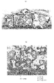
도 13에 실시예 1의 표본 중 하나를 도시하였다. 도 13의 (a)는 전체 형상을 도시한 도면, 도 13의 (b)는 도 13의 (a) 중 사각으로 둘러싼 부위의 확대도이다. 또한, 도 14에는 비교예 1의 표본 중 하나를 마찬가지로 도시하였다.Fig. 13 shows one of the samples of Example 1. Fig. FIG. 13A is a view showing the entire shape, and FIG. 13B is an enlarged view of a portion surrounded by a square in FIG. 13A. 14 shows one of the samples of Comparative Example 1 in the same manner.
그 결과, 실시예 1에서는, 광범위에서 두개골과 조직 재생 컨스트럭트가 연속된 골형성이 보였다. 도 13의 (b)로부터 알 수 있는 바와 같이 실시예 1에서는 신생골(「N」으로 나타냄)이 많이 형성되어 있는 것도 알 수 있다. 또한, 신생골 영역의 표면에는 활성이 높은 골아세포가 배열되어 있어, 추가적인 뼈의 신생이 기대되었다.As a result, in Example 1, continuous osteogenesis of the skull and tissue regeneration construct was seen in a wide range. As can be seen from FIG. 13 (b), it can be seen that in Example 1, a large number of new bone (denoted by "N") is formed. In addition, osteoblasts with high activity were arranged on the surface of the new bone area, and additional bone formation was expected.
한편, 비교예 1에서는, 컨스트럭트 중의 지지체의 주위에 산발적으로 골형성이 보이는 정도였다. 도 14의 (b)로부터 알 수 있는 바와 같이 골형성이 보이지 않는 영역은, 섬유성 조직(「F」로 나타냄)으로 채워져 있었다.On the other hand, in Comparative Example 1, there was a degree of sporadic formation of bone around the support in the construct. As can be seen from Fig. 14 (b), the region where bone formation is not seen was filled with fibrous tissue (denoted by " F ").
또한, 정량적으로는 다음과 같은 결과를 얻을 수 있었다.Quantitatively, the following results were obtained.
(1) 상기 얇게 썬 표본의 관찰에서, 지지체를 제외한 영역에서의 신생골의 면적 비율이 60% 이상인 표본은, 5개의 예 중, 실시예 1에서는 5개(전부), 비교예 1에서는 1개뿐이었다.(1) In the observation of the thinned specimen, 5 specimens (all of them) in Example 1 and 1 specimen in Comparative Example 1 out of the five specimens having an area ratio of new bone in the region other than the scaffold were 60% or more .
(2) 상기 얇게 썬 표본의 관찰에서, 조직 재생 컨스트럭트의 저면과 기존 두개골이, 신생골에 의해 연속해서 생착되고, 기존 두개골로부터 조직 재생 컨스트럭트 내부의 지지체까지 골가교를 나타낸 계면의 길이가 50% 이상인 표본은, 5개의 예 중, 실시예 1에서는 5개(전부), 비교예 1에서는 1개뿐이었다.(2) In the observation of the thinned specimen, the bottom of the tissue regeneration construct and the existing skull were continuously transferred by the new bone, and the length of the interface showing the bone bridge from the existing skull to the support in the tissue regeneration construct Of the five samples (all in the first example) and only one sample in the first comparative example (five samples in the first example).
이상에서, 조직 재생 컨스트럭트에 생체층이 구비되고, 조직 재생 대상 부위와의 사이에 생착층이 개재됨으로써 골형성이 촉진되고 있는 것이 확인되어, 본 발명의 조직 재생 컨스트럭트가 뼈의 증생에 유효한 것이 밝혀졌다.As described above, it was confirmed that bone formation is promoted by providing a living body layer in the tissue regeneration construct and an engrafting layer interposed between the body regeneration construct and the tissue regeneration target site. Thus, . ≪ / RTI >
<실시예 2>≪ Example 2 >
실시예 2에서는 다음과 같은 과정을 거쳐서 조직 재생 컨스트럭트를 제작하여 이식을 행하였다.In Example 2, a tissue regeneration construct was produced through the following procedure and transplanted.
(래트 골수 유래 간엽계 줄기 세포의 배양)(Culture of mesenchymal stem cells derived from rat bone marrow)
4주령 F344 래트의 대퇴골, 경골을 채취하고, 배양액을 사용해서 골수를 씻어내어(flush out) 회수한 골수 세포(대퇴골 2개분, 경골 2개분)를 10% FBS, 1% 페니실린 스트렙트 마이신을 포함하는 αMEM 배지 30㎖를 넣은 배지에 파종하였다. 37℃, 5% 탄산 가스 분위기의 조건 하에서 증식 배양하였다. 3일째에 배지를 교환하여 비접착 세포를 제거하고, 이후 3일에 1회의 비율로 배지 교환을 하였다. 최초의 배지 교환 시부터 배지에 bFGF 3ng/㎖를 첨가해서 배양하였다. 10일 전후에 거의 집밀(콘플루언트) 상태로 된 것을 확인하였다. 그 후, 배지를 제거하여, 트립신(0.05%)+EDTA(0.2mM)로 2분간 인큐베이트하고, 그 후 진동을 부여하여, 가급적 빠르게 세포를 박리해서 분해하고, 즉시 배지를 첨가해서 트립신 활성을 멈추었다.Bone marrow cells (2 femurs and 2 tibia) collected by flushing out the bone marrow were harvested with 10% FBS and 1% penicillin streptomycin at 4-week-old F344 rats Lt; RTI ID = 0.0 > ml < / RTI > And the cells were proliferated under the conditions of 37 ° C and 5% carbon dioxide gas atmosphere. On the third day, the non-adherent cells were removed by exchanging the medium, and thereafter, the medium was changed at a rate of once every three days. From the initial medium change, 3 ng / ml of bFGF was added to the medium and cultured. It was confirmed that it became almost confluent (confluent) around 10 days. Thereafter, the medium was removed, and incubation was carried out with trypsin (0.05%) + EDTA (0.2 mM) for 2 minutes. Thereafter, vibration was applied, the cells were peeled off as soon as possible, I stopped.
세포수를 계측하여, 5000세포/㎠에서 계대해서 배양하였다. 또한 5일간 배양한 시점에 상기와 마찬가지로 세포를 박리해서 분산시켜서(분해해서) 이하의 실험에 사용하였다.The number of cells was measured, and the cells were cultured at 5000 cells / cm 2. At the time of the incubation for 5 days, the cells were peeled and dispersed (decomposed) in the same manner as described above and used in the following experiments.
(조직 재생 컨스트럭트의 제조)(Manufacture of tissue regeneration construct)
니푸로사 제조의 1㎖ 시린지(내경 4.5㎜)의 선단을 절단한 것을 틀로 준비하였다. 선단을 절단한 시린지의 단부로부터 길이 10㎜의 부분에 피스톤 선단을 맞추고, 거기에 지지체로 하는 히드록시아파타이트 다공질체 과립(φ0.5㎜ 이상 φ2.0㎜ 이하)을 충전하였다. 선단을 절단한 시린지의 단부로부터 피스톤 선단까지의 체적은 159㎕가 된다. 용기 충전 상태에서의 지지체의 공공률은 75%였으므로, 119㎕가 간극으로서 존재하게 된다. 거기에, F344 래트의 혈장 107㎕에 상기와 같이 배양한 F344 래트 골수 유래의 간엽계 줄기 세포를 200×104 세포 현탁한 것을 첨가하였다. 전체에 균일하게 세포가 널리 퍼지도록, 27G 주사 바늘을 장착한 시린지로 교반(피펫팅)하였다. 또한, 12㎕의 3.3% 염화 칼슘 수용액을 첨가해서 널리 잘 퍼지도록, 교반(피펫팅)하였다. 그것을 37℃에서 배양(인큐베이트)하고, 응고 반응인 혈장 중의 피브리노겐 중합체화를 촉진시켰다. 그 결과, 1㎖ 시린지 내에 혈장 피브린 내에 세포와 지지체가 봉입된 덩어리(이식체 본체 부분)가 형성되었다. 이 경우, 이 시점에서 생착층에 상당하는 것은 존재하지 않는다.A tip of a 1 ml syringe (inner diameter: 4.5 mm) manufactured by Nippro Co., Ltd. was cut, and a mold was prepared. The distal end of the piston was aligned with a portion of 10 mm in length from the end of the syringe having its tip cut off, and the hydroxyapatite porous body granules (φ 0.5 mm or more and φ2.0 mm or less) serving as a support were filled. The volume from the end of the syringe to which the tip was cut to the tip of the piston was 159 占 퐇. Since the porosity of the support in the container filled state was 75%, 119 占 퐇 existed as the gap. There, the F344 rat mesenchymal stem cells of bone marrow-derived cultured as described above, the plasma 107㎕ the F344 rat was added to a suspension 200 × 10 4 cells. (Pipetting) with a syringe equipped with a 27G injection needle so that the cells spread uniformly throughout the whole. Further, 12 占 퐇 of a 3.3% aqueous solution of calcium chloride was added and stirred (pipetting) so as to spread widely. It was incubated (incubated) at 37 占 폚 to promote fibrinogen polymerization in plasma, which is a coagulation reaction. As a result, a lump (body portion of the implant) filled with cells and supporters was formed in plasma fibrin in a 1 mL syringe. In this case, at this point, there is no equivalent to the adhesion layer.
이어서, 상기에 의해 제작한 이식체를 시린지의 피스톤을 밀어서, 원기둥 형상의 덩어리로서 취출하였다. 이것을 멸균 완료된 φ10㎜ 원기둥 형상 용기 내에 넣고, 핀셋으로 파지하여, 원형의 용기 저면으로부터 3㎜ 뜨게 한 상태로 보유 지지하였다. 거기에, F344 래트의 혈장에 상기 F344 래트 골수 유래의 간엽계 줄기 세포를 200×104세포/1000㎕로 현탁시킨 것 1097㎕를 주입해서 잘 교반(피펫팅)하였다. 거기에 또한 110㎕의 3.3% 염화 칼슘 수용액을 첨가하여, 잘 교반(피펫팅)하였다. 이에 의해 응고 반응이 진행되어 왔을 때를 적당히 가늠해서, 37℃에서 더 배양(인큐베이트)하여, 응고 반응인 혈장 중의 피브리노겐 중합체화를 촉진시켰다. 이에 의해, 이식체의 상하 3㎜ 두께로 생착층이 형성되었다. 또한, 동시에 원기둥 형상 이식체 부분의 측면에도 생착층이 형성되었다. 생착층은 경시적으로 수축하여, 최종적으로 약 0.3㎜ 이하의 두께가 되었다. 이렇게 해서 완성한 원기둥 형상의 조직 재생 컨스트럭트의 상하면에 존재하는 생착층의 세포 밀도는 약 0.5×10⁴ 세포/㎟가 된다.Then, the implant prepared by the above was pushed out of the piston of the syringe and taken out as a cylindrical lump. This was placed in a sterilized 10 mm cylindrical container and gripped with a tweezers to hold it in a state where it was floated 3 mm from the bottom surface of the circular container. There, it will inject the F344 rat bone marrow-derived mesenchymal stem cells in the blood plasma of the F344 rat was suspended in 200 × 10 4 cells / 1000㎕ 1097㎕ was stirred well (pipetting). Thereto was further added 110 μl of a 3.3% aqueous solution of calcium chloride, followed by well stirring (pipetting). As a result, when the solidification reaction had progressed, the fibrinogen was further cultured (incubated) at 37 ° C. to promote the fibrinogen polymerization in the plasma, which is the coagulation reaction. Thus, the engrafting layer was formed with a thickness of 3 mm above and below the implanted body. At the same time, the engrafting layer was also formed on the side surface of the cylindrical implant portion. The engobe layer contracted with time, and finally the thickness became about 0.3 mm or less. The cell density of the deposit layer present on the upper and lower surfaces of the thus completed columnar tissue regeneration construct is about 0.5 x 10 4 cells / mm 2.
(이식 및 그 평가)(Transplantation and evaluation thereof)
F344 래트 대퇴부의 상피를 절개하여, 대퇴골 주위의 근막, 근육을 찢어 대퇴골을 노출시켰다. 계속해서 노출된 대퇴골을 창외 고정기로 고정해서 간극 10㎜의 골결손을 제작하였다. 이 골결손 모델에 대하여 상기 원기둥 형상의 조직 재생 컨스트럭트를 골단면에 원기둥 형상의 상하에 있는 생착층이 정확히 접하도록 집어넣어 이식하였다.F344 Rat The epithelium of the thigh was incised, and the femur was exposed by tearing the fascia and muscles around the femur. The exposed femur was fixed with an outboard fixture to produce a bone defect with a gap of 10 mm. In this bone defect model, the cylindrical tissue regeneration construct was inserted and implanted into the bony section of the bone so that the growth layer on the upper and lower sides of the cylindrical shape was exactly contacted.
조직 재생 컨스트럭트는 한 덩어리이고, 골단면에 끼워 넣어짐으로써 고정성이 양호하였다.The tissue regeneration construct was a lump, and was fitted into the bone section, so that the fixation was good.
근육과 근막을 봉합하고, 또한 상피도 봉합해서 상처를 닫았다. 치유 과정은 1, 2, 4, 8주째의 결손부의 렌트겐 촬영에 의해 평가하였다. 이식 후 4주째에는 골단부와 조직 재생 컨스트럭트의 단부와의 경계가 불명료해지고, 골가교가 일어나고 있는 것이 시사되었다. 또한, 이식 후 8주에 대퇴골을 적출하여, μCT 및 병리 조직학적으로 평가하였다.The muscle and fascia were sutured, and the epithelium was also sutured to close the wound. The healing process was evaluated by Rensgen imaging of the defects at 1, 2, 4, and 8 weeks. At the 4th week after transplantation, the border between the tip of the bone and the end of the tissue regeneration construct became unclear, suggesting that bone bridge is occurring. In addition, the femur was extracted at 8 weeks after transplantation and evaluated by μCT and histopathologically.
그 결과, 조직 재생 컨스트럭트와 잔존 대퇴골이 골가교되고 있는 것이 확인되었다. 또한, 조직 재생 컨스트럭트의 이식체에도 지지체로서 사용한 히드록시아파타이트 과립을 덮도록 성숙한 골형성이 보이고, 일부 골수 모양의 조직의 형성도 보였다. 이식한 조직 재생 컨스트럭트 전체가 한 덩어리로 대퇴골로 정확히 치환된 모습이었다.As a result, it was confirmed that the tissue regeneration construct and the remaining femur were bone crosslinked. In addition, grafts of tissue regeneration constructs showed mature bone formation to cover hydroxyapatite granules used as a support, and some bone marrow-shaped tissues were formed. All of the transplanted tissue regeneration constructs were exactly one lump of the femur.
이상에 의해, 본 발명의 조직 재생 컨스트럭트를 이식함으로써, 자연 치유가 도저히 불가능한 정도로 대규모의 래트 대퇴골 결손의 치유가 가능한 것이 확인되었다.As described above, it has been confirmed that by transplanting the tissue regeneration construct of the present invention, it is possible to cure large-scale defects in the femur of a rat to such an extent that natural healing is almost impossible.
10, 20, 30: 조직 재생 컨스트럭트
11, 21, 31: 이식체
12: 지지체
15, 25, 35: 생착층10, 20, 30: Tissue Regeneration Construct
11, 21, 31: implant
12: Support
15, 25, 35:
Claims (16)
이식체 및 해당 이식체의 외표면 중 적어도 일부에 겹쳐서 배치되는 생착층을 갖고,
상기 이식체는,
지지체와,
상기 지지체 사이의 공간 부분 및 지지체의 내측에 형성된 공공(空孔)에 의한 공간 부분 중 적어도 한쪽에 배치된 상기 조직을 재생하기 위한 세포를 갖고,
상기 생착층은,
상기 조직을 재생하기 위한 세포와,
상기 세포를 보유 지지하고, 응고 반응에 의해 고분자화된 혈장 피브린을 함유하는 기재를 포함하고,
상기 생착층의 상기 기재는 겔 상태로 되어 있음과 함께, 상기 생착층은 상기 지지체가 존재하지 않는 층이고, 상기 생착층의 상기 세포는 0.2×10⁴ 세포/㎟ 이상의 세포 밀도로 상기 생착층에 포함되는, 조직 재생 컨스트럭트.A tissue regeneration construct which is applied to a region to be transplanted and regenerated to regenerate the tissue,
A graft layer and a graft layer which is disposed on at least a part of the outer surface of the graft layer,
The implant,
A support,
A cell for regenerating the tissue disposed on at least one of a space portion between the supports and a space portion formed by holes formed inside the support,
The above-
A cell for regenerating said tissue,
Comprising a substrate containing said cell and containing a plasma fibrin polymerized by a coagulation reaction,
Wherein the base material of the base material layer is in a gel state and the base material layer is a layer free of the base material and the cells of the base material layer have a cell density of 0.2 x 10 4 cells / Included, tissue regeneration construct.
틀 안에 상기 지지체를 배치하는 공정과,
상기 지지체를 배치한 상기 틀 안에, 상기 조직을 재생하는 상기 세포 및 해당 세포를 보유 지지하는 기재를 함유한 기재 현탁액을 상기 지지체의 상면보다 수위가 높아지도록 주입하는 공정과,
상기 기재를 겔 상태로 하는 공정을 포함하는, 조직 재생 컨스트럭트의 제조 방법.10. A method for producing a tissue regeneration construct according to any one of claims 1 to 9,
Disposing the support in a mold,
A step of injecting a substrate suspension containing the cells for regenerating the tissue and a substrate holding the cells so as to be higher in level than the upper surface of the supporter,
And a step of bringing the substrate into a gel state.
틀 안에, 상기 틀의 내면 중 적어도 하나에 상기 지지체가 접촉하는 일 없이 간극을 갖고서 상기 지지체를 배치하는 공정과,
상기 지지체를 배치한 상기 틀 안에, 상기 조직을 재생하는 상기 세포 및 해당 세포를 보유 지지하는 기재를 함유한 기재 현탁액을 주입하는 공정과,
상기 기재를 겔 상태화하는 공정을 포함하는, 조직 재생 컨스트럭트의 제조 방법.10. A method for producing a tissue regeneration construct according to any one of claims 1 to 9,
A step of disposing the support body in a frame with a gap without contacting the support body with at least one of inner surfaces of the frame;
A step of injecting a substrate suspension containing the cells regenerating the tissue and a substrate holding the cells in the frame in which the support is disposed;
And a step of gelatinizing the substrate.
틀 안에 상기 이식체를 배치하는 공정과,
상기 이식체를 배치한 상기 틀 안에, 상기 조직을 재생하는 상기 세포 및 해당 세포를 보유 지지하는 기재를 함유한 기재 현탁액을 상기 지지체의 상면보다 수위가 높아지도록 주입하는 공정과,
상기 기재를 겔 상태로 하는 공정을 포함하는,
조직 재생 컨스트럭트의 제조 방법.10. A method for producing a tissue regeneration construct according to any one of claims 1 to 9,
A step of disposing the implant in a frame,
A step of injecting a substrate suspension containing the cells for regenerating the tissue and a substrate for holding the cells so that the level of the substrate suspension is higher than the upper surface of the support,
And a step of bringing the substrate into a gel state.
A method for manufacturing a tissue regeneration construct.
틀 안에, 상기 틀의 내면 중 적어도 하나에 상기 이식체가 접촉하는 일 없이 간극을 갖고서 상기 이식체를 배치하는 공정과,
상기 이식체를 배치한 상기 틀 안에, 상기 조직을 재생하는 상기 세포 및 해당 세포를 보유 지지하는 기재를 함유한 기재 현탁액을 주입하는 공정과,
상기 기재를 겔 상태로 하는 공정을 포함하는, 조직 재생 컨스트럭트의 제조 방법.10. A method for producing a tissue regeneration construct according to any one of claims 1 to 9,
A step of arranging the implanted object in the frame with a gap without contacting the implanted object with at least one of the inner surfaces of the frame;
A step of injecting a substrate suspension containing the cell regenerating the tissue and a substrate holding the cell in the cell in which the graft is placed,
And a step of bringing the substrate into a gel state.
Applications Claiming Priority (3)
| Application Number | Priority Date | Filing Date | Title |
|---|---|---|---|
| JPJP-P-2012-167382 | 2012-07-27 | ||
| JP2012167382 | 2012-07-27 | ||
| PCT/JP2013/063827 WO2014017147A1 (en) | 2012-07-27 | 2013-05-17 | Tissue regeneration construct, and method for producing tissue regeneration construct |
Publications (2)
| Publication Number | Publication Date |
|---|---|
| KR20150040848A KR20150040848A (en) | 2015-04-15 |
| KR101909328B1 true KR101909328B1 (en) | 2018-10-17 |
Family
ID=49996965
Family Applications (1)
| Application Number | Title | Priority Date | Filing Date |
|---|---|---|---|
| KR1020157000557A KR101909328B1 (en) | 2012-07-27 | 2013-05-17 | Tissue regeneration construct, and method for producing tissue regeneration construct |
Country Status (5)
| Country | Link |
|---|---|
| US (2) | US20150142128A1 (en) |
| EP (1) | EP2878665B1 (en) |
| JP (1) | JP5876934B2 (en) |
| KR (1) | KR101909328B1 (en) |
| WO (1) | WO2014017147A1 (en) |
Families Citing this family (4)
| Publication number | Priority date | Publication date | Assignee | Title |
|---|---|---|---|---|
| CA3028736A1 (en) * | 2016-06-27 | 2018-01-04 | University Of Louisville Research Foundation, Inc. | Spheroids including biologically-relevant materials and related methods |
| JP7272590B2 (en) | 2017-07-11 | 2023-05-12 | 国立大学法人富山大学 | Substrate coated with polymer for selective separation of cells or for cell culture |
| KR102189844B1 (en) * | 2018-11-27 | 2020-12-11 | 단국대학교 천안캠퍼스 산학협력단 | Polymer cell mixed spheroids, method for preparing thereof, and use thereof |
| KR102189850B1 (en) * | 2018-11-27 | 2020-12-11 | 단국대학교 천안캠퍼스 산학협력단 | Bioactive molecules-loaded polymer-cell mixed spheroid, and method for preparing thereof |
Citations (1)
| Publication number | Priority date | Publication date | Assignee | Title |
|---|---|---|---|---|
| WO2010005917A2 (en) * | 2008-07-06 | 2010-01-14 | The Curators Of The University Of Missouri | Osteochondral implants, arthroplasty methods, devices, and systems |
Family Cites Families (9)
| Publication number | Priority date | Publication date | Assignee | Title |
|---|---|---|---|---|
| US7326563B2 (en) * | 2000-11-08 | 2008-02-05 | Surface Logix, Inc. | Device and method for monitoring leukocyte migration |
| JPWO2002040071A1 (en) | 2000-11-14 | 2004-03-18 | 上田 実 | Composition for forming bone or periodontal tissue, and injection for forming bone or periodontal tissue |
| JP2002209573A (en) | 2001-01-15 | 2002-07-30 | Toru Yamazaki | Supporter and transplanter for cell and/or biological tissue, and method for supporting |
| JP2005000608A (en) | 2003-06-11 | 2005-01-06 | Mitsuo Okano | Cultured cell sheet having high take capability and method for manufacturing and using the same |
| US7217294B2 (en) * | 2003-08-20 | 2007-05-15 | Histogenics Corp. | Acellular matrix implants for treatment of articular cartilage, bone or osteochondral defects and injuries and method for use thereof |
| US8070827B2 (en) * | 2007-07-03 | 2011-12-06 | Histogenics Corporation | Method for use of a double-structured tissue implant for treatment of tissue defects |
| JP2011160817A (en) * | 2010-02-04 | 2011-08-25 | Univ Of Tokyo | Implantation support material |
| IL207586A0 (en) * | 2010-08-12 | 2010-12-30 | Omrix Biopharmaceuticals Ltd | A fibrin based therapeutic preparation and use thereof |
| EP3733099B1 (en) * | 2011-02-28 | 2022-08-31 | DePuy Synthes Products, Inc. | Modular tissue scaffolds |
-
2013
- 2013-05-17 JP JP2014526792A patent/JP5876934B2/en active Active
- 2013-05-17 EP EP13823846.4A patent/EP2878665B1/en active Active
- 2013-05-17 KR KR1020157000557A patent/KR101909328B1/en active IP Right Grant
- 2013-05-17 WO PCT/JP2013/063827 patent/WO2014017147A1/en active Application Filing
- 2013-05-17 US US14/413,864 patent/US20150142128A1/en not_active Abandoned
-
2018
- 2018-12-19 US US16/225,874 patent/US20190117829A1/en not_active Abandoned
Patent Citations (1)
| Publication number | Priority date | Publication date | Assignee | Title |
|---|---|---|---|---|
| WO2010005917A2 (en) * | 2008-07-06 | 2010-01-14 | The Curators Of The University Of Missouri | Osteochondral implants, arthroplasty methods, devices, and systems |
Also Published As
| Publication number | Publication date |
|---|---|
| EP2878665A4 (en) | 2016-03-23 |
| JP5876934B2 (en) | 2016-03-02 |
| EP2878665A1 (en) | 2015-06-03 |
| JPWO2014017147A1 (en) | 2016-07-07 |
| WO2014017147A1 (en) | 2014-01-30 |
| US20150142128A1 (en) | 2015-05-21 |
| KR20150040848A (en) | 2015-04-15 |
| US20190117829A1 (en) | 2019-04-25 |
| EP2878665B1 (en) | 2019-05-08 |
Similar Documents
| Publication | Publication Date | Title |
|---|---|---|
| KR101375828B1 (en) | Complex Scaffold For Bone-Cartilage Regeneration, Method For Preparing Thereof And Composition for Treating Bone Cartilage Disease Comprising The Same | |
| Yao et al. | In vitro angiogenesis of 3D tissue engineered adipose tissue | |
| Yao et al. | Biomimetic injectable HUVEC‐adipocytes/collagen/alginate microsphere co‐cultures for adipose tissue engineering | |
| US20050129730A1 (en) | Tissue composites and uses thereof | |
| JP4406283B2 (en) | Tissue regeneration substrate, transplant material, and production method thereof | |
| CN103691001B (en) | Method for preparing three-dimensional porous stent composite layer | |
| NZ330664A (en) | Implantable acrylamide copolymer hydrogel for therapeutic uses | |
| KR102102153B1 (en) | A bead for cell encapsulation and culture based on decellularized tissue, and use thereof | |
| US20190117829A1 (en) | Tissue regeneration construct and method for producing tissue regeneration construct | |
| Harris et al. | Strategies to direct angiogenesis within scaffolds for bone tissue engineering | |
| CN108273131A (en) | A kind of composite bone cement, preparation method and applications and a kind of bone renovating material | |
| CN102089425A (en) | Biomimetic cell scaffolds | |
| JP4122280B2 (en) | Manufacturing method of tissue plug | |
| EP2644695A1 (en) | Cultured cartilage tissue material | |
| US9629939B2 (en) | Collagenous foam materials | |
| US10994050B2 (en) | High yield and high precision bone graft substitute from stem cells | |
| JP2007209203A (en) | Calcium phosphate-based cell culture carrier and method for culturing | |
| Martin et al. | Producing prefabricated tissues and organs via tissue engineering | |
| CN102327643B (en) | A kind of biological support for osteanagenesis | |
| WO2007020713A1 (en) | Cell culture support embeddable in vivo | |
| Partap et al. | Scaffolds & surfaces | |
| JP2004167202A (en) | Implantation material for vascularization, and production method therefor | |
| Gupta et al. | Usage of Additive Manufacturing in Customised Bone Tissue-Engineering Scaffold | |
| RU2744732C1 (en) | Biocomposite spheroid for bone repair and method of its preparation | |
| RU2744664C1 (en) | Method for the production of spheroids from cultured cells of the periosteum to ensure reparative osteogenesis |
Legal Events
| Date | Code | Title | Description |
|---|---|---|---|
| A201 | Request for examination | ||
| E902 | Notification of reason for refusal | ||
| E701 | Decision to grant or registration of patent right | ||
| GRNT | Written decision to grant |