JP7658917B2 - Physical function recovery promoter - Google Patents
Physical function recovery promoter Download PDFInfo
- Publication number
- JP7658917B2 JP7658917B2 JP2021569757A JP2021569757A JP7658917B2 JP 7658917 B2 JP7658917 B2 JP 7658917B2 JP 2021569757 A JP2021569757 A JP 2021569757A JP 2021569757 A JP2021569757 A JP 2021569757A JP 7658917 B2 JP7658917 B2 JP 7658917B2
- Authority
- JP
- Japan
- Prior art keywords
- cerebral infarction
- physical
- subject
- intravascular
- cells
- Prior art date
- Legal status (The legal status is an assumption and is not a legal conclusion. Google has not performed a legal analysis and makes no representation as to the accuracy of the status listed.)
- Active
Links
- 238000011084 recovery Methods 0.000 title claims description 26
- 206010008118 cerebral infarction Diseases 0.000 claims description 92
- 208000026106 cerebrovascular disease Diseases 0.000 claims description 92
- 210000001185 bone marrow Anatomy 0.000 claims description 47
- 230000003863 physical function Effects 0.000 claims description 46
- 210000005087 mononuclear cell Anatomy 0.000 claims description 45
- 230000000694 effects Effects 0.000 claims description 29
- 238000002360 preparation method Methods 0.000 claims description 13
- 210000001428 peripheral nervous system Anatomy 0.000 claims description 6
- 210000003169 central nervous system Anatomy 0.000 claims description 5
- 230000001737 promoting effect Effects 0.000 claims description 5
- 206010008088 Cerebral artery embolism Diseases 0.000 claims description 3
- 230000001269 cardiogenic effect Effects 0.000 claims description 3
- 201000010849 intracranial embolism Diseases 0.000 claims description 3
- 208000004552 Lacunar Stroke Diseases 0.000 claims description 2
- 206010051078 Lacunar infarction Diseases 0.000 claims description 2
- 230000002457 bidirectional effect Effects 0.000 claims description 2
- 238000009472 formulation Methods 0.000 claims 3
- 239000000203 mixture Substances 0.000 claims 3
- 230000003183 myoelectrical effect Effects 0.000 claims 3
- 230000007830 nerve conduction Effects 0.000 claims 2
- 210000004204 blood vessel Anatomy 0.000 claims 1
- 230000005062 synaptic transmission Effects 0.000 claims 1
- 210000004027 cell Anatomy 0.000 description 76
- 210000004556 brain Anatomy 0.000 description 46
- 102100031573 Hematopoietic progenitor cell antigen CD34 Human genes 0.000 description 36
- 101000777663 Homo sapiens Hematopoietic progenitor cell antigen CD34 Proteins 0.000 description 36
- 238000012360 testing method Methods 0.000 description 36
- 210000000578 peripheral nerve Anatomy 0.000 description 34
- 241000699670 Mus sp. Species 0.000 description 25
- 230000006870 function Effects 0.000 description 24
- 238000010172 mouse model Methods 0.000 description 24
- 238000002659 cell therapy Methods 0.000 description 15
- 230000002452 interceptive effect Effects 0.000 description 15
- 238000001356 surgical procedure Methods 0.000 description 15
- 210000000130 stem cell Anatomy 0.000 description 14
- 241000699666 Mus <mouse, genus> Species 0.000 description 13
- 238000011282 treatment Methods 0.000 description 13
- 238000001990 intravenous administration Methods 0.000 description 12
- XLYOFNOQVPJJNP-UHFFFAOYSA-N water Substances O XLYOFNOQVPJJNP-UHFFFAOYSA-N 0.000 description 11
- 210000003792 cranial nerve Anatomy 0.000 description 10
- 230000007423 decrease Effects 0.000 description 10
- 230000007659 motor function Effects 0.000 description 10
- 210000005036 nerve Anatomy 0.000 description 9
- 230000001225 therapeutic effect Effects 0.000 description 7
- 230000001154 acute effect Effects 0.000 description 6
- 210000003958 hematopoietic stem cell Anatomy 0.000 description 6
- 210000003141 lower extremity Anatomy 0.000 description 6
- 230000007658 neurological function Effects 0.000 description 6
- 208000020431 spinal cord injury Diseases 0.000 description 6
- 238000012549 training Methods 0.000 description 6
- 230000003920 cognitive function Effects 0.000 description 5
- 210000003657 middle cerebral artery Anatomy 0.000 description 5
- 238000011302 passive avoidance test Methods 0.000 description 5
- 210000005259 peripheral blood Anatomy 0.000 description 5
- 239000011886 peripheral blood Substances 0.000 description 5
- 238000010825 rotarod performance test Methods 0.000 description 5
- 230000037152 sensory function Effects 0.000 description 5
- 238000007619 statistical method Methods 0.000 description 5
- 230000000638 stimulation Effects 0.000 description 5
- 241001465754 Metazoa Species 0.000 description 4
- 239000000872 buffer Substances 0.000 description 4
- 208000037265 diseases, disorders, signs and symptoms Diseases 0.000 description 4
- 210000004700 fetal blood Anatomy 0.000 description 4
- 230000001771 impaired effect Effects 0.000 description 4
- 208000023589 ischemic disease Diseases 0.000 description 4
- 210000002414 leg Anatomy 0.000 description 4
- 210000002569 neuron Anatomy 0.000 description 4
- 230000002093 peripheral effect Effects 0.000 description 4
- GEHJYWRUCIMESM-UHFFFAOYSA-L sodium sulfite Chemical compound [Na+].[Na+].[O-]S([O-])=O GEHJYWRUCIMESM-UHFFFAOYSA-L 0.000 description 4
- 208000024891 symptom Diseases 0.000 description 4
- 102000004269 Granulocyte Colony-Stimulating Factor Human genes 0.000 description 3
- 108010017080 Granulocyte Colony-Stimulating Factor Proteins 0.000 description 3
- DNIAPMSPPWPWGF-UHFFFAOYSA-N Propylene glycol Chemical compound CC(O)CO DNIAPMSPPWPWGF-UHFFFAOYSA-N 0.000 description 3
- 206010002026 amyotrophic lateral sclerosis Diseases 0.000 description 3
- 210000002798 bone marrow cell Anatomy 0.000 description 3
- 210000001168 carotid artery common Anatomy 0.000 description 3
- 238000005119 centrifugation Methods 0.000 description 3
- 230000006378 damage Effects 0.000 description 3
- 230000005484 gravity Effects 0.000 description 3
- 230000006872 improvement Effects 0.000 description 3
- 238000001361 intraarterial administration Methods 0.000 description 3
- 238000012423 maintenance Methods 0.000 description 3
- 230000000926 neurological effect Effects 0.000 description 3
- 239000000243 solution Substances 0.000 description 3
- 230000004936 stimulating effect Effects 0.000 description 3
- 210000003462 vein Anatomy 0.000 description 3
- WRMNZCZEMHIOCP-UHFFFAOYSA-N 2-phenylethanol Chemical compound OCCC1=CC=CC=C1 WRMNZCZEMHIOCP-UHFFFAOYSA-N 0.000 description 2
- CFKMVGJGLGKFKI-UHFFFAOYSA-N 4-chloro-m-cresol Chemical compound CC1=CC(O)=CC=C1Cl CFKMVGJGLGKFKI-UHFFFAOYSA-N 0.000 description 2
- 206010012289 Dementia Diseases 0.000 description 2
- PEDCQBHIVMGVHV-UHFFFAOYSA-N Glycerine Chemical compound OCC(O)CO PEDCQBHIVMGVHV-UHFFFAOYSA-N 0.000 description 2
- 241000282412 Homo Species 0.000 description 2
- 108010025020 Nerve Growth Factor Proteins 0.000 description 2
- 102000007072 Nerve Growth Factors Human genes 0.000 description 2
- 208000012902 Nervous system disease Diseases 0.000 description 2
- 208000025966 Neurological disease Diseases 0.000 description 2
- DFPAKSUCGFBDDF-UHFFFAOYSA-N Nicotinamide Chemical compound NC(=O)C1=CC=CN=C1 DFPAKSUCGFBDDF-UHFFFAOYSA-N 0.000 description 2
- -1 PDGF Proteins 0.000 description 2
- FAPWRFPIFSIZLT-UHFFFAOYSA-M Sodium chloride Chemical compound [Na+].[Cl-] FAPWRFPIFSIZLT-UHFFFAOYSA-M 0.000 description 2
- 208000027418 Wounds and injury Diseases 0.000 description 2
- 239000000654 additive Substances 0.000 description 2
- 239000003963 antioxidant agent Substances 0.000 description 2
- 235000006708 antioxidants Nutrition 0.000 description 2
- 210000004004 carotid artery internal Anatomy 0.000 description 2
- 206010008129 cerebral palsy Diseases 0.000 description 2
- 239000002738 chelating agent Substances 0.000 description 2
- 239000003795 chemical substances by application Substances 0.000 description 2
- 230000000052 comparative effect Effects 0.000 description 2
- 201000010099 disease Diseases 0.000 description 2
- 208000035475 disorder Diseases 0.000 description 2
- 229940079593 drug Drugs 0.000 description 2
- 239000003814 drug Substances 0.000 description 2
- 230000003203 everyday effect Effects 0.000 description 2
- 230000006698 induction Effects 0.000 description 2
- 208000014674 injury Diseases 0.000 description 2
- 238000000034 method Methods 0.000 description 2
- 230000009251 neurologic dysfunction Effects 0.000 description 2
- 208000015015 neurological dysfunction Diseases 0.000 description 2
- 239000003900 neurotrophic factor Substances 0.000 description 2
- 210000002475 olfactory pathway Anatomy 0.000 description 2
- 239000003755 preservative agent Substances 0.000 description 2
- 235000010265 sodium sulphite Nutrition 0.000 description 2
- 239000003381 stabilizer Substances 0.000 description 2
- 210000002385 vertebral artery Anatomy 0.000 description 2
- WWFDJIVIDXJAQR-FFWSQMGZSA-N 1-[(2R,3R,4R,5R)-4-[[(2R,3R,4R,5R)-5-(4-amino-5-methyl-2-oxopyrimidin-1-yl)-3-[[(2R,3R,4R,5R)-3-[[(2R,3R,4R,5R)-5-(4-amino-5-methyl-2-oxopyrimidin-1-yl)-3-[[(2R,3R,4R,5R)-3-[[(2R,3R,4R,5R)-3-[[(2R,3R,4R,5R)-3-[[(2R,3R,4R,5R)-5-(4-amino-5-methyl-2-oxopyrimidin-1-yl)-3-[[(2R,3R,4R,5R)-3-[[(2R,3R,4R,5R)-3-[[(2R,3R,4R,5R)-3-[[(2R,3R,4R,5R)-3-[[(2R,3R,4R,5R)-3-[[(2R,3R,4R,5R)-3-[[(2R,3R,4R,5R)-5-(4-amino-5-methyl-2-oxopyrimidin-1-yl)-3-[[(2R,3R,4R,5R)-3-[[(2R,3R,4R,5R)-5-(2-amino-6-oxo-1H-purin-9-yl)-3-[[(2R,3R,4R,5R)-5-(2-amino-6-oxo-1H-purin-9-yl)-3-hydroxy-4-(2-methoxyethoxy)oxolan-2-yl]methoxy-hydroxyphosphinothioyl]oxy-4-(2-methoxyethoxy)oxolan-2-yl]methoxy-sulfanylphosphoryl]oxy-4-(2-methoxyethoxy)-5-(5-methyl-2,4-dioxopyrimidin-1-yl)oxolan-2-yl]methoxy-hydroxyphosphinothioyl]oxy-4-(2-methoxyethoxy)oxolan-2-yl]methoxy-hydroxyphosphinothioyl]oxy-5-(2-amino-6-oxo-1H-purin-9-yl)-4-(2-methoxyethoxy)oxolan-2-yl]methoxy-hydroxyphosphinothioyl]oxy-4-(2-methoxyethoxy)-5-(5-methyl-2,4-dioxopyrimidin-1-yl)oxolan-2-yl]methoxy-hydroxyphosphinothioyl]oxy-5-(6-aminopurin-9-yl)-4-(2-methoxyethoxy)oxolan-2-yl]methoxy-hydroxyphosphinothioyl]oxy-5-(6-aminopurin-9-yl)-4-(2-methoxyethoxy)oxolan-2-yl]methoxy-hydroxyphosphinothioyl]oxy-4-(2-methoxyethoxy)-5-(5-methyl-2,4-dioxopyrimidin-1-yl)oxolan-2-yl]methoxy-hydroxyphosphinothioyl]oxy-5-(6-aminopurin-9-yl)-4-(2-methoxyethoxy)oxolan-2-yl]methoxy-hydroxyphosphinothioyl]oxy-4-(2-methoxyethoxy)oxolan-2-yl]methoxy-hydroxyphosphinothioyl]oxy-4-(2-methoxyethoxy)-5-(5-methyl-2,4-dioxopyrimidin-1-yl)oxolan-2-yl]methoxy-hydroxyphosphinothioyl]oxy-4-(2-methoxyethoxy)-5-(5-methyl-2,4-dioxopyrimidin-1-yl)oxolan-2-yl]methoxy-hydroxyphosphinothioyl]oxy-4-(2-methoxyethoxy)-5-(5-methyl-2,4-dioxopyrimidin-1-yl)oxolan-2-yl]methoxy-hydroxyphosphinothioyl]oxy-4-(2-methoxyethoxy)oxolan-2-yl]methoxy-hydroxyphosphinothioyl]oxy-5-(6-aminopurin-9-yl)-4-(2-methoxyethoxy)oxolan-2-yl]methoxy-hydroxyphosphinothioyl]oxy-4-(2-methoxyethoxy)oxolan-2-yl]methoxy-hydroxyphosphinothioyl]oxy-5-(hydroxymethyl)-3-(2-methoxyethoxy)oxolan-2-yl]-5-methylpyrimidine-2,4-dione Chemical compound COCCO[C@@H]1[C@H](O)[C@@H](COP(O)(=S)O[C@@H]2[C@@H](COP(S)(=O)O[C@@H]3[C@@H](COP(O)(=S)O[C@@H]4[C@@H](COP(O)(=S)O[C@@H]5[C@@H](COP(O)(=S)O[C@@H]6[C@@H](COP(O)(=S)O[C@@H]7[C@@H](COP(O)(=S)O[C@@H]8[C@@H](COP(O)(=S)O[C@@H]9[C@@H](COP(O)(=S)O[C@@H]%10[C@@H](COP(O)(=S)O[C@@H]%11[C@@H](COP(O)(=S)O[C@@H]%12[C@@H](COP(O)(=S)O[C@@H]%13[C@@H](COP(O)(=S)O[C@@H]%14[C@@H](COP(O)(=S)O[C@@H]%15[C@@H](COP(O)(=S)O[C@@H]%16[C@@H](COP(O)(=S)O[C@@H]%17[C@@H](COP(O)(=S)O[C@@H]%18[C@@H](CO)O[C@H]([C@@H]%18OCCOC)n%18cc(C)c(=O)[nH]c%18=O)O[C@H]([C@@H]%17OCCOC)n%17cc(C)c(N)nc%17=O)O[C@H]([C@@H]%16OCCOC)n%16cnc%17c(N)ncnc%16%17)O[C@H]([C@@H]%15OCCOC)n%15cc(C)c(N)nc%15=O)O[C@H]([C@@H]%14OCCOC)n%14cc(C)c(=O)[nH]c%14=O)O[C@H]([C@@H]%13OCCOC)n%13cc(C)c(=O)[nH]c%13=O)O[C@H]([C@@H]%12OCCOC)n%12cc(C)c(=O)[nH]c%12=O)O[C@H]([C@@H]%11OCCOC)n%11cc(C)c(N)nc%11=O)O[C@H]([C@@H]%10OCCOC)n%10cnc%11c(N)ncnc%10%11)O[C@H]([C@@H]9OCCOC)n9cc(C)c(=O)[nH]c9=O)O[C@H]([C@@H]8OCCOC)n8cnc9c(N)ncnc89)O[C@H]([C@@H]7OCCOC)n7cnc8c(N)ncnc78)O[C@H]([C@@H]6OCCOC)n6cc(C)c(=O)[nH]c6=O)O[C@H]([C@@H]5OCCOC)n5cnc6c5nc(N)[nH]c6=O)O[C@H]([C@@H]4OCCOC)n4cc(C)c(N)nc4=O)O[C@H]([C@@H]3OCCOC)n3cc(C)c(=O)[nH]c3=O)O[C@H]([C@@H]2OCCOC)n2cnc3c2nc(N)[nH]c3=O)O[C@H]1n1cnc2c1nc(N)[nH]c2=O WWFDJIVIDXJAQR-FFWSQMGZSA-N 0.000 description 1
- 208000024827 Alzheimer disease Diseases 0.000 description 1
- 206010002091 Anaesthesia Diseases 0.000 description 1
- 206010002388 Angina unstable Diseases 0.000 description 1
- 102000009840 Angiopoietins Human genes 0.000 description 1
- 108010009906 Angiopoietins Proteins 0.000 description 1
- 208000003343 Antiphospholipid Syndrome Diseases 0.000 description 1
- 208000000575 Arteriosclerosis Obliterans Diseases 0.000 description 1
- 206010003805 Autism Diseases 0.000 description 1
- 208000020706 Autistic disease Diseases 0.000 description 1
- 206010063659 Aversion Diseases 0.000 description 1
- 208000021130 Bilirubin encephalopathy Diseases 0.000 description 1
- 208000024806 Brain atrophy Diseases 0.000 description 1
- 208000033386 Buerger disease Diseases 0.000 description 1
- 206010008025 Cerebellar ataxia Diseases 0.000 description 1
- 208000032544 Cicatrix Diseases 0.000 description 1
- 206010011086 Coronary artery occlusion Diseases 0.000 description 1
- 208000011990 Corticobasal Degeneration Diseases 0.000 description 1
- FBPFZTCFMRRESA-FSIIMWSLSA-N D-Glucitol Natural products OC[C@H](O)[C@H](O)[C@@H](O)[C@H](O)CO FBPFZTCFMRRESA-FSIIMWSLSA-N 0.000 description 1
- FBPFZTCFMRRESA-KVTDHHQDSA-N D-Mannitol Chemical compound OC[C@@H](O)[C@@H](O)[C@H](O)[C@H](O)CO FBPFZTCFMRRESA-KVTDHHQDSA-N 0.000 description 1
- FBPFZTCFMRRESA-JGWLITMVSA-N D-glucitol Chemical compound OC[C@H](O)[C@@H](O)[C@H](O)[C@H](O)CO FBPFZTCFMRRESA-JGWLITMVSA-N 0.000 description 1
- 208000012239 Developmental disease Diseases 0.000 description 1
- 229920002307 Dextran Polymers 0.000 description 1
- FEWJPZIEWOKRBE-JCYAYHJZSA-N Dextrotartaric acid Chemical compound OC(=O)[C@H](O)[C@@H](O)C(O)=O FEWJPZIEWOKRBE-JCYAYHJZSA-N 0.000 description 1
- 201000010374 Down Syndrome Diseases 0.000 description 1
- 206010013647 Drowning Diseases 0.000 description 1
- ZGTMUACCHSMWAC-UHFFFAOYSA-L EDTA disodium salt (anhydrous) Chemical compound [Na+].[Na+].OC(=O)CN(CC([O-])=O)CCN(CC(O)=O)CC([O-])=O ZGTMUACCHSMWAC-UHFFFAOYSA-L 0.000 description 1
- 208000032027 Essential Thrombocythemia Diseases 0.000 description 1
- PIICEJLVQHRZGT-UHFFFAOYSA-N Ethylenediamine Chemical compound NCCN PIICEJLVQHRZGT-UHFFFAOYSA-N 0.000 description 1
- 101150021185 FGF gene Proteins 0.000 description 1
- 206010048744 Fear of falling Diseases 0.000 description 1
- 229920001917 Ficoll Polymers 0.000 description 1
- 201000011240 Frontotemporal dementia Diseases 0.000 description 1
- WQZGKKKJIJFFOK-GASJEMHNSA-N Glucose Natural products OC[C@H]1OC(O)[C@H](O)[C@@H](O)[C@@H]1O WQZGKKKJIJFFOK-GASJEMHNSA-N 0.000 description 1
- 208000035895 Guillain-Barré syndrome Diseases 0.000 description 1
- 101000707534 Homo sapiens Serine incorporator 1 Proteins 0.000 description 1
- 208000023105 Huntington disease Diseases 0.000 description 1
- 206010070511 Hypoxic-ischaemic encephalopathy Diseases 0.000 description 1
- PIWKPBJCKXDKJR-UHFFFAOYSA-N Isoflurane Chemical compound FC(F)OC(Cl)C(F)(F)F PIWKPBJCKXDKJR-UHFFFAOYSA-N 0.000 description 1
- 208000011200 Kawasaki disease Diseases 0.000 description 1
- 208000009829 Lewy Body Disease Diseases 0.000 description 1
- 201000002832 Lewy body dementia Diseases 0.000 description 1
- 229930195725 Mannitol Natural products 0.000 description 1
- 102000002274 Matrix Metalloproteinases Human genes 0.000 description 1
- 108010000684 Matrix Metalloproteinases Proteins 0.000 description 1
- 206010049567 Miller Fisher syndrome Diseases 0.000 description 1
- 208000001089 Multiple system atrophy Diseases 0.000 description 1
- 206010028289 Muscle atrophy Diseases 0.000 description 1
- 206010028851 Necrosis Diseases 0.000 description 1
- 208000037212 Neonatal hypoxic and ischemic brain injury Diseases 0.000 description 1
- 208000002193 Pain Diseases 0.000 description 1
- 208000018737 Parkinson disease Diseases 0.000 description 1
- 208000030831 Peripheral arterial occlusive disease Diseases 0.000 description 1
- 108010082093 Placenta Growth Factor Proteins 0.000 description 1
- 102100035194 Placenta growth factor Human genes 0.000 description 1
- 102000001938 Plasminogen Activators Human genes 0.000 description 1
- 108010001014 Plasminogen Activators Proteins 0.000 description 1
- 229920003171 Poly (ethylene oxide) Polymers 0.000 description 1
- 239000002202 Polyethylene glycol Substances 0.000 description 1
- 238000011579 SCID mouse model Methods 0.000 description 1
- SKZKKFZAGNVIMN-UHFFFAOYSA-N Salicilamide Chemical compound NC(=O)C1=CC=CC=C1O SKZKKFZAGNVIMN-UHFFFAOYSA-N 0.000 description 1
- 102100031707 Serine incorporator 1 Human genes 0.000 description 1
- DWAQJAXMDSEUJJ-UHFFFAOYSA-M Sodium bisulfite Chemical compound [Na+].OS([O-])=O DWAQJAXMDSEUJJ-UHFFFAOYSA-M 0.000 description 1
- 208000010112 Spinocerebellar Degenerations Diseases 0.000 description 1
- 206010042928 Syringomyelia Diseases 0.000 description 1
- 206010043540 Thromboangiitis obliterans Diseases 0.000 description 1
- 201000007023 Thrombotic Thrombocytopenic Purpura Diseases 0.000 description 1
- 108090001012 Transforming Growth Factor beta Proteins 0.000 description 1
- 102000004887 Transforming Growth Factor beta Human genes 0.000 description 1
- 206010044688 Trisomy 21 Diseases 0.000 description 1
- 208000007814 Unstable Angina Diseases 0.000 description 1
- 108010019530 Vascular Endothelial Growth Factors Proteins 0.000 description 1
- 102000005789 Vascular Endothelial Growth Factors Human genes 0.000 description 1
- 201000004810 Vascular dementia Diseases 0.000 description 1
- 206010053648 Vascular occlusion Diseases 0.000 description 1
- 239000008351 acetate buffer Substances 0.000 description 1
- 239000002253 acid Substances 0.000 description 1
- 230000032683 aging Effects 0.000 description 1
- 230000037005 anaesthesia Effects 0.000 description 1
- 210000002551 anterior cerebral artery Anatomy 0.000 description 1
- 230000003542 behavioural effect Effects 0.000 description 1
- UREZNYTWGJKWBI-UHFFFAOYSA-M benzethonium chloride Chemical compound [Cl-].C1=CC(C(C)(C)CC(C)(C)C)=CC=C1OCCOCC[N+](C)(C)CC1=CC=CC=C1 UREZNYTWGJKWBI-UHFFFAOYSA-M 0.000 description 1
- 229960001950 benzethonium chloride Drugs 0.000 description 1
- WQZGKKKJIJFFOK-VFUOTHLCSA-N beta-D-glucose Chemical compound OC[C@H]1O[C@@H](O)[C@H](O)[C@@H](O)[C@@H]1O WQZGKKKJIJFFOK-VFUOTHLCSA-N 0.000 description 1
- 230000005540 biological transmission Effects 0.000 description 1
- KGBXLFKZBHKPEV-UHFFFAOYSA-N boric acid Chemical compound OB(O)O KGBXLFKZBHKPEV-UHFFFAOYSA-N 0.000 description 1
- 239000004327 boric acid Substances 0.000 description 1
- 230000003925 brain function Effects 0.000 description 1
- 208000009973 brain hypoxia - ischemia Diseases 0.000 description 1
- 210000000133 brain stem Anatomy 0.000 description 1
- 210000001715 carotid artery Anatomy 0.000 description 1
- 239000004359 castor oil Substances 0.000 description 1
- 235000019438 castor oil Nutrition 0.000 description 1
- 239000006285 cell suspension Substances 0.000 description 1
- 230000008859 change Effects 0.000 description 1
- 229960002242 chlorocresol Drugs 0.000 description 1
- 230000001684 chronic effect Effects 0.000 description 1
- 239000007979 citrate buffer Substances 0.000 description 1
- 230000015271 coagulation Effects 0.000 description 1
- 238000005345 coagulation Methods 0.000 description 1
- 230000019771 cognition Effects 0.000 description 1
- 238000007887 coronary angioplasty Methods 0.000 description 1
- 210000004351 coronary vessel Anatomy 0.000 description 1
- 210000004748 cultured cell Anatomy 0.000 description 1
- 238000005520 cutting process Methods 0.000 description 1
- 230000003247 decreasing effect Effects 0.000 description 1
- 210000005258 dental pulp stem cell Anatomy 0.000 description 1
- 238000004090 dissolution Methods 0.000 description 1
- 150000002148 esters Chemical class 0.000 description 1
- 229940012017 ethylenediamine Drugs 0.000 description 1
- 210000003414 extremity Anatomy 0.000 description 1
- 230000001605 fetal effect Effects 0.000 description 1
- 239000012530 fluid Substances 0.000 description 1
- 230000008717 functional decline Effects 0.000 description 1
- 210000001368 germline stem cell Anatomy 0.000 description 1
- 239000008103 glucose Substances 0.000 description 1
- 235000011187 glycerol Nutrition 0.000 description 1
- ZEMPKEQAKRGZGQ-XOQCFJPHSA-N glycerol triricinoleate Natural products CCCCCC[C@@H](O)CC=CCCCCCCCC(=O)OC[C@@H](COC(=O)CCCCCCCC=CC[C@@H](O)CCCCCC)OC(=O)CCCCCCCC=CC[C@H](O)CCCCCC ZEMPKEQAKRGZGQ-XOQCFJPHSA-N 0.000 description 1
- 210000003714 granulocyte Anatomy 0.000 description 1
- 201000004332 intermediate coronary syndrome Diseases 0.000 description 1
- 230000002427 irreversible effect Effects 0.000 description 1
- 229960002725 isoflurane Drugs 0.000 description 1
- 208000006663 kernicterus Diseases 0.000 description 1
- 210000004185 liver Anatomy 0.000 description 1
- 238000002690 local anesthesia Methods 0.000 description 1
- 239000000594 mannitol Substances 0.000 description 1
- 235000010355 mannitol Nutrition 0.000 description 1
- 238000005259 measurement Methods 0.000 description 1
- 230000007246 mechanism Effects 0.000 description 1
- 206010027175 memory impairment Diseases 0.000 description 1
- 208000001725 mucocutaneous lymph node syndrome Diseases 0.000 description 1
- 201000006417 multiple sclerosis Diseases 0.000 description 1
- 210000001665 muscle stem cell Anatomy 0.000 description 1
- 201000006938 muscular dystrophy Diseases 0.000 description 1
- 210000002346 musculoskeletal system Anatomy 0.000 description 1
- 229940028444 muse Drugs 0.000 description 1
- 206010028417 myasthenia gravis Diseases 0.000 description 1
- 208000010125 myocardial infarction Diseases 0.000 description 1
- 230000017074 necrotic cell death Effects 0.000 description 1
- 230000008035 nerve activity Effects 0.000 description 1
- 230000007383 nerve stimulation Effects 0.000 description 1
- 210000000944 nerve tissue Anatomy 0.000 description 1
- 210000000653 nervous system Anatomy 0.000 description 1
- 230000001537 neural effect Effects 0.000 description 1
- 208000018360 neuromuscular disease Diseases 0.000 description 1
- 235000005152 nicotinamide Nutrition 0.000 description 1
- 239000011570 nicotinamide Substances 0.000 description 1
- 229950001015 nusinersen Drugs 0.000 description 1
- 230000036407 pain Effects 0.000 description 1
- 208000033300 perinatal asphyxia Diseases 0.000 description 1
- WVDDGKGOMKODPV-ZQBYOMGUSA-N phenyl(114C)methanol Chemical compound O[14CH2]C1=CC=CC=C1 WVDDGKGOMKODPV-ZQBYOMGUSA-N 0.000 description 1
- 239000008363 phosphate buffer Substances 0.000 description 1
- 229940127126 plasminogen activator Drugs 0.000 description 1
- 229920001223 polyethylene glycol Polymers 0.000 description 1
- 239000001267 polyvinylpyrrolidone Substances 0.000 description 1
- 235000013855 polyvinylpyrrolidone Nutrition 0.000 description 1
- 229920000036 polyvinylpyrrolidone Polymers 0.000 description 1
- 210000003388 posterior cerebral artery Anatomy 0.000 description 1
- 230000002028 premature Effects 0.000 description 1
- 230000002265 prevention Effects 0.000 description 1
- 201000002212 progressive supranuclear palsy Diseases 0.000 description 1
- 230000001172 regenerating effect Effects 0.000 description 1
- 230000000250 revascularization Effects 0.000 description 1
- 229960000581 salicylamide Drugs 0.000 description 1
- 231100000241 scar Toxicity 0.000 description 1
- 230000037390 scarring Effects 0.000 description 1
- 230000037387 scars Effects 0.000 description 1
- 230000035939 shock Effects 0.000 description 1
- 238000004904 shortening Methods 0.000 description 1
- 210000003625 skull Anatomy 0.000 description 1
- 235000010378 sodium ascorbate Nutrition 0.000 description 1
- PPASLZSBLFJQEF-RKJRWTFHSA-M sodium ascorbate Substances [Na+].OC[C@@H](O)[C@H]1OC(=O)C(O)=C1[O-] PPASLZSBLFJQEF-RKJRWTFHSA-M 0.000 description 1
- 229960005055 sodium ascorbate Drugs 0.000 description 1
- WXMKPNITSTVMEF-UHFFFAOYSA-M sodium benzoate Chemical compound [Na+].[O-]C(=O)C1=CC=CC=C1 WXMKPNITSTVMEF-UHFFFAOYSA-M 0.000 description 1
- 235000010234 sodium benzoate Nutrition 0.000 description 1
- 239000004299 sodium benzoate Substances 0.000 description 1
- 229960003885 sodium benzoate Drugs 0.000 description 1
- 239000011780 sodium chloride Substances 0.000 description 1
- 239000001509 sodium citrate Substances 0.000 description 1
- NLJMYIDDQXHKNR-UHFFFAOYSA-K sodium citrate Chemical compound O.O.[Na+].[Na+].[Na+].[O-]C(=O)CC(O)(CC([O-])=O)C([O-])=O NLJMYIDDQXHKNR-UHFFFAOYSA-K 0.000 description 1
- 229940037001 sodium edetate Drugs 0.000 description 1
- 229940079827 sodium hydrogen sulfite Drugs 0.000 description 1
- 235000010267 sodium hydrogen sulphite Nutrition 0.000 description 1
- 229940001482 sodium sulfite Drugs 0.000 description 1
- AKHNMLFCWUSKQB-UHFFFAOYSA-L sodium thiosulfate Chemical compound [Na+].[Na+].[O-]S([O-])(=O)=S AKHNMLFCWUSKQB-UHFFFAOYSA-L 0.000 description 1
- 229940001474 sodium thiosulfate Drugs 0.000 description 1
- 235000019345 sodium thiosulphate Nutrition 0.000 description 1
- PPASLZSBLFJQEF-RXSVEWSESA-M sodium-L-ascorbate Chemical compound [Na+].OC[C@H](O)[C@H]1OC(=O)C(O)=C1[O-] PPASLZSBLFJQEF-RXSVEWSESA-M 0.000 description 1
- 239000002904 solvent Substances 0.000 description 1
- 239000000600 sorbitol Substances 0.000 description 1
- 208000002320 spinal muscular atrophy Diseases 0.000 description 1
- 238000005728 strengthening Methods 0.000 description 1
- 235000000346 sugar Nutrition 0.000 description 1
- 150000008163 sugars Chemical class 0.000 description 1
- 230000009182 swimming Effects 0.000 description 1
- 229940095064 tartrate Drugs 0.000 description 1
- ZRKFYGHZFMAOKI-QMGMOQQFSA-N tgfbeta Chemical compound C([C@H](NC(=O)[C@H](C(C)C)NC(=O)CNC(=O)[C@H](CCC(O)=O)NC(=O)[C@H](CCCNC(N)=N)NC(=O)[C@H](CC(N)=O)NC(=O)[C@H](CC(C)C)NC(=O)[C@H]([C@@H](C)O)NC(=O)[C@H](CCC(O)=O)NC(=O)[C@H]([C@@H](C)O)NC(=O)[C@H](CC(C)C)NC(=O)CNC(=O)[C@H](C)NC(=O)[C@H](CO)NC(=O)[C@H](CCC(N)=O)NC(=O)[C@@H](NC(=O)[C@H](C)NC(=O)[C@H](C)NC(=O)[C@@H](NC(=O)[C@H](CC(C)C)NC(=O)[C@@H](N)CCSC)C(C)C)[C@@H](C)CC)C(=O)N[C@@H]([C@@H](C)O)C(=O)N[C@@H](C(C)C)C(=O)N[C@@H](CC=1C=CC=CC=1)C(=O)N[C@@H](C)C(=O)N1[C@@H](CCC1)C(=O)N[C@@H]([C@@H](C)O)C(=O)N[C@@H](CC(N)=O)C(=O)N[C@@H](CCC(O)=O)C(=O)N[C@@H](C)C(=O)N[C@@H](CC=1C=CC=CC=1)C(=O)N[C@@H](CCCNC(N)=N)C(=O)N[C@@H](C)C(=O)N[C@@H](CC(C)C)C(=O)N1[C@@H](CCC1)C(=O)N1[C@@H](CCC1)C(=O)N[C@@H](CCCNC(N)=N)C(=O)N[C@@H](CCC(O)=O)C(=O)N[C@@H](CCCNC(N)=N)C(=O)N[C@@H](CO)C(=O)N[C@@H](CCCNC(N)=N)C(=O)N[C@@H](CC(C)C)C(=O)N[C@@H](CC(C)C)C(O)=O)C1=CC=C(O)C=C1 ZRKFYGHZFMAOKI-QMGMOQQFSA-N 0.000 description 1
- 210000002303 tibia Anatomy 0.000 description 1
- 210000001519 tissue Anatomy 0.000 description 1
- 210000000689 upper leg Anatomy 0.000 description 1
- 208000021331 vascular occlusion disease Diseases 0.000 description 1
- 210000000216 zygoma Anatomy 0.000 description 1
Images
Classifications
-
- A—HUMAN NECESSITIES
- A61—MEDICAL OR VETERINARY SCIENCE; HYGIENE
- A61K—PREPARATIONS FOR MEDICAL, DENTAL OR TOILETRY PURPOSES
- A61K35/00—Medicinal preparations containing materials or reaction products thereof with undetermined constitution
- A61K35/12—Materials from mammals; Compositions comprising non-specified tissues or cells; Compositions comprising non-embryonic stem cells; Genetically modified cells
- A61K35/28—Bone marrow; Haematopoietic stem cells; Mesenchymal stem cells of any origin, e.g. adipose-derived stem cells
-
- A—HUMAN NECESSITIES
- A61—MEDICAL OR VETERINARY SCIENCE; HYGIENE
- A61K—PREPARATIONS FOR MEDICAL, DENTAL OR TOILETRY PURPOSES
- A61K35/00—Medicinal preparations containing materials or reaction products thereof with undetermined constitution
- A61K35/12—Materials from mammals; Compositions comprising non-specified tissues or cells; Compositions comprising non-embryonic stem cells; Genetically modified cells
- A61K35/48—Reproductive organs
- A61K35/54—Ovaries; Ova; Ovules; Embryos; Foetal cells; Germ cells
- A61K35/545—Embryonic stem cells; Pluripotent stem cells; Induced pluripotent stem cells; Uncharacterised stem cells
-
- A—HUMAN NECESSITIES
- A61—MEDICAL OR VETERINARY SCIENCE; HYGIENE
- A61P—SPECIFIC THERAPEUTIC ACTIVITY OF CHEMICAL COMPOUNDS OR MEDICINAL PREPARATIONS
- A61P25/00—Drugs for disorders of the nervous system
-
- A—HUMAN NECESSITIES
- A61—MEDICAL OR VETERINARY SCIENCE; HYGIENE
- A61P—SPECIFIC THERAPEUTIC ACTIVITY OF CHEMICAL COMPOUNDS OR MEDICINAL PREPARATIONS
- A61P9/00—Drugs for disorders of the cardiovascular system
- A61P9/10—Drugs for disorders of the cardiovascular system for treating ischaemic or atherosclerotic diseases, e.g. antianginal drugs, coronary vasodilators, drugs for myocardial infarction, retinopathy, cerebrovascula insufficiency, renal arteriosclerosis
-
- A—HUMAN NECESSITIES
- A61—MEDICAL OR VETERINARY SCIENCE; HYGIENE
- A61K—PREPARATIONS FOR MEDICAL, DENTAL OR TOILETRY PURPOSES
- A61K35/00—Medicinal preparations containing materials or reaction products thereof with undetermined constitution
- A61K35/12—Materials from mammals; Compositions comprising non-specified tissues or cells; Compositions comprising non-embryonic stem cells; Genetically modified cells
- A61K2035/124—Materials from mammals; Compositions comprising non-specified tissues or cells; Compositions comprising non-embryonic stem cells; Genetically modified cells the cells being hematopoietic, bone marrow derived or blood cells
-
- A—HUMAN NECESSITIES
- A61—MEDICAL OR VETERINARY SCIENCE; HYGIENE
- A61N—ELECTROTHERAPY; MAGNETOTHERAPY; RADIATION THERAPY; ULTRASOUND THERAPY
- A61N1/00—Electrotherapy; Circuits therefor
- A61N1/18—Applying electric currents by contact electrodes
- A61N1/32—Applying electric currents by contact electrodes alternating or intermittent currents
- A61N1/36—Applying electric currents by contact electrodes alternating or intermittent currents for stimulation
- A61N1/36003—Applying electric currents by contact electrodes alternating or intermittent currents for stimulation of motor muscles, e.g. for walking assistance
Landscapes
- Health & Medical Sciences (AREA)
- Life Sciences & Earth Sciences (AREA)
- Engineering & Computer Science (AREA)
- Developmental Biology & Embryology (AREA)
- Cell Biology (AREA)
- Animal Behavior & Ethology (AREA)
- Veterinary Medicine (AREA)
- Public Health (AREA)
- Chemical & Material Sciences (AREA)
- Medicinal Chemistry (AREA)
- Pharmacology & Pharmacy (AREA)
- General Health & Medical Sciences (AREA)
- Immunology (AREA)
- Biomedical Technology (AREA)
- Bioinformatics & Cheminformatics (AREA)
- Chemical Kinetics & Catalysis (AREA)
- Nuclear Medicine, Radiotherapy & Molecular Imaging (AREA)
- Organic Chemistry (AREA)
- General Chemical & Material Sciences (AREA)
- Zoology (AREA)
- Virology (AREA)
- Epidemiology (AREA)
- Biotechnology (AREA)
- Neurology (AREA)
- Neurosurgery (AREA)
- Hematology (AREA)
- Reproductive Health (AREA)
- Urology & Nephrology (AREA)
- Vascular Medicine (AREA)
- Cardiology (AREA)
- Heart & Thoracic Surgery (AREA)
- Gynecology & Obstetrics (AREA)
- Medicines Containing Material From Animals Or Micro-Organisms (AREA)
Description
本発明は、身体機能が不可逆的に低下している状態にある被験者の身体機能に対して実施される、脳および末梢神経の両者に協調して刺激を与える身体運動の効果を促進する、身体機能回復促進剤に関する。The present invention relates to a physical function recovery promoter that promotes the effects of physical exercise that stimulates both the brain and peripheral nerves in a coordinated manner, performed on the physical functions of a subject whose physical functions are irreversibly impaired.
中枢神経と末梢神経とは単独で活動するものではなく、障害時においても互いに密接な連絡を取り、影響を及ぼし合っている。例えば脊髄損傷者において、脳神経細胞は下肢を動かそうとするシグナルを出すものの、下肢が動いたというフィードバックが帰ってこないため、脳神経細胞は「下肢は動かない」と学習する。また、末梢神経は脳からの刺激が来ないため、「下肢を動かす必要がない」と学習し、互いの悪影響が蓄積・固定され身体機能が不可逆的に低下し、後遺症(病気・怪我など急性期症状が治癒した後も、機能障害などの症状や傷痕が残ること。)が生じる。すなわち、脊髄損傷の後遺症はインタラクティブ・バイオ・フィードバック機構が欠如した状態にあるといえる。インタラクティブ・バイオ・フィードバックとは、人の脳および神経系と筋骨格および末梢神経系の間で生じる「双方向の生体フィードバック」のことである。脊髄損傷の後遺症に対しては、欠損しているインタラクティブ・バイオ・フィードバックが促されることにより、患者の神経機能が改善することが示されつつある。例えば、HAL(Hybrid Assistive Limb)と呼ばれる電子装具を使用するサイバニクス治療は、脳神経細胞からの極微弱な下肢運動に関する神経活動をHALが関知し、機械的に下肢を動かす。HALは脊損の急性期には使用されることはほとんどないが、後遺症を有する脊損患者にHALを用いることで、脳神経細胞は「下肢は動く」と学習し、末梢神経は「下肢を動かす必要がある」と学習する。すなわち、中枢神経と末梢神経の間でインタラクティブ・バイオ・フィードバックが促され、神経機能の回復に繋がる。HAL医療用下肢タイプ(医療用HAL)は生体信号反応式運動機能改善装置として製造され、2015年11月に医療器機承認されており、HAL医療用下肢タイプによる歩行運動治療により、ALS、筋ジストロフィー等に対する24%程度の運動能力の改善効果を認められ、インタラクティブ・バイオ・フィードバック理論に基づく治療は、神経・筋疾患に対して効果的であることが証明された。しかし、インタラクティブ・バイオ・フィードバックに基づく身体運動の神経機能回復作用はそれだけでは不十分であり、それをより向上かつ持続させる併用治療が切望されている。例えば、インタラクティブ・バイオ・フィードバック理論に基づく身体運動の神経機能回復作用を促進する可能性のある薬剤としてNusinersenが挙げられており、インタラクティブ・バイオ・フィードバック理論に基づく身体運動との併用による神経機能回復作用が期待されている。The central nervous system and the peripheral nervous system do not act independently, but are in close contact with each other and influence each other even in the event of injury. For example, in a person with spinal cord injury, the brain's nerve cells send signals to move the legs, but since there is no feedback that the legs have moved, the brain's nerve cells learn that the legs cannot move. In addition, since the peripheral nerves do not receive stimulation from the brain, they learn that there is no need to move the legs, and the negative effects of each other accumulate and become fixed, causing an irreversible decline in physical function and sequelae (symptoms such as functional disorders and scars that remain even after the acute symptoms of illness or injury have healed). In other words, the sequelae of spinal cord injury can be said to be in a state where the interactive biofeedback mechanism is lacking. Interactive biofeedback is a "bidirectional biofeedback" that occurs between a person's brain and nervous system and the musculoskeletal and peripheral nervous systems. It is being shown that the nerve function of patients with sequelae of spinal cord injury can be improved by promoting the missing interactive biofeedback. For example, in cybernic treatment using an electronic prosthetic device called HAL (Hybrid Assistive Limb), HAL detects extremely weak nerve activity from brain neurons related to lower limb movement and mechanically moves the lower limbs. HAL is rarely used in the acute phase of spinal cord injury, but by using HAL on spinal cord injury patients with residual damage, the brain neurons learn that "the lower limbs can move" and the peripheral nerves learn that "the lower limbs need to be moved." In other words, interactive biofeedback is promoted between the central and peripheral nerves, leading to the recovery of nerve function. HAL Medical Lower Limb Type (Medical HAL) is manufactured as a biosignal-responsive motor function improvement device and was approved as a medical device in November 2015. Walking exercise treatment using HAL Medical Lower Limb Type has been shown to improve motor ability by about 24% in ALS, muscular dystrophy, etc., proving that treatment based on interactive biofeedback theory is effective for neuromuscular diseases. However, the neurological recovery effect of interactive biofeedback-based physical exercise is insufficient by itself, and there is a strong need for combined treatments that can further improve and sustain the effect. For example, Nusinersen has been cited as a drug that may promote the neurological recovery effect of interactive biofeedback-based physical exercise, and it is expected that the neurological recovery effect will be achieved by combining it with interactive biofeedback-based physical exercise.
脳梗塞の後遺症治療においても、インタラクティブ・バイオ・フィードバック理論に基づく身体運動の神経機能回復が期待されている。脳梗塞の後遺症に対して一般的に行なわれている処置は、機能維持リハビリテーションと呼ばれており、機能低下を防ぐことにより家庭生活や社会生活を維持・継続することを目的に実施されている。すなわち、機能維持リハビリテーションは、既に低下している神経機能の現状維持が目標であるため、身体を動かすことだけに重点が置かれており、身体を動かすことに伴う神経伝達の強化を促すインタラクティブ・バイオ・フィードバック理論に基づくものではない。 Even in the treatment of the aftereffects of cerebral infarction, it is hoped that neurological function recovery through physical movement based on interactive biofeedback theory will be achieved. Treatment generally performed for the aftereffects of cerebral infarction is called functional maintenance rehabilitation, and is carried out with the aim of maintaining and continuing family and social life by preventing functional decline. In other words, because the goal of functional maintenance rehabilitation is to maintain the current state of already-decreased neurological function, the emphasis is placed solely on moving the body, and it is not based on interactive biofeedback theory, which promotes the strengthening of neural transmission that accompanies physical movement.
急性期脳梗塞により障害された神経組織の再生には、造血幹細胞を用いた細胞治療が有効であることが判明している。非特許文献1には、自己の骨髄由来単核球細胞を患者の静脈内に投与する急性期の脳梗塞治療が記載されている。この非特許文献1では、重症の心原性脳塞栓症症例で、且つ脳梗塞発症10日以内において神経機能回復が十分でない患者群を対象としており、局所麻酔身麻酔下で骨髄細胞の採取を行い、比重遠心法を用いて単核球分画の分離を行い、そして静脈内に投与する治療が記載されている。しかしながら造血幹細胞投与の治療時期に関する検討では、脳梗塞後2,4,7,10日及び14日後の急性期および亜急性期に対しては治療効果を示すものの、脳梗塞の後遺症に対しては治療効果がないとされている(非特許文献2)。また、非特許文献3においては、発症18日後(平均)における、脳梗塞患者への造血幹細胞投与による細胞治療が行なわれ、一般的なリハビリテーションも併用されたが、その治療効果が全くないことが報告されている。このように脳梗塞により失われた機能を回復させるための治療法は急性期のみに限られており、後遺症に対する治療法は存在しない。そのため、脳梗塞患者の後遺症に対してインタラクティブ・バイオ・フィードバック理論に基づく身体運動の神経機能回復作用を促進する身体機能回復促進剤の実現が期待される。It has been found that cell therapy using hematopoietic stem cells is effective in regenerating nerve tissue damaged by acute cerebral infarction. Non-Patent
本発明はかかる問題点に鑑みてなされたものであって、身体機能が不可逆的に低下している状態にある被験者の身体機能に対して実施される、脳および末梢神経の両者に刺激を与える身体運動の効果を促進する、身体機能回復促進剤を提供することを目的とする。The present invention has been made in consideration of such problems, and aims to provide a physical function recovery promoter that enhances the effects of physical exercise that stimulates both the brain and peripheral nerves, which is performed on the physical functions of a subject whose physical functions are irreversibly impaired.
本発明にかかる身体機能回復促進剤は、運動機能、認知機能及び/又は感覚機能である身体機能が低下している状態にある被験者の身体機能を回復するための中枢と末梢を協調させる身体運動の効果を促進する身体機能回復促進剤であって、骨髄単核球細胞、CD34陽性細胞、CD133陽性細胞又は幹細胞を有することを特徴とする。The physical function recovery promoter of the present invention is a physical function recovery promoter that promotes the effects of physical exercise that coordinates the central and peripheral nervous systems to recover the physical functions of a subject in a state of impaired physical functions, which are motor function, cognitive function, and/or sensory function, and is characterized by containing bone marrow mononuclear cells, CD34-positive cells, CD133-positive cells, or stem cells.
本発明にかかる身体機能回復促進剤は、脳梗塞の後遺症により、運動機能、認知機能及び/又は感覚機能である身体機能が低下している状態にある被験者の身体機能を回復するため、中枢と末梢を協調させる身体運動の効果を促進する身体機能回復促進剤であって、骨髄単核球細胞を有することを特徴とする。The physical function recovery promoter of the present invention is a physical function recovery promoter that promotes the effects of physical exercise that coordinates the central and peripheral nervous systems in order to restore the physical functions of a subject whose physical functions, such as motor function, cognitive function, and/or sensory function, have been reduced due to the aftereffects of cerebral infarction, and is characterized by containing bone marrow mononuclear cells.
本発明にかかる身体機能回復促進剤は、脳梗塞の後遺症により、運動機能、認知機能及び/又は感覚機能である身体機能が低下している状態にある被験者の身体機能を回復するための身体運動の効果を促進する身体機能回復促進剤であって、CD34陽性細胞を有することを特徴とする。The physical function recovery promoter of the present invention is a physical function recovery promoter that promotes the effects of physical exercise to recover the physical functions of a subject whose physical functions, such as motor function, cognitive function, and/or sensory function, have been impaired due to the aftereffects of cerebral infarction, and is characterized by having CD34-positive cells.
以下、添付の図面を参照して本発明の実施形態について具体的に説明するが、当該実施形態は本発明の原理の理解を容易にするためのものであり、本発明の範囲は、下記の実施形態に限られるものではなく、当業者が以下の実施形態の構成を適宜置換した他の実施形態も、本発明の範囲に含まれる。 Below, an embodiment of the present invention will be described in detail with reference to the attached drawings. However, this embodiment is intended to facilitate understanding of the principles of the present invention, and the scope of the present invention is not limited to the embodiment described below. Other embodiments in which a person skilled in the art appropriately replaces the configuration of the embodiment described below are also included in the scope of the present invention.
本発明にかかる身体機能回復促進剤は、骨髄単核球細胞、CD34陽性細胞、CD133陽性細胞又は幹細胞を有しており、身体機能が低下している状態にある被験者の身体機能を回復するための身体運動の開始の前後に被験者に投与される。本発明にかかる身体機能回復促進剤は、骨髄単核球細胞、CD34陽性細胞、CD133陽性細胞又は幹細胞を有する細胞製剤であるが、その解決課題は身体機能が低下している状態にある被験者の身体機能を回復するため、中枢と末梢を協調させる身体運動の効果を促進するところにあり、細胞製剤の解決課題としては新規である。更に本発明にかかる細胞製剤は、身体運動の開始の前後に被験者に投与されるという使用態様が新規である。本発明細胞製剤における細胞は、単離細胞又は培養細胞のいずれのものでも使用可能である。The physical function recovery promoter of the present invention contains bone marrow mononuclear cells, CD34 positive cells, CD133 positive cells, or stem cells, and is administered to a subject before and after the start of physical exercise to recover the physical function of the subject in a state of reduced physical function. The physical function recovery promoter of the present invention is a cell preparation containing bone marrow mononuclear cells, CD34 positive cells, CD133 positive cells, or stem cells, and the problem to be solved is to promote the effect of physical exercise that coordinates the central and peripheral systems in order to recover the physical function of a subject in a state of reduced physical function, which is a novel problem to be solved by a cell preparation. Furthermore, the cell preparation of the present invention is novel in that it is administered to a subject before and after the start of physical exercise. The cells in the cell preparation of the present invention can be either isolated cells or cultured cells.
身体機能は、運動機能、認知機能及び/又は感覚機能である。 Physical function is motor function, cognitive function and/or sensory function.
運動機能とは、筋力、持久力、敏捷性、平衡性、瞬発力、及び/又は柔軟性についての人の機能である。 Motor function is a person's function of strength, endurance, agility, balance, explosiveness, and/or flexibility.
認知機能とは、理解力、判断力、計算力、思考力、見当識、記憶、及び/又は学習についての人の機能である。 Cognitive function is a person's ability to understand, judge, calculate, think, orient, remember, and/or learn.
感覚機能とは、視覚・聴覚・嗅覚・味覚、及び/又は、触覚についての人の機能である。 Sensory functions are a person's functions of seeing, hearing, smelling, tasting, and/or touch.
本発明にかかる身体機能回復促進方法は、(i)中枢と末梢を協調させる身体運動の開始時までの期間に、骨髄単核球細胞、CD34陽性細胞、CD133陽性細胞又は幹細胞の何れかを投与する工程と、(ii) 中枢と末梢を協調させる身体運動を行う工程を有することを特徴とする。The method for promoting recovery of physical functions according to the present invention is characterized by comprising the steps of (i) administering either bone marrow mononuclear cells, CD34-positive cells, CD133-positive cells or stem cells during the period up to the start of physical exercise that coordinates the central and peripheral systems, and (ii) performing physical exercise that coordinates the central and peripheral systems.
中枢と末梢とは連関している。身体機能が低下している状態にある被験者が身体機能を回復するための身体運動を行う際に、骨髄単核球細胞、CD34陽性細胞、CD133陽性細胞又は幹細胞を被験者に投与することにより身体機能を飛躍的に向上かつ維持させることを本発明者は新知見として見出し、かかる事実に基づいて本発明を完成させた。身体機能が低下している状態にある被験者にインタラクティブ・バイオ・フィードバック理論に基づく身体運動を行う際にも、骨髄単核球細胞、CD34陽性細胞、CD133陽性細胞又は幹細胞を被験者に投与することでインタラクティブ・バイオ・フィードバック理論に基づく身体運動の効果を飛躍的に向上させることができる。The central and peripheral systems are linked. The present inventors have discovered that when a subject with reduced physical function performs physical exercises to restore physical function, administering bone marrow mononuclear cells, CD34-positive cells, CD133-positive cells, or stem cells to the subject can dramatically improve and maintain physical function, and have completed the present invention based on this fact. When a subject with reduced physical function performs physical exercises based on interactive biofeedback theory, the effects of the physical exercises based on interactive biofeedback theory can also be dramatically improved by administering bone marrow mononuclear cells, CD34-positive cells, CD133-positive cells, or stem cells to the subject.
本発明においては、特に限定されるものではないが骨髄単核球細胞は静脈投与されることが好ましい。また、特に限定されるものではないがCD34陽性細胞やCD133陽性細胞は、総頸動脈、内頚動脈、前大脳動脈、中大脳動脈、後大脳動脈又は椎骨動脈に投与されることが好ましく、中でも総頚動脈又は椎骨動脈に投与されることが好ましい。In the present invention, although not particularly limited, bone marrow mononuclear cells are preferably administered intravenously. Furthermore, although not particularly limited, CD34-positive cells and CD133-positive cells are preferably administered to the common carotid artery, internal carotid artery, anterior cerebral artery, middle cerebral artery, posterior cerebral artery, or vertebral artery, and more preferably to the common carotid artery or vertebral artery.
被験者に投与されるCD34陽性細胞又はCD133陽性細胞は、特に限定されるものではなく、臍帯血由来細胞、ヒト骨髄由来細胞、ヒト末梢血由来細胞、胎児肝臓由来等が採用されるが、好ましくはヒト臍帯血由来細胞又はヒト末梢血由来細胞である。The CD34-positive cells or CD133-positive cells administered to the subject are not particularly limited, and may be derived from umbilical cord blood, human bone marrow, human peripheral blood, fetal liver, etc., with human umbilical cord blood-derived cells or human peripheral blood-derived cells being preferred.
被験者に投与される骨髄単核球細胞、CD34陽性細胞、CD133陽性細胞又は幹細胞の濃度は、特に限定されるものではないが、例えば5×10⁴個/kg~1×107個/kgとすることができ、好適には1×105個/kg~1×106個/kgであり、特に好適には5×105個/kgである。例えば、骨髄単核球細胞を静脈内投与する場合は5×106個/kgが好ましく、CD34陽性細胞を動脈内投与する場合は5×105個/kgが好ましく、CD133陽性細胞を動脈内投与する場合は5×105個/kgが好ましく、臍帯血単核球細胞を動脈内投与する場合は5×106個/kgが好ましい。 The concentration of bone marrow mononuclear cells, CD34-positive cells, CD133-positive cells, or stem cells administered to a subject is not particularly limited, and may be, for example, 5 x 10⁴ cells/kg to 1 x 10⁷ cells/kg, preferably 1 x 10⁵ cells/kg to 1 x 10⁶ cells/kg, and particularly preferably 5 x 10⁵ cells/kg. For example, when bone marrow mononuclear cells are administered intravenously, 5 x 10⁶ cells/kg is preferred, when CD34-positive cells are administered intraarterially, 5 x 10⁵ cells/kg is preferred, when CD133-positive cells are administered intraarterially, and when umbilical cord blood mononuclear cells are administered intraarterially, 5 x 10⁶ cells/kg is preferred.
幹細胞は、特に限定されるものではないが、例えばES細胞、EC細胞、EG細胞、iPS細胞、造血幹細胞、間葉系幹細胞、肝幹細胞、膵幹細胞、皮膚幹細胞、臍帯血幹細胞、骨髄幹細胞、筋幹細胞、生殖幹細胞、脂肪幹細胞、歯髄幹細胞又はMUSE細胞である。 Stem cells include, but are not limited to, ES cells, EC cells, EG cells, iPS cells, hematopoietic stem cells, mesenchymal stem cells, hepatic stem cells, pancreatic stem cells, skin stem cells, umbilical cord blood stem cells, bone marrow stem cells, muscle stem cells, germline stem cells, adipose stem cells, dental pulp stem cells, or MUSE cells.
被験者が行う身体機能を回復するための身体運動は、体力の向上を目的として計画的・意図的に実施し、継続性のある身体活動であり、好ましくはインタラクティブ・バイオ・フィードバック理論に基づく身体運動である。 The physical exercise performed by the subject to restore physical functions is planned, intentional, and continuous for the purpose of improving physical strength, and is preferably physical exercise based on interactive biofeedback theory.
被験者の身体機能の低下は、例えば虚血性疾患によるものである。 The subject's decline in physical function is due to, for example, an ischemic disease.
虚血性疾患は、特に限定されるものではないが、例えば、心筋梗塞、不安定狭心症、冠動脈バイパス術後のグラフト閉塞、経皮的冠動脈形成術後の冠動脈閉塞、血行再建術後の血管閉塞、閉塞性動脈硬化症、閉塞性血栓血管炎、本態性血小板血症、血栓性血小板減少性紫斑病、抗リン脂質抗体症候群及び川崎病の中から選択されるいずれかの疾患である。虚血性疾患は例えば脳性麻痺である。脳性麻痺は例えば新生児低酸素性虚血性脳症、早産児ビリルビン脳症等に起因する。虚血性疾患は、好ましくは脳梗塞の後遺症である。脳梗塞の場合、発症から例えば6カ月後には症状が固定していると考えられており、発症から例えば6カ月経過後に残存する神経機能障害を脳梗塞の後遺症として捉えている。一方、マウスでは脳梗塞モデル作製後例えば4週間がヒトの脳梗塞発症6カ月後と同じ時期に相当すると考えられている。この時期は、症状がほぼ固定されて維持リハビリテーションを実施するとともに再発予防が中心となる期間であり、細胞壊死や組織の瘢痕化が見られる病態である。脳梗塞の後遺症治療として造血幹細胞を投与しても、細胞投与単独での治療効果は低い。また脳梗塞後遺症に対して行われるリハビリテーションは機能を維持するためのものであり、失われた機能が回復することは期待できない。しかしながら、骨髄単核球細胞、CD34陽性細胞、CD133陽性細胞又は幹細胞が脳梗塞後遺症を有する患者に投与されると、脳梗塞の後遺症に対して末梢と中枢を協調させる身体運動の効果が促進される。投与される細胞は好ましくは骨髄単核球細胞又はCD34陽性細胞である。骨髄単核球細胞は好ましくは静脈投与され、CD34陽性細胞は好ましくは総頚動脈又は内頚動脈投与される。The ischemic disease is not particularly limited, but may be any disease selected from the group consisting of myocardial infarction, unstable angina, graft occlusion after coronary artery bypass surgery, coronary artery occlusion after percutaneous coronary angioplasty, vascular occlusion after revascularization, arteriosclerosis obliterans, thromboangiitis obliterans, essential thrombocythemia, thrombotic thrombocytopenic purpura, antiphospholipid syndrome, and Kawasaki disease. The ischemic disease is, for example, cerebral palsy. Cerebral palsy is caused by, for example, neonatal hypoxic-ischemic encephalopathy, premature bilirubin encephalopathy, etc. The ischemic disease is preferably a sequela of cerebral infarction. In the case of cerebral infarction, the symptoms are considered to be fixed, for example, six months after the onset, and the neurological dysfunction remaining, for example, six months after the onset is considered to be a sequela of cerebral infarction. On the other hand, in mice, for example, four weeks after the creation of a cerebral infarction model is considered to correspond to the same period as six months after the onset of cerebral infarction in humans. During this period, symptoms are almost fixed and maintenance rehabilitation is performed while the prevention of recurrence is the main focus, and cell necrosis and tissue scarring are observed. Even if hematopoietic stem cells are administered as a treatment for the aftereffects of cerebral infarction, the therapeutic effect of cell administration alone is low. In addition, rehabilitation performed for the aftereffects of cerebral infarction is intended to maintain function, and it is not expected that lost function will be restored. However, when bone marrow mononuclear cells, CD34-positive cells, CD133-positive cells, or stem cells are administered to patients with aftereffects of cerebral infarction, the effect of physical exercise that coordinates the periphery and the center against the aftereffects of cerebral infarction is promoted. The cells to be administered are preferably bone marrow mononuclear cells or CD34-positive cells. Bone marrow mononuclear cells are preferably administered intravenously, and CD34-positive cells are preferably administered into the common carotid artery or the internal carotid artery.
本発明にかかる身体機能回復促進剤は、脳梗塞後遺症に対し、身体運動の開始前、開始時、又は、開始後に被験者に投与されるが、例えばその投与時期は身体運動の開始時の6週間前、4週間前、2週間前、1週間前、3日前、開始時、3日後、1週間後、又は、2週間後の時点から任意の2点を選択した期間内であり、好ましくは身体運動開始時の4週間前から身体運動の開始時までの期間である。The physical function recovery promoter of the present invention is administered to a subject for the aftereffects of cerebral infarction before, at, or after the start of physical exercise, for example, within any two of the following periods: 6 weeks, 4 weeks, 2 weeks, 1 week, 3 days before, at the start, 3 days after, 1 week, or 2 weeks after the start of physical exercise, and preferably from 4 weeks before to the start of physical exercise.
対象となる脳梗塞は、ラクナ梗塞、アテローム血栓症脳梗塞、又は心原性脳塞栓症のいずれの場合でも好適に使用される。 It is suitable for use in cases of lacunar infarction, atherothrombotic cerebral infarction, or cardiogenic cerebral embolism.
また、被験者の身体機能の低下は、例えば神経難病によるものである。 Furthermore, the subject's decline in physical function may be due to, for example, an intractable neurological disease.
神経難病は、特に限定されるものではないが、例えば、筋萎縮性側索硬化症(ALS)、パーキンソン病、多系統萎縮症、脊髄小脳変性症、進行性核上性麻痺、多発性硬化症、脊髄空洞症、ギランバレー症候群、脊髄性筋萎縮症、又は、重症筋無力症の何れかである。 Examples of intractable neurological diseases include, but are not limited to, amyotrophic lateral sclerosis (ALS), Parkinson's disease, multiple system atrophy, spinocerebellar degeneration, progressive supranuclear palsy, multiple sclerosis, syringomyelia, Guillain-Barré syndrome, spinal muscular atrophy, or myasthenia gravis.
また、被験者の身体機能の低下は、例えば認知症によるものである。 In addition, the subject's decline in physical function may be due to, for example, dementia.
認知症は、特に限定されるものではないが、例えば、アルツハイマー型認知症、レビー小体型認知症、前頭側頭型認知症、皮質基底核変性症、脳血管性認知症、ダウン症、又は、ハンチントン病である。 Dementia includes, but is not limited to, Alzheimer's disease, Lewy body dementia, frontotemporal dementia, corticobasal degeneration, vascular dementia, Down's syndrome, or Huntington's disease.
また、被験者の身体機能の低下は、例えば自閉症によるものである。 In addition, the subject's reduced physical function may be due to, for example, autism.
また、被験者の身体機能の低下は、例えば発達障害又は行動障害によるものである。 In addition, the subject's decline in physical function may be due to, for example, a developmental disorder or a behavioral disorder.
また、被験者の身体機能の低下は、例えば脊髄損傷によるものである。 In addition, the subject's reduced physical function may be due to, for example, spinal cord injury.
また、被験者の身体機能の低下は、疾患に起因するものではなく被験者が高齢者である場合における老化による身体機能の低下状態の改善にも適用される。高齢者は特に限定されるものではないが例えば60歳以上であり、好ましくは70歳以上であり、より好ましくは75歳以上95歳以下である。 This also applies to improving the state of decline in physical function of a subject due to aging, when the subject is elderly and the decline in physical function is not due to disease. Elderly people are not particularly limited, but are, for example, 60 years old or older, preferably 70 years old or older, and more preferably 75 years old or older and 95 years old or younger.
本発明においては、身体機能回復促進剤は、更に、神経栄養因子を含有することも可能である。神経栄養因子は、特に限定されるものではないが、例えば、VEGF、アンジオポエチン(angiopoietin)、PDGF、TGF-β、FGF, PlGF、マトリックスメタロプロテアーゼ、プラスミノーゲンアクチベータ等を使用することができる。なお造血幹細胞を末梢血中に動員する作用のある薬剤(顆粒球コロニー刺激因子[granulocyte-colony stimulating factor:G-CSF])の投与は避けることが望ましい。なぜならばG-CSFの投与では骨髄からの顆粒球動員に伴い脳萎や神経機能の低下が引き起こされることがあるからである。In the present invention, the physical function recovery promoter may further contain a neurotrophic factor. The neurotrophic factor is not particularly limited, but examples thereof include VEGF, angiopoietin, PDGF, TGF-β, FGF, PlGF, matrix metalloproteinase, and plasminogen activator. It is preferable to avoid administration of drugs that mobilize hematopoietic stem cells into peripheral blood (granulocyte-colony stimulating factor (G-CSF)). This is because administration of G-CSF may cause brain atrophy and decreased neurological function due to granulocyte mobilization from the bone marrow.
本発明にかかる身体機能回復促進剤を注射用製剤とする場合は、当技術分野で通常使用されている添加剤を適宜用いることができる。添加剤としては、例えば、等張化剤、安定化剤、緩衝剤、保存剤、キレート剤、抗酸化剤、又は溶解補助剤等が挙げられる。等張化剤としては、例えば、ブドウ糖、ソルビトール、マンニトール等の糖類、塩化ナトリウム、グリセリン、プロピレングリコール、ポリエチレングリコール等が挙げられる。安定化剤としては、例えば亜硫酸ナトリウム等が挙げられる。緩衝剤としては、例えば、ホウ酸緩衝剤、リン酸緩衝剤、クエン酸緩衝剤、酒石酸緩衝剤、酢酸緩衝剤等が挙げられる。保存剤としては、例えば、パラオキシ安息香酸エステル、ベンジルアルコール、クロロクレゾール、フェネチルアルコール、塩化ベンゼトニウム等が挙げられる。キレート剤としては、例えば、エデト酸ナトリウム、クエン酸ナトリウム等が挙げられる。抗酸化剤としては、例えば、亜硫酸ナトリウム、亜硫酸水素ナトリウム、アスコルビン酸ナトリウム、チオ硫酸ナトリウム等が挙げられる。溶解補助剤としては、例えば、デキストラン、ポリビニルピロリドン、安息香酸ナトリウム、エチレンジアミン、サリチル酸アミド、ニコチン酸アミド、ポリオキシエチレン硬化ヒマシ油誘導体等が挙げられる。When the physical function recovery promoter of the present invention is prepared as an injectable preparation, additives commonly used in the art can be appropriately used. Examples of additives include isotonicity agents, stabilizers, buffers, preservatives, chelating agents, antioxidants, and dissolution aids. Examples of isotonicity agents include sugars such as glucose, sorbitol, and mannitol, sodium chloride, glycerin, propylene glycol, and polyethylene glycol. Examples of stabilizers include sodium sulfite. Examples of buffers include boric acid buffers, phosphate buffers, citrate buffers, tartrate buffers, and acetate buffers. Examples of preservatives include paraoxybenzoic acid esters, benzyl alcohol, chlorocresol, phenethyl alcohol, and benzethonium chloride. Examples of chelating agents include sodium edetate and sodium citrate. Examples of antioxidants include sodium sulfite, sodium hydrogen sulfite, sodium ascorbate, and sodium thiosulfate. Examples of the solubilizing agent include dextran, polyvinylpyrrolidone, sodium benzoate, ethylenediamine, salicylic acid amide, nicotinic acid amide, polyoxyethylene hydrogenated castor oil derivatives, and the like.
1-1.脳梗塞後遺症モデルマウスの作製
脳梗塞モデル(特許第4481706号)は下記の手法で作成した。7週齢の重症複合免疫不全マウス(SCIDマウス;C.B-17/Icr-scid/IcidJcl)をイソフルラン麻酔により全身麻酔し、左頬骨部よりアプローチして左中大脳動脈に直達できるよう頭蓋底に1.5mm程度の穿孔を行った。嗅索を通過した直後(嗅索交差部の遠位側)の左中大脳動脈を、バイポーラ電気メスを用いて凝固させ、凝固後切断することにより、左中大脳動脈を永久に閉塞し、左中大脳動脈領域の皮質に限局する脳梗塞モデルを作製した。本モデルマウスは、脳梗塞作製から4週間後の慢性期に相当する時期においても神経機能障害が残存していたことから、脳梗塞の後遺症モデルマウスとして用いた。なお、脳梗塞作製後は5匹ずつを1ケージで飼育し、軽度ではあるが互いに常に一定の神経刺激が与えられる状態を維持した。それにより筋肉の廃用性筋萎縮などが予防され、一般的な機能維持リハビリテーション治療を全てのマウスがうけていると考えられる状態で飼育した。
1-1. Preparation of a cerebral infarction sequelae mouse model The cerebral infarction model (Patent No. 4481706) was prepared by the following method. Seven-week-old severe combined immunodeficient mice (SCID mice; CB-17/Icr-scid/IcidJcl) were anesthetized with isoflurane anesthesia, and a 1.5 mm hole was made at the base of the skull to directly reach the left middle cerebral artery by approaching from the left cheekbone. The left middle cerebral artery was coagulated with a bipolar electric scalpel immediately after passing through the olfactory tract (distal to the intersection of the olfactory tract), and the left middle cerebral artery was permanently occluded by cutting after coagulation, and a cerebral infarction model localized to the cortex of the left middle cerebral artery region was prepared. This model mouse was used as a cerebral infarction sequelae mouse model because neurological dysfunction remained even at the
1-2.投与細胞の準備
投与細胞の準備は以下の方法で行った。骨髄単核球については、マウス大腿骨及び脛骨より骨髄液を採取し、骨髄細胞懸濁液を比重遠心液であるFICOLL液に重層し、スイングロータ式遠心機を用いて600gで20分間遠心した。比重遠心液層の直上に観察される単核球細胞分画をパイペットで採取した。CD34陽性細胞については、GCSF動員ヒト末梢血をStem express社より購入し、CliniMACS Systemを用いて分離したCD34陽性細胞を用いた。
1-2. Preparation of cells to be administered The cells to be administered were prepared as follows. For bone marrow mononuclear cells, bone marrow fluid was collected from mouse femurs and tibias, and the bone marrow cell suspension was layered on FICOLL solution, a specific gravity centrifugation solution, and centrifuged at 600g for 20 minutes using a swinging rotor centrifuge. The mononuclear cell fraction observed just above the specific gravity centrifugation solution layer was collected with a pipette. For CD34 positive cells, GCSF-mobilized human peripheral blood was purchased from Stem Express, and CD34 positive cells separated using the CliniMACS System were used.
1-3.細胞投与
骨髄単核球細胞については、上記に記した脳梗塞後遺症モデルマウスに、1x10⁵個のマウス由来骨髄単核球細胞を尾静脈より投与した(細胞治療群,6匹)。また、比較例として、PBSを尾静脈より投与した脳梗塞後遺症モデルマウスを用いた(PBS群,6匹)。CD34陽性細胞については、脳梗塞後遺症モデルマウスに、1x10⁴個のヒト末梢血CD34陽性細胞を頸動脈より投与した(細胞治療群,9匹)。比較例としては、脳梗塞後遺症モデルマウスに、PBSを尾静脈より投与した群を用いた(PBS群,8匹)。
1-3. Cell Administration For bone marrow mononuclear cells, 1x10⁵ mouse-derived bone marrow mononuclear cells were administered to the above-mentioned cerebral infarction sequelae model mice via the tail vein (cell therapy group, 6 mice). As a comparative example, cerebral infarction sequelae model mice to which PBS was administered via the tail vein were used (PBS group, 6 mice). For CD34-positive cells, 1x10⁴ human peripheral blood CD34-positive cells were administered to the cerebral infarction sequelae model mice via the carotid artery (cell therapy group, 9 mice). As a comparative example, a group of cerebral infarction sequelae model mice to which PBS was administered via the tail vein was used (PBS group, 8 mice).
1-4. 脳および末梢神経の両者に刺激を与える身体運動、および脳神経機能試験
脳神経機能試験では、脳梗塞後遺症モデルマウスに骨髄細胞を静脈投与した細胞治療群と、脳梗塞後遺症モデルマウスにPBSを静脈投与したPBS群、更に無処置群 (no surgery群,6匹) を比較した。また、インタラクティブ・バイオ・フィードバック理論を具現化するHAL電子装具は、ヒト用のみが存在し、マウスに装着可能な装具が存在しないため、脳および末梢神経の両者に協調して刺激を与える身体運動を用いた。
1-4. Physical exercises that stimulate both the brain and peripheral nerves, and cranial nerve function tests In the cranial nerve function tests, a cell therapy group in which bone marrow cells were administered intravenously to cerebral infarction sequelae model mice, a PBS group in which PBS was administered intravenously to cerebral infarction sequelae model mice, and a non-treatment group (no surgery group, 6 mice) were compared. In addition, since the HAL electronic prosthetic, which embodies the interactive biofeedback theory, is only available for humans and there is no prosthetic that can be worn by mice, physical exercises that stimulate both the brain and peripheral nerves in coordination were used.
1-4-1. 受動回避試験
脳神経機能試験として受動回避試験 (passive avoidance test)を行った。受動回避試験ではメルクエスト社のTMS-2装置を改良し受動的回避実験装置として使用した。明室と暗室のつながった装置であり、明室寸法及び暗室寸法はともに120(w)×120(D)×135(H)であった。動物が明室から暗室に入った際に電気刺激を与えることにより、暗室への進入とショックの恐怖を関連づけて記憶させる試験(受動的回避試験)に使用する装置である。装置の明室側にマウスを入れ、その10秒後に扉を開け、暗室への移動を可能にし、マウスが暗室に移動後に扉を閉め、10秒後に電気刺激(20mA, 3秒間)を加えた。暗室への侵入と電気刺激による痛みである恐怖を関連づけて記憶させ、動物が嫌悪刺激(電気刺激)に対する記憶を取得させた。24時間後に再び明室に入れ、明室に留まった時間(秒)を確認した。
1-4-1. Passive avoidance test A passive avoidance test was performed as a neurological function test. In the passive avoidance test, a modified TMS-2 device from Melquest was used as the passive avoidance experimental device. The device is connected to a lighted room and a dark room, and the dimensions of both the lighted room and the dark room are 120 (W) x 120 (D) x 135 (H). This device is used in a test (passive avoidance test) in which an animal is given an electric stimulus when it enters the dark room from the lighted room, and the animal associates entering the dark room with the fear of shock and remembers this. A mouse was placed in the lighted room of the device, and 10 seconds later the door was opened to allow the mouse to move to the dark room. After the mouse moved to the dark room, the door was closed, and 10 seconds later an electric stimulus (20 mA, 3 seconds) was applied. The animal was made to associate entering the dark room with the fear of pain caused by the electric stimulus and remember this, and thus acquired a memory for the aversive stimulus (electrical stimulus). After 24 hours, the animal was placed in the lighted room again, and the time (seconds) it stayed in the lighted room was recorded.
図1に示すように、no surgeryマウスでは平均100秒であった。また脳梗塞作製後4週間の脳梗塞後遺症モデルマウスにPBSを静脈投与し、その後ワイヤハング試験および水迷路試験に記載した脳および末梢神経の両者に刺激を与える身体運動をせず、静脈投与後2週間の時点でスコア計測したところ平均38秒であった(図1ではMCAOと表記)。PBS群はno surgery群と比較して記憶学習能力の有意な低下が確認された。脳梗塞作製後4週間の脳梗塞後遺症モデルマウスに骨髄単核球細胞を静脈投与し、その後試験運動をせず、静脈投与後2週間の時点でスコア計測したところ平均75秒であった(図1ではMCAO+BMと表記)。細胞投与群はPBS群と比較して統計学的な有意差はなかった。As shown in Figure 1, the average time in mice without surgery was 100 seconds. PBS was administered intravenously to mice with sequelae of
以上の結果より、脳梗塞作製後4週間の脳梗塞後遺症モデルマウスに骨髄単核球細胞を静脈投与し、静脈投与後2週間の時点のスコアと、脳梗塞後遺症モデルマウスにPBSを静脈投与した群の、投与後2週間時点におけるスコアとの間に有意差がないことが判明した。すなわち、骨髄単核球細胞を投与しても、脳および末梢神経の両者に刺激を与える身体運動無き場合、脳梗塞後遺症モデルマウスにおける脳神経機能向上効果がないことが判明した。 The above results show that there was no significant difference between the scores two weeks after intravenous administration of bone marrow mononuclear cells to cerebral infarction sequelae mouse models four weeks after the creation of cerebral infarction and the scores two weeks after intravenous administration of PBS to cerebral infarction sequelae mouse models. In other words, it was found that even if bone marrow mononuclear cells were administered, there was no effect of improving brain nerve function in cerebral infarction sequelae mouse models in the absence of physical exercise that stimulates both the brain and peripheral nerves.
1-4-2. ワイヤハング試験
脳神経機能試験としてワイヤハング試験 (wire hang test) を行った。ワイヤハング試験では、1cm幅の網目を有する30cm×30cmの金網の中心にマウスを乗せた。金網を逆さまにひっくり返して、床敷きを深くしたオープンケージから約40cmの高さに設置した支持台に置いた。マウスが金網から落ちるまでの時間を計測し、落ちずにしがみついたままの場合は180秒を測定値とした。この試験は落下の恐怖に関する刺激を脳に与え、金網へのしがみつき運動と強く関連づけさせることにより、脳および末梢神経の両者に同時に刺激を与えた。
1-4-2. Wire Hang Test A wire hang test was performed as a brain nerve function test. In the wire hang test, a mouse was placed in the center of a 30cm x 30cm wire mesh with 1cm mesh width. The wire mesh was turned upside down and placed on a support stand installed at a height of about 40cm from an open cage with deep bedding. The time until the mouse fell off the wire mesh was measured, and if the mouse continued to hang on to it without falling, the measurement time was 180 seconds. This test gave the brain a stimulus related to the fear of falling, and by strongly associating it with the movement of clinging to the wire mesh, it stimulated both the brain and peripheral nerves simultaneously.
脳梗塞作製後4週間の脳梗塞後遺症モデルマウスにPBSを静脈投与し、その後試験運動を継続し、静脈投与後2週間及び8週間の時点でスコア計測した(図2ではMCAOと表記)。また脳梗塞作製後4週間の脳梗塞後遺症モデルマウスに骨髄単核球細胞を静脈投与し、その後試験運動を継続し、静脈投与後2週間及び8週間の時点でスコア計測した(図2ではMCAO+BMと表記)。
PBS was administered intravenously to a mouse model of sequelae of
図2に示すように、投与前の時点では、細胞治療群、PBS投与群のいずれもno surgery群と比較して有意な差を示した。静脈投与後2週間の時点において、PBS群はno surgery群と比較して運動機能の有意な低下が確認された。しかしながら細胞投与群はPBS群と比較して運動機能の回復が見られた。3度目の試験となる静脈投与後8週間の結果では、細胞投与群はno surgery群と比較して同レベルまで機能回復を示し、細胞投与群はPBS群と比較して統計学的に有意差を示した。一方のPBS群はno surgery群と比較して運動機能の有意な低下が確認された。As shown in Figure 2, both the cell therapy group and the PBS administration group showed significant differences compared to the no surgery group before administration. Two weeks after intravenous administration, the PBS group showed a significant decline in motor function compared to the no surgery group. However, the cell administration group showed a recovery in motor function compared to the PBS group. In the results of the third test, eight weeks after intravenous administration, the cell administration group showed a similar level of functional recovery compared to the no surgery group, and the cell administration group showed a statistically significant difference compared to the PBS group. On the other hand, the PBS group showed a significant decline in motor function compared to the no surgery group.
以上の結果より、脳梗塞作製後4週間の脳梗塞後遺症モデルマウスに骨髄単核球細胞を静脈投与した群では、脳および末梢神経の両者に協調して刺激を与える身体運動の繰り返しにより、脳梗塞後遺症モデルマウスにおける脳神経機能向上効果を示すことが判明した。 Based on the above results, it was found that in the group of mice with sequelae of cerebral infarction that were administered bone marrow mononuclear cells intravenously four weeks after the induction of cerebral infarction, repeated physical exercise that stimulates both the brain and peripheral nerves in a coordinated manner showed an improvement in brain and nerve function in the mice with sequelae of cerebral infarction.
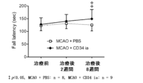
CD34陽性細胞においては、脳梗塞作製後4週間の脳梗塞後遺症モデルマウスにCD34陽性細胞を動脈内投与した。その後試験運動を継続し、投与後2週間及び8週間の時点でスコア計測、脳梗塞作製後4週間の脳梗塞後遺症モデルマウスにPBS投与した群と比較した(図3において、PBS投与群はMCAO + PBS、CD34陽性細胞投与群はMCAO + CD34 iaと表記)。図3に示すように、静脈投与後2週間の時点において、CD34陽性細胞投与群はPBS群と比較して有意差は見られ無いもののスコアの上昇は確認された。投与後8週間ではPBS群との間に有意な差が確認された。
For CD34 positive cells, CD34 positive cells were intra-arterially administered to cerebral infarction
投与後8週間における投与前から変化量を計測したところ、CD34陽性細胞投与群の平均値は33.16、PBS群は2.28で、CD34陽性細胞投与群は投与前から有意に増加していた(p=0.0136)。When the change was measured from before
以上の結果より、脳梗塞作製後4週間の脳梗塞後遺症モデルマウスにCD34陽性細胞を動脈内投与した群は、脳および末梢神経の両者に協調する刺激を与える身体運動の繰り返しにより、脳梗塞後遺症における脳神経機能向上効果を示すことが判明した。
Based on the above results, it was found that the group of mice with sequelae of cerebral infarction that received intra-arterial administration of CD34-
1-4-3.水迷路試験
試験1
脳神経機能試験として水迷路試験 (water maze test) を行った。水迷路試験は、円形のプールに水をはり、マウスが避難できる足場を水面下1cm程度の場所に作り、マウスが避難場所に到達するまでの時間 (秒) を測定することで行った。この試験では、溺水に関する刺激を脳に与え、空間認知および水泳運動と強く関連づけさせることにより、脳および末梢神経の両者に同時に刺激を与えた。
1-4-3. Water
A water maze test was performed as a brain function test. In the water maze test, a circular pool was filled with water, a platform for the mouse to escape to was placed about 1 cm below the water surface, and the time (seconds) it took for the mouse to reach the platform was measured. In this test, drowning-related stimuli were given to the brain, which was strongly associated with spatial cognition and swimming, stimulating both the brain and peripheral nerves simultaneously.
図4に示すように、脳梗塞作製後4週間の脳梗塞後遺症モデルマウスにPBSを静脈投与し、その後脳および末梢神経の両者に刺激を与える身体運動をせず、静脈投与後4週間の時点でスコア計測したところ、PBS群1日目(身体運動前)の平均値は53秒であった (図4ではMCAO+PBSと表記)。脳梗塞作製後4週間の脳梗塞後遺症モデルマウスに骨髄単核球細胞を静脈投与し、その後試験運動をせず、静脈投与後4週間の時点でスコア計測したところ、細胞治療群1日目(身体運動前)の平均値は49秒であった(図4ではMCAO+BMと表記)。No surgery群では、1日目(身体運動前)の平均は22秒であった (図4ではno surgeryと表記)。As shown in Figure 4, PBS was administered intravenously to mice with sequelae of
統計解析の結果、細胞治療群とPBS群の差のP値は0.65であり、有意差はなかった。また、細胞治療群とno surgery群の差のP値は0.002であり有意差があった。また、PBS群とno surgery群の差のP値は0.0004であり有意差があった。すなわち、骨髄単核球細胞を投与しても、脳および末梢神経の両者に刺激を与える身体運動無き場合、脳梗塞後遺症に対する脳神経機能向上効果がないことが判明した。 Statistical analysis showed that the P value for the difference between the cell therapy group and the PBS group was 0.65, which was not significant. Additionally, the P value for the difference between the cell therapy group and the no surgery group was 0.002, which was significant. Additionally, the P value for the difference between the PBS group and the no surgery group was 0.0004, which was significant. In other words, it was found that even if bone marrow mononuclear cells were administered, in the absence of physical exercise that stimulates both the brain and peripheral nerves, there was no effect on improving brain and nerve function in the aftereffects of cerebral infarction.
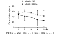
脳梗塞作製後4週間の脳梗塞後遺症モデルマウスにPBSを動脈内投与し、その後試験運動をせず、動脈投与後4週間の時点でスコア計測した(図5)。PBS群1日目(運動前)の平均値は50.9秒であった (図5ではMCAO + PBSと表記)。脳梗塞作製後4週間の脳梗塞後遺症モデルマウスにCD34陽性細胞を動脈投与し、動脈投与後4週間の時点でスコア計測したところ、細胞治療群1日目(運動前)の平均値は47.6秒であった(図5ではMCAO + CD34 iaと表記)。
PBS was administered intra-arterially to mice with sequelae of
統計解析の結果、細胞治療群とPBS群の間に、有意差はなかった。すなわち、CD34陽性細胞を動脈内投与しても、脳および末梢神経の両者に刺激を与える身体運動無き場合、脳梗塞後遺症における脳神経機能向上効果がないことが判明した。Statistical analysis showed no significant difference between the cell therapy group and the PBS group. In other words, it was found that intra-arterial administration of CD34-positive cells has no effect on improving cranial nerve function in the aftereffects of cerebral infarction, in the absence of physical exercise that stimulates both the brain and peripheral nerves.
試験2
脳梗塞後遺症モデルマウスに対する、脳および末梢神経の両者に刺激を与える身体運動の効果の検討を行なった。すなわち、水迷路試験装置を用いて水の入った円形のプールで毎日水迷路試験を行い、脳梗塞により障害された運動機能及び記憶力の向上、すなわち脳および末梢神経の両者に協調した刺激を与える身体運動を2、3、4日目に行なった。
We investigated the effect of physical exercise that stimulates both the brain and peripheral nerves on a mouse model of sequelae of cerebral infarction. Specifically, the mouse was subjected to a water maze test every day in a circular pool filled with water using a water maze test apparatus, and physical exercise that stimulates both the brain and peripheral nerves in a coordinated manner was performed on the second, third, and fourth days to improve the motor function and memory impaired by cerebral infarction.
脳梗塞後遺症モデルにPBSまたは骨髄単核球細胞を投与したマウスに対し、脳および末梢神経の両者に協調して刺激を与える身体運動効果の検討を行なった。 We investigated the effects of physical exercise, which provides coordinated stimulation to both the brain and peripheral nerves, on mice that were administered PBS or bone marrow mononuclear cells as a model of post-cerebral infarction.
PBS群においては5日目(運動後)の平均値は35秒であった。1日目(運動前)との比較の統計解析を行い、訓練による治療効果が有るか否かの判定を行なった。その結果、P値が0.11と訓練の前と後で有意差はなく、PBS投与群において脳梗塞後遺症における脳および末梢神経の両者に協調して刺激を与える身体運動は、脳神経機能を向上させることができないことが判明した。In the PBS group, the average value on the fifth day (after exercise) was 35 seconds. A statistical analysis was performed comparing this with the first day (before exercise) to determine whether or not the training had a therapeutic effect. As a result, the P value was 0.11, indicating no significant difference between before and after training, and it was found that physical exercise, which stimulates both the brain and peripheral nerves in a coordinated manner in the aftereffects of cerebral infarction in the PBS-administered group, is unable to improve cranial nerve function.
骨髄単核球細胞群においては、5日目(運動後)の平均値は8秒であった。1日目(運動前)との比較の統計解析を行い、訓練による治療効果が有るか否かの判定を行なった。その結果、P値が0.00025と訓練の前と後では有意差な短縮が観察され、骨髄単核球細胞群においては、脳梗塞後遺症における脳および末梢神経の両者に刺激を与える身体運動は、脳神経機能を有意に向上かつ持続させることが、判明した(図4)。In the bone marrow mononuclear cell group, the average value on the fifth day (after exercise) was 8 seconds. A statistical analysis was performed comparing this with the first day (before exercise) to determine whether or not the training had a therapeutic effect. As a result, a significant shortening was observed before and after training, with a P value of 0.00025, demonstrating that in the bone marrow mononuclear cell group, physical exercise that stimulates both the brain and peripheral nerves in the aftereffects of cerebral infarction significantly improves and sustains cranial nerve function (Figure 4).
次に、5日目(運動後)における各群の比較検討を行なった。その結果、細胞治療群とPBS群の差のP値は0.04であり、統計学的に有意な脳神経機能の向上が観察された。また、細胞治療群とno surgery群の差のP値は0.95であり、有意な差は観察されなかった。また、PBS群とno surgery群の差のP値は0.02であり有意差があった(図4)。Next, a comparison was made between the groups on the fifth day (after exercise). As a result, the P value for the difference between the cell therapy group and the PBS group was 0.04, and a statistically significant improvement in cranial nerve function was observed. The P value for the difference between the cell therapy group and the no surgery group was 0.95, and no significant difference was observed. The P value for the difference between the PBS group and the no surgery group was 0.02, and there was a significant difference (Figure 4).
脳梗塞後遺症モデルマウスにPBSを投与した群、及び脳梗塞後遺症モデルマウスに骨髄単核球細胞を投与した群について、脳および末梢神経の両者に協調して刺激を与える身体運動の効果の検討を行なった。The effects of physical exercise, which stimulates both the brain and peripheral nerves in a coordinated manner, were examined in a group of mouse models with sequelae of cerebral infarction that were administered PBS, and in a group of mouse models with sequelae of cerebral infarction that were administered bone marrow mononuclear cells.
CD34陽性細胞においては、脳および末梢神経の両者に刺激を与える身体運動を目的とした訓練を行うなかで、ゴールへの到達時間が徐々に短くなる傾向を示した(図5)。したがって、CD34陽性細胞の動脈内投与は、脳梗塞後遺症モデルマウスにおける脳および末梢神経の両者に刺激を与える身体運動は、脳神経機能を有意に向上させることが、判明した。 In the case of CD34-positive cells, the time to reach the goal tended to gradually shorten as training was performed with the aim of physical exercise stimulating both the brain and peripheral nerves (Figure 5). Therefore, it was found that intra-arterial administration of CD34-positive cells and physical exercise stimulating both the brain and peripheral nerves in a mouse model of sequelae of cerebral infarction significantly improved brain nerve function.
試験1と試験2の結果より、(i)脳梗塞後遺症モデルマウスにおける骨髄単核球投与又はCD34陽性細胞のみでは一般的な身体運動が与えられている状態では脳神経機能改善効果がない、(ii)脳梗塞後遺症モデルマウスにおける脳および末梢神経の両者に刺激を与える身体運動のみでは脳神経機能改善効果がない、(iii)脳梗塞後遺症モデルマウスにおける骨髄単核球投与又はCD34陽性細胞投与を行なうことにより、脳梗塞後遺症モデルマウスにおける脳および末梢神経の両者に協調して刺激を与える身体運動の治療効果が出現することが判明した。すなわち、骨髄単核球投与又はCD34陽性細胞投与が脳梗塞後遺症における脳および末梢神経の両者に刺激を与える身体運動の効果を促進することが判明した。
From the results of
1-4-4. ロータロッド試験
試験1
脳神経機能試験としてロータロッドテスト(回転棒テスト:rotarod test)を採用した。ロータロッドテストは、マウスの協調運動と平衡感覚を測定するためのテストである。装置はMK630A(室町機器株式会社)を用いた。300秒間で4 rpm~40 rpmにロッドの回転が加速するように装置をプログラムして、マウスを装置の回転棒に乗せてから落ちるまでの時間(秒)を記録した。
1-4-4.
The rotarod test was used as a cranial nerve function test. The rotarod test is a test to measure coordinated movement and balance in mice. The device used was the MK630A (Muromachi Kikai Co., Ltd.). The device was programmed to accelerate the rotation of the rod from 4 rpm to 40 rpm in 300 seconds, and the time (seconds) from when the mouse was placed on the rotating rod to when it fell off was recorded.
下記図6に示すように、脳梗塞作製後4週間の脳梗塞後遺症モデルマウスに骨髄単核球細胞を静脈投与し、静脈投与後8週間の時点でスコア計測したところ、細胞治療群 (図6ではMCAO+BMと表記)の1日目(運動前)の平均は170秒、PBS投与群(図6ではMCAO+PBSと表記)の1日目(運動前)の平均は148秒、no-surgery群の1日目(運動前)の平均は198秒を示した。細胞投与群とPBS群の比較では統計学的な有意差はなかった。すなわち、骨髄単核球細胞を投与しても、脳および末梢神経の両者に協調する刺激を与える身体運動無き場合、脳梗塞後遺症における脳神経機能向上効果がないことが判明した。As shown in Figure 6 below, bone marrow mononuclear cells were administered intravenously to a mouse model of sequelae of
試験2
脳梗塞後遺症における脳および末梢神経の両者に協調した刺激を与える身体運動として、2日目、3日目、4日目に、全ての群においてロータロッドテストに使用する装置を用いて毎日5分間の運動訓練を実施した。すなわち、マウスを回転棒に乗せ、回転棒から落下後も再び回転棒へ戻すことにより、落下の恐怖や嫌悪感を脳に与え、協調運動及び平衡感覚の機能向上と強く関連づけさせることにより、脳および末梢神経に協調した刺激を与える運動を3日間実施した。
As a physical exercise to stimulate both the brain and peripheral nerves in a coordinated manner in the aftereffects of cerebral infarction, all groups were given 5 minutes of exercise training every day on
5日目(運動後)のロータロッドテストのスコアは、細胞治療群では平均223秒であり、PBS群では平均176秒であった。統計解析の結果スコア差のP値は0.01であり有意差はあった。 On the fifth day (after exercise), the average score for the rotarod test was 223 seconds in the cell therapy group and 176 seconds in the PBS group. Statistical analysis showed that the difference in scores had a P value of 0.01, indicating a significant difference.
試験1と試験2の結果より、(i)脳梗塞後遺症において、骨髄単核球の投与のみ、または一般的な身体運動が与えられているだけでは脳神経機能への改善効果がない、(ii)脳梗塞後遺症において骨髄単核球投与を行なうことにより、脳梗塞後遺症における脳および末梢神経の両者に協調した刺激を与える運動の治療効果が出現することが判明した。すなわち、骨髄単核球投与が脳梗塞後遺症における脳および末梢神経の両者に同時に刺激を与える運動効果を促進することが判明した。
The results of
以上、骨髄単核球の投与により脳梗塞後遺症における脳および末梢神経の両者に同時に刺激を与える運動の効果が促進されていることが判明した。 These results demonstrate that administration of bone marrow mononuclear cells enhances the effects of exercise, which simultaneously stimulates both the brain and peripheral nerves in the aftereffects of cerebral infarction.
脳および末梢神経の両者に刺激を与え、中枢と末梢を協調させる身体運動の効果を促進する治療に利用できる。It can be used therapeutically to stimulate both the brain and peripheral nerves, promoting the effects of physical exercise that coordinates the central and peripheral nervous systems.
Claims (6)
骨髄単核球細胞を含むことを特徴とする身体機能回復促進のための血管内投与製剤。 An intravascular preparation for promoting physical function recovery, which is administered into the blood vessels of a subject suffering from sequelae of cerebral infarction, and promotes the effect of physical exercise that promotes bidirectional feedback between the central nervous system and the peripheral nervous system,
An intravascular preparation for promoting recovery of physical functions, comprising bone marrow mononuclear cells.
前記被験者に対して動力を付与するアクチュエータを有する動作補助装着具と、
前記被験者の生体信号を検出する生体信号センサと、
前記被験者の神経伝達信号及び筋電位信号を、前記生体信号センサにより検出された生体信号から取得する生体信号処理手段と、
前記生体信号処理手段により取得された神経伝達信号及び筋電位信号を用い、前記被験者の意思に従った動力を前記アクチュエータに発生させるための指令信号を生成する随意的制御手段と、
前記随意的制御手段により生成された指令信号に基づいて、前記神経伝達信号に応じた電流及び前記筋電位信号に応じた電流をそれぞれ生成し、前記アクチュエータに供給する駆動電流生成手段と、を備える動作補助装置にて行うことを特徴とする請求項1に記載の血管内投与製剤。 The physical exercise is
A motion-assistance wearing tool having an actuator for applying power to the subject;
A biosignal sensor for detecting a biosignal of the subject;
a biosignal processing means for acquiring a nerve conduction signal and a myoelectric potential signal of the subject from the biosignal detected by the biosignal sensor;
an optional control means for generating a command signal for causing the actuator to generate a power according to the subject's will, using the nerve conduction signal and the myoelectric potential signal acquired by the biosignal processing means;
2. The intravascular preparation according to claim 1, characterized in that the intravascular preparation is performed by an action assist device comprising: a drive current generating means for generating a current corresponding to the neurotransmission signal and a current corresponding to the myoelectric potential signal based on a command signal generated by the optional control means, and supplying the current to the actuator.
Applications Claiming Priority (3)
| Application Number | Priority Date | Filing Date | Title |
|---|---|---|---|
| JP2020001657 | 2020-01-08 | ||
| JP2020001657 | 2020-01-08 | ||
| PCT/JP2020/044082 WO2021140773A1 (en) | 2020-01-08 | 2020-11-26 | Physical function recovery promoter |
Publications (3)
| Publication Number | Publication Date |
|---|---|
| JPWO2021140773A1 JPWO2021140773A1 (en) | 2021-07-15 |
| JPWO2021140773A5 JPWO2021140773A5 (en) | 2022-09-01 |
| JP7658917B2 true JP7658917B2 (en) | 2025-04-08 |
Family
ID=76787888
Family Applications (1)
| Application Number | Title | Priority Date | Filing Date |
|---|---|---|---|
| JP2021569757A Active JP7658917B2 (en) | 2020-01-08 | 2020-11-26 | Physical function recovery promoter |
Country Status (5)
| Country | Link |
|---|---|
| US (1) | US20230190816A1 (en) |
| EP (1) | EP4098266A4 (en) |
| JP (1) | JP7658917B2 (en) |
| CN (1) | CN115243694A (en) |
| WO (1) | WO2021140773A1 (en) |
Families Citing this family (1)
| Publication number | Priority date | Publication date | Assignee | Title |
|---|---|---|---|---|
| EP4599834A1 (en) * | 2022-10-03 | 2025-08-13 | Kidswell Bio Corporation | Agent for treating or preventing cerebral palsy |
Citations (8)
| Publication number | Priority date | Publication date | Assignee | Title |
|---|---|---|---|---|
| WO2005007176A1 (en) | 2003-06-27 | 2005-01-27 | Renomedix Institute Inc. | Remedy for internal administration against cranial nerve diseases containing mesenchymal cells as the active ingredient |
| JP2013172689A (en) | 2012-02-27 | 2013-09-05 | Foundation For Biomedical Research & Innovation | Model animal for post-stroke motor dysfunction, its use, and method of screening effectiveness of new treatment method for recovery of motor function |
| JP2015159895A (en) | 2014-02-26 | 2015-09-07 | 株式会社Clio | Pluripotent stem cell for cerebral infarction treatment |
| WO2015174087A1 (en) | 2014-05-16 | 2015-11-19 | 公益財団法人先端医療振興財団 | Pharmaceutical for prevention and/or treatment of cerebral infarction |
| JP2016506954A (en) | 2013-02-06 | 2016-03-07 | Ncメディカルリサーチ株式会社 | Cell therapy for the treatment of neurodegeneration |
| WO2018034023A1 (en) | 2016-08-18 | 2018-02-22 | 北海道公立大学法人札幌医科大学 | Life extension agent |
| JP3215859U (en) | 2018-02-06 | 2018-04-19 | 有限会社メルクエスト | Fatigue model animal production device |
| WO2018139583A1 (en) | 2017-01-27 | 2018-08-02 | 公益財団法人神戸医療産業都市推進機構 | Mononuclear cell separating device and mononuclear cell separating method |
Family Cites Families (8)
| Publication number | Priority date | Publication date | Assignee | Title |
|---|---|---|---|---|
| JPH0724053Y2 (en) * | 1992-07-21 | 1995-06-05 | 有限会社シヅメメディカル | Unit Small multiplex treadmill for small animals |
| US20060210544A1 (en) * | 2003-06-27 | 2006-09-21 | Renomedix Institute, Inc. | Internally administered therapeutic agents for cranial nerve diseases comprising mesenchymal cells as an active ingredient |
| JP4481706B2 (en) * | 2004-03-31 | 2010-06-16 | 独立行政法人科学技術振興機構 | Cerebral infarction disease model mouse |
| US20090048553A1 (en) * | 2007-08-15 | 2009-02-19 | Lixian Jiang | Bone marrow transplantation for preventing and treating neurological conditions and diabetes |
| JP5771917B2 (en) * | 2010-08-04 | 2015-09-02 | 公益財団法人ヒューマンサイエンス振興財団 | Mononuclear cell separation tube and mononuclear cell separation system |
| CN103826646A (en) * | 2011-06-03 | 2014-05-28 | 麦瑟布莱斯特公司 | Method of treating the effects of stroke |
| JP2013183720A (en) * | 2012-03-09 | 2013-09-19 | Foundation For Biomedical Research & Innovation | Model animal of depression after cerebral infarction and use of the same, and method for screening effectiveness of test drug and transplanted cell with respect to depression state |
| EP3763375A4 (en) * | 2018-03-08 | 2021-09-08 | Foundation for Biomedical Research and Innovation at Kobe | CELL PREPARATION FOR TREATMENT AND / OR PREVENTION OF ISCHEMIC DISEASE AND METHOD OF SCREENING A CELL PREPARATION |
-
2020
- 2020-11-26 JP JP2021569757A patent/JP7658917B2/en active Active
- 2020-11-26 US US17/790,597 patent/US20230190816A1/en active Pending
- 2020-11-26 WO PCT/JP2020/044082 patent/WO2021140773A1/en not_active Ceased
- 2020-11-26 EP EP20912688.7A patent/EP4098266A4/en active Pending
- 2020-11-26 CN CN202080098243.7A patent/CN115243694A/en active Pending
Patent Citations (8)
| Publication number | Priority date | Publication date | Assignee | Title |
|---|---|---|---|---|
| WO2005007176A1 (en) | 2003-06-27 | 2005-01-27 | Renomedix Institute Inc. | Remedy for internal administration against cranial nerve diseases containing mesenchymal cells as the active ingredient |
| JP2013172689A (en) | 2012-02-27 | 2013-09-05 | Foundation For Biomedical Research & Innovation | Model animal for post-stroke motor dysfunction, its use, and method of screening effectiveness of new treatment method for recovery of motor function |
| JP2016506954A (en) | 2013-02-06 | 2016-03-07 | Ncメディカルリサーチ株式会社 | Cell therapy for the treatment of neurodegeneration |
| JP2015159895A (en) | 2014-02-26 | 2015-09-07 | 株式会社Clio | Pluripotent stem cell for cerebral infarction treatment |
| WO2015174087A1 (en) | 2014-05-16 | 2015-11-19 | 公益財団法人先端医療振興財団 | Pharmaceutical for prevention and/or treatment of cerebral infarction |
| WO2018034023A1 (en) | 2016-08-18 | 2018-02-22 | 北海道公立大学法人札幌医科大学 | Life extension agent |
| WO2018139583A1 (en) | 2017-01-27 | 2018-08-02 | 公益財団法人神戸医療産業都市推進機構 | Mononuclear cell separating device and mononuclear cell separating method |
| JP3215859U (en) | 2018-02-06 | 2018-04-19 | 有限会社メルクエスト | Fatigue model animal production device |
Non-Patent Citations (6)
| Title |
|---|
| CHEN, Der-Cherng et al.,Intracerebral implantation of autologous peripheral blood stem cells in stroke patients: a randomize,Cell Transplantation,2014年01月29日,Vol.23,pp.1599-1612 |
| IMURA, Takeshi et al.,Interactive effects of cell therapy and rehabilitation realize the full potential of neurogenesis in,Neuroscience Letters,2013年,Vol. 555,pp. 73-78 |
| KUMAR, A. et al.,Bone marrow mononuclear cell therapy in ischaemic stroke: a systematic review,Acta Neurol Scand.,2017年,135(5):496-506,<doi: 10.1111/ane.12666> |
| TAGUCHI, A. et al.,Intravenous Autologous Bone Marrow Mononuclear Cell Transplantation for Stroke: Phase1/2a Clinical Trial in a Homogeneous Group of Stroke Patients,Stem Cells Dev.,2015年,24(19):2207-18,<doi: 10.1089/scd.2015.0160> |
| 日本臨床(増刊) 最新臨床脳卒中学(上) Vol.72, suppl.5 ,第72巻,株式会社日本臨牀社,2014年,第469-472頁 |
| 高木 康志,脳虚血における神経幹細胞研究の歴史と現状,脳循環代謝,2015年,Vol.26,pp.113-117 |
Also Published As
| Publication number | Publication date |
|---|---|
| EP4098266A1 (en) | 2022-12-07 |
| US20230190816A1 (en) | 2023-06-22 |
| EP4098266A4 (en) | 2023-07-19 |
| WO2021140773A1 (en) | 2021-07-15 |
| JPWO2021140773A1 (en) | 2021-07-15 |
| CN115243694A (en) | 2022-10-25 |
Similar Documents
| Publication | Publication Date | Title |
|---|---|---|
| Krucoff et al. | Enhancing nervous system recovery through neurobiologics, neural interface training, and neurorehabilitation | |
| Rossignol | Locomotion and its recovery after spinal injury | |
| Yates et al. | Organization of vestibular inputs to nucleus tractus solitarius and adjacent structures in cat brain stem | |
| Herbert et al. | Evidence for a role of the reticulospinal system in recovery of skilled reaching after cortical stroke: initial results from a model of ischemic cortical injury | |
| JP6393283B2 (en) | Method and system for treatment of neuromotor dysfunction | |
| Joshua | Physiotherapy for adult neurological conditions | |
| Eschenko et al. | Behavioral, electrophysiological and histopathological consequences of systemic manganese administration in MEMRI | |
| Pazzaglia et al. | Action observation for neurorehabilitation in apraxia | |
| JP7658917B2 (en) | Physical function recovery promoter | |
| Jakeman et al. | Injured mice at the gym: review, results and considerations for combining chondroitinase and locomotor exercise to enhance recovery after spinal cord injury | |
| Li et al. | Role of α7 nicotinic acetylcholine receptors in the pressor response to intracerebroventricular injection of choline: blockade by amyloid peptide Aβ1-42 | |
| JP7545714B2 (en) | Treatment for neurological disorders | |
| JP2025092749A (en) | Treatment for neurological disorders | |
| Schalow | Cure of urinary bladder functions in severe (95%) motoric com-plete cervical spinal cord injury in human | |
| Simcsik et al. | Effects of transcranial direct current stimulation (tdcs) during a virtual reality task in individuals with parkinson’s disease: a randomized, placebo-controlled, and triple-blind Clinical Trial | |
| Kumar et al. | Engagement-sensitive interactive neuromuscular electrical therapy system for post-stroke balance rehabilitation-a concept study | |
| Vladimirovna et al. | Post-Stroke Motor Impairments: The Possibilities of Innovative Technologies and The Results of the Own Research | |
| Danilov et al. | Effects of CN-NINM intervention on chronic stroke rehabilitation: A case study | |
| Duzhar et al. | Duchenne muscular dystrophy: Treatment with fetal progenitor cell transplant | |
| Hu et al. | Advancement in brain–machine interfaces for patients with tetraplegia: neurosurgical perspective | |
| Howell et al. | Mimicking and mitigating the cutaneous response to transcranial electrical stimulation using interferential and combinatorial techniques | |
| HK40095573A (en) | Therapeutic agent for nerve disorders | |
| Bustamante et al. | Electroconvulsive therapy in catatonic patient infected with Sars-Cov-2 | |
| Santamaria et al. | Spinal Cord Injury and Epidural Spinal Cord Stimulation | |
| HK40093348A (en) | Therapeutic agent for nerve disfunction |
Legal Events
| Date | Code | Title | Description |
|---|---|---|---|
| A521 | Request for written amendment filed |
Free format text: JAPANESE INTERMEDIATE CODE: A523 Effective date: 20220527 |
|
| A621 | Written request for application examination |
Free format text: JAPANESE INTERMEDIATE CODE: A621 Effective date: 20231012 |
|
| A131 | Notification of reasons for refusal |
Free format text: JAPANESE INTERMEDIATE CODE: A131 Effective date: 20240827 |
|
| A521 | Request for written amendment filed |
Free format text: JAPANESE INTERMEDIATE CODE: A523 Effective date: 20240904 |
|
| A131 | Notification of reasons for refusal |
Free format text: JAPANESE INTERMEDIATE CODE: A131 Effective date: 20241126 |
|
| TRDD | Decision of grant or rejection written | ||
| A01 | Written decision to grant a patent or to grant a registration (utility model) |
Free format text: JAPANESE INTERMEDIATE CODE: A01 Effective date: 20250318 |
|
| A61 | First payment of annual fees (during grant procedure) |
Free format text: JAPANESE INTERMEDIATE CODE: A61 Effective date: 20250327 |
|
| R150 | Certificate of patent or registration of utility model |
Ref document number: 7658917 Country of ref document: JP Free format text: JAPANESE INTERMEDIATE CODE: R150 |