JP7635191B2 - Immune cells derived from induced pluripotent stem cells - Google Patents
Immune cells derived from induced pluripotent stem cells Download PDFInfo
- Publication number
- JP7635191B2 JP7635191B2 JP2022176895A JP2022176895A JP7635191B2 JP 7635191 B2 JP7635191 B2 JP 7635191B2 JP 2022176895 A JP2022176895 A JP 2022176895A JP 2022176895 A JP2022176895 A JP 2022176895A JP 7635191 B2 JP7635191 B2 JP 7635191B2
- Authority
- JP
- Japan
- Prior art keywords
- cells
- cell
- lineage
- myeloid
- immune cell
- Prior art date
- Legal status (The legal status is an assumption and is not a legal conclusion. Google has not performed a legal analysis and makes no representation as to the accuracy of the status listed.)
- Active
Links
Images


Classifications
-
- A—HUMAN NECESSITIES
- A01—AGRICULTURE; FORESTRY; ANIMAL HUSBANDRY; HUNTING; TRAPPING; FISHING
- A01K—ANIMAL HUSBANDRY; AVICULTURE; APICULTURE; PISCICULTURE; FISHING; REARING OR BREEDING ANIMALS, NOT OTHERWISE PROVIDED FOR; NEW BREEDS OF ANIMALS
- A01K67/00—Rearing or breeding animals, not otherwise provided for; New or modified breeds of animals
- A01K67/027—New or modified breeds of vertebrates
- A01K67/0271—Chimeric vertebrates, e.g. comprising exogenous cells
-
- A—HUMAN NECESSITIES
- A61—MEDICAL OR VETERINARY SCIENCE; HYGIENE
- A61P—SPECIFIC THERAPEUTIC ACTIVITY OF CHEMICAL COMPOUNDS OR MEDICINAL PREPARATIONS
- A61P37/00—Drugs for immunological or allergic disorders
- A61P37/02—Immunomodulators
- A61P37/04—Immunostimulants
-
- A—HUMAN NECESSITIES
- A61—MEDICAL OR VETERINARY SCIENCE; HYGIENE
- A61P—SPECIFIC THERAPEUTIC ACTIVITY OF CHEMICAL COMPOUNDS OR MEDICINAL PREPARATIONS
- A61P7/00—Drugs for disorders of the blood or the extracellular fluid
-
- C—CHEMISTRY; METALLURGY
- C12—BIOCHEMISTRY; BEER; SPIRITS; WINE; VINEGAR; MICROBIOLOGY; ENZYMOLOGY; MUTATION OR GENETIC ENGINEERING
- C12N—MICROORGANISMS OR ENZYMES; COMPOSITIONS THEREOF; PROPAGATING, PRESERVING, OR MAINTAINING MICROORGANISMS; MUTATION OR GENETIC ENGINEERING; CULTURE MEDIA
- C12N15/00—Mutation or genetic engineering; DNA or RNA concerning genetic engineering, vectors, e.g. plasmids, or their isolation, preparation or purification; Use of hosts therefor
- C12N15/09—Recombinant DNA-technology
- C12N15/63—Introduction of foreign genetic material using vectors; Vectors; Use of hosts therefor; Regulation of expression
- C12N15/79—Vectors or expression systems specially adapted for eukaryotic hosts
- C12N15/85—Vectors or expression systems specially adapted for eukaryotic hosts for animal cells
-
- C—CHEMISTRY; METALLURGY
- C12—BIOCHEMISTRY; BEER; SPIRITS; WINE; VINEGAR; MICROBIOLOGY; ENZYMOLOGY; MUTATION OR GENETIC ENGINEERING
- C12N—MICROORGANISMS OR ENZYMES; COMPOSITIONS THEREOF; PROPAGATING, PRESERVING, OR MAINTAINING MICROORGANISMS; MUTATION OR GENETIC ENGINEERING; CULTURE MEDIA
- C12N5/00—Undifferentiated human, animal or plant cells, e.g. cell lines; Tissues; Cultivation or maintenance thereof; Culture media therefor
- C12N5/06—Animal cells or tissues; Human cells or tissues
- C12N5/0602—Vertebrate cells
- C12N5/0634—Cells from the blood or the immune system
- C12N5/0635—B lymphocytes
-
- C—CHEMISTRY; METALLURGY
- C12—BIOCHEMISTRY; BEER; SPIRITS; WINE; VINEGAR; MICROBIOLOGY; ENZYMOLOGY; MUTATION OR GENETIC ENGINEERING
- C12N—MICROORGANISMS OR ENZYMES; COMPOSITIONS THEREOF; PROPAGATING, PRESERVING, OR MAINTAINING MICROORGANISMS; MUTATION OR GENETIC ENGINEERING; CULTURE MEDIA
- C12N5/00—Undifferentiated human, animal or plant cells, e.g. cell lines; Tissues; Cultivation or maintenance thereof; Culture media therefor
- C12N5/06—Animal cells or tissues; Human cells or tissues
- C12N5/0602—Vertebrate cells
- C12N5/0634—Cells from the blood or the immune system
- C12N5/0636—T lymphocytes
-
- C—CHEMISTRY; METALLURGY
- C12—BIOCHEMISTRY; BEER; SPIRITS; WINE; VINEGAR; MICROBIOLOGY; ENZYMOLOGY; MUTATION OR GENETIC ENGINEERING
- C12N—MICROORGANISMS OR ENZYMES; COMPOSITIONS THEREOF; PROPAGATING, PRESERVING, OR MAINTAINING MICROORGANISMS; MUTATION OR GENETIC ENGINEERING; CULTURE MEDIA
- C12N2501/00—Active agents used in cell culture processes, e.g. differentation
- C12N2501/065—Modulators of histone acetylation
-
- C—CHEMISTRY; METALLURGY
- C12—BIOCHEMISTRY; BEER; SPIRITS; WINE; VINEGAR; MICROBIOLOGY; ENZYMOLOGY; MUTATION OR GENETIC ENGINEERING
- C12N—MICROORGANISMS OR ENZYMES; COMPOSITIONS THEREOF; PROPAGATING, PRESERVING, OR MAINTAINING MICROORGANISMS; MUTATION OR GENETIC ENGINEERING; CULTURE MEDIA
- C12N2501/00—Active agents used in cell culture processes, e.g. differentation
- C12N2501/40—Regulators of development
- C12N2501/42—Notch; Delta; Jagged; Serrate
-
- C—CHEMISTRY; METALLURGY
- C12—BIOCHEMISTRY; BEER; SPIRITS; WINE; VINEGAR; MICROBIOLOGY; ENZYMOLOGY; MUTATION OR GENETIC ENGINEERING
- C12N—MICROORGANISMS OR ENZYMES; COMPOSITIONS THEREOF; PROPAGATING, PRESERVING, OR MAINTAINING MICROORGANISMS; MUTATION OR GENETIC ENGINEERING; CULTURE MEDIA
- C12N2501/00—Active agents used in cell culture processes, e.g. differentation
- C12N2501/60—Transcription factors
- C12N2501/602—Sox-2
-
- C—CHEMISTRY; METALLURGY
- C12—BIOCHEMISTRY; BEER; SPIRITS; WINE; VINEGAR; MICROBIOLOGY; ENZYMOLOGY; MUTATION OR GENETIC ENGINEERING
- C12N—MICROORGANISMS OR ENZYMES; COMPOSITIONS THEREOF; PROPAGATING, PRESERVING, OR MAINTAINING MICROORGANISMS; MUTATION OR GENETIC ENGINEERING; CULTURE MEDIA
- C12N2501/00—Active agents used in cell culture processes, e.g. differentation
- C12N2501/60—Transcription factors
- C12N2501/603—Oct-3/4
-
- C—CHEMISTRY; METALLURGY
- C12—BIOCHEMISTRY; BEER; SPIRITS; WINE; VINEGAR; MICROBIOLOGY; ENZYMOLOGY; MUTATION OR GENETIC ENGINEERING
- C12N—MICROORGANISMS OR ENZYMES; COMPOSITIONS THEREOF; PROPAGATING, PRESERVING, OR MAINTAINING MICROORGANISMS; MUTATION OR GENETIC ENGINEERING; CULTURE MEDIA
- C12N2501/00—Active agents used in cell culture processes, e.g. differentation
- C12N2501/60—Transcription factors
- C12N2501/604—Klf-4
-
- C—CHEMISTRY; METALLURGY
- C12—BIOCHEMISTRY; BEER; SPIRITS; WINE; VINEGAR; MICROBIOLOGY; ENZYMOLOGY; MUTATION OR GENETIC ENGINEERING
- C12N—MICROORGANISMS OR ENZYMES; COMPOSITIONS THEREOF; PROPAGATING, PRESERVING, OR MAINTAINING MICROORGANISMS; MUTATION OR GENETIC ENGINEERING; CULTURE MEDIA
- C12N2501/00—Active agents used in cell culture processes, e.g. differentation
- C12N2501/60—Transcription factors
- C12N2501/606—Transcription factors c-Myc
-
- C—CHEMISTRY; METALLURGY
- C12—BIOCHEMISTRY; BEER; SPIRITS; WINE; VINEGAR; MICROBIOLOGY; ENZYMOLOGY; MUTATION OR GENETIC ENGINEERING
- C12N—MICROORGANISMS OR ENZYMES; COMPOSITIONS THEREOF; PROPAGATING, PRESERVING, OR MAINTAINING MICROORGANISMS; MUTATION OR GENETIC ENGINEERING; CULTURE MEDIA
- C12N2501/00—Active agents used in cell culture processes, e.g. differentation
- C12N2501/60—Transcription factors
- C12N2501/608—Lin28
-
- C—CHEMISTRY; METALLURGY
- C12—BIOCHEMISTRY; BEER; SPIRITS; WINE; VINEGAR; MICROBIOLOGY; ENZYMOLOGY; MUTATION OR GENETIC ENGINEERING
- C12N—MICROORGANISMS OR ENZYMES; COMPOSITIONS THEREOF; PROPAGATING, PRESERVING, OR MAINTAINING MICROORGANISMS; MUTATION OR GENETIC ENGINEERING; CULTURE MEDIA
- C12N2501/00—Active agents used in cell culture processes, e.g. differentation
- C12N2501/65—MicroRNA
-
- C—CHEMISTRY; METALLURGY
- C12—BIOCHEMISTRY; BEER; SPIRITS; WINE; VINEGAR; MICROBIOLOGY; ENZYMOLOGY; MUTATION OR GENETIC ENGINEERING
- C12N—MICROORGANISMS OR ENZYMES; COMPOSITIONS THEREOF; PROPAGATING, PRESERVING, OR MAINTAINING MICROORGANISMS; MUTATION OR GENETIC ENGINEERING; CULTURE MEDIA
- C12N2506/00—Differentiation of animal cells from one lineage to another; Differentiation of pluripotent cells
- C12N2506/11—Differentiation of animal cells from one lineage to another; Differentiation of pluripotent cells from blood or immune system cells
-
- C—CHEMISTRY; METALLURGY
- C12—BIOCHEMISTRY; BEER; SPIRITS; WINE; VINEGAR; MICROBIOLOGY; ENZYMOLOGY; MUTATION OR GENETIC ENGINEERING
- C12N—MICROORGANISMS OR ENZYMES; COMPOSITIONS THEREOF; PROPAGATING, PRESERVING, OR MAINTAINING MICROORGANISMS; MUTATION OR GENETIC ENGINEERING; CULTURE MEDIA
- C12N2506/00—Differentiation of animal cells from one lineage to another; Differentiation of pluripotent cells
- C12N2506/45—Differentiation of animal cells from one lineage to another; Differentiation of pluripotent cells from artificially induced pluripotent stem cells
Landscapes
- Health & Medical Sciences (AREA)
- Life Sciences & Earth Sciences (AREA)
- Engineering & Computer Science (AREA)
- Biomedical Technology (AREA)
- Genetics & Genomics (AREA)
- Zoology (AREA)
- Organic Chemistry (AREA)
- Bioinformatics & Cheminformatics (AREA)
- Chemical & Material Sciences (AREA)
- Wood Science & Technology (AREA)
- Biotechnology (AREA)
- Immunology (AREA)
- General Engineering & Computer Science (AREA)
- General Health & Medical Sciences (AREA)
- Cell Biology (AREA)
- Biochemistry (AREA)
- Microbiology (AREA)
- Hematology (AREA)
- Environmental Sciences (AREA)
- Animal Behavior & Ethology (AREA)
- Animal Husbandry (AREA)
- Biodiversity & Conservation Biology (AREA)
- Plant Pathology (AREA)
- Molecular Biology (AREA)
- Biophysics (AREA)
- Physics & Mathematics (AREA)
- General Chemical & Material Sciences (AREA)
- Public Health (AREA)
- Veterinary Medicine (AREA)
- Nuclear Medicine, Radiotherapy & Molecular Imaging (AREA)
- Chemical Kinetics & Catalysis (AREA)
- Medicinal Chemistry (AREA)
- Pharmacology & Pharmacy (AREA)
- Diabetes (AREA)
- Medicines Containing Material From Animals Or Micro-Organisms (AREA)
- Micro-Organisms Or Cultivation Processes Thereof (AREA)
Description
関連出願の相互参照
本国際出願は、2016年9月6日に出願された米国仮出願第62/383,984号の米国特許法119条(e)項に基づく恩典を主張するものであり、その各内容は、その全体で参照により本明細書に組み入れられる。
CROSS-REFERENCE TO RELATED APPLICATIONS This international application claims the benefit under 35 U.S.C. §119(e) of U.S. Provisional Application No. 62/383,984, filed September 6, 2016, the contents of each of which are incorporated herein by reference in their entireties.
政府の支援
本発明は、米国国立衛生研究所により授与された助成金番号U01 HL100001およびR24DK092760の下、政府の支援を受けてなされた。政府は、本発明において一定の権利を有する。
GOVERNMENT SUPPORT This invention was made with Government support under Grant Nos. U01 HL100001 and R24DK092760 awarded by the National Institutes of Health. The Government has certain rights in this invention.
開示の分野
本開示は、概して、医学、細胞生物学、および分子生物学の分野に関する。本開示は、限られた系列の骨髄球系前駆細胞、または多能性幹細胞(PSC)、または多系列造血前駆細胞(MHPC)からのBまたはTリンパ球などの免疫細胞の産生方法に関する。
FIELD OF THE DISCLOSURE The present disclosure relates generally to the fields of medicine, cell biology, and molecular biology. The present disclosure relates to a method for producing immune cells, such as B or T lymphocytes, from restricted lineage myeloid progenitor cells, or pluripotent stem cells (PSCs), or multilineage hematopoietic progenitor cells (MHPCs).
背景
インビボ細胞補充治療のため、多数の疾患、障害および状態の治療のため、ならびに疾患モデル化、薬物スクリーニング、および血液疾患のインビトロ研究のための機能的免疫細胞の供給は不足している。骨髄移植は、多様な血液障害のための最も定評のある細胞補充治療である。骨髄移植の機能単位は、複雑な細胞階層の頂点に位置し、生涯にわたり血液の発生を補充する、造血幹細胞(HSC)である。しかし、HLAがマッチするHSCまたは患者特異的HSCの欠乏により、移植、疾患モデル化、薬物スクリーニング、および血液疾患のインビトロ研究を実施する能力が著しく限定されている。多くの場合、レシピエント対象における満足なインビボ生着および再構成を確実にするのに足るほど大きな細胞集団が移植されるわけではない。
Background There is a scarce supply of functional immune cells for in vivo cell replacement therapy, for the treatment of numerous diseases, disorders and conditions, as well as for disease modeling, drug screening, and in vitro studies of hematological disorders. Bone marrow transplantation is the most established cell replacement therapy for diverse hematological disorders. The functional unit of bone marrow transplantation is the hematopoietic stem cell (HSC), which sits at the apex of a complex cellular hierarchy and replenishes blood development throughout life. However, the lack of HLA-matched or patient-specific HSC severely limits the ability to perform transplantation, disease modeling, drug screening, and in vitro studies of hematological disorders. In many cases, cell populations are not transplanted large enough to ensure satisfactory in vivo engraftment and reconstitution in the recipient subject.
このように、代替供給源からHSCを生成させるために多くの研究が開発されている。例えば、体細胞の人工多能性幹細胞(iPSC)へのリプログラミングは、疾患モデル化、薬物選別および細胞療法のための有望な供給源である多彩な患者特異的多能性細胞を利用できるようにした。多能性細胞は、ヒトおよびマウス体細胞において、OCT4(Oct4)およびSOX2(Sox2)を、KLF4(Klf4)と任意でc-MYC(c-Myc)との組み合わせ、またはNANOG(Nanog)とLIN28(Lin28)との組み合わせのいずれかと共に強制発現させることによって誘導される。トランス活性化因子の代替的な組み合わせは、OCT4、SOX2、NANOGおよびLIN28を含む。骨髄造血前駆細胞(HPC)由来のマウスiPS細胞株が報告されている。出生後のヒト血液細胞から、顆粒球コロニー刺激因子(G-CSF)により動員された末梢血CD34+細胞から、ならびにG-CSF動員などのいかなる前処置も行わないヒト臍帯血および成体骨髄CD34+細胞からのヒトiPS細胞の派生も報告されている。これらは全て、iPSCの供給源としてHPC、幹細胞を採用したものである。Tリンパ細胞、Bリンパ細胞、線維芽細胞およびケラチノサイトなどの体細胞も、iPSCの代替供給源として使用される。 Thus, many studies have been developed to generate HSCs from alternative sources. For example, reprogramming of somatic cells into induced pluripotent stem cells (iPSCs) has made available a versatile patient-specific pluripotent cell that is a promising source for disease modeling, drug screening and cell therapy. Pluripotent cells are induced in human and mouse somatic cells by forced expression of OCT4 (Oct4) and SOX2 (Sox2) together with either KLF4 (Klf4) and optionally c-MYC (c-Myc) or NANOG (Nanog) and LIN28 (Lin28). Alternative combinations of transactivators include OCT4, SOX2, NANOG and LIN28. Mouse iPS cell lines derived from bone marrow hematopoietic progenitor cells (HPCs) have been reported. Derivation of human iPSCs from postnatal human blood cells, from peripheral blood CD34+ cells mobilized with granulocyte colony-stimulating factor (G-CSF), and from human umbilical cord blood and adult bone marrow CD34+ cells without any prior treatment such as G-CSF mobilization have also been reported. All of these employed HPCs, stem cells, as the source of iPSCs. Somatic cells such as T lymphocytes, B lymphocytes, fibroblasts, and keratinocytes are also used as alternative sources of iPSCs.
ヒトiPSCおよびマウスiPSCから生成した奇形腫の分析によって実証されたように、iPSCは、3つの胚葉に属する様々な細胞に分化することが示されている。加えて、iPSCの多能性は、iPSCを導入された胚盤胞から発生したキメラマウスの様々な器官へのiPS細胞由来細胞の寄与によって確認されている。 As demonstrated by analysis of teratomas generated from human and mouse iPSCs, iPSCs have been shown to differentiate into a variety of cells belonging to the three germ layers. In addition, the pluripotency of iPSCs has been confirmed by the contribution of iPSC-derived cells to various organs of chimeric mice developed from blastocysts that contained iPSCs.
しかし、細胞の量および細胞供給源の問題に加えて、iPSC由来の造血幹細胞および前駆細胞(iPSC-HSPC)を産生させること、または、子孫細胞がインビボで生着するであろう、それから分化した細胞を産生させることにはまだ大きな障害がある。上記のように、代替供給源からHSCをインビトロ生成させることを目指した様々な研究により、造血前駆細胞または幹細胞が産生されたが、それらはインビボでうまく生着しない。 However, in addition to issues of cell quantity and cell source, there are still major obstacles to generating iPSC-derived hematopoietic stem and progenitor cells (iPSC-HSPCs) or to generating differentiated cells from them whose progeny would engraft in vivo. As mentioned above, various studies aimed at generating HSCs in vitro from alternative sources have produced hematopoietic progenitor or stem cells that do not engraft well in vivo.
概要
本開示の態様は、特異的定方向分化に供することができる患者特異的な組織適合性複能性造血前駆細胞(MHPC)を産生させて、従来可能であった量よりも大きい量で機能的免疫細胞をインビトロ培養条件で提供するための方法に関する。本開示の態様は、これらのMHPCを含む組成物、および特異的定方向分化プロセスの結果として生じた子孫細胞、およびこれらの細胞の使用にも関する。
[0003] Embodiments of the present disclosure relate to methods for producing patient-specific histocompatible multipotent hematopoietic progenitor cells (MHPCs) that can be subjected to specific directed differentiation to provide functional immune cells in greater quantities than previously possible in in vitro culture conditions. Embodiments of the present disclosure also relate to compositions comprising these MHPCs, and the progeny cells resulting from the specific directed differentiation process, and uses of these cells.
疾患、障害および医学的状態の処置であるインビボ細胞補充治療のため、ならびに疾患モデル化、薬物スクリーニング、および血液疾患のインビトロ研究のための、HLAがマッチする機能的免疫細胞の供給は不足している。主として、免疫細胞は、造血幹細胞(HSC)から分化するが、HLAがマッチするHSCは欠乏している。本方法は、予め非リンパ球系列に決定済みの骨髄球系前駆細胞の系列分化能(lineage potential)をMHPCに逆戻りさせ、そして続いて、造血前駆細胞(HPC)の分化を特異的に促進し、リンパ球系列に方向づけることによってこの問題を解決する。加えて、逆戻り(reversed)系列分化能を有するMHPCが、増強したインビボ生着および再構成特性を有するように改変される。本産生方法は、例えば免疫療法における細胞調製方法として有用である。 There is a shortage of HLA-matched functional immune cells for in vivo cell replacement therapy to treat diseases, disorders, and medical conditions, as well as for disease modeling, drug screening, and in vitro studies of hematological disorders. Primarily, immune cells differentiate from hematopoietic stem cells (HSCs), but HLA-matched HSCs are scarce. The present method solves this problem by reversing the lineage potential of pre-committed non-lymphoid myeloid progenitor cells to MHPCs, and subsequently promoting the differentiation of hematopoietic progenitor cells (HPCs) specifically to direct them to the lymphoid lineage. In addition, MHPCs with reversed lineage potential are engineered to have enhanced in vivo engraftment and reconstitution properties. The present production method is useful, for example, as a cell preparation method in immunotherapy.
本明細書において使用される略語:
HPC=造血前駆細胞
MHPC=多系列造血前駆細胞または複能性造血前駆細胞
iPSC=人工多能性幹細胞
HSC=造血幹細胞
Abbreviations used herein:
HPC = hematopoietic progenitor cell
MHPC = multilineage hematopoietic progenitor cells or multipotent hematopoietic progenitor cells
iPSC = induced pluripotent stem cells
HSC = hematopoietic stem cell
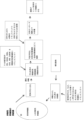
本発明者らは、少なくとも3つの外因性転写因子:ERG、HOXA9、およびRORAを非リンパ球系列決定済み骨髄球系前駆細胞に導入することによって、これらの細胞の系列分化能を逆戻りさせることができた。結果として生じた細胞は、MHPCであった。 By introducing at least three exogenous transcription factors: ERG, HOXA9, and RORA into non-lymphoid lineage-committed myeloid progenitor cells, we were able to reverse the lineage differentiation potential of these cells. The resulting cells were MHPCs.
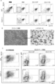
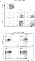
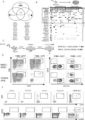
造血の間に産生する血液細胞は、以下の3つの細胞系列:(1)赤血球系細胞、(2)リンパ球系細胞、および(3)骨髄球系細胞に分けられる。図15を参照されたい。正赤芽球、赤芽球および成熟赤血球(RBC)を含む赤血球系細胞は、最も一般的な種類の血液細胞であり、肺から身体組織に酸素を送達する主な手段である。B細胞およびT細胞を含むリンパ球系細胞は、身体の免疫防御に重大な役割を果たす白血球の一種である。顆粒球、巨核球、およびマクロファージを含む骨髄球系細胞は、その他の白血球(例えば、好中球、好酸球および好塩基球)および血小板を含む多様な細胞群である。 The blood cells produced during hematopoiesis are divided into three cell lineages: (1) erythroid cells, (2) lymphoid cells, and (3) myeloid cells. See Figure 15. Erythroid cells, including normoblasts, erythroblasts, and mature red blood cells (RBCs), are the most common type of blood cell and are the primary means of delivering oxygen from the lungs to the body's tissues. Lymphoid cells, including B cells and T cells, are a type of white blood cell that play a critical role in the body's immune defense. Myeloid cells, including granulocytes, megakaryocytes, and macrophages, are a diverse group of cells that include other white blood cells (e.g., neutrophils, eosinophils, and basophils) and platelets.
骨髄球系前駆細胞は、非リンパ球系列である骨髄球系列に決定済みである。骨髄球系列中の骨髄球系前駆細胞は、さらなる細胞分裂、分化および成熟を受け、骨髄球系列は、以下の細胞型:巨核球、血小板、赤血球、マスト細胞、骨髄芽球、好塩基球、好中球、好酸球、単球およびマクロファージを産生する。図15を参照されたい。骨髄球系列は、TおよびBリンパ球などの免疫細胞を産生するリンパ球系列とは異なる。これらの逆戻り系列(reversed lineage)のMHPCにおけるヒストンメチルトランスフェラーゼEZH1をさらに阻害することによって、本発明者らは、OP9-DL1/4細胞との共培養によって、またはこれらの細胞におけるNotchシグナル伝達経路を活性化することによって、これらの細胞の分化を免疫細胞に方向づけることができた。そのうえ、本発明者らは、2つの追加的な外因性転写因子:DACH1およびNFIAをこれらの細胞に組み込むことが、OP9-DL1/4細胞と共培養により、またはNotchシグナル伝達経路を活性化することにより、これらの細胞のリンパ球系分化能を高めたことを見出した。さらに、本発明者らは、2つの他の外因性転写因子:SOX4およびMYBをこれらの細胞に組み込むことが、レシピエント対象におけるこれらの細胞のインビボ生着および再構成を高めたことを見出した。 Myeloid progenitors are committed to the myeloid lineage, a non-lymphoid lineage. Myeloid progenitors in the myeloid lineage undergo further cell division, differentiation and maturation, and the myeloid lineage produces the following cell types: megakaryocytes, platelets, erythrocytes, mast cells, myeloblasts, basophils, neutrophils, eosinophils, monocytes and macrophages. See FIG. 15. The myeloid lineage is distinct from the lymphoid lineage, which produces immune cells such as T and B lymphocytes. By further inhibiting the histone methyltransferase EZH1 in MHPCs of these reversed lineages, we were able to direct the differentiation of these cells into immune cells by co-culture with OP9-DL1/4 cells or by activating the Notch signaling pathway in these cells. Moreover, the inventors found that incorporating two additional exogenous transcription factors, DACH1 and NFIA, into these cells enhanced the lymphoid differentiation potential of these cells by co-culturing with OP9-DL1/4 cells or by activating the Notch signaling pathway. Furthermore, the inventors found that incorporating two other exogenous transcription factors, SOX4 and MYB, into these cells enhanced the in vivo engraftment and reconstitution of these cells in recipient subjects.
本開示のプロトコールの長所は、この方法が、患者の身体から容易に採取することができる細胞源、例えば血液細胞、免疫細胞、皮膚細胞などの体細胞から、所望で特異的な免疫細胞の半永続的な大量生産を可能にすることである。機能的免疫細胞の産生は、患者から得られた幹細胞または前駆細胞のみを使用することに限らない。産生された免疫細胞は、次に、免疫療法のために使用することができる。 The advantage of the disclosed protocol is that the method allows for semi-permanent mass production of desired and specific immune cells from a cell source that can be easily harvested from the patient's body, e.g., somatic cells such as blood cells, immune cells, skin cells, etc. The production of functional immune cells is not limited to using only stem or progenitor cells obtained from the patient. The produced immune cells can then be used for immunotherapy.
したがって、予め非リンパ球系列に決定済みの骨髄球系前駆細胞の系列分化能をMHPCに逆戻りさせ、逆戻り系列MHPCを所望の免疫細胞に特異的に定方向分化させる段階を含む、免疫細胞の産生方法を提供することが、本開示の目的である。非リンパ球系列に決定済みの骨髄球系前駆細胞は、患者の身体における任意の細胞、例えば体細胞から生成されるiPSCから製造することができる。そのような細胞は、患者の身体から容易に採取することができる。例えば、血液試料、皮膚試料、口腔拭き取り検体等からの細胞である。非リンパ球系列に決定済みの骨髄球系前駆細胞は、患者の骨髄から収集される場合がある。 It is therefore an object of the present disclosure to provide a method for producing immune cells, comprising the steps of reverting the lineage differentiation potential of previously non-lymphoid committed myeloid progenitor cells to MHPCs and specifically directing the reverted MHPCs to desired immune cells. Non-lymphoid committed myeloid progenitor cells can be produced from any cell in the patient's body, e.g., iPSCs generated from somatic cells. Such cells can be readily harvested from the patient's body, e.g., from a blood sample, skin sample, oral swab, etc. Non-lymphoid committed myeloid progenitor cells may be collected from the patient's bone marrow.
対象に植え込まれた造血関連細胞のインビボ生着、または再構成、または両方を増強または改善するための方法を提供することも、本開示の目的である。 It is also an object of the present disclosure to provide methods for enhancing or improving the in vivo engraftment, or reconstitution, or both, of hematopoietic-related cells implanted in a subject.
インビボ細胞補充治療、がん免疫療法などの医学療法における使用のため、ならびに疾患モデル化、薬物スクリーニング、および血液疾患のインビトロ研究のための改変(操作とも呼ばれる)細胞の組成物を提供することも、本開示の目的である。 It is also an object of the present disclosure to provide compositions of modified (also called engineered) cells for use in medical therapies such as in vivo cell replacement therapy, cancer immunotherapy, and for disease modeling, drug screening, and in vitro study of hematological disorders.
したがって、(1)TまたはB細胞などの改変免疫細胞を調製するための方法であって、転写因子の外因性コピーを使用して骨髄球系前駆細胞の系列分化能をHPCに逆戻りさせる段階、および逆戻り系列HPCを免疫細胞に特異的定方向分化させる段階を含む、方法;(2)逆戻り系列を有し、増大したリンパ球系列分化能を有する改変骨髄球系前駆細胞;(3)増大したリンパ球系列分化能を含む逆戻り系列を有する改変骨髄球系前駆細胞を含有する組成物;(4)記載された改変免疫細胞の製造/産生において使用するための本明細書記載の改変骨髄球系前駆細胞およびその組成物;(5)細胞補充治療における使用のため、またはがん、自己免疫障害、血液疾患もしくは他の遺伝性疾患および障害の処置のための、本明細書記載の改変骨髄球系前駆細胞およびその組成物;(6)本明細書記載の方法によって調製される改変免疫細胞を含有する薬学的組成物;ならびに(7)骨髄移植およびがん免疫療法、自己免疫障害、血液疾患または他の遺伝性疾患および障害などの、上記方法を用いて製造される免疫細胞を用いた処置を使用するための方法が、本明細書に開示される。改変免疫細胞は、ヒト細胞などの哺乳動物細胞である。 Thus, disclosed herein are (1) methods for preparing modified immune cells, such as T or B cells, comprising reverting the lineage potential of myeloid progenitors to HPCs using exogenous copies of transcription factors, and specifically directing the reverted-lineage HPCs to immune cells; (2) modified myeloid progenitors having a reverted lineage and increased lymphoid lineage potential; (3) compositions containing modified myeloid progenitors having a reverted lineage including increased lymphoid lineage potential; (4) modified myeloid progenitors and compositions thereof described herein for use in the manufacture/production of the modified immune cells described; (5) modified myeloid progenitors and compositions thereof described herein for use in cell replacement therapy or for the treatment of cancer, autoimmune disorders, hematological disorders or other genetic diseases and disorders; (6) pharmaceutical compositions containing modified immune cells prepared by the methods described herein; and (7) methods for using treatments using immune cells produced using the methods described above, such as bone marrow transplantation and cancer immunotherapy, autoimmune disorders, hematological disorders or other genetic diseases and disorders. The modified immune cells are mammalian cells, such as human cells.
一態様では、本開示は、増大したリンパ球系列分化能を含む、逆戻り系列を有する改変または操作骨髄球系前駆細胞を提供する。一態様では、本開示は、本明細書記載の方法によって産生される、増大したリンパ球系列分化能を含めるために逆戻りさせた系列を有する改変または操作骨髄球系前駆細胞を提供する。いくつかの態様では、改変または操作骨髄球系前駆細胞は、ERAトランスフェクションを介して以下の転写因子:ERG、HOXA9、およびRORAのそれぞれの外因性遺伝子コーディングコピーを有する。一態様では、改変または操作骨髄球系前駆細胞は、SOX4、またはMYB、またはSOX4およびMYBの両方の外因性遺伝子コーディングコピーをさらに含む。別の態様では、改変または操作骨髄球系前駆細胞は、DACH1、またはNFIA、またはDACH1およびNFIAの両方の外因性遺伝子コーディングコピーをさらに含む。いくつかの態様では、改変骨髄球系前駆細胞は、系列限定されたCD34+CD45+骨髄球系前駆細胞に由来する。 In one embodiment, the disclosure provides modified or engineered myeloid progenitor cells with reversed lineages, including increased lymphoid lineage differentiation potential. In one embodiment, the disclosure provides modified or engineered myeloid progenitor cells with lineages reversed to include increased lymphoid lineage differentiation potential, produced by the methods described herein. In some embodiments, the modified or engineered myeloid progenitor cells have exogenous gene coding copies of each of the following transcription factors: ERG, HOXA9, and RORA via ERA transfection. In one embodiment, the modified or engineered myeloid progenitor cells further comprise exogenous gene coding copies of SOX4, or MYB, or both SOX4 and MYB. In another embodiment, the modified or engineered myeloid progenitor cells further comprise exogenous gene coding copies of DACH1, or NFIA, or both DACH1 and NFIA. In some embodiments, the modified myeloid progenitor cells are derived from lineage-restricted CD34 + CD45 + myeloid progenitor cells.
別の態様では、本開示は、本明細書記載の改変または操作骨髄球系前駆細胞を含む組成物を提供する。 In another aspect, the present disclosure provides a composition comprising the modified or engineered myeloid progenitor cells described herein.
別の態様では、本開示は、記載された改変免疫細胞の製造/産生において使用するための、本明細書記載の改変骨髄球系前駆細胞およびその組成物を提供し、その際、改変骨髄球系前駆細胞は、以下の転写因子:ERG、HOXA9、およびRORAのそれぞれの外因性遺伝子コーディングコピーを含む。一態様では、改変または操作された改変骨髄球系前駆細胞は、SOX4、またはMYB、またはSOX4およびMYBの両方の外因性遺伝子コーディングコピーをさらに含む。別の態様では、改変または操作改変骨髄球系前駆細胞は、DACH1、またはNFIA、またはDACH1およびNFIAの両方の外因性遺伝子コーディングコピーをさらに含む。 In another aspect, the disclosure provides modified myeloid progenitor cells and compositions thereof described herein for use in the manufacture/production of the described modified immune cells, wherein the modified myeloid progenitor cells comprise an exogenous gene coding copy of each of the following transcription factors: ERG, HOXA9, and RORA. In one aspect, the modified or engineered modified myeloid progenitor cells further comprise an exogenous gene coding copy of SOX4, or MYB, or both SOX4 and MYB. In another aspect, the modified or engineered modified myeloid progenitor cells further comprise an exogenous gene coding copy of DACH1, or NFIA, or both DACH1 and NFIA.
別の態様では、本開示は、細胞補充治療における使用のため、またはがん、自己免疫障害、血液疾患、もしくは他の遺伝性疾患および障害の処置のための、本明細書記載の改変骨髄球系前駆細胞およびその組成物を提供し、その際、改変骨髄球系前駆細胞は、以下の転写因子:ERG、HOXA9、およびRORAのそれぞれの外因性遺伝子コーディングコピーを含む。一態様では、改変または操作改変骨髄球系前駆細胞は、SOX4、またはMYB、またはSOX4およびMYBの両方の外因性遺伝子コーディングコピーをさらに含む。別の態様では、改変または操作された改変骨髄球系前駆細胞は、DACH1、またはNFIA、またはDACH1およびNFIAの両方の外因性遺伝子コーディングコピーをさらに含む。 In another aspect, the disclosure provides modified myeloid progenitor cells and compositions thereof described herein for use in cell replacement therapy or for the treatment of cancer, autoimmune disorders, hematological disorders, or other genetic diseases and disorders, wherein the modified myeloid progenitor cells comprise an exogenous gene coding copy of each of the following transcription factors: ERG, HOXA9, and RORA. In one aspect, the modified or engineered modified myeloid progenitor cells further comprise an exogenous gene coding copy of SOX4, or MYB, or both SOX4 and MYB. In another aspect, the modified or engineered modified myeloid progenitor cells further comprise an exogenous gene coding copy of DACH1, or NFIA, or both DACH1 and NFIA.
したがって、一態様では、(a)骨髄球系前駆細胞から多系列造血前駆細胞(MHPC)をインビトロまたはエクスビボで生成させる段階;(b)結果として生じたMHPC集団におけるヒストンメチルトランスフェラーゼを阻害する段階;および(c)リンパ球系列への分化を促進するために、結果として生じたMHPC集団をnotchリガンドまたは所定のストローマ細胞または両方の存在下で分化させる段階を含む方法が、本明細書に提供される。いくつかの態様では、細胞のインビトロ培養は、段階(a)と段階(b)との間に起こる。いくつかの態様では、細胞の選択は、段階(a)と段階(b)との間に起こる。 Thus, in one embodiment, provided herein is a method comprising: (a) generating multi-lineage hematopoietic progenitor cells (MHPCs) in vitro or ex vivo from myeloid progenitor cells; (b) inhibiting histone methyltransferase in the resulting MHPC population; and (c) differentiating the resulting MHPC population in the presence of a notch ligand or a selected stromal cell or both to promote differentiation into lymphoid lineages. In some embodiments, in vitro culturing of the cells occurs between steps (a) and (b). In some embodiments, selection of the cells occurs between steps (a) and (b).
別の態様では、(a)以下の転写因子:ERG、HOXA9、およびRORAのそれぞれの外因性遺伝子コーディングコピーを骨髄球系前駆細胞にインビトロでトランスフェクトする段階であって、トランスフェクトされた細胞において転写因子が発現されて、骨髄球系および赤血球系分化能を有する、結果として生じる多系列造血前駆細胞(MHPC)集団を産生させる、段階;(b)(i)結果として生じたMHPC集団におけるヒストンメチルトランスフェラーゼを阻害して、リンパ球系分化能を拡大する段階、または(ii)結果として生じたMHPC集団にDACH1およびNFIAの外因性遺伝子コーディングコピーをインビトロでトランスフェクトして、リンパ球系分化能を拡大する段階、または(iii)(i)および(ii)の両方;ならびに(c)リンパ球系列への分化を促進するために、結果として生じたMHPC集団をnotchリガンドまたは支持ストローマ(supportive stroma)または両方の存在下で分化させる段階を含む方法が、本明細書に提供される。 In another aspect, provided herein is a method comprising: (a) transfecting myeloid progenitor cells in vitro with exogenous gene-encoding copies of each of the following transcription factors: ERG, HOXA9, and RORA, where the transcription factors are expressed in the transfected cells to produce a resulting multi-lineage hematopoietic progenitor cell (MHPC) population having myeloid and erythroid differentiation potential; (b) (i) inhibiting histone methyltransferases in the resulting MHPC population to expand lymphoid differentiation potential, or (ii) transfecting the resulting MHPC population in vitro with exogenous gene-encoding copies of DACH1 and NFIA to expand lymphoid differentiation potential, or (iii) both (i) and (ii); and (c) differentiating the resulting MHPC population in the presence of a notch ligand or supportive stroma or both to promote differentiation into lymphoid lineages.
別の態様では、本開示は、(a)以下の転写因子:ERG、HOXA9、およびRORAのそれぞれの外因性コピーを骨髄球系前駆細胞にインビトロでトランスフェクトする段階であって、トランスフェクトされた転写因子が、細胞においてインビボで発現されて、骨髄球系および赤血球系分化能を有する多系列前駆細胞(MHPC)集団を産生させる、段階;(b)(i)結果として生じたMHPC集団におけるH3K9および/もしくはH3K27でヒストンタンパク質を標的とするヒストンメチルトランスフェラーゼ酵素を阻害して、リンパ球系分化能を拡大する段階、または(ii)結果として生じたMHPC集団にDACH1およびNFIAの外因性遺伝子コーディングコピーをインビトロでトランスフェクトして、リンパ球系分化能を拡大する段階、または(iii)(i)および(ii)の両方;ならびに(c)リンパ球系列への分化を促進するために、結果として生じたMHPC集団をnotchリガンドの存在下で分化させる段階を含む、骨髄球系前駆細胞集団から改変免疫細胞を生成させる方法を提供する。これらの免疫細胞は、元の骨髄球系前駆細胞と比較して、ERG、HOXA9、およびRORAの外因性コピーを有するように遺伝的に改変されている。 In another aspect, the disclosure provides a method for the production of a method for the production of a myeloid progenitor cell, comprising the steps of: (a) in vitro transfecting myeloid progenitor cells with exogenous copies of each of the following transcription factors: ERG, HOXA9, and RORA, wherein the transfected transcription factors are expressed in vivo in the cells to generate a multi-lineage progenitor cell (MHPC) population with myeloid and erythroid differentiation potential; (b) (i) inducing histone methyltransferases targeting histone proteins at H3K9 and/or H3K27 in the resulting MHPC population; The present invention provides a method for generating modified immune cells from a population of myeloid progenitor cells, the method comprising: (i) inhibiting a catalyzing enzyme to expand lymphoid differentiation potential, or (ii) transfecting the resulting population of MHPCs in vitro with exogenous gene coding copies of DACH1 and NFIA to expand lymphoid differentiation potential, or (iii) both (i) and (ii); and (c) differentiating the resulting population of MHPCs in the presence of a notch ligand to promote differentiation into lymphoid lineages. These immune cells are genetically modified to have exogenous copies of ERG, HOXA9, and RORA, as compared to the original myeloid progenitor cells.
別の態様では、(a)接触された細胞における遺伝子の外因性コピーのインビボ発現のために、以下の転写因子:ERG、HOXA9、およびRORAのそれぞれの外因性遺伝子コーディングコピーをひとまとめに担持する1つまたは複数のベクターを骨髄球系前駆細胞集団とインビトロで接触させるまたはそれに導入する段階であって、トランスフェクトされた転写因子が、接触後の細胞においてインビボで発現されて、骨髄球系および赤血球系分化能を有する多系列造血前駆細胞(MHPC)集団を産生させる、段階;(b)MHPCをヒストンメチルトランスフェラーゼ酵素の阻害剤と接触させる段階;ならびに(c)MHPCをnotchリガンドまたは所定のストローマ細胞または両方と接触させる段階を含む方法が、本明細書に提供される。いくつかの態様では、細胞のインビトロ培養は、段階(a)と段階(b)との間に起こる。いくつかの態様では、細胞の選択は、段階(a)と段階(b)との間に起こる。方法の一態様では、段階(c)は、当技術分野において公知の任意の方法により、MHPCにおけるNotchシグナル伝達経路を活性化することからなる。 In another aspect, provided herein is a method comprising the steps of: (a) contacting or introducing into a population of myeloid progenitor cells in vitro one or more vectors collectively carrying an exogenous gene-encoding copy of each of the following transcription factors: ERG, HOXA9, and RORA, for in vivo expression of the exogenous copy of the gene in the contacted cells, wherein the transfected transcription factors are expressed in vivo in the contacted cells to generate a population of multi-lineage hematopoietic progenitor cells (MHPCs) having myeloid and erythroid differentiation potential; (b) contacting the MHPCs with an inhibitor of a histone methyltransferase enzyme; and (c) contacting the MHPCs with a notch ligand or a selected stromal cell or both. In some embodiments, in vitro culturing of the cells occurs between steps (a) and (b). In some embodiments, selection of the cells occurs between steps (a) and (b). In one embodiment of the method, step (c) comprises activating the Notch signaling pathway in the MHPCs by any method known in the art.
別の態様では、本開示は、レシピエントホストにおける造血幹細胞のインビボ生着(およびまた再構成)を改善する方法であって、(a)骨髄球系前駆細胞から多系列造血前駆細胞(MHPC)をインビトロまたはエクスビボで生成させる段階;(b)結果として生じたMHPC集団におけるヒストンメチルトランスフェラーゼを阻害する段階;および(c)結果として生じたMHPCをレシピエントホストに移植する段階を含む方法を提供する。 In another aspect, the disclosure provides a method for improving in vivo engraftment (and also reconstitution) of hematopoietic stem cells in a recipient host, the method comprising: (a) generating multi-lineage hematopoietic progenitor cells (MHPCs) from myeloid progenitor cells in vitro or ex vivo; (b) inhibiting histone methyltransferases in the resulting MHPC population; and (c) transplanting the resulting MHPCs into a recipient host.
別の態様では、本開示は、本明細書記載の方法によって産生された、改変または操作免疫細胞を提供する。 In another aspect, the present disclosure provides modified or engineered immune cells produced by the methods described herein.
別の態様では、本開示は、本明細書記載の方法によって産生された、改変または操作免疫細胞を含む組成物を提供する。 In another aspect, the present disclosure provides a composition comprising modified or engineered immune cells produced by the methods described herein.
別の態様では、本開示は、骨髄球系前駆細胞集団由来の改変または操作免疫細胞であって、以下の転写因子:ERG、HOXA9、およびRORAのそれぞれの外因性遺伝子コーディングコピーを含む免疫細胞を提供する。一態様では、改変または操作免疫細胞は、SOX4、またはMYB、またはSOX4およびMYBの両方の外因性遺伝子コーディングコピーをさらに含む。別の態様では、改変または操作免疫細胞は、DACH1、またはNFIA、またはDACH1およびNFIAの両方の外因性遺伝子コーディングコピーをさらに含む。 In another aspect, the disclosure provides modified or engineered immune cells derived from a myeloid progenitor cell population, the immune cells comprising an exogenous gene coding copy of each of the following transcription factors: ERG, HOXA9, and RORA. In one aspect, the modified or engineered immune cells further comprise an exogenous gene coding copy of SOX4, or MYB, or both SOX4 and MYB. In another aspect, the modified or engineered immune cells further comprise an exogenous gene coding copy of DACH1, or NFIA, or both DACH1 and NFIA.
別の態様では、本開示は、骨髄球系前駆細胞集団由来の改変または操作免疫細胞であって、以下の転写因子:ERG、HOXA9、RORA、DACH1およびNFIAのそれぞれの外因性遺伝子コーディングコピーを含む免疫細胞を提供する。 In another aspect, the present disclosure provides modified or engineered immune cells derived from a myeloid progenitor cell population, the immune cells comprising exogenous gene coding copies of each of the following transcription factors: ERG, HOXA9, RORA, DACH1, and NFIA.
別の態様では、本開示は、骨髄球系前駆細胞集団由来の改変または操作免疫細胞であって、以下の転写因子:ERG、HOXA9、およびRORAのそれぞれの外因性遺伝子コーディングコピー、ならびに以下のリプログラミング因子OCT4、SOX2、KLF4、ならびに任意でc-MYCもしくはNANOGおよびLIN28、または4つのリプログラミング因子:OCT4、SOX2、NANOGおよびLIN28のそれぞれの外因性遺伝子コーディングコピーを含む免疫細胞を提供する。別の態様では、改変細胞は、2つの追加的な転写因子:SOX4およびMYBの外因性遺伝子コーディングコピーをさらに含む。別の態様では、改変細胞は、2つの追加的な転写因子:DACH1およびNFIAの外因性遺伝子コーディングコピーをさらに含む。 In another aspect, the disclosure provides modified or engineered immune cells derived from a myeloid progenitor cell population, the immune cells comprising exogenous gene-encoding copies of each of the following transcription factors: ERG, HOXA9, and RORA, and exogenous gene-encoding copies of each of the following reprogramming factors: OCT4, SOX2, KLF4, and optionally c-MYC or NANOG and LIN28, or four reprogramming factors: OCT4, SOX2, NANOG, and LIN28. In another aspect, the modified cells further comprise exogenous gene-encoding copies of two additional transcription factors: SOX4 and MYB. In another aspect, the modified cells further comprise exogenous gene-encoding copies of two additional transcription factors: DACH1 and NFIA.
一態様では、本開示は、骨髄球系前駆細胞集団由来の改変細胞の組成物であって、改変細胞が、以下の転写因子:ERG、HOXA9、およびRORAのそれぞれの外因性コピーを含む組成物を提供する。別の態様では、改変細胞は、2つの追加的な転写因子:SOX4およびMYBの外因性遺伝子コーディングコピーをさらに含む。 In one aspect, the disclosure provides a composition of modified cells derived from a myeloid progenitor cell population, the modified cells comprising an exogenous copy of each of the following transcription factors: ERG, HOXA9, and RORA. In another aspect, the modified cells further comprise exogenous gene-encoding copies of two additional transcription factors: SOX4 and MYB.
一態様では、本開示は、骨髄球系前駆細胞集団由来の改変細胞の組成物であって、改変細胞が、以下の転写因子:ERG、HOXA9、RORA、SOX4、およびMYBのそれぞれの外因性遺伝子コーディングコピーを含む、組成物を提供する。 In one aspect, the disclosure provides a composition of modified cells derived from a myeloid progenitor cell population, the modified cells comprising exogenous gene-encoding copies of each of the following transcription factors: ERG, HOXA9, RORA, SOX4, and MYB.
一態様では、本開示は、骨髄球系前駆細胞集団由来の改変細胞の組成物であって、改変細胞が、以下の転写因子:ERG、HOXA9、RORA、DACH1、NFIA、SOX4、およびMYBのそれぞれの外因性遺伝子コーディングコピーを含む、組成物を提供する。 In one aspect, the disclosure provides a composition of modified cells derived from a myeloid progenitor cell population, the modified cells comprising exogenous gene coding copies of each of the following transcription factors: ERG, HOXA9, RORA, DACH1, NFIA, SOX4, and MYB.
一態様では、本開示は、骨髄球系前駆細胞集団由来の改変細胞の組成物であって、改変細胞が、以下の転写因子:ERG、HOXA9、およびRORAのそれぞれの外因性遺伝子コーディングコピー、ならびに以下のリプログラミング因子:OCT4、SOX2、KLF4、ならびに任意でc-MYCまたはnanogおよびLIN28のそれぞれの外因性遺伝子コーディングコピーを含む、組成物を提供する。リプログラミング因子の代替的な組み合わせは、これらの4つの因子:OCT4、SOX2、NANOGおよびLIN28を含む。 In one aspect, the disclosure provides a composition of modified cells derived from a myeloid progenitor cell population, the modified cells comprising exogenous gene-encoding copies of each of the following transcription factors: ERG, HOXA9, and RORA, and exogenous gene-encoding copies of each of the following reprogramming factors: OCT4, SOX2, KLF4, and optionally c-MYC or nanog and LIN28. An alternative combination of reprogramming factors includes these four factors: OCT4, SOX2, NANOG, and LIN28.
一態様では、本開示は、骨髄球系前駆細胞集団由来の改変細胞の組成物であって、改変細胞が、以下の転写因子:ERG、HOXA9、RORA、SOX4およびMYBのそれぞれの外因性遺伝子コーディングコピー、ならびに以下のリプログラミング因子OCT4、SOX2、KLF4、ならびに任意でc-MYCまたはnanogおよびLIN28のそれぞれの外因性遺伝子コーディングコピーを含む、組成物を提供する。リプログラミング因子の代替的な組み合わせは、これらの4つの因子:OCT4、SOX2、NANOGおよびLIN28を含む。 In one aspect, the disclosure provides a composition of modified cells derived from a myeloid progenitor cell population, the modified cells comprising exogenous gene-encoding copies of each of the following transcription factors: ERG, HOXA9, RORA, SOX4, and MYB, and exogenous gene-encoding copies of each of the following reprogramming factors: OCT4, SOX2, KLF4, and optionally c-MYC or nanog and LIN28. An alternative combination of reprogramming factors includes these four factors: OCT4, SOX2, NANOG, and LIN28.
記載された任意の方法、細胞または組成物の一態様では、骨髄球系前駆細胞、HPC、MHPC、iPSC、改変もしくは操作された細胞、または改変もしくは操作免疫細胞は、哺乳動物細胞である。例えば、免疫細胞は、ヒト、ラット、マウス、ウサギ、またはハムスター細胞である。 In one embodiment of any of the methods, cells, or compositions described, the myeloid progenitor cells, HPCs, MHPCs, iPSCs, modified or engineered cells, or modified or engineered immune cells are mammalian cells. For example, the immune cells are human, rat, mouse, rabbit, or hamster cells.
記載された任意の方法、細胞または組成物の一態様では、骨髄球系前駆細胞、HPC、MHPC、iPSC、改変または操作哺乳動物細胞は、霊長類細胞である。 In one embodiment of any of the methods, cells or compositions described, the myeloid progenitor cells, HPCs, MHPCs, iPSCs, modified or engineered mammalian cells are primate cells.
記載された任意の方法、細胞または組成物の一態様では、骨髄球系前駆細胞、HPC、MHPC、iPSC、改変または操作霊長類細胞または免疫細胞は、ヒト細胞である。 In one embodiment of any of the methods, cells or compositions described, the myeloid progenitor cells, HPCs, MHPCs, iPSCs, modified or engineered primate cells or immune cells are human cells.
記載された任意の方法、細胞または組成物の一態様では、MHPCは、以下の転写因子:ERG、HOXA9、およびRORAのそれぞれを骨髄球系共通前駆細胞(CMP)などの骨髄球系前駆細胞にインビトロまたはエクスビボで導入することによって、例えば、1つまたは複数のベクターをトランスフェクトすることによって生成し、ベクターは、トランスフェクトされた細胞における転写因子のインビボ発現のために、以下の転写因子:ERG、HOXA9、およびRORAのそれぞれの外因性遺伝子コーディングコピーをひとまとめに担持する。 In one embodiment of any of the methods, cells or compositions described, MHPCs are generated by introducing each of the following transcription factors: ERG, HOXA9, and RORA into myeloid progenitor cells, such as common myeloid progenitor cells (CMPs), in vitro or ex vivo, e.g., by transfecting one or more vectors, which collectively carry exogenous gene coding copies of each of the following transcription factors: ERG, HOXA9, and RORA, for in vivo expression of the transcription factors in the transfected cells.
記載された任意の方法、細胞または組成物の一態様では、MHPCは、骨髄球系前駆細胞集団を1つまたは複数のベクターと接触させることによって生成し、その際、ベクターは、接触後の細胞における因子のインビボ発現のために、以下の転写因子:ERG、HOXA9、およびRORAのそれぞれの外因性遺伝子コーディングコピーをひとまとめに担持し、トランスフェクトされた転写因子は、接触後の細胞においてインビボで発現される。例えば、第1のベクターが、ERGの外因性遺伝子コーディングコピーの核酸配列を担持し、第2のベクターが、HOXA9の外因性遺伝子コーディングコピーの核酸配列を担持し、第3のベクターが、RORAの外因性遺伝子コーディングコピーの核酸配列を担持する。あるいは、単一のベクターが、ERG、HOXA9、およびRORA転写因子をコードする3つの外因性遺伝子を全て担持する。 In one embodiment of any of the methods, cells or compositions described, MHPCs are generated by contacting a myeloid progenitor cell population with one or more vectors, where the vectors collectively carry an exogenous gene coding copy of each of the following transcription factors: ERG, HOXA9, and RORA, for in vivo expression of the factors in the contacted cells, and the transfected transcription factors are expressed in vivo in the contacted cells. For example, a first vector carries the nucleic acid sequence of an exogenous gene coding copy of ERG, a second vector carries the nucleic acid sequence of an exogenous gene coding copy of HOXA9, and a third vector carries the nucleic acid sequence of an exogenous gene coding copy of RORA. Alternatively, a single vector carries all three exogenous genes encoding ERG, HOXA9, and RORA transcription factors.
記載された任意の方法、細胞または組成物の一態様では、方法は、転写因子:SOX4の外因性遺伝子コーディングコピーを骨髄球系前駆細胞にインビトロでトランスフェクトすることをさらに含み、その際、トランスフェクトされた転写因子は、トランスフェクトされた細胞においてインビボで発現される。 In one embodiment of any of the methods, cells or compositions described, the method further comprises transfecting an exogenous gene coding copy of the transcription factor SOX4 into myeloid progenitor cells in vitro, where the transfected transcription factor is expressed in vivo in the transfected cells.
記載された任意の方法、細胞または組成物の一態様では、方法は、転写因子:MYBの外因性遺伝子コーディングコピーを骨髄球系前駆細胞にインビトロでトランスフェクトすることをさらに含み、その際、トランスフェクトされた転写因子は、トランスフェクトされた細胞においてインビボで発現される。 In one embodiment of any of the methods, cells or compositions described, the method further comprises transfecting an exogenous gene coding copy of the transcription factor MYB into myeloid progenitor cells in vitro, where the transfected transcription factor is expressed in vivo in the transfected cells.
記載された任意の方法、細胞または組成物の一態様では、骨髄球系列前駆細胞は、少なくともCD45+である。記載された任意の方法、細胞または組成物の一態様では、骨髄球系列前駆細胞は、CD34+CD45+である。記載された任意の方法、細胞または組成物の一態様では、骨髄球系列前駆細胞は、少なくともCD45+およびCD11b+である。いくつかの態様では、骨髄球系列前駆細胞は、IL-7Rアルファ/CD127、CD3、CD4、CD8およびCD19などのリンパ球系列マーカーが陰性である。 In one embodiment of any of the described methods, cells or compositions, the myeloid lineage progenitor cells are at least CD45 + . In one embodiment of any of the described methods, cells or compositions, the myeloid lineage progenitor cells are CD34 + CD45 + . In one embodiment of any of the described methods, cells or compositions, the myeloid lineage progenitor cells are at least CD45 + and CD11b + . In some embodiments, the myeloid lineage progenitor cells are negative for lymphoid lineage markers such as IL-7Ralpha/CD127, CD3, CD4, CD8 and CD19.
記載された任意の方法、細胞または組成物の一態様では、骨髄球系列前駆細胞は、非リンパ球系列に決定済みである。 In one embodiment of any of the methods, cells or compositions described, the myeloid progenitor cells are committed to a non-lymphoid lineage.
記載された任意の方法、細胞または組成物の一態様では、結果として生じたMHPCは、CD34+CD38陰性/低発現である。 In one embodiment of any of the methods, cells or compositions described, the resulting MHPCs are CD34+CD38 negative/low expressing.
記載された任意の方法、細胞または組成物の一態様では、結果として生じたMHPCは、骨髄球系および赤血球系分化能を有するが、リンパ球系分化能を有しないまたは5%未満の非常に限られたリンパ球系分化能を有する。 In one embodiment of any of the methods, cells or compositions described, the resulting MHPCs have myeloid and erythroid differentiation potential, but no lymphoid differentiation potential or have very limited lymphoid differentiation potential of less than 5%.
記載された任意の方法、細胞または組成物の一態様では、骨髄球系列前駆細胞は、多能性幹細胞集団から得られた胚様体由来の前駆細胞である。 In one embodiment of any of the methods, cells or compositions described, the myeloid lineage progenitor cells are embryoid body-derived progenitor cells obtained from a pluripotent stem cell population.
記載された任意の方法、細胞または組成物の一態様では、多能性幹細胞集団は、iPSCまたは胚性幹細胞(ESC)である。 In one embodiment of any of the methods, cells or compositions described, the pluripotent stem cell population is iPSCs or embryonic stem cells (ESCs).
記載された任意の方法、細胞または組成物の一態様では、iPSCは、3つのリプログラミング因子のみ:OCT4、SOX2、およびKLF4の外因性コピーを成熟細胞または体細胞内にインビトロまたはエクスビボで導入することによって産生される。あるいは、4つのリプログラミング因子の外因性コピーを有するiPSCは、OCT4、SOX2、NANOGおよびLIN28を含む。 In one embodiment of any of the methods, cells or compositions described, iPSCs are produced by introducing exogenous copies of only three reprogramming factors: OCT4, SOX2, and KLF4 into mature or somatic cells in vitro or ex vivo. Alternatively, iPSCs with exogenous copies of four reprogramming factors include OCT4, SOX2, NANOG, and LIN28.
記載された任意の方法、細胞または組成物の一態様では、OCT4、SOX2、およびKLF4の外因性コピーを有するiPSCは、c-MYCまたはnanogおよびLIN28の外因性コピーを細胞内にインビトロまたはエクスビボでさらに導入される。 In one embodiment of any of the methods, cells or compositions described, iPSCs with exogenous copies of OCT4, SOX2, and KLF4 are further introduced into the cells in vitro or ex vivo with exogenous copies of c-MYC or nanog and LIN28.
記載された任意の方法、細胞または組成物の一態様では、iPSCは、リプログラミング因子:OCT4、SOX2、およびKLF4の外因性コピーを、任意でc-MYCまたはnanogおよびLIN28と共に成熟細胞または体細胞内にインビトロまたはエクスビボで導入することによって産生される。 In one embodiment of any of the methods, cells or compositions described, iPSCs are produced by introducing exogenous copies of reprogramming factors: OCT4, SOX2, and KLF4, optionally together with c-MYC or nanog and LIN28, into mature or somatic cells in vitro or ex vivo.
記載された任意の方法、細胞または組成物の一態様では、iPSCは、成熟細胞を1つまたは複数のベクターとインビトロまたはエクスビボで接触させることによって産生され、その際、ベクターは、リプログラミング因子:OCT4、SOX2、およびKLF4の外因性コピーを、任意でc-MYCまたはnanogおよびLIN28と共に成熟細胞内にひとまとめに担持し、リプログラミング因子は、接触された成熟細胞または体細胞においてインビボで発現される。 In one embodiment of any of the methods, cells or compositions described, iPSCs are produced by contacting mature cells with one or more vectors in vitro or ex vivo, where the vectors carry exogenous copies of reprogramming factors: OCT4, SOX2, and KLF4, optionally together with c-MYC or nanog and LIN28, collectively within the mature cells, and the reprogramming factors are expressed in vivo in the contacted mature or somatic cells.
記載された任意の方法、細胞または組成物の一態様では、iPSCが作製される元の細胞は、ドナー対象における任意の細胞型、任意の成熟細胞または体細胞からのものであることができる。例えば、細胞は、血液試料、または骨髄試料、Bリンパ球(B細胞)、Tリンパ球(T細胞)、線維芽細胞、ケラチノサイトなどである。 In one embodiment of any of the methods, cells, or compositions described, the cells from which iPSCs are generated can be from any cell type, any mature or somatic cell, in the donor subject. For example, the cells can be from a blood sample, or a bone marrow sample, B lymphocytes (B cells), T lymphocytes (T cells), fibroblasts, keratinocytes, etc.
記載された任意の方法、細胞または組成物の一態様では、iPSCは、開示されたリプログラミング因子を成熟細胞または体細胞内にインビトロまたはエクスビボで2回以上導入することによって産生される。 In one embodiment of any of the methods, cells, or compositions described, iPSCs are produced by introducing the disclosed reprogramming factors into mature or somatic cells two or more times in vitro or ex vivo.
記載された任意の方法、細胞または組成物の一態様では、iPSCは、成熟細胞を、開示されたベクター因子とインビトロまたはエクスビボで2回以上成熟細胞または体細胞内に接触させることによって産生される。 In one embodiment of any of the methods, cells, or compositions described, iPSCs are produced by contacting mature cells with the disclosed vector factors two or more times in vitro or ex vivo in the mature or somatic cells.
記載された任意の方法、細胞または組成物の一態様では、notchリガンドは、Delta-like-1、Delta-like-4、およびヒトIgG1のFcドメインと融合されたヒトDelta-like-1の細胞外ドメインからなる固定化Delta 1 ext-IgGである。
In one embodiment of any of the methods, cells or compositions described, the notch ligand is Delta-like-1, Delta-like-4, and immobilized
記載された任意の方法、細胞または組成物の一態様では、Delta-like-1またはDelta-like-4は、MHPCを固定化Delta 1 ext-IgG、OP9-DL1細胞またはOP9-DL4細胞と共培養することによって供給される。OP9-DL1細胞は、Notchリガンド、Delta-like 1(Dll1)を異所性発現する骨髄由来ストローマ細胞株である。
In one embodiment of any of the methods, cells, or compositions described, Delta-like-1 or Delta-like-4 is delivered by co-culturing MHPCs with immobilized
記載された任意の方法、細胞または組成物の一態様では、阻害されたMHPCのNotchシグナル伝達経路は、培養状態で刺激される。 In one embodiment of any of the methods, cells or compositions described, the Notch signaling pathway of the inhibited MHPCs is stimulated in culture.
記載された任意の方法、細胞または組成物の一態様では、ヒストンメチルトランスフェラーゼは、ヒストンH3リシン残基9(H3K9)および/またはヒストンH3リシン残基27(H3K27)へのメチル基の付加を触媒する。 In one embodiment of any of the methods, cells, or compositions described, the histone methyltransferase catalyzes the addition of a methyl group to histone H3 lysine residue 9 (H3K9) and/or histone H3 lysine residue 27 (H3K27).
記載された任意の方法、細胞または組成物の一態様では、ヒストンメチルトランスフェラーゼ阻害剤は、G9a/GLPヘテロマー複合体を阻害する。 In one embodiment of any of the methods, cells or compositions described, the histone methyltransferase inhibitor inhibits the G9a/GLP heteromeric complex.
記載された任意の方法、細胞または組成物の一態様では、ヒストンメチルトランスフェラーゼ阻害剤は、EZH1(Zeste 1ポリコーム抑制複合体2サブユニットエンハンサー)を阻害する。
In one embodiment of any of the methods, cells or compositions described, the histone methyltransferase inhibitor inhibits EZH1 (Enhancer of
記載された任意の方法、細胞または組成物の一態様では、H3K9またはH3K27ヒストンメチルトランスフェラーゼは、小分子または核酸またはCRISPRによって媒介される標的遺伝的干渉によって阻害される。 In one embodiment of any of the methods, cells or compositions described, H3K9 or H3K27 histone methyltransferase is inhibited by targeted genetic interference mediated by a small molecule or nucleic acid or CRISPR.
記載された任意の方法、細胞または組成物の一態様では、H3K27ヒストンメチルトランスフェラーゼは、EZH1である。 In one embodiment of any of the methods, cells or compositions described, the H3K27 histone methyltransferase is EZH1.
記載された任意の方法、細胞または組成物の一態様では、H3K27ヒストンメチルトランスフェラーゼは、EZH2ではない。 In one embodiment of any of the methods, cells or compositions described, the H3K27 histone methyltransferase is not EZH2.
記載された任意の方法、細胞または組成物の一態様では、ヒストンメチルトランスフェラーゼ小分子阻害剤は、EZH2ではなくEZH1に特異的である。 In one embodiment of any of the methods, cells or compositions described, the histone methyltransferase small molecule inhibitor is specific for EZH1 and not EZH2.
記載された任意の方法、細胞または組成物の一態様では、ヒストンメチルトランスフェラーゼ小分子阻害剤には、AMI-1、A-366、BIX-01294、BIX01338、BRD4770、ケトシン、UNC0224、UNC0631、UNC0638、UNC0642、UNC0646、EPZ5676、EPZ005687、GSK343、EPZ-6438、3-デアザネプラノシンA(DZNeP)HCl、UNC1999、MM-102、SGC 0946、エンタカポン、EPZ015666、UNC0379、EI1、MI-2(メニン-MLL阻害剤)、MI-3(メニン-MLL阻害剤)、PFI-2、GSK126、EPZ004777、BRD4770、およびEPZ-6438が非限定的に含まれる。 In one embodiment of any of the methods, cells or compositions described, histone methyltransferase small molecule inhibitors include AMI-1, A-366, BIX-01294, BIX01338, BRD4770, chaetocin, UNC0224, UNC0631, UNC0638, UNC0642, UNC0646, EPZ5676, EPZ005687, GSK343, EPZ-6438, 3-deazaneplanocin A (DZNeP) HCl, UNC1999, MM-102, SGC Non-limiting examples include 0946, entacapone, EPZ015666, UNC0379, EI1, MI-2 (menin-MLL inhibitor), MI-3 (menin-MLL inhibitor), PFI-2, GSK126, EPZ004777, BRD4770, and EPZ-6438.
記載された任意の方法、細胞または組成物の一態様では、ヒストンメチルトランスフェラーゼ核酸阻害剤は、ヒストンメチルトランスフェラーゼの発現を標的とする核酸である。 In one embodiment of any of the methods, cells or compositions described, the histone methyltransferase nucleic acid inhibitor is a nucleic acid that targets the expression of a histone methyltransferase.
記載された任意の方法、細胞または組成物の一態様では、核酸阻害剤は、RNA干渉阻害剤である。 In one embodiment of any of the methods, cells or compositions described, the nucleic acid inhibitor is an RNA interference inhibitor.
記載された任意の方法、細胞または組成物の一態様では、核酸は、

記載された任意の改変免疫細胞の一態様では、免疫細胞は、SOX4またはMYBまたはSOX4およびMYBの両方の外因性遺伝子コーディングコピーをさらに含む。 In one embodiment of any of the modified immune cells described, the immune cells further comprise an exogenous gene coding copy of SOX4 or MYB or both SOX4 and MYB.
記載された任意の改変免疫細胞の一態様では、免疫細胞は、DACH1またはNFIAまたはDACH1およびNFIAの両方の外因性遺伝子コーディングコピーをさらに含む。 In one embodiment of any of the modified immune cells described, the immune cell further comprises an exogenous gene coding copy of DACH1 or NFIA or both DACH1 and NFIA.
記載された任意の方法、細胞または組成物の一態様では、ヒストンメチルトランスフェラーゼ阻害されたMHPCの特異的定方向分化は、細胞を、IL-7、IL-2、IL-15、およびIL-4からなる群より選択されるサイトカインと接触させることを含む。 In one embodiment of any of the methods, cells or compositions described, the specific directed differentiation of histone methyltransferase-inhibited MHPCs comprises contacting the cells with a cytokine selected from the group consisting of IL-7, IL-2, IL-15, and IL-4.
一態様では、細胞補充治療の方法、または対象におけるがん、自己免疫障害、血液疾患、もしくは他の遺伝性疾患および障害の処置のための方法であって、(a)ドナー対象から体細胞を提供する段階、(b)任意の前述のパラグラフに記載された体細胞に由来する骨髄球系前駆細胞から多系列造血前駆細胞を生成させる段階;(c)任意の前述のパラグラフに記載された、結果として生じた多系列造血前駆細胞集団におけるヒストンメチルトランスフェラーゼを阻害する段階;(d)任意の前述のパラグラフに記載されたリンパ球系列への分化を促進するために、結果として生じた多系列造血前駆細胞集団をnotchリガンドまたはストローマ細胞または両方の存在下で分化させる段階、および結果として生じた分化後のリンパ球系細胞をレシピエント対象に植え込む段階を含む方法が、本明細書に提供される。 In one aspect, provided herein is a method of cell replacement therapy or a method for treating cancer, an autoimmune disorder, a hematological disease, or other genetic disease and disorder in a subject, comprising: (a) providing somatic cells from a donor subject; (b) generating multilineage hematopoietic progenitor cells from myeloid progenitor cells derived from the somatic cells as described in any preceding paragraph; (c) inhibiting histone methyltransferase in the resulting multilineage hematopoietic progenitor cell population as described in any preceding paragraph; (d) differentiating the resulting multilineage hematopoietic progenitor cell population in the presence of notch ligand or stromal cells or both to promote differentiation into lymphoid lineages as described in any preceding paragraph, and transplanting the resulting differentiated lymphoid cells into a recipient subject.
上記処置方法の一態様では、ホスト対象およびレシピエント対象は、同じ個体である。 In one embodiment of the above treatment method, the host subject and the recipient subject are the same individual.
上記処置方法の一態様では、ホスト対象およびレシピエント対象は、同じ個体ではないが、少なくともHLA適合性である。 In one embodiment of the above treatment method, the host subject and the recipient subject are not the same individual, but are at least HLA compatible.
定義
一態様では、本明細書に使用される用語「造血幹細胞」または「HSC」は、自己複製能を有し、かつ3つの造血系列:赤血球系、リンパ球系、および骨髄球系の全ての血液細胞型を生じる、幹細胞を表す。これらの細胞型には、骨髄球系列(単球およびマクロファージ、好中球、好塩基球、好酸球、赤血球、巨核球/血小板、樹状細胞)、およびリンパ球系列(T細胞、B細胞、NK細胞)が含まれる。ヒトHSCは、CD34+、CD59+、CD90/Thy1+、CD38low/-、c-kit/CD117-/low、およびLin-として判定される。マウスHSCは、CD34low/-、SCA-1+、CD90/Thy1+/low、CD38+、c-Kit/CD117+、およびLin-と見なされる。これらのマーカーパネルの発現を検出することで、蛍光標示式細胞分取(FACS)のような技法を介した特異的細胞集団の分離が可能になる。一態様では、用語「造血幹細胞」または「HSC」は、自己複製能を有し、以下の細胞表面マーカー:CD34+、CD59+、Thy1/CD90+、CD38lo/-、CD133+、c-Kit/CD117-/lo、およびLin-を有する幹細胞を表す。一態様では、用語「造血幹細胞」または「HSC」は、少なくともCD34+である幹細胞を表す。一態様では、用語「造血幹細胞」または「HSC」は、自己複製能を有し、少なくともCD34+およびc-kit/CD117lo/-である幹細胞を表す。一態様では、用語「造血幹細胞」または「HSC」は、自己複製能を有し、少なくともCD38low/-、c-kit/CD117-/lowである幹細胞を表す。
DEFINITIONS In one embodiment, the term "hematopoietic stem cell" or "HSC" as used herein refers to a stem cell that has the capacity for self-renewal and gives rise to all blood cell types of the three hematopoietic lineages: erythroid, lymphoid, and myeloid. These cell types include the myeloid lineage (monocytes and macrophages, neutrophils, basophils, eosinophils, erythrocytes, megakaryocytes/platelets, dendritic cells) and lymphoid lineage (T cells, B cells, NK cells). Human HSCs are determined as CD34 + , CD59 + , CD90/Thy1 + , CD38 low/- , c-kit/CD117 -/low , and Lin - . Mouse HSCs are considered as CD34 low/- , SCA-1 + , CD90/Thy1 +/low , CD38 + , c-Kit/CD117 + , and Lin - . Detecting the expression of these marker panels allows for the separation of specific cell populations via techniques such as fluorescence activated cell sorting (FACS). In one embodiment, the term "hematopoietic stem cells" or "HSC" refers to stem cells that have the capacity for self-renewal and have the following cell surface markers: CD34+, CD59+, Thy1/CD90 + , CD38 lo/- , CD133+, c-Kit/CD117- /lo , and Lin- . In one embodiment, the term "hematopoietic stem cells" or "HSC" refers to stem cells that are at least CD34+. In one embodiment, the term "hematopoietic stem cells" or "HSC" refers to stem cells that have the capacity for self-renewal and are at least CD34 + and c-kit/CD117 lo/- . In one embodiment, the term "hematopoietic stem cells" or "HSC" refers to stem cells that have the capacity for self-renewal and are at least CD38 low/- , c-kit/CD117- /low .
本明細書に使用される用語「iPS細胞」、「iPSC」、および「人工多能性幹細胞」は、互換的に使用され、以下のリプログラミング因子:OCT4、SOX2、KLF4、ならびに任意でc-MYCまたはnanogおよびLIN28のトランスフェクションによって分化細胞、例えば体細胞から人工的に派生する多能性細胞を表す。リプログラミング因子の代替的な組み合わせは、OCT4、SOX2、NANOGおよびLIN28を含む。 As used herein, the terms "iPS cells", "iPSCs", and "induced pluripotent stem cells" are used interchangeably and refer to pluripotent cells artificially derived from differentiated cells, e.g., somatic cells, by transfection of the following reprogramming factors: OCT4, SOX2, KLF4, and optionally c-MYC or nanog and LIN28. Alternative combinations of reprogramming factors include OCT4, SOX2, NANOG, and LIN28.
幹細胞および前駆細胞の分化および発生の文脈で使用されるときの、本明細書に使用される用語「系列」は、細胞が完全分化細胞になるために採ることができる細胞分化および発生の経路を表す。例えば、HSCには、3つの造血細胞系列:赤血球系、リンパ球系、および骨髄球系があり、HSCは、これら3つの系列全てについて知られている最終分化細胞型に分化および発生する分化能、すなわち能力を有する。用語「多系列」が使用されるとき、本用語は、細胞が将来的に、1つよりも多い系列について知られている最終分化細胞型に分化および発生できることを意味する。例えば、HSCは、多系列分化能を有する。用語「限られた系列」が使用されるとき、本用語は、細胞が、1つの系列について知られている最終分化細胞型に分化および発生することができることを意味する。例えば、骨髄球系共通前駆細胞(CMP)または巨核球-赤血球系前駆細胞(MEP)(図15参照)は、リンパ球系列ではなく骨髄球系列の最終分化細胞型にのみ分化および発生できるので、限られた系列を有する。骨髄球系列の最終分化細胞には、赤血球、単球、マクロファージ、巨核球、骨髄芽球、樹状細胞、および顆粒球(好塩基球、好中球、好酸球、およびマスト細胞)が含まれ;リンパ球系列の最終分化細胞には、Tリンパ球/T細胞、Bリンパ球/B細胞、樹状細胞、およびナチュラルキラー細胞が含まれる。 As used herein, the term "lineage" when used in the context of stem and progenitor cell differentiation and development refers to the pathway of cell differentiation and development that a cell can take to become a fully differentiated cell. For example, HSCs have three hematopoietic cell lineages: erythroid, lymphoid, and myeloid, and HSCs have differentiation potential, i.e., the ability, to differentiate and develop into terminally differentiated cell types known for all three lineages. When the term "multilineage" is used, the term means that the cell can differentiate and develop into terminally differentiated cell types known for more than one lineage in the future. For example, HSCs have multilineage differentiation potential. When the term "limited lineage" is used, the term means that the cell can differentiate and develop into terminally differentiated cell types known for one lineage. For example, common myeloid progenitor cells (CMPs) or megakaryocytic-erythroid progenitors (MEPs) (see FIG. 15) have limited lineage because they can only differentiate and develop into terminally differentiated cell types of the myeloid lineage and not the lymphoid lineage. Terminally differentiated cells of the myeloid lineage include erythrocytes, monocytes, macrophages, megakaryocytes, myeloblasts, dendritic cells, and granulocytes (basophils, neutrophils, eosinophils, and mast cells); terminally differentiated cells of the lymphoid lineage include T lymphocytes/T cells, B lymphocytes/B cells, dendritic cells, and natural killer cells.
本明細書に使用される用語「前駆細胞」は、後で特異的細胞型(完全分化または最終分化細胞)、例えば、血液細胞、皮膚細胞、骨細胞、または有毛細胞に成熟(分化)する能力を有する未熟または未分化細胞を表す。前駆細胞は、分化によって生み出すことができる細胞と比べて原始細胞表現型を有する(例えば、完全分化細胞よりも発生経路または進行に沿った早い段階である)。多くの場合に、前駆細胞は、顕著または非常に高い増殖能も有する。前駆細胞は、発生経路ならびに細胞が発生および分化する環境に応じて、複数の別個の分化細胞型、または単一の分化細胞型を生み出すことができる。前駆細胞は、増殖して、同様に未熟または未分化のより多くの前駆細胞を作製することもできる。 As used herein, the term "progenitor cell" refers to an immature or undifferentiated cell that has the capacity to subsequently mature (differentiate) into a specific cell type (fully differentiated or terminally differentiated cell), e.g., a blood cell, a skin cell, a bone cell, or a hair cell. Progenitor cells have a primitive cell phenotype compared to cells that can be produced by differentiation (e.g., earlier along a developmental pathway or progression than a fully differentiated cell). In many cases, progenitor cells also have a significant or very high proliferative potential. Progenitor cells can give rise to multiple distinct differentiated cell types, or a single differentiated cell type, depending on the developmental pathway and the environment in which the cells develop and differentiate. Progenitor cells can also proliferate to make more progenitor cells that are similarly immature or undifferentiated.
本明細書に使用される用語「多系列造血前駆細胞」、「複能性造血前駆細胞」および「MHPC」は互換的に使用され、複数の種類の造血系列細胞を生成またはそれに分化する力(ability)または能力を有する造血細胞(血液を形成する細胞)を表す。一態様では、この用語は、本明細書記載の「逆戻り多系列造血前駆細胞」および「逆戻りMHPC」を含む。そのような細胞は、転写因子:ERG、HOXA9、およびRORAの遺伝子コーディング核酸のいくつかの外因性コピーを細胞内に組み込むためのインビトロまたはエクスビボトランスフェクション後の骨髄球系前駆細胞に由来する。一態様では、この用語は、「胚様体由来前駆細胞」および「EB由来前駆細胞」を含む。 As used herein, the terms "multilineage hematopoietic progenitor cells", "multipotent hematopoietic progenitor cells" and "MHPCs" are used interchangeably to refer to hematopoietic cells (cells that form blood) that have the ability or potency to generate or differentiate into multiple types of hematopoietic lineage cells. In one aspect, the terms include "reverted multilineage hematopoietic progenitor cells" and "reverted MHPCs" as described herein. Such cells are derived from myeloid progenitor cells following in vitro or ex vivo transfection to incorporate into the cells several exogenous copies of the gene-encoding nucleic acids for the transcription factors: ERG, HOXA9, and RORA. In one aspect, the terms include "embryoid body-derived progenitor cells" and "EB-derived progenitor cells".
本明細書に使用されるように、一態様では、用語「骨髄球系前駆細胞」または「骨髄球系列前駆細胞」は、骨髄球系列に決定済みであり、骨髄球系列の最終分化細胞型にのみ分化および発生することができる未熟または未分化細胞を表す。例は、骨髄球系列のCMP、MEP、およびGMPである。一態様では、用語「骨髄球系前駆細胞」または「骨髄球系列前駆細胞」は、胚様体から得られた多能性幹細胞由来のCD34+CD45+細胞を表す。一態様では、用語「骨髄球系前駆細胞」または「骨髄球系列前駆細胞」は、顆粒球およびマクロファージにのみ分化および発生する細胞を表す。 As used herein, in one aspect, the term "myeloid progenitor cells" or "myeloid precursor cells" refers to immature or undifferentiated cells that are committed to the myeloid lineage and can differentiate and develop only into terminally differentiated cell types of the myeloid lineage. Examples are CMPs, MEPs, and GMPs of the myeloid lineage. In one aspect, the term "myeloid precursor cells" or "myeloid precursor cells" refers to CD34+CD45+ cells derived from pluripotent stem cells obtained from embryoid bodies. In one aspect, the term "myeloid precursor cells" or "myeloid precursor cells" refers to cells that differentiate and develop only into granulocytes and macrophages.
用語「分化細胞」は、そのネイティブな形態で本明細書において定義される用語としての多能性ではない、任意の初代細胞を意味する。用語「分化細胞」は、複能性細胞(例えば成体体性幹細胞)などの部分的に分化した細胞も包含する。いくつかの態様では、用語「分化細胞」は、専門化の度合いの低い細胞型の細胞(例えば、未分化細胞またはリプログラミングされた細胞)由来の、より専門化された細胞型の細胞であって、細胞分化プロセスを受けた細胞も表す。 The term "differentiated cell" refers to any primary cell that is not pluripotent as that term is defined herein in its native form. The term "differentiated cell" also encompasses partially differentiated cells, such as multipotent cells (e.g., adult somatic stem cells). In some embodiments, the term "differentiated cell" also refers to a cell of a more specialized cell type derived from a cell of a less specialized cell type (e.g., an undifferentiated cell or a reprogrammed cell) that has undergone a cell differentiation process.
細胞個体発生の文脈における用語「分化する」、または「分化している」は、「分化細胞」が、その前駆細胞よりも発生経路をさらに下に進行した細胞であることを意味する、相対的な用語である。したがって、いくつかの実施形態では、本明細書において定義される本用語としてのリプログラミングされた細胞は、系列限定された前駆細胞(中胚葉幹細胞または内胚葉幹細胞など)に分化することができ、この前駆細胞は、今度は経路をさらに下に進んで他の種類の前駆細胞(組織特異的前駆細胞、例えば心筋前駆細胞、または膵臓前駆細胞など)に、次に、ある特定の組織型に特徴的な役割を果たし、さらに増殖する能力を保持する場合または保持しない場合がある最終段階分化細胞に分化することができる。 The term "differentiate" or "differentiating" in the context of cell ontogeny is a relative term meaning that a "differentiated cell" is a cell that has progressed further down the developmental pathway than its progenitor cell. Thus, in some embodiments, a reprogrammed cell as the term is defined herein can differentiate into a lineage-restricted progenitor cell (such as a mesodermal or endodermal stem cell), which in turn can progress further down the pathway to other types of progenitor cells (such as tissue-specific progenitor cells, e.g., cardiac progenitor cells, or pancreatic progenitor cells), and then into terminally differentiated cells that fulfill roles characteristic of a particular tissue type and may or may not retain the ability to proliferate further.
「複能性細胞」に関連して使用される場合の用語「複能性」は、全部で3つの胚葉に由来する細胞の全てではなく一部に分化することができる細胞を表す。したがって、複能性細胞は、部分分化細胞である。複能性細胞は、当技術分野において周知であり、複能性細胞の例には、例えば、造血幹細胞および神経幹細胞、毛包幹細胞、肝臓幹細胞などの成体体性幹細胞が含まれる。複能性は、幹細胞が、他の系列の細胞ではなく、所与の系列の多くの細胞型を形成する場合があることを意味する。例えば、複能性血液幹細胞は、多数の異なる血液細胞型(赤血球、白血球、血小板など)を形成することができるが、ニューロンを形成することができず;心血管前駆細胞(MICP)は、特定の成熟心臓、ペースメーカー、平滑筋、および内皮細胞型に分化し;膵臓由来複能性前駆細胞(PMP)コロニーは、膵臓系列の細胞型(インスリン、グルカゴン、アミラーゼまたはソマトスタチンを産生する細胞)および神経系列の細胞型(形態的にニューロン様、アストロサイト様またはオリゴデンドロサイト様の細胞)を産生する。 The term "multipotent" when used in reference to "multipotent cells" refers to cells that can differentiate into some but not all of the cells derived from all three germ layers. Thus, multipotent cells are partially differentiated cells. Multipotent cells are well known in the art, and examples of multipotent cells include, for example, hematopoietic stem cells and adult somatic stem cells such as neural stem cells, hair follicle stem cells, and liver stem cells. Multipotency means that a stem cell may form many cell types of a given lineage but not cells of other lineages. For example, multipotent blood stem cells can form many different blood cell types (red blood cells, white blood cells, platelets, etc.) but cannot form neurons; cardiovascular progenitor cells (MICPs) differentiate into specific mature cardiac, pacemaker, smooth muscle, and endothelial cell types; pancreatic-derived multipotent progenitor (PMP) colonies produce cell types of the pancreatic lineage (cells that produce insulin, glucagon, amylase, or somatostatin) and neural lineage (cells that are morphologically neuron-, astrocyte-, or oligodendrocyte-like).
本明細書に使用される用語「リプログラミング遺伝子」は、その発現が、分化細胞、例えば体細胞の未分化細胞(例えば、多能性状態または部分多能性状態、複能性状態の細胞)へのリプログラミングに寄与する遺伝子を表す。リプログラミング遺伝子は、例えば、マスター転写因子:Sox2、Oct3/4、Klf4、Nanog、Lin-28、c-mycなどをコードする遺伝子であることができる。用語「リプログラミング因子」は、リプログラミング遺伝子によってコードされるタンパク質を表す。 As used herein, the term "reprogramming gene" refers to a gene whose expression contributes to the reprogramming of a differentiated cell, e.g., a somatic cell, into an undifferentiated cell (e.g., a cell in a pluripotent or partially pluripotent, multipotent state). Reprogramming genes can be, for example, genes encoding master transcription factors: Sox2, Oct3/4, Klf4, Nanog, Lin-28, c-myc, etc. The term "reprogramming factor" refers to a protein encoded by a reprogramming gene.
用語「外因性」は、そのネイティブな供給源以外の細胞に存在する物質を表す。本明細書に使用される場合の用語「外因性」は、当該核酸またはタンパク質が通常は見出されないまたはより低い量で見出される細胞または生物などの生体システムに、ヒトの手を伴うプロセスによって導入された、核酸(例えば、リプログラミング転写因子、例えばSox2、Oct3/4、Klf4、Nanog、Lin-28、c-mycなどをコードする核酸)またはタンパク質(例えば、転写因子ポリペプチド)を表す。物質(例えば、sox2転写因子をコードする核酸、またはタンパク質、例えばSOX2ポリペプチド)は、それが細胞またはその物質を受け継ぐ細胞の祖先に導入されたならば、外因性と見なされる。 The term "exogenous" refers to a substance present in a cell other than its native source. As used herein, the term "exogenous" refers to a nucleic acid (e.g., a nucleic acid encoding a reprogramming transcription factor, e.g., Sox2, Oct3/4, Klf4, Nanog, Lin-28, c-myc, etc.) or a protein (e.g., a transcription factor polypeptide) that has been introduced by a process involving the hand of man into a biological system, such as a cell or organism in which the nucleic acid or protein is not normally found or is found in lower amounts. A substance (e.g., a nucleic acid encoding a sox2 transcription factor, or a protein, e.g., a SOX2 polypeptide) is considered exogenous if it was introduced into a cell or into an ancestor of a cell that inherits the substance.
本明細書に使用される用語「単離された」は、細胞がその自然環境以外の状態に置かれることを示す。用語「単離された」は、その後これらの細胞を他の細胞と組み合わせてまたは混合して後に使用することを排除しない。 As used herein, the term "isolated" indicates that the cells are placed in a condition other than their natural environment. The term "isolated" does not exclude the subsequent use of these cells in combination or admixture with other cells.
本明細書に使用される用語「拡大すること」は、細胞分裂(有糸分裂)を経由して同様の細胞の数を増加させることを表す。用語「増殖させること」および「拡大すること」は、互換的に使用される。 As used herein, the term "expanding" refers to increasing the number of similar cells via cell division (mitosis). The terms "multiplying" and "expanding" are used interchangeably.
本明細書に使用される「細胞表面マーカー」は、細胞表面に発現される任意の分子を表す。細胞表面発現は、通常、分子が膜貫通ドメインを保有することを要する。細胞表面には通常見出されないいくつかの分子を、細胞表面に発現するように、組み換え技法によって操作することができる。多くの天然細胞表面マーカーは、「CD」または「分化クラスター」分子と名付けられている。細胞表面マーカーは、多くの場合、抗体が結合することができる抗原決定基を提供する。本明細書記載の方法に特に関連する細胞表面マーカーは、CD34である。本開示による有用な造血前駆細胞は、好ましくはDC34を発現し、または言い換えると、これらの細胞は、CD34陽性である。 As used herein, "cell surface marker" refers to any molecule that is expressed on the cell surface. Cell surface expression usually requires that the molecule possess a transmembrane domain. Some molecules that are not normally found on the cell surface can be engineered by recombinant techniques to be expressed on the cell surface. Many natural cell surface markers are termed "CD" or "cluster of differentiation" molecules. Cell surface markers often provide antigenic determinants to which an antibody can bind. A cell surface marker of particular relevance to the methods described herein is CD34. Useful hematopoietic progenitor cells according to the present disclosure preferably express DC34, or in other words, these cells are CD34 positive.
細胞は、任意の細胞表面マーカーについて「陽性」または「陰性」と称することができ、このような呼称の両方が、本明細書記載の方法の実施のために有用である。細胞を、マーカーに特異的に結合する抗体と接触させ、続いてそのような接触後の細胞のフローサイトメトリー分析を行って、抗体が細胞と結合しているかどうかを判定することなどの当業者に公知の方法を用いて、細胞が、検出されるに足る量でその表面にマーカーを発現している場合、その細胞は、細胞表面マーカーについて「陽性」と見なされる。細胞が細胞表面マーカーについてのメッセンジャーRNAを発現する場合があるとはいえ、本明細書記載の方法に関して陽性と見なされるために、細胞は、このマーカーを細胞表面に発現しなければならないことを理解されたい。同様に、細胞をマーカーと特異的に結合する抗体と接触させ、続いてそのような接触後の細胞のフローサイトメトリー分析を行って、抗体が細胞と結合しているかどうかを判定することなどの当業者に公知の方法を用いて、細胞がその表面にマーカーを検出されるに足る量で発現していない場合、その細胞は、細胞表面マーカーについて「陰性」または「陰性/低発現」(「-/lo」または「lo/-」と略される)と見なされる。細胞表面系列マーカーに特異的な薬剤が使用されるいくつかの態様では、この薬剤は、全て、蛍光タグなどの同じラベルまたはタグを含むことができ、したがって、そのラベルまたはタグについて陽性の全ての細胞を排除または除去して、本明細書記載の方法における使用のために、未接触の造血幹細胞または前駆細胞を残すことができる。 A cell may be referred to as "positive" or "negative" for any cell surface marker, and both such designations are useful for the practice of the methods described herein. A cell is considered "positive" for a cell surface marker if it expresses the marker on its surface in an amount sufficient to be detected using methods known to those of skill in the art, such as contacting the cell with an antibody that specifically binds to the marker, followed by flow cytometric analysis of the cells after such contact to determine whether the antibody binds to the cell. It should be understood that, although a cell may express messenger RNA for a cell surface marker, the cell must express the marker on the cell surface to be considered positive for the methods described herein. Similarly, a cell is considered "negative" or "negative/low expressing" (abbreviated as "-/lo" or "lo/-") for a cell surface marker if the cell does not express the marker on its surface in an amount sufficient to be detected using methods known to those of skill in the art, such as contacting the cell with an antibody that specifically binds to the marker, followed by flow cytometric analysis of the cells after such contact to determine whether the antibody binds to the cell. In some embodiments in which agents specific for cell surface lineage markers are used, the agents can all contain the same label or tag, such as a fluorescent tag, and thus eliminate or remove all cells positive for that label or tag, leaving uncontacted hematopoietic stem or progenitor cells for use in the methods described herein.
本明細書に使用される用語「ヒストンメチルトランスフェラーゼ阻害剤」または「阻害剤」は、ヒストンメチルトランスフェラーゼ(例えば、G9a、GLP、EZH1)の発現を阻害する、または基質ヒストンタンパク質上のリシン残基をメチル化する酵素の触媒活性を阻害する、任意の分子である。例えば、ヒストンメチルトランスフェラーゼ阻害剤は、阻害された細胞におけるG9a、GLP、もしくはEZH1の発現を阻害するsiRNAもしくはdsRNA、または阻害された細胞におけるG9a、GLP、もしくはEZH1のmRNAの分解を促進するgRNAであることができる。例えば、ヒストンメチルトランスフェラーゼ阻害剤は、酵素活性と拮抗する小分子である。例には、本明細書記載の小分子AMI-1、A-366、BIX-01294、BIX01338、BRD4770、ケトシン、UNC0224、UNC0631、UNC0638、UNC0642、UNC0646、EPZ5676、EPZ005687、GSK343、EPZ-6438、3-デアザネプラノシンA(DZNeP)HCl、UNC1999、MM-102、SGC0946、エンタカポン、EPZ015666、UNC0379、EI1、MI-2(メニン-MLL阻害剤)、MI-3(メニン-MLL阻害剤)、PFI-2、GSK126、EPZ004777、BRD4770、およびEPZ-6438が非限定的に含まれる。 The term "histone methyltransferase inhibitor" or "inhibitor" as used herein is any molecule that inhibits the expression of a histone methyltransferase (e.g., G9a, GLP, EZH1) or inhibits the catalytic activity of an enzyme that methylates lysine residues on a substrate histone protein. For example, a histone methyltransferase inhibitor can be an siRNA or dsRNA that inhibits the expression of G9a, GLP, or EZH1 in an inhibited cell, or a gRNA that promotes the degradation of G9a, GLP, or EZH1 mRNA in an inhibited cell. For example, a histone methyltransferase inhibitor is a small molecule that antagonizes the enzyme activity. Examples include, but are not limited to, the small molecules described herein: AMI-1, A-366, BIX-01294, BIX01338, BRD4770, chaetocin, UNC0224, UNC0631, UNC0638, UNC0642, UNC0646, EPZ5676, EPZ005687, GSK343, EPZ-6438, 3-deazaneplanocin A (DZNeP) HCl, UNC1999, MM-102, SGC0946, entacapone, EPZ015666, UNC0379, EI1, MI-2 (menin-MLL inhibitor), MI-3 (menin-MLL inhibitor), PFI-2, GSK126, EPZ004777, BRD4770, and EPZ-6438.
本明細書に使用される用語「小分子」は、ペプチド、ペプチド模倣薬、アミノ酸、アミノ酸類似体、ポリヌクレオチド、ポリヌクレオチド類似体、アプタマー、ヌクレオチド、ヌクレオチド類似体、約10,000グラム/モル未満の分子量を有する有機または無機化合物(すなわち、ヘテロ有機(heteroorganic)および有機金属化合物を含む)、約5,000グラム/モル未満の分子量を有する有機または無機化合物、約1,000グラム/モル未満の分子量を有する有機または無機化合物、約500グラム/モル未満の分子量を有する有機または無機化合物を非限定的に含む化学薬剤、ならびにそのような化合物の塩、エステル、および他の薬学的に許容される形態を表す。いくつかの態様では、小分子は、ヘテロ有機化合物または有機金属化合物である。 As used herein, the term "small molecule" refers to chemical agents including, but not limited to, peptides, peptidomimetics, amino acids, amino acid analogs, polynucleotides, polynucleotide analogs, aptamers, nucleotides, nucleotide analogs, organic or inorganic compounds having a molecular weight of less than about 10,000 grams/mole (i.e., including heteroorganic and organometallic compounds), organic or inorganic compounds having a molecular weight of less than about 5,000 grams/mole, organic or inorganic compounds having a molecular weight of less than about 1,000 grams/mole, organic or inorganic compounds having a molecular weight of less than about 500 grams/mole, as well as salts, esters, and other pharma- ceutically acceptable forms of such compounds. In some embodiments, the small molecule is a heteroorganic or organometallic compound.
用語「抑制性RNA」は、標的核酸のレベルまたは活性の減少を媒介する標的核酸(例えば、標的マイクロRNA)と相補的な配列を含有する核酸分子を含むことが意味される。抑制性RNAの非限定的な例には、干渉性RNA、shRNA、siRNA、リボザイム、アンタゴミル、およびアンチセンスオリゴヌクレオチドが含まれる。抑制性RNAを作製する方法は、本明細書に記載されている。抑制性RNAを作製する追加的な方法は、当技術分野において公知である。一態様では、本明細書記載のBCL11AマイクロRNAは、BCL11A mRNAの活性の減少を引き起こす抑制性RNAである。 The term "inhibitory RNA" is meant to include a nucleic acid molecule that contains a sequence complementary to a target nucleic acid (e.g., a target microRNA) that mediates a decrease in the level or activity of the target nucleic acid. Non-limiting examples of inhibitory RNAs include interfering RNA, shRNA, siRNA, ribozymes, antagomir, and antisense oligonucleotides. Methods of making inhibitory RNAs are described herein. Additional methods of making inhibitory RNAs are known in the art. In one aspect, the BCL11A microRNA described herein is an inhibitory RNA that causes a decrease in the activity of BCL11A mRNA.
本明細書に使用される「干渉性RNA」は、直接的または間接的(すなわち、変換されて)のいずれかでRNA干渉を媒介することによって遺伝子発現を阻害または下方調節することができる、任意の二本鎖または一本鎖RNA配列を表す。干渉性RNAには、低分子干渉性RNA(「siRNA」)および低分子ヘアピン型RNA(「shRNA」)が非限定的に含まれる。「RNA干渉」は、配列適合性メッセンジャーRNA転写物の選択的分解を表す。 As used herein, "interfering RNA" refers to any double- or single-stranded RNA sequence that can inhibit or downregulate gene expression by mediating RNA interference, either directly or indirectly (i.e., by translation). Interfering RNA includes, but is not limited to, small interfering RNA ("siRNA") and small hairpin RNA ("shRNA"). "RNA interference" refers to the selective degradation of sequence-compatible messenger RNA transcripts.
本明細書に使用される「shRNA」(低分子ヘアピン型RNA)は、アンチセンス領域、ループ部分およびセンス領域を含むRNA分子であって、センス領域が、アンチセンス領域と塩基対形成して二重鎖ステムを形成する相補的ヌクレオチドを有する、RNA分子を表す。転写後プロセシングに続いて、低分子ヘアピン型RNAは、RNアーゼIIIファミリーのメンバーである酵素ダイサーによって媒介される切断事象によって低分子干渉性RNAに変換される。本明細書に使用される語句「転写後プロセシング」は、転写後に起こり、例えば酵素ダイサーおよび/またはドローシャによって媒介されるmRNAプロセシングを表す。 As used herein, "shRNA" (short hairpin RNA) refers to an RNA molecule that includes an antisense region, a loop portion, and a sense region, where the sense region has complementary nucleotides that base pair with the antisense region to form a double-stranded stem. Following post-transcriptional processing, the short hairpin RNA is converted to a small interfering RNA by a cleavage event mediated by the enzyme Dicer, a member of the RNase III family. As used herein, the phrase "post-transcriptional processing" refers to mRNA processing that occurs after transcription and is mediated, for example, by the enzymes Dicer and/or Drosha.
本明細書に使用される「低分子干渉性RNA」または「siRNA」は、RNA干渉を配列特異的に媒介することによって遺伝子発現を阻害または下方調節することができる任意の小型RNA分子を表す。低分子RNAは、例えば、約18から21ヌクレオチド長であることができる。各siRNA二重鎖は、ガイド鎖およびパッセンジャー鎖によって形成される。エンドヌクレアーゼ、アルゴノート2(Ago 2)は、siRNA二重鎖の巻き戻しを触媒する。巻き戻された後、ガイド鎖は、RNA干渉特異性複合体(RISC)に組み込まれ、一方でパッセンジャー鎖は放出される。RISCは、ガイド鎖を用いて相補的配列を有するmRNAを見つけ、標的mRNAのヌクレオチド鎖内分解性切断をもたらす。 As used herein, "small interfering RNA" or "siRNA" refers to any small RNA molecule that can inhibit or downregulate gene expression by mediating RNA interference in a sequence-specific manner. Small RNAs can be, for example, about 18 to 21 nucleotides in length. Each siRNA duplex is formed by a guide strand and a passenger strand. An endonuclease, Argonaute 2 (Ago 2), catalyzes the unwinding of the siRNA duplex. After unwinding, the guide strand is incorporated into the RNA interference specificity complex (RISC), while the passenger strand is released. RISC uses the guide strand to find an mRNA with a complementary sequence, resulting in endonucleolytic cleavage of the target mRNA.
レトロウイルスは、その複製サイクルの途中に逆転写酵素を利用するRNAウイルスである。用語「レトロウイルス」は、任意の公知のレトロウイルス(例えば、モロニーマウス肉腫ウイルス(MoMSV)、ハーベイマウス肉腫ウイルス(HaMuSV)、マウス乳がんウイルス(MuMTV)、テナガザル白血病ウイルス(GaLV)、ネコ白血病ウイルス(FLV)、スプーマウイルスなどのc型レトロウイルスを表す。 Retroviruses are RNA viruses that utilize reverse transcriptase during their replication cycle. The term "retrovirus" refers to any known retrovirus, e.g., c-type retroviruses such as Moloney murine sarcoma virus (MoMSV), Harvey murine sarcoma virus (HaMuSV), mouse mammary tumor virus (MuMTV), gibbon ape leukemia virus (GaLV), feline leukemia virus (FLV), and spumavirus.
レトロウイルスゲノムRNAは、逆転写酵素によって二本鎖DNAに変換される。ウイルスのこの二本鎖DNA形態は、感染細胞の染色体に組み込まれることができ、組み込まれた後、これは、「プロウイルス」と呼ばれる。プロウイルスは、RNAポリメラーゼIIに対する鋳型として役立ち、新しいウイルス粒子を産生するために必要な構造タンパク質および酵素をコードするRNA分子の発現を指示する。 Retroviral genomic RNA is converted to double-stranded DNA by the enzyme reverse transcriptase. This double-stranded DNA form of the virus can be integrated into the chromosomes of infected cells, after which it is called a "provirus." The provirus serves as a template for RNA polymerase II, which directs the expression of RNA molecules that code for the structural proteins and enzymes required to produce new viral particles.
「末端反復配列」または「LTR」と呼ばれる構造が、プロウイルスの各末端にある。用語「末端反復配列(LTR)」は、レトロウイルスDNAの末端に位置する塩基対のドメインを表し、このドメインは、その天然配列状況で、ダイレクトリピートであり、U3、R、およびU5領域を含有する。LTRは、一般的に、レトロウイルス遺伝子の発現およびウイルス複製の基礎となる機能(例えば、遺伝子転写物の促進、開始およびポリアデニル化)を提供する。LTRは、転写制御エレメント、ポリアデニル化シグナルならびにウイルスゲノムの複製および組み込みに必要な配列を含む、多数の調節シグナルを含有する。ウイルスLTRは、U3、RおよびU5と呼ばれる3つの領域に分けられる。U3領域は、エンハンサーおよびプロモーターエレメントを含有する。U5領域は、プライマー結合部位とR領域との間の配列であり、ポリアデニル化配列を含有する。R(反復)領域は、U3およびU5領域に隣接する。U3、R、およびU5領域からなるLTRは、ウイルスゲノムの5'および3'末端の両方に出現する。本発明の一態様では、5'LTRを含むLTR内のプロモーターは、異種プロモーターと置換される。使用することができる異種プロモーターの例には、例えば、脾フォーカス形成ウイルス(SFFV)プロモーター、テトラサイクリン誘導性(TET)プロモーター、β-グロビン遺伝子座制御領域およびβ-グロビンプロモーター(LCR)、ならびにサイトメガロウイルス(CMV)プロモーターが含まれる。 At each end of the provirus are structures called "long terminal repeats" or "LTRs." The term "long terminal repeats (LTR)" refers to a domain of base pairs located at the end of retroviral DNA, which, in its native sequence context, is a direct repeat and contains the U3, R, and U5 regions. LTRs generally provide functions fundamental to retroviral gene expression and viral replication, such as promotion, initiation, and polyadenylation of gene transcripts. LTRs contain a number of regulatory signals, including transcriptional control elements, polyadenylation signals, and sequences required for replication and integration of the viral genome. Viral LTRs are divided into three regions, called U3, R, and U5. The U3 region contains enhancer and promoter elements. The U5 region is the sequence between the primer binding site and the R region, and contains the polyadenylation sequence. The R (repeat) regions flank the U3 and U5 regions. LTRs, consisting of the U3, R, and U5 regions, occur at both the 5' and 3' ends of the viral genome. In one embodiment of the present invention, the promoter in the LTR, including the 5'LTR, is replaced with a heterologous promoter. Examples of heterologous promoters that can be used include, for example, the spleen focus forming virus (SFFV) promoter, the tetracycline inducible (TET) promoter, the β-globin locus control region and β-globin promoter (LCR), and the cytomegalovirus (CMV) promoter.
用語「レンチウイルス」は、緩徐型疾患を引き起こすレトロウイルス群(または属)を表す。この群に含まれるウイルスには、ヒト後天性免疫不全症候群(AIDS)の病原体であるHIV(ヒト免疫不全ウイルス;HIV1型、およびHIV2型を含む);ヒツジに脳炎(ビスナ)または肺炎(マエディ)を引き起こすビスナ-マエディ、ヤギに免疫不全、関節炎、および脳炎を引き起こすヤギ関節炎脳炎ウイルス;ウマに自己免疫性溶血性貧血および脳炎を引き起こすウマ伝染性貧血ウイルス;ネコに免疫不全を引き起こすネコ免疫不全ウイルス(FIV);ウシにリンパ節症、リンパ球増加症、および可能性があることには中枢神経系感染を引き起こすウシ免疫不全ウイルス(BIV);ならびに類人霊長類に免疫不全および脳炎を引き起こすサル免疫不全ウイルス(SIV)が含まれる。これらのウイルスによって引き起こされる疾患は、長い潜伏期および遅延性の経過を特徴とする。通常、ウイルスは、単球およびマクロファージに潜伏感染し、これらからウイルスが他の細胞に伝播する。HIV、FIV、およびSIVは、Tリンパ球、すなわちT細胞にも容易に感染する。
The term "lentivirus" refers to a group (or genus) of retroviruses that cause slow-onset disease. Viruses in this group include HIV (human immunodeficiency virus; including
用語「R領域」は、キャッピング群の開始部(すなわち、転写開始部)に始まり、ポリA区域(tract)の開始部直前に終わるレトロウイルスLTR内の領域を表す。R領域は、U3およびU5領域に隣接するとも定義される。R領域は、逆転写の間にゲノムの一端から他端への新生DNAの移動を可能にすることに重要な役割を果たす。 The term "R region" refers to the region within the retroviral LTR that begins at the beginning of the capping group (i.e., the transcription start) and ends just before the beginning of the polyA tract. The R region is also defined as flanking the U3 and U5 regions. The R region plays an important role in allowing the movement of nascent DNA from one end of the genome to the other during reverse transcription.
用語「プロモーター/エンハンサー」は、プロモーターおよびエンハンサー機能の両方を提供することができる配列を含有するDNAセグメントを表す。例えば、レトロウイルスの末端反復配列は、プロモーターおよびエンハンサー機能の両方を含有する。エンハンサー/プロモーターは、「内因性」、「外因性」、または「異種」であり得る。「内因性」エンハンサー/プロモーターは、ゲノム中の所与の遺伝子と自然に連結されているものである。「外因性」または「異種」エンハンサー/プロモーターは、遺伝子操作(すなわち分子生物学的技法)により遺伝子の近位に配置され、それにより、その遺伝子の転写が連結されるエンハンサー/プロモーターによって指示されるものである。 The term "promoter/enhancer" refers to a DNA segment that contains sequences that can provide both promoter and enhancer functions. For example, the long terminal repeats of retroviruses contain both promoter and enhancer functions. Enhancers/promoters can be "endogenous," "exogenous," or "heterologous." An "endogenous" enhancer/promoter is one that is naturally linked to a given gene in the genome. An "exogenous" or "heterologous" enhancer/promoter is one that has been placed in proximity to a gene by genetic engineering (i.e., molecular biological techniques) such that transcription of that gene is directed by the linked enhancer/promoter.
本明細書記載の「核酸」は、RNAまたはDNAであることができ、一または二本鎖であることができ、例えば、関心対象のタンパク質をコードする核酸、オリゴヌクレオチド、核酸類似体、例えばペプチド-核酸(PNA)、疑似相補的PNA(pc-PNA)、およびロックド核酸(LNA)を含む群より選択することができる。そのような核酸配列には、例えば、タンパク質をコードする核酸配列、例えば、転写リプレッサー、アンチセンス分子、リボザイム、低分子抑制性核酸配列、例えば非限定的に、RNAi、shRNAi、siRNA、マイクロRNAi(miRNA)、およびアンチセンスオリゴヌクレオチドとして作用する核酸配列が非限定的に含まれる。 The "nucleic acid" referred to herein can be RNA or DNA, can be single or double stranded, and can be selected from the group including, for example, nucleic acids encoding proteins of interest, oligonucleotides, nucleic acid analogs such as peptide-nucleic acids (PNAs), pseudo-complementary PNAs (pc-PNAs), and locked nucleic acids (LNAs). Such nucleic acid sequences include, for example, nucleic acid sequences encoding proteins, such as, but not limited to, transcriptional repressors, antisense molecules, ribozymes, small inhibitory nucleic acid sequences, including but not limited to, RNAi, shRNAi, siRNA, microRNAi (miRNA), and nucleic acid sequences acting as antisense oligonucleotides.
本明細書に使用されるレシピエントホストに関連する用語「生着」は、植え込まれた細胞由来であり、かつ植え込みから最低10日後にレシピエントの血中に現れる健康な血液幹細胞を作る、新しい血液形成細胞が成長し始めるときのことである。生着は、早ければ移植の10日後に起こることができるが、約14~20日がより一般的である。 The term "engraftment" as used herein in relation to the recipient host refers to the time when new blood-forming cells begin to develop that produce healthy blood stem cells that are derived from the implanted cells and appear in the recipient's blood at least 10 days after implantation. Engraftment can occur as early as 10 days after transplantation, but is more common around 14-20 days.
本明細書に使用されるように、レシピエントホストにおける免疫システムまたは血液システムに関連する用語「再構成」は、レシピエントホストの体内の生来のリザーバーもしくは動作システム、またはその部分を自然状態または機能的状態に再建することを表す。例えば、化学療法後の骨髄などは、骨髄幹細胞が消失していた。 As used herein, the term "reconstitution" in relation to the immune or hematologic systems in a recipient host refers to the restoration of a native reservoir or operative system, or part thereof, in the recipient host's body to a natural or functional state, such as in a bone marrow following chemotherapy in which bone marrow stem cells have been lost.
詳細な説明
特に説明しないかぎり、本明細書において使用される全ての技術用語および科学用語は、本開示が属する技術分野の専門家によって通常理解されるのと同じ意味を有する。本開示は、本明細書に記載される特定の方法論、プロトコールおよび試薬などに限定されず、したがって変更される可能性があることが理解されるべきである。本明細書において用いられる用語は、特定の態様を記述するためのものにすぎず、特許請求の範囲によってのみ定義される本発明の範囲を制限するものとは意図していない。
Detailed Description Unless otherwise stated, all technical and scientific terms used herein have the same meaning as that commonly understood by those skilled in the art to which this disclosure belongs.It should be understood that this disclosure is not limited to the specific methodology, protocols and reagents described herein, and thus may be modified.The terms used herein are only for describing specific embodiments, and are not intended to limit the scope of the present invention, which is defined only by the claims.
分子生物学における一般用語の定義は、The Merck Manual of Diagnosis and Therapy, 19th Edition, published by Merck Sharp & Dohme Corp., 2011 (ISBN 978-0-911910-19-3)またはmerckmanuals.comの2015年デジタルオンライン版; Robert S. Porter et al. (eds.), The Encyclopedia of Molecular Cell Biology and Molecular Medicine, published by Blackwell Science Ltd., 1999-2012 (ISBN 9783527600908);およびRobert A. Meyers (ed.), Molecular Biology and Biotechnology: a Comprehensive Desk Reference, published by VCH Publishers, Inc., 1995 (ISBN 1-56081-569-8); Immunology by Werner Luttmann, published by Elsevier, 2006; Janeway's Immunobiology, Kenneth Murphy, Allan Mowat, Casey Weaver (eds.), Taylor & Francis Limited, 2014 (ISBN 0815345305, 9780815345305); Lewin's Genes XI, published by Jones & Bartlett Publishers, 2014 (ISBN-1449659055); Michael Richard Green and Joseph Sambrook, Molecular Cloning: A Laboratory Manual, 4th ed., Cold Spring Harbor Laboratory Press, Cold Spring Harbor, N.Y., USA (2012) (ISBN 1936113414); Davis et al., Basic Methods in Molecular Biology, Elsevier Science Publishing, Inc., New York, USA (2012) (ISBN 044460149X); Laboratory Methods in Enzymology: DNA, Jon Lorsch (ed.) Elsevier, 2013 (ISBN 0124199542); Current Protocols in Molecular Biology (CPMB), Frederick M. Ausubel (ed.), John Wiley and Sons, 2014 (ISBN 047150338X, 9780471503385), Current Protocols in Protein Science (CPPS), John E. Coligan (ed.), John Wiley and Sons, Inc., 2005;およびCurrent Protocols in Immunology (CPI) (John E. Coligan, ADA M Kruisbeek, David H Margulies, Ethan M Shevach, Warren Strobe, (eds.) John Wiley and Sons, Inc., 2003 (ISBN 0471142735, 9780471142737)に見出すことができ、その内容は、その全体で参照により本明細書に全て組み入れられる。さらに、状況により特に必要とされないかぎり、単数の用語は、複数を含むものとし、複数の用語は単数を含むものとする。 Definitions of common terms in molecular biology can be found in The Merck Manual of Diagnosis and Therapy, 19th Edition, published by Merck Sharp & Dohme Corp., 2011 (ISBN 978-0-911910-19-3) or the 2015 digital online version at merckmanuals.com; Robert S. Porter et al. (eds.), The Encyclopedia of Molecular Cell Biology and Molecular Medicine, published by Blackwell Science Ltd., 1999-2012 (ISBN 9783527600908); and Robert A. Meyers (ed.), Molecular Biology and Biotechnology: a Comprehensive Desk Reference, published by VCH Publishers, Inc., 1995 (ISBN 1-56081-569-8); Immunology by Werner Luttmann, published by Elsevier, 2006; Janeway's Immunobiology, Kenneth Murphy, Allan Mowat, Casey Weaver (eds.), Taylor & Francis Limited, 2014 (ISBN 0815345305, 9780815345305); Lewin's Genes XI, published by Jones & Bartlett Publishers, 2014 (ISBN-1449659055); Michael Richard Green and Joseph Sambrook, Molecular Cloning: A Laboratory Manual, 4th ed., Cold Spring Harbor Laboratory Press, Cold Spring Harbor, N.Y., USA (2012) (ISBN 1936113414); Davis et al., Basic Methods in Molecular Biology, Elsevier Science Publishing, Inc., New York, USA (2012) (ISBN 044460149X); Laboratory Methods in Enzymology: DNA, Jon Lorsch (ed.) Elsevier, 2013 (ISBN 0124199542); Current Protocols in Molecular Biology (CPMB), Frederick M. Ausubel (ed.), John Wiley and Sons, 2014 (ISBN 047150338X, 9780471503385), Current Protocols in Protein Science (CPPS), John E. Coligan (ed.), John Wiley and Sons, Inc., 2005; and Current Protocols in Immunology (CPI) (John E. Coligan, ADA M Kruisbeek, David H Margulies, Ethan M Shevach, Warren Strobe, (eds.) John Wiley and Sons, Inc., 2003 (ISBN 0471142735, 9780471142737), the contents of which are fully incorporated herein by reference in their entirety. Further, unless otherwise required by context, singular terms shall include pluralities and plural terms shall include the singular.
特に述べないかぎり、本開示は、例えば、Michael R. Green and Joseph Sambrook, Molecular Cloning: A Laboratory Manual, Cold Spring Harbor Laboratory Press, Cold Spring Harbor, N.Y., USA (2012); Davis et al., Basic Methods in Molecular Biology, Elsevier Science Publishing, Inc., New York, USA (1986); Current Protocols in Molecular Biology (CPMB) (Fred M. Ausubel, et al. ed., John Wiley and Sons, Inc.), Current Protocols in Immunology (CPI) (John E. Coligan, et. al., ed. John Wiley and Sons, Inc.), Current Protocols in Cell Biology (CPCB) (Juan S. Bonifacino et. al. ed., John Wiley and Sons, Inc.), Culture of Animal Cells: A Manual of Basic Technique by R. Ian Freshney, Publisher: Wiley-Liss; 5th edition (2005), Animal Cell Culture Methods (Methods in Cell Biology, Vol. 57, Jennie P. Mather and David Barnes editors, Academic Press, 1st edition, 1998), Methods in Molecular biology, Vol.180, Transgenesis Techniques by Alan R. Clark editor, second edition, 2002, Humana Press、およびMethods in Meolcular Biology, Vo. 203, 2003, Transgenic Mouse, editored by Marten H. Hofker and Jan van Deursenにおける当業者に公知の標準的な手順を使用して行われたが、これらは全て、その全体で参照により本明細書に組み入れられる。 Unless otherwise stated, this disclosure is based on, for example, Michael R. Green and Joseph Sambrook, Molecular Cloning: A Laboratory Manual, Cold Spring Harbor Laboratory Press, Cold Spring Harbor, N.Y., USA (2012); Davis et al., Basic Methods in Molecular Biology, Elsevier Science Publishing, Inc., New York, USA (1986); Current Protocols in Molecular Biology (CPMB) (Fred M. Ausubel, et al. ed., John Wiley and Sons, Inc.), Current Protocols in Immunology (CPI) (John E. Coligan, et. al., ed. John Wiley and Sons, Inc.), Current Protocols in Cell Biology (CPCB) (Juan S. Bonifacino et. al. ed., John Wiley and Sons, Inc.), Culture of Animal Cells: A Manual of Basic Technique by R. Ian Freshney, Publisher: Wiley-Liss; 5th edition (2005), Animal Cell Culture Methods (Methods in Cell Biology, Vol. 57, Jennie P. Mather and David Barnes editors, Academic Press, 1st edition, 1998), Methods in Molecular biology, Vol.180, Transgenesis Techniques by Alan R. Clark editor, second edition, 2002, Humana Press, and Methods in Molecular Biology, Vol. 203, 2003, Transgenic Mouse, editored by Marten H. Hofker and Jan van Deursen, all of which are incorporated herein by reference in their entirety.
実施例以外において、または他に指定のない場合、本明細書において用いられる成分の量または反応条件を表現している全ての数値は、いずれの場合にも、用語「約」により修飾されるものと理解されるべきである。パーセンテージに関連して使用される場合の用語「約」は、±1%を意味する。 Other than in the examples, or unless otherwise specified, all numbers expressing quantities of ingredients or reaction conditions used herein should be understood to be modified in each instance by the term "about." When used in connection with percentages, the term "about" means ±1%.
特定される全ての特許および刊行物は、例えば、本発明に関連して用いられ得るそのような刊行物に記載されている方法論を記述および開示する目的のために、参照により本明細書に明示的に組み入れられる。これらの刊行物は、本出願の出願日より前にそれらの開示があったことを示すためのみに提供される。この点に関して、本発明者らが、以前の発明のせいでまたは他の何らかの理由のためにそのような開示に先行する権利を有さないことを承認するものと解釈されるべきではない。日付に関する記載またはこれらの文献の内容に関する表現は全て、出願人が入手可能な情報に基づいており、かつ、これらの文献の日付または内容の正確さに関するいかなる承認も構成しない。 All patents and publications identified are expressly incorporated herein by reference for the purpose of describing and disclosing, for example, the methodologies described in such publications that may be used in connection with the present invention. These publications are provided solely to indicate their disclosure prior to the filing date of this application. Nothing in this regard should be construed as an admission that the inventors are not entitled to antedate such disclosure by virtue of prior invention or for any other reason. All statements as to the date or representation as to the contents of these documents are based on the information available to the applicants and do not constitute any admission as to the accuracy of the dates or contents of these documents.
本明細書記載の開示は、好ましい態様では、ヒトをクローニングするためのプロセス、ヒトの生殖系列の遺伝的同一性を改変するためのプロセス、産業もしくは商業目的でのヒト胚の使用、またはヒトもしくは動物への任意の医学的利益を実質的にもたらすことなくそれらを苦しめる可能性がある、動物の遺伝的同一性を改変するためのプロセス、およびそのようなプロセスの結果として生じる動物にも関するものではない。 The disclosure herein, in preferred embodiments, does not relate to processes for cloning humans, processes for modifying the genetic identity of the human germ line, the use of human embryos for industrial or commercial purposes, or processes for modifying the genetic identity of animals that may cause suffering to humans or animals without substantially resulting in any medical benefit to them, nor to animals resulting from such processes.
本開示は、リンパ球系分化能をほとんどまたは全く有しない前駆細胞から機能的免疫細胞を産生するためのインビトロまたはエクスビボ方法に関する。例えば、骨髄球系前駆細胞は、リンパ球系分化能を有さず、ナチュラルキラーリンパ球、樹状細胞、Tリンパ球、およびBリンパ球などのリンパ球系細胞に増殖および分化しない。骨髄球系前駆細胞は、骨髄球系列に決定済みであり、それらは、さらなる細胞分裂、分化および成熟を受け、以下の細胞型:巨核球、血小板、顆粒球、赤血球、マスト細胞、骨髄芽球、好塩基球、好中球、好酸球、単球およびマクロファージを産生する。図15を参照されたい。非リンパ球系列前駆細胞由来の機能的免疫細胞は、ある特定の転写因子をコードする外因性DNAコピーを担持するように改変される。一態様では、患者特異的機能的免疫細胞を、本方法により産生することができる。細胞は、TまたはB細胞特異的マーカーを発現し、T細胞受容体(TCR)遺伝子再編成も受けるので、機能的である。 The present disclosure relates to an in vitro or ex vivo method for producing functional immune cells from progenitor cells with little or no lymphoid differentiation potential. For example, myeloid progenitor cells do not have lymphoid differentiation potential and do not proliferate and differentiate into lymphoid cells such as natural killer lymphocytes, dendritic cells, T lymphocytes, and B lymphocytes. Myeloid progenitor cells are committed to the myeloid lineage and they undergo further cell division, differentiation, and maturation to produce the following cell types: megakaryocytes, platelets, granulocytes, erythrocytes, mast cells, myeloblasts, basophils, neutrophils, eosinophils, monocytes, and macrophages. See FIG. 15. Functional immune cells derived from non-lymphoid progenitor cells are modified to carry exogenous DNA copies encoding certain transcription factors. In one embodiment, patient-specific functional immune cells can be produced by the method. The cells are functional because they express T or B cell specific markers and also undergo T cell receptor (TCR) gene rearrangement.
したがって、一態様では、(a)骨髄球系前駆細胞から多系列造血前駆細胞(MHPC)を生成させる段階;(b)結果として生じたMHPC集団におけるヒストンメチルトランスフェラーゼを阻害する段階;および(c)リンパ球系列への分化を促進するために、結果として生じたMHPC集団をnotchリガンドまたは所定のストローマ細胞または両方の存在下で分化させる段階を含む、インビトロまたはエクスビボ方法が、本明細書に提供される。いくつかの態様では、細胞のインビトロ培養は、段階間に起こる。いくつかの態様では、細胞培養は、方法の次の段階を行う前に各段階で関心対象の細胞数を拡大するために役立つ。いくつかの態様では、細胞の選択は、段階間に起こる。 Thus, in one embodiment, provided herein is an in vitro or ex vivo method comprising: (a) generating multi-lineage hematopoietic progenitor cells (MHPCs) from myeloid progenitor cells; (b) inhibiting histone methyltransferase in the resulting MHPC population; and (c) differentiating the resulting MHPC population in the presence of a notch ligand or a selected stromal cell or both to promote differentiation into lymphoid lineages. In some embodiments, in vitro culture of the cells occurs between steps. In some embodiments, cell culture serves to expand the number of cells of interest at each step before performing the next step of the method. In some embodiments, selection of the cells occurs between steps.
別の態様では、(a)以下の転写因子:ERG、HOXA9、およびRORAのそれぞれの外因性遺伝子コーディングコピーを骨髄球系前駆細胞にインビトロでトランスフェクトする段階であって、転写因子が、トランスフェクトされた細胞において発現されて、骨髄球系および赤血球系分化能の両方を有する、結果として生じるMHPC集団を産生する、段階;(b)結果として生じたMHPC集団におけるヒストンメチルトランスフェラーゼを阻害して、それにおけるリンパ球系分化能を拡大する段階;ならびに(c)リンパ球系列への分化を促進するために、結果として生じたMHPC集団をnotchリガンドまたは支持ストローマまたは両方の存在下で分化させる段階を含む方法が、本明細書に提供される。いくつかの態様では、細胞のインビトロ培養は、方法の次の段階を行う前に各段階で段階間に起こる。いくつかの態様では、細胞の選択は、段階間に起こる。いくつかの態様では、培養は、関心対象の細胞の数を拡大するために役立つ。 In another embodiment, provided herein is a method comprising: (a) transfecting myeloid progenitor cells in vitro with exogenous gene-encoding copies of each of the following transcription factors: ERG, HOXA9, and RORA, wherein the transcription factors are expressed in the transfected cells to produce a resultant MHPC population having both myeloid and erythroid differentiation potential; (b) inhibiting histone methyltransferases in the resultant MHPC population to expand lymphoid differentiation potential therein; and (c) differentiating the resultant MHPC population in the presence of notch ligand or supporting stroma or both to promote differentiation into lymphoid lineages. In some embodiments, in vitro culture of the cells occurs between steps at each step before performing the next step of the method. In some embodiments, selection of the cells occurs between steps. In some embodiments, the culture serves to expand the number of cells of interest.
別の態様では、本開示は、骨髄球系前駆細胞集団から改変免疫細胞または改変造血前駆細胞(HPC)を生成させる方法であって、(a)以下の転写因子:ERG、HOXA9、およびRORAのそれぞれの外因性コピーを骨髄球系前駆細胞にインビトロでトランスフェクトする段階であって、トランスフェクトされた転写因子が、細胞においてインビボで発現されて、骨髄球系および赤血球系分化能を有するMHPC集団を産生する、段階;(b)結果として生じたMHPC集団におけるヒストンのヒストン3リシン9またはリシン27残基(H3K9もしくはH3K27またはその両方)をメチル化するヒストンメチルトランスフェラーゼを阻害する段階;ならびに(c)リンパ球系列への分化を促進するために、結果として生じたMHPC集団をnotchリガンドの存在下で分化させる段階を含む方法を提供する。これらの免疫細胞は、遺伝的に改変されている。いくつかの態様では、細胞のインビトロ培養は、段階間に起こる。いくつかの態様では、細胞の選択は、段階間に起こる。いくつかの態様では、培養は、方法の次の段階を行う前に各段階で関心対象の細胞の数を拡大するために役立つ。
In another aspect, the disclosure provides a method of generating modified immune cells or modified hematopoietic progenitor cells (HPCs) from a myeloid progenitor cell population, the method comprising: (a) in vitro transfecting myeloid progenitor cells with exogenous copies of each of the following transcription factors: ERG, HOXA9, and RORA, wherein the transfected transcription factors are expressed in vivo in the cells to produce a MHPC population with myeloid and erythroid differentiation potential; (b) inhibiting a histone methyltransferase that methylates
別の態様では、(a)接触後の細胞におけるインビボ発現のために、骨髄球系前駆細胞集団に、以下の転写因子:ERG、HOXA9、およびRORAのそれぞれの外因性遺伝子コーディングコピーをひとまとめに担持する1つまたは複数のベクターを導入または接触させる段階であって、トランスフェクトされた転写因子が、接触後の細胞においてインビボで発現されて、骨髄球系および赤血球系分化能を有するMHPC集団を産生する、段階;(b)MHPCをヒストンメチルトランスフェラーゼ阻害剤と接触させる段階;ならびに(c)MHPCをnotchリガンドまたは所定のストローマ細胞または両方と接触させる段階を含む方法が、本明細書に提供される。いくつかの態様では、細胞のインビトロ培養は、段階間に起こる。いくつかの態様では、細胞の選択は、段階間に起こる。いくつかの態様では、培養は、方法の次の段階を行う前に各段階で関心対象の細胞の数を拡大するために役立つ。 In another embodiment, provided herein is a method comprising the steps of: (a) introducing or contacting a population of myeloid progenitor cells with one or more vectors collectively carrying an exogenous gene coding copy of each of the following transcription factors: ERG, HOXA9, and RORA, for in vivo expression in the contacted cells, wherein the transfected transcription factors are expressed in vivo in the contacted cells to produce a population of MHPCs with myeloid and erythroid differentiation potential; (b) contacting the MHPCs with a histone methyltransferase inhibitor; and (c) contacting the MHPCs with a notch ligand or a predetermined stromal cell or both. In some embodiments, in vitro culture of the cells occurs between steps. In some embodiments, selection of the cells occurs between steps. In some embodiments, culture serves to expand the number of cells of interest at each step before performing the next step of the method.
別の態様では、本開示は、レシピエントホストにおける造血幹細胞(HSC)またはHPCのインビボ生着を改善する方法であって、(a)骨髄球系前駆細胞からMHPCを生成させる段階;(b)結果として生じたMHPC集団におけるヒストンメチルトランスフェラーゼを阻害する段階;および(c)結果として生じたMHPCをホストに移植する段階を含む方法を提供する。いくつかの態様では、細胞のインビトロ培養は、段階間に起こる。いくつかの態様では、細胞の選択は、段階間に起こる。いくつかの態様では、培養は、方法の次の段階を行う前に各段階で関心対象の細胞の数を拡大するために役立つ。骨髄球系前駆細胞は、リンパ球系分化能を有しないまたは限定的に有する。一態様では、OP9-DL1/4細胞において骨髄球系前駆細胞を共培養することは、いかなるCD4+/CD8+細胞も産生しない。 In another embodiment, the present disclosure provides a method for improving in vivo engraftment of hematopoietic stem cells (HSCs) or HPCs in a recipient host, comprising: (a) generating MHPCs from myeloid progenitor cells; (b) inhibiting histone methyltransferase in the resulting MHPC population; and (c) transplanting the resulting MHPCs into the host. In some embodiments, in vitro culture of the cells occurs between steps. In some embodiments, selection of the cells occurs between steps. In some embodiments, culture serves to expand the number of cells of interest at each step before performing the next step of the method. Myeloid progenitor cells have no or limited lymphoid differentiation potential. In one embodiment, co-culturing myeloid progenitor cells in OP9-DL1/4 cells does not produce any CD4 + /CD8 + cells.
別の態様では、本開示は、本明細書記載の方法によって産生された改変または操作免疫細胞を提供する。これらの免疫細胞は、元の骨髄球系前駆細胞と比較して、ERG、HOXA9、およびRORAの外因性コピーを有するように遺伝的に改変されている。 In another aspect, the disclosure provides modified or engineered immune cells produced by the methods described herein. These immune cells are genetically modified to have exogenous copies of ERG, HOXA9, and RORA compared to the original myeloid progenitor cells.
別の態様では、本開示は、本明細書記載の方法によって産生された操作免疫細胞を含む組成物を提供する。一態様では、組成物は、薬理学的に許容される担体をさらに含む。一態様では、薬理学的に許容される担体は、細胞培養培地ではない。 In another aspect, the disclosure provides a composition comprising an engineered immune cell produced by the methods described herein. In one aspect, the composition further comprises a pharmacologically acceptable carrier. In one aspect, the pharmacologically acceptable carrier is not a cell culture medium.
一態様では、本開示は、増大したリンパ球系列分化能を含むように逆戻り系列を有する改変骨髄球系前駆細胞を提供する。 In one aspect, the present disclosure provides modified myeloid progenitor cells with reverted lineage to include increased lymphoid lineage differentiation potential.
一態様では、本開示は、増大したリンパ球系列分化能を含むように逆戻り系列を有する改変骨髄球系前駆細胞を含有する組成物を提供する。 In one aspect, the present disclosure provides a composition containing modified myeloid progenitor cells with reverted lineage to include increased lymphoid lineage differentiation potential.
一態様では、本開示は、記載された改変免疫細胞の製造/産生において使用するための本明細書記載の改変骨髄球系前駆細胞およびその組成物を提供する。 In one aspect, the present disclosure provides modified myeloid progenitor cells and compositions thereof described herein for use in the manufacture/production of the modified immune cells described.
一態様では、本開示は、細胞補充治療における使用のため、またはがん、自己免疫障害、血液疾患もしくは他の遺伝性疾患および障害の処置のための、本明細書記載の改変骨髄球系前駆細胞およびその組成物を提供する。 In one aspect, the present disclosure provides modified myeloid progenitor cells and compositions thereof described herein for use in cell replacement therapy or for the treatment of cancer, autoimmune disorders, hematological disorders or other genetic diseases and disorders.
一態様では、本開示は、骨髄球系前駆細胞集団由来の操作免疫細胞であって、以下の転写因子:ERG、HOXA9、およびRORAのそれぞれの外因性遺伝子コーディングコピーを含む操作免疫細胞を提供する。別の態様では、免疫細胞は、以下の転写因子:ERG、HOXA9、およびRORAのそれぞれの外因性遺伝子コーディングコピーから本質的になる。さらなる態様では、免疫細胞は、以下の転写因子:ERG、HOXA9、およびRORAのそれぞれの外因性遺伝子コーディングコピーからなる。一態様では、免疫細胞は、以下の転写因子:SOX4またはMYBまたはSOX4およびMYBの両方の外因性遺伝子コーディングコピーからさらに構成される。一態様では、免疫細胞は、以下の転写因子:DACH1またはNFIAまたはDACH1およびNFIAの両方の外因性遺伝子コーディングコピーからさらに構成される。 In one aspect, the disclosure provides engineered immune cells derived from a myeloid progenitor cell population, the engineered immune cells comprising an exogenous gene coding copy of each of the following transcription factors: ERG, HOXA9, and RORA. In another aspect, the immune cells consist essentially of an exogenous gene coding copy of each of the following transcription factors: ERG, HOXA9, and RORA. In a further aspect, the immune cells consist of an exogenous gene coding copy of each of the following transcription factors: ERG, HOXA9, and RORA. In one aspect, the immune cells are further comprised of an exogenous gene coding copy of the following transcription factors: SOX4 or MYB or both SOX4 and MYB. In one aspect, the immune cells are further comprised of an exogenous gene coding copy of the following transcription factors: DACH1 or NFIA or both DACH1 and NFIA.
別の態様では、本開示は、骨髄球系前駆細胞集団由来の操作免疫細胞または改変骨髄球系前駆細胞であって、以下の転写因子:ERG、HOXA9、およびRORAのそれぞれの外因性遺伝子コーディングコピー、ならびに以下のリプログラミング因子:OCT4、SOX2、KLF4、ならびに任意でc-MYCまたはnanogおよびLIN28のそれぞれの外因性遺伝子コーディングコピーを含む免疫細胞または改変骨髄球系前駆細胞を提供する。あるいは、リプログラミング因子は、OCT4、SOX2、NANOGおよびLIN28である。別の態様では、免疫細胞または改変骨髄球系前駆細胞は、以下の転写因子:ERG、HOXA9、およびRORAのそれぞれの外因性遺伝子コーディングコピー、ならびに以下のリプログラミング因子OCT4、SOX2、KLF4、ならびに任意でc-MYCまたはnanogおよびLIN28のそれぞれの外因性遺伝子コーディングコピーから本質的になる。さらなる態様では、免疫細胞または改変骨髄球系前駆細胞は、以下の転写因子:ERG、HOXA9、およびRORAのそれぞれの外因性遺伝子コーディングコピー、ならびに以下のリプログラミング因子:OCT4、SOX2、KLF4、ならびに任意でc-MYCまたはnanogおよびLIN28のそれぞれの外因性遺伝子コーディングコピーからなる。あるいは、改変細胞内に導入されたリプログラミング因子は、OCT4、SOX2、NANOGおよびLIN28である。 In another aspect, the disclosure provides an engineered immune cell or modified myeloid progenitor cell derived from a myeloid progenitor cell population, the immune cell or modified myeloid progenitor cell comprising an exogenous gene coding copy of each of the following transcription factors: ERG, HOXA9, and RORA, and an exogenous gene coding copy of each of the following reprogramming factors: OCT4, SOX2, KLF4, and optionally c-MYC or nanog and LIN28. Alternatively, the reprogramming factors are OCT4, SOX2, NANOG, and LIN28. In another aspect, the immune cell or modified myeloid progenitor cell consists essentially of an exogenous gene coding copy of each of the following transcription factors: ERG, HOXA9, and RORA, and an exogenous gene coding copy of each of the following reprogramming factors: OCT4, SOX2, KLF4, and optionally c-MYC or nanog and LIN28. In further embodiments, the immune cells or modified myeloid progenitor cells are comprised of exogenous gene-encoding copies of each of the following transcription factors: ERG, HOXA9, and RORA, and exogenous gene-encoding copies of each of the following reprogramming factors: OCT4, SOX2, KLF4, and optionally c-MYC or nanog and LIN28. Alternatively, the reprogramming factors introduced into the modified cells are OCT4, SOX2, NANOG, and LIN28.
一態様では、本開示は、骨髄球系前駆細胞集団由来の改変もしくは操作免疫細胞または改変骨髄球系前駆細胞の組成物を提供し、その際、改変細胞は、以下の転写因子:ERG、HOXA9、およびRORAのそれぞれの外因性コピーを含む。別の態様では、改変細胞または改変骨髄球系前駆細胞は、以下の転写因子:ERG、HOXA9、およびRORAのそれぞれの外因性遺伝子コーディングコピーから本質的になる。さらなる態様では、改変細胞または改変骨髄球系前駆細胞は、以下の転写因子:ERG、HOXA9、およびRORAのそれぞれの外因性遺伝子コーディングコピーからなる。 In one aspect, the disclosure provides a composition of modified or engineered immune cells or modified myeloid progenitor cells derived from a myeloid progenitor cell population, wherein the modified cells include an exogenous copy of each of the following transcription factors: ERG, HOXA9, and RORA. In another aspect, the modified cells or modified myeloid progenitor cells consist essentially of an exogenous gene coding copy of each of the following transcription factors: ERG, HOXA9, and RORA. In a further aspect, the modified cells or modified myeloid progenitor cells consist of an exogenous gene coding copy of each of the following transcription factors: ERG, HOXA9, and RORA.
一態様では、記載された改変細胞は、2つの追加的な転写因子:SOX4およびMYBの一方または両方の外因性遺伝子コーディングコピーをさらに含む。別の態様では、改変細胞は、2つの追加的な転写因子:SOX4およびMYBの一方または両方の外因性遺伝子コーディングコピーからさらに本質的になる。さらなる態様では、改変細胞は、2つの追加的な転写因子:SOX4およびMYBの外因性遺伝子コーディングコピーからなる。 In one embodiment, the modified cells described further comprise exogenous gene coding copies of one or both of two additional transcription factors: SOX4 and MYB. In another embodiment, the modified cells further consist essentially of exogenous gene coding copies of one or both of two additional transcription factors: SOX4 and MYB. In a further embodiment, the modified cells consist of exogenous gene coding copies of two additional transcription factors: SOX4 and MYB.
一態様では、記載された改変細胞は、2つの追加的な転写因子:DACH1およびNFIAの一方または両方の外因性遺伝子コーディングコピーをさらに含む。別の態様では、改変細胞は、2つの追加的な転写因子:DACH1およびNFIAの外因性遺伝子コーディングコピーからさらに本質的になる。さらなる態様では、改変細胞は、2つの追加的な転写因子:DACH1およびNFIAの一方または両方の外因性遺伝子コーディングコピーからなる。 In one embodiment, the modified cells described further comprise exogenous gene coding copies of one or both of two additional transcription factors: DACH1 and NFIA. In another embodiment, the modified cells further consist essentially of exogenous gene coding copies of two additional transcription factors: DACH1 and NFIA. In a further embodiment, the modified cells consist of exogenous gene coding copies of one or both of two additional transcription factors: DACH1 and NFIA.
別の態様では、本開示は、本明細書記載の方法によって産生された改変または操作MHPCを提供する。別の態様では、本開示は、本明細書記載の方法によって産生された操作MHPCを含む組成物を提供する。操作MHPCは、以下の転写因子:ERG、HOXA9、RORA、SOX4、MYB、DACH1、NFIA、OCT4、SOX2、KLF4、c-MYC、NANOGおよびLIN28のうちの1つまたは複数の外因性遺伝子コーディングコピーを有する。操作MHPCにおける外因性転写またはリプログラミング因子の組み合わせには、ERG、HOXA9、およびRORA;ERG、HOXA9、RORA、SOX4およびMYB;ERG、HOXA9、RORA、DACH1、およびNFIA;ERG、HOXA9、RORA、SOX4、MYB、DACH1、およびNFIA;ERG、HOXA9、RORA、OCT4、SOX2、KLF4、ならびに任意でc-MYCまたはNANOGおよびLIN28;ERG、HOXA9、RORA、SOX4、MYB、OCT4、SOX2、KLF4、ならびに任意でc-MYCまたはnanogおよびLIN28;ERG、HOXA9、RORA、SOX4、MYB、DACH1、NFIA、OCT4、SOX2、KLF4、ならびに任意でc-MYCまたはNANOGおよびLIN28;ERG、HOXA9、RORA、DACH1、NFIA、OCT4、SOX2、KLF4、ならびに任意でc-MYCまたはnanogおよびLIN28;ERG、HOXA9、RORA、DACH1、NFIA、OCT4、SOX2、NANOGおよびLIN28;ERG、HOXA9、RORA、SOX4、MYB、OCT4、SOX2、NANOGおよびLIN28;ならびにERG、HOXA9、RORA、DACH1、NFIA、SOX4、MYB、OCT4、SOX2、NANOGおよびLIN28が含まれる。一態様では、組成物は、薬理学的に許容される担体をさらに含む。一態様では、薬理学的に許容される担体は、細胞培養培地ではない。 In another aspect, the disclosure provides modified or engineered MHPC produced by the methods described herein. In another aspect, the disclosure provides a composition comprising an engineered MHPC produced by the methods described herein. The engineered MHPC has exogenous gene coding copies of one or more of the following transcription factors: ERG, HOXA9, RORA, SOX4, MYB, DACH1, NFIA, OCT4, SOX2, KLF4, c-MYC, NANOG, and LIN28. Combinations of exogenous transcription or reprogramming factors in engineered MHPCs include ERG, HOXA9, and RORA; ERG, HOXA9, RORA, SOX4 and MYB; ERG, HOXA9, RORA, DACH1, and NFIA; ERG, HOXA9, RORA, SOX4, MYB, DACH1, and NFIA; ERG, HOXA9, RORA, OCT4, SOX2, KLF4, and optionally c-MYC or NANOG and LIN28; ERG, HOXA9, RORA, SOX4, MYB, OCT4, SOX2, KLF4, and optionally c-MYC or nanog and LIN28; ERG, HOXA9, RORA, SOX4, MYB, DACH1, NFIA, OCT4, SOX2, KLF4, and optionally c-MYC or NANOG and LIN28; ERG, HOXA9, RORA, DACH1, NFIA, OCT4, SOX2, KLF4, and optionally c-MYC or nanog and LIN28; ERG, HOXA9, RORA, DACH1, NFIA, OCT4, SOX2, NANOG and LIN28; ERG, HOXA9, RORA, SOX4, MYB, OCT4, SOX2, NANOG and LIN28; and ERG, HOXA9, RORA, DACH1, NFIA, SOX4, MYB, OCT4, SOX2, NANOG and LIN28. In one embodiment, the composition further comprises a pharmacologically acceptable carrier. In one embodiment, the pharmacologically acceptable carrier is not a cell culture medium.
一態様では、本開示は、骨髄球系前駆細胞集団由来の改変細胞の組成物であって、改変細胞が、以下の転写因子:ERG、HOXA9、RORA、SOX4およびMYBのそれぞれの外因性遺伝子コーディングコピーを含む、組成物を提供する。別の態様では、改変細胞は、以下の転写因子:ERG、HOXA9、RORA、SOX4およびMYBのそれぞれの外因性遺伝子コーディングコピーからさらに本質的になる。さらなる態様では、改変細胞は、以下の転写因子:ERG、HOXA9、RORA、SOX4およびMYBのそれぞれの外因性遺伝子コーディングコピーからなる。 In one aspect, the disclosure provides a composition of modified cells derived from a myeloid progenitor cell population, the modified cells comprising an exogenous gene coding copy of each of the following transcription factors: ERG, HOXA9, RORA, SOX4, and MYB. In another aspect, the modified cells further consist essentially of an exogenous gene coding copy of each of the following transcription factors: ERG, HOXA9, RORA, SOX4, and MYB. In a further aspect, the modified cells consist of an exogenous gene coding copy of each of the following transcription factors: ERG, HOXA9, RORA, SOX4, and MYB.
一態様では、本開示は、骨髄球系前駆細胞集団由来の改変細胞の組成物であって、改変細胞が、以下の転写因子:ERG、HOXA9、RORA、DACH1およびNFIAのそれぞれの外因性遺伝子コーディングコピーを含む、組成物を提供する。別の態様では、改変細胞は、以下の転写因子:ERG、HOXA9、RORA、DACH1およびNFIAのそれぞれの外因性遺伝子コーディングコピーからさらに本質的になる。さらなる態様では、改変細胞は、以下の転写因子:ERG、HOXA9、RORA、DACH1およびNFIAのそれぞれの外因性遺伝子コーディングコピーからなる。 In one aspect, the disclosure provides a composition of modified cells derived from a myeloid progenitor cell population, the modified cells comprising an exogenous gene coding copy of each of the following transcription factors: ERG, HOXA9, RORA, DACH1, and NFIA. In another aspect, the modified cells further consist essentially of an exogenous gene coding copy of each of the following transcription factors: ERG, HOXA9, RORA, DACH1, and NFIA. In a further aspect, the modified cells consist of an exogenous gene coding copy of each of the following transcription factors: ERG, HOXA9, RORA, DACH1, and NFIA.
一態様では、本開示は、骨髄球系前駆細胞集団由来の改変細胞の組成物であって、改変細胞が、以下の転写因子:ERG、HOXA9、およびRORAのそれぞれの外因性遺伝子コーディングコピー、ならびに以下のリプログラミング因子:OCT4、SOX2、KLF4、ならびに任意でc-MYCまたはnanogおよびLIN28のそれぞれの外因性遺伝子コーディングコピーを含む、組成物を提供する。別の態様では、改変細胞は、以下の転写因子:ERG、HOXA9、およびRORAのそれぞれの外因性遺伝子コーディングコピー、ならびに以下のリプログラミング因子:OCT4、SOX2、KLF4c-MYCまたはnanogおよびLIN28のそれぞれの外因性遺伝子コーディングコピーから本質的になる。さらなる態様では、改変細胞は、以下の転写因子:ERG、HOXA9、およびRORAのそれぞれの外因性遺伝子コーディングコピー、ならびに以下のリプログラミング因子:OCT4、SOX2、KLF4、ならびに任意でc-MYCまたはnanogおよびLIN28のそれぞれの外因性遺伝子コーディングコピーからなる。あるいは、4つのリプログラミング因子:OCT4、SOX2、NANOGおよびLIN28の組み合わせが、改変細胞中にある。 In one aspect, the disclosure provides a composition of modified cells derived from a myeloid progenitor cell population, the modified cells comprising an exogenous gene coding copy of each of the following transcription factors: ERG, HOXA9, and RORA, and an exogenous gene coding copy of each of the following reprogramming factors: OCT4, SOX2, KLF4, and optionally c-MYC or nanog and LIN28. In another aspect, the modified cells consist essentially of an exogenous gene coding copy of each of the following transcription factors: ERG, HOXA9, and RORA, and an exogenous gene coding copy of each of the following reprogramming factors: OCT4, SOX2, KLF4c-MYC or nanog and LIN28. In a further aspect, the modified cells consist of an exogenous gene coding copy of each of the following transcription factors: ERG, HOXA9, and RORA, and an exogenous gene coding copy of each of the following reprogramming factors: OCT4, SOX2, KLF4, and optionally c-MYC or nanog and LIN28. Alternatively, a combination of four reprogramming factors: OCT4, SOX2, NANOG and LIN28 are present in the engineered cells.
一態様では、本開示は、骨髄球系前駆細胞集団由来の改変細胞の組成物であって、改変細胞が、以下の転写因子:ERG、HOXA9、RORA、SOX4およびMYBのそれぞれの外因性遺伝子コーディングコピー、ならびに以下のリプログラミング因子:OCT4、SOX2、KLF4、ならびに任意でc-MYCまたはnanogおよびLIN28のそれぞれの外因性遺伝子コーディングコピーを含む、組成物を提供する。別の態様では、改変細胞は、以下の転写因子:ERG、HOXA9、RORA、SOX4およびMYBのそれぞれの外因性遺伝子コーディングコピー、ならびに以下のリプログラミング因子:OCT4、SOX2、KLF4、ならびに任意でc-MYCまたはnanogおよびLIN28のそれぞれの外因性遺伝子コーディングコピーから本質的になる。さらなる態様では、改変細胞は、以下の転写因子:ERG、HOXA9、RORA、SOX4およびMYBのそれぞれの外因性遺伝子コーディングコピー、ならびに以下のリプログラミング因子:OCT4、SOX2、KLF4、ならびに任意でc-MYCまたはnanogおよびLIN28のそれぞれの外因性遺伝子コーディングコピーからなる。あるいは、4つのリプログラミング因子:OCT4、SOX2、NANOGおよびLIN28の組み合わせが、改変細胞中にある。 In one aspect, the disclosure provides a composition of modified cells derived from a myeloid progenitor cell population, the modified cells comprising an exogenous gene coding copy of each of the following transcription factors: ERG, HOXA9, RORA, SOX4, and MYB, and an exogenous gene coding copy of each of the following reprogramming factors: OCT4, SOX2, KLF4, and optionally c-MYC or nanog and LIN28. In another aspect, the modified cells consist essentially of an exogenous gene coding copy of each of the following transcription factors: ERG, HOXA9, RORA, SOX4, and MYB, and an exogenous gene coding copy of each of the following reprogramming factors: OCT4, SOX2, KLF4, and optionally c-MYC or nanog and LIN28. In further embodiments, the modified cells consist of exogenous gene-encoding copies of each of the following transcription factors: ERG, HOXA9, RORA, SOX4, and MYB, and exogenous gene-encoding copies of each of the following reprogramming factors: OCT4, SOX2, KLF4, and optionally c-MYC or nanog and LIN28. Alternatively, a combination of four reprogramming factors: OCT4, SOX2, NANOG, and LIN28 is in the modified cells.
一態様では、本開示は、骨髄球系前駆細胞集団由来の改変細胞の組成物であって、改変細胞が、以下の転写因子:ERG、HOXA9、RORA、DACH1およびNFIAのそれぞれの外因性遺伝子コーディングコピー、ならびに以下のリプログラミング因子:OCT4、SOX2、KLF4、ならびに任意でc-MYCまたはnanogおよびLIN28のそれぞれの外因性遺伝子コーディングコピーを含む、組成物を提供する。別の態様では、改変細胞は、以下の転写因子:ERG、HOXA9、RORA、DACH1およびNFIAのそれぞれの外因性遺伝子コーディングコピー、ならびに以下のリプログラミング因子:OCT4、SOX2、KLF4、ならびに任意でc-MYCまたはnanogおよびLIN28のそれぞれの外因性遺伝子コーディングコピーから本質的になる。さらなる態様では、改変細胞は、以下の転写因子:ERG、HOXA9、RORA、DACH1およびNFIAのそれぞれの外因性遺伝子コーディングコピー、ならびに以下のリプログラミング因子:OCT4、SOX2、KLF4、ならびに任意でc-MYCまたはnanogおよびLIN28のそれぞれの外因性遺伝子コーディングコピーからなる。あるいは、4つのリプログラミング因子:OCT4、SOX2、NANOGおよびLIN28の組み合わせが、改変細胞中にある。 In one aspect, the disclosure provides a composition of modified cells derived from a myeloid progenitor cell population, the modified cells comprising an exogenous gene coding copy of each of the following transcription factors: ERG, HOXA9, RORA, DACH1, and NFIA, and an exogenous gene coding copy of each of the following reprogramming factors: OCT4, SOX2, KLF4, and optionally c-MYC or nanog and LIN28. In another aspect, the modified cells consist essentially of an exogenous gene coding copy of each of the following transcription factors: ERG, HOXA9, RORA, DACH1, and NFIA, and an exogenous gene coding copy of each of the following reprogramming factors: OCT4, SOX2, KLF4, and optionally c-MYC or nanog and LIN28. In further embodiments, the modified cells consist of exogenous gene coding copies of each of the following transcription factors: ERG, HOXA9, RORA, DACH1 and NFIA, and exogenous gene coding copies of each of the following reprogramming factors: OCT4, SOX2, KLF4, and optionally c-MYC or nanog and LIN28. Alternatively, a combination of four reprogramming factors: OCT4, SOX2, NANOG and LIN28 is in the modified cells.
一態様では、本開示は、骨髄球系前駆細胞集団由来の改変細胞の組成物であって、改変細胞が、以下の転写因子:ERG、HOXA9、RORA、SOX4、MYB、DACH1およびNFIAのそれぞれの外因性遺伝子コーディングコピー、ならびに以下のリプログラミング因子:OCT4、SOX2、KLF4、ならびに任意でc-MYCまたはnanogおよびLIN28のそれぞれの外因性遺伝子コーディングコピーを含む、組成物を提供する。別の態様では、改変細胞は、以下の転写因子:ERG、HOXA9、RORA、SOX4、MYB、DACH1およびNFIAのそれぞれの外因性遺伝子コーディングコピー、ならびに以下のリプログラミング因子:OCT4、SOX2、KLF4 、ならびに任意でc-MYCまたはnanogおよびLIN28のそれぞれの外因性遺伝子コーディングコピーから本質的になる。さらなる態様では、改変細胞は、以下の転写因子:ERG、HOXA9、RORA、SOX4、MYB、DACH1およびNFIAのそれぞれの外因性遺伝子コーディングコピー、ならびに以下のリプログラミング因子:OCT4、SOX2、KLF4、ならびに任意でc-MYCまたはnanogおよびLIN28のそれぞれの外因性遺伝子コーディングコピーからなる。あるいは、4つのリプログラミング因子:OCT4、SOX2、NANOGおよびLIN28の組み合わせが、改変細胞中にある。 In one aspect, the disclosure provides a composition of modified cells derived from a myeloid progenitor cell population, the modified cells comprising exogenous gene-encoding copies of each of the following transcription factors: ERG, HOXA9, RORA, SOX4, MYB, DACH1, and NFIA, and exogenous gene-encoding copies of each of the following reprogramming factors: OCT4, SOX2, KLF4, and optionally c-MYC or nanog and LIN28. In another aspect, the modified cells consist essentially of exogenous gene-encoding copies of each of the following transcription factors: ERG, HOXA9, RORA, SOX4, MYB, DACH1, and NFIA, and exogenous gene-encoding copies of each of the following reprogramming factors: OCT4, SOX2, KLF4, and optionally c-MYC or nanog and LIN28. In further embodiments, the modified cells consist of exogenous gene coding copies of each of the following transcription factors: ERG, HOXA9, RORA, SOX4, MYB, DACH1 and NFIA, and exogenous gene coding copies of each of the following reprogramming factors: OCT4, SOX2, KLF4, and optionally c-MYC or nanog and LIN28. Alternatively, a combination of the four reprogramming factors: OCT4, SOX2, NANOG and LIN28 is in the modified cells.
一態様では、本開示は、本明細書記載の改変免疫細胞と薬理学的に許容される担体とを含む薬理学的組成物であって、改変免疫細胞が、以下の転写因子:ERG、HOXA9、RORAのそれぞれ、ならびに任意で以下の転写因子:SOX4、MYB、DACH1およびNFIAのそれぞれの外因性遺伝子コーディングコピーを含む、薬理学的組成物を提供する。一態様では、薬理学的に許容される担体は、細胞培養培地ではない。一態様では、薬理学的組成物は、当技術分野において公知の少なくとも1つの凍結保存剤を含む、凍結保存組成物である。 In one aspect, the disclosure provides a pharmacological composition comprising a modified immune cell as described herein and a pharmacologically acceptable carrier, wherein the modified immune cell comprises an exogenous gene coding copy of each of the following transcription factors: ERG, HOXA9, RORA, and optionally each of the following transcription factors: SOX4, MYB, DACH1, and NFIA. In one aspect, the pharmacologically acceptable carrier is not a cell culture medium. In one aspect, the pharmacological composition is a cryopreserved composition comprising at least one cryopreservation agent known in the art.
多能性幹細胞(PSC)は、全ての体細胞組織を生み出す能力を有する。PSCの定方向分化は、胚発生を繰り返して、3つの胚葉を特異化することによって患者にマッチした組織を生成させることを目的とする。全胚葉にわたる定方向分化における共通テーマは、PSCが胚性および胎児様細胞型を生み出す傾向であり、これは、成体レシピエントにおける組み込みおよび機能について問題を提起する。この特徴は、個体発生の間に時間的および空間的に別々の波状になって出現する造血システムで特に著しい(Dzierzak and Speck, 2008)。最初期の「原始」前駆細胞は、8.5dpcに卵黄嚢内に出現し、限られたレパートリーのマクロファージ、巨核球および有核赤血球を生み出す(Baron et al 2005, Tavian and Peault 2005, Ferkowicz et al 2005)。これらの初期胚様前駆細胞は、一般的に骨髄球ベースであり、成体レシピエントの骨髄に機能的に再増殖することができない。対照的に、造血幹細胞(HSC)分化能を有する「確定的」細胞が、その後、大動脈-性腺-中腎(AGM)内の動脈内皮および他の解剖学的部位に出現する(Dzierzak and Speck, 2008)。PSCの定方向分化は、初期胚の卵黄嚢に見出されるものに類似した造血前駆細胞を生み出す。これらは、機能再構成能を欠如し、骨髄球系列に偏り、胚性グロビンを発現する。したがって、原始または確定的系列のいずれかの発生を促進する主要な運命決定メカニズムを理解することは、HSCおよびPSCからの他の成体様細胞型(例えば赤血球)を特異化するために重要である。 Pluripotent stem cells (PSCs) have the potential to generate all somatic tissues. Directed differentiation of PSCs aims to generate patient-matched tissues by recapitulating embryonic development and specification of the three germ layers. A common theme in directed differentiation across all germ layers is the propensity of PSCs to generate embryonic and fetal-like cell types, which raises questions about integration and function in the adult recipient. This feature is particularly striking in the hematopoietic system, which emerges in temporally and spatially separate waves during ontogeny (Dzierzak and Speck, 2008). The earliest “primitive” progenitors emerge within the yolk sac at 8.5 dpc and give rise to a limited repertoire of macrophages, megakaryocytes, and nucleated erythrocytes (Baron et al. 2005, Tavian and Peault 2005, Ferkowicz et al. 2005). These early embryonic-like progenitors are generally myeloid-based and are unable to functionally repopulate the bone marrow of adult recipients. In contrast, “committed” cells with hematopoietic stem cell (HSC) differentiation potential subsequently emerge in the arterial endothelium within the aorta-gonad-mesonephros (AGM) and other anatomical sites (Dzierzak and Speck, 2008). Committed differentiation of PSCs gives rise to hematopoietic progenitors similar to those found in the yolk sac of the early embryo. These lack functional reconstitution potential, are biased toward the myeloid lineage, and express embryonic globins. Understanding the key fate decision mechanisms that drive the development of either primitive or definitive lineages is therefore critical for the specification of other adult-like cell types (e.g., erythroid) from HSCs and PSCs.
初期中胚葉におけるWnt経路および造血内皮におけるNotchの活性化は、確定的分化能を高めるために重要である(Kennedy et al. 2012, Sturgeon et al. 2013, Ditadi et al. 2015)。確定的分化能は、BおよびTリンパ球新生によって特徴づけられる。リンパ球系の活性は、HSCの前に出現するのに対し(Boeiers et al 2013, Yoshimoto et al. 2012, Yoshimoto et al 2011)、強いBおよびT細胞分化能は、インビトロで確定的運命の有用なマーカーのままである。網羅的遺伝子発現プロファイリングは、個体発生全体にわたる造血前駆細胞の間の分子的差異に光を当てた(Mckinney-Freeman et al. 2012, Miranda-Saavedra et al. 2009)。ホメオボックス(Hox)AおよびBクラスター遺伝子のいくつかのクラスは、卵黄嚢細胞ではなく、確定的細胞に発現される(Sauvageau et al. 1994, McGrath and Palis 1997)。したがって、HoxB4の過剰発現は、マウスPSCから生着能を有する細胞を生成させるために十分である。そのうえ、HOXAクラスター遺伝子は、ヒトPSCからの造血系の決定を高める(Doulatov et al. 2013, Ramos-Mejia et al. 2015, Dou et al. 2016)。これらの進歩にかかわらず、PSCの確定的造血分化能は、限定的なままである。 Activation of the Wnt pathway in early mesoderm and Notch in hemogenic endothelium is important for promoting definite differentiation potential (Kennedy et al. 2012, Sturgeon et al. 2013, Ditadi et al. 2015). Definite differentiation potential is characterized by B and T lymphopoiesis. Whereas lymphoid activity appears before HSC (Boeiers et al. 2013, Yoshimoto et al. 2012, Yoshimoto et al. 2011), strong B and T cell differentiation potential remains a useful marker of definite fate in vitro. Global gene expression profiling has shed light on molecular differences between hematopoietic progenitors throughout ontogeny (Mckinney-Freeman et al. 2012, Miranda-Saavedra et al. 2009). Several classes of homeobox (Hox) A and B cluster genes are expressed in definitive cells but not in yolk sac cells (Sauvageau et al. 1994, McGrath and Palis 1997). Thus, overexpression of HoxB4 is sufficient to generate engraftment-competent cells from mouse PSCs. Moreover, HOXA cluster genes enhance hematopoietic commitment from human PSCs (Doulatov et al. 2013, Ramos-Mejia et al. 2015, Dou et al. 2016). Despite these advances, the definitive hematopoietic differentiation potential of PSCs remains limited.
エピジェネティック調節は、発生の間に細胞同一性を維持する。分化は、ヘテロクロマチンに関連するDNAおよびヒストン残基のメチル化を含む、抑制メカニズムによる代替系列プログラムの進行性サイレンシングを特徴とする(Dambacher et al 2010)。例えば、ポリコーム抑制複合体2(PRC2)によるヒストンH3のリシン27上のトリメチル化(H3K27)は、血液発生に必要である(Majewski et al. 2008, Majewski et al. 2010, Mochizuki-Kashio et al. 2011, Hidalgo et al. 2012, Xie et al. 2014, Kinkel et al. 2015, Lee et al. 2015, Ikeda et al. 2016)。本発明者らは、原始および確定的造血プログラムは、代替的な系列運命と同様にエピジェネティックなメカニズムによって共排除されることを試験した。卵黄嚢中およびPSCの定方向分化の間に出現する原始プログラムは、マスター転写因子:SCL、RUNX1、GATA2、HOXAが確定的前駆細胞に特徴的な幹細胞およびリンパ球系遺伝子を活性化できないようにする抑制メカニズムによって強固にされている。本発明者らは、この抑制を緩和することが、インビトロのPSCおよびインビボの初期胚性前駆細胞から確定的分化能を樹立することを見出した。本発明者らは、ポリコーム群タンパク質EZH1におけるハプロ不全減少が、インビボでPSCからの多リンパ球系アウトプットおよび原始造血部位におけるHSC出現を可能にすることを本明細書に報告する。したがって、EZH1は、インビトロおよびインビボにおいて確定的造血性分化能の新規な調節因子である。 Epigenetic regulation maintains cell identity during development. Differentiation is characterized by the progressive silencing of alternative lineage programs by repressive mechanisms, including methylation of DNA and histone residues associated with heterochromatin (Dambacher et al. 2010). For example, trimethylation of histone H3 on lysine 27 (H3K27) by polycomb repressive complex 2 (PRC2) is required for blood development (Majewski et al. 2008, Majewski et al. 2010, Mochizuki-Kashio et al. 2011, Hidalgo et al. 2012, Xie et al. 2014, Kinkel et al. 2015, Lee et al. 2015, Ikeda et al. 2016). We tested whether primitive and definitive hematopoietic programs, as well as alternative lineage fates, are co-excluded by epigenetic mechanisms. The primitive program that emerges in the yolk sac and during directed differentiation of PSCs is consolidated by a repressive mechanism that prevents master transcription factors: SCL, RUNX1, GATA2, HOXA from activating stem and lymphoid lineage genes characteristic of definite progenitors. We find that relieving this repression establishes definite differentiation potential from PSCs in vitro and early embryonic progenitors in vivo. We report here that haploinsufficiency reduction in the polycomb group protein EZH1 enables multilymphoid output from PSCs in vivo and HSC emergence at sites of primitive hematopoiesis. Thus, EZH1 is a novel regulator of definite hematopoietic differentiation potential in vitro and in vivo.
本開示の目的は、インビボ細胞補充治療、様々な医学的疾患/状態の処置、ならびに疾患モデル化、薬物スクリーニング、および血液疾患のインビトロ研究のための、HLAがマッチするHSCが、特に免疫細胞を最終的に産生するHLAがマッチするHSCについて、不足している問題の解決を提供することである。別の目的は、対象における移植された造血関連細胞または造血由来細胞の生着および再構成を高めることである。 The objective of the present disclosure is to provide a solution to the lack of HLA-matched HSCs for in vivo cell replacement therapy, treatment of various medical diseases/conditions, as well as disease modeling, drug screening, and in vitro studies of hematological diseases, particularly HLA-matched HSCs that ultimately produce immune cells. Another objective is to enhance the engraftment and reconstitution of transplanted hematopoietic-related or hematopoietic-derived cells in a subject.
本発明者らは、少なくとも3つの転写因子:ERG、HOXA9、およびRORAを系列限定された骨髄球系前駆細胞に導入することによって、これらの細胞の系列分化能を逆戻りさせることができ、その結果、結果として生じた細胞は、今や、増殖してより多くの子孫細胞を産生し、前駆細胞に自己再生し、1つよりも多い系列の細胞型にも分化する能力を有する。この段階は、リンパ球系細胞およびまた赤血球系細胞を作製するための細胞型の別の供給源を提供する。骨髄球系前駆細胞は、さらなる分化および成熟のために骨髄球系列に決定済みであり、骨髄球系列は、以下の細胞型:巨核球、血小板、赤血球、マスト細胞、骨髄芽球、好塩基球、好中球、好酸球、単球およびマクロファージを産生する。骨髄球系列は、TおよびBリンパ球などの免疫細胞を産生するリンパ球系列と異なる。図15を参照されたい。 By introducing at least three transcription factors: ERG, HOXA9, and RORA into lineage-restricted myeloid progenitor cells, we were able to reverse the lineage differentiation potential of these cells, so that the resulting cells now have the ability to proliferate and produce more progeny cells, self-renew into progenitor cells, and also differentiate into more than one lineage cell type. This step provides another source of cell types for making lymphoid and also erythroid cells. Myeloid progenitor cells are committed to the myeloid lineage for further differentiation and maturation, which produces the following cell types: megakaryocytes, platelets, erythrocytes, mast cells, myeloblasts, basophils, neutrophils, eosinophils, monocytes, and macrophages. The myeloid lineage is distinct from the lymphoid lineage, which produces immune cells such as T and B lymphocytes. See FIG. 15.
本発明者らは、以前に、系列限定された大量のCD34+CD45+骨髄球系前駆細胞がiPSCから作製可能であることを示した。(S. Doulatov, et al. 2013, Cell Stem Cell. 13: 459-470を参照されたく、この参考文献は、その全体で本明細書に組み入れられる)。以前に記載されたように、ヒトiPSCは、BMP4およびサイトカインの存在下で胚様体(EB)として分化した(Chadwick et al., 2003, Blood, 102:906-915)。簡潔には、iPSCのコロニーをかき取って、回転している非接着性の10cmプレートに入れた。EB培地は、KO-DMEM+20% FBS(Stem Cell Technologies)、1mM L-グルタミン、1mM NEAA、ペニシリン/ストレプトマイシン、0.1mM β-メルカプトエタノール、200μg/ml h-トランスフェリン、および50μg/ml アスコルビン酸であった。24時間後に、EBを重力によって沈降させることにより培地を交換し、成長因子:50ng/ml BMP4(R&D Systems)、300ng/ml SCF、300ng/ml FLT3、50ng/ml G-CSF、20ng/ml IL-6、10ng/ml IL-3(全てPeprotech)を補充したEB培地に置き換えた。培地を5日目および10日目に交換した。コラゲナーゼB(Roche)で2時間消化することによって14日目にEBを解離させ、続いて酵素不含解離緩衝液(Gibco)で処理し、80μmフィルターを通して濾過した。解離したEBは、10% DMSO、40% FBS凍結溶液中で凍結させることができる。
The present inventors have previously shown that large amounts of lineage-restricted CD34 + CD45 + myeloid progenitors can be generated from iPSCs. (See S. Doulatov, et al. 2013, Cell Stem Cell. 13: 459-470, which is incorporated herein in its entirety.) As previously described, human iPSCs were differentiated as embryoid bodies (EBs) in the presence of BMP4 and cytokines (Chadwick et al., 2003, Blood, 102:906-915). Briefly, iPSC colonies were scraped and placed into rotating non-adherent 10 cm plates. EB medium was KO-DMEM + 20% FBS (Stem Cell Technologies), 1 mM L-glutamine, 1 mM NEAA, penicillin/streptomycin, 0.1 mM β-mercaptoethanol, 200 μg/ml h-transferrin, and 50 μg/ml ascorbic acid. After 24 hours, the medium was changed by allowing the EBs to settle by gravity and replaced with EB medium supplemented with growth factors: 50 ng/ml BMP4 (R&D Systems), 300 ng/ml SCF, 300 ng/ml FLT3, 50 ng/ml G-CSF, 20 ng/ml IL-6, 10 ng/ml IL-3 (all Peprotech). Medium was changed on
EBは、血清の存在下、インビトロで産生および培養された多能性幹細胞の三次元凝集物である。次に、EBは、原始造血前駆細胞型と確定的造血前駆細胞型との混合物を生成するように進む。原始前駆細胞は、胚発生の最初期段階で自然にインビボで生じる細胞と同等と見なされ、一方で、胚性集団は、成熟のその後の段階で成体造血に典型的な細胞と同様に挙動する確定的前駆細胞を生み出す。系列限定されたCD34+CD45+骨髄球系前駆細胞は、10日目に出現し、14日目まで拡大し、次に、EB中で凝集した細胞の解離後にCD34+CD45+表面マーカーによる細胞ソーティングによって単離される。これらの骨髄球系前駆細胞は、強い骨髄球系コロニー形成活性:マクロファージコロニー(CFU-M)、および顆粒球コロニー(CFU-G)を示したが、わずかな赤血球系コロニー(CFU-EおよびBFU-E)または混合コロニー:顆粒球/赤血球/マクロファージ/巨核球コロニー(多系列骨髄球系前駆細胞:CFU-GEMM)、および顆粒球/マクロファージコロニー(CFU-GM)しか産生しない。これらの系列限定されたCD34+CD45+骨髄球系前駆細胞は、血清の不在下で培養中に増殖または自己再生する能力を有しなかった。無血清培地中のこれらのCD34+CD45+前駆細胞は、培養7日後にCD34-細胞に完全に分化し、これは、クローン形成能の欠如と一致する。
EBs are three-dimensional aggregates of pluripotent stem cells produced and cultured in vitro in the presence of serum. The EBs then proceed to generate a mixture of primitive and definite hematopoietic progenitor cell types. Primitive progenitors are considered equivalent to cells that arise naturally in vivo during the earliest stages of embryonic development, while the embryonic population gives rise to definite progenitors that behave similarly to cells typical of adult hematopoiesis during the later stages of maturation. Lineage-restricted CD34 + CD45 + myeloid progenitors appear on
大量産生された系列限定CD34+CD45+骨髄球系前駆細胞から、本発明者らは、これらの細胞に3つの転写因子:ERG、HOXA9およびRORA(本明細書においてEAR因子と省略)を発現させることによってこれらの細胞の骨髄球系拘束系列を逆戻りさせ、細胞増殖および自己複製能を誘導することが可能であったと示した。(S. Doulatov, et al. 2013, Cell Stem Cell. 13: 459-470を参照されたく、この参考文献は、その全体で本明細書に組み入れられる)。簡潔には、3つの転写因子:ERG、HOXA9およびRORAをコードするオープンリーディングフレームを、LR Clonase(INVITROGEN(商標))を使用してレンチウイルスベクターにクローニングした。2つのレンチウイルスベクター:pSMAL-GFP(構成性)およびpINDUCER-21(ドキシサイクリン調節型)を使用した(Meerbrey et al., 2011, 108:3665-3670、この参考文献は、その全体で本明細書に組み入れられる)。第三世代パッケージングプラスミドを293T-17細胞(ATCC)にトランスフェクトすることによってレンチウイルス粒子を産生した。トランスフェクションの12および36時間後にウイルスを収集し、23,000rpmで2時間の超遠心分離により濃縮した。293T細胞上の系列希釈により構築物を力価検定した。ソーティングしたCD34+CD45+前駆細胞を、フィブロネクチン被覆(10ug/cm2)96ウェルプレート上に細胞密度2~5×104個/ウェルで播種した。感染培地は、300ng/ml SCF、300ng/ml FLT3、50ng/ml G-CSF、20ng/ml IL-6、10ng/ml IL-3(全てPeprotech)を有するIMDM+20% BIT(StemCell Technologies)、1mM L-グルタミン、および0.1mM P-メルカプトエタノールであった。レンチウイルス感染を合計体積150ulで実施した。遺伝子導入後、50ng/ml SCF、50ng/ml FLT3、50ng/ml TPO、50ng/ml IL6、および10ng/ml IL-3(全てR&D Systems)を補充した感染培地中で前駆細胞を懸濁培養した。誘導性構築物を用いた全ての実験(全ての移植実験を含む)において、感染培地をStemSpan SFEM(StemCell Technologies)で置き換えた。Doxを2ng/ml(Sigma)で添加した。培養培地は、上記と同じであった。培養物を細胞密度<1×106個/mlで維持し、培地を3~4日毎に交換した。単一のレンチウイルスシステムは、また、3つの転写因子:ERG、HOXA9およびRORAをコードするオープンリーディングフレームを選択された骨髄球系前駆細胞に導入するために使用することができる。単一のレンチウイルス発現システムおよびベクターカセットは、当技術分野において公知である。例えば、その内容がその全体で参照により本明細書に組み入れられる米国特許第8,865,467号に教示される通りである。あるいは、遺伝子導入は、エピソームベクターにより行うことができる。エピソーム発現ベクターシステムは、当技術分野において公知である。例えば、米国特許第5,624,820号;同第5,674,703号;同第6,339,065号;同第6,410,314号;同第6,479,279号;同第6,797,494号;同第6,808,923号;同第7,294,505号;同第7,790,446号;同第8,703,481号;同第8,187,836号;および同第9,068,200号に教示される通りであり、その内容は、その全体で参照により本明細書に組み入れられる。 From the lineage-restricted CD34 + CD45 + myeloid progenitor cells produced in large quantities, the inventors showed that it was possible to reverse the myeloid-restricted lineage of these cells and induce cell proliferation and self-renewal by expressing three transcription factors: ERG, HOXA9 and RORA (abbreviated as EAR factors herein). (See S. Doulatov, et al. 2013, Cell Stem Cell. 13: 459-470, which is incorporated herein in its entirety.) Briefly, the open reading frames encoding the three transcription factors: ERG, HOXA9 and RORA were cloned into lentiviral vectors using LR Clonase (INVITROGEN™). Two lentiviral vectors were used: pSMAL-GFP (constitutive) and pINDUCER-21 (doxycycline-regulated) (Meerbrey et al., 2011, 108:3665-3670, which is incorporated herein in its entirety). Lentiviral particles were produced by transfecting third generation packaging plasmids into 293T-17 cells (ATCC). Virus was harvested 12 and 36 hours post-transfection and concentrated by ultracentrifugation at 23,000 rpm for 2 hours. Constructs were titrated by serial dilution on 293T cells. Sorted CD34 + CD45 + progenitor cells were seeded onto fibronectin-coated (10ug/ cm2 ) 96-well plates at a cell density of 2-5x10⁴ cells /well. Infection medium was IMDM + 20% BIT (StemCell Technologies) with 300ng/ml SCF, 300ng/ml FLT3, 50ng/ml G-CSF, 20ng/ml IL-6, 10ng/ml IL-3 (all Peprotech), 1mM L-glutamine, and 0.1mM P-mercaptoethanol. Lentiviral infection was performed in a total volume of 150ul. After transduction, progenitor cells were cultured in suspension in infection medium supplemented with 50ng/ml SCF, 50ng/ml FLT3, 50ng/ml TPO, 50ng/ml IL-6, and 10ng/ml IL-3 (all R&D Systems). In all experiments with inducible constructs (including all transplantation experiments), infection medium was replaced with StemSpan SFEM (StemCell Technologies). Dox was added at 2ng/ml (Sigma). Culture medium was the same as above. Cultures were maintained at a cell density <1× 10⁶ cells/ml and medium was changed every 3-4 days. A single lentiviral system can also be used to introduce open reading frames encoding three transcription factors: ERG, HOXA9 and RORA into selected myeloid progenitor cells. Single lentiviral expression systems and vector cassettes are known in the art, for example as taught in U.S. Pat. No. 8,865,467, the contents of which are incorporated herein by reference in their entirety. Alternatively, gene transfer can be achieved by episomal vectors. Episomal expression vector systems are known in the art. For example, as taught in U.S. Patent Nos. 5,624,820; 5,674,703; 6,339,065; 6,410,314; 6,479,279; 6,797,494; 6,808,923; 7,294,505; 7,790,446; 8,703,481; 8,187,836; and 9,068,200, the contents of which are incorporated by reference herein in their entireties.
結果として生じたトランスフェクトされた系列限定CD34+CD45+骨髄球系前駆細胞は、無血清培地中で7日間培養後にCD34+細胞集団を産生し、このCD34+細胞集団は、培養7~14日から拡大し続けた。無血清培地中の細胞集団の産生は、多系列造血前駆細胞の自己複製能の回復を示す。CD34+細胞集団のこの拡大後の集団の中に、CD38陰性または低発現でもあるが、CD90+およびCD49+である細胞が含まれる。臍帯血および造血幹細胞に見出される複能性造血前駆細胞が、CD34+/CD38-であることは、周知である。したがって、EARを、系列限定されたCD34+CD45+骨髄球系前駆細胞にトランスフェクトすることによって、無血清培地中で増殖および自己再生することができる新しい細胞集団は、EARトランスフェクションのために使用された元の系列限定されたCD34+CD45+骨髄球系前駆細胞の代わりに多系列複能性造血前駆細胞の特徴である細胞表面マーカーも示す。 The resulting transfected lineage-restricted CD34 + CD45 + myeloid progenitor cells produced a CD34 + cell population after 7 days of culture in serum-free medium, which continued to expand from 7 to 14 days of culture. The production of a cell population in serum-free medium indicates the restoration of the self-renewal capacity of multilineage hematopoietic progenitor cells. This expanded population of CD34 + cells contains cells that are CD90 + and CD49 + but also CD38 negative or low expressing. It is well known that multipotent hematopoietic progenitor cells found in umbilical cord blood and hematopoietic stem cells are CD34 + /CD38 − . Thus, by transfecting EAR into lineage-restricted CD34 + CD45 + myeloid progenitor cells, the new cell population capable of expanding and self-renewing in serum-free medium also displays cell surface markers characteristic of multilineage multipotent hematopoietic progenitor cells instead of the original lineage-restricted CD34 + CD45 + myeloid progenitor cells used for EAR transfection.
したがって、本発明者らは、EARトランスフェクションにより系列限定されたCD34+CD45+骨髄球系前駆細胞から多系列複能性CD34+/CD38lo/-造血前駆細胞を産生することができた。 Thus, we were able to generate multilineage multipotent CD34 + /CD38 lo/- hematopoietic progenitors from lineage-restricted CD34 + CD45 + myeloid progenitors by EAR transfection.
一態様では、系列限定されたCD34+CD45+骨髄球系前駆細胞由来の改変骨髄球系前駆細胞であって、増大したリンパ球系列分化能を含む逆戻り系列を有する改変骨髄球系前駆細胞が、本明細書に提供される。一態様では、増大したリンパ球系列分化能は、以前のEARトランスフェクションと比較して少なくとも5%である。他の態様では、増大したリンパ球系列分化能は、EARトランスフェクション前と比較して、少なくとも10%、少なくとも20%、少なくとも30%、少なくとも40%、少なくとも50%、少なくとも60%、少なくとも70%、少なくとも80%、少なくとも90%、少なくとも95%、少なくとも99%、またはそれよりも大きい。 In one aspect, provided herein are modified myeloid progenitor cells derived from lineage-restricted CD34 + CD45 + myeloid progenitor cells, the modified myeloid progenitor cells having lineage reversion including increased lymphoid lineage potential. In one aspect, the increased lymphoid lineage potential is at least 5% compared to prior EAR transfection. In other aspects, the increased lymphoid lineage potential is at least 10%, at least 20%, at least 30%, at least 40%, at least 50%, at least 60%, at least 70%, at least 80%, at least 90%, at least 95%, at least 99%, or more compared to before EAR transfection.
逆戻り系列の造血前駆細胞は、骨髄球系および赤血球系列分化能を有し、骨髄-赤血球系コロニーを生み出す。そのうえ、本発明者らは、前駆細胞に2つの追加的な転写因子:SOX4およびMYBを発現させることによって、逆戻り系列の造血前駆細胞のインビボ生着が高まり、逆戻り系列のCD34+CD45+造血前駆細胞から混合骨髄-赤血球系コロニーの数に増加があったことを見出した。一態様では、高まったインビボ生着は、SOX4およびMYBからなる群より選択される任意の追加的な転写因子の不在下と比較して少なくとも0.1%である。他の態様では、高まったインビボ生着は、SOX4およびMYBからなる群より選択される任意の追加的な転写因子の不在下と比較して、少なくとも0.2%、少なくとも0.5%、少なくとも1%、少なくとも2%、少なくとも3%2少なくとも4%、少なくとも5%、少なくとも10%、少なくとも20%、少なくとも30%、少なくとも40%、少なくとも50%、少なくとも60%、少なくとも70%、少なくとも80%、少なくとも90%、少なくとも95%、少なくとも99%、またはそれよりも大きい。 Reverse lineage hematopoietic progenitor cells have myeloid and erythroid lineage differentiation potential and generate myeloid-erythroid colonies. Moreover, the present inventors found that by expressing two additional transcription factors, SOX4 and MYB, in progenitor cells, the in vivo engraftment of reverse lineage hematopoietic progenitor cells was enhanced, and there was an increase in the number of mixed myeloid-erythroid colonies from reverse lineage CD34 + CD45 + hematopoietic progenitor cells. In one embodiment, the enhanced in vivo engraftment is at least 0.1% compared to the absence of any additional transcription factor selected from the group consisting of SOX4 and MYB. In other embodiments, the enhanced in vivo engraftment is at least 0.2%, at least 0.5%, at least 1%, at least 2%, at least 3%,2 at least 4%, at least 5%, at least 10%, at least 20%, at least 30%, at least 40%, at least 50%, at least 60%, at least 70%, at least 80%, at least 90%, at least 95%, at least 99%, or more, compared to the absence of any additional transcription factor selected from the group consisting of SOX4 and MYB.
加えて、本発明者らは、別の2つの転写因子:DACH1およびNFIAがリンパ球系分化能を高めたことも見出した。一態様では、高まったインビボリンパ球系分化能は、DACH1およびNFIAからなる群より選択される任意の追加的な転写因子の不在下と比較して、少なくとも0.1%である。他の態様では、高まったインビボリンパ球系分化能は、DACH1およびNFIAからなる群より選択される任意の追加的な転写因子の不在下と比較して、少なくとも0.2%、少なくとも0.5%、少なくとも1%、少なくとも2%、少なくとも3%2少なくとも4%、少なくとも5%、少なくとも10%、少なくとも20%、少なくとも30%、少なくとも40%、少なくとも50%、少なくとも60%、少なくとも70%、少なくとも80%、少なくとも90%、少なくとも95%、少なくとも99%、またはそれよりも大きい。 In addition, the inventors have found that two other transcription factors: DACH1 and NFIA, enhance lymphoid differentiation potential. In one embodiment, the enhanced in vivo lymphoid differentiation potential is at least 0.1% compared to the absence of any additional transcription factor selected from the group consisting of DACH1 and NFIA. In other embodiments, the enhanced in vivo lymphoid differentiation potential is at least 0.2%, at least 0.5%, at least 1%, at least 2%, at least 3%,2 at least 4%, at least 5%, at least 10%, at least 20%, at least 30%, at least 40%, at least 50%, at least 60%, at least 70%, at least 80%, at least 90%, at least 95%, at least 99%, or greater compared to the absence of any additional transcription factor selected from the group consisting of DACH1 and NFIA.
これらの逆戻り系列の複能性CD34+/CD38lo/-造血前駆細胞におけるヒストンメチルトランスフェラーゼをさらに阻害することによって、本発明者らは、逆戻り系列の複能性造血前駆細胞をOP9-DL1/4細胞と共培養することによってこれらの細胞を免疫細胞に分化させることができた。OP9-DL1/4細胞は、Tリンパ球へのHSCの分化を促進することが知られている因子であるNotchリガンドを発現および分泌する。Notchリガンドは、ヒストンメチルトランスフェラーゼ阻害されたCD34+/CD38lo/-造血前駆細胞においてNotchシグナル伝達経路を活性化する。通常、ヒストンメチルトランスフェラーゼ阻害剤の不在下で、逆戻り系列の複能性CD34+/CD 38lo/-造血前駆細胞は、OP9-DL1/4細胞と共に培養したときにT細胞分化能を有するコロニーまたは細胞を約0~5%産生する。対照的に、EZH1をノックダウンすると(例えば、siRNAまたはヒストンメチルトランスフェラーゼ阻害剤を使用することによる)、T細胞分化能の出現率は、少なくとも5倍の増加の25~30%に増加した。図2Aおよび2Bを参照されたい。いくつかの態様では、本明細書において参照される用語「OP9」細胞は、Notchシグナル伝達経路を活性化するNotchリガンドを分泌するOP9-DL1またはOL9-DL4細胞を表す。 By further inhibiting histone methyltransferase in these reverse lineage multipotent CD34 + /CD38 lo/- hematopoietic progenitor cells, the inventors were able to differentiate the reverse lineage multipotent hematopoietic progenitor cells into immune cells by co-culturing the cells with OP9-DL1/4 cells. OP9-DL1/4 cells express and secrete Notch ligand, a factor known to promote HSC differentiation into T lymphocytes. Notch ligand activates the Notch signaling pathway in histone methyltransferase-inhibited CD34 + /CD38 lo/- hematopoietic progenitor cells. Normally, in the absence of histone methyltransferase inhibitors, the reverse lineage multipotent CD34 + /CD 38 lo/- hematopoietic progenitor cells produce about 0-5% colonies or cells with T cell differentiation potential when cultured with OP9-DL1/4 cells. In contrast, knocking down EZH1 (e.g., by using siRNA or histone methyltransferase inhibitors) increased the incidence of T cell differentiation potential by 25-30%, an at least 5-fold increase. See Figures 2A and 2B. In some embodiments, the term "OP9" cells referred to herein refers to OP9-DL1 or OL9-DL4 cells that secrete Notch ligands that activate the Notch signaling pathway.
本明細書記載の任意の方法、細胞、または組成物の一態様では、MHPCは、ヒストンメチルトランスフェラーゼ阻害剤の不在下と比較して、T細胞分化能の出現率増大を示す。一態様では、T細胞分化能の出現率増大は、ヒストンメチルトランスフェラーゼ阻害剤の不在下の前と比較して、少なくとも5%である。他の態様では、T細胞分化能の出現率増大は、ヒストンメチルトランスフェラーゼ阻害剤の不在下の前と比較して、少なくとも10%、少なくとも20%、少なくとも30%、少なくとも40%、少なくとも50%、少なくとも60%、少なくとも70%、少なくとも80%、少なくとも90%、少なくとも95%、少なくとも99%、またはそれよりも大きい。 In one embodiment of any method, cell, or composition described herein, the MHPCs exhibit an increased incidence of T cell differentiation potential compared to the absence of a histone methyltransferase inhibitor. In one embodiment, the increased incidence of T cell differentiation potential is at least 5% compared to before in the absence of a histone methyltransferase inhibitor. In other embodiments, the increased incidence of T cell differentiation potential is at least 10%, at least 20%, at least 30%, at least 40%, at least 50%, at least 60%, at least 70%, at least 80%, at least 90%, at least 95%, at least 99%, or more compared to before in the absence of a histone methyltransferase inhibitor.
そのうえ、2つの他の転写因子:SOX4およびMYBのインビボ発現をこれらの細胞にさらに組み込むことによって、これらの細胞のインビボ生着および再構成は、高まる。本発明者らは、2つの他の転写因子:DACH1およびNFIAのインビボ発現をこれらの細胞にさらに組み込むことによって、これらの細胞のリンパ球系分化能が高まることも見出した。図17Fを参照されたい。 Moreover, the in vivo engraftment and reconstitution of these cells is enhanced by further incorporating in vivo expression of two other transcription factors: SOX4 and MYB. We also found that the lymphoid differentiation potential of these cells is enhanced by further incorporating in vivo expression of two other transcription factors: DACH1 and NFIA. See Figure 17F.
ヒト多能性幹細胞(hPSC)からの確定的リンパ球系発生の遺伝子調節ネットワークを並行して理解するために、本発明者らは、造血幹細胞(HSC)に高発現されることが知られている追加的な転写因子をスクリーニングした。本発明者らは、HOXA9、ERG、RORA、SOX4およびMYBに加えて23個の追加的な転写因子(TF)を選択し、これらは、HSC特異的であり、HOXA9、ERG、およびRORAによって誘導されず、胚様体(EB)から分化したCD34+細胞で下方調節された。28個の転写因子のライブラリーをCD34+ EBに導入し、EBをプレーティングし、コロニー形成アッセイに使用した。PCRによるコロニーの組み込み部位の分析により、13個のTFの濃縮が明らかとなった。これらのライブラリーを前向きに試験するために、CD34+ EBに13個のTFサブセットまたは28個のTFライブラリーを形質導入し、細胞をストローマ共培養物上にプレーティングして、TまたはB細胞分化を誘導した。13個のTFライブラリーは、CD34+ EBから多リンパ球系分化能を誘導するために十分であった。リンパ球系分化能に必要なTFを同定するために、1回に1つのTFを13個のTFカクテルから除去し、形質導入後のEBにTおよびB細胞分化を行った。本発明者らは、HOXA9、ERG、RORAと一緒に、NFIAおよびDACH1を追加することが、hPSCからのTおよびB細胞発生に必要であったことを見出した。 To understand the gene regulatory network of definitive lymphoid lineage development from human pluripotent stem cells (hPSCs) in parallel, we screened additional transcription factors known to be highly expressed in hematopoietic stem cells (HSCs). We selected 23 additional transcription factors (TFs) in addition to HOXA9, ERG, RORA, SOX4, and MYB, which were HSC-specific, not induced by HOXA9, ERG, and RORA, and were downregulated in CD34 + cells differentiated from embryoid bodies (EBs). A library of 28 transcription factors was introduced into CD34 + EBs, the EBs were plated, and used for colony formation assays. Analysis of colony integration sites by PCR revealed enrichment for 13 TFs. To prospectively test these libraries, CD34 + EBs were transduced with the 13 TF subsets or the 28 TF library, and the cells were plated on stromal cocultures to induce T or B cell differentiation. The 13 TF library was sufficient to induce multilymphoid potential from CD34 + EBs. To identify TFs required for lymphoid potential, we removed one TF at a time from the 13 TF cocktail and subjected transduced EBs to T and B cell differentiation. We found that the addition of NFIA and DACH1, together with HOXA9, ERG, and RORA, was required for T and B cell development from hPSCs.
NFI遺伝子は、遺伝子転写の正、および調節因子の両方として機能する。NFIAは、赤血球/顆粒球系列スイッチを調節することが示された(Fazi et al 2005, Starnes et al 2009)。DACH1は、骨髄球系細胞の細胞周期進行を調節し、コロニー形成(colonogenic)活性を維持し、骨髄球系前駆細胞の分化を遮断する(Lee et al 2012, Lee et al 2012)。しかし、造血幹細胞および確定的リンパ球系発生におけるそれらの役割は、以前に調査されていない。ここで、本発明者らは、これらのTFがhPSCからのリンパ球系発生に必要な調節ネットワークの部分であることを実証した。 NFI genes function as both positive and negative regulators of gene transcription. NFIA has been shown to regulate the erythroid/granulocytic lineage switch (Fazi et al. 2005, Starnes et al. 2009). DACH1 regulates cell cycle progression of myeloid cells, maintains colonogenic activity, and blocks differentiation of myeloid progenitors (Lee et al. 2012, Lee et al. 2012). However, their role in hematopoietic stem cell and definitive lymphoid lineage development has not been previously investigated. Here, we demonstrate that these TFs are part of a regulatory network required for lymphoid lineage development from hPSCs.
本開示のプロトコールの利点は、本方法が、全て患者の身体から容易に採取することができる多様な種類の細胞供給源から、幹細胞、造血前駆細胞、ならびに成熟および分化体細胞から、所望の免疫細胞または他の種類の造血細胞(すなわち複能性HSCから分化した細胞、図15参照)の半永久的大量生産を可能にすることである。 The advantage of the disclosed protocol is that the method allows for the semi-permanent mass production of desired immune cells or other types of hematopoietic cells (i.e., cells differentiated from multipotent HSCs, see Figure 15) from a diverse range of cell sources, including stem cells, hematopoietic progenitor cells, and mature and differentiated somatic cells, all of which can be easily harvested from the patient's body.
産生された操作免疫細胞またはヒストンメチルトランスフェラーゼ阻害された操作CD34+/CD38lo/-造血前駆細胞を、免疫システム再構成療法(例えば、骨髄アブレーション後)または免疫療法(例えば、がん療法または自己免疫疾患における)などの様々な医学的処置のために患者に移植することができる。追加的な一利点は、供給源細胞のドナーおよび操作免疫細胞のレシピエントが同一人物ならば、産生される操作免疫細胞が、レシピエントと同一のHLAを有し、このことが移植後のホスト-移植片免疫拒絶を回避することである。供給源細胞のドナー人物とHLAが同種であるレシピエント患者にとって、ホスト-移植片免疫拒絶が大きく減少する。 The produced engineered immune cells or histone methyltransferase-inhibited engineered CD34 + /CD38 lo/- hematopoietic progenitor cells can be transplanted into patients for various medical treatments, such as immune system reconstitution therapy (e.g., after bone marrow ablation) or immunotherapy (e.g., in cancer therapy or autoimmune diseases). An additional advantage is that if the donor of the source cells and the recipient of the engineered immune cells are the same person, the produced engineered immune cells have the same HLA as the recipient, which avoids host-graft immune rejection after transplantation. For recipient patients who are HLA-similar to the donor of the source cells, host-graft immune rejection is greatly reduced.
産生された操作免疫細胞またはヒストンメチルトランスフェラーゼ阻害された操作CD34+/CD38-造血前駆細胞を、将来必要になるまで凍結保存することもできる。 The resulting engineered immune cells or histone methyltransferase-inhibited engineered CD34+/CD38- hematopoietic progenitor cells can also be cryopreserved until future need.
現在のところ、骨髄移植は、多様な血液障害のための最も定評のある細胞補充治療である。骨髄移植の機能単位は、複雑な細胞階層の頂点にあり、生涯にわたり血液発生を補充する造血幹細胞(HSC)である1。HLAがマッチするHSCの不足は、移植、疾患モデル化および薬物スクリーニングを実施する能力を大きく制限する。このように、多くの研究が、代替的な供給源からHSCを生成させることを目的とした。人工多能性幹細胞(iPSC)へのリプログラミングにおける利点2は、疾患モデル化、薬物スクリーニングおよび細胞療法のための有望な供給源である多彩な患者特異的多能性細胞へのアクセスを提供した。しかし、ヒト多能性幹細胞(hPSC)から、生着可能な造血幹細胞および前駆細胞が派生不可能なことは、インビトロアッセイへの血液疾患の特徴づけを限定した。定方向分化によるHSCの生成は、分かりにくいままであり、この問題に対する新規なアプローチの必要がある。 Currently, bone marrow transplantation is the most well-established cell replacement therapy for diverse blood disorders. The functional unit of bone marrow transplantation is the hematopoietic stem cell (HSC), which is at the apex of a complex cellular hierarchy and replenishes blood development throughout life. The scarcity of HLA-matched HSCs greatly limits the ability to perform transplantation, disease modeling and drug screening. Thus, many studies have aimed to generate HSCs from alternative sources. The advances in reprogramming induced pluripotent stem cells (iPSCs) have provided access to versatile patient-specific pluripotent cells that are a promising source for disease modeling, drug screening and cell therapy. However, the inability to derive engraftment-competent hematopoietic stem and progenitor cells from human pluripotent stem cells (hPSCs) has limited the characterization of blood disorders to in vitro assays. The generation of HSCs by directed differentiation remains elusive, necessitating a need for novel approaches to this problem.
hPSCからHSCを生成させる一アプローチは、HSCをその個体発生前駆細胞から特異化することである。今や、HSCは、大動脈-性腺-中腎(AGM)において造血内皮(HE)3および他の解剖学的部位における動脈内皮を起源とすると広く受け入れられている。hPSCからのHEの定方向分化に関する最近の研究は、原始または確定的集団の出現を制御するシグナル伝達経路のいくつかへの貴重な洞察を提供した4,5;しかし、内皮-造血移行は、ヒトの造血発生において不完全な理解のままであり、合理的な介入を意欲のそそるものにしている。 One approach to generate HSCs from hPSCs is to specify HSCs from their ontogenetic progenitors. It is now widely accepted that HSCs originate from hemogenic endothelium (HE) in the aorta-gonad-mesonephros (AGM) 3 and from arterial endothelium in other anatomical sites. Recent studies on directed differentiation of HE from hPSCs have provided valuable insights into some of the signaling pathways that control the emergence of primitive or definitive populations4,5; however, the endothelial-hematopoietic transition remains incompletely understood in human hematopoietic development, making rational interventions challenging.
HSCをその前駆HEから特異化することの代替は、短寿命の前駆細胞から開始して、前駆細胞を幹細胞状態に変換するという、「再特異化」として定義される戦略である6。再特異化は、定方向分化を転写ベースのリプログラミングと組み合わせて、HSCの運命を再確立する。ヒト初代HSCと前駆細胞との間の分子的差異は、遺伝子発現プロファイリングによって十分に特徴づけられており7,8、幹細胞遺伝子を前駆細胞に導入し戻す合理的アプローチを提供している。本発明者らは、hPSC由来の原始前駆細胞におけるHSC転写因子ネットワークを回復することによって移植可能なHSCを得ることができた。このアプローチのための原理証明は、HoxB4がマウス原始前駆細胞においてHSC特性を回復できることを実証している精液実験である9。 An alternative to specifying HSCs from their progenitor HE is a strategy defined as “respecification”, starting from short-lived progenitors and converting progenitors to a stem cell state6 . Respecification combines directed differentiation with transcription-based reprogramming to re-establish the HSC fate. The molecular differences between human primary HSCs and progenitors have been well characterized by gene expression profiling7,8 , providing a rational approach to introduce stem cell genes back into progenitors. We were able to obtain transplantable HSCs by restoring the HSC transcription factor network in hPSC-derived primitive progenitors. The proof of principle for this approach is a seminal experiment demonstrating that HoxB4 can restore HSC properties in mouse primitive progenitors9 .
ヒトシステムについて10,11、種特異差に起因して、ヒト原始前駆細胞におけるhHSC特性を回復するために異なるセットの因子が必要であった。本発明者らは、hPSC用の転写因子の組み合わせを作った。本発明者らは、5つの転写因子:ERG、HOXA9、RORA、SOX4、およびMYB(5Fと略す)がhPSC由来骨髄球系拘束前駆細胞を可逆的に不死化された多系列造血前駆細胞に変換できると以前に報告した6。5Fのドキシサイクリン(Dox)調節型条件付き誘導は、未熟CD34+CD38-の自己再生状態を拡大し、維持する一方で、Doxの中止で分化が開始する。未熟CD34+CD38-自己再生状態は、hHSCの性質である。これらの細胞は、CD34-5F細胞と略される。CD34-5F細胞は、免疫欠損マウスに移植後に短期生着を引き起こし、赤血球系前駆細胞は、インビボで成熟およびヘモグロビンスイッチを受ける。このシステムは、それがインビトロおよびインビボスクリーニングのために多数の生着可能な疾患細胞を生成する能力に起因して、血液障害をモデル化するための有用なプラットフォームを提示する。 For the human system, 10,11 due to species-specific differences, a different set of factors was required to restore hHSC properties in human primitive progenitors. We created a combination of transcription factors for hPSCs. We previously reported that five transcription factors: ERG, HOXA9, RORA, SOX4, and MYB (abbreviated as 5F) can convert hPSC-derived myeloid-committed progenitors into reversibly immortalized multilineage hematopoietic progenitors6 . Doxycycline (Dox)-regulated conditional induction of 5F expands and maintains an immature CD34 + CD38- self-renewal state, while differentiation initiates upon withdrawal of Dox. The immature CD34 + CD38- self-renewal state is a property of hHSCs. These cells are abbreviated as CD34-5F cells. CD34-5F cells cause short-term engraftment after transplantation into immune-deficient mice, and erythroid progenitors undergo maturation and hemoglobin switching in vivo. This system presents a useful platform for modeling hematological disorders due to its ability to generate large numbers of engraftable disease cells for in vitro and in vivo screening.
体細胞リプログラミングによるiPSCの生成は、全体的なエピジェネティックリモデリングを伴い、クロマチン修飾酵素が、リプログラミングの障壁または促進因子として特徴づけられている12,13,14。造血システム内で、HSCから成熟子孫への個体発生および分化の間に血液発生を媒介する多くのエピジェネティックな変化がある。HSCから分化済み子孫への進行は、系列特異的遺伝子の活性化または抑制をもたらす遺伝子発現プログラムの協調制御を伴う。図5を参照されたい。抗原受容体を発現する成熟リンパ球の形成をもたらす事象は、主にクロマチンアクセシビリティーの制御により遺伝子発現およびDNA組み換えの両方の調節を伴う15。HSCの状態は、多数の転写因子およびエピジェネティック修飾因子によって制御される。本発明者らは、スクリーニング戦略を使用して、HSCの運命を調節する追加的な因子を見出した。本発明者らは、HSC運命の潜在的な負の調節因子を抑制するshRNAライブラリーを使用して、転写因子およびエピジェネティック修飾因子をスクリーニングした。 The generation of iPSCs by somatic cell reprogramming involves global epigenetic remodeling, and chromatin-modifying enzymes have been characterized as barriers or facilitators of reprogramming12,13,14. Within the hematopoietic system, there are many epigenetic changes that mediate blood development during ontogeny and differentiation of HSCs to mature progeny. The progression of HSCs to differentiated progeny involves coordinated control of gene expression programs that result in the activation or repression of lineage-specific genes. See Figure 5. The events that lead to the formation of mature lymphocytes that express antigen receptors involve the regulation of both gene expression and DNA recombination, primarily through the control of chromatin accessibility15. The state of HSCs is controlled by a number of transcription factors and epigenetic modifiers. We used a screening strategy to find additional factors that regulate HSC fate. We screened for transcription factors and epigenetic modifiers using an shRNA library that suppresses potential negative regulators of HSC fate.
したがって、本明細書記載の任意の方法、細胞、または組成物の一態様では、MHPCは、以下の転写因子:ERG、HOXA9、およびRORAのそれぞれの外因性遺伝子コーディングコピーを骨髄球系前駆細胞にインビトロで導入することによって生成される。一態様では、ベクターは、本明細書記載の外因性遺伝子コーディングコピーのいずれかを骨髄球系前駆細胞に導入するための輸送媒体として使用される。例えば、1つまたは複数のベクターを骨髄球系前駆細胞にトランスフェクトすることにより、その際、ベクターは、以下の転写因子:ERG、HOXA9、およびRORAのそれぞれの外因性遺伝子コーディングコピーを、トランスフェクトされた細胞における転写因子のインビボ発現のためにひとまとめに担持する。例えば、骨髄球系前駆細胞を1つまたは複数のベクターと接触させることにより、その際、ベクターは、以下の転写因子:ERG、HOXA9、およびRORAのそれぞれの外因性遺伝子コーディングコピーを、接触した細胞における転写因子のインビボ発現のためにひとまとめに担持する。例えば、骨髄球系前駆細胞を1つまたは複数の核酸と接触させることにより、その際、核酸は、以下の転写因子:ERG、HOXA9、およびRORAのそれぞれの外因性遺伝子コーディングコピーを、接触した細胞における転写因子のインビボ発現のためにひとまとめに担持する。一態様では、単一のベクターが、3つの転写因子の全て、ERG、HOXA9、およびRORAの外因性遺伝子コーディングコピーを骨髄球系前駆細胞に導入するための輸送媒体として使用される。一態様では、1つまたは複数のエピソームベクターが、3つの転写因子:ERG、HOXA9、およびRORAの外因性遺伝子コーディングコピーを骨髄球系前駆細胞に導入するための輸送媒体として使用される。 Thus, in one embodiment of any of the methods, cells, or compositions described herein, MHPCs are generated by introducing exogenous gene-encoding copies of each of the following transcription factors: ERG, HOXA9, and RORA into myeloid progenitor cells in vitro. In one embodiment, a vector is used as a delivery vehicle to introduce any of the exogenous gene-encoding copies described herein into myeloid progenitor cells. For example, by transfecting one or more vectors into myeloid progenitor cells, the vectors collectively carry exogenous gene-encoding copies of each of the following transcription factors: ERG, HOXA9, and RORA for in vivo expression of the transcription factors in the transfected cells. For example, by contacting myeloid progenitor cells with one or more vectors, the vectors collectively carry exogenous gene-encoding copies of each of the following transcription factors: ERG, HOXA9, and RORA for in vivo expression of the transcription factors in the contacted cells. For example, myeloid progenitor cells are contacted with one or more nucleic acids, where the nucleic acid collectively carries an exogenous gene-encoding copy of each of the following transcription factors: ERG, HOXA9, and RORA, for in vivo expression of the transcription factors in the contacted cells. In one embodiment, a single vector is used as a delivery vehicle for introducing exogenous gene-encoding copies of all three transcription factors, ERG, HOXA9, and RORA, into myeloid progenitor cells. In one embodiment, one or more episomal vectors are used as a delivery vehicle for introducing exogenous gene-encoding copies of the three transcription factors: ERG, HOXA9, and RORA, into myeloid progenitor cells.
本明細書記載の任意の方法、細胞、または組成物の一態様では、MHPCは、骨髄球系前駆細胞集団を1つまたは複数のベクターと接触させることによって生成し、その際、ベクターは、以下の転写因子:ERG、HOXA9、およびRORAのそれぞれの外因性遺伝子コーディングコピーを、接触した細胞における因子のインビボ発現のためにひとまとめに担持し、トランスフェクトされた転写因子は、接触した細胞においてインビボ発現される。接触は、インビトロまたはエクスビボである。一態様では、単一のベクターが、3つの転写因子の全て:ERG、HOXA9、およびRORAの外因性遺伝子コーディングコピーを骨髄球系前駆細胞に導入するための輸送媒体として使用される。一態様では、1つまたは複数のエピソームベクターが、3つの転写因子:ERG、HOXA9、およびRORAの外因性遺伝子コーディングコピーを骨髄球系前駆細胞に導入するための輸送媒体として使用される。 In one embodiment of any of the methods, cells, or compositions described herein, MHPCs are generated by contacting a population of myeloid progenitor cells with one or more vectors, where the vectors collectively carry exogenous gene-encoding copies of each of the following transcription factors: ERG, HOXA9, and RORA for in vivo expression of the factors in the contacted cells, and the transfected transcription factors are expressed in vivo in the contacted cells. The contacting is in vitro or ex vivo. In one embodiment, a single vector is used as a delivery vehicle for introducing exogenous gene-encoding copies of all three transcription factors: ERG, HOXA9, and RORA into the myeloid progenitor cells. In one embodiment, one or more episomal vectors are used as a delivery vehicle for introducing exogenous gene-encoding copies of all three transcription factors: ERG, HOXA9, and RORA into the myeloid progenitor cells.
本明細書記載の任意の方法、細胞、または組成物の一態様では、MHPCは、骨髄球系前駆細胞を1つまたは複数の核酸と接触させることによって生成し、その際、核酸は、以下の転写因子:ERG、HOXA9、およびRORAのそれぞれの外因性遺伝子コーディングコピーを、接触した細胞における転写因子のインビボ発現のためにひとまとめに含む。接触は、インビトロまたはエクスビボである。 In one embodiment of any method, cell, or composition described herein, MHPCs are generated by contacting myeloid progenitor cells with one or more nucleic acids, where the nucleic acids comprise an exogenous gene coding copy of each of the following transcription factors: ERG, HOXA9, and RORA, collectively, for in vivo expression of the transcription factors in the contacted cells. The contacting is in vitro or ex vivo.
本明細書記載の任意の方法、細胞、または組成物の一態様では、骨髄球系前駆細胞を本明細書記載の任意のベクター、核酸、またはベクターもしくは核酸を含む組成物と接触させることは、インビトロまたはエクスビボで起こる。 In one embodiment of any method, cell, or composition described herein, contacting the myeloid progenitor cells with any vector, nucleic acid, or composition comprising a vector or nucleic acid described herein occurs in vitro or ex vivo.
本明細書記載の任意の方法、細胞、または組成物の一態様では、接触させることまたは導入は、少なくとも1回繰り返される。 In one embodiment of any method, cell, or composition described herein, the contacting or introducing is repeated at least once.
本明細書記載の任意の方法、細胞、または組成物の一態様では、方法は、SOX4またはMYBまたはSOX4およびMYBの両方の外因性遺伝子コーディングコピーを骨髄球系前駆細胞にトランスフェクトすることをさらに含み、その際、トランスフェクトされた転写因子は、トランスフェクトされた細胞においてインビボで発現される。トランスフェクトすることは、インビトロまたはエクスビボである。 In one embodiment of any method, cell, or composition described herein, the method further comprises transfecting an exogenous gene coding copy of SOX4 or MYB or both SOX4 and MYB into a myeloid progenitor cell, wherein the transfected transcription factor is expressed in vivo in the transfected cell. The transfecting can be in vitro or ex vivo.
本明細書記載の任意の方法、細胞、または組成物の一態様では、方法は、DACH1またはNFIAまたはDACH1およびNFIAの両方の外因性遺伝子コーディングコピーを骨髄球系前駆細胞にトランスフェクトすることをさらに含み、その際、トランスフェクトされた転写因子は、トランスフェクトされた細胞においてインビボで発現される。トランスフェクトすることは、インビトロまたはエクスビボである。 In one embodiment of any method, cell, or composition described herein, the method further comprises transfecting an exogenous gene coding copy of DACH1 or NFIA or both DACH1 and NFIA into a myeloid progenitor cell, wherein the transfected transcription factor is expressed in vivo in the transfected cell. The transfecting is in vitro or ex vivo.
転写因子
ERG(ETS関連遺伝子)は、がんにおいて典型的には変異しているタンパク質をコードしていることを意味するがん遺伝子である。ERGは、ETS(赤芽球形質転換特異)ファミリーの転写因子のメンバーである。ERG遺伝子は、転写調節因子として機能する、ERGとも呼ばれるタンパク質をコードする。ETSファミリーにおける遺伝子は、胚発生、細胞増殖、分化、血管新生、炎症、およびアポトーシスを調節する。ERG遺伝子についての外部識別は、以下の通りである:HGNC:3446;Entrez Gene:2078;Ensembl:ENSG00000157554;OMIM:165080;UniProtKB:P11308;EMBL:AY204741 mRNAおよび対応するmRNA翻訳:AAP41719.1;ならびにGENBANK:AY204742 mRNAおよび対応するmRNA翻訳:AAP41720.1。
Transcription factors
ERG (ETS-related gene) is an oncogene, meaning that it encodes a protein that is typically mutated in cancer. ERG is a member of the ETS (erythroblastic transformation specific) family of transcription factors. The ERG gene encodes a protein, also called ERG, that functions as a transcription regulator. Genes in the ETS family regulate embryonic development, cell proliferation, differentiation, angiogenesis, inflammation, and apoptosis. External identifications for the ERG gene are as follows: HGNC: 3446; Entrez Gene: 2078; Ensembl: ENSG00000157554; OMIM: 165080; UniProtKB: P11308; EMBL: AY204741 mRNA and corresponding mRNA translation: AAP41719.1; and GENBANK: AY204742 mRNA and corresponding mRNA translation: AAP41720.1.
ホメオボックスタンパク質Hox-A9は、ヒトにおいてHOXA9遺伝子によってコードされるタンパク質である。脊椎動物において、ホメオボックス遺伝子クラスの転写因子をコードする遺伝子は、4つの別々の染色体上のA、B、C、およびDという名のクラスター中に見出される。これらのタンパク質の発現は、胚発生の間に空間および時間的に調節される。Hox-A9は、第7染色体上のAクラスターの部分であり、遺伝子発現、形態形成、および分化を調節し得るDNA結合性転写因子をコードする。HOXA9遺伝子についての外部識別は、以下の通りである:HGNC:5109;Entrez Gene:3205;Ensembl:ENSG00000078399;OMIM:142956;UniProtKB:P31269;EMBL:BT006990 mRNAおよび対応するmRNA翻訳:AAP35636.1;ならびにGENBANK:AC004080ゲノムDNA。
The homeobox protein Hox-A9 is a protein that in humans is encoded by the HOXA9 gene. In vertebrates, genes encoding transcription factors of the homeobox gene class are found in clusters named A, B, C, and D on four separate chromosomes. Expression of these proteins is spatially and temporally regulated during embryonic development. Hox-A9 is part of the A cluster on
NR1F1(核内受容体サブファミリー1グループFメンバー1)またはRORAとしても知られているRAR関連オーファン受容体アルファ(RORα)は、ヒトにおいてRORA遺伝子によりコードされる核内受容体である。RORαは、概日リズムに関わるいくつかの遺伝子の転写調節に関与する。この核内受容体は、モノマーとしてのDNAを、短鎖A-Tリッチ配列により先行する単一のコアモチーフハーフサイト5'-AGGTCA-3'を含有するROR応答エレメント(RORE)と結合させる。これは、胚発生、細胞分化、免疫、概日リズムのみならず、脂質、ステロイド、生体異物およびグルコース代謝の主要な調節因子である。これは、固有の転写活性を有し、それぞれ転写活性を強化または抑制するアゴニスト(25-ヒドロキシコレステロール)またはインバースアゴニスト(7-酸化ステロール)として作用するオキシステロールのようないくつかの天然リガンドを有すると考えられている。これは、組織、時間およびプロモーターの状況に依存して、別個の組み合わせの補因子を標的遺伝子調節領域に動員して、それらの転写発現をモジュレートすることに関与する。これは、OPN1SW、OPN1SMおよびARR3を含む光受容器発生ならびにMYOD1を用いた骨格筋発生に関与する遺伝子を調節する。これは、適切な小脳発生に必要であり、SHH遺伝子発現を調節して、とりわけ、顆粒細胞の増殖およびカルシウム媒介シグナル伝達に関与する遺伝子の発現を誘導する。これは、CLOCK、ARNTL/BMAL1、NPAS2およびCRY1を含むいくつかの時計遺伝子の概日発現を調節する。これは、ARNTL/BMAL1、CRY1およびNR1D1自体などのいくつかの時計遺伝子上のそれらの共通のDNA応答エレメントとの結合をNR1D1と競合し、結果として時計遺伝子の発現のNR1D1媒介抑制またはRORA媒介活性化を生じ、時計遺伝子の発現の概日パターンをもたらす。したがって、これは、時計の期間および安定性に影響する。これは、アポリポタンパク質APOA1、APOA5、APOC3およびPPARGなどの、脂質代謝に関与する遺伝子も調節する。肝臓において、これは、脂質、ステロイドおよび生体異物の代謝に関与するI期およびII期タンパク質、例えばCYP7B1およびSULT2A1をコードする遺伝子の発現の正または負のモジュレーターとして特異的で、RORCと重複する機能を有する。これは、これらの遺伝子のいくつかの律動的発現を誘導する。加えて、コレステロール代謝に関与する遺伝子の調節のためにNR1H2およびNR1H3と機能的に相互作用する。これは、G6PCおよびPCK1のモジュレーションにより肝臓グルコース代謝の調節にも関与する。脂肪組織において、これは、おそらく二重メカニズムにより作用する脂肪細胞分化の負の調節因子として役割を果たす。これは、直接相互作用によりCEBPB依存性脂肪生成、およびDNA結合の競合によりPPARG依存性脂肪生成を抑制し得る。IL6およびTGFBの下流ならびにRORCアイソフォーム2と相乗的に未決定CD4+ヘルパーT(T(H))細胞のT(H)17細胞への系列特異化に意味づけられており、T(H)1プログラムと拮抗する。これは、おそらく、IL17-IL17F遺伝子座における不可欠なエンハンサーである保存非コード配列2(CNS2)と結合することによってT(H)上でのIL17およびIL17Fの発現を調節する。これは、HIF1Aの転写活性と相互作用し、それを活性化することによって低酸素シグナル伝達に関与する。これは、細胞ストレスに応答して細胞成長を阻害し得る。RORAは、CHUKの発現を誘導し、NF-カッパ-Bシグナル伝達を阻害することによって抗炎症の役割を発揮し得る。RORA遺伝子についての外部識別は、以下の通りである:HGNC:10258;Entrez Gene:6095;Ensembl:ENSG00000069667;OMIM:600825;UniProtKB:P35398;EMBL:U04899 mRNAおよび対応するmRNA:AAA62660.1;GENBANK:L14611 mRNAおよび対応するmRNA翻訳:AAA02963.1。
RAR-related orphan receptor alpha (RORα), also known as NR1F1 (
HOXおよびETSファミリー転写因子:HOXA9およびERGは、hPSCから分化した造血前駆細胞における自己複製および多系列分化能の誘導因子である。RORAは、造血前駆細胞の静止状態の維持に役割を果たす核内受容体である。SOX4およびMYBの追加は、このネットワークをモジュレートして、骨髄球系および赤血球系のインビボ生着を可能にする。 HOX and ETS family transcription factors: HOXA9 and ERG are inducers of self-renewal and multilineage differentiation potential in hematopoietic progenitors differentiated from hPSCs. RORA is a nuclear receptor that plays a role in maintaining the quiescent state of hematopoietic progenitors. Addition of SOX4 and MYB modulates this network to enable in vivo engraftment of myeloid and erythroid lineages.
OCT4、SOX2、KLF4およびc-MYCは、マウス線維芽細胞をiPSCにリプログラムすると同定された本来の4つの転写因子である。これらと同じ4つの因子が、ヒトiPSCを生成するためにも十分であった。OCT3/4およびSOX2は、多能性関連遺伝子の発現を調節することによって多能性ネットワークのコア転写因子として機能する。クルッペル様因子4(KLF4)は、マウスES細胞におけるLIF-STAT3シグナル伝達の下流の標的であり、自己複製を調節する。ヒトiPSCは、4つの代替的な因子を使用して生成させることもでき;OCT4およびSOX2が必要であるが、KLF4およびc-MYCは、ES細胞および初期胚の両方における多能性の維持に重要なホメオボックスタンパク質であるNANOG、ならびにRNA結合タンパク質であるLIN28により置き換えることもできる。OCT4、SOX2、NANOGおよびLIN28リプログラミング因子の組み合わせは、ヒトiPSCを生成するためにも十分であると報告されている。 OCT4, SOX2, KLF4 and c-MYC are the original four transcription factors identified to reprogram mouse fibroblasts into iPSCs. These same four factors were also sufficient to generate human iPSCs. OCT3/4 and SOX2 function as core transcription factors of the pluripotency network by regulating the expression of pluripotency-related genes. Krüppel-like factor 4 (KLF4) is a downstream target of LIF-STAT3 signaling in mouse ES cells and regulates self-renewal. Human iPSCs can also be generated using four alternative factors; OCT4 and SOX2 are required, but KLF4 and c-MYC can be replaced by NANOG, a homeobox protein that is important for maintaining pluripotency in both ES cells and early embryos, and LIN28, an RNA-binding protein. A combination of OCT4, SOX2, NANOG and LIN28 reprogramming factors has also been reported to be sufficient to generate human iPSCs.
転写因子SOX-4(SOX4)。この無イントロン遺伝子は、胚発生の調節および細胞運命の決定に関与するSOX(SRY関連HMGボックス)ファミリー転写因子のメンバーをコードする。コードされるタンパク質は、シンデカン結合タンパク質(シンテニン)などの他のタンパク質とタンパク質複合体を形成した後に転写調節因子として作用する。このタンパク質は、細胞死をもたらすアポトーシス経路および腫瘍発生に機能する場合があり、骨発生における副甲状腺ホルモン(PTH)およびPTH関連タンパク質(PTHrP)の下流の効果を媒介する場合がある。ホモサピエンス(Homo sapiens)(ヒト)SOX4遺伝子についての外部識別は、以下の通りである:HGNC:11200;Entrez Gene:6659;Ensembl:ENSG00000124766;OMIM:184430;UniProtKB:Q06945;EMBL:BC072668 mRNA mRNAおよび対応するmRNA翻訳:AAH72668.1;GENBANK:X65661 mRNAおよび対応するmRNA翻訳:CAA46612.1。 Transcription factor SOX-4 (SOX4). This intronless gene encodes a member of the SOX (SRY-related HMG box) family of transcription factors involved in regulating embryonic development and determining cell fate. The encoded protein acts as a transcriptional regulator after forming a protein complex with other proteins, such as syndecan-binding protein (syntenin). This protein may function in apoptotic pathways leading to cell death and tumorigenesis, and may mediate downstream effects of parathyroid hormone (PTH) and PTH-related protein (PTHrP) in bone development. External identifications for the Homo sapiens (human) SOX4 gene are: HGNC: 11200; Entrez Gene: 6659; Ensembl: ENSG00000124766; OMIM: 184430; UniProtKB: Q06945; EMBL: BC072668 mRNA and corresponding mRNA translation: AAH72668.1; GENBANK: X65661 mRNA and corresponding mRNA translation: CAA46612.1.
MYBがん原遺伝子、転写因子(MYB)。この遺伝子は、転写調節因子として機能する、3つのHTH DNA結合ドメインを有するタンパク質をコードする。このタンパク質は、造血の調節に不可欠な役割を果たす。この遺伝子は、白血病およびリンパ腫において異常発現される、または再配列される、または転座を受ける場合があり、がん遺伝子であると考えられる。MYB遺伝子についての外部識別は、以下の通りである:HGNC:7545;Entrez Gene:4602;Ensembl:ENSG00000118513;OMIM:189990;UniProtKB:P10242;EMBL:AJ606319 mRNAおよび対応するmRNA翻訳:CAE55170.1;GENBANK:AJ606320 mRNAおよび対応するmRNA翻訳:CAE55171.1。 MYB proto-oncogene, transcription factor (MYB). This gene encodes a protein with three HTH DNA-binding domains that functions as a transcriptional regulator. This protein plays an essential role in regulating hematopoiesis. This gene can be aberrantly expressed, rearranged, or translocated in leukemias and lymphomas and is considered to be an oncogene. External identifications for the MYB gene are: HGNC: 7545; Entrez Gene: 4602; Ensembl: ENSG00000118513; OMIM: 189990; UniProtKB: P10242; EMBL: AJ606319 mRNA and corresponding mRNA translation: CAE55170.1; GENBANK: AJ606320 mRNA and corresponding mRNA translation: CAE55171.1.
NFI遺伝子は、遺伝子転写の正および調節因子の両方として機能する。核内因子1A型(NFIA)は、赤血球/顆粒球系列スイッチを調節することが示されている(Fazi et al 2005, Starnes et al 2009)。NFIAは、ウイルスおよび細胞プロモーターならびにアデノウイルス2型の複製起点に存在する回文配列5'-TTGGCNNNNNGCCAA-3'を認識し、それと結合することが示されている。NFIAタンパク質は、転写および複製を個別に活性化することができる。NFIA遺伝子についての外部識別は、以下の通りである:HGNC:7784;Entrez Gene:4774;Ensembl:ENSG00000162599;OMIM:600727;UniProtKB:Q12857;EMBL:AK299579 mRNAおよび対応するmRNA翻訳:BAG61515.1;GENBANK:AC092784ゲノムDNA。
NFI genes function as both positive and negative regulators of gene transcription. Nuclear factor type 1A (NFIA) has been shown to regulate the erythroid/granulocytic lineage switch (Fazi et al 2005, Starnes et al 2009). NFIA has been shown to recognize and bind to the palindrome 5'-TTGGCNNNNNGCCAA-3' present in viral and cellular promoters and in the origin of replication of
ダックスフントファミリー転写因子1(DACH1)。この遺伝子は、他のDNA結合転写因子と関連して発生の間に遺伝子発現および細胞運命の決定を調節するクロマチン関連タンパク質をコードする。タンパク質は、ショウジョウバエ(Drosophila)からヒトまで高度に保存されているSkiドメインを含有する。DACH1は、骨髄球系細胞の細胞周期の進行を調節し、コロニー形成活性を維持し、骨髄球系前駆細胞の分化を遮断する(Lee et al 2012, Lee et al 2012)。転写因子は、器官形成の調節に関与し、SIX1、SIX6およびおそらくSIX5の調節因子であり得る。SIX1による筋芽細胞における前駆細胞増殖の共抑制は、SIX1-DACH1複合体へのEYA3の動員により共活性化にスイッチされる。転写活性化は、CREBBPの会合も伴うようにも見える。DACH1は、p27Kip1プロモーターを含むサイクリン依存性キナーゼ阻害剤を直接抑制することによって、増殖調節におけるSIX6の共リプレッサーとしても作用する。さらに、DACH1は、SMAD4およびNCOR1との相互作用によりTGF-ベータシグナル伝達を阻害し、そのDACHボックス-Nドメインを介してクロマチンDNAと結合する。しかし、造血幹細胞および確定的リンパ球系発生に果たすそれらの役割は、以前に調査されていない。DACH1遺伝子についての外部識別は、以下の通りである:HGNC:266;Entrez Gene:1602;Ensembl:ENSG00000276644;OMIM:603803;UniProtKB:Q9UI36;EMBL:AF356492 mRNAおよび対応するmRNA翻訳:AAL08487.1。 Dachshund family transcription factor 1 (DACH1). This gene encodes a chromatin-associated protein that associates with other DNA-binding transcription factors to regulate gene expression and cell fate decisions during development. The protein contains a Ski domain that is highly conserved from Drosophila to humans. DACH1 regulates cell cycle progression of myeloid cells, maintains colony-forming activity, and blocks differentiation of myeloid progenitor cells (Lee et al 2012, Lee et al 2012). The transcription factor is involved in regulating organogenesis and may be a regulator of SIX1, SIX6, and possibly SIX5. Co-repression of progenitor proliferation in myoblasts by SIX1 is switched to co-activation by recruitment of EYA3 to the SIX1-DACH1 complex. Transcriptional activation also appears to involve the association of CREBBP. DACH1 also acts as a co-repressor of SIX6 in proliferation regulation by directly suppressing the cyclin-dependent kinase inhibitor containing p27Kip1 promoter. In addition, DACH1 inhibits TGF-beta signaling through interactions with SMAD4 and NCOR1 and binds to chromatin DNA through its DACH box-N domain. However, their role in hematopoietic stem cell and definitive lymphoid lineage development has not been investigated previously. External identifications for the DACH1 gene are: HGNC: 266; Entrez Gene: 1602; Ensembl: ENSG00000276644; OMIM: 603803; UniProtKB: Q9UI36; EMBL: AF356492 mRNA and corresponding mRNA translation: AAL08487.1.
記載のおよび所望の転写因子をコードするcDNAは、細胞におけるインビボ発現のために当技術分野において公知の方法によって発現ベクター中にクローニングすることができる。発現ベクターは、構成性または誘導性ベクターであることができる。転写因子についてのタンパク質およびDNA情報は、National Institute of HealthのGenBank(商標)データベース、Protein knowledgebaseのUniProt、およびWeizmann Institute for ScienceのGeneCardデータベースなどの公的に利用可能なデータベースに見出すことができる。cDNAクローンまたはcDNAを担持するプラスミドは、BioCat GmbHから購入することができ、発現用cDNAを担持するレンチウイルスは、また、Applied Biological Materials (ABM) Incから購入することができる。 The cDNAs encoding the described and desired transcription factors can be cloned into expression vectors by methods known in the art for in vivo expression in cells. The expression vectors can be constitutive or inducible vectors. Protein and DNA information for transcription factors can be found in publicly available databases such as the GenBank™ database of the National Institute of Health, the Protein knowledgebase UniProt, and the GeneCard database of the Weizmann Institute for Science. cDNA clones or plasmids carrying the cDNA can be purchased from BioCat GmbH, and lentiviruses carrying the cDNA for expression can also be purchased from Applied Biological Materials (ABM) Inc.
したがって、一態様では、転写因子:ERG、HOXA9、およびRORAの外因性遺伝子コーディングコピーを有する改変骨髄球系列前駆細胞の集団が、本明細書に提供される。一態様では、改変骨髄球系列前駆細胞は、SOX4、またはMYB、またはSOX4およびMYBの両方の外因性遺伝子コーディングコピーをさらに含む。別の態様では、改変骨髄球系列前駆細胞は、DACH1、またはNFIA、またはDACH1およびNFIAの両方の外因性遺伝子コーディングコピーをさらに含む。別の態様では、改変骨髄球系列前駆細胞は、リプログラミング因子:OCT4、SOX2、およびKLF4の外因性遺伝子コーディングコピーを任意でc-MYCもしくはnanogおよびLIN28と共に、またはOCT4、SOX2、NANOG、およびLIN28からなる4つのリプログラミング因子についての外因性遺伝子コーディングコピーをさらに含む。別の態様では、改変骨髄球系列前駆細胞は、無血清培地中で、培養拡大することができ、すなわち、改変骨髄球系列前駆細胞は、無血清培地中で有糸分裂および自己複製を受ける。 Thus, in one embodiment, provided herein is a population of modified myeloid lineage progenitor cells having exogenous gene coding copies of the transcription factors: ERG, HOXA9, and RORA. In one embodiment, the modified myeloid lineage progenitor cells further comprise exogenous gene coding copies of SOX4, or MYB, or both SOX4 and MYB. In another embodiment, the modified myeloid lineage progenitor cells further comprise exogenous gene coding copies of DACH1, or NFIA, or both DACH1 and NFIA. In another embodiment, the modified myeloid lineage progenitor cells further comprise exogenous gene coding copies of reprogramming factors: OCT4, SOX2, and KLF4, optionally with c-MYC or nanog and LIN28, or exogenous gene coding copies for the four reprogramming factors consisting of OCT4, SOX2, NANOG, and LIN28. In another embodiment, the modified myeloid lineage progenitor cells can be culture expanded in serum-free medium, i.e., the modified myeloid lineage progenitor cells undergo mitosis and self-renewal in serum-free medium.
したがって、一態様では、免疫細胞などの血液細胞の産生における使用のために、移植療法およびがん免疫療法などの医学的処置のために、または本明細書記載のインビトロ研究目的で、転写因子:ERG、HOXA9、およびRORAの外因性遺伝子コーディングコピーを有する改変骨髄球系列前駆細胞の集団が、本明細書に提供される。一態様では、改変骨髄球系列前駆細胞は、SOX4、またはMYB、またはSOX4およびMYBの両方の外因性遺伝子コーディングコピーをさらに含む。別の態様では、改変骨髄球系列前駆細胞は、DACH1、またはNFIA、またはDACH1およびNFIAの両方の外因性遺伝子コーディングコピーをさらに含む。別の態様では、改変骨髄球系列前駆細胞は、リプログラミング因子OCT4、SOX2、およびKLF4の外因性遺伝子コーディングコピーを任意でc-MYCもしくはnanogおよびLIN28と共に、またはOCT4、SOX2、NANOG、およびLIN28からなる4つのリプログラミング因子についての外因性遺伝子コーディングコピーをさらに含む。別の態様では、改変骨髄球系列前駆細胞は、無血清培地中で培養拡大することができ、すなわち、改変骨髄球系列前駆細胞は無血清培地中で有糸分裂および自己複製を受ける。 Thus, in one aspect, provided herein is a population of modified myeloid lineage progenitor cells having exogenous gene coding copies of the transcription factors: ERG, HOXA9, and RORA, for use in the production of blood cells such as immune cells, for medical treatments such as transplantation therapy and cancer immunotherapy, or for in vitro research purposes as described herein. In one aspect, the modified myeloid lineage progenitor cells further comprise exogenous gene coding copies of SOX4, or MYB, or both SOX4 and MYB. In another aspect, the modified myeloid lineage progenitor cells further comprise exogenous gene coding copies of DACH1, or NFIA, or both DACH1 and NFIA. In another aspect, the modified myeloid lineage progenitor cells further comprise exogenous gene coding copies of the reprogramming factors OCT4, SOX2, and KLF4, optionally with c-MYC or nanog and LIN28, or exogenous gene coding copies for the four reprogramming factors consisting of OCT4, SOX2, NANOG, and LIN28. In another embodiment, the modified myeloid lineage progenitor cells can be culture expanded in serum-free medium, i.e., the modified myeloid lineage progenitor cells undergo mitosis and self-renewal in serum-free medium.
本明細書記載の任意の方法、細胞、または組成物の一態様では、骨髄球系列前駆細胞は、多能性幹細胞集団から得られた胚様体(EB)由来の前駆細胞である。一態様では、多能性幹細胞は、iPSCである。一態様では、iPSCは、成熟分化体細胞由来である。 In one embodiment of any method, cell, or composition described herein, the myeloid lineage progenitor cells are embryoid body (EB) derived progenitor cells obtained from a pluripotent stem cell population. In one embodiment, the pluripotent stem cells are iPSCs. In one embodiment, the iPSCs are derived from mature differentiated somatic cells.
したがって、記載された任意の方法、細胞、または組成物の一態様では、方法は、骨髄球系列前駆細胞を生成させるための多能性幹細胞(PSC)集団を提供することをさらに含む。一態様では、PSCは、ヒト細胞である。 Thus, in one embodiment of any of the methods, cells, or compositions described, the method further comprises providing a population of pluripotent stem cells (PSCs) for generating myeloid lineage progenitor cells. In one embodiment, the PSCs are human cells.
記載された任意の方法、細胞、または組成物の一態様では、方法は、多能性幹細胞(PSC)集団から骨髄球系列前駆細胞を産生させることをさらに含む。一態様では、PSCは、ヒト細胞である。 In one embodiment of any of the methods, cells, or compositions described, the method further comprises producing myeloid lineage progenitor cells from the pluripotent stem cell (PSC) population. In one embodiment, the PSCs are human cells.
本明細書記載の任意の方法、細胞、または組成物の一態様では、骨髄球系列前駆細胞は、最初に多能性幹細胞集団を骨形成タンパク質4(BMP4)、幹細胞因子(SCF)、Fms様チロシンキナーゼ3(FLT3/CD135)、顆粒球コロニー刺激因子(G-CSF/CSF3)、IL-6、およびIL-3中で約10~21日間インビトロ培養して、多能性幹細胞からEBを形成させること、細胞のEB凝集物を単一細胞に解離させること、ならびに解離した細胞からCD34+およびCD45+細胞の正の選択を行うことによって産生される。正の選択をされたCD34+およびCD45+細胞は、骨髄球系列前駆細胞である。一態様では、PSCは、少なくとも10日間培養される。他の態様では、PSCは、数日間、少なくとも11日間、少なくとも12日間、少なくとも13日間、少なくとも14日間、少なくとも15日間、少なくとも16日間、少なくとも17日間、少なくとも18日間、少なくとも19日間、少なくとも20日間または少なくとも21日間培養される。一態様では、トランスフェクトまたは接触後の骨髄球系前駆細胞は、約7~21日間培養される。他の態様では、PSCは、約10~20日間、約10~18日間、約10~16日間、約10~14日間、約10~12日間、約11~21日間、11~20日間、約11~18日間、約11~16日間、約11~14日間、約11~13日間、約11~12日間、約12~21日間、約12~20日間、約12~18日間、約12~16日間、約12~14日間、約13~21日間、13~20日間、約12~18日間、約13~16日間、約14~20日間、約14~18日間、約14~16日間、10~19日間、約10~17日間、約10~15日間、約10~13日間、約10~11日間、約11~19日間、11~19日間、約11~17日間、約11~15日間、約12~19日間、約12~17日間、約12~15日間、12~13日間、約14~21日間、約13~18日間、約13~17日間、約13~15日間、約13~14日間、約15~21日間、約15~20日間、約15~19日間、約15~17日間、約15~16日間、約16~21日間、約16~20日間、約16~19日間、約16~18日間、約16~17日間、約17~21日間、約17~20日間、約17~19日間、約17~18日間、約18~21日間、約18~20日間、約18~19日間、約19~21日間、約19~20日間、および約20~21日間培養される。 In one embodiment of any of the methods, cells, or compositions described herein, the myeloid lineage progenitor cells are produced by first culturing a population of pluripotent stem cells in bone morphogenetic protein 4 (BMP4), stem cell factor (SCF), Fms-like tyrosine kinase 3 (FLT3/CD135), granulocyte colony stimulating factor (G-CSF/CSF3), IL-6, and IL-3 in vitro for about 10-21 days to form EBs from the pluripotent stem cells, dissociating the EB aggregates of cells into single cells, and positively selecting CD34 + and CD45 + cells from the dissociated cells. The positively selected CD34 + and CD45 + cells are myeloid lineage progenitor cells. In one embodiment, the PSCs are cultured for at least 10 days. In other embodiments, the PSCs are cultured for several days, at least 11 days, at least 12 days, at least 13 days, at least 14 days, at least 15 days, at least 16 days, at least 17 days, at least 18 days, at least 19 days, at least 20 days, or at least 21 days. In one embodiment, the transfected or contacted myeloid progenitor cells are cultured for about 7-21 days. In other embodiments, the PSCs are cultured for about 10-20 days, about 10-18 days, about 10-16 days, about 10-14 days, about 10-12 days, about 11-21 days, 11-20 days, about 11-18 days, about 11-16 days, about 11-14 days, about 11-13 days, about 11-12 days, about 12-21 days, about 12-20 days, about 12-18 days, or about 12-18 days. days, about 12-16 days, about 12-14 days, about 13-21 days, 13-20 days, about 12-18 days, about 13-16 days, about 14-20 days, about 14-18 days, about 14-16 days, 10-19 days, about 10-17 days, about 10-15 days, about 10-13 days, about 10-11 days, about 11-19 days, 11-19 days , about 11-17 days, about 11-15 days, about 12-19 days, about 12-17 days, about 12-15 days, 12-13 days, about 14-21 days, about 13-18 days, about 13-17 days, about 13-15 days, about 13-14 days, about 15-21 days, about 15-20 days, about 15-19 days, about 15-17 days, about 15-16 days , about 16-21 days, about 16-20 days, about 16-19 days, about 16-18 days, about 16-17 days, about 17-21 days, about 17-20 days, about 17-19 days, about 17-18 days, about 18-21 days, about 18-20 days, about 18-19 days, about 19-21 days, about 19-20 days, and about 20-21 days.
本明細書記載の任意の方法、細胞、または組成物の一態様では、骨髄球系列前駆細胞は、CD34+およびCD45+である。本明細書記載の任意の方法、細胞、または組成物の他の態様では、骨髄球系列前駆細胞は、さらに、CD14陽性、またはCD15陽性、またはCD11b陽性またはこれらの細胞表面CD抗原の2もしくは3つの組み合わせについて陽性である。 In one embodiment of any of the methods, cells, or compositions described herein, the myeloid progenitor cells are CD34 + and CD45 + . In other embodiments of any of the methods, cells, or compositions described herein, the myeloid progenitor cells are further positive for CD14, or CD15, or CD11b, or a combination of two or three of these cell surface CD antigens.
本明細書記載の任意の方法、細胞、または組成物の一態様では、骨髄球系列前駆細胞は、非リンパ球系列に決定済みである。一態様では、骨髄球系列前駆細胞は、主として(コロニー形成アッセイにおける合計CFUの>80%のCFU)以下の培養コロニー形成活性:CFU-MおよびCFU-Gコロニーを示す。他の態様では、骨髄球系列前駆細胞は、コロニー形成アッセイにおける合計CFUのうち、82%よりも大きい、84%よりも大きい、86%よりも大きい、88%よりも大きい、90%よりも大きい、92%よりも大きい、95%よりも大きい、97%よりも大きい、または99%よりも大きいCFU-MおよびCFU-Gコロニーを産生する。一態様では、骨髄球系列前駆細胞は、少数のCFU-E、BFU-E、CFU-GEMM、およびCFU-GMコロニー(コロニー形成アッセイにおける合計CFUの<20%)しか産生しない。一態様では、骨髄球系列前駆細胞は、コロニー形成アッセイにおける合計CFUのうち、18%未満、16%未満、14%未満、12%未満、10%未満、8%未満、6%未満、4%未満または2%未満のCFU-E、BFU-E、CFU-GEMM、およびCFU-GMコロニーを産生する。インビトロコロニー形成アッセイは、当技術分野において公知の任意の方法により行うことができる。例えば、Tashiro K, et al. 2012, Stem Cell Res. 8:300-311、米国特許第6103522号、同第6419918号、同第683854号、同第7883861号、同第7989178 9号、同第9273285号に教示される通りである。これらの参考文献は、その全体で本明細書に組み入れられる。例えば、Cell Biolabs Inc.からの造血CFCアッセイおよびR&D Systemsからのメチルセルロースベース培地を使用するヒトコロニー形成細胞(CFC)アッセイなどの市販のキットを使用する。 In one embodiment of any method, cell, or composition described herein, the myeloid progenitor cells are committed to a non-lymphoid lineage. In one embodiment, the myeloid progenitor cells exhibit primarily the following culture colony forming activity (>80% CFUs of total CFUs in a colony forming assay): CFU-M and CFU-G colonies. In other embodiments, the myeloid progenitor cells produce greater than 82%, greater than 84%, greater than 86%, greater than 88%, greater than 90%, greater than 92%, greater than 95%, greater than 97%, or greater than 99% CFU-M and CFU-G colonies of total CFUs in a colony forming assay. In one embodiment, the myeloid progenitor cells produce only a small number of CFU-E, BFU-E, CFU-GEMM, and CFU-GM colonies (<20% of total CFUs in a colony forming assay). In one embodiment, myeloid lineage progenitor cells produce less than 18%, less than 16%, less than 14%, less than 12%, less than 10%, less than 8%, less than 6%, less than 4% or less than 2% CFU-E, BFU-E, CFU-GEMM and CFU-GM colonies of total CFU in colony formation assay. In vitro colony formation assay can be performed by any method known in the art. For example, as taught in Tashiro K, et al. 2012, Stem Cell Res. 8:300-311, U.S. Patent No. 6,103,522, U.S. Patent No. 6,419,918, U.S. Patent No. 683,854, U.S. Patent No. 7,883,861, U.S. Patent No. 7,989,178, U.S. Patent No. 9,273,285. These references are incorporated herein in their entirety. For example, use commercially available kits such as the hematopoietic CFC assay from Cell Biolabs Inc. and the human colony forming cell (CFC) assay using methylcellulose-based medium from R&D Systems.
本明細書記載の任意の方法、細胞、または組成物の別の態様では、骨髄球系列前駆細胞は、末梢血、臍帯血、絨毛膜絨毛、羊水、胎盤血、または骨髄から収集または採取される。CD34+およびCD45+細胞である骨髄球系列前駆細胞は、これらの供給源から正の選択をされる。 In another embodiment of any of the methods, cells, or compositions described herein, myeloid progenitor cells are collected or harvested from peripheral blood, umbilical cord blood, chorionic villi, amniotic fluid, placental blood, or bone marrow. Myeloid progenitor cells that are CD34 + and CD45 + cells are positively selected from these sources.
末梢血前駆細胞(PBPC)は、採取が技術的に容易で、生着に必要な時間がより短いせいで、同種および自己移植のための造血前駆細胞の好ましい供給源になった。伝統的に、顆粒球コロニー刺激因子(G-CSF)が、より多くのPBPCおよび骨髄からの造血前駆細胞の放出を刺激するために使用されてきた。G-CSFを使用するレジメンは、通常健康なドナーからの十分な数のPBPCの採取に成功するものの、5%~10%は幹細胞の動員が不十分であり、複数の大量アフェレーシスまたは骨髄収集が必要になる場合がある。 Peripheral blood progenitor cells (PBPCs) have become the preferred source of hematopoietic progenitor cells for allogeneic and autologous transplantation due to the technical ease of collection and the shorter time required for engraftment. Traditionally, granulocyte colony-stimulating factor (G-CSF) has been used to stimulate the release of hematopoietic progenitor cells from bone marrow and more PBPCs. Although regimens using G-CSF are usually successful in collecting sufficient numbers of PBPCs from healthy donors, 5%–10% may result in insufficient stem cell mobilization, necessitating multiple large-volume apheresis or bone marrow harvests.
本明細書記載の任意の方法、細胞、または組成物の一態様では、多能性幹細胞集団は、人工多能性幹細胞(iPSC)または胚性幹細胞(ESC)である。iPCSおよびESCは、当技術分野において公知の任意の方法によって産生させることができる。iPS細胞の産生方法は、当技術分野において、例えば、米国特許第8,058,065号、および米国特許出願公開第20110223669号、同第20120214243号、同第20130059386号、および同第20130183759号において公知であり、これらの全ては、その全体で参照により本明細書に組み入れられる。 In one embodiment of any method, cell, or composition described herein, the pluripotent stem cell population is induced pluripotent stem cells (iPSCs) or embryonic stem cells (ESCs). iPSCs and ESCs can be produced by any method known in the art. Methods for producing iPS cells are known in the art, for example, in U.S. Pat. No. 8,058,065, and U.S. Patent Application Publication Nos. 20110223669, 20120214243, 20130059386, and 20130183759, all of which are incorporated by reference in their entirety.
本明細書記載の任意の方法、細胞、または組成物の一態様では、iPSCは、わずか3つのリプログラミング因子:OCT4、SOX2、およびKLF4の外因性コピーを成熟細胞または体細胞に導入することによって産生される。 In one embodiment of any method, cell, or composition described herein, iPSCs are produced by introducing exogenous copies of just three reprogramming factors: OCT4, SOX2, and KLF4 into mature or somatic cells.
本明細書記載の任意の方法、細胞、または組成物の一態様では、OCT4、SOX2、およびKLF4の外因性遺伝子コーディングコピーを有するiPSCが、c-MYCまたはnanogおよびLIN28と共に成熟細胞または体細胞にさらに導入される。 In one embodiment of any method, cell, or composition described herein, iPSCs with exogenous gene coding copies of OCT4, SOX2, and KLF4 are further introduced into mature or somatic cells along with c-MYC or nanog and LIN28.
本明細書記載の任意の方法、細胞、または組成物の一態様では、iPSCは、リプログラミング因子:OCT4、SOX2、およびKLF4の外因性コピーを、任意でc-MYCまたはnanogおよびLIN28と共に成熟細胞または体細胞に導入することによって産生される。 In one embodiment of any method, cell, or composition described herein, iPSCs are produced by introducing exogenous copies of reprogramming factors: OCT4, SOX2, and KLF4, optionally together with c-MYC or nanog and LIN28, into mature or somatic cells.
本明細書記載の任意の方法、細胞、または組成物の一態様では、iPSCは、成熟細胞を1つまたは複数のベクターと接触させることによって産生され、その際、ベクターは、リプログラミング因子:OCT4、SOX2、およびKLF4についての外因性遺伝子コーディングコピーを、任意でc-MYCまたはnanogおよびLIN28と共に成熟細胞または体細胞内にひとまとめに担持し、リプログラミング因子は、接触後の成熟細胞または体細胞においてインビボ発現される。接触は、インビトロまたはエクスビボである。 In one embodiment of any of the methods, cells, or compositions described herein, iPSCs are produced by contacting a mature cell with one or more vectors, where the vectors carry exogenous gene coding copies for reprogramming factors: OCT4, SOX2, and KLF4, optionally together with c-MYC or nanog and LIN28, collectively in the mature or somatic cell, and the reprogramming factors are expressed in vivo in the mature or somatic cell following contact. The contacting is in vitro or ex vivo.
任意の開示された方法の一態様では、iPS細胞は、遺伝子Oct4(Pou5f1)、Sox2、cMyc、Klf4、Nanog、Lin28およびGlis1からなる群より選択されるリプログラミング因子をコードする核酸配列の少なくとも1つの外因性コピーを含む。いくつかの態様では、リプログラミング因子の組み合わせが使用される。例えば、Oct4、Sox2、cMyc、およびKlf4からなる4つのリプログラミング因子の組み合わせ、またはOct4、Sox2、Nanog、およびLin28からなる4つのリプログラミング因子の組み合わせである。 In one embodiment of any of the disclosed methods, the iPS cells include at least one exogenous copy of a nucleic acid sequence encoding a reprogramming factor selected from the group consisting of the genes Oct4 (Pou5f1), Sox2, cMyc, Klf4, Nanog, Lin28, and Glis1. In some embodiments, a combination of reprogramming factors is used, such as a combination of four reprogramming factors consisting of Oct4, Sox2, cMyc, and Klf4, or a combination of four reprogramming factors consisting of Oct4, Sox2, Nanog, and Lin28.
本明細書記載の任意の方法、細胞、または組成物の一態様では、iPS細胞が作製される元の成熟細胞には、Bリンパ球(B細胞)、Tリンパ球、(T細胞)、線維芽細胞およびケラチノサイトなどの任意の体細胞が含まれる。 In one embodiment of any method, cell, or composition described herein, the mature cells from which iPS cells are generated include any somatic cell, such as B lymphocytes (B cells), T lymphocytes (T cells), fibroblasts, and keratinocytes.
本明細書記載の任意の方法、細胞、または組成物の一態様では、iPSCは、開示されたリプログラミング因子を成熟細胞または体細胞に2回以上導入することによって産生される。 In one embodiment of any method, cell, or composition described herein, iPSCs are produced by introducing the disclosed reprogramming factors into mature or somatic cells two or more times.
本明細書記載の任意の方法、細胞、または組成物の一態様では、iPSCは、成熟細胞を、開示されたベクター因子と2回以上接触させて成熟/体細胞内に入れることによって産生される。 In one embodiment of any method, cell, or composition described herein, iPSCs are produced by contacting mature cells with the disclosed vector factors two or more times into the mature/somatic cells.
いくつかの態様では、細胞のインビトロ培養は、MHPCを生成させる段階(a)と、多系列造血前駆細胞におけるヒストンメチルトランスフェラーゼを阻害する段階(b)との間で起こる。いくつかの態様では、所望の細胞の選択は、MHPCを生成させる段階(a)とMHPCにおけるヒストンメチルトランスフェラーゼを阻害する段階(b)との間で起こる。 In some embodiments, in vitro culturing of the cells occurs between step (a) of generating MHPCs and step (b) of inhibiting histone methyltransferase in the multi-lineage hematopoietic progenitor cells. In some embodiments, selection of the desired cells occurs between step (a) of generating MHPCs and step (b) of inhibiting histone methyltransferase in the MHPCs.
本明細書記載の任意の方法、細胞、または組成物の一態様では、ヒストンメチルトランスフェラーゼを阻害する前に、記載された転写因子の追加された外因性遺伝子コーディングコピーを担持する、トランスフェクトまたは接触後の骨髄球系前駆細胞は、細胞数を拡大するために十分な時間さらにインビトロで培養される。本明細書記載の任意の方法、細胞、または組成物の一態様では、トランスフェクトまたは接触後の骨髄球系前駆細胞は、少なくとも7日間培養される。他の態様では、トランスフェクトまたは接触後の骨髄球系前駆細胞は、少なくとも7日間、少なくとも8日間、少なくとも9日間、少なくとも10日間、少なくとも11日間、少なくとも12日間、少なくとも13日間,または少なくとも14日間培養される。一態様では、トランスフェクトまたは接触後の骨髄球系前駆細胞は、約7~21日間培養される。他の態様では、トランスフェクトまたは接触後の骨髄球系前駆細胞は、約7~20日間、約7~18日間、約7~16日間、約7~14日間、約7~12日間、約7~10日間、8~20日間、約8~18日間、約8~16日間、約8~14日間、約8~12日間、約8~10日間、9~20日間、約9~18日間、約9~16日間、約9~14日間、約9~12日間、約9~10日間、10~20日間、約10~18日間、約10~16日間、約10~14日間、約10~12日間、11~20日間、約11~18日間、約11~16日間、約11~14日間、約11~13日間、約11~12日間、約12~20日間、約12~18日間、約12~16日間、約12~14日間、13~20日間、約12~18日間、約13~16日間、約14~20日間、約14~18日間、および約14~16日間培養される。 In one embodiment of any method, cell, or composition described herein, the transfected or contacted myeloid progenitor cells carrying an added exogenous gene coding copy of the described transcription factor prior to inhibiting histone methyltransferase are further cultured in vitro for a time sufficient to expand the cell population. In one embodiment of any method, cell, or composition described herein, the transfected or contacted myeloid progenitor cells are cultured for at least 7 days. In other embodiments, the transfected or contacted myeloid progenitor cells are cultured for at least 7 days, at least 8 days, at least 9 days, at least 10 days, at least 11 days, at least 12 days, at least 13 days, or at least 14 days. In one embodiment, the transfected or contacted myeloid progenitor cells are cultured for about 7-21 days. In other embodiments, the myeloid progenitor cells after transfection or contacting are incubated for about 7-20 days, about 7-18 days, about 7-16 days, about 7-14 days, about 7-12 days, about 7-10 days, 8-20 days, about 8-18 days, about 8-16 days, about 8-14 days, about 8-12 days, about 8-10 days, 9-20 days, about 9-18 days, about 9-16 days, about 9-14 days, about 9-12 days, about 9-10 days, 10-20 days, about 1 The cells are cultured for 0 to 18 days, about 10 to 16 days, about 10 to 14 days, about 10 to 12 days, 11 to 20 days, about 11 to 18 days, about 11 to 16 days, about 11 to 14 days, about 11 to 13 days, about 11 to 12 days, about 12 to 20 days, about 12 to 18 days, about 12 to 16 days, about 12 to 14 days, 13 to 20 days, about 12 to 18 days, about 13 to 16 days, about 14 to 20 days, about 14 to 18 days, and about 14 to 16 days.
本明細書記載の任意の方法、細胞、または組成物の一態様では、記載された転写因子の追加された外因性遺伝子コーディングコピーを担持する、培養拡大された骨髄球系前駆細胞は、細胞表面マーカーCD34の存在(CD34陽性)について、および細胞表面マーカーCD38の不在または低発現(CD38低発現/陰性)について、さらに選択される。言い換えると、本明細書記載の期間、培養拡大後に得られた細胞は、CD34について正の選択をされ、CD38に対して負の選択をされる。選択されたCD34+CD38lo/-細胞は、リバース系列MHPCである。選択は、任意の公知の方法、例えば、米国特許出願公開第20090239235号、同第20090061513号、同第20140075593号、ならびに米国特許第5985216号、同第6455263号、同第6461813号、および同第6897031号に記載されている蛍光標識細胞分取(facs)によって行うことができる。これらの参考文献は、その全体で参照により本明細書に組み入れられる。 In one embodiment of any of the methods, cells, or compositions described herein, culture-expanded myeloid progenitor cells carrying added exogenous gene coding copies of the described transcription factors are further selected for the presence of the cell surface marker CD34 (CD34 positive) and for the absence or low expression of the cell surface marker CD38 (CD38 low/negative). In other words, the cells obtained after culture expansion for the period described herein are positively selected for CD34 and negatively selected against CD38. The selected CD34 + CD38 lo/- cells are reverse lineage MHPCs. Selection can be performed by any known method, such as fluorescence activated cell sorting (facs), as described in U.S. Patent Application Publication Nos. 20090239235, 20090061513, 20140075593, and U.S. Patent Nos. 5,985,216, 6,455,263, 6,461,813, and 6,897,031, which are incorporated herein by reference in their entireties.
本明細書記載の任意の方法、細胞、または組成物の一態様では、MHPCは、骨髄球系および赤血球系分化能を有し、リンパ球系分化能が低いかまたは検出不能である。リンパ球系分化能は、例えば、実施例の部に教示される当技術分野において公知の任意の方法によって、またはインビトロ分化プロトコールの間にもしくは受容するマウスホストにおける生着後に測定されるように、決定される。 In one embodiment of any method, cell, or composition described herein, the MHPCs have myeloid and erythroid differentiation potential and low or undetectable lymphoid differentiation potential. Lymphoid differentiation potential is determined by any method known in the art, e.g., as taught in the Examples section, or as measured during an in vitro differentiation protocol or after engraftment in a recipient mouse host.
本明細書記載の任意の方法、細胞、または組成物の一態様では、MHPCは、CD34+CD38低発現/陰性である。本明細書記載の任意の方法、細胞、または組成物の一態様では、MHPCは、CD90陽性またはCD49f陽性または両方である。本明細書記載の任意の方法、細胞、または組成物の一態様では、MHPCは、細胞へのEARの導入前と比較して、HSC特異的転写因子:HLF、またはNF1B、またはHOPX、またはHMGA2もしくはRBPMSもしくはその組み合わせの増大した発現を示す。一態様では、増大した発現は、細胞へのEARの導入前と比較して少なくとも5%である。他の態様では、増大した発現は、細胞へのEARの導入前と比較して、少なくとも10%、少なくとも20%、少なくとも30%、少なくとも40%、少なくとも50%、少なくとも60%、少なくとも70%、少なくとも80%、少なくとも90%、少なくとも95%、少なくとも99%である、またはそれよりも大きい。 In one embodiment of any method, cell, or composition described herein, the MHPCs are CD34 + CD38 low/negative. In one embodiment of any method, cell, or composition described herein, the MHPCs are CD90 positive or CD49f positive or both. In one embodiment of any method, cell, or composition described herein, the MHPCs exhibit increased expression of HSC-specific transcription factors: HLF, or NF1B, or HOPX, or HMGA2 or RBPMS, or a combination thereof, compared to before introduction of the EAR to the cells. In one embodiment, the increased expression is at least 5% compared to before introduction of the EAR to the cells. In other embodiments, the increased expression is at least 10%, at least 20%, at least 30%, at least 40%, at least 50%, at least 60%, at least 70%, at least 80%, at least 90%, at least 95%, at least 99%, or more compared to before introduction of the EAR to the cells.
本明細書記載の任意の方法、細胞、または組成物のいくつかの態様では、MHPCは、ヒト造血前駆細胞の細胞表面マーカー特徴:CD34+、CD59+、Thy1/CD90+、CD38lo/-、C-kit/CD117lo/-およびLin-のうちの少なくとも1つを有する。好ましくは、多系列造血前駆細胞は、これらのマーカーのうちのいくつかを有する。 In some embodiments of any of the methods, cells, or compositions described herein, the MHPCs have at least one of the cell surface markers characteristic of human hematopoietic progenitor cells: CD34 + , CD59 + , Thy1/CD90 + , CD38 lo/- , C-kit/CD117 lo/- and Lin - . Preferably, the multilineage hematopoietic progenitor cells have several of these markers.
本明細書記載の任意の方法、細胞、または組成物のいくつかの態様では、MHPCは、赤血球系系列:CD71およびTer119の細胞表面マーカー特徴を有する。 In some embodiments of any of the methods, cells, or compositions described herein, the MHPCs have cell surface markers characteristic of the erythroid lineage: CD71 and Ter119.
本明細書記載の任意の方法、細胞、または組成物のいくつかの態様では、骨髄球系列前駆細胞またはMHPCは、任意の接触前にCD34+表面マーカーについて選択される。 In some embodiments of any of the methods, cells, or compositions described herein, the myeloid lineage progenitor cells or MHPCs are selected for the CD34 + surface marker prior to any contacting.
リンパ球系分化能を促進および強化するためのヒストンH3メチル化の阻害
これらの実験の過程で、本発明者らは、H3K9およびH3K27を標的とする特異的ヒストン修飾酵素の阻害が、多能性幹細胞由来の造血前駆細胞のリンパ球系分化能を促進することを発見した。ヒストン修飾酵素は、ヒストンリシンメチルトランスフェラーゼである。ヒストンタンパク質の翻訳後修飾は、クロマチンコンパクションを調節し、転写のエピジェネティック調節を媒介し、健康および疾患状態における細胞分化を制御する。ヒストン尾部のメチル化は、エピジェネティックシグナル伝達の基本事象の1つである。ヒストンH3のリシン9(H3K9)のトリメチル化は、HP1のクロマチン動員、ヘテロクロマチン凝縮および遺伝子サイレンシングを媒介する。同様に、H3K27およびH4K20のメチル化は、抑制状態のクロマチンと関連し、それに対し、発現された遺伝子は、H3K4、H3K36およびH3K79でメチル化される。ヒトにおけるH3K9のメチル化は、主にSuv39ファミリーのメンバー、すなわちEHMT1/GLP、EHMT2/G9a、SUV39H1、SUV39H2、SETDB1およびSETDB2、ならびに次に非Suv39酵素PRDM2およびASH1L(Hong Wu et al., Structural Biology of Human H3K9 Methyltransferases, 2010, PLoS ONE, 5(2):e8570に依存する。対照的に、H3K27のメチル化は、ポリコーム抑制複合体2(PRC2)によって行われる。
Inhibition of histone H3 methylation to promote and enhance lymphoid differentiation potential In the course of these experiments, we discovered that inhibition of specific histone-modifying enzymes targeting H3K9 and H3K27 promotes lymphoid differentiation potential of hematopoietic progenitor cells derived from pluripotent stem cells. Histone-modifying enzymes are histone lysine methyltransferases. Post-translational modifications of histone proteins regulate chromatin compaction, mediate epigenetic regulation of transcription, and control cell differentiation in health and disease states. Methylation of histone tails is one of the fundamental events of epigenetic signaling. Trimethylation of
H3K9のジ/トリメチル化は、保存されたSUV39H1/2ヒストンメチルトランスフェラーゼによって主に触媒され、一方で、ポリコーム抑制複合体2(PRC2)は、H3K27のジ/トリメチル化を確実にする(Rea S, 2000. Nature 406:593-599; Margueron R, and Reinberg D. 2011. Nature 469:343-349。PRC2は、EZH1/2触媒サブユニット、SUZ12、EED、およびRBBP7/4を含む(Margueron R, and Reinberg D, 2011)。 H3K9 di-/trimethylation is primarily catalyzed by the conserved SUV39H1/2 histone methyltransferases, while polycomb repressive complex 2 (PRC2) ensures H3K27 di-/trimethylation (Rea S, 2000. Nature 406:593-599; Margueron R, and Reinberg D. 2011. Nature 469:343-349. PRC2 contains the EZH1/2 catalytic subunits, SUZ12, EED, and RBBP7/4 (Margueron R, and Reinberg D, 2011).
理論に縛られることを望むわけでなく、H3K9およびH3K27を標的とするヒストンリシンメチルトランスフェラーゼを阻害することは、ヒストンH3のメチル化に起因する転写抑制を軽減し、それにより、細胞分化を容易にする遺伝子発現を促進する。 Without wishing to be bound by theory, inhibiting histone lysine methyltransferases that target H3K9 and H3K27 relieves transcriptional repression caused by histone H3 methylation, thereby promoting gene expression that facilitates cell differentiation.
本明細書記載の任意の方法、細胞、または組成物の一態様では、ヒストンメチルトランスフェラーゼは、ヒストンH3リシン残基9(H3K9)および/またはヒストンH3リシン残基27(H3K27)へのメチル基の付加を触媒する。 In one embodiment of any method, cell, or composition described herein, the histone methyltransferase catalyzes the addition of a methyl group to histone H3 lysine residue 9 (H3K9) and/or histone H3 lysine residue 27 (H3K27).
記載された任意の方法、細胞、または組成物の一態様では、ヒストンメチルトランスフェラーゼ阻害剤は、G9a/GLPヘテロマー複合体を阻害する。 In one embodiment of any of the methods, cells, or compositions described, the histone methyltransferase inhibitor inhibits the G9a/GLP heteromeric complex.
G9a(EC2.1.1.43)(UniProtKB:Q96KQ7)は、EHMT2、(真性染色質ヒストン-リシンN-メチルトランスフェラーゼ2)、G9Aヒストンメチルトランスフェラーゼおよびタンパク質G9aとしても知られている。 G9a (EC2.1.1.43) (UniProtKB: Q96KQ7) is also known as EHMT2, (euchromatin histone-lysine N-methyltransferase 2), G9A histone methyltransferase and protein G9a.
GLP(EC2.1.1.43)(UniProtKB:Q9H9B1)は、EHMT1(真性染色質ヒストン-リシンN-メチルトランスフェラーゼ1)、G9a様タンパク質1およびGLP1としても公知である。
GLP (EC2.1.1.43) (UniProtKB: Q9H9B1) is also known as EHMT1 (euchromatin histone-lysine N-methyltransferase 1), G9a-
記載された任意の方法、細胞、または組成物の一態様では、ヒストンメチルトランスフェラーゼ阻害剤は、EZH1(Zeste 1ポリコーム抑制複合体2サブユニットのエンハンサー)を阻害する。
In one embodiment of any of the methods, cells, or compositions described, the histone methyltransferase inhibitor inhibits EZH1 (Enhancer of
記載された任意の方法、細胞、または組成物の一態様では、H3K27ヒストンメチルトランスフェラーゼは、EZH1(EC:2.1.1.43)(UniproKB Q92800-1)である。 In one embodiment of any of the methods, cells, or compositions described, the H3K27 histone methyltransferase is EZH1 (EC:2.1.1.43) (UniproKB Q92800-1).
記載された任意の方法、細胞、または組成物の一態様では、H3K27ヒストンメチルトランスフェラーゼは、EZH2(EC:2.1.1.43)(Unipro Q15910-1)ではない。 In one embodiment of any of the methods, cells, or compositions described, the H3K27 histone methyltransferase is not EZH2 (EC:2.1.1.43) (Unipro Q15910-1).
本明細書記載の任意の方法、細胞、または組成物の一態様では、ヒストンメチルトランスフェラーゼの阻害剤は、ヒストンメチルトランスフェラーゼの遺伝子発現またはタンパク質触媒活性を阻害する。 In one embodiment of any method, cell, or composition described herein, the inhibitor of histone methyltransferase inhibits gene expression or protein catalytic activity of histone methyltransferase.
本明細書記載の任意の方法、細胞、または組成物の一態様では、ヒストンメチルトランスフェラーゼH3K9および/またはH3K27は、小分子または核酸またはCRISPR媒介標的遺伝子干渉によって阻害される。 In one embodiment of any method, cell, or composition described herein, histone methyltransferases H3K9 and/or H3K27 are inhibited by small molecules or nucleic acids or CRISPR-mediated targeted gene interference.
記載された任意の方法、細胞、または組成物の一態様では、ヒストンメチルトランスフェラーゼ小分子阻害剤は、約10,000グラム/モル未満の分子量を有するペプチド、ペプチド模倣薬、アミノ酸、アミノ酸類似体、ポリヌクレオチド、ポリヌクレオチド類似体、アプタマー、ヌクレオチド、ヌクレオチド類似体、有機または無機化合物(すなわち、ヘテロ有機および有機金属化合物を含む)を非限定的に含む化学薬剤、約5,000グラム/モル未満の分子量を有する有機または無機化合物、約1,000グラム/モル未満の分子量を有する有機または無機化合物、約500グラム/モル未満の分子量を有する有機または無機化合物、ならびにそのような化合物の塩、エステル、および他の薬学的に許容される形態である。いくつかの態様では、小分子は、ヘテロ有機化合物または有機金属化合物である。 In one embodiment of any of the methods, cells, or compositions described, the histone methyltransferase small molecule inhibitor is a chemical agent including, but not limited to, peptides, peptidomimetics, amino acids, amino acid analogs, polynucleotides, polynucleotide analogs, aptamers, nucleotides, nucleotide analogs, organic or inorganic compounds (i.e., including heteroorganic and organometallic compounds) having a molecular weight of less than about 10,000 grams/mole, organic or inorganic compounds having a molecular weight of less than about 5,000 grams/mole, organic or inorganic compounds having a molecular weight of less than about 1,000 grams/mole, organic or inorganic compounds having a molecular weight of less than about 500 grams/mole, as well as salts, esters, and other pharma- ceutically acceptable forms of such compounds. In some embodiments, the small molecule is a heteroorganic or organometallic compound.
記載された任意の方法、細胞、または組成物の一態様では、ヒストンメチルトランスフェラーゼ小分子阻害剤には、AMI-1、A-366、BIX-01294、BIX01338、BRD4770、ケトシン、UNC0224、UNC0631、UNC0638、UNC0642、UNC0646、EPZ5676、EPZ005687、GSK343、EPZ-6438、3-デアザネプラノシンA(DZNeP)HCl、UNC1999、MM-102、SGC0946、エンタカポン、EPZ015666、UNC0379、EI1、MI-2(メニン-MLL阻害剤)、MI-3(メニン-MLL阻害剤)、PFI-2、GSK126、EPZ004777、BRD4770、およびEPZ-6438が非限定的に含まれる。 In one embodiment of any of the methods, cells, or compositions described, histone methyltransferase small molecule inhibitors include AMI-1, A-366, BIX-01294, BIX01338, BRD4770, chaetocin, UNC0224, UNC0631, UNC0638, UNC0642, UNC0646, EPZ5676, EPZ005687, GSK343, EPZ-6 438, 3-deazaneplanocin A (DZNeP) HCl, UNC1999, MM-102, SGC0946, entacapone, EPZ015666, UNC0379, EI1, MI-2 (menin-MLL inhibitor), MI-3 (menin-MLL inhibitor), PFI-2, GSK126, EPZ004777, BRD4770, and EPZ-6438.
記載された任意の方法、細胞、または組成物の一態様では、ヒストンメチルトランスフェラーゼ小分子阻害剤は、UNC0631、BRD4770、UNC1999、CPI-360、およびBIX01294からなる群より選択される。 In one embodiment of any of the methods, cells, or compositions described, the histone methyltransferase small molecule inhibitor is selected from the group consisting of UNC0631, BRD4770, UNC1999, CPI-360, and BIX01294.
本明細書記載の任意の方法、細胞、または組成物の一態様では、核酸阻害剤は、ヒストンメチルトランスフェラーゼの発現を標的とする核酸である。例えば、ヒストンメチルトランスフェラーゼ、EZH1のmRNAまたは一次転写物を標的とし、それにより、酵素のタンパク質発現を阻害するものである。ヒストン-リシンN-メチルトランスフェラーゼ、Zeste 1ポリコーム抑制複合体2サブユニットのakaエンハンサー(EZH1)またはEC 2.1.1.43は、ヒストンH3(MIM602812参照)lys27(H3K27)のメチル化を媒介し、胚性幹細胞の多能性および可塑性の維持に機能する非古典的ポリコーム抑制複合体-2(PRC2)の成分である。ヒトEZH1遺伝子についての外部識別は、以下の通りである:HGNC:3526;Entrez Gene:2145;Ensembl:ENSG00000108799;OMIM:601674;UniProtKB:Q92800;EMBL:AB002386 mRNAおよび対応するmRNA翻訳:BAA20842.2;GENBANK:BT009782 mRNAおよび対応するmRNA翻訳:AAP88784.1。
In one embodiment of any of the methods, cells, or compositions described herein, the nucleic acid inhibitor is a nucleic acid that targets the expression of a histone methyltransferase, such as one that targets the mRNA or primary transcript of the histone methyltransferase, EZH1, thereby inhibiting protein expression of the enzyme. The histone-lysine N-methyltransferase, aka Enhancer of
一態様では、核酸阻害剤は、ヒトEZH1 mRNAを標的とする。 In one embodiment, the nucleic acid inhibitor targets human EZH1 mRNA.
本明細書記載の任意の方法、細胞、または組成物の一態様では、核酸阻害剤は、RNA干渉阻害剤またはCRISPR媒介遺伝子干渉阻害剤である。RNA干渉阻害剤は、Whitehead Institute、MIT、sirnaウェブサイトに見られる予測RNAiソフトウェア、Invitrogen/ThermoFisherのBLOCK-iT(商標)RNAi Designer、およびRNAi Webの他のオンラインsiRNA設計ツールを使用して、標的としてEZH1のmRNAを使用して設計することができる。 In one embodiment of any method, cell, or composition described herein, the nucleic acid inhibitor is an RNA interference inhibitor or a CRISPR-mediated gene interference inhibitor. RNA interference inhibitors can be designed using EZH1 mRNA as a target using predictive RNAi software found at the Whitehead Institute, MIT, sirna website, BLOCK-iT™ RNAi Designer from Invitrogen/ThermoFisher, and other online siRNA design tools at RNAiWeb.
同様に、CrisperガイドRNAは、Broad Institute(MIT)crisprソフトウェア(MITのウェブサイト参照)、dna20、Clontech、AddGene、e-crisp、およびinnovativegenomicを使用して、標的としてEZH1のmRNAまたはゲノム遺伝子を使用して設計することができる。 Similarly, Crisper guide RNAs can be designed using the EZH1 mRNA or genomic gene as a target using the Broad Institute (MIT) CRISPR software (see MIT website), dna20, Clontech, AddGene, e-crisp, and innovativegenomic.
CRISPR(クラスター化して規則的な配置の短い回文配列リピート)Cas9媒介遺伝子破壊は、哺乳動物を含む多様な生物に機能喪失型変異を生成するのに広く使用されている(Cong et al., 2013, Science, 339(6121):819-23; Hsu et al., 2014, Cell, 157(6):1262-78)に総説)。Cas9ベースのノックアウトスクリーニングが、様々な細胞株における不可欠な遺伝子および薬物耐性に関与する遺伝子を同定するのに適用されている。本発明の実施に全て有用な、量および処方に関してなどのCRISPR-Casシステム、その構成成分、およびそのような構成成分の送達に関する全般情報について、方法、材料、送達媒体、ベクター、粒子、AAV、ならびにその製造および使用を含めて、米国特許第8,999,641号、同第8,993,233号、同第8,945,839号、同第8,932,814号、同第8,906,616号、同第8,895,308号、同第8,889,418号、同第8,889,356号、同第8,871,445号、同第8,865,406号、同第8,795,965号、同第8,771,945号および同第8,697,359号;米国特許出願公開第2014-0310830号、同第2014-0287938号、同第2014-0273234号、同第2014-0273232号、同第2014-0273231号、同第2014-0256046号、同第2014-0248702号、同第2014-0242700号、同第2014-0242699号、同第2014-0242664号、同第2014-0234972号、同第2014-0227787号、同第2014-0189896号、同第2014-0186958号、同第2014-0186919号、同第2014-0186843号、同第2014-0179770号および同第2014-0179006号、同第2014-0170753;欧州特許EP 2 784 162 B1およびEP 2 771 468 B1;欧州特許出願EP 2 771 468(EP13818570.7)、EP 2 764 103(EP13824232.6)、およびEP 2 784 162(EP14170383.5);ならびにPCT国際公開公報第2014/093661号が参照され、その全ては、その全体で参照により本明細書に組み入れられる。
CRISPR (clustered regularly interspaced short palindromic repeats) Cas9-mediated gene disruption has been widely used to generate loss-of-function mutations in diverse organisms, including mammals (reviewed in Cong et al., 2013, Science, 339(6121):819-23; Hsu et al., 2014, Cell, 157(6):1262-78). Cas9-based knockout screens have been applied to identify essential genes and genes involved in drug resistance in various cell lines. For general information regarding CRISPR-Cas systems, their components, and delivery of such components, including with respect to amounts and formulations, all useful in practicing the present invention, including methods, materials, delivery vehicles, vectors, particles, AAVs, and their manufacture and use, see U.S. Patent Nos. 8,999,641; 8,993,233; 8,945,839; 8,932,814; 8,906,616; 8,895,308; 8,889,418; 8,889,356; 8,871,445; 8,865,406; 8,795,965; 8,771,945; and 8,697,359; U.S. Patent Application Publication No. 2014-013634; -0310830, 2014-0287938, 2014-0273234, 2014-0273232, 2014-0273231, 2014-0256046, 2014-0248702, 2014-0242700, 2014-0242699, 2014-0242664 No. 2014-0234972, No. 2014-0227787, No. 2014-0189896, No. 2014-0186958, No. 2014-0186919, No. 2014-0186843, No. 2014-0179770 and No. 2014-0179006, No. 2014-0170753; European Patent EP Reference is made to
本発明に関連する使用のために予想されるCRISPR/Casシステムは、任意の適切なCRISPR酵素を利用することができる。いくつかの態様では、CRISPR酵素は、II型CRISPRシステム酵素である。いくつかの態様では、CRISPR酵素は、Cas9酵素である。いくつかの態様では、Cas9酵素は、肺炎連鎖球菌(S. pneumoniae)、化膿連鎖球菌(S. pyogenes)、またはS.サーモフィルス(S. thermophilus)のCas9であり、これらの生物由来の変異型Cas9を含み得る。酵素は、Cas9ホモログまたはオーソログであり得る。いくつかの態様では、CRISPR酵素は、真核細胞における発現のためにコドン最適化されている。 CRISPR/Cas systems contemplated for use in connection with the present invention can utilize any suitable CRISPR enzyme. In some embodiments, the CRISPR enzyme is a type II CRISPR system enzyme. In some embodiments, the CRISPR enzyme is a Cas9 enzyme. In some embodiments, the Cas9 enzyme is S. pneumoniae, S. pyogenes, or S. thermophilus Cas9, including mutant Cas9s from these organisms. The enzyme can be a Cas9 homolog or ortholog. In some embodiments, the CRISPR enzyme is codon-optimized for expression in eukaryotic cells.
本明細書記載のCRISPR/Casシステムは、関心対象の連続ゲノム領域内の多数の配列を特異的に標的とするために使用される。標的とすることは、典型的には、細胞集団の各細胞に少なくとも1つのCasタンパク質および本明細書記載のガイドRNAライブラリーの1つまたは複数のガイドRNAを含む操作非天然CRISPR-Casシステムを含む1つまたは複数のベクターのベクターシステムを導入することを含む。 The CRISPR/Cas systems described herein are used to specifically target multiple sequences within a contiguous genomic region of interest. Targeting typically involves introducing into each cell of a cell population a vector system of one or more vectors that contain an engineered non-native CRISPR-Cas system that includes at least one Cas protein and one or more guide RNAs from a guide RNA library described herein.
これらの方法において、Casタンパク質および1つまたは複数のガイドRNAは、システムの同じまたは異なるベクター上にあり得、各細胞に組み込まれ、それによって、各ガイド配列は、細胞集団中の各細胞における連続ゲノム領域内の配列を標的とする。Casタンパク質は、前記細胞における発現を確実にするために調節エレメントに、より詳細には細胞集団の細胞における発現に適したプロモーターに、機能的に連結される。特定の態様では、プロモーターは、ドキシサイクリン誘導性プロモーターなどの誘導性プロモーターである。細胞集団の細胞内で転写されるとき、ガイド配列を含むガイドRNAが、連続ゲノム領域中の標的配列に対するCRISPR-Casシステムの配列特異的結合を指示する。典型的には、CRISPR-Casシステムの結合は、Casタンパク質による連続ゲノム領域の切断を誘導する。 In these methods, the Cas protein and one or more guide RNAs can be on the same or different vectors of the system and are incorporated into each cell, whereby each guide sequence targets a sequence within the contiguous genomic region in each cell in the cell population. The Cas protein is operably linked to a regulatory element to ensure expression in said cells, more particularly to a promoter suitable for expression in the cells of the cell population. In certain embodiments, the promoter is an inducible promoter, such as a doxycycline-inducible promoter. When transcribed in the cells of the cell population, the guide RNA, including the guide sequence, directs sequence-specific binding of the CRISPR-Cas system to a target sequence in the contiguous genomic region. Typically, binding of the CRISPR-Cas system induces cleavage of the contiguous genomic region by the Cas protein.
低分子干渉性RNA(siRNA)またはマイクロRNA(miRNA)によって媒介されるRNA干渉(RNAi)は、遺伝子発現の転写後調節のための強力な方法である。RNAiは、哺乳動物細胞における生体プロセスの研究のために広く使用されており、遺伝子発現の選択的モジュレーションが望ましいヒト疾患への治療アプローチを構成することもできる。miRNAと標的mRNA配列との間の相補性の程度に応じて、同族mRNAの分解を誘導することまたは翻訳減衰によって遺伝子発現の欠如が起こる。内因性miRNAは、一次転写物として転写され、続いてRNAse III酵素ドローシャ(1)によってプロセシングされて、ステムループ構造を創出する。核外搬出およびダイサーによる切断は、ガイド鎖およびパッセンジャー鎖に分けられる成熟短二本鎖分子(siRNA)を生成する。ガイド鎖は、標的mRNAの切断を媒介するエフェクター複合体であるRNA誘導サイレンシング複合体(RISC)に負荷され、機能的ガイド鎖はRISCタンパク質と結合し、一方でパッセンジャー鎖は分解する。RISCへのパッセンジャー鎖と比べたガイド鎖の負荷は、siRNAの5'末端の安定性に大きく依存し、より不安定な鎖が優先的にRISCに組み込まれるが、哺乳動物細胞における正確な調節は、不完全に理解される。ガイド鎖の5'末端は、標的同定に重要な「シード領域」を含有する。ドローシャおよびダイサーによる的確な切断が、適切な標的mRNAとの効率的な結合を媒介する所定のシード領域を有するガイドRNAを生成させるために重要である。不正確なプロセシングは、オフターゲット分子との結合を招くが、切断部位の移動も二重鎖末端のヌクレオチド組成を変化させ、そのことは、RISCへの鎖負荷に顕著な影響を有し得る。 RNA interference (RNAi), mediated by small interfering RNA (siRNA) or microRNA (miRNA), is a powerful method for post-transcriptional regulation of gene expression. RNAi has been widely used for the study of biological processes in mammalian cells and can also constitute a therapeutic approach to human diseases in which selective modulation of gene expression is desirable. Depending on the degree of complementarity between the miRNA and the target mRNA sequence, the absence of gene expression occurs by inducing degradation of the cognate mRNA or by translational attenuation. Endogenous miRNAs are transcribed as primary transcripts and subsequently processed by the RNAse III enzyme Drosha (1) to create a stem-loop structure. Nuclear export and cleavage by Dicer generates mature short double-stranded molecules (siRNAs) that are divided into guide and passenger strands. The guide strand is loaded into the RNA-induced silencing complex (RISC), an effector complex that mediates cleavage of the target mRNA, and the functional guide strand binds to RISC proteins, while the passenger strand is degraded. Loading of the guide strand relative to the passenger strand into RISC is highly dependent on the stability of the 5' end of the siRNA, with the less stable strand preferentially incorporated into RISC, but the precise regulation in mammalian cells is incompletely understood. The 5' end of the guide strand contains a "seed region" that is important for target identification. Precise cleavage by Drosha and Dicer is important to generate guide RNAs with defined seed regions that mediate efficient binding to appropriate target mRNAs. Inaccurate processing can lead to binding to off-target molecules, but movement of the cleavage site also changes the nucleotide composition of the duplex end, which can have a significant impact on strand loading into RISC.
選択された標的ポリペプチドの発現を阻害することは、RNA干渉剤の使用を経由する。RNA干渉(RNAi)は、選択的分解のために標的ポリペプチドをコードするメッセンジャーRNAを標的とする低分子干渉性RNA(siRNA)二重鎖を使用する。遺伝子発現のsiRNA依存性転写後サイレンシングは、siRNAによってガイドされる部位で標的メッセンジャーRNA分子を切断することを伴う。RNAiは、標的遺伝子と同一または高度に類似の配列のRNAの発現または導入が、その標的とされた遺伝子から転写されるメッセンジャーRNA(mRNA)の配列特異的分解または特異的転写後遺伝子サイレンシング(PTGS)を招き(Coburn, G. and Cullen, B. (2002) J. Virology 76(18):9225を参照されたい)、それにより、標的遺伝子の発現を阻害する、進化上保存されたプロセスである。一態様では、RNAは、二本鎖RNA(dsRNA)である。このプロセスは、植物、脊椎動物、および哺乳動物細胞において記載されている。RNAiは、自然界において、長鎖dsRNAからsiRNAと名付けられる二本鎖フラグメントへの前進的切断を促進するdsRNA特異的エンドヌクレアーゼ、ダイサーによって開始される。siRNAは、標的mRNAを認識し、切断するタンパク質複合体(「RNA誘導サイレンシング複合体」または「RISC」と名付けられる)に組み込まれる。RNAiは、核酸分子、例えば合成siRNAまたはRNA干渉剤を導入することによっても開始され、標的遺伝子の発現を阻害またはサイレンシングすることができる。本明細書に使用されるように、「標的遺伝子発現の阻害」は、RNA干渉が誘導されない状況と比較して、標的遺伝子または標的遺伝子によってコードされるタンパク質の発現またはタンパク質活性またはレベルにおける任意の減少を含む。減少は、RNA干渉剤によって標的とされていなかった標的遺伝子の発現または標的遺伝子によってコードされるタンパク質の活性もしくはレベルと比較して、少なくとも10%、少なくとも20%、少なくとも30%、少なくとも40%、少なくとも50%、少なくとも60%、少なくとも70%、少なくとも80%、少なくとも90%、少なくとも95%、少なくとも99%、またはそれより大きい。 Inhibiting expression of a selected target polypeptide is via the use of an RNA interference agent. RNA interference (RNAi) uses small interfering RNA (siRNA) duplexes to target messenger RNA encoding the target polypeptide for selective degradation. siRNA-dependent post-transcriptional silencing of gene expression involves cleaving the target messenger RNA molecule at sites guided by the siRNA. RNAi is an evolutionarily conserved process in which expression or introduction of an RNA of identical or highly similar sequence to a target gene leads to sequence-specific degradation or specific post-transcriptional gene silencing (PTGS) of the messenger RNA (mRNA) transcribed from the targeted gene (see Coburn, G. and Cullen, B. (2002) J. Virology 76(18):9225), thereby inhibiting expression of the target gene. In one embodiment, the RNA is double-stranded RNA (dsRNA). This process has been described in plants, vertebrates, and mammalian cells. RNAi is naturally initiated by Dicer, a dsRNA-specific endonuclease that promotes the processive cleavage of long dsRNA into double-stranded fragments termed siRNAs. The siRNAs are incorporated into a protein complex (termed "RNA-induced silencing complex" or "RISC") that recognizes and cleaves the target mRNA. RNAi can also be initiated by introducing a nucleic acid molecule, such as a synthetic siRNA or an RNA interference agent, to inhibit or silence the expression of a target gene. As used herein, "inhibition of target gene expression" includes any decrease in the expression or protein activity or level of a target gene or a protein encoded by the target gene, compared to a situation in which RNA interference is not induced. The decrease is at least 10%, at least 20%, at least 30%, at least 40%, at least 50%, at least 60%, at least 70%, at least 80%, at least 90%, at least 95%, at least 99%, or more, compared to the expression of a target gene or the activity or level of a protein encoded by a target gene that was not targeted by an RNA interference agent.
本明細書において使用される用語「RNA干渉剤」および「RNA干渉」は、RNA干渉剤が、siRNA、miRNA、shRNAまたは他の二本鎖RNA分子を含むかどうかにかかわらず、二本鎖RNAによって媒介される形態の遺伝子サイレンシングを包含することが意図される。siRNAは、標的遺伝子の発現を例えばRNAiにより阻害するように機能するRNA剤として定義される。siRNAは、化学合成される場合があり、インビトロ転写によって産生される場合があり、またはホスト細胞内で産生される場合がある。一態様では、siRNAは、約15から約40ヌクレオチド長、好ましくは約15から約28ヌクレオチド、より好ましくは約19から約25ヌクレオチド長、より好ましくは約19、20、21、22、または23ヌクレオチド長の二本鎖RNA(dsRNA)分子であり、約0、1、2、3、4,または5ヌクレオチド長を有する、3'および/または5'オーバーハングを各鎖上に含有し得る。オーバーハングの長さは、2つの鎖の間で独立しており、すなわち、1つの鎖上のオーバーハングの長さは、2番目の鎖上のオーバーハングの長さに依存しない。好ましくは、siRNAは、標的メッセンジャーRNA(mRNA)の分解または特異的転写後遺伝子サイレンシング(PTGS)を経由してRNA干渉を促進することができる。 As used herein, the terms "RNA interfering agent" and "RNA interference" are intended to encompass forms of gene silencing mediated by double-stranded RNA, whether the RNA interfering agent comprises an siRNA, miRNA, shRNA, or other double-stranded RNA molecule. siRNA is defined as an RNA agent that functions to inhibit expression of a target gene, for example, by RNAi. siRNA may be chemically synthesized, produced by in vitro transcription, or produced within a host cell. In one embodiment, the siRNA is a double-stranded RNA (dsRNA) molecule about 15 to about 40 nucleotides in length, preferably about 15 to about 28 nucleotides, more preferably about 19 to about 25 nucleotides in length, more preferably about 19, 20, 21, 22, or 23 nucleotides in length, and may contain 3' and/or 5' overhangs on each strand having a length of about 0, 1, 2, 3, 4, or 5 nucleotides. The length of the overhangs is independent between the two strands, i.e., the length of the overhang on one strand does not depend on the length of the overhang on the second strand. Preferably, the siRNA can promote RNA interference via degradation or specific post-transcriptional gene silencing (PTGS) of the target messenger RNA (mRNA).
siRNAは、また、低分子ヘアピン型(ステムループとも呼ばれる)RNA(shRNA)も含む。一態様では、これらのshRNAは、短い(例えば、約19から約25ヌクレオチド)アンチセンス鎖に続く、約5から約9ヌクレオチドのヌクレオチドループ、および類似のセンス鎖から構成される。あるいは、センス鎖が、ヌクレオチドループ構造に先行する場合があり、アンチセンス鎖が後続する場合がある。これらのshRNAは、プラスミド、レトロウイルス、およびレンチウイルス中に含有され、例えばpol III U6プロモーター、または別のプロモーターから発現される場合がある(例えば、その全体で参照により本明細書に組み入れられるStewart, et al. (2003) RNA April; 9(4):493-501を参照されたい)。RNA干渉剤の標的遺伝子または配列は、細胞遺伝子またはゲノム配列、例えばBCL11A配列であり得る。siRNAは、標的遺伝子もしくはゲノム配列、またはそのフラグメントと実質的に相同であり得る。これに関連して使用される用語「相同である」は、標的のRNA干渉を引き起こすために、標的mRNAまたはそのフラグメントと実質的に同一、十分に相補的、または類似であることと定義される。ネイティブなRNA分子に加えて、標的配列の発現を阻害または干渉するために適したRNAは、RNA誘導体および類似体を含む。好ましくは、siRNAは、その標的と同一である。siRNAは、好ましくは1つの配列のみを標的とする。siRNAなどのRNA干渉剤のそれぞれは、潜在的オフターゲット効果について、例えば発現プロファイリングによってスクリーニングすることができる。そのような方法は、当業者に公知であり、例えば、Jackson et al. Nature Biotechnology 6:635-637, 2003に記載されている。発現プロファイリングに加えて、配列データベースにおける類似配列について潜在的標的配列がスクリーニングされて、オフターゲット効果を有し得る潜在配列が同定される場合もある。例えば、配列が同一な15個、またはおそらくわずか11個の連続ヌクレオチドが、非標的転写物のサイレンシングを指示するために十分である。したがって、潜在的オフターゲットサイレンシングを回避するために、最初に、BLASTなどの任意の公知の配列比較方法による配列同一性分析を使用して、提案されたsiRNAをスクリーニングする場合がある。siRNA配列は、RISCへのsiRNAのアンチセンス(ガイド)の取り込みを最大化し、それにより、RISCがG9a/GLPまたはEZH1 mRNAを分解のために標的とする能力を最大化するように選択される。これは、アンチセンス鎖の5'末端で最低の結合自由エネルギーを有する配列についてスキャンすることによって達成することができる。より低い自由エネルギーは、siRNA二重鎖のアンチセンス鎖の5'末端の巻き戻しの強化をもたらし、それにより、アンチセンス鎖がRISCによって取り込まれ、ヒトG9a/GLPまたはEZH1 mRNAの配列特異的切断を指示することを確実にする。siRNA分子は、RNAのみを含有する分子に必ずしも限られず、例えば、化学修飾されたヌクレオチドおよび非ヌクレオチドをさらに包含し、リボース糖分子が、別の糖分子または類似の機能を果たす分子に置換された分子も含む。そのうえ、ヌクレオチド残基間にホスホロチオエート結合などの非天然結合を使用することができる。RNA鎖は、フルオロフォアなどのレポーター基の反応性官能基で誘導体化することができる。特に有用な誘導体は、RNA鎖の1つまたは複数の末端で、典型的にはセンス鎖の3'末端で修飾されている。例えば、3'末端の2'-ヒドロキシルを多様な基で容易に選択的に誘導体化することができる。他の有用なRNA誘導体は、2'O-アルキル化残基または2'-O-メチルリボシル誘導体および2'-O-フルオロリボシル誘導体などの修飾糖質部分を有するヌクレオチドを組み込んでいる。RNA塩基も修飾される場合がある。標的配列の発現を阻害または干渉するために有用な任意の修飾塩基が、使用され得る。例えば、5-ブロモウラシルおよび5-ヨードウラシルなどのハロゲン化塩基を組み込むことができる。塩基がアルキル化される場合もあり、例えば、7-メチルグアノシンをグアノシン残基の代わりに組み込むことができる。成功した阻害をもたらす非天然塩基も組み込むことができる。最も好ましいsiRNA修飾は、2'-デオキシ-2'-フルオロウリジンまたはロックド核酸(LAN)ヌクレオチドおよびホスホジエステルまたは様々な数のホスホロチオエート結合のいずれかを含有するRNA二重鎖を含む。そのような修飾は、当業者に公知であり、例えば、Braasch et al., Biochemistry, 42: 7967-7975, 2003に記載されている。アンチセンスオリゴヌクレオチド技法のために確立された化学反応を使用して、siRNA分子に有用な修飾の大部分を導入することができる。好ましくは、修飾は、最低限の2'-O-メチル修飾を含み、好ましくは修飾から2'-O-メチル修飾を除外する。好ましくは修飾からsiRNAの遊離5'-ヒドロキシル基の修飾も除外する。本明細書における実施例は、mRNAを効果的に標的とするshRNA分子などのRNA干渉剤の具体例を提供する。 siRNA also includes short hairpin (also called stem-loop) RNAs (shRNAs). In one embodiment, these shRNAs are composed of a short (e.g., about 19 to about 25 nucleotides) antisense strand followed by a nucleotide loop of about 5 to about 9 nucleotides and a similar sense strand. Alternatively, the sense strand may precede the nucleotide loop structure and be followed by the antisense strand. These shRNAs may be contained in plasmids, retroviruses, and lentiviruses and expressed, for example, from the pol III U6 promoter, or another promoter (see, for example, Stewart, et al. (2003) RNA April; 9(4):493-501, which is incorporated herein by reference in its entirety). The target gene or sequence of the RNA interference agent may be a cellular gene or genomic sequence, such as the BCL11A sequence. The siRNA may be substantially homologous to the target gene or genomic sequence, or a fragment thereof. The term "homologous" used in this context is defined as substantially identical, sufficiently complementary, or similar to a target mRNA or a fragment thereof to cause RNA interference of the target. In addition to native RNA molecules, RNA suitable for inhibiting or interfering with the expression of a target sequence includes RNA derivatives and analogs. Preferably, the siRNA is identical to its target. The siRNA preferably targets only one sequence. Each RNA interference agent, such as siRNA, can be screened for potential off-target effects, for example, by expression profiling. Such methods are known to those skilled in the art and are described, for example, in Jackson et al. Nature Biotechnology 6:635-637, 2003. In addition to expression profiling, potential target sequences may be screened for similar sequences in sequence databases to identify potential sequences that may have off-target effects. For example, 15 or perhaps as few as 11 consecutive nucleotides of sequence identity are sufficient to direct silencing of non-target transcripts. Therefore, to avoid potential off-target silencing, the proposed siRNA may first be screened using sequence identity analysis by any known sequence comparison method, such as BLAST. The siRNA sequence is selected to maximize the incorporation of the siRNA antisense (guide) into RISC, thereby maximizing the ability of RISC to target G9a/GLP or EZH1 mRNA for degradation. This can be achieved by scanning for sequences with the lowest binding free energy at the 5' end of the antisense strand. Lower free energy results in enhanced unwinding of the 5' end of the antisense strand of the siRNA duplex, thereby ensuring that the antisense strand is incorporated by RISC and directs sequence-specific cleavage of human G9a/GLP or EZH1 mRNA. siRNA molecules are not necessarily limited to molecules that contain only RNA, but also include, for example, chemically modified nucleotides and non-nucleotides, including molecules in which the ribose sugar molecule is replaced with another sugar molecule or a molecule that performs a similar function. Moreover, non-natural bonds such as phosphorothioate bonds can be used between nucleotide residues. The RNA strand can be derivatized with a reactive functional group of a reporter group, such as a fluorophore. Particularly useful derivatives are modified at one or more ends of the RNA strand, typically the 3' end of the sense strand. For example, the 2'-hydroxyl at the 3' end can be easily and selectively derivatized with a variety of groups. Other useful RNA derivatives incorporate 2'O-alkylated residues or nucleotides with modified sugar moieties, such as 2'-O-methylribosyl derivatives and 2'-O-fluororibosyl derivatives. The RNA bases can also be modified. Any modified base useful for inhibiting or interfering with the expression of a target sequence can be used. For example, halogenated bases such as 5-bromouracil and 5-iodouracil can be incorporated. The bases can also be alkylated, for example, 7-methylguanosine can be incorporated in place of guanosine residues. Non-natural bases that result in successful inhibition can also be incorporated. The most preferred siRNA modifications include RNA duplexes containing either 2'-deoxy-2'-fluorouridine or locked nucleic acid (LAN) nucleotides and phosphodiester or various numbers of phosphorothioate linkages. Such modifications are known to those skilled in the art and are described, for example, in Braasch et al., Biochemistry, 42: 7967-7975, 2003. Most of the useful modifications can be introduced into siRNA molecules using chemistry established for antisense oligonucleotide technology. Preferably, the modifications include a minimum of 2'-O-methyl modifications, and preferably exclude 2'-O-methyl modifications. Preferably, the modifications also exclude modifications of the free 5'-hydroxyl groups of the siRNA. The examples herein provide specific examples of RNA interference agents, such as shRNA molecules, that effectively target mRNA.
一態様では、核酸は、G9a/GLPまたはEZH1特異的RNA干渉剤またはRNA干渉剤をコードするベクターである。一態様では、RNA干渉剤は、

本明細書記載の任意の方法、細胞、または組成物の一態様では、核酸は、

本明細書記載の任意の方法、細胞、または組成物の一態様では、多系列造血前駆細胞を、SEQ ID NO:1~5、27~30からなる群より選択される核酸配列を含む核酸分子を担持するウイルスベクターまたはベクターと接触させる。 In one embodiment of any of the methods, cells, or compositions described herein, the multilineage hematopoietic progenitor cells are contacted with a viral vector or vector carrying a nucleic acid molecule comprising a nucleic acid sequence selected from the group consisting of SEQ ID NOs: 1-5, 27-30.
本明細書記載の任意の方法、細胞、または組成物の一態様では、ヒストンメチルトランスフェラーゼ阻害剤と接触させることは、1回よりも多く起こる。例えば、多系列造血前駆細胞を、SEQ ID NO:1~5、27~30からなる群より選択される核酸配列を含む核酸分子を担持するウイルスもしくはベクターと最初1回目に接触させた後、または本明細書記載の小分子阻害剤と接触させた後に、接触させた細胞が、洗浄され、次に洗浄された細胞を、最初の接触で使用された同じヒストンメチルトランスフェラーゼ阻害剤と2回目に接触させる。 In one embodiment of any of the methods, cells, or compositions described herein, the contacting with the histone methyltransferase inhibitor occurs more than once. For example, after an initial contact of multilineage hematopoietic progenitor cells with a virus or vector carrying a nucleic acid molecule comprising a nucleic acid sequence selected from the group consisting of SEQ ID NOs: 1-5, 27-30, or after contacting with a small molecule inhibitor described herein, the contacted cells are washed, and then the washed cells are contacted a second time with the same histone methyltransferase inhibitor used in the initial contact.
本明細書において、遺伝子編集のCas9/CRISPRシステムが、本明細書記載の方法、細胞および組成物で採用されることが考えられている。クラスター化した規則的な配置の短い回文配列リピート(CRISPR)/CRISPR関連(Cas)システムは、RNAプログラム可能なゲノム編集に有用である(例えば、Jinek, M. et al. Science (2012) 337(6096):816-821を参照されたい)。 It is contemplated herein that the Cas9/CRISPR system of gene editing may be employed in the methods, cells and compositions described herein. The clustered regularly interspaced short palindromic repeats (CRISPR)/CRISPR-associated (Cas) system is useful for RNA-programmable genome editing (see, e.g., Jinek, M. et al. Science (2012) 337(6096):816-821).
トランス活性化crRNA(tracrRNA)は、小分子トランスコード(trans-encoded)RNAである。これは、ヒト病原体である化膿連鎖球菌から最初に発見された。(Deltcheva E, et al. (2011). Nature 471 (7340): 602-7を参照されたい)。細菌および古細菌において、CRISPR/Cas(クラスター化して規則的な配置の短い回文配列リピート/CRISPR関連タンパク質)は、ウイルスおよびプラスミドから防御するRNA媒介防御システムを構成する。この防御経路は、3段階を有する。最初に、侵入核酸のコピーがCRISPR遺伝子座に組み込まれる。次に、CRISPR RNA(crRNA)が、このCRISPR遺伝子座から転写される。次に、crRNAは、エフェクター複合体に組み込まれ、ここで、crRNAは、複合体を侵入核酸に導き、Casタンパク質はこの核酸を分解する。(Terns MP and Terns RM (2011). Curr Opin Microbiol 14 (3): 321-7を参照されたい)。CRISPR活性化のいくつかの経路があり、その1つは、crRNAの成熟に役割を果たすtracrRNAを必要とする。TracrRNAは、プレcrRNAと相補的であり、それと塩基対を作って、RNA二重鎖を形成する。これは、RNA特異的リボヌクレアーゼであるRNase IIIによって切断されて、crRNA/tracrRNAハイブリッドを形成する。このハイブリッドは、侵入核酸を切断するエンドヌクレアーゼCas9のためのガイドとして作用する。(Deltcheva E, et al. 前記; Jinek M, et al. (2012), Science 337 (6096): 816-21;およびBrouns SJ (2012), Science 337 (6096): 808-9を参照されたい)。 Transactivating crRNA (tracrRNA) is a small trans-encoded RNA that was first discovered in the human pathogen Streptococcus pyogenes. (See Deltcheva E, et al. (2011). Nature 471 (7340): 602-7). In bacteria and archaea, CRISPR/Cas (clustered regularly interspaced short palindromic repeats/CRISPR-associated proteins) constitutes an RNA-mediated defense system that protects against viruses and plasmids. This defense pathway has three steps. First, a copy of the invading nucleic acid is integrated into the CRISPR locus. Then, CRISPR RNA (crRNA) is transcribed from this CRISPR locus. The crRNA is then integrated into an effector complex, where the crRNA guides the complex to the invading nucleic acid and the Cas protein degrades it. (See Terns MP and Terns RM (2011). Curr Opin Microbiol 14 (3): 321-7). There are several pathways for CRISPR activation, one of which involves the tracrRNA, which plays a role in the maturation of the crRNA. TracrRNA is complementary to and base-pairs with the pre-crRNA to form an RNA duplex, which is cleaved by the RNA-specific ribonuclease RNase III to form a crRNA/tracrRNA hybrid. This hybrid acts as a guide for the endonuclease Cas9, which cleaves the invading nucleic acid. (See Deltcheva E, et al. supra; Jinek M, et al. (2012), Science 337 (6096): 816-21; and Brouns SJ (2012), Science 337 (6096): 808-9).
いくつかの態様では、Cas9/CRISPRシステムのガイドRNAは、公知のEZH1の全ての転写物に存在するEZH1遺伝子のエクソン3を標的とするように設計される。エクソン3の配列は、
エクソン3を標的とする非限定的な例示的gRNAは、

他の態様では、Cas9/CRISPRシステムのガイドRNAは、公知のEZH1の全ての転写物にも存在するEZH1遺伝子のエクソン4を標的とするように設計される。エクソン4の配列は、

エクソン4を標的とする非限定的な例示的gRNAは、

本明細書記載の任意の方法、細胞、または組成物の一態様では、ベクターは、転写因子またはリプログラミング因子または核酸阻害剤の本明細書記載の外因性遺伝子コーディングコピーのいずれかを、開示された骨髄球系前駆細胞または開示されたリバース系列複能性造血前駆細胞より選択される標的細胞に導入するための輸送媒体として使用される。 In one embodiment of any of the methods, cells, or compositions described herein, a vector is used as a delivery vehicle to introduce any of the exogenous gene coding copies of the transcription factors or reprogramming factors or nucleic acid inhibitors described herein into a target cell selected from the disclosed myeloid progenitor cells or the disclosed reverse lineage multipotent hematopoietic progenitor cells.
本明細書記載の任意の方法、細胞、または組成物の一態様では、ベクターは、転写因子またはリプログラミング因子または核酸阻害剤の記載された外因性遺伝子コーディングコピーを含む本明細書記載の核酸のいずれかを、開示された骨髄球系前駆細胞または開示されたリバース系列複能性造血前駆細胞より選択される標的細胞に導入するための輸送媒体として使用される。 In one embodiment of any of the methods, cells, or compositions described herein, a vector is used as a delivery vehicle to introduce any of the nucleic acids described herein, including the described exogenous gene coding copies of transcription factors or reprogramming factors or nucleic acid inhibitors, into a target cell selected from the disclosed myeloid progenitor cells or the disclosed reverse lineage multipotent hematopoietic progenitor cells.
一局面では、本明細書は、1つまたは複数のベクターであって、記載された転写因子またはリプログラミング因子または核酸阻害剤のそれぞれの外因性遺伝子コーディングコピーをひとまとめに含むベクターを提供する。外因性遺伝子コーディングコピーは、細胞内の転写因子またはリプログラミング因子の発現のためである。核酸阻害剤のインビボ発現は、それぞれのヒストンメチルトランスフェラーゼの発現を減少および/または阻害するために、G9a/GLPまたはEZH1などの標的とされたヒストンメチルトランスフェラーゼのmRNAを分解するためであり、トランスフェクトされた細胞におけるヒストンH3のメチル化を減少させ、それにおける遺伝子発現の抑制を緩和することを目標とする。一態様では、各ベクターは、本明細書記載の転写因子またはリプログラミング因子から本質的になる。一態様では、各ベクターは、記載された転写因子またはリプログラミング因子のうちの2つ以上から本質的になる。 In one aspect, the present disclosure provides one or more vectors, collectively comprising an exogenous gene coding copy of each of the described transcription factors or reprogramming factors or nucleic acid inhibitors. The exogenous gene coding copy is for expression of the transcription factor or reprogramming factor in a cell. In vivo expression of the nucleic acid inhibitor is for degrading the mRNA of the targeted histone methyltransferase, such as G9a/GLP or EZH1, to reduce and/or inhibit expression of the respective histone methyltransferase, with the goal of reducing histone H3 methylation in the transfected cell and relieving repression of gene expression therein. In one embodiment, each vector consists essentially of the transcription factor or reprogramming factor described herein. In one embodiment, each vector consists essentially of two or more of the described transcription factors or reprogramming factors.
一局面では、本明細書は、1つまたは複数のベクターであって、転写因子またはリプログラミング因子または核酸阻害剤の記載された外因性遺伝子コーディングコピーを含む核酸をひとまとめに含むベクターを提供する。核酸は、細胞内の転写因子またはリプログラミング因子の発現のためである。 In one aspect, the present disclosure provides one or more vectors that collectively contain a nucleic acid that includes a described exogenous gene coding copy of a transcription factor or reprogramming factor or a nucleic acid inhibitor. The nucleic acid is for expression of the transcription factor or reprogramming factor in a cell.
一局面では、本明細書は、1つまたは複数のベクターであって、本明細書記載の以下の転写因子:ERG、HOXA9、およびRORAのそれぞれの外因性遺伝子コーディングコピーをひとまとめに含むベクターを提供する。例えば、3つの転写因子:ERG、HOXA9、およびRORAの全てについてのコード化コピーを担持する単一のベクターである。別の局面では、ベクターは、SOX4およびMYBの外因性遺伝子コーディングコピーをひとまとめにさらに含む。例えば、SOX4およびMYBの両方についてのコード化コピーを担持する単一のベクターである。別の局面では、ベクターは、DACH1およびNFIAの外因性遺伝子コーディングコピーをひとまとめにさらに含む。例えば、DACH1およびNFIAの両方についてのコード化コピーを担持する単一のベクターである。 In one aspect, the present disclosure provides one or more vectors that collectively include an exogenous gene coding copy of each of the following transcription factors described herein: ERG, HOXA9, and RORA. For example, a single vector carrying coding copies for all three transcription factors: ERG, HOXA9, and RORA. In another aspect, the vector collectively further includes an exogenous gene coding copy of SOX4 and MYB. For example, a single vector carrying coding copies for both SOX4 and MYB. In another aspect, the vector collectively further includes an exogenous gene coding copy of DACH1 and NFIA. For example, a single vector carrying coding copies for both DACH1 and NFIA.
別の局面では、本開示は、本明細書記載の1つもしくは複数のベクターまたは本明細書記載の転写因子もしくはリプログラミング因子もしくは両方の核酸を含むホスト細胞も提供する。 In another aspect, the present disclosure also provides a host cell comprising one or more vectors described herein or a nucleic acid of a transcription factor or a reprogramming factor or both described herein.
別の局面では、本明細書における開示は、本明細書記載の1つもしくは複数のベクターまたは本明細書記載の転写因子:ERG、HOXA9、およびRORAの核酸を含むホスト細胞も提供する。 In another aspect, the disclosure herein also provides a host cell comprising one or more of the vectors described herein or the nucleic acids of the transcription factors ERG, HOXA9, and RORA described herein.
別の局面では、ホスト細胞は、本明細書記載の1つもしくは複数のベクターまたは転写因子:SOX4およびMYBの核酸をさらに含む。 In another aspect, the host cell further comprises one or more vectors or nucleic acids of the transcription factors SOX4 and MYB described herein.
別の局面では、ホスト細胞は、本明細書記載の1つもしくは複数のベクターまたは本明細書記載のリプログラミング因子もしくは両方、OCT4、SOX2、およびKLF4、ならびに任意でc-MYCもしくはNANOGおよびLIN28、または4つのリプログラミング因子:OCT4、SOX2、NANOGおよびLIN28の核酸をさらに含む。 In another aspect, the host cell further comprises one or more vectors described herein or nucleic acids of the reprogramming factors described herein, or both, OCT4, SOX2, and KLF4, and optionally c-MYC or NANOG and LIN28, or the four reprogramming factors: OCT4, SOX2, NANOG, and LIN28.
本明細書記載の任意のホスト細胞の一態様では、ホスト細胞は、胚性幹細胞、体性幹細胞、前駆細胞、骨髄細胞、造血幹細胞、造血前駆細胞、T細胞もしくはB細胞などの免疫細胞、赤血球、線維芽細胞、ケラチノサイト、または骨髄球系前駆細胞である。一態様では、ホスト細胞は、対象から単離される。一態様では、ホスト細胞は、血液疾患と診断された対象から単離される。 In one embodiment of any of the host cells described herein, the host cell is an embryonic stem cell, a somatic stem cell, a progenitor cell, a bone marrow cell, a hematopoietic stem cell, a hematopoietic progenitor cell, an immune cell such as a T cell or a B cell, an erythrocyte, a fibroblast, a keratinocyte, or a myeloid progenitor cell. In one embodiment, the host cell is isolated from a subject. In one embodiment, the host cell is isolated from a subject diagnosed with a hematological disorder.
本明細書記載の任意の方法、細胞、または組成物の一態様では、ベクターは、脾フォーカス形成ウイルスプロモーター、テトラサイクリン誘導性プロモーター、ドキシサイクリン(Dox)誘導性、またはaβ-グロビン遺伝子座制御領域およびβ-グロビンプロモーターをさらに含む。一態様では、プロモーターは、その中の核酸分子の標的指向発現を提供する。プロモーターの他の例には、様々な導入遺伝子についてのCMVプロモーターおよびEF1αプロモーター、ならびにEZH1を標的とするshRNAについてのU6プロモーターが非限定的に含まれる。 In one embodiment of any method, cell, or composition described herein, the vector further comprises a spleen focus forming virus promoter, a tetracycline inducible promoter, a doxycycline (Dox) inducible, or aβ-globin locus control region and a β-globin promoter. In one embodiment, the promoter provides targeted expression of the nucleic acid molecule therein. Other examples of promoters include, but are not limited to, the CMV promoter and the EF1α promoter for various transgenes, and the U6 promoter for shRNAs targeting EZH1.
本明細書記載の任意の方法、細胞、または組成物の一態様では、ベクターは、ウイルスまたは非ウイルスベクターである。細胞における遺伝子送達および発現のためのウイルスベクターの非限定的な例は、レトロウイルス、アデノウイルス(2および5型)、アデノ随伴ウイルス(AAV)、ヘルパー依存性アデノウイルスベクター(HdAd)、雑種アデノウイルスベクター、ヘルペスウイルス、ポックスウイルス、ヒト泡沫状ウイルス(HFV)、およびレンチウイルスである。
In one embodiment of any method, cell, or composition described herein, the vector is a viral or non-viral vector. Non-limiting examples of viral vectors for gene delivery and expression in cells are retroviruses, adenoviruses (
本明細書記載の任意の方法、細胞、または組成物の一態様では、ベクターは、エピソームベクターである。 In one embodiment of any method, cell, or composition described herein, the vector is an episomal vector.
本明細書記載の任意の方法、細胞、または組成物の一態様では、ベクターは、組み込み型ベクターである。 In one embodiment of any method, cell, or composition described herein, the vector is an integrating vector.
本明細書記載の任意の方法、細胞、または組成物の一態様では、ベクターは、非組み込み型ベクターである。 In one embodiment of any method, cell, or composition described herein, the vector is a non-integrating vector.
本明細書記載の任意の方法、細胞、または組成物の一態様では、ベクターは、切除可能なベクターである。 In one embodiment of any method, cell, or composition described herein, the vector is an excisable vector.
本明細書記載の任意の方法、細胞、または組成物の一態様では、記載された転写因子のインビボ発現は、調節可能である。すなわち、遺伝子発現のためにベクターに使用されるプロモーターは、誘導性である。 In one embodiment of any of the methods, cells, or compositions described herein, the in vivo expression of the described transcription factor is regulatable, i.e., the promoter used in the vector for gene expression is inducible.
本明細書記載の任意の方法、細胞、または組成物の一局面では、レンチウイルスは、ヒト免疫不全ウイルス1型(HIV-1)、ヒト免疫不全ウイルス2型(HIV-2)、ヤギ関節炎-脳炎ウイルス(CAEV)、ウマ伝染性貧血ウイルス(EIAV)、ネコ免疫不全ウイルス(FIV)、ウシ免疫不全ウイルス(BIV)、およびサル免疫不全ウイルス(SIV)からなる群より選択される。 In one aspect of any method, cell, or composition described herein, the lentivirus is selected from the group consisting of human immunodeficiency virus type 1 (HIV-1), human immunodeficiency virus type 2 (HIV-2), caprine arthritis-encephalitis virus (CAEV), equine infectious anemia virus (EIAV), feline immunodeficiency virus (FIV), bovine immunodeficiency virus (BIV), and simian immunodeficiency virus (SIV).
本明細書に使用される用語「ベクター」は、それが連結されている別の核酸を輸送することができる核酸分子を表す。ベクターの1種類は、追加的な核酸セグメントをライゲートすることができる環状二本鎖DNAループを表す「プラスミド」である。ベクターの別の種類は、追加的な核酸セグメントをウイルスゲノム中にライゲートすることができるウイルスベクターである。ある特定のベクターは、それらが導入されたホスト細胞において自律的複製することができる(例えば、細菌複製起点を有する細菌ベクターおよびエピソーム哺乳動物ベクター)。他のベクター(例えば、非エピソーム哺乳動物ベクター)は、ホスト細胞に導入されるとホスト細胞のゲノムに組み込まれ、それにより、ホストゲノムと一緒に複製される。そのうえ、ある特定のベクターは、それらが機能的に連結された遺伝子の発現を指示することができる。そのようなベクターは、本明細書において「組み換え発現ベクター」、またはより簡単に「発現ベクター」と称される。一般的に、組み換えDNA技法において有用な発現ベクターは、多くの場合にプラスミドの形態である。プラスミドは、ベクターの最も一般的に使用されている形態であるので、「プラスミド」および「ベクター」は、本明細書において互換的に使用することができる。しかし、本明細書記載の方法および組成物は、等価の機能を果たすウイルスベクター(例えば、複製欠損レトロウイルス、レンチウイルス、アデノウイルスおよびアデノ随伴ウイルス)などの、そのような他の形態の発現ベクターを含むことができる。 The term "vector" as used herein refers to a nucleic acid molecule capable of transporting another nucleic acid to which it is linked. One type of vector is a "plasmid" which represents a circular double-stranded DNA loop into which additional nucleic acid segments can be ligated. Another type of vector is a viral vector into which additional nucleic acid segments can be ligated into the viral genome. Certain vectors are capable of autonomous replication in a host cell into which they are introduced (e.g., bacterial vectors having a bacterial origin of replication and episomal mammalian vectors). Other vectors (e.g., non-episomal mammalian vectors) are integrated into the genome of the host cell upon introduction into the host cell, and are thereby replicated along with the host genome. Moreover, certain vectors are capable of directing the expression of genes to which they are operatively linked. Such vectors are referred to herein as "recombinant expression vectors" or more simply as "expression vectors." In general, expression vectors useful in recombinant DNA techniques are often in the form of plasmids. Since plasmids are the most commonly used form of vector, "plasmid" and "vector" can be used interchangeably herein. However, the methods and compositions described herein can include such other forms of expression vectors, such as viral vectors (e.g., replication-defective retroviruses, lentiviruses, adenoviruses and adeno-associated viruses) that serve equivalent functions.
発現ベクター内の「機能的に連結される」は、ヌクレオチド配列の発現を(例えば、インビトロ転写/翻訳システムにおいて、またはベクターが標的細胞に導入されたときに標的細胞において)可能にする方法で、関心対象のヌクレオチド配列が調節配列と連結されることを意味することが意図される。用語「調節配列」は、プロモーター、エンハンサーおよび他の発現制御エレメント(例えば、ポリアデニル化シグナル)を含むことが意図される。そのような調節配列は、例えば、Goeddel; Gene Expression Technology: Methods in Enzymology 185, Academic Press, San Diego, CA (1990)に記載されている。調節配列は、多種類のホスト細胞においてヌクレオチド配列の構成性発現を指示するもの、およびある特定のホスト細胞においてのみヌクレオチド配列の発現を指示するもの(例えば、組織特異的調節配列)を含む。さらに、DNAを標的とするエンドヌクレアーゼは、DNAを標的とするエンドヌクレアーゼの合成を指示するために調節配列を含むベクターにより特定の間隔で、または特定の期間にわたり送達することができる。発現ベクターの設計は、標的細胞、所望の発現レベルの選択などの要因に依存できることが当業者によって理解されよう。 "Operably linked" in an expression vector is intended to mean that the nucleotide sequence of interest is linked to a regulatory sequence in a manner that allows for expression of the nucleotide sequence (e.g., in an in vitro transcription/translation system or in a target cell when the vector is introduced into the target cell). The term "regulatory sequence" is intended to include promoters, enhancers and other expression control elements (e.g., polyadenylation signals). Such regulatory sequences are described, for example, in Goeddel; Gene Expression Technology: Methods in Enzymology 185, Academic Press, San Diego, CA (1990). Regulatory sequences include those that direct constitutive expression of a nucleotide sequence in many types of host cells and those that direct expression of a nucleotide sequence only in certain host cells (e.g., tissue-specific regulatory sequences). Additionally, DNA-targeting endonucleases can be delivered at specific intervals or for specific periods of time by vectors that contain regulatory sequences to direct synthesis of the DNA-targeting endonuclease. It will be appreciated by those skilled in the art that the design of the expression vector can depend on factors such as the selection of the target cell, the desired expression level, etc.
適切なウイルスベクターには、RNAウイルスに基づくベクター、例えばレトロウイルス由来ベクター(例えば、モロニーマウス白血病ウイルス(MLV)由来ベクター)、およびより複雑なレトロウイルス由来ベクター(レンチウイルス由来ベクターなど);ならびにDNAウイルスに基づくベクター、例えばアデノウイルスに基づくベクターおよびアデノ随伴ウイルス(AAV)に基づくベクターが非限定的に含まれる。いくつかの態様では、ポリヌクレオチド送達システムは、レトロウイルスベクター、より好ましくはレンチウイルスベクターを含む。ウイルスベクターの非限定的な例には、ヒト免疫不全ウイルス1(HIV-1)、HIV-2、ネコ免疫不全ウイルス(FIV)、ウマ伝染性貧血ウイルス、サル免疫不全ウイルス(SIV)およびマエディ/ビスナウイルス由来のレンチウイルスベクターが含まれる。 Suitable viral vectors include, but are not limited to, vectors based on RNA viruses, such as retrovirus-derived vectors (e.g., Moloney Murine Leukemia Virus (MLV)-derived vectors), and more complex retrovirus-derived vectors (such as lentivirus-derived vectors); and vectors based on DNA viruses, such as adenovirus-based vectors and adeno-associated virus (AAV)-based vectors. In some embodiments, the polynucleotide delivery system comprises a retroviral vector, more preferably a lentiviral vector. Non-limiting examples of viral vectors include lentiviral vectors derived from human immunodeficiency virus 1 (HIV-1), HIV-2, feline immunodeficiency virus (FIV), equine infectious anemia virus, simian immunodeficiency virus (SIV), and Maedi/Visna virus.
リンパ球系分化能およびリンパ球分化の誘導
別の態様では、本開示は、本明細書記載の方法によって産生される免疫細胞を提供する。これらの免疫細胞は、元の骨髄球系前駆細胞と比較して、ERG、HOXA9、およびRORAの外因性コピーを有するように遺伝的に改変されている。これらの免疫細胞は、元の骨髄球系前駆細胞と比較して、SOX4およびMYBの外因性コピーをさらに有することもできる。これらの免疫細胞は、元の骨髄球系前駆細胞と比較して、DACH1およびNFIAの外因性コピーをさらに有することもできる。
Induction of lymphoid differentiation potential and lymphoid differentiation In another aspect, the present disclosure provides immune cells produced by the methods described herein.These immune cells are genetically modified to have exogenous copies of ERG, HOXA9, and RORA compared to the original myeloid progenitor cells.These immune cells can also have exogenous copies of SOX4 and MYB compared to the original myeloid progenitor cells.These immune cells can also have exogenous copies of DACH1 and NFIA compared to the original myeloid progenitor cells.
別の態様では、本開示は、骨髄球系前駆細胞集団由来の免疫細胞であって、以下の転写因子:ERG、HOXA9、およびRORAのそれぞれの外因性コピーを含む免疫細胞を提供する。 In another aspect, the disclosure provides immune cells derived from a myeloid progenitor cell population, the immune cells comprising exogenous copies of each of the following transcription factors: ERG, HOXA9, and RORA.
別の態様では、本開示は、骨髄球系前駆細胞集団由来の免疫細胞であって、以下の転写因子:ERG、HOXA9、およびRORAのそれぞれの外因性遺伝子コーディングコピー、ならびに以下のリプログラミング因子:OCT4、SOX2、KLF4、ならびに任意でc-MYCもしくはNANOGおよびLIN28、または4つのリプログラミング因子:OCT4、SOX2、NANOGおよびLIN28のそれぞれの外因性遺伝子コーディングコピーを含む免疫細胞を提供する。 In another aspect, the disclosure provides immune cells derived from a myeloid progenitor cell population, the immune cells comprising exogenous gene-encoding copies of each of the following transcription factors: ERG, HOXA9, and RORA, and exogenous gene-encoding copies of each of the following reprogramming factors: OCT4, SOX2, KLF4, and optionally c-MYC or NANOG and LIN28, or each of the four reprogramming factors: OCT4, SOX2, NANOG, and LIN28.
記載された任意の免疫細胞の一態様では、免疫細胞は、SOX4またはMYBまたはSOX4およびMYBの両方の外因性遺伝子コーディングコピーをさらに含む。 In one embodiment of any of the immune cells described, the immune cell further comprises an exogenous gene coding copy of SOX4 or MYB or both SOX4 and MYB.
記載された任意の免疫細胞の一態様では、免疫細胞は、DACH1またはNFIAまたはDACH1およびNFIAの両方の外因性遺伝子コーディングコピーをさらに含む。DACH1およびNFIAは、本明細書記載のリバース系列MHPCにおけるリンパ球系分化能を高める。 In one embodiment of any of the immune cells described, the immune cells further comprise exogenous gene coding copies of DACH1 or NFIA or both DACH1 and NFIA. DACH1 and NFIA enhance lymphoid lineage differentiation potential in the reverse lineage MHPCs described herein.
記載された任意の免疫細胞の一態様では、免疫細胞は、B細胞またはT細胞である。記載された任意の免疫細胞の一態様では、T細胞は、制御性T(TReg)細胞である。記載された任意の免疫細胞の一態様では、T細胞は、ナチュラルキラー細胞である。 In one embodiment of any of the described immune cells, the immune cell is a B cell or a T cell. In one embodiment of any of the described immune cells, the T cell is a regulatory T (T Reg ) cell. In one embodiment of any of the described immune cells, the T cell is a natural killer cell.
リバース系列複能性造血前駆細胞は、不死化されており、標的指向性特異性を高めるためのネイティブなT細胞受容体遺伝子座の除去、クラスIおよびクラスII主要組織適合抗原複合体の欠失、ならびにNK細胞媒介溶解を防止するための非標準HLA-GおよびHLA-Eの発現などのさらなる遺伝的修飾が容易にできる、有用なプラットフォームに相当し(Riolobos L et al. 2013)、このプラットフォームは、免疫療法、例えば、がん免疫療法のためのユニバーサルT細胞の供給源を提供することができる。記載された任意の免疫細胞の一態様では、免疫細胞は、内因性HLAを編集するためのさらなる遺伝的修飾(Riolobos L et al. 2013を参照されたい)、または標的指向性特異性のための内因性TCRの除去、キメラ抗原受容体(CAR)のノックインを受けることができる。 Reverse lineage multipotent hematopoietic progenitor cells represent a useful platform that is immortalized and amenable to further genetic modifications such as removal of native T cell receptor loci for increased targeting specificity, deletion of class I and class II major histocompatibility complexes, and expression of non-canonical HLA-G and HLA-E to prevent NK cell-mediated lysis (Riolobos L et al. 2013), which can provide a source of universal T cells for immunotherapy, e.g., cancer immunotherapy. In one embodiment of any of the immune cells described, the immune cells can undergo further genetic modifications to edit endogenous HLA (see Riolobos L et al. 2013), or removal of endogenous TCRs for targeting specificity, knock-in of chimeric antigen receptors (CARs).
リバース系列MHPCは、また、長期インビトロ培養後にそれらのリンパ球系分化能を保持し、13週間の拡大および分化後に平均約104個のEB細胞から約108個のT細胞を産生する。図1Fを参照されたい。 Reverse lineage MHPCs also retain their lymphoid differentiation potential after long-term in vitro culture, producing an average of 10 T cells from 10 EB cells after 13 weeks of expansion and differentiation. See FIG. 1F.
事実上、骨髄中の造血幹細胞(HSC)は、骨髄球系共通前駆細胞(CMP)およびリンパ球系共通前駆細胞(CLP)に分化する前に複能性前駆細胞(MPP)を生じる。CLPは、骨髄から胸腺に遊走し、そこで、Delta-likeリガンド4(DLL4)を発現する胸腺上皮細胞が、初期胸腺前駆細胞(ETP)における古典的Notch 1シグナル伝達をトリガーする。このNotch 1シグナルは、T細胞系列の決定に不可欠であり、初期胸腺細胞分化の間、ダブルネガティブ3(DN3)期までさらに必要とされる。これらの初期T細胞発生の間の活性Notchシグナル伝達は、B細胞および骨髄球系細胞(樹状細胞(DC)を含む)分化能などの他の系列分化能を阻害する。β-選択の間に、Notchシグナル伝達は、プレT細胞受容体シグナル伝達の結果として解除される。したがって、その後のT細胞発生段階は、非常に低いレベルのNotchシグナル伝達を示す。Notchは、制御性T(TReg)細胞(具体的には、胸腺TReg細胞)の発生に影響することも示唆された。Notchシグナル伝達は、Notch 2受容体によって媒介される。Notchシグナル伝達経路は、脊椎動物種および無脊椎動物種の両方で高度に保存されており、これは、多くの異なる細胞運命の決定を調節する。これは、神経発生、血管新生または筋形成などの発生途中のパターン形成に重要であり、T細胞発生および幹細胞維持を調節する。Notchシグナル伝達は、また、成人期にわたる細胞過程に関与する。Notchを介したシグナル伝達は、隣接細胞の間で起こり、受容体およびそのリガンドの両方は、膜貫通タンパク質である。Schmitt T.M., Zuniga-Pfluecker J.C. (2002) Induction of T cell development from hematopoietic progenitor cells by delta-like-1 in vitro. Immunity 17:749-756; Mohtashami M. (2010) Direct Comparison of Dll1- and Dll4-Mediated Notch Activation Levels Shows Differential Lymphomyeloid Lineage Commitment Outcomes. J Immunol. 185(2):867-76; Ohishi K et al. Delta-1 enhances marrow and thymus repopulating ability of human CD34(+)CD38(-) cord blood cells. J Clin Invest. 2002 Oct;110(8):1165-74;およびDallas MH et al. Density of the Notch ligand Delta1 determines generation of B and T cell precursors from hematopoietic stem cells J Exp Med. 2005 May 2; 201(9): 1361-1366。
In effect, hematopoietic stem cells (HSCs) in the bone marrow give rise to multipotent progenitors (MPPs) before differentiating into common myeloid progenitors (CMPs) and common lymphoid progenitors (CLPs). CLPs migrate from the bone marrow to the thymus, where thymic epithelial cells expressing Delta-like ligand 4 (DLL4) trigger
したがって、ヒストンメチルトランスフェラーゼを阻害された複能性造血前駆細胞におけるリンパ球系列およびT細胞系列に決定された分化を開始するために、これらの細胞は、Notchリガンドに曝露されて、その中のNotchシグナル伝達経路を活性化する。 Thus, to initiate lymphoid and T cell lineage-committed differentiation in histone methyltransferase-inhibited multipotent hematopoietic progenitor cells, these cells are exposed to Notch ligands to activate the Notch signaling pathway therein.
Notchリガンドは、DSL(デルタ、セレート、LAG-2)ドメインおよび多様な数のEGF様リピートを有する1回貫通型膜貫通タンパク質である。2つのクラスの古典的Notchリガンド、Delta/Delta-likeおよびセレート/Jaggedクラスがある。後者は、膜貫通ドメイン近くにシステインリッチリピートの追加的なドメインを有する。哺乳動物には5つの古典的Notchリガンド:Jagged-1、Jagged-2、DLL1、DLL3およびDLL4がある。これらは、4つのNotch受容体、Notch 1~4と結合することができる。Notch Deltaリガンド、Delta-like 1としても知られているDLL1は、NOTCH2受容体と相互作用するタンパク質である。Shimizu K, et al., 2001, J. Biol. Chem. 276 (28): 25753-8; Blaumueller CM, et al., 1997, Cell 90 (2): 281-91; Shimizu K, et al., 2000, Mol. Cell. Biol. 20 (18): 6913-22。DLL1は、ヒトにおいてDLL1遺伝子によってコードされるタンパク質である。DLL1は、Notch Deltaリガンドのヒトホモログである。
Notch ligands are single-pass transmembrane proteins with DSL (Delta, Serrate, LAG-2) domains and a variable number of EGF-like repeats. There are two classes of classical Notch ligands, the Delta/Delta-like and the Serrate/Jagged class. The latter have an additional domain of cysteine-rich repeats near the transmembrane domain. There are five classical Notch ligands in mammals: Jagged-1, Jagged-2, DLL1, DLL3 and DLL4. They can bind to four Notch receptors,
Notchリガンドを提供するいくつかの方法がある。これらは、OP-9-DL1などのストローマ細胞、またはそのようなNotchリガンドを細胞外で発現およびディスプレイもしくは分泌する類似細胞と共に共培養すること、および精製組み換え型NotchリガンドまたはNotch受容体結合フラグメントであって、これらの細胞上で細胞外Notch受容体と接触および結合すると細胞シグナル伝達事象をインビボで誘起するために十分な受容体結合フラグメントを提供することを非限定的に含む。 There are several ways to provide Notch ligands. These include, but are not limited to, co-culturing with stromal cells such as OP-9-DL1, or similar cells that express and display or secrete such Notch ligands extracellularly, and providing purified recombinant Notch ligand or Notch receptor binding fragments sufficient to trigger cell signaling events in vivo upon contact and binding to the extracellular Notch receptor on these cells.
本明細書記載の任意の方法、細胞、または組成物の一態様では、ヒストンメチルトランスフェラーゼが阻害された、段階(b)の複能性造血前駆細胞は、Notchリガンドを発現するストローマ細胞と共培養される。 In one embodiment of any method, cell, or composition described herein, the histone methyltransferase-inhibited multipotent hematopoietic progenitor cells of step (b) are co-cultured with stromal cells expressing a Notch ligand.
本明細書記載の任意の方法、細胞、または組成物の一態様では、細胞の共培養は、Flt-3LおよびIL-7を含む培地中で起こる。 In one embodiment of any method, cell, or composition described herein, the co-culture of cells occurs in a medium that includes Flt-3L and IL-7.
本明細書記載の任意の方法、細胞、または組成物の一態様では、細胞の共培養は、無血清培養条件で行われる。 In one embodiment of any method, cell, or composition described herein, the co-culture of cells is performed under serum-free culture conditions.
本明細書記載の任意の方法、細胞、または組成物の一態様では、Notchリガンドを発現している細胞は、OP-9細胞である。一態様では、その他の場合にOP9-DL1細胞と称されるOP-9細胞は、DLL1を発現する。別の態様では、その他の場合にOP9-DL4細胞と称されるOP-9細胞は、DLL4を発現する。 In one embodiment of any method, cell, or composition described herein, the cell expressing a Notch ligand is an OP-9 cell. In one embodiment, the OP-9 cell, otherwise referred to as an OP9-DL1 cell, expresses DLL1. In another embodiment, the OP-9 cell, otherwise referred to as an OP9-DL4 cell, expresses DLL4.
本明細書記載の任意の方法、細胞、または組成物の一態様では、notchリガンドは、Delta-like-1(DLL1)、Delta-like-4(DLL4)、およびヒトDelta-like-1(DLL1)の細胞外ドメインがヒトIgG1のFcドメインと融合したものからなる固定化Delta1ext-IgGである。「固定化Delta 1ext-IgG」は、Delta-like 1の細胞外ドメインをヒトIgG1のFcドメインと融合することによって作製された組み換えNotchリガンドを表す。これは、設定可能な(titratable)用量のNOTCHリガンドを提供する合成方法である。Varnum-Finney B et al. Immobilization of Notch ligand, Delta-1, is required for induction of notch signaling. J Cell Sci. 2000, 23:4313-8。これらの参考文献は、その全体で参照により本明細書に組み入れられる。組み換えNotchリガンドおよびFc融合体は、AdipoGen(商標)で市販されている。
In one embodiment of any method, cell, or composition described herein, the notch ligand is Delta-like-1 (DLL1), Delta-like-4 (DLL4), and immobilized Delta1 ext-IgG , which consists of the extracellular domain of human Delta-like-1 (DLL1) fused to the Fc domain of human IgG1. "
本明細書記載の任意の方法、細胞、または組成物の一態様では、DLL1またはDLL4は、多系列造血前駆細胞を固定化Delta1ext-IgG、OP9-DL1細胞またはOP9-DL4細胞と共培養することによって供給される。OP9-DL1細胞は、Notchリガンド、Delta-like 1(DLL1)を異所性発現する骨髄由来ストローマ細胞株である。 In one embodiment of any of the methods, cells, or compositions described herein, DLL1 or DLL4 is supplied by co-culturing multilineage hematopoietic progenitor cells with immobilized Delta1 ext-IgG , OP9-DL1 cells, or OP9-DL4 cells, which are bone marrow-derived stromal cell lines that ectopically express the Notch ligand, Delta-like 1 (DLL1).
本明細書記載の任意の方法、細胞、または組成物の一態様では、Notchリガンドは、DLL1またはDLL4である。 In one embodiment of any method, cell, or composition described herein, the Notch ligand is DLL1 or DLL4.
Notchシグナル伝達経路およびOP9-Notchリガンド発現細胞を使用して前駆細胞をT細胞に分化させる方法は、当技術分野において公知である。予めヒストンメチルトランスフェラーゼ阻害剤で阻害された多系列造血前駆細胞から操作免疫細胞を産生させるために、本明細書において任意の方法を使用することができる。例えば、実施例の部に記載されている通り、ならびにまた、米国特許第7575925号、同第8772028号、同第8871510号、および同第9206394号ならびに米国特許出願公開第20090217403号、同第20110123502号、同第20110052554号、同第20110027881号、同第20110236363号、同第20120149100号、同第20130281304号、同第20140322808号、同第20140248248号、および同第20140037599号に記載されている通りである。これらの参考文献は、その全体で参照により本明細書に組み入れられる。 Methods for differentiating progenitor cells into T cells using the Notch signaling pathway and OP9-Notch ligand expressing cells are known in the art. Any method can be used herein to generate engineered immune cells from multilineage hematopoietic progenitor cells previously inhibited with a histone methyltransferase inhibitor. For example, as described in the Examples section, and also in U.S. Pat. Nos. 7,575,925, 8,772,028, 8,871,510, and 9,206,394, and U.S. Patent Application Publication Nos. 20090217403, 20110123502, 20110052554, 20110027881, 20110236363, 20120149100, 20130281304, 20140322808, 20140248248, and 20140037599. These references are incorporated herein by reference in their entirety.
ヒストンメチルトランスフェラーゼが阻害された複能性造血前駆細胞におけるリンパ球系列およびB細胞系列に決定した分化のために、これらの細胞は、(1)B細胞プライミング因子;(2)1つもしくは複数のB系列成長因子を発現している支持細胞との共培養;(3)1つもしくは複数のB細胞活性化因子の不在下もしくは存在下でのCD40Lを発現している支持細胞との共培養;(4)1つもしくは複数のB細胞活性化因子への曝露;または(1)~(4)の組み合わせに、培養期間にわたり曝露される。 For lymphocyte- and B-cell lineage-committed differentiation of histone methyltransferase-inhibited multipotent hematopoietic progenitor cells, these cells are exposed to (1) a B-cell priming factor; (2) coculture with support cells expressing one or more B-lineage growth factors; (3) coculture with support cells expressing CD40L in the absence or presence of one or more B-cell activating factors; (4) exposure to one or more B-cell activating factors; or a combination of (1)-(4) over a culture period.
いくつかの態様では、B細胞プライミング因子は、B系列成長因子であることもできる。いくつかの態様では、B系列成長因子は、B細胞プライミング因子であることもできる。 In some embodiments, the B cell priming factor can be a B lineage growth factor. In some embodiments, the B lineage growth factor can be a B cell priming factor.
B細胞プライミング因子は、当技術分野において公知である。例えば、IL-3、Flt3リガンド、トロンボポエチン、幹細胞因子(SCF)、顆粒球コロニー刺激因子(G-CSF)、顆粒球コロニー刺激因子(GM-CSF)、IL-7、およびIL-11である。本明細書に使用される用語「B細胞プライミング因子」は、造血幹細胞および/またはリンパ球系前駆細胞のB系列発生への決定を支持または促進することができる任意の化合物を表す。化合物は、小分子、ポリペプチド、タンパク質、または核酸であることができる。本明細書記載の方法およびシステムに様々なB細胞プライミング因子を使用することができる。B細胞プライミング因子の例には、インターロイキン3(IL-3)、Flt3リガンド、トロンボポエチン、幹細胞因子(SCF)、顆粒球コロニー刺激因子(G-CSF)、顆粒球コロニー刺激因子(GM-CSF)、インターロイキン7(IL-7)、インターロイキン11(IL-11)、抗ホスファターゼ(Sbfl)、およびメカノ成長因子(MGF)が非限定的に含まれる。当業者は、特定の事情に基づいて使用するためのB細胞プライミング因子の量を選択することができるであろう。一般的に、約1から約1000ng/mlのB細胞プライミング因子を本明細書記載の方法またはシステムに使用することができるが、しかし、典型的な状況では、約1から約100ng/mlのB細胞プライミング因子を使用することができる。しかし、いくつかの状況では、より多いまたはより少ない量のB細胞プライミング因子が使用され得る。1種よりも多いB細胞プライミング因子が使用される状況では、各B細胞プライミング因子の量は、同じ場合があり、または各B細胞プライミング因子の量は、相互に異なる場合がある。 B cell priming factors are known in the art. For example, IL-3, Flt3 ligand, thrombopoietin, stem cell factor (SCF), granulocyte colony stimulating factor (G-CSF), granulocyte colony stimulating factor (GM-CSF), IL-7, and IL-11. As used herein, the term "B cell priming factor" refers to any compound that can support or promote the commitment of hematopoietic stem cells and/or lymphoid progenitor cells to B lineage development. The compound can be a small molecule, polypeptide, protein, or nucleic acid. A variety of B cell priming factors can be used in the methods and systems described herein. Examples of B cell priming factors include, but are not limited to, interleukin 3 (IL-3), Flt3 ligand, thrombopoietin, stem cell factor (SCF), granulocyte colony stimulating factor (G-CSF), granulocyte colony stimulating factor (GM-CSF), interleukin 7 (IL-7), interleukin 11 (IL-11), antiphosphatase (Sbfl), and mechano growth factor (MGF). One of skill in the art would be able to select the amount of B cell priming factor to use based on the particular circumstances. Generally, about 1 to about 1000 ng/ml of B cell priming factor can be used in the methods or systems described herein, but in typical situations, about 1 to about 100 ng/ml of B cell priming factor can be used. However, in some situations, more or less amounts of B cell priming factor can be used. In situations where more than one B cell priming factor is used, the amount of each B cell priming factor can be the same, or the amount of each B cell priming factor can differ from one another.
B細胞活性化因子は、当技術分野において公知である。例えば、CpG DNA、IL-2、IL-10、IL-15、IL-6、IFNα、および抗CD40Lである。本明細書に使用される用語「B細胞活性化因子」は、ナイーブB細胞の活性化、好ましくはナイーブB細胞の抗原非依存性活性化を促進することができる任意の化合物を表す。B細胞活性化因子は、小分子、ポリペプチド、タンパク質または核酸であることができる。化合物がナイーブB細胞抗原非依存性活性化を刺激する能力を有するかどうかを判定するために従来方法を使用することができる。例えば、ヒト末梢血から単離されたナイーブB細胞の活性化について化合物を試験することができる。B細胞活性化因子の非限定的な例には、CpG DNA;IL-2、IL-3、IL-4、IL-6、IL-10、IL-15、IFNαなどのサイトカイン;抗CD40L;および乳酸が含まれる。当業者は、特定の事情に基づきB細胞活性化因子の量を選択することができるであろう。一般的に、約1から約1000ng/mlのサイトカインB細胞活性化因子を本明細書記載の方法またはシステムに使用することができるが、しかし、典型的な状況では、約1から約150ng/ml、または約1から約100ng/mlのサイトカインB細胞活性化因子を使用することができる。しかし、いくつかの状況では、より多いまたはより少ない量が使用され得る。一般的に、約0.1から約5μMのCpG DNAを使用することができるが、しかし、典型的な状況では、約0.5から約4μM、または約1から約3.5μM、または約1.5から約3μM、または約2から約2.5μMのCpG DNAを使用することができる。1種よりも多いB細胞活性化因子が使用される状況では、各B細胞活性化因子の量は、同じ場合があり、または各B細胞活性化因子の量は、相互に異なる場合がある。 B cell activators are known in the art. For example, CpG DNA, IL-2, IL-10, IL-15, IL-6, IFNα, and anti-CD40L. The term "B cell activator" as used herein refers to any compound capable of promoting the activation of naive B cells, preferably antigen-independent activation of naive B cells. The B cell activator can be a small molecule, a polypeptide, a protein, or a nucleic acid. Conventional methods can be used to determine whether a compound has the ability to stimulate naive B cell antigen-independent activation. For example, a compound can be tested for activation of naive B cells isolated from human peripheral blood. Non-limiting examples of B cell activators include CpG DNA; cytokines such as IL-2, IL-3, IL-4, IL-6, IL-10, IL-15, IFNα; anti-CD40L; and lactate. A person skilled in the art would be able to select the amount of B cell activator based on the particular circumstances. Generally, about 1 to about 1000 ng/ml of cytokine B cell activator can be used in the methods or systems described herein, but in typical situations, about 1 to about 150 ng/ml, or about 1 to about 100 ng/ml of cytokine B cell activator can be used. However, in some situations, greater or lesser amounts can be used. Generally, about 0.1 to about 5 μM of CpG DNA can be used, but in typical situations, about 0.5 to about 4 μM, or about 1 to about 3.5 μM, or about 1.5 to about 3 μM, or about 2 to about 2.5 μM of CpG DNA can be used. In situations where more than one B cell activator is used, the amount of each B cell activator can be the same, or the amount of each B cell activator can be different from each other.
B系列成長因子は、当技術分野において公知である。例えば、プレプロB細胞成長刺激因子(PPBSF)、インスリン様成長因子-1(IGF-1)、インターロイキン3(IL-3)、Flt3リガンド、トロンボポエチン、幹細胞因子(SCF)、顆粒球コロニー刺激因子(G-CSF)、顆粒球マクロファージコロニー刺激因子(GM-CSF)、インターロイキン11(IL-11)、抗ホスファターゼ(Sbfl)、およびメカノ成長因子(MGF)である。本明細書に使用される用語「B系列成長因子」は、B系列発生の間にB細胞分化の1つまたは複数の段階を促進することができる任意の化合物を表す。B系列成長因子は、小分子、ポリペプチド、タンパク質、または核酸であることができる。B系列発生における段階の非限定的な例には、前駆B細胞から初期プロB細胞の段階、初期プロB細胞から後期プロB細胞の段階、後期プロB細胞からラージプレB細胞の段階、ラージプレB細胞からスモールプレB細胞の段階、スモールプレB細胞から未熟B細胞の段階、および未熟B細胞から成熟B細胞への段階が含まれる。B系列成長因子の例には、インターロイキン7(IL-7)、プレプロB細胞成長刺激因子(PPBSF)、インスリン様成長因子-1(IGF-1)、インターロイキン3(IL-3)、Flt3リガンド、トロンボポエチン、幹細胞因子(SCF)、顆粒球コロニー刺激因子(G-CSF)、顆粒球マクロファージコロニー刺激因子(GM-CSF)、インターロイキン11(IL-11)、抗ホスファターゼ(Sbfl)、およびメカノ成長因子(MGF)が非限定的に含まれる。当業者は、特定の事情に基づきB系列成長因子の量を選択することができる。一般的に、約1から約1000ng/mlのB系列成長因子を本明細書記載の方法またはシステムに使用することができる。しかし、典型的な状況では、約1から約300ng/ml、約20から約200ng/ml、約50から約150ng/ml、約80から約150ng/mlのサイトカインB細胞活性化因子を使用することができる。しかし、いくつかの状況では、より多いまたはより少ない量のB系列成長因子が使用される場合がある。1つよりも多いB系列成長因子が使用される状況では、各B系列成長因子の量は、同じ場合があり、または各B系列成長因子の量は、相互に異なる場合がある。 B-lineage growth factors are known in the art. For example, prepro-B cell growth stimulating factor (PPBSF), insulin-like growth factor-1 (IGF-1), interleukin 3 (IL-3), Flt3 ligand, thrombopoietin, stem cell factor (SCF), granulocyte colony stimulating factor (G-CSF), granulocyte macrophage colony stimulating factor (GM-CSF), interleukin 11 (IL-11), antiphosphatase (Sbfl), and mechano growth factor (MGF). As used herein, the term "B-lineage growth factor" refers to any compound that can promote one or more stages of B cell differentiation during B-lineage development. B-lineage growth factors can be small molecules, polypeptides, proteins, or nucleic acids. Non-limiting examples of stages in B lineage development include the stage from precursor B cells to early pro-B cells, the stage from early pro-B cells to late pro-B cells, the stage from late pro-B cells to large pre-B cells, the stage from large pre-B cells to small pre-B cells, the stage from small pre-B cells to immature B cells, and the stage from immature B cells to mature B cells. Examples of B lineage growth factors include, but are not limited to, interleukin 7 (IL-7), pre-pro-B cell growth stimulating factor (PPBSF), insulin-like growth factor-1 (IGF-1), interleukin 3 (IL-3), Flt3 ligand, thrombopoietin, stem cell factor (SCF), granulocyte colony-stimulating factor (G-CSF), granulocyte macrophage colony-stimulating factor (GM-CSF), interleukin 11 (IL-11), antiphosphatase (Sbfl), and mechano growth factor (MGF). Those skilled in the art can select the amount of B lineage growth factor based on the specific circumstances. Generally, about 1 to about 1000 ng/ml of B-lineage growth factor can be used in the methods or systems described herein. However, in typical situations, about 1 to about 300 ng/ml, about 20 to about 200 ng/ml, about 50 to about 150 ng/ml, about 80 to about 150 ng/ml of cytokine B cell activator can be used. However, in some situations, greater or lesser amounts of B-lineage growth factor may be used. In situations where more than one B-lineage growth factor is used, the amount of each B-lineage growth factor may be the same, or the amount of each B-lineage growth factor may differ from one another.
細胞分化目的で共培養に使用される支持細胞は、典型的にはストローマ細胞である。様々なストローマ細胞を本明細書記載の方法に使用することができる。ストローマ細胞株の例には、マウスMS5ストローマ細胞株;マウス骨髄由来ストローマ細胞株、例えばS10、S17、OP9およびBMS2細胞株;ヒト骨髄ストローマ細胞株、例えば米国特許第5,879,940号に記載されたものが非限定的に含まれる。この参考文献は、その全体で参照により本明細書に組み入れられる。支持細胞またはストローマ細胞は、1つまたは複数のB系列成長因子、例えば成長因子IL-7を発現する。 The support cells used in co-culture for cell differentiation purposes are typically stromal cells. A variety of stromal cells can be used in the methods described herein. Examples of stromal cell lines include, but are not limited to, the mouse MS5 stromal cell line; mouse bone marrow derived stromal cell lines, such as the S10, S17, OP9 and BMS2 cell lines; human bone marrow stromal cell lines, such as those described in U.S. Pat. No. 5,879,940, which is incorporated herein by reference in its entirety. The support or stromal cells express one or more B-lineage growth factors, such as the growth factor IL-7.
細胞分化の状況で使用される場合の、本明細書に使用される用語「支持細胞またはストローマ細胞」は、複能性造血前駆細胞またはT細胞またはB細胞の成長、増殖、分化、または拡大のための微小環境を創出、促進、または支持することができる任意の細胞を表す。本明細書開示のシステムおよび方法に使用することができる適切な支持細胞には、ストローマ細胞および線維芽細胞が非限定的に含まれる。 As used herein, the term "support or stromal cells" when used in the context of cell differentiation refers to any cells capable of creating, promoting, or supporting a microenvironment for the growth, proliferation, differentiation, or expansion of multipotent hematopoietic progenitor cells or T cells or B cells. Suitable support cells that can be used in the systems and methods disclosed herein include, but are not limited to, stromal cells and fibroblasts.
いくつかの態様では、ヒストンメチルトランスフェラーゼ阻害された複能性造血前駆細胞は、1つまたは複数のB系列成長因子を発現している第1の支持細胞の集団と共培養される。態様では、第1の支持細胞は、IL-7を発現することができる。別の態様では、第1の支持細胞は、IL-7と、プレプロB細胞成長刺激因子(PPBSF)、インスリン様成長因子-1(IGF-1)、インターロイキン3(IL-3)、Flt3リガンド、トロンボポエチン、幹細胞因子(SCF)、顆粒球コロニー刺激因子(G-CSF)、顆粒球マクロファージコロニー刺激因子(GM-CSF)、インターロイキン11(IL-11)、抗ホスファターゼ(Sbfl)、およびメカノ成長因子(MGF)より選択される少なくとも1つのB系列成長因子とを発現することができる。いくつかの態様では、1つまたは複数のB系列成長因子は、ヒト由来である。いくつかの態様では、全てのB系列成長因子は、ヒト由来である。いくつかの態様では、1つまたは複数のB系列成長因子は、ヒト以外の哺乳動物由来である。いくつかの態様では、全てのB系列成長因子は、ヒト以外の哺乳動物由来である。 In some embodiments, the histone methyltransferase-inhibited multipotent hematopoietic progenitor cells are co-cultured with a population of first support cells expressing one or more B-lineage growth factors. In an embodiment, the first support cells can express IL-7. In another embodiment, the first support cells can express IL-7 and at least one B-lineage growth factor selected from prepro-B cell growth stimulating factor (PPBSF), insulin-like growth factor-1 (IGF-1), interleukin 3 (IL-3), Flt3 ligand, thrombopoietin, stem cell factor (SCF), granulocyte colony-stimulating factor (G-CSF), granulocyte macrophage colony-stimulating factor (GM-CSF), interleukin 11 (IL-11), antiphosphatase (Sbfl), and mechano-growth factor (MGF). In some embodiments, the one or more B-lineage growth factors are human-derived. In some embodiments, all of the B-lineage growth factors are human-derived. In some embodiments, one or more B-lineage growth factors are from a mammal other than human. In some embodiments, all of the B-lineage growth factors are from a mammal other than human.
B細胞系列へのエントリーおよび決定は、B細胞特異マーカーの出現によってモニターすることができる。多数の初期B系列マーカーが、当技術分野において公知である。例えば、プロB細胞は、CD19およびCD10の共発現(CD19+CD10+)ならびに代替軽鎖の発現欠如によって同定することができる。 Entry and commitment to the B cell lineage can be monitored by the appearance of B cell specific markers. A number of early B lineage markers are known in the art. For example, pro-B cells can be identified by co-expression of CD19 and CD10 (CD19+CD10+) and lack of expression of surrogate light chains.
前駆細胞をB細胞に分化させる方法は、当技術分野において公知である。任意の方法を本明細書において使用して、ヒストンメチルトランスフェラーゼ阻害剤を用いて予め阻害された多系列造血前駆細胞から操作免疫細胞を産生させることができる。例えば、米国特許第8034613号、同第8133727号、および同第8206979号、ならびに米国特許出願公開第20030152558号、同第20040029271号、同第20050153443号、同第20100047854号、同第2012004036号、同第20120040362号、および同第20140273211号である。これらの参考文献は、その全体で参照により本明細書に組み入れられる。 Methods for differentiating progenitor cells into B cells are known in the art. Any method can be used herein to produce engineered immune cells from multilineage hematopoietic progenitor cells that have been previously inhibited with a histone methyltransferase inhibitor. For example, U.S. Patent Nos. 8,034,613, 8,133,727, and 8,206,979, and U.S. Patent Application Publication Nos. 20030152558, 20040029271, 20050153443, 20100047854, 2012004036, 20120040362, and 20140273211. These references are incorporated herein by reference in their entirety.
人工多能性幹細胞
いくつかの態様では、本明細書記載の多能性幹細胞(PSC)は、単離された人工多能性幹細胞(iPSC)由来である。iPSCを使用する利点は、細胞が、最終的な免疫細胞が再導入されるのと同じ対象に由来することができることである。すなわち、人工多能性幹細胞にリプログラムされた対象から体細胞を得て、次に、対象に投与されるべき改変免疫細胞(例えば、自己由来細胞)にトランスフェクトおよび分化させることができる。前駆細胞は、本質的に自己供給源由来であるので、生着拒絶またはアレルギー応答のリスクは、別の対象または対象群からの細胞の使用と比較して低下する。いくつかの態様では、iPSCを生成させるための細胞は、非自己供給源由来である。加えて、iPSCの使用は、胚性供給源から得られた細胞の必要性を打ち消す。したがって、一態様では、開示された方法に使用されるPSCは、胚性幹細胞ではない。
Induced Pluripotent Stem Cells In some embodiments, the pluripotent stem cells (PSCs) described herein are derived from isolated induced pluripotent stem cells (iPSCs). The advantage of using iPSCs is that the cells can be derived from the same subject from which the final immune cells are reintroduced. That is, somatic cells can be obtained from a subject that has been reprogrammed into induced pluripotent stem cells, and then transfected and differentiated into modified immune cells (e.g., autologous cells) to be administered to the subject. Since the progenitor cells are essentially from an autologous source, the risk of engraftment rejection or allergic response is reduced compared to the use of cells from another subject or group of subjects. In some embodiments, the cells for generating iPSCs are derived from a non-autologous source. In addition, the use of iPSCs negates the need for cells obtained from an embryonic source. Thus, in one embodiment, the PSCs used in the disclosed methods are not embryonic stem cells.
分化は、生理学的状況で一般的に不可逆的であるものの、体細胞を人工多能性幹細胞にリプログラムするために、いくつかの方法が最近開発されている。例示的な方法は、当業者に公知であり、本明細書下記に簡潔に説明されている。 Although differentiation is generally irreversible in physiological situations, several methods have been recently developed to reprogram somatic cells into induced pluripotent stem cells. Exemplary methods are known to those of skill in the art and are briefly described herein below.
本明細書に使用される用語「リプログラミング」は、分化細胞(例えば体細胞)の分化状態を変化または逆戻りさせるプロセスを表す。別の言い方をすると、リプログラミングは、細胞の分化を、細胞のより未分化またはより原始的な型に駆動し戻すプロセスを表す。多数の初代細胞を培養状態に置くことで、完全分化の特徴をいくらかの喪失に導けることに留意すべきである。このように、分化細胞という用語に含まれるそのような細胞を単に培養することで、これらの細胞が非分化細胞(例えば、未分化細胞)または多能性細胞になるわけではない。分化細胞から多能性への移行は、培養において分化特徴の部分的喪失に導く刺激を超えたリプログラミング刺激を必要とする。リプログラムされた細胞は、一般的に、培養中に限られた回数の分裂能のみを有する親の初代細胞と比較して、成長能を失わずに長期継代できるという特徴も有する。 As used herein, the term "reprogramming" refers to the process of changing or reversing the differentiation state of differentiated cells (e.g., somatic cells). In other words, reprogramming refers to the process of driving the differentiation of cells back to a more undifferentiated or more primitive type of cell. It should be noted that placing large numbers of primary cells in culture can lead to some loss of fully differentiated characteristics. Thus, simply culturing such cells within the term differentiated cells does not cause them to become non-differentiated (e.g., undifferentiated) or pluripotent cells. The transition from differentiated cells to pluripotency requires a reprogramming stimulus beyond the stimulus that leads to a partial loss of differentiation characteristics in culture. Reprogrammed cells also generally have the characteristic of being able to be passaged for long periods of time without loss of growth potential, as compared to parental primary cells, which only have the ability to divide a limited number of times in culture.
リプログラムされるべき細胞は、リプログラミング前に部分分化または最終分化のいずれかの状態であることができる。いくつかの態様では、リプログラミングは、分化細胞(例えば体細胞)から多能性状態または複能性状態への分化状態の完全な逆戻りを包含する。いくつかの態様では、リプログラミングは、分化細胞(例えば体細胞)の未分化細胞(例えば胚様細胞)への分化状態の完全または部分的逆戻りを包含する。リプログラミングは、その発現がリプログラミングにさらに寄与する細胞による特定の遺伝子の発現を招くことができる。本明細書記載のある特定の態様では、分化細胞(例えば体細胞)のリプログラミングは、未分化状態を装う分化細胞を生じる(例えば未分化細胞である)。結果として生じた細胞は、「リプログラミングされた細胞」または「人工多能性幹細胞(iPSCまたはiPS細胞)」と称される。 The cell to be reprogrammed can be in either a partially differentiated or terminally differentiated state prior to reprogramming. In some embodiments, reprogramming involves the complete reversal of the differentiated state of a differentiated cell (e.g., somatic cell) to a pluripotent or multipotent state. In some embodiments, reprogramming involves the complete or partial reversal of the differentiated state of a differentiated cell (e.g., somatic cell) to an undifferentiated cell (e.g., embryonic-like cell). Reprogramming can result in the expression of specific genes by the cell whose expression further contributes to the reprogramming. In certain embodiments described herein, reprogramming of a differentiated cell (e.g., somatic cell) results in a differentiated cell that assumes an undifferentiated state (e.g., is an undifferentiated cell). The resulting cell is referred to as a "reprogrammed cell" or "induced pluripotent stem cell (iPSC or iPS cell)."
リプログラミングは、細胞分化の間に起こる核酸修飾(例えばメチル化)、クロマチン凝縮、エピジェネティックな変化、ゲノムインプリンティングなどの遺伝性パターンのうちの少なくともいくつかの変質、例えば逆戻りを含むことができる。リプログラミングは、すでに多能性である細胞の既存の未分化状態を単に維持すること、またはすでに複能性細胞である細胞(例えば骨髄球系共通幹細胞)の既存の完全未満の分化状態を維持することは別個である。リプログラミングは、また、すでに多能性または複能性である細胞の自己複製または増殖を促進することは別個であるが、本明細書記載の組成物および方法は、いくつかの実施形態ではそのような目的のためにも有用であることができる。 Reprogramming can include alteration, e.g., reversal, of at least some of the inherited patterns of nucleic acid modifications (e.g., methylation), chromatin condensation, epigenetic changes, genomic imprinting, etc., that occur during cell differentiation. Reprogramming is distinct from merely maintaining the existing undifferentiated state of a cell that is already pluripotent, or from maintaining the existing less-than-fully differentiated state of a cell that is already a multipotent cell (e.g., a common myeloid stem cell). Reprogramming is also distinct from promoting self-renewal or proliferation of a cell that is already pluripotent or multipotent, although the compositions and methods described herein can be useful for such purposes in some embodiments.
体細胞から多能性幹細胞を生成させる(広く「リプログラミング」と称される)ために使用される具体的なアプローチまたは方法は、請求された本発明に重要ではない。したがって、体細胞を多能性表現型にリプログラムする任意の方法が、本明細書記載の方法における使用のために適切である。 The particular approach or method used to generate pluripotent stem cells from somatic cells (broadly referred to as "reprogramming") is not critical to the claimed invention. Thus, any method of reprogramming somatic cells to a pluripotent phenotype is suitable for use in the methods described herein.
所定の組み合わせの転写因子を使用して多能性細胞を生成するためのリプログラミング方法論は、体細胞から多能性幹細胞を誘導すると記載されている。山中および高橋は、Oct4、Sox2、Klf4、および任意でc-Mycの直接形質導入により、マウス体細胞を発生能が拡大したES細胞様細胞に変換した。山中および高橋の米国特許第8058065号および同第9045738号を参照されたい。iPSCは、多能性関連転写回路系およびエピジェネティックな景観の多くを回復しているので、ES細胞に似ている。加えて、マウスiPSCは、多能性:具体的には、3つの胚葉の細胞型へのインビトロ分化、奇形腫形成、キメラへの寄与、生殖細胞系列伝達、および四倍体相補性についての標準的アッセイを全て満足する。 Reprogramming methodologies to generate pluripotent cells using defined combinations of transcription factors have been described to induce pluripotent stem cells from somatic cells. Yamanaka and Takahashi converted mouse somatic cells into ES cell-like cells with expanded developmental potential by direct transduction of Oct4, Sox2, Klf4, and optionally c-Myc. See U.S. Patents 8,058,065 and 9,045,738 to Yamanaka and Takahashi. iPSCs resemble ES cells because they have restored much of the pluripotency-associated transcriptional circuitry and epigenetic landscape. In addition, mouse iPSCs satisfy all standard assays for pluripotency: specifically, in vitro differentiation into cell types of the three germ layers, teratoma formation, contribution to chimeras, germline transmission, and tetraploid complementation.
その後の研究は、類似の形質導入法を使用してヒトiPS細胞を得ることができることを示し、転写因子のトリオ:OCT4、SOX2、およびNANOGが、多能性を支配する転写因子のコアセットとして確立されている。iPS細胞の産生は、ウイルスベクターを使用して、幹細胞関連遺伝子をコードする核酸配列を成体体細胞に導入することによって達成することができる。 Subsequent studies have shown that human iPS cells can be obtained using similar transduction methods, and a trio of transcription factors: OCT4, SOX2, and NANOG, have been established as the core set of transcription factors governing pluripotency. Production of iPS cells can be achieved by using viral vectors to introduce nucleic acid sequences encoding stem cell-associated genes into adult somatic cells.
iPS細胞は、最終分化した体細胞、および成体幹細胞または体性幹細胞から生成または派生することができる。すなわち、非多能性前駆細胞をリプログラミングにより多能性または複能性にすることができる。そのような場合、最終分化した細胞をリプログラムするために必要である同じ数のリプログラミング因子を含む必要がない場合がある。さらに、リプログラミングは、リプログラミング因子の非ウイルス性導入によって、例えば、タンパク質自体を導入すること、またはリプログラミング因子をコードする核酸を導入すること、または翻訳されるとリプログラミング因子を産生するメッセンジャーRNAを導入することによって、誘導することができる(例えば、Warren et al., Cell Stem Cell, 2010 Nov 5;7(5):618-30を参照されたく、この参考文献は、その全体で参照により本明細書に組み入れられる)。リプログラミングは、例えばOct-4(Oct-3/4またはPouf51としても知られている)、Sox1、Sox2、Sox3、Sox15、Sox18、NANOG、Klf1、Klf2、Klf4、Klf5、NR5A2、c-Myc、l-Myc、n-Myc、Rem2、Tert、およびLIN28を含む幹細胞関連遺伝子をコードする核酸の組み合わせを導入することによって達成することができる。一態様では、本明細書記載の方法および組成物を使用するリプログラミングは、Oct-3/4、Soxファミリーのメンバー、Klfファミリーのメンバー、およびMycファミリーのメンバーのうちの1つまたは複数を体細胞に導入することをさらに含むことができる。一態様では、本明細書記載の方法および組成物は、Oct4、Sox2、Nanog、c-MYCおよびKlf4のそれぞれの1つまたは複数をリプログラミングのために導入することをさらに含む。上述のように、リプログラミングのために使用される正確な方法は、本明細書記載の方法および組成物に必ずしも重要ではない。しかし、リプログラミングされた細胞から分化した細胞が、例えばヒトの治療に使用されるとすれば、一態様では、リプログラミングは、ゲノムを変化させる方法によって影響を受けない。したがって、そのような態様では、リプログラミングは、例えばウイルスまたはプラスミドベクターを使用せずに、達成される。
iPS cells can be generated or derived from terminally differentiated somatic cells, and adult or somatic stem cells. That is, non-pluripotent progenitor cells can be made pluripotent or multipotent by reprogramming. In such cases, it may not be necessary to include the same number of reprogramming factors that are required to reprogram terminally differentiated cells. Furthermore, reprogramming can be induced by non-viral introduction of the reprogramming factors, for example, by introducing the protein itself, or by introducing a nucleic acid that encodes the reprogramming factor, or by introducing a messenger RNA that produces the reprogramming factor when translated (see, e.g., Warren et al., Cell Stem Cell, 2010
出発細胞集団から得られるリプログラミングの効率(すなわち、リプログラミングされた細胞の数)は、Shi, Y., et al (2008) Cell-Stem Cell 2:525-528, Huangfu, D., et al (2008) Nature Biotechnology 26(7):795-797、およびMarson, A., et al (2008) Cell-Stem Cell 3:132-135によって示されるように様々な小分子の添加によって強化することができる。この参考文献は、その全体で参照により本明細書に組み入れられる。したがって、人工多能性幹細胞産生の効率または速度を高める薬剤または薬剤の組み合わせを、患者特異的または疾患特異的iPSCの産生に使用することができる。リプログラミング効率を高める薬剤のいくつかの非限定的な例には、とりわけ、可溶性Wnt、Wnt馴化培地、BIX-01294(G9aヒストンメチルトランスフェラーゼ)、PD0325901(MEK阻害剤)、DNAメチルトランスフェラーゼ阻害剤、ヒストンデアセチラーゼ(HDAC)阻害剤、バルプロ酸、5'-アザシチジン、デキサメタゾン、スベロイルアニリドヒドロキサム酸(SAHA)、ビタミンC、およびトリコスタチン(TSA)が含まれる。 The efficiency of reprogramming (i.e., the number of reprogrammed cells) obtained from the starting cell population can be enhanced by the addition of various small molecules as shown by Shi, Y., et al (2008) Cell-Stem Cell 2:525-528, Huangfu, D., et al (2008) Nature Biotechnology 26(7):795-797, and Marson, A., et al (2008) Cell-Stem Cell 3:132-135, which are incorporated by reference in their entirety. Thus, agents or combinations of agents that enhance the efficiency or rate of induced pluripotent stem cell production can be used to generate patient-specific or disease-specific iPSCs. Some non-limiting examples of agents that enhance reprogramming efficiency include, among others, soluble Wnt, Wnt conditioned medium, BIX-01294 (G9a histone methyltransferase), PD0325901 (MEK inhibitor), DNA methyltransferase inhibitors, histone deacetylase (HDAC) inhibitors, valproic acid, 5'-azacytidine, dexamethasone, suberoylanilide hydroxamic acid (SAHA), vitamin C, and trichostatin (TSA).
リプログラミング増強剤の他の非限定的な例には、スベロイルアニリドヒドロキサム酸(SAHA(例えば、MK0683、ボリノスタット)および他のヒドロキサム酸)、BML-210、デプデシン(例えば、(-)-デプデシン)、HC毒素、ヌルスクリプト(Nullscript)(4-(1,3-ジオキソ-1H,3H-ベンゾ[デ]イソキノリン-2-イル)-N-ヒドロキシブタンアミド)、フェニル酪酸塩(例えば、フェニル酪酸ナトリウム)およびバルプロ酸((VPA)および他の短鎖脂肪酸)、スクリプタイド、スラミンナトリウム、トリコスタチンA(TSA)、APHAコンパウンド8、アピシジン、酪酸ナトリウム、ピバロイルオキシメチルブチレート(ピバネクス(Pivanex)、AN-9)、トラポキシンB、クラミドシン、デプシペプチド(FR901228またはFK228としても知られる)、ベンズアミド(例えば、CI-994(例えば、N-アセチルジナリン)およびMS-27-275)、MGCD0103、NVP-LAQ-824、CBHA(m-カルボキシケイ皮酸ビスヒドロキサム酸)、JNJ16241199、ツバシン(Tubacin)、A-161906、プロキサミド、オキサムフラチン、3-Cl-UCHA(例えば、6-(3-クロロフェニルウレイド)カプロイックヒドロキサム酸)、AOE(2-アミノ-8-オキソ-9,10-エポキシデカン酸)、CHAP31およびCHAP50が含まれる。他のリプログラミング増強剤には、例えば、HDACの優性ネガティブ型(例えば、触媒不活性型)、HDACのsiRNA阻害剤、およびHDACと特異的に結合する抗体が含まれる。そのような阻害剤は、例えば、BIOMOL International、Fukasawa、Merck Biosciences、Novartis、Gloucester Pharmaceuticals、Aton Pharma、Titan Pharmaceuticals、Schering AG、Pharmion、MethylGene、およびSigma Aldrichから入手可能である。 Other non-limiting examples of reprogramming enhancers include suberoylanilide hydroxamic acid (SAHA (e.g., MK0683, vorinostat) and other hydroxamic acids), BML-210, depudecin (e.g., (-)-depudecin), HC toxin, Nullscript (4-(1,3-dioxo-1H,3H-benzo[de]isoquinolin-2-yl)-N-hydroxybutanamide), phenylbutyrate (e.g., sodium phenylbutyrate) and valproic acid ((VPA) and other short chain fatty acids), scriptaid, sodium suramin, trichostatin A (TSA), APHA compound 8, apicidin, sodium butyrate, pivaloyloxymethylbutyrate (pivaloyloxymethylbutyrate), and valproic acid ((VPA) and other short chain fatty acids). Nex (Pivanex), AN-9), trapoxin B, chlamydocin, depsipeptide (also known as FR901228 or FK228), benzamides (e.g., CI-994 (e.g., N-acetyldinaline) and MS-27-275), MGCD0103, NVP-LAQ-824, CBHA (m-carboxycinnamic acid bishydroxamic acid), JNJ16241199, Tubacin, A-161906, proxamide, oxamflatin, 3-Cl-UCHA (e.g., 6-(3-chlorophenylureido)caproic hydroxamic acid), AOE (2-amino-8-oxo-9,10-epoxydecanoic acid), CHAP31, and CHAP50. Other reprogramming enhancers include, for example, dominant negative forms of HDACs (e.g., catalytically inactive forms), siRNA inhibitors of HDACs, and antibodies that specifically bind to HDACs. Such inhibitors are available, for example, from BIOMOL International, Fukasawa, Merck Biosciences, Novartis, Gloucester Pharmaceuticals, Aton Pharma, Titan Pharmaceuticals, Schering AG, Pharmion, MethylGene, and Sigma Aldrich.
本明細書記載の方法で使用するための多能性幹細胞の誘導を確認するために、単離されたクローンを幹細胞マーカーの発現について試験することができる。体細胞から派生した細胞におけるこのような発現で、細胞が人工多能性幹細胞と同定される。幹細胞マーカーは、SSEA3、SSEA4、CD9、Nanog、Fbx15、Ecat1、Esg1、Eras、Gdf3、Fgf4、Cripto、Dax1、Zpf296、Slc2a3、Rex1、Utf1、およびNat1を含む非限定的な群より選択することができる。一態様では、Oct4またはNanogを発現する細胞は、多能性と同定される。そのようなマーカーの発現を同定するための方法は、例えばRT-PCRおよびウエスタンブロットまたはフローサイトメトリー分析などの、コードされるポリペプチドの存在を検出する免疫学的方法を含むことができる。いくつかの態様では、検出は、RT-PCRを伴うのみでなく、タンパク質マーカーの検出も含む。細胞内マーカーは、RT-PCRを介して最良に同定され得るのに対し、細胞表面マーカーは、例えば免疫細胞化学的検査によって容易に同定される。 To confirm the induction of pluripotent stem cells for use in the methods described herein, isolated clones can be tested for expression of stem cell markers. Such expression in cells derived from somatic cells identifies the cells as induced pluripotent stem cells. Stem cell markers can be selected from a non-limiting group including SSEA3, SSEA4, CD9, Nanog, Fbx15, Ecat1, Esg1, Eras, Gdf3, Fgf4, Cripto, Dax1, Zpf296, Slc2a3, Rex1, Utf1, and Nat1. In one embodiment, cells expressing Oct4 or Nanog are identified as pluripotent. Methods for identifying the expression of such markers can include immunological methods that detect the presence of the encoded polypeptide, such as, for example, RT-PCR and Western blot or flow cytometry analysis. In some embodiments, detection not only involves RT-PCR but also includes detection of protein markers. Intracellular markers can best be identified via RT-PCR, whereas cell surface markers are easily identified, for example, by immunocytochemistry.
単離された細胞の多能性幹細胞の特徴は、iPSCが3つの胚葉のそれぞれの細胞に分化する能力を評価する試験によって確認することができる。一例として、ヌードマウスにおける奇形腫の形成を用いて、単離されたクローンの多能性特徴を評価することができる。細胞は、ヌードマウスに導入され、細胞から生じる腫瘍に組織学的検査および/または免疫組織化学的検査が行われる。3つの胚葉の全てからの細胞を含む腫瘍の成長は、例えば、細胞が多能性幹細胞であることをさらに示す。 The pluripotent stem cell characteristics of the isolated cells can be confirmed by tests that evaluate the ability of the iPSCs to differentiate into cells of each of the three germ layers. As an example, the formation of teratomas in nude mice can be used to evaluate the pluripotent characteristics of isolated clones. The cells are introduced into nude mice and tumors arising from the cells are subjected to histological and/or immunohistochemical examination. Growth of tumors containing cells from all three germ layers, for example, further indicates that the cells are pluripotent stem cells.
多数の米国特許および米国特許出願公開が、iPSCを生成させる方法および関連する主題を教示および説明している。例えば米国特許第9347044号、同第9347042号、同第9347045号、同第9340775号、同第9341625号、同第9340772、9250230号、同第9132152号、同第9045738号、同第9005975号、同第9005976号、同第8927277号、同第8993329号、同第8900871号、同第8852941号、同第8802438号、同第8691574号、同第8735150号、同第8765470号、同第8058065号、同第8048675号、ならびに米国特許出願公開第20090227032号、同第20100210014号、同第20110250692号、同第20110201110号、同第20110200568号、同第20110306516号、同第20100021437号、同第20110256626号、同第20110044961号、同第20120276070号、同第20120263689号、同第20120128655号、同第20120100568号、同第20130295064号、同第20130029866号、同第20130189786号、同第20130295579号、同第20130130387号、同第20130157365号、同第20140234973号、同第20140227736号、同第20140093486号、同第同第20140301988号、同第20140170746号、同第20140178989号、同第20140349401号、同第20140065227号、および同第20150140662号である。これらの参考文献は、それらの全体で参照により本明細書に組み入れられる。 Numerous U.S. patents and published U.S. patent applications teach and describe methods for generating iPSCs and related subject matter. See, e.g., U.S. Patents Nos. 9,347,044, 9,347,042, 9,347,045, 9,340,775, 9,341,625, 9,340,772, 9,250,230, 9,132,152, 9,045,738, 9,005,975, 9,005,976, 8,927,277, 8,993,329, 8,900,871, 8,885,2941, Nos. 8802438, 8691574, 8735150, 8765470, 8058065, and 8048675, as well as U.S. Patent Application Publication Nos. 20090227032, 20100210014, 20110250692, 20110201110, 20110200568, 20110306516, No. 20100021437, No. 20110256626, No. 20110044961, No. 20120276070, No. 20120263689, No. 20120128 No. 655, No. 20120100568, No. 20130295064, No. 20130029866, No. 20130189786, No. 20130295579, No. 20 Nos. 130130387, 20130157365, 20140234973, 20140227736, 20140093486, 20140301988, 20140170746, 20140178989, 20140349401, 20140065227, and 20150140662. These references are incorporated by reference herein in their entirety.
リプログラミング用の体細胞
体細胞は、この用語が本明細書において使用される場合、生殖系列細胞を除く、生物の身体を形成する任意の細胞を表す。精子および卵子、それらが作られる元となる細胞(生殖母細胞)、ならびに未分化幹細胞を除けば、哺乳動物の身体におけるあらゆる細胞型が、分化体細胞である。例えば、内臓、皮膚、骨、血液、および結合組織は、全て、分化体細胞でできている。
Somatic cells for reprogramming Somatic cells, as the term is used herein, refer to any cell that forms the body of an organism, except germline cells. Except for sperm and eggs, the cells from which they are made (gametocytes), and undifferentiated stem cells, every cell type in the mammalian body is a differentiated somatic cell. For example, internal organs, skin, bone, blood, and connective tissue are all made of differentiated somatic cells.
本明細書記載の組成物および方法で使用するための追加的な体細胞型は、線維芽細胞(例えば、初代線維芽細胞)、筋肉細胞(例えば、筋細胞)、卵丘細胞、神経細胞、乳腺細胞、肝細胞および膵島細胞を含む。いくつかの態様では、体細胞は、初代細胞株または初代もしくは二次細胞株の子孫である。いくつかの態様では、体細胞は、ヒト試料から、例えば、毛包、血液試料、生検(例えば、皮膚生検、または脂肪生検)、スワブ試料(例えば、口腔スワブ試料)から得られ、このため、ヒト体細胞である。 Additional somatic cell types for use in the compositions and methods described herein include fibroblasts (e.g., primary fibroblasts), muscle cells (e.g., muscle cells), cumulus cells, neuronal cells, mammary cells, hepatic cells, and pancreatic islet cells. In some embodiments, the somatic cells are primary cell lines or the progeny of primary or secondary cell lines. In some embodiments, the somatic cells are obtained from a human sample, e.g., from a hair follicle, blood sample, biopsy (e.g., skin biopsy, or fat biopsy), swab sample (e.g., buccal swab sample), and are thus human somatic cells.
分化体細胞のいくつかの非限定的な例には、上皮細胞、内皮細胞、神経細胞、脂肪細胞、心細胞、骨格筋細胞、皮膚細胞、免疫細胞、肝細胞、脾細胞、肺細胞、末梢循環血液細胞、消化管細胞、腎細胞、骨髄細胞、および膵細胞が、非限定的に含まれる。いくつかの態様では、体細胞は、脳、肝臓、消化管、胃、腸管、脂肪、筋肉、子宮、皮膚、脾臓、内分泌器官、骨などを非限定的に含む、任意の体細胞組織から単離された初代細胞であることができる。さらに、体細胞は、任意の哺乳動物種由来であることができ、その非限定的な例には、マウス、ウシ、サル、ブタ、ウマ、ヒツジ、またはヒト細胞が含まれる。いくつかの態様では、体細胞は、ヒト体細胞である。 Some non-limiting examples of differentiated somatic cells include, but are not limited to, epithelial cells, endothelial cells, neural cells, adipocytes, cardiac cells, skeletal muscle cells, skin cells, immune cells, liver cells, splenocytes, lung cells, peripheral circulating blood cells, gastrointestinal cells, kidney cells, bone marrow cells, and pancreatic cells. In some embodiments, somatic cells can be primary cells isolated from any somatic tissue, including, but not limited to, brain, liver, gastrointestinal tract, stomach, intestinal tract, fat, muscle, uterus, skin, spleen, endocrine organs, bone, and the like. Additionally, somatic cells can be from any mammalian species, non-limiting examples of which include mouse, bovine, monkey, porcine, equine, ovine, or human cells. In some embodiments, somatic cells are human somatic cells.
リプログラムされた細胞を、疾患の治療的処置に使用されるべき甲状腺前駆細胞の作製に使用する場合、処置される患者から単離された体細胞を使用することが望ましいが、必ずしもその必要はない。例えば、疾患に関与する体細胞、疾患の治療的処置に関係する体細胞などを使用することができる。いくつかの態様では、リプログラムされた細胞とそれらが派生または作製される元となった体細胞を含む不均一な集団からリプログラムされた細胞を選択するための方法は、任意の公知の手段によって行うことができる。例えば、選択マーカー遺伝子のような薬物耐性遺伝子などを使用して、指標として選択マーカーを使用してリプログラムされた細胞を単離することができる。 When the reprogrammed cells are used to generate thyroid progenitor cells to be used in the therapeutic treatment of a disease, it is desirable, but not necessary, to use somatic cells isolated from the patient to be treated. For example, somatic cells involved in the disease, somatic cells related to the therapeutic treatment of the disease, etc. can be used. In some embodiments, the method for selecting the reprogrammed cells from a heterogeneous population containing the reprogrammed cells and the somatic cells from which they were derived or generated can be performed by any known means. For example, a drug resistance gene, such as a selectable marker gene, can be used to isolate the reprogrammed cells using the selectable marker as an indicator.
本明細書開示のリプログラムされた体細胞は、アルカリホスファターゼ(AP);ABCG2;ステージ特異的胚抗原-1(SSEA-1);SSEA-3;SSEA-4;TRA-1-60;TRA-1-81;Tra-2-49/6E;ERas/ECAT5、E-カドヘリン;β-III-チューブリン;α-平滑筋アクチン(α-SMA);線維芽細胞成長因子4(Fgf4)、Cripto、Dax1;ジンクフィンガータンパク質296(Zfp296);N-アセチルトランスフェラーゼ-1(Nat1);(ES細胞関連転写物1(ECAT1);ESG1/DPPA5/ECAT2;ECAT3;ECAT6;ECAT7;ECAT8;ECAT9;ECAT10;ECAT15-1;ECAT15-2;Fthl17;Sal14;未分化胚細胞転写因子(Utf1);Rex1;p53;G3PDH;TERTを含むテロメラーゼ;サイレントX染色体遺伝子;Dnmt3a;Dnmt3b;TRIM28;F-ボックス含有タンパク質15(Fbx15);Nanog/ECAT4;Oct3/4;Sox2;Klf4;c-Myc;Esrrb;TDGF1;GABRB3;Zfp42、FoxD3;GDF3;CYP25A1;発生多能性関連2(DPPA2);T細胞リンパ腫切断点1(Tcl1);DPPA3/Stella;DPPA4;多能性に関する他の一般的マーカーなどを含む、任意の数の多能性細胞マーカーを発現することができる。他のマーカーは、Dnmt3L;Sox15;Stat3;Grb2;β-カテニン、およびBmi1を含むことができる。そのような細胞は、また、人工多能性幹細胞が派生する元となる体細胞の特徴であるマーカーの下方調節によって特徴づけることもできる。 The reprogrammed somatic cells disclosed herein express alkaline phosphatase (AP); ABCG2; stage-specific embryonic antigen-1 (SSEA-1); SSEA-3; SSEA-4; TRA-1-60; TRA-1-81; Tra-2-49/6E; ERas/ECAT5, E-cadherin; beta-III-tubulin; alpha-smooth muscle actin (alpha-SMA); fibroblast growth factor 4 (Fgf4), Cripto, Dax1 ; zinc finger protein 296 (Zfp296); N-acetyltransferase-1 (Nat1); (ES cell-associated transcript 1 (ECAT1); ESG1/DPPA5/ECAT2; ECAT3; ECAT6; ECAT7; ECAT8; ECAT9; ECAT10; ECAT15-1; ECAT15-2; Fthl17; Sal14; blastocyte transcription factor (Utf1); Rex1; p53; G3PDH; Any number of pluripotent cell markers can be expressed, including telomerase including TERT; silent X chromosome genes; Dnmt3a; Dnmt3b; TRIM28; F-box containing protein 15 (Fbx15); Nanog/ECAT4; Oct3/4; Sox2; Klf4; c-Myc; Esrrb; TDGF1; GABRB3; Zfp42, FoxD3; GDF3; CYP25A1; developmental pluripotency associated 2 (DPPA2); T cell lymphoma breakpoint 1 (Tcl1); DPPA3/Stella; DPPA4; other general markers for pluripotency. Other markers can include Dnmt3L; Sox15; Stat3; Grb2; β-catenin, and Bmi1. Such cells can also be characterized by downregulation of markers characteristic of the somatic cells from which the induced pluripotent stem cells are derived.
多能性幹細胞から派生した操作免疫細胞の使用
一態様では、本明細書記載の方法によって産生された操作免疫細胞の集団が、本明細書に提供され、その際、細胞中には、転写因子:ERG、HOXA9、およびRORAのそれぞれの外因性遺伝子コーディングコピーを含み、ならびに任意で転写因子:SOX4およびMYBのそれぞれの外因性遺伝子コーディングコピーを含み、または転写因子:DACH1およびNFIAのそれぞれの外因性遺伝子コーディングコピーをさらに含み、または転写因子:SOX4、MYB、DACH1およびNFIAのそれぞれの外因性遺伝子コーディングコピーをさらに含む。一態様では、細胞集団は、薬学的に許容される担体をさらに含む。これらの操作免疫細胞を培養拡大して、使用のための細胞数を増加させることができる。
Use of engineered immune cells derived from pluripotent stem cells In one aspect, a population of engineered immune cells is provided herein, produced by the method described herein, wherein the cells contain exogenous gene coding copies of each of the following transcription factors: ERG, HOXA9, and RORA, and optionally contain exogenous gene coding copies of each of the following transcription factors: SOX4 and MYB, or further contain exogenous gene coding copies of each of the following transcription factors: DACH1 and NFIA, or further contain exogenous gene coding copies of each of the following transcription factors: SOX4, MYB, DACH1, and NFIA. In one aspect, the cell population further contains a pharma- ceutically acceptable carrier. These engineered immune cells can be culture expanded to increase the number of cells for use.
本明細書記載の操作免疫細胞は、生物学的研究のための実験室で有用である。例えば、これらの細胞は、遺伝性疾患または欠陥を有する個体由来であることができ、疾患または欠陥の生物学的局面を研究するため、および疾患または欠陥のための潜在的治療薬についてスクリーニングおよび試験するために実験室で使用することができる。 The engineered immune cells described herein are useful in the laboratory for biological research. For example, the cells can be derived from an individual with a genetic disease or defect and can be used in the laboratory to study the biological aspects of the disease or defect, and to screen and test for potential therapeutic agents for the disease or defect.
あるいは、本明細書記載の操作免疫細胞は、その必要のある対象における細胞補充治療および他の医学的処置に有用である。例えば、化学療法または照射または両方を受けたことがあり、かつ免疫機能および/もしくはリンパ球再構成に欠陥が明らかな患者、またはがん免疫療法が不完全であったことが明らかな患者である。 Alternatively, the engineered immune cells described herein are useful for cell replacement therapy and other medical treatments in subjects in need thereof, such as patients who have undergone chemotherapy or radiation or both and have demonstrated defects in immune function and/or lymphocyte reconstitution, or who have demonstrated incomplete cancer immunotherapy.
様々な態様では、本明細書記載の操作免疫細胞は、細胞補充治療を必要とする対象に投与される(すなわち、植え込まれる、または移植される)。 In various aspects, the engineered immune cells described herein are administered (i.e., implanted or transplanted) to a subject in need of cell replacement therapy.
一態様では、対象における細胞補充治療、またはがん、自己免疫障害、血液疾患、もしくは他の遺伝性疾患および障害の処置のための方法であって、(a)ドナー対象から体細胞を提供する段階、(b)前述のパラグラフのいずれか記載の体細胞由来の骨髄球系前駆細胞から多系列造血前駆細胞を生成させる段階;(c)前述のパラグラフのいずれか記載の、結果として生じた多系列造血前駆細胞の集団におけるヒストンメチルトランスフェラーゼを阻害する段階;(d)前述のパラグラフのいずれか記載のリンパ球系列への分化を促進するために、notchリガンドまたはストローマ細胞または両方の存在下で、結果として生じた多系列造血前駆細胞集団を分化させる段階、ならびに(e)結果として生じた分化したリンパ球系細胞をレシピエント対象に植え込むかまたは投与する段階を含む方法が、本明細書に提供される。 In one aspect, provided herein is a method for cell replacement therapy or treatment of cancer, autoimmune disorders, hematological disorders, or other genetic diseases and disorders in a subject, comprising: (a) providing somatic cells from a donor subject; (b) generating multilineage hematopoietic progenitor cells from myeloid progenitor cells derived from the somatic cells as described in any of the preceding paragraphs; (c) inhibiting histone methyltransferase in the resulting population of multilineage hematopoietic progenitor cells as described in any of the preceding paragraphs; (d) differentiating the resulting population of multilineage hematopoietic progenitor cells in the presence of a notch ligand or stromal cells or both to promote differentiation into lymphoid lineages as described in any of the preceding paragraphs; and (e) implanting or administering the resulting differentiated lymphoid cells to a recipient subject.
上記処置方法の一態様では、ホスト対象およびレシピエント対象は、同じ個体である。 In one embodiment of the above treatment method, the host subject and the recipient subject are the same individual.
上記処置方法の一態様では、ホスト対象およびレシピエント対象は、同じ個体ではないが、少なくともHLA適合性である。 In one embodiment of the above treatment method, the host subject and the recipient subject are not the same individual, but are at least HLA compatible.
血液疾患は、主として血液に影響を及ぼす障害である。非限定的なこのような疾患または障害には、異常ヘモグロビン症(ヘモグロビン分子またはヘモグロビン合成速度の先天性異常)、例えば、鎌状赤血球症、サラセミア、およびメトヘモグロビン血症などの骨髄球系由来障害;貧血(赤血球またはヘモグロビンの欠乏)、悪性貧血;骨髄異形成症候群、好中球減少症(好中球数の減少)、および血栓性血小板減少性紫斑病(TTP)、血小板増加症、血液悪性腫瘍、例えばリンパ腫、骨髄腫、および白血病などの細胞数の減少を招く障害が含まれる。ホジキン病、非ホジキンリンパ腫、バーキットリンパ腫、未分化大細胞リンパ腫、脾臓辺縁帯リンパ腫、肝脾T細胞リンパ腫、および血管免疫芽細胞性T細胞リンパ腫(AILT)などのリンパ腫;多発性骨髄腫、ワルデンシュトレームマクログロブリン血症、形質細胞腫などの骨髄腫;急性リンパ性白血病(ALL)、慢性リンパ性白血病(CLL)、急性骨髄性白血病(AML)、慢性特発性骨髄線維症(MF)、慢性骨髄性白血病(CML)、T細胞前リンパ球性白血病(T-PLL)、B細胞前リンパ球性白血病(B-PLL)、慢性好中球性白血病(CNL)、ヘアリー細胞白血病(HCL)、T細胞大型顆粒リンパ球白血病(T-LGL)、および侵攻性NK細胞白血病などの欠陥WBCを増加させる白血病が含まれる。 Hematological disorders are disorders that primarily affect the blood. Non-limiting examples of such disorders or disorders include hemoglobinopathies (inborn errors in the hemoglobin molecule or the rate of hemoglobin synthesis), myeloid lineage-derived disorders such as sickle cell disease, thalassemia, and methemoglobinemia; anemia (deficiency of red blood cells or hemoglobin), pernicious anemia; myelodysplastic syndromes, neutropenia (reduced neutrophil count), and disorders that result in reduced cell counts such as thrombotic thrombocytopenic purpura (TTP), thrombocytosis, hematological malignancies such as lymphoma, myeloma, and leukemia. Lymphomas such as Hodgkin's disease, non-Hodgkin's lymphoma, Burkitt's lymphoma, anaplastic large cell lymphoma, splenic marginal zone lymphoma, hepatosplenic T-cell lymphoma, and angioimmunoblastic T-cell lymphoma (AILT); myelomas such as multiple myeloma, Waldenstrom's macroglobulinemia, and plasmacytoma; leukemias that increase defective WBCs such as acute lymphocytic leukemia (ALL), chronic lymphocytic leukemia (CLL), acute myeloid leukemia (AML), chronic idiopathic myelofibrosis (MF), chronic myelogenous leukemia (CML), T-cell prolymphocytic leukemia (T-PLL), B-cell prolymphocytic leukemia (B-PLL), chronic neutrophilic leukemia (CNL), hairy cell leukemia (HCL), T-cell large granular lymphocytic leukemia (T-LGL), and aggressive NK cell leukemia.
糖尿病、関節リウマチおよび多発性硬化症などの自己免疫性疾患。 Autoimmune diseases such as diabetes, rheumatoid arthritis and multiple sclerosis.
本明細書に使用される用語「投与すること」、「導入すること」および「移植すること」は、所望の効果が生じるように、損傷または修復部位などの所望の部位で、導入された細胞の少なくとも部分的な局在を招く方法または経路による、対象への記載された細胞、例えば造血前駆細胞の配置の文脈において互換的に使用される。細胞、例えば造血前駆細胞、またはそれらの分化した子孫は、植え込まれた細胞または細胞の成分の少なくとも一部が生存可能なままである、対象内の所望の位置への送達を招く任意の適切な経路によって投与することができる。 As used herein, the terms "administering," "introducing," and "implanting" are used interchangeably in the context of placement of the described cells, e.g., hematopoietic progenitor cells, into a subject by a method or route that results in at least partial localization of the introduced cells at a desired site, such as a site of injury or repair, such that a desired effect occurs. Cells, e.g., hematopoietic progenitor cells, or their differentiated progeny, can be administered by any suitable route that results in delivery to a desired location within a subject, where at least a portion of the implanted cells or components of the cells remain viable.
様々な態様では、本明細書記載の操作免疫細胞は、対象への投与前に任意でエクスビボで拡大される。他の態様では、操作免疫細胞は、任意でしばらくの間凍結保存され、次に対象への投与前に解凍される。 In various aspects, the engineered immune cells described herein are optionally expanded ex vivo prior to administration to a subject. In other aspects, the engineered immune cells are optionally cryopreserved for a period of time and then thawed prior to administration to a subject.
細胞補充治療のために使用される操作免疫細胞は、細胞のレシピエントに対して自己由来/自源性(「自己(self)」)または非自己由来(「非自己(non-self)」、例えば、同種、同系または異種)であることができる。本明細書に使用される「自己由来」は、同じ対象からの細胞を表す。本明細書に使用される「同種」は、比較されている細胞と遺伝的に異なる同じ種の細胞を表す。本明細書に使用される「同系」は、比較される細胞と遺伝的に同一である、異なる対象の細胞を表す。本明細書に使用される「異種」は、比較される細胞と異なる種の細胞を表す。好ましい態様では、本発明の細胞は、同種である。 Engineered immune cells used for cell replacement therapy can be autologous ("self") or non-autologous ("non-self", e.g., allogeneic, syngeneic, or xenogeneic) to the recipient of the cells. As used herein, "autologous" refers to cells from the same subject. As used herein, "allogeneic" refers to cells of the same species that are genetically different from the cells being compared. As used herein, "syngeneic" refers to cells of a different subject that are genetically identical to the cells being compared. As used herein, "xenogeneic" refers to cells of a different species from the cells being compared. In preferred aspects, the cells of the present invention are allogeneic.
様々な態様では、それを必要とする対象に植え込まれるべき本明細書記載の操作免疫細胞は、対象に対して自己由来または同種である。 In various embodiments, the engineered immune cells described herein to be implanted into a subject in need thereof are autologous or allogeneic to the subject.
様々な態様では、本明細書記載の操作免疫細胞は、1つもしくは複数のドナー由来であることができるか、または自己由来供給源から得ることができる。本明細書記載の局面のいくつかの態様では、操作免疫細胞は、それを必要とする対象に投与する前に培養して拡大される。 In various embodiments, the engineered immune cells described herein can be derived from one or more donors or can be obtained from an autologous source. In some embodiments of the aspects described herein, the engineered immune cells are expanded in culture prior to administration to a subject in need thereof.
様々な態様では、本明細書記載の操作免疫細胞は、1つもしくは複数のドナー由来であることができるか、または自己由来供給源から得ることができる。 In various embodiments, the engineered immune cells described herein can be derived from one or more donors or can be obtained from an autologous source.
様々な態様では、植え込み前に、レシピエント対象は、化学療法および/または放射線で処置される。 In various embodiments, prior to implantation, the recipient subject is treated with chemotherapy and/or radiation.
一態様では、化学療法および/または放射線は、内因性幹細胞を減少させて、植え込み後の細胞の生着を促すためのものである。 In one aspect, chemotherapy and/or radiation is intended to reduce endogenous stem cells and promote cell engraftment after implantation.
様々な態様では、植え込み前に、操作免疫細胞または阻害されたリバース系列の多系列造血前駆細胞は、レシピエント対象におけるその後の生着を促進するために、プロスタグランジンE2および/または抗酸化剤N-アセチル-L-システイン(NAC)を用いてエクスビボで処置される。 In various embodiments, prior to implantation, the engineered immune cells or inhibited reverse lineage multilineage hematopoietic progenitor cells are treated ex vivo with prostaglandin E2 and/or the antioxidant N-acetyl-L-cysteine (NAC) to promote subsequent engraftment in the recipient subject.
様々な態様では、レシピエント対象は、ヒトである。 In various embodiments, the recipient subject is a human.
様々な態様では、対象は、HIVもしくは他のウイルス疾患、血液疾患と診断されているか、またはがんの処置を受けている。 In various embodiments, the subject has been diagnosed with HIV or other viral disease, a blood disease, or is undergoing treatment for cancer.
本明細書記載の任意の方法、細胞および組成物の一局面では、対象は、iPSCおよび本明細書記載の操作免疫細胞を産生するために使用される体細胞を供与するように選択される。一態様では、選択された対象は、遺伝性疾患または欠陥を有する。 In one aspect of any of the methods, cells, and compositions described herein, a subject is selected to donate the somatic cells used to produce the iPSCs and engineered immune cells described herein. In one embodiment, the selected subject has a genetic disease or defect.
様々な態様では、ドナー対象は、ヒトである。 In various embodiments, the donor subject is a human.
様々な態様では、ドナーまたはレシピエント対象は、動物、ヒトまたは非ヒト、およびげっ歯動物または非げっ歯動物である。例えば、対象は、任意の哺乳動物、例えば、ヒト、他の霊長類、ブタ、マウスもしくはラットなどのげっ歯動物、ウサギ、モルモット、ハムスター、ウシ、ウマ、ネコ、イヌ、ヒツジもしくはヤギ、またはトリなどの非哺乳動物であることができる。 In various embodiments, the donor or recipient subject is an animal, human or non-human, and rodent or non-rodent. For example, the subject can be any mammal, e.g., a human, other primate, pig, rodent such as a mouse or rat, rabbit, guinea pig, hamster, cow, horse, cat, dog, sheep or goat, or non-mammal such as a bird.
様々な態様では、ドナーまたはレシピエント対象は、HIV、血液疾患またはがんと診断されている。 In various embodiments, the donor or recipient subject has been diagnosed with HIV, a blood disorder, or cancer.
本明細書記載の任意の方法、細胞および組成物の一局面では、生物学的試料または胚性幹細胞、体性幹細胞、前駆細胞、骨髄細胞、造血幹細胞、もしくは造血前駆細胞の集団は、ドナー対象から得られる。 In one aspect of any of the methods, cells, and compositions described herein, a biological sample or population of embryonic stem cells, somatic stem cells, progenitor cells, bone marrow cells, hematopoietic stem cells, or hematopoietic progenitor cells is obtained from a donor subject.
様々な態様では、生物学的試料または本明細書記載の胚性幹細胞、体性幹細胞、前駆細胞、骨髄細胞、造血幹細胞、もしくは造血前駆細胞の集団は、1つもしくは複数のドナーに由来することができるか、または自己由来供給源から得ることができる。 In various embodiments, the biological sample or population of embryonic stem cells, somatic stem cells, progenitor cells, bone marrow cells, hematopoietic stem cells, or hematopoietic progenitor cells described herein can be derived from one or more donors or can be obtained from an autologous source.
一態様では、胚性幹細胞、体性幹細胞、前駆細胞、骨髄細胞、造血幹細胞、造血前駆細胞は、ドナー対象から単離され、トランスフェクトされ、培養され(任意で)、同じ対象に移植し戻され、すなわち自己由来細胞移植片である。ここで、ドナーおよびレシピエント対象は、同じ個体である。別の態様では、胚性幹細胞、体性幹細胞、前駆細胞、骨髄細胞、造血幹細胞、または造血前駆細胞は、対象(レシピエント)とHLA型がマッチするドナーから単離される。ドナー-レシピエント抗原型のマッチングは、当技術分野において周知である。HLA型は、HLA-A、HLA-B、HLA-C、およびHLA-Dを含む。これらは、移植のために必要な最小数の細胞表面抗原マッチングに相当する。すなわち、トランスフェクトされた細胞は、異なる対象に移植される、すなわち、レシピエントホスト対象に対して同種である。ドナーまたは対象の胚性幹細胞、体性幹細胞、前駆細胞、骨髄細胞、造血幹細胞、または造血前駆細胞を、本明細書記載の核酸分子を含むベクターまたは核酸でトランスフェクトすることができ、トランスフェクトされた細胞が培養され、阻害され、開示のように分化し、任意で拡大され、次にレシピエント対象に移植される。一態様では、移植後の操作免疫細胞は、レシピエント対象に生着する。一態様では、移植後の操作免疫細胞は、レシピエント対象における免疫システムを再構成する。トランスフェクトされた細胞を、また、トランスフェクト後に凍結保存して貯蔵されるか、または細胞拡大後に凍結保存して貯蔵することができる。 In one embodiment, embryonic stem cells, somatic stem cells, progenitor cells, bone marrow cells, hematopoietic stem cells, hematopoietic progenitor cells are isolated from a donor subject, transfected, cultured (optionally), and transplanted back into the same subject, i.e., an autologous cell graft, where the donor and recipient subject are the same individual. In another embodiment, the embryonic stem cells, somatic stem cells, progenitor cells, bone marrow cells, hematopoietic stem cells, or hematopoietic progenitor cells are isolated from a donor that is HLA type matched to the subject (recipient). Donor-recipient antigen type matching is well known in the art. HLA types include HLA-A, HLA-B, HLA-C, and HLA-D. These represent the minimum number of cell surface antigen matchings required for transplantation. That is, the transfected cells are transplanted into a different subject, i.e., allogeneic to the recipient host subject. Donor or subject embryonic stem cells, somatic stem cells, progenitor cells, bone marrow cells, hematopoietic stem cells, or hematopoietic progenitor cells can be transfected with vectors or nucleic acids comprising the nucleic acid molecules described herein, the transfected cells are cultured, inhibited, differentiated as disclosed, optionally expanded, and then transplanted into a recipient subject. In one aspect, the engineered immune cells after transplantation engraft in the recipient subject. In one aspect, the engineered immune cells after transplantation reconstitute the immune system in the recipient subject. The transfected cells can also be cryopreserved and stored after transfection or cryopreserved and stored after cell expansion.
操作免疫細胞またはヒストンメチルトランスフェラーゼ阻害されたリバース系列の多系列造血前駆細胞は、骨髄除去療法を受けたかまたは受けていない個体における骨髄または臍帯血移植片の部分として投与される場合がある。一態様では、本明細書において考えられる遺伝的改変細胞は、化学除去(chemoablative)または放射線除去(radioablative)骨髄療法を受けた個体への骨髄移植において投与される。 The engineered immune cells or histone methyltransferase-inhibited reverse lineage multilineage hematopoietic progenitor cells may be administered as part of a bone marrow or umbilical cord blood graft in individuals who have or have not undergone myeloablative therapy. In one aspect, the genetically modified cells contemplated herein are administered in a bone marrow transplant to an individual who has undergone chemoablative or radioablative bone marrow therapy.
一態様では、細胞の1用量は、対象に静脈内送達される。一態様では、細胞は、対象に静脈内投与される。 In one embodiment, a dose of cells is delivered intravenously to a subject. In one embodiment, the cells are administered intravenously to a subject.
特定の態様では、患者は、本明細書記載の改変細胞、例えば、操作免疫細胞またはヒストンメチルトランスフェラーゼ阻害されたリバース系列の多系列造血前駆細胞の1用量を、約1×10⁵個/kg、約5×105個/kg、約1×106個/kg、約2×106個/kg、約3×106個/kg、約4×106個/kg、約5×106個/kg、約6×106個/kg、約7×106個/kg、約8×106個/kg、約9×106個/kg、約1×107個/kg、約5×107個/kg、約1×108個/kg、またはより大きい単回静脈内用量で受ける。 In certain embodiments, a patient receives a dose of modified cells described herein, e.g., engineered immune cells or histone methyltransferase inhibited reverse lineage multi-lineage hematopoietic progenitor cells, at about 1× 10 /kg, about 5× 10 /kg, about 1× 10 /kg, about 2× 10 /kg, about 3× 10 /kg, about 4× 10 /kg, about 5× 10 /kg, about 6×10/kg, about 7× 10 /kg, about 8× 10 /kg, about 9 × 10 /kg, about 1× 10 /kg, about 5× 10 /kg, about 1× 10 /kg, or greater, in a single intravenous dose.
ある特定の態様では、患者は、本明細書記載の改変細胞、例えば、操作免疫細胞またはヒストンメチルトランスフェラーゼ阻害されたリバース系列の多系列造血前駆細胞の1用量を、少なくとも1×10⁵個/kg、少なくとも5×105個/kg、少なくとも1×106個/kg、少なくとも2×106個/kg、少なくとも3×106個/kg、少なくとも4×106個/kg、少なくとも5×106個/kg、少なくとも6×106個/kg、少なくとも7×106個/kg、少なくとも8×106個/kg、少なくとも9×106個/kg、少なくとも1×107個/kg、少なくとも5×107個/kg、少なくとも1×108個/kg、またはより大きい単回静脈内用量で受ける。 In certain embodiments, a patient receives a dose of modified cells described herein, e.g., engineered immune cells or histone methyltransferase inhibited reverse lineage multi-lineage hematopoietic progenitor cells, in a single intravenous dose of at least 1×10/kg, at least 5 × 10 /kg, at least 1× 10 /kg, at least 2 ×10/kg, at least 3× 10 /kg, at least 4× 10 /kg, at least 5× 10 /kg, at least 6× 10 /kg, at least 7× 10 /kg, at least 8× 10 /kg, at least 9× 10 /kg, at least 1× 10 /kg, at least 5× 10 /kg, at least 1× 10 /kg, or greater.
追加的な態様では、患者は、本明細書記載の改変細胞、例えば、操作免疫細胞またはヒストンメチルトランスフェラーゼ阻害されたリバース系列の多系列造血前駆細胞の1用量を、約1×10⁵個/kgから約1×108個/kg、約1×106個/kgから約1×108個/kg、約1×106個/kgから約9×106個/kg、約2×106個/kgから約8×106個/kg、約2×106個/kgから約8×106個/kg、約2×106個/kgから約5×106個/kg、約3×106個/kgから約5×106個/kg、約3×106個/kgから約4×108個/kg、または任意の介在する個/kgの用量で受ける。 In additional embodiments, a patient receives a dose of modified cells described herein, e.g., engineered immune cells or histone methyltransferase inhibited reverse lineage multilineage hematopoietic progenitor cells, at about 1× 10/kg to about 1×10/kg, about 1×10 / kg to about 1 × 10 /kg, about 1×10/kg to about 9× 10 /kg, about 2× 10 /kg to about 8× 10 /kg, about 2× 10 /kg to about 8× 10 /kg, about 2× 10 /kg to about 5× 10 /kg, about 3× 10 /kg to about 5× 10 /kg, about 3× 10 /kg to about 4 × 10 /kg, or any intervening dose/kg.
一般的に、本明細書記載の操作免疫細胞またはヒストンメチルトランスフェラーゼ阻害されたリバース系列の多系列造血前駆細胞は、薬学的に許容される担体との懸濁物として、例えば治療用組成物として投与される。治療用組成物は、生理学的に耐容性の担体を、細胞組成物および任意で活性成分としてその中に溶解または分散した本明細書記載の少なくとも1種の追加的な生物活性剤と一緒に含有する。好ましい態様では、治療用組成物は、そう望まれないかぎり、治療目的で哺乳動物またはヒト患者に投与されたときに、実質的に免疫原性でない。当業者は、細胞組成物中に使用されるべき薬学的に許容される担体が、緩衝剤、化合物、凍結保存剤、保存剤、または他の薬剤を、対象に送達されるべき細胞の生存度を実質的に妨害する量で含まないことを認識している。細胞を含む製剤は、例えば、細胞膜の完全性を維持させる浸透圧緩衝剤、および任意で細胞生存度を維持するかまたは投与時の生着を高めるための栄養素を含むことができる。そのような製剤および懸濁物は、当業者に公知であり、かつ/または日常的な実験を使用して本明細書記載の細胞を用いた使用のために適応させることができる。 Generally, the engineered immune cells or histone methyltransferase inhibited reverse lineage multilineage hematopoietic progenitor cells described herein are administered as a suspension with a pharma- ceutically acceptable carrier, e.g., as a therapeutic composition. The therapeutic composition contains a physiologically tolerable carrier together with the cell composition and, optionally, at least one additional bioactive agent described herein dissolved or dispersed therein as an active ingredient. In a preferred embodiment, the therapeutic composition is substantially non-immunogenic when administered to a mammalian or human patient for therapeutic purposes, unless so desired. Those skilled in the art will recognize that the pharma-ceutically acceptable carrier to be used in the cell composition will not include buffers, compounds, cryopreservatives, preservatives, or other agents in amounts that substantially interfere with the viability of the cells to be delivered to the subject. The formulation containing the cells can include, for example, an osmotic buffer to maintain cell membrane integrity, and optionally nutrients to maintain cell viability or enhance engraftment upon administration. Such formulations and suspensions are known to those skilled in the art and/or can be adapted for use with the cells described herein using routine experimentation.
組成物、担体、希釈剤および試薬を表すときに本明細書に使用される用語「薬学的に許容される」、「生理学的に耐容性の」およびその文法的変形は、互換的に使用され、悪心、めまい、胃もたれなどの望まれない生理作用を生じずにこれらの物質を哺乳動物に投与できることを表す。薬学的に許容される担体は、そう望まれないかぎり、混和される薬剤に対する免疫応答の発生を促進しない。その中に溶解または分散された活性成分を含有する薬理学的組成物の調製は、当技術分野において十分に理解されており、製剤に基づき限定される必要はない。典型的には、そのような組成物は、液体溶液または懸濁物のいずれかとして注射用に調製されるが、使用前に液体中に溶解または懸濁するために適した固体剤形も調製することができる。調製物を乳化させるかまたはリポソーム組成物として提示することもできる。薬学的に許容され、活性成分と適合性で、本明細書記載の治療方法における使用に適した量の賦形剤と、活性成分を混合することができる。適切な賦形剤には、例えば、水、食塩水、デキストロース、グリセロール、エタノールなどおよびその組み合わせが含まれる。加えて、所望であれば、組成物は、活性成分の有効性を高める湿潤または乳化剤、pH緩衝剤などの少量の補助物質を含有することができる。本発明の治療用組成物は、その中の成分の薬学的に許容される塩を含むことができる。薬学的に許容される塩には、例えば、塩酸もしくはリン酸のような無機酸、または酢酸、酒石酸、マンデル酸などのような有機酸によって形成される酸付加塩(ポリペプチドの遊離アミノ基によって形成される)が含まれる。遊離カルボキシル基によって形成される塩も同様に、例えば、ナトリウム、カリウム、アンモニウム、カルシウムまたは水酸化第二鉄のような無機塩基、およびイソプロピルアミン、トリメチルアミン、2-エチルアミノエタノール、ヒスチジン、プロカインなどのような有機塩基に由来することができる。生理学的に耐容性の担体は、当技術分野において周知である。例示的な液体担体は、活性成分および水の他に材料を含有しないか、または生理的pH値のリン酸ナトリウムのような緩衝液、生理食塩水またはリン酸緩衝食塩水のような両方を含有する無菌水溶液である。なおさらに、水性担体は、1つよりも多い緩衝塩、ならびに塩化ナトリウムおよび塩化カリウムのような塩、デキストロース、ポリエチレングリコール、ならびに他の溶質も含有することができる。液体組成物はまた、水に加えておよび水を除く液相を含有することができる。そのような追加的な液相の例は、グリセリン、綿実油のような植物油、および水-油エマルションである。特定の障害または状態の処置において有効である本明細書記載の方法で使用される活性薬剤の量は、障害または状態の性質に依存すると考えられ、標準的な臨床技法によって判定することができる。適切な薬学的担体は、本技術分野の標準的な参考教科書であるRemington's Pharmaceutical Sciences, A. Osolに記載されている。例えば、注射による投与に適した非経口組成物は、0.9%塩化ナトリウム溶液中に1.5重量%の活性成分を溶解することによって調製される。 The terms "pharmacologically acceptable," "physiologically tolerable," and grammatical variations thereof, as used herein when describing compositions, carriers, diluents, and reagents, are used interchangeably to describe the ability of these substances to be administered to a mammal without producing undesired physiological effects, such as nausea, dizziness, stomach upset, and the like. A pharma-ceutically acceptable carrier will not promote the development of an immune response to the agent with which it is admixed, unless so desired. The preparation of pharmacological compositions containing active ingredients dissolved or dispersed therein is well understood in the art and need not be limited based on formulation. Typically, such compositions are prepared for injection as either liquid solutions or suspensions, although solid dosage forms suitable for dissolving or suspending in liquid prior to use can also be prepared. Preparations can also be emulsified or presented as liposomal compositions. The active ingredient can be mixed with an excipient in an amount that is pharma-ceutically acceptable, compatible with the active ingredient, and suitable for use in the therapeutic methods described herein. Suitable excipients include, for example, water, saline, dextrose, glycerol, ethanol, and the like, and combinations thereof. In addition, if desired, the composition may contain minor amounts of auxiliary substances such as wetting or emulsifying agents, pH buffering agents, etc., which enhance the effectiveness of the active ingredient. The therapeutic composition of the present invention may contain pharma-ceutically acceptable salts of the ingredients therein. Pharmaceutically acceptable salts include acid addition salts (formed by the free amino groups of the polypeptide) formed with inorganic acids such as, for example, hydrochloric or phosphoric acid, or organic acids such as acetic, tartaric, mandelic, etc. Salts formed by free carboxyl groups can likewise be derived from inorganic bases such as, for example, sodium, potassium, ammonium, calcium, or ferric hydroxide, and organic bases such as isopropylamine, trimethylamine, 2-ethylaminoethanol, histidine, procaine, etc. Physiologically tolerable carriers are well known in the art. Exemplary liquid carriers are sterile aqueous solutions that contain no materials other than the active ingredient and water, or both, such as buffers such as sodium phosphate, saline, or phosphate buffered saline at physiological pH values. Still further, aqueous carriers may contain more than one buffer salt, as well as salts such as sodium and potassium chloride, dextrose, polyethylene glycol, and other solutes. Liquid compositions may also contain liquid phases in addition to and in addition to water. Examples of such additional liquid phases are glycerin, vegetable oils such as cottonseed oil, and water-oil emulsions. The amount of active agent used in the methods described herein that will be effective in treating a particular disorder or condition will depend on the nature of the disorder or condition and can be determined by standard clinical techniques. Suitable pharmaceutical carriers are described in Remington's Pharmaceutical Sciences, A. Osol, a standard reference text in this field. For example, a parenteral composition suitable for administration by injection is prepared by dissolving 1.5% by weight of the active ingredient in 0.9% sodium chloride solution.
一態様では、「薬学的に許容される」担体は、インビトロ細胞培養培地を含まない。 In one aspect, a "pharmaceutically acceptable" carrier does not include in vitro cell culture medium.
いくつかの態様では、記載された操作免疫細胞の組成物は、薬学的に許容される担体をさらに含む。一態様では、薬学的に許容される担体は、組織または細胞培養培地を含まない。 In some embodiments, the compositions of engineered immune cells described further comprise a pharma- ceutically acceptable carrier. In one embodiment, the pharma-ceutically acceptable carrier does not comprise tissue or cell culture medium.
様々な態様では、2回目またはその後の用量の細胞がレシピエント対象に投与される。例えば、2回目およびその後の投与は、以前の投与から約1日から30週間の間に与えることができる。2、3、4回またはより多い合計投与を必要に応じて個体に送達することができる。 In various embodiments, a second or subsequent dose of cells is administered to the recipient subject. For example, the second and subsequent doses can be given between about 1 day and 30 weeks after the previous dose. Two, three, four or more total doses can be delivered to the individual as needed.
細胞組成物は、対象において有効な細胞補充処置を招く任意の適切な経路により投与することができる、すなわち、投与は、組成物の少なくとも一部が送達される、対象における所望の位置への送達を招く、すなわち、少なくとも1×10⁴個の細胞が、ある期間にわたり所望の部位に送達される。投与様式は、注射、注入、または点滴注入を含み、「注射」は、静脈内、動脈内、脳室内、心臓内注射および注入を非限定的に含む。細胞の送達のために、注射または注入による投与が、一般的に好ましい。 The cell composition can be administered by any suitable route that results in an effective cell replacement treatment in the subject, i.e., administration results in delivery to the desired location in the subject where at least a portion of the composition is delivered, i.e., at least 1×10 4 cells are delivered to the desired site over a period of time. Modes of administration include injection, infusion, or drip infusion, where "injection" includes but is not limited to intravenous, intraarterial, intraventricular, intracardiac injection and infusion. For cell delivery, administration by injection or infusion is generally preferred.
効力の試験は、本明細書記載の方法を使用する処置の経過途中で行うことができる。処置の開始前に、および次に処置開始後のその後の特定の期間に、特定の病気に関連するいくつかの症状の重症度の測定値が記録される。 Efficacy testing can be performed over the course of treatment using the methods described herein. Measurements of the severity of some symptoms associated with a particular disease are recorded before treatment begins, and then at specific periods thereafter after treatment begins.
本発明を、以下の番号付き項目のいずれかにおいて定義することができる。
[1]. a. 骨髄球系前駆細胞から多系列造血前駆細胞を生成させる段階;
b. 結果として生じた多系列造血前駆細胞集団におけるヒストンメチルトランスフェラーゼを阻害する段階;および
c. リンパ球系列への分化を促進するために、結果として生じた多系列造血前駆細胞集団をnotchリガンドまたはストローマ細胞または両方の存在下で分化させる段階
を含む、方法。
[2]. a. 以下の転写因子:ERG、HOXA9、およびRORAのそれぞれの外因性遺伝子コーディングコピーを骨髄球系前駆細胞にインビトロでトランスフェクトする段階であって、トランスフェクトされた細胞において該転写因子が発現されて、骨髄球系および赤血球系分化能(potential)を有する多系列造血前駆細胞集団を産生させる、段階;
b. リンパ球系分化能を拡大するために、結果として生じた多系列造血前駆細胞集団におけるヒストンメチルトランスフェラーゼを阻害する段階;ならびに
c. リンパ球系列への分化を促進するために、結果として生じた多系列造血前駆細胞集団をnotchリガンドまたは支持ストローマ(supportive stroma)または両方の存在下で分化させる段階
を含む、方法。
[3]. 多系列造血前駆細胞が、以下の転写因子:ERG、HOXA9、RORAのそれぞれを骨髄球系前駆細胞にインビトロで導入することによって産生される、項目1の方法。
[4]. 転写因子:SOX4およびMYBの外因性遺伝子コーディングコピーを骨髄球系前駆細胞にトランスフェクトする段階をさらに含む、項目2または3の方法。
[5]. 転写因子:NFIAおよびDACH1の外因性遺伝子コーディングコピーを骨髄球系前駆細胞にトランスフェクトする段階をさらに含む、項目2、3、または4の方法。
[6]. 骨髄球系列前駆細胞が、CD34+ CD45+である、項目1~5のいずれかの方法。
[7]. 多系列造血前駆細胞が、CD34+ CD38陰性/低発現である、項目1~6のいずれかの方法。
[8]. 骨髄球系列前駆細胞が、多能性幹細胞集団由来の胚様体前駆細胞である、項目1~7のいずれかの方法。
[9]. 多能性幹細胞集団が、人工多能性幹細胞(iPS細胞)または胚性幹細胞(ESC)である、項目8の方法。
[10]. 人工多能性幹細胞が、リプログラミング因子:OCT4、SOX2、KLF4のみ、ならびに任意でc-MYCまたはnanogおよびLIN28を成熟細胞に導入することによって産生される、項目9の方法。
[11]. 成熟細胞が、Bリンパ球(B細胞)、Tリンパ球(T細胞)、線維芽細胞、およびケラチノサイトからなる群より選択される、項目10の方法。
[12]. 人工多能性幹細胞が、リプログラミング因子を成熟細胞に2回以上導入することによって産生される、項目9、10または11の方法。
[13]. notchリガンドが、Delta-like-1、Delta-like-4、およびヒトIgG1のFcドメインと融合したヒトDelta-like-1の細胞外ドメインからなる固定化Delta1ext-IgGからなる群より選択される、項目1~12のいずれかの方法。
[14]. 多系列造血前駆細胞を固定化Delta1ext-IgG、OP9-DL1細胞またはOP9-DL4細胞と共培養することによって、Delta-like-1またはDelta-like-4が供給される、項目13の方法。
[15]. ヒストンメチルトランスフェラーゼが、ヒストン3リシン残基9(H3K9)および/またはヒストン3リシン残基27(H3K27)へのメチル基の付加を触媒する、項目1~14のいずれかの方法。
[16]. ヒストンメチルトランスフェラーゼH3K9および/またはH3K27が、小分子または核酸によって阻害される、項目15の方法。
[17]. ヒストンメチルトランスフェラーゼH3K9および/またはH3K27小分子阻害剤が、約10,000グラム/モル未満の分子量を有する有機もしくは無機化合物、または該化合物の塩もしくはエステルもしくは他の薬学的に許容される形態、ペプチド、ペプチド模倣薬、アミノ酸、アミノ酸類似体、ヌクレオチド、またはヌクレオチド類似体である、項目16の方法。
[18]. ヒストンメチルトランスフェラーゼH3K9および/またはH3K27小分子阻害剤が、ヘテロ有機(heterorganic)化合物または有機金属化合物である、項目16または17の方法。
[19]. 小分子阻害剤が、BIX-01294、UNC0638、E72、BRD4770、A-366、ケトシン、UNC0224、UNC0631、UNC0646、EPZ005687、EPZ-6438(E7438)、3-デアザネプラノシンA(DZNep)、EI1、GSK343、GSK126、およびUNC1999からなる群より選択される、項目16~18のいずれかの方法。
[20]. 核酸阻害剤が、ヒストンメチルトランスフェラーゼの発現を標的とする核酸である、項目16の方法。
[21]. 核酸阻害剤が、RNA干渉阻害剤またはRNA干渉剤である、項目16または17の方法。
[22]. 核酸阻害剤が、EZH1と結合するアプタマー、EZH1特異的RNA干渉剤、またはEZH1特異的RNA干渉剤をコードするベクターからなる群より選択されるEZH1特異的核酸であり、
該RNA干渉剤が、SEQ ID NO:1~5、27~30からなる群より選択されるヌクレオチド配列のうちの1つまたは複数を含む、
項目21の方法。
[23]. 項目1~22のいずれかの方法によって産生される、免疫細胞。
[24]. 以下の転写因子:ERG、HOXA9、およびRORAのそれぞれの外因性コピーを含む、骨髄球系前駆細胞集団由来の免疫細胞。
[25]. 以下のリプログラミング因子:SOX4およびMYBのそれぞれの外因性コピーをさらに含む、項目24の免疫細胞。
[26]. 以下のリプログラミング因子:NFIAおよびDACH1のそれぞれの外因性コピーをさらに含む、項目24または25の免疫細胞。
[27]. 以下のリプログラミング因子:OCT4、SOX2、KLF4、および任意でc-MYCのそれぞれの外因性コピーをさらに含む、項目24、25または26の免疫細胞。
[28]. ネイティブなT細胞受容体(TCR)遺伝子座を除去するため、クラスIもしくはクラスII主要組織適合抗原複合体もしくは両方を欠失させるため、非古典的HLA-GもしくはHLA-Eもしくは両方を発現させるため、またはその中の内因性HLAを編集するために、さらに遺伝的に改変されている、項目24~27のいずれかの免疫細胞。
[29]. 項目23~28のいずれかの免疫細胞の集団を含む、組成物。
[30]. 薬学的に許容される担体をさらに含む、項目29の組成物。
[31]. 項目23~28のいずれかの免疫細胞の集団と薬学的に許容される担体とを含む薬学的組成物。
[32]. 対象における細胞補充治療において使用するための、項目31の薬学的組成物。
[33]. a. 項目2~12の方法により、骨髄球系前駆細胞から多系列造血前駆細胞を生成させる段階;
b. 項目15~22の方法により、結果として生じた多系列造血前駆細胞集団におけるヒストンメチルトランスフェラーゼを阻害する段階;
c. 項目13~14の方法により、リンパ球系列への分化を促進するために、結果として生じた多系列造血前駆細胞集団をnotchリガンドまたは支持ストローマまたは両方の存在下で分化させる段階;および
d. 結果として生じた多系列造血前駆細胞をホストに移植する段階
を含む、ホストにおける造血細胞のインビボ生着を改善するエクスビボまたはインビトロの方法。
[34]. 項目23~28の免疫細胞の集団、または項目29~30の組成物、または項目31~32の薬学的組成物をレシピエント対象に投与する段階を含む、細胞補充治療または免疫療法を必要とする対象における細胞補充治療または免疫療法の方法。
[35]. 対象が、化学療法または照射または両方を受けたことがあり、かつ免疫機能もしくはリンパ球再構成におけ欠陥、または免疫機能およびリンパ球再構成の両方における欠陥が明らかな患者である、項目34の細胞補充治療の方法。
[36]. 対象への植え込み前に、免疫細胞が、レシピエント対象におけるその後の生着を促進するために、プロスタグランジンE2および/または抗酸化剤N-アセチル-L-システイン(NAC)でエクスビボで処理される、項目34または35の細胞補充治療の方法。
[37]. 免疫細胞が、レシピエント対象に対して自己由来である、またはレシピエント対象と少なくともHLA型がマッチする、項目34または35の細胞補充治療の方法。
[38]. 増大したリンパ球系列分化能を含む逆戻り系列を有する、改変または操作骨髄球系前駆細胞。
[39]. 増大したリンパ球系列分化能を含む逆戻り系列を有する改変または操作骨髄球系前駆細胞を含む組成物。
[40]. 記載された改変免疫細胞の製造/産生において使用するための、改変骨髄球系前駆細胞または改変もしくは操作骨髄球系前駆細胞を含む組成物であって、改変骨髄球系前駆細胞が、逆戻り系列を有し、かつ増大したリンパ球系列分化能を有し、改変骨髄球系前駆細胞が、以下の転写因子:ERG、HOXA9、およびRORAのそれぞれの外因性遺伝子コーディングコピーを含む、改変骨髄球系前駆細胞または組成物。
[41]. 細胞補充治療における使用のため、またはがん、自己免疫障害、血液疾患、もしくは他の遺伝性疾患および障害の処置のための、改変骨髄球系前駆細胞または改変もしくは操作骨髄球系前駆細胞を含む組成物であって、改変骨髄球系前駆細胞が、逆戻り系列を有し、かつ増大したリンパ球系列分化能を有し、改変骨髄球系前駆細胞が、以下の転写因子:ERG、HOXA9、およびRORAのそれぞれの外因性遺伝子コーディングコピーを含む、改変骨髄球系前駆細胞または組成物。
[42]. 項目38~41の改変骨髄球系前駆細胞は、SOX4、またはMYB、またはSOX4およびMYBの両方の外因性遺伝子コーディングコピーをさらに含む。
[43]. 項目38~42の改変骨髄球系前駆細胞は、DACH1、またはNFIA、またはDACH1およびNFIAの両方の外因性遺伝子コーディングコピーをさらに含む。
[44]. 項目38~43の改変骨髄球系前駆細胞は、系列限定CD34+CD45+骨髄球系前駆細胞由来である。
[45]. 項目38~44の改変骨髄球系前駆細胞は、以下のリプログラミング因子:OCT4、SOX2、KLF4および任意でc-MYCのそれぞれの外因性コピーをさらに含む。
[46]. (a)ドナー対象から体細胞を提供する段階、(b)前述の項目のいずれかの体細胞に由来する骨髄球系前駆細胞から多系列造血前駆細胞を生成させる段階;(c)結果として生じた、前述の項目のいずれかの多系列造血前駆細胞の集団におけるヒストンメチルトランスフェラーゼを阻害する段階;(d)前述の項目のいずれかのリンパ球系列への分化を促進するために、結果として生じた多系列造血前駆細胞の集団をnotchリガンドまたはストローマ細胞または両方の存在下で分化させる段階、および結果として生じた分化したリンパ球系細胞をレシピエント対象に植え込む段階を含む、細胞補充治療の方法、または対象におけるがん、自己免疫障害、血液疾患、もしくは他の遺伝性疾患および障害の処置のための方法。
[47]. ホスト対象およびレシピエント対象が、同じ個体である、項目46の方法。
[48]. ホスト対象およびレシピエント対象が、同じ個体ではないが、少なくともHLA適合性である、項目46の方法。
The invention can be defined in any of the following numbered paragraphs:
[1]. a. generating multilineage hematopoietic progenitor cells from myeloid progenitor cells;
b. inhibiting histone methyltransferases in the resulting multilineage hematopoietic progenitor cell population; and
c. Differentiating the resulting population of multilineage hematopoietic progenitor cells in the presence of notch ligand or stromal cells or both to promote differentiation into lymphoid lineages.
[2]. a. transfecting in vitro myeloid progenitor cells with exogenous gene-encoding copies of each of the following transcription factors: ERG, HOXA9, and RORA, which are expressed in the transfected cells to generate a multilineage hematopoietic progenitor cell population with myeloid and erythroid differentiation potential;
b. inhibiting histone methyltransferases in the resulting multilineage hematopoietic progenitor cell population to expand lymphoid differentiation potential; and
c. Differentiating the resulting population of multilineage hematopoietic progenitor cells in the presence of notch ligand or supportive stroma or both to promote differentiation into lymphoid lineages.
[3] The method of
[4] The method of
[5]. The method of
[6] The method of any of
[7] The method of any of
[8] The method of any of
[9] The method of paragraph 8, wherein the pluripotent stem cell population is induced pluripotent stem cells (iPS cells) or embryonic stem cells (ESCs).
[10]. The method of
[11] The method of
[12] The method of
[13] The method of any of
[14] The method of
[15] The method of any of paragraphs 1-14, wherein the histone methyltransferase catalyzes the addition of a methyl group to
[16] The method of
[17] The method of
[18] The method of
[19]. The method of any of items 16-18, wherein the small molecule inhibitor is selected from the group consisting of BIX-01294, UNC0638, E72, BRD4770, A-366, chaetocin, UNC0224, UNC0631, UNC0646, EPZ005687, EPZ-6438 (E7438), 3-deazaneplanocin A (DZNep), EI1, GSK343, GSK126, and UNC1999.
[20] The method of
[21] The method of
[22]. The nucleic acid inhibitor is an EZH1-specific nucleic acid selected from the group consisting of an aptamer that binds to EZH1, an EZH1-specific RNA interference agent, or a vector encoding an EZH1-specific RNA interference agent;
The RNA interference agent comprises one or more of the nucleotide sequences selected from the group consisting of SEQ ID NOs: 1-5, 27-30;
Item 21 method.
[23] An immune cell produced by any of the methods described in
[24]. Immune cells derived from myeloid progenitor populations contain exogenous copies of each of the following transcription factors: ERG, HOXA9, and RORA.
[25]. The immune cells of item 24 further contained exogenous copies of each of the following reprogramming factors: SOX4 and MYB.
[26]. The immune cells of
[27]. The immune cells of
[28]. The immune cells of any of paragraphs 24 to 27, which have been further genetically modified to ablate the native T cell receptor (TCR) locus, to delete class I or class II major histocompatibility complexes or both, to express nonclassical HLA-G or HLA-E or both, or to edit the endogenous HLA therein.
[29] A composition comprising a population of immune cells according to any of items 23 to 28.
[30] The composition of
[31] A pharmaceutical composition comprising a population of immune cells according to any of items 23 to 28 and a pharma- ceutical acceptable carrier.
[32]. The pharmaceutical composition of
[33]. a. generating multilineage hematopoietic progenitor cells from myeloid progenitor cells by the method of paragraphs 2-12;
b. inhibiting histone methyltransferases in the resulting multilineage hematopoietic progenitor cell population by the method of paragraphs 15-22;
c. Differentiating the resulting population of multilineage hematopoietic progenitor cells in the presence of notch ligand or supporting stroma or both to promote differentiation into lymphoid lineages by the method of items 13-14; and
d. An ex vivo or in vitro method of improving in vivo engraftment of hematopoietic cells in a host comprising transplanting the resulting multilineage hematopoietic progenitor cells into the host.
[34]. A method for cell replacement therapy or immunotherapy in a subject in need thereof, comprising administering to a recipient subject a population of immune cells according to items 23-28, or a composition according to items 29-30, or a pharmaceutical composition according to items 31-32.
[35] The method of cell replacement therapy of
[36] The method of cell replacement therapy of
[37] The method of cell replacement therapy of
[38]. Modified or engineered myeloid progenitor cells with reverted lineage, including increased lymphoid lineage differentiation potential.
[39]. A composition comprising modified or engineered myeloid progenitor cells having reverted lineage, including increased lymphoid lineage differentiation potential.
[40]. A modified myeloid progenitor cell or a composition comprising modified or engineered myeloid progenitor cells for use in the manufacture/production of the modified immune cells as described, wherein the modified myeloid progenitor cells have reverse lineage and have increased lymphoid lineage differentiation potential, and wherein the modified myeloid progenitor cells contain exogenous gene coding copies of each of the following transcription factors: ERG, HOXA9, and RORA.
[41]. A modified myeloid progenitor cell or a composition comprising modified or engineered myeloid progenitor cells for use in cell replacement therapy or for the treatment of cancer, autoimmune disorders, hematological disorders, or other genetic diseases and disorders, wherein the modified myeloid progenitor cells have reverse lineage and increased lymphoid lineage differentiation potential, and the modified myeloid progenitor cells contain exogenous gene coding copies of each of the following transcription factors: ERG, HOXA9, and RORA.
[42]. The modified myeloid progenitor cells of items 38-41 further contain exogenous gene coding copies of SOX4, or MYB, or both SOX4 and MYB.
[43]. The modified myeloid progenitor cells of items 38-42 further contain exogenous gene coding copies of DACH1, or NFIA, or both DACH1 and NFIA.
[44]. The engineered myeloid progenitor cells in sections 38-43 are derived from lineage-restricted CD34 + CD45 + myeloid progenitor cells.
[45]. The modified myeloid progenitor cells of paragraphs 38-44 further contain exogenous copies of each of the following reprogramming factors: OCT4, SOX2, KLF4, and optionally c-MYC.
[46]. A method of cell replacement therapy or a method for the treatment of cancer, an autoimmune disorder, a blood disorder, or other genetic diseases and disorders in a subject, comprising: (a) providing somatic cells from a donor subject; (b) generating multilineage hematopoietic progenitor cells from myeloid progenitor cells derived from the somatic cells of any of the preceding items; (c) inhibiting histone methyltransferase in the resulting population of multilineage hematopoietic progenitor cells of any of the preceding items; (d) differentiating the resulting population of multilineage hematopoietic progenitor cells in the presence of a notch ligand or stromal cells or both to promote differentiation into the lymphoid lineage of any of the preceding items, and transplanting the resulting differentiated lymphoid cells into a recipient subject.
[47] The method of paragraph 46, wherein the host subject and the recipient subject are the same individual.
[48] The method of paragraph 46, wherein the host subject and recipient subject are not the same individual, but are at least HLA-compatible.
本発明は、限定するものとして解釈されるべきでない以下の実施例によってさらに例証される。本出願全体にわたり引用される全ての参考文献の内容ならびに図面および表は、参照により本明細書に組み入れられる。 The present invention is further illustrated by the following examples, which should not be construed as limiting. The contents of all references cited throughout this application, as well as figures and tables, are hereby incorporated by reference.
当業者は、日常的な実験のみ用いて本明細書記載の発明の特定の態様の多くの等価物を認識し、または確認することができるであろう。そのような等価物は、以下の特許請求の範囲によって包含されることが意図される。 Those skilled in the art will recognize, or be able to ascertain using no more than routine experimentation, many equivalents to the specific embodiments of the invention described herein. Such equivalents are intended to be encompassed by the scope of the following claims.
実験手順
hIPSCの培養
MSC-IPS1(Park et al., 2008)、CD34-IPSおよびCD45-IPSを使用して全ての実験を行った。ヒトIPS細胞を、マウス胚性線維芽細胞(GlobalStem)フィーダー上のDMEM/F12+20%ノックアウト代用血清(Invitrogen(商標))、1mM L-グルタミン、1mM NEAA、0.1mM β-メルカプトエタノール、および10ng/ml bFGF中で維持した。培地を毎日交換し、コラゲナーゼIVによる標準的な凝集塊継代を用いて、細胞を7日毎に新鮮フィーダー上に1:4で継代した。
Experimental procedure
Culture of hIPSCs
All experiments were performed using MSC-IPS1 (Park et al., 2008), CD34-IPS and CD45-IPS. Human IPS cells were maintained in DMEM/F12 + 20% knockout serum replacement (Invitrogen™), 1 mM L-glutamine, 1 mM NEAA, 0.1 mM β-mercaptoethanol and 10 ng/ml bFGF on mouse embryonic fibroblast (GlobalStem) feeders. Medium was changed daily and cells were passaged 1:4 onto fresh feeders every 7 days using standard clump passaging with collagenase IV.
EBの分化
以前に記載されたようにEBの分化を行った(Chadwick et al., 2003)。簡潔には、hPSCのコロニーをかき取って、回転している非接着性の10cmプレートに2:1の比で入れた。EB培地は、KO-DMEM+20% FBS(Stem Cell Technologies)、1mM L-グルタミン、1mM NEAA、ペニシリン/ストレプトマイシン、0.1mM β-メルカプトエタノール、200μg/ml h-トランスフェリン、および50μg/ml アスコルビン酸であった。24時間後に、重力によりEBを沈降させることにより培地を交換し、成長因子:50ng/ml BMP4(R&D Systems)、200ng/ml SCF、200ng/ml FLT3、50ng/ml G-CSF、20ng/ml IL-6、10ng/ml IL-3(全てPeprotech)を補充したEB培地で置き換えた。培地を5日目および10日目に交換した。14日目にコラゲナーゼB(Roche)で2時間消化することによってEBを解離させ、続いて酵素不含解離緩衝液(Gibco)で処理し、80μmフィルターを通過させて濾過した。解離したEBを10% DMSO、40% FBS凍結溶液中で凍結させた。
EB Differentiation EB differentiation was performed as previously described (Chadwick et al., 2003). Briefly, hPSC colonies were scraped and plated in a 2:1 ratio onto rotating non-adherent 10 cm plates. EB medium was KO-DMEM + 20% FBS (Stem Cell Technologies), 1 mM L-glutamine, 1 mM NEAA, penicillin/streptomycin, 0.1 mM β-mercaptoethanol, 200 μg/ml h-transferrin, and 50 μg/ml ascorbic acid. After 24 hours, medium was changed by allowing EBs to settle by gravity and replaced with EB medium supplemented with growth factors: 50 ng/ml BMP4 (R&D Systems), 200 ng/ml SCF, 200 ng/ml FLT3, 50 ng/ml G-CSF, 20 ng/ml IL-6, 10 ng/ml IL-3 (all Peprotech). Media was changed on
前駆細胞のソーティング
Lonzaのウェブサイトのpoietics細胞の解凍手順のマニュアルおよび説明のセクションに見出すことができるLonza Poieticsプロトコールに従って、解離したEB細胞を解凍した。解凍された細胞を染色緩衝液(PBS+2% FBS)100μlあたり1×10⁶個で再懸濁した。ヒトCD34マイクロビーズ(Miltenyi Biotec)を使用してバルクEB培養物からCD34+細胞をソーティングし、製造業者の説明書通りに磁気カラムセパレーター(MACS)を通過させた。
Progenitor cell sorting
Dissociated EB cells were thawed according to the Lonza Poietics protocol, which can be found in the manual and instructions section of the Lonza website for thawing procedures for poietics cells. Thawed cells were resuspended at 1 x 10⁶ cells per 100 μl of staining buffer (PBS + 2% FBS). CD34+ cells were sorted from bulk EB cultures using human CD34 microbeads (Miltenyi Biotec) and passed through a magnetic column separator (MACS) as per the manufacturer's instructions.
レンチウイルスおよびshRNAライブラリープラスミド
5Fレンチウイルスプラスミド:HOXA9、ERG、RORA、SOX4、およびMYBをpInducer-21 Dox-誘導性レンチウイルスベクターにクローニングした。各エピジェネティック修飾因子に対する4つのshRNAおよびルシフェラーゼに対する3つのshRNAを、Broad Institute RNAi ConsortiumからpLKO.1またはpLKO.5レンチウイルスベクター中に得た。レンチウイルスプラスミドおよび第三世代パッケージングプラスミドを293T-17細胞(ATCC)にトランスフェクトすることによってレンチウイルス粒子を産生させた。トランスフェクションの24時間後にウイルスを収集し、23,000rpmで3時間の超遠沈により濃縮した。連続希釈によって293T細胞を用いて全てのウイルスを力価検定した。この試験に使用したshRNAについての全てのTRC番号を表1に提供する。
Lentiviral and shRNA library plasmids
5F lentiviral plasmids: HOXA9, ERG, RORA, SOX4, and MYB were cloned into the pInducer-21 Dox-inducible lentiviral vector. Four shRNAs for each epigenetic modifier and three shRNAs for luciferase were obtained from the Broad Institute RNAi Consortium in pLKO.1 or pLKO.5 lentiviral vectors. Lentiviral particles were produced by transfecting lentiviral plasmids and third-generation packaging plasmids into 293T-17 cells (ATCC). Viruses were collected 24 hours after transfection and concentrated by ultracentrifugation at 23,000 rpm for 3 hours. All viruses were titrated using 293T cells by serial dilution. All TRC numbers for the shRNAs used in this study are provided in Table 1.
5Fの遺伝子導入および5Fの培養
MACS分離CD34+ EB前駆細胞をレトロネクチン被覆(10μg/cm2)96ウェルプレートに細胞密度2~5×104個/ウェルで播種した。感染培地は、50ng/ml SCF、50ng/ml FLT3、50ng/ml TPO、50ng/ml IL6、10ng/ml IL3(全てR&D Systems)を有するSFEM(StemCell)であった。レンチウイルス感染を合計体積150μlで実施した。因子に対する感染多重度(MOI)は、ERGおよびHOXA9についてMOI=5、RORA、SOX4、MYBについてMOI=3、および全てのshRNAについてMOI=2であった。プレートを2300rpm、RTで30分間遠心分離することによってウイルスを細胞上に濃縮した。感染を24時間行った。遺伝子導入後、5F細胞を、50ng/ml SCF、50ng/ml FLT3、50ng/ml TPO、50ng/ml IL6、および10ng/ml IL3(全てR&D Systems)を有するSFEM中で培養した。Doxを2μg/ml(Sigma)で添加した。培養物を密度<1×106個/mlで維持し、培地を3~4日毎に交換した。14日間の培養後に、T細胞分化プロトコールにより5Fをプレーティングした。
Genetic transformation of 5F and culturing of 5F
MACS-isolated CD34 + EB progenitor cells were seeded on retronectin-coated (10 μg/cm 2 ) 96-well plates at a cell density of 2-5×10 4 cells/well. Infection medium was SFEM (StemCell) with 50 ng/ml SCF, 50 ng/ml FLT3, 50 ng/ml TPO, 50 ng/ml IL6, 10 ng/ml IL3 (all from R&D Systems). Lentiviral infections were performed in a total volume of 150 μl. Multiplicities of infection (MOI) for factors were: MOI=5 for ERG and HOXA9, MOI=3 for RORA, SOX4, MYB, and MOI=2 for all shRNAs. Virus was concentrated on the cells by centrifuging the plates at 2300 rpm for 30 min at RT. Infections were performed for 24 h. After transfection, 5F cells were cultured in SFEM with 50ng/ml SCF, 50ng/ml FLT3, 50ng/ml TPO, 50ng/ml IL6, and 10ng/ml IL3 (all from R&D Systems). Dox was added at 2μg/ml (Sigma). Cultures were maintained at a density of < 1x10⁶ cells/ml and medium was changed every 3-4 days. After 14 days of culture, 5F were plated using the T cell differentiation protocol.
Notchリガンドdelta like 1発現OP9(OP9-DL1)細胞株との共培養
OP9-DL1細胞をOP9培地中で単層培養し、OP9培地は、FCS(終濃度20%)、2-メルカプトエタノール(終濃度0.1mM)、可欠アミノ酸(終濃度0.1mM)、ピルビン酸ナトリウム(終濃度1mM)、ペニシリン(終濃度10U/ml)、L-グルタミン(終濃度1mM)、ストレプトマイシン(終濃度100μg/ml)、および重炭酸ナトリウム(終濃度2.2g/リットル)を補充したα-MEMである。
Co-culture with Notch ligand delta like 1 expressing OP9 (OP9-DL1) cell line
OP9-DL1 cells were grown in monolayer in OP9 medium, which is α-MEM supplemented with FCS (
逆戻り系列複能性造血前駆細胞(上記EB由来前駆細胞としても知られる)を、6ウェルまたは100mm未処理ディッシュのウェル1つあたり細胞密度1~6×10⁵個でOP9-DL1の単層上にプレーティングした。培養培地は、SCF(終濃度30ng/ml; R&D systems)、F1t3リガンドおよびIL-7(終濃度各5ng/ml; R&D systems)を含有した。培養7日目に、緩く接着した造血細胞を優しくピペッティングすることによって収集した。その後5日毎に、強いピペッティングにより非接着性iPS細胞由来造血細胞を採取し、70μmナイロンメッシュを通して濾過し、OP9-DL1単層上のOP9培地中に移した。全てのその後の継代で全てのサイトカインを添加した。
Reverted lineage multipotent hematopoietic progenitor cells (also known as EB-derived progenitor cells as above) were plated on OP9-DL1 monolayers at a density of 1-6× 10⁵ cells per well in 6-well or 100-mm untreated dishes. Culture medium contained SCF (
OP9-DL1細胞との共培養の14日目までに、逆戻り系列複能性造血前駆細胞は、リンパ球様細胞に変換した。これらの細胞は、共培養14日目までにCD25および/またはCD44を発現したので、前駆細胞が胸腺において分化するのと同じ方法でT系列に分化したと考えられる。フィコエリトリンコンジュゲート型抗CD8抗体(クローン53-6.7)、抗CD19抗体(クローン1D3)および抗CD25抗体(クローン7D4)、ならびにアロフィコシアニンコンジュゲート型抗CD4抗体(クローンGK1.5)、抗CD11b抗体(クローンM1/70)および抗CD44抗体(クローンIM7)(全てBiolegend(Tokyo)製)を使用してCD細胞表面マーカーの発現を検証した。
By
TCRβ遺伝子座(Tcrb)での再配列は、T細胞系列決定の特質であり、通常のαβ T細胞発生の間のCD4/CD8ダブルネガティブ胸腺細胞のダブルポジティブ段階への進行に不可欠である。OP9-DL1細胞上で培養された逆戻り系列複能性造血前駆細胞から発生するT細胞がTCRβ遺伝子座の通常の再配列を受けるかどうかを判定するために、TCRβ鎖に対する様々な抗体を用いて分化細胞を30日目に染色した。フルオレセインイソチオシアネートコンジュゲート型TCRパネル(BD biosciences)を使用した。
Rearrangements at the TCRβ locus (Tcrb) are a hallmark of T cell lineage commitment and are essential for progression of CD4/CD8 double-negative thymocytes to the double-positive stage during normal αβ T cell development. To determine whether T cells arising from reverted lineage multipotent hematopoietic progenitors cultured on OP9-DL1 cells undergo normal rearrangements at the TCRβ locus, differentiating cells were stained at
多様なパターンのTcrVβ遺伝子発現が、分化後の逆戻り系列複能性造血前駆細胞において検出されると予想される。多様性は、ゲノムPCRにより確認することができる。OP9-DL1細胞およびマウス成体胸腺細胞をゲノムPCRベース分析の陽性対照として使用することができる。以前に報告されたPCRプライマーを、Tcr遺伝子再編成の分析のために使用する(Ikawa T, Kawamoto H, Wright L Y et al., "Long-term cultured E2A-deficient hematopoietic progenitor cells are pluripotent", Immunity, Vol. 20, pp. 349-360.; Kawamoto H, Ohmura K, Fujimoto S et al., "Extensive proliferation of T cell lineage-restricted progenitors in the thymus: an essential process for clonal expression of diverse T cell receptor beta chains" Eur. J. Immunol., Vol. 33, pp. 606-615)。 Diverse patterns of TcrVβ gene expression are expected to be detected in reverted lineage multipotent hematopoietic progenitor cells after differentiation. Diversity can be confirmed by genomic PCR. OP9-DL1 cells and mouse adult thymocytes can be used as positive controls for genomic PCR-based analysis. Previously reported PCR primers are used for analysis of Tcr gene rearrangements (Ikawa T, Kawamoto H, Wright L Y et al., "Long-term cultured E2A-deficient hematopoietic progenitor cells are pluripotent", Immunity, Vol. 20, pp. 349-360.; Kawamoto H, Ohmura K, Fujimoto S et al., "Extensive proliferation of T cell lineage-restricted progenitors in the thymus: an essential process for clonal expression of diverse T cell receptor beta chains" Eur. J. Immunol., Vol. 33, pp. 606-615).
正常な胸腺細胞発生の間に、TCRαβまたはTCRγδを担持するT細胞が、胸腺中で発生する。OP9-DL1細胞上で培養された逆戻り系列複能性造血前駆細胞からT細胞の両集団が発生するかどうかを判定するために、アロフィコシアニンコンジュゲート型抗TCRβ抗体(クローンH57-597)、およびフィコエリトリンコンジュゲート型抗TCRγδ抗体(クローンGL3)を用いて、TCRαβまたはTCRγδの表面発現について分化済み逆戻り系列複能性造血前駆細胞を分析した。この共培養システムにおいてαβ T細胞およびγδ T細胞の両方が分化済み細胞から生成したと予想される。 During normal thymocyte development, T cells bearing TCRαβ or TCRγδ arise in the thymus. To determine whether both populations of T cells arise from the reverted lineage multipotent hematopoietic progenitor cells cultured on OP9-DL1 cells, differentiated reverted lineage multipotent hematopoietic progenitor cells were analyzed for surface expression of TCRαβ or TCRγδ using an allophycocyanin-conjugated anti-TCRβ antibody (clone H57-597) and a phycoerythrin-conjugated anti-TCRγδ antibody (clone GL3). Both αβ and γδ T cells are expected to be generated from the differentiated cells in this coculture system.
同様に、20日間の共培養後の分化済み細胞は、CD4/CD8ダブルポジティブ細胞およびCD8シングルポジティブ細胞である。 Similarly, after 20 days of co-culture, the differentiated cells are CD4/CD8 double positive cells and CD8 single positive cells.
共培養分化済み細胞上に発現された機能的TCRの確認。この共培養システムにおけるCD4+CD8+分化済み細胞を21日目の培養物からソーティングし、7.5×10⁴個のT細胞を、分化培地中、IL-2(終濃度1ng/ml)および抗CD28抗体(終濃度1μg/ml;クローン37.51)の存在下、プレートに結合した抗CD3抗体(終濃度10μg/ml;クローン145-2C11)を用いて3日間刺激した。その後、PMA/イオノマイシンを培養物に添加し、添加されたPMA/イオノマイシンに細胞をさらに6時間曝露した。Cytofix/Cytoperm(登録商標)およびGolgiStop(登録商標)(BD Biosciences)を製造業者の説明書に従って、IFN-γについての細胞内染色を行った。フィコエリトリンコンジュゲート型抗CD8抗体(クローン53-6.7)およびフィコエリトリンコンジュゲート型抗IFN-γ抗体(クローンXMG1.2)をIFN-γ産生分析のために使用した。染色後の細胞をフローサイトメトリーによって分析した。この共培養システムにおけるCD4-CD8+分化済み細胞は、抗CD3抗体および抗CD28抗体によるTCR刺激に応答してIFN-γを産生すると予想され、これは、逆戻り系列複能性造血前駆細胞から分化した細胞に機能的TCRが発現されることを示している。
Confirmation of functional TCR expressed on co-cultured differentiated cells. CD4+CD8+ differentiated cells in this co-culture system were sorted from the 21-day culture, and 7.5× 10⁴ T cells were stimulated with plate-bound anti-CD3 antibody (
追加的に、この共培養システムにおけるCD4+CD8+分化済み細胞7.5×10⁴個を、分化培地中のプレート結合抗CD3抗体(終濃度10μg/ml;クローン145-2C11)と共に、IL-2(終濃度2ng/ml)およびTGF-β1(終濃度5ng/ml)の存在下で2日間培養した。正常な成体リンパ球系組織由来のナイーブT細胞で観察されるように、制御性T細胞の特質であるFoxp3陽性細胞集団が強化されると予想される(Chen W, Jin W, Hardegen N et al., "Conversion of peripheral CD4+CD25- naive T cells to CD4+CD25+ regulatory T cells by TGF-β induction of transcription factor Foxp3", J. Exp. Med., Vol. 198, pp. 1875-1886)。これらのデータは、この共培養において生成したiPS細胞由来T細胞がTCRまたはサイトカイン受容体を介する刺激に応答できることを示している。 Additionally, 7.5x10⁴ CD4+CD8+ differentiated cells in this co-culture system were cultured with plate-bound anti-CD3 antibody (final concentration 10μg/ml; clone 145-2C11) in differentiation medium in the presence of IL-2 (final concentration 2ng/ml) and TGF-β1 (final concentration 5ng/ml) for 2 days. As observed in naive T cells from normal adult lymphoid tissues, the Foxp3 positive cell population, a hallmark of regulatory T cells, is expected to be enriched (Chen W, Jin W, Hardegen N et al., "Conversion of peripheral CD4+CD25- naive T cells to CD4+CD25+ regulatory T cells by TGF-β induction of transcription factor Foxp3", J. Exp. Med., Vol. 198, pp. 1875-1886). These data indicate that iPS cell-derived T cells generated in this co-culture can respond to stimulation via the TCR or cytokine receptors.
T細胞の分化
14日間の再特異化後に、1×10⁵個の5FをOP9-DL1ストローマ共培養物中にプレーティングした。30ng/mL SCF、5ng/mL FLT3、5ng/mL(全てR&D Systems)を有するα-MEM(Gibco)、1%ペニシリン/ストレプトマイシン、20% FBS(Gemini)、1mM L-グルタミン中で細胞を2ug/mL Dox存在下で20日間、続いてDoxを除去して培養した。機械的解離により細胞を収集し、40uM capを通して濾過し、5~7日毎に新鮮ストローマ上に継代した。35日後に、CD45、CD7、CD3、CD4およびCD8を使用してT細胞発生を評価した。
T cell differentiation
After 14 days of respecification, 1x10⁵ 5F were plated into OP9-DL1 stromal co-cultures. Cells were cultured in α-MEM (Gibco), 1% penicillin/streptomycin, 20% FBS (Gemini), 1mM L-glutamine with 30ng/mL SCF, 5ng/mL FLT3, 5ng/mL (all R&D Systems) in the presence of 2ug/mL Dox for 20 days followed by removal of Dox. Cells were harvested by mechanical dissociation, filtered through a 40uM cap and passaged onto fresh stroma every 5-7 days. After 35 days, T cell development was assessed using CD45, CD7, CD3, CD4 and CD8.
IMDMで1×に希釈したDMEM(10×)50mL中にコラゲナーゼIV 250mg、ヒアルロニダーゼV 100mg、DNase I 6.8mgを含有するEB解離培地中でマウスYS細胞またはAGM細胞を30分間単一細胞に解離させた。複数の胚にわたり細胞をプールし、計数した。75000個の細胞を6ウェルプレート中のOP9-DL1の集密ウェル1個に播種した。細胞をaMEM、20%血清、1% Pen/Strep/L-グルタミン、5ng/mL mIL-7(R&D)および5ng/mL hFLT3(R&D)中で12日間培養した。 Mouse YS or AGM cells were dissociated into single cells for 30 min in EB dissociation medium containing 250 mg collagenase IV, 100 mg hyaluronidase V, 6.8 mg DNase I in 50 mL DMEM (10x) diluted 1x with IMDM. Cells were pooled across multiple embryos and counted. 75,000 cells were seeded into one confluent well of OP9-DL1 in a 6-well plate. Cells were cultured for 12 days in aMEM, 20% serum, 1% Pen/Strep/L-glutamine, 5ng/mL mIL-7 (R&D) and 5ng/mL hFLT3 (R&D).
B細胞の分化
14日間の再特異化後に、5×10⁴個の5Fを6ウェルプレート中のMS-5ストローマの単一ウェルにプレーティングした。細胞を、1% ペニシリン/ストレプトマイシン 50ng/mL SCF(R&D)、10ng/mL FLT3(R&D)、25ng/mL IL7(R&D)および25ng/mL TPO(R&D)を補充したMyelocult H5100(Stem Cell Technologies)中で2ug/mL Dox存在下で10日間培養し、続いてDoxを除去した。
B cell differentiation
After 14 days of respecification, 5x10⁴ 5F were plated in a single well of MS-5 stroma in a 6-well plate. The cells were cultured in Myelocult H5100 (Stem Cell Technologies) supplemented with 1% penicillin/streptomycin, 50ng/mL SCF (R&D), 10ng/mL FLT3 (R&D), 25ng/mL IL7 (R&D) and 25ng/mL TPO (R&D) in the presence of 2ug/mL Dox for 10 days, followed by removal of Dox.
マウスB細胞の分化のために、同じ遺伝子型の複数の胚からプールしたYSまたはEPを単一細胞に解離させ、75000個の細胞を6ウェルプレート中のOP9ストローマの集密ウェル上に播種した。細胞をaMEM、10% 血清、5×10⁻⁵M B-メルカプトエタノール、1% PSG、50U/mL mIL7(R&D)および10ng/mL hFLT3(R&D)中で培養した。 For mouse B cell differentiation, YS or EP pooled from multiple embryos of the same genotype were dissociated into single cells and 75,000 cells were seeded onto confluent wells of OP9 stroma in 6-well plates. Cells were cultured in aMEM, 10% serum, 5x10⁻⁵ M B-mercaptoethanol, 1% PSG, 50U/mL mIL7 (R&D) and 10ng/mL hFLT3 (R&D).
クラススイッチアッセイのために、B220 MACSまたはCD19 MACSマイクロビーズ濃縮キット(Miltenyi Biotec)を製造業者の推奨により使用して、B細胞前駆細胞を精製した。6ウェルプレートのウェル1個内の、0.5ug/mL 抗CD40(Ebioscience, Cat. #16-0402-86)および25ng/mL IL4(Ebioscience, Cat. #14-8041)を補充した完全RPMI培地中に5×10⁵個のB220+細胞または5×10⁵個のCD19+細胞をプレーティングした。4日目にフローによりクラススイッチを分析した。
For class switching assays, B cell precursors were purified using B220 MACS or CD19 MACS microbead enrichment kits (Miltenyi Biotec) as recommended by the manufacturer. 5x10⁵ B220+ cells or 5x10⁵ CD19+ cells were plated in one well of a 6 - well plate in complete RPMI medium supplemented with 0.5ug/mL anti-CD40 (Ebioscience, Cat. #16-0402-86) and 25ng/mL IL4 (Ebioscience, Cat. # 14-8041 ). Class switching was analyzed by flow cytometry on
コロニーアッセイ
14日間の再特異化後に、10ng/ml IL6(Peprotech)、10ng/ml FLT3(R&D)、および50ng/ml TPO(R&D)を補充した完全メチルセルロースH3434(StemCell Technologies)3ml中に5×10⁴個の細胞をプレーティングした。混合物を60mmディッシュ2枚に分配し、加湿チャンバー内で14日間維持した。
Colony assay
After 14 days of respecification, 5 × 10⁴ cells were plated in 3 ml of complete methylcellulose H3434 (StemCell Technologies) supplemented with 10 ng/ml IL6 (Peprotech), 10 ng /ml FLT3 (R&D), and 50 ng/ml TPO (R&D). The mixture was distributed into two 60 mm dishes and maintained in a humidified chamber for 14 days.
マウス移植
NOD/LtSz-scidIL2Rgnull(NSG)(Jackson Labs)マウスを繁殖させ、Boston Children's Hospital動物飼育施設で飼育した。BCH動物実験委員会によって承認された施設内ガイドラインに従って動物実験を行った。静脈内移植片は、以前に記載されている。簡潔には、移植の24時間前に6~10週齢マウスに照射した(275rad)。実験の間の一貫性を保証するために、雌性マウスのみを使用した。28.5gインスリン用針を使用して体積100uLで細胞を移植した。飲料水中でスルファトリム(Sulfatrim)を投与して、照射後の感染を予防した。
Mouse transplantation
NOD/LtSz-scidIL2Rgnull (NSG) (Jackson Labs) mice were bred and housed in the Boston Children's Hospital Animal Care Facility. Animal experiments were performed in accordance with institutional guidelines approved by the BCH Animal Care and Use Committee. Intravenous grafts have been described previously. Briefly, 6-10 week old mice were irradiated (275 rad) 24 hours prior to transplantation. To ensure consistency between experiments, only female mice were used. Cells were implanted in a volume of 100 uL using a 28.5 g insulin needle. Sulfatrim was administered in the drinking water to prevent infection after irradiation.
フローサイトメトリー
ヒト細胞のために以下の抗体を使用した:T細胞染色用にCD45 APC-Cy7 (557833, BD Biosciences)、CD4 PE-Cy5(IM2636U, Beckman Coulter Immunotech)、CD8 BV421(RPA-T8, BD Horizon)、CD5 BV510(UCHT2, BD Biosciences)、TCRgd APC(555718, BD Biosciences)、TCRab BV510(T10B9.1A-31, BD Biosciences)、CD3 PE-Cy7(UCHT1, BD Pharmigen)、CD7 PE(555361, BD Pharmigen)、CD1a APC(559775, BD Pharmigen)。B細胞染色用に、CD45 PE-Cy5(IM2652U, Beckman Coulter Immunotech)、CD19 PE(4G7, BD)、CD56 V450(B159, BD Biosciences)、CD11b APC-Cy7(557754, BD Biosciences)、HSC/前駆細胞ソーティング用に、CD34 PE-Cy7(8G12, BD)、CD45(557833, BD Biosciences)、CD38 PE-Cy7(IM2651U, BD)、DAPI。骨髄球系および赤血球系の染色用に、CD11b APC-Cy7(557754, BD Biosci-ences)、GLYA PE-Cy7(A71564, Beckman Coulter)、CD71 PE (555537, BD Bioscienc-es)、CD45 PE-Cy5(IM2652U, Beckman Coulter Immunotech)。染色緩衝液(PBS+2% FBS)100μlあたり細胞<1×10⁶個を用い、各抗体を1:100希釈して、暗条件のRTで30分間、全ての染色を行った。抗マウスIgkおよび陰性ビーズ(BD)を用いた自動補正により補正を行った。BD FortessaまたはBD Ariaサイトメーターで全ての取得を行った。
Flow cytometry. The following antibodies were used for human cells: for T cell staining, CD45 APC-Cy7 (557833, BD Biosciences), CD4 PE-Cy5 (IM2636U, Beckman Coulter Immunotech), CD8 BV421 (RPA-T8, BD Horizon), CD5 BV510 (UCHT2, BD Biosciences), TCRgd APC (555718, BD Biosciences), TCRab BV510 (T10B9.1A-31, BD Biosciences), CD3 PE-Cy7 (UCHT1, BD Pharmigen), CD7 PE (555361, BD Pharmigen), and CD1a APC (559775, BD Pharmigen). For B cell staining, CD45 PE-Cy5 (IM2652U, Beckman Coulter Immunotech), CD19 PE (4G7, BD), CD56 V450 (B159, BD Biosciences), CD11b APC-Cy7 (557754, BD Biosciences), and for HSC/progenitor cell sorting, CD34 PE-Cy7 (8G12, BD), CD45 (557833, BD Biosciences), CD38 PE-Cy7 (IM2651U, BD), and DAPI. For myeloid and erythroid staining, CD11b APC-Cy7 (557754, BD Biosciences), GLYA PE-Cy7 (A71564, Beckman Coulter), CD71 PE (555537, BD Biosciences), and CD45 PE-Cy5 (IM2652U, Beckman Coulter Immunotech) were used. All staining was performed at RT in the dark for 30 min with each antibody diluted 1:100 in <1x10⁶ cells per 100 μl staining buffer (PBS + 2% FBS). Compensation was performed with autocompensation using anti-mouse Igk and negative beads (BD). All acquisitions were performed on a BD Fortessa or BD Aria cytometer.
マウス細胞のために以下の抗体を使用した:生着分析用にCD45.2 PE-Cy7(104, eBiosci-ence)、CD45.1 FITC(A20, eBioscience)、B220 PB(RA3-6B2, BD Biosciences)、Ter119 PE-Cy5(Ter 119, eBioscience)、GR1(RB6-8C5, BD Bioscience)、CD3 APC(145-2C11, eBioscience)、CD19 APC-Cy7(1D3, BD Bioscience)、MAC1 A700(M1/70, BD Bioscience)。HSC/前駆細胞染色用に、CD45.2 APC-Cy7(104, BioLegend)、Sca1 PE-Cy7(D7, BD Bioscience)、CD34 AF700(RAM34, eBioscience)、CD48 FITC(BioLegend)、CD150 PE-Cy5(TC15-12F12.2, BioLegend)、cKit APC(2B8, eBioscience)、Fcγ PE(93, eBioscience)。RNA seqのソートのために、CD16/32(93, Bio-legend)、Ter119ビオチン(Ter119, eBioscience)、GR1ビオチン(RB6-8C, eBioscience)、CD3ビオチン(17A2, eBioscience)、CD5ビオチン(53-6.7, eBioscience)、CD19ビオチン(eBio1D3, eBioscience)、ストレプトアビジンEF450(eBioscience)、CD45 PerCP-Cy5.5(30-F11, eBiosci-ence)、CD144 EF660(eBioBV13, eBioscience)、CD117 APC-EF780(2B8, eBioscience)、CD41 PE-Cy7(eBioMWReg30, eBioscience)。B細胞染色用に、CD45.2 APC-CY7(104, BioLegend)、CD23 PE-Cy7(B3B4, eBioscience)、Ter119 PE-Cy5(Ter 119, eBioscience)、IgM EF660(eB121-15F9, eBioscience)、MAC1 A700(M1/70, BD Bioscience)、CD5 BV510(53-7.3 BD Biosciences)、IgM(11/41, eBioscience)、IgG1 FITC(A85-1, BD)、B220 PE Cy5(RA3-6B2, BD Biosciences)。T細胞染色用に、CD45.2 APC-CY7(104, BioLegend)、TCRb PE-Cy5(H57-597, BD Biosciences)、CD8 APC-EF780(53-6.7, eBioscience)、CD4 APC(GK1.5, eBioscience)、CD3 AF700(17A2, BioLegend)、TCRgd FITC(GL3, BD Biosciences)。染色緩衝液(PBS+2% FBS)100μlあたり細胞<1×10⁶個を用い、各抗体を1:100希釈して、暗条件の氷上で30分間、全ての染色を行った。抗マウスIgkおよび陰性ビーズ(BD)を用いた自動補正により補正を行った。BD FortessaまたはBD Ariaサイトメーターで全ての取得を行った。 The following antibodies were used for mouse cells: CD45.2 PE-Cy7 (104, eBiosci-ence), CD45.1 FITC (A20, eBioscience), B220 PB (RA3-6B2, BD Biosciences), Ter119 PE-Cy5 (Ter 119, eBioscience), GR1 (RB6-8C5, BD Bioscience), CD3 APC (145-2C11, eBioscience), CD19 APC-Cy7 (1D3, BD Bioscience), MAC1 A700 (M1/70, BD Bioscience) for engraftment analysis. For HSC/progenitor cell staining, CD45.2 APC-Cy7 (104, BioLegend), Sca1 PE-Cy7 (D7, BD Bioscience), CD34 AF700 (RAM34, eBioscience), CD48 FITC (BioLegend), CD150 PE-Cy5 (TC15-12F12.2, BioLegend), cKit APC (2B8, eBioscience), Fcγ PE (93, eBioscience). For RNA-seq sorting, we used CD16/32 (93, Bio-legend), Ter119 biotin (Ter119, eBioscience), GR1 biotin (RB6-8C, eBioscience), CD3 biotin (17A2, eBioscience), CD5 biotin (53-6.7, eBioscience), CD19 biotin (eBio1D3, eBioscience), streptavidin EF450 (eBioscience), CD45 PerCP-Cy5.5 (30-F11, eBioscience), CD144 EF660 (eBioBV13, eBioscience), CD117 APC-EF780 (2B8, eBioscience), and CD41 PE-Cy7 (eBioMWReg30, eBioscience). For B cell staining, CD45.2 APC-CY7 (104, BioLegend), CD23 PE-Cy7 (B3B4, eBioscience), Ter119 PE-Cy5 (Ter 119, eBioscience), IgM EF660 (eB121-15F9, eBioscience), MAC1 A700 (M1/70, BD Bioscience), CD5 BV510 (53-7.3 BD Biosciences), IgM (11/41, eBioscience), IgG1 FITC (A85-1, BD), B220 PE Cy5 (RA3-6B2, BD Biosciences). For T cell staining, CD45.2 APC-CY7 (104, BioLegend), TCRb PE-Cy5 (H57-597, BD Biosciences), CD8 APC-EF780 (53-6.7, eBioscience), CD4 APC (GK1.5, eBioscience), CD3 AF700 (17A2, BioLegend), and TCRgd FITC (GL3, BD Biosciences). All staining was performed on ice in the dark for 30 min at a 1:100 dilution of each antibody using <1x10⁶ cells per 100 μl of staining buffer (PBS + 2% FBS). Compensation was performed by auto-compensation using anti-mouse Igk and negative beads (BD). All acquisitions were performed on a BD Fortessa or BD Aria cytometer.
結果
本発明者らは、限られたリンパ球系分化能を有する原始前駆細胞を再特異化することが可能であると以前に実証した。候補因子の選択を拡大するために、本発明者らは、エピジェネティック修飾因子をスクリーニングして、再特異化のための追加の調節層を提供した。本発明者らは、Broad RNAi Consortiumからの低分子ヘアピン型RNA(shRNA)のライブラリーを採用して、多能性へのリプログラミング効率を高めるために以前に実験室で使用されたDNAおよびヒストンメチル化経路における20個の遺伝子を標的とした(図7A)(Onder T. et al. 2012)。
Results We previously demonstrated that it is possible to respecify primitive progenitor cells with limited lymphoid differentiation potential. To expand the selection of candidate factors, we screened epigenetic modifiers to provide an additional layer of regulation for respecification. We employed a library of short hairpin RNAs (shRNAs) from the Broad RNAi Consortium to target 20 genes in DNA and histone methylation pathways that were previously used in the laboratory to enhance reprogramming efficiency to pluripotency (Figure 7A) (Onder T. et al. 2012).
確立された再特異化プラットフォームを使用して、本発明者らは、2つのiPSC株(MSC-IPS、CD45-IPS)を造血促進条件で胚様体(EB)に分化させて、CD34+CD45+骨髄球系前駆細胞を生成させた。次に、本発明者らは、EB由来前駆細胞に5つの転写因子(5Fカクテル:ERG、HOXA9、RORA、SOX4およびMYB)を形質導入し、各エピジェネティック修飾因子を標的とする個別のshRNAを感染させ、確立されたOP9-DL1共培養システムを用いてTリンパ球系分化能についてスクリーニングした。3つの独立したスクリーニングからの結果を図1A~1Fに要約する。上位ヒットを検証後の先導候補はEZH1であった。 Using the established respecification platform, we differentiated two iPSC lines (MSC-IPS, CD45-IPS) into embryoid bodies (EBs) in hematopoietic-promoting conditions to generate CD34 + CD45 + myeloid progenitors. We then transduced the EB-derived progenitors with five transcription factors (5F cocktail: ERG, HOXA9, RORA, SOX4 and MYB), infected with individual shRNAs targeting each epigenetic modifier, and screened for T-lymphoid differentiation potential using the established OP9-DL1 co-culture system. The results from three independent screens are summarized in Figure 1A-1F. The lead candidate after validation of the top hits was EZH1.
EZH1は、確定的分化能の重要な抑制因子である。
エピジェネティック因子が確定的分化能の障壁として作用するという仮説を検証するために、本発明者らは、体細胞リプログラミングに影響することが以前に示された20個のDNAおよびヒストンメチル化因子を標的とするshRNAライブラリーを使用する機能喪失表現型スクリーニングを採用した(図7A)(Onder et al. 2012)。本発明者らは、ライブラリーを、胚様体分化から得られた原始CD34+前駆細胞に導入した。表現型スクリーニングを容易にするために、本発明者らは、5つの転写因子の所定のセット(5F)を使用してこれらの原始前駆細胞を拡大した(Doulatov et al 2013)。拡大された前駆細胞は、BおよびT細胞分化能の欠如、ならびに胚性グロビンの発現を含む胚特徴を保持していた。本発明者らは、5F細胞に個別のヘアピン(遺伝子1つあたり4×ヘアピン)を形質導入し、OP9-DL1共培養システムを用いてT細胞分化能の出現についてスクリーニングした(Holmes and Zuniga-Pflucker 2009)(図1Aおよび1F)。5週間の共培養後に、CD7、CD3、CD4およびCD8についてのフローサイトメトリーによってT細胞分化能を分析した。本発明者らは、8つのエピジェネティック調節因子のノックダウンが、原始5F細胞からのCD4+CD8+ T細胞分化能を高めたことを見出した(図1B)。上位ヒットの中に、メチル化DNA結合タンパク質のいくつかのメンバー、ヒストンH3リシン9(H3K9)メチルトランスフェラーゼ、PRC2成分、およびSMYD2、SETドメイン含有メチルトランスフェラーゼ(図1C)があった。H3K9およびH3K27メチルトランスフェラーゼは、以前に、胎児および成体HSCにおける系列決定および自己複製と関連付けられている(Ugarte et al. 2015, Chen et al. 2012, Xie et al. 2014, Hidalgo et al. 2012, Lee et al. 2015)。
EZH1 is a key repressor of definitive differentiation potential.
To test the hypothesis that epigenetic factors act as a barrier to definitive differentiation potential, we employed a loss-of-function phenotypic screen using an shRNA library targeting 20 DNA and histone methylation factors previously shown to affect somatic cell reprogramming (Figure 7A) (Onder et al. 2012). We introduced the library into primitive CD34 + progenitor cells obtained from embryoid body differentiation. To facilitate phenotypic screening, we expanded these primitive progenitor cells using a defined set of five transcription factors (5F) (Doulatov et al. 2013). The expanded progenitor cells retained embryonic features, including lack of B- and T-cell differentiation potential, and expression of embryonic globins. We transduced 5F cells with individual hairpins (4x hairpins per gene) and screened for the emergence of T cell differentiation potential using the OP9-DL1 coculture system (Holmes and Zuniga-Pflucker 2009) (Fig. 1A and 1F). After 5 weeks of coculture, T cell differentiation potential was analyzed by flow cytometry for CD7, CD3, CD4 and CD8. We found that knockdown of eight epigenetic regulators enhanced CD4 + CD8 + T cell differentiation potential from primitive 5F cells (Fig. 1B). Among the top hits were several members of methylated DNA binding proteins, histone H3 lysine 9 (H3K9) methyltransferase, PRC2 components, and SMYD2, a SET domain-containing methyltransferase (Fig. 1C). H3K9 and H3K27 methyltransferases have previously been linked to lineage commitment and self-renewal in fetal and adult HSCs (Ugarte et al. 2015, Chen et al. 2012, Xie et al. 2014, Hidalgo et al. 2012, Lee et al. 2015).
これらの上位候補を前向きに検証するために、本発明者らは、これらの8つの因子を標的とするshRNA(遺伝子1つあたり2×)を形質導入された5F細胞のT細胞分化能を分析した。独立して、本発明者らは、B細胞分化を支持するマウス骨髄由来ストローマであるMS-5との共培養を用いて、それらのBリンパ球系分化能を試験した(Nishihara et al. 1998, Ohkawara et al. 1998)。対照ルシフェラーゼ遺伝子を標的とする複数のshRNAを形質導入された5F細胞は、TおよびB細胞分化能を全く示さなかったか、または無視できるレベル(区間、標準偏差付き)で示した。試験された候補のうちEZH1のノックダウンのみが、独立したヘアピンおよびiPSC株にわたり強いTおよびB細胞分化能を誘発した(図2A、2B、2E)。フローサイトメトリー(図2C、2D)およびコロニー形成アッセイ(図2F)によると、EZH1についてのshRNAを形質導入された前駆細胞(5F+shEZH1)の骨髄-赤血球系分化能は、ルシフェラーゼを標的とするshRNA(5F+shLUC)と比較してほとんど無変化であった。これらの結果は、EZH1の欠失が、原始造血前駆細胞における多リンパ球系列分化能を明らかにすることを示している。 To prospectively validate these top candidates, we analyzed the T cell differentiation potential of 5F cells transduced with shRNAs targeting these eight factors (2x per gene). Independently, we tested their B lymphoid lineage differentiation potential using coculture with MS-5, a mouse bone marrow-derived stroma that supports B cell differentiation (Nishihara et al. 1998, Ohkawara et al. 1998). 5F cells transduced with multiple shRNAs targeting a control luciferase gene showed no or negligible levels (intervals, with standard deviations) of T and B cell differentiation potential. Only knockdown of EZH1 among the candidates tested induced robust T and B cell differentiation potential across independent hairpins and iPSC lines (Figures 2A, 2B, 2E). Flow cytometry (Fig. 2C, 2D) and colony formation assays (Fig. 2F) showed that the myeloid-erythroid differentiation potential of progenitor cells transduced with shRNA for EZH1 (5F+shEZH1) was almost unchanged compared to shRNA targeting luciferase (5F+shLUC). These results indicate that deletion of EZH1 reveals multilymphoid lineage differentiation potential in primitive hematopoietic progenitor cells.
他のPRC2成分は、EZH1を表現型コピーしない。
EZH1は、ポリコーム群タンパク質のメンバーである。ポリコーム抑制複合体2(PRC2)は、ヒストンH3リシン27(H3K27)上のメチル化を媒介し、転写調節および幹細胞発生に重要な役割を果たす。EZH1およびEZH2は、PRC2の近縁酵素サブユニットである。十分に特徴づけされた古典的PRC2は、EZH2、EEDおよびSUZから構成される(Onder T. et al., 2012)。EZH1は、EZH2のホモログおよびEEDとの相互作用パートナーとして同定され(Jones CA et al. 1998およびShen X. et al. 2008)、EZH2に対して冗長または相補的役割を果たすと考えられてきた。しかし、最近の研究は、EZH1の転写抑制機能に加えて、それについて新規な遺伝子活性化役割を明らかにした(Mousavi K. et al. 2012およびXu J. et al. 2015)。
Other PRC2 components do not phenocopy EZH1.
EZH1 is a member of the polycomb group of proteins. Polycomb repressive complex 2 (PRC2) mediates methylation on histone H3 lysine 27 (H3K27) and plays an important role in transcriptional regulation and stem cell development. EZH1 and EZH2 are closely related enzyme subunits of PRC2. The well-characterized classical PRC2 is composed of EZH2, EED and SUZ (Onder T. et al., 2012). EZH1 was identified as a homolog of EZH2 and an interaction partner with EED (Jones CA et al. 1998 and Shen X. et al. 2008) and has been considered to play a redundant or complementary role to EZH2. However, recent studies have revealed a novel gene activation role for EZH1 in addition to its transcriptional repression function (Mousavi K. et al. 2012 and Xu J. et al. 2015).
EZH1は、PRC2の触媒成分であるショウジョウバエzeste E(z)エンハンサーのホモログである(Abel et al. 1996)(Laible et al. 1997, Jones et al. 1998, Shen et al. 2008)。PRC2は、発生学的に調節される遺伝子でのエピジェネティックなサイレンシングを媒介するE(z)、EedおよびSuz12から構成される(Muller et al. 2002, Sparmann and van Lohuizen 2006, Simon and Kingston 2009)。EZH2は、PRC2の一次触媒成分であるものの、EZH1は、Ezh2を機能的に代替することができ(Shen et al. 2008)、とはいえ、Ezh1は、より弱いメチルトランスフェラーゼ活性を有し(Magueron et al. 2008)、RNAポリメラーゼ伸長を促進することができる(Mousavi et al. 2012)。増え続ける証拠は、EZH1およびEZH2が別個の分子的機能を有することを示している。Ezh1は、Ezh2と異なり、それ自体でまたはヌクレオソーム認識タンパク質Suz12との複合体として最も頻繁に見出される。対照的に、Ezh2は、Suz12および足場タンパク質Eedとほぼ常に複合体化している。加えて、EZH2は、体性リプログラミングのために必要であり、EZH1の欠失は、リプログラミングを高める(Onder et al. 2012, Cacchiarelli et al. 2015)。これらの研究は、サブユニット相互作用およびホロ酵素組成に依存したPRC2による複雑なモードのエピジェネティック調節を例証している。 EZH1 is a homolog of the Drosophila zeste E(z) enhancer (Abel et al. 1996), which is the catalytic component of PRC2 (Laible et al. 1997, Jones et al. 1998, Shen et al. 2008). PRC2 is composed of E(z), Eed, and Suz12, which mediate epigenetic silencing at developmentally regulated genes (Muller et al. 2002, Sparmann and van Lohuizen 2006, Simon and Kingston 2009). Although EZH2 is the primary catalytic component of PRC2, EZH1 can functionally replace Ezh2 (Shen et al. 2008), although Ezh1 has weaker methyltransferase activity (Magueron et al. 2008) and can promote RNA polymerase elongation (Mousavi et al. 2012). Increasing evidence indicates that EZH1 and EZH2 have distinct molecular functions. Ezh1, unlike Ezh2, is most frequently found by itself or in complex with the nucleosome recognition protein Suz12. In contrast, Ezh2 is almost always complexed with Suz12 and the scaffolding protein Eed. In addition, EZH2 is required for somatic reprogramming, and deletion of EZH1 enhances reprogramming (Onder et al. 2012, Cacchiarelli et al. 2015). These studies illustrate a complex mode of epigenetic regulation by PRC2 that depends on subunit interactions and holoenzyme composition.
確定的造血性分化能におけるPRC2の重要性を精査するために、本発明者らは、5F細胞においてPRC2の各成分をノックダウンし(遺伝子1つあたり2つのshRNA)、T細胞分化を評価した。本発明者らは、各shRNAによる効率的なノックダウンを確認した(図3A)。EZH1のノックダウンは、CD4+CD8+ T細胞のパーセンテージを劇的に高めた(shLUCと比べて5倍)。SUZ12のノックダウンも、より小さい程度であるがT細胞分化能を高めた。しかし、EEDおよびEZH2のノックダウンは、T細胞分化能に影響しなかった(図3Aおよび3C)。EZH1およびEZH2の二重ノックダウンは、EZH2枯渇を表現型コピーしたが、これは、EZH2がEZH1の上位であることを示している(図3B)。したがって、EZH2の欠失は、確定的分化能の回復においてEZH1を表現型コピーしない。この結果を検証するために、本発明者らは、EZH1よりもEZH2に対して150倍高い選択性を有するEZH2/EZH1二重阻害剤GSK126を使用した(McCabe et al. 2012)(図3G、3Iおよび3J)。3uM GSK126で、本発明者らは、コロニー形成アッセイにより評価したとき、部分毒性を有して顕著に減少した全体的H3K27me3を観察した(図3I、3J)。T細胞分化能に対するEZH2阻害の効果を試験するために、本発明者らは、中胚葉分化の初期段階でアクチビン/Nodalシグナル伝達の阻害を介して、Tリンパ球系列分化能を有するhPSCから確定的造血内皮(HE)を生成させた(Kennedy et al. 2012)。DMSO処理細胞において、本発明者らは、3uM GSK126を用いた処理で打ち消された強いCD4+CD8+ T細胞集団を観察したが、これは、EZH1と異なり、EZH2がT細胞分化に必要なことを示している(図3H)。 To probe the importance of PRC2 in definitive hematopoietic differentiation, we knocked down each component of PRC2 (two shRNAs per gene) in 5F cells and assessed T cell differentiation. We confirmed efficient knockdown with each shRNA (Figure 3A). Knockdown of EZH1 dramatically enhanced the percentage of CD4 + CD8 + T cells (five-fold compared to shLUC). Knockdown of SUZ12 also enhanced T cell differentiation, albeit to a lesser extent. However, knockdown of EED and EZH2 did not affect T cell differentiation (Figures 3A and 3C). Double knockdown of EZH1 and EZH2 phenocopied EZH2 depletion, indicating that EZH2 is epistatic to EZH1 (Figure 3B). Thus, deletion of EZH2 does not phenocopie EZH1 in restoring definitive differentiation. To verify this result, we used the EZH2/EZH1 dual inhibitor GSK126, which has 150-fold higher selectivity for EZH2 over EZH1 (McCabe et al. 2012) (Figures 3G, 3I, and 3J). At 3 uM GSK126, we observed significantly reduced global H3K27me3 with partial toxicity as assessed by colony formation assay (Figures 3I, 3J). To test the effect of EZH2 inhibition on T cell differentiation potential, we generated definitive hemogenic endothelium (HE) from hPSCs with T lymphocyte lineage differentiation potential via inhibition of Activin/Nodal signaling at the early stage of mesoderm differentiation (Kennedy et al. 2012). In DMSO-treated cells, we observed a robust CD4+CD8+ T cell population that was counteracted by treatment with 3 uM GSK126, indicating that EZH2, but not EZH1, is required for T cell differentiation (Figure 3H).
EZH1の触媒SETドメインが確定的造血性分化能を制限するために必要であったかどうか判定するために、本発明者らは、ヒトEZH1を標的とするshRNAにより標的とされることを回避するために、完全長マウスEzh1オープンリーディングフレーム(ORF)(mEzh1)または触媒SETドメインを欠失したEzh1(mEzh1ΔSET)を用いて救済実験を行った(図3D)。本発明者らは、5F+shLUCを用いたいかなる条件でもT細胞を観察せず、以前と同様に5F+shEZH1においてCD4+CD8+T細胞の強固な集団を観察した(図3E、3F)。mEzh1の共発現は、5F+shEZH1細胞のT細胞分化能を完全に打ち消したが(図3E、3F)、これは、マウスORFが確定的造血性分化能を機能的に制限するために十分なことを示している。対照的に、mEzh1ΔSETの発現は、T細胞分化能を抑制しなかった(図3E、3F)。まとめると、これらのデータは、全般的PRC2阻害ではなく、特異的EZH1阻害が、確定的造血性分化能を解除し、触媒SETドメインがこの分化能を制限するために必要なことを示している。 To determine whether the catalytic SET domain of EZH1 was required to restrict definitive hematopoietic differentiation, we performed rescue experiments using the full-length mouse Ezh1 open reading frame (ORF) (mEzh1) or Ezh1 with the catalytic SET domain deleted (mEzh1ΔSET) to avoid being targeted by shRNAs targeting human EZH1 (Fig. 3D). We did not observe T cells in any condition with 5F+shLUC, and as before, we observed a robust population of CD4+CD8+ T cells in 5F+shEZH1 (Fig. 3E, 3F). Co-expression of mEzh1 completely abolished the T cell differentiation potential of 5F+shEZH1 cells (Fig. 3E, 3F), indicating that the mouse ORF is sufficient to functionally restrict definitive hematopoietic differentiation. In contrast, expression of mEzh1ΔSET did not suppress T cell differentiation potential (Fig. 3E, 3F). Collectively, these data indicate that specific EZH1 inhibition, but not general PRC2 inhibition, releases definitive hematopoietic differentiation potential and that the catalytic SET domain is required to restrict this differentiation potential.
EZH1は、HSCおよびリンパ球系遺伝子を直接調節する。
強化された確定的分化能についての分子的基盤を理解するために、本発明者らは、CD34+CD38- 5F+shLUCおよび5F+shEZH1細胞のRNAシーケンシング分析を行った。EZH1ノックダウンの後に有意に上方調節された遺伝子(104個の遺伝子、>2倍、t検定、p<0.1)を、防御応答、免疫応答およびT細胞共刺激の遺伝子オントロジー(GO)タームについて濃縮した(図4A、4B)。ヒトHSPC階層に関連する転写変化を特異的に分析するために、本発明者らは、最初期パターンの系列決定を捕捉する6つのシグネチャー:HSC、MLP(初期リンパ球系)、HSC_MLP(幹細胞およびリンパ球系)、GMP(骨髄球系)、CMP_MEP(赤血球系)および前駆細胞(全ての前駆細胞)を使用してGSEAを行った(Doulatov et al. 2010)。リンパ球系列分化能の取得と一致して、HSC_MLPおよびMLPシグネチャーは、5F+shEZH1において高度に濃縮されていた(図4C)。
EZH1 directly regulates HSC and lymphoid genes.
To understand the molecular basis for the enhanced deterministic differentiation potential, we performed RNA sequencing analysis of CD34 + CD38 - 5F+shLUC and 5F+shEZH1 cells. Genes significantly upregulated after EZH1 knockdown (104 genes, >2-fold, t-test, p<0.1) were enriched for gene ontology (GO) terms of defense response, immune response, and T cell costimulation (Figure 4A, 4B). To specifically analyze transcriptional changes associated with the human HSPC hierarchy, we performed GSEA using six signatures that capture the earliest patterns of lineage commitment: HSC, MLP (early lymphoid), HSC_MLP (stem and lymphoid), GMP (myeloid), CMP_MEP (erythroid), and progenitor (all progenitors) (Doulatov et al. 2010). Consistent with the acquisition of lymphoid lineage differentiation potential, HSC_MLP and MLP signatures were highly enriched in 5F+shEZH1 (Figure 4C).
本発明者らは、次に、ATAC-シーケンシングを行って、クロマチンアクセシビリティーの差次的領域を同定した(Buenrostro et al. 2013)。EZH1のノックダウンで有意に上方調節された1500個のATACピークの不偏GREAT分析(McLean et al. 2010)は、T細胞発生、リンパ球活性化および免疫応答に関係する経路における濃縮を明らかにした(図4E、図9A)。逆に、下方調節されたピークは、生殖プロセス、神経および肺発生、ならびに重要なことには胚性造血などの他の細胞発生プロセスに関係する経路において濃縮されていた(図4G、図9B)。さらに、HSC、HSC/MLP、BおよびT細胞シグネチャーは、全て、上方調節されたATACピークの間で有意に濃縮されていたが(図4F、図9C)、これは、EZH1のノックダウンがエピジェネティックなリモデリングを誘導して、HSCおよびリンパ球系関連遺伝子へのアクセシビリティーを解除することを示している。 We next performed ATAC-sequencing to identify differential regions of chromatin accessibility (Buenrostro et al. 2013). An unbiased GREAT analysis of 1500 ATAC peaks that were significantly upregulated upon EZH1 knockdown (McLean et al. 2010) revealed enrichment in pathways related to T cell development, lymphocyte activation and immune responses (Fig. 4E, 9A). Conversely, downregulated peaks were enriched in pathways related to other cell developmental processes such as reproductive processes, neural and lung development, and importantly embryonic hematopoiesis (Fig. 4G, 9B). Furthermore, HSC, HSC/MLP, B and T cell signatures were all significantly enriched among the upregulated ATAC peaks (Fig. 4F, 9C), indicating that EZH1 knockdown induces epigenetic remodeling to release accessibility to HSC and lymphoid lineage-related genes.
クロマチンアクセシビリティーおよび遺伝子発現における変化が、EZH1によって直接誘導されたかどうかを判定するために、本発明者らは、5F細胞にエピトープタグ付きEZH1またはEZH2を過剰発現させ、続いてChIP-シーケンシングを行うことによってそのゲノムワイドな占有性を定義した。プロモーター領域でのEZH2結合部位とEZH1結合部位との比較によって、1069個の独特なEZH1結合部位が同定され(図5A)、それらは、抑圧的、二価および活性マークと関連した(図5B、5C)。これらのEZH1結合部位をよりよくアノテーションするために、本発明者らは、独特に結合した転写因子(TF、1069個の遺伝子のうち152個)を定義し、それらを初期HSPC階層のTFシグネチャーと比較した(Laurenti et al. 2013)。著しくは、EZH1結合型TFは、HSC、MLPおよびプロB集団において高度に濃縮されており(図5Dおよび5F)、これらのうちの多数は、二価マークされていた(図5G)。全てのEZH1結合型二価遺伝子のうち、有意な数の遺伝子が、顆粒球/マクロファージ、NK細胞、T細胞およびB細胞特異遺伝子としてアノテーションされる(Novershtern et al. 2011)(図5H)。EZH1結合型TFは、発現に有意な変化を示さなかったものの、CellNetにより定義される各EZH1結合型TFによって制御される調節後のネットワークが有意に変化した(図5D)。具体的には、EZH1結合型TFおよびそれらのネットワークは、HSPC、BおよびT細胞のGRNにおいて有意に濃縮されていた(図5E)。STAT5A、YAP1、NLRC5、ZNF697およびBACH2などのEZH1結合型TFのネットワークは、BおよびT細胞のGRNにおいて全て有意に上方調節された(データは示さず)。まとめると、これらのデータは、EZH1がHSCおよびMLP転写因子と直接結合し、EZH1の阻害が、幹細胞およびリンパ球系遺伝子調節ネットワークを抑制解除することによって確定的造血性分化能を解除するという説得力のある証拠を提供している。 To determine whether changes in chromatin accessibility and gene expression were directly induced by EZH1, we overexpressed epitope-tagged EZH1 or EZH2 in 5F cells and defined its genome-wide occupancy by subsequent ChIP-sequencing. Comparison of EZH2- and EZH1-binding sites in promoter regions identified 1069 unique EZH1-binding sites (Fig. 5A), which were associated with repressive, bivalent and active marks (Fig. 5B, 5C). To better annotate these EZH1-binding sites, we defined uniquely bound transcription factors (TFs, 152 of 1069 genes) and compared them to the TF signature of the early HSPC hierarchy (Laurenti et al. 2013). Strikingly, EZH1-bound TFs were highly enriched in HSC, MLP and pro-B populations (Figures 5D and 5F), and many of these were bivalently marked (Figure 5G). Among all EZH1-bound bivalent genes, a significant number of genes were annotated as granulocyte/macrophage, NK cell, T cell and B cell specific genes (Novershtern et al. 2011) (Figure 5H). Although EZH1-bound TFs did not show significant changes in expression, the post-regulatory networks controlled by each EZH1-bound TF as defined by CellNet were significantly altered (Figure 5D). Specifically, EZH1-bound TFs and their networks were significantly enriched in GRNs of HSPCs, B and T cells (Figure 5E). Networks of EZH1-bound TFs, such as STAT5A, YAP1, NLRC5, ZNF697 and BACH2, were all significantly upregulated in GRNs of B and T cells (data not shown). Collectively, these data provide compelling evidence that EZH1 directly binds to HSC and MLP transcription factors and that inhibition of EZH1 abrogates definitive hematopoietic differentiation potential by derepressing stem cell and lymphoid gene regulatory networks.
Ezh1欠乏は、胚性リンパ球新生をインビボで高める。
Ezh1の役割をインビボで探るために、本発明者らは、マウス胚における初期リンパ球系発生を最初に検討した。胚体外卵黄嚢(YS)は、造血の最初期部位であるので、原始赤血球系および骨髄-赤血球系前駆細胞のみを生成すると考えられた(Dzierzak and Speck 2008)。しかし、最近の研究は、確定的HSCの出現前の卵黄嚢におけるリンパ球系分化能を報告している(Boiers et al. 2013, Yoshimoto et al. 2012, Yoshimoto et al. 2011)。Ezh1欠乏がこの初期リンパ球系分化能を高めることができるかどうかを判定するために、本発明者らは、野生型(WT)胚およびEzh1ノックアウト(Ezh1-/-)胚からのE9.5卵黄嚢(YS)の系列分析を行った。本発明者らは、WT YSにおけるCD19+B220+ B細胞(3.64%)およびCD3+CD5+ T細胞(0.45%)の小集団を検出した(図11A~11C)。これらのリンパ球系集団は、Ezh1-/- YSの方が高頻度の傾向であったが、この増加は有意ではなかった(図11Bおよび11C)。さらに、本発明者らは、母体の循環血が本発明者らの系列分析と交絡する可能性を排除することができなかった。したがって、本発明者らは、E9.5 WTおよびEzh1-/-胚本体から卵黄嚢を切り離し、インビトロでリンパ球系分化を行った。本発明者らは、胚性B細胞の強固な集団を検出し(B-1亜型;AA4.1+CD19+B220lo-neg)、かつ成体様B-2細胞(AA4.1+CD19+B220+)をほとんど検出しなかった。これは、以前の結果と一致する(Yoshimoto et al. 2012)(図12A)。本発明者らは、WT細胞とEzh1-/-細胞との間にB細胞分化能に有意差を見出さなかったが、生殖細胞系IgMからIgG1へのクラススイッチの増加に気づいた(図12C)。同様に、本発明者らは、胚本体(EP)におけるWTとEzh1-/-との間のT細胞分化能に差を観察しなかった(図6Aおよび6B)。対照的に、Ezh1-/- YS前駆細胞は、2~5倍多いCD4+CD8+ T細胞を生成した(図6Bおよび6D)。これらの初期T細胞は、少ない割合が成体型TCRβを発現したが、主として胎児TCRγδを発現した(図6Cおよび6D)。これらのデータは、Ezh1欠乏がYS前駆細胞のリンパ球系分化能を高めることを実証している。まとめると、これまでの結果は、確定的分化能の代用尺度としてのリンパ球系列運命の抑制に果たすEZH1の役割を提起するものである。
Ezh1 deficiency enhances embryonic lymphopoiesis in vivo.
To explore the role of Ezh1 in vivo, we first examined early lymphoid lineage development in mouse embryos. As the earliest site of hematopoiesis, the extraembryonic yolk sac (YS) was thought to generate only primitive erythroid and myeloid-erythroid progenitors (Dzierzak and Speck 2008). However, recent studies have reported lymphoid lineage differentiation potential in the yolk sac before the appearance of definitive HSCs (Boiers et al. 2013, Yoshimoto et al. 2012, Yoshimoto et al. 2011). To determine whether Ezh1 deficiency can enhance this early lymphoid lineage differentiation potential, we performed lineage analysis of E9.5 yolk sac (YS) from wild-type (WT) and Ezh1 knockout (Ezh1-/-) embryos. We detected small populations of CD19 + B220 + B cells (3.64%) and CD3 + CD5 + T cells (0.45%) in WT YS (Fig. 11A-11C). These lymphoid populations tended to be more frequent in Ezh1 -/- YS, but this increase was not significant (Fig. 11B and 11C). Furthermore, we could not exclude the possibility that maternal circulation may confound our lineage analysis. Therefore, we isolated yolk sacs from E9.5 WT and Ezh1 -/- embryo bodies and performed lymphoid differentiation in vitro. We detected a robust population of embryonic B cells (B-1 subtype; AA4.1 + CD19 + B220 lo-neg ) and few adult-like B-2 cells (AA4.1 + CD19 + B220 + ). This is consistent with previous results (Yoshimoto et al. 2012) (Figure S12A). We found no significant difference in B cell differentiation potential between WT and Ezh1 -/- cells, but noticed an increase in class switching from germline IgM to IgG1 (Figure S12C). Similarly, we did not observe any difference in T cell differentiation potential between WT and Ezh1 -/- in the embryonic body (EP) (Figures 6A and 6B). In contrast, Ezh1 -/- YS progenitors generated 2-5 times more CD4 + CD8 + T cells (Figures 6B and 6D). These early T cells expressed mainly fetal TCR γ δ, although a small proportion expressed adult TCR β (Figures 6C and 6D). These data demonstrate that Ezh1 deficiency enhances the lymphoid differentiation potential of YS progenitors. Together, our results suggest a role for EZH1 in suppressing lymphoid lineage fate as a surrogate measure of definitive differentiation potential.
Ezh1ハプロ不全は、個体発生におけるHSCの生成を促進する。
成体レシピエントに再増殖する能力によって定義される真のHSCの出現は、胚性造血から確定的造血への移行をマークする。本発明者らは、EZH1が確定的分化能の門番として作用するならば、HSCがより早く出現し、高い再増殖能を示し得ると予測した。HSCは、約10.5dpcにAGM中に現れるが(Boisset et al. 2010)、それらは、極めて稀であり(胚あたり約1つのHSC)、成体ホストの生着を強く裏付けるわけではない(Bertrand et al. 2005, Muller et al. 1994, North et al. 2002)。したがって、この移行時点に焦点を合わせて、本発明者らは、E10.5のWT、Ezh1+/-およびEzh1-/-胚からAGMおよびYSを単離した(図6E)。Ezh1およびSuz12の発現は、YSからAGMにかけて減少し、一方で、Ezh2およびEedは、AGM中の方が高かった(図6F)。本発明者らは、全AGM(3.5胚当量(embryo equivalent)(ee))またはYS(5ee)を、亜致死照射された成体NOD/SCID-IL2Rγnull(NSG)マウスに移植し、造血再構成をモニタリングした。4週間後にWT AGMからの生着が、マウス7匹中3匹で観察された(11.9±13.6%)が、経時的に減少し、16週間後にマウス7匹中2匹が生着した(12.2±11.4%)(図6G)。これは、約10.4胚当量(ee)における1再増殖単位(repopulating unit)に相当する。WTのAGMを移植されたマウス7匹中1匹のみが長期多系列キメリズムを示し、これは、今回HSCが非常に稀少であったことと一致する。対照的に、Ezh1-/- AGM由来細胞を移植されたマウス8匹中5匹が4週間後に生着し(36.7±20.9%)、安定なキメリズムを経時的に保持した(16週間;34.3±32.9%)。よりいっそう顕著には、Ezh1+/-細胞を移植されたマウスは、最高の初期キメリズムを有し(41.2±29.2%;5匹中4匹が生着)、それは、経時的に増加し(68.9±35.6%)、大部分が多系列であった(マウス5匹中3匹)。(図6Gおよび6H)。これは、3.6 Ezh1-/-および2.2 Ezh1+/-胚当量における1再増殖単位に相当し、HSCの頻度における5倍近くの増加である。
Ezh1 haploinsufficiency promotes the generation of HSCs during ontogeny.
The emergence of true HSCs, defined by their ability to repopulate adult recipients, marks the transition from embryonic to definitive hematopoiesis. We predicted that if EZH1 acts as a gatekeeper of definitive differentiation potential, HSCs may emerge earlier and exhibit high repopulation potential. Although HSCs appear in the AGM at approximately 10.5 dpc (Boisset et al. 2010), they are extremely rare (approximately one HSC per embryo) and do not strongly support engraftment of the adult host (Bertrand et al. 2005, Muller et al. 1994, North et al. 2002). Thus, focusing on this transition time point, we isolated the AGM and YS from WT, Ezh1 +/- and Ezh1 -/- embryos at E10.5 (Figure 6E). Expression of Ezh1 and Suz12 decreased from YS to AGM, whereas Ezh2 and Eed were higher in AGM (Fig. 6F). We transplanted whole AGM (3.5 embryo equivalents (ee)) or YS (5ee) into sublethally irradiated adult NOD/SCID-IL2Rγ null (NSG) mice and monitored hematopoietic reconstitution. Engraftment from WT AGM was observed in 3 of 7 mice after 4 weeks (11.9 ± 13.6%) but decreased over time, with 2 of 7 mice engrafting after 16 weeks (12.2 ± 11.4%) (Fig. 6G). This corresponds to 1 repopulating unit in approximately 10.4 embryo equivalents (ee). Only 1 of 7 mice transplanted with WT AGM showed long-term multilineage chimerism, consistent with the very rare nature of HSCs in this study. In contrast, 5 of 8 mice transplanted with Ezh1 -/- AGM-derived cells engrafted after 4 weeks (36.7 ± 20.9%) and maintained stable chimerism over time (16 weeks; 34.3 ± 32.9%). Even more strikingly, mice transplanted with Ezh1 +/- cells had the highest initial chimerism (41.2 ± 29.2%; 4 of 5 engrafted), which increased over time (68.9 ± 35.6%) and was mostly multilineage (3 of 5 mice) (Figures 6G and 6H). This corresponds to one repopulating unit in 3.6 Ezh1 -/- and 2.2 Ezh1 +/- embryo equivalents, a nearly 5-fold increase in HSC frequency.
AGMと同様に、E10.5のYSは、含有するとしてもほとんどHSCを含有しない。これと一致して、本発明者らは、4週間後にレシピエント7匹中5匹(3.4±1.5%)に、16週間後にはマウス9匹中3匹のみ(4.3±2.9%)に、WT YS細胞の低レベルの生着を検出した(図6I)。対照的に、大部分のEzh1-/-(6.0±4.9%、7匹中5匹が生着)、および全てのEzh1+/- YS移植マウス(8.8±6.5%、5匹中5匹が生着)が、安定した長期生着を示した(図6Iおよび6M)。再増殖単位の数は、AGMと類似していた(8.9ee WTにおいて約1;4つのEzh1-/-において1、<2つのEzh1+/-において1)。全ての生着マウスを骨髄球系列およびリンパ球系列について再構成した(図6J)。本発明者らは、WT AGMレシピエントと比較してEzh1-/- AGM移植片レシピエントのT細胞グラフトにおける有意な増加を観察した(図6J)。Ezh1+/- AGM生着マウスの腹膜腔におけるB細胞の最大80%は、胚性B-1細胞とは対照的に成体様B-2であった(図13A)。さらに、Ezh1-/-およびEzh1+/- AGMおよびYS生着マウスにおいて、胚性TCRγδ配置に対して、ドナー由来CD45.2+CD3+ T細胞の>90%は、成体型TCRβを発現した(図13B)。これらのデータは、Ezh1欠乏、および特にハプロ不全が、確定的HSCの生成および成体様リンパ球新生を刺激するという説得力のある証拠を提供する。 Similar to the AGM, the YS at E10.5 contains few, if any, HSCs. Consistent with this, we detected low levels of engraftment of WT YS cells in 5 of 7 recipients (3.4 ± 1.5%) after 4 weeks and in only 3 of 9 mice (4.3 ± 2.9%) after 16 weeks (Figure 6I). In contrast, most Ezh1-/- (6.0 ± 4.9%, 5 of 7 engrafted) and all Ezh1+/- YS-transplanted mice (8.8 ± 6.5%, 5 of 5 engrafted) showed stable long-term engraftment (Figures 6I and 6M). The number of repopulating units was similar to the AGM (~1 in 8.9ee WT; 1 in 4 Ezh1-/-, <1 in 2 Ezh1+/-). All engrafted mice were reconstituted for myeloid and lymphoid lineages (Figure 6J). We observed a significant increase in T cell engraftment in Ezh1-/- AGM graft recipients compared to WT AGM recipients (Figure 6J). Up to 80% of B cells in the peritoneal cavity of Ezh1+/- AGM-engrafted mice were adult-like B-2 as opposed to embryonic B-1 cells (Figure 13A). Furthermore, in Ezh1-/- and Ezh1+/- AGM and YS-engrafted mice, >90% of donor-derived CD45.2+CD3+ T cells expressed adult TCRβ as opposed to embryonic TCRγδ configuration (Figure 13B). These data provide compelling evidence that Ezh1 deficiency, and specifically haploinsufficiency, stimulates the generation of definitive HSCs and adult-like lymphopoiesis.
Ezh1欠損HSCの拡張した自己複製能を判定するために、本発明者らは、二次移植を行った。生着のカットオフとして1%を用いて、本発明者らは、WT AGM(マウス4匹中0匹)またはYS(マウス7匹中0匹)を移植されたマウスにおいて4週間後の末梢血中にドナーの寄与は全く検出されなかった。対照的に、7匹中4匹のEzh1-/-(4.4±1.0%)および9匹中9匹のEzh1+/-(57.8±30.6%)AGM移植二次レシピエントが生着した。(図6Kおよび6L)。Ezh1-/- YSマウスは生着しなかった(10匹中0匹)ものの、本発明者らは、Ezh1+/- YS細胞からキメリズム(マウス7匹中5匹が生着、1.5±0.7%)を観察し、それは、移植後16週間までに増加した(マウス7匹中6匹が生着、5.3±4.5%)(図6Mおよび6N)。顕著には、Ezh1欠損AGMおよびYSの二次レシピエントの全ては、B、T、および骨髄球系列を有する多系列生着を示した(図6Lおよび6N)。まとめると、Ezh1は、個体発生の間に確定的分化能を独特に抑制し、Ezh1欠損は、胚性幹細胞および前駆細胞の長期多系列分化および自己複製能を促進する。 To determine the extended self-renewal potential of Ezh1-deficient HSCs, we performed secondary transplants. Using 1% as a cutoff for engraftment, we detected no donor contribution in peripheral blood after 4 weeks in mice transplanted with WT AGM (0 of 4 mice) or YS (0 of 7 mice). In contrast, 4 of 7 Ezh1-/- (4.4 ± 1.0%) and 9 of 9 Ezh1+/- (57.8 ± 30.6%) AGM-transplanted secondary recipients engrafted (Figures 6K and 6L). Although Ezh1 -/- YS mice did not engraft (0 of 10), we observed chimerism from Ezh1 +/- YS cells (5 of 7 mice engrafted, 1.5 ± 0.7%), which increased by 16 weeks post-transplant (6 of 7 mice engrafted, 5.3 ± 4.5%) (Figures 6M and 6N). Remarkably, all Ezh1-deficient AGM and YS secondary recipients showed multilineage engraftment with B, T, and myeloid lineages (Figures 6L and 6N). Taken together, Ezh1 uniquely suppresses definitive differentiation potential during ontogeny, and Ezh1 deficiency promotes long-term multilineage differentiation and self-renewal potential of embryonic stem and progenitor cells.
本明細書中および明細書全体で引用される参考文献は、参照により本明細書に組み入れられる。 The references cited herein and throughout the specification are hereby incorporated by reference.
参考文献


(表1)RNA干渉および対応するshRNAのための標的転写因子候補


配列情報
SEQUENCE LISTING
<110> PRESIDENT AND FELLOWS OF HARVARD COLLEGE
THE CHILDREN'S MEDICAL CENTER CORPORATION
<120> IMMUNE CELLS DERIVED FROM INDUCED PLURIPOTENT STEM CELL
<150> US 62/383,984
<151> 2016-09-06
<160> 111
<170> PatentIn version 3.5
<210> 1
<211> 21
<212> DNA
<213> Artificial Sequence
<220>
<223> Description of Artificial Sequence: Synthetic
oligonucleotide
<400> 1
ctatctggca gtgcgagaat g 21
<210> 2
<211> 21
<212> DNA
<213> Artificial Sequence
<220>
<223> Description of Artificial Sequence: Synthetic
oligonucleotide
<400> 2
agacgtgcaa gcaggtcttt c 21
<210> 3
<211> 21
<212> DNA
<213> Artificial Sequence
<220>
<223> Description of Artificial Sequence: Synthetic
oligonucleotide
<400> 3
tggatgactt atgcgtgatt t 21
<210> 4
<211> 21
<212> DNA
<213> Artificial Sequence
<220>
<223> Description of Artificial Sequence: Synthetic
oligonucleotide
<400> 4
caacagaact ttatggtaga a 21
<210> 5
<211> 21
<212> DNA
<213> Artificial Sequence
<220>
<223> Description of Artificial Sequence: Synthetic
oligonucleotide
<400> 5
ccgccgtggt ttgtattcat t 21
<210> 6
<211> 128
<212> DNA
<213> Artificial Sequence
<220>
<223> Description of Artificial Sequence: Synthetic
polynucleotide
<400> 6
attacagcaa gatggaaata ccaaatcccc ctacctccaa atgtatcact tactggaaaa 60
gaaaagtgaa atctgaatac atgcgacttc gacaacttaa acggcttcag gcaaatatgg 120
gtgcaaag 128
<210> 7
<211> 20
<212> DNA
<213> Artificial Sequence
<220>
<223> Description of Artificial Sequence: Synthetic
oligonucleotide
<400> 7
tcgacaactt aaacggcttc 20
<210> 8
<211> 20
<212> DNA
<213> Artificial Sequence
<220>
<223> Description of Artificial Sequence: Synthetic
oligonucleotide
<400> 8
tgcgacttcg acaacttaaa 20
<210> 9
<211> 20
<212> DNA
<213> Artificial Sequence
<220>
<223> Description of Artificial Sequence: Synthetic
oligonucleotide
<400> 9
cctccaaatg tatcacttac 20
<210> 10
<211> 20
<212> DNA
<213> Artificial Sequence
<220>
<223> Description of Artificial Sequence: Synthetic
oligonucleotide
<400> 10
taaacggctt caggcaaata 20
<210> 11
<211> 20
<212> DNA
<213> Artificial Sequence
<220>
<223> Description of Artificial Sequence: Synthetic
oligonucleotide
<400> 11
aaacggcttc aggcaaatat 20
<210> 12
<211> 20
<212> DNA
<213> Artificial Sequence
<220>
<223> Description of Artificial Sequence: Synthetic
oligonucleotide
<400> 12
catttggagg tagggggatt 20
<210> 13
<211> 20
<212> DNA
<213> Artificial Sequence
<220>
<223> Description of Artificial Sequence: Synthetic
oligonucleotide
<400> 13
ccagtaagtg atacatttgg 20
<210> 14
<211> 20
<212> DNA
<213> Artificial Sequence
<220>
<223> Description of Artificial Sequence: Synthetic
oligonucleotide
<400> 14
gtgatacatt tggaggtagg 20
<210> 15
<211> 20
<212> DNA
<213> Artificial Sequence
<220>
<223> Description of Artificial Sequence: Synthetic
oligonucleotide
<400> 15
aagtgataca tttggaggta 20
<210> 16
<211> 20
<212> DNA
<213> Artificial Sequence
<220>
<223> Description of Artificial Sequence: Synthetic
oligonucleotide
<400> 16
agtgatacat ttggaggtag 20
<210> 17
<211> 20
<212> DNA
<213> Artificial Sequence
<220>
<223> Description of Artificial Sequence: Synthetic
oligonucleotide
<400> 17
tttccagtaa gtgatacatt 20
<210> 18
<211> 20
<212> DNA
<213> Artificial Sequence
<220>
<223> Description of Artificial Sequence: Synthetic
oligonucleotide
<400> 18
taagtgatac atttggaggt 20
<210> 19
<211> 129
<212> DNA
<213> Artificial Sequence
<220>
<223> Description of Artificial Sequence: Synthetic
polynucleotide
<400> 19
gctttgtatg tggcaaattt tgcaaaggtt caagaaaaaa cccagatcct caatgaagaa 60
tggaagaagc ttcgtgtcca acctgttcag tcaatgaagc ctgtgagtgg acaccctttt 120
ctcaaaaag 129
<210> 20
<211> 20
<212> DNA
<213> Artificial Sequence
<220>
<223> Description of Artificial Sequence: Synthetic
oligonucleotide
<400> 20
gcttcattga ctgaacaggt 20
<210> 21
<211> 20
<212> DNA
<213> Artificial Sequence
<220>
<223> Description of Artificial Sequence: Synthetic
oligonucleotide
<400> 21
acaggcttca ttgactgaac 20
<210> 22
<211> 20
<212> DNA
<213> Artificial Sequence
<220>
<223> Description of Artificial Sequence: Synthetic
oligonucleotide
<400> 22
agaaaagggt gtccactcac 20
<210> 23
<211> 20
<212> DNA
<213> Artificial Sequence
<220>
<223> Description of Artificial Sequence: Synthetic
oligonucleotide
<400> 23
tccattcttc attgaggatc 20
<210> 24
<211> 20
<212> DNA
<213> Artificial Sequence
<220>
<223> Description of Artificial Sequence: Synthetic
oligonucleotide
<400> 24
ccattcttca ttgaggatct 20
<210> 25
<211> 20
<212> DNA
<213> Artificial Sequence
<220>
<223> Description of Artificial Sequence: Synthetic
oligonucleotide
<400> 25
gtatgtggca aattttgcaa 20
<210> 26
<211> 20
<212> DNA
<213> Artificial Sequence
<220>
<223> Description of Artificial Sequence: Synthetic
oligonucleotide
<400> 26
cagtcaatga agcctgtgag 20
<210> 27
<211> 21
<212> DNA
<213> Artificial Sequence
<220>
<223> Description of Artificial Sequence: Synthetic
oligonucleotide
<400> 27
gcttcctctt caacctcaat a 21
<210> 28
<211> 21
<212> DNA
<213> Artificial Sequence
<220>
<223> Description of Artificial Sequence: Synthetic
oligonucleotide
<400> 28
ccgccgtggt ttgtattcat t 21
<210> 29
<211> 21
<212> DNA
<213> Artificial Sequence
<220>
<223> Description of Artificial Sequence: Synthetic
oligonucleotide
<400> 29
gctcttcttt gattacaggt a 21
<210> 30
<211> 21
<212> DNA
<213> Artificial Sequence
<220>
<223> Description of Artificial Sequence: Synthetic
oligonucleotide
<400> 30
gctactcgga aaggaaacaa a 21
<210> 31
<211> 21
<212> DNA
<213> Artificial Sequence
<220>
<223> Description of Artificial Sequence: Synthetic
oligonucleotide
<400> 31
taatggaagg cagactattt a 21
<210> 32
<211> 21
<212> DNA
<213> Artificial Sequence
<220>
<223> Description of Artificial Sequence: Synthetic
oligonucleotide
<400> 32
ctctaatgac aagcataatt a 21
<210> 33
<211> 21
<212> DNA
<213> Artificial Sequence
<220>
<223> Description of Artificial Sequence: Synthetic
oligonucleotide
<400> 33
tcaaggttct acctatgtta a 21
<210> 34
<211> 21
<212> DNA
<213> Artificial Sequence
<220>
<223> Description of Artificial Sequence: Synthetic
oligonucleotide
<400> 34
gcccaccttc agacttctat t 21
<210> 35
<211> 21
<212> DNA
<213> Artificial Sequence
<220>
<223> Description of Artificial Sequence: Synthetic
oligonucleotide
<400> 35
gtacgtggga gagatcatta c 21
<210> 36
<211> 21
<212> DNA
<213> Artificial Sequence
<220>
<223> Description of Artificial Sequence: Synthetic
oligonucleotide
<400> 36
gactgagtcc tgccgcaaat a 21
<210> 37
<211> 21
<212> DNA
<213> Artificial Sequence
<220>
<223> Description of Artificial Sequence: Synthetic
oligonucleotide
<400> 37
agtcgagtac ctgtgcgatt a 21
<210> 38
<211> 21
<212> DNA
<213> Artificial Sequence
<220>
<223> Description of Artificial Sequence: Synthetic
oligonucleotide
<400> 38
cgttgggatt catggcctat t 21
<210> 39
<211> 21
<212> DNA
<213> Artificial Sequence
<220>
<223> Description of Artificial Sequence: Synthetic
oligonucleotide
<400> 39
cggaaatctt aaaccaagaa t 21
<210> 40
<211> 21
<212> DNA
<213> Artificial Sequence
<220>
<223> Description of Artificial Sequence: Synthetic
oligonucleotide
<400> 40
gaaacagctg ccttagcttc a 21
<210> 41
<211> 21
<212> DNA
<213> Artificial Sequence
<220>
<223> Description of Artificial Sequence: Synthetic
oligonucleotide
<400> 41
tatgatggtt aacggtgatc a 21
<210> 42
<211> 21
<212> DNA
<213> Artificial Sequence
<220>
<223> Description of Artificial Sequence: Synthetic
oligonucleotide
<400> 42
tattgccttc tcaccagctg c 21
<210> 43
<211> 21
<212> DNA
<213> Artificial Sequence
<220>
<223> Description of Artificial Sequence: Synthetic
oligonucleotide
<400> 43
cggcaaagat catccatata t 21
<210> 44
<211> 21
<212> DNA
<213> Artificial Sequence
<220>
<223> Description of Artificial Sequence: Synthetic
oligonucleotide
<400> 44
gctgtgaagg agtttgaatc a 21
<210> 45
<211> 21
<212> DNA
<213> Artificial Sequence
<220>
<223> Description of Artificial Sequence: Synthetic
oligonucleotide
<400> 45
gctctgtgtt tgaggacagt a 21
<210> 46
<211> 21
<212> DNA
<213> Artificial Sequence
<220>
<223> Description of Artificial Sequence: Synthetic
oligonucleotide
<400> 46
acttagttca gaaaccttaa a 21
<210> 47
<211> 21
<212> DNA
<213> Artificial Sequence
<220>
<223> Description of Artificial Sequence: Synthetic
oligonucleotide
<400> 47
gcaacggata catcttaaat a 21
<210> 48
<211> 21
<212> DNA
<213> Artificial Sequence
<220>
<223> Description of Artificial Sequence: Synthetic
oligonucleotide
<400> 48
cctcggttct gagtcgtata a 21
<210> 49
<211> 21
<212> DNA
<213> Artificial Sequence
<220>
<223> Description of Artificial Sequence: Synthetic
oligonucleotide
<400> 49
tcgagaagct agagatcata a 21
<210> 50
<211> 21
<212> DNA
<213> Artificial Sequence
<220>
<223> Description of Artificial Sequence: Synthetic
oligonucleotide
<400> 50
acctctttga tctcgacaat a 21
<210> 51
<211> 21
<212> DNA
<213> Artificial Sequence
<220>
<223> Description of Artificial Sequence: Synthetic
oligonucleotide
<400> 51
cctcttcgac ttagacaaca a 21
<210> 52
<211> 21
<212> DNA
<213> Artificial Sequence
<220>
<223> Description of Artificial Sequence: Synthetic
oligonucleotide
<400> 52
cacacattcc tgaccagaga t 21
<210> 53
<211> 21
<212> DNA
<213> Artificial Sequence
<220>
<223> Description of Artificial Sequence: Synthetic
oligonucleotide
<400> 53
cgagagagtt catggctctt t 21
<210> 54
<211> 21
<212> DNA
<213> Artificial Sequence
<220>
<223> Description of Artificial Sequence: Synthetic
oligonucleotide
<400> 54
gctccaggaa tttaacaaga t 21
<210> 55
<211> 21
<212> DNA
<213> Artificial Sequence
<220>
<223> Description of Artificial Sequence: Synthetic
oligonucleotide
<400> 55
gctcagatga taacttctgt a 21
<210> 56
<211> 21
<212> DNA
<213> Artificial Sequence
<220>
<223> Description of Artificial Sequence: Synthetic
oligonucleotide
<400> 56
cactcggata tttgatatgt g 21
<210> 57
<211> 21
<212> DNA
<213> Artificial Sequence
<220>
<223> Description of Artificial Sequence: Synthetic
oligonucleotide
<400> 57
agttagagac atgggtaata c 21
<210> 58
<211> 21
<212> DNA
<213> Artificial Sequence
<220>
<223> Description of Artificial Sequence: Synthetic
oligonucleotide
<400> 58
cgtgacttca tagaggagta t 21
<210> 59
<211> 21
<212> DNA
<213> Artificial Sequence
<220>
<223> Description of Artificial Sequence: Synthetic
oligonucleotide
<400> 59
atccctccca tcccatattt g 21
<210> 60
<211> 21
<212> DNA
<213> Artificial Sequence
<220>
<223> Description of Artificial Sequence: Synthetic
oligonucleotide
<400> 60
gtagcaatga tgagaccctt t 21
<210> 61
<211> 21
<212> DNA
<213> Artificial Sequence
<220>
<223> Description of Artificial Sequence: Synthetic
oligonucleotide
<400> 61
gcctagtaaa ttacagaaga a 21
<210> 62
<211> 21
<212> DNA
<213> Artificial Sequence
<220>
<223> Description of Artificial Sequence: Synthetic
oligonucleotide
<400> 62
gtacgcaaga aattggaaga a 21
<210> 63
<211> 21
<212> DNA
<213> Artificial Sequence
<220>
<223> Description of Artificial Sequence: Synthetic
oligonucleotide
<400> 63
cttgaataca acattgccaa t 21
<210> 64
<211> 21
<212> DNA
<213> Artificial Sequence
<220>
<223> Description of Artificial Sequence: Synthetic
oligonucleotide
<400> 64
gcccttcctc acagagttaa a 21
<210> 65
<211> 21
<212> DNA
<213> Artificial Sequence
<220>
<223> Description of Artificial Sequence: Synthetic
oligonucleotide
<400> 65
acgctgagta cttcgaaatg t 21
<210> 66
<211> 21
<212> DNA
<213> Artificial Sequence
<220>
<223> Description of Artificial Sequence: Synthetic
oligonucleotide
<400> 66
gatgattctg cctccaaatt g 21
<210> 67
<211> 21
<212> DNA
<213> Artificial Sequence
<220>
<223> Description of Artificial Sequence: Synthetic
oligonucleotide
<400> 67
ccgggaacag agaatgttta a 21
<210> 68
<211> 21
<212> DNA
<213> Artificial Sequence
<220>
<223> Description of Artificial Sequence: Synthetic
oligonucleotide
<400> 68
cacccgtgat cacggagatt t 21
<210> 69
<211> 21
<212> DNA
<213> Artificial Sequence
<220>
<223> Description of Artificial Sequence: Synthetic
oligonucleotide
<400> 69
ccagtgaatc taatgtgact a 21
<210> 70
<211> 21
<212> DNA
<213> Artificial Sequence
<220>
<223> Description of Artificial Sequence: Synthetic
oligonucleotide
<400> 70
ccagagacat acataggaat t 21
<210> 71
<211> 21
<212> DNA
<213> Artificial Sequence
<220>
<223> Description of Artificial Sequence: Synthetic
oligonucleotide
<400> 71
gtgcgatggt taggcgattt g 21
<210> 72
<211> 21
<212> DNA
<213> Artificial Sequence
<220>
<223> Description of Artificial Sequence: Synthetic
oligonucleotide
<400> 72
gcaaacttta tgtttgggat t 21
<210> 73
<211> 21
<212> DNA
<213> Artificial Sequence
<220>
<223> Description of Artificial Sequence: Synthetic
oligonucleotide
<400> 73
ctggagctgg tgaatgagaa a 21
<210> 74
<211> 21
<212> DNA
<213> Artificial Sequence
<220>
<223> Description of Artificial Sequence: Synthetic
oligonucleotide
<400> 74
gccctgatct ctaagatcta t 21
<210> 75
<211> 21
<212> DNA
<213> Artificial Sequence
<220>
<223> Description of Artificial Sequence: Synthetic
oligonucleotide
<400> 75
gtcagatcag accacaacga t 21
<210> 76
<211> 21
<212> DNA
<213> Artificial Sequence
<220>
<223> Description of Artificial Sequence: Synthetic
oligonucleotide
<400> 76
agacgaggta tgtgaagaca a 21
<210> 77
<211> 21
<212> DNA
<213> Artificial Sequence
<220>
<223> Description of Artificial Sequence: Synthetic
oligonucleotide
<400> 77
attgatgcca caaccataat a 21
<210> 78
<211> 21
<212> DNA
<213> Artificial Sequence
<220>
<223> Description of Artificial Sequence: Synthetic
oligonucleotide
<400> 78
cagattggat cggaaagtaa a 21
<210> 79
<211> 21
<212> DNA
<213> Artificial Sequence
<220>
<223> Description of Artificial Sequence: Synthetic
oligonucleotide
<400> 79
cctaatactt tccagattga t 21
<210> 80
<211> 21
<212> DNA
<213> Artificial Sequence
<220>
<223> Description of Artificial Sequence: Synthetic
oligonucleotide
<400> 80
taatggatat tgcctacatt t 21
<210> 81
<211> 21
<212> DNA
<213> Artificial Sequence
<220>
<223> Description of Artificial Sequence: Synthetic
oligonucleotide
<400> 81
caagatgctg atgagcaaga t 21
<210> 82
<211> 21
<212> DNA
<213> Artificial Sequence
<220>
<223> Description of Artificial Sequence: Synthetic
oligonucleotide
<400> 82
cggcctgaac gccttcgaca t 21
<210> 83
<211> 21
<212> DNA
<213> Artificial Sequence
<220>
<223> Description of Artificial Sequence: Synthetic
oligonucleotide
<400> 83
gacctgagca ccttcgactt c 21
<210> 84
<211> 21
<212> DNA
<213> Artificial Sequence
<220>
<223> Description of Artificial Sequence: Synthetic
oligonucleotide
<400> 84
gccggtgacc aagattacca a 21
<210> 85
<211> 21
<212> DNA
<213> Artificial Sequence
<220>
<223> Description of Artificial Sequence: Synthetic
oligonucleotide
<400> 85
gctgacaatc aaatgaatca t 21
<210> 86
<211> 21
<212> DNA
<213> Artificial Sequence
<220>
<223> Description of Artificial Sequence: Synthetic
oligonucleotide
<400> 86
cggaatctca tagcaccaat a 21
<210> 87
<211> 21
<212> DNA
<213> Artificial Sequence
<220>
<223> Description of Artificial Sequence: Synthetic
oligonucleotide
<400> 87
gcttacgttt actggtttct t 21
<210> 88
<211> 21
<212> DNA
<213> Artificial Sequence
<220>
<223> Description of Artificial Sequence: Synthetic
oligonucleotide
<400> 88
ccaaacctct tgccactaga a 21
<210> 89
<211> 21
<212> DNA
<213> Artificial Sequence
<220>
<223> Description of Artificial Sequence: Synthetic
oligonucleotide
<400> 89
gcctagtgtg tgtgctttct t 21
<210> 90
<211> 21
<212> DNA
<213> Artificial Sequence
<220>
<223> Description of Artificial Sequence: Synthetic
oligonucleotide
<400> 90
gcaacgactc ttaccgaatt t 21
<210> 91
<211> 21
<212> DNA
<213> Artificial Sequence
<220>
<223> Description of Artificial Sequence: Synthetic
oligonucleotide
<400> 91
cccacgacgt aaagccttta a 21
<210> 92
<211> 21
<212> DNA
<213> Artificial Sequence
<220>
<223> Description of Artificial Sequence: Synthetic
oligonucleotide
<400> 92
gccaagtagt agttcagagt t 21
<210> 93
<211> 21
<212> DNA
<213> Artificial Sequence
<220>
<223> Description of Artificial Sequence: Synthetic
oligonucleotide
<400> 93
gcccaatgag actgacatca a 21
<210> 94
<211> 21
<212> DNA
<213> Artificial Sequence
<220>
<223> Description of Artificial Sequence: Synthetic
oligonucleotide
<400> 94
agaatcgtcg tatgcagtga a 21
<210> 95
<211> 21
<212> DNA
<213> Artificial Sequence
<220>
<223> Description of Artificial Sequence: Synthetic
oligonucleotide
<400> 95
cgactacatc aaaggcagca a 21
<210> 96
<211> 21
<212> DNA
<213> Artificial Sequence
<220>
<223> Description of Artificial Sequence: Synthetic
oligonucleotide
<400> 96
gaggttcgct tatcaactaa t 21
<210> 97
<211> 21
<212> DNA
<213> Artificial Sequence
<220>
<223> Description of Artificial Sequence: Synthetic
oligonucleotide
<400> 97
cccgagtatg cgcccatatt t 21
<210> 98
<211> 21
<212> DNA
<213> Artificial Sequence
<220>
<223> Description of Artificial Sequence: Synthetic
oligonucleotide
<400> 98
tcgccaacac gagtgttata t 21
<210> 99
<211> 21
<212> DNA
<213> Artificial Sequence
<220>
<223> Description of Artificial Sequence: Synthetic
oligonucleotide
<400> 99
ccgcaagaag aagctaaaca a 21
<210> 100
<211> 21
<212> DNA
<213> Artificial Sequence
<220>
<223> Description of Artificial Sequence: Synthetic
oligonucleotide
<400> 100
cacattggag agaggcgatt t 21
<210> 101
<211> 21
<212> DNA
<213> Artificial Sequence
<220>
<223> Description of Artificial Sequence: Synthetic
oligonucleotide
<400> 101
cccggatctc aagctcgcta t 21
<210> 102
<211> 21
<212> DNA
<213> Artificial Sequence
<220>
<223> Description of Artificial Sequence: Synthetic
oligonucleotide
<400> 102
ccagatgttc ttcgctaata a 21
<210> 103
<211> 21
<212> DNA
<213> Artificial Sequence
<220>
<223> Description of Artificial Sequence: Synthetic
oligonucleotide
<400> 103
gcctcagagc tattacccaa t 21
<210> 104
<211> 21
<212> DNA
<213> Artificial Sequence
<220>
<223> Description of Artificial Sequence: Synthetic
oligonucleotide
<400> 104
cccaaggtca aggagattat t 21
<210> 105
<211> 21
<212> DNA
<213> Artificial Sequence
<220>
<223> Description of Artificial Sequence: Synthetic
oligonucleotide
<400> 105
ccaccagaag aagagaagaa t 21
<210> 106
<211> 21
<212> DNA
<213> Artificial Sequence
<220>
<223> Description of Artificial Sequence: Synthetic
oligonucleotide
<400> 106
cgtctgcaaa gaggagaaga t 21
<210> 107
<211> 21
<212> DNA
<213> Artificial Sequence
<220>
<223> Description of Artificial Sequence: Synthetic
oligonucleotide
<400> 107
gagagcgcaa agacattgtt t 21
<210> 108
<211> 21
<212> DNA
<213> Artificial Sequence
<220>
<223> Description of Artificial Sequence: Synthetic
oligonucleotide
<400> 108
ctgggaagta tgatgtgtat t 21
<210> 109
<211> 21
<212> DNA
<213> Artificial Sequence
<220>
<223> Description of Artificial Sequence: Synthetic
oligonucleotide
<400> 109
accacctaag aagcccaaat c 21
<210> 110
<211> 20
<212> DNA
<213> Artificial Sequence
<220>
<223> Description of Artificial Sequence: Synthetic
oligonucleotide
<400> 110
cccagatcct caatgaagaa 20
<210> 111
<211> 15
<212> DNA
<213> Artificial Sequence
<220>
<223> Description of Artificial Sequence: Synthetic
oligonucleotide
<220>
<221> modified_base
<222> (6)..(10)
<223> a, c, t, g, unknown or other
<400> 111
ttggcnnnnn gccaa 15
Sequence information
SEQUENCE LISTING
<110> PRESIDENT AND FELLOWS OF HARVARD COLLEGE
THE CHILDREN'S MEDICAL CENTER CORPORATION
<120> IMMUNE CELLS DERIVED FROM INDUCED PLURIPOTENT STEM CELL
<150> US 62/383,984
<151> 2016-09-06
<160> 111
<170> PatentIn version 3.5
<210> 1
<211> 21
<212> DNA
<213> Artificial Sequence
<220>
<223> Description of Artificial Sequence: Synthetic
oligonucleotide
<400> 1
ctatctggca gtgcgagaat g 21
<210> 2
<211> 21
<212> DNA
<213> Artificial Sequence
<220>
<223> Description of Artificial Sequence: Synthetic
oligonucleotide
<400> 2
agacgtgcaa gcaggtcttt c 21
<210> 3
<211> 21
<212> DNA
<213> Artificial Sequence
<220>
<223> Description of Artificial Sequence: Synthetic
oligonucleotide
<400> 3
tggatgactt atgcgtgatt t 21
<210> 4
<211> 21
<212> DNA
<213> Artificial Sequence
<220>
<223> Description of Artificial Sequence: Synthetic
oligonucleotide
<400> 4
caacagaact ttatggtaga a 21
<210> 5
<211> 21
<212> DNA
<213> Artificial Sequence
<220>
<223> Description of Artificial Sequence: Synthetic
oligonucleotide
<400> 5
ccgccgtggt ttgtattcat t 21
<210> 6
<211> 128
<212> DNA
<213> Artificial Sequence
<220>
<223> Description of Artificial Sequence: Synthetic
Polynucleotide
<400> 6
attacagcaa gatggaaata ccaaatcccc ctacctccaa atgtatcact tactggaaaa 60
gaaaagtgaa atctgaatac atgcgacttc gacaacttaa acggcttcag gcaaatatgg 120
gtgcaaag 128
<210> 7
<211> 20
<212> DNA
<213> Artificial Sequence
<220>
<223> Description of Artificial Sequence: Synthetic
oligonucleotide
<400> 7
tcgacaactt aaacggcttc 20
<210> 8
<211> 20
<212> DNA
<213> Artificial Sequence
<220>
<223> Description of Artificial Sequence: Synthetic
oligonucleotide
<400> 8
<210> 9
<211> 20
<212> DNA
<213> Artificial Sequence
<220>
<223> Description of Artificial Sequence: Synthetic
oligonucleotide
<400> 9
<210> 10
<211> 20
<212> DNA
<213> Artificial Sequence
<220>
<223> Description of Artificial Sequence: Synthetic
oligonucleotide
<400> 10
<210> 11
<211> 20
<212> DNA
<213> Artificial Sequence
<220>
<223> Description of Artificial Sequence: Synthetic
oligonucleotide
<400> 11
aaacggcttc aggcaaatat 20
<210> 12
<211> 20
<212> DNA
<213> Artificial Sequence
<220>
<223> Description of Artificial Sequence: Synthetic
oligonucleotide
<400> 12
<210> 13
<211> 20
<212> DNA
<213> Artificial Sequence
<220>
<223> Description of Artificial Sequence: Synthetic
oligonucleotide
<400> 13
<210> 14
<211> 20
<212> DNA
<213> Artificial Sequence
<220>
<223> Description of Artificial Sequence: Synthetic
oligonucleotide
<400> 14
<210> 15
<211> 20
<212> DNA
<213> Artificial Sequence
<220>
<223> Description of Artificial Sequence: Synthetic
oligonucleotide
<400> 15
<210> 16
<211> 20
<212> DNA
<213> Artificial Sequence
<220>
<223> Description of Artificial Sequence: Synthetic
oligonucleotide
<400> 16
<210> 17
<211> 20
<212> DNA
<213> Artificial Sequence
<220>
<223> Description of Artificial Sequence: Synthetic
oligonucleotide
<400> 17
<210> 18
<211> 20
<212> DNA
<213> Artificial Sequence
<220>
<223> Description of Artificial Sequence: Synthetic
oligonucleotide
<400> 18
<210> 19
<211> 129
<212> DNA
<213> Artificial Sequence
<220>
<223> Description of Artificial Sequence: Synthetic
Polynucleotide
<400> 19
gctttgtatg tggcaaattt tgcaaaggtt caagaaaaaa cccagatcct caatgaagaa 60
tggaagaagc ttcgtgtcca acctgttcag tcaatgaagc ctgtgagtgg acaccctttt 120
<210> 20
<211> 20
<212> DNA
<213> Artificial Sequence
<220>
<223> Description of Artificial Sequence: Synthetic
oligonucleotide
<400> 20
<210> 21
<211> 20
<212> DNA
<213> Artificial Sequence
<220>
<223> Description of Artificial Sequence: Synthetic
oligonucleotide
<400> 21
<210> 22
<211> 20
<212> DNA
<213> Artificial Sequence
<220>
<223> Description of Artificial Sequence: Synthetic
oligonucleotide
<400> 22
<210> 23
<211> 20
<212> DNA
<213> Artificial Sequence
<220>
<223> Description of Artificial Sequence: Synthetic
oligonucleotide
<400> 23
tccattcttc attgaggatc 20
<210> 24
<211> 20
<212> DNA
<213> Artificial Sequence
<220>
<223> Description of Artificial Sequence: Synthetic
oligonucleotide
<400> 24
<210> 25
<211> 20
<212> DNA
<213> Artificial Sequence
<220>
<223> Description of Artificial Sequence: Synthetic
oligonucleotide
<400> 25
<210> 26
<211> 20
<212> DNA
<213> Artificial Sequence
<220>
<223> Description of Artificial Sequence: Synthetic
oligonucleotide
<400> 26
<210> 27
<211> 21
<212> DNA
<213> Artificial Sequence
<220>
<223> Description of Artificial Sequence: Synthetic
oligonucleotide
<400> 27
gcttcctctt caacctcaat a 21
<210> 28
<211> 21
<212> DNA
<213> Artificial Sequence
<220>
<223> Description of Artificial Sequence: Synthetic
oligonucleotide
<400> 28
ccgccgtggt ttgtattcat t 21
<210> 29
<211> 21
<212> DNA
<213> Artificial Sequence
<220>
<223> Description of Artificial Sequence: Synthetic
oligonucleotide
<400> 29
gctcttcttt gattacaggt a 21
<210> 30
<211> 21
<212> DNA
<213> Artificial Sequence
<220>
<223> Description of Artificial Sequence: Synthetic
oligonucleotide
<400> 30
gctactcgga aaggaaacaa a 21
<210> 31
<211> 21
<212> DNA
<213> Artificial Sequence
<220>
<223> Description of Artificial Sequence: Synthetic
oligonucleotide
<400> 31
taatggaagg cagactattt a 21
<210> 32
<211> 21
<212> DNA
<213> Artificial Sequence
<220>
<223> Description of Artificial Sequence: Synthetic
oligonucleotide
<400> 32
ctctaatgac aagcataatt a 21
<210> 33
<211> 21
<212> DNA
<213> Artificial Sequence
<220>
<223> Description of Artificial Sequence: Synthetic
oligonucleotide
<400> 33
tcaaggttct acctatgtta a 21
<210> 34
<211> 21
<212> DNA
<213> Artificial Sequence
<220>
<223> Description of Artificial Sequence: Synthetic
oligonucleotide
<400> 34
gcccaccttc agacttctat t 21
<210> 35
<211> 21
<212> DNA
<213> Artificial Sequence
<220>
<223> Description of Artificial Sequence: Synthetic
oligonucleotide
<400> 35
gtacgtggga gagatcatta c 21
<210> 36
<211> 21
<212> DNA
<213> Artificial Sequence
<220>
<223> Description of Artificial Sequence: Synthetic
oligonucleotide
<400> 36
gactgagtcc tgccgcaaat a 21
<210> 37
<211> 21
<212> DNA
<213> Artificial Sequence
<220>
<223> Description of Artificial Sequence: Synthetic
oligonucleotide
<400> 37
agtcgagtac ctgtgcgatt a 21
<210> 38
<211> 21
<212> DNA
<213> Artificial Sequence
<220>
<223> Description of Artificial Sequence: Synthetic
oligonucleotide
<400> 38
cgttgggatt catggcctat t 21
<210> 39
<211> 21
<212> DNA
<213> Artificial Sequence
<220>
<223> Description of Artificial Sequence: Synthetic
oligonucleotide
<400> 39
cggaaatctt aaaccaagaa t 21
<210> 40
<211> 21
<212> DNA
<213> Artificial Sequence
<220>
<223> Description of Artificial Sequence: Synthetic
oligonucleotide
<400> 40
gaaacagctg ccttagcttc a 21
<210> 41
<211> 21
<212> DNA
<213> Artificial Sequence
<220>
<223> Description of Artificial Sequence: Synthetic
oligonucleotide
<400> 41
tatgatggtt aacggtgatc a 21
<210> 42
<211> 21
<212> DNA
<213> Artificial Sequence
<220>
<223> Description of Artificial Sequence: Synthetic
oligonucleotide
<400> 42
tattgccttc tcaccagctg c 21
<210> 43
<211> 21
<212> DNA
<213> Artificial Sequence
<220>
<223> Description of Artificial Sequence: Synthetic
oligonucleotide
<400> 43
cggcaaagat catccatata t 21
<210> 44
<211> 21
<212> DNA
<213> Artificial Sequence
<220>
<223> Description of Artificial Sequence: Synthetic
oligonucleotide
<400> 44
gctgtgaagg agtttgaatc a 21
<210> 45
<211> 21
<212> DNA
<213> Artificial Sequence
<220>
<223> Description of Artificial Sequence: Synthetic
oligonucleotide
<400> 45
gctctgtgtt tgaggacagt a 21
<210> 46
<211> 21
<212> DNA
<213> Artificial Sequence
<220>
<223> Description of Artificial Sequence: Synthetic
oligonucleotide
<400> 46
Acttagttca gaaaccttaa a 21
<210> 47
<211> 21
<212> DNA
<213> Artificial Sequence
<220>
<223> Description of Artificial Sequence: Synthetic
oligonucleotide
<400> 47
gcaacggata catcttaaat a 21
<210> 48
<211> 21
<212> DNA
<213> Artificial Sequence
<220>
<223> Description of Artificial Sequence: Synthetic
oligonucleotide
<400> 48
cctcggttct gagtcgtata a 21
<210> 49
<211> 21
<212> DNA
<213> Artificial Sequence
<220>
<223> Description of Artificial Sequence: Synthetic
oligonucleotide
<400> 49
tcgagaagct agagatcata a 21
<210> 50
<211> 21
<212> DNA
<213> Artificial Sequence
<220>
<223> Description of Artificial Sequence: Synthetic
oligonucleotide
<400> 50
acctctttga tctcgacaat a 21
<210> 51
<211> 21
<212> DNA
<213> Artificial Sequence
<220>
<223> Description of Artificial Sequence: Synthetic
oligonucleotide
<400> 51
cctcttcgac ttagacaaca a 21
<210> 52
<211> 21
<212> DNA
<213> Artificial Sequence
<220>
<223> Description of Artificial Sequence: Synthetic
oligonucleotide
<400> 52
cacacattcc tgaccagaga t 21
<210> 53
<211> 21
<212> DNA
<213> Artificial Sequence
<220>
<223> Description of Artificial Sequence: Synthetic
oligonucleotide
<400> 53
cgagagagtt catggctctt t 21
<210> 54
<211> 21
<212> DNA
<213> Artificial Sequence
<220>
<223> Description of Artificial Sequence: Synthetic
oligonucleotide
<400> 54
gctccaggaa tttaacaaga t 21
<210> 55
<211> 21
<212> DNA
<213> Artificial Sequence
<220>
<223> Description of Artificial Sequence: Synthetic
oligonucleotide
<400> 55
gctcagatga taacttctgt a 21
<210> 56
<211> 21
<212> DNA
<213> Artificial Sequence
<220>
<223> Description of Artificial Sequence: Synthetic
oligonucleotide
<400> 56
cactcggata tttgatatgt g 21
<210> 57
<211> 21
<212> DNA
<213> Artificial Sequence
<220>
<223> Description of Artificial Sequence: Synthetic
oligonucleotide
<400> 57
agttagagac atgggtaata c 21
<210> 58
<211> 21
<212> DNA
<213> Artificial Sequence
<220>
<223> Description of Artificial Sequence: Synthetic
oligonucleotide
<400> 58
cgtgacttca tagaggagta t 21
<210> 59
<211> 21
<212> DNA
<213> Artificial Sequence
<220>
<223> Description of Artificial Sequence: Synthetic
oligonucleotide
<400> 59
atccctccca tcccatattt g 21
<210> 60
<211> 21
<212> DNA
<213> Artificial Sequence
<220>
<223> Description of Artificial Sequence: Synthetic
oligonucleotide
<400> 60
gtagcaatga tgagaccctt t 21
<210> 61
<211> 21
<212> DNA
<213> Artificial Sequence
<220>
<223> Description of Artificial Sequence: Synthetic
oligonucleotide
<400> 61
gcctagtaaa ttacagaaga a 21
<210> 62
<211> 21
<212> DNA
<213> Artificial Sequence
<220>
<223> Description of Artificial Sequence: Synthetic
oligonucleotide
<400> 62
gtacgcaaga aattggaaga a 21
<210> 63
<211> 21
<212> DNA
<213> Artificial Sequence
<220>
<223> Description of Artificial Sequence: Synthetic
oligonucleotide
<400> 63
cttgaataca acattgccaa t 21
<210> 64
<211> 21
<212> DNA
<213> Artificial Sequence
<220>
<223> Description of Artificial Sequence: Synthetic
oligonucleotide
<400> 64
gcccttcctc acagagttaa a 21
<210> 65
<211> 21
<212> DNA
<213> Artificial Sequence
<220>
<223> Description of Artificial Sequence: Synthetic
oligonucleotide
<400> 65
acgctgagta cttcgaaatg t 21
<210> 66
<211> 21
<212> DNA
<213> Artificial Sequence
<220>
<223> Description of Artificial Sequence: Synthetic
oligonucleotide
<400> 66
gatgattctg cctccaaatt g 21
<210> 67
<211> 21
<212> DNA
<213> Artificial Sequence
<220>
<223> Description of Artificial Sequence: Synthetic
oligonucleotide
<400> 67
ccgggaacag agaatgttta a 21
<210> 68
<211> 21
<212> DNA
<213> Artificial Sequence
<220>
<223> Description of Artificial Sequence: Synthetic
oligonucleotide
<400> 68
cacccgtgat cacggagatt t 21
<210> 69
<211> 21
<212> DNA
<213> Artificial Sequence
<220>
<223> Description of Artificial Sequence: Synthetic
oligonucleotide
<400> 69
ccagtgaatc taatgtgact a 21
<210> 70
<211> 21
<212> DNA
<213> Artificial Sequence
<220>
<223> Description of Artificial Sequence: Synthetic
oligonucleotide
<400> 70
ccagagacat acataggaat t 21
<210> 71
<211> 21
<212> DNA
<213> Artificial Sequence
<220>
<223> Description of Artificial Sequence: Synthetic
oligonucleotide
<400> 71
gtgcgatggt taggcgattt g 21
<210> 72
<211> 21
<212> DNA
<213> Artificial Sequence
<220>
<223> Description of Artificial Sequence: Synthetic
oligonucleotide
<400> 72
gcaaacttta tgttttgggat t 21
<210> 73
<211> 21
<212> DNA
<213> Artificial Sequence
<220>
<223> Description of Artificial Sequence: Synthetic
oligonucleotide
<400> 73
ctggagctgg tgaatgagaa a 21
<210> 74
<211> 21
<212> DNA
<213> Artificial Sequence
<220>
<223> Description of Artificial Sequence: Synthetic
oligonucleotide
<400> 74
gccctgatct ctaagatcta t 21
<210> 75
<211> 21
<212> DNA
<213> Artificial Sequence
<220>
<223> Description of Artificial Sequence: Synthetic
oligonucleotide
<400> 75
gtcagatcag accacaacga t 21
<210> 76
<211> 21
<212> DNA
<213> Artificial Sequence
<220>
<223> Description of Artificial Sequence: Synthetic
oligonucleotide
<400> 76
agacgaggta tgtgaagaca a 21
<210> 77
<211> 21
<212> DNA
<213> Artificial Sequence
<220>
<223> Description of Artificial Sequence: Synthetic
oligonucleotide
<400> 77
attgatgcca caaccataat a 21
<210> 78
<211> 21
<212> DNA
<213> Artificial Sequence
<220>
<223> Description of Artificial Sequence: Synthetic
oligonucleotide
<400> 78
cagattggat cggaaagtaa a 21
<210> 79
<211> 21
<212> DNA
<213> Artificial Sequence
<220>
<223> Description of Artificial Sequence: Synthetic
oligonucleotide
<400> 79
cctaatactt tccagattga t 21
<210> 80
<211> 21
<212> DNA
<213> Artificial Sequence
<220>
<223> Description of Artificial Sequence: Synthetic
oligonucleotide
<400> 80
taatggatat tgcctacatt t 21
<210> 81
<211> 21
<212> DNA
<213> Artificial Sequence
<220>
<223> Description of Artificial Sequence: Synthetic
oligonucleotide
<400> 81
caagatgctg atgagcaaga t 21
<210> 82
<211> 21
<212> DNA
<213> Artificial Sequence
<220>
<223> Description of Artificial Sequence: Synthetic
oligonucleotide
<400> 82
cggcctgaac gccttcgaca t 21
<210> 83
<211> 21
<212> DNA
<213> Artificial Sequence
<220>
<223> Description of Artificial Sequence: Synthetic
oligonucleotide
<400> 83
gacctgagca ccttcgactt c 21
<210> 84
<211> 21
<212> DNA
<213> Artificial Sequence
<220>
<223> Description of Artificial Sequence: Synthetic
oligonucleotide
<400> 84
gccggtgacc aagattacca a 21
<210> 85
<211> 21
<212> DNA
<213> Artificial Sequence
<220>
<223> Description of Artificial Sequence: Synthetic
oligonucleotide
<400> 85
gctgacaatc aaatgaatca t 21
<210> 86
<211> 21
<212> DNA
<213> Artificial Sequence
<220>
<223> Description of Artificial Sequence: Synthetic
oligonucleotide
<400> 86
cggaatctca tagcaccaat a 21
<210> 87
<211> 21
<212> DNA
<213> Artificial Sequence
<220>
<223> Description of Artificial Sequence: Synthetic
oligonucleotide
<400> 87
gcttacgttt actggttct t 21
<210> 88
<211> 21
<212> DNA
<213> Artificial Sequence
<220>
<223> Description of Artificial Sequence: Synthetic
oligonucleotide
<400> 88
ccaaacctct tgccactaga a 21
<210> 89
<211> 21
<212> DNA
<213> Artificial Sequence
<220>
<223> Description of Artificial Sequence: Synthetic
oligonucleotide
<400> 89
gcctagtgtg tgtgctttct t 21
<210> 90
<211> 21
<212> DNA
<213> Artificial Sequence
<220>
<223> Description of Artificial Sequence: Synthetic
oligonucleotide
<400> 90
gcaacgactc ttaccgaatt t 21
<210> 91
<211> 21
<212> DNA
<213> Artificial Sequence
<220>
<223> Description of Artificial Sequence: Synthetic
oligonucleotide
<400> 91
cccacgacgt aaagccttta a 21
<210> 92
<211> 21
<212> DNA
<213> Artificial Sequence
<220>
<223> Description of Artificial Sequence: Synthetic
oligonucleotide
<400> 92
gccaagtagt agttcagagt t 21
<210> 93
<211> 21
<212> DNA
<213> Artificial Sequence
<220>
<223> Description of Artificial Sequence: Synthetic
oligonucleotide
<400> 93
gcccaatgag actgacatca a 21
<210> 94
<211> 21
<212> DNA
<213> Artificial Sequence
<220>
<223> Description of Artificial Sequence: Synthetic
oligonucleotide
<400> 94
agaatcgtcg tatgcagtga a 21
<210> 95
<211> 21
<212> DNA
<213> Artificial Sequence
<220>
<223> Description of Artificial Sequence: Synthetic
oligonucleotide
<400> 95
cgactacatc aaaggcagca a 21
<210> 96
<211> 21
<212> DNA
<213> Artificial Sequence
<220>
<223> Description of Artificial Sequence: Synthetic
oligonucleotide
<400> 96
gaggttcgct tatcaactaa t 21
<210> 97
<211> 21
<212> DNA
<213> Artificial Sequence
<220>
<223> Description of Artificial Sequence: Synthetic
oligonucleotide
<400> 97
cccgagtatg cgcccatatt t 21
<210> 98
<211> 21
<212> DNA
<213> Artificial Sequence
<220>
<223> Description of Artificial Sequence: Synthetic
oligonucleotide
<400> 98
tcgccaacac gagtgttata t 21
<210> 99
<211> 21
<212> DNA
<213> Artificial Sequence
<220>
<223> Description of Artificial Sequence: Synthetic
oligonucleotide
<400> 99
ccgcaagaag aagctaaaca a 21
<210> 100
<211> 21
<212> DNA
<213> Artificial Sequence
<220>
<223> Description of Artificial Sequence: Synthetic
oligonucleotide
<400> 100
cacattggag agaggcgatt t 21
<210> 101
<211> 21
<212> DNA
<213> Artificial Sequence
<220>
<223> Description of Artificial Sequence: Synthetic
oligonucleotide
<400> 101
cccggatctc aagctcgcta t 21
<210> 102
<211> 21
<212> DNA
<213> Artificial Sequence
<220>
<223> Description of Artificial Sequence: Synthetic
oligonucleotide
<400> 102
ccagatgttc ttcgctaata a 21
<210> 103
<211> 21
<212> DNA
<213> Artificial Sequence
<220>
<223> Description of Artificial Sequence: Synthetic
oligonucleotide
<400> 103
gcctcagagc tattacccaa t 21
<210> 104
<211> 21
<212> DNA
<213> Artificial Sequence
<220>
<223> Description of Artificial Sequence: Synthetic
oligonucleotide
<400> 104
cccaaggtca aggagattat t 21
<210> 105
<211> 21
<212> DNA
<213> Artificial Sequence
<220>
<223> Description of Artificial Sequence: Synthetic
oligonucleotide
<400> 105
ccaccagaag aagagaagaa t 21
<210> 106
<211> 21
<212> DNA
<213> Artificial Sequence
<220>
<223> Description of Artificial Sequence: Synthetic
oligonucleotide
<400> 106
cgtctgcaaa gaggagaaga t 21
<210> 107
<211> 21
<212> DNA
<213> Artificial Sequence
<220>
<223> Description of Artificial Sequence: Synthetic
oligonucleotide
<400> 107
gagagcgcaa agacattgtt t 21
<210> 108
<211> 21
<212> DNA
<213> Artificial Sequence
<220>
<223> Description of Artificial Sequence: Synthetic
oligonucleotide
<400> 108
ctgggaagta tgatgtgtat t 21
<210> 109
<211> 21
<212> DNA
<213> Artificial Sequence
<220>
<223> Description of Artificial Sequence: Synthetic
oligonucleotide
<400> 109
accacctaag aagcccaaat c 21
<210> 110
<211> 20
<212> DNA
<213> Artificial Sequence
<220>
<223> Description of Artificial Sequence: Synthetic
oligonucleotide
<400> 110
<210> 111
<211> 15
<212> DNA
<213> Artificial Sequence
<220>
<223> Description of Artificial Sequence: Synthetic
oligonucleotide
<220>
<221> modified_base
<222> (6)..(10)
<223> a, c, t, g, unknown or other
<400> 111
Claims (19)
b)リンパ球系列への分化およびTリンパ球またはBリンパ球の生産を促進するために、該MHPCをnotchリガンドまたはストローマ細胞または両方と接触させる段階と、
c)B220もしくはCD19陽性Bリンパ球、CD4/CD8ダブルポジティブT細胞、またはCD8シングルポジティブT細胞を単離する段階と、
を含む方法により生産された免疫細胞。 a) inhibiting Enhancer of Zeste Homolog 1 (EZH1) expression in multilineage hematopoietic progenitor cells (MHPCs), the MHPCs being differentiated from CD34+ hemogenic endothelial cells generated from induced pluripotent stem (iPS) cells;
b) contacting said MHPCs with a notch ligand or stromal cells or both to promote differentiation into lymphoid lineages and production of T or B lymphocytes;
c) isolating B220 or CD19 positive B lymphocytes, CD4/CD8 double positive T cells, or CD8 single positive T cells;
2. An immune cell produced by a method comprising the steps of:
b)リンパ球系分化能を拡大するために、結果として生じた多系列造血前駆細胞集団におけるZesteホモログ1エンハンサー(EZH1)発現を阻害する段階と、
c)リンパ球系列への分化およびTリンパ球またはBリンパ球の生産を促進するために、結果として生じた多系列造血前駆細胞集団をnotchリガンドまたは支持ストローマ(supportive stroma)または両方の存在下で分化させる段階と、
d)B220もしくはCD19陽性Bリンパ球、CD4/CD8ダブルポジティブT細胞、またはCD8シングルポジティブT細胞を単離する段階と、
を含む方法により生産された免疫細胞。 a) transfecting in vitro myeloid progenitor cells that are CD34+ CD45+ with exogenous gene coding copies of each of the following transcription factors: ETS-related gene (ERG), homeobox A9 (HOXA9), and retinoic acid receptor (RAR)-related orphan receptor alpha (RORA), wherein the transcription factors are expressed in the transfected cells to produce a multilineage hematopoietic progenitor cell population with myeloid and erythroid differentiation potential and no or less than 5% lymphoid differentiation potential;
b) inhibiting enhancer of Zeste homolog 1 (EZH1) expression in the resulting multilineage hematopoietic progenitor cell population to expand lymphoid differentiation potential; and
c) differentiating the resulting population of multilineage hematopoietic progenitor cells in the presence of a notch ligand or a supporting stroma or both to promote differentiation into lymphoid lineages and production of T or B lymphocytes;
d) isolating B220 or CD19 positive B lymphocytes, CD4/CD8 double positive T cells, or CD8 single positive T cells;
2. An immune cell produced by a method comprising the steps of:
Applications Claiming Priority (4)
| Application Number | Priority Date | Filing Date | Title |
|---|---|---|---|
| US201662383984P | 2016-09-06 | 2016-09-06 | |
| US62/383,984 | 2016-09-06 | ||
| PCT/US2017/050167 WO2018048828A1 (en) | 2016-09-06 | 2017-09-06 | Immune cells derived from induced pluripotent stem cell |
| JP2019512797A JP7215994B2 (en) | 2016-09-06 | 2017-09-06 | Immune cells derived from induced pluripotent stem cells |
Related Parent Applications (1)
| Application Number | Title | Priority Date | Filing Date |
|---|---|---|---|
| JP2019512797A Division JP7215994B2 (en) | 2016-09-06 | 2017-09-06 | Immune cells derived from induced pluripotent stem cells |
Publications (2)
| Publication Number | Publication Date |
|---|---|
| JP2023011849A JP2023011849A (en) | 2023-01-24 |
| JP7635191B2 true JP7635191B2 (en) | 2025-02-25 |
Family
ID=61562902
Family Applications (2)
| Application Number | Title | Priority Date | Filing Date |
|---|---|---|---|
| JP2019512797A Active JP7215994B2 (en) | 2016-09-06 | 2017-09-06 | Immune cells derived from induced pluripotent stem cells |
| JP2022176895A Active JP7635191B2 (en) | 2016-09-06 | 2022-11-04 | Immune cells derived from induced pluripotent stem cells |
Family Applications Before (1)
| Application Number | Title | Priority Date | Filing Date |
|---|---|---|---|
| JP2019512797A Active JP7215994B2 (en) | 2016-09-06 | 2017-09-06 | Immune cells derived from induced pluripotent stem cells |
Country Status (5)
| Country | Link |
|---|---|
| US (2) | US11525119B2 (en) |
| EP (1) | EP3510145A4 (en) |
| JP (2) | JP7215994B2 (en) |
| CA (1) | CA3035660A1 (en) |
| WO (1) | WO2018048828A1 (en) |
Families Citing this family (39)
| Publication number | Priority date | Publication date | Assignee | Title |
|---|---|---|---|---|
| JP7215994B2 (en) * | 2016-09-06 | 2023-01-31 | ザ チルドレンズ メディカル センター コーポレーション | Immune cells derived from induced pluripotent stem cells |
| CN118325839A (en) | 2018-03-27 | 2024-07-12 | 宾夕法尼亚大学董事会 | Modified immune cells with enhanced functions and screening methods thereof |
| US10842821B2 (en) | 2018-04-05 | 2020-11-24 | Medeor Therapeutics, Inc. | Cellular compositions derived from prior organ donors and methods of manufacture and use thereof |
| US10881692B2 (en) | 2018-04-05 | 2021-01-05 | Medeor Therapeutics, Inc. | Compositions for establishing mixed chimerism and methods of manufacture thereof |
| US20200063104A1 (en) * | 2018-08-22 | 2020-02-27 | Wisconsin Alumni Research Foundation | Expansion of hpsc-derived granulocytic and lymphoid progenitors with um171 |
| US20200086004A1 (en) * | 2018-09-18 | 2020-03-19 | Medeor Therapeutics, Inc. | Methods of making cellular compositions derived from deceased donors to promote graft tolerance |
| US12274733B2 (en) * | 2018-09-28 | 2025-04-15 | President And Fellows Of Harvard College | Cellular reprogramming to reverse aging and promote organ and tissue regeneration |
| CN113453702A (en) | 2018-09-28 | 2021-09-28 | 哈佛大学的校长及成员们 | Cell reprogramming to reverse senescence and promote tissue and tissue regeneration |
| CN110592017B (en) * | 2019-07-12 | 2021-02-19 | 中国医学科学院血液病医院(中国医学科学院血液学研究所) | Use of a histone methyltransferase inhibitor in the preparation of a product for promoting megakaryocyte proliferation or platelet production |
| CN115397974A (en) * | 2020-01-23 | 2022-11-25 | 儿童医疗中心有限公司 | Stromal-free T cell differentiation from human pluripotent stem cells |
| CN113493767B (en) * | 2020-04-01 | 2024-04-26 | 北京大学 | Preparation of eosinophils in vitro using human pluripotent stem cells |
| GB202008821D0 (en) * | 2020-06-10 | 2020-07-22 | Highersteaks Ltd | Systems and methods for cell conversion |
| TW202237827A (en) | 2020-12-03 | 2022-10-01 | 美商世紀治療股份有限公司 | Genetically engineered cells and uses thereof |
| US11661459B2 (en) | 2020-12-03 | 2023-05-30 | Century Therapeutics, Inc. | Artificial cell death polypeptide for chimeric antigen receptor and uses thereof |
| EP4301847A4 (en) * | 2021-03-05 | 2025-03-12 | The Children's Medical Center Corporation | Stroma-free NK cell differentiation from human pluripotent stem cells |
| WO2022212514A1 (en) * | 2021-03-31 | 2022-10-06 | Garuda Therapeutics, Inc. | Pluripotent stem cell-derived hematopoietic lineages |
| IL307358A (en) | 2021-04-07 | 2023-11-01 | Century Therapeutics Inc | Compounds and methods for generating gamma-delta T cells from induced pluripotent stem cells |
| CA3214661A1 (en) | 2021-04-07 | 2022-10-13 | Century Therapeutics, Inc. | Combined artificial cell death/reporter system polypeptide for chimeric antigen receptor cell and uses thereof |
| CA3210702A1 (en) | 2021-04-07 | 2022-10-13 | Century Therapeutics, Inc. | Gene transfer vectors and methods of engineering cells |
| CA3214473A1 (en) | 2021-04-07 | 2022-10-13 | Century Therapeutics, Inc. | Compositions and methods for generating alpha-beta t cells from induced pluripotent stem cells |
| WO2023129937A1 (en) | 2021-12-29 | 2023-07-06 | Century Therapeutics, Inc. | Genetically engineered cells having anti-cd19 / anti-cd22 chimeric antigen receptors, and uses thereof |
| WO2023164440A1 (en) | 2022-02-22 | 2023-08-31 | Juno Therapeutics, Inc. | Proteinase 3 (pr3) chimeric autoantibody receptor t cells and related methods and uses |
| CN119365492A (en) | 2022-05-04 | 2025-01-24 | 世纪治疗股份有限公司 | Cells engineered with HLA-E and HLA-G transgenes |
| CN119698467A (en) | 2022-06-08 | 2025-03-25 | 世纪治疗股份有限公司 | Immune effector cells derived from induced pluripotent stem cells genetically engineered to have membrane-bound IL12 and their uses |
| AU2023284442A1 (en) | 2022-06-08 | 2024-12-05 | Century Therapeutics, Inc. | Genetically engineered cells expressing cd16 variants and nkg2d and uses thereof |
| WO2023240212A2 (en) | 2022-06-08 | 2023-12-14 | Century Therapeutics, Inc. | Genetically engineered cells having anti-cd133 / anti-egfr chimeric antigen receptors, and uses thereof |
| GB202209676D0 (en) * | 2022-06-30 | 2022-08-17 | Gc Therapeutics Inc | Methods and compositions for stem cell differentiation |
| EP4558621A1 (en) * | 2022-07-18 | 2025-05-28 | The Children's Medical Center Corporation | Methods of t cell differentiation and compositions thereof |
| WO2024102838A1 (en) | 2022-11-09 | 2024-05-16 | Century Therapeutics, Inc. | Engineered interleukin-7 receptors and uses thereof |
| IL320650A (en) | 2022-11-10 | 2025-07-01 | Century Therapeutics Inc | Genetically engineered cells having anti-nectin4 chimeric antigen receptors, and uses thereof |
| JP2024107756A (en) | 2023-01-30 | 2024-08-09 | 株式会社ジャパンディスプレイ | Distortion detector |
| WO2024254499A2 (en) * | 2023-06-09 | 2024-12-12 | University Of Washington | Compositions and methods for establishing salivary gland organoid cultures |
| WO2025096718A1 (en) * | 2023-11-01 | 2025-05-08 | Shoreline Biosciences, Inc. | Methods and compositions for generating modified cells |
| WO2025101938A2 (en) | 2023-11-10 | 2025-05-15 | Century Therapeutics, Inc. | Genetically engineered cells having multi-transmembrane domain chimeric antigen receptors utilizing g protein-coupled receptor scaffolds, and uses thereof |
| WO2025106626A1 (en) | 2023-11-15 | 2025-05-22 | Century Therapeutics, Inc. | Genetically engineered cells expressing c-x-c chemokine receptor type 4, and uses thereof |
| WO2025207795A1 (en) | 2024-03-26 | 2025-10-02 | Juno Therapeutics, Inc. | Genetically engineered cells having anti-cd33 / anti-cd123 chimeric antigen receptors, and uses thereof |
| WO2025207798A1 (en) | 2024-03-26 | 2025-10-02 | Century Therapeutics, Inc. | Genetically engineered cells having anti-cd123 chimeric antigen receptors, and uses thereof |
| WO2025212997A1 (en) * | 2024-04-05 | 2025-10-09 | Appia Bio, Inc. | Highly efficient and scalable production of engineered t cells from stem cells |
| WO2025217462A1 (en) | 2024-04-10 | 2025-10-16 | Garuda Therapeutics, Inc. | Immune compatible cells for allogeneic cell therapies to cover global, ethnic, or disease- specific populations |
Citations (2)
| Publication number | Priority date | Publication date | Assignee | Title |
|---|---|---|---|---|
| US20120129262A1 (en) | 2010-10-22 | 2012-05-24 | West Michael D | Methods of Modifying Transcriptional Regulatory Networks in Stem Cells |
| JP2019526266A (en) | 2016-09-06 | 2019-09-19 | ザ チルドレンズ メディカル センター コーポレーション | Immune cells derived from induced pluripotent stem cells |
Family Cites Families (120)
| Publication number | Priority date | Publication date | Assignee | Title |
|---|---|---|---|---|
| US683854A (en) | 1899-12-08 | 1901-10-01 | James Alpheus Green | Thill-coupling. |
| US5532341A (en) | 1985-03-28 | 1996-07-02 | Sloan-Kettering Institute For Cancer Research | Human pluripotent hematopoietic colony stimulating factor |
| AU6014094A (en) | 1992-12-02 | 1994-06-22 | Baylor College Of Medicine | Episomal vectors for gene therapy |
| AU7353494A (en) | 1993-11-12 | 1995-05-29 | Case Western Reserve University | Episomal expression vector for human gene therapy |
| US6103522A (en) | 1994-07-20 | 2000-08-15 | Fred Hutchinson Cancer Research Center | Human marrow stromal cell lines which sustain hematopoiesis |
| US6479279B2 (en) | 1995-12-29 | 2002-11-12 | Estonian Biocentre | Episomal vectors and uses thereof |
| US6797494B1 (en) | 1996-08-16 | 2004-09-28 | Medical Research Council | Self-replicating episomal expression vectors conferring tissue-specific gene expression |
| US5985216A (en) | 1997-07-24 | 1999-11-16 | The United States Of America, As Represented By The Secretary Of Agriculture | Flow cytometry nozzle for high efficiency cell sorting |
| US6455263B2 (en) | 1998-03-24 | 2002-09-24 | Rigel Pharmaceuticals, Inc. | Small molecule library screening using FACS |
| US6897031B1 (en) | 1998-04-17 | 2005-05-24 | Rigel Pharmaceuticals, Inc. | Multiparameter FACS assays to detect alterations in exocytosis |
| US6461813B2 (en) | 1998-09-21 | 2002-10-08 | Rigel Pharmaceuticals, Inc. | Multiparameter FACS assays to detect alterations in cell cycle regulation |
| DE19848017A1 (en) | 1998-10-17 | 2000-04-20 | Multigene Biotech Gmbh | Episomally replicating vectors, useful in gene therapy, contain origins of replication and scaffold-matrix attached region sequences but no viral coding sequences |
| WO2000072886A1 (en) | 1999-05-26 | 2000-12-07 | Dana-Farber Cancer Institute, Inc. | Episomally replicating lentiviral vectors |
| US7989178B2 (en) | 2001-01-29 | 2011-08-02 | Hemogenix, Inc. | Colony assay miniaturization with enumeration output |
| US7354730B2 (en) | 2002-01-29 | 2008-04-08 | Hemogenix, Inc. | High-throughput assay of hematopoietic stem and progenitor cell proliferation |
| US20030027330A1 (en) | 2001-04-02 | 2003-02-06 | Robert Lanza | Method for facilitating the production of differentiated cell types and tissues from embryonic and adult pluripotent and multipotent cells |
| WO2003020926A1 (en) | 2001-08-30 | 2003-03-13 | Wagner Thomas E | Stable episomal vectors |
| BR0214029A (en) | 2001-11-09 | 2005-04-19 | Artecel Sciences Inc | Methods and compositions for using stromal cells to support embryonic and adult stem cells |
| US20040029271A1 (en) | 2002-05-08 | 2004-02-12 | Boehringer Ingelheim International Gmbh | Pax5-deficient pro-B cells, methods of producing them and the use of such cells in human therapy |
| US7250496B2 (en) * | 2002-11-14 | 2007-07-31 | Rosetta Genomics Ltd. | Bioinformatically detectable group of novel regulatory genes and uses thereof |
| US7575925B2 (en) | 2002-12-10 | 2009-08-18 | Sunnybrook Health Sciences Centre | Cell preparations comprising cells of the T cell lineage and methods of making and using them |
| EP1753859B1 (en) | 2004-06-04 | 2014-07-30 | Université Pierre et Marie Curie (Paris 6) | A method for producing red blood cells |
| US7790446B2 (en) | 2005-02-11 | 2010-09-07 | Kosagen Cell Factory Oü | Vectors, cell lines and their use in obtaining extended episomal maintenance replication of hybrid plasmids and expression of gene products |
| US8034613B2 (en) | 2005-06-01 | 2011-10-11 | Wisconsin Alumni Research Foundation | Multipotent lymphohematopoietic progenitor cells |
| WO2006132524A1 (en) | 2005-06-06 | 2006-12-14 | Academisch Medisch Centrum Bij De Universiteit Van Amsterdam | Means and methods for generating a t cell against an antigen of interest. |
| EP1913129A4 (en) | 2005-06-23 | 2009-06-03 | Sinai School Medicine | Cardiomyocyte cell populations |
| US20100047854A1 (en) | 2005-09-30 | 2010-02-25 | Hideo Mugishima | Method for detection of human precursor t cell and precursor b cell |
| ES2364342T3 (en) | 2005-10-05 | 2011-08-31 | Università Degli Studi Di Milano - Bicocca | A PROCEDURE FOR THE TRANSFER OF EPISOMIC VECTORS IN ANIMAL CELLS. |
| US8278104B2 (en) | 2005-12-13 | 2012-10-02 | Kyoto University | Induced pluripotent stem cells produced with Oct3/4, Klf4 and Sox2 |
| US20090227032A1 (en) | 2005-12-13 | 2009-09-10 | Kyoto University | Nuclear reprogramming factor and induced pluripotent stem cells |
| WO2007150036A2 (en) | 2006-06-22 | 2007-12-27 | University Of Medicine & Dentistry Of New Jersey | Episomal expression vector for metazoan cells |
| EP2064337B1 (en) | 2006-09-20 | 2012-07-18 | Genzyme Corporation | A facs-and reporter protein-based system for high throughput development of therapeutic proteins |
| AU2007347364A1 (en) | 2007-02-21 | 2008-08-28 | Adelaide Research & Innovation Pty Ltd | Method for obtaining TREG-cells |
| US20090061513A1 (en) | 2007-05-15 | 2009-03-05 | Picovitro Ab | Cell sorting and cell cultivation methods |
| NZ600979A (en) | 2008-01-15 | 2014-01-31 | Abbott Lab | Improved mammalian expression vectors and uses thereof |
| WO2009097140A1 (en) | 2008-01-30 | 2009-08-06 | Memorial Sloan-Kettering Cancer Center | Methods for off -the -shelf tumor immunotherapy using allogeneic t-cell precursors |
| US20100021437A1 (en) | 2008-04-07 | 2010-01-28 | The McLean Hospital Corporation Whitehead Institute for Biomedical Research | Neural stem cells derived from induced pluripotent stem cells |
| US9382514B2 (en) | 2008-06-20 | 2016-07-05 | Escape Therapeutics, Inc. | Compositions comprising mesenchymal stem cell-derived fibroblasts |
| KR101606943B1 (en) | 2008-06-27 | 2016-03-28 | 고쿠리츠 다이가쿠 호진 교토 다이가쿠 | Method of efficiently establishing induced pluripotent stem cells |
| AU2008360135A1 (en) | 2008-07-31 | 2010-02-04 | Gifu University | Efficient method for establishing induced pluripotent stem cells |
| EP2315829B1 (en) | 2008-08-08 | 2015-02-11 | Mayo Foundation For Medical Education And Research | Induced pluripotent stem cells |
| US9273285B1 (en) | 2008-08-25 | 2016-03-01 | Stemcell Technologies Inc. | Shorter colony-forming cell assay |
| WO2010030947A1 (en) | 2008-09-11 | 2010-03-18 | University Of Florida Research Foundation, Inc. | System and method for producing t cells |
| WO2010042490A1 (en) | 2008-10-06 | 2010-04-15 | Boston Medical Center Corporation | A single lentiviral vector system for induced pluripotent (ips) stem cells derivation |
| WO2010050626A1 (en) | 2008-10-30 | 2010-05-06 | Kyoto University | Method for producing induced pluripotent stem cells |
| WO2010051634A1 (en) | 2008-11-07 | 2010-05-14 | Sunnybrook Health Sciences Centre | Human progenitor t-cells |
| WO2010059876A2 (en) | 2008-11-21 | 2010-05-27 | California Institute Of Technology | In vitro human b lymphopoiesis culture system |
| US8372642B2 (en) | 2009-02-27 | 2013-02-12 | Cellular Dynamics International, Inc. | Differentiation of pluripotent cells |
| US9340775B2 (en) | 2009-03-25 | 2016-05-17 | The Salk Institute For Biological Studies | Induced pluripotent stem cell produced by transfecting a human neural stem cell with an episomal vector encoding the Oct4 and Nanog proteins |
| US20120128655A1 (en) | 2009-04-03 | 2012-05-24 | The Mclean Hospital Corporation | Induced pluripotent stem cells |
| CN101580816B (en) | 2009-04-23 | 2012-02-29 | 中国科学院广州生物医药与健康研究院 | Novel serum-free medium for rapid and efficient production of induced pluripotent stem cells and method for using same |
| JP5765714B2 (en) | 2009-05-29 | 2015-08-19 | 国立大学法人京都大学 | Method for producing and culturing induced pluripotent stem cells |
| WO2010137348A1 (en) | 2009-05-29 | 2010-12-02 | Keio University | Method for selecting clone of induced pluripotent stem cells |
| CA2952805C (en) | 2009-06-05 | 2021-06-01 | Cellular Dynamics International, Inc. | Reprogramming t cells and hematopoietic cells |
| SG176925A1 (en) | 2009-06-19 | 2012-01-30 | Salk Inst For Biological Studi | Generation of induced pluripotent stem cells from cord blood |
| US20110027881A1 (en) | 2009-07-31 | 2011-02-03 | St. Marianna University School Of Medicine | Production method of immune cells |
| CN102625837B (en) | 2009-08-07 | 2015-01-14 | 国立大学法人京都大学 | Method for Efficient Establishment of Induced Pluripotent Stem Cells |
| JP5807879B2 (en) | 2009-09-01 | 2015-11-10 | 国立大学法人京都大学 | How to select induced pluripotent stem cells |
| WO2011032025A2 (en) | 2009-09-10 | 2011-03-17 | The Salk Institute For Biological Studies | Adipose-derived induced pluripotent stem cells |
| WO2011037301A1 (en) | 2009-09-22 | 2011-03-31 | 서울대학교병원 | Method for derivation of pluripotent stem cells from adult cells and pluripotent stem cells produced by method |
| EP2480657B1 (en) | 2009-09-24 | 2018-01-17 | Kyoto University | Method of efficiently establishing induced pluripotent stem cells |
| JP2013507974A (en) | 2009-10-29 | 2013-03-07 | マックマスター ユニバーシティー | Preparation of induced pluripotent stem cells and progenitor cells from fibroblasts |
| US20120276070A1 (en) | 2009-11-17 | 2012-11-01 | Vitro Diagnositics, Inc. | Induced Pluripotent Stem Cells and Related Methods |
| US8871510B2 (en) | 2009-12-03 | 2014-10-28 | University Of Utah Research Foundation | Methods for generating T lymphocytes from hematopoietic stem cells |
| US9206394B2 (en) | 2010-02-03 | 2015-12-08 | The University Of Tokyo | Method for reconstructing immune function using pluripotent stem cells |
| CN102782122B (en) | 2010-02-16 | 2015-02-25 | 国立大学法人京都大学 | Method of efficiently establishing induced pluripotent stem cells |
| EP2537930A4 (en) | 2010-02-18 | 2013-10-02 | Univ Osaka | METHOD FOR MANUFACTURING INDUCED PLURIPOTENT STRAIN CELL |
| CN103097521A (en) | 2010-04-16 | 2013-05-08 | 学校法人庆应义塾 | Method for producing induced pluripotent stem cells |
| EP2558571A4 (en) | 2010-04-16 | 2014-09-24 | Immune Disease Inst Inc | EXPRESSION OF EXTENDED POLYPEPTIDE FROM MODIFIED SYNTHETIC RNA AND USES THEREOF |
| US8048675B1 (en) | 2010-05-12 | 2011-11-01 | Ipierian, Inc. | Integration-free human induced pluripotent stem cells from blood |
| US20110306516A1 (en) | 2010-06-15 | 2011-12-15 | The New York Stem Cell Foundation | Methods for producing induced pluripotent stem cells |
| AU2011267849B2 (en) | 2010-06-15 | 2015-02-05 | FUJIFILM Cellular Dynamics, Inc. | Generation of induced pluripotent stem cells from small volumes of peripheral blood |
| US8088010B1 (en) | 2010-07-01 | 2012-01-03 | Otho Dale Hill | Online gaming with real-world data |
| CN107828727A (en) | 2010-07-13 | 2018-03-23 | 人类起源公司 | Produce the method for NK, thus obtained cell colony and application thereof |
| WO2012014207A2 (en) | 2010-07-27 | 2012-02-02 | Technion Research & Development Foundation Ltd. | Method for generating induced pluripotent stem cells from keratinocytes derived from plucked hair follicles |
| WO2012018933A2 (en) | 2010-08-04 | 2012-02-09 | Cellular Dynamics International, Inc. | Reprogramming immortalized b cells |
| WO2012021867A2 (en) | 2010-08-13 | 2012-02-16 | The Johns Hopkins University | A comprehensive methylome map of myeloid and lymphoid commitment from hematopoietic progenitors |
| WO2012027266A2 (en) | 2010-08-24 | 2012-03-01 | New York University | Methods for dectecting embryonic stem cells, induced pluripotent stem cells, or cells undergoing reprogramming to produce induced pluripotent stem cells |
| EP2616540A4 (en) | 2010-09-14 | 2014-02-19 | Univ Kyoto | METHOD OF EFFICIENTLY ESTABLISHING INDUCED PLURIPOTENT STEM CELLS |
| US20130295064A1 (en) | 2010-10-14 | 2013-11-07 | University Of Central Florida Research Foundation, Inc. | Cardiac induced pluripotent stem cells and methods of use in repair and regeneration of myocardium |
| CN102559587A (en) | 2010-12-16 | 2012-07-11 | 中国科学院上海药物研究所 | Preparing method of iPS cell and medium for preparing iPS cell |
| US9250230B2 (en) | 2011-02-01 | 2016-02-02 | Shi V. Liu | Using induced pluripotent stem cells for screening anti-neoplastic agents |
| US9132152B2 (en) | 2011-02-10 | 2015-09-15 | The Regents Of The University Of California | Compositions and methods for generating induced pluripotent stem cells |
| KR101334404B1 (en) | 2011-04-28 | 2013-12-12 | 포항공과대학교 산학협력단 | Method for preparing induced pluripotent stem cells using artificial microvesicles derived from embryonic stem cells |
| US9395354B2 (en) | 2011-07-21 | 2016-07-19 | The Board Of Trustees Of The Leland Stanford Junior University | Cardiomyocytes from induced pluripotent stem cells from patients and methods of use thereof |
| WO2013028702A1 (en) | 2011-08-22 | 2013-02-28 | Mayo Foundation For Medical Education And Research | Methods and materials for obtaining induced pluripotent stem cells |
| EP2765188B1 (en) | 2011-10-06 | 2017-12-06 | Keio University | Method for producing corneal endothelial cell |
| WO2013067302A1 (en) * | 2011-11-04 | 2013-05-10 | Glaxosmithkline Intellectual Property (No. 2) Limited | Method of treatment |
| JP6048839B2 (en) | 2011-11-08 | 2016-12-21 | 国立大学法人名古屋大学 | iPS cell-derived vascular progenitor cell sheet and method for producing the same |
| CN104053769A (en) | 2011-11-21 | 2014-09-17 | 大学健康网络 | Populations of hematopoietic progenitor cells and methods of enriching stem cells thereof |
| CN112410298A (en) | 2011-12-02 | 2021-02-26 | 菲特治疗公司 | Enhanced stem cell compositions |
| US20130157365A1 (en) | 2011-12-20 | 2013-06-20 | Advanced Technologies And Regenerative Medicine, Llc | Induced pluripotent stem cells from human umbilical cord tissue-derived cells |
| US10119150B2 (en) | 2012-05-13 | 2018-11-06 | Allele Biotechnology & Pharmaceuticals, Inc. | Feeder-free Derivation of human-induced pluripotent stem cells with synthetic messenger RNA |
| ES2666503T3 (en) | 2012-05-16 | 2018-05-04 | Becton, Dickinson And Company | Distinctive cell surface characteristics to isolate neurons from cell cultures derived from pluripotent stem cells |
| US20140037599A1 (en) | 2012-08-03 | 2014-02-06 | The Trustees Of The University Of Pennsylvania | Compositions and Methods of Treating T Cell Deficiency |
| TWI670004B (en) | 2012-09-07 | 2019-09-01 | 美商陶氏農業科學公司 | Fluorescence activated cell sorting (facs) enrichment to generate plants |
| EP2904103B8 (en) | 2012-10-01 | 2020-03-11 | Taipei Veterans General Hospital | Method for preparing induced pluripotent stem cells and its applications |
| WO2014093694A1 (en) | 2012-12-12 | 2014-06-19 | The Broad Institute, Inc. | Crispr-cas nickase systems, methods and compositions for sequence manipulation in eukaryotes |
| PL2784162T3 (en) | 2012-12-12 | 2016-01-29 | Broad Inst Inc | Engineering of systems, methods and optimized guide compositions for sequence manipulation |
| US8697359B1 (en) | 2012-12-12 | 2014-04-15 | The Broad Institute, Inc. | CRISPR-Cas systems and methods for altering expression of gene products |
| CN105658796B (en) | 2012-12-12 | 2021-10-26 | 布罗德研究所有限公司 | CRISPR-CAS component systems, methods, and compositions for sequence manipulation |
| WO2014093622A2 (en) | 2012-12-12 | 2014-06-19 | The Broad Institute, Inc. | Delivery, engineering and optimization of systems, methods and compositions for sequence manipulation and therapeutic applications |
| CN105121648B (en) | 2012-12-12 | 2021-05-07 | 布罗德研究所有限公司 | Systems, methods and engineering of guide compositions for sequence manipulation |
| WO2014093655A2 (en) | 2012-12-12 | 2014-06-19 | The Broad Institute, Inc. | Engineering and optimization of systems, methods and compositions for sequence manipulation with functional domains |
| PT2898075E (en) | 2012-12-12 | 2016-06-16 | Harvard College | Engineering and optimization of improved systems, methods and enzyme compositions for sequence manipulation |
| US9938499B2 (en) | 2013-03-13 | 2018-04-10 | Wisconsin Alumni Research Foundation | Methods and materials for hematoendothelial differentiation of human pluripotent stem cells under defined conditions |
| US11332719B2 (en) | 2013-03-15 | 2022-05-17 | The Broad Institute, Inc. | Recombinant virus and preparations thereof |
| EP3789487A1 (en) | 2013-04-03 | 2021-03-10 | Memorial Sloan Kettering Cancer Center | Effective generation of tumor-targeted t-cells derived from pluripotent stem cells |
| WO2016061131A1 (en) * | 2014-10-14 | 2016-04-21 | The J. David Gladstone Institutes | Compositions and methods for reactivating latent immunodeficiency virus |
| EP3250680A4 (en) | 2015-01-26 | 2018-12-05 | Fate Therapeutics, Inc. | Methods and compositions for inducing hematopoietic cell differentiation |
| CA2975384A1 (en) | 2015-01-29 | 2016-08-04 | Regents Of The University Of Minnesota | Chimeric antigen receptors, compositions, and methods |
| WO2016210292A1 (en) | 2015-06-25 | 2016-12-29 | Children's Medical Center Corporation | Methods and compositions relating to hematopoietic stem cell expansion, enrichment, and maintenance |
| JP7017008B2 (en) | 2015-10-15 | 2022-02-08 | 国立大学法人京都大学 | Method for Producing CD4 Positive T Cells from Pluripotent Stem Cells |
| CA3002157A1 (en) | 2015-10-20 | 2017-04-27 | FUJIFILM Cellular Dynamics, Inc. | Methods for directed differentiation of pluripotent stem cells to immune cells |
| SG11201803145RA (en) | 2015-11-04 | 2018-05-30 | Fate Therapeutics Inc | Methods and compositions for inducing hematopoietic cell differentiation |
| AU2016349504B2 (en) | 2015-11-04 | 2023-02-09 | Fate Therapeutics, Inc. | Genomic engineering of pluripotent cells |
| CN116121281B (en) | 2015-11-27 | 2026-01-02 | 卡瑟里克斯私人有限公司 | Genetically modified cells and their uses |
| WO2017161001A1 (en) | 2016-03-15 | 2017-09-21 | Children's Medical Center Corporation | Methods and compositions relating to hematopoietic stem cell expansion |
| JP6948072B2 (en) | 2016-04-15 | 2021-10-13 | 国立大学法人京都大学 | How to induce CD8 positive T cells |
| US11578310B2 (en) | 2016-06-23 | 2023-02-14 | Kyoto University | Method for producing CD4/CD8 double-positive T cells |
-
2017
- 2017-09-06 JP JP2019512797A patent/JP7215994B2/en active Active
- 2017-09-06 WO PCT/US2017/050167 patent/WO2018048828A1/en not_active Ceased
- 2017-09-06 US US16/330,307 patent/US11525119B2/en active Active
- 2017-09-06 EP EP17849409.2A patent/EP3510145A4/en active Pending
- 2017-09-06 CA CA3035660A patent/CA3035660A1/en active Pending
-
2022
- 2022-10-27 US US18/050,290 patent/US20230193200A1/en active Pending
- 2022-11-04 JP JP2022176895A patent/JP7635191B2/en active Active
Patent Citations (2)
| Publication number | Priority date | Publication date | Assignee | Title |
|---|---|---|---|---|
| US20120129262A1 (en) | 2010-10-22 | 2012-05-24 | West Michael D | Methods of Modifying Transcriptional Regulatory Networks in Stem Cells |
| JP2019526266A (en) | 2016-09-06 | 2019-09-19 | ザ チルドレンズ メディカル センター コーポレーション | Immune cells derived from induced pluripotent stem cells |
Non-Patent Citations (1)
| Title |
|---|
| Cell Stem Cell, 2013, Vol.13, pp.459-470 |
Also Published As
| Publication number | Publication date |
|---|---|
| JP2023011849A (en) | 2023-01-24 |
| JP2019526266A (en) | 2019-09-19 |
| WO2018048828A1 (en) | 2018-03-15 |
| US20190225940A1 (en) | 2019-07-25 |
| CA3035660A1 (en) | 2018-03-15 |
| US20230193200A1 (en) | 2023-06-22 |
| US11525119B2 (en) | 2022-12-13 |
| EP3510145A1 (en) | 2019-07-17 |
| EP3510145A4 (en) | 2020-03-25 |
| JP7215994B2 (en) | 2023-01-31 |
Similar Documents
| Publication | Publication Date | Title |
|---|---|---|
| JP7635191B2 (en) | Immune cells derived from induced pluripotent stem cells | |
| US11261430B2 (en) | Hematopoietic stem and progenitor cells derived from hemogenic endothelial cells | |
| US12146162B2 (en) | Hematopoietic stem and progenitor cells derived from hemogenic endothelial cells by episomal plasmid gene transfer | |
| EP3365435B1 (en) | Multi-lineage hematopoietic precursor cell production by genetic programming | |
| US9206394B2 (en) | Method for reconstructing immune function using pluripotent stem cells | |
| JP2020010704A (en) | Methods for reconstructing immune functions using pluripotent stem cells | |
| US20230073449A1 (en) | Stroma-free t cell differentiation from human pluripotent stem cells | |
| US20240158750A1 (en) | Stroma-free nk cell differentiation from human pluripotent stem cells | |
| US20260015582A1 (en) | Methods of t cell differentiation and compositions thereof | |
| Junqueira Reis et al. | Induced pluripotent stem cell for the study and treatment of sickle cell anemia | |
| JP2019080491A (en) | Method for inducing NY-ESO1 antigen-specific T cells for immune cell therapy | |
| JP2025535084A (en) | Methods and compositions for T cell differentiation | |
| WO2025175104A1 (en) | Synthetic nucleic acids and cells for immune cell differentiation | |
| Duong | Molecular and cellular basis of hematopoietic stem cells maintenance and differentiation |
Legal Events
| Date | Code | Title | Description |
|---|---|---|---|
| A521 | Request for written amendment filed |
Free format text: JAPANESE INTERMEDIATE CODE: A523 Effective date: 20221201 |
|
| A621 | Written request for application examination |
Free format text: JAPANESE INTERMEDIATE CODE: A621 Effective date: 20221201 |
|
| A521 | Request for written amendment filed |
Free format text: JAPANESE INTERMEDIATE CODE: A523 Effective date: 20221214 |
|
| A521 | Request for written amendment filed |
Free format text: JAPANESE INTERMEDIATE CODE: A523 Effective date: 20221214 |
|
| RD04 | Notification of resignation of power of attorney |
Free format text: JAPANESE INTERMEDIATE CODE: A7424 Effective date: 20230817 |
|
| A131 | Notification of reasons for refusal |
Free format text: JAPANESE INTERMEDIATE CODE: A131 Effective date: 20231010 |
|
| A601 | Written request for extension of time |
Free format text: JAPANESE INTERMEDIATE CODE: A601 Effective date: 20231226 |
|
| A521 | Request for written amendment filed |
Free format text: JAPANESE INTERMEDIATE CODE: A523 Effective date: 20240220 |
|
| A131 | Notification of reasons for refusal |
Free format text: JAPANESE INTERMEDIATE CODE: A131 Effective date: 20240502 |
|
| A601 | Written request for extension of time |
Free format text: JAPANESE INTERMEDIATE CODE: A601 Effective date: 20240731 |
|
| A521 | Request for written amendment filed |
Free format text: JAPANESE INTERMEDIATE CODE: A523 Effective date: 20241029 |
|
| TRDD | Decision of grant or rejection written | ||
| A01 | Written decision to grant a patent or to grant a registration (utility model) |
Free format text: JAPANESE INTERMEDIATE CODE: A01 Effective date: 20250116 |
|
| A61 | First payment of annual fees (during grant procedure) |
Free format text: JAPANESE INTERMEDIATE CODE: A61 Effective date: 20250212 |
|
| R150 | Certificate of patent or registration of utility model |
Ref document number: 7635191 Country of ref document: JP Free format text: JAPANESE INTERMEDIATE CODE: R150 |