JP7608822B2 - Antigen detection system, antigen detection kit, and antigen detection method - Google Patents
Antigen detection system, antigen detection kit, and antigen detection method Download PDFInfo
- Publication number
- JP7608822B2 JP7608822B2 JP2020214818A JP2020214818A JP7608822B2 JP 7608822 B2 JP7608822 B2 JP 7608822B2 JP 2020214818 A JP2020214818 A JP 2020214818A JP 2020214818 A JP2020214818 A JP 2020214818A JP 7608822 B2 JP7608822 B2 JP 7608822B2
- Authority
- JP
- Japan
- Prior art keywords
- antigen
- enzyme
- unit
- labeled antibody
- detection
- Prior art date
- Legal status (The legal status is an assumption and is not a legal conclusion. Google has not performed a legal analysis and makes no representation as to the accuracy of the status listed.)
- Active
Links
Images
Landscapes
- Apparatus Associated With Microorganisms And Enzymes (AREA)
Description
本発明は、抗原抗体反応を利用した抗原検出システムに関する。 The present invention relates to an antigen detection system that utilizes an antigen-antibody reaction.
即時診断検査(POCT:Point of care testing)では、抗原抗体反応を検出原理に利用したイムノクロマト法(ラテラルフローイムノアッセイとも呼ばれる)が広く普及しているが、使用する着色粒子の不均一性に由来する発色のバラつきやその脆弱な発色性により、定量測定には不向きである。 In point-of-care testing (POCT), immunochromatography (also known as lateral flow immunoassay), which uses antigen-antibody reactions as its detection principle, is widely used, but it is not suitable for quantitative measurement due to the variation in color development caused by the non-uniformity of the colored particles used and its weak color development.
POCT検査キットを使用する医療検査業界では、定量測定へのニーズがあり、定量化に向けた様々な手法が試みられている。 In the medical testing industry, which uses POCT test kits, there is a need for quantitative measurement, and various methods for quantification are being attempted.
しかしながら、イムノクロマト法で安定した定量測定を達成するには、シグナルの再現性が高い材料を開発する必要がある。特に、抗体など担持する担体には、例えば、金コロイド粒子や着色ラテックス粒子に代表される着色粒子が用いられる。これらの担体には、安定して再現性のある発色シグナルが得られるように粒子径の均一性が求められるなど、技術課題が多い。現在、普及している金コロイド粒子を使ったイムノクロマトキットでは、検出ラインや検出面の呈色度合いを光学式に読み取る定量測定が試みられているが、再現性のある定量検出は難しい。 However, to achieve stable quantitative measurements with immunochromatography, it is necessary to develop materials with high signal reproducibility. In particular, colored particles, such as gold colloid particles and colored latex particles, are used as carriers to support antibodies and other substances. These carriers have many technical challenges, such as the need for uniform particle size to obtain stable and reproducible color signals. Currently, quantitative measurements are attempted in immunochromatography kits that use gold colloid particles, which are in widespread use, by optically reading the degree of coloration of the detection line or detection surface, but reproducible quantitative detection is difficult.
光学式に代わり、電気化学法を用いた検出手法では、電気信号を検出することでシグナルが安定化し、高感度測定も可能になると思われる。 Instead of optical detection, electrochemical detection methods stabilize the signal by detecting an electrical signal, which is likely to enable highly sensitive measurements.
電気化学法を用いた検出には、電極表面を修飾し、検出対象物を捕捉した際に生じる電極の特性変化を検出する方法があるが、電極表面の修飾度合いが不均一になることから安定検出は難しい。電極を修飾することなく、電気信号を直接検出する手法であれば、安定検出に好適と思われる。簡便なイムノクロマト法と安定検出に好適な酵素反応による電気化学法を組み合わせた「電気化学イムノクロマト法」が提案されている(特許文献1)。 One method for detection using electrochemical methods is to modify the electrode surface and detect the change in the electrode properties that occurs when the target substance is captured, but stable detection is difficult because the degree of modification of the electrode surface is non-uniform. A method that directly detects electrical signals without modifying the electrode is thought to be suitable for stable detection. An "electrochemical immunochromatography method" has been proposed that combines a simple immunochromatography method with an electrochemical method using an enzyme reaction that is suitable for stable detection (Patent Document 1).
しかしながら、上述の方法では、試料の投入から検出対象物を検出するまでに最低でも3手順が必要で、15分程度を要する。ヘルスケア用途のPOCTキットとして本法を適応させる場合、手順数が多く操作が煩雑で、検出までに時間もかかることから、一般エンドユーザ―が使用するには不便である。 However, the above-mentioned method requires at least three steps from the introduction of the sample to the detection of the target substance, and takes about 15 minutes. When this method is applied to a POCT kit for healthcare use, the many steps and complicated operation, as well as the time required for detection, make it inconvenient for general end users to use.
本発明は上記した問題点に鑑みてなされたものであり、本発明の目的は、正確な定量分析が可能で、簡便な操作で検出時間の短縮が実現できる、正確かつ迅速な抗原検出システムを提供することである。 The present invention has been made in consideration of the above-mentioned problems, and the object of the present invention is to provide an accurate and rapid antigen detection system that is capable of accurate quantitative analysis and can shorten the detection time with simple operations.
本発明者らは、鋭意研究を重ねた結果、抗原検出システム内に4つの異なるユニットを有した装置として構成することによって、正確かつ迅速な定量分析を実現する抗原検出測定を行うことができることに想到し、本発明を完成させた。本発明は、以下のものを含む。 As a result of extensive research, the inventors came up with the idea that an antigen detection system can be configured as a device having four different units, thereby enabling antigen detection measurements that realize accurate and rapid quantitative analysis, and thus completed the present invention. The present invention includes the following:
[1] 抗原と反応する酵素標識抗体を用いる抗原検出システムであって、
酵素標識抗体を含んでいてもよい導入ユニットと、
前記酵素標識抗体と反応する抗原アナログが固定された精製ユニットと、
前記酵素標識抗体の酵素と反応する基質が固定された反応ユニットと、
電気信号の増減を検出する検出ユニットとを有する、抗原検出システム。
[2] 前記反応ユニットと前記検出ユニットが同一流路上に存在するものである、[1]に記載の抗原検出システム。
[3] 前記導入ユニットと精製ユニットと前記反応ユニットと前記検出ユニットがこの順に試料液が移動できるように構成されている、[1]または[2]に記載の抗原検出システム。
[4] 前記酵素標識抗体の酵素が、加水分解酵素、及び酸化還元酵素からなる群から選択される少なくとも1種である、[1]~[3]のいずれかに記載の抗原検出システム。
[5] 前記精製ユニットにおいて抗原アナログを固定する担持体が、メンブレンフィルター、イムノクロマトメンブレン、不織布、ろ紙、メッシュ、及び担体微粒子からなる群から選択される少なくとも1種である、[1]~[4]のいずれかに記載の抗原検出システム。
[6] 前記検出ユニットが、少なくとも作用極及び対極を備える電極を有する、[1]~[5]のいずれかに記載の抗原検出システム。
[7] 支持体に固定された[1]~[6]のいずれかに記載の抗原検出システムと、酵素標識抗体とを含む、抗原検出キット。
[8]
[1]~[7]のいずれかに記載の抗原検出システムを用いる抗原検出方法であって、
(1)検出対象物である抗原を含有する試料と酵素標識抗体とを接触させて被検試料を得る第1工程、
(2)第1工程で得られた被検試料と精製ユニットとを接触させて未反応の酵素標識抗体を精製ユニットに捕捉させ、精製試料を得る第2工程、
(3)精製試料と反応ユニットとを接触させて、酵素標識抗体の酵素と基質とを反応させる第3工程、及び
(4)第3工程の反応生成物と検出ユニットとを接触させて生じる電気信号を検出する第4工程
を含む、抗原検出方法。
[1] An antigen detection system using an enzyme-labeled antibody that reacts with an antigen, comprising:
A transfer unit which may include an enzyme-labeled antibody;
a purification unit on which an antigen analogue that reacts with the enzyme-labeled antibody is immobilized;
a reaction unit having a substrate immobilized thereon that reacts with the enzyme of the enzyme-labeled antibody;
and a detection unit that detects an increase or decrease in an electrical signal.
[2] The antigen detection system according to [1], wherein the reaction unit and the detection unit are present on the same flow path.
[3] The antigen detection system according to [1] or [2], wherein the introduction unit, the purification unit, the reaction unit, and the detection unit are configured so that a sample liquid can move in this order.
[4] The antigen detection system according to any one of [1] to [3], wherein the enzyme of the enzyme-labeled antibody is at least one selected from the group consisting of hydrolases and oxidoreductases.
[5] The antigen detection system according to any one of [1] to [4], wherein the support to which the antigen analogue is fixed in the purification unit is at least one selected from the group consisting of a membrane filter, an immunochromatographic membrane, a nonwoven fabric, a filter paper, a mesh, and a carrier particle.
[6] The antigen detection system according to any one of [1] to [5], wherein the detection unit has an electrode including at least a working electrode and a counter electrode.
[7] An antigen detection kit comprising the antigen detection system according to any one of [1] to [6] immobilized on a support, and an enzyme-labeled antibody.
[8]
An antigen detection method using the antigen detection system according to any one of [1] to [7],
(1) a first step of contacting a sample containing an antigen to be detected with an enzyme-labeled antibody to obtain a test sample;
(2) a second step of contacting the test sample obtained in the first step with a purification unit to capture unreacted enzyme-labeled antibodies in the purification unit to obtain a purified sample;
An antigen detection method comprising: (3) a third step of contacting the purified sample with a reaction unit to react the enzyme of the enzyme-labeled antibody with a substrate; and (4) a fourth step of contacting the reaction product of the third step with a detection unit to detect an electrical signal generated.
本発明の抗原検出システムは、簡便な操作で迅速かつ高感度に定量測定が可能になる。特に、抗原に結合していない遊離の酵素標識抗体を捕捉することが可能な精製ユニットを設けることにより、操作に要する手順が少なくなり、より短時間に検出対象の抗原量の測定が可能になる。 The antigen detection system of the present invention enables rapid and highly sensitive quantitative measurement with simple operation. In particular, by providing a purification unit capable of capturing free enzyme-labeled antibodies that are not bound to antigens, the number of steps required for operation is reduced, making it possible to measure the amount of antigen to be detected in a shorter time.
以下に本発明を更に詳細に説明する。 The present invention is described in further detail below.
<抗原検出システム>
抗原検出システムは、試料中の検出対象である抗原と、酵素標識抗体とを特異的に結合させて、その結合体中の標識酵素と反応した基質量を測定することにより、検出対象の抗原量を測定することが可能な免疫測定システムであって、以下の四つのユニットを有し、それぞれの部位を順に試料液が移動できるように構成されている。ここで試料液とは、抗原検出システムを移動する全ての液体を包括して指すものとする。
第一の部位は、試料と酵素標識抗体が投入される“導入ユニット”で、第二の部位は、被験試料中の遊離の酵素標識抗体を捕捉する“精製ユニット”で、第三の部位は、標識酵素が基質と反応する“反応ユニット”で、第四の部位は、電流を電極で検出する“検出ユニット”で、以上、四つのユニットから構成されている。
<Antigen detection system>
The antigen detection system is an immunoassay system capable of measuring the amount of the antigen to be detected by specifically binding the antigen to be detected in a sample with an enzyme-labeled antibody and measuring the amount of the substrate that reacts with the labeling enzyme in the binding body, and has the following four units and is configured so that the sample liquid can move through each unit in order. Here, the sample liquid refers to all liquids that move through the antigen detection system.
The first site is the "introduction unit" where the sample and enzyme-labeled antibody are introduced, the second site is the "purification unit" which captures free enzyme-labeled antibody in the test sample, the third site is the "reaction unit" where the labeled enzyme reacts with the substrate, and the fourth site is the "detection unit" which detects the current using an electrode. In summary, it is composed of four units.
抗原検出システムは、試料、または、酵素標識抗体と混合した被検試料が、導入ユニット、精製ユニット、反応ユニット、検出ユニットを順に通るように構成されていることが好ましい。 The antigen detection system is preferably configured so that the sample or the test sample mixed with an enzyme-labeled antibody passes through an introduction unit, a purification unit, a reaction unit, and a detection unit in that order.
導入ユニットは、試料液の流れに対して最上流部に設けられ、試料をシステムに導入するユニットである。あらかじめ試料に含まれる検出対象の抗原と酵素標識抗体とを結合させてから導入ユニットに適用してもよいし、導入ユニット内に酵素標識抗体が含まれていてもよい。その場合、導入ユニットには試料をそのまま適用できる。いずれの場合も、試料には過剰量の酵素標識抗体を接触させて被検試料とする。 The introduction unit is a unit that is provided at the most upstream position in the flow of the sample liquid and introduces the sample into the system. The antigen to be detected contained in the sample may be bound to an enzyme-labeled antibody in advance and then applied to the introduction unit, or the introduction unit may contain an enzyme-labeled antibody. In this case, the sample can be applied directly to the introduction unit. In either case, the sample is brought into contact with an excess amount of enzyme-labeled antibody to prepare the test sample.
精製ユニットでは、被検試料中の抗原と結合していない酵素標識抗体を、精製ユニットに予め固定された抗原アナログに捕捉させることで被検試料から遊離の酵素標識抗体を除去した精製試料を得る。 In the purification unit, the enzyme-labeled antibody that is not bound to the antigen in the test sample is captured by an antigen analog that has been immobilized in advance on the purification unit, thereby obtaining a purified sample from which the free enzyme-labeled antibody has been removed from the test sample.
反応ユニットでは、酵素標識抗体の酵素により酸化還元または分解される基質が予め固定されており、検出対象の抗原と結合した酵素標識抗体の酵素がこの基質と反応して電極活性物質を生成する。 In the reaction unit, a substrate that is oxidized, reduced, or decomposed by the enzyme of the enzyme-labeled antibody is immobilized in advance, and the enzyme of the enzyme-labeled antibody that is bound to the antigen to be detected reacts with this substrate to produce an electrode active substance.
検出ユニットでは、電極活性物質が検出ユニットで酸化されるときの電流の増減を電極で検出することで、検出対象の抗原の量を電気化学的に測定することが可能になるものである。 In the detection unit, the amount of the antigen to be detected can be electrochemically measured by detecting the increase or decrease in current when the electrode active substance is oxidized in the detection unit using an electrode.
抗原検出システムを構成する各ユニットの内、精製ユニットでは、未反応の酵素標識抗体を捕捉することで、未反応の酵素標識抗体を除去するための洗浄操作が不要になる。また、反応ユニットには基質物質が予め固定されているため、あとから基質となる物質の添加等の操作が不要になる。これらにより、抗原量の測定を短時間に効率的に行うことができる。 Of the units that make up the antigen detection system, the purification unit captures unreacted enzyme-labeled antibodies, eliminating the need for washing procedures to remove unreacted enzyme-labeled antibodies. In addition, because the substrate substance is fixed to the reaction unit in advance, there is no need for procedures such as adding a substrate substance later. As a result, the amount of antigen can be measured efficiently in a short time.
<導入ユニット>
導入ユニットには、いくつかの機能を持たせることができる。一つには、予め酵素標識抗体と混合した被検試料や、まだ酵素標識抗体を混合していない試料を本システムに導入する機能である。その場合、下流に設置する精製ユニット,反応ユニット,検出ユニットは各々の機能は独立していてもよいし、各機能を任意に組み合わせたユニットでもよい。
<Introduction unit>
The introduction unit can have several functions. One function is to introduce a test sample that has already been mixed with an enzyme-labeled antibody or a sample that has not yet been mixed with an enzyme-labeled antibody into the system. In this case, the purification unit, reaction unit, and detection unit installed downstream may each have independent functions, or each function may be combined in any combination.
また一つには、検出対象の抗原と酵素標識抗体とを結合させるための機能である。試料の導入とあわせて酵素標識抗体も注入する場合や、予め導入ユニット内で酵素標識抗体も仕込んでおくことにより、導入ユニット内で抗原と酵素標識抗体が結合する。余分な遊離の標識抗体は下流の精製ユニットにて除かれる。 Another function is to bind the antigen to be detected with the enzyme-labeled antibody. When the enzyme-labeled antibody is injected together with the sample, or when the enzyme-labeled antibody is pre-injected in the introduction unit, the antigen and the enzyme-labeled antibody will bind in the introduction unit. Excess free labeled antibody is removed in the downstream purification unit.
また一つには、導入ユニット内に遊離の酵素標識抗体と特異的に反応する抗原アナログを固定化して、抗原と酵素標識抗体とが結合するのにあわせて、抗原と結合しなかった遊離の酵素標識抗体を導入ユニット内で同時に除く機能を持たせることもできる。その場合、精製ユニットの機能も同時に持たせることで、下流の精製ユニットは除くことができる。 Alternatively, an antigen analogue that specifically reacts with free enzyme-labeled antibodies can be immobilized within the introduction unit, giving it the ability to simultaneously remove free enzyme-labeled antibodies that have not bound to the antigen as the antigen binds to the enzyme-labeled antibodies. In this case, by simultaneously giving it the functionality of a purification unit, the downstream purification unit can be eliminated.
また一つには、導入ユニット内に精製ユニット,反応ユニット,検出ユニットの各機能を併せ持つものである。その場合、試料を滴下するだけで抗原の検出が可能になる。 Another type is one that combines the functions of a purification unit, reaction unit, and detection unit within the introduction unit. In this case, antigens can be detected simply by dropping a sample.
この導入ユニットの形状や使用する部材に特に指定はなく、ガラス繊維等の繊維状素材で構成されていてもよい。目的とする機能にあわせて適宜選択ができる。 There are no particular restrictions on the shape or materials used for this introduction unit, and it may be made of fibrous materials such as glass fiber. Appropriate selection can be made according to the intended function.
導入ユニットまたは精製ユニットに対向する支持体の端部側には、所定量の液体を吸収することのできる吸収部材を配置することができる。これによれば、毛細管現象を利用して精製ユニットから検出ユニットに向かう試料液の流れを安定に維持することができる。 An absorbent member capable of absorbing a predetermined amount of liquid can be placed on the end of the support facing the introduction unit or purification unit. This makes it possible to utilize capillary action to maintain a stable flow of sample liquid from the purification unit toward the detection unit.
使用する吸収部材は、セルロース繊維、ガラス繊維、パルプ等の繊維からなる綿;不織布、ろ紙等の親水性繊維;ポリエチレングリコール、ポリアクリル酸ナトリウム、ポリアクリルアミド等の高吸水性ポリマーで構成されていてもよい。 The absorbent material used may be composed of fibers such as cellulose fiber, glass fiber, pulp, etc.; hydrophilic fibers such as nonwoven fabric and filter paper; and highly absorbent polymers such as polyethylene glycol, sodium polyacrylate, and polyacrylamide.
これらの部材は、保水容量を高くできることから、添加される試料液を、十分に、しかも所定の速度で吸収することができる。また、試料液の吸収速度も速くできるので、抗原濃度の測定を迅速に行うことができる。 These materials have a high water retention capacity, so they can absorb the added sample liquid sufficiently and at a specified speed. They can also increase the rate at which the sample liquid is absorbed, so the antigen concentration can be measured quickly.
<精製ユニット>
精製ユニットは、被検試料に含まれる抗原と結合していない遊離の酵素標識抗体を取り除く部位である。抗原と結合していない遊離の酵素標識抗体が存在したまま基質に接触すると、抗原と結合していなくても標識酵素が基質と反応するため、ノイズ発生の原因となり抗原濃度を正確に測定できない。精製ユニットには所定量の抗原アナログが予め固定化されているので、精製ユニットに展開供給される試料液中の遊離の酵素標識抗体を捕捉することができる。
<Refining unit>
The purification unit is a site where free enzyme-labeled antibodies that are not bound to antigens contained in the test sample are removed. If free enzyme-labeled antibodies that are not bound to antigens are present and come into contact with the substrate, the labeling enzyme will react with the substrate even though it is not bound to the antigen, causing noise and making it impossible to accurately measure the antigen concentration. A specified amount of antigen analog is immobilized in the purification unit beforehand, so that free enzyme-labeled antibodies in the sample liquid that is developed and supplied to the purification unit can be captured.
精製ユニットは、ニトロセルロース膜、セルロースアセテート膜、ニトロセルロースとセルロースアセテートとのセルロース混合エステル膜、ナイロン、不織布、ろ紙、粒子等の透水性材料を主として構成されていてもよい。液体の流路となる透水性材料の微細構造を必要に応じて変えることができるので、試料液が流路を流れる速度の調節を行うことができ、適正条件下で試料の測定を安定に行うことができる。 The purification unit may be mainly composed of permeable materials such as nitrocellulose membrane, cellulose acetate membrane, cellulose mixed ester membrane of nitrocellulose and cellulose acetate, nylon, nonwoven fabric, filter paper, particles, etc. The microstructure of the permeable material that serves as the liquid flow path can be changed as needed, so that the speed at which the sample liquid flows through the flow path can be adjusted, allowing stable measurement of the sample under appropriate conditions.
抗原アナログを固定する方法としては、担体微粒子等を抗原アナログの担持体として上記の透水性材料に固定する方法、抗原アナログを上記の透水性材料に直接固定する方法等がある。 Methods for immobilizing the antigen analogue include immobilizing carrier particles or the like to the water-permeable material as a carrier for the antigen analogue, and immobilizing the antigen analogue directly to the water-permeable material.
固定する方法としては、例えば、担持体のカルボキシ基とタンパク質中のアミノ基と架橋反応させて固定化するアミンカップリング法や、アビジンとビオチンによる結合反応が利用できる。ほかにも、物理的な吸着力等を利用して直接支持体に固定することもできる。 Methods for immobilization include, for example, the amine coupling method, which involves a crosslinking reaction between the carboxyl group of the support and the amino group in the protein, and the binding reaction using avidin and biotin. In addition, it is also possible to immobilize directly to the support by using physical adsorption, etc.
<反応ユニット>
反応ユニットは、抗原と結合している酵素標識抗体を基質と反応させて電極活性物質を生成する部位である。酵素標識抗体と結合した抗原量に比例して生成した電極活性物質が電極上で酸化されるときの電流を検出することにより、抗原を高感度で測定することが可能となる。反応ユニットは後述の検出ユニットと同じ空間に存在してもよい。
<Reaction unit>
The reaction unit is a site where an enzyme-labeled antibody bound to an antigen reacts with a substrate to generate an electrode active substance. The electrode active substance generated in proportion to the amount of antigen bound to the enzyme-labeled antibody is oxidized on the electrode, and the current generated when this is detected allows the antigen to be measured with high sensitivity. The reaction unit may be present in the same space as the detection unit described below.
<検出ユニット>
検出ユニットは、作用極と対極、または作用極と対極と参照極とからなる電極間に定電圧を印加して電流を測定する測定部を備える。
前記電極としては、電極として機能することができる材料、大きさ及び形状であれば特に限定されることはなく、どのようなものでも用いることができる。電極として、例えば、グラファイト、カーボン、カーボンファブリック、金、白金、銀、銅、アルミニウム等の金属又は合金、SnO2、In2O3、WO3、TiO2等導電性酸化物等の単層又は2以上の積層構造が挙げられる。この電極は、導電材料片を基板上に貼り付けるか、一部を埋設するなどして形成してもよいし、導電剤ペーストを用いたスクリーン印刷法等の印刷法や蒸着、スパッタリングを用いて形成してもよい。
<Detection unit>
The detection unit includes a measurement section that measures a current by applying a constant voltage between electrodes consisting of a working electrode and a counter electrode, or a working electrode, a counter electrode, and a reference electrode.
The electrode is not particularly limited as long as it has a material, size and shape that can function as an electrode, and any material can be used. Examples of the electrode include a single layer or a laminate structure of two or more layers of metals or alloys such as graphite, carbon, carbon fabric, gold, platinum, silver, copper, and aluminum, and conductive oxides such as SnO 2 , In 2 O 3 , WO 3 , and TiO 2. The electrode may be formed by attaching a piece of conductive material to a substrate or burying a part of it, or may be formed by using a printing method such as a screen printing method using a conductive paste, vapor deposition, or sputtering.
<抗原検出方法>
酵素標識抗体を使って電気特性を測定する工程について、詳述する。本発明の酵素標識抗体としては、基質との反応において、ニコチンアミドアデニンジヌクレオチド(NAD)やフラビンアデニンジヌクレオチド(FAD)などの補酵素を必要とするもの、必要としないものの両方とも利用可能で、基質との反応の程度を電気的手法で検出できるものを用いる。電気的手法で検出できるものとしては、酵素反応により分解されると電気化学的に活性化される基質を用いたり、補酵素の共存下で補酵素によって還元される電子伝達物質を用いて当該活性化基質や当該電子伝達物質の電気化学的応答を測定することができるものであればよい。
<Antigen detection method>
The process of measuring electrical properties using an enzyme-labeled antibody will be described in detail. As the enzyme-labeled antibody of the present invention, both those requiring a coenzyme such as nicotinamide adenine dinucleotide (NAD) or flavin adenine dinucleotide (FAD) in the reaction with the substrate and those not requiring a coenzyme can be used, and those capable of detecting the degree of reaction with the substrate by an electrical method are used. As the antibody capable of being detected by an electrical method, it is sufficient to use a substrate that is electrochemically activated when decomposed by an enzyme reaction, or an electron carrier that is reduced by a coenzyme in the presence of the coenzyme, and to measure the electrochemical response of the activated substrate or the electron carrier.
このような酵素標識抗体と結合し電気特性を測定するために用いられる酵素、基質等は、抗原や酵素標識抗体に応じて適宜選択されるが、例えば、酵素としてアルカリフォスファターゼ、基質としてp-アミノフェニルリン酸の組み合わせや、酵素として西洋わさびペルオキシダーゼ、基質として過酸化水素、フェロセンメタノール混合溶液の組み合わせ、酵素としてグルコースオキシダーゼや乳酸オキシダーゼ、基質としてグルコースや乳酸などが挙げられる。本発明では、補酵素としてFADやNAD、電子伝達物質としてフェリシアン化物やビタミンKなどを用いてもよい。 The enzymes, substrates, etc. used to bind to such enzyme-labeled antibodies and measure electrical properties are appropriately selected depending on the antigen and enzyme-labeled antibody, and examples include a combination of alkaline phosphatase as the enzyme and p-aminophenyl phosphate as the substrate, a combination of horseradish peroxidase as the enzyme, hydrogen peroxide as the substrate, and a mixed solution of ferrocene and methanol, glucose oxidase or lactate oxidase as the enzyme, and glucose or lactate as the substrate. In the present invention, FAD or NAD may be used as the coenzyme, and ferricyanide or vitamin K may be used as the electron transfer substance.
本発明に好ましい実施の態様の一つである免疫グロブリンAの検出を行うとき、本発明の酵素標識抗体と結合する基質としては、p-アミノフェニルリン酸(p-AminoPhenylPhosphate;以下、「pAPP」と略記。)を用いることができる。この、pAPPは、酵素(アルカリフォスファターゼ)により分解され、p-AminoPhenol(pAP)となり酸化反応によってp-Quinoneimine(pQI)となる過程での電気化学的応答を検出することで抗原抗体反応の有無を、電流や電圧の変化として電気的手法で検出することができるものである。 When detecting immunoglobulin A, which is one of the preferred embodiments of the present invention, p-aminophenylphosphate (hereinafter abbreviated as "pAPP") can be used as a substrate that binds to the enzyme-labeled antibody of the present invention. This pAPP is decomposed by an enzyme (alkaline phosphatase) to become p-aminophenol (pAP), which is then oxidized to p-quinoneimine (pQI). By detecting the electrochemical response during this process, the presence or absence of an antigen-antibody reaction can be detected by an electrical method as a change in current or voltage.
電気的手法での検出を行うにあたっては、特に限定されるものではないが、各種アンペロメトリーやボルタンメトリーが好ましい。アンペロメトリーは、電位ステップに対して電極に流れる電流を時間に対して測定、解析する手法であり、試料液中の電気化学活性種の定量分析に、広く用いられている方法である。ボルタンメトリーは、電極電位を変化させた時の応答電流を測定する手法として、広く用いられている方法である。電極電位を直線的に変化させるサイクリックボルタンメトリーや、パルスにより変化させるパルスボルタンメトリーがある。得られる応答電流値は、試料液中の電気化学活性種の濃度に依存する。本発明においては、このような公知の電気化学的方法・手段を用いて、溶液中に生成した活性化基質の電気化学的応答を測定し、元の抗原の定性及び/または定量分析を行うものである。 For detection by electrical methods, various amperometry and voltammetry are preferred, although not limited thereto. Amperometry is a method for measuring and analyzing the current flowing through an electrode in response to a potential step versus time, and is a method that is widely used for quantitative analysis of electrochemically active species in a sample solution. Voltammetry is a method that is widely used for measuring the response current when the electrode potential is changed. There are cyclic voltammetry, which changes the electrode potential linearly, and pulse voltammetry, which changes the electrode potential by a pulse. The response current value obtained depends on the concentration of the electrochemically active species in the sample solution. In the present invention, such known electrochemical methods and means are used to measure the electrochemical response of the activated substrate generated in the solution, and qualitative and/or quantitative analysis of the original antigen is performed.
<試料>
本発明における試料とは、一般的な臨床検査等における検査対象物を指す。具体的な試料としては、血液(全血、血漿、血清)、リンパ液、唾液、尿、大便、汗、粘液、涙、随液、鼻汁、頸部又は膣の分泌液、精液、胸膜液、羊水、腹水、中耳液、関節液、胃吸引液、組織・細胞等の抽出液や破砕液等の生体液の他、食品、土壌、植物の抽出液や破砕液等の溶液や、河川水、温泉水、飲料水、汚染水等を含むほとんど全ての液体試料が用いられる。本発明では、検出対象物に対して1種類の抗体しか必要としないため、複数のモノクローナル抗体が結合できるような分子量の大きい物質だけでなく、1つのモノクローナル抗体しか結合できない低分子物質(ハプテン)も検出対象物となりうる。
<Sample>
The sample in the present invention refers to a test object in a general clinical test, etc. Specific samples include biological fluids such as blood (whole blood, plasma, serum), lymph, saliva, urine, feces, sweat, mucus, tears, cerebrospinal fluid, nasal discharge, cervical or vaginal secretions, semen, pleural fluid, amniotic fluid, ascites, middle ear fluid, synovial fluid, gastric aspirate, tissue/cell extracts and homogenates, as well as solutions such as food, soil, and plant extracts and homogenates, river water, hot spring water, drinking water, polluted water, etc. Almost all liquid samples are used. In the present invention, only one type of antibody is required for the detection object, so that not only substances with large molecular weights that can be bound by multiple monoclonal antibodies, but also low molecular weight substances (haptens) that can only be bound by one monoclonal antibody can be used as the detection object.
<抗原アナログ>
本発明における抗原検出システムで用いる抗原アナログとしては、標識抗体と抗原抗体反応で特異的に結合して結合体を形成するものであれば、その種類は特に限定されない。例えば、カドミウム,ダイオキシンなどの化合物、ウィルスタンパク、ステロイドなどの各種生体分子、低分子抗体、アプタマーなども利用できる。本発明において抗原アナログは抗原検出システムの精製ユニットに固定化される。よって抗原アナログは保存安定性の高いものが好ましい。具体的には、免疫グロブリンA/IgAやクロモグラニンA/CgA等のストレスマーカー、抗炎症サイトカイン等の炎症マーカー等が挙げられる。
<Antigen analogues>
The antigen analog used in the antigen detection system of the present invention is not particularly limited as long as it specifically binds to the labeled antibody through an antigen-antibody reaction to form a conjugate. For example, compounds such as cadmium and dioxin, various biomolecules such as viral proteins and steroids, low molecular weight antibodies, aptamers, etc. can be used. In the present invention, the antigen analog is immobilized on the purification unit of the antigen detection system. Therefore, it is preferable that the antigen analog has high storage stability. Specifically, examples of the antigen analog include stress markers such as immunoglobulin A/IgA and chromogranin A/CgA, and inflammatory markers such as anti-inflammatory cytokines.
<酵素標識抗体の作製方法>
酵素標識抗体は、例えばペルオキシダーゼやアルカリフォスファターゼなど、用いる基質と反応する酵素を予め標識した市販の酵素標識抗体試薬が利用できる。また、市販の酵素標識キットを使い、自前の酵素標識抗体を作製してもよい。その場合、アルカリフォスファターゼなどの酵素は、あらかじめスクシンイミジルエステル基やマレイミド基で活性化されており、標的の抗体分子と混合するだけで、標識する抗体分子のアミノ基やチオール基に酵素の標識が簡便にできるキットを使うことができる。例えば、ペルオキシダーゼの場合、同仁化学研究所社製のPeroxidase Labeling Kit-NH2 (製品番号LK11)やPeroxidase Labeling Kit-SH(製品番号LK09)が、アルカリフォスファターゼの場合、Alkaline Phosphatase Labeling Kit-NH2 (製品番号LK12)やAlkaline Phosphatase Labeling Kit-SH(製品番号LK13)を用いると簡便に酵素標識が可能である。
<Method for producing enzyme-labeled antibodies>
For enzyme-labeled antibodies, commercially available enzyme-labeled antibody reagents can be used that are pre-labeled with an enzyme that reacts with the substrate to be used, such as peroxidase or alkaline phosphatase. Alternatively, enzyme-labeled antibodies can be prepared by yourself using a commercially available enzyme labeling kit. In this case, an enzyme such as alkaline phosphatase is pre-activated with a succinimidyl ester group or maleimide group, and a kit can be used that allows easy labeling of the enzyme to the amino or thiol group of the antibody molecule to be labeled by simply mixing it with the target antibody molecule. For example, in the case of peroxidase, Peroxidase Labeling Kit-NH 2 (product number LK11) or Peroxidase Labeling Kit-SH (product number LK09) manufactured by Dojindo Laboratories can be used for easy enzyme labeling, and in the case of alkaline phosphatase, Alkaline Phosphatase Labeling Kit-NH 2 (product number LK12) or Alkaline Phosphatase Labeling Kit-SH (product number LK13) can be used for easy enzyme labeling.
<抗原検出システムの形態>
本発明における抗原検出システムの形態としては、例えば、テストストリップ、バイオマーカー検出チップ等、が挙げられる。
<Configuration of antigen detection system>
The antigen detection system of the present invention may take the form of, for example, a test strip, a biomarker detection chip, and the like.
<抗原検出システムの支持体>
支持体の素材としては軽量で廃棄が容易な樹脂であればよく、例えばアクリル、ポリカーボネイト、ポリプロピレン、ポリスチレンなどが望ましい。
Support for antigen detection system
The material of the support may be any resin that is lightweight and easy to dispose of, such as acrylic, polycarbonate, polypropylene, or polystyrene.
<抗原検出キット>
抗原検出キットは、少なくとも酵素標識抗体の溶液、または酵素標識抗体を含む導入ユニット、並びに抗原アナログが固定された精製ユニット、基質が固定された反応ユニット、電極を有する検出ユニット、及び吸収ユニットを備える抗原検出システムを含む。抗原検出システムが固定された支持体をさらに含んでもよい。
<Antigen detection kit>
The antigen detection kit includes at least an introduction unit containing an enzyme-labeled antibody solution or an enzyme-labeled antibody, and an antigen detection system including a purification unit on which an antigen analog is immobilized, a reaction unit on which a substrate is immobilized, a detection unit having an electrode, and an absorption unit. The antigen detection system may further include a support on which the antigen detection system is immobilized.
以下に、実施例に基づいて本発明をより具体的に説明するが、本発明はこれらの実施例によって限定されるものではない。なお、特に断りのない限り、単位は質量基準である。 The present invention will be described in more detail below with reference to examples, but the present invention is not limited to these examples. Note that unless otherwise specified, units are based on mass.
[試薬・材料等]
・メンブレン:イムノクロマト用ニトロセルロースメンブレン(Immunopore RP)、(GEヘルスケア社製,コード番号 78356403)
・抗原アナログ結合フィルター用メンブレン:セルロース混合エステルタイプ メンブレンフィルター、(アドバンテック東洋社製,製品番号A500A090C)
・吸収パッド:イムノクロマト用吸収パッド(CF4)、(GEヘルスケア社製,コード番号8114-2250)
・アルカリフォスファターゼ標識抗イムノグロブリンA抗体:Goat Anti-Human IgA alpha chain (Alkaline Phosphatase) preadsorbed、 (アブカム社製,ab98551)
・イムノグロブリンA抗原:Native Human IgA protein、(アブカム社製,ab91025)
・p-アミノフェニルリン酸(pAPP):4-Aminophenylphosphate Monosodium、(CAS番号.52331-30-3),(エルケーティー ラボラトリーズ/LKT Laboratories社製,製品番号A5030)
・リン酸緩衝生理食塩水(PBS):137mmol/l NaCl, 8.1mmol/l Na2HPO4,2.68mmol/l KCl, 1.47mmol/l KH2PO4 pH7.4
・トリス緩衝生理食塩水(TBS):150mmol/l NaCl, 20mmol/l Tris, pH7.4とpH8.0に調整
・ウシ血清アルブミン(BSA):Bovine Serum Albumin、(CAS番号.9048-46-8)、(シグマ・アルドリッチ社製,カタログ番号A4503)
・ブロッキング試薬:SuperBlock (TBS) Blocking Buffer, (サーモフィシャーサイエンティフィック社製/Thermo Fisher Scientific K.K.,製品番号37535)
・界面活性剤:ポリオキシエチレン(20)ソルビタンモノラウレート,ツイーン(TM)20相当品、(CAS番号.9005-64-5),(和光純薬社製,製品コード番号 167-11515)
・塩化カリウム(KCl):(CAS番号.7447-40-7)、(和光純薬社製,製品番号169-03542)
・作用電極:ポリエチレンナフタレート(PEN)フィルム、帝人デュポンフィルム社製のPENフィルムQ51-A4-188μmの基板上に膜厚50nmで金を真空蒸着した電極 (図3)。
・参照電極:参照電極用銀塩化銀インク(ビー・エイ・エス社製、カタログ番号011464)、作用電極上に参照電極用銀塩化銀インクを滴下し、60℃の乾燥機にて2時間乾燥したもの(図3)。
・電極おさえ:ポリジメチルシロキサン(Polydimethylsiloxane)、東レ・ダウコーニング社製シリコーン・ポッティング材のSILPOTTM 184を使用したシリコン樹脂製シート。樹脂製シートは、SILPOTTM 184の主剤と硬化剤を20:1で混合し、75℃の加温しながら真空ポンプで脱泡後に、樹脂液を5mm×10mm×1mmの型に流し込み、厚みが均一になるように広げ、75℃で1時間静置したもの。
[Reagents, materials, etc.]
Membrane: Nitrocellulose membrane for immunochromatography (Immunopore RP), (GE Healthcare, code number 78356403)
Membrane for antigen analog binding filter: Cellulose mixed ester type membrane filter (Advantec Toyo Co., Ltd., product number A500A090C)
Absorption pad: Immunochromatography absorption pad (CF4), (GE Healthcare, code number 8114-2250)
Alkaline phosphatase-labeled anti-immunoglobulin A antibody: Goat Anti-Human IgA alpha chain (Alkaline Phosphatase) preadsorbed (Abcam, ab98551)
Immunoglobulin A antigen: Native Human IgA protein (Abcam, ab91025)
p-Aminophenylphosphate (pAPP): 4-Aminophenylphosphate Monosodium, (CAS No. 52331-30-3), (LKT Laboratories, Product No. A5030)
Phosphate buffered saline (PBS): 137 mmol/l NaCl, 8.1 mmol/l Na2HPO4 , 2.68 mmol/l KCl, 1.47 mmol/l KH2PO4 pH 7.4
Tris-buffered saline (TBS): 150 mmol/l NaCl, 20 mmol/l Tris, adjusted to pH 7.4 and pH 8.0
Bovine serum albumin (BSA): (CAS No. 9048-46-8) (Sigma-Aldrich, Catalog No. A4503)
Blocking reagent: SuperBlock (TBS) Blocking Buffer (Thermo Fisher Scientific K.K., product number 37535)
Surfactant: Polyoxyethylene (20) sorbitan monolaurate, equivalent to Tween (TM) 20, (CAS number 9005-64-5), (manufactured by Wako Pure Chemical Industries, Ltd., product code number 167-11515)
Potassium chloride (KCl): (CAS No. 7447-40-7), (Wako Pure Chemical Industries, Ltd., Product No. 169-03542)
Working electrode: polyethylene naphthalate (PEN) film, Teijin DuPont Films' PEN film Q51-A4-188 μm substrate on which gold was vacuum-deposited to a thickness of 50 nm (FIG. 3).
Reference electrode: silver-silver chloride ink for reference electrodes (manufactured by BAS Co., Ltd., catalog number 011464). Silver-silver chloride ink for reference electrodes was dropped onto the working electrode and dried in a dryer at 60° C. for 2 hours (FIG. 3).
Electrode holder: Silicone resin sheet using polydimethylsiloxane and SILPOT ™ 184, a silicone potting material manufactured by Dow Corning Toray Co., Ltd. The resin sheet was made by mixing the base agent and hardener of SILPOT ™ 184 in a ratio of 20:1, degassing with a vacuum pump while heating to 75°C, pouring the resin liquid into a mold of 5 mm x 10 mm x 1 mm, spreading it to a uniform thickness, and leaving it to stand at 75°C for 1 hour.
[実験装置]
・計測装置:ALS/CH Instruments 電気化学アナライザー ALS704E、ビー・エイ・エス社製。
[Experimental Equipment]
Measurement equipment: ALS/CH Instruments electrochemical analyzer ALS704E, manufactured by BAS Corporation.
[電気化学的な生体物質の検出方法]
以下の、ブロッキング工程、基質を保持する工程、抗原アナログを固定する工程、検出器具を組み立てる工程、検出対象の抗原と酵素標識抗体を接触させる工程、メンブレンの電気特性を測定する工程を実施例における電気化学的な生体物質の検出方法とした。
[Electrochemical detection method for biological materials]
The electrochemical detection method for biological substances in the examples included the following steps: blocking, holding the substrate, immobilizing the antigen analog, assembling the detection device, contacting the antigen to be detected with the enzyme-labeled antibody, and measuring the electrical properties of the membrane.
1.ブロッキング工程
GEヘルスケア社製のイムノクロマト用メンブレン(Immunopore RP)を長さ50mm、幅5mmに裁断した。非特異吸着を抑制するために、ブロッキングバッファーとして、サーモフィシャーサイエンティフィック社製のSuperBlock (TBS) Blocking Buffer溶液(製品番号37535)に界面活性剤のポリオキシエチレン(20)ソルビタンモノラウレート(ツイーン20相当品)を 0.5パーセント添加したメンブレンブロッキングバッファーを調製した。このブロッキングバッファーに裁断したイムノクロマト用メンブレンを浸漬し、プレートミキサー(タイテック社、卓上小型振とう機 マイルドミキサー PR-36)で揺らしながら60分間ブロッキングバッファー中に浸漬してブロッキング処理を施した。
界面活性剤のポリオキシエチレン(20)ソルビタンモノラウレート(ツイーン20相当品)を 0.5パーセント添加した20mM TBS-T(pH8.0)の洗浄液を調製した。この洗浄液に裁断したイムノクロマト用メンブレンを浸漬し、プレートミキサー(タイテック社、卓上小型振とう機 マイルドミキサー PR-36)で揺らしながら30分間メンブレンを洗浄処理した。洗浄液から取り出したメンブレンを4℃の冷蔵庫内にて1晩静置した。
1. Blocking step GE Healthcare immunochromatographic membrane (Immunopore RP) was cut into a length of 50 mm and a width of 5 mm. In order to suppress non-specific adsorption, a membrane blocking buffer was prepared by adding 0.5% of a surfactant, polyoxyethylene (20) sorbitan monolaurate (equivalent to Tween 20), to Thermo Fisher Scientific SuperBlock (TBS) Blocking Buffer solution (product number 37535). The cut immunochromatographic membrane was immersed in this blocking buffer and subjected to blocking treatment by immersing it in the blocking buffer for 60 minutes while shaking with a plate mixer (Taitec, tabletop small shaker, Mild Mixer PR-36).
A washing solution of 20 mM TBS-T (pH 8.0) containing 0.5% of the surfactant polyoxyethylene (20) sorbitan monolaurate (equivalent to Tween 20) was prepared. The cut immunochromatographic membrane was immersed in this washing solution and washed for 30 minutes while being shaken with a plate mixer (Taitec Corporation, tabletop small shaker, Mild Mixer PR-36). The membrane was removed from the washing solution and left to stand overnight in a refrigerator at 4°C.
2.基質を保持する工程
アルカリフォスファターゼの基質であるp-アミノフェニルリン酸(pAPP)を精製水で溶解して100mMのpAPP溶液を調製した。予めブロッキング処理を施したメンブレンの端から20mmのところに、100mM pAPP溶液を2μL滴下し、37℃で2時間静置して基質をメンブレンに保持した。その後、基質を保持したメンブレンは4℃にて使用まで保管した。この基質液を滴下する操作は、メンブレンをブロッキング液に浸漬する前であっても、浸漬した後でもよいが、浸漬後のほうがより高い電気化学シグナルを得ることができるので好ましい。また、基質液を滴下した後は、低温下でそのまま保管してもよいが、滴下した基質を十分乾かす必要がある。
2. Substrate retention process p-aminophenyl phosphate (pAPP), a substrate for alkaline phosphatase, was dissolved in purified water to prepare a 100 mM pAPP solution. 2 μL of 100 mM pAPP solution was dropped 20 mm from the end of the membrane that had been previously subjected to blocking treatment, and the membrane was left to stand at 37 ° C for 2 hours to retain the substrate. The membrane retaining the substrate was then stored at 4 ° C until use. The substrate solution may be dropped before or after the membrane is immersed in the blocking solution, but it is preferable to drop the substrate after immersion because a higher electrochemical signal can be obtained. After dropping the substrate solution, the membrane may be stored at a low temperature, but the dropped substrate must be dried thoroughly.
3.抗原アナログを固定する工程
検出対象の抗原と反応しない遊離のアルカリフォスファターゼ標識抗イムノグロブリンA抗体を捕捉するために、以下の方法で予め抗原アナログとしてイムノグロブリンA抗原を裁断したメンブレンフィルターや流路のイムノクロマトメンブレン上に固定した。
メンブレンフィルターに固定する方法では、メンブレンフィルターにセルロース混合エステルタイプ メンブレンフィルター(アドバンテック東洋社,製品番号A500A090C)を使い、5mm×7mmに裁断し、その上に100ng/mLのBSAを含むPBSにて1mg/mLに調整したイムノグロブリンA抗原(Native Human IgA protein、(アブカム社製,ab91025)2μLを4か所、計8μLを滴下した。なお、実施例では、イムノグロブリンA抗原を結合したメンブレンフィルターは、特に非特異吸着を抑制するブロッキング処理を施さなかったが、非特異吸着をさらに抑制したい場合は、適当なブロッキング剤を使いブロッキング処理を施してもよい。また、イムノグロブリンA抗原を結合するメンブレンフィルターには、セルロース混合エステルタイプ以外にも、液体の通過に適当な細孔サイズを有しタンパク分子を結合できるようなPVDFメンブレンや、タンパク分子を結合できるようなビーズ粒子などを充填したカラム等も使用できる。
3. Step of Immobilizing Antigen Analogue In order to capture free alkaline phosphatase-labeled anti-immunoglobulin A antibodies that do not react with the antigen to be detected, immunoglobulin A antigens were immobilized in advance as antigen analogues on a cut membrane filter or an immunochromatographic membrane in a flow channel by the following method.
In the method of fixing the membrane to a membrane filter, a cellulose mixed ester type membrane filter (Advantec Toyosha, product number A500A090C) was used as the membrane filter, cut into 5 mm x 7 mm pieces, and immunoglobulin A antigen (Native Human IgA) adjusted to 1 mg/mL with PBS containing 100 ng/mL BSA was applied on the membrane filter. Protein (Abcam, ab91025) 2 μL was dropped into 4 locations, totaling 8 μL. In the examples, the membrane filter bound to the immunoglobulin A antigen was not subjected to blocking treatment to suppress non-specific adsorption, but if it is desired to further suppress non-specific adsorption, a blocking treatment may be performed using an appropriate blocking agent. In addition to the cellulose mixed ester type, the membrane filter bound to the immunoglobulin A antigen may also be a PVDF membrane having a pore size suitable for the passage of liquid and capable of binding protein molecules, or a column filled with bead particles capable of binding protein molecules.
流路のイムノクロマトメンブレン上に固定する方法では、GEヘルスケア社製のイムノクロマト用メンブレン(Immunopore RP)を長さ50mm、幅5mmに裁断し、試料注入口側のメンブレン端から13mmの位置に、100ng/mLのBSAを含むPBSにて1mg/mLに調整したイムノグロブリンA抗原(Native Human IgA protein、(アブカム社製,ab91025)2μLを滴下した。非特異吸着を抑制するために、ブロッキングバッファーとして、サーモフィシャーサイエンティフィック社製のSuperBlock (TBS) Blocking Buffer溶液(製品番号37535)に界面活性剤のポリオキシエチレン(20)ソルビタンモノラウレート(ツイーン20相当品)を 0.5パーセント添加したメンブレンブロッキングバッファーを調製した。このブロッキングバッファーに抗原アナログを滴下したイムノクロマト用メンブレンを浸漬し、プレートミキサー(タイテック社、卓上小型振とう機 マイルドミキサー PR-36)で揺らしながら60分間ブロッキングバッファー中に浸漬してブロッキング処理を施した。界面活性剤のポリオキシエチレン(20)ソルビタンモノラウレート(ツイーン20相当品)を 0.5パーセント添加した20mM TBS-T(pH8.0)の洗浄液を調製した。この洗浄液に裁断したイムノクロマト用メンブレンを浸漬し、プレートミキサー(タイテック社、卓上小型振とう機 マイルドミキサー PR-36)で揺らしながら30分間メンブレンを洗浄処理した。洗浄液から取り出したメンブレンを4℃の冷蔵庫内にて1晩静置した。その後、前述の「基質を保持する工程」に従い、アルカリフォスファターゼの基質であるp-アミノフェニルリン酸(pAPP)を精製水で溶解して100mMのpAPP溶液を調製した。予めブロッキング処理を施したメンブレンの端から23mmのところに、100mM pAPP溶液を1.25μL滴下、5分後にさらに1.25μL滴下し、37℃で2時間静置して基質をメンブレンに保持した。その後、基質を保持したメンブレンは4℃にて使用まで保管した。 In the method of immobilizing on the immunochromatographic membrane of the flow channel, a GE Healthcare immunochromatographic membrane (Immunopore RP) was cut to a length of 50 mm and a width of 5 mm, and 2 μL of immunoglobulin A antigen (Native Human IgA protein, Abcam, ab91025) adjusted to 1 mg/mL with PBS containing 100 ng/mL BSA was dropped at a position 13 mm from the end of the membrane on the sample injection port side. To suppress non-specific adsorption, a blocking buffer was used, in which the surfactant polyoxyethylene (20) sorbitan monolaurate (equivalent to Tween 20) was added to SuperBlock (TBS) Blocking Buffer solution (product number 37535) manufactured by Thermo Fisher Scientific. A membrane blocking buffer containing 0.5% added immunoassay membrane was prepared. The immunochromatography membrane with the antigen analogue dropped thereon was immersed in this blocking buffer and subjected to blocking treatment by immersing in the blocking buffer for 60 minutes while shaking with a plate mixer (Taitec Corporation, small tabletop shaker, mild mixer PR-36). A washing solution of 20 mM TBS-T (pH 8.0) containing 0.5% added surfactant polyoxyethylene (20) sorbitan monolaurate (equivalent to Tween 20) was prepared. The cut immunochromatography membrane was immersed in this washing solution and subjected to blocking treatment by immersing in the blocking buffer for 60 minutes while shaking with a plate mixer (Taitec Corporation, small tabletop shaker, mild mixer PR-36). The membrane was washed with a 50 ml PBS (PR-36) for 30 minutes while shaking. The membrane was removed from the washing solution and left to stand overnight in a refrigerator at 4°C. Then, following the above-mentioned "Substrate retention step," p-aminophenyl phosphate (pAPP), a substrate for alkaline phosphatase, was dissolved in purified water to prepare a 100 mM pAPP solution. 1.25 μL of 100 mM pAPP solution was dropped 23 mm from the end of the membrane that had been previously blocked, and another 1.25 μL was dropped 5 minutes later. The membrane was left to stand at 37°C for 2 hours to retain the substrate in the membrane. The membrane with the substrate retained was then stored at 4°C until use.
4.検出器具を組み立てる工程
(1)検出用金電極の作製
図3に示した電極配線を有する検出用金電極は、真空蒸着装置を使ってポリエチレンナフタレート(PEN)フィルム(帝人デュポンフィルム社製、PENフィルムQ51-A4- 188μm)の基板上に50nmの膜厚で金を蒸着した。さらに、図3に示す部位に電極の3配線の内の真中の配線上で、図3にて示すイムノクロマト用メンブレンの流路と接する部分の中央部付近に参照電極用銀塩化銀インク(ビー・エイ・エス社製、カタログ番号 011464)を塗布し、60℃で2時間乾燥した。
4. Steps for Assembling the Detection Instrument (1) Preparation of the Gold Electrode for Detection The gold electrode for detection having the electrode wiring shown in Figure 3 was prepared by depositing gold to a thickness of 50 nm on a substrate of polyethylene naphthalate (PEN) film (PEN film Q51-A4-188 μm, manufactured by Teijin DuPont Films) using a vacuum deposition apparatus. Furthermore, on the middle wire of the three wires of the electrode in the position shown in Figure 3, near the center of the part that contacts the flow channel of the immunochromatography membrane shown in Figure 3, silver-silver chloride ink for reference electrode (manufactured by BAS, catalog number 011464) was applied, and dried at 60°C for 2 hours.
(2)電極おさえ
シリコン樹脂には、ポリジメチルシロキサン(Polydimethylsiloxane)(東レ・ダウコーニング社製シリコーン・ポッティング材のSILPOT(登録商標) 184)を使用した。SILPOT(登録商標) 184の主剤と硬化剤を20:1で混合し、75℃に加温しながら真空ポンプで脱泡した後に、調製した樹脂液を5mm×10mm×1mmの型に流し込み、厚みが均一になるように広げた。さらに75℃で1時間静置した後に、型から取り出してシリコン樹脂製シートを作製した。
(2) Electrode holder Polydimethylsiloxane (SILPOT (registered trademark) 184, a silicone potting material manufactured by Toray Dow Corning Co., Ltd.) was used as the silicone resin. The base material and curing agent of SILPOT (registered trademark) 184 were mixed at a ratio of 20:1, and the mixture was degassed with a vacuum pump while being heated to 75°C. The prepared resin liquid was then poured into a mold of 5 mm x 10 mm x 1 mm and spread to a uniform thickness. After being left to stand for 1 hour at 75°C, the mold was removed to prepare a silicone resin sheet.
(3)吸水パッドの作製
イムノクロマト用吸収パッド(CF4)(GEヘルスケア社製,コード番号8114-2250)を14.7mm×17mmに裁断した。
(3) Preparation of Absorbent Pad An absorbent pad for immunochromatography (CF4) (manufactured by GE Healthcare, code number 8114-2250) was cut to a size of 14.7 mm x 17 mm.
(4)検出器具を組み立てる工程
図1と図2に示したような構成で、アクリル製イムノクロマト用筐体6の上に、予め準備した基質保持イムノクロマトメンブレン2,抗原アナログ結合フィルター7,参照電極用インクを塗布した検出用金電極3,吸水パッド4,基質保持イムノクロマトメンブレン2と検出用金電極3を確実に接触させるためのシリコン樹脂製シート(5mm×10mm×1mm)8を電極裏に、それぞれ装着して本検出器具とした。
抗原アナログ結合フィルターを設置する場所として、実施例1に示す検出器具の試料注入口1の近傍でイムノクロマトメンブレンと接するように配置した。また、実施例2では、イムノクロマトメンブレン端から13mmの位置に抗原アナログを滴下したものを用いる場合は、抗原アナログ結合フィルター7が不要になる。その他、フィルターメンブレンの装着が可能なフィルターホルダーを使用して、試料注入口1に接する部分に設置する場合や、予め試料の前処理工程用の治具としても使用できる。
(4) Step of assembling the detection device As shown in Figures 1 and 2, the detection device was constructed by mounting a previously prepared substrate-retaining immunochromatographic membrane 2, an antigen analog binding filter 7, a detection gold electrode 3 coated with reference electrode ink, an absorbent pad 4, and a silicone resin sheet (5 mm x 10 mm x 1 mm) 8 for ensuring contact between the substrate-retaining immunochromatographic membrane 2 and the detection gold electrode 3 on the back of the electrodes on top of an acrylic immunochromatographic housing 6.
The antigen analogue binding filter was placed in contact with the immunochromatographic membrane near the sample injection port 1 of the detection device shown in Example 1. In Example 2, when an immunochromatographic membrane in which an antigen analogue is dropped at a position 13 mm from the end is used, the antigen analogue binding filter 7 becomes unnecessary. In addition, a filter holder capable of mounting a filter membrane can be used to place the filter in contact with the sample injection port 1, or it can be used as a tool for a sample pretreatment step.
5.検出対象の抗原と酵素標識抗体を接触させる工程
0.1パーセントのBSAと0.05パーセントのポリオキシエチレン(20)ソルビタンモノラウレート(ツイーン20相当品)を含むTBS(pH8.0)で、試験濃度に調整したイムノグロブリンA抗原溶液11μL と、同じく0.1パーセントのBSAと0.05パーセントのポリオキシエチレン(20)ソルビタンモノラウレート(ツイーン20相当品)を含むTBS(pH8.0)で10μg/mLに調整したアルカリフォスファターゼ標識抗イムノグロブリンA抗体溶液 11μLと、予めTBS(pH8.0)425μLと1M KCl 25μLと精製水50μLを混合したサンプル調製用の溶液22μLとをマイクロチューブ内で混合した試料を各実施例の試験液とした。
5. Step of contacting the antigen to be detected with the enzyme-labeled antibody A sample was prepared by mixing in a microtube 11 μL of immunoglobulin A antigen solution adjusted to a test concentration with TBS (pH 8.0) containing 0.1% BSA and 0.05% polyoxyethylene (20) sorbitan monolaurate (equivalent to Tween 20), 11 μL of alkaline phosphatase-labeled anti-immunoglobulin A antibody solution adjusted to 10 μg/mL with TBS (pH 8.0) containing 0.1% BSA and 0.05% polyoxyethylene (20) sorbitan monolaurate (equivalent to Tween 20), and 22 μL of a sample preparation solution previously mixed with 425 μL of TBS (pH 8.0), 25 μL of 1M KCl, and 50 μL of purified water, to prepare a test solution for each Example.
6.メンブレンの電気特性を測定する工程
図1と図2に示す検出用金電極3の端子と計測装置のALS/CH Instruments 電気化学アナライザー ALS704E(ビー・エイ・エス社)をコネクターケーブルで接続した。
6. Step of Measuring the Electrical Properties of the Membrane The terminal of the detection gold electrode 3 shown in Figures 1 and 2 was connected to the measuring device, ALS/CH Instruments Electrochemical Analyzer ALS704E (BAS Co., Ltd.), with a connector cable.
本検出器具の試料注入口1から、イムノグロブリンA抗原を含む試験液40μLを滴下した。試験液の注入後、電気化学アナライザーを使って、試験液の注入後20秒が経過した時点から、電気化学的に発生した電流値を以下の測定条件にてクロノアンペロメトリーテクニック (i-t)で計測した。
アンペロメトリーi-tテクニックの測定パラメータ:
初期電位(V)=0.15
サンプル間隔(Sec) =0.1
測定時間(Sec) =2e+3
静止時間(Sec) =0
感度(A/V)=1e-6
40 μL of a test solution containing immunoglobulin A antigen was dropped from the sample injection port 1 of this detection device. After the injection of the test solution, the electrochemically generated current value was measured using an electrochemical analyzer by the chronoamperometry technique (i-t) under the following measurement conditions, starting from the point when 20 seconds had elapsed after the injection of the test solution.
Measurement parameters of the amperometric it technique:
Initial potential (V) = 0.15
Sample interval (Sec) = 0.1
Measurement time (Sec) =2e+3
Quiet time (Sec) = 0
Sensitivity (A/V) = 1e-6
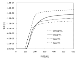
検出対象の抗原であるイムノグロブリンA抗原が試験液中に含まれる場合、抗原と結合した酵素標識抗体中のアルカリフォスファターゼの酵素活性で、基質のp-アミノフェニルリン酸がp-アミノフェノールを経由してp-キノンイミンに酸化される際に生じる電流として定量的に検出できた。さらに、測定経過時間毎の電流値を下記の計算式にて積算値(積分コマンド処理)を求めて、一定時間に検出できた電荷量(クーロン・C)として 算出した。より正確に検出対象の抗原濃度を求めたい場合は、抗原濃度と電荷量の検量線を作成することで推定できた。
電流(A)×時間(S)=電荷(C) ・・・式1
試験液中に含まれるイムノグロブリンA抗原の各試験濃度について得られた電荷量を図4に示した。
When the immunoglobulin A antigen, which is the antigen to be detected, is contained in the test solution, the current generated when the substrate p-aminophenyl phosphate is oxidized to p-quinoneimine via p-aminophenol due to the enzyme activity of alkaline phosphatase in the enzyme-labeled antibody bound to the antigen can be quantitatively detected. Furthermore, the current value for each measurement time was calculated as the integrated value (integral command processing) using the following formula, and the amount of charge (Coulomb-C) detected in a certain period of time was calculated. To obtain a more accurate concentration of the antigen to be detected, a calibration curve of the antigen concentration and the amount of charge could be created to estimate the concentration.
Current (A) x time (S) = charge (C) ... Formula 1
The charge amounts obtained for each test concentration of immunoglobulin A antigen contained in the test solution are shown in FIG.
[実施例1]
前述の本実施例における電気化学的な生体物質の検出方法において、未反応のアルカリフォスファターゼ標識抗イムノグロブリンA抗体を捕捉するために、予めイムノグロブリンA抗原をセルロース混合エステル膜に固定した抗原結合フィルターを使用した。
また、0.1パーセントのBSAと0.05パーセントのポリオキシエチレン(20)ソルビタンモノラウレート(ツイーン20相当品)を含むTBS(pH8.0)で、試験濃度が0,1,10,100μg/mLになるように調整したイムノグロブリンA抗原溶液11μL と、同じく0.1パーセントのBSAと0.05パーセントのポリオキシエチレン(20)ソルビタンモノラウレート(ツイーン20相当品)を含むTBS(pH8.0)で10μg/mLに調整したアルカリフォスファターゼ標識抗イムノグロブリンA抗体溶液 11μLと、予めTBS(pH8.0)425μLと1M KCl 25μLと精製水50μLを混合したサンプル調製用の溶液22μLとをマイクロチューブ内で混合した試験液として、本検出器具を使用して電気化学アナライザーで電気化学的に発生した電流値を計測し、式1にて求めた積算値/一定時間に検出できた電荷量(クーロン・C)を図4に示す。図4のように、本発明の電気化学的な生体物質の検出方法においては、0.1μg/mLから100μg/mLの濃度まで検出が可能であり、検出された電荷値(クーロン・C)より、イムノグロブリンA抗原の有無を確認することができ、さらにその濃度も求めることができた。
[Example 1]
In the electrochemical detection method for a biological substance in the present embodiment described above, an antigen-binding filter in which immunoglobulin A antigen was immobilized in advance on a cellulose mixed ester membrane was used to capture unreacted alkaline phosphatase-labeled anti-immunoglobulin A antibody.
In addition, 11 μL of immunoglobulin A antigen solution was prepared with TBS (pH 8.0) containing 0.1% BSA and 0.05% polyoxyethylene (20) sorbitan monolaurate (equivalent to Tween 20) to test concentrations of 0, 1, 10, and 100 μg/mL, 11 μL of alkaline phosphatase-labeled anti-immunoglobulin A antibody solution was prepared with TBS (pH 8.0) containing 0.1% BSA and 0.05% polyoxyethylene (20) sorbitan monolaurate (equivalent to Tween 20) to test concentrations of 10 μg/mL, and 425 μL of TBS (pH 8.0) and 1 M KCl A test solution was prepared by mixing 22 μL of a sample preparation solution prepared by mixing 25 μL of IgG1 with 50 μL of purified water in a microtube, and the current value electrochemically generated was measured with an electrochemical analyzer using this detection device, and the integrated value calculated by formula 1/amount of charge (Coulomb·C) detected in a certain time period is shown in Figure 4. As shown in Figure 4, the electrochemical detection method of the present invention is capable of detecting concentrations from 0.1 μg/mL to 100 μg/mL, and the presence or absence of immunoglobulin A antigen could be confirmed from the detected charge value (Coulomb·C), and the concentration could also be calculated.
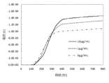
[実施例2]
前述の本実施例における電気化学的な生体物質の検出方法において、未反応のアルカリフォスファターゼ標識抗イムノグロブリンA抗体を捕捉するために、予めイムノグロブリンA抗原を裁断したイムノクロマトメンブレン上で試料注入口側のメンブレンの端から13mmの位置に滴下し、界面活性剤のポリオキシエチレン(20)ソルビタンモノラウレート(ツイーン20相当品)を0.5パーセント添加したサーモフィシャーサイエンティフィック社Super Block溶液にてブロッキング処理を施し、さらにアルカリフォスファターゼの基質であるp-アミノフェニルリン酸(pAPP)をメンブレンの端から23mmのところに滴下したイムノクロマトメンブレンを使用した。また、検出目的のイムノグロブリンA抗原と、アルカリフォスファターゼを標識した抗イムノグロブリンA抗体 10μg/mLと、リン酸緩衝生理食塩水を混合して作られる被検体試験液は実施例1と同じである。図5のように、抗イムノグロブリンA抗原濃度を0,1,10μg/mLでの電荷(クーロン・C)からイムノグロブリンA抗原の有無を確認することができ、さらにその濃度も求めることができた。
[Example 2]
In the electrochemical detection method of the present embodiment described above, in order to capture unreacted alkaline phosphatase-labeled anti-immunoglobulin A antibodies, immunoglobulin A antigens were dropped onto a cut immunochromatographic membrane at a position 13 mm from the end of the membrane on the sample injection port side, and blocking treatment was performed with Thermo Fisher Scientific's Super Block solution containing 0.5% of a surfactant polyoxyethylene (20) sorbitan monolaurate (equivalent to Tween 20), and further p-aminophenyl phosphate (pAPP), a substrate for alkaline phosphatase, was dropped onto the membrane at a position 23 mm from the end. The immunochromatographic membrane used was the same as that used in Example 1. The immunoglobulin A antigens to be detected, 10 μg/mL of anti-immunoglobulin A antibodies labeled with alkaline phosphatase, and phosphate buffered saline were mixed to prepare a test solution to be tested. As shown in FIG. 5, the presence or absence of immunoglobulin A antigen could be confirmed from the charge (Coulomb·C) at anti-immunoglobulin A antigen concentrations of 0, 1, and 10 μg/mL, and further the concentration could be calculated.
[比較例1]
実施例1および2で使用した試薬・部材の他に、ミリポア社製のイムノクロマト用ニトロセルロースメンブレン(HF120)や、そのイムノクロマト用メンブレンに固定化する抗原捕捉用抗体として、一次抗体の抗イムノグロブリンA抗体:Anti-Human IgA 抗体 [3B7] (アブカム社製,ab7400)を使用した。
[Comparative Example 1]
In addition to the reagents and materials used in Examples 1 and 2, a nitrocellulose membrane for immunochromatography (HF120) manufactured by Millipore and a primary antibody, anti-immunoglobulin A antibody: Anti-Human IgA antibody [3B7] (manufactured by Abcam, ab7400), were used as the antigen capture antibody immobilized on the immunochromatography membrane.
ミリポア社製のイムノクロマト用ニトロセルロースメンブレン(HF120)を長さ50mm、幅5mmに裁断した。抗イムノグロブリンA抗体:Anti―Human IgA 抗体 [3B7] (アブカム社製,ab7400) をPBSで 1 mg/mlに希釈した1次抗体液をイムノクロマトメンブレンの端部から20mmの位置に2μl滴下し、37℃で2時間乾燥した。 A nitrocellulose membrane for immunochromatography (HF120) manufactured by Millipore was cut to a length of 50 mm and a width of 5 mm. 2 μl of a primary antibody solution containing anti-immunoglobulin A antibody: Anti-Human IgA antibody [3B7] (manufactured by Abcam, ab7400) diluted to 1 mg/ml with PBS was dropped onto the immunochromatography membrane at a position 20 mm from the end, and the membrane was dried at 37°C for 2 hours.
1次抗体を固定化したイムノクロマトメンブレンをサーモフィシャーサイエンティフィック社のSuperBlock T20 (TBS) Blocking Buffer(製品番号37536)に浸漬し、プレートミキサーで揺らしながら15分浸漬した。ピペッティングリザーバーを利用して、ポリオキシエチレン(20)ソルビタンモノラウレート(ツイーン20相当品) 0.05パーセントを含んだTBS-T( pH7.4)の洗浄液にブロッキングしたイムノクロマト用メンブレンを浸漬し、プレートミキサーで揺らしながら30分間メンブレンを洗浄処理した。洗浄液から取り出したメンブレンを4℃の冷蔵庫内にて1晩静置した。 The immunochromatographic membrane with the immobilized primary antibody was immersed in SuperBlock T20 (TBS) Blocking Buffer (product number 37536) from Thermo Fisher Scientific, and left to soak for 15 minutes while being shaken with a plate mixer. Using a pipetting reservoir, the blocked immunochromatographic membrane was immersed in a washing solution of TBS-T (pH 7.4) containing 0.05% polyoxyethylene (20) sorbitan monolaurate (equivalent to Tween 20), and washed for 30 minutes while being shaken with a plate mixer. The membrane was removed from the washing solution and left to stand overnight in a refrigerator at 4°C.
作製したイムノクロマトメンブレンと吸水パッドと検出用の金電極と、イムノクロマトメンブレンと検出用電極を確実に接触させるためのシリコン樹脂製シートとを、それぞれイムノクロマト用筐体にセットして検出器を組み立てた。 The detector was assembled by placing the prepared immunochromatographic membrane, absorbent pad, gold electrode for detection, and a silicone resin sheet for ensuring contact between the immunochromatographic membrane and the detection electrode in an immunochromatographic housing.
0.1パーセントのBSAと、ポリオキシエチレン(20)ソルビタンモノラウレート(ツイーン20相当品) 0.05パーセントを含んだTBS-Tで試験濃度が0,1,5μg/mL になるように調整したイムノグロブリンA抗原溶液10μLと、0.1パーセントのBSAと、ポリオキシエチレン(20)ソルビタンモノラウレート(ツイーン20相当品) 0.05パーセントを含んだTBS-T で10μg/mL に調製したアルカリフォスファターゼ標識抗イムノグロブリンA抗体:Goat Anti-Human IgA alpha chain (Alkaline Phosphatase) preadsorbed (アブカム社製,ab98551)10μLを検出器の試料注入口から続けて滴下した。 10 μL of immunoglobulin A antigen solution adjusted to test concentrations of 0, 1, and 5 μg/mL with TBS-T containing 0.1% BSA and 0.05% polyoxyethylene (20) sorbitan monolaurate (equivalent to Tween 20) and 10 μL of alkaline phosphatase-labeled anti-immunoglobulin A antibody: Goat Anti-Human IgA alpha chain (Alkaline Phosphatase) preadsorbed (Abcam, ab98551) adjusted to 10 μg/mL with TBS-T containing 0.1% BSA and 0.05% polyoxyethylene (20) sorbitan monolaurate (equivalent to Tween 20) were successively dripped from the sample injection port of the detector.
試料注入口の液がなくなった後、20mM TBS(pH8.0)と50mM KClを混合した溶液にて調製した5mM pAPP基質液50μLを添加した。基質液の添加10秒後に電気化学測定をスタートし、i-tアンペロメトリーモードで電流値を検出した。計測50秒後と200秒後の電流値の差分を検出電流値とした。
アンペロメトリー(i-t)モードの測定パラメータ:
初期電位(V)=0.15
サンプル間隔(Sec) =0.1
測定時間(Sec) =400
静止時間(Sec) =0
測定値のスケール =1
感度(A/V)=1e-6
After the liquid in the sample injection port was exhausted, 50 μL of 5 mM pAPP substrate solution prepared with a solution of 20 mM TBS (pH 8.0) and 50 mM KCl was added. 10 seconds after the addition of the substrate solution, electrochemical measurement was started, and the current value was detected in the it amperometry mode. The difference between the current values after 50 seconds and 200 seconds of measurement was taken as the detected current value.
Measurement parameters for amperometry (it) mode:
Initial potential (V) = 0.15
Sample interval (Sec) = 0.1
Measurement time (Sec) = 400
Quiet time (Sec) = 0
Measurement scale = 1
Sensitivity (A/V) = 1e-6
その結果、酵素標識抗体試薬を添加後に洗浄液を滴下しない洗浄工程を省いた比較例では、検出目的の抗原と結合していない遊離のアルカリフォスファターゼ標識抗イムノグロブリンA抗体が電極近傍のイムノクロマトメンブレン上に残留していた。その遊離の抗体に標識されているアルカリフォスファターゼにより、電気的なノイズが大きくなって、検出対象のイムノグロブリンA抗原の濃度に依存した電気シグナルを検出ができなかった。 As a result, in a comparative example in which the washing step of not dripping washing solution after adding the enzyme-labeled antibody reagent was omitted, free alkaline phosphatase-labeled anti-immunoglobulin A antibodies that were not bound to the antigen to be detected remained on the immunochromatographic membrane near the electrode. The alkaline phosphatase labeled to the free antibodies increased the electrical noise, making it impossible to detect an electrical signal that depended on the concentration of the immunoglobulin A antigen to be detected.
本発明は、短時間で評価でき、かつ測定結果が定量的である抗原抗体反応評価法に関するものであり、例えば、ストレスマーカーであるイムノグロブリンAの検出等を行うことができる評価方法に関する。POCT等の現場で使用できる抗原検出システムの提供などに資するものである。 The present invention relates to an antigen-antibody reaction evaluation method that can be performed in a short time and produces quantitative measurement results, and is an evaluation method that can detect, for example, immunoglobulin A, a stress marker. The present invention contributes to the provision of an antigen detection system that can be used in the field of POCT, etc.
1…試料注入口、2…基質保持イムノクロマトメンブレン、3…検出用金電極、4…吸水パッド、5…筐体(上側)、6…筐体(下側)、7…抗原アナログ結合フィルター、8…電極おさえ、9…参照電極用銀塩化銀インク滴下の位置 1...Sample inlet, 2...Substrate-holding immunochromatographic membrane, 3...Gold electrode for detection, 4...Absorbent pad, 5...Housing (upper side), 6...Housing (lower side), 7...Antigen analog binding filter, 8...Electrode holder, 9...Position for silver-silver chloride ink drop for reference electrode
Claims (7)
酵素標識抗体を含んでいてもよい導入ユニットと、
前記酵素標識抗体と反応する抗原アナログが固定された精製ユニットと、
前記酵素標識抗体の酵素と反応する基質が固定された反応ユニットと、
電気信号の増減を検出する検出ユニットとを有し、
前記抗原アナログと前記基質が一つの担持体上に固定されており、
前記検出ユニットが、作用極及び対極を備える電極を有する、抗原検出システム。 An antigen detection system using an enzyme-labeled antibody that reacts with an antigen,
A transfer unit which may include an enzyme-labeled antibody;
a purification unit on which an antigen analogue that reacts with the enzyme-labeled antibody is immobilized;
a reaction unit having a substrate immobilized thereon that reacts with the enzyme of the enzyme-labeled antibody;
A detection unit for detecting an increase or decrease in an electrical signal ,
the antigen analog and the substrate are immobilized on a single support;
The detection unit has an electrode having a working electrode and a counter electrode .
(1)抗原を含有する試料と酵素標識抗体とを接触させて被検試料を得る第1工程、
(2)第1工程で得られた被検試料と精製ユニットとを接触させて未反応の酵素標識抗体を精製ユニットに捕捉させ、精製試料を得る第2工程、
(3)精製試料と反応ユニットとを接触させて酵素標識抗体と基質とを反応させる第3工程、及び
(4)第3工程の反応生成物と検出ユニットとを接触させて生じる電気信号を検出する第4工程
を含む、抗原検出方法。 An antigen detection method using the antigen detection system according to any one of claims 1 to 6 , comprising:
(1) a first step of contacting a sample containing an antigen with an enzyme-labeled antibody to obtain a test sample;
(2) a second step of contacting the test sample obtained in the first step with a purification unit to capture unreacted enzyme-labeled antibodies in the purification unit to obtain a purified sample;
An antigen detection method comprising: (3) a third step of contacting the purified sample with a reaction unit to react the enzyme-labeled antibody with a substrate; and (4) a fourth step of contacting the reaction product of the third step with a detection unit to detect an electrical signal generated.
Priority Applications (1)
| Application Number | Priority Date | Filing Date | Title |
|---|---|---|---|
| JP2020214818A JP7608822B2 (en) | 2020-12-24 | 2020-12-24 | Antigen detection system, antigen detection kit, and antigen detection method |
Applications Claiming Priority (1)
| Application Number | Priority Date | Filing Date | Title |
|---|---|---|---|
| JP2020214818A JP7608822B2 (en) | 2020-12-24 | 2020-12-24 | Antigen detection system, antigen detection kit, and antigen detection method |
Publications (2)
| Publication Number | Publication Date |
|---|---|
| JP2022100694A JP2022100694A (en) | 2022-07-06 |
| JP7608822B2 true JP7608822B2 (en) | 2025-01-07 |
Family
ID=82271255
Family Applications (1)
| Application Number | Title | Priority Date | Filing Date |
|---|---|---|---|
| JP2020214818A Active JP7608822B2 (en) | 2020-12-24 | 2020-12-24 | Antigen detection system, antigen detection kit, and antigen detection method |
Country Status (1)
| Country | Link |
|---|---|
| JP (1) | JP7608822B2 (en) |
Citations (4)
| Publication number | Priority date | Publication date | Assignee | Title |
|---|---|---|---|---|
| JP2000155122A (en) | 1998-11-18 | 2000-06-06 | Toyota Central Res & Dev Lab Inc | Specific binding pair measurement method using micro oxygen electrode |
| JP2003294680A (en) | 2002-03-29 | 2003-10-15 | Techno Medica Co Ltd | Electrochemical biosensor |
| JP2007064827A (en) | 2005-08-31 | 2007-03-15 | Rohm Co Ltd | Biochip and immunological analyzing method |
| JP5097557B2 (en) | 2005-12-22 | 2012-12-12 | ローム株式会社 | Immunoassay apparatus and method |
-
2020
- 2020-12-24 JP JP2020214818A patent/JP7608822B2/en active Active
Patent Citations (4)
| Publication number | Priority date | Publication date | Assignee | Title |
|---|---|---|---|---|
| JP2000155122A (en) | 1998-11-18 | 2000-06-06 | Toyota Central Res & Dev Lab Inc | Specific binding pair measurement method using micro oxygen electrode |
| JP2003294680A (en) | 2002-03-29 | 2003-10-15 | Techno Medica Co Ltd | Electrochemical biosensor |
| JP2007064827A (en) | 2005-08-31 | 2007-03-15 | Rohm Co Ltd | Biochip and immunological analyzing method |
| JP5097557B2 (en) | 2005-12-22 | 2012-12-12 | ローム株式会社 | Immunoassay apparatus and method |
Also Published As
| Publication number | Publication date |
|---|---|
| JP2022100694A (en) | 2022-07-06 |
Similar Documents
| Publication | Publication Date | Title |
|---|---|---|
| JP2930426B2 (en) | Apparatus for performing one or more competitive immunoassays | |
| JP4040110B2 (en) | Analytical instrument for membrane-based assays | |
| Lin et al. | A nanoparticle label/immunochromatographic electrochemical biosensor for rapid and sensitive detection of prostate-specific antigen | |
| EP0306772B1 (en) | Lateral flow chromatographic binding assay device | |
| US4956302A (en) | Lateral flow chromatographic binding assay device | |
| JP3214854B2 (en) | Trace analysis on card | |
| US4746631A (en) | Immunoassay method, device, and test kit | |
| KR101481189B1 (en) | Fix indicator on analyzer | |
| US5435970A (en) | Device for analysis for constituents in biological fluids | |
| JP2504923B2 (en) | Immunological measurement method | |
| KR101002031B1 (en) | Method and apparatus for direct immune sensor evaluation | |
| EP0121385A1 (en) | Conductimetric bioassay techniques | |
| KR101644144B1 (en) | Enhanced immunoassay sensor | |
| JP4379588B2 (en) | Devices and methods for high density lipoprotein assays | |
| US20030045001A1 (en) | Immunochromatographic test strip with arcuate sample application zone for ease-of-use in the field | |
| JPS61142463A (en) | Device for chromatography | |
| KR890010559A (en) | Chromatography binding assay device and method | |
| CA2519402A1 (en) | Adhered membranes retaining porosity and biological activity in assay device for measuring serum cholesterol associated with high-density lipoproteins | |
| US20020164811A1 (en) | Novel method for measurement of glycated hemoglobin by a rapid strip test procedure | |
| JPS63180857A (en) | Diagnostic device and manufacture and usage thereof | |
| CN106796224B (en) | Test strip assembly | |
| JP3859027B2 (en) | Method for measuring specific substances in nipple discharge | |
| JPH10185920A (en) | Assaying apparatus and assaying method using the same | |
| US4181501A (en) | Method and apparatus for measuring antibody levels | |
| JP7608822B2 (en) | Antigen detection system, antigen detection kit, and antigen detection method |
Legal Events
| Date | Code | Title | Description |
|---|---|---|---|
| RD04 | Notification of resignation of power of attorney |
Free format text: JAPANESE INTERMEDIATE CODE: A7424 Effective date: 20210415 |
|
| A621 | Written request for application examination |
Free format text: JAPANESE INTERMEDIATE CODE: A621 Effective date: 20231026 |
|
| A977 | Report on retrieval |
Free format text: JAPANESE INTERMEDIATE CODE: A971007 Effective date: 20240419 |
|
| A131 | Notification of reasons for refusal |
Free format text: JAPANESE INTERMEDIATE CODE: A131 Effective date: 20240430 |
|
| A601 | Written request for extension of time |
Free format text: JAPANESE INTERMEDIATE CODE: A601 Effective date: 20240627 |
|
| A521 | Request for written amendment filed |
Free format text: JAPANESE INTERMEDIATE CODE: A523 Effective date: 20240820 |
|
| TRDD | Decision of grant or rejection written | ||
| A01 | Written decision to grant a patent or to grant a registration (utility model) |
Free format text: JAPANESE INTERMEDIATE CODE: A01 Effective date: 20241119 |
|
| A61 | First payment of annual fees (during grant procedure) |
Free format text: JAPANESE INTERMEDIATE CODE: A61 Effective date: 20241202 |
|
| R150 | Certificate of patent or registration of utility model |
Ref document number: 7608822 Country of ref document: JP Free format text: JAPANESE INTERMEDIATE CODE: R150 |