JP7607289B2 - Novel anti-hepatitis B virus antibody and its use - Google Patents
Novel anti-hepatitis B virus antibody and its use Download PDFInfo
- Publication number
- JP7607289B2 JP7607289B2 JP2021569886A JP2021569886A JP7607289B2 JP 7607289 B2 JP7607289 B2 JP 7607289B2 JP 2021569886 A JP2021569886 A JP 2021569886A JP 2021569886 A JP2021569886 A JP 2021569886A JP 7607289 B2 JP7607289 B2 JP 7607289B2
- Authority
- JP
- Japan
- Prior art keywords
- seq
- antibody
- antigen
- set forth
- binding fragment
- Prior art date
- Legal status (The legal status is an assumption and is not a legal conclusion. Google has not performed a legal analysis and makes no representation as to the accuracy of the status listed.)
- Active
Links
Images
Classifications
-
- A—HUMAN NECESSITIES
- A61—MEDICAL OR VETERINARY SCIENCE; HYGIENE
- A61P—SPECIFIC THERAPEUTIC ACTIVITY OF CHEMICAL COMPOUNDS OR MEDICINAL PREPARATIONS
- A61P1/00—Drugs for disorders of the alimentary tract or the digestive system
- A61P1/16—Drugs for disorders of the alimentary tract or the digestive system for liver or gallbladder disorders, e.g. hepatoprotective agents, cholagogues, litholytics
-
- A—HUMAN NECESSITIES
- A61—MEDICAL OR VETERINARY SCIENCE; HYGIENE
- A61P—SPECIFIC THERAPEUTIC ACTIVITY OF CHEMICAL COMPOUNDS OR MEDICINAL PREPARATIONS
- A61P31/00—Antiinfectives, i.e. antibiotics, antiseptics, chemotherapeutics
- A61P31/12—Antivirals
-
- A—HUMAN NECESSITIES
- A61—MEDICAL OR VETERINARY SCIENCE; HYGIENE
- A61P—SPECIFIC THERAPEUTIC ACTIVITY OF CHEMICAL COMPOUNDS OR MEDICINAL PREPARATIONS
- A61P31/00—Antiinfectives, i.e. antibiotics, antiseptics, chemotherapeutics
- A61P31/12—Antivirals
- A61P31/20—Antivirals for DNA viruses
-
- C—CHEMISTRY; METALLURGY
- C07—ORGANIC CHEMISTRY
- C07K—PEPTIDES
- C07K16/00—Immunoglobulins [IG], e.g. monoclonal or polyclonal antibodies
- C07K16/08—Immunoglobulins [IG], e.g. monoclonal or polyclonal antibodies against material from viruses
- C07K16/081—DNA viruses
- C07K16/082—Hepadnaviridae (F), e.g. hepatitis B virus
-
- C—CHEMISTRY; METALLURGY
- C12—BIOCHEMISTRY; BEER; SPIRITS; WINE; VINEGAR; MICROBIOLOGY; ENZYMOLOGY; MUTATION OR GENETIC ENGINEERING
- C12N—MICROORGANISMS OR ENZYMES; COMPOSITIONS THEREOF; PROPAGATING, PRESERVING, OR MAINTAINING MICROORGANISMS; MUTATION OR GENETIC ENGINEERING; CULTURE MEDIA
- C12N15/00—Mutation or genetic engineering; DNA or RNA concerning genetic engineering, vectors, e.g. plasmids, or their isolation, preparation or purification; Use of hosts therefor
- C12N15/09—Recombinant DNA-technology
- C12N15/63—Introduction of foreign genetic material using vectors; Vectors; Use of hosts therefor; Regulation of expression
- C12N15/79—Vectors or expression systems specially adapted for eukaryotic hosts
- C12N15/85—Vectors or expression systems specially adapted for eukaryotic hosts for animal cells
-
- C—CHEMISTRY; METALLURGY
- C12—BIOCHEMISTRY; BEER; SPIRITS; WINE; VINEGAR; MICROBIOLOGY; ENZYMOLOGY; MUTATION OR GENETIC ENGINEERING
- C12P—FERMENTATION OR ENZYME-USING PROCESSES TO SYNTHESISE A DESIRED CHEMICAL COMPOUND OR COMPOSITION OR TO SEPARATE OPTICAL ISOMERS FROM A RACEMIC MIXTURE
- C12P21/00—Preparation of peptides or proteins
- C12P21/02—Preparation of peptides or proteins having a known sequence of two or more amino acids, e.g. glutathione
-
- G—PHYSICS
- G01—MEASURING; TESTING
- G01N—INVESTIGATING OR ANALYSING MATERIALS BY DETERMINING THEIR CHEMICAL OR PHYSICAL PROPERTIES
- G01N33/00—Investigating or analysing materials by specific methods not covered by groups G01N1/00 - G01N31/00
- G01N33/48—Biological material, e.g. blood, urine; Haemocytometers
- G01N33/50—Chemical analysis of biological material, e.g. blood, urine; Testing involving biospecific ligand binding methods; Immunological testing
- G01N33/53—Immunoassay; Biospecific binding assay; Materials therefor
- G01N33/576—Immunoassay; Biospecific binding assay; Materials therefor for hepatitis
- G01N33/5761—Hepatitis B
- G01N33/5764—Hepatitis B surface antigen
-
- A—HUMAN NECESSITIES
- A61—MEDICAL OR VETERINARY SCIENCE; HYGIENE
- A61K—PREPARATIONS FOR MEDICAL, DENTAL OR TOILETRY PURPOSES
- A61K39/00—Medicinal preparations containing antigens or antibodies
- A61K2039/505—Medicinal preparations containing antigens or antibodies comprising antibodies
-
- A—HUMAN NECESSITIES
- A61—MEDICAL OR VETERINARY SCIENCE; HYGIENE
- A61K—PREPARATIONS FOR MEDICAL, DENTAL OR TOILETRY PURPOSES
- A61K39/00—Medicinal preparations containing antigens or antibodies
- A61K2039/57—Medicinal preparations containing antigens or antibodies characterised by the type of response, e.g. Th1, Th2
- A61K2039/575—Medicinal preparations containing antigens or antibodies characterised by the type of response, e.g. Th1, Th2 humoral response
-
- C—CHEMISTRY; METALLURGY
- C07—ORGANIC CHEMISTRY
- C07K—PEPTIDES
- C07K2317/00—Immunoglobulins specific features
- C07K2317/20—Immunoglobulins specific features characterized by taxonomic origin
- C07K2317/24—Immunoglobulins specific features characterized by taxonomic origin containing regions, domains or residues from different species, e.g. chimeric, humanized or veneered
-
- C—CHEMISTRY; METALLURGY
- C07—ORGANIC CHEMISTRY
- C07K—PEPTIDES
- C07K2317/00—Immunoglobulins specific features
- C07K2317/30—Immunoglobulins specific features characterized by aspects of specificity or valency
-
- C—CHEMISTRY; METALLURGY
- C07—ORGANIC CHEMISTRY
- C07K—PEPTIDES
- C07K2317/00—Immunoglobulins specific features
- C07K2317/50—Immunoglobulins specific features characterized by immunoglobulin fragments
- C07K2317/515—Complete light chain, i.e. VL + CL
-
- C—CHEMISTRY; METALLURGY
- C07—ORGANIC CHEMISTRY
- C07K—PEPTIDES
- C07K2317/00—Immunoglobulins specific features
- C07K2317/50—Immunoglobulins specific features characterized by immunoglobulin fragments
- C07K2317/52—Constant or Fc region; Isotype
-
- C—CHEMISTRY; METALLURGY
- C07—ORGANIC CHEMISTRY
- C07K—PEPTIDES
- C07K2317/00—Immunoglobulins specific features
- C07K2317/50—Immunoglobulins specific features characterized by immunoglobulin fragments
- C07K2317/56—Immunoglobulins specific features characterized by immunoglobulin fragments variable (Fv) region, i.e. VH and/or VL
- C07K2317/565—Complementarity determining region [CDR]
-
- C—CHEMISTRY; METALLURGY
- C07—ORGANIC CHEMISTRY
- C07K—PEPTIDES
- C07K2317/00—Immunoglobulins specific features
- C07K2317/50—Immunoglobulins specific features characterized by immunoglobulin fragments
- C07K2317/56—Immunoglobulins specific features characterized by immunoglobulin fragments variable (Fv) region, i.e. VH and/or VL
- C07K2317/567—Framework region [FR]
-
- C—CHEMISTRY; METALLURGY
- C07—ORGANIC CHEMISTRY
- C07K—PEPTIDES
- C07K2317/00—Immunoglobulins specific features
- C07K2317/60—Immunoglobulins specific features characterized by non-natural combinations of immunoglobulin fragments
- C07K2317/62—Immunoglobulins specific features characterized by non-natural combinations of immunoglobulin fragments comprising only variable region components
- C07K2317/622—Single chain antibody (scFv)
-
- C—CHEMISTRY; METALLURGY
- C07—ORGANIC CHEMISTRY
- C07K—PEPTIDES
- C07K2317/00—Immunoglobulins specific features
- C07K2317/70—Immunoglobulins specific features characterized by effect upon binding to a cell or to an antigen
- C07K2317/72—Increased effector function due to an Fc-modification
-
- C—CHEMISTRY; METALLURGY
- C07—ORGANIC CHEMISTRY
- C07K—PEPTIDES
- C07K2317/00—Immunoglobulins specific features
- C07K2317/70—Immunoglobulins specific features characterized by effect upon binding to a cell or to an antigen
- C07K2317/76—Antagonist effect on antigen, e.g. neutralization or inhibition of binding
-
- C—CHEMISTRY; METALLURGY
- C07—ORGANIC CHEMISTRY
- C07K—PEPTIDES
- C07K2317/00—Immunoglobulins specific features
- C07K2317/90—Immunoglobulins specific features characterized by (pharmaco)kinetic aspects or by stability of the immunoglobulin
- C07K2317/92—Affinity (KD), association rate (Ka), dissociation rate (Kd) or EC50 value
Landscapes
- Health & Medical Sciences (AREA)
- Life Sciences & Earth Sciences (AREA)
- Chemical & Material Sciences (AREA)
- Organic Chemistry (AREA)
- General Health & Medical Sciences (AREA)
- Engineering & Computer Science (AREA)
- Virology (AREA)
- Medicinal Chemistry (AREA)
- Molecular Biology (AREA)
- Communicable Diseases (AREA)
- Genetics & Genomics (AREA)
- Immunology (AREA)
- General Chemical & Material Sciences (AREA)
- Biochemistry (AREA)
- Chemical Kinetics & Catalysis (AREA)
- Biotechnology (AREA)
- Nuclear Medicine, Radiotherapy & Molecular Imaging (AREA)
- Pharmacology & Pharmacy (AREA)
- Animal Behavior & Ethology (AREA)
- Public Health (AREA)
- Veterinary Medicine (AREA)
- Proteomics, Peptides & Aminoacids (AREA)
- Biophysics (AREA)
- Bioinformatics & Cheminformatics (AREA)
- Oncology (AREA)
- Wood Science & Technology (AREA)
- Zoology (AREA)
- Biomedical Technology (AREA)
- Microbiology (AREA)
- General Engineering & Computer Science (AREA)
- Urology & Nephrology (AREA)
- Gastroenterology & Hepatology (AREA)
- Physics & Mathematics (AREA)
- Hematology (AREA)
- Cell Biology (AREA)
- Pathology (AREA)
- General Physics & Mathematics (AREA)
- Analytical Chemistry (AREA)
- Plant Pathology (AREA)
- Food Science & Technology (AREA)
Description
本発明は、分子ウイルス学及び免疫学の分野、特にB型肝炎ウイルス(HBV)感染の治療の分野に関する。具体的には、本発明は、B型肝炎ウイルス表面抗原(HBsAg)に対する抗体及び該抗体をコードする核酸、並びにそれらの使用に関する。本発明の抗HBsAg抗体は、HBsAgに対して酸性pHよりも中性pHにおいて高い結合親和性を有する。この新規の抗体は、HBV感染又はHBV感染に関連する疾患(例えばB型肝炎)を予防及び/又は治療するため、被験体(例えばヒト)におけるHBVの毒性を中和するため、又は被験体におけるHBV DNA及び/又はHBsAgの血清レベルを低下させるために使用することができる。したがって、本発明は、HBV感染若しくはHBV感染に関係する疾患(例えばB型肝炎)を予防及び/又は治療するため、被験体(例えばヒト)におけるHBVの毒性を中和するため、被験体におけるHBV DNA及び/又はHBsAgの血清レベルを低下させるため、又は被験体(例えば、慢性HBV感染者又は慢性B型肝炎患者)におけるHBVに対する液性免疫応答を活性化するための医薬組成物の製造における抗体及びその変異体の使用に更に関する。 The present invention relates to the field of molecular virology and immunology, in particular to the field of treatment of Hepatitis B virus (HBV) infection. In particular, the present invention relates to antibodies against Hepatitis B virus surface antigen (HBsAg) and nucleic acids encoding said antibodies, and uses thereof. The anti-HBsAg antibodies of the present invention have a higher binding affinity for HBsAg at neutral pH than at acidic pH. The novel antibodies can be used to prevent and/or treat HBV infection or diseases associated with HBV infection (e.g., Hepatitis B), to neutralize the toxicity of HBV in a subject (e.g., a human), or to reduce serum levels of HBV DNA and/or HBsAg in a subject. Thus, the present invention further relates to the use of the antibodies and variants thereof in the manufacture of a pharmaceutical composition for preventing and/or treating HBV infection or a disease associated with HBV infection (e.g., hepatitis B), neutralizing the toxicity of HBV in a subject (e.g., a human), reducing serum levels of HBV DNA and/or HBsAg in a subject, or activating a humoral immune response against HBV in a subject (e.g., a chronically HBV-infected or chronic hepatitis B patient).
B型肝炎ウイルス感染、特に慢性HBV感染は、世界的に最も重要な公衆衛生問題の1つである(非特許文献1)。慢性HBV感染は、慢性B型肝炎(CHB)、肝硬変(LC)及び原発性肝細胞癌(HCC)等の一連の肝疾患を引き起こす恐れがある(非特許文献2)。報告によると、現在、全世界で約20億人がHBVに感染しており、現時点で約3億5000万人が慢性B型肝炎ウイルス感染を有し、これらの感染者がHBV感染に関連する肝疾患によって最終的に死亡するリスクは、15%~25%に達する可能性があり、毎年全世界で100万人超がかかる疾患によって死亡している(非特許文献1及び非特許文献2)。
Hepatitis B virus infection, especially chronic HBV infection, is one of the most important public health problems worldwide (Non-Patent Document 1). Chronic HBV infection may cause a series of liver diseases such as chronic hepatitis B (CHB), liver cirrhosis (LC) and primary hepatocellular carcinoma (HCC) (Non-Patent Document 2). It has been reported that currently, approximately 2 billion people worldwide are infected with HBV, and approximately 350 million people currently have chronic hepatitis B virus infection, and the risk of these infected people ultimately dying from liver disease associated with HBV infection may reach 15% to 25%, with more than 1 million people dying from the disease worldwide every year (Non-Patent
現在の慢性HBV感染の治療薬は、インターフェロン(IFN)とヌクレオシド又はヌクレオチド類似体(NA)とに分けることができる(非特許文献1、非特許文献3及び非特許文献2)。しかしながら、単独又は組合せでの上述の薬物による治療は、感染者におけるHBVウイルスを完全に除去することができず、それによるHBsAg陰性化又はHBsAgセロコンバージョン(感染者における完全なHBVウイルスクリアランスの証拠)の奏効率は、通常は5%未満である(非特許文献3)。
Current therapeutic agents for chronic HBV infection can be divided into interferons (IFNs) and nucleoside or nucleotide analogs (NAs) (Non-Patent
免疫学的手段に基づく慢性HBV感染の治療のための新薬の開発は、この分野における重要な研究の方向の1つである。慢性HBV感染の免疫療法は、通常は能動免疫療法(ワクチン等を含むその対応する薬物形態)及び受動免疫療法(抗体等を含むその対応する薬物形態)の2つの方法で行われる。能動免疫療法とは、慢性HBV感染者の身体を刺激して、細胞性免疫応答(CTL効果等)及び/又はHBVに対する液性免疫応答(抗体等)を能動的に引き起こし、HBVを阻害又は除去する目的を達成するための治療ワクチン(タンパク質ワクチン、ペプチドワクチン、核酸ワクチン等を含む)の投与を指す。現在、慢性HBV感染の治療に使用することができる明らかに重要かつ効果的な能動免疫療法薬/ワクチンはない。受動免疫療法(抗体を例として挙げる)とは、HBV感染者に対する治療特性を有する抗体の投与を指し、治療効果は、HBVが新生児の肝細胞に感染するのを阻止する抗体を介したウイルス中和、又はウイルス及び感染した肝細胞を身体から除去する抗体を介した免疫クリアランスによって達成することができる。現在、予防B型肝炎ワクチンに応答を示した患者又はHBV感染から回復した患者の血清/血漿から精製された抗HBsポリクローナル抗体、すなわち高力価B型肝炎免疫グロブリン(HBIG)が、HBVの母子垂直感染を阻止し、慢性HBV感染患者における肝移植後のHBV再感染を予防し、偶然にHBVに曝露された人々の感染を防ぐために広く使用されている。しかしながら、HBV感染患者(例えばCHB患者)の治療にHBIGを直接適用することは、明白な効果がなく、高力価血漿の供給源の少なさ、高価格、不安定な性質及び潜在的な安全性の問題等の多くの制限を有する。 The development of new drugs for the treatment of chronic HBV infection based on immunological means is one of the important research directions in this field. Immunotherapy of chronic HBV infection is usually carried out in two ways: active immunotherapy (its corresponding drug forms including vaccines, etc.) and passive immunotherapy (its corresponding drug forms including antibodies, etc.). Active immunotherapy refers to the administration of therapeutic vaccines (including protein vaccines, peptide vaccines, nucleic acid vaccines, etc.) to stimulate the body of a chronic HBV-infected person to actively induce a cellular immune response (such as CTL effect) and/or a humoral immune response (such as antibodies) against HBV, achieving the purpose of inhibiting or eliminating HBV. At present, there is no obviously important and effective active immunotherapy drug/vaccine that can be used to treat chronic HBV infection. Passive immunotherapy (taking antibodies as an example) refers to the administration of antibodies with therapeutic properties to HBV-infected individuals, and the therapeutic effect can be achieved by antibody-mediated virus neutralization, which blocks HBV from infecting newborn liver cells, or antibody-mediated immune clearance, which removes the virus and infected liver cells from the body. Currently, anti-HBs polyclonal antibodies, i.e. high titer hepatitis B immunoglobulin (HBIG), purified from serum/plasma of patients who responded to prophylactic hepatitis B vaccines or recovered from HBV infection, are widely used to block mother-to-child vertical transmission of HBV, prevent HBV reinfection after liver transplantation in chronic HBV-infected patients, and prevent infection in people accidentally exposed to HBV. However, the direct application of HBIG to treat HBV-infected patients (e.g. CHB patients) has many limitations, such as no obvious effect, limited supply of high titer plasma, high cost, unstable nature, and potential safety issues.
したがって、HBVウイルス、特にHBsAgをより効果的に除去することができるHBV感染者に対する革新的な治療方法及び薬物を開発することが緊急であり、必要とされている。 Therefore, there is an urgent need to develop innovative treatment methods and drugs for HBV-infected individuals that can more effectively remove the HBV virus, especially HBsAg.
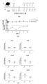
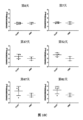
本発明者らは、in vivoでHBVの毒性を中和し、HBV DNA及び/又はHBsAgの血清レベルを低下させることができる、優れた特性を有する抗HBsAgヒト化抗体を以前に開発している。本発明者らは、以前の研究に基づき、多くの創造的な作業を行い、ヒト化抗体の徹底的な研究及び改変を行うことで、pH依存抗原結合能を有する抗HBsAg抗体を開発した。本発明の抗HBsAg抗体は、HBsAgに対して酸性pHよりも中性pHにおいて高い親和性を有するため、抗体の再利用が実現され、抗体の半減期が大幅に延長され、HBVクリアランスの効率が高まる。さらに、本発明者らは、上述の抗体のFc領域に突然変異を導入し、中性条件下でのhFcRn又はmFcγRIIに対するその親和性を高めることによって、スカベンジャー抗体を得て、抗体の半減期を更に延長する。 The present inventors have previously developed an anti-HBsAg humanized antibody with excellent properties, which can neutralize the toxicity of HBV in vivo and reduce the serum levels of HBV DNA and/or HBsAg. Based on the previous research, the present inventors have made many creative efforts and thoroughly studied and modified the humanized antibody to develop an anti-HBsAg antibody with pH-dependent antigen binding ability. The anti-HBsAg antibody of the present invention has a higher affinity for HBsAg at neutral pH than at acidic pH, which realizes antibody recycling, greatly extends the half-life of the antibody, and enhances the efficiency of HBV clearance. Furthermore, the present inventors obtain a scavenger antibody by introducing mutations into the Fc region of the above-mentioned antibody to increase its affinity for hFcRn or mFcγRII under neutral conditions, thereby further extending the half-life of the antibody.
本発明の抗体は、HBV DNA及び/又はHBsAgの血清レベルを低下させる活性を保持するだけでなく、抗原抑制時間がより長いために治療の注射量及び投与頻度が大幅に低減され、大きな臨床的価値を有することから極めて有利である。 The antibodies of the present invention are highly advantageous because they not only retain the activity of reducing serum levels of HBV DNA and/or HBsAg, but also have a longer antigen suppression time, which significantly reduces the injection dose and frequency of administration for treatment, providing great clinical value.
本発明の抗体
したがって、一態様において、本発明は、HBsAgに特異的に結合することが可能な抗体又はその抗原結合フラグメントであって、酸性pHよりも中性pHにおいて高い親和性でHBsAgに結合する、抗体又はその抗原結合フラグメントを提供する。
Antibodies of the Invention Thus, in one aspect, the present invention provides an antibody, or antigen-binding fragment thereof, capable of specifically binding to HBsAg, which binds to HBsAg with higher affinity at neutral pH than at acidic pH.
或る特定の実施の形態においては、中性pHはpH6.7~pH7.5、例えばpH7.4である。 In certain embodiments, the neutral pH is between pH 6.7 and pH 7.5, for example pH 7.4.
或る特定の実施の形態においては、酸性pHはpH4.0~pH6.5、例えばpH6.0である。 In certain embodiments, the acidic pH is between pH 4.0 and pH 6.5, for example pH 6.0.
或る特定の実施の形態においては、抗体又はその抗原結合フラグメントの酸性pH(例えばpH6.0)でのHBsAgへの結合のKDと中性pH(例えばpH7.4)でのHBsAgへの結合のKDとの比率(すなわち、KD(酸性pH)/KD(中性pH)の値)は、1超、例えば1.5以上、2以上、3以上、4以上、5以上、6以上、7以上、8以上、9以上、10以上、15以上、20以上、30以上、40以上、50以上、60以上、70以上、80以上、90以上、100以上、300以上、500以上、800以上、1000以上、2000以上、5000以上又は10000以上である。幾つかの実施の形態においては、KD(酸性pH)/KD(中性pH)の値は、1超かつ10000以下、例えば5000以下、2000以下、1000以下、900以下、800以下、700以下、600以下、500以下、400以下、300以下、200以下、100以下、90以下、80以下、70以下、60以下、50以下、40以下、30以下、20以下又は10以下である。KDは、当該技術分野で既知の手法、例えばSPR技法(例えばBiacore)によって測定することができる。 In certain embodiments, the ratio of the KD for binding to HBsAg at acidic pH (e.g., pH 6.0) to the KD for binding to HBsAg at neutral pH (e.g., pH 7.4) of the antibody or antigen-binding fragment thereof (i.e., the value of KD (acidic pH)/ KD (neutral pH)) is greater than 1, e.g., 1.5 or more, 2 or more, 3 or more, 4 or more, 5 or more, 6 or more, 7 or more, 8 or more, 9 or more, 10 or more, 15 or more, 20 or more, 30 or more, 40 or more, 50 or more, 60 or more, 70 or more, 80 or more, 90 or more, 100 or more, 300 or more, 500 or more, 800 or more, 1000 or more, 2000 or more, 5000 or more, or 10000 or more. In some embodiments, the value of KD (acidic pH)/ KD (neutral pH) is greater than 1 and less than 10,000, e.g., less than 5,000, less than 2,000, less than 1,000, less than 900, less than 800, less than 700, less than 600, less than 500, less than 400, less than 300, less than 200, less than 100, less than 90, less than 80, less than 70, less than 60, less than 50, less than 40, less than 30, less than 20, or less than 10. KD can be measured by techniques known in the art, such as SPR techniques (e.g. Biacore).
幾つかの実施の形態においては、抗体又はその抗原結合フラグメントのpH6.0でのHBsAgへの結合のKDとpH7.4でのHBsAgへの結合のKDとの比率は、1超、例えば1.5以上、2以上である。或る特定の実施の形態においては、中性pHでの抗体又はその抗原結合フラグメントのKD値は、10-7M、10-8M、10-9M、10-10M、10-11M、10-12M以下であり得る。幾つかの実施の形態においては、酸性pHでの本発明の抗体のKD値は、10-9M、10-8M、10-7M、10-6M以上であり得る。 In some embodiments, the ratio of the K D for binding to HBsAg at pH 6.0 of the antibody or antigen-binding fragment thereof to the K D for binding to HBsAg at pH 7.4 is greater than 1, e.g., 1.5 or greater, e.g., 2 or greater. In certain embodiments, the K D value of the antibody or antigen-binding fragment thereof at neutral pH may be 10 −7 M, 10 −8 M, 10 −9 M, 10 −10 M, 10 −11 M, 10 −12 M or less. In some embodiments, the K D value of the antibody of the invention at acidic pH may be 10 −9 M, 10 −8 M, 10 −7 M, 10 −6 M or more.
或る特定の実施の形態においては、抗体又はその抗原結合フラグメントの酸性pH(例えばpH6.0)でのHBsAgへの結合のEC50と中性pH(例えばpH7.4)でのHBsAgへの結合のEC50との比率(すなわち、EC50(酸性pH)/EC50(中性pH)の値)は、1超、例えば1.5以上、2以上、3以上、4以上、5以上、6以上、7以上、8以上、9以上、10以上、15以上、20以上、30以上、40以上、50以上、60以上、70以上、80以上、90以上、100以上、300以上、500以上、800以上、1000以上、2000以上、5000以上又は10000以上である。幾つかの実施の形態においては、EC50(酸性pH)/EC50(中性pH)の値は、1超かつ10000以下、例えば5000以下、2000以下、1000以下、900以下、800以下、700以下、600以下、500以下、400以下、300以下、200以下、100以下、90以下、80以下、70以下、60以下、50以下、40以下、30以下、20以下又は10以下である。幾つかの実施の形態においては、EC50は、ELISA法によって測定され、例えばELISA法によって作成された用量応答曲線の回帰分析によって算出される。 In certain embodiments, the ratio of the EC50 of the antibody or antigen-binding fragment thereof for binding to HBsAg at acidic pH (e.g., pH 6.0) to the EC50 for binding to HBsAg at neutral pH (e.g., pH 7.4) (i.e., the value of EC50(acidic pH)/EC50(neutral pH)) is greater than 1, e.g., 1.5 or more, 2 or more, 3 or more, 4 or more, 5 or more, 6 or more, 7 or more, 8 or more, 9 or more, 10 or more, 15 or more, 20 or more, 30 or more, 40 or more, 50 or more, 60 or more, 70 or more, 80 or more, 90 or more, 100 or more, 300 or more, 500 or more, 800 or more, 1000 or more, 2000 or more, 5000 or more, or 10,000 or more. In some embodiments, the value of EC50(acidic pH)/EC50(neutral pH) is greater than 1 and less than 10,000, e.g., less than 5,000, less than 2,000, less than 1,000, less than 900, less than 800, less than 700, less than 600, less than 500, less than 400, less than 300, less than 200, less than 100, less than 90, less than 80, less than 70, less than 60, less than 50, less than 40, less than 30, less than 20, or less than 10. In some embodiments, the EC50 is measured by ELISA and calculated, e.g., by regression analysis of a dose-response curve generated by ELISA.
或る特定の実施の形態においては、抗体又はその抗原結合フラグメントのpH6.0でのHBsAgへの結合のEC50とpH7.4でのHBsAgへの結合のEC50との比率は、1超、例えば1.5以上、又は2以上である。 In certain embodiments, the ratio of the EC50 of the antibody or antigen-binding fragment thereof for binding to HBsAg at pH 6.0 to the EC50 for binding to HBsAg at pH 7.4 is greater than 1, e.g., 1.5 or greater, or 2 or greater.
或る特定の実施の形態においては、本発明の抗体又はその抗原結合フラグメントは、抗HBVヒト化抗体M1Dに由来する(中国特許出願第201810307136.5号に詳細に記載されている)。 In certain embodiments, the antibody or antigen-binding fragment thereof of the present invention is derived from the anti-HBV humanized antibody M1D (described in detail in Chinese Patent Application No. 201810307136.5).
或る特定の実施の形態においては、本発明の抗体又はその抗原結合フラグメントは、HBsAgのaa118~124に対して酸性pHよりも中性pHにおいて高い親和性で結合する。 In certain embodiments, the antibodies or antigen-binding fragments thereof of the present invention bind to aa 118-124 of HBsAg with higher affinity at neutral pH than at acidic pH.
或る特定の実施の形態においては、抗体又はその抗原結合フラグメントは、以下の特徴の1つ以上を有する、HCDR1、HCDR2及びHCDR3を含む重鎖可変領域(VH)を含む:
(i)HCDR1が、配列番号20に示される配列と比較してヒスチジンに置き換えられた少なくとも1個のアミノ酸(例えば1個、2個、3個、4個又は5個のアミノ酸)を有する;
(ii)HCDR2が、配列番号21に示される配列と比較してヒスチジンに置き換えられた少なくとも1個のアミノ酸(例えば1個、2個、3個、4個又は5個のアミノ酸)を有する;及び/又は、
(iii)HCDR2が、配列番号16に示される配列と比較してヒスチジンに置き換えられた少なくとも1個のアミノ酸(例えば1個、2個、3個、4個又は5個のアミノ酸)を有する。
In certain embodiments, the antibody or antigen-binding fragment thereof comprises a heavy chain variable region (VH) comprising HCDR1, HCDR2, and HCDR3 having one or more of the following characteristics:
(i) HCDR1 has at least one amino acid (e.g., 1, 2, 3, 4, or 5 amino acids) replaced with a histidine compared to the sequence set forth in SEQ ID NO:20;
(ii) HCDR2 has at least one amino acid (e.g., 1, 2, 3, 4, or 5 amino acids) replaced with a histidine compared to the sequence set forth in SEQ ID NO:21; and/or
(iii) HCDR2 has at least one amino acid (e.g., 1, 2, 3, 4, or 5 amino acids) replaced with a histidine compared to the sequence set forth in SEQ ID NO:16.
或る特定の実施の形態においては、抗体又はその抗原結合フラグメントは、以下の特徴の1つ以上を有するLCDR1、LCDR2及びLCDR3を含む重鎖可変領域(VL)を含む:
(i)LCDR1が、配列番号11に示される配列と比較してヒスチジンに置き換えられた少なくとも1個のアミノ酸(例えば1個、2個、3個、4個又は5個のアミノ酸)を有する;
(ii)LCDR2が、配列番号12に示される配列と比較してヒスチジンに置き換えられた少なくとも1個のアミノ酸(例えば1個、2個、3個、4個又は5個のアミノ酸)を有する;及び/又は、
(iii)LCDR2が、配列番号46に示される配列と比較してヒスチジンに置き換えられた少なくとも1個のアミノ酸(例えば1個、2個、3個、4個又は5個のアミノ酸)を有する。
In certain embodiments, the antibody or antigen-binding fragment thereof comprises a heavy chain variable region (VL) comprising an LCDR1, an LCDR2, and an LCDR3 having one or more of the following characteristics:
(i) LCDR1 has at least one amino acid (e.g., 1, 2, 3, 4, or 5 amino acids) replaced with a histidine compared to the sequence set forth in SEQ ID NO:11;
(ii) LCDR2 has at least one amino acid (e.g., one, two, three, four or five amino acids) replaced with a histidine compared to the sequence set forth in SEQ ID NO: 12; and/or
(iii) LCDR2 has at least one amino acid (e.g., 1, 2, 3, 4, or 5 amino acids) replaced with a histidine compared to the sequence set forth in SEQ ID NO:46.
或る特定の実施の形態においては、抗体又はその抗原結合フラグメントは、
(a)以下の3つのCDRを含む重鎖可変領域(VH):
(i)GGSIX1X2NFW(配列番号50)(ここで、X1はT又はHから選択され、X2はS又はHから選択される)の配列を有するHCDR1、
(ii)X3GX4X5X6X7T(配列番号51)(ここで、X3はS又はHから選択され、X4はP、S又はYから選択され、X5はG又はDから選択され、X6はT又はHから選択され、X7はY又はHから選択される)の配列を有するHCDR2、及び、
(iii)ARSHX8YGX9X10DYAFDF(配列番号52)(ここで、X8はD又はHから選択され、X9はS又はHから選択され、X10はH又はNから選択される)の配列を有するHCDR3、
及び/又は、
(b)以下の3つのCDRを含む軽鎖可変領域(VL):
(iv)QDIX11X12S(配列番号53)(ここで、X11はS又はHから選択され、X12はS、Y又はHから選択される)の配列を有するLCDR1、
(v)YAN(配列番号12)の配列を有するLCDR2、及び、
(vi)QQYHX13LPLT(配列番号54)(ここで、X13はS又はYから選択される)の配列を有するLCDR3、
を含む。
In certain embodiments, the antibody or antigen-binding fragment thereof comprises:
(a) a heavy chain variable region (VH) comprising the following three CDRs:
(i) HCDR1 having the sequence GGSIX 1 X 2 NFW (SEQ ID NO:50), where X 1 is selected from T or H, and X 2 is selected from S or H;
(ii) HCDR2 having the sequence : X3GX4X5X6X7T (SEQ ID NO: 51 ), wherein X3 is selected from S or H, X4 is selected from P, S or Y, X5 is selected from G or D, X6 is selected from T or H, and X7 is selected from Y or H; and
(iii) a HCDR3 having the sequence: ARSHX8YGX9X10DYAFDF (SEQ ID NO:52), wherein X8 is selected from D or H, X9 is selected from S or H, and X10 is selected from H or N;
and/or
(b) a light chain variable region (VL) comprising the following three CDRs:
(iv) LCDR1 having the sequence of QDIX 11 X 12 S (SEQ ID NO:53), wherein X 11 is selected from S or H, and X 12 is selected from S, Y, or H;
(v) an LCDR2 having the sequence of YAN (SEQ ID NO: 12); and
(vi) an LCDR3 having the sequence QQYHX 13 LPLT (SEQ ID NO:54), wherein X 13 is selected from S or Y;
Includes.
或る特定の実施の形態においては、抗体又はその抗原結合フラグメントは、
(a)以下の3つのCDRを含む重鎖可変領域(VH):
(i)配列番号8、14、17、20から選択される配列から構成されるHCDR1、
(ii)配列番号9、15、21、18から選択される配列から構成されるHCDR2、及び、
(iii)配列番号10、16、22から選択される配列から構成されるHCDR3、
及び/又は、
(b)以下の3つのCDRを含む軽鎖可変領域(VL):
(iv)配列番号11、19、23から選択される配列から構成されるLCDR1、
(v)配列番号12に示される配列から構成されるLCDR2、及び、
(vi)配列番号13に示される配列から構成されるLCDR3、
を含む。
In certain embodiments, the antibody or antigen-binding fragment thereof comprises:
(a) a heavy chain variable region (VH) comprising the following three CDRs:
(i) HCDR1 consisting of a sequence selected from SEQ ID NOs: 8, 14, 17, and 20;
(ii) an HCDR2 consisting of a sequence selected from SEQ ID NOs: 9, 15, 21, and 18; and
(iii) HCDR3 consisting of a sequence selected from SEQ ID NOs: 10, 16, and 22;
and/or
(b) a light chain variable region (VL) comprising the following three CDRs:
(iv) LCDR1 consisting of a sequence selected from SEQ ID NOs: 11, 19, and 23;
(v) an LCDR2 consisting of the sequence set forth in SEQ ID NO: 12; and
(vi) LCDR3 consisting of the sequence shown in SEQ ID NO: 13;
Includes.
或る特定の実施の形態においては、X1はT又はHから選択され、X2はSから選択され、X3はSから選択され、X4はP又はSから選択され、X5はGから選択され、X6はT又はHから選択され、X7はY又はHから選択され、X8はD又はHから選択され、X9はS又はHから選択され、X10はH又はNから選択され、X11はSから選択され、X12はS又はYから選択され、X13はYから選択される。 In certain embodiments, X1 is selected from T or H, X2 is selected from S, X3 is selected from S, X4 is selected from P or S, X5 is selected from G, X6 is selected from T or H, X7 is selected from Y or H, X8 is selected from D or H, X9 is selected from S or H, X10 is selected from H or N, X11 is selected from S , X12 is selected from S or Y, and X13 is selected from Y.
或る特定の実施の形態においては、抗体又はその抗原結合フラグメントは、
(a)以下の3つのCDRを含む重鎖可変領域(VH):
(i)配列番号8、20から選択される配列から構成されるHCDR1、
(ii)配列番号9、21から選択される配列から構成されるHCDR2、及び、
(iii)配列番号10、22から選択される配列から構成されるHCDR3、
及び/又は、
(b)以下の3つのCDRを含む軽鎖可変領域(VL):
(iv)配列番号11、23から選択される配列から構成されるLCDR1、
(v)配列番号12に示される配列から構成されるLCDR2、及び、
(vi)配列番号13に示される配列から構成されるLCDR3、
を含む。
In certain embodiments, the antibody or antigen-binding fragment thereof comprises:
(a) a heavy chain variable region (VH) comprising the following three CDRs:
(i) HCDR1 consisting of a sequence selected from SEQ ID NOs: 8 and 20;
(ii) an HCDR2 consisting of a sequence selected from SEQ ID NOs: 9 and 21; and
(iii) HCDR3 consisting of a sequence selected from SEQ ID NOs: 10 and 22;
and/or
(b) a light chain variable region (VL) comprising the following three CDRs:
(iv) LCDR1 consisting of a sequence selected from SEQ ID NOs: 11 and 23;
(v) an LCDR2 consisting of the sequence set forth in SEQ ID NO: 12; and
(vi) LCDR3 consisting of the sequence shown in SEQ ID NO: 13;
Includes.
或る特定の実施の形態においては、抗体又はその抗原結合フラグメントは、
(1)以下の3つのCDRを含むVH:配列番号8に示されるHCDR1、配列番号9に示されるHCDR2、配列番号10に示されるHCDR3、及び以下の3つのCDRを含むVL:配列番号11に示されるLCDR1、配列番号12に示されるLCDR2、配列番号13に示されるLCDR3、
(2)以下の3つのCDRを含むVH:配列番号14に示されるHCDR1、配列番号15に示されるHCDR2、配列番号16に示されるHCDR3、及び以下の3つのCDRを含むVL:配列番号11に示されるLCDR1、配列番号12に示されるLCDR2、配列番号13に示されるLCDR3、
(3)以下の3つのCDRを含むVH:配列番号20に示されるHCDR1、配列番号21に示されるHCDR2、配列番号22に示されるHCDR3、及び以下の3つのCDRを含むVL:配列番号23に示されるLCDR1、配列番号12に示されるLCDR2、配列番号13に示されるLCDR3、又は、
(4)以下の3つのCDRを含むVH:配列番号17に示されるHCDR1、配列番号18に示されるHCDR2、配列番号10に示されるHCDR3、及び以下の3つのCDRを含むVL:配列番号19に示されるLCDR1、配列番号12に示されるLCDR2、配列番号13に示されるLCDR3、
を含む。
In certain embodiments, the antibody or antigen-binding fragment thereof comprises:
(1) VH comprising the following three CDRs: HCDR1 as shown in SEQ ID NO: 8, HCDR2 as shown in SEQ ID NO: 9, HCDR3 as shown in SEQ ID NO: 10, and VL comprising the following three CDRs: LCDR1 as shown in SEQ ID NO: 11, LCDR2 as shown in SEQ ID NO: 12, LCDR3 as shown in SEQ ID NO: 13,
(2) VH comprising the following three CDRs: HCDR1 as shown in SEQ ID NO: 14, HCDR2 as shown in SEQ ID NO: 15, HCDR3 as shown in SEQ ID NO: 16, and VL comprising the following three CDRs: LCDR1 as shown in SEQ ID NO: 11, LCDR2 as shown in SEQ ID NO: 12, LCDR3 as shown in SEQ ID NO: 13,
(3) VH comprising the following three CDRs: HCDR1 as shown in SEQ ID NO: 20, HCDR2 as shown in SEQ ID NO: 21, HCDR3 as shown in SEQ ID NO: 22, and VL comprising the following three CDRs: LCDR1 as shown in SEQ ID NO: 23, LCDR2 as shown in SEQ ID NO: 12, LCDR3 as shown in SEQ ID NO: 13, or
(4) VH comprising the following three CDRs: HCDR1 as shown in SEQ ID NO: 17, HCDR2 as shown in SEQ ID NO: 18, HCDR3 as shown in SEQ ID NO: 10, and VL comprising the following three CDRs: LCDR1 as shown in SEQ ID NO: 19, LCDR2 as shown in SEQ ID NO: 12, LCDR3 as shown in SEQ ID NO: 13,
Includes.
或る特定の実施の形態においては、抗体又はその抗原結合フラグメントは、ヒト免疫グロブリンのフレームワーク領域(例えば、ヒト生殖細胞系列抗体遺伝子によってコードされるアミノ酸配列に含まれるフレームワーク領域)を更に含み、該フレームワーク領域は、任意にヒト残基からマウス残基への1個以上(例えば1個、2個、3個、4個、5個、6個、7個、8個、9個又は10個)の復帰突然変異を含む。 In certain embodiments, the antibody or antigen-binding fragment thereof further comprises a human immunoglobulin framework region (e.g., a framework region contained in an amino acid sequence encoded by a human germline antibody gene), which optionally comprises one or more (e.g., 1, 2, 3, 4, 5, 6, 7, 8, 9, or 10) back mutations from human residues to murine residues.
或る特定の実施の形態においては、抗体又はその抗原結合フラグメントは、ヒト重鎖生殖細胞系列遺伝子によってコードされるアミノ酸配列に含まれる重鎖フレームワーク領域、及び/又はヒト軽鎖生殖細胞系列遺伝子によってコードされるアミノ酸配列に含まれる軽鎖フレームワーク領域を含む。 In certain embodiments, the antibody or antigen-binding fragment thereof comprises a heavy chain framework region comprised in an amino acid sequence encoded by a human heavy chain germline gene and/or a light chain framework region comprised in an amino acid sequence encoded by a human light chain germline gene.
或る特定の実施の形態においては、抗体又はその抗原結合フラグメントは、IGHV4-4*08によってコードされるアミノ酸配列(配列番号55)に含まれる重鎖フレームワーク領域及びIGKV1-39*01によってコードされるアミノ酸配列(配列番号56)に含まれる軽鎖フレームワーク領域を含み、該重鎖フレームワーク領域及び/又は該軽鎖フレームワーク領域は、任意にヒト残基からマウス残基への1個以上(例えば1個、2個、3個、4個、5個、6個、7個、8個、9個又は10個)の復帰突然変異を含む。 In certain embodiments, the antibody or antigen-binding fragment thereof comprises a heavy chain framework region comprised in the amino acid sequence encoded by IGHV4-4 * 08 (SEQ ID NO:55) and a light chain framework region comprised in the amino acid sequence encoded by IGKV1-39 * 01 (SEQ ID NO:56), wherein the heavy chain framework region and/or the light chain framework region optionally comprise one or more (e.g., 1, 2, 3, 4, 5, 6, 7, 8, 9, or 10) backmutations from human residues to murine residues.
或る特定の実施の形態においては、抗体又はその抗原結合フラグメントのVHは、配列番号24に示されるVH FR1、配列番号25に示されるVH FR2、配列番号26に示されるVH FR3及び配列番号27に示されるVH FR4を含む。 In certain embodiments, the VH of the antibody or antigen-binding fragment thereof comprises a VH FR1 set forth in SEQ ID NO:24, a VH FR2 set forth in SEQ ID NO:25, a VH FR3 set forth in SEQ ID NO:26, and a VH FR4 set forth in SEQ ID NO:27.
幾つかの実施の形態においては、抗体又はその抗原結合フラグメントのVLは、配列番号28に示されるVL FR1、配列番号29に示されるVL FR2、配列番号30に示されるVL FR3及び配列番号31に示されるVL FR4を含む。 In some embodiments, the VL of the antibody or antigen-binding fragment thereof comprises a VL FR1 set forth in SEQ ID NO:28, a VL FR2 set forth in SEQ ID NO:29, a VL FR3 set forth in SEQ ID NO:30, and a VL FR4 set forth in SEQ ID NO:31.
或る特定の実施の形態においては、抗体又はその抗原結合フラグメントは、
(a)以下から選択されるアミノ酸配列を含む重鎖可変領域(VH):
(i)配列番号1、3、4及び6のいずれか1つに示される配列、
(ii)配列番号1、3、4、6のいずれか1つに示される配列と比較して1個若しくは幾つかのアミノ酸の置換、欠失若しくは付加(例えば1個、2個、3個、4個又は5個のアミノ酸の置換、欠失又は付加)を有する配列、又は、
(iii)配列番号1、3、4、6のいずれか1つに示される配列と比較して少なくとも80%、少なくとも85%、少なくとも90%、少なくとも91%、少なくとも92%、少なくとも93%、少なくとも94%、少なくとも95%、少なくとも96%、少なくとも97%、少なくとも98%、少なくとも99%若しくは100%の配列同一性を有する配列、
並びに、
(b)以下から選択されるアミノ酸配列を含む軽鎖可変領域(VL):
(iv)配列番号2、5及び7のいずれか1つに示される配列、
(v)配列番号2、5、7のいずれか1つに示される配列と比較して1個若しくは幾つかのアミノ酸の置換、欠失若しくは付加(例えば1個、2個、3個、4個又は5個のアミノ酸の置換、欠失又は付加)を有する配列、又は、
(vi)配列番号2、5、7のいずれか1つに示される配列と比較して少なくとも80%、少なくとも85%、少なくとも90%、少なくとも91%、少なくとも92%、少なくとも93%、少なくとも94%、少なくとも95%、少なくとも96%、少なくとも97%、少なくとも98%、少なくとも99%若しくは100%の配列同一性を有する配列、
を含む。
In certain embodiments, the antibody or antigen-binding fragment thereof comprises:
(a) a heavy chain variable region (VH) comprising an amino acid sequence selected from the following:
(i) a sequence as set forth in any one of SEQ ID NOs: 1, 3, 4 and 6;
(ii) a sequence having one or several amino acid substitutions, deletions or additions (e.g., one, two, three, four or five amino acid substitutions, deletions or additions) compared to the sequence shown in any one of SEQ ID NOs: 1, 3, 4, 6; or
(iii) a sequence having at least 80%, at least 85%, at least 90%, at least 91%, at least 92%, at least 93%, at least 94%, at least 95%, at least 96%, at least 97%, at least 98%, at least 99%, or 100% sequence identity to any one of the sequences set forth in SEQ ID NOs: 1, 3, 4, and 6;
and,
(b) a light chain variable region (VL) comprising an amino acid sequence selected from the following:
(iv) a sequence as set forth in any one of SEQ ID NOs: 2, 5, and 7;
(v) a sequence having one or several amino acid substitutions, deletions or additions (e.g., one, two, three, four or five amino acid substitutions, deletions or additions) compared to the sequence shown in any one of SEQ ID NOs: 2, 5, 7; or
(vi) a sequence having at least 80%, at least 85%, at least 90%, at least 91%, at least 92%, at least 93%, at least 94%, at least 95%, at least 96%, at least 97%, at least 98%, at least 99% or 100% sequence identity to any one of SEQ ID NOs: 2, 5, 7;
Includes.
或る特定の実施の形態においては、(ii)又は(v)に記載される置換は、保存的置換である。 In certain embodiments, the substitution described in (ii) or (v) is a conservative substitution.
或る特定の実施の形態においては、抗体又はその抗原結合フラグメントは、
(1)配列番号1に示される配列を有するVH及び配列番号2に示される配列を有するVL、
(2)配列番号3に示される配列を有するVH及び配列番号2に示される配列を有するVL、
(3)配列番号4に示される配列を有するVH及び配列番号5に示される配列を有するVL、又は、
(4)配列番号6に示される配列を有するVH及び配列番号7に示される配列を有するVL、
を含む。
In certain embodiments, the antibody or antigen-binding fragment thereof comprises:
(1) VH having the sequence shown in SEQ ID NO: 1 and VL having the sequence shown in SEQ ID NO: 2;
(2) VH having the sequence shown in SEQ ID NO: 3 and VL having the sequence shown in SEQ ID NO: 2;
(3) a VH having the sequence shown in SEQ ID NO: 4 and a VL having the sequence shown in SEQ ID NO: 5; or
(4) VH having the sequence shown in SEQ ID NO: 6 and VL having the sequence shown in SEQ ID NO: 7;
Includes.
或る特定の実施の形態においては、抗体又はその抗原結合フラグメントは、ヒト免疫グロブリンに由来する定常領域を更に含む。 In certain embodiments, the antibody or antigen-binding fragment thereof further comprises a constant region derived from a human immunoglobulin.
或る特定の実施の形態においては、抗体又はその抗原結合フラグメントの重鎖は、ヒト免疫グロブリン(例えばIgG1、IgG2、IgG3又はIgG4)に由来する重鎖定常領域を含み、抗体又はその抗原結合フラグメントの軽鎖は、ヒト免疫グロブリン(例えばκ又はλ)に由来する軽鎖定常領域を含む。 In certain embodiments, the heavy chain of the antibody or antigen-binding fragment thereof comprises a heavy chain constant region derived from a human immunoglobulin (e.g., IgG1, IgG2, IgG3, or IgG4), and the light chain of the antibody or antigen-binding fragment thereof comprises a light chain constant region derived from a human immunoglobulin (e.g., κ or λ).
或る特定の実施の形態においては、抗体又はその抗原結合フラグメントは、
(a)ヒト免疫グロブリンの重鎖定常領域(CH)若しくはその変異体であって、該変異体が、それが由来する野生型配列と比較して1個以上のアミノ酸の置換、欠失若しくは付加、若しくはそれらの任意の組合せ(例えば、多くとも20個、多くとも15個、多くとも10個若しくは多くとも5個のアミノ酸の置換、欠失若しくは付加、又はそれらの任意の組合せ;例えば1個、2個、3個、4個若しくは5個のアミノ酸の置換、欠失若しくは付加、又はそれらの任意の組合せ)を有する、及び/又は、
(b)ヒト免疫グロブリンの軽鎖定常領域(CL)若しくはその変異体であって、該変異体が、それが由来する野生型配列と比較して1個以上のアミノ酸の置換、欠失若しくは付加、若しくはそれらの任意の組合せ(例えば、多くとも20個、多くとも15個、多くとも10個若しくは多くとも5個のアミノ酸の置換、欠失若しくは付加、又はそれらの任意の組合せ;例えば1個、2個、3個、4個若しくは5個のアミノ酸の置換、欠失若しくは付加、又はそれらの任意の組合せ)を有する、
を含む。
In certain embodiments, the antibody or antigen-binding fragment thereof comprises:
(a) a heavy chain constant region (CH) of a human immunoglobulin or a variant thereof, which variant has one or more amino acid substitutions, deletions or additions, or any combination thereof, compared to the wild-type sequence from which it is derived (e.g. at most 20, at most 15, at most 10 or at most 5 amino acid substitutions, deletions or additions, or any combination thereof; e.g. 1, 2, 3, 4 or 5 amino acid substitutions, deletions or additions, or any combination thereof); and/or
(b) a human immunoglobulin light chain constant region (CL) or a variant thereof, which variant has one or more amino acid substitutions, deletions or additions, or any combination thereof, compared to the wild-type sequence from which it is derived (e.g., at most 20, at most 15, at most 10 or at most 5 amino acid substitutions, deletions or additions, or any combination thereof; e.g., 1, 2, 3, 4 or 5 amino acid substitutions, deletions or additions, or any combination thereof);
Includes.
或る特定の実施の形態においては、抗体又はその抗原結合フラグメントは、ヒトIgG1又はIgG4重鎖定常領域を含む。或る特定の実施の形態においては、抗体又はその抗原結合フラグメントは、配列番号57に示される重鎖定常領域(CH)を含む。 In certain embodiments, the antibody or antigen-binding fragment thereof comprises a human IgG1 or IgG4 heavy chain constant region. In certain embodiments, the antibody or antigen-binding fragment thereof comprises a heavy chain constant region (CH) set forth in SEQ ID NO:57.
或る特定の実施の形態においては、本発明の抗体又はその抗原結合フラグメントは、ヒト免疫グロブリンの重鎖定常領域(CH)の変異体を含み、該変異体は、hFcRn又はmFcγRIIに対して中性pH(例えばpH7.4)で、それが由来する野生型配列と比較して増強された親和性を有する。かかる実施の形態においては、変異体は概して、それが由来する野生型配列と比較して少なくとも1個のアミノ酸の置換を有する。 In certain embodiments, the antibodies or antigen-binding fragments thereof of the invention comprise a variant of a human immunoglobulin heavy chain constant region (CH) that has enhanced affinity for hFcRn or mFcγRII at neutral pH (e.g., pH 7.4) compared to the wild-type sequence from which it is derived. In such embodiments, the variant generally has at least one amino acid substitution compared to the wild-type sequence from which it is derived.
或る特定の実施の形態においては、抗体又はその抗原結合フラグメントは、ヒトIgG1重鎖定常領域の変異体を含み、該変異体が、それが由来する野生型配列と比較して以下の置換:(i)M252Y、N286E、N434Y、又は(ii)K326D、L328Yを有し、ここで、上述のアミノ酸位置は、Kabatナンバリングシステムに従う位置である。或る特定の実施の形態においては、抗体又はその抗原結合フラグメントは、配列番号47又は48に示される重鎖定常領域(CH)を含む。 In certain embodiments, the antibody or antigen-binding fragment thereof comprises a mutant of a human IgG1 heavy chain constant region, the mutant having the following substitutions compared to the wild-type sequence from which it is derived: (i) M252Y, N286E, N434Y, or (ii) K326D, L328Y, where the above amino acid positions are according to the Kabat numbering system. In certain embodiments, the antibody or antigen-binding fragment thereof comprises a heavy chain constant region (CH) as set forth in SEQ ID NO: 47 or 48.
或る特定の実施の形態においては、軽鎖定常領域はκ軽鎖定常領域である。或る特定の実施の形態においては、抗体又はその抗原結合フラグメントは、配列番号58に示される軽鎖定常領域(CL)を含む。 In certain embodiments, the light chain constant region is a kappa light chain constant region. In certain embodiments, the antibody or antigen-binding fragment thereof comprises a light chain constant region (CL) set forth in SEQ ID NO:58.
或る特定の実施の形態においては、抗体又はその抗原結合フラグメントは、
(1)配列番号1に示されるVHと配列番号57に示されるCHとを含む重鎖及び配列番号2に示されるVLと配列番号58に示されるCLとを含む軽鎖、
(2)配列番号1に示されるVHと配列番号47に示されるCHとを含む重鎖及び配列番号2に示されるVLと配列番号58に示されるCLとを含む軽鎖、
(3)配列番号1に示されるVHと配列番号48に示されるCHとを含む重鎖及び配列番号2に示されるVLと配列番号58に示されるCLとを含む軽鎖、
(4)配列番号3に示されるVHと配列番号57に示されるCHとを含む重鎖及び配列番号2に示されるVLと配列番号58に示されるCLとを含む軽鎖、
(5)配列番号3に示されるVHと配列番号47に示されるCHとを含む重鎖及び配列番号2に示されるVLと配列番号58に示されるCLとを含む軽鎖、
(6)配列番号3に示されるVHと配列番号48に示されるCHとを含む重鎖及び配列番号2に示されるVLと配列番号58に示されるCLとを含む軽鎖、
(7)配列番号4に示されるVHと配列番号57に示されるCHとを含む重鎖及び配列番号5に示されるVLと配列番号58に示されるCLとを含む軽鎖、
(8)配列番号4に示されるVHと配列番号47に示されるCHとを含む重鎖及び配列番号5に示されるVLと配列番号58に示されるCLとを含む軽鎖、
(9)配列番号4に示されるVHと配列番号48に示されるCHとを含む重鎖及び配列番号5に示されるVLと配列番号58に示されるCLとを含む軽鎖、
(10)配列番号6に示されるVHと配列番号57に示されるCHとを含む重鎖及び配列番号7に示されるVLと配列番号58に示されるCLとを含む軽鎖、
(11)配列番号6に示されるVHと配列番号47に示されるCHとを含む重鎖及び配列番号7に示されるVLと配列番号58に示されるCLとを含む軽鎖、又は、
(12)配列番号6に示されるVHと配列番号48に示されるCHとを含む重鎖及び配列番号7に示されるVLと配列番号58に示されるCLとを含む軽鎖、
を含む。
In certain embodiments, the antibody or antigen-binding fragment thereof comprises:
(1) A heavy chain comprising a VH represented by SEQ ID NO: 1 and a CH represented by SEQ ID NO: 57, and a light chain comprising a VL represented by SEQ ID NO: 2 and a CL represented by SEQ ID NO: 58;
(2) A heavy chain comprising a VH represented by SEQ ID NO: 1 and a CH represented by SEQ ID NO: 47, and a light chain comprising a VL represented by SEQ ID NO: 2 and a CL represented by SEQ ID NO: 58;
(3) A heavy chain comprising VH set forth in SEQ ID NO: 1 and CH set forth in SEQ ID NO: 48, and a light chain comprising VL set forth in SEQ ID NO: 2 and CL set forth in SEQ ID NO: 58;
(4) A heavy chain comprising VH set forth in SEQ ID NO: 3 and CH set forth in SEQ ID NO: 57, and a light chain comprising VL set forth in SEQ ID NO: 2 and CL set forth in SEQ ID NO: 58;
(5) A heavy chain comprising VH set forth in SEQ ID NO: 3 and CH set forth in SEQ ID NO: 47, and a light chain comprising VL set forth in SEQ ID NO: 2 and CL set forth in SEQ ID NO: 58;
(6) A heavy chain comprising VH set forth in SEQ ID NO: 3 and CH set forth in SEQ ID NO: 48, and a light chain comprising VL set forth in SEQ ID NO: 2 and CL set forth in SEQ ID NO: 58;
(7) A heavy chain comprising VH set forth in SEQ ID NO: 4 and CH set forth in SEQ ID NO: 57, and a light chain comprising VL set forth in SEQ ID NO: 5 and CL set forth in SEQ ID NO: 58;
(8) A heavy chain comprising VH set forth in SEQ ID NO: 4 and CH set forth in SEQ ID NO: 47, and a light chain comprising VL set forth in SEQ ID NO: 5 and CL set forth in SEQ ID NO: 58;
(9) A heavy chain comprising VH set forth in SEQ ID NO: 4 and CH set forth in SEQ ID NO: 48, and a light chain comprising VL set forth in SEQ ID NO: 5 and CL set forth in SEQ ID NO: 58;
(10) A heavy chain comprising VH set forth in SEQ ID NO: 6 and CH set forth in SEQ ID NO: 57, and a light chain comprising VL set forth in SEQ ID NO: 7 and CL set forth in SEQ ID NO: 58;
(11) A heavy chain comprising VH represented by SEQ ID NO: 6 and CH represented by SEQ ID NO: 47, and a light chain comprising VL represented by SEQ ID NO: 7 and CL represented by SEQ ID NO: 58; or
(12) A heavy chain comprising VH set forth in SEQ ID NO: 6 and CH set forth in SEQ ID NO: 48, and a light chain comprising VL set forth in SEQ ID NO: 7 and CL set forth in SEQ ID NO: 58;
Includes.
抗体の作製
本発明の抗体は、当該技術分野で既知の様々な方法によって作製することができ、例えば遺伝子工学的組み換え技術によって得ることができる。例えば、本発明の抗体の重鎖及び軽鎖遺伝子をコードするDNA分子は、化学合成又はPCR増幅によって得られる。得られたDNA分子を発現ベクターに挿入した後、宿主細胞にトランスフェクトする。次いで、トランスフェクトされた宿主細胞を特定の条件下で培養し、本発明の抗体を発現させる。
Antibody Production The antibody of the present invention can be produced by various methods known in the art, for example, by genetic engineering recombinant technology. For example, DNA molecules encoding the heavy and light chain genes of the antibody of the present invention can be obtained by chemical synthesis or PCR amplification. The obtained DNA molecules are inserted into an expression vector and then transfected into a host cell. The transfected host cell is then cultured under specific conditions to express the antibody of the present invention.
本発明の抗原結合フラグメントは、完全な抗体分子を加水分解することによって得ることができる(Morimoto et al., J. Biochem. Biophys. Methods 24:107-117 (1992)及びBrennan et al., Science 229:81 (1985)を参照されたい)。加えて、これらの抗原結合フラグメントは、組み換え宿主細胞によって直接産生させることもできる(Hudson, Curr. Opin. Immunol. 11: 548-557 (1999)、Little et al., Immunol. Today, 21: 364-370 (2000)に概説される)。例えば、Fab’フラグメントを宿主細胞から直接得ることができ、Fab’フラグメントを化学的に結合して、F(ab’)2フラグメントを形成することができる(Carter et al., Bio/Technology, 10: 163-167 (1992))。加えて、Fv、Fab又はF(ab’)2フラグメントを組み換え宿主細胞培養培地から直接単離することもできる。当業者は、これらの抗原結合フラグメントを作製する他の手法を十分認識している。 The antigen-binding fragments of the present invention can be obtained by hydrolysis of intact antibody molecules (see Morimoto et al., J. Biochem. Biophys. Methods 24:107-117 (1992) and Brennan et al., Science 229:81 (1985)). In addition, these antigen-binding fragments can be produced directly by recombinant host cells (reviewed in Hudson, Curr. Opin. Immunol. 11: 548-557 (1999); Little et al., Immunol. Today, 21: 364-370 (2000)). For example, Fab' fragments can be obtained directly from host cells and chemically coupled to form F(ab') 2 fragments (Carter et al., Bio/Technology, 10: 163-167 (1992)). In addition, Fv, Fab or F(ab') 2 fragments can be isolated directly from recombinant host cell culture medium. Those of skill in the art will be well aware of other techniques for producing these antigen-binding fragments.
したがって、別の態様において、本発明は、本発明の抗体若しくはその抗原結合フラグメント、又はその重鎖可変領域及び/又は軽鎖可変領域をコードするヌクレオチド配列を含む単離核酸分子を提供する。或る特定の好ましい実施の形態においては、単離核酸分子は、本発明の抗体若しくはその抗原結合フラグメント、又はその重鎖可変領域及び/又は軽鎖可変領域をコードする。 Thus, in another aspect, the invention provides an isolated nucleic acid molecule comprising a nucleotide sequence encoding an antibody or antigen-binding fragment thereof of the invention, or a heavy chain variable region and/or a light chain variable region thereof. In certain preferred embodiments, the isolated nucleic acid molecule encodes an antibody or antigen-binding fragment thereof of the invention, or a heavy chain variable region and/or a light chain variable region thereof.
別の態様において、本発明は、本発明の単離核酸分子を含むベクター(例えば、クローニングベクター又は発現ベクター)を提供する。或る特定の好ましい実施の形態においては、本発明のベクターは、例えばプラスミド、コスミド、バクテリオファージ等である。 In another aspect, the invention provides a vector (e.g., a cloning vector or an expression vector) comprising an isolated nucleic acid molecule of the invention. In certain preferred embodiments, the vector of the invention is, for example, a plasmid, cosmid, bacteriophage, etc.
別の態様において、本発明は、本発明の単離核酸分子又は本発明のベクターを含む宿主細胞を提供する。かかる宿主細胞としては、大腸菌(E. coli)細胞等の原核細胞、並びに酵母細胞、昆虫細胞、植物細胞及び動物細胞(例えばマウス細胞、ヒト細胞等の哺乳動物細胞等)等の真核細胞が挙げられるが、これらに限定されない。或る特定の好ましい実施の形態においては、本発明の宿主細胞は、CHO(例えばCHO-K1、CHO-S、CHO DG44)等の哺乳動物細胞である。 In another aspect, the present invention provides a host cell comprising an isolated nucleic acid molecule of the present invention or a vector of the present invention. Such host cells include, but are not limited to, prokaryotic cells, such as E. coli cells, and eukaryotic cells, such as yeast cells, insect cells, plant cells, and animal cells (e.g., mammalian cells, such as mouse cells, human cells, etc.). In certain preferred embodiments, the host cell of the present invention is a mammalian cell, such as a CHO cell (e.g., CHO-K1, CHO-S, CHO DG44).
別の態様において、抗体又はその抗原結合フラグメントの発現を可能にする条件下で本発明の宿主細胞を培養することと、培養された宿主細胞培養物から抗体又はその抗原結合フラグメントを回収することとを含む、本発明の抗体又はその抗原結合フラグメントを作製する方法が提供される。 In another aspect, a method for producing an antibody or antigen-binding fragment thereof of the invention is provided, comprising culturing a host cell of the invention under conditions that allow expression of the antibody or antigen-binding fragment thereof, and recovering the antibody or antigen-binding fragment thereof from the cultured host cell culture.
誘導抗体
本発明の抗体又はその抗原結合フラグメントは、例えば別の分子(例えば、別のポリペプチド又はタンパク質)に連結して誘導体化することができる。概して、抗体又はその抗原結合フラグメントの誘導体化(例えば標識化)は、HBsAgとの結合に悪影響を与えない。したがって、本発明の抗体又はその抗原結合フラグメントは、かかる誘導体化形態を含むことも意図される。例えば、本発明の抗体又は抗原結合フラグメントは、別の抗体(例えば、二重特異性抗体を形成するため)、検出試薬、医薬試薬、及び/又は抗体若しくは抗原結合フラグメントと別の分子(例えば、アビジン又はポリヒスチジンタグ)との結合を媒介することが可能なタンパク質若しくはポリペプチド等の1つ以上の他の分子群と機能的に連結することができる(化学的結合、遺伝子融合、非共有結合又は他の手段による)。
Derivatized Antibodies The antibodies or antigen-binding fragments thereof of the present invention can be derivatized, for example, by linking to another molecule (e.g., another polypeptide or protein). Generally, derivatization (e.g., labeling) of the antibodies or antigen-binding fragments thereof does not adversely affect binding to HBsAg. Thus, the antibodies or antigen-binding fragments thereof of the present invention are also intended to include such derivatized forms. For example, the antibodies or antigen-binding fragments of the present invention can be functionally linked (by chemical conjugation, genetic fusion, non-covalent binding or other means) to one or more other molecular groups, such as another antibody (e.g., to form a bispecific antibody), a detection reagent, a pharmaceutical reagent, and/or a protein or polypeptide capable of mediating binding of the antibody or antigen-binding fragment to another molecule (e.g., avidin or polyhistidine tag).
したがって、或る特定の実施の形態においては、本発明の抗体又はその抗原結合フラグメントは標識されている。幾つかの実施の形態においては、本発明の抗体又はその抗原結合フラグメントは、酵素、放射性核種、蛍光色素、発光性物質(例えば化学発光物質)又はビオチン等の検出可能な標識を有する。本発明の検出可能な標識は、蛍光、分光法、光化学、生化学、免疫学、電気的、光学的又は化学的手段によって検出することができる任意の物質であり得る。かかる標識は、当該技術分野で既知であり、その例としては、酵素(例えばホースラディッシュペルオキシダーゼ、アルカリホスファターゼ、β-ガラクトシダーゼ、ウレアーゼ、グルコースオキシダーゼ等)、放射性核種(例えば3H、125I、35S、14C又は32P)、蛍光色素(例えばフルオレセインイソチオシアネート(FITC)、フルオレセイン、テトラメチルローダミンイソチオシアネート(TRITC)、フィコエリトリン(PE)、テキサスレッド、ローダミン、量子ドット又はシアニン染料誘導体(例えばCy7、Alexa 750))、発光性物質(例えば、アクリジンエステル化合物等の化学発光物質)、磁性ビーズ(例えばDynabeads(商標))、コロイド金又は着色ガラス又はプラスチック(例えばポリスチレン、ポリプロピレン、ラテックス、等)ビーズ等の熱量測定マーカー、及び上述のマーカーで修飾されたアビジン(例えばストレプトアビジン)をライゲートするために使用されるビオチンが挙げられるが、これらに限定されない。或る特定の実施の形態においては、かかる標識は、免疫学的検出(例えば酵素結合免疫測定、放射免疫測定、蛍光免疫測定、化学発光免疫測定等)に適したものであり得る。或る特定の実施の形態においては、潜在的な立体障害を低減するために、上記の検出可能な標識を異なる長さのリンカーによって本発明の抗体又はその抗原結合フラグメントにライゲートしてもよい。 Thus, in certain embodiments, the antibodies or antigen-binding fragments thereof of the invention are labeled. In some embodiments, the antibodies or antigen-binding fragments thereof of the invention comprise a detectable label, such as an enzyme, a radionuclide, a fluorochrome, a luminescent material (e.g., a chemiluminescent material), or biotin. A detectable label of the invention can be any material that can be detected by fluorescent, spectroscopic, photochemical, biochemical, immunological, electrical, optical, or chemical means. Such labels are known in the art and examples include enzymes (e.g., horseradish peroxidase, alkaline phosphatase, β-galactosidase, urease, glucose oxidase, etc.), radionuclides (e.g., 3 H, 125 I, 35 S, 14 C or 32 P), fluorescent dyes (e.g., fluorescein isothiocyanate (FITC), fluorescein, tetramethylrhodamine isothiocyanate (TRITC), phycoerythrin (PE), Texas Red, rhodamine, quantum dots or cyanine dye derivatives (e.g., Cy7, Alexa Fluor, etc.). 750), luminescent substances (e.g., chemiluminescent substances such as acridine ester compounds), magnetic beads (e.g., Dynabeads™), calorimetric markers such as colloidal gold or colored glass or plastic (e.g., polystyrene, polypropylene, latex, etc.) beads, and biotin used to ligate avidin (e.g., streptavidin) modified with the above-mentioned markers. In certain embodiments, such labels may be suitable for immunological detection (e.g., enzyme-linked immunoassay, radioimmunoassay, fluorescent immunoassay, chemiluminescent immunoassay, etc.). In certain embodiments, the above-mentioned detectable labels may be ligated to the antibodies or antigen-binding fragments thereof of the present invention by linkers of different lengths to reduce potential steric hindrance.
医薬組成物及び治療用途
本発明の抗体又はその抗原結合フラグメントは、被験体(例えばヒト)におけるHBV感染又はHBV感染に関連する疾患(例えばB型肝炎)を予防又は治療するため、in vitro又は被験体(例えばヒト)におけるHBVの毒性を中和するため、被験体(例えばヒト)におけるHBV DNA及び/又はHBsAgの血清レベルを低下させるため、被験体(例えば、慢性HBV感染又は慢性B型肝炎の患者)におけるHBVに対する液性免疫応答を活性化するために使用することができる。
Pharmaceutical Compositions and Therapeutic Uses The antibodies or antigen-binding fragments thereof of the invention can be used to prevent or treat HBV infection or a disease associated with HBV infection (e.g., hepatitis B) in a subject (e.g., a human), to neutralize the toxicity of HBV in vitro or in a subject (e.g., a human), to reduce serum levels of HBV DNA and/or HBsAg in a subject (e.g., a human), and to activate a humoral immune response against HBV in a subject (e.g., a patient with chronic HBV infection or chronic hepatitis B).
したがって、別の態様において、本発明は、本発明の抗体又はその抗原結合フラグメントと、薬学的に許容可能な担体及び/又は賦形剤とを含む医薬組成物を提供する。本発明の医薬組成物は、付加的な薬学的活性剤を含んでいてもよい。或る特定の実施の形態においては、付加的な薬学的活性剤は、HBV感染又はHBV感染に関連する疾患(例えばB型肝炎)の予防又は治療に使用される薬物、例えばインターフェロン又はペグ化インターフェロン等のインターフェロン薬である。 Thus, in another aspect, the invention provides a pharmaceutical composition comprising an antibody or antigen-binding fragment thereof of the invention and a pharma- ceutically acceptable carrier and/or excipient. The pharmaceutical composition of the invention may comprise an additional pharma- ceutical active agent. In certain embodiments, the additional pharma- ceutical active agent is a drug used in the prophylaxis or treatment of HBV infection or a disease associated with HBV infection (e.g., hepatitis B), e.g., an interferon drug such as interferon or pegylated interferon.
別の態様において、本発明は、被験体におけるHBV感染(例えばヒト)若しくはHBV感染に関連する疾患(例えばB型肝炎)の予防及び/又は治療のため、in vitro若しくは被験体(例えばヒト)におけるHBVの毒性を中和するため、被験体(例えばヒト)におけるHBV DNA及び/又はHBsAgの血清レベルを低下させるため、及び/又は被験体(例えば、慢性HBV感染又は慢性B型肝炎の患者)におけるHBVに対する液性免疫応答を活性化するための薬剤の製造への本発明の抗体若しくはその抗原結合フラグメント又は本発明の医薬組成物の使用を提供する。 In another aspect, the invention provides the use of an antibody or antigen-binding fragment thereof of the invention or a pharmaceutical composition of the invention in the manufacture of a medicament for the prevention and/or treatment of HBV infection in a subject (e.g., a human) or a disease associated with HBV infection (e.g., hepatitis B), for neutralizing the toxicity of HBV in vitro or in a subject (e.g., a human), for reducing serum levels of HBV DNA and/or HBsAg in a subject (e.g., a human), and/or for activating a humoral immune response against HBV in a subject (e.g., a patient with chronic HBV infection or chronic hepatitis B).
別の態様において、本発明は、本発明による抗体若しくはその抗原結合フラグメント又は本発明による医薬組成物を、それを必要とする被験体に有効量投与することを含む、被験体(例えばヒト)におけるHBV感染若しくはHBV感染に関連する疾患(例えばB型肝炎)を予防若しくは治療し、in vivo若しくは被験体(例えばヒト)におけるHBVの毒性を中和し、被験体(例えばヒト)におけるHBV DNA及び/又はHBsAgの血清レベルを低下させ、及び/又は被験体(例えば、慢性HBV感染又は慢性B型肝炎の患者)におけるHBVに対する液性免疫応答を活性化する方法を提供する。 In another aspect, the present invention provides a method for preventing or treating HBV infection or a disease associated with HBV infection (e.g., hepatitis B) in a subject (e.g., a human), neutralizing the toxicity of HBV in vivo or in a subject (e.g., a human), reducing serum levels of HBV DNA and/or HBsAg in a subject (e.g., a human), and/or activating a humoral immune response against HBV in a subject (e.g., a patient with chronic HBV infection or chronic hepatitis B), comprising administering to a subject in need thereof an effective amount of an antibody or antigen-binding fragment thereof according to the present invention or a pharmaceutical composition according to the present invention.
本発明によって提供される薬物及び医薬組成物は、単独又は組合せで使用することができ、他の薬学的活性剤(例えば他の抗ウイルス剤、例えばインターフェロン又はペグ化インターフェロン等のインターフェロン薬)と組み合わせて使用することもできる。 The drugs and pharmaceutical compositions provided by the present invention may be used alone or in combination, and may also be used in combination with other pharma- ceutical active agents (e.g., other antiviral agents, e.g., interferon drugs such as interferon or pegylated interferon).
本発明の抗体若しくはその抗原結合フラグメント又は本発明の医薬組成物は、経口、口腔内、舌下、眼内、局所、非経口、直腸、髄腔内、小胞体内(intracytoplasmic reticulum)、鼠径部、膀胱内、局所(例えば粉末、軟膏又は点滴剤)、又は経鼻経路を含むが、これらに限定されない従来の投与経路によって投与することができる。本発明の抗体又はその抗原結合フラグメントは、当該技術分野で既知の様々な方法によって投与することができる。しかしながら、多くの治療用途について好ましい投与経路/投与方法は、非経口投与(例えば静脈注射、皮下注射、腹腔内注射、筋肉内注射)である。当業者であれば、投与経路及び/又は投与方法が意図される目的に応じて異なることを理解するはずである。好ましい実施の形態においては、本発明の抗体又はその抗原結合フラグメントは、静脈内注入又は注射によって投与される。 The antibodies or antigen-binding fragments thereof of the present invention or pharmaceutical compositions of the present invention can be administered by conventional routes of administration, including, but not limited to, oral, buccal, sublingual, intraocular, topical, parenteral, rectal, intrathecal, intracytoplasmic reticulum, inguinal, intravesical, topical (e.g., powder, ointment or drops), or intranasal routes. The antibodies or antigen-binding fragments thereof of the present invention can be administered by a variety of methods known in the art. However, for many therapeutic applications, the preferred route/method of administration is parenteral administration (e.g., intravenous, subcutaneous, intraperitoneal, intramuscular injection). One of skill in the art will appreciate that the route and/or method of administration will vary depending on the intended purpose. In a preferred embodiment, the antibodies or antigen-binding fragments thereof of the present invention are administered by intravenous infusion or injection.
本発明の抗体若しくはその抗原結合フラグメント又は本発明の医薬組成物は、様々な剤形、例えば液体、半固体及び固体形態、例えば溶液(例えば注射剤)、分散液又は懸濁液、錠剤、粉末、顆粒、エマルション、丸薬、シロップ、粉末、リポソーム、カプセル及び坐剤に製剤化することができる。好ましい剤形は、意図される投与方法及び治療用途によって異なる。 The antibodies or antigen-binding fragments thereof of the invention or pharmaceutical compositions of the invention can be formulated into a variety of dosage forms, including liquid, semi-solid and solid forms, such as solutions (e.g., injectables), dispersions or suspensions, tablets, powders, granules, emulsions, pills, syrups, powders, liposomes, capsules and suppositories. The preferred dosage form depends on the intended method of administration and therapeutic application.
例えば、好ましい剤形の1つは注射剤である。かかる注射剤は、滅菌注射液であってもよい。例えば、滅菌注射液は、以下の方法によって調製することができる:必要量の本発明による抗体又はその抗原結合フラグメントを好適な溶媒に組み込み、任意に他の期待される成分(pH調節剤、界面活性剤、アジュバント、イオン強度増強剤、等張剤、防腐剤、希釈剤又はそれらの任意の組合せを含むが、これらに限定されない)を同時に組み込んだ後、濾過減菌を行う。加えて、滅菌注射液は、簡便な保存及び使用のために滅菌粉末に調製することができる(例えば、真空乾燥又は凍結乾燥による)。かかる滅菌粉末を使用前に滅菌パイロジェンフリー水等の好適なビヒクルに分散させることができる。 For example, one of the preferred dosage forms is an injection. Such an injection may be a sterile injection solution. For example, the sterile injection solution can be prepared by the following method: incorporating the required amount of the antibody or antigen-binding fragment thereof according to the present invention in a suitable solvent, optionally incorporating other expected components (including, but not limited to, pH adjusting agents, surfactants, adjuvants, ionic strength enhancing agents, isotonicity agents, preservatives, diluents or any combination thereof), followed by filtration sterilization. In addition, the sterile injection solution can be prepared into a sterile powder (e.g., by vacuum drying or lyophilization) for convenient storage and use. Such a sterile powder can be dispersed in a suitable vehicle, such as sterile pyrogen-free water, before use.
別の好ましい剤形は、分散液である。分散液は、以下の方法によって調製することができる:本発明による抗体又はその抗原結合フラグメントを、基本分散媒及び任意に他の期待される成分(pH調節剤、界面活性剤、アジュバント、イオン強度増強剤、等張剤、防腐剤、希釈剤又はそれらの任意の組合せを含むが、これらに限定されない)を含む滅菌ビヒクルに組み込む。加えて、期待される薬物動態特性を得るために、モノステアリン酸塩及びゼラチン等の吸収遅延剤を分散液に組み込んでもよい。 Another preferred dosage form is a dispersion. Dispersions can be prepared by the following method: the antibody or antigen-binding fragment thereof according to the present invention is incorporated into a sterile vehicle containing a basic dispersion medium and optionally other desired ingredients (including, but not limited to, pH adjusting agents, surfactants, adjuvants, ionic strength enhancers, isotonicity agents, preservatives, diluents, or any combination thereof). In addition, absorption retardants such as monostearate salts and gelatin may be incorporated into the dispersion to obtain the desired pharmacokinetic properties.
別の好ましい剤形は、カプセル、錠剤、粉末、顆粒等を含む経口固形剤形である。かかる固形剤形は概して、(a)クエン酸ナトリウム及びリン酸カルシウム等の不活性の薬物賦形剤(又はビヒクル)、(b)デンプン、ラクトース、スクロース、マンノース及びケイ酸等の充填剤、(c)カルボキシメチルセルロース、アルギン酸塩、ゼラチン、ポリビニルピロリドン、スクロース及びアラビアゴム等の結合剤、(d)グリセロール等の湿潤剤、(e)寒天、炭酸カルシウム、ジャガイモ又はタピオカデンプン等の崩壊剤、(f)オレフィン等の遅延剤、(g)第四級アンモニウム化合物等の吸収促進剤、(h)セチルアルコール及びモノステアリン酸グリセリル等の保湿剤、(i)カオリン及びベントナイト等の吸着剤、(j)タルク、ステアリン酸カルシウム、ステアリン酸マグネシウム、固体ポリエチレングリコール、ドデシル硫酸ナトリウム等の滑沢剤、又はそれらの任意の組合せの少なくとも1つを含む。錠剤及びカプセルの剤形の場合、緩衝剤が含まれることもある。 Another preferred dosage form is an oral solid dosage form including capsules, tablets, powders, granules, etc. Such solid dosage forms generally include at least one of: (a) inert pharmaceutical excipients (or vehicles) such as sodium citrate and calcium phosphate; (b) fillers such as starch, lactose, sucrose, mannose, and silicic acid; (c) binders such as carboxymethylcellulose, alginates, gelatin, polyvinylpyrrolidone, sucrose, and gum arabic; (d) humectants such as glycerol; (e) disintegrants such as agar-agar, calcium carbonate, potato or tapioca starch; (f) retardants such as olefins; (g) absorption enhancers such as quaternary ammonium compounds; (h) humectants such as cetyl alcohol and glyceryl monostearate; (i) adsorbents such as kaolin and bentonite; (j) lubricants such as talc, calcium stearate, magnesium stearate, solid polyethylene glycol, sodium dodecyl sulfate, or any combination thereof. Tablet and capsule dosage forms may also contain buffering agents.
加えて、放出調節又はパルス放出剤形を得るために、放出速度調整剤(すなわち、薬物放出速度を変化させることが可能な作用物質)を経口固形剤形に添加してもよい。かかる放出速度調整剤としては、カルボキシプロピルメチルセルロース、メチルセルロース、カルボキシメチルセルロースナトリウム、エチルセルロース、酢酸セルロース、ポリエチレンオキシド、キサンタンガム、イソアクリル(isoacrylic)アミノコポリマー、水素添加芳香油、カルナウバワックス、パラフィン、酢酸フタル酸セルロース、カルボキシプロピルメチルセルロースフタレート、メタクリル酸コポリマー又はそれらの任意の組合せが挙げられるが、これらに限定されない。放出調節又はパルス放出剤形は、1つ又は一群の放出速度調整剤を含み得る。 In addition, release rate modifiers (i.e., agents capable of varying the rate of drug release) may be added to the oral solid dosage form to obtain a modified or pulsatile release dosage form. Such release rate modifiers include, but are not limited to, carboxypropyl methylcellulose, methylcellulose, sodium carboxymethylcellulose, ethylcellulose, cellulose acetate, polyethylene oxide, xanthan gum, isoacrylic amino copolymers, hydrogenated aromatic oils, carnauba wax, paraffin, cellulose acetate phthalate, carboxypropyl methylcellulose phthalate, methacrylic acid copolymers, or any combination thereof. A modified or pulsatile release dosage form may include one or a group of release rate modifiers.
別の好ましい剤形は、エマルション、溶液、懸濁液、シロップ等を含む経口液体剤形である。かかる経口液体剤形は、活性成分に加えて、当該技術分野で一般に使用される不活性溶媒、例えば水又は他の溶媒、例えばエチルアルコール、イソプロパノール、プロピレングリコール、1,3-ブチレングリコール、油(綿実油、ラッカセイ油、トウモロコシ油、オリーブ油、芳香油及びゴマ油等)、グリセロール、ポリエチレングリコール及びソルビタン脂肪酸エステル及びそれらの任意の組合せを更に含み得る。かかる経口液体剤形は、これらの不活性溶媒に加えて、保湿剤、乳化剤、懸濁化剤、甘味剤、香味剤、香料等を更に含み得る。 Another preferred dosage form is an oral liquid dosage form including emulsions, solutions, suspensions, syrups, and the like. In addition to the active ingredient, such oral liquid dosage forms may further contain inert solvents commonly used in the art, such as water or other solvents, such as ethyl alcohol, isopropanol, propylene glycol, 1,3-butylene glycol, oils (cottonseed oil, peanut oil, corn oil, olive oil, aromatic oils, and sesame oil, etc.), glycerol, polyethylene glycol, and sorbitan fatty acid esters, and any combination thereof. In addition to these inert solvents, such oral liquid dosage forms may further contain moisturizers, emulsifiers, suspending agents, sweeteners, flavoring agents, fragrances, and the like.
加えて、本発明による抗体又はその抗原結合フラグメントは、簡便な投与のために医薬組成物中の単位剤形で存在していてもよい。本発明による医薬組成物は、滅菌されており、製造条件及び保存条件下で安定しているのが望ましい。 In addition, the antibodies or antigen-binding fragments thereof according to the invention may be present in unit dosage form in pharmaceutical compositions for convenient administration. The pharmaceutical compositions according to the invention are preferably sterile and stable under manufacturing and storage conditions.
本発明において提供される薬剤及び医薬組成物は、単独若しくは組合せで使用することができ、又は付加的な薬学的活性剤(例えば、他の抗ウイルス剤、例えばインターフェロン又はペグ化インターフェロン等のインターフェロン型剤)と組み合わせて使用してもよい。幾つかの好ましい実施の形態においては、本発明による抗体又はその抗原結合フラグメントは、HBV感染に関連する疾患を予防及び/又は治療するために他の抗ウイルス剤(複数の場合もある)と組み合わせて使用される。本発明による抗体又はその抗原結合フラグメント及びかかる抗ウイルス剤(複数の場合もある)は同時、別個又は順次に投与することができる。かかる抗ウイルス剤(複数の場合もある)としては、インターフェロン型剤、リバビリン、アダマンタン、ヒドロキシウレア、IL-2、L-12及びペンタカルボキシサイトゾル酸等が挙げられるが、これらに限定されない。 The medicaments and pharmaceutical compositions provided herein may be used alone or in combination, or may be used in combination with additional pharma- ceutical active agents (e.g., other antiviral agents, e.g., interferon or interferon-type agents such as pegylated interferon). In some preferred embodiments, the antibodies or antigen-binding fragments thereof according to the invention are used in combination with other antiviral agent(s) to prevent and/or treat diseases associated with HBV infection. The antibodies or antigen-binding fragments thereof according to the invention and such antiviral agent(s) may be administered simultaneously, separately or sequentially. Such antiviral agent(s) include, but are not limited to, interferon-type agents, ribavirin, adamantane, hydroxyurea, IL-2, L-12, and pentacarboxycytosolic acid.
本発明による医薬組成物は、「治療有効量」又は「予防有効量」の本発明による抗体又はその抗原結合フラグメントを含み得る。「予防有効量」は、疾患(HBV感染又はHBV感染に関連する疾患等)を予防するか、抑制するか、又はその進行を遅らせるのに十分な量を指す。「治療有効量」は、疾患を有する患者において疾患及びその合併症を治癒するか又は少なくとも部分的に抑制するのに十分な量を指す。本発明による抗体又はその抗原結合フラグメントの治療有効量は、以下の要因:治療すべき疾患の重症度、患者の免疫系の全体的な状態、年齢、体重及び性別等の患者の一般条件、薬物の投与方法、同時に用いられる付加的な療法等に応じて異なる場合がある。 A pharmaceutical composition according to the invention may contain a "therapeutically effective amount" or a "prophylactically effective amount" of an antibody or antigen-binding fragment thereof according to the invention. A "prophylactically effective amount" refers to an amount sufficient to prevent, inhibit, or slow the progression of a disease (such as HBV infection or a disease associated with HBV infection). A "therapeutically effective amount" refers to an amount sufficient to cure or at least partially inhibit the disease and its complications in a patient having the disease. A therapeutically effective amount of an antibody or antigen-binding fragment thereof according to the invention may vary depending on the following factors: the severity of the disease to be treated, the overall state of the patient's immune system, the general condition of the patient such as age, weight, and sex, the method of administration of the drug, additional therapies used simultaneously, etc.
投与計画は、最適な所望の効果(例えば、治療効果又は予防効果)が得られるように調整することができる。例えば、単回投与を行っても、又は一定期間内に複数回投与を行っても、又は治療状況の要件によって指定されるように用量を比例的に減少若しくは増加させてもよい。 Dosage regimens can be adjusted to provide the optimum desired effect (e.g., therapeutic or prophylactic). For example, a single dose can be administered or multiple doses can be administered over a period of time, or the dose can be proportionally reduced or increased as dictated by the requirements of the therapeutic situation.
本発明による抗体又はその抗原結合フラグメントについて、治療有効量又は予防有効量の例示的かつ非限定的な範囲は、0.025mg/kg~50mg/kg、より好ましくは0.1mg/kg~50mg/kg、より好ましくは0.1mg/kg~25mg/kg、0.1mg/kg~10mg/kgである。治療すべき疾患のタイプ及び重症度に応じて用量が変わり得ることに注目すべきである。加えて、任意の特定の患者について、患者のニーズ及び医師による専門的な評価に応じて特定の投与計画を経時的に調整する必要があり、本明細書に提示される用量範囲が、本発明による医薬組成物の用途又は範囲を定義するのではなく、例示目的でのみ与えられることが当業者には理解される。 For the antibody or antigen-binding fragment thereof according to the present invention, an exemplary and non-limiting range of a therapeutically or prophylactically effective amount is 0.025 mg/kg to 50 mg/kg, more preferably 0.1 mg/kg to 50 mg/kg, more preferably 0.1 mg/kg to 25 mg/kg, 0.1 mg/kg to 10 mg/kg. It should be noted that the dose may vary depending on the type and severity of the disease to be treated. In addition, for any particular patient, the specific dosing regimen must be adjusted over time depending on the patient's needs and the professional evaluation by the physician, and it will be understood by those skilled in the art that the dose ranges presented herein are given for illustrative purposes only, and do not define the use or scope of the pharmaceutical composition according to the present invention.
キット及び検出用途
本発明の抗体又はその抗原結合フラグメントは、HBsAgに特異的に結合することができるため、サンプル中のHBsAgの存在又はレベルの検出に使用することができる。
Kits and Detection Applications The antibodies or antigen-binding fragments thereof of the present invention are capable of specifically binding to HBsAg and can therefore be used to detect the presence or level of HBsAg in a sample.
したがって、別の態様において、本発明は、本発明の抗体又はその抗原結合フラグメントを含むキットを提供する。幾つかの実施の形態においては、本発明の抗体又はその抗原結合フラグメントは、検出可能な標識を有する。他の実施の形態においては、キットは、本発明の抗体又はその抗原結合フラグメントを特異的に認識する第2の抗体を更に含む。第2の抗体が検出可能な標識を更に含むのが好ましい。かかる検出可能な標識は、当業者に既知であり、放射性同位体、蛍光物質、発光物質、着色物質及び酵素(例えばホースラディッシュペルオキシダーゼ)等が含まれるが、これらに限定されない。 Thus, in another aspect, the present invention provides a kit comprising an antibody or antigen-binding fragment thereof of the present invention. In some embodiments, the antibody or antigen-binding fragment thereof of the present invention has a detectable label. In other embodiments, the kit further comprises a second antibody that specifically recognizes the antibody or antigen-binding fragment thereof of the present invention. Preferably, the second antibody further comprises a detectable label. Such detectable labels are known to those of skill in the art and include, but are not limited to, radioisotopes, fluorescent substances, luminescent substances, colored substances, and enzymes (e.g., horseradish peroxidase).
別の態様において、本発明は、本発明の抗体又はその抗原結合フラグメントを使用することを含む、サンプル中のHBsAgタンパク質の存在又はレベルを検出する方法を提供する。幾つかの実施の形態においては、本発明の抗体又はその抗原結合フラグメントは、検出可能な標識を更に含む。他の実施の形態においては、方法は、本発明の抗体又はその抗原結合フラグメントを検出するために検出可能な標識を有する第2の抗体を使用することを更に含む。方法を診断目的又は非診断目的(例えば、サンプルが患者に由来するサンプルではなく、細胞サンプルである)で用いることができる。 In another aspect, the invention provides a method for detecting the presence or level of HBsAg protein in a sample, comprising using an antibody or antigen-binding fragment thereof of the invention. In some embodiments, the antibody or antigen-binding fragment thereof of the invention further comprises a detectable label. In other embodiments, the method further comprises using a second antibody having a detectable label to detect the antibody or antigen-binding fragment thereof of the invention. The method can be used for diagnostic or non-diagnostic purposes (e.g., the sample is not a patient-derived sample but is a cell sample).
幾つかの実施の形態においては、方法は、(1)サンプルと本発明の抗体又はその抗原結合フラグメントとを接触させることと、(2)抗体若しくはその抗原結合フラグメントとHBsAgタンパク質との間の複合体の形成を検出するか、又は複合体の量を検出することとを含む。複合体の形成は、HBsAgタンパク質及び/又はHBVの存在を示す。 In some embodiments, the method includes (1) contacting a sample with an antibody or antigen-binding fragment thereof of the invention, and (2) detecting the formation of a complex between the antibody or antigen-binding fragment thereof and HBsAg protein or detecting the amount of the complex. The formation of the complex indicates the presence of HBsAg protein and/or HBV.
別の態様において、本発明は、被験体に由来するサンプル中のHBsAgタンパク質の存在を検出するために本発明の抗体又はその抗原結合フラグメントを使用することを含む、被験体がHBVに感染しているかを診断する方法を提供する。幾つかの実施の形態においては、本発明の抗体又はその抗原結合フラグメントは、検出可能な標識を更に含む。他の実施の形態においては、方法は、本発明の抗体又はその抗原結合フラグメントを検出するために検出可能な標識を有する第2の抗体を使用することを更に含む。 In another aspect, the invention provides a method for diagnosing whether a subject is infected with HBV, comprising using an antibody or antigen-binding fragment thereof of the invention to detect the presence of HBsAg protein in a sample derived from the subject. In some embodiments, the antibody or antigen-binding fragment thereof of the invention further comprises a detectable label. In other embodiments, the method further comprises using a second antibody having a detectable label to detect the antibody or antigen-binding fragment thereof of the invention.
別の態様において、サンプル中のHBsAgタンパク質の存在又はレベルを検出するため、又は被験体がHBVに感染しているかを診断するためのキットの製造への本発明の抗体又はその抗原結合フラグメントの使用が提供される。 In another aspect, there is provided the use of an antibody or antigen-binding fragment thereof of the present invention in the manufacture of a kit for detecting the presence or level of HBsAg protein in a sample or for diagnosing whether a subject is infected with HBV.
用語の定義
本発明において、他に規定のない限り、本明細書において使用される科学用語及び技術用語は、当業者に一般に理解される意味を有する。さらに、細胞培養、生化学、核酸化学、免疫学の研究室及び本書において用いられる他の操作工程は全て、対応する分野において広く用いられる日常的な工程である。加えて、本発明をより良好に理解するために、関連用語の定義及び説明を下に提示する。
Definition of Terms In the present invention, unless otherwise specified, scientific and technical terms used herein have the meanings commonly understood by those skilled in the art.In addition, cell culture, biochemistry, nucleic acid chemistry, immunology laboratory and other operation steps used herein are all routine steps widely used in the corresponding fields.In addition, in order to better understand the present invention, the definitions and explanations of related terms are provided below.
本明細書において使用される場合、「抗体」という用語は、通例、2対のポリペプチド鎖から構成され、各対が軽鎖(LC)及び重鎖(HC)を有する免疫グロブリン分子を指す。抗体軽鎖は、κ(カッパ)及びλ(ラムダ)軽鎖に分類することができる。重鎖はμ、δ、γ、α又はεに分類され、抗体のアイソタイプは、それぞれIgM、IgD、IgG、IgA及びIgEと定義される。軽鎖及び重鎖において、可変領域及び定常領域は、約12アミノ酸以上の「J」領域によって連結され、重鎖は約3アミノ酸以上の「D」領域も含む。各重鎖は、重鎖可変領域(VH)及び重鎖定常領域(CH)から構成される。重鎖定常領域は、3つのドメイン(CH1、CH2及びCH3)から構成される。各軽鎖は、軽鎖可変領域(VL)及び軽鎖定常領域(CL)から構成される。軽鎖定常領域は、ドメインCLから構成される。定常ドメインは、抗体と抗原との結合には直接関与しないが、免疫系の様々な細胞(例えばエフェクター細胞)及び古典的補体系の第1成分(C1q)を含む宿主組織又は因子への免疫グロブリンの結合の媒介等の様々なエフェクター機能を示す。また、VH及びVL領域は、フレームワーク領域(FR)と呼ばれる比較的保存された領域が組み入れられた超可変領域(相補性決定領域(CDR)と呼ばれる)に細分することができる。VH及びVLは各々、以下の順序で配置された3つのCDR及び4つのFRから構成される:アミノ末端からカルボキシ末端に向かってFR1、CDR1、FR2、CDR2、FR3、CDR3、FR4。各重鎖/軽鎖対の可変領域(VH及びVL)は、それぞれ抗原結合部位を形成する。各領域又はドメインにおけるアミノ酸の割当ては、Kabat, Sequences of Proteins of Immunological Interest (National Institutes of Health, Bethesda, Md. (1987 and 1991))又はChothia & Lesk (1987) J. Mol. Biol. 196 :901-917、Chothia et al. (1989) Nature 342:878-883の定義に従うものであり得る。 As used herein, the term "antibody" refers to an immunoglobulin molecule that is typically composed of two pairs of polypeptide chains, each pair having a light chain (LC) and a heavy chain (HC). Antibody light chains can be classified as κ (kappa) and λ (lambda) light chains. Heavy chains are classified as μ, δ, γ, α, or ε, and antibody isotypes are defined as IgM, IgD, IgG, IgA, and IgE, respectively. In light and heavy chains, the variable and constant regions are linked by a "J" region of about 12 or more amino acids, and heavy chains also contain a "D" region of about 3 or more amino acids. Each heavy chain is composed of a heavy chain variable region (VH) and a heavy chain constant region (CH). The heavy chain constant region is composed of three domains (CH1, CH2, and CH3). Each light chain is composed of a light chain variable region (VL) and a light chain constant region (CL). The light chain constant region is composed of the domain CL. The constant domains are not directly involved in binding the antibody to an antigen, but exhibit various effector functions, such as mediating the binding of immunoglobulins to host tissues or factors, including various cells of the immune system (e.g., effector cells) and the first component of the classical complement system (C1q). The VH and VL regions can also be subdivided into hypervariable regions (called complementarity determining regions (CDRs)) interspersed with relatively conserved regions called framework regions (FRs). Each VH and VL is composed of three CDRs and four FRs arranged in the following order from the amino terminus to the carboxy terminus: FR1, CDR1, FR2, CDR2, FR3, CDR3, FR4. The variable regions (VH and VL) of each heavy/light chain pair form the respective antigen-binding sites. The amino acid assignments in each region or domain may be as defined by Kabat, Sequences of Proteins of Immunological Interest (National Institutes of Health, Bethesda, Md. (1987 and 1991)) or Chothia & Lesk (1987) J. Mol. Biol. 196:901-917, Chothia et al. (1989) Nature 342:878-883.
本明細書において使用される場合、「相補性決定領域」又は「CDR」という用語は、抗原結合に関与する抗体の可変領域中のアミノ酸残基を指す。重鎖及び軽鎖の可変領域の各々がCDR1、CDR2及びCDR3と名付けられた3つのCDRを含む。これらのCDRの正確な境界は、当該技術分野で既知の様々なナンバリングシステム、例えばKabatナンバリングシステム(Kabat et al., Sequences of Proteins of Immunological Interest, 5th Ed. Public Health Service, National Institutes of Health, Bethesda, Md., 1991)、Chothiaナンバリングシステム(Chothia & Lesk (1987) J. Mol. Biol. 196:901-917、Chothia et al. (1989) Nature 342:878-883)又はIMGTナンバリングシステム(Lefranc et al., Dev. Comparat. Immunol. 27:55-77, 2003)に従って定義することができる。所与の抗体について、各ナンバリングシステムによって定義されたCDRが当業者には容易に特定される。さらに、異なるナンバリングシステム間の対応は、当業者には既知である(例えば、Lefranc et al., Dev. Comparat. Immunol. 27:55-77, 2003を参照されたい)。 As used herein, the term "complementarity determining region" or "CDR" refers to the amino acid residues in the variable region of an antibody that are involved in antigen binding. Each of the heavy and light chain variable regions contains three CDRs, designated CDR1, CDR2, and CDR3. The exact boundaries of these CDRs can be defined according to various numbering systems known in the art, such as the Kabat numbering system (Kabat et al., Sequences of Proteins of Immunological Interest, 5th Ed. Public Health Service, National Institutes of Health, Bethesda, Md., 1991), the Chothia numbering system (Chothia & Lesk (1987) J. Mol. Biol. 196:901-917; Chothia et al. (1989) Nature 342:878-883) or the IMGT numbering system (Lefranc et al., Dev. Comparat. Immunol. 27:55-77, 2003). For a given antibody, the CDRs defined by each numbering system can be readily identified by one of ordinary skill in the art. Moreover, the correspondence between different numbering systems is known to those of skill in the art (see, e.g., Lefranc et al., Dev. Comparat. Immunol. 27:55-77, 2003).
本発明において、本発明の抗体又はその抗原結合フラグメントに含まれるCDRは、当該技術分野で既知の様々なナンバリングシステムに従って決定することができる。或る特定の実施の形態においては、本発明の抗体又はその抗原結合フラグメントに含まれるCDRは、Kabat、Chothia又はIMGTナンバリングシステムによって決定されることが好ましい。或る特定の実施の形態においては、本発明の抗体又はその抗原結合フラグメントに含まれるCDRは、Kabatナンバリングシステムによって決定されることが好ましい。 In the present invention, the CDRs contained in the antibodies or antigen-binding fragments thereof of the present invention can be determined according to various numbering systems known in the art. In certain embodiments, the CDRs contained in the antibodies or antigen-binding fragments thereof of the present invention are preferably determined by the Kabat, Chothia or IMGT numbering system. In certain embodiments, the CDRs contained in the antibodies or antigen-binding fragments thereof of the present invention are preferably determined by the Kabat numbering system.
本明細書において使用される場合、「フレームワーク領域」又は「FR」残基という用語は、上で定義されたCDR残基以外の抗体の可変領域中のアミノ酸残基を指す。 As used herein, the term "framework region" or "FR" residues refers to amino acid residues in the variable region of an antibody other than the CDR residues defined above.
「抗体」という用語は、抗体を作製する任意の特定の方法によって限定されない。例えば、組み換え抗体、モノクローナル抗体及びポリクローナル抗体が含まれる。抗体は種々のアイソタイプの抗体、例えばIgG(例えばIgG1、IgG2、IgG3又はIgG4サブタイプ)、IgA1、IgA2、IgD、IgE又はIgM抗体であり得る。 The term "antibody" is not limited by any particular method of making the antibody. It includes, for example, recombinant antibodies, monoclonal antibodies, and polyclonal antibodies. The antibodies can be of various isotypes, such as IgG (e.g., IgG1, IgG2, IgG3, or IgG4 subtypes), IgA1, IgA2, IgD, IgE, or IgM antibodies.
本明細書において使用される場合、抗体の「抗原結合フラグメント」という用語は、完全長抗体が結合する同じ抗原に特異的に結合する能力を保持し、及び/又は抗原に特異的に結合するために完全長抗体と競合する、完全長抗体のフラグメントを含むポリペプチドを指し、「抗原結合部分」とも呼ばれる。概して、Fundamental Immunology, Ch. 7 (Paul, W., ed., 2nd edition, Raven Press, NY (1989)(その全体があらゆる目的で引用することにより本明細書の一部をなす)を参照されたい。抗体の抗原結合フラグメントは、組み換えDNA技術又は無傷抗体の酵素的若しくは化学的切断によって作製することができる。抗原結合フラグメントの非限定的な例としては、Fab、Fab’、F(ab’)2、Fd、Fv、相補性決定領域(CDR)フラグメント、scFv、ダイアボディ、シングルドメイン抗体、キメラ抗体、線状抗体、ナノボディ(Domantisの技術)、プロボディ(probody)、及びポリペプチドに対して特異的な抗原結合能を与えるのに十分な抗体の少なくとも一部を含むポリペプチドが挙げられる。改変抗体変異体は、Holliger et al., 2005; Nat Biotechnol, 23: 1126-1136に概説されている。 As used herein, the term "antigen-binding fragment" of an antibody refers to a polypeptide comprising a fragment of a full-length antibody that retains the ability to specifically bind to the same antigen bound by the full-length antibody and/or competes with the full-length antibody for specifically binding to an antigen, also referred to as an "antigen-binding portion." See generally, Fundamental Immunology, Ch. 7 (Paul, W., ed., 2nd edition, Raven Press, NY (1989), incorporated herein by reference in its entirety for all purposes. Antigen-binding fragments of antibodies can be produced by recombinant DNA techniques or by enzymatic or chemical cleavage of intact antibodies. Non-limiting examples of antigen-binding fragments include Fab, Fab', F(ab') 2 , Fd, Fv, complementarity determining region (CDR) fragments, scFv, diabodies, single domain antibodies, chimeric antibodies, linear antibodies, nanobodies (Domantis technology), probodies, and polypeptides comprising at least a portion of an antibody sufficient to confer specific antigen-binding ability to the polypeptide. Engineered antibody variants are reviewed in Holliger et al., 2005; Nat Biotechnol, 23: 1126-1136.
本明細書において使用される場合、「完全長抗体」という用語は、2つの「完全長重鎖」及び2つの「完全長軽鎖」から構成される抗体を指す。「完全長重鎖」は、N末端からC末端の方向に重鎖可変領域(VH)、重鎖定常領域CH1ドメイン、ヒンジ領域(HR)、重鎖定常領域CH2ドメイン及び重鎖定常領域CH3ドメインから構成されるポリペプチドを指す。また、完全長抗体がIgEアイソタイプである場合、任意に重鎖定常領域CH4ドメインも含まれる。「完全長重鎖」は、N末端からC末端の方向にVH、CH1、HR、CH2及びCH3から構成されるポリペプチド鎖であるのが好ましい。「完全長軽鎖」は、N末端からC末端の方向に軽鎖可変領域(VL)及び軽鎖定常領域(CL)から構成されるポリペプチド鎖である。2対の完全長抗体鎖は、CLとCH1との間のジスルフィド結合及び2つの完全長重鎖のHRの間のジスルフィド結合によって連結される。本発明の完全長抗体は、ヒト等の単一種に由来していても、キメラ抗体又はヒト化抗体であってもよい。本発明の完全長抗体は、それぞれVH及びVL対によって形成される2つの抗原結合部位を含み、2つの抗原結合部位が同じ抗原を特異的に認識/結合する。 As used herein, the term "full-length antibody" refers to an antibody composed of two "full-length heavy chains" and two "full-length light chains". A "full-length heavy chain" refers to a polypeptide composed of a heavy chain variable region (VH), a heavy chain constant region CH1 domain, a hinge region (HR), a heavy chain constant region CH2 domain, and a heavy chain constant region CH3 domain in an N-terminal to C-terminal direction. If the full-length antibody is an IgE isotype, it also optionally includes a heavy chain constant region CH4 domain. A "full-length heavy chain" is preferably a polypeptide chain composed of a VH, CH1, HR, CH2, and CH3 in an N-terminal to C-terminal direction. A "full-length light chain" is a polypeptide chain composed of a light chain variable region (VL) and a light chain constant region (CL) in an N-terminal to C-terminal direction. The two pairs of full-length antibody chains are linked by a disulfide bond between the CL and CH1 and a disulfide bond between the HRs of the two full-length heavy chains. The full-length antibody of the present invention may be derived from a single species, such as human, or may be a chimeric or humanized antibody. The full-length antibody of the present invention comprises two antigen-binding sites, each formed by a VH and VL pair, and the two antigen-binding sites specifically recognize/bind to the same antigen.
本明細書において使用される場合、「Fd」という用語は、VH及びCH1ドメインから構成される抗体フラグメントを指す。「dAbフラグメント」という用語は、VHドメインから構成される抗体フラグメントを指す(Ward et al., Nature 341:544 546 (1989))。「Fabフラグメント」という用語は、VL、VH、CL及びCH1ドメインから構成される抗体フラグメントを指す。「F(ab’)2フラグメント」という用語は、ヒンジ領域上のジスルフィド架橋によって連結された2つのFabフラグメントから構成される抗体フラグメントを指す。「Fab’フラグメント」という用語は、F(ab’)2フラグメント中の2つの重鎖フラグメントを連結するジスルフィド結合を還元することによって得られたフラグメントを指し、無傷軽鎖と重鎖のFdフラグメント(VH及びCH1ドメインからなる)とから構成される。 As used herein, the term "Fd" refers to an antibody fragment consisting of the VH and CH1 domains. The term "dAb fragment" refers to an antibody fragment consisting of the VH domain (Ward et al., Nature 341:544 546 (1989)). The term "Fab fragment" refers to an antibody fragment consisting of the VL, VH, CL and CH1 domains. The term "F(ab') 2 fragment" refers to an antibody fragment consisting of two Fab fragments linked by a disulfide bridge on the hinge region. The term "Fab'fragment" refers to a fragment obtained by reducing the disulfide bond linking the two heavy chain fragments in the F(ab') 2 fragment and consists of an intact light chain and an Fd fragment of the heavy chain (consisting of the VH and CH1 domains).
本明細書において使用される場合、「Fv」という用語は、抗体の単一アームのVL及びVHドメインから構成される抗体フラグメントを指す。Fvフラグメントは概して、完全な抗原結合部位を形成することができる最小の抗体フラグメントとみなされる。一般に、6つのCDRによって抗体に抗原結合特異性が与えられると考えられている。しかしながら、親和性が完全な結合部位よりも低い可能性があるが、1つの可変領域(例えば、3つの抗原特異的CDRしか含まないFdフラグメント)であっても抗原を認識及び結合することができる。 As used herein, the term "Fv" refers to an antibody fragment composed of the VL and VH domains of a single arm of an antibody. An Fv fragment is generally considered the smallest antibody fragment capable of forming a complete antigen-binding site. It is generally believed that the six CDRs confer antigen-binding specificity to the antibody. However, even a single variable region (e.g., an Fd fragment containing only three antigen-specific CDRs) can recognize and bind an antigen, although the affinity may be lower than that of a complete binding site.
本明細書において使用される場合、「Fc」という用語は、抗体の第1の重鎖の第2、第3の定常領域と第2の重鎖の第2、第3の定常領域とをジスルフィド結合によって連結することで形成された抗体フラグメントを指す。抗体のFcフラグメントは、多くの異なる機能を有するが、抗原結合には関与しない。 As used herein, the term "Fc" refers to an antibody fragment formed by linking the second and third constant regions of a first heavy chain and the second and third constant regions of a second heavy chain of an antibody via disulfide bonds. The Fc fragment of an antibody has many different functions, but is not involved in antigen binding.
本明細書において使用される場合、「scFv」という用語は、VL及びVHドメインを含み、VL及びVHがリンカーによって連結された単一のポリペプチド鎖を指す(例えば、Bird et al., Science 242:423-426 (1988)、Huston et al., Proc. Natl. Acad. Sci. USA 85:5879-5883 (1988)、及びPluckthun, The Pharmacology of Monoclonal Antibodies, Vol. 113, Roseburg and Moore eds, Springer-Verlag, New York, pp. 269-315 (1994)を参照されたい)。かかるscFv分子は、全体構造:NH2-VL-リンカー-VH-COOH又はNH2-VH-リンカー-VL-COOHを有し得る。好適な従来技術のリンカーは、反復GGGGSアミノ酸配列又はその変異体からなる。例えば、アミノ酸配列(GGGGS)4を有するリンカーを使用することができるが、その変異体を使用してもよい(Holliger et al. (1993), Proc. Natl. Acad. Sci. USA 90: 6444-6448)。本発明において使用することができる他のリンカーは、Alfthan et al. (1995), Protein Eng. 8:725-731、Choi et al. (2001), Eur. J. Immunol. 31: 94-106、Hu et al. (1996), Cancer Res. 56:3055-3061、Kipriyanov et al. (1999), J. Mol. Biol. 293:41-56及びRoovers et al. (2001), Cancer Immunolに記載されている。場合によっては、scFvのVH及びVLの間にもジスルフィド結合が存在し得る。 As used herein, the term "scFv" refers to a single polypeptide chain comprising VL and VH domains, the VL and VH being connected by a linker (see, e.g., Bird et al., Science 242:423-426 (1988); Huston et al., Proc. Natl. Acad. Sci. USA 85:5879-5883 (1988); and Pluckthun, The Pharmacology of Monoclonal Antibodies, Vol. 113, Roseburg and Moore eds, Springer-Verlag, New York, pp. 269-315 (1994)). Such scFv molecules may have the overall structure: NH2 - VL-linker-VH-COOH or NH2 -VH-linker-VL-COOH. A suitable prior art linker consists of a repeating GGGGS amino acid sequence or a variant thereof. For example, a linker having the amino acid sequence (GGGGS) 4 can be used, although variants thereof may also be used (Holliger et al. (1993), Proc. Natl. Acad. Sci. USA 90: 6444-6448). Other linkers that can be used in the present invention are described in Alfthan et al. (1995), Protein Eng. 8:725-731, Choi et al. (2001), Eur. J. Immunol. 31: 94-106, Hu et al. (1996), Cancer Res. 56:3055-3061, Kipriyanov et al. (1999), J. Mol. Biol. 293:41-56, and Roovers et al. (2001), Cancer Immunol. In some cases, a disulfide bond may also be present between the VH and VL of the scFv.
本明細書において使用される場合、「ダイアボディ」という用語は、VH及びVLドメインが単一のポリペプチド鎖上に発現しているが、使用されるリンカーが、同じ鎖の2つのドメイン間の対合を可能にするには短すぎるため、1つのドメインが別の鎖の相補的なドメインと強制的に対合し、2つの抗原結合部位が生成することを指す(例えば、Holliger P. et al., Proc. Natl. Acad. Sci. USA 90: 6444-6448 (1993)及びPoljak RJ et al., Structure 2:1121-1123 (1994)を参照されたい)。 As used herein, the term "diabody" refers to a VH and VL domain expressed on a single polypeptide chain, but where the linker used is too short to allow pairing between the two domains on the same chain, so that one domain is forced to pair with the complementary domain on another chain, generating two antigen-binding sites (see, e.g., Holliger P. et al., Proc. Natl. Acad. Sci. USA 90: 6444-6448 (1993) and Poljak RJ et al., Structure 2:1121-1123 (1994)).
上述の抗体フラグメントは各々、完全長抗体が結合する同じ抗原に特異的に結合する能力を維持し、及び/又は抗原に特異的に結合するために完全長抗体と競合する。 Each of the above antibody fragments retains the ability to specifically bind to the same antigen that the full-length antibody binds and/or competes with the full-length antibody for specifically binding to the antigen.
当業者に既知の従来の手法(例えば、組み換えDNA技術又は酵素的若しくは化学的断片化)を用いて、所与の抗体(例えば、本発明によって提供される抗体)から抗体の抗原結合フラグメント(例えば、上述の抗体フラグメント)を得ることができ、無傷抗体をスクリーニングするのと同じ方法で特異性についてスクリーニングすることができる。 Antigen-binding fragments of antibodies (e.g., the antibody fragments described above) can be obtained from a given antibody (e.g., an antibody provided by the present invention) using conventional techniques known to those of skill in the art (e.g., recombinant DNA techniques or enzymatic or chemical fragmentation) and screened for specificity in the same manner as intact antibodies are screened.
本明細書において、文脈上明らかに他の指示がない限り、「抗体」という用語に言及する場合、完全な抗体だけでなく、抗体の抗原結合フラグメントも含まれる。 In this specification, unless the context clearly indicates otherwise, references to the term "antibody" include not only complete antibodies but also antigen-binding fragments of antibodies.
本明細書において使用される場合、「モノクローナル抗体」、「McAb」及び「mAb」という用語は、同じ意味を有し、区別なく使用することができる。この用語は、高度の相同性を示す抗体分子の集団、すなわち自発的に生じる可能性がある自然突然変異を除いて完全に同一の抗体分子の集団からの抗体又は抗体のフラグメントを指す。モノクローナル抗体は、抗原の単一のエピトープに対して高い特異性を有する。ポリクローナル抗体は概して、モノクローナル抗体と比べて、概して抗原上の異なるエピトープを認識する少なくとも2つ以上の異なる抗体を含む。加えて、「モノクローナル」という修飾語は、抗体の高度に均一な集団から得られる抗体の特徴を示すにすぎず、任意の特定の方法による抗体の作製を必要とすると解釈すべきではない。 As used herein, the terms "monoclonal antibody," "McAb," and "mAb" have the same meaning and can be used interchangeably. The term refers to an antibody or antibody fragment from a population of antibody molecules that exhibit a high degree of homology, i.e., a population of antibody molecules that are completely identical except for natural mutations that may occur spontaneously. A monoclonal antibody has high specificity for a single epitope of an antigen. A polyclonal antibody generally contains at least two or more different antibodies that generally recognize different epitopes on an antigen compared to a monoclonal antibody. In addition, the modifier "monoclonal" merely indicates the character of the antibody as being obtained from a highly homogeneous population of antibodies and should not be construed as requiring production of the antibody by any particular method.
本明細書において使用される場合、「キメラ抗体」という用語は、軽鎖又は/及び重鎖の一部が或る抗体(特定の種に由来していても、又は特定の抗体クラス若しくはサブクラスに属していてもよい)に由来し、軽鎖又は/及び重鎖の別の部分が別の抗体(同じ若しくは異なる種に由来していても、又は同じ若しくは異なる抗体クラス若しくはサブクラスに属していてもよい)に由来する抗体を指すが、いずれの場合も依然として標的抗原に対する結合活性を保持する(Cabilly et al.に対する米国特許第4,816,567号、Morrison et al., Proc. Natl. Acad. Sci. USA, 81:6851 6855 (1984))。例えば、「キメラ抗体」という用語は、抗体の重鎖及び軽鎖可変領域が第1の抗体(例えばマウス抗体)に由来し、抗体の重鎖及び軽鎖定常領域が第2の抗体(例えばヒト抗体)に由来する抗体(例えば、ヒト-マウスキメラ抗体)を含む場合がある。キメラ抗体を作製するためには、当該技術分野で既知の方法を用いて、免疫動物の免疫グロブリン可変領域をヒト免疫グロブリン定常領域に連結することができる(例えば、Cabilly et al.に対する米国特許第4,816,567号を参照されたい)。例えば、VHをコードするDNAを、重鎖定常領域をコードする別のDNA分子に操作可能に連結して、完全長重鎖遺伝子を得る。ヒト重鎖定常領域遺伝子の配列は、当該技術分野で既知であり(例えば、Kabat, EA et al. (1991), Sequences of Proteins of Immunological Interest, Fifth Edition, US Department of Health and Human Services, NIH Publication No. 91-3242を参照されたい)、これらの領域を含むDNAフラグメントは、標準的なPCR増幅によって得ることができる。重鎖定常領域はIgG1、IgG2、IgG3、IgG4、IgA、IgE、IgM又はIgD定常領域であり得るが、概してIgG1又はIgG4定常領域であるのが好ましい。例えば、VLをコードするDNAを、軽鎖定常領域CLをコードする別のDNA分子に操作可能に連結して、完全長軽鎖遺伝子(及びFab軽鎖遺伝子)を得る。ヒト軽鎖定常領域遺伝子の配列は、当該技術分野で既知であり(例えば、Kabat, EA et al. (1991), Sequences of Proteins of Immunological Interest, Fifth Edition, US Department of Health and Human Services, NIH Publication No. 91-3242を参照されたい)、これらの領域を含むDNAフラグメントは、標準的なPCR増幅によって得ることができる。軽鎖定常領域は、κ又はλ定常領域であり得るが、概してκ定常領域であるのが好ましい。 As used herein, the term "chimeric antibody" refers to an antibody in which a portion of the light or/and heavy chain is derived from one antibody (which may be from a particular species or belong to a particular antibody class or subclass) and another portion of the light or/and heavy chain is derived from another antibody (which may be from the same or a different species or belong to the same or a different antibody class or subclass), but in either case still retains binding activity for the target antigen (U.S. Pat. No. 4,816,567 to Cabilly et al.; Morrison et al., Proc. Natl. Acad. Sci. USA, 81:6851 6855 (1984)). For example, the term "chimeric antibody" may include antibodies in which the heavy and light chain variable regions of the antibody are derived from a first antibody (e.g., a murine antibody) and the heavy and light chain constant regions of the antibody are derived from a second antibody (e.g., a human antibody) (e.g., a human-mouse chimeric antibody). To generate chimeric antibodies, the immunoglobulin variable regions of the immunized animal can be linked to human immunoglobulin constant regions using methods known in the art (see, e.g., U.S. Patent No. 4,816,567 to Cabilly et al.). For example, DNA encoding VH is operably linked to another DNA molecule encoding a heavy chain constant region to provide a full-length heavy chain gene. Sequences of human heavy chain constant region genes are known in the art (see, e.g., Kabat, EA et al. (1991), Sequences of Proteins of Immunological Interest, Fifth Edition, US Department of Health and Human Services, NIH Publication No. 91-3242), and DNA fragments containing these regions can be obtained by standard PCR amplification. The heavy chain constant region can be an IgG1, IgG2, IgG3, IgG4, IgA, IgE, IgM, or IgD constant region, but is generally preferably an IgG1 or IgG4 constant region. For example, the DNA encoding the VL is operably linked to another DNA molecule encoding the light chain constant region CL to obtain a full-length light chain gene (and a Fab light chain gene). The sequences of human light chain constant region genes are known in the art (see, e.g., Kabat, EA et al. (1991), Sequences of Proteins of Immunological Interest, Fifth Edition, US Department of Health and Human Services, NIH Publication No. 91-3242), and DNA fragments containing these regions can be obtained by standard PCR amplification. The light chain constant region can be a kappa or lambda constant region, but is generally preferably a kappa constant region.
本明細書において使用される場合、「ヒト化抗体」という用語は、ヒト抗体の配列との相同性を高めるためにアミノ酸配列が修飾された、遺伝子操作された非ヒト抗体を指す。概して、ヒト化抗体のCDR領域の全部又は一部が非ヒト抗体(ドナー抗体)に由来し、非CDR領域(例えば、可変領域FR及び/又は定常領域)の全部又は一部がヒト免疫グロブリン(レセプター抗体)に由来する。幾つかの実施の形態においては、ヒト化抗体のCDR領域が非ヒト抗体(ドナー抗体)に由来し、非CDR領域(例えば、可変領域FR及び/又は定常領域)の全部又は一部がヒト免疫グロブリン(レセプター抗体)に由来する。ヒト化抗体は概して、抗原特異性、親和性、反応性等を含むが、これらに限定されない、ドナー抗体の期待される特性を保持する。ドナー抗体は、所望の特性(例えば抗原特異性、親和性、反応性等)を有するマウス、ラット、ウサギ又は非ヒト霊長類(例えばカニクイザル)抗体であり得る。ヒト化抗体を作製するためには、当該技術分野で既知の方法を用いて、免疫動物のCDR領域をヒトフレームワーク配列に挿入することができる(Winterに対する米国特許第5,225,539号;Queen et al.に対する米国特許第5,530,101号;同第5,585,089号;同第5,693,762号及び同第6,180,370号;並びにLo, Benny, KC, editor, in Antibody Engineering: Methods and Protocols, volume 248, Humana Press, New Jersey, 2004を参照されたい)。 As used herein, the term "humanized antibody" refers to a genetically engineered non-human antibody whose amino acid sequence has been modified to increase homology with the sequence of a human antibody. Generally, all or a portion of the CDR region of a humanized antibody is derived from a non-human antibody (donor antibody) and all or a portion of the non-CDR region (e.g., variable region FR and/or constant region) is derived from a human immunoglobulin (receptor antibody). In some embodiments, the CDR region of a humanized antibody is derived from a non-human antibody (donor antibody) and all or a portion of the non-CDR region (e.g., variable region FR and/or constant region) is derived from a human immunoglobulin (receptor antibody). A humanized antibody generally retains the expected properties of the donor antibody, including, but not limited to, antigen specificity, affinity, reactivity, etc. The donor antibody can be a mouse, rat, rabbit, or non-human primate (e.g., cynomolgus monkey) antibody with the desired properties (e.g., antigen specificity, affinity, reactivity, etc.). To generate humanized antibodies, the CDR regions of an immunized animal can be inserted into human framework sequences using methods known in the art (see U.S. Pat. No. 5,225,539 to Winter; U.S. Pat. Nos. 5,530,101; 5,585,089; 5,693,762 and 6,180,370 to Queen et al.; and Lo, Benny, KC, editor, in Antibody Engineering: Methods and Protocols, volume 248, Humana Press, New Jersey, 2004).
本明細書において使用される場合、「生殖細胞系列抗体遺伝子」又は「生殖細胞系列抗体遺伝子セグメント」という用語は、特定の免疫グロブリンの発現について遺伝子再配列及び突然変異を引き起こす可能性がある成熟プロセスを経ていない、免疫グロブリンをコードする生物のゲノム中に存在する配列を指す。本発明において、「重鎖生殖細胞系列遺伝子」という表現は、V遺伝子(可変)、D遺伝子(多様性)、J遺伝子(接合)及びC遺伝子(定常)を含む、免疫グロブリン重鎖をコードする生殖細胞系列抗体遺伝子又は遺伝子フラグメントを指す。同様に、「軽鎖生殖細胞系列遺伝子」という表現は、V遺伝子(可変)、J遺伝子(接合)及びC遺伝子(定常)を含む、免疫グロブリン軽鎖をコードする生殖細胞系列抗体遺伝子又は遺伝子フラグメントを指す。本発明において、生殖細胞系列抗体遺伝子又は生殖細胞系列抗体遺伝子フラグメントによってコードされるアミノ酸配列は、「生殖細胞系列配列」とも称される。生殖細胞系列抗体遺伝子又は生殖細胞系列抗体遺伝子フラグメント及びそれらの対応する生殖細胞系列配列は、当業者に既知であり、専門データベース(例えば、IMGT、unswag、NCBI又はVBASE2)から取得又は検索することができる。 As used herein, the term "germline antibody gene" or "germline antibody gene segment" refers to a sequence present in the genome of an organism that encodes an immunoglobulin that has not undergone a maturation process that may result in gene rearrangements and mutations for the expression of a particular immunoglobulin. In the present invention, the expression "heavy chain germline gene" refers to a germline antibody gene or gene fragment that encodes an immunoglobulin heavy chain, including a V gene (variable), a D gene (diversity), a J gene (joining), and a C gene (constant). Similarly, the expression "light chain germline gene" refers to a germline antibody gene or gene fragment that encodes an immunoglobulin light chain, including a V gene (variable), a J gene (joining), and a C gene (constant). In the present invention, the amino acid sequence encoded by a germline antibody gene or germline antibody gene fragment is also referred to as a "germline sequence". Germline antibody genes or germline antibody gene fragments and their corresponding germline sequences are known to those skilled in the art and can be obtained or searched from specialized databases (e.g., IMGT, unswag, NCBI, or VBASE2).
本明細書において使用される場合、「特異的結合」という用語は、抗体とそれが対象とする抗原との間の反応等の2つの分子間の非ランダム結合反応を指す。特異的結合相互作用の強度又は親和性は、相互作用の平衡解離定数(KD)によって表すことができる。本発明において、「KD」という用語は、特定の抗体-抗原相互作用の解離平衡定数を指し、抗体と抗原との間の結合親和性を説明するために用いられる。平衡解離定数が小さいほど、抗体-抗原結合が強力となり、抗体と抗原との間の親和性が高くなる。2つの分子間の特異的結合特性は、当該技術分野で既知の方法を用いて、例えばBIACORE機器の表面プラズモン共鳴(SPR)を用いて測定することができる。 As used herein, the term "specific binding" refers to a non-random binding reaction between two molecules, such as the reaction between an antibody and its antigen. The strength or affinity of a specific binding interaction can be represented by the equilibrium dissociation constant (K D ) of the interaction. In the present invention, the term "K D " refers to the dissociation equilibrium constant of a particular antibody-antigen interaction and is used to describe the binding affinity between an antibody and an antigen. The smaller the equilibrium dissociation constant, the stronger the antibody-antigen binding and the higher the affinity between the antibody and the antigen. The specific binding properties between two molecules can be measured using methods known in the art, for example, using surface plasmon resonance (SPR) on a BIACORE instrument.
本明細書において使用される場合、「酸性pHよりも中性pHにおいて高い親和性で結合する」という表現又は「pH依存結合」という同等の表現は、本発明の抗体の酸性pHでのHBsAgへの結合についてのKD値又はEC50値が、中性pHでのHBsAgへの結合についてのKD値又はEC50値よりも高いことを指す。KDは、当該技術分野で既知の手法、例えばSPR法(例えばBiacore)によって測定することができる。本発明において、「EC50」という用語は、抗体-抗原半数影響濃度(half maximum effect concentration)、すなわち、特定の抗体-抗原間の最大結合効果の50%に達するのに必要とされる抗体濃度を指し、抗体と抗原との間の結合能を説明するために用いられる。EC50が小さいほど、抗体と抗原との間の結合能が高くなる。抗体-抗原半数影響濃度(EC50)は、当該技術分野で既知の方法を用いて、例えば抗原を固相担体に結合させ、抗体を抗原に特異的に結合させる酵素結合免疫吸着法(ELISA)を用いて決定することができる。 As used herein, the expression "binds with higher affinity at neutral pH than at acidic pH" or the equivalent expression "pH-dependent binding" refers to a higher K D or EC50 value for the binding of the antibody of the invention to HBsAg at acidic pH than to HBsAg at neutral pH. K D can be measured by techniques known in the art, for example, SPR techniques (e.g., Biacore). In the present invention, the term "EC50" refers to the antibody-antigen half maximum effect concentration, i.e., the antibody concentration required to reach 50% of the maximum binding effect between a particular antibody and antigen, and is used to describe the binding ability between an antibody and an antigen. The smaller the EC50, the higher the binding ability between the antibody and the antigen. The antibody-antigen half maximum effect concentration (EC50) can be determined using techniques known in the art, for example, using an enzyme-linked immunosorbent assay (ELISA) in which the antigen is bound to a solid support and the antibody is specifically bound to the antigen.
本明細書において使用される場合、「中和抗体」は、標的ウイルスの毒性(例えば、細胞に感染する能力)を大幅に低下させるか、又は完全に阻害することができる抗体又はその抗原結合フラグメントを指す。概して、中和抗体は、標的ウイルスを認識及び結合し、標的ウイルスが被験体の細胞に侵入/感染するのを防ぐことができる。本発明の抗体は、中和抗体である。 As used herein, a "neutralizing antibody" refers to an antibody or antigen-binding fragment thereof that can significantly reduce or completely inhibit the toxicity (e.g., ability to infect cells) of a target virus. Generally, a neutralizing antibody can recognize and bind to a target virus and prevent the target virus from entering/infecting a subject's cells. The antibodies of the present invention are neutralizing antibodies.
しかしながら、本願においては、抗体のウイルス中和能が抗体のウイルス除去能と全く同等というわけではないことを理解すべきである。本明細書において使用される場合、「ウイルスを中和する」とは、標的ウイルスが被験体の細胞に侵入/感染するのを阻害することによって標的ウイルスの毒性を中和する(すなわち、標的ウイルスの毒性を大幅に低下させるか、又は完全に阻害する)ことを意味する。本明細書において使用される場合、「ウイルスを除去する」とは、標的ウイルス(細胞に感染しているか否かを問わない)が生物から除去されることで、生物がウイルスに感染する前の状態に変わる(例えば、ウイルスの血清学的検査の結果が陰性になる)ことを意味する。したがって、概して、中和抗体は、必ずしもウイルス除去能を有するわけではない。しかしながら、本願においては、本発明者は驚くべきことに、本発明による抗体がHBVを中和するだけでなく、ウイルスを除去することもでき(すなわち、HBV DNA及び/又はHBsAgをin vivoで除去し、HBV及びHBV感染細胞をin vivoで除去することができる)、したがって重要な臨床的価値を有することを見出した。 However, in the present application, it should be understood that the virus neutralizing ability of an antibody is not entirely equivalent to the virus clearing ability of an antibody. As used herein, "neutralizing a virus" means neutralizing the toxicity of a target virus by inhibiting the target virus from entering/infecting the cells of a subject (i.e., significantly reducing or completely inhibiting the toxicity of the target virus). As used herein, "clearing a virus" means removing the target virus (whether or not it is infecting cells) from an organism, thereby changing the organism to a state before infection with the virus (e.g., a serological test result for the virus becomes negative). Thus, generally, a neutralizing antibody does not necessarily have virus clearing ability. However, in the present application, the inventor surprisingly found that the antibody according to the present invention can not only neutralize HBV, but also clear the virus (i.e., clear HBV DNA and/or HBsAg in vivo and clear HBV and HBV-infected cells in vivo), and thus has significant clinical value.
本明細書において使用される場合、「単離された」という用語は、人工的な手段によって自然状態から得られた状態を指す。或る特定の「単離された」物質又は成分が天然に存在する場合、これは自然環境が変化するか、若しくは物質が自然環境から単離されたか、又はその両方であることから可能である。例えば、或る特定の単離されていないポリヌクレオチド又はポリペプチドが或る特定の生きている動物の体内に自然に存在する場合、かかる自然状態から高純度で単離された同じポリヌクレオチド又はポリペプチドは、単離されたポリヌクレオチド又はポリペプチドと呼ばれる。「単離された」という用語は、混入した人工又は合成物質も、単離された物質の活性に影響を及ぼさない他の不純物も排除するものではない。 As used herein, the term "isolated" refers to a state obtained from the natural state by artificial means. If a particular "isolated" substance or component is naturally occurring, this is possible because the natural environment has been altered or the substance has been isolated from the natural environment, or both. For example, if a particular non-isolated polynucleotide or polypeptide naturally occurs in a particular living animal, the same polynucleotide or polypeptide isolated in high purity from such natural state is referred to as an isolated polynucleotide or polypeptide. The term "isolated" does not exclude contaminating artificial or synthetic substances, nor other impurities that do not affect the activity of the isolated substance.
本明細書において使用される場合、「ベクター」という用語は、ポリヌクレオチドを挿入することができる核酸ビヒクルを指す。ベクターが、挿入されたポリヌクレオチドによってコードされるタンパク質の発現を可能にする場合、このベクターは、発現ベクターと称される。ベクターは、ベクターによって運ばれる遺伝物質の要素が宿主細胞において発現され得るように形質転換、形質導入又はトランスフェクションによって宿主細胞に導入することができる。ベクターは当業者に既知であり、プラスミド;ファージミド;コスミド;酵母人工染色体(YAC)、細菌人工染色体(BAC)又はP1由来人工染色体(PAC)等の人工染色体;λファージ又はM13ファージ等のバクテリオファージ、及び動物ウイルスが含まれるが、これらに限定されない。ベクターとして使用することができる動物ウイルスとしては、レトロウイルス(レンチウイルスを含む)、アデノウイルス、アデノ随伴ウイルス、ヘルペスウイルス(例えば単純ヘルペスウイルス)、ポックスウイルス、バキュロウイルス、パピローマウイルス、パポーバウイルス(例えばSV40)が挙げられるが、これらに限定されない。ベクターは、プロモーター配列、転写開始配列、エンハンサー配列、選択要素及びレポーター遺伝子を含むが、これらに限定されない、発現を制御する様々な要素を含み得る。加えて、ベクターは複製開始部位を含み得る。 As used herein, the term "vector" refers to a nucleic acid vehicle into which a polynucleotide can be inserted. If the vector allows for the expression of a protein encoded by the inserted polynucleotide, the vector is referred to as an expression vector. The vector can be introduced into a host cell by transformation, transduction or transfection so that the elements of genetic material carried by the vector can be expressed in the host cell. Vectors are known to those skilled in the art and include, but are not limited to, plasmids; phagemids; cosmids; artificial chromosomes such as yeast artificial chromosomes (YACs), bacterial artificial chromosomes (BACs) or P1-derived artificial chromosomes (PACs); bacteriophages such as lambda or M13 phages, and animal viruses. Animal viruses that can be used as vectors include, but are not limited to, retroviruses (including lentiviruses), adenoviruses, adeno-associated viruses, herpes viruses (e.g., herpes simplex viruses), poxviruses, baculoviruses, papilloma viruses, papova viruses (e.g., SV40). The vector may contain various elements that control expression, including, but not limited to, promoter sequences, transcription initiation sequences, enhancer sequences, selection elements, and reporter genes. In addition, the vector may contain a replication origin.
本明細書において使用される場合、「宿主細胞」という用語は、ベクターを導入することができる細胞を指し、大腸菌(Escherichia coli)若しくは枯草菌(Bacillus subtilis)等の原核細胞、酵母細胞若しくはアスペルギルス等の真菌細胞、S2ショウジョウバエ細胞若しくはSf9等の昆虫細胞、又は線維芽細胞、CHO細胞、COS細胞、NSO細胞、HeLa細胞、BHK細胞、HEK 293細胞若しくはヒト細胞等の動物細胞が含まれるが、これらに限定されない。 As used herein, the term "host cell" refers to a cell into which a vector can be introduced, including, but not limited to, prokaryotic cells such as Escherichia coli or Bacillus subtilis, fungal cells such as yeast cells or Aspergillus, insect cells such as S2 Drosophila cells or Sf9, or animal cells such as fibroblasts, CHO cells, COS cells, NSO cells, HeLa cells, BHK cells, HEK 293 cells, or human cells.
本明細書において使用される場合、「同一性」という用語は、2つのポリペプチド間又は2つの核酸間の一致度を指す。比較される2つの配列が、或る特定の部位に塩基又はアミノ酸の同じモノマーサブユニットを有する(例えば、2つのDNA分子の各々が或る特定の部位にアデニンを有するか、又は2つのポリペプチドの各々が或る特定の部位にリジンを有する)場合、2つの分子はその部位で同一である。2つの配列間の同一性パーセントは、比較される部位の総数に対する2つの配列で共通する同一の部位の数×100の関数である。例えば、2つの配列の10個の部位のうち6個が一致している場合、これら2つの配列は、60%の同一性を有する。例えば、DNA配列:CTGACT及びCAGGTTは、50%の同一性を有する(6個の部位のうち3個が一致している)。概して、2つの配列の比較は、最大の同一性が得られるように行われる。かかるアラインメントは、Needleman, et al. (J. Mol. Biol. 48:443-453, 1970の方法に基づくAlignプログラム(DNAstar, Inc.)等のコンピュータプログラムを用いて行うことができる。2つのアミノ酸配列間の同一性パーセントは、ALIGNプログラム(バージョン2.0)に組み込まれたE. Meyers and W. Miller (Comput. Appl. Biosci., 4:11-17 (1988))のアルゴリズムを用い、PAM120重量残基表、ギャップ長ペナルティ12及びギャップペナルティ4を用いて決定することもできる。加えて、2つのアミノ酸配列間の同一性のパーセンテージは、GCGソフトウェアパッケージ(http://www.gcg.comで入手可能)のGAPプログラムに組み込まれたNeedleman and Wunsch (J. Mol. Biol. 48:444-453 (1970))のアルゴリズムにより、Blossum 62マトリックス又はPAM250マトリックスのいずれかを用い、ギャップ重量16、14、12、10、8、6又は4及び1、2、3、4、5又は6の長さ重量を用いて決定することができる。 As used herein, the term "identity" refers to the degree of correspondence between two polypeptides or two nucleic acids. If the two sequences being compared have the same monomer subunit of a base or amino acid at a particular site (e.g., two DNA molecules each have an adenine at a particular site, or two polypeptides each have a lysine at a particular site), then the two molecules are identical at that site. The percent identity between two sequences is a function of the number of identical sites shared by the two sequences over the total number of sites compared times 100. For example, if 6 out of 10 sites in two sequences are identical, then the two sequences have 60% identity. For example, the DNA sequences: CTGACT and CAGGTT have 50% identity (3 out of 6 sites are identical). Generally, the comparison of two sequences is performed to obtain the maximum identity. Such alignments can be performed using computer programs such as the Align program (DNAstar, Inc.) based on the method of Needleman, et al. (J. Mol. Biol. 48:443-453, 1970). The percent identity between two amino acid sequences can also be determined using the algorithm of E. Meyers and W. Miller (Comput. Appl. Biosci., 4:11-17 (1988)) incorporated into the ALIGN program (version 2.0), using a PAM120 weight residue table, a gap length penalty of 12, and a gap penalty of 4. In addition, the percentage identity between two amino acid sequences can be determined using the algorithm of Blossum and Wunsch (J. Mol. Biol. 48:444-453 (1970)) incorporated into the GAP program of the GCG software package (available at http://www.gcg.com). It can be determined using either a PAM250 matrix or a PAM62 matrix, with gap weights of 16, 14, 12, 10, 8, 6 or 4 and length weights of 1, 2, 3, 4, 5 or 6.
本明細書に関わる20個の従来のアミノ酸は、日常的な方法で表される。例えば、Immunology-A Synthesis (2nd Edition, E. S. Golub and D. R. Gren, Eds., Sinauer Associates, Sunderland, Mass. (1991))(引用することにより本明細書の一部をなす)を参照されたい。本開示においては、「ポリペプチド」及び「タンパク質」という用語は、同じ意味を有し、区別なく使用される。また、本開示においては、アミノ酸は概して、当該技術分野で既知の1文字及び3文字の略号によって表される。例えば、アラニンはA又はAlaによって表すことができる。加えて、本明細書において使用される場合、「モノクローナル抗体」及び「McAb」という用語は、同じ意味を有し、区別なく使用され得る。「ポリクローナル抗体」及び「PcAb」という用語は、同じ意味を有し、区別なく使用され得る。 The twenty conventional amino acids in the context of this specification are represented in the conventional manner. See, for example, Immunology-A Synthesis (2nd Edition, E. S. Golub and D. R. Gren, Eds., Sinauer Associates, Sunderland, Mass. (1991)), which is incorporated herein by reference. In this disclosure, the terms "polypeptide" and "protein" have the same meaning and are used interchangeably. Also, in this disclosure, amino acids are generally represented by one-letter and three-letter abbreviations known in the art. For example, alanine can be represented by A or Ala. Additionally, as used herein, the terms "monoclonal antibody" and "McAb" have the same meaning and may be used interchangeably. The terms "polyclonal antibody" and "PcAb" have the same meaning and may be used interchangeably.
本明細書において使用される場合、「薬学的に許容可能な担体及び/又は賦形剤」という用語は、当該技術分野で既知の被験体及び活性物質に薬理学的及び/又は生理学的に適合する担体及び/又は賦形剤を指し(例えば、Remington's Pharmaceutical Sciences. Edited by Gennaro AR, 19th ed. Pennsylvania: Mack Publishing Company, 1995を参照されたい)、pH調整剤、界面活性剤、アジュバント、イオン強度増強剤、希釈剤、浸透圧制御剤、吸収遅延剤及び防腐剤を含むが、これらに限定されない。例えば、pH調整剤としては、リン酸緩衝剤が挙げられるが、これらに限定されない。界面活性剤としては、陽イオン、陰イオン又は非イオン界面活性剤、例えばTween-80が挙げられるが、これらに限定されない。イオン強度増強剤としては、塩化ナトリウムが挙げられるが、これらに限定されない。防腐剤としては、パラベン、クロロブタノール、フェノール及びソルビン酸等の様々な抗菌剤及び抗真菌剤が挙げられるが、これらに限定されない。浸透圧制御剤としては、糖、NaCl及びそれらの類似体が挙げられるが、これらに限定されない。吸収遅延剤としては、モノステアレート及びゼラチンが挙げられるが、これらに限定されない。 As used herein, the term "pharmacologically acceptable carrier and/or excipient" refers to a carrier and/or excipient that is pharmacologically and/or physiologically compatible with the subject and active agent known in the art (see, e.g., Remington's Pharmaceutical Sciences. Edited by Gennaro AR, 19th ed. Pennsylvania: Mack Publishing Company, 1995), including, but not limited to, pH adjusters, surfactants, adjuvants, ionic strength enhancers, diluents, osmotic pressure control agents, absorption retarders, and preservatives. For example, pH adjusters include, but are not limited to, phosphate buffers. Surfactants include, but are not limited to, cationic, anionic, or nonionic surfactants, such as Tween-80. Ionic strength enhancers include, but are not limited to, sodium chloride. Preservatives include, but are not limited to, various antibacterial and antifungal agents, such as parabens, chlorobutanol, phenol, and sorbic acid. Osmotic agents include, but are not limited to, sugars, NaCl and their analogs. Absorption delaying agents include, but are not limited to, monostearate and gelatin.
本明細書において使用される場合、「予防/予防する」という用語は、被験体における疾患、障害又は症状(HBV感染又はHBV感染に関連する疾患等)の発症を抑制するか又は遅らせるために行われる方法を指す。本明細書において使用される場合、「治療/治療する」という用語は、有益な又は所望の臨床転帰を得るために行われる方法を指す。本発明の目的上、有益な又は所望の臨床転帰としては、検出可能であるか又は検出可能でないかを問わず、症状の緩和、疾患の範囲の縮小、疾患の状態の安定化(すなわち、悪化しないこと)、疾患の進行の遅延又は減速、並びに症状の軽減(部分的又は完全のいずれも)が挙げられるが、これらに限定されない。加えて、「治療」は、期待される生存期間(治療が認められない場合)と比較した生存期間の延長も指す。本願においては、本発明による抗体は、HBVを中和する能力を有し、したがって罹患していない被験体又はその細胞をHBVによる感染から予防/保護するために使用することができる。加えて、本発明による抗体は、HBVを除去する能力を有し(すなわち、HBV DNA及び/又はHBsAgをin vivoで除去し、HBV及びHBVに感染した細胞をin vivoで除去することができる)、したがって感染被験体においてHBV感染又はHBV感染に関連する疾患を治療するために使用することができる。 As used herein, the term "prevention/preventing" refers to a method performed to inhibit or delay the onset of a disease, disorder or condition in a subject, such as HBV infection or a disease associated with HBV infection. As used herein, the term "treatment/treating" refers to a method performed to obtain a beneficial or desired clinical outcome. For purposes of the present invention, beneficial or desired clinical outcomes include, but are not limited to, alleviation of symptoms, whether detectable or undetectable, reduction in the extent of the disease, stabilization of the disease state (i.e., not worsening), delay or slowing of disease progression, and alleviation of symptoms (either partial or complete). In addition, "treatment" also refers to an increase in survival time compared to expected survival time (in the absence of treatment). In the present application, the antibodies according to the present invention have the ability to neutralize HBV and can therefore be used to prevent/protect non-affected subjects or their cells from infection with HBV. In addition, the antibodies according to the invention have the ability to clear HBV (i.e., can clear HBV DNA and/or HBsAg in vivo and can clear HBV and HBV-infected cells in vivo) and therefore can be used to treat HBV infection or a disease associated with HBV infection in an infected subject.
本明細書において使用される場合、「被験体」という用語は、霊長類哺乳動物、例えばヒト等の哺乳動物を指す。 As used herein, the term "subject" refers to a mammal, such as a primate mammal, e.g., a human.
本明細書において使用される場合、「有効量」という用語は、期待される効果を達成するか又は少なくとも部分的に達成するのに十分な量を指す。例えば、疾患(HBV感染又はHBV感染と関連する疾患等)の予防に効果的な量は、疾患(HBV感染又はHBV感染と関連する疾患等)の発症を予防、抑制又は遅延させるのに効果的な量を指す。疾患の治療に効果的な量は、疾患を有する患者において疾患及びその合併症を治癒するか又は少なくとも部分的に阻止するのに効果的な量を指す。かかる有効量の決定は、当業者の能力の範囲内である。例えば、治療用途に効果的な量は、治療すべき疾患の重症度、患者の免疫系の全体的な状態、年齢、体重及び性別等の患者の一般条件、薬物の投与方法、同時に用いられる付加的な療法等に応じて異なる。 As used herein, the term "effective amount" refers to an amount sufficient to achieve or at least partially achieve an expected effect. For example, an amount effective for preventing a disease (such as HBV infection or a disease associated with HBV infection) refers to an amount effective to prevent, inhibit or delay the onset of the disease (such as HBV infection or a disease associated with HBV infection). An amount effective for treating a disease refers to an amount effective to cure or at least partially prevent the disease and its complications in a patient having the disease. Determining such effective amounts is within the capabilities of a person skilled in the art. For example, an amount effective for therapeutic use will vary depending on the severity of the disease to be treated, the overall state of the patient's immune system, the general condition of the patient, such as age, weight and sex, the method of administration of the drug, additional therapies used at the same time, etc.
本発明の有益な効果
本発明の抗体は、HBsAgを特異的に認識/結合することができ、HBVの毒性を中和することができ、被験体におけるHBV DNA及び/又はHBsAgの血清レベルを低下させることができ、体内のHBV及びHBV感染細胞を効果的に除去することができるだけでなく、大幅に向上した抗原クリアランス効果及び抗原抑制時間を有する。特に驚くべきことに、慢性B型肝炎患者が、体内の高レベルのHBsAgのためにHBVに対して免疫枯渇(寛容)を生じることで感染が長引く傾向があるが、本発明の抗体は、HBVに対する液性免疫応答を回復するように被験体(例えば、慢性HBV感染患者又は慢性B型肝炎患者)を活性化することで、臨床的治癒率を高めることができることが当該技術分野で既知である。したがって、本発明の抗体は、HBV感染及びHBV感染と関連する疾患(例えばB型肝炎)の予防及び治療に特に適している。加えて、本発明の抗体は、pH依存抗原結合特性を有し、抗体の単一分子が抗原の複数の分子に結合することができるため、投与頻度及び投与量を減少させることもでき、大きな臨床的価値を有する。
Beneficial Effects of the Invention The antibody of the present invention can specifically recognize/bind to HBsAg, neutralize the toxicity of HBV, reduce the serum level of HBV DNA and/or HBsAg in a subject, and effectively eliminate HBV and HBV-infected cells in the body, as well as have a significantly improved antigen clearance effect and antigen suppression time. Particularly, it is known in the art that, although chronic hepatitis B patients tend to prolong infection due to immune exhaustion (tolerance) to HBV due to high levels of HBsAg in the body, the antibody of the present invention can increase the clinical cure rate by activating the subject (e.g., chronic HBV-infected patient or chronic hepatitis B patient) to restore humoral immune response to HBV. Therefore, the antibody of the present invention is particularly suitable for the prevention and treatment of HBV infection and diseases associated with HBV infection (e.g., hepatitis B). In addition, the antibody of the present invention has pH-dependent antigen-binding properties and a single antibody molecule can bind to multiple antigen molecules, thereby reducing the administration frequency and dosage, which is of great clinical value.
本発明の実施形態を添付の図面及び実施例と併せて以下で詳細に説明する。しかしながら、以下の図面及び実施例が本発明を説明するためにのみ用いられ、本発明の範囲を限定するものではないことが当業者には理解される。添付の図面及び以下の好ましい実施形態の詳細な説明により、本発明の様々な目的及び有利な態様が当業者に明らかとなる。 Embodiments of the present invention will be described in detail below in conjunction with the accompanying drawings and examples. However, it will be understood by those skilled in the art that the following drawings and examples are used only to illustrate the present invention and do not limit the scope of the present invention. Various objects and advantageous aspects of the present invention will become apparent to those skilled in the art from the accompanying drawings and the detailed description of the preferred embodiments below.
配列情報
本発明に関わる一部の配列の情報を下記表1に提示する。
Sequence Information Some sequence information relevant to the present invention is provided in Table 1 below.
ここで、以下の実施例を参照して本発明を説明するが、これらの実施例は、本発明を限定することを意図するものではなく、本発明を例示することを意図するものである。 The present invention will now be described with reference to the following examples, which are not intended to limit the invention but are intended to illustrate the invention.
特段の指定がない限り、本発明において使用される分子生物学実験法及びイムノアッセイ法は、基本的にJ. Sambrook et al., Molecular Cloning: Laboratory Manual, 2nd Edition, Cold Spring Harbor Laboratory Press, 1989、及びFM Ausubel et al., Compiled Molecular Biology Experiment Guide, 3rd edition, John Wiley & Sons, Inc., 1995を参照し、制限酵素は、製品の製造業者により推奨される条件に従って使用した。当業者は、実施例が本発明を例として説明し、本発明によって特許請求される保護範囲を限定することを意図しないことを理解している。 Unless otherwise specified, the molecular biology experimental methods and immunoassay methods used in the present invention are basically based on J. Sambrook et al., Molecular Cloning: Laboratory Manual, 2nd Edition, Cold Spring Harbor Laboratory Press, 1989, and FM Ausubel et al., Compiled Molecular Biology Experiment Guide, 3rd edition, John Wiley & Sons, Inc., 1995, and restriction enzymes were used according to the conditions recommended by the product manufacturers. Those skilled in the art will understand that the examples are illustrative of the present invention and are not intended to limit the scope of protection claimed by the present invention.
実施例1:pH依存抗HBsAg抗体のファージスクリーニング
1.1 pH依存抗体修飾のための突然変異部位の決定
研究室において以前に開発した抗HBVヒト化抗体M1D(中国特許出願公開第201810307136.5号に詳細に記載される)を親抗体として使用し、その可変領域をpH依存抗原結合のために修飾した。図1に示されるように、修飾M1Dは中性条件下で抗原結合活性を維持することができたが、酸性条件下での抗原結合活性が大幅に減少していた。解離した修飾M1Dは、細胞内FcRnに結合することができ、血漿中に戻り、再び抗原に結合することで、pH依存抗原結合修飾後のM1Dの1分子が抗原の複数の分子に繰り返し結合し、中和することができた。ヒスチジンは酸性条件下でプロトン化することができ、pH依存抗原結合特性をもたらす重要なアミノ酸であった。M1Dの可変領域の三次元構造をシミュレーションし(可変領域の三次元構造を可変領域のアミノ酸配列に従ってシミュレーションした)、図2に示した。棒状の構造はCDR領域を表し、青色はVK CDRを表し、緑色はVH CDRを表し、赤色は表面アミノ酸を表すものであった。合計25個の表面アミノ酸がCDR領域に見られ、文献に開示されるアミノ酸から得られた経験と組み合わせることで、ヒスチジンについて24個のランダム置き換え部位が存在することが決定された。
Example 1: Phage Screening of pH-Dependent Anti-HBsAg Antibodies 1.1 Determination of Mutation Sites for pH-Dependent Antibody Modification The anti-HBV humanized antibody M1D (described in detail in China Patent Application Publication No. 201810307136.5) previously developed in our laboratory was used as the parent antibody, and its variable region was modified for pH-dependent antigen binding. As shown in FIG. 1, the modified M1D could maintain its antigen-binding activity under neutral conditions, but its antigen-binding activity under acidic conditions was greatly reduced. The dissociated modified M1D could bind to intracellular FcRn, return to plasma, and bind to antigen again, so that one molecule of M1D after pH-dependent antigen-binding modification could repeatedly bind to and neutralize multiple molecules of antigen. Histidine could be protonated under acidic conditions, and was the key amino acid that resulted in pH-dependent antigen-binding properties. The three-dimensional structure of the variable region of M1D was simulated (the three-dimensional structure of the variable region was simulated according to the amino acid sequence of the variable region) and is shown in FIG. 2. The stick structures represent the CDR regions, blue represents the VK CDRs, green represents the VH CDRs, and red represents surface amino acids. A total of 25 surface amino acids were found in the CDR regions, and combined with experience gained from amino acids disclosed in the literature, it was determined that there were 24 random replacement sites for histidine.
1.2 M1Dに由来するpH依存scFv抗体のファージライブラリー構築
M1D抗体の軽鎖及び重鎖の可変領域を鋳型として用いて、抗体可変領域CDR中で決定された24個の部位をヒスチジンに置き換え、表2のプライマーに従って標的フラグメントを増幅して、M1Dに由来するpH依存scFv抗体をコードする遺伝子フラグメントを得た。PCR条件は、95℃、5分;95℃、30秒;57℃、30秒;72℃、30秒;72℃、10分で25回の増幅サイクルであり、SOE-PCR反応条件は、95℃、5分;95℃、30秒;57℃、30秒;72℃、30秒;72℃、10分で5回の増幅サイクルであった。増幅産物をアガロースゲル電気泳動によって分析し、増幅産物をDNA精製及び回収キット(TianGen、DP214-03)を用いて回収/精製することで、M1Dに由来するpH依存scFv抗体をコードする遺伝子フラグメントH~Kを得た。scFv抗体の構造はNH2-VH-リンカー-VL-COOHであり、リンカー配列は(G4S)3とすることができた。遺伝子フラグメントH~Kの各々をSfiIで消化した後、10:1(遺伝子フラグメント:ベクター)のモル比でベクターpCGMT(Scripps, Making chemistry selectable by linking it to infectivityによる)にライゲートした。ライゲーション産物をエレクトロポレーション(エレクトロポレーション条件:25μF、2.5KV、200Ω)によってコンピテント大腸菌ER2738に形質転換した。形質転換した大腸菌をSOC培地中で45分間回収した後、200μLの細菌溶液をLBプレート(100g/Lアンピシリン+テトラサイクリン+2g/mLグルコースを含む)にプレーティングし、37℃で一晩静置することによってインキュベートした。プレート上の全てのコロニーが可変領域中の決定された突然変異部位を無作為にヒスチジンに突然変異させたライブラリーであり、これをその後のスクリーニングに使用した。モノクローナルコロニーをプレートから選び出し、シークエンシングして、scFv抗体をコードする組み換えベクターの配列の正確さを確認した。scFv抗体をコードする組み換えベクター(pCGMT-scFv)の概要図を図3に示した。
1.2 Construction of a phage library of pH-dependent scFv antibody derived from M1D Using the variable regions of the light and heavy chains of the M1D antibody as templates, 24 sites determined in the antibody variable region CDR were replaced with histidine, and the target fragment was amplified according to the primers in Table 2 to obtain a gene fragment encoding a pH-dependent scFv antibody derived from M1D. The PCR conditions were 95°C, 5 minutes; 95°C, 30 seconds; 57°C, 30 seconds; 72°C, 30 seconds; 72°C, 10 minutes, with 25 amplification cycles, and the SOE-PCR reaction conditions were 95°C, 5 minutes; 95°C, 30 seconds; 57°C, 30 seconds; 72°C, 30 seconds; 72°C, 10 minutes, with 5 amplification cycles. The amplified products were analyzed by agarose gel electrophoresis, and the amplified products were recovered/purified using a DNA purification and recovery kit (TianGen, DP214-03) to obtain gene fragments H to K encoding pH-dependent scFv antibodies derived from M1D. The structure of the scFv antibody was NH 2 -VH-linker-VL-COOH, and the linker sequence could be (G 4 S) 3. Each of the gene fragments H to K was digested with SfiI and then ligated into the vector pCGMT (by Scripps, Making chemistry selectable by linking it to infectivity) at a molar ratio of 10:1 (gene fragment:vector). The ligation products were transformed into competent E. coli ER2738 by electroporation (electroporation conditions: 25 μF, 2.5 KV, 200 Ω). After the transformed E. coli was allowed to recover in SOC medium for 45 minutes, 200 μL of the bacterial solution was plated on an LB plate (containing 100 g/L ampicillin + tetracycline + 2 g/mL glucose) and incubated overnight at 37° C. All colonies on the plate were the library in which the determined mutation sites in the variable regions were randomly mutated to histidine, and were used for subsequent screening. Monoclonal colonies were picked from the plate and sequenced to confirm the accuracy of the sequence of the recombinant vector encoding the scFv antibody. A schematic diagram of the recombinant vector encoding the scFv antibody (pCGMT-scFv) is shown in FIG. 3.
1.3 pH依存scFv抗体のスクリーニング
先の工程において得られたライブラリーを複数回スクリーニングし、スクリーニングにおいて得られた陽性モノクローナルコロニーを、アンピシリン(100g/L)及びグルコース(2g/mL)を含む2×YT培地でOD値0.6に達するように培養した後、補助的な重複感染のためにM13KO7を添加した。2時間後に100g/Lカナマイシンを添加し、重複感染を37℃で行った。2時間後に培養物を4000rpmで10分間遠心分離し、上清を捨て、細胞ペレットを採取した。細胞ペレットを、アンピシリン及びカナマイシン(100g/L)を含む培地に再懸濁し、振盪しながら30℃で一晩培養した。続いて、培養物を12000rpmで10分間遠心分離し、細胞及び上清を採取し、試験のために4℃で保管した。
1.3 Screening of pH-dependent scFv antibodies The library obtained in the previous step was screened multiple times, and the positive monoclonal colonies obtained in the screening were cultured in 2xYT medium containing ampicillin (100 g/L) and glucose (2 g/mL) to reach an OD value of 0.6, and then M13KO7 was added for auxiliary superinfection. After 2 hours, 100 g/L kanamycin was added, and superinfection was performed at 37°C. After 2 hours, the culture was centrifuged at 4000 rpm for 10 minutes, the supernatant was discarded, and the cell pellet was collected. The cell pellet was resuspended in a medium containing ampicillin and kanamycin (100 g/L) and cultured overnight at 30°C with shaking. Subsequently, the culture was centrifuged at 12000 rpm for 10 minutes, and the cells and supernatant were collected and stored at 4°C for testing.
HBsAg(200ng/mL)抗原をコーティングしたELISAプレートを取り、100μLの試験対象の上清を各ウェルに添加し、インキュベーションを37℃で1時間行った(各上清につき2つのウェル)。続いて、ELISAプレートをPBSTで1回洗浄した後、120μLのpH7.4のPBS及びpH6.0のPBSを各上清の2つのウェルにそれぞれ添加し、37℃で30分間インキュベートした。対応するpHのPBSTで5回洗浄を行い、5000倍希釈した抗M13-HRPを100μL添加し、37℃で30分間インキュベートした。続いて、ELISAプレートをPBSTで5回洗浄し、基質TMB溶液を添加した。15分間の発色後に呈色反応をH2SO4で停止し、読取りをOD450/620で測定した。3回目のELISA試験の結果を図4A~図4Cに示した。図4A~図4Cの結果から、これらのscFv抗体を提示するファージが全てELISA検出において反応性を有し、pH6.0で抗原に弱く結合し得ることが示された。良好な効果を有する4株のpH依存ファージ抗体が初めに得られ、A31-73、A42-13、A42-23及びA41-8と名付けられた。 HBsAg (200 ng/mL) antigen coated ELISA plate was taken and 100 μL of the tested supernatant was added to each well and incubated at 37° C. for 1 h (two wells for each supernatant). Then, the ELISA plate was washed once with PBST, after which 120 μL of PBS pH 7.4 and PBS pH 6.0 were added to two wells of each supernatant, respectively, and incubated at 37° C. for 30 min. Then, five washes were performed with PBST of the corresponding pH, and 100 μL of 5000-fold diluted anti-M13-HRP was added and incubated at 37° C. for 30 min. Then, the ELISA plate was washed five times with PBST and substrate TMB solution was added. After 15 min of color development, the color reaction was stopped with H 2 SO 4 and readings were measured at OD 450/620 . The results of the third ELISA test are shown in Figures 4A to 4C. The results in Figures 4A to 4C show that all of the phages displaying these scFv antibodies were reactive in ELISA detection and could weakly bind to antigens at pH 6.0. Four strains of pH-dependent phage antibodies with good effects were initially obtained and named A31-73, A42-13, A42-23 and A41-8.
実施例2:pH依存抗HBsAg抗体の作製
2.1 真核生物発現のための組み換えベクターの構築
本発明において、多量の抗体組換えを行う必要があることから、抗体を効率的に組み換えることができる軽鎖及び重鎖ベクターのセットを構築する必要があった。本発明において、研究室内の既存の真核生物発現ベクターpTT5を特別に修飾して、二重プラスミド同時トランスフェクションのために軽鎖及び重鎖組み換えベクターのセットを構築した。MGWSCIILFLVATATGVHS(配列番号49)を軽鎖及び重鎖のシグナルペプチドとして使用した。ヒト抗体の軽鎖及び重鎖の定常領域をコードする配列を別個にシグナルペプチドの下流にライゲートして、抗体組換えを容易にする一連の真核生物発現ベクターpTT5-CH、pTT5-Cκ及びpTT5-Cλを構築した。
Example 2: Preparation of pH-dependent anti-HBsAg antibody 2.1 Construction of recombinant vector for eukaryotic expression In the present invention, since it is necessary to carry out a large amount of antibody recombination, it was necessary to construct a set of light and heavy chain vectors that can efficiently recombine antibodies. In the present invention, an existing eukaryotic expression vector pTT5 in the laboratory was specially modified to construct a set of light and heavy chain recombination vectors for dual plasmid co-transfection. MGWSCIILFLVATATGVHS (SEQ ID NO: 49) was used as the signal peptide of the light chain and heavy chain. The sequences encoding the constant regions of the light and heavy chains of human antibodies were separately ligated downstream of the signal peptide to construct a series of eukaryotic expression vectors pTT5-CH, pTT5-Cκ and pTT5-Cλ that facilitate antibody recombination.
1.3において得られた4つのscFv抗体を用い、軽鎖及び重鎖可変領域フラグメントを表3のプライマーによって増幅した。具体的な増幅反応条件は、95℃、5分;95℃、30秒;57℃、30秒;72℃、30秒;72℃、10分で25回の増幅サイクルであった。また、増幅産物をゲルから回収した。 Using the four scFv antibodies obtained in 1.3, light and heavy chain variable region fragments were amplified with the primers in Table 3. The specific amplification reaction conditions were 95°C, 5 minutes; 95°C, 30 seconds; 57°C, 30 seconds; 72°C, 30 seconds; and 72°C, 10 minutes, with 25 amplification cycles. The amplified products were also recovered from the gel.
研究室で作製したギブソンアセンブリ溶液を用いて、上記の構築した真核生物発現ベクターに抗体可変領域遺伝子の回収されたPCR産物をライゲートし(プライマーはベクターと相同の配列を有する)、組み換えベクターVH+pTT5-CH(配列番号57に示される重鎖定常領域を含む)及びVH+pTT5-Cκ(配列番号58に示される軽鎖定常領域を含む)を得た。組み換えベクターを大腸菌DH5α株に形質転換し、LBプレート上にプレーティングし、37℃のインキュベーター内で一晩培養した。モノクローナルコロニーをプレートから選び出し、シークエンシングし、シークエンシング結果を、MEGAを用いる配列比較に供することで、その遺伝子の正確さを確認し、情報が誤っている遺伝子を除外した。 Using Gibson assembly solution prepared in the laboratory, the recovered PCR products of the antibody variable region genes were ligated to the constructed eukaryotic expression vectors (the primers have sequences homologous to the vector) to obtain recombinant vectors VH+pTT5-CH (containing the heavy chain constant region shown in SEQ ID NO:57) and VH+pTT5-Cκ (containing the light chain constant region shown in SEQ ID NO:58). The recombinant vectors were transformed into E. coli DH5α strain, plated on LB plates, and cultured overnight in an incubator at 37°C. Monoclonal colonies were picked from the plates and sequenced, and the sequencing results were subjected to sequence comparison using MEGA to confirm the accuracy of the genes and to exclude genes with erroneous information.
2.2 抗体遺伝子の小規模及び大規模発現
構築した組み換えベクターVH+pTT5-CH及びVH+pTT5-CκをHEK293細胞に同時トランスフェクトし、小規模発現のための二重プラスミドを24ウェルプレートに1ウェル当たり500μLで同時トランスフェクトした。小規模発現の細胞上清が抗原活性を有する場合、トランスフェクションシステムを100mL(使用する抗体の量によって決まる)のFreeStyle(商標) 293F懸濁細胞(細胞密度は約2×10⁶細胞/mlであった)まで拡大した。トランスフェクト細胞を32℃、5%CO2のインキュベーター内の振盪フラスコにおいて培養し、7日間の発現後に上清を採取した。
2.2 Small-scale and large-scale expression of antibody genes The constructed recombinant vectors VH+pTT5-CH and VH+pTT5-Cκ were co-transfected into HEK293 cells, and the dual plasmids for small-scale expression were co-transfected into 24-well plates at 500 μL per well. If the cell supernatant of small-scale expression had antigen activity, the transfection system was expanded to 100 mL (depending on the amount of antibody used) of FreeStyle™ 293F suspension cells (cell density was about 2× 10⁶ cells/ml). The transfected cells were cultured in a shake flask in an incubator at 32° C. and 5% CO2 , and the supernatant was harvested after 7 days of expression.
2.3 抗体精製
細胞発現上清を採取し、プロテインAカラムを用い、製造業者の使用説明書に従って精製した。具体的な工程は以下の通りであった:採取した細胞培養上清を8000rpmで10分間遠心分離し、上清を保持し、乾燥粉末Na2HPO4でpH値を8.4に調整した後、孔径0.22μmのフィルター膜で濾過した。プロテインAを結合させたセファロース4B培地10mLをカラムに充填し、AKTA Explorer100システムに接続し、ポンプAを0.2Mリン酸水素二ナトリウム溶液に接続し、ポンプBを0.2Mクエン酸溶液に接続した。検出波長をUV280nmとし、流速を5mL/分とし、ポンプA/Bのサンプル注入比を調整した。カラムを初めに100%のB(pH2.3)で洗浄してタンパク質不純物を除去し、10%のB(pH8.0)でpHのバランスを取り、検出波長でのシグナルが安定してからゼロに戻った後にサンプルを充填した。フロースルーピークが過ぎた後に、検出波長でのシグナルがゼロまで下がり、安定するまで10%のBを使用してバランスを取り、70%のB(pH4.0)を用いて溶出を行い、溶出ピークを収集した。溶出ピークサンプルをPBSバッファーに透析し、濃度のアッセイ及びSDS-PAGE及びHPLC分析に供して、IgG抗体の純度を決定した。A31-73、A42-12、A42-23及びA41-8のHPLCの結果を図6に示した。4つの抗体が単一成分である。
2.3 Antibody purification The cell expression supernatant was collected and purified using a protein A column according to the manufacturer's instructions. The specific steps were as follows: the collected cell culture supernatant was centrifuged at 8000 rpm for 10 minutes, the supernatant was retained, and the pH value was adjusted to 8.4 with dry powder Na2HPO4 , and then filtered through a filter membrane with a pore size of 0.22 μm. 10 mL of Sepharose 4B medium bound with protein A was packed into the column and connected to an AKTA Explorer 100 system, pump A was connected to a 0.2 M disodium hydrogen phosphate solution, and pump B was connected to a 0.2 M citric acid solution. The detection wavelength was UV 280 nm, the flow rate was 5 mL/min, and the sample injection ratio of pump A/B was adjusted. The column was first washed with 100% B (pH 2.3) to remove protein impurities, pH balanced with 10% B (pH 8.0), and loaded with sample after the signal at the detection wavelength stabilized and returned to zero. After the flow-through peak, the column was balanced with 10% B until the signal at the detection wavelength dropped to zero and stabilized, and elution was performed with 70% B (pH 4.0) to collect the elution peak. The elution peak sample was dialyzed into PBS buffer and subjected to concentration assay and SDS-PAGE and HPLC analysis to determine the purity of the IgG antibodies. The HPLC results of A31-73, A42-12, A42-23 and A41-8 are shown in Figure 6. The four antibodies are single components.
実施例3:pH依存抗HBsAg抗体の特性分析及び機能評価
実施例1の方法による予備スクリーニングによって得られたHBsAgへのpH依存結合の特性を有する4株のファージ抗体を、それぞれA31-73、A42-13、A42-23及びA41-8と名付けた。さらに、4株のファージ抗体を実施例2の方法による真核生物発現及び精製に供した。4つの抗体のVH及びVLアミノ酸配列を下記表に示した。加えて、4つの抗体のCDR配列を決定し、重鎖可変領域及び軽鎖可変領域のCDRのアミノ酸配列を表5に示した。A31-73、A42-13、A42-23及びA41-8にHBsAgへのpH依存抗原結合の特性を与えた突然変異部位を表5にまとめた。
Example 3: Characterization and functional evaluation of pH-dependent anti-HBsAg antibodies Four phage antibody strains having pH-dependent binding properties to HBsAg obtained by preliminary screening according to the method of Example 1 were named A31-73, A42-13, A42-23 and A41-8, respectively. Furthermore, the four phage antibody strains were subjected to eukaryotic expression and purification according to the method of Example 2. The VH and VL amino acid sequences of the four antibodies are shown in the table below. In addition, the CDR sequences of the four antibodies were determined, and the amino acid sequences of the CDRs of the heavy chain variable region and the light chain variable region are shown in Table 5. The mutation sites that conferred pH-dependent antigen-binding properties to HBsAg to A31-73, A42-13, A42-23 and A41-8 are summarized in Table 5.
3.1 異なるpHでのpH依存抗HBsAg抗体の抗原結合活性の決定
本発明者らは、A31-73、A42-12、A42-23及びA41-8のそれぞれについてHBsAgへのpH依存抗原結合の能力をELISA法によって試験した。初めに、BCAタンパク質定量キットを用いて精製抗体の濃度を決定し、抗体濃度を希釈によって1111.11ng/mLに統一した。続いて、20%NBSを用いて3倍勾配希釈による抗体濃度の勾配希釈を行い、合計8つの濃度を勾配希釈によって得た。続いて、希釈した抗体を市販のHBsAgプレート(Beijing Wantaiから購入した)に添加し、37℃で1時間インキュベートした(上清1つにつき2つのウェル)。続いて、ELISAプレートをPBSTで1回洗浄し、回転乾燥した。次いで、pH7.4及びpH6.0の120μLのPBSを各上清の2つのウェルに添加し、37℃で30分間インキュベートした。プレートを対応するpHのPBSTで5回洗浄し、回転乾燥した。続いて、GAH-HRP標識二次抗体を添加し、30分間インキュベートし、プレートをPBSTで5回洗浄し、回転乾燥した。また、基質TMB溶液を添加した。15分間の発色後に呈色反応をH2SO4で停止し、読取りをOD450/630で測定した。
3.1 Determination of pH-dependent anti-HBsAg antibody antigen binding activity at different pHs We tested the ability of pH-dependent antigen binding to HBsAg for each of A31-73, A42-12, A42-23 and A41-8 by ELISA. First, the concentration of the purified antibody was determined using a BCA protein quantification kit, and the antibody concentration was standardized to 1111.11 ng/mL by dilution. Then, gradient dilution of the antibody concentration was performed by 3-fold gradient dilution using 20% NBS, and a total of 8 concentrations were obtained by gradient dilution. Then, the diluted antibody was added to a commercial HBsAg plate (purchased from Beijing Wantai) and incubated at 37°C for 1 hour (2 wells per supernatant). Then, the ELISA plate was washed once with PBST and spun dry. Then, 120 μL of PBS pH 7.4 and pH 6.0 were added to two wells of each supernatant and incubated at 37° C. for 30 min. The plate was washed five times with PBST of the corresponding pH and spun dry. Subsequently, GAH-HRP-labeled secondary antibody was added and incubated for 30 min, the plate was washed five times with PBST and spun dry. Also, substrate TMB solution was added. After 15 min of color development, the color reaction was stopped with H 2 SO 4 and the reading was measured at OD450/630.
結果を図7に示した。pH7.4においては、A31-73、A42-12、A42-23及びA41-8の全てがM1Dと同等の抗原結合活性を有していたが、pH6.0においては、それらの抗原結合活性が有意に低下した。EC50の結果を表6にまとめた。 The results are shown in Figure 7. At pH 7.4, A31-73, A42-12, A42-23, and A41-8 all had antigen-binding activity equivalent to that of M1D, but at pH 6.0, their antigen-binding activity was significantly reduced. The EC50 results are summarized in Table 6.
3.2 動物モデルにおけるpH依存抗HBsAg抗体の治療効果の決定
HBVトランスジェニックマウスを用いて、動物におけるHBVウイルスを除去する上述のpH依存抗HBsAg抗体の能力を評価した。pH依存抗HBsAg抗体及び親抗体M1DをHBVトランスジェニックマウス(国立台湾大学のChen Peizhe教授により提供された)に、1群当たり4匹又は5匹のHBVトランスジェニックマウスで、10mg/kgの用量での尾静脈注射によって投与した。続いて、血液サンプルをマウスの後眼窩静脈叢から採取し、マウス血清中のHBsAg及び抗体レベルの変化を検出した。
3.2 Determination of the therapeutic effect of pH-dependent anti-HBsAg antibody in an animal model HBV transgenic mice were used to evaluate the ability of the above-mentioned pH-dependent anti-HBsAg antibody to clear HBV virus in animals. The pH-dependent anti-HBsAg antibody and parent antibody M1D were administered to HBV transgenic mice (provided by Professor Chen Peizhe of National Taiwan University) by tail vein injection at a dose of 10 mg/kg, with 4 or 5 HBV transgenic mice per group. Subsequently, blood samples were collected from the retro-orbital venous plexus of the mice to detect the changes in HBsAg and antibody levels in the mouse serum.
3.2.1 HBsAgの定量的検出
(1)反応プレートの準備:マウスモノクローナル抗体HBs-45E9を20mM PBバッファー(Na2HPO4/NaH2PO4バッファー、pH7.4)で2μg/mLまで希釈し、100μLのコーティング溶液を化学発光プレートの各ウェルに添加して、2℃~8℃で16時間~24時間、続いて37℃で更に2時間コーティングを行い、プレートをPBST洗浄溶液で1回洗浄し、回転乾燥した。洗浄後に200μLのブロッキング溶液を各ウェルに添加し、37℃で2時間のブロッキングを行った。続いて、ブロッキング溶液を捨て、プレートを乾燥室内で乾燥させ、後に使用するために2℃~8℃で保管した。
(2)サンプルの希釈:採取したマウス血清を、20%NBS(新生児ウシ血清)を含むPBS溶液で希釈し、その後の定量的検出のために1:30及び1:150の2つの勾配を形成した。
(3)サンプルの変性処理:15μLの上記の希釈血清サンプルを7.5μLの変性バッファー(20mM PB7.4に溶解した15%SDS)と十分に混合し、37℃で1時間反応させた。次いで、90μLの停止バッファー(20mM PB7.4に溶解した4%CHAPS)を添加し、よく混合した。
(4)サンプルの反応:100μLの上述の変性血清サンプルを反応プレートに添加し、37℃で1時間反応させた。続いて、反応プレートをPBSTで5回洗浄し、回転乾燥した。
(5)酵素標識反応:HBs-A6A7-HRP反応溶液を化学発光プレートに100μL/ウェルで添加し、37℃で1時間反応させた。次いで、プレートをPBSTで5回洗浄し、回転乾燥した。
(6)発光反応及び測定:発光溶液を化学発光プレートに添加し(100μL/ウェル)、光強度検出を行った。
(7)マウス血清サンプル中のHBsAg濃度の算出:標準品を用いて並列実験を行い、標準品の測定結果に基づいて標準曲線を描いた。次いで、マウス血清サンプルの光強度測定値を標準曲線に代入し、試験対象の血清サンプル中のHBsAgの濃度を算出した。
3.2.1 Quantitative detection of HBsAg (1) Preparation of reaction plate: Mouse monoclonal antibody HBs-45E9 was diluted to 2 μg/mL with 20 mM PB buffer (Na 2 HPO 4 /NaH 2 PO 4 buffer, pH 7.4), 100 μL of coating solution was added to each well of the chemiluminescence plate for coating at 2°C-8°C for 16-24 hours, followed by another 2 hours at 37°C, and the plate was washed once with PBST washing solution and spun dry. After washing, 200 μL of blocking solution was added to each well for blocking at 37°C for 2 hours. Then, the blocking solution was discarded, and the plate was dried in a drying room and stored at 2°C-8°C for later use.
(2) Sample dilution: The collected mouse serum was diluted with PBS solution containing 20% NBS (newborn bovine serum) to form two gradients of 1:30 and 1:150 for subsequent quantitative detection.
(3) Denaturation treatment of the sample: 15 μL of the above diluted serum sample was thoroughly mixed with 7.5 μL of denaturation buffer (15% SDS dissolved in 20 mM PB7.4) and reacted for 1 hour at 37° C. Then, 90 μL of stop buffer (4% CHAPS dissolved in 20 mM PB7.4) was added and thoroughly mixed.
(4) Sample reaction: 100 μL of the above-mentioned denatured serum sample was added to the reaction plate and reacted for 1 hour at 37° C. Then, the reaction plate was washed five times with PBST and spun dry.
(5) Enzyme labeling reaction: HBs-A6A7-HRP reaction solution was added to the chemiluminescence plate at 100 μL/well and reacted for 1 hour at 37° C. The plate was then washed five times with PBST and spun dry.
(6) Luminescence reaction and measurement: The luminescence solution was added to the chemiluminescence plate (100 μL/well), and light intensity detection was performed.
(7) Calculation of HBsAg concentration in mouse serum samples: A parallel experiment was performed using a standard, and a standard curve was drawn based on the measurement results of the standard. The light intensity measurements of the mouse serum samples were then substituted into the standard curve to calculate the concentration of HBsAg in the serum samples to be tested.
3.2.2 血清中の抗体の定量的検出
(1)サンプルの希釈:血清サンプルを20%NBSで1000倍、10000倍、100000倍の3つの勾配に希釈した。対応する対照抗体を最初のウェルにおいて20%NBSで20ng/mLに希釈した後、6つの勾配に3倍希釈した。
(3)サンプルの添加:100μL/ウェルの先の工程で希釈したサンプル及び対応する抗体希釈液を市販のHBsAbプレート(Beijing Wantaiから購入した)に添加し、37℃で1時間インキュベートした。
(4)洗浄:プレートをプレートウォッシャーによって300μL/ウェルのPBSTで5回洗浄し、回転乾燥した;
(5)酵素標識二次抗体の添加:100μL/ウェルのGAH-HRP標識二次抗体希釈液を添加し、37℃で30分間インキュベートした;
(6)洗浄:プレートをプレートウォッシャーによって300μL/ウェルのPBSTで5回洗浄し、回転乾燥した。
(7)発色:100μL/ウェルの発色溶液、37℃、10分~15分;
(8)停止及び読取り:50μL/ウェルの停止溶液を添加し、読取り値を10分以内にマイクロプレートリーダーによってOD450/630で測定した。
(9)マウス血清サンプル中の抗体濃度の算出:対応する標準抗体の測定結果に基づいて標準曲線を描いた。次いで、マウス血清サンプルの光強度測定値を標準曲線に代入し、試験対象の血清サンプル中の抗体濃度を算出した。
3.2.2 Quantitative detection of antibodies in serum (1) Sample dilution: Serum samples were diluted in 20% NBS in three gradients: 1000-fold, 10,000-fold, and 100,000-fold. The corresponding control antibody was diluted to 20 ng/mL in 20% NBS in the first well, and then diluted 3-fold in six gradients.
(3) Adding samples: 100 μL/well of the samples diluted in the previous step and the corresponding antibody dilutions were added to a commercial HBsAb plate (purchased from Beijing Wantai) and incubated at 37° C. for 1 hour.
(4) Washing: The plate was washed 5 times with 300 μL/well PBST by plate washer and spun dry;
(5) Addition of enzyme-labeled secondary antibody: 100 μL/well of a diluted solution of GAH-HRP-labeled secondary antibody was added and incubated at 37° C. for 30 minutes;
(6) Washing: The plate was washed 5 times with 300 μL/well of PBST using a plate washer and spun dry.
(7) Color development: 100 μL/well color development solution, 37° C., 10 to 15 minutes;
(8) Stop and read: 50 μL/well of stop solution was added and the readings were measured at OD450/630 by a microplate reader within 10 minutes.
(9) Calculation of antibody concentration in mouse serum samples: A standard curve was drawn based on the corresponding standard antibody measurement results, and the light intensity measurement values of the mouse serum samples were then substituted into the standard curve to calculate the antibody concentration in the serum samples to be tested.
抗体処理後のマウスの血清中のHBsAg及び抗体レベルの検出結果を図8A及び図8Bに示した。結果から、親抗体M1DのHBsAgレベルが約10日後にベースラインまで回復した一方で、pH依存抗体のHBsAgレベルが約15日後にベースラインまで回復したことが示された。血清中の抗体濃度を比較すると、M1Dが10日目に最低の抗体濃度を示したのに対し、3つのpH依存抗体は、15日目にも依然として検出することができ、pH依存抗体が親抗体と同等のHBsAgを除去する能力を維持した上で親抗体よりも長い抗原阻害時間を有し、すなわち、pH依存抗体のクリアランス効果がより長時間発揮され得ることが示された。A31-73、A42-12及びA41-8によるHBsAgのクリアランス結果を図8Aに示し、A31-73、A42-12及びA41-8の血清濃度の結果を図8Bに示した。動物の治療効果の結果から、A31-73、A42-12及びA41-8がHBV感染又はHBV感染に関連する疾患(例えばB型肝炎)を治療する可能性を有することが示された。 The results of detection of HBsAg and antibody levels in the serum of mice after antibody treatment are shown in Figures 8A and 8B. The results showed that the HBsAg level of the parent antibody M1D returned to baseline after about 10 days, while the HBsAg level of the pH-dependent antibodies returned to baseline after about 15 days. Comparing the antibody concentrations in serum, M1D showed the lowest antibody concentration on day 10, while the three pH-dependent antibodies were still detectable on day 15, indicating that the pH-dependent antibodies have a longer antigen inhibition time than the parent antibodies while maintaining the same ability to remove HBsAg as the parent antibodies, i.e., the clearance effect of the pH-dependent antibodies can be exerted for a longer period of time. The results of HBsAg clearance by A31-73, A42-12 and A41-8 are shown in Figure 8A, and the results of serum concentrations of A31-73, A42-12 and A41-8 are shown in Figure 8B. The results of the therapeutic effects in animals showed that A31-73, A42-12 and A41-8 have the potential to treat HBV infection or diseases associated with HBV infection (e.g., hepatitis B).
実施例4:スカベンジャー抗体の構築及び機能評価
pH依存抗体は、そのpH依存抗原結合活性を発揮するために細胞内に侵入する必要がある。したがって、細胞侵入の最初の制限要因が破られなければ、その後のpH依存抗原結合特性が「作用」する機会はない。したがって、本実施例においては、中性pHでのhFcRn受容体への結合を増強するか、又はFcγRs受容体への結合を増強することができるスカベンジャー抗体をFc領域中のアミノ酸の更なる突然変異によって得た。図9に示されるように、スカベンジャー抗体は、細胞外に位置し、抗原を細胞内に相互に輸送することで抗体半減期を極度に延長する「輸送ヘルパー」の役割を果たし、抗原に再び結合することができ、それにより抗原の細胞侵入の効率を改善し、クリアランス効率を大幅に改善した。
Example 4: Construction and Functional Evaluation of Scavenger Antibody pH-dependent antibodies need to enter cells to exert their pH-dependent antigen-binding activity. Therefore, unless the initial limiting factor of cell entry is broken, there is no chance for the subsequent pH-dependent antigen-binding properties to "work". Therefore, in this example, a scavenger antibody that can enhance binding to hFcRn receptor or enhance binding to FcγRs receptor at neutral pH was obtained by further mutation of amino acids in the Fc region. As shown in Figure 9, the scavenger antibody is located outside the cell and plays the role of a "transport helper" that extremely extends the antibody half-life by transporting the antigen back and forth into the cell, and can bind to the antigen again, thereby improving the efficiency of antigen cell entry and greatly improving the clearance efficiency.
4.1 hFcRnに結合することが可能なスカベンジャー抗体
4.1.1 hFcRnに結合することが可能なスカベンジャー抗体の構築、発現及び精製
実施例3のHBVトランスジェニックマウスにおいて最高の性能を有するA31-73及びA41-8を、中性条件下でのhFcRnに対する親和性を高めるためにFc領域中のV4(M252Y、N286E、N434Y)突然変異に供した(この作業はGeneral Biologyに委託した;注文番号:G120460)。2つのスカベンジャー抗体A31-73 V4及びA41-8 V4が得られ、突然変異後の重鎖定常領域を配列番号47に示した。上記の2つの抗体を大規模真核生物発現及び精製に供し、その具体的な操作工程を実施例2.2及び2.3に示した。A31-73 V4及びA41-8 V4のHPLC結果を図10に示し、結果から抗体が単一成分であることが示された。
4.1 Scavenger antibodies capable of binding to hFcRn 4.1.1 Construction, expression and purification of scavenger antibodies capable of binding to hFcRn A31-73 and A41-8, which have the best performance in HBV transgenic mice in Example 3, were subjected to V4 (M252Y, N286E, N434Y) mutation in the Fc region to enhance the affinity to hFcRn under neutral conditions (this work was entrusted to General Biology; order number: G120460). Two scavenger antibodies, A31-73 V4 and A41-8 V4, were obtained, and the heavy chain constant region after mutation is shown in SEQ ID NO: 47. The above two antibodies were subjected to large-scale eukaryotic expression and purification, and the specific operation steps are shown in Examples 2.2 and 2.3. The HPLC results of A31-73 V4 and A41-8 V4 are shown in Figure 10, and the results show that the antibodies are single components.
4.1.2 hFcRnに結合することが可能なスカベンジャー抗体のin vitro活性検出
4.1.2.1 異なるpHでのhFcRnに結合することが可能なスカベンジャー抗体の抗原結合活性の決定
pH7.4及びpH6.0でのA31-73 V4及びA41-8 V4の抗原結合活性をそれぞれ検出し、その具体的な操作工程を実施例3.1に示した。結果を図11に示した。結果から、A31-73 V4及びA41-8 V4の両方が中性pHで親と同等の抗原結合活性を維持することができ、酸性pHでの抗原結合活性が更に弱まったことが示され、A31-73 V4及びA41-8 V4のpH依存抗原結合活性が或る程度増強されたことが示された。
4.1.2 In Vitro Activity Detection of Scavenger Antibodies Capable of Binding to hFcRn 4.1.2.1 Determination of Antigen-binding Activity of Scavenger Antibodies Capable of Binding to hFcRn at Different pH The antigen-binding activity of A31-73 V4 and A41-8 V4 at pH 7.4 and pH 6.0 was detected, respectively, and the specific operation steps were shown in Example 3.1. The results were shown in Figure 11. The results showed that both A31-73 V4 and A41-8 V4 could maintain the same antigen-binding activity as their parent at neutral pH, and the antigen-binding activity at acidic pH was further weakened, indicating that the pH-dependent antigen-binding activity of A31-73 V4 and A41-8 V4 was enhanced to a certain extent.
4.1.2.2 hFcRnに結合することが可能なスカベンジャー抗体の細胞レベルでの機能検証
4.1.2.2.1 488蛍光によるHBsAgの標識
1mgのHBsAgの標識を例とし、プロセス全体を光から保護した。
(1)1mLの1mg/mL HBsAgをホウ酸塩緩衝剤(PH8.5、500mL)に4℃で4時間透析した;
(2)HBsAgと488標識とのモル比を1:5とし、算出により0.1988mgの488蛍光が必要とされた;
(3)10mg/mLの488蛍光溶液をDMFで調製し、よく混合した;
(4)19.88μLの488蛍光を1mLの透析HBsAgに添加し、よく混合し、室温で1時間インキュベートした;
(5)インキュベーション混合物をPBSに4℃で一晩透析した。
4.1.2.2 Functional validation at cellular level of scavenger antibodies capable of binding to hFcRn 4.1.2.2.1 Labeling of HBsAg with 488 fluorescence The labeling of 1 mg of HBsAg was taken as an example, and the whole process was protected from light.
(1) 1 mL of 1 mg/mL HBsAg was dialyzed against borate buffer (pH 8.5, 500 mL) at 4° C. for 4 hours;
(2) The molar ratio of HBsAg to 488 label was 1:5, and it was calculated that 0.1988 mg of 488 fluor was required;
(3) A 10 mg/mL 488 fluorescent solution was prepared in DMF and mixed well;
(4) 19.88 μL of 488 Fluorescence was added to 1 mL of dialyzed HBsAg, mixed well and incubated at room temperature for 1 hour;
(5) The incubation mixture was dialyzed against PBS at 4° C. overnight.
4.1.2.2.2 MDCK細胞に基づく免疫蛍光実験
(蛍光標識サンプルを用いる全ての工程を暗環境で操作した)
(1)細胞プレーティング:2×10⁵細胞/mLの密度の細胞を細胞イメージング用の24ウェルガラス底培養プレートにおいて250μL/ウェルで一晩培養した;
(2)対応する蛍光で標識した抗体及び抗原を無血清培地で希釈した:抗原については800ng/mL、抗体については2μg/mL;
(3)各125μLの抗原及び抗体を均一に混合した後、37℃のCO2インキュベーター内で1時間静置した;
(4)細胞イメージング培養プレート内の細胞上清を捨て、抗原-抗体複合体を添加し、均一に振盪し、CO2インキュベーターにおいて37℃で2時間静置した;
(5)上清を捨て、予め37℃でインキュベートした1mLの滅菌PBSを用いて細胞表面を3回~5回「洗浄」し、ピペッティングによって除去した;
(6)1mLの10%パラホルムアルデヒドを添加し、光から保護した37℃のCO2インキュベーター内で0.5時間静置した;
(7)パラホルムアルデヒドを捨て、予め37℃でインキュベートした1mLの滅菌PBSを用いて細胞表面を3回~5回「洗浄」し、ピペッティングによって除去した;
(8)0.5mLのDAPI(滅菌PBSで1000倍希釈した)を添加し、光から保護した37℃のCO2インキュベーター内で15分間静置した;
(9)DAPI希釈液を捨て、予め37℃でインキュベートした1mLの滅菌PBSを用いて細胞表面を3回~5回「洗浄」し、ピペッティングによって除去し、0.5mLのPBSを添加し、イメージングのためにハイコンテントイメージャーに入れた。
4.1.2.2.2 Immunofluorescence experiments based on MDCK cells (all steps using fluorescently labeled samples were performed in a dark environment)
(1) Cell plating: Cells at a density of 2 × 10 5 cells/mL were cultured overnight at 250 μL/well in a 24-well glass-bottom culture plate for cell imaging;
(2) The corresponding fluorescently labeled antibodies and antigens were diluted in serum-free medium: 800 ng/mL for antigens and 2 μg/mL for antibodies;
(3) 125 μL each of the antigen and antibody were uniformly mixed and then allowed to stand in a CO 2 incubator at 37° C. for 1 hour;
(4) Discard the cell supernatant in the cell imaging culture plate, add the antigen-antibody complex, shake evenly, and leave it in a CO2 incubator at 37°C for 2 hours;
(5) The supernatant was discarded, and the cell surface was "washed" 3 to 5 times with 1 mL of sterile PBS pre-incubated at 37°C and removed by pipetting;
(6) 1 mL of 10% paraformaldehyde was added and the mixture was placed in a CO2 incubator at 37°C protected from light for 0.5 hours;
(7) The paraformaldehyde was discarded, and the cell surface was "washed" 3 to 5 times with 1 mL of sterile PBS pre-incubated at 37°C and removed by pipetting;
(8) 0.5 mL of DAPI (diluted 1000-fold with sterile PBS) was added, and the mixture was left to stand in a CO2 incubator at 37°C protected from light for 15 minutes;
(9) The DAPI dilution was discarded, and the cell surface was "washed" 3-5 times with 1 mL of sterile PBS that had been pre-incubated at 37°C, removed by pipetting, 0.5 mL of PBS was added, and placed in a high content imager for imaging.
実験結果を図12A及び図12Bに示した。結果から、A31-73 V4及びA41-8 V4で処理した細胞における抗原蛍光強度がA31-73及びA41-8で処理した細胞よりも顕著に高いことが示され、上記のスカベンジャー抗体がより多くの抗原を細胞内に効率的に輸送し得ることが示された。この結果から、スカベンジャー抗体におけるV4突然変異が中性条件下でのhFcRnへの抗体の結合を増強し得ることが確認された。抗原-抗体複合体が細胞内に侵入した後、細胞内pHは僅か約6.0であり、抗体が抗原-抗体複合体から解離し、細胞表面に戻り、再び抗原結合機能を果たした。スカベンジャー抗体が中性条件下でhFcRnに結合する能力の増強により、その後のpH依存抗原結合特性の「利便性の扉」が開かれ得る。 The experimental results are shown in Figures 12A and 12B. The results showed that the antigen fluorescence intensity in cells treated with A31-73 V4 and A41-8 V4 was significantly higher than that in cells treated with A31-73 and A41-8, indicating that the above scavenger antibody can efficiently transport more antigen into cells. This result confirmed that the V4 mutation in the scavenger antibody can enhance the binding of the antibody to hFcRn under neutral conditions. After the antigen-antibody complex entered the cell, the intracellular pH was only about 6.0, and the antibody dissociated from the antigen-antibody complex and returned to the cell surface to perform the antigen-binding function again. The enhanced ability of the scavenger antibody to bind to hFcRn under neutral conditions may open the "door of convenience" for subsequent pH-dependent antigen-binding properties.
4.1.3 動物モデルにおけるhFcRnに結合することが可能なスカベンジャー抗体の治療効果の決定
V4修飾は、hFcRn受容体を対象とするものであった。HBVトランスジェニックマウスの受容体は、修飾の効果の評価に用いることができないmFcRnであった。したがって、rAAV-HBV adrを感染させたhFcRnトランスジェニックマウスのモデルを採用した。6週齢~8週齢のhFcRnトランスジェニックマウス(Biosaituから購入した)を、尾静脈を介した適切な用量のrAAV-HBV adr(Beijing Wujiahe Molecular Medicine Institute Co., Ltd.から購入した)の単回注射に供した。マウスにおけるHBVウイルス価を4週間続けてモニタリングした。ウイルス価が安定した後、HBVウイルス価が安定したhFcRnトランスジェニックマウス(各群4匹)に、尾静脈を介して2つのスカベンジャー抗体M1D及び陰性対照16G12をそれぞれ20mg/kgの単回用量で注射し、液性免疫応答を抑制するために抗CD4抗体の腹腔内注射で補助した。次いで、血清中のHBsAg、抗体及びHBV DNAの濃度を測定し、スカベンジャー抗体のin vivo抗原クリアランス速度及び抗体半減期を分析した。
4.1.3 Determination of the therapeutic effect of scavenger antibodies capable of binding to hFcRn in an animal model The V4 modification was directed at the hFcRn receptor. The receptor of HBV transgenic mice was mFcRn, which could not be used to evaluate the effect of the modification. Therefore, a model of hFcRn transgenic mice infected with rAAV-HBV adr was adopted. Six- to eight-week-old hFcRn transgenic mice (purchased from Biosaitu) were subjected to a single injection of an appropriate dose of rAAV-HBV adr (purchased from Beijing Wujiahe Molecular Medicine Institute Co., Ltd.) via the tail vein. The HBV viral titers in the mice were monitored continuously for four weeks. After the viral titer was stabilized, the hFcRn transgenic mice (four mice per group) with stable HBV viral titers were injected with two scavenger antibodies M1D and negative control 16G12 at a single dose of 20 mg/kg each via the tail vein, and supplemented with intraperitoneal injection of anti-CD4 antibody to suppress humoral immune response.The concentrations of HBsAg, antibody and HBV DNA in serum were then measured, and the in vivo antigen clearance rate and antibody half-life of the scavenger antibodies were analyzed.
4.1.3.1 HBsAgの定量的検出
具体的な工程を実施例3.2.1に示し、A31-73 V4及びA41-8 V4の検出結果を図13Aに示した。
4.1.3.1 Quantitative detection of HBsAg The specific steps are shown in Example 3.2.1, and the detection results of A31-73 V4 and A41-8 V4 are shown in Figure 13A.
4.1.3.2 血清中の抗体の定量的検出
具体的な工程を実施例3.2.2に示し、A31-73 V4及びA41-8 V4の検出結果を図13Bに示した。
4.1.3.2 Quantitative detection of antibodies in serum The specific steps are shown in Example 3.2.2, and the detection results of A31-73 V4 and A41-8 V4 are shown in Figure 13B.
4.1.3.3 HBV DNAの定量的検出
HBV DNAの定量的検出をHBV DNAリアルタイム蛍光定量的検出キットの使用説明書に従って行った(キットはBeijing Jinmaige Biotechnology Co., Ltd.から購入した)。
4.1.3.3 Quantitative detection of HBV DNA Quantitative detection of HBV DNA was performed according to the instructions of the HBV DNA real-time fluorescent quantitative detection kit (the kit was purchased from Beijing Jinmaige Biotechnology Co., Ltd.).
本実施例においては、動物におけるA31-73 V4、A41-8 V4及び参照抗体M1Dのウイルスクリアランス能力を決定した。実験結果を図13Cに示した。 In this example, the viral clearance capabilities of A31-73 V4, A41-8 V4, and the reference antibody M1D in animals were determined. The experimental results are shown in Figure 13C.
図13A~図13Cの結果から、スカベンジャー抗体A31-73 V4及びA41-8 V4がM1Dと同等の抗原クリアランス能を有するが、より長い阻害時間を有することが示された。M1Dが最大の抗原クリアランス効果を発揮した後、抗原の回復が急速に開始し、M1Dが14日目に失効し始め、血清中のHBsAgはベースラインレベル(陰性対照16G12に基づく)まで戻った。しかしながら、2つのスカベンジャー抗体は、最大のクリアランス効果を発揮した後も長期にわたってより低い抗原レベルを維持し、ゆっくりと回復することができた。A41-8 V4処理群においては、HBsAgは約30日でベースラインまで回復した。A31-73 V4処理群においては、予定された実験の終了後もベースラインレベルまで戻らなかった。これらは、血清中の抗体半減期の検出結果と一致していた。かかる技術的効果は注目すべき、予期せぬものである。 The results in Figures 13A to 13C show that the scavenger antibodies A31-73 V4 and A41-8 V4 have antigen clearance capabilities comparable to M1D, but have longer inhibition times. After M1D exerted its maximum antigen clearance effect, antigen recovery began rapidly, and M1D began to expire on day 14, and HBsAg in serum returned to baseline levels (based on the negative control 16G12). However, the two scavenger antibodies were able to maintain lower antigen levels for a long period of time and recover slowly after exerting their maximum clearance effect. In the A41-8 V4 treatment group, HBsAg recovered to baseline in about 30 days. In the A31-73 V4 treatment group, HBsAg did not return to baseline levels even after the scheduled end of the experiment. These were consistent with the detection results of antibody half-life in serum. Such technical effects are noteworthy and unexpected.
4.2 mFcγRIIに結合することが可能なスカベンジャー抗体
4.2.1 mFcγRIIに結合することが可能なスカベンジャー抗体の構築、発現及び精製
実施例3のHBVトランスジェニックマウスにおいて最高の性能を有するA31-73及びA41-8をFc領域上のDY(K326D、L328Y)突然変異に供し(この作業はGeneral Biologyに委託した;注文番号:G120460)、それぞれA31-73 DY及びA41-8 DYが得られ、突然変異後の重鎖定常領域を配列番号48に示した。抗体を大規模真核生物発現及び精製に供し、その具体的な操作工程を実施例2.2及び2.3に示した。A31-73 DYのタンパク質ゲルの結果を図14に示した。結果から抗体が単一成分であることが示された。
4.2 Scavenger antibody capable of binding to mFcγRII 4.2.1 Construction, expression and purification of scavenger antibody capable of binding to mFcγRII A31-73 and A41-8 with the best performance in the HBV transgenic mice of Example 3 were subjected to DY (K326D, L328Y) mutation on the Fc region (this work was entrusted to General Biology; order number: G120460), to obtain A31-73 DY and A41-8 DY, respectively, whose heavy chain constant region after mutation is shown in SEQ ID NO: 48. The antibody was subjected to large-scale eukaryotic expression and purification, and the specific operation steps are shown in Examples 2.2 and 2.3. The protein gel result of A31-73 DY is shown in Figure 14. The result showed that the antibody was a single component.
4.2.2 mFcγRIIに結合することが可能なスカベンジャー抗体のin vitro活性検出
4.2.2.1 異なるpHでmFcγRIIに結合することが可能なスカベンジャー抗体の抗原結合活性の決定
pH7.4及びpH6.0でのA31-73 DYの抗原結合活性をそれぞれ検出した。その具体的な操作工程を実施例3.1に示す。結果を図15に示した。A31-73 DYは、中性pHで親抗体と同等の抗原結合活性及びpH依存抗原結合活性を維持することができ、酸性pHでの抗原結合活性が更に弱まり、A31-73 DYのpH依存抗原結合活性が或る程度増強されたことが示された。
4.2.2 In vitro activity detection of scavenger antibodies capable of binding to mFcγRII 4.2.2.1 Determination of antigen-binding activity of scavenger antibodies capable of binding to mFcγRII at different pH The antigen-binding activity of A31-73 DY was detected at pH 7.4 and pH 6.0, respectively. The specific operation steps are shown in Example 3.1. The results are shown in Figure 15. It was shown that A31-73 DY can maintain the same antigen-binding activity and pH-dependent antigen-binding activity as the parent antibody at neutral pH, and the antigen-binding activity at acidic pH was further weakened, and the pH-dependent antigen-binding activity of A31-73 DY was enhanced to a certain extent.
4.2.2.2 mFcγRIIに結合することが可能なスカベンジャー抗体の細胞レベルでの機能検証
4.2.2.2.1 488蛍光によるHBsAgの標識
具体的な工程を4.1.2.2.1に示した。
4.2.2.2 Functional verification at the cellular level of scavenger antibodies capable of binding to mFcγRII 4.2.2.2.1 Labeling of HBsAg with 488 fluorescence The specific steps are shown in 4.1.2.2.1.
4.2.2.2.2 マウス初代マクロファージに基づく免疫蛍光実験
(1)実験の4日前に、1.5mLの3%チオグリコール酸ナトリウム溶液を各マウスの腹腔内に注射したが、腸内には注射しなかった;
(2)2匹のマウスを屠殺し、75%アルコールに3分間浸漬した;
(3)マウスを発泡ボード上に水平固定し、腹部を露出させた。腹部皮膚を組織用ハサミで切り、腹膜を消毒し、切開して腹腔を露出させ、左手に持った2本の有鉤攝子で腹部切開部の皮膚を引っ張って固定し、右手に持ったパスツールピペットによって1640培養培地をピペッティングし、4mL/回で計2回の腹腔洗浄を行った。洗浄をより完全かつ徹底的にするために、ピペットを用いて腹腔内を静かにかつ十分にかき回した。約2分かけて十分に撹拌し、約5分間静置してマクロファージを十分に単離した後、洗浄溶液をピペッティングし、遠心分離管に移した;
(4)4℃、1100g、5分間;
(5)上清を慎重に捨て、細胞を1640培地で2回洗浄し、4℃、1100gで5分間遠心分離し、上清を捨て、細胞をRPM1640に再懸濁した;
(6)細胞を計数した後、106細胞/mLの密度を有するように細胞を調整し、24ウェルガラス底細胞イメージング培養プレート上にて250μL/ウェルで培養し、2時間後に培地を交換し、RPM1640で1回洗浄を行い、非接着性細胞を捨てた後、インキュベーションを37℃のCO2インキュベーター内で一晩行った;
(7)対応する蛍光で標識した抗体及び抗原を無血清培地で、抗原については800ng/mL、抗体については20μg/mLまで希釈した;
(8)各125μLの抗原及び抗体を均一に混合した後、37℃のCO2インキュベーター内で1時間静置した;
(9)細胞イメージング培養プレート内の細胞上清を捨て、抗原-抗体複合体を添加し、均一に振盪し、CO2インキュベーターにおいて37℃で2時間静置した;
(10)上清を捨て、予め37℃でインキュベートした1mLの滅菌PBSを用いて細胞表面を3回~5回「洗浄」し、ピペッティングによって除去した;
(11)Dioを2000倍希釈し、細胞が隠れるような量で添加し、室温で20分間静置した;
(12)上清を捨て、予め37℃でインキュベートした1mLの滅菌PBSを用いて細胞表面を3回~5回「洗浄」し、ピペッティングによって除去した;
(13)生細胞核色素(1mL容量に2滴添加した)を添加し、室温で20分間静置し、イメージングのためにハイコンテントイメージャーに入れた。
4.2.2.2.2 Immunofluorescence Experiments Based on Mouse Primary Macrophages (1) Four days before the experiment, 1.5 mL of 3% sodium thioglycolate solution was injected into the peritoneal cavity of each mouse, but not into the intestine;
(2) Two mice were sacrificed and immersed in 75% alcohol for 3 minutes;
(3) The mouse was fixed horizontally on a foam board, and the abdomen was exposed. The abdominal skin was cut with tissue scissors, the peritoneum was disinfected, and the abdominal cavity was exposed by incision. The skin of the abdominal incision was pulled and fixed with two hooked scissors held in the left hand, and 1640 culture medium was pipetted with a Pasteur pipette held in the right hand, and peritoneal lavage was performed twice in total at 4 mL per time. In order to make the lavage more complete and thorough, the peritoneal cavity was gently and thoroughly stirred with a pipette. After thoroughly stirring for about 2 minutes and leaving it to stand for about 5 minutes to thoroughly isolate the macrophages, the lavage solution was pipetted and transferred to a centrifuge tube;
(4) 4°C, 1100g, 5 min;
(5) The supernatant was carefully discarded, the cells were washed twice with 1640 medium, centrifuged at 1100 g for 5 min at 4° C., the supernatant was discarded, and the cells were resuspended in RPM 1640;
(6) After counting the cells, the cells were adjusted to have a density of 106 cells/mL, and cultured on a 24-well glass-bottom cell imaging culture plate at 250 μL/well. After 2 hours, the medium was replaced, washed once with RPM1640, and non-adherent cells were discarded, followed by incubation overnight in a CO2 incubator at 37°C;
(7) The corresponding fluorescently labeled antibodies and antigens were diluted in serum-free medium to 800 ng/mL for antigens and 20 μg/mL for antibodies;
(8) 125 μL each of the antigen and antibody were uniformly mixed and then allowed to stand in a CO2 incubator at 37° C. for 1 hour;
(9) Discard the cell supernatant in the cell imaging culture plate, add the antigen-antibody complex, shake evenly, and leave it in a CO2 incubator at 37°C for 2 hours;
(10) The supernatant was discarded, and the cell surface was "washed" 3 to 5 times with 1 mL of sterile PBS pre-incubated at 37°C and removed by pipetting;
(11) Dio was diluted 2000-fold, added to the cells in an amount sufficient to cover them, and allowed to stand at room temperature for 20 minutes;
(12) The supernatant was discarded, and the cell surface was "washed" 3 to 5 times with 1 mL of sterile PBS pre-incubated at 37°C and removed by pipetting;
(13) Live cell nuclear dye (2 drops per 1 mL volume) was added, the plate was left at room temperature for 20 min, and then placed in a high content imager for imaging.
実験の結果を図16に示した。結果から、マウスマクロファージによる抗原-抗体複合体の食作用がDY修飾によって増強され、より多くの抗原分解がもたらされることが分かった。 The experimental results are shown in Figure 16. The results show that DY modification enhances the phagocytosis of antigen-antibody complexes by mouse macrophages, resulting in greater antigen degradation.
4.2.3 動物モデルにおけるmFcγRIIに結合することが可能なスカベンジャー抗体の治療効果の決定
4.2.3.1 トランスジェニックマウスにおけるスカベンジャー抗体の治療効果の評価
6週齢~8週齢のHBVトランスジェニックマウスを選択し、尾静脈を介してA31-73 DYスカベンジャー抗体及びM1Dを5mg/kgの単回用量で注射した(1群当たり4匹のマウス)。血清中のHBsAg、抗体及びHBV DNAの濃度を検出することによって、スカベンジャー抗体のin vivo抗原クリアランス速度及び抗体半減期を分析した。
4.2.3 Determination of the therapeutic effect of scavenger antibodies capable of binding to mFcγRII in an animal model 4.2.3.1 Evaluation of the therapeutic effect of scavenger antibodies in transgenic mice HBV transgenic mice aged 6 to 8 weeks were selected and injected with A31-73 DY scavenger antibody and M1D at a single dose of 5 mg/kg via the tail vein (4 mice per group). The in vivo antigen clearance rate and antibody half-life of the scavenger antibodies were analyzed by detecting the concentrations of HBsAg, antibody and HBV DNA in the serum.
4.2.3.1.1 HBsAgの定量的検出
具体的な工程を実施例3.2.1に示し、A31-73 DYの検出結果を図17Aに示した。
4.2.3.1.1 Quantitative detection of HBsAg The specific steps are shown in Example 3.2.1, and the detection results of A31-73 DY are shown in Figure 17A.
4.2.3.1.2 HBV DNAの定量的検出
HBV DNAの定量的検出をHBV DNAリアルタイム蛍光定量的検出キットの使用説明書に従って行った(キットはBeijing Jinmaige Biotechnology Co., Ltd.から購入した)。
4.2.3.1.2 Quantitative detection of HBV DNA Quantitative detection of HBV DNA was performed according to the instructions of the HBV DNA real-time fluorescent quantitative detection kit (the kit was purchased from Beijing Jinmaige Biotechnology Co., Ltd.).
本実施例においては、動物におけるA31-73 DY及び参照抗体M1Dのウイルスクリアランス能力を決定した。実験結果を図17Bに示した。 In this example, the viral clearance capabilities of A31-73 DY and the reference antibody M1D in animals were determined. The experimental results are shown in Figure 17B.
4.2.3.1.3 血清中の抗体の定量的検出
具体的な工程を実施例3.2.2に示し、A31-73 DYの検出結果を図17Cに示した。
4.2.3.1.3 Quantitative detection of antibodies in serum The specific steps are shown in Example 3.2.2, and the detection results of A31-73 DY are shown in Figure 17C.
図17A~図17Cの結果から、DY修飾を有するスカベンジャー抗体A31-73 DYの抗原クリアランス能力がM1Dよりも1桁超強いことが示された。A31-73 DYは、HBVトランスジェニックマウスにおいてHBsAgを急速に除去するだけでなく(図17A)、マウスにおけるHBV DNA価を効果的に低下させることができ(図17B)、これは血清における抗体半減期の検出結果と一致していた(図17C)。血清中の抗体濃度を比較すると、A31-73 DYの半減期はM1Dよりもほぼ12日長く、スカベンジャー抗体A31-73 DYが抗原を相互に結合する機能を有するため、抗原クリアランスの時間が増加することが示された。 The results in Figures 17A to 17C show that the antigen clearance ability of the DY-modified scavenger antibody A31-73 DY is more than one order of magnitude stronger than that of M1D. A31-73 DY not only rapidly cleared HBsAg in HBV transgenic mice (Figure 17A), but also effectively reduced the HBV DNA titer in the mice (Figure 17B), which was consistent with the detection results of the antibody half-life in serum (Figure 17C). Comparing the antibody concentrations in serum, the half-life of A31-73 DY was nearly 12 days longer than that of M1D, indicating that the scavenger antibody A31-73 DY has the function of binding antigens to each other, thereby increasing the time of antigen clearance.
4.2.3.2 アデノ随伴ウイルスをトランスフェクトしたHBVマウスモデルにおけるスカベンジャー抗体の治療効果の評価
図18Aに示されるように、4週齢~6週齢のC57マウスに1×10¹¹のrAAV8-1.3HBV adrを注射し、アデノ随伴ウイルスのトランスフェクションによって得られるHBVマウスモデルを確立し(-28日目)、マウスウイルス価を連続的にモニタリングした。ウイルス価が安定した後、マウスにおけるベースラインHBsAgレベルを検出し、マウスを群に分けた(-1日目)。スカベンジャー抗体A31-73 DY(略して73 DY)及び陰性対照16G12を5mg/kgの用量で5回注射した。5回目の処理の2週間後に全てのマウスを屠殺し、マウス血清を採取し、血清HBsAg及び抗HBsレベルを分析した。
4.2.3.2 Evaluation of the therapeutic effect of scavenger antibodies in an adeno-associated virus-transfected HBV mouse model As shown in FIG. 18A, 4-6 week-old C57 mice were injected with 1×10 11 rAAV8-1.3HBV adr to establish an HBV mouse model obtained by transfection with adeno-associated virus (day -28), and the mouse virus titer was continuously monitored. After the virus titer stabilized, the baseline HBsAg level in the mice was detected and the mice were divided into groups (day -1). The scavenger antibody A31-73 DY (abbreviated as 73 DY) and the negative control 16G12 were injected five times at a dose of 5 mg/kg. Two weeks after the fifth treatment, all mice were sacrificed, and mouse serum was collected and serum HBsAg and anti-HBs levels were analyzed.
4.2.3.2.1 HBsAgの定量的検出
具体的な工程を実施例3.2.1に示し、A31-73 DYの検出結果を図18Bに示した。
4.2.3.2.1 Quantitative detection of HBsAg The specific steps are shown in Example 3.2.1, and the detection results of A31-73 DY are shown in Figure 18B.
4.2.3.2.2 抗HBsの定性的検出
(1)サンプルの希釈:血清サンプルを20%NBSで100倍、500倍、2500倍、12500倍及び62500倍の5つの勾配に希釈した;
(3)サンプルの添加:100μL/ウェルの先の工程で希釈したサンプル及び対応する抗体希釈液を市販のHBsAbプレート(Beijing Wantaiから購入した)に添加し、37℃で1時間インキュベートした。
(4)洗浄:プレートをプレートウォッシャーによって300μL/ウェルのPBSTで5回洗浄し、回転乾燥した;
(5)酵素標識二次抗体の添加:100μL/ウェルのGAM-HRP標識二次抗体希釈液を添加し、37℃で30分間インキュベートした;
(6)洗浄:プレートをプレートウォッシャーによって300μL/ウェルのPBSTで5回洗浄し、回転乾燥した;
(7)発色:100μL/ウェルの発色溶液、37℃、10分~15分;
(8)停止及び読取り:50μL/ウェルの停止溶液を添加し、読取り値を10分以内にマイクロプレートリーダーによってOD450/630で測定した。
(9)マウス血清サンプル中の抗HBs濃度の算出:0.1~1の間の読取り値に希釈倍率を乗算し、その対数値(log10)を取った。
4.2.3.2.2 Qualitative detection of anti-HBs (1) Sample dilution: serum samples were diluted with 20% NBS in five gradients: 100-fold, 500-fold, 2500-fold, 12500-fold and 62500-fold;
(3) Adding samples: 100 μL/well of the samples diluted in the previous step and the corresponding antibody dilutions were added to a commercial HBsAb plate (purchased from Beijing Wantai) and incubated at 37° C. for 1 hour.
(4) Washing: The plate was washed 5 times with 300 μL/well PBST by plate washer and spun dry;
(5) Addition of enzyme-labeled secondary antibody: 100 μL/well of a diluted solution of GAM-HRP-labeled secondary antibody was added and incubated at 37° C. for 30 minutes;
(6) Washing: The plate was washed 5 times with 300 μL/well PBST by plate washer and spun dry;
(7) Color development: 100 μL/well color development solution, 37° C., 10 to 15 minutes;
(8) Stop and read: 50 μL/well of stop solution was added and the readings were measured at OD450/630 by a microplate reader within 10 minutes.
(9) Calculation of anti-HBs concentration in mouse serum samples: Readings between 0.1 and 1 were multiplied by the dilution factor and the logarithm (log 10 ) was taken.
抗体処理後のマウス血清中の抗HBsレベルの検出結果を図18Cに示した。 The results of detecting anti-HBs levels in mouse serum after antibody treatment are shown in Figure 18C.
図18Bの結果から、スカベンジャー抗体A31-73 DYが、アデノ随伴ウイルスによって得られるHBVマウスモデルにおいてHBsAgレベルを5mg/kgの用量及び15日/注射の投与頻度にて長時間にわたって100IU/ml未満に制御し得ることが示された。図18Cの結果は0日目、7日目、37日目、52日目、67日目及び82日目のマウスにおける抗HBsレベルを示し、A31-73 DY抗体で処理したマウスがより高い抗HBsレベルを有し、4匹のマウスのうち3匹が明らかな抗HBsを産生した(最後まで処理を継続したマウスのみを計数した)ことが分かり、抗HBsの産生がHBsAgの連続的阻害と関連し得ることが示された。これにより、スカベンジャー抗体A31-73 DYが従来の抗体の限界を打ち破り、低用量、低頻度での治療を達成し、より重要なことには、長期治療後にマウスにおける液性免疫応答が活性化し、抗HBsの産生が可能となることが示された。
The results in Figure 18B show that the scavenger antibody A31-73 DY can control HBsAg levels below 100 IU/ml for a long period of time in an adeno-associated virus-derived HBV mouse model at a dose of 5 mg/kg and a dosing frequency of 15 days/injection. The results in Figure 18C show anti-HBs levels in mice on
実施例5:M1D、A31-73及びA41-8の親和性決定
HBsAgを酢酸ナトリウム(pH4.5)に5μg/mLで溶解し、チップコーティングプログラムをBiacore 3000装置で実行し、HBsAgをCM5チップにコーティングした。HBsAgのコーティング量を2400RUとした。分析物を100nMから2倍希釈して、7つの濃度のサンプルを調製した。親和性決定プログラムをBiacore 3000装置で実行し、流速を50μl/分に設定し、結合時間を90秒に設定し、解離時間を600秒に設定し、サンプルチャンバーの温度を10℃に設定し、再生溶液を50mM NaOHとし、再生流速を50μL/分に設定し、再生時間を60秒に設定した。結果を表7にまとめた。
Example 5: Affinity determination of M1D, A31-73 and A41-8 HBsAg was dissolved in sodium acetate (pH 4.5) at 5 μg/mL, and the chip coating program was run on the Biacore 3000 instrument to coat HBsAg on a CM5 chip. The coating amount of HBsAg was 2400 RU. Seven concentrations of samples were prepared by diluting the analyte 2-fold from 100 nM. The affinity determination program was run on the Biacore 3000 instrument, with the flow rate set to 50 μl/min, the binding time set to 90 s, the dissociation time set to 600 s, the temperature of the sample chamber set to 10° C., the regeneration solution was 50 mM NaOH, the regeneration flow rate set to 50 μL/min, and the regeneration time set to 60 s. The results are summarized in Table 7.
本発明の特定の実施形態を詳細に説明したが、開示した全ての教示に従い、細部に様々な修正及び変更を加えることができ、これらの変更が本発明の保護範囲内であることが当業者には理解される。本発明の全範囲は、添付の特許請求の範囲及びその任意の均等物によって与えられる。
本明細書は以下の発明を開示する。
[1]
HBsAgに特異的に結合することが可能な抗体又はその抗原結合フラグメントであって、酸性pHよりも中性pHにおいて高い親和性でHBsAgに結合し、
(a)以下の3つのCDRを含む重鎖可変領域(VH):
(i)GGSIX
1
X
2
NFW(配列番号50)(ここで、X
1
はT又はHから選択され、X
2
はS又はHから選択される)の配列を有するHCDR1、
(ii)X
3
GX
4
X
5
X
6
X
7
T(配列番号51)(ここで、X
3
はS又はHから選択され、X
4
はP、S又はYから選択され、X
5
はG又はDから選択され、X
6
はT又はHから選択され、X
7
はY又はHから選択される)の配列を有するHCDR2、及び、
(iii)ARSHX
8
YGX
9
X
10
DYAFDF(配列番号52)(ここで、X
8
はD又はHから選択され、X
9
はS又はHから選択され、X
10
はH又はNから選択される)の配列を有するHCDR3、
及び/又は、
(b)以下の3つのCDRを含む軽鎖可変領域(VL):
(iv)QDIX
11
X
12
S(配列番号53)(ここで、X
11
はS又はHから選択され、X
12
はS、Y又はHから選択される)の配列を有するLCDR1、
(v)YAN(配列番号12)の配列を有するLCDR2、及び、
(vi)QQYHX
13
LPLT(配列番号54)(ここで、X
13
はS又はYから選択される)の配列を有するLCDR3、
を含む、抗体又はその抗原結合フラグメント。
[2]
(a)以下の3つのCDRを含む重鎖可変領域(VH):
(i)配列番号8、14、17、20から選択される配列からなるHCDR1、
(ii)配列番号9、15、21、18から選択される配列からなるHCDR2、及び、
(iii)配列番号10、16、22から選択される配列からなるHCDR3、
及び/又は、
(b)以下の3つのCDRを含む軽鎖可変領域(VL):
(iv)配列番号11、19、23から選択される配列からなるLCDR1、
(v)配列番号12に示される配列からなるLCDR2、及び、
(vi)配列番号13に示される配列からなるLCDR3、
を含む、[1]に記載の抗体又はその抗原結合フラグメント。
[3]
(1)以下の3つのCDRを含むVH:配列番号8に示されるHCDR1、配列番号9に示されるHCDR2、配列番号10に示されるHCDR3、及び以下の3つのCDRを含むVL:配列番号11に示されるLCDR1、配列番号12に示されるLCDR2、配列番号13に示されるLCDR3、
(2)以下の3つのCDRを含むVH:配列番号14に示されるHCDR1、配列番号15に示されるHCDR2、配列番号16に示されるHCDR3、及び以下の3つのCDRを含むVL:配列番号11に示されるLCDR1、配列番号12に示されるLCDR2、配列番号13に示されるLCDR3、
(3)以下の3つのCDRを含むVH:配列番号20に示されるHCDR1、配列番号21に示されるHCDR2、配列番号22に示されるHCDR3、及び以下の3つのCDRを含むVL:配列番号23に示されるLCDR1、配列番号12に示されるLCDR2、配列番号13に示されるLCDR3、又は、
(4)以下の3つのCDRを含むVH:配列番号17に示されるHCDR1、配列番号18に示されるHCDR2、配列番号10に示されるHCDR3、及び以下の3つのCDRを含むVL:配列番号19に示されるLCDR1、配列番号12に示されるLCDR2、配列番号13に示されるLCDR3、
を含む、[1]又は[2]に記載の抗体又はその抗原結合フラグメント。
[4]
前記抗体又はその抗原結合フラグメントが、ヒト免疫グロブリンのフレームワーク領域(例えば、ヒト生殖細胞系列抗体遺伝子によってコードされるアミノ酸配列に含まれるフレームワーク領域)を更に含み、該フレームワーク領域が、任意にヒト残基からマウス残基への1個以上(例えば1個、2個、3個、4個、5個、6個、7個、8個、9個又は10個)の復帰突然変異を含み、
好ましくは、前記抗体又はその抗原結合フラグメントが、ヒト重鎖生殖細胞系列遺伝子によってコードされるアミノ酸配列に含まれる重鎖フレームワーク領域、及び/又はヒト軽鎖生殖細胞系列遺伝子によってコードされるアミノ酸配列に含まれる軽鎖フレームワーク領域を含み、
好ましくは、前記抗体又はその抗原結合フラグメントが、IGHV4-4
*
08によってコードされるアミノ酸配列に含まれる重鎖フレームワーク領域及びIGKV1-39
*
01によってコードされるアミノ酸配列に含まれる軽鎖フレームワーク領域を含み、該重鎖フレームワーク領域及び/又は該軽鎖フレームワーク領域が、任意にヒト残基からマウス残基への1個以上(例えば1個、2個、3個、4個、5個、6個、7個、8個、9個又は10個)の復帰突然変異を含み、
好ましくは、前記抗体又はその抗原結合フラグメントのVHが、配列番号24に示されるVH FR1、配列番号25に示されるVHFR2、配列番号26に示されるVH FR3及び配列番号27に示されるVHFR4を含み、
好ましくは、前記抗体又はその抗原結合フラグメントのVLが、配列番号28に示されるVL FR1、配列番号29に示されるVL FR2、配列番号30に示されるVL FR3及び配列番号31に示されるVL FR4を含む、[1]~[3]のいずれかに記載の抗体又はその抗原結合フラグメント。
[5]
前記抗体又はその抗原結合フラグメントが、
(a)以下から選択されるアミノ酸配列を含む重鎖可変領域(VH):
(i)配列番号1、3、4及び6のいずれか1つに示される配列、
(ii)配列番号1、3、4、6のいずれか1つに示される配列と比較して1個若しくは幾つかのアミノ酸の置換、欠失若しくは付加(例えば1個、2個、3個、4個又は5個のアミノ酸の置換、欠失又は付加)を有する配列、又は、
(iii)配列番号1、3、4、6のいずれか1つに示される配列と比較して少なくとも80%、少なくとも85%、少なくとも90%、少なくとも91%、少なくとも92%、少なくとも93%、少なくとも94%、少なくとも95%、少なくとも96%、少なくとも97%、少なくとも98%、少なくとも99%若しくは100%の配列同一性を有する配列、
並びに、
(b)以下から選択されるアミノ酸配列を含む軽鎖可変領域(VL):
(iv)配列番号2、5及び7のいずれか1つに示される配列、
(v)配列番号2、5、7のいずれか1つに示される配列と比較して1個若しくは幾つかのアミノ酸の置換、欠失若しくは付加(例えば1個、2個、3個、4個又は5個のアミノ酸の置換、欠失又は付加)を有する配列、又は、
(vi)配列番号2、5、7のいずれか1つに示される配列と比較して少なくとも80%、少なくとも85%、少なくとも90%、少なくとも91%、少なくとも92%、少なくとも93%、少なくとも94%、少なくとも95%、少なくとも96%、少なくとも97%、少なくとも98%、少なくとも99%若しくは100%の配列同一性を有する配列、
を含み、好ましくは、(ii)又は(v)に記載される置換が保存的置換である、[1]~[4]のいずれかに記載の抗体又はその抗原結合フラグメント。
[6]
(1)配列番号1に示される配列を有するVH及び配列番号2に示される配列を有するVL、
(2)配列番号3に示される配列を有するVH及び配列番号2に示される配列を有するVL、
(3)配列番号4に示される配列を有するVH及び配列番号5に示される配列を有するVL、又は、
(4)配列番号6に示される配列を有するVH及び配列番号7に示される配列を有するVL、
を含む、[1]~[5]のいずれかに記載の抗体又はその抗原結合フラグメント。
[7]
前記抗体又はその抗原結合フラグメントがヒト免疫グロブリンに由来する定常領域を更に含み、
好ましくは、前記抗体又はその抗原結合フラグメントの重鎖がヒト免疫グロブリン(例えばIgG1、IgG2、IgG3又はIgG4)に由来する重鎖定常領域を含み、前記抗体又はその抗原結合フラグメントの軽鎖がヒト免疫グロブリン(例えばκ又はλ)に由来する軽鎖定常領域を含み、
好ましくは、前記抗体又はその抗原結合フラグメントが配列番号58に示される軽鎖定常領域(CL)を含む、[1]~[6]のいずれかに記載の抗体又はその抗原結合フラグメント。
[8]
前記抗体又はその抗原結合フラグメントがヒトIgG1重鎖定常領域の変異体を含み、該変異体が、それが由来する野生型配列と比較して以下の置換:(i)M252Y、N286E、N434Y、又は(ii)K326D、L328Yを有し、ここで、上述のアミノ酸位置は、Kabatナンバリングシステムに従う位置であり、
好ましくは、前記抗体又はその抗原結合フラグメントが配列番号47又は48に示される重鎖定常領域(CH)を含む、[1]~[7]のいずれかに記載の抗体又はその抗原結合フラグメント。
[9]
配列番号57に示される重鎖定常領域(CH)を含む、[1]~[7]のいずれかに記載の抗体又はその抗原結合フラグメント。
[10]
(1)配列番号1に示されるVHと配列番号57に示されるCHとを含む重鎖及び配列番号2に示されるVLと配列番号58に示されるCLとを含む軽鎖、
(2)配列番号1に示されるVHと配列番号47に示されるCHとを含む重鎖及び配列番号2に示されるVLと配列番号58に示されるCLとを含む軽鎖、
(3)配列番号1に示されるVHと配列番号48に示されるCHとを含む重鎖及び配列番号2に示されるVLと配列番号58に示されるCLとを含む軽鎖、
(4)配列番号3に示されるVHと配列番号57に示されるCHとを含む重鎖及び配列番号2に示されるVLと配列番号58に示されるCLとを含む軽鎖、
(5)配列番号3に示されるVHと配列番号47に示されるCHとを含む重鎖及び配列番号2に示されるVLと配列番号58に示されるCLとを含む軽鎖、
(6)配列番号3に示されるVHと配列番号48に示されるCHとを含む重鎖及び配列番号2に示されるVLと配列番号58に示されるCLとを含む軽鎖、
(7)配列番号4に示されるVHと配列番号57に示されるCHとを含む重鎖及び配列番号5に示されるVLと配列番号58に示されるCLとを含む軽鎖、
(8)配列番号4に示されるVHと配列番号47に示されるCHとを含む重鎖及び配列番号5に示されるVLと配列番号58に示されるCLとを含む軽鎖、
(9)配列番号4に示されるVHと配列番号48に示されるCHとを含む重鎖及び配列番号5に示されるVLと配列番号58に示されるCLとを含む軽鎖、
(10)配列番号6に示されるVHと配列番号57に示されるCHとを含む重鎖及び配列番号7に示されるVLと配列番号58に示されるCLとを含む軽鎖、
(11)配列番号6に示されるVHと配列番号47に示されるCHとを含む重鎖及び配列番号7に示されるVLと配列番号58に示されるCLとを含む軽鎖、又は、
(12)配列番号6に示されるVHと配列番号48に示されるCHとを含む重鎖及び配列番号7に示されるVLと配列番号58に示されるCLとを含む軽鎖、
を含む、[1]~[9]のいずれかに記載の抗体又はその抗原結合フラグメント。
[11]
前記抗体又はその抗原結合フラグメントがscFv、Fab、Fab'、(Fab')
2
、Fvフラグメント、ダイアボディ、二重特異性抗体、多重特異性抗体、プロボディ、キメラ抗体又はヒト化抗体からなる群から選択され、好ましくは、該抗体がキメラ抗体又はヒト化抗体である、[1]~[10]のいずれかに記載の抗体又はその抗原結合フラグメント。
[12]
HBsAgに特異的に結合し、HBVの毒性を中和し、並びに/又は被験体におけるHBV DNA及び/若しくはHBsAgの血清レベルを低下させることが可能である、[1]~[11]のいずれかに記載の抗体又はその抗原結合フラグメント。
[13]
[1]~[12]のいずれかに記載の抗体若しくはその抗原結合フラグメント、又はその重鎖可変領域及び/若しくは軽鎖可変領域をコードする単離核酸分子。
[14]
[13]に記載の核酸分子を含み、好ましくはクローニングベクター又は発現ベクターである、ベクター。
[15]
[13]に記載の核酸分子又は[14]に記載のベクターを含む、宿主細胞。
[16]
抗体又はその抗原結合フラグメントの発現を可能にする条件下で[15]に記載の宿主細胞を培養することと、培養された宿主細胞培養物から前記抗体又はその抗原結合フラグメントを回収することとを含む、[1]~[12]のいずれかに記載の抗体又はその抗原結合フラグメントを作製する方法。
[17]
[1]~[12]のいずれかに記載の抗体又はその抗原結合フラグメントと、薬学的に許容可能な担体及び/又は賦形剤とを含む、医薬組成物。
[18]
被験体(例えばヒト)におけるHBV感染若しくはHBV感染関連疾患(例えばB型肝炎)の予防及び/又は治療のため、in vitro若しくは被験体(例えばヒト)におけるHBVの毒性を中和するため、被験体(例えばヒト)におけるHBV DNA及び/又はHBsAgの血清レベルを低下させるため、並びに/又は被験体(例えば、慢性HBV感染者又は慢性B型肝炎患者)におけるHBVに対する液性免疫応答を活性化するための薬剤の製造への[1]~[12]のいずれかに記載の抗体若しくはその抗原結合フラグメント又は[17]に記載の医薬組成物の使用。
[19]
被験体(例えばヒト)におけるHBV感染若しくはHBV感染関連疾患(例えばB型肝炎)の予防及び/又は治療に使用され、in vitro若しくは被験体(例えばヒト)におけるHBVの毒性を中和するために使用され、被験体(例えばヒト)におけるHBV DNA及び/又はHBsAgの血清レベルを低下させるために使用され、並びに/又は被験体(例えば、慢性HBV感染者又は慢性B型肝炎患者)におけるHBVに対する液性免疫応答を活性化するために使用される、[1]~[12]のいずれかに記載の抗体若しくはその抗原結合フラグメント又は[17]に記載の医薬組成物。
[20]
[1]~[12]のいずれかに記載の抗体若しくはその抗原結合フラグメント又は[17]に記載の医薬組成物を、それを必要とする被験体に有効量投与することを含む、被験体におけるHBV感染若しくはHBV感染関連疾患(例えばB型肝炎)の予防及び/又は治療のため、被験体(例えばヒト)におけるHBVの毒性を中和するため、被験体(例えばヒト)におけるHBV DNA及び/又はHBsAgの血清レベルを低下させるため、並びに/又は被験体(例えば、慢性HBV感染者又は慢性B型肝炎患者)におけるHBVに対する液性免疫応答を活性化するために用いられる方法。
Although the specific embodiments of the present invention have been described in detail, it is understood that according to all the teachings disclosed, various modifications and changes can be made to the details by those skilled in the art, and these modifications are within the protection scope of the present invention. The full scope of the present invention is given by the appended claims and any equivalents thereof.
This specification discloses the following inventions.
[1]
An antibody or antigen-binding fragment thereof capable of specifically binding to HBsAg, which binds to HBsAg with higher affinity at neutral pH than at acidic pH;
(a) a heavy chain variable region (VH) comprising the following three CDRs:
(i) HCDR1 having the sequence GGSIX 1 X 2 NFW (SEQ ID NO:50), where X 1 is selected from T or H, and X 2 is selected from S or H;
(ii) an HCDR2 having the sequence of X3GX4X5X6X7T ( SEQ ID NO:51), wherein X3 is selected from S or H, X4 is selected from P, S or Y, X5 is selected from G or D, X6 is selected from T or H, and X7 is selected from Y or H; and
(iii) HCDR3 having the sequence: ARSHX8YGX9X10DYAFDF ( SEQ ID NO:52), wherein X8 is selected from D or H, X9 is selected from S or H, and X10 is selected from H or N;
and/or
(b) a light chain variable region (VL) comprising the following three CDRs:
(iv) LCDR1 having the sequence QDIX 11 X 12 S (SEQ ID NO:53), wherein X 11 is selected from S or H, and X 12 is selected from S, Y, or H;
(v) an LCDR2 having the sequence of YAN (SEQ ID NO: 12); and
(vi) an LCDR3 having the sequence QQYHX 13 LPLT (SEQ ID NO:54), wherein X 13 is selected from S or Y;
An antibody or antigen-binding fragment thereof comprising:
[2]
(a) a heavy chain variable region (VH) comprising the following three CDRs:
(i) HCDR1 consisting of a sequence selected from SEQ ID NOs: 8, 14, 17, 20;
(ii) an HCDR2 consisting of a sequence selected from SEQ ID NOs: 9, 15, 21, and 18; and
(iii) HCDR3 consisting of a sequence selected from SEQ ID NOs: 10, 16, and 22;
and/or
(b) a light chain variable region (VL) comprising the following three CDRs:
(iv) LCDR1 consisting of a sequence selected from SEQ ID NOs: 11, 19, and 23;
(v) an LCDR2 consisting of the sequence set forth in SEQ ID NO: 12; and
(vi) an LCDR3 consisting of the sequence set forth in SEQ ID NO: 13;
The antibody or antigen-binding fragment thereof according to [1],
[3]
(1) VH comprising the following three CDRs: HCDR1 as shown in SEQ ID NO: 8, HCDR2 as shown in SEQ ID NO: 9, HCDR3 as shown in SEQ ID NO: 10, and VL comprising the following three CDRs: LCDR1 as shown in SEQ ID NO: 11, LCDR2 as shown in SEQ ID NO: 12, LCDR3 as shown in SEQ ID NO: 13,
(2) VH comprising the following three CDRs: HCDR1 as shown in SEQ ID NO: 14, HCDR2 as shown in SEQ ID NO: 15, HCDR3 as shown in SEQ ID NO: 16, and VL comprising the following three CDRs: LCDR1 as shown in SEQ ID NO: 11, LCDR2 as shown in SEQ ID NO: 12, LCDR3 as shown in SEQ ID NO: 13,
(3) VH comprising the following three CDRs: HCDR1 as shown in SEQ ID NO: 20, HCDR2 as shown in SEQ ID NO: 21, HCDR3 as shown in SEQ ID NO: 22, and VL comprising the following three CDRs: LCDR1 as shown in SEQ ID NO: 23, LCDR2 as shown in SEQ ID NO: 12, LCDR3 as shown in SEQ ID NO: 13, or
(4) VH comprising the following three CDRs: HCDR1 as shown in SEQ ID NO: 17, HCDR2 as shown in SEQ ID NO: 18, HCDR3 as shown in SEQ ID NO: 10, and VL comprising the following three CDRs: LCDR1 as shown in SEQ ID NO: 19, LCDR2 as shown in SEQ ID NO: 12, LCDR3 as shown in SEQ ID NO: 13,
The antibody or antigen-binding fragment thereof according to [1] or [2],
[4]
the antibody or antigen-binding fragment thereof further comprises a human immunoglobulin framework region (e.g., a framework region contained in an amino acid sequence encoded by a human germline antibody gene), the framework region optionally comprising one or more (e.g., 1, 2, 3, 4, 5, 6, 7, 8, 9, or 10) backmutations from human residues to murine residues;
Preferably, the antibody or antigen-binding fragment thereof comprises a heavy chain framework region comprised in an amino acid sequence encoded by a human heavy chain germline gene, and/or a light chain framework region comprised in an amino acid sequence encoded by a human light chain germline gene,
Preferably, the antibody or antigen-binding fragment thereof comprises a heavy chain framework region comprised in the amino acid sequence encoded by IGHV4-4 * 08 and a light chain framework region comprised in the amino acid sequence encoded by IGKV1-39 * 01, the heavy chain framework region and/or the light chain framework region optionally comprising one or more (e.g. 1, 2, 3, 4, 5, 6, 7, 8, 9 or 10) back mutations from human residues to murine residues,
Preferably, the VH of the antibody or antigen-binding fragment thereof comprises a VH FR1 as set forth in SEQ ID NO:24, a VH FR2 as set forth in SEQ ID NO:25, a VH FR3 as set forth in SEQ ID NO:26, and a VH FR4 as set forth in SEQ ID NO:27;
Preferably, the VL of the antibody or antigen-binding fragment thereof comprises a VL FR1 shown in SEQ ID NO: 28, a VL FR2 shown in SEQ ID NO: 29, a VL FR3 shown in SEQ ID NO: 30, and a VL FR4 shown in SEQ ID NO: 31. The antibody or antigen-binding fragment thereof according to any one of [1] to [3].
[5]
the antibody or antigen-binding fragment thereof comprising:
(a) a heavy chain variable region (VH) comprising an amino acid sequence selected from the following:
(i) a sequence as set forth in any one of SEQ ID NOs: 1, 3, 4 and 6;
(ii) a sequence having one or several amino acid substitutions, deletions or additions (e.g., one, two, three, four or five amino acid substitutions, deletions or additions) compared to the sequence shown in any one of SEQ ID NOs: 1, 3, 4, 6; or
(iii) a sequence having at least 80%, at least 85%, at least 90%, at least 91%, at least 92%, at least 93%, at least 94%, at least 95%, at least 96%, at least 97%, at least 98%, at least 99%, or 100% sequence identity to any one of the sequences set forth in SEQ ID NOs: 1, 3, 4, and 6;
and,
(b) a light chain variable region (VL) comprising an amino acid sequence selected from the following:
(iv) a sequence as set forth in any one of SEQ ID NOs: 2, 5, and 7;
(v) a sequence having one or several amino acid substitutions, deletions or additions (e.g., one, two, three, four or five amino acid substitutions, deletions or additions) compared to the sequence shown in any one of SEQ ID NOs: 2, 5, 7; or
(vi) a sequence having at least 80%, at least 85%, at least 90%, at least 91%, at least 92%, at least 93%, at least 94%, at least 95%, at least 96%, at least 97%, at least 98%, at least 99% or 100% sequence identity to any one of SEQ ID NOs: 2, 5, 7;
and preferably, the substitution described in (ii) or (v) is a conservative substitution.
[6]
(1) VH having the sequence shown in SEQ ID NO: 1 and VL having the sequence shown in SEQ ID NO: 2;
(2) VH having the sequence shown in SEQ ID NO: 3 and VL having the sequence shown in SEQ ID NO: 2;
(3) a VH having the sequence shown in SEQ ID NO: 4 and a VL having the sequence shown in SEQ ID NO: 5; or
(4) VH having the sequence shown in SEQ ID NO: 6 and VL having the sequence shown in SEQ ID NO: 7;
The antibody or antigen-binding fragment thereof according to any one of [1] to [5],
[7]
the antibody or antigen-binding fragment thereof further comprises a constant region derived from a human immunoglobulin;
Preferably, the heavy chain of the antibody or antigen-binding fragment thereof comprises a heavy chain constant region derived from a human immunoglobulin (e.g., IgG1, IgG2, IgG3 or IgG4) and the light chain of the antibody or antigen-binding fragment thereof comprises a light chain constant region derived from a human immunoglobulin (e.g., κ or λ);
Preferably, the antibody or antigen-binding fragment thereof comprises a light chain constant region (CL) as set forth in SEQ ID NO: 58. The antibody or antigen-binding fragment thereof according to any one of [1] to [6].
[8]
the antibody or antigen-binding fragment thereof comprises a variant of a human IgG1 heavy chain constant region, the variant having the following substitutions compared to the wild-type sequence from which it is derived: (i) M252Y, N286E, N434Y, or (ii) K326D, L328Y, wherein the above amino acid positions are according to the Kabat numbering system;
Preferably, the antibody or antigen-binding fragment thereof comprises a heavy chain constant region (CH) shown in SEQ ID NO: 47 or 48. The antibody or antigen-binding fragment thereof according to any one of [1] to [7].
[9]
The antibody or antigen-binding fragment thereof according to any one of [1] to [7], comprising a heavy chain constant region (CH) as set forth in SEQ ID NO:57.
[10]
(1) A heavy chain comprising a VH represented by SEQ ID NO: 1 and a CH represented by SEQ ID NO: 57, and a light chain comprising a VL represented by SEQ ID NO: 2 and a CL represented by SEQ ID NO: 58;
(2) A heavy chain comprising a VH represented by SEQ ID NO: 1 and a CH represented by SEQ ID NO: 47, and a light chain comprising a VL represented by SEQ ID NO: 2 and a CL represented by SEQ ID NO: 58;
(3) A heavy chain comprising VH set forth in SEQ ID NO: 1 and CH set forth in SEQ ID NO: 48, and a light chain comprising VL set forth in SEQ ID NO: 2 and CL set forth in SEQ ID NO: 58;
(4) A heavy chain comprising VH set forth in SEQ ID NO: 3 and CH set forth in SEQ ID NO: 57, and a light chain comprising VL set forth in SEQ ID NO: 2 and CL set forth in SEQ ID NO: 58;
(5) A heavy chain comprising a VH represented by SEQ ID NO: 3 and a CH represented by SEQ ID NO: 47, and a light chain comprising a VL represented by SEQ ID NO: 2 and a CL represented by SEQ ID NO: 58;
(6) A heavy chain comprising VH set forth in SEQ ID NO: 3 and CH set forth in SEQ ID NO: 48, and a light chain comprising VL set forth in SEQ ID NO: 2 and CL set forth in SEQ ID NO: 58;
(7) A heavy chain comprising VH set forth in SEQ ID NO: 4 and CH set forth in SEQ ID NO: 57, and a light chain comprising VL set forth in SEQ ID NO: 5 and CL set forth in SEQ ID NO: 58;
(8) A heavy chain comprising VH set forth in SEQ ID NO: 4 and CH set forth in SEQ ID NO: 47, and a light chain comprising VL set forth in SEQ ID NO: 5 and CL set forth in SEQ ID NO: 58;
(9) A heavy chain comprising VH set forth in SEQ ID NO: 4 and CH set forth in SEQ ID NO: 48, and a light chain comprising VL set forth in SEQ ID NO: 5 and CL set forth in SEQ ID NO: 58;
(10) A heavy chain comprising VH set forth in SEQ ID NO: 6 and CH set forth in SEQ ID NO: 57, and a light chain comprising VL set forth in SEQ ID NO: 7 and CL set forth in SEQ ID NO: 58;
(11) A heavy chain comprising VH represented by SEQ ID NO: 6 and CH represented by SEQ ID NO: 47, and a light chain comprising VL represented by SEQ ID NO: 7 and CL represented by SEQ ID NO: 58; or
(12) A heavy chain comprising VH set forth in SEQ ID NO: 6 and CH set forth in SEQ ID NO: 48, and a light chain comprising VL set forth in SEQ ID NO: 7 and CL set forth in SEQ ID NO: 58;
The antibody or antigen-binding fragment thereof according to any one of [1] to [9],
[11]
The antibody or antigen-binding fragment thereof according to any one of [1] to [10], wherein the antibody or antigen-binding fragment thereof is selected from the group consisting of an scFv, Fab, Fab', (Fab') 2 , Fv fragment, diabody, bispecific antibody, multispecific antibody, probody, chimeric antibody or humanized antibody, preferably the antibody is a chimeric antibody or a humanized antibody.
[12]
The antibody or antigen-binding fragment thereof according to any one of [1] to [11], which is capable of specifically binding to HBsAg, neutralizing HBV toxicity, and/or reducing serum levels of HBV DNA and/or HBsAg in a subject.
[13]
[13] An isolated nucleic acid molecule encoding the antibody or antigen-binding fragment thereof according to any one of [1] to [12], or the heavy chain variable region and/or the light chain variable region thereof.
[14]
A vector comprising the nucleic acid molecule according to [13], which is preferably a cloning vector or an expression vector.
[15]
A host cell comprising the nucleic acid molecule according to [13] or the vector according to [14].
[16]
A method for producing the antibody or antigen-binding fragment thereof according to any one of [1] to [12], comprising culturing the host cell according to [15] under conditions allowing expression of the antibody or antigen-binding fragment thereof, and recovering the antibody or antigen-binding fragment thereof from the cultured host cell culture.
[17]
A pharmaceutical composition comprising the antibody or antigen-binding fragment thereof according to any one of [1] to [12], and a pharma- ceutical acceptable carrier and/or excipient.
[18]
Use of the antibody or antigen-binding fragment thereof according to any one of [1] to [12], or the pharmaceutical composition according to [17], in the manufacture of a medicament for preventing and/or treating HBV infection or a disease associated with HBV infection (e.g., hepatitis B) in a subject (e.g., a human), neutralizing toxicity of HBV in vitro or in a subject (e.g., a human), reducing serum levels of HBV DNA and/or HBsAg in a subject (e.g., a human), and/or activating a humoral immune response against HBV in a subject (e.g., a chronic HBV-infected person or a chronic hepatitis B patient).
[19]
The antibody or antigen-binding fragment thereof according to any of [1] to [12], or the pharmaceutical composition according to [17], which is used for the prevention and/or treatment of HBV infection or a disease associated with HBV infection (e.g., hepatitis B) in a subject (e.g., human), for neutralizing the toxicity of HBV in vitro or in a subject (e.g., human), for reducing serum levels of HBV DNA and/or HBsAg in a subject (e.g., human), and/or for activating a humoral immune response against HBV in a subject (e.g., a chronic HBV-infected person or a chronic hepatitis B patient).
[20]
A method for preventing and/or treating HBV infection or a disease associated with HBV infection (e.g., hepatitis B) in a subject, neutralizing the toxicity of HBV in a subject (e.g., a human), reducing serum levels of HBV DNA and/or HBsAg in a subject (e.g., a human), and/or activating a humoral immune response against HBV in a subject (e.g., a chronically HBV-infected person or a chronic hepatitis B patient), comprising administering an effective amount of the antibody or antigen-binding fragment thereof according to any of [1] to [12], or the pharmaceutical composition according to [17], to a subject in need thereof.
Claims (25)
(1)以下の3つのCDRを含むVH:配列番号8に示されるHCDR1、配列番号9に示されるHCDR2、配列番号10に示されるHCDR3、及び以下の3つのCDRを含むVL:配列番号11に示されるLCDR1、配列番号12に示されるLCDR2、配列番号13に示されるLCDR3、
(2)以下の3つのCDRを含むVH:配列番号14に示されるHCDR1、配列番号15に示されるHCDR2、配列番号16に示されるHCDR3、及び以下の3つのCDRを含むVL:配列番号11に示されるLCDR1、配列番号12に示されるLCDR2、配列番号13に示されるLCDR3、
(3)以下の3つのCDRを含むVH:配列番号20に示されるHCDR1、配列番号21に示されるHCDR2、配列番号22に示されるHCDR3、及び以下の3つのCDRを含むVL:配列番号23に示されるLCDR1、配列番号12に示されるLCDR2、配列番号13に示されるLCDR3、又は、
(4)以下の3つのCDRを含むVH:配列番号17に示されるHCDR1、配列番号18に示されるHCDR2、配列番号10に示されるHCDR3、及び以下の3つのCDRを含むVL:配列番号19に示されるLCDR1、配列番号12に示されるLCDR2、配列番号13に示されるLCDR3、
を含む、抗体又はその抗原結合フラグメント。 1. An antibody or antigen-binding fragment thereof capable of specifically binding to HBsAg, the antibody or antigen-binding fragment thereof binding to HBsAg with higher affinity at neutral pH than at acidic pH , the antibody comprising a heavy chain variable region (VH) and a light chain variable region (VL);
(1) VH comprising the following three CDRs: HCDR1 as shown in SEQ ID NO: 8, HCDR2 as shown in SEQ ID NO: 9, HCDR3 as shown in SEQ ID NO: 10, and VL comprising the following three CDRs: LCDR1 as shown in SEQ ID NO: 11, LCDR2 as shown in SEQ ID NO: 12, LCDR3 as shown in SEQ ID NO: 13,
(2) VH comprising the following three CDRs: HCDR1 as shown in SEQ ID NO: 14, HCDR2 as shown in SEQ ID NO: 15, HCDR3 as shown in SEQ ID NO: 16, and VL comprising the following three CDRs: LCDR1 as shown in SEQ ID NO: 11, LCDR2 as shown in SEQ ID NO: 12, LCDR3 as shown in SEQ ID NO: 13,
(3) VH comprising the following three CDRs: HCDR1 as shown in SEQ ID NO: 20, HCDR2 as shown in SEQ ID NO: 21, HCDR3 as shown in SEQ ID NO: 22, and VL comprising the following three CDRs: LCDR1 as shown in SEQ ID NO: 23, LCDR2 as shown in SEQ ID NO: 12, LCDR3 as shown in SEQ ID NO: 13, or
(4) VH comprising the following three CDRs: HCDR1 as shown in SEQ ID NO: 17, HCDR2 as shown in SEQ ID NO: 18, HCDR3 as shown in SEQ ID NO: 10, and VL comprising the following three CDRs: LCDR1 as shown in SEQ ID NO: 19, LCDR2 as shown in SEQ ID NO: 12, LCDR3 as shown in SEQ ID NO: 13,
2. An antibody or antigen-binding fragment thereof comprising:
前記抗体又はその抗原結合フラグメントのVLが、配列番号28に示されるVL FR1、配列番号29に示されるVL FR2、配列番号30に示されるVL FR3及び配列番号31に示されるVL FR4を含む、請求項1に記載の抗体又はその抗原結合フラグメント。 the VH of the antibody or antigen-binding fragment thereof comprises a VH FR1 set forth in SEQ ID NO:24, a VH FR2 set forth in SEQ ID NO:25, a VH FR3 set forth in SEQ ID NO:26, and a VH FR4 set forth in SEQ ID NO:27;
The antibody or antigen-binding fragment thereof of claim 1, wherein the VL of the antibody or antigen-binding fragment thereof comprises a VL FR1 set forth in SEQ ID NO:28, a VL FR2 set forth in SEQ ID NO:29, a VL FR3 set forth in SEQ ID NO:30, and a VL FR4 set forth in SEQ ID NO:31.
(2)配列番号3に示される配列を有するVH及び配列番号2に示される配列を有するVL、
(3)配列番号4に示される配列を有するVH及び配列番号5に示される配列を有するVL、又は、
(4)配列番号6に示される配列を有するVH及び配列番号7に示される配列を有するVL、
を含む、請求項1に記載の抗体又はその抗原結合フラグメント。 (1) VH having the sequence shown in SEQ ID NO: 1 and VL having the sequence shown in SEQ ID NO: 2;
(2) VH having the sequence shown in SEQ ID NO: 3 and VL having the sequence shown in SEQ ID NO: 2;
(3) a VH having the sequence shown in SEQ ID NO: 4 and a VL having the sequence shown in SEQ ID NO: 5; or
(4) VH having the sequence shown in SEQ ID NO: 6 and VL having the sequence shown in SEQ ID NO: 7;
The antibody or antigen-binding fragment thereof of claim 1 .
(2)配列番号1に示されるVHと配列番号47に示されるCHとを含む重鎖及び配列番号2に示されるVLと配列番号58に示されるCLとを含む軽鎖、
(3)配列番号1に示されるVHと配列番号48に示されるCHとを含む重鎖及び配列番号2に示されるVLと配列番号58に示されるCLとを含む軽鎖、
(4)配列番号3に示されるVHと配列番号57に示されるCHとを含む重鎖及び配列番号2に示されるVLと配列番号58に示されるCLとを含む軽鎖、
(5)配列番号3に示されるVHと配列番号47に示されるCHとを含む重鎖及び配列番号2に示されるVLと配列番号58に示されるCLとを含む軽鎖、
(6)配列番号3に示されるVHと配列番号48に示されるCHとを含む重鎖及び配列番号2に示されるVLと配列番号58に示されるCLとを含む軽鎖、
(7)配列番号4に示されるVHと配列番号57に示されるCHとを含む重鎖及び配列番号5に示されるVLと配列番号58に示されるCLとを含む軽鎖、
(8)配列番号4に示されるVHと配列番号47に示されるCHとを含む重鎖及び配列番号5に示されるVLと配列番号58に示されるCLとを含む軽鎖、
(9)配列番号4に示されるVHと配列番号48に示されるCHとを含む重鎖及び配列番号5に示されるVLと配列番号58に示されるCLとを含む軽鎖、
(10)配列番号6に示されるVHと配列番号57に示されるCHとを含む重鎖及び配列番号7に示されるVLと配列番号58に示されるCLとを含む軽鎖、
(11)配列番号6に示されるVHと配列番号47に示されるCHとを含む重鎖及び配列番号7に示されるVLと配列番号58に示されるCLとを含む軽鎖、又は、
(12)配列番号6に示されるVHと配列番号48に示されるCHとを含む重鎖及び配列番号7に示されるVLと配列番号58に示されるCLとを含む軽鎖、
を含む、請求項1に記載の抗体又はその抗原結合フラグメント。 (1) A heavy chain comprising a VH represented by SEQ ID NO: 1 and a CH represented by SEQ ID NO: 57, and a light chain comprising a VL represented by SEQ ID NO: 2 and a CL represented by SEQ ID NO: 58;
(2) A heavy chain comprising a VH represented by SEQ ID NO: 1 and a CH represented by SEQ ID NO: 47, and a light chain comprising a VL represented by SEQ ID NO: 2 and a CL represented by SEQ ID NO: 58;
(3) A heavy chain comprising VH set forth in SEQ ID NO: 1 and CH set forth in SEQ ID NO: 48, and a light chain comprising VL set forth in SEQ ID NO: 2 and CL set forth in SEQ ID NO: 58;
(4) A heavy chain comprising VH set forth in SEQ ID NO: 3 and CH set forth in SEQ ID NO: 57, and a light chain comprising VL set forth in SEQ ID NO: 2 and CL set forth in SEQ ID NO: 58;
(5) A heavy chain comprising a VH represented by SEQ ID NO: 3 and a CH represented by SEQ ID NO: 47, and a light chain comprising a VL represented by SEQ ID NO: 2 and a CL represented by SEQ ID NO: 58;
(6) A heavy chain comprising VH set forth in SEQ ID NO: 3 and CH set forth in SEQ ID NO: 48, and a light chain comprising VL set forth in SEQ ID NO: 2 and CL set forth in SEQ ID NO: 58;
(7) A heavy chain comprising VH set forth in SEQ ID NO: 4 and CH set forth in SEQ ID NO: 57, and a light chain comprising VL set forth in SEQ ID NO: 5 and CL set forth in SEQ ID NO: 58;
(8) A heavy chain comprising VH set forth in SEQ ID NO: 4 and CH set forth in SEQ ID NO: 47, and a light chain comprising VL set forth in SEQ ID NO: 5 and CL set forth in SEQ ID NO: 58;
(9) A heavy chain comprising VH set forth in SEQ ID NO: 4 and CH set forth in SEQ ID NO: 48, and a light chain comprising VL set forth in SEQ ID NO: 5 and CL set forth in SEQ ID NO: 58;
(10) A heavy chain comprising VH set forth in SEQ ID NO: 6 and CH set forth in SEQ ID NO: 57, and a light chain comprising VL set forth in SEQ ID NO: 7 and CL set forth in SEQ ID NO: 58;
(11) A heavy chain comprising VH represented by SEQ ID NO: 6 and CH represented by SEQ ID NO: 47, and a light chain comprising VL represented by SEQ ID NO: 7 and CL represented by SEQ ID NO: 58; or
(12) A heavy chain comprising VH set forth in SEQ ID NO: 6 and CH set forth in SEQ ID NO: 48, and a light chain comprising VL set forth in SEQ ID NO: 7 and CL set forth in SEQ ID NO: 58;
The antibody or antigen-binding fragment thereof of claim 1 .
Applications Claiming Priority (3)
| Application Number | Priority Date | Filing Date | Title |
|---|---|---|---|
| CN201910432602 | 2019-05-23 | ||
| CN201910432602.7 | 2019-05-23 | ||
| PCT/CN2020/091739 WO2020233695A1 (en) | 2019-05-23 | 2020-05-22 | Novel anti-hepatitis b virus antibody and uses thereof |
Publications (2)
| Publication Number | Publication Date |
|---|---|
| JP2022534080A JP2022534080A (en) | 2022-07-27 |
| JP7607289B2 true JP7607289B2 (en) | 2024-12-27 |
Family
ID=73441881
Family Applications (2)
| Application Number | Title | Priority Date | Filing Date |
|---|---|---|---|
| JP2021569889A Active JP7372638B2 (en) | 2019-05-23 | 2020-05-22 | Anti-hepatitis B virus antibody and its use |
| JP2021569886A Active JP7607289B2 (en) | 2019-05-23 | 2020-05-22 | Novel anti-hepatitis B virus antibody and its use |
Family Applications Before (1)
| Application Number | Title | Priority Date | Filing Date |
|---|---|---|---|
| JP2021569889A Active JP7372638B2 (en) | 2019-05-23 | 2020-05-22 | Anti-hepatitis B virus antibody and its use |
Country Status (9)
| Country | Link |
|---|---|
| US (2) | US12410239B2 (en) |
| EP (2) | EP3974446A4 (en) |
| JP (2) | JP7372638B2 (en) |
| KR (2) | KR102769097B1 (en) |
| CN (2) | CN111978393B (en) |
| AU (2) | AU2020280543C1 (en) |
| BR (2) | BR112021023483A2 (en) |
| CA (2) | CA3141648A1 (en) |
| WO (2) | WO2020233715A1 (en) |
Families Citing this family (2)
| Publication number | Priority date | Publication date | Assignee | Title |
|---|---|---|---|---|
| PE20240080A1 (en) * | 2020-06-08 | 2024-01-16 | Hoffmann La Roche | ANTI-HBV ANTIBODIES AND METHODS OF USE |
| WO2025149667A1 (en) | 2024-01-12 | 2025-07-17 | Pheon Therapeutics Ltd | Antibody drug conjugates and uses thereof |
Citations (2)
| Publication number | Priority date | Publication date | Assignee | Title |
|---|---|---|---|---|
| WO2013047748A1 (en) | 2011-09-30 | 2013-04-04 | 中外製薬株式会社 | Antigen-binding molecule promoting disappearance of antigens having plurality of biological activities |
| WO2018184593A1 (en) | 2017-04-07 | 2018-10-11 | 厦门大学 | Antibody for treating hepatitis b infection and related disease |
Family Cites Families (20)
| Publication number | Priority date | Publication date | Assignee | Title |
|---|---|---|---|---|
| US4816567A (en) | 1983-04-08 | 1989-03-28 | Genentech, Inc. | Recombinant immunoglobin preparations |
| US5225539A (en) | 1986-03-27 | 1993-07-06 | Medical Research Council | Recombinant altered antibodies and methods of making altered antibodies |
| US5530101A (en) | 1988-12-28 | 1996-06-25 | Protein Design Labs, Inc. | Humanized immunoglobulins |
| US6121022A (en) * | 1995-04-14 | 2000-09-19 | Genentech, Inc. | Altered polypeptides with increased half-life |
| JP2000253878A (en) * | 1999-03-09 | 2000-09-19 | Nisshinbo Ind Inc | Human anti-HBs antibody |
| US20090010920A1 (en) * | 2003-03-03 | 2009-01-08 | Xencor, Inc. | Fc Variants Having Decreased Affinity for FcyRIIb |
| KR102057826B1 (en) | 2008-04-11 | 2019-12-20 | 추가이 세이야쿠 가부시키가이샤 | Antigen-binding molecule capable of binding to two or more antigen molecules repeatedly |
| US20120021428A1 (en) | 2009-03-31 | 2012-01-26 | Marcus Otte | Method for diagnosis of cancer and monitoring of cancer treatments |
| KR101072895B1 (en) | 2009-12-24 | 2011-10-17 | 주식회사 녹십자 | Human antibodies specifically binding to the Hepatitis B virus surface antigen |
| EP2752200B1 (en) | 2011-09-30 | 2023-11-01 | Chugai Seiyaku Kabushiki Kaisha | Antigen-binding molecule inducing immune response to target antigen |
| EP3308798B1 (en) * | 2012-06-11 | 2020-09-09 | Xiamen University | Polypeptides and antibodies for treating hbv infection and related diseases |
| CN107001429B (en) | 2014-11-28 | 2022-01-04 | 赛特瑞恩股份有限公司 | Epitope of hepatitis B virus surface antigen and binding molecule specifically binding thereto for neutralizing hepatitis B virus |
| JP6863966B2 (en) | 2015-08-25 | 2021-04-21 | シャアメン ユニバーシティ | Polypeptide Carriers and Their Use to Present Target Polypeptides |
| JP6949012B2 (en) | 2015-10-09 | 2021-10-13 | シャアメン ユニバーシティ | Antibodies against hepatitis B surface antigens and their use |
| EP3439696A4 (en) | 2016-04-06 | 2019-12-25 | University of Washington | THERAPEUTIC VACCINE FOR HEPATITIS B VIRUS (HBV) USING HBV NUCLEOCAPSIDIC ANTIGEN |
| RU2616236C1 (en) | 2016-05-05 | 2017-04-13 | Закрытое акционерное общество научно-производственная компания "Комбиотех" | METHOD FOR ASSESSING ANTIBODY LEVEL SPECIFIC TO DIFFERENT VARIANTS OF HEPATITIS B VIRUS HBsAg |
| HRP20260151T1 (en) * | 2016-08-02 | 2026-03-27 | Visterra, Inc. | MODIFIED POLYPEPTIDES AND THEIR USE |
| JP6955020B2 (en) | 2017-02-17 | 2021-10-27 | シャアメン ユニバーシティ | Polypeptide Carriers and Their Use to Present Target Polypeptides |
| CA3059446A1 (en) | 2017-04-18 | 2018-10-25 | Alnylam Pharmaceuticals, Inc. | Methods for the treatment of subjects having a hepatitis b virus (hbv) infection |
| GB201707484D0 (en) | 2017-05-10 | 2017-06-21 | Argenx Bvba | Method of preparing ph-dependent antibodies |
-
2020
- 2020-05-22 CN CN202010444137.1A patent/CN111978393B/en active Active
- 2020-05-22 WO PCT/CN2020/091890 patent/WO2020233715A1/en not_active Ceased
- 2020-05-22 CA CA3141648A patent/CA3141648A1/en active Pending
- 2020-05-22 BR BR112021023483A patent/BR112021023483A2/en unknown
- 2020-05-22 KR KR1020217041773A patent/KR102769097B1/en active Active
- 2020-05-22 WO PCT/CN2020/091739 patent/WO2020233695A1/en not_active Ceased
- 2020-05-22 US US17/613,322 patent/US12410239B2/en active Active
- 2020-05-22 KR KR1020217042348A patent/KR20220012338A/en active Pending
- 2020-05-22 JP JP2021569889A patent/JP7372638B2/en active Active
- 2020-05-22 CA CA3141673A patent/CA3141673A1/en active Pending
- 2020-05-22 JP JP2021569886A patent/JP7607289B2/en active Active
- 2020-05-22 AU AU2020280543A patent/AU2020280543C1/en active Active
- 2020-05-22 EP EP20808887.2A patent/EP3974446A4/en active Pending
- 2020-05-22 BR BR112021023485A patent/BR112021023485A2/en unknown
- 2020-05-22 US US17/613,392 patent/US20220251173A1/en active Pending
- 2020-05-22 EP EP20809871.5A patent/EP3981786A4/en active Pending
- 2020-05-22 AU AU2020280539A patent/AU2020280539B2/en active Active
- 2020-05-22 CN CN202010443062.5A patent/CN111978392B/en active Active
Patent Citations (2)
| Publication number | Priority date | Publication date | Assignee | Title |
|---|---|---|---|---|
| WO2013047748A1 (en) | 2011-09-30 | 2013-04-04 | 中外製薬株式会社 | Antigen-binding molecule promoting disappearance of antigens having plurality of biological activities |
| WO2018184593A1 (en) | 2017-04-07 | 2018-10-11 | 厦门大学 | Antibody for treating hepatitis b infection and related disease |
Non-Patent Citations (1)
| Title |
|---|
| Pauline BONVIN et al.,"De novo isolation of antibodies with pH-dependent binding properties",mAbs,2015年01月21日,Vol. 7, No. 2,p.294-302,DOI: 10.1080/19420862.2015.1006993 |
Also Published As
| Publication number | Publication date |
|---|---|
| US20220235117A1 (en) | 2022-07-28 |
| JP2022534080A (en) | 2022-07-27 |
| JP2022542743A (en) | 2022-10-07 |
| AU2020280539A1 (en) | 2022-01-27 |
| JP7372638B2 (en) | 2023-11-01 |
| CN111978393B (en) | 2024-06-18 |
| EP3974446A1 (en) | 2022-03-30 |
| WO2020233695A1 (en) | 2020-11-26 |
| US12410239B2 (en) | 2025-09-09 |
| WO2020233715A1 (en) | 2020-11-26 |
| CA3141648A1 (en) | 2020-11-26 |
| AU2020280543C1 (en) | 2026-01-29 |
| CN111978393A (en) | 2020-11-24 |
| CN111978392A (en) | 2020-11-24 |
| AU2020280543B2 (en) | 2025-08-28 |
| CN111978392B (en) | 2024-05-24 |
| BR112021023485A2 (en) | 2022-02-08 |
| KR20220012297A (en) | 2022-02-03 |
| EP3981786A1 (en) | 2022-04-13 |
| EP3981786A4 (en) | 2023-07-26 |
| KR20220012338A (en) | 2022-02-03 |
| AU2020280539B2 (en) | 2026-02-12 |
| KR102769097B1 (en) | 2025-02-18 |
| US20220251173A1 (en) | 2022-08-11 |
| BR112021023483A2 (en) | 2022-02-08 |
| AU2020280543A1 (en) | 2022-01-27 |
| CA3141673A1 (en) | 2020-11-26 |
| EP3974446A4 (en) | 2023-08-02 |
Similar Documents
| Publication | Publication Date | Title |
|---|---|---|
| CN103483421B (en) | Polypeptides and antibodies for treating HBV infection and related diseases | |
| JP7457822B2 (en) | Anti-CD3 and anti-CD123 bispecific antibodies and uses thereof | |
| KR102561555B1 (en) | Antibodies for treatment of hepatitis B infection and related diseases | |
| JP6949012B2 (en) | Antibodies against hepatitis B surface antigens and their use | |
| CN113583116A (en) | Antibodies against SARS-CoV-1 or SARS-CoV-2 and uses thereof | |
| JP7607289B2 (en) | Novel anti-hepatitis B virus antibody and its use | |
| US20250101083A1 (en) | Epitope peptide and antibody for treating hbv infection and related diseases | |
| RU2814471C2 (en) | New antibodies against hepatitis b virus and application thereof | |
| RU2803082C2 (en) | Antibodies against hepatitis b virus and their use | |
| HK40064293A (en) | Novel anti-hepatitis b virus antibody and uses thereof | |
| HK40068830A (en) | Anti-hepatitis b virus antibodies and use thereof |
Legal Events
| Date | Code | Title | Description |
|---|---|---|---|
| A524 | Written submission of copy of amendment under article 19 pct |
Free format text: JAPANESE INTERMEDIATE CODE: A524 Effective date: 20220127 |
|
| A621 | Written request for application examination |
Free format text: JAPANESE INTERMEDIATE CODE: A621 Effective date: 20230512 |
|
| A977 | Report on retrieval |
Free format text: JAPANESE INTERMEDIATE CODE: A971007 Effective date: 20240528 |
|
| A131 | Notification of reasons for refusal |
Free format text: JAPANESE INTERMEDIATE CODE: A131 Effective date: 20240604 |
|
| A521 | Request for written amendment filed |
Free format text: JAPANESE INTERMEDIATE CODE: A523 Effective date: 20240902 |
|
| TRDD | Decision of grant or rejection written | ||
| A01 | Written decision to grant a patent or to grant a registration (utility model) |
Free format text: JAPANESE INTERMEDIATE CODE: A01 Effective date: 20241112 |
|
| A61 | First payment of annual fees (during grant procedure) |
Free format text: JAPANESE INTERMEDIATE CODE: A61 Effective date: 20241209 |
|
| R150 | Certificate of patent or registration of utility model |
Ref document number: 7607289 Country of ref document: JP Free format text: JAPANESE INTERMEDIATE CODE: R150 |