JP7586826B2 - Method and apparatus for training an automated dental charting system - Patents.com - Google Patents
Method and apparatus for training an automated dental charting system - Patents.com Download PDFInfo
- Publication number
- JP7586826B2 JP7586826B2 JP2021546700A JP2021546700A JP7586826B2 JP 7586826 B2 JP7586826 B2 JP 7586826B2 JP 2021546700 A JP2021546700 A JP 2021546700A JP 2021546700 A JP2021546700 A JP 2021546700A JP 7586826 B2 JP7586826 B2 JP 7586826B2
- Authority
- JP
- Japan
- Prior art keywords
- image
- dental
- tooth
- information
- images
- Prior art date
- Legal status (The legal status is an assumption and is not a legal conclusion. Google has not performed a legal analysis and makes no representation as to the accuracy of the status listed.)
- Active
Links
Images

Classifications
-
- G—PHYSICS
- G16—INFORMATION AND COMMUNICATION TECHNOLOGY [ICT] SPECIALLY ADAPTED FOR SPECIFIC APPLICATION FIELDS
- G16H—HEALTHCARE INFORMATICS, i.e. INFORMATION AND COMMUNICATION TECHNOLOGY [ICT] SPECIALLY ADAPTED FOR THE HANDLING OR PROCESSING OF MEDICAL OR HEALTHCARE DATA
- G16H15/00—ICT specially adapted for medical reports, e.g. generation or transmission thereof
-
- A—HUMAN NECESSITIES
- A61—MEDICAL OR VETERINARY SCIENCE; HYGIENE
- A61C—DENTISTRY; APPARATUS OR METHODS FOR ORAL OR DENTAL HYGIENE
- A61C7/00—Orthodontics, i.e. obtaining or maintaining the desired position of teeth, e.g. by straightening, evening, regulating, separating, or by correcting malocclusions
- A61C7/002—Orthodontic computer assisted systems
-
- G—PHYSICS
- G16—INFORMATION AND COMMUNICATION TECHNOLOGY [ICT] SPECIALLY ADAPTED FOR SPECIFIC APPLICATION FIELDS
- G16H—HEALTHCARE INFORMATICS, i.e. INFORMATION AND COMMUNICATION TECHNOLOGY [ICT] SPECIALLY ADAPTED FOR THE HANDLING OR PROCESSING OF MEDICAL OR HEALTHCARE DATA
- G16H10/00—ICT specially adapted for the handling or processing of patient-related medical or healthcare data
- G16H10/60—ICT specially adapted for the handling or processing of patient-related medical or healthcare data for patient-specific data, e.g. for electronic patient records
-
- G—PHYSICS
- G16—INFORMATION AND COMMUNICATION TECHNOLOGY [ICT] SPECIALLY ADAPTED FOR SPECIFIC APPLICATION FIELDS
- G16H—HEALTHCARE INFORMATICS, i.e. INFORMATION AND COMMUNICATION TECHNOLOGY [ICT] SPECIALLY ADAPTED FOR THE HANDLING OR PROCESSING OF MEDICAL OR HEALTHCARE DATA
- G16H30/00—ICT specially adapted for the handling or processing of medical images
- G16H30/40—ICT specially adapted for the handling or processing of medical images for processing medical images, e.g. editing
-
- G—PHYSICS
- G16—INFORMATION AND COMMUNICATION TECHNOLOGY [ICT] SPECIALLY ADAPTED FOR SPECIFIC APPLICATION FIELDS
- G16H—HEALTHCARE INFORMATICS, i.e. INFORMATION AND COMMUNICATION TECHNOLOGY [ICT] SPECIALLY ADAPTED FOR THE HANDLING OR PROCESSING OF MEDICAL OR HEALTHCARE DATA
- G16H30/00—ICT specially adapted for the handling or processing of medical images
Landscapes
- Health & Medical Sciences (AREA)
- Public Health (AREA)
- Engineering & Computer Science (AREA)
- Epidemiology (AREA)
- General Health & Medical Sciences (AREA)
- Primary Health Care (AREA)
- Medical Informatics (AREA)
- Dentistry (AREA)
- Nuclear Medicine, Radiotherapy & Molecular Imaging (AREA)
- Radiology & Medical Imaging (AREA)
- General Engineering & Computer Science (AREA)
- Oral & Maxillofacial Surgery (AREA)
- Life Sciences & Earth Sciences (AREA)
- Animal Behavior & Ethology (AREA)
- Veterinary Medicine (AREA)
- Apparatus For Radiation Diagnosis (AREA)
- Medical Treatment And Welfare Office Work (AREA)
- Dental Tools And Instruments Or Auxiliary Dental Instruments (AREA)
- Image Analysis (AREA)
Description
本発明は、歯カルテ作成の技術分野に関し、より詳細には、自動歯カルテ作成システムを訓練するための方法および装置、例えば、ニューラルネットワークに基づく歯カルテ作成システムなどの、人工知能に基づく自動歯カルテ作成システムを訓練するための方法および装置に関する。 The present invention relates to the technical field of dental chart creation, and more particularly to a method and apparatus for training an automatic dental chart creation system, for example an artificial intelligence based automatic dental chart creation system, such as a neural network based dental chart creation system.
歯科カルテは、歯および支持構造の系統的診断、追跡、治療において歯科医を支援する。歯科医による画像の格納および表示のための電子機器の一般的な使用により、必要に応じて表示および更新できるデジタル歯科カルテが広く使用されている。 Dental charts assist dentists in the systematic diagnosis, tracking, and treatment of teeth and supporting structures. The common use of electronic devices for storing and displaying images by dentists has led to the widespread use of digital dental charts that can be viewed and updated as needed.
米国特許第8,416,984号に記載されている方法など、患者から取得したいくつかのタイプのデジタル画像のいずれかの分析から取得された情報を使用して、患者の電子歯科カルテを自動生成する方法が存在する。この方法は、取得した画像データに従って、撮像された各歯の位置を、歯の状態(例えば、修復および治療)を特徴付ける記号で表す患者用のテンプレート歯科カルテを生成することを可能にする。 Methods exist for automatically generating a patient's electronic dental chart using information obtained from the analysis of any of several types of digital images acquired from the patient, such as the method described in U.S. Pat. No. 8,416,984. This method allows for the generation of a template dental chart for the patient, in which the position of each imaged tooth is represented by a symbol characterizing the condition of the tooth (e.g., restorations and treatments) according to the acquired image data.
図1aは、患者の歯科カルテ100の描写であり、各歯は、記号105などの記号によって表されている。歯科カルテ100は、画像の分析を使用して、患者の歯の画像から生成され得る。歯科カルテ100は、歯の数、相対的な歯の大きさ、検出された歯の輪郭および角度、ならびに全体的な歯の状態、歯の色、歯または歯の特定の表面の修復およびその他の治療のような、患者に特有の特徴を示し得る。
FIG. 1a is a depiction of a patient's
図示のように、異なるタイプの画像が歯の描写に関連付けられ得る。取得される画像は、可視光画像(VL)、紫外線画像(UV)、赤外光画像(IR)、蛍光画像(F)、OCT(光コヒーレンストモグラフィー)画像、X線画像(X)、CBCT(歯科用コーンビームコンピュータ断層撮影)処理でボリューム画像を形成するために使用される画像投影、輪郭画像、3Dメッシュ画像、および超音波画像を含む1つ以上の画像タイプまたはモダリティの画像であり得る。 As shown, different types of images may be associated with the tooth depiction. The images acquired may be of one or more image types or modalities including visible light images (VL), ultraviolet images (UV), infrared images (IR), fluorescence images (F), OCT (optical coherence tomography) images, x-ray images (X), image projections used to form volumetric images in CBCT (dental cone beam computed tomography) processing, contour images, 3D mesh images, and ultrasound images.
画像が取得され、対応する歯に関連付けられているかどうかを示し、以前の修復および治療に関する情報または周囲の歯茎または周囲の骨に関する情報など、対応する歯に関する特定の情報を与えるために、歯の記号を適切に強調表示または他の方法でマークすることができる。 Tooth symbols may be suitably highlighted or otherwise marked to indicate whether an image has been taken and is associated with a corresponding tooth, and to give specific information about the corresponding tooth, such as information about previous restorations and treatments or information about the surrounding gums or bone.
画像はいつでも、通常は患者が歯科医との予約をするたびに、歯科カルテに追加することができる。同様に、歯の情報はいつでも更新することができる。 Images can be added to the dental record at any time, typically each time a patient makes an appointment with a dentist. Similarly, tooth information can be updated at any time.
図1bに示すように、歯科医はポインティングデバイスを使用して、歯に関連する画像または情報項目にアクセスしたり、新しい画像を歯にリンクしたり、歯に関する新しい情報項目を入力したりできる。例えば、歯科医は、マウスで制御されるポインタ110を使用してウィンドウ115を開き、テキストフィールド120で参照される歯2に関連する注記を編集または修正することができる。
As shown in FIG. 1b, the dentist can use a pointing device to access images or information items related to the teeth, link new images to the teeth, and enter new information items related to the teeth. For example, the dentist can use a mouse-controlled pointer 110 to open window 115 to edit or modify notes related to
そのような歯科カルテは非常に効率的であることが証明されているが、それらを改善し、それらの関連性を改善し、それらが構築される方法を改善するための継続的な必要性がある。 Although such dental charts have proven to be very efficient, there is a continuing need to improve them, to improve their relevance and to improve the way in which they are constructed.
本発明は、前述の懸念の1つ以上に対処するために考案された。 The present invention has been devised to address one or more of the above-mentioned concerns.
本発明の第1の態様によれば、通信ネットワークを介して複数の歯科情報システムに接続された自動歯科カルテ作成システムを訓練するためのコンピュータ方法であって、歯科情報システムのそれぞれは、電子歯科カルテを生成するように構成されており、生成された電子歯科カルテのそれぞれは、デジタル画像と対応するデジタル画像の少なくとも一部を特徴付ける関連する情報項目とを含み、
前記情報項目は、
根とクラウンの下にある部分について、歯が存在するかどうか、根尖周囲病変、根管かどうか、根内の支柱、骨量減少、強直、副鼻腔感染症、および洞床の腫れ、
並びに、クラウンレベルの部分について、復元するかどうか、3Dの内側~遠位の修復範囲、クラウンと素材の種類、修復中のライナー、修復中の崩壊、および隣接歯間崩壊、
並びに、レントゲン写真では検出できない特徴について、頬側~舌側の修復範囲、骨折、修復物周辺の漏れ、および修復物周辺のエナメルの破砕、
のうちの少なくとも1つの項目を含み、
このコンピュータ方法は、
通信ネットワークを介して、複数の歯科情報システムによって生成された複数の電子歯科カルテを取得するステップであって、複数の電子歯科カルテは、複数の患者に関連している、ステップと、
取得された電子歯科カルテのそれぞれについて、
歯または関心領域を描写する画像の少なくとも一部を抽出し、描写された歯または描写された関心領域を特徴付ける少なくとも対応する情報項目を取得する、ステップと、
抽出された画像の少なくとも一部と対応する情報項目とを訓練データセットに格納するステップと、
訓練データセットを使用して自動歯科カルテ作成システムを訓練するステップと、
を含むことを特徴とする、コンピュータ方法、が提供される。
According to a first aspect of the present invention there is provided a computer method for training an automated dental charting system connected via a communications network to a plurality of dental information systems, each of the dental information systems being configured to generate an electronic dental chart, each of the generated electronic dental charts including a digital image and an associated item of information characterizing at least a portion of a corresponding digital image,
The information items are:
The area below the root and crown, whether the tooth is present, periapical lesions, whether there are root canals, intraradicular posts, bone loss, ankylosis, sinus infection, and swelling of the sinus floor;
and for the crown level area, whether to restore, 3D medial to distal restoration range, type of crown and material, liner during restoration, decay during restoration, and interproximal decay.
and features not detectable on radiographs, such as buccal-lingual coverage of restorations, fractures, peri-restorative leakage, and peri-restorative enamel fractures.
and
The computer method comprises:
obtaining, via a communications network, a plurality of electronic dental charts generated by a plurality of dental information systems , the plurality of electronic dental charts relating to a plurality of patients;
For each electronic dental record obtained,
- extracting at least a portion of the image depicting the tooth or the region of interest and obtaining at least a corresponding item of information characterizing the depicted tooth or the depicted region of interest;
storing at least a portion of the extracted images and corresponding information items in a training dataset;
training an automated dental charting system using the training data set;
A computer method is provided, comprising:
本発明の方法によれば、自動歯科カルテ作成システムは、信頼できる電子歯科カルテを生成および/または更新するように訓練され得る。 In accordance with the methods of the present invention, an automated dental charting system can be trained to generate and/or update a reliable electronic dental chart.
実施形態によれば、本方法は、画像の少なくとも一部が抽出される画像をフィルタリングするステップをさらに含む。 According to an embodiment, the method further comprises filtering the image from which at least a portion of the image is extracted.
実施形態によれば、歯または関心領域を描写する画像の抽出された少なくとも一部は、歯または関心領域を特徴付ける取得された情報項目に基づいて自動的に抽出される。 According to an embodiment, the extracted at least part of the image depicting the tooth or region of interest is extracted automatically based on the acquired information items characterizing the tooth or region of interest.
実施形態によれば、本方法は、画像の少なくとも一部が抽出される画像のタイプを取得するステップをさらに含み、取得された画像のタイプは、画像の対応する少なくとも一部に関連して訓練データセットに格納される。 According to an embodiment, the method further comprises a step of obtaining a type of image from which at least a portion of the image is extracted, the obtained type of image being stored in the training data set in association with the corresponding at least a portion of the image.
実施形態によれば、本方法は、抽出された画像の少なくとも一部を識別するステップをさらに含み、画像の少なくとも一部は、画像の少なくとも一部が識別される画像に関連する情報項目に応じて識別される。 According to an embodiment, the method further comprises a step of identifying at least a portion of the extracted image, the at least a portion of the image being identified according to an item of information associated with the image from which the at least a portion of the image is identified.
実施形態によれば、電子歯科カルテの少なくとも1つは、少なくとも同じ歯または同じ関心領域を描写するいくつかのタイプの画像を含み、訓練データセットがいくつかのタイプの画像のそれぞれの少なくとも一部を含むように、抽出するステップおよび格納するステップが繰り返される。そのようなタイプは、紫外線画像タイプ、可視光画像タイプ、赤外線画像タイプ、OCT画像タイプ、X線画像タイプ、CBCT画像タイプ、超音波画像タイプ、蛍光画像タイプ、および/または3Dメッシュ画像タイプを含み得る。 According to an embodiment, at least one of the electronic dental charts includes several types of images depicting at least the same tooth or the same region of interest, and the steps of extracting and storing are repeated such that the training data set includes at least a portion of each of the several types of images. Such types may include an ultraviolet image type, a visible light image type, an infrared image type, an OCT image type, an X-ray image type, a CBCT image type, an ultrasound image type, a fluorescence image type, and/or a 3D mesh image type.
実施形態によれば、自動歯科カルテ作成システムは、少なくとも1つの人工ニューラルネットワークを含み得る人工知能エンジンを含む。 According to an embodiment, the automated dental charting system includes an artificial intelligence engine that may include at least one artificial neural network.
実施形態によれば、複数の電子歯科カルテのうちの電子歯科カルテのそれぞれは、通信ネットワークを介してサーバーから取得され、サーバーは、複数の電子歯科カルテのうちの電子歯科カルテを生成した歯科情報システムとは異なる。 According to an embodiment, each of the electronic dental charts of the plurality of electronic dental charts is obtained from a server via a communications network, the server being different from the dental information system that generated the electronic dental chart of the plurality of electronic dental charts.
本発明の第2の態様によれば、歯または関心領域を描写する画像の少なくとも一部にカルテデータを自動的に割り当てるためのコンピュータ方法であって、
歯または関心領域を描写する画像の少なくとも一部を取得するステップと、
上述の方法に従って訓練された自動歯科カルテ作成システムを使用して、描写された歯または関心領域を特徴付ける情報項目を画像の少なくとも一部に割り当てるステップと、を含むコンピュータ方法が提供される。
According to a second aspect of the present invention there is provided a computer method for automatically assigning medical chart data to at least a portion of an image depicting a tooth or an area of interest, comprising the steps of:
obtaining at least a portion of an image depicting a tooth or an area of interest;
and using an automated dental charting system trained according to the above-described method to assign items of information characterizing the depicted teeth or regions of interest to at least a portion of the image.
本発明による方法の少なくとも一部は、コンピュータで実施することができる。したがって、本発明は、完全にハードウェアの実施形態、完全にソフトウェアの実施形態(ファームウェア、常駐ソフトウェア、マイクロコードなどを含む)、またはソフトウェアとハードウェアの側面を組み合わせた実施形態(これらはすべて、本明細書では一般に「回路」、「モジュール」もしくは「システム」と呼ばれ得る)の形態をとることができる。さらに、本発明は、媒体に具体化されたコンピュータ使用可能なプログラムコードを有する任意の有形の表現媒体に具体化されたコンピュータプログラム製品の形態をとることができる。 At least a portion of the method according to the invention may be computer-implemented. Accordingly, the invention may take the form of an entirely hardware embodiment, an entirely software embodiment (including firmware, resident software, microcode, etc.), or an embodiment combining software and hardware aspects (all of which may be referred to generally herein as a "circuit," "module," or "system"). Furthermore, the invention may take the form of a computer program product embodied in any tangible medium of expression having computer usable program code embodied in the medium.
本発明はソフトウェアで実施することができるので、本発明は、任意の適切なキャリア媒体上のプログラム可能な装置に提供するためのコンピュータ可読コードとして具体化することができる。有形のキャリア媒体は、フロッピーディスク、CD-ROM、ハードディスクドライブ、磁気テープデバイスまたはソリッドステートメモリデバイスなどのような記憶媒体を含み得る。一時的なキャリア媒体は、電気信号、電子信号、光信号、音響信号、磁気信号、または、例えばマイクロ波もしくはRF信号などの電磁信号、などの信号を含み得る。 Since the present invention can be implemented in software, the present invention can be embodied as computer readable code for provision to a programmable apparatus on any suitable carrier medium. Tangible carrier media can include storage media such as floppy disks, CD-ROMs, hard disk drives, magnetic tape devices, or solid state memory devices. Transient carrier media can include signals such as electrical signals, electronic signals, optical signals, acoustic signals, magnetic signals, or electromagnetic signals, e.g., microwave or RF signals.
本発明の他の特徴および利点は、添付の図面を参照して、非限定的な例示的な実施形態の以下の説明から明らかになるであろう。 Other features and advantages of the present invention will become apparent from the following description of non-limiting exemplary embodiments, with reference to the accompanying drawings.
以下は、本発明の特定の実施形態の詳細な説明であり、同じ参照番号がいくつかの図のそれぞれにおける構造の同じ要素を識別する図面を参照する。 The following is a detailed description of a particular embodiment of the invention, with reference to the drawings in which like reference numbers identify like elements of structure in each of the several views.
以下の図面および本文では、同様の構成要素は同様の参照番号で示され、既に説明した構成要素および構成要素の配置または相互作用に関する同様の説明は省略されている。「第1」、「第2」などの用語は、それらが使用される場合、必ずしも序数または優先関係を示すわけではなく、単にある要素を別の要素からより明確に区別するために使用され得る。 In the following drawings and text, similar components are indicated by similar reference numerals, and similar descriptions of components already described and their arrangement or interaction are omitted. Terms such as "first", "second", etc., when used, do not necessarily indicate an ordinal or priority relationship, but may simply be used to more clearly distinguish one element from another.
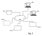
図2は、本発明の実施形態を実行することができる、通信ネットワーク215を介して複数の歯科情報システム210-1~210-3に接続された自動歯科カルテ作成システム205を訓練するためのコンピューティング装置200を示す。図示の例によれば、コンピューティング装置200は、記憶装置220にさらに接続されている。
FIG. 2 illustrates a
コンピューティング装置200および自動歯科カルテ作成システム205は、互いに直接接続されているか、通信ネットワーク215または別の通信ネットワーク、例えばプライベートネットワークを介して接続されている2つの異なる装置であり得る。コンピューティング装置200および自動歯科カルテ作成システム205はまた、同じ装置に統合することができる。
The
説明のために、3つの歯科情報システムおよび1つの記憶装置のみが示されていることに留意されたい。しかしながら、コンピューティング装置200は、数百もしくは数千の歯科情報システムおよび/または数百もしくは数千の記憶装置に接続され得ることが理解されるべきである。
Note that for purposes of illustration, only three dental information systems and one storage device are shown. However, it should be understood that
実施形態によれば、各歯科情報システムは、患者の歯科画像を取得し、電子歯科カルテを生成するように構成される。取得された画像は、可視光画像(VL)、紫外線画像(UV)、赤外線画像(IR)、OCT画像、蛍光画像(F)、X線画像(X)、CBCT(歯科用コーンビームコンピュータ断層撮影)処理でボリューム画像を形成するために使用される画像投影、輪郭画像、3Dメッシュ画像、および超音波画像を含む、異なるタイプまたはモダリティの画像であり得る。生成された電子歯科カルテは、患者の歯に関する情報を提供する、患者の歯に関する画像および関連する情報項目を含む。画像および関連する情報はどちらも同期している。 According to an embodiment, each dental information system is configured to acquire dental images of the patient and generate an electronic dental chart. The acquired images can be images of different types or modalities, including visible light images (VL), ultraviolet images (UV), infrared images (IR), OCT images, fluorescence images (F), X-ray images (X), image projections used to form volume images in CBCT (dental cone beam computed tomography) processing, contour images, 3D mesh images, and ultrasound images. The generated electronic dental chart includes images and associated information items related to the patient's teeth, providing information about the patient's teeth. Both the images and the associated information are synchronized.
電子歯科カルテまたは電子歯科カルテの一部は、歯科情報システムから直接、またはそれらが以前に保管されていた記憶装置220などの1つ以上の記憶装置から入手することができる。
The electronic dental chart or portions of the electronic dental chart may be obtained directly from the dental information system or from one or more storage devices, such as
実施形態によれば、歯科情報システムは、同じテンプレートまたは互換性のあるテンプレートに基づいて電子歯科カルテを生成し、同じ記号は同じ意味を有し、その結果、それらはソフトウェアの一部によって自動的に復号され得る。説明のために、電子歯科カルテは、各歯科情報システム内にインストールされた同じソフトウェアアプリケーション、または各歯科情報システムによってアクセスされる同じソフトウェアアプリケーションによって生成される(例えば、ソフトウェアアプリケーションがサービスとして提供される場合)。 According to an embodiment, the dental information systems generate the electronic dental charts based on the same template or compatible templates, and the same symbols have the same meaning, so that they can be automatically decoded by a piece of software. For purposes of illustration, the electronic dental charts are generated by the same software application installed within each dental information system or accessed by each dental information system (e.g., if the software application is provided as a service).
さらに特定の実施形態によれば、歯科情報システムのそれぞれは、生成された電子歯科カルテまたは生成された電子歯科カルテの一部を遠隔装置、例えば遠隔記憶装置または情報システムに転送することができる。好ましくは、生成された電子歯科カルテ(または生成された電子歯科カルテの一部)は匿名化した後転送され、この場合、生成された電子歯科カルテ(または生成された電子歯科カルテの一部)が関連付けられている患者を特定することはできない。 According to further particular embodiments, each of the dental information systems can transfer the generated electronic dental chart or a part of the generated electronic dental chart to a remote device, such as a remote storage or information system. Preferably, the generated electronic dental chart (or a part of the generated electronic dental chart) is transferred after being anonymized, in which case it is not possible to identify the patient to whom the generated electronic dental chart (or a part of the generated electronic dental chart) is associated.
図3は、本発明の1つ以上の実施形態を実施するため、特に図5、図6a、および図6bを参照して説明したステップまたはステップの一部を実行するための、図2に示すコンピューティング装置200の概略ブロック図である。
Figure 3 is a schematic block diagram of the
コンピューティング装置200は、
- CPUと表記される、マイクロプロセッサなどの中央処理装置305と、
- 本発明の実施形態の方法の実行可能コードを格納するための、RAMと表記されるランダムアクセスメモリ310、および本発明の実施形態による自動歯カルテ作成システムを訓練するための方法を実施するために必要な変数およびパラメータを記録するように構成されたレジスタであって、そのメモリ容量が、例えば拡張ポートに接続された任意のRAMによって拡張することができる、レジスタと、
- 本発明の実施形態を実施するためのコンピュータプログラムを記憶するための、ROMと表記される読み取り専用メモリ315と、
- ユーザーからの入力の受信、ユーザーへの情報の表示、および/または外部装置との間のデータの受信/送信のために使用することができるユーザーインターフェースおよび/または入力/出力インターフェース330と、
- 遠隔装置との間で、特に、歯科情報システム210-1~210-3および/または記憶装置220から、および自動歯科カルテ作成システム205にデータを送受信するためにデジタルデータを送受信できる通信ネットワークに通常接続されているネットワークインターフェース320と、に接続された通信バスを備えている。ネットワークインターフェース320は、単一のネットワークインターフェースであり得るか、または一連の異なるネットワークインターフェース(例えば、有線および無線インターフェース、または異なる種類の有線もしくは無線インターフェース)から構成され得る。データパケットは、送信のためにネットワークインターフェースに書き込まれるか、またはCPU305で実行されているソフトウェアアプリケーションの制御下で受信のためにネットワークインターフェースから読み取られる。
a central processing unit 305, such as a microprocessor, denoted CPU;
a random access memory 310, denoted RAM, for storing the executable code of the method of an embodiment of the invention, and registers arranged to record variables and parameters necessary for the implementation of the method for training an automatic dental charting system according to an embodiment of the invention, the memory capacity of which can be expanded, for example by any RAM connected to an expansion port;
a read-only memory 315, denoted ROM, for storing a computer program for implementing an embodiment of the invention;
A user interface and/or input/output interface 330 that can be used to receive input from a user, display information to a user, and/or receive/transmit data from/to an external device;
- a network interface 320, which is typically connected to a communications network capable of transmitting and receiving digital data to and from remote devices, in particular from the dental information systems 210-1 to 210-3 and/or the
コンピューティング装置200の通信バスは、任意で、大容量記憶装置として使用されるHDと表記されるハードディスク325に接続され得る。
The communication bus of the
実行可能コードは、読み取り専用メモリ315、ハードディスク325、または例えばディスクなどの着脱式デジタル媒体のいずれかに格納することができる。変形例によれば、プログラムの実行可能コードは、実行される前に、ハードディスク325などのコンピューティング装置200の記憶手段の1つに記憶されるべく、ネットワークインターフェース320を介して通信ネットワークによって受信され得る。
The executable code can be stored either in the read-only memory 315, in the hard disk 325 or on a removable digital medium, such as a disk. According to a variant, the executable code of the program can be received by the communications network via the network interface 320 to be stored in one of the storage means of the
中央処理装置305は、本発明の実施形態による1つ以上のプログラムの命令またはソフトウェアコードの一部の実行を制御および指示するように構成され、命令は前述の記憶手段の1つに記憶される。電源を入れた後、CPU305は、ソフトウェアアプリケーションに関連するメインRAMメモリ310からの命令を、例えばROM315またはハードディスク325からロードした後に実行することができる。そのようなソフトウェアアプリケーションは、CPU305によって実行されると、本明細書に開示されたアルゴリズムのステップが実行されるようにする。 The central processing unit 305 is configured to control and direct the execution of instructions of one or more programs or parts of software code according to embodiments of the present invention, the instructions being stored in one of the aforementioned storage means. After power-on, the CPU 305 may execute instructions from the main RAM memory 310 related to software applications, for example after loading them from the ROM 315 or the hard disk 325. Such software applications, when executed by the CPU 305, cause the steps of the algorithms disclosed herein to be performed.
本明細書に開示されるアルゴリズムの任意のステップは、PC(「パーソナルコンピュータ」)、DSP(「デジタルシグナルプロセッサ」)もしくはマイクロコントローラ、または、FPGA(「フィールドプログラマブルゲートアレイ」)やASIC(「特定用途向け集積回路」)などのマシンまたは専用構成要素によってハードウェアに実装されるその他のものなどのプログラム可能なコンピューティングマシンによる一連の命令またはプログラムの実行によってソフトウェアに実装され得る。 Any step of an algorithm disclosed herein may be implemented in software by execution of a sequence of instructions or a program by a programmable computing machine such as a PC ("personal computer"), a DSP ("digital signal processor") or a microcontroller, or other machine such as an FPGA ("field programmable gate array") or ASIC ("application specific integrated circuit") or implemented in hardware by dedicated components.
実施形態によれば、自動歯科カルテ作成システム205の概略ブロック図は、コンピューティング装置200の概略ブロック図と同様である。
According to an embodiment, a schematic block diagram of the automated
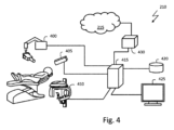
図4は、電子歯科カルテおよび/または電子歯科カルテの一部を生成する、図2の歯科情報システム210-1~210-3のうちの1つなどの歯科情報システムを示す概略図である。 FIG. 4 is a schematic diagram illustrating a dental information system, such as one of dental information systems 210-1 through 210-3 of FIG. 2, that generates an electronic dental chart and/or a portion of an electronic dental chart.
図示のように、歯科情報システム210は、X線撮像装置400、口腔内カメラなどのデジタルカメラ405、または、歯の構造のボリューム画像を生成するための歯科用コーンビームコンピュータ断層撮影(CBCT)システム410であり得る少なくとも1つの撮像装置を含む。超音波または他の撮像タイプを使用する装置など、他のタイプの撮像装置を使用して、歯および支持構造、歯茎、および関連組織の画像を取得することもできる。さらに、歯科情報システム210を操作するために、様々なタイプの診断測定機器も提供され得る。
As shown, the
引き続き図4を参照すると、撮像装置から画像データを取得、処理、および格納するためのコンピュータまたは他のタイプの専用論理プロセッサなどのホストプロセッサ415もまた、撮像結果を表示するための1つ以上のディスプレイ425とともに、歯科情報システム210の一部である。ホストプロセッサ415は、1つ以上の画像捕捉装置と、および任意で、任意の数の自動測定装置とデータ通信している。さらに、ホストプロセッサ415はまた、内部に、またはネットワーク化されたホストまたはサーバー上に格納された、患者記録のデータベースとデータ通信することができ、例えば、磁気、光学、または他のデータ記憶媒体を使用する装置など、長期記憶に使用される不揮発性メモリ記憶装置であり得るコンピュータクセス可能なメモリ420も提供される。
Continuing to refer to FIG. 4, a
さらに、ホストプロセッサ415は、遠隔装置との間で、特に、コンピューティング装置200、自動歯科カルテ作成システム205、および/または記憶装置220との間でデータを送受信するためにデジタルデータを送受信することができる通信ネットワーク215に通常接続されているネットワークインターフェース430を備えている。ネットワークインターフェース430は、単一のネットワークインターフェースであり得るか、または一連の異なるネットワークインターフェース(例えば、有線および無線インターフェース、または異なる種類の有線もしくは無線インターフェース)から構成され得る。
Furthermore, the
異なる歯科情報システムが同様の撮像装置、例えば同様のX線撮像装置または同様の口腔内カメラを使用し得るが、これらの装置は異なる製造業者からのものであり得るか、または異なる設定に従って設定され得ることに留意されたい。結果として、これらの装置によって生成される画像は、それらが同じ対象物(例えば、同じ歯)を描写する場合でも、異なって見える可能性がある。したがって、異なる歯科情報システムによって生成された電子歯科カルテの一部は、類似しているはずであるが、実際にはわずかに異なる場合がある。 Note that while different dental information systems may use similar imaging devices, e.g., similar x-ray imagers or similar intraoral cameras, these devices may be from different manufacturers or may be configured according to different settings. As a result, the images generated by these devices may look different, even if they depict the same object (e.g., the same tooth). Thus, some of the electronic dental records generated by different dental information systems, while supposed to be similar, may in fact differ slightly.
図5は、自動歯カルテ作成システムのステップを示している。 Figure 5 shows the steps of the automated dental chart creation system.
図示のように、第1のステップ(ステップ500)は、患者の歯の画像、例えば、1つの口腔外パノラマ画像および1つ以上の歯のいくつかの画像、例えば、X線画像、可視光画像、紫外線画像、赤外線画像、OCT画像、蛍光画像、およびCBCT画像、を取得することを目的としている。 As shown, the first step (step 500) is aimed at acquiring images of the patient's teeth, e.g., one extraoral panoramic image and several images of one or more teeth, e.g., x-ray images, visible light images, ultraviolet images, infrared images, OCT images, fluorescence images, and CBCT images.
次に、これらの画像は、歯科カルテ情報(符号510)を使用して電子歯科カルテを生成するために処理される(ステップ505)。取得された画像の処理は、例えば、1つ以上の人工ニューラルネットワークを含む人工知能(AI)エンジンに基づくことができる。人工ニューラルネットワークは、教師あり学習に基づく教師ありニューラルネットワークであり得る。教師あり学習では、入力サンプルと関連するラベルの訓練セットをニューラルネットワークに提示する必要がある(各ラベルは出力の対象物を描写する)。対応するラベルのセットは、専門家によってニューラルネットワークとは別に実行される事前の分類に従って決定され得る。歯科カルテ情報は、パラメータ値のセット(例えば、レイヤーとノードの数、重み値など)としてAIエンジンに符号化できる。 These images are then processed (step 505) to generate an electronic dental chart using dental chart information (symbol 510). The processing of the acquired images can be based on an artificial intelligence (AI) engine, for example including one or more artificial neural networks. The artificial neural network can be a supervised neural network based on supervised learning. In supervised learning, a training set of input samples and associated labels must be presented to the neural network (each label describing an object in the output). The set of corresponding labels can be determined according to a prior classification performed separately from the neural network by an expert. The dental chart information can be coded into the AI engine as a set of parameter values (e.g. number of layers and nodes, weight values, etc.).
実施形態によれば、口腔外パノラマ画像は、異なるタイプの他の画像によって、および画像上に表される歯を特徴付ける情報項目によって補足される基本的な電子歯科カルテを生成するために使用される。 According to an embodiment, the extraoral panoramic image is used to generate a basic electronic dental chart that is supplemented by other images of different types and by information items characterizing the teeth represented on the image.
点線の矢印で示唆されているように、電子歯科カルテは、新しい画像を処理することによって、および/または以前に処理された画像を新しい学習ステップにさらに処理することによって(すなわち、新しい情報を考慮して)いつでも自動的に更新され得る。 As suggested by the dotted arrows, the electronic dental record can be automatically updated at any time by processing new images and/or by further processing previously processed images into new learning steps (i.e., taking into account new information).
図6aは、電子歯科カルテを生成するために図5に示されているようなAIエンジンで実行されるステップの例を示し、図6bは、そのようなAIエンジンを訓練するためのステップの例を示す。 Figure 6a shows example steps performed in an AI engine such as that shown in Figure 5 to generate an electronic dental chart, and Figure 6b shows example steps for training such an AI engine.
図6aに示されているステップは、図2の自動歯科カルテ作成システム205などの自動歯科カルテ作成システムで実行することができ、図6bに示されるステップは、同じシステムまたは異なるシステム、例えば、図2のコンピューティング装置200において実行され得る。
The steps shown in FIG. 6a may be performed in an automated dental charting system, such as the automated
AIエンジンは、特定のAI技術、例えばファジー論理または人工ニューラルネットワーク、AI技術の組合せ、またはAI技術と従来の技術との組合せ、例えばニューラルネットワークと所定の規則との組合せに基づくことができる。 The AI engine can be based on a specific AI technique, e.g. fuzzy logic or artificial neural networks, a combination of AI techniques, or a combination of AI techniques and traditional techniques, e.g. neural networks and predefined rules.
電子歯科カルテを生成または更新するために処理される画像が取得された後(ステップ600)、画像は好ましくはフィルタリングされる(ステップ605)。そのようなフィルタリングステップは、例えば、画素値が所定のフォーマット(例えば、YUV)に従って、所定の解像度に従って、所定のバイト数(例えば、1成分あたり3バイト、1バイト)でコード化されるように、取得された画像を正規化するステップを含み得る。さらに、正規化ステップは、輝度および範囲拡張などのパラメータを調整するステップを含み得る。フィルタリングステップはまた、画像強調および画像平滑化などの画像処理を含み得る。 After an image to be processed to generate or update an electronic dental chart has been acquired (step 600), the image is preferably filtered (step 605). Such a filtering step may include, for example, normalizing the acquired image so that pixel values are coded according to a predefined format (e.g., YUV), according to a predefined resolution, and with a predefined number of bytes (e.g., 3 bytes, 1 byte per component). Furthermore, the normalization step may include adjusting parameters such as brightness and range expansion. The filtering step may also include image processing such as image enhancement and image smoothing.
次に、取得された画像のタイプが判定される(ステップ610)。取得された画像のタイプは、例えば、取得された画像に関連するそのソースまたはパラメータを識別することによって、またはよく知られた方法に従って後者を分析することによって、自動的に判定されることが好ましい。 Next, the type of image acquired is determined (step 610). The type of image acquired is preferably determined automatically, for example by identifying its source or parameters associated with the acquired image, or by analyzing the latter according to well-known methods.
特定の実施形態によれば、取得された画像をフィルタリングするステップは、フィルタリングが画像のタイプに適合されるように、そのタイプの判定後に実行される。 According to a particular embodiment, the step of filtering the acquired image is performed after determining the type of image, such that the filtering is adapted to that type.
次に、取得された画像(ステップ615)において歯が識別され、画像内に描写された歯の数、画像内のそれらの位置、それらの大きさ、およびそれらの形状が判定される。このようなステップは、訓練後にAIエンジンを使用して、セグメンテーションステップなどの前処理ステップの有無にかかわらず実行できる。実施形態によれば、歯は、グローバルインデックスを使用してインデックス付けまたは番号付けされ、その結果、画像で表される歯と別の画像で表される同じ歯との間にリンクが確立され得る。このようなグローバルインデックスは、既知の電子歯科カルテで使用されているものと同じであり得る。 Next, the teeth are identified in the acquired image (step 615) and the number of teeth depicted in the image, their location in the image, their size, and their shape are determined. Such steps can be performed with or without a pre-processing step, such as a segmentation step, using the AI engine after training. According to an embodiment, the teeth are indexed or numbered using a global index, so that a link can be established between a tooth represented in an image and the same tooth represented in another image. Such a global index can be the same as that used in known electronic dental charts.
一実施形態によれば、歯の識別および番号付けは、境界ボックス(例えば、長方形のボックスまたはより正確な輪郭を有するボックス)を使用することによって実行される。境界ボックスは、それぞれに含まれる歯の数を識別するために、パノラマ画像上の長方形のボックスを識別することによって、大まかな画像セグメンテーションを含み得る。 According to one embodiment, tooth identification and numbering is performed by using bounding boxes (e.g. rectangular boxes or boxes with more precise contours). The bounding boxes may involve a rough image segmentation by identifying rectangular boxes on the panoramic image to identify the number of teeth contained in each one.
このような境界ボックスは、AIエンジンの暗黙のルールを使用することによって、例えば、取得した画像の一部を人工ニューラルネットワークの入力として使用することによって判定できる。後者は、適切な訓練の後、歯の描写を識別し、したがって、識別された歯の周りの境界ボックスを判定することができる。 Such bounding boxes can be determined by using the implicit rules of an AI engine, for example by using parts of the acquired images as input to an artificial neural network. The latter, after appropriate training, is able to identify tooth depictions and thus determine bounding boxes around the identified teeth.
さらに、セグメンテーションは、あいまいな状況を明確にするために、次のような明示的なルールを実装する場合がある(必要な場合)。
- 「長方形のボックスには、所定の距離(1mmなど)を超えてはならないオーバーラップがある場合がある」、および
- 「1つの弧に対応する長方形のボックスの中心は、空間的変動が小さい曲線を表す必要がある(つまり、歯はほとんど線または曲率の少ない曲線に沿って整列する)」。
In addition, segmentation may (where necessary) implement explicit rules to clarify ambiguous situations, such as:
- "The rectangular boxes may have overlaps that should not exceed a certain distance (e.g., 1 mm)", and - "The center of the rectangular box corresponding to one arc should represent a curve with small spatial variation (i.e., the teeth mostly align along a line or a curve with little curvature)."
同様に、歯の番号付けアルゴリズムは、次のような暗黙的または明示的なルールを実装する場合がある。
- 「咬合中のパノラマ画像では、歯の数は所定の自然な順序に従う」、
- 「下顎の歯は上顎に現れない」、および
- 「上顎はボックスのサブセットで構成され、補完的なボックスのグループで構成される下顎の上に配置される」。
Similarly, a tooth numbering algorithm may implement implicit or explicit rules such as:
- "In panoramic images during occlusion, the number of teeth follows a given natural order",
- "the teeth of the mandible do not appear in the maxilla", and - "the maxilla consists of a subset of boxes, placed above the mandible, which consists of a complementary group of boxes".
歯の位置に関する情報は、特に、取得された画像が患者の歯のサブセットを含み、そこから患者の口内における歯の位置を導き出すことが不可能ではないにしても困難である場合、画像分析以外の手段によって提供され得ることに留意されたい。例えば、歯の位置に関する情報は、ユーザーから、または画像を撮影するときにユーザーに与えられる指示から取得することができる(ステップ620)。 It should be noted that information regarding tooth position may be provided by means other than image analysis, particularly if the acquired image contains a subset of the patient's teeth from which it is difficult, if not impossible, to derive the positions of the teeth in the patient's mouth. For example, information regarding tooth position may be obtained from the user or from instructions given to the user when taking the image (step 620).
次に、識別された歯は分類され、または特徴付けられる(ステップ625)。 The identified teeth are then classified or characterized (step 625).
そのような分類/特徴付けステップは、好ましくは、学習段階中に取得された電子歯科カルテから取得された情報を使用することによって、例えば、歯の描写を分類するために使用される、以前に訓練された人工ニューラルネットワークの入力として孤立した歯の描写を使用することによって実行される。人工ニューラルネットワークの出力は、孤立した歯の描写を特徴付ける1つ以上の情報項目である。 Such a classification/characterization step is preferably performed by using information obtained from the electronic dental chart obtained during the learning phase, for example by using the isolated tooth representation as input of a previously trained artificial neural network that is used to classify the tooth representation. The output of the artificial neural network is one or more items of information characterizing the isolated tooth representation.
繰り返し、このようなステップは、次のような一連のルールと閾値とを使用して補足できる。
- 「強い飽和信号はおそらく金属の特徴である」、
- 「金属を検出する機能により、アマルガム、インプラント、金属製クラウン、金属製ブラケット、根管などを含む可能性が高くなる」、および
- 「骨の根の深さは、歯の喪失につながる可能性のある骨の喪失があるかどうかを決定する」。
Again, such steps can be supplemented with a set of rules and thresholds such as:
- "The strong saturation signal is probably characteristic of metals",
- "The ability to detect metal increases the likelihood of including amalgam, implants, metal crowns, metal brackets, root canals, etc.", and - "Bone root depth determines if there is bone loss that may lead to tooth loss."
分類/特性評価ステップの目的は、1つ以上の情報項目を識別された歯に関連付けることである。そのような情報項目は、例えば、以下のものであり得る。
- 根とクラウンの下にある部分について
・歯が存在するかどうか、
・根尖周囲病変、
・根管かどうか、
・根内の支柱、
・骨量減少、
・強直、
・副鼻腔感染症、および
・洞床の腫れ、
- クラウンレベルの部分について
・復元するかどうか、
・3Dの内側~遠位の修復範囲(頬側~舌側の範囲を決定するのが困難)、
・クラウンと素材の種類(磁器、磁器溶融金属、すべて金属)、
・修復中のライナー(アマルガムまたは複合材料からのX線写真では異なるグレースケールとして表示される)、
・修復中の崩壊、および
・隣接歯間崩壊、
- レントゲン写真では検出できない特徴について
・頬側~舌側の修復範囲、
・骨折、
・修復物周辺の漏れ、および
・修復物周辺のエナメルの破砕。
The purpose of the classification/characterization step is to associate one or more items of information with the identified teeth. Such items of information may be, for example:
- The area beneath the root and crown - Whether the tooth is present or not,
- Periapical lesions,
・Whether it is a root canal,
- Supports within the roots,
- bone loss,
- rigidity,
Sinus infections, and Swelling of the sinus floor,
- Regarding the crown level part - Whether to restore or not,
- 3D medial-distal restoration extent (difficult to determine buccal-lingual extent),
-Type of crown and material (porcelain, porcelain-fused-to-metal, all-metal),
- Liners in restorations (appear as different greyscales on X-rays from amalgam or composite),
- Collapse during restoration, and - Interproximal collapse,
- Features that cannot be detected by X-rays: Restoration range from buccal to lingual side,
·fracture,
• Leaking around the restoration, and • Fracture of enamel around the restoration.
当然、他の情報項目は、歯および/または歯のグループに関連付けられ得る。 Of course, other items of information may be associated with teeth and/or groups of teeth.
分類/特徴付けステップは、例えば、各境界ボックスに含まれる信号を正規化された平均的な歯の描写にスケーリングおよび整列させるために、いくつかの前処理を含み得ることにも留意されたい。これは、平均的な歯の描写と比較して、近心/遠位、咬合/頬などの配置情報を定義するのに役立ち得る。 Note also that the classification/characterization step may include some pre-processing, for example to scale and align the signal contained in each bounding box to the normalized average tooth representation. This may help to define placement information such as mesial/distal, occlusal/buccal, etc., compared to the average tooth representation.
歯が分類/特徴付けられると、対応する電子歯科カルテが生成される(現在の歯が新しい患者に関連付けられた第1の画像の最初のものである場合)か、分類/特徴付けに応じて更新される(ステップ630)。実施形態によれば、画像は格納され(まだ格納されていない場合)、現在の歯に関連する情報項目は、例えばそのグローバルインデックスを使用して、この歯に関連して格納される。 Once the tooth has been classified/characterized, a corresponding electronic dental chart is generated (if the current tooth is the first of the first images associated with a new patient) or updated according to the classification/characterization (step 630). According to an embodiment, the image is stored (if it is not already stored) and the information items related to the current tooth are stored in association with this tooth, for example using its global index.
これらのステップは、処理されるすべての画像に対して、取得された画像で識別されたすべての歯に対して繰り返される(ステップ635および640)。 These steps are repeated for all images to be processed and for all teeth identified in the acquired images (steps 635 and 640).
上記のように、使用されるAIエンジンは、ローカリゼーションタスクおよび/または分類および特徴付けタスクのために、畳み込みニューラルネットワークおよび深層学習技術などの人工ニューラルネットワークを使用することができる。 As mentioned above, the AI engine used may use artificial neural networks such as convolutional neural networks and deep learning techniques for localization tasks and/or classification and characterization tasks.
図6aを参照することによって説明される処理は、典型的には患者の歯の画像のセットから、電子歯科カルテを作成するときに、ならびに、患者の歯の新しい画像が撮影されるか、歯科医が患者の歯に介入および/または監視するたびに実行され得る。 The process described by reference to FIG. 6a may be performed when creating an electronic dental chart, typically from a set of images of the patient's teeth, as well as each time new images of the patient's teeth are taken or a dentist intervenes and/or monitors the patient's teeth.
実施形態によれば、歯の識別および/または分類に使用される、図5および図6aを参照して説明されているようなAIエンジンの訓練は、低レベルのエラー(これらのエラーは通常、画像の解釈に向けられている)を含む大量の電子歯科カルテを使用して行われる。これらの電子カルテは、自動歯科カルテ作成システムの訓練に使用するために送信される前または後に匿名化されることが好ましい。 According to an embodiment, training of an AI engine such as that described with reference to Figs. 5 and 6a, used for tooth identification and/or classification, is performed using a large number of electronic dental charts containing low levels of errors (these errors are typically directed towards image interpretation). These electronic dental charts are preferably anonymized before or after being sent for use in training the automated dental charting system.
信頼性のために、自動歯科カルテ作成システムを訓練するために使用される電子カルテは、好ましくは、開業医によって生成された、開業医によって制御された、または信頼できるシステムによって、異なる場所で、異なる歯科情報システムを使用して自動的に生成された電子カルテである。それらは共通のフォーマットを共有しており、情報システムが画像とこれらの画像に表されている歯に関連する情報項目とを自動的に取得することを可能にする。 For reliability, the electronic medical records used to train the automated dental charting system are preferably practitioner-generated, practitioner-controlled, or automatically generated by a trusted system, at different locations, and using different dental information systems. They share a common format, allowing the information system to automatically retrieve images and items of information related to the teeth represented in these images.
電子歯科カルテは、歯科情報システムから、および/または歯科情報システムによって生成された電子歯科カルテが格納されているサーバーから取得することができる。 The electronic dental chart may be obtained from a dental information system and/or from a server on which the electronic dental chart generated by the dental information system is stored.
少なくとも1つの電子歯科カルテが取得された後(ステップ650)、電子歯科カルテは、患者の歯を描写する画像を取得するために(ステップ655)、取得された画像のタイプを取得するために(ステップ660)、および取得された画像(ステップ665)に描写された歯に関する情報項目を取得するために分析される。 After at least one electronic dental chart has been acquired (step 650), the electronic dental chart is analyzed to acquire images depicting the patient's teeth (step 655), to acquire the type of image acquired (step 660), and to acquire information items regarding the teeth depicted in the acquired images (step 665).
画像、画像のタイプ、および描写された歯に関する情報項目を取得するステップは、データが適切に収集され得るように有利に事前に決定された電子歯科カルテの電子フォーマットに従って実行される。 The steps of acquiring the image, the type of image, and the information items regarding the depicted teeth are advantageously performed according to a predetermined electronic format of the electronic dental chart so that the data can be collected appropriately.
対応する情報項目が、考慮される画像で表される歯または関心領域を特徴付けるという条件で、同じ歯または同じ関心領域のいくつかの画像を使用できることに留意されたい。言い換えれば、情報項目と画像は同期している(すなわち、歯または関心領域を描写する画像が取得されてから対応する情報項目が定義されるまでの間、歯または関心領域の状態は変化しないままである。)。 It should be noted that several images of the same tooth or the same region of interest can be used, provided that the corresponding information items characterize the tooth or region of interest represented in the images considered. In other words, the information items and the images are synchronized (i.e. the state of the tooth or region of interest remains unchanged between the time the image depicting the tooth or region of interest is acquired and the time the corresponding information item is defined).
特定の実施形態によれば、取得された画像はフィルタリングされる(ステップ670)。電子歯科カルテを生成または更新するときに実行され得る、図6aを参照して説明されたフィルタリングステップと同様に、このフィルタリングステップは、例えば、取得された画像を、画素値が所定のバイト数(例えば、構成要素ごとに3バイト、1バイト)でコード化されるように、所定のフォーマット(例:YUV)に従って、所定の解像度に従って正規化するステップを含み得る。さらに、正規化ステップは、輝度および範囲拡張などのパラメータを調整するステップを含み得る。 According to a particular embodiment, the acquired image is filtered (step 670). Similar to the filtering step described with reference to FIG. 6a, which may be performed when generating or updating an electronic dental chart, this filtering step may, for example, include normalizing the acquired image according to a predetermined resolution, according to a predetermined format (e.g. YUV), such that pixel values are coded in a predetermined number of bytes (e.g. 3 bytes, 1 byte per component). Furthermore, the normalization step may include adjusting parameters such as brightness and range expansion.
この場合も、特定の実施形態によれば、取得された画像をフィルタリングするステップは、画像タイプに依存し得る。 Again, according to certain embodiments, the step of filtering the acquired image may depend on the image type.
次に、図示の例によれば、AIエンジンに歯の描写を識別する方法を教えるために、第1の学習段階が実行される(ステップ675)。そのために、取得された画像のこの歯に対応する部分を識別するために、歯に関連する取得された情報項目が取得される。そのような情報項目は、この歯に対応する画像の部分を識別するためにセグメンテーション情報と組み合わされ得る。 Next, according to the illustrated example, a first learning phase is performed (step 675) to teach the AI engine how to identify tooth depictions. To that end, acquired information items related to the tooth are acquired in order to identify the part of the acquired image that corresponds to this tooth. Such information items can be combined with the segmentation information in order to identify the part of the image that corresponds to this tooth.
画像のこの部分は、AIエンジンの訓練に使用される680-1を参照する訓練データセットに追加される。実施形態によれば、画像のタイプはまた、対応する画像部分に関連して、訓練データセットに格納され得る。 This portion of the image is added to a training dataset referenced 680-1 that is used to train the AI engine. According to an embodiment, the type of image may also be stored in the training dataset in association with the corresponding image portion.
次に、取得された画像に表されている歯が、(訓練された後の)AIエンジンを使用して識別される(ステップ685)。 The teeth depicted in the captured image are then identified using the AI engine (once it has been trained) (step 685).
次に、識別された歯を分類または特徴付ける方法をAIエンジンに教えるべく、第2の学習段階が実行され(ステップ690)、すなわち、AIエンジンが歯のクラスおよび/または特性を表す情報項目を歯の描写と関連付けることを可能にする。 A second learning phase is then performed (step 690) to teach the AI engine how to classify or characterize the identified teeth, i.e., to enable the AI engine to associate items of information representing tooth classes and/or characteristics with tooth depictions.
そのために、識別された歯に関連する取得された情報項目が取得される。識別された歯および関連する情報項目に対応する画像の部分は、AIエンジンの訓練に使用される680-2を参照する訓練データセットに追加される。実施形態によれば、画像のタイプはまた、対応する画像部分に関連して、訓練データセットに格納され得る。 To that end, retrieved information items related to the identified tooth are retrieved. The portions of the images corresponding to the identified tooth and the related information items are added to a training dataset, referenced 680-2, used to train the AI engine. According to an embodiment, the type of image may also be stored in the training dataset in association with the corresponding image portion.
これらのステップは、取得された画像で識別されたすべての歯について、すべての電子歯科カルテのすべての画像に対して繰り返されることが好ましい(ステップ695)。 These steps are preferably repeated for all images in all electronic dental charts for all teeth identified in the acquired images (step 695).
特定の実施形態によれば、電子歯科カルテのいくつかは、学習段階が実行された後にAIエンジンを試験するために使用されるように、AIエンジンを訓練するために使用される電子歯科カルテのセットから除去され得る。このような場合、自動カルテ作成システムの効率は、自動カルテ作成システムの出力を期待される応答(すなわち、電子歯科カルテの情報項目)と比較することによって判定することができる。このような評価は、学習段階で実行することもできるため、効率的なレベルに達したらすぐに停止できる。 According to certain embodiments, some of the electronic dental charts may be removed from the set of electronic dental charts used to train the AI engine, so that they are used to test the AI engine after the learning phase has been performed. In such a case, the efficiency of the automated charting system can be determined by comparing the output of the automated charting system with the expected response (i.e., the information items in the electronic dental charts). Such an evaluation can also be performed during the learning phase, so that it can be stopped as soon as an efficient level is reached.
訓練データセットは、定期的に、例えば毎週または毎月、新しい電子歯科カルテまたは歯科情報システムから取得された更新された電子歯科カルテで更新され得ることに留意されたい。実施形態によれば、自動歯カルテ作成システムは、訓練データセットが更新された後に訓練される。 It should be noted that the training data set may be updated periodically, for example weekly or monthly, with new electronic dental charts or updated electronic dental charts obtained from the dental information system. According to an embodiment, the automated dental chart creation system is trained after the training data set is updated.
本発明は、図面および前述の説明において詳細に例示および説明されてきたが、そのような例示および説明は、説明的または例示的であり、限定的ではないと見なされるべきであり、本発明は、開示された実施形態に限定されない。開示された実施形態の他の変形は、図面、開示および添付の特許請求の範囲の研究から、特許請求される発明を実施する際に当業者によって理解および実行され得る。 While the invention has been illustrated and described in detail in the drawings and the foregoing description, such illustration and description should be considered as illustrative or exemplary and not restrictive, and the invention is not limited to the disclosed embodiments. Other variations of the disclosed embodiments can be understood and effected by those skilled in the art in practicing the claimed invention, from a study of the drawings, the disclosure and the appended claims.
そのような変形は、特に、本発明の要約および/または添付の特許請求の範囲に記載されている実施形態を組み合わせることに由来し得る。 Such variations may result in particular from combining the embodiments described in the Summary of the Invention and/or in the appended claims.
特に、画像分析は、説明のために、情報の項目が関連付けられている歯の識別に向けられているが、画像分析は、情報項目が関連付けられる任意の関心領域(ROI)の識別に向けることができ、例えば、嚢胞タイプの病理を顎の一部の描写に関連付けることを可能にする。そのような場合、関心領域は、情報項目に従って、例えば、嚢胞の描写に従って、および/または嚢胞を特徴付ける特徴に従って、少なくとも部分的に識別および/または抽出され得る。 In particular, although the image analysis is for illustrative purposes directed to the identification of a tooth with which an item of information is associated, the image analysis can be directed to the identification of any region of interest (ROI) with which an item of information is associated, making it possible, for example, to associate a cyst-type pathology with a depiction of a portion of a jaw. In such cases, the region of interest can be identified and/or extracted at least in part according to the item of information, for example according to the depiction of the cyst and/or according to features characterizing the cyst.
特許請求の範囲において、「含む(comprising)」という単語は、他の要素またはステップを除外せず、不定冠詞「a」または「an」は、複数を除外しない。単一のプロセッサまたは他のユニットは、特許請求の範囲に記載されたいくつかの項目の機能を果たすことができる。異なる特徴が相互に異なる従属請求項に記載されているという単なる事実は、これらの特徴の組合せが有利に使用できないことを示すものではない。特許請求の範囲の中のいかなる参照記号も、本発明の範囲を限定するものとして解釈されるべきではない。
In the claims, the word "comprising" does not exclude other elements or steps, and the indefinite articles "a" or "an" do not exclude a plurality. A single processor or other unit may fulfill the functions of several items recited in the claims. The mere fact that different features are recited in mutually different dependent claims does not indicate that a combination of these features cannot be used to advantage. Any reference signs in the claims should not be interpreted as limiting the scope of the invention.
Claims (14)
前記情報項目は、
根とクラウンの下にある部分について、歯が存在するかどうか、根尖周囲病変、根管かどうか、根内の支柱、骨量減少、強直、副鼻腔感染症、および洞床の腫れ、
並びに、クラウンレベルの部分について、復元するかどうか、3Dの内側~遠位の修復範囲、クラウンと素材の種類、修復中のライナー、修復中の崩壊、および隣接歯間崩壊、
並びに、レントゲン写真では検出できない特徴について、頬側~舌側の修復範囲、骨折、修復物周辺の漏れ、および修復物周辺のエナメルの破砕、
のうちの少なくとも1つの項目を含み、
当該コンピュータ方法は、
前記通信ネットワークを介して、複数の前記歯科情報システムによって生成された複数の電子歯科カルテを取得するステップであって、複数の前記電子歯科カルテは、複数の患者に関連している、ステップと、
取得された前記電子歯科カルテのそれぞれについて、
歯または関心領域を描写する画像の少なくとも一部を抽出し、描写された前記歯または描写された前記関心領域を特徴付ける少なくとも対応する情報項目を取得するステップと、
抽出された前記画像の少なくとも一部と前記対応する情報項目とを訓練データセットに格納するステップと、
前記訓練データセットを使用して前記自動歯科カルテ作成システムを訓練するステップと、
を含むことを特徴とするコンピュータ方法。 1. A computer method for training an automated dental charting system connected to a plurality of dental information systems via a communications network, each of the dental information systems configured to generate an electronic dental chart, each of the generated electronic dental charts including a digital image and an associated item of information characterizing at least a portion of the corresponding digital image;
The information items are:
The area below the root and crown, whether the tooth is present, periapical lesions, whether there are root canals, intraradicular posts, bone loss, ankylosis, sinus infection, and swelling of the sinus floor;
and for the crown level area, whether to restore, 3D medial to distal restoration range, type of crown and material, liner during restoration, decay during restoration, and interproximal decay.
and features not detectable on radiographs, such as buccal-lingual coverage of restorations, fractures, peri-restorative leakage, and peri-restorative enamel fractures.
and
The computer method comprises:
obtaining, via the communications network, a plurality of electronic dental charts generated by a plurality of the dental information systems , the plurality of electronic dental charts relating to a plurality of patients;
For each of the electronic dental charts obtained,
- extracting at least a portion of an image depicting a tooth or an area of interest and obtaining at least a corresponding item of information characterizing said depicted tooth or said depicted area of interest;
storing at least a portion of the extracted images and the corresponding information items in a training dataset;
training the automated dental charting system using the training data set;
23. A computer method comprising:
歯または関心領域を描写する画像の少なくとも一部を取得するステップと、
請求項1から9のいずれか1項に記載の方法に従って訓練された自動歯科カルテ作成システムを使用して、前記描写された歯または関心領域を特徴付ける情報項目を前記画像の少なくとも一部に割り当てるステップと、を含むことを特徴とする方法。 1. A method for automatically assigning medical chart data to at least a portion of an image depicting a tooth or region of interest, comprising:
obtaining at least a portion of an image depicting a tooth or an area of interest;
and using an automated dental charting system trained according to the method of any one of claims 1 to 9, assigning items of information characterizing the depicted teeth or areas of interest to at least a portion of the image.
Applications Claiming Priority (3)
| Application Number | Priority Date | Filing Date | Title |
|---|---|---|---|
| US201962802679P | 2019-02-07 | 2019-02-07 | |
| US62/802,679 | 2019-02-07 | ||
| PCT/EP2020/053129 WO2020161301A1 (en) | 2019-02-07 | 2020-02-07 | Method and apparatus for training automatic tooth charting systems |
Publications (2)
| Publication Number | Publication Date |
|---|---|
| JP2022520197A JP2022520197A (en) | 2022-03-29 |
| JP7586826B2 true JP7586826B2 (en) | 2024-11-19 |
Family
ID=69526269
Family Applications (1)
| Application Number | Title | Priority Date | Filing Date |
|---|---|---|---|
| JP2021546700A Active JP7586826B2 (en) | 2019-02-07 | 2020-02-07 | Method and apparatus for training an automated dental charting system - Patents.com |
Country Status (4)
| Country | Link |
|---|---|
| US (1) | US20220351813A1 (en) |
| EP (1) | EP3921841A1 (en) |
| JP (1) | JP7586826B2 (en) |
| WO (1) | WO2020161301A1 (en) |
Families Citing this family (2)
| Publication number | Priority date | Publication date | Assignee | Title |
|---|---|---|---|---|
| JP7462170B2 (en) * | 2021-02-22 | 2024-04-05 | パナソニックIpマネジメント株式会社 | Intraoral camera system, tooth identification method, control method and program |
| WO2024006572A1 (en) * | 2022-07-01 | 2024-01-04 | Pramana Inc. | Apparatus and a method for detecting associations among datasets of different types |
Citations (3)
| Publication number | Priority date | Publication date | Assignee | Title |
|---|---|---|---|---|
| WO2016143022A1 (en) | 2015-03-09 | 2016-09-15 | 富士通株式会社 | Crown information acquisition program, information processing device, and crown information acquisition method |
| JP2017527399A (en) | 2014-09-09 | 2017-09-21 | レイドス イノベイションズ テクノロジー,インコーポレイティド | Apparatus and method for disease detection |
| US20180028294A1 (en) | 2016-07-27 | 2018-02-01 | James R. Glidewell Dental Ceramics, Inc. | Dental cad automation using deep learning |
Family Cites Families (9)
| Publication number | Priority date | Publication date | Assignee | Title |
|---|---|---|---|---|
| US8416984B2 (en) * | 2011-01-20 | 2013-04-09 | Carestream Health, Inc. | Automatic tooth charting using digital images |
| US9158889B2 (en) * | 2013-04-26 | 2015-10-13 | Oral4D Systems Ltd. | Electronic dental charting |
| US10572625B2 (en) * | 2014-04-09 | 2020-02-25 | RealCloud Imaging Inc. | Combination dental imaging system and dental practice management and charting system with a bi-directional communication interface |
| US10902595B2 (en) * | 2015-12-04 | 2021-01-26 | 3Shape A/S | Deriving tooth condition information for populating digital dental charts |
| WO2018033762A1 (en) * | 2016-08-15 | 2018-02-22 | Trophy | Dynamic dental arch map |
| PL3595574T3 (en) * | 2017-03-17 | 2024-01-03 | Nobel Biocare Services Ag | Automatic dental arch mapping system and method |
| CN114587237B (en) * | 2018-01-26 | 2025-09-30 | 阿莱恩技术有限公司 | Diagnostic intraoral scanning and tracking |
| US20190313963A1 (en) * | 2018-04-17 | 2019-10-17 | VideaHealth, Inc. | Dental Image Feature Detection |
| US11389131B2 (en) * | 2018-06-27 | 2022-07-19 | Denti.Ai Technology Inc. | Systems and methods for processing of dental images |
-
2020
- 2020-02-07 EP EP20704281.3A patent/EP3921841A1/en active Pending
- 2020-02-07 WO PCT/EP2020/053129 patent/WO2020161301A1/en not_active Ceased
- 2020-02-07 US US17/427,734 patent/US20220351813A1/en not_active Abandoned
- 2020-02-07 JP JP2021546700A patent/JP7586826B2/en active Active
Patent Citations (3)
| Publication number | Priority date | Publication date | Assignee | Title |
|---|---|---|---|---|
| JP2017527399A (en) | 2014-09-09 | 2017-09-21 | レイドス イノベイションズ テクノロジー,インコーポレイティド | Apparatus and method for disease detection |
| WO2016143022A1 (en) | 2015-03-09 | 2016-09-15 | 富士通株式会社 | Crown information acquisition program, information processing device, and crown information acquisition method |
| US20180028294A1 (en) | 2016-07-27 | 2018-02-01 | James R. Glidewell Dental Ceramics, Inc. | Dental cad automation using deep learning |
Also Published As
| Publication number | Publication date |
|---|---|
| JP2022520197A (en) | 2022-03-29 |
| EP3921841A1 (en) | 2021-12-15 |
| US20220351813A1 (en) | 2022-11-03 |
| WO2020161301A1 (en) | 2020-08-13 |
Similar Documents
| Publication | Publication Date | Title |
|---|---|---|
| US12251253B2 (en) | Systems and methods for processing of dental images | |
| US11366985B2 (en) | Dental image quality prediction platform using domain specific artificial intelligence | |
| Leite et al. | Artificial intelligence-driven novel tool for tooth detection and segmentation on panoramic radiographs | |
| US11189028B1 (en) | AI platform for pixel spacing, distance, and volumetric predictions from dental images | |
| US20220180447A1 (en) | Artificial Intelligence Platform for Dental Claims Adjudication Prediction Based on Radiographic Clinical Findings | |
| US11311247B2 (en) | System and methods for restorative dentistry treatment planning using adversarial learning | |
| US11357604B2 (en) | Artificial intelligence platform for determining dental readiness | |
| US11367188B2 (en) | Dental image synthesis using generative adversarial networks with semantic activation blocks | |
| US11398013B2 (en) | Generative adversarial network for dental image super-resolution, image sharpening, and denoising | |
| US11217350B2 (en) | Systems and method for artificial-intelligence-based dental image to text generation | |
| US11348237B2 (en) | Artificial intelligence architecture for identification of periodontal features | |
| Orhan et al. | Evaluation of artificial intelligence for detecting periapical pathosis on cone‐beam computed tomography scans | |
| US20210118132A1 (en) | Artificial Intelligence System For Orthodontic Measurement, Treatment Planning, And Risk Assessment | |
| US20200364624A1 (en) | Privacy Preserving Artificial Intelligence System For Dental Data From Disparate Sources | |
| US20220012815A1 (en) | Artificial Intelligence Architecture For Evaluating Dental Images And Documentation For Dental Procedures | |
| US20200411167A1 (en) | Automated Dental Patient Identification And Duplicate Content Extraction Using Adversarial Learning | |
| KR101839789B1 (en) | System for generating interpretation data of dental image | |
| US20200410649A1 (en) | Inpainting Dental Images With Missing Anatomy | |
| US20210357688A1 (en) | Artificial Intelligence System For Automated Extraction And Processing Of Dental Claim Forms | |
| US20200387829A1 (en) | Systems And Methods For Dental Treatment Prediction From Cross- Institutional Time-Series Information | |
| US20210358604A1 (en) | Interface For Generating Workflows Operating On Processing Dental Information From Artificial Intelligence | |
| JP7586826B2 (en) | Method and apparatus for training an automated dental charting system - Patents.com | |
| Juneja et al. | A comprehensive review on artificial intelligence-driven preprocessing, segmentation, and classification techniques for precision furcation analysis in radiographic images | |
| Ahn et al. | Using artificial intelligence methods for dental image analysis: state-of-the-art reviews | |
| Aldusari et al. | The Role of Artificial Intelligence (AI) In Dentistry: Enhancing Diagnosis Accuracy and Treatment |
Legal Events
| Date | Code | Title | Description |
|---|---|---|---|
| A621 | Written request for application examination |
Free format text: JAPANESE INTERMEDIATE CODE: A621 Effective date: 20230206 |
|
| A977 | Report on retrieval |
Free format text: JAPANESE INTERMEDIATE CODE: A971007 Effective date: 20240327 |
|
| A131 | Notification of reasons for refusal |
Free format text: JAPANESE INTERMEDIATE CODE: A131 Effective date: 20240402 |
|
| A521 | Request for written amendment filed |
Free format text: JAPANESE INTERMEDIATE CODE: A523 Effective date: 20240617 |
|
| TRDD | Decision of grant or rejection written | ||
| A01 | Written decision to grant a patent or to grant a registration (utility model) |
Free format text: JAPANESE INTERMEDIATE CODE: A01 Effective date: 20240910 |
|
| A601 | Written request for extension of time |
Free format text: JAPANESE INTERMEDIATE CODE: A601 Effective date: 20241009 |
|
| A61 | First payment of annual fees (during grant procedure) |
Free format text: JAPANESE INTERMEDIATE CODE: A61 Effective date: 20241107 |
|
| R150 | Certificate of patent or registration of utility model |
Ref document number: 7586826 Country of ref document: JP Free format text: JAPANESE INTERMEDIATE CODE: R150 |