JP7581330B2 - Method and system for determining intravascular properties - Patents.com - Google Patents
Method and system for determining intravascular properties - Patents.com Download PDFInfo
- Publication number
- JP7581330B2 JP7581330B2 JP2022511015A JP2022511015A JP7581330B2 JP 7581330 B2 JP7581330 B2 JP 7581330B2 JP 2022511015 A JP2022511015 A JP 2022511015A JP 2022511015 A JP2022511015 A JP 2022511015A JP 7581330 B2 JP7581330 B2 JP 7581330B2
- Authority
- JP
- Japan
- Prior art keywords
- determining
- velocity
- acceleration
- characteristic
- blood vessel
- Prior art date
- Legal status (The legal status is an assumption and is not a legal conclusion. Google has not performed a legal analysis and makes no representation as to the accuracy of the status listed.)
- Active
Links
Images


Classifications
-
- A—HUMAN NECESSITIES
- A61—MEDICAL OR VETERINARY SCIENCE; HYGIENE
- A61B—DIAGNOSIS; SURGERY; IDENTIFICATION
- A61B5/00—Measuring for diagnostic purposes; Identification of persons
- A61B5/02—Detecting, measuring or recording for evaluating the cardiovascular system, e.g. pulse, heart rate, blood pressure or blood flow
- A61B5/026—Measuring blood flow
- A61B5/0265—Measuring blood flow using electromagnetic means, e.g. electromagnetic flowmeter
- A61B5/027—Measuring blood flow using electromagnetic means, e.g. electromagnetic flowmeter using catheters
-
- A—HUMAN NECESSITIES
- A61—MEDICAL OR VETERINARY SCIENCE; HYGIENE
- A61B—DIAGNOSIS; SURGERY; IDENTIFICATION
- A61B1/00—Instruments for performing medical examinations of the interior of cavities or tubes of the body by visual or photographical inspection, e.g. endoscopes; Illuminating arrangements therefor
- A61B1/04—Instruments for performing medical examinations of the interior of cavities or tubes of the body by visual or photographical inspection, e.g. endoscopes; Illuminating arrangements therefor combined with photographic or television appliances
- A61B1/041—Capsule endoscopes for imaging
-
- A—HUMAN NECESSITIES
- A61—MEDICAL OR VETERINARY SCIENCE; HYGIENE
- A61B—DIAGNOSIS; SURGERY; IDENTIFICATION
- A61B1/00—Instruments for performing medical examinations of the interior of cavities or tubes of the body by visual or photographical inspection, e.g. endoscopes; Illuminating arrangements therefor
- A61B1/313—Instruments for performing medical examinations of the interior of cavities or tubes of the body by visual or photographical inspection, e.g. endoscopes; Illuminating arrangements therefor for introducing through surgical openings, e.g. laparoscopes
- A61B1/3137—Instruments for performing medical examinations of the interior of cavities or tubes of the body by visual or photographical inspection, e.g. endoscopes; Illuminating arrangements therefor for introducing through surgical openings, e.g. laparoscopes for examination of the interior of blood vessels
-
- A—HUMAN NECESSITIES
- A61—MEDICAL OR VETERINARY SCIENCE; HYGIENE
- A61B—DIAGNOSIS; SURGERY; IDENTIFICATION
- A61B34/00—Computer-aided surgery; Manipulators or robots specially adapted for use in surgery
- A61B34/20—Surgical navigation systems; Devices for tracking or guiding surgical instruments, e.g. for frameless stereotaxis
-
- A—HUMAN NECESSITIES
- A61—MEDICAL OR VETERINARY SCIENCE; HYGIENE
- A61B—DIAGNOSIS; SURGERY; IDENTIFICATION
- A61B34/00—Computer-aided surgery; Manipulators or robots specially adapted for use in surgery
- A61B34/30—Surgical robots
-
- A—HUMAN NECESSITIES
- A61—MEDICAL OR VETERINARY SCIENCE; HYGIENE
- A61B—DIAGNOSIS; SURGERY; IDENTIFICATION
- A61B34/00—Computer-aided surgery; Manipulators or robots specially adapted for use in surgery
- A61B34/70—Manipulators specially adapted for use in surgery
- A61B34/73—Manipulators for magnetic surgery
-
- A—HUMAN NECESSITIES
- A61—MEDICAL OR VETERINARY SCIENCE; HYGIENE
- A61B—DIAGNOSIS; SURGERY; IDENTIFICATION
- A61B5/00—Measuring for diagnostic purposes; Identification of persons
- A61B5/02—Detecting, measuring or recording for evaluating the cardiovascular system, e.g. pulse, heart rate, blood pressure or blood flow
- A61B5/02007—Evaluating blood vessel condition, e.g. elasticity, compliance
-
- A—HUMAN NECESSITIES
- A61—MEDICAL OR VETERINARY SCIENCE; HYGIENE
- A61B—DIAGNOSIS; SURGERY; IDENTIFICATION
- A61B5/00—Measuring for diagnostic purposes; Identification of persons
- A61B5/02—Detecting, measuring or recording for evaluating the cardiovascular system, e.g. pulse, heart rate, blood pressure or blood flow
- A61B5/02028—Determining haemodynamic parameters not otherwise provided for, e.g. cardiac contractility or left ventricular ejection fraction
-
- A—HUMAN NECESSITIES
- A61—MEDICAL OR VETERINARY SCIENCE; HYGIENE
- A61B—DIAGNOSIS; SURGERY; IDENTIFICATION
- A61B5/00—Measuring for diagnostic purposes; Identification of persons
- A61B5/02—Detecting, measuring or recording for evaluating the cardiovascular system, e.g. pulse, heart rate, blood pressure or blood flow
- A61B5/026—Measuring blood flow
- A61B5/0265—Measuring blood flow using electromagnetic means, e.g. electromagnetic flowmeter
-
- A—HUMAN NECESSITIES
- A61—MEDICAL OR VETERINARY SCIENCE; HYGIENE
- A61B—DIAGNOSIS; SURGERY; IDENTIFICATION
- A61B5/00—Measuring for diagnostic purposes; Identification of persons
- A61B5/03—Measuring fluid pressure within the body other than blood pressure, e.g. cerebral pressure ; Measuring pressure in body tissues or organs
- A61B5/036—Measuring fluid pressure within the body other than blood pressure, e.g. cerebral pressure ; Measuring pressure in body tissues or organs by means introduced into body tracts
-
- A—HUMAN NECESSITIES
- A61—MEDICAL OR VETERINARY SCIENCE; HYGIENE
- A61B—DIAGNOSIS; SURGERY; IDENTIFICATION
- A61B5/00—Measuring for diagnostic purposes; Identification of persons
- A61B5/06—Devices, other than using radiation, for detecting or locating foreign bodies ; Determining position of diagnostic devices within or on the body of the patient
- A61B5/061—Determining position of a probe within the body employing means separate from the probe, e.g. sensing internal probe position employing impedance electrodes on the surface of the body
-
- A—HUMAN NECESSITIES
- A61—MEDICAL OR VETERINARY SCIENCE; HYGIENE
- A61B—DIAGNOSIS; SURGERY; IDENTIFICATION
- A61B5/00—Measuring for diagnostic purposes; Identification of persons
- A61B5/145—Measuring characteristics of blood in vivo, e.g. gas concentration or pH-value ; Measuring characteristics of body fluids or tissues, e.g. interstitial fluid or cerebral tissue
- A61B5/14503—Measuring characteristics of blood in vivo, e.g. gas concentration or pH-value ; Measuring characteristics of body fluids or tissues, e.g. interstitial fluid or cerebral tissue invasive, e.g. introduced into the body by a catheter or needle or using implanted sensors
-
- A—HUMAN NECESSITIES
- A61—MEDICAL OR VETERINARY SCIENCE; HYGIENE
- A61B—DIAGNOSIS; SURGERY; IDENTIFICATION
- A61B5/00—Measuring for diagnostic purposes; Identification of persons
- A61B5/68—Arrangements of detecting, measuring or recording means, e.g. sensors, in relation to patient
- A61B5/6846—Arrangements of detecting, measuring or recording means, e.g. sensors, in relation to patient specially adapted to be brought in contact with an internal body part, i.e. invasive
- A61B5/6847—Arrangements of detecting, measuring or recording means, e.g. sensors, in relation to patient specially adapted to be brought in contact with an internal body part, i.e. invasive mounted on an invasive device
- A61B5/6852—Catheters
-
- A—HUMAN NECESSITIES
- A61—MEDICAL OR VETERINARY SCIENCE; HYGIENE
- A61B—DIAGNOSIS; SURGERY; IDENTIFICATION
- A61B5/00—Measuring for diagnostic purposes; Identification of persons
- A61B5/68—Arrangements of detecting, measuring or recording means, e.g. sensors, in relation to patient
- A61B5/6846—Arrangements of detecting, measuring or recording means, e.g. sensors, in relation to patient specially adapted to be brought in contact with an internal body part, i.e. invasive
- A61B5/6867—Arrangements of detecting, measuring or recording means, e.g. sensors, in relation to patient specially adapted to be brought in contact with an internal body part, i.e. invasive specially adapted to be attached or implanted in a specific body part
- A61B5/6869—Heart
-
- A—HUMAN NECESSITIES
- A61—MEDICAL OR VETERINARY SCIENCE; HYGIENE
- A61B—DIAGNOSIS; SURGERY; IDENTIFICATION
- A61B5/00—Measuring for diagnostic purposes; Identification of persons
- A61B5/68—Arrangements of detecting, measuring or recording means, e.g. sensors, in relation to patient
- A61B5/6846—Arrangements of detecting, measuring or recording means, e.g. sensors, in relation to patient specially adapted to be brought in contact with an internal body part, i.e. invasive
- A61B5/6867—Arrangements of detecting, measuring or recording means, e.g. sensors, in relation to patient specially adapted to be brought in contact with an internal body part, i.e. invasive specially adapted to be attached or implanted in a specific body part
- A61B5/6876—Blood vessel
-
- A—HUMAN NECESSITIES
- A61—MEDICAL OR VETERINARY SCIENCE; HYGIENE
- A61B—DIAGNOSIS; SURGERY; IDENTIFICATION
- A61B34/00—Computer-aided surgery; Manipulators or robots specially adapted for use in surgery
- A61B34/20—Surgical navigation systems; Devices for tracking or guiding surgical instruments, e.g. for frameless stereotaxis
- A61B2034/2046—Tracking techniques
- A61B2034/2051—Electromagnetic tracking systems
-
- A—HUMAN NECESSITIES
- A61—MEDICAL OR VETERINARY SCIENCE; HYGIENE
- A61B—DIAGNOSIS; SURGERY; IDENTIFICATION
- A61B34/00—Computer-aided surgery; Manipulators or robots specially adapted for use in surgery
- A61B34/20—Surgical navigation systems; Devices for tracking or guiding surgical instruments, e.g. for frameless stereotaxis
- A61B2034/2046—Tracking techniques
- A61B2034/2055—Optical tracking systems
-
- A—HUMAN NECESSITIES
- A61—MEDICAL OR VETERINARY SCIENCE; HYGIENE
- A61B—DIAGNOSIS; SURGERY; IDENTIFICATION
- A61B34/00—Computer-aided surgery; Manipulators or robots specially adapted for use in surgery
- A61B34/20—Surgical navigation systems; Devices for tracking or guiding surgical instruments, e.g. for frameless stereotaxis
- A61B2034/2046—Tracking techniques
- A61B2034/2065—Tracking using image or pattern recognition
-
- A—HUMAN NECESSITIES
- A61—MEDICAL OR VETERINARY SCIENCE; HYGIENE
- A61B—DIAGNOSIS; SURGERY; IDENTIFICATION
- A61B34/00—Computer-aided surgery; Manipulators or robots specially adapted for use in surgery
- A61B34/30—Surgical robots
- A61B2034/303—Surgical robots specifically adapted for manipulations within body lumens, e.g. within lumen of gut, spine, or blood vessels
-
- A—HUMAN NECESSITIES
- A61—MEDICAL OR VETERINARY SCIENCE; HYGIENE
- A61B—DIAGNOSIS; SURGERY; IDENTIFICATION
- A61B90/00—Instruments, implements or accessories specially adapted for surgery or diagnosis and not covered by any of the groups A61B1/00 - A61B50/00, e.g. for luxation treatment or for protecting wound edges
- A61B90/36—Image-producing devices or illumination devices not otherwise provided for
- A61B90/37—Surgical systems with images on a monitor during operation
- A61B2090/374—NMR or MRI
-
- A—HUMAN NECESSITIES
- A61—MEDICAL OR VETERINARY SCIENCE; HYGIENE
- A61B—DIAGNOSIS; SURGERY; IDENTIFICATION
- A61B90/00—Instruments, implements or accessories specially adapted for surgery or diagnosis and not covered by any of the groups A61B1/00 - A61B50/00, e.g. for luxation treatment or for protecting wound edges
- A61B90/36—Image-producing devices or illumination devices not otherwise provided for
- A61B90/37—Surgical systems with images on a monitor during operation
- A61B2090/376—Surgical systems with images on a monitor during operation using X-rays, e.g. fluoroscopy
-
- A—HUMAN NECESSITIES
- A61—MEDICAL OR VETERINARY SCIENCE; HYGIENE
- A61B—DIAGNOSIS; SURGERY; IDENTIFICATION
- A61B90/00—Instruments, implements or accessories specially adapted for surgery or diagnosis and not covered by any of the groups A61B1/00 - A61B50/00, e.g. for luxation treatment or for protecting wound edges
- A61B90/39—Markers, e.g. radio-opaque or breast lesions markers
- A61B2090/3937—Visible markers
- A61B2090/3941—Photoluminescent markers
-
- A—HUMAN NECESSITIES
- A61—MEDICAL OR VETERINARY SCIENCE; HYGIENE
- A61B—DIAGNOSIS; SURGERY; IDENTIFICATION
- A61B90/00—Instruments, implements or accessories specially adapted for surgery or diagnosis and not covered by any of the groups A61B1/00 - A61B50/00, e.g. for luxation treatment or for protecting wound edges
- A61B90/39—Markers, e.g. radio-opaque or breast lesions markers
- A61B2090/3954—Markers, e.g. radio-opaque or breast lesions markers magnetic, e.g. NMR or MRI
-
- A—HUMAN NECESSITIES
- A61—MEDICAL OR VETERINARY SCIENCE; HYGIENE
- A61B—DIAGNOSIS; SURGERY; IDENTIFICATION
- A61B90/00—Instruments, implements or accessories specially adapted for surgery or diagnosis and not covered by any of the groups A61B1/00 - A61B50/00, e.g. for luxation treatment or for protecting wound edges
- A61B90/39—Markers, e.g. radio-opaque or breast lesions markers
- A61B2090/3966—Radiopaque markers visible in an X-ray image
-
- A—HUMAN NECESSITIES
- A61—MEDICAL OR VETERINARY SCIENCE; HYGIENE
- A61B—DIAGNOSIS; SURGERY; IDENTIFICATION
- A61B90/00—Instruments, implements or accessories specially adapted for surgery or diagnosis and not covered by any of the groups A61B1/00 - A61B50/00, e.g. for luxation treatment or for protecting wound edges
- A61B90/39—Markers, e.g. radio-opaque or breast lesions markers
- A61B2090/397—Markers, e.g. radio-opaque or breast lesions markers electromagnetic other than visible, e.g. microwave
- A61B2090/3975—Markers, e.g. radio-opaque or breast lesions markers electromagnetic other than visible, e.g. microwave active
-
- A—HUMAN NECESSITIES
- A61—MEDICAL OR VETERINARY SCIENCE; HYGIENE
- A61B—DIAGNOSIS; SURGERY; IDENTIFICATION
- A61B2562/00—Details of sensors; Constructional details of sensor housings or probes; Accessories for sensors
- A61B2562/02—Details of sensors specially adapted for in-vivo measurements
- A61B2562/0223—Magnetic field sensors
Landscapes
- Health & Medical Sciences (AREA)
- Life Sciences & Earth Sciences (AREA)
- Surgery (AREA)
- Engineering & Computer Science (AREA)
- Veterinary Medicine (AREA)
- Animal Behavior & Ethology (AREA)
- Biomedical Technology (AREA)
- Heart & Thoracic Surgery (AREA)
- Medical Informatics (AREA)
- Molecular Biology (AREA)
- Physics & Mathematics (AREA)
- Public Health (AREA)
- General Health & Medical Sciences (AREA)
- Biophysics (AREA)
- Pathology (AREA)
- Cardiology (AREA)
- Nuclear Medicine, Radiotherapy & Molecular Imaging (AREA)
- Physiology (AREA)
- Optics & Photonics (AREA)
- Vascular Medicine (AREA)
- Robotics (AREA)
- Radiology & Medical Imaging (AREA)
- Hematology (AREA)
- Electromagnetism (AREA)
- Human Computer Interaction (AREA)
- Magnetic Resonance Imaging Apparatus (AREA)
- Ultra Sonic Daignosis Equipment (AREA)
- Apparatus For Radiation Diagnosis (AREA)
- Force Measurement Appropriate To Specific Purposes (AREA)
- Measuring Pulse, Heart Rate, Blood Pressure Or Blood Flow (AREA)
Description
本発明は、独立請求項のプリアンブルによる血管内の組織または流体の特性を判断するための方法およびシステムに関する。 The present invention relates to a method and a system for determining properties of tissue or fluid in a blood vessel according to the preamble of the independent claim.
従来技術では、最小限に侵襲的な方法で血管を治療することが知られている。例えば、インプラントは、カテーテル装置を使用して送達することができる。そのような治療は、例えば、他の状態の中でもとりわけ、狭窄した血管または動脈瘤を治療するために使用される。同様に、従来技術では、患者の身体の内側部分を画像化および/または治療するために、マイクロロボットが使用されている。 In the prior art, it is known to treat blood vessels in a minimally invasive manner. For example, implants can be delivered using catheter devices. Such treatments are used, for example, to treat narrowed blood vessels or aneurysms, among other conditions. Similarly, in the prior art, microrobots have been used to image and/or treat interior portions of a patient's body.
しかしながら、治療および診断用途の両方においてそのような装置で一貫して生じる問題は、装置の周囲についての正確な情報が入手可能できないことである。 However, a consistent problem with such devices in both therapeutic and diagnostic applications is the lack of availability of accurate information about the device's surroundings.
特に、最小侵襲的処置と組み合わせて使用される撮像技術は、典型的には、周囲組織の特性に関して限られた情報しか提供しない。例えば、X線を使用するコントラスト画像化は、血管系の解剖学的情報を提供するが、機械的特性または血流の流れの障害に関する情報を提供することができない場合がある。したがって、複数の方法が、通常、平行して必要であり、そのような情報の収集は、時間がかかり、費用がかかり、困難なものになる。低侵襲的方法による治療中にリアルタイムでこのような情報を得ることは特に困難である。 In particular, imaging techniques used in conjunction with minimally invasive procedures typically provide limited information regarding the characteristics of the surrounding tissue. For example, contrast imaging using x-rays provides anatomical information of the vasculature but may not be able to provide information regarding mechanical properties or obstructions to blood flow. Thus, multiple methods are usually required in parallel, making the collection of such information time consuming, costly, and difficult. Obtaining such information in real time during treatment with minimally invasive methods is particularly difficult.
したがって、本発明の目的は、従来技術の欠点を克服することであり、特に、人体を治療または診断するための装置の近隣の、血管内の組織または流体の異なる特性を、簡単な方法で判断することを可能にする方法およびシステムを提供することである。 The object of the present invention is therefore to overcome the drawbacks of the prior art, in particular to provide a method and a system that allows to determine in a simple manner the different properties of tissue or fluid within a blood vessel in the vicinity of a device for treating or diagnosing the human body.
この目的および他の目的は、本発明の独立請求項の特徴部分による方法およびシステムによって達成される。 This and other objects are achieved by the method and system according to the characterizing parts of the independent claims of the present invention.
本発明は、患者の血管または心臓(V)における特性を判断するためのシステムに関する。システムは、血管または心臓に配置される要素を含む。システムは、さらに、要素に作用する推進力を判断するための手段と、要素の加速度および速度のうちの少なくとも1つを判断するための手段とを備える。好ましくは、システムは、要素の加速度および速度の両方を判断するための手段を含む。 The present invention relates to a system for determining a characteristic in a blood vessel or heart (V) of a patient. The system includes an element disposed in the blood vessel or heart. The system further comprises means for determining a driving force acting on the element and means for determining at least one of an acceleration and a velocity of the element. Preferably, the system includes means for determining both the acceleration and the velocity of the element.
そのような手段は、特に、センサ、例えば加速度計、または患者の身体の内側または外側の基準点までの距離を測定するように適合されたセンサを備えてもよい。加えて、または代替として、推進力を判断するための手段はまた、撮像装置および/または撮像装置によって生成される画像を分析するためのコンピュータを備えてもよい。 Such means may in particular comprise a sensor, for example an accelerometer, or a sensor adapted to measure distances to reference points inside or outside the patient's body. Additionally or alternatively, the means for determining the propulsion may also comprise an imaging device and/or a computer for analyzing images generated by the imaging device.
さらに、システムは、推進力と、加速度および速度の少なくとも1つとに基づいて、要素の近隣媒体の少なくとも1つの特性を判断するための手段を備える。例えば、そのような手段は、コンピュータ、好ましくはソフトウェアコードを実行するコンピュータを含んでもよい。好ましくは、手段は、要素の加速度および速度の両方に基づいて少なくとも1つの特性を判断する。 Furthermore, the system comprises means for determining at least one characteristic of a medium proximate to the element based on the momentum and at least one of the acceleration and velocity. For example, such means may include a computer, preferably a computer executing software code. Preferably, the means determines the at least one characteristic based on both the acceleration and velocity of the element.
本発明による方法は、患者の血管内の特性を判断する方法を提供する。それは、
-要素を血管または心臓に配置するステップと、
-要素に作用する推進力を判断するステップと、
-要素の有効加速度および有効速度の少なくとも1つ、特に有効加速度および有効速度の両方を判断するステップと、
-推進力と、要素の有効加速度および有効速度の少なくとも1つ、特に要素の有効加速度および有効速度の両方とに基づいて、要素の近隣媒体の少なくとも1つの特性を判断ステップとを含む。
The method according to the present invention provides a method for determining intravascular characteristics of a patient, comprising:
- placing the element in a blood vessel or in the heart;
- determining the driving forces acting on the element;
- determining at least one of the effective acceleration and the effective velocity of the element, in particular both the effective acceleration and the effective velocity;
determining at least one characteristic of a medium in the vicinity of the element based on the driving force and on at least one of the effective acceleration and the effective velocity of the element, in particular on both the effective acceleration and the effective velocity of the element.
本明細書に記載される方法を用いて判断され得る血管の特性は、機械的特性、例えば、組織の弾性、剛性、延性、および/または硬度であり得る。演繹によって、組織の解剖学的または組織学的特性、特に壊死組織または癌組織の存在を判断することも可能であろう。もちろん、粘度および/または流速などの、血液の物理的特性を判断することも可能であり得る。加えて、または代替として、特性は、流れに影響を及ぼす血管の特性、例えば、血管内の血液の流れを妨害、減速もしくは加速するかまたは乱流を生成する障害物を含むことができる。他の診断上関連のある特性、例えば、動脈瘤、血餅/血栓、気泡、および/または血管壁の欠陥の存在が得られることも考えられる。 The properties of blood vessels that may be determined using the methods described herein may be mechanical properties, e.g., elasticity, stiffness, ductility, and/or hardness of the tissue. By deduction, it may also be possible to determine anatomical or histological properties of the tissue, in particular the presence of necrotic or cancerous tissue. Of course, it may also be possible to determine physical properties of blood, such as viscosity and/or flow rate. Additionally or alternatively, properties may include properties of blood vessels that affect flow, e.g., obstructions that impede, slow down or speed up the flow of blood in the vessel or generate turbulence. It is also conceivable that other diagnostically relevant properties may be obtained, e.g., the presence of aneurysms, clots/thrombi, air bubbles, and/or defects in the vessel wall.
近隣媒体は、特に、要素と相互作用することができる、要素の近傍のすべての生物学的材料として理解されるものとする。それは、液体、固体、気体であり得るか、または軟質材料を含み得る。 The nearby medium is to be understood in particular as all biological material in the vicinity of the element that can interact with the element. It may be liquid, solid, gaseous or may include soft materials.
要素は、人体内で、少なくとも一時的に、自由に移動するように適合された任意の装置として理解されるものとする。これは、特に、マイクロロボット、センサ、薬物担体、または浮遊要素であってもよい。特に、要素は、その内側および/またはその表面に強磁性粒子などの磁気要素を含んでもよい。それはまた、強磁性材料からなってもよい。 An element is to be understood as any device adapted to move freely, at least temporarily, inside the human body. It may in particular be a microrobot, a sensor, a drug carrier or a levitating element. In particular, the element may comprise magnetic elements, such as ferromagnetic particles, in its interior and/or on its surface. It may also consist of a ferromagnetic material.
特に、本方法は、血管の特性を分析するためにのみ用いられてもよい。具体的には、本方法は、対象の特定の特性を判断する唯一の目的のために実行することができる。したがって、要素は、方法を実行するためにデータを提供するために導入されるに過ぎず、その他の目的に供されない。 In particular, the method may be used solely to analyze blood vessel characteristics. Specifically, the method may be performed for the sole purpose of determining a particular characteristic of an object. Thus, the elements are introduced merely to provide data for performing the method and serve no other purpose.
しかしながら、追加的または代替的に、体内に導入されて別の動作を行う医療装置を用いて本方法を実行することも可能である。例えば、マイクロロボットを体内に導入して、薬物を送達するか、または標的部位を治療してもよい。次いで、本方法は、マイクロロボットがいつ標的部位に到達したかを判断するために、またはさらには最適な標的部位を規定するために、実行されてもよい。 However, additionally or alternatively, the method may be performed using a medical device that is introduced into the body to perform another operation. For example, a microrobot may be introduced into the body to deliver a drug or treat a target site. The method may then be performed to determine when the microrobot has reached the target site, or even to define an optimal target site.
別の処理と並行して本方法を実行するために別個の要素を使用することも考えられる。
推進力は、典型的には、特に明記しない限り、患者の身体とのいかなる相互作用もない場合に要素に及ぼされる総力として理解されるものとする。例えば、要素が磁性であり、磁界によって作動される場合、推進力は、血流、または身体内の摩擦、血流、もしくは血栓からの閉塞等の身体的機能による他の力を考慮に入れることなく、磁界によって要素に及ぼされる力である。同様に、要素がプロペラまたはジェットなどの自己推進手段を含む場合、推進力は、この自己推進手段によって及ぼされる力となるであろう。自己推進手段と磁界との組み合わせが使用される場合、推進力は、これらの力の両方を含む。要素の所与の質量に対する推進力から理論上の加速度および速度を計算することが可能である。
It is also contemplated that separate elements may be used to carry out the method in parallel with another process.
Propulsion force is typically understood to be the total force exerted on the element in the absence of any interaction with the patient's body, unless otherwise stated. For example, if the element is magnetic and actuated by a magnetic field, the propulsion force is the force exerted on the element by the magnetic field, without taking into account other forces due to blood flow or bodily functions such as friction, blood flow, or blockage from clots within the body. Similarly, if the element includes a self-propelling means such as a propeller or jet, the propulsion force will be the force exerted by this self-propelling means. If a combination of self-propelling means and a magnetic field is used, the propulsion force includes both of these forces. It is possible to calculate theoretical acceleration and velocity from the propulsion force for a given mass of the element.
有効であり理論的な加速度ならびに速度は、摩擦、重力、抗力などの影響により決して同じではない。より洗練されたモデルでは、そのような影響が考慮され得る。 The effective and theoretical acceleration and velocity are never the same due to effects such as friction, gravity, drag, etc. More sophisticated models can take such effects into account.
例えば、装置の近隣の組織または流体のなんらかのパラメータが既知である場合、それらは、要素が推進力に基づいてその組織内で到達すべき理論的加速度または終端速度を計算するために考慮される。例えば、血液が要素に及ぼす速度依存抗力が既知である場合、要素の理論的終端速度を計算することができる。 For example, if certain parameters of the tissue or fluid in the vicinity of the device are known, they can be taken into account to calculate the theoretical acceleration or terminal velocity that an element should reach in that tissue based on the driving force. For example, if the velocity-dependent drag that blood exerts on an element is known, the theoretical terminal velocity of the element can be calculated.
対照的に、要素の有効加速度および/または有効速度は、患者の身体に対する要素の実際の加速度および/または実際の速度として理解されるものとする。したがって、有効な加速度または速度と理論的な加速度または速度との差(これは、推進力を要素の質量で除することによって判断することができる)は、要素の環境(例えば、血液の粘度)または障害物に関する情報を提供する。 In contrast, the effective acceleration and/or effective velocity of an element shall be understood as the actual acceleration and/or actual velocity of the element relative to the patient's body. Thus, the difference between the effective acceleration or velocity and the theoretical acceleration or velocity (which can be determined by dividing the thrust by the mass of the element) provides information about the element's environment (e.g. blood viscosity) or obstacles.
したがって、有効推進力は、有効加速度と要素の質量との積として理解されるものとする。したがって、所与の要素について、有効加速度は、有効推進力と等価なパラメータである。 The effective thrust is therefore to be understood as the product of the effective acceleration and the mass of the element. For a given element, the effective acceleration is therefore the parameter equivalent to the effective thrust.
推進力は、要素に作用する磁界などのアクチュエータのパラメータなどの既知のパラメータに基づいて測定または判断することができる。 The thrust can be measured or determined based on known parameters, such as actuator parameters, such as the magnetic field acting on the element.
好ましくは、推進力は、要素に含まれるセンサに基づいて判断される。これは、要素の正確な位置が知られていないか、または判断が困難である場合に特に有利である。例えば、要素が大型血管内に位置し、磁界によって駆動される場合、要素の場所における磁界の正確な特性は、知られていない場合がある。したがって、磁界によって要素に及ぼされる力を測定することができれば、この問題は解決される。 Preferably, the driving force is determined based on a sensor included in the element. This is particularly advantageous when the exact location of the element is not known or is difficult to determine. For example, if the element is located in a large blood vessel and is driven by a magnetic field, the exact characteristics of the magnetic field at the location of the element may not be known. Therefore, being able to measure the force exerted on the element by the magnetic field solves this problem.
追加的にまたは代替的に、有効加速度を測定するために要素に含まれる他のセンサを使用することも考えられる。 Additionally or alternatively, it is contemplated that other sensors included in the element may be used to measure the effective acceleration.
好ましくは、本方法は、要素が位置する患者の領域を撮像するステップをさらに含む。特に、画像化は、X線画像化、磁気共鳴画像化(MRI)、コンピュータ断層撮影、陽電子放出断層撮影(PET)、および超音波画像化のうちの1つを使用して行われ得る。これは、本方法を実施する際にいくつかの利点を提供することができる。一方で、特定の機械的特性が本方法によって判断される場合、撮像データは、正しい位置特定および解釈(例えば、特定のタイプの組織への割り当て)を支援することができる。一方で、撮像データを用いて要素の有効加速度および/または速度を測定することも考えられる。 Preferably, the method further comprises imaging the area of the patient in which the element is located. In particular, the imaging may be performed using one of X-ray imaging, magnetic resonance imaging (MRI), computed tomography, positron emission tomography (PET), and ultrasound imaging. This may provide several advantages in implementing the method. On the one hand, the imaging data may assist in correct localization and interpretation (e.g., assignment to a particular type of tissue) if certain mechanical properties are determined by the method. On the other hand, it is also conceivable to use the imaging data to measure the effective acceleration and/or velocity of the element.
代替的または追加的に、要素をナビゲーションするためのデータまたは本発明による方法によって収集されたデータの較正ならびに解釈のために患者のデータベースを使用することも考えられる。そのようなデータベースには、典型的な患者または患者群に関する情報だけでなく、患者個人情報も記憶され得る。較正および解釈は、人工知能法を使用することによって行われてもよい。特に、較正または解釈に使用可能な特定の情報は、ディープラーニング法または機械学習法によって収集されてもよい。このような方法は、本発明による方法の間および/またはその準備において実施することができる。 Alternatively or additionally, it is also conceivable to use a patient database for the calibration and interpretation of data for navigating the elements or data collected by the method according to the invention. In such a database, information on typical patients or patient groups as well as patient personal information may be stored. The calibration and interpretation may be performed by using artificial intelligence methods. In particular, specific information usable for the calibration or interpretation may be collected by deep learning or machine learning methods. Such methods may be implemented during and/or in preparation for the method according to the invention.
代替的または追加的に、第1の要素を使用することによって生成されたデータを使用して、後続の他の要素のためのナビゲーションに関連するパラメータ(例えば磁界)を変更することも考えられる。 Alternatively or additionally, data generated by using a first element may be used to modify a parameter related to navigation (e.g., a magnetic field) for another subsequent element.
好ましくは、近隣媒体の少なくとも1つの特性は、追加的に撮像データに基づく。例えば、撮像データは、特定の数の異なるタイプの組織が存在するかどうかの情報を提供することができる。加えて、または代替として、撮像データは、血管内の障害物の存在を明らかにしてもよい。したがって、本発明の近隣媒体の1つの特性は、撮像データに基づいているが、他の特性は、異なる組織タイプまたは障害物の機械的特性などである。撮像データと機械的特性との組み合わせは、どのようなタイプの組織が存在するか、および/または血管内にどのような種類の障害物が存在するかを決定的に判断することを可能にし得る。 Preferably, at least one characteristic of the nearby medium is additionally based on imaging data. For example, the imaging data may provide information on whether a certain number of different types of tissue are present. Additionally or alternatively, the imaging data may reveal the presence of an obstacle in the blood vessel. Thus, one characteristic of the nearby medium of the present invention is based on imaging data, while other characteristics are mechanical properties of different tissue types or obstacles, etc. The combination of imaging data and mechanical properties may make it possible to conclusively determine what type of tissue is present and/or what kind of obstacle is present in the blood vessel.
当業者は、これが、撮像データが本発明による方法においてどのように有利に使用され得るかの単なる非限定的な例であることを理解するであろう。しかしながら、撮像データを伴わずに本方法を実施することが可能である。 Those skilled in the art will appreciate that this is merely a non-limiting example of how imaging data may be advantageously used in the method according to the present invention. However, it is possible to practice the method without imaging data.
好ましくは、本方法は、要素の位置を判断するステップをさらに含む。これは、特定の組織タイプが患者の身体内のどこに位置するかのさらなる情報を提供する。これは、これらの組織が、例えば外科的技術によって治療または除去されることが計画される場合に特に有利である。 Preferably, the method further includes the step of determining the location of the element. This provides further information of where particular tissue types are located within the patient's body. This is particularly advantageous when these tissues are planned to be treated or removed, for example, by surgical techniques.
好ましくは、その位置は、撮像技術によって判断される。
好ましくは、本方法は、近隣媒体の少なくとも1つの特性を位置または時間の関数としてメモリに保存するステップをさらに含む。これは、1回のセッション中に血管内の複数の位置を分析することを可能にする。特に、データは、続いて、1次元、2次元、または3次元の特性マップを作成するように分析されることができる。例えば、血管の狭窄または動脈瘤は、本発明による方法により、前記血管の長手方向軸に沿って判断することができる。狭窄または動脈瘤のレベルが判断され、複数の点で保存される場合、長手方向軸に沿った狭窄または動脈瘤を示すグラフを得ることができる。同様に、データは、2次元または3次元で収集されてもよい。
Preferably, the location is determined by imaging techniques.
Preferably, the method further comprises a step of storing in memory at least one characteristic of the neighboring medium as a function of position or time. This allows for analysing multiple positions in the blood vessel during one session. In particular, the data can be subsequently analysed to create a one-, two- or three-dimensional characteristic map. For example, a stenosis or aneurysm of a blood vessel can be determined along the longitudinal axis of said blood vessel by the method according to the invention. If the level of stenosis or aneurysm is determined and stored at multiple points, a graph showing the stenosis or aneurysm along the longitudinal axis can be obtained. Similarly, the data may be collected in two or three dimensions.
好ましくは、近隣媒体の少なくとも1つの特性を計算するステップは、ソフトウェアコードを実行するコンピュータによって実行される。これは、特に、本方法を自動的に実行することを可能にし、したがって、迅速で、信頼性があり、安価である。好ましくは、近隣媒体の少なくとも1つの特性は、機械的特性、血行動態特性、解剖学的特性、および組織学的特性のうちの1つである。例えば、血液の粘度、組織のヤング率、血液の流速、および/または血管のサイズもしくは形状、特にその直径であってもよい。 Preferably, the step of calculating at least one property of the neighboring medium is performed by a computer executing software code. This in particular allows the method to be performed automatically and is therefore fast, reliable and cheap. Preferably, the at least one property of the neighboring medium is one of a mechanical property, a hemodynamic property, an anatomical property and a histological property. It may for example be the viscosity of the blood, the Young's modulus of the tissue, the flow rate of the blood and/or the size or shape of the blood vessel, in particular its diameter.
好ましくは、血管内に配置された要素は磁気要素を含み、推進力を判断するステップは磁界の磁界強度を判断するステップを含む。磁界強度を判断することは、特に、磁界を生成するユニットの既知のパラメータに基づいて空間における特定の点(特に要素の位置)で磁界の特性を計算することを包含するが、空間における特定の点で磁界を測定することも含むことができる。磁界の測定は、要素によって、したがって、その場所および/または基準点において行われてもよい。 Preferably, the element placed in the blood vessel comprises a magnetic element, and the step of determining the driving force comprises a step of determining a magnetic field strength of the magnetic field. Determining the magnetic field strength includes in particular calculating a characteristic of the magnetic field at a particular point in space (in particular the position of the element) based on known parameters of the unit generating the magnetic field, but may also include measuring the magnetic field at the particular point in space. The measurement of the magnetic field may be performed by the element and therefore at its location and/or at a reference point.
好ましくは、本方法は、要素に作用する推進力に基づいて要素の理論上の加速度または速度を計算するステップをさらに含む。 Preferably, the method further comprises the step of calculating a theoretical acceleration or velocity of the element based on the driving forces acting on the element.
好ましくは、要素の速度および加速度の理論値を計算するステップは、加えて、要素の位置と、撮像ステップで取得される撮像データとに基づく。例えば、血管のサイズおよび血液量は、撮像データに基づいて考慮されてもよい。加えて、または代替的に、血流の速度および拍動性は、特にドップラー超音波画像化によって測定および考慮されてもよい。特に、血管壁に対する要素の位置も考慮に入れてもよい。 Preferably, the step of calculating the theoretical values of the velocity and acceleration of the element is additionally based on the position of the element and on the imaging data acquired in the imaging step. For example, the size and blood volume of the blood vessel may be taken into account based on the imaging data. Additionally or alternatively, the velocity and pulsatility of the blood flow may be measured and taken into account, in particular by Doppler ultrasound imaging. In particular, the position of the element relative to the vessel wall may also be taken into account.
好ましくは、本方法は、要素の周囲の液体、特に血管または心臓内の血液の流速を測定するステップをさらに含む。加えて、近隣媒体の少なくとも1つの特性の判断は、流速に基づいてもよい。特に、流速は、ドップラー超音波画像化によって判断してもよい。しかしながら、要素内および/または要素上に含まれるセンサを用いて流速を測定することも考えられる。 Preferably, the method further comprises a step of measuring the flow velocity of the liquid surrounding the element, in particular blood in a blood vessel or in the heart. In addition, the determination of at least one characteristic of the neighboring medium may be based on the flow velocity. In particular, the flow velocity may be determined by Doppler ultrasound imaging. However, it is also conceivable to measure the flow velocity using a sensor included in and/or on the element.
好ましくは、本方法は、少なくとも1つの検出器および/またはマーカを用いて要素の位置を特定するステップをさらに含む。マーカは、要素上に配置することができ、検出器は、患者の身体内の所定の位置または患者の身体に対する所定の位置に配置することができる。例えば、検出器は、血管の外側、頭蓋骨などの骨、または心臓などの臓器に配置され得る。検出器は、その近傍にある要素を検出するかまたは検出を助けるように適合された任意の装置として理解されるものとする。マーカは、要素に関連付けられ、特にセンサによって要素を検出するのに役立つ。 Preferably, the method further comprises the step of locating the element using at least one detector and/or marker. The marker may be located on the element and the detector may be located at a predetermined position in or relative to the patient's body. For example, the detector may be located on the outside of a blood vessel, on a bone such as the skull, or on an organ such as the heart. A detector is to be understood as any device adapted to detect or aid in the detection of an element in its vicinity. A marker is associated with an element and is particularly useful for detecting the element by a sensor.
検出器はまた、要素までの距離を測定するように適合されてもよい。検出器は、特に好ましくは、電気センサ、磁気センサまたは光学センサであってもよい。マーカは、NFCチップ、磁石または放射線不透過性材料であり得る。蛍光マーカまたは同位体マーカを使用することもできる。 The detector may also be adapted to measure the distance to the element. The detector may particularly preferably be an electric, magnetic or optical sensor. The marker may be an NFC chip, a magnet or a radiopaque material. Fluorescent or isotopic markers may also be used.
本発明はさらに、患者の身体内の要素の近隣媒体を分析するためのコンピュータプログラム製品を対象とする。それは、コンピュータ上で実行されると、推進力と、要素の有効加速度および有効速度のうちの少なくとも1つ、特に要素の有効加速度および有効速度の両方とに基づいて、要素の近隣媒体の少なくとも1つの特性を判断するステップを実行するように適合されたソフトウェアコードを備える。コンピュータプログラム製品は、特に、撮像データを処理し、好ましくは、撮像装置によって取得された近隣媒体の撮像データに基づいて、近隣媒体の少なくとも1つの特性を判断するように適合されてもよい。たとえば、コンピュータプログラム製品は、撮像データに基づいて要素の速度および加速度のうちの少なくとも1つを判断するように適合されてもよい。 The present invention is further directed to a computer program product for analyzing a neighboring medium of an element in a patient's body. It comprises software code adapted to perform, when executed on a computer, a step of determining at least one characteristic of the neighboring medium of the element based on a driving force and at least one of an effective acceleration and an effective velocity of the element, in particular both the effective acceleration and the effective velocity of the element. The computer program product may in particular be adapted to process imaging data and to determine at least one characteristic of the neighboring medium based on imaging data of the neighboring medium, preferably acquired by an imaging device. For example, the computer program product may be adapted to determine at least one of the velocity and acceleration of the element based on the imaging data.
本発明はさらに、血管の特性を判断するためのシステムに関する。それは、体液によって運ばれるおよび/または体液中で能動的に移動するように適合された要素と、測定ユニットと、計算ユニットとを備える。測定ユニットは、要素の加速度および速度のうちの少なくとも1つを判断するように適合される。計算ユニットは、要素の加速度および速度の少なくとも1つに基づいて血管の少なくとも1つの特性を判断するように適合される。好ましくは、測定ユニットおよび計算ユニットの少なくとも一方は、要素に作用する推進力を判断するように適合される。例えば、計算することは、動作パラメータ、例えば磁界の特性に基づいて推進力を計算してもよい。追加的または代替的に、測定ユニットはまた、例えば磁界を測定することによって、要素に及ぼされる力を測定するように適合されてもよい。特に、計算ユニットは、本明細書に記載の方法を実行することによって、特に好ましくは、本明細書に記載の方法のステップを実行するように適合されたソフトウェアコードを実行することによって、血管の少なくとも特性を判断することができる。 The present invention further relates to a system for determining a characteristic of a blood vessel. It comprises an element adapted to be carried by and/or to actively move in a body fluid, a measurement unit and a calculation unit. The measurement unit is adapted to determine at least one of an acceleration and a velocity of the element. The calculation unit is adapted to determine at least one characteristic of the blood vessel based on at least one of the acceleration and the velocity of the element. Preferably, at least one of the measurement unit and the calculation unit is adapted to determine a propulsive force acting on the element. For example, the calculating may calculate the propulsive force based on an operating parameter, for example a characteristic of a magnetic field. Additionally or alternatively, the measurement unit may also be adapted to measure a force exerted on the element, for example by measuring a magnetic field. In particular, the calculation unit may determine at least the characteristic of the blood vessel by executing a method as described herein, particularly preferably by executing software code adapted to execute steps of the method as described herein.
当業者は、本システムが、特に、本明細書に記載される方法ステップのいずれかを実行するように適合されてもよいことを理解するであろう。 Those skilled in the art will appreciate that the system may be specifically adapted to perform any of the method steps described herein.
好ましくは、本システムは、要素が位置する患者の身体の領域を撮像するように適合された撮像装置を備える。これは、特に、撮像ユニットが採用され得る、本発明による方法の文脈において説明されるようなすべてのステップを行うことを可能にする。特に、撮像ユニットは、PET、MRI、超音波、X線、およびCTのいずれかであってもよい。 Preferably, the system comprises an imaging device adapted to image the area of the patient's body in which the element is located. This makes it possible in particular to carry out all the steps as described in the context of the method according to the invention in which an imaging unit may be employed. In particular, the imaging unit may be any of PET, MRI, ultrasound, X-ray and CT.
以下において、本発明は、以下の図を参照して詳細に説明される。 The present invention will now be described in detail with reference to the following figures:
図1は、血管Vにおいて要素1を模式的に示す。ここで、要素1は、強磁性材料を含む移動要素である。したがって、外部磁石(不図示、図8参照)によって印加される磁界が、血管V内で要素1を案内または推進してもよい。したがって、それは要素1に力2を及ぼすことができる。この場合、例えば血流、摩擦、または重力などの身体的機能による影響がないと仮定すると、磁界によって及ぼされる力2は、要素1に作用する唯一の力であろう。したがって、それは、この例における推進力を表す。しかしながら、流体抵抗により、要素はまた、抗力も受ける。ここで、抗力は未知であり、血液の流体特性および血管壁上の摩擦を判断するために、判断されるべきである。しかしながら、推進力に流体抵抗および摩擦を含むことも可能であろう。要素の有効加速度3または速度4を測定することも可能である。ここで、加速度3は、例えば、要素に含まれる加速度計によって測定される。代替的に、それは、例えば、撮像装置によって測定されてもよい。有効加速度3は、力2と抗力との差を表す。したがって、抗力を計算することができ、その結果、血液および血管の流体特性も計算することができる。これは、当然ながら、血液の流れ特性が条件によって影響を受ける場合に特に有利である。
Figure 1 shows a schematic representation of an
図2は、図1に示されるものと同様の要素を模式的に示す。ここで、明確にするために、血管Vは示されていない。加えて、要素は、異なる形状を有し、準球状ではなく立方体を有する。しかしながら、それは、強磁性材料も含み、磁界によって推進されることができる。図2に模式的に示す状態は、要素が終端速度4に達したときの平衡状態を表している。したがって、推進力2および抗力5は、等しいノルム値を有するが、反対の符号を有する。したがって、終端速度4を使用して、血液の粘度などの血液のパラメータを計算することができる。本方法の同様の実施形態では、磁界は、特定の周波数でその方向を変えてもよい。速度を測定する代わりに、要素の運動の周波数を測定し、それから血液の流体特性を判断してもよい。
Figure 2 shows a schematic representation of an element similar to that shown in Figure 1. Here, for clarity, the blood vessel V is not shown. In addition, the element has a different shape, being cubic rather than quasi-spherical. However, it also comprises a ferromagnetic material and can be propelled by a magnetic field. The state shown diagrammatically in Figure 2 represents an equilibrium state when the element reaches a
図3は、異なるタイプの要素1を模式的に示す。これは球状であり、血管V内の流体によって受動的に運ばれる。それは、要素1の有効加速度3を測定するように適合されたセンサをさらに備える。ここで、血管は、血液の流速を一時的に上昇させる狭窄部を有する。これにより、加速度計6は、一時的な加速度(および拡大領域における減速度)を検出する。この例では、要素1は受動的に運ばれるので、推進力はゼロである。したがって、有効加速度3を直接用いて、血管Vの特性、この場合は狭窄部の存在を、判断することができる。
Figure 3 shows a schematic representation of a different type of
図4は、血管V内において液体によって運ばれる要素1を示す。明確にするために、推進手段のいずれもこの概略図には示されていないが、当業者は、装置を移動させる、操縦する、または案内する、説明された方法のいずれかが、この実施形態において採用され得ることを理解するであろう。ここでは、X線撮像装置を有する撮像ユニット7を用いる。要素1は、X線撮像において可視である。さらに、血液は造影剤を含有し、血管系もX線下で可視である。ドップラー撮像ユニット8は、血管系内の血液の流れ9を視覚化するために用いられる。当然ながら、ドップラー撮像ユニットおよびX線撮像装置は、各々1つのコンピュータに、または同じコンピュータ、例えば計算において撮像結果を使用するために本発明によるコンピュータプログラム製品を含むコンピュータに、接続されてもよい。
Figure 4 shows
図5は、要素1が血管V内を移動するのを示す。ここで、いくつかの検出器10が血管Vの周囲に配置される。それらは、血管の周りにおいて閉じた銅コイルによって形成される。要素は、その周囲に磁界を生成する永久磁石を含む。したがって、要素1が検出器10を通過するとき、移動する磁界は、検出可能な電流を検出器内に誘起する。ロボット上に放射チップを提供し、身体上に配置された受信器/検出器を提供することも可能であり、これはロボットの姿勢を三角測量することができる。
Figure 5 shows an
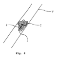
図6は、本方法の別の用途を示す。ここでは、要素は、血管内を移動しており、その中に含まれる強磁性要素を介して要素1に力2を及ぼす磁界によって推進される。しかしながら、血管V内に血餅Cが形成され、血流を遮断または制限している。その結果、要素1も、血餅Cに当たると、同様に移動を停止する。したがって、要素の有効速度はゼロになり、一方、推進力2はゼロではない。これは、ある特性、この場合は血餅の存在の判断を可能にする。もちろん、要素1の有効加速度を加えて測定することも考えられ、それは、血餅Cの位置および/またはその機械的特性に関する情報をさらに与えるであろう(より柔らかい血餅Cは、より低い負の加速度値をもたらすであろう)。
Figure 6 shows another application of the method. Here, an element is moving in a blood vessel and is propelled by a magnetic field exerting a
図7は、本発明によるシステムを模式的に示す。それは、センサ6が配置される要素1を含む。ここで示されるセンサは、要素の加速度を測定するように適合され、任意選択で、温度および安全性などの基本値を測定するように適合される。システムは、測定ユニット11および分析器ユニット12をさらに備える。測定ユニットは、特に、センサから加速度値を受け取るように適合される。しかしながら、それは、マーカまたは検出器10に基づいて値を測定することもできる。本方法を実施する必要はないが、特定の実施形態では、要素1に作用する有効推進力を測定することが有利である場合がある。したがって、この非限定的な例では、測定ユニット11はまた、特にセンサ6との相互作用によって、要素の位置における磁界を測定するように適合される。分析器ユニット12は、測定ユニット11から受信した値および測定ユニット11によって受信された値を処理するように適合される。加えて、それは、値を保存するためのメモリ15を含む。例えば、要素1に含まれるセンサ6から有効加速度値を受け取ってもよい。加えて、推進力は、外部磁気ユニット(図8参照)のパラメータから既知でもよく、またはセンサ6によって測定されてもよい。いずれの場合も、分析器ユニットは、これらの値を処理し、それらを分析して、血管の少なくとも1つの特性を判断するように適合される。特性値は、任意選択でメモリ15に保存することができる。本システムを、メモリに保存された値に基づいて、データ、特に、患者の解剖学的構造の再構成などのデータの2次元および/または3次元画像を示すように適合されたディスプレイと組み合わせることも考えられる。本明細書に説明される実施例および実施形態のうちのいずれかが本システムとともに実現され得ることが、理解されるであろう。
7 shows a schematic representation of a system according to the invention. It comprises an
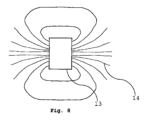
図8は、要素1を推進するために使用されてもよい磁気要素14を示す。ここでは、それは、磁界14を生成するために選択的にオンおよびオフにすることができる電磁石を含む。勿論、永久磁石を用いてもよい。インペラ、プロペラ、または別の推進手段を動作させるために電気エネルギーを使用することも考えられる。
Figure 8 shows a
Claims (18)
-血管または心臓(V)に配置される要素(1)と、
-前記要素に作用する推進力(2)を判断するための手段と、
-前記要素(1)の加速度(3)および速度(4)の少なくとも1つ、好ましくは加速度(3)および速度(4)を判断するための手段と、
-前記推進力(2)と、前記要素(1)の加速度(3)および速度(4)のうちの少なくとも1つ、好ましくは加速度(3)および速度(4)とに基づいて、前記要素(1)の近隣媒体の少なくとも1つの特性を判断するための手段とを備える、システム。 1. A system for determining a characteristic in a blood vessel or heart (V) of a patient, comprising:
an element (1) placed in a blood vessel or in the heart (V),
- means for determining the propulsive forces (2) acting on said element;
- means for determining at least one of the acceleration (3) and the velocity (4) of said element (1), preferably the acceleration (3) and the velocity (4);
- means for determining at least one characteristic of a medium in the vicinity of said element (1) based on said propulsion force (2) and on at least one of the acceleration (3) and the velocity (4) of said element (1), preferably on the acceleration (3) and the velocity (4).
-血管または心臓(V)に配置される要素(1)と、
-前記要素に作用する推進力(2)を判断するための手段と、
-前記要素(1)の加速度(3)および速度(4)の少なくとも1つを判断するための手段と、
-前記推進力(2)と、前記要素(1)の加速度(3)および速度(4)のうちの少なくとも1つとに基づいて、前記要素(1)の近隣媒体の少なくとも1つの特性を判断するための手段とを備え、前記システムは、
-血管または心臓(V)に配置される前記要素(1)に作用する推進力(2)を判断するステップと、
-前記要素(1)の加速度(3)および速度(4)の少なくとも1つを判断するステップと、
-前記推進力(2)と、前記要素(1)の加速度(3)および速度(4)のうちの少なくとも1つとに基づいて、前記要素(1)の近隣媒体の少なくとも1つの特性を判断するステップとを実行する、方法。 1. A method of operating a system for determining a characteristic of a blood vessel or heart (V) of a patient, the system comprising:
an element (1) placed in a blood vessel or in the heart (V),
- means for determining the propulsive forces (2) acting on said element;
- means for determining at least one of the acceleration (3) and the velocity (4) of said element (1);
means for determining at least one characteristic of a medium in the vicinity of said element (1) based on said momentum (2) and on at least one of the acceleration (3) and the velocity (4) of said element (1), said system comprising:
- determining the driving forces (2) acting on said element (1) placed in a blood vessel or in the heart (V) ;
- determining at least one of the acceleration (3) and the velocity (4) of said element (1);
- determining at least one characteristic of a medium in the vicinity of said element (1) based on said momentum (2) and at least one of the acceleration (3) and the velocity (4) of said element (1).
Applications Claiming Priority (3)
| Application Number | Priority Date | Filing Date | Title |
|---|---|---|---|
| EP19315100.8 | 2019-08-21 | ||
| EP19315100.8A EP3782543A1 (en) | 2019-08-21 | 2019-08-21 | Method and system for determining properties in a vessel |
| PCT/EP2020/073457 WO2021032869A1 (en) | 2019-08-21 | 2020-08-21 | Method and system for determining properties in a vessel |
Publications (3)
| Publication Number | Publication Date |
|---|---|
| JP2022545232A JP2022545232A (en) | 2022-10-26 |
| JPWO2021032869A5 JPWO2021032869A5 (en) | 2023-07-04 |
| JP7581330B2 true JP7581330B2 (en) | 2024-11-12 |
Family
ID=67953725
Family Applications (1)
| Application Number | Title | Priority Date | Filing Date |
|---|---|---|---|
| JP2022511015A Active JP7581330B2 (en) | 2019-08-21 | 2020-08-21 | Method and system for determining intravascular properties - Patents.com |
Country Status (11)
| Country | Link |
|---|---|
| US (1) | US12257040B2 (en) |
| EP (2) | EP3782543A1 (en) |
| JP (1) | JP7581330B2 (en) |
| KR (1) | KR20220052337A (en) |
| CN (1) | CN114222540B (en) |
| AU (1) | AU2020333920A1 (en) |
| BR (1) | BR112022000947A2 (en) |
| CA (1) | CA3147453A1 (en) |
| IL (1) | IL290611A (en) |
| MX (1) | MX2022002065A (en) |
| WO (1) | WO2021032869A1 (en) |
Families Citing this family (3)
| Publication number | Priority date | Publication date | Assignee | Title |
|---|---|---|---|---|
| EP4124312A1 (en) * | 2021-07-26 | 2023-02-01 | Artedrone | System and method for moving a medical device for treating or diagnosing a patient |
| TW202241358A (en) * | 2021-01-21 | 2022-11-01 | 法商亞特德隆公司 | System and method for moving a medical device for treating or diagnosing a patient |
| CN115154847A (en) * | 2022-08-12 | 2022-10-11 | 深圳市爱博医疗机器人有限公司 | Operation device and force measuring method for slender medical instrument |
Citations (4)
| Publication number | Priority date | Publication date | Assignee | Title |
|---|---|---|---|---|
| US20060152309A1 (en) | 2005-01-11 | 2006-07-13 | Mintchev Martin P | Magnetic levitation of intraluminal microelectronic capsule |
| WO2013172312A1 (en) | 2012-05-14 | 2013-11-21 | オリンパスメディカルシステムズ株式会社 | Capsule therapy device and therapy system |
| JP2014000431A (en) | 2006-08-11 | 2014-01-09 | Koninklijke Philips Nv | Ultrasonic system for cerebral blood flow imaging and microbubble-enhanced blood clot lysis |
| US20140253114A1 (en) | 2013-02-26 | 2014-09-11 | Mir Behrad KHAMESEE | System and method for providing force information in a magnetic field environment |
Family Cites Families (10)
| Publication number | Priority date | Publication date | Assignee | Title |
|---|---|---|---|---|
| RU2061216C1 (en) * | 1994-03-05 | 1996-05-27 | Ивановский Инженерно-Строительный Институт | Method of measurement of viscosity of liquid |
| US5419325A (en) * | 1994-06-23 | 1995-05-30 | General Electric Company | Magnetic resonance (MR) angiography using a faraday catheter |
| US5833603A (en) * | 1996-03-13 | 1998-11-10 | Lipomatrix, Inc. | Implantable biosensing transponder |
| US7962194B2 (en) * | 2003-04-15 | 2011-06-14 | Polyvalor, Limited Partnership | Method and system for propelling and controlling displacement of a microrobot in a blood vessel |
| US20070244520A1 (en) | 2004-04-19 | 2007-10-18 | Searete Llc | Lumen-traveling biological interface device and method of use |
| US20120035434A1 (en) * | 2006-04-12 | 2012-02-09 | Searete Llc, A Limited Liability Corporation Of The State Of Delaware | Control of a lumen traveling device in a body tube tree |
| KR101272156B1 (en) * | 2011-08-31 | 2013-06-05 | 전남대학교산학협력단 | A Micro-Robot System For Intravascular Therapy And Controling Method Thereof |
| JP5020403B1 (en) * | 2011-11-28 | 2012-09-05 | リオン株式会社 | Vibration type physical property measuring apparatus and method |
| US20170119235A1 (en) * | 2015-10-29 | 2017-05-04 | Elwha Llc | Lumen traveling device |
| US20190298311A1 (en) * | 2016-10-07 | 2019-10-03 | Koninklijke Philips N.V. | Intravascular flow determination |
-
2019
- 2019-08-21 EP EP19315100.8A patent/EP3782543A1/en not_active Withdrawn
-
2020
- 2020-08-21 KR KR1020227009008A patent/KR20220052337A/en not_active Withdrawn
- 2020-08-21 JP JP2022511015A patent/JP7581330B2/en active Active
- 2020-08-21 AU AU2020333920A patent/AU2020333920A1/en not_active Abandoned
- 2020-08-21 US US17/636,669 patent/US12257040B2/en active Active
- 2020-08-21 CA CA3147453A patent/CA3147453A1/en not_active Abandoned
- 2020-08-21 WO PCT/EP2020/073457 patent/WO2021032869A1/en not_active Ceased
- 2020-08-21 MX MX2022002065A patent/MX2022002065A/en unknown
- 2020-08-21 CN CN202080056972.6A patent/CN114222540B/en active Active
- 2020-08-21 EP EP20757912.9A patent/EP4017347A1/en active Pending
- 2020-08-21 BR BR112022000947A patent/BR112022000947A2/en not_active IP Right Cessation
-
2022
- 2022-02-14 IL IL290611A patent/IL290611A/en unknown
Patent Citations (4)
| Publication number | Priority date | Publication date | Assignee | Title |
|---|---|---|---|---|
| US20060152309A1 (en) | 2005-01-11 | 2006-07-13 | Mintchev Martin P | Magnetic levitation of intraluminal microelectronic capsule |
| JP2014000431A (en) | 2006-08-11 | 2014-01-09 | Koninklijke Philips Nv | Ultrasonic system for cerebral blood flow imaging and microbubble-enhanced blood clot lysis |
| WO2013172312A1 (en) | 2012-05-14 | 2013-11-21 | オリンパスメディカルシステムズ株式会社 | Capsule therapy device and therapy system |
| US20140253114A1 (en) | 2013-02-26 | 2014-09-11 | Mir Behrad KHAMESEE | System and method for providing force information in a magnetic field environment |
Also Published As
| Publication number | Publication date |
|---|---|
| JP2022545232A (en) | 2022-10-26 |
| BR112022000947A2 (en) | 2022-06-14 |
| WO2021032869A1 (en) | 2021-02-25 |
| CA3147453A1 (en) | 2021-02-25 |
| US12257040B2 (en) | 2025-03-25 |
| AU2020333920A1 (en) | 2022-03-03 |
| CN114222540B (en) | 2025-11-07 |
| KR20220052337A (en) | 2022-04-27 |
| IL290611A (en) | 2022-04-01 |
| US20220280050A1 (en) | 2022-09-08 |
| EP4017347A1 (en) | 2022-06-29 |
| CN114222540A (en) | 2022-03-22 |
| EP3782543A1 (en) | 2021-02-24 |
| MX2022002065A (en) | 2022-03-17 |
Similar Documents
| Publication | Publication Date | Title |
|---|---|---|
| US11786318B2 (en) | Intelligent real-time tool and anatomy visualization in 3D imaging workflows for interventional procedures | |
| Heunis et al. | Flexible instruments for endovascular interventions: Improved magnetic steering, actuation, and image-guided surgical instruments | |
| JP6129750B2 (en) | Non-rigid morphing of blood vessel images using the shape of the device in the blood vessel | |
| JP7581330B2 (en) | Method and system for determining intravascular properties - Patents.com | |
| EP2744391B1 (en) | Shape sensing assisted medical procedure | |
| Yang et al. | Ultrasound-guided catheterization using a driller-tipped guidewire with combined magnetic navigation and drilling motion | |
| US20090118620A1 (en) | System and method for tracking an ultrasound catheter | |
| US12016652B2 (en) | System and method for real-time creation of cardiac electro-physiology signals in the heart | |
| US20100063384A1 (en) | Local intra-body delivery system | |
| Kagadis et al. | Emerging technologies for image guidance and device navigation in interventional radiology | |
| CN105517489B (en) | Navigation system | |
| CN107432766A (en) | A kind of accurate minimally invasive operation navigating system | |
| EP3206620B1 (en) | System for planning the introduction of a needle in a patient's body | |
| CN112236099A (en) | Systems and methods for implementing and evaluating programs | |
| HK40071782B (en) | Method and system for determining properties in a vessel | |
| HK40071782A (en) | Method and system for determining properties in a vessel | |
| JPWO2021032869A5 (en) | ||
| Fullerton et al. | The influence of stents on the performance of an ultrasonic navigation system for endovascular procedures |
Legal Events
| Date | Code | Title | Description |
|---|---|---|---|
| A521 | Request for written amendment filed |
Free format text: JAPANESE INTERMEDIATE CODE: A523 Effective date: 20230626 |
|
| A621 | Written request for application examination |
Free format text: JAPANESE INTERMEDIATE CODE: A621 Effective date: 20230626 |
|
| A977 | Report on retrieval |
Free format text: JAPANESE INTERMEDIATE CODE: A971007 Effective date: 20240207 |
|
| A131 | Notification of reasons for refusal |
Free format text: JAPANESE INTERMEDIATE CODE: A131 Effective date: 20240220 |
|
| A601 | Written request for extension of time |
Free format text: JAPANESE INTERMEDIATE CODE: A601 Effective date: 20240517 |
|
| A521 | Request for written amendment filed |
Free format text: JAPANESE INTERMEDIATE CODE: A523 Effective date: 20240627 |
|
| TRDD | Decision of grant or rejection written | ||
| A01 | Written decision to grant a patent or to grant a registration (utility model) |
Free format text: JAPANESE INTERMEDIATE CODE: A01 Effective date: 20240903 |
|
| A601 | Written request for extension of time |
Free format text: JAPANESE INTERMEDIATE CODE: A601 Effective date: 20241003 |
|
| A61 | First payment of annual fees (during grant procedure) |
Free format text: JAPANESE INTERMEDIATE CODE: A61 Effective date: 20241030 |
|
| R150 | Certificate of patent or registration of utility model |
Ref document number: 7581330 Country of ref document: JP Free format text: JAPANESE INTERMEDIATE CODE: R150 |