JP7522397B2 - Long-wavelength luminescent substrate for bioluminescent enzymes - Google Patents
Long-wavelength luminescent substrate for bioluminescent enzymes Download PDFInfo
- Publication number
- JP7522397B2 JP7522397B2 JP2020040703A JP2020040703A JP7522397B2 JP 7522397 B2 JP7522397 B2 JP 7522397B2 JP 2020040703 A JP2020040703 A JP 2020040703A JP 2020040703 A JP2020040703 A JP 2020040703A JP 7522397 B2 JP7522397 B2 JP 7522397B2
- Authority
- JP
- Japan
- Prior art keywords
- luminescence
- luminescent
- substrate
- enzyme
- bioluminescent
- Prior art date
- Legal status (The legal status is an assumption and is not a legal conclusion. Google has not performed a legal analysis and makes no representation as to the accuracy of the status listed.)
- Active
Links
Images
Landscapes
- Measuring Or Testing Involving Enzymes Or Micro-Organisms (AREA)
- Micro-Organisms Or Cultivation Processes Thereof (AREA)
- Nitrogen Condensed Heterocyclic Rings (AREA)
Description
特許法第30条第2項適用 発行日 2019年6月27日 刊行物名 第43回有機電子移動化学討論会 講演要旨集 Article 30,
特許法第30条第2項適用 開催日 2019年6月27日~2019年6月28日(公開日は2019年6月27日) 集会名、開催場所 第43回有機電子移動化学討論会 横浜国立大学教育文化ホール(横浜市保土ヶ谷区常磐台79-1)Article 30,
特許法第30条第2項適用 発行日 2019年10月5日 刊行物名 生物発光化学発光研究会第35回学術講演会 講演要旨集 Article 30,
特許法第30条第2項適用 開催日 2019年10月5日 集会名、開催場所 生物発光化学発光研究会第35回学術講演会 国立研究開発法人 産業技術総合研究所 臨海副都心センター(東京都江東区青梅2-4-7)Article 30,
特許法第30条第2項適用 発行日 2019年11月20日 刊行物名 第42回日本分子生物学会年会 講演要旨集 Article 30,
特許法第30条第2項適用 開催日 2019年12月3日~2019年12月6日(公開日は2019年12月3日) 集会名、開催場所 第42回日本分子生物学会年会 福岡国際会議場・福岡サンパレスホテル&ホール・マリンメッセ福岡 Article 30,
特許法第30条第2項適用 発行日 2020年3月5日 刊行物名 日本化学会第100春季年会 講演予稿集Article 30,
本発明は、生物発光酵素の発光基質の新規合成と長波長発光に関するものである。 The present invention relates to the novel synthesis of luminescent substrates for bioluminescent enzymes and their long-wavelength luminescence.
生体イメージングやバイオアッセイなどを可能とする核心技術は発光標識であり、発光標識の安定性や発光特性は、イメージングやバイオアッセイの信頼性の要である。例えば、レポータージーンアッセイ(reporter-gene assay)、ツーハイブリットアッセイ(two-hybrid assay)、酵素結合免疫吸着法(enzyme-linked immunoSorbent assay)、放射免疫アッセイ(ラジオイムノアッセイ、RIA)等のバイオアッセイにおいて、発光標識は信号を発信する核心的な役割を果たす(非特許文献1、2)。 The core technology that enables biological imaging and bioassays is luminescent labels, and the stability and luminescence properties of luminescent labels are essential for the reliability of imaging and bioassays. For example, in bioassays such as reporter-gene assays, two-hybrid assays, enzyme-linked immunosorbent assays, and radioimmunoassays (RIA), luminescent labels play a key role in transmitting signals (Non-Patent Documents 1 and 2).
その他のバイオアッセイとして、通常「発光イメージングプローブ」を用いるアッセイ法が広く使われている。例えば、細胞内での蛋白質-蛋白質相互作用(PPI)を発光イメージングする方法が開発されており、非特許文献3等のように蛍光蛋白質間蛍光共鳴エネルギー転移を利用したフレート法(FRET)、非特許文献4等のように蛍光蛋白質や発光蛋白質を2分割し、その蛋白質断片間の再結合による発光回復を特徴とする二分子型自己相補法(2-molecule-format protein-fragment complementation assay(PCA))等が知られている。 As other bioassays, assay methods that usually use "luminescence imaging probes" are widely used. For example, methods have been developed for luminescence imaging of protein-protein interactions (PPI) within cells, and known methods include the FRET method, which uses fluorescence resonance energy transfer between fluorescent proteins (see Non-Patent Document 3, etc.), and the 2-molecule-format protein-fragment complementation assay (PCA), which is characterized by dividing a fluorescent protein or luminescent protein into two and restoring luminescence by recombination between the protein fragments (see Non-Patent Document 4, etc.).
本発明者らは、蛋白質の自発的な組継ぎ反応を用いたアッセイ法(protein splicing assay (PSA))を開発し(非特許文献5)、さらに、一分子型生物発光プローブ(integrated-molecule-format bioluminescent probe;又は簡単にsingle-chain probe)のような単一分子内で蛋白質-蛋白質間の相互作用を簡便に検出する手法も開発した(特許文献1、非特許文献3)。更にその派生手法として、発光酵素の遺伝子配列を円順列置換(circular permutation)したことを特徴とする一分子型生物発光プローブも開発した(特許文献2、非特許文献4)。他にも生物発光酵素23(ALuc(登録商標)23)に歪みをかけると発光輝度が上昇する現象を利用して分子歪みセンサー(molecular tension-indexed bioluminescent probe)という新概念の発光プローブを開発した(特許文献3、非特許文献5)。最近では、レポータージーンアッセイと一分子型生物発光プローブを合体した多重リガンド認識型生物発光プローブ(multiple recognition-type bioluminescent probe)を開発し、このプローブは、1つの検量物質に対して2回センシングすることを特徴とする(非特許文献6)。更に、2色の一分子型生物発光プローブを組み合わせることによって「マルチカラー生物発光イメージングプローブセット」を開発した(特許文献4)。このプローブは、検体の多面的な生理活性の多色イメージングできることを特徴としている。
The present inventors have developed an assay method (protein splicing assay (PSA)) that uses spontaneous protein splicing reactions (Non-Patent Document 5), and have also developed a method for easily detecting protein-protein interactions within a single molecule, such as an integrated-molecule-format bioluminescent probe (or simply a single-chain probe) (Patent Document 1, Non-Patent Document 3). As a derivative method, they have also developed an integrated-molecule-format bioluminescent probe characterized by circular permutation of the gene sequence of a luminescent enzyme (
前述したバイオアッセイにおいて、発光標識は検出信号を発信する、最も肝心な要素であり、バイオアッセイの性能は発光標識の性能に左右されると言っても過言ではない。従来、発光標識として使われてきたのは、蛍光蛋白質(fluorescent protein)、生物発光酵素(luciferase)、horseradish peroxidase (HRP)、Alkaline Phosphatase (AP)、b-galactosidase、蛍光色素、放射線同位元素などである。 In the bioassay described above, the luminescent label is the most important element that emits the detection signal, and it is no exaggeration to say that the performance of the bioassay depends on the performance of the luminescent label. Conventionally, luminescent labels have been used such as fluorescent proteins, bioluminescent enzymes (luciferase), horseradish peroxidase (HRP), alkaline phosphatase (AP), b-galactosidase, fluorescent dyes, and radioisotopes.
これらの発光標識は、実用例から主に2つに分類できる:即ち、光の励起により発生する発光(Photoluminescenceまたはfluorescence、蛍光)と、化学エネルギーにより励起されることを特徴とする発光(chemiluminescence、化学発光)。化学発光の内、生体由来の発光酵素の触媒反応による発光現象を生物発光と称する。 These luminescent labels can be divided into two main categories based on their practical applications: luminescence generated by excitation with light (photoluminescence or fluorescence), and luminescence characterized by excitation by chemical energy (chemiluminescence). Of the chemiluminescence types, the luminescence phenomenon caused by the catalytic reaction of a luminescent enzyme derived from a living organism is called bioluminescence.
化学発光は必ず基質を必要とする。一般的に生物発光酵素などにより基質の酸化が触媒され、基質が持つ本来の化学エネルギーより励起された電子エネルギーが基底状態に戻る際に発光を放つ。このため、蛍光蛋白質は蛍光団を内部に有するが、生物発光酵素の発光団は外部の基質に依存する。従って基質の化学構造を制御することにより、発光輝度と多色発光ができると思い至った。 Chemiluminescence always requires a substrate. Generally, the oxidation of the substrate is catalyzed by a bioluminescent enzyme, and light is emitted when the electronic energy excited from the substrate's original chemical energy returns to the ground state. For this reason, fluorescent proteins have fluorophores inside, but the luminophores of bioluminescent enzymes depend on external substrates. Therefore, it was thought that luminescence brightness and multicolor emission could be achieved by controlling the chemical structure of the substrate.
前述した概念の下で、本発明者の以前の研究を再度レビューした(非特許文献7)。即ち、発光酵素と発光基質間の結合モデルを検討した。図1で示したように生物発光酵素の場合、発光基質(赤線)が酵素活性部位に対してC2番位置(R-A)より挿入されることを示唆している。一方、C6番位置(R-C)は外側向きになっていることが分かる。このシミュレーションは、生物発光酵素に結合する発光基質のC6番位置(R-C)は外向きであるため、C6番位置(R-C)の官能基の置換は立体障害が少ないことを示唆している。 Based on the above-mentioned concept, the inventor's previous research was reviewed again (Non-Patent Document 7). That is, a binding model between a luminescent enzyme and a luminescent substrate was examined. As shown in Figure 1, in the case of a bioluminescent enzyme, it is suggested that the luminescent substrate (red line) is inserted into the enzyme active site from the C2 position (R-A). On the other hand, it can be seen that the C6 position (R-C) faces outward. This simulation suggests that since the C6 position (R-C) of the luminescent substrate that binds to a bioluminescent enzyme faces outward, the substitution of the functional group at the C6 position (R-C) has little steric hindrance.
前述した見解は、逆にいうとC2番位置(R-A)に官能基を替えた場合には、立体障害を受けやすいことを示すものでもある。実際に、図2で示したようにC2番位置(R-A)の官能基を替えた場合、厳格に官能基のサイズに依存することが図2と図3のデータより明らかになった(非特許文献7)。 The above-mentioned view also indicates that changing the functional group at the C2 position (R-A) is likely to cause steric hindrance. In fact, as shown in Figure 2, when changing the functional group at the C2 position (R-A), the data in Figures 2 and 3 make it clear that it strictly depends on the size of the functional group (Non-Patent Document 7).
また、海洋生物由来の生物発光酵素と発光基質間の結合モデルに関する別の研究例によると、ウミシイタケ生物発光酵素(RLuc)がその基質と結合する際、C6番から入った方が熱力学的に安定的だという見解が示された(非特許文献8、図4)。 In addition, another study on a binding model between a marine bioluminescent enzyme and a luminescent substrate showed that when Renilla reniformis bioluminescent enzyme (RLuc) binds to its substrate, it is thermodynamically more stable when the binding occurs from C6 (Non-Patent Document 8, Figure 4).
この研究例は、RLucの場合、むしろ基質のC6位の官能基の化学構造が重要であることを示唆するものである。 This research example suggests that in the case of RLuc, the chemical structure of the functional group at the C6 position of the substrate is rather important.
本発明者はこれらの見解の基に、生物発光酵素またはRLucに特異的に反応する発光基質を開発したことがあった(非特許文献9)。この研究の代表例は、図5のように説明できる。即ち、複数の発光酵素が同一反応場に共存している中、それぞれ別の基質に特異的であるため、どの基質を投入するかによって、どちらかの1つの発光酵素のみが反応する仕組みである。 Based on these findings, the present inventor has previously developed a luminescent substrate that reacts specifically with bioluminescent enzymes or RLuc (Non-Patent Document 9). A representative example of this research can be explained as shown in Figure 5. In other words, while multiple luminescent enzymes coexist in the same reaction field, each is specific to a different substrate, so depending on which substrate is added, only one of the luminescent enzymes will react.
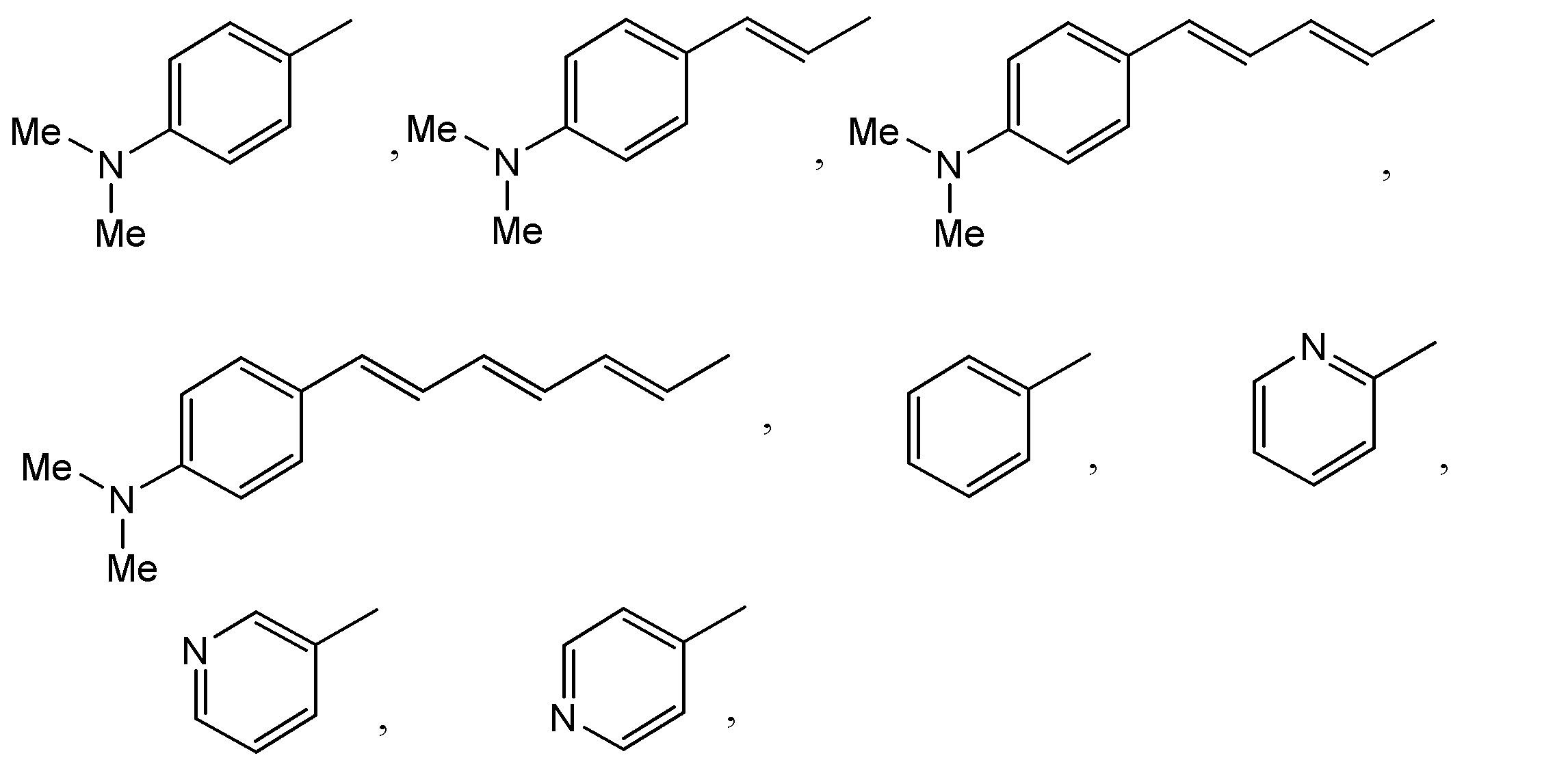
しかしながら、前述した何れの方法でも、発光色の多色化には至らなかった。生物発光の多色化における伝統的なアプローチとしては、発光基質に蛍光色素を繋げる方法であった。その長波長発光原理は、まず発光基質が短波長発光を放つとその共鳴エネルギーが隣接した蛍光色素に移るため、蛍光色素が長波長発光を放つことができる。このような現象を生物発光共鳴エネルギー移動(BRET)という。その典型的な例が、図6に示したようなものである(非特許文献10)。 However, none of the above methods have been able to produce multiple luminescent colors. The traditional approach to producing multiple colors in bioluminescence is to connect a fluorescent dye to a luminescent substrate. The principle of long-wavelength luminescence is that the luminescent substrate first emits short-wavelength luminescence, and the resonance energy is transferred to the adjacent fluorescent dye, allowing the fluorescent dye to emit long-wavelength luminescence. This phenomenon is called bioluminescence resonance energy transfer (BRET). A typical example is shown in Figure 6 (Non-Patent Document 10).
しかしこのような方法は、煩雑な基質合成工程が必要であり、発光エネルギーが蛍光色素に移る効率も悪いため、発光損失により輝度が非常に低いことが問題点であった。また、蛍光色素付き基質は、溶解度が悪く、比較的に大きい分子であるため細胞膜透過性が悪い。 However, this method requires a complicated substrate synthesis process, and the efficiency of transferring luminescence energy to the fluorescent dye is low, resulting in very low brightness due to luminescence loss. In addition, the fluorescent dye-attached substrate has poor solubility and is a relatively large molecule, so it has poor cell membrane permeability.
本発明では、発光色の乏しい海洋生物由来生物発光酵素の発光性能(多色性、発光輝度、発光安定性など)を改善するために、(1)発光基質の創製と(2)新規発光基質と好適に反応する発光酵素の選択とそれに伴う発光反応条件の最適化、(3)それに付随するバイオアッセイ技術の観点から研究課題を解決する必要がある。 In the present invention, in order to improve the luminescence performance (multi-color, luminescence brightness, luminescence stability, etc.) of bioluminescent enzymes derived from marine organisms that have poor luminescence color, it is necessary to (1) create a luminescent substrate, (2) select a luminescent enzyme that reacts favorably with the new luminescent substrate and the associated optimization of luminescence reaction conditions, and (3) solve research problems from the perspective of the associated bioassay technology.
まず「(1)発光基質の創製」においては、前述した基質と海洋生物発光酵素間の結合モデルに基づいて基質の官能基を変える必要があり、より具体的には海洋生物由来生物発光酵素の共通基質(セレンテラジン)のC6位の官能基を長波長シフトが見込まれるπ結合の長い官能基に変える必要がある。同様にC2位の官能基においては、ヒドロキシ基の調整による結合能力の検討が必要である。さらにC6位をその他の官能基、例えばピリジン基に替えC2位のフェノール基をベンゼングループに置換することにより、各生物発光酵素との選択性などを極める必要がある。「(2)新規発光基質と好適に反応する発光酵素の選択とそれに伴う発光反応条件の最適化」において、発光酵素の選択は、発光輝度と発光色が決まる上で重要な要素であり、様々な選択肢の中で、とりわけ海洋生物由来の発光酵素に焦点を当て、今回開発した発光基質との反応による発光特性(輝度や色)を最適化する必要がある。また「(3)それの付随するバイオアッセイ技術」についても綿密な検討が必要である。反応溶液を最適化することにより、高い発光性能を導くことができ、適切なバイオアッセイと組み合わせることにより、本発明の良さを確実なものにすることができる。 First, in "(1) Creation of a luminescent substrate," it is necessary to change the functional group of the substrate based on the binding model between the substrate and marine bioluminescent enzymes described above. More specifically, it is necessary to change the functional group at C6 of the common substrate (coelenterazine) of marine bioluminescent enzymes to a functional group with a long π bond that is expected to shift the wavelength to a longer wavelength. Similarly, for the functional group at C2, it is necessary to consider the binding ability by adjusting the hydroxyl group. Furthermore, it is necessary to maximize the selectivity with each bioluminescent enzyme by replacing the C6 position with another functional group, such as a pyridine group, and the phenol group at C2 with a benzene group. In "(2) Selection of a luminescent enzyme that reacts favorably with a new luminescent substrate and the associated optimization of the luminescent reaction conditions," the selection of the luminescent enzyme is an important factor in determining the luminescence brightness and color. Among the various options, it is necessary to focus on the luminescent enzyme derived from marine organisms and optimize the luminescence characteristics (brightness and color) of the reaction with the luminescent substrate developed this time. In addition, "(3) The associated bioassay technology" also requires careful consideration. By optimizing the reaction solution, high luminescence performance can be achieved, and by combining it with an appropriate bioassay, the merits of the present invention can be ensured.
当該基質は、海洋生物由来の発光酵素と反応して発光することを特徴とする小分子化学物質であり、様々なバイオアッセイにおける発光標識として利用できる。バイオアッセイは、その発光方式によって、(1)BRETのように蛍光色素(または蛍光蛋白質)が絡む方式と、(2)純粋に化学発光(生物発光を含む)に頼る手法の2種類に大別される。(1)BRET方式の場合、短波長の発光エネルギーが長波長側に移るため、組織透過性の良い発光信号が放される反面、標識の性能がエネルギー移動効率に依存するが一般的に効率が低い問題点もある。またBRET現象が起こる前と後のスペクトルの変化が大きくないため、光学フィルターを必要とするなど、分析装置が煩雑になる恐れがある。 The substrate is a small molecule chemical that reacts with a marine organism-derived luminescent enzyme to emit light, and can be used as a luminescent label in various bioassays. Bioassays can be broadly divided into two types according to the luminescence method: (1) methods involving fluorescent dyes (or fluorescent proteins) such as BRET, and (2) methods that rely purely on chemiluminescence (including bioluminescence). In the case of the (1) BRET method, the short-wavelength luminescence energy is transferred to the long-wavelength side, so a luminescence signal with good tissue penetration is emitted, but there is also the problem that the performance of the label depends on the energy transfer efficiency, which is generally low. In addition, since the change in the spectrum before and after the BRET phenomenon is not large, there is a risk that the analytical equipment will become complicated, such as requiring an optical filter.
(1)と(2)のいずれの方式においても、エネルギー源は発光であるが故に起こる様々な課題もあった。例えば、化学発光の一種である生物発光の場合、「生物発光酵素」に依存するため、塩濃度、温度、pH、重金属イオンの濃度等によっても発光強度が容易に変動する問題がある。また、化学発光は基質導入後、直ちに輝度を落としてしまう場合が多い(発光安定性が悪い)ため、多数のサンプルを計測する場合に最初のサンプルと最後のサンプルの測定に時差が生じ、測定の信頼性が落ちる問題点があった。他にも、生体由来の体液(例えば、唾液、血清、汗、尿)環境下でバイオアッセイをした場合、体液による光の吸収問題、発光反応そのものへの妨害効果などが問題であった。 In both methods (1) and (2), various issues arise because the energy source is luminescence. For example, in the case of bioluminescence, which is a type of chemiluminescence, there is a problem that the luminescence intensity can easily vary depending on salt concentration, temperature, pH, and the concentration of heavy metal ions because it depends on "bioluminescent enzymes." In addition, since chemiluminescence often loses brightness immediately after the introduction of the substrate (poor luminescence stability), there is a time lag between the measurement of the first and last samples when measuring multiple samples, which reduces the reliability of the measurement. Other problems include light absorption by the body fluids and the interference effect on the luminescence reaction itself when bioassays are performed in an environment of body fluids derived from living organisms (e.g., saliva, serum, sweat, urine).
これらの背景から、新規発光システムにおいては、まず、体液中の生理物質による発光吸収現象や妨害効果をなくしつつ発光安定性の優れたシステムの開発が課題であった。例えば、発光信号が長波長へシフトした発光であれば、体液中の様々な発光吸収物質の妨害効果を回避し(この長波長領域をオプティカルウィンドウと言う)、同時に発光反応溶液など、発光条件を最適化することにより、従来にない信頼性と輝度を持つ発光システムを開発できると思い至った。その具体的な手法としては、長波長発光する発光基質の新規開発とそれをサポートする発光反応条件の最適化である。 Given this background, the first challenge for a new luminescence system was to develop a system with excellent luminescence stability while eliminating the luminescence absorption phenomenon and interfering effects caused by physiological substances in body fluids. For example, if the luminescence signal is shifted to longer wavelengths, it was thought that by avoiding the interfering effects of various luminescence absorbing substances in body fluids (this long-wavelength region is called the optical window) and at the same time optimizing the luminescence conditions such as the luminescence reaction solution, it would be possible to develop a luminescence system with unprecedented reliability and brightness. The specific method for achieving this is the new development of a luminescence substrate that emits light at long wavelengths and the optimization of the luminescence reaction conditions that support it.
新規発光基質の創製:
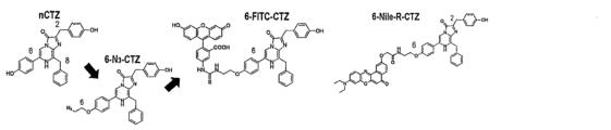
発光基質の創製においては、これまでの本発明者らの研究成果より海洋生物由来生物発光酵素の共通基質(セレンテラジン)の場合、C6番位置の官能基が発光性能に重要であるという考えの基で、C6番位置の官能基を調整した。まず本来基質のC6位に位置するフェノール基(Ph-OH)においてそのヒドロキシ基(OH基)をジメチルアミン基に替える試みをした。またC6番位置のフェノール基とイミダゾピラジノン骨格との間に2重結合を設けることにより、π結合を伸ばし長波長へのシフト効果を試みた。更に基質(セレンテラジン)のC2位のフェノール基をベンゼングループに置換する試みをした。本発明では、C2位がフェノール基を持つ場合を「1グループ」にし、ベンゼングループに置換した場合を「2グループ」に分類した。一方、C6位にピリジン基に替え、さらにC2位のフェノール基をベンゼングループに置換した場合を「3グループ」に分類した。
Creation of novel luminescent substrates:
In creating the luminescent substrate, the functional group at the C6 position was adjusted based on the idea that the functional group at the C6 position is important for luminescence performance in the case of the common substrate (coelenterazine) of marine organism-derived bioluminescent enzymes based on the results of the inventors' research to date. First, an attempt was made to replace the hydroxyl group (OH group) in the phenol group (Ph-OH) originally located at the C6 position of the substrate with a dimethylamine group. In addition, an attempt was made to extend the π bond and shift the wavelength to a longer wavelength by providing a double bond between the phenol group at the C6 position and the imidazopyrazinone skeleton. Furthermore, an attempt was made to replace the phenol group at the C2 position of the substrate (coelenterazine) with a benzene group. In the present invention, the case where the C2 position has a phenol group was classified as "Group 1," and the case where the phenol group was replaced with a benzene group was classified as "
新規発光基質の最適発光酵素の検討:
新規発光基質において、事前に生物発光酵素との結合モデルについて検討したとしても、実際に合成された基質が、思いもかけない新しい発光特性を持つ可能性がある。したがって、新規発光基質においては、できるだけ多くの発光酵素と組み合わせて発光性能を検討する必要がある。例えば、新規発光基質が特定発光酵素に選択的に発光する可能性があった場合、マルチカラーイメージングシステムを構成できる技術的な難関を克服したことになる。また、発光酵素との組み合わせにより、思いがけないレッドシフトした発光色を放つ可能性があり、発光スペクトルに関する綿密な評価も必要である。他に発光持続性や安定性も発光酵素との相性によって決まる側面があり、新規発光基質がどの発光酵素と最も合うかを綿密に評価する必要がある。
Examination of optimal luminescent enzyme for new luminescent substrate:
Even if a binding model of a new luminescent substrate with a bioluminescent enzyme is examined in advance, the actually synthesized substrate may have new and unexpected luminescence properties. Therefore, it is necessary to examine the luminescence performance of a new luminescent substrate in combination with as many luminescent enzymes as possible. For example, if a new luminescent substrate has the potential to selectively emit light for a specific luminescent enzyme, the technical difficulty of constructing a multicolor imaging system will be overcome. In addition, the combination with a luminescent enzyme may produce an unexpected red-shifted luminescence color, so a thorough evaluation of the luminescence spectrum is also necessary. In addition, the luminescence duration and stability are also aspects that are determined by the compatibility with the luminescent enzyme, so it is necessary to thoroughly evaluate which luminescent enzyme the new luminescent substrate is most compatible with.
最適反応条件とバイオアッセイの検討:
また、前述した新規発光基質とそれに最適な発光酵素が見つかった場合においても、その反応に影響する最適反応条件とそのバイオアッセイの検討が必要である。一般的にこれらの発光反応は酵素反応であるため、pHや温度、塩度など様々な要因により発光輝度と発光色、発光安定性が決まる。そのため、これらの要素に関する検討が必要である。
Optimal reaction conditions and bioassay:
Even if the aforementioned new luminescent substrate and the optimal luminescent enzyme are found, it is necessary to consider the optimal reaction conditions that affect the reaction and the bioassay. Generally, these luminescent reactions are enzyme reactions, so the luminescence brightness, luminescence color, and luminescence stability are determined by various factors such as pH, temperature, and salinity. Therefore, it is necessary to consider these factors.
また、これらの最適条件を実用的なバイオアッセイに適用するためには、これまで知られている代表的なバイオアッセイ系においてその性能を検証する必要がある。それによって、新規発光基質と発光酵素の組み合わせによる発光システムの有用性を確証できる。 Furthermore, in order to apply these optimal conditions to practical bioassays, it is necessary to verify their performance in representative bioassay systems known to date. This will enable us to confirm the usefulness of the luminescence system that combines the new luminescent substrate and luminescent enzyme.
本発明は、以下の項1~項5に示される
項1. 下記式(I):
The present invention relates to a compound according to the following items 1 to 5:

項2.
R1はヒドロキシ基で置換されていてもよいベンジル基である、項1に記載のイミダゾピラジノン化合物。
項3.
R2は下記のいずれかの基
Item 3.
R2 is any one of the following groups:
項4.
項1~3のいずれかに記載のイミダゾピラジノン化合物と生物発光酵素との組み合わせにより生物発光を放つことを特徴とするバイオアッセイシステム。
項5.
生物発光酵素の遺伝子が組み込まれた形質転換細胞であって、さらに項1~3のいずれかに記載のイミダゾピラジノン化合物を含み、前記生物発光酵素との組み合わせにより生物発光を放つことを特徴とする、形質転換細胞。
Item 4.
Item 4. A bioassay system which emits bioluminescence by combining the imidazopyrazinone compound according to any one of Items 1 to 3 with a bioluminescent enzyme.
Item 5.
A transformed cell having a gene for a bioluminescent enzyme incorporated therein, further comprising the imidazopyrazinon compound according to any one of Items 1 to 3, and emitting bioluminescence in combination with the bioluminescent enzyme.
本研究により、発光輝度と発光安定性が優れた長波長発光システムが開発できれば、そのインパクトは測り知れないほど大きい。発光システムは、化学、医薬学、生物学などで広く用いられており、直接的には生体イメージングやバイオアッセイの発光標識として使うことができる。 If this research leads to the development of a long-wavelength luminescence system with excellent luminescence brightness and stability, the impact will be immeasurable. Luminescence systems are widely used in chemistry, medicine, biology, and other fields, and can be used directly as luminescence labels in biological imaging and bioassays.
従来、生体イメージングは、MRI、CT、PETなどの物理的なイメージング手法によって行われてきたが、一般的にその感度が悪いため、長時間の測定や強磁場、放射線曝露が必要であった。造影剤を飲む必要もあり、被検者への苦痛も大きかった。一方、発光システムを用いれば、特異的な分子イメージングができるため、従来よりも極めて高速かつ高感度イメージングができる可能性がある。しかし、これまでの海洋生物由来の発光システムにおいては、その発光色が青色や緑色に留まっていた。もし、本発光システムが組織透過性の優れた赤領域の光を放つことができれば、発光信号の組織透過性が非常に優れるため、生体内で起こる分子イベントや癌の転移などを高感度かつ高速に可視化する道が開かれることになる。 Conventionally, biological imaging has been performed using physical imaging methods such as MRI, CT, and PET, but these generally have poor sensitivity, necessitating long measurement times, strong magnetic fields, and exposure to radiation. Subjects also need to ingest contrast agents, which causes considerable pain to the subjects. On the other hand, the use of a luminescence system allows for specific molecular imaging, which could potentially enable imaging that is much faster and more sensitive than ever before. However, luminescence systems derived from marine organisms to date have only emitted light in the blue and green range. If this luminescence system could emit light in the red range, which has excellent tissue penetration, the luminescence signal would have excellent tissue penetration, opening the way for highly sensitive and rapid visualization of molecular events and cancer metastasis that occur in living organisms.
また、バイオアッセイに本技術を適用すれば、同一実験系の中で多様な発光色を表現できるため、マルチカラーイメージングやサンプル処理能の優れたアッセイシステムを容易に構築できる。例えば、多数の発光酵素標識が共存している中で、特定発光酵素のみを光らせたい場合には、その発光酵素だけに特異的に発光する発光基質を添加することにより実現できる。 In addition, by applying this technology to bioassays, a variety of luminescent colors can be expressed in the same experimental system, making it easy to build an assay system with excellent multi-color imaging and sample processing capabilities. For example, if you want to make only a specific luminescent enzyme glow when multiple luminescent enzyme labels coexist, this can be achieved by adding a luminescent substrate that emits light specifically to only that luminescent enzyme.
本発明の新規発光基質は、下記式(I)で表されるイミダゾピラジノン化合物を包含する。 The novel luminescent substrate of the present invention includes an imidazopyrazinon compound represented by the following formula (I):

アラルキル基としては、ベンジル、フェネチル、ナフチルメチル、ビフェニリルメチルが挙げられる。
Aralkyl groups include benzyl, phenethyl, naphthylmethyl, and biphenylylmethyl.
置換されていてもよいアラルキル基、置換されていてもよいアリール基の置換基としては、OH、SH、C1~C4の直鎖または分岐を有するアルコキシ基(メトキシ、エトキシ、n-プロポキシ、イソプロポキシ、t-ブトキシなど)、C1~C4の直鎖または分岐を有するアルキル基(メチル、エチル、n-プロピル、イソプロピル、t-ブチルなど)、メチレンジオキシなどが挙げられ、アラルキル基、アリール基の置換基の数は、0~3個、より好ましくは0個、1個又は2個である。 Substituents of the optionally substituted aralkyl group and the optionally substituted aryl group include OH, SH, C1 - C4 linear or branched alkoxy groups (methoxy, ethoxy, n-propoxy, isopropoxy, t-butoxy, etc.), C1 - C4 linear or branched alkyl groups (methyl, ethyl, n-propyl, isopropyl, t-butyl, etc.), methylenedioxy, etc., and the number of substituents of the aralkyl group and aryl group is 0 to 3, more preferably 0, 1 or 2.
アリール基としては、フェニル、ナフチル、フルオレニル、ビフェニリル、テトラヒドロナフチルなどが挙げられる。 Aryl groups include phenyl, naphthyl, fluorenyl, biphenylyl, and tetrahydronaphthyl.
含窒素アリール基としては、2-ピリジル、3-ピリジル、4-ピリジル、2-ピロリル、3-ピロリル、2-イミダゾリル、4-イミダゾリル、2-キノリニル、4-キノリニル、5-キノリニルなどが挙げられる。 Nitrogen-containing aryl groups include 2-pyridyl, 3-pyridyl, 4-pyridyl, 2-pyrrolyl, 3-pyrrolyl, 2-imidazolyl, 4-imidazolyl, 2-quinolinyl, 4-quinolinyl, and 5-quinolinyl.
低級アルキル基としては、メチル、エチル、n-プロピル、イソプロピル、n-ブチル、イソブチル、sec-ブチル、t-ブチルなどの直鎖又は分岐を有するC1~C6、好ましくはC1~C4のアルキル基が挙げられる。 The lower alkyl group includes a straight or branched C 1 -C 6 , preferably C 1 -C 4 alkyl group such as methyl, ethyl, n-propyl, isopropyl, n-butyl, isobutyl, sec-butyl, t-butyl, and the like.
p-C6H4は1,4-フェニレン基を示す。 pC 6 H 4 represents a 1,4-phenylene group.
mは0、1、2、3又は4である。 m is 0, 1, 2, 3 or 4.
一般式(I)の化合物は、下記のスキーム1に従い製造することができる。 The compound of formula (I) can be prepared according to the following scheme 1.
<スキーム1> <Scheme 1>

反応は、一般式(2)の化合物1モルに対して、一般式(1)の化合物を1~4モル程度使用し、酸の存在下または非存在下に65~100℃程度の温度で1~2時間反応させることで有利に進行する。
The reaction proceeds favorably by using about 1 to 4 moles of the compound of general formula (1) per mole of the compound of general formula (2) and reacting in the presence or absence of an acid at a temperature of about 65 to 100°C for 1 to 2 hours.
一般式(2)の化合物の製造法は次のスキーム2で説明する。
The method for producing the compound of general formula (2) is illustrated in the following
<スキーム2>
<

R4は、ハロゲン原子、特にBrまたはClを表す。)
スキーム2で示している一般式(2)の化合物の製造法について説明する。
R4 represents a halogen atom, in particular Br or Cl.
A method for producing the compound of general formula (2) shown in
一般式(2)の製造法は鈴木カップリング反応を鍵反応とするものである。化合物(4)1モルに対して、化合物(5)1モルを使用し、パラジウム触媒と塩基の存在下に、1~2時間反応させると化合物(6)が得られる。得られる化合物(6)からR2B(OH)2を使用した鈴木カップリング反応で化合物(2)を得ることができる。 The production method of the general formula (2) uses Suzuki coupling reaction as the key reaction. Compound (6) is obtained by reacting 1 mole of compound (5) with 1 mole of compound (4) for 1 to 2 hours in the presence of a palladium catalyst and a base. Compound (2) can be obtained from the obtained compound (6) by Suzuki coupling reaction using R 2 B(OH) 2 .
一般的に、「蛍光」と「発光」と称されるが、実は熱を伴わないために冷光(Luminescence)と言われる「発光」現象の一つが「蛍光(photoluminescenceまたはfluorescence)」である。これ以外にも発光には、化学発光、燐光、結晶光などがある。その内、バイオアッセイで良く使われているのが蛍光と化学発光である。化学発光の内、生体由来の発光を特に生物発光という。即ち、生物発光は化学発光の一種である。 Generally, these are called "fluorescence" and "luminescence", but in fact, one type of "luminescence" phenomenon is called luminescence because it does not involve heat. Other types of luminescence include chemiluminescence, phosphorescence, and crystallography. Of these, fluorescence and chemiluminescence are often used in bioassays. Of the chemiluminescence, luminescence that originates from living organisms is specifically called bioluminescence. In other words, bioluminescence is a type of chemiluminescence.
蛍光が蛍光色素や蛍光蛋白質により放たれる発光である一方、生物発光はルシフェラーゼと呼ばれる生物発光酵素がその特異的な基質(小分子化学物質)と触媒反応することにより基質の化学エネルギーが放出される発光である。 While fluorescence is the light emitted by fluorescent dyes or fluorescent proteins, bioluminescence is the light emitted when a bioluminescent enzyme called luciferase catalyzes a reaction with its specific substrate (a small chemical molecule) to release the chemical energy of the substrate.
蛍光蛋白質と発光酵素の根本的な違いは、蛍光蛋白質はその蛍光団を分子内に持つが、発光酵素は、その分子内に発光団を持たないことである。その代わり、基質に依存する。 The fundamental difference between fluorescent proteins and luciferases is that fluorescent proteins have a fluorophore in their molecule, whereas luciferases do not. Instead, they depend on a substrate.
このことから、生物発光の輝度と発光色を決定するには、発光団に該当する基質を改変することがもっとも効果的であることが分かる。 This shows that the most effective way to determine the brightness and color of bioluminescence is to modify the substrate that corresponds to the luminophore.
生物発光酵素は、主にホタルのように陸上昆虫由来の発光酵素と海洋生物由来の発光酵素に大別される。海洋生物由来の発光酵素は、一般的に輝度が高い反面、発光安定性が悪く(爆発的に発光してすぐ減衰してしまう)、長波長発光する事例がない。 Bioluminescent enzymes are broadly divided into those derived from terrestrial insects, such as fireflies, and those derived from marine organisms. Luminescent enzymes derived from marine organisms generally have high brightness, but poor luminescence stability (they emit an explosive amount of light and then quickly decay), and there have been no cases of them emitting long wavelengths.
本研究者は、実はこの問題を解決している。一般式(I)の発光基質を用いれば、海洋生物由来の発光酵素と組み合わせることにより約600nm辺りの発光ピークを放つシステムを構築できる。なお、表1で示される1シリーズと表2で示される2シリーズはそれぞれ海洋生物由来の発光酵素と特異的に発光してレッドシフトした発光を放つ。一方、表3で示される3シリーズの発光基質との組み合わせでは発光のレッドシフト現象は起こらなかった。その代わりに発光酵素に関する選択性において、NanoLucなどに特異性を示す。
The researchers have actually solved this problem. By using the luminescent substrate of general formula (I), it is possible to construct a system that emits a luminescence peak at around 600 nm by combining it with a luminescent enzyme derived from marine organisms. Series 1 shown in Table 1 and
この発光反応において、必要な反応条件は、一般的な酵素反応に用いられる緩衝溶液であれば良い。例えば、Trisバッファー、HBSSバッファー、PBSバッファー、HEPESバッファーなどの反応溶液条件下で好適に反応する。また、緩衝バッファーのpHにおいては、中性領域(pH 6-8)のpHが好ましく、更にpH5、pH9あたりの条件下でも発光反応そのものに大きな障害は生じない。発光反応の添加物においては、一般的に、反応溶液に認められる添加物であれば良く、例えば、ポリエチレングリコール(PEG), デキストランやCa2+、抗酸化剤(ビタミンCなど)を添加しても良い。しかし、一部の多価重金属の添加は発光反応を阻害する恐れがある。 In this luminescence reaction, the necessary reaction conditions may be a buffer solution used in a general enzyme reaction. For example, the reaction is favorable under reaction solution conditions such as Tris buffer, HBSS buffer, PBS buffer, and HEPES buffer. In addition, the pH of the buffer is preferably in the neutral range (pH 6-8), and even under conditions of around pH 5 and pH 9, the luminescence reaction itself does not suffer from any significant damage. In addition, additives for the luminescence reaction may be additives generally found in reaction solutions, such as polyethylene glycol (PEG), dextran, Ca 2+ , and antioxidants (such as vitamin C). However, the addition of some polyvalent heavy metals may inhibit the luminescence reaction.
このような反応条件で、生物発光酵素と当該発光基質の発光反応を誘発した場合、以下のような結果が見込まれる。より具体的には、生物発光酵素16 (ALuc(登録商標)16)と前記発光基質(1シリーズ)を組み合わせることにより図7のような発光スペクトルを得ることができる。この結果は、生物発光酵素が1シリーズの1b、1c、1dと反応した場合、レッドシフトした発光色(緑色、黄色、オレンジ色)を示すことが分かる。ALuc(登録商標)16と2シリーズを組み合わせることにより図8のような発光スペクトルを得ることができる。この結果は、生物発光酵素が2シリーズの2b、2c、2dなどと反応した場合、1シリーズと同様にレッドシフトした発光色(緑色、黄色、オレンジ色)を示すことを表している。ALuc(登録商標)16と3シリーズを組み合わせることにより図9のような発光スペクトルを得ることができる。この結果から分かるように、生物発光酵素は3シリーズと一定の発光輝度を示したものの、レッドシフトした発光色は示さないことが分かる。
When the luminescence reaction between the bioluminescent enzyme and the luminescent substrate is induced under such reaction conditions, the following results are expected. More specifically, by combining the bioluminescent enzyme 16 (ALuc (registered trademark) 16) with the luminescent substrate (series 1), an emission spectrum as shown in FIG. 7 can be obtained. This result shows that when the bioluminescent enzyme reacts with 1b, 1c, and 1d of series 1, it shows a red-shifted emission color (green, yellow, orange). By combining ALuc (registered trademark) 16 with
ここまで、上述した発光色のレッドシフト現象は、図10の写真より一目瞭然に分かる。即ち、図10より分かるように、各シリーズの右側に行けば行くほど発光色がレッドシフトすることが分かる。 The red shift phenomenon of the emitted color described above can be seen at a glance from the photograph in Figure 10. That is, as can be seen from Figure 10, the further to the right you go in each series, the more the emitted color red shifts.
また、発光反応に用いる発光酵素と発光基質の濃度は、過剰量の基質を添加した方が好ましく、発光輝度を高めるために有利である。その発光基質の濃度は、5μM以上が好ましく、もっとも好ましいのは50μM以上の濃度である。これは、図11より明らかに分かる。即ち、一般式(I)で表される新規発光基質は、5μM以上から輝度が上がり始め、約100μM程度では極めて高い発光輝度を示すことが分かる。1-3シリーズの発光基質の輝度はネイティブセレンテラジン(nCTZ)(自然界の発光基質)に比べて一部弱いものの、それは、1-3シリーズの発光基質の分解が進んだことに一因があると思われる。 In addition, it is preferable to add an excess amount of the luminescent enzyme and luminescent substrate used in the luminescent reaction, as this is advantageous in increasing the luminescence brightness. The concentration of the luminescent substrate is preferably 5 μM or more, and most preferably 50 μM or more. This can be seen clearly from FIG. 11. That is, it can be seen that the luminescence brightness of the novel luminescent substrate represented by general formula (I) begins to increase from 5 μM or more, and at about 100 μM it shows extremely high luminescence brightness. Although the brightness of the 1-3 series luminescent substrate is weaker in some parts than native coelenterazine (nCTZ) (a natural luminescent substrate), this is thought to be partly due to the progress of decomposition of the 1-3 series luminescent substrate.
発光酵素と新規発光基質との発光結果を測定するにあたり、輝度の測定タイミングは発光反応開始後約0.1秒以後であればいつでも測定して良い。また一般的に0.3秒以後は比較的安定した発光輝度を示す。この見解は、図13の実施例から分かる。 When measuring the luminescence results of a luminescent enzyme and a new luminescent substrate, the luminescence measurement can be performed at any time from about 0.1 seconds after the start of the luminescence reaction. Generally, the luminescence luminescence shows a relatively stable brightness from 0.3 seconds onwards. This view can be seen from the example in Figure 13.
また、海洋生物由来の発光酵素と当該発光基質間の発光反応を測定するためには、汎用されているルミノメーターやマイクロプレートリーダーで測定すれば良い。もし、前述した発光反応サンプルが多い場合には、多チャンネル式生物発光測定システムにより測定しても良い(特許文献5)。この多チャンネル式化学発光計測システムは、多数のサンプルから出る化学発光(又は、生物発光)を同時測定するものであり、この目的を達成するために多チャンネルミラー仕様の穿孔キャップを用いて多サンプルの保持・同時操作と反射面による効率的な集光式の光学系と網羅的な同時データ処理が可能なソフトシステムを組み合わせたことを特徴としている。多チャンネルミラー式穿孔キャップの採用により多連或いは単独のサンプルチューブと多チャンネルマイクロスライドとを兼用して、前述した発光サンプルの輝度を高速測定することができる。 In addition, the luminescence reaction between the marine organism-derived luminescent enzyme and the luminescent substrate can be measured using a general-purpose luminometer or microplate reader. If there are many samples of the above-mentioned luminescence reaction, they may be measured using a multichannel bioluminescence measurement system (Patent Document 5). This multichannel chemiluminescence measurement system simultaneously measures the chemiluminescence (or bioluminescence) emitted from many samples, and to achieve this purpose, it is characterized by combining a software system that can hold and simultaneously operate multiple samples using a multichannel mirror-type perforated cap, an efficient light-collecting optical system using a reflective surface, and comprehensive simultaneous data processing. By adopting a multichannel mirror-type perforated cap, the brightness of the above-mentioned luminescent sample can be measured at high speed by using multiple or single sample tubes and a multichannel microslide.
本明細書において、生物発光酵素は、海洋生物由来の天然の生物発光酵素と、前記発光酵素を遺伝子改変した発光酵素誘導体(人工生物発光酵素)の両方を含む。 In this specification, bioluminescent enzymes include both natural bioluminescent enzymes derived from marine organisms and genetically modified bioluminescent enzyme derivatives (artificial bioluminescent enzymes).
本発明の発光システム(発光酵素+新規発光基質)は以下のような化学発光、生物発光を出すバイオアッセイへの応用が想定できる:海洋生物由来の発光酵素からの生物発光を用いた各種バイオアッセイ測定法、酸化ラジカルの発光測定法、ルミノールの酸化を含む化学発光サンプルの測定法、レポータージーンアッセイ(reporter-gene assay)、ツーハイブリットアッセイ(two-hybrid assay)、タンパク質相補法(protein complementation assay)、インテインを介したタンパク質スプライシング法(intein-mediated protein splicing assay)、一分子型生物発光プローブ法(single-chain probe-based assay)、二分子型生物発光プローブ法(two-molecule-format bioluminescent probe-based assay)。 The luminescence system of the present invention (luminescent enzyme + novel luminescent substrate) can be expected to be applied to the following bioassays that produce chemiluminescence and bioluminescence: various bioassay measurement methods using bioluminescence from marine organism-derived luminescent enzymes, luminescence measurement methods for oxidized radicals, measurement methods for chemiluminescent samples including the oxidation of luminol, reporter-gene assays, two-hybrid assays, protein complementation assays, intein-mediated protein splicing assays, single-chain probe-based assays, and two-molecule-format bioluminescence probe methods. bioluminescent probe-based assay).
例えば、レポータージーンアッセイを行う際、市販の6チャンネルのマイクロスライド(ibidi社製)上でレポーター(例えば、発光酵素)発現ベクターを持つ真核細胞を培養しリガンド刺激を行い、レポーター蛋白質の細胞内蓄積を待つ。その後、本発明の基質を添加することによって生細胞が発光することから、その輝度をルミノメーターや多チャンネル式化学発光計測システムを用いて測定すればよい。 For example, when performing a reporter gene assay, eukaryotic cells carrying a reporter (e.g., luminescent enzyme) expression vector are cultured on a commercially available 6-channel microslide (ibidi), ligand stimulation is performed, and intracellular accumulation of the reporter protein is awaited. After that, the substrate of the present invention is added to the living cells, which then emit light, and the luminance can be measured using a luminometer or a multichannel chemiluminescence measurement system.
また、前記細胞を細胞溶解液で溶解した後、細胞ライセートを市販の200μLチューブやPCRチューブに入れて測定すればよい。 Alternatively, the cells can be lysed in a cell lysis solution, and the cell lysate can be placed in a commercially available 200 μL tube or PCR tube for measurement.
また、一分子型生物発光プローブを用いる場合も、市販の6チャンネルのマイクロスライド(ibidi社製)上で細胞を培養し一分子型生物発光プローブを発現させ、当該細胞にリガンド刺激を行う。リガンド刺激後、本発明の発光基質を添加することによって細胞を発光させ、その輝度をルミノメーターや多チャンネル式化学発光計測システムを用いて光測定すればよい。 When using a single-molecule-type bioluminescent probe, cells are cultured on a commercially available six-channel microslide (ibidi) to express the single-molecule-type bioluminescent probe, and the cells are stimulated with a ligand. After ligand stimulation, the cells are made to emit light by adding the luminescent substrate of the present invention, and the luminance can be measured using a luminometer or a multichannel chemiluminescence measurement system.
本発明の発光酵素と新規発光基質間の発光反応を基盤としたバイオアッセイを用いることにより、多数の化学物質やホルモン、薬剤候補物質、毒性物質などのスクリーニングができる。 By using a bioassay based on the luminescence reaction between the luminescent enzyme of the present invention and the new luminescent substrate, it is possible to screen a large number of chemical substances, hormones, drug candidates, toxic substances, etc.
本発明で開発した発光基質は、ウミシイタケ生物発光酵素(RLuc類)に特異的に発光したり、NanoLucに特異的に発光したりする。このような独特な選択性を用いれば、多数の発光酵素(レポーター蛋白質)が共存している中でも、特定発光酵素だけを選択的に光らせることができる。また、本発明の最も大きい特徴の一つである基質の発光色の違いを用いたバイオアッセイも可能である。例えば、同一発光酵素であっても、どの発光基質を添加したかによって異なる発光色が確認でき、マルチカラーイメージングが可能である。 The luminescent substrate developed in this invention emits light specifically to Renilla reniformis bioluminescent enzymes (RLucs) or NanoLuc. By using this unique selectivity, it is possible to selectively make a specific luminescent enzyme emit light even when many luminescent enzymes (reporter proteins) coexist. In addition, bioassays using the differences in the luminescent colors of the substrates, one of the greatest features of this invention, are also possible. For example, even with the same luminescent enzyme, different luminescent colors can be confirmed depending on which luminescent substrate is added, making multicolor imaging possible.
即ち、本発明は、従来技術では到底できなかった、多数の発光酵素の共存下でのマルチカラーイメージングを可能とする画期的な技術であり、いずれのバイオアッセイによっても実現できなかった技術である。3つの発光酵素(ALuc(登録商標), RLuc, NanoLuc)が共存する中で、どの発光基質を添加するかによって、青色、緑色、黄色、オレンジ色を実現できる。従って、最大12種類(3x4)のバイオアッセイを同時に可能とする新技術である。 In other words, the present invention is a groundbreaking technology that enables multicolor imaging in the presence of multiple luminescent enzymes, something that was impossible with conventional technology, and is a technology that could not be realized with any bioassay. In the presence of three luminescent enzymes (ALuc (registered trademark), RLuc, NanoLuc), blue, green, yellow, or orange colors can be achieved depending on which luminescent substrate is added. Therefore, this is a new technology that enables up to 12 types (3 x 4) of bioassays to be performed simultaneously.
また、このバイオアッセイを行う際、その測定法は、通常の標準的な生物発光アッセイに準じて実験手順を実施すればよく、特に制限なく従来のプロトコルが適用できる。 In addition, when performing this bioassay, the measurement method can be performed by carrying out experimental procedures in accordance with standard bioluminescence assays, and conventional protocols can be applied without any particular restrictions.
PCRチューブやマイクロスライド、または96穴マイクロプレート上で培養した多数の細胞にそれぞれ化学物質やホルモン、薬剤候補物質、毒性物質などを、マルチチャンネルピペットを用いて操作し、それらから出る発光値を計測すれば良い。 Chemical substances, hormones, drug candidates, toxic substances, etc. can be administered to a large number of cells cultured in PCR tubes, microslides, or 96-well microplates using a multichannel pipette, and the luminescence emitted from them can be measured.
また、発光の分別のためには、それぞれの目的に合致する光学フィルターを検出部に構えることにより、発光の中でも特に関心のある波長の光を選別して測定することも可能である。このような光学フィルターを用いることで、発光サンプルのマルチカラー測定を行えば良い。 In addition, to separate the emitted light, it is possible to select and measure light of wavelengths of particular interest by placing optical filters that match each purpose in the detection section. By using such optical filters, multi-color measurement of the luminescent sample can be performed.
これらのバイオアッセイの対象となる被検物質には、例えば、有機または無機の化合物(特に低分子量の化合物)、生物活性を持つホルモンやホルモン様化学物質、生理活性を持つ重金属イオン、ペプチド等が含まれる。これらの物質は、機能や構造が既知のものであっても未知のものであってもよい。また、「コンビナトリアルケミカルライブラリー」は、目的物質を効率的に特定するための被検物質群として有効な手段である。コンビナトリアルケミカルライブラリーの調製およびスクリーニングは、当該技術分野において周知である(例えば、米国特許第6004617号;5985365号を参照)。さらには、市販のライブラリー(例えば、米国ComGenex社製、ロシアAsinex社製、米国Tripos,Inc.社製、ロシアChemStar,Ltd社製、米国3D Pharmaceuticals社製、Martek Biosciences社製などのライブラリー)を使用することもできる。また、コンビナトリアルケミカルライブラリーを、本プローブを発現する細胞の集団に適用することによって、いわゆる「ハイスループットスクリーニング」を実施することもできる。 The test substances that are the subject of these bioassays include, for example, organic or inorganic compounds (especially low molecular weight compounds), biologically active hormones and hormone-like chemicals, biologically active heavy metal ions, peptides, etc. These substances may have known or unknown functions and structures. In addition, "combinatorial chemical libraries" are effective means of test substance groups for efficiently identifying target substances. Preparation and screening of combinatorial chemical libraries are well known in the art (see, for example, U.S. Pat. Nos. 6,004,617 and 5,985,365). Furthermore, commercially available libraries (e.g., libraries manufactured by ComGenex, Inc., USA; Asinex, Russia; Tripos, Inc., USA; ChemStar, Ltd., Russia; 3D Pharmaceuticals, USA; Martek Biosciences, etc.) can also be used. In addition, so-called "high-throughput screening" can be performed by applying a combinatorial chemical library to a population of cells expressing the probe.
次に、本明細書で用いる用語、概念について説明する。 Next, we will explain the terms and concepts used in this specification.
本発明におけるその他の用語や概念は、発明の実施形態の説明や実施例において詳しく規定する。なお、用語は基本的にはIUPAC-IUB Commission on Biochemical Nomenclatureによるものであり、あるいは当該分野において慣用的に使用される用語の意味に基づくものである。また発明を実施するために使用する様々な技術は、特にその出典を明示した技術を除いては、公知の文献等に基づいて当業者であれば容易かつ確実に実施可能である。例えば、遺伝子工学および分子生物学的技術はJ.Sambrook,E.F.Fritsch&T.Maniatis, “Molecular Cloning:A Laboratory Manual(2nd edition)”,Cold Spring Harbor Laboratory Press,Cold Spring Harbor,New York(1989);D.M.Glover et al.ed.,“DNA Cloning”,2nd ed.,Vol.1to4,(The Practical Approach Series),IRL Press,Oxford University Press(1995);Ausubel,F.M. et al.,Current Protocols in Molecular Biology,John Wiley&Sons,New York,N.Y,1995;日本生化学会編、「続生化学実験講座1、遺伝子研究法II」、東京化学同人(1986);日本生化学会編、「新生化学実験講座2、核酸III(組換えDNA技術)」、東京化学同人(1992);R.Wu ed.,“Methods in Enzymology”,Vol.68(Recombinant DNA), Academic Press,New York(1980);R.Wu et al. ed.,“Methods in Enzymology”,Vol.100(Recombinant DNA,Part B)&101(Recombinant DNA, Part C),Academic Press,New York(1983); R.Wu et al. ed.,“Methods in Enzymology”, Vol.153(Recombinant DNA,Part D),154(Recombinant DNA,Part E)&155(Recombinant DNA,Part F),Academic Press,New York(1987)などに記載の方法あるいはそこで引用された文献記載の方法またはそれらと実質的に同様な方法や改変法により行うことができる。また、本発明で使用する各種タンパク質やペプチド、あるいはそれらをコードするDNAについては、既存のデータベース(URL:http://www.ncbi.nlm.nih.gov/等)から入手することができる。
Other terms and concepts in the present invention are defined in detail in the description of the embodiments and examples of the invention. The terms are basically based on the IUPAC-IUB Commission on Biochemical Nomenclature or on the meaning of terms commonly used in the relevant field. Furthermore, the various techniques used to implement the invention can be easily and reliably implemented by a person skilled in the art based on known literature, etc., except for those techniques whose sources are specifically indicated. For example, genetic engineering and molecular biology techniques are described in detail in J. Sambrook, E. F. Fritsch & T. Maniatis, “Molecular Cloning: A Laboratory Manual (2nd edition)”, Cold Spring Harbor Laboratory Press, Cold Spring Harbor, New Y. ork (1989);D. M. Glover et al. ed. , “DNA Cloning”, 2nd ed. , Vol. 1to4, (The Practical Approach Series), IRL Press, Oxford University Press (1995); Ausubel, F. M. et al. , Current Protocols in Molecular Biology, John Wiley & Sons, New York, N. Y, 1995; Edited by the Japanese Biochemical Society, "Continued Biochemistry Experiment Course 1, Gene Research Methods II", Tokyo Kagaku Dojin (1986); Edited by the Japanese Biochemical Society, "New
以下、実施例に基づき本発明をさらに詳細に説明するが、本発明は実施例に限定されるものではない。 The present invention will be described in more detail below with reference to examples, but the present invention is not limited to these examples.
製造実施例1
以下の反応式に従い、化合物(1a)と化合物(2a)から1シリーズの化合物1a~1dを製造した。
Manufacturing Example 1
A series of compounds 1a to 1d were produced from compound (1a) and compound (2a) according to the following reaction scheme.

化合物1a~1dの物性値を以下に記載する。
・化合物1a
物性値(MS、NMR):
; 1H-NMR (500 MHz, METHANOL-D3) δ 7.91 (s, 1 H), 7.44 (d, J = 6.9 Hz, 2 H), 7.38 (d, J = 7.4 Hz, 2 H), 7.28-7.31 (m, 2 H), 7.23 (t, J = 7.4 Hz, 1 H), 7.16 (d, J = 8.6 Hz, 2 H), 6.79 (d, J = 9.2 Hz, 2 H), 6.69 (dd, J = 8.9, 2.6 Hz, 2 H), 4.40 (s, 2H), 4.06 (s, 2 H), 2.97 (d, J = 12.0 Hz, 6 H)
; 13C-NMR (125 MHz, METHANOL-D3) δ 155.66, 151.51, 136.76, 129.47, 129.28, 128.45, 128.39, 127.26, 126.86, 114.85, 112.05, 105.63, 72.19, 60.92, 39.07
; HR-ESI-MS: m/z: [M+H]+ calcd for C28H27N4O2, 451.2134; found, 451.2130, [M+Na]+ calcd for C28H26N4O2Na, 473.1947; found, 473.1953.
The physical properties of compounds 1a to 1d are shown below.
Compound 1a
Physical properties (MS, NMR):
; 1 H-NMR (500 MHz, METHANOL-D3) δ 7.91 (s, 1 H), 7.44 (d, J = 6.9 Hz, 2 H), 7.38 (d, J = 7.4 Hz, 2 H), 7.28-7.31 (m, 2 H), 7.23 (t, J = 7.4 Hz, 1 H), 7 .16 (d, J = 8.6 Hz, 2 H), 6.79 (d, J = 9.2 Hz, 2 H), 6.69 (dd, J = 8.9, 2.6 Hz, 2 H), 4.40 (s, 2H), 4.06 (s, 2 H), 2.97 (d, J = 12.0 Hz, 6 H)
; 13 C-NMR (125 MHz, METHANOL-D3) δ 155.66, 151.51, 136.76, 129.47, 129.28, 128.45, 128.39, 127.26, 126.86, 114.85, 112.05, 105.63, 7 2.19, 60.92, 39.07
; HR-ESI-MS: m/z: [M+H] + calcd for C 28 H 27 N 4 O 2 , 451.2134; found, 451.2130, [M+Na] + calcd for C 28 H 26 N 4 O 2 Na, 473.1947; found, 473.1953.
・化合物1b
物性値(MS、NMR):
; 1H-NMR (500 MHz, METHANOL-D3) δ 7.51 (s, 1 H), 7.37 (t, J = 7.7 Hz, 4 H), 7.30 (t, J = 7.4 Hz, 2 H), 7.23 (t, J = 7.2 Hz, 1 H), 7.14 (t, J = 8.3 Hz, 3 H), 6.68-6.73 (m, 5 H), 4.40 (s, 2 H), 4.04 (s, 2 H), 2.97 (s, 6 H)
; HR-ESI-MS: m/z: [M+H]+ calcd for C30H29N4O2, 477.2291; found, 477.2287, [M+Na]+ calcd for C30H28N4O2Na, 499.2104; found, 499.2094.
Compound 1b
Physical properties (MS, NMR):
; 1 H-NMR (500 MHz, METHANOL-D3) δ 7.51 (s, 1 H), 7.37 (t, J = 7.7 Hz, 4 H), 7.30 (t, J = 7.4 Hz, 2 H), 7.23 (t, J = 7.2 Hz, 1 H), 7.14 (t, J = 8.3 Hz, 3 H ), 6.68-6.73 (m, 5 H), 4.40 (s, 2 H), 4.04 (s, 2 H), 2.97 (s, 6 H)
; HR-ESI-MS: m/z: [M+H] + calcd for C 30 H 29 N 4 O 2 , 477.2291; found, 477.2287, [M+Na] + calcd for C 30 H 28 N 4 O 2 Na, 499.2104; found, 499.2094.
・化合物1c
物性値(MS、NMR):
; 1H-NMR (500 MHz, METHANOL-D3) δ 7.47 (s, 1 H), 7.36 (d, J = 8.0 Hz, 2 H), 7.27-7.33 (m, 4 H), 7.23 (t, J = 7.4 Hz, 1 H), 7.14 (d, J = 8.6 Hz, 2 H), 7.02-7.09 (m, 1 H), 6.77 (dd, J = 15.2, 10.6 Hz, 1 H), 6.62-6.71 (m, 5 H), 6.33 (d, J = 15.5 Hz, 1 H), 4.38 (s, 2 H), 4.03 (s, 2 H), 3.87 (d, J = 14.9 Hz, 1 H), 2.95 (s, 6 H)
; HR-ESI-MS: m/z: [M+H]+ calcd for C32H31N4O2, 503.2447; found, 503.2463, [M+Na]+ calcd for C32H30N4O2Na, 525.2266; found, 525.2263.
Compound 1c
Physical properties (MS, NMR):
; 1 H-NMR (500 MHz, METHANOL-D3) δ 7.47 (s, 1 H), 7.36 (d, J = 8.0 Hz, 2 H), 7.27-7.33 (m, 4 H), 7.23 (t, J = 7.4 Hz, 1 H), 7.14 (d, J = 8.6 Hz, 2 H), 7 .02-7.09 (m, 1 H), 6.77 (dd, J = 15.2, 10.6 Hz, 1 H), 6.62-6.71 (m, 5 H), 6.33 (d, J = 15.5 Hz, 1 H), 4.38 (s, 2 H), 4.03 (s, 2 H), 3.87 (d, J = 1 4.9Hz, 1H), 2.95 (s, 6H)
; HR-ESI-MS: m/z: [M+H] + calcd for C 32 H 31 N 4 O 2 , 503.2447; found, 503.2463, [M+Na] + calcd for C 32 H 30 N 4 O 2 Na, 525.2266; found, 525.2263.
・化合物1d
物性値(MS、NMR):
; 1H-NMR (500 MHz, METHANOL-D3) δ 7.47 (s, 1H), 7.27-7.37 (comp., 7 H), 7.23 (t, J = 7.4 Hz, 1 H), 7.14 (d, J = 8.6 Hz, 2 H), 7.02-7.09 (m, 2 H), 6.77 (dd, J = 15.2, 10.6 Hz, 1 H), 6.62-6.71 (m, 6 H), 6.33 (d, J = 15.5 Hz, 1 H), 4.38 (s, 2 H), 4.03 (s, 2 H), 2.95 (s, 6 H)
; HR-ESI-MS: m/z: [M+H]+ calcd for C34H33N4O2, 529.2604; found, 529.2637, [M+Na]+ calcd for C34H32N4O2Na, 551.2423; found, 551.2409, [M+K]+ calcd for C34H32N4O2K, 567.2162; found, 567.2144.
Compound 1d
Physical properties (MS, NMR):
; 1 H-NMR (500 MHz, METHANOL-D3) δ 7.47 (s, 1H), 7.27-7.37 (comp., 7 H), 7.23 (t, J = 7.4 Hz, 1 H), 7.14 (d, J = 8.6 Hz, 2 H), 7.02-7.09 (m, 2 H), (dd, J = 15.2, 10.6 Hz, 1 H), 6.62-6.71 (m, 6 H), 6.33 (d, J = 15.5 Hz, 1 H), 4.38 (s, 2 H), 4.03 (s, 2 H), 2.95 (s, 6 H)
; HR-ESI-MS: m/z: [M+H] + calcd for C 34 H 33 N 4 O 2 , 529.2604; found, 529.2637, [M+Na] + calcd for C 34 H 32 N 4 O 2 Na, 551.2423; found, 551.2409, [M+K] + calcd for C 34 H 32 N 4 O 2 K, 567.2162; found, 567.2144.
製造実施例2
以下の反応式に従い、化合物(1b)と化合物(2b)から2シリーズの化合物2a~2dを製造した。
Production Example 2
According to the following reaction scheme, two series of compounds 2a to 2d were produced from compound (1b) and compound (2b).

化合物2a~2dの物性値を以下に記載する。
・化合物2a
物性値(MS、NMR):
1H-NMR (500 MHz, METHANOL-D3): δ 7.95 (s, 1H), 7.64-7.79 (m, 3H), 7.53 (dt, J = 15.5, 7.0 Hz, 2H), 7.43-7.48 (m, 1H), 7.27-7.35 (m, 1H), 6.66-6.74 (m, 3H), 6.38-6.58 (m, 3H), 4.84 (s, 2H), 2.93 (s, 6H)
;ESI-MS: m/z: [M+H]+421.20,
The physical properties of compounds 2a to 2d are shown below.
Compound 2a
Physical properties (MS, NMR):
1 H-NMR (500 MHz, METHANOL-D3): δ 7.95 (s, 1H), 7.64-7.79 (m, 3H), 7.53 (dt, J = 15.5, 7.0 Hz, 2H), 7.43-7.48 (m, 1H), 7.27-7.35 (m, 1H), 6. 66-6.74 (m, 3H), 6.38-6.58 (m, 3H), 4.84 (s, 2H), 2.93 (s, 6H)
;ESI-MS: m/z: [M+H] +421.20 ,
・化合物2b
物性値(MS、NMR):
1H-NMR (500 MHz, METHANOL-D3): δ 7.93 (s, 1H), 7.83 (d, J = 6.9 Hz, 3H), 7.47-7.62 (m, 4H), 7.33-7.42 (m, 3H), 7.14 (t, J = 7.4 Hz, 3H), 7.06 (t, J = 7.2 Hz, 2H), 6.92 (d, J = 6.9 Hz, 3H), 2.94 (s, 6H)
;ESI-MS: m/z: [M+H]+447.22
Compound 2b
Physical properties (MS, NMR):
1 H-NMR (500 MHz, METHANOL-D3): δ 7.93 (s, 1H), 7.83 (d, J = 6.9 Hz, 3H), 7.47-7.62 (m, 4H), 7.33-7.42 (m, 3H), 7.14 (t, J = 7.4 Hz, 3H), 7.06 ( t, J = 7.2 Hz, 2H), 6.92 (d, J = 6.9 Hz, 3H), 2.94 (s, 6H)
;ESI-MS: m/z: [M+H] + 447.22
・化合物2c
物性値(MS、NMR):
1H-NMR (500 MHz, METHANOL-D3) : δ 8.02 (1H), 7.88 (s, 4H), 7.71 (d, J = 7.4 Hz, 1H), 7.53 (q, J = 7.4 Hz, 5H), 6.67-6.80 (m, 5H), 4.10 (s, 2H), 3.93 (d, J = 6.3 Hz, 1H), 3.15 (s, 1H), 2.94 (s, 6H)
;ESI-MS: m/z: [M+H]+473.23
Compound 2c
Physical properties (MS, NMR):
1 H-NMR (500 MHz, METHANOL-D3): δ 8.02 (1H), 7.88 (s, 4H), 7.71 (d, J = 7.4 Hz, 1H), 7.53 (q, J = 7.4 Hz, 5H), 6.67-6.80 (m, 5H), 4.10 (s, 2H), 3.93 (d, J = 6.3 Hz, 1H), 3.15 (s, 1H), 2.94 (s, 6H)
;ESI-MS: m/z: [M+H] + 473.23
・化合物2d
物性値(MS、NMR):
1H-NMR (500 MHz, METHANOL-D3): δ 8.00 (d, J = 5.7 Hz, 1H), 7.55-7.57(comp., 7 H), 7.30 (d, J = 8.6 Hz, 2H), 7.22 (t, J = 7.7 Hz, 1H), 7.12 (t, J = 7.4 Hz, 1H), 6.69-6.77 (m, 2H), 6.54-6.59 (m, 1H), 6.69-6.48 (m, 6H), 4.10 (s, 2H), 2.94 (s, 6H)
;ESI-MS: m/z: [M+H]+499.29
Compound 2d
Physical properties (MS, NMR):
1 H-NMR (500 MHz, METHANOL-D3): δ 8.00 (d, J = 5.7 Hz, 1H), 7.55-7.57(comp., 7 H), 7.30 (d, J = 8.6 Hz, 2H), 7.22 (t, J = 7.7 Hz, 1H), 7.12 (t, J = 7.4 Hz, 1H), 6.69-6.77 (m, 2H), 6.54-6.59 (m, 1H), 6.69-6.48 (m, 6H), 4.10 (s, 2H), 2.94 (s, 6H)
;ESI-MS: m/z: [M+H] + 499.29
製造実施例3
以下の反応式に従い、化合物(1)と化合物(2c)から3シリーズの化合物3a~3dを製造した。
Production Example 3
Three series of compounds 3a to 3d were produced from compound (1) and compound (2c) according to the following reaction scheme.

化合物3a~3dの物性値を以下に記載する。
・化合物3a
物性値(MS、NMR):
1H-NMR (500 MHz, METHANOL-D3): δ 8.65 (d, J = 4.0 Hz, 1H), 8.20 (d, J = 12.6 Hz, 3H), 7.88-7.94 (m, 1H), 7.59 (t, J = 3.2 Hz, 4H), 7.41 (t, J = 6.6 Hz, 1H), 7.32 (d, J = 6.9 Hz, 2H), 7.24 (t, J = 7.7 Hz, 2H), 7.14 (t, J = 7.4 Hz, 1H), 4.16 (s, 2H)
;ESI-MS: m/z: [M+H]+379.15
The physical properties of compounds 3a to 3d are shown below.
Compound 3a
Physical properties (MS, NMR):
1 H-NMR (500 MHz, METHANOL-D3): δ 8.65 (d, J = 4.0 Hz, 1H), 8.20 (d, J = 12.6 Hz, 3H), 7.88-7.94 (m, 1H), 7.59 (t, J = 3.2 Hz, 4H), 7.41 (t, J = 6.6 Hz, 1H), 7.32 (d, J = 6.9 Hz, 2H), 7.24 (t, J = 7.7 Hz, 2H), 7.14 (t, J = 7.4 Hz, 1H), 4.16 (s, 2H)
;ESI-MS: m/z: [M+H] +379.15
・化合物3b
物性値(MS、NMR):
1H-NMR (500 MHz, METHANOL-D3): δ 9.16 (s, 1H), 8.59 (d, J = 2.9 Hz, 1H), 8.43 (d, J = 8.6 Hz, 1H), 8.21 (d, J = 23.5 Hz, 2H), 7.88 (s, 4H), 7.31 (d, J = 7.4 Hz, 2H), 7.24 (t, J = 7.7 Hz, 3H), 7.15 (d, J = 6.9 Hz, 1H), 4.16 (s, 2H)
;ESI-MS: m/z: [M+H]+379.11
Compound 3b
Physical properties (MS, NMR):
1 H-NMR (500 MHz, METHANOL-D3): δ 9.16 (s, 1H), 8.59 (d, J = 2.9 Hz, 1H), 8.43 (d, J = 8.6 Hz, 1H), 8.21 (d, J = 23.5 Hz, 2H), 7.88 (s, 4H), 7.31 ( d, J = 7.4 Hz, 2H), 7.24 (t, J = 7.7 Hz, 3H), 7.15 (d, J = 6.9 Hz, 1H), 4.16 (s, 2H)
;ESI-MS: m/z: [M+H] +379.11
・化合物3c
物性値(MS、NMR):
1H-NMR (500 MHz, METHANOL-D3): δ 8.63 (d, J = 5.7 Hz, 2H), 8.32-8.21 (2H), 8.11 (d, J = 4.6 Hz, 2H), 7.88 (s, 1H), 7.56 (t, J = 3.4 Hz, 3H), 7.31 (d, J = 7.4 Hz, 2H), 7.24 (t, J = 7.7 Hz, 2H), 7.14 (s, 1H), 4.16 (s, 2H)
;ESI-MS: m/z: [M+H]+379.13
Compound 3c
Physical properties (MS, NMR):
1 H-NMR (500 MHz, METHANOL-D3): δ 8.63 (d, J = 5.7 Hz, 2H), 8.32-8.21 (2H), 8.11 (d, J = 4.6 Hz, 2H), 7.88 (s, 1H), 7.56 (t, J = 3.4 Hz, 3H), (d, J = 7.4 Hz, 2H), 7.24 (t, J = 7.7 Hz, 2H), 7.14 (s, 1H), 4.16 (s, 2H)
;ESI-MS: m/z: [M+H] +379.13
・化合物3d
物性値(MS、NMR):
1H-NMR (500 MHz, METHANOL-D3 with 0.49 %TFA): δ 8.76 (s, 1H), 8.13 (d, J = 7.4 Hz, 2H), 8.03-8.04 (m, 2H), 7.51-7.66 (m, 6H), 7.23-7.31 (m, 6H), 4.30 (s, 2H)
;ESI-MS: m/z: [M+H]+ 378.14
Compound 3d
Physical properties (MS, NMR):
1 H-NMR (500 MHz, METHANOL-D3 with 0.49 %TFA): δ 8.76 (s, 1H), 8.13 (d, J = 7.4 Hz, 2H), 8.03-8.04 (m, 2H), 7.51-7.66 (m, 6H), 7.23-7.31 (m, 6H), 4.30 (s, 2H)
;ESI-MS: m/z: [M+H] + 378.14
実施例1:市販の生物発光酵素と本発明の新規発光基質間の発光反応による発光スペクトルを測定した。まず、生物発光酵素であるALuc(登録商標)をHEPESバッファーに希釈し1μM濃度に合わせる。また、新規開発した発光基質(1a、1b、1c、1d)とネイティブ発光基質(coelenterazine, nCTZ)をそれぞれHEPESバッファーに希釈して最終濃度の100μMになるようにした。前記発光酵素10μLをPCRチューブに移し、更に40μLの前述発光基質を添加したのち、すぐスペクトルフォトメーター(AB-1850、ATTO製)のサンプル台に移して発光スペクトルを30秒間測定した。 Example 1: The luminescence spectrum of the luminescence reaction between a commercially available bioluminescent enzyme and the novel luminescent substrate of the present invention was measured. First, the bioluminescent enzyme ALuc (registered trademark) was diluted in HEPES buffer to a concentration of 1 μM. In addition, the newly developed luminescent substrates (1a, 1b, 1c, 1d) and the native luminescent substrate (coelenterazine, nCTZ) were each diluted in HEPES buffer to a final concentration of 100 μM. 10 μL of the luminescent enzyme was transferred to a PCR tube, and 40 μL of the luminescent substrate was added. The tube was then immediately transferred to the sample stage of a spectrophotometer (AB-1850, manufactured by ATTO) and the luminescence spectrum was measured for 30 seconds.
その結果、1シリーズの発光基質を添加した場合、それぞれ、緑色(1a)、薄緑色(1b)、黄色(1c)、オレンジ色(1d)のスペクトルピークが観察できた(図7)。同様に2シリーズの発光基質を添加した場合、それぞれ、緑色(2a)、薄緑色(2b)、黄色(2c)、オレンジ色(2d)のスペクトルピークが観察できた(図8)。この結果は、添加した発光基質に依存して発光色がレッドシフトしている傾向が観察できた。一方、3シリーズの発光基質を添加した場合、その発光色のレッドシフトは観察できなかった。
As a result, when the luminescent substrate of series 1 was added, spectral peaks of green (1a), light green (1b), yellow (1c), and orange (1d) were observed (Figure 7). Similarly, when the luminescent substrate of
また、生体組織透過性の優れた600nm以上の波長領域における発光輝度を測定したところ、1c、1dおよび2c、2dは、従来の海洋生物由来の発光スペクトルとは大きく異なり、600nm以上の長波長発光の割合が約4~5割に至ることが分かった。従来の一番汎用的に用いられているnCTZにおいてその600nm以上の長波長発光の割合が1~2%に過ぎないことに比べると画期的な進歩である。 Furthermore, when the luminescence brightness was measured in the wavelength region of 600 nm or more, which has excellent biological tissue permeability, it was found that 1c, 1d, 2c, and 2d are significantly different from the conventional emission spectra derived from marine organisms, with the proportion of long-wavelength emission of 600 nm or more reaching approximately 40-50%. This is a groundbreaking advancement compared to nCTZ, the most commonly used conventional material, where the proportion of long-wavelength emission of 600 nm or more is only 1-2%.
実施例2:図10の実施例は、実施例1で記述した発光反応を実際のマイクロチューブ内で実現した例である。まず、カラム精製済みのALuc(登録商標)16原液(蛋白質)を10μMになるようにHEPESバッファーにより希釈しその50μLをPCRチューブ(200μL容量)に入れる。別途、新規発光基質(1a,1b,1c,1d,2c,2d)をHEPESバッファーにより希釈し0.1μMになるように希釈する。その希釈基質溶液から50μLを分注し、前述したPCRチューブに添加したのち、直ちにデジタルカメラ(PowerShot G7X, Cannon)によりその発光写真を30秒露光時間で撮影した。その結果、実施例1より予測されたように、1d = 2d > 1c = 2c > 1b > 1a > nCTZの順でレッドシフトしていることが確認できる。その発光輝度においては、1c、2c、1d、2dあたりの輝度が比較的に弱いものの、これらの光は組織透過性の観点からすると、組織透過性が優れているため、生体試料中の発光イメージングに適している。 Example 2: The example in FIG. 10 is an example in which the luminescence reaction described in Example 1 was realized in an actual microtube. First, column-purified ALuc (registered trademark) 16 stock solution (protein) was diluted with HEPES buffer to 10 μM, and 50 μL of the solution was placed in a PCR tube (200 μL volume). Separately, new luminescence substrates (1a, 1b, 1c, 1d, 2c, 2d) were diluted with HEPES buffer to 0.1 μM. 50 μL of the diluted substrate solution was dispensed and added to the PCR tube described above, and then a luminescence photograph was immediately taken with a digital camera (PowerShot G7X, Cannon) with an exposure time of 30 seconds. As a result, as predicted from Example 1, it was confirmed that the red shift occurred in the order of 1d = 2d > 1c = 2c > 1b > 1a > nCTZ. Although the luminescence brightness of 1c, 2c, 1d, and 2d is relatively weak, these lights have excellent tissue penetration properties and are therefore suitable for luminescence imaging in biological samples.
実施例3:本実験では、新規発光基質の濃度と発光輝度との相関性に関する検討を行った(図11)。まず、カラム精製した生物発光酵素16(ALuc(登録商標)16)をHEPESバッファーにより1μMに希釈した。一方、新規発光基質においては、その濃度を0.025、0.05、0.1、0.25、0.5、1、2.5、5、10、25、50、100 μMになるようにHEPESバッファーを用いて希釈した。次に、黒フレームの96穴マイクロプレートに、前述した濃度違いの新規発光基質を予め30μLずつ分注しておく。次にマルチチャンネルピペットを用いて、この96穴マイクロプレートに前述したALuc(登録商標)16溶液を30μLずつ同時に添加してから直ちにIVISイメージングシステムに移してその発光輝度を同時に測定した。 Example 3: In this experiment, the correlation between the concentration of the novel luminescent substrate and the luminescence brightness was examined (Figure 11). First, column-purified bioluminescent enzyme 16 (ALuc (registered trademark) 16) was diluted to 1 μM with HEPES buffer. Meanwhile, the novel luminescent substrate was diluted with HEPES buffer to concentrations of 0.025, 0.05, 0.1, 0.25, 0.5, 1, 2.5, 5, 10, 25, 50, and 100 μM. Next, 30 μL of the novel luminescent substrate with different concentrations described above was dispensed in advance into a 96-well microplate with a black frame. Next, using a multichannel pipette, 30 μL of the ALuc (registered trademark) 16 solution was simultaneously added to this 96-well microplate, and then immediately transferred to the IVIS imaging system and the luminescence brightness was measured simultaneously.
その結果、nCTZの場合、2.5μMの基質濃度から発光輝度が急速に増加することが分かる。1シリーズの発光基質を添加した場合、基質の種類によって発光輝度の変化が大きく異なった。1aの場合、25μMの濃度から急激な発光輝度の上昇がみられた。1bの場合、5μMの濃度から発光輝度の上昇がみられた。2シリーズの場合、2c>2d>2a>2bの順番で発光輝度が高かった。とりわけ2cの場合、5μMの濃度から発光輝度が大きく上昇することが分かる。一方、3シリーズの場合、その発光輝度が3c=3d>3a>3bの順で発光輝度が良かった。3cと3dの場合、他の発光基質と同様に約5μMの濃度から発光輝度の上昇がみられた。
As a result, it can be seen that in the case of nCTZ, the luminescence brightness increases rapidly from a substrate concentration of 2.5 μM. When the luminescence substrates of series 1 were added, the change in luminescence brightness varied greatly depending on the type of substrate. In the case of 1a, a rapid increase in luminescence brightness was observed from a concentration of 25 μM. In the case of 1b, an increase in luminescence brightness was observed from a concentration of 5 μM. In the case of
実施例4:本発明で開発した発光基質は、特定発光酵素に選択性を持つことを実証するために以下の実験を実施した。 Example 4: The following experiment was carried out to demonstrate that the luminescent substrate developed in this invention has selectivity for a specific luminescent enzyme.
具体的にはMDA-MB-231 細胞を予め黒フレームの96ウェルオプティカルマイクロプレート(Thermo Fisher)に培養し、ウェル底面積の90%が埋まるまで細胞培養した。その後、それぞれの海洋生物由来の発光酵素(GLuc、RLuc、RLuc8.6-535、RLuc8.6-535SG、ALuc(登録商標)16、ALuc(登録商標)23、ALuc(登録商標)49など)をコードするプラスミドを細胞にトランスフェクションし、発光酵素を一過性発現させた。トランスフェクション後、1日間CO2インキュベーター内でインキュベーションした後、マイクロプレートを(1)生細胞測定用と(2)ライセート測定用に分けて実験を進める。(1)生細胞測定の場合、まず細胞が剥がれないように注意深く液体培地を吸い取り除いた後、各ウェルの底に付着している細胞に、マルチチャンネルマイクロピペット(Gilson)を用いて40μLの発光基質(濃度:0.1mM)を同時に添加した後、そのマイクロプレートを直ちにIVISイメージングシステム(Caliper Life Sciences)に入れて、発光輝度を測定した。一方、(2)ライセート測定の場合、まず、液体培地を除去し、マイクロプレートの各ウェルに50μLのプロメガ製の細胞溶解試薬(ライシスバッファー)を添加し、20分間インキュベーションした。その後、細胞溶解液を10μLずつ取り、黒フレームの96ウェルオプティカルマイクロプレート(Thermo Fisher)に移した。更に発光測定をするために、マルチチャンネルマイクロピペット(Gilson)を用いて40μLの発光基質(濃度:0.1mM)を各ウェルに同時に添加した後、そのマイクロプレートを直ちにIVISイメージングシステム(Caliper Life Sciences)に入れて、発光輝度を測定した。 Specifically, MDA-MB-231 cells were cultured in advance in a 96-well optical microplate (Thermo Fisher) with a black frame, and the cells were cultured until 90% of the bottom area of the wells was filled. Then, a plasmid encoding a luminescent enzyme derived from each marine organism (GLuc, RLuc, RLuc8.6-535, RLuc8.6-535SG, ALuc (registered trademark) 16, ALuc (registered trademark) 23, ALuc (registered trademark) 49, etc.) was transfected into the cells to transiently express the luminescent enzyme. After transfection, the cells were incubated in a CO 2 incubator for one day, and then the microplate was divided into (1) for live cell measurement and (2) for lysate measurement to proceed with the experiment. (1) In the case of live cell measurement, first, the liquid medium was carefully sucked off so that the cells would not come off, and then 40 μL of luminescence substrate (concentration: 0.1 mM) was added simultaneously to the cells attached to the bottom of each well using a multichannel micropipette (Gilson), and the microplate was immediately placed in an IVIS imaging system (Caliper Life Sciences) to measure the luminescence brightness. On the other hand, (2) in the case of lysate measurement, first, the liquid medium was removed, and 50 μL of Promega's cell lysis reagent (lysis buffer) was added to each well of the microplate and incubated for 20 minutes. Then, 10 μL of the cell lysis solution was taken and transferred to a black frame 96-well optical microplate (Thermo Fisher). For further luminescence measurements, 40 μL of luminescent substrate (concentration: 0.1 mM) was added simultaneously to each well using a multichannel micropipette (Gilson), and the microplate was immediately placed in an IVIS imaging system (Caliper Life Sciences) to measure the luminescence intensity.
その結果、図12の結果を得た。即ち、生細胞イメージングを行った時には(図12(A))、基質1aを添加した場合、主にRLucシリーズの発光酵素が選択的に発光することが分かる。また、3aを添加した場合、NanoLucに一定の選択性を示した。一方、3dを添加した場合、NanoLucに高い選択性を示した。一方、nCTZを加えた場合、とりわけALuc(登録商標)シリーズの発光酵素が高い発光輝度を示した。 As a result, the results shown in Figure 12 were obtained. That is, when live cell imaging was performed (Figure 12 (A)), it was found that when substrate 1a was added, mainly the RLuc series of luminescent enzymes selectively emitted light. Furthermore, when 3a was added, a certain degree of selectivity was shown for NanoLuc. On the other hand, when 3d was added, high selectivity was shown for NanoLuc. On the other hand, when nCTZ was added, the ALuc (registered trademark) series of luminescent enzymes in particular showed high luminescence brightness.
一方、細胞ライセートイメージングを行った時には(図12(B))、生細胞のケースと同じ傾向の選択性結果を示したが、ライセートの方がより鮮明にその選択性が確認できた(バックグラウンド発光が低かった)。また、nCTZを加えた場合、ALuc(登録商標)シリーズの発光輝度が生細胞のケースに比べて比較的に明るかった。 On the other hand, when cell lysate imaging was performed (Figure 12 (B)), the selectivity results showed the same tendency as in the case of live cells, but the selectivity was more clearly confirmed in the lysate (background luminescence was low). Furthermore, when nCTZ was added, the luminescence brightness of the ALuc (registered trademark) series was relatively brighter than in the case of live cells.
実施例5: 新規発光基質の発光輝度の経時変化を測定した(図13)。まず、前述した新規発光基質をHEPESバッファーより希釈して濃度が25μM、50μM、100μMになるように準備し、それぞれの発光基質希釈液を40μL取り、黒フレームの96ウェルオプティカルマイクロプレート(Thermo Fisher)に移した。更にこのマイクロプレートをベルトールド製のマイクロプレートリーダーにセットした。一方、発光酵素側においては、精製済みのALuc(登録商標)16をHEPESバッファーに希釈して1μMになるように調整した。更にこの発光酵素溶液を前述したマイクロプレートリーダーのインジェクターにプライムした。発光測定においては、発光酵素溶液インジェクト後0.1秒刻みで発光輝度測定を行った。 Example 5: The change in luminescence intensity of the new luminescent substrate over time was measured (Figure 13). First, the aforementioned new luminescent substrate was diluted with HEPES buffer to prepare concentrations of 25 μM, 50 μM, and 100 μM, and 40 μL of each diluted luminescent substrate solution was transferred to a 96-well optical microplate (Thermo Fisher) with a black frame. This microplate was then set in a Berthold microplate reader. On the other hand, on the luminescent enzyme side, purified ALuc (registered trademark) 16 was diluted with HEPES buffer to adjust to 1 μM. This luminescent enzyme solution was then primed into the injector of the aforementioned microplate reader. In the luminescence measurement, luminescence intensity was measured at 0.1 second intervals after the luminescent enzyme solution was injected.
その結果、1シリーズにおいては、1a添加においてその発光輝度の上昇が著しく、とりわけ基質の濃度が50μM、100μMになった場合においてその輝度の上昇が高かった(図13(A))。2シリーズの場合、2Cの添加による発光輝度の上昇が著しく、その基質濃度が50μM、100μMである場合、とりわけ発光輝度が高かった(図13(B))。3シリーズの場合、3cと3dの濃度が50μMや100μMである場合、比較的に発光輝度が高かった(図13(C))。nCTZの場合、発光基質のいずれの濃度においても発光輝度が高かった(図13(D))。
As a result, in series 1, the luminescence intensity increased significantly with the addition of 1a, and the increase in intensity was particularly high when the substrate concentration was 50 μM or 100 μM (Figure 13 (A)). In
実施例6:新規発光基質の生体試料の中での化学的な安定性を検証した(図14)。そのために、濃度違いの血清を用いた実験系をセットした。まず、20μLのHEPESバッファーのみ(血清0%)またはHEPESバッファーと牛胎児血清(fetal bovine serum、FBS)の混合液(血清30%、血清60%、血清100%)を予め96ウェルオプティカルマイクロプレート(Thermo Fisher)に導入する。その後、各発光基質(1シリーズ、2シリーズ、3シリーズ)を0.1μMになるように調整した後、マルチチャンネルマイクロピペット(Gilson)を用いてそれぞれ20μLずつの発光基質液を前述した血清混合液入りのマイクロプレートに同時に添加する。その結果、各血清の最終濃度は、それぞれ0%、15%、30%、50%になる。このマイクロプレートを直ちにIVISイメージングシステム(Caliper Life Sciences)に入れて、発光輝度を測定した。
Example 6: The chemical stability of the new luminescent substrate in a biological sample was verified (Figure 14). To this end, an experimental system using serum of different concentrations was set up. First, 20 μL of HEPES buffer alone (0% serum) or a mixture of HEPES buffer and fetal bovine serum (FBS) (30% serum, 60% serum, 100% serum) was introduced into a 96-well optical microplate (Thermo Fisher) in advance. After that, each luminescent substrate (
その結果、全般的に、血清の割合が高ければ高いほど、自家発光の傾向が激しく表れた。1シリーズ、2シリーズ、3シリーズの中では、1シリーズにおいて自家発光の傾向が著しかった。
The results showed that, in general, the higher the proportion of serum, the more pronounced the tendency for autoluminescence. Among
実施例7:本新規発光基質と生物発光酵素間の組み合わせに基盤した発光システムが実際にバイオアッセイに使用できるかどうかを検証するために、本発光システムを発光プローブのバイオアッセイに適用してみた(図15)。 Example 7: To verify whether the luminescence system based on the combination of this novel luminescent substrate and bioluminescent enzyme can actually be used in bioassays, this luminescence system was applied to a bioassay of a luminescent probe (Figure 15).
本実験は、免疫抑制物質・長寿要因物質として知られているラパマイシンを測定するバイオアッセイ系の構築に関するものである。まず、金らの既開発の発光イメージングプローブ(分子歪みセンサー、TP2.4)を用いて、ラパマイシン活性の測定実験を行った(図15)。このプローブの作動原理は、まずラパマイシンに応答してその発光プローブ内の蛋白質―蛋白質間の相互作用(PPI)が起こり、その結果、分子歪みがその蛋白質の間に挟まれた発光酵素に及ぶ。分子歪みを受けた生物発光酵素23(ALuc(登録商標)23)は、発光輝度を上昇させる特徴があり、その程度も濃度依存的であるため、バイオアッセイが可能である。 This experiment is concerned with the construction of a bioassay system to measure rapamycin, which is known as an immunosuppressant and longevity factor. First, an experiment to measure rapamycin activity was carried out using a luminescent imaging probe (molecular distortion sensor, TP2.4) previously developed by Kim et al. (Figure 15). The working principle of this probe is that a protein-protein interaction (PPI) occurs within the luminescent probe in response to rapamycin, and as a result, molecular distortion extends to the luminescent enzyme sandwiched between the proteins. A bioluminescent enzyme 23 (ALuc (registered trademark) 23) that has been subjected to molecular distortion has the characteristic of increasing luminescence brightness, and the degree of this increase is concentration-dependent, making bioassays possible.
TP2.4をコードするプラスミドを安定発現するCOS-7細胞(アフリカミドリサル腎臓由来細胞)を6ウェルマイクロスライド(ibidi社、Germany)に培養し、ウェル底面の80%が埋まるまで培養した。その後、マイクロスライドを(1)生細胞測定用と(2)ライセート測定用に分けて次の実験を進める。(1)生細胞測定の場合、まず細胞が流されないように注意深く液体培地を吸い取り除いた後、各ウェルの底に付着している細胞に、マルチチャンネルマイクロピペット(Gilson)を用いて60μLの発光基質(濃度:0.1M)を同時に添加した後、そのマイクロスライドを直ちにIVISイメージングシステム(Caliper Life Sciences)に入れて、発光輝度を測定した。一方、(2)ライセート測定の場合、6ウェルマイクロスライドの培地を除いた後、プロメガ製のライシスバッファーを50μL添加して20分間放置した。その後、マルチチャンネルマイクロピペット(Gilson)を用いて50μLの発光基質(濃度:0.1mM)を同時に添加した後、そのマイクロスライドを直ちにIVISイメージングシステム(Caliper Life Sciences)に入れて、発光輝度を測定した。 COS-7 cells (African green monkey kidney-derived cells) stably expressing a plasmid encoding TP2.4 were cultured in a 6-well microslide (ibidi, Germany) until 80% of the bottom of the well was filled. The microslide was then divided into two sections for (1) live cell measurement and (2) lysate measurement, and the next experiment was carried out. (1) In the case of live cell measurement, the liquid medium was carefully removed so as not to wash away the cells, and then 60 μL of luminescence substrate (concentration: 0.1 M) was added simultaneously to the cells attached to the bottom of each well using a multichannel micropipette (Gilson). The microslide was then immediately placed in an IVIS imaging system (Caliper Life Sciences) to measure the luminescence intensity. (2) In the case of lysate measurement, the medium was removed from the 6-well microslide, and 50 μL of Promega lysis buffer was added and left for 20 minutes. Then, 50 μL of luminescent substrate (concentration: 0.1 mM) was added simultaneously using a multichannel micropipette (Gilson), and the microslide was immediately placed in an IVIS imaging system (Caliper Life Sciences) to measure the luminescence brightness.
その結果、ラパマイシン無し(w/o rapa)に比べて、ラパマイシン有り(w/ rapa)の条件で、発光輝度が全般的にラパマイシン刺激依存的に上昇する現象が確認できた。そのレベルはnCTZの場合が一番著しいが、新規発光基質を加えた場合も、同様に発光輝度がラパマイシン依存的に上昇した。 As a result, it was confirmed that the luminescence intensity generally increased in a rapamycin stimulation-dependent manner in the presence of rapamycin (w/ rapa) compared to the absence of rapamycin (w/o rapa). The level was most remarkable in the case of nCTZ, but the luminescence intensity also increased in a rapamycin-dependent manner when a new luminescence substrate was added.
実施例7:新規開発した生物発光基質と生物発光酵素(ALuc(登録商標))間の組み合わせによる輝度変化の検証(図16)。この実験を実施するために、まず、MDA-MB-231細胞を黒フレームの96穴マイクロプレートに培養した。その後、代表的なALuc(登録商標)類をコードするプラスミドを細胞にトランスフェクションしてから1日間インキュベーションした。その後、ラパマイシン有り無しの培地に培地交換を行い更に5時間インキュベーションした。一方、新規発光基質は、原液からHEPESバッファー希釈により0.1μMになるように調整した。 Example 7: Verification of the change in luminance due to the combination of the newly developed bioluminescent substrate and the bioluminescent enzyme (ALuc (registered trademark)) (Figure 16). To carry out this experiment, MDA-MB-231 cells were first cultured in a 96-well microplate with a black frame. Then, the cells were transfected with a plasmid encoding a representative ALuc (registered trademark) and incubated for one day. The medium was then replaced with a medium containing or without rapamycin and incubated for another 5 hours. Meanwhile, the novel luminescent substrate was adjusted to 0.1 μM by diluting the original solution with HEPES buffer.
その後、マイクロプレートの培地を除去した後、マルチチャンネルマイクロピペットを用いて、前述した発光基質を一気に添加してからそのプレートを直ちにIVISイメージングシステム(Caliper Life Sciences)に入れて、発光輝度を測定した。 Then, the medium was removed from the microplate, and the aforementioned luminescent substrate was added in one go using a multichannel micropipette. The plate was then immediately placed in an IVIS imaging system (Caliper Life Sciences) to measure the luminescence brightness.
その結果、全般的に良い発光輝度を示すことが分かったが、とりわけ高い発光輝度を示したのは、A16-DEVD-MLSであった。この発光酵素は、ALuc(R)16にMLSを繋げたため、細胞膜に局在するようにデザインされた発光酵素である。A16-DEVD-MLSがとりわけ高い輝度を示した理由としては、この発光酵素が細胞膜に局在するため、発光基質が細胞膜を透過しなくても容易に発光酵素と発光反応を示すことができるためである。一般的に、ALuc(登録商標)シリーズは、小胞体に局在するため発光基質が細胞膜を透過したのち、更に小胞体膜も透過しないとならず、発光基質が2回細胞膜を通るため、どうしても発光基質のサプライに停滞が起こり、その分、発光輝度が弱まったと思われる。 As a result, it was found that they all showed good luminescence brightness, but A16-DEVD-MLS showed particularly high luminescence brightness. This luminescent enzyme is designed to localize in the cell membrane by linking MLS to ALuc(R)16. The reason why A16-DEVD-MLS showed particularly high brightness is that this luminescent enzyme is localized in the cell membrane, so it can easily react with the luminescent enzyme to emit light even if the luminescent substrate does not penetrate the cell membrane. In general, the ALuc(R) series is localized in the endoplasmic reticulum, so after the luminescent substrate penetrates the cell membrane, it must also penetrate the endoplasmic reticulum membrane. Since the luminescent substrate passes through the cell membrane twice, stagnation of the supply of luminescent substrate is inevitable, and it is thought that the luminescence brightness was weakened accordingly.
Claims (3)
R1はヒドロキシ基で置換されていてもよいベンジル基を示す。
R2は下記のいずれかの基を示す。
R1 is a benzyl group which may be substituted with a hydroxy group .
R2 represents any one of the following groups :
Priority Applications (1)
| Application Number | Priority Date | Filing Date | Title |
|---|---|---|---|
| JP2020040703A JP7522397B2 (en) | 2020-03-10 | 2020-03-10 | Long-wavelength luminescent substrate for bioluminescent enzymes |
Applications Claiming Priority (1)
| Application Number | Priority Date | Filing Date | Title |
|---|---|---|---|
| JP2020040703A JP7522397B2 (en) | 2020-03-10 | 2020-03-10 | Long-wavelength luminescent substrate for bioluminescent enzymes |
Publications (2)
| Publication Number | Publication Date |
|---|---|
| JP2021143129A JP2021143129A (en) | 2021-09-24 |
| JP7522397B2 true JP7522397B2 (en) | 2024-07-25 |
Family
ID=77765907
Family Applications (1)
| Application Number | Title | Priority Date | Filing Date |
|---|---|---|---|
| JP2020040703A Active JP7522397B2 (en) | 2020-03-10 | 2020-03-10 | Long-wavelength luminescent substrate for bioluminescent enzymes |
Country Status (1)
| Country | Link |
|---|---|
| JP (1) | JP7522397B2 (en) |
Families Citing this family (2)
| Publication number | Priority date | Publication date | Assignee | Title |
|---|---|---|---|---|
| CN118084918A (en) * | 2024-01-29 | 2024-05-28 | 中国药科大学 | A chemiluminescent probe and its preparation method and application in detecting beta-amyloid protein |
| CN121609704A (en) * | 2026-02-02 | 2026-03-06 | 浙江大学医学院附属第一医院(浙江省第一医院) | Near-infrared chemiluminescent probe responding to tau protein as well as preparation method and application thereof |
Citations (2)
| Publication number | Priority date | Publication date | Assignee | Title |
|---|---|---|---|---|
| JP2018165265A (en) | 2017-03-28 | 2018-10-25 | 国立大学法人電気通信大学 | Novel coelenterazine derivative |
| JP2019507753A (en) | 2016-02-15 | 2019-03-22 | プロメガ コーポレイションPromega Corporation | Coelenterazine analogues |
-
2020
- 2020-03-10 JP JP2020040703A patent/JP7522397B2/en active Active
Patent Citations (2)
| Publication number | Priority date | Publication date | Assignee | Title |
|---|---|---|---|---|
| JP2019507753A (en) | 2016-02-15 | 2019-03-22 | プロメガ コーポレイションPromega Corporation | Coelenterazine analogues |
| JP2018165265A (en) | 2017-03-28 | 2018-10-25 | 国立大学法人電気通信大学 | Novel coelenterazine derivative |
Non-Patent Citations (2)
| Title |
|---|
| ACS Chem. Biol.,2019年,14,959-965 |
| Chem. Commun.,2015年,51,391-394 |
Also Published As
| Publication number | Publication date |
|---|---|
| JP2021143129A (en) | 2021-09-24 |
Similar Documents
| Publication | Publication Date | Title |
|---|---|---|
| Azad et al. | Split-luciferase complementary assay: applications, recent developments, and future perspectives | |
| CN104781401B (en) | Artificial bio-membrane's Luminescence Enzyme | |
| Liu et al. | A novel N-nitrosation-based ratiometric fluorescent probe for highly selective imaging endogenous nitric oxide in living cells and zebrafish | |
| Ozawa et al. | Advances in fluorescence and bioluminescence imaging | |
| Roda et al. | Bioluminescence in analytical chemistry and in vivo imaging | |
| Leng et al. | A wash-free SNAP-tag fluorogenic probe based on the additive effects of quencher release and environmental sensitivity | |
| Tamaki et al. | Color-tunable bioluminescence imaging portfolio for cell imaging | |
| EP4455668A2 (en) | Luciferase-based thermal shift assays | |
| WO2001046694A2 (en) | A bioluminescence resonance energy transfer (bret) fusion molecule and method of use | |
| US8709821B2 (en) | Luminescent substrate for liciferase | |
| Wang et al. | Two-isophorone fluorophore-based design of a ratiometric fluorescent probe and its application in the sensing of biothiols | |
| JP7522397B2 (en) | Long-wavelength luminescent substrate for bioluminescent enzymes | |
| JP6153140B2 (en) | Luminescent substrate for use in artificial bioluminescent enzymes | |
| Lee et al. | Diversity-oriented optical imaging probe development | |
| Deng et al. | A genetically encoded bioluminescent system for fast and highly sensitive detection of antibodies with a bright green fluorescent protein | |
| JP5110573B2 (en) | Multicolor bioluminescence visualization probe set or single molecule type multicolor bioluminescence visualization probe | |
| JP6132350B2 (en) | Artificial bioluminescent enzyme | |
| Heinrich et al. | Photo-transformable genetically-encoded optical probes for functional highlighting in vivo | |
| JP7668575B2 (en) | Peptides that catalyze luminescent reactions | |
| Awais et al. | Illuminating intracellular signaling and molecules for single cell analysis | |
| Warrier et al. | Fluorescent probes for biomedical applications (2009–2014) | |
| Jiang et al. | Discovery of New Substrates for L ux AB Bacterial Bioluminescence | |
| Kitada et al. | Visible Light Bioluminescence Imaging Platform for Animal Cell Imaging | |
| Feliciano et al. | Photoproteins as reporters in whole-cell sensing | |
| Payne | Development of CoraFluors: A Versatile, Target-agnostic Assay Technology Platform Optimized for Chemical Biology Research |
Legal Events
| Date | Code | Title | Description |
|---|---|---|---|
| A80 | Written request to apply exceptions to lack of novelty of invention |
Free format text: JAPANESE INTERMEDIATE CODE: A80 Effective date: 20200325 |
|
| A621 | Written request for application examination |
Free format text: JAPANESE INTERMEDIATE CODE: A621 Effective date: 20230216 |
|
| A977 | Report on retrieval |
Free format text: JAPANESE INTERMEDIATE CODE: A971007 Effective date: 20240131 |
|
| A131 | Notification of reasons for refusal |
Free format text: JAPANESE INTERMEDIATE CODE: A131 Effective date: 20240213 |
|
| A601 | Written request for extension of time |
Free format text: JAPANESE INTERMEDIATE CODE: A601 Effective date: 20240411 |
|
| A521 | Request for written amendment filed |
Free format text: JAPANESE INTERMEDIATE CODE: A523 Effective date: 20240605 |
|
| TRDD | Decision of grant or rejection written | ||
| A01 | Written decision to grant a patent or to grant a registration (utility model) |
Free format text: JAPANESE INTERMEDIATE CODE: A01 Effective date: 20240618 |
|
| A61 | First payment of annual fees (during grant procedure) |
Free format text: JAPANESE INTERMEDIATE CODE: A61 Effective date: 20240702 |
|
| R150 | Certificate of patent or registration of utility model |
Ref document number: 7522397 Country of ref document: JP Free format text: JAPANESE INTERMEDIATE CODE: R150 |