JP7420266B2 - Analysis equipment - Google Patents
Analysis equipment Download PDFInfo
- Publication number
- JP7420266B2 JP7420266B2 JP2022544944A JP2022544944A JP7420266B2 JP 7420266 B2 JP7420266 B2 JP 7420266B2 JP 2022544944 A JP2022544944 A JP 2022544944A JP 2022544944 A JP2022544944 A JP 2022544944A JP 7420266 B2 JP7420266 B2 JP 7420266B2
- Authority
- JP
- Japan
- Prior art keywords
- output
- information
- unit
- severity
- result
- Prior art date
- Legal status (The legal status is an assumption and is not a legal conclusion. Google has not performed a legal analysis and makes no representation as to the accuracy of the status listed.)
- Active
Links
- 238000004458 analytical method Methods 0.000 title claims description 128
- 208000037656 Respiratory Sounds Diseases 0.000 claims description 243
- 238000002555 auscultation Methods 0.000 claims description 119
- 238000001514 detection method Methods 0.000 claims description 102
- 230000002159 abnormal effect Effects 0.000 claims description 72
- 206010019280 Heart failures Diseases 0.000 claims description 29
- 238000003745 diagnosis Methods 0.000 claims description 28
- 238000001228 spectrum Methods 0.000 claims description 20
- 230000010365 information processing Effects 0.000 claims description 14
- 230000005236 sound signal Effects 0.000 claims description 4
- 238000012545 processing Methods 0.000 description 73
- 230000005856 abnormality Effects 0.000 description 65
- 238000010586 diagram Methods 0.000 description 21
- 238000000034 method Methods 0.000 description 21
- 238000004891 communication Methods 0.000 description 20
- 210000004072 lung Anatomy 0.000 description 16
- 229940079593 drug Drugs 0.000 description 14
- 239000003814 drug Substances 0.000 description 14
- 230000016507 interphase Effects 0.000 description 10
- 230000006870 function Effects 0.000 description 9
- 238000012360 testing method Methods 0.000 description 8
- 230000008569 process Effects 0.000 description 7
- 241000288140 Gruiformes Species 0.000 description 6
- 230000003434 inspiratory effect Effects 0.000 description 6
- 206010037833 rales Diseases 0.000 description 6
- 208000024891 symptom Diseases 0.000 description 5
- 238000010801 machine learning Methods 0.000 description 4
- 230000029058 respiratory gaseous exchange Effects 0.000 description 4
- 230000003797 telogen phase Effects 0.000 description 4
- 238000013528 artificial neural network Methods 0.000 description 3
- 238000012937 correction Methods 0.000 description 3
- 238000003066 decision tree Methods 0.000 description 3
- 201000010099 disease Diseases 0.000 description 3
- 208000037265 diseases, disorders, signs and symptoms Diseases 0.000 description 3
- XLYOFNOQVPJJNP-UHFFFAOYSA-N water Substances O XLYOFNOQVPJJNP-UHFFFAOYSA-N 0.000 description 3
- 208000000059 Dyspnea Diseases 0.000 description 2
- 206010013975 Dyspnoeas Diseases 0.000 description 2
- 230000005540 biological transmission Effects 0.000 description 2
- 230000036772 blood pressure Effects 0.000 description 2
- 238000013135 deep learning Methods 0.000 description 2
- 239000000203 mixture Substances 0.000 description 2
- 230000002093 peripheral effect Effects 0.000 description 2
- 238000000513 principal component analysis Methods 0.000 description 2
- 230000004044 response Effects 0.000 description 2
- 238000012706 support-vector machine Methods 0.000 description 2
- 238000012549 training Methods 0.000 description 2
- 206010011224 Cough Diseases 0.000 description 1
- 206010030113 Oedema Diseases 0.000 description 1
- 229910003798 SPO2 Inorganic materials 0.000 description 1
- 101100478210 Schizosaccharomyces pombe (strain 972 / ATCC 24843) spo2 gene Proteins 0.000 description 1
- 206010042674 Swelling Diseases 0.000 description 1
- 230000001154 acute effect Effects 0.000 description 1
- 230000009798 acute exacerbation Effects 0.000 description 1
- 230000004596 appetite loss Effects 0.000 description 1
- 238000013459 approach Methods 0.000 description 1
- QVGXLLKOCUKJST-UHFFFAOYSA-N atomic oxygen Chemical compound [O] QVGXLLKOCUKJST-UHFFFAOYSA-N 0.000 description 1
- 230000037396 body weight Effects 0.000 description 1
- 238000004364 calculation method Methods 0.000 description 1
- 230000000747 cardiac effect Effects 0.000 description 1
- 230000008859 change Effects 0.000 description 1
- 238000006243 chemical reaction Methods 0.000 description 1
- 208000035850 clinical syndrome Diseases 0.000 description 1
- 230000001447 compensatory effect Effects 0.000 description 1
- 238000012790 confirmation Methods 0.000 description 1
- 238000010276 construction Methods 0.000 description 1
- 230000007423 decrease Effects 0.000 description 1
- 230000004064 dysfunction Effects 0.000 description 1
- 230000000694 effects Effects 0.000 description 1
- 206010016256 fatigue Diseases 0.000 description 1
- 230000036541 health Effects 0.000 description 1
- 239000004973 liquid crystal related substance Substances 0.000 description 1
- 230000007774 longterm Effects 0.000 description 1
- 208000019017 loss of appetite Diseases 0.000 description 1
- 235000021266 loss of appetite Nutrition 0.000 description 1
- 239000011159 matrix material Substances 0.000 description 1
- 230000007246 mechanism Effects 0.000 description 1
- 238000002483 medication Methods 0.000 description 1
- 238000012986 modification Methods 0.000 description 1
- 230000004048 modification Effects 0.000 description 1
- 229910052760 oxygen Inorganic materials 0.000 description 1
- 239000001301 oxygen Substances 0.000 description 1
- 230000000306 recurrent effect Effects 0.000 description 1
- 150000003839 salts Chemical class 0.000 description 1
- 208000013220 shortness of breath Diseases 0.000 description 1
- 238000010183 spectrum analysis Methods 0.000 description 1
- 230000008961 swelling Effects 0.000 description 1
- 230000001225 therapeutic effect Effects 0.000 description 1
- 230000004584 weight gain Effects 0.000 description 1
- 235000019786 weight gain Nutrition 0.000 description 1
Images


Classifications
-
- A—HUMAN NECESSITIES
- A61—MEDICAL OR VETERINARY SCIENCE; HYGIENE
- A61B—DIAGNOSIS; SURGERY; IDENTIFICATION
- A61B7/00—Instruments for auscultation
- A61B7/003—Detecting lung or respiration noise
-
- A—HUMAN NECESSITIES
- A61—MEDICAL OR VETERINARY SCIENCE; HYGIENE
- A61B—DIAGNOSIS; SURGERY; IDENTIFICATION
- A61B7/00—Instruments for auscultation
- A61B7/02—Stethoscopes
- A61B7/04—Electric stethoscopes
-
- G—PHYSICS
- G16—INFORMATION AND COMMUNICATION TECHNOLOGY [ICT] SPECIALLY ADAPTED FOR SPECIFIC APPLICATION FIELDS
- G16H—HEALTHCARE INFORMATICS, i.e. INFORMATION AND COMMUNICATION TECHNOLOGY [ICT] SPECIALLY ADAPTED FOR THE HANDLING OR PROCESSING OF MEDICAL OR HEALTHCARE DATA
- G16H40/00—ICT specially adapted for the management or administration of healthcare resources or facilities; ICT specially adapted for the management or operation of medical equipment or devices
- G16H40/60—ICT specially adapted for the management or administration of healthcare resources or facilities; ICT specially adapted for the management or operation of medical equipment or devices for the operation of medical equipment or devices
- G16H40/63—ICT specially adapted for the management or administration of healthcare resources or facilities; ICT specially adapted for the management or operation of medical equipment or devices for the operation of medical equipment or devices for local operation
-
- G—PHYSICS
- G16—INFORMATION AND COMMUNICATION TECHNOLOGY [ICT] SPECIALLY ADAPTED FOR SPECIFIC APPLICATION FIELDS
- G16H—HEALTHCARE INFORMATICS, i.e. INFORMATION AND COMMUNICATION TECHNOLOGY [ICT] SPECIALLY ADAPTED FOR THE HANDLING OR PROCESSING OF MEDICAL OR HEALTHCARE DATA
- G16H40/00—ICT specially adapted for the management or administration of healthcare resources or facilities; ICT specially adapted for the management or operation of medical equipment or devices
- G16H40/60—ICT specially adapted for the management or administration of healthcare resources or facilities; ICT specially adapted for the management or operation of medical equipment or devices for the operation of medical equipment or devices
- G16H40/67—ICT specially adapted for the management or administration of healthcare resources or facilities; ICT specially adapted for the management or operation of medical equipment or devices for the operation of medical equipment or devices for remote operation
-
- G—PHYSICS
- G16—INFORMATION AND COMMUNICATION TECHNOLOGY [ICT] SPECIALLY ADAPTED FOR SPECIFIC APPLICATION FIELDS
- G16H—HEALTHCARE INFORMATICS, i.e. INFORMATION AND COMMUNICATION TECHNOLOGY [ICT] SPECIALLY ADAPTED FOR THE HANDLING OR PROCESSING OF MEDICAL OR HEALTHCARE DATA
- G16H50/00—ICT specially adapted for medical diagnosis, medical simulation or medical data mining; ICT specially adapted for detecting, monitoring or modelling epidemics or pandemics
- G16H50/20—ICT specially adapted for medical diagnosis, medical simulation or medical data mining; ICT specially adapted for detecting, monitoring or modelling epidemics or pandemics for computer-aided diagnosis, e.g. based on medical expert systems
-
- G—PHYSICS
- G16—INFORMATION AND COMMUNICATION TECHNOLOGY [ICT] SPECIALLY ADAPTED FOR SPECIFIC APPLICATION FIELDS
- G16H—HEALTHCARE INFORMATICS, i.e. INFORMATION AND COMMUNICATION TECHNOLOGY [ICT] SPECIALLY ADAPTED FOR THE HANDLING OR PROCESSING OF MEDICAL OR HEALTHCARE DATA
- G16H50/00—ICT specially adapted for medical diagnosis, medical simulation or medical data mining; ICT specially adapted for detecting, monitoring or modelling epidemics or pandemics
- G16H50/30—ICT specially adapted for medical diagnosis, medical simulation or medical data mining; ICT specially adapted for detecting, monitoring or modelling epidemics or pandemics for calculating health indices; for individual health risk assessment
-
- G—PHYSICS
- G16—INFORMATION AND COMMUNICATION TECHNOLOGY [ICT] SPECIALLY ADAPTED FOR SPECIFIC APPLICATION FIELDS
- G16H—HEALTHCARE INFORMATICS, i.e. INFORMATION AND COMMUNICATION TECHNOLOGY [ICT] SPECIALLY ADAPTED FOR THE HANDLING OR PROCESSING OF MEDICAL OR HEALTHCARE DATA
- G16H50/00—ICT specially adapted for medical diagnosis, medical simulation or medical data mining; ICT specially adapted for detecting, monitoring or modelling epidemics or pandemics
- G16H50/70—ICT specially adapted for medical diagnosis, medical simulation or medical data mining; ICT specially adapted for detecting, monitoring or modelling epidemics or pandemics for mining of medical data, e.g. analysing previous cases of other patients
Description
本発明は、分析装置、分析方法、記録媒体に関する。 The present invention relates to an analysis device, an analysis method, and a recording medium.
心不全とは、何らかの心臓機能障害、すなわち、心臓に器質的および/あるいは機能的異常が生じて心ポンプ機能の代償機転が破綻した結果、呼吸困難・倦怠感や浮腫が出現し、それに伴い運動耐容能が低下する臨床症候群のことをいう。心不全を患った患者は、治療により寛解しても、常に増悪のリスクがある。水分・塩分の過剰摂取、薬の服用忘れ、過度な運動などが原因で、患者に急性増悪が生じると、再入院を余儀なくされる。そのため、退院した患者の心不全増悪を早期に発見して治療介入することにより、急性増悪を防ぐことが重要である。 Heart failure refers to some type of cardiac dysfunction, that is, an organic and/or functional abnormality in the heart that causes the compensatory mechanism of the heart pump function to fail, resulting in dyspnea, fatigue, and edema, and associated exercise tolerance. It refers to a clinical syndrome in which the ability of Patients with heart failure are always at risk of worsening the disease, even if it goes into remission with treatment. When patients experience acute exacerbations due to excessive intake of water or salt, forgetting to take medications, or excessive exercise, they are forced to be readmitted to the hospital. Therefore, it is important to prevent acute worsening of heart failure by early detection and therapeutic intervention in patients who have been discharged from the hospital.
心不全を診断する方法の一つに、聴診による肺音の検診がある。かかる検診は、安全かつ簡便に肺の健康状態、ひいては心不全を診断できる方法の一つである。しかし、訓練を積んだ専門医でなければ詳細かつ正確な診断結果を得ることは困難である。そのため、一般の看護師や介護従事者による回診や訪問介護などの現場では、詳細な診断を下すことはできなかった。 One method for diagnosing heart failure is to examine lung sounds by auscultation. Such a medical examination is one way to safely and easily diagnose the health condition of the lungs and, by extension, heart failure. However, it is difficult to obtain detailed and accurate diagnostic results without being a trained specialist. For this reason, it has not been possible to make detailed diagnoses in the field, such as by general nurses and care workers making rounds or visiting care workers.
この問題に対処するため、電子聴診器により収集した肺音に対し、副雑音と呼ばれる異常音の有無を自動判別するシステムが提案されている(例えば特許文献1乃至4、6参照)。また、電子聴診器により収集した患者の生体音データを、予め取得してある患者の正常時データ、および異常時データと比較して、異常を検出するシステムが提案されている(例えば特許文献5参照)。
In order to deal with this problem, a system has been proposed that automatically determines the presence or absence of abnormal sounds called subnoises in lung sounds collected by an electronic stethoscope (see, for example,
また、関連する技術として、特許文献7がある。特許文献7には、患者により入力された病状情報に基づいて患者に推奨する診療医師を確定し、診療医師の情報を患者端末に送信するシステムが記載されている。
Further, as a related technique, there is
特許文献1乃至6に記載されているような技術を用いて、患者本人などの専門知識を持たない人間が検査を行った場合、検査結果を医療従事者と効率的に共有することが難しい、という課題が生じる。また、特許文献7に記載されているように、患者により入力された病状情報を送信するという構成にした場合、検査結果が無差別に医療従事者に送信されることとなり、その結果として、検査結果の効率的な共有を難しくする。例えば、以上のように、検査結果を医療従事者と効率的に共有することが難しい、という課題が生じていた。
When a person without specialized knowledge, such as a patient himself/herself, performs a test using the techniques described in
そこで、本発明の目的は、検査結果を医療従事者と効率的に共有することが難しい、という課題を解決する分析装置、分析方法、記録媒体を提供することにある。 SUMMARY OF THE INVENTION Therefore, an object of the present invention is to provide an analysis device, an analysis method, and a recording medium that solve the problem that it is difficult to efficiently share test results with medical personnel.
本発明の一形態である分析装置は、
聴診位置ごとの肺音を含む時系列音響信号に基づいて、前記聴診位置ごとに肺音異常を検知する検知部と、
前記検知部が検知した前記聴診位置ごとの肺音異常の検知結果と、患者の状態を示す状態情報と、に基づいて、前記患者の心不全の重症度を判定する判定部と、
前記判定部が判定した結果に基づいて、外部装置に対して所定の情報を出力するか否か判断して、判断の結果に応じた出力を行う出力部と、
を有する
という構成をとる。An analysis device that is one form of the present invention is
a detection unit that detects abnormal lung sounds at each auscultation position based on a time-series acoustic signal including the lung sounds at each auscultation position;
a determination unit that determines the severity of heart failure of the patient based on the detection result of abnormal lung sounds at each auscultation position detected by the detection unit and status information indicating the patient's condition;
an output unit that determines whether or not to output predetermined information to an external device based on the determination result of the determination unit, and outputs the output according to the determination result;
It has the following structure.
また、本発明の他の形態である分析方法は、
情報処理装置が、
聴診位置ごとの肺音を含む時系列音響信号に基づいて、前記聴診位置ごとに肺音異常を検知し、
検知した前記聴診位置ごとの肺音異常の検知結果と、患者の状態を示す状態情報と、に基づいて、前記患者の心不全の重症度を判定し、
判定した結果に基づいて、外部装置に対して所定の情報を出力するか否か判断して、判断の結果に応じた出力を行う
という構成をとる。In addition, an analysis method according to another embodiment of the present invention includes:
The information processing device
Detecting abnormal lung sounds at each auscultation position based on a time-series acoustic signal including lung sounds at each auscultation position,
Determining the severity of heart failure of the patient based on the detection results of abnormal lung sounds for each detected auscultation position and status information indicating the patient's condition;
Based on the determined result, it is determined whether or not to output predetermined information to an external device, and output is performed in accordance with the determined result.
また、本発明の他の形態である記録媒体は、
聴診位置ごとの肺音を含む時系列音響信号に基づいて、前記聴診位置ごとに肺音異常を検知する検知部と、
前記検知部が検知した前記聴診位置ごとの肺音異常の検知結果と、患者の状態を示す状態情報と、に基づいて、前記患者の心不全の重症度を判定する判定部と、
前記判定部が判定した結果に基づいて、外部装置に対して所定の情報を出力するか否か判断して、判断の結果に応じた出力を行う出力部と、
を実現するためのプログラムを記録した、コンピュータが読み取り可能な記録媒体である。Further, a recording medium according to another embodiment of the present invention is
a detection unit that detects abnormal lung sounds at each auscultation position based on a time-series acoustic signal including the lung sounds at each auscultation position;
a determination unit that determines the severity of heart failure of the patient based on the detection result of abnormal lung sounds for each auscultation position detected by the detection unit and status information indicating the patient's condition;
an output unit that determines whether or not to output predetermined information to an external device based on the determination result of the determination unit, and outputs the output according to the determination result;
It is a computer-readable recording medium that records a program for realizing this.
本発明は、上述したような構成を有することにより、検査結果を医療従事者と効率的に共有することを可能とする。 By having the above-described configuration, the present invention makes it possible to efficiently share test results with medical personnel.
[第1の実施形態]
本発明の第1の実施形態を図1から図12までを参照して説明する。図1は、分析システム100の全体の構成例を示す図である。図2は、分析装置200の構成例を示すブロック図である。図3は、肺音データ251の一例を示す図である。図4、図5は、聴診位置の一例を説明するための図である。図6は、分析結果情報252の一例を示す図である。図7は、重症度判定用情報253の一例を示す図である。図8は、重症度情報254の一例を示す図である。図9は、個人状態情報255に含まれる情報例を示す図である。図10は、出力情報256の一例を示す図である。図11は、処理装置300の構成例を示すブロック図である。図12は、分析装置200の動作例を示すフローチャートである。[First embodiment]
A first embodiment of the present invention will be described with reference to FIGS. 1 to 12. FIG. 1 is a diagram showing an example of the overall configuration of an analysis system 100. FIG. 2 is a block diagram showing a configuration example of the
本発明の第1の実施形態では、肺音を分析した結果に応じて、所定の情報を処理装置300に対して送信する分析装置200を有する分析システム100について説明する。後述するように、分析装置200は、胸部や背部の複数個所から肺音を取得するとともに、取得した肺音に基づいて、それぞれの箇所における異常を検知する。また、分析装置200は、検知した結果に基づいて、心不全の重症度を判定する。そして、分析装置200は、判定した重症度に基づいて、所定の情報を処理装置300へと出力するか否か判断する。
In a first embodiment of the present invention, an analysis system 100 will be described that includes an
また、分析装置200は、過去に処理装置300へ対して出力した結果を含む出力情報256を有している。分析装置200は、処理装置300へ対して情報を出力するか否かを判断する際に、出力情報256に含まれる情報を参照することが出来る。
Furthermore, the
図1は、分析システム100全体の構成例を示している。図1を参照すると、分析システム100は、分析装置200と処理装置300とを有している。図1で示すように、分析装置200と処理装置300とは、ネットワーク400を介して互いに通信可能なよう接続されている。なお、分析システム100は、1以上の任意の数の分析装置200、処理装置300を有することが出来る。
FIG. 1 shows an example of the overall configuration of an analysis system 100. Referring to FIG. 1, an analysis system 100 includes an
分析装置200は、患者から取得した肺音の分析結果に応じて、所定の情報を処理装置300に対して送信するか否か判断する情報処理装置である。分析装置200は、例えば、スマートフォン、タブレット型端末、PDA(Personal Digital Assistant)、ノートパソコンなどである。分析装置200は、上記例示した以外であっても構わない。
The
図2は、分析装置200の構成例を示している。図2を参照すると、分析装置200は、主な構成要素として、例えば、電子聴診器210と、操作入力部220と、画面表示部230と、通信I/F部240と、記憶部250と、演算処理部260と、を有している。
FIG. 2 shows an example of the configuration of the
電子聴診器210は、患者の肺音を取得する。例えば、電子聴診器210は、聴診器のチェストピースを患者の胸部または背部に当てることにより、患者の肺音をディジタル信号に変換し、無線あるいは有線により演算処理部260へ転送する。
The
操作入力部220は、キーボードやマウスなどの操作入力装置からなる。操作入力部220は、分析装置200を利用する利用者の操作を検出して、演算処理部260に出力する。なお、利用者には、患者本人の他、医師や看護師などの医療従事者、介護福祉士などの介護従事者、あるいは患者の家族などが含まれて構わない。
The
画面表示部230は、LCD(Liquid Crystal Display、液晶ディスプレイ)などの画面表示装置からなる。画面表示部230は、演算処理部260からの指示に応じて、分析結果などの各種情報を画面表示することが出来る。
The
通信I/F部240は、データ通信回路からなる。通信I/F部240は、有線または無線を介して接続された処理装置300などの各種外部装置との間でデータ通信を行う。
The communication I/
記憶部250は、ハードディスクやメモリなどの記憶装置である。記憶部250は、演算処理部260における各種処理に必要な処理情報やプログラム257を記憶する。プログラム257は、演算処理部260に読み込まれて実行されることにより各種処理部を実現する。プログラム257は、通信I/F部240などのデータ入出力機能を介して外部装置や記録媒体から予め読み込まれ、記憶部250に保存されている。記憶部250で記憶される主な情報としては、例えば、肺音データ251、分析結果情報252、重症度判定用情報253、重症度情報254、個人状態情報255、出力情報256などがある。
The
肺音データ251は、聴診位置ごとの肺音データを示している。図3は、肺音データ251に含まれる情報の一例を示している。図3を参照すると、肺音データ251には、例えば、聴診位置ごとの肺音データが含まれている。
肺音データ251に含まれる図3で例示するような情報は、例えば、分析装置200を用いた分析を行うごと、または、電子聴診器210を用いた聴診を行うごと、などのタイミングで作成される。肺音データ251は、聴診日時などに応じたデータ識別情報と、図3で例示するような情報と、を組み合わせたものであっても構わない
The information included in the
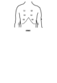
ここで、肺音データ251に含まれる情報のうち聴診位置の項目は、肺音を聴診するために電子聴診器210のチェストピースを当てる患者の体のおおよその場所のことを指す。すなわち、聴診位置は、肺音の取得部位である。例えば、図3の例では、聴診位置(1)から聴診位置(12)までの合計12箇所の聴診位置が設定されている(図3では、聴診位置(3)~(11)は省略されている)。
Here, among the information included in the
図4は、聴診位置(1)~(6)までの一例を説明するための模式図であり、図5は、聴診位置(7)~(12)までの一例を説明するための模式図である。図4を参照すると、聴診位置(1)、(2)は、例えば、前胸部の上肺野の左右に設定される。聴診位置(3)、(4)は、例えば、前胸部の中肺野の左右に設定される。聴診位置(5)、(6)は、例えば、前胸部の下肺野の左右に設定される。また、図5を参照すると、聴診位置(7)、(8)は、例えば、背部の上肺野の左右に設定される。聴診位置(9)、(10)は、例えば、背部の中肺野の左右に設定される。聴診位置(11)、(12)は、例えば、背部の下肺野の左右に設定される。例えば、以上のように、聴診位置は、予め設定されている。 FIG. 4 is a schematic diagram for explaining an example of auscultation positions (1) to (6), and FIG. 5 is a schematic diagram for explaining an example of auscultation positions (7) to (12). be. Referring to FIG. 4, auscultation positions (1) and (2) are set, for example, on the left and right sides of the upper lung field of the anterior chest. Auscultation positions (3) and (4) are set, for example, on the left and right sides of the middle lung field of the anterior chest. The auscultation positions (5) and (6) are set, for example, on the left and right sides of the lower lung field of the anterior chest. Further, referring to FIG. 5, the auscultation positions (7) and (8) are set, for example, on the left and right sides of the upper lung field on the back. The auscultation positions (9) and (10) are set, for example, on the left and right sides of the middle lung field in the back. The auscultation positions (11) and (12) are set, for example, on the left and right sides of the lower lung field on the back. For example, as described above, the auscultation position is set in advance.
なお、聴診位置は上述した個数と場所に限定されない。例えば、前胸部および背部だけでなく左右の側胸部の上肺野、中肺野、下肺野に聴診位置を設定し、合計18個としてもよい。あるいは、上記の聴診位置のうちの一部を除外してもよい。例えば、聴診位置(3)~(6)、(9)、(10)を除外し、聴診位置(1)、(2)、(7)、(8)、(11)、(12)の合計6箇所に限定してもよい。 Note that the auscultation positions are not limited to the number and location described above. For example, auscultation positions may be set not only in the anterior chest and back, but also in the upper, middle, and lower lung fields of the left and right lateral chest, for a total of 18 positions. Alternatively, some of the above auscultation positions may be excluded. For example, excluding auscultation positions (3) to (6), (9), and (10), the sum of auscultation positions (1), (2), (7), (8), (11), and (12) It may be limited to six locations.
また、肺音データ251のうち肺音データの項目には、聴診位置において電子聴診器210によって取得された、肺音を含むディジタル時系列音響信号が記録される。聴診時の患者の姿勢は、臥位と座位に大別されるが、前胸部と背部聴診は通常、座位で行われる。なお、1つの肺音データ(例えばデータ1)の信号長は任意である。1つの肺音データは、患者の連続するN呼吸分の信号であってよい。ここで、Nは1以上の正の整数である。また、肺音データは、電子聴診器210から取得された時系列音響信号に対して、休止相の期間の時系列音響信号の除去、雑音除去、呼吸タイミングの付与などの加工を施した信号であってよい。
Further, in the lung sound data item of the
分析結果情報252は、後述する異常検知部262が肺音データ251に基づいて異常を検知した結果を示している。図6は、分析結果情報252に含まれる情報の一例を示している。図6を参照すると、分析結果情報252には、例えば、聴診位置ごとの分析結果が含まれている。
The analysis result
分析結果情報252に含まれる図6で例示するような情報は、例えば、分析装置200を用いた分析を行うごと、または、異常検知部262による肺音データ251に対する分析を行うごと、などのタイミングで作成される。分析結果情報252は、異常検知部262が分析を行った日時などに応じた分析結果識別情報と、図6で例示するような情報と、を組み合わせたものであっても構わない。
The information included in the analysis result
なお、分析結果情報252に含まれる情報のうち分析結果の項目には、肺音データを後述する異常検知部262が機械的に分析した結果が記録される。例えば、分析結果の項目には、肺音データが異常な肺音データであるか否かを表す数値が記録される。例えば、分析結果の項目には、正常肺音であることを示す値0、異常肺音であることを示す値1の二値が記録されていてよい。あるいは、分析結果の項目には、肺音データの異常度を表す数値が記録されていてよい。異常度は、事前に設定された閾値以下の異常度は、肺音データが正常肺音であることを表し、閾値を超える異常度は、肺音データが異常肺音であることを表す。
Note that in the analysis result item of the information included in the analysis result
重症度判定用情報253は、後述する重症度判定部263が重症度を判定する際に用いる情報である。重症度判定用情報253は、例えば、通信I/F部240などのデータ入出力機能を介して外部装置や記録媒体などから読み込まれる、利用者が操作入力部220を操作して入力する、などの方法により予め取得され、記憶部250に保存されている。なお、重症度判定用情報253は、患者が退院するときなどのタイミングで作成されてもよい。換言すると、重症度判定用情報253は、退院時の患者の状態に応じて作成されてもよい。一般に、入院した心不全患者の多くは、心不全治療を受けて寛解した状態で退院する。そのため、多くの患者の退院時点の肺音は正常である。ただし、患者の都合によっては、軽症の状態で退院するケースがある。退院時の状態に応じた重症度判定用情報253を作成しておくことで、寛解した状態で退院した患者や軽症の状態で退院した患者など、退院時の患者の状態に応じた判定を行うことが可能となる。重症度判定用情報253は、通院時などのタイミングで作成・更新されてもよい。
The
図7は、重症度判定用情報253の一例を示している。図7を参照すると、重症度判定用情報253は、例えば、聴診位置(1)~(12)に1対1に対応する列と、重症度に1対1に対応する行とを有し、行と列の交点に肺音異常有りを示す+記号、および肺音異常無しを示す-記号を設定するテーブルである。例えば、図7で例示する場合、重症度判定用情報253が示すテーブルは、何れの聴診位置にも肺音異常がない場合、重症度0と判定することを示している。また、背部の下肺野に設定された聴診位置(11)、(12)の少なくとも一方に肺音異常があり、それ以外の聴診位置(1)~(10)に肺音異常がない場合、重症度1と判定することを示している。また、聴診位置(11)、(12)の双方に肺音異常があり、前胸部の下肺野に設定された聴診位置(5)、(6)の何れか一方のみに肺音異常があり、それ以外の聴診位置(1)~(4)、(7)~(10)に肺音異常がない場合、重症度2と判定することを示している。最終行に設定された重症度Nは、全ての聴診位置(1)~(12)に肺音異常があるものとされている。図6では、重症度2と重症度Nとの間の1以上の重症度は記載が省略されているが、それらに関しても肺音異常が有る聴診位置と肺音異常が無い聴診位置とが設定されている。重症度2と重症度Nとの間の1以上の重症度では、肺音異常があるとされる聴診位置の数は4以上、12未満であり、重症度Nに近づくにつれ、その数は増加する。
FIG. 7 shows an example of the
例えば、以上のように、重症度判定用情報253は、聴診位置(1)~(12)における肺音異常の有無の組み合わせにより、心不全の重症度を重症度0から重症度NまでN+1のクラスに分類している。ここで、重症度0は、異常肺音が全く聞こえない状態であるため、心不全が寛解している状態であると言える。また、重症度1は、背部の下肺野のみで肺音異常が聞こえる状態であるため、心不全は寛解しているとは言えないが、軽症であり、このような状態で退院する患者も存在する状態である。重症度2は、背部の下肺野に加えて前胸部の下肺野の一方にも異常肺音が出ているため、重症度1よりは重症であると言える。但し、いまだ軽症に属するため、この時点で適切な対応を取れば再入院を防止できる確率が高いと言える。
For example, as described above, the
なお、重症度判定用情報253が示す情報は、図7で例示する場合に限定されない。例えば、重症度判定用情報253は、背部の下肺野に設定された聴診位置(11)、(12)の少なくとも一方に肺音異常があり、それ以外の聴診位置(1)~(10)に肺音異常がない場合と、聴診位置(11)、(12)の双方に肺音異常があり、前胸部の下肺野に設定された聴診位置(5)、(6)の何れか一方のみに肺音異常があり、それ以外の聴診位置(1)~(4)、(7)~(10)に肺音異常がない場合と、をともに重症度1とするなど、図6で例示した以外の情報を示していても構わない。また、重症度判定用情報253が有する列数は、肺音データ251において予め設定された聴診位置の数に応じたものであって構わない。
Note that the information indicated by the
また、例えば、ラ音は、吸気の終末だけ聴こえるときは軽症、吸気開始直後から聞こえるときは重症といった知見がある。そのため、判定テーブルに、聴診位置毎の異常肺音の有無に加えて、異常肺音が聞こえるタイミングを設定し、聴診位置と異常肺音の有無と異常肺音が聞こえるタイミングの組み合わせによって、心不全の重症度を判定するようにしてもよい。また、重症度判定用情報253は、性状の異なるラ音(荒い断続性ラ音、細かい断続性ラ音)など異常音の種別や数に応じた重症度を設定してもよい。また、重症度判定用情報253は、患者の体重の増加量など後述する個人状態情報255に含まれうる情報に応じた重症度を設定してもよい。また、重症度判定用情報253は、例えば、異常肺音となった聴診位置の数と、患者の心不全の重症度と、を対応づけた情報であってもよい。例えば、重症度判定用情報253は、異常肺音となった聴診位置の数が0、1以上2以下、3以上4以下、5以上8以下、9以上のとき、それぞれ重症度0、1、2、3、4(最大)としてよい。
Furthermore, for example, it is known that rales are mild when heard only at the end of inspiration, and severe when heard immediately after the beginning of inspiration. Therefore, in addition to the presence or absence of abnormal lung sounds for each auscultation position, the timing at which abnormal lung sounds are heard is set in the judgment table, and the combination of the auscultation position, the presence or absence of abnormal lung sounds, and the timing at which abnormal lung sounds are heard determines whether heart failure is detected. The severity may also be determined. Further, the
重症度情報254は、後述する重症度判定部263が分析結果情報252と重症度判定用情報253とを用いて判定した結果を示している。図8は、分析結果情報252の一例を示している。図8を参照すると、重症度情報254では、例えば、重症度判定部263が判定を行った日時などに応じた重症度識別情報と、重症度と、が対応付けられている。
The
なお、重症度情報254中の重症度の項目には、重症度判定部263が分析結果情報252と重症度判定用情報253とを用いて判定した結果が記録される。つまり、重症度0、重症度1、重症度2、……、など、上述した重症度を示す情報が記録される。
Note that the severity item in the
個人状態情報255は、患者の状態を示す情報である。個人状態情報255に含まれる情報は、重症度判定部163が重症度の判定を行う際などに用いることが出来る。個人状態情報255は、例えば、通信I/F部240などのデータ入出力機能を介して外部装置や記録媒体などから読み込まれる、利用者が操作入力部220を操作して入力する、などの方法により予め取得され、記憶部250に保存されている。なお、個人状態情報255は、分析装置200を用いた分析を行うごとに操作入力部220に対する操作などを行うことなどにより、適宜更新されて構わない。
図9は、個人状態情報255に含まれる情報の一例を示している。図9を参照すると、個人状態情報255では、例えば、状態情報を入力された日時などに応じた状態識別情報と、状態情報と、が対応付けられている。
FIG. 9 shows an example of information included in the
なお、個人状態情報255中の状態情報の項目には、患者の体重や服薬状況などを示す情報が含まれうる。服薬状況を示す情報は、例えば、前回服薬した日時、前回飲んだ薬の種類、服薬頻度、などの情報を含んでよい。また、状態情報の項目には、血圧、脈拍、自覚症状(外出時などの息切れ、むくみ、せき、食欲低下など)、摂取水分量、経皮的動脈血酸素飽和度(SPO2)などを示す情報が含まれても構わない。状態情報の項目には、退院時や来院時などにおける医師からの連絡事項などが含まれても構わない。
Note that the status information items in the
出力情報256は、出力部264が出力した内容や、出力部264が出力した結果などを示している。図10は、出力情報256の一例を示している。図10を参照すると、出力情報256では、例えば、出力部264が出力した日時などに応じた出力識別情報と、重症度と、出力内容と、出力結果と、が含まれている。
The
ここで、重症度の項目は、出力部264が出力すると判断した際の重症度を示している。また、出力内容の項目には、出力部264が出力した重症度以外の内容が含まれている。例えば、出力内容の項目には、肺音データ、取得部位、取得日時、個人状態情報、…などのうちの少なくとも1つが含まれうる。また、出力結果の項目には、出力部264による出力の結果として、処理装置300から取得した情報が含まれている。例えば、出力結果の項目には、経過観察、服薬指示、受診勧告、などの出力に応じて診断した結果を示す情報が含まれる。出力結果の項目には、かかりつけ医からのメッセージなど、上記例示した以外の情報が含まれても構わない。
Here, the severity item indicates the severity at which the
以上が、記憶部250で保存される主な情報の一例である。なお、データ識別情報、分析結果識別情報、重症度識別情報、状態識別情報、出力識別情報などの各種識別情報は、それぞれ異なるものであっても構わないし、例えば、分析全体の日時に応じた情報など、共通なものであっても構わない。
The above is an example of the main information stored in the
演算処理部260は、CPUなどのマイクロプロセッサとその周辺回路を有し、記憶部250からプログラム257を読み込んで実行することにより、上記ハードウェアとプログラム257とを協働させて各種処理部を実現する。演算処理部260で実現される主な処理部には、肺音取得部261、異常検知部262、重症度判定部263、出力部264、受信部265などがある。
The
肺音取得部261は、患者の肺音を含むディジタル時系列音響信号およびその他の情報を取得する。肺音取得部261は、操作入力部220などから入力される利用者の指示に従って、患者の肺音を含むディジタル時系列音響信号を、電子聴診器210から取得する。また、肺音取得部261は、上記ディジタル時系列音響信号とともに、日時を示す情報などを取得することが出来る。そして、肺音取得部261は、取得したディジタル時系列音響信号およびその他の情報を用いて、図3で例示したような肺音データ251を生成して、記憶部250に保存する。上述したように、肺音取得部261は、データ識別情報と、図2で例示するような情報と、を組み合わせても構わない。
The lung
なお、患者の聴診位置毎の肺音を電子聴診器によって取得して聴診位置に対応付けて記録する方法は、任意である。例えば、特許文献1、4あるいは6などに記載されるように、電子聴診器210を用いる利用者に対して聴診位置をガイダンスするためのガイダンス画面を画面表示部230に表示して行う方法など、任意の方法を使用してよい。また、肺音取得部261は、特許文献8に記載されるような方法で、患者に対して呼吸タイミングを指示するようにしてもよい。
Note that the method of acquiring lung sounds for each auscultation position of the patient using an electronic stethoscope and recording them in association with the auscultation position is arbitrary. For example, as described in
また、肺音取得部261は、肺音品質の指標値を算出し、算出した指標値に基づく警告を画面表示部230などに行うよう構成しても構わない。このような警告を行うことで、患者や利用者などは、背景雑音を低減する対策または/および肺音を増大させる対策を講じた上で、再度肺音の取得を行うことが可能となる。
Further, the lung
肺音品質の指標値算出処理は、例えば、所定のフィルタを適用した後、信号の強度を算出、比較することなどにより行われる。例えば、肺音取得部261は、帯域通過フィルタを使用して、電子聴診器210から出力される時系列音響信号から、患者の肺音が含まれる100Hz~約2kHzの周波数帯域の時系列音響信号を抽出する。次に、肺音取得部261は、抽出した時系列音響信号中の肺音の強度と背景雑音の強度とを算出し、それらの相違度を肺音の品質の指標値として算出する。例えば、肺音取得部261は、肺音を含む時系列音響信号から吸気相、呼気相、休止相を検出する。そして、肺音取得部261は、休止相における時系列音響信号の強度を背景雑音の強度として算出する。時系列音響信号の強度は、例えば振幅値の二乗平均平方根を使用できるが、それに限定されず、振幅などであってもよい。また、肺音取得部261は、吸気相または/および呼気相における時系列音響信号の強度から背景雑音の強度を減算した値を、肺音の強度として算出する。そして、肺音取得部261は、算出した背景雑音の強度に対する肺音の強度の比を、肺音の品質の指標値とする。なお、肺音の品質の指標値は上記したものに限定されず、肺音の強度と背景雑音の強度とから算出されるS/N比を指標値としてもよい。フィルタの適用は、省略しても構わない。
The lung sound quality index value calculation process is performed, for example, by applying a predetermined filter, and then calculating and comparing signal intensities. For example, the lung
なお、肺音取得部261は、時系列音響信号と予め定められた閾値とを比較することで呼気相や吸気相を検出することが出来る。また、肺音取得部261は、検出された吸気の開始時点の直前所定期間を休止相として検出することが出来る。肺音取得部261は、上記例示した以外の方法を用いて、吸気相、呼気相、および休止相を検出しても構わない。例えば、肺音取得部261は、電子聴診器210から出力される肺音を含む時系列音響信号のどの区間が吸気相、呼気相、休止相であるかを推定するための機械学習を行った学習済みの学習モデルに患者の肺音を含む時系列音響信号を入力することで、区間ごとに吸気相、呼気相、休止相の推定確率を当該学習モデルから取得するように構成されていてもよい。学習モデルは、例えば、様々な肺音を含む時系列音響信号を教師データとしてニューラルネットワークなどの機械学習アルゴリズムを用いた機械学習によって、事前に生成することができる。
Note that the lung
また、肺音取得部261は、肺音を含むディジタル時系列音響信号から休止相の期間および背景雑音を除去し、休止相の期間および背景雑音を除去した後のディジタル時系列音響信号を聴診位置に関連付けて肺音データ251に記録するよう構成しても構わない。例えば、肺音取得部261は、肺音を含むディジタル時系列音響信号を、吸気相とその直後の呼気相からなる区間(以下、吸気・呼気区間と記す)と休止相の区間(以下、休止区間と記す)とに2分割する。次に、肺音取得部261は、吸気・呼気区間と休止区間のディジタル時系列音響信号をそれぞれ高速フーリエ変換(FFT)して吸気・呼気区間と休止区間の周波数スペクトルを算出する。次に、肺音取得部261は、吸気・呼気区間の周波数スペクトルから休止区間の周波数スペクトルを減算する。この減算により、吸気相と呼気相に含まれている背景雑音が抑制される。次に、肺音取得部261は、上記減算後の吸気・呼気区間の周波数スペクトルを逆周波数変換することにより、吸気・呼気区間の雑音除去後のディジタル時系列音響信号を生成する。そして、肺音取得部261は、上記生成した吸気・呼気区間の雑音除去後のディジタル時系列音響信号を聴診位置に関連付けて肺音データ251に記録する。なお、肺音取得部261は、聴診位置の肺音を含むディジタル時系列音響信号から休止相の期間を除去し、背景雑音を除去しないようにしてもよい。その場合、肺音取得部261は、注目中の聴診位置の肺音を含むディジタル時系列音響信号を、吸気・呼気区間と休止区間とに2分割し、吸気・呼気区間のディジタル時系列音響信号を聴診位置に関連付けて肺音データ251に記録する。
The lung
異常検知部262は、肺音データ251に含まれる各聴診位置の肺音データから異常を検知し、検知結果を聴診位置に関連付けて分析結果情報252に記録する。例えば、異常検知部262は、事前に生成し記憶している異常検知用モデルに肺音データを入力し、肺音データが異常肺音である確率を異常検知用モデルから取得する。次に、異常検知部262は、異常肺音である確率を事前に設定された閾値と比較する。そして、確率が閾値を超えている場合、異常検知部262は、異常肺音であると判別する。つまり、異常検知部262は、異常を検知する。一方、閾値以下である場合、異常検知部262は、異常肺音ではないと判別する。その後、異常検知部262は、検知結果を分析結果情報252に記録する。
The
なお、異常検知用モデルは、例えば、異常音を収集したデータベースを用いて教師データを生成し、ディープラーニングを用いて、入力された音データ(入力データ)の特徴および判別基準を学習することで、事前に生成することが出来る。例えば、異常検知部262は、学習および入力データには音声を一定の区間毎にFFT(高速フーリエ変換)やlog-FFTして時系列順に並べたスペクトログラムを用い、ディープラーニングにはRNN(リカレントニューラルネットワーク)やCNN(コンボリューティブニューラルネットワーク)を用いることができる。
The anomaly detection model can be created by, for example, generating training data using a database that collects abnormal sounds, and using deep learning to learn the characteristics and discrimination criteria of the input sound data (input data). , can be generated in advance. For example, the
また、異常検知部262は、肺音波形をゼロ交差係数やMFCC(メル周波数ケプストラム係数)などの短時間特徴量に変換して、機械学習によって異常音を検知する方法を使用してもよい。例えば異常検知部262は、学習時にGMM(混合ガウス分布)でモデル化して、検知時に該当モデルに適合するか否かを調べるようにしてよい。また異常検知部262は、SVM(サポートベクターマシン)のような識別器の識別面を学習して、その識別面を用いて、入力されたデータが異常音に該当するかを識別してもよい。異常検知部262は、こうした特徴量を、前述のような直接求める方法以外にも、NMF(非負値行列因子分解)やPCA(主成分分析)のように、データそのものを用いて特徴量を生成するようにしてもよい。
Further, the
また、異常検知部262は、入力信号の長時間パワー分布や、特定周波数ビン範囲の成分量・成分比率の分布など、入力波形の統計的特徴を用いて、決定木などにより異常音を検知してもよい。その場合、異常検知部262は、決定木の項目としては、直接の値(例えばパワーが3フレーム連続して20mWを超えた場合)の他、統計的特徴(例えばガウス近似して3σより大きい処理フレームが発生した場合)を用いてもよい。また、異常検知部262は、入力信号そのものではなく、それをAR(自己回帰)過程などでモデル化し、そのモデルパラメータの幾つかが閾値を超えることなどによって、異常音を検知してもよい。これらの方法は学習過程を含まない場合があるが、決定木の構成や閾値の決定などに対象信号である異常音の観察を含むため、便宜上教師有り学習に含める。
In addition, the
また、異常検知部262は、例えば、患者が退院する時など過去の患者の肺音データと聴診所見とを用いて、異常検知用モデルを学習するよう構成しても構わない。異常検知用モデルを学習する際に用いる肺音データには、患者の退院時点の肺音データに加えて、それ以前の患者の正常な肺音データを使用してもよいし、患者以外の人の正常な肺音データなどを使用しても構わない。このように退院時の肺音データに基づいて異常検知用モデルを生成することで、退院時の患者の状態を考慮した検知を行うことが可能となる。
Furthermore, the
なお、異常検知用モデルは、聴診位置ごとに生成されていても構わないし、複数の聴診位置で共通であっても構わない。また、異常検知用モデルは、異なる観点で機械学習した複数のモデルであってもよい。例えば、異常検知用モデルには、同じ聴診位置の肺音を、呼吸タイミングに基づいて、吸気相の肺音部分と呼気相の肺音部分とそれ以外(即ち休止相)とに分割し、吸気相の肺音部分を使用して学習したモデルと、呼気相の肺音部分を使用して学習したモデルとが含まれていてもよい。 Note that the abnormality detection model may be generated for each auscultation position, or may be common to a plurality of auscultation positions. Further, the abnormality detection model may be a plurality of models machine-learned from different viewpoints. For example, in an abnormality detection model, lung sounds at the same auscultation position are divided into an inspiratory phase lung sound part, an expiratory phase lung sound part, and other parts (i.e. resting phase) based on the breathing timing. A model learned using the lung sound part of the phase and a model learned using the lung sound part of the exhalation phase may be included.
重症度判定部263は、分析結果情報252が示す聴診位置ごとの分析結果と、重症度判定用情報253と、に基づいて、重症度を判定する。そして、重症度判定部263は、判定した重症度を重症度識別情報と対応付けて重症度情報254として記憶部250に保存する。
The
例えば、重症度判定部263は、分析結果情報252を参照して、異常が検知された聴診位置を特定する。そして、重症度判定部263は、重症度判定用情報253を参照して、特定した結果に対応する重症度を判定する。
For example, the
なお、上述したように、重症度判定用情報253は、退院時の患者の状態に応じて作成することが出来る。重症度判定用情報253が上記のように作成されている場合、重症度判定部263は、退院時の患者の状態に応じた重症度判定用情報253を参照することにより、退院時の患者の状態に応じた重症度の判定を行うことが出来る、ということも出来る。
Note that, as described above, the
また、重症度判定部263は、重症度を判定する際に個人状態情報255を参照して、判定した重症度を補正するよう構成することが出来る。例えば、心不全が憎悪すると肺に水が溜まるため体重が増加する、という知見がある。そのため、重症度判定部263は、個人状態情報255に含まれる患者の体重に基づいて、判定した重症度を補正することが出来る。例えば、体重が1週間で3kg増加したなど、予め定められた条件を満たした場合、重症度判定部263は、判定した重症度を1上げるなど、設定値分の補正を行うことが出来る。なお、重症度判定部263は、1週間で5kg増加している場合は重症度を2上げるなど、上記例示した以外の補正を行っても構わない。また、重症度判定部263は、例えば、血圧が所定値以上低下した場合や脈拍が所定数を超えている場合に重症度を上げるなど、上記例示した以外の個人状態情報255を用いた補正を行っても構わない。重症度判定部263は、個人状態情報255に含まれうる情報に応じた重症度を設定した重症度判定用情報253を参照することにより、個人状態情報255を考慮した重症度を直接判定してもよい。
Moreover, the
出力部264は、重症度判定部263による判定の結果に基づいて、所定の情報を処理装置300へと出力するか否か判断する。そして、出力部264は、判断の結果に応じて、所定の情報を出力する。また、出力部264は、出力した重症度やその他の出力内容などを出力識別情報と対応付けて、出力情報256として記憶部250に保存する。なお、出力部264による処理装置300に対する出力は、例えばメール、グループウェアのメッセージ機能、ビジネスチャットなど、任意のコミュニケーション方法のうちの少なくとも1つを用いて実現してよい。
The
例えば、出力部264は、重症度判定部263が判定した重症度が出力閾値を超えているか否か確認する。そして、出力部264は、重症度が出力閾値を超えている場合、処理装置300へと出力すると判断する。その後、出力部264は、処理装置300に対して所定の情報を出力する。
For example, the
ここで、出力部264が出力する情報は、例えば、予め定められている。例えば、出力部264が出力する情報には、重症度判定部263が判定した重症度、肺音データ251に含まれる肺音データや聴診位置、肺音データを取得した日時を示す情報、個人状態情報255に含まれる体重や服薬状況などの情報、などのうちの少なくとも一つが含まれる。出力部264は、分析結果情報252に含まれる情報、肺音データに対して高速フーリエ変換(FFT)することで算出した周波数スペクトルなど、上記例示した以外の情報を出力してもよい。なお、出力部264が出力する情報は、肺音データ251に含まれる肺音データのうち、異常検知部262が異常を検知した肺音データのみであっても構わないし、肺音データ251に含まれる肺音データすべてであっても構わない。また、出力部264は、今回の分析時に取得した肺音データの他、直近2週間の肺音データや異常検知部262が異常を検知しなかった際の肺音データなど、過去の肺音データを出力してもよい。出力部264は、今回の分析時に取得した肺音データから算出した周波数スペクトルと、異常検知部262が異常を検知しなかった際の肺音データから算出した周波数スペクトルと、などを出力してもよい。出力部264は、上記のような周波数スペクトルの他に、診断結果が受診勧告となる患者の平均的な周波数スペクトルなどを出力してもよい。また、出力部264が出力する情報のうち個人状態情報に含まれる体重や服薬状況などの情報も、直近2週間の体重の推移や直近2週間の服薬状況など、過去の情報を含んでいても構わない。
Here, the information output by the
また、出力部264は、重症度判定部263が判定した重症度に応じて、出力する情報を変更しても構わない。例えば、出力部264は、重症度が上がるほど出力する情報を増やす、重症度が送信閾値以下の場合に肺音データのみを出力し重症度が送信閾値を超えた場合に肺音データと個人状態情報255に含まれる情報を出力する、など、重症度に応じて出力する情報に差を設けてもよい。
Furthermore, the
また、出力部264による出力先となる処理装置300は、例えば予め定められている。例えば、出力部264は、分析装置200の利用者や患者などの識別子と、当該患者のかかりつけ医などが有する処理装置300と、を対応付けた情報を有している。そして、出力部264は、上記情報を参照することで、患者のかかりつけ医などが有する処理装置300に対して所定の情報を出力する。
Further, the
なお、出力部264は、所定の情報を処理装置300へと出力するか否か判断する際に、出力情報256中に含まれる過去の出力結果を参照しても構わない。例えば、重症度判定部263が判定した重症度と同じ(または、差が所定値以下)重症度で出力結果が経過観察であった情報が出力情報256に含まれる場合、出力部264は、重症度が出力閾値を超えていても処理装置300へと出力しないと判断することが出来る。出力部264は、重症度判定部263が判定した重症度と同じ重症度で出力結果が経過観察であった情報が出力情報256に含まれる場合、スペクトル解析を行った結果などに基づいて出力するか否か判断してもよい。例えば、出力部264は、出力結果が経過観察だった際の肺音データと、今回の肺音データと、に対して、高速フーリエ変換(FFT)することで周波数スペクトルを算出する。そして、出力部264は、算出した周波数スペクトルを比較することで、所定の情報を処理装置300に対して送信するか否か判断する。例えば、出力部264は、周波数スペクトルの差が所定位置以下の場合に所定の情報を処理装置300に対して送信すると判断する。また、出力部264は、重症度判定部263が判定した重症度と同じ重症度で出力結果が経過観察であった情報が出力情報256に含まれる場合、ラ音の数や頻度など肺音データの内容に応じて出力するか否か判断してもよい。例えば、ラ音の数や頻度など肺音データの差異が所定値以下の場合、出力部264は出力しないと判断してもよい。
Note that the
受信部265は、出力部264による出力の結果を処理装置300から受信する。そして、受信部265は、受診した情報に基づいて出力情報256のうちの出力結果の項目を更新する。
The receiving
例えば、受信部265は、経過観察、服薬指示、受診勧告など、出力部264による出力の内容に応じて処理装置300を有するかかりつけ医などが診断した結果を示す情報を、処理装置300から受信する。そして、受信部265は、受診した情報を出力情報256のうちの出力結果の項目に保存することで、出力情報256を更新する。
For example, the receiving
処理装置300は、分析装置200の出力部264が出力した情報を受信する情報処理装置である。処理装置300は、例えば、かかりつけ医が勤務する病院などに設置されている。処理装置300は、例えば、スマートフォン、タブレット型端末、PDA(Personal Digital Assistant)、ノートパソコンなどである。処理装置300は、上記例示した以外であっても構わない。
The
図11は、処理装置300の構成例を示している。図11を参照すると、処理装置300は、主な構成要素として、例えば、操作入力部310と、画面表示部320と、通信I/F部330と、記憶部340と、演算処理部350と、を有している。なお、操作入力部310、画面表示部320、通信I/F部330、の構成は、分析装置200が有する各構成と同様であって構わない。そのため、説明を省略する。
FIG. 11 shows a configuration example of the
記憶部340は、ハードディスクやメモリなどの記憶装置である。記憶部340は、演算処理部350における各種処理に必要な処理情報やプログラム343を記憶する。プログラム343は、演算処理部350に読み込まれて実行されることにより各種処理部を実現する。プログラム343は、通信I/F部330などのデータ入出力機能を介して外部装置や記録媒体から予め読み込まれ、記憶部340に保存されている。記憶部340で記憶される主な情報としては、例えば、取得情報341と診断結果342となどがある。
The
取得情報341は、分析装置200が出力した情報を含んでいる。つまり、取得情報341には、出力部264による出力に応じた情報が含まれる。取得情報341に含まれる情報は、画面表示部320などに出力することが出来る。
The acquired
診断結果342は、取得情報341に応じて、かかりつけ医などが診断した結果を示している。診断結果342は、例えば、経過観察、服薬指示、受診勧告など、取得情報341に応じて処理装置300を有するかかりつけ医などが診断した結果を示す情報を示している。
演算処理部350は、CPUなどのマイクロプロセッサとその周辺回路を有し、記憶部340からプログラム343を読み込んで実行することにより、上記ハードウェアとプログラム343とを協働させて各種処理部を実現する。演算処理部350で実現される主な処理部には、取得部351、診断結果入力部352、出力部353などがある。
The
取得部351は、分析装置200の出力部264が出力した情報を取得する。そして、取得部351は、取得した情報を取得情報341として記憶部340に保存する。
The
診断結果入力部352は、取得部351が保存した取得情報341に対するかかりつけ医などの診断結果を取得する。診断結果入力部352は、操作入力部310などに対する操作に応じて、経過観察、服薬指示、受診勧告など、取得情報341に応じてかかりつけ医などが診断した結果を示す情報を取得する。そして、診断結果入力部352は、取得した情報を診断結果342として記憶部340に保存する。
The diagnosis
出力部353は、診断結果342を分析装置200に対して出力する。例えば、出力部353は、診断結果入力部352が新たな診断結果342を保存した際に、新たな診断結果を示す情報を分析装置200に対して送信する。
The
例えば、以上のように、処理装置300は、出力部264による出力に応じた診断結果を示す情報を取得し、診断結果を示す情報を分析装置200に対して送り返す。なお、診断結果を示す情報には、かかりつけ医からのメッセージなど、上記例示した以外の情報が含まれてもよい。
For example, as described above, the
続いて、図12を参照して、分析装置200の動作例について説明する。
Next, an example of the operation of the
図12を参照すると、肺音取得部261は、各聴診位置について、患者の肺音を含むディジタル時系列音響信号などを取得する(ステップS101)。そして、肺音取得部261は、取得したディジタル時系列音響信号およびその他の情報を用いて、図3で例示したような肺音データ251を生成して、記憶部250に保存する。上述したように、肺音取得部261は、データ識別情報と、図3で例示するような情報と、を組み合わせても構わない。
Referring to FIG. 12, the lung
なお、肺音取得部261は、ディジタル時系列音響信号などを取得する際、肺音品質の指標値を算出し、算出した指標値に基づく警告を画面表示部230などに行っても構わない。また、肺音取得部261は、休止相の期間の時系列音響信号の除去、雑音除去、呼吸タイミングの付与などの加工を施した後、加工後のデータを用いて肺音データ251を生成して、記憶部250に保存してもよい。
Note that when acquiring the digital time-series acoustic signal or the like, the lung
異常検知部262は、肺音データ251に含まれる各聴診位置の肺音データから異常を検知し、検知結果を聴診位置に関連付けて分析結果情報252に記録する(ステップS102)。例えば、異常検知部262は、事前に生成し記憶している異常検知用モデルに肺音データを入力し、肺音データが異常肺音である確率を異常検知用モデルから取得する。次に、異常検知部262は、異常肺音である確率を事前に設定された閾値と比較する。そして、確率が閾値を超えている場合、異常検知部262は、異常肺音であると判別する。つまり、異常検知部262は、異常を検知する。一方、閾値以下である場合、異常検知部262は、異常肺音ではないと判別する。その後、異常検知部262は、検知結果を分析結果情報252に記録する。
The
重症度判定部263は、分析結果情報252が示す聴診位置ごとの分析結果と、重症度判定用情報253と、に基づいて、重症度を判定する(ステップS103)。そして、重症度判定部263は、判定した重症度を重症度識別情報と対応付けて重症度情報254として記憶部250に保存する。
The
なお、重症度判定部263は、退院時の患者の状態に応じた重症度判定用情報253を参照することにより、退院時の患者の状態に応じた重症度の判定を行ってもよい。また、重症度判定部263は、重症度を判定する際に個人状態情報255を参照して、判定した重症度を補正してもよい。重症度判定部263は、個人状態情報255に含まれうる情報に応じた重症度を設定した重症度判定用情報253を参照することにより、個人状態情報156を考慮した重症度を直接判定してもよい。
Note that the
出力部264は、重症度判定部263による判定の結果に基づいて、所定の情報を処理装置300へと出力するか否か判断する。例えば、出力部264は、重症度判定部263が判定した重症度が出力閾値を超えているか否か確認する(ステップS104)。
The
重症度が出力閾値を超えている場合(ステップS104、Yes)、出力部264は、出力情報256が条件を満たすか否か確認する(ステップS105)。そして、出力情報256が条件を満たす場合(ステップS105、Yes)、出力部264は、処理装置300に対して出力すると判断して、所定の情報を処理装置300に対して送信する(ステップS106)。また、出力部264は、出力した情報を出力情報256として記憶部250に保存する。なお、上記条件には、重症度判定部263が判定した重症度と同じ重症度で出力結果の項目が経過観察の情報が出力情報に含まれるか、上記情報が含まれる場合の周波数スペクトルの差、などを含むことが出来る。
If the severity exceeds the output threshold (step S104, Yes), the
受信部265は、出力部264による出力の結果を処理装置300から受信する(ステップS107)。そして、受信部265は、受診した情報に基づいて出力情報256のうちの出力結果の項目を保存する(ステップS108)。
The receiving
なお、重症度が出力閾値以下である場合(ステップS104、No)、または、出力情報256が条件を満たさない場合(ステップS105、No)、出力部264は処理装置300に対する出力を行わないと判断する。
Note that if the severity is below the output threshold (step S104, No), or if the
以上が、分析装置200の動作例である。なお、ステップS105の処理は省略しても構わない。
The above is an example of the operation of the
このように、分析装置200は、異常検知部262と、重症度判定部263と、出力部264と、を有している。このような構成によると、出力部264は、異常検知部262による検知結果に基づいて重症度判定部263が判定した重症度に基づいて、処理装置300に対する出力を行うか否か判断することが出来る。その結果、重症度が高いなど情報共有の必要性が高い場合に検査結果を医療従事者と共有することが可能となる。つまり、上記構成によると、効率的な情報共有を実現することが出来る。
As described above, the
なお、患者の都合などにより、電子聴診器210を用いた肺音データの取得が途中で終了してしまうことも想定される。このような場合、異常検知部262は、肺音データを取得した聴診位置に対応する分析のみを行うことになる。重症度判定部263は、異常検知部262が分析を行った聴診位置の数に基づいて、重症度の判定を行うか否か判断するよう構成してもよい。例えば、肺音データが取得されず異常肺音か否かの分析が行われていない聴診位置の数が、事前に設定された閾値未満である場合、重症度判定部263は、重症度を算出せず、分析がエラー終了した旨を画面表示部230に表示することが出来る。その理由は、誤った情報を操作者などに与えないようにするためである。一方、聴診位置の数が事前に設定された閾値以上である場合、重症度判定部263は、異常肺音か否かの分析が行われていない聴診位置において肺音異常が検知されなかったと仮定して、重症度を算出することが出来る。この場合、重症度判定部263は、算出した重症度を最も楽観的な値として保持してもよい。即ち、算出した重症度が重症度1であった場合、「重症度1」ではなく「重症度1以上」あるいは「最低でも重症度1」として保持することが出来る。なお、閾値は任意に設定して構わない。
Note that it is also assumed that acquisition of lung sound data using the
また、分析装置200は、患者の過去に異常が検知された聴診位置に基づいて、どの聴診位置に注目するか判断するよう構成してもよい。例えば、分析装置200の肺音取得部261は、過去の分析結果情報252に基づいて、聴診位置ごとの異常検知頻度を算出することが出来る。また、肺音取得部261は、算出した異常検知頻度が高い順に、肺音データを取得するようガイドなどすることが出来る。なお、異常検知頻度が高い順に肺音データを取得する場合、異常検知頻度が高い順に肺音データを取得しない場合よりも、重症度判定部263が重症度を算出するか否か判断する際に用いる閾値を小さくしてもよい。
Furthermore, the
また、分析装置200は、受信部265が受信した診断の結果を示す情報を患者などが確認した際、確認した旨を示す情報を処理装置300に対して送信するよう構成してもよい。このように構成することで、処理装置300を操作するかかりつけ医などから、診断の結果を示す情報を患者などが確認したか否か容易に確認することが出来る。
Further, the
[第2の実施の形態]
図13は、本発明の第2の実施形態に係る分析システム500のブロック図である。図13を参照すると、分析システム500は、複数の分析装置510と、サーバ装置520とから構成されている。また、複数の分析装置510とサーバ装置520とは、インターネットなどのネットワーク530を通じて相互に通信可能に接続されている。[Second embodiment]
FIG. 13 is a block diagram of an analysis system 500 according to the second embodiment of the present invention. Referring to FIG. 13, the analysis system 500 includes a plurality of
分析装置510は、肺音を分析した結果に応じた指示を出力する情報処理装置である。分析装置510は、スマートフォン、タブレット型端末、PDA、ノートパソコンなどであってよいが、それらに限定されない。分析装置510は、図示しない電子聴診器、通信I/F部、操作入力部、画面表示部、記憶部、および、演算処理部を備えている。
The
サーバ装置520は、複数の分析装置510に対して、肺音分析に必要な各種のサービスを、ネットワーク530を通じて提供するコンピュータである。例えば、サーバ装置520は、図2に示した肺音データ251、分析結果情報252、重症度判定用情報253、重症度情報254、個人状態情報255、出力情報256、およびプログラム257の少なくとも一部を記憶し、それらを、ネットワーク530を通じて分析装置510に提供する。そのため、分析装置510は、図2の分析装置200と比較して、記憶部に肺音データ251、分析結果情報252、重症度判定用情報253、重症度情報254、個人状態情報255、出力情報256、およびプログラム257の少なくとも一部を記憶する必要がなく、記憶容量を削減することができる。
The
また、サーバ装置520は、図2に示した肺音取得部261、異常検知部262、重症度判定部263、出力部264、および受信部265の少なくとも一部の機能を、ネットワーク530を通じて分析装置510に提供することが出来る。即ち、サーバ装置520は、図12で示す各処理の少なくとも一部を、分析装置510に代わって実行する。そのため、分析装置510は、図2の分析装置200と比較して、演算処理部260の構成を簡素化することができる。
In addition, the
例えば、以上のように、分析装置200としての機能は、分析システム500などにより実現されてもよい。
For example, as described above, the functions of the
[第3の実施の形態]
次に、図14、図15を参照して、本発明の第3の実施形態について説明する。第3の実施形態では、分析装置600の構成の概要について説明する。[Third embodiment]
Next, a third embodiment of the present invention will be described with reference to FIGS. 14 and 15. In the third embodiment, an overview of the configuration of an
図14は、分析装置600のハードウェア構成例を示している。図14を参照すると、分析装置600は、一例として、以下のようなハードウェア構成を有している。
・CPU(Central Processing Unit)601(演算装置)
・ROM(Read Only Memory)602(記憶装置)
・RAM(Random Access Memory)603(記憶装置)
・RAM603にロードされるプログラム群604
・プログラム群604を格納する記憶装置605
・情報処理装置外部の記録媒体610の読み書きを行うドライブ装置606
・情報処理装置外部の通信ネットワーク611と接続する通信インタフェース607
・データの入出力を行う入出力インタフェース608
・各構成要素を接続するバス609FIG. 14 shows an example of the hardware configuration of the
・CPU (Central Processing Unit) 601 (arithmetic unit)
・ROM (Read Only Memory) 602 (storage device)
・RAM (Random Access Memory) 603 (storage device)
・
-
- A
- A
・I/
・Bus 609 that connects each component
また、分析装置600は、プログラム群604をCPU601が取得して当該CPU601が実行することで、図15に示す検知部621、判定部622、出力部623としての機能を実現することが出来る。なお、プログラム群604は、例えば、予め記憶装置605やROM602に格納されており、必要に応じてCPU601がRAM603などにロードして実行する。また、プログラム群604は、通信ネットワーク611を介してCPU601に供給されてもよいし、予め記録媒体610に格納されており、ドライブ装置606が該プログラムを読み出してCPU601に供給してもよい。
Further, the
なお、図14は、分析装置600のハードウェア構成例を示している。分析装置600のハードウェア構成は上述した場合に限定されない。例えば、分析装置600は、ドライブ装置606を有さないなど、上述した構成の一部から構成されてもよい。
Note that FIG. 14 shows an example of the hardware configuration of the
検知部621は、聴診位置ごとの肺音を含む時系列音響信号に基づいて、聴診位置ごとに肺音異常を検知する。
The
判定部622は、検知部621が検知した聴診位置ごとの肺音異常の検知結果と、患者の状態を示す状態情報と、に基づいて、患者の心不全の重症度を判定する。
The determining
出力部623は、判定部622が判定した結果に基づいて、外部装置に対して所定の情報を出力するか否か判断して、判断の結果に応じた出力を行う。
The
このように、分析装置600は、検知部621と、判定部622と、出力部623と、を有している。このような構成によると、出力部623は、検知部621による検知結果に基づいて判定部622が判定した重症度に基づいて、処理装置300に対する出力を行うか否か判断することが出来る。その結果、重症度が高いなど情報共有の必要性が高い場合に検査結果を医療従事者と共有することが可能となる。つまり、上記構成によると、効率的な情報共有を実現することが出来る。
In this way, the
なお、上述した分析装置600は、分析装置600などの情報処理装置に所定のプログラムが組み込まれることで実現できる。具体的に、本発明の他の形態であるプログラムは、情報処理装置に、聴診位置ごとの肺音を含む時系列音響信号に基づいて、聴診位置ごとに肺音異常を検知する検知部と、検知部が検知した聴診位置ごとの肺音異常の検知結果と、患者の状態を示す状態情報と、に基づいて、患者の心不全の重症度を判定する判定部と、判定部が判定した結果に基づいて、外部装置に対して所定の情報を出力するか否か判断して、判断の結果に応じた出力を行う出力部と、を実現するためのプログラムである。
Note that the
また、上述した分析装置600などの情報処理装置により実現される分析方法は、情報処理装置が、聴診位置ごとの肺音を含む時系列音響信号に基づいて、聴診位置ごとに肺音異常を検知し、検知した聴診位置ごとの肺音異常の検知結果と、患者の状態を示す状態情報と、に基づいて、患者の心不全の重症度を判定し、判定した結果に基づいて、外部装置に対して所定の情報を出力するか否か判断して、判断の結果に応じた出力を行う、という方法である。
Further, in an analysis method realized by an information processing device such as the
上述した構成を有する、プログラム(または記録媒体)、または、分析方法、の発明であっても、上述した分析装置600と同様の作用・効果を有するために、上述した本発明の目的を達成することが出来る。
Even the invention of the program (or recording medium) or analysis method having the above-mentioned configuration achieves the above-mentioned object of the present invention because it has the same operation and effect as the above-mentioned
以上、上記各実施形態を参照して本発明を説明したが、本発明は、上述した実施形態に限定されるものではない。本発明の構成や詳細には、様々な変形例を組み合わせるなど、本発明の範囲内で当業者が理解しうる様々な変更をすることができる。 Although the present invention has been described above with reference to the embodiments described above, the present invention is not limited to the embodiments described above. The configuration and details of the present invention can be modified in various ways that can be understood by those skilled in the art within the scope of the present invention, such as combining various modifications.
本発明は、人の肺音を分析する装置やシステムに利用でき、特に心不全治療を受けて退院した患者の心不全増悪を早期に検出し再入院を防止する装置、システムに利用できる。 INDUSTRIAL APPLICATION This invention can be utilized for the apparatus and system which analyze a person's lung sound, and can be especially utilized for the apparatus and system which detects the worsening of heart failure early and prevents re-hospitalization of the patient discharged after receiving heart failure treatment.
上記の実施形態の一部又は全部は、以下の付記のようにも記載され得るが、以下には限られない。
(付記1)
聴診位置ごとの肺音を含む時系列音響信号に基づいて、前記聴診位置ごとに肺音異常を検知する検知部と、
前記検知部が検知した前記聴診位置ごとの肺音異常の検知結果と、患者の状態を示す状態情報と、に基づいて、前記患者の心不全の重症度を判定する判定部と、
前記判定部が判定した結果に基づいて、外部装置に対して所定の情報を出力するか否か判断して、判断の結果に応じた出力を行う出力部と、
を有する
分析装置。
(付記2)
前記出力部は、前記判定部が判定した重症度が予め定められた出力閾値を超えているか否かに基づいて、前記外部装置に対して所定の情報を出力するか否か判断する
付記1に記載の分析装置。
(付記3)
前記出力部は、当該出力部が過去に出力した結果に応じて前記外部装置から受信した情報に基づいて、前記外部装置に対して所定の情報を出力するか否か判断する
付記1または付記2に記載の分析装置。
(付記4)
前記出力部は、前記判定部が判定した重症度と同じ重症度で過去に出力した結果に応じて前記外部装置から受信した情報に基づいて、前記外部装置に対して所定の情報を出力するか否か判断する
付記3に記載の分析装置。
(付記5)
前記出力部は、前記判定部が判定した重症度に応じた重症度において、前記外部装置から出力の結果として経過観察を含む情報を受信していた場合に、前記時系列音響信号から算出した周波数スペクトルを用いて前記外部装置に対して所定の情報を出力するか否か判断する
付記3または付記4に記載の分析装置。
(付記6)
前記出力部は、前記検知部が検知する際に用いた前記時系列音響信号から算出した周波数スペクトルと、過去の時系列音響信号から算出した周波数スペクトルと、の比較結果に基づいて、前記外部装置に対して所定の情報を出力するか否か判断する
付記5に記載の分析装置。
(付記7)
前記所定の情報には、前記判定部が判定した重症度と、前記検知部が検知する際に用いた前記時系列音響信号と、が含まれている
付記1から付記6までのいずれか1項に記載の分析装置。
(付記8)
前記所定の情報には、前記患者の体重を示す情報、服薬状況を示す情報、のうちの少なくとも一方が含まれている
付記7に記載の分析装置。
(付記9)
前記所定の情報には、前記検知部が検知する際に用いた前記時系列音響信号から算出した周波数スペクトルが含まれている
付記7または付記8に記載の分析装置。
(付記10)
前記出力部が出力した結果に応じて診断した結果を示す情報を受信する受信部を有する
付記1から付記9までのいずれか1項に記載の分析装置。
(付記11)
前記外部装置は、かかりつけ医が有する装置である
付記1から付記10までのいずれか1項に記載の分析装置。
(付記12)
情報処理装置が、
聴診位置ごとの肺音を含む時系列音響信号に基づいて、前記聴診位置ごとに肺音異常を検知し、
検知した前記聴診位置ごとの肺音異常の検知結果と、患者の状態を示す状態情報と、に基づいて、前記患者の心不全の重症度を判定し、
判定した結果に基づいて、外部装置に対して所定の情報を出力するか否か判断して、判断の結果に応じた出力を行う
分析方法。
(付記13)
情報処理装置に、
聴診位置ごとの肺音を含む時系列音響信号に基づいて、前記聴診位置ごとに肺音異常を検知する検知部と、
前記検知部が検知した前記聴診位置ごとの肺音異常の検知結果と、患者の状態を示す状態情報と、に基づいて、前記患者の心不全の重症度を判定する判定部と、
前記判定部が判定した結果に基づいて、外部装置に対して所定の情報を出力するか否か判断して、判断の結果に応じた出力を行う出力部と、
を実現するためのプログラムを記録した、コンピュータが読み取り可能な記録媒体。Part or all of the above embodiments may be described as in the following additional notes, but are not limited to the following.
(Additional note 1)
a detection unit that detects abnormal lung sounds at each auscultation position based on a time-series acoustic signal including the lung sounds at each auscultation position;
a determination unit that determines the severity of heart failure of the patient based on the detection result of abnormal lung sounds at each auscultation position detected by the detection unit and status information indicating the patient's condition;
an output unit that determines whether or not to output predetermined information to an external device based on the determination result of the determination unit, and outputs the output according to the determination result;
Analyzer with.
(Additional note 2)
The output unit determines whether or not to output predetermined information to the external device based on whether the severity determined by the determination unit exceeds a predetermined output threshold. Analyzer as described.
(Additional note 3)
The output unit determines whether to output predetermined information to the external device based on information received from the external device according to results output by the output unit in the past.
(Additional note 4)
The output unit outputs predetermined information to the external device based on information received from the external device according to a result output in the past with the same severity as the severity determined by the determination unit. The analysis device described in
(Appendix 5)
The output unit outputs the frequency calculated from the time-series acoustic signal when receiving information including progress observation as an output result from the external device at a severity level corresponding to the severity level determined by the determination unit. The analysis device according to
(Appendix 6)
The output unit outputs the signal to the external device based on a comparison result between a frequency spectrum calculated from the time-series audio signal used for detection by the detection unit and a frequency spectrum calculated from a past time-series audio signal. The analysis device according to
(Appendix 7)
The predetermined information includes the severity determined by the determination unit and the time-series acoustic signal used by the detection unit for detection. Any one of
(Appendix 8)
The analysis device according to
(Appendix 9)
The analysis device according to
(Appendix 10)
The analysis device according to any one of
(Appendix 11)
The analysis device according to any one of
(Appendix 12)
The information processing device
Detecting abnormal lung sounds at each auscultation position based on a time-series acoustic signal including lung sounds at each auscultation position,
Determining the severity of heart failure of the patient based on the detection results of abnormal lung sounds for each detected auscultation position and status information indicating the patient's condition;
An analysis method that determines whether or not to output predetermined information to an external device based on the determined result, and outputs the information according to the determined result.
(Appendix 13)
In the information processing device,
a detection unit that detects abnormal lung sounds at each auscultation position based on a time-series acoustic signal including the lung sounds at each auscultation position;
a determination unit that determines the severity of heart failure of the patient based on the detection result of abnormal lung sounds at each auscultation position detected by the detection unit and status information indicating the patient's condition;
an output unit that determines whether or not to output predetermined information to an external device based on the determination result of the determination unit, and outputs the output according to the determination result;
A computer-readable recording medium that records a program to achieve this.
100 分析システム
200 分析装置
210 電子聴診器
220 操作入力部
230 画面表示部
240 通信I/F部
250 記憶部
151 肺音データ
152 分析結果情報
153 重症度判定用情報
154 重症度情報
255 個人状態情報
256 出力情報
257 プログラム
260 演算処理部
261 肺音取得部
262 異常検知部
263 重症度判定部
264 出力部
265 受信部
300 処理装置
310 操作入力部
320 画面表示部
330 通信I/F部
340 記憶部
341 取得情報
342 診断結果
350 演算処理部
351 取得部
352 診断結果入力部
353 出力部
400 ネットワーク
500 分析システム
510 分析装置
520 サーバ装置
530 ネットワーク
600 分析装置
601 CPU
602 ROM
603 RAM
604 プログラム群
605 記憶装置
606 ドライブ装置
607 通信インタフェース
608 入出力インタフェース
609 バス
610 記録媒体
611 通信ネットワーク
621 検知部
622 判定部
623 出力部
100
602 ROM
603 RAM
604
Claims (10)
前記検知部が検知した前記聴診位置ごとの肺音異常の検知結果と、患者の状態を示す状態情報と、に基づいて、前記患者の心不全の重症度を判定する判定部と、
前記判定部が判定した結果に基づいて、外部装置に対して所定の情報を出力するか否か判断して、判断の結果に応じた出力を行う出力部と、
を有する
分析装置。 a detection unit that detects abnormal lung sounds at each auscultation position based on a time-series acoustic signal including the lung sounds at each auscultation position;
a determination unit that determines the severity of heart failure of the patient based on the detection result of abnormal lung sounds at each auscultation position detected by the detection unit and status information indicating the patient's condition;
an output unit that determines whether or not to output predetermined information to an external device based on the determination result of the determination unit, and outputs an output according to the determination result;
analysis device.
請求項1に記載の分析装置。 The output unit determines whether to output predetermined information to the external device based on whether the severity determined by the determination unit exceeds a predetermined output threshold. Analyzer described in .
請求項1または請求項2に記載の分析装置。 The output unit determines whether to output predetermined information to the external device based on information received from the external device according to results output by the output unit in the past. The analysis device according to item 2.
請求項3に記載の分析装置。 The output unit outputs predetermined information to the external device based on information received from the external device according to a result output in the past with the same severity as the severity determined by the determination unit. The analyzer according to claim 3, wherein the analyzer determines whether or not the data is rejected.
請求項3または請求項4に記載の分析装置。 The output unit outputs the frequency calculated from the time-series acoustic signal when receiving information including progress observation as an output result from the external device at a severity level corresponding to the severity level determined by the determination unit. The analysis device according to claim 3 or 4, wherein a spectrum is used to determine whether to output predetermined information to the external device.
請求項5に記載の分析装置。 The output unit outputs the signal to the external device based on a comparison result between a frequency spectrum calculated from the time-series audio signal used for detection by the detection unit and a frequency spectrum calculated from a past time-series audio signal. The analyzer according to claim 5, wherein the analyzer determines whether or not to output predetermined information to the target.
請求項1から請求項6までのいずれか1項に記載の分析装置。 Any one of claims 1 to 6, wherein the predetermined information includes the severity level determined by the determination unit and the time-series acoustic signal used in the detection by the detection unit. The analysis device according to item 1.
請求項1から請求項7までのいずれか1項に記載の分析装置。 The analysis device according to any one of claims 1 to 7 , further comprising a receiving unit that receives information indicating a diagnosis result based on the result output by the output unit.
聴診位置ごとの肺音を含む時系列音響信号に基づいて、前記聴診位置ごとに肺音異常を検知し、
検知した前記聴診位置ごとの肺音異常の検知結果と、患者の状態を示す状態情報と、に基づいて、前記患者の心不全の重症度を判定し、
判定した結果に基づいて、外部装置に対して所定の情報を出力するか否か判断して、判断の結果に応じた出力を行う
分析方法。 The information processing device
Detecting abnormal lung sounds at each auscultation position based on a time-series acoustic signal including lung sounds at each auscultation position,
Determining the severity of heart failure of the patient based on the detection results of abnormal lung sounds for each detected auscultation position and status information indicating the patient's condition;
An analysis method that determines whether or not to output predetermined information to an external device based on the determined result, and outputs the information according to the determined result.
聴診位置ごとの肺音を含む時系列音響信号に基づいて、前記聴診位置ごとに肺音異常を検知する検知部と、
前記検知部が検知した前記聴診位置ごとの肺音異常の検知結果と、患者の状態を示す状態情報と、に基づいて、前記患者の心不全の重症度を判定する判定部と、
前記判定部が判定した結果に基づいて、外部装置に対して所定の情報を出力するか否か判断して、判断の結果に応じた出力を行う出力部と、
を実現するためのプログラム。 In the information processing device,
a detection unit that detects abnormal lung sounds at each auscultation position based on a time-series acoustic signal including the lung sounds at each auscultation position;
a determination unit that determines the severity of heart failure of the patient based on the detection result of abnormal lung sounds at each auscultation position detected by the detection unit and status information indicating the patient's condition;
an output unit that determines whether or not to output predetermined information to an external device based on the determination result of the determination unit, and outputs the output according to the determination result;
A program to achieve this.
Applications Claiming Priority (1)
| Application Number | Priority Date | Filing Date | Title |
|---|---|---|---|
| PCT/JP2020/032060 WO2022044132A1 (en) | 2020-08-25 | 2020-08-25 | Analysis device |
Publications (3)
| Publication Number | Publication Date |
|---|---|
| JPWO2022044132A1 JPWO2022044132A1 (en) | 2022-03-03 |
| JPWO2022044132A5 JPWO2022044132A5 (en) | 2023-04-24 |
| JP7420266B2 true JP7420266B2 (en) | 2024-01-23 |
Family
ID=80352826
Family Applications (1)
| Application Number | Title | Priority Date | Filing Date |
|---|---|---|---|
| JP2022544944A Active JP7420266B2 (en) | 2020-08-25 | 2020-08-25 | Analysis equipment |
Country Status (3)
| Country | Link |
|---|---|
| US (1) | US20230320690A1 (en) |
| JP (1) | JP7420266B2 (en) |
| WO (1) | WO2022044132A1 (en) |
Citations (4)
| Publication number | Priority date | Publication date | Assignee | Title |
|---|---|---|---|---|
| JP2007190080A (en) | 2006-01-17 | 2007-08-02 | Nagasaki Univ | Lung sound diagnostic apparatus and lung sound diagnostic method |
| WO2010044452A1 (en) | 2008-10-16 | 2010-04-22 | 国立大学法人長崎大学 | Information judgment aiding method, sound information judging method, sound information judgment aiding device, sound information judging device, sound information judgment aiding system, and program |
| JP2018516616A (en) | 2015-04-16 | 2018-06-28 | コーニンクレッカ フィリップス エヌ ヴェKoninklijke Philips N.V. | Device, system and method for detecting heart and / or respiratory disease in a subject |
| JP2020116301A (en) | 2019-01-28 | 2020-08-06 | パイオニア株式会社 | Respiratory sound detection device and program |
Family Cites Families (6)
| Publication number | Priority date | Publication date | Assignee | Title |
|---|---|---|---|---|
| JP2005027751A (en) * | 2003-07-08 | 2005-02-03 | Konica Minolta Medical & Graphic Inc | Organism sound signal processing system |
| JP2007029749A (en) * | 2006-09-26 | 2007-02-08 | Konica Minolta Medical & Graphic Inc | Diagnosis assisting system |
| JP5964151B2 (en) * | 2012-06-21 | 2016-08-03 | シャープ株式会社 | Information processing apparatus, information processing method, control program, and recording medium |
| JP2014023715A (en) * | 2012-07-26 | 2014-02-06 | Sharp Corp | Measurement support device, measurement support method, control program, and recording medium |
| JP2017000198A (en) * | 2015-06-04 | 2017-01-05 | 日本光電工業株式会社 | Electronic stethoscope system |
| JP2019010436A (en) * | 2017-06-30 | 2019-01-24 | ヤマハ株式会社 | Biological sensor and signal acquisition method of biological sensor |
-
2020
- 2020-08-25 JP JP2022544944A patent/JP7420266B2/en active Active
- 2020-08-25 US US18/021,430 patent/US20230320690A1/en active Pending
- 2020-08-25 WO PCT/JP2020/032060 patent/WO2022044132A1/en active Application Filing
Patent Citations (4)
| Publication number | Priority date | Publication date | Assignee | Title |
|---|---|---|---|---|
| JP2007190080A (en) | 2006-01-17 | 2007-08-02 | Nagasaki Univ | Lung sound diagnostic apparatus and lung sound diagnostic method |
| WO2010044452A1 (en) | 2008-10-16 | 2010-04-22 | 国立大学法人長崎大学 | Information judgment aiding method, sound information judging method, sound information judgment aiding device, sound information judging device, sound information judgment aiding system, and program |
| JP2018516616A (en) | 2015-04-16 | 2018-06-28 | コーニンクレッカ フィリップス エヌ ヴェKoninklijke Philips N.V. | Device, system and method for detecting heart and / or respiratory disease in a subject |
| JP2020116301A (en) | 2019-01-28 | 2020-08-06 | パイオニア株式会社 | Respiratory sound detection device and program |
Also Published As
| Publication number | Publication date |
|---|---|
| JPWO2022044132A1 (en) | 2022-03-03 |
| US20230320690A1 (en) | 2023-10-12 |
| WO2022044132A1 (en) | 2022-03-03 |
Similar Documents
| Publication | Publication Date | Title |
|---|---|---|
| US9700218B2 (en) | Systems and methods for reducing nuisance alarms in medical devices | |
| JP6199330B2 (en) | Identification of Chain Stokes breathing patterns using oximetry signals | |
| JP6857612B2 (en) | Cardiovascular deterioration warning score | |
| JP2019500939A (en) | Device, system and method for screening and monitoring of encephalopathy / delirium | |
| JP7217289B2 (en) | Medical intervention control system | |
| US20230240545A1 (en) | Heart Rate Variability Composite Scoring and Analysis | |
| KR20210066271A (en) | Order system using medical deep learning in the field of anesthesia | |
| Siddiqui et al. | Severity classification of chronic obstructive pulmonary disease and asthma with heart rate and SpO2 sensors | |
| WO2021225744A1 (en) | Heart rate variability monitoring and analysis | |
| Shah et al. | Personalized alerts for patients with COPD using pulse oximetry and symptom scores | |
| Altıntop et al. | Classification of depth of coma using complexity measures and nonlinear features of electroencephalogram signals | |
| JP7420266B2 (en) | Analysis equipment | |
| CN113171059B (en) | Postoperative END risk early warning of multi-modal monitoring information and related equipment | |
| WO2022044131A1 (en) | Analysis device | |
| Mohktar et al. | Effect of home telehealth data quality on decision support system performance | |
| US20230301616A1 (en) | Lung sound analysis system | |
| US20230284998A1 (en) | Lung sound analysis system | |
| US20230293136A1 (en) | Lung sound analysis system | |
| WO2022044129A1 (en) | Pulmonary sound analysis system | |
| WO2022044130A1 (en) | Lung sound analysis system | |
| Shin et al. | DNN based reliability evaluation for telemedicine data | |
| Beh et al. | Machine-Aided PPG Signal Quality Assessment (SQA) for Multi-Mode Physiological Signal Monitoring | |
| Hedman et al. | Developing and comparing machine learning models to detect sleep apnoea using single-lead electrocardiogram (ECG) monitoring | |
| Rehm | A Computational System for Detecting the Acute Respiratory Distress Syndrome Using Physiologic Waveform Data from Mechanical Ventilators | |
| Giacomelli et al. | Using soft computer techniques on smart devices for monitoring chronic diseases: the chronious case |
Legal Events
| Date | Code | Title | Description |
|---|---|---|---|
| A521 | Request for written amendment filed |
Free format text: JAPANESE INTERMEDIATE CODE: A523 Effective date: 20230207 |
|
| A621 | Written request for application examination |
Free format text: JAPANESE INTERMEDIATE CODE: A621 Effective date: 20230207 |
|
| TRDD | Decision of grant or rejection written | ||
| A01 | Written decision to grant a patent or to grant a registration (utility model) |
Free format text: JAPANESE INTERMEDIATE CODE: A01 Effective date: 20231212 |
|
| A61 | First payment of annual fees (during grant procedure) |
Free format text: JAPANESE INTERMEDIATE CODE: A61 Effective date: 20231225 |
|
| R151 | Written notification of patent or utility model registration |
Ref document number: 7420266 Country of ref document: JP Free format text: JAPANESE INTERMEDIATE CODE: R151 |