JP7405486B2 - Ultra-long-acting insulin-FC fusion protein and methods of use - Google Patents
Ultra-long-acting insulin-FC fusion protein and methods of use Download PDFInfo
- Publication number
- JP7405486B2 JP7405486B2 JP2022536936A JP2022536936A JP7405486B2 JP 7405486 B2 JP7405486 B2 JP 7405486B2 JP 2022536936 A JP2022536936 A JP 2022536936A JP 2022536936 A JP2022536936 A JP 2022536936A JP 7405486 B2 JP7405486 B2 JP 7405486B2
- Authority
- JP
- Japan
- Prior art keywords
- insulin
- fusion protein
- seq
- fragment
- human
- Prior art date
- Legal status (The legal status is an assumption and is not a legal conclusion. Google has not performed a legal analysis and makes no representation as to the accuracy of the status listed.)
- Active
Links
Images


Classifications
-
- C—CHEMISTRY; METALLURGY
- C12—BIOCHEMISTRY; BEER; SPIRITS; WINE; VINEGAR; MICROBIOLOGY; ENZYMOLOGY; MUTATION OR GENETIC ENGINEERING
- C12N—MICROORGANISMS OR ENZYMES; COMPOSITIONS THEREOF; PROPAGATING, PRESERVING, OR MAINTAINING MICROORGANISMS; MUTATION OR GENETIC ENGINEERING; CULTURE MEDIA
- C12N5/00—Undifferentiated human, animal or plant cells, e.g. cell lines; Tissues; Cultivation or maintenance thereof; Culture media therefor
- C12N5/06—Animal cells or tissues; Human cells or tissues
- C12N5/0602—Vertebrate cells
- C12N5/0684—Cells of the urinary tract or kidneys
- C12N5/0686—Kidney cells
-
- A—HUMAN NECESSITIES
- A61—MEDICAL OR VETERINARY SCIENCE; HYGIENE
- A61K—PREPARATIONS FOR MEDICAL, DENTAL OR TOILETRY PURPOSES
- A61K38/00—Medicinal preparations containing peptides
- A61K38/16—Peptides having more than 20 amino acids; Gastrins; Somatostatins; Melanotropins; Derivatives thereof
- A61K38/17—Peptides having more than 20 amino acids; Gastrins; Somatostatins; Melanotropins; Derivatives thereof from animals; from humans
- A61K38/22—Hormones
- A61K38/28—Insulins
-
- A—HUMAN NECESSITIES
- A61—MEDICAL OR VETERINARY SCIENCE; HYGIENE
- A61K—PREPARATIONS FOR MEDICAL, DENTAL OR TOILETRY PURPOSES
- A61K47/00—Medicinal preparations characterised by the non-active ingredients used, e.g. carriers or inert additives; Targeting or modifying agents chemically bound to the active ingredient
- A61K47/50—Medicinal preparations characterised by the non-active ingredients used, e.g. carriers or inert additives; Targeting or modifying agents chemically bound to the active ingredient the non-active ingredient being chemically bound to the active ingredient, e.g. polymer-drug conjugates
- A61K47/51—Medicinal preparations characterised by the non-active ingredients used, e.g. carriers or inert additives; Targeting or modifying agents chemically bound to the active ingredient the non-active ingredient being chemically bound to the active ingredient, e.g. polymer-drug conjugates the non-active ingredient being a modifying agent
- A61K47/68—Medicinal preparations characterised by the non-active ingredients used, e.g. carriers or inert additives; Targeting or modifying agents chemically bound to the active ingredient the non-active ingredient being chemically bound to the active ingredient, e.g. polymer-drug conjugates the non-active ingredient being a modifying agent the modifying agent being an antibody, an immunoglobulin or a fragment thereof, e.g. an Fc-fragment
-
- A—HUMAN NECESSITIES
- A61—MEDICAL OR VETERINARY SCIENCE; HYGIENE
- A61K—PREPARATIONS FOR MEDICAL, DENTAL OR TOILETRY PURPOSES
- A61K9/00—Medicinal preparations characterised by special physical form
- A61K9/0012—Galenical forms characterised by the site of application
- A61K9/0019—Injectable compositions; Intramuscular, intravenous, arterial, subcutaneous administration; Compositions to be administered through the skin in an invasive manner
-
- A—HUMAN NECESSITIES
- A61—MEDICAL OR VETERINARY SCIENCE; HYGIENE
- A61P—SPECIFIC THERAPEUTIC ACTIVITY OF CHEMICAL COMPOUNDS OR MEDICINAL PREPARATIONS
- A61P3/00—Drugs for disorders of the metabolism
- A61P3/08—Drugs for disorders of the metabolism for glucose homeostasis
-
- A—HUMAN NECESSITIES
- A61—MEDICAL OR VETERINARY SCIENCE; HYGIENE
- A61P—SPECIFIC THERAPEUTIC ACTIVITY OF CHEMICAL COMPOUNDS OR MEDICINAL PREPARATIONS
- A61P3/00—Drugs for disorders of the metabolism
- A61P3/08—Drugs for disorders of the metabolism for glucose homeostasis
- A61P3/10—Drugs for disorders of the metabolism for glucose homeostasis for hyperglycaemia, e.g. antidiabetics
-
- C—CHEMISTRY; METALLURGY
- C07—ORGANIC CHEMISTRY
- C07K—PEPTIDES
- C07K14/00—Peptides having more than 20 amino acids; Gastrins; Somatostatins; Melanotropins; Derivatives thereof
- C07K14/435—Peptides having more than 20 amino acids; Gastrins; Somatostatins; Melanotropins; Derivatives thereof from animals; from humans
- C07K14/575—Hormones
- C07K14/62—Insulins
-
- C—CHEMISTRY; METALLURGY
- C12—BIOCHEMISTRY; BEER; SPIRITS; WINE; VINEGAR; MICROBIOLOGY; ENZYMOLOGY; MUTATION OR GENETIC ENGINEERING
- C12N—MICROORGANISMS OR ENZYMES; COMPOSITIONS THEREOF; PROPAGATING, PRESERVING, OR MAINTAINING MICROORGANISMS; MUTATION OR GENETIC ENGINEERING; CULTURE MEDIA
- C12N15/00—Mutation or genetic engineering; DNA or RNA concerning genetic engineering, vectors, e.g. plasmids, or their isolation, preparation or purification; Use of hosts therefor
- C12N15/09—Recombinant DNA-technology
- C12N15/11—DNA or RNA fragments; Modified forms thereof; Non-coding nucleic acids having a biological activity
- C12N15/62—DNA sequences coding for fusion proteins
-
- C—CHEMISTRY; METALLURGY
- C12—BIOCHEMISTRY; BEER; SPIRITS; WINE; VINEGAR; MICROBIOLOGY; ENZYMOLOGY; MUTATION OR GENETIC ENGINEERING
- C12N—MICROORGANISMS OR ENZYMES; COMPOSITIONS THEREOF; PROPAGATING, PRESERVING, OR MAINTAINING MICROORGANISMS; MUTATION OR GENETIC ENGINEERING; CULTURE MEDIA
- C12N15/00—Mutation or genetic engineering; DNA or RNA concerning genetic engineering, vectors, e.g. plasmids, or their isolation, preparation or purification; Use of hosts therefor
- C12N15/09—Recombinant DNA-technology
- C12N15/63—Introduction of foreign genetic material using vectors; Vectors; Use of hosts therefor; Regulation of expression
- C12N15/79—Vectors or expression systems specially adapted for eukaryotic hosts
- C12N15/85—Vectors or expression systems specially adapted for eukaryotic hosts for animal cells
-
- C—CHEMISTRY; METALLURGY
- C12—BIOCHEMISTRY; BEER; SPIRITS; WINE; VINEGAR; MICROBIOLOGY; ENZYMOLOGY; MUTATION OR GENETIC ENGINEERING
- C12N—MICROORGANISMS OR ENZYMES; COMPOSITIONS THEREOF; PROPAGATING, PRESERVING, OR MAINTAINING MICROORGANISMS; MUTATION OR GENETIC ENGINEERING; CULTURE MEDIA
- C12N5/00—Undifferentiated human, animal or plant cells, e.g. cell lines; Tissues; Cultivation or maintenance thereof; Culture media therefor
- C12N5/06—Animal cells or tissues; Human cells or tissues
- C12N5/0602—Vertebrate cells
- C12N5/0681—Cells of the genital tract; Non-germinal cells from gonads
- C12N5/0682—Cells of the female genital tract, e.g. endometrium; Non-germinal cells from ovaries, e.g. ovarian follicle cells
-
- A—HUMAN NECESSITIES
- A61—MEDICAL OR VETERINARY SCIENCE; HYGIENE
- A61K—PREPARATIONS FOR MEDICAL, DENTAL OR TOILETRY PURPOSES
- A61K38/00—Medicinal preparations containing peptides
-
- C—CHEMISTRY; METALLURGY
- C07—ORGANIC CHEMISTRY
- C07K—PEPTIDES
- C07K2319/00—Fusion polypeptide
-
- C—CHEMISTRY; METALLURGY
- C07—ORGANIC CHEMISTRY
- C07K—PEPTIDES
- C07K2319/00—Fusion polypeptide
- C07K2319/30—Non-immunoglobulin-derived peptide or protein having an immunoglobulin constant or Fc region, or a fragment thereof, attached thereto
Landscapes
- Health & Medical Sciences (AREA)
- Life Sciences & Earth Sciences (AREA)
- Chemical & Material Sciences (AREA)
- Engineering & Computer Science (AREA)
- Genetics & Genomics (AREA)
- Organic Chemistry (AREA)
- General Health & Medical Sciences (AREA)
- Bioinformatics & Cheminformatics (AREA)
- Diabetes (AREA)
- Zoology (AREA)
- Biomedical Technology (AREA)
- Medicinal Chemistry (AREA)
- Biotechnology (AREA)
- Wood Science & Technology (AREA)
- Molecular Biology (AREA)
- General Engineering & Computer Science (AREA)
- Endocrinology (AREA)
- Biochemistry (AREA)
- Veterinary Medicine (AREA)
- Public Health (AREA)
- Animal Behavior & Ethology (AREA)
- Pharmacology & Pharmacy (AREA)
- Biophysics (AREA)
- Microbiology (AREA)
- Proteomics, Peptides & Aminoacids (AREA)
- Gastroenterology & Hepatology (AREA)
- Chemical Kinetics & Catalysis (AREA)
- General Chemical & Material Sciences (AREA)
- Emergency Medicine (AREA)
- Hematology (AREA)
- Obesity (AREA)
- Nuclear Medicine, Radiotherapy & Molecular Imaging (AREA)
- Plant Pathology (AREA)
- Physics & Mathematics (AREA)
- Toxicology (AREA)
- Epidemiology (AREA)
- Dermatology (AREA)
- Immunology (AREA)
- Urology & Nephrology (AREA)
- Cell Biology (AREA)
Description
優先権および関連出願の相互参照
本出願は、2019年12月19日に出願された米国仮特許出願第62/950,803号、および、2020年3月12日に出願された米国仮特許出願第62/988,441号に関連し、これらの優先権の利益を主張するものである。上記の各特許出願の内容は、その全体が参照によって本明細書に援用される。
Priority and Cross-References to Related Applications This application is based on U.S. Provisional Patent Application No. 62/950,803, filed on December 19, 2019, and U.S. Provisional Patent Application No. 62/950,803, filed on March 12, 2020. No. 62/988,441 and claims the benefit of these priorities. The contents of each of the above patent applications are incorporated herein by reference in their entirety.
本発明の技術は、インスリン-Fc融合タンパク質の組成物、およびヒトにおける糖尿病を治療するためのその使用に関する。 The present technology relates to compositions of insulin-Fc fusion proteins and their use to treat diabetes in humans.
本発明の技術の背景技術に関する以下の記述は、単に、本発明の技術の理解の一助として提供されるものであり、本発明の技術に対する先行技術の記述や構成を認めるものではない。 The following description of the background of the inventive technology is provided merely as an aid to the understanding of the inventive technology and is not an admission of prior art description or construction to the inventive technology.
糖尿病は、インスリンの欠乏および/またはインスリン使用が効かないことを特徴とする、慢性的な状態である。インスリンが絶対的に欠乏した糖尿病患者は、1型糖尿病またはインスリン依存性糖尿病(IDDM)患者と分類される。1型糖尿病患者は、膵臓のインスリン産生β細胞の免疫による破壊と結び付いた遺伝的素因を有すると考えられている。それに対して、いくらかのインスリンをまだ生産できるが、インスリン抵抗性または他の機能障害による相対的な欠乏を有する糖尿病患者は、2型糖尿病またはインスリン非依存性糖尿病(NIDDM)患者に分類される。2型糖尿病は、遺伝的素因、肥満症、およびある特定の薬物療法に関連している。女性は、いわゆる妊娠糖尿病において、妊娠中も、一次的なインスリン抵抗性を発症する可能性がある。成人の中には、緩やかに進行する型の自己免疫性糖尿病である、成人潜在性自己免疫性糖尿病(LADA)と診断されるものもいる。1型糖尿病と同様に、LADAでは、膵臓のインスリン産生β細胞が破壊されるが、速度はゆっくりである。若年発症成人型糖尿病(MODY)と診断される人の割合は小さいが、これは、インスリン産生を阻害する常染色体優性遺伝子の変異によって引き起こされるいくつかの遺伝性真性糖尿病型のいずれかを指す。
Diabetes is a chronic condition characterized by insulin deficiency and/or ineffectiveness of insulin use. Diabetic patients who are absolutely deficient in insulin are classified as
1型糖尿病患者、LADA患者、またはMODY患者の膵臓が十分なインスリンを産生していない場合、その患者は通常、高血糖症を特徴とする非定型の糖血症表現型を示す。これらの場合、患者は慢性的なインスリン注射療法で治療される。2型糖尿病および妊娠糖尿病でも、患者は、膵臓によって産生されているインスリンを適切に利用できないために、高血糖症を示す場合が多い。これらの場合、患者は経口薬で治療することができ、食事と運動は変更する場合と変更しない場合があるが、多くの被験者は最終的には、1型糖尿病に似た状態(β細胞量の著しい減少を伴う膵臓の炎症性疾患)へと進行し、外因性インスリンに依存するようになる。未治療のままであると、糖尿病は、体重減少、食欲不振、嘔吐、脱水、運動機能に関連する問題、昏睡、さらには死亡に繋がる可能性がある。
When a
およそ3000万人、すなわち合衆国の人口の9.4%が、糖尿病を有している。1型糖尿病は、糖尿病と診断された全ての症例の約5%を占め、およそ150万人に発症している。現行の糖尿病治療薬としては、種々の短時間作用型のインスリン製品(例えば、Humalog(登録商標)(イーライリリー社(Eli Lilly)、インディアナポリス、インディアナ州)およびNovoLog(登録商標)(ノボノルディスク社(Novo Nordisk)、バウスベア、デンマーク))、並びに、長時間作用型のインスリン製品(例えば、Lantus(登録商標)(サノフィ社(Sanofi)、パリ、フランス)およびLevemir(登録商標)(ノボノルディスク社、バウスベア、デンマーク))が挙げられ、これらは、1日に複数回皮下注射で投与されるか、または装着型皮下注入ポンプによって投与される。頻繁な注射による負担は、治療レジメンのコンプライアンスの不足や投与量の不足に繋がり、長期的な健康アウトカムを悪化する結果となる。実際、米国成人のうち、心血管イベント、切断、ケトアシドーシスにより、毎年700万人以上の糖尿病に関連した退院が報告されている。さらに、米国成人のうち、低血糖症や高血糖症の危機などにより、毎年1,400万人以上の糖尿病に関連した救急外来が報告されている。糖尿病と診断されている20歳以上の米国成人のうち、腎疾患の有病率は36%以上と推定されている。糖尿病は、米国における主要死因の第7位であり、年間の総費用は2450億ドル以上と推定されている。そのため、この疾患に対する費用対効果が高く、負担の少ない治療法の選択肢が求められている。
Approximately 30 million people, or 9.4% of the United States population, have diabetes.
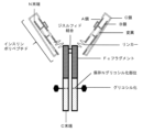
1つの態様において、本開示は、インスリンポリペプチドと、Fcフラグメントと、を含む融合タンパク質であって、上記インスリンポリペプチドおよび前記Fcフラグメントはリンカーによって接続されており、上記融合タンパク質は以下の配列:
FVNQHLCGSHLVEALALVCGERGFHYGGGGGGSGGGGGIVEQCCTSTCSLDQLENYCGGGGGAGGGGAGGGGAGGGGGDKTHTCPPCPAPELLGGPSVFLFPPKPKDTLMISRTPEVTCVVVDVSHEDPEVKFNWYVDGVEVHNAKTKPREEQYSSTYRVVSVLTVLHQDWLNGKEYKCKVSNKALPAPIEKTISKAKGQPREPQVYTLPPSRDELTKNQVSLTCLVKGFYPSDIAVEWESNGQPENNYKTTPPVLDSDGSFFLYSKLTVDKSRWQQGNVFSCSVMHEALHNHYTQKSLSLSPG(配列番号87)、
を含む、融合タンパク質を提供する。
In one aspect, the present disclosure provides a fusion protein comprising an insulin polypeptide and an Fc fragment, wherein the insulin polypeptide and the Fc fragment are connected by a linker, and the fusion protein has the following sequence:
FVNQHLCGSHLVEALALVCGERGFHYGGGGGGSGGGGGIVEQCCTSTCSLDQLENYCGGGGGAGGGGAGGGGAGGGGGGDKTHTCPPCPAPELLGGPSVFLFPPKPKDTLMISRTPEVTCVVVD VSHEDPEVKFNWYVDGVEVHNAKTKPREEQYSSTYRVVSVLTVLHQDWLNGKEYKCKVSNKALPAPIEKTISKAKGQPREPQVYTLPPSRDELTKNQVSLTCLVKGFYPSDIAVEWESNGQPENN YKTTPPVLDSDGSFFLYSKLTVDKSRWQQGNVFSCSVMHEALHNHYTQKSLSLSPG (SEQ ID NO: 87),
Provided are fusion proteins comprising:
いくつかの実施形態において、本開示は、インスリンポリペプチドと、Fcフラグメントと、を含む融合タンパク質であって、上記インスリンポリペプチドおよび上記Fcフラグメントはリンカーによって接続されており、上記Fcフラグメントはヒト由来であり、且つ以下の配列:
DKTHTCPPCPAPELLGGPSVFLFPPKPKDTLMISRTPEVTCVVVDVSHEDPEVKFNWYVDGVEVHNAKTKPREEQYX1STYRVVSVLTVLHQDWLNGKEYKCKVSNKALPAPIEKTISKAKGQPREPQVYTLPPSRDELTKNQVSLTCLVKGFYPSDIAVEWESNGQPENNYKTTPPVLDSDGSFFLYSKLTVDKSRWQQGNVFSCSVMHEALHNHYTQKSLSLSPG(配列番号77)、
を含み、X1はS、D、A、またはRであり、上記インスリンポリペプチドはC鎖を介してインスリンA鎖アナログに連結されたインスリンB鎖アナログからなり、上記インスリンポリペプチドのインスリンB鎖アナログのN末端から16番目のアミノ酸(すなわち、B16)はアラニンである(すなわち、B16A)、融合タンパク質を提供する。
In some embodiments, the present disclosure provides a fusion protein comprising an insulin polypeptide and an Fc fragment, wherein the insulin polypeptide and the Fc fragment are connected by a linker, and the Fc fragment is of human origin. and the following array:
DKTHTCPPCPAPELLGGPSVFLFPPKPKDTLMISRTPEVTCVVVDVSHEDPEVKFNWYVDGVEVHNAKTKPREEQYX 1 STYRVVSVLTVLHQDWLNGKEYKCKVSNKALPAPIEKTISKAK GQPREPQVYTLPPSRDELTKNQVSLTCLVKGFYPSDIAVEWESNGQPENNYKTTPPVLDSDGSFFLYSKLTVDKSRWQQGNVFSCSVMHEALHNHYTQKSLSLSPG (SEQ ID NO: 77),
, X 1 is S, D, A, or R, and the insulin polypeptide consists of an insulin B chain analog linked via a C chain to an insulin A chain analog, and the insulin B chain of the insulin polypeptide The 16th amino acid from the N-terminus of the analog (ie, B16) is alanine (ie, B16A), providing a fusion protein.
いくつかの実施形態において、上記インスリンポリペプチドは、配列:
FVNQHLCGSX1LVEALALVCGERGFHYGGGGGGSGGGGGIVEQCCX2STCSLDQLENYC(配列番号9)、
を含み、X1はDではなく、X2はHではない。いくつかの実施形態において、上記インスリンポリペプチドは、以下の配列:
FVNQHLCGSHLVEALALVCGERGFHYGGGGGGSGGGGGIVEQCCTSTCSLDQLENYC(配列番号10)、
を含む。
In some embodiments, the insulin polypeptide has the sequence:
FVNQHLCGSX 1 LVEALALVCGERGFHYGGGGGGGSGGGGIVEQCCX 2 STCSLDQLENYC (SEQ ID NO: 9),
, X 1 is not D and X 2 is not H. In some embodiments, the insulin polypeptide has the following sequence:
FVNQHLCGSHLVEALALVCGERGFHYGGGGGGGSGGGGIVEQCCTSTCSLDQLENYC (SEQ ID NO: 10),
including.
いくつかの構成において、上記リンカーは、配列:
GGGGGQGGGGQGGGGQGGGGG(配列番号13)、
を含む。構成において、上記リンカーは、配列:
GGGGGAGGGGAGGGGAGGGGG(配列番号67)、
を含む。構成において、上記リンカーは、配列:
GGGGAGGGG(配列番号11)、
を含む。
In some configurations, the linker includes an array:
GGGGGQGGGGQGGGGQGGGGG (SEQ ID NO: 13),
including. In the configuration, the linker above has an array:
GGGGGAGGGGGAGGGGAGGGGG (SEQ ID NO: 67),
including. In the configuration, the linker above has an array:
GGGGAGGGG (SEQ ID NO: 11),
including.
いくつかの実施形態において、上記融合タンパク質は、以下の配列:
FVNQHLCGSHLVEALALVCGERGFHYGGGGGGSGGGGGIVEQCCTSTCSLDQLENYCGGGGAGGGGDKTHTCPPCPAPELLGGPSVFLFPPKPKDTLMISRTPEVTCVVVDVSHEDPEVKFNWYVDGVEVHNAKTKPREEQYSSTYRVVSVLTVLHQDWLNGKEYKCKVSNKALPAPIEKTISKAKGQPREPQVYTLPPSRDELTKNQVSLTCLVKGFYPSDIAVEWESNGQPENNYKTTPPVLDSDGSFFLYSKLTVDKSRWQQGNVFSCSVMHEALHNHYTQKSLSLSPG(配列番号89)、
を含む。
In some embodiments, the fusion protein has the following sequence:
FVNQHLCGSHLVEALALVCGERGFHYGGGGGGGSGGGGIVEQCCTSTCSLDQLENYCGGGGAGGGGDKTHTCPPCPAPELLGGPSVFLFPPKPKDTLMISRTPEVTCVVVDVSHEDPEVKFNW YVDGVEVHNAKTKPREEQYSSTYRVVSVLTVLHQDWLNGKEYKCKVSNKALPAPIEKTISKAKGQPREPQVYTLPPSRDELTKNQVSLTCLVKGFYPSDIAVEWESNGQPENNYKTTPPVLDSDG SFFLYSKLTVDKSRWQQGNVFSCSVMHEALHNHYTQKSLSLSPG (SEQ ID NO: 89),
including.
いくつかの実施形態において、上記融合タンパク質は、以下の配列:
FVNQHLCGSHLVEALALVCGERGFHYGGGGGGSGGGGGIVEQCCTSTCSLDQLENYCGGGGGQGGGGQGGGGQGGGGGDKTHTCPPCPAPELLGGPSVFLFPPKPKDTLMISRTPEVTCVVVDVSHEDPEVKFNWYVDGVEVHNAKTKPREEQYSSTYRVVSVLTVLHQDWLNGKEYKCKVSNKALPAPIEKTISKAKGQPREPQVYTLPPSRDELTKNQVSLTCLVKGFYPSDIAVEWESNGQPENNYKTTPPVLDSDGSFFLYSKLTVDKSRWQQGNVFSCSVMHEALHNHYTQKSLSLSPG(配列番号78)、
を含む。
In some embodiments, the fusion protein has the following sequence:
FVNQHLCGSHLVEALALVCGERGFHYGGGGGGSGGGGGIVEQCCTSTCSLDQLENYCGGGGGQGGGGQGGGGQGGGGGDKTHTCPPCPAPELLGGPSVFLFPPKPKDTLMISRTPEVTCVVVD VSHEDPEVKFNWYVDGVEVHNAKTKPREEQYSSTYRVVSVLTVLHQDWLNGKEYKCKVSNKALPAPIEKTISKAKGQPREPQVYTLPPSRDELTKNQVSLTCLVKGFYPSDIAVEWESNGQPENN YKTTPPVLDSDGSFFLYSKLTVDKSRWQQGNVFSCSVMHEALHNHYTQKSLSLSPG (SEQ ID NO: 78),
including.
いくつかの実施形態において、上記融合タンパク質は、以下の配列:
FVNQHLCGSHLVEALALVCGERGFHYGGGGGGSGGGGGIVEQCCTSTCSLDQLENYCGGGGGQGGGGQGGGGQGGGGGDKTHTCPPCPAPELLGGPSVFLFPPKPKDTLMISRTPEVTCVVVDVSHEDPEVKFNWYVDGVEVHNAKTKPREEQYDSTYRVVSVLTVLHQDWLNGKEYKCKVSNKALPAPIEKTISKAKGQPREPQVYTLPPSRDELTKNQVSLTCLVKGFYPSDIAVEWESNGQPENNYKTTPPVLDSDGSFFLYSKLTVDKSRWQQGNVFSCSVMHEALHNHYTQKSLSLSPG(配列番号80)、
を含む。
In some embodiments, the fusion protein has the following sequence:
FVNQHLCGSHLVEALALVCGERGFHYGGGGGGSGGGGGIVEQCCTSTCSLDQLENYCGGGGGQGGGGQGGGGQGGGGGDKTHTCPPCPAPELLGGPSVFLFPPKPKDTLMISRTPEVTCVVVD VSHEDPEVKFNWYVDGVEVHNAKTKPREEQYDSTYRVVSVLTVLHQDWLNGKEYKCKVSNKALPAPIEKTISKAKGQPREPQVYTLPPSRDELTKNQVSLTCLVKGFYPSDIAVEWESNGQPENN YKTTPPVLDSDGSFFLYSKLTVDKSRWQQGNVFSCSVMHEALHNHYTQKSLSLSPG (SEQ ID NO: 80),
including.
いくつかの実施形態において、上記融合タンパク質は、以下の配列:
FVNQHLCGSHLVEALALVCGERGFHYGGGGGGSGGGGGIVEQCCTSTCSLDQLENYCGGGGGQGGGGQGGGGQGGGGGDKTHTCPPCPAPELLGGPSVFLFPPKPKDTLMISRTPEVTCVVVDVSHEDPEVKFNWYVDGVEVHNAKTKPREEQYASTYRVVSVLTVLHQDWLNGKEYKCKVSNKALPAPIEKTISKAKGQPREPQVYTLPPSRDELTKNQVSLTCLVKGFYPSDIAVEWESNGQPENNYKTTPPVLDSDGSFFLYSKLTVDKSRWQQGNVFSCSVMHEALHNHYTQKSLSLSPG(配列番号82)、
を含む。
In some embodiments, the fusion protein has the following sequence:
FVNQHLCGSHLVEALALVCGERGFHYGGGGGGSGGGGGIVEQCCTSTCSLDQLENYCGGGGGQGGGGQGGGGQGGGGGDKTHTCPPCPAPELLGGPSVFLFPPKPKDTLMISRTPEVTCVVVD VSHEDPEVKFNWYVDGVEVHNAKTKPREEQYASTYRVVSVLTVLHQDWLNGKEYKCKVSNKALPAPIEKTISKAKGQPREPQVYTLPPSRDELTKNQVSLTCLVKGFYPSDIAVEWESNGQPENN YKTTPPVLDSDGSFFLYSKLTVDKSRWQQGNVFSCSVMHEALHNHYTQKSLSLSPG (SEQ ID NO: 82),
including.
いくつかの実施形態において、上記融合タンパク質は、以下の配列:
FVNQHLCGSHLVEALALVCGERGFHYGGGGGGSGGGGGIVEQCCTSTCSLDQLENYCGGGGGQGGGGQGGGGQGGGGGDKTHTCPPCPAPELLGGPSVFLFPPKPKDTLMISRTPEVTCVVVDVSHEDPEVKFNWYVDGVEVHNAKTKPREEQYRSTYRVVSVLTVLHQDWLNGKEYKCKVSNKALPAPIEKTISKAKGQPREPQVYTLPPSRDELTKNQVSLTCLVKGFYPSDIAVEWESNGQPENNYKTTPPVLDSDGSFFLYSKLTVDKSRWQQGNVFSCSVMHEALHNHYTQKSLSLSPG(配列番号84)、
を含む。
In some embodiments, the fusion protein has the following sequence:
FVNQHLCGSHLVEALALVCGERGFHYGGGGGGSGGGGGIVEQCCTSTCSLDQLENYCGGGGGQGGGGQGGGGQGGGGGDKTHTCPPCPAPELLGGPSVFLFPPKPKDTLMISRTPEVTCVVVD VSHEDPEVKFNWYVDGVEVHNAKTKPREEQYRSTYRVVSVLTVLHQDWLNGKEYKCKVSNKALPAPIEKTISKAKGQPREPQVYTLPPSRDELTKNQVSLTCLVKGFYPSDIAVEWESNGQPENN YKTTPPVLDSDGSFFLYSKLTVDKSRWQQGNVFSCSVMHEALHNHYTQKSLSLSPG (SEQ ID NO: 84),
including.
いくつかの実施形態において、上記融合タンパク質は、N末端からC末端へ以下の配向で各ドメインを含む:
(N末端)-インスリンポリペプチド-リンカー-Fcフラグメント-(C末端)。
In some embodiments, the fusion protein comprises each domain in the following orientation from N-terminus to C-terminus:
(N-terminus) - insulin polypeptide - linker - Fc fragment - (C-terminus).
いくつかの実施形態において、上記融合タンパク質は、ホモ二量体である。例において、上記融合タンパク質のホモ二量体率は90%超である。いくつかの例において、上記融合タンパク質は、HEK293細胞またはCHO細胞の一方を用いて作製され、プロテインAビーズまたはプロテインAカラムを用いた精製後に得られるホモ二量体の力価は150mg/Lを超える。実施形態において、上記融合タンパク質のインスリン受容体IC50は5000nM以下である。いくつかの実施形態において、上記融合タンパク質のインスリン受容体IC50は2400nM以下である。 In some embodiments, the fusion protein is a homodimer. In an example, the homodimerization rate of the fusion protein is greater than 90%. In some instances, the fusion protein is produced using either HEK293 cells or CHO cells, and the resulting homodimer titer is greater than 150 mg/L after purification using Protein A beads or Protein A columns. exceed. In embodiments, the insulin receptor IC50 of the fusion protein is 5000 nM or less. In some embodiments, the insulin receptor IC50 of the fusion protein is 2400 nM or less.
いくつかの実施形態において、上記融合タンパク質のヒトFcRn受容体EC50は1000ng/mL以下である。例において、上記融合タンパク質の、ビオチン化FcγRI濃度が3000ng/mLである場合のヒトFcγRI受容体アッセイにおけるOD450比は0.50以下である。実施形態において、ビオチン化C1q濃度が1000ng/mLである場合のヒトC1qアッセイにおけるOD450比は0.35以下である。実施形態において、上記融合タンパク質は医薬組成物として製剤化される。いくつかの例において、上記医薬組成物中の上記融合タンパク質の濃度は約3mg/mL以上である。いくつかの実施形態において、上記医薬組成物は、皮下投与に好適である。 In some embodiments, the fusion protein has a human FcRn receptor EC50 of 1000 ng/mL or less. In an example, the OD450 ratio of the above fusion protein in a human FcγRI receptor assay when the biotinylated FcγRI concentration is 3000 ng/mL is 0.50 or less. In embodiments, the OD450 ratio in the human C1q assay is 0.35 or less when the biotinylated C1q concentration is 1000 ng/mL. In embodiments, the fusion protein is formulated as a pharmaceutical composition. In some examples, the concentration of the fusion protein in the pharmaceutical composition is about 3 mg/mL or more. In some embodiments, the pharmaceutical compositions are suitable for subcutaneous administration.
実施形態において、患者の血糖値を低下させる方法として、上記融合タンパク質またはその医薬組成物の生理学的有効量が患者に投与され得る。例において、上記患者は糖尿病と診断された患者である。いくつかの実施形態において、上記融合タンパク質は皮下投与される。上記融合タンパク質は、患者に毎日、週2回、または週1回投与されてもよい。いくつかの例において、上記融合タンパク質は、0.025~0.500mg/kg/週の投与量で患者に週1回投与される。 In embodiments, a physiologically effective amount of the fusion protein or pharmaceutical composition thereof can be administered to a patient as a method of lowering blood glucose levels in the patient. In an example, the patient is a patient diagnosed with diabetes. In some embodiments, the fusion protein is administered subcutaneously. The fusion protein may be administered to the patient daily, twice a week, or once a week. In some examples, the fusion protein is administered to the patient once a week at a dosage of 0.025-0.500 mg/kg/week.
実施形態において、上記融合タンパク質を発現するように細胞が改変されてもよい。上記細胞は、上記融合タンパク質をコードする核酸でトランスフェクトされてもよい。いくつかの例において、上記細胞は、HEK293細胞またはCHO細胞である。 In embodiments, cells may be modified to express the fusion protein described above. The cell may be transfected with a nucleic acid encoding the fusion protein. In some examples, the cells are HEK293 cells or CHO cells.
実施形態において、配列番号87の上記融合タンパク質をコードする核酸(cDNA)は、以下の核酸配列:
atggaatggagctgggtctttctcttcttcctgtcagtaacgactggtgtccactccttcgtgaaccagcacctgtgcggctcccacctggtggaagctctggcactcgtgtgcggcgagcggggcttccactacgggggtggcggaggaggttctggtggcggcggaggcatcgtggaacagtgctgcacctccacctgctccctggaccagctggaaaactactgcggtggcggaggtggtgcaggaggcggtggagccggtggaggtggggctggaggaggcgggggagacaaaactcacacatgcccaccgtgcccagcacctgaactcctggggggaccgtcagtcttcctcttccccccaaaacccaaggacaccctcatgatctcccggacccctgaggtcacatgcgtggtggtggacgtgagccacgaagaccctgaggtcaagttcaactggtacgtggacggcgtggaggtgcataatgccaagacaaagccgcgggaggagcagtacagcagcacgtaccgtgtggtcagcgtcctcaccgtcctgcaccaggactggctgaatggcaaggagtacaagtgcaaggtctccaacaaagccctcccagcccccatcgagaaaaccatctccaaagccaaagggcagccccgagaaccacaggtgtacaccctgcccccatcccgggatgagctgaccaagaaccaggtcagcctgacctgcctggtcaaaggcttctatcccagcgacatcgccgtggagtgggagagcaatgggcagccggagaacaactacaagaccacgcctcccgtgctggactccgacggctccttcttcctctacagcaagctcaccgtggacaagagcaggtggcagcaggggaacgtcttctcatgctccgtgatgcatgaggctctgcacaaccactacacgcagaagagcctctccctgtctccgggttag(配列番号88)、
を含む。
In an embodiment, the nucleic acid (cDNA) encoding the fusion protein of SEQ ID NO: 87 has the following nucleic acid sequence:
atggaatggagctgggtctttctcttcttcctgtcagtaacgactggtgtccactccttcgtgaaccagcacctgtgcggctcccacctggtggaagctctggcactcgtgtgcggcgagcgg ggcttccactacgggggtggcggaggaggttctggtggcggcggaggcatcgtggaacagtgctgcacctccacctgctccctggaccagctggaaaactactgcggtggcggaggtggtgcagg aggcggtggagccggtggaggtggggctggaggggcggggagagacaaaactcacacatgcccaccgtgcccagcacctgaactcctggggggaccgtcagtcttcctcttcccccccaaaaccca aggacacctcatgatctcccggacccctgaggtcacatgcgtggtggtggacgtgagccacgaagaccctgaggtcaagttcaactggtacgtggacggcgtggaggtgcataatgccaagaca aagccgcggaggagcagtacagcagcacgtaccgtgtggtcagcgtcctcaccgtcctgcaccaggactggctgaatggcaaggagtacaagtgcaaggtctccaacaaagccctcccagcccc catcgagaaaaccatctccaaagccaaaggcagcccgagaaccacaggtgtacacctgcccccatcccgggatgagctgaccaagaaccaggtcagcctgacctgcctggtcaaaggcttct atcccagcgacatcgccgtggagtgggagagcaatgggcagccggagaaacaactacaagaccacgcctcccgtgctggactccgacggctccttcttcctctacagcaagctcaccgtggacaag agcaggtggcagcagggaacgtcttctcatgctccgtgatgcatgaggctctgcacaaccactacacgcagaagagcctctccctgtctccgggttag (SEQ ID NO: 88),
including.
実施形態において、配列番号89の上記融合タンパク質をコードする核酸(cDNA)は、以下の核酸配列:
atggaatggagctgggtctttctcttcttcctgtcagtaacgactggtgtccactccttcgtgaaccagcacctgtgcggctcccacctggtggaagctctggcactcgtgtgcggcgagcggggcttccactacgggggtggcggaggaggttctggtggcggcggaggcatcgtggaacagtgctgcacctccacctgctccctggaccagctggaaaactactgcggtggcggaggtgccggaggcgggggagacaaaactcacacatgcccaccgtgcccagcacctgaactcctggggggaccgtcagtcttcctcttccccccaaaacccaaggacaccctcatgatctcccggacccctgaggtcacatgcgtggtggtggacgtgagccacgaagaccctgaggtcaagttcaactggtacgtggacggcgtggaggtgcataatgccaagacaaagccgcgggaggagcagtacagcagcacgtaccgtgtggtcagcgtcctcaccgtcctgcaccaggactggctgaatggcaaggagtacaagtgcaaggtctccaacaaagccctcccagcccccatcgagaaaaccatctccaaagccaaagggcagccccgagaaccacaggtgtacaccctgcccccatcccgggatgagctgaccaagaaccaggtcagcctgacctgcctggtcaaaggcttctatcccagcgacatcgccgtggagtgggagagcaatgggcagccggagaacaactacaagaccacgcctcccgtgctggactccgacggctccttcttcctctacagcaagctcaccgtggacaagagcaggtggcagcaggggaacgtcttctcatgctccgtgatgcatgaggctctgcacaaccactacacgcagaagagcctctccctgtctccgggttag(配列番号90)、
を含む。
In an embodiment, the nucleic acid (cDNA) encoding the fusion protein of SEQ ID NO: 89 has the following nucleic acid sequence:
atggaatggagctgggtctttctcttcttcctgtcagtaacgactggtgtccactccttcgtgaaccagcacctgtgcggctcccacctggtggaagctctggcactcgtgtgcggcgagcgg ggcttccactacgggggtggcggaggaggttctggtggcggcggaggcatcgtggaacagtgctgcacctccacctgctccctggaccagctggaaaactactgcggtggcggaggtgccggagg cggggagacaaaactcacacatgcccaccgtgcccagcacctgaactcctgggggaccgtcagtcttcctcttcccccaaaacccaaggacaccctcatgatctcccggacccctgaggtca catgcgtggtggtggacgtgagccacgaagacctgaggtcaagttcaactggtacgtggacggcgtggaggtgcataatgcaagacaaagccgcgggaggagcagtacagcagcacgtaccgt gtggtcagcgtcctcaccgtcctgcaccaggactggctgaatggcaaggagtacaagtgcaaggtctccaacaaagccctcccagcccccatcgagaaaaccatctccaaagccaaagggcagcc ccgagaaccacaggtgtacacctgcccccatcccgggatgagctgaccaagaaccaggtcagcctgacctgcctggtcaaaggcttctatcccagcgacatcgccgtggagtgggagagcaatg ggcagccggaacaactacaagaccacgcctcccgtgctggactccgacggctccttcttcctctacagcaagctcaccgtggacaagagcaggtggcagcagggaacgtcttctcatgctcc gtgatgcatgaggctctgcacaaccactacacgcagaagagcctctccctgtctccgggttag (SEQ ID NO: 90),
including.
実施形態において、配列番号78の上記融合タンパク質をコードする核酸(cDNA)は、以下の核酸配列:
atggaatggagctgggtctttctcttcttcctgtcagtaacgactggtgtccactccttcgtgaaccagcacctgtgcggctcccacctggtggaagctctggcactcgtgtgcggcgagcggggcttccactacgggggtggcggaggaggttctggtggcggcggaggcatcgtggaacagtgctgcacctccacctgctccctggaccagctggaaaactactgcggtggcggaggtggtcaaggaggcggtggacagggtggaggtgggcagggaggaggcgggggagacaaaactcacacatgcccaccgtgcccagcacctgaactcctggggggaccgtcagtcttcctcttccccccaaaacccaaggacaccctcatgatctcccggacccctgaggtcacatgcgtggtggtggacgtgagccacgaagaccctgaggtcaagttcaactggtacgtggacggcgtggaggtgcataatgccaagacaaagccgcgggaggagcagtacagcagcacgtaccgtgtggtcagcgtcctcaccgtcctgcaccaggactggctgaatggcaaggagtacaagtgcaaggtctccaacaaagccctcccagcccccatcgagaaaaccatctccaaagccaaagggcagccccgagaaccacaggtgtacaccctgcccccatcccgggatgagctgaccaagaaccaggtcagcctgacctgcctggtcaaaggcttctatcccagcgacatcgccgtggagtgggagagcaatgggcagccggagaacaactacaagaccacgcctcccgtgctggactccgacggctccttcttcctctacagcaagctcaccgtggacaagagcaggtggcagcaggggaacgtcttctcatgctccgtgatgcatgaggctctgcacaaccactacacgcagaagagcctctccctgtctccgggttag(配列番号79)、
を含む。
In an embodiment, the nucleic acid (cDNA) encoding the fusion protein of SEQ ID NO: 78 has the following nucleic acid sequence:
atggaatggagctgggtctttctcttcttcctgtcagtaacgactggtgtccactccttcgtgaaccagcacctgtgcggctcccacctggtggaagctctggcactcgtgtgcggcgagcgg ggcttccactacgggggtggcggaggggttctggtggcggcggaggcatcgtggaacagtgctgcacctccacctgctccctggaccagctggaaaactactgcggtggcggaggtggtcaagg aggcggtggacagggtggaggtgggcaggaggaggcggggagagacaaaactcacacatgcccaccgtgcccagcacctgaactcctggggggaccgtcagtcttcctcttcccccccaaaaccca aggacaccctcatgatctcccggacccctgaggtcacatgcgtggtggtggacgtgagccacgaagaccctgaggtcaagttcaactggtacgtggacggcgtggaggtgcataatgccaagaca aagccgcggaggagcagtacagcagcacgtaccgtgtggtcagcgtcctcaccgtcctgcaccaggactggctgaatggcaaggagtacaagtgcaaggtctccaacaaagccctcccagcccc catcgagaaaaccatctccaaagccaaaggcagcccgagaaccacaggtgtacacctgcccccatcccgggatgagctgaccaagaaccaggtcagcctgacctgcctggtcaaaggcttct atcccagcgacatcgccgtggagtgggagagcaatgggcagccggagaaacaactacaagaccacgcctcccgtgctggactccgacggctccttcttcctctacagcaagctcaccgtggacaag agcaggtggcagcagggaacgtcttctcatgctccgtgatgcatgaggctctgcacaaccactacacgcagaagagcctctccctgtctccgggttag (SEQ ID NO: 79),
including.
実施形態において、配列番号80の上記融合タンパク質をコードする核酸(cDNA)は、以下の核酸配列:
atggaatggagctgggtctttctcttcttcctgtcagtaacgactggtgtccactccttcgtgaaccagcacctgtgcggctcccacctggtggaagctctggcactcgtgtgcggcgagcggggcttccactacgggggtggcggaggaggttctggtggcggcggaggcatcgtggaacagtgctgcacctccacctgctccctggaccagctggaaaactactgcggtggcggaggtggtcaaggaggcggtggacagggtggaggtgggcagggaggaggcgggggagacaaaactcacacatgcccaccgtgcccagcacctgaactcctggggggaccgtcagtcttcctcttccccccaaaacccaaggacaccctcatgatctcccggacccctgaggtcacatgcgtggtggtggacgtgagccacgaagaccctgaggtcaagttcaactggtacgtggacggcgtggaggtgcataatgccaagacaaagccgcgggaggagcagtacgacagcacgtaccgtgtggtcagcgtcctcaccgtcctgcaccaggactggctgaatggcaaggagtacaagtgcaaggtctccaacaaagccctcccagcccccatcgagaaaaccatctccaaagccaaagggcagccccgagaaccacaggtgtacaccctgcccccatcccgggatgagctgaccaagaaccaggtcagcctgacctgcctggtcaaaggcttctatcccagcgacatcgccgtggagtgggagagcaatgggcagccggagaacaactacaagaccacgcctcccgtgctggactccgacggctccttcttcctctacagcaagctcaccgtggacaagagcaggtggcagcaggggaacgtcttctcatgctccgtgatgcatgaggctctgcacaaccactacacgcagaagagcctctccctgtctccgggttag(配列番号81)、
を含む。
In an embodiment, the nucleic acid (cDNA) encoding the fusion protein of SEQ ID NO: 80 has the following nucleic acid sequence:
atggaatggagctgggtctttctcttcttcctgtcagtaacgactggtgtccactccttcgtgaaccagcacctgtgcggctcccacctggtggaagctctggcactcgtgtgcggcgagcgg ggcttccactacgggggtggcggaggggttctggtggcggcggaggcatcgtggaacagtgctgcacctccacctgctccctggaccagctggaaaactactgcggtggcggaggtggtcaagg aggcggtggacagggtggaggtgggcaggaggaggcggggagagacaaaactcacacatgcccaccgtgcccagcacctgaactcctggggggaccgtcagtcttcctcttcccccccaaaaccca aggacacctcatgatctcccggacccctgaggtcacatgcgtggtggtggacgtgagccacgaagaccctgaggtcaagttcaactggtacgtggacggcgtggaggtgcataatgccaagaca aagccgcggaggagcagtacgacagcacgtaccgtgtggtcagcgtcctcaccgtcctgcaccaggactggctgaatggcaaggagtacaagtgcaaggtctccaacaaagccctcccagcccc catcgagaaaaccatctccaaagccaaaggcagcccgagaaccacaggtgtacacctgcccccatcccgggatgagctgaccaagaaccaggtcagcctgacctgcctggtcaaaggcttct atcccagcgacatcgccgtggagtgggagagcaatgggcagccggagaaacaactacaagaccacgcctcccgtgctggactccgacggctccttcttcctctacagcaagctcaccgtggacaag agcaggtggcagcagggaacgtcttctcatgctccgtgatgcatgaggctctgcacaaccactacacgcagaagagcctctccctgtctccgggttag (SEQ ID NO: 81),
including.
実施形態において、配列番号82の上記融合タンパク質をコードする核酸(cDNA)は、以下の核酸配列:
atggaatggagctgggtctttctcttcttcctgtcagtaacgactggtgtccactccttcgtgaaccagcacctgtgcggctcccacctggtggaagctctggcactcgtgtgcggcgagcggggcttccactacgggggtggcggaggaggttctggtggcggcggaggcatcgtggaacagtgctgcacctccacctgctccctggaccagctggaaaactactgcggtggcggaggtggtcaaggaggcggtggacagggtggaggtgggcagggaggaggcgggggagacaaaactcacacatgcccaccgtgcccagcacctgaactcctggggggaccgtcagtcttcctcttccccccaaaacccaaggacaccctcatgatctcccggacccctgaggtcacatgcgtggtggtggacgtgagccacgaagaccctgaggtcaagttcaactggtacgtggacggcgtggaggtgcataatgccaagacaaagccgcgggaggagcagtacgccagcacgtaccgtgtggtcagcgtcctcaccgtcctgcaccaggactggctgaatggcaaggagtacaagtgcaaggtctccaacaaagccctcccagcccccatcgagaaaaccatctccaaagccaaagggcagccccgagaaccacaggtgtacaccctgcccccatcccgggatgagctgaccaagaaccaggtcagcctgacctgcctggtcaaaggcttctatcccagcgacatcgccgtggagtgggagagcaatgggcagccggagaacaactacaagaccacgcctcccgtgctggactccgacggctccttcttcctctacagcaagctcaccgtggacaagagcaggtggcagcaggggaacgtcttctcatgctccgtgatgcatgaggctctgcacaaccactacacgcagaagagcctctccctgtctccgggttag(配列番号83)、
を含む。
In an embodiment, the nucleic acid (cDNA) encoding the fusion protein of SEQ ID NO: 82 has the following nucleic acid sequence:
atggaatggagctgggtctttctcttcttcctgtcagtaacgactggtgtccactccttcgtgaaccagcacctgtgcggctcccacctggtggaagctctggcactcgtgtgcggcgagcgg ggcttccactacgggggtggcggaggggttctggtggcggcggaggcatcgtggaacagtgctgcacctccacctgctccctggaccagctggaaaactactgcggtggcggaggtggtcaagg aggcggtggacagggtggaggtgggcaggaggaggcggggagagacaaaactcacacatgcccaccgtgcccagcacctgaactcctggggggaccgtcagtcttcctcttcccccccaaaaccca aggacacctcatgatctcccggacccctgaggtcacatgcgtggtggtggacgtgagccacgaagaccctgaggtcaagttcaactggtacgtggacggcgtggaggtgcataatgccaagaca aagccgcggaggagcagtacgccagcacgtaccgtgtggtcagcgtcctcaccgtcctgcaccaggactggctgaatggcaaggagtacaagtgcaaggtctccaacaaagccctcccagcccc catcgagaaaaccatctccaaagccaaaggcagcccgagaaccacaggtgtacacctgcccccatcccgggatgagctgaccaagaaccaggtcagcctgacctgcctggtcaaaggcttct atcccagcgacatcgccgtggagtgggagagcaatgggcagccggagaaacaactacaagaccacgcctcccgtgctggactccgacggctccttcttcctctacagcaagctcaccgtggacaag agcaggtggcagcagggaacgtcttctcatgctccgtgatgcatgaggctctgcacaaccactacacgcagaagagcctctccctgtctccgggttag (SEQ ID NO: 83),
including.
実施形態において、配列番号84の上記融合タンパク質をコードする核酸(cDNA)は、以下の核酸配列:
atggaatggagctgggtctttctcttcttcctgtcagtaacgactggtgtccactccttcgtgaaccagcacctgtgcggctcccacctggtggaagctctggcactcgtgtgcggcgagcggggcttccactacgggggtggcggaggaggttctggtggcggcggaggcatcgtggaacagtgctgcacctccacctgctccctggaccagctggaaaactactgcggtggcggaggtggtcaaggaggcggtggacagggtggaggtgggcagggaggaggcgggggagacaaaactcacacatgcccaccgtgcccagcacctgaactcctggggggaccgtcagtcttcctcttccccccaaaacccaaggacaccctcatgatctcccggacccctgaggtcacatgcgtggtggtggacgtgagccacgaagaccctgaggtcaagttcaactggtacgtggacggcgtggaggtgcataatgccaagacaaagccgcgggaggagcagtacagaagcacgtaccgtgtggtcagcgtcctcaccgtcctgcaccaggactggctgaatggcaaggagtacaagtgcaaggtctccaacaaagccctcccagcccccatcgagaaaaccatctccaaagccaaagggcagccccgagaaccacaggtgtacaccctgcccccatcccgggatgagctgaccaagaaccaggtcagcctgacctgcctggtcaaaggcttctatcccagcgacatcgccgtggagtgggagagcaatgggcagccggagaacaactacaagaccacgcctcccgtgctggactccgacggctccttcttcctctacagcaagctcaccgtggacaagagcaggtggcagcaggggaacgtcttctcatgctccgtgatgcatgaggctctgcacaaccactacacgcagaagagcctctccctgtctccgggttag(配列番号85)、
を含む。
In an embodiment, the nucleic acid (cDNA) encoding the fusion protein of SEQ ID NO: 84 has the following nucleic acid sequence:
atggaatggagctgggtctttctcttcttcctgtcagtaacgactggtgtccactccttcgtgaaccagcacctgtgcggctcccacctggtggaagctctggcactcgtgtgcggcgagcgg ggcttccactacgggggtggcggaggggttctggtggcggcggaggcatcgtggaacagtgctgcacctccacctgctccctggaccagctggaaaactactgcggtggcggaggtggtcaagg aggcggtggacagggtggaggtgggcaggaggaggcggggagagacaaaactcacacatgcccaccgtgcccagcacctgaactcctggggggaccgtcagtcttcctcttcccccccaaaaccca aggacacctcatgatctcccggacccctgaggtcacatgcgtggtggtggacgtgagccacgaagaccctgaggtcaagttcaactggtacgtggacggcgtggaggtgcataatgccaagaca aagccgcggaggagcagtacagaaagcacgtaccgtgtggtcagcgtcctcaccgtcctgcaccaggactggctgaatggcaaggagtacaagtgcaaggtctccaacaaagccctcccagcccc catcgagaaaaccatctccaaagccaaaggcagcccgagaaccacaggtgtacacctgcccccatcccgggatgagctgaccaagaaccaggtcagcctgacctgcctggtcaaaggcttct atcccagcgacatcgccgtggagtgggagagcaatgggcagccggagaaacaactacaagaccacgcctcccgtgctggactccgacggctccttcttcctctacagcaagctcaccgtggacaag agcaggtggcagcagggaacgtcttctcatgctccgtgatgcatgaggctctgcacaaccactacacgcagaagagcctctccctgtctccgggttag (SEQ ID NO: 85),
including.
インスリン治療は、必要な投与回数が少ないほど(例えば、週1回の注射)、患者にとっての負担が少なくなり、コンプライアンスの改善、グルコースコントロールの改善、そして最終的には長期健康アウトカムの改善に繋がる。本明細書に開示されるように、ヒト臨床用に提唱された超長時間作用型インスリン治療は、インビボでの作用を延長するためにヒトFcフラグメントを利用するインスリン-Fc融合タンパク質を含んでいる。糖尿病の超長時間作用型治療に適したインスリン-Fc融合タンパク質は、様々な設計目標を満たす必要がある。糖尿病の超長期作用型治療に適したインスリン-Fc融合タンパク質は、哺乳類細胞、例えばヒト胎児由来腎臓(HEK、例えばHEK293)細胞で、所望のホモ二量体生成物が許容できる力価(例えば、一過性にトランスフェクトされたHEK細胞から50mg/Lを超えるホモ二量体力価、HEK細胞から一過性にトランスフェクトされた75mg/Lを超えるホモ二量体力価、一過性にトランスフェクトされたHEK細胞から100mg/Lを超えるホモ二量体力価、一過性にトランスフェクトされたHEK細胞から150mg/Lを超えるホモ二量体力価など)を有して、製造可能であることが望ましい。ホモ二量体力価が150mg/Lを超えるヒトインスリン-Fc融合タンパク質の構成のみを、本発明では有用と見なしているが、これは、一過性にトランスフェクトされたHEK細胞におけるこのレベル未満のホモ二量体力価では、比較的コモディティ化したヒトインスリン市場の低製造コスト要件を満たす安定にトランスフェクトされたチャイニーズハムスター卵巣(CHO)細胞における商業生産ホモ二量体力価が得られにくいことが、経験的に分かっているからである。 Insulin treatment is less burdensome for patients when fewer doses are required (e.g., weekly injections), leading to improved compliance, better glucose control, and ultimately improved long-term health outcomes. . As disclosed herein, ultra-long-acting insulin treatments proposed for human clinical use include insulin-Fc fusion proteins that utilize human Fc fragments to prolong action in vivo. . Insulin-Fc fusion proteins suitable for ultra-long-acting treatment of diabetes must meet various design goals. Insulin-Fc fusion proteins suitable for ultra-long-acting treatment of diabetes mellitus are suitable for use in mammalian cells, such as human embryonic kidney (HEK, e.g., HEK293) cells, in which the desired homodimeric product is produced at an acceptable potency (e.g., Homodimer titer greater than 50 mg/L from transiently transfected HEK cells, Homodimer titer greater than 75 mg/L transiently transfected from HEK cells, transiently transfected (e.g., a homodimer titer of greater than 100 mg/L from transfected HEK cells, a homodimer titer of greater than 150 mg/L from transiently transfected HEK cells, etc.). desirable. Only constructs of human insulin-Fc fusion proteins with homodimer titers greater than 150 mg/L are considered useful in the present invention, which are below this level in transiently transfected HEK cells. Homodimer titers are difficult to obtain commercially produced homodimer titers in stably transfected Chinese hamster ovary (CHO) cells that meet the low manufacturing cost requirements of the relatively commoditized human insulin market. This is because we know this from experience.
さらに、インスリン-Fc融合タンパク質は、4℃でのIM-9 IR結合アッセイで測定した場合に、かなりの親和性(例えば、5000nM未満のIC50、4000nM未満のIC50、3000nM未満のIC50、2400nM未満のIC50、より好ましくは2000nM未満IC50など)でIRと結合する必要がある。経験に基づいて、IR活性IC50値が5000nM未満の分子のみを、必要な生物活性を示す可能性があるものと見なしている。好ましい実施形態において、インスリン-Fc融合タンパク質は、2400nM未満のIR活性IC50値、より好ましくは2000nM未満のIR活性IC50値を示す。インスリン-Fc融合タンパク質の構成はまた、投与頻度を少なくすることを妥当なものとするために、インビボで持続的な生物活性(例えば、約2時間超、約6時間超、約9時間超、約12時間超、約18時間超、約1日超、約1.5日超、約2日超、約2.5日超、約3日超、約4日超、約5日超、約6日超、約7日超、またはそれ以上のグルコース低減活性を示す)を示す必要もある。インスリン-Fc融合タンパク質の構成はまた、インビボにおける系滞留時間の延長(例えば、血清半減期は3日超、またはそれ以上でなければならない)も示す必要がある。所与のインスリン-Fc融合タンパク質の持続的な生物活性および滞留時間の延長は、抗体およびFc融合タンパク質のインビボ排出半減期の延長に関与する、FcRn受容体との結合能によって予測することができる。FcRn受容体活性は、典型的には、マイクロプレートリーダーで測定されるOD450nmの値を用いたアッセイ(例えば、酵素結合免疫吸着測定法(ELISA)アッセイ)で測定される、インスリン-Fc融合タンパク質にその最大結合の半分に到達させるインスリン-Fc融合タンパク質の濃度(すなわち、EC50値)によって評価される。経験に基づいて、1500ng/mL以下(より好ましくは1000ng/mL未満)のヒトFcRn受容体EC50値を示すインスリン-Fc融合タンパク質の構成は、週1回の投与を妥当なものとするのに十分に長い半減期を示す可能性が最も高い。 Furthermore, insulin-Fc fusion proteins have significant affinities (e.g., IC50 of less than 5000 nM, IC50 of less than 4000 nM, IC50 of less than 2400 nM, It should bind to the IR with an IC50, more preferably an IC50 of less than 2000 nM. Based on experience, only molecules with IR activity IC50 values below 5000 nM are considered as potentially exhibiting the required biological activity. In a preferred embodiment, the insulin-Fc fusion protein exhibits an IR activity IC50 value of less than 2400 nM, more preferably an IR activity IC50 value of less than 2000 nM. The composition of the insulin-Fc fusion protein also provides sustained biological activity in vivo (e.g., greater than about 2 hours, greater than about 6 hours, greater than about 9 hours, More than about 12 hours, more than about 18 hours, more than about 1 day, more than about 1.5 days, more than about 2 days, more than about 2.5 days, more than about 3 days, more than about 4 days, more than about 5 days, about It is also necessary to exhibit glucose-lowering activity for more than 6 days, more than about 7 days, or more. The composition of the insulin-Fc fusion protein should also exhibit an extended residence time in the system in vivo (eg, serum half-life should be greater than 3 days, or longer). The sustained biological activity and prolonged residence time of a given insulin-Fc fusion protein can be predicted by its ability to bind to the FcRn receptor, which is responsible for prolonging the in vivo elimination half-life of antibodies and Fc fusion proteins. . FcRn receptor activity is typically determined by insulin-Fc fusion protein assays (e.g., enzyme-linked immunosorbent assay (ELISA) assays) using OD450nm values measured in a microplate reader. It is assessed by the concentration of insulin-Fc fusion protein that reaches half its maximum binding (ie, EC50 value). Based on experience, compositions of insulin-Fc fusion proteins that exhibit human FcRn receptor EC50 values of 1500 ng/mL or less (more preferably less than 1000 ng/mL) are sufficient to justify weekly dosing. most likely to exhibit a long half-life.
最後に、糖尿病などの慢性疾患の治療に有用であるためには、インスリン-Fc融合タンパク質の構成は、抗薬物抗体、特に反復投与後の上記分子の生物活性を中和する抗体の産生を誘導するものであってはならない。所与のインスリン-Fc融合タンパク質の構成が有害な免疫原性反応を誘発する傾向は、まず、オプソニン化された分子のファゴサイトーシス、炎症メディエーターの放出、抗体依存性細胞傷害を含む多くの免疫系エフェクター機能で重要な役割を担っている、FcγRI受容体と結合する能力によって予測することができる。FcγRI受容体活性は、通常、インスリン-Fc融合タンパク質を所与の濃度とした酵素結合免疫吸着測定法(ELISA)アッセイにおいて、マイクロプレートリーダーで得られる450nmの波長における吸光度(OD450)の値によって評価される。経験に基づいて、ビオチン化FcγRI濃度を3000ng/mLとしたヒトFcγRI受容体アッセイにおけるOD450比(ここで、比のための参照インスリン-Fc融合タンパク質の構成は配列番号76)が0.50以下を示すインスリン-Fc融合タンパク質の構成は、週1回の反復投与を妥当なものとするのに十分に低い免疫原性を示す可能性がある。有害な免疫原性反応を誘導する所与のインスリン-Fc融合タンパク質の構成の傾向は、結合した分子を貪食細胞に除去させる補体カスケードを活性化し、炎症によりさらなる食細胞を引き付け、細胞殺害膜攻撃複合体を活性化する、補体成分1q(C1q)との結合能によっても予測することができる。C1q活性は、通常、マイクロプレート上にコートされるインスリン-Fc融合タンパク質の構成を所与の濃度とした酵素結合免疫吸着測定法(ELISA)アッセイにおいて、マイクロプレートリーダーで得られるOD450値によって評価される。経験に基づいて、ビオチン化C1q濃度を1000ng/mLとしたヒトC1q受容体アッセイにおけるOD450比(ここで、比のための参照インスリン-Fc融合タンパク質の構成は配列番号76)が0.35以下を示すインスリン-Fc融合タンパク質の構成は、週1回の反復投与を妥当なものとするのに十分に低い免疫原性を示す可能性がある。 Finally, to be useful in the treatment of chronic diseases such as diabetes, the composition of insulin-Fc fusion proteins must induce the production of anti-drug antibodies, particularly antibodies that neutralize the biological activity of the molecule after repeated administration. It should not be something that you do. The propensity of a given insulin-Fc fusion protein configuration to elicit deleterious immunogenic responses is primarily due to the phagocytosis of opsonized molecules, release of inflammatory mediators, and antibody-dependent cellular cytotoxicity. It can be predicted by the ability to bind to FcγRI receptor, which plays an important role in system effector function. FcγRI receptor activity is usually assessed by the absorbance at a wavelength of 450 nm (OD450) value obtained with a microplate reader in an enzyme-linked immunosorbent assay (ELISA) assay using a given concentration of insulin-Fc fusion protein. be done. Based on our experience, the OD450 ratio (where the reference insulin-Fc fusion protein composition for the ratio is SEQ ID NO: 76) in the human FcγRI receptor assay with a biotinylated FcγRI concentration of 3000 ng/mL is 0.50 or less. The insulin-Fc fusion protein composition shown may exhibit sufficiently low immunogenicity to justify repeated weekly administration. The propensity of a given insulin-Fc fusion protein composition to induce a deleterious immunogenic response activates the complement cascade that causes bound molecules to be removed by phagocytes, attracts additional phagocytes through inflammation, and induces cell-killing membrane formation. It can also be predicted by its ability to bind to complement component 1q (C1q), which activates the attack complex. C1q activity is typically assessed by OD450 values obtained on a microplate reader in an enzyme-linked immunosorbent assay (ELISA) assay with a given concentration of insulin-Fc fusion protein composition coated onto a microplate. Ru. Based on our experience, the OD450 ratio (where the reference insulin-Fc fusion protein composition for the ratio is SEQ ID NO: 76) in the human C1q receptor assay with a biotinylated C1q concentration of 1000 ng/mL is 0.35 or less. The insulin-Fc fusion protein composition shown may exhibit sufficiently low immunogenicity to justify repeated weekly administration.
ヒト臨床用に提唱された超長時間作用型インスリン治療は、インビボでの作用を延長するためにヒトFcフラグメントを利用するインスリン-Fc融合タンパク質を含んでいる。様々な設計のインスリン-Fc融合タンパク質の構成の挙動を理解するために、まず、イヌ用の超長時間作用型インスリンとして有用なインスリン-Fc融合タンパク質の構成を検討した。ヒトFcフラグメントは、イヌにおいては免疫原性であることで、抗薬物抗体の産生を誘導できると予想されるため、ヒトFcフラグメントをイヌFcフラグメントに置き換えた。 Very long-acting insulin treatments proposed for human clinical use include insulin-Fc fusion proteins that utilize human Fc fragments to prolong action in vivo. To understand the behavior of various designed insulin-Fc fusion protein constructs, we first investigated insulin-Fc fusion protein constructs useful as ultra-long-acting insulin for dogs. Since human Fc fragments are expected to be immunogenic in dogs and thus able to induce the production of anti-drug antibodies, human Fc fragments were replaced with dog Fc fragments.
しかし、予想外にも、インスリン-Fc融合タンパク質の構成でヒトFcフラグメントと任意のイヌFcフラグメントとを単純に交換しても、許容できるホモ二量体力価(例えば、50mg/Lを超えるホモ二量体力価)や、十分に高いNAOC値(例えば、150%FBGL・日・kg/mgを超えるNAOC)を有する生成物を、必ずしも得られないことが分かった。例えば、場合によっては、Fcフラグメントが特定のアイソタイプ(例えば、イヌIgGB)である場合のみ、設計目標を満たすのに十分に高いホモ二量体力価(例えば、50mg/Lを超えるイヌインスリン-Fc融合タンパク質のホモ二量体力価)および許容できる高いNAOC値(例えば、150%FBGL・日・kg/mgを超えるNAOC)を有するインスリン-Fc融合タンパク質の構成が得られた。他の場合では、インスリン-Fc融合タンパク質の構成のインスリンポリペプチドの特定のアミノ酸が、標的種において免疫原性であることにより部位特異的変異を必要とすることが分かり、許容できるほど高いNAOC値(例えば、150%FBGL・日・kg/mgを超えるNAOC値)および0.50を超える3回目の週1回皮下投与後のNAOCR値を有し、標的種において非免疫原性であり且つ生物活性を示す、インスリン-Fc融合タンパク質の構成は比較的少数であることが分かった。 However, unexpectedly, simple exchange of human Fc fragments with any canine Fc fragments in the construction of insulin-Fc fusion proteins results in acceptable homodimer titers (e.g., greater than 50 mg/L). It has been found that it is not always possible to obtain a product with a sufficiently high NAOC value (for example, NAOC greater than 150% FBGL·day·kg/mg). For example, in some cases, only if the Fc fragment is of a particular isotype (e.g., canine IgG) will the homodimeric titer be high enough to meet the design goals (e.g., >50 mg/L canine insulin-Fc fusion). A construction of an insulin-Fc fusion protein was obtained with a homodimer titer of protein) and an acceptably high NAOC value (eg, NAOC > 150% FBGL·day·kg/mg). In other cases, specific amino acids of the insulin polypeptide of the composition of the insulin-Fc fusion protein are found to be immunogenic in the target species, thus requiring site-directed mutagenesis, resulting in acceptably high NAOC values. (e.g., an NAOC value greater than 150% FBGL·day·kg/mg) and a NAOCR value after the third weekly subcutaneous administration greater than 0.50, and is non-immunogenic in the target species and biologically Relatively few constructs of insulin-Fc fusion proteins were found to be active.
さらなる場合で、Fcフラグメントを、グリコシル化を防止するために変異させ、それによってインスリン-Fc融合タンパク質の構成の免疫原性をさらに低減させた場合、予想外にも、Fcフラグメントの特定のアミノ酸変異のみが、所望のホモ二量体力価(例えば、50mg/Lを超えるイヌインスリン-Fc融合タンパク質の構成のホモ二量体力価)およびNAOC値(例えば、150%FBGL・日・kg/mgを超えるNAOC値)をもたらすことが見出された。さらに、インスリン-Fc融合タンパク質の構成のインスリン成分を追加で変異させることが、所望のホモ二量体力価(例えば、50mg/Lを超えるイヌインスリン-Fc融合タンパク質の構成のホモ二量体力価)およびNAOC値(例えば、150%FBGL・日・kg/mgを超えるNAOC値)を有しながら、0.50を超える3回目の週1回皮下投与後のNAOCR値も達成する、Fc変異型非グリコシル化インスリンFc融合タンパク質の構成を製造するために必要であることが見出された。 In a further case, unexpectedly, certain amino acid mutations of the Fc fragment were mutated to prevent glycosylation, thereby further reducing the immunogenicity of the insulin-Fc fusion protein construct. only the desired homodimer titer (e.g., homodimer titer of the composition of the canine insulin-Fc fusion protein greater than 50 mg/L) and NAOC value (e.g., greater than 150% FBGL·day·kg/mg). NAOC value). Additionally, additional mutations in the insulin component of the composition of the insulin-Fc fusion protein can be used to achieve a desired homodimer titer (e.g., a homodimer titer of the composition of the canine insulin-Fc fusion protein greater than 50 mg/L). Fc variant non- It has been found necessary to produce a glycosylated insulin Fc fusion protein construct.
ヒト超長時間作用型インスリンにおいては、ホモ二量体力価を最大化し製造コストを削減するため、ヒトIgG分子の異なるアイソタイプ(例えば、IgG1とIgG2)をベースとするFcフラグメントを含むインスリン-Fc融合タンパク質の構成が製造された。かなり予想外なことであるが、インスリン-Fc融合タンパク質の構成において、IgG2分子ベースのFcフラグメントからIgG1分子ベースのFcフラグメントに切り替えると、IR結合活性またはFcRn結合活性を著しく損なうことなく、平均ホモ二量体力価が50%以上増加することが、見出された。しかし、得られたIgG1由来インスリン-Fc融合タンパク質の構成は、ビオチン化FcγRI濃度を3000ng/mLとしたヒトFcγRI受容体アッセイにおけるOD450比(ここで、比のための参照インスリン-Fc融合タンパク質の構成は配列番号76)が0.50よりもかなり大きく、ビオチン化C1q濃度を1000ng/mLとしたヒトC1q受容体アッセイにおけるOD450比(ここで、比のための参照インスリン-Fc融合タンパク質の構成は配列番号76)が0.35よりもかなり大きかったことから、免疫系と不利益に相互作用し中和抗体を発生する可能性がかなり高いと判断された。 For human ultra-long-acting insulin, insulin-Fc fusions containing Fc fragments based on different isotypes of the human IgG molecule (e.g., IgG1 and IgG2) have been used to maximize homodimeric potency and reduce manufacturing costs. A protein construct was produced. Quite unexpectedly, switching from an IgG2 molecule-based Fc fragment to an IgG1 molecule-based Fc fragment in the construction of an insulin-Fc fusion protein reduces the average homogeneity without significantly compromising IR or FcRn binding activity. It was found that the dimer titer increased by more than 50%. However, the composition of the resulting IgG1-derived insulin-Fc fusion protein was determined by the OD450 ratio in the human FcγRI receptor assay with a biotinylated FcγRI concentration of 3000 ng/mL (where the composition of the reference insulin-Fc fusion protein for the ratio (SEQ ID NO: 76) is significantly greater than 0.50 and the OD450 ratio in a human C1q receptor assay with a biotinylated C1q concentration of 1000 ng/mL (where the composition of the reference insulin-Fc fusion protein for the ratio is Since the number 76) was significantly greater than 0.35, it was determined that the possibility of adversely interacting with the immune system and generating neutralizing antibodies was quite high.
望ましくない免疫原性を低減する試みとして、インスリン-Fc融合タンパク質の構成に、宿主細胞内での合成時にFcフラグメントのグリコシル化を妨げるための変異を導入した。具体的には、インスリン-Fc融合タンパク質ホモ二量体の収量改善を維持しながら、FcγRI相互作用とC1q相互作用を低減するために、IgG1 Fc領域の重鎖のCH2ドメインに保存されているアスパラギン(N)-グリコシル化部位を異なるアミノ酸(例えば、S、D、A、R、およびQ)に変異させた。興味深いことに、S、D、A、およびR変異型の非グリコシル化インスリン-Fc融合タンパク質の構成は、グリコシル化された親構成と比較してホモ二量体力価が改善された。しかし、保存アスパラギン(N)-グリコシル化部位にQ変異を有するインスリン-Fc融合タンパク質の構成のホモ二量体力価は、低過ぎて(すなわち、ヒトインスリン-Fc融合タンパク質の構成の設計目標である150mg/L未満)、低製造コスト要件を支持することができなかった。さらに、FcγRI結合およびC1q結合は、S、D、A、およびR変異型の非グリコシル化インスリン-Fc融合タンパク質の構成おいて顕著に低減した。しかし、これらのインスリン-Fc融合タンパク質の構成では、収量および免疫原性の改善が、グリコシル化した親構成と比較して、予想外に低いFcRn結合親和性と著しく低いIR結合によって相殺され、インビボでの生物活性と滞留時間において許容できない低減が示された。 In an attempt to reduce undesirable immunogenicity, mutations were introduced into the construct of the insulin-Fc fusion protein to prevent glycosylation of the Fc fragment during synthesis in host cells. Specifically, a conserved asparagine in the CH2 domain of the heavy chain of the IgG1 Fc region was added to reduce FcγRI and C1q interactions while maintaining improved yields of insulin-Fc fusion protein homodimers. (N)-Glycosylation sites were mutated to different amino acids (eg, S, D, A, R, and Q). Interestingly, the S, D, A, and R variant non-glycosylated insulin-Fc fusion protein constructs had improved homodimer potency compared to the glycosylated parent construct. However, the homodimeric potency of the insulin-Fc fusion protein construct with the Q mutation at the conserved asparagine (N)-glycosylation site was too low (i.e., the design goal of the human insulin-Fc fusion protein construct). 150 mg/L), could not support low manufacturing cost requirements. Furthermore, FcγRI and C1q binding was significantly reduced in the S, D, A, and R variant non-glycosylated insulin-Fc fusion protein configurations. However, for these insulin-Fc fusion protein configurations, the improvements in yield and immunogenicity are offset by unexpectedly lower FcRn binding affinities and significantly lower IR binding compared to the glycosylated parent configurations, resulting in lower in vivo showed an unacceptable reduction in biological activity and residence time.
イヌ由来Fcフラグメントを含むインスリン-Fc融合タンパク質の構成で予想外に発見されたことのように、ヒトインスリン-Fc融合タンパク質の構成におけるインスリン配列の1つのアミノ酸を変更することで、S、D、A、およびR変異型の非グリコシル化インスリン-Fc融合タンパク質の構成の収量および免疫原性の改善を維持または改善しつつ、IR結合親和性およびFcRn結合親和性を、元のグリコシル化した親構成と比較して減少させるのではなく、増加させる(例えば、IRアッセイにおけるIC50値が低くなった)ことが見出された。このことから、得られたグリコシル化されていない変異型インスリン-Fc融合タンパク質の構成は、許容できるインビボグルコース低減能と滞留時間の延長を示すと予想される。予想外なことに、非グリコシル化インスリン-Fc融合タンパク質の構成の同じアミノ酸を、保存されたアスパラギン(N)-グリコシル化部位のQ変異で変更したところ、既に不十分であったホモ二量体力価が著しく減少する結果となった。 As was unexpectedly discovered in the construction of an insulin-Fc fusion protein containing a canine-derived Fc fragment, changing a single amino acid in the insulin sequence in the construction of a human insulin-Fc fusion protein results in S, D, A, and R mutant non-glycosylated insulin-Fc fusion protein constructs with improved IR binding affinity and FcRn binding affinity while maintaining or improving the yield and immunogenicity of the non-glycosylated insulin-Fc fusion protein constructs. was found to increase rather than decrease (eg, lower IC50 values in the IR assay) compared to From this, the resulting non-glycosylated mutant insulin-Fc fusion protein construct is expected to exhibit acceptable in vivo glucose lowering capacity and extended residence time. Unexpectedly, changing the same amino acids in the composition of a non-glycosylated insulin-Fc fusion protein with a Q mutation at the conserved asparagine (N)-glycosylation site reduced the already insufficient homodimeric potency. This resulted in a significant decrease in the value.
S、D、A、およびR変異型の非グリコシル化インスリン-Fc融合タンパク質の特性をさらに操作できる方法を理解するため、インスリンポリペプチドとFcフラグメントを繋ぐ、インスリン-Fc融合タンパク質の構成のリンカー領域に改変を加えた。試験の結果、リンカーがない場合、得られたイヌインスリン-Fc融合タンパク質の構成のホモ二量体力価は許容できないほどに低い(すなわち、50mg/L未満)ことが示された。同じ長さのリンカーを含むインスリン-Fc融合タンパク質の構成の場合、インスリン-Fc融合タンパク質の構成のホモ二量体力価、IR受容体結合およびFcRn受容体結合、並びにFcγRI結合およびC1q結合の観点から、特定のアミノ酸配列が他のものより好ましいことが見出された。 To understand how the properties of S, D, A, and R mutant non-glycosylated insulin-Fc fusion proteins can be further manipulated, the linker region of the insulin-Fc fusion protein construct that connects the insulin polypeptide and the Fc fragment. Changes were made to. Testing showed that in the absence of the linker, the resulting canine insulin-Fc fusion protein construct had an unacceptably low homodimeric potency (ie, less than 50 mg/L). For insulin-Fc fusion protein constructs containing linkers of the same length, in terms of homodimeric potency, IR receptor binding and FcRn receptor binding, and FcγRI binding and C1q binding of the insulin-Fc fusion protein constructs. , it was found that certain amino acid sequences are preferred over others.
そこで、本明細書では、変異型インスリンポリペプチドと、非グリコシル化Fcフラグメントと、変異型インスリンポリペプチドと非グリコシル化Fcフラグメントとの間のリンカーと、を含み、許容できる高いホモ二量体力価(例えば、150mg/Lを超えるホモ二量体力価)、IRアッセイにおけるIC50値(例えば、5000nM未満、2400nM未満、より好ましくは2000nM未満のIC50)、ヒトFcRn受容体のEC50値(例えば、1500ng/L以下、より好ましくは1000ng/mL未満のEC50)、ヒトFcγRI受容体のOD450比(例えば、ビオチン化FcγRI受容体濃度3000ng/mLにおけるOD450比が0.50以下。ここで、比のための参照インスリン-Fc融合タンパク質の構成は配列番号76)、および、ヒトC1q受容体のOD450比(例えば、ビオチン化C1q濃度1000ng/mLにおけるOD450比が0.35以下。ここで、比のための参照インスリン-Fc融合タンパク質の構成は配列番号76)の各設計目標を達成する、特定の、製造性の高い、高純度の、長時間作用性の、生物活性を示す、非免疫原性のインスリン-Fc融合タンパク質の構成が、提供される。これらの例示的なインスリン-Fc融合タンパク質の構成は、週1回反復投与を妥当なものとし、糖尿病の治療に好適なものとする、十分に低い免疫原性 十分に長い半減期を示すことが期待される。 Accordingly, herein, the present invention includes a mutant insulin polypeptide, a non-glycosylated Fc fragment, and a linker between the mutant insulin polypeptide and the non-glycosylated Fc fragment, and provides an acceptable high homodimer titer. (e.g. homodimer titer greater than 150 mg/L), IC50 values in IR assays (e.g. IC50 less than 5000 nM, less than 2400 nM, more preferably less than 2000 nM), EC50 values for human FcRn receptors (e.g. 1500 ng/L), L or less, more preferably less than 1000 ng/mL), the OD450 ratio of the human FcγRI receptor (e.g., the OD450 ratio at a biotinylated FcγRI receptor concentration of 3000 ng/mL is less than or equal to 0.50; where the reference for the ratio The composition of the insulin-Fc fusion protein is SEQ ID NO: 76) and the OD450 ratio of the human C1q receptor (for example, the OD450 ratio at a biotinylated C1q concentration of 1000 ng/mL is 0.35 or less. Here, the reference insulin for the ratio - The composition of the Fc fusion protein is a specific, easily manufacturable, highly purified, long-acting, bioactive, non-immunogenic insulin-Fc that achieves the design goals of SEQ ID NO: 76). Constructs of fusion proteins are provided. The composition of these exemplary insulin-Fc fusion proteins exhibits sufficiently low immunogenicity and sufficiently long half-lives to justify repeated weekly administration and to make them suitable for the treatment of diabetes. Be expected.
定義
本明細書で使用される場合、冠詞「a」および「an」は、その冠詞の文法上の目的語が1つであること、または2つ以上であることを指し、例えば、少なくとも1つであることを指す。本明細書において「含む(comprising)」という用語と共に使用された場合での、「a」または「an」という語の使用は、「1つ」を意味する場合もあるが、「1つまたは複数」、「少なくとも1つ」、および「1つまたは2つ以上」という意味とも合致する。
DEFINITIONS As used herein, the articles "a" and "an" refer to one or more than one grammatical object of the article, e.g., at least one It refers to being. The use of the word "a" or "an" when used herein with the term "comprising" may mean "one," but also refers to "one or more." ”, “at least one”, and “one or more”.
本明細書で使用される場合、「約」および「およそ」は、通常、測定の性質または精度を考慮に入れた、測定量に対する許容可能な誤差の程度を意味する。例示的な誤差の程度としては、所与の値の範囲の、20パーセント(%)以内、典型的には10%以内、さらに典型的には5%以内である。 As used herein, "about" and "approximately" mean an acceptable degree of error for the measured quantity, usually taking into account the nature or precision of the measurement. Exemplary degrees of error are within 20 percent (%), typically within 10%, and more typically within 5% of a given value range.
本明細書で使用される場合、障害(例えば、本明細書に記載の障害)を治療するのに有効な分子、化合物、複合体、または物質の量、「治療有効量」または「有効量」とは、対象に単回または複数回投与を行う際、対象の治療において、または障害(例えば、本明細書に記載の障害)を有する対象の治癒、緩和、軽減、もしくは改善において、そのような治療を行わない場合に予想されるよりも有効である、当該分子、化合物、複合体、または物質の量を指す。 As used herein, an amount of a molecule, compound, conjugate, or substance effective to treat a disorder (e.g., a disorder described herein), "therapeutically effective amount" or "effective amount" means that such administration, upon single or multiple administration to a subject, in the treatment of a subject, or in the curing, palliation, alleviation, or amelioration of a subject having a disorder (e.g., a disorder described herein) Refers to the amount of a molecule, compound, complex, or substance that is more effective than would be expected in the absence of treatment.
本明細書で使用される場合、用語「アナログ」とは、別の化合物または複合体の化学構造と類似しているが、少なくとも1つの側面においてそれとは異なっている、化学構造を有する化合物または複合体(例えば、本明細書に記載の化合物または複合体、例えば、インスリン)を指す。 As used herein, the term "analog" means a compound or complex whose chemical structure is similar to, but differs in at least one aspect from, another compound or complex. (e.g., a compound or conjugate described herein, e.g., insulin).
本明細書で使用される場合、用語「抗体」または「抗体分子」とは、免疫グロブリン分子(Ig)、免疫グロブリン(Ig)分子の免疫学的活性を有する部分、すなわち、抗原と特異的に結合する、例えば免疫反応する、抗原結合部位を含む分子、を指す。本明細書で使用される場合、用語「抗体ドメイン」とは、免疫グロブリンの可変領域または定常領域を指す。本明細書で使用される場合、用語「抗体ドメイン」とは、免疫グロブリンの可変領域または定常領域を指す。当該技術分野では、抗体は、哺乳類(例えば、ヒト)の場合はIgA、IgM、またはIgGなどのいくつかのクラスを含むと記述されている。免疫グロブリンの各クラスは、さらに、イヌの場合はIgGA、IgGB、IgGC、およびIgGD、ヒトの場合はIgG1、IgG2、IgG3、およびIgG4のように、種々のアイソタイプに分類できる。与の免疫グロブリンクラスの各免疫グロブリンアイソタイプが、互いに異なるアミノ酸配列、構造、および機能特性を含む(例えば、Fcγ受容体に対する異なる結合親和性)ことは、当業者が認めるところである。「特異的に結合する」または「免疫反応する」は、抗体が、所望の抗原の1または複数の抗原決定基と反応し、他のポリペプチドに対してはより低い親和性を示す、例えば、他のポリペプチドとは反応しない、ことを意味する。 As used herein, the term "antibody" or "antibody molecule" refers to an immunoglobulin molecule (Ig), an immunologically active portion of an immunoglobulin (Ig) molecule, i.e., an antigen-specific Refers to a molecule containing an antigen-binding site that binds to, e.g., immunoreacts with, an antigen-binding site. As used herein, the term "antibody domain" refers to an immunoglobulin variable or constant region. As used herein, the term "antibody domain" refers to an immunoglobulin variable or constant region. Antibodies are described in the art to include several classes, such as IgA, IgM, or IgG in mammals (eg, humans). Each class of immunoglobulins can be further divided into various isotypes, such as IgG, IgG, IgG, and IgG in dogs, and IgG1, IgG2, IgG3, and IgG4 in humans. Those skilled in the art will appreciate that each immunoglobulin isotype of a given immunoglobulin class contains different amino acid sequence, structural, and functional properties (eg, different binding affinities for Fcγ receptors). "Specifically binds" or "immunoreactive" means that the antibody reacts with one or more antigenic determinants of a desired antigen and exhibits lower affinity for other polypeptides, e.g. This means that it does not react with other polypeptides.
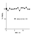
本明細書で使用される場合、用語「曲線下面積」または「AUC」とは、所与の投与量のインスリン-Fc融合タンパク質を投与した後の対象における、時間に対する%FBGL(空腹時血糖値)の曲線の下側の積分面積を指す。本明細書で使用される場合、用語「曲線上面積」または「AOC」は、インスリン-Fc融合タンパク質の生物学的効力の尺度として用いられ、AOCは、時間に対する%FBGLの曲線の下側の可能な総面積とAUC値との間の差に等しい。本明細書で使用される場合、「正規化曲線上面積」、「正規化AOC」、または「NAOC」は、AOC値を、投与されたインスリン-Fc融合タンパク質の実際の投与量で割ったものである。本明細書で使用される場合、用語「正規化AOC比」または「NAOCR」は、一連の投与における、最初のインスリン-Fc融合タンパク質投与から得られたNAOCに対する、特定のインスリン-Fc融合タンパク質投与から得られたNAOCの比である。そのため、NAOCRは、反復投与後のインスリン-Fc融合タンパク質の生物活性における変化の尺度となる。 As used herein, the term "area under the curve" or "AUC" refers to %FBGL (fasting blood glucose level) over time in a subject after administration of a given dose of insulin-Fc fusion protein. ) refers to the integrated area under the curve. As used herein, the term "area under the curve" or "AOC" is used as a measure of the biological potency of an insulin-Fc fusion protein, and the AOC is the area under the curve of %FBGL versus time. Equal to the difference between the total possible area and the AUC value. As used herein, "normalized area under the curve," "normalized AOC," or "NAOC" is the AOC value divided by the actual dose of insulin-Fc fusion protein administered. It is. As used herein, the term "normalized AOC ratio" or "NAOCR" refers to the NAOC obtained from the first insulin-Fc fusion protein administration in a series of administrations for a particular insulin-Fc fusion protein administration. is the ratio of NAOC obtained from Therefore, NAOCR is a measure of the change in the biological activity of insulin-Fc fusion protein after repeated administration.
本明細書で使用される場合、用語「生物活性」、「活性」、「生物活性」、「効力」、「生物活性効力」、または「生物学的効力」とは、インスリン-Fc融合タンパク質が、IRを活性化し、且つ/または、標的対象において血糖値の低減を及ぼす、程度を指す。本明細書で使用される場合、「インビトロ活性」または「IR活性」とは、インスリン-Fc融合タンパク質がIRに結合する際の親和性を指し、典型的には、競合的結合アッセイにおいてインスリン-Fc融合タンパク質がインスリン参照標準の半分をIRから押しのける濃度(すなわち、IC50)によって評価される。本明細書で使用される場合、「インビボ活性」とは、インスリン-Fc融合タンパク質投与後の標的対象の空腹時血糖値における低減の程度および持続時間を指す。 As used herein, the terms "biological activity", "activity", "biological activity", "potency", "bioactive potency", or "biological efficacy" mean that the insulin-Fc fusion protein , refers to the degree to which IR is activated and/or exerts a reduction in blood glucose levels in a target subject. As used herein, "in vitro activity" or "IR activity" refers to the affinity with which an insulin-Fc fusion protein binds to the IR, typically in a competitive binding assay. It is evaluated by the concentration at which the Fc fusion protein displaces half of the insulin reference standard from the IR (ie, IC50). As used herein, "in vivo activity" refers to the extent and duration of reduction in fasting blood glucose levels in a target subject following administration of an insulin-Fc fusion protein.
本明細書で使用される場合、用語「生合成」、「組換え合成」、または「組換え製造」は、インスリン-Fc融合タンパク質をコードする核酸分子(例えば、ベクター)(例えば、インスリン-Fc融合タンパク質全体が単一の核酸分子によってコードされる場合)で細胞をトランスフェクトすることによって、インスリン-Fc融合タンパク質を宿主細胞内で発現させるプロセスを指す。例示的な宿主細胞としては、哺乳類細胞、例えば、HEK293細胞またはCHO細胞が挙げられる。細胞は、当技術分野において標準的な方法を用いて培養することができ、発現されたインスリン-Fc融合タンパク質は、当技術分野において標準的な方法を用いて細胞培養物から採取および精製してもよい。 As used herein, the term "biosynthesis," "recombinant synthesis," or "recombinant manufacturing" refers to a nucleic acid molecule (e.g., vector) encoding an insulin-Fc fusion protein (e.g., insulin-Fc refers to the process by which an insulin-Fc fusion protein is expressed in a host cell by transfecting the cell with (where the entire fusion protein is encoded by a single nucleic acid molecule). Exemplary host cells include mammalian cells, such as HEK293 cells or CHO cells. Cells can be cultured using methods standard in the art, and the expressed insulin-Fc fusion protein can be harvested and purified from the cell culture using methods standard in the art. Good too.
本明細書で使用される場合、「細胞表面受容体」という用語は、一般に細胞の膜の外表面に存在し、可溶性分子、例えば血液供給中を循環する分子と相互作用する、タンパク質などの分子を指す。いくつかの実施形態において、細胞表面受容体は、ホルモン受容体(例えば、インスリンホルモン受容体またはインスリン受容体(IR))、または抗体のFcフラグメントもしくはFc領域に結合するFc受容体(例えば、Fcγ受容体、例えばFcγRI、または胎児性Fc受容体、例えばFcRn)を含む場合がある。本明細書で使用される場合、「インビトロ活性」または「Fcγ受容体活性」または「Fcγ受容体結合」または「FcRn受容体活性」または「FcRn結合」は、インスリン-Fc融合タンパク質がFc受容体(例えば、Fcγ受容体またはFcRn受容体)に結合する親和性を指し、典型的には、マイクロプレートリーダーで測定されるOD450nm値を用いたアッセイ(例えば、酵素結合免疫吸着測定法(ELISA)アッセイ)で測定される、インスリン-Fc融合タンパク質にその最大結合の半分に到達させる濃度(すなわち、EC50値)によって評価される。あるいは、インスリン-Fc融合タンパク質がFc受容体(例えば、Fcγ受容体またはFcRn受容体)に結合する親和性は、インスリン-Fc融合タンパク質を所与の濃度とした、酵素結合免疫吸着測定法(ELISA)アッセイにおいて、マイクロプレートリーダーで得られたOD450nm値によって評価される。 As used herein, the term "cell surface receptor" refers to a molecule, such as a protein, that generally resides on the outer surface of a cell's membrane and interacts with soluble molecules, e.g. molecules circulating in the blood supply. refers to In some embodiments, the cell surface receptor is a hormone receptor (e.g., insulin hormone receptor or insulin receptor (IR)) or an Fc receptor (e.g., Fcγ) that binds to an Fc fragment or Fc region of an antibody. receptors, such as FcγRI, or fetal Fc receptors, such as FcRn). As used herein, "in vitro activity" or "Fcγ receptor activity" or "Fcγ receptor binding" or "FcRn receptor activity" or "FcRn binding" means that the insulin-Fc fusion protein (e.g., Fcγ or FcRn receptors) and is typically used in assays (e.g., enzyme-linked immunosorbent assay (ELISA) assays using OD450nm values measured in a microplate reader). ) is evaluated by the concentration that causes the insulin-Fc fusion protein to reach half of its maximum binding (ie, the EC50 value), as measured by the EC50 value. Alternatively, the affinity with which insulin-Fc fusion protein binds to an Fc receptor (e.g., Fcγ receptor or FcRn receptor) can be determined by enzyme-linked immunosorbent assay (ELISA) using a given concentration of insulin-Fc fusion protein. ) In the assay, it is evaluated by the OD450nm value obtained with a microplate reader.
本明細書で使用される場合、「C1q」または「補体成分1q」という用語は、自然免疫系の一部である、補体系に関与するタンパク質複合体を意味する。C1qは、C1rおよびC1sと一緒に、C1複合体を形成する。C1qは、T細胞やB細胞に対する樹状細胞による特異的な抗原提示に関与する役割を担っている。 As used herein, the term "C1q" or "complement component 1q" refers to the protein complex involved in the complement system, which is part of the innate immune system. C1q forms the C1 complex together with C1r and C1s. C1q plays a role in specific antigen presentation by dendritic cells to T cells and B cells.
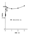
本明細書で使用される場合、用語「空腹時血糖値」または「FBGL」は、食物を与えない期間の終わり、且つ、インスリン-Fc融合タンパク質を投与する直前の、標的対象の平均血糖値を指す。本明細書で使用される場合、用語「空腹時血糖値パーセント」、「空腹時血糖値%」、または「%FBGL」は、空腹時血糖値に対する所与の血糖値の比率に100を乗じたものを指す。 As used herein, the term "fasting blood glucose level" or "FBGL" refers to the target subject's average blood glucose level at the end of the food-free period and immediately prior to administering the insulin-Fc fusion protein. Point. As used herein, the term "percent fasting blood glucose," "% fasting blood glucose," or "%FBGL" refers to the ratio of a given blood glucose to fasting blood glucose multiplied by 100. point to something
本明細書で使用される場合、用語「免疫原性の」または「免疫原性」は、分子の反復投与後、対象が当該分子と特異的に結合することができる抗体(すなわち、抗薬物抗体)を発現するように、所与の分子(例えば、本発明のインスリン-Fc融合タンパク質)が標的対象の免疫系を誘起する能力を指す。本明細書で使用される場合、用語「中和すること」、「中和抗体」、または「中和抗薬物抗体」は、抗体が標的対象における化合物の生物活性を妨害する能力を指す。本明細書で使用される場合、用語「免疫原性エピトープ」、「免疫原性ホットスポット」、または「ホットスポット」は、抗薬物抗体の中程度または強い結合の原因となる、所与の分子(例えば、本発明のインスリン-Fc融合タンパク質)の変異またはエピトープを指す。 As used herein, the term "immunogenic" or "immunogenic" refers to antibodies (i.e., anti-drug antibodies) that are capable of specifically binding a molecule to a subject after repeated administration of the molecule. ) refers to the ability of a given molecule (eg, an insulin-Fc fusion protein of the invention) to induce the immune system of a targeted subject to express a protein. As used herein, the term "neutralizing," "neutralizing antibody," or "neutralizing anti-drug antibody" refers to the ability of an antibody to interfere with the biological activity of a compound in a target subject. As used herein, the term "immunogenic epitope," "immunogenic hotspot," or "hotspot" refers to a given molecule that is responsible for moderate or strong binding of anti-drug antibodies. (eg, an insulin-Fc fusion protein of the invention).
本明細書で使用される場合、用語「インスリン参照標準」は、(i)哺乳動物(例えば、イヌ、またはヒト)由来の天然由来のインスリン;(ii)Fcフラグメントを含まないインスリンポリペプチド;または、(iii)標準治療用インスリン(例えば、市販のインスリン)のいずれか1つである。 As used herein, the term "insulin reference standard" refers to (i) naturally occurring insulin from a mammal (e.g., dog, or human); (ii) an insulin polypeptide that does not contain an Fc fragment; or , (iii) standard therapeutic insulin (eg, commercially available insulin).
本明細書で使用される場合、用語「モノマー」は、単一のポリペプチドから構成されるタンパク質または融合タンパク質を指す。実施形態において、「モノマー」は、タンパク質または融合タンパク質であり、例えば、インスリンポリペプチドとFcフラグメントポリペプチドとを含む単一ポリペプチドであって、当該インスリンおよび当該Fcフラグメントポリペプチドがペプチド結合により結合されることで形成される、単一ポリペプチドである。実施形態において、モノマーは、単一の核酸分子にコードされる。 As used herein, the term "monomer" refers to a protein or fusion protein that is composed of a single polypeptide. In embodiments, a "monomer" is a protein or fusion protein, e.g., a single polypeptide comprising an insulin polypeptide and an Fc fragment polypeptide, wherein the insulin and the Fc fragment polypeptide are linked by a peptide bond. It is a single polypeptide formed by In embodiments, the monomer is encoded by a single nucleic acid molecule.
本明細書で使用される場合、「N末端」は、アミノ酸のα-アミノ基である遊離アミン基(例えば、第2の炭素原子に隣接して位置する1つの炭素原子に共有結合している遊離アミノであって、この第2の炭素原子がこのアミノ酸のカルボニル基の一部となっている)を含むアミノ酸によって開始されるタンパク質またはポリペプチドの始まりを指す。本明細書で使用される場合、「C末端」は、カルボン酸基を含むアミノ酸で終わっているタンパク質またはポリペプチドの末端を指し、ここでこのカルボン酸基の炭素原子は、このアミノ酸のα-アミノ基に隣接して位置している。 As used herein, "N-terminus" refers to a free amine group that is the alpha-amino group of an amino acid (e.g., a free amine group covalently bonded to one carbon atom located adjacent to a second carbon atom). Refers to the beginning of a protein or polypeptide that is initiated by an amino acid that contains a free amino acid, the second carbon atom of which is part of the amino acid's carbonyl group. As used herein, "C-terminal" refers to the terminus of a protein or polypeptide that ends in an amino acid that contains a carboxylic acid group, where the carbon atom of the carboxylic acid group is Located adjacent to the amino group.
本明細書で使用される場合、「OD450」、「450nmの光学密度」、および「450nmの吸光度」は、同義的に使用され、アッセイ、例えばマイクロプレートベースアッセイ、例えば酵素結合免疫吸着測定法、例えばELISAアッセイ、において試料を通過した450nmの光の吸光度を、マイクロプレートリーダーを用いて読み取ることを指す場合がある。 As used herein, "OD450," "optical density at 450 nm," and "absorbance at 450 nm" are used synonymously and include assays, e.g., microplate-based assays, e.g., enzyme-linked immunosorbent assays. For example, it may refer to reading the absorbance of 450 nm light passing through a sample in an ELISA assay using a microplate reader.
本明細書で使用される場合、特定のアッセイにおいての「OD450比」は、特定の時点に実行された第1の被験インスリン-Fc融合タンパク質について得られたOD450値を、別の時点に実行された第2の被験インスリン-Fc融合タンパク質について得られたOD450値と、比較する方法を指す。OD450比は、第1の被験物質のOD450値を、参照インスリン-Fc融合タンパク質のOD450値で割ることによって得られる。同様に、第二の被験物質のOD450値を、第一の被験物質のOD450比の算出に用いたものと同じ参照インスリン-Fc融合タンパク質のOD450値で割ることによって、第二の被験物質の第二のOD450比を得ることができる。結果として、第1の被験インスリン-Fc融合タンパク質と第2の被験インスリン-Fc融合タンパク質のアッセイ特性を比較することができる。OD450比の算出に用いる参照インスリン-Fc融合タンパク質の構成は、配列番号76である。 As used herein, "OD450 ratio" in a particular assay refers to the OD450 value obtained for a first test insulin-Fc fusion protein performed at a particular time point to the OD450 value obtained for a first test insulin-Fc fusion protein performed at a particular time point. OD450 value obtained for a second test insulin-Fc fusion protein. The OD450 ratio is obtained by dividing the OD450 value of the first test substance by the OD450 value of the reference insulin-Fc fusion protein. Similarly, the OD450 value of the second test article can be calculated by dividing the OD450 value of the second test article by the OD450 value of the same reference insulin-Fc fusion protein used to calculate the OD450 ratio of the first test article. Two OD450 ratios can be obtained. As a result, the assay properties of the first test insulin-Fc fusion protein and the second test insulin-Fc fusion protein can be compared. The composition of the reference insulin-Fc fusion protein used to calculate the OD450 ratio is SEQ ID NO:76.
本明細書で使用される場合、「薬力学」または「PD」は、通常、対象におけるインスリン-Fc融合タンパク質の生物学的効果を指す。具体的には、本明細書において、PDは、インスリン-Fc融合タンパク質の投与後、対象における経時的な空腹時血糖値の低下の指標を指す。 As used herein, "pharmacodynamics" or "PD" generally refers to the biological effects of an insulin-Fc fusion protein in a subject. Specifically, as used herein, PD refers to an indicator of a decrease in fasting blood glucose levels in a subject over time after administration of an insulin-Fc fusion protein.
本明細書で使用される場合、「薬物動態」または「PK」は、通常、吸収、分布、代謝、および排泄の観点での、インスリン-Fc融合タンパク質と対象の体との特徴的な相互作用を指す。具体的には、本明細書において、PKは、インスリン-Fc融合タンパク質を投与した後の所与の時点における、対象の血液または血清中のインスリン-Fc融合タンパク質の濃度を指す。本明細書で使用される場合、「半減期」とは、薬物排泄の一次指数関数的減衰モデルから算出される、対象の血液または血清中のインスリン-Fc融合タンパク質の濃度が元の値の半分に達するまでの時間を指す。「半減期」の値が大きいインスリン-Fc融合タンパク質ほど、標的対象においてより長い作用持続時間を示す。 As used herein, "pharmacokinetics" or "PK" generally refers to the characteristic interactions of an insulin-Fc fusion protein with a subject's body in terms of absorption, distribution, metabolism, and excretion. refers to Specifically, as used herein, PK refers to the concentration of insulin-Fc fusion protein in the blood or serum of a subject at a given time after administration of the insulin-Fc fusion protein. As used herein, "half-life" means that the concentration of insulin-Fc fusion protein in a subject's blood or serum is half of its original value, as calculated from a first-order exponential decay model of drug excretion. This refers to the time it takes to reach . Insulin-Fc fusion proteins with higher "half-life" values exhibit a longer duration of action in the target subject.
本明細書で使用される場合、アミノ酸またはヌクレオチド配列における「配列同一性」、「配列相同性」、「相同性」、または「同一の」という用語は、バリアントのヌクレオチド配列またはアミノ酸配列の特定の連続するセグメントを、参照配列のヌクレオチド配列またはアミノ酸配列に対してアラインメントして比較したとき、バリアントおよび参照配列内に同じヌクレオチドまたはアミノ酸残基が存在することを述べている。配列アライメント法および配列間の同一性を求めるための方法は、当該技術分野において公知であり、類似性のために配列を整理、整列、および比較するクラスタルオメガの使用が挙げられ、このソフトウェアは、各配列位置を強調し、その位置において全配列間で比較し、以下のスコアのうちの1つを割り当てる。「*」(アステリスク)は、単一の完全に保存された残基を有する配列位置に割り当てられ、「:」(コロン)は、Gonnet PAM 250マトリックスで0.5を超えるスコアを有する、強く類似した特性のグループ間の保存を示し、「.」(ピリオド)は、Gonnet PAM 250マトリックスで0.5以下のスコアを有する弱く類似した特性のグループ間の保存を示し、「-」(ダッシュ)は配列ギャップを示し、ある配列範囲内の特定の比較集合の中に局所的な相同性が存在しないことを意味し、空白「 」は比較配列全体でその特定の位置に配列相同性がほとんど存在しないことを示す。例えば、Ausubelら(編)(1995年)、Current Protocols in Molecular Biology、19章(グリーン・パブリッシング(Greene Publishing)およびワイリー・インターサイエンス(Wiley-Interscience)、ニューヨーク);および、ALIGNプログラム、Dayhoff、Atlas of Polypeptide Sequence and Structure 5:Suppl.3(国立生物医学研究財団(National Biomedical Research Foundation)、ワシントンD.C.)を参照されたい。2つのヌクレオチド配列の最適なアラインメントに関して、バリアントヌクレオチド配列の連続するセグメントは、参照ヌクレオチド配列に対して追加のヌクレオチドまたは削除されたヌクレオチドを有していてもよい。同様に、2つのアミノ酸配列の最適なアラインメントの目的のために、バリアントアミノ酸配列の連続するセグメントは、参照アミノ酸配列に対して追加のアミノ酸残基または削除されたアミノ酸残基を有していてもよい。いくつかの実施形態において、参照ヌクレオチド配列または参照アミノ酸配列との比較に用いられる連続セグメントは、少なくとも6個、10個、15個、または20個の連続ヌクレオチド、またはアミノ酸残基から構成され、30個、40個、50個、100個、またはそれ以上のヌクレオチドまたはアミノ酸残基であってよい。バリアントのヌクレオチド配列またはアミノ酸配列にギャップが含まれることに付随する配列同一性の増加の補正は、ギャップペナルティを割り当てることによって行うことができる。配列アライメントの方法は、当技術分野において公知である。 As used herein, the terms "sequence identity," "sequence homology," "homology," or "identical" in amino acid or nucleotide sequences refer to the specific States the presence of the same nucleotide or amino acid residues in the variant and reference sequence when the contiguous segments are aligned and compared to the nucleotide or amino acid sequence of the reference sequence. Sequence alignment methods and methods for determining identity between sequences are known in the art and include the use of Clustal Omega to organize, align, and compare sequences for similarity; this software , each sequence position is highlighted and compared between all sequences at that position and assigned one of the following scores: “*” (asterisk) is assigned to a sequence position with a single fully conserved residue and “:” (colon) indicates a strongly similar sequence with a score greater than 0.5 in the Gonnet PAM 250 matrix. ``.'' (period) indicates conservation between groups of weakly similar traits with a score of 0.5 or less in the Gonnet PAM 250 matrix, and ``-'' (dash) Indicates a sequence gap, meaning that there is no local homology within a given comparison set within a range of sequences; a blank " " indicates that there is little sequence homology at that particular position across the comparison sequences. Show that. For example, Ausubel et al. (eds.) (1995), Current Protocols in Molecular Biology, Chapter 19 (Greene Publishing and Wiley-Interscience, New York); and ALIG N program, Dayhoff, Atlas of Polypeptide Sequence and Structure 5: Suppl. 3 (National Biomedical Research Foundation, Washington, D.C.). For optimal alignment of two nucleotide sequences, consecutive segments of a variant nucleotide sequence may have additional or deleted nucleotides relative to the reference nucleotide sequence. Similarly, for the purposes of optimal alignment of two amino acid sequences, consecutive segments of a variant amino acid sequence may have additional or deleted amino acid residues with respect to the reference amino acid sequence. good. In some embodiments, the contiguous segment used for comparison to a reference nucleotide sequence or reference amino acid sequence is comprised of at least 6, 10, 15, or 20 contiguous nucleotides or amino acid residues; nucleotides or amino acid residues. Corrections for increased sequence identity associated with the inclusion of gaps in the nucleotide or amino acid sequences of variants can be made by assigning a gap penalty. Methods of sequence alignment are known in the art.
実施形態において、2つの配列間のパーセント同一性または「相同性」の決定は、数学的アルゴリズムを使用して達成される。例えば、アミノ酸配列のパーセント同一性は、ギャップ開始ペナルティ12、ギャップ伸長ペナルティ2、BLOSUM行列62を用いるアフィン6ギャップ検索を使用するスミス-ウォーターマン相同性検索アルゴリズムを用いて決定される。スミス-ウォーターマン相同性検索アルゴリズムは、参照によって本明細書に援用される、SmithおよびWaterman(1981)、Adv.Appl.Math、2巻:pp482~489で説明されている。実施形態において、ヌクレオチド配列のパーセント同一性は、ギャップ開始ペナルティ25、ギャップ伸長ペナルティ5を用いる、スミス-ウォーターマン相同性検索アルゴリズムを使用して決定される。このような配列同一性の決定は、例えば、タイムロジック社(TimeLogic)のDeCypherハードウェアアクセラレータを用いて、実施することができる。
In embodiments, determining percent identity or "homology" between two sequences is accomplished using a mathematical algorithm. For example, percent identity of amino acid sequences is determined using the Smith-Waterman homology search algorithm using an
本明細書で使用される場合、用語「相同性」は、例えばβストランド、ヘリックス、フォールドなどの共通の構造特性および共通の空間分布の位置を見つけることによって、2つ以上のタンパク質を比較するために使用される。従って、相同なタンパク質構造は、空間的な分析によって定義される。構造相同性の測定には、空間の幾何学的トポロジー的特徴を計算することが必要である。タンパク質の3次元構造を生成・解析するのに用いられる1つの手法として、3次元の類似性が2次元の類似性を反映しているという事実に基づいて類似配列を見つけ出す、ホモロジーモデリング(比較モデリング、知識ベースモデリングとも呼ばれる)がある。相同構造は、配列の類似性を必須条件とはしていない。 As used herein, the term "homology" refers to the comparison of two or more proteins, e.g. by finding common structural properties such as β-strands, helices, folds, and common spatial distribution positions. used for. Therefore, homologous protein structures are defined by spatial analysis. Measuring structural homology requires calculating the geometric and topological features of the space. One method used to generate and analyze the three-dimensional structure of proteins is homology modeling (comparative modeling), which finds similar sequences based on the fact that three-dimensional similarities reflect two-dimensional similarities. (also known as knowledge-based modeling). Homologous structures do not require sequence similarity.
本明細書で使用される場合、用語「対象」および「患者」は、疾患または障害、例えば、糖尿病または本明細書に記載の別の疾患または障害、を有する犬科動物およびヒト、または正常な対象を含むことを意図している。 As used herein, the terms "subject" and "patient" refer to canines and humans with a disease or disorder, such as diabetes or another disease or disorder described herein, or a normal intended to contain objects.
本明細書で使用される場合、用語「力価」または「収量」は、細胞培養物の体積当たりの生合成(例えば、哺乳動物細胞、例えば、HEK293細胞またはCHO細胞における生合成)から生じる融合タンパク質生成物(例えば、本明細書に記載のインスリン-Fc融合タンパク質)の量を指す。生成物の量は、生産プロセスの任意の工程(例えば、精製の前または後)で求めることができるが、収量または力価は、常に、元の細胞培養物の体積当たりで記載される。本明細書で使用される場合、用語「生成物収量」または「総タンパク質収量」は、細胞によって発現され、少なくとも1つのアフィニティークロマトグラフィー工程(例えば、プロテインAまたはプロテインG)を介して精製されたインスリン-Fc融合タンパク質の総量を指し、インスリン-Fc融合タンパク質の単量体、インスリン-Fc融合タンパク質のホモ二量体、およびインスリン-Fc融合タンパク質のホモ二量体の高次分子凝集体を含む。本明細書で使用される場合、用語「パーセントホモ二量体」または「ホモ二量体率」は、所望のホモ二量体である融合タンパク質生成物(例えば、本明細書に記載のインスリン-Fc融合タンパク質)の割合を指す。本明細書で使用される場合、用語「ホモ二量体力価」は、細胞培養物の体積当たりで報告される、プロテインA精製工程後のホモ二量体率と総タンパク質収量との積を指す。 As used herein, the term "titer" or "yield" refers to the fusion resulting from biosynthesis (e.g., biosynthesis in mammalian cells, e.g., HEK293 cells or CHO cells) per volume of cell culture. Refers to the amount of protein product (eg, insulin-Fc fusion protein described herein). Although the amount of product can be determined at any step in the production process (eg, before or after purification), yield or titer is always stated per volume of the original cell culture. As used herein, the term "product yield" or "total protein yield" refers to protein expressed by cells and purified via at least one affinity chromatography step (e.g., Protein A or Protein G). Refers to the total amount of insulin-Fc fusion protein, including insulin-Fc fusion protein monomer, insulin-Fc fusion protein homodimer, and higher molecular aggregates of insulin-Fc fusion protein homodimer . As used herein, the term "percent homodimer" or "percent homodimer" refers to a fusion protein product that is a desired homodimer (e.g., insulin- Fc fusion protein). As used herein, the term "homodimer titer" refers to the product of the homodimer rate and total protein yield after the Protein A purification step, reported per volume of cell culture. .
本明細書で使用される場合、疾患または障害を有する対象を「治療する」または「治療すること」という用語は、疾患または障害の少なくとも1つの症状が解消、治癒、緩和、軽減、変化、軽快、緩解、または改善するように、対象をレジメン、例えば本明細書に記載の融合タンパク質などの融合タンパク質の投与に供することを意味する。治療は、疾患もしくは障害、または疾患もしくは障害の症状を緩和、軽減、変化、軽快、緩解、改善する、または影響を与えるのに有効な量を投与することを含む。治療は、疾患または障害の症状の増悪または悪化を抑制するものであってもよい。 As used herein, the term "treating" or "treating" a subject having a disease or disorder means that at least one symptom of the disease or disorder is eliminated, cured, alleviated, alleviated, changed, or ameliorated. , remission, or amelioration, means subjecting the subject to a regimen, eg, administration of a fusion protein, such as the fusion proteins described herein. Treatment includes administering an amount effective to alleviate, alleviate, change, ameliorate, ameliorate, ameliorate, or affect the disease or disorder, or the symptoms of the disease or disorder. Treatment may inhibit exacerbation or worsening of symptoms of a disease or disorder.
インスリン-Fc融合タンパク質の組成および構造
本開示は、ペプチドリンカーを介して種特異的なFcフラグメントに連結されたインスリンポリペプチドを含む融合タンパク質(すなわち、インスリン-Fc融合タンパク質)の組成、並びに、糖尿病(例えば、ヒトおよび/またはイヌにおける糖尿病)を治療するためのその使用に関する。本明細書で使用される場合、「融合タンパク質」および「インスリン-Fc融合タンパク質」という用語は、ペプチド結合を介して共有結合した、例えば異なる供給源(異なるタンパク質、ポリペプチド、細胞など)からの、2以上の部分からなるタンパク質を指す。インスリン-Fc融合タンパク質は、(i)各部分をコードする遺伝子を連結して1つの核酸分子にし、(ii)核酸分子が以下のようにコードしているタンパク質を宿主細胞(例えば、HEKまたはCHO)中で発現することで、共有結合される:(N末端)--インスリンポリペプチド--リンカー--Fcフラグメント--(C末端)。完全に組換え合成するアプローチの方が、インスリンポリペプチドとFcフラグメントを別々に合成してから化学的に結合させる方法よりも好ましい。化学的結合工程とその後の精製プロセスは、製造の複雑さを増加させ、製品の収量を減少させ、コストを増加させる。
Composition and Structure of Insulin-Fc Fusion Proteins This disclosure describes the composition of fusion proteins that include an insulin polypeptide linked to a species-specific Fc fragment via a peptide linker (i.e., insulin-Fc fusion proteins), as well as the composition and structure of insulin-Fc fusion proteins. (e.g. diabetes in humans and/or dogs). As used herein, the terms "fusion protein" and "insulin-Fc fusion protein" refer to proteins covalently linked via peptide bonds, e.g. , refers to a protein consisting of two or more parts. Insulin-Fc fusion proteins are produced by (i) linking the genes encoding each portion into one nucleic acid molecule, and (ii) transferring the protein that the nucleic acid molecules encode to a host cell (e.g., HEK or CHO) into a host cell (e.g., HEK or CHO). ) is covalently linked to: (N-terminus) -- insulin polypeptide -- linker -- Fc fragment -- (C-terminus). A fully recombinant synthesis approach is preferred over separately synthesizing the insulin polypeptide and Fc fragment and then chemically linking them. Chemical bonding steps and subsequent purification processes increase manufacturing complexity, reduce product yield, and increase cost.
本明細書で使用される場合、「二量体」という用語は、共有結合した2つのポリペプチドを含むタンパク質または融合タンパク質を指す。実施形態において、2つの同一のポリペプチドが共有結合(例えば、ジスルフィド結合を介して)し、「ホモ二量体」を形成している(図1に図示)。ジスルフィド結合を図1に示しているが、実際のジスルフィド結合の総数は、図1に示される数よりも多くても少なくてもよい。実施形態において、ホモ二量体は、単一の核酸分子によってコードされており、ホモ二量体は、まずインスリン-Fc融合タンパク質単量体を形成し、次に細胞内でのさらなるプロセシング後に2つの同一のインスリン-Fc融合タンパク質単量体をホモ二量体へと構築することによって、細胞内で組換え製造される。 As used herein, the term "dimer" refers to a protein or fusion protein that includes two polypeptides covalently linked. In embodiments, two identical polypeptides are covalently linked (eg, via a disulfide bond) to form a "homodimer" (illustrated in Figure 1). Although disulfide bonds are shown in FIG. 1, the actual total number of disulfide bonds may be greater or less than the number shown in FIG. In embodiments, the homodimer is encoded by a single nucleic acid molecule, and the homodimer first forms the insulin-Fc fusion protein monomer and then, after further processing within the cell, the 2 It is produced recombinantly within cells by assembling two identical insulin-Fc fusion protein monomers into homodimers.
本明細書で使用される場合、「多量体、」「多量体の」、または「多量体状態」という用語は、Fc融合タンパク質二量体と平衡状態にあり得る、または、Fc融合タンパク質二量体の永久凝集型として作用し得る、Fc融合タンパク質二量体の非共有結合性の会合形態(例えば、Fc融合タンパク質ホモ二量体の二量体、Fc融合タンパク質ホモ二量体の三量体、Fc融合タンパク質ホモ二量体の四量体、または5以上のFc融合タンパク質ホモ二量体を含む高次凝集体)を指す。Fc融合タンパク質の多量体型は、インスリン-Fc融合タンパク質ホモ二量体とは異なる物理的、安定性、または薬理活性を有し得ると予想できる。 As used herein, the terms "multimer," "multimeric," or "multimeric state" refer to Fc fusion protein dimers that can be in equilibrium with Fc fusion protein dimers, or Non-covalently associated forms of Fc fusion protein dimers (e.g., dimers of Fc fusion protein homodimers, trimers of Fc fusion protein homodimers) can act as permanently aggregated forms of Fc fusion protein dimers. , a tetramer of Fc fusion protein homodimers, or a higher-order aggregate comprising five or more Fc fusion protein homodimers). It can be expected that multimeric forms of Fc fusion proteins may have different physical, stability, or pharmacological activities than insulin-Fc fusion protein homodimers.
インスリンポリペプチド
インスリンポリペプチドは、例えば、膵臓内のランゲルハンス島のβ細胞によって産生されるインスリンまたはインスリン類似体であってもよい。インスリンは、血液からのグルコースの吸収を調節することで機能する。タンパク質レベルの上昇およびグルコースレベルの上昇などの刺激により、インスリンはβ細胞から放出され、IRに結合し、哺乳類(例えば、ヒト)の代謝の多くの側面に影響を与えるシグナルカスケードを開始する。このプロセスの崩壊は、いくつかの疾患、特に糖尿病、インスリノーマ、インスリン抵抗性、メタボリックシンドローム、および多嚢胞性卵巣症候群に直接関係する。本開示のインスリン類似体は、インスリンの構造に関連しているが、1またはそれ以上の改変を含んでいるものであってもよい。いくつかの実施形態において、インスリン類似体は、インスリンに対する少なくとも1つのアミノ酸の置換、欠失、付加、または化学修飾を含み、これらは、インスリン-Fc融合タンパク質の構成の特定の特徴または特性に影響を与える可能性がある。例えば、本明細書に記載の修飾または変更は、参照標準と比較して、インスリン-Fc融合タンパク質の構成の構造、安定性、pH感受性、生物活性、または細胞表面受容体(例えば、インスリンホルモン受容体)に対する結合親和性に影響を与える可能性がある。
Insulin Polypeptide Insulin polypeptide can be, for example, insulin or an insulin analog produced by the beta cells of the islets of Langerhans within the pancreas. Insulin works by regulating the absorption of glucose from the blood. Upon stimulation, such as increased protein levels and increased glucose levels, insulin is released from beta cells, binds to the IR, and initiates a signal cascade that affects many aspects of mammalian (eg, human) metabolism. Disruption of this process is directly related to several diseases, especially diabetes, insulinoma, insulin resistance, metabolic syndrome, and polycystic ovary syndrome. Insulin analogs of the present disclosure are related in structure to insulin, but may include one or more modifications. In some embodiments, the insulin analog comprises at least one amino acid substitution, deletion, addition, or chemical modification to insulin that affects a particular characteristic or property of the composition of the insulin-Fc fusion protein. It is possible to give For example, modifications or changes described herein may improve the structure, stability, pH sensitivity, biological activity, or cell surface receptors (e.g., insulin hormone receptor) of the composition of the insulin-Fc fusion protein compared to a reference standard. may affect the binding affinity for
インスリンのアミノ酸配列は、進化を通じて、特に脊椎動物において強く保存されている。例えば、天然型イヌインスリンと天然型ブタインスリンは、ヒトインスリンと1つのアミノ酸しか異なっておらず、天然型ウシインスリンは、ヒトインスリンと3つのアミノ酸しか異なっておらず、天然型ネコインスリンは、ヒトインスリンとたった4つのアミノ酸しか異なっていない。本明細書で使用される場合、用語「B鎖またはB鎖アナログ」、「Cペプチド」または「C鎖」、および「A鎖またはA鎖アナログ」は、図1に例示されるようなインスリンポリペプチドのペプチドセグメントを指す。インスリンは、ジスルフィド結合(例えば、1または複数のB鎖システイン側鎖チオールおよび1または複数のA鎖システイン側鎖チオールによって形成されるジスルフィド結合)を介して接続された、2つのペプチド鎖(すなわち、B鎖およびA鎖)を含む、51アミノ酸のホルモンである。インスリンのA鎖は21アミノ酸長であり、インスリンのB鎖は30アミノ酸長である。天然型のインスリンでは、A鎖は、2つのA鎖システイン側鎖チオールによって形成された1つの鎖内ジスルフィド結合を含む。参考のため、配列番号1のヒトインスリンB鎖および配列番号2のヒトインスリンA鎖の配列を以下に示す:
FVNQHLCGSHLVEALYLVCGERGFFYTPKT(配列番号1)
GIVEQCCTSICSLYQLENYCN(配列番号2)。
The amino acid sequence of insulin is strongly conserved throughout evolution, especially in vertebrates. For example, natural canine insulin and natural porcine insulin differ from human insulin by only one amino acid, natural bovine insulin differs from human insulin by only three amino acids, and natural feline insulin differs from human insulin by only one amino acid. It differs from insulin in only four amino acids. As used herein, the terms "B chain or B chain analog,""Cpeptide" or "C chain," and "A chain or A chain analog" refer to insulin polypeptides such as those illustrated in FIG. Refers to the peptide segment of a peptide. Insulin consists of two peptide chains (i.e., It is a 51-amino acid hormone, including B chain and A chain). The A chain of insulin is 21 amino acids long and the B chain of insulin is 30 amino acids long. In native insulin, the A chain contains one intrachain disulfide bond formed by two A chain cysteine side chain thiols. For reference, the sequences of human insulin B chain of SEQ ID NO: 1 and human insulin A chain of SEQ ID NO: 2 are shown below:
FVNQHLCGSHLVEALYLVCGERGFFYTPKT (SEQ ID NO: 1)
GIVEQCCTSICSLYQLENYCN (SEQ ID NO: 2).
本明細書で使用される場合、「インスリン」または「インスリンポリペプチド」という用語は、成熟インスリン、プレプロインスリン、プロインスリン、および天然に存在するインスリン、またはその類似体を包含する。実施形態において、インスリンポリペプチドは、完全長インスリンポリペプチドまたはその断片とすることができる。実施形態において、インスリンポリペプチドは、成熟インスリン、プレプロインスリン、プロインスリン、または天然に存在するインスリンからの1または複数の断片を含むことができる。 As used herein, the term "insulin" or "insulin polypeptide" encompasses mature insulin, preproinsulin, proinsulin, and naturally occurring insulin, or analogs thereof. In embodiments, the insulin polypeptide can be a full-length insulin polypeptide or a fragment thereof. In embodiments, the insulin polypeptide can include mature insulin, preproinsulin, proinsulin, or one or more fragments from naturally occurring insulin.
インスリンは通常、N末端--B鎖:C鎖:A鎖--C末端ポリペプチドとして構築され、生物活性を示すようにするためにC鎖が切断される。参考のため、C鎖を含むヒトインスリン分子全体(すなわち、ヒトプロインスリン)の配列を、C鎖に下線を引いて以下に示す:
FVNQHLCGSHLVEALYLVCGERGFFYTPKTRREAEDLQVGQVELGGGPGAGSLQPLALEGSLQKRGIVEQCCTSICSLYQLENYCN(配列番号3)。
単鎖インスリンポリペプチドの、生理活性を有する2鎖ポリペプチドへの変換は、通常、グルコース刺激によるインスリン分泌より前に、ランゲルハンス島のβ細胞内で、I型エンドプロテアーゼ(Cペプチド-B鎖接続を破壊するPC1およびPC3、並びにPC2)、およびCペプチド-A鎖結合を正しい部位で正確に切断するII型エンドプロテアーゼの2つのエンドプロテアーゼによって達成されている。しかし、インスリンなどの治療用分子の生合成に用いられる細胞系(例えば、細菌、酵母、および哺乳類(HEKおよびCHO)細胞系)はこの経路を有していないため、化学的または酵素的な方法を用いて単鎖ポリペプチドを発現および回収した後に変換を行わなければならない。発現および回収後にC鎖を切断するための公知の技術は全て、C鎖がA鎖のN末端直前のリジンで終端するように、まずC鎖を改変することに依存している。次に、リジン残基のC末端で特異的にペプチド結合を切断する、トリプシンまたはLys-Cファミリーから選択される酵素を用いて、単鎖インスリンポリペプチドをC鎖のC末端リジンおよびB鎖のN末端から29番目の位置のC末端リジンで切断する。場合によっては、得られた生物活性を示す2鎖インスリンは、B鎖のN末端から30番目の位置の切断されたアミノ酸を再付加せずに使用され、場合によっては、B鎖のN末端から30番目の位置の切断されたアミノ酸が追加の酵素的方法を用いて当該分子に再付加される。このような処理がインスリンではうまくいくのは、インスリンが2鎖ポリペプチド形態の全体の中にリジンを1つしか含まないためである。しかし、既知のFcフラグメントは全て複数のリジン残基を含むため、この処理は本明細書に含まれるインスリン-Fc融合タンパク質に対しては使用することができない。従って、酵素切断プロセスは、Fcフラグメントを非機能的な部分にまで消化することによって、インビボでインスリンポリペプチドの作用を延長するFcフラグメントの能力を取り除くことになる。従って、本発明のインスリン-Fc融合タンパク質は、C鎖切断を必要としないことから単鎖形態で生物活性を示す、インスリンポリペプチドを含まなければならない。
Insulin is typically constructed as an N-terminal -- B chain: C chain: A chain -- C-terminal polypeptide, with the C chain truncated to render it biologically active. For reference, the sequence of the entire human insulin molecule (i.e., human proinsulin), including the C chain, is shown below with the C chain underlined:
FVNQHLCGSHLVEALYLVCGERGFFYTPKT RREAEDLQVGQVELGGGPGAGSLQPLALEGSLQKR GIVEQCCTSICSLYQLENYCN (SEQ ID NO: 3).
The conversion of single-chain insulin polypeptides into bioactive two-chain polypeptides is normally carried out by type I endoprotease (C-peptide-B chain linkage) in the β-cells of the islets of Langerhans, prior to glucose-stimulated insulin secretion. This is accomplished by two endoproteases: PC1 and PC3, which destroy the C-peptide, and PC2), and type II endoprotease, which precisely cleaves the C-peptide-A chain bond at the correct site. However, cell systems used for the biosynthesis of therapeutic molecules such as insulin (e.g., bacterial, yeast, and mammalian (HEK and CHO) cell lines) do not have this pathway, so chemical or enzymatic methods The conversion must be carried out after expressing and recovering the single chain polypeptide using a polypeptide. All known techniques for cleaving the C chain after expression and recovery rely on first modifying the C chain so that it terminates at a lysine just before the N-terminus of the A chain. The single-chain insulin polypeptide is then isolated from the C-terminal lysine of the C-chain and the B-chain using trypsin or an enzyme selected from the Lys-C family, which cleaves peptide bonds specifically at the C-terminus of the lysine residue. Cleave at the C-terminal lysine at the 29th position from the N-terminus. In some cases, the resulting biologically active two-chain insulin is used without re-adding the truncated amino acid at
生物活性を示すいくつかの単鎖インスリンポリペプチドが、当該技術分野において報告されている。いずれの場合も、単鎖インスリンポリペプチドは、静電バランスをとり、凝集を防ぎ、IR結合および/または下流シグナル伝達を増強して天然型の2鎖インスリンと同等レベルの生物活性を達成するために、特定の長さと組成のC鎖、並びに、特定のアミノ酸部位で変異したA鎖およびB鎖を含んでいる。ここで、ペプチドセグメント上の変異の位置は、セグメントの名称(例えば、B鎖、C鎖、A鎖)と、セグメントのN末端から数えたアミノ酸の数を用いて表記される。例えば、「B16」という表記は、B鎖のアミノ酸配列のN末端から16番目のアミノ酸のことをいう。また、「A8」という表記は、A鎖のN末端から8番目のアミノ酸のことをいう。さらに、あるアミノ酸が特定の位置で天然型から新しいアミノ酸に変異している場合、その位置にはその新しいアミノ酸のための1文字アミノ酸コードが付加される。例えば、B16AはB鎖のアミノ酸配列のN末端から16番目のアミノ酸におけるアラニン変異のことをいい、A8HはA鎖のアミノ酸配列のN末端から8番目のアミノ酸におけるヒスチジン変異をいう。 Several single chain insulin polypeptides that exhibit biological activity have been reported in the art. In either case, single-chain insulin polypeptides are used to balance electrostatics, prevent aggregation, and enhance IR binding and/or downstream signaling to achieve levels of biological activity comparable to native two-chain insulin. It contains a C chain of a specific length and composition, and an A chain and a B chain that are mutated at specific amino acid positions. Here, the position of a mutation on a peptide segment is expressed using the name of the segment (eg, B chain, C chain, A chain) and the number of amino acids counted from the N-terminus of the segment. For example, the notation "B16" refers to the 16th amino acid from the N-terminus of the amino acid sequence of the B chain. Moreover, the notation "A8" refers to the 8th amino acid from the N-terminus of the A chain. Additionally, if an amino acid is mutated from its natural form to a new amino acid at a particular position, the one-letter amino acid code for the new amino acid is added to that position. For example, B16A refers to an alanine mutation in the 16th amino acid from the N-terminus of the amino acid sequence of the B chain, and A8H refers to a histidine mutation in the 8th amino acid from the N-terminus of the A-chain amino acid sequence.
米国特許第9855318B2号では、配列GGSGGGG(配列番号72)のC鎖(「第1のリンカー」)、A鎖における置換、並びにB鎖における置換および欠失(非天然型アミノ酸には下線を引き、欠失させた天然型アミノ酸は下線を引いたZで表している)を有する単鎖インスリン類似体が報告されている:
FVNQHLCGSHLVEALELVCGERGFHYGGGGGGSGGGGGIVEQCCTSTCSLDQLENYCZ(配列番号7_NULL)。
No. 9,855,318B2 discloses substitutions in the C chain (the "first linker"), A chain, and substitutions and deletions in the B chain (non-natural amino acids are underlined) of the sequence GGSGGGGG (SEQ ID NO: 72); A single-chain insulin analog has been reported with a deleted naturally occurring amino acid (represented by an underlined Z):
FVNQHLCGS H LVEAL E LVCGERGF H Y GGGGGGSGGGGG GIVEQCCTS T CSL D QLENYC Z (SEQ ID NO: 7_NULL).
以下に、上に示した配列を、インスリンポリペプチド配列の表記から記号Zの不在アミノ酸を取り除いて、再掲したものである。ここでも、前と同様に、非天然型のアミノ酸に下線を引いている。2つの別々の表記をしたが、この1対の配列は全く同じインスリンポリペプチドを指している。
FVNQHLCGSHLVEALELVCGERGFHYGGGGGGSGGGGGIVEQCCTSTCSLDQLENYC(配列番号7)
The sequence shown above is reproduced below, with the absent amino acid symbol Z removed from the insulin polypeptide sequence notation. Again, as before, non-natural amino acids are underlined. Although two separate designations are provided, this pair of sequences refers to exactly the same insulin polypeptide.
FVNQHLCGS H LVEAL E LVCGERGF H Y GGGGGGSGGGGG GIVEQCCTS T CSL D QLENYC (Sequence number 7)
いくつかの実施形態において、予想外にも、配列番号7のB鎖のN末端から16番目の位置(すなわち、B16)でアラニンをグルタミン酸に置換して配列番号10を得る、インスリン-Fc融合タンパク質の構成は、インスリン-Fc融合タンパク質のホモ二量体力価、IR結合親和性、およびFcRn結合を向上させながら、FcγRI結合親和性およびC1q結合親和性が低いことによって判断される免疫原性の低減を維持することが見出された。B16におけるこの特定のアミノ酸置換は、もともと、この位置のアラニンはインスリン特異的T細胞を活性化する能力が低いことが知られていることで動機付けられていた(Alleva,D.G.、Gaur,A.、Jin,L.、Wegmann,D.、Gottlieb,P.A.、Pahuja,A.、Johnson,E.B.、Motheral,T.、Putnam,A.、Crowe,P.D.、Ling,N.、Boehme,S.A.、Conlon,P.J.、(2002年)、Diabetes、51巻、7号、pp2126~2134)。配列番号10を、非天然型のアミノ酸のそれぞれに下線を引いて以下に挙げる:
FVNQHLCGSHLVEALALVCGERGFHYGGGGGGSGGGGGIVEQCCTSTCSLDQLENYC(配列番号10)。
In some embodiments, an insulin-Fc fusion protein unexpectedly substitutes glutamic acid for alanine at the 16th position from the N-terminus of the B chain of SEQ ID NO: 7 (i.e., B16) to obtain SEQ ID NO: 10. The composition improves homodimeric titer, IR binding affinity, and FcRn binding of insulin-Fc fusion proteins while reducing immunogenicity as judged by lower FcγRI binding affinity and C1q binding affinity. was found to be maintained. This particular amino acid substitution in B16 was originally motivated by the fact that alanine at this position is known to have a poor ability to activate insulin-specific T cells (Alleva, D.G., Gaur , A., Jin, L., Wegmann, D., Gottlieb, P.A., Pahuja, A., Johnson, E.B., Motheral, T., Putnam, A., Crowe, P.D., Ling, N., Boehme, S.A., Conlon, P.J., (2002), Diabetes, Vol. 51, No. 7, pp. 2126-2134). SEQ ID NO: 10 is listed below with each unnatural amino acid underlined:
FVNQHLCGS H LVEAL A LVCGERGF H Y GGGGGGSGGGGG GIVEQCCTS T CSL D QLENYC (SEQ ID NO: 10).
リンカー
いくつかの例では、インスリンポリペプチドのC末端は、FcフラグメントのN末端に直接接続される(例えば、無リンカーまたはリンカーなし)。他の例では、組換え製造されたインスリン-Fc融合タンパク質の構築を成功させるために、インスリンポリペプチドをFcフラグメントに接続するリンカーが必要となる。実施形態において、本明細書に記載のインスリン-Fc融合タンパク質の構成は、アミノ酸(例えば、天然アミノ酸または非天然アミノ酸)から構成される、インスリンポリペプチドとFcフラグメントとの間のペプチドリンカーを含む。実施形態において、例えば、単一の核酸分子が、インスリンポリペプチド内の様々なペプチドと、ペプチドリンカーおよびFcフラグメントとをコードできるように、ペプチドリンカーは核酸分子にコードさせることができる。ペプチドリンカーの選択肢(例えば、長さ、組成、疎水性、および二次構造)は、インスリン-Fc融合タンパク質の構成の製造性(すなわち、ホモ二量体力価)、化学的および酵素的な安定性、生物活性(すなわち、NAOC値)、生物活性と相関するパラメータ(すなわち、FcRnアッセイにおけるEC50値)、並びにインスリン-Fc融合タンパク質の免疫原性に影響し得る(Chen,X.、Zaro,J.、Shen,W.C.、Adv Drug Deliv Rev.、2013年10月15日;65巻(10号):pp1357~1369)。表1は、ホモ二量体力価と生物活性を向上させる目的で、インスリン-Fc融合タンパク質の構成の設計に使用されるいくつかのリンカーを挙げている。

実施形態において、ペプチドリンカーは、配列:
GGGGGQGGGGQGGGGQGGGGG(配列番号13)
を有する。他の実施形態において、ペプチドリンカーは、配列:
GGGGSGGGG(配列番号12)
を有する。好ましい実施形態において、ペプチドリンカーは、配列:
GGGGGAGGGGAGGGGAGGGGG(配列番号67)
または、配列:
GGGGAGGGG(配列番号11)
を含む。
In embodiments, the peptide linker has the sequence:
GGGGGQGGGGQGGGGQGGGG (SEQ ID NO: 13)
has. In other embodiments, the peptide linker has the sequence:
GGGGSGGGGG (SEQ ID NO: 12)
has. In a preferred embodiment, the peptide linker has the sequence:
GGGGGAGGGGGAGGGGAGGGGG (SEQ ID NO: 67)
Or an array:
GGGGAGGGG (SEQ ID NO: 11)
including.
配列番号13のペプチドリンカーのようなペプチドリンカーを有する組換え製造されたインスリン-Fc融合タンパク質の構成を構築する際には、インスリンA鎖のC末端とペプチドリンカーのN末端の間の、望ましくない酵素切断の可能性に注意を払わなければならない。インスリンポリペプチドからリンカーおよびFcフラグメントを切断した場合、インスリン-Fc融合タンパク質の構成は、長時間の生物活性を提供することができないものとなるであろう。アスパラギン-グリシン結合の間には、既知の酵素切断部位が存在する(Vlasak,J.、Ionescu,R.、(2011年)、MAbs、3巻、3号、pp253~263)。配列番号13の好ましいペプチドリンカーを包含する多くのペプチドリンカーの実施形態では、N末端アミノ酸はグリシンである。さらに、インスリンA鎖のC末端(すなわち、A鎖のN末端から21番目のアミノ酸(すなわち、A21))は、アスパラギンである。そこで、配列番号7、配列番号9、および配列番号10のインスリンポリペプチドでは、インスリン-Fc融合タンパク質の構成においてA鎖とペプチドリンカーとの間に形成されるであろう、酵素切断可能であり得るアスパラギン-グリシン結合を除去するために、A21アスパラギンを省いている。予想外にも、A鎖のC末端にアスパラギンを保持している、配列番号8のインスリンポリペプチドから構築されたインスリン-Fc融合タンパク質の構成が、許容できるホモ二量体力価(すなわち、50mg/Lを超えるイヌインスリン-Fc融合タンパク質のホモ二量体力価)での哺乳類細胞における製造性、イヌにおける許容できるインビボ生物活性(すなわち、150%FBGL・日・kg/mgを超えるNAOC)、および、複数回投与後の生物活性レベルの維持(すなわち、0.5を超える、イヌにおける3回目注射後のNAOCR値)を示している。結果は、以前の教示に基づく予想に反して、少なくとも本明細書に開示されるFcフラグメント配列を含むインスリン-Fc融合タンパク質の構成については、インスリンポリペプチドとペプチドリンカーとの間のアスパラギン-グリシン結合を含むインスリン-Fc融合タンパク質の構成の酵素切断または非活性化のリスクは存在しないことを示している。 When constructing recombinantly produced insulin-Fc fusion protein constructs with peptide linkers such as the peptide linker of SEQ ID NO: 13, undesirable Attention must be paid to the possibility of enzymatic cleavage. If the linker and Fc fragment were cleaved from the insulin polypeptide, the insulin-Fc fusion protein construct would be incapable of providing long-term biological activity. A known enzymatic cleavage site exists between the asparagine-glycine bond (Vlasak, J., Ionescu, R., (2011), MAbs, Vol. 3, No. 3, pp. 253-263). In many peptide linker embodiments, including the preferred peptide linker of SEQ ID NO: 13, the N-terminal amino acid is glycine. Furthermore, the C-terminus of the insulin A chain (ie, the 21st amino acid from the N-terminus of the A chain (ie, A21)) is asparagine. Thus, the insulin polypeptides of SEQ ID NO: 7, SEQ ID NO: 9, and SEQ ID NO: 10 may be enzymatically cleavable, which would be formed between the A chain and the peptide linker in the construction of the insulin-Fc fusion protein. The A21 asparagine is omitted to remove the asparagine-glycine bond. Unexpectedly, the construction of an insulin-Fc fusion protein constructed from the insulin polypeptide of SEQ ID NO: 8, retaining an asparagine at the C-terminus of the A chain, resulted in acceptable homodimer titers (i.e., 50 mg/ Manufacturability in mammalian cells with a homodimer titer of the canine insulin-Fc fusion protein greater than L), acceptable in vivo biological activity in dogs (i.e., NAOC greater than 150% FBGL·day·kg/mg), and Figure 2 shows maintenance of bioactivity levels after multiple doses (ie, NAOCR values after the third injection in dogs greater than 0.5). The results show that, contrary to expectations based on previous teachings, at least for the construction of insulin-Fc fusion proteins comprising the Fc fragment sequences disclosed herein, the asparagine-glycine bond between the insulin polypeptide and the peptide linker It is shown that there is no risk of enzymatic cleavage or deactivation of the construct of insulin-Fc fusion protein containing .
別の実施形態において、インスリンポリペプチド組成およびFcフラグメント組成が同じ場合、配列番号13のグルタミン(Q)をアラニン(A)に変異させ、GGGGGAGGGGAGGGGAGGGGG(配列番号67)のペプチドリンカーを作製したところ、ホモ二量体力価が高く、IRに対する結合親和性が増加し、FcRn受容体に対する結合親和性が増加した、インスリン-Fc融合タンパク質の構成が得られることが見出された。 In another embodiment, when the insulin polypeptide composition and Fc fragment composition are the same, mutating glutamine (Q) in SEQ ID NO: 13 to alanine (A) to create a peptide linker of GGGGGAGGGGAGGGGAGGGGG (SEQ ID NO: 67) results in homogeneous It has been found that constructs of insulin-Fc fusion proteins with high dimer titers, increased binding affinity for the IR, and increased binding affinity for the FcRn receptor are obtained.
別の実施形態において、インスリンポリペプチド組成およびFcフラグメント組成が同じ場合、配列番号67のペプチドリンカーは、インスリン-Fc融合タンパク質のホモ二量体力価にもFcRn受容体に対する結合親和性にも顕著な影響を与えずに、IRアッセイにおけるIC50値を60%増加させて、短くできることが見出された。この短縮型ペプチドリンカーは、配列:
GGGGAGGGG(配列番号11)
を含む。
In another embodiment, when the insulin polypeptide composition and Fc fragment composition are the same, the peptide linker of SEQ ID NO: 67 is significant for either the homodimeric titer of the insulin-Fc fusion protein or the binding affinity for the FcRn receptor. It was found that the IC50 value in the IR assay could be increased and shortened by 60% without impact. This truncated peptide linker has the sequence:
GGGGAGGGG (SEQ ID NO: 11)
including.
Fcフラグメント
「Fcフラグメント」、「Fc領域」、「Fcドメイン」、または「Fcポリペプチド」という用語は、本明細書では、免疫グロブリン重鎖のC末端領域を規定するために使用される。Fcフラグメント、Fc領域、Fcドメイン、またはFcポリペプチドは、天然型配列のFc領域であってもよいし、バリアント/変異型Fc領域であってもよい。免疫グロブリン重鎖のFc領域の境界は様々であり得るが、一般に、重鎖のヒンジ領域、重鎖のCH2領域、および重鎖のCH3領域の一部または全部から構成される。イヌFcフラグメントまたはヒトFcフラグメントのヒンジ領域は、重鎖のCH1ドメインと重鎖のCH2領域とを接続し、また、2つの同一だが別々のFc融合タンパク質単量体からFc融合タンパク質のホモ二量体を形成するための1または複数の重鎖間ジスルフィド架橋を形成する1または複数のシステインを含む、アミノ酸配列を含む。ヒンジ領域は、天然に存在するアミノ酸配列または天然に存在しないアミノ酸配列の全部または一部を含んでもよい。
Fc Fragments The terms "Fc fragment,""Fcregion,""Fcdomain," or "Fc polypeptide" are used herein to define the C-terminal region of an immunoglobulin heavy chain. The Fc fragment, Fc region, Fc domain, or Fc polypeptide may be a native sequence Fc region or a variant/mutant Fc region. The boundaries of the Fc region of an immunoglobulin heavy chain can vary, but generally consist of a portion or all of the heavy chain hinge region, the heavy chain CH2 region, and the heavy chain CH3 region. The hinge region of a canine or human Fc fragment connects the CH1 domain of the heavy chain and the CH2 region of the heavy chain, and also connects the CH1 domain of the heavy chain to the CH2 region of the heavy chain and also allows homodimerization of the Fc fusion protein from two identical but separate Fc fusion protein monomers. The amino acid sequence includes one or more cysteines that form one or more inter-heavy chain disulfide bridges to form a body. The hinge region may include all or part of a naturally occurring or non-naturally occurring amino acid sequence.
Fc受容体(FcR)とは、Fcフラグメントまたは抗体のFc領域に結合する受容体を指す。実施形態において、FcRは、イヌFcRまたはヒトFcRの天然型配列である。実施形態において、FcRは、IgG抗体のFcフラグメントまたはFc領域と結合するもの(γ受容体)であり、FcγRIサブクラス、FcγRIIaサブクラス、FcγRIIbサブクラス、およびFcγRIIIサブクラスという受容体を含むが、これらに限定はされず、これらの受容体の対立遺伝子バリアントおよび選択的スプライシングを受けた型を含む。「FcR」は、新生児型受容体であるFcRnも含み、これは、母親のIgG分子の胎児への移行に関与しており(Guyerら、1976年、J.Immunol.、117巻:p587;およびKimら、1994年、J.Immunol.、24巻:p249)、また、インビボにおける抗体およびFc融合タンパク質のインビボ排出半減期の延長にも関与している。ある種由来の哺乳類FcR(例えば、ヒト由来のFcR)は、同じ種(例えば、ヒト由来)のFcフラグメントを含むインスリン-Fc融合タンパク質のインビトロ結合が可能であり、また、別の哺乳類種由来(例えば、イヌ由来)のFcフラグメントのインビトロ結合が可能な場合もあることは、当業者には理解される。実施形態において、ヒト由来のFcRは、ヒトまたはイヌ由来のFcフラグメントを含むインスリン-Fc融合タンパク質の構成のFcR結合特性を評価するための、結合特性測定に、インビトロで(例えば、アッセイで)使用される。実施形態において、イヌ由来のFcRは、イヌ由来Fcフラグメントを含むインスリン-Fc融合タンパク質の構成の結合を測定するために、インビトロで(例えば、アッセイで)使用される。 Fc receptor (FcR) refers to a receptor that binds to the Fc fragment or Fc region of an antibody. In embodiments, the FcR is a native sequence of a canine FcR or a human FcR. In embodiments, the FcR is one that binds to the Fc fragment or Fc region of an IgG antibody (a gamma receptor) and includes, but is not limited to, receptors of the FcγRI subclass, FcγRIIa subclass, FcγRIIb subclass, and FcγRIII subclass. and includes allelic variants and alternatively spliced forms of these receptors. "FcR" also includes the neonatal receptor FcRn, which is involved in the transfer of maternal IgG molecules to the fetus (Guyer et al., 1976, J. Immunol., 117: p587; and Kim et al., 1994, J. Immunol., 24: p249), and is also involved in prolonging the in vivo elimination half-life of antibodies and Fc fusion proteins in vivo. A mammalian FcR from one species (e.g., a human FcR) is capable of in vitro binding of an insulin-Fc fusion protein containing an Fc fragment from the same species (e.g., from a human), and is also capable of binding an insulin-Fc fusion protein containing an Fc fragment from another mammalian species (e.g., from a human). It will be appreciated by those skilled in the art that in vitro binding of Fc fragments (eg, from dogs) may be possible. In embodiments, the human-derived FcR is used in vitro (e.g., in an assay) to determine the binding properties of an insulin-Fc fusion protein construct comprising a human- or canine-derived Fc fragment. be done. In embodiments, canine-derived FcR is used in vitro (eg, in an assay) to measure binding of an insulin-Fc fusion protein construct comprising a canine-derived Fc fragment.
実施形態において、天然型のイヌおよびヒトのIgGアイソタイプFcフラグメントのアミノ酸配列に存在する場合が多いC末端リジン(すなわち、Fcフラグメント配列の最後のアミノ酸を表すリジン)が、製造時に望ましくないアミノ酸配列バリアントが偶然生成されることを防ぐために取り除かれる(例えば、C末端リジンを含むFcフラグメントと、C末端リジンが取り除かれたFcフラグメントとの混在が、細胞内で所望のタンパク質が生産される際に起こる可能性がある)(Dick,LW.、(2008年)、Biotechnol Bioeng.、8月15日;100巻(6号)pp1132~43)。 In embodiments, the C-terminal lysine (i.e., the lysine that represents the last amino acid of the Fc fragment sequence) often present in the amino acid sequence of native canine and human IgG isotype Fc fragments is removed from undesired amino acid sequence variants during manufacture. is removed to prevent accidental production (for example, a mixture of Fc fragments containing C-terminal lysine and Fc fragments from which C-terminal lysine has been removed occurs when the desired protein is produced within the cell). (Dick, LW., (2008), Biotechnol Bioeng., August 15; Vol. 100 (No. 6) pp 1132-43).
イヌインスリン-Fc融合タンパク質の実施形態において、Fcフラグメントは、イヌIgGAのFcフラグメント(配列番号14)、イヌIgGBのFcフラグメント(配列番号15)、イヌIgGCのFcフラグメント(配列番号16)、またはイヌIgGDのFcフラグメント(配列番号17)のFc領域(例えば、ヒンジ領域、CH2ドメイン、およびCH3ドメイン)を含む。すなわち、イヌインスリン-Fc融合タンパク質の構成において、C末端リジンを欠いたイヌFcフラグメント配列としては、以下のものがある:
RCTDTPPCPVPEPLGGPSVLIFPPKPKDILRITRTPEVTCVVLDLGREDPEVQISWFVDGKEVHTAKTQSREQQFNGTYRVVSVLPIEHQDWLTGKEFKCRVNHIDLPSPIERTISKARGRAHKPSVYVLPPSPKELSSSDTVSITCLIKDFYPPDIDVEWQSNGQQEPERKHRMTPPQLDEDGSYFLYSKLSVDKSRWQQGDPFTCAVMHETLQNHYTDLSLSHSPG(配列番号14)
DCPKCPAPEMLGGPSVFIFPPKPKDTLLIARTPEVTCVVVDLDPEDPEVQISWFVDGKQMQTAKTQPREEQFNGTYRVVSVLPIGHQDWLKGKQFTCKVNNKALPSPIERTISKARGQAHQPSVYVLPPSREELSKNTVSLTCLIKDFFPPDIDVEWQSNGQQEPESKYRTTPPQLDEDGSYFLYSKLSVDKSRWQRGDTFICAVMHEALHNHYTQESLSHSPG(配列番号15)
CNNCPCPGCGLLGGPSVFIFPPKPKDILVTARTPTVTCVVVDLDPENPEVQISWFVDSKQVQTANTQPREEQSNGTYRVVSVLPIGHQDWLSGKQFKCKVNNKALPSPIEEIISKTPGQAHQPNVYVLPPSRDEMSKNTVTLTCLVKDFFPPEIDVEWQSNGQQEPESKYRMTPPQLDEDGSYFLYSKLSVDKSRWQRGDTFICAVMHEALHNHYTQISLSHSPG(配列番号16)
CISPCPVPESLGGPSVFIFPPKPKDILRITRTPEITCVVLDLGREDPEVQISWFVDGKEVHTAKTQPREQQFNSTYRVVSVLPIEHQDWLTGKEFKCRVNHIGLPSPIERTISKARGQAHQPSVYVLPPSPKELSSSDTVTLTCLIKDFFPPEIDVEWQSNGQPEPESKYHTTAPQLDEDGSYFLYSKLSVDKSRWQQGDTFTCAVMHEALQNHYTDLSLSHSPG(配列番号17)。
ヒトインスリン-Fc融合タンパク質の構成において、C末端リジンを欠いたヒトFcフラグメント配列としては、以下のものがある:
DKTHTCPPCPAPELLGGPSVFLFPPKPKDTLMISRTPEVTCVVVDVSHEDPEVKFNWYVDGVEVHNAKTKPREEQYNSTYRVVSVLTVLHQDWLNGKEYKCKVSNKALPAPIEKTISKAKGQPREPQVYTLPPSRDELTKNQVSLTCLVKGFYPSDIAVEWESNGQPENNYKTTPPVLDSDGSFFLYSKLTVDKSRWQQGNVFSCSVMHEALHNHYTQKSLSLSPG(配列番号73)
ECPPCPAPPVAGPSVFLFPPKPKDTLMISRTPEVTCVVVDVSHEDPEVQFNWYVDGVEVHNAKTKPREEQFNSTFRVVSVLTVVHQDWLNGKEYKCKVSNKGLPAPIEKTISKTKGQPREPQVYTLPPSREEMTKNQVSLTCLVKGFYPSDIAVEWESNGQPENNYKTTPPMLDSDGSFFLYSKLTVDKSRWQQGNVFSCSVMHEALHNHYTQKSLSLSPG(配列番号74)。
In embodiments of the canine insulin-Fc fusion protein, the Fc fragment is a canine IgG Fc fragment (SEQ ID NO: 14), a canine IgG Fc fragment (SEQ ID NO: 15), a canine IgG Fc fragment (SEQ ID NO: 16), or a canine IgG Fc fragment (SEQ ID NO: 16). It includes the Fc region (eg, hinge region, CH2 domain, and CH3 domain) of the IgGD Fc fragment (SEQ ID NO: 17). That is, in the construction of a canine insulin-Fc fusion protein, canine Fc fragment sequences lacking a C-terminal lysine include the following:
RCTDTPPCPVPEPLGGPSVLIFPPKPKDILRITRPEVTCVVLDLGREDPEVQISWFVDGKEVHTAKTQSREQQFNGTYRVVSVLPIEHQDWLTGKEFKCRVNHIDLPSPPIERTISKARGRAH KPSVYVLPPSPKELSSSDTVSITCLIKDFYPPDIDVEWQSNGQQEPERKHRMTPPQLDEDGSYFLYSKLSVDKSRWQQGDPFTCAVMHETLQNHYTDLSLSHSPG (Sequence number 14)
DCPKCPAPEMLGGPSVFIFPPKPKDTLLIARTPEVTCVVVDLDPEDPEVQISWFVDGKQMQTAKTQPREEQFNGTYRVVSVLPIGHQDWLKGKQFTCKVNNKALPSPIERTISKARGQAHQPS VYVLPPSREELSKNTVSLTCLIKDFFPDIDVEWQSNGQQEPESKYRTTPPQLDEDGSYFLYSKLSVDKSRWQRGDTFICAVMHEALHNHYTQESLSHSPG (SEQ ID NO: 15)
CNNCPCPGCGLLGGPSVFIFPPKPKDILVTARTPTVTCVVVDLDPENPEVQISWFVDSKQVQTANTQPREEQSNGTYRVVSVLPIGHQDWLSGKQFKCKVNNKALPSPIEEIISKTPGQAHQP NVYVLPPSRDEMSKNTVTLTCLVKDFFPEIDVEWQSNGQQEPESKYRMTPPQLDEDGSYFLYSKLSVDKSRWQRGDTFICAVMHEALHNHYTQISLSHSPG (SEQ ID NO: 16)
CISPCPVPESLGGPSVFIFPPKPKDILRITPEITCVVLDLGREDPEVQISWFVDGKEVHTAKTQPREQQFNSTYRVVSVLPIEHQDWLTGKEFKCRVNHIGLPSPIEERTISKARGQAHQPS VYVLPPSPKELSSSDTVTLTCLIKDFFPEIDVEWQSNGQPEPESKYHTTAPQLDEDGSYFLYSKLSVDKSRWQQGDTFTCAVMHEALQNHYTDLSLSHSPG (SEQ ID NO: 17).
In the construction of human insulin-Fc fusion proteins, human Fc fragment sequences lacking the C-terminal lysine include:
DKTHTCPPCPAPELLGGPSVFLFPPKPKDTLMISRTPEVTCVVVDVSHEDPEVKFNWYVDGVEVHNAKTKPREEQYNSTYRVVSVLTVLHQDWLNGKEYKCKVSNKALPAPIEKTISKAKGQP REPQVYTLPPSRDELTKNQVSLTCLVKGFYPSDIAVEWESNGQPENNYKTTPPVLDSDGSFFLYSKLTVDKSRWQQGNVFSCSVMHEALHNHYTQKSLSLSPG (Sequence number 73)
ECPPCPAPPVAGPSVFLFPPKPKDTLMISRTPEVTCVVVDVSHEDPEVQFNWYVDGVEVHNAKTKPREEQFNSTFRVVSVLTVVHQDWLNGKEYKCKVSNKGLPAPIEKTISKTKGQPREPQV YTLPPSREEMTKNQVSLTCLVKGFYPSDIAVEWESNGQPENNYKTTPPMLDSDGSFLYSKLTVDKSRWQQGNVFSCSVMHEALHNHYTQKSLSLSPG (SEQ ID NO: 74).
イヌの場合、イヌにおけるIgGAアイソタイプがFcγエフェクター機能を有していないことから(ヒトにおけるヒトIgG2アイソタイプとよく似ている)、任意の望ましくない免疫原性を最低限にするのに、イヌIgGAが好ましい。しかし、配列番号4のインスリンポリペプチドと配列番号11のペプチドリンカーとを含むインスリン-Fc融合タンパク質の実施形態において、予想外にも、イヌIgGAフラグメント(配列番号14)を含むインスリン-Fc融合タンパク質の構成が、高度に凝集し、所望のホモ二量体の力価が低い(すなわち、イヌインスリン-Fc融合タンパク質のホモ二量体力価が50mg/L未満)ことが発見された。さらに、インスリン-Fc融合タンパク質の構成は、イヌにおいて生物活性を示さなかった(すなわち、NAOC値が150%FBGL・日・kg/mg未満)が、これは、その高い凝集度(例えば、低いホモ二量体率)によると推測される。配列番号4のインスリンポリペプチド、イヌIgGA Fcフラグメント(配列番号14)、および/またはリンカーを変異させたが、凝集度が十分に低く、所望のホモ二量体の力価が十分に高いイヌIgGA Fcフラグメントを含むインスリン-Fc融合タンパク質の実施形態はなかった。しかし、インスリン-Fc融合タンパク質の構成でイヌIgGA Fcフラグメント(配列番号14)をイヌIgGB Fcフラグメント(配列番号15)で置き換えた場合、著しく凝集度の低い化合物が得られ、所望のホモ二量体の比較的高い力価が得られた。さらに、配列番号4のインスリンポリペプチドとイヌIgGB Fcフラグメント(配列番号15)を含むインスリン-Fc融合タンパク質の構成は、イヌにおいて生物活性を示し、複数日に亘ってグルコースを低減させる生物活性を示した(すなわち、NAOC値が150%FBGL・日・kg/mgより大きい)。 In dogs, since the IgG isotype in dogs does not have Fcγ effector function (much like the human IgG2 isotype in humans), canine IgG is recommended to minimize any undesirable immunogenicity. preferable. However, in an embodiment of an insulin-Fc fusion protein comprising an insulin polypeptide of SEQ ID NO: 4 and a peptide linker of SEQ ID NO: 11, unexpectedly, an insulin-Fc fusion protein comprising a canine IgG fragment (SEQ ID NO: 14) The construct was found to be highly aggregated and to have a low desired homodimer titer (ie, canine insulin-Fc fusion protein homodimer titer of less than 50 mg/L). Additionally, the insulin-Fc fusion protein construct did not exhibit biological activity in dogs (i.e., NAOC values less than 150% FBGL·day·kg/mg), which may be due to its high degree of aggregation (e.g., low homogeneity). This is assumed to be due to the dimerization rate). The insulin polypeptide of SEQ ID NO: 4, the canine IgG Fc fragment (SEQ ID NO: 14), and/or the linker has been mutated, but the degree of aggregation is sufficiently low and the titer of the desired homodimer is sufficiently high that the canine IgG There were no embodiments of insulin-Fc fusion proteins that included Fc fragments. However, when the canine IgG Fc fragment (SEQ ID NO: 14) is replaced with the canine IgG Fc fragment (SEQ ID NO: 15) in the construction of the insulin-Fc fusion protein, a compound with significantly lower aggregation is obtained and the desired homodimerization A relatively high titer of was obtained. Furthermore, the composition of an insulin-Fc fusion protein comprising the insulin polypeptide of SEQ ID NO: 4 and a canine IgG Fc fragment (SEQ ID NO: 15) exhibits bioactivity in dogs and exhibits glucose-reducing bioactivity over multiple days. (i.e. NAOC value greater than 150% FBGL·day·kg/mg).
イヌIgGB FcフラグメントがイヌIgGA Fcフラグメントよりも好ましいことが、配列番号7のインスリンポリペプチドと配列番号13のペプチドリンカーとを含むインスリン-Fc融合タンパク質の構成において確認されたが、どちらの構成も、配列番号4のインスリンポリペプチドおよび配列番号11のペプチドリンカーとかなり異なるものであった。配列番号7のインスリンポリペプチドと配列番号13のペプチドリンカーを含むインスリン-Fc融合タンパク質の構成は、イヌIgGA(配列番号14)、イヌIgGB(配列番号15)、イヌIgGC(配列番号16)、またはイヌIgGD(配列番号17)の免疫グロブリン由来のFcフラグメントを用いて合成された。従来の精製法を用いて、イヌIgGAを含むインスリン-Fc融合タンパク質構成とイヌIgGBを含むインスリン-Fc融合タンパク質の構成のみが、かなりのタンパク質収量を示した。しかし、前述の通り、インスリン-Fc融合タンパク質のイヌIgGA構成は、高度に凝集しており、生物活性レベルが低かったが、一方、インスリン-Fc融合タンパク質のイヌIgGB構成は、凝集度が低く(すなわち、ホモ二量体率が高い)、望ましいホモ二量体の力価が高く(すなわち、イヌインスリン-Fc融合タンパク質の構成のホモ二量体力価が50mg/Lを超える)、イヌにおける長時間グルコース低減生物活性がかなりのレベルであった(すなわち、NAOC値が150%FBGL・日・kg/mg以上)。別の精製法を用いて、インスリン-Fc融合タンパク質のイヌIgGC構成は、低い凝集度で回収されたが、おそらくはFcRn受容体に対する親和性の低さから、イヌにおいて最小限の生物活性しか示さなかった(すなわち、NAOC値が150%FBGL・日・kg/mg)。以上から、イヌ特異的製品に関しては、イヌIgGB(配列番号15)が、インスリンポリペプチドの選択にかかわらず、イヌに使用される全てのインスリン-Fc融合タンパク質の構成において、好ましいFcフラグメントである。 The preference of canine IgG Fc fragments over canine IgG Fc fragments was confirmed in the construction of an insulin-Fc fusion protein comprising the insulin polypeptide of SEQ ID NO: 7 and the peptide linker of SEQ ID NO: 13, although neither configuration It was significantly different from the insulin polypeptide of SEQ ID NO: 4 and the peptide linker of SEQ ID NO: 11. The composition of the insulin-Fc fusion protein comprising the insulin polypeptide of SEQ ID NO: 7 and the peptide linker of SEQ ID NO: 13 can include canine IgG (SEQ ID NO: 14), canine IgG (SEQ ID NO: 15), canine IgG (SEQ ID NO: 16), or It was synthesized using an immunoglobulin-derived Fc fragment of canine IgGGD (SEQ ID NO: 17). Using conventional purification methods, only the insulin-Fc fusion protein constructs containing canine IgGA and the insulin-Fc fusion protein constructs containing canine IgGB showed significant protein yields. However, as mentioned above, the canine IgG construct of the insulin-Fc fusion protein was highly aggregated and had a low level of biological activity, whereas the canine IgG construct of the insulin-Fc fusion protein was less aggregated ( i.e., the homodimer rate is high), the desired homodimer titer is high (i.e., the homodimer titer of the composition of the canine insulin-Fc fusion protein is greater than 50 mg/L), and the long-term There were significant levels of glucose-reducing bioactivity (ie, NAOC values greater than or equal to 150% FBGL·day·kg/mg). Using an alternative purification method, a canine IgGC construct of the insulin-Fc fusion protein was recovered with low aggregation but showed minimal biological activity in dogs, likely due to its low affinity for the FcRn receptor. (i.e., NAOC value was 150% FBGL·day·kg/mg). Thus, for dog-specific products, canine IgG (SEQ ID NO: 15) is the preferred Fc fragment in the construction of all insulin-Fc fusion proteins used in dogs, regardless of the choice of insulin polypeptide.
イヌIgGBアイソタイプがイヌIgGAアイソタイプよりも高い親和性でイヌFcγ受容体と相互作用すると考えると、イヌIgGBを含むインスリン-Fc融合タンパク質の構成を反復注射した後に、望ましくない免疫原性のリスクが存在する可能性がある。そこで、インスリン-Fc融合タンパク質のより大きなホモ二量体力価を維持しつつ、イヌFcγRI受容体に対する親和性を低減させるために、イヌIgGB Fcフラグメントに対する様々な変異の検討を行った。 Given that the canine IgG isotype interacts with canine Fcγ receptors with higher affinity than the canine IgG isotype, there is a risk of undesirable immunogenicity after repeated injections of insulin-Fc fusion protein constructs containing canine IgG. there's a possibility that. Therefore, various mutations to the canine IgG Fc fragment were investigated in order to reduce the affinity of the insulin-Fc fusion protein for the canine FcγRI receptor while maintaining greater homodimeric potency.
FcγRI相互作用を低減する1つの方法では、宿主細胞内でインスリン-Fc融合タンパク質を合成する際に、Fcフラグメントの脱グリコシル化またはグリコシル化防止が行われる。各IgGフラグメントは、Fc領域の各重鎖のCH2ドメインに保存されたアスパラギン(N)-グリコシル化部位を含んでいる。ここでは、保存されたN-グリコシル化部位を指す表記として、「cNg」を用いる。合成されたインスリン-Fc融合タンパク質から結合したグリカンを除去する1つの方法として、宿主細胞での生産中にグリカンの結合を完全に防止するためにcNg部位を変異させることが挙げられる。本明細書では、cNg変異を記述するために用いられる表記は、cNg-(置換アミノ酸)である。例えば、cNg部位のアスパラギンがセリンに変異している場合、この変異は「cNg-S」と表記される。 One method of reducing FcγRI interactions involves deglycosylation or prevention of glycosylation of the Fc fragment during synthesis of the insulin-Fc fusion protein in the host cell. Each IgG fragment contains a conserved asparagine (N)-glycosylation site in the CH2 domain of each heavy chain of the Fc region. Here, the notation "cNg" is used to refer to the conserved N-glycosylation site. One way to remove attached glycans from synthesized insulin-Fc fusion proteins is to mutate the cNg site to completely prevent glycan attachment during production in host cells. The notation used herein to describe cNg mutations is cNg- (substituted amino acid). For example, when asparagine at the cNg site is mutated to serine, this mutation is expressed as "cNg-S."
インスリン-Fc融合タンパク質の構成のB鎖のN末端からのcNg部位の絶対位置は、インスリンポリペプチドの長さ、リンカーの長さ、cNg部位より前のFcフラグメント中で取り除かれたアミノ酸によって変化する。本明細書において、所与のインスリン-Fc融合タンパク質配列におけるcNg部位の絶対位置(インスリン-Fc融合タンパク質のB鎖のN末端から数えて測定)を指して用いられる表記は「NB(番号)」である。例えば、cNg部位がB鎖のN末端から数えて155番目のアミノ酸位置に存在する場合、この部位の絶対位置は「cNg-NB155」と称される。さらなる例として、cNg部位がB鎖のN末端から数えて155番目のアミノ酸位置に存在し、この部位のアスパラギンがセリンに変異している場合、この変異は「cNg-NB155-S」と表記される。 The absolute position of the cNg site from the N-terminus of the B chain in the construction of the insulin-Fc fusion protein varies depending on the length of the insulin polypeptide, the length of the linker, and the amino acids removed in the Fc fragment before the cNg site. . The notation used herein to refer to the absolute position of the cNg site (measured from the N-terminus of the B chain of the insulin-Fc fusion protein) in a given insulin-Fc fusion protein sequence is "NB (number)". It is. For example, if the cNg site is present at the 155th amino acid position counting from the N-terminus of the B chain, the absolute position of this site is referred to as "cNg-NB155." As a further example, if the cNg site is present at the 155th amino acid position counting from the N-terminus of the B chain, and the asparagine at this site is mutated to serine, this mutation is written as "cNg-NB155-S". Ru.
配列番号4のインスリンポリペプチドと、cNg-Q変異、cNg-S変異、cNg-D変異、cNg-K変異を有するイヌIgGB Fcフラグメントとを含むインスリン-Fc融合タンパク質の実施形態において、予想外にも、cNg-Kを含む化合物とcNg-S変異を含む化合物のみが、必要な、50mg/Lを超えるイヌインスリン-Fc融合タンパク質のホモ二量体力価と最低のFcγRI結合親和性を示すことが見出された。一方、配列番号7のインスリンポリペプチドとcNg-S変異を有するイヌIgGB Fcフラグメントを含むインスリン-Fc融合タンパク質の実施形態では、予想外にも、得られたインスリン-Fc融合タンパク質は、対応するインスリン-Fc融合タンパク質の構成であるが天然型のイヌIgGB Fcを含むものと比較して、イヌにおいて著しく低い生物活性を示すことが見出された(すなわち、NAOC値は、天然型のグリコシル化部位アミノ酸、例えばcNg-Nを含む対応するインスリン-Fc融合タンパク質の方が著しく低かった)。予想外にも、このインスリン-Fc融合タンパク質の生物活性は、配列番号10のインスリンポリペプチドについて上述したようにB16アミノ酸をアラニンに変異させたとき、cNg-S変異体において回復した(すなわち、NAOC値が著しく増加した)。まとめると、インスリン-Fc融合タンパク質の構成のFcフラグメント上のcNg変異の選択とインスリンポリペプチドの組成との間には、好ましいインスリン-Fc融合タンパク質の実施形態を特定するのに実験が必要となるような、予想外の顕著な相互作用が存在している。特定のインスリン-Fc融合タンパク質の実施形態では、cNg-S変異を含むイヌIgGB Fc変異体が好ましく、cNg-Sに下線を引いたその配列は、以下のように示される:
DCPKCPAPEMLGGPSVFIFPPKPKDTLLIARTPEVTCVVVDLDPEDPEVQISWFVDGKQMQTAKTQPREEQFSGTYRVVSVLPIGHQDWLKGKQFTCKVNNKALPSPIERTISKARGQAHQPSVYVLPPSREELSKNTVSLTCLIKDFFPPDIDVEWQSNGQQEPESKYRTTPPQLDEDGSYFLYSKLSVDKSRWQRGDTFICAVMHEALHNHYTQESLSHSPG(配列番号18)。
In an embodiment of an insulin-Fc fusion protein comprising the insulin polypeptide of SEQ ID NO: 4 and a canine IgG Fc fragment having a cNg-Q mutation, a cNg-S mutation, a cNg-D mutation, a cNg-K mutation, unexpectedly Also, only compounds containing cNg-K and compounds containing the cNg-S mutation exhibited the required homodimeric titers of canine insulin-Fc fusion protein above 50 mg/L and the lowest FcγRI binding affinities. discovered. On the other hand, in an embodiment of an insulin-Fc fusion protein comprising the insulin polypeptide of SEQ ID NO: 7 and a canine IgG Fc fragment having a cNg-S mutation, unexpectedly the resulting insulin-Fc fusion protein - Fc fusion protein constructs were found to exhibit significantly lower biological activity in dogs compared to those containing native canine IgG Fc (i.e., NAOC values The corresponding insulin-Fc fusion proteins containing amino acids such as cNg-N were significantly lower). Unexpectedly, the biological activity of this insulin-Fc fusion protein was restored in the cNg-S mutant when the B16 amino acid was mutated to alanine as described above for the insulin polypeptide of SEQ ID NO: 10 (i.e., NAOC value increased significantly). In summary, the selection of cNg mutations on the Fc fragment of the composition of insulin-Fc fusion proteins and the composition of insulin polypeptides will require experimentation to identify preferred insulin-Fc fusion protein embodiments. There are significant and unexpected interactions. In certain insulin-Fc fusion protein embodiments, a canine IgG Fc variant containing the cNg-S mutation is preferred, the sequence of which is shown below, with cNg-S underlined:
DCPKCPAPEMLGGPSVFIFPPKPKDTLLIARTPEVTCVVVDLDPEDPEVQISWFVDGKQMQTAKTQPREEQF S GTYRVVSVLPIGHQDWLKGKQFTCKVNNKALPSPIERTISKARGQAHQ PSVYVLPPSREELSKNTVSLTCLIKDFFPDIDVEWQSNGQQEPESKYRTTPPQLDEDGSYFLYSKLSVDKSRWQRGDTFICAVMHEALHNHYTQESLSHSPG (SEQ ID NO: 18).
一般に、ヒトFc IgG2アイソタイプは、Fcγエフェクター機能を持たないために、望ましくない免疫原性を誘発する傾向が低いことから、他のアイソタイプよりも好ましい。例として、FcγRI結合ELISAおよびC1q結合ELISAの1つの実行において、ヒトIgG1フラグメントを含む配列番号76のインスリン-Fc融合タンパク質の実施形態は、ビオチン化FcγRI濃度を3000ng/mLとしたヒトFcγRI結合アッセイにおけるOD450が2.078であり、ビオチン化C1q濃度を1000ng/mLとしたヒトC1q結合アッセイにおけるOD450が3.006であったが、このように高い値は、配列番号76が患者において免疫原性を示す可能性があることを示している。これに対して、FcγRI結合ELISAおよびC1q結合ELISAの同じ実行において、配列番号7のインスリンポリペプチドと、配列番号13のペプチドリンカーと、ヒトIgG2 Fcフラグメント(配列番号74)と、を含むインスリン-Fc融合タンパク質の構成(配列番号75)は、ビオチン化FcγRI濃度を3,000ng/mLとしたヒトFcγRI結合アッセイにおけるOD450が0.093であり、ビオチン化C1q濃度を1,000ng/mLとしたヒトC1q結合アッセイにおけるOD450が0.928であり、配列番号75が患者において免疫原性を示す可能性が著しく減少したことが示された。 In general, the human Fc IgG2 isotype is preferred over other isotypes because it is less likely to induce undesirable immunogenicity due to its lack of Fcγ effector function. As an example, in one run of the FcγRI binding ELISA and the C1q binding ELISA, the insulin-Fc fusion protein embodiment of SEQ ID NO: 76 containing a human IgG1 fragment was tested in a human FcγRI binding assay with a biotinylated FcγRI concentration of 3000 ng/mL. The OD450 was 2.078 and the OD450 in the human C1q binding assay was 3.006 using a biotinylated C1q concentration of 1000 ng/mL; these high values indicate that SEQ ID NO: 76 is not immunogenic in patients. This indicates that there is a possibility of In contrast, in the same run of the FcγRI binding ELISA and the C1q binding ELISA, insulin-Fc comprising the insulin polypeptide of SEQ ID NO: 7, the peptide linker of SEQ ID NO: 13, and the human IgG2 Fc fragment (SEQ ID NO: 74) The composition of the fusion protein (SEQ ID NO: 75) has an OD450 of 0.093 in a human FcγRI binding assay with a biotinylated FcγRI concentration of 3,000 ng/mL, and a human C1q with a biotinylated C1q concentration of 1,000 ng/mL. The OD450 in the binding assay was 0.928, indicating that SEQ ID NO: 75 has a significantly reduced potential to be immunogenic in patients.
インスリン-Fc融合タンパク質の構成のOD450測定値の絶対値は、例えばアッセイの実行時の小さな変動によって、ELISAの1つの実行から次の実行まで変化する可能性がある。これにより、試験対象のインスリン-Fc融合タンパク質の構成の濃度を一定としても、異なるELISA実行間の異なるインスリン-Fc融合タンパク質の構成のOD450の絶対値測定値の比較は、信頼性が低くなる。対照的に、あるELISA実行におけるあるインスリン-Fc融合タンパク質の構成のOD450測定値と、同じELISA実行からの第2のインスリン-Fc融合タンパク質の構成のOD450測定値との比較(ここでも、インスリン-Fc融合タンパク質濃度を一定に保つ)は、異なるELISA実行間の同じ2つのインスリン-Fc融合タンパク質の構成において比較的安定となる。これを受けて、ヒトFcγRI結合アッセイおよびC1q結合アッセイのOD450に関するインスリン-Fc融合タンパク質の設計目標をOD450比として表したが、この比は、参照インスリン-Fc融合タンパク質の構成の絶対OD450値に対する分析中のインスリン-Fc融合タンパク質の絶対OD450値の比であり、両方の測定を同じELISA実験内で行うものである。試験された全ての試料について、FcγRI結合ELISA OD450におけるインスリン-Fc融合タンパク質構成試料のビオチン化FcγRI濃度は、3000ng/mLに設定し、C1q結合ELISA OD450におけるインスリン-Fc融合タンパク質構成試料のビオチン化C1q濃度は、1000ng/mLに設定する。 The absolute value of the OD450 measurement of an insulin-Fc fusion protein construct can vary from one run of the ELISA to the next due to, for example, small variations in assay run. This makes comparisons of absolute OD450 measurements of different insulin-Fc fusion protein constructs between different ELISA runs less reliable, even if the concentration of the insulin-Fc fusion protein construct under test is constant. In contrast, comparing the OD450 measurement of one insulin-Fc fusion protein construct in one ELISA run to the OD450 measurement of a second insulin-Fc fusion protein construct from the same ELISA run (again, insulin- (keeping the Fc fusion protein concentration constant) will be relatively stable in the same two insulin-Fc fusion protein configurations between different ELISA runs. Accordingly, the design goals for insulin-Fc fusion proteins with respect to OD450 for human FcγRI binding assays and C1q binding assays were expressed as OD450 ratios, which were analyzed relative to the absolute OD450 value of the reference insulin-Fc fusion protein construct. The ratio of absolute OD450 values of insulin-Fc fusion protein in both measurements performed within the same ELISA experiment. For all samples tested, the biotinylated FcγRI concentration of the insulin-Fc fusion protein constituent samples in the FcγRI binding ELISA OD450 was set to 3000 ng/mL, and the biotinylated C1q of the insulin-Fc fusion protein constituent samples in the C1q binding ELISA OD450. The concentration is set at 1000 ng/mL.
配列番号76のインスリン-Fc融合タンパク質の構成(ヒトIgG1 Fcフラグメントを含む)を、FcγRI結合ELISAおよびC1q結合ELISAにおけるOD450比を算出するための参照インスリン-Fc融合タンパク質として用いる。上記で与えられた測定OD450値を用いた、配列番号75のインスリン-Fc融合タンパク質の構成(ヒトIgG2 Fcフラグメントを含む)の、FcγRI結合ELISAにおけるOD450比は:
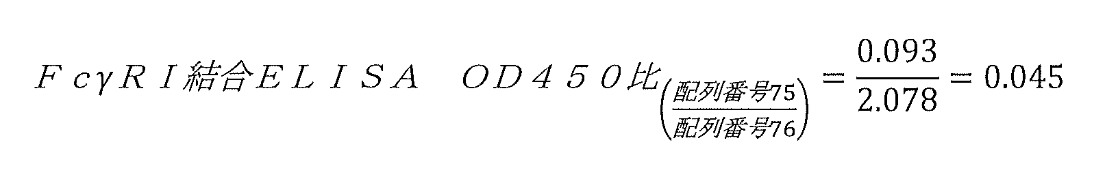

配列番号76のインスリン-Fc融合タンパク質の構成(hIgG1)に対する、配列番号75のインスリン-Fc融合タンパク質の構成(hIgG2)のFcγRI結合ELISA OD450比およびC1q結合ELISA OD450比は、望ましくない免疫原性の傾向を低減することを目的として、種々のインスリン-Fc融合体構成のFcγRI結合およびC1q結合を評価するための設計目標を作成する際に使用した、意欲的な基準である。よって、ヒトFcγRI結合(試験対象のインスリン-Fc融合タンパク質のビオチン化FcγRI濃度は3000ng/mL)の場合に設けられた設計目標は、0.50未満のOD450比であり、C1q結合の場合に設けられた設計目標(試験対象のインスリン-Fc融合タンパク質のビオチン化C1q濃度は1000ng/mL)は、0.35未満のOD450比である。 The FcγRI-binding ELISA OD450 ratio and the C1q-binding ELISA OD450 ratio of the insulin-Fc fusion protein construct of SEQ ID NO: 75 (hIgG2) to the insulin-Fc fusion protein construct of SEQ ID NO: 76 (hIgG1) are undesirable immunogenic. These are the ambitious criteria we used in developing design goals for assessing FcγRI and C1q binding of various insulin-Fc fusion constructs, with the goal of reducing trends. Therefore, the design goal established in the case of human FcγRI binding (biotinylated FcγRI concentration of the insulin-Fc fusion protein tested was 3000 ng/mL) was an OD450 ratio of less than 0.50, and the design goal established in the case of C1q binding. The established design goal (biotinylated C1q concentration of the insulin-Fc fusion protein tested is 1000 ng/mL) is an OD450 ratio of less than 0.35.
ヒトIgG2 Fcフラグメントを含むこのインスリン-Fc融合タンパク質の構成(配列番号75)の2つの別々の合成から得られた平均ホモ二量体力価は、117mg/Lであった。 The average homodimer titer obtained from two separate syntheses of this insulin-Fc fusion protein construct (SEQ ID NO: 75) containing a human IgG2 Fc fragment was 117 mg/L.
比較すると、同じ配列番号7のインスリンポリペプチドと、同じ配列番号13のペプチドリンカーと、ヒトIgG1フラグメント(配列番号73)と、を含むインスリン-Fc融合タンパク質の実施形態(配列番号76)では、インスリン-Fc融合タンパク質は、2つの別々の合成からの平均ホモ二量体力価が180mg/Lであり、ヒトIgG2フラグメントを含む類似のインスリン-Fc融合タンパク質の構成の場合で得られた平均ホモ二量体力価よりも50%以上大きいことが見出された。ヒトIgG1アイソタイプがヒトIgG2アイソタイプよりも高い親和性でヒトFcγRI受容体と相互作用することを考えると、ヒトIgG1を含むインスリン-Fc融合タンパク質の構成を反復注射した後に、望ましくない免疫原性のリスクが存在する可能性がある。 By comparison, an insulin-Fc fusion protein embodiment (SEQ ID NO: 76) comprising the same insulin polypeptide of SEQ ID NO: 7, the same peptide linker of SEQ ID NO: 13, and a human IgG1 fragment (SEQ ID NO: 73), - The Fc fusion protein has an average homodimer titer of 180 mg/L from two separate syntheses, compared to the average homodimer titer obtained for similar insulin-Fc fusion protein constructions containing human IgG2 fragments. It was found that the physical strength value was more than 50% higher than the physical strength value. Given that the human IgG1 isotype interacts with the human FcγRI receptor with higher affinity than the human IgG2 isotype, there is a risk of undesirable immunogenicity after repeated injections of insulin-Fc fusion protein constructs containing human IgG1. may exist.
そこで、インスリン-Fc融合タンパク質のホモ二量体力価を高く維持しつつ、ヒトFcγRI受容体およびC1qに対する親和性を低減させるために、ヒトIgG1 Fcフラグメントに対する様々な変異の検討を行った。 Therefore, in order to reduce the affinity for human FcγRI receptor and C1q while maintaining a high homodimer titer of the insulin-Fc fusion protein, various mutations to the human IgG1 Fc fragment were investigated.
前述のように、FcγRI相互作用を低減する1つの方法では、宿主細胞内でインスリン-Fc融合タンパク質を合成する際に、Fcフラグメントの脱グリコシル化またはグリコシル化防止が行われる。合成されたインスリン-Fc融合タンパク質から結合したグリカンを除去する1つの方法として、宿主細胞での生産中にグリカンの結合を完全に防止するためにcNg部位を変異させることが挙げられる。 As mentioned above, one method of reducing FcγRI interactions involves deglycosylation or prevention of glycosylation of the Fc fragment during synthesis of the insulin-Fc fusion protein in the host cell. One way to remove attached glycans from synthesized insulin-Fc fusion proteins is to mutate the cNg site to completely prevent glycan attachment during production in host cells.
cNg部位に下線が引かれた、変異型ヒトIgG1 Fcフラグメントの一般構成は以下の通りであり:
DKTHTCPPCPAPELLGGPSVFLFPPKPKDTLMISRTPEVTCVVVDVSHEDPEVKFNWYVDGVEVHNAKTKPREEQYX
1 STYRVVSVLTVLHQDWLNGKEYKCKVSNKALPAPIEKTISKAKGQPREPQVYTLPPSRDELTKNQVSLTCLVKGFYPSDIAVEWESNGQPENNYKTTPPVLDSDGSFFLYSKLTVDKSRWQQGNVFSCSVMHEALHNHYTQKSLSLSPG(配列番号77)、
例において、X1はS、D、A、R、またはQである。
The general composition of the mutant human IgG1 Fc fragment, with the cNg site underlined, is as follows:
DKTHTCPPCPAPELLGGPSVFLFPPKPKDTLMISRTPEVTCVVVDVSHEDPEVKFNWYVDGVEVHNAKTKPREEQY KGQPREPQVYTLPPSRDELTKNQVSLTCLVKGFYPSDIAVEWESNGQPENNYKTTPPVLDSDGSFFLYSKLTVDKSRWQQGNVFSCSVMHEALHNHYTQKSLSLSPG (SEQ ID NO : 77 ) ,
In examples, X 1 is S, D, A, R, or Q.
配列番号7のインスリンポリペプチドと、配列番号13のペプチドリンカーと、X1がSである(すなわち、cNg-NB155-S変異を有する)配列番号77のヒトIgG1 Fcフラグメントと、を含むヒトIgG1 Fcフラグメントを、以下に示す:
FVNQHLCGSHLVEALELVCGERGFHYGGGGGGSGGGGGIVEQCCTSTCSLDQLENYCGGGGGQGGGGQGGGGQGGGGGDKTHTCPPCPAPELLGGPSVFLFPPKPKDTLMISRTPEVTCVVVDVSHEDPEVKFNWYVDGVEVHNAKTKPREEQYSSTYRVVSVLTVLHQDWLNGKEYKCKVSNKALPAPIEKTISKAKGQPREPQVYTLPPSRDELTKNQVSLTCLVKGFYPSDIAVEWESNGQPENNYKTTPPVLDSDGSFFLYSKLTVDKSRWQQGNVFSCSVMHEALHNHYTQKSLSLSPG(配列番号91)
A human IgG1 Fc comprising an insulin polypeptide of SEQ ID NO: 7, a peptide linker of SEQ ID NO: 13, and a human IgG1 Fc fragment of SEQ ID NO: 77 in which X 1 is S (i.e., has a cNg-NB155-S mutation) The fragment is shown below:
FVNQHLCGSHLVEALELVCGERGFHYGGGGGGSGGGGGIVEQCCTSTCSLDQLENYCGGGGGQGGGGQGGGGQGGGGGDKTHTCPPCPAPELLGGPSVFLFPPKPKDTLMISRTPEVTCVVVD VSHEDPEVKFNWYVDGVEVHNAKTKPREEQY S STYRVVSVLTVLHQDWLNGKEYKCKVSNKALPAPIEKTISKAKGQPREPQVYTLPPSRDELTKNQVSLTCLVKGFYPSDIAVEWESNGQPE NNYKTTPPVLDSDGSFFLYSKLTVDKSRWQQGNVFSCSVMHEALHNHYTQKSLSLSPG (SEQ ID NO: 91)
予想外にも、配列番号91の非グリコシル化インスリン-Fc融合タンパク質の実施形態は、グリコシル化した親物質(配列番号76)と比べて、ホモ二量体力価が向上することが見出された。さらに、配列番号76の参照インスリン-Fc融合タンパク質に対する配列番号91のインスリン-Fc融合タンパク質実施形態の、ビオチン化FcγRI濃度が3000ng/mLであるヒトFcγRIアッセイにおけるOD450比は0.50未満であり、配列番号76の参照インスリン-Fc融合タンパク質に対する配列番号91のインスリン-Fc融合タンパク質の構成の、ビオチン化C1q濃度が1,000ng/mLである場合のヒトC1q結合アッセイにおけるOD450比は0.35未満であった。しかし、得られた配列番号91の非グリコシル化インスリン-Fc融合タンパク質は、配列番号76の親グリコシル化インスリン-Fc融合タンパク質に対して、IR結合の低減(IRアッセイにおけるIC50値の増加)と、FcRn結合親和性の低減(EC50値の増加)を示し、インビボにおける滞留時間の許容できない減少に伴って生物活性の許容できない低減を示す可能性が高いことが示された。 Unexpectedly, the non-glycosylated insulin-Fc fusion protein embodiment of SEQ ID NO: 91 was found to have improved homodimer potency compared to the glycosylated parent material (SEQ ID NO: 76). . Furthermore, the OD450 ratio of the insulin-Fc fusion protein embodiment of SEQ ID NO: 91 to the reference insulin-Fc fusion protein of SEQ ID NO: 76 in a human FcγRI assay with a biotinylated FcγRI concentration of 3000 ng/mL is less than 0.50; The composition of the insulin-Fc fusion protein of SEQ ID NO: 91 to the reference insulin-Fc fusion protein of SEQ ID NO: 76 has an OD450 ratio of less than 0.35 in a human C1q binding assay when the biotinylated C1q concentration is 1,000 ng/mL. Met. However, the resulting non-glycosylated insulin-Fc fusion protein of SEQ ID NO: 91 showed reduced IR binding (increased IC50 value in IR assay) with respect to the parent glycosylated insulin-Fc fusion protein of SEQ ID NO: 76. It was shown that the FcRn binding affinity was reduced (increased EC50 value) and was likely to show an unacceptable reduction in biological activity along with an unacceptable reduction in in vivo residence time.
インスリン-Fc融合タンパク質
本明細書において、インスリンポリペプチドと、Fcフラグメントと、当該インスリンポリペプチドと当該Fcフラグメントとの間のリンカーと、を含む、インスリン-Fc融合タンパク質の構成が提供される。実施形態において、インスリンポリペプチドは、N末端からC末端へ以下の配向で各ドメインを含む:
(N末端)--B鎖--C鎖--A鎖--(C末端)。
実施形態において、インスリンポリペプチドは、FcフラグメントのN末端側に位置する。実施形態において、上記融合タンパク質は、N末端からC末端へ以下の配向で各ドメインを含む:
(N末端)--インスリンポリペプチド--リンカー--Fcフラグメント--(C末端)(例えば、(N末端)--B鎖--C鎖--A鎖--リンカー--Fcフラグメント--(C末端))(図1に図示)。
Insulin-Fc Fusion Proteins Provided herein are insulin-Fc fusion protein constructs that include an insulin polypeptide, an Fc fragment, and a linker between the insulin polypeptide and the Fc fragment. In embodiments, the insulin polypeptide comprises each domain in the following orientation from N-terminus to C-terminus:
(N-terminal) --B chain--C chain--A chain--(C terminal).
In embodiments, the insulin polypeptide is located on the N-terminal side of the Fc fragment. In embodiments, the fusion protein comprises each domain in the following orientation from N-terminus to C-terminus:
(N-terminus) -- Insulin polypeptide -- Linker -- Fc fragment -- (C-terminus) (For example, (N-terminus) -- B chain -- C chain -- A chain -- Linker -- Fc fragment -- (C-terminal)) (illustrated in Figure 1).
イヌインスリン-Fc融合タンパク質
実施形態において、配列番号5の好ましい非免疫原性生物活性インスリンポリペプチドを、配列番号13の好ましいリンカーを用いて、配列番号15の好ましいイヌIgGB Fcフラグメントと組み合わせたところ、イヌにおける一連の反復注射の3回目の注射の後、50mg/Lを超えるホモ二量体力価、イヌにおいて150%FBGL・日・kg/mgを超えるNAOC値、および、0.5を超えるNAOCR値を示す、配列番号20の、高いホモ二量体力価をもたらす、非凝集性の、生物活性を有する、非免疫原性の、イヌインスリン-Fc融合構成ファミリーが得られた。以下は配列番号20を示しており、非天然型のアミノ酸には下線が引かれており:
FVNQHLCGSX
1 LVEALELVCGERGFHYGGGGGGSGGGGGIVEQCCX
2 STCSLDQLENYCX
3 GGGGGQGGGGQGGGGQGGGGGDCPKCPAPEMLGGPSVFIFPPKPKDTLLIARTPEVTCVVVDLDPEDPEVQISWFVDGKQMQTAKTQPREEQFNGTYRVVSVLPIGHQDWLKGKQFTCKVNNKALPSPIERTISKARGQAHQPSVYVLPPSREELSKNTVSLTCLIKDFFPPDIDVEWQSNGQQEPESKYRTTPPQLDEDGSYFLYSKLSVDKSRWQRGDTFICAVMHEALHNHYTQESLSHSPG(配列番号20)、
X1はDではなく、X2はHではなく、X3は存在しないかNである。
Canine Insulin-Fc Fusion Proteins In embodiments, the preferred non-immunogenic bioactive insulin polypeptide of SEQ ID NO: 5 is combined with the preferred canine IgG Fc fragment of SEQ ID NO: 15 using the preferred linker of SEQ ID NO: 13. After the third injection of a series of repeated injections in dogs, homodimer titers greater than 50 mg/L, NAOC values greater than 150% FBGL·day·kg/mg in dogs, and NAOCR values greater than 0.5. A non-aggregating, biologically active, non-immunogenic, canine insulin-Fc fusion construct family of SEQ ID NO: 20 was obtained, exhibiting a high homodimer titer. The following shows SEQ ID NO: 20, with non-natural amino acids underlined:
FVNQHLCGS X 1 LVEAL E LVCGERGF H Y GGGGGGSGGGGG GIVEQCC X 2 S T CSL D QLENYC PKDTLLIARTPEVTCVVDLDPEDPEVQISWFVDGKQMQTAKTQPREEQFNGTYRVVSVLPIGHQDWLKGKQFTCKVNNKALPSPIERTISKARGQAHQPSVYVLPPSREELSKNTVSLTCLIKD FFPPPDIDVEWQSNGQQEPESKYRTTPPQLDEDGSYFLYSKLSVDKSRWQRGDTFICAVMHEALHNHYTQESLSHSPG ( SEQ ID NO: 20 ) ,
X 1 is not D, X 2 is not H, and X 3 is absent or N.
配列番号20を含むイヌインスリン-Fc融合タンパク質の好ましい実施形態においては、X1はHであり、X2はTであり、X3は存在しないかNである。セレクションにより、イヌにおける一連の反復注射の3回目の注射の後、50mg/Lを超えるホモ二量体力価、イヌにおいて150%FBGL・日・kg/mgを超えるNAOC値、および、0.5を超えるNAOCR値を示す、配列番号21の、高いホモ二量体力価をもたらす、非凝集性の、生物活性を有する、非免疫原性の、イヌインスリン-Fc融合タンパク質の構成が得られる。以下は配列番号21を示しており、非天然型のアミノ酸には下線が引かれており:
FVNQHLCGSHLVEALELVCGERGFHYGGGGGGSGGGGGIVEQCCTSTCSLDQLENYCX
3 GGGGGQGGGGQGGGGQGGGGGDCPKCPAPEMLGGPSVFIFPPKPKDTLLIARTPEVTCVVVDLDPEDPEVQISWFVDGKQMQTAKTQPREEQFNGTYRVVSVLPIGHQDWLKGKQFTCKVNNKALPSPIERTISKARGQAHQPSVYVLPPSREELSKNTVSLTCLIKDFFPPDIDVEWQSNGQQEPESKYRTTPPQLDEDGSYFLYSKLSVDKSRWQRGDTFICAVMHEALHNHYTQESLSHSPG(配列番号21)、
X3は存在しないかNである。
In a preferred embodiment of the canine insulin-Fc fusion protein comprising SEQ ID NO: 20, X 1 is H, X 2 is T, and X 3 is absent or N. Selection results in homodimer titers greater than 50 mg/L, NAOC values greater than 150% FBGL·day·kg/mg in dogs, and 0.5 after the third injection of a series of repeated injections in dogs. A non-aggregating, bioactive, non-immunogenic, canine insulin-Fc fusion protein construct of SEQ ID NO: 21 is obtained that exhibits NAOCR values exceeding SEQ ID NO: 21, resulting in high homodimer titers. The following shows SEQ ID NO: 21, with non-natural amino acids underlined:
FVNQHLCGSHLVEAL E LVCGERGF H Y GGGGGGSGGGGG GIVEQCCTS T CSL D QLENYC RTPEVTCVVDLDPEDPEVQISWFVDGKQMQTAKTQPREEQFNGTYRVVSVLPIGHQDWLKGKQFTCKVNNKALPSPIERTISKARGQAHQPSVYVLPPSREELSKNTVSLTCLIKDFFPDIDV EWQSNGQQEPESKYRTTPQLDEDGSYFLYSKLSVDKSRWQRGDTFICAVMHEALHNHYTQESLSHSPG (SEQ ID NO: 21 ) ,
X 3 does not exist or is N.
好ましい実施形態において、配列番号21においてX3が存在しない場合、イヌにおける一連の反復注射の3回目の注射の後、50mg/Lを超えるホモ二量体力価、イヌにおいて150%FBGL・日・kg/mgを超えるNAOC値、および、0.5を超えるNAOCR値を示す、配列番号26の、高いホモ二量体をもたらす、非凝集性の、生物活性を有する、非免疫原性の、イヌインスリン-Fc融合タンパク質が得られる。以下は配列番号26を示しており、非天然型のアミノ酸には下線が引かれている:
FVNQHLCGSHLVEALELVCGERGFHYGGGGGGSGGGGGIVEQCCTSTCSLDQLENYCGGGGGQGGGGQGGGGQGGGGGDCPKCPAPEMLGGPSVFIFPPKPKDTLLIARTPEVTCVVVDLDPEDPEVQISWFVDGKQMQTAKTQPREEQFNGTYRVVSVLPIGHQDWLKGKQFTCKVNNKALPSPIERTISKARGQAHQPSVYVLPPSREELSKNTVSLTCLIKDFFPPDIDVEWQSNGQQEPESKYRTTPPQLDEDGSYFLYSKLSVDKSRWQRGDTFICAVMHEALHNHYTQESLSHSPG(配列番号26)。
In a preferred embodiment, when X 3 is absent in SEQ ID NO: 21, homodimer titers greater than 50 mg/L, 150% FBGL·day·kg in dogs after the third injection of a series of repeated injections in dogs. A non-aggregating, bioactive, non-immunogenic, highly homodimeric, canine insulin of SEQ ID NO: 26 with an NAOC value greater than /mg and an NAOCR value greater than 0.5. -Fc fusion protein is obtained. The following shows SEQ ID NO: 26, with non-natural amino acids underlined:
FVNQHLCGSHLVEAL E LVCGERGF H Y GGGGGGSGGGG GIVEQCCTS T CSL D QLENYCGGGGGQGGGGQGGGGQGGGGDCPKCPAPEMLGGPSVFIFPPKPKDTLLIARTPEV TCVVVDLDPEDPEVQISWFVDGKQMQTAKTQPREEQFNGTYRVVSVLPIGHQDWLKGKQFTCKVNNKALPSPIERTISKARGQAHQPSVYVLPPSREELSKNTVSLTCLIKDFFPPDIDVEWQSN GQQEPESKYRTTPQLDEDGSYFLYSKLSVDKSRWQRGDTFICAVMHEALHNHYTQESLSHSPG (SEQ ID NO: 26).
好ましい実施形態において、配列番号21においてX3がNである場合、イヌにおける一連の反復注射の3回目の注射の後、50mg/Lを超えるホモ二量体力価、イヌにおいて150%FBGL・日・kg/mgを超えるNAOC値、および、0.5を超えるNAOCR値を示す、配列番号28の、高いホモ二量体力価をもたらす、非凝集性の、生物活性を有する、非免疫原性の、イヌインスリン-Fc融合タンパク質が得られる。以下は配列番号28を示しており、非天然型のアミノ酸には下線が引かれている:
FVNQHLCGSHLVEALELVCGERGFHYGGGGGGSGGGGGIVEQCCTSTCSLDQLENYCNGGGGGQGGGGQGGGGQGGGGGDCPKCPAPEMLGGPSVFIFPPKPKDTLLIARTPEVTCVVVDLDPEDPEVQISWFVDGKQMQTAKTQPREEQFNGTYRVVSVLPIGHQDWLKGKQFTCKVNNKALPSPIERTISKARGQAHQPSVYVLPPSREELSKNTVSLTCLIKDFFPPDIDVEWQSNGQQEPESKYRTTPPQLDEDGSYFLYSKLSVDKSRWQRGDTFICAVMHEALHNHYTQESLSHSPG(配列番号28)。
In a preferred embodiment, when X 3 is N in SEQ ID NO: 21, after the third injection of a series of repeated injections in dogs, the homodimer titer is greater than 50 mg/L, 150% FBGL day in dogs. A non-aggregating, bioactive, non-immunogenic, yielding high homodimer titer of SEQ ID NO: 28 exhibiting NAOC values greater than kg/mg and NAOCR values greater than 0.5. A canine insulin-Fc fusion protein is obtained. The following shows SEQ ID NO: 28, with non-natural amino acids underlined:
FVNQHLCGSHLVEAL E LVCGERGF H Y GGGGGSGGGG GIVEQCCTS T CSL D QLENYCNGGGGGQGGGGQGGGGQGGGGDCPKCPAPEMLGGPSVFIFPPKPKDTLLIARTPE VTCVVDLDPEDPEVQISWFVDGKQMQTAKTQPREEQFNGTYRVVSVLPIGHQDWLKGKQFTCKVNNKALPSPIERTISKARGQAHQPSVYVLPPSREELSKNTVSLTCLIKDFFPDIDVEWQS NGQQEPESKYRTTPQLDEDGSYFLYSKLSVDKSRWQRGDTFICAVMHEALHNHYTQESLSHSPG (SEQ ID NO: 28).
好ましい実施形態において、配列番号18の好ましい非グリコシル化cNg-S変異型イヌIgGB Fcフラグメントを、配列番号13の好ましいリンカーを用いて、配列番号9の好ましいB16A変異型インスリンポリペプチド配列と組み合わせると、イヌにおける一連の反復注射の3回目の注射の後、50mg/Lを超えるホモ二量体力価、イヌにおいて150%FBGL・日・kg/mgを超えるNAOC値、および、0.5を超えるNAOCR値を示す、配列番号22の、高いホモ二量体力価をもたらす、非凝集性の、生物活性を有する、非免疫原性の、イヌインスリン-Fc融合タンパク質ファミリーが得られる。以下は配列番号22を示しており、非天然型のアミノ酸には下線が引かれており:
FVNQHLCGSX
1 LVEALALVCGERGFHYGGGGGGSGGGGGIVEQCCX
2 STCSLDQLENYCGGGGGQGGGGQGGGGQGGGGGDCPKCPAPEMLGGPSVFIFPPKPKDTLLIARTPEVTCVVVDLDPEDPEVQISWFVDGKQMQTAKTQPREEQFSGTYRVVSVLPIGHQDWLKGKQFTCKVNNKALPSPIERTISKARGQAHQPSVYVLPPSREELSKNTVSLTCLIKDFFPPDIDVEWQSNGQQEPESKYRTTPPQLDEDGSYFLYSKLSVDKSRWQRGDTFICAVMHEALHNHYTQESLSHSPG(配列番号22)、
X1はDではなく、X2はHではない。
In a preferred embodiment, the preferred non-glycosylated cNg-S mutant canine IgG Fc fragment of SEQ ID NO: 18 is combined with the preferred B16A mutant insulin polypeptide sequence of SEQ ID NO: 9 using the preferred linker of SEQ ID NO: 13; After the third injection of a series of repeated injections in dogs, homodimer titers greater than 50 mg/L, NAOC values greater than 150% FBGL·day·kg/mg in dogs, and NAOCR values greater than 0.5. A family of non-aggregating, biologically active, non-immunogenic canine insulin-Fc fusion proteins is obtained that yields high homodimeric titers, with SEQ ID NO: 22. The following shows SEQ ID NO: 22, with non-natural amino acids underlined:
FVNQHLCGS X 1 LVEAL A LVCGERGF H Y GGGGGGSGGGGG GIVEQCC LIARTPEVTCVVDLDPEDPEVQISWFVDGKQMQTAKTQPREEQF S GTYRVVSVLPIGHQDWLKGKQFTCKVNNKALPSPIERTISKARGQAHQPSVYVLPPSREELSKNTVSLTCLIKDFFP PDIDVEWQSNGQQEPESKYRTTPPQLDEDGSYFLYSKLSVDKSRWQRGDTFICAVMHEALHNHYTQESLSHSPG (SEQ ID NO : 22 ) ,
X 1 is not D, and X 2 is not H.
好ましい実施形態において、配列番号22においてX1がHであり、X2がTである場合、イヌにおける一連の反復注射の3回目の注射の後、50mg/Lを超えるホモ二量体力価、イヌにおいて150%FBGL・日・kg/mgを超えるNAOC値、および、0.5を超えるNAOCR値を示す、配列番号30の、高いホモ二量体力価をもたらす、非凝集性の、生物活性を有する、非免疫原性の、イヌインスリン-Fc融合タンパク質が得られる。以下は配列番号30を示しており、非天然型のアミノ酸には下線が引かれている:
FVNQHLCGSHLVEALALVCGERGFHYGGGGGGSGGGGGIVEQCCTSTCSLDQLENYCGGGGGQGGGGQGGGGQGGGGGDCPKCPAPEMLGGPSVFIFPPKPKDTLLIARTPEVTCVVVDLDPEDPEVQISWFVDGKQMQTAKTQPREEQFSGTYRVVSVLPIGHQDWLKGKQFTCKVNNKALPSPIERTISKARGQAHQPSVYVLPPSREELSKNTVSLTCLIKDFFPPDIDVEWQSNGQQEPESKYRTTPPQLDEDGSYFLYSKLSVDKSRWQRGDTFICAVMHEALHNHYTQESLSHSPG(配列番号30)。
In a preferred embodiment, when in SEQ ID NO : 22, X 1 is H and A non-aggregating, biologically active, high homodimer titer of SEQ ID NO: 30 exhibiting NAOC values greater than 150% FBGL·day·kg/mg and NAOCR values greater than 0.5. , a non-immunogenic canine insulin-Fc fusion protein is obtained. The following shows SEQ ID NO: 30, with non-natural amino acids underlined:
FVNQHLCGS H LVEAL A LVCGERGF H Y GGGGGGSGGGGG GIVEQCCTS T CSL D QLENYCGGGGGQGGGGQGGGGQGGGGDCPKCPAPEMLGGPSVFIFPPKPKDTLLIARTP EVTCVVVDLDPEDPEVQISWFVDGKQMQTAKTQPREEQF S GTYRVVSVLPIGHQDWLKGKQFTCKVNNKALPSPIERTISKARGQAHQPSVYVLPPSREELSKNTVSLTCLIKDFFPPDIDVE WQSNGQQEPESKYRTTPQLDEDGSYFLYSKLSVDKSRWQRGDTFICAVMHEALHNHYTQESLSHSPG (SEQ ID NO: 30).
配列番号30の、製造性と有効性が高い、超長時間作用型イヌインスリン-Fc融合タンパク質の実施形態が予想外にも発見されたことにより、本発明者らは、ヒト患者用の、同様の製造性と有効性を有する、超長時間作用型インスリン-Fc融合タンパク質の構成をつくり出す試みを行った。 The unexpected discovery of a highly manufacturable, highly effective, ultra-long-acting canine insulin-Fc fusion protein embodiment of SEQ ID NO: 30 led us to develop a similar protein for human patients. An attempt was made to create an ultra-long-acting insulin-Fc fusion protein construct that has the same manufacturability and efficacy.
ヒトインスリン-Fc融合タンパク質
他の非グリコシル化インスリン-Fc融合タンパク質の構成が同じ挙動を示すかどうかを判定するための、さらなる実験を行った。cNg部位に下線が引かれた、ヒトIgG1 Fcフラグメントの一般構成は以下の通りであり:
DKTHTCPPCPAPELLGGPSVFLFPPKPKDTLMISRTPEVTCVVVDVSHEDPEVKFNWYVDGVEVHNAKTKPREEQYX
1 STYRVVSVLTVLHQDWLNGKEYKCKVSNKALPAPIEKTISKAKGQPREPQVYTLPPSRDELTKNQVSLTCLVKGFYPSDIAVEWESNGQPENNYKTTPPVLDSDGSFFLYSKLTVDKSRWQQGNVFSCSVMHEALHNHYTQKSLSLSPG(配列番号77)、
X1はS、D、A、R、またはQである。
Human Insulin-Fc Fusion Proteins Further experiments were performed to determine whether other non-glycosylated insulin-Fc fusion protein constructs exhibit the same behavior. The general composition of the human IgG1 Fc fragment, with the cNg site underlined, is as follows:
DKTHTCPPCPAPELLGGPSVFLFPPKPKDTLMISRTPEVTCVVVDVSHEDPEVKFNWYVDGVEVHNAKTKPREEQY KGQPREPQVYTLPPSRDELTKNQVSLTCLVKGFYPSDIAVEWESNGQPENNYKTTPPVLDSDGSFFLYSKLTVDKSRWQQGNVFSCSVMHEALHNHYTQKSLSLSPG (SEQ ID NO : 77 ) ,
X 1 is S, D, A, R, or Q.
配列番号7のインスリンポリペプチド(B16A変異なし)と、配列番号13のリンカーと、グリコシル化を防ぐためのcNg変異を有していてもよいヒトIgG1 Fcフラグメントの構成(配列番号77、X1はS、D、A、R、またはQ)と、を含むインスリン-Fc融合タンパク質の実施形態を、以下に示す:
FVNQHLCGSHLVEALELVCGERGFHYGGGGGGSGGGGGIVEQCCTSTCSLDQLENYCGGGGGQGGGGQGGGGQGGGGGDKTHTCPPCPAPELLGGPSVFLFPPKPKDTLMISRTPEVTCVVVDVSHEDPEVKFNWYVDGVEVHNAKTKPREEQYSSTYRVVSVLTVLHQDWLNGKEYKCKVSNKALPAPIEKTISKAKGQPREPQVYTLPPSRDELTKNQVSLTCLVKGFYPSDIAVEWESNGQPENNYKTTPPVLDSDGSFFLYSKLTVDKSRWQQGNVFSCSVMHEALHNHYTQKSLSLSPG(配列番号91)
FVNQHLCGSHLVEALELVCGERGFHYGGGGGGSGGGGGIVEQCCTSTCSLDQLENYCGGGGGQGGGGQGGGGQGGGGGDKTHTCPPCPAPELLGGPSVFLFPPKPKDTLMISRTPEVTCVVVDVSHEDPEVKFNWYVDGVEVHNAKTKPREEQYDSTYRVVSVLTVLHQDWLNGKEYKCKVSNKALPAPIEKTISKAKGQPREPQVYTLPPSRDELTKNQVSLTCLVKGFYPSDIAVEWESNGQPENNYKTTPPVLDSDGSFFLYSKLTVDKSRWQQGNVFSCSVMHEALHNHYTQKSLSLSPG(配列番号92)
FVNQHLCGSHLVEALELVCGERGFHYGGGGGGSGGGGGIVEQCCTSTCSLDQLENYCGGGGGQGGGGQGGGGQGGGGGDKTHTCPPCPAPELLGGPSVFLFPPKPKDTLMISRTPEVTCVVVDVSHEDPEVKFNWYVDGVEVHNAKTKPREEQYASTYRVVSVLTVLHQDWLNGKEYKCKVSNKALPAPIEKTISKAKGQPREPQVYTLPPSRDELTKNQVSLTCLVKGFYPSDIAVEWESNGQPENNYKTTPPVLDSDGSFFLYSKLTVDKSRWQQGNVFSCSVMHEALHNHYTQKSLSLSPG(配列番号93)
FVNQHLCGSHLVEALELVCGERGFHYGGGGGGSGGGGGIVEQCCTSTCSLDQLENYCGGGGGQGGGGQGGGGQGGGGGDKTHTCPPCPAPELLGGPSVFLFPPKPKDTLMISRTPEVTCVVVDVSHEDPEVKFNWYVDGVEVHNAKTKPREEQYRSTYRVVSVLTVLHQDWLNGKEYKCKVSNKALPAPIEKTISKAKGQPREPQVYTLPPSRDELTKNQVSLTCLVKGFYPSDIAVEWESNGQPENNYKTTPPVLDSDGSFFLYSKLTVDKSRWQQGNVFSCSVMHEALHNHYTQKSLSLSPG(配列番号94)
FVNQHLCGSHLVEALELVCGERGFHYGGGGGGSGGGGGIVEQCCTSTCSLDQLENYCGGGGGQGGGGQGGGGQGGGGGDKTHTCPPCPAPELLGGPSVFLFPPKPKDTLMISRTPEVTCVVVDVSHEDPEVKFNWYVDGVEVHNAKTKPREEQYQSTYRVVSVLTVLHQDWLNGKEYKCKVSNKALPAPIEKTISKAKGQPREPQVYTLPPSRDELTKNQVSLTCLVKGFYPSDIAVEWESNGQPENNYKTTPPVLDSDGSFFLYSKLTVDKSRWQQGNVFSCSVMHEALHNHYTQKSLSLSPG(配列番号95)
Construction of the insulin polypeptide of SEQ ID NO: 7 (without the B16A mutation), the linker of SEQ ID NO: 13, and a human IgG1 Fc fragment optionally having a cNg mutation to prevent glycosylation (SEQ ID NO: 77, X 1 is Embodiments of insulin-Fc fusion proteins comprising:
FVNQHLCGSHLVEALELVCGERGFHYGGGGGGSGGGGGIVEQCCTSTCSLDQLENYCGGGGGQGGGGQGGGGQGGGGGDKTHTCPPCPAPELLGGPSVFLFPPKPKDTLMISRTPEVTCVVVD VSHEDPEVKFNWYVDGVEVHNAKTKPREEQY S STYRVVSVLTVLHQDWLNGKEYKCKVSNKALPAPIEKTISKAKGQPREPQVYTLPPSRDELTKNQVSLTCLVKGFYPSDIAVEWESNGQPE NNYKTTPPVLDSDGSFFLYSKLTVDKSRWQQGNVFSCSVMHEALHNHYTQKSLSLSPG (SEQ ID NO: 91)
FVNQHLCGSHLVEALELVCGERGFHYGGGGGGSGGGGGIVEQCCTSTCSLDQLENYCGGGGGQGGGGQGGGGQGGGGGDKTHTCPPCPAPELLGGPSVFLFPPKPKDTLMISRTPEVTCVVVD VSHEDPEVKFNWYVDGVEVHNAKTKPREEQY D STYRVVSVLTVLHQDWLNGKEYKCKVSNKALPAPIEKTISKAKGQPREPQVYTLPPSRDELTKNQVSLTCLVKGFYPSDIAVEWESNGQPE NNYKTTPPVLDSDGSFFLYSKLTVDKSRWQQGNVFSCSVMHEALHNHYTQKSLSLSPG (SEQ ID NO: 92)
FVNQHLCGSHLVEALELVCGERGFHYGGGGGGSGGGGGIVEQCCTSTCSLDQLENYCGGGGGQGGGGQGGGGQGGGGGDKTHTCPPCPAPELLGGPSVFLFPPKPKDTLMISRTPEVTCVVVD VSHEDPEVKFNWYVDGVEVHNAKTKPREEQY A STYRVVSVLTVLHQDWLNGKEYKCKVSNKALPAPIEKTISKAKGQPREPQVYTLPPSRDELTKNQVSLTCLVKGFYPSDIAVEWESNGQPE NNYKTTPPVLDSDGSFFLYSKLTVDKSRWQQGNVFSCSVMHEALHNHYTQKSLSLSPG (SEQ ID NO: 93)
FVNQHLCGSHLVEALELVCGERGFHYGGGGGGSGGGGGIVEQCCTSTCSLDQLENYCGGGGGQGGGGQGGGGQGGGGGDKTHTCPPCPAPELLGGPSVFLFPPKPKDTLMISRTPEVTCVVVD VSHEDPEVKFNWYVDGVEVHNAKTKPREEQY R STYRVVSVLTVLHQDWLNGKEYKCKVSNKALPAPIEKTISKAKGQPREPQVYTLPPSRDELTKNQVSLTCLVKGFYPSDIAVEWESNGQPE NNYKTTPPVLDSDGSFFLYSKLTVDKSRWQQGNVFSCSVMHEALHNHYTQKSLSLSPG (SEQ ID NO: 94)
FVNQHLCGSHLVEALELVCGERGFHYGGGGGGSGGGGGIVEQCCTSTCSLDQLENYCGGGGGQGGGGQGGGGQGGGGGDKTHTCPPCPAPELLGGPSVFLFPPKPKDTLMISRTPEVTCVVVD VSHEDPEVKFNWYVDGVEVHNAKTKPREEQY Q STYRVVSVLTVLHQDWLNGKEYKCKVSNKALPAPIEKTISKAKGQPREPQVYTLPPSRDELTKNQVSLTCLVKGFYPSDIAVEWESNGQPE NNYKTTPPVLDSDGSFFLYSKLTVDKSRWQQGNVFSCSVMHEALHNHYTQKSLSLSPG (SEQ ID NO: 95)
配列番号13の好ましいリンカーを用いた、配列番号7のインスリンポリペプチドと、X1がS(cNg-NB155-S:配列番号91)、D(cNg-NB155-D:配列番号92)、A(cNg-NB155-A:配列番号93)、R(cNg-NB155-R:配列番号94)、またはQ(cNg-NB155-Q:配列番号95)である配列番号77のヒトIgG1 Fcフラグメントと、を含む実施形態においては、予想外にも、これらの非グリコシル化インスリン-Fc融合タンパク質のそれぞれが、グリコシル化型の親物質(配列番号76)と比べて、ホモ二量体力価の向上を示すことが発見された。さらに、配列番号91、配列番号92、配列番号93、配列番号94、および配列番号95の、脱グリコシル化インスリン-Fc融合タンパク質のそれぞれについては、ビオチン化FcγRI濃度が3000ng/mLである場合のヒトFcγRIアッセイにおけるOD450比(OD450比算出に使用される参照インスリン-Fc融合タンパク質が配列番号76のインスリン-Fc融合タンパク質である場合)は全て0.50未満であり、ビオチン化C1q濃度が1,000ng/mLである場合のヒトC1q結合アッセイにおけるOD450比(OD450比算出に使用される参照インスリン-Fc融合タンパク質が配列番号76のインスリン-Fc融合タンパク質である場合)は全て0.35未満であった。しかし、配列番号91、配列番号92、配列番号93、配列番号94、および配列番号95の脱グリコシル化インスリン-Fc融合タンパク質は、その全てが、グリコシル化型の親物質(配列番号76)と比べて、IR結合親和性が低く(IRアッセイにおけるIC50値が高い)、FcRn結合親和性が低くなる(EC50値が高い)ことが明らかとなり、これらの化合物が、インビボにおける生物活性および滞留時間の許容できない低減を示す可能性が高いことが示された。予想外にも、配列番号13の好ましいリンカーを用いた、配列番号7のインスリンポリペプチドと、X1がQである配列番号77のヒトIgG1 Fcフラグメントと、を含む実施形態(配列番号95)においては、得られた非グリコシル化インスリン-Fc融合タンパク質は、ホモ二量体力価が136mg/Lであり、このインスリン-Fc融合タンパク質の構成では、ホモ二量体力価の設計目標である150mg/Lが達成されなかった。 Using the preferred linker of SEQ ID NO : 13, the insulin polypeptide of SEQ ID NO: 7 and cNg-NB155-A: SEQ ID NO: 93), R (cNg-NB155-R: SEQ ID NO: 94), or Q (cNg-NB155-Q: SEQ ID NO: 95), the human IgG1 Fc fragment of SEQ ID NO: 77; In embodiments comprising, unexpectedly, each of these non-glycosylated insulin-Fc fusion proteins exhibits improved homodimer potency compared to the glycosylated parent material (SEQ ID NO: 76). was discovered. Furthermore, for each of the deglycosylated insulin-Fc fusion proteins of SEQ ID NO: 91, SEQ ID NO: 92, SEQ ID NO: 93, SEQ ID NO: 94, and SEQ ID NO: 95, human The OD450 ratios in the FcγRI assay (when the reference insulin-Fc fusion protein used to calculate the OD450 ratio is the insulin-Fc fusion protein of SEQ ID NO: 76) are all less than 0.50, and the biotinylated C1q concentration is 1,000 ng. /mL in the human C1q binding assay (when the reference insulin-Fc fusion protein used for OD450 ratio calculation is the insulin-Fc fusion protein of SEQ ID NO: 76) were all less than 0.35. . However, the deglycosylated insulin-Fc fusion proteins of SEQ ID NO: 91, SEQ ID NO: 92, SEQ ID NO: 93, SEQ ID NO: 94, and SEQ ID NO: 95 all have a The results showed that these compounds have low IR binding affinity (high IC50 values in IR assays) and low FcRn binding affinities (high EC50 values), indicating that these compounds have good in vivo biological activity and acceptable residence times. It was shown that there is a high possibility of showing a reduction that cannot be achieved. Unexpectedly, in an embodiment (SEQ ID NO: 95) comprising an insulin polypeptide of SEQ ID NO: 7 and a human IgG1 Fc fragment of SEQ ID NO: 77, in which X 1 is Q, using the preferred linker of SEQ ID NO: 13. The resulting non-glycosylated insulin-Fc fusion protein has a homodimer titer of 136 mg/L, and this insulin-Fc fusion protein composition has a designed homodimer titer of 150 mg/L. was not achieved.
図28は、配列番号75(天然ヒトIgG2を含む)および配列番号76(天然ヒトIgG1を含む)、並びに、配列番号77のバリアントを含む配列である、配列番号91、配列番号92、配列番号93、配列番号94、および配列番号95の、並列配列比較を示している。「*」は、所与の配列位置において全ての配列間で完全に相同であることを示しており、一方、「:」、「.」、空白は、それぞれ、所与の配列位置における配列間のアミノ酸変異が保存的であること、中程度であること、または全く異なるものであること、を示している。 FIG. 28 shows sequences including SEQ ID NO: 75 (containing natural human IgG2) and SEQ ID NO: 76 (containing natural human IgG1), as well as variants of SEQ ID NO: 77, SEQ ID NO: 91, SEQ ID NO: 92, and SEQ ID NO: 93. , SEQ ID NO: 94, and SEQ ID NO: 95. “*” indicates complete homology between all sequences at a given sequence position, while “:”, “.”, and blank space respectively indicate complete homology between all sequences at a given sequence position. indicates that the amino acid variations in the amino acids are conservative, moderate, or completely different.
前述のイヌインスリン-Fc融合タンパク質の構成に関する予想外の結果から学んだことを適用して、配列番号91、配列番号92、配列番号93、および配列番号94のインスリン-Fc融合タンパク質の実施形態におけるインスリンポリペプチドのB鎖上の第16アミノ酸(B16)をアラニンに変異させた結果、配列番号78(cNg-NB155-Sを有する)、配列番号80(cNg-NB155-Dを有する)、配列番号82(cNg-NB155-Aを有する)、配列番号84(cNg-NB155-Rを有する)が得られた。上記の通りB16アミノ酸をアラニンに変異させて、配列番号10のインスリンポリペプチドを得た場合の、配列番号78、配列番号80、配列番号82、および配列番号84のインスリン-Fc融合タンパク質の構成においては、ホモ二量体力価の増加も、FcγRI結合親和性およびC1q結合親和性の低減も損なうことなく、許容できるインスリン受容体結合が予想外に回復した(すなわち、IR結合親和性およびFcRn受容体結合親和性が顕著に増加した)。 Applying the learnings from the unexpected results regarding the construction of canine insulin-Fc fusion proteins described above, in the insulin-Fc fusion protein embodiments of SEQ ID NO: 91, SEQ ID NO: 92, SEQ ID NO: 93, and SEQ ID NO: 94. As a result of mutating the 16th amino acid (B16) on the B chain of insulin polypeptide to alanine, SEQ ID NO: 78 (having cNg-NB155-S), SEQ ID NO: 80 (having cNg-NB155-D), SEQ ID NO: 82 (having cNg-NB155-A) and SEQ ID NO: 84 (having cNg-NB155-R) were obtained. In the structure of the insulin-Fc fusion proteins of SEQ ID NO: 78, SEQ ID NO: 80, SEQ ID NO: 82, and SEQ ID NO: 84 when the B16 amino acid is mutated to alanine as described above to obtain the insulin polypeptide of SEQ ID NO: 10. unexpectedly restored acceptable insulin receptor binding (i.e., IR binding affinity and FcRn receptor binding affinity was significantly increased).
好ましい実施形態において、配列番号78のヒトインスリン-Fc融合タンパク質の構成は、配列番号10のインスリンポリペプチドと、配列番号13のリンカーと、配列番号77のヒトIgG1 FcフラグメントのcNg-NB155-S変異体と、を含む。配列番号78のインスリン-Fc融合タンパク質は、150mg/Lを超えるホモ二量体力価を示す。以下は、B16A変異およびcNg-NB155-S変異に下線が引かれた、配列番号78のインスリン-Fc融合タンパク質を示している:
FVNQHLCGSHLVEALALVCGERGFHYGGGGGGSGGGGGIVEQCCTSTCSLDQLENYCGGGGGQGGGGQGGGGQGGGGGDKTHTCPPCPAPELLGGPSVFLFPPKPKDTLMISRTPEVTCVVVDVSHEDPEVKFNWYVDGVEVHNAKTKPREEQYSSTYRVVSVLTVLHQDWLNGKEYKCKVSNKALPAPIEKTISKAKGQPREPQVYTLPPSRDELTKNQVSLTCLVKGFYPSDIAVEWESNGQPENNYKTTPPVLDSDGSFFLYSKLTVDKSRWQQGNVFSCSVMHEALHNHYTQKSLSLSPG(配列番号78)。
In a preferred embodiment, the human insulin-Fc fusion protein of SEQ ID NO: 78 comprises the insulin polypeptide of SEQ ID NO: 10, the linker of SEQ ID NO: 13, and the cNg-NB155-S mutation of the human IgG1 Fc fragment of SEQ ID NO: 77. including the body. The insulin-Fc fusion protein of SEQ ID NO: 78 exhibits a homodimer titer of greater than 150 mg/L. The following shows the insulin-Fc fusion protein of SEQ ID NO: 78, with the B16A mutation and cNg-NB155-S mutation underlined:
FVNQHLCGSHLVEAL A LVCGERGFHYGGGGGGGSGGGGIVEQCCTSTCSLDQLENYCGGGGGQGGGGQGGGGQGGGGGDKTHTCPPCPAPELLGGPSVFLFPPKPKDTLMISRTPEVTCVV VDVSHEDPEVKFNWYVDGVEVHNAKTKPREEQY S STYRVVSVLTVLHQDWLNGKEYKCKVSNKALPAPIEKTISKAKGQPREPQVYTLPPSRDELTKNQVSLTCLVKGFYPSDIAVEWESNGQ PENNYKTTPVLDSDGSFFLYSKLTVDKSRWQQGNVFSCSVMHEALHNHYTQKSLSLSPG (SEQ ID NO: 78).
イヌインスリン-Fc融合タンパク質の構成での予想外の発見と同様に、B16A変異と、cNg-NB155-S変異を有するヒトIgG1 Fcフラグメントと、を含む、配列番号78の非グリコシル化インスリン-Fc融合タンパク質の実施形態の生物活性は、B16A変異を含んでいない、配列番号91の非グリコシル化インスリン-Fc融合タンパク質の実施形態の生物活性と比較して、予想外に回復した(すなわち、IR結合親和性およびFcRn受容体結合親和性が顕著に増加した)。配列番号78のインスリン-Fc融合タンパク質の実施形態は、IRアッセイにおけるIC50値が2400nM未満、より好ましくは2000nM未満であり、ヒトFcRnアッセイにおけるEC50値が1500ng/L未満、より好ましくは1000ng/mL未満であり、ビオチン化FcγRI濃度が3,000ng/mLである場合のヒトFcγRIアッセイにおけるOD450比(OD450比算出に使用された参照インスリン-Fc融合タンパク質が配列番号76のインスリン-Fc融合タンパク質である場合)が0.50未満であり、ビオチン化C1q濃度が1,000ng/mLである場合のヒトC1q結合アッセイにおけるOD450比(OD450比算出に使用された参照インスリン-Fc融合タンパク質が配列番号76のインスリン-Fc融合タンパク質である場合)が0.35未満であることが分かった。 Similar to the unexpected discovery in the construction of a canine insulin-Fc fusion protein, a non-glycosylated insulin-Fc fusion of SEQ ID NO: 78 comprising the B16A mutation and a human IgG1 Fc fragment with the cNg-NB155-S mutation. The biological activity of the protein embodiment was unexpectedly restored (i.e., the IR binding affinity FcRn receptor binding affinity was significantly increased). Embodiments of the insulin-Fc fusion protein of SEQ ID NO: 78 have an IC50 value in an IR assay of less than 2400 nM, more preferably less than 2000 nM, and an EC50 value in a human FcRn assay of less than 1500 ng/L, more preferably less than 1000 ng/mL. and the OD450 ratio in the human FcγRI assay when the biotinylated FcγRI concentration is 3,000 ng/mL (when the reference insulin-Fc fusion protein used for calculating the OD450 ratio is the insulin-Fc fusion protein of SEQ ID NO: 76) ) is less than 0.50 and the biotinylated C1q concentration is 1,000 ng/mL in the human C1q binding assay (the reference insulin-Fc fusion protein used to calculate the OD450 ratio is -Fc fusion protein) was found to be less than 0.35.
好ましい実施形態において、配列番号80のインスリン-Fc融合タンパク質は、配列番号10のインスリンポリペプチドと、配列番号13のリンカーと、配列番号77(X1はD)のFcフラグメントのcNg-NB155-D変異体と、を含む。配列番号80のインスリン-Fc融合タンパク質は、ホモ二量体力価が150mg/Lを超え、IRアッセイにおけるIC50値が2400nM未満であり、ヒトFcRnアッセイにおけるEC50値が1000ng/mL未満であり、ビオチン化FcγRI濃度が3,000ng/mLである場合のヒトFcγRIアッセイにおけるOD450比が0.50未満であり、ビオチン化C1q濃度が1,000ng/mLである場合のヒトC1q結合アッセイにおけるOD450比が0.35未満であった。以下は、B16A変異およびcNg-NB155-D変異に下線が引かれた、配列番号80を示している:
FVNQHLCGSHLVEALALVCGERGFHYGGGGGGSGGGGGIVEQCCTSTCSLDQLENYCGGGGGQGGGGQGGGGQGGGGGDKTHTCPPCPAPELLGGPSVFLFPPKPKDTLMISRTPEVTCVVVDVSHEDPEVKFNWYVDGVEVHNAKTKPREEQYDSTYRVVSVLTVLHQDWLNGKEYKCKVSNKALPAPIEKTISKAKGQPREPQVYTLPPSRDELTKNQVSLTCLVKGFYPSDIAVEWESNGQPENNYKTTPPVLDSDGSFFLYSKLTVDKSRWQQGNVFSCSVMHEALHNHYTQKSLSLSPG(配列番号80)。
In a preferred embodiment, the insulin-Fc fusion protein of SEQ ID NO: 80 comprises the insulin polypeptide of SEQ ID NO: 10, the linker of SEQ ID NO: 13, and the cNg-NB155-D of the Fc fragment of SEQ ID NO: 77 (X 1 is D). including a variant. The insulin-Fc fusion protein of SEQ ID NO: 80 has a homodimer titer of greater than 150 mg/L, an IC50 value in an IR assay of less than 2400 nM, an EC50 value of less than 1000 ng/mL in a human FcRn assay, and is biotinylated. The OD450 ratio in the human FcγRI assay when the FcγRI concentration is 3,000 ng/mL is less than 0.50, and the OD450 ratio in the human C1q binding assay when the biotinylated C1q concentration is 1,000 ng/mL is 0. It was less than 35. The following shows SEQ ID NO: 80, with the B16A mutation and cNg-NB155-D mutation underlined:
FVNQHLCGSHLVEAL A LVCGERGFHYGGGGGGSGGGGGIVEQCCTSTCSLDQLENYCGGGGGQGGGGQGGGGQGGGGGDKTHTCPPCPAPELLGGPSVFLFPPKPKDTLMISRTPEVTCVV VDVSHEDPEVKFNWYVDGVEVHNAKTKPREEQY D STYRVVSVLTVLHQDWLNGKEYKCKVSNKALPAPIEKTISKAKGQPREPQVYTLPPSRDELTKNQVSLTCLVKGFYPSDIAVEWESNGQ PENNYKTTPVLDSDGSFFLYSKLTVDKSRWQQGNVFSCSVMHEALHNHYTQKSLSLSPG (SEQ ID NO: 80).
好ましい実施形態において、配列番号82のインスリン-Fc融合タンパク質は、配列番号10のインスリンポリペプチドと、配列番号13のリンカーと、配列番号77(X1はA)のFcフラグメントのcNg-NB155-A変異体と、を含む。配列番号82のインスリン-Fc融合タンパク質は、ホモ二量体力価が150mg/Lを超え、IRアッセイにおけるIC50値が2400nM未満であり、ヒトFcRnアッセイにおけるEC50値が1000ng/mL未満であり、ビオチン化FcγRI濃度が3,000ng/mLである場合のヒトFcγRIアッセイにおけるOD450比が0.50未満であり、ビオチン化C1q濃度が1,000ng/mLである場合のヒトC1q結合アッセイにおけるOD450比が0.35未満であった。以下は、B16A変異およびcNg-NB155-A変異に下線が引かれた、配列番号82を示している:
FVNQHLCGSHLVEALALVCGERGFHYGGGGGGSGGGGGIVEQCCTSTCSLDQLENYCGGGGGQGGGGQGGGGQGGGGGDKTHTCPPCPAPELLGGPSVFLFPPKPKDTLMISRTPEVTCVVVDVSHEDPEVKFNWYVDGVEVHNAKTKPREEQYASTYRVVSVLTVLHQDWLNGKEYKCKVSNKALPAPIEKTISKAKGQPREPQVYTLPPSRDELTKNQVSLTCLVKGFYPSDIAVEWESNGQPENNYKTTPPVLDSDGSFFLYSKLTVDKSRWQQGNVFSCSVMHEALHNHYTQKSLSLSPG(配列番号82)。
In a preferred embodiment, the insulin-Fc fusion protein of SEQ ID NO: 82 comprises the insulin polypeptide of SEQ ID NO: 10, the linker of SEQ ID NO: 13, and the cNg-NB155-A Fc fragment of SEQ ID NO: 77 (X 1 is A). including a variant. The insulin-Fc fusion protein of SEQ ID NO: 82 has a homodimer titer of greater than 150 mg/L, an IC50 value in an IR assay of less than 2400 nM, an EC50 value of less than 1000 ng/mL in a human FcRn assay, and is biotinylated. The OD450 ratio in the human FcγRI assay when the FcγRI concentration is 3,000 ng/mL is less than 0.50, and the OD450 ratio in the human C1q binding assay when the biotinylated C1q concentration is 1,000 ng/mL is 0. It was less than 35. The following shows SEQ ID NO: 82, with the B16A mutation and cNg-NB155-A mutation underlined:
FVNQHLCGSHLVEAL A LVCGERGFHYGGGGGGSGGGGGIVEQCCTSTCSLDQLENYCGGGGGQGGGGQGGGGQGGGGGDKTHTCPPCPAPELLGGPSVFLFPPKPKDTLMISRTPEVTCVV VDVSHEDPEVKFNWYVDGVEVHNAKTKPREEQY A STYRVVSVLTVLHQDWLNGKEYKCKVSNKALPAPIEKTISKAKGQPREPQVYTLPPSRDELTKNQVSLTCLVKGFYPSDIAVEWESNGQ PENNYKTTPVLDSDGSFFLYSKLTVDKSRWQQGNVFSCSVMHEALHNHYTQKSLSLSPG (SEQ ID NO: 82).
好ましい実施形態において、配列番号84のインスリン-Fc融合タンパク質は、配列番号10のインスリンポリペプチドと、配列番号13のリンカーと、配列番号77(X1はR)のFcフラグメントのcNg-NB155-R変異体と、を含む。配列番号84のインスリン-Fc融合タンパク質は、ホモ二量体力価が150mg/Lを超え、IRアッセイにおけるIC50値が2400nM未満であり、ヒトFcRnアッセイにおけるEC50値が1000ng/mL未満であり、ビオチン化FcγRI濃度が3,000ng/mLである場合のヒトFcγRIアッセイにおけるOD450比が0.50未満であり、ビオチン化C1q濃度が1,000ng/mLである場合のヒトC1q結合アッセイにおけるOD450比が0.35未満であった。以下は、B16A変異およびcNg-NB155-R変異に下線が引かれた、配列番号84を示している:
FVNQHLCGSHLVEALALVCGERGFHYGGGGGGSGGGGGIVEQCCTSTCSLDQLENYCGGGGGQGGGGQGGGGQGGGGGDKTHTCPPCPAPELLGGPSVFLFPPKPKDTLMISRTPEVTCVVVDVSHEDPEVKFNWYVDGVEVHNAKTKPREEQYRSTYRVVSVLTVLHQDWLNGKEYKCKVSNKALPAPIEKTISKAKGQPREPQVYTLPPSRDELTKNQVSLTCLVKGFYPSDIAVEWESNGQPENNYKTTPPVLDSDGSFFLYSKLTVDKSRWQQGNVFSCSVMHEALHNHYTQKSLSLSPG(配列番号84)。
In a preferred embodiment, the insulin-Fc fusion protein of SEQ ID NO: 84 comprises the insulin polypeptide of SEQ ID NO: 10, the linker of SEQ ID NO: 13, and the Fc fragment of SEQ ID NO: 77 (X 1 is R) cNg-NB155-R. including a variant. The insulin-Fc fusion protein of SEQ ID NO: 84 has a homodimer titer of greater than 150 mg/L, an IC50 value in an IR assay of less than 2400 nM, an EC50 value of less than 1000 ng/mL in a human FcRn assay, and is biotinylated. The OD450 ratio in the human FcγRI assay when the FcγRI concentration is 3,000 ng/mL is less than 0.50, and the OD450 ratio in the human C1q binding assay when the biotinylated C1q concentration is 1,000 ng/mL is 0. It was less than 35. The following shows SEQ ID NO: 84, with the B16A mutation and cNg-NB155-R mutation underlined:
FVNQHLCGSHLVEAL A LVCGERGFHYGGGGGGSGGGGGIVEQCCTSTCSLDQLENYCGGGGGQGGGGQGGGGQGGGGGDKTHTCPPCPAPELLGGPSVFLFPPKPKDTLMISRTPEVTCVV VDVSHEDPEVKFNWYVDGVEVHNAKTKPREEQY R STYRVVSVLTVLHQDWLNGKEYKCKVSNKALPAPIEKTISKAKGQPREPQVYTLPPSRDELTKNQVSLTCLVKGFYPSDIAVEWESNGQ PENNYKTTPVLDSDGSFFLYSKLTVDKSRWQQGNVFSCSVMHEALHNHYTQKSLSLSPG (SEQ ID NO: 84).
配列番号78での場合のように、cNg-NB155-D変異、cNg-NB155-A変異、cNg-NB155-R変異、およびcNg-NB155-Q変異を有する各ヒトIgG1 Fcフラグメントを含む非グリコシル化インスリン-Fc融合タンパク質の構成(配列番号92、配列番号93、配列番号94、および配列番号95)の生物活性は、当該インスリンポリペプチドのB鎖上の第16番アミノ酸(B16)をアラニンに変異させた(配列番号10のインスリンポリペプチドを得た)場合、配列番号92、配列番号93、および配列番号94の非グリコシル化インスリン-Fc融合タンパク質の構成の、ホモ二量体力価の増加も、FcγRI結合親和性およびC1q結合親和性の低減も損なうことなく、回復した(すなわち、すなわち、IR結合親和性およびFcRn受容体結合親和性が顕著に増加した)(前述の通り、配列番号95のインスリン-Fc融合タンパク質の構成のホモ二量体力価は設計目標である150mg/Lを達成しなかった)。 Non-glycosylated human IgG1 Fc fragments with cNg-NB155-D, cNg-NB155-A, cNg-NB155-R, and cNg-NB155-Q mutations, as in SEQ ID NO:78. The biological activity of the insulin-Fc fusion protein constructs (SEQ ID NO: 92, SEQ ID NO: 93, SEQ ID NO: 94, and SEQ ID NO: 95) is determined by mutating the 16th amino acid (B16) on the B chain of the insulin polypeptide to alanine. (resulting in the insulin polypeptide of SEQ ID NO: 10), the increase in homodimer potency of the non-glycosylated insulin-Fc fusion protein constructs of SEQ ID NO: 92, SEQ ID NO: 93, and SEQ ID NO: 94 also increased. The reduction in FcγRI and C1q binding affinities was also restored intact (i.e., IR binding affinities and FcRn receptor binding affinities were significantly increased) (as described above, insulin of SEQ ID NO: 95) The homodimer titer of the -Fc fusion protein construct did not achieve the design goal of 150 mg/L).
しかし、配列番号10のインスリンポリペプチドと、配列番号13のリンカーと、配列番号77(X1はQ)のFcフラグメントのcNg-NB155-Q変異体と、を含む配列番号86のインスリン-Fc融合タンパク質は、配列番号95の非グリコシル化インスリン-Fc融合タンパク質の構成の、FcγRI結合親和性およびC1q結合親和性における低減を示さなかったが、配列番号86のインスリン-Fc融合タンパク質の構成のホモ二量体力価は、配列番号95のホモ二量体力価から111mg/Lまでさらに低下しており、150mg/Lを超えるホモ二量体力価という、インスリン-Fc融合タンパク質の設計目標を達成していない。以下は、B16A変異およびcNg-NB155-Q変異に下線が引かれた、配列番号86を示している:
FVNQHLCGSHLVEALALVCGERGFHYGGGGGGSGGGGGIVEQCCTSTCSLDQLENYCGGGGGQGGGGQGGGGQGGGGGDKTHTCPPCPAPELLGGPSVFLFPPKPKDTLMISRTPEVTCVVVDVSHEDPEVKFNWYVDGVEVHNAKTKPREEQYQSTYRVVSVLTVLHQDWLNGKEYKCKVSNKALPAPIEKTISKAKGQPREPQVYTLPPSRDELTKNQVSLTCLVKGFYPSDIAVEWESNGQPENNYKTTPPVLDSDGSFFLYSKLTVDKSRWQQGNVFSCSVMHEALHNHYTQKSLSLSPG(配列番号86)。
However, an insulin-Fc fusion of SEQ ID NO: 86 comprising an insulin polypeptide of SEQ ID NO: 10, a linker of SEQ ID NO: 13, and a cNg-NB155-Q variant of the Fc fragment of SEQ ID NO: 77 (X 1 is Q). The protein showed no reduction in FcγRI binding affinity and C1q binding affinity of the non-glycosylated insulin-Fc fusion protein construct of SEQ ID NO: 95, but homogenized with the insulin-Fc fusion protein construct of SEQ ID NO: 86. The quantitative titer has further decreased from the homodimer titer of SEQ ID NO: 95 to 111 mg/L, failing to achieve the design goal of the insulin-Fc fusion protein of a homodimer titer of greater than 150 mg/L. . The following shows SEQ ID NO: 86, with the B16A mutation and cNg-NB155-Q mutation underlined:
FVNQHLCGSHLVEAL A LVCGERGFHYGGGGGGSGGGGGIVEQCCTSTCSLDQLENYCGGGGGQGGGGQGGGGQGGGGGDKTHTCPPCPAPELLGGPSVFLFPPKPKDTLMISRTPEVTCVV VDVSHEDPEVKFNWYVDGVEVHNAKTKPREEQY Q STYRVVSVLTVLHQDWLNGKEYKCKVSNKALPAPIEKTISKAKGQPREPQVYTLPPSRDELTKNQVSLTCLVKGFYPSDIAVEWESNGQ PENNYKTTPVLDSDGSFFLYSKLTVDKSRWQQGNVFSCSVMHEALHNHYTQKSLSLSPG (SEQ ID NO: 86).
これらの結果から、ヒトIgG1 Fcフラグメント上のcNg変異の選択と、インスリンポリペプチドの組成との間には、予想外且つ顕著な相互作用があることが示されたため、得られるインスリン-Fc融合タンパク質のIR結合親和性およびFcRn結合親和性に関して好ましい構成を特定するための実験法が必要である。 These results demonstrate that there is an unexpected and significant interaction between the selection of cNg mutations on the human IgG1 Fc fragment and the composition of the insulin polypeptide, and therefore the resulting insulin-Fc fusion protein. Experimental methods are needed to identify preferred configurations with respect to IR binding affinity and FcRn binding affinity.
いくつかの構成において、本明細書に記載のインスリン-Fc融合タンパク質の構成は、N末端にリーダーアミノ酸配列を含まない。他の構成において、本明細書に記載のインスリン-Fc融合タンパク質の構成は、N末端などに、リーダー配列を含む。いくつかの実施形態において、例示的なリーダー配列としては、アミノ酸配列:
MEWSWVFLFFLSVTTGVHS(配列番号24)
が挙げられる。いくつかの実施形態において、本明細書に記載のインスリン-Fc融合タンパク質の構成は、細胞(例えば、真核細胞、例えば、哺乳類細胞)での発現(例えば、組み換え発現)などのためのリーダー配列を含む核酸分子にコードされている。ある実施形態では、リーダー配列は、細胞培養などにおいて、発現中に切断される。リーダー配列をコードする例示的な核酸配列としては、核酸配列:
atggaatggagctgggtctttctcttcttcctgtcagtaacgactggtgtccactcc(配列番号23)
が挙げられる。実施形態において、配列番号23の例示的な核酸は、配列番号24の例示的なリーダー配列をコードする。
In some configurations, the insulin-Fc fusion protein constructs described herein do not include a leader amino acid sequence at the N-terminus. In other configurations, the insulin-Fc fusion protein constructs described herein include a leader sequence, such as at the N-terminus. In some embodiments, exemplary leader sequences include the amino acid sequence:
MEWSWVFLFFLSVTTGVHS (SEQ ID NO: 24)
can be mentioned. In some embodiments, the constructs of the insulin-Fc fusion proteins described herein include a leader sequence, such as for expression (e.g., recombinant expression) in a cell (e.g., eukaryotic cell, e.g., mammalian cell). encoded by a nucleic acid molecule containing In certain embodiments, the leader sequence is cleaved during expression, such as in cell culture. Exemplary nucleic acid sequences encoding leader sequences include:
atggaatggagctgggtctttctcttcttcctgtcagtaacgactggtgtccactcc (SEQ ID NO: 23)
can be mentioned. In embodiments, the exemplary nucleic acid of SEQ ID NO:23 encodes the exemplary leader sequence of SEQ ID NO:24.
配列番号78のインスリン-Fc融合タンパク質の構成を含む好ましい実施形態において、核酸配列(リーダー配列は下線部)は以下である:
atggaatggagctgggtctttctcttcttcctgtcagtaacgactggtgtccactccttcgtgaaccagcacctgtgcggctcccacctggtggaagctctggcactcgtgtgcggcgagcggggcttccactacgggggtggcggaggaggttctggtggcggcggaggcatcgtggaacagtgctgcacctccacctgctccctggaccagctggaaaactactgcggtggcggaggtggtcaaggaggcggtggacagggtggaggtgggcagggaggaggcgggggagacaaaactcacacatgcccaccgtgcccagcacctgaactcctggggggaccgtcagtcttcctcttccccccaaaacccaaggacaccctcatgatctcccggacccctgaggtcacatgcgtggtggtggacgtgagccacgaagaccctgaggtcaagttcaactggtacgtggacggcgtggaggtgcataatgccaagacaaagccgcgggaggagcagtacagcagcacgtaccgtgtggtcagcgtcctcaccgtcctgcaccaggactggctgaatggcaaggagtacaagtgcaaggtctccaacaaagccctcccagcccccatcgagaaaaccatctccaaagccaaagggcagccccgagaaccacaggtgtacaccctgcccccatcccgggatgagctgaccaagaaccaggtcagcctgacctgcctggtcaaaggcttctatcccagcgacatcgccgtggagtgggagagcaatgggcagccggagaacaactacaagaccacgcctcccgtgctggactccgacggctccttcttcctctacagcaagctcaccgtggacaagagcaggtggcagcaggggaacgtcttctcatgctccgtgatgcatgaggctctgcacaaccactacacgcagaagagcctctccctgtctccgggttag(配列番号79)。
In a preferred embodiment comprising the construction of the insulin-Fc fusion protein of SEQ ID NO: 78, the nucleic acid sequence (leader sequence underlined) is:
atggaatggagctgggtctttctcttcttcctgtcagtaacgactggtgtccactcc ttcgtgaaccagcacctgtgcggctcccacctggtggaagctctggcactcgtgtgcggcgagcggggcttccactacgggggtggcggaggaggttctggtggcggcggaggcatcgtggaacagtgctgcacctccacctgctccctggaccagctggaaaactactgcggtggcggaggtggtcaaggaggcggtggacagggtggaggtgggcagggaggaggcgggggagacaaaactcacacatgcccaccgtgcccagcacctgaactcctggggggaccgtcagtcttcctcttccccccaaaacccaaggacaccctcatgatctcccggacccctgaggtcacatgcgtggtggtggacgtgagccacgaagaccctgaggtcaagttcaactggtacgtggacggcgtggaggtgcataatgccaagacaaagccgcgggaggagcagtacagcagcacgtaccgtgtggtcagcgtcctcaccgtcctgcaccaggactggctgaatggcaaggagtacaagtgcaaggtctccaacaaagccctcccagcccccatcgagaaaaccatctccaaagccaaagggcagccccgagaaccacaggtgtacaccctgcccccatcccgggatgagctgaccaagaaccaggtcagcctgacctgcctggtcaaaggcttctatcccagcgacatcgccgtggagtgggagagcaatgggcagccggagaacaactacaagaccacgcctcccgtgctggactccgacggctccttcttcctctacagcaagctcaccgtggacaagagcaggtggcagcaggggaacgtcttctcatgctccgtgatgcatgaggctctgcacaaccactacacgcagaagagcctctccctgtctccgggttag(配列番号79)。
配列番号80のインスリン-Fc融合タンパク質の構成を含む好ましい実施形態において、核酸配列(リーダー配列は下線部)は以下である:
atggaatggagctgggtctttctcttcttcctgtcagtaacgactggtgtccactccttcgtgaaccagcacctgtgcggctcccacctggtggaagctctggcactcgtgtgcggcgagcggggcttccactacgggggtggcggaggaggttctggtggcggcggaggcatcgtggaacagtgctgcacctccacctgctccctggaccagctggaaaactactgcggtggcggaggtggtcaaggaggcggtggacagggtggaggtgggcagggaggaggcgggggagacaaaactcacacatgcccaccgtgcccagcacctgaactcctggggggaccgtcagtcttcctcttccccccaaaacccaaggacaccctcatgatctcccggacccctgaggtcacatgcgtggtggtggacgtgagccacgaagaccctgaggtcaagttcaactggtacgtggacggcgtggaggtgcataatgccaagacaaagccgcgggaggagcagtacgacagcacgtaccgtgtggtcagcgtcctcaccgtcctgcaccaggactggctgaatggcaaggagtacaagtgcaaggtctccaacaaagccctcccagcccccatcgagaaaaccatctccaaagccaaagggcagccccgagaaccacaggtgtacaccctgcccccatcccgggatgagctgaccaagaaccaggtcagcctgacctgcctggtcaaaggcttctatcccagcgacatcgccgtggagtgggagagcaatgggcagccggagaacaactacaagaccacgcctcccgtgctggactccgacggctccttcttcctctacagcaagctcaccgtggacaagagcaggtggcagcaggggaacgtcttctcatgctccgtgatgcatgaggctctgcacaaccactacacgcagaagagcctctccctgtctccgggttag(配列番号81)。
In a preferred embodiment comprising the construction of the insulin-Fc fusion protein of SEQ ID NO: 80, the nucleic acid sequence (leader sequence is underlined) is:
atggaatggagctgggtctttctcttcttcctgtcagtaacgactggtgtccactcc ttcgtgaaccagcacctgtgcggctcccacctggtggaagctctggcactcgtgtgcggcgagcggggcttccactacgggggtggcggaggaggttctggtggcggcggaggcatcgtggaacagtgctgcacctccacctgctccctggaccagctggaaaactactgcggtggcggaggtggtcaaggaggcggtggacagggtggaggtgggcagggaggaggcgggggagacaaaactcacacatgcccaccgtgcccagcacctgaactcctggggggaccgtcagtcttcctcttccccccaaaacccaaggacaccctcatgatctcccggacccctgaggtcacatgcgtggtggtggacgtgagccacgaagaccctgaggtcaagttcaactggtacgtggacggcgtggaggtgcataatgccaagacaaagccgcgggaggagcagtacgacagcacgtaccgtgtggtcagcgtcctcaccgtcctgcaccaggactggctgaatggcaaggagtacaagtgcaaggtctccaacaaagccctcccagcccccatcgagaaaaccatctccaaagccaaagggcagccccgagaaccacaggtgtacaccctgcccccatcccgggatgagctgaccaagaaccaggtcagcctgacctgcctggtcaaaggcttctatcccagcgacatcgccgtggagtgggagagcaatgggcagccggagaacaactacaagaccacgcctcccgtgctggactccgacggctccttcttcctctacagcaagctcaccgtggacaagagcaggtggcagcaggggaacgtcttctcatgctccgtgatgcatgaggctctgcacaaccactacacgcagaagagcctctccctgtctccgggttag(配列番号81)。
配列番号82のインスリン-Fc融合タンパク質の構成を含む実施形態において、核酸配列(リーダー配列は下線部)は以下である:
atggaatggagctgggtctttctcttcttcctgtcagtaacgactggtgtccactccttcgtgaaccagcacctgtgcggctcccacctggtggaagctctggcactcgtgtgcggcgagcggggcttccactacgggggtggcggaggaggttctggtggcggcggaggcatcgtggaacagtgctgcacctccacctgctccctggaccagctggaaaactactgcggtggcggaggtggtcaaggaggcggtggacagggtggaggtgggcagggaggaggcgggggagacaaaactcacacatgcccaccgtgcccagcacctgaactcctggggggaccgtcagtcttcctcttccccccaaaacccaaggacaccctcatgatctcccggacccctgaggtcacatgcgtggtggtggacgtgagccacgaagaccctgaggtcaagttcaactggtacgtggacggcgtggaggtgcataatgccaagacaaagccgcgggaggagcagtacgccagcacgtaccgtgtggtcagcgtcctcaccgtcctgcaccaggactggctgaatggcaaggagtacaagtgcaaggtctccaacaaagccctcccagcccccatcgagaaaaccatctccaaagccaaagggcagccccgagaaccacaggtgtacaccctgcccccatcccgggatgagctgaccaagaaccaggtcagcctgacctgcctggtcaaaggcttctatcccagcgacatcgccgtggagtgggagagcaatgggcagccggagaacaactacaagaccacgcctcccgtgctggactccgacggctccttcttcctctacagcaagctcaccgtggacaagagcaggtggcagcaggggaacgtcttctcatgctccgtgatgcatgaggctctgcacaaccactacacgcagaagagcctctccctgtctccgggttag(配列番号83)。
In an embodiment comprising the construction of the insulin-Fc fusion protein of SEQ ID NO: 82, the nucleic acid sequence (leader sequence is underlined) is:
atggaatggagctgggtctttctcttcttcctgtcagtaacgactggtgtccactcc ttcgtgaaccagcacctgtgcggctcccacctggtggaagctctggcactcgtgtgcggcgagcggggcttccactacgggggtggcggaggaggttctggtggcggcggaggcatcgtggaacagtgctgcacctccacctgctccctggaccagctggaaaactactgcggtggcggaggtggtcaaggaggcggtggacagggtggaggtgggcagggaggaggcgggggagacaaaactcacacatgcccaccgtgcccagcacctgaactcctggggggaccgtcagtcttcctcttccccccaaaacccaaggacaccctcatgatctcccggacccctgaggtcacatgcgtggtggtggacgtgagccacgaagaccctgaggtcaagttcaactggtacgtggacggcgtggaggtgcataatgccaagacaaagccgcgggaggagcagtacgccagcacgtaccgtgtggtcagcgtcctcaccgtcctgcaccaggactggctgaatggcaaggagtacaagtgcaaggtctccaacaaagccctcccagcccccatcgagaaaaccatctccaaagccaaagggcagccccgagaaccacaggtgtacaccctgcccccatcccgggatgagctgaccaagaaccaggtcagcctgacctgcctggtcaaaggcttctatcccagcgacatcgccgtggagtgggagagcaatgggcagccggagaacaactacaagaccacgcctcccgtgctggactccgacggctccttcttcctctacagcaagctcaccgtggacaagagcaggtggcagcaggggaacgtcttctcatgctccgtgatgcatgaggctctgcacaaccactacacgcagaagagcctctccctgtctccgggttag(配列番号83)。
配列番号84のインスリン-Fc融合タンパク質の構成を含む好ましい実施形態において、核酸配列(リーダー配列は下線部)は以下である:
atggaatggagctgggtctttctcttcttcctgtcagtaacgactggtgtccactccttcgtgaaccagcacctgtgcggctcccacctggtggaagctctggcactcgtgtgcggcgagcggggcttccactacgggggtggcggaggaggttctggtggcggcggaggcatcgtggaacagtgctgcacctccacctgctccctggaccagctggaaaactactgcggtggcggaggtggtcaaggaggcggtggacagggtggaggtgggcagggaggaggcgggggagacaaaactcacacatgcccaccgtgcccagcacctgaactcctggggggaccgtcagtcttcctcttccccccaaaacccaaggacaccctcatgatctcccggacccctgaggtcacatgcgtggtggtggacgtgagccacgaagaccctgaggtcaagttcaactggtacgtggacggcgtggaggtgcataatgccaagacaaagccgcgggaggagcagtacagaagcacgtaccgtgtggtcagcgtcctcaccgtcctgcaccaggactggctgaatggcaaggagtacaagtgcaaggtctccaacaaagccctcccagcccccatcgagaaaaccatctccaaagccaaagggcagccccgagaaccacaggtgtacaccctgcccccatcccgggatgagctgaccaagaaccaggtcagcctgacctgcctggtcaaaggcttctatcccagcgacatcgccgtggagtgggagagcaatgggcagccggagaacaactacaagaccacgcctcccgtgctggactccgacggctccttcttcctctacagcaagctcaccgtggacaagagcaggtggcagcaggggaacgtcttctcatgctccgtgatgcatgaggctctgcacaaccactacacgcagaagagcctctccctgtctccgggttag(配列番号85)。
In a preferred embodiment comprising the construction of the insulin-Fc fusion protein of SEQ ID NO: 84, the nucleic acid sequence (leader sequence underlined) is:
atggaatggagctgggtctttctcttcttcctgtcagtaacgactggtgtcc actccttcgtgaaccagcacctgtgcggctcccacctggtggaagctctggcactcgtgtgcggcgagcggggcttccactacgggggtggcggaggaggttctggtggcggcggaggcatcgtggaacagtgctgcacctccacctgctccctggaccagctggaaaactactgcggtggcggaggtggtcaaggaggcggtggacagggtggaggtgggcagggaggaggcgggggagacaaaactcacacatgcccaccgtgcccagcacctgaactcctggggggaccgtcagtcttcctcttccccccaaaacccaaggacaccctcatgatctcccggacccctgaggtcacatgcgtggtggtggacgtgagccacgaagaccctgaggtcaagttcaactggtacgtggacggcgtggaggtgcataatgccaagacaaagccgcgggaggagcagtacagaagcacgtaccgtgtggtcagcgtcctcaccgtcctgcaccaggactggctgaatggcaaggagtacaagtgcaaggtctccaacaaagccctcccagcccccatcgagaaaaccatctccaaagccaaagggcagccccgagaaccacaggtgtacaccctgcccccatcccgggatgagctgaccaagaaccaggtcagcctgacctgcctggtcaaaggcttctatcccagcgacatcgccgtggagtgggagagcaatgggcagccggagaacaactacaagaccacgcctcccgtgctggactccgacggctccttcttcctctacagcaagctcaccgtggacaagagcaggtggcagcaggggaacgtcttctcatgctccgtgatgcatgaggctctgcacaaccactacacgcagaagagcctctccctgtctccgggttag(配列番号85)。
配列番号86のインスリン-Fc融合タンパク質の構成を含む好ましい実施形態において、核酸配列(リーダー配列は下線部)は以下である:
atggaatggagctgggtctttctcttcttcctgtcagtaacgactggtgtccactccttcgtgaaccagcacctgtgcggctcccacctggtggaagctctggcactcgtgtgcggcgagcggggcttccactacgggggtggcggaggaggttctggtggcggcggaggcatcgtggaacagtgctgcacctccacctgctccctggaccagctggaaaactactgcggtggcggaggtggtcaaggaggcggtggacagggtggaggtgggcagggaggaggcgggggagacaaaactcacacatgcccaccgtgcccagcacctgaactcctggggggaccgtcagtcttcctcttccccccaaaacccaaggacaccctcatgatctcccggacccctgaggtcacatgcgtggtggtggacgtgagccacgaagaccctgaggtcaagttcaactggtacgtggacggcgtggaggtgcataatgccaagacaaagccgcgggaggagcagtaccaaagcacgtaccgtgtggtcagcgtcctcaccgtcctgcaccaggactggctgaatggcaaggagtacaagtgcaaggtctccaacaaagccctcccagcccccatcgagaaaaccatctccaaagccaaagggcagccccgagaaccacaggtgtacaccctgcccccatcccgggatgagctgaccaagaaccaggtcagcctgacctgcctggtcaaaggcttctatcccagcgacatcgccgtggagtgggagagcaatgggcagccggagaacaactacaagaccacgcctcccgtgctggactccgacggctccttcttcctctacagcaagctcaccgtggacaagagcaggtggcagcaggggaacgtcttctcatgctccgtgatgcatgaggctctgcacaaccactacacgcagaagagcctctccctgtctccgggttag(配列番号100)。
In a preferred embodiment comprising the construction of the insulin-Fc fusion protein of SEQ ID NO: 86, the nucleic acid sequence (leader sequence underlined) is:
atggaatggagctgggtctttctcttcttcctgtcagtaacgactggtgtcc actccttcgtgaaccagcacctgtgcggctcccacctggtggaagctctggcactcgtgtgcggcgagcggggcttccactacgggggtggcggaggaggttctggtggcggcggaggcatcgtggaacagtgctgcacctccacctgctccctggaccagctggaaaactactgcggtggcggaggtggtcaaggaggcggtggacagggtggaggtgggcagggaggaggcgggggagacaaaactcacacatgcccaccgtgcccagcacctgaactcctggggggaccgtcagtcttcctcttccccccaaaacccaaggacaccctcatgatctcccggacccctgaggtcacatgcgtggtggtggacgtgagccacgaagaccctgaggtcaagttcaactggtacgtggacggcgtggaggtgcataatgccaagacaaagccgcgggaggagcagtaccaaagcacgtaccgtgtggtcagcgtcctcaccgtcctgcaccaggactggctgaatggcaaggagtacaagtgcaaggtctccaacaaagccctcccagcccccatcgagaaaaccatctccaaagccaaagggcagccccgagaaccacaggtgtacaccctgcccccatcccgggatgagctgaccaagaaccaggtcagcctgacctgcctggtcaaaggcttctatcccagcgacatcgccgtggagtgggagagcaatgggcagccggagaacaactacaagaccacgcctcccgtgctggactccgacggctccttcttcctctacagcaagctcaccgtggacaagagcaggtggcagcaggggaacgtcttctcatgctccgtgatgcatgaggctctgcacaaccactacacgcagaagagcctctccctgtctccgggttag(配列番号100)。
用途によっては、インスリン-Fc融合タンパク質に対して共有結合修飾を行うことで、その特性をさらに改変することが望ましい場合がある。例えば、Dozier,J.;Distefano,M.、Site-Specific PEGylation of Therapeutic Proteins.、Int.J.Mol.Sci.、2015年、16巻(10号)、pp25831~25864)に記載されているように、インスリン-Fc融合タンパク質を、様々な長さのポリエチレングリコール(PEG)分子に複合することで、その循環半減期をさらに延長させてもよい。1つのそのような複合法は、アシル転移反応を介して一級アミン(例えば、PEG分子の末端)とグルタミン(例えば標的タンパク質上のQ、例えば標的インスリン-Fc融合タンパク質上のQ)のカルボキサミド基との間に共有結合を形成する、トランスグルタミナーゼ(TGase)と呼ばれる酵素クラスに基づくものである(Dozier,J.;Distefano,M.、p25853)。配列番号86および配列番号95のインスリン-Fc融合タンパク質のNB153位のQは、好ましいTGase結合部位であるが、インスリン-Fc融合タンパク質はそれぞれがGGGGGQGGGGQGGGGQGGGGG(配列番号13)リンカーを含んでいるため、そのリンカーも当該Q残基に結合することとなる。そのため、リンカーのQ部位における望ましくないTGase結合を最小限にするため、リンカーにおけるグルタミン(Q)のアラニン(A)への変異を調べた。従って、好ましい実施形態において、配列番号87のインスリン-Fc融合タンパク質は、配列番号10のインスリンポリペプチドと、GGGGGAGGGGAGGGGAGGGGG(配列番号67)のリンカーと、配列番号77(X1はS)のFcフラグメントのcNg-NB155-S変異体と、を含む。配列番号87のインスリン-Fc融合タンパク質は、ホモ二量体力価が150mg/Lを超え、IRアッセイにおけるIC50値が2400nM未満であり、ヒトFcRnアッセイにおけるEC50値が1000ng/mL未満であり、ビオチン化FcγRI濃度が3,000ng/mLである場合のヒトFcγRIアッセイにおけるOD450比が0.50未満であり、ビオチン化C1q濃度が1,000ng/mLである場合のヒトC1q結合アッセイにおけるOD450比が0.35未満であった。以下は、B16A変異およびcNg-NB155-S変異、並びにリンカー配列に下線が引かれた、配列番号87を示している:
FVNQHLCGSHLVEALALVCGERGFHYGGGGGGSGGGGGIVEQCCTSTCSLDQLENYCGGGGGAGGGGAGGGGAGGGGGDKTHTCPPCPAPELLGGPSVFLFPPKPKDTLMISRTPEVTCVVVDVSHEDPEVKFNWYVDGVEVHNAKTKPREEQYSSTYRVVSVLTVLHQDWLNGKEYKCKVSNKALPAPIEKTISKAKGQPREPQVYTLPPSRDELTKNQVSLTCLVKGFYPSDIAVEWESNGQPENNYKTTPPVLDSDGSFFLYSKLTVDKSRWQQGNVFSCSVMHEALHNHYTQKSLSLSPG(配列番号87)。
Depending on the application, it may be desirable to make covalent modifications to the insulin-Fc fusion protein to further modify its properties. For example, Dozier, J. ; DiStefano, M.; , Site-Specific PEGylation of Therapeutic Proteins. , Int. J. Mol. Sci. , 2015, Vol. 16 (Issue 10), pp. 25831-25864), conjugating insulin-Fc fusion proteins to polyethylene glycol (PEG) molecules of various lengths reduces their circulation by half. The period may be further extended. One such conjugation method combines a primary amine (e.g., the end of a PEG molecule) with a carboxamide group of glutamine (e.g., Q on a target protein, e.g., Q on a target insulin-Fc fusion protein) via an acyl transfer reaction. (Dozier, J.; DiStefano, M., p. 25853). Although the Q at position NB153 of the insulin-Fc fusion proteins of SEQ ID NO: 86 and SEQ ID NO: 95 is the preferred TGase binding site, the insulin-Fc fusion proteins each contain a GGGGGQGGGGQGGGGQGGGGG (SEQ ID NO: 13) linker, so that A linker will also bind to the Q residue. Therefore, to minimize undesired TGase binding at the Q site of the linker, mutation of glutamine (Q) to alanine (A) in the linker was investigated. Accordingly, in a preferred embodiment, the insulin-Fc fusion protein of SEQ ID NO: 87 comprises an insulin polypeptide of SEQ ID NO: 10, a linker of GGGGGAGGGGAGGGGAGGGGG (SEQ ID NO: 67), and an Fc fragment of SEQ ID NO: 77 (X 1 is S). cNg-NB155-S mutant. The insulin-Fc fusion protein of SEQ ID NO: 87 has a homodimer titer of greater than 150 mg/L, an IC50 value in an IR assay of less than 2400 nM, an EC50 value of less than 1000 ng/mL in a human FcRn assay, and is biotinylated. The OD450 ratio in the human FcγRI assay when the FcγRI concentration is 3,000 ng/mL is less than 0.50, and the OD450 ratio in the human C1q binding assay when the biotinylated C1q concentration is 1,000 ng/mL is 0. It was less than 35. The following shows SEQ ID NO: 87 with the B16A and cNg-NB155-S mutations and the linker sequence underlined:
FVNQHLCGSHLVEAL A LVCGERGFHYGGGGGGSGGGGGIVEQCCTSTCSLDQLENYC GGGGGAGGGGAGGGGAGGGG DKTHTCPPCPAPELLGGPSVFLFPPKPKDTLMISRTPEVTC VVVDVSHEDPEVKFNWYVDGVEVHNAKTKPREEQY S STYRVVSVLTVLHQDWLNGKEYKCKVSNKALPAPIEKTISKAKGQPREPQVYTLPPSRDELTKNQVSLTCLVKGFYPSDIAVEWESN GQPENNYKTTPPVLDSDGSFFLYSKLTVDKSRWQQGNVFSCSVMHEALHNHYTQKSLSLSPG (SEQ ID NO: 87).
配列番号87のインスリン-Fc融合タンパク質を含む実施形態において、核酸配列(リーダー配列は下線部)は以下である:
atggaatggagctgggtctttctcttcttcctgtcagtaacgactggtgtccactccttcgtgaaccagcacctgtgcggctcccacctggtggaagctctggcactcgtgtgcggcgagcggggcttccactacgggggtggcggaggaggttctggtggcggcggaggcatcgtggaacagtgctgcacctccacctgctccctggaccagctggaaaactactgcggtggcggaggtggtgcaggaggcggtggagccggtggaggtggggctggaggaggcgggggagacaaaactcacacatgcccaccgtgcccagcacctgaactcctggggggaccgtcagtcttcctcttccccccaaaacccaaggacaccctcatgatctcccggacccctgaggtcacatgcgtggtggtggacgtgagccacgaagaccctgaggtcaagttcaactggtacgtggacggcgtggaggtgcataatgccaagacaaagccgcgggaggagcagtacagcagcacgtaccgtgtggtcagcgtcctcaccgtcctgcaccaggactggctgaatggcaaggagtacaagtgcaaggtctccaacaaagccctcccagcccccatcgagaaaaccatctccaaagccaaagggcagccccgagaaccacaggtgtacaccctgcccccatcccgggatgagctgaccaagaaccaggtcagcctgacctgcctggtcaaaggcttctatcccagcgacatcgccgtggagtgggagagcaatgggcagccggagaacaactacaagaccacgcctcccgtgctggactccgacggctccttcttcctctacagcaagctcaccgtggacaagagcaggtggcagcaggggaacgtcttctcatgctccgtgatgcatgaggctctgcacaaccactacacgcagaagagcctctccctgtctccgggttag(配列番号88)。
In an embodiment comprising the insulin-Fc fusion protein of SEQ ID NO: 87, the nucleic acid sequence (leader sequence underlined) is:
atggaatggagctgggtctttctcttcttcctgtcagtaacgactggtgtcc actccttcgtgaaccagcacctgtgcggctcccacctggtggaagctctggcactcgtgtgcggcgagcggggcttccactacgggggtggcggaggaggttctggtggcggcggaggcatcgtggaacagtgctgcacctccacctgctccctggaccagctggaaaactactgcggtggcggaggtggtgcaggaggcggtggagccggtggaggtggggctggaggaggcgggggagacaaaactcacacatgcccaccgtgcccagcacctgaactcctggggggaccgtcagtcttcctcttccccccaaaacccaaggacaccctcatgatctcccggacccctgaggtcacatgcgtggtggtggacgtgagccacgaagaccctgaggtcaagttcaactggtacgtggacggcgtggaggtgcataatgccaagacaaagccgcgggaggagcagtacagcagcacgtaccgtgtggtcagcgtcctcaccgtcctgcaccaggactggctgaatggcaaggagtacaagtgcaaggtctccaacaaagccctcccagcccccatcgagaaaaccatctccaaagccaaagggcagccccgagaaccacaggtgtacaccctgcccccatcccgggatgagctgaccaagaaccaggtcagcctgacctgcctggtcaaaggcttctatcccagcgacatcgccgtggagtgggagagcaatgggcagccggagaacaactacaagaccacgcctcccgtgctggactccgacggctccttcttcctctacagcaagctcaccgtggacaagagcaggtggcagcaggggaacgtcttctcatgctccgtgatgcatgaggctctgcacaaccactacacgcagaagagcctctccctgtctccgggttag(配列番号88)。
Fc融合タンパク質複合体におけるリンカーの長さは、収量から生物活性の範囲にわたって、当該融合タンパク質の特徴に影響を与えることが知られている。従って、リンカーを短くしたインスリン-Fc融合タンパク質の構成を試験にかけ、リンカーの長さによる収量や機能の増減を求めた。別の好ましい実施形態において、配列番号89のインスリン-Fc融合タンパク質は、配列番号10のインスリンポリペプチドと、GGGGAGGGG(配列番号11)のリンカーと、配列番号77(X1はS)のFcフラグメントのcNg-NB155-S変異体と、を含む。配列番号89のインスリン-Fc融合タンパク質は、ホモ二量体力価が150mg/Lを超え、IRアッセイにおけるIC50値が2400nM未満であり、ヒトFcRnアッセイにおけるEC50値が1000ng/mL未満であり、ビオチン化FcγRI濃度が3,000ng/mLである場合のヒトFcγRIアッセイにおけるOD450比が0.50未満であり、ビオチン化C1q濃度が1,000ng/mLである場合のヒトC1q結合アッセイにおけるOD450比が0.35未満であった。 The length of the linker in an Fc fusion protein complex is known to influence the characteristics of the fusion protein, ranging from yield to biological activity. Therefore, the construction of an insulin-Fc fusion protein with a shortened linker was tested to determine the increase or decrease in yield and function depending on the length of the linker. In another preferred embodiment, the insulin-Fc fusion protein of SEQ ID NO: 89 comprises an insulin polypeptide of SEQ ID NO: 10, a linker of GGGGAGGGGG (SEQ ID NO: 11), and an Fc fragment of SEQ ID NO: 77 (X 1 is S). cNg-NB155-S mutant. The insulin-Fc fusion protein of SEQ ID NO: 89 has a homodimer titer of greater than 150 mg/L, an IC50 value in an IR assay of less than 2400 nM, an EC50 value of less than 1000 ng/mL in a human FcRn assay, and is biotinylated. The OD450 ratio in the human FcγRI assay when the FcγRI concentration is 3,000 ng/mL is less than 0.50, and the OD450 ratio in the human C1q binding assay when the biotinylated C1q concentration is 1,000 ng/mL is 0. It was less than 35.
以下は、B16A変異およびcNg-NB155-S変異、並びにリンカー配列に下線が引かれた、配列番号89を示している:
FVNQHLCGSHLVEALALVCGERGFHYGGGGGGSGGGGGIVEQCCTSTCSLDQLENYCGGGGAGGGGDKTHTCPPCPAPELLGGPSVFLFPPKPKDTLMISRTPEVTCVVVDVSHEDPEVKFNWYVDGVEVHNAKTKPREEQYSSTYRVVSVLTVLHQDWLNGKEYKCKVSNKALPAPIEKTISKAKGQPREPQVYTLPPSRDELTKNQVSLTCLVKGFYPSDIAVEWESNGQPENNYKTTPPVLDSDGSFFLYSKLTVDKSRWQQGNVFSCSVMHEALHNHYTQKSLSLSPG(配列番号89)。
The following shows SEQ ID NO: 89 with the B16A and cNg-NB155-S mutations and the linker sequence underlined:
FVNQHLCGSHLVEAL A LVCGERGFHYGGGGGGGSGGGGIVEQCCTSTCSLDQLENYC GGGGAGGGGG DKTHTCPPCPAPELLGGPSVFLFPPKPKDTLMISRTPEVTCVVVDVSHEDPEV KFNWYVDGVEVHNAKTKPREEQY S STYRVVSVLTVLHQDWLNGKEYKCKVSNKALPAPIEKTISKAKGQPREPQVYTLPPSRDELTKNQVSLTCLVKGFYPSDIAVEWESNGQPENNYKTTPP VLDSDGSFFLYSKLTVDKSRWQQGNVFSCSVMHEALHNHYTQKSLSLSPG (SEQ ID NO: 89).
配列番号89のインスリン-Fc融合タンパク質を含む実施形態において、核酸配列(リーダー配列は下線部)は以下である:
atggaatggagctgggtctttctcttcttcctgtcagtaacgactggtgtccactccttcgtgaaccagcacctgtgcggctcccacctggtggaagctctggcactcgtgtgcggcgagcggggcttccactacgggggtggcggaggaggttctggtggcggcggaggcatcgtggaacagtgctgcacctccacctgctccctggaccagctggaaaactactgcggtggcggaggtgccggaggcgggggagacaaaactcacacatgcccaccgtgcccagcacctgaactcctggggggaccgtcagtcttcctcttccccccaaaacccaaggacaccctcatgatctcccggacccctgaggtcacatgcgtggtggtggacgtgagccacgaagaccctgaggtcaagttcaactggtacgtggacggcgtggaggtgcataatgccaagacaaagccgcgggaggagcagtacagcagcacgtaccgtgtggtcagcgtcctcaccgtcctgcaccaggactggctgaatggcaaggagtacaagtgcaaggtctccaacaaagccctcccagcccccatcgagaaaaccatctccaaagccaaagggcagccccgagaaccacaggtgtacaccctgcccccatcccgggatgagctgaccaagaaccaggtcagcctgacctgcctggtcaaaggcttctatcccagcgacatcgccgtggagtgggagagcaatgggcagccggagaacaactacaagaccacgcctcccgtgctggactccgacggctccttcttcctctacagcaagctcaccgtggacaagagcaggtggcagcaggggaacgtcttctcatgctccgtgatgcatgaggctctgcacaaccactacacgcagaagagcctctccctgtctccgggttag(配列番号90)。
In an embodiment comprising the insulin-Fc fusion protein of SEQ ID NO: 89, the nucleic acid sequence (leader sequence underlined) is:
atggaatggagctgggtctttctcttcttcctgtcagtaacgactggtgtcc actccttcgtgaaccagcacctgtgcggctcccacctggtggaagctctggcactcgtgtgcggcgagcggggcttccactacgggggtggcggaggaggttctggtggcggcggaggcatcgtggaacagtgctgcacctccacctgctccctggaccagctggaaaactactgcggtggcggaggtgccggaggcgggggagacaaaactcacacatgcccaccgtgcccagcacctgaactcctggggggaccgtcagtcttcctcttccccccaaaacccaaggacaccctcatgatctcccggacccctgaggtcacatgcgtggtggtggacgtgagccacgaagaccctgaggtcaagttcaactggtacgtggacggcgtggaggtgcataatgccaagacaaagccgcgggaggagcagtacagcagcacgtaccgtgtggtcagcgtcctcaccgtcctgcaccaggactggctgaatggcaaggagtacaagtgcaaggtctccaacaaagccctcccagcccccatcgagaaaaccatctccaaagccaaagggcagccccgagaaccacaggtgtacaccctgcccccatcccgggatgagctgaccaagaaccaggtcagcctgacctgcctggtcaaaggcttctatcccagcgacatcgccgtggagtgggagagcaatgggcagccggagaacaactacaagaccacgcctcccgtgctggactccgacggctccttcttcctctacagcaagctcaccgtggacaagagcaggtggcagcaggggaacgtcttctcatgctccgtgatgcatgaggctctgcacaaccactacacgcagaagagcctctccctgtctccgggttag(配列番号90)。
通常の好ましい構成において、配列番号77(X1はS、D、A、またはR)の、免疫原性の低い、非グリコシル化型の、cNg変異型のヒトIgG1 Fcフラグメントを、配列番号10の好ましいB16A変異型インスリンポリペプチドおよび各種リンカー(配列番号11、配列番号13、配列番号67)と組み合わせると、糖尿病患者における長期に亘る週1回投与を可能にするのに十分なインビボ効力および作用持続時間を示すことが期待される、高いホモ二量体力価をもたらし、生物活性を示し、非免疫原性である、ヒトインスリン-Fc融合タンパク質の構成ファミリーが得られる。 In a typical preferred configuration, a less immunogenic, non-glycosylated, cNg variant human IgG1 Fc fragment of SEQ ID NO: 77 (X 1 is S, D, A, or R) is combined with a less immunogenic, non-glycosylated, cNg variant human IgG1 Fc fragment of SEQ ID NO: 10. When combined with the preferred B16A mutant insulin polypeptide and various linkers (SEQ ID NO: 11, SEQ ID NO: 13, SEQ ID NO: 67), sufficient in vivo potency and duration of action to allow long-term weekly dosing in diabetic patients. A constitutive family of human insulin-Fc fusion proteins is obtained that yields high homodimeric titers that are expected to exhibit time-dependent effects, exhibit biological activity, and are non-immunogenic.
インスリン-Fc融合タンパク質の生産
実施形態において、融合タンパク質は、実施例のセクションでより詳細に記載されるように、細胞に発現させることができる。
Production of Insulin-Fc Fusion Proteins In embodiments, fusion proteins can be expressed in cells as described in more detail in the Examples section.
発現および精製
実施形態において、インスリン-Fc融合タンパク質は、哺乳類細胞や非哺乳類細胞などの真核細胞などにおいて、組換え発現することができる。発現に用いられる例示的な哺乳類細胞としては、HEK細胞(例えば、HEK293細胞)またはCHO細胞が挙げられる。CHO細胞は種々の株やサブクラスに細分することができ(例えば、CHO DG44、CHO-M、およびCHO-K1)、これらの細胞株のいくつかは、実施例セクションに記載されるような、特定の種類の核酸分子(例えば、DNAを含むベクター)や特定の細胞増殖培地組成との使用を最適化するために、遺伝子改変されていてもよい。実施形態において、細胞は、インスリン-Fc融合タンパク質をコードする核酸分子(例えば、ベクター)(例えば、インスリン-Fc融合タンパク質全体が単一の核酸分子にコードされている)でトランスフェクトされる。実施形態において、HEK293細胞は、インスリン-Fc融合タンパク質をコードするベクターでトランスフェクトしても、当該宿主細胞がはっきり認められるレベルのインスリン-Fc融合タンパク質を発現しなくなるまでの期間(例えば、3日間、4日間、5日間、7日間、10日間、12日間、14日間、またはそれ以上)、インスリン-Fc融合タンパク質を一次的に発現するのみである(すなわち、一過性トランスフェクション)。インスリン-Fc融合タンパク質をコードする核酸配列で一過性にトランスフェクトされたHEK293細胞は、複数のインスリン-Fc融合タンパク質候補の作製と選別を容易にする、より迅速な組換えタンパク質生産を可能にする場合が多い。実施形態において、CHO細胞は、宿主細胞DNAに恒久的に組み込まれ、細胞が適切に培養されている限りインスリン-Fc融合タンパク質の一貫性のある永続的な発現(すなわち、安定なトランスフェクション)をもたらす、ベクターでトランスフェクトされる。インスリン-Fc融合タンパク質をコードする核酸で安定にトランスフェクトされたCHO細胞およびCHO細胞株は、成長するのにより時間がかかる場合があるが、より高い総タンパク質収量が得られる場合が多く、低コスト製品(例えば、比較的コモディティ化が進んだヒトインスリン市場用の製品)の製造により適している。細胞および細胞株は、当該技術分野における標準方法を用いて培養することができる。
Expression and Purification In embodiments, insulin-Fc fusion proteins can be expressed recombinantly, such as in eukaryotic cells, such as mammalian cells and non-mammalian cells. Exemplary mammalian cells used for expression include HEK cells (eg, HEK293 cells) or CHO cells. CHO cells can be subdivided into various lines and subclasses (e.g., CHO DG44, CHO-M, and CHO-K1), and some of these cell lines have specific may be genetically modified to optimize use with a type of nucleic acid molecule (eg, a vector containing DNA) or a particular cell growth media composition. In embodiments, a cell is transfected with a nucleic acid molecule (eg, a vector) that encodes an insulin-Fc fusion protein (eg, the entire insulin-Fc fusion protein is encoded in a single nucleic acid molecule). In embodiments, HEK293 cells are transfected with a vector encoding an insulin-Fc fusion protein for a period of time (e.g., 3 days) until the host cell no longer expresses appreciable levels of insulin-Fc fusion protein. , 4 days, 5 days, 7 days, 10 days, 12 days, 14 days, or more) to only temporarily express the insulin-Fc fusion protein (ie, transient transfection). HEK293 cells transiently transfected with a nucleic acid sequence encoding an insulin-Fc fusion protein enable more rapid recombinant protein production, facilitating the generation and selection of multiple insulin-Fc fusion protein candidates Often. In embodiments, the CHO cells are permanently integrated into the host cell DNA and provide consistent and permanent expression (i.e., stable transfection) of the insulin-Fc fusion protein as long as the cells are properly cultured. transfected with the vector. CHO cells and CHO cell lines stably transfected with nucleic acids encoding insulin-Fc fusion proteins may take longer to grow, but often have higher total protein yields and are less costly. It is more suitable for manufacturing products such as those for the relatively commoditized human insulin market. Cells and cell lines can be cultured using standard methods in the art.
好ましい実施形態において、配列番号79を含むcDNA配列(配列番号78のインスリン-Fc融合タンパク質の実施形態に対応)、配列番号81を含むcDNA配列(配列番号80のインスリン-Fc融合タンパク質の実施形態に対応)、配列番号83を含むcDNA配列(配列番号82のインスリン-Fc融合タンパク質の実施形態に対応)、配列番号85を含むcDNA配列(配列番号84のインスリン-Fc融合タンパク質の実施形態に対応)、配列番号88を含むcDNA配列(配列番号87のインスリン-Fc融合タンパク質の実施形態に対応)、および配列番号90を含むcDNA配列(配列番号89のインスリン-Fc融合タンパク質の実施形態に対応)のうちの1つを含むHEK細胞を用いて、インスリン-Fc融合タンパク質が発現される。好ましい実施形態において、配列番号79を含むcDNA配列(配列番号78のインスリン-Fc融合タンパク質の実施形態に対応)、配列番号81を含むcDNA配列(配列番号80のインスリン-Fc融合タンパク質の実施形態に対応)、配列番号83を含むcDNA配列(配列番号82のインスリン-Fc融合タンパク質の実施形態に対応)、配列番号85を含むcDNA配列(配列番号84のインスリン-Fc融合タンパク質の実施形態に対応)、配列番号88を含むcDNA配列(配列番号87のインスリン-Fc融合タンパク質の実施形態に対応)、および配列番号90を含むcDNA配列(配列番号89のインスリン-Fc融合タンパク質の実施形態に対応)のうちの1つを含むCHO細胞を用いて、インスリン-Fc融合タンパク質が発現される。 In a preferred embodiment, a cDNA sequence comprising SEQ ID NO: 79 (corresponding to the insulin-Fc fusion protein embodiment of SEQ ID NO: 78), a cDNA sequence comprising SEQ ID NO: 81 (corresponding to the insulin-Fc fusion protein embodiment of SEQ ID NO: 80) corresponding), a cDNA sequence comprising SEQ ID NO: 83 (corresponding to the insulin-Fc fusion protein embodiment of SEQ ID NO: 82), a cDNA sequence comprising SEQ ID NO: 85 (corresponding to the insulin-Fc fusion protein embodiment of SEQ ID NO: 84) , a cDNA sequence comprising SEQ ID NO: 88 (corresponding to the insulin-Fc fusion protein embodiment of SEQ ID NO: 87), and a cDNA sequence comprising SEQ ID NO: 90 (corresponding to the insulin-Fc fusion protein embodiment of SEQ ID NO: 89). Insulin-Fc fusion proteins are expressed using HEK cells containing one of the following. In a preferred embodiment, a cDNA sequence comprising SEQ ID NO: 79 (corresponding to the insulin-Fc fusion protein embodiment of SEQ ID NO: 78), a cDNA sequence comprising SEQ ID NO: 81 (corresponding to the insulin-Fc fusion protein embodiment of SEQ ID NO: 80) corresponding), a cDNA sequence comprising SEQ ID NO: 83 (corresponding to the insulin-Fc fusion protein embodiment of SEQ ID NO: 82), a cDNA sequence comprising SEQ ID NO: 85 (corresponding to the insulin-Fc fusion protein embodiment of SEQ ID NO: 84) , a cDNA sequence comprising SEQ ID NO: 88 (corresponding to the insulin-Fc fusion protein embodiment of SEQ ID NO: 87), and a cDNA sequence comprising SEQ ID NO: 90 (corresponding to the insulin-Fc fusion protein embodiment of SEQ ID NO: 89). Insulin-Fc fusion proteins are expressed using CHO cells containing one of the following.
いくつかの実施形態において、インスリン-Fc融合タンパク質は、細胞から(例えば、細胞の溶解によって)精製または単離される。他の実施形態では、インスリン-Fc融合タンパク質は、細胞によって分泌され、細胞が成長した細胞培地から精製または単離される。インスリン-Fc融合タンパク質の精製は、カラムクロマトグラフィ(例えば、アフィニティークロマトグラフィー)の使用、または、サイズの違い、電荷の違い、および/もしくは特定の分子に対する親和性の違いに基づいた他の分離法の使用を含むことができる。実施形態において、インスリン-Fc融合タンパク質の精製は、例えば、Fcフラグメントを含むタンパク質を、プロテインAビーズに共有結合したプロテインAに、中性溶液pHにおいて高い親和性で結合させる、プロテインAビーズまたはプロテインAカラムを用いることで、Fcフラグメントを含むタンパク質を、選別または濃縮することを含む。次いで、結合したインスリン-Fc融合タンパク質は、溶液における変量を変化(例えば、溶液pHを低下)させることで、プロテインAビーズから溶離させることができる。イオン交換クロマトグラフィーおよび/またはゲル濾過クロマトグラフィーなどの他の分離法を、代わりにまたは追加で使用することもできる。実施形態において、インスリン-Fc融合タンパク質の精製は、タンパク質調製物の濾過または遠心分離をさらに含む。実施形態において、インスリン-Fc融合タンパク質のさらなる精製は、ダイアフィルトレーション、限外濾過、および各種サイズの多孔質膜を通す濾過を含み、さらに、医薬品添加物による最終的な製剤化が行われる。 In some embodiments, the insulin-Fc fusion protein is purified or isolated from the cell (eg, by lysis of the cell). In other embodiments, the insulin-Fc fusion protein is secreted by the cell and purified or isolated from the cell culture medium in which the cell was grown. Purification of the insulin-Fc fusion protein may be achieved through the use of column chromatography (e.g., affinity chromatography) or other separation methods based on size differences, charge differences, and/or differences in affinity for specific molecules. may include use. In embodiments, the purification of insulin-Fc fusion proteins involves, for example, protein A beads or proteins that bind the protein containing the Fc fragment with high affinity at neutral solution pH to protein A covalently bound to protein A beads. Using the A column involves sorting or concentrating proteins containing Fc fragments. The bound insulin-Fc fusion protein can then be eluted from the Protein A beads by changing variables in the solution (eg, lowering the solution pH). Other separation methods such as ion exchange chromatography and/or gel filtration chromatography can be used instead or in addition. In embodiments, purifying the insulin-Fc fusion protein further comprises filtration or centrifugation of the protein preparation. In embodiments, further purification of the insulin-Fc fusion protein includes diafiltration, ultrafiltration, and filtration through porous membranes of various sizes, followed by final formulation with pharmaceutical excipients. .
精製インスリン-Fc融合タンパク質は、純度、総タンパク質収量、構造、および/または活性などについて、種々の方法、例えば、280nmにおける吸光度(例えば、総タンパク質収量を測定するため)、分子ふるいまたはキャピラリー電気泳動(例えば、分子量、凝集率、および/または純度を測定するため)、質量分析(MS)および/もしくは液体クロマトグラフィー(LC-MS)(例えば、純度および/またはグリコシル化を測定するため)、並びに/または、ELISA(例えば、抗インスリン抗体に対する結合の程度、例えば親和性、を測定するため)を用いて、特徴付けすることができる。例示的な特徴付け方法も、実施例セクションに記載される。 Purified insulin-Fc fusion proteins can be assayed for purity, total protein yield, structure, and/or activity using a variety of methods, such as absorbance at 280 nm (e.g., to measure total protein yield), molecular sieves, or capillary electrophoresis. (e.g., to measure molecular weight, aggregation rate, and/or purity), mass spectrometry (MS) and/or liquid chromatography (LC-MS) (e.g., to measure purity and/or glycosylation), and /or can be characterized using ELISA (eg, to measure the degree of binding, eg, affinity, to anti-insulin antibodies). Exemplary characterization methods are also described in the Examples section.
実施形態において、一過性にトランスフェクトされたHEK細胞で産生され、プロテインA精製された後の、インスリン-Fc融合タンパク質の総タンパク質収量は、5mg/L超、10mg/L超、または20mg/L超である。好ましい実施形態において、一過性にトランスフェクトされたHEK細胞で産生され、プロテインA精製された後の、ヒトインスリン-Fc融合タンパク質の総タンパク質収量は、100mg/L超(例えば、150mg/L超)である。実施形態において、一過性にトランスフェクトされたHEK細胞で産生され、プロテインA精製された後の、インスリン-Fc融合タンパク質のホモ二量体率は、70%超(例えば、80%超、85%超、90%超、95%超、96%超、97%超、98%超、99%超)である。好ましい実施形態において、インスリン-Fc融合体の総タンパク質収量とホモ二量体率との積として算出される、一過性にトランスフェクトされたHEK細胞で産生され、プロテインA精製された後の、ヒトインスリン-Fc融合タンパク質のホモ二量体力価は、100mg/L超(例えば、150mg/L超)である。ホモ二量体力価が150mg/Lより大きいヒトインスリン-Fc融合タンパク質の構成のみを、本発明では有用と見なしたが、これは、このレベル未満のホモ二量体力価では、比較的コモディティ化したヒトインスリン市場の厳しい低製造コスト要件を満たすCHO細胞における商業生産力価が得られにくいことが、経験的に分かっているからである。 In embodiments, the total protein yield of the insulin-Fc fusion protein after being produced in transiently transfected HEK cells and purified with Protein A is greater than 5 mg/L, greater than 10 mg/L, or greater than 20 mg/L. It is over L. In a preferred embodiment, the total protein yield of the human insulin-Fc fusion protein after being produced in transiently transfected HEK cells and purified with Protein A is greater than 100 mg/L (e.g., greater than 150 mg/L). ). In embodiments, the homodimer rate of the insulin-Fc fusion protein after being produced in transiently transfected HEK cells and purified with Protein A is greater than 70% (e.g., greater than 80%, 85%). %, 90%, 95%, 96%, 97%, 98%, 99%). In a preferred embodiment, after protein A purification produced in transiently transfected HEK cells, calculated as the product of the total protein yield of the insulin-Fc fusion and the homodimer rate: The homodimer titer of human insulin-Fc fusion protein is greater than 100 mg/L (eg, greater than 150 mg/L). Only constructs of human insulin-Fc fusion proteins with homodimer titers greater than 150 mg/L were considered useful in the present invention, since homodimer titers below this level are relatively commoditized. This is because experience has shown that it is difficult to obtain commercially produced titers in CHO cells that meet the stringent low manufacturing cost requirements of the human insulin market.
実施形態において、安定にトランスフェクトされたCHO細胞(例えば、CHO細胞株またはCHO細胞クローン)で産生され、プロテインA精製された後のインスリン-Fc融合タンパク質の総タンパク質収量は、1L当たり100mg(例えば、mg/1Lの培地)超のインスリン-Fc融合タンパク質である。好ましい実施形態において、安定にトランスフェクトされたCHO細胞(例えば、CHO細胞株またはCHO細胞クローン)で産生され、プロテインA精製された後のインスリン-Fc融合タンパク質の総タンパク質収量は、150mg超のインスリン-Fc融合タンパク質/1Lの培地(例えば、200mg/L超、300mg/L超、400mg/L超、500mg/L超、600mg/L超、またはそれ以上)である。実施形態において、安定にトランスフェクトされたCHO細胞(例えば、CHO細胞株またはCHO細胞クローン)で産生され、プロテインA精製された後のインスリン-Fc融合タンパク質のホモ二量体率は、70%超(例えば、80%超、85%超、90%超、95%超、96%超、97%超、98%超、99%超)である。実施形態において、安定にトランスフェクトされたCHO細胞(例えば、CHO細胞株またはCHO細胞クローン)で産生され、プロテインA精製された後のインスリン-Fc融合タンパク質の、インスリン-Fc融合体の総タンパク質収量とホモ二量体率との積として算出されるホモ二量体力価は、150mg/L超(例えば、200mg/L超、300mg/L超、400mg/L超、500mg/L超、600mg/L超、またはそれ以上)である。 In embodiments, the total protein yield of insulin-Fc fusion protein after protein A purification produced in stably transfected CHO cells (e.g., a CHO cell line or CHO cell clone) is 100 mg/L (e.g., , mg/1L of medium). In a preferred embodiment, the total protein yield of the insulin-Fc fusion protein after protein A purification produced in stably transfected CHO cells (e.g., a CHO cell line or CHO cell clone) is greater than 150 mg of insulin. - Fc fusion protein/L of medium (eg, greater than 200 mg/L, greater than 300 mg/L, greater than 400 mg/L, greater than 500 mg/L, greater than 600 mg/L, or more). In embodiments, the homodimerization rate of the insulin-Fc fusion protein after protein A purification produced in stably transfected CHO cells (e.g., a CHO cell line or CHO cell clone) is greater than 70%. (For example, more than 80%, more than 85%, more than 90%, more than 95%, more than 96%, more than 97%, more than 98%, more than 99%). In embodiments, the total protein yield of insulin-Fc fusion of insulin-Fc fusion protein produced in stably transfected CHO cells (e.g., a CHO cell line or CHO cell clone) and after Protein A purification. The homodimer titer calculated as the product of super or more).
インスリン-Fc融合タンパク質の機能的特徴
インスリン受容体との相互作用により標的対象(例えば、イヌまたはヒト)の血糖を低下させる方法であって、インスリン-Fc融合タンパク質、例えば本明細書に記載の融合タンパク質を対象に投与することを含む、方法が本明細書に記載される。いくつかの実施形態において、対象は糖尿病と診断されたものである(例えば、イヌの場合はイヌ糖尿病、あるいは、ヒトの場合は1型糖尿病または2型糖尿病)。
Functional Characteristics of Insulin-Fc Fusion Proteins A method of lowering blood glucose in a target subject (e.g., a dog or human) by interaction with an insulin receptor, comprising: an insulin-Fc fusion protein, e.g., a fusion described herein. Methods are described herein that include administering a protein to a subject. In some embodiments, the subject has been diagnosed with diabetes (eg, canine diabetes in a dog, or
実施形態において、本明細書に記載のインスリン-Fc融合タンパク質は、実施例12に記載の4℃でのIM-9を用いたIR結合アッセイにおけるIC50によって評価された場合に、かなりの親和性でIRに結合する(例えば、5000nM未満のIC50、4000nM未満のIC50、3000nM未満のIC50、2400nM未満のIC50、2000nM未満のIC50)。経験に基づいて、5000nM未満、好ましくは2400nM未満、より好ましくは2000nM未満の、IR活性IC50値を示す化合物のみを、標的対象において生物活性を示す可能性があるものと見なした。通常、IR結合の親和性が高いほど(すなわち、IC50値が低いほど)、好ましい。しかし、周知のように、インスリンおよびインスリン類似体(例えば、本明細書に記載のインスリンポリペプチド)のクリアランスは主に、IRに結合後の、IRの内部移行と細胞内分解によって支配されている。従って、IR結合親和性が高過ぎる(すなわち、IC50が低過ぎる、例えば、IC50が500nM未満)インスリン-Fc融合タンパク質の構成は、血行からの除去が早過ぎて、標的対象におけるグルコース低減生物活性の持続時間が所望のものよりも短くなる可能性がある。 In embodiments, the insulin-Fc fusion proteins described herein have significant affinity as assessed by IC50 in an IR binding assay using IM-9 at 4°C as described in Example 12. Binds to the IR (eg, IC50 less than 5000 nM, IC50 less than 4000 nM, IC50 less than 3000 nM, IC50 less than 2400 nM, IC50 less than 2000 nM). Based on experience, only compounds exhibiting IR activity IC50 values of less than 5000 nM, preferably less than 2400 nM, more preferably less than 2000 nM were considered as potentially showing biological activity in the target subject. Generally, higher IR binding affinities (ie, lower IC50 values) are preferred. However, as is well known, the clearance of insulin and insulin analogs (e.g., the insulin polypeptides described herein) is primarily governed by the internalization and intracellular degradation of the IR after binding to the IR. . Therefore, constructs of insulin-Fc fusion proteins with too high an IR binding affinity (i.e., an IC50 that is too low, e.g., an IC50 of less than 500 nM) may be cleared from the circulation too quickly and result in glucose-lowering bioactivity in the target subject. The duration may be shorter than desired.
実施形態において、本明細書に記載のインスリン-Fc融合タンパク質の構成は、実施例19に従って測定される、インスリン-Fc融合タンパク質参照標準のそれよりも高い親和性で、FcRn受容体に結合する。いくつかの実施形態において、本明細書に記載される、ヒトFcRn受容体アッセイにおけるEC50値によって評価される、インスリン-Fc融合タンパク質の構成のFcRn受容体に対する親和性は、1500ng/mL以下、より好ましくは1000ng/mL以下である。 In embodiments, the insulin-Fc fusion protein constructs described herein bind to the FcRn receptor with a higher affinity than that of the insulin-Fc fusion protein reference standard, as measured according to Example 19. In some embodiments, the affinity of the composition of the insulin-Fc fusion protein for the FcRn receptor, as assessed by EC50 value in the human FcRn receptor assay described herein, is less than or equal to 1500 ng/mL, or more. Preferably it is 1000 ng/mL or less.
実施形態において、本明細書に記載のインスリン-Fc融合タンパク質は、対象に投与後、グルコースレベル(例えば、血糖値)を低下させることができる。実施形態において、インスリン-Fc融合タンパク質のグルコース低減活性は、インスリン参照標準のそれよりも大きい。いくつかの実施形態において、インスリン-Fc融合タンパク質の活性の持続時間は、投与前空腹時血糖値に対する、空腹時血糖の減少、例えば統計的に有意な減少、によって評価することができる。実施形態において、インスリン-Fc融合タンパク質の活性の持続時間(例えば、投与前レベルと比べて、対象における空腹時血糖値の統計的に有意な減少がある期間)は、約2時間よりも長い。実施形態において、インスリン-Fc融合タンパク質の活性の持続時間(例えば、投与前レベルと比べて、対象における血糖値の統計的に有意な減少がある期間)は、約6時間よりも長い、9時間よりも長い、12時間よりも長い、18時間よりも長い、1日よりも長い、1.5日よりも長い、2日よりも長い、2.5日よりも長い、3日よりも長い、4日よりも長い、5日よりも長い、6日よりも長い、7日よりも長い、またはそれ以上である。実施形態において、インスリン-Fc融合タンパク質は、長時間作用性である(例えば、半減期、例えば血清中半減期、が長い)。 In embodiments, the insulin-Fc fusion proteins described herein can reduce glucose levels (eg, blood sugar levels) after administration to a subject. In embodiments, the glucose lowering activity of the insulin-Fc fusion protein is greater than that of the insulin reference standard. In some embodiments, the duration of activity of the insulin-Fc fusion protein can be assessed by a decrease, eg, a statistically significant decrease, in fasting blood glucose relative to pre-dose fasting blood glucose levels. In embodiments, the duration of activity of the insulin-Fc fusion protein (eg, the period of time during which there is a statistically significant decrease in fasting blood glucose levels in a subject compared to pre-dose levels) is greater than about 2 hours. In embodiments, the duration of activity of the insulin-Fc fusion protein (e.g., the period of time during which there is a statistically significant decrease in blood glucose levels in the subject compared to pre-administration levels) is greater than about 6 hours, about 9 hours. longer than, longer than 12 hours, longer than 18 hours, longer than 1 day, longer than 1.5 days, longer than 2 days, longer than 2.5 days, longer than 3 days, longer than 4 days, longer than 5 days, longer than 6 days, longer than 7 days, or more. In embodiments, the insulin-Fc fusion protein is long-acting (eg, has a long half-life, eg, serum half-life).
実施形態において、標的対象における本明細書に記載のインスリン-Fc融合タンパク質の血清半減期は、インスリン参照標準や対照製剤のそれよりも長い。実施形態において、標的対象におけるインスリン-Fc融合タンパク質の血清半減期(例えば、投与してすぐの対象の血液中のインスリン-Fc融合タンパク質の血清半減期)は、約2時間よりも長い。実施形態において、標的対象におけるインスリン-Fc融合タンパク質の血清半減期は、約0.5日間、1日間、2日間、または2.5日間である。好ましい実施形態において、標的対象におけるインスリン-Fc融合タンパク質の血清半減期は、約3日間以上である。 In embodiments, the serum half-life of the insulin-Fc fusion proteins described herein in the target subject is longer than that of the insulin reference standard or control formulation. In embodiments, the serum half-life of the insulin-Fc fusion protein in the target subject (eg, the serum half-life of the insulin-Fc fusion protein in the subject's blood immediately after administration) is greater than about 2 hours. In embodiments, the serum half-life of the insulin-Fc fusion protein in the target subject is about 0.5 days, 1 day, 2 days, or 2.5 days. In preferred embodiments, the serum half-life of the insulin-Fc fusion protein in the target subject is about 3 days or more.
実施形態において、本明細書に記載のインスリン-Fc融合タンパク質の効力と、生物活性の持続時間とを組み合わせたものを、%FBGL・日・kg/mgを単位として、所与の投与量(単位mg/kg)に対して正規化された空腹時血糖%(%FBGL)曲線上の面積(NAOC)を算出することで、定量化している場合がある。実施形態において、本明細書に記載のインスリン-Fc融合タンパク質のNAOCは、150%FBGL・日・kg/mg超(例えば、200%FBGL・日・kg/mg超、250%FBGL・日・kg/mg超、またはそれ以上)である。ここでも経験に基づくが、NAOC値が150%FBGL・日・kg/mgより大きいと、標的対象における投与量要件は、許容できる治療コストを達成するのに十分に低いものとなる。実施形態において、インスリン-Fc融合タンパク質のNAOCは、標的対象における反復投与後に維持されなければならない(すなわち、インスリン-Fc融合タンパク質の1回目投与後のNAOCに対する3回目投与後のNAOCの比は、0.5超(例えば、0.6超、0.7超、0.8超、0.9超、またはそれ以上)である。 In embodiments, the combined potency and duration of biological activity of the insulin-Fc fusion proteins described herein are measured at a given dose (in %FBGL·day·kg/mg). It may be quantified by calculating the area (NAOC) on the % fasting blood glucose (%FBGL) curve normalized to mg/kg). In embodiments, the NAOC of the insulin-Fc fusion proteins described herein is greater than 150% FBGL·day·kg/mg (e.g., greater than 200% FBGL·day·kg/mg, 250% FBGL·day·kg/mg). /mg or more). Again based on experience, when the NAOC value is greater than 150% FBGL·day·kg/mg, the dosage requirements in the target subject are low enough to achieve acceptable treatment costs. In embodiments, the NAOC of the insulin-Fc fusion protein must be maintained after repeated administration in the target subject (i.e., the ratio of the NAOC after the third administration to the NAOC after the first administration of the insulin-Fc fusion protein is greater than 0.5 (eg, greater than 0.6, greater than 0.7, greater than 0.8, greater than 0.9, or greater).
いくつかの実施形態において、本明細書に記載のインスリン-Fc融合タンパク質の構成は、実施例15に従って測定される、インスリン-Fc融合タンパク質参照標準のそれよりも低い親和性で、Fcγ受容体に結合する。いくつかの実施形態において、インスリン-Fc融合タンパク質参照標準(すなわち、配列番号76のインスリン-Fc融合タンパク質)のFcγ受容体親和性に対する、インスリン-Fc融合タンパク質のFcγ受容体親和性の比は、0.50未満(例えば0.40未満、0.30未満、0.20未満)である。いくつかの実施形態において、ビオチン化FcγRIが3000ng/mLである場合のヒトFcγRI受容体アッセイにおけるOD450比(配列番号76の参照インスリン-Fc融合タンパク質に対しての比)によって評価される、インスリン-Fc融合タンパク質のFcγ受容体に対する親和性は、0.50以下である。 In some embodiments, the insulin-Fc fusion protein constructs described herein have a lower affinity for the Fcγ receptor than that of the insulin-Fc fusion protein reference standard, measured according to Example 15. Join. In some embodiments, the ratio of the Fcγ receptor affinity of the insulin-Fc fusion protein to the Fcγ receptor affinity of the insulin-Fc fusion protein reference standard (i.e., the insulin-Fc fusion protein of SEQ ID NO: 76) is: It is less than 0.50 (for example, less than 0.40, less than 0.30, less than 0.20). In some embodiments, insulin- The affinity of the Fc fusion protein for Fcγ receptors is 0.50 or less.
いくつかの実施形態において、本明細書に記載のインスリン-Fc融合タンパク質の構成は、実施例16に従って測定される、インスリン-Fc融合タンパク質参照標準のそれよりも低い親和性で、C1qに結合する。いくつかの実施形態において、インスリン-Fc融合タンパク質参照標準(すなわち、配列番号76のインスリン-Fc融合タンパク質)のFcγ受容体親和性に対する、インスリン-Fc融合タンパク質のC1q結合親和性の比は、0.50未満(例えば0.40未満、0.30未満、0.20未満)である。いくつかの実施形態において、ビオチン化C1q濃度が1000ng/mLである場合のヒトC1q結合アッセイにおけるOD450比(配列番号76の参照インスリン-Fc融合タンパク質に対しての比)によって評価される、インスリン-Fc融合タンパク質のC1q結合親和性は、0.35以下である。 In some embodiments, the insulin-Fc fusion protein constructs described herein bind C1q with a lower affinity than that of the insulin-Fc fusion protein reference standard, measured according to Example 16. . In some embodiments, the ratio of the C1q binding affinity of the insulin-Fc fusion protein to the Fcγ receptor affinity of the insulin-Fc fusion protein reference standard (i.e., the insulin-Fc fusion protein of SEQ ID NO: 76) is 0. It is less than .50 (for example, less than 0.40, less than 0.30, less than 0.20). In some embodiments, insulin- The C1q binding affinity of the Fc fusion protein is 0.35 or less.
治療方法および対象選択の特徴
糖尿病(例えば、ヒトにおける1型糖尿病または2型糖尿病)を治療するための方法であって、インスリン-Fc融合タンパク質(例えば、本明細書に記載のインスリン-Fc融合タンパク質の構成)を標的対象に投与することを含む、方法が、本明細書に記載される。
Treatment Methods and Subject Selection Features A method for treating diabetes (e.g.,
実施形態において、本明細書に記載の任意の方法で用いられる参照標準は、参照処置または参照治療を含む。いくつかの実施形態において、上記参照は、糖尿病治療のための標準的治療剤(例えば、イヌ糖尿病用の標準的治療剤、またはヒトの1型糖尿病用の標準的治療剤、またはヒトの2型糖尿病用の標準的治療剤)を含む。いくつかの実施形態において、参照標準は、市販のインスリンまたはインスリン類似体である。いくつかの実施形態において、参照標準は、長時間持続型インスリン、中間持続型インスリン、短時間持続型インスリン、速効型インスリン、短時間作用型、中間作用型、長時間作用型インスリンを含む。いくつかの実施形態において、イヌインスリンの参照標準は、Vetsulin(登録商標)、Prozinc(登録商標)、インスリンNPH、インスリングラルギン(Lantus(登録商標))、または組換えヒトインスリンを含む。いくつかの実施形態において、ヒトインスリンの参照標準は、Humalog(登録商標)、NovoLog(登録商標)、Novolin(登録商標)R(ノボノルディスク社、バウスベア、デンマーク)、Novolin(登録商標)N(ノボノルディスク社、バウスベア、デンマーク)、Humulin(登録商標)R(イーライリリー社、インディアナポリス、インディアナ州)、Humulin(登録商標)N(イーライリリー社、インディアナポリス、インディアナ州)、Lantus(登録商標)、およびLevemir(登録商標)を含み、あるいは、遺伝子組換えヒトインスリンを含む。
In embodiments, the reference standard used in any method described herein includes a reference treatment or treatment. In some embodiments, the reference refers to standard treatments for diabetes treatment (e.g., standard treatments for canine diabetes, or standard treatments for
実施形態において、本明細書に記載の任意の方法で用いられる参照標準は、糖尿病治療(例えば、イヌ糖尿病治療またはヒト糖尿病治療)の、成績、例えば本明細書に記載される成績、を含む。 In embodiments, the reference standard used in any method described herein comprises a performance, e.g., a performance described herein, of a diabetes treatment (e.g., a canine diabetes treatment or a human diabetes treatment).
実施形態において、参照標準は、治療の開始前の、例えば本明細書に記載のインスリン-Fc融合タンパク質治療の開始前の、糖尿病を有する標的対象のマーカー(例えば、血糖またはHbA1c)のレベルである。実施形態において、標的対象の血糖値は、治療の開始前で、200mg/dL超(例えば250mg/dL超、300mg/dL超、350mg/dL超、400mg/dL超、またはそれ以上)である。実施形態において、イヌにおけるフルクトサミンレベルは、治療の開始前で、250μmol/L超、350μmol/L超(例えば、400μmol/L超、450μmol/L超、500μmol/L超、550μmol/L超、600μmol/L超、650μmol/L超、700μmol/L超、750μmol/L超、またはそれ以上)である。実施形態において、ヒト標的対象におけるHbA1cレベルは、治療の開始前で、7mmol/L超(例えば、7.5mmol/L超、8mmol/L超、9mmol/L超、10mmol/L超、11mmol/L超、12mmol/L超、またはそれ以上)である。実施形態において、参照標準は、疾患の存在、進行、または重症度の尺度である。実施形態において、参照標準は、例えば標的対象が糖尿病を有するものである、治療の開始前の、例えば本明細書に記載のインスリン-Fc融合タンパク質治療の開始前の、病気の症状の存在または重症度の尺度である。 In embodiments, the reference standard is the level of a marker (e.g., blood glucose or HbA1c) in a target subject with diabetes before the start of treatment, e.g., before the start of insulin-Fc fusion protein therapy as described herein. . In embodiments, the target subject's blood glucose level is greater than 200 mg/dL (eg, greater than 250 mg/dL, greater than 300 mg/dL, greater than 350 mg/dL, greater than 400 mg/dL, or greater) prior to initiation of treatment. In embodiments, the fructosamine level in the dog is greater than 250 μmol/L, greater than 350 μmol/L (e.g., greater than 400 μmol/L, greater than 450 μmol/L, greater than 500 μmol/L, greater than 550 μmol/L, greater than 600 μmol/L) before the start of treatment. (more than L, more than 650 μmol/L, more than 700 μmol/L, more than 750 μmol/L, or more). In embodiments, the HbA1c level in the human target subject is greater than 7 mmol/L (e.g., greater than 7.5 mmol/L, greater than 8 mmol/L, greater than 9 mmol/L, greater than 10 mmol/L, 11 mmol/L) before the initiation of treatment. 12 mmol/L, or more). In embodiments, the reference standard is a measure of the presence, progression, or severity of the disease. In embodiments, the reference standard is one in which the target subject has diabetes, the presence or severity of symptoms of the disease prior to initiation of treatment, e.g., prior to initiation of insulin-Fc fusion protein therapy as described herein. It is a measure of degree.
医薬組成物および投与経路
標的対象の血糖を低下させるために用いることができる、本明細書に記載のインスリン-Fc融合タンパク質の構成を含有する医薬組成物が、本明細書で提供される。医薬組成物中のインスリン-Fc融合タンパク質の含量および濃度、並びに医薬組成物の標的対象への投与量は、医学的に関連性がある対象の特徴(例えば、年齢、体重、性別、他の医学的条件など)、医薬組成物中の化合物の溶解性、化合物の効力および活性、並びに医薬組成物の投与様式などの臨床的に関連性がある要因に基づいて、選択することができる。投与経路および投与レジメンに関するさらなる情報について、読者は、Comprehensive Medicinal Chemistry(Corwin Hansch;編集委員長)、ペルガモン・プレス社、1990年の第5巻の25.3章を参照されたい。
Pharmaceutical Compositions and Routes of Administration Provided herein are pharmaceutical compositions containing the insulin-Fc fusion protein compositions described herein that can be used to lower blood glucose in a targeted subject. The content and concentration of the insulin-Fc fusion protein in the pharmaceutical composition, as well as the dosage of the pharmaceutical composition to be administered to the target subject, will depend on medically relevant subject characteristics (e.g., age, weight, gender, other medical conditions). The choice can be made based on clinically relevant factors such as the solubility of the compound in the pharmaceutical composition, the potency and activity of the compound, and the mode of administration of the pharmaceutical composition. For further information regarding routes of administration and dosing regimens, the reader is referred to Chapter 25.3 of
本開示の製剤は、非経口投与に好適な製剤を包含する。「非経口投与」および「非経口的に投与される」という表現は、本明細書で使用される場合、腸内投与および局所投与以外の、通常は静脈内注射または皮下注射による投与様式を意味する。 Formulations of the present disclosure include formulations suitable for parenteral administration. The expressions "parenteral administration" and "parenterally administered" as used herein refer to modes of administration other than enteral and topical administration, usually by intravenous or subcutaneous injection. do.
本開示の医薬組成物中に使用されてもよい好適な水性担体および非水性担体の例として、水、エタノール、ポリオール類(グリセロール、プロピレングリコール、ポリエチレングリコールなど)、およびこれらの好適な混合物、オリーブ油などの植物油、並びに、オレイン酸エチルなどの注射用有機酸エステル類が挙げられる。レシチンなどのコーティング材を使用し、分散液の場合は必要な粒径を維持し、界面活性剤、例えばTweenのような界面活性剤を使用するなどして、適切な流動性を維持することができる。いくつかの実施形態において、医薬組成物(例えば、本明細書に記載の医薬組成物)は、Tweenのような界面活性剤、例えば、ポリソルベート20、ツイン20、またはツイン80を含む。いくつかの実施形態において、医薬組成物(例えば、本明細書に記載の医薬組成物)は、Tweenのような界面活性剤、例えば、ツイン80を、約0.001%~約2%、または約0.005%~約0.1%、または約0.01%~約0.5%の濃度で含む。
Examples of suitable aqueous and non-aqueous carriers that may be used in the pharmaceutical compositions of the present disclosure include water, ethanol, polyols (glycerol, propylene glycol, polyethylene glycol, etc.), and suitable mixtures thereof, olive oil. and injectable organic acid esters such as ethyl oleate. Proper fluidity can be maintained by using coatings such as lecithin, by maintaining the required particle size in the case of dispersions, and by using surfactants, e.g. Tween. can. In some embodiments, a pharmaceutical composition (eg, a pharmaceutical composition described herein) includes a surfactant, such as a Tween, eg,
いくつかの実施形態において、水性担体中のインスリン-Fc融合タンパク質の濃度は、約3mg/mLである。いくつかの実施形態において、水性担体中のインスリン-Fc融合タンパク質の濃度は、約6mg/mLである。いくつかの実施形態において、水性担体中のインスリン-Fc融合タンパク質の濃度は、約8mg/mL、9mg/mL、10mg/mL、12mg/mL、15mg/mL、またはそれ以上である。 In some embodiments, the concentration of insulin-Fc fusion protein in the aqueous carrier is about 3 mg/mL. In some embodiments, the concentration of insulin-Fc fusion protein in the aqueous carrier is about 6 mg/mL. In some embodiments, the concentration of insulin-Fc fusion protein in the aqueous carrier is about 8 mg/mL, 9 mg/mL, 10 mg/mL, 12 mg/mL, 15 mg/mL, or more.
いくつかの実施形態において、インスリン-Fc融合タンパク質は、ボーラス投与、点滴投与、または静脈内プッシュ投与される。いくつかの実施形態において、融合タンパク質は、シリンジ注入、ポンプ、ペン、針、または留置カテーテルを介して投与される。いくつかの実施形態において、インスリン-Fc融合タンパク質は、皮下ボーラス注入で投与される。また、導入方法は、充電式または生分解性のデバイスによって提供されてもよい。近年、タンパク質性バイオ医薬品を含む薬剤の制御送達用に、様々な徐放ポリマー製デバイスが開発され、インビボ試験されている。生分解性ポリマーおよび非分解性ポリマーの両方を含む、種々の生体適合性ポリマー(ヒドロゲルを含む)を用いて、特定の標的部位で化合物を持続放出するためのインプラントを形成することができる。 In some embodiments, the insulin-Fc fusion protein is administered as a bolus, infusion, or intravenous push. In some embodiments, the fusion protein is administered via syringe injection, pump, pen, needle, or indwelling catheter. In some embodiments, the insulin-Fc fusion protein is administered as a subcutaneous bolus injection. The method of introduction may also be provided by a rechargeable or biodegradable device. In recent years, various sustained release polymeric devices have been developed and tested in vivo for the controlled delivery of drugs, including proteinaceous biopharmaceuticals. A variety of biocompatible polymers, including hydrogels, including both biodegradable and non-degradable polymers, can be used to form implants for sustained release of compounds at specific target sites.
投与量
本明細書に記載される構成のインスリン-Fc融合タンパク質の実際の投与量レベルは、特定の標的対象(例えば、イヌまたはヒト)において、所望の治療反応を達成するのに有効な、有効成分の量を得るために、変えることができる。選択された投与量レベルは、種々の要因に依存することとなり、例えば、使用された特定の融合タンパク質、またはそのエステル、塩、もしくはアミドの活性、投与経路、投与時期、使用されている化合物の排泄速度、治療期間、使用された特定の融合タンパク質と組み合わせて使用された他の薬剤、化合物および/または物質、治療を受けている対象の年齢、性別、体重、状態、全体的な健康、および既往歴、並びに医学分野で周知の同様の要因が挙げられる。
Dosage The actual dosage level of an insulin-Fc fusion protein configured as described herein will be an effective dosage level effective to achieve the desired therapeutic response in a particular target subject (e.g., dog or human). The amounts of ingredients can be varied to obtain the desired amount. The selected dosage level will depend on a variety of factors, such as the activity of the particular fusion protein or ester, salt, or amide thereof used, the route of administration, the timing of administration, and the nature of the compound being used. excretion rate, duration of treatment, other drugs, compounds and/or substances used in combination with the particular fusion protein used, age, sex, weight, condition, and general health of the subject being treated; These include medical history, as well as similar factors well known in the medical field.
通常、インスリン-Fc融合タンパク質の好適な投与量は、治療効果を生むのに有効な最低投与量である量となる。このような有効量は、上記の要因に通常は依存することとなる。通常、標的対象に対するインスリン-Fc融合タンパク質の静脈内投与量および皮下投与量は、1日当たり体重1キログラム当たり約0.001~約1mg(例えばmg/kg)、例えば、約0.001~1mg/kg/日、約0.01~0.1mg/kg/日、約0.1~1mg/kg/日、または約0.01~1mg/kg/日の範囲となる。さらに他の実施形態では、融合タンパク質は、0.025~4mg/キログラム体重/週、例えば、0.025~1.0mg/kg/週の投与量で投与される。 Generally, a suitable dose of insulin-Fc fusion protein will be that amount that is the lowest dose effective to produce a therapeutic effect. Such effective amount will generally depend on the factors mentioned above. Typically, the intravenous and subcutaneous doses of insulin-Fc fusion protein to the target subject will be from about 0.001 to about 1 mg per kilogram of body weight per day (e.g., about 0.001 to about 1 mg/kg), such as from about 0.001 to about 1 mg/kg body weight per day. kg/day, about 0.01-0.1 mg/kg/day, about 0.1-1 mg/kg/day, or about 0.01-1 mg/kg/day. In yet other embodiments, the fusion protein is administered at a dosage of 0.025-4 mg/kilogram body weight/week, such as 0.025-1.0 mg/kg/week.
本開示は、上記の医薬組成物および製剤のうちのいずれかにおける、インスリン-Fc融合タンパク質の製剤化を企図している。さらに、本開示は、上記の投与経路のいずれかを介した投与を企図している。当業者は、治療中の状態、並びに治療中の対象の健康全般、年齢、および大きさに基づいて、適切な製剤および投与経路を選択することができる。 This disclosure contemplates the formulation of insulin-Fc fusion proteins in any of the pharmaceutical compositions and formulations described above. Additionally, this disclosure contemplates administration via any of the routes of administration described above. Those skilled in the art will be able to select the appropriate formulation and route of administration based on the condition being treated and the general health, age, and size of the subject being treated.
以下の実施例により本発明の技術のさらなる例示がなされるが、それらはいかなる限定とも見なされるべきではない。 The following examples further illustrate the technology of the present invention, but are not to be considered as limiting in any way.
一般的な方法、アッセイ、および材料
実施例1:HEK293細胞におけるインスリン-Fc融合タンパク質の合成および作製法
インスリン-Fc融合タンパク質を以下の通りに合成した。目的の遺伝子配列を、プロプライエタリソフトウェア(レイクファーマ社(LakePharma)、ベルモント、カリフォルニア州)を用いて構築し、高発現哺乳類ベクターにクローニングした。HEK293細胞をトランスフェクションの24時間前に振盪フラスコ内に播種し、無血清合成培地を用いて増殖させた。目的のインスリン-Fc融合タンパク質をコードするDNA発現コンストラクトを、一過性トランスフェクションのための標準操作手順(レイクファーマ社、ベルモント、カリフォルニア州)を用いて、HEK293細胞浮遊液に一過性にトランスフェクトした。20時間後、細胞を計数して生存率と生細胞数を求め、ForteBio(登録商標)Octet(登録商標)(ポール・フォルテバイオ社(Pall ForteBio LLC)、フリーモント、カリフォルニア州)で力価を測定した。一過性トランスフェクションのプロダクションラン中、追加の測定値を得た。5日目以降に培養物の回収を行った。
General methods, assays, and materials
Example 1: Synthesis and production method of insulin-Fc fusion protein in HEK293 cells
Insulin-Fc fusion protein was synthesized as follows. The gene sequence of interest was constructed using proprietary software (LakePharma, Belmont, Calif.) and cloned into a high expression mammalian vector. HEK293 cells were seeded into shake flasks 24 hours before transfection and grown using serum-free defined medium. DNA expression constructs encoding the insulin-Fc fusion protein of interest were transiently transfected into HEK293 cell suspensions using standard operating procedures for transient transfections (Lake Pharma, Belmont, CA). It was effected. After 20 hours, cells were counted to determine viability and viable cell number, and titers were determined with a ForteBio® Octet® (Pall ForteBio LLC, Fremont, CA). It was measured. Additional measurements were obtained during production runs of transient transfections. Cultures were collected after the 5th day.
実施例2:CHO細胞におけるインスリン-Fc融合タンパク質の合成および作製法
CHO細胞株は、元はCHO-K1(レイクファーマ社、ベルモント、カリフォルニア州)由来のものであり、当該技術分野において公知の方法を用いた組換え技術によって内在性グルタミンシンテターゼ(GS)遺伝子をノックアウトしたものである。CHO発現およびGSセレクション用の安定発現DNAベクターを設計・最適化し、高発現哺乳類ベクター(レイクファーマ社、ベルモント、カリフォルニア州)に組み込んだ。完成したコンストラクトの配列をそれぞれ確認してから、スケールアップ実験を開始した。浮遊液順応CHO細胞を、37℃、5%CO2の加湿恒温器内の、合成培地(CD OptiCHO;インビトロジェン社(Invitrogen)、カールズバッド、カリフォルニア州)中で、培養した。CHO細胞の培養では、血清などの動物由来産物は用いなかった。
Example 2: Synthesis and Production of Insulin-Fc Fusion Proteins in CHO Cells The CHO cell line was originally derived from CHO-K1 (Lake Pharma, Belmont, Calif.) and was prepared using methods known in the art. The endogenous glutamine synthetase (GS) gene has been knocked out using recombinant technology. Stable expression DNA vectors for CHO expression and GS selection were designed and optimized and integrated into high expression mammalian vectors (Lake Pharma, Belmont, CA). After confirming the sequence of each completed construct, scale-up experiments were started. Suspension-adapted CHO cells were cultured in defined medium (CD OptiCHO; Invitrogen, Carlsbad, Calif.) in a humidified incubator at 37° C., 5% CO 2 . Animal-derived products such as serum were not used in the culture of CHO cells.
対数増殖期にCD OptiCHO培地中で増殖中の、およそ8×10⁷個の浮遊液順応CHO細胞を、MaxCyte(登録商標)STX(登録商標)系(マックスサイト社(MaxCyte,Inc.)、ゲイサーズバーグ、メリーランド州)と、各インスリン-Fc融合タンパク質用の安定なCHO細胞株を作製するためのDNA(DNAコンストラクトはインスリン-Fc融合タンパク質の完全長配列を含む)80μgを用いた、エレクトロポレーションによるトランスフェクションにかけた。24時間後、トランスフェクト細胞を計数し、インスリン-Fc融合遺伝子の安定した組み込みのためのセレクションにかけた。トランスフェクト細胞を、振盪フラスコ内の0~100μMメチオニンスルホキシミン(MSX)を含有するCD OptiCHO選択培地中に、0.5×10⁶細胞/mLの細胞密度で播種し、37℃、5%CO2でインキュベートした。セレクション工程の間、CHOの安定なプールがその成長速度と生存率を回復させるまで、細胞は2~3日毎にスピンダウンして新鮮選択培地に再懸濁した。細胞培養物は、増殖および力価をモニターした。 Approximately 8 x 10 7 suspension-adapted CHO cells growing in CD OptiCHO medium in logarithmic growth phase were cultured in a MaxCyte® STX® system (MaxCyte, Inc., Gay Thursburg, MD) and 80 μg of DNA (DNA constructs contain the full-length sequence of the insulin-Fc fusion protein) to generate stable CHO cell lines for each insulin-Fc fusion protein. transfection by poration. After 24 hours, transfected cells were counted and subjected to selection for stable integration of the insulin-Fc fusion gene. Transfected cells were seeded at a cell density of 0.5 × 10 6 cells/mL in CD OptiCHO selection medium containing 0-100 μM methionine sulfoximine (MSX) in shake flasks at 37 °C and 5% Incubated with CO2 . During the selection process, cells were spun down and resuspended in fresh selection medium every 2-3 days until a stable pool of CHO restored their growth rate and viability. Cell cultures were monitored for proliferation and titer.
細胞を2.5×10⁶細胞/mLまで増殖させた。細胞バンク用に回収した際、生存率は95%超であった。次に細胞を遠心し、細胞数が15×106細胞/mL/バイアルとなるように、細胞ペレットを7.5%ジメチルスルホキシド(DMSO)を含むCD OptiCHO培地中に再懸濁した。バイアルを液体窒素中で凍結保存し貯蔵した。 Cells were grown to 2.5×10 6 cells/mL. When harvested for cell banking, the survival rate was over 95%. The cells were then centrifuged and the cell pellet was resuspended in CD OptiCHO medium containing 7.5% dimethyl sulfoxide (DMSO) to a cell count of 15 x 10 cells/mL/vial. Vials were stored frozen in liquid nitrogen.
小規模スケールアップ製造を、CHO細胞を用いて以下の通りに行った。細胞を、37℃の、100μM MSXを含有するCD OptiCHO増殖培地中で、製造用にスケールアップし、およそ14~21日間、グルコースと必要に応じて追加のアミノ酸とを添加したCD OptiCHO増殖培地を、必要に応じて2~4日毎に添加した。安定プールのプロダクションランから回収された条件培地上清を遠心スピンで清澄化した。タンパク質を、結合バッファーで予め平衡化されたプロテインA(MabSelect、GEヘルスケア社(GE Healthcare)、リトル・チャルフォント、英国)カラムに通した。OD280値(NanoDrop、サーモサイエンティフィック社(Thermo Scientific))を測定したときにバックグラウンドレベルかその付近になるまで、洗浄バッファーをカラムに通した。インスリン-Fc融合タンパク質を低pH緩衝液を用いて溶出させ、溶出画分を回収し、各画分のOD280値を記録した。標的インスリン-Fc融合タンパク質を含有する画分をプールし、所望により、0.2μMメンブランフィルターを用いてさらに濾過した。 Small-scale scale-up manufacturing was performed using CHO cells as follows. Cells were scaled up for production in CD OptiCHO growth medium containing 100 μM MSX at 37°C for approximately 14-21 days in CD OptiCHO growth medium supplemented with glucose and optionally additional amino acids. , added every 2 to 4 days as needed. Conditioned medium supernatants collected from stable pool production runs were clarified by centrifugal spin. Proteins were passed through a Protein A (MabSelect, GE Healthcare, Little Chalfont, UK) column pre-equilibrated with binding buffer. Wash buffer was passed through the column until the OD280 value (NanoDrop, Thermo Scientific) was at or near background levels when measured. The insulin-Fc fusion protein was eluted using a low pH buffer, the eluted fractions were collected, and the OD280 value of each fraction was recorded. Fractions containing the target insulin-Fc fusion protein were pooled and optionally further filtered using a 0.2 μM membrane filter.
この細胞株を所望によるさらなるサブクローニングにかけて単クローン性とし、当業者に公知の方法である限界希釈法を用いて、高力価インスリン-Fc-融合タンパク質発現クローンを求めて、所望によるさらなるセレクションを行った。高力価モノクローナルインスリン-Fc融合タンパク質発現細胞株を得た後、インスリン-Fc融合タンパク質の製造を、上記のように、MSXを含有しない増殖培地中、または所望によりMSXを含有する増殖培地中で達成することで、組換え型の、CHOで作製された、インスリン-Fc融合タンパク質を含有する細胞培養上清を得た。所望によりMSX濃度を経時的に増加させて、追加の選択性を及ぼすことで、より高い産物の力価をもたらすことが可能なクローンを得た。 This cell line is optionally further subcloned to make it monoclonal and optionally further selected for high titer insulin-Fc-fusion protein expressing clones using limiting dilution, a method well known to those skilled in the art. Ta. After obtaining a high titer monoclonal insulin-Fc fusion protein expressing cell line, production of insulin-Fc fusion protein is carried out as described above in a growth medium without MSX, or optionally in a growth medium containing MSX. This resulted in a cell culture supernatant containing recombinant, CHO-produced insulin-Fc fusion protein. If desired, the MSX concentration was increased over time to obtain clones that could exert additional selectivity, resulting in higher product titers.
実施例3:CHO細胞におけるインスリン-Fc融合タンパク質の合成および作製法
CHO細胞株は、元はCHO-K1(レイクファーマ社、ベルモント、カリフォルニア州)由来のものであり、当該技術分野において公知の方法を用いた組換え技術によって内在性グルタミンシンテターゼ(GS)遺伝子をノックアウトしたものである。CHO発現およびGSセレクション用の安定発現DNAベクターを設計・最適化し、高発現哺乳類ベクター(レイクファーマ社、ベルモント、カリフォルニア州)に組み込む。完成したコンストラクトの配列をそれぞれ確認してから、スケールアップ実験を開始する。浮遊液順応CHO細胞を、37℃、5%CO2の加湿恒温器内の、合成培地(CD OptiCHO;インビトロジェン社、カールズバッド、カリフォルニア州)中で、培養する。CHO細胞の培養では、血清などの動物由来産物は用いない。
Example 3: Synthesis and Production of Insulin-Fc Fusion Proteins in CHO Cells The CHO cell line was originally derived from CHO-K1 (Lake Pharma, Belmont, Calif.) and was prepared using methods known in the art. The endogenous glutamine synthetase (GS) gene has been knocked out using recombinant technology. Stable expression DNA vectors for CHO expression and GS selection are designed and optimized and integrated into high expression mammalian vectors (Lake Pharma, Belmont, CA). After confirming the sequence of each completed construct, begin scale-up experiments. Suspension-adapted CHO cells are cultured in defined medium (CD OptiCHO; Invitrogen, Carlsbad, Calif.) in a humidified incubator at 37° C. and 5% CO 2 . Animal-derived products such as serum are not used in the culture of CHO cells.
対数増殖期にCD OptiCHO培地中で増殖中の、およそ8×10⁷個の浮遊液順応CHO細胞を、MaxCyte(登録商標)STX(登録商標)系(マックスサイト社、ゲイサーズバーグ、メリーランド州)と、各インスリン-Fc融合タンパク質用の安定なCHO細胞株を作製するためのDNA(DNAコンストラクトはインスリン-Fc融合タンパク質の完全長配列を含む)80μgを用いた、エレクトロポレーションによるトランスフェクションにかける。24時間後、トランスフェクト細胞を計数し、インスリン-Fc融合遺伝子の安定した組み込みのためのセレクションにかける。トランスフェクト細胞を、振盪フラスコ内の0~100μMメチオニンスルホキシミン(MSX)を含有するCD OptiCHO選択培地中に、0.5×10⁶細胞/mLの細胞密度で播種し、37℃、5%CO2でインキュベートする。セレクション工程の間、CHOの安定なプールがその成長速度と生存率を回復させるまで、細胞は2~3日毎にスピンダウンして新鮮選択培地に再懸濁する。細胞培養物は、増殖および力価をモニターする。 Approximately 8 x 10 suspension-adapted CHO cells growing in CD OptiCHO medium in logarithmic growth phase were cultured in the MaxCyte® STX® system (MaxCyte, Inc., Gaithersburg, MD). ) and 80 μg of DNA (DNA constructs containing the full-length sequence of the insulin-Fc fusion protein) to generate stable CHO cell lines for each insulin-Fc fusion protein. put on. After 24 hours, the transfected cells are counted and subjected to selection for stable integration of the insulin-Fc fusion gene. Transfected cells were seeded at a cell density of 0.5 × 10 6 cells/mL in CD OptiCHO selection medium containing 0-100 μM methionine sulfoximine (MSX) in shake flasks at 37 °C and 5% Incubate with CO2 . During the selection process, cells are spun down and resuspended in fresh selection medium every 2-3 days until a stable pool of CHO restores their growth rate and viability. Cell cultures are monitored for growth and titer.
細胞を2.5×10⁶細胞/mLまで増殖させる。細胞バンク用に回収した際、生存率は95%超を維持する。次に細胞を遠心し、細胞数が15×106細胞/mL/バイアルとなるように、細胞ペレットを7.5%ジメチルスルホキシド(DMSO)を含むCD OptiCHO培地中に再懸濁する。バイアルを液体窒素中で凍結保存し貯蔵する。 Cells are grown to 2.5 x 10⁶ cells/mL. Viability remains above 95% when harvested for cell banking. The cells are then centrifuged and the cell pellet is resuspended in CD OptiCHO medium containing 7.5% dimethyl sulfoxide (DMSO) to a cell count of 15 x 10 cells/mL/vial. Vials are stored frozen in liquid nitrogen.
小規模スケールアップ製造を、CHO細胞を用いて以下の通りに行う。細胞を、37℃の、100μM MSXを含有するCD OptiCHO増殖培地中で、製造用にスケールアップし、およそ14~21日間、グルコースと必要に応じて追加のアミノ酸とを添加したCD OptiCHO増殖培地を、必要に応じて2~4日毎に添加する。安定プールのプロダクションランから回収された条件培地上清を遠心スピンで清澄化する。タンパク質を、結合バッファーで予め平衡化されたプロテインA(MabSelect、GEヘルスケア社、リトル・チャルフォント、英国)カラムに通す。OD280値(NanoDrop、サーモサイエンティフィック社(Thermo Scientific))を測定したときにバックグラウンドレベルかその付近になるまで、洗浄バッファーをカラムに通す。インスリン-Fc融合タンパク質を低pH緩衝液を用いて溶出させ、溶出画分を回収し、各画分のOD280値を記録する。標的インスリン-Fc融合タンパク質を含有する画分をプールし、所望により、0.2μMメンブランフィルターを用いてさらに濾過する。 Small-scale scale-up manufacturing is performed using CHO cells as follows. Cells were scaled up for production in CD OptiCHO growth medium containing 100 μM MSX at 37°C for approximately 14-21 days in CD OptiCHO growth medium supplemented with glucose and optionally additional amino acids. , added every 2 to 4 days as necessary. Conditioned medium supernatants collected from stable pool production runs are clarified by centrifugal spin. The protein is passed through a Protein A (MabSelect, GE Healthcare, Little Chalfont, UK) column pre-equilibrated with binding buffer. Wash buffer is passed through the column until the OD280 value (NanoDrop, Thermo Scientific) is at or near background levels when measured. The insulin-Fc fusion protein is eluted using a low pH buffer, the eluted fractions are collected, and the OD280 value of each fraction is recorded. Fractions containing the target insulin-Fc fusion protein are pooled and optionally further filtered using a 0.2 μM membrane filter.
この細胞株を所望によるさらなるサブクローニングにかけて単クローン性とし、当業者に公知の方法である限界希釈法を用いて、高力価インスリン-Fc-融合タンパク質発現クローンを求めて、所望によるさらなるセレクションを行う。高力価モノクローナルインスリン-Fc融合タンパク質発現細胞株を得た後、インスリン-Fc融合タンパク質の製造を、上記のように、MSXを含有しない増殖培地中、または所望によりMSXを含有する増殖培地中で達成することで、組換え型の、CHOで作製された、インスリン-Fc融合タンパク質を含有する細胞培養上清を得る。所望によりMSX濃度を経時的に増加させて、追加の選択性を及ぼすことで、より高い産物の力価をもたらすことが可能なクローンを得る。 This cell line is optionally further subcloned to make it monoclonal and optionally further selected for high titer insulin-Fc-fusion protein expressing clones using limiting dilution, a method known to those skilled in the art. . After obtaining a high titer monoclonal insulin-Fc fusion protein expressing cell line, production of insulin-Fc fusion protein is carried out as described above in a growth medium without MSX, or optionally in a growth medium containing MSX. This results in a cell culture supernatant containing a recombinant, CHO-made insulin-Fc fusion protein. If desired, the MSX concentration is increased over time to obtain clones that can exert additional selectivity, resulting in higher product titers.
実施例4:インスリン-Fc融合タンパク質の精製
インスリン-Fc融合タンパク質の精製は以下の通りに行った。分泌インスリン-Fc融合タンパク質を含有する条件培地上清を、一過性トランスフェクトHEK、安定トランスフェクトHEK、または安定トランスフェクトCHOのプロダクションランから回収し、遠心分離で清澄化した。所望のインスリン-Fc融合タンパク質を含有する上清をプロテインAカラムに流し、0.15~0.50M塩化ナトリウムを含む各種洗浄バッファーで洗浄し、その後、低pH液を用いて溶出させた。その後、所望のタンパク質を含有する溶出画分をプールし、緩衝液を200mM HEPES、100mM NaCl、50mM NaOAc、pH7.0の緩衝液に交換した。最後の濾過工程は0.2μmメンブランフィルターを用いて行った。最終的なタンパク質濃度は280nmでの溶液吸光度から算出した。イオン交換クロマトグラフィー(例えば、陰イオン交換ビーズ樹脂または陽イオン交換ビーズ樹脂を用いるイオン交換クロマトグラフィー)、ゲル濾過クロマトグラフィー、などの方法による任意のさらなる精製を、必要に応じて行った。
Example 4: Purification of insulin-Fc fusion protein Insulin-Fc fusion protein was purified as follows. Conditioned medium supernatant containing secreted insulin-Fc fusion protein was collected from production runs of transiently transfected HEK, stably transfected HEK, or stably transfected CHO and clarified by centrifugation. The supernatant containing the desired insulin-Fc fusion protein was loaded onto a Protein A column, washed with various wash buffers containing 0.15-0.50M sodium chloride, and then eluted using a low pH solution. Elution fractions containing the desired protein were then pooled and buffer exchanged to 200 mM HEPES, 100 mM NaCl, 50 mM NaOAc, pH 7.0 buffer. The final filtration step was performed using a 0.2 μm membrane filter. Final protein concentration was calculated from solution absorbance at 280 nm. Any further purification by methods such as ion exchange chromatography (eg, using anion exchange bead resin or cation exchange bead resin), gel filtration chromatography, etc. was performed as necessary.
実施例5:インスリン-Fc融合タンパク質の精製
インスリン-Fc融合タンパク質の精製は以下の通りに行う。分泌インスリン-Fc融合タンパク質を含有する条件培地上清を、一過性トランスフェクトHEK、安定トランスフェクトHEK、または安定トランスフェクトCHOのプロダクションランから回収し、遠心分離で清澄化する。所望のインスリン-Fc融合タンパク質を含有する上清をプロテインAカラムに流し、0.15~0.50M塩化ナトリウムを含む各種洗浄バッファーで洗浄し、その後、低pH液を用いて溶出させる。その後、所望のタンパク質を含有する溶出画分をプールし、緩衝液を200mM HEPES、100mM NaCl、50mM NaOAc、pH7.0の緩衝液に交換する。最後の濾過工程は0.2μmメンブランフィルターを用いて行う。最終的なタンパク質濃度は280nmでの溶液吸光度から算出する。イオン交換クロマトグラフィー(例えば、陰イオン交換ビーズ樹脂または陽イオン交換ビーズ樹脂を用いるイオン交換クロマトグラフィー)、ゲル濾過クロマトグラフィー、などの方法による任意のさらなる精製を、必要に応じて行う。
Example 5: Purification of insulin-Fc fusion protein Purification of insulin-Fc fusion protein is performed as follows. Conditioned medium supernatant containing secreted insulin-Fc fusion protein is collected from production runs of transiently transfected HEK, stably transfected HEK, or stably transfected CHO and clarified by centrifugation. The supernatant containing the desired insulin-Fc fusion protein is applied to a protein A column, washed with various wash buffers containing 0.15-0.50M sodium chloride, and then eluted using a low pH solution. The elution fractions containing the desired protein are then pooled and the buffer exchanged to a buffer of 200mM HEPES, 100mM NaCl, 50mM NaOAc, pH 7.0. The final filtration step is performed using a 0.2 μm membrane filter. Final protein concentration is calculated from solution absorbance at 280 nm. Any further purification by methods such as ion exchange chromatography (eg, using anion exchange bead resin or cation exchange bead resin), gel filtration chromatography, etc. is performed as necessary.
実施例6:非還元CE-SDSおよび還元CE-SDSによる構造確認
キャピラリー電気泳動ドデシル硫酸ナトリウム(CE-SDS)分析を、LabChip(登録商標)GXII(パーキンエルマー社(Perkin Elmer)、ウォルサム、マサチューセッツ州)において、200mM HEPES、100mM NaCl、50mM NaOAc、pH7.0の緩衝液中に溶解された精製インスリン-Fc融合タンパク質の溶液に対して行い、電気泳動図をプロットした。非還元条件下で、試料を分子量(MW)既知のタンパク質標準と共に泳動した。溶出ピークはインスリン-Fc融合タンパク質ホモ二量体の「見かけの」分子量を表すものであった。
Example 6: Structural Confirmation with Non-Reduced and Reduced CE-SDS Capillary electrophoresis sodium dodecyl sulfate (CE-SDS) analysis was performed on a LabChip® GXII (Perkin Elmer, Waltham, MA). ) on a solution of purified insulin-Fc fusion protein dissolved in a buffer of 200mM HEPES, 100mM NaCl, 50mM NaOAc, pH 7.0, and the electropherograms were plotted. Samples were run under non-reducing conditions with protein standards of known molecular weight (MW). The elution peak represented the "apparent" molecular weight of the insulin-Fc fusion protein homodimer.
インスリン-Fc融合タンパク質の構造純度が正しいものであるらしいと判断する手段として、還元条件下(例えば、インスリン-Fc融合物ホモ二量体のジスルフィド結合を切断するためにβ-メルカプトエタノールを使用)で、得られたインスリン-Fc融合タンパク質モノマーの見かけの分子量を、インスリン-Fc融合タンパク質ホモ二量体の分子量の半分と比較する。 As a means of determining that the structural purity of the insulin-Fc fusion protein is likely to be correct, reducing conditions (e.g., using β-mercaptoethanol to cleave the disulfide bonds of the insulin-Fc fusion homodimer) The apparent molecular weight of the resulting insulin-Fc fusion protein monomer is compared to half the molecular weight of the insulin-Fc fusion protein homodimer.
実施例7:非還元CE-SDSおよび還元CE-SDSによる構造確認
キャピラリー電気泳動ドデシル硫酸ナトリウム(CE-SDS)分析を、LabChip(登録商標)GXII(パーキンエルマー社(Perkin Elmer)、ウォルサム、マサチューセッツ州)において、200mM HEPES、100mM NaCl、50mM NaOAc、pH7.0の緩衝液中に溶解された精製インスリン-Fc融合タンパク質の溶液に対して行い、電気泳動図をプロットする。非還元条件下で、試料を分子量(MW)既知のタンパク質標準と共に泳動する。溶出ピークはインスリン-Fc融合タンパク質ホモ二量体の「見かけの」分子量を表す。
Example 7: Structural Confirmation with Non-Reduced and Reduced CE-SDS Capillary electrophoresis sodium dodecyl sulfate (CE-SDS) analysis was performed on a LabChip® GXII (Perkin Elmer, Waltham, MA). ) on a solution of purified insulin-Fc fusion protein dissolved in a buffer of 200mM HEPES, 100mM NaCl, 50mM NaOAc, pH 7.0, and the electropherogram is plotted. Samples are run under non-reducing conditions with protein standards of known molecular weight (MW). The elution peak represents the "apparent" molecular weight of the insulin-Fc fusion protein homodimer.
インスリン-Fc融合タンパク質の構造純度が正しいものであるらしいと判断する手段として、還元条件下(例えば、インスリン-Fc融合物ホモ二量体のジスルフィド結合を切断するためにβ-メルカプトエタノールを使用)で、得られたインスリン-Fc融合タンパク質モノマーの見かけの分子量を、インスリン-Fc融合タンパク質ホモ二量体の分子量の半分と比較する。 As a means of determining that the structural purity of the insulin-Fc fusion protein is likely to be correct, reducing conditions (e.g., using β-mercaptoethanol to cleave the disulfide bonds of the insulin-Fc fusion homodimer) The apparent molecular weight of the resulting insulin-Fc fusion protein monomer is compared to half the molecular weight of the insulin-Fc fusion protein homodimer.
実施例8:グリカンを含む化合物のグリカン除去を伴うLC-MSによる配列同定
質量分析(MS)によるインスリン-Fc融合タンパク質質量の正確な評価のために、インスリン-Fc融合タンパク質が天然でグリコシル化されている場合は、試料にまず、MS分析に干渉し得る天然起源グリカンを除去する処理を行った。200mM HEPES、100mM NaCl、50mM NaOAc、pH7.0の緩衝溶液中に溶解させた2.5mg/mLインスリン-Fc融合タンパク質100μLを、まず、Zeba脱塩カラム(ピアス、サーモフィッシャー・サイエンティフィック社、ウォルサム、マサチューセッツ州)を用いて、5mM EDTAを含有する0.1M Tris、pH8.0の緩衝液に緩衝液交換した。融合タンパク質に存在するN結合型グリカン(例えば、cNg-N部位に位置するアスパラギンの側鎖に連結したグリカン)を除去するために、この溶液に、1.67μLのPNGase F酵素(Prozyme N-グリカナーゼ)を添加し、この混合物を37℃の恒温器内で一晩インキュベートした。次に、試料をLC-MS(ノババイオアッセイズ社、ウォバーン、マサチューセッツ州)による分析にかけ、グリカンを含まない所望のホモ二量体に対応する分子の分子量を得た。cNg-アスパラギンからグリカンを切断するために用いられた酵素過程は、アスパラギン側鎖を脱アミノ化してアスパラギン酸の形成も引き起こすため、この質量のさらなる補正を次に行った。補正を行う場合、酵素処理後のホモ二量体は全体として2Da増加し、これは、ホモ二量体に存在する各鎖において、1Daの質量に相当する。従って、実際の分子量は、分析試料中のインスリン-Fc融合タンパク質構造の酵素修飾を補正するために、測定質量から2Daを引いたものである。
Example 8: Sequence Identification by LC-MS with Glycan Removal of Glycan-Containing Compounds For accurate assessment of insulin-Fc fusion protein mass by mass spectrometry (MS), insulin-Fc fusion protein was naturally glycosylated. If present, samples were first treated to remove naturally occurring glycans that could interfere with MS analysis. 100 μL of 2.5 mg/mL insulin-Fc fusion protein dissolved in a buffer solution of 200 mM HEPES, 100 mM NaCl, 50 mM NaOAc, pH 7.0 was first added to a Zeba desalting column (Pierce, Thermo Fisher Scientific, Inc.). (Waltham, MA) to 0.1 M Tris, pH 8.0 buffer containing 5 mM EDTA. To remove N-linked glycans present in the fusion protein (e.g., glycans linked to the side chains of asparagine located at the cNg-N site), 1.67 μL of PNGase F enzyme (Prozyme N-glycanase) was added to this solution. ) was added and the mixture was incubated overnight in a 37°C incubator. The samples were then analyzed by LC-MS (Nova Bioassays, Woburn, MA) to obtain the molecular weight of the molecule corresponding to the desired glycan-free homodimer. Since the enzymatic process used to cleave the glycan from cNg-asparagine also deaminates the asparagine side chain and causes the formation of aspartic acid, further correction of this mass was then performed. When the correction is made, the homodimer after enzyme treatment has a total increase of 2 Da, which corresponds to a mass of 1 Da in each chain present in the homodimer. Therefore, the actual molecular weight is the measured mass minus 2 Da to correct for enzymatic modification of the insulin-Fc fusion protein structure in the analyzed sample.
インスリン-Fc融合タンパク質のアミノ酸組成が、天然グリコシル化がcNg部位で生じることを妨げるものである場合、当該分子をPNGアーゼ酵素で前処理せずに、LC-MS(ノババイオアッセイズ社、ウォバーン、マサチューセッツ州)を用いて、インスリン-Fc融合タンパク質質量の正確な評価を直接得た。 If the amino acid composition of the insulin-Fc fusion protein is such that native glycosylation does not occur at the cNg site, the molecule can be analyzed by LC-MS (Nova Bioassays, Woburn) without pretreatment with PNGase enzyme. , MA) was used to directly obtain an accurate estimate of insulin-Fc fusion protein mass.
実施例9:グリカンを含む化合物のグリカン除去を伴うLC-MSによる配列同定
質量分析(MS)によるインスリン-Fc融合タンパク質質量の正確な評価のために、インスリン-Fc融合タンパク質が天然でグリコシル化されている場合は、試料にまず、MS分析に干渉し得る天然起源グリカンを除去する処理を行う。200mM HEPES、100mM NaCl、50mM NaOAc、pH7.0の緩衝溶液中に溶解させた2.5mg/mLインスリン-Fc融合タンパク質100μLを、まず、Zeba脱塩カラム(ピアス、サーモフィッシャー・サイエンティフィック社、ウォルサム、マサチューセッツ州)を用いて、5mM EDTAを含有する0.1M Tris、pH8.0の緩衝液に緩衝液交換する。融合タンパク質に存在するN結合型グリカン(例えば、cNg-N部位に位置するアスパラギンの側鎖に連結したグリカン)を除去するために、この溶液に、1.67μLのPNGase F酵素(Prozyme N-グリカナーゼ)を添加し、この混合物を37℃の恒温器内で一晩インキュベートする。次に、試料をLC-MS(ノババイオアッセイズ社、ウォバーン、マサチューセッツ州)による分析にかけ、グリカンを含まない所望のホモ二量体に対応する分子の分子量を得る。cNg-アスパラギンからグリカンを切断するために用いられた酵素過程は、アスパラギン側鎖を脱アミノ化してアスパラギン酸の形成も引き起こすため、この質量のさらなる補正を次に行う。補正を行う場合、酵素処理後のホモ二量体は全体として2Da増加し、これは、ホモ二量体に存在する各鎖において、1Daの質量に相当する。従って、実際の分子量は、分析試料中のインスリン-Fc融合タンパク質構造の酵素修飾を補正するために、測定質量から2Daを引いたものである。
Example 9: Sequence Identification by LC-MS with Glycan Removal of Glycan-Containing Compounds For accurate assessment of insulin-Fc fusion protein mass by mass spectrometry (MS), insulin-Fc fusion protein was naturally glycosylated. If so, the sample is first treated to remove naturally occurring glycans that may interfere with MS analysis. 100 μL of 2.5 mg/mL insulin-Fc fusion protein dissolved in a buffer solution of 200 mM HEPES, 100 mM NaCl, 50 mM NaOAc, pH 7.0 was first added to a Zeba desalting column (Pierce, Thermo Fisher Scientific, Inc.). (Waltham, MA) to 0.1 M Tris, pH 8.0 buffer containing 5 mM EDTA. To remove N-linked glycans present in the fusion protein (e.g., glycans linked to the side chains of asparagine located at the cNg-N site), 1.67 μL of PNGase F enzyme (Prozyme N-glycanase) was added to this solution. ) and the mixture is incubated overnight in a 37°C incubator. The sample is then subjected to analysis by LC-MS (Nova Bioassays, Woburn, Mass.) to obtain the molecular weight of the molecule corresponding to the desired glycan-free homodimer. Further correction of this mass is then performed since the enzymatic process used to cleave the glycan from cNg-asparagine also deaminates the asparagine side chain, causing the formation of aspartic acid. When the correction is made, the homodimer after enzyme treatment has a total increase of 2 Da, which corresponds to a mass of 1 Da in each chain present in the homodimer. Therefore, the actual molecular weight is the measured mass minus 2 Da to correct for enzymatic modification of the insulin-Fc fusion protein structure in the analyzed sample.
インスリン-Fc融合タンパク質のアミノ酸組成が、天然グリコシル化がcNg部位で生じることを妨げるものである場合、当該分子をPNGアーゼ酵素で前処理せずに、LC-MS(ノババイオアッセイズ社、ウォバーン、マサチューセッツ州)を用いて、インスリン-Fc融合タンパク質質量の正確な評価を直接得ることができる。 If the amino acid composition of the insulin-Fc fusion protein is such that native glycosylation does not occur at the cNg site, the molecule can be analyzed by LC-MS (Nova Bioassays, Woburn) without pretreatment with PNGase enzyme. , MA) can be used to directly obtain an accurate estimate of insulin-Fc fusion protein mass.
実施例10:分子ふるいクロマトグラフィーによるホモ二量体の割合
インスリン-Fc融合タンパク質の分子ふるいクロマトグラフィー(SEC-HPLC)は、2998フォトダイオードアレイに接続したWaters 2795HT HPLC(ウォーターズ社(Waters Corporation)、ミルフォード、マサチューセッツ州)を用いて、280nmの波長で行った。100μL以下の、目的のインスリン-Fc融合タンパク質を含有する試料を、流速0.2mL/分で、50mMリン酸ナトリウム、300mM NaCl、および0.05%w/vアジ化ナトリウムを含むpH6.2の移動相を用いて機能する、MAbPac SEC-1、5μm、4×300mmカラム(サーモフィッシャー・サイエンティフィック社、ウォルサム、マサチューセッツ州)に注入した。このMAbPac SEC-1カラムは、分子サイズ分離の原則に基づいて機能する。すなわち、大型の可溶性インスリン-Fc凝集体(例えば、インスリン-Fc融合タンパク質ホモ二量体の多量体)ほど、より早い滞留時間で溶出し、非凝集性のホモ二量体はより遅い滞留時間に溶出した。分析用SEC-HPLCにより、ホモ二量体の凝集型多量体からホモ二量体の混合物を分離する場合、非凝集型ホモ二量体の割合に対して、インスリン-Fc融合タンパク質溶液の純度を確認した。
Example 10: Proportion of homodimers by molecular-sieve chromatography. Molecule- sieve chromatography (SEC-HPLC) of insulin-Fc fusion protein was performed on a Waters 2795HT HPLC (Waters Corporation) connected to a 2998 photodiode array. Milford, MA) at a wavelength of 280 nm. Up to 100 μL of a sample containing the insulin-Fc fusion protein of interest was added to a solution containing 50 mM sodium phosphate, 300 mM NaCl, and 0.05% w/v sodium azide, pH 6.2, at a flow rate of 0.2 mL/min. The sample was injected onto a MAbPac SEC-1, 5 μm, 4×300 mm column (Thermo Fisher Scientific, Waltham, MA) working with mobile phase. This MAbPac SEC-1 column works on the principle of molecular size separation. That is, larger soluble insulin-Fc aggregates (e.g., multimers of insulin-Fc fusion protein homodimers) elute at faster retention times, and non-aggregated homodimers elute at slower retention times. It eluted. When separating a mixture of homodimers from aggregated multimers by analytical SEC-HPLC, the purity of the insulin-Fc fusion protein solution is determined relative to the proportion of non-aggregated homodimers. confirmed.
実施例11:分子ふるいクロマトグラフィーによるホモ二量体の割合
インスリン-Fc融合タンパク質の分子ふるいクロマトグラフィー(SEC-HPLC)は、2998フォトダイオードアレイに接続したWaters 2795HT HPLC(ウォーターズ社、ミルフォード、マサチューセッツ州)を用いて、280nmの波長で行う。100μL以下の、目的のインスリン-Fc融合タンパク質を含有する試料を、流速0.2mL/分で、50mMリン酸ナトリウム、300mM NaCl、および0.05%w/vアジ化ナトリウムを含むpH6.2の移動相を用いて機能する、MAbPac SEC-1、5μm、4×300mmカラム(サーモフィッシャー・サイエンティフィック社、ウォルサム、マサチューセッツ州)に注入する。このMAbPac SEC-1カラムは、分子サイズ分離の原則に基づいて機能する。すなわち、大型の可溶性インスリン-Fc凝集体(例えば、インスリン-Fc融合タンパク質ホモ二量体の多量体)ほど、より早い滞留時間で溶出し、非凝集性のホモ二量体はより遅い滞留時間に溶出することとなる。分析用SEC-HPLCにより、ホモ二量体の凝集型多量体からホモ二量体の混合物を分離する場合、非凝集型ホモ二量体の割合に対して、インスリン-Fc融合タンパク質溶液の純度を確認する。
Example 11: Proportion of homodimers by molecular-sieve chromatography Micro -sieve chromatography (SEC-HPLC) of insulin-Fc fusion proteins was performed on a Waters 2795HT HPLC (Waters Inc., Milford, MA) coupled to a 2998 photodiode array. at a wavelength of 280 nm. Up to 100 μL of a sample containing the insulin-Fc fusion protein of interest was added to a pH 6.2 solution containing 50 mM sodium phosphate, 300 mM NaCl, and 0.05% w/v sodium azide at a flow rate of 0.2 mL/min. Inject onto a MAbPac SEC-1, 5 μm, 4×300 mm column (Thermo Fisher Scientific, Waltham, MA) working with mobile phase. This MAbPac SEC-1 column works on the principle of molecular size separation. That is, larger soluble insulin-Fc aggregates (e.g., multimers of insulin-Fc fusion protein homodimers) elute at faster retention times, and non-aggregated homodimers elute at slower retention times. It will elute. When separating a mixture of homodimers from aggregated multimers by analytical SEC-HPLC, the purity of the insulin-Fc fusion protein solution is determined relative to the proportion of non-aggregated homodimers. confirm.
実施例12:インビトロでのIM-9における例示的なインスリン-Fc融合タンパク質のインスリン受容体(IR)への4℃での結合
ヒトIRを発現するヒトIM-9細胞(ATTC番号CCL-159)を、5%FBS含有RPMI完全培地中で、70~80%コンフルエントに培養・維持した。IM-9細胞の培養物を250×g(約1000rpm)で10分間遠心し、細胞をペレット化した。細胞をHBSS緩衝液またはPBS緩衝液で1回洗浄し、8×10⁶細胞/mLの濃度となるように冷FACS染色用培地(HBSS/2mM EDTA/0.1%ナトリウムアジド+4%ウマ血清)中に再懸濁し、試験液が作製されるまで氷上または4℃で維持した。インスリン-Fcタンパク質を、1.2mLチューブ内で、FACS緩衝液で1:3連続希釈して(各希釈液の体積はおよそ60μL)、2×濃度とし、この溶液はピペッティングの準備ができるまで氷上で保冷した。
Example 12: Binding of an exemplary insulin-Fc fusion protein to the insulin receptor (IR) in IM-9 in vitro at 4°C Human IM-9 cells expressing human IR (ATTC number CCL-159) were cultured and maintained at 70-80% confluence in RPMI complete medium containing 5% FBS. Cultures of IM-9 cells were centrifuged at 250×g (approximately 1000 rpm) for 10 minutes to pellet the cells. Cells were washed once with HBSS buffer or PBS buffer and incubated with cold FACS staining medium (HBSS/2mM EDTA/0.1% sodium azide + 4% horse serum) to a concentration of 8 x 10⁶ cells/mL. and kept on ice or at 4°C until test solutions were made. Insulin-Fc protein was serially diluted 1:3 in FACS buffer in 1.2 mL tubes (volume of each dilution approximately 60 μL) to a 2× concentration, and the solution was incubated until ready for pipetting. It was kept cold on ice.
ビオチン化RHIをFACS染色用培地で1.25μg/mLの濃度に希釈した。40μLの連続希釈された試験化合物と、8μLの1.25μg/mLビオチン-RHIを、V底マイクロタイタープレートの各ウェルに添加し、ゆっくりとしたボルテックスで混合し、氷上に置いた。次いで、40μLのIM-9細胞懸濁液(8×106細胞/mL)をマルチチャンネルピペットで各ウェルに加え、再度穏やかに混合し、30分間氷上でインキュベートして、IM-9細胞上のIRに対する競合的結合を生じさせた。次いで、V底プレートを3000rpmで3分間遠心し、上清を吸引除去することにより、細胞を275μLの氷冷FACS洗浄バッファー(HBSS/2mM EDTA/0.1%ナトリウムアジド+0.5%ウマ血清)で2回洗浄した。次いで、細胞を、1:100希釈ストレプトアビジン-PE(ライフテクノロジーズ社(Life Technologies))を含有するFACS染色用培地40μL中に再懸濁し、氷上で20分間置いた。次いで、細胞を275μLの氷冷FACS緩衝液で1回洗浄し、最後に、3%パラホルムアルデヒドで室温で10分間固定した。次いで、細胞を275μLの氷冷FACS緩衝液で1回洗浄し、250μlのFACS緩衝液中に再懸濁し、分析を行った。 Biotinylated RHI was diluted in FACS staining medium to a concentration of 1.25 μg/mL. 40 μL of serially diluted test compound and 8 μL of 1.25 μg/mL biotin-RHI were added to each well of a V-bottom microtiter plate, mixed by gentle vortexing, and placed on ice. Then, 40 μL of IM-9 cell suspension (8 × 10 6 cells/mL) was added to each well with a multichannel pipette, mixed gently again, and incubated on ice for 30 min. Competitive binding to IR occurred. The cells were then washed with 275 μL of ice-cold FACS wash buffer (HBSS/2 mM EDTA/0.1% sodium azide + 0.5% horse serum) by centrifuging the V-bottom plate at 3000 rpm for 3 min and aspirating the supernatant. Washed twice with Cells were then resuspended in 40 μL of FACS staining medium containing 1:100 diluted streptavidin-PE (Life Technologies) and placed on ice for 20 minutes. Cells were then washed once with 275 μL of ice-cold FACS buffer and finally fixed with 3% paraformaldehyde for 10 min at room temperature. Cells were then washed once with 275 μl of ice-cold FACS buffer, resuspended in 250 μl of FACS buffer, and analyzed.
次いで、細胞を含有するV底プレートを、Guava 8-HTフローサイトメーター(ミリポア社(Millipore))で分析した。各濃度の試験化合物について、ビオチン化RHIのIRへの結合を、FACS FL-2チャネル上の細胞の平均蛍光強度(MFI)によって定量化した。対照ウェルはビオチン化RHIのみで標識されたものであり、各試験化合物濃度よりもたらされる阻害率(%)の算出に用いた。IM-9細胞に対するビオチン化RHI結合の試験化合物による阻害率を、試験化合物の対数濃度に対してプロットし、得られたIC50値は、各試験化合物について、GraphPad Prism(グラフパッド・ソフトウェア社(GraphPad Software)、ラ・ホーヤ、カリフォルニア州)を用いて算出したものである。従って、試験化合物のIC50値が低いほど、低濃度でのビオチン化RHIの阻害レベルが大きいことを示し、インスリン-Fc融合タンパク質のIRへの結合が強いことを示している。また、非標識組換えヒトインスリン(RHI)などの対照化合物を内部標準として用いて、RHI IC50を求め、これに対し、所与の化合物のIC50の比を得ることができた(IC50(化合物)/IC50(RHI))。IC50比が低いほど、RHIにより類似した結合を有していることとなり(IRとの結合が強くなる)、IC50比が高いほど、RHIに比べて、IRとの結合が弱くなる。 V-bottom plates containing cells were then analyzed on a Guava 8-HT flow cytometer (Millipore). For each concentration of test compound, the binding of biotinylated RHI to the IR was quantified by the mean fluorescence intensity (MFI) of the cells on the FACS FL-2 channel. Control wells were labeled only with biotinylated RHI and were used to calculate the inhibition rate (%) caused by each test compound concentration. The inhibition rate of biotinylated RHI binding to IM-9 cells by the test compound was plotted against the logarithmic concentration of the test compound, and the resulting IC50 value was calculated using GraphPad Prism (GraphPad Software, Inc.) for each test compound. Software), La Jolla, California). Therefore, a lower IC50 value for a test compound indicates a greater level of inhibition of biotinylated RHI at low concentrations, indicating stronger binding of the insulin-Fc fusion protein to the IR. Additionally, a control compound such as unlabeled recombinant human insulin (RHI) was used as an internal standard to determine the RHI IC50, to which the ratio of the IC50 of a given compound could be obtained (IC50(compound) /IC50(RHI)). The lower the IC50 ratio, the more similar the binding is to RHI (the stronger the binding to IR), and the higher the IC50 ratio, the weaker the binding to IR compared to RHI.
実施例13:インビトロでのIM-9における例示的なインスリン-Fc融合タンパク質のインスリン受容体(IR)への4℃での結合
ヒトIRを発現するヒトIM-9細胞(ATTC番号CCL-159)を、5%FBS含有RPMI完全培地中で、70~80%コンフルエントに培養・維持する。IM-9細胞の培養物を250×g(約1000rpm)で10分間遠心し、細胞をペレット化する。細胞をHBSS緩衝液またはPBS緩衝液で1回洗浄し、8×10⁶細胞/mLの濃度となるように冷FACS染色用培地(HBSS/2mM EDTA/0.1%ナトリウムアジド+4%ウマ血清)中に再懸濁し、試験液が作製されるまで氷上または4℃で維持する。インスリン-Fcタンパク質を、1.2mLチューブ内で、FACS緩衝液で1:3連続希釈して(各希釈液の体積はおよそ60μL)、2×濃度とし、この溶液はピペッティングの準備ができるまで氷上で保冷する。
Example 13: Binding of an exemplary insulin-Fc fusion protein to the insulin receptor (IR) in IM-9 in vitro at 4°C Human IM-9 cells expressing human IR (ATTC number CCL-159) are cultured and maintained at 70-80% confluence in RPMI complete medium containing 5% FBS. Centrifuge the culture of IM-9 cells at 250 xg (approximately 1000 rpm) for 10 minutes to pellet the cells. Cells were washed once with HBSS buffer or PBS buffer and incubated with cold FACS staining medium (HBSS/2mM EDTA/0.1% sodium azide + 4% horse serum) to a concentration of 8 x 10⁶ cells/mL. and keep on ice or at 4°C until the test solution is made. Insulin-Fc protein was serially diluted 1:3 in FACS buffer in 1.2 mL tubes (volume of each dilution approximately 60 μL) to a 2× concentration, and the solution was incubated until ready for pipetting. Keep cool on ice.
ビオチン化RHIをFACS染色用培地で1.25μg/mLの濃度に希釈する。40μLの連続希釈された試験化合物と、8μLの1.25μg/mLビオチン-RHIを、V底マイクロタイタープレートの各ウェルに添加し、ゆっくりとしたボルテックスで混合し、氷上に置く。次いで、40μLのIM-9細胞懸濁液(8×106細胞/mL)をマルチチャンネルピペットで各ウェルに加え、再度穏やかに混合し、30分間氷上でインキュベートして、IM-9細胞上のIRに対する競合的結合を生じさせる。次いで、V底プレートを3000rpmで3分間遠心し、上清を吸引除去することにより、細胞を275μLの氷冷FACS洗浄バッファー(HBSS/2mM EDTA/0.1%ナトリウムアジド+0.5%ウマ血清)で2回洗浄する。次いで、細胞を、1:100希釈ストレプトアビジン-PE(ライフテクノロジーズ社)を含有するFACS染色用培地40μL中に再懸濁し、氷上で20分間置く。次いで、細胞を275μLの氷冷FACS緩衝液で1回洗浄し、最後に、3%パラホルムアルデヒドで室温で10分間固定する。次いで、細胞を275μLの氷冷FACS緩衝液で1回洗浄し、250μlのFACS緩衝液中に再懸濁し、分析を行う。 Biotinylated RHI is diluted in FACS staining medium to a concentration of 1.25 μg/mL. Add 40 μL of serially diluted test compound and 8 μL of 1.25 μg/mL biotin-RHI to each well of a V-bottom microtiter plate, mix by gentle vortexing and place on ice. Then, 40 μL of IM-9 cell suspension (8 x 10 cells/mL) was added to each well with a multichannel pipette, mixed gently again, and incubated on ice for 30 min to increase the IR on IM-9 cells. give rise to competitive binding to The cells were then washed with 275 μL of ice-cold FACS wash buffer (HBSS/2 mM EDTA/0.1% sodium azide + 0.5% horse serum) by centrifuging the V-bottom plate at 3000 rpm for 3 min and aspirating the supernatant. Wash twice with Cells are then resuspended in 40 μL of FACS staining medium containing 1:100 diluted streptavidin-PE (Life Technologies) and placed on ice for 20 minutes. Cells are then washed once with 275 μL of ice-cold FACS buffer and finally fixed with 3% paraformaldehyde for 10 minutes at room temperature. Cells are then washed once with 275 μl of ice-cold FACS buffer, resuspended in 250 μl of FACS buffer, and analyzed.
次いで、細胞を含有するV底プレートを、Guava 8-HTフローサイトメーター(ミリポア社)で分析する。各濃度の試験化合物について、ビオチン化RHIのIRへの結合を、FACS FL-2チャネル上の細胞の平均蛍光強度(MFI)によって定量化する。対照ウェルはビオチン化RHIのみで標識されたものであり、各試験化合物濃度よりもたらされる阻害率(%)の算出に用いる。IM-9細胞に対するビオチン化RHI結合の試験化合物による阻害率を、試験化合物の対数濃度に対してプロットし、得られるIC50値は、各試験化合物について、GraphPad Prism(グラフパッド・ソフトウェア社、ラ・ホーヤ、カリフォルニア州)を用いて算出されるものである。従って、試験化合物のIC50値が低いほど、低濃度でのビオチン化RHIの阻害レベルが大きいことを示し、インスリン-Fc融合タンパク質のIRへの結合が強いことを示している。また、非標識組換えヒトインスリン(RHI)などの対照化合物を内部標準として用いて、RHI IC50を求め、これに対し、所与の化合物のIC50の比を得ることができる(IC50(化合物)/IC50(RHI))。IC50比が低いほど、RHIとの結合と類似しており(IRとの結合が強い)、IC50比が高いほど、RHIとの結合に対して、IRとの結合が弱い。 The V-bottom plates containing cells are then analyzed on a Guava 8-HT flow cytometer (Millipore). For each concentration of test compound, the binding of biotinylated RHI to the IR is quantified by the mean fluorescence intensity (MFI) of the cells on the FACS FL-2 channel. Control wells are labeled with biotinylated RHI only and are used to calculate the inhibition rate (%) resulting from each test compound concentration. The inhibition rate of biotinylated RHI binding to IM-9 cells by the test compound is plotted against the logarithmic concentration of the test compound, and the resulting IC50 value is calculated using GraphPad Prism (GraphPad Software Inc., La. Hoya, California). Therefore, a lower IC50 value for a test compound indicates a greater level of inhibition of biotinylated RHI at low concentrations, indicating stronger binding of the insulin-Fc fusion protein to the IR. Alternatively, a reference compound such as unlabeled recombinant human insulin (RHI) can be used as an internal standard to determine the RHI IC50, to which the ratio of the IC50 of a given compound can be obtained (IC50(compound)/ IC50 (RHI)). The lower the IC50 ratio is, the more similar the binding is to RHI (the stronger the binding is to IR), and the higher the IC50 ratio is, the weaker is the binding to IR compared to the binding to RHI.
実施例14:インビトロヒトにおけるFcγRI結合親和性アッセイ
ヒトFcγRI(すなわち、rhFcγRI)を用いた以下のようなELISAアッセイを用いて、pH7.4におけるインスリン-Fc融合タンパク質のFcγRIに対する結合を実施した。インスリン-Fc融合タンパク質を、pH9.6の炭酸水素ナトリウム緩衝液で10μg/mLに希釈し、Maxisorp(ヌンク社(Nunc))マイクロタイタープレート上に4℃で一晩コートし、その後、そのマイクロプレートの各ストリップをPBST(PBS/0.05%Tween-20)緩衝液で5回洗浄し、Superblockブロッキング試薬(サーモフィッシャー社)でブロッキングした。ビオチン化rhFcγRI(組換えヒトFcγRI;R&Dシステムズ社(R&D Systems))の連続希釈物を、3000ng/mLから4.1ng/mLまで、PBST/10%Superblock緩衝液中で作製し、インスリン-Fc融合タンパク質でコートされたマイクロプレートストリップ上に100μL/ウェルで添加した。このマイクロタイタープレートを室温で1時間インキュベートした後、マイクロプレートストリップをPBSTで5回洗浄してから、PBST/10%Superblock緩衝液で1:10000希釈したストレプトアビジン-HRPを100μL/ウェルで添加した。45分間のインキュベーション後、マイクロプレートストリップを再度PBSTで5回洗浄した。TMBを添加して結合したFcγRIタンパク質を除去し、ELISA反応停止試薬(ボストン・バイオプロダクツ社(Boston Bioproducts))で停止させた。プレートをELISAプレートリーダーにおいて450nmで読み取り、各ウェルに添加されたrhFcγRIの対数濃度に対してOD450値(rhFcγRIのインスリン-Fcタンパク質に対する結合に比例)をプロットして、GraphPad Prismソフトウェアを用いて結合曲線を作成した。曲線がやや似ている化合物の分類については、例えば3000ng/mLのrhFcγRI濃度のような、高濃度のうちの1つにおけるOD450を用いることで、コートされたインスリン-Fc融合タンパク質化合物間の違いを識別することができる。様々な時間にランが行われた複数のインスリン-Fc融合タンパク質間の違いを比較するために、ヒトFcγRIアッセイにおけるOD450比は、を、3000ng/mLのビオチン化FcγRI濃度で得られた試験インスリン-Fc融合タンパク質化合物のOD450値を、3000ng/mLのビオチン化FcγRI濃度で得られた配列番号76の参照インスリン-Fc融合タンパク質のOD450値で割ったものとして算出した。
Example 14: In Vitro Human FcγRI Binding Affinity Assay Binding of insulin-Fc fusion proteins to FcγRI at pH 7.4 was performed using an ELISA assay using human FcγRI (ie, rhFcγRI) as follows. Insulin-Fc fusion protein was diluted to 10 μg/mL in pH 9.6 sodium bicarbonate buffer and coated onto Maxisorp (Nunc) microtiter plates overnight at 4°C, and then the microplates were coated overnight at 4°C. Each strip was washed five times with PBST (PBS/0.05% Tween-20) buffer and blocked with Superblock blocking reagent (Thermo Fisher). Serial dilutions of biotinylated rhFcγRI (recombinant human FcγRI; R&D Systems) were made in PBST/10% Superblock buffer from 3000 ng/mL to 4.1 ng/mL to induce insulin-Fc fusion. Added 100 μL/well onto protein coated microplate strips. After incubating the microtiter plate for 1 hour at room temperature, the microplate strips were washed 5 times with PBST before adding 100 μL/well of streptavidin-HRP diluted 1:10000 in PBST/10% Superblock buffer. . After 45 minutes of incubation, the microplate strips were washed again 5 times with PBST. Bound FcγRI protein was removed by adding TMB and stopped with ELISA stop reagent (Boston Bioproducts). Plates were read at 450 nm in an ELISA plate reader, OD450 values (proportional to the binding of rhFcγRI to insulin-Fc protein) were plotted against the log concentration of rhFcγRI added to each well, and binding curves were generated using GraphPad Prism software. It was created. For classification of compounds whose curves are somewhat similar, the OD450 at one of the higher concentrations, such as a rhFcγRI concentration of 3000 ng/mL, can be used to differentiate between coated insulin-Fc fusion protein compounds. can be identified. To compare differences between multiple insulin-Fc fusion proteins run at different times, the OD450 ratio in the human FcγRI assay was compared to the test insulin-FcγRI obtained at a biotinylated FcγRI concentration of 3000 ng/mL. The OD450 value of the Fc fusion protein compound was calculated as the OD450 value of the reference insulin-Fc fusion protein of SEQ ID NO: 76 obtained at a biotinylated FcγRI concentration of 3000 ng/mL.
実施例15:インビトロヒトにおけるFcγRI結合親和性アッセイ
ヒトFcγRI(すなわち、rhFcγRI)を用いた以下のようなELISAアッセイを用いて、pH7.4におけるインスリン-Fc融合タンパク質のFcγRIに対する結合を実施する。インスリン-Fc融合タンパク質を、pH9.6の炭酸水素ナトリウム緩衝液で10μg/mLに希釈し、Maxisorp(ヌンク社)マイクロタイタープレート上に4℃で一晩コートし、その後、そのマイクロプレートの各ストリップをPBST(PBS/0.05%Tween-20)緩衝液で5回洗浄し、Superblockブロッキング試薬(サーモフィッシャー社)でブロッキングする。ビオチン化rhFcγRI(組換えヒトFcγRI;R&Dシステムズ社)の連続希釈物を、3000ng/mLから4.1ng/mLまで、PBST/10%Superblock緩衝液中で作製し、インスリン-Fc融合タンパク質でコートされたマイクロプレートストリップ上に100μL/ウェルで添加する。このマイクロタイタープレートを室温で1時間インキュベートした後、マイクロプレートストリップをPBSTで5回洗浄してから、PBST/10%Superblock緩衝液で1:10000希釈したストレプトアビジン-HRPを100μL/ウェルで添加する。45分間のインキュベーション後、マイクロプレートストリップを再度PBSTで5回洗浄する。TMBを添加して結合したFcγRIタンパク質を除去し、ELISA反応停止試薬(ボストン・バイオプロダクツ社)で停止させる。プレートをELISAプレートリーダーにおいて450nmで読み取り、各ウェルに添加されたrhFcγRIの対数濃度に対してOD450値(rhFcγRIのインスリン-Fcタンパク質に対する結合に比例)をプロットして、GraphPad Prismソフトウェアを用いて結合曲線を作成する。曲線がやや似ている化合物の分類については、例えば3000ng/mLのrhFcγRI濃度のような、高濃度のうちの1つにおけるOD450を用いることで、コートされたインスリン-Fc融合タンパク質化合物間の違いを識別することができる。様々な時間にランが行われた複数のインスリン-Fc融合タンパク質間の違いを比較するために、ヒトFcγRIアッセイにおけるOD450比は、を、3000ng/mLのビオチン化FcγRI濃度で得られた試験インスリン-Fc融合タンパク質化合物のOD450値を、3000ng/mLのビオチン化FcγRI濃度で得られた配列番号76の参照インスリン-Fc融合タンパク質のOD450値で割ったものとして算出する。
Example 15: In Vitro Human FcγRI Binding Affinity Assay Binding of insulin-Fc fusion proteins to FcγRI at pH 7.4 is performed using an ELISA assay using human FcγRI (ie, rhFcγRI) as follows. Insulin-Fc fusion protein was diluted to 10 μg/mL in pH 9.6 sodium bicarbonate buffer and coated onto Maxisorp (Nunc) microtiter plates overnight at 4°C and then added to each strip of the microplate. The cells are washed five times with PBST (PBS/0.05% Tween-20) buffer and blocked with Superblock blocking reagent (Thermo Fisher). Serial dilutions of biotinylated rhFcγRI (recombinant human FcγRI; R&D Systems, Inc.) were made in PBST/10% Superblock buffer from 3000 ng/mL to 4.1 ng/mL and coated with insulin-Fc fusion protein. Add 100 μL/well onto a microplate strip. After incubating the microtiter plate for 1 hour at room temperature, wash the microplate strips 5 times with PBST before adding 100 μL/well of streptavidin-HRP diluted 1:10000 in PBST/10% Superblock buffer. . After 45 minutes of incubation, the microplate strips are washed again 5 times with PBST. Bound FcγRI protein is removed by adding TMB and stopped with ELISA stop reagent (Boston Bioproducts). Plates were read at 450 nm in an ELISA plate reader, OD450 values (proportional to the binding of rhFcγRI to insulin-Fc protein) were plotted against the log concentration of rhFcγRI added to each well, and binding curves were generated using GraphPad Prism software. Create. For classification of compounds whose curves are somewhat similar, the OD450 at one of the higher concentrations, such as a rhFcγRI concentration of 3000 ng/mL, can be used to differentiate between coated insulin-Fc fusion protein compounds. can be identified. To compare differences between multiple insulin-Fc fusion proteins run at different times, the OD450 ratio in the human FcγRI assay was compared to the test insulin-FcγRI obtained at a biotinylated FcγRI concentration of 3000 ng/mL. The OD450 value of the Fc fusion protein compound is calculated as the OD450 value of the reference insulin-Fc fusion protein of SEQ ID NO: 76 obtained at a biotinylated FcγRI concentration of 3000 ng/mL.
実施例16:インビトロにおけるC1q結合親和性アッセイ
ヒト補体成分C1qを用いた以下のようなELISAアッセイを用いて、pH7.4におけるインスリン-Fc融合タンパク質の補体成分C1qに対する結合を実施した。インスリン-Fc融合タンパク質を、pH9.6の炭酸水素ナトリウム緩衝液で10μg/mLに希釈し、Maxisorp(ヌンク社(Nunc))マイクロタイタープレート上に4℃で一晩コートし、その後、そのマイクロプレートの各ストリップをPBST(PBS/0.05%Tween-20)緩衝液で5回洗浄し、Superblockブロッキング試薬(サーモフィッシャー社)でブロッキングした。ビオチン化補体成分C1q(ヒト補体成分C1q;シグマ・アルドリッチ社(Sigma-Aldrich))の連続希釈物を、1000ng/mLから1.4ng/mLまで、PBST/10%Superblock緩衝液中で作製し、インスリン-Fc融合タンパク質でコートされたマイクロプレートストリップ上に100μL/ウェルで添加した。このマイクロタイタープレートを室温で1時間インキュベートした後、マイクロプレートストリップをPBSTで5回洗浄してから、PBST/10%Superblock緩衝液で1:12000希釈したストレプトアビジン-HRPを100μL/ウェルで添加した。45分間のインキュベーション後、マイクロプレートストリップを再度PBSTで5回洗浄した。TMBを添加して結合した補体C1qタンパク質を除去し、ELISA反応停止試薬(ボストン・バイオプロダクツ社)で停止させた。プレート吸光度をELISAプレートリーダーにおいて450nmで読み取り(OD450)、各ウェルに添加された補体成分C1qの対数濃度に対してこのOD450値(補体成分C1qのインスリン-Fcタンパク質に対する結合に比例)をプロットして、GraphPad Prismソフトウェアを用いて結合曲線を作成した。曲線がやや似ている化合物の分類については、例えば1000ng/mLの補体成分C1q濃度のような、高濃度のうちの1つにおけるOD450を用いることで、コートされたインスリン-Fc融合タンパク質化合物間の違いを識別した。さらに、様々な時間にランが行われた複数のインスリン-Fc融合タンパク質間の違いを比較するために、ヒトC1qアッセイにおけるOD450比は、を、1000ng/mLのビオチン化C1q濃度で得られた試験インスリン-Fc融合タンパク質化合物のOD450を、1000ng/mLのビオチン化C1q濃度で得られた配列番号76の参照インスリン-Fc融合タンパク質のOD450で割ったものとして算出した。
Example 16: In Vitro C1q Binding Affinity Assay Binding of insulin-Fc fusion proteins to complement component C1q at pH 7.4 was performed using an ELISA assay using human complement component C1q as follows. Insulin-Fc fusion protein was diluted to 10 μg/mL in pH 9.6 sodium bicarbonate buffer and coated onto Maxisorp (Nunc) microtiter plates overnight at 4°C, and then the microplates were coated overnight at 4°C. Each strip was washed five times with PBST (PBS/0.05% Tween-20) buffer and blocked with Superblock blocking reagent (Thermo Fisher). Serial dilutions of biotinylated complement component C1q (human complement component C1q; Sigma-Aldrich) were made in PBST/10% Superblock buffer from 1000 ng/mL to 1.4 ng/mL. and added 100 μL/well onto microplate strips coated with insulin-Fc fusion protein. After incubating the microtiter plate for 1 hour at room temperature, the microplate strips were washed 5 times with PBST before adding 100 μL/well of streptavidin-HRP diluted 1:12000 in PBST/10% Superblock buffer. . After 45 minutes of incubation, the microplate strips were washed again 5 times with PBST. Bound complement C1q protein was removed by adding TMB and stopped with ELISA stop reagent (Boston Bioproducts). Plate absorbance was read at 450 nm in an ELISA plate reader (OD450) and this OD450 value (proportional to the binding of complement component C1q to insulin-Fc protein) was plotted against the log concentration of complement component C1q added to each well. and binding curves were generated using GraphPad Prism software. For classification of compounds whose curves are somewhat similar, the OD450 at one of the higher concentrations, such as a complement component C1q concentration of 1000 ng/mL, can be used to differentiate between coated insulin-Fc fusion protein compounds. identified the differences. Additionally, to compare differences between multiple insulin-Fc fusion proteins run at different times, the OD450 ratio in the human C1q assay was compared to that of the test obtained at a biotinylated C1q concentration of 1000 ng/mL. The OD450 of the insulin-Fc fusion protein compound was calculated as the OD450 of the reference insulin-Fc fusion protein of SEQ ID NO: 76 obtained at a biotinylated C1q concentration of 1000 ng/mL.
実施例17:インビトロにおけるC1q結合親和性アッセイ
ヒト補体成分C1qを用いた以下のようなELISAアッセイを用いて、pH7.4におけるインスリン-Fc融合タンパク質の補体成分C1qに対する結合を実施する。インスリン-Fc化合物を、pH9.6の炭酸水素ナトリウム緩衝液で10μg/mLに希釈し、Maxisorp(ヌンク社)マイクロタイタープレート上に4℃で一晩コートし、その後、そのマイクロプレートの各ストリップをPBST(PBS/0.05%Tween-20)緩衝液で5回洗浄し、Superblockブロッキング試薬(サーモフィッシャー社)でブロッキングする。ビオチン化補体成分C1q(ヒト補体成分C1q;シグマ・アルドリッチ社)の連続希釈物を、1000ng/mLから1.4ng/mLまで、PBST/10%Superblock緩衝液中で作製し、インスリン-Fc融合タンパク質でコートされたマイクロプレートストリップ上に100μL/ウェルで添加する。このマイクロタイタープレートを室温で1時間インキュベートした後、マイクロプレートストリップをPBSTで5回洗浄してから、PBST/10%Superblock緩衝液で1:12000希釈したストレプトアビジン-HRPを100μL/ウェルで添加する。45分間のインキュベーション後、マイクロプレートストリップを再度PBSTで5回洗浄する。TMBを添加して結合した補体C1qタンパク質を除去し、ELISA反応停止試薬(ボストン・バイオプロダクツ社)で停止させる。プレート吸光度をELISAプレートリーダーにおいて450nmで読み取り(OD450)、各ウェルに添加された補体成分C1qの対数濃度に対してこのOD450値(補体成分C1qのインスリン-Fcタンパク質に対する結合に比例)をプロットして、GraphPad Prismソフトウェアを用いて結合曲線を作成する。曲線がやや似ている化合物の分類については、例えば1000ng/mLの補体成分C1q濃度のような、高濃度のうちの1つにおけるOD450を用いることで、コートされたインスリン-Fc融合タンパク質化合物間の違いを識別することができる。さらに、様々な時間にランが行われた複数のインスリン-Fc融合タンパク質間の違いを比較するために、ヒトC1qアッセイにおけるOD450比は、を、1000ng/mLのビオチン化C1q濃度で得られた試験インスリン-Fc融合タンパク質化合物のOD450を、1000ng/mLのビオチン化C1q濃度で得られた配列番号76の参照インスリン-Fc融合タンパク質のOD450で割ったものとして算出する。
Example 17: In Vitro C1q Binding Affinity Assay Binding of insulin-Fc fusion proteins to complement component C1q at pH 7.4 is performed using an ELISA assay using human complement component C1q as follows. Insulin-Fc compounds were diluted to 10 μg/mL in pH 9.6 sodium bicarbonate buffer and coated onto Maxisorp (Nunc) microtiter plates overnight at 4°C, after which each strip of the microplate was coated.
実施例18:インビトロにおけるインスリン-Fc融合タンパク質のイヌFcRn受容体に対する親和性の測定
イヌIgG由来のFcフラグメントを含むインスリン-Fc融合タンパク質のイヌFcRn受容体に対するインビトロ結合親和性を、溶液pH5.5で実施されたELISA法により測定した。この弱酸性のpHは、Fcフラグメント含有分子がFcRn受容体に結合するのに好ましい結合環境である。インビボでは、細胞はFcRnを細胞表面上に発現し、また、エンドソーム内に内部発現する。Fcフラグメントを含む分子が天然プロセス(ピノサイトーシスやエンドサイトーシスなど)を通じて細胞に運ばれると、エンドソーム内ではpHがより低いpHへと変化するため、そこでFcRn受容体がエンドソーム-リソソーム区画で分解されるはずであったFcフラグメント含有分子に結合し、それによって、これらの分子がpHが中性付近(例えば、pH7.0~7.4)である細胞表面に戻されて再利用される。中性pHはFcRn受容体に対する結合に好適でないため、Fcフラグメント含有分子は放出されて、血行中に戻される。これが、Fcフラグメント含有分子がインビボにおいて示す薬物動態学的な血中半減期が長い、主な仕組みである。
Example 18: Determination of the affinity of insulin-Fc fusion protein for canine FcRn receptor in vitro The in vitro binding affinity of insulin-Fc fusion protein containing an Fc fragment derived from canine IgG for canine FcRn receptor was determined in solution pH 5.5. It was measured by the ELISA method carried out in . This slightly acidic pH is a preferred binding environment for Fc fragment-containing molecules to bind to FcRn receptors. In vivo, cells express FcRn on the cell surface and internally within endosomes. When molecules containing Fc fragments are delivered to cells through natural processes (such as pinocytosis and endocytosis), the pH changes to a lower pH within the endosome, where the FcRn receptor is degraded in the endosomal-lysosomal compartment. It binds to Fc fragment-containing molecules that would otherwise have been removed, thereby recycling these molecules back to the cell surface where the pH is near neutral (eg, pH 7.0-7.4). Because neutral pH is not favorable for binding to FcRn receptors, Fc fragment-containing molecules are released and returned to the circulation. This is the main mechanism by which Fc fragment-containing molecules exhibit a long pharmacokinetic blood half-life in vivo.
イヌ由来Fcフラグメントを含むインスリン-Fc融合タンパク質を炭酸水素ナトリウム緩衝液(pH9.6)で10μg/mlに希釈し、Maxisorb ELISAプレートストリップ上に室温で1~2時間、デュプリケートでコートした。次いで、各ストリップをPBST(PBS/0.1%Tween-20)緩衝液で4回洗浄し、Superblockブロッキング試薬(サーモフィッシャー社)で洗浄した。次いで、FcRn結合用のストリップを、再度、pH5.5のMES/NaCl/Tween(50mM MES/150mM NaCl/0.1%Tween-20)緩衝液で2回洗浄し、その後、FcRn試薬(ビオチン化イヌFcRn;イムニトラック社(Immunitrack))を添加した。ビオチン化FcRn試薬の連続希釈物(1:3希釈)を、1000ng/mlから0.45ng/mLまでの濃度で、pH5.5のMES/NaCl/Tween/10%Superblock緩衝液中で作製し、マルチチャネルピペッターを用いて、インスリン-Fc融合タンパク質化合物でコートされたストリップ上に100μL/ウェルで添加した。その後、このアッセイプレートを室温で1時間インキュベートした。FcRnが結合中のストリップをpH5.5のMES/NaCl/Tween緩衝液で4回洗浄した後、pH5.5のMES/NaCl/10%Superblock緩衝液で1:10000希釈されたストレプトアビジン-HRPを100μl/ウェルで添加した。45分間のインキュベーション後、再度ストリップをpH5.5のMES/NaCl/Tween緩衝液で4回洗浄した。最後に、TMBを添加することで、結合したビオチン化イヌFcRn試薬を除去し、発色はELISA反応停止試薬で停止させた。ELISAプレートリーダーにおいて450nmの波長でプレートの読み取りを行った。OD値(イヌFcRnのインスリン-Fc融合タンパク質試験化合物に対する結合に比例)を、各ウェルに添加されたFcRnの対数濃度に対してプロットして、GraphPad Prismソフトウェアを用いて結合曲線を作成した。各結合曲線のEC50値を算出し、様々な化合物間で比較を行った。 Insulin-Fc fusion proteins containing Fc fragments from dogs were diluted to 10 μg/ml in sodium bicarbonate buffer (pH 9.6) and coated in duplicate onto Maxisorb ELISA plate strips for 1-2 hours at room temperature. Each strip was then washed four times with PBST (PBS/0.1% Tween-20) buffer and Superblock blocking reagent (Thermo Fisher). The strip for FcRn binding was then washed twice again with MES/NaCl/Tween (50mM MES/150mM NaCl/0.1% Tween-20) buffer at pH 5.5, followed by the FcRn reagent (biotinylated Canine FcRn (Immunitrack) was added. Serial dilutions (1:3 dilutions) of the biotinylated FcRn reagent were made in MES/NaCl/Tween/10% Superblock buffer at pH 5.5 at concentrations from 1000 ng/ml to 0.45 ng/ml; 100 μL/well was added onto the insulin-Fc fusion protein compound coated strip using a multichannel pipettor. The assay plate was then incubated for 1 hour at room temperature. After washing the FcRn-bound strips four times with MES/NaCl/Tween buffer pH 5.5, streptavidin-HRP diluted 1:10000 in MES/NaCl/10% Superblock buffer pH 5.5 was added. Added 100 μl/well. After 45 minutes of incubation, the strips were again washed four times with MES/NaCl/Tween buffer, pH 5.5. Finally, the bound biotinylated canine FcRn reagent was removed by adding TMB, and color development was stopped with an ELISA reaction stop reagent. Plates were read in an ELISA plate reader at a wavelength of 450 nm. OD values (proportional to the binding of canine FcRn to insulin-Fc fusion protein test compound) were plotted against the log concentration of FcRn added to each well to generate binding curves using GraphPad Prism software. EC50 values for each binding curve were calculated and comparisons were made between various compounds.
実施例19:インビトロにおけるインスリン-Fc融合タンパク質のヒトFcRn受容体に対する親和性の測定
ヒトIgG由来のFcフラグメントを含むインスリン-Fc融合タンパク質のヒトFcRn受容体に対するインビトロ結合親和性を、溶液pH5.5で実施されたELISA法により測定した。この弱酸性のpHは、Fcフラグメント含有分子がFcRn受容体に結合するのに好ましい結合環境である。インビボでは、細胞はFcRnを細胞表面上に発現し、また、エンドソーム内に内部発現する。Fcフラグメントを含む分子が天然プロセス(ピノサイトーシスやエンドサイトーシスなど)を通じて細胞に運ばれると、エンドソーム内ではpHがより低いpHへと変化するため、そこでFcRn受容体がエンドソーム-リソソーム区画で分解されるはずであったFcフラグメント含有分子に結合し、それによって、これらの分子がpHが中性付近(例えば、pH7.0~7.4)である細胞表面に戻されて再利用される。中性pHはFcRn受容体に対する結合に好適でないため、Fcフラグメント含有分子は放出されて、血行中に戻される。これが、Fcフラグメント含有分子がインビボにおいて示す薬物動態学的な血中半減期が長い、主な仕組みである。
Example 19: In vitro determination of affinity of insulin-Fc fusion protein for human FcRn receptor It was measured by the ELISA method carried out in . This slightly acidic pH is a preferred binding environment for Fc fragment-containing molecules to bind to FcRn receptors. In vivo, cells express FcRn on the cell surface and internally within endosomes. When molecules containing Fc fragments are delivered to cells through natural processes (such as pinocytosis and endocytosis), the pH changes to a lower pH within the endosome, where the FcRn receptor is degraded in the endosomal-lysosomal compartment. It binds to Fc fragment-containing molecules that would otherwise have been removed, thereby recycling these molecules back to the cell surface where the pH is near neutral (eg, pH 7.0-7.4). Because neutral pH is not favorable for binding to FcRn receptors, Fc fragment-containing molecules are released and returned to the circulation. This is the main mechanism by which Fc fragment-containing molecules exhibit a long pharmacokinetic blood half-life in vivo.
Fcフラグメントを含むインスリン-Fc融合タンパク質を炭酸水素ナトリウム緩衝液(pH9.6)で10μg/mLに希釈し、Maxisorb ELISAプレートストリップ上に室温で1~2時間、デュプリケートでコートした。次いで、各ストリップをPBST(PBS/0.1%Tween-20)緩衝液で5回洗浄し、Superblockブロッキング試薬(サーモフィッシャー社)で洗浄した。次いで、FcRn結合用のストリップを、再度、pH5.5のMES/NaCl/Tween(50mM MES/150mM NaCl/0.1%Tween-20)緩衝液で3回洗浄し、その後、FcRn試薬(ビオチン化ヒトFcRn;イムニトラック社)を添加した。ビオチン化FcRn試薬の連続希釈物(1:3希釈)を、6000ng/mLから8.23ng/mLまでの濃度で、pH5.5のMES/NaCl/Tween/5%Superblock緩衝液中で作製し、マルチチャネルピペッターを用いて、インスリン-Fc融合タンパク質化合物でコートされたストリップ上に100μL/ウェルで添加した。その後、このアッセイプレートを室温で1.5時間インキュベートした。FcRnが結合中のストリップをpH5.5のMES/NaCl/Tween緩衝液で4回洗浄した後、pH5.5のMES/NaCl/5%Superblock緩衝液で1:10000希釈されたストレプトアビジン-HRPを100μl/ウェルで添加した。45分間のインキュベーション後、再度ストリップをpH5.5のMES/NaCl/Tween緩衝液で5回洗浄した。最後に、TMBを添加することで、結合したビオチン化FcRn試薬を除去し、発色はELISA反応停止試薬で停止させた。ELISAプレートリーダーにおいて450nmの波長でプレートの読み取りを行った。OD450値(ヒトFcRnのインスリン-Fc融合タンパク質試験化合物に対する結合に比例)を、各ウェルに添加されたFcRnの対数濃度に対してプロットして、GraphPad Prismソフトウェアを用いて結合曲線を作成した。各結合曲線のEC50値を算出し、様々な化合物間で比較を行った。 Insulin-Fc fusion proteins containing Fc fragments were diluted to 10 μg/mL in sodium bicarbonate buffer (pH 9.6) and coated in duplicate onto Maxisorb ELISA plate strips for 1-2 hours at room temperature. Each strip was then washed five times with PBST (PBS/0.1% Tween-20) buffer and Superblock blocking reagent (Thermo Fisher). The strip for FcRn binding was then washed again three times with MES/NaCl/Tween (50mM MES/150mM NaCl/0.1% Tween-20) buffer at pH 5.5, and then washed with FcRn reagent (biotinylated Human FcRn (Immunitrac) was added. Serial dilutions (1:3 dilutions) of the biotinylated FcRn reagent were made in MES/NaCl/Tween/5% Superblock buffer at pH 5.5 at concentrations from 6000 ng/mL to 8.23 ng/mL; 100 μL/well was added onto the insulin-Fc fusion protein compound coated strip using a multichannel pipettor. The assay plate was then incubated for 1.5 hours at room temperature. After washing the FcRn-bound strips four times with MES/NaCl/Tween buffer pH 5.5, streptavidin-HRP diluted 1:10000 in MES/NaCl/5% Superblock buffer pH 5.5 was added. Added 100 μl/well. After 45 minutes of incubation, the strips were again washed five times with MES/NaCl/Tween buffer, pH 5.5. Finally, the bound biotinylated FcRn reagent was removed by adding TMB, and color development was stopped with an ELISA reaction stop reagent. Plates were read in an ELISA plate reader at a wavelength of 450 nm. OD450 values (proportional to the binding of human FcRn to insulin-Fc fusion protein test compound) were plotted against the log concentration of FcRn added to each well to generate binding curves using GraphPad Prism software. EC50 values for each binding curve were calculated and comparisons were made between various compounds.
実施例20:インビトロにおけるインスリン-Fc融合タンパク質のヒトFcRn受容体に対する親和性の測定
ヒトIgG由来のFcフラグメントを含むインスリン-Fc融合タンパク質のヒトFcRn受容体に対するインビトロ結合親和性を、溶液pH5.5で実施されたELISA法により測定する。この弱酸性のpHは、Fcフラグメント含有分子がFcRn受容体に結合するのに好ましい結合環境である。インビボでは、細胞はFcRnを細胞表面上に発現し、また、エンドソーム内に内部発現する。Fcフラグメントを含む分子が天然プロセス(ピノサイトーシスやエンドサイトーシスなど)を通じて細胞に運ばれると、エンドソーム内ではpHがより低いpHへと変化するため、そこでFcRn受容体がエンドソーム-リソソーム区画で分解されるはずであったFcフラグメント含有分子に結合し、それによって、これらの分子がpHが中性付近(例えば、pH7.0~7.4)である細胞表面に戻されて再利用される。中性pHはFcRn受容体に対する結合に好適でないため、Fcフラグメント含有分子は放出されて、血行中に戻される。これが、Fcフラグメント含有分子がインビボにおいて示す薬物動態学的な血中半減期が長い、主な仕組みである。
Example 20: In vitro determination of affinity of insulin-Fc fusion protein for human FcRn receptor It is measured by the ELISA method carried out in . This slightly acidic pH is a preferred binding environment for Fc fragment-containing molecules to bind to FcRn receptors. In vivo, cells express FcRn on the cell surface and internally within endosomes. When molecules containing Fc fragments are delivered to cells through natural processes (such as pinocytosis and endocytosis), the pH changes to a lower pH within the endosome, where the FcRn receptor is degraded in the endosomal-lysosomal compartment. It binds to Fc fragment-containing molecules that would otherwise have been removed, thereby recycling these molecules back to the cell surface where the pH is near neutral (eg, pH 7.0-7.4). Because neutral pH is not favorable for binding to FcRn receptors, Fc fragment-containing molecules are released and returned to the circulation. This is the main mechanism by which Fc fragment-containing molecules exhibit a long pharmacokinetic blood half-life in vivo.
イヌ由来Fcフラグメントを含むインスリン-Fc融合タンパク質を炭酸水素ナトリウム緩衝液(pH9.6)で10μg/mLに希釈し、Maxisorb ELISAプレートストリップ上に室温で1~2時間、デュプリケートでコートする。次いで、各ストリップをPBST(PBS/0.1%Tween-20)緩衝液で5回洗浄し、Superblockブロッキング試薬(サーモフィッシャー社)で洗浄した。次いで、FcRn結合用のストリップを、再度、pH5.5のMES/NaCl/Tween(50mM MES/150mM NaCl/0.1%Tween-20)緩衝液で3回洗浄し、その後、FcRn試薬(ビオチン化イヌFcRn;イムニトラック社)を添加した。ビオチン化FcRn試薬の連続希釈物(1:3希釈)を、6000ng/mLから8.23ng/mLまでの濃度で、pH5.5のMES/NaCl/Tween/5%Superblock緩衝液中で作製し、マルチチャネルピペッターを用いて、インスリン-Fc融合タンパク質化合物でコートされたストリップ上に100μL/ウェルで添加する。その後、このアッセイプレートを室温で1.5時間インキュベートする。FcRnが結合中のストリップをpH5.5のMES/NaCl/Tween緩衝液で4回洗浄した後、pH5.5のMES/NaCl/5%Superblock緩衝液で1:10000希釈されたストレプトアビジン-HRPを100μl/ウェルで添加する。45分間のインキュベーション後、再度ストリップをpH5.5のMES/NaCl/Tween緩衝液で5回洗浄する。最後に、TMBを添加することで、結合したビオチン化FcRn試薬を除去し、発色はELISA反応停止試薬で停止させる。ELISAプレートリーダーにおいて450nmの波長でプレートの読み取りを行う。OD450値(ヒトFcRnのインスリン-Fc融合タンパク質試験化合物に対する結合に比例)を、各ウェルに添加されたFcRnの対数濃度に対してプロットして、GraphPad Prismソフトウェアを用いて結合曲線を作成する。各結合曲線のEC50値を算出し、様々な化合物間で比較を行う。 Insulin-Fc fusion protein containing the canine-derived Fc fragment is diluted to 10 μg/mL in sodium bicarbonate buffer (pH 9.6) and coated in duplicate onto Maxisorb ELISA plate strips for 1-2 hours at room temperature. Each strip was then washed five times with PBST (PBS/0.1% Tween-20) buffer and Superblock blocking reagent (Thermo Fisher). The strip for FcRn binding was then washed again three times with MES/NaCl/Tween (50mM MES/150mM NaCl/0.1% Tween-20) buffer at pH 5.5, and then washed with FcRn reagent (biotinylated Canine FcRn (ImniTrac) was added. Serial dilutions (1:3 dilutions) of the biotinylated FcRn reagent were made in MES/NaCl/Tween/5% Superblock buffer at pH 5.5 at concentrations from 6000 ng/mL to 8.23 ng/mL; Add 100 μL/well onto the insulin-Fc fusion protein compound coated strip using a multichannel pipettor. The assay plate is then incubated for 1.5 hours at room temperature. After washing the FcRn-bound strips four times with MES/NaCl/Tween buffer pH 5.5, streptavidin-HRP diluted 1:10000 in MES/NaCl/5% Superblock buffer pH 5.5 was added. Add 100 μl/well. After 45 minutes of incubation, the strips are again washed 5 times with MES/NaCl/Tween buffer, pH 5.5. Finally, the bound biotinylated FcRn reagent is removed by adding TMB, and color development is stopped with an ELISA reaction stop reagent. Read the plate at a wavelength of 450 nm in an ELISA plate reader. The OD450 value (proportional to the binding of human FcRn to insulin-Fc fusion protein test compound) is plotted against the log concentration of FcRn added to each well to generate a binding curve using GraphPad Prism software. EC50 values for each binding curve are calculated and comparisons are made between various compounds.
実施例21:イヌにイヌインスリンFc融合タンパク質を単回投与した後のインビボ薬物動態(PD)を求めるための一般化手順
イヌにおける空腹時血糖値に対する作用について、以下のように、インスリン-Fc融合タンパク質を評価した。およそ10~15kgの重量の、N=1、2、3、またはそれ以上の、健常な抗体未処置のイヌを、各インスリン-Fc融合タンパク質に対して1匹ずつ用いた。アナフィラキシー、嗜眠、窮迫、疼痛などの徴候がないか、動物の観察も1日2回行い、所望によりいくつかの化合物については、追加で3週間以上の皮下注射により治療を継続し、中和抗薬物抗体の誘導性を示すものとして重要な、当該化合物のグルコース低減能が経時的に低下するかどうかの確認を行った。0日目、動物に、インスリンFc融合タンパク質ホモ二量体を含有する医薬組成物の、静脈内投与または皮下投与による単回投与を、10~50mMのリン酸水素ナトリウム、50~150mMの塩化ナトリウム、0.005~0.05%v/vのTween-80、および所望により濃度0.02~1.00mg/mLの静菌剤(例えば、フェノール、m‐クレゾール、またはメチルパラベン)の溶液中1~10mg/mLの濃度、7.0~8.0の溶液pH、0.08~0.80mgのインスリン-Fc融合タンパク質/kg(または、モルベースで、およそ1.2~12.3nmol/kgまたはおよそ0.4~4.0U/kgのインスリン当量に相当)の投与量で、実施した。0日目に、注射の直前と、注射の15分後、30分後、45分後、60分後、120分後、240分後、360分後、および480分後、並びに1日後、2日後、3日後、4日後、5日後、6日後、および7日後に、適当な静脈から、血液を採取した。
Example 21: Generalized Procedure for Determining In Vivo Pharmacokinetics (PD) Following Single Administration of Canine Insulin Fc Fusion Protein in Dogs For effects on fasting blood glucose levels in dogs, insulin-Fc fusion protein Protein was evaluated. N=1, 2, 3, or more healthy antibody-naive dogs weighing approximately 10-15 kg were used, one for each insulin-Fc fusion protein. Animals were also observed twice daily for signs of anaphylaxis, lethargy, distress, pain, etc., and if desired, treatment was continued with additional subcutaneous injections of some compounds for at least 3 weeks to ensure neutralizing anti-inflammatory activity. We confirmed whether the glucose-reducing ability of the compound, which is important as an indicator of drug antibody inducibility, decreases over time. On
それぞれの時点について、最低1mLの全血を採取した。グルコース計(ACCU-CHEK(登録商標)Aviva Plus)を用いて直ちにグルコースレベルの読み取りを行い、これにはおよそ1滴の血液が必要であった。0日目から7日目までの平均空腹時血糖値%(%FBGL)をプロットし、所与のインスリン-Fc融合タンパク質の生物活性を評価した。
A minimum of 1 mL of whole blood was collected for each time point. Immediate glucose level readings were taken using a glucose meter (ACCU-CHEK® Aviva Plus), which required approximately 1 drop of blood. The average % fasting blood glucose (%FBGL) from
実施例22:イヌにイヌインスリン-Fc融合タンパク質を反復投与した後のインビボ薬物動態(PD)を求めるための一般化手順
反復注射の際の血糖値に対する作用について、以下のように、インスリン-Fc融合タンパク質を評価した。およそ10kg~20kgの重量の健常な抗体未処置のイヌを用いて、各動物にインスリン-Fc融合タンパク質を反復投与した。アナフィラキシー、嗜眠、窮迫、疼痛などの負の副作用の徴候がないか、動物を1日2回観察し、所望によりいくつかの化合物については、追加で2~5回の皮下注射の分だけ治療を継続し、インビボで中和抗薬物抗体が存在する可能性を示す、当該化合物のグルコース低減能が経時的に低下するかどうかの確認を行った。0日目、動物に、インスリンFc融合タンパク質を含有する医薬組成物の単回皮下注射を、10~50mMのリン酸水素ナトリウム、50~150mMの塩化ナトリウム、0.005~0.05%v/vのTween-80、および所望により濃度0.02~1.00mg/mLの静菌剤(例えば、フェノール、m‐クレゾール、またはメチルパラベン)の溶液中、7.0~8.0の溶液pH、0.08~0.80mgのインスリン-Fc融合タンパク質/kg(または、モルベースで、およそ1.2~12.3nmol/kgまたはおよそ0.4~4.0U/kgのインスリン当量に相当)の投与量で、実施した。0日目に、注射の直前と、注射の15分後、30分後、45分後、60分後、120分後、240分後、360分後、および480分後、並びに1日後、2日後、3日後、4日後、5日後、6日後、および7日後に、適当な静脈から、血液を採取した。
Example 22: Generalized procedure for determining in vivo pharmacokinetics (PD) after repeated administration of canine insulin-Fc fusion protein to dogs Regarding the effect on blood glucose levels during repeated injections, insulin-Fc The fusion protein was evaluated. Each animal received repeated doses of insulin-Fc fusion protein using healthy, antibody-naive dogs weighing approximately 10-20 kg. Animals were observed twice daily for signs of negative side effects such as anaphylaxis, lethargy, distress, pain, and optionally treated with 2 to 5 additional subcutaneous injections for some compounds. Subsequently, it was confirmed whether the glucose-lowering ability of the compound decreased over time, indicating the possibility of the existence of neutralizing anti-drug antibodies in vivo. On
その後、皮下注射は週1回よりも少ない頻度で行い、場合によっては、所与のインスリン-Fc融合タンパク質製剤の薬力学に基づいて異なる間隔で注射を行った。その後、明らかとなったインスリン-Fc融合タンパク質の薬力学に応じて、各インスリン-Fc融合タンパク質の注射を、より高用量またはより低用量に調整した。例えば、0日目の1回目注射の投与量が血糖を低下させる際に不十分であると分かった場合、注射されるインスリン-Fc融合タンパク質のその後の投与量レベルは上向きに調整した。同様に、0日目の1回目注射の投与量によるグルコースの低減が強力過ぎると分かった場合、注射されるインスリン-Fc融合タンパク質のその後の投与量レベルは下向きに調整した。また、中間投与量や最終投与量も、必要に応じて同様に調整できることが分かった。各投与量について、注射の直前と、注射の15分後、30分後、45分後、60分後、120分後、240分後、360分後、および480分後、並びに1日後、2日後、3日後、4日後、5日後、6日後、および7日後(並びに所望により14日後)に、適当な静脈から、血液を採取した。それぞれの時点について、最低1mLの全血を採取した。グルコース計(ACCU-CHEK(登録商標)Aviva Plus)を用いて直ちにグルコースレベルの読み取りを行い、これにはおよそ1滴の血液が必要であった。試験全体を通じた平均空腹時血糖値%(%FBGL)を、融合タンパク質の生物活性を求めた時点に対して、プロットした。
Thereafter, subcutaneous injections were given less frequently than once a week, and in some cases, injections were given at different intervals based on the pharmacodynamics of a given insulin-Fc fusion protein formulation. Each insulin-Fc fusion protein injection was then adjusted to higher or lower doses depending on the revealed pharmacodynamics of the insulin-Fc fusion protein. For example, if the dose of the first injection on
各投与量の生物活性を求めるため、曲線上面積(AOC)解析を以下のように行った。時間に対する%FBGLのデータを作成した後、そのデータを、データ解析ソフトウェア(GraphPad Prism、グラフパッド・ソフトウェア社、サンディエゴ、カリフォルニア州)に入力した。このソフトウェアを用いて、まず、曲線下面積解析(AUC)を行って、各投与量について、%FBGL対時間曲線下面積の積分を行った。AUCデータを所望のAOCデータに変換するために、以下の式を用いた:AOC=TPA-AUC;式中、TPAは、各投与存続期間(例えば、7日間、14日間など)に、100%(ここで、100%は%FBGL対時間曲線のy=100%を表す)を掛けて得られる、総可能面積である。例えば、投与量存続期間が7日間であり、算出されたAUCが500%FBGL・日である場合、AOCは以下の通りに算出される:AOC=(100%FBGL×7日)-(500%FBGL・日)=200%FBGL・日。一連の注射量の注射量ごとにこの解析を行い、1回目注射、2回目注射、3回目注射などの場合のAOC値を得た。 To determine the biological activity of each dose, area under the curve (AOC) analysis was performed as follows. After generating the %FBGL vs. time data, the data was entered into data analysis software (GraphPad Prism, GraphPad Software, Inc., San Diego, Calif.). Using this software, an area under the curve analysis (AUC) was first performed to integrate the area under the %FBGL vs. time curve for each dose. To convert the AUC data to the desired AOC data, the following formula was used: AOC = TPA - AUC; where TPA is 100% for each dosing period (e.g., 7 days, 14 days, etc.). (where 100% represents y=100% of the %FBGL versus time curve). For example, if the dose duration is 7 days and the calculated AUC is 500% FBGL day, then the AOC is calculated as follows: AOC = (100% FBGL x 7 days) - (500% FBGL・day)=200%FBGL・day. This analysis was performed for each injection dose in a series of injections to obtain AOC values for the first injection, second injection, third injection, etc.
前述のようにインスリン-Fc融合タンパク質の投与量は異なる場合があるため、所与のインスリン-Fc融合タンパク質について算出された全てのAOC値を、特定の投与量の当該インスリン-Fc融合タンパク質に対して、正規化した方が、都合がよい場合が多い。そうすることで、所与の試験の注射間で投与量レベルが変化しても、複数の注射間で、インスリン-Fc融合タンパク質のグルコース低減能を、簡便に比較することが可能になる。所与の投与量の正規化後のAOC(NAOC)は以下のように算出した:NAOC=AOC/D(単位は%FBGL・日・kg/mg);式中、Dはmg/kgを単位とする動物に注射された実際の投与量である。所与の動物において、一連の注射のそれぞれ注射ごとにNAOC値を算出し、同じインスリン-Fc融合タンパク質製剤を投与された動物群間で平均した。 As mentioned above, doses of insulin-Fc fusion protein may vary, so all AOC values calculated for a given insulin-Fc fusion protein are calculated for a particular dose of that insulin-Fc fusion protein. Therefore, it is often more convenient to normalize. Doing so allows the glucose lowering ability of insulin-Fc fusion proteins to be conveniently compared between multiple injections, even though dosage levels vary between injections in a given study. The normalized AOC (NAOC) for a given dose was calculated as follows: NAOC = AOC/D (in %FBGL·day·kg/mg); where D is in mg/kg. is the actual dose injected into the animal. For a given animal, NAOC values were calculated for each injection in the series and averaged across groups of animals receiving the same insulin-Fc fusion protein formulation.
所与の動物において、一連の注射の注射ごとのNAOC比(NAOCR)を、以下のように、注射ごと(例えば1回目注射、2回目注射、3回目注射、…N回目注射)のNAOC値を求め、所与の注射のそれぞれのNAOCを1回目注射から得られたNAOCで割ることにより算出した:
NAOCR=(NAOC(N回目注射)/NAOC(1回目注射))。
一連の注射のN回目注射についての所与のインスリン-Fc融合タンパク質製剤のNAOCRを評価することによって、インビボにおける所与のインスリン-Fc融合タンパク質のグルコース低減能が、一連のN回の投与の間に、その生物活性を実質的に保持した(例えば、N回目投与のNAOCRが0.5より大きい)かどうか、あるいは、所与のインスリン-Fc融合タンパク質のインビボでのグルコース低減活性が、N回の投与の間に、その効力のかなりの部分を失った(例えば、N回目投与のNAOCRが0.5未満)かどうか、を判定することができ、これにより、インビボにおいて中和抗薬物抗体が形成される可能性が示される。好ましい実施形態において、3回目皮下注射後のNAOCの、1回目皮下注射後のNAOCに対する比は、0.5よりも大きかった(すなわち、3回目の皮下注射のNAOCRは0.5よりも大きかった)。
In a given animal, the injection-by-injection NAOC ratio (NAOCR) for a series of injections is defined as the NAOC value for each injection (e.g., 1st injection, 2nd injection, 3rd injection, ... and calculated by dividing each NAOC of a given injection by the NAOC obtained from the first injection:
NAOCR=(NAOC(Nth injection)/NAOC(1st injection)).
By evaluating the NAOCR of a given insulin-Fc fusion protein formulation for the Nth injection of a series of injections, the glucose lowering ability of a given insulin-Fc fusion protein in vivo can be determined for the Nth injection of a series of injections. whether the in vivo glucose-lowering activity of a given insulin-Fc fusion protein substantially retained its biological activity (e.g., NAOCR greater than 0.5 for the Nth dose), or whether the in vivo glucose-lowering activity of a given insulin-Fc fusion protein has lost a significant portion of its potency during administration (e.g., NAOCR less than 0.5 for the Nth dose), thereby allowing neutralizing anti-drug antibodies to The possibility of formation is shown. In a preferred embodiment, the ratio of the NAOC after the third subcutaneous injection to the NAOC after the first subcutaneous injection was greater than 0.5 (i.e., the NAOCR for the third subcutaneous injection was greater than 0.5). ).
実施例23:イヌ血清中のイヌインスリン-Fc融合タンパク質のインビボ薬物動態(PK)を求めるための一般化手順
イヌ血清中のイヌアイソタイプのFcフラグメントを含むインスリン-Fc融合タンパク質の濃度を測定するためのアッセイを、以下のように構築した。このアッセイにはサンドイッチ形式のELISAが含まれ、当該ELISAでは、血清試料中の治療化合物を、ELISAプレート上にコートされた抗インスリン/プロインスリンモノクローナル抗体(mAb)に捕捉させ、次いでHRP結合型抗イヌIgG Fc特異的抗体で検出し、その後、TMB基質系を用いて発色を行った。Maxisorp ELISAプレート(ヌンク社)を、コーティング緩衝液(pH=9.6の炭酸ナトリウム-重炭酸ナトリウム緩衝液)中5μg/mlの抗インスリンmAbクローンD6C4(バイオラド社(Biorad))で、4℃で一晩コートした。次いで、プレートを、PBST(PBS+0.05%Tween20)で5回洗浄し、SuperBlockブロッキング液(サーモフィッシャー社)で室温で最低1時間(または4Cで一晩)ブロッキングした。試験血清試料をPBST/SB/20%HS試料希釈緩衝液(PBS+0.1%Tween20+10%SuperBlock+20%ウマ血清)で1:20希釈した。検量線を作成する場合、目的のインスリン-Fc融合タンパク質を、試料希釈緩衝液(PBST/SB/20%HS)+5%ビーグル血清プール(バイオIVT社(BioIVT))で、200ng/mlから0.82ng/mlまでの濃度範囲で、1:2.5連続希釈で、希釈した。標準および希釈後血清試料を、デュプリケートで100μl/ウェルでブロッキング後のプレートに添加し、室温で1時間インキュベートした。インキュベーション後、試料および標準をPBSTで5回洗い流した。HRP結合型ヤギ抗イヌIgG Fc(シグマ社)検出用抗体をPBST/SB/20%HS緩衝液で約1:15,000希釈し、100μlを全てのウェルに添加し、室温、暗所で45分間インキュベートした。プレートをPBSTで5回、脱イオン水で1回洗浄し、100μl/ウェルのTMB(インビトロジェン社)を室温で8~10分間添加することで発色させた。その後、100μl/ウェルのELISA停止液(ボストン・バイオプロダクツ社)を添加することで発色を停止させ、30分以内にSpectraMaxプレートリーダー(モレキュラーデバイス社)を用いて450nmの吸光度を読み取った。試料中のインスリン-Fc融合タンパク質化合物の濃度は、SoftMaxProソフトウェアを用いて4-PL曲線上の補間を行うことにより算出した。
Example 23: Generalized Procedure for Determining the In Vivo Pharmacokinetics (PK) of Canine Insulin-Fc Fusion Protein in Dog Serum To Determine the Concentration of Insulin-Fc Fusion Protein Containing Fc Fragments of Canine Isotypes in Dog Serum The assay was constructed as follows. The assay involves a sandwich format ELISA in which therapeutic compounds in a serum sample are captured by anti-insulin/proinsulin monoclonal antibodies (mAbs) coated on an ELISA plate, followed by HRP-conjugated anti-insulin antibodies (mAbs). Detection was performed with a dog IgG Fc-specific antibody, followed by color development using the TMB substrate system. Maxisorp ELISA plates (Nunc) were coated with 5 μg/ml anti-insulin mAb clone D6C4 (Biorad) in coating buffer (sodium carbonate-sodium bicarbonate buffer, pH = 9.6) at 4°C. I coated it overnight. Plates were then washed five times with PBST (PBS + 0.05% Tween 20) and blocked with SuperBlock blocking solution (Thermo Fisher) for a minimum of 1 hour at room temperature (or overnight at 4C). Test serum samples were diluted 1:20 in PBST/SB/20% HS sample dilution buffer (PBS + 0.1
実施例24:イヌ血清中の抗薬物抗体を測定するためのアッセイプロトコル
Maxisorp ELISAプレート(ヌンク社)を、コーティング緩衝液(pH=9.6、炭酸塩-重炭酸塩(Biocarbonate)緩衝液)で希釈された、10μg/mLの目的のインスリン-Fc融合タンパク質で4℃で一晩コートし、試験化合物に対するADAの測定を行った。イヌIgG由来のFcフラグメントを含むインスリン-Fc融合タンパク質のインスリン部分に対するADAの測定のために、プレートをコーティング緩衝液中30μg/mLの精製インスリンでコートした。次いで、プレートを、PBST(PBS+0.05%Tween20)で5回洗浄し、SuperBlockブロッキング液(サーモフィッシャー社、ウォルサム、マサチューセッツ州)で少なくとも1時間(または一晩)ブロッキングした。イヌIgGユニットにおけるADAを算出するため、ストリップを直接、pH=9.6の炭酸-重炭酸(Biocarb)コーティング緩衝液で1:2連続希釈した、濃度300~4.69ng/mlのイヌIgG(ジャクソン・イムノリサーチ・ラボラトリーズ社(Jackson Immunoresearch Laboratories)、ウェスト・グローブ、ペンシルベニア州)で、4℃で一晩コートし、7点疑似検量線の作成に用いた。標準用のストリッププレートも、洗浄して、SuperBlockブロッキング液で少なくとも1時間(または一晩)ブロッキングした。
Example 24: Assay protocol for measuring anti-drug antibodies in dog serum Maxisorp ELISA plates (Nunc) were coated with coating buffer (pH = 9.6, carbonate-bicarbonate buffer). The cells were coated with diluted insulin-Fc fusion protein of interest at 10 μg/mL overnight at 4° C. and ADA measurements for test compounds were performed. For measurement of ADA to the insulin portion of insulin-Fc fusion proteins containing Fc fragments derived from dog IgG, plates were coated with 30 μg/mL purified insulin in coating buffer. Plates were then washed five times with PBST (PBS + 0.05% Tween 20) and blocked with SuperBlock blocking solution (Thermo Fisher, Waltham, MA) for at least 1 hour (or overnight). To calculate ADA in dog IgG units, strips were serially diluted 1:2 directly with pH=9.6 carbonate-bicarbonate (Biocarb) coating buffer for canine IgG (concentrations 300-4.69 ng/ml). The samples were coated overnight at 4° C. at Jackson Immunoresearch Laboratories, West Grove, Pa., and used to generate a 7-point pseudo-calibration curve. Standard strip plates were also washed and blocked with SuperBlock blocking solution for at least 1 hour (or overnight).
試験血清試料をPBST/SB/20%HS試料希釈緩衝液(PBS+0.1%Tween20+10%SuperBlock+20%ウマ血清)で1:100以上(通常、1:200として試験)に希釈し、インスリン-Fc融合タンパク質でコート(またはRHIでコート)したストリップに100μL/ウェルでデュプリケートで添加した。イヌIgGでコートした標準用ストリップのデュプリケートストリップも各プレートに加え、PBST/SB(PBS+0.1%Tween20+10%SuperBlock)緩衝液を100μL/ウェルで添加した。プレートを室温で1時間インキュベートした後、PBSTで5回洗浄した。ADAの検出のために、HRP結合型ヤギ抗ネコIgG F(ab’)2(抗ネコIgG F(ab’)2試薬はイヌ抗体に対して交差反応性を示した;ジャクソン・イムノリサーチ・ラボラトリーズ社、ウェスト・グローブ、ペンシルベニア州)をPBST/SBで1:10000希釈し、試料ウェルと標準ウェルの両方に100μL/ウェルで添加し、室温、暗所で45分間インキュベートした。プレートをPBSTで5回、次いで脱イオン水で1回洗浄した後、100μL/ウェルのTMB基質(インビトロジェン、サーモフィッシャー・サイエンティフィック社、ウォルサム、マサチューセッツ州)を添加して室温、暗所で15~20分間置くことで、発色させた。その後、100μL/ウェルのELISA停止液(ボストン・バイオプロダクツ社)を添加することで発色を停止させ、30分以内にSpectraMaxプレートリーダーを用いて450nmの吸光度を読み取った。SoftMax Proソフトウェア(モレキュラーデバイス社、サンノゼ、カリフォルニア州)を用いて、OD値を4-PL疑似検量線に内挿することにより抗薬物抗体濃度を求めた。 Test serum samples were diluted 1:100 or higher (typically tested as 1:200) with PBST/SB/20% HS sample dilution buffer (PBS + 0.1% Tween20 + 10% SuperBlock + 20% horse serum) and insulin-Fc fusion protein (or RHI coated) in duplicate at 100 μL/well. Duplicate strips of standard strips coated with dog IgG were also added to each plate, and PBST/SB (PBS+0.1% Tween20+10% SuperBlock) buffer was added at 100 μL/well. Plates were incubated for 1 hour at room temperature and then washed 5 times with PBST. For detection of ADA, HRP-conjugated goat anti-feline IgG F(ab')2 (anti-feline IgG F(ab')2 reagent showed cross-reactivity with dog antibodies; Jackson ImmunoResearch Laboratories Co., Ltd., West Grove, PA) was diluted 1:10000 in PBST/SB, added to both sample and standard wells at 100 μL/well, and incubated at room temperature in the dark for 45 minutes. After washing the plates five times with PBST and once with deionized water, 100 μL/well of TMB substrate (Invitrogen, Thermo Fisher Scientific, Waltham, MA) was added for 15 min at room temperature in the dark. Color was developed by leaving it for ~20 minutes. Color development was then stopped by adding 100 μL/well of ELISA stop solution (Boston Bioproducts), and absorbance at 450 nm was read within 30 minutes using a SpectraMax plate reader. Anti-drug antibody concentrations were determined by interpolating OD values to a 4-PL pseudo-calibration curve using SoftMax Pro software (Molecular Devices, San Jose, CA).
検出されたADAの特異性を明らかにするため、「阻害」アッセイを行った。薬物阻害ADAアッセイでは、血清試料をPBST/SB/20%HS緩衝液で1:100希釈し、等量の300μg/mLの関連治療化合物(最終的な試料希釈は1:200であり、最終的な阻害性化合物は150μg/mLである)と混合し、室温で30~40分間インキュベートすることで、抗薬物抗体を遊離阻害剤(すなわち、治療化合物)と結合させた。プレインキュベーションの後、試料を、インスリン-Fc融合タンパク質でコート(またはRHIでコート)したストリップに100μL/ウェルでデュプリケートで添加した。阻害性化合物を含まないPBST/SB/20%HS緩衝液で1:200希釈した試料も、イヌIgGでコートした標準のデュプリケートストリップと共に、試料プレートで試験した。アッセイ法の残りの工程は上記のように行った。薬物阻害ウェルで測定されたADAを、非阻害ADA濃度と対応させることで、ADAの特異性を評価した。薬物阻害ウェルでADAシグナルの著しい阻害が確認された場合、これは、ADAが治療化合物に対して特異的であったことを意味する。 To clarify the specificity of the ADA detected, an "inhibition" assay was performed. For drug inhibition ADA assays, serum samples were diluted 1:100 in PBST/SB/20% HS buffer and an equal volume of 300 μg/mL of the relevant therapeutic compound (final sample dilution was 1:200; final The anti-drug antibodies were bound to the free inhibitor (ie, therapeutic compound) by mixing with the inhibitory compound (150 μg/mL) and incubating for 30-40 minutes at room temperature. After pre-incubation, samples were added in duplicate to the insulin-Fc fusion protein coated (or RHI coated) strips at 100 μL/well. Samples diluted 1:200 in PBST/SB/20% HS buffer without inhibitory compounds were also tested in sample plates along with standard duplicate strips coated with dog IgG. The remaining steps of the assay were performed as described above. ADA specificity was assessed by correlating ADA measured in drug-inhibited wells with non-inhibitory ADA concentrations. If significant inhibition of ADA signal was observed in drug inhibition wells, this means that ADA was specific for the therapeutic compound.
実施例25:マウスにヒトインスリンFc融合タンパク質を単回投与した後のインビボ薬物動態(PD)を求めるための一般化手順
空腹時血糖値に対する作用について、以下のように、インスリン-Fc融合タンパク質を評価した。1群当たりN=3のbalb/cマウスまたは糖尿病の野生型非肥満性糖尿病(wt NOD)マウス(ジャクソン・ラボラトリーズ社)からデータを収集した。実験の1時間前に動物を絶食させ、その後、0時間の時点で、マウスに、インスリンFc融合タンパク質ホモ二量体を含有する医薬組成物の単回皮下投与を、10~50mMのリン酸水素ナトリウム、50~150mMの塩化ナトリウム、0.005~0.05%v/vのTween-80、および所望により濃度0.02~1.00mg/mLの静菌剤(例えばフェノール、m‐クレゾール、またはメチルパラベン)の溶液中、300μg/kgのインスリン-Fc融合タンパク質濃度で、最終的な溶液pHは水酸化ナトリウムおよび/または塩酸を用いて7.0~8.0に調整して、実施した。
Example 25: Generalized Procedure for Determining In Vivo Pharmacokinetics (PD) Following Single Administration of Human Insulin Fc Fusion Protein in Mice For effects on fasting blood glucose levels, insulin-Fc fusion protein was evaluated. Data were collected from N=3 BALB/c mice or diabetic wild type non-obese diabetic (wt NOD) mice (Jackson Laboratories) per group. Animals were fasted for 1 hour prior to the experiment, and then at
各時点において、血液試料を採取し、グルコース計(AlphaTRAK(登録商標)2ペット用グルコース計)を用いて直ちにグルコースレベルの読み取りを行い、これにはおよそ1滴の血液が必要であった。0時間から9時間までの平均空腹時血糖値%(%FBGL)をプロットし、所与のインスリン-Fc融合タンパク質の生物活性を評価した。
At each time point, a blood sample was taken and the glucose level was immediately read using a glucose meter (
実施例26:マウスにヒトインスリンFc融合タンパク質を単回投与した後のインビボ薬物動態(PD)を求めるための一般化手順
空腹時血糖値に対する作用について、以下のように、インスリン-Fc融合タンパク質を評価する。1群当たりN=3のbalb/cマウスまたは糖尿病wt NODマウス(ジャクソン・ラボラトリーズ社)からデータを収集する。実験の1時間前に動物を絶食させ、その後、0時間の時点で、マウスに、インスリンFc融合タンパク質ホモ二量体を含有する医薬組成物の単回皮下投与を、10~50mMのリン酸水素ナトリウム、50~150mMの塩化ナトリウム、0.005~0.05%v/vのTween-80、および所望により濃度0.02~1.00mg/mLの静菌剤(例えばフェノール、m‐クレゾール、またはメチルパラベン)の溶液中、300μg/kgのインスリン-Fc融合タンパク質濃度で、最終的な溶液pHは水酸化ナトリウムおよび/または塩酸を用いて7.0~8.0に調整して、実施する。
Example 26: Generalized Procedure for Determining In Vivo Pharmacokinetics (PD) Following Single Administration of Human Insulin Fc Fusion Protein in Mice For effects on fasting blood glucose levels, insulin-Fc fusion protein was evaluate. Data are collected from N=3 balb/c or diabetic wt NOD mice (Jackson Laboratories) per group. Animals were fasted for 1 hour prior to the experiment, and then at
各時点において、血液試料を採取し、グルコース計(AlphaTRAK(登録商標)2ペット用グルコース計)を用いて直ちにグルコースレベルの読み取りを行い、これにはおよそ1滴の血液が必要である。0時間から9時間までの平均空腹時血糖値%(%FBGL)をプロットし、所与のインスリン-Fc融合タンパク質の生物活性を評価する。
At each time point, a blood sample is taken and the glucose level is immediately read using a glucose meter (
実施例27:免疫原性エピトープを特定するためのアッセイ手順
ライブラリー中の各化合物をELISAマイクロプレートウェルの個別のストリップ上にコートする以外は、実施例25の抗薬物抗体ELISAアッセイにおける記載と同様に、Maxisorp ELISAマイクロプレート(ヌンク社)を、アミノ酸配列既知のインスリン-Fc融合タンパク質ホモ二量体化合物ライブラリーでコートし、このコートされたプレートをブロッキングした。ライブラリー中の化合物は、様々なB鎖、C鎖、およびA鎖のアミノ酸変異を含んで、インスリンポリペプチドのアミノ酸組成が異なり、リンカー組成が異なり、ヒト由来のいくつかのものを含んで、Fcフラグメント組成が異なる、一連のインスリン-Fc融合タンパク質からなる。別に、実施例24に記載したように、それぞれ、イヌIgGユニットにおける抗薬物抗体(ADA)を算出するため、いくつかのプレートストリップウェルを直接、1:2連続希釈したイヌIgG(ジャクソン・イムノリサーチ・ラボラトリーズ社、ウェスト・グローブ、ペンシルベニア州)でコートした。
Example 27: Assay procedure for identifying immunogenic epitopes Same as described in the anti-drug antibody ELISA assay of Example 25, except that each compound in the library is coated onto a separate strip of ELISA microplate wells. First, a Maxisorp ELISA microplate (Nunc) was coated with an insulin-Fc fusion protein homodimer compound library whose amino acid sequence was known, and the coated plate was blocked. The compounds in the library differ in the amino acid composition of the insulin polypeptide, including various B-chain, C-chain, and A-chain amino acid mutations, differ in linker composition, and include some of human origin. Consists of a series of insulin-Fc fusion proteins that differ in Fc fragment composition. Separately, several plate strip wells were directly diluted with 1:2 serially diluted dog IgG (Jackson ImmunoResearch) to calculate anti-drug antibodies (ADA) in canine IgG units, respectively, as described in Example 24. Laboratories, Inc., West Grove, Pa.).
インスリン-Fc融合タンパク質の反復投与を受けた個々のイヌから得られた血清は、まず、実施例24の抗薬物抗体ELISAアッセイでの選別にかける。実施例24のアッセイで中程度または高い陽性(例えば、中程度または高い抗体力価)を示す血清試料を、PBST/SB/20%HS試料希釈緩衝液(PBS+0.1%Tween20+10%SuperBlock+20%ウマ血清)で連続希釈(1:200~1:8000)し、インスリン-Fc融合タンパク質化合物ライブラリーでコートしたプレートに添加し、室温で1時間置く。インキュベーション後、プレートをPBSTで5回洗浄する。コートされた化合物ライブラリーに対して交差反応可能なイヌ抗体の検出のため、イヌIgGに交差反応性を示す、HRP結合型ヤギ抗ネコIgG F(ab’)2(ジャクソン・イムノリサーチ・ラボラトリーズ社、ウェスト・グローブ、ペンシルベニア州)を、PBST/SBで1:10000希釈し、試料ウェルと標準ウェルの両方に100μL/ウェルで添加し、室温、暗所で45分間インキュベートする。プレートをPBSTで5回、脱イオン水で1回洗浄した後、100μL/ウェルのTMB基質(インビトロジェン、サーモフィッシャー・サイエンティフィック社、ウォルサム、マサチューセッツ州)を添加して室温、暗所で15~20分間置くことで、発色させる。その後、100μL/ウェルのELISA停止液(ボストン・バイオプロダクツ社、アシュランド、マサチューセッツ州)を添加することで発色を停止させ、30分以内にSpectraMaxプレートリーダーを用いて450nmの吸光度を読み取る。血清試料中に存在する抗化合物交差反応性の抗体濃度は、SoftMax Proソフトウェア(モレキュラーデバイス社、サンノゼ、カリフォルニア州)を用いて、直接コートされたイヌIgG抗体対照に対する4-PL疑似検量線にOD値を内挿することにより求める。
Sera obtained from individual dogs that have received repeated doses of insulin-Fc fusion protein are first screened in the anti-drug antibody ELISA assay of Example 24. Serum samples showing moderate or high positivity (e.g., moderate or high antibody titers) in the assay of Example 24 were diluted with PBST/SB/20% HS sample dilution buffer (PBS + 0.1
アッセイから得られた抗体濃度と、コートされたインスリン-Fc融合タンパク質ライブラリーの既知のアミノ酸組成とを相関させることで、特定のアミノ酸変異やエピトープが、アッセイ上の全抗体シグナルの、いずれの原因にもなっていない、あるいは、いくつか、大部分、または全ての原因になっているか、を判断し、各種のインスリン-Fc融合タンパク質ホモ二量体に結合しないこと、弱く結合すること、または強く結合することを示すことができる。中程度または強い結合の原因となる変異やエピトープを、本明細書では、免疫原性の「ホットスポット」と称する。 By correlating the antibody concentration obtained from the assay with the known amino acid composition of the coated insulin-Fc fusion protein library, we can determine whether specific amino acid mutations or epitopes contribute to the overall antibody signal on the assay. determine whether it is responsible for some, most, or all of the insulin-Fc fusion protein homodimers. It can be shown that they combine. Mutations or epitopes that cause moderate or strong binding are referred to herein as immunogenic "hot spots."
実施例28:高いホモ二量体力価と許容可能なレベルの急性および反復投与生物活性とを有するイヌインスリン-Fc融合タンパク質を得るための設計プロセス
発明を実施するための形態に記載された設計目標を達成するためのプロセスは、以下の工程を含むものであった。まず、得られるインスリン-Fc融合タンパク質が、免疫原性が最小限であり、長時間作用性の生物活性を有する産物をもたらす可能性が最も高いように(例えば、Fcγ受容体I結合性が最小限の種特異的なIgGアイソタイプを選択した)、配列番号4または配列番号5のインスリンポリペプチドを、特定のIgGアイソタイプの種特異的なFcフラグメントおよびリンカーと組み合わせた。所望の融合タンパク質をコードするDNA配列を作製し、ベクター(レイクファーマ社、サン・カルロス、カリフォルニア州)にクローニングした後、そのベクターを用いて、実施例1に記載の手順に従って、HEK細胞に一過性にトランスフェクトした。次いで、インスリン-Fc融合タンパク質を実施例4に従って精製し、総タンパク質収量およびホモ二量体率を実施例10に従って測定した。ホモ二量体力価が50mg/Lよりも大きい候補のみを許容可能なものと見なしたが、これは、このレベルに満たない力価では、動物用製品の厳しい低製造コスト要件を満たす商業生産力価は得られにくいからである。選別されたインスリン-Fc融合タンパク質を、次に、実施例12に記載したような、インビトロのインスリン受容体結合研究を通じて、生物活性の各指標でスクリーニングした。経験に基づいて、IR活性のIC50値が5000nM未満の化合物のみを、標的種で生物活性を示す可能性があるものと見なした。インビトロにおけるIRのIC50値は、定性的なスクリーニングツールとして有用であるが、ヒトインスリン受容体を発現するヒトIM-9細胞を利用するため、イヌIRとヒトIRとの間の親和性の小さな違いの一部を捉えられない場合がある。さらに、インスリン受容体結合以外の要素が、インビボにおける化合物の生物活性に影響を与える場合がある(例えば、インビボにおける薬物動態学的な排出半減期の延長を可能にするイヌFcRnに対する親和性)。そこで、製造およびIR活性IC50値の観点から許容できる選別されたインスリン-Fc融合タンパク質を、イヌでの生物活性についてさらに選別し、所望の効力および/または生物活性の持続時間に満たない(例えば、NAOCが150%FBGL・日・kg/mg未満)物質を振るい落とした。ここでも経験に基づくが、NAOC値が150%FBGL・日・kg/mgより大きいと、標的種における投与量要件は、許容できる治療コストを達成するのに十分に低いものとなる。最後に、当該技術分野ではほとんど言及されない評価基準をさらに追加した。下記の実施例でより詳細に論じるが、初回投与後の標的種で許容できるNAOCレベルを示す多くのインスリン-Fc融合タンパク質実施形態が、予想外にも、反復投与後にその生物活性レベルを維持できない。さらに、ほとんどの場合で、標的種における反復投与時の生物活性の低減は中和抗薬物抗体の発生と相関していた。抗薬物抗体がつくられて活性を維持できないというこの傾向は、このようなインスリン-Fc融合タンパク質のイヌ糖尿病などの慢性疾患の治療での使用を非実用的なものにしている。そこで、許容できる反復投与後生物活性レベル(例えば、1回目投与に対する3回目投与のNAOCR値が0.5よりも大きい)を示し、抗薬物抗体が最低レベルの、インスリン-Fc融合体タンパク質のみを、本発明で使用するのに許容できるものと見なした。
Example 28: Design Process for Obtaining Canine Insulin-Fc Fusion Proteins with High Homodimer Potency and Acceptable Levels of Acute and Repeated Dose Biological Activity Design Goals as Described in the Detailed Description The process to achieve this included the following steps. First, the resulting insulin-Fc fusion protein will most likely result in a product with minimal immunogenicity and long-acting biological activity (e.g. minimal Fcγ receptor I binding). The insulin polypeptide of SEQ ID NO: 4 or SEQ ID NO: 5 was combined with a species-specific Fc fragment of the particular IgG isotype and a linker. After the DNA sequence encoding the desired fusion protein is generated and cloned into a vector (Lake Pharma, San Carlos, CA), the vector is used to infect HEK cells according to the procedure described in Example 1. hypertransfected. The insulin-Fc fusion protein was then purified according to Example 4, and the total protein yield and homodimer rate were determined according to Example 10. Only candidates with homodimer titers greater than 50 mg/L were considered acceptable, as titers below this level are not suitable for commercial production to meet stringent low manufacturing cost requirements for veterinary products. This is because titer is difficult to obtain. The selected insulin-Fc fusion proteins were then screened for indicators of biological activity through in vitro insulin receptor binding studies as described in Example 12. Based on experience, only compounds with IC50 values for IR activity less than 5000 nM were considered as potentially biologically active in the target species. The IC50 value of IR in vitro is useful as a qualitative screening tool, but because it utilizes human IM-9 cells expressing the human insulin receptor, there are small differences in affinity between canine and human IRs. Some parts of the image may not be captured. Additionally, factors other than insulin receptor binding may influence the biological activity of a compound in vivo (eg, affinity for canine FcRn allowing for increased pharmacokinetic elimination half-life in vivo). Screened insulin-Fc fusion proteins that are acceptable in terms of manufacturing and IR activity IC50 values are then further screened for biological activity in dogs, with less than the desired potency and/or duration of biological activity (e.g. Materials (NAOC less than 150% FBGL·day·kg/mg) were shaken off. Again, based on experience, when the NAOC value is greater than 150% FBGL·day·kg/mg, the dosage requirements in the target species are low enough to achieve acceptable treatment costs. Finally, we added an additional evaluation criterion that is rarely mentioned in the technical field. As discussed in more detail in the Examples below, many insulin-Fc fusion protein embodiments that exhibit acceptable NAOC levels in target species after initial administration unexpectedly fail to maintain their biological activity levels after repeated administration. . Furthermore, in most cases, the reduction in biological activity upon repeated administration in the target species was correlated with the development of neutralizing anti-drug antibodies. This tendency for anti-drug antibodies to be generated and unable to maintain activity makes the use of such insulin-Fc fusion proteins impractical in the treatment of chronic diseases such as canine diabetes. Therefore, only insulin-Fc fusion proteins that exhibit acceptable levels of bioactivity after repeated administration (e.g., NAOCR value greater than 0.5 for the third dose relative to the first dose) and have the lowest levels of anti-drug antibodies are selected. , were deemed acceptable for use in the present invention.
実施例29:高いホモ二量体力価と許容可能なレベルの急性および反復投与生物活性とを有するヒトインスリン-Fc融合タンパク質を得るための設計プロセス
発明を実施するための形態に記載された設計目標を達成するためのプロセスは、以下の工程を含むものであった。まず、得られるインスリン-Fc融合タンパク質が、長時間作用性の生物活性を有する産物をもたらす可能性が最も高いように、配列番号7または配列番号10のインスリンポリペプチドを、特定のIgGアイソタイプ(IgG2またはIgG1)のヒトFcフラグメントおよびリンカーと組み合わせた。所望の融合タンパク質をコードするDNA配列を作製し、ベクター(レイクファーマ社、サン・カルロス、カリフォルニア州)にクローニングした後、そのベクターを用いて、実施例1に記載の手順に従って、HEK細胞に一過性にトランスフェクトした。次いで、インスリン-Fc融合タンパク質を実施例4に従って精製し、総タンパク質収量およびホモ二量体率を実施例10に従って測定し、ホモ二量体力価を算出した。ホモ二量体力価が150mg/Lよりも大きい候補のみを許容可能なものと見なしたが、これは、このレベルに満たないホモ二量体力価は、ホモ二量体力価が高いCHO安定感染細胞株とは換言しにくく、比較的コモディティ化したヒトインスリン市場の低製造コスト要件を満たす商業生産力価が得られにくいからである。選別されたインスリン-Fc融合タンパク質の構成を、次に、実施例12に記載したような、インビトロのIR結合研究を通じて、生物活性の各指標でスクリーニングした。経験に基づいて、IR活性のIC50値が2400nM未満、より好ましくは2000nM未満の化合物のみを、インビボで生物活性を示す可能性があるものと見なした。インビトロにおけるIR IC50値は、このアッセイはヒトIRを発現するヒトIM-9細胞を利用しており、結合性が小さい(IR IC50値が大きい)化合物と比較して、結合性が大きいほど(IR IC50値が小さいほど)、所与の投与量でインビボ効力が大きくなると予想されることから、有用な定性的スクリーニング手段である。さらに、IR結合以外の要因が、インビボにおける化合物の生物活性に影響を与えている可能性がある。例えば、Fc融合タンパク質のヒトFcRn受容体に対する親和性は、化合物のインビボ薬物動態学的排出半減期に関係している。インスリン-Fc融合タンパク質のインビボ半減期の数日間以上の延長は、インビトロにおけるヒトFcRn結合アッセイ(実施例19)で測定されるEC50値が1500ng/mL未満、より好ましくは1000ng/mL未満であることと、よく相関している。
Example 29: Design Process for Obtaining Human Insulin-Fc Fusion Proteins with High Homodimer Potency and Acceptable Levels of Acute and Repeated Dose Biological Activity Design Goals as Described in the Detailed Description The process to achieve this included the following steps. First, the insulin polypeptide of SEQ ID NO: 7 or SEQ ID NO: 10 is isolated to a specific IgG isotype (IgG2 or IgG1) in combination with a human Fc fragment and a linker. After the DNA sequence encoding the desired fusion protein is generated and cloned into a vector (Lake Pharma, San Carlos, CA), the vector is used to infect HEK cells according to the procedure described in Example 1. hypertransfected. The insulin-Fc fusion protein was then purified according to Example 4, the total protein yield and homodimer rate were measured according to Example 10, and the homodimer titer was calculated. Only candidates with homodimer titers greater than 150 mg/L were considered acceptable, since homodimer titers below this level are associated with CHO stable infections with high homodimer titers. This is because cell lines are difficult to describe, and it is difficult to obtain commercially produced titers that meet the low manufacturing cost requirements of the relatively commoditized human insulin market. The selected insulin-Fc fusion protein constructs were then screened for each indicator of biological activity through in vitro IR binding studies as described in Example 12. Based on experience, only compounds with an IC50 value for IR activity below 2400 nM, more preferably below 2000 nM, were considered as potentially biologically active in vivo. In vitro IR IC50 values are determined because this assay utilizes human IM-9 cells expressing human IR, and compounds with greater binding (higher IR IC50 values) are compared to compounds with lower binding (higher IR IC50 values). The lower the IC50 value), the greater the in vivo efficacy at a given dose, and is therefore a useful qualitative screening tool. Furthermore, factors other than IR binding may influence the biological activity of compounds in vivo. For example, the affinity of an Fc fusion protein for the human FcRn receptor is related to the in vivo pharmacokinetic elimination half-life of the compound. An increase in the in vivo half-life of the insulin-Fc fusion protein by more than a few days results in an EC50 value of less than 1500 ng/mL, more preferably less than 1000 ng/mL, as determined by the in vitro human FcRn binding assay (Example 19). are well correlated.
製造、IR活性IC50値、およびヒトFcRn活性EC50値の各領域で許容できるものであった選別後のインスリン-Fc融合タンパク質の構成を、さらに、ヒトFcγRI受容体結合親和性とヒトC1q結合親和性を試験することによって、インビボ免疫原性の可能性についてスクリーニングした。これらの免疫系成分により強く結合する分子ほど、抗原提示細胞(APC)の増加を経て、抗薬物抗体によって評価される免疫原性プロファイルがより大きい可能性が高い。抗薬物抗体は望ましくないものであり、上記分子のインビボ薬物動態学的半減期を阻害する可能性や、薬物の活性を中和する可能性や、あるいは、その両方の機能を果たす可能性がある。抗薬物抗体の可能性がインスリン-Fcのインビボ成績に与える影響は、大きな問題になる可能性があった。そこで、望ましくない抗原提示のリスクと抗薬物抗体を誘導する免疫原性の可能性を軽減するために、FcγRI(実施例14)およびC1q(実施例16)に対して、候補インスリン-Fc融合タンパク質をスクリーニングする研究を行った(Guilliams,Martin&Bruhns,Pierre&Saeys,Yvan&Hammad,Hamida&Lambrecht,Bart.(2014)、The function of Fc gamma receptors in dendritic cells and macrophages.、Nature reviews.Immunology.、14巻、10.1038/nri3582)。ヒトFcγRI結合(試験対象のインスリン-Fc融合タンパク質のビオチン化FcγRI濃度は3000ng/mL)の場合に設けられた設計目標は、0.50未満のOD450比(配列番号76のインスリン-Fc融合タンパク質の構成に対しての比)であり、C1q結合の場合に設けられた設計目標(試験対象のインスリン-Fc融合タンパク質のビオチン化C1q濃度は1000ng/mL)は、0.35未満のOD450比(配列番号76のインスリン-Fc融合タンパク質の構成に対しての比)である。 The composition of the insulin-Fc fusion protein after selection, which was acceptable in each region of manufacturing, IR activity IC50 value, and human FcRn activity EC50 value, was further evaluated for human FcγRI receptor binding affinity and human C1q binding affinity. Screened for potential in vivo immunogenicity by testing. Molecules that bind more tightly to these immune system components are likely to undergo an increase in antigen presenting cells (APCs) and have a greater immunogenicity profile as assessed by anti-drug antibodies. Anti-drug antibodies are undesirable and may interfere with the in vivo pharmacokinetic half-life of the molecule, neutralize the activity of the drug, or both. . The potential impact of anti-drug antibodies on the in vivo performance of insulin-Fc could be a major problem. Therefore, to reduce the risk of undesirable antigen presentation and the potential for immunogenicity inducing anti-drug antibodies, candidate insulin-Fc fusion proteins were tested against FcγRI (Example 14) and C1q (Example 16). (Guilliams, Martin & Bruhns, Pierre & Saeys, Yvan & Hammad, Hamida & Lambrecht, Bart. (2014), The function of Fc gamma receptors in dendritic cells and macrophages., Nature reviews. Immunology, vol. 14, 10.1038/ nri3582). The design goal established for human FcγRI binding (biotinylated FcγRI concentration of the insulin-Fc fusion protein tested was 3000 ng/mL) was an OD450 ratio of less than 0.50 (for the insulin-Fc fusion protein of SEQ ID NO: 76). The design goal set in the case of C1q binding (biotinylated C1q concentration of the insulin-Fc fusion protein tested was 1000 ng/mL) was an OD450 ratio of less than 0.35 (ratio of (ratio to the composition of insulin-Fc fusion protein No. 76).
別の場所で記載したように、ヒトIgG1 Fcを含むインスリン-Fc融合タンパク質の構成は、予想外にも、ヒトIgG2 Fcを含むする構成よりも収量がかなり高いことが分かり、そのため、製造性の点で好ましいものであった。しかし、ヒトIgG1 Fcを含むインスリン-Fc融合タンパク質の構成は、cNg部位に天然のN結合型グリコシル化を含む場合、FcγRIおよびC1qの両方に対して非常に高い結合性を示す場合が多く、これらの部分のうちの片方または両方に対する結合性が強いと、それに相関して、インビボにおける抗原提示および免疫原性が増強されている可能性が高かった(Kouser,L.、Madhukaran,S.P.、Shastri,A.、Saraon,A.、Ferluga,J.、Al-Mozaini,M.、およびKishore,U.(2015年)、Emerging and Novel Functions of Complement Protein C1q.、Frontiers in Immunology、6巻、317号、doi:10.3389/fimmu.2015.00317)。そこで、hIgG1アイソタイプのFcを含むインスリン-Fc融合タンパク質の構成のいくつかのバリアントを、まず、生合成中の自然なグリコシル化を防ぐために天然型のグリコシル化部位(cNg)を変異させた化合物をスクリーニングすることにより、試験した。次に、これらの分子をさらに変異させ、インスリンポリペプチドの組成を操作することにより、その特性を向上させた。最後に、組成と長さの異なるいくつかのリンカーバリアントを検討し、種々の設計特性のさらなる最適化が可能であるかを確認した。複数回の最適化によるスクリーニングの後、許容レベルのホモ二量体力価、IR結合、FcRn結合、FcγRI結合、およびC1q結合を示すインスリン-Fc融合タンパク質構成のみを、本発明で使用するのに許容できるものと見なした。 As described elsewhere, an insulin-Fc fusion protein construct containing human IgG1 Fc was unexpectedly found to have significantly higher yields than a construct containing human IgG2 Fc, thus reducing manufacturability. It was preferable in this respect. However, constructs of insulin-Fc fusion proteins containing human IgG1 Fc often exhibit very high binding to both FcγRI and C1q when they contain natural N-linked glycosylation at the cNg site; Stronger binding to one or both of the moieties was likely to correlate with enhanced antigen presentation and immunogenicity in vivo (Kouser, L., Madhukaran, SP. , Shastri, A., Saraon, A., Ferruga, J., Al-Mozaini, M., and Kishore, U. (2015), Emerging and Novel Functions of Complement Protein C1 q., Frontiers in Immunology, 6 volumes, 317, doi:10.3389/fimmu.2015.00317). Therefore, we developed several variants of the insulin-Fc fusion protein composition, including Fc of the hIgG1 isotype, by first developing a compound in which the natural glycosylation site (cNg) was mutated to prevent natural glycosylation during biosynthesis. It was tested by screening. They then further mutated these molecules and manipulated the composition of the insulin polypeptide to improve its properties. Finally, several linker variants with different compositions and lengths were investigated to see if further optimization of various design properties was possible. After multiple rounds of optimization screening, only insulin-Fc fusion protein constructs exhibiting acceptable levels of homodimer titer, IR binding, FcRn binding, FcγRI binding, and C1q binding are acceptable for use in the present invention. I considered it possible.
結果:イヌFcフラグメントを含むインスリン-FC融合タンパク質
実施例30:イヌFc IgGAアイソタイプを含むイヌインスリン-Fc融合タンパク質
配列番号4のインスリンポリペプチド配列とイヌIgGAアイソタイプのFcフラグメント(配列番号14)とを含み、配列番号11のペプチドリンカーを用いる、インスリン-Fc融合タンパク質の作製を試みた。得られたインスリン-Fc融合タンパク質の完全アミノ酸配列は以下の通りである:
FVNQHLCGSDLVEALALVCGERGFFYTDPTGGGPRRGIVEQCCHSICSLYQLENYCNGGGGAGGGGRCTDTPPCPVPEPLGGPSVLIFPPKPKDILRITRTPEVTCVVLDLGREDPEVQISWFVDGKEVHTAKTQSREQQFNGTYRVVSVLPIEHQDWLTGKEFKCRVNHIDLPSPIERTISKARGRAHKPSVYVLPPSPKELSSSDTVSITCLIKDFYPPDIDVEWQSNGQQEPERKHRMTPPQLDEDGSYFLYSKLSVDKSRWQQGDPFTCAVMHETLQNHYTDLSLSHSPG(配列番号31)。
Results: Insulin-FC fusion protein containing canine Fc fragment
Example 30: Canine Insulin-Fc Fusion Protein Comprising Canine Fc IgG Isotype
We attempted to create an insulin-Fc fusion protein containing the insulin polypeptide sequence of SEQ ID NO: 4 and an Fc fragment of the dog IgG isotype (SEQ ID NO: 14), using the peptide linker of SEQ ID NO: 11. The complete amino acid sequence of the resulting insulin-Fc fusion protein is as follows:
FVNQHLCGSDLVEALAVCGERGFFYTDPTGGGPRRGIVEQCCHSICSLYQLENYCNGGGGAGGGRCTDTPPCPVPEPLGGPSVLIFPPKPKDILRITRPEVTCVVLDLGREDPEVQISWF VDGKEVHTAKTQSREQQFNGTYRVVSVLPIEHQDWLTGKEFKCRVNHIDLPSPIEERTISKARGRAHKPSVYVLPPSPKELSSSDTVSITCLIKDFYPPDIDVEWQSNGQQEPERKHRMTPPQLDE DGSYFLYSKLSVDKSRWQQGDPFTCAVMHETLQNHYTDLSLSHSPG (SEQ ID NO: 31).
配列番号31のインスリン-Fc融合タンパク質は、実施例1に従ってHEK細胞で合成し、実施例4に従って精製した。プロテインA精製工程後のタンパク質収量は22mg/Lであった。インスリン-Fc融合タンパク質の構造は実施例6に従って非還元CE-SDSおよび還元CE-SDSで確認し、さらに、実施例8に記載のグリカン除去を伴うLC-MSによって配列の確認を行った。実施例10に従って分子ふるいクロマトグラフィーでホモ二量体率を測定したところ、24%であると求められ、ホモ二量体の凝集の程度が高いことが示された。すなわち、得られたホモ二量体力価は5mg/Lに過ぎなかった。要約すると、HEK細胞で配列番号31のインスリン-Fc融合タンパク質を製造した場合、凝集レベルが高く、ホモ二量体力価が低い(5mg/L)結果となり、50mg/Lより大きいホモ二量体力価という設計目標を達成しなかった。 Insulin-Fc fusion protein of SEQ ID NO: 31 was synthesized in HEK cells according to Example 1 and purified according to Example 4. The protein yield after the Protein A purification step was 22 mg/L. The structure of the insulin-Fc fusion protein was confirmed with non-reduced CE-SDS and reduced CE-SDS according to Example 6, and the sequence was further confirmed by LC-MS with glycan removal as described in Example 8. When the homodimer rate was measured by molecular sieve chromatography according to Example 10, it was found to be 24%, indicating a high degree of homodimer aggregation. That is, the obtained homodimer titer was only 5 mg/L. In summary, production of the insulin-Fc fusion protein of SEQ ID NO: 31 in HEK cells resulted in high aggregation levels and low homodimer titers (5 mg/L), with homodimer titers greater than 50 mg/L. The design goal was not achieved.
設計目標は達成されていないが、配列番号31のインスリン-Fc融合タンパク質を、生物活性について評価した。まず、配列番号31のインスリン-Fc融合タンパク質のインスリン受容体結合を実施例12に従って測定したところ、IC50値は2,733nMという結果となり、当該化合物がインビボで生物活性を示す可能性があることが示された(すなわち、IC50が5000nM未満)。 Although design goals were not met, the insulin-Fc fusion protein of SEQ ID NO: 31 was evaluated for biological activity. First, the insulin receptor binding of the insulin-Fc fusion protein of SEQ ID NO: 31 was measured according to Example 12, and the IC50 value was 2,733 nM, indicating that the compound may exhibit biological activity in vivo. (i.e., IC50 less than 5000 nM).
次に、実施例21に従って当該化合物をN=3のイヌに単回静脈内投与した後、インビボにおける配列番号31のインスリン-Fc融合タンパク質の薬力学的特性(PD)を測定した。図2は、時間の関数としての、配列番号31の空腹時血糖値%を示している。配列番号31のNAOCを実施例22の手順に従って算出したところ、105%FBGL・日・kg/mgであった。実施例23の方法を用いて、配列番号31のインビボ半減期を算出したところ、1日未満であった。NAOCが比較的低いのは、試料中の凝集体量が多い(すなわち、ホモ二量体率が低い)ことの結果であると考えられるが、血行中に残った可溶性ホモ二量体は薬物動態学的排出半減期が1日未満であり、これは、週1回投与を支持するものにはなりにくいと見なされた。 Next, the in vivo pharmacodynamic properties (PD) of the insulin-Fc fusion protein of SEQ ID NO: 31 were determined after a single intravenous administration of the compound to N=3 dogs according to Example 21. Figure 2 shows the % fasting blood glucose level of SEQ ID NO: 31 as a function of time. When the NAOC of SEQ ID NO: 31 was calculated according to the procedure of Example 22, it was 105% FBGL·day·kg/mg. Using the method of Example 23, the in vivo half-life of SEQ ID NO: 31 was calculated to be less than 1 day. Although the relatively low NAOC is likely a result of the high amount of aggregates in the sample (i.e., low homodimer rate), the soluble homodimers remaining in the circulation may be affected by pharmacokinetics. The biological elimination half-life was less than 1 day, which was considered unlikely to support once weekly dosing.
実施例31:イヌIgGAアイソタイプを含むインスリン-Fc融合タンパク質のFcフラグメント領域の変異
配列番号31のインスリン-Fc融合タンパク質のホモ二量体含有率を増加させ、生物活性を向上させ、半減期を延長する試みでは、当該FcフラグメントのCH3領域に変異を挿入して、分子間の会合(例えば、Fcフラグメント-Fcフラグメント分子間相互作用)を妨げて、FcRn受容体により強く結合する(例えば、FcRnに対するより高い親和性)ことを促すことで、再循環および体循環時間を増加させる試みを行った。以下のインスリン-Fc融合タンパク質を、実施例1に従ってHEK細胞で合成し、実施例4に従って精製し、実施例6、実施例8、および実施例10に従って試験した。試験の結果は以下の表2に示す。配列番号31に対しての、配列番号32、配列番号33、配列番号34、および配列番号35の配列アラインメント、並びに、アミノ酸配列の相違を、図3に示す。
FVNQHLCGSDLVEALALVCGERGFFYTDPTGGGPRRGIVEQCCHSICSLYQLENYCNGGGGAGGGGRCTDTPPCPVPEPLGGPSVLIFPPKPKDILRITRTPEVTCVVLDLGREDPEVQISWFVDGKEVHTAKTQSREQQFNGTYRVVSVLPIEHQDWLTGKEFKCRVNHIDLPSPIERTISKARGRAHKPSVYVLPPSPKELSSSDTVSITCLIKDFYPPDIDVEWQSNGQQEPERKHRMTPPQLDEDGSYFLYSKLSVDKSRWQQGDPFTCAVLHEALHSHYTQKSLSLSPG(配列番号32)
FVNQHLCGSDLVEALALVCGERGFFYTDPTGGGPRRGIVEQCCHSICSLYQLENYCNGGGGAGGGGRCTDTPPCPVPEPLGGPSVLIFPPKPKDILRITRTPEVTCVVLDLGREDPEVQISWFVDGKEVHTAKTQSREQQFNGTYRVVSVLPIEHQDWLTGKEFKCRVNHIDLPSPIERTISKARGRAHKPSVYVLPPSPKELSSSDTVSITCLIKDFYPPDIDVEWQSNGQQEPERKHRMTPPQLDEDGSYFLYSKLSVDKSRWQQGDPFTCAVLHETLQSHYTDLSLSHSPG(配列番号33)
FVNQHLCGSDLVEALALVCGERGFFYTDPTGGGPRRGIVEQCCHSICSLYQLENYCNGGGGAGGGGRCTDTPPCPVPEPLGGPSVLIFPPKPKDILRITRTPEVTCVVLDLGREDPEVQISWFVDGKEVHTAKTQSREQQFNGTYRVVSVLPIEHQDWLTGKEFKCRVNHIDLPSPIERTISKARGRAHKPSVYVLPPSPKELSSSDTVSITCLIKDFYPPDIDVEWQSNGQQEPERKHRMTPPQLDEDGSYFLYSKLSVDKSRWQQGDPFTCAVMHETLQSHYTDLSLSHSPG(配列番号34)
FVNQHLCGSDLVEALALVCGERGFFYTDPTGGGPRRGIVEQCCHSICSLYQLENYCNGGGGAGGGGRCTDTPPCPVPEPLGGPSVLIFPPKPKDILRITRTPEVTCVVLDLGREDPEVQISWFVDGKEVHTAKTQSREQQFNGTYRVVSVLPIEHQDWLTGKEFKCRVNHIDLPSPIERTISKARGRAHKPSVYVLPPSPKELSSSDTVSITCLIKDFYPPDIDVEWQSNGQQEPERKHRMTPPQLDEDGSYFLYSKLSVDKSRWQQGDPFTCAVLHETLQNHYTDLSLSHSPG(配列番号35)
Example 31: Mutations in the Fc Fragment Region of an Insulin-Fc Fusion Protein Containing a Canine IgG Isotype Increases Homodimer Content, Improves Biological Activity, and Extends Half-Life of the Insulin-Fc Fusion Protein of SEQ ID NO: 31 Attempts to do so include inserting mutations in the CH3 region of the Fc fragment to prevent intermolecular association (e.g., Fc fragment-Fc fragment intermolecular interactions) and bind more tightly to the FcRn receptor (e.g., Attempts were made to increase recirculation and systemic circulation time by encouraging higher affinity (higher affinity). The following insulin-Fc fusion proteins were synthesized in HEK cells according to Example 1, purified according to Example 4, and tested according to Example 6, Example 8, and Example 10. The results of the test are shown in Table 2 below. The sequence alignment of SEQ ID NO: 32, SEQ ID NO: 33, SEQ ID NO: 34, and SEQ ID NO: 35 with respect to SEQ ID NO: 31, and the differences in the amino acid sequences are shown in FIG.
FVNQHLCGSDLVEALAVCGERGFFYTDPTGGGPRRGIVEQCCHSICSLYQLENYCNGGGGAGGGRCTDTPPCPVPEPLGGPSVLIFPPKPKDILRITRPEVTCVVLDLGREDPEVQISWF VDGKEVHTAKTQSREQQFNGTYRVVSVLPIEHQDWLTGKEFKCRVNHIDLPSPIEERTISKARGRAHKPSVYVLPPSPKELSSSDTVSITCLIKDFYPPDIDVEWQSNGQQEPERKHRMTPPQLDE DGSYFLYSKLSVDKSRWQQGDPFTCAVLHEALHSHYTQKSLSLSPG (SEQ ID NO: 32)
FVNQHLCGSDLVEALAVCGERGFFYTDPTGGGPRRGIVEQCCHSICSLYQLENYCNGGGGAGGGRCTDTPPCPVPEPLGGPSVLIFPPKPKDILRITRPEVTCVVLDLGREDPEVQISWF VDGKEVHTAKTQSREQQFNGTYRVVSVLPIEHQDWLTGKEFKCRVNHIDLPSPIEERTISKARGRAHKPSVYVLPPSPKELSSSDTVSITCLIKDFYPPDIDVEWQSNGQQEPERKHRMTPPQLDE DGSYFLYSKLSVDKSRWQQGDPFTCAVLHETLQSHYTDLSLSHSPG (SEQ ID NO: 33)
FVNQHLCGSDLVEALAVCGERGFFYTDPTGGGPRRGIVEQCCHSICSLYQLENYCNGGGGAGGGRCTDTPPCPVPEPLGGPSVLIFPPKPKDILRITRPEVTCVVLDLGREDPEVQISWF VDGKEVHTAKTQSREQQFNGTYRVVSVLPIEHQDWLTGKEFKCRVNHIDLPSPIEERTISKARGRAHKPSVYVLPPSPKELSSSDTVSITCLIKDFYPPDIDVEWQSNGQQEPERKHRMTPPQLDE DGSYFLYSKLSVDKSRWQQGDPFTCAVMHETLQSHYTDLSLSHSPG (SEQ ID NO: 34)
FVNQHLCGSDLVEALAVCGERGFFYTDPTGGGPRRGIVEQCCHSICSLYQLENYCNGGGGAGGGRCTDTPPCPVPEPLGGPSVLIFPPKPKDILRITRPEVTCVVLDLGREDPEVQISWF VDGKEVHTAKTQSREQQFNGTYRVVSVLPIEHQDWLTGKEFKCRVNHIDLPSPIEERTISKARGRAHKPSVYVLPPSPKELSSSDTVSITCLIKDFYPPDIDVEWQSNGQQEPERKHRMTPPQLDE DGSYFLYSKLSVDKSRWQQGDPFTCAVLHETLQNHYTDLSLSHSPG (SEQ ID NO: 35)
各イヌIgGAバリアントをベースとしたインスリン-Fc融合タンパク質を、対応するタンパク質収量、ホモ二量体率、およびホモ二量体力価と共に、表2に列挙する。結果から分かるように、IgGA Fcフラグメントに対する種々の変異は、ホモ二量体率およびホモ二量体力価を向上させず、高度なタンパク質凝集を引き起こし、ホモ二量体力価は5mg/L未満という極めて低いものであった。従って、これらの化合物のインビボにおける生物活性および薬物動態は、評価できなかった。

実施例32:他のイヌFcフラグメントアイソタイプを用いたイヌインスリン-Fc融合タンパク質
上記のように、イヌIgGAは、イヌにおいてFcγIエフェクター機能を示さないため(ヒトにおけるヒトIgG2アイソタイプとよく似ている)、イヌ用の非免疫原性インスリン-Fc融合タンパク質を作製するためのFcフラグメントのアイソタイプとして好ましいと考えられている。しかし、イヌIgGA Fcフラグメントを用いて製造されたインスリン-Fc融合タンパク質は、凝集性が高く、ホモ二量体力価が許容できないほど低く、生物活性と作用持続時間が許容できないほど低いレベルであった。そのため、配列番号31のインスリン-Fc融合物のイヌIgGA Fcフラグメントの代わりとして、他のイヌIgGアイソタイプ(配列番号15のイヌIgGB)、配列番号16のイヌIgGC、および配列番号17のイヌIgGDのFcフラグメントを評価した。イヌIgGBアイソタイプ、イヌIgGCアイソタイプ、およびイヌIgGDアイソタイプをベースとしたFcフラグメントを含むこれら3種のインスリン-Fc融合タンパク質は、配列番号31のインスリン-Fc融合タンパク質の作製に用いたものと同じ、配列番号4のインスリンポリペプチドと配列番号11のペプチドリンカーを用いて、合成した。タンパク質製造は実施例1に従ってHEK293細胞で行った。次いで、インスリン-Fc融合タンパク質を、実施例4に従ってプロテインAカラムを用いて精製した。インスリン-Fc融合タンパク質の構造は実施例6に従って非還元CE-SDSおよび還元CE-SDSで確認し、さらに、実施例8に記載のグリカン除去を伴うLC-MSによって配列の確認を行った。ホモ二量体率は実施例10に従って分子ふるいクロマトグラフィーで測定した。これらの配列を以下に示し、配列番号31に対するこれらの配列アラインメント比較を図4に示す:
FVNQHLCGSDLVEALALVCGERGFFYTDPTGGGPRRGIVEQCCHSICSLYQLENYCNGGGGAGGGGDCPKCPAPEMLGGPSVFIFPPKPKDTLLIARTPEVTCVVVDLDPEDPEVQISWFVDGKQMQTAKTQPREEQFNGTYRVVSVLPIGHQDWLKGKQFTCKVNNKALPSPIERTISKARGQAHQPSVYVLPPSREELSKNTVSLTCLIKDFFPPDIDVEWQSNGQQEPESKYRTTPPQLDEDGSYFLYSKLSVDKSRWQRGDTFICAVMHEALHNHYTQESLSHSPG(配列番号36)
FVNQHLCGSDLVEALALVCGERGFFYTDPTGGGPRRGIVEQCCHSICSLYQLENYCNGGGGAGGGGCNNCPCPGCGLLGGPSVFIFPPKPKDILVTARTPTVTCVVVDLDPENPEVQISWFVDSKQVQTANTQPREEQSNGTYRVVSVLPIGHQDWLSGKQFKCKVNNKALPSPIEEIISKTPGQAHQPNVYVLPPSRDEMSKNTVTLTCLVKDFFPPEIDVEWQSNGQQEPESKYRMTPPQLDEDGSYFLYSKLSVDKSRWQRGDTFICAVMHEALHNHYTQISLSHSPG(配列番号37)
FVNQHLCGSDLVEALALVCGERGFFYTDPTGGGPRRGIVEQCCHSICSLYQLENYCNGGGGAGGGGCISPCPVPESLGGPSVFIFPPKPKDILRITRTPEITCVVLDLGREDPEVQISWFVDGKEVHTAKTQPREQQFNSTYRVVSVLPIEHQDWLTGKEFKCRVNHIGLPSPIERTISKARGQAHQPSVYVLPPSPKELSSSDTVTLTCLIKDFFPPEIDVEWQSNGQPEPESKYHTTAPQLDEDGSYFLYSKLSVDKSRWQQGDTFTCAVMHEALQNHYTDLSLSHSPG(配列番号38)
Example 32: Canine Insulin-Fc Fusion Proteins Using Other Canine Fc Fragment Isotypes As noted above, canine IgG does not exhibit FcγI effector function in dogs (much like the human IgG2 isotype in humans); It is considered the preferred Fc fragment isotype for generating non-immunogenic insulin-Fc fusion proteins for dogs. However, insulin-Fc fusion proteins produced using canine IgG Fc fragments had high aggregation properties, unacceptably low homodimer titers, and unacceptably low levels of biological activity and duration of action. . Therefore, as an alternative to the canine IgG Fc fragment of the insulin-Fc fusion of SEQ ID NO: 31, the Fc of other canine IgG isotypes (canine IgG of SEQ ID NO: 15), canine IgG of SEQ ID NO: 16, and canine IgGD of SEQ ID NO: 17. Fragments were evaluated. These three insulin-Fc fusion proteins, including Fc fragments based on canine IgG isotype, canine IgGGC isotype, and canine IgGGD isotype, have the same sequence as that used to generate the insulin-Fc fusion protein of SEQ ID NO: 31. It was synthesized using the insulin polypeptide No. 4 and the peptide linker SEQ ID No. 11. Protein production was performed in HEK293 cells according to Example 1. The insulin-Fc fusion protein was then purified using a Protein A column according to Example 4. The structure of the insulin-Fc fusion protein was confirmed with non-reduced CE-SDS and reduced CE-SDS according to Example 6, and the sequence was further confirmed by LC-MS with glycan removal as described in Example 8. The homodimer rate was determined by molecular sieve chromatography according to Example 10. These sequences are shown below and their sequence alignment comparison to SEQ ID NO: 31 is shown in Figure 4:
FVNQHLCGSDLVEALALVCGERGFFYTDPTGGGPRRGIVEQCCHSICSLYQLENYCNGGGGAGGGDCPKCPAPEMLGGPSVFIFPPKPKDTLLIARTPEVTCVVVDLDPEDPEVQISWFVDG KQMQTAKTQPREEQFNGTYRVVSVLPIGHQDWLKGKQFTCKVNNKALPSPIERTISKARGQAHQPSVYVLPPSREELSKNTVSLTCLIKDFFPDIDVEWQSNGQQEPESKYRTTPPQLDEDGSY FLYSKLSVDKSRWQRGDTFICAVMHEALHNHYTQESLSHSPG (SEQ ID NO: 36)
FVNQHLCGSDLVEALAVCGERGFFYTDPTGGGPRRGIVEQCCHSICSLYQLENYCNGGGGAGGGGCNNCPCPGCGLLGGPSVFIFPPKPKDILVTARTPTTVTCVVVDLDPENPEVQISWFVD SKQVQTANTQPREEQSNGTYRVVSVLPIGHQDWLSGKQFKCKVNNKALPSPIEEIISKTPGQAHQPNVYVLPPSRDEMSKNTVTLTCLVKDFFPPEIDVEWQSNGQQEPESKYRMTPPQLDEDGS YFLYSKLSVDKSRWQRGDTFICAVMHEALHNHYTQISLSHSPG (SEQ ID NO: 37)
FVNQHLCGSDLVEALAVCGERGFFYTDPTGGGPRRGIVEQCCHSICSLYQLENYCNGGGGAGGGGCISPCPVPESLGGPSVFIFPPKPKDILRITRTPEITCVVLDLGREDPEVQISWFVDG KEVHTAKTQPREQQFNSTYRVVSVLPIEHQDWLTGKEFKCRVNHIGLPSPIEERTISKARGQAHQPSVYVLPPSPKELSSSDTVTLTCLIKDFFPEIDVEWQSNGQPEPESKYHTTAPQLDEDGS YFLYSKLSVDKSRWQQGDTFTCAVMHEALQNHYTDLSLSHSPG (Sequence number 38)
得られたタンパク質収量、ホモ二量体率、およびホモ二量体力価を表3に示す。予想外にも、イヌIgGBアイソタイプベースのFcフラグメントを含む配列番号36のインスリン-Fc融合タンパク質のみが、50mg/L超という設計基準を達成するホモ二量体力価を示した。イヌIgGCアイソタイプベースのFcフラグメントを含む配列番号37のインスリン-Fc融合タンパク質からは、化合物が全く得られず、イヌIgGDアイソタイプベースのFcフラグメントを含む配列番号38のインスリン-Fc融合タンパク質は、かなりのタンパク質収量を示したものの、凝集度が高く、そのためホモ二量体力価が許容できないほどに低かった。 The obtained protein yield, homodimer rate, and homodimer titer are shown in Table 3. Unexpectedly, only the insulin-Fc fusion protein of SEQ ID NO: 36, which includes a canine IgG isotype-based Fc fragment, exhibited homodimeric titers that achieved the design criteria of greater than 50 mg/L. The insulin-Fc fusion protein of SEQ ID NO: 37 containing a canine IgG isotype-based Fc fragment yielded no compound, and the insulin-Fc fusion protein of SEQ ID NO: 38 containing a canine IgGD isotype-based Fc fragment yielded no compound. Although protein yields were shown, the degree of aggregation was high and therefore homodimer titers were unacceptably low.
配列番号36および配列番号38のインスリン-Fc融合タンパク質の、インビトロにおけるインスリン受容体結合を、実施例12の手順に従って試験した。配列番号38のインスリン-Fc融合タンパク質は5000nMより大きいIC50を示したが、これは、当該化合物がインビボにおいて生物活性を示さない可能性が高いことを示している。しかし、配列番号36のインスリン-Fc融合タンパク質は28nMというIC50を示し、これは、この配列がインビボにおいて生物活性を示す可能性があることを示している。

実施例33:イヌIgGBアイソタイプFcフラグメントを有する配列番号4のインスリンポリペプチドを含むインスリン-Fc融合タンパク質のインビボにおける効力
実施例32の有望なホモ二量体力価およびインスリン受容体活性の結果を考慮して、配列番号36のインスリン-Fc融合タンパク質を、およそ10kgの重さのN=3の健常な抗体未処置ビーグル犬のそれぞれに静脈内注射後、実施例21に従ってインビボ生物活性について試験した。別の実験で、上記化合物をN=3の未処置ビーグル犬に皮下投与した。図5は、配列番号36のインスリン-Fc融合タンパク質を単回静脈内投与した場合の、時間に対する%FBGLを示しており、図6は、配列番号36のインスリン-Fc融合タンパク質を単回皮下投与した場合の、時間に対する%FBGLを示しており、共に、配列番号36のインスリン-Fc融合タンパク質が、イヌにおいて顕著な生物活性を示すことを示している。
Example 33: In Vivo Efficacy of an Insulin-Fc Fusion Protein Comprising the Insulin Polypeptide of SEQ ID NO: 4 with a Canine IgG Isotype Fc Fragment Considering the promising homodimer titer and insulin receptor activity results of Example 32. The insulin-Fc fusion protein of SEQ ID NO: 36 was tested for in vivo biological activity according to Example 21 after intravenous injection into each of N=3 healthy antibody-naive beagle dogs weighing approximately 10 kg. In a separate experiment, the above compounds were administered subcutaneously to N=3 naive beagle dogs. Figure 5 shows %FBGL versus time for a single intravenous administration of the insulin-Fc fusion protein of SEQ ID NO: 36, and Figure 6 shows a single subcutaneous administration of the insulin-Fc fusion protein of SEQ ID NO: 36. Figure 3 shows the %FBGL vs. time when the insulin-Fc fusion protein of SEQ ID NO: 36 exhibits significant biological activity in dogs.
NAOCを実施例22の手順に従って算出して、上記インスリン-Fc融合タンパク質の相対生物活性および作用持続時間を求めた。静脈内注射した配列番号36のインスリン-Fc融合タンパク質のNAOCは、399%FBGL・日・kg/mgであり、これは、静脈内注射した配列番号31のインスリン-Fc融合タンパク質のNAOCの3.8倍であり、イヌIgGA Fcフラグメントを含むインスリン-Fc融合タンパク質に対する、イヌIgGB Fcフラグメントを含むインスリン-Fc融合タンパク質の、生物活性における顕著な増加が示された。皮下注射した配列番号36のインスリン-Fc融合タンパク質のNAOCは366%FBGL・日・kg/mgであり、皮下投与を介した生物活性レベルは静脈内投与を介して得られる生物活性レベルと同様であることが明らかとなった。 NAOC was calculated according to the procedure of Example 22 to determine the relative biological activity and duration of action of the insulin-Fc fusion protein. The NAOC of the insulin-Fc fusion protein of SEQ ID NO: 36 injected intravenously is 399% FBGL·day·kg/mg, which is 3.5% of the NAOC of the insulin-Fc fusion protein of SEQ ID NO: 31 injected intravenously. 8-fold, indicating a significant increase in biological activity of the insulin-Fc fusion protein containing the canine IgG Fc fragment relative to the insulin-Fc fusion protein containing the canine IgG Fc fragment. The NAOC of the insulin-Fc fusion protein of SEQ ID NO: 36 injected subcutaneously is 366% FBGL·day·kg/mg, and the bioactivity level via subcutaneous administration is similar to that obtained via intravenous administration. One thing became clear.
実施例34:イヌIgGBアイソタイプFcフラグメントを有する配列番号4のインスリンポリペプチドを含むインスリン-Fc融合タンパク質を反復皮下投与した後のインビボ免疫原性スクリーニング
イヌにおいて、配列番号36のインスリン-Fc融合タンパク質を反復皮下投与した場合の生物活性を、実施例22に記載の方法に従って試験した。N=3の動物に0日目、35日目、および42日目に皮下投与を行い、実施例22に従って、各投与後の7日間の枠について、%FBGLを測定した。反復皮下注射のそれぞれについて、実施例22の手順に従って、NAOCおよびNAOCRを算出した。表4に示されているように、イヌにおける反復皮下投与では、予想外にも、NAOCRにおける顕著な減少(すなわち、3回目注射のNAOCが、1回目注射のNAOCの0.40、すなわち40%しかない)によって判断されるように、3回目の投与までに生物活性が顕著に減衰することが明らかになった。

特定の説明に束縛されるものではないが、イヌにおける3回目の反復皮下投与後に配列番号36のインスリン-Fc融合タンパク質の生物活性が顕著に減少した原因は、その生物活性を中和する抗薬物抗体の発生によるものであると想定された。抗薬物抗体は、インスリン-Fc融合タンパク質のインスリンポリペプチド部分、リンカー部分、またはFcフラグメント部分に対するものである場合がある。免疫原性反応が抗原提示細胞間、ヘルパーT細胞間、B細胞間、およびその関連サイトカイン間の相互作用として現れ、当該薬物(例えば、抗薬物抗体)に対する内因性抗体の産生に繋がり得る。結合抗体は、インスリン-Fc融合タンパク質と結合可能な全てのアイソタイプであり、これらは実施例24に記載されているような免疫学的検定で検出できる。インスリン-Fc融合タンパク質の機能活性を阻害する中和抗体は、通常は、生物活性のために必要なエピトープに対するものである。この通りであるかどうかの判断を行うため、各投与の施行前と、実施例11および実施例12に記載の実験の最後に血清を採取し、これを実施例24に従って抗薬物抗体レベルを定量化する試験にかけた。図7に示されているように、抗薬物抗体のレベルは上記化合物の複数回の皮下投与に伴って確かに増加し、配列番号36のインスリンFc融合タンパク質の3回目の注射後の中和抗薬物抗体の産生が、NAOCRの減少の原因である可能性があることが示された。 Without wishing to be bound by any particular explanation, the significant decrease in biological activity of the insulin-Fc fusion protein of SEQ ID NO: 36 after the third repeated subcutaneous administration in dogs may be due to antidrugs that neutralize its biological activity. It was assumed that this was due to the development of antibodies. The anti-drug antibody may be directed against the insulin polypeptide portion, linker portion, or Fc fragment portion of the insulin-Fc fusion protein. Immunogenic responses manifest as interactions between antigen-presenting cells, helper T cells, B cells, and their associated cytokines, and can lead to the production of endogenous antibodies against the drug (eg, anti-drug antibodies). Binding antibodies are any isotype capable of binding the insulin-Fc fusion protein, and these can be detected in an immunoassay as described in Example 24. Neutralizing antibodies that inhibit the functional activity of insulin-Fc fusion proteins are usually directed against epitopes necessary for biological activity. In order to determine whether this is the case, serum samples were collected before each administration and at the end of the experiments described in Examples 11 and 12, and the anti-drug antibody levels were determined according to Example 24. I put it to a test to change it. As shown in Figure 7, the levels of anti-drug antibodies did increase with multiple subcutaneous administrations of the compound and neutralizing anti-drug antibodies after the third injection of the insulin Fc fusion protein of SEQ ID NO: 36. It has been shown that the production of drug antibodies may be responsible for the decrease in NAOCR.
実施例35:潜在的な免疫原性リスクを低減するための、配列番号4のインスリンポリペプチドとイヌIgGBアイソタイプFcフラグメントとを含む非グリコシル化インスリン-Fc融合タンパク質
実施例32および実施例33で示したように、配列番号36のインスリン-Fc融合タンパク質は、イヌにおいて、許容可能なホモ二量体含有率、ホモ二量体力価、および生物活性を示した。しかし、糖尿病などの慢性疾患へのその使用には、反復皮下投与に伴って生物活性が低減し(実施例34)、抗薬物抗体が産生される(実施例34)という欠陥がある。特定の理論に束縛されるものではないが、抗薬物抗体の産生と生物活性の低減の可能性のある1つの原因として、イヌIgGB Fcフラグメントが、イヌ免疫系の各種受容体(例えば、FcγRIなどのFcγ受容体)と、相互作用することが増えることが挙げられる。しかしながら、イヌIgGBアイソタイプは、4種のイヌIgGアイソタイプの中で、Fcフラグメントに用いられた場合に、製造性および単回投与生物活性の各設計目標を達成するインスリン-Fc融合タンパク質をもたらす(実施例28)、唯一のアイソタイプであった。発明を実施するための形態に記載したように、Fcγ相互作用を低減するための1つの方法には、FcフラグメントのcNg部位を変異させることで、宿主細胞内で合成される際のグリコシル化を妨げることが行われる。すなわち、cNg部位の変異を配列番号36のFcフラグメント領域に対して行ったことで、実施例14に記載のインビトロヒトFcγRIアッセイにおける結合性によって評価される、インビボにおけるFcγ受容体に対するFcフラグメントの結合親和性が低減された。グリカンを持たないことの確認は、実施例8のLC-MS法を用いて行い、ただし、PNGアーゼF処理工程は省略した。配列番号36のインスリン-Fc融合タンパク質におけるcNg部位の位置はcNg-NB139である。配列番号36への変異としては、cNg-NB139-Qという変異を含む配列番号39、cNg-NB139-Sという変異を含む配列番号40、cNg-NB139-Dという変異を含む配列番号41、およびcNg-NB139-Kという変異を含む配列番号42があった。cNg変異型インスリン-Fc融合タンパク質の完全アミノ酸配列を以下に挙げ(NB139位には下線)、得られた配列アラインメントを図8に示した(クラスタルオメガ):
FVNQHLCGSDLVEALALVCGERGFFYTDPTGGGPRRGIVEQCCHSICSLYQLENYCNGGGGAGGGGDCPKCPAPEMLGGPSVFIFPPKPKDTLLIARTPEVTCVVVDLDPEDPEVQISWFVDGKQMQTAKTQPREEQFQGTYRVVSVLPIGHQDWLKGKQFTCKVNNKALPSPIERTISKARGQAHQPSVYVLPPSREELSKNTVSLTCLIKDFFPPDIDVEWQSNGQQEPESKYRTTPPQLDEDGSYFLYSKLSVDKSRWQRGDTFICAVMHEALHNHYTQESLSHSPG(配列番号39)
FVNQHLCGSDLVEALALVCGERGFFYTDPTGGGPRRGIVEQCCHSICSLYQLENYCNGGGGAGGGGDCPKCPAPEMLGGPSVFIFPPKPKDTLLIARTPEVTCVVVDLDPEDPEVQISWFVDGKQMQTAKTQPREEQFSGTYRVVSVLPIGHQDWLKGKQFTCKVNNKALPSPIERTISKARGQAHQPSVYVLPPSREELSKNTVSLTCLIKDFFPPDIDVEWQSNGQQEPESKYRTTPPQLDEDGSYFLYSKLSVDKSRWQRGDTFICAVMHEALHNHYTQESLSHSPG(配列番号40)
FVNQHLCGSDLVEALALVCGERGFFYTDPTGGGPRRGIVEQCCHSICSLYQLENYCNGGGGAGGGGDCPKCPAPEMLGGPSVFIFPPKPKDTLLIARTPEVTCVVVDLDPEDPEVQISWFVDGKQMQTAKTQPREEQFDGTYRVVSVLPIGHQDWLKGKQFTCKVNNKALPSPIERTISKARGQAHQPSVYVLPPSREELSKNTVSLTCLIKDFFPPDIDVEWQSNGQQEPESKYRTTPPQLDEDGSYFLYSKLSVDKSRWQRGDTFICAVMHEALHNHYTQESLSHSPG(配列番号41)
FVNQHLCGSDLVEALALVCGERGFFYTDPTGGGPRRGIVEQCCHSICSLYQLENYCNGGGGAGGGGDCPKCPAPEMLGGPSVFIFPPKPKDTLLIARTPEVTCVVVDLDPEDPEVQISWFVDGKQMQTAKTQPREEQFKGTYRVVSVLPIGHQDWLKGKQFTCKVNNKALPSPIERTISKARGQAHQPSVYVLPPSREELSKNTVSLTCLIKDFFPPDIDVEWQSNGQQEPESKYRTTPPQLDEDGSYFLYSKLSVDKSRWQRGDTFICAVMHEALHNHYTQESLSHSPG(配列番号42)。
Example 35: A non-glycosylated insulin-Fc fusion protein comprising an insulin polypeptide of SEQ ID NO: 4 and a canine IgG isotype Fc fragment to reduce potential immunogenicity risks as shown in Example 32 and Example 33. As shown, the insulin-Fc fusion protein of SEQ ID NO: 36 showed acceptable homodimer content, homodimer titer, and biological activity in dogs. However, its use in chronic diseases such as diabetes has the drawbacks of reduced biological activity (Example 34) and production of anti-drug antibodies (Example 34) with repeated subcutaneous administration. Without wishing to be bound by any particular theory, one possible cause of anti-drug antibody production and reduced biological activity is that canine IgG Fc fragments are linked to various receptors in the canine immune system (e.g., FcγRI, etc.). Fcγ receptors). However, the canine IgG isotype is the only one of the four canine IgG isotypes that, when used in the Fc fragment, yields an insulin-Fc fusion protein that achieves the design goals of manufacturability and single-dose bioactivity. Example 28) was the only isotype. As described in the Detailed Description, one method for reducing Fcγ interactions involves mutating the cNg site of the Fc fragment, which reduces glycosylation when synthesized within the host cell. Something that hinders is done. That is, by mutating the cNg site to the Fc fragment region of SEQ ID NO: 36, the binding of the Fc fragment to the Fcγ receptor in vivo, as evaluated by binding in the in vitro human FcγRI assay described in Example 14, was improved. Affinity reduced. The absence of glycans was confirmed using the LC-MS method of Example 8, but the PNGase F treatment step was omitted. The position of the cNg site in the insulin-Fc fusion protein of SEQ ID NO: 36 is cNg-NB139. Mutations to SEQ ID NO: 36 include SEQ ID NO: 39 containing the mutation cNg-NB139-Q, SEQ ID NO: 40 containing the mutation cNg-NB139-S, SEQ ID NO: 41 containing the mutation cNg-NB139-D, and cNg There was SEQ ID NO: 42 containing the mutation -NB139-K. The complete amino acid sequence of the cNg mutant insulin-Fc fusion protein is listed below (NB 139 is underlined) and the resulting sequence alignment is shown in Figure 8 (Cluster Omega):
FVNQHLCGSDLVEALALVCGERGFFYTDPTGGGPRRGIVEQCCHSICSLYQLENYCNGGGGAGGGDCPKCPAPEMLGGPSVFIFPPKPKDTLLIARTPEVTCVVVDLDPEDPEVQISWFVDG KQMQTAKTQPREEQF Q GTYRVVSVLPIGHQDWLKGKQFTCKVNNKALPSPIERTISKARGQAHQPSVYVLPPSREELSKNTVSLTCLIKDFFPPDIDVEWQSNGQQEPESKYRTTPPQLDEDG SYFLYSKLSVDKSRWQRGDTFICAVMHEALHNHYTQESLSHSPG (SEQ ID NO: 39)
FVNQHLCGSDLVEALALVCGERGFFYTDPTGGGPRRGIVEQCCHSICSLYQLENYCNGGGGAGGGDCPKCPAPEMLGGPSVFIFPPKPKDTLLIARTPEVTCVVVDLDPEDPEVQISWFVDG KQMQTAKTQPREEQF S GTYRVVSVLPIGHQDWLKGKQFTCKVNNKALPSPIERTISKARGQAHQPSVYVLPPSREELSKNTVSLTCLIKDFFPDIDVEWQSNGQQEPESKYRTTPPQLDEDG SYFLYSKLSVDKSRWQRGDTFICAVMHEALHNHYTQESLSHSPG (SEQ ID NO: 40)
FVNQHLCGSDLVEALALVCGERGFFYTDPTGGGPRRGIVEQCCHSICSLYQLENYCNGGGGAGGGDCPKCPAPEMLGGPSVFIFPPKPKDTLLIARTPEVTCVVVDLDPEDPEVQISWFVDG KQMQTAKTQPREEQF D GTYRVVSVLPIGHQDWLKGKQFTCKVNNKALPSPIERTISKARGQAHQPSVYVLPPSREELSKNTVSLTCLIKDFFPDIDVEWQSNGQQEPESKYRTTPPQLDEDG SYFLYSKLSVDKSRWQRGDTFICAVMHEALHNHYTQESLSHSPG (SEQ ID NO: 41)
FVNQHLCGSDLVEALALVCGERGFFYTDPTGGGPRRGIVEQCCHSICSLYQLENYCNGGGGAGGGDCPKCPAPEMLGGPSVFIFPPKPKDTLLIARTPEVTCVVVDLDPEDPEVQISWFVDG KQMQTAKTQPREEQF K GTYRVVSVLPIGHQDWLKGKQFTCKVNNKALPSPIERTISKARGQAHQPSVYVLPPSREELSKNTVSLTCLIKDFFPPDIDVEWQSNGQQEPESKYRTTPPQLDEDG SYFLYSKLSVDKSRWQRGDTFICAVMHEALHNHYTQESLSHSPG (SEQ ID NO: 42).
インスリン-Fc融合タンパク質を、実施例1に従ってHEK293細胞で製造し、実施例4に従ってプロテインAカラムを用いて精製した。インスリン-Fc融合タンパク質の構造は実施例6に従って非還元CE-SDSおよび還元CE-SDSで確認し、さらに、実施例8に記載のグリカン除去を伴うLC-MSによって配列の確認を行った。ホモ二量体率は実施例10に従って分子ふるいクロマトグラフィーで測定した。表5に示すように、配列番号40、配列番号41、および配列番号42のインスリン-Fc融合タンパク質のホモ二量体力価は設計目標を達成しているが、予想外なことに、cNg-NB139-Q変異を含む配列番号39のインスリン-Fc融合タンパク質はホモ二量体力価の設計目標を達成しなかった。

残り3種の化合物のうちのどれが低減された免疫原性を示す可能性が最も高いかを判断するため、実施例15の手順に従ってFcγ受容体との結合を測定した。Fcγ受容体との結合性が低いことが、免疫原性が最小となることと、相関している可能性が最も高い。表6は、これらのインスリン-Fc融合タンパク質のFcγ受容体I結合を、配列番号36のインスリン-Fc融合タンパク質のFcγ受容体結合と比較しており、予想外にも、cNg-D変異を含む配列番号41のインスリン-Fc融合タンパク質が、cNg-S変異を含む配列番号40およびcNg-K変異を含む配列番号42の各インスリン-Fc融合タンパク質のFcγ受容体結合活性のおよそ2倍の、Fcγ受容体結合活性を示すことが明らかにされている。このことから、cNg-S変異およびcNg-K変異を含む後者2つの化合物を含むインスリン-Fc融合タンパク質のみを、イヌにおける反復投与生物活性試験に好適であると見なした。

実施例36:非グリコシル化cNg-KイヌIgGBアイソタイプFcフラグメントを有する配列番号4のインスリンポリペプチド、および非グリコシル化cNg-SイヌIgGBアイソタイプFcフラグメントを有する配列番号4のインスリンポリペプチドの、インビボにおける生物活性および免疫原性の評価
cNg-S変異を含む配列番号40のインスリン-Fc融合タンパク質が、イヌにおける反復投与時の生物活性の成績を向上させたかどうかを確かめるため、当該化合物をN=1のイヌに、0日目、7日目、14日目、および28日目に、実施例22の手順に従って皮下投与した。イヌの%FBGLが低下し過ぎた場合、そのイヌに餌を与えて、血糖を安全なレベルまで上昇させた。1回目注射のNAOCは191%FBGL・日・kg/mgであり、配列番号40のインスリン-Fc融合タンパク質がインビボにおいて十分な生物活性を示すことが示された。それ以降の各投与についても、実施例22の一般手順に従って、NAOCおよびNAOCRの測定を行い、投与を施行した時間から次の投与を施行する直前までを算出した。表7に示されるNAOCおよびNAOCRは、配列番号40のインスリン-Fc融合タンパク質が、4回投与レジメンの3回目投与および4回目投与時にNAOCRの顕著な減少を示したことを表している。すなわち、cNg-S変異を含む配列番号40のインスリン-Fc融合タンパク質は、配列番号36のインスリン-Fc融合タンパク質のFcγRI結合の1/4という低いFcγRI結合を有しているにもかかわらず、イヌにおいて反復投与時の生物活性を示すことができない。
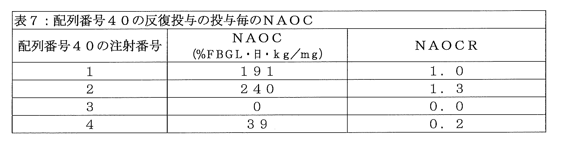
cNg-K変異を含む配列番号42のインスリン-Fc融合タンパク質が、イヌにおける反復投与時の生物活性の成績を向上させたかどうかを確かめるため、当該化合物をN=1のイヌに、0日目、7日目、14日目、および28日目に、実施例22の手順に従って皮下投与した。イヌの%FBGLが低下し過ぎた場合、そのイヌに餌を与えて、血糖を安全なレベルまで上昇させた。1回目注射のNAOCは449%FBGL・日・kg/mgであり、配列番号42のインスリン-Fc融合タンパク質がインビボにおいて十分な生物活性を示すことが示された。また、当該化合物の薬物動態プロファイルを、ELISAを使用する実施例23の方法で測定し、2コンパートメントモデルをそのデータに適合し、当該化合物の排出半減期を求めたところ、約0.9日であった。それ以降の各投与についても、実施例22の一般手順に従って、NAOCおよびNAOCRの測定を行い、投与を施行した時間から次の投与を施行する直前までを算出した。表8に示されるNAOCおよびNAOCRは、配列番号42のインスリン-Fc融合タンパク質が、4回の投与中、0.6よりも大きなNAOCRを維持することを示している。以上から、予想外にも、cNg-K変異を含む配列番号42のインスリン-Fc融合タンパク質が、イヌにおける反復投与時の生物活性を顕著に向上させた唯一の、配列番号36のインスリン-Fc融合タンパク質の非グリコシル化変異体であった。

また、抗薬物抗体レベルおよび抗インスリン抗体レベルを、治療の全期間(28日間)と、追加で2週間、実施例24に従って測定した。図9は、配列番号42のインスリン-Fc融合タンパク質が、イヌにおける反復皮下投与に伴って、抗薬物抗体を発生させるものの、その抗薬物抗体価は、配列番号36のインスリン-Fc融合タンパク質によって発生するもの(実施例32)よりもずっと低いことを示している。 Anti-drug and anti-insulin antibody levels were also measured according to Example 24 during the entire treatment period (28 days) and for an additional 2 weeks. FIG. 9 shows that although the insulin-Fc fusion protein of SEQ ID NO: 42 generates anti-drug antibodies upon repeated subcutaneous administration in dogs, the anti-drug antibody titer is generated by the insulin-Fc fusion protein of SEQ ID NO: 36. This shows that it is much lower than that of Example 32 (Example 32).
実施例37:抗薬物抗体を含有するイヌ血清のスクリーニング、並びにインスリンポリペプチドのB10D位およびA8H位における免疫原性である可能性があるエピトープの特定
イヌIgGB FcフラグメントのcNg部位のLysへの変異(すなわち、cNg-K)は、配列番号4のインスリンポリペプチドと配列番号11のペプチドリンカーとを含むインスリン-融合タンパク質の反復投与時の生物活性を向上させたが(実施例36)、得られた配列番号42のインスリン-Fc融合タンパク質は抗薬物抗体を発生させてしまった(実施例36)。このことから、配列番号4のインスリンポリペプチドが、予想外にも、イヌの免疫系の対象にされる特定のエピトープ(すなわち、免疫原性の「ホットスポット」)を含む可能性があるという仮定が得られた。そのため、実施例24に記載の血清試料中に存在する抗体の結合特異性を、実施例27の一般手順に従って評価した。コートされたインスリン-Fc融合タンパク質ライブラリーに対する、配列番号36のインスリン-Fc融合タンパク質(実施例32)の反復投与から得られた抗体含有血清試料の解析によって、予想外にも、配列番号5のインスリンポリペプチド配列内に、B鎖のN末端から10番目の位置(すなわち、B10)のアスパラギン酸変異、および、別に、A鎖のN末端から8番目の位置(すなわち、A8)のヒスチジン変異という、2つの主要な「ホットスポット」が存在することが示された。これらの結果は、これら2つの特定のアミノ酸変異を含むインスリンポリペプチドアミノ酸組成を含むインスリン-Fc融合タンパク質が、イヌにおいて免疫原性である可能性があり、そのため、反復注射後に生物活性を中和する抗薬物抗体を発生する可能性があることを示唆している。以上から、B10アスパラギン酸とA8ヒスチジンを含んでいないインスリンポリペプチドが、長期間に亘ってイヌに反復投与(例えば、イヌ糖尿病を治療するため)する必要があるインスリン-Fc融合タンパク質には好適であることが確認された。
Example 37: Screening of dog sera containing anti-drug antibodies and identification of potentially immunogenic epitopes at positions B10D and A8H of the insulin polypeptide Mutation of the cNg site of a canine IgG Fc fragment to Lys (i.e., cNg-K) improved the biological activity upon repeated administration of an insulin-fusion protein comprising an insulin polypeptide of SEQ ID NO: 4 and a peptide linker of SEQ ID NO: 11 (Example 36); The insulin-Fc fusion protein of SEQ ID NO: 42 generated anti-drug antibodies (Example 36). This leads to the hypothesis that the insulin polypeptide of SEQ ID NO: 4 may unexpectedly contain specific epitopes (i.e., immunogenic "hot spots") that are targeted by the canine immune system. was gotten. Therefore, the binding specificity of the antibodies present in the serum samples described in Example 24 was evaluated according to the general procedure of Example 27. Analysis of antibody-containing serum samples obtained from repeated administration of the insulin-Fc fusion protein of SEQ ID NO: 36 (Example 32) to a coated insulin-Fc fusion protein library unexpectedly revealed that the insulin-Fc fusion protein of SEQ ID NO: 5 Within the insulin polypeptide sequence, there is an aspartic acid mutation at the 10th position from the N-terminus of the B chain (i.e., B10) and, separately, a histidine mutation at the 8th position from the N-terminus of the A chain (i.e., A8). , it was shown that there are two major "hot spots". These results indicate that insulin-Fc fusion proteins containing insulin polypeptide amino acid compositions containing these two specific amino acid mutations may be immunogenic in dogs and therefore neutralize biological activity after repeated injections. This suggests that there is a possibility of developing anti-drug antibodies. From the above, insulin polypeptides that do not contain B10 aspartate and A8 histidine are suitable for insulin-Fc fusion proteins that need to be repeatedly administered to dogs over a long period of time (e.g., to treat canine diabetes). It was confirmed that there is.
実施例38:配列番号4のインスリンポリペプチドと非グリコシル化イヌIgGBアイソタイプFcフラグメントとを含むインスリン-Fc融合タンパク質において、インスリンポリペプチドのB10D変異およびA8H変異を天然組成に戻すと、潜在的な免疫原性リスクが低減される
「ホットスポット」変異の置換が、配列番号4のインスリンポリペプチドとイヌIgGBアイソタイプフラグメントとを含むインスリン-Fc融合タンパク質の免疫原性および反復投与時の生物活性を向上させるかどうかを評価するために、例示的なインスリン-Fc融合タンパク質(配列番号43)において、インスリンポリペプチドのB10アミノ酸とA8アミノ酸を、以下に挙げる当該インスリンポリペプチドの天然組成であるヒスチジンおよびスレオニンにそれぞれ戻したもの(配列番号63)(非天然アミノ酸には下線)を合成した。
FVNQHLCGSHLVEALALVCGERGFFYTDPTGGGPRRGIVEQCCTSICSLYQLENYCN(配列番号63)。
Example 38: In an insulin-Fc fusion protein comprising the insulin polypeptide of SEQ ID NO: 4 and a non-glycosylated canine IgG isotype Fc fragment, returning the B10D and A8H mutations of the insulin polypeptide to its native composition confers potential immunity. Substitution of "hot spot" mutations that reduce the risk of pathogenicity improves the immunogenicity and biological activity upon repeated administration of an insulin-Fc fusion protein comprising the insulin polypeptide of SEQ ID NO: 4 and a canine IgG isotype fragment. In an exemplary insulin-Fc fusion protein (SEQ ID NO: 43), the B10 and A8 amino acids of the insulin polypeptide are replaced by histidine and threonine, which are the natural composition of the insulin polypeptide listed below. Each returned product (SEQ ID NO: 63) (unnatural amino acids are underlined) was synthesized.
FVNQHLCGSHLVEAL A LVCGERGFFYT DP T GGGPRR GIVEQCCTSICSLYQLENYCN (SEQ ID NO: 63).
さらに、非グリコシル化cNg変異体にさらなる利点がある可能性を考慮して、配列番号43のインスリン-Fc融合タンパク質はcNg-Q変異を含んでいる。配列番号43のインスリン-Fc融合タンパク質の全アミノ酸配列を以下に示す:
FVNQHLCGSHLVEALALVCGERGFFYTDPTGGGPRRGIVEQCCTSICSLYQLENYCNGGGGAGGGGDCPKCPAPEMLGGPSVFIFPPKPKDTLLIARTPEVTCVVVDLDPEDPEVQISWFVDGKQMQTAKTQPREEQFQGTYRVVSVLPIGHQDWLKGKQFTCKVNNKALPSPIERTISKARGQAHQPSVYVLPPSREELSKNTVSLTCLIKDFFPPDIDVEWQSNGQQEPESKYRTTPPQLDEDGSYFLYSKLSVDKSRWQRGDTFICAVMHEALHNHYTQESLSHSPG(配列番号43)。
Furthermore, considering the possibility that non-glycosylated cNg variants have additional advantages, the insulin-Fc fusion protein of SEQ ID NO: 43 contains a cNg-Q mutation. The complete amino acid sequence of the insulin-Fc fusion protein of SEQ ID NO: 43 is shown below:
FVNQHLCGSHLVEALAVCGERGFFYTDPTGGGPRRGIVEQCCTSICSLYQLENYCNGGGGAGGGGDCPKCPAPEMLGGPSVFIFPPKPKDTLLIARTPEVTCVVVDLDPEDPEVQISWFVDG KQMQTAKTQPREEQFQGTYRVVSVLPIGHQDWLKGKQFTCKVNNKALPSPIERTISKARGQAHQPSVYVLPPSREELSKNTVSLTCLIKDFFPDIDVEWQSNGQQEPESKYRTTPPQLDEDGSY FLYSKLSVDKSRWQRGDTFICAVMHEALHNHYTQESLSHSPG (SEQ ID NO: 43).
配列番号43のインスリン-Fc融合タンパク質を、実施例1に従ってHEK293細胞で製造し、実施例4に従ってプロテインAカラムを用いて精製した。得られたタンパク質収量はわずか21mg/Lであった。構造の確認を実施例6に従って非還元CE-SDSおよび還元CE-SDSで行い、さらに、配列の確認を実施例8に記載のグリカン除去を伴うLC-MSによって行った。実施例10に従って分子ふるいクロマトグラフィーによって測定されたホモ二量体率は98.0%であり、このタンパク質には比較的凝集体がないことが示された。 Insulin-Fc fusion protein of SEQ ID NO: 43 was produced in HEK293 cells according to Example 1 and purified using a Protein A column according to Example 4. The protein yield obtained was only 21 mg/L. Structural confirmation was performed with non-reduced CE-SDS and reduced CE-SDS according to Example 6, and sequence confirmation was performed by LC-MS with glycan removal as described in Example 8. The percent homodimerization determined by molecular mesh chromatography according to Example 10 was 98.0%, indicating that the protein was relatively free of aggregates.
ホモ二量体力価は21mg/Lと比較的低かったが、配列番号43のインスリン-Fc融合タンパク質を、イヌにおいて、インビボにおける生物活性および免疫原性について、実施例22、実施例23、および実施例24の手順にそれぞれ従って、評価した。図10から明らかなように、配列番号43のインスリン-Fc融合タンパク質において、B10D変異およびA8H変異を天然アミノ酸(すなわち、B10HおよびA8T)に戻すことで、親化合物(配列番号36)の免疫原性が顕著に低減した。 Although the homodimer titer was relatively low at 21 mg/L, the insulin-Fc fusion protein of SEQ ID NO: 43 was tested for in vivo biological activity and immunogenicity in dogs as described in Example 22, Example 23, and Each was evaluated according to the procedure of Example 24. As is clear from FIG. 10, in the insulin-Fc fusion protein of SEQ ID NO: 43, returning the B10D mutation and A8H mutation to natural amino acids (i.e., B10H and A8T) improves the immunogenicity of the parent compound (SEQ ID NO: 36). was significantly reduced.
しかしながら、図11に示されているように、天然のB10アミノ酸およびA8アミノ酸を含む配列番号43のインスリン-Fc融合タンパク質は、生物活性を示さなかった(すなわち、NAOCが実質的にゼロであった)。 However, as shown in Figure 11, the insulin-Fc fusion protein of SEQ ID NO: 43 containing the natural B10 and A8 amino acids showed no biological activity (i.e., had virtually no NAOC). ).
実施例39:イヌIgGB Fcフラグメントを含むインスリン-Fc融合タンパク質会合体の生物活性を向上させるために、配列番号63のインスリンポリペプチドにさらなるB鎖変異およびA鎖変異を組み込む試み
配列番号36のインスリン-Fc融合タンパク質(実施例33)と比べて、配列番号43のインスリン-Fc融合タンパク質が抗薬物抗体を発生させなかったこと(実施例38)は、配列番号4のインスリンポリペプチドにおけるB10D変異およびA8H変異が、抗薬物抗体産生の原因となる、免疫原性のエピトープである可能性があるという説の強力な証拠である。しかし、配列番号36のものと比較して、配列番号43のインスリン-Fc融合タンパク質はインビボにおける効力が欠如しており、このことは、これら2つのアミノ酸変異が、許容できる生物活性レベルの達成にも関与していることを示している。配列番号43のインスリン-Fc融合タンパク質のインビボにおける効力の欠如は、実施例12の方法に従ってインスリン受容体結合アッセイで測定された、その高いIC50(以下の表9に示す)と関連がある。よって、インスリンポリペプチドにおける天然のB10アミノ酸およびA8アミノ酸を保持することで免疫原性の程度を低く維持しながら、インスリン-Fc融合タンパク質の生物活性を増加させる(すなわち、インスリン受容体結合アッセイにおけるIC50値を5000nM未満、より好ましくは4000nM未満、さらにより好ましくは3000nM未満に減少させる)ための、さらなる試みが必要であった。
Example 39: Attempt to incorporate additional B chain and A chain mutations into the insulin polypeptide of SEQ ID NO: 63 to improve the biological activity of an insulin-Fc fusion protein complex comprising a canine IgG Fc fragment Insulin of SEQ ID NO: 36 -Fc fusion protein (Example 33), the insulin-Fc fusion protein of SEQ ID NO: 43 did not generate anti-drug antibodies (Example 38) due to the B10D mutation in the insulin polypeptide of SEQ ID NO: 4 and This is strong evidence that the A8H mutation may be an immunogenic epitope responsible for anti-drug antibody production. However, compared to that of SEQ ID NO: 36, the insulin-Fc fusion protein of SEQ ID NO: 43 lacks potency in vivo, indicating that these two amino acid mutations are insufficient to achieve acceptable levels of biological activity. It also shows that they are involved. The lack of in vivo efficacy of the insulin-Fc fusion protein of SEQ ID NO: 43 is associated with its high IC50 (shown in Table 9 below), determined in an insulin receptor binding assay according to the method of Example 12. Thus, retaining the natural B10 and A8 amino acids in the insulin polypeptide increases the biological activity of the insulin-Fc fusion protein (i.e., the IC50 in insulin receptor binding assays) while maintaining a low degree of immunogenicity. Further attempts were required to reduce the value below 5000 nM, more preferably below 4000 nM, even more preferably below 3000 nM.
IRに対する強力な結合には、インスリンのB鎖およびA鎖の各種部分が必要であることが知られている(Hubbard S.R.、“Structural biology: Insulin meets its receptor”、Nature.、2013年;493巻(7431号):pp171-172)。よって、B10およびA8を天然インスリンにおけるものと同じに、且つ、C鎖およびペプチドリンカーを一定に維持しながら、B鎖またはA鎖の一部を改変した。これらのインスリン-Fc融合タンパク質のいくつかを、実施例1に従ってHEK293細胞で製造し、実施例4に従ってプロテインAカラムを用いて精製した。構造の確認を実施例6に従って非還元CE-SDSおよび還元CE-SDSで行い、さらに、配列の確認を実施例8に記載のグリカン除去を伴うLC-MSによって行った。ホモ二量体含有率を実施例10に従って分子ふるいクロマトグラフィーで測定し、インスリン受容体結合親和性を実施例12に従って測定した。これらの配列を以下に示し、配列番号43に対して得られた配列アラインメントを図12に示す(クラスタルオメガ)。
FVNQHLCGSHLVQALYLVCGERGFFYTDPTGGGPRRGIVEQCCTSICSLYQLENYCGGGGAGGGGDCPKCPAPEMLGGPSVFIFPPKPKDTLLIARTPEVTCVVVDLDPEDPEVQISWFVDGKQMQTAKTQPREEQFSGTYRVVSVLPIGHQDWLKGKQFTCKVNNKALPSPIERTISKARGQAHQPSVYVLPPSREELSKNTVSLTCLIKDFFPPDIDVEWQSNGQQEPESKYRTTPPQLDEDGSYFLYSKLSVDKSRWQRGDTFICAVMHEALHNHYTQESLSHSPG(配列番号44)
FVNQHLCGSELVEALALVCGERGFFYTDPTGGGPRRGIVEQCCTSICSLYQLENYCGGGGAGGGGDCPKCPAPEMLGGPSVFIFPPKPKDTLLIARTPEVTCVVVDLDPEDPEVQISWFVDGKQMQTAKTQPREEQFSGTYRVVSVLPIGHQDWLKGKQFTCKVNNKALPSPIERTISKARGQAHQPSVYVLPPSREELSKNTVSLTCLIKDFFPPDIDVEWQSNGQQEPESKYRTTPPQLDEDGSYFLYSKLSVDKSRWQRGDTFICAVMHEALHNHYTQESLSHSPG(配列番号45)
FVNQHLCGSHLVEALALVCGEAGFFYTDPTGGGPRRGIVEQCCTSICSLYQLENYCGGGGAGGGGDCPKCPAPEMLGGPSVFIFPPKPKDTLLIARTPEVTCVVVDLDPEDPEVQISWFVDGKQMQTAKTQPREEQFSGTYRVVSVLPIGHQDWLKGKQFTCKVNNKALPSPIERTISKARGQAHQPSVYVLPPSREELSKNTVSLTCLIKDFFPPDIDVEWQSNGQQEPESKYRTTPPQLDEDGSYFLYSKLSVDKSRWQRGDTFICAVMHEALHNHYTQESLSHSPG(配列番号46)
FVNQHLCGSHLVEALALVCGERGFYYTDPTGGGPRRGIVEQCCTSICSLYQLENYCGGGGAGGGGDCPKCPAPEMLGGPSVFIFPPKPKDTLLIARTPEVTCVVVDLDPEDPEVQISWFVDGKQMQTAKTQPREEQFSGTYRVVSVLPIGHQDWLKGKQFTCKVNNKALPSPIERTISKARGQAHQPSVYVLPPSREELSKNTVSLTCLIKDFFPPDIDVEWQSNGQQEPESKYRTTPPQLDEDGSYFLYSKLSVDKSRWQRGDTFICAVMHEALHNHYTQESLSHSPG(配列番号47)
FVNQHLCGSHLVEALALVCGERGFFYTDPTGGGPRRGIVEQCCTSICSLYQLENYCGGGGAGGGGDCPKCPAPEMLGGPSVFIFPPKPKDTLLIARTPEVTCVVVDLDPEDPEVQISWFVDGKQMQTAKTQPREEQFSGTYRVVSVLPIGHQDWLKGKQFTCKVNNKALPSPIERTISKARGQAHQPSVYVLPPSREELSKNTVSLTCLIKDFFPPDIDVEWQSNGQQEPESKYRTTPPQLDEDGSYFLYSKLSVDKSRWQRGDTFICAVMHEALHNHYTQESLSHSPG(配列番号48)
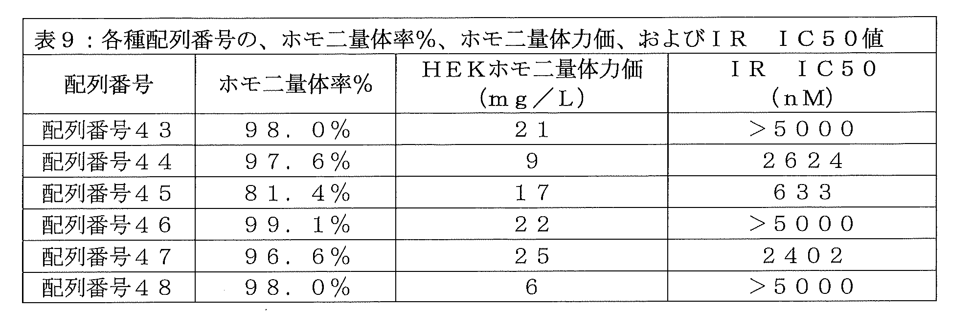
FVNQHLCGSHLVQALYLVCGERGFFYTDPTGGGPRRGIVEQCCTSICSLYQLENYCGGGGAGGGGDCPKCPAPEMLGGPSVFIFPPKPKDTLLIARTPEVTCVVVDLDPEDPEVQISWFVDGK QMQTAKTQPREEQFSGTYRVVSVLPIGHQDWLKGKQFTCKVNNKALPSPIERTISKARGQAHQPSVYVLPPSREELSKNTVSLTCLIKDFFPDIDVEWQSNGQQEPESKYRTTPPQLDEDGSYF LYSKLSVDKSRWQRGDTFICAVMHEALHNHYTQESLSHSPG (SEQ ID NO: 44)
FVNQHLCGSELVEALVCGERGFFYTDPTGGGPRRGIVEQCCTSICSLYQLENYCGGGGAGGGGDCPKCPAPEMLGGPSVFIFPPKPKDTLLIARTPEVTCVVVDLDPEDPEVQISWFVDGK QMQTAKTQPREEQFSGTYRVVSVLPIGHQDWLKGKQFTCKVNNKALPSPIERTISKARGQAHQPSVYVLPPSREELSKNTVSLTCLIKDFFPDIDVEWQSNGQQEPESKYRTTPPQLDEDGSYF LYSKLSVDKSRWQRGDTFICAVMHEALHNHYTQESLSHSPG (SEQ ID NO: 45)
FVNQHLCGSHLVEALAVCGEAGFFYTDPTGGGPRRGIVEQCCTSICSLYQLENYCGGGGAGGGGDCPKCPAPEMLGGPSVFIFPPKPKDTLLIARTPEVTCVVVDLDPEDPEVQISWFVDGK QMQTAKTQPREEQFSGTYRVVSVLPIGHQDWLKGKQFTCKVNNKALPSPIERTISKARGQAHQPSVYVLPPSREELSKNTVSLTCLIKDFFPDIDVEWQSNGQQEPESKYRTTPPQLDEDGSYF LYSKLSVDKSRWQRGDTFICAVMHEALHNHYTQESLSHSPG (SEQ ID NO: 46)
FVNQHLCGSHLVEALVCGERGFYYTDPTGGGPRRGIVEQCCTSICSLYQLENYCGGGGAGGGGDCPKCPAPEMLGGPSVFIFPPKPKDTLLIARTPEVTCVVVDLDPEDPEVQISWFVDGK QMQTAKTQPREEQFSGTYRVVSVLPIGHQDWLKGKQFTCKVNNKALPSPIERTISKARGQAHQPSVYVLPPSREELSKNTVSLTCLIKDFFPDIDVEWQSNGQQEPESKYRTTPPQLDEDGSYF LYSKLSVDKSRWQRGDTFICAVMHEALHNHYTQESLSHSPG (SEQ ID NO: 47)
FVNQHLCGSHLVEALAVCGERGFFYTDPTGGGPRRGIVEQCCTSICSLYQLENYCGGGGAGGGGDCPKCPAPEMLGGPSVFIFPPKPKDTLLIARTPEVTCVVVDLDPEDPEVQISWFVDGK QMQTAKTQPREEQFSGTYRVVSVLPIGHQDWLKGKQFTCKVNNKALPSPIERTISKARGQAHQPSVYVLPPSREELSKNTVSLTCLIKDFFPDIDVEWQSNGQQEPESKYRTTPPQLDEDGSYF LYSKLSVDKSRWQRGDTFICAVMHEALHNHYTQESLSHSPG (SEQ ID NO: 48)
配列番号43と比べて、提唱された変異がIR結合を向上させた(すなわち、IC50値を低下させた)のは、3つの場合(配列番号44、配列番号45、および配列番号47)でだけであった。しかし、いずれの変異でも、50mg/Lより大きいホモ二量体力価という製造設計目標を達成する化合物は得られず、また、場合によっては、変異により製造性が顕著に低減する(例えば、20mg/L未満のホモ二量体力価)こともあった。 Compared to SEQ ID NO: 43, only in three cases (SEQ ID NO: 44, SEQ ID NO: 45, and SEQ ID NO: 47) did the proposed mutations improve IR binding (i.e. lower IC50 values). Met. However, none of the mutations results in a compound that achieves the manufacturing design goal of a homodimer titer greater than 50 mg/L, and in some cases the mutations significantly reduce manufacturability (e.g., 20 mg/L). Homodimer titers below L) were also observed.
実施例40:イヌIgGB Fcフラグメントを含むインスリン-Fc融合タンパク質会合体の生物活性を向上させるために、配列番号63のインスリンポリペプチドにC鎖変異を組み込む試み
実施例39で得られた結果は、配列番号63のインスリンポリペプチドのA鎖およびB鎖を変異させる試みが全て、関連インスリン-Fc融合物のHEKホモ二量体力価が許容できないほどに低い(すなわち、25mg/L以下のホモ二量体力価)結果となったことを示している。そのため、さらなる実験が必要となった。本実施例では、配列番号63のインスリンポリペプチドのC鎖組成を、より長くすることで、あるいは可動性を増加させることで、変異させた。天然インスリン(例えばヒトインスリン)は、インスリン受容体に結合する際に、B鎖およびA鎖両方の折り畳みの移動を含む、顕著な立体構造変化を起こすことが示されている(例えば、Mentingら、Nature、2013年;493巻(7431号):pp241~245によって説明されている)。本発明のインスリンポリペプチドと異なり、天然インスリンは、天然型では2鎖ポリペプチドであり、2本のジスルフィド結合だけで接続され、A鎖とB鎖の可動性を制限するC鎖は存在しないため、インスリン受容体においてこの立体構造変化を自由に起こすことができる。特定の理論に束縛されるものではないが、配列番号63のインスリンポリペプチド内に含まれるC鎖が、柔軟でなさ過ぎる(例えば、B鎖とA鎖の間での動きを容易なものにしないアミノ酸組成およびアミノ酸配列)、且つ/または、短過ぎる(例えば、B鎖のC末端とA鎖のN末端との間のアミノ酸が十分ではない)ために、インスリンポリペプチドが、インスリン受容体への強力な結合に必要な分子形状変化を起こすことが妨げられると仮定した。よって、以下に示すように、配列番号43のインスリン-Fc融合タンパク質をベースとして、インスリンポリペプチドC鎖を変化させて、いくつかのインスリン-Fc融合タンパク質を合成した。配列番号43に対して得られた配列アラインメントを図13に示す(クラスタルオメガ)。
FVNQHLCGSHLVQALYLVCGERGFFYTDPTQRGGGGGQRGIVEQCCTSICSLYQLENYCGGGGAGGGGDCPKCPAPEMLGGPSVFIFPPKPKDTLLIARTPEVTCVVVDLDPEDPEVQISWFVDGKQMQTAKTQPREEQFSGTYRVVSVLPIGHQDWLKGKQFTCKVNNKALPSPIERTISKARGQAHQPSVYVLPPSREELSKNTVSLTCLIKDFFPPDIDVEWQSNGQQEPESKYRTTPPQLDEDGSYFLYSKLSVDKSRWQRGDTFICAVMHEALHNHYTQESLSHSPG(配列番号49)
FVNQHLCGSHLVEALALVCGERGFFYTDPTGGGGGGSGGGGGIVEQCCTSICSLYQLENYCGGGGAGGGGDCPKCPAPEMLGGPSVFIFPPKPKDTLLIARTPEVTCVVVDLDPEDPEVQISWFVDGKQMQTAKTQPREEQFSGTYRVVSVLPIGHQDWLKGKQFTCKVNNKALPSPIERTISKARGQAHQPSVYVLPPSREELSKNTVSLTCLIKDFFPPDIDVEWQSNGQQEPESKYRTTPPQLDEDGSYFLYSKLSVDKSRWQRGDTFICAVMHEALHNHYTQESLSHSPG(配列番号50)
FVNQHLCGSHLVEALALVCGERGFFYTDPGGGGGGGGGIVEQCCTSICSLYQLENYCGGGGAGGGGDCPKCPAPEMLGGPSVFIFPPKPKDTLLIARTPEVTCVVVDLDPEDPEVQISWFVDGKQMQTAKTQPREEQFSGTYRVVSVLPIGHQDWLKGKQFTCKVNNKALPSPIERTISKARGQAHQPSVYVLPPSREELSKNTVSLTCLIKDFFPPDIDVEWQSNGQQEPESKYRTTPPQLDEDGSYFLYSKLSVDKSRWQRGDTFICAVMHEALHNHYTQESLSHSPG(配列番号51)
FVNQHLCGSHLVEALALVCGERGFFYTPGGGGGGGGGIVEQCCTSICSLYQLENYCGGGGAGGGGDCPKCPAPEMLGGPSVFIFPPKPKDTLLIARTPEVTCVVVDLDPEDPEVQISWFVDGKQMQTAKTQPREEQFSGTYRVVSVLPIGHQDWLKGKQFTCKVNNKALPSPIERTISKARGQAHQPSVYVLPPSREELSKNTVSLTCLIKDFFPPDIDVEWQSNGQQEPESKYRTTPPQLDEDGSYFLYSKLSVDKSRWQRGDTFICAVMHEALHNHYTQESLSHSPG(配列番号52)
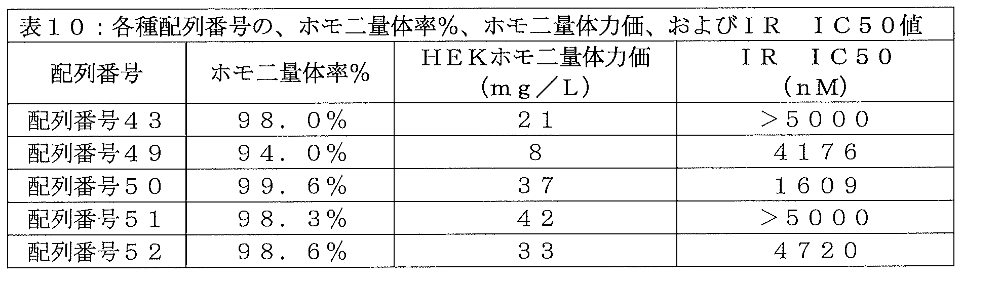
FVNQHLCGSHLVQALYLVCGERGFFYTDPTQRGGGGGGQRGIVEQCCTSICSLYQLENYCGGGGAGGGGDCPKCPAPEMLGGPSVFIFPPKPKDTLLIARTPEVTCVVVDLDPEDPEVQISWFV DGKQMQTAKTQPREEQFSGTYRVVSVLPIGHQDWLKGKQFTCKVNNKALPSPIERTISKARGQAHQPSVYVLPPSREELSKNTVSLTCLIKDFFPPDIDVEWQSNGQQEPESKYRTTPPQLDEDG SYFLYSKLSVDKSRWQRGDTFICAVMHEALHNHYTQESLSHSPG (SEQ ID NO: 49)
FVNQHLCGSHLVEALAVCGERGFFYTDPTGGGGGGGSGGGGIVEQCCTSICSLYQLENYCGGGGAGGGGDCPKCPAPEMLGGPSVFIFPPKPKDTLLIARTPEVTCVVVDLDPEDPEVQISW FVDGKQMQTAKTQPREEQFSGTYRVVSVLPIGHQDWLKGKQFTCKVNNKALPSPIERTISKARGQAHQPSVYVLPPSREELSKNTVSLTCLIKDFFPPDIDVEWQSNGQQEPESKYRTTPPQLDE DGSYFLYSKLSVDKSRWQRGDTFICAVMHEALHNHYTQESLSHSPG (SEQ ID NO: 50)
FVNQHLCGSHLVEALAVCGERGFFYTDPGGGGGGGGGIVEQCCTSICSLYQLENYCGGGGAGGGGDCPKCPAPEMLGGPSVFIFPPKPKDTLLIARTPEVTCVVVDLDPEDPEVQISWFVDG KQMQTAKTQPREEQFSGTYRVVSVLPIGHQDWLKGKQFTCKVNNKALPSPIERTISKARGQAHQPSVYVLPPSREELSKNTVSLTCLIKDFFPDIDVEWQSNGQQEPESKYRTTPPQLDEDGSY FLYSKLSVDKSRWQRGDTFICAVMHEALHNHYTQESLSHSPG (SEQ ID NO: 51)
FVNQHLCGSHLVEALAVCGERGFFYTPGGGGGGGGGIVEQCCTSICSLYQLENYCGGGGAGGGGDCPKCPAPEMLGGPSVFIFPPKPKDTLLIARTPEVTCVVVDLDPEDPEVQISWFVDGK QMQTAKTQPREEQFSGTYRVVSVLPIGHQDWLKGKQFTCKVNNKALPSPIERTISKARGQAHQPSVYVLPPSREELSKNTVSLTCLIKDFFPDIDVEWQSNGQQEPESKYRTTPPQLDEDGSYF LYSKLSVDKSRWQRGDTFICAVMHEALHNHYTQESLSHSPG (SEQ ID NO: 52)
インスリン-Fc融合タンパク質を、実施例1に従ってHEK293細胞で製造し、実施例4に従ってプロテインAカラムを用いて精製した。構造の確認を実施例6に従って非還元CE-SDSおよび還元CE-SDSで行い、さらに、配列の確認を実施例8に記載のグリカン除去を伴うLC-MSによって行った。ホモ二量体含有率を実施例10に従って分子ふるいクロマトグラフィーで測定し、インスリン受容体結合親和性を実施例12に従って測定した。結果を表10に示す。配列番号43のインスリン-Fc融合タンパク質のものとの比較において、C鎖変異がインスリン受容体結合親和性を顕著に向上させた(3000nM未満のIC50)のは、最も長いC鎖(GGGGGGSGGGG:配列番号71)を含む1例(配列番号50)でだけであった。しかし、これらのC鎖変異型インスリン-Fc融合タンパク質のいずれも、50mg/Lという製造設計目標を超えるホモ二量体力価を示さなかった。実際に、1つの例(配列番号49)では、C鎖変異は予想外にも、顕著に低いホモ二量体力価をもたらした。 Insulin-Fc fusion protein was produced in HEK293 cells according to Example 1 and purified using a Protein A column according to Example 4. Structural confirmation was performed with non-reduced CE-SDS and reduced CE-SDS according to Example 6, and sequence confirmation was performed by LC-MS with glycan removal as described in Example 8. Homodimer content was determined by molecular exclusion chromatography according to Example 10, and insulin receptor binding affinity was determined according to Example 12. The results are shown in Table 10. In comparison with that of the insulin-Fc fusion protein of SEQ ID NO. 71) in only one case (SEQ ID NO: 50). However, none of these C chain variant insulin-Fc fusion proteins exhibited homodimer titers above the manufacturing design goal of 50 mg/L. Indeed, in one example (SEQ ID NO: 49), the C chain mutation unexpectedly resulted in significantly lower homodimer titers.
実施例41:生物活性を向上させるために、配列番号63のインスリンポリペプチドとイヌIgGB Fcフラグメントとを含むインスリン-Fc融合タンパク質にペプチドリンカー変異を組み入れる試み
特定の理論に束縛されるものではないが、配列番号43のインスリン-Fc融合タンパク質のインスリン受容体結合が不十分であることの、別の考えられる理由として、はるかに大きいFcフラグメント分子がペプチドリンカーを介してインスリンポリペプチドに極めて近接して結合していることから生じる、インスリンポリペプチドとインスリン受容体との間の立体障害が関係していると考えられていた。ペプチドリンカーがより短かったり、ペプチドリンカーの折り畳みがより密であると、この問題が悪化する可能性があると考えられ、一方、ペプチドリンカーがより長かったり、ペプチドリンカーが自ら折り返しを起こしにくい(例えば、リンカーの分子剛性がより高い)場合、インスリンポリペプチドとFcフラグメントとの間により多くの空間がつくられることで、この問題が緩和されることがある。インスリンポリペプチドとFcフラグメントとの間の空間の増加は、インスリン受容体とFcフラグメントとの間の距離も増加させ、インスリン受容体結合中の干渉が少なくなる。配列番号43のインスリン-Fc融合タンパク質の構築に用いられた配列番号11(すなわち、GGGGAGGGG)のペプチドリンカーは、当該リンカーを構成するアミノ酸が側鎖を含まないものである(すなわち、グリシンとアラニンというアミノ酸しか含んでいない)ため、短過ぎ、且つ/または可動性があり過ぎる可能性がある、という仮説が立てられた。よって、この仮説を検証するため、配列番号43のインスリン-Fc融合タンパク質とは別の2つのインスリン-Fc融合タンパク質バリアントを合成した。配列番号48のインスリン-Fc融合タンパク質は、配列番号43のインスリン-Fc融合タンパク質の構築に用いられたものと同じペプチドリンカーを含むが、A鎖のN末端から21番目の位置(すなわち、A21)のアスパラギンが存在しない(すなわち、des-A21)インスリンポリペプチドを含む。この特定の変異を組み込むことにより、A鎖とペプチドリンカーとの間の接合部が、タンパク質収量および/または上記分子の生物活性に影響しているかどうかを確かめた。もう一方の配列番号53のインスリン-Fc融合タンパク質は、このdes-A21N A鎖変異と、配列番号43のインスリン-Fc融合タンパク質の構築に用いられたものの長さの2倍より長いペプチドリンカーと、を含む。このより長いペプチドリンカーにおいては、アラニンは好ましくなく、代わりに、極性アミド側鎖を含むグルタミンと置き換わっている。このグルタミン置換は、このペプチドリンカーの親水性を増強し、場合によってはリンカーが折れ返ることを妨げることが期待された。これらの配列を以下に示し、配列番号43に対する配列アラインメントを図14に示す(クラスタルオメガ):
FVNQHLCGSHLVEALALVCGERGFFYTDPTGGGPRRGIVEQCCTSICSLYQLENYCGGGGGQGGGGQGGGGQGGGGGDCPKCPAPEMLGGPSVFIFPPKPKDTLLIARTPEVTCVVVDLDPEDPEVQISWFVDGKQMQTAKTQPREEQFSGTYRVVSVLPIGHQDWLKGKQFTCKVNNKALPSPIERTISKARGQAHQPSVYVLPPSREELSKNTVSLTCLIKDFFPPDIDVEWQSNGQQEPESKYRTTPPQLDEDGSYFLYSKLSVDKSRWQRGDTFICAVMHEALHNHYTQESLSHSPG(配列番号53)
FVNQHLCGSHLVEALALVCGERGFFYTDPTGGGPRRGIVEQCCTSICSLYQLENYCGGGGAGGGGDCPKCPAPEMLGGPSVFIFPPKPKDTLLIARTPEVTCVVVDLDPEDPEVQISWFVDGKQMQTAKTQPREEQFSGTYRVVSVLPIGHQDWLKGKQFTCKVNNKALPSPIERTISKARGQAHQPSVYVLPPSREELSKNTVSLTCLIKDFFPPDIDVEWQSNGQQEPESKYRTTPPQLDEDGSYFLYSKLSVDKSRWQRGDTFICAVMHEALHNHYTQESLSHSPG(配列番号48)

FVNQHLCGSHLVEALALVCGERGFFYTDPTGGGPRRGIVEQCCTSICSLYQLENYCGGGGGQGGGGQGGGGQGGGGDCPKCPAPEMLGGPSVFIFPPKPKDTLLIARTPEVTCVVVDLDPED PEVQISWFVDGKQMQTAKTQPREEQFSGTYRVVSVLPIGHQDWLKGKQFTCKVNNKALPSPIERTISKARGQAHQPSVYVLPPSREELSKNTVSLTCLIKDFFPDIDVEWQSNGQQEPESKYRT TPPQLDEDGSYFLYSKLSVDKSRWQRGDTFICAVMHEALHNHYTQESLSHSPG (SEQ ID NO: 53)
FVNQHLCGSHLVEALAVCGERGFFYTDPTGGGPRRGIVEQCCTSICSLYQLENYCGGGGAGGGGDCPKCPAPEMLGGPSVFIFPPKPKDTLLIARTPEVTCVVVDLDPEDPEVQISWFVDGK QMQTAKTQPREEQFSGTYRVVSVLPIGHQDWLKGKQFTCKVNNKALPSPIERTISKARGQAHQPSVYVLPPSREELSKNTVSLTCLIKDFFPDIDVEWQSNGQQEPESKYRTTPPQLDEDGSYF LYSKLSVDKSRWQRGDTFICAVMHEALHNHYTQESLSHSPG (SEQ ID NO: 48)
2種のインスリン-Fc融合タンパク質を、実施例1に従ってHEK293細胞で製造し、実施例4に従ってプロテインAカラムを用いて精製した。構造の確認を実施例6に従って非還元CE-SDSおよび還元CE-SDSで行い、さらに、配列の確認を実施例8に記載のグリカン除去を伴うLC-MSによって行った。ホモ二量体含有率を実施例10に従って分子ふるいクロマトグラフィーで測定し、インスリン受容体結合親和性を実施例12に従って測定した。結果を表11に示す。組成の異なる(配列番号53の場合ではGGGGGQGGGGQGGGGQGGGGG(配列番号13)、対して、配列番号43の場合ではGGGGAGGGG(配列番号11))より長いペプチドリンカーを組み込んだ場合、IC50値の顕著な低減によって評価される、インスリン受容体結合の向上が生じ、リンカーを長くすることが、他のインスリン-Fc融合タンパク質の、インスリン受容体結合を増加させるための戦略になり得ることが示された。しかし、より長いリンカーを組み込んでも、ホモ二量体力価は、50mg/L超という製造設計目標を超えては向上しなかった。 Two insulin-Fc fusion proteins were produced in HEK293 cells according to Example 1 and purified using a Protein A column according to Example 4. Structural confirmation was performed with non-reduced CE-SDS and reduced CE-SDS according to Example 6, and sequence confirmation was performed by LC-MS with glycan removal as described in Example 8. Homodimer content was determined by molecular exclusion chromatography according to Example 10, and insulin receptor binding affinity was determined according to Example 12. The results are shown in Table 11. Incorporation of longer peptide linkers of different compositions (GGGGGQGGGGQGGGGQGGGG in the case of SEQ ID NO: 53 (SEQ ID NO: 13), versus GGGGAGGGGG (SEQ ID NO: 11) in the case of SEQ ID NO: 43) was assessed by a significant reduction in IC50 values. improved insulin receptor binding occurred, indicating that lengthening the linker may be a strategy for increasing insulin receptor binding of other insulin-Fc fusion proteins. However, incorporating a longer linker did not improve homodimer titer beyond the manufacturing design goal of >50 mg/L.
実施例42:イヌIgGB Fcフラグメントを含むインスリン-Fc融合タンパク質会合体のホモ二量体力価を向上させるために、配列番号63のインスリンポリペプチドのB鎖の一部を欠失させる試み
実施例41の結果は、ペプチドリンカーを改変することで、天然のB10アミノ酸とA8アミノ酸を含む配列番号43のインスリン-Fc融合タンパク質のインスリン受容体に対する結合親和性を増加させることができることを示している。しかし、このペプチドリンカーの変異では、ホモ二量体力価を、製造設計目標を達成するのに十分なほどに増加させることはできなかった。ホモ二量体力価は、細胞内合成および細胞内プロセシングを含むいくつかの特性の関数であるため、インスリン-Fc分子は、合成中および合成後、2つのホモ二量体モノマー間で分子内で、または、2以上の別々のホモ二量体間で分子間で、自己会合(すなわち、凝集)していたかもしれないという仮説を立てた。この凝集によって、実施例1、実施例4、および実施例10に記載された製造工程中に細胞培養上清から得られたホモ二量体力価は許容できないほどに低いものであった。このインスリン-Fc融合タンパク質分子間の相互作用の可能性は、部分的には、自己会合し凝集体を形成するインスリンの周知の傾向によるものであり得る。インスリンの自己会合する傾向を低減するための当該技術分野において公知の1つの方法は、B鎖のC末端付近のアミノ酸を変異させることを含む。例えば、インスリンリスプロ(B28K変異;B29P変異)およびインスリンアスパルト(B28D変異)は、会合と凝集を防ぐことで、主に単量体型のインスリンを溶液中に生じさせる非天然型のB鎖変異を含む、周知の市販2鎖インスリンである。凝集を防ぐための別のアプローチは、アミノ酸の構造的な欠失を含む。例えば、デスペンタペプチドインスリンとして知られている2鎖インスリン(despentapeptide insulin)(DPPI;Brange J.、Dodson G.G.、Edwards J.、Holden P.H.、Whittingham J.L.、1997年b.、“A model of insulin fibrils derived from the x-ray crystal structure of a monomeric insulin (despentapeptide insulin)”、Proteins、27巻、pp507~516を参照)は、B鎖の5個のC末端アミノ酸(YTPKT)が除去されている以外は、天然型の2鎖ヒトインスリンと同一である。DPPIは、天然型2鎖ヒトインスリンと比べて、インスリン受容体に対する結合親和性が低いが、溶液中で完全に単量体の状態であり、これは、DPPI分子間では会合も凝集もほとんど起こらないことを意味している。よって、分子内および分子間の自己会合の可能性を低減し、インスリン-Fc融合タンパク質のホモ二量体力価を向上させる試みでは、DPPI、インスリンリスプロ、およびインスリンアスパルトについて上記したような、部分的なB鎖アミノ酸切断およびB鎖アミノ酸変異を用いて、配列番号43のインスリン-Fc融合タンパク質のいくつかのバリアントを構築した。これらの配列を以下に示し、配列番号43に対する配列アラインメントを図15に示す(クラスタルオメガ):
FVNQHLCGSHLVEALALVCGERGFFYTDPGGGGGGGGGIVEQCCTSICSLYQLENYCGGGGAGGGGDCPKCPAPEMLGGPSVFIFPPKPKDTLLIARTPEVTCVVVDLDPEDPEVQISWFVDGKQMQTAKTQPREEQFSGTYRVVSVLPIGHQDWLKGKQFTCKVNNKALPSPIERTISKARGQAHQPSVYVLPPSREELSKNTVSLTCLIKDFFPPDIDVEWQSNGQQEPESKYRTTPPQLDEDGSYFLYSKLSVDKSRWQRGDTFICAVMHEALHNHYTQESLSHSPG(配列番号51)
FVNQHLCGSHLVEALALVCGERGFFYTPGGGGGGGGGIVEQCCTSICSLYQLENYCGGGGAGGGGDCPKCPAPEMLGGPSVFIFPPKPKDTLLIARTPEVTCVVVDLDPEDPEVQISWFVDGKQMQTAKTQPREEQFSGTYRVVSVLPIGHQDWLKGKQFTCKVNNKALPSPIERTISKARGQAHQPSVYVLPPSREELSKNTVSLTCLIKDFFPPDIDVEWQSNGQQEPESKYRTTPPQLDEDGSYFLYSKLSVDKSRWQRGDTFICAVMHEALHNHYTQESLSHSPG(配列番号52)
FVNQHLCGSHLVEALALVCGERGFFYTQGGGGGGGGGIVEQCCTSICSLYQLENYCGGGGAGGGGDCPKCPAPEMLGGPSVFIFPPKPKDTLLIARTPEVTCVVVDLDPEDPEVQISWFVDGKQMQTAKTQPREEQFSGTYRVVSVLPIGHQDWLKGKQFTCKVNNKALPSPIERTISKARGQAHQPSVYVLPPSREELSKNTVSLTCLIKDFFPPDIDVEWQSNGQQEPESKYRTTPPQLDEDGSYFLYSKLSVDKSRWQRGDTFICAVMHEALHNHYTQESLSHSPG(配列番号54)
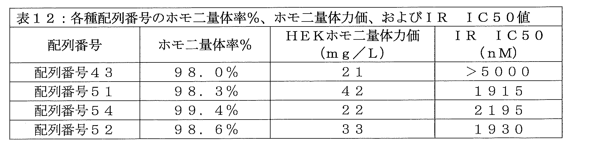
FVNQHLCGSHLVEALAVCGERGFFYTDPGGGGGGGGGIVEQCCTSICSLYQLENYCGGGGAGGGGDCPKCPAPEMLGGPSVFIFPPKPKDTLLIARTPEVTCVVVDLDPEDPEVQISWFVDG KQMQTAKTQPREEQFSGTYRVVSVLPIGHQDWLKGKQFTCKVNNKALPSPIERTISKARGQAHQPSVYVLPPSREELSKNTVSLTCLIKDFFPDIDVEWQSNGQQEPESKYRTTPPQLDEDGSY FLYSKLSVDKSRWQRGDTFICAVMHEALHNHYTQESLSHSPG (SEQ ID NO: 51)
FVNQHLCGSHLVEALAVCGERGFFYTPGGGGGGGGGIVEQCCTSICSLYQLENYCGGGGAGGGGDCPKCPAPEMLGGPSVFIFPPKPKDTLLIARTPEVTCVVVDLDPEDPEVQISWFVDGK QMQTAKTQPREEQFSGTYRVVSVLPIGHQDWLKGKQFTCKVNNKALPSPIERTISKARGQAHQPSVYVLPPSREELSKNTVSLTCLIKDFFPDIDVEWQSNGQQEPESKYRTTPPQLDEDGSYF LYSKLSVDKSRWQRGDTFICAVMHEALHNHYTQESLSHSPG (SEQ ID NO: 52)
FVNQHLCGSHLVEALVCGERGFFYTQGGGGGGGGGIVEQCCTSICSLYQLENYCGGGGAGGGGDCPKCPAPEMLGGPSVFIFPPKPKDTLLIARTPEVTCVVVDLDPEDPEVQISWFVDGK QMQTAKTQPREEQFSGTYRVVSVLPIGHQDWLKGKQFTCKVNNKALPSPIERTISKARGQAHQPSVYVLPPSREELSKNTVSLTCLIKDFFPDIDVEWQSNGQQEPESKYRTTPPQLDEDGSYF LYSKLSVDKSRWQRGDTFICAVMHEALHNHYTQESLSHSPG (SEQ ID NO: 54)
インスリン-Fc融合タンパク質を、実施例1に従ってHEK293細胞で製造し、実施例4に従ってプロテインAカラムを用いて精製した。構造の確認を実施例6に従って非還元CE-SDSおよび還元CE-SDSで行い、さらに、配列の確認を実施例8に記載のグリカン除去を伴うLC-MSによって行った。ホモ二量体含有率を実施例10に従って分子ふるいクロマトグラフィーで測定し、インスリン受容体結合親和性を実施例12に従って測定した。結果を表12に示す。得られた化合物のホモ二量体力価は1例(配列番号51)で顕著に増加しただけであったが、予想外にも、変異型化合物(配列番号51、配列番号54、および配列番号52)の全てにおいて、インスリン受容体親和性が向上した。 Insulin-Fc fusion protein was produced in HEK293 cells according to Example 1 and purified using a Protein A column according to Example 4. Structural confirmation was performed with non-reduced CE-SDS and reduced CE-SDS according to Example 6, and sequence confirmation was performed by LC-MS with glycan removal as described in Example 8. Homodimer content was determined by molecular exclusion chromatography according to Example 10, and insulin receptor binding affinity was determined according to Example 12. The results are shown in Table 12. The homodimer titers of the resulting compounds were significantly increased in only one case (SEQ ID NO: 51), whereas, unexpectedly, the homodimer titers of the mutant compounds (SEQ ID NO: 51, SEQ ID NO: 54, and SEQ ID NO: 52) were significantly increased. ) all showed improved insulin receptor affinity.
実施例43:ホモ二量体力価および生物活性をさらに向上させるための、配列番号43のインスリン-Fc融合タンパク質に対する、B鎖変異、C鎖変異、およびA鎖変異、B鎖切断、並びにリンカー変異を組み合わせる試み
実施例39、実施例40、実施例41、および実施例42で示されたように、単一の戦略で、非免疫原性の天然型のB10アミノ酸およびA8アミノ酸を含むインスリンポリペプチドと、イヌIgGB Fcフラグメントを組み込んで、許容できるインスリン受容体活性およびホモ二量体力価を有するインスリン-Fc融合タンパク質を形成することに成功したものはない。そこで、C鎖をより長くし、ペプチドリンカーをより長くし、B鎖のC末端アミノ酸を切断するというコンセプトを組み合わせた。さらに、自己会合や凝集の傾向をさらに低減する可能性があるため、生理的pHで負または正に帯電する側基を有するアミノ酸といった、疎水性の低いアミノ酸を用いて、天然型インスリンの疎水性アミノ酸残基部位にさらなる点変異を導入した。変異の例としては、チロシンからアラニン、チロシンからグルタミン酸、イソロイシンからスレオニン、およびフェニルアラニンからヒスチジンの変異が挙げられる。さらに、解析を単純にするため、全ての場合で、イヌIgGB FcフラグメントのcNg部位を天然型であるアスパラギンに戻した。これらのインスリン-Fc融合タンパク質バリアントの配列を以下に示し、配列番号43に対して得られた配列アラインメントを図16に示す(クラスタルオメガ):
FVNQHLCGSHLVEALELVCGERGFFYTPKTGGSGGGGGIVEQCCTSTCSLDQLENYCGGGGGQGGGGQGGGGQGGGGGDCPKCPAPEMLGGPSVFIFPPKPKDTLLIARTPEVTCVVVDLDPEDPEVQISWFVDGKQMQTAKTQPREEQFNGTYRVVSVLPIGHQDWLKGKQFTCKVNNKALPSPIERTISKARGQAHQPSVYVLPPSREELSKNTVSLTCLIKDFFPPDIDVEWQSNGQQEPESKYRTTPPQLDEDGSYFLYSKLSVDKSRWQRGDTFICAVMHEALHNHYTQESLSHSPG(配列番号55)
FVNQHLCGSHLVEALELVCGERGFHYGGGGGGSGGGGGIVEQCCTSTCSLDQLENYCNHGGGGQGGGGQGGGGQGGGGGDCPKCPAPEMLGGPSVFIFPPKPKDTLLIARTPEVTCVVVDLDPEDPEVQISWFVDGKQMQTAKTQPREEQFNGTYRVVSVLPIGHQDWLKGKQFTCKVNNKALPSPIERTISKARGQAHQPSVYVLPPSREELSKNTVSLTCLIKDFFPPDIDVEWQSNGQQEPESKYRTTPPQLDEDGSYFLYSKLSVDKSRWQRGDTFICAVMHEALHNHYTQESLSHSPG(配列番号56)
FVNQHLCGSHLVEALELVCGERGFHYGGGGGGSGGGGGIVEQCCTSTCSLDQLENYCNGGGGGQGGGGQGGGGQGGGGGDCPKCPAPEMLGGPSVFIFPPKPKDTLLIARTPEVTCVVVDLDPEDPEVQISWFVDGKQMQTAKTQPREEQFNGTYRVVSVLPIGHQDWLKGKQFTCKVNNKALPSPIERTISKARGQAHQPSVYVLPPSREELSKNTVSLTCLIKDFFPPDIDVEWQSNGQQEPESKYRTTPPQLDEDGSYFLYSKLSVDKSRWQRGDTFICAVMHEALHNHYTQESLSHSPG(配列番号28)
FVNQHLCGSHLVEALELVCGERGFHYGGGGGGSGGGGGIVEQCCTSTCSLDQLENYCGGGGGQGGGGQGGGGQGGGGGDCPKCPAPEMLGGPSVFIFPPKPKDTLLIARTPEVTCVVVDLDPEDPEVQISWFVDGKQMQTAKTQPREEQFNGTYRVVSVLPIGHQDWLKGKQFTCKVNNKALPSPIERTISKARGQAHQPSVYVLPPSREELSKNTVSLTCLIKDFFPPDIDVEWQSNGQQEPESKYRTTPPQLDEDGSYFLYSKLSVDKSRWQRGDTFICAVMHEALHNHYTQESLSHSPG(配列番号26)
FVNQHLCGSHLVEALELVCGERGFFYGGGGGGSGGGGGIVEQCCTSTCSLDQLENYCGGGGGQGGGGQGGGGQGGGGGDCPKCPAPEMLGGPSVFIFPPKPKDTLLIARTPEVTCVVVDLDPEDPEVQISWFVDGKQMQTAKTQPREEQFNGTYRVVSVLPIGHQDWLKGKQFTCKVNNKALPSPIERTISKARGQAHQPSVYVLPPSREELSKNTVSLTCLIKDFFPPDIDVEWQSNGQQEPESKYRTTPPQLDEDGSYFLYSKLSVDKSRWQRGDTFICAVMHEALHNHYTQESLSHSPG(配列番号57)
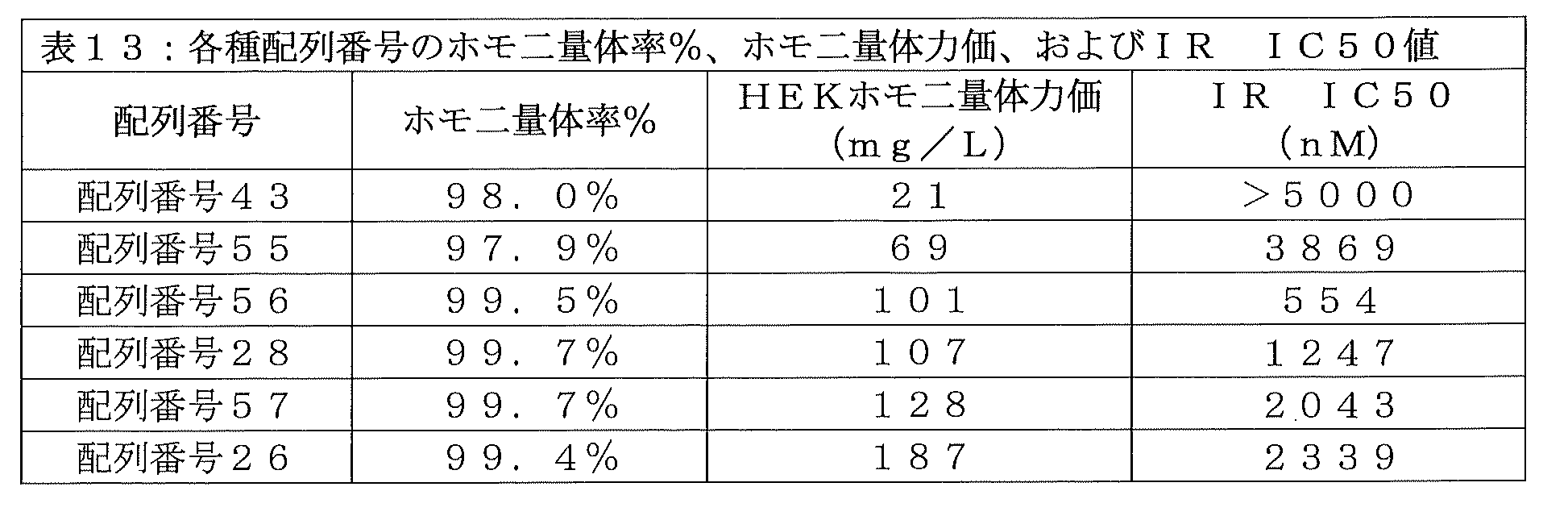
FVNQHLCGSHLVEALELVCGERGFFYTPKTGGSGGGGIVEQCCTSTCSLDQLENYCGGGGGQGGGGQGGGGQGGGGDCPKCPAPEMLGGPSVFIFPPKPKDTLLIARTPEVTCVVVDLDPE DPEVQISWFVDGKQMQTAKTQPREEQFNGTYRVVSVLPIGHQDWLKGKQFTCKVNNKALPSPIERTISKARGQAHQPSVYVLPPSREELSKNTVSLTCLIKDFFPDIDVEWQSNGQQEPESKYR TTPPQLDEDGSYFLYSKLSVDKSRWQRGDTFICAVMHEALHNHYTQESLSHSPG (SEQ ID NO: 55)
FVNQHLCGSHLVEALELVCGERGFHYGGGGGGGSGGGGIVEQCCTSTCSLDQLENYCNHGGGGQGGGGQGGGGQGGGGDCPKCPAPEMLGGPSVFIFPPKPKDTLLIARTPEVTCVVVDLDP EDPEVQISWFVDGKQMQTAKTQPREEQFNGTYRVVSVLPIGHQDWLKGKQFTCKVNNKALPSPIERTISKARGQAHQPSVYVLPPSREELSKNTVSLTCLIKDFFPDIDVEWQSNGQQEPESKY RTTPPQLDEDGSYFLYSKLSVDKSRWQRGDTFICAVMHEALHNHYTQESLSHSPG (SEQ ID NO: 56)
FVNQHLCGSHLVEALLVCGERGFHYGGGGGGGSGGGGIVEQCCTSTCSLDQLENYCNGGGGGQGGGGQGGGGQGGGGDCPKCPAPEMLGGPSVFIFPPKPKDTLLIARTPEVTCVVVDLDP EDPEVQISWFVDGKQMQTAKTQPREEQFNGTYRVVSVLPIGHQDWLKGKQFTCKVNNKALPSPIERTISKARGQAHQPSVYVLPPSREELSKNTVSLTCLIKDFFPDIDVEWQSNGQQEPESKY RTTPPQLDEDGSYFLYSKLSVDKSRWQRGDTFICAVMHEALHNHYTQESLSHSPG (SEQ ID NO: 28)
FVNQHLCGSHLVEALELVCGERGFHYGGGGGGSGGGGGIVEQCCTSTCSLDQLENYCGGGGGQGGGGQGGGGQGGGGDCPKCPAPEMLGGPSVFIFPPKPKDTLLIARTPEVTCVVVDLDPE DPEVQISWFVDGKQMQTAKTQPREEQFNGTYRVVSVLPIGHQDWLKGKQFTCKVNNKALPSPIERTISKARGQAHQPSVYVLPPSREELSKNTVSLTCLIKDFFPDIDVEWQSNGQQEPESKYR TTPPQLDEDGSYFLYSKLSVDKSRWQRGDTFICAVMHEALHNHYTQESLSHSPG (SEQ ID NO: 26)
FVNQHLCGSHLVEALLVCGERGFFYGGGGGGSGGGGGIVEQCCTSTCSLDQLENYCGGGGGQGGGGQGGGGQGGGGDCPKCPAPEMLGGPSVFIFPPKPKDTLLIARTPEVTCVVVDLDPE DPEVQISWFVDGKQMQTAKTQPREEQFNGTYRVVSVLPIGHQDWLKGKQFTCKVNNKALPSPIERTISKARGQAHQPSVYVLPPSREELSKNTVSLTCLIKDFFPDIDVEWQSNGQQEPESKYR TTPPQLDEDGSYFLYSKLSVDKSRWQRGDTFICAVMHEALHNHYTQESLSHSPG (SEQ ID NO: 57)
インスリン-Fc融合タンパク質を、実施例1に従ってHEK293細胞で製造し、実施例4に従ってプロテインAカラムを用いて精製した。構造の確認を実施例6に従って非還元CE-SDSおよび還元CE-SDSで行い、さらに、配列の確認を実施例8に記載のグリカン除去を伴うLC-MSによって行った。ホモ二量体含有率を実施例10に従って分子ふるいクロマトグラフィーで測定し、インスリン受容体結合親和性を実施例12に従って測定した。結果を表13に示す。結果に示されているように、特定のB鎖アミノ酸およびA鎖アミノ酸の疎水性を低減すること、より長くより可動性が高いCペプチド配列を使用すること、いくつかのC末端B鎖アミノ酸を切断すること、並びにより長いペプチドリンカーを用いること、を組み合わせることで、ホモ二量体力価およびインスリン受容体結合活性の設計基準を最低限達成する有用なインスリン-Fc融合タンパク質がいくつか得られた。配列番号56、配列番号28、配列番号26、および配列番号57は、配列番号43または配列番号55のいずれよりも、より好ましいインスリン受容体IC50値(3000nM未満)と、より好ましいHEKホモ二量体力価(100mg/L超)を示した。驚くべきことに、わずか数個のアミノ酸を変えるだけで、元の配列番号43のインスリン-Fc融合タンパク質に対して、インスリン受容体親和性が数倍に向上し、配列番号26のインスリン-Fc融合タンパク質の場合では、ホモ二量体力価が劇的に増加している。 Insulin-Fc fusion protein was produced in HEK293 cells according to Example 1 and purified using a Protein A column according to Example 4. Structural confirmation was performed with non-reduced CE-SDS and reduced CE-SDS according to Example 6, and sequence confirmation was performed by LC-MS with glycan removal as described in Example 8. Homodimer content was determined by molecular exclusion chromatography according to Example 10, and insulin receptor binding affinity was determined according to Example 12. The results are shown in Table 13. As shown in the results, reducing the hydrophobicity of certain B-chain and A-chain amino acids, using a longer and more flexible C-peptide sequence, and reducing some C-terminal B-chain amino acids A combination of truncation and the use of longer peptide linkers resulted in several useful insulin-Fc fusion proteins that minimally achieved the design criteria of homodimeric potency and insulin receptor binding activity. . SEQ ID NO: 56, SEQ ID NO: 28, SEQ ID NO: 26, and SEQ ID NO: 57 have more favorable insulin receptor IC50 values (less than 3000 nM) and more favorable HEK homodimer potency than either SEQ ID NO: 43 or SEQ ID NO: 55. (more than 100 mg/L). Surprisingly, changing just a few amino acids improved the insulin receptor affinity several-fold over the original insulin-Fc fusion protein of SEQ ID NO: 43, and the insulin-Fc fusion protein of SEQ ID NO: 26 In the case of proteins, homodimer titers are dramatically increased.
実施例44:配列番号7のインスリンポリペプチドと配列番号13のペプチドリンカーと配列番号15のイヌIgGB Fcフラグメントとから構築されたインスリン-Fc融合タンパク質のインビボ生物活性、反復投与生物活性、および免疫原性
実施例43より得られた肯定的なホモ二量体力価およびインスリン受容体結合活性の結果から、2種の最も有望なインスリン-Fc融合タンパク質(配列番号26および配列番号28)を、イヌにおいて、反復投与生物活性および免疫原性を評価する試験にかけた。それぞれの化合物は、より長く、より親水性が高い配列番号13のペプチドリンカーと、より製造性が高く、より凝集性が低い配列番号15のイヌIgGB Fcフラグメントと、を含む。最も重要なこととして、どちらのインスリン-Fc融合タンパク質も、免疫原性が低いと推定される天然型のB10アミノ酸およびA8アミノ酸(すなわち、一般配列番号6)を有するインスリンポリペプチドを含んでいる。配列番号28のインスリン-Fc融合タンパク質の場合、A21位にアスパラギンが存在する(すなわちインスリンポリペプチドは配列番号8を含む)。配列番号26のインスリン-Fc融合タンパク質の場合、A21位にアスパラギンが存在しない(すなわちインスリンポリペプチドは配列番号7を含む)。
Example 44: In vivo biological activity, repeated dose biological activity, and immunogenicity of an insulin-Fc fusion protein constructed from the insulin polypeptide of SEQ ID NO: 7, the peptide linker of SEQ ID NO: 13, and the canine IgG Fc fragment of SEQ ID NO: 15. Based on the positive homodimer titer and insulin receptor binding activity results obtained from Example 43, the two most promising insulin-Fc fusion proteins (SEQ ID NO: 26 and SEQ ID NO: 28) were tested in dogs. , was subjected to tests to assess repeated dose bioactivity and immunogenicity. Each compound includes a longer, more hydrophilic peptide linker of SEQ ID NO: 13 and a more manufacturable, less aggregating canine IgG Fc fragment of SEQ ID NO: 15. Most importantly, both insulin-Fc fusion proteins contain an insulin polypeptide with naturally occurring B10 and A8 amino acids (ie, general SEQ ID NO: 6) that are predicted to be less immunogenic. In the case of the insulin-Fc fusion protein of SEQ ID NO: 28, there is an asparagine at position A21 (ie, the insulin polypeptide comprises SEQ ID NO: 8). In the case of the insulin-Fc fusion protein of SEQ ID NO: 26, there is no asparagine at position A21 (ie, the insulin polypeptide comprises SEQ ID NO: 7).
配列番号28のインスリン-Fc融合タンパク質のインビボ生物活性を、実施例21の手順に従ってN=1のイヌで試験した。単回皮下投与の図17に示される結果は、配列番号28のインスリン-Fc融合タンパク質が、実施例22の手順に従って算出されたNAOCが1076%FBGL・日・kg/mgであり、インビボにおいて確かに生物活性を示すことが示された。配列番号28のインスリン-Fc融合タンパク質の薬物動態プロファイルを、ELISAを使用する実施例23の方法で測定し、2コンパートメントモデルをそのデータに適合し、その排出半減期を求めたところ、約3.5日であった。 The in vivo biological activity of the insulin-Fc fusion protein of SEQ ID NO: 28 was tested in N=1 dogs according to the procedure of Example 21. The results shown in FIG. 17 for a single subcutaneous administration show that the insulin-Fc fusion protein of SEQ ID NO: 28 has an NAOC of 1076% FBGL·day·kg/mg calculated according to the procedure of Example 22, and is proven to be effective in vivo. was shown to exhibit biological activity. The pharmacokinetic profile of the insulin-Fc fusion protein of SEQ ID NO: 28 was determined by the method of Example 23 using ELISA, a two-compartment model was fitted to the data, and its elimination half-life was determined to be approximately 3. It was the 5th.
次いで、実施例15の手順に従って、配列番号28のインスリン-Fc融合タンパク質の皮下投与を、N=1のイヌに、最初の注射後の14日目、28日目、および42日目に続けることで、反復投与時の生物活性を評価した。イヌの%FBGLが低下し過ぎた場合、そのイヌに餌を与えて、血糖を安全なレベルまで上昇させた。それ以降の各投与については、実施例22の一般手順に従って、NAOCおよびNAOCRの測定を行い、投与を施行した時間から次の投与を施行する直前までを算出した。表14に示されるNAOCおよびNAOCRは、配列番号28のインスリン-Fc融合タンパク質が、4回の投与中、0.8よりも大きなNAOCRを維持することから、反復投与時の生物活性の設計目標を達成していることを示している。

配列番号28のインスリン-Fc融合タンパク質の免疫原性を、実施例24の手順に従って試験した。図18に示されているように、配列番号28のインスリン-Fc融合タンパク質は、反復投与実験を通してインビボ生物活性が維持されたことと一致して、インビボにおける免疫原性を見かけ上示していない。 The immunogenicity of the insulin-Fc fusion protein of SEQ ID NO: 28 was tested according to the procedure of Example 24. As shown in FIG. 18, the insulin-Fc fusion protein of SEQ ID NO: 28 shows no apparent immunogenicity in vivo, consistent with the maintenance of in vivo biological activity through repeated administration experiments.
インスリンポリペプチド鎖のA21のアスパラギンが存在しない、配列番号26のインスリン-Fc融合タンパク質についても、イヌにおける反復投与時の生物活性成績を評価した。上記化合物を、N=1のイヌに、0日目、14日目、28日目、および42日目に、実施例22の手順に従って皮下投与した。イヌの%FBGLが低下し過ぎた場合、そのイヌに餌を与えて、血糖を安全なレベルまで上昇させた。1回目注射のNAOCは堂々の2278%FBGL・日・kg/mgであり、配列番号26のインスリン-Fc融合タンパク質が、配列番号28のインスリン-Fc融合タンパク質のほぼ2倍の効力で、インビボにおいて十分な生物活性を示すことが示された。インスリン-Fc融合タンパク質の薬物動態プロファイルを、ELISAを使用する実施例23の方法で測定し、2コンパートメントモデルをそのデータに適合し、その排出半減期を求めたところ、4.1±0.7日であった。図19および図20は、動物に配列番号26のホモ二量体を投与する場合の、単回投与による血糖調節と、複数回投与による複数週に亘る血糖調節を示している。それ以降の各投与についても、実施例22の一般手順に従って、NAOCおよびNAOCRの測定を行い、投与を施行した時間から次の投与を施行する直前までを算出した。表15に示されるNAOCおよびNAOCRは、配列番号26のインスリン-Fc融合タンパク質が、4回の投与中、1.0以上のNAOCRを維持することから、実施例29に記載の反復投与時の生物活性の設計目標を達成していることを示している。
The insulin-Fc fusion protein of SEQ ID NO: 26, in which asparagine at A21 of the insulin polypeptide chain is absent, was also evaluated for biological activity upon repeated administration in dogs. The above compound was administered subcutaneously to N=1 dogs on
配列番号26のインスリン-Fc融合タンパク質の免疫原性を、実施例24の手順に従って試験した。図21に示されているように、配列番号26のインスリン-Fc融合タンパク質は、反復投与実験を通してインビボ生物活性が維持されたことと一致して、インビボにおける免疫原性を見かけ上示していない。
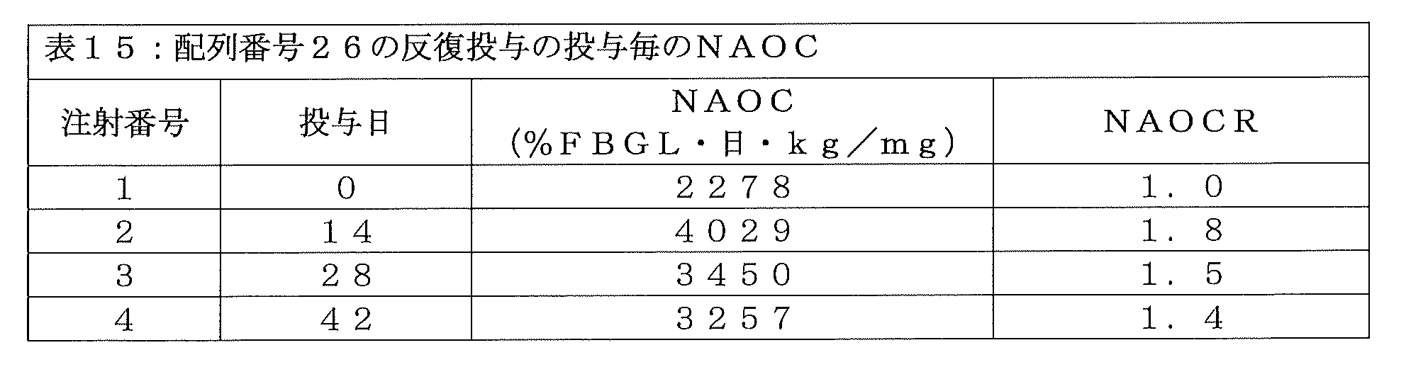
発明を実施するための形態で論じたように、アスパラギン-グリシン結合間には既知の酵素的切断部位が存在する(Vlasak,J.、Ionescu,R.、(2011年)MAbs、3巻、3号、pp253~263)。配列番号13のペプチドリンカーを有する配列番号26のインスリン-Fc融合タンパク質に含まれる配列番号7のインスリンポリペプチドの、A鎖の21番目のアミノ酸(すなわち、A21)にあるアスパラギンを取り除くことで、A鎖のC末端とペプチドリンカーのN末端との間のアスパラギン-グリシン結合が酵素的に切断される可能性が排除される。しかし、配列番号28のインスリン-Fc融合タンパク質は、配列番号13のペプチドリンカーと、A21にアスパラギンを保持する配列番号8のインスリンポリペプチドと、を含んでいる。そのため、配列番号28のインスリン-Fc融合タンパク質は、合成中または皮下投与後のインビボで、酵素による消化を受けるであろうと予想されていた。しかし、かなり予想外なことに、配列番号28のインスリン-Fc融合タンパク質は、許容できるホモ二量体力価を伴ってHEK細胞で製造可能であり、また、その生物活性を損なう酵素消化の徴候もなく、許容できるインビボ生物活性を示した。 As discussed in the Detailed Description, there is a known enzymatic cleavage site between the asparagine-glycine bond (Vlasak, J., Ionescu, R., (2011) MAbs, vol. 3, 3). No., pp. 253-263). By removing the asparagine at the 21st amino acid (i.e., A21) of the A chain of the insulin polypeptide of SEQ ID NO: 7 contained in the insulin-Fc fusion protein of SEQ ID NO: 26 having the peptide linker of SEQ ID NO: 13, A The possibility of enzymatic cleavage of the asparagine-glycine bond between the C-terminus of the chain and the N-terminus of the peptide linker is excluded. However, the insulin-Fc fusion protein of SEQ ID NO: 28 includes the peptide linker of SEQ ID NO: 13 and the insulin polypeptide of SEQ ID NO: 8, which retains an asparagine at A21. Therefore, it was expected that the insulin-Fc fusion protein of SEQ ID NO: 28 would undergo enzymatic digestion during synthesis or in vivo after subcutaneous administration. However, quite unexpectedly, the insulin-Fc fusion protein of SEQ ID NO: 28 can be produced in HEK cells with acceptable homodimeric titers and also without signs of enzymatic digestion that impair its biological activity. showed acceptable in vivo biological activity.
実施例45:好ましい配列番号7のインスリンポリペプチドと好ましい配列番号13のペプチドリンカーとを含むインスリン-Fc融合タンパク質の製造性およびインビボ効力を最適とするための、イヌIgGBアイソタイプFcフラグメントの確認
実施例43および実施例44に記載されたように、免疫原性がなく、収量が多く、純度が高く、生物活性が高いインスリン-Fc融合タンパク質が得られる、インスリンポリペプチドとペプチドリンカーの新規組み合わせが発見されたことから、イヌIgGB Fcフラグメントが、実施例32および実施例33でのインスリン-Fc融合タンパク質の場合では事実であったように、ホモ二量体力価および生物活性の観点で好ましいアイソタイプであるどうかに関する疑問が、残った。そこで、配列番号26のインスリン-Fc融合タンパク質のインスリンポリペプチド(配列番号7)とペプチドリンカー(配列番号13)は維持し、配列番号15のイヌIgGB Fcフラグメントを配列番号14のイヌIgGA Fcフラグメント、配列番号16のイヌIgGC Fcフラグメント、または配列番号17のイヌIgGD Fcフラグメントに置き換えた、さらなるインスリン-Fc融合タンパク質を設計した。これらの得られたインスリン-Fc融合タンパク質バリアントの配列を以下に示す:
FVNQHLCGSHLVEALELVCGERGFHYGGGGGGSGGGGGIVEQCCTSTCSLDQLENYCGGGGGQGGGGQGGGGQGGGGGDCPKCPAPEMLGGPSVFIFPPKPKDTLLIARTPEVTCVVVDLDPEDPEVQISWFVDGKQMQTAKTQPREEQFNGTYRVVSVLPIGHQDWLKGKQFTCKVNNKALPSPIERTISKARGQAHQPSVYVLPPSREELSKNTVSLTCLIKDFFPPDIDVEWQSNGQQEPESKYRTTPPQLDEDGSYFLYSKLSVDKSRWQRGDTFICAVMHEALHNHYTQESLSHSPG(配列番号26)
FVNQHLCGSHLVEALELVCGERGFHYGGGGGGSGGGGGIVEQCCTSTCSLDQLENYCGGGGGQGGGGQGGGGQGGGGGRCTDTPPCPVPEPLGGPSVLIFPPKPKDILRITRTPEVTCVVLDLGREDPEVQISWFVDGKEVHTAKTQSREQQFNGTYRVVSVLPIEHQDWLTGKEFKCRVNHIDLPSPIERTISKARGRAHKPSVYVLPPSPKELSSSDTVSITCLIKDFYPPDIDVEWQSNGQQEPERKHRMTPPQLDEDGSYFLYSKLSVDKSRWQQGDPFTCAVMHETLQNHYTDLSLSHSPG(配列番号58)
FVNQHLCGSHLVEALELVCGERGFHYGGGGGGSGGGGGIVEQCCTSTCSLDQLENYCGGGGGQGGGGQGGGGQGGGGGCNNCPCPGCGLLGGPSVFIFPPKPKDILVTARTPTVTCVVVDLDPENPEVQISWFVDSKQVQTANTQPREEQSNGTYRVVSVLPIGHQDWLSGKQFKCKVNNKALPSPIEEIISKTPGQAHQPNVYVLPPSRDEMSKNTVTLTCLVKDFFPPEIDVEWQSNGQQEPESKYRMTPPQLDEDGSYFLYSKLSVDKSRWQRGDTFICAVMHEALHNHYTQISLSHSPG(配列番号59)
FVNQHLCGSHLVEALELVCGERGFHYGGGGGGSGGGGGIVEQCCTSTCSLDQLENYCGGGGGQGGGGQGGGGQGGGGGCISPCPVPESLGGPSVFIFPPKPKDILRITRTPEITCVVLDLGREDPEVQISWFVDGKEVHTAKTQPREQQFNSTYRVVSVLPIEHQDWLTGKEFKCRVNHIGLPSPIERTISKARGQAHQPSVYVLPPSPKELSSSDTVTLTCLIKDFFPPEIDVEWQSNGQPEPESKYHTTAPQLDEDGSYFLYSKLSVDKSRWQQGDTFTCAVMHEALQNHYTDLSLSHSPG(配列番号60)。
Example 45: Validation Example of Canine IgG Isotype Fc Fragments to Optimize Manufacturability and In Vivo Efficacy of Insulin-Fc Fusion Proteins Comprising the Preferred Insulin Polypeptide of SEQ ID NO: 7 and the Preferred SEQ ID NO: 13 Peptide Linker Novel combinations of insulin polypeptides and peptide linkers have been discovered that result in insulin-Fc fusion proteins that are non-immunogenic, have high yields, high purity, and high biological activity, as described in Example 43 and Example 44. The canine IgG Fc fragment is the preferred isotype in terms of homodimer potency and biological activity, as was the case for the insulin-Fc fusion proteins in Examples 32 and 33. Questions remained as to whether Therefore, the insulin polypeptide (SEQ ID NO: 7) and peptide linker (SEQ ID NO: 13) of the insulin-Fc fusion protein of SEQ ID NO: 26 were maintained, and the canine IgG Fc fragment of SEQ ID NO: 15 was replaced with the canine IgG Fc fragment of SEQ ID NO: 14. Additional insulin-Fc fusion proteins were designed that replaced the canine IgG Fc fragment of SEQ ID NO: 16 or the canine IgGD Fc fragment of SEQ ID NO: 17. The sequences of these resulting insulin-Fc fusion protein variants are shown below:
FVNQHLCGSHLVEALELVCGERGFHYGGGGGGSGGGGGIVEQCCTSTCSLDQLENYCGGGGGQGGGGQGGGGQGGGGDCPKCPAPEMLGGPSVFIFPPKPKDTLLIARTPEVTCVVVDLDPE DPEVQISWFVDGKQMQTAKTQPREEQFNGTYRVVSVLPIGHQDWLKGKQFTCKVNNKALPSPIERTISKARGQAHQPSVYVLPPSREELSKNTVSLTCLIKDFFPDIDVEWQSNGQQEPESKYR TTPPQLDEDGSYFLYSKLSVDKSRWQRGDTFICAVMHEALHNHYTQESLSHSPG (SEQ ID NO: 26)
FVNQHLCGSHLVEALVCGERGFHYGGGGGGSGGGGGIVEQCCTSTCSLDQLENYCGGGGGQGGGGQGGGGQGGGGRCTDTPPCPVPEPLGGPSVLIFPPKPKDILRITRTPEVTCVVLDL GREDPEVQISWFVDGKEVHTAKTQSREQQFNGTYRVVSVLPIEHQDWLTGKEFKCRVNHIDLPSPIERTISKARGRAHKPSVYVLPPSPKELSSSDTVSITCLIKDFYPPDIDVEWQSNGQQEPE RKHRMTPPQLDEDGSYFLYSKLSVDKSRWQQGDPFTCAVMHETLQNHYTDLSLSHSPG (SEQ ID NO: 58)
FVNQHLCGSHLVEALVCGERGFHYGGGGGGGSGGGGIVEQCCTSTCSLDQLENYCGGGGGQGGGGQGGGGQGGGGCNNCPCGLLGGPSVFIFPPKPKDILVTARTPTVTCVVVVDLDP ENPEVQISWFVDSKQVQTANTQPREEQSNGTYRVVSVLPIGHQDWLSGKQFKCKVNNKALPSPIEEIISKTPGQAHQPNVYVLPPSRDEMSKNTVTLTCLVKDFFPEIDVEWQSNGQQEPESKY RMTPPQLDEDGSYFLYSKLSVDKSRWQRGDTFICAVMHEALHNHYTQISLSHSPG (SEQ ID NO: 59)
FVNQHLCGSHLVEALLVCGERGFHYGGGGGGGSGGGGIVEQCCTSTCSLDQLENYCGGGGGQGGGGQGGGGQGGGGCISPCPVPESLGGPSVFIFPPKPKDILRITRTPEITCVVLDLGRE DPEVQISWFVDGKEVHTAKTQPREQQFNSTYRVVSVLPIEHQDWLTGKEFKCRVNHIGLPSPIERTISKARGQAHQPSVYVLPPSPKELSSSDTVTLTCLIKDFFPEIDVEWQSNGQPEPESKY HTTAPQLDEDGSYFLYSKLSVDKSRWQQGDTFTCAVMHEALQNHYTDLSLSHSPG (SEQ ID NO: 60).
インスリン-Fc融合タンパク質を、実施例1に従ってHEK293細胞で製造し、実施例4に従ってプロテインAカラムまたはプロテインGカラムを用いて精製した。構造の確認を実施例6に従って非還元CE-SDSおよび還元CE-SDSで行い、さらに、配列の確認を実施例8に記載のグリカン除去を伴うLC-MSによって行った。ホモ二量体含有率を実施例10に従って分子ふるいクロマトグラフィーで測定し、インスリン受容体結合親和性を実施例12に従って測定した。さらに、インスリン-Fc融合タンパク質のイヌFcRn受容体に対する親和性を、実施例15に従って測定した。表16に示されているように、イヌIgGB Fcフラグメントを含む配列番号26のインスリン-Fc融合タンパク質は、これらの配列の中で最も高いホモ二量体力価を示した。イヌIgGA Fcフラグメントを含む配列番号58のインスリン-Fc融合タンパク質は、プロテインAカラムを用いて精製された場合は不十分なホモ二量体力価しか示さなかったが、プロテインGカラムを用いて精製された場合では、ホモ二量体力価は顕著に向上し、50mg/L超という設計目標を上回った。イヌIgGC Fcフラグメントを含む配列番号59のインスリン-Fc融合タンパク質についても同じであった。イヌIgGD Fcフラグメントを含む配列番号60のインスリン-Fc融合タンパク質は、プロテインAまたはプロテインGカラムのどちらで精製した場合も、化合物が得られなかった。以上から、異なるインスリンポリペプチド(配列番号4)およびペプチドリンカー(配列番号11)を含む配列番号36のインスリン-Fc融合タンパク質で示されたように、イヌIgGBはホモ二量体力価の観点で好ましいFcフラグメントであった(実施例32参照)。

プロテインGを介して精製されたイヌIgGA Fcフラグメントを含む配列番号58のインスリン-Fc融合タンパク質のインビボ生物活性を、実施例21の手順に従って試験した。図22に示される結果は、配列番号58のインスリン-Fc融合タンパク質が、実施例22に従って算出されたNAOCがわずか174%FBGL・日・kg/mgであり、インビボにおいてある程度の生物活性しか示さないことが示された。 The in vivo biological activity of the insulin-Fc fusion protein of SEQ ID NO: 58 containing a canine IgG Fc fragment purified via Protein G was tested according to the procedure of Example 21. The results shown in FIG. 22 show that the insulin-Fc fusion protein of SEQ ID NO: 58 has an NAOC of only 174% FBGL·day·kg/mg as calculated according to Example 22, and exhibits only a certain degree of biological activity in vivo. It was shown that
プロテインGを介して精製されたイヌIgGC Fcフラグメントを含む配列番号59のインスリン-Fc融合タンパク質のインビボ生物活性を、実施例21の手順に従って試験した。図23に示される結果は、配列番号59のインスリン-Fc融合タンパク質が、実施例22に従って算出されたNAOCがわずか39%FBGL・日・kg/mgであり、インビボにおいてある程度の生物活性しか示さないことが示された。 The in vivo biological activity of the insulin-Fc fusion protein of SEQ ID NO: 59 containing a canine IgG Fc fragment purified via Protein G was tested according to the procedure of Example 21. The results shown in FIG. 23 show that the insulin-Fc fusion protein of SEQ ID NO: 59 has an NAOC of only 39% FBGL·day·kg/mg as calculated according to Example 22, and exhibits only a certain degree of biological activity in vivo. It was shown that
以上から、異なるインスリンポリペプチド(配列番号4)およびペプチドリンカー(配列番号11)を含む配列番号36のインスリン-Fc融合タンパク質で示されたように、イヌIgGBは生物活性の観点で好ましいFcフラグメントであった(実施例32および実施例33、並びに上記の表16を参照)。 From the above, canine IgG is a preferred Fc fragment from the viewpoint of biological activity, as shown in the insulin-Fc fusion protein of SEQ ID NO: 36, which contains a different insulin polypeptide (SEQ ID NO: 4) and a peptide linker (SEQ ID NO: 11). (See Examples 32 and 33 and Table 16 above).
実施例46:潜在的な免疫原性リスクを低減するための、配列番号8のインスリンポリペプチドと、配列番号13のペプチドリンカーと、イヌIgGB Fcフラグメントとを含む非グリコシル化インスリン-Fc融合タンパク質
配列番号26のインスリン-Fc融合タンパク質は設計目標の全てを達成しているが(実施例28)、治療が長期に亘った場合(例えば、6か月、1年、2年、またはそれ以上)に免疫原性リスクが生じる恐れがあり、これが起こった場合、糖尿病治療における本インスリン-Fc融合タンパク質の使用が損なわれる可能性がある。発明を実施するための形態、実施例34および実施例35に記載したように、反復投与後の生物活性を低減させる、考えられる原因の1つは、イヌIgGB Fcフラグメントがイヌの免疫系と望ましくない相互作用を起こすことによる、中和抗薬物抗体の産生である。しかし、実施例45で示された結果は、予想外にも、イヌIgGBアイソタイプが、4種のイヌIgGアイソタイプの中で、所望の製造性および生物活性をもたらす唯一の選択肢であったことを示している。そこで、FcγRI受容体結合性が低く、長期の慢性的な免疫原性リスクが低減されるはずの、非グリコシル化インスリン-Fc融合タンパク質を得るための、さらなるFc変異を探索した。
Example 46: Non-glycosylated insulin-Fc fusion protein sequence comprising an insulin polypeptide of SEQ ID NO: 8, a peptide linker of SEQ ID NO: 13, and a canine IgG Fc fragment to reduce potential immunogenicity risks. Insulin-Fc
発明を実施するための形態に記載したように、FcγRI相互作用を低減するための1つの方法には、FcフラグメントのcNg部位を変異させることで、宿主細胞内で合成される際のグリコシル化を妨げることが行われる。すなわち、cNg部位の変異を配列番号26のFcフラグメント領域に対して行ったことで、実施例15に記載のインビトロヒトFcγRIアッセイにおける結合性によって評価される、インビボにおけるFcγ受容体に対するFcフラグメントの結合親和性が低減された。配列番号26のインスリン-Fc融合タンパク質におけるcNg部位の位置はcNg-NB151である。配列番号26に対する変異として、配列番号62にはcNg-NB151-S変異が含まれ、配列番号61には同じcNg-NB151-S変異に加えてNB119-A変異が含まれた。Lo,M.ら、“Effector attenuating substitutions that maintain antibody stability and reduce toxicity in mice”、J.Biol.Chem.、(2017年)、pp.1~20により、マウス抗体での使用しか記載されていないが、NB119-Aは、FcγRIとの相互作用を低減させるさらなる試みにおいて組み込まれたものである。得られたインスリン-Fc融合タンパク質の完全アミノ酸配列を以下に挙げ(明確にするためにNB119部位およびNB151部位には下線)、さらに、これらの配列アラインメント(クラスタルオメガ)を図24に示す:
FVNQHLCGSHLVEALELVCGERGFHYGGGGGGSGGGGGIVEQCCTSTCSLDQLENYCGGGGGQGGGGQGGGGQGGGGGDCPKCPAPEMLGGPSVFIFPPKPKDTLLIARTPEVTCVVVALDPEDPEVQISWFVDGKQMQTAKTQPREEQFSGTYRVVSVLPIGHQDWLKGKQFTCKVNNKALPSPIERTISKARGQAHQPSVYVLPPSREELSKNTVSLTCLIKDFFPPDIDVEWQSNGQQEPESKYRTTPPQLDEDGSYFLYSKLSVDKSRWQRGDTFICAVMHEALHNHYTQESLSHSPG(配列番号61)
FVNQHLCGSHLVEALELVCGERGFHYGGGGGGSGGGGGIVEQCCTSTCSLDQLENYCGGGGGQGGGGQGGGGQGGGGGDCPKCPAPEMLGGPSVFIFPPKPKDTLLIARTPEVTCVVVDLDPEDPEVQISWFVDGKQMQTAKTQPREEQFSGTYRVVSVLPIGHQDWLKGKQFTCKVNNKALPSPIERTISKARGQAHQPSVYVLPPSREELSKNTVSLTCLIKDFFPPDIDVEWQSNGQQEPESKYRTTPPQLDEDGSYFLYSKLSVDKSRWQRGDTFICAVMHEALHNHYTQESLSHSPG(配列番号62)。
As described in the Detailed Description, one method for reducing FcγRI interactions involves mutating the cNg site of the Fc fragment, which reduces glycosylation when synthesized within the host cell. Something that hinders is done. That is, by mutating the cNg site to the Fc fragment region of SEQ ID NO: 26, binding of the Fc fragment to the Fcγ receptor in vivo, as evaluated by binding in the in vitro human FcγRI assay described in Example 15, was improved. Affinity reduced. The position of the cNg site in the insulin-Fc fusion protein of SEQ ID NO: 26 is cNg-NB151. As mutations to SEQ ID NO: 26, SEQ ID NO: 62 contained the cNg-NB151-S mutation, and SEQ ID NO: 61 contained the NB119-A mutation in addition to the same cNg-NB151-S mutation. Lo, M. et al., “Effector attenuating substances that maintain antibody stability and reduce toxicity in mice”, J. et al. Biol. Chem. , (2017), pp. 1-20, although only described for use with murine antibodies, NB119-A was incorporated in a further attempt to reduce interaction with FcγRI. The complete amino acid sequence of the resulting insulin-Fc fusion protein is listed below (NB119 and NB151 sites are underlined for clarity), and their sequence alignment (Cluster Omega) is shown in Figure 24:
FVNQHLCGSHLVEALLVCGERGFHYGGGGGGGSGGGGIVEQCCTSTCSLDQLENYCGGGGGQGGGGQGGGGQGGGGDCPKCPAPEMLGGPSVFIFPPKPKDTLLIARTPEVTCVVV A LD PEDPEVQISWFVDGKQMQTAKTQPREEQF S GTYRVVSVLPIGHQDWLKGKQFTCKVNNKALPSPIERTISKARGQAHQPSVYVLPPSREELSKNTVSLTCLIKDFFPDIDVEWQSNGQQEPE SKYRTTPPQLDEDGSYFLYSKLSVDKSRWQRGDTFICAVMHEALHNHYTQESLSHSPG (SEQ ID NO: 61)
FVNQHLCGSHLVEALELVCGERGFHYGGGGGGSGGGGGIVEQCCTSTCSLDQLENYCGGGGGQGGGGQGGGGQGGGGDCPKCPAPEMLGGPSVFIFPPKPKDTLLIARTPEVTCVVVDLDPE DPEVQISWFVDGKQMQTAKTQPREEQF S GTYRVVSVLPIGHQDWLKGKQFTCKVNNKALPSPIERTISKARGQAHQPSVYVLPPSREELSKNTVSLTCLIKDFFPDIDVEWQSNGQQEPESK YRTTPPQLDEDGSYFLYSKLSVDKSRWQRGDTFICAVMHEALHNHYTQESLSHSPG (SEQ ID NO: 62).
インスリン-Fc融合タンパク質を、実施例1に従ってHEK293細胞で製造し、実施例4に従ってプロテインAカラムを用いて精製した。構造の確認を実施例6に従って非還元CE-SDSおよび還元CE-SDSで行い、さらに、配列の確認を実施例8に記載のグリカン除去を伴うLC-MSによって行った。ホモ二量体含有率を実施例10に従って分子ふるいクロマトグラフィーで測定し、インスリン受容体結合親和性を実施例12に従って測定した。表17に示されているように、Fcフラグメントに対してcNg-NB151-S変異を組み込むと、ホモ二量体率が減少し、許容できないほどに高い凝集レベルを示した(すなわち、ホモ二量体率が70%強まで低下した)。
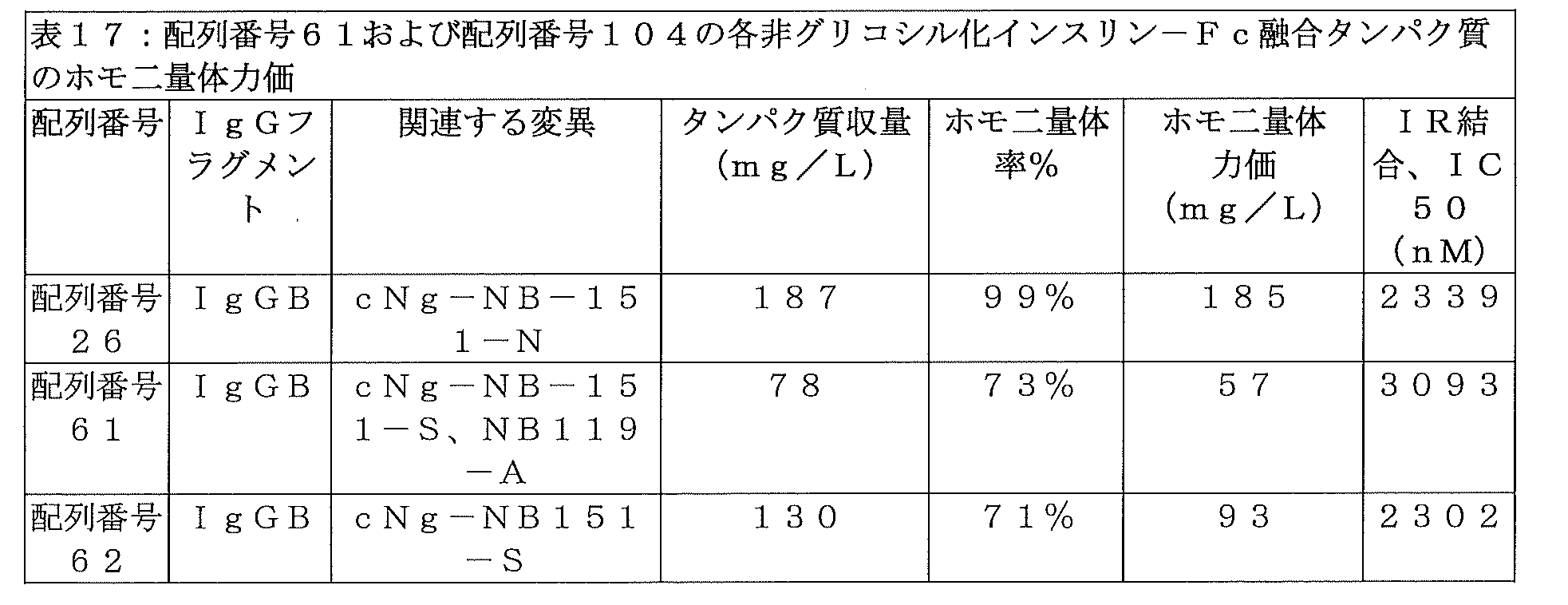
配列番号61および配列番号62のインスリン-Fc融合タンパク質のインビボ生物活性を、実施例21の手順に従ってN=1のイヌで試験した。単回皮下投与についての図25に示される結果は、両化合物が、配列番号26のインスリン-Fc融合タンパク質よりもインビボ生物活性が顕著に低かったことを示している(配列番号62のNAOC=574%FBGL・日・kg/mg;配列番号61のNAOC=921%FBGL・日・kg/mg)。これらの結果は、Fcフラグメントに対してcNg-NB151-S変異を組み込んで、配列番号26のインスリン-Fc融合タンパク質の非グリコシル化型を作製したところ、予想外にも、得られる化合物のインビボ生物活性が低減されたことを示している。 The in vivo biological activity of the insulin-Fc fusion proteins of SEQ ID NO: 61 and SEQ ID NO: 62 was tested in N=1 dogs according to the procedure of Example 21. The results shown in Figure 25 for a single subcutaneous administration indicate that both compounds had significantly lower in vivo bioactivity than the insulin-Fc fusion protein of SEQ ID NO: 26 (NAOC of SEQ ID NO: 62 = 574 %FBGL·day·kg/mg; NAOC of SEQ ID NO: 61 = 921%FBGL·day·kg/mg). These results demonstrate that when the cNg-NB151-S mutation was incorporated into the Fc fragment to create a non-glycosylated version of the insulin-Fc fusion protein of SEQ ID NO: 26, the resulting compound unexpectedly showed significant in vivo bioactivity. It shows that the activity was reduced.
cNg-NB151-S部位の変異を含む配列番号62のインスリン-Fc融合タンパク質の凝集度を減らし、生物活性を向上させる試みにおいて、凝集に関与すると考えられている領域に変異を含む、各種インスリンポリペプチドB鎖バリアントを検討した。インスリン-Fc融合タンパク質を、実施例1に従ってHEK293細胞で製造し、実施例4に従ってプロテインAカラムを用いて精製した。構造の確認を実施例6に従って非還元CE-SDSおよび還元CE-SDSで行い、さらに、配列の確認を実施例8に記載のグリカン除去を伴うLC-MSによって行った。ホモ二量体含有率は実施例10に従って分子ふるいクロマトグラフィーで測定した。試験されたB鎖バリアントの中で、B鎖のN末端から16番目のアミノ酸(すなわち、B16)にチロシンからアラニンへの置換を含む1つのインスリンFc融合タンパク質(配列番号30)が、予想外にも、ホモ二量体力価が高く(105mg/L)、凝集が少なく(99%ホモ二量体)、104mg/Lというホモ二量体力価が得られることが分かった。実施例12に従って測定されたインスリン受容体結合は許容できるものであり、IC50は2040nMであった。実施例16に従って測定されたFcRn受容体結合親和性EC50値は1194ng/mLであった。配列番号30のインスリン-Fc融合タンパク質の薬物動態プロファイルを、ELISAを使用する実施例23の方法で測定し、2コンパートメントモデルをそのデータに適合し、その排出半減期を求めたところ、4.1±0.7日であった。配列番号30の配列を以下に示す(明確にするためにB16A変異およびcNg-NB151-S変異には下線)。
FVNQHLCGSHLVEALALVCGERGFHYGGGGGGSGGGGGIVEQCCTSTCSLDQLENYCGGGGGQGGGGQGGGGQGGGGGDCPKCPAPEMLGGPSVFIFPPKPKDTLLIARTPEVTCVVVDLDPEDPEVQISWFVDGKQMQTAKTQPREEQFSGTYRVVSVLPIGHQDWLKGKQFTCKVNNKALPSPIERTISKARGQAHQPSVYVLPPSREELSKNTVSLTCLIKDFFPPDIDVEWQSNGQQEPESKYRTTPPQLDEDGSYFLYSKLSVDKSRWQRGDTFICAVMHEALHNHYTQESLSHSPG(配列番号30)。
In an attempt to reduce the degree of aggregation and improve biological activity of the insulin-Fc fusion protein of SEQ ID NO: 62 containing mutations in the cNg-NB151-S site, various insulin polypeptides containing mutations in regions thought to be involved in aggregation were used. Peptide B chain variants were investigated. Insulin-Fc fusion protein was produced in HEK293 cells according to Example 1 and purified using a Protein A column according to Example 4. Structural confirmation was performed with non-reduced CE-SDS and reduced CE-SDS according to Example 6, and sequence confirmation was performed by LC-MS with glycan removal as described in Example 8. Homodimer content was determined by molecular exclusion chromatography according to Example 10. Among the B chain variants tested, one insulin Fc fusion protein (SEQ ID NO: 30) containing a tyrosine to alanine substitution at the 16th amino acid from the N-terminus of the B chain (i.e., B16) unexpectedly It was also found that a homodimer titer of 104 mg/L was obtained with high homodimer titer (105 mg/L) and low aggregation (99% homodimer). Insulin receptor binding measured according to Example 12 was acceptable, with an IC50 of 2040 nM. The FcRn receptor binding affinity EC50 value measured according to Example 16 was 1194 ng/mL. The pharmacokinetic profile of the insulin-Fc fusion protein of SEQ ID NO: 30 was determined by the method of Example 23 using ELISA, a two-compartment model was fitted to the data, and its elimination half-life was determined to be 4.1. It was ±0.7 days. The sequence of SEQ ID NO: 30 is shown below (B16A mutation and cNg-NB151-S mutation are underlined for clarity).
FVNQHLCGSHLVEAL A LVCGERGFHYGGGGGGGSGGGGIVEQCCTSTCSLDQLENYCGGGGGQGGGGQGGGGQGGGGDCPKCPAPEMLGGPSVFIFPPKPKDTLLIARTPEVTCVVVDLD PEDPEVQISWFVDGKQMQTAKTQPREEQF S GTYRVVSVLPIGHQDWLKGKQFTCKVNNKALPSPIERTISKARGQAHQPSVYVLPPSREELSKNTVSLTCLIKDFFPDIDVEWQSNGQQEPE SKYRTTPPQLDEDGSYFLYSKLSVDKSRWQRGDTFICAVMHEALHNHYTQESLSHSPG (SEQ ID NO: 30).
次に、配列番号30のインスリン-Fc融合タンパク質を、イヌにおいて、反復投与時の生物活性成績について評価した。上記化合物を、N=1のイヌに、0日目、7日目、14日目、および28日目に、実施例22の手順に従って皮下投与した。イヌの%FBGLが低下し過ぎた場合、そのイヌに餌を与えて、血糖を安全なレベルまで上昇させた。予想外にも、配列番号62のインスリン-Fc融合タンパク質と比較して、B16A変異を含む配列番号30のインスリン-Fc融合タンパク質の1回目注射のNAOCは、顕著に高かった(1185%FBGL・日・kg/mg)。1回目投与のインビボ生物活性のグラフを図26に示す。また、当該化合物の薬物動態プロファイルを、ELISAを使用する実施例23の方法で測定し、2コンパートメントモデルをそのデータに適合し、当該化合物の排出半減期を求めたところ、3.5日であった。それ以降の各投与についても、実施例22の一般手順に従って、NAOCおよびNAOCRの測定を行い、投与を施行した時間から次の投与を施行する直前までを算出した。表18に示されるNAOCおよびNAOCRは、配列番号30のインスリン-Fc融合タンパク質が、4回の投与中、0.6以上のNAOCRを維持することから、反復投与時の生物活性の設計目標を達成していることを示している。まとめると、これらの結果は、好適な、配列番号26の非グリコシル化cNg-Sバリアントを得るには、インスリンB鎖配列を変異することが必要であったことを示している。よって、配列番号10のインスリンポリペプチドは、cNg変異型イヌIgGB Fcフラグメントを含む非グリコシル化インスリン-Fc融合タンパク質に好適であった。
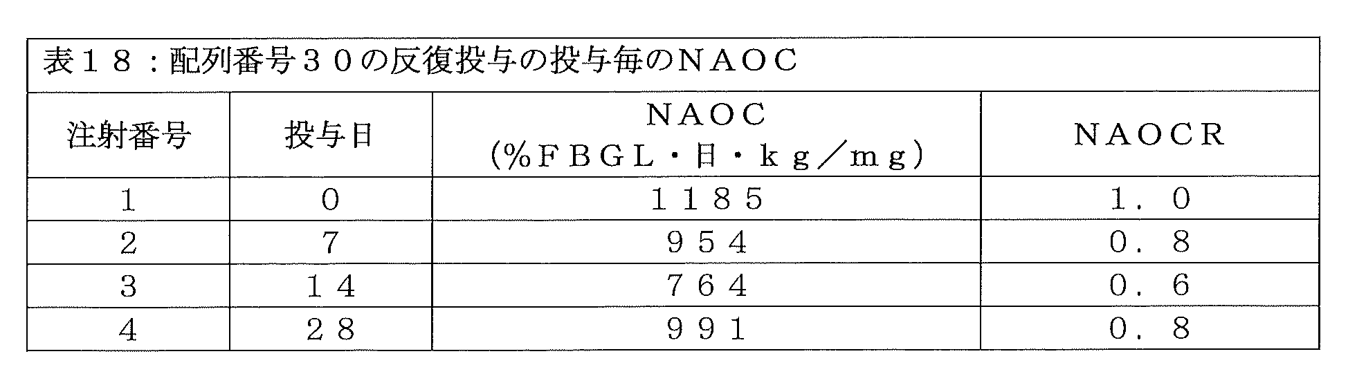
最後に、選択された化合物の、免疫系との相互作用のしやすさを、実施例15の手順に従ってそのFcγ受容体結合活性を測定することにより、調べた。表19は、これらのインスリン-Fc融合タンパク質のFcγ受容体I結合と、配列番号36のインスリン-Fc融合タンパク質のFcγ受容体結合とを比較している。非グリコシル化インスリン-Fc融合タンパク質(cNg-S変異によって達成)が、最も低い、配列番号36に対するFcγ受容体結合比を示したことが分かる。

実施例47:安定にトランスフェクトされたCHO細胞株を介して作製されたイヌIgGB由来のFcフラグメントを含む好ましいインスリン-Fc融合タンパク質を用いた、例示的なCHOベースのプロダクションラン
配列番号26または配列番号30をコードするベクターで安定にトランスフェクトされた別々のCHO細胞株を、実施例2に記載されたように構築した。フェッドバッチ式振盪フラスコ14日間プロダクションラン(0.5~2.0Lの培地規模)の播種を、37℃、5%二酸化炭素に設定された振盪恒温器内で、0.5×10⁶細胞/mLとなるように行った。増殖培地としてCD OptiCHOをDynamis(サーモフィッシャー社)に置き換え、フィードとしてEfficient Feed C(サーモフィッシャー社)を用いた以外は、上記実施例2に記載したように、ランを実行した。3%v/vのフィードの添加をプロダクションラン3日目から開始して、さらに4日目にも添加を行い、振盪フラスコ温度を32℃に調整し、振盪恒温器内の二酸化炭素濃度を5%から2%に下げた。このランの間に、細胞は8~14×106細胞/mLまで増加し、14日目に、プロダクションランを回収して細胞を取り除き、実施例4、実施例6、実施例8、および実施例10に記載されたように培養上清の精製と試験を行って、インスリン-Fc融合タンパク質を得た。表20には、安定にトランスフェクトされたCHO細胞株を含むプロダクションランから得られた製造データが記載されている。

実施例48:安定にトランスフェクトされたCHO細胞株を介して作製されたイヌIgGB由来のFcフラグメントを含む好ましいインスリン-Fc融合タンパク質を用いた、例示的なCHOベースのプロダクションラン
配列番号28をコードするベクターで安定にトランスフェクトされたCHO細胞株を、実施例2に記載されたように構築する。フェッドバッチ式振盪フラスコ14日間プロダクションラン(0.5~2.0Lの培地規模)の播種を、37℃、5%二酸化炭素に設定された振盪恒温器内で、0.5×10⁶細胞/mLとなるように行う。増殖培地としてCD OptiCHOをDynamis(サーモフィッシャー社)に置き換え、フィードとしてEfficient Feed C(サーモフィッシャー社)を用いる以外は、実施例2に記載したように、ランを実行する。3%v/vのフィードの添加をプロダクションラン3日目から開始して、さらに4日目にも添加を行い、振盪フラスコ温度を32℃に調整し、振盪恒温器内の二酸化炭素濃度を5%から2%に下げる。14日目に、プロダクションランを回収して細胞を取り除き、実施例4、実施例6、実施例8、および実施例10に記載されたように培養上清の精製と試験を行って、インスリン-Fc融合タンパク質を得る。得られるプロダクションランからは、配列番号28の、200mg/Lを超えるタンパク質収量、95%を超えるホモ二量体、および190mg/Lを超えるホモ二量体力価が得られると期待される。
結果:ヒトFcフラグメントを含むインスリン-FC融合タンパク質
Example 48: An exemplary CHO-based production run using a preferred insulin-Fc fusion protein containing a canine IgG-derived Fc fragment generated via a stably transfected CHO cell line encoding SEQ ID NO: 28. A CHO cell line stably transfected with the vector is constructed as described in Example 2. Fed-batch shake flask 14-day production runs (0.5-2.0 L media size) were seeded at 0.5 x 10 6 cells/cell in a shaking incubator set at 37°C and 5% carbon dioxide. mL. Runs are performed as described in Example 2, except that CD OptiCHO is replaced with Dynamis (Thermo Fisher) as the growth medium and Efficient Feed C (Thermo Fisher) is used as the feed. Addition of 3% v/v feed was started on
Results: Insulin-FC fusion protein containing human Fc fragment
実施例49:ヒトFc IgG1アイソタイプのFcフラグメントを含むインスリン-Fc融合タンパク質、およびヒトFc IgG2アイソタイプのFcフラグメントを含むインスリン-Fc融合タンパク質
配列番号7のインスリンポリペプチド配列とヒトIgG2アイソタイプのFcフラグメント(配列番号74)とを含み、配列番号13のペプチドリンカーを用いる、インスリン-Fc融合タンパク質の作製を試みた。得られたインスリン-Fc融合タンパク質の完全アミノ酸配列は以下の通りである:
FVNQHLCGSHLVEALELVCGERGFHYGGGGGGSGGGGGIVEQCCTSTCSLDQLENYCGGGGGQGGGGQGGGGQGGGGGECPPCPAPPVAGPSVFLFPPKPKDTLMISRTPEVTCVVVDVSHEDPEVQFNWYVDGVEVHNAKTKPREEQFNSTFRVVSVLTVVHQDWLNGKEYKCKVSNKGLPAPIEKTISKTKGQPREPQVYTLPPSREEMTKNQVSLTCLVKGFYPSDIAVEWESNGQPENNYKTTPPMLDSDGSFFLYSKLTVDKSRWQQGNVFSCSVMHEALHNHYTQKSLSLSPG(配列番号75)。
Example 49: Insulin-Fc fusion protein comprising an Fc fragment of the human Fc IgG1 isotype and insulin-Fc fusion protein comprising an Fc fragment of the human Fc IgG2 isotype Insulin polypeptide sequence of SEQ ID NO: 7 and an Fc fragment of the human IgG2 isotype ( An attempt was made to create an insulin-Fc fusion protein containing SEQ ID NO: 74) and using the peptide linker of SEQ ID NO: 13. The complete amino acid sequence of the resulting insulin-Fc fusion protein is as follows:
FVNQHLCGSHLVEALLVCGERGFHYGGGGGGGSGGGGIVEQCCTSTCSLDQLENYCGGGGGQGGGGQGGGGQGGGGECPPCPAPPVAGPSVFLFPPKPKDTLMISRTPEVTCVVVDVSHED PEVQFNWYVDGVEVHNAKTKPREEQFNSTFRVVSVLTVVHQDWLNGKEYKCKVSNKGLPAPIEKTISKTKGQPREPQVYTLPPSREEMTKNQVSLTCLVKGFYPSDIAVEWESNGQPENNYKTTP PMLDSDGSFFLYSKLTVDKSRWQQGNVFSCSVMHEALHNHYTQKSLSLSPG (SEQ ID NO: 75).
さらに、配列番号7のインスリンポリペプチド配列とヒトIgG1アイソタイプのFcフラグメント(配列番号73)とを含み、配列番号13のペプチドリンカーを用いる、インスリン-Fc融合タンパク質の作製を試みた。得られたインスリン-Fc融合タンパク質の完全アミノ酸配列は以下の通りである:
FVNQHLCGSHLVEALELVCGERGFHYGGGGGGSGGGGGIVEQCCTSTCSLDQLENYCGGGGGQGGGGQGGGGQGGGGGDKTHTCPPCPAPELLGGPSVFLFPPKPKDTLMISRTPEVTCVVVDVSHEDPEVKFNWYVDGVEVHNAKTKPREEQYNSTYRVVSVLTVLHQDWLNGKEYKCKVSNKALPAPIEKTISKAKGQPREPQVYTLPPSRDELTKNQVSLTCLVKGFYPSDIAVEWESNGQPENNYKTTPPVLDSDGSFFLYSKLTVDKSRWQQGNVFSCSVMHEALHNHYTQKSLSLSPG(配列番号76)。
Furthermore, an attempt was made to create an insulin-Fc fusion protein containing the insulin polypeptide sequence of SEQ ID NO: 7 and an Fc fragment of the human IgG1 isotype (SEQ ID NO: 73), using the peptide linker of SEQ ID NO: 13. The complete amino acid sequence of the resulting insulin-Fc fusion protein is as follows:
FVNQHLCGSHLVEALELVCGERGFHYGGGGGGSGGGGGIVEQCCTSTCSLDQLENYCGGGGGQGGGGQGGGGQGGGGGDKTHTCPPCPAPELLGGPSVFLFPPKPKDTLMISRTPEVTCVVVD VSHEDPEVKFNWYVDGVEVHNAKTKPREEQYNSTYRVVSVLTVLHQDWLNGKEYKCKVSNKALPAPIEKTISKAKGQPREPQVYTLPPSRDELTKNQVSLTCLVKGFYPSDIAVEWESNGQPENN YKTTPPVLDSDGSFFLYSKLTVDKSRWQQGNVFSCSVMHEALHNHYTQKSLSLSPG (SEQ ID NO: 76).
配列番号75および配列番号76のインスリン-Fc融合タンパク質の構成は、実施例1に従ってHEK細胞で合成し、実施例4に従って精製した。それぞれのインスリン-Fc融合タンパク質の構成について、2つの別々の合成工程およびプロテインA精製工程を経て、総タンパク質収量から、表21に示されるような平均ホモ二量体力価が得られた。インスリン-Fc融合タンパク質の構成の構造は実施例6に従って非還元CE-SDSおよび還元CE-SDSで確認し、さらに、実施例8に記載のグリカン除去を伴うLC-MSによって配列の確認を行った。実施例10に従って分子ふるいクロマトグラフィーでホモ二量体率を測定したところ、以下の表21に示すように求められた。まとめると、HEK細胞における配列番号76のインスリン-Fc融合タンパク質の製造では、凝集レベルとホモ二量体力価レベルが許容できるものであり、150mg/Lよりも大きいホモ二量体力価という設計目標が達成されたが、その一方、HEK細胞における配列番号75のインスリン-Fc融合タンパク質の製造では、ホモ二量体力価設計目標が達成されなかった。 Insulin-Fc fusion protein constructs SEQ ID NO: 75 and SEQ ID NO: 76 were synthesized in HEK cells according to Example 1 and purified according to Example 4. For each insulin-Fc fusion protein construct, through two separate synthesis and Protein A purification steps, the total protein yield resulted in average homodimer titers as shown in Table 21. The structure of the insulin-Fc fusion protein construct was confirmed with non-reduced CE-SDS and reduced CE-SDS according to Example 6, and the sequence was further confirmed by LC-MS with glycan removal as described in Example 8. . When the homodimer rate was measured by molecular sieve chromatography according to Example 10, it was determined as shown in Table 21 below. In summary, for the production of the insulin-Fc fusion protein of SEQ ID NO: 76 in HEK cells, the aggregation and homodimer titer levels were acceptable and the design goal of homodimer titer greater than 150 mg/L was achieved. On the other hand, the homodimer titer design goal was not achieved in the production of the insulin-Fc fusion protein of SEQ ID NO: 75 in HEK cells.
次に、配列番号76のインスリン-Fc融合タンパク質を、インビボで生物活性を示す可能性があるかについて評価した。まず、配列番号76のインスリン-Fc融合タンパク質のIR結合を実施例12に従って測定したところ、IC50値は1799nMという結果となり、当該化合物がインビボで生物活性を示す可能性があることが示された(すなわち、IC50が2400nM未満)。FcRnアッセイにおけるEC50値は578ng/mLであり、イヌでのスクリーニング研究(例えば、実施例28に記載のもの)やヒト糖尿病患者において測定された場合に、長期のインビボ生物活性プロファイルを有する可能性が高いことが示された。 Next, the insulin-Fc fusion protein of SEQ ID NO: 76 was evaluated for its potential to exhibit biological activity in vivo. First, the IR binding of the insulin-Fc fusion protein of SEQ ID NO: 76 was measured according to Example 12, and the IC50 value was 1799 nM, indicating that the compound may exhibit biological activity in vivo ( i.e., IC50 is less than 2400 nM). The EC50 value in the FcRn assay was 578 ng/mL and may have a long-term in vivo bioactivity profile when measured in screening studies in dogs (e.g., those described in Example 28) and in human diabetic patients. It was shown that high
次に、望ましくない免疫原性の傾向について、実施例14の方法および実施例16の方法により、各化合物のインビトロにおけるFcγRI結合およびC1q結合を測定することで、スクリーニングを行った。配列番号75のインスリン-Fc融合タンパク質の、FcγRIアッセイにおけるOD450比およびC1qアッセイにおけるOD450比(OD450比の参照インスリン-Fc融合タンパク質は配列番号76)は、それぞれ0.045および0.309であることが明らかとなり、配列番号75のホモ二量体が、FcγRI結合の設計目標(アッセイOD450比が0.50未満)およびC1q結合の設計目標(アッセイOD450比が0.35未満)(実施例29)を達成したことが示された。しかし、上記のように、HEK細胞における配列番号75のインスリン-Fc融合タンパク質の収量は、ホモ二量体力価の設計目標を達成しなかった。

実施例50:ヒトIgG1アイソタイプを含むインスリン-Fc融合タンパク質のFcフラグメント領域のcNg変異
配列番号76のインスリン-Fc融合タンパク質の、ホモ二量体含有率、IR結合親和性、および許容できるFcRn受容体に対する結合親和性を維持する試みの中で、FcγRIに対する結合およびC1qに対する結合を低減しようと、cNg-S変異をFcフラグメントのCH3領域に挿入したところ、以下のような配列番号91のインスリン-Fc融合タンパク質の構成が得られた:
FVNQHLCGSHLVEALELVCGERGFHYGGGGGGSGGGGGIVEQCCTSTCSLDQLENYCGGGGGQGGGGQGGGGQGGGGGDKTHTCPPCPAPELLGGPSVFLFPPKPKDTLMISRTPEVTCVVVDVSHEDPEVKFNWYVDGVEVHNAKTKPREEQYSSTYRVVSVLTVLHQDWLNGKEYKCKVSNKALPAPIEKTISKAKGQPREPQVYTLPPSRDELTKNQVSLTCLVKGFYPSDIAVEWESNGQPENNYKTTPPVLDSDGSFFLYSKLTVDKSRWQQGNVFSCSVMHEALHNHYTQKSLSLSPG(配列番号91)。
Example 50: cNg mutations in the Fc fragment region of an insulin-Fc fusion protein containing the human IgG1 isotype Homodimer content, IR binding affinity, and acceptable FcRn receptors of the insulin-Fc fusion protein of SEQ ID NO: 76 In an attempt to maintain binding affinity for FcγRI and C1q, we inserted a cNg-S mutation into the CH3 region of the Fc fragment, resulting in the following insulin-Fc fragment of SEQ ID NO:91. The composition of the fusion protein was obtained:
FVNQHLCGSHLVEALELVCGERGFHYGGGGGGSGGGGGIVEQCCTSTCSLDQLENYCGGGGGQGGGGQGGGGQGGGGGDKTHTCPPCPAPELLGGPSVFLFPPKPKDTLMISRTPEVTCVVVD VSHEDPEVKFNWYVDGVEVHNAKTKPREEQY S STYRVVSVLTVLHQDWLNGKEYKCKVSNKALPAPIEKTISKAKGQPREPQVYTLPPSRDELTKNQVSLTCLVKGFYPSDIAVEWESNGQPE NNYKTTPPVLDSDGSFFLYSKLTVDKSRWQQGNVFSCSVMHEALHNHYTQKSLSLSPG (SEQ ID NO: 91).
配列番号91の非グリコシル化インスリン-Fc融合タンパク質の構成を、実施例1に従ってHEK細胞において合成し、実施例4に従って精製し、実施例6、実施例8、実施例10、実施例12、実施例14、および実施例16に従って試験を行った。表22に示される結果は、配列番号76の親グリコシル化インスリン-Fc融合タンパク質の構成の結果と共に、総タンパク質収量、ホモ二量体率、およびホモ二量体力価を示している。これらの結果は、配列番号76のヒトIgG1 Fcフラグメント内のcNg-S変異により得られた、配列番号91のインスリン-Fc融合タンパク質の構成が、FcγRI親和性およびC1q親和性の望ましい低減を示したことを示している。配列番号91のインスリン-Fc融合タンパク質の構成のホモ二量体力価は、配列番号76のインスリン-Fc融合タンパク質の構成のホモ二量体力価と比べて、顕著に増加していた。ホモ二量体力価は向上したが、配列番号91のインスリン-Fc融合タンパク質の構成は、IRアッセイにおけるIC50値が、2400nMよりも大きく(2400nM未満という設計基準の外)、且つ、配列番号76のものよりもかなり高く(すなわち、IR親和性がより低い)、このインスリン-Fc融合タンパク質の構成が、許容できる効力を示したり、設計目標(実施例29)を達成する可能性が低いことが示された。
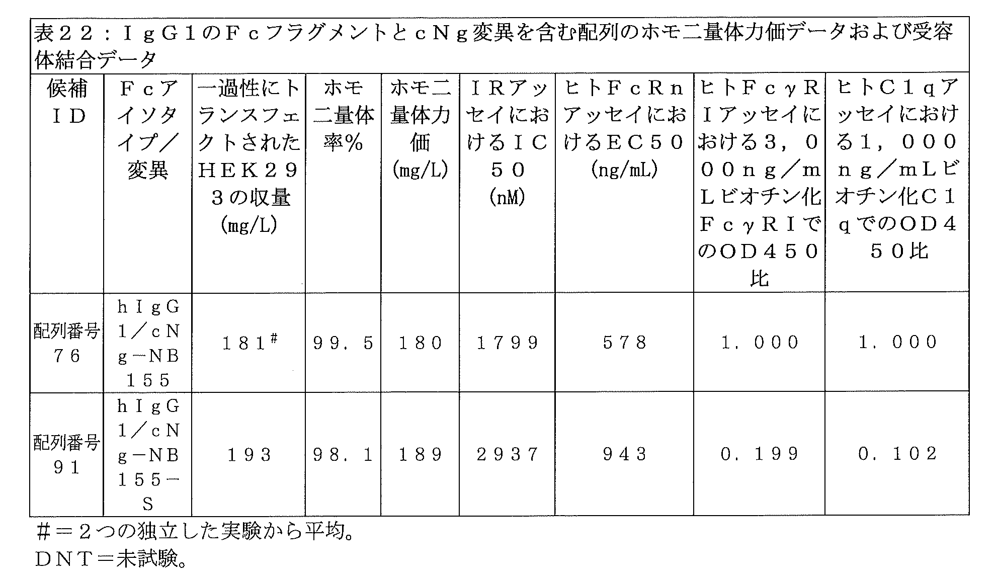
実施例51:変異型インスリンポリペプチド(配列番号10)、リンカー(配列番号13)、およびcNg-S変異を含むIgG1アイソタイプFcを用いたヒトインスリン-Fc融合タンパク質
上記のように、IgG1アイソタイプFcフラグメントにおけるcNg-S変異では、FcγRIおよびC1qに対する親和性が顕著に低減した、インスリン-Fc融合タンパク質の構成が得られたが、このインスリン-Fc融合タンパク質の構成は、許容できないほどに低いIR受容体親和性を有している(すなわち、IC50値が2400nMより大きい)。そこで、インスリンポリペプチドに変異を加え、その変異がIR親和性を改善させるかどうかを確かめる目的で、得られた物質をIRアッセイにおけるIC50に対するスクリーニングにかけた。B鎖のN末端から16番目のアミノ酸をチロシンからアラニンにかえた(すなわち、B16A)ところ、配列番号10のインスリンポリペプチドが得られた。配列番号10の変異型インスリンポリペプチドおよび配列番号13のペプチドリンカーを、X1がSである配列番号77のcNg変異型Fcフラグメントと共に用いたところ、配列番号78のインスリン-Fc融合タンパク質の構成が得られた。インスリン-Fc融合タンパク質の構成におけるcNg-S変異を、Fcフラグメント領域に対して行うことで、実施例15に記載のインビトロヒトFcγRIアッセイにおける結合性によって評価される、インビボにおける当該FcフラグメントのFcγ受容体に対する結合親和性が低減された。配列番号78のインスリン-Fc融合タンパク質におけるcNg-S部位の位置は、cNg部位における「N」から「S」への変異を含むcNg-NB155である。
Example 51: Human insulin-Fc fusion protein using a mutant insulin polypeptide (SEQ ID NO: 10), a linker (SEQ ID NO: 13), and an IgG1 isotype Fc containing the cNg-S mutation. IgG1 isotype Fc fragment as described above. The cNg-S mutation in , resulted in an insulin-Fc fusion protein construct with markedly reduced affinity for FcγRI and C1q, but this insulin-Fc fusion protein construct had an unacceptably low affinity for IR receptors. affinity (ie, IC50 value greater than 2400 nM). Therefore, a mutation was added to the insulin polypeptide, and the resulting substance was subjected to screening for IC50 in an IR assay in order to confirm whether the mutation improved IR affinity. When the 16th amino acid from the N-terminus of the B chain was changed from tyrosine to alanine (ie, B16A), the insulin polypeptide of SEQ ID NO: 10 was obtained. When the mutant insulin polypeptide of SEQ ID NO: 10 and the peptide linker of SEQ ID NO: 13 were used with the cNg mutant Fc fragment of SEQ ID NO: 77 in which X 1 is S, the composition of the insulin-Fc fusion protein of SEQ ID NO: 78 was Obtained. By making the cNg-S mutation in the construction of an insulin-Fc fusion protein to the Fc fragment region, the Fcγ receptor of the Fc fragment in vivo is improved as assessed by binding in the in vitro human FcγRI assay described in Example 15. The binding affinity for the body was reduced. The position of the cNg-S site in the insulin-Fc fusion protein of SEQ ID NO: 78 is cNg-NB155, which contains an "N" to "S" mutation in the cNg site.
配列番号78のインスリン-Fc融合タンパク質の構成を、実施例1に従ってHEK293細胞で製造し、実施例4に従ってプロテインAカラムを用いて精製した。配列番号78のインスリン-Fc融合タンパク質の構造は実施例6に従って非還元CE-SDSおよび還元CE-SDSで確認し、さらに、実施例8に記載のグリカン除去を行わないLC-MSによって配列の確認を行った。配列番号78の化合物の分子量を、酵素による糖鎖除去を行わない実施例8のLC-MS法で測定した。配列番号78のターゲット質量は65046.9Daであり、実測質量は65046.7Daであり、配列番号78の化合物のアミノ酸組成およびホモ二量体構造が正しいことが確認された。 The insulin-Fc fusion protein construct of SEQ ID NO: 78 was produced in HEK293 cells according to Example 1 and purified using a Protein A column according to Example 4. The structure of the insulin-Fc fusion protein of SEQ ID NO: 78 was confirmed by non-reduced CE-SDS and reduced CE-SDS according to Example 6, and the sequence was further confirmed by LC-MS without glycan removal as described in Example 8. I did it. The molecular weight of the compound of SEQ ID NO: 78 was measured by the LC-MS method of Example 8 without enzymatic sugar chain removal. The target mass of SEQ ID NO: 78 was 65046.9 Da, and the measured mass was 65046.7 Da, confirming that the amino acid composition and homodimer structure of the compound SEQ ID NO: 78 were correct.
ホモ二量体率は実施例10に従って分子ふるいクロマトグラフィーで測定した。cNg変異型インスリン-Fc融合タンパク質の完全アミノ酸配列を以下に挙げる(NB155位には下線):
FVNQHLCGSHLVEALALVCGERGFHYGGGGGGSGGGGGIVEQCCTSTCSLDQLENYCGGGGGQGGGGQGGGGQGGGGGDKTHTCPPCPAPELLGGPSVFLFPPKPKDTLMISRTPEVTCVVVDVSHEDPEVKFNWYVDGVEVHNAKTKPREEQYSSTYRVVSVLTVLHQDWLNGKEYKCKVSNKALPAPIEKTISKAKGQPREPQVYTLPPSRDELTKNQVSLTCLVKGFYPSDIAVEWESNGQPENNYKTTPPVLDSDGSFFLYSKLTVDKSRWQQGNVFSCSVMHEALHNHYTQKSLSLSPG(配列番号78)。
The homodimer rate was determined by molecular sieve chromatography according to Example 10. The complete amino acid sequence of the cNg mutant insulin-Fc fusion protein is listed below (NB position 155 is underlined):
FVNQHLCGSHLVEALALVCGERGFHYGGGGGGSGGGGGIVEQCCTSTCSLDQLENYCGGGGGQGGGGQGGGGQGGGGGDKTHTCPPCPAPELLGGPSVFLFPPKPKDTLMISRTPEVTCVVVD VSHEDPEVKFNWYVDGVEVHNAKTKPREEQY S STYRVVSVLTVLHQDWLNGKEYKCKVSNKALPAPIEKTISKAKGQPREPQVYTLPPSRDELTKNQVSLTCLVKGFYPSDIAVEWESNGQPE NNYKTTPPVLDSDGSFFLYSKLTVDKSRWQQGNVFSCSVMHEALHNHYTQKSLSLSPG (SEQ ID NO: 78).
配列番号78のインスリン-Fc融合タンパク質の構成の総タンパク質収量、ホモ二量体率、およびホモ二量体力価の結果が表23に示され、配列番号76および配列番号91の結果と比較されている。インスリンポリペプチド配列のB16A変異を非グリコシル化cNg-S変異型インスリン-Fc融合タンパク質に含めたところ、配列番号76と比較してホモ二量体力価が向上した。配列番号78のインスリン-Fc融合タンパク質のインビトロIR結合を、実施例12の手順に従って試験した。配列番号10のインスリンポリペプチドへのB16A変異を配列番号78のインスリン-Fc融合タンパク質に含めたところ、インスリンポリペプチド(配列番号7)がB16E変異を含む配列番号91と比較して、IR親和性が著しく向上した(すなわち、IR IC50測定値が低くなった)。このB16A変異を別のインスリン-Fc融合タンパク質で行った際(米国特許第10,597,435号の配列番号3)、IR IC50測定値が5000nMよりも大きくなり、インビボにおいてグルコースを低下させる生物活性を示さなかったことを考えると、これは特に驚くべきことである。アミノ酸配列の相違を強調した、配列番号76、配列番号78、および配列番号91の配列アラインメントを、図27に示す。「*」は、所与の配列位置において全ての配列間で完全に相同であることを示しており、一方、「:」、「.」、空白は、それぞれ、所与の配列位置における配列間のアミノ酸変異が保存的であること、中程度であること、または全く異なるものであること、を示している。

さらに、配列番号78のインスリン-Fc融合タンパク質のインスリンポリペプチドにB16A変異を含めた(配列番号10が得られる)ところ、実施例19の手順で測定される、FcRn結合が許容できるものとなり、このインスリン-Fc融合タンパク質の、インビボにおける薬物動態学的半減期が延長され、インビボにおけるグルコース低減生物活性プロファイルが延長される可能性があることが示された。なお、配列番号78のFcRn結合親和性は、B16A変異を含まない類似のインスリン-Fc融合タンパク質(配列番号91)のそれよりも大きく(FcRn結合アッセイにおいてEC50測定値がより小さいことによって示される)、B16A変異を含めることが、IR受容体親和性に加えて、予想外にもFcRn受容体親和性に影響を与えることがさらに明らかとなった。FcRn親和性が高くなるほど、それに相関して、治療薬のインビボにおける薬物動態学的半減期が長くなる場合が多いことから、このようにFcRn受容体親和性を徐々に増加させることは、望ましいことであろうと考えられる。 Furthermore, inclusion of the B16A mutation in the insulin polypeptide of the insulin-Fc fusion protein of SEQ ID NO: 78 (resulting in SEQ ID NO: 10) resulted in acceptable FcRn binding as measured by the procedure of Example 19; It has been shown that insulin-Fc fusion proteins may have an extended in vivo pharmacokinetic half-life and an extended in vivo glucose-lowering bioactivity profile. Note that the FcRn binding affinity of SEQ ID NO: 78 is greater than that of a similar insulin-Fc fusion protein (SEQ ID NO: 91) that does not contain the B16A mutation (as indicated by a lower EC50 measurement in the FcRn binding assay). , it was further revealed that inclusion of the B16A mutation unexpectedly affected FcRn receptor affinity in addition to IR receptor affinity. This gradual increase in FcRn receptor affinity is desirable because higher FcRn affinity often correlates with a longer in vivo pharmacokinetic half-life of a therapeutic agent. It is thought that it is.
配列番号78のインスリン-Fc融合タンパク質の、FcγRI結合特性およびC1q結合特性を、実施例14の方法および実施例16の方法で測定したところ、許容できるものであることが示された。また、配列番号78のインスリン-Fc融合タンパク質のC1q結合親和性ははるかに低く、配列番号76のインスリン-Fc融合タンパク質に対して80%超のC1q結合の低減を示した。以上から、配列番号78のインスリン-Fc融合タンパク質は、製造性、インビボ効力、インビボ生物活性の延長、および免疫原性の可能性の低さについての各設計目標(実施例29)を達成した。 The FcγRI and C1q binding properties of the insulin-Fc fusion protein of SEQ ID NO: 78 were determined by the methods of Example 14 and Example 16 and were shown to be acceptable. Also, the C1q binding affinity of the insulin-Fc fusion protein of SEQ ID NO: 78 was much lower, showing a reduction in C1q binding of over 80% relative to the insulin-Fc fusion protein of SEQ ID NO: 76. In summary, the insulin-Fc fusion protein of SEQ ID NO: 78 achieved each of the design goals (Example 29) for manufacturability, in vivo efficacy, extended in vivo biological activity, and low potential for immunogenicity.
実施例52:ヒトIgG1アイソタイプを含むインスリン-Fc融合タンパク質のFcフラグメント領域の他のcNg変異
インビボ生物活性が延長され、免疫原性を示す可能性が低く、配列番号76のインスリン-Fc融合タンパク質と同様のホモ二量体含有率とIR結合親和性を維持し、FcRn受容体に対する結合親和性が許容できるものである、インスリン-Fc融合タンパク質の構成を作製するさらなる試みにおいて、FcγRI結合およびC1q結合を低減しようと、他のcNg変異をFcフラグメントのCH3領域に挿入し、配列番号92、配列番号93、配列番号94、および配列番号95を得た。インスリン-Fc融合タンパク質の実施形態を、実施例1に従ってHEK細胞で合成し、実施例4に従って精製し、実施例6、実施例8、実施例10、実施例12、実施例14、および実施例16に従って試験した。結果は表24に示す。配列番号75、配列番号76、および配列番号91に対しての、配列番号92、配列番号93、配列番号94、および配列番号95の配列アラインメントを、アミノ酸配列の相違を強調表示して、図28に示す(クラスタルオメガ)。「*」は、所与の配列位置において全ての配列間で完全に相同であることを示しており、一方、「:」、「.」、空白は、それぞれ、所与の配列位置における配列間のアミノ酸変異が保存的であること、中程度であること、または全く異なるものであること、を示している。
Example 52: Other cNg mutations in the Fc fragment region of an insulin-Fc fusion protein containing the human IgG1 isotype have prolonged in vivo biological activity, are less likely to be immunogenic, and are similar to the insulin-Fc fusion protein of SEQ ID NO: 76. In a further attempt to create an insulin-Fc fusion protein construct that maintains similar homodimer content and IR binding affinity and has acceptable binding affinity for FcRn receptors, FcγRI binding and C1q binding Other cNg mutations were inserted in the CH3 region of the Fc fragment in an attempt to reduce the number of cNg mutations resulting in SEQ ID NO: 92, SEQ ID NO: 93, SEQ ID NO: 94, and SEQ ID NO: 95. Embodiments of insulin-Fc fusion proteins were synthesized in HEK cells according to Example 1, purified according to Example 4, Example 6, Example 8, Example 10, Example 12, Example 14, and Example Tested according to 16. The results are shown in Table 24. Sequence alignment of SEQ ID NO: 92, SEQ ID NO: 93, SEQ ID NO: 94, and SEQ ID NO: 95 against SEQ ID NO: 75, SEQ ID NO: 76, and SEQ ID NO: 91, with amino acid sequence differences highlighted, is shown in FIG. (Cluster Omega). “*” indicates complete homology between all sequences at a given sequence position, while “:”, “.”, and blank space respectively indicate complete homology between all sequences at a given sequence position. indicates that the amino acid variations in the amino acids are conservative, moderate, or completely different.
インスリン-Fc融合タンパク質の実施形態の完全アミノ酸配列を以下に示し、cNg変異には下線を引く。
FVNQHLCGSHLVEALELVCGERGFHYGGGGGGSGGGGGIVEQCCTSTCSLDQLENYCGGGGGQGGGGQGGGGQGGGGGDKTHTCPPCPAPELLGGPSVFLFPPKPKDTLMISRTPEVTCVVVDVSHEDPEVKFNWYVDGVEVHNAKTKPREEQYDSTYRVVSVLTVLHQDWLNGKEYKCKVSNKALPAPIEKTISKAKGQPREPQVYTLPPSRDELTKNQVSLTCLVKGFYPSDIAVEWESNGQPENNYKTTPPVLDSDGSFFLYSKLTVDKSRWQQGNVFSCSVMHEALHNHYTQKSLSLSPG(配列番号92)
FVNQHLCGSHLVEALELVCGERGFHYGGGGGGSGGGGGIVEQCCTSTCSLDQLENYCGGGGGQGGGGQGGGGQGGGGGDKTHTCPPCPAPELLGGPSVFLFPPKPKDTLMISRTPEVTCVVVDVSHEDPEVKFNWYVDGVEVHNAKTKPREEQYASTYRVVSVLTVLHQDWLNGKEYKCKVSNKALPAPIEKTISKAKGQPREPQVYTLPPSRDELTKNQVSLTCLVKGFYPSDIAVEWESNGQPENNYKTTPPVLDSDGSFFLYSKLTVDKSRWQQGNVFSCSVMHEALHNHYTQKSLSLSPG(配列番号93)
FVNQHLCGSHLVEALELVCGERGFHYGGGGGGSGGGGGIVEQCCTSTCSLDQLENYCGGGGGQGGGGQGGGGQGGGGGDKTHTCPPCPAPELLGGPSVFLFPPKPKDTLMISRTPEVTCVVVDVSHEDPEVKFNWYVDGVEVHNAKTKPREEQYRSTYRVVSVLTVLHQDWLNGKEYKCKVSNKALPAPIEKTISKAKGQPREPQVYTLPPSRDELTKNQVSLTCLVKGFYPSDIAVEWESNGQPENNYKTTPPVLDSDGSFFLYSKLTVDKSRWQQGNVFSCSVMHEALHNHYTQKSLSLSPG(配列番号94)
FVNQHLCGSHLVEALELVCGERGFHYGGGGGGSGGGGGIVEQCCTSTCSLDQLENYCGGGGGQGGGGQGGGGQGGGGGDKTHTCPPCPAPELLGGPSVFLFPPKPKDTLMISRTPEVTCVVVDVSHEDPEVKFNWYVDGVEVHNAKTKPREEQYQSTYRVVSVLTVLHQDWLNGKEYKCKVSNKALPAPIEKTISKAKGQPREPQVYTLPPSRDELTKNQVSLTCLVKGFYPSDIAVEWESNGQPENNYKTTPPVLDSDGSFFLYSKLTVDKSRWQQGNVFSCSVMHEALHNHYTQKSLSLSPG(配列番号95)。
The complete amino acid sequence of an insulin-Fc fusion protein embodiment is shown below, with cNg mutations underlined.
FVNQHLCGSHLVEALELVCGERGFHYGGGGGGSGGGGGIVEQCCTSTCSLDQLENYCGGGGGQGGGGQGGGGQGGGGGDKTHTCPPCPAPELLGGPSVFLFPPKPKDTLMISRTPEVTCVVVD VSHEDPEVKFNWYVDGVEVHNAKTKPREEQY D STYRVVSVLTVLHQDWLNGKEYKCKVSNKALPAPIEKTISKAKGQPREPQVYTLPPSRDELTKNQVSLTCLVKGFYPSDIAVEWESNGQPE NNYKTTPPVLDSDGSFFLYSKLTVDKSRWQQGNVFSCSVMHEALHNHYTQKSLSLSPG (SEQ ID NO: 92)
FVNQHLCGSHLVEALELVCGERGFHYGGGGGGSGGGGGIVEQCCTSTCSLDQLENYCGGGGGQGGGGQGGGGQGGGGGDKTHTCPPCPAPELLGGPSVFLFPPKPKDTLMISRTPEVTCVVVD VSHEDPEVKFNWYVDGVEVHNAKTKPREEQY A STYRVVSVLTVLHQDWLNGKEYKCKVSNKALPAPIEKTISKAKGQPREPQVYTLPPSRDELTKNQVSLTCLVKGFYPSDIAVEWESNGQPE NNYKTTPPVLDSDGSFFLYSKLTVDKSRWQQGNVFSCSVMHEALHNHYTQKSLSLSPG (SEQ ID NO: 93)
FVNQHLCGSHLVEALELVCGERGFHYGGGGGGSGGGGGIVEQCCTSTCSLDQLENYCGGGGGQGGGGQGGGGQGGGGGDKTHTCPPCPAPELLGGPSVFLFPPKPKDTLMISRTPEVTCVVVD VSHEDPEVKFNWYVDGVEVHNAKTKPREEQY R STYRVVSVLTVLHQDWLNGKEYKCKVSNKALPAPIEKTISKAKGQPREPQVYTLPPSRDELTKNQVSLTCLVKGFYPSDIAVEWESNGQPE NNYKTTPPVLDSDGSFFLYSKLTVDKSRWQQGNVFSCSVMHEALHNHYTQKSLSLSPG (SEQ ID NO: 94)
FVNQHLCGSHLVEALELVCGERGFHYGGGGGGSGGGGGIVEQCCTSTCSLDQLENYCGGGGGQGGGGQGGGGQGGGGGDKTHTCPPCPAPELLGGPSVFLFPPKPKDTLMISRTPEVTCVVVD VSHEDPEVKFNWYVDGVEVHNAKTKPREEQY Q STYRVVSVLTVLHQDWLNGKEYKCKVSNKALPAPIEKTISKAKGQPREPQVYTLPPSRDELTKNQVSLTCLVKGFYPSDIAVEWESNGQPE NNYKTTPPVLDSDGSFFLYSKLTVDKSRWQQGNVFSCSVMHEALHNHYTQKSLSLSPG (SEQ ID NO: 95).
X1がD、A、R、Qである、配列番号77のヒトIgG1 Fcフラグメントを含む、上記のインスリン-Fc融合タンパク質の実施形態を、配列番号76と共に、表24に挙げ、表24は対応する総タンパク質収量、ホモ二量体率、およびホモ二量体力価を示している。これらの結果は、cNg部位への各種変異をヒトIgG1 Fcフラグメント内に含めて、配列番号92、配列番号93、配列番号94、および配列番号95を得た結果、望ましいFcγRI親和性およびC1q親和性の低減がもたらされたことを示している。配列番号92、配列番号93、および配列番号94のホモ二量体力価は、配列番号76に比べて顕著に増加した。配列番号95のホモ二量体力価が、設計目標(実施例29)を達成するには低すぎたため、インスリン受容体結合は測定しなかった。ホモ二量体力価は向上したが、配列番号92、配列番号93、および配列番号94は、IRアッセイにおけるIC50値が、全ての場合で2400nMよりも大きく、配列番号76のそれよりもかなり高かった(すなわち、IR親和性がより低い)。全ての場合で、IRアッセイにおけるIC50値は設計基準である2400nM未満よりも大きく、これらのインスリン-Fc融合タンパク質の実施形態が、許容できる効力と商品原価を示しそうにないことが示された。配列番号91、配列番号92、配列番号93、配列番号94、および配列番号95のいずれも、設計目標(実施例29)を達成していない。

実施例53:インスリンポリペプチド(配列番号10)、リンカー(配列番号13)、および各種cNg変異を含むIgG1アイソタイプFcを用いたヒトインスリン-Fc融合タンパク質
上記のように、IgG1アイソタイプFcフラグメントのcNg部位における変異では、FcγRIおよびC1qに対する親和性が顕著に低減した、インスリン-Fc融合タンパク質が得られたが、これらの化合物は、許容できないほどに低いIR受容体親和性を有している(すなわち、IC50値が2400nMより大きい)。そこで、配列番号78中のインスリンポリペプチドに同じ変異をなして(B鎖のN末端から16番目のアミノ酸をチロシンからアラニンにかえ(すなわち、B16A)ところ、配列番号10のインスリンポリペプチドが得られた)、IR親和性を向上させた変異型インスリンポリペプチドがあるかどうかを確かめるために、得られたインスリン-Fc融合タンパク質の構成を、IRアッセイにおけるIC50に対するスクリーニングにかけた。配列番号10の変異型インスリンポリペプチドおよび配列番号13のペプチドリンカーを用いて、以下に示すような、配列番号80、配列番号82、配列番号84、および配列番号86のインスリン-Fc融合タンパク質の構成を作製した。これらのインスリン-Fc融合タンパク質の構成はそれぞれ、実施例15に記載のインビトロヒトFcγRIアッセイにおける結合性によって評価される、インビボにおけるFcフラグメントのFcγ受容体に対する結合親和性を低減されせるためにFcフラグメント領域に対して行われた、cNg部位変異を含む。配列番号80、配列番号82、配列番号84、および配列番号86のインスリン-Fc融合タンパク質の構成におけるcNg部位の位置はcNg-NB155である。配列番号80のインスリン-Fc融合タンパク質は、cNg部位に「N」から「D」への変異を含む。配列番号82のインスリン-Fc融合タンパク質は、cNg部位に「N」から「A」への変異を含む。配列番号84のインスリン-Fc融合タンパク質は、cNg部位に「N」から「R」への変異を含む。配列番号86のインスリン-Fc融合タンパク質は、cNg部位に「N」から「Q」への変異を含む。
Example 53: Human Insulin-Fc Fusion Protein Using an Insulin Polypeptide (SEQ ID NO: 10), a Linker (SEQ ID NO: 13), and an IgG1 Isotype Fc Containing Various cNg Mutations cNg portions of IgG1 isotype Fc fragments as described above. Mutations in FcγRI and C1q resulted in insulin-Fc fusion proteins with significantly reduced affinity for FcγRI and C1q, but these compounds have unacceptably low IR receptor affinities (i.e., IC50 value greater than 2400 nM). Therefore, by making the same mutation in the insulin polypeptide in SEQ ID NO: 78 (changing the 16th amino acid from the N-terminus of the B chain from tyrosine to alanine (i.e., B16A)), the insulin polypeptide in SEQ ID NO: 10 was obtained. To ascertain whether there are mutant insulin polypeptides with improved IR affinity, the resulting insulin-Fc fusion protein constructs were screened for IC50 in an IR assay. Construction of insulin-Fc fusion proteins of SEQ ID NO: 80, SEQ ID NO: 82, SEQ ID NO: 84, and SEQ ID NO: 86 as shown below using the mutant insulin polypeptide of SEQ ID NO: 10 and the peptide linker of SEQ ID NO: 13. was created. Each of these insulin-Fc fusion protein configurations is designed to reduce the binding affinity of the Fc fragment to the Fcγ receptor in vivo, as assessed by binding in the in vitro human FcγRI assay described in Example 15. Contains cNg site mutations made to the region. The position of the cNg site in the constructs of the insulin-Fc fusion proteins of SEQ ID NO: 80, SEQ ID NO: 82, SEQ ID NO: 84, and SEQ ID NO: 86 is cNg-NB155. The insulin-Fc fusion protein of SEQ ID NO: 80 contains an "N" to "D" mutation at the cNg site. The insulin-Fc fusion protein of SEQ ID NO: 82 contains an "N" to "A" mutation at the cNg site. The insulin-Fc fusion protein of SEQ ID NO: 84 contains an "N" to "R" mutation at the cNg site. The insulin-Fc fusion protein of SEQ ID NO: 86 contains an "N" to "Q" mutation at the cNg site.
タンパク質製造は実施例1に従ってHEK293細胞で行った。驚くべきことに、cNg-NB155-Q変異とB16A変異とを一緒に含む配列番号86のインスリン-Fc融合タンパク質の構成のホモ二量体力価は、ホモ二量体力価の設計目標を達成するのに必要なものよりも著しく低かった。次に、配列番号80、配列番号82、および配列番号84のインスリン-Fc融合タンパク質の構成を、実施例4に従ってプロテインAカラムを用いて精製した。インスリン-Fc融合タンパク質の構成の構造は実施例6に従って非還元CE-SDSおよび還元CE-SDSで確認し、さらに、実施例8に記載のグリカン除去を行わないLC-MSによって配列の確認を行った。配列番号80のインスリン-Fc融合タンパク質の化合物分子量を、酵素による糖鎖除去を行わない実施例8のLC-MS法で測定した。配列番号80のターゲット質量は65102.9Daであり、実測質量は65102.8Daであり、配列番号80の化合物のアミノ酸組成およびホモ二量体構造が正しいことが確認された。配列番号82のインスリン-Fc融合タンパク質の化合物分子量を、酵素による糖鎖除去を行わない実施例8のLC-MS法で測定した。配列番号82のターゲット質量は65014.9Daであり、実測質量は65014.9Daであり、配列番号82の化合物のアミノ酸組成およびホモ二量体構造が正しいことが確認された。配列番号84のインスリン-Fc融合タンパク質の化合物分子量を、酵素による糖鎖除去を行わない実施例8のLC-MS法で測定した。配列番号84のターゲット質量は65185.1Daであり、実測質量は65184.8Daであり、配列番号84の化合物のアミノ酸組成およびホモ二量体構造が正しいことが確認された。 Protein production was performed in HEK293 cells according to Example 1. Surprisingly, the homodimer potency of the insulin-Fc fusion protein construct of SEQ ID NO: 86 containing the cNg-NB155-Q mutation and the B16A mutation together is sufficient to achieve the design goal of homodimer potency. was significantly lower than what was needed. The insulin-Fc fusion protein constructs of SEQ ID NO: 80, SEQ ID NO: 82, and SEQ ID NO: 84 were then purified using a Protein A column according to Example 4. The structure of the insulin-Fc fusion protein construct was confirmed by non-reduced CE-SDS and reduced CE-SDS according to Example 6, and the sequence was further confirmed by LC-MS without glycan removal as described in Example 8. Ta. The compound molecular weight of the insulin-Fc fusion protein of SEQ ID NO: 80 was measured by the LC-MS method of Example 8 without enzymatic removal of sugar chains. The target mass of SEQ ID NO: 80 was 65102.9 Da, and the measured mass was 65102.8 Da, confirming that the amino acid composition and homodimer structure of the compound SEQ ID NO: 80 were correct. The compound molecular weight of the insulin-Fc fusion protein of SEQ ID NO: 82 was measured by the LC-MS method of Example 8 without enzymatic sugar chain removal. The target mass of SEQ ID NO: 82 was 65014.9 Da, and the measured mass was 65014.9 Da, confirming that the amino acid composition and homodimer structure of the compound SEQ ID NO: 82 were correct. The compound molecular weight of the insulin-Fc fusion protein of SEQ ID NO: 84 was measured by the LC-MS method of Example 8 without enzymatic removal of sugar chains. The target mass of SEQ ID NO: 84 was 65185.1 Da, and the measured mass was 65184.8 Da, confirming that the amino acid composition and homodimer structure of the compound SEQ ID NO: 84 were correct.
ホモ二量体率は実施例10に従って分子ふるいクロマトグラフィーで測定した。cNg変異型インスリン-Fc融合タンパク質の構成の完全アミノ酸配列を以下に挙げ(配列番号80、配列番号82、配列番号84、および配列番号86;NB155位には下線)、配列番号76および配列番号78に対して得られた配列アラインメントを図29に示す(クラスタルオメガ)。「*」は、所与の配列位置において全ての配列間で完全に相同であることを示しており、一方、「:」、「.」、空白は、それぞれ、所与の配列位置における配列間のアミノ酸変異が保存的であること、中程度であること、または全く異なるものであること、を示している。
FVNQHLCGSHLVEALALVCGERGFHYGGGGGGSGGGGGIVEQCCTSTCSLDQLENYCGGGGGQGGGGQGGGGQGGGGGDKTHTCPPCPAPELLGGPSVFLFPPKPKDTLMISRTPEVTCVVVDVSHEDPEVKFNWYVDGVEVHNAKTKPREEQYDSTYRVVSVLTVLHQDWLNGKEYKCKVSNKALPAPIEKTISKAKGQPREPQVYTLPPSRDELTKNQVSLTCLVKGFYPSDIAVEWESNGQPENNYKTTPPVLDSDGSFFLYSKLTVDKSRWQQGNVFSCSVMHEALHNHYTQKSLSLSPG(配列番号80)
FVNQHLCGSHLVEALALVCGERGFHYGGGGGGSGGGGGIVEQCCTSTCSLDQLENYCGGGGGQGGGGQGGGGQGGGGGDKTHTCPPCPAPELLGGPSVFLFPPKPKDTLMISRTPEVTCVVVDVSHEDPEVKFNWYVDGVEVHNAKTKPREEQYASTYRVVSVLTVLHQDWLNGKEYKCKVSNKALPAPIEKTISKAKGQPREPQVYTLPPSRDELTKNQVSLTCLVKGFYPSDIAVEWESNGQPENNYKTTPPVLDSDGSFFLYSKLTVDKSRWQQGNVFSCSVMHEALHNHYTQKSLSLSPG(配列番号82)
FVNQHLCGSHLVEALALVCGERGFHYGGGGGGSGGGGGIVEQCCTSTCSLDQLENYCGGGGGQGGGGQGGGGQGGGGGDKTHTCPPCPAPELLGGPSVFLFPPKPKDTLMISRTPEVTCVVVDVSHEDPEVKFNWYVDGVEVHNAKTKPREEQYRSTYRVVSVLTVLHQDWLNGKEYKCKVSNKALPAPIEKTISKAKGQPREPQVYTLPPSRDELTKNQVSLTCLVKGFYPSDIAVEWESNGQPENNYKTTPPVLDSDGSFFLYSKLTVDKSRWQQGNVFSCSVMHEALHNHYTQKSLSLSPG(配列番号84)
FVNQHLCGSHLVEALALVCGERGFHYGGGGGGSGGGGGIVEQCCTSTCSLDQLENYCGGGGGQGGGGQGGGGQGGGGGDKTHTCPPCPAPELLGGPSVFLFPPKPKDTLMISRTPEVTCVVVDVSHEDPEVKFNWYVDGVEVHNAKTKPREEQYQSTYRVVSVLTVLHQDWLNGKEYKCKVSNKALPAPIEKTISKAKGQPREPQVYTLPPSRDELTKNQVSLTCLVKGFYPSDIAVEWESNGQPENNYKTTPPVLDSDGSFFLYSKLTVDKSRWQQGNVFSCSVMHEALHNHYTQKSLSLSPG(配列番号86)。
The homodimer rate was determined by molecular sieve chromatography according to Example 10. The complete amino acid sequences of the cNg mutant insulin-Fc fusion protein components are listed below (SEQ ID NO: 80, SEQ ID NO: 82, SEQ ID NO: 84, and SEQ ID NO: 86; NB position 155 is underlined), SEQ ID NO: 76 and SEQ ID NO: 78. The sequence alignment obtained for Cluster Omega is shown in Figure 29. “*” indicates complete homology between all sequences at a given sequence position, while “:”, “.”, and blank space respectively indicate complete homology between all sequences at a given sequence position. indicates that the amino acid variations in the amino acids are conservative, moderate, or completely different.
FVNQHLCGSHLVEALALVCGERGFHYGGGGGGSGGGGGIVEQCCTSTCSLDQLENYCGGGGGQGGGGQGGGGQGGGGGDKTHTCPPCPAPELLGGPSVFLFPPKPKDTLMISRTPEVTCVVVD VSHEDPEVKFNWYVDGVEVHNAKTKPREEQY D STYRVVSVLTVLHQDWLNGKEYKCKVSNKALPAPIEKTISKAKGQPREPQVYTLPPSRDELTKNQVSLTCLVKGFYPSDIAVEWESNGQPE NNYKTTPPVLDSDGSFFLYSKLTVDKSRWQQGNVFSCSVMHEALHNHYTQKSLSLSPG (SEQ ID NO: 80)
FVNQHLCGSHLVEALALVCGERGFHYGGGGGGSGGGGGIVEQCCTSTCSLDQLENYCGGGGGQGGGGQGGGGQGGGGGDKTHTCPPCPAPELLGGPSVFLFPPKPKDTLMISRTPEVTCVVVD VSHEDPEVKFNWYVDGVEVHNAKTKPREEQY A STYRVVSVLTVLHQDWLNGKEYKCKVSNKALPAPIEKTISKAKGQPREPQVYTLPPSRDELTKNQVSLTCLVKGFYPSDIAVEWESNGQPE NNYKTTPPVLDSDGSFFLYSKLTVDKSRWQQGNVFSCSVMHEALHNHYTQKSLSLSPG (SEQ ID NO: 82)
FVNQHLCGSHLVEALALVCGERGFHYGGGGGGSGGGGGIVEQCCTSTCSLDQLENYCGGGGGQGGGGQGGGGQGGGGGDKTHTCPPCPAPELLGGPSVFLFPPKPKDTLMISRTPEVTCVVVD VSHEDPEVKFNWYVDGVEVHNAKTKPREEQY R STYRVVSVLTVLHQDWLNGKEYKCKVSNKALPAPIEKTISKAKGQPREPQVYTLPPSRDELTKNQVSLTCLVKGFYPSDIAVEWESNGQPE NNYKTTPPVLDSDGSFFLYSKLTVDKSRWQQGNVFSCSVMHEALHNHYTQKSLSLSPG (SEQ ID NO: 84)
FVNQHLCGSHLVEALALVCGERGFHYGGGGGGSGGGGGIVEQCCTSTCSLDQLENYCGGGGGQGGGGQGGGGQGGGGGDKTHTCPPCPAPELLGGPSVFLFPPKPKDTLMISRTPEVTCVVVD VSHEDPEVKFNWYVDGVEVHNAKTKPREEQY Q STYRVVSVLTVLHQDWLNGKEYKCKVSNKALPAPIEKTISKAKGQPREPQVYTLPPSRDELTKNQVSLTCLVKGFYPSDIAVEWESNGQPE NNYKTTPPVLDSDGSFFLYSKLTVDKSRWQQGNVFSCSVMHEALHNHYTQKSLSLSPG (SEQ ID NO: 86).
得られた総タンパク質収量、ホモ二量体率、およびホモ二量体力価を表25に示す。上記のような配列番号86を除いて、インスリンポリペプチド配列のB16A変異を非グリコシル化cNg変異型インスリン-Fc融合タンパク質の実施形態に含めたところ、配列番号76と比較してホモ二量体力価が向上した。cNg-NB155-D組成物は、222mg/Lにおいて最も高いホモ二量体力価を示した。インスリン-Fc融合タンパク質のインビトロIR結合を、実施例12の手順に従って試験した。配列番号78の場合のように、配列番号80、配列番号82、および配列番号84のインスリン-Fc融合タンパク質の実施形態のインスリンポリペプチドにB16A変異を含めたところ、インスリンポリペプチドがB16E変異を含む配列番号92、配列番号93、および配列番号94のインスリン-Fc融合タンパク質の構成と比べて、IR親和性が著しく向上した(すなわち、IRアッセイにおけるIC50値が低かった)。 The total protein yield, homodimer rate, and homodimer titer obtained are shown in Table 25. Inclusion of the B16A mutation of the insulin polypeptide sequence in an embodiment of a non-glycosylated cNg variant insulin-Fc fusion protein, with the exception of SEQ ID NO: 86 as described above, resulted in homodimer titers compared to SEQ ID NO: 76. has improved. The cNg-NB155-D composition showed the highest homodimer titer at 222 mg/L. In vitro IR binding of insulin-Fc fusion proteins was tested according to the procedure of Example 12. The inclusion of the B16A mutation in the insulin polypeptides of the insulin-Fc fusion protein embodiments of SEQ ID NO: 80, SEQ ID NO: 82, and SEQ ID NO: 84, as in the case of SEQ ID NO: 78, results in the insulin polypeptide containing the B16E mutation. Compared to the insulin-Fc fusion protein constructs of SEQ ID NO: 92, SEQ ID NO: 93, and SEQ ID NO: 94, the IR affinity was significantly improved (ie, the IC50 value in the IR assay was lower).
さらに、配列番号80、配列番号82、および配列番号84のインスリン-Fc融合タンパク質のインスリンポリペプチドにB16A変異を含めたところ、実施例19の手順で測定される、これらのインスリン-Fc融合タンパク質の実施形態のFcRn結合が許容できるものとなり、これらの化合物の、インビボにおける薬物動態学的半減期が延長され、インビボにおけるグルコース低減生物活性プロファイルが延長される可能性があることが示された。なお、B16A変異を含むインスリン-Fc融合タンパク質の構成(配列番号80、配列番号82、および配列番号84)のFcRn結合親和性は、B16A変異を含まない類似のインスリン-Fc融合タンパク質の構成(配列番号92、配列番号93、および配列番号94)のそれよりも顕著に大きく、B16A変異を含めることが、IR受容体親和性に加えて、予想外にもFcRn受容体親和性に影響を与えることがさらに明らかとなった。FcRn親和性が高くなるほど、それに相関して、治療薬のインビボにおける薬物動態学的半減期が長くなる場合が多いことから、これらのようにFcRn受容体親和性を徐々に増加させることは、望ましいことであろうと考えられる。 Furthermore, when the B16A mutation was included in the insulin polypeptides of the insulin-Fc fusion proteins of SEQ ID NO: 80, SEQ ID NO: 82, and SEQ ID NO: 84, the insulin-Fc fusion proteins of these insulin-Fc fusion proteins, as measured by the procedure of Example 19, It has been shown that the FcRn binding of embodiments is acceptable, and these compounds have the potential to have an extended in vivo pharmacokinetic half-life and an extended in vivo glucose-lowering bioactivity profile. Note that the FcRn binding affinity of the insulin-Fc fusion protein constructs containing the B16A mutation (SEQ ID NO: 80, SEQ ID NO: 82, and SEQ ID NO: 84) is different from that of the similar insulin-Fc fusion protein constructs that do not include the B16A mutation (SEQ ID NO: 80, SEQ ID NO: 82, and SEQ ID NO: 84). No. 92, SEQ ID No. 93, and SEQ ID No. 94), the inclusion of the B16A mutation unexpectedly affects FcRn receptor affinity in addition to IR receptor affinity. has become even clearer. These gradual increases in FcRn receptor affinity are desirable because higher FcRn affinity often correlates with longer in vivo pharmacokinetic half-lives of therapeutic agents. This is likely to be the case.
配列番号80、配列番号82、および配列番号84のインスリン-Fc融合タンパク質の構成の、FcγRI結合特性およびC1q結合特性を、実施例14の方法および実施例16の方法で測定したところ、許容できるものであることが示された。さらに、cNg-NB155-R(配列番号84)変異に伴ってFcγRI結合親和性が非常に顕著に低減し、配列番号76のインスリン-Fc融合タンパク質に対して80%超のFcγRI結合低減を示した。配列番号80、配列番号82、および配列番号84のインスリン-Fc融合タンパク質は、C1q結合親和性についてもかなり低く、これらのインスリン-Fc融合タンパク質の実施形態はいずれも、配列番号76のインスリン-Fc融合タンパク質に対して80%超のC1q結合低減を示した。以上から、配列番号80、配列番号82、および配列番号84のインスリン-Fc融合タンパク質は、製造性、インビボ効力、インビボ生物活性の延長、および免疫原性の可能性の低さについての各設計目標(実施例29)を達成した。
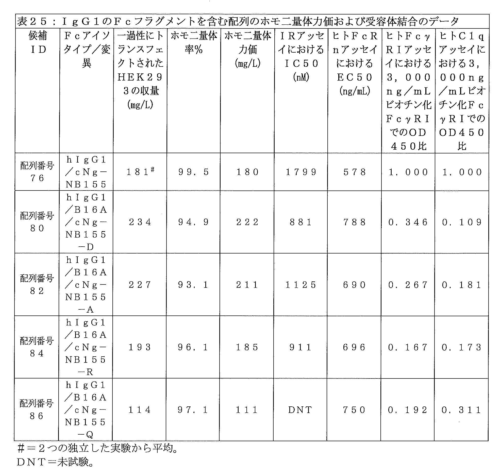
実施例54:配列番号10のインスリンポリペプチドと、cNg-S変異を含むIgG1アイソタイプFcと、各種リンカーと、を含むヒトインスリン-Fc融合タンパク質
上記のように、IgG1アイソタイプFcフラグメントのcNg部位における変異と、インスリンポリペプチド上の変異は、IR、FcRn、FcγRI(受容体)結合、およびC1q結合といった物性に予想外の変化をもたらした。リンカー組成物がこれらの特性にどのような影響を与えたかをさらに深く理解するため、配列番号10のB16A変異型インスリンポリペプチドと、cNg-S変異を有する非グリコシル化hIgG1 Fcフラグメント(配列番号77;X1はS)と、配列番号13のリンカー配列と、を含む特定のインスリン-Fc融合タンパク質の構成(配列番号78)を出発点として選び、そこから、リンカーの長さと組成を以下の表26に示すように変更した。
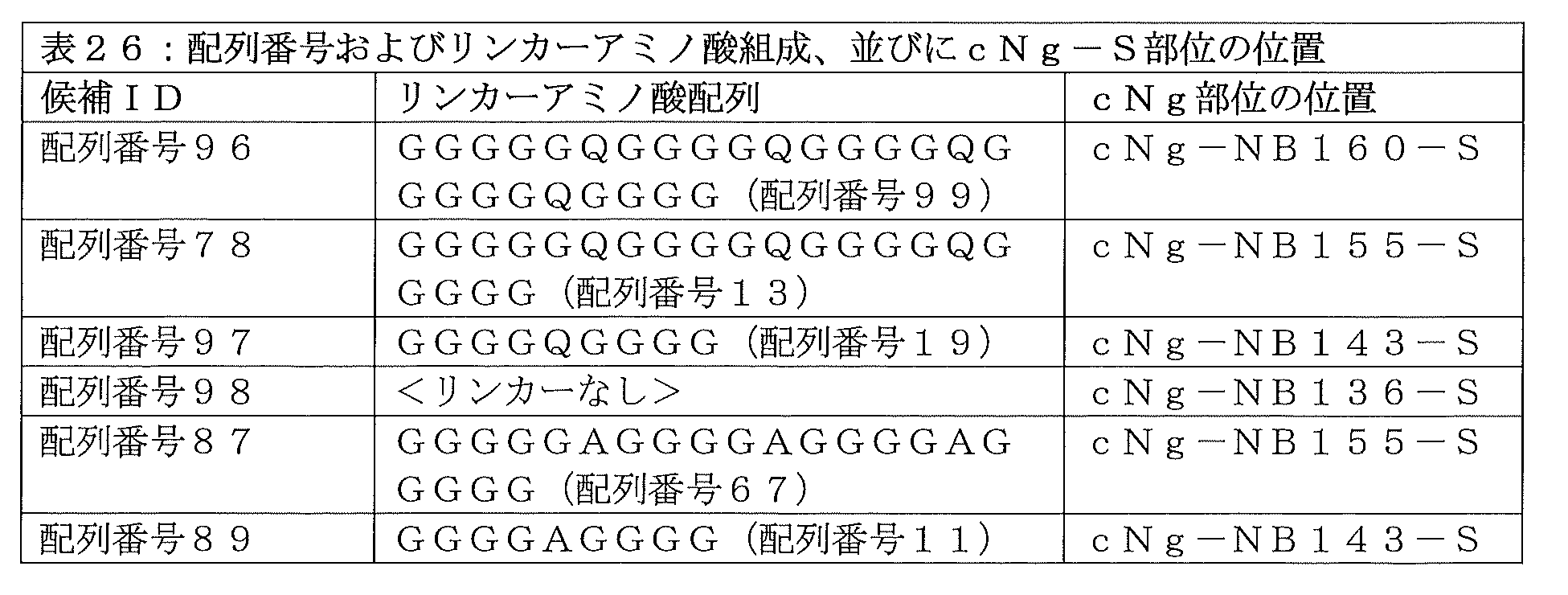
配列番号96、配列番号97、配列番号98、配列番号87、および配列番号89のインスリン-Fc融合タンパク質の実施形態を、実施例1に従ってHEK293細胞で製造した。次いで、インスリン-Fc融合タンパク質の構成を、実施例4に従ってプロテインAカラムを用いて精製した。インスリン-Fc融合タンパク質の構成の構造は実施例6に従って非還元CE-SDSおよび還元CE-SDSで確認し、さらに、実施例8に記載のグリカン除去を行わないLC-MSによって配列の確認を行った。グリカンを持たないことの確認は、実施例8のLC-MS法を用いて行い、ただし、PNGアーゼF処理工程は省略した。配列番号87のインスリン-Fc融合タンパク質の構成の化合物分子量を、酵素による糖鎖除去を行わない実施例8のLC-MS法で測定した。ターゲット質量は64704.6Daであり、実測質量は64704.4Daであり、配列番号87の化合物のアミノ酸組成およびホモ二量体構造が正しいことが確認された。配列番号89のインスリン-Fc融合タンパク質の構成の化合物分子量を、酵素による糖鎖除去を行わない実施例8のLC-MS法で測定した。ターゲット質量は63279.2Daであり、実測質量は63278.8Daであり、配列番号89の化合物のアミノ酸組成およびホモ二量体構造が正しいことが確認された。 Insulin-Fc fusion protein embodiments of SEQ ID NO: 96, SEQ ID NO: 97, SEQ ID NO: 98, SEQ ID NO: 87, and SEQ ID NO: 89 were produced in HEK293 cells according to Example 1. The insulin-Fc fusion protein construct was then purified using a Protein A column according to Example 4. The structure of the insulin-Fc fusion protein construct was confirmed by non-reduced CE-SDS and reduced CE-SDS according to Example 6, and the sequence was further confirmed by LC-MS without glycan removal as described in Example 8. Ta. The absence of glycans was confirmed using the LC-MS method of Example 8, but the PNGase F treatment step was omitted. The molecular weight of the compound constituting the insulin-Fc fusion protein of SEQ ID NO: 87 was measured by the LC-MS method of Example 8 without enzymatic removal of sugar chains. The target mass was 64704.6 Da, and the measured mass was 64704.4 Da, confirming that the amino acid composition and homodimer structure of the compound of SEQ ID NO: 87 were correct. The molecular weight of the compound constituting the insulin-Fc fusion protein of SEQ ID NO: 89 was measured by the LC-MS method of Example 8 without enzymatic sugar chain removal. The target mass was 63279.2 Da, and the measured mass was 63278.8 Da, confirming that the amino acid composition and homodimer structure of the compound of SEQ ID NO: 89 were correct.
ホモ二量体率は実施例10に従って分子ふるいクロマトグラフィーで測定した。インビトロにおけるIM-9 IR結合のIC50は実施例12に記載されように測定し、Fcγ受容体結合親和性は実施例15に記載されたようなインビトロヒトFcγRIアッセイを用いて測定し、C1q結合親和性は実施例16に記載されたように測定し、ヒトFcRn受容体に対する親和性は実施例19に記載されたように測定した。結果を表27に示す。
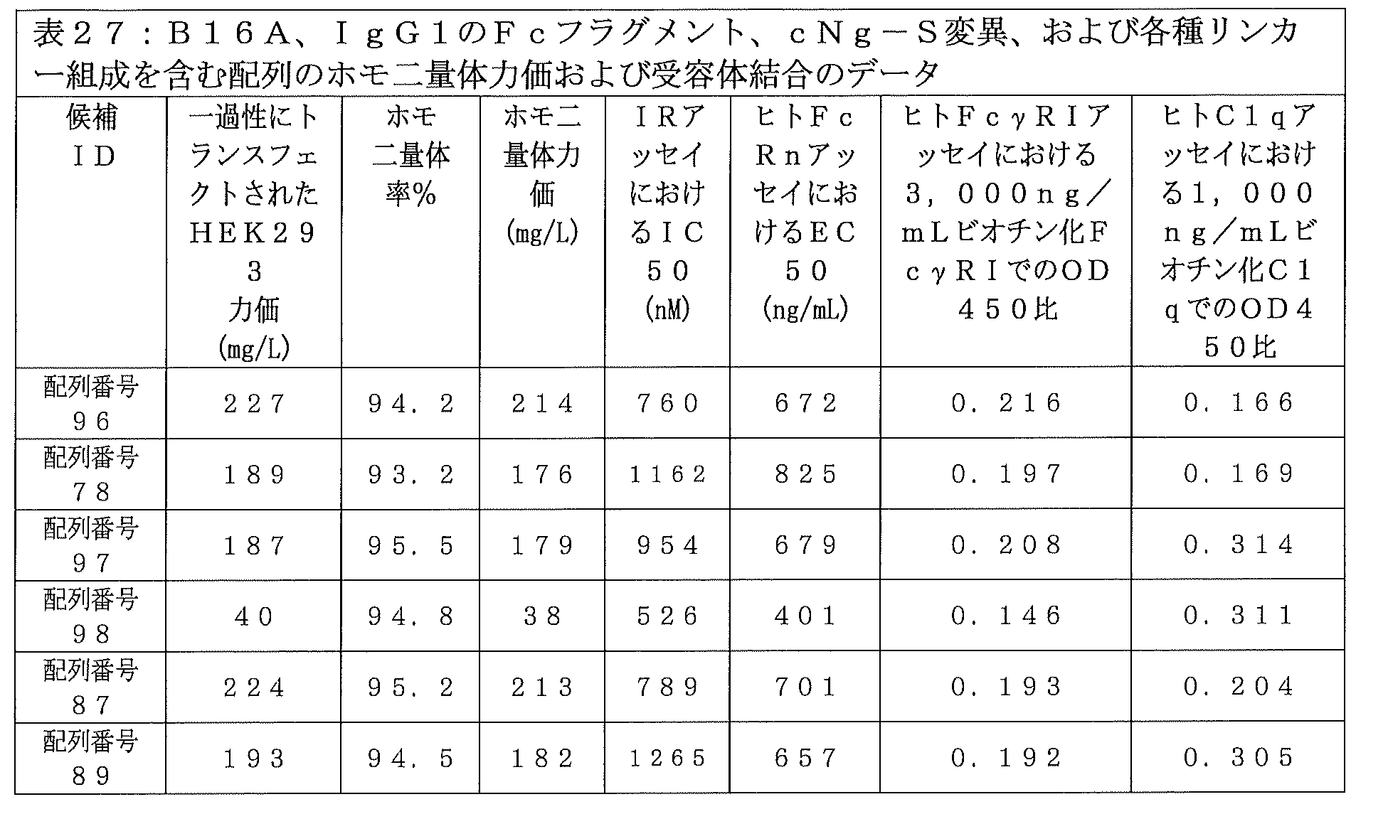
FVNQHLCGSHLVEALALVCGERGFHYGGGGGGSGGGGGIVEQCCTSTCSLDQLENYCGGGGGAGGGGAGGGGAGGGGGDKTHTCPPCPAPELLGGPSVFLFPPKPKDTLMISRTPEVTCVVVDVSHEDPEVKFNWYVDGVEVHNAKTKPREEQYSSTYRVVSVLTVLHQDWLNGKEYKCKVSNKALPAPIEKTISKAKGQPREPQVYTLPPSRDELTKNQVSLTCLVKGFYPSDIAVEWESNGQPENNYKTTPPVLDSDGSFFLYSKLTVDKSRWQQGNVFSCSVMHEALHNHYTQKSLSLSPG(配列番号87)
FVNQHLCGSHLVEALALVCGERGFHYGGGGGGSGGGGGIVEQCCTSTCSLDQLENYCGGGGGQGGGGQGGGGQGGGGGQGGGGDKTHTCPPCPAPELLGGPSVFLFPPKPKDTLMISRTPEVTCVVVDVSHEDPEVKFNWYVDGVEVHNAKTKPREEQYSSTYRVVSVLTVLHQDWLNGKEYKCKVSNKALPAPIEKTISKAKGQPREPQVYTLPPSRDELTKNQVSLTCLVKGFYPSDIAVEWESNGQPENNYKTTPPVLDSDGSFFLYSKLTVDKSRWQQGNVFSCSVMHEALHNHYTQKSLSLSPG(配列番号96)
FVNQHLCGSHLVEALALVCGERGFHYGGGGGGSGGGGGIVEQCCTSTCSLDQLENYCGGGGQGGGGDKTHTCPPCPAPELLGGPSVFLFPPKPKDTLMISRTPEVTCVVVDVSHEDPEVKFNWYVDGVEVHNAKTKPREEQYSSTYRVVSVLTVLHQDWLNGKEYKCKVSNKALPAPIEKTISKAKGQPREPQVYTLPPSRDELTKNQVSLTCLVKGFYPSDIAVEWESNGQPENNYKTTPPVLDSDGSFFLYSKLTVDKSRWQQGNVFSCSVMHEALHNHYTQKSLSLSPG(配列番号97)
FVNQHLCGSHLVEALALVCGERGFHYGGGGGGSGGGGGIVEQCCTSTCSLDQLENYCGGGGAGGGGDKTHTCPPCPAPELLGGPSVFLFPPKPKDTLMISRTPEVTCVVVDVSHEDPEVKFNWYVDGVEVHNAKTKPREEQYSSTYRVVSVLTVLHQDWLNGKEYKCKVSNKALPAPIEKTISKAKGQPREPQVYTLPPSRDELTKNQVSLTCLVKGFYPSDIAVEWESNGQPENNYKTTPPVLDSDGSFFLYSKLTVDKSRWQQGNVFSCSVMHEALHNHYTQKSLSLSPG(配列番号89)
FVNQHLCGSHLVEALALVCGERGFHYGGGGGGSGGGGGIVEQCCTSTCSLDQLENYCDKTHTCPPCPAPELLGGPSVFLFPPKPKDTLMISRTPEVTCVVVDVSHEDPEVKFNWYVDGVEVHNAKTKPREEQYSSTYRVVSVLTVLHQDWLNGKEYKCKVSNKALPAPIEKTISKAKGQPREPQVYTLPPSRDELTKNQVSLTCLVKGFYPSDIAVEWESNGQPENNYKTTPPVLDSDGSFFLYSKLTVDKSRWQQGNVFSCSVMHEALHNHYTQKSLSLSPG(配列番号98)。
The homodimer rate was determined by molecular sieve chromatography according to Example 10. IC50 for IM-9 IR binding in vitro was determined as described in Example 12, Fcγ receptor binding affinity was determined using an in vitro human FcγRI assay as described in Example 15, and C1q binding affinity was determined using an in vitro human FcγRI assay as described in Example 15. The affinity was determined as described in Example 16 and the affinity for human FcRn receptor was determined as described in Example 19. The results are shown in Table 27.
FVNQHLCGSHLVEALALVCGERGFHYGGGGGGSGGGGGIVEQCCTSTCSLDQLENYCGGGGGAGGGGAGGGGAGGGGGGDKTHTCPPCPAPELLGGPSVFLFPPKPKDTLMISRTPEVTCVVVD VSHEDPEVKFNWYVDGVEVHNAKTKPREEQY S STYRVVSVLTVLHQDWLNGKEYKCKVSNKALPAPIEKTISKAKGQPREPQVYTLPPSRDELTKNQVSLTCLVKGFYPSDIAVEWESNGQPE NNYKTTPPVLDSDGSFFLYSKLTVDKSRWQQGNVFSCSVMHEALHNHYTQKSLSLSPG (SEQ ID NO: 87)
FVNQHLCGSHLVEALALVCGERGFHYGGGGGGGSGGGGIVEQCCTSTCSLDQLENYCGGGGGQGGGGQGGGGQGGGGQGGGGDKTHTCPPCPAPELLGGPSVFLFPPKPKDTLMISRTPEVT CVVVDVSHEDPEVKFNWYVDGVEVHNAKTKPREEQY S STYRVVSVLTVLHQDWLNGKEYKCKVSNKALPAPIEKTISKAKGQPREPQVYTLPPSRDELTKNQVSLTCLVKGFYPSDIAVEWES NGQPENNYKTTPPVLDSDGSFFLYSKLTVDKSRWQQGNVFSCSVMHEALHNHYTQKSLSLSPG (SEQ ID NO: 96)
FVNQHLCGSHLVEALALVCGERGFHYGGGGGGGSGGGGIVEQCCTSTCSLDQLENYCGGGGQGGGGDKTHTCPPCPAPELLGGPSVFLFPPKPKDTLMISRTPEVTCVVVDVSHEDPEVKFNW YVDGVEVHNAKTKPREEQY S STYRVVSVLTVLHQDWLNGKEYKCKVSNKALPAPIEKTISKAKGQPREPQVYTLPPSRDELTKNQVSLTCLVKGFYPSDIAVEWESNGQPENNYKTTPPVLDS DGSFFLYSKLTVDKSRWQQGNVFSCSVMHEALHNHYTQKSLSLSPG (SEQ ID NO: 97)
FVNQHLCGSHLVEALALVCGERGFHYGGGGGGGSGGGGIVEQCCTSTCSLDQLENYCGGGGAGGGGDKTHTCPPCPAPELLGGPSVFLFPPKPKDTLMISRTPEVTCVVVDVSHEDPEVKFNW YVDGVEVHNAKTKPREEQY S STYRVVSVLTVLHQDWLNGKEYKCKVSNKALPAPIEKTISKAKGQPREPQVYTLPPSRDELTKNQVSLTCLVKGFYPSDIAVEWESNGQPENNYKTTPPVLDS DGSFFLYSKLTVDKSRWQQGNVFSCSVMHEALHNHYTQKSLSLSPG (SEQ ID NO: 89)
FVNQHLCGSHLVEALALVCGERGFHYGGGGGGGSGGGGIVEQCCTSTCSLDQLENYCDKTHTCPPCPAPELLGGPSVFLFPPKPKDTLMISRTPEVTCVVVDVSHEDPEVKFNWYVDGVEVHN AKTKPREEQY S STYRVVSVLTVLHQDWLNGKEYKCKVSNKALPAPIEKTISKAKGQPREPQVYTLPPSRDELTKNQVSLTCLVKGFYPSDIAVEWESNGQPENNYKTTPPVLDSDGSFFLYSK LTVDKSRWQQGNVFSCSVMHEALHNHYTQKSLSLSPG (SEQ ID NO: 98).
これらの結果は、インスリンポリペプチドのC末端がFcフラグメントのN末端に直接連結している、リンカーを含まないインスリン-Fc融合タンパク質の構成(配列番号98)において、ホモ二量体力価が許容できないほどに低い(すなわち、38mg/L)ことを示している。少なくとも9アミノ酸長のグリシン-グルタミンリンカー組成物を有するインスリン-Fc融合タンパク質の構成(配列番号97、配列番号78、および配列番号96)では、生理化学的性質に、リンカー長によって変化する重要な傾向はないように思われた。グリシン-アラニン組成物リンカーを含む配列番号87のインスリン-Fc融合タンパク質の構成を、グリシン-グルタミン組成物リンカーを含む配列番号78のインスリン-Fc融合タンパク質の構成と比較したところ、リンカー長が同じく21アミノ酸である場合、配列番号87のインスリン-Fc融合タンパク質の構成が、ホモ二量体力価の向上、IR結合の増強、およびFcRn結合の増強を示すが、FcγRI結合特性とC1q結合特性はほぼ同じであることが示された。よって、配列番号96、配列番号97、配列番号87、および配列番号89のインスリン-Fc融合タンパク質の構成の全てが設計目標(実施例29)を達成したが、配列番号87のホモ二量体が好ましい実施形態である。 These results demonstrate that in the linker-free insulin-Fc fusion protein construct (SEQ ID NO: 98), in which the C-terminus of the insulin polypeptide is linked directly to the N-terminus of the Fc fragment, homodimer titers are unacceptable. The figure shows that the amount of oxidation is relatively low (i.e., 38 mg/L). The construction of insulin-Fc fusion proteins with glycine-glutamine linker compositions of at least 9 amino acids in length (SEQ ID NO: 97, SEQ ID NO: 78, and SEQ ID NO: 96) shows significant trends in physiochemical properties that vary with linker length. It seemed like there wasn't. Comparing the composition of the insulin-Fc fusion protein of SEQ ID NO: 87 containing a glycine-alanine composition linker with the composition of the insulin-Fc fusion protein of SEQ ID NO: 78 containing a glycine-glutamine composition linker, it was found that the linker length is also 21. When amino acids, the composition of the insulin-Fc fusion protein of SEQ ID NO: 87 exhibits enhanced homodimeric potency, enhanced IR binding, and enhanced FcRn binding, but nearly identical FcγRI and C1q binding properties. It was shown that Thus, while all of the insulin-Fc fusion protein constructs of SEQ ID NO: 96, SEQ ID NO: 97, SEQ ID NO: 87, and SEQ ID NO: 89 achieved the design goals (Example 29), the homodimer of SEQ ID NO: 87 This is a preferred embodiment.
実施例55:B16A変異型インスリンポリペプチドと、cNg-NB155-X部位変異を有するヒトIgG1アイソタイプFcフラグメントと、リンカー(配列番号13)とを含むインスリン-Fc融合タンパク質のインビボ効力
実施例53の有望なホモ二量体力価、IR活性、およびFcRn活性の結果を考慮して、配列番号78、配列番号80、配列番号82、および配列番号84のインスリン-Fc融合タンパク質を、およそ10kgの重さのN=3の健常な抗体未処置ビーグル犬のそれぞれに皮下注射後、実施例22に従ってインビボ生物活性について試験する。配列番号78、配列番号80、配列番号82、および配列番号84の各インスリン-Fc融合タンパク質を単回皮下投与した場合の、時間に対する%FBGLのプロットを作成する。この時間に対する%FBGLのデータは、配列番号78、配列番号80、配列番号82、および配列番号84のインスリン-Fc融合タンパク質が、イヌにおいて顕著な生物活性を有することを示すと期待される。
Example 55: Promising In Vivo Efficacy of Insulin-Fc Fusion Protein Comprising a B16A Mutant Insulin Polypeptide, a Human IgG1 Isotype Fc Fragment with a cNg-NB155-X Site Mutation, and a Linker (SEQ ID NO: 13) Example 53 Considering the homodimer titer, IR activity, and FcRn activity results, the insulin-Fc fusion proteins of SEQ ID NO: 78, SEQ ID NO: 80, SEQ ID NO: 82, and SEQ ID NO: 84 weighed approximately 10 kg. Each of N=3 healthy antibody-naive beagle dogs is tested for in vivo bioactivity according to Example 22 after subcutaneous injection. A plot of %FBGL versus time is created for a single subcutaneous administration of each insulin-Fc fusion protein of SEQ ID NO: 78, SEQ ID NO: 80, SEQ ID NO: 82, and SEQ ID NO: 84. This %FBGL versus time data is expected to demonstrate that the insulin-Fc fusion proteins of SEQ ID NO: 78, SEQ ID NO: 80, SEQ ID NO: 82, and SEQ ID NO: 84 have significant biological activity in dogs.
実施例56:B16A変異型インスリンポリペプチドと、cNg-NB155-S変異を有するヒトIgG1アイソタイプFcフラグメントと、リンカー(配列番号11)とを含むインスリン-Fc融合タンパク質のインビボ効力
実施例54の有望なホモ二量体力価、IR活性、およびFcRn活性の結果を考慮して、配列番号89のインスリン-Fc融合タンパク質を、およそ10kgの重さのN=3の健常な抗体未処置ビーグル犬のそれぞれに皮下注射後、実施例22に従ってインビボ生物活性について試験する。配列番号89のインスリン-Fc融合タンパク質を単回皮下投与した場合の、時間に対する%FBGLのプロットを作成する。この時間に対する%FBGLのデータは、配列番号89のインスリン-Fc融合タンパク質が、イヌにおいて顕著な生物活性を有することを示すと期待される。
Example 56: Promising In Vivo Efficacy of Insulin-Fc Fusion Protein Comprising a B16A Mutant Insulin Polypeptide, a Human IgG1 Isotype Fc Fragment with the cNg-NB155-S Mutation, and a Linker (SEQ ID NO: 11) Considering the homodimer titer, IR activity, and FcRn activity results, the insulin-Fc fusion protein of SEQ ID NO: 89 was administered to each of N=3 healthy antibody-naive beagle dogs weighing approximately 10 kg. After subcutaneous injection, test for in vivo biological activity according to Example 22. A plot of %FBGL versus time is generated for a single subcutaneous administration of the insulin-Fc fusion protein of SEQ ID NO: 89. This %FBGL versus time data is expected to indicate that the insulin-Fc fusion protein of SEQ ID NO: 89 has significant biological activity in dogs.
実施例57:B16A変異型インスリンポリペプチドと、cNg-NB155-S変異を有するヒトIgG1アイソタイプFcフラグメントと、リンカー(配列番号67)とを含むインスリン-Fc融合タンパク質のインビボ効力
実施例54の有望なホモ二量体力価、IR活性、およびFcRn活性の結果を考慮して、配列番号87のインスリン-Fc融合タンパク質を、およそ10kgの重さのN=3の健常な抗体未処置ビーグル犬のそれぞれに皮下注射後、実施例22に従ってインビボ生物活性について試験する。配列番号87のインスリン-Fc融合タンパク質を単回皮下投与した場合の、時間に対する%FBGLのプロットを作成する。この時間に対する%FBGLのデータは、配列番号87のインスリン-Fc融合タンパク質が、イヌにおいて顕著な生物活性を有することを示すと期待される。
Example 57: Promising In Vivo Efficacy of Insulin-Fc Fusion Protein Comprising a B16A Mutant Insulin Polypeptide, a Human IgG1 Isotype Fc Fragment with the cNg-NB155-S Mutation, and a Linker (SEQ ID NO: 67) Considering the homodimer titer, IR activity, and FcRn activity results, the insulin-Fc fusion protein of SEQ ID NO: 87 was administered to each of N=3 healthy antibody-naive beagle dogs weighing approximately 10 kg. After subcutaneous injection, it is tested for in vivo biological activity according to Example 22. A plot of %FBGL versus time is generated for a single subcutaneous administration of the insulin-Fc fusion protein of SEQ ID NO: 87. This %FBGL versus time data is expected to indicate that the insulin-Fc fusion protein of SEQ ID NO: 87 has significant biological activity in dogs.
実施例58:安定にトランスフェクトされたCHO細胞株を介して作製されたヒトIgG1由来のFcフラグメントを含む好ましいインスリン-Fc融合タンパク質を用いた、例示的なCHOベースのプロダクションラン
配列番号78、配列番号80、配列番号82、配列番号84、配列番号87、および配列番号89のインスリン-Fc融合タンパク質の構成をコードするベクターで安定にトランスフェクトされた別々のCHO細胞株を、実施例2に記載されたように作製する。フェッドバッチ式振盪フラスコ14日間プロダクションラン(0.5~2.0Lの培地規模)の播種を、37℃、5%二酸化炭素に設定された振盪恒温器内で、0.5×10⁶細胞/mLとなるように行う。増殖培地としてCD OptiCHOをDynamis(サーモフィッシャー社)に置き換え、フィードとしてEfficient Feed C(サーモフィッシャー社)を用いる以外は、実施例2に記載したように、ランを実行する。3%v/vのフィードの添加をプロダクションラン3日目から開始して、さらに4日目にも添加を行い、振盪フラスコ温度を32℃に調整し、振盪恒温器内の二酸化炭素濃度を5%から2%に下げる。このランの間に、細胞密度は8~14×106細胞/mLまで増加し、14日目に、プロダクションランを回収して細胞を取り除き、実施例4、実施例6、実施例8、および実施例10に記載されたように培養上清の精製と試験を行って、インスリン-Fc融合タンパク質を得る。安定にトランスフェクトされたCHO細胞株を用いた配列番号78、配列番号80、配列番号82、配列番号84、配列番号87、および配列番号89のインスリン-Fc融合タンパク質の構成の別々のプロダクションランからの、製造およびインビトロ試験データは、ホモ二量体力価が一過性にトランスフェクトされたHEK293細胞で得られたものよりも大きく、IR、FcRn受容体、FcγRI受容体結合特性、およびC1q結合特性が設計目標(実施例29)を達成することを示すと期待される。
Example 58: Exemplary CHO-based production run using a preferred insulin-Fc fusion protein containing a human IgG1-derived Fc fragment generated via a stably transfected CHO cell line SEQ ID NO: 78, Sequence Separate CHO cell lines stably transfected with vectors encoding the insulin-Fc fusion protein constructs No. 80, SEQ ID No. 82, SEQ ID No. 84, SEQ ID No. 87, and SEQ ID No. 89 were described in Example 2. Prepare as shown. Fed-batch shake flask 14-day production runs (0.5-2.0 L media size) were seeded at 0.5 x 10 6 cells/in a shaking incubator set at 37°C and 5% carbon dioxide. mL. Runs are performed as described in Example 2, except that CD OptiCHO is replaced with Dynamis (Thermo Fisher) as the growth medium and Efficient Feed C (Thermo Fisher) is used as the feed. Addition of 3% v/v feed was started on
実施例59:マウスにヒトインスリンFc融合タンパク質を単回投与した後のインビボ薬物動態(PD)
空腹時血糖値に対する作用について、以下のように、インスリン-Fc融合タンパク質を評価した。1群当たりN=3のbalb/cマウスまたは糖尿病のwt NODマウス(ジャクソン・ラボラトリーズ社)からデータを収集した。実験の1時間前に動物を絶食させ、その後、0時間の時点で、マウスに、インスリンFc融合タンパク質ホモ二量体を含有する医薬組成物の単回皮下投与を、10~50mMのリン酸水素ナトリウム、50~150mMの塩化ナトリウム、0.005~0.05%v/vのTween-80、および所望により濃度0.02~1.00mg/mLの静菌剤(例えばフェノール、m‐クレゾール、またはメチルパラベン)の溶液中、300μg/kgのインスリン-Fc融合タンパク質濃度で、最終的な溶液pHは水酸化ナトリウムおよび/または塩酸を用いて7.0~8.0に調整して、実施した。
Example 59: In vivo pharmacokinetics (PD) after single administration of human insulin Fc fusion protein to mice
The insulin-Fc fusion protein was evaluated for its effect on fasting blood glucose levels as follows. Data were collected from N=3 balb/c mice or diabetic wt NOD mice (Jackson Laboratories) per group. Animals were fasted for 1 hour prior to the experiment, and then at
各時点において、血液試料を採取し、グルコース計(AlphaTRAK(登録商標)2ペット用グルコース計)を用いて直ちにグルコースレベルの読み取りを行い、これにはおよそ1滴の血液が必要であった。0時間から9時間までの平均空腹時血糖値%(%FBGL)をプロットし、所与のインスリン-Fc融合タンパク質の構成の生物活性を評価した。配列番号87および配列番号89を単回皮下投与した場合の、図38に示される結果は、両方の化合物がマウスにおいて顕著な生物活性を示したことを示している。
At each time point, a blood sample was taken and the glucose level was immediately read using a glucose meter (
実施例60:マウスにヒトインスリンFc融合タンパク質を単回投与した後のインビボ薬物動態(PD)
空腹時血糖値に対する作用について、以下のように、インスリン-Fc融合タンパク質を評価する。1群当たりN=3のbalb/cマウスまたは糖尿病wt NODマウス(ジャクソン・ラボラトリーズ社)からデータを収集する。実験の1時間前に動物を絶食させ、その後、0時間の時点で、マウスに、インスリンFc融合タンパク質ホモ二量体を含有する医薬組成物の単回皮下投与を、10~50mMのリン酸水素ナトリウム、50~150mMの塩化ナトリウム、0.005~0.05%v/vのTween-80、および所望により濃度0.02~1.00mg/mLの静菌剤(例えばフェノール、m‐クレゾール、またはメチルパラベン)の溶液中、300μg/kgのインスリン-Fc融合タンパク質濃度で、最終的な溶液pHは水酸化ナトリウムおよび/または塩酸を用いて7.0~8.0に調整して、実施する。
Example 60: In vivo pharmacokinetics (PD) after single administration of human insulin Fc fusion protein to mice
Insulin-Fc fusion proteins are evaluated for their effects on fasting blood glucose levels as follows. Data are collected from N=3 balb/c or diabetic wt NOD mice (Jackson Laboratories) per group. Animals were fasted for 1 hour prior to the experiment, and then at
各時点において、血液試料を採取し、グルコース計(AlphaTRAK(登録商標)2ペット用グルコース計)を用いて直ちにグルコースレベルの読み取りを行い、これにはおよそ1滴の血液が必要である。0時間から9時間までの平均空腹時血糖値%(%FBGL)をプロットし、所与のインスリン-Fc融合タンパク質の構成の生物活性を評価する。配列番号78、配列番号80、配列番号82、または配列番号84の単回皮下投与によって、これらのインスリン-Fc融合タンパク質がマウスにおいて顕著な生物活性を示すことが明らかになると期待される。
At each time point, a blood sample is taken and the glucose level is immediately read using a glucose meter (
実施例61:例示的なインスリン-Fc融合タンパク質のドメインおよび配列
上記の実施例で使用された例示的なインスリン-Fc融合タンパク質のアミノ酸配列および対応するDNA配列を、図31、図32、図33、図34、図35、図36、および図37に示す。
均等論
Example 61: Domains and Sequences of Exemplary Insulin-Fc Fusion Proteins The amino acid sequences and corresponding DNA sequences of the exemplary insulin-Fc fusion proteins used in the examples above are shown in Figures 31, 32, and 33. , shown in FIGS. 34, 35, 36, and 37.
doctrine of equivalents
クレームにおいて、「a」、「an」、および「the」などの冠詞は、反対の趣旨が示されていない限り、または文脈から明らかでない限り、1つを意味する場合も2つ以上を意味する場合もある。ある群の1または複数の要素の間に「または」を含むクレームまたは記述は、反対の趣旨が示されていない限り、または文脈から明らかでない限り、前記群の要素の1、2以上、または全てが、所与の製品または工程に存在していれば、それに使用されていれば、またはそれに関連していれば、満たされているものとする。本開示は、前記群の厳密に1つの要素が所与の製品または工程に存在している、それに使用されている、またはそれに関連している、実施形態を包含する。本開示は、前記群の要素のうちの2以上または全てが所与の製品または工程に存在している、それに使用されている、またはそれに関連している、実施形態を包含する。 In the claims, articles such as "a," "an," and "the" mean one or more, unless the contrary is indicated or it is clear from the context. In some cases. A claim or statement that includes "or" between one or more members of a group refers to one, two or more, or all of the members of said group, unless the contrary is indicated or it is clear from the context. is satisfied if it is present in, used in, or associated with a given product or process. The present disclosure encompasses embodiments in which exactly one member of the group is present in, used in, or associated with a given product or process. The present disclosure encompasses embodiments in which two or more or all of the members of the above groups are present in, used in, or associated with a given product or process.
さらに、本開示は、列挙されたクレームのうちの1または複数からの1または複数の制限、要素、節、および記述用語が別のクレームに導入された、全ての変形、組み合わせ、および入れ替えを包含する。例えば、別のクレームに従属する任意のクレームを、同じ基本クレームに従属する任意の他のクレームに存在する1または複数の制限を含むように修正することができる。要素がリストとして、例えばマーカッシュ群形式で提示されている場合、要素の各部分群を開示し、任意の要素を当該群から除外することもできる。一般的に、本開示または本開示の側面が、特定の要素および/または特徴を含むものとして言及される場合、本開示または本開示の側面のある実施形態は、かかる要素および/または特徴からなるか、またはこれらから本質的になることが、理解されるべきである。簡潔さのために、これらの実施形態は、本明細書において、特にこの通りの言葉では記載されていない。また、用語「含む(comprise)」、「含むこと(comprising)」、「含有する(contain)」、および「含有すること(containing)」は、オープンであることを意図され、その使用は、さらなる要素または工程の包含を許容することに留意する。範囲が示される場合、端点が含まれる。さらに、別段に示されるかまたは別段に文脈および当業者の理解から明らかでない限り、範囲として表される値は、本開示の異なる実施形態において記述される範囲内の任意の特定の値または部分範囲を、文脈によって特に明示されない限り、当該範囲の下限の単位の10分の1まで想定し得る。 Moreover, this disclosure encompasses all variations, combinations, and permutations in which one or more limitations, elements, clauses, and descriptive terms from one or more of the recited claims are introduced into another claim. do. For example, any claim that is dependent on another claim may be modified to include one or more limitations that are present in any other claim that is dependent on the same base claim. If the elements are presented as a list, for example in Markush group format, each subgroup of elements can be disclosed and any element can be excluded from the group. Generally, when the disclosure or aspects of the disclosure are referred to as including particular elements and/or features, certain embodiments of the disclosure or aspects of the disclosure consist of such elements and/or features. It should be understood that or consisting essentially of: For the sake of brevity, these embodiments are not specifically described herein in these exact terms. Also, the terms "comprise," "comprising," "contain," and "containing" are intended to be open, and their use may be used to refer to further Note that the inclusion of elements or steps is permissible. Where a range is given, endpoints are included. Furthermore, unless otherwise indicated or otherwise clear from the context and understanding of those skilled in the art, values expressed as ranges may refer to any specific value or subrange within the ranges recited in different embodiments of this disclosure. may be assumed to be up to one-tenth of a unit at the lower end of the range, unless the context clearly indicates otherwise.
Claims (29)
前記インスリンポリペプチドおよび前記Fcフラグメントはリンカーによって接続されており、N末端からC末端へ以下の配向:
(N末端)-インスリンポリペプチド-リンカー-Fcフラグメント-(C末端)
で各ドメインを含み、
前記Fcフラグメントはヒト由来であり、且つ以下の配列(配列番号77):
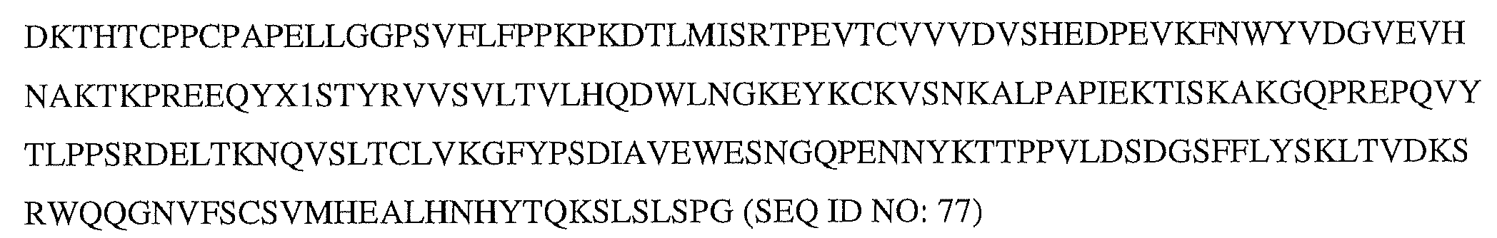
前記インスリンポリペプチドは
以下の配列:
FVNQHLCGSHLVEALALVCGERGFHYGGGGGGSGGGGGIVEQCCTSTCSLDQLENYC(配列番号10)、
を含み、かつ
前記リンカーが以下の配列:
GGGGGAGGGGAGGGGAGGGGG(配列番号67);
GGGGGQGGGGQGGGGQGGGGGQGGGG(配列番号99);または
GGGGGQGGGGQGGGGQGGGGG(配列番号13)
を含む、
融合タンパク質。 A fusion protein comprising an insulin polypeptide and an Fc fragment,
The insulin polypeptide and the Fc fragment are connected by a linker in the following orientation from N-terminus to C-terminus:
(N-terminus) - insulin polypeptide - linker - Fc fragment - (C-terminus)
contains each domain in
The Fc fragment is of human origin and has the following sequence (SEQ ID NO: 77):
The insulin polypeptide is
Array below:
FVNQHLCGSHLVEALALVCGERGFHYGGGGGGGSGGGGIVEQCCTSTCSLDQLENYC (SEQ ID NO: 10),
including, and
The linker has the following sequence:
GGGGGAGGGGGAGGGGAGGGGG (SEQ ID NO: 67);
GGGGGQGGGGQGGGGQGGGGQGGGG (SEQ ID NO: 99); or
GGGGGQGGGGQGGGGQGGGGG (SEQ ID NO: 13)
including,
fusion protein.
前記インスリンポリペプチドおよび前記Fcフラグメントはリンカーによって接続されており、the insulin polypeptide and the Fc fragment are connected by a linker;
前記融合タンパク質は以下の配列(配列番号87):The fusion protein has the following sequence (SEQ ID NO: 87):
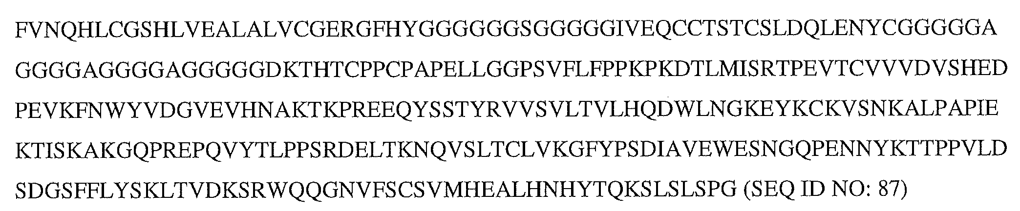
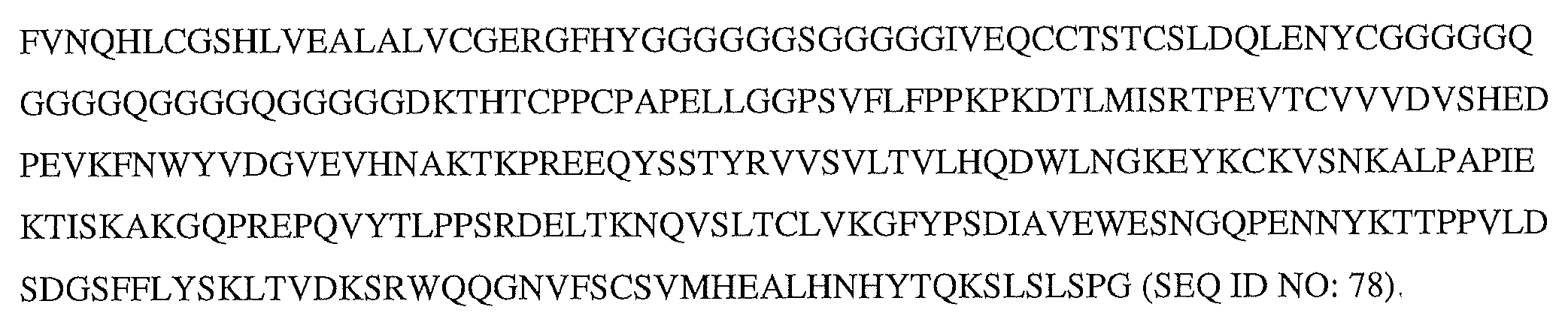
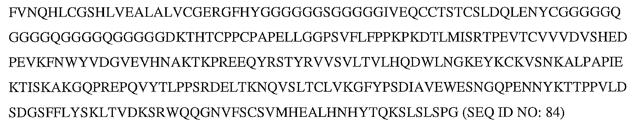
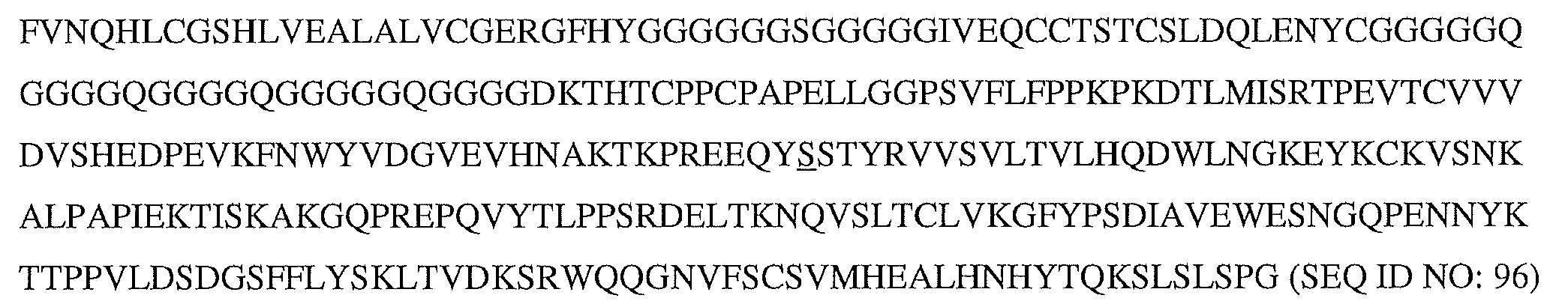
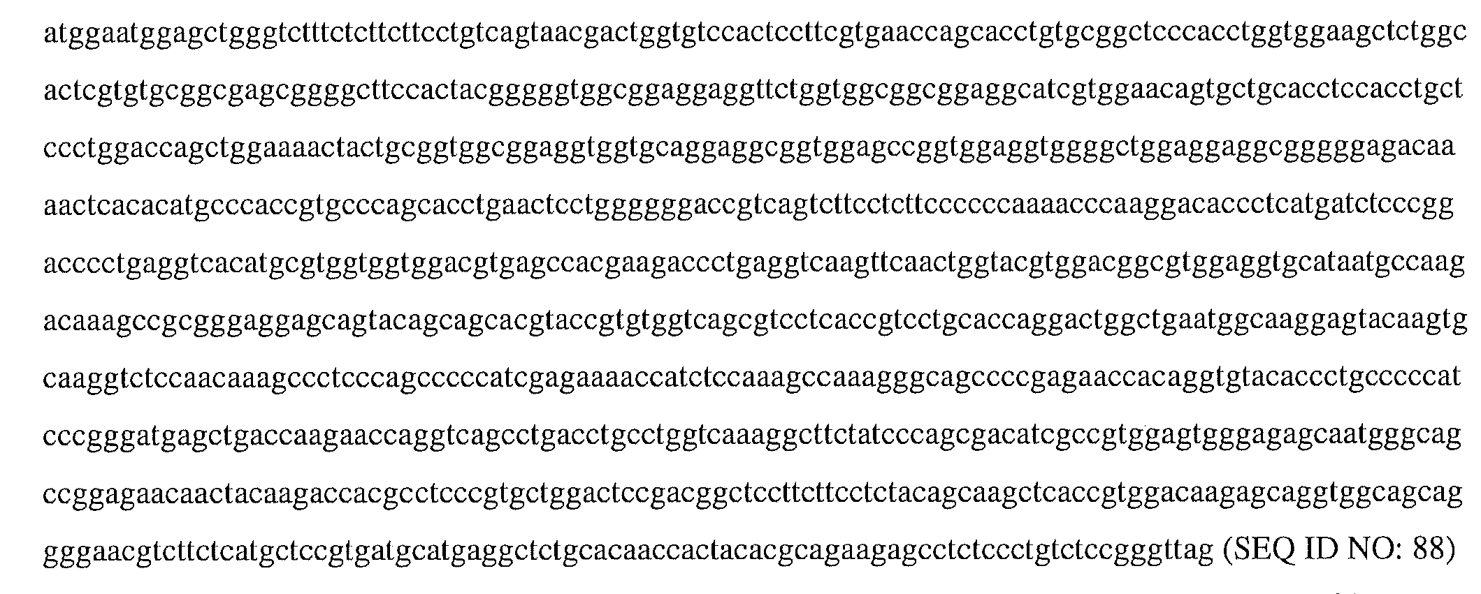

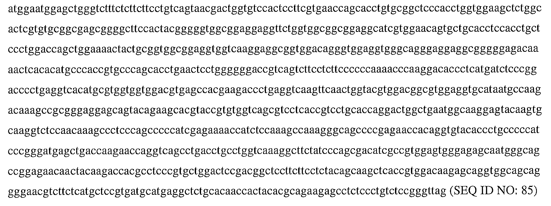
Applications Claiming Priority (5)
| Application Number | Priority Date | Filing Date | Title |
|---|---|---|---|
| US201962950803P | 2019-12-19 | 2019-12-19 | |
| US62/950,803 | 2019-12-19 | ||
| US202062988441P | 2020-03-12 | 2020-03-12 | |
| US62/988,441 | 2020-03-12 | ||
| PCT/US2020/063673 WO2021126584A1 (en) | 2019-12-19 | 2020-12-07 | Ultra-long acting insulin-fc fusion proteins and methods of use |
Publications (2)
| Publication Number | Publication Date |
|---|---|
| JP2023507376A JP2023507376A (en) | 2023-02-22 |
| JP7405486B2 true JP7405486B2 (en) | 2023-12-26 |
Family
ID=74104207
Family Applications (1)
| Application Number | Title | Priority Date | Filing Date |
|---|---|---|---|
| JP2022536936A Active JP7405486B2 (en) | 2019-12-19 | 2020-12-07 | Ultra-long-acting insulin-FC fusion protein and methods of use |
Country Status (17)
| Country | Link |
|---|---|
| US (3) | US11352407B2 (en) |
| EP (2) | EP4282473A3 (en) |
| JP (1) | JP7405486B2 (en) |
| KR (2) | KR20250142943A (en) |
| CN (1) | CN114846025B (en) |
| AU (1) | AU2020407365B2 (en) |
| BR (1) | BR112022012071A2 (en) |
| CA (1) | CA3159486A1 (en) |
| DK (1) | DK4073098T5 (en) |
| FI (1) | FI4073098T3 (en) |
| HR (1) | HRP20231433T1 (en) |
| HU (1) | HUE064376T2 (en) |
| IL (1) | IL294051A (en) |
| LT (1) | LT4073098T (en) |
| SI (1) | SI4073098T1 (en) |
| WO (1) | WO2021126584A1 (en) |
| ZA (1) | ZA202206047B (en) |
Families Citing this family (8)
| Publication number | Priority date | Publication date | Assignee | Title |
|---|---|---|---|---|
| KR20210029210A (en) | 2018-06-29 | 2021-03-15 | 악스톤 바이오사이언시스 코퍼레이션 | Ultra-long acting insulin-FC fusion protein and method of use |
| KR20250142943A (en) | 2019-12-19 | 2025-09-30 | 악스톤 바이오사이언시스 코퍼레이션 | Ultra-long acting insulin-fc fusion proteins and methods of use |
| US11186623B2 (en) | 2019-12-24 | 2021-11-30 | Akston Bioscience Corporation | Ultra-long acting insulin-Fc fusion proteins and methods of use |
| US11192930B2 (en) | 2020-04-10 | 2021-12-07 | Askton Bioscences Corporation | Ultra-long acting insulin-Fc fusion protein and methods of use |
| LT3972987T (en) | 2020-04-10 | 2023-09-25 | Akston Biosciences Corporation | ANTIGEN-SPECIFIC IMMUNOTHERAPY FOR COVID-19 FUSION PROTEINS AND METHODS OF USE |
| US11198719B2 (en) | 2020-04-29 | 2021-12-14 | Akston Biosciences Corporation | Ultra-long acting insulin-Fc fusion protein and methods of use |
| EP4373861A4 (en) | 2021-07-23 | 2025-08-20 | Akston Biosciences Corp | INSULIN-FC FUSION PROTEINS AND METHODS OF USE FOR THE TREATMENT OF CANCER |
| TWI871539B (en) | 2021-11-15 | 2025-02-01 | 美商美國禮來大藥廠 | Preserved formulations |
Citations (5)
| Publication number | Priority date | Publication date | Assignee | Title |
|---|---|---|---|---|
| JP2015504679A (en) | 2012-01-12 | 2015-02-16 | バイオジェン・アイデック・エムエイ・インコーポレイテ | Chimeric factor VIII polypeptide and uses thereof |
| JP2016510003A (en) | 2013-02-26 | 2016-04-04 | ハンミ ファーム.カンパニー リミテッドHanmi Pharm.Co.,Ltd. | Novel insulin analogues and their uses |
| JP2018515088A (en) | 2015-05-07 | 2018-06-14 | イーライ リリー アンド カンパニー | Fusion protein |
| WO2018107117A1 (en) | 2016-12-09 | 2018-06-14 | Akston Biosciences Corporation | Insulin-fc fusions and methods of use |
| US20180340031A1 (en) | 2017-05-25 | 2018-11-29 | Bristol-Myers Squibb Company | Antagonistic cd40 monoclonal antibodies and uses thereof |
Family Cites Families (39)
| Publication number | Priority date | Publication date | Assignee | Title |
|---|---|---|---|---|
| US20030040601A1 (en) | 2001-06-08 | 2003-02-27 | Ivan Diers | Method for making insulin precursors and insulin analog precursors |
| US8188231B2 (en) | 2002-09-27 | 2012-05-29 | Xencor, Inc. | Optimized FC variants |
| WO2010107520A1 (en) | 2009-03-20 | 2010-09-23 | Smartcells, Inc. | Soluble non-depot insulin conjugates and uses thereof |
| CA2754408A1 (en) | 2009-03-30 | 2010-10-14 | Boehringer Ingelheim International Gmbh | Fusion proteins comprising canine fc portions |
| CN101891823B (en) | 2010-06-11 | 2012-10-03 | 北京东方百泰生物科技有限公司 | Exendin-4 and analog fusion protein thereof |
| AU2011282977A1 (en) | 2010-07-28 | 2013-02-21 | Smartcells, Inc. | Drug-ligand conjugates, synthesis thereof, and intermediates thereto |
| JP2013535467A (en) | 2010-07-28 | 2013-09-12 | スマートセルズ・インコーポレイテツド | Recombinantly expressed insulin polypeptide and uses thereof |
| AR087433A1 (en) | 2011-08-08 | 2014-03-26 | Merck Sharp & Dohme | N-GLICOSILATED INSULIN ANALOGS |
| US20140302028A1 (en) | 2011-11-18 | 2014-10-09 | Merck Sharp & Dohme Corp. | Fc containing polypeptides having increased anti-inflammatory properties and increased fcrn binding |
| KR102041412B1 (en) | 2011-12-30 | 2019-11-11 | 한미사이언스 주식회사 | Derivatives of Immunglobulin Fc fragment |
| CN103509118B (en) | 2012-06-15 | 2016-03-23 | 郭怀祖 | insulin-Fc fusion protein |
| JP6649250B2 (en) * | 2013-05-21 | 2020-02-19 | プレジデント・アンド・フェロウズ・オブ・ハーバード・カレッジ | Engineered heme binding constructs and uses thereof |
| EP3194429A4 (en) | 2014-09-18 | 2018-06-13 | Askgene Pharma, Inc. | Novel feline erythropoietin receptor agonists |
| WO2016105545A2 (en) | 2014-12-24 | 2016-06-30 | Case Western Reserve University | Insulin analogues with enhanced stabilized and reduced mitogenicity |
| WO2016119023A1 (en) | 2015-01-29 | 2016-08-04 | Nexvet Australia Pty Ltd | Therapeutic and diagnostic agents |
| EP3260139A4 (en) | 2015-02-17 | 2018-09-05 | Hanmi Pharm. Co., Ltd. | Long-acting insulin or insulin analogue complex |
| ES2754432T3 (en) | 2015-05-04 | 2020-04-17 | Apogenix Ag | Single chain CD40 receptor agonist proteins |
| ES2784603T3 (en) | 2015-06-02 | 2020-09-29 | Novo Nordisk As | Insulins with recombinant polar extensions |
| WO2017055314A1 (en) * | 2015-10-02 | 2017-04-06 | F. Hoffmann-La Roche Ag | Bispecific anti-cd19xcd3 t cell activating antigen binding molecules |
| CN114835793A (en) | 2015-11-16 | 2022-08-02 | Ubi蛋白公司 | Method for extending protein half-life |
| EP3481413A4 (en) | 2016-07-08 | 2020-01-08 | Askgene Pharma, Inc. | Fusion protein comprising leptin and methods for producing and using the same |
| JP7158378B2 (en) | 2016-09-23 | 2022-10-21 | ハンミ ファーマシューティカル カンパニー リミテッド | Insulin analogue with reduced binding force with insulin receptor and use thereof |
| US20200181258A1 (en) | 2016-10-17 | 2020-06-11 | Vetoquinol Sa | Modified antibody constant region |
| UY37629A (en) | 2017-03-07 | 2018-10-31 | Univ Case Western Reserve | SIMPLE CHAIN INSULIN ANALOGS STABILIZED BY A FOUR DISULFURED BRIDGE |
| US12297272B2 (en) | 2017-08-15 | 2025-05-13 | Eianco US inc. | IgG Fc variants for veterinary use |
| EP3781147A4 (en) | 2018-04-16 | 2022-04-20 | University of Utah Research Foundation | GLUCOSE-SENSITIVE INSULIN |
| KR20210029210A (en) | 2018-06-29 | 2021-03-15 | 악스톤 바이오사이언시스 코퍼레이션 | Ultra-long acting insulin-FC fusion protein and method of use |
| US11267862B2 (en) | 2018-06-29 | 2022-03-08 | Akston Biosciences Corporation | Ultra-long acting insulin-Fc fusion proteins and methods of use |
| WO2020069011A1 (en) | 2018-09-25 | 2020-04-02 | Absci, Llc | Protein purification methods |
| WO2020070276A1 (en) | 2018-10-05 | 2020-04-09 | Novo Nordisk A/S | Bifunctional compounds comprising insulin peptides and egf(a) peptides |
| CN113330025A (en) | 2018-11-19 | 2021-08-31 | 卡斯西部储备大学 | Single chain insulin analogs with polyalanine C domain subsegments |
| CA3139946A1 (en) | 2019-05-17 | 2020-11-26 | Case Western Reserve University | Variant single-chain insulin analogues |
| WO2021011827A1 (en) | 2019-07-16 | 2021-01-21 | Akston Biosciences Corporation | Ultra-long acting insulin-fc fusion proteins and methods of use |
| TWI844709B (en) | 2019-07-31 | 2024-06-11 | 美商美國禮來大藥廠 | Relaxin analogs and methods of using the same |
| KR20250142943A (en) | 2019-12-19 | 2025-09-30 | 악스톤 바이오사이언시스 코퍼레이션 | Ultra-long acting insulin-fc fusion proteins and methods of use |
| US11186623B2 (en) | 2019-12-24 | 2021-11-30 | Akston Bioscience Corporation | Ultra-long acting insulin-Fc fusion proteins and methods of use |
| US11192930B2 (en) | 2020-04-10 | 2021-12-07 | Askton Bioscences Corporation | Ultra-long acting insulin-Fc fusion protein and methods of use |
| US11198719B2 (en) | 2020-04-29 | 2021-12-14 | Akston Biosciences Corporation | Ultra-long acting insulin-Fc fusion protein and methods of use |
| KR20240158331A (en) | 2022-03-16 | 2024-11-04 | 베이징 투오 지에 바이오파마수티컬 컴퍼니 리미티드 | Human insulin analogues, fusion proteins thereof and their medicinal uses |
-
2020
- 2020-12-07 KR KR1020257031841A patent/KR20250142943A/en active Pending
- 2020-12-07 WO PCT/US2020/063673 patent/WO2021126584A1/en not_active Ceased
- 2020-12-07 EP EP23198760.3A patent/EP4282473A3/en active Pending
- 2020-12-07 HR HRP20231433TT patent/HRP20231433T1/en unknown
- 2020-12-07 KR KR1020227022527A patent/KR102865175B1/en active Active
- 2020-12-07 DK DK20830444.4T patent/DK4073098T5/en active
- 2020-12-07 JP JP2022536936A patent/JP7405486B2/en active Active
- 2020-12-07 HU HUE20830444A patent/HUE064376T2/en unknown
- 2020-12-07 AU AU2020407365A patent/AU2020407365B2/en active Active
- 2020-12-07 IL IL294051A patent/IL294051A/en unknown
- 2020-12-07 CA CA3159486A patent/CA3159486A1/en active Pending
- 2020-12-07 LT LTEPPCT/US2020/063673T patent/LT4073098T/en unknown
- 2020-12-07 CN CN202080088372.8A patent/CN114846025B/en active Active
- 2020-12-07 SI SI202030309T patent/SI4073098T1/en unknown
- 2020-12-07 FI FIEP20830444.4T patent/FI4073098T3/en active
- 2020-12-07 BR BR112022012071A patent/BR112022012071A2/en unknown
- 2020-12-07 EP EP20830444.4A patent/EP4073098B1/en active Active
- 2020-12-07 US US17/114,395 patent/US11352407B2/en active Active
-
2021
- 2021-11-19 US US17/531,033 patent/US11555058B2/en active Active
-
2022
- 2022-05-31 ZA ZA2022/06047A patent/ZA202206047B/en unknown
-
2023
- 2023-01-10 US US18/152,595 patent/US12606605B2/en active Active
Patent Citations (5)
| Publication number | Priority date | Publication date | Assignee | Title |
|---|---|---|---|---|
| JP2015504679A (en) | 2012-01-12 | 2015-02-16 | バイオジェン・アイデック・エムエイ・インコーポレイテ | Chimeric factor VIII polypeptide and uses thereof |
| JP2016510003A (en) | 2013-02-26 | 2016-04-04 | ハンミ ファーム.カンパニー リミテッドHanmi Pharm.Co.,Ltd. | Novel insulin analogues and their uses |
| JP2018515088A (en) | 2015-05-07 | 2018-06-14 | イーライ リリー アンド カンパニー | Fusion protein |
| WO2018107117A1 (en) | 2016-12-09 | 2018-06-14 | Akston Biosciences Corporation | Insulin-fc fusions and methods of use |
| US20180340031A1 (en) | 2017-05-25 | 2018-11-29 | Bristol-Myers Squibb Company | Antagonistic cd40 monoclonal antibodies and uses thereof |
Non-Patent Citations (1)
| Title |
|---|
| PROTEIN & CELL, 2017, Vol.9, No.1, pp.63-73 |
Also Published As
| Publication number | Publication date |
|---|---|
| HUE064376T2 (en) | 2024-03-28 |
| CN114846025B (en) | 2025-11-11 |
| FI4073098T3 (en) | 2023-11-15 |
| AU2020407365A1 (en) | 2022-06-09 |
| KR20220115975A (en) | 2022-08-19 |
| EP4282473A2 (en) | 2023-11-29 |
| DK4073098T3 (en) | 2023-11-20 |
| NZ788617A (en) | 2024-10-25 |
| US11555058B2 (en) | 2023-01-17 |
| EP4282473A3 (en) | 2024-02-21 |
| CA3159486A1 (en) | 2021-06-24 |
| ZA202206047B (en) | 2023-08-30 |
| SI4073098T1 (en) | 2023-12-29 |
| WO2021126584A1 (en) | 2021-06-24 |
| KR102865175B1 (en) | 2025-09-30 |
| JP2023507376A (en) | 2023-02-22 |
| HRP20231433T1 (en) | 2024-03-29 |
| EP4073098B1 (en) | 2023-09-27 |
| US20220064251A1 (en) | 2022-03-03 |
| US20210309709A1 (en) | 2021-10-07 |
| IL294051A (en) | 2022-08-01 |
| DK4073098T5 (en) | 2024-07-22 |
| US20230250148A1 (en) | 2023-08-10 |
| CN114846025A (en) | 2022-08-02 |
| AU2020407365B2 (en) | 2023-09-21 |
| LT4073098T (en) | 2023-12-11 |
| BR112022012071A2 (en) | 2022-08-30 |
| EP4073098A1 (en) | 2022-10-19 |
| KR20250142943A (en) | 2025-09-30 |
| US12606605B2 (en) | 2026-04-21 |
| US11352407B2 (en) | 2022-06-07 |
Similar Documents
| Publication | Publication Date | Title |
|---|---|---|
| JP7510728B2 (en) | Ultra-long acting insulin-FC fusion proteins and methods of use - Patents.com | |
| JP7405486B2 (en) | Ultra-long-acting insulin-FC fusion protein and methods of use | |
| US11267862B2 (en) | Ultra-long acting insulin-Fc fusion proteins and methods of use | |
| WO2021011827A1 (en) | Ultra-long acting insulin-fc fusion proteins and methods of use | |
| WO2023064711A2 (en) | Ultra-long acting insulin-fc fusion proteins and methods of use |
Legal Events
| Date | Code | Title | Description |
|---|---|---|---|
| A521 | Request for written amendment filed |
Free format text: JAPANESE INTERMEDIATE CODE: A523 Effective date: 20220825 |
|
| A621 | Written request for application examination |
Free format text: JAPANESE INTERMEDIATE CODE: A621 Effective date: 20220825 |
|
| A131 | Notification of reasons for refusal |
Free format text: JAPANESE INTERMEDIATE CODE: A131 Effective date: 20230711 |
|
| A977 | Report on retrieval |
Free format text: JAPANESE INTERMEDIATE CODE: A971007 Effective date: 20230714 |
|
| A601 | Written request for extension of time |
Free format text: JAPANESE INTERMEDIATE CODE: A601 Effective date: 20231011 |
|
| A521 | Request for written amendment filed |
Free format text: JAPANESE INTERMEDIATE CODE: A523 Effective date: 20231018 |
|
| TRDD | Decision of grant or rejection written | ||
| A01 | Written decision to grant a patent or to grant a registration (utility model) |
Free format text: JAPANESE INTERMEDIATE CODE: A01 Effective date: 20231114 |
|
| A61 | First payment of annual fees (during grant procedure) |
Free format text: JAPANESE INTERMEDIATE CODE: A61 Effective date: 20231208 |
|
| R150 | Certificate of patent or registration of utility model |
Ref document number: 7405486 Country of ref document: JP Free format text: JAPANESE INTERMEDIATE CODE: R150 |