JP7391971B2 - Novel DNA aptamer and its uses - Google Patents
Novel DNA aptamer and its uses Download PDFInfo
- Publication number
- JP7391971B2 JP7391971B2 JP2021536744A JP2021536744A JP7391971B2 JP 7391971 B2 JP7391971 B2 JP 7391971B2 JP 2021536744 A JP2021536744 A JP 2021536744A JP 2021536744 A JP2021536744 A JP 2021536744A JP 7391971 B2 JP7391971 B2 JP 7391971B2
- Authority
- JP
- Japan
- Prior art keywords
- aptamer
- cancer
- present disclosure
- dna aptamer
- cells
- Prior art date
- Legal status (The legal status is an assumption and is not a legal conclusion. Google has not performed a legal analysis and makes no representation as to the accuracy of the status listed.)
- Active
Links
- 108091008102 DNA aptamers Proteins 0.000 title claims description 41
- 108091023037 Aptamer Proteins 0.000 claims description 192
- 206010061902 Pancreatic neoplasm Diseases 0.000 claims description 79
- 208000015486 malignant pancreatic neoplasm Diseases 0.000 claims description 78
- 201000002528 pancreatic cancer Diseases 0.000 claims description 78
- 208000008443 pancreatic carcinoma Diseases 0.000 claims description 78
- 206010028980 Neoplasm Diseases 0.000 claims description 60
- 201000011510 cancer Diseases 0.000 claims description 42
- 239000000203 mixture Substances 0.000 claims description 25
- 230000008685 targeting Effects 0.000 claims description 18
- 239000002773 nucleotide Substances 0.000 claims description 16
- 125000003729 nucleotide group Chemical group 0.000 claims description 16
- 239000002246 antineoplastic agent Substances 0.000 claims description 8
- 238000003745 diagnosis Methods 0.000 claims description 6
- 102000016911 Deoxyribonucleases Human genes 0.000 claims description 5
- 108010053770 Deoxyribonucleases Proteins 0.000 claims description 5
- 238000006467 substitution reaction Methods 0.000 claims description 5
- 208000003174 Brain Neoplasms Diseases 0.000 claims description 4
- 206010006187 Breast cancer Diseases 0.000 claims description 4
- 208000026310 Breast neoplasm Diseases 0.000 claims description 4
- 206010009944 Colon cancer Diseases 0.000 claims description 4
- AOJJSUZBOXZQNB-TZSSRYMLSA-N Doxorubicin Chemical compound O([C@H]1C[C@@](O)(CC=2C(O)=C3C(=O)C=4C=CC=C(C=4C(=O)C3=C(O)C=21)OC)C(=O)CO)[C@H]1C[C@H](N)[C@H](O)[C@H](C)O1 AOJJSUZBOXZQNB-TZSSRYMLSA-N 0.000 claims description 4
- 206010058467 Lung neoplasm malignant Diseases 0.000 claims description 4
- 206010033128 Ovarian cancer Diseases 0.000 claims description 4
- 206010061535 Ovarian neoplasm Diseases 0.000 claims description 4
- 201000007270 liver cancer Diseases 0.000 claims description 4
- 208000014018 liver neoplasm Diseases 0.000 claims description 4
- 201000005202 lung cancer Diseases 0.000 claims description 4
- 208000020816 lung neoplasm Diseases 0.000 claims description 4
- 229920001223 polyethylene glycol Polymers 0.000 claims description 4
- 239000002202 Polyethylene glycol Substances 0.000 claims description 3
- 239000012830 cancer therapeutic Substances 0.000 claims description 3
- 208000029742 colonic neoplasm Diseases 0.000 claims description 3
- 150000001982 diacylglycerols Chemical class 0.000 claims description 3
- YUOCYTRGANSSRY-UHFFFAOYSA-N pyrrolo[2,3-i][1,2]benzodiazepine Chemical compound C1=CN=NC2=C3C=CN=C3C=CC2=C1 YUOCYTRGANSSRY-UHFFFAOYSA-N 0.000 claims description 3
- FJHBVJOVLFPMQE-QFIPXVFZSA-N 7-Ethyl-10-Hydroxy-Camptothecin Chemical compound C1=C(O)C=C2C(CC)=C(CN3C(C4=C([C@@](C(=O)OC4)(O)CC)C=C33)=O)C3=NC2=C1 FJHBVJOVLFPMQE-QFIPXVFZSA-N 0.000 claims description 2
- OKTJSMMVPCPJKN-UHFFFAOYSA-N Carbon Chemical group [C] OKTJSMMVPCPJKN-UHFFFAOYSA-N 0.000 claims description 2
- PXGOKWXKJXAPGV-UHFFFAOYSA-N Fluorine Chemical compound FF PXGOKWXKJXAPGV-UHFFFAOYSA-N 0.000 claims description 2
- 229930195731 calicheamicin Natural products 0.000 claims description 2
- HXCHCVDVKSCDHU-LULTVBGHSA-N calicheamicin Chemical compound C1[C@H](OC)[C@@H](NCC)CO[C@H]1O[C@H]1[C@H](O[C@@H]2C\3=C(NC(=O)OC)C(=O)C[C@](C/3=C/CSSSC)(O)C#C\C=C/C#C2)O[C@H](C)[C@@H](NO[C@@H]2O[C@H](C)[C@@H](SC(=O)C=3C(=C(OC)C(O[C@H]4[C@@H]([C@H](OC)[C@@H](O)[C@H](C)O4)O)=C(I)C=3C)OC)[C@@H](O)C2)[C@@H]1O HXCHCVDVKSCDHU-LULTVBGHSA-N 0.000 claims description 2
- 229910052799 carbon Inorganic materials 0.000 claims description 2
- DQLATGHUWYMOKM-UHFFFAOYSA-L cisplatin Chemical compound N[Pt](N)(Cl)Cl DQLATGHUWYMOKM-UHFFFAOYSA-L 0.000 claims description 2
- 229960004316 cisplatin Drugs 0.000 claims description 2
- 239000000412 dendrimer Substances 0.000 claims description 2
- 229920000736 dendritic polymer Polymers 0.000 claims description 2
- 229960004679 doxorubicin Drugs 0.000 claims description 2
- VQNATVDKACXKTF-XELLLNAOSA-N duocarmycin Chemical compound COC1=C(OC)C(OC)=C2NC(C(=O)N3C4=CC(=O)C5=C([C@@]64C[C@@H]6C3)C=C(N5)C(=O)OC)=CC2=C1 VQNATVDKACXKTF-XELLLNAOSA-N 0.000 claims description 2
- 229960005501 duocarmycin Drugs 0.000 claims description 2
- 229930184221 duocarmycin Natural products 0.000 claims description 2
- 229910052731 fluorine Inorganic materials 0.000 claims description 2
- 239000011737 fluorine Substances 0.000 claims description 2
- 125000000956 methoxy group Chemical group [H]C([H])([H])O* 0.000 claims description 2
- 125000002496 methyl group Chemical group [H]C([H])([H])* 0.000 claims description 2
- DASWEROEPLKSEI-UIJRFTGLSA-N monomethyl auristatin e Chemical group CN[C@@H](C(C)C)C(=O)N[C@@H](C(C)C)C(=O)N(C)[C@@H]([C@@H](C)CC)[C@H](OC)CC(=O)N1CCC[C@H]1[C@H](OC)[C@@H](C)C(=O)N[C@H](C)[C@@H](O)C1=CC=CC=C1 DASWEROEPLKSEI-UIJRFTGLSA-N 0.000 claims description 2
- MFRNYXJJRJQHNW-NARUGQRUSA-N monomethyl auristatin f Chemical compound CN[C@@H](C(C)C)C(=O)N[C@@H](C(C)C)C(=O)N(C)C([C@@H](C)CC)[C@H](OC)CC(=O)N1CCC[C@H]1[C@H](OC)[C@@H](C)C(=O)N[C@H](C(O)=O)CC1=CC=CC=C1 MFRNYXJJRJQHNW-NARUGQRUSA-N 0.000 claims description 2
- 201000005443 oral cavity cancer Diseases 0.000 claims description 2
- YHHSONZFOIEMCP-UHFFFAOYSA-O phosphocholine Chemical compound C[N+](C)(C)CCOP(O)(O)=O YHHSONZFOIEMCP-UHFFFAOYSA-O 0.000 claims description 2
- 229950004354 phosphorylcholine Drugs 0.000 claims description 2
- 229920000642 polymer Polymers 0.000 claims description 2
- 210000004027 cell Anatomy 0.000 description 170
- 230000027455 binding Effects 0.000 description 45
- 210000001519 tissue Anatomy 0.000 description 35
- 108020004414 DNA Proteins 0.000 description 33
- 238000000034 method Methods 0.000 description 23
- 238000002474 experimental method Methods 0.000 description 17
- 238000001943 fluorescence-activated cell sorting Methods 0.000 description 15
- 238000010586 diagram Methods 0.000 description 14
- 238000003384 imaging method Methods 0.000 description 14
- 102000053602 DNA Human genes 0.000 description 13
- 206010061289 metastatic neoplasm Diseases 0.000 description 10
- 206010027476 Metastases Diseases 0.000 description 9
- 230000001394 metastastic effect Effects 0.000 description 9
- 238000012216 screening Methods 0.000 description 9
- 210000002966 serum Anatomy 0.000 description 9
- 238000012790 confirmation Methods 0.000 description 8
- 238000001356 surgical procedure Methods 0.000 description 8
- 241001465754 Metazoa Species 0.000 description 7
- 238000000684 flow cytometry Methods 0.000 description 7
- 230000008569 process Effects 0.000 description 7
- 238000011160 research Methods 0.000 description 7
- 230000004083 survival effect Effects 0.000 description 7
- YBJHBAHKTGYVGT-ZKWXMUAHSA-N (+)-Biotin Chemical compound N1C(=O)N[C@@H]2[C@H](CCCCC(=O)O)SC[C@@H]21 YBJHBAHKTGYVGT-ZKWXMUAHSA-N 0.000 description 6
- 108091003079 Bovine Serum Albumin Proteins 0.000 description 6
- 239000012148 binding buffer Substances 0.000 description 6
- 230000009401 metastasis Effects 0.000 description 6
- 230000004048 modification Effects 0.000 description 6
- 238000012986 modification Methods 0.000 description 6
- 210000000496 pancreas Anatomy 0.000 description 6
- 238000012447 xenograft mouse model Methods 0.000 description 6
- 230000029918 bioluminescence Effects 0.000 description 5
- 238000005415 bioluminescence Methods 0.000 description 5
- 239000012091 fetal bovine serum Substances 0.000 description 5
- 238000004519 manufacturing process Methods 0.000 description 5
- 102000018697 Membrane Proteins Human genes 0.000 description 4
- 108010052285 Membrane Proteins Proteins 0.000 description 4
- 241000699666 Mus <mouse, genus> Species 0.000 description 4
- 241000699670 Mus sp. Species 0.000 description 4
- 108091028043 Nucleic acid sequence Proteins 0.000 description 4
- 229960002685 biotin Drugs 0.000 description 4
- 239000011616 biotin Substances 0.000 description 4
- 238000011580 nude mouse model Methods 0.000 description 4
- 230000009870 specific binding Effects 0.000 description 4
- 238000003786 synthesis reaction Methods 0.000 description 4
- 208000007433 Lymphatic Metastasis Diseases 0.000 description 3
- 241000699660 Mus musculus Species 0.000 description 3
- HEMHJVSKTPXQMS-UHFFFAOYSA-M Sodium hydroxide Chemical compound [OH-].[Na+] HEMHJVSKTPXQMS-UHFFFAOYSA-M 0.000 description 3
- 229920002472 Starch Polymers 0.000 description 3
- 229940041181 antineoplastic drug Drugs 0.000 description 3
- 235000020958 biotin Nutrition 0.000 description 3
- 210000004369 blood Anatomy 0.000 description 3
- 239000008280 blood Substances 0.000 description 3
- 229940127089 cytotoxic agent Drugs 0.000 description 3
- 238000011161 development Methods 0.000 description 3
- 230000012202 endocytosis Effects 0.000 description 3
- 238000005516 engineering process Methods 0.000 description 3
- 230000006870 function Effects 0.000 description 3
- 102000004169 proteins and genes Human genes 0.000 description 3
- 108090000623 proteins and genes Proteins 0.000 description 3
- 239000008107 starch Substances 0.000 description 3
- 235000019698 starch Nutrition 0.000 description 3
- 229940032147 starch Drugs 0.000 description 3
- 230000000699 topical effect Effects 0.000 description 3
- 238000005406 washing Methods 0.000 description 3
- GUBGYTABKSRVRQ-XLOQQCSPSA-N Alpha-Lactose Chemical compound O[C@@H]1[C@@H](O)[C@@H](O)[C@@H](CO)O[C@H]1O[C@@H]1[C@@H](CO)O[C@H](O)[C@H](O)[C@H]1O GUBGYTABKSRVRQ-XLOQQCSPSA-N 0.000 description 2
- GUBGYTABKSRVRQ-QKKXKWKRSA-N Lactose Natural products OC[C@H]1O[C@@H](O[C@H]2[C@H](O)[C@@H](O)C(O)O[C@@H]2CO)[C@H](O)[C@@H](O)[C@H]1O GUBGYTABKSRVRQ-QKKXKWKRSA-N 0.000 description 2
- 208000003445 Mouth Neoplasms Diseases 0.000 description 2
- 102000008730 Nestin Human genes 0.000 description 2
- 108010088225 Nestin Proteins 0.000 description 2
- 108091034117 Oligonucleotide Proteins 0.000 description 2
- 239000012979 RPMI medium Substances 0.000 description 2
- 102000007056 Recombinant Fusion Proteins Human genes 0.000 description 2
- 108010008281 Recombinant Fusion Proteins Proteins 0.000 description 2
- 238000011579 SCID mouse model Methods 0.000 description 2
- 238000004458 analytical method Methods 0.000 description 2
- 238000010171 animal model Methods 0.000 description 2
- 238000001574 biopsy Methods 0.000 description 2
- 230000015572 biosynthetic process Effects 0.000 description 2
- 210000000170 cell membrane Anatomy 0.000 description 2
- 238000006243 chemical reaction Methods 0.000 description 2
- 238000010367 cloning Methods 0.000 description 2
- 230000000295 complement effect Effects 0.000 description 2
- 238000004624 confocal microscopy Methods 0.000 description 2
- 238000010276 construction Methods 0.000 description 2
- 239000002254 cytotoxic agent Substances 0.000 description 2
- 231100000599 cytotoxic agent Toxicity 0.000 description 2
- 230000000694 effects Effects 0.000 description 2
- 238000000799 fluorescence microscopy Methods 0.000 description 2
- 230000004907 flux Effects 0.000 description 2
- 230000004927 fusion Effects 0.000 description 2
- 239000001963 growth medium Substances 0.000 description 2
- 238000004128 high performance liquid chromatography Methods 0.000 description 2
- 229940027941 immunoglobulin g Drugs 0.000 description 2
- 238000001727 in vivo Methods 0.000 description 2
- 230000003834 intracellular effect Effects 0.000 description 2
- 238000007913 intrathecal administration Methods 0.000 description 2
- 230000009545 invasion Effects 0.000 description 2
- 239000008101 lactose Substances 0.000 description 2
- 239000003446 ligand Substances 0.000 description 2
- 208000012987 lip and oral cavity carcinoma Diseases 0.000 description 2
- HQKMJHAJHXVSDF-UHFFFAOYSA-L magnesium stearate Chemical compound [Mg+2].CCCCCCCCCCCCCCCCCC([O-])=O.CCCCCCCCCCCCCCCCCC([O-])=O HQKMJHAJHXVSDF-UHFFFAOYSA-L 0.000 description 2
- 239000002609 medium Substances 0.000 description 2
- 210000005055 nestin Anatomy 0.000 description 2
- 239000000546 pharmaceutical excipient Substances 0.000 description 2
- 239000002244 precipitate Substances 0.000 description 2
- 238000002271 resection Methods 0.000 description 2
- 238000012163 sequencing technique Methods 0.000 description 2
- 238000007920 subcutaneous administration Methods 0.000 description 2
- 208000024891 symptom Diseases 0.000 description 2
- 229940124597 therapeutic agent Drugs 0.000 description 2
- JFCFGYGEYRIEBE-YVLHJLIDSA-N wob38vs2ni Chemical compound CO[C@@H]([C@@]1(O)C[C@H](OC(=O)N1)[C@@H](C)[C@@H]1O[C@@]1(C)[C@@H](OC(=O)[C@H](C)N(C)C(=O)CCC(C)(C)S)CC(=O)N1C)\C=C\C=C(C)\CC2=CC(OC)=C(Cl)C1=C2 JFCFGYGEYRIEBE-YVLHJLIDSA-N 0.000 description 2
- PRDFBSVERLRRMY-UHFFFAOYSA-N 2'-(4-ethoxyphenyl)-5-(4-methylpiperazin-1-yl)-2,5'-bibenzimidazole Chemical compound C1=CC(OCC)=CC=C1C1=NC2=CC=C(C=3NC4=CC(=CC=C4N=3)N3CCN(C)CC3)C=C2N1 PRDFBSVERLRRMY-UHFFFAOYSA-N 0.000 description 1
- 244000215068 Acacia senegal Species 0.000 description 1
- 241001251200 Agelas Species 0.000 description 1
- 102000029816 Collagenase Human genes 0.000 description 1
- 108060005980 Collagenase Proteins 0.000 description 1
- 208000001333 Colorectal Neoplasms Diseases 0.000 description 1
- 229920002261 Corn starch Polymers 0.000 description 1
- FBPFZTCFMRRESA-FSIIMWSLSA-N D-Glucitol Natural products OC[C@H](O)[C@H](O)[C@@H](O)[C@H](O)CO FBPFZTCFMRRESA-FSIIMWSLSA-N 0.000 description 1
- FBPFZTCFMRRESA-KVTDHHQDSA-N D-Mannitol Chemical compound OC[C@@H](O)[C@@H](O)[C@H](O)[C@H](O)CO FBPFZTCFMRRESA-KVTDHHQDSA-N 0.000 description 1
- FBPFZTCFMRRESA-JGWLITMVSA-N D-glucitol Chemical compound OC[C@H](O)[C@@H](O)[C@H](O)[C@H](O)CO FBPFZTCFMRRESA-JGWLITMVSA-N 0.000 description 1
- SHIBSTMRCDJXLN-UHFFFAOYSA-N Digoxigenin Natural products C1CC(C2C(C3(C)CCC(O)CC3CC2)CC2O)(O)C2(C)C1C1=CC(=O)OC1 SHIBSTMRCDJXLN-UHFFFAOYSA-N 0.000 description 1
- KCXVZYZYPLLWCC-UHFFFAOYSA-N EDTA Chemical compound OC(=O)CN(CC(O)=O)CCN(CC(O)=O)CC(O)=O KCXVZYZYPLLWCC-UHFFFAOYSA-N 0.000 description 1
- 241000792859 Enema Species 0.000 description 1
- 108090000790 Enzymes Proteins 0.000 description 1
- 102000004190 Enzymes Human genes 0.000 description 1
- 241000588724 Escherichia coli Species 0.000 description 1
- 239000004606 Fillers/Extenders Substances 0.000 description 1
- 108090000331 Firefly luciferases Proteins 0.000 description 1
- WQZGKKKJIJFFOK-GASJEMHNSA-N Glucose Natural products OC[C@H]1OC(O)[C@H](O)[C@@H](O)[C@@H]1O WQZGKKKJIJFFOK-GASJEMHNSA-N 0.000 description 1
- 229920000084 Gum arabic Polymers 0.000 description 1
- UFHFLCQGNIYNRP-UHFFFAOYSA-N Hydrogen Chemical compound [H][H] UFHFLCQGNIYNRP-UHFFFAOYSA-N 0.000 description 1
- 229920002153 Hydroxypropyl cellulose Polymers 0.000 description 1
- 238000012404 In vitro experiment Methods 0.000 description 1
- 241000581650 Ivesia Species 0.000 description 1
- 229930195725 Mannitol Natural products 0.000 description 1
- 206010027457 Metastases to liver Diseases 0.000 description 1
- 229920000168 Microcrystalline cellulose Polymers 0.000 description 1
- WHNWPMSKXPGLAX-UHFFFAOYSA-N N-Vinyl-2-pyrrolidone Chemical compound C=CN1CCCC1=O WHNWPMSKXPGLAX-UHFFFAOYSA-N 0.000 description 1
- 101710163270 Nuclease Proteins 0.000 description 1
- -1 Opadry Polymers 0.000 description 1
- 229930040373 Paraformaldehyde Natural products 0.000 description 1
- 239000012980 RPMI-1640 medium Substances 0.000 description 1
- VYPSYNLAJGMNEJ-UHFFFAOYSA-N Silicium dioxide Chemical compound O=[Si]=O VYPSYNLAJGMNEJ-UHFFFAOYSA-N 0.000 description 1
- 108020004682 Single-Stranded DNA Proteins 0.000 description 1
- 235000021355 Stearic acid Nutrition 0.000 description 1
- CZMRCDWAGMRECN-UGDNZRGBSA-N Sucrose Chemical compound O[C@H]1[C@H](O)[C@@H](CO)O[C@@]1(CO)O[C@@H]1[C@H](O)[C@@H](O)[C@H](O)[C@@H](CO)O1 CZMRCDWAGMRECN-UGDNZRGBSA-N 0.000 description 1
- 229930006000 Sucrose Natural products 0.000 description 1
- JLCPHMBAVCMARE-UHFFFAOYSA-N [3-[[3-[[3-[[3-[[3-[[3-[[3-[[3-[[3-[[3-[[3-[[5-(2-amino-6-oxo-1H-purin-9-yl)-3-[[3-[[3-[[3-[[3-[[3-[[5-(2-amino-6-oxo-1H-purin-9-yl)-3-[[5-(2-amino-6-oxo-1H-purin-9-yl)-3-hydroxyoxolan-2-yl]methoxy-hydroxyphosphoryl]oxyoxolan-2-yl]methoxy-hydroxyphosphoryl]oxy-5-(5-methyl-2,4-dioxopyrimidin-1-yl)oxolan-2-yl]methoxy-hydroxyphosphoryl]oxy-5-(6-aminopurin-9-yl)oxolan-2-yl]methoxy-hydroxyphosphoryl]oxy-5-(6-aminopurin-9-yl)oxolan-2-yl]methoxy-hydroxyphosphoryl]oxy-5-(6-aminopurin-9-yl)oxolan-2-yl]methoxy-hydroxyphosphoryl]oxy-5-(6-aminopurin-9-yl)oxolan-2-yl]methoxy-hydroxyphosphoryl]oxyoxolan-2-yl]methoxy-hydroxyphosphoryl]oxy-5-(5-methyl-2,4-dioxopyrimidin-1-yl)oxolan-2-yl]methoxy-hydroxyphosphoryl]oxy-5-(4-amino-2-oxopyrimidin-1-yl)oxolan-2-yl]methoxy-hydroxyphosphoryl]oxy-5-(5-methyl-2,4-dioxopyrimidin-1-yl)oxolan-2-yl]methoxy-hydroxyphosphoryl]oxy-5-(5-methyl-2,4-dioxopyrimidin-1-yl)oxolan-2-yl]methoxy-hydroxyphosphoryl]oxy-5-(6-aminopurin-9-yl)oxolan-2-yl]methoxy-hydroxyphosphoryl]oxy-5-(6-aminopurin-9-yl)oxolan-2-yl]methoxy-hydroxyphosphoryl]oxy-5-(4-amino-2-oxopyrimidin-1-yl)oxolan-2-yl]methoxy-hydroxyphosphoryl]oxy-5-(4-amino-2-oxopyrimidin-1-yl)oxolan-2-yl]methoxy-hydroxyphosphoryl]oxy-5-(4-amino-2-oxopyrimidin-1-yl)oxolan-2-yl]methoxy-hydroxyphosphoryl]oxy-5-(6-aminopurin-9-yl)oxolan-2-yl]methoxy-hydroxyphosphoryl]oxy-5-(4-amino-2-oxopyrimidin-1-yl)oxolan-2-yl]methyl [5-(6-aminopurin-9-yl)-2-(hydroxymethyl)oxolan-3-yl] hydrogen phosphate Polymers Cc1cn(C2CC(OP(O)(=O)OCC3OC(CC3OP(O)(=O)OCC3OC(CC3O)n3cnc4c3nc(N)[nH]c4=O)n3cnc4c3nc(N)[nH]c4=O)C(COP(O)(=O)OC3CC(OC3COP(O)(=O)OC3CC(OC3COP(O)(=O)OC3CC(OC3COP(O)(=O)OC3CC(OC3COP(O)(=O)OC3CC(OC3COP(O)(=O)OC3CC(OC3COP(O)(=O)OC3CC(OC3COP(O)(=O)OC3CC(OC3COP(O)(=O)OC3CC(OC3COP(O)(=O)OC3CC(OC3COP(O)(=O)OC3CC(OC3COP(O)(=O)OC3CC(OC3COP(O)(=O)OC3CC(OC3COP(O)(=O)OC3CC(OC3COP(O)(=O)OC3CC(OC3COP(O)(=O)OC3CC(OC3COP(O)(=O)OC3CC(OC3CO)n3cnc4c(N)ncnc34)n3ccc(N)nc3=O)n3cnc4c(N)ncnc34)n3ccc(N)nc3=O)n3ccc(N)nc3=O)n3ccc(N)nc3=O)n3cnc4c(N)ncnc34)n3cnc4c(N)ncnc34)n3cc(C)c(=O)[nH]c3=O)n3cc(C)c(=O)[nH]c3=O)n3ccc(N)nc3=O)n3cc(C)c(=O)[nH]c3=O)n3cnc4c3nc(N)[nH]c4=O)n3cnc4c(N)ncnc34)n3cnc4c(N)ncnc34)n3cnc4c(N)ncnc34)n3cnc4c(N)ncnc34)O2)c(=O)[nH]c1=O JLCPHMBAVCMARE-UHFFFAOYSA-N 0.000 description 1
- HKGATZAPXCCEJR-OWRSNIELSA-N [4-[[(2s)-2-[[(2s)-2-[3-[2-[2-[2-[2-[2-[2-[2-[2-[3-(2,5-dioxopyrrol-1-yl)propanoylamino]ethoxy]ethoxy]ethoxy]ethoxy]ethoxy]ethoxy]ethoxy]ethoxy]propanoylamino]-3-methylbutanoyl]amino]propanoyl]amino]phenyl]methyl (6s,6as)-3-[5-[[(6as)-2-methoxy-8-methyl-1 Chemical compound N([C@H](C(=O)N[C@@H](C)C(=O)NC1=CC=C(C=C1)COC(=O)N1C=2C=C(C(=CC=2C(=O)N2C=C(C)C[C@H]2[C@@H]1O)OC)OCCCCCOC1=CC2=C(C(N3C=C(C)C[C@H]3C=N2)=O)C=C1OC)C(C)C)C(=O)CCOCCOCCOCCOCCOCCOCCOCCOCCNC(=O)CCN1C(=O)C=CC1=O HKGATZAPXCCEJR-OWRSNIELSA-N 0.000 description 1
- YKTSYUJCYHOUJP-UHFFFAOYSA-N [O--].[Al+3].[Al+3].[O-][Si]([O-])([O-])[O-] Chemical compound [O--].[Al+3].[Al+3].[O-][Si]([O-])([O-])[O-] YKTSYUJCYHOUJP-UHFFFAOYSA-N 0.000 description 1
- 230000003187 abdominal effect Effects 0.000 description 1
- 239000000205 acacia gum Substances 0.000 description 1
- 235000010489 acacia gum Nutrition 0.000 description 1
- 239000004480 active ingredient Substances 0.000 description 1
- 239000000654 additive Substances 0.000 description 1
- 239000000556 agonist Substances 0.000 description 1
- CEGOLXSVJUTHNZ-UHFFFAOYSA-K aluminium tristearate Chemical compound [Al+3].CCCCCCCCCCCCCCCCCC([O-])=O.CCCCCCCCCCCCCCCCCC([O-])=O.CCCCCCCCCCCCCCCCCC([O-])=O CEGOLXSVJUTHNZ-UHFFFAOYSA-K 0.000 description 1
- 229940063655 aluminum stearate Drugs 0.000 description 1
- 229940125644 antibody drug Drugs 0.000 description 1
- 239000000427 antigen Substances 0.000 description 1
- 108091007433 antigens Proteins 0.000 description 1
- 102000036639 antigens Human genes 0.000 description 1
- 239000002543 antimycotic Substances 0.000 description 1
- 230000008901 benefit Effects 0.000 description 1
- WQZGKKKJIJFFOK-VFUOTHLCSA-N beta-D-glucose Chemical compound OC[C@H]1O[C@@H](O)[C@H](O)[C@@H](O)[C@@H]1O WQZGKKKJIJFFOK-VFUOTHLCSA-N 0.000 description 1
- 239000011230 binding agent Substances 0.000 description 1
- 229920000249 biocompatible polymer Polymers 0.000 description 1
- 230000031018 biological processes and functions Effects 0.000 description 1
- 229940098773 bovine serum albumin Drugs 0.000 description 1
- FUFJGUQYACFECW-UHFFFAOYSA-L calcium hydrogenphosphate Chemical compound [Ca+2].OP([O-])([O-])=O FUFJGUQYACFECW-UHFFFAOYSA-L 0.000 description 1
- CJZGTCYPCWQAJB-UHFFFAOYSA-L calcium stearate Chemical compound [Ca+2].CCCCCCCCCCCCCCCCCC([O-])=O.CCCCCCCCCCCCCCCCCC([O-])=O CJZGTCYPCWQAJB-UHFFFAOYSA-L 0.000 description 1
- 235000013539 calcium stearate Nutrition 0.000 description 1
- 239000008116 calcium stearate Substances 0.000 description 1
- 239000004203 carnauba wax Substances 0.000 description 1
- 235000013869 carnauba wax Nutrition 0.000 description 1
- 239000000969 carrier Substances 0.000 description 1
- 230000032823 cell division Effects 0.000 description 1
- 239000001913 cellulose Substances 0.000 description 1
- 238000005119 centrifugation Methods 0.000 description 1
- 239000013522 chelant Substances 0.000 description 1
- 238000007385 chemical modification Methods 0.000 description 1
- 239000003795 chemical substances by application Substances 0.000 description 1
- 238000011208 chromatographic data Methods 0.000 description 1
- 238000004587 chromatography analysis Methods 0.000 description 1
- 229960002424 collagenase Drugs 0.000 description 1
- 229940075614 colloidal silicon dioxide Drugs 0.000 description 1
- 235000009508 confectionery Nutrition 0.000 description 1
- 210000002808 connective tissue Anatomy 0.000 description 1
- 239000008120 corn starch Substances 0.000 description 1
- 230000003247 decreasing effect Effects 0.000 description 1
- 239000008121 dextrose Substances 0.000 description 1
- 235000019700 dicalcium phosphate Nutrition 0.000 description 1
- 235000005911 diet Nutrition 0.000 description 1
- 230000037213 diet Effects 0.000 description 1
- QONQRTHLHBTMGP-UHFFFAOYSA-N digitoxigenin Natural products CC12CCC(C3(CCC(O)CC3CC3)C)C3C11OC1CC2C1=CC(=O)OC1 QONQRTHLHBTMGP-UHFFFAOYSA-N 0.000 description 1
- SHIBSTMRCDJXLN-KCZCNTNESA-N digoxigenin Chemical compound C1([C@@H]2[C@@]3([C@@](CC2)(O)[C@H]2[C@@H]([C@@]4(C)CC[C@H](O)C[C@H]4CC2)C[C@H]3O)C)=CC(=O)OC1 SHIBSTMRCDJXLN-KCZCNTNESA-N 0.000 description 1
- 239000003085 diluting agent Substances 0.000 description 1
- 239000007884 disintegrant Substances 0.000 description 1
- 238000010494 dissociation reaction Methods 0.000 description 1
- 230000005593 dissociations Effects 0.000 description 1
- 239000002552 dosage form Substances 0.000 description 1
- 239000003814 drug Substances 0.000 description 1
- 239000003221 ear drop Substances 0.000 description 1
- 229940047652 ear drops Drugs 0.000 description 1
- 238000013399 early diagnosis Methods 0.000 description 1
- 239000007920 enema Substances 0.000 description 1
- 229940079360 enema for constipation Drugs 0.000 description 1
- 230000002708 enhancing effect Effects 0.000 description 1
- 229940088598 enzyme Drugs 0.000 description 1
- 210000002615 epidermis Anatomy 0.000 description 1
- 210000002919 epithelial cell Anatomy 0.000 description 1
- 238000011156 evaluation Methods 0.000 description 1
- 230000029142 excretion Effects 0.000 description 1
- 239000003889 eye drop Substances 0.000 description 1
- 229940012356 eye drops Drugs 0.000 description 1
- 210000002950 fibroblast Anatomy 0.000 description 1
- 239000000945 filler Substances 0.000 description 1
- 238000012921 fluorescence analysis Methods 0.000 description 1
- 235000013305 food Nutrition 0.000 description 1
- 239000012634 fragment Substances 0.000 description 1
- 230000002496 gastric effect Effects 0.000 description 1
- 230000002068 genetic effect Effects 0.000 description 1
- 238000003205 genotyping method Methods 0.000 description 1
- 230000036541 health Effects 0.000 description 1
- 230000002440 hepatic effect Effects 0.000 description 1
- 210000005260 human cell Anatomy 0.000 description 1
- 239000001863 hydroxypropyl cellulose Substances 0.000 description 1
- 235000010977 hydroxypropyl cellulose Nutrition 0.000 description 1
- 239000012216 imaging agent Substances 0.000 description 1
- 229960003444 immunosuppressant agent Drugs 0.000 description 1
- 230000001861 immunosuppressant effect Effects 0.000 description 1
- 239000003018 immunosuppressive agent Substances 0.000 description 1
- 238000000338 in vitro Methods 0.000 description 1
- 239000004615 ingredient Substances 0.000 description 1
- 238000002347 injection Methods 0.000 description 1
- 239000007924 injection Substances 0.000 description 1
- 238000011081 inoculation Methods 0.000 description 1
- 238000001361 intraarterial administration Methods 0.000 description 1
- 238000007918 intramuscular administration Methods 0.000 description 1
- 238000007912 intraperitoneal administration Methods 0.000 description 1
- 238000001990 intravenous administration Methods 0.000 description 1
- 229960001375 lactose Drugs 0.000 description 1
- 210000004185 liver Anatomy 0.000 description 1
- 210000004072 lung Anatomy 0.000 description 1
- 235000019359 magnesium stearate Nutrition 0.000 description 1
- 239000000594 mannitol Substances 0.000 description 1
- 235000010355 mannitol Nutrition 0.000 description 1
- 239000000463 material Substances 0.000 description 1
- 238000005259 measurement Methods 0.000 description 1
- ANZJBCHSOXCCRQ-FKUXLPTCSA-N mertansine Chemical compound CO[C@@H]([C@@]1(O)C[C@H](OC(=O)N1)[C@@H](C)[C@@H]1O[C@@]1(C)[C@@H](OC(=O)[C@H](C)N(C)C(=O)CCS)CC(=O)N1C)\C=C\C=C(C)\CC2=CC(OC)=C(Cl)C1=C2 ANZJBCHSOXCCRQ-FKUXLPTCSA-N 0.000 description 1
- 210000001758 mesenteric vein Anatomy 0.000 description 1
- 208000037819 metastatic cancer Diseases 0.000 description 1
- 208000011575 metastatic malignant neoplasm Diseases 0.000 description 1
- GBMDVOWEEQVZKZ-UHFFFAOYSA-N methanol;hydrate Chemical compound O.OC GBMDVOWEEQVZKZ-UHFFFAOYSA-N 0.000 description 1
- 229940016286 microcrystalline cellulose Drugs 0.000 description 1
- 235000019813 microcrystalline cellulose Nutrition 0.000 description 1
- 239000008108 microcrystalline cellulose Substances 0.000 description 1
- 230000003278 mimic effect Effects 0.000 description 1
- 238000010172 mouse model Methods 0.000 description 1
- 210000004400 mucous membrane Anatomy 0.000 description 1
- 238000002887 multiple sequence alignment Methods 0.000 description 1
- 231100000252 nontoxic Toxicity 0.000 description 1
- 230000003000 nontoxic effect Effects 0.000 description 1
- 150000007523 nucleic acids Chemical group 0.000 description 1
- QIQXTHQIDYTFRH-UHFFFAOYSA-N octadecanoic acid Chemical compound CCCCCCCCCCCCCCCCCC(O)=O QIQXTHQIDYTFRH-UHFFFAOYSA-N 0.000 description 1
- OQCDKBAXFALNLD-UHFFFAOYSA-N octadecanoic acid Natural products CCCCCCCC(C)CCCCCCCCC(O)=O OQCDKBAXFALNLD-UHFFFAOYSA-N 0.000 description 1
- 230000008520 organization Effects 0.000 description 1
- 210000000277 pancreatic duct Anatomy 0.000 description 1
- 201000008129 pancreatic ductal adenocarcinoma Diseases 0.000 description 1
- 210000004923 pancreatic tissue Anatomy 0.000 description 1
- 229920002866 paraformaldehyde Polymers 0.000 description 1
- 238000007911 parenteral administration Methods 0.000 description 1
- 244000052769 pathogen Species 0.000 description 1
- 230000001717 pathogenic effect Effects 0.000 description 1
- 239000008188 pellet Substances 0.000 description 1
- 230000035699 permeability Effects 0.000 description 1
- 239000008194 pharmaceutical composition Substances 0.000 description 1
- 229920000729 poly(L-lysine) polymer Polymers 0.000 description 1
- 229920001184 polypeptide Polymers 0.000 description 1
- 229920000036 polyvinylpyrrolidone Polymers 0.000 description 1
- 235000013855 polyvinylpyrrolidone Nutrition 0.000 description 1
- 210000003240 portal vein Anatomy 0.000 description 1
- 229940069328 povidone Drugs 0.000 description 1
- 235000019814 powdered cellulose Nutrition 0.000 description 1
- 229920003124 powdered cellulose Polymers 0.000 description 1
- 108090000765 processed proteins & peptides Proteins 0.000 description 1
- 102000004196 processed proteins & peptides Human genes 0.000 description 1
- 238000004393 prognosis Methods 0.000 description 1
- 238000000746 purification Methods 0.000 description 1
- 238000001959 radiotherapy Methods 0.000 description 1
- 238000012552 review Methods 0.000 description 1
- 239000000523 sample Substances 0.000 description 1
- 238000000926 separation method Methods 0.000 description 1
- 229940079832 sodium starch glycolate Drugs 0.000 description 1
- 239000008109 sodium starch glycolate Substances 0.000 description 1
- 229920003109 sodium starch glycolate Polymers 0.000 description 1
- 239000000600 sorbitol Substances 0.000 description 1
- 239000008117 stearic acid Substances 0.000 description 1
- 230000000638 stimulation Effects 0.000 description 1
- 239000005720 sucrose Substances 0.000 description 1
- 239000006228 supernatant Substances 0.000 description 1
- 239000004094 surface-active agent Substances 0.000 description 1
- 230000009897 systematic effect Effects 0.000 description 1
- 230000009885 systemic effect Effects 0.000 description 1
- 239000000454 talc Substances 0.000 description 1
- 229910052623 talc Inorganic materials 0.000 description 1
- 239000003053 toxin Substances 0.000 description 1
- 231100000765 toxin Toxicity 0.000 description 1
- 230000009466 transformation Effects 0.000 description 1
- 238000002054 transplantation Methods 0.000 description 1
- 230000005740 tumor formation Effects 0.000 description 1
- 238000012418 validation experiment Methods 0.000 description 1
- 239000011534 wash buffer Substances 0.000 description 1
- XLYOFNOQVPJJNP-UHFFFAOYSA-N water Substances O XLYOFNOQVPJJNP-UHFFFAOYSA-N 0.000 description 1
- 239000003643 water by type Substances 0.000 description 1
- 239000000080 wetting agent Substances 0.000 description 1
Images


Classifications
-
- C—CHEMISTRY; METALLURGY
- C12—BIOCHEMISTRY; BEER; SPIRITS; WINE; VINEGAR; MICROBIOLOGY; ENZYMOLOGY; MUTATION OR GENETIC ENGINEERING
- C12Q—MEASURING OR TESTING PROCESSES INVOLVING ENZYMES, NUCLEIC ACIDS OR MICROORGANISMS; COMPOSITIONS OR TEST PAPERS THEREFOR; PROCESSES OF PREPARING SUCH COMPOSITIONS; CONDITION-RESPONSIVE CONTROL IN MICROBIOLOGICAL OR ENZYMOLOGICAL PROCESSES
- C12Q1/00—Measuring or testing processes involving enzymes, nucleic acids or microorganisms; Compositions therefor; Processes of preparing such compositions
- C12Q1/68—Measuring or testing processes involving enzymes, nucleic acids or microorganisms; Compositions therefor; Processes of preparing such compositions involving nucleic acids
- C12Q1/6876—Nucleic acid products used in the analysis of nucleic acids, e.g. primers or probes
- C12Q1/6883—Nucleic acid products used in the analysis of nucleic acids, e.g. primers or probes for diseases caused by alterations of genetic material
- C12Q1/6886—Nucleic acid products used in the analysis of nucleic acids, e.g. primers or probes for diseases caused by alterations of genetic material for cancer
-
- C—CHEMISTRY; METALLURGY
- C12—BIOCHEMISTRY; BEER; SPIRITS; WINE; VINEGAR; MICROBIOLOGY; ENZYMOLOGY; MUTATION OR GENETIC ENGINEERING
- C12N—MICROORGANISMS OR ENZYMES; COMPOSITIONS THEREOF; PROPAGATING, PRESERVING, OR MAINTAINING MICROORGANISMS; MUTATION OR GENETIC ENGINEERING; CULTURE MEDIA
- C12N15/00—Mutation or genetic engineering; DNA or RNA concerning genetic engineering, vectors, e.g. plasmids, or their isolation, preparation or purification; Use of hosts therefor
- C12N15/09—Recombinant DNA-technology
- C12N15/11—DNA or RNA fragments; Modified forms thereof; Non-coding nucleic acids having a biological activity
- C12N15/115—Aptamers, i.e. nucleic acids binding a target molecule specifically and with high affinity without hybridising therewith ; Nucleic acids binding to non-nucleic acids, e.g. aptamers
-
- C—CHEMISTRY; METALLURGY
- C12—BIOCHEMISTRY; BEER; SPIRITS; WINE; VINEGAR; MICROBIOLOGY; ENZYMOLOGY; MUTATION OR GENETIC ENGINEERING
- C12N—MICROORGANISMS OR ENZYMES; COMPOSITIONS THEREOF; PROPAGATING, PRESERVING, OR MAINTAINING MICROORGANISMS; MUTATION OR GENETIC ENGINEERING; CULTURE MEDIA
- C12N15/00—Mutation or genetic engineering; DNA or RNA concerning genetic engineering, vectors, e.g. plasmids, or their isolation, preparation or purification; Use of hosts therefor
- C12N15/09—Recombinant DNA-technology
- C12N15/11—DNA or RNA fragments; Modified forms thereof; Non-coding nucleic acids having a biological activity
- C12N15/111—General methods applicable to biologically active non-coding nucleic acids
-
- C—CHEMISTRY; METALLURGY
- C12—BIOCHEMISTRY; BEER; SPIRITS; WINE; VINEGAR; MICROBIOLOGY; ENZYMOLOGY; MUTATION OR GENETIC ENGINEERING
- C12N—MICROORGANISMS OR ENZYMES; COMPOSITIONS THEREOF; PROPAGATING, PRESERVING, OR MAINTAINING MICROORGANISMS; MUTATION OR GENETIC ENGINEERING; CULTURE MEDIA
- C12N2310/00—Structure or type of the nucleic acid
- C12N2310/10—Type of nucleic acid
- C12N2310/16—Aptamers
-
- C—CHEMISTRY; METALLURGY
- C12—BIOCHEMISTRY; BEER; SPIRITS; WINE; VINEGAR; MICROBIOLOGY; ENZYMOLOGY; MUTATION OR GENETIC ENGINEERING
- C12N—MICROORGANISMS OR ENZYMES; COMPOSITIONS THEREOF; PROPAGATING, PRESERVING, OR MAINTAINING MICROORGANISMS; MUTATION OR GENETIC ENGINEERING; CULTURE MEDIA
- C12N2310/00—Structure or type of the nucleic acid
- C12N2310/30—Chemical structure
- C12N2310/32—Chemical structure of the sugar
- C12N2310/321—2'-O-R Modification
-
- C—CHEMISTRY; METALLURGY
- C12—BIOCHEMISTRY; BEER; SPIRITS; WINE; VINEGAR; MICROBIOLOGY; ENZYMOLOGY; MUTATION OR GENETIC ENGINEERING
- C12N—MICROORGANISMS OR ENZYMES; COMPOSITIONS THEREOF; PROPAGATING, PRESERVING, OR MAINTAINING MICROORGANISMS; MUTATION OR GENETIC ENGINEERING; CULTURE MEDIA
- C12N2320/00—Applications; Uses
- C12N2320/10—Applications; Uses in screening processes
- C12N2320/13—Applications; Uses in screening processes in a process of directed evolution, e.g. SELEX, acquiring a new function
-
- C—CHEMISTRY; METALLURGY
- C12—BIOCHEMISTRY; BEER; SPIRITS; WINE; VINEGAR; MICROBIOLOGY; ENZYMOLOGY; MUTATION OR GENETIC ENGINEERING
- C12Q—MEASURING OR TESTING PROCESSES INVOLVING ENZYMES, NUCLEIC ACIDS OR MICROORGANISMS; COMPOSITIONS OR TEST PAPERS THEREFOR; PROCESSES OF PREPARING SUCH COMPOSITIONS; CONDITION-RESPONSIVE CONTROL IN MICROBIOLOGICAL OR ENZYMOLOGICAL PROCESSES
- C12Q2525/00—Reactions involving modified oligonucleotides, nucleic acids, or nucleotides
- C12Q2525/10—Modifications characterised by
- C12Q2525/205—Aptamer
Landscapes
- Life Sciences & Earth Sciences (AREA)
- Health & Medical Sciences (AREA)
- Engineering & Computer Science (AREA)
- Genetics & Genomics (AREA)
- Chemical & Material Sciences (AREA)
- Organic Chemistry (AREA)
- Biomedical Technology (AREA)
- Wood Science & Technology (AREA)
- Zoology (AREA)
- Molecular Biology (AREA)
- Biotechnology (AREA)
- General Engineering & Computer Science (AREA)
- Bioinformatics & Cheminformatics (AREA)
- Biophysics (AREA)
- Microbiology (AREA)
- Biochemistry (AREA)
- General Health & Medical Sciences (AREA)
- Physics & Mathematics (AREA)
- Proteomics, Peptides & Aminoacids (AREA)
- Plant Pathology (AREA)
- Immunology (AREA)
- Pathology (AREA)
- Analytical Chemistry (AREA)
- Oncology (AREA)
- Hospice & Palliative Care (AREA)
- Pharmaceuticals Containing Other Organic And Inorganic Compounds (AREA)
- Medicinal Preparation (AREA)
- Medicines Containing Antibodies Or Antigens For Use As Internal Diagnostic Agents (AREA)
- Measuring Or Testing Involving Enzymes Or Micro-Organisms (AREA)
Description
本開示は、新規のDNAアプタマーに関するものである。具体的には、Cell-SELEXを用いて癌DNAライブラリーから癌細胞に特異的に結合するものとして選別されたDNAアプタマーに関するものである。また、本開示は、新規のDNAアプタマーを含む癌組織ターゲッティング用組成物、癌診断用組成物または癌治療用組成物に関するものである。本開示は、国立癌センターの機関固有研究事業及び中小ベンチャー企業部のTIPSプログラム(民間投資主導型の技術創業支援事業)の一環として行った研究から導き出されたものである。
[課題固有番号:NCC‐1210080、研究課題名:Cell-SELEXを活用した膵臓癌特異的転移因子の発掘]
[課題固有番号:NCC‐1410270、研究課題名:アプタマー-抗体融合体プラットフォーム技術の構築を通じた新概念抗癌剤の開発]
[課題固有番号:S2562351、研究課題名:アプタマー-抗体-薬品融合体を用いた膵臓癌治療剤の開発]
The present disclosure relates to novel DNA aptamers. Specifically, the present invention relates to a DNA aptamer selected from a cancer DNA library using Cell-SELEX as one that specifically binds to cancer cells. The present disclosure also relates to a cancer tissue targeting composition, a cancer diagnosis composition, or a cancer treatment composition containing the novel DNA aptamer. This disclosure is derived from research conducted as part of the National Cancer Center's Institution-Specific Research Program and the Small and Medium Enterprise Venture Business Division's TIPS Program (Private Investment-led Technology Startup Support Program).
[Project unique number: NCC-1210080, Research project name: Discovery of pancreatic cancer-specific metastatic factors using Cell-SELEX]
[Project unique number: NCC-1410270, Research project title: Development of new concept anticancer drugs through construction of aptamer-antibody fusion platform technology]
[Project unique number: S2562351, Research project title: Development of pancreatic cancer therapeutic agent using aptamer-antibody-drug fusion]
アプタマー(aptamer)とは、独特の3次元構造を有し抗体と同様に対象標的に特異的に結合する単一-ストランドDNAまたはRNAオリゴヌクレオチドをいう。一般に、アプタマーはナノモル~ピコモルに達する低い濃度でも高い結合力を有する特徴を有する。 Aptamers refer to single-stranded DNA or RNA oligonucleotides that have a unique three-dimensional structure and, like antibodies, specifically bind to a target of interest. In general, aptamers are characterized by high binding strength even at low concentrations ranging from nanomolar to picomolar.
アプタマーは、ターゲット-特異的結合特性によってしばしば抗体と比較されるが、抗体に比べ、アプタマーは細胞または動物を用いた生物学的工程なしに化学的合成により簡単に製造可能であり、高い温度でも比較的に安定しており、小さいサイズであるので、対象標的へのアプローチ性に優れている。さらに、化学的合成過程で簡単に多様な変形が可能であり、非-免疫性及び無毒性という点で治療剤としての使用の可能性において抗体に比べて有利な長所を有する。ただし、アプタマーは、生体内に存在する核酸分解酵素(ヌクレアーゼ)により分解され、半減期が短いという短所を有している。このような短所は多様な化学的変形を用いて克服されることができる。 Aptamers are often compared to antibodies due to their target-specific binding properties, but compared to antibodies, aptamers can be easily produced by chemical synthesis without biological processes using cells or animals, and can be produced at elevated temperatures. Because it is relatively stable and small in size, it has excellent approachability to the target. In addition, they can be easily modified in various ways during chemical synthesis, and are non-immune and non-toxic, which are advantages over antibodies in terms of potential use as therapeutic agents. However, aptamers have the disadvantage of being degraded by nucleases present in living organisms and having a short half-life. These shortcomings can be overcome using a variety of chemical modifications.
癌は早期に発見して治療する必要がある。特に、膵臓癌は、予後が最も良くない癌腫であって、癌診断後1年以内の死亡率が癌腫全体において1位である。2年生存率が10%程度であり、5年生存率は8%以内に過ぎない。ここ20年間、ほぼすべての癌において5年生存率が大幅に増加したが、膵臓癌は、1997年に集計された5年生存率3%から、2016年には8%程度までの増加に過ぎない非常に暗鬱な結果を示している。 Cancer needs to be detected and treated early. In particular, pancreatic cancer is a cancer with the poorest prognosis, and the mortality rate within one year after cancer diagnosis ranks first among all cancers. The two-year survival rate is about 10%, and the five-year survival rate is only within 8%. Over the past 20 years, the five-year survival rate for almost all cancers has increased significantly, but for pancreatic cancer, the five-year survival rate has only increased from 3% in 1997 to around 8% in 2016. Not showing very gloomy results.
実際に膵臓癌患者の中で手術が可能な患者群は20%前後であり、これすらも腫瘍の大きさが1cm以下であり、リンパ節転移及び遠隔転移がなければ手術後に90%以上の高い生存率を期待できるが、ほとんどの患者は診断時に既に手術を施すことができないケースに該当する。手術が不可能な場合には、ほとんど抗癌剤や放射線治療に依存しているが、明確に標準化された治療法がないので、できるだけ膵臓癌を早期に診断することが生存率を高めるのに重要である。 In fact, only about 20% of pancreatic cancer patients can be operated on, and even in this case, if the tumor size is 1 cm or less and there is no lymph node metastasis or distant metastasis, the rate of surgery is higher than 90%. Although the survival rate can be expected, most patients are already in the category of cases in which surgery cannot be performed at the time of diagnosis. When surgery is not possible, most patients rely on anticancer drugs and radiation therapy, but as there is no clearly standardized treatment, it is important to diagnose pancreatic cancer as early as possible to increase survival rates. be.
膵臓癌は、早期症状がほとんど現れないだけでなく、患者が症状を感じるようになると相当な水準に進行した状態がほとんどなので、膵臓癌の早期診断は非常に難しい。現在は、膵臓癌に使用できる早期診断マーカーが事実上ない状況である。 Early diagnosis of pancreatic cancer is extremely difficult because not only do pancreatic cancers exhibit almost no symptoms in their early stages, but also when patients begin to experience symptoms, they are usually in a fairly advanced state. Currently, there are virtually no early diagnostic markers that can be used for pancreatic cancer.
早期膵臓癌は、腫瘍の大きさが2cm未満であり、膵臓内に限定されており、浸潤及びリンパ節転移がないケースであると一般に定義される。しかし、膵臓癌の大きさが早期膵臓癌の基準の2cm未満であるとしても、50%に達するケースに転移が伴っている。腫瘍の大きさ、リンパ節転移、遠隔転移の有無によって膵臓癌の病期を区分するとき、早期膵臓癌に分類する第II病期でも上腸間膜静脈や肝門脈連結部位に浸潤が生じる場合が多く、少数の細胞による初期転移が発見される場合が多い。この場合には手術が不可能である。映像学的に遠隔転移が発見されず、腫瘍が膵臓に限定されていると判断されて手術が施された場合にも、手術後の短期間内に遠隔転移が発見される場合が多い。この場合、手術を施しても再発の可能性が非常に高く、効果が明確な抗癌剤がないので、診断後の平均生存率が6~12カ月に過ぎない。このため、膵臓癌完治のための唯一の方法である早期手術的切除のためにも、本格的な遠隔転移や、少数の細胞による初期転移以前の早期膵臓癌を診断する必要がある。 Early pancreatic cancer is generally defined as cases in which the tumor is less than 2 cm in size, confined within the pancreas, and without invasion or lymph node metastasis. However, even if the size of pancreatic cancer is less than 2 cm, which is the standard for early pancreatic cancer, metastasis occurs in up to 50% of cases. When classifying the stages of pancreatic cancer based on tumor size, lymph node metastasis, and presence or absence of distant metastasis, even in stage II, which is classified as early pancreatic cancer, invasion occurs in the superior mesenteric vein and hepatic portal vein connection site. In many cases, initial metastases involving a small number of cells are discovered. Surgery is not possible in this case. Even if distant metastases are not detected visually and surgery is performed after determining that the tumor is limited to the pancreas, distant metastases are often discovered within a short period of time after surgery. In this case, even if surgery is performed, there is a very high possibility of recurrence, and since there are no anticancer drugs with clear efficacy, the average survival rate after diagnosis is only 6 to 12 months. Therefore, in order to perform early surgical resection, which is the only method for complete cure of pancreatic cancer, it is necessary to diagnose early pancreatic cancer before full-scale distant metastasis or initial metastasis caused by a small number of cells.
細胞表面蛋白質に対する特異的プローブの開発は、細胞表面蛋白質の細胞外部の部分のみを組換え蛋白質に分離精製した後、これを抗原とした抗体を開発したり、アプタマー選択のための素材として用いられる。アプタマー選別のためのスクリーニング実行方法を一般にSELEX(systematic evolution of ligands by exponential enrichment)技法と呼ぶ。しかし、このような方法は、組換え蛋白質を分離して用いる場合がほとんどであるが、この時、細胞表面蛋白質の3次元的構造が変形される可能性が高く、蛋白質の3次元的構造が標的蛋白質との結合に重要である場合、実際の細胞表面の標的蛋白質には結合できない状況が生じ得る。 The development of specific probes for cell surface proteins involves separating and purifying only the extracellular portion of cell surface proteins into recombinant proteins, and then developing antibodies using this as an antigen or using it as a material for aptamer selection. . A screening method for selecting aptamers is generally referred to as a systematic evolution of ligands by exponential enrichment (SELEX) technique. However, in most of these methods, the recombinant protein is isolated and used, but at this time, there is a high possibility that the three-dimensional structure of the cell surface protein will be modified; If it is important for binding to a target protein, a situation may arise where it cannot actually bind to the target protein on the cell surface.
既存のSELEX技法とは異なり、生きている細胞を用いて細胞膜特異的にアプタマーを選別するCell-SELEX方法を用いる場合には、これらの限界を克服する可能性が高い。 Unlike existing SELEX techniques, these limitations are likely to be overcome when using the Cell-SELEX method, which uses living cells to select cell membrane-specific aptamers.
本開示では、膵臓癌細胞を対象にCell-SELEX方法を用いて膵臓癌に高い結合力を有するDNAアプタマーを選別し、選別されたアプタマーに対して細胞及び組織ターゲッティング効率と血中安全性を高めるための追加の研究を行い、膵臓癌だけでなく、大腸癌、肝癌、肺癌、脳腫瘍、口腔癌、卵巣癌及び乳癌など多様な癌種に特異的に結合できることを確認して本開示を完成した。 In the present disclosure, DNA aptamers with high binding strength to pancreatic cancer are selected using the Cell-SELEX method targeting pancreatic cancer cells, and the cell and tissue targeting efficiency and blood safety of the selected aptamers are enhanced. The present disclosure was completed by conducting additional research and confirming that it can specifically bind to various cancer types, including not only pancreatic cancer, but also colorectal cancer, liver cancer, lung cancer, brain tumor, oral cancer, ovarian cancer, and breast cancer. .
本開示は、新規のDNAアプタマーを提供することを目的とする。具体的には、上記新規のDNAアプタマーは癌特異的に結合するDNAアプタマーである。 The present disclosure aims to provide novel DNA aptamers. Specifically, the novel DNA aptamer is a DNA aptamer that specifically binds to cancer.
本開示の他の目的は、癌特異的に結合するDNAアプタマーを用いて癌の診断方法または治療方法を提供することにある。 Another object of the present disclosure is to provide a method for diagnosing or treating cancer using a DNA aptamer that specifically binds to cancer.
本開示は、癌細胞の探知に有用なアプタマーを開発するためのものであって、膵臓癌細胞膜に特異的に結合するアプタマーを、Cell-SELEX方法を用いて選別した。具体的には、本開示は、転移した膵臓癌組織から分離した標的細胞CMLu-1を標的細胞として用い、正常な膵臓組織細胞HPNEを対照群細胞として用いて膵臓癌細胞に特異的に結合するアプタマーを選別した。 The present disclosure is aimed at developing aptamers useful for detecting cancer cells, and aptamers that specifically bind to pancreatic cancer cell membranes were selected using the Cell-SELEX method. Specifically, the present disclosure uses target cells CMLu-1 isolated from metastatic pancreatic cancer tissue as target cells and normal pancreatic tissue cells HPNE as control cells to specifically bind to pancreatic cancer cells. Aptamers were selected.
本開示は、一側面において、配列番号6の塩基配列を含むDNAアプタマーを提供し、本開示は、一側面において、配列番号6の塩基配列と、90%以上、または、95%以上の配列相同性を有する塩基配列を含むDNAアプタマーを提供する。本開示の一側面において、DNAアプタマーは、癌特異的に結合するものであり得る。本開示の一側面において、DNAアプタマーは、配列番号6の塩基配列からなるものであり得る。 In one aspect, the present disclosure provides a DNA aptamer comprising the base sequence of SEQ ID NO: 6, and in one aspect, the present disclosure provides a DNA aptamer having a sequence homology of 90% or more or 95% or more with the base sequence of SEQ ID NO: 6. The present invention provides a DNA aptamer containing a base sequence having a specific property. In one aspect of the present disclosure, the DNA aptamer may be one that binds cancer-specifically. In one aspect of the present disclosure, the DNA aptamer may consist of the base sequence of SEQ ID NO: 6.
また、本開示は、一側面において、配列番号4の塩基配列と、90%以上、または、95%以上の配列相同性を有する塩基配列からなるDNAアプタマーを提供する。本開示の一側面において、DNAアプタマーは、配列番号4の塩基配列からなるものであり得る。 Further, in one aspect, the present disclosure provides a DNA aptamer comprising a base sequence having sequence homology of 90% or more, or 95% or more with the base sequence of SEQ ID NO: 4. In one aspect of the present disclosure, the DNA aptamer may consist of the base sequence of SEQ ID NO: 4.
本開示において、「90%以上の配列相同性を有する塩基配列」とは、1個~数個のヌクレオチドが追加、結実または置換されて90%以上100%未満の配列に共通性があるもので、類似の癌特異的結合能が見られる塩基配列を意味する。 In the present disclosure, "nucleotide sequences having 90% or more sequence homology" refers to sequences that have 90% or more but less than 100% sequence commonality due to the addition, fruiting, or substitution of one to several nucleotides. , means a base sequence that has similar cancer-specific binding ability.
本開示の一側面において、配列番号4の塩基配列と、90%以上の配列相同性を有するものは、配列番号4の塩基配列のうち配列番号6の塩基配列に対応する位置ではなく、他の位置で配列番号4の塩基配列と異なる塩基配列を有するものを意味し得る。 In one aspect of the present disclosure, the nucleotide sequence having 90% or more sequence homology with the nucleotide sequence of SEQ ID NO: 4 is not at a position corresponding to the nucleotide sequence of SEQ ID NO: 6 in the nucleotide sequence of SEQ ID NO: 4, but at another position. It can mean having a base sequence that differs from the base sequence of SEQ ID NO: 4 at a position.
本開示において、「DNAアプタマー」は、短い単一ストランドオリゴヌクレオチドとして高い親和度と特異性で対象標的に結合する特性を有し、それぞれ独特の3次元構造を有するものを意味し得る。繰り返された生体外の選別及び濃縮過程を通じてDNAアプタマーライブラリーから特定の対象標的に特異的に結合するDNA分子、即ち、DNAアプタマーの選別が可能である。 In this disclosure, "DNA aptamer" may refer to a short, single-stranded oligonucleotide that has the property of binding to a target of interest with high affinity and specificity, each having a unique three-dimensional structure. Through repeated in vitro selection and enrichment processes, DNA molecules that specifically bind to a particular target, ie, DNA aptamers, can be selected from a DNA aptamer library.
本開示のある実験例において、Cell-SELEX過程を通じて濃縮されたDNAプールのアプタマー配列を分析して類似の配列同士にグループ化した結果、11個のアプタマーファミリー(SQ1~SQ11)が分類された。このうち、膵臓癌由来の転移した癌細胞であるCMLu-1に対して結合力が高いグループとしてSQ7が確認され、SQ7ファミリー内には一部の塩基配列が異なる類似性の高い配列を有するアプタマーも含まれていた(表3のSQ7a及びSQ7bアプタマー)。 In an experimental example of the present disclosure, 11 aptamer families (SQ1 to SQ11) were classified as a result of analyzing aptamer sequences of a DNA pool enriched through the Cell-SELEX process and grouping similar sequences. Among these, SQ7 was confirmed as a group with high binding strength to CMLu-1, which is a metastatic cancer cell derived from pancreatic cancer, and the SQ7 family contains aptamers with highly similar sequences that differ in some base sequences. were also included (SQ7a and SQ7b aptamers in Table 3).
また、膵臓癌組織由来細胞に特異的に結合するSQ7(配列番号4)アプタマー(80mer)を鋳型として合成収率の増進及び合成費用の削減のために、これの切れた形態のアプタマー(32mer)を製作した。具体的には、標的細胞CMLu-1への結合力をほとんど維持しながらもサイズを半分以上に減らしたSQ7-1アプタマーを製作した(配列番号6)。本開示の発明者は、当該SQ7-1アプタマーがSQ7アプタマーの配列で癌細胞及び癌組織ターゲッティング能力に重要な機能をする部位であると判断し、これに基づいて追加実験を行った。 In addition, using the SQ7 (SEQ ID NO: 4) aptamer (80mer), which specifically binds to pancreatic cancer tissue-derived cells, as a template, we have created a truncated form of the aptamer (32mer) in order to increase the synthesis yield and reduce the synthesis cost. was produced. Specifically, we produced an SQ7-1 aptamer whose size was reduced by more than half while maintaining most of its binding ability to the target cell CMLu-1 (SEQ ID NO: 6). The inventors of the present disclosure determined that the SQ7-1 aptamer is a site in the sequence of the SQ7 aptamer that plays an important function in the ability to target cancer cells and cancer tissues, and conducted additional experiments based on this.
本開示は、一側面において、上記本開示のDNAアプタマーに対してDNase抵抗性を有するように変形が導入された変形DNAアプタマーを提供し、上記変形が配列番号6の中で10%以上の塩基で生じるものであり得、当該DNAアプタマーは配列番号8、12または14のうちいずれか1つの配列を有するものであり得る。 In one aspect, the present disclosure provides a modified DNA aptamer in which a modification is introduced into the DNA aptamer of the present disclosure so as to have DNase resistance, and the modification includes 10% or more bases in SEQ ID NO: 6. The DNA aptamer may have a sequence of any one of SEQ ID NOs: 8, 12, or 14.
本開示の一側面において、上記DNase抵抗性を有するように導入される変形は、1つ以上のヌクレオチド内の糖構造の2’炭素位置で-OH基が-Me(メチル)、-OMe、-NH2、-F(フッ素)、-O-2-メトキシエチル-O-プロピル、-O-2-メチルチオエチル(methylthioethyl)、-O-3-アミノプロピル、-O-3-ジメチルアミノプロピル、-O-N-メチルアセトアミドまたは-O-ジメチルアミドオキシエチルへの置換による変形であり得る。 In one aspect of the present disclosure, the modification introduced to have DNase resistance includes an -OH group at the 2' carbon position of the sugar structure in one or more nucleotides such as -Me (methyl), -OMe, - NH2, -F (fluorine), -O-2-methoxyethyl-O-propyl, -O-2-methylthioethyl, -O-3-aminopropyl, -O-3-dimethylaminopropyl, -O Variations may include substitution with -N-methylacetamide or -O-dimethylamidooxyethyl.
本開示の一実施例において、SQ7-1アプタマーを鋳型としてSQ7-1アプタマーの2次構造で領域を分けて内部2’-O-メチル-変形アプタマー(internal 2’-O-methyl-modified aptamers)を製造し、これらの血清内半減期を測定して安定性を確認した。その結果、一部の変形アプタマーの血清内半減期がSQ7-1アプタマーに比べて90倍以上増加することを確認した。 In one embodiment of the present disclosure, internal 2'-O-methyl-modified aptamers are prepared by using SQ7-1 aptamer as a template and dividing regions according to the secondary structure of SQ7-1 aptamer. were manufactured and their stability was confirmed by measuring their serum half-lives. As a result, it was confirmed that the serum half-life of some modified aptamers was increased by more than 90 times compared to the SQ7-1 aptamer.
一方、本開示の実施例において、Cell-SELEXに用いた標的細胞は、同所移植実験を通じて転移した膵臓癌組織から得たCMLu-1であった。このため、本開示は、特に膵臓癌の診断に役立つことができる。 Meanwhile, in the examples of the present disclosure, the target cells used in Cell-SELEX were CMLu-1 obtained from metastatic pancreatic cancer tissue through an orthotopic transplantation experiment. Therefore, the present disclosure can be particularly useful in diagnosing pancreatic cancer.
本開示は、上記アプタマーを含む癌組織ターゲッティング用組成物を提供する。本開示の一側面において、DNAアプタマーを含む組成物は、上記成分にさらに同一または類似の機能をする有効成分、または、組成物の剤形を安定化させたりアプタマーの安定性を増進させたりする成分をさらに含み得る。本開示の一側面において、組成物は製薬組成物であり得る。 The present disclosure provides a composition for cancer tissue targeting that includes the aptamer described above. In one aspect of the present disclosure, the composition containing the DNA aptamer further contains an active ingredient that functions the same or similar to the above ingredients, or stabilizes the dosage form of the composition or enhances the stability of the aptamer. Further components may be included. In one aspect of the disclosure, the composition can be a pharmaceutical composition.
また、本開示は、本開示の一側面によるアプタマーを含む癌診断用組成物を提供する。
本開示の一側面において、DNAアプタマーは、作用薬モイエティと結合されて用いられ得る。
The present disclosure also provides a cancer diagnostic composition comprising an aptamer according to one aspect of the present disclosure.
In one aspect of the present disclosure, DNA aptamers can be used in combination with agonist moieties.
本開示の一側面において、作用薬モイエティは、細胞毒性剤、免疫抑制剤、画像化剤(例えば、蛍光団またはキレート)、ナノ合成物質または毒素ポリペプチドであり得る。ここで、細胞毒性剤は化学療法剤であり得る。 In one aspect of the disclosure, the agent moiety can be a cytotoxic agent, an immunosuppressant, an imaging agent (eg, a fluorophore or a chelate), a nanosynthetic, or a toxin polypeptide. Here, the cytotoxic agent may be a chemotherapeutic agent.
本開示は、一側面において、本開示の一側面による新規DNAアプタマー及び上記DNAアプタマーと結合された抗癌剤を含む、癌治療用組成物に関するものであり得る。 In one aspect, the present disclosure may relate to a composition for treating cancer, including a novel DNA aptamer according to one aspect of the present disclosure and an anticancer agent combined with the DNA aptamer.
本開示の一側面において、癌は膵臓癌、大腸癌、肝癌、肺癌、脳腫瘍、口腔癌、卵巣癌、または、乳癌であり得るが、これに制限されるわけではない。 In one aspect of the present disclosure, the cancer may be, but is not limited to, pancreatic cancer, colon cancer, liver cancer, lung cancer, brain tumor, oral cavity cancer, ovarian cancer, or breast cancer.
本開示の一側面において、抗癌剤は、MMAE(monomethyl auristatin E)、MMAF(monomethyl auristatin F)、カリケアミシン(Calicheamicin)、メルタンシン(mertansine;DM1)、ラブタンシン(ravtansine;DM4)、テシリン(tesirine;SCX)、ドキソルビシン(Doxorubicin)、シスプラチン(Cisplatin)、SN-38、デュオカルマイシン(Duocarmycin)、及びピロロベンゾジアゼピン(pyrrolobenzodiazepine;PBD)からなる群から選択された1つ以上であり得るが、これに制限されるわけではない。 In one aspect of the present disclosure, the anticancer agent includes MMAE (monomethyl auristatin E), MMAF (monomethyl auristatin F), calicheamicin, mertansine (DM1), and ravtansine. ine; DM4), tesirine (SCX), It may be one or more selected from the group consisting of Doxorubicin, Cisplatin, SN-38, Duocarmycin, and pyrrolobenzodiazepine (PBD), but is not limited thereto. isn't it.
本開示の一側面において、DNAアプタマーは、ポリエチレングリコール(PEG)またはこれの誘導体、ジアシルグリセロール(DAG)またはこれの誘導体、抗体、デンドリマーまたは双性イオン-含有生体適合性重合体(例えば、ホスホリルコリン含有重合体)と結合されたものであり得る。 In one aspect of the disclosure, the DNA aptamer is a polyethylene glycol (PEG) or derivative thereof, diacylglycerol (DAG) or a derivative thereof, an antibody, a dendrimer or a zwitterion-containing biocompatible polymer (e.g., phosphorylcholine-containing polymer).
本開示の一側面において、組成物は、生理学的に許容可能な賦形剤、担体、または、添加剤をさらに含み得、これにはデンプン、ゼラチン化デンプン、微結晶セルロース、乳糖、ポビドン、コロイダルシリコンジオキシド、リン酸水素カルシウム、ラクトース、マンニトール、飴、アラビアゴム、アルファデンプン、コーンスターチ、粉末セルロース、ヒドロキシプロピルセルロース、オパドライ、デンプングリコール酸ナトリウム、カルナウバロウ、合成ケイ酸アルミニウム、ステアリン酸、ステアリン酸マグネシウム、ステアリン酸アルミニウム、ステアリン酸カルシウム、白糖、デキストロース、ソルビトール及びタルクなどが用いられ得るが、これに制限されるわけではない。 In one aspect of the disclosure, the composition may further include physiologically acceptable excipients, carriers, or additives, including starch, gelatinized starch, microcrystalline cellulose, lactose, povidone, colloidal Silicon dioxide, calcium hydrogen phosphate, lactose, mannitol, candy, gum arabic, alpha starch, corn starch, powdered cellulose, hydroxypropyl cellulose, Opadry, sodium starch glycolate, carnauba wax, synthetic aluminum silicate, stearic acid, magnesium stearate. , aluminum stearate, calcium stearate, sucrose, dextrose, sorbitol, talc, and the like may be used, but are not limited thereto.
一方、本開示の一側面において、組成物は、関連技術分野の通常の技術者によって理解されているように、選択された投与経路に応じて多様な形態で対象体に投与され得る。例えば、局所、経腸または非経口適用によって投与され得る。局所適用は、表皮、吸入、浣腸剤、点眼剤、点耳剤及び身体内の粘膜を通じた適用を含んでいるが、これに制限されるわけではない。経腸適用は、経口投与、直腸投与、膣投与及び胃栄養供給チューブなどを含む。非経口投与は、静脈内、動脈内、皮膜内、眼窩内、心臓内、皮内、経気管、角皮下、関節内、皮膜下、くも膜下、脊髄内、硬膜外、胸骨内、腹腔内、皮下、筋肉内、経上皮、鼻腔、肺内、脊髄腔内、直腸及び局所投与方式を含み得る。 Meanwhile, in one aspect of the present disclosure, the compositions may be administered to a subject in a variety of forms depending on the chosen route of administration, as understood by those of ordinary skill in the relevant art. For example, it may be administered by topical, enteral or parenteral application. Topical applications include, but are not limited to, application through the epidermis, inhalation, enemas, eye drops, ear drops, and mucous membranes within the body. Enteral applications include oral administration, rectal administration, vaginal administration, gastric feeding tubes, and the like. Parenteral administration is intravenous, intraarterial, intrathecal, intraorbital, intracardiac, intradermal, transtracheal, subcutaneous, intraarticular, subthecal, intrathecal, intraspinal, epidural, intrasternal, and intraperitoneal. , subcutaneous, intramuscular, transepithelial, nasal, intrapulmonary, intraspinal, rectal and topical modes of administration.
また、本開示の一側面において、組成物は投与経路などに応じて適切な形態で製剤化され得る。製剤化する場合には、充填剤、増量剤、結合剤、湿潤剤、崩解剤、界面活性剤などの希釈剤または賦形剤などを用いて調剤され得るが、これに制限されるわけではない。 Further, in one aspect of the present disclosure, the composition can be formulated in an appropriate form depending on the route of administration and the like. When formulating, it may be prepared using diluents or excipients such as fillers, extenders, binders, wetting agents, disintegrants, surfactants, etc., but is not limited thereto. do not have.
本開示の一側面において、組成物には投与経路と、対象体の体重、年齢、性別、健康状態、食餌、投与時間、排泄率などによって通常の技術者が有効であると判断する量の本開示の一側面によるDNAアプタマーが含まれ得る。 In one aspect of the present disclosure, the composition may be administered in an amount determined to be effective by one of ordinary skill in the art depending on the route of administration and the weight, age, sex, health condition, diet, administration time, excretion rate, etc. of the subject. DNA aptamers according to one aspect of the disclosure may be included.
本開示の癌細胞に高い結合力を有するものとして選別され最適化されたDNAアプタマーは、標的細胞及び組織ターゲッティング効率が増進し、高い血中安定性を有するので、癌の診断及び治療などに効果的に用いられることができる。 The DNA aptamer of the present disclosure, which has been selected and optimized to have high binding strength to cancer cells, has improved targeting efficiency for target cells and tissues, and has high stability in blood, so it is effective in cancer diagnosis and treatment. It can be used in many ways.
本開示は、前述した側面及び後述する実験例または実施例を通じて更に明確になるものである。以下では、本開示の添付の表を参照して記述される実施例を通じて当該業界の通常の技術者が容易に理解して実現することができるように詳細に説明することにする。しかし、これら実験例または実施例は、本開示を例示的に説明するためのものであって、本開示の範囲がこれら実験例または実施例に限定されるわけではない。 The present disclosure will become clearer through the aspects described above and the experimental examples or examples described below. Hereinafter, the present disclosure will be described in detail through examples described with reference to the accompanying tables so that those skilled in the art can easily understand and implement the present disclosure. However, these experimental examples or examples are for illustratively explaining the present disclosure, and the scope of the present disclosure is not limited to these experimental examples or examples.
[実験例1]Cell-SELEXを用いたアプタマースクリーニング
Cell-SELEX技法を用いて膵臓癌細胞特異的に結合するアプタマーをスクリーニングするための実験方法を図3に概略的に記載した。
[Experimental Example 1] Aptamer Screening Using Cell-SELEX An experimental method for screening aptamers that specifically bind to pancreatic cancer cells using the Cell-SELEX technique is schematically described in FIG.
詳述すると、まず膵臓癌の特徴的な細胞膜蛋白質を表現する生きている細胞株を得るために、膵臓癌細胞を動物に移植して癌が転移した組織から膵臓癌細胞株CMLu-1を単離した(図3の(A))。 Specifically, in order to obtain a living cell line that expresses a cell membrane protein characteristic of pancreatic cancer, pancreatic cancer cells were transplanted into animals and the pancreatic cancer cell line CMLu-1 was isolated from the tissue to which the cancer had metastasized. ((A) in Figure 3).
ssDNAライブラリーを製作し、これに対して膵臓癌細胞株であるCMLu-1細胞を標的細胞(陽性細胞)として用い、対照群細胞(陰性細胞)としてhTERT/HPNE(Human Pancreatic Nestin Expressing cells)を用いてスクリーニングした。対照群細胞には結合せず、膵臓癌細胞のみに結合するssDNA選別過程を繰り返して選択された濃縮されたssDNAプールをクローニングし、配列分析した後、グループ化した。 An ssDNA library was created, and CMLu-1 cells, a pancreatic cancer cell line, were used as target cells (positive cells), and hTERT/HPNE (Human Pancreatic Nestin Expressing cells) were used as control cells (negative cells). was used for screening. The enriched ssDNA pool selected by repeating the selection process for ssDNA that binds only to pancreatic cancer cells but not to control cells was cloned, sequenced, and grouped.
(1)ヒト膵臓癌細胞株の異種移植マウスモデルから転移した膵臓癌細胞株の構築
転移した膵臓癌細胞CMLu-1は下記方法で得た。具体的には、NOD/SCIDマウスを用いて同所移植(orthotopic)マウスモデルを作った。まず、膵臓癌の転移状況を模写(mimic)できる動物モデルを構築するにおいて、時期に応じた腫瘍形成の状況を非侵襲的にモニタリングできるようにホタルルシフェラーゼ(firefly luciferase)が持続的に発現する細胞株である膵臓癌細胞株(CFPAC-1-Luci cells)を確立して用いた。膵臓癌細胞株CFPAC-1-LuciをNOD/SCIDマウスに同所移植し、43日が過ぎた後、膵臓から転移した肺組織から腫瘍組織を摘出して遺伝型分析(genotyping)を行って転移した細胞の遺伝的性質が膵臓癌細胞と同じであることを確認した後、単一細胞(single cell)として分離して培養した。転移腫瘍組織から分離したCMLu-1細胞を、10%ウシ胎児血清(FBS,Thermo Fisher Scientific,USA)及び100IU/mLのAnti-Anti(antibiotic-antimycotic;Gibco)を含むRPMI-1640(Hyclone,Logan,UT,USA)培地で培養・維持した。
(1) Construction of pancreatic cancer cell line metastasized from a xenograft mouse model of human pancreatic cancer cell line Metastatic pancreatic cancer cell CMLu-1 was obtained by the following method. Specifically, an orthotopic mouse model was created using NOD/SCID mice. First, in constructing an animal model that can mimic the metastatic state of pancreatic cancer, we first developed cells that continuously express firefly luciferase so that we could non-invasively monitor the state of tumor formation depending on the stage. A pancreatic cancer cell line (CFPAC-1-Luci cells) was established and used. The pancreatic cancer cell line CFPAC-1-Luci was orthotopically transplanted into NOD/SCID mice, and after 43 days, tumor tissue was removed from the lung tissue that had metastasized from the pancreas, and genotyping was performed to determine whether it had metastasized. After confirming that the genetic properties of the cells were the same as pancreatic cancer cells, they were isolated and cultured as single cells. CMLu-1 cells isolated from metastatic tumor tissue were treated with RPMI-1640 (Hycl) containing 10% fetal bovine serum (FBS, Thermo Fisher Scientific, USA) and 100 IU/mL Anti-Anti (antibiotic-antimycotic; Gibco). one, Logan , UT, USA) culture medium.
このように得たCMLu-1細胞をCell-SELEXで陽性選択(positive selection)のための細胞として用い、ATCC Inc.から購入したヒト膵管上皮正常細胞(Human Pancreatic duct Normal Epithelial cells;HPNE)を陰性選択(negative selection)のための対照群細胞(control cell)として用いた。 The CMLu-1 cells obtained in this way were used as cells for positive selection in Cell-SELEX and were transferred to ATCC Inc. Human pancreatic duct normal epithelial cells (HPNE) purchased from HPNE were used as control cells for negative selection.
(2)ssDNAライブラリーの製作及びCell-SELEXのためのプライマーの製作
膵臓癌-特異的アプタマーCell-SELEX使用のためのDNAライブラリーは、一般及び独特のヌクレオチドの組合わせで構成されたDNA配列のプール(pool)であった。5’-末端に20個の一般ヌクレオチドがあり、中間に40個の無作為塩基配列を有し、3’-末端に追加で20個の一般ヌクレオチドからなる。蛍光-活性化細胞分類機(Fluorescence-activated cell sorter;「FACS」;別名「フローサイトメーター」)を用いて選択(selection)の濃縮をモニタリングするために5’-末端をCy5と標識し、3’-末端はssDNA精製のためにビオチンと標識した(図1)。また、前方プライマーは5’-末端をCy5と標識し(5’-Cy5-配列-3’)、後方プライマーは5’-末端をビオチンと標識した(5’-ビオチン-配列-3’)。DNAライブラリーに含まれたDNA形態と、用いられる前方及び後方プライマーの形態は、下記表1の通りである。
(2) Production of ssDNA library and primer production for Cell-SELEX The DNA library for use in pancreatic cancer-specific aptamer Cell-SELEX is a DNA sequence composed of a combination of common and unique nucleotides. It was a pool of There are 20 common nucleotides at the 5'-end, 40 random base sequences in the middle, and an additional 20 common nucleotides at the 3'-end. The 5′-end was labeled with Cy5 to monitor selection enrichment using a fluorescence-activated cell sorter (“FACS”; also known as “flow cytometer”). The '-end was labeled with biotin for ssDNA purification (Figure 1). Further, the forward primer was labeled with Cy5 at the 5'-end (5'-Cy5-sequence-3'), and the rear primer was labeled with biotin at the 5'-end (5'-biotin-sequence-3'). The forms of the DNA contained in the DNA library and the forms of the forward and backward primers used are shown in Table 1 below.

各溶出されたプールを増幅させるためにPCRが用いられた。ストレプトアビジン-ビオチン結合でビオチン化された相互補完ストランドを捕獲し、二重-ストランドDNAはNaOHに変成させることを通じてssDNAを分離した。PCR混合物を準備し、メーカーの指示に従ってPCR反応を行った。 PCR was used to amplify each eluted pool. The biotinylated complementary strands were captured using streptavidin-biotin binding, and the double-stranded DNA was denatured into NaOH to separate the ssDNA. A PCR mixture was prepared and the PCR reaction was performed according to the manufacturer's instructions.
(3)Cell-SELEX(systematic evolution of ligands by exponentia enrichment)を通じたライブラリースクリーニング
上記製作されたssDNAライブラリーに対してCMLu-1細胞を標的細胞(陽性細胞)として用い、hTERT/HPNE(Human Pancreatic Nestin Expressing cells)を対照群細胞(陰性細胞)として用いてスクリーニングを行った。
(3) Library screening through Cell-SELEX (systemic evolution of ligands by exponentia enrichment) Using CMLu-1 cells as target cells (positive cells) for the ssDNA library prepared above, hTERT/HPNE (Human Pancreatic Screening was performed using Nestin Expressing cells as control group cells (negative cells).
10nmolのDNAライブラリーを5mM MgCl2、0.1mg/mL tRNA、及び1mg/mLウシ血清アルブミンを含むDulbecco’s PBS(Hyclone,USA)である、結合緩衝液(binding buffer)1,000μLに溶解させた。DNAライブラリーまたは濃縮されたプールを95℃で10分間変成させ、氷の上で10分間冷却させた後、軌道混合器(orbital shaker)で4℃で1時間CMLu-1細胞とともに培養した。結合していないDNA配列を除去するために、CMLu-1を3回洗浄した後、結合したDNAは1,000μLの結合緩衝液を用いて95℃で15分間遠心分離機を通じて溶出した。対抗選択(counter selection)を行うために、各アプタマープールをhTERT/HPNEを1時間培養し、その後、陰性選択を行うために、上澄液を採取した。濃縮されたプールはFACSを用いてモニタリングされ、アプタマー候補を同定するためにシークエンシング用キアゲンクローニングキット(Quiagen Cloning Kit for sequencing;Quiagen,Germany)を用いて大腸菌(Escherichia coli)にクローニングした。 10 nmol of the DNA library was dissolved in 1,000 μL of binding buffer, Dulbecco's PBS (Hyclone, USA) containing 5 mM MgCl 2 , 0.1 mg/mL tRNA, and 1 mg/mL bovine serum albumin. I let it happen. DNA libraries or enriched pools were denatured at 95°C for 10 minutes, cooled on ice for 10 minutes, and then incubated with CMLu-1 cells for 1 hour at 4°C in an orbital shaker. After washing CMLu-1 three times to remove unbound DNA sequences, bound DNA was eluted with 1,000 μL of binding buffer through a centrifuge at 95° C. for 15 min. Each aptamer pool was incubated with hTERT/HPNE for 1 hour for counter selection, and then the supernatant was collected for negative selection. The enriched pool was monitored using FACS and cloned into Escherichia coli using the Quiagen Cloning Kit for sequencing (Quiagen, Germany) to identify aptamer candidates.
(4)濃縮されたssDNAプールのクローニング及びシークエンシング及び多重配列整列の分析
候補配列の選択のために5回濃縮されたssDNAプールをクローニングし、シークエンシングした。ssDNAプールは変形されていないプライマーを用いてPCRで増幅され、pGEM-T easy vector(Promega,USA)にライゲーションし、HITTM-DH5αコンピテント細胞(competent cell;Promega,USA)にクローニングした。これにより、200個のクローニングされた配列がコスモジンテック(ソウル、韓国)を通じて分析され、ClustalX 1.83で整列した。
(4) Cloning and sequencing of the enriched ssDNA pool and analysis of multiple sequence alignments The enriched ssDNA pool five times was cloned and sequenced for selection of candidate sequences. The ssDNA pool was amplified by PCR using unmodified primers, ligated into pGEM-T easy vector (Promega, USA), and cloned into HITTM-DH5α competent cells (Promega, USA). Thereby, 200 cloned sequences were analyzed through Cosmodintech (Seoul, Korea) and aligned with ClustalX 1.83.
各濃縮回数による濃縮程度を確認した結果は、図2の通りである。
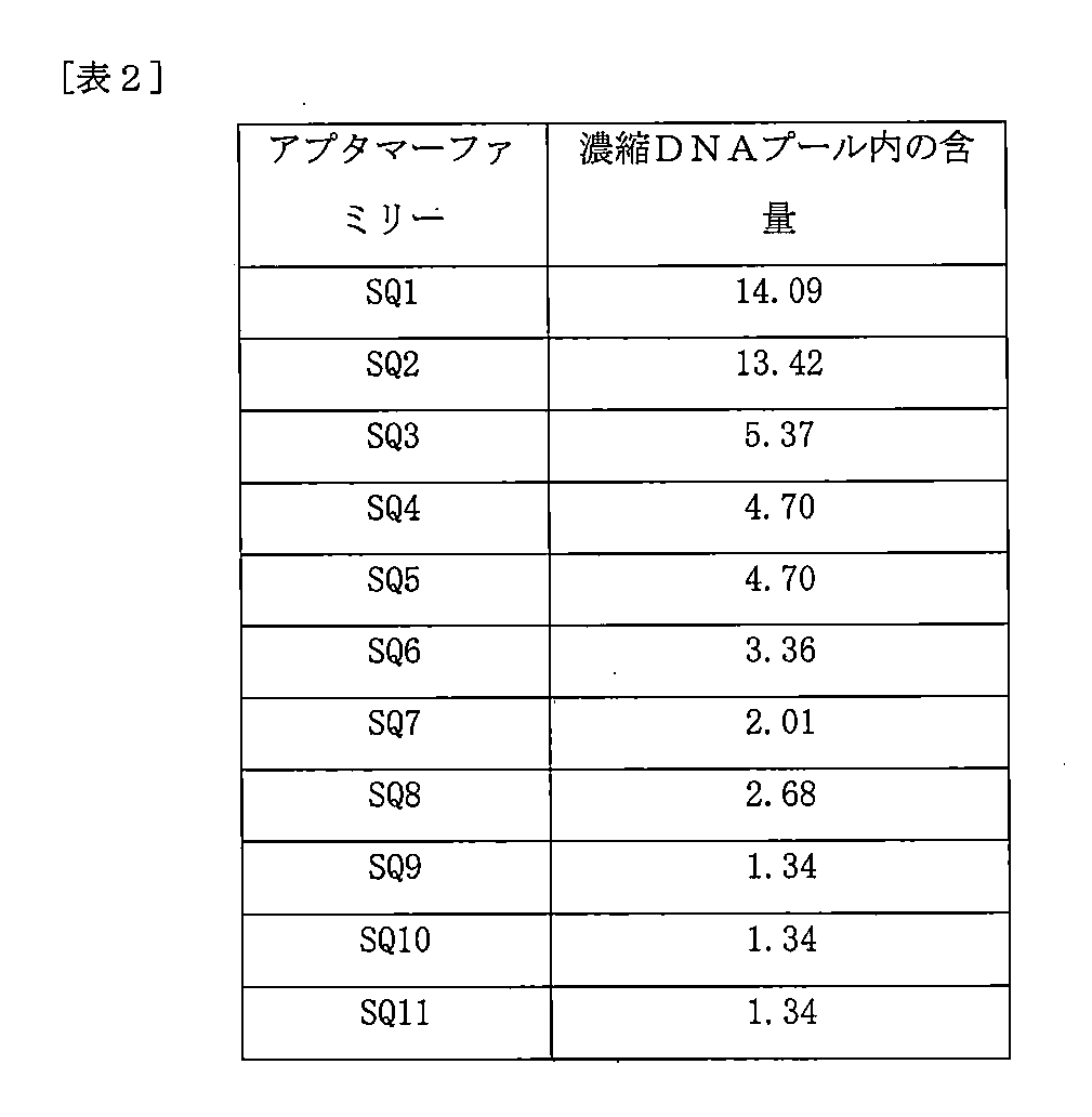
上記(1)~(4)の過程は、図3で図式化された。この過程を通じて配列確認された各アプタマーを類似の配列を有する各アプタマーにグループ化し、これを通じてCy5-標識アプタマーファミリー候補11個(SQ1~SQ11)が確認された。当該アプタマーファミリーのプール全体での含量は、下記表2の通りであった。
The results of confirming the degree of concentration according to each number of times of concentration are shown in FIG.
The processes (1) to (4) above are diagrammatically illustrated in FIG. Through this process, each aptamer whose sequence was confirmed was grouped into aptamers with similar sequences, and through this, 11 Cy5-labeled aptamer family candidates (SQ1 to SQ11) were identified. The content of the aptamer family in the entire pool was as shown in Table 2 below.
[実験例2]濃縮されたアプタマーファミリー候補の標的細胞結合特異性の確認
実験例1の(4)で得られた濃縮されたアプタマーファミリー候補のCMLu-1標的細胞結合特異性をフローサイトメトリー(FACS)を通じて確認した。
[Experimental Example 2] Confirmation of target cell binding specificity of enriched aptamer family candidates The CMLu-1 target cell binding specificity of the enriched aptamer family candidates obtained in (4) of Experimental Example 1 was determined by flow cytometry ( Confirmed via FACS).
各Cy5-標識アプタマーファミリー候補を3×10⁵CMLu-1細胞及びhTERT/HPNE細胞とともにCell-SELEXで用いられた4℃の結合緩衝液で1時間培養した。細胞を0.1%NaN3を含む結合緩衝液で3回洗浄し、結合された配列を有するペレットを結合緩衝液に再懸濁させた。BD FACSCalliburTM及びFACSVerseTM(BD Biosciences,USA)を通じて10,000個の細胞を測定して蛍光分析を行い、データはFlowJo software v10.0.7を用いて分析した。 Each Cy5-labeled aptamer family candidate was incubated with 3×10 5 CMLu-1 cells and hTERT/HPNE cells for 1 hour in the 4° C. binding buffer used in Cell-SELEX. Cells were washed three times with binding buffer containing 0.1% NaN, and the pellet with bound sequences was resuspended in binding buffer. Fluorescence analysis was performed by measuring 10,000 cells through BD FACSCallibur ™ and FACSVerse ™ (BD Biosciences, USA), and the data were analyzed using FlowJo software v10.0.7.
各Cy5-標識アプタマーファミリー候補のターゲット細胞結合力を測定した結果は、図4の通りである。対象ターゲット細胞である転移した膵臓癌細胞(CMLu-1)に結合特異性が最も高いアプタマーであるSQ7を確認し、この配列は下記の通りである。 The results of measuring the target cell binding strength of each Cy5-labeled aptamer family candidate are shown in FIG. SQ7, an aptamer with the highest binding specificity to metastatic pancreatic cancer cells (CMLu-1), which is the target cell of interest, was confirmed, and its sequence is as follows.
*SQ7アプタマー配列
5’-AGCAGCACAGAGGTCAGATGATGTTGGTATATACTTCTTTAGCTTGGAACCAACTCTTGCCCTATGCGTGCTACCGTGAA-3’(配列番号4)
一方、当該SQ7ファミリーに含まれたアプタマー配列は、次の通りである。
*SQ7 aptamer sequence 5'-AGCAGCACAGAGGTCAGATGATGTTGGTATATACTTCTTTAGCTTGGAACCAACTCTTGCCCTATGCGTGCTACCGTGAA-3' (SEQ ID NO: 4)
On the other hand, the aptamer sequences included in the SQ7 family are as follows.

[実験例3]SQ7アプタマーの分析及びアプタマー断片機能の確認
上記実施例2を通じて選ばれたSQ7アプタマーの配列による2次構造を確認した結果は、図5の通りである。また、細胞結合力を再確認するために、SQ7アプタマーと対照群として未処理対照例(NT)、DNAプールライブラリー及びSQ8-Compアプタマー(SQ8アプタマーと一部分で相補的な核酸配列を有するアプタマー;配列番号5)を製造して標的細胞結合力を実験例2と同一の方法でFACSを用いて測定した。その結果は、図6aで確認できるように、対照群であるNT、DNAプールライブラリー、及びSQ8-Compアプタマーは、類似の水準の低い結合力を示した一方、SQ7アプタマーは、標的細胞結合力が顕著に優れた。
[Experimental Example 3] Analysis of SQ7 Aptamer and Confirmation of Aptamer Fragment Function The secondary structure of the SQ7 aptamer selected in Example 2 was confirmed based on the sequence, and the results are shown in FIG. In addition, in order to reconfirm the cell binding ability, we used the SQ7 aptamer, an untreated control example (NT) as a control group, a DNA pool library, and the SQ8-Comp aptamer (an aptamer having a nucleic acid sequence partially complementary to the SQ8 aptamer; SEQ ID NO: 5) was produced and its target cell binding strength was measured using FACS in the same manner as in Experimental Example 2. The results show that the control group NT, DNA pool library, and SQ8-Comp aptamer showed similar levels of low avidity, while the SQ7 aptamer showed low avidity for target cells. was significantly better.
SQ7アプタマーの中に膵臓癌特異的結合に特に重要な部分が存在するか確認するために、SQ7アプタマーの一部分を含む多様なアプタマーを製造して標的細胞との結合力を確認した。細胞結合力は維持しながらアプタマー全体の大きさを減らすことができるならば、アプタマーの製造費用は下げながら細胞内浸透力を増進させることができるものである。結果的には、下記表4の配列のSQ7-1アプタマーが標的細胞結合力をほとんど維持しながらも優れたエンドサイトーシス(endocytosis)能力を有することを確認した。これに追加としてSQ7アプタマー、SQ7-1アプタマー、未処理対照例(NT)、DNAプールライブラリー及びSQ8-Compアプタマーの標的細胞結合力を実験例2と同一の方法でFACSを用いて測定した。その結果は、図6bで確認できるように、対照群であるNT、DNAプールライブラリー、及びSQ8-Compアプタマーは、類似の水準の低い結合力を示した一方、SQ7-1アプタマーは、SQ7アプタマーと同様に標的細胞結合力が顕著に優れた。 In order to confirm whether there is a part of the SQ7 aptamer that is particularly important for pancreatic cancer-specific binding, various aptamers containing a part of the SQ7 aptamer were prepared and their binding ability with target cells was confirmed. If the overall size of the aptamer can be reduced while maintaining cell binding strength, it is possible to increase the intracellular permeability while lowering the manufacturing cost of the aptamer. As a result, it was confirmed that the SQ7-1 aptamer having the sequence shown in Table 4 below had excellent endocytosis ability while maintaining almost the ability to bind to target cells. Additionally, the target cell binding ability of SQ7 aptamer, SQ7-1 aptamer, untreated control (NT), DNA pool library, and SQ8-Comp aptamer was measured using FACS in the same manner as in Experimental Example 2. The results show that the control group NT, DNA pool library, and SQ8-Comp aptamer showed similar levels of low avidity, while the SQ7-1 aptamer Similarly, the target cell binding ability was remarkable.
当該SQ7-1アプタマーの配列は下記の通りであり(配列番号6)、該当する2次構造は図7の通りである。また、次の実験でSQ7-1アプタマーの対照群として用いるために、SQ7-1-Revアプタマー(SQ7-1の反対方向の配列を有するアプタマー;配列番号7)を製造した。 The sequence of the SQ7-1 aptamer is as follows (SEQ ID NO: 6), and the corresponding secondary structure is as shown in FIG. In addition, SQ7-1-Rev aptamer (aptamer having a sequence in the opposite direction of SQ7-1; SEQ ID NO: 7) was prepared for use as a control group for SQ7-1 aptamer in the next experiment.

[実験例4]選択されたアプタマーの細胞及び組織内の効率のよいターゲッティングの確認
(1)選択されたアプタマーのエンドサイトーシス(endocytosis)効率を共焦点顕微鏡イメージングを通じて確認
1×10⁴細胞/ウェルの対照群細胞(HPNE)と標的細胞(C-MLu-1)をpoly-L-lysine(Sigma,USA)でコーティングされた8-ウェルチャンバスライド(Thermo scientific,USA)に実験の4時間前にプレーティングした。洗浄緩衝液で洗浄した後に、4℃の結合緩衝液200ulにCy5-標識アプタマー(250nM)、またはDNAプールライブラリーを添加して培養した。2回洗浄した後、4%パラホルムアルデヒドを用いて細胞を固定させ、Hoechst33342で核を染色した。この後、細胞を共焦点顕微鏡(LSM780,Carl Zeiss,Germany)を用いて映像化し、イメージはソフトウェア(Zen blue edition)で分析した。
[Experiment Example 4] Confirmation of efficient targeting of selected aptamers in cells and tissues
(1) Confirm the endocytosis efficiency of the selected aptamer through confocal microscopy imaging. Control group cells (HPNE) and target cells (C-MLu-1) at 1×10 4 cells/well were isolated using poly-L -lysine (Sigma, USA) coated on 8-well chamber slides (Thermo scientific, USA) 4 hours before the experiment. After washing with washing buffer, Cy5-labeled aptamer (250 nM) or DNA pool library was added to 200 ul of binding buffer at 4°C and cultured. After washing twice, cells were fixed using 4% paraformaldehyde and nuclei were stained with Hoechst 33342. After this, the cells were imaged using a confocal microscope (LSM780, Carl Zeiss, Germany) and the images were analyzed with software (Zen blue edition).
SQ7とSQ7-1アプタマーの共焦点顕微鏡の実験結果は、図8及び図9の通りである。図面で最も明るく見える白色の部分が、アプタマーが多く集まっていることを示す。図8及び図9で見られるように、SQ7及びSQ7-1アプタマーが対照群細胞に比べて膵臓癌細胞を十分にターゲッティングし、細胞内のエンドサイトーシス(内在化)もうまくいくことを確認することができた。 The experimental results of confocal microscopy of SQ7 and SQ7-1 aptamers are shown in FIGS. 8 and 9. The white parts that appear the brightest in the drawing indicate a large concentration of aptamer. As seen in Figures 8 and 9, the SQ7 and SQ7-1 aptamers are well targeted to pancreatic cancer cells compared to control cells, confirming that intracellular endocytosis (internalization) is also successful. I was able to do that.
(2)生体内(in vivo)及び生体外(ex vivo)蛍光イメージングを通じてアプタマーの膵臓癌ターゲティングを確認
雌Hsd:無胸腺ヌード-Foxn1ヌードマウス(Athymic nude-Foxn1 nude mice:6週齢)をHarlan Laboratories,Inc.(フランス)から購入した。これらを光と湿度が制御される条件の無特異病原体(specific pathogen free;SPF)環境に保管し、国立癌センター動物施設で飼料と水の供給を受けた。すべての動物実験は、国立癌センター研究所(NCCRI,unit number:NCC-16-163D)の実験動物運営委員会(Institutional Animal Care and Use Committee;IACUC)から許可を受けて行った。NCCRIは、国際実験動物管理評価認証協会から認可された機関である。
(2) Confirm pancreatic cancer targeting of the aptamer through in vivo and ex vivo fluorescence imaging Female Hsd: Athymic nude-Foxn1 nude mice (6 weeks old) were transferred to Harlan Laboratories, Inc. Purchased from (France). They were kept in a specific pathogen free (SPF) environment with controlled light and humidity conditions, and were provided with food and water at the National Cancer Center Animal Facility. All animal experiments were conducted with permission from the Institutional Animal Care and Use Committee (IACUC) of the National Cancer Center Research Institute (NCCRI, unit number: NCC-16-163D). NCCRI is an organization accredited by the International Association for the Evaluation and Certification of Laboratory Animal Care.
膵臓癌の同所異種移植マウスモデルは、ATCCから購入したCFPAC-1細胞(1x10⁶細胞)をマウス膵尾(tail of pancreas)に注射して構築した。接種3週後にマウスは治療に応じて2つのグループ(Cy5.5-SQ8-Compアプタマー vs Cy5.5-SQ7アプタマーまたはCy5.5-SQ7-1-Revアプタマー vs Cy5.5-SQ7-1アプタマー)に分かれ、上記Cy5.5-標識アプタマー(300pmol/50ulのPBS)を静脈内に投与した。 An orthotopic xenograft mouse model of pancreatic cancer was constructed by injecting CFPAC-1 cells (1×10 6 cells) purchased from ATCC into the mouse tail of pancreas. Three weeks after inoculation, mice were divided into two groups depending on treatment (Cy5.5-SQ8-Comp aptamer vs. Cy5.5-SQ7 aptamer or Cy5.5-SQ7-1-Rev aptamer vs. Cy5.5-SQ7-1 aptamer). The Cy5.5-labeled aptamer (300 pmol/50 ul of PBS) was administered intravenously.
生体外実験のためには、投与15分後または3時間後にマウスを犠牲にして解剖した。腫瘍組織を得て、IVIS Lumina(Caliper Life Science,Hopkinton,MA,USA)を用いて生体発光イメージングに映像化した。すべての映像データは、ソフトウェア(Living Image Acquisition and Analysis software)を用いて分析した。 For in vitro experiments, mice were sacrificed and dissected 15 minutes or 3 hours after administration. Tumor tissue was obtained and imaged for bioluminescent imaging using IVIS Lumina (Caliper Life Science, Hopkinton, MA, USA). All image data were analyzed using Living Image Acquisition and Analysis software.
SQ7とSQ7-1アプタマーの生体発光イメージング実験結果は、図10及び図11のグレースケール(Gray scale)図の通りである。また、イメージング結果から得たフラックス全体(total flux)を図10及び図11にグラフで示した。 The results of the bioluminescence imaging experiment of SQ7 and SQ7-1 aptamers are shown in gray scale diagrams in FIGS. 10 and 11. Further, the total flux obtained from the imaging results is shown in graphs in FIGS. 10 and 11.
各図の上段のイメージング結果では、白色で明るく示されるほど細胞にアプタマーが多く結合したということを意味する。SQ7及びSQ7-1アプタマーが他のアプタマーに比べてイメージング結果が白色で示されることを確認することができる。 In the imaging results at the top of each figure, the brighter the color, the more aptamer has bound to the cell. It can be seen that the imaging results of SQ7 and SQ7-1 aptamers appear white compared to other aptamers.
従って、本開示のアプタマーが他の対照群アプタマー(SQ1アプタマー(配列番号17)、SQ8-Compアプタマー、及びSQ7-1-Revアプタマー)に比べ、膵臓癌組織に特異的に移動して結合するということを確認することができ、膵臓癌組織に対する顕著に優れたターゲッティング効率を有することを確認することができた。 Therefore, the aptamer of the present disclosure specifically migrates and binds to pancreatic cancer tissue compared to other control aptamers (SQ1 aptamer (SEQ ID NO: 17), SQ8-Comp aptamer, and SQ7-1-Rev aptamer). We were able to confirm that this method had significantly superior targeting efficiency for pancreatic cancer tissue.
[実験例5]アプタマーの血清内安定性の増進のための変形体の製造及び確認
上記実験例を通じて製造したSQ7-1アプタマーに基づいてアプタマーの血清内安定性を増進させるために、内部2’-O-メチル-変形アプタマー(internal 2’-O-methyl-modified aptamers)を製造した。具体的には、SQ7-1アプタマーの2次構造で領域を分けてSQ7-1(1)、SQ7-1(2)、SQ7-1(3)、SQ7-1(4)、SQ7-1(5)、SQ7-1(6)、及びSQ7-1(1,5)アプタマーを製造した(図12a~図12d)。
[Experimental Example 5] Production and confirmation of a variant for enhancing the serum stability of aptamer In order to enhance the serum stability of the aptamer based on the SQ7-1 aptamer produced through the above experimental example, the internal 2' -O-methyl-modified aptamers (internal 2'-O-methyl-modified aptamers) were prepared. Specifically, we divided the regions according to the secondary structure of the SQ7-1 aptamer and produced SQ7-1(1), SQ7-1(2), SQ7-1(3), SQ7-1(4), and SQ7-1( 5), SQ7-1(6), and SQ7-1(1,5) aptamers were produced (Figures 12a to 12d).
製造した内部2’-O-メチル-変形アプタマーの配列を整理すると、下記表5の通りである。 The arrangement of the internal 2'-O-methyl-modified aptamer prepared is shown in Table 5 below.

このように製造した5ugの内部2’-O-メチル-変形アプタマーを37℃でマウス血清に0、0.1、0.5、2、6、及び24時間培養した。それぞれの時点でサンプルにDNase活性を止めるために0.5M EDTAを添加し、EtOH-NaOAcを添加して沈殿させた。DNA-アプタマー-沈殿物サンプルはHPLCで分析した。 5ug of the internal 2'-O-methyl-modified aptamer thus prepared was incubated in mouse serum at 37°C for 0, 0.1, 0.5, 2, 6, and 24 hours. At each time point, samples were added with 0.5 M EDTA to stop DNase activity and precipitated with EtOH-NaOAc. DNA-aptamer-precipitate samples were analyzed by HPLC.
クロマトグラフィー分析は、可変波長測定機(variable wavelength detector;VWD)であるQuatpumpが装着されたWaters e2695 HPLC system(MA,USA)で行われた。LCのためのEmpower3 personal single system softwareを装着したパーソナルコンピュータをクロマトグラフデータを処理するのに用いた。分析物は、Agela Technologies Inc.(北京、中国)から購入したVenusil、XBP C18カラム(250mm × 4.6mm、5um)を用いて分離された。移動相はメタノール-水(55:45、体積比)混合物であり、流速は0.5mL/分であった。カラム温度は30℃であり、測定波長は260nmであった。Shanghai GaoGe Industrial and Trading Co.,Ltd.(上海、中国)から購入した15uL LCマイクロシリンジを注射として用いた。 Chromatographic analysis was performed on a Waters e2695 HPLC system (MA, USA) equipped with a variable wavelength detector (VWD) Quatpump. A personal computer equipped with Empower3 personal single system software for LC was used to process the chromatographic data. Analytes were purchased from Agela Technologies Inc. Separation was performed using a Venusil, XBP C18 column (250 mm x 4.6 mm, 5 um) purchased from (Beijing, China). The mobile phase was a methanol-water (55:45, volume ratio) mixture, and the flow rate was 0.5 mL/min. The column temperature was 30° C. and the measurement wavelength was 260 nm. Shanghai GaoGe Industrial and Trading Co.,Ltd. A 15uL LC microsyringe purchased from (Shanghai, China) was used as an injection.
その結果、SQ7-1(1)、SQ7-1(5)、及びSQ7-1(1,5)アプタマーの半減期が顕著に増加した。具体的には、図13(図13a~図13d)で見られるように、SQ7-1(1)、SQ7-1(5)、及びSQ7-1(1,5)アプタマーの半減期がSQ7-1アプタマーに比べてそれぞれ91倍、145倍、及び760倍増加することを確認した。これらの標的細胞に対する結合力は、SQ7-1アプタマーに比べて多少減少したが、DNAプールライブラリー(グラフの一番左側の細い実線)に比べては明確に区別されるほど高かった(図14)。 As a result, the half-lives of SQ7-1(1), SQ7-1(5), and SQ7-1(1,5) aptamers were significantly increased. Specifically, as seen in Figure 13 (Figures 13a to 13d), the half-lives of SQ7-1(1), SQ7-1(5), and SQ7-1(1,5) aptamers are It was confirmed that the increase was 91 times, 145 times, and 760 times, respectively, compared to 1 aptamer. Although the binding power to these target cells was somewhat decreased compared to the SQ7-1 aptamer, it was so high that it was clearly distinguishable compared to the DNA pool library (thin solid line on the far left of the graph) (Figure 14 ).
まとめると、上記言及した通り、本開示によるDNAアプタマーは、膵臓癌に高い結合力を持って特異的に結合する特徴を有し、選別されたアプタマーのサイズを減らして標的細胞または組織に対するターゲッティング効率を上げ、DNaseに対する抵抗性を増進させる変形を通じて血中安全性を高めることができることを確認した。 In summary, as mentioned above, the DNA aptamer according to the present disclosure has the characteristics of specifically binding to pancreatic cancer with high avidity, and reduces the size of the selected aptamer to improve targeting efficiency to target cells or tissues. It was confirmed that blood safety can be improved through modification that increases the resistance to DNase.
[実験例6]ヒト患者膵臓癌組織のマウス同所異種移植モデル(Patient-Derived Orthotopic Xenograft Model;PDOXモデル)でのターゲッティングの確認
本開示のアプタマーが患者腫瘍組織の複雑性(complexity)及び異質性(heterogeneity)を反映できる患者-由来同所異種移植モデル(「PDOXモデル」)でも優れたターゲッティング効率を有するか確認するために、蛍光イメージングを用いた生体内(in vivo)検証実験を行った。
[Experimental Example 6] Confirmation of targeting in mouse orthotopic xenograft model (Patient-Derived Orthotopic Xenograft Model; PDOX model) of human patient pancreatic cancer tissue In order to confirm whether a patient-derived orthotopic xenograft model (“PDOX model”) that can reflect (heterogeneity) also has excellent targeting efficiency, an in vivo validation experiment using fluorescence imaging was performed.
国立癌センター医生命研究審議委員会(Institutional Review Board;「IRB」)の審議を経た後、研究に同意した患者を対象に検体を確保してヌードマウス(Harlan Laboratories,Inc.(フランス)から購入)の膵臓に直接移植した患者-由来同所異種移植モデル(patient-derived orthotopic xenograft,PDOX)を製作した。膵臓癌患者の原発性腫瘍検体(以下「HPT」と称する)は、手術的切除直後に、手術が不可能な進行性膵臓癌患者の場合は、患者から肝転移組織生検検体(以下「GUN」と称する)を採取した直後に、培地に浸されたチューブに入れて移動してできるだけ速かに雌Hsd:無胸腺ヌード-Foxn1ヌードマウス(実験例4と同一に入手)の膵臓の膵尾に切除及び縫合して移植した(PDOX1世代、F1)。その後、周期的に腹部促進及びMRI映像装備を通じて腫瘍の大きさを測定し、腫瘍の大きさが3000mm3に到達すると、マウスを犠牲にして腫瘍組織を得た後、一定の大きさ(3mm×3mm×3mm)の腫瘍組織を多数の個体のヌードマウスに同所再移植して次世代(F2,F3,F4…)を形成させ、個体数を増幅した。 After deliberation by the National Cancer Center Institutional Review Board (IRB), samples were secured from patients who consented to the research and purchased from nude mice (Harlan Laboratories, Inc. (France)). A patient-derived orthotopic xenograft (PDOX) model was created that was directly transplanted into the pancreas of a patient. Primary tumor specimens (hereinafter referred to as “HPT”) from patients with pancreatic cancer are collected immediately after surgical resection, and liver metastasis tissue biopsy specimens (hereinafter referred to as “GUN”) are collected from patients with advanced pancreatic cancer where surgery is not possible. Immediately after collecting the mouse, place it in a tube immersed in culture medium and move it as quickly as possible to the pancreatic tail of the pancreas of a female Hsd:Athymic nude-Foxn1 nude mouse (obtained in the same manner as in Experiment 4). It was excised and sutured and transplanted (PDOX1 generation, F1). After that, the tumor size was periodically measured through abdominal stimulation and MRI imaging equipment, and when the tumor size reached 3000 mm3 , the mouse was sacrificed to obtain tumor tissue, and then a certain size (3 mm x Tumor tissue (3 mm x 3 mm) was orthotopically reimplanted into multiple individual nude mice to form the next generation (F2, F3, F4...) and expand the population.
4世代(F4)で構築された患者-由来同所異種移植マウスモデルを三匹ずつ2つのグループに分け、それぞれCy5.5が標識されたアプタマー(300pmol/50ulのPBS)であるSQ7-1アプタマーとSQ7-1-Revアプタマーを静脈投与した。投与15分後にマウスを犠牲にした後、解剖して腫瘍組織を得た後、IVIS Lumina(Caliper Life Science,Hopkinton,MA,USA)を用いて生体発光イメージングに映像化した。すべての映像データは、ソフトウェア(Living Image Acquisition and Analysis software)を用いて分析した。生体発光イメージング実験結果は、図15のグレースケール(Gray scale)図の通りであり、イメージング結果から得たフラックス全体(total flux)を図15にグラフで示した。 The patient-derived orthotopic xenograft mouse model constructed in the fourth generation (F4) was divided into two groups of three animals each, and each group was treated with SQ7-1 aptamer, a Cy5.5-labeled aptamer (300 pmol/50 ul of PBS). and SQ7-1-Rev aptamer were administered intravenously. Mice were sacrificed 15 minutes after administration and then dissected to obtain tumor tissue, which was imaged using IVIS Lumina (Caliper Life Science, Hopkinton, MA, USA) for bioluminescent imaging. All image data were analyzed using Living Image Acquisition and Analysis software. The results of the bioluminescence imaging experiment are shown in the gray scale diagram of FIG. 15, and the total flux obtained from the imaging results is shown graphically in FIG.
図10及び図11のように、図15でも図面の上段のイメージング結果では、白色で明るく示されるほど細胞にアプタマーが多く結合したということを意味し、SQ7-1アプタマーは、SQ7-1-Revアプタマーに比べて白色水準が非常に高いことを確認することができる。 As shown in FIGS. 10 and 11, in the imaging results shown in the upper part of the drawing in FIG. 15, the brighter the white color, the more the aptamer has bound to the cells. It can be confirmed that the white level is much higher than that of the aptamer.
即ち、本開示のアプタマーは、患者腫瘍組織の複雑性(complexity)及び異質性(heterogeneity)を保有しているPDOXモデルでも顕著に優れたターゲッティング効率を有していることを確認することができた。 That is, it was confirmed that the aptamer of the present disclosure has significantly superior targeting efficiency even in the PDOX model, which has the complexity and heterogeneity of patient tumor tissue. .
[実験例7]多様な膵臓癌細胞株での結合能の確認
多様な種類の膵臓癌細胞株に対する本開示のアプタマーの結合能を確認するために、追加のフローサイトメトリー(FACS)実験を行った。
[Experimental Example 7] Confirmation of binding ability with various pancreatic cancer cell lines In order to confirm the binding ability of the aptamer of the present disclosure with various types of pancreatic cancer cell lines, additional flow cytometry (FACS) experiments were conducted. Ta.
[抗体結合アプタマーの製造]
抗体結合アプタマーの製造のためにジゴキシゲニン(Digoxigenin)と標識された250nM濃度のSQ7-1アプタマーと125nM濃度の抗-ジゴキシゲニン-抗体(Abcam;Cat. No. ab420,USA)をともに入れて常温で30分間混ぜた。
[Production of antibody-binding aptamer]
To prepare an antibody-binding aptamer, 250 nM SQ7-1 aptamer labeled with digoxigenin and 125 nM anti-digoxigenin antibody (Abcam; Cat. No. ab420, USA) were mixed together at room temperature for 30 min. Mixed for a minute.
実験例2と同一の方法でフローサイトメトリーを行うものの、上記製造した抗体結合アプタマーを用い、2次抗体(secondary antibody)はアレクサ(Alexa)488と標識された抗-マウス-免疫グロブリンG(anti-mouse IgG)(Invitrogen,USA)を4μg/ml濃度で用いた。細胞は、CFPAC-1細胞株、SNU-213細胞株、SNU-410細胞株、Capan-2細胞株、HPAF-II細胞株、AsPC-1細胞株、Capan-1細胞株、MIA PaCa細胞株、BxPC-3細胞株、及びPANC-1細胞株をそれぞれ用いた。このような細胞株は、いずれもATCCから購入した。 Flow cytometry was carried out in the same manner as in Experimental Example 2, but the antibody-binding aptamer prepared above was used, and the secondary antibody was anti-mouse-immunoglobulin G (anti-mouse-immunoglobulin G) labeled with Alexa 488. -mouse IgG) (Invitrogen, USA) was used at a concentration of 4 μg/ml. The cells include CFPAC-1 cell line, SNU-213 cell line, SNU-410 cell line, Capan-2 cell line, HPAF-II cell line, AsPC-1 cell line, Capan-1 cell line, MIA PaCa cell line, BxPC-3 cell line and PANC-1 cell line were used, respectively. All such cell lines were purchased from ATCC.
多様な膵臓癌細胞株に対する、各アプタマーの結合力を測定した結果を図16(図16a~図16b)及び図17に示した。図17は、SQ7-1-Revアプタマーに対するSQ7-1アプタマーの相対的な蛍光強度の幾何平均値を倍数で示したものである。 The results of measuring the binding strength of each aptamer to various pancreatic cancer cell lines are shown in FIG. 16 (FIGS. 16a and 16b) and FIG. 17. FIG. 17 shows the geometric mean value of the relative fluorescence intensity of the SQ7-1 aptamer to the SQ7-1-Rev aptamer in multiples.
図16及び17の結果によると、本開示のSQ7-1アプタマーは、SQ7-1-Revアプタマーに比べ、すべての膵臓癌細胞株に対してさらに優れて特異的に結合することを確認することができる。先に詳察した実験例4及び6の結果と総合すると、本開示のアプタマーは、多様な類型の膵臓癌細胞に特異的に結合するものであることを確認することができる。 According to the results shown in FIGS. 16 and 17, it can be confirmed that the SQ7-1 aptamer of the present disclosure binds more efficiently and specifically to all pancreatic cancer cell lines than the SQ7-1-Rev aptamer. can. When combined with the results of Experimental Examples 4 and 6 detailed above, it can be confirmed that the aptamer of the present disclosure specifically binds to various types of pancreatic cancer cells.
また、SQ7アプタマーはSQ7-1アプタマーを含むものであるので、同様に多様な類型の膵臓癌細胞に特異的に結合するものである。 Furthermore, since the SQ7 aptamer includes the SQ7-1 aptamer, it also specifically binds to various types of pancreatic cancer cells.
[実験例8]多様な癌腫細胞株での結合能の確認
多様な癌腫の細胞株に対する本開示のアプタマーの結合能を確認するために、追加のフローサイトメトリー(FACS)実験を行った。
[Experimental Example 8] Confirmation of binding ability with various cancer cell lines In order to confirm the binding ability of the aptamer of the present disclosure with various cancer cell lines, additional flow cytometry (FACS) experiments were conducted.
実験例2と同一の方法でフローサイトメトリーを行うものの、実験例7で用いた抗体結合アプタマーと2次抗体を同一に用いた。細胞は、U87細胞株、U251細胞株、CAL27細胞株、HEP3B細胞株、A549細胞株、HCT116細胞株、SK-OV3細胞株、ES-2細胞株、MCF7細胞株、SK-BR3細胞株、NCI-N87細胞株、KPL4細胞株、BT-474細胞株、MDA-MB231細胞株、及びHCC1938細胞株をそれぞれ用いた。このような細胞株は、いずれもATCCから購入した。 Although flow cytometry was performed in the same manner as in Experimental Example 2, the same antibody-binding aptamer and secondary antibody used in Experimental Example 7 were used. Cells include U87 cell line, U251 cell line, CAL27 cell line, HEP3B cell line, A549 cell line, HCT116 cell line, SK-OV3 cell line, ES-2 cell line, MCF7 cell line, SK-BR3 cell line, NCI - N87 cell line, KPL4 cell line, BT-474 cell line, MDA-MB231 cell line, and HCC1938 cell line were used, respectively. All such cell lines were purchased from ATCC.
多様な癌細胞株に対する、各アプタマーの結合力を測定した結果を図18に示した。図18は、SQ7-1-Revアプタマーに対するSQ7-1アプタマーの相対的な蛍光強度の幾何平均値を倍数で示したものである。 The results of measuring the binding strength of each aptamer to various cancer cell lines are shown in FIG. FIG. 18 shows the geometric mean value of the relative fluorescence intensity of the SQ7-1 aptamer to the SQ7-1-Rev aptamer in multiples.
図18の結果によると、本開示のSQ7-1アプタマーはSQ7-1-Revアプタマーに比べ、多様な癌細胞株、即ち、大腸癌、肝癌、肺癌、脳腫瘍、口腔癌、卵巣癌及び乳癌の細胞株に対してさらに優れて特異的に結合することを確認することができる。先に詳察した実験例4及び6の結果と総合すると、本開示のアプタマーは多様な類型の癌細胞に特異的に結合するものであることを確認することができる。 According to the results in FIG. 18, the SQ7-1 aptamer of the present disclosure is more effective than the SQ7-1-Rev aptamer in cells of various cancer cell lines, namely, colon cancer, liver cancer, lung cancer, brain tumor, oral cancer, ovarian cancer, and breast cancer. It can be confirmed that the protein binds more specifically to the strain. When combined with the results of Experimental Examples 4 and 6 detailed above, it can be confirmed that the aptamer of the present disclosure specifically binds to various types of cancer cells.
また、SQ7アプタマーはSQ7-1アプタマーを含むものであるので、同様に多様な類型の癌細胞に特異的に結合するものである。 Furthermore, since the SQ7 aptamer includes the SQ7-1 aptamer, it similarly binds specifically to various types of cancer cells.
[実験例9]膵管腺癌腫のPDOX-由来細胞株での結合能の確認
本開示のアプタマーが、実際の臨床患者の膵臓癌細胞に特異的に十分に結合する可能性を有するか否かを確認するために、追加のフローサイトメトリー(FACS)実験を行った。
[Experimental Example 9] Confirmation of binding ability in PDOX-derived cell line of pancreatic ductal adenocarcinoma It was determined whether the aptamer of the present disclosure has the potential to specifically and sufficiently bind to pancreatic cancer cells of actual clinical patients. Additional flow cytometry (FACS) experiments were performed to confirm.
限定された細胞分裂後に死滅する正常細胞とは異なり、癌細胞は無限分裂の特性を有しているので、腫瘍組織から分離された癌細胞は、形質転換がなくても無限増殖が可能な細胞株を形成することができ、患者の臨床的、分子生物学的な特性を反映させることができると考えられる。本実験で、膵臓癌PDOXマウスから摘出した腫瘍組織を3mm×4mmに切り出した後、細胞を結合組織から分離するためにコラゲナーゼ(collagenase)が含まれたHuman cell dissociation kit(Miltenyi Biotech Inc.)と混合して組織解離装置(Gentle Macs,Miltenyi Biotech Inc)で1時間反応させる。反応終了の後、ウシ胎児血清(fetal bovine serum;FBS)が含まれたRPMI培地で酵素活性を抑制し、続いて遠心分離すれば、組織から解離した細胞の沈殿物を得ることができる。ウシ胎児血清が含まれたRPMI培地でこれを懸濁して10cmの培養皿に2×10⁶個の水準に細胞を一様に敷き、二日ごとに培地を交換しながら正常の線維芽細胞と死んだ細胞を除去して膵臓癌患者別のPDOX由来癌細胞株を確立した。確立した各細胞株の命名は、由来したPDOXの命名と同一である。 Unlike normal cells, which die after a limited number of cell divisions, cancer cells have the property of infinite division, so cancer cells isolated from tumor tissue are cells that can proliferate indefinitely without transformation. It is thought that the clinical and molecular biological characteristics of the patient can be reflected. In this experiment, tumor tissue removed from pancreatic cancer PDOX mice was cut into 3 mm x 4 mm pieces, and then a human cell dissociation kit (Miltenyi Biotech Inc.) containing collagenase was used to separate the cells from connective tissue. Mix and react for 1 hour in a tissue dissociator (Gentle Macs, Miltenyi Biotech Inc). After the reaction is completed, enzyme activity is inhibited with RPMI medium containing fetal bovine serum (FBS), followed by centrifugation to obtain a precipitate of cells dissociated from the tissue. Suspend this in RPMI medium containing fetal bovine serum, spread the cells uniformly at 2 x 10 cells in a 10 cm culture dish, and mix them with normal fibroblasts while replacing the medium every two days. Dead cells were removed to establish PDOX-derived cancer cell lines for each pancreatic cancer patient. The nomenclature of each established cell line is identical to that of the PDOX from which it was derived.
肝転移患者生検組織を用いたPDOXモデル(GUN#13、GUN#16、GUN#20、GUN#34、GUN#38、GUN#41及びGUN#46)、超音波内視鏡微細検体を用いたPDOXモデル(EUS#16)、及び手術的切除検体を用いたPDOXモデル(HPT#19、HPT#22、HPT#43、HPT#45、及びHPT#48)の腫瘍組織から分離した癌細胞株を対象に実験例2と同一の方法でフローサイトメトリーを行うものの、実験例7で用いた抗体結合アプタマーを同一に用いた。
PDOX model (GUN #13, GUN #16,
多様なPDOX-由来細胞株に対する、各アプタマーの結合力を測定した結果を図19に示した。図19は、SQ7-1-Revアプタマーに対するSQ7-1アプタマーの相対的な蛍光強度の幾何平均値を倍数で示したものである。 The results of measuring the binding strength of each aptamer to various PDOX-derived cell lines are shown in FIG. FIG. 19 shows the geometric mean value of the relative fluorescence intensity of the SQ7-1 aptamer to the SQ7-1-Rev aptamer in multiples.
図19の結果によると、本開示のSQ7-1アプタマーは、SQ7-1-Revアプタマーに比べ、複数の患者から得た膵臓癌組織から由来した多様なPDOX-由来細胞株に対してさらに優れて特異的に結合することを確認することができる。先に詳察した実験例4及び6の結果と総合すると、本開示のアプタマーは、実際の臨床で患者の膵臓癌組織に特異的に結合するものであることを確認することができる。 According to the results in FIG. 19, the SQ7-1 aptamer of the present disclosure is more effective than the SQ7-1-Rev aptamer against various PDOX-derived cell lines derived from pancreatic cancer tissues obtained from multiple patients. Specific binding can be confirmed. When combined with the results of Experimental Examples 4 and 6 detailed above, it can be confirmed that the aptamer of the present disclosure specifically binds to pancreatic cancer tissue of patients in actual clinical practice.
また、SQ7アプタマーは、SQ7-1アプタマーを含むものであるので、同様に患者の膵臓癌組織に特異的に結合するものである。 Furthermore, since the SQ7 aptamer includes the SQ7-1 aptamer, it similarly binds specifically to the patient's pancreatic cancer tissue.
以上、一部の実施例と添付の図面に示された例によって本開示の技術的思想が説明されたが、本開示の属する技術分野で通常の知識を有する者が理解できる本開示の技術的思想及び範囲を逸脱しない範囲で多様な置換、変形及び変更がなされ得るという点を理解すべきである。また、そのような置換、変形及び変更は、添付の請求の範囲内に属すると考えられなければならない。 The technical idea of the present disclosure has been explained above using some embodiments and examples shown in the attached drawings, but the technical idea of the present disclosure can be understood by a person having ordinary knowledge in the technical field to which the present disclosure pertains. It should be understood that various substitutions, modifications and changes may be made without departing from the spirit and scope. Also, such substitutions, modifications and changes are to be considered as falling within the scope of the appended claims.
Claims (11)
前記変形は1つ以上のヌクレオチド内の糖構造の2’炭素位置で-OH基が-Me(メチル)、-OMe、-NH 2 、-F(フッ素)、-O-2-メトキシエチル-O-プロピル、-O-2-メチルチオエチル(methylthioethyl)、-O-3-アミノプロピル、-O-3-ジメチルアミノプロピル、-O-N-メチルアセトアミドまたは-O-ジメチルアミドオキシエチルへの置換による変形であるものである、DNAアプタマー。 The DNA aptamer according to claim 1 , wherein the aptamer is modified to have resistance to DNase ,
Said variations include -OH groups at the 2' carbon position of the sugar structure in one or more nucleotides such as -Me (methyl), -OMe, -NH 2 , -F (fluorine), -O-2-methoxyethyl-O By substitution with -propyl, -O-2-methylthioethyl, -O-3-aminopropyl, -O-3-dimethylaminopropyl, -O-N-methylacetamide or -O-dimethylamidooxyethyl A DNA aptamer that is a variant .
Applications Claiming Priority (3)
| Application Number | Priority Date | Filing Date | Title |
|---|---|---|---|
| KR20180167948 | 2018-12-21 | ||
| KR10-2018-0167948 | 2018-12-21 | ||
| PCT/KR2019/009956 WO2020130269A1 (en) | 2018-12-21 | 2019-08-08 | Novel dna aptamer and use thereof |
Publications (3)
| Publication Number | Publication Date |
|---|---|
| JP2022516054A JP2022516054A (en) | 2022-02-24 |
| JPWO2020130269A5 JPWO2020130269A5 (en) | 2022-07-14 |
| JP7391971B2 true JP7391971B2 (en) | 2023-12-05 |
Family
ID=71100532
Family Applications (1)
| Application Number | Title | Priority Date | Filing Date |
|---|---|---|---|
| JP2021536744A Active JP7391971B2 (en) | 2018-12-21 | 2019-08-08 | Novel DNA aptamer and its uses |
Country Status (6)
| Country | Link |
|---|---|
| US (1) | US20220090081A1 (en) |
| EP (1) | EP3901287A4 (en) |
| JP (1) | JP7391971B2 (en) |
| KR (1) | KR102324242B1 (en) |
| CN (1) | CN113646442A (en) |
| WO (2) | WO2020130225A1 (en) |
Families Citing this family (3)
| Publication number | Priority date | Publication date | Assignee | Title |
|---|---|---|---|---|
| WO2020130225A1 (en) * | 2018-12-21 | 2020-06-25 | 국립암센터 | Dna aptamer specifically binding to pancreatic cancer and use thereof |
| KR102506295B1 (en) | 2020-08-28 | 2023-03-08 | 국립암센터 | A humanized antibody to Digoxigenin and use thereof |
| KR102506288B1 (en) * | 2020-09-07 | 2023-03-06 | 국립암센터 | A complex comprising an antibody to Digoxigenin and use thereof |
Citations (2)
| Publication number | Priority date | Publication date | Assignee | Title |
|---|---|---|---|---|
| JP2012528587A (en) | 2009-06-01 | 2012-11-15 | ソンギュングヮン ユニバーシティ ファウンデーション フォー コーポレート コラボレーション | Nucleic acid aptamer capable of specifically binding to pancreatic cancer cells or tissues and use thereof |
| WO2017073535A1 (en) | 2015-10-30 | 2017-05-04 | タグシクス・バイオ株式会社 | Dna aptamer binding to cancer cell |
Family Cites Families (7)
| Publication number | Priority date | Publication date | Assignee | Title |
|---|---|---|---|---|
| KR101250557B1 (en) * | 2011-05-18 | 2013-04-03 | 국립암센터 | PAUF-specific aptamer and therapeutic composition for treatment of pancreatic cancer comprising thereof |
| KR101699105B1 (en) * | 2012-04-02 | 2017-01-23 | 성균관대학교산학협력단 | A method for the diagnosis of pancreatic cancer using ALPPL2 protein |
| KR101568400B1 (en) * | 2012-08-09 | 2015-11-12 | 연세대학교 산학협력단 | Nucleic Acid Aptamers Specifically Binding to Cancer Stem Cells and Their Uses |
| KR101458947B1 (en) * | 2013-03-05 | 2014-11-12 | 국립암센터 | Interleukin-8 aptamers and uses thereof |
| WO2020130225A1 (en) * | 2018-12-21 | 2020-06-25 | 국립암센터 | Dna aptamer specifically binding to pancreatic cancer and use thereof |
| KR102506295B1 (en) * | 2020-08-28 | 2023-03-08 | 국립암센터 | A humanized antibody to Digoxigenin and use thereof |
| KR102506288B1 (en) * | 2020-09-07 | 2023-03-06 | 국립암센터 | A complex comprising an antibody to Digoxigenin and use thereof |
-
2019
- 2019-02-01 WO PCT/KR2019/001416 patent/WO2020130225A1/en active Application Filing
- 2019-08-08 JP JP2021536744A patent/JP7391971B2/en active Active
- 2019-08-08 KR KR1020190096665A patent/KR102324242B1/en active IP Right Grant
- 2019-08-08 US US17/416,858 patent/US20220090081A1/en active Pending
- 2019-08-08 EP EP19900348.4A patent/EP3901287A4/en active Pending
- 2019-08-08 CN CN201980092801.6A patent/CN113646442A/en active Pending
- 2019-08-08 WO PCT/KR2019/009956 patent/WO2020130269A1/en unknown
Patent Citations (2)
| Publication number | Priority date | Publication date | Assignee | Title |
|---|---|---|---|---|
| JP2012528587A (en) | 2009-06-01 | 2012-11-15 | ソンギュングヮン ユニバーシティ ファウンデーション フォー コーポレート コラボレーション | Nucleic acid aptamer capable of specifically binding to pancreatic cancer cells or tissues and use thereof |
| WO2017073535A1 (en) | 2015-10-30 | 2017-05-04 | タグシクス・バイオ株式会社 | Dna aptamer binding to cancer cell |
Non-Patent Citations (3)
| Title |
|---|
| CHAMPANHAC, C. et al.,Development of a panel of DNA aptamers with high affinity for pancreatic ductal adenocarcinoma,Scientific Reports,2015年,vol. 5,16788 |
| CLAWSON, G. A. et al.,A cholecystokinin B receptor-specific DNA aptamer for targeting pancreatic ductal adenocarcinoma,Nucleic Acid Therapeutics,2017年,vol.27, no.1,p.23-35 |
| WU, X. et al.,DNA aptamer selected against pancreatic ductal adenocarcinoma for in vivo imaging and clinical tissue recognition,Theranostics,2015年,vol.5, no.9,p.985-994 |
Also Published As
| Publication number | Publication date |
|---|---|
| KR102324242B1 (en) | 2021-11-11 |
| US20220090081A1 (en) | 2022-03-24 |
| CN113646442A (en) | 2021-11-12 |
| JP2022516054A (en) | 2022-02-24 |
| KR20200078303A (en) | 2020-07-01 |
| WO2020130269A1 (en) | 2020-06-25 |
| EP3901287A4 (en) | 2022-10-26 |
| WO2020130225A1 (en) | 2020-06-25 |
| EP3901287A1 (en) | 2021-10-27 |
Similar Documents
| Publication | Publication Date | Title |
|---|---|---|
| JP7391971B2 (en) | Novel DNA aptamer and its uses | |
| ES2928111T3 (en) | Inhibition of AXL signaling in antimetastatic therapy | |
| CN107001417B (en) | Targeting peptides and methods of use thereof | |
| CN109072240A (en) | Use the composition and method of piRNA diagnosing and treating cancer | |
| CN104781417B (en) | For detecting the CD133 aptamers of cancer stem cell | |
| JP2021506802A (en) | Targeting with fibronectin type III-like domain molecules | |
| US20220073921A1 (en) | Aptamer and use of the aptamer in the diagnosis and treatment of cancer | |
| Civit et al. | Targeting hormone refractory prostate cancer by in vivo selected DNA libraries in an orthotopic xenograft mouse model | |
| Hu et al. | TLS11a aptamer/CD3 antibody anti-tumor system for liver cancer | |
| CN106794216A (en) | The peptide of different mucoprotein SND1 interactions is blocked as the purposes for the treatment of of cancer | |
| CN110049999A (en) | Anti- PSMA antibody, its purposes and its conjugate | |
| CN107868786B (en) | Single-stranded DNA aptamer of multidrug resistant colon cancer cell | |
| CN108350459B (en) | Cancer cell-binding DNA aptamers | |
| Yang et al. | Neoantigen vaccination augments antitumor effects of anti-PD-1 on mouse hepatocellular carcinoma | |
| JPWO2011126010A1 (en) | Cancer cell-selective membrane-permeable peptide and use thereof | |
| KR20190040824A (en) | Fusion protein for CRISP/Cas system and complex comprising the same and uses thereof | |
| JP7504985B2 (en) | Novel DNA aptamer and its uses | |
| US20230285589A1 (en) | Liposome complex for cancer treatment, comprising novel cd47 binder and polynucleotide | |
| JP2003527398A (en) | Endothelial specific targeting | |
| JP2023528293A (en) | Single domain antibodies and their use in cancer therapy | |
| US20230322893A1 (en) | Cd47 binder and liposome complex for cancer treatment | |
| KR20180129585A (en) | MAGE-1 specific aptamer and use thereof | |
| Zeng et al. | Identification of a novel peptide targeting TIGIT to evaluate immunomodulation of 125I seed brachytherapy in HCC by near-infrared fluorescence | |
| WO2023230600A2 (en) | Mirna-based cancer therapy with a tumor-navigating peptide | |
| Sala Faig | Targeted drug delivery for the selective elimination of CXCR4+ cancer cells in metastatic colorectal cancer models |
Legal Events
| Date | Code | Title | Description |
|---|---|---|---|
| A521 | Request for written amendment filed |
Free format text: JAPANESE INTERMEDIATE CODE: A523 Effective date: 20220706 |
|
| A621 | Written request for application examination |
Free format text: JAPANESE INTERMEDIATE CODE: A621 Effective date: 20220706 |
|
| A977 | Report on retrieval |
Free format text: JAPANESE INTERMEDIATE CODE: A971007 Effective date: 20230524 |
|
| A131 | Notification of reasons for refusal |
Free format text: JAPANESE INTERMEDIATE CODE: A131 Effective date: 20230530 |
|
| A521 | Request for written amendment filed |
Free format text: JAPANESE INTERMEDIATE CODE: A523 Effective date: 20230830 |
|
| TRDD | Decision of grant or rejection written | ||
| A01 | Written decision to grant a patent or to grant a registration (utility model) |
Free format text: JAPANESE INTERMEDIATE CODE: A01 Effective date: 20231024 |
|
| A61 | First payment of annual fees (during grant procedure) |
Free format text: JAPANESE INTERMEDIATE CODE: A61 Effective date: 20231122 |
|
| R150 | Certificate of patent or registration of utility model |
Ref document number: 7391971 Country of ref document: JP Free format text: JAPANESE INTERMEDIATE CODE: R150 |